Decoding Endometrial Receptivity: A Comparative Transcriptomic Analysis of Advanced vs. Delayed Window of Implantation
This article provides a comprehensive comparative analysis of the endometrial transcriptome in advanced versus delayed window of implantation (WOI), a critical factor in recurrent implantation failure (RIF).
Decoding Endometrial Receptivity: A Comparative Transcriptomic Analysis of Advanced vs. Delayed Window of Implantation
Abstract
This article provides a comprehensive comparative analysis of the endometrial transcriptome in advanced versus delayed window of implantation (WOI), a critical factor in recurrent implantation failure (RIF). We explore the distinct molecular signatures, including immune and metabolic dysregulation, that characterize displaced WOIs and their impact on endometrial receptivity. For our target audience of researchers, scientists, and drug development professionals, the article details cutting-edge methodological approaches from RNA-seq to machine learning classifiers for WOI prediction. It further addresses troubleshooting in transcriptomic analysis, validates findings against clinical outcomes, and discusses the direct application of this knowledge in developing novel diagnostics and personalized therapeutic strategies for improved assisted reproductive technology success.
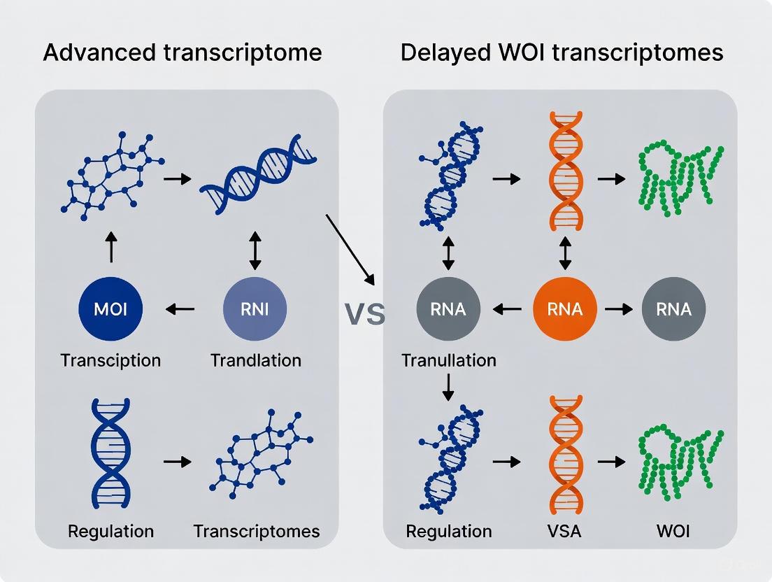
Defining the Molecular Landscape of the Displaced Window of Implantation
Window of Implantation (WOI) Displacement and Recurrent Implantation Failure (RIF)
Recurrent Implantation Failure (RIF) remains a significant challenge in assisted reproductive technology (ART), affecting approximately 10% of couples undergoing fertility treatment [1]. While RIF has multiple potential causes, including embryonic aneuploidy, uterine anatomical abnormalities, and thrombophilias, the displacement of the window of implantation (WOI) has emerged as a critical endometrial factor contributing to this condition [1] [2]. The WOI represents a brief temporal period during the mid-secretory phase of the menstrual cycle when the endometrium acquires a receptive phenotype capable of supporting embryo implantation [3]. In a typical hormone replacement therapy (HRT) cycle, blastocyst transfer is conventionally scheduled for the fifth day of progesterone administration (P+5); however, growing evidence indicates that this timing does not align with the WOI for a substantial proportion of patients [4].
Transcriptomic analyses have revealed that WOI displacement occurs in approximately 34% of subfertile patients, with 25% exhibiting a pre-receptive endometrium and 9% displaying a post-receptive endometrium at the expected time of receptivity [3]. The clinical implications of this displacement are profound, as transfers deviating by more than 12 hours from the personalized WOI demonstrate significantly reduced pregnancy rates (23.08% vs. 44.35%, p < 0.001) and an approximate two-fold increase in pregnancy loss [3]. Recent research has further stratified RIF into distinct molecular subtypes, including immune-driven (RIF-I) and metabolic-driven (RIF-M) profiles, highlighting the heterogeneous nature of endometrial dysfunction in this condition [5]. This comprehensive analysis examines the transcriptomic signatures characterizing advanced and delayed WOI states, compares experimental methodologies for WOI assessment, and explores emerging therapeutic approaches targeting specific RIF subtypes.
Comparative Transcriptomic Profiles of Advanced vs. Delayed WOI
Global Transcriptomic Alterations
Endometrial transcriptomic profiling has revealed distinct molecular signatures associated with WOI displacement in RIF patients. A study of 40 RIF patients undergoing personalized embryo transfer (pET) found that 67.5% (27/40) exhibited non-receptive endometrium at the conventional P+5 timeframe in HRT cycles [4]. Among patients who achieved clinical pregnancy following pET, significant differences in gene expression profiles were observed between advanced, normal, and delayed WOI groups at the P+5 timepoint [4]. Researchers identified ten differentially expressed genes (DEGs) involved in immunomodulation, transmembrane transport, and tissue regeneration that accurately classified endometrium with different WOI statuses [4].
Single-cell RNA sequencing studies have provided unprecedented resolution of the cellular dynamics during WOI, identifying a two-stage decidualization process in stromal cells and a gradual transition process in luminal epithelial cells across the implantation window [6]. These investigations have further identified time-varying gene sets regulating epithelial receptivity, enabling stratification of RIF endometria into distinct functional classes based on their deficiency patterns [6].
Molecular Subtypes of RIF Endometrium
Comprehensive computational analysis integrating multiple endometrial transcriptomic datasets has revealed two biologically distinct molecular subtypes of RIF endometrium:
- Immune-Driven Subtype (RIF-I): Characterized by enrichment of immune and inflammatory pathways, including IL-17 and TNF signaling pathways (p < 0.01), with increased infiltration of effector immune cells [5].
- Metabolic-Driven Subtype (RIF-M): Marked by dysregulation of oxidative phosphorylation, fatty acid metabolism, steroid hormone biosynthesis, and altered expression of the circadian clock gene PER1 [5].
The development of the MetaRIF classifier has enabled accurate distinction between these subtypes in independent validation cohorts (AUC: 0.94 and 0.85), significantly outperforming previously published models [5].
Table 1: Key Characteristics of RIF Molecular Subtypes
| Feature | RIF-I (Immune-Driven) | RIF-M (Metabolic-Driven) |
|---|---|---|
| Enriched Pathways | IL-17 signaling, TNF signaling, immune activation | Oxidative phosphorylation, fatty acid metabolism, steroid hormone biosynthesis |
| Cellular Features | Increased effector immune cell infiltration | Mitochondrial dysfunction, metabolic alterations |
| Key Marker | Elevated T-bet/GATA3 expression ratio | Altered PER1 expression (circadian rhythm) |
| Proposed Treatment | Sirolimus (mTOR inhibition) [5] | Prostaglandins [5] |
Signaling Pathway Dysregulation
The transcriptomic differences between advanced and delayed WOI states reflect fundamental alterations in key biological pathways. Analysis of endometrial receptivity has identified significant dysregulation in:
- Immune Signaling Pathways: The RIF-I subtype demonstrates upregulation of interleukin and cytokine-mediated signaling pathways, creating a suboptimal inflammatory microenvironment for implantation [5].
- Metabolic Pathways: The RIF-M subtype shows coordinated dysregulation of energy metabolism pathways, including oxidative phosphorylation and lipid metabolism [5].
- Epithelial-Stromal Crosstalk: Single-cell transcriptomics has revealed disrupted communication networks between epithelial and stromal compartments in displaced WOI, affecting decidualization and receptivity acquisition [6].
Figure 1: Transcriptomic Stratification of WOI Displacement and RIF Subtypes
Comparative Analysis of WOI Assessment Methodologies
Transcriptomic Profiling Technologies
Multiple transcriptomic approaches have been developed to assess endometrial receptivity and identify WOI displacement:
- RT-qPCR-Based Panels: The ER Map tool utilizes a high-throughput RT-qPCR platform for targeted analysis of genes related to endometrial proliferation and embryonic implantation, demonstrating 100% reproducibility in repeated cycles from the same patient [3].
- Microarray Technology: Endometrial Receptivity Array (ERA) employs customized microarrays to evaluate the expression of 238 genes, classifying endometrium as pre-receptive, receptive, or post-receptive [4] [7].
- RNA Sequencing: Next-generation sequencing-based methods like the Endometrial Receptivity Diagnosis (ERD) model analyze 166 biomarker genes, providing comprehensive transcriptome coverage independent of prior knowledge [4].
- Single-Cell RNA Sequencing: This advanced methodology enables resolution of cellular heterogeneity and cell-type specific gene expression patterns across the WOI, identifying distinct epithelial, stromal, and immune cell subpopulations and their dynamics [6].
Table 2: Comparison of WOI Assessment Methodologies
| Methodology | Target Genes | Resolution | Advantages | Clinical Validation |
|---|---|---|---|---|
| ER Map (RT-qPCR) | Selected receptivity genes | Targeted | High reproducibility, quantitative accuracy | 2256 patients; 44.35% pregnancy rate within WOI vs 23.08% outside WOI [3] |
| ERA (Microarray) | 238 genes | Targeted | Standardized commercial test | 77.5% receptivity rate in fertile controls vs 32.5% in RIF [4] |
| ERD (RNA-seq) | 166 genes | Whole transcriptome | Comprehensive, hypothesis-free | 65% pregnancy rate in RIF after pET [4] |
| scRNA-seq | Whole transcriptome | Single-cell | Cellular resolution, identifies subtypes | Stratified RIF into RIF-I and RIF-M subtypes [5] [6] |
Experimental Protocols for Transcriptomic Analysis
Endometrial Tissue Collection and Processing
Standardized protocols for endometrial tissue collection and processing are critical for reliable transcriptomic analysis:
- Patient Selection Criteria: Studies typically include women aged 18-38 years with BMI 18-25 kg/m², regular menstrual cycles (25-35 days), and exclusion of uterine pathology, endometriosis, hydrosalpinx, endocrine disorders, and chronic endometritis [5] [4].
- Biopsy Timing: In natural cycles, biopsies are timed relative to the LH surge (LH+7 for expected WOI). In HRT cycles, biopsies are typically performed after 5 days of progesterone administration (P+5) [4].
- Tissue Processing: Endometrial biopsies are rinsed with plain RPMI-1640 to remove blood and mucus, followed by immediate cryopreservation at -80°C or RNA extraction using commercial kits (e.g., Qiagen RNeasy Mini Kits) [5].
- RNA Quality Control: RNA integrity is assessed prior to library preparation, with quality thresholds typically set at RIN >7.0 [5].
Transcriptomic Data Analysis Workflow
Computational analysis of WOI transcriptomes follows a standardized workflow:
- Data Preprocessing: Raw sequencing reads are quality-checked (FastQC), trimmed, and aligned to the reference genome (STAR/Hisat2).
- Normalization: Cross-platform normalization is performed using random-effects models to integrate multiple datasets [5].
- Differential Expression: Differentially expressed genes (DEGs) between RIF and control samples are identified using packages such as MetaDE [5].
- Unsupervised Clustering: ConsensusClusterPlus and similar tools identify molecular subtypes without a priori assumptions [5].
- Pathway Analysis: Gene Set Enrichment Analysis (GSEA) reveals enriched biological pathways in identified subtypes [5].
Figure 2: Experimental Workflow for WOI Transcriptome Analysis
The Scientist's Toolkit: Essential Research Reagents and Platforms
Table 3: Essential Research Reagents and Platforms for WOI Studies
| Category | Specific Product/Platform | Application in WOI Research |
|---|---|---|
| RNA Extraction | Qiagen RNeasy Mini Kits [5] | High-quality RNA isolation from endometrial biopsies |
| Library Preparation | Illumina TruSeq Stranded mRNA | RNA-seq library construction for transcriptome profiling |
| Sequencing Platforms | Illumina NovaSeq 6000 | High-throughput sequencing for transcriptome analysis |
| Single-Cell Platforms | 10X Genomics Chromium [6] | Single-cell RNA sequencing for cellular resolution |
| Computational Tools | MetaDE [5] | Identification of differentially expressed genes |
| Clustering Algorithms | ConsensusClusterPlus [5] | Unsupervised clustering for subtype identification |
| Pathway Analysis | Gene Set Enrichment Analysis (GSEA) [5] | Functional interpretation of transcriptomic signatures |
| Drug Repurposing | Connectivity Map (CMap) [5] | Prediction of candidate therapeutic compounds |
| Hormonal Reagents | Estradiol valerate, Medroxyprogesterone acetate [4] [8] | Endometrial preparation in HRT cycles and in vitro models |
| 3D Culture Systems | WOI Assembloids [8] | In vitro modeling of human endometrial receptivity |
Therapeutic Implications and Future Directions
Personalized Embryo Transfer (pET)
The primary clinical application of WOI transcriptomics is the guidance of personalized embryo transfer (pET) based on individual receptivity status. Implementation of pET in RIF patients has demonstrated significant improvements in reproductive outcomes. In one study of 40 RIF patients, ERD-guided pET resulted in a clinical pregnancy rate of 65% (26/40), compared to previous failures at the conventional transfer time [4]. The importance of precise synchronization is highlighted by the dramatic decline in pregnancy rates when transfers deviate by more than 24 hours from the personalized WOI (19.23% vs. 44.35%, p = 0.011) [3].
Subtype-Specific Therapeutic Interventions
The identification of molecular RIF subtypes enables targeted therapeutic approaches:
- RIF-I (Immune-Driven): Connectivity Map analysis has identified sirolimus (rapamycin) as a candidate therapeutic, potentially modulating the hyper-inflammatory microenvironment through mTOR pathway inhibition [5].
- RIF-M (Metabolic-Driven): Prostaglandins have been predicted as potential treatments for addressing the metabolic dysregulation characteristic of this subtype [5].
Advanced Model Systems
The development of sophisticated in vitro models represents a promising direction for future WOI research:
- Endometrial Assembloids: Recently established WOI endometrial assembloids recapitulate structural attributes (pinopodes and cilia) and molecular characteristics of mid-secretory endometrium, exhibiting hormone responsiveness, energy metabolism with enhanced mitochondria, and promising potential for embryo implantation studies [8].
- Single-Cell Atlas Resources: Comprehensive single-cell transcriptomic atlases of fertile endometrium across the WOI provide reference data for identifying pathological deviations in RIF patients [6].
These advanced models and resources will facilitate deeper understanding of implantation mechanisms, screening of therapeutic compounds, and development of personalized treatment strategies for RIF patients with displaced WOI.
The human endometrium, the inner lining of the uterus, undergoes precisely orchestrated molecular and cellular transformations each menstrual cycle to create a transient window of implantation (WOI). This brief period of endometrial receptivity (ER), typically occurring around day LH+7 in a natural cycle, represents the only time when the endometrium permits blastocyst attachment and invasion [9] [10]. Establishing a definitive transcriptomic baseline for the receptive endometrium is fundamental for diagnosing ER pathologies, improving assisted reproductive technologies, and developing targeted therapies for infertility. This guide provides a comparative analysis of the transcriptomic signatures that distinguish the receptive from non-receptive endometrium, with particular focus on advanced versus delayed WOI profiles that characterize certain infertility conditions.
Molecular characterization of endometrial receptivity has evolved significantly from histological dating to comprehensive transcriptomic profiling. While traditional morphological assessment revealed structural changes, transcriptomic technologies—from microarrays to single-cell RNA sequencing (scRNA-seq) and spatial transcriptomics—have uncovered the complex gene expression networks that functionally define receptivity [9]. These technologies have enabled researchers to identify consistent gene expression patterns that signify the optimal state for embryo implantation, providing a critical baseline against which pathological states can be compared.
Core Transcriptomic Signature of the Receptive Endometrium
Established Gene Expression Profiles
The transition from pre-receptive to receptive endometrium involves significant transcriptomic reprogramming across multiple gene families. Table 1 summarizes the key transcriptomic hallmarks that define the receptive endometrial state, categorized by their molecular functions.
Table 1: Core Transcriptomic Signature of Receptive Endometrium
| Gene Category | Representative Genes | Expression Direction in WOI | Primary Function in Implantation |
|---|---|---|---|
| Cellular Adhesion & Migration | SPP1 (Osteopontin), LAMB3, MFAP5 | Upregulated | Facilitates embryo attachment and trophoblast invasion [9] |
| Immune Modulation | IL15, GPX3, CXCL14 | Upregulated | Regulates immune tolerance and inflammatory response [9] [10] |
| Cellular Differentiation & Decidualization | PAEP, DPP4, MAOA | Upregulated | Supports stromal decidualization and epithelial differentiation [10] |
| Secretory Factors | LIF, PROK1, ANGPTL1 | Upregulated | Provides embryonic signals and endometrial nourishment [9] [10] |
| Metabolic Reprogramming | MT1E, MT1F, MT1G, MT1X | Upregulated (Early Secretory) | Protection against oxidative stress [10] |
| Cell Cycle Regulators | Histone-encoding genes (HIST cluster) | Downregulated | Suppression of proliferation during differentiation [11] |
Analysis of the receptive endometrium reveals coordinated upregulation of genes governing embryo adhesion (SPP1, LAMB3), immune modulation (IL15, GPX3), and secretory functions (PAEP, LIF). Simultaneously, genes driving cellular proliferation are typically downregulated as the tissue transitions from growth to differentiation [11] [10]. This transcriptomic shift creates a favorable environment for embryo recognition, attachment, and subsequent invasion.
Single-Cell Resolution of Receptive Signatures
Recent scRNA-seq studies have substantially refined our understanding of endometrial receptivity by characterizing cell-type-specific transcriptomic dynamics. Research examining over 220,000 endometrial cells across the WOI has revealed that luminal epithelial cells undergo a gradual transitional process, while stromal cells display a clear two-stage decidualization program [6]. These sophisticated analyses have identified distinct subpopulations within major endometrial cell types—including 8 epithelial, 5 stromal, 11 NK/T cell, and 10 myeloid subpopulations—each contributing uniquely to the receptive state [6].
The emergence of spatial transcriptomics has further enhanced this resolution by mapping gene expression within tissue architecture. Studies utilizing 10x Visium technology have identified seven distinct cellular niches in the mid-luteal phase endometrium, with unciliated epithelial cells dominating the cellular landscape [12] [13]. This spatial context is crucial for understanding how localized gene expression patterns facilitate embryo-endometrium crosstalk during implantation.
Comparative Analysis: Advanced vs. Delayed WOI Transcriptomes
Transcriptomic Signatures of WOI Displacement
Displacement of the WOI—either advanced or delayed—represents a significant pathological mechanism in recurrent implantation failure (RIF). Table 2 compares the transcriptomic profiles associated with normal, advanced, and delayed WOI states, highlighting potential diagnostic and therapeutic targets.
Table 2: Comparative Transcriptomic Profiles of Normal, Advanced, and Delayed WOI
| Transcriptomic Feature | Normal WOI (LH+7/P+5) | Advanced WOI | Delayed WOI |
|---|---|---|---|
| Key Regulator Genes | SPP1, LIF, PAEP, IL15, GPX3 [9] | Premature elevation of receptivity markers | Persistent pre-receptive gene signature |
| Immune Response Pathways | Balanced immunomodulation (IL15) [9] | Premature immune activation | Prolonged inflammatory state |
| Cellular Proliferation Signatures | Suppressed [11] | Premature suppression | Delayed suppression |
| Differentiation Markers | Appropriately timed (PAEP, DPP4) [10] | Premature elevation | Delayed acquisition |
| Clinical Detection Method | Transcriptomic dating (ERD/ERA) [4] | ERD/ERA testing | ERD/ERA testing |
| Therapeutic Implications | Standard embryo transfer timing | Earlier embryo transfer | Later embryo transfer |
Transcriptome analysis of RIF patients has revealed that approximately 67.5% (27/40) exhibit non-receptive endometrium at the conventional P+5 timing in hormone replacement therapy cycles [4]. Among patients who achieved pregnancy through personalized embryo transfer (pET), distinct transcriptomic signatures differentiated advanced, normal, and delayed WOI groups. Specifically, researchers identified 10 differentially expressed genes (DEGs) involved in immunomodulation, transmembrane transport, and tissue regeneration that accurately classified endometrium with different WOI timings [4].
Functional Consequences of WOI Displacement
The functional impact of WOI displacement extends beyond temporal misalignment to fundamental alterations in endometrial physiology. In advanced WOI, the endometrium prematurely expresses receptivity markers, creating a discordance between endometrial readiness and embryonic development. Conversely, delayed WOI maintains a pre-receptive gene signature when the embryo is developmentally prepared for implantation [4]. Both scenarios disrupt the critical synchrony required for successful implantation.
Single-cell transcriptomic profiling has further revealed that RIF endometria often display a hyper-inflammatory microenvironment, particularly affecting epithelial cell function [6]. This pathological state compromises the delicate immune balance required for embryo acceptance while simultaneously disrupting the normal differentiation trajectory of endometrial cells. Such findings explain why simply adjusting embryo transfer timing without addressing underlying molecular dysfunction may yield suboptimal outcomes in some RIF patients.
Experimental Protocols for Transcriptomic Analysis
Sample Collection and Processing
Establishing a reliable transcriptomic baseline for endometrial receptivity requires stringent experimental protocols. The following methodology represents current best practices derived from multiple studies:
Patient Selection & Cycle Dating: Recruit participants with confirmed regular ovulation and normal uterine anatomy. Precisely determine the luteinizing hormone (LH) surge through daily serum measurements or urinary LH dipstick testing, designating the day of surge as LH+0 [6]. For hormone replacement therapy (HRT) cycles, designate the first day of progesterone administration as P+0.
Endometrial Biopsy: Perform endometrial biopsies using a Pipelle catheter under sterile conditions. Collect samples from the fundal and upper uterine regions to minimize anatomical variation. For single-cell analyses, immediately process tissue for cell dissociation [4] [6].
Single-Cell RNA Sequencing: Fresh endometrial tissue should be enzymatically dispersed into single-cell suspensions. Use the 10X Chromium system for cell capture and library preparation. Sequence on Illumina platforms (e.g., NovaSeq 6000) to achieve sufficient depth (recommended: >50,000 reads/cell) [6]. Filter data to remove low-quality cells (those with <500 genes or >20% mitochondrial gene expression) and doublets using tools like DoubletFinder [12].
Spatial Transcriptomics: For spatial transcriptomics using the 10X Visium platform, flash-freeze endometrial tissue in optimal cutting temperature compound. Section tissues at appropriate thickness (typically 10μm) and place onto Visium slides. After H&E staining and imaging, permeabilize tissue to allow mRNA capture from spatially barcoded spots. Follow standard library preparation and sequencing protocols [12] [13].
Data Analysis Workflows
Preprocessing & Normalization: Process raw sequencing data through standard pipelines (Cell Ranger for scRNA-seq; Space Ranger for spatial transcriptomics). Apply quality control filters, then normalize data using SCTransform for scRNA-seq or corresponding methods for spatial data [12].
Cell Type Identification & Clustering: Perform principal component analysis followed by graph-based clustering. Visualize results using UMAP. Annotate cell types using established markers: epithelial cells (EPCAM, KRTT), stromal cells (PDGFRA, DECORIN), endothelial cells (PECAM1, VWF), and immune cells (PTPRC) [14] [6].
Differential Expression Analysis: Identify differentially expressed genes using appropriate statistical methods (e.g., Seurat's FindAllMarkers function, DESeq2 for bulk RNA-seq). Apply multiple testing correction (Benjamini-Hochberg) with significance threshold of adjusted p-value < 0.05 [12] [11].
Temporal Modeling & Trajectory Analysis: For time-series data, employ computational tools like StemVAE to model transcriptomic dynamics across the WOI. Construct RNA velocity trajectories to infer cellular differentiation paths [6].
The following diagram illustrates the comprehensive experimental workflow for establishing transcriptomic baselines of endometrial receptivity:
Diagram 1: Experimental workflow for establishing transcriptomic baselines of endometrial receptivity, encompassing sample collection, sequencing, bioinformatic analysis, and clinical validation.
Signaling Pathways Governing Endometrial Receptivity
Molecular Regulation of the WOI
The transition to a receptive endometrial state is coordinated by sophisticated molecular pathways that respond to hormonal cues and mediate embryo-endometrium crosstalk. The following diagram illustrates the key signaling networks and their interactions during this critical period:
Diagram 2: Key signaling pathways governing endometrial receptivity, showing hormonal regulation, molecular signaling networks, and functional outcomes.
The molecular pathways depicted above translate hormonal signals into functional changes that define the receptive state. Progesterone activation, in particular, initiates a transcriptional cascade that includes induction of HAND2, which inhibits fibroblast growth factor (FGF) signaling in the stroma, thereby modulating epithelial proliferation and differentiation [10]. Simultaneously, estrogen and progesterone coordinately regulate leukemia inhibitory factor (LIF) signaling, a critical pathway for epithelial receptivity and embryo implantation [9] [10].
Notch signaling plays a dual role in endometrial receptivity—it is suppressed during the proliferative phase to permit ciliogenesis through FOXJ1 upregulation, but at receptivity phase, it contributes to epithelial differentiation [10]. Additionally, BMP and Wnt signaling pathways, evolutionarily conserved in mammalian implantation, contribute to the stromal-epithelial cross-talk necessary for decidualization and immune modulation [14].
The Scientist's Toolkit: Essential Research Reagents
Key Reagents for Endometrial Receptivity Research
Table 3: Essential Research Reagents for Endometrial Receptivity Studies
| Reagent Category | Specific Examples | Research Application | Functional Role |
|---|---|---|---|
| Single-Cell Isolation Kits | 10X Genomics Chromium Single Cell Kits | Single-cell transcriptomic profiling | Partitioning individual cells for barcoding and cDNA synthesis [6] |
| Spatial Transcriptomics Platforms | 10X Visium Spatial Gene Expression | Spatial mapping of gene expression | Capturing location-specific transcriptome data in tissue context [12] [13] |
| Cell Type Markers | EPCAM (epithelial), PDGFRA (stromal), PECAM1 (endothelial), PTPRC (immune) | Cell type identification and validation | Antibodies for immunofluorescence or FACS validation of scRNA-seq findings [14] [6] |
| Hormone Receptors | ESR1, PGR antibodies | Hormone response assessment | Detecting estrogen and progesterone receptor expression across menstrual cycle [10] |
| qPCR Assays | Commercial TaqMan assays for SPP1, LIF, PAEP, IL15, GPX3 | Transcript validation | Confirming differential expression of receptivity biomarkers [4] [9] |
| Bioinformatic Tools | Seurat, SCTransform, Harmony, CARD, DoubletFinder | Computational analysis of transcriptomic data | Data normalization, batch correction, clustering, and spatial deconvolution [12] [6] |
Establishing a reliable baseline of endometrial receptivity requires appropriate controls and validation strategies. Researchers should include samples from well-characterized fertile women across multiple cycle time points, with precise LH dating. For RIF studies, careful patient selection is crucial—excluding those with endometriosis, uterine abnormalities, or endocrine disorders that could confound results [4] [6]. Integration of multiple 'omics datasets through computational approaches like CARD deconvolution can enhance spatial resolution by mapping single-cell data onto spatial transcriptomics datasets [12].
The established transcriptomic baseline for receptive endometrium provides an essential reference for diagnosing displacement disorders and developing targeted interventions. Current research applications include:
Diagnostic Development: Transcriptomic signatures have been successfully commercialized in diagnostic tests like the Endometrial Receptivity Array (ERA) and Win-Test, which guide personalized embryo transfer timing for RIF patients [4] [9].
Drug Discovery: Identification of key pathways (LIF, NOTCH, BMP/Wnt) offers targets for pharmacological intervention to rescue deficient receptivity [14] [10].
Disease Modeling: Organoid systems that recapitulate endometrial epithelium transcriptome enable functional studies of receptivity mechanisms and screening of therapeutic compounds [10].
Future research directions include integrating multi-omics approaches to understand post-transcriptional regulation, exploring epigenetic modifications that govern WOI timing, and developing non-invasive biomarkers for endometrial receptivity assessment. As single-cell and spatial technologies continue to advance, they will further refine our understanding of the complex cellular interactions that enable embryo implantation, ultimately improving diagnostics and treatments for implantation disorders.
Key Differentially Expressed Genes (DEGs) in Advanced WOI Transcriptomes
The window of implantation (WOI) represents a critical, transient period during the mid-secretory phase of the menstrual cycle when the endometrium acquires a receptive phenotype capable of supporting embryo implantation. Transcriptomic analyses have revolutionized our understanding of endometrial receptivity by revealing the intricate gene expression dynamics that govern this process. The comparative analysis of advanced versus delayed WOI transcriptomes provides a powerful framework for identifying key differentially expressed genes (DEGs) that serve as molecular determinants of successful implantation. This comprehensive comparison guide examines the experimental approaches, datasets, and computational methods employed in WOI transcriptomics research, with a specific focus on DEG characterization across receptivity phases. By synthesizing findings from multiple transcriptomic profiling studies, we aim to establish a standardized reference for evaluating endometrial receptivity status in both research and clinical settings, ultimately facilitating the development of diagnostic tools and targeted interventions for endometrial-factor infertility.
Methodological Approaches in WOI Transcriptome Research
Experimental Design and Sample Collection Protocols
Research into endometrial receptivity employs rigorously standardized methodologies to ensure comparability across studies. The predominant approach involves endometrial biopsy collection timed according to the luteinizing hormone (LH) surge, with LH+7 days generally recognized as the core receptive phase [6] [15]. Sample sizes in recent investigations range from 28 to 90 endometrial biopsies collected across multiple time points spanning the WOI, typically including pre-receptive (LH+3, LH+5), receptive (LH+7), and post-receptive (LH+9, LH+11) phases [6] [15]. These studies exclusively utilize samples from fertile women with confirmed regular menstrual cycles, with precise cycle dating established through daily serum LH measurements [6].
For transcriptomic profiling, two primary methodological approaches prevail: bulk RNA sequencing (RNA-Seq) and single-cell RNA sequencing (scRNA-seq). Bulk RNA-Seq provides a global transcriptome profile of endometrial tissue and has been successfully employed to develop predictive models for endometrial dating [15]. In contrast, scRNA-seq technologies, particularly the 10X Genomics platform, enable resolution of cellular heterogeneity within endometrial tissues by profiling individual cells [6]. Recent scRNA-seq studies have analyzed over 220,000 endometrial cells, identifying distinct cellular subpopulations including unciliated epithelial cells, ciliated epithelial cells, stromal cells, endothelial cells, and various immune cell types [6]. This single-cell approach has proven invaluable for deciphering cell-type-specific contributions to endometrial receptivity and identifying rare cellular subpopulations that may play critical roles in implantation.
Bioinformatics and Computational Analysis Pipelines
The analysis of WOI transcriptomic data employs sophisticated bioinformatics pipelines for quality control, normalization, and differential expression analysis. For bulk RNA-Seq data, standard practices include read quality assessment using Fastp, mapping to reference genomes with Salmon, and calculation of transcripts per million (TPM) values for normalization [16]. The DESeq2 package serves as the primary tool for identifying DEGs, with standard thresholds set at adjusted p-value < 0.05 and absolute fold change > 2 [16].
Single-cell data analysis utilizes specialized computational workflows, including the CellRanger pipeline for initial processing and the Seurat package for downstream analyses [17]. Quality control measures typically exclude cells expressing fewer than 200 genes or more than 4,000 genes to remove low-quality cells and potential doublets [17]. Batch effects between samples are corrected using Harmony software, while cell clustering employs graph-based methods with the FindClusters function [17]. For temporal analysis of WOI dynamics, advanced computational models like StemVAE enable both descriptive characterization and predictive modeling of transcriptomic changes across implantation stages [6].
Functional enrichment analysis represents a critical component of WOI transcriptomic studies, with Gene Ontology (GO) and Kyoto Encyclopedia of Genes and Genomes (KEGG) pathway analyses performed using the clusterProfiler package [16] [18]. Weighted gene co-expression network analysis (WGCNA) identifies functional modules and hub genes associated with specific reproductive phenotypes [19]. These computational approaches collectively enable the identification of key DEGs and regulatory networks underlying endometrial receptivity.
Table 1: Standardized Experimental Protocols in WOI Transcriptome Studies
| Protocol Component | Standardized Approach | Variations and Alternatives |
|---|---|---|
| Sample Timing | LH surge-based (LH+3, +5, +7, +9, +11) | Natural cycles; Hormone replacement therapy cycles |
| Tissue Collection | Endometrial biopsy (Pipelle) | Endometrial aspirates; Surgical specimens |
| RNA Sequencing | Bulk RNA-Seq; Single-cell RNA-Seq (10X Genomics) | Microarrays; Spatial transcriptomics |
| Quality Control | CellRanger (scRNA-seq); Fastp (bulk RNA-Seq) | Partek Flow; Custom QC pipelines |
| DEG Identification | DESeq2; Seurat FindAllMarkers | EdgeR; Limma-Voom; Scanpy |
| Functional Analysis | clusterProfiler (GO/KEGG) | GSEA; DAVID; Metascape |
Comparative Analysis of DEGs Across WOI Transitions
Temporal Dynamics of Transcriptomic Changes
The transition through the WOI involves precisely orchestrated temporal changes in gene expression patterns. Comparative analyses across prereceptive (LH+3/LH+5), receptive (LH+7), and post-receptive (LH+9/LH+11) phases reveal dynamic transcriptional reprogramming in all major endometrial cell types [6] [15]. Luminal and glandular epithelial cells undergo a gradual transition process characterized by sequential activation of receptivity-associated genes, while stromal cells exhibit a clear-cut two-stage decidualization process [6]. These temporal patterns have been validated through both bulk tissue transcriptomics and single-cell approaches, confirming their reproducibility across different study populations and methodological platforms.
Research employing daily sampling across the WOI has identified a continuum of transcriptomic changes rather than abrupt transitions, with distinct early, mid, and late receptivity signatures [6]. One study analyzing 28 endometrial biopsies across 5 time points (LH+3 to LH+11) demonstrated substantial inter-individual variations in cellular composition and gene expression patterns despite strict cycle dating [6]. This temporal resolution has enabled the identification of phase-specific gene expression patterns and the development of predictive models capable of accurately classifying endometrial samples according to histological dating [15]. The high accuracy of these models (85.19-100% in validation sets) underscores the robustness of transcriptomic signatures for endometrial dating [15].
Key DEGs in Advanced Versus Delayed WOI Transcriptomes
Comparative analysis of transcriptomic profiles between advanced and delayed WOI states has identified consistent DEG patterns associated with receptivity status. In advanced/receptive endometria, upregulation of specific gene sets involved in embryo attachment, immunomodulation, and stromal decidualization is consistently observed [6] [15]. These receptivity-associated genes include those encoding for transporters, growth factors, and immunomodulatory proteins that collectively create a favorable microenvironment for implantation.
In contrast, delayed/non-receptive endometria exhibit distinct gene expression patterns characterized by persistence of proliferative-phase genes, incomplete activation of receptivity factors, and altered expression of cell adhesion molecules [6]. Studies of recurrent implantation failure (RIF) patients have identified dysregulated epithelial receptivity genes and a hyper-inflammatory microenvironment in the endometrium during the putative WOI [6]. Single-cell transcriptomic analyses further reveal that RIF endometria can be stratified into distinct classes of deficiencies based on their DEG profiles, with some cases showing displaced WOI timing and others exhibiting fundamentally dysregulated epithelium [6].
Table 2: Key Differentially Expressed Genes Across WOI Transitions
| Gene Category | Advanced/Receptive Phase | Delayed/Non-Receptive Phase | Biological Function |
|---|---|---|---|
| Transporters | Upregulated | Variably expressed | Nutrient transport to implantation site |
| Growth Factors | Significantly upregulated | Reduced expression | Embryo-endometrial signaling |
| Immunomodulators | Appropriately modulated | Dysregulated | Maternal immune tolerance |
| Cell Adhesion Molecules | Specifically activated | Persistence of proliferative phase patterns | Embryo attachment and invasion |
| Decidualization Markers | Strongly induced | Incomplete or delayed induction | Stromal preparation for implantation |
Signaling Pathways and Molecular Networks in WOI Regulation
Core Regulatory Pathways in Endometrial Receptivity
Transcriptomic analyses have elucidated several core signaling pathways that coordinate the acquisition of endometrial receptivity. Consistently identified pathways include Wnt signaling, TGF-β signaling, and pathways involved in extracellular matrix organization [6] [20]. These pathways exhibit precise temporal activation patterns during the WOI transition and coordinate functional changes across different endometrial cell types. Single-cell resolution studies have further revealed cell-type-specific utilization of these pathways, with distinct regulatory programs operating in epithelial, stromal, and immune cell compartments [6].
Pathway analysis of DEGs between receptive and non-receptive endometria additionally highlights the importance of metabolic reprogramming, cytoskeletal reorganization, and cell-cell communication pathways [6]. In particular, integrative analyses combining transcriptomic and metabolomic data have revealed coordinated regulation of secondary metabolite biosynthesis, flavonoid biosynthesis, and sesquiterpenoid/triterpenoid biosynthesis pathways during the receptivity acquisition process [19]. These findings suggest that metabolic reprogramming represents an essential component of endometrial preparedness for implantation, alongside the more established signaling and structural changes.
Transcription Factor Networks Governing WOI Transitions
WOI transcriptome analyses have identified specific transcription factor (TF) networks that orchestrate the receptivity acquisition program. Time-series single-cell data have enabled the reconstruction of TF regulatory networks driving the gradual transition of luminal epithelial cells and the two-stage decidualization process in stromal cells [6]. These analyses reveal sequential TF activation patterns, with distinct early, mid, and late WOI regulators.
Studies of RIF endometria have identified specific TF expression abnormalities that disrupt the normal progression of receptivity-related gene expression programs [6]. In particular, TFs regulating epithelial receptivity genes show altered expression patterns in RIF patients, contributing to the dysfunctional endometrial phenotype [6]. Network analysis approaches including WGCNA have further identified hub TFs that coordinate the expression of large gene modules associated with receptivity status, highlighting their potential as regulatory master switches in the WOI establishment process [19].
Diagram 1: Signaling Pathway Dynamics During WOI Transition. This diagram illustrates the sequential activation of core signaling pathways and cellular processes during the progression through the window of implantation.
Table 3: Essential Research Reagents for WOI Transcriptome Studies
| Reagent Category | Specific Products | Application in WOI Research |
|---|---|---|
| Single-Cell Platform | 10X Genomics Chromium | Single-cell transcriptome profiling of endometrial cells |
| Library Prep Kits | Chromium Single Cell 3' Kit | scRNA-seq library construction |
| Bioinformatics Tools | Seurat, CellRanger | scRNA-seq data analysis and cell clustering |
| Pathway Analysis | clusterProfiler | GO and KEGG enrichment analysis |
| DEG Analysis | DESeq2, EdgeR | Identification of differentially expressed genes |
| Reference Genomes | Ensembl human genome build | Read alignment and transcript quantification |
The comparative analysis of advanced versus delayed WOI transcriptomes has yielded critical insights into the molecular basis of endometrial receptivity. Through the application of standardized experimental protocols and computational分析方法, researchers have consistently identified key DEGs and signaling pathways that distinguish receptive from non-receptive endometrium. The emergence of single-cell transcriptomics has further refined our understanding by revealing cell-type-specific gene expression programs and enabling the identification of rare cellular subpopulations critical for implantation success.
Future directions in WOI transcriptome research include the integration of multi-omics approaches, the development of improved diagnostic classifiers for clinical use, and the application of spatial transcriptomics to preserve architectural context. Additionally, continued investigation of transcriptomic dysregulation in RIF patients will identify novel therapeutic targets and personalized treatment strategies. These advances will ultimately enhance our ability to diagnose and treat endometrial-factor infertility, improving outcomes for patients undergoing assisted reproduction.
Distinct Molecular Signatures of Delayed WOI Endometrium
The window of implantation (WOI) represents a critical, limited time period during the mid-secretory phase of the menstrual cycle when the endometrium acquires a receptive phenotype capable of supporting embryo implantation [21]. Displacement of this window—either delayed or advanced—is recognized as a significant endometrial factor contributing to recurrent implantation failure (RIF) in assisted reproductive technology (ART) [4] [22]. Molecular analysis of the endometrium has revealed that approximately 15.9-25% of RIF patients exhibit displaced WOI, substantially higher than the 1.8% observed in fertile populations [23] [4]. This comparative guide examines the distinct transcriptomic signatures that differentiate delayed WOI endometrium from both advanced and optimally timed receptivity, providing researchers and drug development professionals with a detailed analysis of molecular alterations underlying this pathological condition.
Molecular Characterization of Delayed WOI Endometrium
Transcriptomic Alterations and Pathway Dysregulation
Delayed WOI endometrium demonstrates a characteristic molecular signature marked by aberrant expression of genes critical for endometrial receptivity. Transcriptomic profiling of RIF patients has identified two biologically distinct molecular subtypes of endometrial dysfunction: an immune-driven subtype (RIF-I) characterized by enriched immune and inflammatory pathways including IL-17 and TNF signaling, and a metabolic-driven subtype (RIF-M) featuring dysregulation of oxidative phosphorylation, fatty acid metabolism, and steroid hormone biosynthesis [5]. These subtypes demonstrate different protein-level expression patterns, with the T-bet/GATA3 ratio significantly elevated in RIF-I compared to RIF-M [5].
Single-cell transcriptomic sequencing of luteal phase endometrium further reveals that RIF endometria exhibit displaced WOI and dysregulated epithelium within a hyper-inflammatory microenvironment [6]. This comprehensive analysis identified a two-stage decidualization process for stromal cells and a gradual transition process for epithelial cells across the WOI, with RIF patients showing distinct deficiencies in these coordinated cellular transitions [6].
Table 1: Key Molecular Features of Delayed WOI Endometrium
| Molecular Feature | Characteristics | Detection Method | Functional Implications |
|---|---|---|---|
| Immune Dysregulation | Enriched IL-17, TNF signaling pathways; increased effector immune cell infiltration | scRNA-seq, IHC for T-bet/GATA3 ratio | Creates inflammatory microenvironment hostile to implantation [5] [6] |
| Metabolic Alterations | Dysregulated oxidative phosphorylation, fatty acid metabolism, steroid hormone biosynthesis | Bulk RNA-seq, pathway analysis | Compromises energy production and hormonal response necessary for receptivity [5] |
| Epithelial Receptivity Defects | Altered expression of time-varying gene sets regulating epithelium receptivity | Time-series scRNA-seq | Disrupts embryo attachment and invasion processes [6] |
| Circadian Rhythm Disruption | Altered expression of circadian clock gene PER1 | Transcriptomic profiling | Affects temporal coordination of implantation signals [5] |
| Stromal Dysfunction | Disrupted two-stage decidualization process | scRNA-seq, pseudotime analysis | Impairs biosensing of embryo quality and implantation preparedness [6] |
Comparative Analysis of Advanced vs. Delayed WOI Signatures
While both advanced and delayed WOI represent temporal displacements of the receptivity window, they exhibit distinct molecular profiles with different implications for pregnancy outcomes. Research has identified specific transcriptomic signatures associated with different reproductive outcomes, including an optimal endometrial receptivity signature resulting in an 80% ongoing pregnancy rate for live birth, contrasted with a late receptive-stage signature carrying a 50% risk of biochemical pregnancy [7]. The molecular differences between these profiles primarily manifest in the regulation of cell cycle processes, with abnormal down-regulation of cell cycle genes representing a key feature of suboptimal receptivity signatures associated with poor pregnancy outcomes [7].
Single-cell analysis provides further distinction between temporal displacement types, revealing that delayed WOI exhibits a hyper-inflammatory microenvironment with dysfunctional epithelial cells, while advanced WOI shows premature molecular maturation that creates asynchrony with embryonic development [6]. This fundamental difference in underlying mechanisms necessitates different diagnostic and therapeutic approaches.
Table 2: Differential Gene Expression in WOI Displacement
| Gene Category | Delayed WOI Expression | Advanced WOI Expression | Function in Implantation |
|---|---|---|---|
| Circadian Genes (PER1) | Significantly altered [5] | Not characterized | Temporal coordination of receptivity |
| Extracellular Matrix Remodelers (MMP10) | Not specified | Upregulated [24] | Tissue remodeling for invasion |
| Hormone Receptors (ESR1) | Not specified | Downregulated [24] | Estrogen signaling regulation |
| Immune Regulators (IL13RA2) | Upregulated in immune subtype [5] | Not characterized | Inflammation modulation |
| Epithelial Receptivity Factors | Time-varying dysregulation [6] | Premature expression | Embryo attachment capacity |
Diagnostic Approaches and Experimental Protocols
Transcriptomic Profiling Methodologies
Accurate identification of delayed WOI requires sophisticated molecular profiling techniques. Several validated approaches have emerged for endometrial receptivity assessment:
RNA Sequencing-Based Protocols: RNA-seq provides a comprehensive, quantitative method for endometrial receptivity gene expression profiling independent of prior knowledge [4]. The rsERT (RNA-seq-based endometrial receptivity test) utilizes 175 biomarker genes and demonstrates 98.4% accuracy in WOI prediction [22]. The standard protocol involves: (1) Endometrial biopsy during putative WOI (LH+7 in natural cycles or P+5 in HRT cycles); (2) RNA extraction using TRIZOL method or commercial kits (e.g., Qiagen RNeasy Mini Kits); (3) Library preparation and sequencing; (4) Computational analysis using machine learning algorithms for receptivity classification [5] [22].
Targeted Sequencing Approaches: The beREADY model employs Targeted Allele Counting by sequencing (TAC-seq) technology to analyze 72 genes (57 endometrial receptivity biomarkers, 11 WOI-relevant genes, and 4 housekeeper genes) [23]. This targeted method provides high quantitative accuracy down to single-molecule level, with validation showing 98.2% accuracy in receptivity classification [23].
Single-Cell RNA Sequencing: High-resolution scRNA-seq protocols enable decomposition of endometrial cellular heterogeneity across WOI. The standard methodology includes: (1) Endometrial aspirate collection; (2) Enzymatic tissue dispersion; (3) Single-cell capture using 10X Chromium system; (4) Library preparation and sequencing; (5) Computational analysis using algorithms like StemVAE for temporal prediction and pattern discovery [6]. This approach typically yields >220,000 cells with median detection of 8,481 unique transcripts and 2,983 genes per cell, enabling identification of rare cell populations and cell-type specific dysregulation in RIF [6].
Non-Invasive Diagnostic Development
Emerging non-invasive approaches analyze extracellular vesicles from uterine fluid (UF-EVs) as surrogates for endometrial tissue biopsies. UF-EVs contain RNA cargo that reflects the molecular profile of parent endometrial cells, with strong correlation between UF-EV transcriptomic signatures and corresponding endometrial tissue [25]. This approach enables WOI assessment without the invasiveness of biopsy, potentially allowing embryo transfer in the same ART cycle.
Therapeutic Implications and Research Applications
Personalized Embryo Transfer Strategies
Transcriptomic profiling guiding personalized embryo transfer (pET) has demonstrated significant improvement in reproductive outcomes for RIF patients with displaced WOI. Clinical studies show that pET based on molecular receptivity assessment improves pregnancy rates from 23.7% with conventional timing to 50.0% in RIF patients transferring day-3 embryos [22]. For blastocyst transfers, a similar trend shows improvement from 40.7% to 63.6% [22]. These outcomes highlight the clinical utility of molecular signature identification in managing delayed WOI.
Mechanism-Targeted Therapeutic Development
The distinct molecular signatures of delayed WOI provide targets for therapeutic development:
Immune-Targeted Approaches: For the immune-driven RIF-I subtype, Connectivity Map (CMap)-based drug predictions have identified sirolimus (rapamycin) as a candidate therapeutic to modulate inflammatory pathways [5].
Metabolic-Targeted Approaches: For the metabolic-driven RIF-M subtype, prostaglandins have been identified as potential treatments to address metabolic dysregulation [5].
Gene Network-Based Interventions: Weighted Gene Co-expression Network Analysis (WGCNA) of UF-EV transcriptomes has identified gene modules significantly correlated with pregnancy outcomes, providing targets for future therapeutic development [25]. A Bayesian logistic regression model integrating these gene expression modules with clinical variables achieves predictive accuracy of 0.83 for pregnancy outcome, enabling targeted patient selection for interventions [25].
Diagram 1: Molecular signatures and targeted therapies for delayed WOI endometrium. The diagram illustrates the two major subtypes (immune and metabolic) with their associated pathway dysregulations and corresponding therapeutic strategies.
The Scientist's Toolkit: Essential Research Reagents and Platforms
Table 3: Essential Research Tools for WOI Transcriptome Studies
| Tool Category | Specific Products/Platforms | Application in WOI Research | Key Features |
|---|---|---|---|
| Sequencing Platforms | Illumina TAC-seq, 10X Chromium scRNA-seq | Transcriptome profiling, single-cell analysis | High sensitivity, quantitative, single-cell resolution [23] [6] |
| Computational Tools | StemVAE, WGCNA, MetaRIF classifier | Temporal modeling, network analysis, subtype classification | Pattern discovery, predictive modeling, AUC 0.94 for subtypes [6] [5] |
| Bioinformatic Databases | Connectivity Map (CMap), GEO datasets | Drug prediction, data integration | Identifies candidate therapeutics for molecular subtypes [5] |
| Validation Reagents | IHC antibodies (T-bet, GATA3), qPCR assays | Protein-level validation, gene expression confirmation | Verifies transcriptomic findings at protein level [5] |
| Commercial Diagnostic Tests | ERA, ER Map, WIN-Test, rsERT, beREADY | Clinical WOI assessment, pET guidance | 57-238 biomarker genes, clinical validation [23] [22] |
Diagram 2: Experimental workflow for delayed WOI transcriptome analysis. The diagram outlines key steps from sample collection through sequencing, computational analysis, validation, and clinical application.
The distinct molecular signatures of delayed WOI endometrium represent a significant advancement in understanding the endometrial factors contributing to recurrent implantation failure. Through comprehensive transcriptomic profiling, researchers have identified characteristic immune and metabolic dysregulation patterns that disrupt the carefully coordinated processes of endometrial receptivity. The molecular stratification of RIF into immune-driven and metabolic-driven subtypes provides a foundation for developing targeted, personalized therapeutic strategies rather than empirical approaches. As transcriptomic technologies continue to evolve, particularly through single-cell resolution and non-invasive diagnostics using uterine fluid extracellular vesicles, the precision in diagnosing and treating WOI displacement will further improve. For drug development professionals, these molecular signatures offer promising targets for novel therapeutics designed to correct specific pathway dysregulations in delayed WOI, potentially transforming the approach to this challenging aspect of reproductive medicine.
The window of implantation (WOI) represents a critical, brief period during the mid-secretory phase of the menstrual cycle when the endometrium acquires a receptive phenotype capable of supporting embryo implantation. Transcriptomic studies have revolutionized our understanding of endometrial receptivity by revealing that a significant proportion of recurrent implantation failure (RIF) patients exhibit displacement of this window—either advanced or delayed—from the conventional timing expected in hormone replacement therapy (HRT) or natural cycles. Among patients with RIF, approximately 67.5% demonstrate non-receptive endometrium at the conventional P+5 time point in HRT cycles, with transcriptome profiling revealing distinct molecular signatures between advanced, normal, and delayed WOI groups [26]. This comparative analysis examines the immune, inflammatory, and metabolic pathway dysregulations underlying WOI displacement, providing researchers with methodological frameworks and analytical tools for investigating this reproductive disorder.
Analytical Methodologies for WOI Transcriptome Investigation
Core Transcriptomic Profiling Approaches
Research into WOI displacement employs several well-established transcriptomic profiling and analysis techniques, each with distinct applications and advantages:
RNA Sequencing (Bulk and Single-Cell): Bulk RNA-seq provides comprehensive gene expression data from endometrial tissue samples, while single-cell RNA-seq (scRNA-seq) enables resolution at individual cell type level. Recent studies have utilized scRNA-seq to analyze over 220,000 endometrial cells across multiple time points (LH+3 to LH+11), revealing cellular heterogeneity and distinct subpopulation dynamics during the WOI [6].
Microarray-Based Profiling: Although largely superseded by sequencing approaches, microarray platforms remain in use, particularly for validation studies and when analyzing previously collected samples. The endometrial receptivity array (ERA) represents a customized microarray application that has been commercially implemented for WOI prediction [26].
Non-Invasive Alternatives: Extracellular vesicles (EVs) isolated from uterine fluid (UF-EVs) provide a promising non-invasive source for transcriptomic analysis. The RNA content of UF-EVs strongly correlates with endometrial tissue transcriptomes, offering a repeatable sampling method without the need for invasive biopsy [25].
Pathway Enrichment Analysis Techniques
Two primary computational approaches dominate pathway enrichment analysis in WOI research, each with distinct methodological foundations:
Table 1: Comparison of Pathway Enrichment Analysis Methods
| Method | Principle | Advantages | Limitations | Best Applications in WOI Research |
|---|---|---|---|---|
| Over-Representation Analysis (ORA) | Tests whether genes from a predefined pathway are over-represented in a list of differentially expressed genes (DEGs) using statistical tests like Fisher's exact test [27] | Simple, fast, intuitive interpretation; works well with clear DEG lists | Depends on arbitrary significance thresholds; ignores gene expression correlations; misses subtle coordinated expression changes [27] [28] | Initial screening; studies with strong, clear differential expression; validation of specific pathway hypotheses |
| Gene Set Enrichment Analysis (GSEA) | Evaluates whether members of a gene set tend to occur at the top or bottom of a ranked gene list (by expression fold change) without relying on arbitrary significance thresholds [27] | Captures subtle coordinated expression changes; uses all available expression data; more robust to noise | Requires larger sample sizes; computationally intensive; may miss pathways with mixed expression directions [27] | Discovery-oriented studies; detecting subtle pathway modulation; analyzing pathways with distributed expression changes |
| Network-Enhanced Methods (PEANUT) | Integrates protein-protein interaction networks with expression data through network propagation to amplify signals of connected gene sets [28] | Accounts for biological interactions; improved detection of relevant pathways; enhanced signal-to-noise ratio | More complex implementation; requires specialized computational tools | Uncovering novel pathway associations; leveraging interactome data; integrative systems biology approaches |
Weighted Gene Co-Expression Network Analysis (WGCNA)
WGCNA represents a systems biology approach that constructs co-expression modules—clusters of highly correlated genes—and correlates these modules with clinical traits of interest. This method has been successfully applied to identify functionally relevant gene clusters associated with pregnancy outcomes in ART [25] and defective endometrial receptivity [29]. The turquoise module identified in pediatric septic shock research demonstrates how WGCNA can pinpoint gene clusters strongly correlated with specific pathological states [30].
Key Signaling Pathways in WOI Displacement
Immune and Inflammatory Pathway Dysregulation
Comprehensive transcriptomic analyses consistently identify immune dysregulation as a central feature of WOI displacement. Single-cell transcriptomic profiling of luteal-phase endometrium has revealed a hyper-inflammatory microenvironment in RIF patients, characterized by aberrant cytokine signaling and immune cell interactions [6]. The specific immune pathways implicated include:
IL-6/JAK/STAT3 Signaling: This pathway demonstrates significant activation in receptive endometrium, with dysregulation observed in displacement conditions. The pathway coordinates endometrial stromal cell decidualization and modulates immune cell function during implantation [30].
TNF-α/NF-κB Pathway: As a master regulator of inflammation, this pathway shows altered activation states in displaced WOI, contributing to the pro-inflammatory microenvironment that compromises embryo implantation [30].
Adaptive Immune Response: Gene Ontology enrichment analyses highlight significant involvement of adaptive immune response pathways (GO:0002250) in endometrial receptivity, with dysregulation observed in RIF patients [25].
Immune cell infiltration analyses using digital deconvolution tools like CIBERSORTx reveal altered populations of uterine natural killer (uNK) cells, macrophages, and T cells in displaced WOI. Specifically, the balance between M1 (inflammatory) and M2 (anti-inflammatory) macrophages emerges as critical, with a moderate increase in M1/M2 ratio during WOI being beneficial for implantation, while significant deviations from this balance associate with defective receptivity [29].
Metabolic Pathway Alterations
Metabolic reprogramming represents another hallmark of WOI displacement, with several pathway classes consistently identified:
Cellular Energetics and Mitochondrial Function: Pathways regulating oxidative phosphorylation, ATP synthesis, and mitochondrial function show significant alterations, reflecting the heightened energy demands of the receptive endometrium [30] [31].
Ion Homeostasis and Transmembrane Transport: Gene Ontology terms including "ion homeostasis" (GO:0050801) and "inorganic cation transmembrane transport" (GO:0098662) feature prominently in receptivity analyses, suggesting critical roles for ionic balance in implantation success [25].
Lipid and Amino Acid Metabolism: Metabolic syndrome-associated pathways demonstrate intriguing connections to implantation success, with several metabolic genes showing differential expression in receptive versus non-receptive endometrium [32].
Table 2: Experimentally Validated Diagnostic Genes for Endometrial Receptivity Assessment
| Gene Category | Specific Genes | Expression Pattern in Displaced WOI | Proposed Functional Role | Validation Evidence |
|---|---|---|---|---|
| Immune Regulators | TRAF1, PIM3 | Upregulated in inflammatory microenvironments | Amplification of inflammatory signaling; immune cell activation | MR analysis confirming causal associations; molecular docking with therapeutic compounds [30] |
| Metabolic Mediators | KCNMB1, DAK | Variably dysregulated (context-dependent) | Ion channel function; glycerolipid metabolism | Experimental validation in disease models; association with metabolic syndrome [32] |
| Transcription Factors | ZNF692, GTF3C5 | Consistently altered across multiple studies | Transcriptional regulation of receptivity genes | Machine learning identification; diagnostic performance validation [32] |
| Epithelial Receptivity Mediators | PAEP, SPP1, LIFR | Downregulated in deficient endometrium | Epithelial-stromal communication; embryo attachment | Time-varying expression patterns across WOI; single-cell validation [6] |
Advanced Computational and Experimental Workflows
Integrated Analytical Pipelines
Sophisticated computational workflows now enable comprehensive characterization of WOI displacement. The following diagram illustrates a representative integrated pipeline for WOI transcriptome analysis:
Diagram 1: Integrated Transcriptomic Analysis Workflow for WOI Displacement
Machine Learning for Diagnostic Model Development
Machine learning algorithms have demonstrated remarkable efficacy in developing diagnostic classifiers for WOI displacement. Studies have successfully employed multiple algorithms including:
LASSO Regression: Provides feature selection coupled with regularization, effectively reducing overfitting while identifying the most predictive genes [32].
Support Vector Machine-Recursive Feature Elimination (SVM-RFE): Iteratively eliminates the least important features to optimize classification performance [32].
Random Forest and Gradient Boosting: Ensemble methods that capture complex feature interactions while maintaining robustness to noise [30].
Artificial Neural Networks (ANN): Deep learning approaches that can model highly non-linear relationships, with one study achieving 98.3% accuracy in endometrial receptivity assessment using immune-infiltration related factors [29].
These computational approaches have been validated in independent patient cohorts, with nomogram models demonstrating high diagnostic performance (AUCs: 0.875-0.969) for distinguishing receptive from non-receptive endometrium [30] [32].
Research Reagent Solutions for WOI Investigation
Table 3: Essential Research Reagents and Computational Tools for WOI Displacement Studies
| Category | Specific Tools/Databases | Primary Application | Key Features | Access Information |
|---|---|---|---|---|
| Pathway Analysis Tools | GSEA [27] | Gene set enrichment analysis | Rank-based approach; no arbitrary DEG thresholds; considers all genes | https://www.gsea-msigdb.org/gsea/ |
| PEANUT [28] | Network-enhanced pathway enrichment | Integrates PPI networks; network propagation; improved signal detection | https://peanut.cs.tau.ac.il/ | |
| Enrichr [33] | Over-representation analysis | User-friendly interface; extensive library of gene sets; multiple visualization options | https://maayanlab.cloud/Enrichr/ | |
| Gene Set Libraries | MSigDB [27] [28] | Curated gene sets for enrichment analysis | Hallmark pathways; curated collections; immunologic signatures | https://www.gsea-msigdb.org/gsea/msigdb |
| KEGG [28] [32] | Pathway mapping and analysis | Manually curated pathways; disease associations; hierarchical structure | https://www.genome.jp/kegg/ | |
| Gene Ontology [30] [32] | Functional annotation | Three categories (BP, MF, CC); structured vocabulary; evolutionary coverage | http://geneontology.org/ | |
| Immune Deconvolution | CIBERSORTx [30] [29] | Digital tissue cytometry | Estimation of immune cell fractions from bulk tissue; batch correction | https://cibersortx.stanford.edu/ |
| ESTIMATE [30] | Tumor purity scoring | Stromal and immune scoring; applicable to endometrial tissue | https://bioinformatics.mdanderson.org/estimate/ | |
| Data Resources | GEO [30] [31] | Public repository of expression data | Curated datasets; standardized metadata; multiple platforms | https://www.ncbi.nlm.nih.gov/geo/ |
| HERB [30] | Traditional medicine compound screening | Natural compound database; target identification; molecular docking | http://herb.ac.cn/ |
Pathway enrichment analysis has fundamentally advanced our understanding of WOI displacement by revealing the complex interplay between immune, inflammatory, and metabolic pathways that underpin endometrial receptivity. The integration of transcriptomic profiling with sophisticated computational methods has enabled researchers to move beyond descriptive characterizations toward predictive models and potential therapeutic interventions.
Future research directions should prioritize several key areas: (1) longitudinal sampling designs to capture temporal dynamics of pathway activation throughout the menstrual cycle; (2) multi-omics integration combining transcriptomics with proteomic, epigenomic, and metabolomic data; (3) spatial transcriptomics to resolve pathway activity within specific endometrial microanatomical regions; and (4) application of network medicine approaches to identify key regulatory nodes that might serve as therapeutic targets.
The continued refinement of pathway analysis methodologies, particularly network-enhanced approaches that incorporate biological context through protein-protein interaction data, promises to uncover deeper insights into the molecular pathology of WOI displacement. These advances will ultimately enable more precise diagnostics and targeted interventions for patients suffering from implantation failure associated with displaced WOI.
The Role of Circadian Clock Genes (e.g., PER1) and Steroid Hormone Biosynthesis
The circadian clock system and steroid hormone biosynthesis are intricately linked physiological processes that maintain temporal homeostasis. Circadian rhythms, governed by a core transcription-translation feedback loop of clock genes, create 24-hour oscillations in cellular functions. This system regulates the timing of steroid hormone production, and in turn, steroid hormones, particularly glucocorticoids, act as potent zeitgebers (time-givers) that can reset peripheral clocks [34]. The core circadian gene PER1 is a critical component of this molecular network, serving not only to maintain circadian rhythm homeostasis but also playing a significant role in the pathophysiological processes of various diseases, including those affecting reproductive endocrinology [35]. This comparative analysis examines the mechanistic relationships between circadian clock genes and steroidogenic pathways across different experimental models and physiological contexts, with particular relevance to temporal regulation in reproductive tissues.
Molecular Mechanisms: Core Clock Components and Steroidogenic Pathways
The Circadian Clock Gene Network
The molecular architecture of the circadian clock consists of interlocking transcriptional-translational feedback loops (TTFLs) involving core clock genes. The primary loop involves CLOCK and BMAL1 proteins forming heterodimers that activate transcription of Per (1-3) and Cry (1-2) genes by binding to E-box elements in their promoters [36] [34]. The resulting PER and CRY proteins accumulate, multimerize, and translocate back to the nucleus to suppress CLOCK-BMAL1 transcriptional activity, completing approximately 24-hour cycles [37].
An auxiliary loop involves nuclear receptors REV-ERBα (NR1D1) and RORα, which regulate BMAL1 transcription by competing for ROR response elements (ROREs), adding stability to the core oscillator [38] [36]. This molecular network maintains circadian timing in the central pacemaker (suprachiasmatic nucleus, SCN) and peripheral tissues, creating a hierarchical clock system that synchronizes physiological processes, including steroid hormone production.
Figure 1: Circadian Clock System and Steroid Hormone Regulation. The core molecular clock generates rhythms through transcriptional-translational feedback loops. The central pacemaker (SCN) synchronizes peripheral clocks, which regulate steroid hormone production. Steroid hormones provide feedback to modulate circadian timing. Created based on information from [36] and [34].
Circadian Regulation of Steroid Hormone Biosynthesis
Steroid hormone biosynthesis exhibits robust circadian rhythms across multiple endocrine tissues. The hypothalamic-pituitary-adrenal (HPA) axis shows particularly strong circadian regulation, with glucocorticoid levels peaking just before the active phase [34]. This rhythmicity emerges from three synchronized mechanisms: (1) SCN control of corticotropin-releasing hormone (CRH) and arginine-vasopressin (AVP) neurons in the paraventricular nucleus, (2) autonomic innervation of the adrenal gland regulating sensitivity to ACTH, and (3) intrinsic adrenal clock gating of steroidogenic capacity [34].
Similar circadian regulation occurs in reproductive steroidogenesis. In bovine corpus luteum, core clock genes (NR1D1, BMAL1, PER1) oscillate throughout the estrous cycle, with high expression during the mid and late stages when progesterone secretion peaks [38]. The circadian component NR1D1 functionally regulates luteal regression by modulating progesterone synthesis and cell death pathways, demonstrating direct circadian control of reproductive steroidogenesis [38].
Table 1: Circadian Clock Genes Involved in Steroid Hormone Regulation
| Clock Gene | Molecular Function | Role in Steroidogenesis | Experimental Evidence |
|---|---|---|---|
| PER1 | Core negative feedback component; forms complex with CRY to inhibit CLOCK:BMAL1 | Regulates timing of steroidogenic enzyme expression; implicated in luteal regression | Low expression correlates with poor prognosis in endocrine-related conditions [35] |
| NR1D1 (REV-ERBα) | Nuclear receptor transcription factor; represses BMAL1 transcription | Directly regulates steroidogenic genes in corpus luteum; modulates progesterone production | Agonist (GSK4112) suppresses progesterone; antagonist (SR8278) reverses luteal regression [38] |
| BMAL1 | Core positive component; heterodimerizes with CLOCK to drive circadian transcription | Essential for HPA axis rhythmicity; uterine deletion causes abortion in mice | Altered expression affects steroid synthesis and cell death pathways in corpus luteum [38] |
| CLOCK | Forms heterodimer with BMAL1; histone acetyltransferase activity | Modulates adrenal and gonadal steroid production | Genetic alterations affect circadian glucocorticoid secretion [36] |
Comparative Experimental Models and Methodologies
In Vitro and Ex Vivo Approaches
Research into circadian-steroidogenesis interactions employs diverse experimental models, each with distinct advantages and limitations. Bovine corpus luteum ex vivo culture has provided particularly valuable insights into reproductive steroidogenesis. In this model, luteal tissues are collected at specific estrous cycle stages (early, mid, late, and regression) and treated with prostaglandin F2α (PGF2α) to experimentally induce luteolysis [38]. Researchers then apply specific circadian clock modulators - the NR1D1 agonist GSK4112 and antagonist SR8278 - to directly test clock gene function in steroidogenesis and cell death pathways [38].
Table 2: Experimental Models for Circadian Steroidogenesis Research
| Experimental System | Key Applications | Methodological Advantages | Limitations |
|---|---|---|---|
| Bovine Corpus Luteum (ex vivo) | NR1D1 mechanism in progesterone regulation; luteal regression pathways | Defined estrous cycle stages; responsive to circadian modulators; clinical relevance to cattle reproduction | Limited genetic manipulation potential; species-specific differences |
| Genetically Engineered Mice | Tissue-specific clock gene functions; developmental and reproductive phenotypes | Powerful genetic tools (knockout, conditional knockout); controlled environmental conditions | Costly maintenance; significant physiological differences from humans |
| Human Chronotherapy Trials | Timing of glucocorticoid treatment; optimizing drug efficacy and safety | Direct clinical relevance; real-world physiological complexity | Ethical and practical constraints; difficult to control confounding variables |
Chronogenetic Interventions
Emerging "chronogenetic" approaches represent a sophisticated methodological innovation. Researchers have developed genetically engineered stem cells containing synthetic gene circuits that activate therapeutic transgenes in response to endogenous circadian signals [39]. In mouse models of rheumatoid arthritis, these engineered cartilage constructs successfully delivered anti-inflammatory compounds precisely when inflammation biomarkers peaked during the circadian cycle [39]. This approach demonstrates the potential for circadian-based interventions to optimize therapeutic efficacy for steroid-responsive conditions.
Signaling Pathways and Molecular Integration
NR1D1-Mediated Regulation in Corpus Luteum
In bovine corpus luteum, NR1D1 sits at the intersection of circadian timing and steroidogenic regulation. This nuclear receptor transcription factor rhythmically represses BMAL1 transcription while directly influencing genes involved in progesterone synthesis and luteal cell survival [38]. During luteolysis, PGF2α signaling reduces NR1D1 expression, which in turn decreases progesterone production and increases expression of autophagy- (LC3, CTSB) and apoptosis-related genes [38]. NR1D1 activation with GSK4112 directly suppresses steroidogenic enzyme genes (STAR, CYP11A1, HSD3B) while upregulating cell death pathways, whereas NR1D1 antagonism with SR8278 produces opposite effects, rescuing progesterone output [38].
Figure 2: NR1D1 Integration into Luteal Regression Pathways. Prostaglandin F2α (PGF2α) signaling during luteolysis decreases NR1D1 expression. NR1D1 regulates both steroidogenic genes (decreasing progesterone production) and cell death genes (promoting luteal regression). Created based on data from [38].
Systemic Hormonal Feedback Loops
Beyond local regulation within steroidogenic tissues, systemic hormonal feedback creates organism-level integration between the circadian and endocrine systems. Glucocorticoids exhibit particularly robust circadian rhythms and function as potent zeitgebers for peripheral clocks throughout the body [34]. These steroids bind glucocorticoid response elements (GREs) in target genes, including clock genes such as Per1, thereby resetting local circadian timing in tissues like the liver, heart, and kidney [34]. This creates a bidirectional relationship: the central clock regulates glucocorticoid secretion through the HPA axis, while glucocorticoids feedback to adjust peripheral circadian phase.
Similar regulatory relationships exist for other steroid hormones. Sex steroids (estrogens, androgens) exhibit circadian rhythms and influence clock gene expression in reproductive tissues [37]. In the uterus, circadian clocks regulate the timing of uterine receptivity, and disruption of uterine Bmal1 causes implantation failure and abortion in mice [37]. This demonstrates the critical importance of circadian-steroidogenesis integration for successful reproduction.
The Scientist's Toolkit: Essential Research Reagents
Table 3: Key Research Reagents for Circadian Steroidogenesis Studies
| Reagent / Method | Category | Research Application | Key Function |
|---|---|---|---|
| GSK4112 | Small molecule agonist | NR1D1 pathway activation | Specifically activates NR1D1 receptor to test circadian gene function in steroidogenesis [38] |
| SR8278 | Small molecule antagonist | NR1D1 pathway inhibition | Blocks NR1D1 activity to assess pathway necessity; reverses PGF2α-induced luteal regression [38] |
| PGF2α | Physiological inducer | Experimental luteolysis model | Induces corpus luteum regression to study circadian-steroidogenesis interactions during tissue remodeling [38] |
| RNA-seq | Transcriptomic profiling | Circadian gene expression analysis | Quantifies genome-wide rhythmic transcription; identifies clock-controlled steroidogenic genes [40] [41] |
| Chronogenetic circuits | Engineered cells | Circadian-controlled therapy | Stem cells engineered with synthetic gene circuits that release therapeutics in response to circadian signals [39] |
The integration of circadian clock genes with steroid hormone biosynthesis creates a sophisticated temporal control system that optimizes endocrine function according to time of day and physiological state. The PER1 clock gene and its regulatory partners, particularly NR1D1, emerge as critical nodes in this network, directly regulating steroidogenic enzyme expression and hormone output across multiple tissues. The experimental approaches summarized here - from ex vivo luteal models to innovative chronogenetic systems - provide powerful methodologies for dissecting these complex relationships.
Understanding these mechanisms has significant implications for both basic reproductive biology and clinical translation. Chronotherapeutic approaches that align treatments with endogenous circadian rhythms of steroid hormone signaling may improve efficacy for conditions ranging from rheumatoid arthritis to infertility [36] [39]. Future research should focus on identifying specific clock gene polymorphisms that affect steroidogenesis in humans, developing more targeted circadian-modulating compounds, and exploring how circadian-steroidogenesis integration becomes disrupted in metabolic and reproductive diseases.
Advanced Transcriptomic Technologies and Analytical Frameworks for WOI Profiling
RNA-Sequencing (RNA-Seq) vs. Microarray in Endometrial Receptivity Research
The molecular characterization of endometrial receptivity (ER) is a critical frontier in reproductive medicine, aiming to address the significant challenge of implantation failure in assisted reproductive technology (ART). The window of implantation (WOI), a transient period when the endometrium is receptive to embryo attachment, is subject to individual variation, and its displacement is a recognized cause of recurrent implantation failure (RIF) [4] [42]. Precise identification of the WOI is therefore essential for optimizing embryo transfer timing. Transcriptomic analysis has emerged as a powerful method for assessing ER, moving beyond the limitations of traditional histological dating [43]. Two principal technologies—microarray and RNA-Sequencing (RNA-Seq)—have been deployed to define the transcriptomic signatures of receptive endometrium. This guide provides a comparative analysis of these technologies, focusing on their performance in the context of profiling advanced and delayed WOI transcriptomes.
Technology Comparison: RNA-Seq vs. Microarray
The fundamental differences between RNA-Seq and microarray technologies underlie their distinct performance in ER research. The table below summarizes their core characteristics and performance metrics based on recent studies.
Table 1: A direct comparison of RNA-Seq and Microarray technologies in endometrial receptivity research.
| Feature | RNA-Sequencing (RNA-Seq) | Microarray |
|---|---|---|
| Technology Principle | High-throughput sequencing of cDNA; provides direct sequence information [42] | Hybridization of fluorescently-labeled cDNA to pre-defined probes on a chip [44] |
| Throughput & Dynamic Range | Ultra-high sensitivity and a broader dynamic range for accurate quantification of expression levels [42] | Limited dynamic range and lower sensitivity, particularly for low-abundance transcripts [42] |
| Prior Knowledge Requirement | Not required; enables discovery of novel transcripts, splice variants, and non-coding RNAs [45] [46] | Requires prior sequence knowledge for probe design [42] |
| Genome Coverage | Whole-transcriptome analysis unrestricted by pre-designed probes [42] | Limited to the set of genes represented on the array [42] |
| Accuracy & Reproducibility | More comprehensive and quantitative gene expression profiling [4] | Subject to technical limitations such as background fluorescence and cross-hybridization [4] |
| Key Applications in ER Research | - Development of novel diagnostic tests (e.g., rsERT, ERD) [4] [42]- Construction of complex regulatory networks (e.g., ceRNA) [45]- Identification of specific WOI displacements [4] | - Endometrial Receptivity Array (ERA) for WOI prediction [42]- Historical profiling of prereceptive vs. receptive endometrium [43] |
| Typical Biomarker Panel Size | 166-175 genes for ERD/rsERT models [4] [42] | 238 genes for the ERA test [42] |
| Reported Diagnostic Accuracy | ~98.4% (rsERT) [42]; 100% in training set (ERD) [4] | Clinically validated; results reproducible in the same patient years later [42] |
Experimental Insights from ER Studies
Protocol for RNA-Seq-Based ER Biomarker Discovery
The development of an RNA-Seq-based Endometrial Receptivity Test (rsERT) exemplifies a standard, robust protocol for identifying a diagnostic transcriptomic signature [42].
- Patient Selection & Endometrial Biopsy: Recruit IVF patients with confirmed normal WOI timing and successful pregnancy. Endometrial biopsies are precisely timed to the receptive phase (e.g., LH+7 in natural cycles or P+5 in hormone replacement therapy cycles).
- RNA Extraction & Quality Control: Isolate total RNA from endometrial tissue. Assess RNA integrity, accepting only high-quality samples (e.g., RIN > 7.0).
- Library Preparation & Sequencing: Deplete ribosomal RNA and prepare sequencing libraries. Perform high-throughput sequencing on a platform such as Illumina HiSeq or NovaSeq.
- Bioinformatic Analysis: Align sequencing reads to the human reference genome. Identify differentially expressed genes (DEGs) between prereceptive and receptive phases.
- Machine Learning & Predictor Construction: Apply machine learning algorithms to the DEGs to build a classifier. Use cross-validation to assess the model's accuracy.
- Clinical Validation: Prospectively validate the model by guiding personalized embryo transfer in RIF patients and comparing pregnancy outcomes to a control group receiving conventional embryo transfer.
Key Findings on WOI Displacement
RNA-Seq has been pivotal in elucidating the molecular basis of WOI displacement. A 2024 study found that 67.5% of RIF patients exhibited a non-receptive endometrium on the conventional day P+5 in an HRT cycle. Transcriptome analysis of these patients revealed that the gene expression profiles of advanced, normal, and delayed WOI groups were significantly distinct from each other. The study identified 10 key DEGs involved in immunomodulation, transmembrane transport, and tissue regeneration that could accurately classify the endometrium with different WOI statuses [4]. This level of granular, phenotype-specific classification underscores the analytical power of RNA-Seq.
Visualization of the RNA-Seq Workflow for ER Diagnostics
The following diagram illustrates the integrated experimental and computational pipeline for developing an RNA-Seq-based receptivity test.
The Scientist's Toolkit: Essential Research Reagents
Successful transcriptomic profiling of endometrial receptivity relies on a suite of specialized reagents and kits. The following table details essential components for a typical RNA-Seq workflow.
Table 2: Key research reagents and kits used in endometrial transcriptome studies.
| Reagent/Kits | Specific Example | Function in Experimental Protocol |
|---|---|---|
| RNA Extraction Kit | RNeasy Mini/Micro Kit (Qiagen) [47] [48] | Isolation of high-quality total RNA from endometrial tissue or cervical cells. |
| RNA Quality Assessment | Agilent 2100 Bioanalyzer with RNA Nano Kit [47] [48] | Determination of RNA Integrity Number (RIN) to ensure only high-quality samples proceed. |
| RNA-Seq Library Prep Kit | TruSeq Stranded Total RNA Kit (Illumina) with Ribo-Zero depletion [47] [48] | Construction of sequencing libraries, including ribosomal RNA depletion to enrich for mRNA and other transcripts. |
| Sequencing Platform | Illumina HiSeq, NovaSeq, or NextSeq systems [42] [44] [48] | High-throughput sequencing of prepared libraries. |
| Bioinformatic Tools | HISAT2, STAR, DESeq2, edgeR, limma-voom [42] [46] [47] | Read alignment, quantification, and differential expression analysis. |
| Pathway Analysis Software | Ingenuity Pathway Analysis (IPA), Cytoscape with ClueGO, g:Profiler [43] [46] [44] | Functional enrichment analysis of DEGs to identify impacted biological pathways. |
The transition from microarray to RNA-Seq represents a significant evolution in endometrial receptivity research. While microarray-based tests like the ERA have established clinical utility, RNA-Seq offers demonstrable advantages in terms of sensitivity, dynamic range, and discovery power without the need for prior transcriptome knowledge. Its ability to profile the entire transcriptome has facilitated the development of more refined diagnostic models and has been instrumental in uncovering the specific transcriptomic alterations associated with WOI displacement in RIF patients. For researchers and drug developers, RNA-Seq is the superior technology for discovering novel biomarkers, constructing complex gene regulatory networks, and ultimately developing more precise diagnostic and therapeutic strategies for implantation failure.
Leveraging Public Transcriptomic Datasets (e.g., GEO) for Meta-Analysis
Transcriptomic meta-analysis, the quantitative synthesis of data from multiple independent gene expression studies, has emerged as a powerful methodology for identifying robust molecular signatures that often remain obscured in individual studies due to sample size limitations and inherent technical variability [49] [50]. In the context of window of implantation (WOI) research, where transcriptomic profiling seeks to identify the precise temporal window for embryo-endometrial synchrony, meta-analysis offers particular promise for reconciling conflicting findings and generating biologically meaningful insights across diverse datasets. The fundamental advantage of this approach lies in its ability to enhance statistical power, improve precision in effect size estimation, and answer questions not addressed by individual studies through the integration of multiple datasets [50].
The Gene Expression Omnibus (GEO) serves as a cornerstone resource for such analyses, functioning as a public functional genomics data repository supporting MIAME-compliant data submissions across array- and sequence-based platforms [51]. This NIH-maintained database provides access to thousands of transcriptomic datasets, making it an invaluable resource for researchers investigating the molecular signatures of advanced and delayed WOI. However, the journey from raw data retrieval to biologically interpretable results requires careful navigation of methodological challenges, including data heterogeneity, batch effects, and variations in experimental design [50] [52]. This guide provides a comprehensive framework for conducting transcriptomic meta-analyses specifically contextualized within WOI research, offering experimental protocols, data comparison frameworks, and analytical workflows to ensure robust and reproducible findings.
Database Selection and Access Frameworks
Key Public Transcriptomic Databases
Selecting appropriate databases represents the critical first step in any transcriptomic meta-analysis. While numerous repositories exist, several have proven particularly valuable for reproductive biology research:
Gene Expression Omnibus (GEO): This NIH-hosted repository represents one of the most comprehensive sources of gene expression data, containing microarray, RNA-seq, and other high-throughput sequencing data from diverse organisms [51] [53]. GEO's advanced search functionality allows filtering by organism, experimental variables, study author, and number of samples, enabling targeted retrieval of WOI-relevant datasets [54]. The database interfaces with the Sequence Read Archive (SRA) for raw sequencing data access, facilitating uniform reprocessing when necessary [53].
EMBL Expression Atlas: Maintained by the European Molecular Biology Laboratory, this resource provides carefully annotated transcriptomic datasets categorized as "baseline" (steady-state tissue expression) or "differential" (comparative experimental conditions) [53]. Its structured annotation facilitates browsing by experimental factors such as time or disease status, potentially relevant for identifying WOI-phase comparisons.
TCGA (The Cancer Genome Atlas): While oncology-focused, TCGA contains transcriptomic data relevant to endometrial function and pathology through its uterine cancer projects [53]. The Genomic Data Commons Data Portal provides interactive exploration tools and facilitates data retrieval based on primary cancer site, disease type, and other metadata categories.
Recount3: For researchers with bioinformatics expertise, Recount3 provides access to uniformly processed RNA-seq data from GEO, SRA, GTEx, and TCGA [53]. This resource eliminates technical processing variations between datasets, thereby reducing a major source of heterogeneity in meta-analyses.
Single-Cell Databases: For investigators exploring cellular heterogeneity in endometrial receptivity, specialized single-cell databases exist, including the Single Cell Expression Atlas, Single Cell Portal, and CZ Cell x Gene Discover [53]. These resources enable exploration of cell type-specific transcriptional patterns potentially crucial for understanding WOI establishment.
Table 1: Comparative Features of Major Transcriptomic Databases
| Database | Data Types | Special Features | WOI Research Relevance |
|---|---|---|---|
| GEO [51] | Microarray, RNA-seq, scRNA-seq | Comprehensive repository; Advanced search filters; SRA linkage | High - Extensive endometrial transcriptome datasets |
| EMBL Expression Atlas [53] | RNA-seq, Microarray | Baseline/Differential categorization; Pre-analyzed datasets | Medium - Structured annotation facilitates discovery |
| TCGA [53] | RNA-seq, Clinical data | Clinical correlation; Interactive exploration tools | Medium - Endometrial cancer data with normal adjacent tissue |
| Recount3 [53] | Uniformly processed RNA-seq | Computational access; Consistent processing | High - Reduces technical variability across studies |
| Single Cell Portal [53] | scRNA-seq | Cell type-specific analysis; Interactive visualizations | Emerging - Understanding endometrial cellular heterogeneity |
Database Search Strategies for WOI Research
Effective database searching requires strategic approaches to identify all relevant WOI transcriptomic datasets:
Keyword Optimization: Utilize comprehensive search terms including "endometrial receptivity," "window of implantation," "uterine receptivity," "embryo implantation," combined with model organisms ("human," "mouse," "rat") and transcriptomic methodologies ("RNA-seq," "microarray").
Advanced Filtering: Employ GEO's advanced search capabilities using syntax such as "human[Organism] AND endometri[All Fields] AND transcriptom[All Fields]" to target specific experimental systems [54]. Filtering by sample number (e.g., "10:50[Number of Samples]") can exclude studies with insufficient statistical power [54].
Metadata Inspection: Carefully examine study metadata for alignment with WOI research questions, including timing of sample collection (particularly luteal phase dating in human studies), endometrial dating method, and patient inclusion/exclusion criteria.
Cross-Database Validation: Verify dataset completeness by searching multiple repositories for overlapping studies, which may reveal additional metadata or processing variations that could impact analytical decisions.
Experimental Design and Methodological Frameworks
Meta-Analysis Workflow Architecture
A robust transcriptomic meta-analysis follows a structured workflow encompassing data identification, processing, integration, and biological interpretation. The following diagram illustrates the key stages in this process, adapted from established methodologies in wound healing transcriptomics [55] [50] and customized for WOI research applications:
Comparative Analysis of Meta-Analysis Pipelines
Multiple computational frameworks exist for transcriptomic meta-analysis, each with distinct methodological approaches and advantages for WOI research:
Table 2: Comparison of Transcriptomic Meta-Analysis Pipelines
| Pipeline/Method | Core Methodology | Advantages | WOI Application Considerations |
|---|---|---|---|
| Inverse-Variance Weighted Meta-Analysis [49] | Effect size combination weighted by precision | Accounts for differential precision across studies; Reduces small-study bias | Optimal for combining heterogeneous WOI studies with varying sample sizes |
| CoRMAP [52] | De novo assembly with orthology mapping | Reference-genome independent; Standardized processing across datasets | Suitable for cross-species WOI comparisons (e.g., translational models) |
| Functional Mapping [52] | Pathway/ontology-based integration | Biological context prioritization; Reduced multiple testing burden | Focuses on pathway dysregulation in impaired receptivity |
| ARCHS4 [53] | Uniformly processed GEO data | Pre-processed data consistency; Interactive gene expression queries | Rapid exploration of candidate genes across multiple endometrial datasets |
Standardized Experimental Protocol for WOI Transcriptomic Meta-Analysis
The following protocol provides a step-by-step methodology for conducting a comprehensive WOI transcriptomic meta-analysis, integrating best practices from wound healing transcriptomics [55] [56] and Arabidopsis stress response studies [49]:
Phase 1: Dataset Acquisition and Curation
Systematic Literature Review: Conduct comprehensive searches across GEO, ArrayExpress, and published literature using predefined search strategies and inclusion criteria focused on endometrial receptivity studies.
Inclusion/Exclusion Criteria Application: Select studies based on:
- Sample type (e.g., endometrial biopsy, endometrial epithelial cells)
- Precise timing relative to WOI (LH surge dating, histologic dating)
- Experimental design (case-control, time series)
- Data completeness (raw data availability, sufficient sample size)
Metadata Standardization: Extract and harmonize key clinical and methodological variables including patient characteristics (age, infertility diagnosis, hormone treatment), sample processing methods, and platform specifications.
Phase 2: Data Preprocessing and Quality Control
Raw Data Retrieval: Download raw data files (FASTQ for RNA-seq, CEL files for microarrays) from GEO/SRA or comparable repositories [57].
Quality Assessment: Perform quality control using FastQC for RNA-seq data to evaluate sequencing depth, base quality, and adapter contamination [57].
Normalization and Batch Effect Correction:
- Apply appropriate normalization methods (e.g., TPM for RNA-seq, quantile normalization for microarrays) [50]
- Implement batch effect correction using ComBat or surrogate variable analysis to minimize technical variability [50]
- Address missing data through imputation methods (e.g., k-nearest neighbor) when appropriate [50]
Gene Identifier Mapping: Standardize gene identifiers across platforms using Entrez IDs or HGNC symbols to ensure comparability [50].
Phase 3: Statistical Integration and Analysis
Effect Size Calculation: Compute standardized effect sizes (e.g., log expression ratios or standardized mean differences) for each study using appropriate models [49].
Differential Expression Analysis: Apply both fixed-effects and random-effects models to identify consistently differentially expressed genes between receptive and non-receptive endometrium across studies [50].
Heterogeneity Quantification: Assess between-study heterogeneity using Cochran's Q test and I² statistic to determine the consistency of gene expression effects [49] [50].
Pathway and Functional Enrichment Analysis: Conduct gene set enrichment analysis (GSEA) and overrepresentation analysis using GO, KEGG, and specialized reproductive biology databases to identify dysregulated biological processes in altered WOI states [57].
Phase 4: Validation and Interpretation
Independent Validation: Validate meta-analysis findings in hold-out datasets not included in the discovery phase or through experimental validation (qRT-PCR, immunohistochemistry) [50].
Sensitivity Analysis: Assess the robustness of findings through leave-one-out analysis and subgroup analysis based on study quality, platform type, or patient characteristics.
Clinical Correlation: Evaluate the association between identified transcriptional signatures and clinical outcomes (implantation success, pregnancy rates) when available data permits.
Analytical Framework for WOI Transcriptome Comparison
Data Integration and Cross-Platform Normalization
The integration of heterogeneous transcriptomic data requires sophisticated normalization approaches to ensure valid cross-study comparisons:
Within-Platform Normalization: Apply platform-specific normalization methods (RMA for microarrays, TMM for RNA-seq) to address technical artifacts within individual studies [50].
Between-Study Integration: Utilize cross-platform normalization methods including z-score transformation, rank-based methods, or combat correction to remove systematic biases between datasets [50].
Gene Set Enrichment Meta-Analysis: As an alternative to direct expression value integration, conduct pathway-level meta-analysis where enrichment scores are combined across studies, potentially more robust to platform-specific effects [49].
Temporal Dynamics Analysis in WOI
The WOI represents a precisely timed developmental window requiring specialized analytical approaches for temporal dynamics:
Time-Series Clustering: Apply k-means clustering or similar algorithms to identify co-expressed gene groups with distinct temporal patterns across the implantation window, analogous to approaches used in wound healing time series [55] [56].
Trajectory Analysis: Utilize pseudotime analysis methods to reconstruct continuous transcriptional trajectories across the WOI, ordering samples based on progression toward receptivity rather than experimental time points alone.
Phase-Specific Markers: Identify gene sets specifically upregulated during early, middle, and late WOI phases through stage-stratified analysis, similar to approaches used in characterizing wound healing stages [55].
Research Reagent Solutions for Transcriptomic Analysis
Successful transcriptomic meta-analysis relies on both computational tools and experimental resources for validation studies:
Table 3: Essential Research Reagents for WOI Transcriptomic Studies
| Reagent Category | Specific Examples | Research Application | Validation Utility |
|---|---|---|---|
| Reference Transcriptomes | GRCh38 (human), GRCm39 (mouse) | Read alignment; Expression quantification | Essential for reprocessing raw sequencing data |
| Orthology Databases | OrthoMCL, OrthoDB | Cross-species comparisons; Evolutionary conservation analysis | Facilitates translational models of implantation |
| Pathway Analysis Tools | clusterProfiler, Enrichr, GSEA | Functional interpretation; Biological mechanism identification | Contextualizes meta-analysis findings |
| Cell Type Markers | Epithelial (KRTTD1, LTF), Stromal (HOXA10, PAEP) | Cellular deconvolution; Tissue heterogeneity assessment | Validates cell type-specific expression patterns |
| qPCR Assays | Commercial primer-probe sets for candidate genes | Experimental validation; Technical verification | Confirms meta-analysis results in independent samples |
Comparative Performance Assessment Framework
Benchmarking Meta-Analysis Approaches
Evaluating the performance of different meta-analysis methodologies requires systematic assessment across multiple dimensions:
Technical Reproducibility: Measure consistency of findings across bootstrap resampling or leave-one-study-out validation approaches.
Biological Validation: Assess concordance with established WOI markers (e.g., LIF, integrins, glycodelin) and prior biological knowledge.
Clinical Predictive Value: When possible, evaluate the association between meta-analysis-derived signatures and implantation success or pregnancy outcomes.
Methodological Robustness: Quantify sensitivity to analytical parameters, inclusion criteria, and normalization strategies.
Interpretation and Contextualization Guidelines
Effective interpretation of WOI transcriptomic meta-analyses requires careful consideration of several factors:
Biological Plausibility: Interpret findings within established frameworks of endometrial biology, implantation mechanisms, and reproductive physiology.
Technical Artifact Recognition: Distinguish genuine biological signals from platform-specific artifacts, batch effects, or study-specific biases.
Clinical Translation Potential: Evaluate the feasibility of translating identified signatures into clinical diagnostics for endometrial receptivity assessment.
Limitation Acknowledgment: Explicitly address constraints imposed by sample size, clinical heterogeneity, and methodological variations across included studies.
Transcriptomic meta-analysis represents a powerful approach for advancing WOI research by integrating diverse datasets to identify robust molecular signatures of endometrial receptivity. By implementing standardized workflows, appropriate statistical methods, and rigorous validation frameworks, researchers can overcome the limitations of individual studies and generate biologically meaningful insights into the complex regulation of embryo implantation. The continued growth of public transcriptomic resources and development of specialized analytical methods will further enhance our ability to decipher the molecular basis of advanced and delayed WOI states, ultimately improving diagnostic and therapeutic strategies for implantation failure.
Machine Learning and AI Classifiers for WOI Prediction (e.g., ERD, MetaRIF)
Recurrent implantation failure (RIF) remains a significant barrier in assisted reproductive technology, where multiple transfers of high-quality embryos fail to achieve clinical pregnancy [5]. The window of implantation (WOI) represents a critical, transient period when the endometrium acquires a receptive phenotype capable of supporting embryo implantation [58]. Displacement of this window—whether advanced or delayed—has been identified in approximately 26-47% of RIF patients, highlighting the crucial need for precise diagnostic tools to identify the personalized WOI (pWOI) [26].
Transcriptomic profiling of endometrial tissue has emerged as a powerful approach for assessing endometrial receptivity status. Recent advances have integrated machine learning algorithms with these molecular signatures to develop sophisticated classifiers capable of predicting the WOI with remarkable accuracy [26] [15]. This comparative analysis examines the experimental frameworks, performance metrics, and clinical applications of leading WOI prediction classifiers, focusing specifically on their utility in distinguishing between advanced and delayed WOI transcriptomes.
Comparative Performance of WOI Prediction Classifiers
The evolution of endometrial receptivity assessment has progressed from histological dating to molecular classifiers based on transcriptomic signatures. The table below summarizes the key performance metrics of contemporary WOI prediction models.
Table 1: Performance Comparison of WOI Prediction Classifiers
| Classifier Name | Technology Platform | Biological Sample | Key Performance Metrics | Primary Clinical Application |
|---|---|---|---|---|
| MetaRIF [5] | RNA-seq, Machine Learning (64 algorithm combinations) | Endometrial tissue biopsy | AUC: 0.94-0.88 in validation cohorts; outperforms existing models (kootsig: 0.48; Wangsig: 0.54) | Identifies RIF molecular subtypes (immune-driven RIF-I and metabolic-driven RIF-M) |
| ERD Model [26] | RNA-seq, Machine Learning | Endometrial tissue biopsy | 100% accuracy in training set; 85.19% in validation set; improved pregnancy rate in RIF patients from baseline to 65% after pET | Personalizes embryo transfer timing in RIF patients with displaced WOI |
| UF-EVs Bayesian Model [58] | RNA-seq of UF-EVs, Bayesian Logistic Regression | Uterine fluid extracellular vesicles | Predictive accuracy: 0.83; F1-score: 0.80 | Non-invasive assessment of endometrial receptivity and pregnancy outcome prediction |
| Inflammatory Proteomics Model [59] | OLINK Proteomics, Machine Learning | Uterine fluid | Classifies endometrial receptive phase with top 5 differential proteins | Non-invasive definition of endometrial receptivity phases through inflammatory proteomics |
Experimental Protocols and Methodologies
Sample Collection and Patient Selection Criteria
Across the featured studies, rigorous patient selection criteria were implemented to ensure research validity. Participants typically included women aged 25-39 years with regular menstrual cycles and body mass index (BMI) of 18-25 kg/m² [5] [26]. Exclusion criteria encompassed endometrial pathologies such as endometriosis, chronic endometritis, intrauterine adhesions, polycystic ovarian syndrome, and hydrosalpinx [5] [26].
Endometrial sample timing was critically synchronized to the window of implantation. In hormone replacement therapy (HRT) cycles, samples were collected on day P+5 (5 days after progesterone initiation), while in natural cycles, sampling occurred on days LH+5, LH+7, and LH+9 post-luteinizing hormone surge [26] [15]. The precise timing was further corroborated using histological evaluation based on Noyes' criteria [5].
Transcriptomic Profiling and Data Analysis
Transcriptomic analysis followed standardized workflows across studies. RNA extraction was performed using commercial kits (e.g., Qiagen RNeasy Mini Kits), followed by library preparation and sequencing [5]. For endometrial receptivity diagnosis (ERD), RNA sequencing identified differentially expressed genes (DEGs) between prereceptive, receptive, and post-receptive phases [26].
Bioinformatic analyses employed sophisticated computational approaches. The MetaRIF study utilized MetaDE for identifying DEGs between RIF and normal samples, followed by unsupervised clustering (ConsensusClusterPlus) to reveal molecular subtypes [5]. Gene Set Enrichment Analysis (GSEA) elucidated biological pathways characterizing each subtype [5]. For UF-EVs analysis, Weighted Gene Co-expression Network Analysis (WGCNA) clustered differentially expressed genes into functionally relevant modules [58].
Machine Learning Classifier Development
Classifier development incorporated various machine learning approaches. The MetaRIF classifier was developed using the optimal F-score from 64 combinations of machine learning algorithms [5]. The ERD model employed a customized machine learning approach based on 166 biomarker genes [26]. The UF-EVs study implemented a Bayesian logistic regression model, integrating gene expression modules with clinical variables including vesicle size and history of previous miscarriages [58].
Table 2: Key Research Reagent Solutions for WOI Transcriptomic Studies
| Research Reagent | Specific Product/Assay | Primary Function in WOI Research |
|---|---|---|
| RNA Extraction Kit | Qiagen RNeasy Mini Kits [5] | Isolation of high-quality total RNA from endometrial tissue samples |
| Sequencing Platform | mRNA-enriched RNA-Seq [15] | Comprehensive transcriptomic profiling of endometrial receptivity |
| Protein Assay | Olink Target-96 Inflammation panel [59] | Quantification of 92 inflammation-related proteins in uterine fluid |
| Microarray Platforms | GPL17077, GPL9072, GPL15789, GPL16791 [5] | Gene expression profiling in multi-dataset meta-analyses |
| Computational Tools | ConsensusClusterPlus, MetaDE [5] | Identification of molecular subtypes and differentially expressed genes |
Signaling Pathways and Molecular Mechanisms
The transcriptomic signatures underlying WOI displacement involve distinct biological pathways that differ between advanced and delayed receptivity profiles.
Immune and Inflammatory Pathways
The MetaRIF classifier identified an immune-driven subtype (RIF-I) characterized by enrichment in IL-17 and TNF signaling pathways (p < 0.01) with increased infiltration of effector immune cells [5]. Similarly, inflammatory proteomics of uterine fluid revealed elevated expression of various inflammatory factors in the displaced WOI group compared to the receptive group [59]. Transcriptomic data from endometrial tissues confirmed that differential gene sets between receptive phases were mostly enriched in immune-related processes, with significantly lower expression of immune-related genes in the WOI group versus the displaced WOI group [59].
Metabolic Pathways
The metabolic-driven RIF subtype (RIF-M) exhibited distinct dysregulation of oxidative phosphorylation, fatty acid metabolism, and steroid hormone biosynthesis [5]. This subtype also demonstrated altered expression of the circadian clock gene PER1, suggesting a potential link between metabolic dysregulation and temporal displacement of the WOI [5]. These findings highlight the heterogeneous nature of RIF pathogenesis, with implications for subtype-specific therapeutic interventions.
Classifier Workflows and Analytical Pipelines
The development of accurate WOI prediction classifiers follows structured analytical pipelines that integrate transcriptomic data with machine learning algorithms.
MetaRIF Subtype Classification Pipeline
The MetaRIF classifier development involved a comprehensive multi-dataset analysis. Researchers conducted integrated computational analysis of publicly available endometrial transcriptomic datasets with prospectively collected samples [5]. Multi-platform data were harmonized using a random-effects model, and differentially expressed genes between RIF and normal samples were identified using MetaDE [5]. Unsupervised clustering with ConsensusClusterPlus revealed two reproducible RIF subtypes with distinct biological characteristics [5].
Non-Invasive Assessment Approaches
Recent advancements have focused on developing less invasive assessment methods. The UF-EVs approach analyzes extracellular vesicles isolated from uterine fluid, providing a non-invasive alternative to traditional endometrial biopsies [58]. Similarly, inflammatory proteomics of uterine fluid utilizes the OLINK Target-96 Inflammation panel to quantify 92 inflammation-related proteins, enabling receptivity assessment without tissue sampling [59]. These non-invasive methods can be performed during the same cycle as embryo transfer, addressing a significant limitation of biopsy-dependent approaches.
Clinical Applications and Therapeutic Implications
Personalized Embryo Transfer
The clinical implementation of transcriptomic classifiers has demonstrated significant improvements in reproductive outcomes. In RIF patients with previously failed embryo transfers, ERD-guided personalized embryo transfer (pET) resulted in a clinical pregnancy rate of 65%, substantially higher than historical baselines [26]. This approach adjusts transfer timing based on the patient's unique WOI, addressing temporal displacement issues that conventional timing protocols fail to accommodate.
Subtype-Specific Therapeutic Interventions
The identification of molecular RIF subtypes enables targeted therapeutic strategies. Based on Connectivity Map (CMap) analysis, researchers identified sirolimus as a candidate treatment for the immune-driven RIF-I subtype and prostaglandins for the metabolic-driven RIF-M subtype [5]. This stratified approach represents a shift from empirical treatments toward mechanism-based interventions tailored to individual pathogenetic profiles.
Machine learning and AI classifiers for WOI prediction represent a transformative advancement in reproductive medicine, enabling precise identification of the window of implantation through transcriptomic profiling. The featured classifiers—including MetaRIF, ERD, UF-EVs Bayesian, and Inflammatory Proteomics models—demonstrate robust performance in distinguishing receptive from non-receptive endometrium and identifying displaced WOI in RIF patients.
The comparative analysis reveals that while these classifiers share common foundations in transcriptomic analysis, they differ in their biological samples, technological platforms, and clinical applications. The integration of machine learning algorithms with multi-omics data provides unprecedented insights into the molecular mechanisms underlying endometrial receptivity, particularly the distinct immune and metabolic pathways associated with WOI displacement.
As research progresses, the convergence of non-invasive sampling methods with sophisticated computational analytics promises to enhance the accessibility, accuracy, and clinical utility of WOI prediction. These advancements hold significant potential for optimizing endometrial receptivity assessment and developing personalized therapeutic strategies for patients experiencing recurrent implantation failure.
Successful embryo implantation in assisted reproductive technology (ART) depends on a delicate synchronization between a viable embryo and a receptive endometrium during a brief period known as the window of implantation (WOI). Displacement of this window—either advanced or delayed—is a significant cause of Recurrent Implantation Failure (RIF), affecting nearly 10% of patients undergoing in vitro fertilization [4]. Traditional histological dating methods have proven inadequate for precisely identifying the WOI, leading to the development of molecular diagnostics based on endometrial transcriptomic profiling [60].
The Endometrial Receptivity Diagnosis (ERD) model represents a breakthrough in this field, utilizing RNA-sequencing technology and machine learning to accurately predict the WOI by analyzing the expression patterns of 166 biomarker genes [4]. This comparative analysis examines how transcriptomic profiling technologies like ERD are revolutionizing the diagnosis and management of implantation failure by enabling personalized embryo transfer (pET) timing based on an individual's molecular receptivity signature.
Comparative Analysis of Transcriptomic Technologies for WOI Assessment
ERD: Transcriptome-Based WOI Prediction
The ERD model employs RNA-sequencing to analyze endometrial tissue samples, measuring the expression of a carefully selected gene panel to determine endometrial receptivity status. In clinical validation studies, the ERD approach demonstrated significant diagnostic value, identifying that 67.5% of RIF patients (27/40) exhibited non-receptive endometrium during the conventional WOI timing (P+5) in hormone replacement therapy (HRT) cycles [4].
Implementation of ERD-guided personalized embryo transfer resulted in a clinical pregnancy rate of 65% (26/40) in previously unsuccessful RIF patients, highlighting the clinical impact of precise WOI determination [4]. The test successfully categorized patients into advanced, normal, or delayed WOI groups based on distinct gene expression signatures, with 10 key differentially expressed genes involved in immunomodulation, transmembrane transport, and tissue regeneration accurately classifying endometrium with different WOI displacements [4].
Emerging Alternatives: UF-EV Transcriptomic Analysis
A promising non-invasive alternative to endometrial biopsies involves analyzing extracellular vesicles isolated from uterine fluid (UF-EVs). These vesicles carry molecular cargo, including specific RNAs, that reflect the endometrial transcriptomic profile [25]. A 2025 study analyzing UF-EVs from 82 women undergoing ART identified 966 differentially expressed genes between women who achieved pregnancy and those who did not [25].
This approach utilizes Weighted Gene Co-expression Network Analysis (WGCNA) to cluster differentially expressed genes into functionally relevant modules. When combined with clinical variables in a Bayesian logistic regression model, UF-EV transcriptomics achieved a predictive accuracy of 0.83 and an F1-score of 0.80 for pregnancy outcome prediction [25]. The non-invasive nature of this method allows for endometrial receptivity assessment in the same cycle as embryo transfer, addressing a significant limitation of biopsy-dependent tests.
Molecular Subtyping of Recurrent Implantation Failure
Advanced transcriptomic analysis has revealed that RIF comprises biologically distinct molecular subtypes, explaining why uniform treatment approaches often fail. A comprehensive 2025 computational analysis integrating multiple endometrial transcriptomic datasets identified two reproducible RIF subtypes: an immune-driven subtype (RIF-I) and a metabolic-driven subtype (RIF-M) [5].
The RIF-I subtype shows enrichment for immune and inflammatory pathways (IL-17 and TNF signaling), while the RIF-M subtype demonstrates dysregulation of oxidative phosphorylation, fatty acid metabolism, and steroid hormone biosynthesis [5]. This subtyping approach enabled the development of the MetaRIF classifier, which accurately distinguishes subtypes in independent validation cohorts (AUC: 0.94 and 0.85) and outperforms previously published models [5].
Table 1: Comparative Performance of Transcriptomic Diagnostic Approaches for Endometrial Receptivity
| Diagnostic Approach | Sample Type | Key Features | Performance Metrics | Clinical Advantages |
|---|---|---|---|---|
| ERD Model [4] | Endometrial tissue biopsy | 166-gene signature, RNA-sequencing | 65% pregnancy rate in RIF after pET; Identified 67.5% of RIF patients with displaced WOI | Guides personalized embryo transfer timing |
| UF-EV Analysis [25] | Uterine fluid extracellular vesicles | Non-invasive, 966 DEGs identified, WGCNA modules | Predictive accuracy: 0.83, F1-score: 0.80 | Same-cycle transfer possible, less invasive |
| MetaRIF Classifier [5] | Endometrial tissue biopsy | Identifies immune vs. metabolic RIF subtypes | AUC: 0.94 and 0.85 in validation cohorts | Enables subtype-specific treatment strategies |
Experimental Protocols and Methodologies
Endometrial Tissue Collection and Processing
The standard protocol for endometrial receptivity testing begins with tissue sample collection during the mid-secretory phase. For HRT cycles, sampling typically occurs on day P+5 (5th day after starting progesterone administration), while for natural cycles, the reference is day LH+7 (7th day after the LH surge) [4]. Samples are processed using strict RNA stabilization protocols, typically immediately placed in RNAlater solution, refrigerated at 4°C for 48 hours, then stored at -80°C until processing [5].
Total RNA isolation is performed using commercial kits such as Qiagen RNeasy Mini Kits, with RNA quality verification through gel electrophoresis and Qubit quantification. Samples typically require RNA Integrity Number (RIN) values ≥7.0 to proceed with sequencing [5]. Strand-specific libraries are constructed using kits such as the TruSeq RNA sample preparation kit, and sequencing is performed on platforms such as Illumina Novaseq 6000 with 2×150 bp paired-end reads [4] [5].
Bioinformatic Analysis Pipeline
The transcriptomic data analysis workflow begins with quality control of raw sequencing data using tools like FastQC, followed by adapter trimming with Skewer [5]. Clean reads are aligned to the reference genome using aligners such as STAR, and gene expression quantification is performed using metrics like FPKM (Fragments Per Kilobase of exon model per Million mapped reads) or CPM (Counts Per Million) [5].
Differential expression analysis is conducted using packages such as DEGseq with MA-plot-based method with Random Sampling (MARS) model [5]. For ERD, machine learning algorithms process the expression data of 166 biomarker genes to classify endometrial status as pre-receptive, receptive, or post-receptive [4]. In UF-EV analysis, Weighted Gene Co-expression Network Analysis (WGCNA) clusters differentially expressed genes into functionally relevant modules associated with pregnancy outcomes [25].
Table 2: Key Research Reagent Solutions for Endometrial Receptivity Transcriptomics
| Reagent/Kit | Manufacturer | Specific Application | Critical Function |
|---|---|---|---|
| RNAlater Solution | Thermo Fisher Scientific | RNA stabilization | Preserves RNA integrity during sample storage |
| RNeasy Mini Kit | Qiagen | Total RNA isolation | High-quality RNA extraction from tissue samples |
| TruSeq RNA Sample Prep Kit | Illumina | RNA sequencing library preparation | Strand-specific library construction for sequencing |
| DEGseq Package | Bioconductor | Differential expression analysis | Identifies significantly differentially expressed genes |
| WGCNA R Package | CRAN | Gene co-expression network analysis | Clusters genes into functionally relevant modules |
Molecular Signatures of Advanced vs. Delayed WOI
Distinct Transcriptomic Profiles
Comparative analysis of endometrial transcriptomes from RIF patients with confirmed WOI displacements has revealed distinct molecular signatures between advanced, normal, and delayed receptivity windows. A 2024 study examining gene expression profiles of P+5 endometrium from advanced (n=6), normal (n=10), and delayed (n=10) WOI groups found significantly different gene expression profiles among all three groups [4].
The study identified 10 key differentially expressed genes involved in immunomodulation, transmembrane transport, and tissue regeneration that could accurately classify endometrium with different WOI displacements [4]. These genes represent potential biomarkers for distinguishing specific types of WOI displacement and offer insights into the biological processes disrupted in each condition.
Pathway and Enrichment Analysis
Gene set enrichment analysis of receptive-phase endometrium reveals consistent involvement of specific biological processes. A meta-analysis of 164 endometrial samples identified that a significant proportion of receptivity-associated genes participate in responses to external stimuli, inflammatory responses, and humoral immune responses [60].
The complement and coagulation cascades pathway emerges as significantly enriched during the WOI, highlighting the importance of immunomodulatory processes in successful implantation [60]. Additionally, many receptivity-associated genes show connection to extracellular regions and exosomes, with meta-signature genes having 2.13 times higher probability of being in exosomes compared to other protein-coding genes [60].
Clinical Validation and Clinical Impact
Validation Studies and Outcomes
The clinical efficacy of transcriptome-based ERD was demonstrated in a study of 40 RIF patients with a mean of 4.55±2.28 prior failures [4]. After ERD-guided personalized embryo transfer, the clinical pregnancy rate reached 65% (26/40), significantly surpassing what would be expected from continued conventional timing [4]. This improvement highlights the critical importance of accurate WOI determination for this patient population.
Further validation comes from the consistent observation that endometrial receptivity-related genes show similar expression patterns during the WOI in both natural and HRT cycles, supporting the biological relevance of these signatures across different cycle protocols [4]. The robustness of these molecular signatures enables reliable clinical application regardless of cycle type.
Comparison with Traditional Methods
Traditional histological dating methods based on Noyes' criteria have shown poor correlation with molecular receptivity status, with studies demonstrating their inaccuracy and limited reproducibility [60]. The development of molecular diagnostics represents a paradigm shift from morphological assessment to functional molecular evaluation of endometrial status.
The limitations of traditional methods are particularly evident in RIF patients, where molecular diagnostics have identified WOI displacements in 26-47% of cases, explaining previous implantation failures and guiding successful correction through adjusted transfer timing [4].
Transcriptomic-based diagnostic tests like ERD represent a significant advancement in the precision medicine approach to infertility treatment. By moving beyond morphological assessment to molecular profiling, these tests enable truly personalized embryo transfer timing, particularly benefiting patients with recurrent implantation failure. The identification of distinct molecular subtypes of RIF further enhances our ability to match patients with targeted therapeutic interventions, potentially addressing the heterogeneous nature of implantation failure.
Future developments in this field will likely focus on less invasive sampling methods using uterine fluid extracellular vesicles, integration of multi-omics data for more comprehensive endometrial assessment, and the development of subtype-specific treatment protocols based on individual molecular profiles. As these technologies continue to evolve and validate in larger clinical trials, they hold the promise of transforming the landscape of assisted reproduction, offering new hope to patients struggling with implantation failure.
Application of Connectivity Map (CMap) for Drug Repurposing in RIF
The Connectivity Map (CMap) is a comprehensive resource and methodology designed to connect diseases, genes, drugs, and pathways through systematic analysis of gene expression signatures. Originally developed by Lamb et al. in 2006, CMap provides a reference database containing transcriptomic profiles from cell lines treated with thousands of chemical compounds and genetic perturbagens [61] [62]. The fundamental premise of CMap is that diseases and drug treatments create characteristic gene expression signatures, and comparing these signatures can reveal unexpected connections—most importantly for drug repurposing, that a drug inducing a signature opposite to a disease signature might have therapeutic potential for that condition [61] [63].
For researchers investigating rifampicin (RIF) and related rifamycins, CMap offers powerful opportunities for drug repurposing. Rifamycins (including rifampin, rifabutin, and rifapentine) are cornerstone antibiotics in tuberculosis treatment but present challenges due to their potent induction of drug-metabolizing enzymes (DMEs), leading to significant drug-drug interactions (DDIs) [64]. Transcriptomic studies have shown that rifamycins induce distinct but overlapping patterns of DME expression in primary human hepatocytes, with key cytochrome P450 genes (CYP2C8, CYP3A4, CYP3A7, CYP3A43) integral to all rifamycin responses [64]. These rifamycin-induced transcriptional signatures can be leveraged within the CMap framework to identify compounds with opposing signatures that might mitigate adverse effects or enhance therapeutic efficacy.
CMap Methodology: Workflow and Key Algorithms
The standard CMap workflow involves multiple stages from signature generation to connectivity scoring, each with specific methodological considerations critical for reliable results in RIF research.
Core CMap Workflow
The following diagram illustrates the principal steps in conducting a CMap analysis:
Signature Generation and Connectivity Scoring
The initial step in CMap analysis involves creating a query gene signature representative of the biological state of interest. For RIF research, this typically means identifying differentially expressed genes (DEGs) in response to rifamycin treatment. Studies have shown that rifampin, rifabutin, and rifapentine significantly alter the transcription of numerous genes in primary human hepatocytes, with one study identifying 619 (1.53%), 1,811 (4.47%), and 526 (1.3%) significantly altered transcripts, respectively [64]. Only 126 transcripts (0.31%) constituted an integrated gene signature common to all three rifamycins, highlighting the importance of drug-specific signatures [64].
The CMap algorithm employs a non-parametric, rank-based Kolmogorov-Smirnov (KS) statistic to compute connectivity scores between query signatures and reference profiles. The query signature is separated into upregulated (h↑) and downregulated (h↓) gene sets, which are tested against rank-ordered reference profiles [61]. The connectivity score ranges from -1 to +1, where positive scores indicate similarity between query and reference signatures, and negative scores indicate opposition—the primary interest for therapeutic reversal of disease states [61].
Computational Tools and Implementation
Researchers can access CMap through several platforms. The Broad Institute provides a web interface (CLUE platform) at https://clue.io, while programmatic access is available through R packages like WebCMap, specifically designed for high-throughput connectivity analysis [65] [66]. WebCMap implements six distinct methods for connectivity analysis and a meta-score to evaluate consistency between methods, addressing concerns about reproducibility in CMap results [66].
Application of CMap in Rifampicin Research: Experimental Data and Findings
Transcriptional Signatures of Rifamycins
Rifamycins induce comprehensive transcriptional changes that can serve as queries for CMap analysis. Key findings from transcriptomic studies of rifamycins in primary human hepatocytes include:
Table 1: Rifamycin-Induced Differential Gene Expression in Primary Human Hepatocytes
| Gene Category | Rifampin | Rifabutin | Rifapentine | Biological Significance |
|---|---|---|---|---|
| Total Altered Transcripts | 619 (1.53%) | 1,811 (4.47%) | 526 (1.3%) | Rifabutin shows most extensive transcriptome impact |
| Common CYP Genes | CYP2C8, CYP3A4, CYP3A7, CYP3A43 | CYP2C8, CYP3A4, CYP3A7, CYP3A43 | CYP2C8, CYP3A4, CYP3A7, CYP3A43 | Core drug-metabolizing enzyme induction |
| CYP3A4 Fold Change | 16.7 | 25.5 | 6.8 | Rifapentine shows lowest DDI potential |
| Key UGT Genes | UGT1A4, UGT1A5 | UGT1A4, UGT1A5 | UGT1A4, UGT1A5 | Phase II metabolism enzymes |
| Transcription Regulators | FOXA3, HNF4α, NR1I2, NR1I3, NR3C1, RXRα | FOXA3, HNF4α, NR1I2, NR1I3, NR3C1, RXRα | FOXA3, HNF4α, NR1I2, NR1I3, NR3C1, RXRα | Master regulators of drug metabolism |
Data derived from primary human hepatocyte studies [64]
The transcriptional data reveal that while all rifamycins induce DMEs, they differ significantly in their overall transcriptomic impact and potency, with rifapentine demonstrating a lower induction potential for key enzymes like CYP3A4, suggesting it may have a lower drug-drug interaction potential [64].
CMap Analysis of Rifamycin Signatures
When rifamycin-induced gene signatures are used as CMap queries, they can identify compounds with similar or opposing mechanisms. The CMap framework has been used to predict differential DDI potential among rifamycins, with rifapentine predicted to have lower interaction potential with 58 clinical drugs used to treat co-morbidities in TB patients [64]. This application demonstrates how CMap can directly inform clinical decision-making for combination therapies.
The following diagram illustrates the transcriptional networks and regulatory mechanisms underlying rifamycin-induced gene signatures:
Comparative Analysis: CMap Versus Alternative Methodologies
Performance Comparison of CMap Versions
The CMap resource has evolved significantly, with important performance differences between versions that impact RIF research applications:
Table 2: Comparative Performance of CMap Versions
| Parameter | CMap Build 1 | CMap Build 2 (LINCS L1000) | Implications for RIF Research |
|---|---|---|---|
| Database Scale | 6,100 expression profiles | >1.5 million gene expression profiles | Greatly expanded reference for rifamycin signatures |
| Technology Platform | Affymetrix microarrays | L1000 Luminex bead arrays | Different sensitivity for detecting rifamycin-responsive genes |
| Gene Coverage | ~12,000 genes directly measured | 978 landmark genes + ~11,350 computationally inferred | Potential missing of relevant rifamycin-regulated genes |
| Self-Query Success Rate | Not available | 83% (top-10% rank) | Benchmark for expected performance |
| Cross-Platform Concordance | Reference | 17% (top-10% rank with CMap1 queries) | Significant reproducibility challenges |
| Key Influencing Factors | N/A | Differential expression strength, cell line responsiveness | Need for strong rifamycin signatures in responsive systems |
Data synthesized from performance evaluations [67]
The relatively low concordance between CMap versions (only 17% of CMap 1 signatures correctly prioritized the same compound in CMap 2) highlights significant reproducibility challenges that researchers must consider when studying rifamycins [67]. This discrepancy appears influenced by the strength of the differential expression signature, with stronger signatures showing better reproducibility—an important consideration for rifamycin studies given their potent transcriptional effects.
Several alternative databases and methodologies exist for drug repurposing based on transcriptional signatures:
Table 3: CMap Alternatives for Drug Repurposing Research
| Method/Resource | Key Features | Advantages | Limitations for RIF Research |
|---|---|---|---|
| CMap (CLUE Platform) | >1.5M gene expression profiles; connectivity scoring; web interface and API | Comprehensive reference; established methodology; specialized tools like WebCMAP [66] | Reproducibility concerns between versions [67] |
| LINCS L1000 | NIH program; massive-scale perturbation data; multiple cell types and readouts | Extremely large scale; standardized protocols | L1000 technology infers most gene expressions rather than direct measurement |
| De Abrew et al. Independent Dataset | 34 compounds in four cell lines; comparative performance assessment | Independent validation; controlled conditions | Limited compound coverage; smaller scale |
| Structure-Based Drug Design | Computational docking; target-based screening | Rational design; target specificity | Requires known protein structures; may miss polypharmacology |
| AI-Based Repurposing | Machine learning; pattern recognition in large datasets | Emerging methodology; integration of multi-omics data | "Black box" limitations; requires extensive training data |
Experimental Protocols for CMap in RIF Studies
Generating Rifamycin-Specific Transcriptomic Signatures
Reliable CMap analysis begins with robust signature generation. For rifamycin studies, the following protocol is recommended based on established methodologies:
Cell Model Selection: Primary human hepatocytes represent the gold standard for studying rifamycin-induced DME changes, though hepatoma cell lines (HepG2, Huh7) may be used with recognition of their metabolic limitations [64].
Treatment Conditions:
- Concentrations: Therapeutic relevant concentrations (e.g., 10-50 μM for rifampin)
- Duration: 24-72 hour treatments to capture full transcriptional response
- Controls: Vehicle-treated (e.g., 0.0025% methanol) controls for baseline assessment
Transcriptomic Profiling: RNA extraction followed by RNA sequencing (preferred) or microarray analysis. Studies have successfully used next-generation sequencing to detect even low-abundance DME transcripts in PHHs [64].
Differential Expression Analysis:
- Statistical threshold: p < 0.05 with false discovery rate correction
- Fold change: >1.5-fold change commonly used [64]
- Signature generation: Separate up- and down-regulated gene lists
CMap Query and Analysis Protocol
Signature Formatting: Prepare up- and down-regulated gene lists using official gene symbols. For rifamycin research, focus on strong, consistent DEGs across biological replicates.
Platform Selection: Choose between web interface (clue.io) for individual queries or programmatic access (WebCMap R package) for high-throughput analysis [65] [66].
Parameter Settings:
- Cell line matching: Use unmatched mode (recommended) unless specific hepatocyte references available
- Feature space: Landmark genes when using L1000 data
- Signature strength: Prioritize queries with adequate DEG numbers (>150) for better reproducibility [67]
Result Interpretation:
- Focus on compounds with consistently negative connectivity scores across multiple methods
- Prioritize results with meta-score consistency in WebCMap analysis [66]
- Apply mechanistic plausibility filters based on known rifamycin biology
Essential Research Reagents and Computational Tools
Table 4: Essential Research Reagents and Computational Tools for CMap-RIF Studies
| Category | Specific Items | Function/Application | Example Sources/Platforms |
|---|---|---|---|
| Biological Models | Primary human hepatocytes | Gold standard for DME induction studies [64] | Commercial vendors (e.g., Lonza, ThermoFisher) |
| Hepatoma cell lines (HepG2, Huh7) | Accessible alternative for transcriptomic screening | ATCC, commercial repositories | |
| Rifamycin Compounds | Rifampin, Rifabutin, Rifapentine | Comparative transcriptomic profiling [64] | Sigma-Aldrich, Tocris |
| Transcriptomics | RNA extraction kits | Quality RNA for sequencing/microarrays | Qiagen, ThermoFisher |
| RNA-seq library prep kits | Next-generation sequencing | Illumina, NEB | |
| Microarray platforms | Alternative profiling method | Affymetrix, Agilent | |
| Computational Tools | CLUE web platform | User-friendly CMap queries [65] | https://clue.io |
| WebCMap R package | High-throughput analysis [66] | https://github.com/geneprophet/WebCMap | |
| signatureSearch R package | Additional signature search methods | Bioconductor | |
| Reference Databases | CMap/LINCS | Primary reference database [62] | https://clue.io |
| CMAP 1.0 (Legacy) | Historical comparisons | Broad Institute | |
| LINCS L1000 | Expanded perturbation database [61] | NIH LINCS Program |
The Connectivity Map represents a powerful framework for drug repurposing research related to rifampicin and other rifamycins. By leveraging the distinct transcriptional signatures induced by these antibiotics, researchers can identify compounds with opposing activities that might mitigate adverse effects or discover new therapeutic applications. The comparative analysis presented here reveals both the strengths of the CMap approach and important limitations, particularly regarding reproducibility between database versions. Nevertheless, when implemented with careful attention to experimental protocols, signature strength, and computational best practices, CMap analysis offers valuable insights for optimizing rifamycin therapies and addressing the persistent challenge of drug-resistant tuberculosis. Future directions include integration with other omics datasets, application of artificial intelligence methodologies, and expansion to patient-derived cellular models for personalized therapeutic approaches.
In developmental biology, understanding the journey from a single fertilized egg to a complex multicellular organism requires tools that can precisely map cell fate decisions and spatial organization. Single-cell RNA sequencing (scRNA-seq) and spatial transcriptomics (ST) have emerged as transformative technologies that empower researchers to dissect these complex processes with unprecedented resolution. While scRNA-seq reveals cellular heterogeneity and developmental trajectories by profiling gene expression in individual cells, spatial transcriptomics adds a crucial layer by mapping this expression within its native tissue architecture [68]. The integration of these technologies is particularly powerful for investigating nuanced biological contexts, such as the comparative analysis of advanced versus delayed windows of implantation (WOI) in reproductive biology, where the precise temporal and spatial coordination of gene expression is critical for success.
Technology Face-Off: scRNA-seq vs. Spatial Transcriptomics
While both scRNA-seq and spatial transcriptomics provide high-resolution gene expression data, they are designed to answer distinct but complementary biological questions.
Single-Cell RNA Sequencing (scRNA-seq) works by isolating individual cells from a tissue, followed by RNA capture, reverse transcription, library preparation, and sequencing. This process allows for the detailed characterization of cellular heterogeneity, the identification of rare cell types, and the inference of developmental trajectories through computational methods. However, a key limitation is the loss of spatial context, as cells are dissociated from their native tissue environment [68].
Spatial Transcriptomics (ST) technologies preserve the spatial location of RNA transcripts within a tissue section. These methods can be broadly categorized into two groups:
- Imaging-based approaches (e.g., CosMx, MERFISH, Xenium) use in situ hybridization with fluorescently labeled probes to detect and localize hundreds to thousands of RNA molecules at single-cell or subcellular resolution [69] [70].
- Sequencing-based approaches (e.g., Visium, Stereo-seq) capture RNA onto spatially barcoded spots on a surface, followed by sequencing to decode the expression profiles and their locations [71].
Table 1: Core Technology Comparison
| Feature | Single-Cell RNA-Seq (scRNA-seq) | Spatial Transcriptomics (ST) |
|---|---|---|
| Spatial Context | Lost during tissue dissociation | Preserved |
| Resolution | Single-cell | Single-cell to subcellular (imaging-based); multi-cellular spots (some sequencing-based) |
| Throughput | High (tens of thousands of cells) | Varies by platform; typically a single tissue section per run |
| Key Applications | Identifying cell types/states, developmental trajectories, rare cell populations | Defining spatial organization, cell-cell communication, tissue architecture |
| Data Complexity | High (cellular heterogeneity) | Very High (heterogeneity + spatial coordinates) |
Performance Benchmarking: Platforms Under the Microscope
Rigorous, independent benchmarking studies are essential for researchers to select the optimal platform for their specific needs. Recent evaluations using controlled samples reveal distinct performance trade-offs.
Benchmarking Imaging-Based Spatial Transcriptomics
A 2025 study compared three commercial imaging-based ST platforms—CosMx, MERFISH, and Xenium—using formalin-fixed paraffin-embedded (FFPE) samples of lung adenocarcinoma and pleural mesothelioma. The platforms were evaluated on metrics including transcript counts, gene detection, and signal-to-noise ratio [69].
Table 2: Performance of Imaging-Based ST Platforms (FFPE Tumor Samples)
| Platform | Panel Size (Genes) | Transcripts per Cell (Approx.) | Key Findings |
|---|---|---|---|
| CosMx | 1,000-plex | Highest (p < 2.2e-16) | Highest transcript and unique gene counts per cell; some target genes expressed at levels similar to negative controls in older samples [69]. |
| MERFISH | 500-plex | Lower in older TMAs | Performance was significantly better in newer tissue samples (MESO2 TMA) compared to older ones (ICON1/2 TMAs) [69]. |
| Xenium (Unimodal) | 339-plex (289+50) | Higher than multimodal | Unimodal segmentation yielded higher transcript and gene counts per cell than multimodal segmentation (p < 2.2e-16) [69]. |
| Xenium (Multimodal) | 339-plex (289+50) | Lower than unimodal | Exhibited few target gene probes with expression similar to negative controls [69]. |
Benchmarking Sequencing-Based and Subcellular Resolution Platforms
Another systematic benchmark of 11 sequencing-based ST methods using mouse embryonic and brain tissues highlighted molecular diffusion and sensitivity as key differentiators. The study, creating the cadasSTre dataset, found that sensitivity was highly dependent on sequencing depth, with no platform reaching saturation even at billions of reads [71]. For instance, in the mouse hippocampus, Visium (probe-based), DynaSpatial, and Slide-seq V2 showed the highest sensitivity for detecting known marker genes after data downsampling [71].
A 2025 benchmarking effort focused on four high-throughput platforms with subcellular resolution: Stereo-seq v1.3, Visium HD FFPE, CosMx 6K, and Xenium 5K. The study used matched samples and complementary data (CODEX protein profiling, scRNA-seq) as ground truth. It found that Xenium 5K demonstrated superior sensitivity for multiple marker genes, while CosMx 6K, despite high total transcript counts, showed substantial deviation from matched scRNA-seq reference data [70]. Stereo-seq v1.3, Visium HD FFPE, and Xenium 5K all exhibited high gene-wise correlation with scRNA-seq data [70].
Experimental Design and Protocols
To ensure reliable and reproducible results, adherence to detailed experimental protocols is critical. The following workflows are derived from recent benchmarking studies.
General Workflow for Spatial Transcriptomics
A typical ST experiment involves tissue preparation, profiling, and data analysis. The workflow below outlines the key stages from sample collection to biological interpretation, integrating steps common to both sequencing-based and imaging-based approaches.
Detailed Methodologies from Key Studies
Protocol A: Benchmarking ST Platforms with FFPE Tumor Samples (Imaging-Based) [69]
- Sample Preparation: Serial 5 μm sections were obtained from FFPE blocks of lung adenocarcinoma and pleural mesothelioma assembled in tissue microarrays (TMAs).
- Platform-Specific Processing: Serial sections from the same TMAs were processed according to manufacturer protocols for CosMx (Human Universal Cell Characterization Panel, 1,000-plex), MERFISH (Immuno-Oncology Panel, 500-plex), and Xenium (custom 339-plex lung panel).
- Data Acquisition: For CosMx, specific regions (fields of view, FOVs) of 545 μm × 545 μm were selected. MERFISH and Xenium covered the entire mounted tissue area.
- Cell Segmentation and Filtering: Each platform's proprietary cell segmentation algorithm was applied. Cells were filtered based on platform-specific recommendations (e.g., CosMx: cells with <30 transcripts or 5x larger than the geometric mean of cell areas were removed).
- Validation: Data was compared to orthogonal methods including bulk RNA-seq, GeoMx Digital Spatial Profiler, multiplex immunofluorescence (mIF), and H&E staining.
Protocol B: Generating a Multi-Platform Ground Truth Dataset (Subcellular Resolution) [70]
- Sample Collection: Treatment-naïve tumor samples (colon adenocarcinoma, hepatocellular carcinoma, ovarian cancer) were collected.
- Uniform Processing: Samples were divided and processed into FFPE blocks, fresh-frozen (OCT-embedded) blocks, and single-cell suspensions for scRNA-seq.
- Serial Sectioning: Consecutive tissue sections were cut for parallel profiling on Stereo-seq v1.3, Visium HD FFPE, CosMx 6K, and Xenium 5K platforms.
- Ground Truth Establishment: Adjacent sections were profiled with CODEX (protein) to establish spatial ground truth. scRNA-seq was performed on the same samples as a transcriptional reference.
- Manual Annotation: Nuclear boundaries and cell types were manually annotated on H&E and DAPI-stained images to enable robust evaluation of cell segmentation and typing.
Protocol C: Reconstructing Developmental Potential with CytoTRACE 2 (scRNA-seq Analysis) [72]
- Data Curation: A large atlas of human and mouse scRNA-seq datasets with experimentally validated potency levels was compiled, spanning 33 datasets, 9 platforms, and 406,058 cells.
- Model Training: Phenotypes were grouped into six broad potency categories (totipotent to differentiated). A "gene set binary network" (GSBN), an interpretable deep learning architecture, was trained to identify discriminative gene sets for each potency category.
- Prediction and Smoothing: The model outputs a potency category and a continuous "potency score" (1 to 0) for each cell. Scores are smoothed using Markov diffusion and a nearest-neighbor approach to account for transcriptional similarity among related cells.
- Validation: Performance was benchmarked against held-out datasets and other state-of-the-art machine learning and trajectory inference methods.
The Scientist's Toolkit: Essential Reagents and Solutions
Successful execution of these advanced genomic techniques relies on a suite of specialized reagents and tools.
Table 3: Key Research Reagent Solutions
| Item | Function | Example Use-Case |
|---|---|---|
| Formalin-Fixed Paraffin-Embedded (FFPE) Blocks | Preserves tissue morphology and biomolecules for long-term storage at room temperature; standard in clinical archives. | Used for retrospective studies and platform benchmarking with clinically relevant samples [69] [70]. |
| Spatially Barcoded Oligo Arrays | Capture location-specific RNA sequences; the foundation of sequencing-based ST. | 10x Visium slides use these arrays to capture transcriptome-wide data from tissue sections [71] [73]. |
| Multiplexed FISH Probe Panels | Fluorescently labeled gene-specific probes for in situ detection and quantification of RNA. | CosMx, MERFISH, and Xenium use complex probe panels (500-6,000 genes) for targeted, high-resolution imaging [69] [74]. |
| Cell Segmentation Markers | Fluorescent stains (e.g., DAPI) or morphological markers that define cellular boundaries. | Critical for assigning transcripts to individual cells in imaging-based ST; performance varies between platforms [69] [70]. |
| CODEX Multiplexed Antibody Panels | Enable highly multiplexed protein imaging on tissue sections, providing protein-level spatial ground truth. | Used to validate cell type annotations and protein expression patterns identified by ST [70]. |
| ERD/ERA Test Panels | Custom gene panels for assessing endometrial receptivity status from biopsy or fluid-derived RNA. | Used to classify the window of implantation (WOI) as advanced, normal, or delayed in RIF patients [4] [9]. |
Application in Context: Resolving the Window of Implantation
The comparative analysis of advanced versus delayed WOI transcriptomes is a prime example of how these tools are applied to a complex developmental biology problem. Successful embryo implantation requires precise synchronization between the embryo and a receptive endometrium during a brief WOI.
Transcriptomic Profiling of WOI Displacement: A 2024 study on Recurrent Implantation Failure (RIF) patients used RNA-seq on endometrial biopsies to identify distinct transcriptomic signatures. They found that 67.5% of RIF patients had a non-receptive endometrium on the conventional day (P+5) of hormone replacement therapy. Furthermore, they identified 10 differentially expressed genes (DEGs) that could accurately classify endometrium with advanced, normal, or delayed WOI, implicating processes like immunomodulation and tissue regeneration [4].
Non-Invasive Monitoring via Extracellular Vesicles: Advancing towards less invasive methods, a 2025 study profiled the transcriptome of extracellular vesicles from uterine fluid (UF-EVs). They identified 966 differentially 'expressed' genes between pregnant and non-pregnant groups after euploid blastocyst transfer. A Bayesian model integrating gene co-expression modules with clinical variables achieved a predictive accuracy of 0.83 for pregnancy outcome, highlighting the potential of liquid biopsy approaches in reproductive medicine [25].
The integration of scRNA-seq can further deconvolute the cellular subsets driving these bulk or vesicular transcriptomic signatures, while spatial transcriptomics can map the critical ligand-receptor pairs between the embryo and the maternal endometrium within the spatial context of the implantation site.
Integrated Data Analysis and Visualization
The true power of scRNA-seq and spatial transcriptomics is unlocked through integrated computational analysis. The pathway below illustrates a common workflow for combining these datasets to gain a unified view of tissue organization and function.
Key analytical steps include:
- Cell Type Annotation: Labeling cell clusters in scRNA-seq data using known marker genes [68].
- Developmental Trajectory Inference: Using tools like CytoTRACE 2 to predict the developmental potential of cells and reconstruct differentiation hierarchies from scRNA-seq data [72].
- Spatial Clustering: Identifying regions within a tissue section with similar gene expression patterns, which often correspond to anatomical or functional domains [71] [73].
- Data Integration: Mapping cell types identified from scRNA-seq onto spatial data (deconvolution) to create a comprehensive spatial cell atlas and infer cell-cell communication networks [70].
Spatial transcriptomics and scRNA-seq are no longer niche technologies but are central to modern developmental biology. Benchmarking studies reveal a landscape of diverse platforms, each with distinct strengths in sensitivity, resolution, and multiplexing capability. The choice between them depends on the specific biological question, with scRNA-seq excelling in discovering heterogeneity and trajectories, and ST providing the essential spatial context.
Looking ahead, the field is moving towards:
- Higher-Plex and Whole Transcriptome Coverage: New imaging-based platforms like CosMx Whole Transcriptome are pushing targeted panels to over 6,000 genes, while sequencing-based methods like Visium HD offer transcriptome-wide coverage at higher resolution [70] [74].
- Multi-Omic Integration: Simultaneous profiling of RNA and protein, or even 3D genome architecture, on the same tissue section is becoming a reality, providing a more holistic view of cellular state [70] [74].
- Computational Innovation: As data complexity and volume grow, new algorithms for integrated analysis, visualization, and hypothesis generation will be critical.
For researchers studying the transcriptomics of the window of implantation, these tools offer an unprecedented opportunity to move beyond static snapshots to a dynamic understanding of how temporal displacement (advanced/delayed WOI) manifests in specific cellular contexts and spatial neighborhoods within the endometrium, ultimately paving the way for improved diagnostics and therapeutics.
Overcoming Challenges in Transcriptomic Data Analysis and Clinical Translation
The integration of data from multiple cohort studies has become a fundamental strategy in biomedical research, particularly in complex fields like endometrial receptivity research. Data harmonization—the process of ensuring compatibility and comparability of diverse datasets—enables researchers to overcome the limitations of individual studies by increasing statistical power, enhancing variability in exposures, and facilitating the investigation of complex research questions that cannot be addressed within a single population [75]. In transcriptomic analysis of the window of implantation (WOI), this approach is especially valuable given the substantial inter-individual variability in endometrial receptivity signatures and the relatively small sample sizes typical in single-center studies.
The challenges of multi-cohort integration are particularly pronounced in WOI research, where differences in protocols, sample collection methods, population characteristics, and analytical platforms introduce significant heterogeneity that must be addressed to draw valid conclusions. Researchers have developed various methodological frameworks to tackle these challenges, ranging from prospective harmonization during data collection to retrospective approaches that reconcile existing datasets [75] [76]. These methods are essential for advancing our understanding of advanced versus delayed WOI transcriptomes, as they enable the combination of data from multiple cohorts to identify robust molecular signatures that distinguish these physiological states.
Fundamental Principles of Data Harmonization
Dimensions of Heterogeneity
Data harmonization addresses heterogeneity across three primary dimensions [76]:
- Syntax Heterogeneity: Differences in technical data formats and structures across cohorts
- Structural Heterogeneity: Variations in how variables relate to each other within datasets
- Semantic Heterogeneity: Differences in the intended meaning or interpretation of measured constructs
In WOI transcriptome research, semantic heterogeneity is particularly challenging, as different studies may use varying terminology, measurement protocols, and analytical approaches to assess what appears to be the same underlying biological phenomenon.
Harmonization Approaches
Researchers can employ two primary harmonization strategies [76]:
- Stringent Harmonization: Uses identical measures and procedures across studies
- Flexible Harmonization: Ensures datasets are inferentially equivalent while allowing for methodological differences
Each approach presents tradeoffs between comparability and feasibility, with stringent harmonization offering greater standardization but requiring more extensive coordination between cohorts, while flexible harmonization accommodates existing differences but may introduce additional sources of variability.
Technical Frameworks for Data Harmonization
Extract, Transform, Load (ETL) Processes
The ETL framework provides a systematic approach for data harmonization, particularly effective for multi-cohort integration. This process involves [75]:
- Extraction: Collecting data from source cohort databases
- Transformation: Mapping variables to common standards and resolving discrepancies
- Loading: Integrating transformed data into a unified dataset
In the context of WOI research, this approach was successfully implemented to harmonize data from the Living in Full Health (LIFE) project in Jamaica and the Cancer Prevention Project of Philadelphia (CAP3) in the United States, demonstrating that 74% of questionnaire forms could harmonize more than 50% of variables [75]. The REDCap platform, with its application programming interfaces (APIs), has proven particularly valuable for implementing ETL processes in cohort studies, providing secure data management while facilitating interoperability between different research databases [75].
Table 1: ETL Implementation in Multi-Cohort Studies
| ETL Phase | Key Activities | WOI Research Applications |
|---|---|---|
| Extraction | Data collection from source databases; API utilization | Retrieval of transcriptomic data from different sequencing platforms |
| Transformation | Variable mapping; Recoding; Standardization | Normalization of gene expression values; Batch effect correction |
| Loading | Data integration; Quality assurance | Creation of unified datasets for advanced vs delayed WOI comparison |
Common Data Models
Common Data Models (CDMs) provide standardized structural frameworks that enable consistent data representation across different cohorts. The Observational Medical Outcomes Partnership (OMOP) CDM has been successfully implemented in multi-cohort dementia research, demonstrating complete coverage of cohort data elements when appropriately applied [77]. While specific applications of OMOP to WOI transcriptome research are not yet documented in the literature, the principles of CDM implementation are transferable across domains.
The implementation of CDMs typically involves:
- Analysis of source data structures from participating cohorts
- Mapping to target CDM specifications
- Terminology standardization using controlled vocabularies
- Quality assurance to ensure mapping accuracy
Challenges in CDM implementation include handling cohort-specific data fields and limitations in the scope of available standardized vocabularies, particularly for specialized domains like transcriptomics [77].
Automated Harmonization Methods
Recent advances in machine learning have enabled more automated approaches to data harmonization. The SONAR (Semantic and Distribution-Based Harmonization) method exemplifies this trend, combining semantic learning from variable descriptions with distribution learning from study participant data [78]. This approach learns an embedding vector for each variable and uses pairwise cosine similarity to score variable similarities, outperforming existing benchmark methods for both intracohort and intercohort variable harmonization [78].
For WOI transcriptome studies, such automated methods could potentially harmonize variables related to patient characteristics, treatment protocols, and outcome measures, though the harmonization of core transcriptomic data would require additional specialized approaches.
Application to WOI Transcriptome Research
Experimental Design Considerations
Transcriptomic studies comparing advanced and delayed WOI present particular harmonization challenges due to variations in:
- Sample collection timing relative to LH surge or progesterone administration
- Biopsy processing and RNA extraction methods
- Sequencing platforms and depth
- Data normalization approaches
The ERD (Endometrial Receptivity Diagnostic) model study demonstrated the importance of standardized timing, with biopsies collected on day P+5 after progesterone administration in hormone replacement therapy cycles, highlighting how procedural standardization enables more reliable cross-study comparisons [4].
Analytical Workflows for Transcriptome Harmonization
Harmonizing transcriptomic data across cohorts requires specialized analytical workflows that address technical variability while preserving biological signals. The following diagram illustrates a representative workflow for multi-cohort WOI transcriptome studies:
This workflow emphasizes the critical importance of batch effect correction when combining transcriptomic data from multiple cohorts, as technical variations can easily obscure biological signals, particularly when comparing subtle differences between advanced and delayed WOI states.
Comparative Analysis of Harmonization Techniques
Performance Metrics for Harmonization Methods
The evaluation of harmonization methods requires multiple performance dimensions. The SONAR method demonstrated superiority using area under the curve (AUC) and top-k accuracy metrics, significantly improving harmonization of concepts that were difficult for existing semantic methods to address [78]. In WOI transcriptome studies, relevant performance metrics would include:
- Technical variability reduction (measured by PCA visualization)
- Biological signal preservation (assessed by known marker expression)
- Statistical power in differential expression analysis
- Reproducibility across validation cohorts
Table 2: Comparison of Data Harmonization Techniques for WOI Research
| Method | Key Features | Advantages | Limitations | WOI Application Examples |
|---|---|---|---|---|
| ETL Process [75] | Systematic extraction, transformation, loading of data | Structured approach; High transparency | Labor-intensive; Requires manual mapping | Harmonization of clinical metadata across transcriptomic studies |
| Common Data Models [77] | Standardized structural frameworks | Enables interoperability; Supports federated analysis | Limited vocabulary for specialized domains | Standardizing patient phenotype data across multi-cohort WOI studies |
| Automated Semantic Methods [78] | Machine learning-based variable matching | Scalable; Reduces manual effort | Requires validation; May miss nuanced differences | Harmonizing clinical assessment instruments across international cohorts |
| Federated Learning [77] | Privacy-preserving decentralized analysis | Addresses data sharing restrictions; Maintains privacy | Technical complexity; Limited model choices | Multi-center analysis of sensitive patient transcriptomic data |
Impact on Analytical Outcomes
The choice of harmonization method significantly impacts downstream analytical results in WOI transcriptome studies. Research has demonstrated that displaced WOI occurs in approximately 15.9% of RIF patients compared to only 1.8% of fertile women [23], but these findings depend on effective harmonization of both transcriptomic data and patient classification criteria across study populations.
Incomplete or suboptimal harmonization can lead to:
- Reduced statistical power due to unresolved heterogeneity
- Biased effect estimates from unaddressed confounding
- Compromised reproducibility across studies
- Inaccurate classification of WOI status
Research Reagent Solutions for WOI Transcriptome Studies
The standardization of research reagents is a critical component of effective data harmonization in multi-cohort WOI studies. Consistent use of validated reagents and protocols ensures that observed differences reflect biological reality rather than methodological artifacts.
Table 3: Essential Research Reagents for Multi-Cohort WOI Transcriptome Studies
| Reagent Category | Specific Examples | Function in WOI Research | Harmonization Considerations |
|---|---|---|---|
| RNA Extraction Kits | TRIZOL method; Commercial silica-column kits | Isolation of high-quality RNA from endometrial biopsies | Standardization across cohorts critical for comparability [24] |
| RNA Quality Assessment | Agilent 2100 Bioanalyzer; RNA Integrity Number (RIN) | Quality control of RNA samples prior to sequencing | Consistent quality thresholds essential (e.g., RIN >7) [24] |
| Library Preparation | Illumina kits; TAC-seq protocols | Preparation of sequencing libraries from RNA | Standardized protocols reduce technical variability [23] |
| Sequencing Platforms | Illumina; Ion Torrent | High-throughput transcriptome sequencing | Platform-specific effects must be addressed in harmonization |
| qPCR Validation | SYBR Green; TaqMan assays | Validation of sequencing results | Requires standardized reference genes and protocols |
Implementation Protocols for Multi-Cohort WOI Studies
Prospective Harmonization Framework
Prospective harmonization, implemented before or during data collection, offers significant advantages for WOI transcriptome studies. The following protocol outlines key steps for implementing prospective harmonization:
Pre-Study Planning
- Establish common protocols for sample collection, processing, and storage
- Define standardized clinical data elements using common data elements (CDEs)
- Select compatible analytical platforms across participating cohorts
Data Collection Phase
- Implement synchronized sampling timing relative to LH surge or progesterone administration
- Use standardized RNA extraction and quality control methods
- Apply consistent clinical annotation for patient characteristics
Analytical Phase
- Implement coordinated bioinformatic processing pipelines
- Apply batch correction methods accounting for cohort-specific technical effects
- Use harmonized statistical models for differential expression analysis
This approach mirrors the successful harmonization framework implemented in the LIFE-CAP3 integration, which demonstrated that prospective harmonization enables robust cross-cohort comparisons while reducing costs and leveraging resources required for multi-site studies [75].
Quality Assurance and Validation
Rigorous quality assurance is essential for successful harmonization. Recommended practices include:
- Cross-validation of transcriptomic findings using independent methods (e.g., qPCR)
- Random sampling and manual verification of automated harmonization processes
- Periodic re-evaluation of harmonization rules as new data becomes available
- Sensitivity analyses to assess the impact of harmonization decisions on results
In the context of WOI research, this should include validation of WOI classification using independent markers or functional assays to ensure that harmonized transcriptomic signatures accurately reflect endometrial receptivity status.
Effective data harmonization is indispensable for advancing our understanding of advanced versus delayed WOI transcriptomes through multi-cohort integration. The systematic application of ETL processes, common data models, and emerging automated methods enables researchers to overcome the challenges of heterogeneity while maximizing the scientific value of combined datasets. As transcriptomic technologies continue to evolve and multi-center collaborations expand, robust harmonization frameworks will become increasingly critical for generating reproducible, clinically meaningful insights into endometrial receptivity. The techniques and frameworks reviewed here provide a foundation for such efforts, highlighting both current best practices and promising directions for future methodological innovation.
The fidelity of window of implantation (WOI) transcriptome research is fundamentally dependent on the pre-analytical phase, where sample collection timing, protocols, and patient selection converge to determine data quality and biological validity. Advanced and delayed WOI states present unique molecular landscapes that can be obscured by suboptimal collection strategies. This comparative guide objectively evaluates sampling methodologies, providing a structured framework for researchers and drug development professionals to optimize protocols for superior data integrity. The precision of sample collection directly influences the accuracy of downstream molecular analyses, including whole-transcriptome sequencing (WGS) and targeted panel approaches, thereby affecting the reliability of biomarkers identified and therapeutic recommendations derived.
Within reproductive medicine and developmental biology, the comparative analysis of advanced versus delayed WOI transcriptomes demands meticulous temporal alignment and standardized processing to ensure meaningful comparative outcomes. The growing emphasis on precision medicine in reproductive health necessitates protocols that minimize variability while capturing biologically significant signals. This guide synthesizes experimental data and methodological frameworks to establish evidence-based standards for sample collection across varying research designs and clinical constraints, enabling more robust cross-study comparisons and accelerating the translation of transcriptomic findings into clinical applications.
Comparative Analysis of Sampling Strategies
Timing Parameters for WOI Sampling
The temporal precision of sample collection is paramount in WOI transcriptome studies, as the endometrial receptivity landscape undergoes dynamic changes within narrow windows. Optimal timing must be synchronized with specific molecular milestones rather than relying solely on histological dating or chronological timepoints. Evidence suggests that transcriptomic shifts defining the WOI can occur within a 24-48 hour period, requiring collection schedules capable of capturing these transient molecular events.
Experimental data from controlled ovarian stimulation cycles versus natural cycles indicate significant variations in transcriptomic timing, necessitating protocol adjustments based on the clinical context. For advanced WOI states, sampling typically occurs earlier in the luteal phase (LH+5 to LH+6) to capture precocious receptivity signatures, while delayed WOI sampling targets later windows (LH+7 to LH+9) to identify aberrantly expressed genes. The table below summarizes key temporal parameters for different sampling scenarios:
Table 1: Sampling Timepoints for WOI Transcriptome Studies
| WOI Status | Primary Sampling Window | Secondary Validation Points | Critical Molecular Milestones |
|---|---|---|---|
| Advanced WOI | LH+5 to LH+6 | LH+4, LH+7 | Peak LIF expression; IL-15 downregulation |
| Normative WOI | LH+7 | LH+6, LH+8 | Maximum integrin activation; glycodelin peak |
| Delayed WOI | LH+8 to LH+9 | LH+7, LH+10 | Delayed HOXA10 expression; prolonged mucin profile |
Methodological Comparison of Sampling Protocols
Sample collection methodologies vary significantly in their complexity, resource requirements, and analytical outputs. Endometrial tissue sampling primarily utilizes pipelle biopsy, which provides robust material for transcriptomic analysis but introduces patient discomfort and potential inflammatory responses that may confound molecular signatures. Emerging liquid biopsy approaches analyzing uterine fluid or blood-based biomarkers offer less invasive alternatives but currently provide more limited transcriptome coverage.
Comparative studies between whole-transcriptome sequencing (WGS) and targeted panel sequencing reveal important trade-offs. WGS captures the complete transcriptional landscape, including non-coding RNAs and novel isoforms potentially relevant to WOI dynamics, but generates substantial data management challenges and higher costs. Targeted panels focus on known receptivity markers with greater sequencing depth at lower cost but risk missing novel pathways or biomarkers outside the predetermined gene set [79]. The diagnostic yield difference can be significant, with comprehensive approaches identifying approximately one-third more therapy-relevant biomarkers in comparative oncology studies, suggesting parallels for WOI research [79].
Table 2: Protocol Comparison for Transcriptome Sampling and Analysis
| Parameter | Whole Transcriptome Sequencing | Targeted Panel Sequencing |
|---|---|---|
| Genomic Coverage | Complete transcriptome | Protein-coding regions only |
| Variant Detection Range | SNVs, indels, CNVs, structural variants, novel transcripts | Primarily SNVs and small indels in preselected genes |
| Therapy Recommendations | 3.5 median per patient [79] | 2.5 median per patient [79] |
| Sample Requirements | 50-100ng RNA (high quality) | 10-20ng RNA (moderate quality acceptable) |
| Critical Limitations | Higher cost; complex data analysis; longer turnaround | Misses novel biomarkers; limited to known targets |
Experimental Protocols and Workflows
Endometrial Tissue Collection and Processing Protocol
This standardized protocol for endometrial tissue sampling ensures sample integrity for subsequent transcriptomic analysis of advanced versus delayed WOI states.
Materials Required:
- Sterile endometrial pipelle (3mm diameter recommended)
- RNA stabilization solution (e.g., RNAlater)
- Sterile transport medium
- Liquid nitrogen for flash freezing
- -80°C storage facility
Step-by-Step Procedure:
- Patient Preparation: Confirm ovulation timing through LH surge monitoring or progesterone supplementation day calculation. Schedule procedure according to target WOI status.
- Sample Acquisition: Using sterile technique, insert pipelle through cervical os until fundal contact is made. Apply continuous suction while withdrawing with rotating motion to obtain tissue strips.
- Immediate Processing: Immediately place tissue fragments into RNAlater solution (for transcriptomics) or flash-freeze in liquid nitrogen (for additional omics analyses).
- Sample Division: If multiple analyses are required, divide tissue into appropriate portions prior to stabilization to avoid freeze-thaw cycles.
- Storage: Transfer stabilized samples to -80°C within 2 hours of collection until RNA extraction.
Critical Timing Considerations: The interval between sample acquisition and stabilization must not exceed 10 minutes to prevent RNA degradation. Procedure timing should be meticulously recorded relative to LH surge or progesterone administration, as even 8-hour deviations can significantly alter transcriptomic profiles in WOI studies.
RNA Extraction and Quality Control Protocol
High-quality RNA extraction is essential for reliable WOI transcriptome data, particularly when comparing subtle differences between advanced and delayed states.
Materials Required:
- Commercial RNA extraction kit with column purification
- DNase I digestion reagents
- Bioanalyzer or tape station for RNA quality assessment
- RNase-free consumables and workspace
Procedure:
- Homogenization: Mechanically homogenize stabilized tissue in lysis buffer using disposable pestles. Ensure complete tissue disruption.
- RNA Isolation: Follow manufacturer protocols for column-based RNA purification, including on-column DNase digestion to remove genomic DNA contamination.
- Elution: Elute RNA in nuclease-free water rather than TE buffer, as EDTA can interfere with downstream sequencing library preparation.
- Quality Assessment: Determine RNA integrity number (RIN) using Bioanalyzer. Accept only samples with RIN ≥8.0 for WOI transcriptome studies.
- Quantification: Precisely quantify RNA using fluorometric methods (e.g., Qubit) rather than spectrophotometry alone.
Quality Thresholds: For WOI transcriptome analysis, minimum quality standards include RIN ≥8.0, 260/280 ratio ≥2.0, and total RNA ≥100ng for WGS or ≥20ng for targeted panels. Samples from advanced WOI states frequently show different ribosomal RNA profiles than delayed states, which should be noted but not necessarily excluded if other quality metrics are acceptable.
Visualization of Sampling Workflows
Endometrial Sampling Pathway
Transcriptomic Analysis Decision Tree
The Scientist's Toolkit: Research Reagent Solutions
Table 3: Essential Research Reagents for WOI Transcriptome Studies
| Reagent/Category | Specific Function | Application Notes |
|---|---|---|
| RNAlater Stabilization Solution | Preserves RNA integrity immediately post-collection | Critical for maintaining transcriptome profile; must be applied within 10 minutes of collection |
| Column-Based RNA Extraction Kits | Isolves high-quality RNA with minimal genomic DNA contamination | Select kits with DNase digestion step; average yield: 50-200ng/mg tissue |
| RNA Integrity Number (RIN) Chips | Assesses RNA quality pre-sequencing | Essential QC step; require RIN ≥8.0 for reliable WOI transcriptome data |
| Library Prep Kits (Stranded) | Prepares sequencing libraries preserving strand information | Enables detection of antisense transcripts; crucial for comprehensive regulatory network analysis |
| Targeted Capture Panels | Enriches specific gene sets for focused sequencing | Commercial endometrial receptivity panels available; cover 100-500 key genes |
| qPCR Master Mixes | Validates sequencing findings via targeted quantification | Required for technical validation of differentially expressed genes |
Patient Selection Criteria and Special Considerations
Inclusion and Exclusion Framework
Precise patient selection is critical for meaningful comparison of advanced versus delayed WOI transcriptomes. Inclusion criteria should encompass specific demographic, hormonal, and cycle characteristics that define distinct phenotypic groups while controlling for confounding variables. The following framework provides standardized selection parameters:
Primary Inclusion Criteria:
- Age 25-38 years (reproductive age with minimized age-related confounders)
- Regular menstrual cycles (25-35 day intervals)
- Documented ovulation (confirmed via LH surge or mid-luteal progesterone ≥10ng/mL)
- No hormonal medications for ≥3 months prior to sampling
- BMI 18-30 kg/m² (excluding metabolic influences on endometrial function)
Stratification Parameters: For advanced WOI cohort: History of recurrent implantation failure with early progesterone rise or previous ERA test indicating precocious receptivity. For delayed WOI cohort: Unexplained infertility with adequate embryo quality or previous ERA test indicating displaced receptivity window.
Special Population Considerations
Oncology Patients: For fertility preservation studies, consider timing constraints and potential effects of malignancy on endometrial receptivity. Sampling protocols may require modification based on treatment cycles and medication effects [79].
Pediatric/Adolescent Populations: While less common in WOI research, developmental studies require specialized consent processes and age-appropriate sampling protocols, potentially leveraging sparse sampling techniques validated in pediatric pharmacokinetic studies [80].
Patients with Renal/Hepatic Impairment: Metabolic conditions can significantly alter drug metabolism and potentially affect transcriptomic profiles when medications are involved, necessitating careful documentation and potential exclusion unless specifically studying these populations [80].
The comparative analysis of advanced versus delayed WOI transcriptomes demands rigorous attention to sample collection timing, methodological consistency, and appropriate patient stratification. This guide provides evidence-based frameworks for implementing optimized protocols that maximize data quality while acknowledging practical constraints. As transcriptomic technologies evolve toward single-cell resolution and multi-omics integration, the foundational principles of precise temporal collection, rapid stabilization, and stringent quality control will remain paramount for generating biologically meaningful and clinically actionable insights into endometrial receptivity.
Error Correction and Quality Control in Long-Read and Short-Read Sequencing Data
The accurate identification of the window of implantation (WOI) represents a pivotal challenge in reproductive medicine, particularly for patients experiencing recurrent implantation failure (RIF). Transcriptomic profiling of endometrial receptivity has emerged as a powerful diagnostic approach, with RNA-Seq-based endometrial receptivity tests (rsERT) demonstrating significant clinical utility for guiding personalized embryo transfer (pET) [22]. The precision of these molecular diagnostics depends fundamentally on the sequencing technologies employed and the robustness of associated error correction and quality control methodologies. Next-generation sequencing (NGS) platforms fall into two primary categories: short-read sequencing (typically 50-300 bases) and long-read sequencing (ranging from 5,000 to over 1,000,000 bases) [81]. Each technology presents distinct advantages and limitations in error profiles, correction requirements, and quality assessment protocols that directly impact data reliability for WOI transcriptome analysis.
Each sequencing platform exhibits characteristic error patterns that must be addressed through specialized computational approaches. Short-read technologies from Illumina, Element Biosciences, and Ion Torrent generally provide high base-level accuracy but struggle with genomic context interpretation in repetitive regions [81] [82]. Conversely, long-read technologies from Pacific Biosciences (PacBio) and Oxford Nanopore Technologies (ONT) excel at resolving complex genomic regions but initially suffered from higher per-read error rates, though recent advancements have substantially improved their accuracy [81] [83] [84]. Understanding these fundamental technological differences is essential for selecting appropriate error correction and quality control strategies in WOI transcriptome research.
Technology-Specific Error Profiles and Characteristics
Short-Read Sequencing Error Profiles
Short-read sequencing technologies, particularly those employing synthesis-based chemistry like Illumina platforms, generally exhibit low raw error rates (typically below 0.1%) [84]. However, these errors are not randomly distributed and demonstrate specific contextual biases. The dominant error types in short-read data include substitution errors, which occur more frequently at specific genomic contexts such as homopolymer regions or extreme GC-content areas [82]. During library preparation, the amplification steps can introduce additional artifacts, including duplicate reads and coverage biases, particularly in regions with extreme GC content [81]. Perhaps most significantly, short reads face inherent limitations in resolving structural variations and complex repetitive regions due to their limited length, which can result in misassemblies and variant calling inaccuracies in these genomic contexts [82].
The technological foundations of short-read sequencing contribute directly to these error characteristics. Methods like sequencing by synthesis (Illumina) and sequencing by binding (Element Biosciences) rely on cyclic enzymatic reactions and imaging, where phasing errors can accumulate during later cycles [81]. Additionally, the requirement for PCR amplification prior to sequencing introduces biases, as some genomic regions amplify more efficiently than others [81]. These technical limitations manifest particularly in transcriptome studies through challenges in accurately resolving alternative splicing isoforms and gene family members with high sequence similarity, both relevant to endometrial receptivity research [83].
Long-Read Sequencing Error Profiles
Long-read sequencing technologies exhibit fundamentally different error profiles compared to short-read platforms. PacBio Single Molecule Real-Time (SMRT) sequencing produces primarily stochastic errors distributed randomly across reads, with an initial error rate of approximately 15% in raw data [83]. These errors stem largely from limitations in fluorescence signal detection and variability in polymerase kinetics during the continuous sequencing process [83]. In contrast, Oxford Nanopore Technologies (ONT) sequencing demonstrates predominantly systematic errors, with particular challenges in homopolymer regions where consecutive identical bases lead to current signal interpretation difficulties [83]. The initial error rates for ONT sequencing typically range between 5-10%, varying significantly with DNA quality and sample type [83].
Recent technological advancements have substantially improved long-read accuracy. PacBio's HiFi (High Fidelity) mode employs circular consensus sequencing (CCS) to generate highly accurate reads (exceeding 99.9% accuracy) by repeatedly sequencing the same DNA molecule [81] [83] [84]. This approach effectively reduces stochastic errors through multiple passes. Meanwhile, ONT has addressed systematic errors through hardware improvements like the R10 chip with dual reader head design and enhanced basecalling algorithms utilizing deep learning models such as Bonito and Guppy [83]. These developments have positioned long-read technologies as competitive options for applications requiring both long-range information and high accuracy, including WOI transcriptome characterization.
Table 1: Fundamental Characteristics of Short-Read and Long-Read Sequencing Technologies
| Characteristic | Short-Read Sequencing | PacBio Long-Read | ONT Long-Read |
|---|---|---|---|
| Typical Read Length | 50-300 bp | 500-20,000 bp | 20 bp->1,000,000 bp |
| Raw Read Accuracy | >99.9% (Q30) | ~85% (raw), >99.9% (HiFi) | 92-98% (dependent on version) |
| Dominant Error Type | Substitution errors | Stochastic errors | Systematic errors (homopolymers) |
| PCR Amplification | Required | Not required | Not required |
| Primary Advantages | Low cost per base, High throughput | High accuracy in HiFi mode, Epigenetic detection | Ultra-long reads, Portability, Real-time analysis |
| Primary Limitations | Limited resolution in repetitive regions | Higher DNA input requirements, Instrument cost | Higher error rate, Large file sizes |
Error Correction Strategies and Methodologies
Short-Read Error Correction Approaches
Error correction for short-read sequencing data typically employs k-mer spectrum methods that leverage the high coverage of shotgun sequencing. These approaches operate on the fundamental principle that true biological sequences will be oversampled relative to erroneous reads in high-coverage datasets. The process begins with k-mer counting, where all possible subsequences of length k are enumerated across the read set. Low-frequency k-mers are subsequently identified as potential errors and corrected to match high-frequency counterparts [82]. Tools implementing these methods demonstrate particular effectiveness for correcting substitution errors, which represent the predominant error type in short-read data.
For specialized applications like transcriptome analysis of endometrial receptivity, additional correction strategies are often employed. Reference-based correction methods align reads to a reference genome or transcriptome, identifying and rectifying discrepancies that likely represent sequencing errors rather than biological variants [22]. In the context of WOI transcriptome studies, this approach facilitates the accurate detection of differentially expressed genes (DEGs) critical for identifying receptive endometrium [4] [22]. Additionally, hybrid correction methodologies integrate short-read and long-read data, leveraging the base-level accuracy of short reads with the long-range continuity of long reads to produce optimized assemblies [85]. This strategy has demonstrated particular utility for resolving complex genomic regions and accurately characterizing structural variations that may impact gene expression regulation in endometrial receptivity.
Long-Read Error Correction Techniques
Long-read error correction strategies have evolved significantly to address the distinct error profiles of PacBio and ONT technologies. A fundamental distinction exists between self-correction methods, which use only long-read data, and hybrid correction methods, which incorporate complementary short-read data. PacBio's innovative HiFi (High Fidelity) approach represents a paradigm in self-correction through circular consensus sequencing (CCS). In this method, circularized DNA templates undergo repeated sequencing (typically 5-10 passes), generating multiple subreads from a single molecule [83] [84]. Computational consensus analysis of these subreads produces highly accurate HiFi reads with typical accuracies exceeding 99.9% (Q30), effectively mitigating the stochastic errors characteristic of PacBio's single-pass sequencing [83] [84].
For ONT data, correction strategies must address the technology's systematic errors, particularly in homopolymer regions. The NextDenovo tool exemplifies advanced correction methodology for noisy long reads, implementing a correction-then-assembly (CTA) pipeline that first corrects errors in reads before genome assembly [86]. This tool employs a Kmer score chain algorithm for initial rough correction, followed by identification and specialized processing of low-score regions (LSRs) typically associated with homopolymers or other challenging sequences [86]. Benchmarking demonstrates that NextDenovo achieves superior correction efficiency, being 9.51-69.25 times faster than competing tools on real biological data while maintaining an average error rate below 1% [86]. For WOI transcriptome studies utilizing ONT's direct RNA sequencing capabilities, these correction methods are essential for accurate isoform-level expression analysis of endometrial receptivity biomarkers.
Diagram 1: Long-read error correction workflows for PacBio HiFi and Oxford Nanopore Technologies. The PacBio approach utilizes circular consensus sequencing, while ONT correction employs a multi-stage computational pipeline with specialized handling for problematic regions.
Hybrid Correction Approaches
Hybrid correction methodologies integrate both short-read and long-read sequencing data to leverage their complementary strengths. This approach typically utilizes the high accuracy of short reads (Q30+) to correct the systematic errors prevalent in long-read data, particularly those generated by ONT platforms [85] [83]. The fundamental process involves mapping short reads to long reads, identifying discrepancies, and rectifying errors in the long reads based on the consensus from high-accuracy short reads. This strategy has demonstrated particular utility in metagenomic studies and complex genome assemblies where both base-level accuracy and long-range continuity are essential [85].
Comparative studies have evaluated the performance of hybrid approaches against short-read-only and long-read-only strategies across multiple metrics. In microbiome research, while long-read approaches yielded superior assembly statistics (highest N50, lowest contig numbers), hybrid strategies produced the longest assemblies and highest mapping rates to reference bacterial genomes [85]. For WOI transcriptome research, hybrid methods facilitate accurate full-length transcript assembly,--enabling comprehensive characterization of alternative splicing isoforms and transcript length variants that may serve as biomarkers for endometrial receptivity [4] [22]. The implementation of hybrid correction requires sophisticated computational pipelines that address challenges in data integration, including coverage balancing, coordinate resolution, and format compatibility between different sequencing technologies.
Table 2: Performance Comparison of Sequencing and Correction Strategies
| Performance Metric | Short-Read Only | Long-Read Only | Hybrid Approach |
|---|---|---|---|
| Base-Level Accuracy | Q30-Q40+ | Q20-Q30 (ONT), Q30+ (PacBio HiFi) | Q30+ |
| Variant Calling - SNVs | High recall & precision [82] | High recall & precision [82] | High recall & precision |
| Variant Calling - Indels | Good for <10bp, deteriorates with size [82] | High performance across sizes [82] | High performance across sizes |
| Structural Variant Detection | Limited in repetitive regions [82] | Excellent, even in repeats [82] | Excellent, with improved precision |
| Assembly Continuity | Fragmented in repeats | Highly continuous | Highly continuous |
| Transcript Isoform Resolution | Limited | Excellent | Excellent with validated accuracy |
| Recommended Coverage | 30-50x | 20-30x (PacBio HiFi), >50x (ONT) | Variable based on mix |
Quality Control Frameworks for Sequencing Data
Quality Control Tools and Metrics
Comprehensive quality control represents a critical component in sequencing workflows, particularly for clinical applications such as WOI transcriptome analysis. Specialized tools have been developed to address the unique requirements of different sequencing technologies. LongReadSum has emerged as a versatile quality control solution specifically designed for long-read sequencing data, supporting a wide range of file formats including ONT POD5, ONT FAST5, ONT basecall summary, PacBio unaligned BAM, and Illumina Complete Long Read (ICLR) FASTQ files [87]. This tool efficiently delivers comprehensive metrics across diverse data types, addressing the historical paucity of flexible QC tools for long-read data [87]. For short-read data, established tools like FastQC and MultiQC provide standardized quality metrics, including per-base sequence quality, GC content, adapter contamination, and sequence duplication levels.
Quality control assessment extends beyond basic sequencing metrics to encompass application-specific parameters. In WOI transcriptome studies, RNA integrity measurements are paramount, typically assessed through RNA Integrity Number (RIN) or similar metrics [4] [22] [15]. Additionally, library complexity estimates and strand-specificity verification provide crucial quality indicators for transcriptome analyses aiming to detect subtle expression changes associated with endometrial receptivity [22] [15]. For clinical applications, including endometrial receptivity diagnostics, establishing rigorous quality thresholds is essential, as evidenced by the successful implementation of RNA-Seq-based endometrial receptivity tests (rsERT) that incorporate stringent QC metrics to ensure reliable WOI prediction [22].
Technology-Specific Quality Control Recommendations
Effective quality control strategies must be tailored to the specific technologies employed in sequencing workflows. For PacBio data, quality control should prioritize the validation of HiFi read quality, including metrics such as read length distribution, number of passes in circular consensus sequencing, and consensus accuracy [83] [84]. The implementation of data filtering to remove low-quality reads (e.g., those with low consensus scores or short lengths) before analysis minimizes the impact of residual errors on downstream results [83]. For applications requiring the highest possible accuracy, such as variant detection in clinical samples, integration with short-read data for hybrid validation provides an additional quality assurance layer [83] [82].
For ONT sequencing, quality control measures should focus on addressing the technology's specific error patterns. The utilization of the R10 flow cell with its dual reader head design significantly improves accuracy in homopolymer regions and is recommended for studies demanding high-quality data [83]. The generation of consensus sequences through high-depth sequencing (>50X coverage) using tools like Medaka enhances base-level accuracy [83]. Additionally, the implementation of advanced basecalling algorithms incorporating the latest deep learning models (e.g., Bonito) optimizes the interpretation of raw electrical signals, thereby reducing systematic errors [83]. For WOI transcriptome studies utilizing ONT's direct RNA sequencing capabilities, special attention should be paid to RNA quality assessment and library preparation metrics, as sample integrity directly impacts read length and data quality in native RNA sequencing [81] [83].
Diagram 2: Comprehensive quality control workflow for sequencing data, illustrating technology-specific assessment pathways and key metrics for short-read and long-read platforms.
Application in Window of Implantation Transcriptome Research
Experimental Design Considerations
Transcriptomic analysis of endometrial receptivity requires meticulous experimental design to ensure meaningful results. Research investigating advanced versus delayed WOI transcriptomes should incorporate precise endometrial dating based on either the luteinizing hormone (LH) surge in natural cycles (LH+7 typically representing the receptive phase) or progesterone administration in hormone replacement therapy (HRT) cycles (P+5 representing the receptive phase) [4] [22] [15]. Study populations should be carefully characterized, with RIF patients typically defined as experiencing failure to achieve clinical pregnancy after transfer of at least 4 high-quality embryos across multiple cycles [4] [22]. Critical exclusion criteria should encompass endometrial pathologies such as endometriosis, endometritis, hysteromyoma, and adenomyosis that could confound transcriptomic findings [4] [22].
The selection of appropriate sequencing technologies significantly impacts the resolution and accuracy of WOI transcriptome characterization. Short-read RNA-Seq provides cost-effective quantitative expression data for established endometrial receptivity biomarkers and has successfully supported the development of predictive models like the RNA-Seq-based endometrial receptivity test (rsERT) comprising 175 biomarker genes [22]. Conversely, long-read sequencing enables comprehensive isoform-level resolution of transcript expression, potentially revealing previously undetected splicing variants associated with WOI displacement [81] [83]. For studies comparing advanced versus delayed WOI transcriptomes, a hybrid sequencing approach may offer optimal balance, combining the quantitative accuracy of short reads for established biomarkers with the exploratory power of long reads for novel isoform discovery [85].
Impact of Sequencing Technologies on Diagnostic Accuracy
The choice of sequencing technology and implementation of appropriate error correction directly influence the diagnostic accuracy of WOI transcriptome analysis. Clinical studies have demonstrated that RNA-Seq-based endometrial receptivity tests can achieve prediction accuracies exceeding 98.4% for WOI identification, significantly improving pregnancy outcomes in RIF patients when guiding personalized embryo transfer [22]. The stringent quality control and error correction methodologies applied to this sequencing data are fundamental to achieving this level of diagnostic precision. Research has identified that distinct transcriptomic signatures characterize advanced, normal, and delayed WOI states, with specific differentially expressed genes (DEGs) involved in immunomodulation, transmembrane transport, and tissue regeneration accurately classifying endometrial receptivity status [4].
The implementation of long-read sequencing technologies offers particular promise for enhancing the resolution of WOI transcriptome characterization. By providing full-length transcript sequence without assembly, long-read RNA sequencing eliminates ambiguities in isoform identification that can challenge short-read approaches [81] [83]. This capability is particularly relevant for investigating the molecular mechanisms underlying WOI displacement, as alternative splicing patterns and novel transcript isoforms may contribute to the aberrant gene expression observed in RIF patients [4]. Additionally, the ability of both PacBio and ONT platforms to detect native base modifications enables integrated analysis of gene expression and epigenetic regulation in endometrial tissue, potentially revealing novel dimensions of endometrial receptivity regulation [83] [84].
Table 3: Essential Research Reagents and Tools for WOI Transcriptome Studies
| Category | Specific Tools/Reagents | Application in WOI Research |
|---|---|---|
| Sequencing Platforms | Illumina NovaSeq, PacBio Revio, ONT PromethION | Transcriptome profiling of endometrial biopsies |
| Quality Control Tools | LongReadSum, FastQC, MultiQC | QC assessment of sequencing data from endometrial samples |
| Error Correction Tools | NextDenovo, Canu, NECAT | Correcting long-read data for accurate variant calling |
| Bioinformatics Pipelines | rsERT, ERA, custom RNA-Seq workflows | WOI prediction based on transcriptomic signatures |
| Reference Resources | GRCh37/GRCh38, transcriptome annotations | Alignment and expression quantification of receptivity genes |
| Experimental Materials | Endometrial biopsy devices, RNA stabilization reagents | Sample collection and preservation for transcriptomic analysis |
The comparative analysis of error correction and quality control methodologies for short-read and long-read sequencing technologies reveals a complex landscape of complementary strengths and limitations. Short-read sequencing offers unparalleled base-level accuracy and cost-efficiency for quantitative expression analysis, making it well-suited for established diagnostic applications such as RNA-Seq-based endometrial receptivity testing [22] [15]. Conversely, long-read technologies excel in resolving complex transcriptomic features including alternative splicing, isoform diversity, and structural variations, providing powerful exploratory capabilities for investigating the molecular basis of WOI displacements [81] [83] [82]. The emerging paradigm of hybrid approaches leverages the complementary strengths of both technologies, offering enhanced accuracy for clinical applications while maintaining discovery power for fundamental research.
For WOI transcriptome studies specifically, the selection of appropriate sequencing technologies and implementation of rigorous error correction protocols directly impacts diagnostic accuracy and clinical outcomes. The demonstrated success of personalized embryo transfer guided by transcriptomic signatures in improving pregnancy rates for RIF patients underscores the critical importance of reliable sequencing data [4] [22]. As sequencing technologies continue to evolve, with PacBio achieving increasingly higher HiFi read accuracy and ONT addressing systematic errors through improved chemistry and basecalling algorithms, the resolution of WOI transcriptome characterization will continue to advance. Future research directions will likely integrate multi-omic approaches, combining transcriptomic, epigenetic, and genomic data to develop increasingly comprehensive models of endometrial receptivity. Through the continued refinement of error correction and quality control methodologies, sequencing technologies will remain foundational to both fundamental understanding and clinical management of implantation disorders.
Navigating the Limitations of Histological Dating (Noyes' Criteria)
For decades, the histological evaluation of endometrial tissue using the Noyes criteria has been the cornerstone for assessing endometrial receptivity and timing the window of implantation (WOI). However, a growing body of contemporary research reveals significant limitations in its accuracy, reproducibility, and clinical utility. This comparative analysis systematically evaluates the performance of traditional histological dating against emerging transcriptomic technologies, presenting experimental data that underscore a paradigm shift in endometrial receptivity assessment. Within the broader context of advanced versus delayed WOI transcriptome research, we demonstrate how molecular profiling technologies offer superior precision for guiding personalized embryo transfer, particularly in cases of recurrent implantation failure (RIF).
Introduced in 1950, the Noyes criteria established histological dating as the gold standard for evaluating endometrial development and pinpointing the WOI [22] [88]. This method relies on the microscopic examination of endometrial tissue biopsies to assess morphological changes in glands and stroma across the secretory phase, assigning a specific "post-ovulation day" based on established patterns [88]. The diagnosis of luteal phase deficiency, a proposed cause of infertility and recurrent implantation failure, has traditionally relied on this method, defined by an endometrium histologically dated as more than two days behind the chronological date [89] [88].
Despite its longstanding history, the fundamental accuracy and clinical value of this method have been repeatedly questioned. A critical analysis by a leading research group concluded that "histologic endometrial dating does not have the accuracy or the precision necessary to provide a valid method for the diagnosis of luteal phase deficiency or to otherwise guide the clinical management of women with reproductive failure" [89]. This limitation stems from considerable intersubject, intrasubject, and interobserver variability, which renders the traditional criteria much less temporally distinct and discriminating than originally described [89]. As research increasingly focuses on the molecular signatures of a displaced WOI, the limitations of a purely morphological approach become increasingly pronounced, necessitating more precise diagnostic tools.
Comparative Performance: Histological Dating vs. Transcriptomic Analysis
Diagnostic Accuracy and Reproducibility
The core limitation of histological dating lies in its subjective nature and poor temporal discrimination.
Table 1: Comparative Diagnostic Performance of Dating Methods
| Parameter | Histological Dating (Noyes Criteria) | Transcriptomic Analysis (e.g., rsERT, ERD) |
|---|---|---|
| Basis of Assessment | Morphological changes in glands and stroma [88] | Whole-transcriptome gene expression profiling [4] [22] |
| Temporal Discrimination | Limited; cannot reliably distinguish specific cycle days or narrow intervals [89] | High; can pinpoint specific days and narrow WOI windows [90] [15] |
| Interobserver Reproducibility | Moderate to low (Weighted Kappa: 0.672) [88] | High; computational analysis minimizes subjective bias [4] |
| Reported Accuracy | Inconsistent; "out-of-phase" found in 5–50% of fertile women [88] | High (e.g., rsERT: 98.4% cross-validation accuracy) [22] |
| Correlation with Chronology (LH surge) | Moderate (r = 0.70) [91] | High; directly models continuous molecular changes [90] |
A prospective study quantifying the agreement between two pathologists on endometrial dating found it to be only "acceptable" (weighted kappa = 0.672), highlighting inherent subjectivity [88]. Furthermore, the rate of "out-of-phase" endometrium on post-ovulation day 7 was significantly higher in RIF patients than in good-prognosis patients (31.6% vs. 3.8%), suggesting an association with pathology, but also underscoring the method's variability [88].
Clinical Utility and Impact on Pregnancy Outcomes
The ultimate test of any diagnostic tool is its ability to improve clinical outcomes. Here, transcriptomic methods demonstrate a clear advantage.
Table 2: Clinical Outcomes in RIF Patients Guided by Different Dating Methods
| Intervention Group | Dating Method | Intrauterine Pregnancy Rate (IPR) | Cumulative Live-Birth Rate (LBR) | Source |
|---|---|---|---|---|
| Experimental Group (Day 3 embryos) | rsERT-guided pET | 50.0% | Not Reported | [22] |
| Control Group (Day 3 embryos) | Conventional ET | 23.7% | Not Reported | [22] |
| RIF Patients (Out-of-Phase) | Histology-guided pFET | Not Reported | 61.7% | [88] |
One prospective, non-randomized trial using an RNA-Seq-based endometrial receptivity test (rsERT) showed a statistically significant improvement in the IPR for RIF patients receiving pET compared to those undergoing conventional ET (50.0% vs. 23.7%) when transferring day-3 embryos [22]. Another study found that histologic dating could still be of clinical value, reporting a 61.7% cumulative live-birth rate in RIF patients with an out-of-phase endometrium who underwent personalized FET [88]. However, the molecular approach benefits from superior objectivity and precision. Research using an Endometrial Receptivity Diagnostic (ERD) model has shown that WOI displacement is common in RIF patients, with 67.5% (27/40) being non-receptive on the conventional day P+5 in an HRT cycle. Adjusting the transfer timing based on this transcriptomic assessment raised the clinical pregnancy rate to 65% [4].
Experimental Insights into Transcriptomic Profiling of the WOI
Key Methodologies for Transcriptomic Analysis
The transition from histology to transcriptomics involves distinct experimental protocols:
- Sample Collection and Preparation: Endometrial biopsies are typically performed during a natural cycle (timed from the urinary LH surge) or a hormone replacement therapy (HRT) cycle (timed from the initiation of progesterone administration) [4] [15]. Tissue is rinsed, stabilized, and processed for RNA extraction.
- RNA Sequencing and Data Processing: Total RNA is extracted and sequenced using next-generation sequencing (NGS) platforms like RNA-Seq. This provides a comprehensive and quantitative profile of the entire transcriptome, surpassing the limited gene coverage of older microarray technologies [4] [22].
- Bioinformatic Analysis and Model Training: Differential expression analysis identifies genes that vary significantly across the menstrual cycle. Machine learning algorithms are then trained on these gene expression profiles to build a predictive model. For instance, one model was developed using a penalized cyclic cubic regression spline fitted for over 20,000 genes, assigning each sample a "model time" that minimizes the mean squared error between observed and expected gene expression [90]. These models can classify the endometrial state (e.g., pre-receptive, receptive, post-receptive) or predict a specific post-ovulatory day with high accuracy [15].
Signaling Pathways and Molecular Dynamics
Transcriptomic studies reveal that the transition into and out of the WOI is governed by dramatic, synchronized changes in the expression of thousands of genes. One molecular staging model identified significant daily changes in expression for over 3400 endometrial genes throughout the cycle, with the most rapid shifts occurring during the secretory phase [90]. These dynamic changes create a unique molecular fingerprint of receptivity that is invisible to histological inspection.
The following diagram illustrates the core workflow for transcriptomic analysis of endometrial receptivity, from sample collection to clinical application:
Studies comparing the transcriptomes of advanced, normal, and delayed WOI groups have identified key differentially expressed genes (DEGs) involved in critical biological processes such as immunomodulation, transmembrane transport, and tissue regeneration [4]. These gene sets can accurately classify endometrium with different WOI statuses, providing a molecular explanation for displacement phenomena. Furthermore, a remarkable similarity in the expression patterns of ER-related genes has been observed between natural and HRT cycles, reinforcing the biological validity of these molecular signatures [4].
The Scientist's Toolkit: Essential Reagents for Endometrial Receptivity Research
Table 3: Key Research Reagent Solutions for Endometrial Transcriptome Studies
| Reagent / Material | Function in Experimental Protocol | Representative Use Case |
|---|---|---|
| Endometrial Biopsy Pipelle | Minimally invasive collection of endometrial tissue samples. | Used in all cited clinical studies for sample acquisition [4] [88]. |
| RNA Stabilization Solution | Preserves RNA integrity immediately post-collection to prevent degradation. | Critical for ensuring high-quality input material for RNA-seq [22] [15]. |
| mRNA-enriched Library Prep Kit | Isolates and prepares messenger RNA for next-generation sequencing. | Employed in transcriptomic profiling to focus on the protein-coding genome [15]. |
| Next-Gen Sequencing Platform | High-throughput determination of cDNA sequence and abundance. | The foundation for whole-transcriptome analysis (RNA-seq) [4] [22]. |
| Penalized Regression Spline Model | A computational/bioinformatic tool to model cyclical gene expression. | Used to build a "molecular staging model" that accurately dates the entire menstrual cycle [90]. |
The evidence demonstrates that while histological dating identified the crucial link between endometrial morphology and receptivity, it is insufficient for the precise demands of modern reproductive medicine. Its limitations in accuracy, reproducibility, and temporal resolution are overcome by transcriptomic profiling, which captures the dynamic molecular essence of the WOI. For researchers investigating the nuances of advanced versus delayed WOI transcriptomes, and for clinicians treating RIF, molecular diagnostics provide an indispensable tool. The future of endometrial receptivity assessment lies in the continued refinement of these transcriptomic signatures, integrating them with other omics data to further unravel the complexities of embryo implantation and ultimately improve live-birth rates for patients worldwide.
Challenges in Biomarker Validation and Achieving Clinical-Grade Accuracy
The transition from biomarker discovery to clinically validated diagnostic tool represents one of the most significant challenges in modern translational research. Despite advances in high-throughput technologies that have accelerated biomarker discovery, a staggering 95% of biomarker candidates fail to progress to clinical application, primarily during the validation phase [92]. This "validation valley of death" is particularly pronounced in complex research areas like the comparative analysis of advanced versus delayed window of implantation (WOI) transcriptomes, where biological subtleties demand exceptional analytical rigor. The validation challenge encompasses both technical performance demonstration and proof of clinical utility, requiring researchers to navigate evolving technologies, methodologies, and regulatory standards.
Within this context, this guide provides a comparative analysis of biomarker validation platforms and methodologies, with specific consideration for transcriptomic biomarker research. We objectively evaluate competing technologies through the lens of analytical performance, clinical applicability, and practical implementation, providing researchers with a framework for selecting optimal validation strategies for their specific scientific context.
Comparative Analysis of Biomarker Validation Platforms
Analytical Performance Metrics Across Platforms
Table 1: Performance comparison of major biomarker validation platforms
| Platform | Sensitivity | Specificity | Dynamic Range | Multiplexing Capacity | Sample Throughput | Best Applications |
|---|---|---|---|---|---|---|
| ELISA | Moderate | High | Narrow (10-100x) | Low (single-plex) | Moderate | High-abundance proteins, validated targets |
| MSD | High (up to 100x ELISA) | High | Broad (>1000x) | Moderate (10-40 plex) | High | Cytokines, signaling proteins, pharmacokinetics |
| LC-MS/MS | Very High | Very High | Broad (>1000x) | High (100-1000s) | Moderate | Protein panels, metabolomics, post-translational modifications |
| NGS Panels | High (variant-dependent) | High | N/A | Very High (100s genes) | Moderate | Genomic alterations, fusion genes, expression signatures |
| Whole Genome Sequencing | High for SVs | High for SVs | N/A | Comprehensive | Low | Structural variants, complex genomic rearrangements |
Traditional ELISA platforms, while considered the historical gold standard for protein biomarker validation, demonstrate significant limitations in dynamic range and multiplexing capacity compared to modern alternatives [93]. Meso Scale Discovery (MSD) electrochemiluminescence technology provides up to 100-fold greater sensitivity than conventional ELISA while enabling multiplexed analysis of up to 10-40 analytes from a single sample, dramatically reducing sample volume requirements and per-sample costs [93]. For example, measuring a panel of four inflammatory biomarkers (IL-1β, IL-6, TNF-α, and IFN-γ) costs approximately $61.53 per sample using individual ELISAs compared to $19.20 per sample using MSD multiplexing, representing a 69% reduction in reagent costs while conserving precious clinical samples [93].
Liquid chromatography tandem mass spectrometry (LC-MS/MS) extends these advantages further, enabling quantification of hundreds to thousands of proteins in a single run with exceptional precision and specificity, particularly for low-abundance targets [93]. In genomic applications, targeted next-generation sequencing (NGS) panels (386-523 genes) provide comprehensive profiling capabilities, but recent evidence demonstrates that whole-genome sequencing (WGS) detects 62% more copy number variations and 98% more structural variants than panel-based approaches, with significant implications for complex biomarker signatures [94].
Operational and Economic Considerations
Table 2: Operational characteristics and implementation requirements
| Parameter | ELISA | MSD | LC-MS/MS | NGS | WGS |
|---|---|---|---|---|---|
| Hands-on Time | Moderate | Low-Moderate | High | High | High |
| Time to Results | 4-6 hours | 2-4 hours | 8-24 hours | 3-7 days | 7-14 days |
| Equipment Cost | $ | $$ | $$$$ | $$$ | $$$$ |
| Consumable Cost | $ | $$ | $$$ | $$ | $$$$ |
| Technical Expertise | Basic | Moderate | Advanced | Advanced | Advanced |
| Sample Quality Requirements | Standard | Standard | Stringent | Stringent (RIN>7) | Stringent |
| Regulatory Acceptance | High | High | Growing | Established | Emerging |
The operational burden and infrastructure requirements vary substantially across platforms, directly impacting implementation feasibility in different research settings. While ELISA systems offer relatively low instrumentation costs and straightforward protocols, their limitations in multiplexing and sensitivity must be weighed against the higher capital investment required for LC-MS/MS or NGS platforms [93]. For transcriptomic studies comparing advanced versus delayed WOI states, sample quality requirements become particularly critical, with RNA integrity number (RIN) values >7.0 generally required for robust sequencing-based biomarker validation [94].
The economic analysis extends beyond per-sample costs to include validation timeline considerations. Traditional biomarker validation requires 5-10 years and costs millions per candidate, though AI-powered approaches are compressing this timeline to 12-18 months through improved candidate selection and automated analysis [92]. These advanced approaches are particularly valuable for complex transcriptomic signatures where multiple analytes must be validated concurrently.
Methodological Framework for Clinical-Grade Validation
Analytical Validation Requirements
Analytical validation establishes that a biomarker measurement method is reliable, reproducible, and fit-for-purpose. The 2025 FDA Biomarker Method Validation guidance emphasizes a "fit-for-purpose" approach rather than one-size-fits-all requirements, with stringency determined by the biomarker's context of use [95]. Key analytical performance parameters must be rigorously established:
- Accuracy and Precision: Demonstration of coefficient of variation under 15% for repeat measurements and recovery rates between 80-120% for quantitative assays [92]
- Sensitivity and Specificity: Establishment of detection and quantification limits appropriate to biological concentrations, with diagnostic biomarkers typically requiring ≥80% sensitivity and specificity depending on clinical context [92]
- Parallelism and Dilutional Linearity: Evidence that calibrators behave similarly to endogenous biomarkers across the assay range, a critical distinction from pharmacokinetic assay validation [95]
For WOI transcriptome studies, where sample availability may be limited, multiplexed platforms like MSD and targeted RNA sequencing offer significant advantages by maximizing data generation from minimal input material. A 2025 study demonstrated that whole genome and transcriptome sequencing (WGTS) identified additional reportable variants in 76% of cases compared to panel sequencing, with 35% having direct therapeutic or diagnostic relevance [94].
Clinical Validation and Utility Assessment
Clinical validation establishes the relationship between the biomarker and clinical endpoints, demonstrating that the biomarker reliably predicts the biological process, pathological state, or response to intervention. For diagnostic biomarkers like those derived from WOI transcriptome comparisons, performance is typically evaluated using receiver operating characteristic (ROC) analysis, with area under the curve (AUC) values ≥0.80 generally required for clinical utility [92].
A 2025 clinical validation of the AI-based TriVerity sepsis test demonstrated AUROCs of 0.83 for bacterial infection diagnosis and 0.91 for viral infection discrimination, significantly outperforming conventional biomarkers like C-reactive protein and procalcitonin [96]. The test achieved rule-out sensitivity >95% and rule-in specificity >92% across its three scores (Bacterial, Viral, and Severity), performance characteristics that could potentially reduce inappropriate antibiotic use by 60-70% through more accurate classification [96].
For regulatory qualification as a clinical decision-making tool, biomarkers must demonstrate analytical validity, clinical validity, and clinical utility—the three-legged stool of biomarker validity [92]. Weakness in any of these areas will compromise the entire validation effort, regardless of strength in the others.
Experimental Workflows for Biomarker Validation
Sample Processing and Quality Control
Diagram 1: Sample processing workflow for biomarker validation
Robust sample processing represents the foundational step in biomarker validation, particularly when working with clinical specimens from multiple sites or longitudinal collections. For transcriptomic studies comparing advanced versus delayed WOI states, standardized collection protocols are essential to minimize pre-analytical variability. Blood samples should be processed within 2 hours of collection, with plasma/serum separated and aliquoted to avoid freeze-thaw cycles [96]. For formalin-fixed paraffin-embedded (FFPE) tissues, recent studies demonstrate that while FFPE-derived DNA produces noisier copy-number variant data compared to fresh tissues, careful histological review and tumor dissection can compensate for these limitations, enabling successful whole-genome sequencing [94].
Quality control measures must be appropriate to the sample type and analytical platform:
- DNA/RNA Samples: Quantification by fluorometry, integrity assessment (RIN for RNA, DIN for DNA), and purity measurement (A260/A280 ratios)
- FFPE Samples: Evaluation of fragment length distribution (median ~437bp for FFPE vs ~618bp for fresh tissues) and duplication rates (median 25% for FFPE vs 7% for fresh) [94]
- Plasma Samples: Hemolysis assessment, particularly for spectroscopic methods
Technology-Specific Analytical Protocols
Table 3: Research reagent solutions for biomarker validation
| Reagent Category | Specific Examples | Function | Technical Considerations |
|---|---|---|---|
| Nucleic Acid Extraction | Qiagen AllPrep, Maxwell RSC Blood DNA/RNA kits | Co-isolation of DNA and RNA from limited samples | Maintains molecular integrity, enables multi-omics from single sample |
| Library Preparation | Illumina RNA Prep with Enrichment, SMARTer Stranded Total RNA-Seq | Target enrichment, adapter ligation | Preserves strand orientation, minimizes GC bias |
| Multiplex Immunoassay | MSD U-PLEX biomarker panels, Luminex xMAP | Simultaneous quantification of multiple protein targets | Custom panel configuration, minimal cross-reactivity |
| Reference Materials | NIST SRM 2371, Horizon Multiplex I DNA | Assay calibration, quality control | Commutable with patient samples, well-characterized |
| Data Analysis | GATK, STAR, PURPLE (CNV calling) | Variant calling, expression quantification | Handles FFPE-derived data noise, corrects for ploidy |
Protocol optimization is platform-specific, but several common principles emerge across technologies. For sequencing-based approaches, the Long-Read Personalized OncoGenomics (POG) study demonstrated that nanopore long-read sequencing resolves complex structural variants, viral integrations, and extrachromosomal DNA that are frequently missed by short-read technologies [97]. Their protocol involved:
- DNA extraction from 189 patient tumors and 41 matched normal samples
- Library preparation using the Oxford Nanopore Technologies Ligation Sequencing Kit
- Sequencing on PromethION flow cells for maximum throughput
- Integrated analysis with matched short-read DNA and RNA sequencing data
For protein biomarker validation, the SEPSIS-SHIELD study implemented a rigorous protocol for the TriVerity test [96]:
- Blood collection into PAXgene Blood RNA tubes
- RNA extraction using the PreAnalytiX kit
- Isothermal amplification of 29 host immune mRNAs
- Detection on the Myrna instrument with integrated machine learning algorithms
- Generation of three scores (Bacterial, Viral, Severity) within approximately 30 minutes
Regulatory and Implementation Considerations
Evolving Regulatory Landscape
The regulatory framework for biomarker validation continues to evolve, with the FDA issuing new Biomarker Method Validation guidance in 2025 that explicitly recognizes differences between biomarker assays and pharmacokinetic assays [95]. A critical distinction lies in reference materials: while PK assays use fully characterized reference standards identical to the analyte, most biomarker assays rely on synthetic or recombinant proteins that may differ from endogenous biomarkers in critical characteristics like molecular structure, folding, truncation, glycosylation patterns, and other post-translational modifications [95].
This fundamental difference necessitates alternative validation approaches. Rather than spike-recovery experiments using reference standards, biomarker validation should emphasize:
- Parallelism assessments demonstrating similarity between endogenous analytes and calibrators [95]
- Use of endogenous quality controls and patient samples to characterize assay performance [95]
- Biological variability quantification across relevant populations [98]
The Alzheimer's Association's 2025 clinical practice guideline for blood-based biomarkers exemplifies the performance standards required for clinical implementation, recommending ≥90% sensitivity and ≥75% specificity for triaging tests and ≥90% for both sensitivity and specificity for confirmatory tests in cognitive impairment evaluation [99].
Implementation Challenges and Solutions
Successful translation of validated biomarkers to clinical practice faces multiple implementation barriers. A review of EMA biomarker qualification procedures found that 77% of biomarker challenges were linked to assay validity issues, particularly problems with specificity, sensitivity, detection thresholds, and reproducibility [93]. Additional challenges include:
- Reproducibility: Inter-laboratory validation fails for 60% of biomarkers that appeared promising in discovery [92]
- Standardization: Lack of standardized protocols for measuring and reporting biomarkers complicates cross-study comparisons [98]
- Population Diversity: Biomarker performance varies across genetic backgrounds, environments, and disease subtypes [98]
- Integration: Difficulties implementing new tools into clinical workflows despite proven utility [98]
Addressing these challenges requires strategic approaches:
- Multisite validation studies early in development to assess reproducibility
- Standardized operating procedures aligned with Clinical Laboratory Standards Institute (CLSI) guidelines
- Diverse patient cohorts in validation studies to ensure generalizability
- Outsourcing to specialized CROs with advanced technologies and regulatory expertise [93]
The landscape of biomarker validation is undergoing rapid transformation, driven by technological advances in multiplexed assays, sequencing technologies, and computational analytics. Successful navigation of the validation pathway from promising observation to clinical-grade biomarker requires careful platform selection, rigorous methodological execution, and strategic regulatory planning. For researchers working in complex areas like WOI transcriptome comparison, the integration of multiple technologies—combining the sensitivity of multiplexed immunoassays with the comprehensive profiling power of sequencing—may offer the most robust path to clinically actionable biomarkers.
The emergence of AI-powered discovery and validation platforms, coupled with evolving regulatory science that recognizes the unique challenges of biomarker development, promises to improve the dismal 95% failure rate that has long plagued the field. By applying the comparative framework presented in this guide, researchers can make informed decisions about validation strategies that maximize both scientific rigor and practical feasibility, accelerating the translation of biomarker discoveries to clinical impact.
Successful embryo implantation in assisted reproductive technology (ART) depends critically on a receptive endometrium during a brief period known as the window of implantation (WOI). Traditionally, assessing this receptivity has required invasive endometrial biopsies, which cannot be performed in the same cycle as embryo transfer and present limitations including patient discomfort, procedural risk, and inter-observer variability [59] [100]. These challenges have driven the search for non-invasive alternatives that can accurately define endometrial receptivity while allowing fresh embryo transfer cycles.
Uterine fluid (UF), a complex biological liquid in direct contact with the endometrial cavity, has emerged as a promising non-invasive medium for receptivity assessment. It contains a rich array of secreted molecules—including proteins, metabolites, and extracellular vesicles (EVs)—that reflect the endometrial microenvironment and its functional state [100]. This review provides a comparative analysis of proteomic and transcriptomic methodologies utilizing uterine fluid, framing them within a broader thesis on advanced versus displaced WOI transcriptomes. We evaluate experimental data, detailed protocols, and the clinical applicability of these novel approaches for researchers and drug development professionals.
Methodological Comparison: Proteomic vs. Transcriptomic Analysis of Uterine Fluid
The analysis of uterine fluid can be broadly categorized into two strategic approaches: one focusing on the protein components and the other on the transcriptomic cargo contained within extracellular vesicles. The following table compares their core methodologies, strengths, and applications.
Table 1: Comparison of Proteomic and Transcriptomic Approaches to Uterine Fluid Analysis
| Feature | Inflammatory Proteomics (OLINK) | UF-EV Transcriptomics (RNA-Seq) |
|---|---|---|
| Analytical Target | 92 inflammation-related proteins [59] | Whole transcriptome of Extracellular Vesicles (UF-EVs) [25] |
| Technology Platform | OLINK Target-96 Inflammation panel (Proximity Extension Assay) | RNA Sequencing (RNA-Seq) & Bayesian modeling [25] |
| Sample Processing | UF diluted in 500µL normal saline, centrifuged; supernatant analyzed [59] | UF-EV isolation, RNA extraction, library prep, and sequencing [25] |
| Key Finding | Displaced WOI shows elevated inflammatory protein signature [59] | 966 differentially 'expressed' genes between pregnant vs. non-pregnant groups [25] |
| Primary Application | Defining receptive (WOI) vs. non-receptive (displaced WOI) phase [59] | Predicting pregnancy outcome post-euploid blastocyst transfer [25] |
| Model Performance | Predictive model established using top 5 differential proteins [59] | Bayesian model accuracy: 0.83; F1-score: 0.80 [25] |
Experimental Protocols and Data Analysis
Proteomic Profiling of Uterine Fluid Inflammatory Proteins
Experimental Protocol: A nested cohort study design is employed. Uterine fluid is collected on day P+5 (5 days after progesterone initiation) in a hormone replacement therapy (HRT) cycle for frozen embryo transfer [59].
- Sample Collection: After cervical rinsing, an embryo transfer catheter attached to a syringe is introduced into the uterine cavity. Gentle aspiration is applied to collect UF.
- Sample Preparation: The fluid is immediately placed in 500µL normal saline, centrifuged to remove cellular debris, and the supernatant is stored at -80°C.
- Protein Measurement: Inflammatory proteins in the UF supernatant are quantified using the Olink Target-96 Inflammation panel, which uses a Proximity Extension Assay to simultaneously measure 92 inflammation-related proteins [59].
- Data Integration & Modeling: Proteomic data is integrated with paired endometrial transcriptomic data (from tissue biopsy). A predictive model for the receptivity phase is established based on the most differentially expressed proteins.
Key Experimental Data: The analysis reveals a distinct inflammatory profile in the uterine fluid of patients with a displaced WOI compared to those with a normal WOI.
Table 2: Key Quantitative Findings from UF Proteomic and Transcriptomic Studies
| Study Type | Comparison Groups | Primary Molecular Findings | Key Enriched Biological Pathways |
|---|---|---|---|
| UF Inflammatory Proteomics | WOI vs. Displaced WOI | Increased expression of various inflammatory factors in the displaced WOI group [59] | Immune-related processes (from paired tissue transcriptomics) [59] |
| UF-EV Transcriptomics | Pregnant vs. Not Pregnant | 966 differentially 'expressed' genes; 236 over-expressed in pregnant group [25] | Adaptive immune response, ion homeostasis, transmembrane transport [25] |
| Endometrial Tissue Transcriptomics (RIF) | RIF vs. Normal; RIF-I vs. RIF-M | 1,776 robust DEGs between RIF and normal; Two molecular subtypes identified: RIF-I (Immune) and RIF-M (Metabolic) [5] | RIF-I: IL-17/TNF signaling; RIF-M: Oxidative phosphorylation, fatty acid metabolism [5] |
The following diagram illustrates the experimental workflow and the central finding of the UF proteomics protocol.
Diagram 1: UF Proteomics Workflow and Finding.
Transcriptomic Analysis of Uterine Fluid Extracellular Vesicles (UF-EVs)
Experimental Protocol: This approach leverages the RNA cargo of extracellular vesicles isolated from uterine fluid as a surrogate for the endometrial tissue transcriptome [25].
- UF-EV Isolation: Extracellular vesicles are isolated from uterine fluid samples collected during the WOI.
- RNA Sequencing & Analysis: Total RNA is extracted from UF-EVs. Following library preparation, RNA sequencing is performed. Differential gene expression analysis is conducted between groups (e.g., pregnant vs. non-pregnant).
- Network & Predictive Modeling: Weighted Gene Co-expression Network Analysis (WGCNA) clusters differentially expressed genes into modules related to key biological traits. A Bayesian logistic regression model integrates these gene modules with clinical variables to predict pregnancy outcome [25].
Key Experimental Data: Transcriptomic analysis of UF-EVs from 82 women revealed 966 genes differentially expressed between those who achieved pregnancy and those who did not after a single euploid blastocyst transfer. A Bayesian model incorporating gene modules and clinical factors (vesicle size, previous miscarriages) achieved a predictive accuracy of 0.83 [25].
Molecular Subtypes of Endometrial Receptivity Failure
Moving beyond a uniform "receptive" state, research on Recurrent Implantation Failure (RIF) has revealed molecular heterogeneity in endometrial dysfunction. A comprehensive computational analysis identified two biologically distinct molecular subtypes of RIF, providing a framework for the "advanced vs. delayed WOI" thesis [5].
- RIF-I (Immune-Driven Subtype): This subtype is characterized by enriched immune and inflammatory pathways, including IL-17 and TNF signaling. It shows increased infiltration of effector immune cells and a higher T-bet/GATA3 expression ratio, indicating a pro-inflammatory endometrial microenvironment [5].
- RIF-M (Metabolic-Driven Subtype): This subtype is defined by dysregulation of oxidative phosphorylation, fatty acid metabolism, and steroid hormone biosynthesis. It also shows altered expression of the circadian clock gene PER1, suggesting a disruption in metabolic and timing pathways crucial for receptivity [5].
This subtyping is not just descriptive; it has direct therapeutic implications. The Connectivity Map (CMap) database predicted sirolimus (an immunomodulator) as a candidate for the RIF-I subtype and prostaglandins for the RIF-M subtype [5]. The molecular classifier, MetaRIF, accurately distinguished these subtypes with an AUC of 0.94, highlighting the potential for personalized treatment strategies [5].
The following diagram summarizes the pathogenesis and potential interventions for these two RIF subtypes.
Diagram 2: Molecular Subtypes of RIF and Targeted Interventions.
The Scientist's Toolkit: Essential Research Reagents and Materials
The successful implementation of the described methodologies relies on specific, commercially available research tools and platforms. The following table details key reagent solutions for researchers looking to establish these assays.
Table 3: Essential Research Reagent Solutions for UF Analysis
| Reagent / Platform | Specific Function | Research Context |
|---|---|---|
| Olink Target-96 Inflammation Panel | Multiplex immunoassay for high-sensitivity quantification of 92 inflammation-related proteins in biofluids [59]. | Core platform for UF inflammatory proteomic profiling to define WOI phase [59]. |
| RNA-Sequencing (RNA-Seq) | High-throughput sequencing for comprehensive, unbiased profiling of the entire transcriptome [25] [101]. | Used for transcriptomic analysis of UF-EVs to identify gene signatures associated with pregnancy outcomes [25]. |
| Weighted Gene Co-expression Network Analysis (WGCNA) | Bioinformatics algorithm to identify clusters (modules) of highly correlated genes and link them to clinical traits [25]. | Used to cluster differentially expressed genes from UF-EV RNA-Seq data into functionally relevant modules [25]. |
| Predefined Gene Panels (Targeted RNA Profiling) | Focused gene expression panels that sequence a preselected set of genes for superior sensitivity and cost-effectiveness [101]. | Suggested for validating discoveries from whole transcriptome studies and developing clinical assays [101]. |
| Connectivity Map (CMap) Database | A resource that links gene expression signatures to potential therapeutic compounds [5]. | Used to identify candidate drugs (e.g., sirolimus, prostaglandins) for molecular subtypes of RIF [5]. |
The move beyond invasive biopsies represents a paradigm shift in endometrial receptivity assessment. Proteomic analysis of uterine fluid inflammatory signatures and transcriptomic profiling of UF-EVs are two powerful, non-invasive approaches that show significant diagnostic and predictive potential. The emergence of molecular subtypes in implantation failure, such as the immune-driven RIF-I and metabolic-driven RIF-M, underscores the biological complexity of the WOI and moves the field toward a more nuanced understanding of "receptivity." These advancements, powered by high-throughput technologies and sophisticated bioinformatics, pave the way for truly personalized embryo transfer strategies in ART, potentially improving outcomes for patients suffering from infertility.
Validating Molecular Subtypes and Comparative Efficacy of Personalized Interventions
Recurrent implantation failure (RIF) presents a significant challenge in assisted reproductive technology, affecting approximately 10% of patients undergoing in vitro fertilization. While traditionally approached as a single condition, emerging research reveals significant biological heterogeneity underlying RIF pathogenesis. Recent transcriptomic analyses have identified two distinct molecular subtypes of RIF: an immune-driven subtype (RIF-I) and a metabolic-driven subtype (RIF-M). This comparative guide provides researchers and drug development professionals with a detailed analysis of these subtypes, including their validation, distinguishing characteristics, and implications for personalized therapeutic development.
Molecular Definitions and Diagnostic Validation
Comprehensive computational analysis integrating multiple endometrial transcriptomic datasets has enabled the reproducible identification of two RIF subtypes with distinct pathogenic mechanisms.
Table 1: Diagnostic Performance of RIF Subtype Classification
| Classification Method | AUC in Validation Cohorts | Comparison with Previous Models | Key Distinguishing Features |
|---|---|---|---|
| MetaRIF Classifier | 0.94 and 0.85 | Superior to existing signatures (AUC: 0.48-0.72) | Integrates immune and metabolic signatures |
| RIF-I Subtype | - | - | Enriched IL-17/TNF signaling, increased immune cell infiltration |
| RIF-M Subtype | - | - | Dysregulated oxidative phosphorylation, fatty acid metabolism |
The MetaRIF classifier, developed from 1,776 robust differentially expressed genes (DEGs) between RIF and normal endometrial samples, demonstrates exceptional accuracy in distinguishing RIF subtypes [5]. Unsupervised clustering analysis confirmed two reproducible subtypes with distinct molecular signatures: RIF-I (immune-driven) characterized by enhanced immune and inflammatory pathways, and RIF-M (metabolic-driven) featuring dysregulation of metabolic processes including oxidative phosphorylation, fatty acid metabolism, and steroid hormone biosynthesis [5].
Comparative Molecular Profiles
The two RIF subtypes exhibit fundamentally different molecular landscapes that inform their distinct pathogenic mechanisms and potential therapeutic targets.
Table 2: Molecular Characteristics of RIF Subtypes
| Feature | RIF-I (Immune-Driven) | RIF-M (Metabolic-Driven) |
|---|---|---|
| Enriched Pathways | IL-17 signaling, TNF signaling, inflammatory response | Oxidative phosphorylation, fatty acid metabolism, steroid hormone biosynthesis |
| Key Cell Types | Elevated effector immune cells | Altered stromal and epithelial cell metabolism |
| Signature Genes | Enhanced cytokine and chemokine profiles | PER1 (circadian clock gene), metabolic enzyme genes |
| Protein Markers | Increased T-bet/GATA3 ratio | Decreased T-bet/GATA3 ratio |
| Metabolic Features | - | Mitochondrial dysfunction, altered cholesterol homeostasis |
The RIF-I subtype demonstrates significant enrichment in immune and inflammatory pathways including IL-17 and TNF signaling (p < 0.01), with increased infiltration of effector immune cells and a higher T-bet/GATA3 expression ratio confirmed by immunohistochemical analysis [5]. In contrast, the RIF-M subtype shows prominent dysregulation of core metabolic processes including oxidative phosphorylation and fatty acid metabolism, along with altered expression of the circadian clock gene PER1, suggesting disruption of metabolic timing mechanisms in endometrial receptivity [5].
Additional research has corroborated these metabolic distinctions, identifying RIF subtypes characterized by different metabolic gene expression profiles, with one subtype enriched in inflammasome and inflammatory pathways and another showing stronger association with lipid metabolism including biosynthesis of unsaturated fatty acids and mitochondrial fatty acid beta-oxidation [102].
Experimental Methodologies and Workflows
Validation of RIF subtypes employed integrated multi-omics approaches and machine learning algorithms to ensure robust and reproducible classification.
Sample Collection and Processing
Endometrial biopsies were collected during the mid-secretory phase (5-8 days after luteinizing hormone peak) with precise timing confirmed by Noyes' criteria [5]. Study participants included women aged 18-38 with normal BMI (18-25 kg/m²) and regular menstrual cycles, excluding those with uterine pathologies, endometriosis, hydrosalpinx, chromosomal abnormalities, or endocrine disorders [5]. Tissue samples were immediately cryopreserved at -80°C for subsequent RNA extraction using Qiagen RNeasy Mini Kits [5].
Transcriptomic Analysis Pipeline
The analytical workflow integrated multiple microarray datasets (GSE111974, GSE71331, GSE58144, GSE106602) after rigorous quality control and normalization [5]. Multi-platform data harmonization employed a random-effects model, with differentially expressed genes identified using MetaDE.12 [5]. Unsupervised clustering with ConsensusClusterPlus revealed two reproducible RIF subtypes, whose biological characteristics were analyzed through Gene Set Enrichment Analysis (GSEA) [5].
Machine Learning Classification
The MetaRIF classifier was developed using the optimal F-score from 64 combinations of machine learning algorithms, demonstrating superior performance (AUC: 0.88) compared to previously published models (AUC: 0.48-0.72) [5]. This classifier accurately distinguished RIF subtypes in independent validation cohorts (AUC: 0.94 and 0.85), confirming its robustness across diverse populations [5].
Signaling Pathways and Molecular Mechanisms
The distinct pathogenic mechanisms of RIF subtypes involve fundamentally different signaling cascades and cellular processes.
The RIF-I subtype demonstrates activation of pro-inflammatory signaling cascades, particularly IL-17 and TNF signaling pathways, leading to enhanced immune cell infiltration and creation of a hostile endometrial microenvironment for embryo implantation [5]. These pathways drive excessive inflammation that likely interferes with the delicate immune tolerance required during the window of implantation.
In contrast, the RIF-M subtype exhibits disruption of core metabolic processes including oxidative phosphorylation and fatty acid metabolism, potentially creating cellular energy deficits that compromise the energy-intensive process of embryo implantation [5]. The altered expression of the circadian clock gene PER1 in this subtype suggests disruption of metabolic timing mechanisms that may be critical for coordinating endometrial receptivity [5].
Therapeutic Implications and Drug Prediction
Based on the distinct molecular drivers of each subtype, connectivity mapping has identified potential subtype-specific therapeutic approaches.
Table 3: Potential Therapeutic Strategies for RIF Subtypes
| Subtype | Candidate Therapeutics | Mechanism of Action | Development Status |
|---|---|---|---|
| RIF-I | Sirolimus (Rapamycin) | Immunomodulation,抑制 mTOR signaling | Preclinical prediction |
| RIF-M | Prostaglandins | Metabolic regulation, potential improvement of uterine receptivity | Preclinical prediction |
| Shared | Personalized protocols | Targeting individual molecular profiles | Conceptual framework |
Connectivity Map (CMap) analysis predicted sirolimus (rapamycin) as a candidate therapeutic for the RIF-I subtype, potentially addressing the immune dysregulation through mTOR pathway modulation [5]. For the RIF-M subtype, prostaglandins were identified as potential candidates, possibly through their role in metabolic regulation and uterine receptivity [5].
Additional research has identified other potential therapeutic targets, including PDIA4 and PGBD5 as shared diagnostic biomarkers between endometriosis-related RIF and other implantation disorders [103], as well as EDNRB, BIRC3, and TRPC6 as shared molecular features between uterine fibroids and RIF [104].
The Scientist's Toolkit: Essential Research Reagents
Table 4: Key Research Reagents for RIF Subtype Investigation
| Reagent Category | Specific Examples | Research Application |
|---|---|---|
| RNA Extraction | Qiagen RNeasy Mini Kits | High-quality RNA isolation from endometrial biopsies for transcriptomic analysis |
| Clustering Algorithms | ConsensusClusterPlus R package | Unsupervised molecular subtyping and pattern discovery |
| Pathway Analysis | Gene Set Enrichment Analysis (GSEA) | Biological pathway identification and functional characterization |
| Machine Learning | MetaRIF classifier | Accurate subtype classification and prediction |
| Validation Tools | Immunohistochemistry (T-bet/GATA3 ratio) | Protein-level validation of subtype-specific immune signatures |
The validation of immune-driven (RIF-I) and metabolic-driven (RIF-M) molecular subtypes represents a paradigm shift in understanding recurrent implantation failure. This refined classification system, supported by robust transcriptomic evidence and validated through machine learning approaches, provides a foundation for personalized therapeutic strategies in reproductive medicine. Future research should focus on translating these molecular insights into clinically deployable diagnostic tools and targeted interventions to improve outcomes for patients with this challenging condition.
The molecular characterization of the window of implantation (WOI) has become a cornerstone of reproductive medicine, with transcriptomic analyses providing unprecedented insights into endometrial receptivity. Within this field, the comparative analysis of advanced versus delayed WOI transcriptomes aims to uncover the critical biological pathways that dictate successful embryo implantation. This research is not merely academic; it directly fuels the development of diagnostic classifiers that can stratify patient populations and guide personalized treatment strategies. The transition from bulk transcriptome studies to clinically deployable tools hinges on the performance of these predictive models. Their ability to accurately distinguish between molecular subtypes of endometrial disorders, such as recurrent implantation failure (RIF), with high sensitivity and specificity is paramount. This guide provides a comparative analysis of the MetaRIF classifier against other models, framing their performance within the critical context of WOI research and its application to overcoming female infertility.
Comparative Performance Analysis of Molecular Classifiers
The MetaRIF Classifier and Its Competitors
The MetaRIF classifier is a novel machine learning-based tool designed to distinguish between molecular subtypes of Recurrent Implantation Failure (RIF) using endometrial transcriptomic data. It was developed to address the biological and clinical heterogeneity of RIF by identifying two distinct subtypes: an immune-driven subtype (RIF-I) and a metabolic-driven subtype (RIF-M) [5] [105].
When benchmarked against existing models, MetaRIF demonstrated superior performance in independent validation cohorts. The table below summarizes the key performance metrics, with AUC (Area Under the Receiver Operating Characteristic Curve) serving as the primary indicator of overall diagnostic accuracy.
Table 1: Performance Benchmarking of MetaRIF Against Other Classifiers
| Classifier Name | Primary Function | Reported AUC (Area Under Curve) | Key Strengths |
|---|---|---|---|
| MetaRIF | Distinguishes RIF-I and RIF-M endometrial subtypes [5] [105] | 0.88 (overall); 0.94 and 0.85 in validation cohorts [5] | High accuracy, validated on multiple datasets, linked to subtype-specific therapies [5] |
| koot_sig | Not Specified | 0.48 [5] | (Outperformed by MetaRIF) |
| Wang_sig | Not Specified | 0.54 [5] | (Outperformed by MetaRIF) |
| OSR_score | Not Specified | 0.72 [5] | (Outperformed by MetaRIF) |
Performance Metrics in Diagnostic Context
In diagnostic model evaluation, Sensitivity (true positive rate) measures the model's ability to correctly identify individuals with a condition, while Specificity (true negative rate) measures its ability to correctly identify those without it [106]. The AUC integrates this performance across all possible classification thresholds, providing a single robust measure of overall accuracy [107] [108]. An AUC of 1.0 represents a perfect test, while 0.5 represents a test no better than chance. MetaRIF's AUC of 0.88 falls into the "excellent" range, significantly outperforming the other listed models, which show "fail" to "poor" discriminative ability [5].
Experimental Protocols for Classifier Development and Validation
The development of a robust transcriptomic classifier like MetaRIF requires a rigorous, multi-stage experimental workflow. The following protocol details the key methodologies used in its development and the subsequent benchmarking against other models.
Table 2: Key Phases in Classifier Development and Benchmarking
| Phase | Core Objective | Key Activities & Techniques |
|---|---|---|
| 1. Data Collection & Curation | Assemble comprehensive and relevant transcriptomic datasets. | Integration of public datasets (GEO: GSE111974, GSE71331, etc.) with prospectively collected patient samples. Application of strict inclusion/exclusion criteria for patient cohorts [5]. |
| 2. Molecular Subtyping | Define biologically distinct subgroups within the disease (RIF). | Identification of Differentially Expressed Genes (DEGs). Unsupervised clustering (ConsensusClusterPlus) to reveal subtypes (RIF-I, RIF-M). Gene Set Enrichment Analysis (GSEA) to characterize biological pathways [5]. |
| 3. Classifier Training | Build a model to predict the defined subtypes. | Testing 64 combinations of machine learning algorithms to select the optimal model based on F-score. This creates the core MetaRIF classifier [5]. |
| 4. Model Validation & Benchmarking | Objectively evaluate performance and compare against alternatives. | Application of the classifier to independent validation cohorts. Calculation of performance metrics (AUC, sensitivity, specificity). Direct comparison of AUC with previously published models (kootsig, Wangsig, OSR_score) [5]. |
Visualizing Molecular Subtypes and Classifier Workflow
Signaling Pathways in RIF Molecular Subtypes
(A) RIF Molecular Subtype Pathways
MetaRIF Classifier Development Workflow
(B) Classifier Development Workflow
Table 3: Key Research Reagent Solutions for Transcriptomic Classifier Development
| Tool / Reagent | Function in Research |
|---|---|
| RNA Extraction Kits (e.g., Qiagen RNeasy) | Isolate high-quality total RNA from endometrial tissue samples for downstream transcriptomic analysis [5]. |
| Gene Expression Omnibus (GEO) | Public repository for mining existing transcriptomic datasets (e.g., GSE111974), essential for initial discovery and meta-analysis [5]. |
| ConsensusClusterPlus | An R package for performing unsupervised clustering on transcriptomic data to define robust molecular subtypes without prior labeling [5]. |
| Connectivity Map (CMap) | A database that enables the prediction of therapeutic compounds that can reverse a specific disease gene expression signature, linking subtypes to treatment [5]. |
| Immunohistochemistry (IHC) Antibodies | Validate protein-level expression of key subtype-associated genes (e.g., T-bet, GATA3) identified by computational analysis, confirming biological relevance [5]. |
The benchmarking data unequivocally demonstrates that the MetaRIF classifier (AUC: 0.88) possesses superior discriminative power compared to previously published models like kootsig (AUC: 0.48) and OSRscore (AUC: 0.72) [5]. This performance advantage is not merely statistical; it is rooted in MetaRIF's foundation in the biologically distinct immune (RIF-I) and metabolic (RIF-M) subtypes of RIF. For researchers in advanced vs. delayed WOI transcriptomes, this underscores a critical principle: classifier accuracy is inextricably linked to a deep understanding of the underlying molecular heterogeneity of the endometrium. The integration of comprehensive data harmonization, rigorous unsupervised clustering, and machine learning optimization, as exemplified by the MetaRIF development protocol, provides a robust framework for creating the next generation of diagnostic tools in reproductive medicine.
Correlating Transcriptomic Profiles with Clinical Pregnancy Outcomes after pET
The precise synchronization of a viable embryo with a receptive endometrium is a critical determinant of success in assisted reproductive technology (ART). The transient period known as the window of implantation (WOI), during which the endometrium is receptive to blastocyst implantation, is governed by complex transcriptomic dynamics [109] [26]. Displacement of the WOI is a significant endometrial factor in implantation failure, particularly in patients experiencing recurrent implantation failure (RIF) [26] [22]. Personalized embryo transfer (pET), guided by transcriptomic profiling of endometrial receptivity, represents a paradigm shift from traditional histological dating. This approach aims to rectify embryo-endometrial asynchrony by identifying the optimal timing for transfer on an individual basis. This review provides a comparative analysis of the leading transcriptomic-based methodologies for WOI assessment, correlating their specific molecular signatures with clinical pregnancy outcomes and dissecting the experimental protocols that underpin their clinical application.
Comparative Analysis of Transcriptomic Profiling Methodologies
The clinical application of transcriptomic profiling for endometrial receptivity has materialized through several distinct diagnostic tools, primarily differentiated by their technological platform and sampling method.
Biopsy-Based Endometrial Receptivity Tests
The established method for assessing the WOI involves an endometrial biopsy during a mock hormone replacement therapy (HRT) cycle, typically on day P+5 after progesterone administration [109].
Endometrial Receptivity Analysis (ERA): This pioneering test utilizes next-generation sequencing (NGS) to analyze the expression of 248 genes related to endometrial status. The computational predictor classifies the endometrium into specific phases—proliferative, pre-receptive, receptive, late receptive, or post-receptive—and provides a recommendation for pET timing. In a recent multicenter retrospective study, 41.5% (83/200) of patients with previous implantation failures exhibited a displaced WOI. The use of ERA-guided pET in this cohort resulted in significantly higher ongoing pregnancy rates (49.0%) compared to standard transfer (27.1%) [109].
RNA-Seq-based Endometrial Receptivity Test (rsERT): This tool leverages whole-transcriptome RNA sequencing, offering ultra-high sensitivity and a dynamic range unrestricted by predefined gene panels. One rsERT, comprising 175 biomarker genes, demonstrated an average accuracy of 98.4% in predicting WOI. In a prospective nonrandomized controlled trial with RIF patients, rsERT-guided pET significantly improved the intrauterine pregnancy rate to 50.0% compared to 23.7% in the control group when transferring day-3 embryos [22].
Endometrial Receptivity Diagnostic (ERD) Model: Developed specifically from a Chinese population, this model uses RNA-seq data from 166 biomarker genes. A study applying the ERD model found that 67.5% (27/40) of RIF patients were non-receptive at the conventional P+5 timing. After pET guided by the model, the clinical pregnancy rate reached 65%. Furthermore, transcriptome analysis of pregnant patients revealed distinct gene expression profiles among those with advanced, normal, and delayed WOIs, identifying 10 key differentially expressed genes involved in immunomodulation, transmembrane transport, and tissue regeneration [26].
Table 1: Comparison of Clinical Outcomes from Key Studies on Transcriptomic Profiling
| Study / Test | Study Design | Patient Population | WOI Displacement Rate | Ongoing/Clinical Pregnancy Rate (pET vs. Control) |
|---|---|---|---|---|
| ERA [109] | Multicenter Retrospective | 270 patients, ≥1 previous failure | 41.5% (83/200) | 49.0% vs. 27.1% (OPR) |
| rsERT [22] | Prospective Non-randomized | 142 RIF patients | Not Specified | 50.0% vs. 23.7% (IPR, cleavage stage) |
| ERD Model [26] | Prospective Cohort | 40 RIF patients | 67.5% (27/40) | 65.0% (Overall after pET) |
Emerging Non-Invasive Alternatives
To circumvent the need for an invasive biopsy, research is exploring the analysis of extracellular vesicles from uterine fluid (UF-EVs). The transcriptomic profile of UF-EVs strongly correlates with that of the endometrial tissue itself. A recent RNA-sequencing study of UF-EVs from 82 women undergoing single euploid blastocyst transfer identified 966 differentially 'expressed' genes between pregnant and non-pregnant groups. A Bayesian logistic regression model integrating gene expression modules with clinical variables achieved a predictive accuracy of 0.83 for pregnancy outcome, highlighting the potential of UF-EVs as a non-invasive surrogate for endometrial tissue [58].
Experimental Protocols for Transcriptomic Profiling
A standardized workflow is crucial for generating reliable and reproducible transcriptomic data for endometrial receptivity.
1. Patient Preparation and Endometrial Biopsy: The protocol is typically performed in a hormone replacement therapy (HRT) cycle. Patients undergo estradiol priming starting on the first or second day of the menstrual cycle. Once ultrasound confirms a trilaminar endometrium >6 mm, micronized vaginal progesterone (typically 800 mg daily) is initiated. The endometrial biopsy is obtained after five full days (approximately 120 hours) of progesterone administration. Using a pipelle, a small tissue sample is extracted from the fundal endometrium [109].
2. RNA Extraction and Sequencing: Total RNA is extracted from the endometrial tissue or UF-EVs using reagents like TRIzol. For RNA-seq, library preparation is performed (e.g., with the NEBNext Ultra II RNA Library Prep Kit), and sequencing is conducted on platforms such as the Illumina Novaseq 6000 to generate high-quality, paired-end reads [26] [22].
3. Bioinformatic and Statistical Analysis: After quality control and alignment of reads to the reference genome, gene expression levels are quantified. Differential expression analysis is performed using software packages like DESeq2. Machine learning algorithms are then employed to identify signature genes and build a predictive classifier for receptivity status [26] [22] [110]. For UF-EV analysis, Weighted Gene Co-expression Network Analysis (WGCNA) can cluster genes into functionally relevant modules associated with implantation success [58].
Diagram 1: Experimental workflow for transcriptomic profiling and clinical application in pET. The process involves sample collection in a controlled HRT cycle, RNA processing and sequencing, computational analysis to build a diagnostic model, and finally, the clinical application of the results to guide embryo transfer timing. UF-EV: Extracellular Vesicles from Uterine Fluid; HRT: Hormone Replacement Therapy; pET: personalized Embryo Transfer.
Molecular Signatures of the Window of Implantation
Transcriptomic analyses consistently reveal that a displaced WOI is characterized by the aberrant expression of genes critical to the establishment of a receptive microenvironment.
4.1 Signaling Pathways and Key Genes: Functional enrichment analyses consistently implicate immune regulation, ion transport, and cell communication pathways in endometrial receptivity. The MAPK, TNF, and RAS signaling pathways are pivotal in establishing receptivity, with hub genes like ISG15, CXCL10, IFI6, and RSAD2 playing significant roles [111]. One study of RIF patients identified 10 key DEGs among advanced, normal, and delayed WOI groups involved in immunomodulation, transmembrane transport, and tissue regeneration, which could accurately classify the different WOI types [26]. Another UF-EV study highlighted the importance of adaptive immune response (GO:0002250) and ion homeostasis (GO:0050801) pathways [58].
4.2 Advanced vs. Delayed WOI Transcriptomes: The molecular distinction between an advanced and a delayed endometrium at a single timepoint (e.g., P+5) underscores the clinical necessity for pET. Research shows that the gene expression profiles of P+5 endometrium from advanced, normal, and delayed WOI groups are significantly different from each other [26]. While a universal "receptive" signature is sought, the specific genes dysregulated in displacement can vary, potentially reflecting different underlying pathological mechanisms leading to the same clinical outcome of failed implantation.
Table 2: Key Molecular Pathways and Genes Associated with Endometrial Receptivity
| Biological Category | Specific Pathways / Gene Ontology Terms | Example Key Genes |
|---|---|---|
| Immune Regulation | Adaptive Immune Response, TNF Signaling Pathway | CXCL10, ISG15, IL-6 |
| Cell Communication & Signaling | MAPK Signaling Pathway, RAS Signaling Pathway | MAPK1, FGF2, BMP4 |
| Cellular Transport | Inorganic Cation Transmembrane Transport, Ion Homeostasis | SLC25A39, ITGB1 |
| Tissue Remodeling | Extracellular Matrix Organization, Endothelial-Mesenchymal Transition | VIM, CDH11, MMP9 |
Diagram 2: Core signaling pathways in endometrial receptivity. Steroid hormones (Progesterone, Estrogen) act through genomic and non-genomic pathways to activate transcription factors and kinase signaling cascades (e.g., MAPK), which in turn regulate the expression of gene networks critical for immune modulation, tissue remodeling, and cell communication, ultimately opening the Window of Implantation (WOI).
The Scientist's Toolkit: Essential Research Reagents and Platforms
The translation of transcriptomic research into clinical diagnostics relies on a suite of validated reagents and platforms.
Table 3: Essential Research Reagents and Platforms for Endometrial Receptivity Research
| Item | Function/Application | Specific Examples |
|---|---|---|
| RNA Extraction Kit | Isolation of high-quality total RNA from tissue or biofluids. | TRIzol Reagent, QIAamp DSP DNA Blood Mini Kit [112] [110] |
| NGS Library Prep Kit | Preparation of sequencing-ready RNA libraries. | NEBNext Ultra II RNA Library Prep Kit, Ion Plus Fragment Library Kit [112] [113] |
| Sequencing Platform | High-throughput sequencing of transcriptome. | Illumina Novaseq 6000, Bioelectron-seq 4000 [112] [113] |
| Microarray Platform | Genome-wide profiling of gene expression (for some tests). | Affymetrix CytoScan 750K arrays [114] |
| Computational Tools | Differential expression analysis, machine learning, and pathway enrichment. | DESeq2, WGCNA, SVM-RFE, clusterProfiler [26] [110] [115] |
The correlation between endometrial transcriptomic profiles and clinical pregnancy outcomes after pET is firmly established. Comparative analysis reveals that a significant proportion of patients with implantation failure exhibit a displaced WOI, identifiable through distinct molecular signatures. Technologies like ERA, rsERT, and the ERD model demonstrate the clinical utility of correcting for this displacement, consistently showing improved pregnancy rates. The ongoing refinement of these tools, including the development of non-invasive methods using UF-EVs, promises to make personalized embryo transfer more accessible and effective. Future research integrating multi-omics data and exploring the interplay between endometrial receptivity and embryo secretome will further refine our understanding of implantation, ultimately improving outcomes for patients undergoing ART.
Embryo implantation is a pivotal step in assisted reproductive technology (ART), reliant on the synchronized dialogue between a competent embryo and a receptive endometrium. The period when the endometrium is receptive to embryo implantation, known as the window of implantation (WOI), is a transient and critical phase. In natural cycles, the WOI typically occurs around the seventh day after the luteinizing hormone (LH) surge (LH+7). In contrast, during hormone replacement therapy (HRT) cycles, used for frozen embryo transfer (FET), progesterone administration (P+5) is the standard benchmark [4] [9]. The molecular characterization of the endometrium during this period has been revolutionized by transcriptomic analyses, providing unprecedented insights into endometrial receptivity (ER). This guide offers a comparative analysis of the endometrial transcriptome in natural versus HRT cycles, contextualized within broader research on advanced and delayed WOI. It is designed to equip researchers and drug development professionals with objective data on the performance of these two endometrial preparation protocols.
Molecular Portrait of the Window of Implantation
The transition of the endometrium from a pre-receptive to a receptive state is governed by significant transcriptional changes. Transcriptomic studies, primarily using microarray and RNA-sequencing (RNA-seq), have identified specific gene expression signatures associated with the WOI.
Core Transcriptomic Signature in Natural Cycles
In natural cycles, the comparison between pre-receptive (e.g., LH+2) and receptive (LH+7) endometrium reveals a dynamic shift in gene expression. While the number of differentially expressed genes (DEGs) varies widely across studies (from 107 to 2878 genes), certain conserved patterns emerge [9]. Key biological processes upregulated during the WOI include immune response, cellular adhesion, signal transduction, and extracellular matrix remodeling [9]. Despite inter-study variability, some genes are recurrently identified as markers of receptivity, including Osteopontin (SPP1) and Interleukin-15 (IL15) [9]. Other validated genes overexpressed during the mid-secretory phase are LAMB3, MFAP5, ANGPTL1, PROK1, and NLF2, which are involved in angiogenesis and tissue remodeling [9].
Table 1: Key Transcriptomic Markers of Endometrial Receptivity in Natural Cycles
| Gene Symbol | Full Name | Function in Endometrial Receptivity | Expression during WOI |
|---|---|---|---|
| SPP1 | Secreted Phosphoprotein 1 (Osteopontin) | Cellular adhesion and migration | Up-regulated [9] |
| IL15 | Interleukin 15 | Immunomodulation, stromal cell proliferation | Up-regulated [9] |
| LAMB3 | Laminin Subunit Beta 3 | Extracellular matrix remodeling | Up-regulated [9] |
| MFAP5 | Microfibril Associated Protein 5 | Endothelial cell microenvironment | Up-regulated [9] |
| PROK1 | Prokineticin 1 | Angiogenesis | Up-regulated [9] |
Transcriptomic Disturbances in Recurrent Implantation Failure (RIF)
Recurrent Implantation Failure (RIF) is a condition where patients fail to achieve a clinical pregnancy after multiple transfers of good-quality embryos. Transcriptomic studies have revealed that a leading cause of RIF is the displacement of the WOI—either advanced or delayed [4] [26]. A 2024 study by Zhang et al. found that 67.5% (27/40) of RIF patients were non-receptive on the conventional P+5 day in an HRT cycle, indicating a high prevalence of WOI displacement in this population [4] [26] [116]. Furthermore, the study identified 10 DEGs involved in immunomodulation, transmembrane transport, and tissue regeneration that could accurately classify endometrium with different WOI statuses, underscoring the molecular basis of this displacement [4] [26].
Direct Comparative Analysis: Natural Cycles vs. HRT Cycles
The choice between a natural cycle (NC-FET) and an artificial/hormone replacement therapy cycle (AC-FET/HRT-FET) for endometrial preparation is a key clinical decision. Transcriptomic analyses provide a molecular basis for comparing these protocols.
Global Transcriptome Profile and Fidelity
Cluster analyses of endometrial transcriptomes consistently demonstrate that natural cycles are associated with a receptivity profile more closely resembling that of fertile controls.
- Superiority of Natural Cycle Transcriptome: A study comparing RIF patients in NC-FET and AC-FET to fertile women found that the overall gene expression pattern in natural cycles clustered more closely with the fertile group. This suggests that natural cycles are associated with a better endometrial receptivity transcriptome than artificial cycles [117].
- Negative Impact of Hormonal Supplementation: The same study concluded that artificial cycles have a stronger negative effect on the expression of genes and pathways crucial for endometrial receptivity. Significant overrepresentation of estrogen response elements was found among genes with deteriorated expression in artificial cycles [117].
Similarities in Gene Expression Dynamics
Despite the differences, some research indicates that the core program governing the WOI is preserved between cycles.
- Conserved ER Gene Patterns: A recent RNA-seq study showed that a large number of ER-related genes exhibit significant correlation and similar expression patterns across P+3, P+5, P+7 endometrium in HRT cycles and LH+5, LH+7, LH+9 endometrium in natural cycles. This indicates that ER-related genes share similar expression dynamics during the WOI in both cycle types [4] [26].
- Successful Personalization: The fact that transcriptome-based ER diagnostics (like the ERD model) can successfully predict the WOI and improve pregnancy outcomes in HRT cycles (clinical pregnancy rate of 65% in RIF patients after personalization) further supports the notion that a recognizable and functional receptive phase exists in HRT cycles [4] [26].
Table 2: Comparative Overview of Natural and HRT Cycles from Transcriptomic Studies
| Feature | Natural Cycle (NC-FET) | Hormone Replacement Therapy (HRT) Cycle |
|---|---|---|
| Global Transcriptome Fidelity | Clusters more closely with fertile controls [117] | Shows greater transcriptomic deviation [117] |
| Key Affected Pathways | More physiological expression of genes for receptivity [117] | Deteriorated expression of crucial receptivity genes (e.g., ESR2, FSHR, LEP, interleukins, MMPs) [117] |
| WOI Displacement in RIF | Not directly quantified in studies, but occurs [4] | 67.5% of RIF patients were non-receptive on conventional P+5 day [4] [26] |
| Hormone Response Elements | N/A | Significant overrepresentation of Estrogen Response Elements in dysregulated genes [117] |
| Clinical Pregnancy Rate in RIF | Not specified in results | 65% after ERD-guided personalized transfer [4] [26] |
Figure 1: Molecular Signatures of Natural vs. HRT Cycles. This diagram summarizes the core transcriptomic differences between natural and hormone replacement therapy cycles as identified in comparative studies. The natural cycle endometrium demonstrates a closer molecular alignment with the profile of fertile controls.
Transcriptomic Analysis of WOI Displacement
The clinical challenge of RIF is intimately linked to the displacement of the WOI. Transcriptomics not only identifies this displacement but also provides tools for its correction.
Molecular Definitions of Advanced and Delayed WOI
In the 2024 study, RIF patients who achieved pregnancy after personalized embryo transfer (pET) were grouped based on their diagnosed WOI. Analysis of their P+5 endometrial samples revealed that:
- The gene expression profiles of the advanced (n=6), normal (n=10), and delayed (n=10) WOI groups were significantly different from each other [4] [26].
- This confirms that advanced, normal, and delayed receptivity states are defined by distinct transcriptomic signatures, even when biopsies are taken on the same calendar day [4] [26].
Clinical Application: From Transcriptomic Diagnosis to Personalized Transfer
The identification of a displaced WOI via transcriptomic analysis has a direct and impactful clinical application.
- Diagnosis: Tools like the Endometrial Receptivity Array (ERA) and the Endometrial Receptivity Diagnosis (ERD) model use machine learning on transcriptomic data (microarray or RNA-seq) to diagnose the individual WOI status as pre-receptive, receptive, or post-receptive [4] [9].
- Intervention: Based on the diagnosis, the timing of embryo transfer is personalized (pET). For example, a patient with a delayed WOI would receive the embryo later than the conventional P+5 day [4].
- Outcome: This strategy has proven highly effective. The application of the ERD model guided pET in RIF patients, improving the clinical pregnancy rate to 65% (26/40) [4] [26] [116].
Figure 2: Experimental and Clinical Workflow for Transcriptome-Based WOI Personalization. The process begins with an endometrial biopsy from a patient with recurrent implantation failure (RIF). Transcriptomic analysis and a diagnostic model classify the window of implantation (WOI) status, guiding personalized embryo transfer (pET) timing to significantly improve pregnancy outcomes.
Detailed Experimental Protocols
For researchers aiming to replicate or build upon these findings, a detailed description of the key methodologies is essential.
Endometrial Tissue Collection and RNA Sequencing (as per Zhang et al., 2024)
- Patient Selection: Recruit RIF patients (e.g., ≥3 transfer attempts with ≥4 high-quality embryos failed to implant). Exclude patients with confounding gynecological pathologies like endometriosis, adenomyosis, or intrauterine adhesions [4] [26].
- Endometrial Preparation (HRT Cycle): Administer estradiol valerate (e.g., 4-8 mg daily) starting on day 2 of the menstrual cycle. Continue until endometrial thickness is ≥7 mm, then initiate progesterone administration [4] [26].
- Biopsy Collection: Perform an endometrial biopsy on the conventional P+5 day. The tissue should be immediately processed and stored appropriately (e.g., RNAlater) for RNA preservation [4].
- RNA Sequencing: Extract total RNA. Prepare libraries (e.g., using poly-A selection for mRNA enrichment). Sequence on a high-throughput platform (e.g., Illumina) to a sufficient depth (e.g., >30 million paired-end reads per sample) [4] [26].
- Bioinformatic Analysis: Align sequenced reads to a reference genome (e.g., GRCh38). Perform quantification of gene expression. Identify differentially expressed genes (DEGs) between patient groups (e.g., advanced vs. normal vs. delayed WOI) using statistical packages (e.g., DESeq2, edgeR) with a false discovery rate (FDR) correction [4] [26].
- Machine Learning for Classification: Use the expression values of the identified DEGs (e.g., the 10-gene signature) to train a classifier (e.g., support vector machine, random forest) to predict WOI status [4] [26].
Comparative Protocol Analysis (as per Altmäe et al.)
- Study Groups: Include three cohorts: 1) RIF patients in a natural cycle (NC-FET), 2) RIF patients in an artificial cycle (AC-FET), and 3) fertile control women in a natural cycle (NC-FC) [117].
- Biopsy Timing: Schedule endometrial biopsies at the time of embryo implantation (LH+7 for natural cycles, P+5 for artificial cycles), confirmed by histological dating [117].
- Microarray Analysis: Hybridize extracted RNA to a commercial or custom microarray (e.g., Affymetrix platform). Normalize the data and perform statistical analysis to identify DEGs between the groups [117].
- Functional and Promoter Analysis: Use bioinformatics tools (e.g., DAVID, IPA) for functional enrichment analysis of DEGs (Gene Ontology, KEGG pathways). Analyze promoter regions (e.g., -50 kb upstream) of DEGs for overrepresentation of transcription factor binding sites, such as Estrogen and Progesterone Response Elements (EREs, PREs) using databases like TRANSFAC [117].
The Scientist's Toolkit: Essential Research Reagents and Solutions
Table 3: Key Reagents and Tools for Endometrial Receptivity Transcriptomics
| Tool / Reagent | Function in Research | Example Usage |
|---|---|---|
| RNAlater Stabilization Solution | Preserves RNA integrity in fresh tissue samples immediately after biopsy. | Prevents degradation of endometrial RNA during sample transport and storage [4]. |
| Poly-A mRNA Selection Kits | Enriches for messenger RNA from total RNA by selecting for poly-adenylated tails. | Preparation of RNA-seq libraries for sequencing on Illumina platforms [4] [26]. |
| Endometrial Receptivity Array (ERA) | A customized microarray based on a defined transcriptomic signature for diagnosing WOI status. | Commercial diagnostic tool to classify endometrium as pre-receptive, receptive, or post-receptive [9]. |
| RNA-seq Library Prep Kits | Converts purified RNA into a format compatible with high-throughput sequencers. | Used in NGS-based studies for comprehensive and quantitative gene expression profiling [4] [26]. |
| DAVID / IPA Bioinformatics Tools | Functional enrichment analysis software for interpreting gene lists from 'omics' studies. | Identifying biological processes and pathways (e.g., immunomodulation, cell adhesion) enriched in DEG lists [117]. |
Immunohistochemical (IHC) Validation of Key Protein Markers (e.g., T-bet/GATA3 Ratio)
Within the context of advanced versus delayed window of implantation (WOI) transcriptome research, the precise validation of protein markers through immunohistochemistry (IHC) serves as a critical bridge between genomic findings and functional protein expression. The transcription factors T-bet and GATA-3 represent master regulators of immune polarization, with their ratio providing a quantifiable measure of T-helper 1 (Th1) versus T-helper 2 (Th2) balance in tissue microenvironments [118]. This balance is particularly relevant in reproductive immunology, where proper immune polarization is essential for successful embryo implantation and pregnancy maintenance. The accurate assessment of these key markers requires rigorous methodological standardization and validation across various tissue types and pathological conditions. This guide provides a comprehensive comparative analysis of IHC approaches for T-bet and GATA-3 detection, offering experimental data and protocols to assist researchers in obtaining reliable, reproducible results for both diagnostic and research applications in reproductive biology and beyond.
Technical Comparison of IHC Detection Methods
Methodological Approaches and Their Applications
Table 1: Comparative Analysis of IHC Methodologies for T-bet/GATA-3 Detection
| Methodology | Detection System | Tissue Type | Key Findings | Advantages | Limitations |
|---|---|---|---|---|---|
| Standard IHC [119] | Monoclonal anti-T-bet (Santa Cruz), polyclonal anti-GATA3 (Abcam) | Renal allograft biopsies | T-bet/GATA3>1 strongly correlated with antibody-mediated rejection (93.3% vs 18.2%) | Clinically accessible, cost-effective | Semi-quantitative, single-plex only |
| Double Labelling IHC [120] | Sequential IHC with T-bet and GATA-3 antibodies | Colonic mucosa (lymphocytic colitis vs. celiac disease) | LC: Mixed IEL (T-bet+ and GATA-3+); CD: Exclusively T-bet+ IEL | Cellular co-expression analysis | Technically challenging, antibody compatibility issues |
| Multiplex TSA [121] | Tyramide signal amplification with convolutional neural networks | Kidney transplant biopsies | CD3+CD8−/CD3+CD8+ ratios higher in patients with <10% IFTA development | High-plex capability, quantitative | Requires specialized equipment and expertise |
| Automated IHC [122] | CNT360 Full-automatic IHC&ISH Stainer | Not specified (theoretical application) | Improved throughput and consistency | Standardization, minimal human error | High initial investment, protocol optimization needed |
Impact of Pre-analytical Factors on Marker Detection
Table 2: Effects of Tissue Handling on IHC Marker Integrity
| Pre-analytical Factor | Impact on T-bet Detection | Impact on GATA-3 Detection | Overall Effect on T-bet/GATA-3 Ratio |
|---|---|---|---|
| Delayed Fixation (24+ hours) [123] | Significant reduction in detectable epitopes | Significant reduction in detectable epitopes | Potentially altered ratio, false negatives |
| Prolonged Fixation (up to 7 days) [123] | Minimal impact on detection | Minimal impact on detection | Maintained ratio reliability |
| Fixation Delay (1-6 hours) [123] | Moderate epitope degradation | Moderate epitope degradation | Possible ratio distortion |
| Incomplete Penetration | Variable staining intensity | Variable staining intensity | Inconsistent regional ratio calculations |
Experimental Protocols for T-bet/GATA-3 Validation
Standardized IHC Protocol for Transcription Factor Detection
The following protocol has been validated for formalin-fixed, paraffin-embedded (FFPE) tissues in renal allograft biopsies [119] and can be adapted to endometrial tissue analysis in WOI research:
Tissue Sectioning and Mounting: Cut FFPE tissue blocks into 3-5μm sections and mount on charged glass slides. Bake slides at 60°C for 30 minutes to ensure proper adhesion.
Deparaffinization and Rehydration:
- Immerse slides in xylene (3 changes, 5 minutes each)
- Rehydrate through graded ethanol series (100%, 95%, 70% - 2 minutes each)
- Rinse in distilled water
Antigen Retrieval: Perform heat-induced epitope retrieval using citrate buffer (pH 6.0) or EDTA buffer (pH 9.0) in a decloaking chamber or water bath at 95-100°C for 20-30 minutes. Cool slides to room temperature for 20-30 minutes.
Immunostaining:
- Block endogenous peroxidase activity with 3% H₂O₂ for 10 minutes
- Apply protein block (serum-free) for 10 minutes to reduce non-specific binding
- Incubate with primary antibodies:
- T-bet: Mouse monoclonal antibody (H-210, sc-21003; Santa Cruz Biotechnology) at 1:100-1:200 dilution for 60 minutes at room temperature or overnight at 4°C
- GATA-3: Rabbit polyclonal antibody (ab61168; Abcam) at 1:200-1:500 dilution with similar incubation conditions
- Apply appropriate secondary detection system (polymer-based systems recommended) for 30 minutes
- Develop with DAB chromogen for 5-10 minutes
- Counterstain with hematoxylin, dehydrate, and mount
Quantitative Assessment and Scoring Methodology
For consistent T-bet/GATA-3 ratio calculation, implement the following quantitative approach:
Cell Counting Protocol: Count positive cells in at least 10 high-power fields (HPF, 400x) or specific structural units (e.g., glomeruli in kidney, glands in endometrium) by two independent pathologists blinded to clinical data.
Scoring Criteria: Express results as total number of positive cells per structural unit or per square millimeter in the tissue area of interest [119].
Ratio Calculation: Calculate T-bet/GATA-3 ratio by dividing the average T-bet-positive cells by the average GATA-3-positive cells in comparable tissue compartments.
Research Reagent Solutions for IHC Validation
Table 3: Essential Research Reagents for T-bet/GATA-3 IHC Detection
| Reagent Category | Specific Product | Application Function | Validation Data |
|---|---|---|---|
| Primary Antibodies | T-bet (H-210, sc-21003; Santa Cruz) [119] | Binds specifically to T-bet transcription factor | Identifies Th1 cells in mixed populations |
| GATA-3 (ab61168; Abcam) [119] | Binds specifically to GATA-3 transcription factor | Identifies Th2 cells in mixed populations | |
| Detection System | Polymer-based IHC detection systems [122] | Amplifies signal with minimal background | Enhances sensitivity for low-abundance targets |
| Automation Platforms | CNT360 Full-automatic IHC&ISH Stainer [122] | Standardizes staining process | Improves reproducibility across batches |
| Tissue Preservation | 10% Neutral Buffered Formalin [123] | Preserves tissue architecture and epitopes | Maintains antigen integrity for accurate ratio calculation |
Comparative Experimental Data Across Tissue Types
T-bet/GATA-3 Expression Patterns in Pathological Conditions
Table 4: T-bet/GATA-3 Ratio Correlations with Disease States
| Pathological Condition | Tissue Type | T-bet/GATA-3 Ratio | Clinical Correlation | Reference |
|---|---|---|---|---|
| Antibody-Mediated Rejection | Renal allografts | >1 | 93.3% correlation with ABMR diagnosis; 87.5% sensitivity | [119] |
| T-Cell Mediated Rejection | Renal allografts | <1 | Predominant GATA-3 expression pattern | [119] |
| Celiac Disease | Duodenal mucosa | Predominantly T-bet+ | All intraepithelial lymphocytes T-bet+ | [120] |
| Lymphocytic Colitis | Colonic mucosa | Mixed expression | 10-20% IEL GATA-3+, remainder T-bet+ | [120] |
| Healthy Homeostasis | Multiple tissues | Balanced | Dynamic expression maintaining tolerance | [124] |
Methodological Performance Metrics
The validation of T-bet/GATA-3 IHC across studies reveals important performance characteristics. In renal transplant rejection diagnosis, T-bet/GATA3>1 demonstrated 87.5% sensitivity for antibody-mediated rejection, slightly lower than C4d deposition specificity (90% vs. 100%) but with significantly higher sensitivity (87.5% vs. 68.8%) [119]. This ratio proved particularly valuable in diagnosing C4d-negative ABMR cases where traditional markers fail. The dynamic nature of these transcription factors in regulatory T cells [124] further supports their utility in assessing immune balance in WOI research, where subtle shifts in inflammatory states may impact implantation success.
Visualization of Experimental Workflows and Biological Significance
IHC Validation Workflow for T-bet/GATA-3 Ratio
T-bet/GATA-3 Regulation of Th1/Th2 Balance
The immunohistochemical validation of T-bet and GATA-3 protein markers provides a crucial methodological approach for assessing immune polarization status in tissue microenvironments relevant to WOI transcriptome research. The quantitative T-bet/GATA-3 ratio serves as a reliable surrogate for Th1/Th2 cytokine profiles, with demonstrated diagnostic utility across multiple pathological conditions including transplant rejection and inflammatory bowel diseases. Successful implementation requires strict adherence to standardized protocols with particular attention to pre-analytical factors, especially tissue fixation parameters. Automated IHC platforms and advanced detection systems enhance reproducibility, while emerging multiplex approaches coupled with computational analysis represent the future of quantitative tissue immunophenotyping. When properly validated, the T-bet/GATA-3 ratio offers researchers in reproductive medicine a powerful tool for investigating immune contributions to implantation failure and developing targeted immunomodulatory interventions.
The integration of transcriptomic profiling into in vitro fertilization (IVF) protocols represents a significant advancement in personalized reproductive medicine. This analysis evaluates the economic and workflow implications of transcriptome-guided embryo transfer, particularly for patients with recurrent implantation failure (RIF). By comparing the cost structures, clinical outcomes, and operational requirements of this technology against standard embryo transfer protocols, we provide evidence-based insights for clinicians, researchers, and healthcare policymakers. Current evidence suggests that while the diagnostic costs are substantial, the improved pregnancy rates and reduced cycle repetitions can create a favorable cost-benefit profile in specific patient populations, particularly when targeted to those most likely to benefit from personalized implantation window assessment.
In assisted reproductive technology (ART), a significant challenge persists in achieving successful embryo implantation despite transferring high-quality embryos. This problem is particularly acute for patients experiencing recurrent implantation failure (RIF), defined as failure to achieve clinical pregnancy after transfer of multiple high-quality embryos across several cycles [22]. For these patients, the economic and emotional burden of repeated unsuccessful cycles is substantial.
Transcriptome-guided embryo transfer has emerged as a promising approach to address implantation failure by objectively identifying the window of implantation (WOI) through endometrial gene expression profiling [125]. This technology aims to personalize embryo transfer timing rather than relying on standard histological dating or hormonal parameters alone. While the biological rationale for this approach is increasingly supported by molecular evidence, its integration into clinical practice requires careful consideration of economic viability and workflow implications within healthcare systems [126].
This analysis examines the cost-benefit profile of transcriptome-guided embryo transfer from both clinical and economic perspectives, with particular attention to its application in personalized medicine for patients with varying endometrial receptivity profiles.
Methodological Approaches to Transcriptomic Assessment
Transcriptomic Profiling Technologies
Several methodological approaches have been developed for assessing endometrial receptivity through transcriptomic profiling:
RNA Sequencing (RNA-Seq) provides a comprehensive, quantitative analysis of the entire transcriptome without being limited to predefined genes. This ultra-high sensitivity technique enables identification of differentially expressed genes (DEGs) across the menstrual cycle phases [22]. The rsERT (RNA-Seq-based Endometrial Receptivity Test) utilizes this approach with 175 biomarker genes and has demonstrated an average accuracy of 98.4% in predicting WOI through tenfold cross-validation [22].
Targeted Sequencing Approaches offer a more focused analysis of specific gene panels. The beREADY model employs TAC-seq (Targeted Allele Counting by sequencing) technology to analyze 72 genes, including 57 endometrial receptivity-associated biomarkers, 11 additional WOI-relevant genes, and 4 housekeeper genes [23]. This method enables sensitive, dynamic detection of transcriptome biomarkers with high quantitative precision while potentially reducing costs compared to whole transcriptome sequencing.
Microarray Technology formed the basis for earlier tests such as the Endometrial Receptivity Array (ERA), which utilizes a customized array containing 238 differentially expressed genes to identify transcriptomic signatures of receptivity [125]. This approach couples microarray analysis to a computational predictor that classifies endometrial samples into proliferative, pre-receptive, receptive, or post-receptive stages.
Experimental Workflow for Transcriptomic Assessment
The standard workflow for transcriptome-based endometrial receptivity testing involves multiple critical stages from sample collection to clinical interpretation:
Table 1: Key Research Reagent Solutions for Transcriptomic Analysis
| Reagent Category | Specific Examples | Function in Experimental Protocol |
|---|---|---|
| RNA Stabilization Reagents | RNAlater, PAXgene Tissue Systems | Preserve RNA integrity immediately post-biopsy to prevent degradation |
| RNA Extraction Kits | Qiagen RNeasy, TRIzol-based systems | Isolate high-quality total RNA from endometrial tissue samples |
| Library Preparation Kits | Illumina Stranded mRNA Prep | Convert RNA to sequencing-ready libraries with barcoding for multiplexing |
| Sequencing Reagents | Illumina SBS chemistry, NovaSeq reagents | Perform high-throughput sequencing of transcriptome libraries |
| Quality Control Tools | Bioanalyzer RNA kits, Qubit assays | Assess RNA integrity number (RIN) and quantify nucleic acid concentrations |
Clinical Efficacy and Diagnostic Accuracy
Detection of Window of Implantation Displacement
Transcriptome-based endometrial receptivity tests have demonstrated significant capability in identifying displaced WOI in patients with implantation failure. Research indicates that approximately 25.9% of RIF patients exhibit WOI displacement [125], with some studies reporting even higher rates. A recent investigation found that 67.5% of RIF patients (27/40) were non-receptive during the conventional WOI (P+5) of hormone replacement therapy cycles [26].
The beREADY model detected displaced WOI in 15.9% of RIF patients compared to only 1.8% in fertile women (p = 0.012) [23], demonstrating the clinical significance of WOI displacement in infertile populations. Another study utilizing the rsERT test found that personalized embryo transfer (pET) guided by transcriptomic results significantly improved pregnancy outcomes in RIF patients [22].
Improvement in Pregnancy Outcomes
Multiple studies have reported improved clinical outcomes when embryo transfer is timed according to transcriptomic receptivity assessment:
Table 2: Clinical Outcomes of Transcriptome-Guided Versus Standard Embryo Transfer
| Study | Patient Population | Intervention | Clinical Pregnancy Rate | Live Birth/ Ongoing Pregnancy Rate | Statistical Significance |
|---|---|---|---|---|---|
| rsERT Trial [22] | RIF patients (day-3 embryos) | rsERT-guided pET vs. conventional ET | 50.0% vs. 23.7% | N/A | RR: 2.107; 95% CI: 1.159-3.830; P=0.017 |
| rsERT Trial [22] | RIF patients (blastocysts) | rsERT-guided pET vs. conventional ET | 63.6% vs. 40.7% | N/A | RR: 1.562; 95% CI: 0.898-2.718; P=0.111 |
| ERD Model Study [26] | RIF patients | ERD-guided pET | N/A | 65.0% (26/40) after pET | N/A |
| Tb-ERA RCT (Proposed) [127] | Chinese RIF patients | Tb-ERA guided pET vs. control | Target: 20% absolute improvement | N/A | Power calculation completed |
For patients with recurrent implantation failure, the transcriptome-guided approach demonstrates particularly promising results. The clinical pregnancy rate nearly doubled in RIF patients transferring day-3 embryos when using rsERT guidance compared to conventional timing (50.0% vs. 23.7%) [22]. Similarly, a study applying an endometrial receptivity diagnosis (ERD) model achieved a 65% clinical pregnancy rate in RIF patients after personalized embryo transfer [26].
However, a recent meta-analysis of ten studies found no significant difference in clinical pregnancy rates between pET and standard ET in unselected patients (RR=1.07; 95% CI: 0.87-1.30; P=0.53) [128], suggesting that the benefits may be confined to specific patient subgroups rather than the general IVF population.
Economic Analysis
Cost-Effectiveness of Embryo Diagnostics
Economic modeling of embryo diagnostics in IVF treatment demonstrates that improved embryo selection technologies can be cost-effective under specific parameters. One economic assessment found that embryo diagnostics associated with a cost up to €460 per cycle could be cost-effective compared to elective single embryo transfer (eSET) and double embryo transfer (DET) when assuming an absolute improvement in live birth rates of 9% [126].
The sensitivity analysis in this model indicated that results were more sensitive to changes in ongoing pregnancy rates than to diagnostic costs, highlighting that the clinical performance of the test is the primary driver of economic viability rather than the test cost itself [126].
Cost Analysis of Frozen Embryo Transfer Strategies
The economic implications of embryo transfer strategies extend beyond the diagnostic test itself to encompass the entire treatment pathway. A direct healthcare cost analysis of freeze-all policies with frozen embryo transfer (FET) versus fresh transfer found that total costs per patient were similar (€6,952 versus €6,863), with mean costs per live birth of €13,101 for freeze-all strategy versus €15,279 for fresh transfer [129]. This resulted in a non-significant mean cost-saving of €2,178 per live birth with the freeze-all approach.
Since transcriptome-guided transfers typically occur in frozen cycles, these findings support the economic feasibility of the underlying treatment structure required for personalized embryo transfer timing.
Molecular Mechanisms and Biomarker Validation
Signaling Pathways in Endometrial Receptivity
The molecular basis for transcriptome-guided embryo transfer lies in the precise regulation of gene expression during the transition from pre-receptive to receptive endometrium. This process involves complex signaling pathways and molecular interactions that can be visualized through the following pathway:
Biomarker Validation Across Populations
The validation of endometrial receptivity biomarkers across diverse ethnic populations strengthens their clinical utility. A transcriptomic analysis specifically focused on Chinese women demonstrated that endometrial dating using RNA-Seq achieved 100% accuracy in the training set and 85.19% in the validation set for assessing endometrium on days LH+3, LH+5, LH+7, and LH+9 [15]. This highlights that while the core molecular mechanisms of receptivity are conserved, population-specific biomarker panels may enhance predictive accuracy.
Another study developing a transcriptome-based endometrial receptivity assessment (Tb-ERA) for Chinese patients found that only 133 (55.88%) of the 238 genes in the original ERA were shared in common with their test, likely due to differences in ethnic backgrounds, profiling methodologies, and data analyses [127].
Limitations and Contradictory Evidence
Despite promising results from multiple studies, the efficacy of transcriptome-guided embryo transfer remains controversial in some research. A prospective randomized controlled trial evaluating the AdhesioRT test found that personalized embryo transfers guided by this test actually led to lower pregnancy rates compared to controls (28% vs. 61%) [130]. Transfers at the standard timing (PG+6) achieved a 58.4% pregnancy rate, whereas those with a suggested WOI shift (transfer on a different day than PG+6) experienced only a 19.6% pregnancy rate.
This surprising outcome highlights the need for further research into the conditions under which transcriptome-guided transfer provides genuine benefit and suggests that deviating from standard transfer timing may potentially lower implantation success in some circumstances [130].
A comprehensive meta-analysis further tempered enthusiasm for universal application of this technology, concluding that "ERA appears to possess limited guidance in embryo transfer" when applied to unselected patient populations [128]. This analysis of ten studies found no significant differences in clinical pregnancy rates between pET and standard ET protocols across broader IVF populations.
Transcriptome-guided embryo transfer represents a significant innovation in personalized reproductive medicine, with demonstrated potential to improve outcomes for patients experiencing recurrent implantation failure. The economic viability of this approach depends heavily on appropriate patient selection, with current evidence supporting its cost-effectiveness primarily in RIF populations where the prevalence of WOI displacement is higher.
For researchers and clinicians considering implementation of this technology, the following key points should guide decision-making:
Patient Selection: The strongest evidence supports use in RIF patients, with potentially limited benefit in unselected IVF populations.
Economic Considerations: Diagnostic costs up to €460 can be offset by improved pregnancy rates and reduced cycle repetitions, particularly when targeted to appropriate patient subgroups.
Methodological Considerations: Various transcriptomic profiling platforms show comparable efficacy, though population-specific biomarker panels may enhance accuracy.
Clinical Integration: Transcriptome-guided transfer requires frozen embryo cycles, with economic models supporting the cost-effectiveness of freeze-all strategies in appropriate clinical scenarios.
Further research with rigorous randomized controlled trial designs across diverse patient populations will help refine the optimal application parameters for this promising technology and clarify the contradictory findings in current literature.
Conclusion
The comparative transcriptomic analysis of advanced and delayed WOI unequivocally reveals that endometrial receptivity is not a single entity but a spectrum of molecular states, primarily characterized by distinct immune and metabolic dysfunctions. The validation of classifiers like MetaRIF and the success of ERD-guided personalized embryo transfer (pET) demonstrate that transcriptome-based stratification can directly improve clinical outcomes for RIF patients. Future directions must focus on the clinical deployment of non-invasive diagnostic methods, such as uterine fluid proteomics, and the development of subtype-specific therapeutics, such as sirolimus for immune-driven RIF. For drug development, these transcriptomic signatures offer novel targets and a pathway to repurpose existing drugs, ultimately paving the way for a new era of precision medicine in reproductive health.
References
- 1. Recurrent implantation failure: A comprehensive summary ... [https://pmc.ncbi.nlm.nih.gov/articles/PMC9849692/]
- 2. Understanding Recurrent Implantation Failure [https://www.fertility.com/content/healthcare/psp/fertility/symphony-markets/global/en/resources/articles/understanding-recurrent-implantation-failure.html]
- 3. The precise determination of the window of implantation ... [https://www.nature.com/articles/s41598-021-92955-w]
- 4. Endometrial transcriptome profiling of patients with recurrent ... [https://pmc.ncbi.nlm.nih.gov/articles/PMC10863671/]
- 5. Molecular subtype of recurrent implantation failure reveals ... [https://pmc.ncbi.nlm.nih.gov/articles/PMC12257665/]
- 6. Time-series single-cell transcriptomic profiling of luteal ... [https://www.nature.com/articles/s41467-024-55419-z]
- 7. Window of implantation transcriptomic stratification reveals ... [https://www.sciencedirect.com/science/article/pii/S0015028217305022]
- 8. Human receptive endometrial assembloid for deciphering ... [https://elifesciences.org/reviewed-preprints/90729]
- 9. 15 years of transcriptomic analysis on endometrial receptivity [https://fertilityresearchandpractice.biomedcentral.com/articles/10.1186/s40738-019-0059-7]
- 10. Organoids as powerful models of endometrium epithelium ... [https://link.springer.com/article/10.1007/s00018-025-05807-5]
- 11. Endometrial Proliferative Phase-Centered View of ... [https://www.mdpi.com/1422-0067/25/10/5320]
- 12. A spatial transcriptomics dataset of the endometrium from ... [https://www.nature.com/articles/s41597-025-05995-6]
- 13. A spatial transcriptomics dataset of the endometrium from ... [https://pubmed.ncbi.nlm.nih.gov/41162434/]
- 14. Hallmarks of uterine receptivity predate placental mammals [https://pmc.ncbi.nlm.nih.gov/articles/PMC11580939/]
- 15. Transcriptomic analysis of endometrial receptivity for a ... [https://www.sciencedirect.com/science/article/pii/S0015028220326789]
- 16. Comparative transcriptome analysis reveals potential ... [https://bmcplantbiol.biomedcentral.com/articles/10.1186/s12870-025-06276-5]
- 17. Single-Cell Transcriptomic Analysis Reveals Dynamic Cellular ... [https://pmc.ncbi.nlm.nih.gov/articles/PMC11437678/]
- 18. Comparative analysis of human and mouse transcriptomes ... [https://www.frontiersin.org/journals/cell-and-developmental-biology/articles/10.3389/fcell.2024.1486493/full]
- 19. Comparative transcriptome and metabolome profiling ... [https://www.frontiersin.org/journals/plant-science/articles/10.3389/fpls.2025.1610552/full]
- 20. Transcriptomic Profiling of Human Limbus-Derived Stromal ... [https://www.mdpi.com/1422-0067/23/15/8226]
- 21. Non-Invasive and Mechanism-Based Molecular ... [https://www.frontiersin.org/journals/reproductive-health/articles/10.3389/frph.2022.863173/full]
- 22. The role of transcriptomic biomarkers of endometrial ... [https://translational-medicine.biomedcentral.com/articles/10.1186/s12967-021-02837-y]
- 23. Targeted gene expression profiling for accurate ... [https://www.nature.com/articles/s41598-023-40991-z]
- 24. Transcriptome sequencing of endometrium revealed ... [https://pmc.ncbi.nlm.nih.gov/articles/PMC7000608/]
- 25. Endometrial receptivity profiled through transcriptomic ... [https://www.nature.com/articles/s41598-025-19633-z]
- 26. Endometrial transcriptome profiling of patients with ... [https://www.frontiersin.org/journals/endocrinology/articles/10.3389/fendo.2023.1292723/full]
- 27. GSEA vs ORA: Two key pathway analysis approach... [https://pluto.bio/resources/Learning%20Series/gsea-vs-ora-two-key-pathway-analysis-approaches-for-next-gen-sequencing-data]
- 28. Pathway Enrichment Analysis through Network UTilization [https://pmc.ncbi.nlm.nih.gov/articles/PMC12311273/]
- 29. Establishment of an Artificial Neural Network Model Using ... [https://www.mdpi.com/2076-393X/10/2/139]
- 30. Inflammatory pathways and immune dysregulation in pediatric ... [https://pmc.ncbi.nlm.nih.gov/articles/PMC12537127/]
- 31. Endometrial Proliferative Phase-Centered View of ... [https://pmc.ncbi.nlm.nih.gov/articles/PMC11121472/]
- 32. Identification of diagnostic genes for myocardial ischemia ... [https://www.frontiersin.org/journals/immunology/articles/10.3389/fimmu.2025.1567572/full]
- 33. Enrichr - Ma'ayan Lab – Computational Systems Biology [https://maayanlab.cloud/enrichr/]
- 34. Endocrine regulation of circadian rhythms [https://www.nature.com/articles/s44323-025-00024-6]
- 35. Research Progress on the Role and Mechanism of ... [https://www.frontiersin.org/journals/immunology/articles/10.3389/fimmu.2025.1715457/full]
- 36. Peripheral clocks and systemic zeitgeber interactions [https://www.frontiersin.org/journals/endocrinology/articles/10.3389/fendo.2025.1606242/full]
- 37. Influence of lifestyle and the circadian clock on reproduction [https://pmc.ncbi.nlm.nih.gov/articles/PMC11897534/]
- 38. Functional role of circadian clock system in ... [https://pubmed.ncbi.nlm.nih.gov/40702377/]
- 39. Study uses body's clock to deliver medication precisely when ... [https://source.washu.edu/2025/04/study-uses-bodys-clock-to-deliver-medication-precisely-when-needed/]
- 40. AspWood: High-Spatial-Resolution Transcriptome Profiles ... [https://pmc.ncbi.nlm.nih.gov/articles/PMC5559752/]
- 41. Evolution of gene expression in seasonal environments [https://elifesciences.org/articles/107309]
- 42. The role of transcriptomic biomarkers of endometrial ... [https://pmc.ncbi.nlm.nih.gov/articles/PMC8082865/]
- 43. Meta-signature of human endometrial : a meta-analysis... receptivity [https://www.nature.com/articles/s41598-017-10098-3?error=cookies_not_supported&code=4231697c-ca3c-4fe4-ac03-63e391058eca]
- 44. Transcriptomics of receptive endometrium in women with ... [https://pmc.ncbi.nlm.nih.gov/articles/PMC8722101/]
- 45. Transcriptomic analysis and competing endogenous RNA network in... [https://www.spandidos-publications.com/10.3892/etm.2021.10092?text=fulltext]
- 46. The endometrial transcriptome transition preceding receptivity ... [https://bmcgenomics.biomedcentral.com/articles/10.1186/s12864-023-09698-3]
- 47. Transcriptome study of receptive endometrium in overweight ... [https://journals.plos.org/plosone/article?id=10.1371/journal.pone.0261873]
- 48. The cervical transcriptome changes during the menstrual ... [https://www.frontiersin.org/journals/reproductive-health/articles/10.3389/frph.2023.1224919/full]
- 49. Meta‐analysis and meta‐regression of transcriptomic ... [https://pmc.ncbi.nlm.nih.gov/articles/PMC4815425/]
- 50. GEO Datasets for Transcriptomics Meta-Analysis in Research [https://www.elucidata.io/blog/geo-datasets-for-transcriptomics-meta-analysis-in-research]
- 51. Gene Expression Omnibus - NCBI - NIH [https://www.ncbi.nlm.nih.gov/geo/]
- 52. Comparative transcriptomics analysis pipeline for the meta ... [https://bmcbioinformatics.biomedcentral.com/articles/10.1186/s12859-022-04972-9]
- 53. Public RNA-Seq and scRNA-Seq Databases [https://bigomics.ch/blog/ultimate-guide-to-public-rnaseq-and-sc-rna-seq-databases/]
- 54. Home - GEO DataSets - NCBI - NIH [https://www.ncbi.nlm.nih.gov/gds]
- 55. Robust classification of wound healing stages in both mice ... [https://pmc.ncbi.nlm.nih.gov/articles/PMC10127407/]
- 56. Positional differences in the wound transcriptome of skin and ... [https://bmcgenomics.biomedcentral.com/articles/10.1186/1471-2164-11-471]
- 57. Analyzing Transcriptomics Data from GEO Datasets [https://www.elucidata.io/blog/analyzing-transcriptomics-data-from-geo-datasets]
- 58. Endometrial receptivity profiled through transcriptomic ... [https://pmc.ncbi.nlm.nih.gov/articles/PMC12518540/]
- 59. Inflammatory proteomics of uterine fluid is potentially a non ... [https://link.springer.com/article/10.1007/s10815-025-03728-7]
- 60. Meta-signature of human endometrial receptivity [https://www.nature.com/articles/s41598-017-10098-3]
- 61. A review of connectivity map and computational approaches ... [https://pmc.ncbi.nlm.nih.gov/articles/PMC5952941/]
- 62. Connectivity Map (CMAP) [https://www.broadinstitute.org/connectivity-map-cmap]
- 63. Recent advances in drug repositioning and rediscovery for ... [https://link.springer.com/article/10.1007/s11030-025-11248-w]
- 64. Hepatocytic transcriptional signatures predict comparative ... [https://pmc.ncbi.nlm.nih.gov/articles/PMC7387492/]
- 65. [clue.io] [https://clue.io/]
- 66. WebCMap: an R package for high-throughput connectivity ... [https://pubmed.ncbi.nlm.nih.gov/41268475/]
- 67. Evaluation of connectivity map shows limited ... [https://www.nature.com/articles/s41598-021-97005-z]
- 68. Advancements in single-cell RNA sequencing and spatial ... [https://www.frontierspartnerships.org/journals/acta-biochimica-polonica/articles/10.3389/abp.2025.13922/full]
- 69. Comparison of imaging based single-cell resolution spatial ... [https://www.nature.com/articles/s41467-025-63414-1]
- 70. Systematic benchmarking of high-throughput subcellular ... [https://pmc.ncbi.nlm.nih.gov/articles/PMC12534522/]
- 71. Systematic comparison of sequencing-based spatial ... [https://www.nature.com/articles/s41592-024-02325-3]
- 72. Improved reconstruction of single-cell developmental ... [https://www.nature.com/articles/s41592-025-02857-2]
- 73. Comparative Methods for Demystifying Spatial ... [https://pubmed.ncbi.nlm.nih.gov/38819570/]
- 74. Spatial Biology Takes Center Stage at AACR 2025 [https://brukerspatialbiology.com/spatial-biology-takes-center-stage-at-aacr-2025/]
- 75. Harmonization and integration of data from prospective ... [https://pmc.ncbi.nlm.nih.gov/articles/PMC12109133/]
- 76. A General Primer for Data Harmonization | Scientific Data [https://www.nature.com/articles/s41597-024-02956-3]
- 77. Data harmonization and federated learning for multi-cohort ... [https://www.sciencedirect.com/science/article/pii/S1532046424000790]
- 78. Robust Automated Harmonization of Heterogeneous Data ... [https://medinform.jmir.org/2025/1/e54133]
- 79. Translational and clinical comparison of whole genome ... [https://pmc.ncbi.nlm.nih.gov/articles/PMC11724059/]
- 80. PK Sampling Optimization: Importance, Schedule & ... [https://www.allucent.com/resources/blog/why-pk-sampling-optimization-matters]
- 81. Long-read sequencing vs short-read sequencing [https://frontlinegenomics.com/long-read-sequencing-vs-short-read-sequencing/]
- 82. Comparative evaluation of SNVs, indels, and structural ... [https://www.nature.com/articles/s41439-024-00276-x]
- 83. Error Rate of PacBio vs Nanopore: How Accurate Are Long ... [https://www.cd-genomics.com/blog/pacbio-nanopore-error-rate-correction-strategies/]
- 84. Comparing long-read sequencing technologies [https://www.pacb.com/blog/sequencing-101-comparing-long-read-sequencing-technologies/]
- 85. A comparison of short-read, HiFi long-read, and hybrid ... [https://pubmed.ncbi.nlm.nih.gov/38451230/]
- 86. NextDenovo: an efficient error correction and accurate ... [https://genomebiology.biomedcentral.com/articles/10.1186/s13059-024-03252-4]
- 87. A fast and flexible quality control and signal summarization ... [https://www.sciencedirect.com/science/article/pii/S2001037025000182]
- 88. Clinical value of histologic endometrial dating for personalized ... [https://pmc.ncbi.nlm.nih.gov/articles/PMC7488450/]
- 89. A critical analysis of the accuracy, reproducibility, and ... [https://www.sciencedirect.com/science/article/pii/S0015028204001633]
- 90. A molecular staging model for accurately dating the ... [https://www.nature.com/articles/s41467-023-41979-z]
- 91. A comparison between two methods of chronological ... [https://pubmed.ncbi.nlm.nih.gov/3678512/]
- 92. Biomarker Validation Guide 2025: Discovery to FDA Approval [https://www.motif.bio/blog/biomarker-discovery-validation-guide-2025]
- 93. Beyond ELISA: the future of biomarker validation [https://www.drugtargetreview.com/article/154316/beyond-elisa-the-future-of-biomarker-validation/]
- 94. Whole genome sequencing improves tissue-of-origin ... [https://www.nature.com/articles/s41467-025-59661-x]
- 95. Why is Biomarker Assay Validation Different from that of ... [https://link.springer.com/article/10.1208/s12248-025-01135-5]
- 96. Clinical validation of an AI-based blood testing device for ... [https://www.nature.com/articles/s41591-025-03933-y]
- 97. Long-read sequencing of an advanced cancer cohort ... [https://www.sciencedirect.com/science/article/pii/S2666979X24002933]
- 98. Understanding biomarker validation, qualification challenges [https://www.techtarget.com/pharmalifesciences/feature/Understanding-biomarker-validation-qualification-challenges]
- 99. New Clinical Practice Guideline for Blood-Based Biomarkers [https://aaic.alz.org/releases-2025/clinical-practice-guideline-blood-based-biomarkers.asp]
- 100. Endometrial fluid biomarkers and their potential as predictors ... [https://pmc.ncbi.nlm.nih.gov/articles/PMC10627212/]
- 101. Whole Transcriptome vs. Targeted Gene Expression Profiling [https://missionbio.com/company/blog/comparison-whole-transcriptome-vs-targeted-gene-expression-profiling/]
- 102. Recurrent Implantation Failure: Bioinformatic Discovery of ... [https://pmc.ncbi.nlm.nih.gov/articles/PMC10487894/]
- 103. Unraveling shared diagnostic genes and cellular ... [https://www.nature.com/articles/s41598-025-93146-7]
- 104. Identifying shared molecular landscape between uterine ... [https://www.sciencedirect.com/science/article/pii/S2405844025013878]
- 105. Molecular subtype of recurrent implantation failure reveals ... [https://www.springermedicine.com/molecular-subtype-of-recurrent-implantation-failure-reveals-dist/51220316]
- 106. Sensitivity and specificity [https://en.wikipedia.org/wiki/Sensitivity_and_specificity]
- 107. Comparing multi-class classifier performance by ... [https://www.sciencedirect.com/science/article/abs/pii/S0925231224002911]
- 108. Statistics of Generative Artificial Intelligence and ... [https://www.sciencedirect.com/science/article/pii/S0893395224002436]
- 109. Clinical outcomes following endometrial receptivity ... [https://www.nature.com/articles/s41598-025-01056-5]
- 110. Transcriptomics reveals preterm birth risk: identification and ... [https://bmcpregnancychildbirth.biomedcentral.com/articles/10.1186/s12884-025-07293-w]
- 111. Comprehensive transcriptomic profiling of dynamic ... [https://pubmed.ncbi.nlm.nih.gov/41092031/]
- 112. Performance and clinical implications of non-invasive ... [https://pmc.ncbi.nlm.nih.gov/articles/PMC12404953/]
- 113. Analysis of transcriptome and metabolome characteristics ... [https://www.frontiersin.org/journals/veterinary-science/articles/10.3389/fvets.2025.1633877/full]
- 114. A retrospective study for the diagnostic value of ... [https://www.frontiersin.org/journals/genetics/articles/10.3389/fgene.2025.1649253/full]
- 115. Integrative transcriptomic analysis identifies shared EndMT ... [https://www.nature.com/articles/s41598-025-20302-4]
- 116. Endometrial transcriptome profiling of patients with ... [https://pubmed.ncbi.nlm.nih.gov/PMC10863671]
- 117. Endometrial transcriptome analysis indicates superiority of ... [https://www.sciencedirect.com/science/article/abs/pii/S1472648316300281]
- 118. T-bet/GATA-3 ratio as a measure of the Th1/Th2 cytokine ... [https://pubmed.ncbi.nlm.nih.gov/12957404/]
- 119. The Diagnostic Value of Transcription Factors T-bet/GATA3 ... [https://pmc.ncbi.nlm.nih.gov/articles/PMC3819890/]
- 120. Differences in the T-bet and GATA-3 expression patterns ... [https://pubmed.ncbi.nlm.nih.gov/20809338/]
- 121. Quantitative assessment of inflammatory infiltrates in ... [https://www.sciencedirect.com/science/article/pii/S0023683722005918]
- 122. IHC Stainer Outcomes: Decoding the Significance of Your ... [https://www.celnovte.com/solution/ihc-stainer-outcomes-decoding-the-significance-of-your-test-results/]
- 123. Impact of delayed and prolonged fixation on the evaluation ... [https://pmc.ncbi.nlm.nih.gov/articles/PMC6647403/]
- 124. Dynamic expression of T-bet and GATA3 by regulatory T cells ... [https://pmc.ncbi.nlm.nih.gov/articles/PMC4297509/]
- 125. Endometrial Receptivity Analysis (ERA): data versus opinions [https://pmc.ncbi.nlm.nih.gov/articles/PMC8045472/]
- 126. An economic assessment of embryo diagnostics (Dx) [https://pmc.ncbi.nlm.nih.gov/articles/PMC4282199/]
- 127. The clinical efficiency of transcriptome-based endometrial ... [https://pmc.ncbi.nlm.nih.gov/articles/PMC9163422/]
- 128. Personalized versus standard frozen-thawed embryo ... [https://pmc.ncbi.nlm.nih.gov/articles/PMC10224903/]
- 129. A direct healthcare cost analysis of the cryopreserved ... [https://www.sciencedirect.com/science/article/abs/pii/S147264831630548X]
- 130. Article Clinical efficacy of AdhesioRT-guided personalized ... [https://www.sciencedirect.com/science/article/abs/pii/S1472648325003815]