Germline Transmission Testing in SMGT Offspring: Protocols, Validation, and Applications in Biomedical Research
This article provides a comprehensive resource for researchers and drug development professionals on verifying germline transmission in offspring derived from Sperm-Mediated Gene Transfer (SMGT).
Germline Transmission Testing in SMGT Offspring: Protocols, Validation, and Applications in Biomedical Research
Abstract
This article provides a comprehensive resource for researchers and drug development professionals on verifying germline transmission in offspring derived from Sperm-Mediated Gene Transfer (SMGT). It covers the foundational principles of SMGT and its efficiency in generating transgenic large animal models, detailed methodological protocols from sperm preparation to molecular validation, strategies for troubleshooting common issues like mosaicism and low transmission rates, and rigorous comparative analysis against other germline editing techniques. The content synthesizes current evidence and established protocols to support the reliable application of SMGT in creating genetically engineered models for xenotransplantation, disease modeling, and biotechnology.
Understanding SMGT: Principles and Germline Transmission Fundamentals
Defining Sperm-Mediated Gene Transfer and its Role in Transgenesis
Sperm-mediated gene transfer (SMGT) represents a conceptually straightforward and efficient technique for producing transgenic animals. This method leverages the innate ability of sperm cells to bind, internalize, and transport exogenous DNA into an oocyte during fertilization. While studies have reported high efficiency in generating transgenic large animals like pigs, the technique is also noted for variability and challenges in reproducibility. This guide objectively examines the performance of SMGT against other transgenic techniques, with a specific focus on its application in germline transmission testing, providing a consolidated overview of experimental data, protocols, and key reagents for research scientists and drug development professionals.
Sperm-mediated gene transfer (SMGT) is a transgenic technique that utilizes sperm cells as natural vectors to spontaneously bind and internalize exogenous DNA and transport it into an oocyte during fertilization [1]. The foundational principle of SMGT, first described in mice in 1989, is based on the intrinsic ability of sperm cells to act as vehicles for foreign genetic material, thereby facilitating the production of genetically modified offspring [2]. This approach offers a potential alternative to more complex and expensive methods like pronuclear microinjection.
The core mechanism involves the interaction between exogenous DNA molecules and DNA-binding proteins (DBPs) present on the surface of the sperm cell's head, particularly in the post-acrosomal region [1] [3]. However, nature has evolved protective barriers; an inhibitory factor present in mammalian seminal fluid blocks the binding of foreign DNA to sperm cells. Therefore, a critical initial step in SMGT is the extensive washing of sperm samples to remove seminal fluid, which allows the DBPs to interact with the introduced DNA [1]. Following binding, the DNA is internalized, and its integration into the genome is believed to occur after the sperm penetrates the oocyte, during events such as oocyte activation, nucleus decondensation, or pronuclei formation [1].
SMGT in the Landscape of Transgenic Technologies
The production of transgenic animals is a critical tool in biomedical, agricultural, and veterinary research. Several techniques are available, each with distinct advantages and limitations. SMGT occupies a unique position within this toolkit, particularly for applications in large animal models.
Table 1: Comparison of Primary Transgenic Animal Production Techniques
| Technique | Key Principle | Relative Efficiency | Cost & Technical Demand | Key Challenges |
|---|---|---|---|---|
| Sperm-Mediated Gene Transfer (SMGT) | Use of sperm as natural vector for exogenous DNA [1] | Variable; reported as high as 80% in pigs [4], but often low [3] | Low cost, technically simple [2] | Lack of repeatability, low DNA uptake by functional sperm [3] |
| Pronuclear Microinjection | Direct injection of DNA into the male pronucleus of a zygote | Efficient in mice; low in farm animals (0.5-4%) [3] | High cost, requires specialized equipment and skill [4] | Inefficiency in large animals, requires embryo handling [2] |
| Testis-Mediated Gene Transfer (TMGT) | Direct injection of nucleic acids into the testis, often followed by electroporation [5] | Varies by species and carrier [6] | Moderate technical demand | Invasive procedure, primarily transfects somatic Leydig cells [5] |
| Somatic Cell Nuclear Transfer (SCNT) | Transfer of a nucleus from a transgenic somatic cell into an enucleated oocyte | Moderate | Very high cost and technical demand [7] | Complex workflow, ethical considerations |
| CRISPR/Cas9 Editing | Direct genome editing of embryos or germ cells using CRISPR/Cas9 | High accuracy and efficiency | Moderate to high cost | Off-target effects, mosaicism [7] |
A systematic review from 2024 directly compared SMGT and TMGT, analyzing 72 studies conducted between 2010 and 2022. It found that the efficiency of producing transgenic animals is highly dependent on the species, gene carrier, and transfer method. For SMGT in mice, the most effective gene transfer methods were identified as nanoparticles, streptolysin-O, and virus packaging. In contrast, for TMGT in mice and rats, the best methods were virus packaging, dimethyl sulfoxide (DMSO), electroporation, and liposomes [6].
Experimental Data and Germline Transmission
The ultimate validation of a successful transgenic technique is the stable germline transmission of the transgene to subsequent generations (F1, F2, etc.). This is a critical component of thesis research focused on the heritability of genetically modified traits.
Efficacy Across Species
SMGT has been applied across a wide range of species, including mammals, birds, and fish, indicating its broad applicability [1]. However, its success rate is inconsistent.
- High-Efficiency Claims: One of the most notable successes of SMGT was the generation of a large number of hDAF (human decay accelerating factor) transgenic pigs for xenotransplantation research. The reported efficiency was exceptionally high, with up to 80% of the born pigs having the transgene integrated into their genome. Among these, 64% transcribed the gene, and 83% of those expressed the functional protein. Critically, the study confirmed that the hDAF gene was transmitted to the progeny and expressed in a stable manner [4].
- Challenges and Failures: In contrast, other studies highlight the ongoing challenges. A 2011 study on porcine SMGT that used deep intrauterine artificial insemination with sperm incubated with DNA resulted in 29 piglets, none of which integrated the transgene [3]. This underscores the reproducibility issues associated with the technique.
- Historical Context: A review of claims made between 1989 and 2004 found that only about 25% demonstrated transmission beyond the F0 (founder) generation, which is a fundamental requirement for claiming usable animal transgenesis [1].
Table 2: Summary of SMGT Experimental Outcomes in Key Studies
| Species | Transgene | Insemination/Method | Efficiency (Transgenic/Total) | Germline Transmission Confirmed? | Source |
|---|---|---|---|---|---|
| Pig | hDAF | Artificial Insemination with DNA-treated sperm | Up to 80% of pigs | Yes, to progeny | [4] |
| Pig | EGFP | Deep Intrauterine AI | 0% (0/29 piglets) | Not applicable | [3] |
| Pig | Multiple reporters (e.g., EGFP) | SMGT optimization | High efficiency reported | Implied for multigene models | [2] |
| Mouse | Various | SMGT with enhancements | High efficiency reported in early study | Not specified | [2] |
Key Experimental Protocol: SMGT in Pigs
The following is a detailed methodology for producing transgenic pigs via SMGT, as derived from high-impact studies [4] [3]. This protocol is essential for replicating experiments and forms the basis for germline transmission testing.
- Sperm Preparation: Collect semen from trained boars. Remove seminal fluid by washing sperm in a pre-warmed Swine Fertilization Medium (SFM) supplemented with 6 mg/mL Bovine Serum Albumin (BSA). Centrifuge the samples and resuspend the sperm pellet. Repeat the washing and centrifugation steps. Perform a sperm count using a hemocytometric chamber.
- Sperm/DNA Incubation: Dilute the washed sperm cells (approximately 10^9 cells) in SFM/BSA. Add the exogenous DNA (e.g., linearized plasmid DNA at a concentration of 0.4 μg per 10^6 sperm) and incubate for 2 hours at 17°C. Gently invert the flask every 20 minutes to prevent sperm sedimentation. For the final 20 minutes, the incubation temperature can be adjusted to room temperature, with a brief 1-minute heating to 37°C immediately before artificial insemination.
- Artificial Insemination: Perform artificial insemination in prepubertal, synchronized gilts using standard procedures at approximately 43 hours after hCG injection, using the DNA-treated sperm cells.
- Analysis of Offspring:
- Genomic Integration: Isolate genomic DNA from offspring tissues. Use PCR and Southern blot analysis with probes specific to the transgene (e.g., the entire hDAF minigene) to confirm the presence and integration of the transgene.
- Transcription and Expression: Isolve total RNA from snap-frozen tissues and perform RT-PCR to detect transgene transcription. Protein expression can be confirmed by immunohistochemistry on frozen tissue sections and Western blotting.
- Functionality Testing: For relevant transgenes (e.g., hDAF), functional assays can be conducted. For example, challenge peripheral blood mononuclear cells from transgenic animals with human serum in vitro to test for resistance to complement-mediated lysis [4].
- Germline Transmission: To test for germline transmission, mate the confirmed transgenic founder (F0) animals with wild-type partners. Analyze the resulting F1 progeny using the same molecular techniques (PCR, Southern blot) to confirm the inheritance of the transgene.
The Scientist's Toolkit: Key Research Reagents
Successful implementation of SMGT relies on a suite of specific reagents and materials. The table below details essential components for a typical SMGT experiment.
Table 3: Essential Research Reagents for SMGT Experiments
| Reagent/Material | Function in SMGT Protocol | Example Use Case |
|---|---|---|
| Swine Fertilization Medium (SFM) | A specialized medium for washing and incubating sperm cells, maintaining their functionality during DNA uptake. | Used as the base medium for preparing sperm samples and for co-incubation with exogenous DNA [4] [3]. |
| Bovine Serum Albumin (BSA) | Added to the medium as a protein supplement to support sperm viability and health during the incubation process. | Supplemented at 6 mg/mL in SFM for processing porcine spermatozoa [4] [3]. |
| Dimethyl Sulfoxide (DMSO) | A chemical facilitator that helps permeabilize the sperm membrane, potentially improving the uptake of exogenous DNA. | Used in SMGT studies in mice and rabbits; evaluated for porcine SMGT to improve DNA-binding [6] [3]. |
| Nanoparticles & Liposomes | Act as gene delivery carriers to complex with DNA, protect it, and enhance its delivery into sperm cells. | Identified as one of the best methods for gene transfer in mouse SMGT [6]. |
| Streptolysin-O | A bacterial toxin that creates pores in cell membranes, facilitating the entry of large DNA molecules into sperm cells. | Identified as a top method for generating transgenic mice via SMGT [6]. |
| Fast Green FCF | A visible dye used to track the injection solution, ensuring accurate delivery during procedures like intra-testicular injection. | Used to visualize the plasmid DNA solution during injection-based gene transfer protocols [5]. |
| Anti-DIG Horseradish Peroxidase (HRP) | Used in immunological detection methods to locate and visualize where exogenous DNA has bound to the sperm cell. | Used to label digoxigenin-tagged DNA for immunocytochemistry to determine its binding location on sperm [3]. |
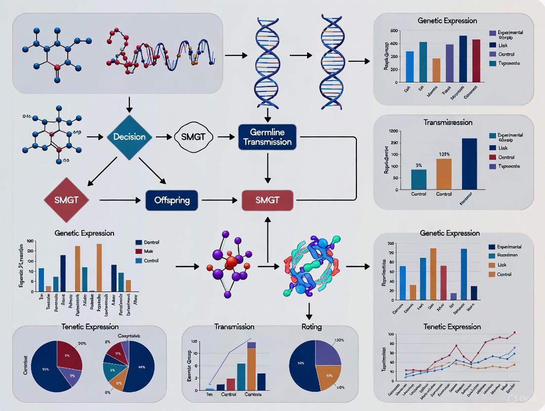
Workflow and Logical Diagram
The following diagram illustrates the complete experimental workflow for generating and validating transgenic animals via Sperm-Mediated Gene Transfer, from initial sperm preparation to the critical confirmation of germline transmission in offspring.
Sperm-mediated gene transfer remains a promising yet challenging technique in the transgenesis toolbox. Its principal advantages of low cost and technical simplicity are compelling, especially for generating large animal models like transgenic pigs for biomedical research [2] [4]. However, the scientific community must contend with its major drawback: inconsistent reproducibility and variable efficiency across studies and species [3]. The successful application of SMGT for stable germline transmission, as demonstrated in some high-profile studies, proves its potential. Future research must focus on standardizing protocols and understanding the fundamental mechanisms of DNA uptake and integration to overcome existing barriers [1] [6]. For researchers, particularly in xenotransplantation and agricultural biotechnology, SMGT presents a high-risk, high-reward pathway that, when successful, can efficiently produce valuable multigene transgenic large animals.
The ability of sperm cells to spontaneously bind and internalize exogenous DNA is a biological phenomenon with profound implications for germline transmission testing and the development of sperm-mediated gene transfer (SMGT) technologies. This process, once considered a laboratory curiosity, is now recognized as a complex, biologically controlled mechanism that enables spermatozoa to function as natural vectors for genetic material [1]. Within SMGT offspring research, understanding the precise molecular machinery governing sperm-DNA interaction is paramount, as it directly influences the efficiency and reliability of generating genetically modified organisms. The mechanism is not random but involves specific DNA-binding proteins, regulated uptake processes, and sophisticated intracellular handling of foreign genetic material [8] [9]. This guide objectively compares the key molecular components and experimental data that define this unique biological capability, providing researchers with a structured framework for evaluating and applying this technology in germline modification studies.
Molecular Mechanisms of DNA Binding and Uptake
The interaction between spermatozoa and exogenous DNA is a carefully orchestrated process initiated at the sperm cell membrane and culminating within the nuclear compartment. Mature sperm cells from virtually all mammalian species, including humans, demonstrate a spontaneous capacity to bind exogenous nucleic acids within a specific 15-20 minute window post-exposure [9]. This interaction is primarily mediated by ionic exchanges between the negatively charged phosphate backbone of DNA and specific positively charged substrates located on the sperm head [9].
DNA-Binding Proteins as Primary Receptors
Southwestern blot analysis has identified three major classes of DNA-binding proteins (DBPs) in sperm head extracts that serve as primary receptors for exogenous DNA [8] [9]:
- A 50 kDa protein class of uncertain function.
- A 30-35 kDa protein class that is highly conserved across mammalian species and represents the principal mediator of DNA binding.
- A <20 kDa protein class that likely includes sperm protamines.
The 30-35 kDa DBPs are particularly significant as they represent the only class accessible to exogenous DNA in intact, viable sperm cells and demonstrate conserved electrophoretic mobility across species boundaries [8]. These proteins exhibit specific binding affinity for DNA molecules, forming discrete protein/DNA complexes as confirmed through band shift assays [8]. Their strategic location on the sperm head enables direct interaction with incoming DNA molecules, facilitating subsequent internalization steps.
Table 1: Major DNA-Binding Protein Classes in Mammalian Sperm
| Molecular Weight Class | Conservation Across Species | Accessibility in Intact Sperm | Postulated Function |
|---|---|---|---|
| ~50 kDa | Variable | Limited | Auxiliary DNA binding |
| 30-35 kDa | High | Primary access | Primary DNA receptor |
| <20 kDa | Variable | Limited | Chromatin packaging |
Factors Influencing DNA Uptake Efficiency
The efficiency of DNA uptake by sperm cells is influenced by several physical and molecular factors:
- DNA Size Selectivity: Sperm cells exhibit a clear preference for larger DNA molecules, with 7 kb fragments being internalized more efficiently than smaller 150-750 bp fragments [9].
- Inhibitory Factors in Seminal Plasma: Seminal fluid contains powerful inhibitory factors that block DNA binding to sperm cells [8] [9] [1]. These factors specifically target the 30-35 kDa DBPs, rendering them unable to bind exogenous DNA. Consequently, extensive washing to remove seminal plasma is a prerequisite for successful DNA uptake in experimental SMGT protocols [1].
- Charge-Mediated Interactions: Polyanions such as heparin and dextran sulfate compete with DNA for binding sites and can reverse DNA binding, while polycations like poly-L-lysine enhance DNA uptake [9], confirming the electrostatic nature of the initial interaction.
Experimental Protocols for Studying Sperm-DNA Interactions
Research into sperm-DNA binding mechanisms employs several well-established experimental approaches that provide complementary data on different aspects of the interaction.
Southwestern Blot Analysis for DNA-Binding Protein Identification
This technique is fundamental for identifying and characterizing the specific sperm proteins capable of binding exogenous DNA.
Detailed Methodology:
- Prepare protein extracts from purified sperm heads.
- Separate proteins using SDS-PAGE electrophoresis.
- Transfer proteins from the gel to a nitrocellulose or PVDF membrane.
- Incubate the membrane with labeled exogenous DNA probes.
- Detect bound DNA-protein complexes using appropriate detection methods (e.g., autoradiography for radiolabeled probes or chemiluminescence for digoxigenin-labeled probes).
- Identify specific protein bands that interact with the DNA probes.
This method directly revealed the 30-35 kDa proteins as the primary DNA-binding components in sperm cells [8] [9].
Band Shift Assay (Electrophoretic Mobility Shift Assay)
The band shift assay is used to confirm and characterize the interaction between purified 30-35 kDa DBPs and exogenous DNA.
Detailed Methodology:
- Incubate purified 30-35 kDa protein fraction with target DNA sequences.
- Separate protein-DNA complexes from free DNA using non-denaturing polyacrylamide gel electrophoresis.
- Visualize DNA migration patterns using DNA-specific stains.
- Observe slowed migration (band shifting) of DNA sequences bound by proteins, confirming direct interaction.
- Perform competition experiments with cold competitor DNA to demonstrate binding specificity.
This assay confirmed that the 30-35 kDa proteins directly interact with exogenous DNA to form discrete complexes [8].
Sperm-Mediated Gene Transfer (SMGT) Protocol
The functional application of sperm-DNA interactions is embodied in the SMGT protocol for generating transgenic animals.
Detailed Methodology:
- Collect epididymal sperm or extensively wash ejaculated sperm to remove seminal plasma inhibitors [9] [1].
- Incubate sperm cells with exogenous DNA for 15-20 minutes to allow binding and internalization.
- Use DNA-loaded sperm for in vitro fertilization or intracytoplasmic sperm injection (ICSI).
- Transfer resulting embryos to synchronized recipients.
- Analyze offspring for transgene integration and expression.
This protocol exploits the natural ability of sperm to bind and internalize DNA, though efficiency remains variable across species [1].
Diagram 1: Experimental workflow for studying sperm-DNA interactions and SMGT. Critical steps include removing seminal inhibitors and allowing 15-20 minutes for DNA binding.
Regulatory Controls and Intracellular Fate of Internalized DNA
The sperm-DNA interaction is not an unregulated process but is controlled by specific biological barriers that likely evolved to prevent random genetic alterations during natural reproduction [1].
Endogenous Nuclease Activation and DNA Processing
Upon DNA internalization, mouse epididymal sperm cells exhibit activation of endogenous nucleases that mediate rearrangements of the internalized DNA [10]. This nuclease activity represents a fundamental biological barrier that processes foreign DNA before it can integrate into the sperm genome. The internalized DNA sequences become tightly associated with the sperm nuclear scaffold, where they undergo a recombination process with the host chromosomal DNA [10].
Table 2: Key Regulatory Controls in Sperm-DNA Interactions
| Regulatory Control | Molecular Basis | Functional Impact on SMGT |
|---|---|---|
| Seminal Plasma Inhibitors | Factor(s) that block DNA binding to 30-35 kDa DBPs | Must be removed by extensive washing for successful DNA uptake |
| Endogenous Nuclease Activation | Nucleases triggered by DNA interaction | Processes and rearranges internalized DNA before integration |
| Nuclear Scaffold Association | Tight binding to nuclear matrix | May facilitate recombination with sperm chromosomal DNA |
| Retrotransposition Machinery | LINE-1 reverse transcriptase activity | Converts RNA to cDNA; may facilitate DNA integration |
Reverse Transcriptase Activity and Retrotransposition
A significant development in understanding the fate of internalized nucleic acids is the discovery of an active reverse transcriptase (RT) in sperm nuclei, encoded by LINE-1 retrotransposons [11]. This RT can reverse-transcribe both internalized RNA and DNA molecules:
- Exogenous RNA is directly reverse-transcribed into cDNA in a single-step reaction [11].
- DNA molecules are first transcribed into RNA by a DNA-dependent RNA polymerase present in spermatozoa, then reverse-transcribed into cDNA [11].
The resulting cDNA copies can be delivered to oocytes at fertilization, propagated throughout embryogenesis, and inherited in a non-Mendelian fashion in adult tissues, where they may be transcriptionally active and induce novel phenotypic traits [11].
Diagram 2: Proposed retrotransposition pathway for nucleic acid processing in sperm. LINE-1 reverse transcriptase converts both exogenous RNA and DNA into cDNA copies that can be transmitted to offspring.
The Scientist's Toolkit: Essential Research Reagents
Research into sperm-DNA interactions requires specific reagents and methodologies to successfully investigate and manipulate this biological phenomenon.
Table 3: Essential Research Reagents for Sperm-DNA Interaction Studies
| Reagent/Methodology | Specific Function | Experimental Application |
|---|---|---|
| Purified 30-35 kDa DBP Fraction | Identifies primary DNA receptor proteins | Southwestern blot, band shift assays |
| Seminal Plasma Inhibitor Fractions | Blocks DNA binding to DBPs | Control experiments; study of regulatory mechanisms |
| LINE-1 Reverse Transcriptase Inhibitors | Suppresses RT activity (e.g., Nevirapine, AZT) | Investigates role of retrotransposition in DNA integration |
| Heparin/Dextran Sulfate | Polyanionic competitors disrupt electrostatic binding | Confirms ionic nature of DNA binding; control experiments |
| Poly-L-lysine | Polycation enhances DNA uptake | Increases efficiency of DNA internalization |
| Sperm Chromatin Structure Assay (SCSA) | Quantifies DNA fragmentation index (DFI) | Assesses sperm DNA integrity; measures damage from experimental procedures |
| TUNEL Assay/TdT-Strand Displacement Probe | Directly detects DNA breakpoints and fragments | Measures DNA damage; quantifies mean DNA breaks (MDB) |
| Epididymal Sperm Isolation Protocol | Obtains sperm free of seminal plasma | Essential preparatory step for SMGT experiments |
Implications for Germline Transmission Testing in SMGT Offspring
The molecular mechanisms of DNA binding and internalization have direct consequences for the design and interpretation of germline transmission studies in SMGT offspring research. Several critical considerations emerge:
Integration Specificity: Sequence analysis of sperm genomic DNA transformed with foreign plasmids indicates that integration may occur at unique sites in the sperm genome, potentially mediated by a retrotranscription step [10]. This non-random integration pattern has significant implications for predicting expression stability and inheritance patterns in SMGT offspring.
Extrachromosomal Inheritance: The RT-generated cDNA copies are maintained as low-copy number, extrachromosomal sequences that are mosaic distributed in founder tissues and transmitted to subsequent generations in a non-Mendelian fashion [11]. This challenges conventional transgenic screening approaches that assume chromosomal integration.
Paternal Somatic Experience Integration: Growing evidence indicates that spermatozoa can incorporate RNA from somatic cell-released exosomes, potentially transferring acquired characteristics to offspring [11]. This somatic-to-germline RNA transfer represents a previously underappreciated variable in germline transmission studies.
Understanding these mechanisms provides researchers with a more comprehensive framework for designing SMGT experiments, interpreting germline transmission data, and advancing the technology toward more reliable applications in biomedical research and biotechnology.
The selection of an appropriate animal model is a cornerstone of biomedical research, particularly for studies requiring high translational relevance to humans. While rodents have long been the mainstay, their physiological and genetic differences from humans can limit the predictive value of preclinical data [12]. Large animal models, such as pigs, sheep, and goats, offer a compelling alternative due to their closer phylogenetic, anatomic, and physiologic resemblance to humans [13]. Within this context, the method used for genetic modification is critical. This guide objectively compares the efficiency and cost-effectiveness of Sperm-Mediated Gene Transfer (SMGT) against traditional techniques for creating large animal models, with a specific focus on its application in germline transmission testing.
Comparative Analysis of Genetic Modification Techniques
The following table summarizes the key performance metrics of predominant methods for generating genetically modified large animals. SMGT emerges as a particularly balanced technology, offering distinct advantages in efficiency and practicality [13].
Table 1: Comparison of Genetic Modification Techniques for Large Animals
| Technique | Key Mechanism | Germline Transmission Efficiency | Relative Cost | Development Time | Key Advantages | Major Limitations |
|---|---|---|---|---|---|---|
| Sperm-Mediated Gene Transfer (SMGT) | Spermatozoon acts as a natural vector for DNA into the oocyte [13]. | High efficiency; allows for insertion of large DNA fragments [13]. | Cost-effective | Shorter production time for modified sperm [13]. | Less technically demanding; avoids complex embryo manipulation [13]. | Lower public acceptance for food technology; complex regulatory landscape [13]. |
| Pronuclear (PN) Microinjection | Direct microinjection of DNA into the pronucleus of a fertilized oocyte [13]. | Low (1-10%); random transgene integration leads to highly variable expression [13]. | High (inefficient, requires screening many founders) [13]. | Lengthy | A long-established, widely understood method. | Technically challenging; random integration causes variable expression; low efficiency in livestock [13]. |
| Somatic Cell Nuclear Transfer (SCNT) | Transfer of a nucleus from a genetically modified somatic cell into an enucleated oocyte [13]. | High (allows for pre-selection of specific genetic modifications) [13]. | Very High (inefficient, costly rearing) [13]. | Lengthy | Enables precise genetic engineering (knock-outs/ins) before embryo creation [13]. | Frequent developmental abnormalities due to incomplete nuclear reprogramming [13]. |
| Germline Stem Cell (GSC) Transplantation | Transplantation of genetically modified male germline stem cells into a recipient testis [13]. | Promising; produces donor-derived sperm [13]. | High | Shorter than SCNT or ES cells [13]. | Circumvents embryo manipulation and nuclear reprogramming issues [13]. | Challenging to maintain GSCs from domestic animals in vitro for extended periods [13]. |
The data illustrates that SMGT provides a superior balance of high efficiency and cost-effectiveness. Its ability to utilize the sperm as a natural vector simplifies the process, reducing the need for highly specialized equipment and personnel compared to PN microinjection and SCNT. This directly translates to lower operational costs and faster timelines for producing founder animals, a critical advantage in research and development.
Experimental Protocol for SMGT and Germline Transmission Testing
To achieve reliable germline transmission using SMGT, a standardized experimental protocol is essential. The following workflow details the key steps from sperm preparation to the confirmation of germline transmission in offspring.
Diagram 1: SMGT Germline Transmission Testing Workflow
Detailed Methodology:
- Sperm Preparation and DNA Incubation: High-quality spermatozoa are collected and washed. The sperm membrane is made permeable to allow uptake of the target DNA construct, which is then co-incubated with the sperm [13].
- Fertilization and Embryo Transfer: The DNA-loaded sperm are used for fertilization, most effectively via Intracytoplasmic Sperm Injection (ICSI), to ensure delivery of the genetic material [13]. The resulting embryos are cultured in vitro to a suitable stage before being surgically transferred into synchronized recipient females [13].
- Founder (F0) Generation Analysis: Offspring born from this process are the founder (F0) generation. They are genotyped using PCR and Southern blot analysis to confirm the presence and integration pattern of the transgene.
- Germline Transmission Testing: To definitively prove that the F0 animal has the transgene in its germ cells, it is bred with a wild-type (non-transgenic) partner [13]. The resulting F1 offspring are genotyped. The confirmation of germline transmission is achieved when transgenic-positive individuals are identified within the F1 generation, proving that the genetic modification was passed on.
The Scientist's Toolkit: Essential Reagents for SMGT Research
Table 2: Key Research Reagent Solutions for SMGT Experiments
| Reagent / Material | Function in SMGT Protocol |
|---|---|
| Target DNA Construct | The engineered genetic material (e.g., for a specific mutation or transgene) to be incorporated into the animal's genome. |
| Sperm Washing Medium | A specialized buffer used to remove seminal plasma and prepare sperm for efficient DNA uptake without damaging motility or viability. |
| Membrane Permeabilization Agents | Chemicals (e.g., Triton X-100) used to temporarily disrupt the sperm membrane, facilitating the entry of foreign DNA. |
| PCR and Southern Blot Kits | Essential reagents for genotyping. PCR screens for transgene presence, while Southern blot analysis confirms integration and copy number. |
| ICSI Micromanipulation System | A set of specialized microscopes and micro-pipettes for the precise injection of a single DNA-loaded sperm head directly into an oocyte. |
| Embryo Culture Media | A sequence of complex, defined media that supports the development of fertilized oocytes into viable embryos ready for transfer. |
In the pursuit of physiologically relevant large animal models for biomedical research, SMGT stands out for its high efficiency and cost-effectiveness. As demonstrated, SMGT offers a less technically demanding and more reliable path to germline transmission compared to traditional methods like pronuclear microinjection and SCNT. Its ability to efficiently produce transgenic founders that reliably pass the modification to the next generation makes it an invaluable tool for establishing stable transgenic lines. This accelerates research in areas such as drug development, disease modeling, and the production of therapeutic proteins, ultimately enhancing the translational potential of preclinical studies.
The Critical Importance of Germline Transmission Testing in SMGT Workflows
Sperm-mediated gene transfer (SMGT) presents a potentially streamlined route for generating transgenic animals. However, its value in rigorous scientific and therapeutic applications is contingent upon the thorough confirmation that the transgene has been successfully integrated into the germline and can be stably transmitted to subsequent generations. This article explores the critical role of germline transmission testing within SMGT workflows, comparing its performance against established transgenic methods and detailing the experimental protocols essential for validation.
Performance Comparison: SMGT vs. Alternative Transgenesis Methods
The efficiency of SMGT can be evaluated against other common techniques for creating genetically modified large animals. The following table summarizes key performance metrics based on experimental data.
Table 1: Comparison of Transgenesis Methods in Large Animals
| Method | Reported Integration Efficiency | Key Advantages | Key Limitations | Germline Transmission Confirmed |
|---|---|---|---|---|
| Sperm-Mediated Gene Transfer (SMGT) [4] | Up to 80% of offspring (founders) | High efficiency, low cost, technical simplicity [4] [14] | Risk of mosaicism; variable transgene expression patterns [14] | Yes, stable transmission to progeny demonstrated [4] |
| Pronuclear Microinjection [13] [14] | 1-5% in livestock species [13] | Well-established methodology; direct applicability to human zygotes [14] | Technically challenging, low efficiency, costly, random integration leading to variable expression [13] [14] | Yes (standard part of validation) |
| Somatic Cell Nuclear Transfer (SCNT) [13] | Low, efficiency impacted by developmental abnormalities | Enables precise genetic modifications (knock-outs/ins) in donor cells prior to transfer [13] | Very costly, time-consuming, associated with developmental abnormalities [13] | Yes (inherent in the method) |
Essential Protocols for Germline Transmission Testing in SMGT
Confirming germline transmission involves a multi-stage experimental workflow designed to first identify founder animals and then verify the inheritance of the transgene in their offspring.
Stage 1: Genomic Integration and Expression Analysis in Founders
This initial stage focuses on characterizing the F0 generation animals produced via SMGT.
- DNA Extraction and PCR/Southern Blotting: Genomic DNA is extracted from candidate founder animals (e.g., from blood or tissue biopsies). Standard Polymerase Chain Reaction (PCR) is used for initial, sensitive detection of the transgene. Southern blot analysis provides confirmatory evidence of genomic integration, revealing integration patterns and transgene copy number [4].
- RNA Extraction and RT-PCR: RNA is extracted from relevant tissues and reverse-transcribed to cDNA. Reverse Transcription PCR (RT-PCR) is then performed to confirm that the integrated transgene is being actively transcribed. Primers should be designed to span an intron or to differentiate between the transgene and endogenous sequences to rule out genomic DNA contamination [4].
- Protein Expression Analysis (Immunohistochemistry/Western Blot): Protein-level expression is confirmed using techniques like immunohistochemistry on tissue sections or Western blotting on protein lysates. This verifies that the mRNA is translated into a functional protein and allows for the assessment of tissue-specific localization [4].
- Fluorescence In Situ Hybridization (FISH): This cytogenetic technique is used to visually map the physical location of the transgene on a specific chromosome, providing definitive evidence of integration and information about the integration site [4].
Stage 2: Inheritance and Stability Analysis in Offspring
The definitive test for germline transmission is breeding the founder animal and analyzing the F1 generation.
- Breeding and Genotyping: A confirmed transgenic founder (F0) is bred with a wild-type mate. The resulting offspring (F1) are genotyped using PCR to determine if they have inherited the transgene. A Mendelian inheritance pattern (approximately 50% of offspring positive for a heterozygous insertion) confirms successful germline transmission [4].
- Functional Assays in Progeny: To ensure the transgene remains functional across generations, expression and functional analyses (as described in Stage 1) should be repeated on positive F1 offspring. This confirms that expression is stable and not silenced in the subsequent generation [4].
The logical sequence of these validation stages is outlined below.
The Scientist's Toolkit: Key Reagents for SMGT Germline Testing
A successful SMGT and validation workflow relies on several critical reagents and tools.
Table 2: Essential Research Reagents for SMGT Workflows
| Reagent / Tool | Function in Workflow |
|---|---|
| Linearized Plasmid DNA | The genetic construct of interest; linearization often improves integration efficiency [4]. |
| Sperm Washing Medium (e.g., SFM/BSA) | Used to remove seminal fluid and prepare sperm for DNA uptake [4]. |
| DNA Extraction Kits | For high-quality genomic DNA isolation from tissues or blood for PCR and Southern blotting [4]. |
| Sequence-Specific Primers & Probes | Essential for PCR, RT-PCR, and Southern blotting to specifically detect the transgene [4]. |
| Antibodies against Transgenic Protein | For detecting and localizing the expressed protein via immunohistochemistry and Western blot [4]. |
| FISH Probe (e.g., Biotin-labeled) | A labeled DNA probe complementary to the transgene for chromosomal localization [4]. |
Regulatory and Safety Context
Germline transmission testing is not only a scientific necessity but also a regulatory consideration. Health authorities like the European Medicines Agency (EMA) have issued guidelines on "Non-clinical testing for inadvertent germline transmission of gene transfer vectors," highlighting the importance of assessing the risk of accidental integration into reproductive cells during gene therapy development [15]. This is a distinct but related concern, underscoring the broader relevance of understanding and controlling germline modification in modern biotherapeutics [16].
Germline transmission testing is the definitive step that validates the success of an SMGT procedure, moving beyond the mere presence of a transgene to proving its heritability. While SMGT offers a highly efficient and accessible method for generating transgenic large animal models, its data is only conclusive when supported by rigorous breeding studies and molecular analysis of subsequent generations. This comprehensive validation through germline transmission testing solidifies the model's utility for long-term biomedical research and therapeutic development.
Historical Context and Evolution of SMGT from Mice to Livestock Species
Sperm-mediated gene transfer (SMGT) represents a fascinating approach in the field of transgenesis, aiming to utilize spermatozoa as natural vectors for introducing foreign genetic material into oocytes during fertilization. The journey of SMGT from a contested concept in mice to an applied method in livestock species illustrates a significant evolution in reproductive biotechnology. This guide objectively compares the performance of various SMGT methodologies and their alternatives, with a specific focus on evidence of germline transmission—a critical benchmark for success in transgenic animal production. Framed within the broader thesis on germline transmission testing in SMGT offspring research, this review provides researchers and drug development professionals with a detailed comparison of protocols, efficiencies, and experimental data.
Historical Context and Key Milestones
The foundation of SMGT was laid in 1971 when Brackett and colleagues provided the first evidence of sperm-mediated transport of foreign DNA (simian virus 40 DNA) into rabbit oocytes [17]. However, the field ignited in 1989 when Lavitrano et al. reported producing transgenic mice by simply incubating sperm with plasmid DNA before in vitro fertilization [18]. This promising report sparked widespread excitement but proved difficult to replicate independently, leading to initial skepticism about the methodology [18] [17].
In the subsequent decades, researchers pursued various modifications to improve the reliability and efficiency of SMGT. A significant advancement came in 1999 with the development of intracytoplasmic sperm injection-mediated transgenesis (ICSI-Tr), which involved complexing DNA with membrane-damaged sperm before microinjection into oocytes [17]. While this method showed higher transgenic rates in mice, it required highly specialized skills and equipment. The ongoing challenge of consistent DNA uptake by sperm led to innovative solutions, including the use of linkers, electroporation, and chemical facilitators, gradually expanding SMGT applications from mice to larger livestock species such as pigs, cattle, sheep, and goats [18] [19] [17].
Comparative Analysis of SMGT Methodologies and Alternatives
The table below summarizes the key performance metrics of primary SMGT methodologies and other established gene-editing techniques, with a focus on germline transmission evidence.
Table 1: Comparison of Transgenesis Methods in Livestock Species
| Method | Key Principle | Typical Efficiency (Transgenic Offspring) | Germline Transmission Evidence | Relative Technical Complexity | Key Advantages & Limitations |
|---|---|---|---|---|---|
| Standard SMGT | Incubation of sperm with DNA for fertilization | Variable, often low; Highly species-dependent [17] | Limited and often lacking in early studies [17] | Moderate | Advantage: Conceptually simple. Limitation: Inconsistent results, low efficiency [17]. |
| Linker-Based SMGT (LB-SMGT) | Use of a monoclonal antibody (mAb C) to link DNA to sperm surface antigen [18] | High: 37.5% in pigs, 33% in mice (F0) [18] | Confirmed: F1 progeny analysis in pigs and mice [18] | Moderate | Advantage: High efficiency, works across multiple species. Limitation: Requires production of a specific linker protein. |
| ICSI-Mediated Transgenesis (ICSI-Tr) | Direct injection of DNA-complexed sperm into oocyte cytoplasm [17] | High in mice [17] | Confirmed in model species [17] | Very High | Advantage: Reduces mosaicism. Limitation: Technically demanding, requires specialized equipment [17]. |
| Somatic Cell Nuclear Transfer (SCNT) | Transfer of a nucleus from a genetically modified somatic cell into an enucleated oocyte [20] [19] | Low to moderate (0-3%) [18] [19] | Inherent—all offspring are derived from a modified cell line | Very High | Advantage: Permits precise genetic modifications. Limitation: Low efficiency, often associated with health issues in offspring [18]. |
| Pronuclear Microinjection | Physical injection of DNA into the pronucleus of a zygote [18] [19] | Low in livestock (<1% in F0) [18] | Possible, but low efficiency makes it difficult to assess | High | Advantage: Well-established history. Limitation: Very low efficiency in livestock, high mosaicism [18]. |
| CRISPR/Cas9 with Electroporation | Direct delivery of gene-editing reagents into zygotes [20] [7] | Variable, but generally higher than microinjection [7] | Confirmed in multiple livestock species [20] [7] | Moderate to High | Advantage: Enables precise genome editing. Limitation: Risk of off-target effects and mosaicism [20] [7]. |
The experimental data supporting germline transmission is a cornerstone for validating any transgenesis method. In the case of LB-SMGT, the 37.5% efficiency in pigs was not merely a measure of founder (F0) animals carrying the transgene. Crucially, these F0 animals were bred, and the transgene was successfully passed to the F1 generation, confirming stable integration into the germline. Furthermore, fluorescence in situ hybridization (FISH) analysis provided physical evidence of chromosomal integration, and expression of the transgene was demonstrated in 61% of the transgenic pigs [18]. This multi-faceted validation—integration, transmission, and expression—provides a robust model for confirming germline transmission in SMGT offspring research.
Detailed Experimental Protocols
Protocol for Linker-Based SMGT (LB-SMGT)
The LB-SMGT protocol represents a significant refinement of standard SMGT, enhancing the specific binding of DNA to sperm [18].
- Sperm Preparation and DNA Loading: Washed sperm from the target species are incubated with the monoclonal antibody mAb C. This antibody is a basic protein that binds to a surface antigen found on sperm across multiple species (pig, mouse, cow, etc.) and interacts ionically with the negatively charged DNA backbone. The DNA of interest (e.g., a linearized plasmid) is then added to form a specific antibody-DNA-sperm complex. Radioisotope studies have demonstrated that this linker increases DNA binding to sperm by 25-56% compared to controls [18].
- Fertilization and Embryo Transfer: The treated sperm are used for in vitro fertilization of oocytes. Alternatively, for species like pigs, surgical oviduct insemination can be performed, where the gene-loaded sperm are surgically deposited directly into the oviduct of a surrogate gilt. The resulting embryos are then allowed to develop to term within the surrogate [18].
- Analysis of Offspring: Founder (F0) offspring are screened for the presence of the transgene using PCR and Southern blotting. To confirm germline transmission, transgenic F0 animals are bred with wild-type partners, and the resulting F1 progeny are screened for the transgene. Further analysis can include FISH to visualize the integration site and RT-PCR or immunohistochemistry to confirm transgene expression [18].
Protocol for Intratesticular Gene Transfer (ITGT) in Neonates
This related protocol focuses on delivering genes directly to the testis, transfecting both germ and somatic cells, and represents an alternative pathway to generating genetically modified offspring [5].
- Anesthesia: Neonatal mice (days 3-5) are anesthetized using a simple isoflurane-based system. A convenient setup uses a 15 mL centrifuge tube containing cotton wool soaked with isoflurane, capped with the cut tip of a rubber finger, into which the pup's nose is inserted. This method achieves over 90% postoperative survival with normal recovery [5].
- Surgical Injection and Electroporation: A small incision is made in the lower abdomen to expose the testis. A plasmid DNA solution (e.g., 0.25 μg/μL in PBS with a Fast Green tracer) is injected directly into the testis using a glass micropipette. Immediately after injection, in vivo electroporation is performed using tweezer-type electrodes to facilitate nucleic acid uptake into the testicular cells. The procedure is completed within 30 minutes for both testes [5].
- Outcome: This protocol results in efficient transfection of interstitial Leydig cells and limited transfection of seminiferous tubules, offering a route to study gene function in spermatogenesis or to manipulate germ cells in situ [5].
Diagram 1: SMGT Germline Transmission Testing Workflow
The Scientist's Toolkit: Key Research Reagent Solutions
Successful implementation of SMGT and validation of germline transmission rely on a suite of specific reagents and tools. The table below details essential materials and their functions.
Table 2: Essential Reagents for SMGT and Germline Analysis
| Reagent / Tool | Function in SMGT Protocol | Specific Examples / Notes |
|---|---|---|
| Monoclonal Antibody mAb C | Acts as a linker to specifically bind exogenous DNA to a surface antigen on sperm [18]. | A basic protein that binds ionically to DNA; reactive with sperm from pig, mouse, chicken, cow, goat, sheep, and human [18]. |
| Vector/Transgene Construct | Carries the gene of interest for integration into the animal's genome. | Often linearized before use; may contain reporter genes (e.g., tdTomato, SEAP) or therapeutic genes of interest [18] [5]. |
| Fast Green FCF | A visual tracer dye used during microinjection to monitor the delivery of the DNA solution [5]. | Added to the DNA solution (e.g., 0.02% v/v) to confirm successful injection into the target tissue (testis, pronucleus) [5]. |
| Methyl-β-Cyclodextrin | A chemical facilitator that increases the association of DNA with the sperm surface membrane [7]. | Used in some advanced SMGT protocols to improve DNA uptake efficiency [7]. |
| Electroporation Apparatus | Applies controlled electrical pulses to create temporary pores in cell membranes, facilitating DNA entry [5] [7]. | Used in ITGT for testicular cells [5] and in other methods like i-GONAD for embryo editing [7]. |
| Isoflurane Anesthesia System | Provides safe and controllable anesthesia for surgical procedures on neonatal or adult animals [5]. | Critical for survival surgery; a simple system can be constructed from common lab equipment [5]. |
| PCR & Southern Blot Reagents | Molecular biology tools for genotyping F0 and F1 animals to detect the presence and integration pattern of the transgene [18]. | Standard for initial screening and confirmation of transgene integration. |
| FISH Kits | Validates the chromosomal location and integration site of the transgene, providing physical evidence of integration [18]. | Used to provide definitive proof of transgene integration in the genome. |
The evolution of SMGT from a simple but inconsistent idea in mice to a refined, linker-based technology with demonstrated success in livestock marks a significant achievement in transgenic science. The critical differentiator for any transgenesis method is conclusive evidence of germline transmission, which has been robustly provided for LB-SMGT through F1 progeny analysis and FISH. While newer, more precise technologies like CRISPR/Cas9 now dominate the field, SMGT remains an important part of the historical and methodological landscape. Its development underscores the importance of iterative protocol refinement, the use of specific reagents to enhance efficiency, and the necessity of comprehensive germline transmission testing in validating transgenic animal models. For researchers, the choice of method involves a careful trade-off between efficiency, technical complexity, and the requirement for precise versus random genetic modification.
A Step-by-Step Protocol for SMGT and Transmission Analysis
Sperm preparation is a critical first step in assisted reproductive technologies (ART) and emerging fields such as sperm-mediated gene transfer (SMGT). The choice of processing technique directly influences the quality of selected sperm, impacting key outcomes from fertilization rates to the integrity of genetic material carried forward. This guide provides a comparative analysis of conventional and advanced sperm preparation methods, evaluating their performance in selecting high-quality sperm for advanced research applications, including germline transmission testing.
In sperm-mediated gene transfer (SMGT), the objective extends beyond selecting viable, motile sperm for fertilization; it involves preparing sperm capable of effectively binding and internalizing exogenous DNA constructs for subsequent germline transmission. This process demands sperm preparation techniques that not only isolate motile sperm with intact membranes but also preserve their inherent biological competence for DNA uptake and transport. Conventional techniques, while designed to enhance motility and morphology, often overlook molecular attributes like DNA fragmentation and apoptotic markers, which are critical when the sperm is intended as a vector for genetic material [21]. The centrifugation steps in these methods can even induce sperm DNA fragmentation (sDF) through the generation of reactive oxygen species (ROS), potentially compromising the genetic cargo [22]. Therefore, optimizing sperm washing and incubation protocols is paramount for the success and reliability of SMGT offspring research.
Comparative Analysis of Sperm Preparation Techniques
Various sperm preparation techniques are employed, each with distinct advantages and limitations for research applications. The following table summarizes the core characteristics of these methods.
Table 1: Comparison of Key Sperm Preparation Techniques
| Technique | Underlying Principle | Key Advantages | Reported Limitations |
|---|---|---|---|
| Density Gradient Centrifugation (DGC) | Separation based on sperm density and motility through a silica gel gradient [23] [24]. | Effectively separates motile sperm, removes leukocytes and debris [25]. | Centrifugation may generate ROS and induce DNA damage [21] [22]. |
| Swim-Up (SU) | Self-migration of motile sperm from semen into an overlaying culture medium [23] [24]. | Simple, cost-effective; yields sperm with high motility and normal morphology [25]. | Lower recovery of motile sperm; potential for sample contamination [24]. |
| DGC followed by Swim-Up (DGC-SU) | Combination of DGC and SU techniques for sequential sperm selection [23]. | May combine the high yield of DGC with the high motility of SU. | Protocol is more time-consuming; still involves centrifugation steps. |
| Magnetic-Activated Cell Sorting (MACS) | Selection of non-apoptotic sperm using Annexin-V conjugated microbeads to bind phosphatidylserine (PS) [21]. | Effectively removes apoptotic sperm, reduces DNA fragmentation, improves chromatin maturity [21]. | Requires specialized equipment; often used as an adjunct to DGC or SU. |
| Microfluidic Sperm Selection (MSSP) | Biomimetic selection based on motility and boundary-following behavior, often combined with apoptotic marker trapping [22]. | Avoids centrifugation-induced DNA damage; provides highly motile sperm with superior DNA integrity [22]. | Emerging technology, not yet widespread in clinical practice. |
Quantitative data from comparative studies underscores the performance differences between these methods. The table below presents experimental outcomes from independent research.
Table 2: Quantitative Performance Metrics of Sperm Preparation Techniques
| Technique | Motile Sperm Recovery (%) | DNA Fragmentation Index (sDF%) | Apoptotic Marker Expression (%) | Fertilization / Pregnancy Rate |
|---|---|---|---|---|
| Swim-Up (Conventional) | Baseline [22] | 7.9% [22] | 26.5% [22] | No significant difference vs. DGC in IUI pregnancy rates [25] |
| Density Gradient (DGC) | Comparable to Swim-Up [23] | Variable; not specifically designed to reduce sDF [21] | Not sufficiently evaluated for apoptosis [21] | No significant difference vs. Swim-Up in IUI pregnancy rates [25] |
| DGC-MACS | Not specified | Significantly lower than DGC, SU, and DGC-SU in teratozoospermic samples [21] | Significantly lower than DGC, SU, and DGC-SU [21] | Improved embryo cleavage and clinical pregnancy rates vs. other techniques [21] |
| Microfluidic (MSSP) | >68% improvement over some reported methods [22] | 1.4% (over 85% improvement vs. Swim-Up) [22] | 5.66% (90% reduction vs. Swim-Up) [22] | Data on clinical outcomes is promising but still emerging [22] |
Detailed Experimental Protocols for Key Techniques
Density Gradient Centrifugation (DGC) followed by Swim-Up
This combined protocol is commonly used in ART laboratories to maximize the selection of high-quality sperm [23].
Methodology:
- Gradient Preparation: In a conical centrifuge tube, carefully layer a "lower layer" of 90% density gradient medium (e.g., Isolate, PureSperm) beneath an "upper layer" of 50% density gradient medium, without mixing the layers [23] [26].
- Sample Loading and Centrifugation: Gently layer 2.0 mL of liquefied semen on top of the gradient column. Centrifuge the tube at 300 g for 10-20 minutes [23] [26].
- Pellet Extraction: After centrifugation, carefully aspirate and discard the upper layers. Using a sterile pipette, extract the sperm pellet from the bottom of the tube [23].
- Washing: Resuspend the pellet in 4.0 mL of sperm washing medium (e.g., Ham's F-10 or SWM). Centrifuge at 250-350 g for 5-10 minutes. Repeat this washing step once more [23] [26].
- Swim-Up: Discard the supernatant and gently layer 0.5 mL of fresh medium over the final pellet. Incline the tube at a 45° angle and incubate at 37°C for 30-60 minutes [23] [24].
- Collection: After incubation, carefully aspirate the upper supernatant, which now contains the most motile sperm, and transfer it to a sterile tube for use [23].
DGC with Magnetic-Activated Cell Sorting (DGC-MACS)
This protocol is optimized for selecting non-apoptotic sperm, which is crucial for experiments requiring high DNA integrity [21].
Methodology:
- Initial Processing: First, process the semen sample using the DGC protocol (steps 1-4 above) to obtain a pellet rich in motile sperm [21].
- Annexin-V Binding: Resuspend the DGC pellet in an appropriate buffer. Incubate the sperm suspension with Annexin-V conjugated magnetic microbeads. The Annexin-V binds to phosphatidylserine (PS), which is externalized on the membrane of apoptotic sperm [21].
- Magnetic Separation: Pass the cell suspension through a separation column placed in a strong magnetic field. Sperm with externalized PS (apoptotic) will be retained in the column, while non-apoptotic, viable sperm with intact membranes will pass through and be collected [21].
- Final Preparation: The eluted fraction of non-apoptotic sperm is then centrifuged and resuspended in a suitable medium for subsequent use or incubation with DNA constructs [21].
Visualizing the Sperm Preparation Workflow for SMGT
The following diagram illustrates the logical decision-making pathway and procedural steps for preparing sperm for SMGT applications, integrating the techniques discussed.
Figure 1: Sperm Preparation Workflow for SMGT. This flowchart outlines the decision points and key technical steps in preparing sperm for sperm-mediated gene transfer, from initial sample processing to final incubation with DNA constructs.
The Scientist's Toolkit: Essential Reagents and Materials
Successful sperm preparation relies on a suite of specialized reagents and materials. The following table details the core components of the research toolkit.
Table 3: Essential Research Reagent Solutions for Sperm Preparation
| Reagent/Material | Function/Description | Example Uses |
|---|---|---|
| Density Gradient Medium | Silane-coated colloidal silica particles (e.g., PureSperm, Isolate) used to form discontinuous gradients (e.g., 45% and 90%) for selecting sperm based on density and motility [23] [26]. | Density Gradient Centrifugation (DGC) |
| Sperm Washing Medium | A buffered culture medium (e.g., Ham's F-10, HEPES-buffered Human Tubal Fluid) used to wash, resuspend, and maintain sperm during processing. Often supplemented with protein [23]. | All preparation techniques for washing and swim-up steps. |
| Annexin-V Microbeads | Magnetic microbeads conjugated to Annexin-V protein, which has a high affinity for phosphatidylserine (PS). Used to label and remove apoptotic sperm during MACS [21]. | Magnetic-Activated Cell Sorting (MACS) |
| Microfluidic Sperm Selection Device (MSSP) | A 3D-printed or fabricated chip with microchannels that select sperm based on motility, boundary-following behavior, and sometimes apoptotic marker expression, avoiding centrifugation [22]. | Microfluidic Sperm Selection |
| Synthetic Protein Substitute | A synthetic supplement (e.g., synthetic serum substitute) added to culture media to replace human serum albumin, providing energy substrates and protecting sperm membranes [23]. | Added to washing medium for swim-up and final resuspension. |
The selection of a sperm preparation technique is a foundational decision in SMGT and germline transmission research. While conventional methods like DGC and Swim-Up provide adequate motile sperm recovery for standard ART, evidence shows they are suboptimal for selecting sperm with the high DNA integrity required for genetic transmission studies. Advanced techniques, particularly DGC-MACS and microfluidic MSSP, demonstrate superior performance in isolating non-apoptotic sperm with significantly lower DNA fragmentation. For researchers aiming to optimize washing and incubation with DNA constructs, adopting these advanced methods or incorporating MACS as an adjunct to DGC is a critical strategy to enhance the reliability and efficiency of producing SMGT offspring.
Sperm-mediated gene transfer (SMGT) represents a direct approach for creating transgenic animals by using spermatozoa as natural vectors to deliver exogenous DNA into the oocyte during fertilization. Within the broader context of germline transmission testing research, SMGT offers a methodological alternative to more complex procedures like pronuclear microinjection, particularly in large animal models where conventional transgenesis remains inefficient and costly [4]. The fundamental principle underlying SMGT leverages the innate ability of sperm cells to bind, internalize, and protect exogenous DNA, subsequently transferring this genetic material during the fertilization process to create genetically modified offspring [4]. This guide provides a systematic comparison of SMGT performance against other germline modification techniques, supported by experimental data and detailed protocols for researchers and drug development professionals.
Performance Comparison of Germline Modification Techniques
The efficiency of SMGT must be evaluated against other established methods for germline modification. The table below provides a quantitative comparison of key performance metrics across different techniques.
Table 1: Performance comparison of germline modification techniques
| Method | Typical Efficiency (Transgenesis Rate) | Relative Cost | Technical Complexity | Primary Application | Key Advantages |
|---|---|---|---|---|---|
| SMGT | Up to 80% in pigs [4] | Low | Moderate | Large animal transgenesis | Simplicity, cost-effectiveness, no specialized equipment needed |
| Pronuclear Microinjection | ~1-3% in farm animals [4] | High | High | Murine transgenesis | Well-established for mice |
| Intra-Testicular Gene Transfer | Limited to seminiferous tubules; efficient for Leydig cells [5] | Low to Moderate | High | Neonatal gene delivery | Targets germ cells and somatic cells in testes |
| In Vivo Electroporation of Oviduct (i-GONAD) | Efficient for embryos [5] | Moderate | High | In vivo embryo editing | Avoids embryo manipulation |
Beyond these quantitative metrics, SMGT demonstrates distinct practical advantages. The procedure achieves high transgenesis efficiency without requiring expensive microinjection apparatus or highly specialized technical expertise, making it particularly suitable for laboratories focused on large animal models [4]. Furthermore, research confirms that transgenes delivered via SMGT are not only integrated but also functionally transcribed, translated, and stably transmitted to subsequent generations [4].
SMGT Experimental Protocol and Workflow
The successful implementation of SMGT requires careful attention to methodological details. The following protocol, adapted from successful production of hDAF transgenic pigs, provides a reliable framework for researchers [4].
Sperm Preparation and DNA Uptake
- Semen Collection and Washing: Collect semen from trained animal donors following an abstinence period of 4-5 days. Remove seminal fluid by washing sperm in an appropriate prewarmed fertilization medium (e.g., Swine Fertilization Medium - SFM) supplemented with 6 mg/mL BSA. Centrifuge at 800 × g for 10 minutes at 25°C, aspirate supernatants, resuspend sperm, and repeat centrifugation at 800 × g for 10 minutes at 17°C [4].
- Sperm Counting: Resuspend the washed sperm pellet and perform accurate counting using a hemocytometric chamber to standardize the number of sperm for DNA incubation [4].
- DNA Incubation: Dilute washed sperm cells (10⁹ cells) to 120 mL with SFM/BSA at 17°C. Add linearized plasmid DNA at a concentration of 0.4 μg per 10⁶ sperm. Incubate for 2 hours at 17°C, inverting the flask every 20 minutes to prevent sedimentation. During the final 20 minutes of incubation, gradually adjust the temperature to room temperature, followed by a brief 1-minute heating at 37°C immediately before artificial insemination [4].
Artificial Insemination and Outcome Validation
- Female Preparation and Insemination: Prepubertal synchronized gilts should be prepared with eCG (1,250 units) followed by hCG (750 units) 60 hours later. Perform artificial insemination 43 hours after hCG injection using 1–1.5 × 10⁹ DNA-treated sperm cells per gilt using standard veterinary procedures [4].
- Transgene Validation in Offspring:
- Integration Analysis: Perform genomic DNA extraction from offspring tissue samples followed by Southern blotting or PCR analysis using transgene-specific primers to confirm successful integration into the genome [4].
- Expression Analysis: Conduct RT-PCR on tissue samples to verify transcription of the transgene. Additionally, perform immunohistochemistry and Western blotting on relevant tissues to confirm protein expression and proper cellular localization [4].
- Functional Assessment: Validate protein functionality through tissue-specific assays. For human decay-accelerating factor (hDAF), this involved testing peripheral blood mononuclear cell resistance to challenge with human serum in vitro [4].
- Germline Transmission: Breed founder animals and screen subsequent generations to confirm stable inheritance of the transgene, completing the assessment of germline transmission [4].
Diagram: SMGT workflow for germline transmission
The Scientist's Toolkit: Essential Research Reagents
Successful implementation of SMGT requires specific reagents and materials. The following table outlines essential solutions and their functions in the experimental workflow.
Table 2: Key research reagent solutions for SMGT
| Reagent/Material | Function | Example Composition | Critical Parameters |
|---|---|---|---|
| Fertilization Medium with BSA | Seminal fluid removal and sperm maintenance | SFM + 6 mg/mL BSA [4] | Protein source, osmolarity, pH (7.4) |
| Linearized Plasmid DNA | Transgene delivery | XhoI-linearized RSV-hDAF plasmid [4] | Purity, concentration (0.4 μg/10⁶ sperm), linearization |
| Synchronization Hormones | Control female reproductive cycle | eCG (1,250 units) + hCG (750 units) [4] | Timing (60-hour interval), dosage accuracy |
| DNA Integrity Stains | Sperm quality assessment | Acridine Orange for SCSA [27] | Fresh preparation, staining specificity |
| Transgene Detection Primers | Validate integration and expression | hDAF-specific: 5'-CTGCTGCTGGTGCTGTTGTG-3' (F) 5'-TAGCGTCCCAAGCAAACCTG-3' (R) [4] | Specificity, annealing temperature |
DNA Integrity Assessment in SMGT Research
The integrity of sperm DNA represents a critical factor influencing SMGT success, as DNA damage can compromise both fertilization potential and transgene delivery efficiency. The sperm DNA fragmentation index (DFI) serves as a key parameter for assessing DNA integrity, with significant implications for germline transmission studies [28] [27].
Table 3: DNA fragmentation impact on reproductive outcomes
| DFI Level | Fertilization Rate | Clinical Pregnancy Rate | Live Birth Rate | Recommended Action |
|---|---|---|---|---|
| Low (DFI ≤ 15%) | Normal [29] | Significantly higher after IVF [29] [30] | Significantly higher [30] | Proceed with SMGT |
| Moderate (15% < DFI < 30%) | Variable [28] | Reduced in some studies [29] | Moderate reduction [30] | Consider antioxidant pretreatment |
| High (DFI ≥ 30%) | May be impaired [28] | Significantly decreased after IVF [29] [28] | Significantly lower [30] | Implement sperm selection techniques |
Research demonstrates a clear negative correlation between sperm DFI and conventional semen parameters including sperm survival rate, concentration, and progressive motility [28]. Furthermore, elevated DFI is associated with increased oxidative stress markers like malondialdehyde (MDA) and reduced total antioxidant capacity (TAC) in seminal plasma, suggesting a mechanistic link between oxidative damage and DNA integrity [28].
Emerging Technologies: Artificial Intelligence in Sperm Selection
Recent advances in artificial intelligence (AI) offer promising approaches to enhance SMGT efficiency through improved sperm selection. Deep convolutional neural networks (CNNs) can now predict sperm DNA integrity directly from brightfield images, enabling selection of superior sperm for genetic manipulation without compromising cell viability [31].
Technology Implementation: AI algorithms trained on datasets of sperm images with known DNA fragmentation indices can identify subtle morphological patterns correlated with DNA integrity that are imperceptible to human observation [32] [31]. These systems demonstrate moderate correlation (bivariate correlation ~0.43) between sperm cell images and actual DNA quality, enabling identification of high DNA integrity cells within the 86th percentile from a given sample [31].
Workflow Integration: AI-based sperm selection systems can process images in under 10 milliseconds per cell, making them directly compatible with current microscopy-based sperm selection procedures [31]. This technology provides embryologists with objective DNA quality predictions to supplement traditional morphology assessments, potentially improving SMGT outcomes by ensuring optimal sperm selection [33].
SMGT represents a valuable methodology within the germline transmission testing toolkit, particularly for large animal transgenesis where it demonstrates superior efficiency and cost-effectiveness compared to conventional approaches. The experimental protocols and performance data presented in this guide provide researchers with a framework for implementing this technology effectively. As emerging technologies like AI-enhanced sperm selection continue to mature, they promise to further refine SMGT outcomes by enabling more precise identification of optimal sperm candidates for genetic modification, ultimately advancing the field of germline transmission research.
In the field of sperm-mediated gene transfer (SMGT) offspring research, confirming the successful integration of foreign genetic material into the germline represents a critical step. This verification ensures that the transgene can be stably transmitted to subsequent generations, a fundamental requirement for establishing novel transgenic animal models. Among the various analytical techniques available, Southern blotting and polymerase chain reaction (PCR) have emerged as two cornerstone methodologies for molecular confirmation. Each technique offers distinct advantages and suffers from specific limitations in sensitivity, specificity, throughput, and technical demand. This guide provides an objective comparison of PCR and Southern blotting within the context of germline transmission testing, supported by experimental data and detailed protocols, to assist researchers in selecting the most appropriate method for their specific applications in SMGT research.
Technical Comparison: Southern Blotting vs. PCR
The choice between Southern blotting and PCR for confirming germline transmission involves a careful consideration of multiple performance factors. The following table summarizes a systematic comparison of these two fundamental techniques, drawing on direct experimental evidence from the literature.
Table 1: Technical comparison of Southern blotting and PCR for germline transmission analysis
| Aspect | Southern Blotting | PCR |
|---|---|---|
| Fundamental Principle | DNA fragmentation, gel electrophoresis, and hybridization with a labeled probe [34] | Enzymatic in vitro amplification of a specific DNA sequence using oligonucleotide primers [34] |
| Key Strength | Low false-positive rate; provides information on integration pattern and copy number [35] [34] | High sensitivity; capable of detecting very low copy numbers [34] |
| Key Limitation | Labor-intensive and time-consuming (≥3 days); requires large amounts of DNA [35] [34] | Susceptible to false positives from contamination; does not provide inherent copy number information [34] |
| Throughput | Low | High |
| DNA Requirement | Large amounts (micrograms) [35] [34] | Small amounts (nanograms) [34] |
| Experimental Duration | Several days (3+ days) [35] | Several hours [35] |
| Accuracy for Single-Copy Genes | Accurate [35] | Accurate (with validated assay) [35] [34] |
| Accuracy for Multi-Copy Genes | Less accurate; can underestimate due to complex arrangements [35] | Can struggle with high-copy genes due to resolution limits [35] |
| Ability to Distinguish Homozygotes from Heterozygotes | Challenging due to similar banding patterns from sequence homology [35] | Can distinguish based on Ct values, but requires careful calibration [35] |
| Technical Expertise Required | High [35] | Moderate to Low [35] |
Beyond the factors in the table, it is noteworthy that digital PCR (dPCR) and paired-end whole-genome sequencing (PE-WGS) are emerging as powerful alternatives. dPCR provides absolute quantification without a standard curve and is more tolerant of inhibitors, while PE-WGS can elucidate the entire integration structure, including flanking sequences and insertion sites [35]. However, these methods come with higher costs and greater bioinformatic requirements.
Experimental Data and Performance Benchmarks
Direct comparative studies highlight the practical performance differences between these methodologies. In a systematic benchmarking of gene copy number techniques using genetically modified crop events, both Southern blotting and qPCR (a quantitative form of PCR) accurately quantified single-copy genes. However, discrepancies emerged for multi-copy genes. Southern blotting often underestimated multi-copy numbers due to complex arrangements like tandem repeats, while qPCR showed resolution limits around a two-fold variation [35].
Another study comparing Southern blotting and qPCR for measuring leukocyte telomere length found only a modest correlation (R² = 0.27) between the two methods. While both captured expected biological trends, the qPCR method had a larger measurement error (>10%) compared to Southern blotting (2.5%) [36] [37]. This underscores that while PCR is highly sensitive, its quantitative accuracy can be inferior to that of Southern blotting in certain applications.
Table 2: Summary of experimental data from comparative studies
| Study Focus | Southern Blotting Performance | PCR Performance | Key Finding |
|---|---|---|---|
| Gene Copy Number Analysis [35] | Accurate for single-copy; underestimates multi-copy genes. | qPCR accurate for single-copy; struggles with high-copy resolution. | Discrepancies are most prominent for multi-copy gene analysis. |
| Telomere Length Measurement [36] [37] | Lower measurement error (2.5%); detected significant ethnic difference. | Higher measurement error (>10%); failed to detect significant ethnic difference. | Southern blotting showed superior precision and ability to detect specific biological differences. |
| HPV Detection [38] | Considered a reference standard for detection and typing. | High detection rate; agreed with Southern in 86% of positive specimens. | PCR is highly sensitive but may not always match the typing specificity of Southern blotting. |
Detailed Experimental Protocols
Southern Blotting Protocol for Germline Transmission
The following workflow outlines the key steps for using Southern blotting to identify founder animals, which is critical for reliable germline transmission analysis.
Title: Southern Blotting Workflow for Germline Transmission
Key Steps and Considerations:
- Genomic DNA Digestion: Digest 5-10 µg of high-quality genomic DNA with a restriction enzyme that cuts once within the transgene. The enzyme should not have a recognition site containing 5'-CG-3', as this sequence is often methylated and resistant to digestion in mammalian DNA. Use a high-concentration enzyme (e.g., 50 U/µL) and incubate for 12-24 hours to ensure complete digestion. Incomplete digestion is a major source of unreliable results [34].
- Gel Electrophoresis and Transfer: Separate the digested DNA fragments by agarose gel electrophoresis. Stain the gel with ethidium bromide and photograph it to verify complete digestion—the DNA should appear as a smooth smear. Subsequently, transfer the DNA from the gel onto a solid membrane (e.g., nylon or nitrocellulose) [34].
- Probe Labeling and Hybridization: Prepare a labeled probe that is specific to the transgene. The probe should not cross-hybridize with endogenous sequences. Random prime labeling with a high-specific-activity isotope or a digoxigenin system is commonly used. Hybridize the probe to the membrane-bound DNA under appropriate conditions [34] [37].
- Analysis and Interpretation: In founder animals, a transgene integrated in multiple head-to-tail copies will typically yield a strong band corresponding to the unit length of the transgene and fainter bands representing the junction fragments where the transgene array meets the genomic DNA. A single, clean band of the expected size confirms the presence of the transgene. Southern blotting is preferred for founder identification due to its low false-positive rate and ability to provide information about integration structure and copy number [34].
PCR Protocol for Germline Transmission
The workflow below details the process for establishing and implementing a reliable PCR-based genotyping assay, which is suitable for screening offspring of founder animals.
Title: PCR Genotyping Workflow for Germline Transmission
Key Steps and Considerations:
- Primer Design: Design primers to be unique to the transgene. The best primer pairs bridge two portions uniquely combined in the transgene, for example, the promoter and the coding sequence. This helps avoid false positives from endogenous sequences or common plasmid backbones. Using software like Primer3 to design several candidate pairs is recommended [34].
- Assay Validation: The PCR assay must be rigorously validated before use. This involves testing the primer set with a dilution series of wild-type mouse genomic DNA "spiked" with the transgene at known concentrations (e.g., 1, 0.1, 0.01, and 0.001 copies per diploid genome). This test demonstrates the sensitivity of the assay and establishes a detection limit. It is not sufficient to test the assay on plasmid DNA alone [34].
- PCR Genotyping and Contamination Control: For routine genotyping, use 0.2-20 ng of genomic DNA and limit the number of cycles to 30-40. Crucially, include both positive and negative controls in every run. To prevent cross-contamination between samples during tissue collection, clean tools with 70% ethanol (not Clidox, which can inhibit PCR). Always include a DNA size marker on the gel to confirm the amplicon is the correct size [34].
The Scientist's Toolkit: Essential Research Reagents
Successful molecular confirmation of germline transmission relies on a set of key reagents. The following table details these essential materials and their functions.
Table 3: Key research reagents for molecular confirmation of germline transmission
| Research Reagent | Function and Importance in Germline Transmission Analysis |
|---|---|
| Restriction Endonucleases | Enzymes that cut DNA at specific sequences. Used in Southern blotting to liberate a characteristic DNA fragment containing the transgene for identification and copy number analysis [35] [34]. |
| Specific Nucleic Acid Probes | Labeled DNA or RNA sequences complementary to the transgene. They are hybridized to target DNA on a membrane in Southern blotting to visually confirm the presence and structure of the integrated transgene [34]. |
| Thermostable DNA Polymerase | The core enzyme in PCR that amplifies a specific transgene target from a complex genomic DNA background, enabling highly sensitive detection for genotyping offspring [34]. |
| Sequence-Specific Oligonucleotide Primers | Short, single-stranded DNA molecules that define the start and end points of amplification in PCR. Primers must be uniquely designed for the transgene to ensure specific and reliable detection [34]. |
| Positive Control Transgene Template | A known copy of the transgene (e.g., plasmid DNA) used during PCR assay validation and as a control in genotyping runs to verify the reaction is working correctly [34]. |
| High-Quality Genomic DNA | Pure, high-molecular-weight DNA, free from contaminants and degradation, is a fundamental requirement for both Southern blotting and PCR to ensure accurate and interpretable results [34]. |
Both Southern blotting and PCR are indispensable tools for the molecular confirmation of germline transmission in SMGT research. Southern blotting remains the gold standard for identifying founder animals due to its low false-positive rate and ability to provide contextual information about integration structure. However, its technical demands and low throughput are significant drawbacks. PCR, once validated, is the superior method for high-throughput genotyping of subsequent generations due to its speed, sensitivity, and lower DNA requirement, though it requires stringent controls to prevent false positives.
The choice between them is not mutually exclusive; a powerful strategy is to use Southern blotting for initial founder characterization and then leverage that information to design and validate a robust PCR assay for colony maintenance. Furthermore, researchers should be aware of new technologies like dPCR and next-generation sequencing, which offer even greater precision for quantifying transgene copy number and characterizing integration sites, albeit at a higher cost and computational burden [35]. The optimal methodological pathway depends on the specific research question, the required information depth, and the available laboratory resources.
This guide compares Fluorescence In Situ Hybridization (FISH) against other genomic validation techniques within the context of germline transmission testing for Sperm-Mediated Gene Transfer (SMGT) offspring research. We provide an objective analysis of experimental performance data, detailed methodological protocols, and resource information to assist researchers in selecting appropriate validation strategies for confirming transgene integration and inheritance.
In SMGT research, a method used to generate transgenic large animals for biomedical research, validating successful germline transmission is a critical milestone [4]. This process involves demonstrating that a introduced transgene has not only integrated into the genome of founder animals but has also been stably passed to subsequent generations. Advanced validation techniques are therefore required to confirm the presence, location, and structure of the integrated transgene within the host genome. Among these, FISH stands as a powerful cytogenetic technique for directly visualizing the integration site on chromosomes.
Comparison of Genomic Validation Techniques
Researchers employ multiple methods to validate transgenesis. The table below summarizes the primary techniques used in SMGT and related transgenic research.
Table 1: Comparison of Genomic Integration Validation Techniques
| Technique | Principle | Key Applications in SMGT Validation | Key Experimental Findings from Literature |
|---|---|---|---|
| Fluorescence In Situ Hybridization (FISH) | Hybridization of fluorescent DNA probes to complementary chromosomal sequences for visualization under a microscope [39]. | - Mapping integration site on specific chromosomes [4].- Determining transgene copy number via signal quantification [40].- Identifying large-scale chromosomal rearrangements. | - Successfully localized the hDAF transgene on metaphase chromosomes in SMGT-generated pigs [4].- Proved transmission of the transgene to progeny [4]. |
| PCR & RT-PCR | Amplification of specific DNA (PCR) or cDNA from mRNA (RT-PCR) sequences using primers. | - Rapid initial screening for transgene presence [4].- Confirming transcription of the transgene into mRNA [4]. | - In SMGT pigs, PCR confirmed genomic integration, while RT-PCR confirmed active transcription of the hDAF transgene [4].- 80% of pigs showed integrated transgene; 64% of those transcribed it stably [4]. |
| Southern Blot | Fragmentation of DNA, gel electrophoresis, transfer to a membrane, and hybridization with a labeled probe. | - Determining transgene copy number.- Assessing the integrity of the integrated transgene structure. | - Standard method for confirming genomic integration, used alongside FISH in SMGT studies to provide complementary data [4]. |
| Western Blot & Immunohistochemistry | Detection of specific proteins using antibodies. | - Confirming translation of the transgene mRNA into protein [4].- Analyzing tissue-specific protein expression patterns. | - 83% of SMGT pigs that transcribed the hDAF gene expressed the functional protein [4].- Protein expression was found in caveolae, as in human cells [4]. |
Detailed FISH Experimental Protocol
The following protocol for metaphase FISH is adapted from methodologies used to validate transgenic pigs [4].
Probe Preparation
- Probe Design: A probe must be specific to the transgene of interest. For the hDAF transgene, a 2-kb biotin-labeled probe was generated via PCR from the original plasmid [4].
- Probe Labeling: Tagging can be done via nick translation or PCR using nucleotides conjugated to fluorophores (direct labeling) or haptens like biotin or digoxigenin (indirect labeling) [40].
Chromosome Preparation
- Cell Culture: Collect peripheral blood lymphocytes and culture them in a medium containing phytohemagglutinin (PHA) to stimulate mitosis [4].
- Metaphase Arrest:
- Add a mitotic inhibitor (e.g., colchicine) to the culture to arrest cells in metaphase.
- Incubate to accumulate metaphase cells.
- Slide Preparation:
- Harvest cells and subject them to a hypotonic solution to swell them.
- Fix cells repeatedly with Carnoy's fixative (3:1 methanol:glacial acetic acid).
- Drop the cell suspension onto clean glass slides to obtain metaphase chromosome spreads [40].
Hybridization and Detection
- Denaturation: Co-denature the chromosome DNA and the labeled probe together, typically using heat (e.g., 73-75°C).
- Hybridization: Incubate the slide with the probe under appropriate conditions (temperature, pH, salt concentration) for ~12 hours to allow the probe to hybridize to its complementary sequence [40].
- Washing: Perform post-hybridization washes with appropriate buffers to remove any unbound or non-specifically bound probe, thereby reducing background noise [40].
- Signal Detection (for indirect labeling): If a biotinylated probe is used, detect it with fluorescently labeled streptavidin. A digoxigenin-labeled probe can be detected with a fluorescent anti-digoxigenin antibody [4].
- Counterstaining and Visualization: Counterstain chromosomes with 4',6-diamidino-2-phenylindole (DAPI) and mount the slide. Analyze using a fluorescence microscope equipped with appropriate filters [39].
Diagram Title: FISH Experimental Workflow
The Scientist's Toolkit: Essential Research Reagents
The following table lists key reagents and their functions for performing FISH in a germline transmission context.
Table 2: Essential Research Reagents for FISH Validation
| Reagent / Material | Function / Application | Specific Example / Note |
|---|---|---|
| Fluorescent Probes | Binds to the target transgene sequence for visualization. | A biotin-labeled hDAF probe was used in SMGT pig validation [4]. |
| Chromosome Spread Slides | The substrate for metaphase chromosome analysis. | Prepared from PHA-stimulated lymphocytes [4]. |
| Fixative | Preserves cellular and chromosomal structure. | Carnoy's fixative (3:1 methanol:acetic acid) is standard [40]. |
| Blocking DNA | Suppresses non-specific binding of probes to repetitive genomic sequences. | Unlabeled Cot-1 DNA is often used [40]. |
| Stringency Wash Buffers | Removes non-specifically bound probes to reduce background. | Saline-sodium citrate (SSC) buffer at controlled temperatures [40]. |
| Detection Reagents | Amplifies and visualizes the signal (for indirectly labeled probes). | Fluorescently conjugated streptavidin or antibodies [4]. |
| Counterstain (e.g., DAPI) | Stains all nuclear DNA to visualize chromosome morphology. | Allows for the identification of specific chromosomes based on banding patterns [39]. |
Analysis of Validation Data from SMGT Studies
The integrated use of FISH and other techniques provides a comprehensive validation picture. In the cited SMGT study [4]:
- Integration and Transcription: PCR and Southern Blot confirmed the physical presence of the hDAF transgene in up to 80% of founder pigs. RT-PCR further showed that 64% of these animals actively transcribed the gene.
- Protein Expression and Function: Immunohistochemistry and Western Blot confirmed that 83% of the transcribing animals expressed the hDAF protein, which was functional and correctly localized.
- Germline Transmission: FISH analysis on metaphase chromosomes from lymphocytes and offspring was critical to visually confirm the chromosomal integration site and demonstrate the transmission of the transgene to the next generation [4].
Diagram Title: Multi-Technique Validation Pathway
For researchers confirming germline transmission in SMGT offspring, FISH provides the unique and critical ability to visually map a transgene's physical location on a chromosome. While PCR and Southern Blot are essential for initial confirmation of integration, and RT-PCR/Western Blot are necessary for confirming expression, FISH remains the gold standard for cytogenetic validation of integration site and heritability. The choice of technique should be guided by the specific validation question, with a comprehensive approach often being the most robust.
In sperm-mediated gene transfer (SMGT) research, confirming that a transgene has been successfully passed to offspring and is functionally active is a critical step. This process, known as germline transmission testing, relies heavily on two analytical pillars: Reverse Transcription Quantitative PCR (RT-qPCR) to measure messenger RNA (expression of the transgene) and protein expression profiling to confirm the presence and functionality of the resulting protein. This guide objectively compares the core methodologies within these fields, providing experimental data and protocols to inform researchers developing robust validation pipelines for SMGT offspring. The need for such rigorous validation is underscored by studies where SMGT in pigs achieved transgene integration in up to 80% of offspring, with a majority of these animals stably transcribing and expressing the functional protein [4].
Comparative Analysis of RT-qPCR Methodologies
Core Principles and Workflow
RT-qPCR is a highly sensitive technique for quantifying the expression level of a target gene by first converting RNA into complementary DNA (cDNA) and then monitoring its amplification in real-time [41]. Its accuracy is paramount for assessing whether a transgene inherited by SMGT offspring is being actively transcribed. A generalized workflow is depicted below.
Comparison of Detection Chemistries
The choice of detection chemistry fundamentally impacts the specificity, cost, and workflow of your RT-qPCR assay. The two primary options are compared in the table below.
Table 1: Comparison of RT-qPCR Detection Chemistries
| Feature | SYBR Green | TaqMan Probes |
|---|---|---|
| Mechanism | Binds non-specifically to double-stranded DNA [41] | Sequence-specific fluorescent probe hydrolyzed during amplification [41] |
| Specificity | Lower (requires melt curve analysis to confirm specificity) [41] | High (inherent in probe design) [41] |
| Cost | Lower | Higher |
| Multiplexing | Not possible | Possible (with different dye labels) [41] |
| Best For | Single-target analysis, initial validation, cost-sensitive projects | High-specificity requirements, multiplexing, complex backgrounds |
Selection and Validation of Reference Genes
A critical, often overlooked aspect of accurate gene expression analysis is normalization using stably expressed reference genes, sometimes called housekeeping genes. The MIQE guidelines emphasize that the expression stability of these genes must be validated for each specific set of experimental conditions, as their expression can vary [42] [43]. Using an unstable reference gene can lead to significant errors in data interpretation [44].
Table 2: Stable Reference Genes Identified in Various Species/Contexts
| Species/Condition | Recommended Stable Reference Genes | Validation Method |
|---|---|---|
| Lotus (General) | TBP, GAPDH, UBQ [43] | geNorm, NormFinder |
| Lotus (Flowers) | TBP, EF-1α [43] | geNorm, NormFinder |
| Horse Gram (Abiotic Stress) | TCTP, Profilin [42] | geNorm, NormFinder, BestKeeper, Delta-Delta Ct |
| Mouse Pluripotent Stem Cells | Identified from compendium microarray data [45] | Microarray analysis |
Experimental Protocol: Reference Gene Validation [42] [43]
- Select Candidates: Identify 9-12 potential reference genes from RNA-seq data (based on low coefficient of variation) or from commonly used housekeeping genes (e.g., GAPDH, ACTB, TBP, 18S).
- Design Primers: Design primers with high amplification efficiency (90–110%) and a single, specific amplification product.
- Run qPCR: Perform qPCR on cDNA from all experimental conditions (e.g., different tissues, treatments, developmental stages of offspring).
- Analyze Stability: Input the cycle threshold (Ct) values into algorithms like geNorm and NormFinder. These programs rank genes based on their expression stability.
- Select Genes: Use the top-ranked stable genes (typically two are recommended) for normalizing target gene expression data.
Protein Expression and Functional Profiling
Confirmatory Techniques for Transgene Expression
While RT-qPCR confirms the presence of transcript, protein-level analysis is necessary to verify that the functional product is synthesized. The following diagram illustrates a multi-faceted validation pathway.
Experimental Protocol: Western Blot for Protein Validation [46]
- Protein Extraction: Homogenize tissue samples in a suitable lysis buffer containing protease inhibitors.
- Separation: Separate proteins by molecular weight using SDS-polyacrylamide gel electrophoresis (SDS-PAGE).
- Transfer: Electrophoretically transfer proteins from the gel to a nitrocellulose or PVDF membrane.
- Blocking and Incubation: Block the membrane to prevent non-specific binding, then incubate with a primary antibody specific for the transgene protein (e.g., anti-hDAF for xenotransplantation models [4]). Follow with a enzyme-conjugated secondary antibody.
- Detection: Use a chemiluminescent substrate to visualize the protein bands. The presence of a band at the expected molecular weight confirms protein expression.
Experimental Protocol: Immunohistochemistry (IHC) for Spatial Localization [4]
- Tissue Preparation: Fix tissues in paraformaldehyde, embed in paraffin, and section.
- Staining: Deparaffinize and rehydrate sections. Perform antigen retrieval if needed. Block endogenous peroxidases and non-specific binding sites.
- Antibody Incubation: Incubate with a primary antibody against the target protein, followed by a biotinylated secondary antibody and an enzyme-streptavidin complex (e.g., LSAB-2 kit).
- Visualization: Apply a chromogen (e.g., 3-amino-9-ethylcarbazole) to precipitate a colored signal at the antigen site. Counterstain with hematoxylin/eosin. This protocol confirmed hDAF expression localized to caveolae in pig tissues, matching human cell patterns [4].
Functional Assays
Ultimately, protein function must be assessed. In a study producing hDAF transgenic pigs for xenotransplantation, a functional in vitro assay was used:
- Method: Isolated peripheral blood mononuclear cells from transgenic offspring were challenged with human serum.
- Outcome: Cells from hDAF-positive pigs demonstrated increased resistance to lysis compared to controls, proving the expressed hDAF protein was functionally active in mitigating hyperacute rejection [4].
The Scientist's Toolkit: Essential Research Reagents
The following reagents are critical for executing the experiments described in this guide.
Table 3: Key Research Reagent Solutions for Functional Analysis
| Reagent / Kit | Function | Example Use Case |
|---|---|---|
| RNAprep Pure Kit | High-quality total RNA extraction from tissues, crucial for downstream accuracy [43]. | Isolating RNA from various offspring tissues (liver, heart, blood) for RT-qPCR. |
| FastQuant RT Kit | Reliable first-strand cDNA synthesis from RNA templates [43]. | Converting purified RNA to cDNA for subsequent qPCR amplification. |
| TaqMan Assays / SYBR Green Master Mix | Core chemistries for quantitative PCR detection and amplification [41]. | Quantifying transgene (e.g., hDAF) mRNA levels in offspring cDNA. |
| Validated Reference Gene Assays | Pre-designed primers/probes for stable genes like TBP, GAPDH [43]. | Normalizing qPCR data to account for technical variation. |
| Specific Primary Antibodies | Detect target protein in Western Blot or IHC (e.g., anti-hDAF mAbs) [4]. | Confirming hDAF protein expression and cellular localization in tissues. |
| LSAB-2 Kit | Immunohistochemistry detection system for signal amplification [4]. | Visualizing protein expression within the tissue architecture of offspring. |
A robust functional analysis of SMGT offspring requires an integrated approach. While RT-qPCR precisely quantifies transgene transcription, it must be rigorously executed with validated reference genes. Protein-level analyses, from Western Blot and IHC to functional assays, are non-negotiable for confirming that the inherited transgene results in a functional product. The methodologies and data compared in this guide provide a framework for researchers to build reliable germline transmission testing protocols, ultimately strengthening the validity of SMGT and other transgenic research outcomes.
Solving SMGT Challenges: Maximizing Transmission Efficiency and Stability
Mosaicism presents a significant challenge in transgenic research, particularly in studies requiring germline transmission. The phenomenon occurs when an organism contains a mixture of cells with different genetic constitutions—some carrying the transgene and others lacking it. This cellular heterogeneity can compromise experimental consistency and the reliability of germline transmission in SMGT (Sperm-Mediated Gene Transfer) offspring research. Analysis of 262 transgenic mouse pedigrees reveals that approximately 30% of mice produced by microinjection of plasmids into pronuclei are mosaic in the germline, indicating that integration frequently occurs after the first round of chromosomal DNA replication [47].
In mosaics resulting from delayed integration, transgenic cells may distribute to both the trophectoderm and the inner cell mass, or sometimes to only one of these cell types. Mosaicism of the inner cell mass typically results in even representation among somatic tissues, and usually the germline as well; however, the germline is sometimes deficient in or entirely lacks transgenic cells [47]. Understanding and addressing this variability is crucial for generating reliable transgenic models for drug development and basic research.
Comparative Analysis of Strategic Approaches
Researchers have developed multiple strategies to mitigate mosaicism and ensure uniform transgene distribution. The table below compares three primary approaches, their core methodologies, key advantages, and their performance in achieving germline transmission.
Table 1: Strategic Approaches to Mitigate Mosaicism
| Strategy | Core Methodology | Key Advantages | Germline Transmission Efficiency | Uniformity of Distribution |
|---|---|---|---|---|
| EIIaCre/loxP System | Uses early embryonic Cre expression under adenoviral EIIa promoter to excise selection cassettes and promote recombination [48] | Generates knock-down, knock-out, and floxed substrains from a single targeted germline; Parental transmission-dependent efficiency [48] | Variable, parent-dependent; Mosaic F1 males successfully produced segregation in next generation [48] | Mosaicism pattern depends on target gene and parental transmission; Female mosaics generate complete excisions during oogenesis [48] |
| Pronuclear Microinjection Optimization | Standard microinjection of plasmids into fertilized eggs with technical refinements [47] | Well-established protocol; Suitable for various transgene types | ~70% non-mosaic transmission based on pedigree analysis [47] | Approximately 30% mosaicism rate in founder generations [47] |
| Self-Limiting Transgene Systems | Utilizes transgenes with selective disadvantages (e.g., sexual sterility) that disappear without selective pressure [49] | Predictable persistence; Built-in biocontainment; Suitable for field applications | Not applicable (designed for elimination) | Rapid disappearance from populations in laboratory cage studies [49] |
The quantitative comparison reveals that each strategy offers distinct advantages for different research contexts. The EIIaCre/loxP system provides remarkable flexibility in generating multiple genetic sublines from a single experiment, while pronuclear microinjection remains a reliable workhorse despite its inherent mosaicism rates. Self-limiting systems address specific ecological concerns but are unsuitable for establishing stable transgenic lines.
Table 2: Performance Comparison by Embryonic Outcome
| Strategy | Mosaic Founder Rate | Germline Competence | Transmission to F1 | Key Limitations |
|---|---|---|---|---|
| EIIaCre/loxP System | Variable degrees of mosaicism in F1 [48] | Dependent on parental transmission; Males suitable for segregation [48] | Successful segregation achieved in next generation [48] | Mosaic females systematically generate complete excisions during oogenesis [48] |
| Pronuclear Microinjection | ~30% based on pedigree analysis [47] | Germline sometimes deficient in transgenic cells [47] | Established methodology for generating stable lines | Delay in integration causes uneven distribution [47] |
| Self-Limiting Systems | Not specifically quantified | Not designed for germline transmission | Transgene disappears more rapidly than model predictions [49] | Reduced fitness through male sterility and prolonged juvenile development [49] |
Experimental Protocols for Key Methodologies
EIIaCre/loxP Recombination Protocol
The EIIaCre/loxP system represents a sophisticated approach for addressing mosaicism through controlled recombination. This methodology employs three appropriately positioned loxP sites in the targeted gene combined with the transgenic mouse EIIaCre to selectively remove selection cassettes in vivo [48].
Materials Required:
- IGF-IRneolox or AMPKneolox mice (or other floxed-targeted lines)
- EIIaCre transgenic mice (available in 129/Sv and C57Bl/6 backgrounds)
- Standard animal housing facilities (25°C, 12h light-dark cycle)
- DNA extraction reagents: Lysis buffer (100mM Tris pH 8.5, 5mM EDTA, 0.2% SDS, 200mM NaCl), Proteinase K
- Southern blotting materials: HindIII or HincII and I-SceI restriction enzymes, nylon membranes, hybridization reagents
- PCR genotyping reagents: Taq DNA-polymerase, primers for EIIaCre and positive control genes
Procedure:
- Cross adult heterozygous EIIaCre transgenic mice with 8-week-old floxed-targeted mice (F0 crossings)
- Screen offspring (F1) for presence of EIIaCre by PCR using specific primers (5′-CCTGGAAAATGCTTCTGTCCG-3′ and 5′-CAGGGTGTTATAAGCAATCCC-3′)
- Analyze partial Cre-recombination patterns of floxed DNA segments in targeted genes by Southern blotting
- Mate mice with partial and mosaic Cre-recombination patterns with wild-type mice of the same genetic background
- Monitor segregation of EIIaCre transgene and Cre-recombined alleles in F2 generation by Southern blotting and PCR
This protocol has demonstrated reliability and reproducibility when applied to different target genes, with efficiency dependent on the target gene and parental transmission of the transgene [48].
Laboratory Population Monitoring for Transgene Persistence
For researchers investigating transgene persistence and stability, overlapping-generation cage studies provide valuable data on transmission dynamics.
Materials Required:
- Wild-type (G3 strain) and transgenic mosquito strains (Ag(DSM)1 or Ag(DSM)2)
- Custom-built plastic maintenance cages (30 × 50 × 28 cm)
- Environmental control systems (27°C, 70% relative humidity)
- Feeding apparatus with 10% sucrose plus 0.1% methylparaben preservative
- Fluorescence detection equipment for marker identification
Procedure:
- Initialize overlapping generation cage populations using mixtures of virgin hemizygous transgenic females and WT virgin females and males (2-4 days of age)
- Maintain populations in controlled environmental conditions with continuous food access
- Monitor transgene frequency weekly among progeny in continuously maintained cages
- Apply no deliberate selection for transgenic individuals
- Track transgene proportions until disappearance from population
This experimental design revealed that transgenes causing complete sexual sterility in male hemizygotes disappeared more rapidly than model predictions, with periods before ovipositions containing no transgenic progeny ranging from three to eleven weeks after cage initiation [49].
Visual Guide to Strategic Decision-Making
The following diagram illustrates the key decision points and methodological options for addressing mosaicism in transgenic research:
Essential Research Reagent Solutions
Successful implementation of mosaicism mitigation strategies requires specific research reagents and materials. The following table details key solutions and their applications in experimental protocols.
Table 3: Essential Research Reagents for Mosaicism Studies
| Reagent/Material | Specifications | Research Application | Strategic Value |
|---|---|---|---|
| EIIaCre Transgenic Mice | 129/Sv and C57Bl/6 backgrounds [48] | Early embryonic Cre expression for recombination | Enables selective removal of selection cassettes in vivo; Creates mosaicism patterns |
| Floxed-Targeted Mouse Lines | IGF-IRneolox, AMPKneolox with three loxP sites [48] | Conditional gene targeting with selection cassettes | Allows generation of knock-down, knock-out and floxed substrains from single line |
| I-PpoI Endonuclease Transgene | β2-tubulin promoter driving I-PpoI nuclease [49] | Male sexual sterility studies in mosquito models | Self-limiting transgene system for persistence studies and risk assessment |
| Southern Blotting System | HindIII/HincII and I-SceI enzymes, specific genomic probes [48] | Analysis of partial Cre-recombination patterns | Gold standard for detecting mosaic patterns in transgenic loci |
| PCR Genotyping Assay | Cre-specific primers with internal positive controls [48] | Rapid screening for transgene presence | Efficient monitoring of transgene segregation in breeding schemes |
| Overlapping-Generation Cage System | Custom plastic cages with environmental control [49] | Transgene persistence monitoring in populations | Enables realistic modeling of transgene behavior over multiple generations |
Addressing mosaicism requires strategic selection of methodological approaches aligned with specific research goals. The EIIaCre/loxP system offers unparalleled flexibility for researchers requiring multiple genetic models from a single experiment, while optimized pronuclear microinjection provides established reliability for standard transgenic line generation. Self-limiting systems address specific ecological safety concerns but intentionally limit germline transmission. By implementing the detailed protocols and reagent systems outlined in this guide, researchers can significantly improve transgene distribution uniformity, enhancing both experimental reproducibility and the validity of conclusions drawn from SMGT offspring studies. The continued refinement of these methodologies will further advance the precision of genetic manipulation in model organisms, supporting more reliable drug development and basic research outcomes.
Minimizing Off-Target Effects in SMGT-Derived Offspring
Sperm-mediated gene transfer (SMGT) represents a promising avenue for generating genetically modified offspring, particularly in species where more complex embryo manipulation is challenging [50]. This technique leverages the innate ability of spermatozoa to bind, internalize, and transport exogenous DNA into the oocyte during fertilization [51]. However, the integration of CRISPR/Cas9 systems into SMGT protocols introduces a significant concern: the potential for off-target effects to become permanently incorporated into the germline [52] [53]. Off-target effects refer to unintended mutations at genomic sites with sequence similarity to the target site, resulting from erroneous editing by the CRISPR-Cas9 machinery [52]. In the context of SMGT-derived offspring, where the goal is the stable transmission of a precise genetic modification, these off-target events pose a substantial risk to the validity and safety of the resulting animal models. This guide provides a comparative analysis of strategies and methodologies to minimize and detect these unintended mutations, ensuring the reliability of germline transmission testing in SMGT offspring research.
Comparative Analysis of Off-Target Detection Methods
Accurately identifying off-target effects is the foundation for assessing and improving the precision of any gene-editing protocol. Various methods have been developed, each with distinct strengths and limitations in sensitivity, scalability, and technical requirements. The table below provides a structured comparison of the primary detection technologies.
Table 1: Performance Comparison of Key Off-Target Detection Methods
| Method | Key Principle | Advantages | Limitations | Reported Sensitivity |
|---|---|---|---|---|
| In Silico Prediction [53] | Computational prediction of off-target sites based on sequence similarity to the gRNA. | Fast, low-cost, easy to implement for initial gRNA screening. | Prone to false positives and negatives; misses off-targets with low sequence homology. | Not applicable (predictive only) |
| High-Throughput Sequencing-Based Methods (e.g., using large DNA target libraries) [52] | Uses massive libraries of DNA targets and guide RNAs coupled with high-throughput sequencing to analyze mismatch tolerance. | Highly comprehensive; provides detailed data on mismatch tolerance and cleavage efficiency. | May be limited by the design of the library; can be complex and costly to implement. | Capable of detecting low-frequency events |
| Emerging Sensitive Technologies [52] [54] | Aims to detect ultra-low levels of off-target activity through enhanced biochemical or sequencing assays. | Potential for superior sensitivity and specificity compared to earlier methods. | Sensitivity can still be hindered by technological limitations; requires further standardization. | Designed for ultra-low level detection, but performance varies |
The choice of detection method should be tailored to the specific research context. For instance, initial gRNA selection should prioritize in silico tools to filter out guides with high predicted off-target activity [53]. For definitive validation in SMGT-derived offspring, more sensitive, unbiased sequencing-based methods are essential to identify unexpected mutations [52].
Experimental Protocols for Validating Editing Precision
To ensure the credibility of germline transmission data, a robust experimental workflow for quantifying on-target efficiency and off-target effects is critical. The following protocols detail key steps for the validation of SMGT-derived embryos and offspring.
Protocol 1: Assessing Editing Efficiency and Mosaicism in SMGT-Derived Embryos
Objective: To determine the rate of successful target modification and the prevalence of mosaicism (where the embryo contains a mixture of edited and unedited cells) in embryos produced via ICSI-SMGT.
- Sperm Preparation and Treatment: Isolate sperm and apply a membrane-disrupting treatment to facilitate CRISPR/Cas9 complex uptake. Based on comparative studies, Quick Freezing (QF) without cryoprotectants has been shown to significantly increase DNA-binding capacity (97.52 ± 0.74%) and resulted in a high rate of EGFP-expressing porcine embryos (80.43 ± 5.91%) [51]. Alternatively, Triton X-100 (TX-100) treatment also yields high DNA-binding (90.93 ± 2.61%) but with lower transgenic embryo rates (29.03 ± 8.29%) [51].
- Incubation with CRISPR Construct: Co-incubate the treated sperm with a prepared mixture of the CRISPR/Cas9 ribonucleoprotein (RNP) complex and the donor DNA template.
- Intracytoplasmic Sperm Injection (ICSI): Use the transfected spermatozoa to fertilize in vitro-matured (IVM) oocytes via ICSI [51].
- Embryo Culture and Analysis: Culture the resulting embryos to the desired developmental stage (e.g., blastocyst). Extract genomic DNA from the entire embryo or from individual blastomeres.
- PCR and Sequencing: Amplify the target locus by PCR and subject the product to Sanger sequencing or Next-Generation Sequencing (NGS) to calculate the editing efficiency and identify mosaic patterns.
Protocol 2: Comprehensive Off-Target Screening in Founder Offspring
Objective: To perform a genome-wide survey for off-target mutations in live SMGT-derived offspring.
- Sample Collection: Collect genomic DNA from the tissue of interest (e.g., ear clip, blood) of the founder animal.
- In Silico Prediction: Utilize bioinformatics tools (e.g., Cas-OFFinder) to generate a list of potential off-target sites based on the gRNA sequence used.
- Targeted Locus Amplification (TLA) or Whole-Genome Sequencing (WGS): For a hypothesis-driven approach, perform deep sequencing (amplicon-seq) of all in silico-predicted off-target sites. For an unbiased, comprehensive analysis, subject the DNA to Whole-Genome Sequencing (WGS) at sufficient coverage (recommended >30x) [54].
- Variant Calling and Analysis: Process the NGS data using a standardized bioinformatics pipeline. The choice of variant caller is critical, as performance varies; for instance, DeepVariant has demonstrated higher precision and sensitivity for SNVs, while GATK HaplotypeCaller may have an advantage in detecting rare variants [55]. Compare the variants found against the background of a control sample (e.g., wild-type animal of the same breed) to filter out naturally occurring polymorphisms.
- Validation: Confirm any putative off-target mutations using an independent method, such as Sanger sequencing.
Table 2: Key Research Reagent Solutions for SMGT and Off-Target Analysis
| Reagent / Material | Function in Protocol | Key Considerations |
|---|---|---|
| CRISPR-Cas9 RNP Complex | The core editing machinery; consists of purified Cas9 protein and synthetic guide RNA (sgRNA). | Using RNP complexes, rather than plasmid DNA, reduces the duration of Cas9 activity and can lower off-target effects [52]. |
| Membrane Disruption Reagents (e.g., Triton X-100) | Treats sperm to damage the plasma membrane, facilitating the interaction between exogenous DNA/RNP and sperm chromatin [51]. | Optimization of concentration and exposure time is critical to balance DNA uptake with maintaining sperm viability and genomic integrity [51]. |
| High-Fidelity Cas9 Variants (e.g., eSpCas9, SpCas9-HF1) | Engineered versions of the Cas9 enzyme with reduced off-target activity while maintaining robust on-target cleavage [52]. | A key solution to minimize the root cause of off-target effects; should be considered for all therapeutic or bioproduction applications. |
| Next-Generation Sequencing (NGS) Kits | For preparing sequencing libraries to conduct whole-genome or targeted deep sequencing of potential off-target sites. | Essential for unbiased detection. The sensitivity and depth of sequencing directly impact the ability to identify low-frequency, off-target events [54]. |
| Variant Calling Software (e.g., DeepVariant, GATK) | Bioinformatics tools to identify mutations from NGS data by comparing sequence reads to a reference genome. | Selection of the appropriate algorithm is context-dependent, as performance differs for SNVs versus rare INDELs [55]. |
Visualizing the Workflow for Off-Target Minimization in SMGT
The following diagram illustrates the integrated logical workflow for minimizing off-target effects in SMGT-derived offspring, from initial design to final validation.
The successful application of SMGT for generating precisely edited animal models hinges on the rigorous minimization and detection of off-target effects. As detailed in this guide, a multi-faceted approach is essential. This includes the careful selection of high-fidelity editing tools, the optimization of sperm treatment protocols to balance efficiency with safety, and the implementation of a comprehensive, multi-stage genotyping strategy that leverages the most sensitive detection technologies available. By integrating these practices into the standard workflow for germline transmission testing, researchers can significantly enhance the reliability and safety of SMGT-derived offspring, thereby solidifying the role of this technology in advancing biomedical and agricultural research.
Sperm-mediated gene transfer (SMGT) represents a pivotal methodology for generating transgenic animals, offering a less technically demanding and more cost-effective alternative to pronuclear microinjection, particularly in large animal models. The core principle of SMGT involves the innate capacity of spermatozoa to bind, internalize, and subsequently deliver exogenous DNA into an oocyte during fertilization, thereby facilitating germline transmission. The efficiency of this process is highly dependent on the transfection technique and the selection of appropriate reagents to maximize DNA uptake while preserving sperm viability and function. This guide provides a comparative analysis of prominent sperm transfection techniques, details essential experimental protocols, and outlines the critical reagents required to optimize DNA uptake for successful germline transmission testing in SMGT offspring research.
Comparative Analysis of Sperm Transfection and Selection Techniques
The choice of technique for introducing foreign DNA into sperm or for selecting the most competent sperm post-preparation significantly impacts the success of SMGT. The following table summarizes key methodologies, their mechanisms, and performance outcomes.
Table 1: Comparison of Sperm Transfection and Advanced Selection Techniques
| Technique | Core Mechanism | Key Optimization Parameters | Transfection Efficiency / Outcome | Impact on Viability & DNA Integrity |
|---|---|---|---|---|
| Electroporation [56] | Application of electrical pulses to create transient pores in the sperm membrane. | Voltage, pulse duration, number of pulses, buffer composition (e.g., DMSO). | ~50% transfection efficiency reported in a Southern catfish spermatogonial stem cell line using microporous membrane electroporation [57]. High transgene integration rates (up to 80% in pigs) achieved via SMGT, which can utilize electroporation [4]. | Significantly affected by parameters; e.g., DMSO addition notably decreased viability in sheep testicular cells. Optimal parameters (320 V, 8 ms, single pulse) balanced efficiency and viability [56]. |
| Sperm-Mediated Gene Transfer (SMGT) [4] | Incubation of sperm with exogenous DNA for spontaneous binding and internalization. | Sperm concentration, DNA quantity and form (linearized vs. circular), incubation time and temperature. | A highly efficient procedure in pigs, with up to 80% of offspring showing transgene integration. Of these, 64% transcribed the gene stably, and 83% of those expressed the protein [4]. | The methodology focuses on DNA uptake without inherently damaging sperm function, allowing for subsequent use in artificial insemination [4]. |
| Microfluidic Sperm Selection [58] | Physical separation of sperm based on size, motility, and other physiological properties using microfluidic chips. | Chip design, flow rates, and fluid dynamics to mimic natural selection barriers. | Not a direct transfection method. It is a selection technique that yields sperm with significantly lower DNA fragmentation (SDF) and higher motility compared to conventional methods [58]. | Significantly decreases sperm DNA fragmentation (SDF by ~10%), increases progressive motility (~14.5%), and improves morphology [58]. Enhances reproductive outcomes like implantation and clinical pregnancy rates [58]. |
| Density Gradient Centrifugation (DGC) [59] | Separation based on sperm density and morphology through centrifugation through colloidal silica gradients. | Gradient concentration (e.g., 45%/90%), centrifugation force and time. | Not a direct transfection method. It is a conventional separation technique that selects for morphologically normal, dense spermatozoa, indirectly enriching for cells with better DNA integrity [59]. | Effectively separates sperm from seminal plasma and debris. Selects against immature sperm with abnormal head morphology, which are often associated with DNA damage [59]. |
| Swim-Up [59] | Migration of motile sperm from a semen pellet into an overlying culture medium. | Incubation time, angle of tube, and interface surface area. | Not a direct transfection method. It is a simple separation technique that selects for a population with high motility [59]. | Selects for motile sperm devoid of double-stranded DNA damage, though may be less effective against single-stranded damage compared to DGC. Has a low sperm recovery rate [59]. |
Detailed Experimental Protocols for Key Techniques
Protocol for Electroporation of Testicular Cells
This protocol, optimized for sheep testicular cells including spermatogonial stem cells (SSCs), outlines the steps for efficient electroporation transfection [56].
Key Reagents:
- Phosphate Buffered Saline (PBS) with antibiotics.
- Dulbecco's Modified Eagle Medium (DMEM) with 10% Fetal Bovine Serum (FBS).
- Electroporation transducer.
- Dimethyl Sulfoxide (DMSO) (optional).
Methodology:
- Cell Isolation and Preparation: Isolate testicular cells from the donor tissue using a two-step enzymatic digestion process with collagenase and trypsin-EDTA. Resuspend the final cell pellet in an appropriate electroporation medium or DMEM with 10% FBS [56].
- Electroporation Parameters: Use predetermined optimal parameters. For sheep testicular cells, this was identified as 320 V total voltage, 8-millisecond burst duration, and a single burst mode. The addition of DMSO should be avoided if maintaining high cell viability is a priority, as it significantly decreases viability despite potentially increasing transgene expression in some cell types [56].
- Post-Transfection Processing: After electroporation, culture the cells and assess transfection efficiency, viability, and mean fluorescent intensity (MFI) after 48-72 hours. The highest viability rates were observed using 320 V/8 milliseconds in a transduction medium without DMSO [56].
Protocol for Sperm-Mediated Gene Transfer (SMGT) in Pigs
This protocol describes the generation of hDAF transgenic pigs via SMGT, demonstrating high efficiency for large animal transgenesis [4].
Key Reagents:
- Swine Fertilization Medium (SFM) supplemented with BSA.
- Linearized plasmid DNA (e.g., containing the hDAF minigene).
Methodology:
- Sperm Preparation: Collect semen from mature boars. Remove seminal fluid by washing sperm in SFM supplemented with 6 mg/mL BSA. Centrifuge at 800 × g for 10 minutes and resuspend the sperm pellet in SFM/BSA. Perform a sperm count [4].
- DNA Incubation: Dilute washed sperm cells (10⁹ cells) to a volume of 120 mL with SFM/BSA. Add 0.4 μg of linearized plasmid DNA per 10⁶ sperm cells. Incubate the mixture for 2 hours at 17°C, inverting the flask every 20 minutes to prevent sedimentation. For the final 20 minutes, allow the incubation to proceed at room temperature [4].
- Artificial Insemination: Briefly heat the sperm/DNA mixture to 37°C for one minute immediately before use. Perform artificial insemination in prepubertal synchronized gilts using standard procedures [4].
- Germline Transmission Testing: Analyze offspring for transgene integration using Southern blotting and PCR. Verify transcription and translation via RT-PCR, Northern blotting, and immunohistochemistry. Confirm germline transmission by demonstrating the presence of the transgene in the subsequent (F1) generation [4].
Signaling Pathways and Workflows in Sperm Transfection
The following diagrams illustrate the experimental workflow for SMGT and a key signaling pathway that can be leveraged to enhance sperm motility during the process.
SMGT Workflow from Transfection to Germline Transmission
Diagram 1: SMGT workflow for germline transmission.
FGFR Signaling Pathway for Motility Regulation
Diagram 2: FGFR signaling pathway for sperm motility.
The Scientist's Toolkit: Essential Research Reagents
A successful SMGT experiment relies on a suite of carefully selected reagents and materials. The following table catalogs key solutions used in the featured protocols and the broader field.
Table 2: Essential Reagents for Sperm Transfection and Germ Cell Culture
| Reagent / Material | Function / Application | Example from Protocols |
|---|---|---|
| Fibroblast Growth Factor 2 (FGF2) | Enhances sperm motility and kinematics by activating FGFR signaling pathways on the sperm flagellum [60]. | Used at 12 ng/mL in germline stem cell culture medium to support cell proliferation and maintenance [61]. |
| Dimethyl Sulfoxide (DMSO) | A transfection-enhancing reagent that increases membrane permeability. However, it can significantly reduce cell viability [56]. | Evaluated in electroporation of sheep testicular cells; found to decrease viability and is not recommended for general use [56]. |
| Colloidal Silide (e.g., Percoll) | Forms the basis of Discontinuous Density Gradient Centrifugation (DGC) for selecting morphologically normal sperm with intact DNA [59]. | Used in standard 45% and 90% gradients to separate sperm from seminal plasma and debris prior to SMGT or IVF [59]. |
| Sperm Handling Media (e.g., SFM) | Provides a buffered, nutrient-rich environment for sperm during washing, incubation with DNA, and preparation for insemination. | Swine Fertilization Medium (SFM) with BSA is used for washing and diluting sperm during SMGT protocols [4]. |
| Enzymes (Collagenase, Trypsin) | Digest testicular tissue and intercellular matrices to isolate individual testicular cells or spermatogonial stem cells (SSCs) for in vitro transfection [56]. | A two-step digestion with collagenase type 1 and trypsin-EDTA is used to isolate sheep testicular cells for electroporation [56]. |
| Growth Factors (GDNF, SCF, EGF) | Critical for the in vitro culture and maintenance of spermatogonial stem cells (SSCs), promoting self-renewal and proliferation. | GDNF (20 ng/mL), SCF (100 ng/mL), and EGF (50 ng/mL) are components of culture media for germline stem-cell-like cells (GSCLCs) [61]. |
| Retinoic Acid (RA) | A key inducer of meiosis and differentiation in germ cells, used to drive spermatogonial stem cells towards becoming spermatozoa in vitro. | Used in combination with 11-ketotestosterone to differentiate Southern catfish spermatogonial stem cells into sperm-like cells [57]. |
| Pluripotent Stem Cell Culture Supplements (CHIR99021, PD0325901, LIF) | Maintain pluripotency and self-renewal in embryonic stem cells, which can be differentiated into germ cells, providing an alternative source for transfection. | CHIR99021 (3 μM), PD0325901 (0.4 μM), and LIF are used in "2i/LIF" media for culturing rodent embryonic stem cells with high germline transmission potential [62]. |
Germline transmission represents the ultimate validation for studies in reproductive biology, transgenic animal production, and gene therapy safety. However, achieving high transmission rates remains a significant technical challenge across multiple methodologies. In Sperm-Mediated Gene Transfer (SMGT), initial studies reported highly variable efficiency, hampering its reliable application for generating transgenic animals [4]. Similarly, emerging technologies like adeno-associated virus (AAV)-based gene therapies face rigorous scrutiny regarding inadvertent germline transmission, requiring sophisticated testing protocols to quantify this risk [63]. This guide objectively compares technical refinements from seminal studies that have systematically overcome low transmission rates, providing researchers with experimentally-validated approaches and their corresponding performance metrics. The analysis is framed within germline transmission testing for SMGT offspring research, offering methodologies applicable from agricultural biotechnology to therapeutic safety assessment.
Table 1: Overview of Germline Transmission Methodologies and Primary Challenges
| Methodology | Primary Challenge | Key Technical Barrier | Typical Baseline Efficiency |
|---|---|---|---|
| Standard SMGT | Variable DNA uptake by sperm | Low/no integration of exogenous DNA | Highly variable, often <10% [4] |
| PGC-Mediated Transmission | Complex in vitro culture | Maintaining germline competency ex vivo | Dependent on species and culture conditions [50] |
| AAV-Based Gene Therapy | Risk of germline transmission | Vector presence in gonads and germ cells | Requires formal risk assessment [63] |
| Direct Embryo Manipulation | Low germline integration | Random integration in blastodermal cells | Low in avians (~4%) [50] |
Technical Refinement 1: SMGT Protocol Optimization
Experimental Protocol for High-Efficiency SMGT
The seminal SMGT protocol that achieved up to 80% transmission efficiency in pigs involved these critical methodological refinements [4]:
- Sperm Preparation: Semen was collected from selected boars and washed in swine fertilization medium (SFM) supplemented with 6 mg/ml BSA prewarmed to 37°C. Seminal fluid was removed by centrifugation at 800 × g for 10 minutes at 25°C, with supernatants aspirated and sperm resuspended and spun again under the same conditions.
- DNA Uptake Conditions: Washed sperm cells (10⁹) were diluted to 120 ml SFM/BSA at 17°C. XhoI linearized plasmid DNA (0.4 μg per 10⁶ sperm) was added for 2 hours at 17°C with flask inversion every 20 minutes to prevent sedimentation. The final 20 minutes of incubation occurred at room temperature with heating to 37°C for 1 minute immediately before artificial insemination.
- Artificial Insemination: Prepubertal synchronized gilts received 1,250 units of eCG followed by 750 units of hCG 60 hours later. Insemination was performed 43 hours after hCG injection using 1-1.5 × 10⁹ DNA-treated sperm cells per gilt.
- Transmission Validation: Transgenic offspring were identified via Southern blotting using the entire hDAF minigene as a probe under high-stringency conditions that showed no hybridization in control animals. RT-PCR confirmed transcription, and immunohistochemistry with seven anti-hDAF mAbs validated protein expression.
Quantitative Outcomes of Optimized SMGT
Table 2: Efficiency Metrics for Optimized SMGT in Pigs [4]
| Efficiency Parameter | Performance Metric | Validation Method |
|---|---|---|
| Transgenic Integration | 80% of pigs (80/100) | Southern blot analysis |
| Stable Transcription | 64% of transgenic pigs | Northern blot/RT-PCR |
| Protein Expression | 83% of transcription-positive pigs | Immunohistochemistry/Western blot |
| Germline Transmission | Confirmed in progeny | Inheritance analysis |
| Functional Protein | Confirmed in peripheral blood mononuclear cells | In vitro challenge with human serum |
SMGT Experimental Workflow for High-Efficiency Germline Transmission
Technical Refinement 2: Advanced Sperm Manipulation Techniques
Sperm Transfection Assisted Genome Editing (STAGE)
Building upon SMGT, the STAGE platform represents a significant refinement incorporating CRISPR/Cas9 for precision editing [50]:
- Sperm Transfection: Cockerel sperm was transfected with eGFP plasmid using a liposomal agent, resulting in transgene expression in 89.5% of offspring (17/19 chicks).
- CRISPR/Cas9 Integration: The platform was adapted to generate GFP-knockout chickens and introduce targeted mutations in the doublesex and mab-3 related transcription factor 1 (DMRT1) gene.
- Protocol Enhancement: Collares et al. refined this approach using dimethyl sulfoxide (DMSO)-treated sperm depleted of seminal plasma for EGFP plasmid delivery, confirming that plasmid DNA-DMSO complexes with sperm washing protocols enable effective in vivo transfection.
Direct Testicular Intervention Strategies
Alternative sperm-based approaches have demonstrated high efficiency through direct testicular intervention [50]:
- Direct Intratesticular Injection: Li et al. pioneered direct intratesticular administration by injecting roosters with recombinant plasmids encoding green fluorescent protein (GFP), achieving 56.5% transmission efficiency in F1 and 52.9% in F2 progeny.
- Testicular Cell Transplantation: Trefil et al. applied testicular cell transplantation to gamma-irradiated sterile roosters, restoring spermatogenesis in 50% of recipients. Transplantation of primary germ cells expressing mCherry or GFP into sterile roosters efficiently produced transgenic G1 offspring.
- Comparative Efficiency Metrics: Min et al. used direct testicular injection to deliver an antiviral transgene (EGFP-MMx), achieving testicular chimerism in 72.2% of treated roosters.
Table 3: Comparative Efficiency of Advanced Sperm Manipulation Techniques
| Technique | Species | Transmission Efficiency | Key Advantage |
|---|---|---|---|
| STAGE with Liposomal Transfection | Chicken | 89.5% (17/19 offspring) | High transfection efficiency [50] |
| STAGE with DMSO Treatment | Chicken | Significant improvement over basic SMGT | Simplified chemical transfection [50] |
| Direct Intratesticular Injection | Chicken | 56.5% F1, 52.9% F2 | Bypasses complex in vitro manipulation [50] |
| Testicular Cell Transplantation | Chicken | 50% spermatogenesis restoration | Applicable to sterile individuals [50] |
Technical Refinement 3: Modulation of Biological Pathways
TRP53-CDKN1A Pathway Manipulation
Kanatsu-Shinohara et al. identified that the Trp53 tumor suppressor gene limits germline genetic diversity via Cdkn1a, revealing a biological pathway that can be manipulated to enhance transmission patterns [64]:
- Competitive Advantage: Trp53-deficient spermatogonial stem cells (SSCs) outcompeted wild-type SSCs after co-transplantation into infertile mice and produced significantly more progeny.
- Transmission Pattern Alteration: Lentivirus-mediated transgenerational lineage analysis showed that offspring bearing the same virus integration were repeatedly born in a non-random pattern from wild-type SSCs. However, SSCs lacking Trp53 or Cdkn1a sired transgenic offspring in random patterns with increased genetic diversity.
- Apoptosis Reduction: Apoptosis of KIT+ differentiating germ cells was reduced in Trp53- or Cdkn1a-deficient mice, with reduced CDKN1A expression in Trp53-deficient spermatogonia suggesting that Cdkn1a limits genetic diversity by supporting apoptosis of syncytial spermatogonial clones.
Experimental Protocol for Germline Transmission Analysis
The study employed a sophisticated lentiviral tracking methodology [64]:
- SSC Transfection: Testis cells were dissociated into single cells and infected with lentivirus vectors then transplanted into seminiferous tubules of infertile mice.
- Transgenerational Monitoring: Recipient males were mated with wild-type females to produce offspring, with offspring DNA analyzed to determine virus transgene integration.
- Quantitative Analysis: The germline transmission patterns of specific SSC clones were monitored in 1,325 offspring derived from spermatogonial transplantation to determine SSC kinetics, revealing that not all SSCs contribute equally to sperm production.
- Pattern Analysis: Clones that appeared only once reappeared later with a mean lifespan of approximately 124.4 days, demonstrating dynamic contribution of SSCs to spermatogenesis.
TRP53-CDKN1A Pathway Regulating Germline Transmission Patterns
Technical Refinement 4: Germline Transmission Risk Assessment for Gene Therapies
Experimental Protocol for AAV Germline Transmission Risk Assessment
Cochran et al. developed a comprehensive two-stage statistical model to evaluate the risk of germline transmission following AAV5-hFVIII-SQ gene therapy [63]:
- Animal Model: Male B6.129S6-Rag2tm1Fwa N12 mice (lacking mature B and T lymphocytes) received a single intravenous injection of vehicle or 6 × 10¹³ vg/kg AAV5-hFVIII-SQ.
- Breeding Strategy: Treated males were mated with naïve females at two time points: 4 days post-dosing (when vector genomes peak in semen) and 37 days post-dosing (after one complete spermatogenesis cycle).
- Tissue Analysis: F0 male tissues (liver, testes) and F1 offspring liver tissue were analyzed by quantitative PCR with primers specific for codon-optimized hFVIII-SQ that did not cross-react with murine FVIII sequences.
- Statistical Model: A novel two-stage statistical model accounted for the number of dosed males and offspring sired, estimating germline transmission risk with 99.2% confidence.
Quantitative Risk Assessment Outcomes
Table 4: Germline Transmission Risk Assessment for AAV5-hFVIII-SQ Gene Therapy [63]
| Assessment Parameter | Result | Significance |
|---|---|---|
| Vector DNA in F0 Testes | Detected in all treated males | Confirmed gonadal exposure |
| Vector DNA in F1 Offspring | 0/offspring (none detected) | No germline transmission observed |
| Statistical Confidence | 99.2% confidence level | High reliability of risk assessment |
| Risk Estimate | <5% probability of transmission | Quantified safety profile |
| Clinical Relevance | Informs risk-benefit assessment | Supports regulatory evaluation |
The Scientist's Toolkit: Essential Research Reagents and Materials
Table 5: Essential Research Reagents for Germline Transmission Studies
| Reagent/Material | Application | Specific Function | Exemplary Use |
|---|---|---|---|
| Lentivirus Vectors | SSC lineage tracing | Clonal marking of spermatogonial stem cells | Tracking germline transmission patterns [64] |
| Linearized Plasmid DNA | SMGT | Exogenous DNA for sperm uptake | hDAF transgene integration in pigs [4] |
| Liposomal Transfection Agents | STAGE | Facilitate DNA uptake by sperm | eGFP plasmid delivery in chickens [50] |
| DMSO Treatment | Sperm transfection | Enhances membrane permeability | Plasmid DNA delivery in SMGT [50] |
| AAV5-hFVIII-SQ Vector | Gene therapy assessment | Factor VIII delivery vector | Germline transmission risk modeling [63] |
| Species-Specific Fertilization Media | Sperm preparation | Maintains viability during manipulation | Swine fertilization medium (SFM) with BSA [4] |
| qPCR Primers Specific to Transgene | Transmission detection | Identifies transgene in offspring | hFVIII-SQ detection without murine cross-reactivity [63] |
| Anti-hDAF Monoclonal Antibodies | Protein expression validation | Confirms transgene translation | Immunohistochemistry in multiple tissues [4] |
The seminal studies examined demonstrate that overcoming low transmission rates requires both technical precision and biological insight. The optimized SMGT protocol achieving 80% efficiency highlights the importance of meticulous sperm preparation and DNA uptake conditions [4]. The discovery that TRP53-CDKN1A pathway manipulation alters germline transmission patterns reveals how fundamental biological processes can be targeted to enhance transmission efficiency [64]. Meanwhile, the rigorous risk assessment framework for AAV gene therapies provides a model for evaluating unintended germline transmission in therapeutic contexts [63]. For researchers pursuing germline transmission in SMGT offspring research, these studies collectively indicate that success depends on integrating refined technical protocols with deeper understanding of the biological mechanisms governing germline contribution and validation. The tabulated data and experimental workflows provided herein offer a comprehensive reference for designing studies with enhanced transmission efficiency across multiple applications.
The stability of transgene expression across generations is a paramount concern in germline transmission testing, particularly in offspring derived from Sperm-Mediated Gene Transfer (SMGT). The foundational premise of these models is the reliable inheritance and activation of a transgene. However, this stability is contingent upon the high fidelity of two core biological processes: transcription and translation. Transcriptional fidelity ensures the accurate copying of genetic information from DNA into messenger RNA (mRNA), while translational fidelity guarantees the precise decoding of this mRNA into a functional protein sequence [65]. Compromises in either process can lead to erratic transgene expression, the production of misfolded or dysfunctional proteins, and ultimately, the failure of a model to reliably phenocopy the intended biological state [66] [67]. This guide provides a comparative analysis of the mechanisms governing these fidelity processes, the experimental tools for their assessment, and their critical implications for SMGT offspring research.
Comparative Analysis of Fidelity Mechanisms and Error Rates
The systems responsible for gene expression employ multi-layered strategies to ensure accuracy, balancing the competing demands of speed and fidelity. The following section breaks down and compares the core mechanisms for transcription and translation.
Table 1: Core Fidelity Mechanisms in Gene Expression
| Aspect | Transcription Fidelity | Translational Fidelity |
|---|---|---|
| Primary Machinery | RNA Polymerase II (RNAPII) and associated factors [68] [65] | Ribosome, Aminoacyl-tRNA Synthetases (aaRSs), Elongation Factors [66] [69] |
| Key Fidelity Determinants | Trigger Loop (substrate selection) [65], Rpb9 subunit (error prevention) [68], TFIIS (proofreading) [68] [65] | Codon-anticodon recognition [66], Kinetic proofreading by EF-Tu [66] [69], aaRS editing domains [69] |
| Proofreading Mechanism | Intrinsic (ribozyme-like transcript cleavage) and Factor-assisted (TFIIS) [65] | Pre- and post-transfer editing by aaRSs [69], Ribosomal quality control |
| Typical Error Rate | ~10⁻⁵ to 10⁻⁶ errors per base [68] | ~10⁻³ to 10⁻⁴ errors per codon (missense errors) [66] [69] |
| Impact of Errors | Production of mutant mRNA templates, affecting all protein copies synthesized from it [65] | Production of erroneous proteins, which can lead to proteotoxicity and loss of function [66] [70] |
Quantitative data reveals a stark difference in the inherent error rates of these processes. Transcription by RNAPII is remarkably accurate, with error rates measured between 2.9 × 10⁻⁶ and 5.69 × 10⁻⁶ across species from yeast to humans [68]. In contrast, translation is inherently less precise, with a baseline missense error rate of approximately 1 in 10,000 codons, though this can rise significantly under stress or due to mutations in the translational machinery [69]. This fundamental disparity underscores why a single transcriptional error can be amplified into many faulty proteins.
Table 2: Experimentally Measured Error Rates Across Biological Processes
| Process / Condition | Organism / System | Error Rate | Notes |
|---|---|---|---|
| DNA Replication | In vivo | 10⁻⁸ to 10⁻¹⁰ per base [66] | Highest fidelity due to proofreading and repair |
| Transcription | S. cerevisiae (Yeast) | 2.9 × 10⁻⁶ ± 1.9 × 10⁻⁷ /bp [68] | Measured via circle-sequencing assay |
| Transcription | H. sapiens (Human) | 4.7 × 10⁻⁶ ± 9.9 × 10⁻⁸ /bp [68] | Measured via circle-sequencing assay |
| Translation (Missense) | E. coli (Baseline) | ~10⁻⁴ per codon [69] | Base-level amino acid misincorporation |
| Translation (Readthrough) | E. coli (Baseline) | 10⁻³ to 10⁻² [69] | Stop-codon readthrough frequency |
| Translation Termination | In vivo | ~10⁻⁵ [66] | Release factor recognition of stop codons |
Experimental Protocols for Assessing Fidelity
Validating the integrity of SMGT models requires robust methods to directly quantify transcriptional and translational accuracy. Below are detailed protocols for key assays cited in recent literature.
Circle-Sequencing for Transcriptional Fidelity
This massively parallel sequencing approach provides a genome-wide survey of transcription errors by RNA polymerases [68].
Detailed Workflow:
- RNA Isolation & DNase Treatment: Total RNA is extracted from the model system (e.g., SMGT offspring tissues, cell lines) and rigorously treated with DNase to eliminate genomic DNA contamination.
- cDNA Synthesis & Circularization: RNA is reverse-transcribed into cDNA. The resulting cDNA molecules are then circularized using single-stranded DNA ligases.
- Rolling-Circle Amplification: Circularized cDNA templates are amplified via rolling-circle amplification (RCA). This process generates multiple tandem copies of the original cDNA within a single, long concatemeric DNA molecule.
- High-Throughput Sequencing: The RCA products are sheared and prepared for next-generation sequencing.
- Error Calling & Analysis: Sequencing reads are aligned to the reference genome. Within each group of reads derived from a single original cDNA molecule, a base call is only considered a true transcription error if it appears as a consensus mismatch across all copies, thereby filtering out sequencing errors [68].
Luciferase-Based Reporter Assay for Translational Fidelity
This method uses a sensitized luciferase reporter to detect errors during the translation process in living cells [70] [67].
Detailed Workflow:
- Reporter Design: A firefly luciferase gene is mutated at a specific, functionally critical residue (e.g., K529N), rendering the enzyme inactive.
- Delivery: The mutant luciferase construct is delivered into target cells via plasmid transfection (to assess combined transcription/translation) or as in vitro-transcribed mRNA (to isolate translational fidelity) [67].
- Incubation & Measurement: Cells are incubated to allow for the synthesis of the luciferase protein. Luciferase activity is then measured using a luminometer.
- Interpretation: Since the correct translation of the mutant mRNA produces an inactive luciferase, any detected luminescence signal is the result of an erroneous translation event that incorporated a corrective amino acid at the mutant site. The level of luciferase activity is thus directly proportional to the rate of translational errors in the cell [67].
Signaling Pathways and Logical Workflows
The following diagram illustrates the interconnected nature of transcriptional and translational fidelity, their regulatory checkpoints, and the consequences of their failure, which is highly relevant for the stable expression of transgenes in SMGT models.
Diagram 1: The interconnected pathways of gene expression fidelity. Key checkpoints (blue) ensure accurate transcription and translation. Stressors or mutations (grey) can compromise these checkpoints, leading to erroneous products, proteotoxicity, and ultimately, unstable germline transmission in models like SMGT.
The Scientist's Toolkit: Essential Research Reagents
A selection of key reagents and tools is fundamental for investigating gene expression fidelity in a research setting.
Table 3: Key Research Reagents for Fidelity Studies
| Reagent / Tool | Function / Application | Specific Example / Context |
|---|---|---|
| Mutant Luciferase Reporters | Sensitive detection of translational errors in live cells [67]. | Plasmid or mRNA with a single inactivating point mutation (e.g., K529N) [67]. |
| Circle-Sequencing Assay | Genome-wide, high-resolution mapping of transcription errors by any RNA polymerase [68]. | Used to establish baseline RNAPII error rates in yeast, worms, flies, and human cells [68]. |
| Aminoglycoside Antibiotics | Inducers of translational errors; used to experimentally increase error rates and study consequences [69] [70]. | Paromomycin and streptomycin cause misreading of the genetic code by the ribosome [69]. |
| Isogenic Yeast Strains | Genetically tractable models for dissecting genetic contributions to fidelity and its link to phenotypes like aging. | BY × RM recombinant haploid progeny used to map QTLs linking translational fidelity to longevity [70]. |
| Anti-hDAF Antibodies | Validating stable transgene expression at the protein level in SMGT and other transgenic models. | Multiple monoclonal antibodies (e.g., IA10, Bric110) used to detect hDAF protein expression in transgenic pig tissues [4]. |
| N-Acetyl Cysteine (NAC) | Antioxidant used to test the role of oxidative stress in fidelity loss. | Treatment shown to normalize increased translational error rates in trichothiodystrophy (TTD) patient cells [67]. |
The principles of transcriptional and translational fidelity are not merely abstract concepts but have direct, concrete implications for the validation and interpretation of SMGT offspring models. For instance, in a study generating hDAF transgenic pigs via SMGT, researchers did not assume stable expression; they systematically confirmed it through multiple fidelity checkpoints: transgene integration (Southern blot/PCR), accurate transcription (Northern blot/RT-PCR), and functional protein expression (immunohistochemistry and Western blot) [4]. This multi-layered validation is essential to ensure that the observed phenotype is due to the stable expression of the intended transgene and not confounded by expression errors.
Furthermore, research has shown that defects in the fidelity machinery can have profound organism-level consequences. Mutations in factors like TFIIH, which is involved in transcription, can lead to error-prone translation and a stress-sensitive proteome, manifesting in human developmental disorders [67]. Similarly, in yeast, natural genetic variation in translational fidelity is genetically linked to cellular longevity, providing direct support for the functional impact of fidelity on key physiological outcomes [70]. In the context of SMGT, ensuring high fidelity is therefore critical not only for stable expression but also for the overall health and viability of the transgenic offspring, preventing confounding pathologies that could obscure the experimental results.
In conclusion, the rigorous assessment of transcriptional and translational fidelity should be an integral component of the characterization pipeline for SMGT-derived offspring and other germline transmission models. By employing the comparative data, experimental protocols, and tools outlined in this guide, researchers can move beyond simply confirming the presence of a transgene to guaranteeing its stable and accurate functional expression across generations.
Benchmarking SMGT: Validation Frameworks and Comparative Analysis with Alternative Methods
Establishing a Rigorous Validation Framework for Germline Transmission
In the field of sperm-mediated gene transfer (SMGT) research, establishing a rigorous validation framework for confirming germline transmission is paramount. This process is the definitive proof that a genetic modification has been successfully passed through the germline to subsequent generations, a fundamental requirement for creating stable genetically engineered (GE) animal lines. The emergence of advanced genome editing technologies, particularly CRISPR/Cas9, has substantially accelerated the capability to create GE animals for both agricultural and biomedical applications [71] [72]. However, these technological advancements also necessitate equally sophisticated validation methodologies to distinguish true germline transmission from mosaic patterns and off-target effects. Within the specific context of SMGT offspring research—where spermatozoa are used as vectors for delivering genetic constructs—validation frameworks must be tailored to confirm not only the presence of the transgene but also its stable integration and functional expression in the progeny. This guide provides a comparative analysis of the current methodologies, experimental protocols, and data interpretation standards essential for establishing such a framework.
Foundational Components of the Validation Framework
A robust validation framework for germline transmission in SMGT offspring rests on several interdependent pillars. First, Molecular Confirmation requires techniques that go beyond simple PCR to include Southern blot analysis for determining integration patterns and digital PCR for precise transgene copy number quantification [73]. Second, Functional Validation involves assessing the phenotypic outcome of the genetic modification, which may include biochemical assays, immunohistochemistry, or behavioral tests relevant to the modified gene's function. Third, Heritability Assessment demands breeding studies to confirm stable Mendelian inheritance patterns across multiple generations (typically to at least the F2 generation), which is crucial for distinguishing mosaic founders from true germline transmitants [7]. Finally, Off-Target Analysis utilizes whole-genome sequencing or targeted sequencing approaches to identify and characterize potential unintended modifications, a particularly critical consideration when using CRISPR/Cas9 systems [71] [7]. This multi-faceted approach ensures that reported germline transmission events meet the highest standards of scientific rigor.
Comparative Analysis of Germline Transmission Methodologies
Experimental Protocols and Workflows
Different approaches to achieving germline transmission involve distinct experimental workflows, each with specific advantages and limitations for SMGT research.
Primordial Germ Cell (PGC)-Mediated Transmission: This contemporary approach involves isolating PGCs from early embryos, performing genetic modification in vitro using CRISPR/Cas9 systems, and then transplanting these edited cells into recipient embryos [71]. The protocol begins with PGC isolation from stage 28 (5.5-day-old) gonads, followed by culture on fibroblast feeder layers with a medium supplemented with stem cell factor (SCF), leukemia inhibitory factor (LIF), and basic fibroblast growth factor (bFGF) [71]. The genetically modified PGCs are then microinjected into the bloodstream of recipient embryos at the appropriate developmental stage, where they migrate to and colonize the genital ridges. The resulting chimeric animals are then bred to assess germline transmission.
Sperm-Mediated Gene Transfer (SMGT): This direct method utilizes spermatozoa as vectors for gene delivery. The protocol involves transfecting cockerel sperm with an eGFP plasmid using a liposomal agent, followed by artificial insemination [71]. A refined version of this approach, sperm transfection assisted genome editing (STAGE), has been adapted for precision editing with CRISPR/Cas9 to generate knockout chickens and induce targeted mutations [71]. The key steps include sperm washing to remove seminal plasma, incubation with the DNA construct (often with facilitating agents like dimethyl sulfoxide - DMSO), and subsequent artificial insemination [71] [72].
Improved Genome Editing via Oviductal Nucleic Acid Delivery (i-GONAD): This innovative in vivo approach bypasses the need for ex vivo embryo handling. The protocol involves injecting a small volume (1-1.5 μL) of a solution containing genome-editing reagents (gRNA and Cas9 protein as ribonucleoprotein complexes) and trypan blue into the ampulla of the oviduct of a pregnant female [72]. Immediately after injection, the entire oviduct is electroporated using tweezer-type electrodes. Under appropriate electrical conditions, the genome-editing components are transferred via the zona pellucida to the inside of the embryo [72].
Performance Comparison of Methodologies
Table 1: Comparative Analysis of Germline Transmission Methodologies
| Methodology | Theoretical Efficiency | Practical Germline Transmission Rate | Key Advantages | Major Limitations | Skill Requirement |
|---|---|---|---|---|---|
| PGC-Mediated | High (theoretically up to 100% in G1 if full colonization) | Up to 90% reported in optimized chicken models [71] | Minimal mosaicism; high precision with CRISPR; established in avian models | Complex cell culture requirements; limited to species with established PGC culture | High (requires stem cell culture and microinjection expertise) |
| SMGT/STAGE | Variable | 89.5% with eGFP plasmid in chickens; highly variable in mammals [71] | Technically simple; avoids embryo manipulation; cost-effective | Highly variable efficiency; potential for mosaicism; limited to accessible sperm | Low to Moderate (basic molecular biology and artificial insemination skills) |
| i-GONAD | Moderate to High | 43-70% efficiency reported in mouse and rabbit models with ePE3max system [72] | No ex vivo embryo handling; reduced animal use (aligns with 3Rs); single procedure | Limited to species with accessible oviduct; optimization required for each species | Moderate (requires microinjection and surgical skills) |
| Direct Embryo Manipulation | Low to Moderate | Limited success; one study reported transgene presence in only 1 of 25 chickens [71] | Direct approach; no specialized cell culture needed | Low efficiency; high mosaicism; technically challenging microinjection | High (requires embryo microinjection expertise) |
Table 2: Quantitative Validation Metrics Across Model Organisms
| Organism | Optimal Method | Typical G0 Mosaicism Rate | Time to Validate F1 Generation | Cost Estimate (Relative Units) | Regulatory Considerations |
|---|---|---|---|---|---|
| Chicken | PGC-Mediated | 5-15% [71] | 6-8 months | 100 | Lower concern for agricultural applications |
| Mouse | i-GONAD | 10-25% [72] | 3-4 months | 50 | Stringent oversight for mammalian models |
| Pig | SMGT/SCNT | 20-40% [7] | 12-18 months | 200 | Complex for agricultural biotechnology |
| Zebrafish | Direct Microinjection | 15-30% (estimated) | 2-3 months | 25 | Minimal regulation for aquatic species |
Essential Research Reagent Solutions
Table 3: Key Research Reagents for Germline Transmission Validation
| Reagent/Category | Specific Examples | Function in Validation Framework | Critical Quality Controls |
|---|---|---|---|
| Genome Editing Enzymes | Cas9 protein, Cas9 mRNA, Base Editors (BE3), Prime Editors (PEmax) [72] | Induce targeted genetic modifications; newer editors reduce off-target effects | Nuclease activity assays; purity confirmation; endotoxin testing |
| Guide RNA Systems | crRNA/tracrRNA complexes, sgRNA, engineered pegRNA (epegRNA) [72] | Target specific genomic loci; critical for editing efficiency and specificity | HPLC purification; sequence verification; absence of RNase contamination |
| Delivery Vehicles | Liposomal agents (e.g., for STAGE), Electroporation systems, Viral vectors (AAV) [71] [72] | Facilitate intracellular delivery of editing components; determine efficiency and toxicity | Size distribution; encapsulation efficiency; transduction units (for viral) |
| Cell Culture Supplements | SCF, LIF, bFGF, Fetal Bovine Serum [71] | Maintain pluripotency and viability of PGCs during in vitro culture | Growth promotion testing; mycoplasma screening; lot-to-lot consistency |
| Selection Markers | Fluorescent proteins (GFP, mCherry), Antibiotic resistance genes [71] | Enable tracking and selection of successfully modified cells and embryos | Brightness/stability (for FPs); killing curve validation (for antibiotics) |
| Analytical Tools | PCR primers for genotyping, Antibodies for protein detection, NGS panels [73] | Confirm genetic modifications at DNA, RNA, and protein levels | Specificity validation; sensitivity determination; cross-reactivity testing |
Visualization of Methodologies and Workflows
Germline Transmission Validation Pathway
SMGT Experimental Workflow
Technical Challenges and Analytical Considerations
Despite methodological advances, several technical challenges persist in germline transmission validation for SMGT research. Mosaicism remains a significant hurdle, particularly when using editing approaches that target early embryonic stages rather than the germ cells themselves [7]. The reported mosaicism rates vary widely (5-40% depending on methodology and species), necessitating careful breeding strategies to identify true germline transmitants [71] [7]. Off-target effects represent another critical consideration, especially with CRISPR/Cas9 systems; rigorous validation must include methods such as whole-genome sequencing or targeted sequencing of potential off-target sites predicted by in silico tools [7]. Variable efficiency across species and methodologies complicates comparative analysis, with factors such as transfection method, developmental timing, and species-specific reproductive biology all influencing outcomes [72] [7]. Analytical methods must account for these variables when interpreting germline transmission data, particularly when transitioning between model organisms.
The establishment of a rigorous validation framework for germline transmission in SMGT research requires a multifaceted approach that integrates molecular, phenotypic, and generational analyses. As the field advances, methodology selection must be guided by species-specific considerations, regulatory requirements, and the specific research objectives. The comparative data presented in this guide demonstrates that while PGC-mediated approaches offer the highest efficiency in established models like chickens, emerging techniques such as i-GONAD present compelling alternatives that reduce animal use and technical complexity [71] [72]. For all methodologies, validation must extend beyond simple genotyping to include comprehensive assessment of transgene stability, expression consistency, and functional impact across multiple generations. By adopting the standardized framework outlined here, researchers can ensure that reported germline transmission events meet the rigorous standards required for both scientific advancement and regulatory approval, ultimately accelerating the development of genetically engineered animal models for biomedical and agricultural applications.
The generation of transgenic animals is a cornerstone of biomedical and agricultural research. Among the various techniques developed, Sperm-Mediated Gene Transfer (SMGT) and Pronuclear Microinjection (PNI) represent two fundamentally different approaches. This guide provides a direct, data-driven comparison of these methods, focusing on their efficiency, yield, and practical application, framed within the critical context of germline transmission testing in SMGT offspring research. Understanding the strengths and limitations of each technique is essential for selecting the appropriate strategy for generating genetically modified large animal models.
Quantitative Comparison of Efficiency and Yield
The following tables summarize key performance metrics and technical requirements for SMGT and PNI, based on aggregated data from numerous studies.
Table 1: Comparative Efficiency and Yield of SMGT and Pronuclear Microinjection
| Parameter | Sperm-Mediated Gene Transfer (SMGT) | Pronuclear Microinjection (PNI) |
|---|---|---|
| Overall Transgenesis Efficiency | Up to 80% in swine; typically 5-60% across species [4] [74] | 1-4% in mice; as low as 1% in cattle and swine [75] [13] |
| Transgene Positivity Rate in Offspring | Up to 80% integration; 64% transcription; 83% protein expression in swine models [4] | Varies significantly by species; ~1% of injected embryos result in transgenic pigs [75] |
| Embryo Survival/Usability | Maintains good sperm quality, fertilization rates (~60% cleavage, ~41% development to blastocyst in swine IVF) [76] | High embryo loss due to mechanical damage; requires large numbers of embryos [75] [14] |
| Key Advantage | High efficiency, potential for mass transgenesis, low technical barrier [75] [4] | Well-established, direct zygote manipulation [75] [77] |
| Primary Limitation | Variable DNA uptake efficiency; requires sperm washing [75] [76] | Extremely low efficiency in livestock; random integration [75] [78] |
Table 2: Technical and Practical Considerations
| Consideration | Sperm-Mediated Gene Transfer (SMGT) | Pronuclear Microinjection (PNI) |
|---|---|---|
| Equipment & Expertise | Minimal specialized equipment; does not require highly trained personnel [4] [76] | Requires sophisticated micromanipulation systems and highly skilled technicians [14] [79] |
| Relative Cost | Low cost [4] [76] | High cost (equipment, labor, embryo maintenance) [13] |
| DNA Carrying Capacity | Can accommodate large DNA fragments; successful with multiple genes [78] [13] | Limited primarily by construct size and purity [75] |
| Species Flexibility | Success in mice, rabbits, pigs, sheep, cows, chickens, fish [75] | Efficiency highly species-dependent; low success in livestock [75] [13] |
| Integration Control | Random integration [74] | Random integration, leading to variable expression and position effects [78] [13] |
Experimental Protocols in Practice
SMGT Protocol Workflow
The SMGT protocol leverages the natural ability of spermatozoa to bind and internalize exogenous DNA, using them as vectors to introduce genetic material into the oocyte during fertilization [4]. A standardized protocol for swine is detailed below.
Key Reagent Solutions:
- Swine Fertilization Medium (SFM): Used for washing and diluting sperm. Contains glucose, sodium citrate, EDTA, citric acid, and Trizma, supplemented with BSA [4].
- Linearized Plasmid DNA: The gene of interest, linearized for improved uptake. A typical dosage is 0.4 μg per 10^6 sperm [4].
Detailed Methodology:
- Sperm Preparation: Semen is collected from a selected boar. Seminal fluid is removed by washing the sperm in pre-warmed SFM supplemented with 6 mg/ml BSA. The sample is centrifuged, the supernatant is aspirated, and the sperm pellet is resuspended and counted [4].
- DNA Uptake: Washed sperm cells (10^9) are diluted in SFM/BSA. Linearized plasmid DNA is added and the mixture is incubated for 2 hours at 17°C, with gentle inversion every 20 minutes to prevent sedimentation [4].
- Fertilization: The final 20 minutes of incubation occur at room temperature, followed by a brief 1-minute heating to 37°C immediately before artificial insemination. Prepubertal synchronized gilts are inseminated with the DNA-treated sperm cells [4].
- Confirmation of Transgenesis: Offspring are screened using PCR, Southern blot, and Western blot to confirm transgene integration, transcription, and protein expression [75] [4].
Diagram 1: SMGT Experimental Workflow
Pronuclear Microinjection Protocol Workflow
PNI involves the physical injection of a DNA solution directly into the pronucleus of a fertilized zygote, aiming for random integration into the host genome [75] [14].
Key Reagent Solutions:
- DNA Construct: Purified, linearized transgenic DNA fragment dissolved in injection buffer (typically Tris-EDTA). Hundreds of copies are injected per pronucleus [75] [14].
- Embryo Culture Media: Specific media formulations to maintain embryo viability before and after microinjection.
Detailed Methodology:
- Zygote Collection: Fertilized oocytes are harvested from donor females, typically following superovulation protocols to increase yield [75].
- Visualization: Zygotes are placed on a microscope equipped with a micromanipulator. The pronuclei are visualized. In species with opaque ooplasm like pigs or cattle, centrifugation may be required to displace cytoplasmic lipids for clear viewing [14] [13].
- Microinjection: A fine glass needle loaded with the DNA solution is guided through the zona pellucida and cytoplasm into one of the pronuclei. A nanolitre volume (e.g., 1-2 pL) is injected [75] [14].
- Embryo Transfer: Surviving embryos are cultured briefly and then surgically transferred into the reproductive tract of a synchronized surrogate mother [75].
- Founder Identification: Resulting offspring (founders) are screened for the presence of the transgene. Due to mosaicism, the transgene may not be present in all cells, requiring subsequent breeding to confirm germline transmission [14].
Diagram 2: PNI Experimental Workflow
Germline Transmission in SMGT Offspring
A critical measure of success for any transgenic technique is the stable transmission of the transgene through the germline to subsequent generations. Research confirms that SMGT is capable of producing lines with stable germline transmission.
- Inheritance and Stability: In a key study producing hDAF transgenic pigs for xenotransplantation, the hDAF gene was not only integrated and expressed but also transmitted to progeny, with expression remaining stable across generations [4].
- Functional Expression: The transgene protein expressed in SMGT-derived offspring was localized correctly in caveolae and demonstrated functional activity in in vitro resistance assays, proving that the SMGT process can yield fully functional transgenic protein [4].
- Overcoming Mosaicism: A known challenge in transgenesis, including PNI, is mosaicism in the founder generation (F0), where the transgene is not integrated into all cells. This necessitates additional breeding to obtain non-mosaic offspring. SMGT founders have been shown to successfully transmit the transgene to their F1 offspring, confirming germline integration [4].
The Scientist's Toolkit: Research Reagent Solutions
Table 3: Essential Reagents for SMGT and PNI Experiments
| Reagent / Solution | Function | Application |
|---|---|---|
| Swine Fertilization Medium (SFM) | A specialized medium for washing, diluting, and co-incubating sperm with DNA; maintains sperm viability and facilitates DNA uptake [4] [76]. | SMGT |
| Linearized Plasmid DNA | The exogenous gene of interest, linearized from a circular plasmid to improve its efficiency of integration into the host genome [4]. | SMGT, PNI |
| Pronuclear Microinjection Buffer | A stable buffer (often Tris-EDTA) for dissolving the transgenic DNA construct, ensuring it remains intact and non-toxic during the injection process [75]. | PNI |
| BSA (Bovine Serum Albumin) | Added to media as a protein supplement to maintain cell viability and reduce mechanical stress on gametes and embryos [4]. | SMGT, PNI |
| Hormones (e.g., eCG, hCG) | Used for superovulation of donor females and synchronization of recipient/inseminated females to ensure a supply of zygotes and a receptive reproductive tract [4] [77]. | SMGT, PNI |
Concluding Remarks
The choice between SMGT and PNI involves a clear trade-off between efficiency and technical maturity. PNI is a well-established but inefficient and resource-intensive method, particularly for large animals. In contrast, SMGT offers a dramatically more efficient, cost-effective, and technically accessible pathway for generating transgenic livestock, as evidenced by success rates up to 80% in swine.
For research programs focused on large animal models where high yield and cost are primary concerns, SMGT presents a compelling alternative. Its proven capability for stable germline transmission and functional transgene expression solidifies its value in the modern transgenic toolkit, particularly when combined with emerging genome editing technologies.
The development of genetically modified animals is a cornerstone of biological research, pharmaceutical testing, and agricultural innovation. For any genetic modification to be passed to subsequent generations, the alteration must be successfully incorporated into the germline. Within this field, two distinct technological approaches—Sperm-Mediated Gene Transfer (SMGT) and Primordial Germ Cell (PGC)-Mediated Editing—have emerged as pivotal strategies. These techniques embody fundamentally different philosophies for achieving germline transmission, with each demonstrating distinct advantages and limitations across avian and mammalian systems. The critical evaluation of these methods through rigorous germline transmission testing in offspring research provides essential data on transmission efficiency, stability of genetic modification, and practical applicability. This article provides a comparative analysis of SMGT and PGC-mediated editing, framing the discussion within the context of a broader thesis on validating germline transmission efficacy in genetically modified offspring.
Fundamental Mechanisms and Methodologies
Sperm-Mediated Gene Transfer (SMGT): Mechanism and Workflow
SMGT is a technique that utilizes spermatozoa as natural vectors to deliver exogenous genetic material into an oocyte during fertilization. The core principle involves incubating sperm cells with foreign DNA—such as plasmids, CRISPR/Cas9 constructs, or transposon systems—followed by artificial insemination or in vitro fertilization. Several variations of this core technique have been developed:
- Classical SMGT: Involves the removal of seminal plasma and co-incubation of sperm with DNA, sometimes facilitated by liposomal agents or dimethyl sulfoxide (DMSO) to enhance DNA uptake [71] [80]. A study by Harel-Markowitz et al. used this method with liposomal agents to transfect cockerel sperm with an eGFP plasmid, resulting in transgene expression in 89.5% of offspring [71].
- Intracytoplasmic Sperm Injection-Mediated Transgenesis (ICSI-Tr): This more direct method involves complexing DNA with membrane-damaged (dead) spermatozoa and microinjecting the complex directly into the cytoplasm of metaphase II oocytes [17].
- Sperm Transfection Assisted Genome Editing (STAGE): An advanced adaptation of SMGT that combines it with CRISPR/Cas9 for targeted mutations, as demonstrated by Cooper et al., who successfully generated GFP-knockout chickens and targeted mutations in the DMRT1 gene [71].
A significant barrier for SMGT, particularly in some fish species, is the robust nuclease activity present in the ooplasm, which can rapidly degrade foreign DNA before integration can occur. In rainbow trout, for example, ooplasm nucleases completely degraded exogenous DNA and even compact sperm genomes in in vitro experiments [81].
PGC-Mediated Genome Editing: Mechanism and Workflow
PGC-mediated editing takes a fundamentally different approach by targeting the embryonic progenitors of gametes. The process involves the isolation, in vitro culture, genetic modification, and subsequent transplantation of PGCs into recipient embryos, generating germline chimeras that can produce genetically modified offspring.
The key stages of this methodology are:
- PGC Isolation and Culture: Avian PGCs are typically isolated from the vascular system of HH stage 14–16 embryos (blood PGCs) or the gonadal ridge of HH stage 26–28 embryos (gonadal PGCs) [82]. A critical breakthrough was the development of long-term in vitro culture systems for PGCs, which require specific signaling factors such as basic fibroblast growth factor (bFGF), and activation of the MEK/ERK, AKT, and SMAD3 signaling pathways to maintain germline competency during proliferation [71] [82].
- Genetic Modification: Cultured PGCs are genetically modified in vitro using various techniques, including lipofection, viral vector transduction (e.g., adenovirus), and electroporation of CRISPR/Cas9 components [83] [84].
- Transplantation and Germline Chimera Production: Genetically modified PGCs are transplanted into the dorsal aorta or bloodstream of recipient embryos. These PGCs migrate to the gonadal ridges and become incorporated into the developing germline, producing chimeric birds. Offspring derived from these germ cells carry the genetic modification [82].
Table 1: Core Methodological Principles of SMGT and PGC-Mediated Editing
| Feature | SMGT | PGC-Mediated Editing |
|---|---|---|
| Biological Principle | Sperm as natural vector for DNA delivery | In vitro manipulation of germline stem cells |
| Key Technical Steps | Sperm-DNA incubation; Artificial insemination/ICSI | PGC isolation, culture, genetic modification, transplantation |
| Primary Integration Mechanism | Predominantly random integration | Enables precise editing via CRISPR/Cas9 and HDR |
| Germline Transmission Route | Direct modification of the fertilizing genome | Through germline chimeras |
Figure 1: Comparative Workflows of SMGT and PGC-Mediated Editing
Comparative Analysis of Germline Transmission Efficiency
Quantitative data from germline transmission testing in offspring provides the most critical metric for evaluating the success of any genetic modification protocol. The efficiency of each method varies significantly between avian and mammalian systems, influenced by fundamental differences in reproductive biology and embryonic development.
Germline Transmission Efficiency in Avian Systems
The unique reproductive biology of birds—specifically, the complex structure of the fertilized egg with a large yolk and the advanced developmental stage of the embryo at oviposition—makes avian systems particularly challenging for genetic modification [82] [84]. Consequently, the efficiency of germline transmission differs markedly between SMGT and PGC-mediated approaches in species like chickens.
- SMGT Efficiency in Birds: Reported efficiency rates for SMGT in chickens are highly variable. A study using the STAGE method achieved successful knockout of the GFP and DMRT1 genes, but specific transmission rates were not detailed [71]. Another study using liposome-mediated transfection of sperm with an eGFP plasmid reported a remarkably high transgene expression rate of 89.5% (17/19 chicks) in the G0 generation [71]. However, concerns about mosaic patterns of transgene expression and the stability of integration persist.
- PGC-Mediated Efficiency in Birds: PGC-mediated editing has become the dominant approach for avian transgenesis due to its potentially higher and more reliable germline transmission rates. Germline transmission efficiency from chimeras bred to produce G1 offspring typically ranges from 2% to 11% in chickens, and up to 45% germline chimera production efficiency with 2.4-10% germline transmission in quail when using adenoviral CRISPR/Cas9 vectors [84]. The establishment of robust, long-term PGC cultures has been a pivotal factor in achieving these efficiencies.
Germline Transmission Efficiency in Mammalian Systems
Mammalian systems, particularly pigs, sheep, and goats, have historically been more amenable to pronuclear microinjection and SMGT than avian systems, though PGC-mediated approaches are less common than somatic cell nuclear transfer (SCNT).
- SMGT Efficiency in Mammals: In pigs, SMGT has shown variable but sometimes high efficiency, ranging from 5% to 60% for the production of transgenic founders [80]. Li et al. demonstrated robust germline transmission in roosters via direct intratesticular injection, with 56.5% of F1 and 52.9% of F2 progeny exhibiting stable transgene integration [71]. However, a significant challenge is the random integration of transgenes, which can lead to positional effects, transgene silencing, and unpredictable expression levels [17] [80].
- SCNT Efficiency in Mammals (Comparative Context): While not PGC-mediated, SCNT is a common germline modification strategy in mammals. The efficiency of generating transgenic livestock via SCNT is low (0.5-1% for modifying somatic cells), but the pre-selection of successfully modified donor cells ensures that 100% of born clones carry the genetic modification [80] [19]. This contrasts with the chimeric nature of PGC-derived first-generation animals.
Table 2: Quantitative Comparison of Germline Transmission Efficiency
| System & Method | Reported Efficiency Range | Key Supporting Evidence |
|---|---|---|
| Avian SMGT | Highly variable; up to 89.5% G0 expression | eGFP plasmid delivery via lipofection [71] |
| Avian PGC-Mediated | 2% - 11% (Chicken); 2.4% - 10% (Quail) | Adenoviral CRISPR/Cas9 delivery to PGCs [84] |
| Mammalian SMGT (Pig) | 5% - 60% founder generation | Sperm incubation with exogenous DNA [80] |
| Mammalian Direct Injection | Up to 56.5% F1 transmission | Direct intratesticular plasmid injection [71] |
Technical Hurdles and Biological Barriers
The practical application of both SMGT and PGC-mediated editing is constrained by several significant technical and biological hurdles that directly impact the success rate of germline transmission.
Key Barriers in SMGT
- Ooplasmic Nucleases: A potent cellular defense mechanism present in oocytes of many species can rapidly degrade foreign DNA. This is a particularly formidable barrier in fish like rainbow trout, where ooplasm demonstrates robust DNase activity capable of degrading any type of foreign DNA, including oligonucleotides and compact sperm genomes [81]. Similar nuclease activity is documented in mammalian and avian oocytes and chicken egg albumen [81].
- Random Integration and Silencing: SMGT predominantly leads to random integration of transgenes into the host genome. This can result in concatemeric integration (multiple head-to-tail copies), position effect variegation (unpredictable expression based on genomic context), and eventual transgene silencing over generations [17] [80].
- Seminal Plasma Interference: Seminal plasma acts as a transfection barrier in mammalian and aquatic species. Its removal before sperm transfection is often a critical, though technically demanding, step for successful DNA uptake [81].
Key Barriers in PGC-Mediated Editing
- Complexity of PGC Culture: Establishing and maintaining germline-competent PGCs in vitro requires sophisticated protocols and specific growth factor cocktails (e.g., SCF, bFGF, LIF). Maintaining the germline competency of PGCs during long-term culture is non-trivial and requires precise activation of signaling pathways like MEK/ERK and AKT [71] [82].
- Low Germline Competency Post-Transplantation: Not all transplanted PGCs successfully migrate to and colonize the gonadal ridges. The efficiency of generating high-percentage germline chimeras can be low, and a significant investment in breeding programs is required to obtain genetically modified offspring from these chimeras [82] [84].
- Species-Specific Challenges: PGC culture systems and specific molecular markers are well-established for only a few model avian species (e.g., chickens, quail). Applying this technology to wild or endangered birds, or other mammalian species, is challenging due to a lack of species-specific knowledge and reagents [82].
The Scientist's Toolkit: Essential Research Reagents
Successful implementation of SMGT or PGC-mediated editing relies on a suite of specialized reagents and tools. The following table details key solutions required for research in this field.
Table 3: Essential Research Reagents for Germline Editing
| Reagent/Tool | Function | Application Examples |
|---|---|---|
| CRISPR/Cas9 System | RNA-guided nuclease for precise DNA double-strand breaks. | Targeted gene knockout (e.g., DMRT1 in chickens) or knock-in in PGCs [71] [84]. |
| Transposon Systems (Sleeping Beauty, piggyBac) | Enzyme systems that catalyze the precise "cut-and-paste" integration of DNA fragments. | Stable, single-copy transgene integration in PGCs or via cytoplasmic microinjection [17] [80]. |
| Liposomal Transfection Agents | Form lipid-DNA complexes that fuse with cell membranes to facilitate DNA uptake. | Transfection of sperm for SMGT or PGCs in culture [71] [83]. |
| Growth Factor Cocktails (SCF, bFGF, LIF) | Promote in vitro proliferation and maintain germline competency of PGCs. | Essential for long-term culture of avian PGCs without differentiation [71] [82]. |
| Adenoviral Vectors | Efficient delivery of large genetic payloads, including CRISPR/Cas9 constructs, into cells. | High-efficiency genetic modification of chicken and quail PGCs in vitro or in blastoderms [83] [84]. |
| PGC-Specific Markers (SSEA1, CVH) | Antibodies or RNA probes for identifying and isolating pure PGC populations. | Purification of PGCs from embryonic blood or gonads using FACS or MACS [82]. |
The choice between SMGT and PGC-mediated editing is not a matter of identifying a universally superior technology, but rather of selecting the most appropriate tool for a specific research question, target species, and desired outcome.
SMGT offers compelling advantages in terms of technical simplicity, lower cost, and avoidance of complex cell culture systems. It is particularly attractive for applications where the introduction of a novel trait via a randomly integrated transgene is sufficient, and in mammalian systems where it has demonstrated high efficiency. However, its limitations—including vulnerability to nuclease degradation, unpredictable transgene expression, and lower reliability in avian systems—are significant.
In contrast, PGC-mediated editing represents a more robust and versatile platform for precise genetic engineering. Its capacity for precise CRISPR/Cas9-mediated editing, stable transgene expression, and reliable germline transmission in challenging systems like birds makes it the method of choice for ambitious projects requiring precise genomic alterations, such as modeling human diseases, introducing disease resistance, or producing recombinant pharmaceuticals in egg whites. The primary trade-offs are the requirement for sophisticated technical expertise, established PGC culture systems, and longer timelines to generate modified offspring.
Future directions in germline modification will likely involve the refinement of both approaches. For SMGT, combining it with advanced nucleases and recombinases may enhance its precision. For PGC-mediated editing, the development of standardized culture systems for a wider range of species and the application of emerging precision editors like base and prime editors will further solidify its role as a powerful engine for biological discovery and agricultural innovation. The continued rigorous application of germline transmission testing in offspring will remain the ultimate benchmark for validating the success of all these technologies.
Comparative Analysis of Transmission Rates Across Species and Transgenes
Within the field of transgenic animal production, germline transmission testing is a critical step for validating the success of genetic modifications and ensuring their heritability. This guide provides a comparative analysis of transmission rates achieved through Sperm-Mediated Gene Transfer (SMGT) and related techniques across various species and transgenes. SMGT leverages the innate ability of sperm cells to bind, internalize, and deliver exogenous DNA into an oocyte during fertilization [4] [85]. When compared to established methods like pronuclear microinjection, SMGT offers a simpler and less expensive alternative, though its efficiency and reproducibility have historically been challenges [85] [14]. The data and methodologies summarized here are intended to serve as a benchmark for researchers, scientists, and drug development professionals engaged in germline transmission testing in SMGT offspring.
Comparative Transmission Data Across Species and Transgenes
The following table summarizes key experimental outcomes from SMGT and closely related gene transfer studies in different species. The data illustrates the variability in efficiency, which is influenced by the specific technique, species, and transgene involved.
Table 1: Comparative Transmission Rates of SMGT and Related Techniques Across Species
| Species | Technique | Transgene | Key Efficiency Metric | Reported Rate | Source (Year) |
|---|---|---|---|---|---|
| Pig | SMGT | hDAF (human decay accelerating factor) | Integration into genome | 80% of pigs | [4] (2002) |
| Stable transcription | 64% of transgenic pigs | ||||
| Protein expression | 83% of pigs that transcribed the gene | ||||
| Chicken | Sperm Transfection Assisted Genome Editing (STAGE) | eGFP | Transgene expression in offspring | 89.5% (17/19 chicks) | [50] |
| Chicken | Direct Testicular Injection | GFP-based transgenes | Germline transmission to F1 progeny | 56.5% of F1 progeny | [50] |
| Mouse | MBCD-SMGT (CRISPR/Cas9) | GFP Reporter | Production of GFP-positive blastocysts | Significantly higher vs. control | [86] (2024) |
| Bovine | SMGT | Various DNA lengths | Association of 5.5 kb DNA with sperm | 0.07450 ± 0.0317 ng | [85] (2022) |
| (In vitro sperm uptake) | 2.2 kb DNA | Association with sperm | ~0.045 ng | ||
| 8.5 kb DNA | Association with sperm | ~0.020 ng | |||
| General | Pronuclear Microinjection | Various | Overall transgenesis efficiency (mice) | ~2% | [14] |
| (Significantly lower in non-rodent species) |
Detailed Experimental Protocols and Workflows
To achieve the results summarized above, specific and optimized laboratory protocols were employed. Below are the detailed methodologies for two key approaches: the standard SMGT protocol in pigs and the enhanced MBCD-SMGT technique in mice.
This protocol was pivotal in producing transgenic pigs for xenotransplantation research with high efficiency.
- Sperm Preparation: Semen from selected boars is collected and washed to remove seminal fluid. Sperm is suspended in Swine Fertilization Medium (SFM) supplemented with Bovine Serum Albumin (BSA) and counted.
- Sperm/DNA Uptake: Washed sperm cells (10^9) are diluted and incubated with XhoI-linearized plasmid DNA (0.4 μg per 10^6 sperm) for 2 hours at 17°C. The flask is inverted every 20 minutes to prevent sedimentation.
- Artificial Insemination: For the final 20 minutes of incubation, the temperature is adjusted to room temperature and then briefly heated to 37°C for one minute immediately before artificial insemination of synchronized gilts.
- Analysis of Offspring: Integration of the hDAF transgene is confirmed by Southern blot and PCR analysis. Transcription is verified by Northern blot and RT-PCR, and protein expression is validated by immunohistochemistry and Western blotting.
This recent protocol enhances the uptake of the CRISPR/Cas9 system by sperm cells for the production of targeted mutant mice.
- Sperm Treatment: Mouse sperm are incubated in c-TYH medium containing different concentrations of Methyl β-cyclodextrin (MBCD; 0-2 mM) along with the plasmid encoding Cas9 and gRNA (20 ng/μl) for 30 minutes. MBCD removes cholesterol from the sperm membrane, which increases extracellular reactive oxygen species (ROS) and facilitates exogenous DNA uptake.
- In Vitro Fertilization (IVF): The treated sperm are used to fertilize oocytes in Human Tubal Fluid (HTF) medium.
- Embryo Culture and Transfer: Fertilized eggs are cultured in modified potassium simplex optimization medium (mKSOM). The resulting blastocysts are screened for transfection (e.g., GFP expression) and targeted mutations. Selected embryos are transferred into recipient females to generate live offspring.
The logical flow of the MBCD-SMGE technique, from sperm treatment to the generation of mutant offspring, is illustrated below.
Technical Considerations for SMGT
Impact of Exogenous DNA Characteristics
The physical properties of the exogenous DNA itself are a critical factor for successful SMGT. A study on bovine SMGT systematically evaluated the effects of DNA length and quantity [85].
- DNA Length: The length of the exogenous DNA molecule significantly affects its association with spermatozoa. Longer DNA sequences (e.g., 8.5 kb) are less efficiently associated with sperm cells compared to shorter sequences (e.g., 2.2 kb or 5.5 kb), regardless of the amount of DNA used. This is likely due to the increased negative charge and physical hindrance of larger molecules [85].
- DNA Quantity: In the same study, the amount of exogenous DNA used did not compromise sperm viability. However, varying the concentration (by molecule number or mass) did not overcome the low association efficiency of larger DNA sequences, indicating that DNA size is a more critical parameter to optimize than quantity for improving SMGT protocols [85].
Comparison with Alternative Germline Transmission Strategies
SMGT is one of several strategies for achieving germline transmission. The choice of model system and delivery method can lead to substantial differences in the efficacy and specificity of transgene expression.
- Viral Vector Transduction in Mice: A 2025 study comparing viral strategies to target locus coeruleus norepinephrine neurons in mice found notable differences [87]. When using cre-dependent AAV vectors, the Dbh-cre and Net-cre driver lines showed high efficacy (70.5-79.5%) and specificity (71.4-82.2%) for transgene expression in noradrenergic neurons. In contrast, the Th-cre approach resulted in significantly lower efficacy (33.3%) and specificity (46.0%), highlighting the importance of selecting the appropriate promoter or driver line for the target cell type [87].
- Primordial Germ Cell (PGC)-Mediated Transmission in Avian Species: In chickens, PGC-mediated editing is a robust and versatile platform. This method involves the in vitro culture and genetic modification of PGCs, which are then reintroduced into a host embryo to generate germline chimeras [50]. This technique has become a cornerstone of avian genetic engineering due to its high efficiency.
The Scientist's Toolkit: Key Research Reagent Solutions
The following table lists essential reagents and their functions as derived from the protocols and studies cited in this guide.
Table 2: Essential Research Reagents for SMGT Experiments
| Reagent / Solution | Function in the Protocol |
|---|---|
| Methyl β-cyclodextrin (MBCD) | Removes cholesterol from the sperm plasma membrane, inducing a premature acrosomal reaction and enhancing exogenous DNA uptake [86]. |
| Swine Fertilization Medium (SFM) / c-TYH Medium | Specialized media used for washing and incubating sperm cells during the DNA uptake phase of SMGT [4] [86]. |
| Dimethyl Sulfoxide (DMSO) | A chemical agent used to permeabilize sperm cells, facilitating the entry of exogenous DNA molecules [50]. |
| Human Tubal Fluid (HTF) Medium | A standard medium used for performing in vitro fertilization (IVF) with DNA-treated sperm [86]. |
| Modified KSOM (mKSOM) Medium | An optimized culture medium used for the in vitro development of embryos following fertilization [86]. |
| Linearized Plasmid DNA | The form of exogenous DNA (as opposed to circular plasmid) often used in SMGT to potentially improve integration efficiency [4]. |
| Anti-hDAF Monoclonal Antibodies | Antibodies used in immunohistochemistry and Western blotting to detect and validate transgene protein expression in tissues [4]. |
For researchers developing transgenic animal models or advanced gene therapies, a critical milestone is the successful and stable transmission of a transgene to subsequent generations. This germline transmission confirms that the genetic modification has been integrated into the genome of the germ cells and can be inherited in a stable, predictable manner. Evaluating transmission to the F1 (first filial) and F2 (second filial) generations provides essential data on the long-term stability of the transgene, its potential for silencing, and the feasibility of establishing stable transgenic lines. This guide objectively compares the performance of several prominent gene transfer methodologies—Sperm-Mediated Gene Transfer (SMGT), transposon-based systems, and viral vectors—based on experimental data for germline transmission and transgene stability in progeny.
Quantitative Comparison of Germline Transmission
The following tables summarize key experimental outcomes for transgene transmission across different species and gene delivery techniques, providing a direct comparison of their performance.
Table 1: Summary of F1 Generation Transmission and Stability Data
| Method | Species | Transmission Rate to F1 | Transgene Stability in F1 | Key Observations | Source |
|---|---|---|---|---|---|
| Sperm-Mediated Gene Transfer (SMGT) | Swine | Up to 80% of F0 founders integrated the transgene; stable transcription in 64% of positive pigs. | Functional protein expression confirmed; transgene transmitted to F1 progeny. | The hDAF gene was functional and correctly localized. High efficiency for a large animal model. | [4] |
| Sleeping Beauty/PiggyBac Transposons | Cattle | Successful germline transmission demonstrated via natural breeding. | GFP expression detected in F1 calf; three integration sites identified in non-coding regions. | Founder animals showed no significant health issues over 36 months; normal blood parameters. | [88] |
| Adeno-Associated Virus (AAV) | Mouse (Hemophilia A model) | No transgene DNA detected in any F1 offspring (0%). | N/A - No transmission observed. | Risk of germline transmission estimated to be <5% with 99.2% confidence. A somatic-only gene therapy. | [63] |
| Testis-Mediated Gene Transfer (TMGT) | Chicken | 56.5% of F1 progeny exhibited stable transgene integration. | Stable integration confirmed in F2 generation (52.9% of progeny). | Demonstrates a high-efficiency methodology for avian transgenesis. | [71] |
Table 2: Summary of Molecular Analysis and Health Findings in Progeny
| Method | Species | Integration Pattern | Long-Term Health of Founders/F1 | Copy Number / Integration Sites | Source |
|---|---|---|---|---|---|
| SMGT | Swine | Integrated into genome. | Not specifically reported for F1. | Not detailed. | [4] |
| Sleeping Beauty/PiggyBac Transposons | Cattle | Multi-copy insertion in non-coding regions. | F0 founders healthy >3 years; F1 calf showed normal genomic stability and blood parameters. | Three specific integration sites identified in the F1 calf via NGS. | [88] |
| Adeno-Associated Virus (AAV) | Mouse (Hemophilia A model) | Primarily episomal (non-integrating) in somatic cells; no germline integration. | N/A - No transmission occurred. | Transgene DNA detected in liver and testes of F0 males, but not in F1 offspring. | [63] |
| Plasmid Microinjection (Blastoderm) | Chicken | Low efficiency chromosomal integration. | Not specifically reported. | Transgene presence confirmed in one male's blood and another's semen out of 25 chickens. | [71] |
Detailed Experimental Protocols for Key Studies
To ensure reproducibility and provide a clear understanding of the experimental groundwork, this section details the methodologies from two pivotal studies representing different technological approaches.
Protocol 1: Sperm-Mediated Gene Transfer (SMGT) in Swine
The following workflow outlines the key steps for producing transgenic pigs via SMGT, as used to generate hDAF transgenic pigs for xenotransplantation research [4].
Key Materials and Reagents:
- Animals: Prepubertal synchronized gilts (Large White or crossbreed) and selected boars of proven fertility.
- DNA Vector: XhoI-linearized plasmid containing the hDAF minigene under the control of an RSV promoter.
- Media: Swine Fertilization Medium (SFM) supplemented with Bovine Serum Albumin (BSA).
- Analytical Tools: Specific primers for PCR and RT-PCR; anti-hDAF monoclonal antibodies for immunohistochemistry and Western blotting; a biotin-labeled hDAF probe for Fluorescence in situ Hybridization (FISH) [4].
Critical Steps and Observations: The washing step to remove seminal plasma is crucial, as its presence is detrimental to DNA uptake by sperm [76]. The treated sperm are used for artificial insemination within a narrow timeframe post-DNA incubation. Analysis of the resulting F0 founders involves a comprehensive battery of tests to confirm genomic integration, transcription, and functional protein expression. Germline transmission is confirmed by breeding the F0 founders and demonstrating the presence and expression of the hDAF transgene in the F1 progeny [4].
Protocol 2: Transposon-Mediated Transgenesis in Cattle
This protocol describes the use of the Sleeping Beauty (SB) and PiggyBac (PB) transposon systems to generate transgenic cattle, with a focus on long-term health and germline transmission assessment [88].
Key Materials and Reagents:
- Transposon System: A bicomponent system consisting of (1) a transposon donor plasmid carrying the transgene (e.g., a fluorescent protein gene) flanked by Inverted Terminal Repeats (ITRs), and (2) a source of transposase (typically mRNA) [17] [88].
- Microinjection Equipment: Standard pronuclear microinjection setup.
- Analytical Tools: Fluorescence microscopy to detect reporter genes in oocytes and somatic tissues; Illumina HiSeq platform for Next-Generation Sequencing (NGS) to identify transgene integration sites; standard veterinary hematology analyzers for long-term health monitoring [88].
Critical Steps and Observations: The transposase mRNA facilitates the "cut-and-paste" excision of the transgene cassette from the donor plasmid and its integration into the host genome, preferably at TA (SB) or TTAA (PB) sites [17] [88]. A critical differentiator of this study was the long-term (over 3 years) monitoring of founder health, which revealed no significant issues and normal blood parameters. Germline competency was confirmed by detecting the transgene and its expression in gametes, and successful transmission was achieved through natural breeding, resulting in a healthy F1 calf that expressed the transgene and showed normal genomic stability [88].
The Scientist's Toolkit: Essential Research Reagents
This table catalogs key reagents and their functions essential for conducting germline transmission studies.
Table 3: Key Reagent Solutions for Germline Transmission Research
| Research Reagent | Primary Function in Experimentation | Specific Application Example |
|---|---|---|
| Transposon Systems (SB, PB) | Enables non-viral genomic integration of transgenes. | Microinjection into embryos for efficient founder transgenesis in cattle and pigs [17] [88]. |
| Linearized Plasmid DNA | Serves as the vector for the transgene of interest. | Incubation with sperm cells in SMGT for delivery into oocytes during fertilization [4]. |
| I-SceI Meganuclease | A rare-cutting endonuclease that induces specific DNA double-strand breaks. | Co-injection with transgene to enhance integration efficiency by stimulating DNA repair mechanisms [17]. |
| qPCR Assays | Quantitative detection and measurement of transgene DNA copy number. | Used to screen for transgene presence in tissue samples from F0 and F1 animals [63] [88]. |
| Next-Generation Sequencing (NGS) | Precisely maps the location and number of transgene integration sites in the host genome. | Assessing genomic stability and confirming safe, non-coding integration patterns in F1 offspring [88]. |
| Specific Antibodies (e.g., anti-hDAF) | Detects and confirms expression of the transgenic protein. | Immunohistochemistry and Western blot analysis of protein expression and localization in F0 and F1 tissues [4]. |
The choice of gene delivery method profoundly impacts the efficiency of germline transmission and the long-term stability of the transgene in subsequent generations. SMGT presents a highly efficient and relatively simple option for producing transgenic large animals, with strong evidence for stable transmission to F1 progeny. Transposon systems like Sleeping Beauty and PiggyBac offer a balanced profile of good efficiency, precise integration characteristics, and a strong safety profile, as evidenced by the long-term health of transgenic cattle and their offspring. In contrast, AAV-based somatic gene therapy appears to present a very low risk of germline transmission. For researchers, the decision matrix involves weighing factors such as target species, desired integration profile, regulatory considerations, and the necessity of F1 stability data when selecting the optimal gene transfer technology.
Conclusion
Germline transmission testing is the cornerstone for validating the success of Sperm-Mediated Gene Transfer, confirming that a transgene has been stably integrated into the genome and can be passed to subsequent generations. The high efficiency of SMGT, demonstrated by integration rates up to 80% in porcine models, positions it as a powerful and cost-effective alternative to traditional methods like pronuclear microinjection for creating large transgenic animals. Future directions should focus on refining protocols to eliminate mosaicism, adapting SMGT for use with CRISPR-based precision editing tools, and expanding its application to create more complex multi-transgene models for xenotransplantation and advanced biomedical research. The continued evolution of SMGT promises to significantly accelerate the development of large animal models critical for translating basic research into clinical therapies.
References
- 1. Sperm-mediated gene transfer [https://en.wikipedia.org/wiki/Sperm-mediated_gene_transfer]
- 2. Sperm-mediated gene transfer [https://pubmed.ncbi.nlm.nih.gov/16478599/]
- 3. Factors affecting porcine sperm mediated gene transfer [https://www.sciencedirect.com/science/article/abs/pii/S003452881000322X]
- 4. Efficient production by sperm-mediated gene transfer of ... [https://pmc.ncbi.nlm.nih.gov/articles/PMC137866/]
- 5. A Simple and Safe Protocol for Intra-Testicular Gene ... [https://www.mdpi.com/2673-6284/14/4/81]
- 6. and testis-mediated gene transfer in production ... [https://pubmed.ncbi.nlm.nih.gov/38361185/]
- 7. Navigating gene editing in porcine embryos [https://www.sciencedirect.com/science/article/pii/S0888754325000308]
- 8. The mechanism of binding of exogenous DNA to sperm cells [https://pubmed.ncbi.nlm.nih.gov/7867721/]
- 9. The Interaction Between Exogenous DNA and Sperm Cells [https://pubmed.ncbi.nlm.nih.gov/1554501/]
- 10. integration of foreign DNA sequences in the mouse sperm ... [https://pubmed.ncbi.nlm.nih.gov/10213310/]
- 11. Sperm-Mediated Transgenerational Inheritance [https://www.frontiersin.org/journals/microbiology/articles/10.3389/fmicb.2017.02401/full]
- 12. Role of animal models in biomedical research: a review [https://pmc.ncbi.nlm.nih.gov/articles/PMC9247923/]
- 13. Germline modification of domestic animals - PMC [https://pmc.ncbi.nlm.nih.gov/articles/PMC4933526/]
- 14. The Potential Applicability of Gene Transfer Technology to ... [https://www.medsci.org/v01p0076.htm]
- 15. Non-clinical testing for inadvertent germline transmission of ... [https://www.ema.europa.eu/en/non-clinical-testing-inadvertent-germline-transmission-gene-transfer-vectors-scientific-guideline]
- 16. Regulatory Consideration for the Nonclinical Safety ... [https://pmc.ncbi.nlm.nih.gov/articles/PMC9700330/]
- 17. Exogenous enzymes upgrade transgenesis and genetic ... [https://pmc.ncbi.nlm.nih.gov/articles/PMC11114025/]
- 18. Effective generation of transgenic pigs and mice by linker ... [https://pmc.ncbi.nlm.nih.gov/articles/PMC113740/]
- 19. Sheep and Goat Genome Engineering - PubMed Central - NIH [https://pmc.ncbi.nlm.nih.gov/articles/PMC6735269/]
- 20. Perspectives in Genome-Editing Techniques for Livestock [https://pmc.ncbi.nlm.nih.gov/articles/PMC10452040/]
- 21. Analyzing the Differential Impact of Semen Preparation ... [https://pmc.ncbi.nlm.nih.gov/articles/PMC9953211/]
- 22. Sperm quality metrics were improved by a biomimetic ... [https://www.nature.com/articles/s41378-023-00501-7]
- 23. Comparison of the effect of different media on the clinical ... [https://pmc.ncbi.nlm.nih.gov/articles/PMC4390677/]
- 24. Sperm Preparation Techniques for Artificial Insemination [https://www.intechopen.com/chapters/16102]
- 25. Retrospective comparison of the semen preparation ... [https://www.sciencedirect.com/science/article/abs/pii/S2468784722000137]
- 26. A Comparison of Triple and Double Sperm Washing for ... [https://www.imrpress.com/journal/CEOG/49/11/10.31083/j.ceog4911250/htm]
- 27. Correlations between elevated basal sperm DNA ... [https://www.frontiersin.org/journals/endocrinology/articles/10.3389/fendo.2022.987812/full]
- 28. Correlation analysis of sperm DNA fragmentation index ... [https://www.nature.com/articles/s41598-023-28765-z]
- 29. Correlation of sperm DNA damage with IVF and ICSI ... [https://pmc.ncbi.nlm.nih.gov/articles/PMC3455102/]
- 30. Review The effect of sperm DNA fragmentation on live birth ... [https://www.sciencedirect.com/science/article/abs/pii/S1472648314006038]
- 31. Deep learning-based selection of human sperm with high ... [https://www.nature.com/articles/s42003-019-0491-6]
- 32. Current Updates on Involvement of Artificial Intelligence ... [https://pmc.ncbi.nlm.nih.gov/articles/PMC10890589/]
- 33. Artificial intelligence for sperm selection—a systematic review [https://www.sciencedirect.com/science/article/pii/S0015028223005332]
- 34. Transgene Detection Assays: Validation and Protocols [http://ko.cwru.edu/protocols/dnacalc.html]
- 35. insights from Southern blotting, qPCR, dPCR and NGS - PMC [https://pmc.ncbi.nlm.nih.gov/articles/PMC11606420/]
- 36. Comparison between southern blots and qPCR ... - PubMed [https://pubmed.ncbi.nlm.nih.gov/23946336/]
- 37. [Comparison of Southern blotting and Real-time PCR in ... [https://pubmed.ncbi.nlm.nih.gov/23591355/]
- 38. Comparison of the polymerase chain reaction and Southern ... [https://pubmed.ncbi.nlm.nih.gov/7866640/]
- 39. Fluorescence In Situ Hybridization (FISH) [https://www.genome.gov/genetics-glossary/Fluorescence-In-Situ-Hybridization-FISH]
- 40. Fluorescence in situ hybridization [https://en.wikipedia.org/wiki/Fluorescence_in_situ_hybridization]
- 41. Introduction to Quantitative PCR (qPCR) Gene Expression ... [https://www.thermofisher.com/us/en/home/life-science/pcr/real-time-pcr/real-time-pcr-learning-center/gene-expression-analysis-real-time-pcr-information/introduction-gene-expression.html]
- 42. Identification and validation of reference genes for qRT ... [https://pmc.ncbi.nlm.nih.gov/articles/PMC8720121/]
- 43. Evaluation and validation of suitable reference genes for ... [https://www.nature.com/articles/s41598-024-61806-9]
- 44. Comparative Analysis of Gene Expression Level by ... [https://journals.plos.org/plosone/article?id=10.1371/journal.pone.0038161]
- 45. Global gene expression profiling reveals similarities and ... [https://pmc.ncbi.nlm.nih.gov/articles/PMC2000702/]
- 46. Validation and functional analysis of the critical proteins in ... [https://www.sciencedirect.com/science/article/pii/S0753332219304810]
- 47. Germline and somatic mosaicism in transgenic mice [https://www.sciencedirect.com/science/article/pii/0012160686900680]
- 48. Cre-mediated germline mosaicism: a method allowing ... [https://pmc.ncbi.nlm.nih.gov/articles/PMC113161/]
- 49. Comparison of Model Predictions and Laboratory ... [https://pmc.ncbi.nlm.nih.gov/articles/PMC5198195/]
- 50. Genome Editing in the Chicken: From PGC-Mediated ... [https://www.mdpi.com/1422-0067/26/19/9426]
- 51. Effect of sperm treatment on efficiency of EGFP-expressing ... [https://www.sciencedirect.com/science/article/abs/pii/S0093691X09001708]
- 52. Off-target interactions in the CRISPR-Cas9 Machinery [https://pubmed.ncbi.nlm.nih.gov/40688512/]
- 53. Off-target effects in CRISPR-Cas genome editing for ... [https://pubmed.ncbi.nlm.nih.gov/40777742/]
- 54. Methods for detecting off-target effects of CRISPR/Cas9 [https://pubmed.ncbi.nlm.nih.gov/41213415/]
- 55. Performance comparison of germline variant calling tools ... [https://pubmed.ncbi.nlm.nih.gov/40913732/]
- 56. Comparison of Different Electroporation Parameters on ... [https://pmc.ncbi.nlm.nih.gov/articles/PMC5011331/]
- 57. Establishment of a Southern catfish (Silurus meridionalis) ... [https://www.sciencedirect.com/science/article/pii/S2352513424006756]
- 58. A Systematic Review and Meta-Analysis [https://pubmed.ncbi.nlm.nih.gov/40723352/]
- 59. Sperm Separation and Selection Techniques to Mitigate ... [https://www.mdpi.com/2075-1729/15/2/302]
- 60. Fibroblast Growth Factor Receptors (FGFRs) in Human ... [https://pubmed.ncbi.nlm.nih.gov/25970615/]
- 61. Optimized protocol to derive germline stem-cell-like ... [https://pmc.ncbi.nlm.nih.gov/articles/PMC9294266/]
- 62. An optimized proliferation system of embryonic stem cells ... [https://www.sciencedirect.com/science/article/pii/S0006291X21010901]
- 63. Lack of germline transmission in male mice following a ... [https://pmc.ncbi.nlm.nih.gov/articles/PMC10457182/]
- 64. Regulation of male germline transmission patterns by the ... [https://pmc.ncbi.nlm.nih.gov/articles/PMC9481916/]
- 65. Transcription fidelity and its roles in the cell [https://www.sciencedirect.com/science/article/pii/S136952741730036X]
- 66. Fidelity at the molecular level: lessons from protein synthesis [https://pmc.ncbi.nlm.nih.gov/articles/PMC3691815/]
- 67. TFIIH mutations can impact on translational fidelity of the ... [https://pmc.ncbi.nlm.nih.gov/articles/PMC10026254/]
- 68. Evolutionary conservation of the fidelity of transcription - PMC [https://pmc.ncbi.nlm.nih.gov/articles/PMC10027832/]
- 69. Translational Fidelity during Bacterial Stresses and Host ... [https://pmc.ncbi.nlm.nih.gov/articles/PMC10057733/]
- 70. Translational fidelity and longevity are genetically linked [https://www.nature.com/articles/s41467-025-62944-y]
- 71. Genome Editing in the Chicken: From PGC-Mediated ... [https://pmc.ncbi.nlm.nih.gov/articles/PMC12524394/]
- 72. Artificial Insemination as a Possible Convenient Tool to ... [https://pmc.ncbi.nlm.nih.gov/articles/PMC11587059/]
- 73. Clinical Implementation of Matched Tumor/Germline ... [https://pmc.ncbi.nlm.nih.gov/articles/PMC12597594/]
- 74. Molecular breeding of pigs in the genome editing era - PMC [https://pmc.ncbi.nlm.nih.gov/articles/PMC11892312/]
- 75. A review of transgenic animal techniques and their ... [https://pmc.ncbi.nlm.nih.gov/articles/PMC10169938/]
- 76. Sperm-mediated gene transfer–treated spermatozoa ... [https://www.sciencedirect.com/science/article/abs/pii/S0093691X09003446]
- 77. Transgenic Rabbit Models: Now and the Future [https://www.mdpi.com/2076-3417/10/21/7416]
- 78. Germline modification of domestic animals - PubMed - NIH [https://pubmed.ncbi.nlm.nih.gov/27390591/]
- 79. Nucleic acids delivery methods for genome editing in zygotes ... [https://biologydirect.biomedcentral.com/articles/10.1186/s13062-016-0115-8]
- 80. Molecular breeding of pigs in the genome editing era [https://gsejournal.biomedcentral.com/articles/10.1186/s12711-025-00961-7]
- 81. Robust DNase activity of the ooplasm can act as a gametic ... [https://www.sciencedirect.com/science/article/abs/pii/S0093691X19304212]
- 82. Primordial germ cell-mediated transgenesis and genome ... [https://jasbsci.biomedcentral.com/articles/10.1186/s40104-018-0234-4]
- 83. A Comparative Study of Transfection Techniques for ... [https://www.semanticscholar.org/paper/A-Comparative-Study-of-Transfection-Techniques-for-Meng-Zhu/69665c0b441c1123d484a06310ef0711a1819d96]
- 84. Strategies for the Generation of Gene Modified Avian Models [https://www.mdpi.com/2073-4425/14/4/899]
- 85. Exogenous DNA length and quantity affect the transfection ... [https://www.sciencedirect.com/science/article/abs/pii/S245201442200187X]
- 86. Methyl β-Cyclodextrin-sperm-mediated gene editing ... [https://pmc.ncbi.nlm.nih.gov/articles/PMC10811837/]
- 87. A comparison of viral strategies and model systems to target ... [https://pmc.ncbi.nlm.nih.gov/articles/PMC12233902/]
- 88. Long-term health and germline transmission in transgenic ... [https://bmcgenomics.biomedcentral.com/articles/10.1186/s12864-018-4760-4]