Optimizing SMGT Protocol for Transgenic Pig Production: A Comprehensive Guide for Biomedical Research
This article provides a systematic overview of the Sperm-Mediated Gene Transfer (SMGT) protocol for generating transgenic pigs, a key technology in biomedical and agricultural research.
Optimizing SMGT Protocol for Transgenic Pig Production: A Comprehensive Guide for Biomedical Research
Abstract
This article provides a systematic overview of the Sperm-Mediated Gene Transfer (SMGT) protocol for generating transgenic pigs, a key technology in biomedical and agricultural research. It explores the foundational principles of SMGT, detailing its advantages over traditional methods like pronuclear microinjection. A step-by-step methodological guide is presented, from sperm preparation to embryo transfer, followed by critical troubleshooting and optimization strategies to enhance efficiency and reproducibility. The protocol is validated through comparative analysis with other genetic modification techniques and a discussion of regulatory and analytical frameworks for quality control. This guide is designed to equip researchers and drug development professionals with the knowledge to effectively apply SMGT in creating large animal models for human disease and therapeutic development.
Understanding SMGT: Principles and Advantages in Transgenic Pig Models
Defining Sperm-Mediated Gene Transfer
Sperm-mediated gene transfer (SMGT) is a transgenic technique that utilizes sperm cells as natural vectors to bind, internalize, and deliver exogenous DNA into an oocyte during fertilization, resulting in the production of genetically modified animals [1] [2]. The concept was established in 1989, when it was first demonstrated that exogenous DNA incubated with mouse spermatozoa could be detected in the tissues of born offspring [2]. This discovery prompted extensive research across various species, with the dual aim of clarifying the underlying molecular mechanisms and developing biotechnological applications for generating transgenic animals [2].
SMGT has been successfully applied to produce transgenic pigs, notably for expressing human decay accelerating factor and for introducing multiple reporter genes simultaneously [3] [4]. The technique is recognized for its high efficiency, low cost, and ease of use compared to other methods like pronuclear microinjection, as it does not require embryo handling or expensive equipment [3] [4]. In the context of pig production, SMGT serves as a valuable tool for creating large animal models for medical research, agricultural applications, and xenotransplantation [3] [4].
Mechanism of SMGT
The method capitalizes on the intrinsic ability of spermatozoa to act as natural vectors for foreign genetic material. The process is not random but involves specific interactions and requires careful preparation to overcome natural biological barriers [1].
DNA Binding and Internalization
- Sperm Preparation: Seminal fluid contains an inhibitory factor that blocks the binding of exogenous DNA to sperm cells. Therefore, seminal plasma must be removed from sperm samples through extensive washing immediately after ejaculation [1].
- DNA-Protein Interaction: Exogenous DNA molecules bind to the cell membrane overlying the head of the sperm cell. This binding is mediated by specific DNA-binding proteins (DBPs) present on the sperm surface [1].
- Translocation: Once bound, the DBPs facilitate the translocation of the exogenous DNA into the sperm cell [1].
Post-Fertilization DNA Integration
After the transfected sperm cell penetrates the oocyte, the exogenous DNA must integrate into the embryonic genome. The exact mechanism is not fully defined, but several possibilities have been suggested, including:
- Integration during oocyte activation.
- Integration during sperm nucleus decondensation.
- Integration during the formation of the male and female pronuclei [1].
All proposed mechanisms agree that integration occurs after the sperm cell has entered the oocyte [1].
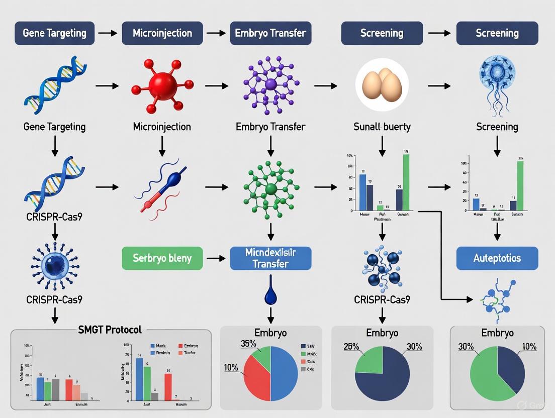
Figure 1: SMGT Workflow for Transgenic Animal Production. This diagram outlines the key steps in the SMGT protocol, from sperm preparation to the generation of a genetically modified animal.
Key Considerations and Controversies
Despite reported successes, SMGT remains a controversial technique and is not yet established as a reliable form of genetic manipulation [1]. The primary source of skepticism stems from the evolutionary implications: if sperm cells readily acted as vectors for exogenous DNA, it could lead to genetic chaos. Biological systems have therefore evolved robust barriers to minimize such occurrences [1].
The main natural barriers identified are:
- Seminal Fluid Inhibitory Factor: A factor present in mammalian seminal fluid that causes DBPs to lose their ability to bind exogenous DNA, thus blocking the first step of the process [1].
- Sperm Endogenous Nuclease Activity: An enzyme activity triggered by the interaction of sperm cells with foreign DNA molecules, likely designed to degrade the foreign genetic material [1].
The existence of these protections suggests that the successful production of transgenic animals via SMGT may represent instances where these natural barriers were overcome in the laboratory setting, which also contributes to the technique's inconsistent experimental outcomes [1].
Applications in Transgenic Pig Production
Within porcine molecular breeding, SMGT offers a straightforward method to introduce new genetic traits. The applications are particularly valuable for biomedical and agricultural research.
Table 1: SMGT Applications and Outcomes in Pig Transgenesis
| Application Area | Specific Example | Reported Outcome | Significance |
|---|---|---|---|
| Xenotransplantation | Expression of human decay accelerating factor (hDAF) | Generation of transgenic pig lines [3] | Aims to make pig organs suitable for grafting to humans. |
| Multigene Engineering | Simultaneous introduction of three fluorescent reporter genes (eGFP, EBFB, RFP) | Production of multigene transgenic pigs [4] | Demonstrates capacity for complex genetic modification. |
| Biomedical Research | Creation of large animal models for human diseases | Various transgenic pig models [1] [4] | Pigs are physiologically closer to humans than rodents. |
Quantitative Data and Efficiency
The efficiency of SMGT is a critical factor for its practical application. While it boasts advantages in cost and simplicity, its overall efficiency is often reported as low, though it can vary significantly.
Table 2: SMGT Efficiency and Performance Metrics
| Metric | Reported Value/Range | Context and Notes | Source |
|---|---|---|---|
| Transgenic Efficiency | 5% to 60% | Broad range observed in pig transgenesis [4]. | Lavitrano et al. |
| Transgene Transmission (F0 to F1) | ~25% | Only a quarter of initial studies demonstrated heritability beyond the founder generation [1]. | Smith & Spadafora, 2005 |
| Phenotype Modification Rate | Up to 80% | Frequency of observed phenotype changes in some experiments can be high [1]. | Smith & Spadafora, 2005 |
The low efficiency is primarily attributed to the poor uptake of exogenous DNA by sperm cells, which reduces the number of oocytes fertilized by transfected spermatozoa [1]. Furthermore, for a technique to be considered successful in animal transgenesis, the transgene must be stably transmitted to subsequent generations (F1, F2, etc.), a hurdle that many early claims of SMGT did not clear [1].
Comparative Analysis with Other Transgenic Techniques
In modern pig breeding, SMGT is one of several available techniques. Comparing it with other methods highlights its relative advantages and limitations.
Table 3: SMGT vs. Other Transgenic Techniques in Pigs
| Technique | Key Principle | Relative Efficiency | Key Advantages | Key Limitations |
|---|---|---|---|---|
| Sperm-Mediated Gene Transfer (SMGT) | Sperm cells bind and internalize exogenous DNA for delivery during fertilization. | 5-60% [4] | Low cost, simple procedure, no embryo handling or specialized equipment needed [3] [4]. | Low and inconsistent DNA uptake, controversy around reliability, presence of natural barriers [1]. |
| Pronuclear Microinjection (PNI) | DNA construct is microinjected into the pronucleus of a fertilized egg. | ~1% [4] | Was the traditional standard method. | Low efficiency, requires many embryos, high operational skill, causes mechanical damage [4]. |
| Cytoplasmic Injection (CI) | DNA transposon systems are injected into the cytoplasm of fertilized eggs. | >8% [4] | Simpler than PNI, less mechanical damage [4]. | Relies on timing of nuclear membrane fusion [4]. |
| Somatic Cell Nuclear Transfer (SCNT) | Nuclei from genetically modified somatic cells are transferred into enucleated oocytes. | 0.5-1% (for livestock) [4] | Allows for in vitro screening of modified cells before embryo production, enabling more complex edits [4]. | Technically complex, generally low efficiency, concerns about animal health [4]. |
Essential Research Reagent Solutions
A successful SMGT protocol requires specific reagents and materials to facilitate the binding of DNA to sperm and ensure subsequent fertilization and embryo development.
Table 4: Key Research Reagents for SMGT Experiments
| Reagent / Material | Function in SMGT Protocol | Specific Examples / Notes |
|---|---|---|
| Processed Spermatozoa | The primary biological vector for exogenous DNA. | Semen must be extensively washed to remove inhibitory factors in seminal plasma [1]. |
| Exogenous DNA Construct | The genetic material to be transferred into the embryo. | Can be linear or circular DNA; size and purity can affect efficiency [1] [4]. |
| DNA-Binding Proteins (DBPs) | Mediate the specific binding of DNA to the sperm cell membrane. | Naturally present on sperm head surface; their activity is unlocked by seminal plasma removal [1]. |
| Washing Buffers | To remove seminal plasma and prepare sperm for DNA uptake. | Typically protein-free media to prevent interference with DNA-sperm interaction. |
| Fertilization Media | To support the union of DNA-loaded sperm and oocytes. | Standard in vitro fertilization (IVF) media can be used post-incubation [4]. |
| Embryo Culture Media | To support the development of fertilized embryos post-IVF. | Supports the early-stage transgenic embryos before transfer [4]. |
Sperm-mediated gene transfer represents a conceptually simple and cost-effective alternative to more complex transgenic technologies for the production of genetically modified pigs. While it has proven successful in generating transgenic pig models for biomedical research and agricultural improvement, its path to becoming a reliable, mainstream methodology is hampered by inconsistent efficiency and ongoing controversy regarding its fundamental mechanisms. Future research aimed at better understanding and controlling the interactions between sperm and exogenous DNA, particularly how to consistently overcome natural biological barriers, will be crucial for unlocking the full potential of SMGT in porcine molecular breeding.
The production of transgenic pigs is a cornerstone of biomedical and agricultural research, enabling advancements in xenotransplantation, disease modeling, and livestock improvement. Sperm-Mediated Gene Transfer (SMGT) represents a pivotal methodology in the historical development of transgenic technology. First conceptualized as a technique leveraging the innate ability of spermatozoa to internalize exogenous DNA, SMGT provides a simplified and efficient alternative to more complex and equipment-intensive methods like pronuclear microinjection (PNI) and Somatic Cell Nuclear Transfer (SCNT) [5]. Its application has evolved from early proof-of-concept studies to a validated protocol for generating pigs with targeted genetic modifications. This document details the application notes and experimental protocols for SMGT, framing it within a broader thesis on its role for transgenic pig production.
Comparative Analysis of Transgenic Techniques
The evolution of pig transgenesis has been driven by several core technologies, each with distinct advantages and limitations. Table 1 provides a comparative overview of these key methods.
Table 1: Key Techniques for Generating Transgenic Pigs
| Technique | Fundamental Principle | Key Advantages | Primary Limitations | Reported Transgenic Efficiency |
|---|---|---|---|---|
| Pronuclear Microinjection (PNI) | Physical microinjection of DNA construct into a pronucleus of a fertilized egg [5]. | Established history; does not require cell culture. | Low efficiency (~1%); random transgene integration; high mechanical damage; requires specialized equipment [5]. | ~1% (number of transgenic pigs/injected embryos transferred) [5]. |
| Somatic Cell Nuclear Transfer (SCNT) | Transfer of a nucleus from a genetically modified somatic cell into an enucleated oocyte [6] [5]. | Enables pre-selection of modified cell clones; ensures precise genetic modifications and high transgene positivity rate [6] [5]. | Technically complex; low overall efficiency (0.5-1% for modifying somatic cells); associated with placental and fetal abnormalities [5]. | High positivity rate in offspring due to pre-screening, but low live birth rate per embryo transferred [5]. |
| Sperm-Mediated Gene Transfer (SMGT) | Incubation of sperm cells with exogenous DNA, followed by artificial insemination or in vitro fertilization [5]. | Simple procedure; no expensive equipment required; high potential efficiency [5]. | Variable efficiency (5-60%); inconsistency in DNA uptake and integration; potential for mosaicism [5]. | 5% to 60%, highly dependent on protocol optimization [5]. |
SMGT Protocol for Transgenic Pig Production
The following section provides a detailed, step-by-step protocol for the production of transgenic pigs via SMGT, incorporating best practices and key considerations for researchers.
Reagents and Equipment
Reagents:
- Porcine spermatozoa (fresh or frozen-thawed from a proven boar)
- Exogenous DNA construct (e.g., linearized plasmid, Sleeping Beauty or piggyBac transposon system)
- Sperm wash medium (e.g., Dulbecco's PBS supplemented with BSA)
- Capacitation-inducing agents (e.g., heparin, bicarbonate)
- In vitro fertilization (IVF) medium or artificial insemination medium
- Antibiotics (Penicillin-Streptomycin)
Equipment:
- Standard cell culture incubator (38.5°C, 5% CO₂)
- Benchtop centrifuge
- Laminar flow hood
- Hemocytometer or automated cell counter
- Instrument for artificial insemination or materials for in vitro fertilization
Step-by-Step Procedure
Step 1: Sperm Preparation and Washing
- Collect and pool semen from multiple fertile boars. Alternatively, use frozen-thawed semen from a commercial source.
- Wash spermatozoa twice in sperm wash medium by centrifugation at 500 x g for 10 minutes to remove seminal plasma, which contains nucleases that can degrade the exogenous DNA.
- Resuspend the final sperm pellet in a suitable incubation medium to a concentration of 1-5 x 10ⶠsperm/mL.
Step 2: Incubation with Exogenous DNA
- Dilute the purified exogenous DNA construct in the same incubation medium. The optimal final DNA concentration typically ranges from 1 to 10 ng/µL and should be determined empirically.
- Incubate the prepared sperm suspension with the DNA solution for a defined period (e.g., 15 minutes to 2 hours) at 38.5°C. Some protocols include a brief exposure to a mild detergent (e.g., Triton X-100) or lipofectamine to enhance DNA uptake, though this can compromise sperm viability and requires careful optimization.
Step 3: Removal of Unbound DNA
- After incubation, pellet the sperm cells by gentle centrifugation.
- Wash the sperm pellet once with medium to remove any unincorporated, free-floating DNA.
- Resuspend the sperm in pre-warmed IVF medium for immediate use.
Step 4: Fertilization and Embryo Transfer
- Option A: In Vitro Fertilization (IVF): Co-incubate the DNA-loaded sperm with in vitro-matured porcine oocytes for 5-6 hours. Subsequently, wash the presumptive zygotes to remove excess sperm and culture them in vitro until the desired stage for embryo transfer.
- Option B: Artificial Insemination (AI): Use the DNA-loaded sperm suspension for surgical or non-surgical artificial insemination of synchronized sows. This method is less common but has been reported [5].
- Transfer the resulting embryos (from IVF) into the oviducts of synchronized surrogate sows. The number of embryos transferred per surrogate should follow standard institutional protocols (e.g., 30-50 embryos per surrogate).
Step 5: Genotyping and Analysis
- After farrowing, collect tissue samples (e.g., ear notch or tail clip) from the piglets.
- Extract genomic DNA and screen for the presence of the transgene using PCR, Southern blot, or other appropriate molecular techniques.
- For positive founders, conduct further analyses to determine transgene copy number, integration site, and expression levels.
Current Applications and Outcomes
SMGT has been successfully applied to create multi-transgenic pigs. For instance, one study demonstrated the simultaneous introduction of three fluorescent reporter genes using this technique [5]. In the context of xenotransplantation, SMGT was used to create pig lines expressing the human decay accelerating factor (hDAF), a key protein that helps protect transplanted tissues from complement-mediated rejection [5]. The efficiency of SMGT can be highly variable, with reported transgene positivity rates in offspring ranging from 5% to 60%, underscoring the need for rigorous protocol standardization [5]. The technique's primary advantage lies in its simplicity and accessibility, as it bypasses the need for sophisticated micromanipulation equipment.
The Scientist's Toolkit: Key Research Reagent Solutions
Table 2 outlines essential reagents and their critical functions in the SMGT workflow, providing a quick reference for laboratory setup.
Table 2: Essential Research Reagents for SMGT
| Research Reagent / Tool | Function in SMGT Protocol |
|---|---|
| Sperm Wash Medium | Removes seminal plasma and protects sperm during centrifugation, preventing DNA degradation. |
| Exogenous DNA Construct | Carries the genetic material of interest (e.g., a therapeutic gene or a fluorescent reporter). |
| Transposon Systems (e.g., Sleeping Beauty, piggyBac) | Facilitates more stable and precise integration of the transgene into the host genome, improving expression and reducing positional effects [5]. |
| Capacitation-Inducing Agents | Mimics the natural physiological process that prepares sperm for fertilization, which can also enhance DNA uptake. |
| In Vitro Fertilization (IVF) Medium | Supports the fertilization process and subsequent early embryonic development in vitro. |
| Cdk8-IN-14 | Cdk8-IN-14|CDK8 Inhibitor|For Research Use |
| Icmt-IN-13 | Icmt-IN-13, MF:C21H25ClFNO, MW:361.9 g/mol |
Workflow and Pathway Diagrams
The following diagrams, generated using DOT language, illustrate the core SMGT workflow and its context within the broader transgenic development pipeline.
SMGT Experimental Workflow
Transgenic Pig Development Pathway
This application note provides a detailed technical comparison of Sperm-Mediated Gene Transfer (SMGT) against two established methods—Pronuclear Microinjection (PNI) and Somatic Cell Nuclear Transfer (SCNT)—for the production of transgenic pigs. Framed within broader thesis research on optimizing SMGT protocols, this document summarizes quantitative performance data, outlines detailed experimental methodologies, and identifies essential research reagents. The content is designed to support researchers, scientists, and drug development professionals in selecting the most appropriate genetic engineering strategy for their projects.
The generation of transgenic pigs is a critical technology for biomedical research and agricultural science, enabling the study of human diseases, the production of pharmaceutical proteins, and the enhancement of livestock traits [7] [5]. The selection of an optimal gene transfer method is paramount to the success and efficiency of these endeavors. While Pronuclear Microinjection (PNI) and Somatic Cell Nuclear Transfer (SCNT) have been foundational techniques, Sperm-Mediated Gene Transfer (SMGT) presents a compelling alternative that leverages the natural ability of spermatozoa to internalize and deliver exogenous DNA into the oocyte during fertilization [7] [5]. This document provides a structured comparison of these three key technologies, with a specific focus on their application in transgenic pig production.
Comparative Analysis of Key Techniques
The following table summarizes the core characteristics of PNI, SCNT, and SMGT, highlighting the distinct advantages of the SMGT protocol.
Table 1: Key Characteristics of Transgenesis Techniques in Pig Production
| Feature | Pronuclear Microinjection (PNI) | Somatic Cell Nuclear Transfer (SCNT) | Sperm-Mediated Gene Transfer (SMGT) |
|---|---|---|---|
| Core Principle | Direct microinjection of DNA into the pronucleus of a fertilized oocyte [7] [8] | Transfer of a nucleus from a genetically modified somatic cell into an enucleated oocyte [7] [9] | Use of sperm cells as natural vectors to carry exogenous DNA during fertilization [7] [5] |
| Integration Site | Random [7] | Can be predefined via cell culture screening [9] | Random [5] |
| Typical Efficiency (Transgenic Offspring) | ~1% [5] | High (theoretically 100% from pre-screened cells) [9] | 5% to 60% [5] |
| Key Advantage | Well-established history [10] | Pre-selection of modified cells; potential for gene targeting [7] [9] | No complex equipment or embryo manipulation; high throughput potential [5] |
| Primary Limitation | Low efficiency; random integration; technically demanding [7] [5] | Very low live birth rate (1-3%); epigenetic abnormalities [7] [11] [9] | Instability of DNA-sperm interaction; variable efficiency [5] |
| Technical Skill & Equipment | Requires highly trained personnel and sophisticated micromanipulation equipment [5] [8] | Requires advanced cell culture and micromanipulation skills [7] | Relatively simple; requires standard artificial insemination or in vitro fertilization lab setup [5] |
| Handling of Embryos | Extensive in vitro manipulation of zygotes [7] | Complex in vitro manipulation of oocytes and reconstructed embryos [12] | Minimal; utilizes standard fertilization procedures [5] |
Table 2: Quantitative Data Comparison for Transgenic Pig Production
| Parameter | Pronuclear Microinjection (PNI) | Somatic Cell Nuclear Transfer (SCNT) | Sperm-Mediated Gene Transfer (SMGT) |
|---|---|---|---|
| Transgene Positivity Rate in Offspring | Low (~1%) [5] | Very High (theoretically 100%) [9] | Variable (5-60%) [5] |
| Mosaicism in Founders | High prevalence [9] | Absent (all cells are transgenic) [9] | Can occur [7] |
| Cost and Resource Intensity | High (costly equipment, skilled staff, many embryos needed) [7] [5] | Very High (complex procedures, low overall live birth rate) [7] [9] | Low (minimal specialized equipment or reagents) [5] |
| Developmental Abnormalities | Not typically associated | High (due to incomplete nuclear reprogramming) [7] [11] | Not typically associated |
Detailed Experimental Protocols
Protocol for Sperm-Mediated Gene Transfer (SMGT) in Pigs
Principle: This protocol exploits the innate ability of sperm cells to bind and internalize exogenous DNA, which is then delivered to the oocyte upon fertilization to generate transgenic embryos [5].
Workflow Overview:
Step-by-Step Procedure:
Sperm Collection and Preparation:
- Collect fresh semen from a boar of known fertility. Alternatively, use frozen-thawed semen from a commercial source.
- Wash spermatozoa twice by centrifugation (800 x g for 10 minutes) in a non-capacitating medium, such as Dulbecco's Phosphate-Buffered Saline (DPBS) supplemented with 1 mg/mL bovine serum albumin (BSA) to remove seminal plasma.
- Resuspend the final sperm pellet to a concentration of 1-5 x 10^6 sperm/mL in a defined fertilization medium.
Interaction with Exogenous DNA:
- Incubate the prepared sperm suspension with the linearized, foreign DNA construct (10-100 ng/μL final concentration) for 30-60 minutes at 37°C under 5% CO₂ [5]. To enhance DNA uptake, consider using specific agents like dimethylsulfoxide (DMSO) or lipofectamine during this step.
- Post-incubation, wash the sperm cells again to remove any unbound DNA.
Fertilization:
- For In Vitro Fertilization (IVF): Add the DNA-loaded sperm suspension to in vitro-matured porcine oocytes in fertilization medium. Co-incubate for 6-18 hours at 38.5°C under 5% CO₂ and 5% O₂.
- For Artificial Insemination (AI): The DNA-loaded sperm preparation can be used for direct intrauterine artificial insemination of synchronized sows [5].
Embryo Culture and Transfer:
- For IVF-derived embryos, remove the presumptive zygotes from the fertilization drops, wash to remove excess sperm, and culture in a defined embryo culture medium (e.g., PZM-3) for 5-7 days until the blastocyst stage.
- Select developmentally competent embryos for surgical transfer into synchronized recipient gilts.
Genotyping of Offspring:
- After birth, collect tissue samples (e.g., ear notch or tail clip) from the founder piglets (F0).
- Extract genomic DNA and screen for the presence and integration of the transgene using PCR, Southern blot analysis, or next-generation sequencing.
Protocol for Pronuclear Microinjection (PNI) in Pigs
Principle: A DNA construct is physically injected directly into the larger male pronucleus of a fertilized, one-cell embryo (zygote), leading to random integration into the host genome [7] [13].
Workflow Overview:
Step-by-Step Procedure:
Zygote Collection:
- Collect fertilized one-cell embryos (zygotes) from super-ovulated sows or via in vitro fertilization. The pronuclei are often not visible in pig zygotes due to cytoplasmic lipids.
Preparation for Microinjection:
- Centrifuge zygotes at 12,000-15,000 x g for 5-10 minutes to force cytoplasmic organelles to the periphery, making the pronuclei more visible [5].
- Prepare a microinjection needle filled with the DNA solution (1-2 ng/μL in injection buffer, e.g., Tris-EDTA). Place a drop of manipulation medium containing the zygotes on an inverted microscope equipped with micromanipulators.
Microinjection:
- Immobilize a zygote using a holding pipette. Insert the injection pipette through the zona pellucida and the oolemma into the larger male pronucleus.
- Deliver a precise volume of DNA solution (1-2 picoliters) until visible swelling of the pronucleus occurs (~1-2% of its volume). Withdraw the pipette carefully [13].
Embryo Culture and Transfer:
- Wash the injected zygotes and culture them in vitro for 1-3 days to assess viability and initial cleavage. Embryos that develop normally to the 2- to 4-cell stage are selected.
- Surgically transfer viable embryos into the oviduct of a synchronized recipient gilt.
Founder Analysis:
- Screen offspring for transgene integration. Due to frequent mosaicism, analyze tissues from multiple lineages and breed founders to confirm germline transmission [9].
Protocol for Somatic Cell Nuclear Transfer (SCNT) in Pigs
Principle: The genetic material of an unfertilized oocyte is removed and replaced with the nucleus from a somatic cell that has been genetically modified in culture, resulting in a cloned, transgenic embryo [7] [9].
Workflow Overview:
Step-by-Step Procedure:
Genetic Modification of Donor Cells:
- Isolate and culture somatic cells, typically fetal fibroblasts. Transfect these cells with the desired DNA construct using electroporation or lipofection.
- Apply drug selection (e.g., G418 for neomycin resistance) to select stably transfected clones. Expand and validate modified cells for transgene integration and expression via PCR, Southern blot, or other assays [9].
Oocyte Enucleation:
- Collect in vivo- or in vitro-matured oocytes at the Metaphase II (MII) stage. Remove the chromosomal spindle complex (enucleation) using a micropipette under microscopic visualization, often facilitated by fluorescent staining or polarized light microscopy.
Nuclear Transfer:
- Insert a single, genetically modified donor cell (or its nucleus) into the perivitelline space of the enucleated oocyte, directly in contact with the oocyte's cytoplasm.
Fusion and Activation:
- Fuse the donor cell with the enucleated oocyte using electrical pulses or viral fusogens. This fusion event also introduces factors from the oocyte cytoplasm that begin reprogramming the donor nucleus.
- Simultaneously or shortly after fusion, artificially activate the reconstructed oocyte to initiate embryonic development, typically using a combination of chemical agents like ionomycin and 6-DMAP, or specific inhibitors [11].
Embryo Culture and Transfer:
- Culture the successfully activated SCNT embryos in a defined medium for 1-6 days. Monitor development to the blastocyst stage.
- Transfer developmentally competent embryos into the uterus of a synchronized recipient gilt.
The Scientist's Toolkit: Essential Research Reagents
Table 3: Key Research Reagent Solutions for Transgenic Pig Production
| Reagent / Material | Function / Application | Example Use in Protocol |
|---|---|---|
| Sperm Washing Medium | Removes seminal plasma and decapacitation factors to prepare sperm for DNA uptake [5]. | Initial preparation of boar sperm for SMGT. |
| DNA Transfection Reagents | Enhances the binding and internalization of exogenous DNA into sperm cells. | Added during the sperm-DNA co-incubation step in SMGT. |
| Holding and Injection Pipettes | Essential microtools for precise manipulation and injection of zygotes and oocytes. | Used for pronuclear microinjection (PNI) and enucleation/cell transfer in SCNT. |
| Manipulation Medium | A specific culture medium designed to maintain embryo viability outside the incubator during micromanipulation. | Used for all procedures performed on a microscope stage (PNI, SCNT). |
| Fusion/Activation Media | Contains components to induce cell fusion and trigger embryonic development in SCNT embryos. | Applied after donor cell transfer in SCNT to create a reconstructed, activated embryo [11]. |
| Defined Embryo Culture Medium | Supports the in vitro development of embryos from the one-cell stage to the blastocyst. | Used for culturing embryos post-manipulation (all three techniques) before transfer. |
| Drug Selection Agents | Selects for somatic cells that have successfully integrated the transgene during culture. | Added to the media of donor cells in the SCNT workflow to create a pure population of transgenic cells [9]. |
| Lamotrigine-13C2,15N2,d3 | Lamotrigine-13C2,15N2,d3, MF:C9H9Cl2N5, MW:265.09 g/mol | Chemical Reagent |
| Sparfloxacin-d5 | Sparfloxacin-d5 Stable Isotope | Sparfloxacin-d5 (CI-978-d5) is a deuterated bacterial inhibitor for research. For Research Use Only. Not for human or veterinary use. |
The choice between SMGT, PNI, and SCNT for generating transgenic pigs depends heavily on the project's specific goals, available resources, and technical expertise. SMGT offers a less technically demanding and potentially high-efficiency alternative that avoids extensive embryo manipulation. PNI remains a direct but inefficient method, while SCNT, despite its complexity and low overall efficiency, provides unparalleled control by allowing for pre-selection of specific genetic modifications. Researchers must weigh these key advantages and limitations carefully to successfully advance their work in porcine transgenesis.
Sperm-Mediated Gene Transfer (SMGT) presents a powerful methodology for generating transgenic animals, offering a simpler and more efficient alternative to complex techniques like pronuclear microinjection. Within the context of transgenic pig production—a critical field for biomedical applications such as xenotransplantation and disease modeling—SMGT leverages the innate ability of sperm cells to bind and internalize exogenous DNA. This protocol details the core principles and molecular mechanisms governing the spontaneous uptake of foreign DNA by spermatozoa, providing a foundational framework for researchers aiming to utilize SMGT in porcine transgenesis. The ensuing application notes outline a standardized, reproducible experimental workflow based on these principles [14].
Molecular Mechanism of DNA Uptake
The interaction between sperm cells and exogenous DNA is not a casual event but a controlled process mediated by specific ionic interactions and protein-DNA recognition. Understanding this mechanism is paramount for optimizing SMGT efficiency [15] [14].
Key DNA-Binding Proteins
Southwestern blot analysis of sperm head protein extracts has identified specific classes of DNA-binding proteins that act as substrates for exogenous DNA. The major protein classes involved are summarized in Table 1 below [15].
Table 1: Major DNA-Binding Protein Classes in Spermatozoa
| Protein Class (Molecular Weight) | Conservation Across Species | Accessibility in Intact Cells | Postulated Role in DNA Uptake |
|---|---|---|---|
| ~50 kDa | Variable | Not accessible | Potential secondary substrate |
| 30-35 kDa | High | Accessible | Primary DNA receptor |
| <20 kDa (includes protamines) | Variable | Not accessible | Nuclear packaging |
The 30-35 kDa protein class is of particular importance. It is highly conserved among mammalian species and represents the only class of DNA-binding proteins accessible to exogenous DNA in intact, viable sperm cells. Purified 30-35 kDa proteins interact with DNA in vitro, forming discrete protein/DNA complexes as confirmed by band shift assays. This evidence strongly supports their role as the primary receptor for exogenous DNA on the sperm surface [15] [14].
The Process of Uptake and Localization
Epididymal sperm cells spontaneously take up exogenous DNA during a brief incubation period of 15-20 minutes. The DNA is specifically and reversibly localized to the nuclear area of the sperm head. The binding is ionic; it can be competed out by an excess of cold competitor DNA or other polyanions like heparin and dextran sulfate. Conversely, polycations like poly-L-lysine favor DNA uptake. Furthermore, sperm cells show a preference for larger DNA molecules (e.g., 7 kb) over smaller fragments (150-750 bp) [14].
Regulatory Inhibition by Seminal Plasma
A critical factor controlling DNA uptake is the presence of a powerful inhibitory factor in the seminal plasma of mammals. This factor blocks the binding of exogenous DNA to sperm cells and is effective even across species (heterologous inhibition). The 30-35 kDa DNA-binding proteins are the specific target of this inhibition, losing their DNA-binding capability in the presence of the seminal plasma factor. Therefore, for successful SMGT, it is essential to use sperm prepared from epididymides, effectively washed to remove any trace of seminal plasma [15] [14].
The following diagram illustrates the sequential molecular mechanism of exogenous DNA uptake by spermatozoa and its key regulatory point.
Quantitative Data on DNA-Sperm Interaction
The efficiency of DNA uptake is influenced by several physical and molecular factors. The key quantitative parameters characterizing this interaction are consolidated in Table 2 below [14].
Table 2: Key Parameters of Exogenous DNA Uptake by Spermatozoa
| Parameter | Experimental Findings | Experimental Notes |
|---|---|---|
| Uptake Time Course | 15-20 minutes | Saturation is reached within this timeframe. |
| DNA Size Preference | Preferential uptake of larger molecules (e.g., 7 kb) | Uptake of smaller fragments (150-750 bp) is less efficient. |
| Binding Affinity | Reversible binding | DNA can be displaced by excess competitor DNA or polyanions. |
| Cellular Localization | Specific localization to the nuclear area of sperm head | Determined by autoradiography and fluorescence. |
| Critical Step | Removal of seminal plasma | Seminal plasma contains a potent inhibitory factor. |
Experimental Protocol: DNA Uptake and Validation
This protocol describes the foundational steps for loading exogenous DNA onto porcine spermatozoa for SMGT applications, based on the elucidated core principles.
Sperm Preparation and DNA Binding Assay
Objective: To prepare sperm free of seminal plasma inhibitors and facilitate binding of exogenous plasmid DNA.
Materials:
- Porcine Epididymal Sperm: Collected from cauda epididymides.
- DNA Construct: Plasmid DNA of interest (e.g., for xenotransplantation antigens like GGTA1).
- Wash Buffer: Dulbecco's Phosphate Buffered Saline (DPBS).
- Binding Medium: DPBS supplemented with 6 mg/mL BSA.
- Poly-L-lysine (optional, to enhance uptake).
Procedure:
- Sperm Collection: Mince porcine cauda epididymides in pre-warmed DPBS and filter the suspension through a nylon mesh to remove tissue debris.
- Washing: Centrifuge the sperm suspension at 500 x g for 10 minutes. Resuspend the sperm pellet in 1 mL of Binding Medium. Repeat this wash step twice to ensure complete removal of seminal plasma components.
- DNA Incubation: Adjust the sperm concentration to 1-5 x 10^7 cells/mL in Binding Medium. Add the plasmid DNA at a final concentration of 1-5 µg/mL. Incubate the mixture for 20 minutes at room temperature or 37°C with gentle agitation.
- Removal of Unbound DNA: Pellet the sperm cells by centrifugation at 500 x g for 10 minutes. Carefully remove the supernatant containing unbound DNA. Wash the pellet once with Binding Medium to remove any residual unbound DNA.
- Validation of Uptake (Optional): Resuspend a small aliquot of the sperm pellet for analysis. DNA binding can be validated using:
- Fluorescence In Situ Hybridization (FISH) with labeled DNA probes.
- PCR on washed sperm samples to detect the presence of the transgene.
Assessing Sperm DNA Integrity Post-Uptake
Objective: To evaluate the DNA fragmentation index (DFI) of sperm post-DNA uptake, as high DFI can compromise the success of subsequent fertilization and embryo development [16] [17].
Materials:
- Terminal deoxynucleotidyl transferase dUTP Nick End Labeling (TUNEL) Assay Kit
- Flow Cytometer or Fluorescence Microscope
- DPBS
Procedure:
- After the final wash, fix the DNA-loaded sperm cells in 4% paraformaldehyde for 30 minutes.
- Permeabilize the cells with 0.1% Triton X-100 in DPBS for 5 minutes on ice.
- Follow the manufacturer's instructions for the TUNEL assay to label DNA strand breaks.
- Analyze the samples by flow cytometry. Calculate the DFI as the percentage of TUNEL-positive sperm in the population.
- Interpretation: A high DFI is inversely correlated with sperm motility, normal morphology, and fertility outcomes in assisted reproductive technologies. Sperm with a DFI > 30% may lead to significantly reduced embryo development rates [17].
The following workflow diagram integrates the core protocol with quality control measures.
The Scientist's Toolkit: Essential Research Reagents
Successful execution of SMGT relies on a suite of specific reagents. Table 3 lists critical materials and their functions for DNA uptake experiments.
Table 3: Essential Reagents for SMGT DNA Uptake Experiments
| Reagent / Material | Function / Application |
|---|---|
| Porcine Epididymal Sperm | Source of male gametes; must be free of seminal plasma to avoid inhibition of DNA uptake. |
| Plasmid DNA Construct | Exogenous DNA carrying the gene of interest (e.g., for knockout of xenoantigens like GGTA1, CMAH, B4GALNT2 [18]). |
| Binding Medium (BSA) | Provides a protein-rich environment to maintain sperm viability during the DNA incubation step. |
| Poly-L-lysine | Polycation that enhances DNA uptake by strengthening ionic interactions with the sperm surface [14]. |
| Heparin / Dextran Sulfate | Polyanions used as competitive inhibitors in control experiments to confirm specificity of DNA binding [14]. |
| TUNEL Assay Kit | Validates sperm DNA integrity post-uptake; high DFI indicates poor sperm quality and predicts low fertility [17]. |
| PCR Reagents | Molecular validation of transgene association with washed sperm cells. |
| Arginase inhibitor 7 | |
| Sulfasalazine-d3,15N | Sulfasalazine-d3,15N, MF:C18H14N4O5S, MW:402.4 g/mol |
Application Notes in Transgenic Pig Production
The principles outlined herein are directly applicable to modern pig transgenesis for xenotransplantation. Current research employs somatic cell nuclear transfer (SCNT) using genetically engineered fibroblasts as the primary method for generating multi-gene edited pigs. However, SMGT remains a valuable and simpler technique for rapid proof-of-concept studies or introducing less complex genetic modifications [18].
When designing DNA constructs for SMGT, researchers should consider edits that reduce xenograft immunogenicity. Key targets include:
- Knockout of Glycosyltransferases: Inactivation of genes like GGTA1 (α-Gal), CMAH (Neu5Gc), and B4GALNT2 to remove major xenoantigens recognized by human pre-formed antibodies [18].
- Expression of Human Complement Regulators: Incorporation of transgenes like hCD46, hCD55, and hCD59 to protect pig cells from human complement-mediated lysis [18].
Efficiency in producing viable founders depends not only on successful DNA uptake but also on maintaining high sperm quality. Rigorous assessment of sperm motility, morphology, and DNA fragmentation index (DFI) post-uptake is crucial, as these parameters are strongly correlated with successful embryo development and live births [17].
Sperm-mediated gene transfer (SMGT) represents a pioneering methodology within transgenic pig production research, enabling researchers to overcome fundamental reproductive isolation barriers that traditionally limit genetic exchange between species. This technique leverages the innate capacity of spermatozoa to internalize and deliver foreign DNA into oocytes during fertilization, thereby creating genetically modified embryos. Within the broader context of transgenic livestock development, SMGT provides a relatively efficient and technically accessible approach compared to more complex methods such as somatic cell nuclear transfer (SCNT) or pronuclear microinjection. The technique holds particular promise for agricultural biotechnology, pharmaceutical production, and biomedical research, including the development of pig models for human disease and xenotransplantation [19] [20].
Recent advances in genetic engineering have further enhanced SMGT's potential. The development of SynNICE (Synthetic Nucleus Injection into Cell Embryos) technology enables the transfer of megabase-scale human DNA sequences into other species, representing a quantum leap in overcoming reproductive isolation barriers. This approach has successfully demonstrated the delivery of a 1.14 Mb human Y chromosome fragment (AZFa region) into mouse early embryos, establishing a groundbreaking platform for cross-species genetic transfer that could be adapted to porcine systems [21]. The ability to synthesize, assemble, and transfer such large genetic constructs opens new possibilities for creating transgenic pigs with humanized physiological systems for biomedical applications.
Quantitative Comparison of Genetic Transfer Techniques
Transgenic pig production employs multiple technical approaches, each with distinct advantages and limitations. The following table summarizes the key methodologies used to overcome reproductive isolation in pig genetic engineering:
Table 1: Comparison of Genetic Transfer Techniques for Transgenic Pig Production
| Technique | Key Features | Efficiency | Insert Size Capacity | Technical Complexity |
|---|---|---|---|---|
| Sperm-Mediated Gene Transfer (SMGT) | Uses sperm as natural vector for DNA uptake; can be enhanced with electroporation or magnetic fields | Moderate | Limited (typically plasmid DNA) | Low to Moderate |
| Somatic Cell Nuclear Transfer (SCNT) | Transfer of somatic nucleus into enucleated oocyte; enables precise genetic modifications | Low to Moderate | Virtually unlimited (whole genome) | High |
| ICSI-Mediated Gene Transfer | Direct injection of DNA with sperm into oocyte | Moderate | Limited (typically plasmid DNA) | High |
| Retroviral Vector Transduction | Uses modified viruses to deliver genes | High | Limited by viral packaging capacity | Moderate |
| SynNICE Technology | Transfer of synthetic megabase-scale DNA via yeast nuclear carriers | Demonstrated in mice; porcine adaptation pending | Very High (megabase scale) | Very High |
The quantitative comparison reveals that SMGT occupies a unique position in the transgenic technology landscape, offering a balance between technical accessibility and practical applicability. While newer approaches like SynNICE technology offer unprecedented capacity for large DNA fragment transfer, SMGT remains particularly valuable for applications requiring routine genetic modifications without specialized equipment [19] [21] [20]. The integration efficiency of SMGT can be significantly enhanced through optimization of DNA uptake conditions, including the use of specific electroporation parameters and the selection of donor boars with higher natural DNA uptake capabilities [19].
SMGT Protocol for Transgenic Pig Production
Critical Reagents and Materials
The successful implementation of SMGT requires careful preparation of specific reagents and materials. The following table outlines the essential components of the "Researcher's Toolkit" for SMGT experiments:
Table 2: Essential Research Reagent Solutions for SMGT Protocol
| Reagent/Material | Function | Specifications |
|---|---|---|
| Donor Sperm | DNA vector carrier | Selected boars with high DNA uptake capacity [19] |
| Foreign DNA Construct | Genetic material for transfer | High-quality plasmid DNA with appropriate regulatory elements |
| Electroporation Buffer | Facilitate DNA uptake | Optimized ionic composition with minimal cytotoxicity |
| Transfection Enhancers | Increase membrane permeability | Compounds such as dimethyl sulfoxide (DMSO) or specific lipids |
| In Vitro Maturation Media | Oocyte preparation | TCM-199 with FSH, LH, EGF, and cysteine [22] |
| Embryo Culture Media | Post-fertilization development | Sequential media supporting early embryonic development |
Detailed Experimental Workflow
The following diagram illustrates the comprehensive SMGT workflow for transgenic pig production:
Step-by-Step Protocol Implementation
Sperm Preparation and DNA Loading
Sperm Collection and Selection: Collect semen from proven donor boars using standard artificial insemination protocols. Select boars with demonstrated high DNA uptake capacity, as significant individual variation exists [19] [20]. Wash sperm samples to remove seminal plasma using a discontinuous density gradient centrifugation system.
DNA Processing and Complex Formation: Prepare high-purity plasmid DNA containing the transgene of interest. For optimal results, linearize the DNA construct and resuspend in TRIS-EDTA buffer at a concentration of 0.1-1.0 μg/μL. The DNA can be pre-incubated with lipofection agents to facilitate membrane passage, though this requires optimization for different sperm batches.
DNA Uptake via Electroporation: Mix 10-20 million motile sperm with 5-10 μg of DNA in electroporation buffer. Perform electroporation using optimized parameters: square wave pulses, field strength of 600-800 V/cm, and pulse duration of 1-5 ms [19]. Immediate assessment of sperm viability post-electroporation is critical, as excessive electrical parameters can compromise membrane integrity and fertilization capacity.
Oocyte Preparation and In Vitro Maturation
Oocyte Collection: Obtain ovaries from slaughterhouses and transport to the laboratory in sterile physiological saline at 39°C. Aspirate follicles (3-6 mm diameter) using an 18-gauge needle attached to a 10 mL syringe. Collect oocytes surrounded by a compact, multilayer cumulus cell mass (cumulus-oocyte complexes, COCs) [22].
In Vitro Maturation (IVM): Wash COCs three times in maturation medium (TCM-199 supplemented with 10% FBS, 10 ng/mL EGF, 10 IU/mL FSH, 10 IU/mL LH, and 0.1 mM cysteine). Culture groups of 50-70 COCs in 500 μL maturation medium under mineral oil at 39°C in a 5% CO₂ atmosphere with saturated humidity for 42-44 hours [22].
Oocyte Assessment and Preparation: After maturation, denude oocytes by gentle pipetting in 1 mg/mL hyaluronidase solution. Select only metaphase II oocytes exhibiting a distinct first polar body for fertilization experiments.
Fertilization and Embryo Transfer
In Vitro Fertilization (IVF): Co-incubate DNA-loaded sperm (1-2 × 10ⵠsperm/mL) with matured oocytes in fertilization medium for 6-8 hours. Subsequently, remove excess sperm and transfer presumptive zygotes to fresh culture medium.
Embryo Culture and Selection: Culture embryos for 5-6 days until the blastocyst stage. Monitor embryonic development carefully, noting any developmental delays or abnormalities. Screen blastocysts for transgene integration using PCR or other molecular techniques before transfer.
Embryo Transfer: Synchronize recipient gilts or sows hormonally. Surgically transfer 30-50 developing embryos to the oviducts of each recipient on day 1 after estrus detection [19]. Monitor pregnancy status via ultrasound around day 28-35 post-transfer.
Advanced Technical Considerations and SynNICE Integration
Enhancing SMGT Efficiency
Several technical factors critically influence SMGT efficiency. Sperm quality and viability after DNA loading represent the most significant determinants of success. While electroporation enhances DNA uptake, it can simultaneously reduce sperm motility and membrane integrity. Optimization must balance these competing factors through systematic parameter testing. The DNA-sperm incubation conditions (time, temperature, and media composition) also significantly impact internalization efficiency. Some protocols incorporate short-duration, high-intensity electromagnetic pulses to transiently increase membrane permeability without irreversible damage [19].
The selection of donor boars with inherently higher DNA uptake capacity provides another optimization avenue. Research indicates substantial individual variation in this trait, with some boars demonstrating consistently superior performance as DNA vectors [20]. Establishing a screening protocol to identify such individuals can dramatically improve overall SMGT efficiency.
Integration with SynNICE Technology for Complex Genetic Modifications
The emerging SynNICE technology offers a revolutionary approach that could complement traditional SMGT for sophisticated genetic engineering applications. This method involves:
Megabase DNA Assembly: Utilizing yeast homologous recombination systems to assemble megabase-scale human DNA sequences through a hierarchical strategy. This approach successfully assembled the highly repetitive (69.38% repeats) 1.14 Mb human AZFa region [21].
Yeast Nuclear Extraction: Developing techniques to isolate intact yeast nuclei containing the synthetic DNA while preserving chromatin structure. These nuclei can be cryopreserved for up to six months without degradation [21].
Cross-Species Delivery: Microinjecting the purified yeast nuclei into recipient oocytes or early embryos, enabling transfer of extremely large genetic constructs across species boundaries.
For transgenic pig production, SynNICE technology could enable the transfer of entire human gene clusters or regulatory domains to create more sophisticated humanized pig models. This approach shows particular promise for xenotransplantation research, where introducing large segments of human MHC genes could substantially reduce immune rejection barriers [21].
Alternative and Complementary Methods
While SMGT provides distinct advantages, researchers should consider complementary techniques for specific applications:
Somatic Cell Nuclear Transfer (SCNT): This approach involves transferring genetically modified somatic cell nuclei into enucleated oocytes. The protocol includes: (1) preparation of donor cells (typically fetal fibroblasts); (2) enucleation of in vitro-matured oocytes; (3) nuclear transfer; (4) artificial activation of reconstructed embryos; and (5) embryo culture and transfer [22]. SCNT enables precise genetic modifications in the donor cells before nuclear transfer, including gene knockouts using CRISPR/Cas9 systems.
ICSI-Mediated Gene Transfer: Intracytoplasmic sperm injection simultaneously delivers sperm and DNA directly into the oocyte cytoplasm, bypassing membrane barriers. This approach can be combined with SMGT principles by pre-incubating sperm with DNA before injection [19].
Lentiviral Transduction: Using engineered lentiviruses to deliver genetic material offers high transduction efficiency but is limited by insert size constraints and potential safety concerns [19].
SMGT represents a powerful methodology for overcoming reproductive isolation barriers in transgenic pig production, offering a unique combination of technical accessibility and biological efficiency. The protocol detailed in this application note provides researchers with a comprehensive framework for implementing this technology, from sperm preparation and DNA loading to embryo transfer and validation.
The ongoing development of advanced genetic transfer technologies, particularly the groundbreaking SynNICE platform for megabase-scale DNA synthesis and transfer, promises to further expand the possibilities for cross-species genetic engineering. These approaches will enable the creation of increasingly sophisticated porcine models for biomedical research, xenotransplantation, and agricultural innovation.
As the field progresses, the integration of SMGT with these emerging technologies will likely yield increasingly efficient and precise methods for genetic modification, ultimately enhancing our ability to address fundamental biological questions and develop novel therapeutic applications through transgenic animal models.
The Role of SMGT in Modern Molecular Breeding Programs
Sperm-Mediated Gene Transfer (SMGT) represents a transformative approach in the field of transgenesis, enabling the production of genetically engineered animals by utilizing spermatozoa as vectors for exogenous DNA. This method has emerged as a powerful alternative to conventional techniques like pronuclear microinjection, particularly in large animal models such as pigs. Within modern molecular breeding programs, SMGT facilitates the introduction of desirable traits—including disease resistance, improved feed efficiency, and enhanced production characteristics—in a manner that is both cost-effective and technically accessible [4]. The integration of SMGT into broader genetic improvement strategies is pivotal for advancing agricultural biotechnology, as it overcomes limitations associated with reproductive isolation and the lengthy timelines of traditional breeding [4]. This document details the application notes and experimental protocols for employing SMGT in transgenic pig production, providing researchers with a structured framework to implement this technology effectively.
SMGT Principles and Comparative Advantages
The fundamental principle of SMGT involves the innate capacity of sperm cells to bind, internalize, and transport exogenous DNA into the oocyte during fertilization, thereby facilitating the stable genomic integration of the transgene in the resulting offspring [4] [23]. This process leverages the natural biological functions of sperm, making it a less invasive and more streamlined methodology compared to other assisted reproductive technologies.
The comparative advantages of SMGT are particularly evident when evaluated against other established transgenesis techniques. The following table summarizes key performance metrics and characteristics of SMGT relative to pronuclear microinjection (PNI) and somatic cell nuclear transfer (SCNT).
Table 1: Comparison of Primary Methods for Producing Transgenic Pigs
| Method | Reported Transgenesis Efficiency | Key Technical Requirements | Primary Advantages | Primary Limitations |
|---|---|---|---|---|
| SMGT | Up to 80% integration rate; ~5-60% overall efficiency [4] [23] | Artificial insemination equipment, standard cell culture facilities | Low cost, technical simplicity, no complex equipment, suitable for multi-gene transfer [4] [23] | Potential variability in DNA uptake, need for optimized sperm preparation |
| Pronuclear Microinjection (PNI) | ~1% (number of transgenic pigs/injected embryos) [4] | Specialized micromanipulators, microinjectors, skilled personnel | Direct introduction of DNA into the pronucleus | Low efficiency, high mechanical damage to embryos, requires many embryos [4] |
| Somatic Cell Nuclear Transfer (SCNT) | High (due to pre-selection of modified cells) [4] [18] | Cell culture facility, micromanipulation equipment for nuclear transfer | Enables precise gene editing via pre-screened cell lines, high transgene positivity rate [4] [18] | Technically complex, very low overall efficiency, associated health issues in clones [4] |
As illustrated, SMGT offers a unique balance of high efficiency, operational simplicity, and cost-effectiveness, making it exceptionally suitable for large-scale applications in agricultural breeding and biomedical research, such as the generation of multi-transgene pigs for xenotransplantation [23].
Detailed SMGT Protocol for Transgenic Pig Production
This section provides a step-by-step protocol for generating transgenic pigs via SMGT, based on established methodologies with reported success in producing pigs expressing human genes like decay-accelerating factor (hDAF) [23].
Reagent and Material Preparation
Table 2: Key Research Reagent Solutions for SMGT
| Item | Specification/Function | Example/Notes |
|---|---|---|
| Animal Models | Prepubertal synchronized gilts (e.g., Large White), selected boars [23] | Gilts synchronized with eCG and hCG; boars with proven fertility. |
| Sperm Preparation Medium | Swine Fertilization Medium (SFM) supplemented with BSA (6 mg/mL) [23] | SFM composition: Glucose (11.25 g/L), Sodium Citrate (10 g/L), EDTA (4.7 g/L), etc., pH 7.4. |
| Exogenous DNA Construct | Linearized plasmid DNA (e.g., pX-hDAF) | 0.4 μg per 10^6 sperm cells; linearized via restriction enzyme digestion (e.g., XhoI) to enhance integration [23]. |
| General Lab Equipment | Centrifuge, hemocytometer, water bath, standard artificial insemination supplies. | For sperm washing, counting, and insemination procedures. |
Step-by-Step Workflow
The following diagram outlines the complete SMGT workflow, from sperm preparation to the genotyping of born piglets.
3.2.1 Sperm Collection and Washing
- Collect semen from trained boars. Remove seminal fluid by washing sperm in pre-warmed SFM supplemented with BSA [23].
- Centrifuge the sample at 800 × g for 10 minutes at 25°C. Aspirate the supernatant, resuspend the sperm pellet, and repeat the centrifugation step at 800 × g for 10 minutes at 17°C [23].
- Resuspend the final sperm pellet in SFM/BSA at room temperature and perform a cell count using a hemocytometer to adjust the concentration for the subsequent incubation step.
3.2.2 Sperm-DNA Incubation
- Dilute the washed sperm cells to a concentration of 1 × 10^9 cells in 120 mL of SFM/BSA maintained at 17°C [23].
- Add the linearized plasmid DNA at a concentration of 0.4 μg per 10^6 sperm cells [23].
- Incubate the sperm-DNA mixture for 2 hours at 17°C. Gently invert the flask every 20 minutes to prevent sperm sedimentation.
- For the final 20 minutes of incubation, bring the mixture to room temperature. Subsequently, heat the mixture at 37°C for 1 minute immediately prior to artificial insemination to enhance fertilization competence [23].
3.2.3 Artificial Insemination and Gestation
- Perform artificial insemination in prepubertal gilts that have been hormonally synchronized. Administration of 1,250 units of eCG followed by 750 units of hCG 60 hours later is a standard protocol [23].
- Inseminate the gilts at approximately 43 hours post-hCG injection using the entire prepared volume of DNA-treated sperm cells (1–1.5 × 10^9 sperm per gilt) [23].
- Allow pregnancy to proceed to term. Monitor the sows regularly throughout the gestation period.
3.2.4 Genotypic and Phenotypic Screening of Offspring
- Following birth, collect tissue samples (e.g., ear notch or blood) from the piglets for genomic DNA extraction.
- Screen for the presence of the transgene using PCR analysis with primers specific to the exogenous gene (e.g., the hDAF minigene) [23]. Confirm integration via Southern blot analysis for more definitive proof of genomic integration.
- For positive founders, assess transgene expression through RT-PCR to detect specific mRNA transcripts and Western blotting or immunohistochemistry to confirm the presence and localization of the expressed protein [23].
Integration with Advanced Genome Editing Technologies
While SMGT is highly effective for introducing transgenes, its potential is magnified when integrated with modern genome editing tools. The emergence of CRISPR/Cas9 and related technologies allows for precise alterations in the pig genome, ranging from target gene knockout (KO) to precise base editing [4] [18]. SMGT can be adapted to deliver not only traditional transgenes but also CRISPR machinery (e.g., Cas9 mRNA and sgRNA) to achieve targeted genetic modifications.
This synergy is particularly powerful for applications like xenotransplantation, where multiplex genetic modifications are required. For instance, the simultaneous knockout of genes encoding for xenoantigens (e.g., GGTA1, CMAH, B4GalNT2) can be combined with the knock-in of human protective genes (e.g., human complement regulatory proteins) to create donor pigs with organs compatible for human transplantation [18]. SMGT provides a viable route for introducing multiple genetic constructs efficiently, as demonstrated by the successful production of multi-gene transgenic pigs using this method [4] [18].
SMGT establishes itself as a cornerstone technology in modern molecular breeding programs, offering a uniquely efficient and accessible pathway for the genetic engineering of pigs. Its demonstrated success in producing stable, multi-transgenic founders, coupled with its potential for integration with precision tools like CRISPR/Cas9, positions SMGT as an indispensable asset for addressing complex challenges in agriculture and biomedicine. The detailed protocols and application notes provided herein serve as a comprehensive guide for researchers aiming to harness this technology. As the field progresses, the continued refinement of SMGT, particularly in enhancing the consistency of DNA uptake and the stability of integration, will further solidify its role in the future of transgenic livestock production.
A Step-by-Step Protocol: Executing SMGT for Pig Transgenesis
Sperm-Mediated Gene Transfer (SMGT) presents a less technically demanding alternative to methods like somatic cell nuclear transfer (SCNT) for generating genetically modified pigs. This protocol outlines a workflow for producing transgenic offspring using sperm as vectors for exogenous DNA, a technique particularly valuable for biomedical research applications such as xenotransplantation and human disease modeling [18] [24]. The core principle involves introducing a transgene into sperm cells, which subsequently fertilize oocytes to produce genetically modified embryos. This approach can circumvent the need for extensive in vitro manipulation of embryos and the high demand for oocyte donors, which is a significant bottleneck in primate and pig research [24].
The diagram below illustrates the complete SMGT workflow, from sperm preparation to the genotyping of offspring.
Key Research Reagent Solutions
The following table details the essential materials and reagents required for the successful implementation of the SMGT protocol.
TABLE 1: Essential Research Reagents for SMGT
| Reagent/Material | Function/Description | Example/Note |
|---|---|---|
| Sperm Source | Acts as the vector for transgene delivery to the oocyte. | Fresh or frozen-thawed porcine sperm [24]. |
| Gene Delivery Vector | Carries the transgene into the sperm cell. | Lentivirus vectors (e.g., carrying EGFP) [24]. |
| Culture Media | Maintains sperm viability during incubation with the transgene. | Standard sperm preparation media. |
| Antibiotics (Optional) | Selects for successfully transfected cells in some protocols. | Puromycin, if vector contains a resistance marker [24]. |
| Primers for PCR | Amplifies specific DNA sequences to detect transgene integration. | Designed for the specific transgene (e.g., EGFP) [24]. |
| Antibodies for Immunofluorescence (IF) | Visualizes transgene-encoded protein expression in cells or tissues. | Anti-GFP antibody to detect EGFP expression [24]. |
Detailed Experimental Protocols
Sperm Preparation and Transgene Incubation
This critical first step involves preparing competent sperm cells and loading them with the foreign genetic material.
- Sperm Collection and Washing: Collect semen from a male boar. Dilute the semen in a pre-warmed appropriate buffer (e.g., Dulbecco's phosphate-buffered saline - DPBS). Centrifuge at 800 x g for 10 minutes to pellet the sperm cells. Remove the supernatant and resuspend the pellet in fresh buffer. Repeat this wash step twice to remove seminal plasma and debris [24].
- Sperm Incubation with Transgene: Resuspend the final sperm pellet in a suitable medium. Add the gene delivery vector (e.g., lentivirus at a titer of ~1.0 × 10^9 TU/mL) to the sperm suspension. A common ratio is 50 µL of concentrated virus diluted to 1 mL with saline [24].
- Incubation Parameters: Incubate the mixture for a predetermined period (e.g., 30-60 minutes) at 37°C to allow for the association of the vector with the sperm cells.
Fertilization and Embryo Transfer
The method of fertilization can be adapted based on laboratory capabilities and regulatory approvals.
- Fertilization Route:
- In Vivo Fertilization (Direct Sperm Transfer): Use the transfected sperm for artificial insemination of a synchronized female pig.
- In Vitro Fertilization (IVF): Co-incubate the transfected sperm with in vitro-matured porcine oocytes. Following fertilization, culture the resulting embryos to a suitable stage (e.g., 2-4 cell stage or blastocyst) [24].
- Embryo Transfer: Surgically transfer the developed embryos into the reproductive tract of a synchronized surrogate sow. The number of embryos transferred depends on the developmental stage and standard practices in porcine embryology [18].
Validation and Genotyping of Offspring
After the birth of offspring, confirm the successful integration and expression of the transgene.
- Tissue Sample Collection: Collect tissue samples (e.g., ear notch, blood) from the piglets for DNA analysis.
- DNA Extraction and PCR: Extract genomic DNA from the samples. Perform Polymerase Chain Reaction (PCR) using primers specific to the transgene (e.g., EGFP) to confirm its presence in the genome [24].
- Protein Expression Analysis:
- Immunofluorescence (IF): On tissue sections (e.g., testis biopsies), use an antibody against the transgene-encoded protein (e.g., anti-GFP). Fix tissues in 4% Paraformaldehyde (PFA), embed in paraffin, section, and perform standard IF staining to visualize protein expression [24].
- Flow Cytometry: For cells in suspension, such as sperm or blood cells, analyze for fluorescence (if using a reporter like EGFP) or stain with specific antibodies for analysis by flow cytometry.
- Functional Sperm Analysis (For Founder Males): For male offspring, assess the functionality and transgene carriage of their sperm.
- Sperm Motility and Integrity: Evaluate sperm motility, acrosomal integrity, and mitochondrial membrane potential to ensure the genetic modification does not adversely affect sperm function. Studies show transgenic sperm can have similar freezing and recovery rates to wild-type sperm [24].
- Transmission to F1 Generation: Breed the transgenic founder males to wild-type females to confirm germline transmission and generate heterozygous F1 offspring, thereby establishing a stable transgenic line [24].
Quantitative Data and Analysis
The table below summarizes key parameters for validating successful transgenic offspring.
TABLE 2: Key Validation Parameters for Transgenic Offspring
| Analysis Method | Parameter Measured | Expected Outcome in Positive Founders |
|---|---|---|
| Polymerase Chain Reaction (PCR) | Presence of transgene DNA in genome | Amplification of a DNA fragment of expected size. |
| Quantitative PCR (qPCR) | Transgene copy number | Quantification of integrated transgene copies relative to a reference gene. |
| Immunofluorescence (IF) | Spatial expression of transgene protein | Detection of specific fluorescence signal in target tissues/cells. |
| Western Blot | Expression level of transgene protein | Presence of a protein band at the expected molecular weight. |
| Sperm Motility Analysis | Sperm functionality post-modification | Motility rates comparable to wild-type sperm [24]. |
| Histology | Tissue morphology and development | Normal tissue architecture in testis and other organs [24]. |
In the specialized field of transgenic pig production via sperm-mediated gene transfer (SMGT), the initial processing of semen is a critical determinant of experimental success. The removal of seminal plasma (SP) and the subsequent washing of spermatozoa are not merely preparatory steps but are foundational procedures that significantly influence the efficiency of exogenous DNA uptake, the viability of sperm during preservation, and ultimately, the rate of transgenesis. This protocol details the rationale and methods for seminal plasma removal, framed within the context of an optimized SMGT workflow for pig models. Evidence from recent studies indicates that the deliberate removal of seminal plasma prior to liquid storage of boar spermatozoa enhances their fertilizing ability, leading to higher fertility rates and a greater number of implanted embryos [25]. Furthermore, the presence of seminal plasma during storage can modulate the sperm's ability to undergo crucial processes like capacitation and acrosomal exocytosis, which are vital for successful fertilization post-genetic modification [26]. This document provides detailed application notes and standardized protocols to ensure reproducibility and high yield in SMGT experiments.
The Rationale: Why Remove Seminal Plasma?
Seminal plasma, a complex mixture of secretions from the male accessory glands, plays a paradoxical role in assisted reproductive technologies. While it is essential for sperm function in vivo, its prolonged presence in vitro can be detrimental.
- Elimination of Decapacitation Factors: SP contains factors that stabilize the sperm membrane and prevent premature capacitation. While beneficial in the male reproductive tract, these factors can hinder essential processes for SMGT, such as the sperm's responsiveness to in vitro capacitation and its ability to bind and internalize exogenous DNA [25] [27]. Their removal is therefore a strategic step to "prime" the sperm for genetic manipulation.
- Reduction of Oxidative Stress: Semen contains dead spermatozoa, leukocytes, and cellular debris that generate reactive oxygen species (ROS). ROS induce oxidative stress, leading to sperm DNA fragmentation, lipid peroxidation of the plasma membrane, and loss of motility [27] [28]. Washing sperm removes these contaminants, thereby preserving sperm genomic integrity and functional competence—a non-negotiable prerequisite for producing viable transgenic embryos.
- Improved Sperm Preservation: For medium-term liquid storage of sperm, which is often required in SMGT protocols, the removal of SP has been shown to be beneficial. Studies in boars demonstrate that sperm stored in the absence of SP for 72 hours present greater resistance to acrosomal damage and maintain higher viability [25]. This improved preservation directly translates to better reproductive performance, as evidenced by significantly higher fertility rates and numbers of implanted embryos in gilts inseminated with SP-free sperm doses [25].
- Facilitation of Exogenous DNA Uptake: In SMGT, spermatozoa act as vectors for foreign DNA. The presence of SP can interfere with the binding and internalization of this DNA. Washing sperm free of SP is thus a critical first step in ensuring efficient gene transfer, a practice successfully employed in the production of hDAF transgenic pigs where SP removal was a key part of the protocol [23].
Table 1: Impact of Seminal Plasma Removal on Sperm Quality and Function
| Parameter | Effect of Seminal Plasma Removal | Significance for SMGT |
|---|---|---|
| Acrosome Integrity | Significantly higher after 72h storage [25] | Better sperm function and oocyte penetration. |
| In Vivo Fertility | Higher fertility rate (63.27% vs 38.57%) and number of implanted embryos (13.71 vs 7.16) [25] | Increased efficiency in generating transgenic offspring. |
| Responsiveness to Capacitation | Maintained or improved [25] [26] | Prepares sperm for fertilization and potentially for DNA uptake. |
| Oxidative Damage | Reduced by removing sources of ROS [27] | Protects sperm DNA integrity, crucial for accurate transgenesis. |
The following table consolidates key quantitative findings from relevant studies on seminal plasma removal, providing a solid evidence base for protocol development.
Table 2: Comparative Analysis of Seminal Plasma Removal in Different Species and Preservation Conditions
| Species | Storage Condition | Key Findings with SP Removal | Reference |
|---|---|---|---|
| Boar | Liquid storage (17°C, 72h) | - Lower acrosome damage (12.87% vs 16.38%)- Higher fertility rate (63.27% vs 38.57%)- More implanted embryos (13.71 vs 7.16) | [25] |
| Boar | Liquid storage (17°C, 48-72h) | Reduced sperm ability to undergo in vitro capacitation and acrosomal exocytosis, involving Ca2+ and Tyr phosphorylation of GSK3α/β. | [26] |
| Ram (Epididymal) | Cooling (5°C, 48h) | Better sperm motility, viability, and mitochondrial activity when stored without SP. Final SP supplementation was beneficial. | [29] |
| Ram (Ejaculated) | Cooling (5°C, 48h) | SP withdrawal impaired sperm function (increased apoptosis, decreased mitochondrial activity). Final SP supplementation was beneficial. | [29] |
| Human (IUI) | Preparation for Insemination | Sperm washing followed by swim-up or density gradient increased motility and normal morphology rates post-processing. | [30] |
Experimental Protocols
Protocol 1: Standard Sperm Washing by Centrifugation for SMGT
This is a fundamental method for efficiently removing seminal plasma and concentrating spermatozoa, suitable as an initial step in SMGT workflows [23] [28].
Key Reagents:
- Collection Buffer: Swine Fertilization Medium (SFM) or Dulbecco's Phosphate Buffered Saline (DPBS).
- Supplement: Bovine Serum Albumin (BSA, 6 mg/mL) or synthetic serum substitute.
Detailed Methodology:
- Semen Collection and Dilution: Collect boar semen via the gloved-hand technique. Dilute the raw semen (sperm-rich fraction is preferred) with pre-warmed (37°C) SFM/BSA buffer at a ratio of 1:2 (semen:buffer) [23].
- First Centrifugation: Transfer the diluted semen to a 50 mL conical tube. Centrifuge at 800 × g for 10 minutes at 25°C [23].
- Supernatant Removal: Carefully aspirate and discard the supernatant, which contains the seminal plasma and diluted constituents.
- Second Centrifugation: Resuspend the resulting sperm pellet in fresh SFM/BSA buffer. Centrifuge again at 800 × g for 10 minutes at 17°C [23].
- Final Resuspension: Discard the supernatant and resuspend the final cleaned sperm pellet in an appropriate volume of extender (e.g., Beltsville Thawing Solution - BTS) for liquid storage, or in DNA uptake medium for immediate use in SMGT.
- Quality Assessment: Count sperm concentration using a hemocytometer and assess motility and viability post-wash.
Protocol 2: Sperm Washing Followed by DNA Incubation for SMGT
This protocol, adapted from successful transgenic pig production studies, integrates the washing step directly with the initial phase of gene transfer [23].
Key Reagents:
- As in Protocol 1, plus linearized plasmid DNA (e.g., 0.4 μg per 10^6 sperm).
Detailed Methodology:
- Sperm Washing: Perform steps 1-4 from Protocol 1 to obtain a washed sperm pellet free of seminal plasma.
- Sperm Counting: Resuspend the pellet in SFM/BSA and perform an accurate sperm count.
- DNA Incubation: Dilute the washed sperm cells to a concentration of ~1-1.5 × 10^9 sperm in a total volume of 120 mL SFM/BSA. Add the linearized plasmid DNA and incubate for 2 hours at 17°C. Invert the flask gently every 20 minutes to prevent sedimentation [23].
- Final Processing: For artificial insemination, the final 20 minutes of incubation can be performed at room temperature, with a brief heating step at 37°C for 1 minute immediately before use [23]. For in vitro fertilization, the sperm-DNA mixture may be processed further (e.g., through a density gradient) for selection.
Signaling Pathways Modulated by Seminal Plasma
The presence of seminal plasma during sperm storage exerts a modulating effect on key signaling pathways that are essential for sperm function. Removal of SP prior to storage, as recommended for SMGT protocols, alters the activity of these pathways, priming the sperm for subsequent capacitation and acrosomal exocytosis. The diagram below illustrates the core signaling logic affected by SP removal, based on proteomic and functional analyses [26] [31].
The removal of seminal plasma leads to an upregulation of tyrosine phosphorylation in key proteins, including glycogen synthase kinase-3 (GSK3) α/β, which is a central regulator of sperm motility and capacitation [26]. Concurrently, it promotes an increase in intracellular Ca²⺠levels and enhances mitochondrial membrane potential. These coordinated changes in core signaling pathways collectively prime the spermatozoa, making them more responsive to in vitro capacitation stimuli and subsequent acrosomal exocytosis, which are advantageous states for SMGT procedures [26].
The Scientist's Toolkit: Essential Research Reagents
The following table lists critical reagents and their functions for executing the seminal plasma removal and sperm washing protocols effectively.
Table 3: Essential Reagents for Seminal Plasma Removal and Sperm Washing
| Reagent / Solution | Function / Purpose | Example |
|---|---|---|
| Seminal Plasma | Autologous fluid used in re-supplementation studies post-storage. | Collected by high-speed centrifugation of raw semen [29]. |
| Semen Extender | Provides energy and protects sperm during liquid storage. | Beltsville Thawing Solution (BTS), INRA 96 [25] [29]. |
| Buffered Culture Medium | Base solution for sperm washing and incubation; maintains pH and osmolarity. | Swine Fertilization Medium (SFM), modified Human Tubal Fluid (mHTF) [23] [30]. |
| Protein Supplement | Protects sperm membranes from iatrogenic damage during centrifugation. | Bovine Serum Albumin (BSA), Synthetic Serum Substitute (SSS) [23] [30]. |
| Density Gradient Medium | Silane-coated silica particles for selecting motile, morphologically normal sperm. | Percoll, Isolate [30] [28]. |
| Protease Inhibitors | Prevents nonspecific proteolysis of sperm surface proteins during processing. | Commercial cocktails (e.g., Pierce) added during initial liquefaction [32]. |
| Tmv-IN-6 | Tmv-IN-6, MF:C29H27N3OS, MW:465.6 g/mol | Chemical Reagent |
| Mlkl-IN-7 | Mlkl-IN-7, MF:C21H15N5O5S2, MW:481.5 g/mol | Chemical Reagent |
Workflow for SMGT Sperm Preparation
A robust SMGT protocol requires the integration of seminal plasma removal with downstream genetic modification steps. The following workflow diagram outlines the critical path from semen collection to the production of sperm ready for fertilization.
This workflow begins with the collection of the sperm-rich fraction of the boar ejaculate, which is then diluted and subjected to a series of centrifugation and washing steps to thoroughly remove seminal plasma. The resulting pellet of washed spermatozoa represents a critical branch point. It can either be resuspended in an extender like BTS for medium-term liquid storage before SMGT, or it can be directly resuspended in a DNA uptake medium for immediate incubation with the foreign DNA construct. This process culminates in the production of spermatozoa competent for both transgene carriage and fertilization.
Within the context of Sperm-Mediated Gene Transfer (SMGT) for transgenic pig production, the preparation of nucleic acids is a foundational step. The efficiency of generating genetically modified pigs, crucial for biomedical research and xenotransplantation, is highly dependent on the quality and dosage of the genetic material used [33]. This application note provides detailed protocols for optimizing the co-incubation of CRISPR/Cas9 ribonucleoproteins (RNPs) with porcine fetal fibroblasts (PFFs) and for the subsequent purification and quantification of DNA from edited cell lines. These standardized procedures are designed to enhance editing efficiency and ensure the integrity of DNA for downstream applications, including somatic cell nuclear transfer (SCNT) [34].
Optimizing Co-incubation Conditions for RNP Delivery
The delivery of CRISPR/Cas9 components via electroporation has emerged as a highly efficient method for gene editing in porcine cells, offering advantages in simplicity and scalability over traditional methods like microinjection [35]. The following protocol details the use of an improved reporter RNA-enriched dual-sgRNA CRISPR/Cas9 ribonucleoproteins (IRE-DSRNP) system for transgene-free gene editing.
Detailed Protocol: IRE-DSRNP Electroporation and Co-incubation
Principle: This method involves the direct delivery of Cas9 protein complexed with dual sgRNAs and a fluorescent RNA probe (ATTO TM550) into cells via electroporation. Fluorescence-activated cell sorting (FACS) then enriches successfully transfected cells, significantly reducing the time required to generate monoclonal edited cell lines [34].
Materials & Reagents:
- Cells: Primary Porcine Fetal Fibroblasts (PFFs).
- RNP Complex Components: Cas9 protein (NEB, Ipswich, USA), target-specific sgRNAs, ATTO TM550-tracrRNA (IDT, Iowa, USA).
- Electroporation System: Nucleofector 2b device (Lonza, Cologne, Germany) with the Basic Nucleofector Kit for Primary Mammalian Fibroblasts (Lonza).
- Cell Culture Consumables: 6-well plates, cloning rings.
- Buffer: Nuclease-free water or Tris-EDTA (TE) buffer for elution [36].
Procedure:
- RNP Complex Formation: Co-incubate the Cas9 protein with the two designed sgRNAs and ATTO TM550-tracrRNA to form the RNP complex. The optimized concentration for ATTO TM550 is 0.01 nmol for a typical reaction [34].
- Cell Preparation: Harvest and count PFFs. Resuspend the cell pellet in the provided Nucleofector solution.
- Electroporation: Combine the cell suspension with the prepared RNP complex and transfer the mixture into a certified cuvette. Electroporate using the pre-optimized program on the Nucleofector 2b device.
- Post-Electroporation Co-incubation & Culture: Immediately after electroporation, transfer the cells to pre-warmed culture media in a 6-well plate. Incubate the cells for 24 hours under standard conditions (e.g., 37°C, 5% CO₂) to allow for gene editing to occur.
- Cell Enrichment: After the 24-hour co-incubation, digest the cells using 0.05% trypsin-EDTA solution. Use FACS to isolate the top 15% of ATTO TM550-positive cells.
- Monoclonal Cell Line Establishment: Seed the enriched polyclonal cells into 6-well plates at a low density (e.g., 500 cells per well). After 4 days, replace the culture medium. Use cloning rings to isolate individual monoclonal colonies, which are then expanded sequentially in 96-well and 24-well plates. The entire process of generating donor cells takes approximately 1.5–2 weeks [34].
Optimization Data: The concentration of the fluorescent tracer, ATTO TM550, is critical for balancing cell enrichment and editing efficiency. The data below demonstrate a non-linear relationship, where excessive concentration can be counterproductive [34].
Table 1: Impact of ATTO TM550 Concentration on Editing Efficiency
| ATTO TM550 Concentration (nmol) | Percentage of Positive Cells | Gene Editing Efficiency |
|---|---|---|
| 0.005 | 10.2% | 22.95% |
| 0.01 | Not Specified | 52.51% (Peak) |
| 0.02 | Not Specified | 49.67% |
| 0.04 | 57.5% | 14.42% |
Workflow Diagram
The following diagram illustrates the optimized IRE-DSRNP workflow for generating gene-edited porcine fetal fibroblast cell lines.
DNA Purification and Quality Control
Following the establishment of edited cell lines, high-quality genomic DNA (gDNA) must be isolated for genotyping validation and downstream analyses.
DNA Purification Protocol: Silica-Binding Chemistry
Principle: This method relies on binding DNA to a silica membrane in the presence of chaotropic salts, which inactivate nucleases and facilitate binding. Contaminants are removed with wash buffers, and pure DNA is eluted in a low-salt solution [36].
Materials & Reagents:
- Lysis Buffer: Contains detergents and chaotropic salts (e.g., guanidine hydrochloride).
- Wash Buffer: Typically an alcohol-based solution.
- Elution Buffer: Nuclease-free water or TE buffer.
- Silica-Membrane Spin Columns.
- Centrifuge.
- Optional: RNase A [36].
Procedure:
- Cell Lysis: Resuspend the pelleted monoclonal cells in lysis buffer. Incubate to completely disrupt cells and inactivate nucleases.
- Lysate Clearing: Centrifuge the lysate to pellet cellular debris, or pass it through a clearing column to remove insoluble material.
- DNA Binding: Apply the cleared lysate to a silica-membrane spin column. Centrifuge to bind DNA to the membrane under high-salt conditions.
- Washing: Perform two wash steps with the provided wash buffer to remove proteins, salts, and other contaminants. Centrifuge to remove residual ethanol.
- Elution: Add nuclease-free water or TE buffer (e.g., 50-100 µl) to the center of the membrane. Incubate for 1-5 minutes and then centrifuge to elute the purified DNA [36].
DNA Quantification and Purity Assessment
Accurate quantification and purity assessment are critical for downstream applications like PCR and sequencing.
Spectrophotometry (e.g., NanoDrop):
- Concentration: DNA concentration (in µg/mL) is calculated as A260 reading × 50 (for dsDNA).
- Purity: Assess sample purity using absorbance ratios.
Fluorometry (e.g., Qubit):
- Principle: Uses fluorescent dyes that bind specifically to dsDNA, providing a more accurate quantification of DNA concentration in the presence of contaminants like RNA or nucleotides, which can skew spectrophotometer results [37] [38].
- Protocol: Use an assay kit (e.g., Qubit dsDNA BR Assay). Prepare a standard curve with the provided standards, then mix samples with the working dye solution and measure fluorescence in the fluorometer [38].
Quality Control Standards Table: Table 2: DNA QC Measurement Standards and Methods
| Parameter | Target Value | Recommended Method |
|---|---|---|
| Concentration | Application-dependent | Fluorometry (Qubit) for accuracy; Spectrophotometry for general use. |
| Purity (A260/A280) | ~1.8 | Spectrophotometry (NanoDrop) [37] [38]. |
| Purity (A260/A230) | 2.0–2.2 | Spectrophotometry (NanoDrop) [37] [38]. |
| Size/Integrity | High Molecular Weight bands | Pulsed-field gel electrophoresis or Agilent Femto Pulse System [37]. |
The Scientist's Toolkit: Research Reagent Solutions
Table 3: Essential Reagents and Kits for SMGT-related DNA Preparation
| Item | Function | Example Use Case |
|---|---|---|
| CRISPR/Cas9 RNP Complex | Enables transgene-free gene editing by creating targeted double-strand breaks in the genome. | Knockout of specific genes (e.g., DOCK8, IgA) in porcine fetal fibroblasts [34]. |
| ATTO TM550-tracrRNA | Fluorescent reporter RNA used to label the RNP complex for enrichment of transfected cells via FACS. | Increasing the proportion of successfully edited cells post-electroporation [34]. |
| Nucleofector Kit | Optimized reagents and protocols for electroporation of hard-to-transfect cells, including primary fibroblasts. | High-efficiency delivery of RNP complexes into porcine fetal fibroblasts (PFFs) [34]. |
| Silica-Membrane DNA Purification Kits | Rapid and efficient isolation of high-purity, PCR-ready genomic DNA from cell cultures. | Purifying gDNA from monoclonal PFF lines for genotyping PCR and sequencing [36]. |
| Fluorometric DNA Quantification Kits | Highly specific and accurate measurement of dsDNA concentration, unaffected by common contaminants. | Precisely quantifying DNA before library preparation for next-generation sequencing [37] [38]. |
| InhA-IN-7 | InhA-IN-7|InhA Inhibitor|Research Compound | InhA-IN-7 is a potent, cell-active inhibitor of the Mycobacterium tuberculosis InhA enzyme. For Research Use Only. Not for human or veterinary use. |
| Raf inhibitor 3 | Raf inhibitor 3, MF:C18H19FN8O2S, MW:430.5 g/mol | Chemical Reagent |
Within the rigorous demands of transgenic pig production for biomedical research, selecting the appropriate gene delivery vector is a critical determinant of success. Technologies like Sperm-Mediated Gene Transfer (SMGT) require vectors that balance efficient transmission with controlled and stable transgene expression in the resulting offspring. Among non-viral options, episomal and transposon-based vector systems offer distinct pathways to this goal. Episomal vectors provide transient, high-copy number expression without genomic integration, while transposon systems facilitate permanent genomic integration, enabling long-term, stable inheritance. This application note details the characteristics, experimental protocols, and decision-making criteria for employing these systems within a broader SMGT protocol for porcine transgenesis.
Vector System Fundamentals and Comparison
The choice between episomal and transposon systems hinges on the experimental requirements for transgene persistence and safety.
Episomal Vectors, such as those based on the Epstein-Barr virus (EBV) OriP/EBNA1 system, are plasmids that replicate autonomously in the nucleus without integrating into the host genome [39]. They are ideal for transient, high-level expression and can be easily removed from a cell population after selection is withdrawn [40]. Their high cloning capacity makes them suitable for delivering large genetic constructs.
Transposon Systems, like Sleeping Beauty (SB) and PiggyBac (PB), are DNA transposons that facilitate the precise cut-and-paste of a gene of interest from the donor plasmid into the host genome [39] [41]. This results in stable, long-term transgene expression and reliable germline transmission, which is crucial for generating stable transgenic animal lines [41].
The table below provides a quantitative comparison of these systems to guide initial selection.
Table 1: Characteristics of Episomal and Transposon Vector Systems
| Feature | Episomal Vectors (e.g., OriP/EBNA1) | Transposon Vectors (e.g., Sleeping Beauty, PiggyBac) |
|---|---|---|
| Genomic Integration | No, episomal maintenance [39] | Yes, permanent integration [39] |
| Mitotic Stability | Stable with selection; lost without [40] | Genomically stable, inherited [41] |
| Cargo Capacity | Large (>100 kb) [39] | Medium to Large [42] |
| Typical Expression | Transient, but can be prolonged | Stable, long-term |
| Key Components | OriP, EBNA1 gene [39] | Transposon ends (TIRs), Transposase [39] |
| Primary Risk | Low genotoxicity, EBNA1-related toxicity [39] | Risk of insertional mutagenesis [39] |
| Technical Simplicity | Simple transfection [42] | Simple transfection (two-plasmid system) [42] |
Application Protocols for Transgenic Pig Production
The following protocols are adapted for use in porcine fetal fibroblasts (PFFs), which can then be used as donor nuclei for Somatic Cell Nuclear Transfer (SCNT) to generate genetically modified pigs [43]. This approach avoids the issue of mosaicism often encountered in direct embryo injection.
Protocol A: Gene Delivery using an Episomal Vector (epiCRISPR)
This protocol is ideal for transient, high-efficiency gene-editing applications, such as CRISPR-Cas9-mediated knockout in primary cells [40].
Principle: An all-in-one episomal vector expressing Cas9, a guide RNA (gRNA), and a selection marker is delivered to cells. The OriP/EBNA1 system allows for sustained plasmid persistence during selection, enabling high editing efficiency, after which the vector is naturally lost [40].
Materials:
- Research Reagent Solutions: All-in-one epiCRISPR plasmid (e.g., pEPI-CRISPR) containing OriP/EBNA1, Cas9, gRNA expression cassette, and puromycin resistance gene [40].
- Porcine Fetal Fibroblasts (PFFs).
- Electroporation system (e.g., Gene Pulser Xcell, Bio-Rad) [43].
- Selection antibiotic (e.g., Puromycin).
Step-by-Step Methodology:
- Cell Preparation: Culture PFFs in standard medium (e.g., DMEM with 10% FBS) until they reach 70-90% confluency. Harvest cells using trypsin and resuspend in an electroporation-compatible buffer.
- Electroporation: Mix 1-10 µg of the epiCRISPR plasmid with the cell suspension. Electroporate using optimized parameters for PFFs (e.g., 230 V, 500 µF) [43].
- Recovery and Selection: 24 hours post-electroporation, begin selection with puromycin. Maintain selection for 5-7 days to enrich for transfected cells [40].
- Vector Loss & Clone Expansion: Remove puromycin and culture cells for an additional 7-10 days to allow for the loss of the episomal vector. Confirm vector loss via PCR or fluorescence loss [40].
- Validation: Isolve single-cell clones and validate gene edits via Restriction Fragment Length Polymorphism (RFLP) assay and Sanger sequencing [40] [43].
The workflow below outlines this process from transfection to the generation of edited cells for SCNT.
Protocol B: Stable Gene Integration using a Transposon System
This protocol is designed for creating stable, transgenic cell lines where the transgene is permanently integrated into the genome, ensuring transmission to offspring [41].
Principle: A two-plasmid system is used: a "donor" plasmid containing the gene of interest flanked by transposon inverted terminal repeats (ITRs), and a "helper" plasmid expressing the transposase enzyme. Co-transfection allows the transposase to integrate the ITR-flanked cassette into the host genome [41].
Materials:
Step-by-Step Methodology:
- Vector Delivery: Co-electroporate PFFs with the transposon donor plasmid and the transposase helper plasmid (e.g., PiggyBac transposase) at a molar ratio of 1:1 to 3:1 (donor:helper) [41].
- Selection and Expansion: 48 hours post-electroporation, begin antibiotic selection (e.g., G418) for 10-14 days to select for stably integrated cells [43].
- Clone Isolation and Screening: Pick individual drug-resistant colonies and expand them. Screen for transgene integration via PCR and expression via fluorescence or Western blot.
- Off-Target Analysis (Optional): For CRISPR applications, use targeted deep sequencing on potential off-target sites to confirm specificity [40].
- SCNT Preparation: Expand validated, stable cell clones for use as nuclear donors in SCNT to generate founder (F0) pigs [43].
The workflow below illustrates the key steps for generating stable cell lines via transposon-mediated integration.
The Scientist's Toolkit: Essential Research Reagents
The table below catalogs the key reagents required for implementing the protocols described in this note.
Table 2: Essential Research Reagents for Vector-Based Transgenesis
| Reagent / Solution | Function / Application | Example System / Notes |
|---|---|---|
| OriP/EBNA1 Plasmid | Episomal vector backbone for transient, high-copy expression and easy removal [40]. | pCEP4; requires EBNA1 gene for replication [39]. |
| PiggyBac Transposon System | Two-plasmid system for highly efficient, precise genomic integration with large cargo capacity [41]. | Donor plasmid with ITRs; Helper plasmid with transposase. |
| Sleeping Beauty Transposon | Non-viral system for stable gene integration; well-studied safety profile [39]. | SB100X hyperactive transposase for high efficiency. |
| All-in-one CRISPR Plasmid | Expresses Cas9 and gRNA from a single vector for simplified genome editing [44] [41]. | pX330; can be combined with episomal or transposon systems. |
| Electroporation Apparatus | Physical method for high-efficiency nucleic acid delivery into hard-to-transfect PFFs [43]. | Gene Pulser Xcell (Bio-Rad); parameters require optimization. |
| Puromycin / G418 (Geneticin) | Selection antibiotics for enriching successfully transfected/transformed cells [40] [43]. | Puromycin for episomal vectors; G418 for stable transposon integration. |
| 20-HETE inhibitor-2 | 20-HETE inhibitor-2, MF:C19H23FN4O, MW:342.4 g/mol | Chemical Reagent |
| Denv-IN-11 | Denv-IN-11, MF:C22H16ClNO5S, MW:441.9 g/mol | Chemical Reagent |
Decision Framework and Concluding Remarks
Selecting between episomal and transposon systems depends on the specific goals of the SMGT-based transgenic project. The following decision pathway synthesizes the information to guide researchers.
In conclusion, both episomal and transposon systems provide powerful, non-viral means for genetic modification in the context of sophisticated protocols like SMGT for pig production. The episomal system is the tool of choice for transient expression, high-efficiency editing, and delivery of large constructs with minimal genotoxic concern. In contrast, the transposon system is indispensable for generating stable, heritable transgenic lines. A clear understanding of their operational profiles enables researchers to make an informed choice, accelerating the creation of advanced porcine models for biomedical science.
Advanced assisted reproductive technologies (ARTs) are fundamental for accelerating genetic improvement and enhancing the efficiency of transgenic livestock production. Within this framework, Laparoscopic Artificial Insemination (LAI) and surgical embryo transfer (ET) represent cornerstone procedural methodologies. Their precision and reliability are particularly valuable in research settings involving genetically modified pigs, where the conservation and propagation of unique germplasm are paramount. These techniques enable the efficient dissemination of superior genetics or specific transgenes, bypassing natural anatomical barriers and logistical challenges. When integrated with molecular techniques such as Sperm-Mediated Gene Transfer (SMGT), they form a powerful synergistic platform for generating and multiplying transgenic animal models. This document provides detailed application notes and standardized protocols for LAI and surgical ET, contextualized within a broader research pipeline for transgenic pig production.
Laparoscopic Artificial Insemination (LAI) is a minimally invasive surgical technique that involves the direct deposition of semen into the uterine horns under visual guidance, facilitated by a laparoscope [45] [46]. This method effectively overcomes the anatomical limitations of the cervix in species like sheep, goats, and pigs, which traditionally hinder conventional transcervical artificial insemination. By bypassing this barrier, LAI significantly enhances pregnancy rates, particularly when using frozen-thawed semen, elevating success rates from 20-40% with conventional methods to 60-70% [45] [46]. It is consequently regarded as the "gold standard" for reproduction in small ruminants and is highly applicable to porcine biomedical research.
Surgical Embryo Transfer (ET) is another critical procedure wherein embryos are collected from a genetically superior or genetically modified donor female and surgically transferred into the reproductive tract of a recipient surrogate. This allows for the multiplication of offspring from valuable genetic lines.
In the context of transgenic pig production, these techniques integrate into a larger workflow. A primary molecular method like Sperm-Mediated Gene Transfer (SMGT) can be employed to create founder transgenic animals. SMGT utilizes spermatozoa as natural vectors to deliver exogenous DNA into an oocyte during fertilization [5]. The resulting embryos, potentially carrying the desired genetic modification, can then be transferred via surgical ET. Once founder animals are confirmed, LAI can be used for the rapid expansion of the transgenic line using semen from these males, ensuring the efficient and reliable dissemination of the transgene through the research herd.
Application Notes: Laparoscopic Artificial Insemination (LAI)
Quantitative Outcomes and Key Success Factors
The success of LAI is governed by a multitude of interacting factors. Comprehensive data from large-scale studies have quantified the impact of these variables on pregnancy rates, which are crucial for planning and optimizing a transgenic breeding program.
Table 1: Key Factors Influencing LAI Success Rates [45] [46]
| Category | Specific Factor | Level of Influence | Optimal Condition/Recommendation |
|---|---|---|---|
| Animal-Related | Uterine Tone | Most Critical Predictor | Pink color, contracts upon palpation, strong vascularization |
| Age | Significant | Best outcomes at 2-4 years of age | |
| Body Condition Score (BCS) | Significant | BCS 2.5–3.5 on a 5-point scale | |
| Breed | Significant | Genetic background influences responsiveness | |
| Semen-Related | Motility | Determinant Factor | Preservation during freezing-thawing process is critical |
| Concentration | Key Parameter | 20–50 × 10^6 motile sperm per uterine horn (0.1–0.2 ml volume) | |
| Procedural | Timing of Insemination | Crucial | 52–56 hours after progesterone sponge withdrawal |
| Deposition Site | Important | Well-vascularized, high-tension region of the uterine horn | |
| Environmental | Season | Strong Influence | Higher fertility during the natural breeding season |
| Nutritional Status | Important | Adequate energy and protein supply pre- and post-procedure |
Standardized LAI Protocol
The following protocol is adapted from established guidelines for small ruminants and is applicable for porcine research models [45] [46] [47].
Animal Preparation:
- Selection: Use healthy, non-pregnant females aged 1-6 years with a Body Condition Score of 2.5-3.5/5. Animals should be free from reproductive disorders [45] [46] [48].
- Estrus Synchronization: Implement hormonal synchronization to ensure a predictable and timed ovulation. A common and effective protocol involves:
- Day 0: Insert intravaginal sponges or a CIDR containing Fluorogestone Acetate (FGA) or Medroxyprogesterone Acetate (MAP).
- Day 11-14: Remove the progestin source.
- At withdrawal: Administer an intramuscular injection of 300-330 IU Pregnant Mare Serum Gonadotropin (PMSG) to induce superovulation [45] [46].
- Fasting: Withhold feed for 12-24 hours before surgery to reduce gastrointestinal distension and improve laparoscopic visualization and safety [45] [46].
Anesthesia and Analgesia:
- Use a combination of light sedation and local anesthesia at the incision sites to minimize stress. Preemptive and postoperative analgesia is considered best practice for animal welfare [45] [46] [48].
- Abdominal insufflation is achieved using medical-grade COâ‚‚ to create a pneumoperitoneum, providing space for instrument manipulation [45] [46].
Surgical Technique:
- Secure the animal in a specialized cradle in a near-vertical, head-down position (approx. 30-45° tilt). This allows the abdominal organs to shift cranially, presenting clear visual and physical access to the reproductive tract [47].
- Make two 5-10 mm incisions in the ventral abdomen after aseptic preparation.
- Insert trocars for the laparoscope and specialized insemination forceps.
- Under direct visualization, identify the uterine horns. They should appear turgid and well-vascularized, indicating optimal estrus.
- Using the insemination rod, puncture a well-vascularized site on each uterine horn and deposit 0.1-0.2 ml of semen containing 20-50 x 10^6 motile spermatozoa per horn [45] [46] [47].
- Withdraw the instruments, release the COâ‚‚, and close the incisions with skin staples or sutures.
Semen Handling:
- For frozen-thawed semen, follow standardized thawing procedures (e.g., 37°C for 30 seconds) as recommended by the semen provider [45] [46].
- Use appropriate semen extenders and assess sperm motility and viability post-thaw [45].
Application Notes: Sperm-Mediated Gene Transfer (SMGT) Protocol
SMGT is a technique that leverages the intrinsic ability of sperm cells to bind and internalize exogenous DNA, which is then delivered to the oocyte upon fertilization [5]. This protocol can be utilized to generate founder transgenic pigs.
Reagents and Materials:
- Freshly collected semen from a boar of known fertility.
- The gene of interest (e.g., a CRISPR/Cas9 construct, a transgene cassette), purified and linearized.
- DNAse I to remove non-internalized DNA.
- Heparin to facilitate sperm capacitation and DNA uptake.
- Standard semen extenders.
- In vitro fertilization (IVF) media or surgical artificial insemination equipment.
Procedure:
- Semen Preparation: Collect and dilute semen in a suitable extender. Remove seminal plasma by centrifugation through a density gradient or via a "swim-up" procedure to select for motile, viable sperm.
- Sperm-DNA Incubation: Co-incubate the prepared sperm (approximately 1 x 10^6 - 1 x 10^7 sperm/ml) with the exogenous DNA construct (5-10 µg/ml) for a defined period, typically 15-40 minutes, at room temperature or 37°C [5]. The presence of heparin (10-50 µg/ml) during incubation can enhance DNA uptake.
- Washing and Removal of Non-Internalized DNA: Wash the sperm cells by centrifugation to remove unbound DNA. To eliminate surface-bound but non-internalized DNA, treat the sperm with DNAse I (e.g., 50-100 U/ml) for 15-30 minutes at room temperature. This step is crucial to ensure that only internalized DNA is delivered during fertilization.
- Fertilization/Semen Deposition:
- In Vitro Fertilization (IVF): Use the treated sperm for standard IVF procedures with in vitro-matured oocytes. The resulting embryos can be cultured and then transferred via surgical ET.
- In Vivo Fertilization (Artificial Insemination): Use the transfected sperm for laparoscopic AI, as described in Section 3.2, to directly inseminate synchronized females. This approach can be less technically demanding than full IVF/ET but may have lower overall efficiency for generating transgenics.
Key Considerations:
- The efficiency of SMGT can be highly variable. Factors such as sperm quality, DNA construct design, and incubation conditions must be optimized [5].
- Confirmation of transgene integration in the resulting offspring is mandatory via PCR, Southern blot, or next-generation sequencing.
The Scientist's Toolkit: Research Reagent Solutions
Table 2: Essential Materials for LAI, ET, and SMGT Protocols
| Item | Function/Application | Examples/Specifications |
|---|---|---|
| CIDR / Progestin Sponge | Estrus synchronization in recipient/surrogate females. Provides sustained release of progesterone/progestin to suppress estrus. | CIDR (Controlled Internal Drug Release), Fluorogestone Acetate (FGA) sponges, Medroxyprogesterone Acetate (MAP) sponges [45] [47]. |
| PMSG (eCG) | Superovulation induction. Stimulates follicular development following progestin withdrawal. | Pregnant Mare Serum Gonadotropin, typically 300-400 IU administered intramuscularly [45] [46]. |
| Laparoscopic Tower | Core equipment for minimally invasive procedures. Provides visualization and manipulation capabilities. | Includes laparoscope (5-10 mm diameter), light source, camera, video monitor, and COâ‚‚ insufflator [45] [47]. |
| Specialized Trocars and Insemination Rod | Surgical access and semen deposition. Trocars create ports for instrument entry; the insemination rod is used for precise semen delivery. | Disposable or reusable trocars (5-10 mm); a side-opening laparoscopic insemination pipette [45] [47]. |
| Semen Extender & Cryopreservation Media | Maintains sperm viability during storage, freezing, and thawing. Provides energy substrates and protects against cold shock. | Commercially available extenders (e.g., Androhep, BTS); cryopreservation media containing egg yolk, glycerol, or other cryoprotectants [45]. |
| CRISPR/Cas9 System | Precise gene editing for creating transgenic constructs. Used to knock out, knock in, or modify specific genes in the genome. | Cas9 nuclease, single-guide RNA (sgRNA), donor DNA template for homology-directed repair (HDR) [49] [5]. |
| Heparin & DNAse I | Critical for SMGT protocol. Heparin promotes sperm capacitation and DNA uptake; DNAse I removes non-internalized DNA. | Heparin sodium salt; DNAse I, RNase-free [5]. |
| Antibacterial agent 206 | Antibacterial agent 206, MF:C30H28FN5O5, MW:557.6 g/mol | Chemical Reagent |
| Ac-VRPR-AMC | Ac-VRPR-AMC, MF:C34H51N11O7, MW:725.8 g/mol | Chemical Reagent |
Experimental Workflow and Integration
The following diagram illustrates the integrated workflow for transgenic pig production, combining SMGT, LAI, and Surgical Embryo Transfer.
Figure 1: Integrated Workflow for Transgenic Pig Production. This chart outlines the primary pathways for generating and multiplying genetically modified pigs. The process can initiate with Sperm-Mediated Gene Transfer (SMGT) to create founder animals. Following genotypic confirmation, the transgenic line is expanded using either Laparoscopic Artificial Insemination (LAI) with semen from positive founders or through surgical embryo transfer (ET) from founder females.
The following diagram details the specific procedural steps for performing Laparoscopic Artificial Insemination.
Figure 2: Laparoscopic Artificial Insemination Procedural Workflow. This flowchart details the sequential steps for performing LAI, from pre-operative animal preparation through to post-operative care and pregnancy diagnosis. Adherence to each step is critical for achieving high pregnancy rates and ensuring animal welfare. Key steps include proper estrus synchronization, safe anesthetic practices, and precise semen deposition under direct visualization.
Within the broader research on Sperm-Mediated Gene Transfer (SMGT) for transgenic pig production, quantifying success is paramount. This application note provides a consolidated reference on the efficiency metrics—specifically transgenesis rates and positive offspring yield—that researchers can anticipate. The data presented herein, derived from empirical studies, serve as a critical benchmark for designing experiments, allocating resources, and evaluating the performance of novel SMGT protocol refinements. Accurate efficiency metrics are the foundation for advancing SMGT from a promising technique to a reliable tool in translational biomedical and agricultural research.
Transgenesis Efficiency Metrics Across Methodologies
The efficiency of producing transgenic pigs varies significantly depending on the genetic engineering technique employed. The table below summarizes the expected transgenesis rates for various established methods, providing a baseline for comparison with SMGT protocols.
Table 1: Efficiency metrics for transgenic pig production technologies
| Method | Key Characteristics | Reported Transgenesis Rate (Positive F0 Offspring) | Germline Transmission | Key Advantages | Key Challenges |
|---|---|---|---|---|---|
| Pronuclear Microinjection (PNI) [5] [50] | Random integration of transgene via microinjection into pronucleus. | ~1-3% [5] [50] | Confirmed [50] | Well-established protocol; modified trait is often heritable [50]. | Low efficiency; requires specialized equipment and skills; random integration can lead to variable expression and positional effects [5]. |
| Somatic Cell Nuclear Transfer (SCNT) [5] [51] [50] | Nuclear transfer from genetically modified somatic cell into enucleated oocyte. | Overall efficiency: 0.5-1.0% (in livestock) [5] [50]. Transgenesis rate can be high from pre-selected cells [5]. | Inherent (offspring are clones of the donor cell) [51]. | Permits precise pre-selection of modified cells; avoids mosaicism [50]. | Technically complex; high rates of embryonic and fetal mortality; potential for genetic defects [50]. |
| Linker-Based Sperm-Mediated Gene Transfer (LB-SMGT) [52] | Uses a monoclonal antibody (mAb C) to link DNA to sperm surface for fertilization. | 37.5% (3 of 8 piglets in study) [52] | Confirmed to F1 and F2 generations [52]. | High efficiency; does not require expensive microinjection equipment; uses standard artificial insemination techniques [52]. | Requires production of a specific linker antibody; optimization of DNA-linker-sperm ratios is critical. |
| Cytoplasmic Microinjection (CI) [5] | Microinjection of transposon-based vector into the cytoplasm of fertilized eggs. | >8% [5] | Information Not Specified | Simpler than PNI as it avoids the need to visualize the pronucleus [5]. | Relies on transposon systems for integration; potential for lower integration efficiency than PNI. |
| Standard SMGT (without linker) [53] | Co-incubation of sperm with exogenous DNA followed by artificial insemination. | 0% in one study (0 of 29 piglets) [53] | Not Achieved | Technically simple and low-cost [53]. | Highly variable and often low efficiency; DNA often binds to non-viable sperm; repeatability is a major challenge [53]. |
Detailed SMGT Protocol and Workflow
This section outlines a generalized experimental workflow for SMGT, integrating key steps from the literature that contribute to achieving higher efficiency rates.
Reagent and Material Preparation
- Sperm Source: Collect fresh, high-motility ejaculate or epididymal sperm from a healthy boar. Assess sperm quality (viability, motility) prior to use [53].
- Transgene Construct: Prepare a high-purity, linearized DNA vector containing the gene of interest and necessary regulatory elements (e.g., promoter, polyA signal). The size and conformation of the DNA can influence binding efficiency.
- Incubation Medium: Use a defined medium such as Swine Fertilization Medium (SFM) or a similar buffer supplemented with Bovine Serum Albumin (BSA) [53].
- Linker Protein (for LB-SMGT): Purify the monoclonal antibody (e.g., mAb C), which is reactive to a surface antigen on porcine sperm and can bind DNA via ionic interaction [52].
DNA Sperm Interaction and Transfected Sperm Preparation
The core of the SMGT protocol involves facilitating the binding and uptake of the transgene by the sperm cell.
- Sperm Washing: Wash sperm cells to remove seminal plasma, which can inhibit DNA binding.
- DNA-Sperm Complex Formation:
- Standard SMGT: Co-incubate washed sperm (e.g., 1-5 x 10^6 cells/mL) with the linearized transgene DNA (e.g., 5.4 kb plasmid at ~1-5 µg/mL) for 2 hours at room temperature [53].
- LB-SMGT Enhancement: First, incubate the transgene DNA with the mAb C linker protein to form a complex. Then, incubate this complex with the washed sperm cells. Studies show this can increase DNA binding by 25-56% compared to controls [52].
- Assessment of DNA Binding: Analyze an aliquot of the DNA-exposed sperm to confirm binding. This can be done using flow cytometry with fluorescently labeled DNA [53] or by measuring bound radiolabeled DNA [52]. Note that DNA often binds preferentially to sperm with compromised membrane integrity (non-viable) [53].
- Sperm Washing (Post-Incubation): Gently pellet and resuspend the transfected sperm in a fresh, protein-supplemented insemination medium to remove unbound DNA.
Fertilization and Embryo Transfer
- Artificial Insemination: Use deep intrauterine insemination to deposit the transfected sperm suspension close to the site of fertilization in synchronized gilts. This method allows for the use of a reduced number of spermatozoa, which is beneficial when a portion of the sperm population is non-functional due to the DNA treatment [53].
- Embryo Transfer (Alternative): As an alternative to direct insemination, perform in vitro fertilization (IVF) or Intracytoplasmic Sperm Injection (ICSI) with the transfected sperm, and then surgically transfer the resulting embryos to synchronized recipient sows [53].
Genotyping and Analysis of Offspring
- Sample Collection: Obtain tissue (e.g., ear notch) or blood samples from all born piglets (F0 generation).
- Transgene Detection:
- Primary Screening: Use polymerase chain reaction (PCR) to screen for the presence of the transgene [51].
- Confirmation: Perform Southern blot analysis to confirm the integration pattern and copy number of the transgene [51].
- Expression Analysis: For positive animals, assess transgene expression at the mRNA level (e.g., RT-PCR) and protein level (e.g., Western blot, immunohistochemistry) [54] [51].
- Germline Transmission: Breed confirmed F0 transgenic founders with wild-type animals and screen the F1 offspring for the transgene to confirm heritability [52].
The Scientist's Toolkit: Key Research Reagent Solutions
Table 2: Essential reagents and materials for SMGT experiments
| Research Reagent/Material | Function in SMGT Protocol |
|---|---|
| Monoclonal Antibody (mAb C) [52] | Serves as a biological linker to specifically bind exogenous DNA to a surface antigen on sperm, critically enhancing DNA uptake. |
| Linearized Transgene Construct | The form of DNA used for integration; linear molecules often show improved integration efficiency over circular plasmids. |
| Swine Fertilization Medium (SFM) [53] | A specialized buffer used for sperm washing, co-incubation with DNA, and preparation for insemination. |
| Digoxigenin (DIG)-labeled DNA & Anti-DIG Probes [53] | Used for cytochemical localization of bound DNA on sperm cells to validate successful interaction and determine binding location. |
| Doxycycline (DOX) [51] | An inducer molecule used in Tet-On inducible expression systems to activate the transcription of the transgene in generated models. |
| Topoisomerase I inhibitor 11 | Topoisomerase I inhibitor 11, MF:C47H52N12O4, MW:849.0 g/mol |
| Anti-inflammatory agent 63 | Anti-inflammatory agent 63, MF:C18H15NO4, MW:309.3 g/mol |
Visualizing the SMGT Workflow and Critical Assessment
The following diagrams summarize the core SMGT protocol and a key quality control check for the procedure.
SMGT Experimental Workflow
Sperm Viability and DNA Binding Assessment
A critical factor for SMGT success is the functionality of sperm that bind DNA. This assessment is a key determinant of final efficiency.
Enhancing SMGT Success: Troubleshooting Common Pitfalls and Protocol Refinements
Within the context of Sperm-Mediated Gene Transfer (SMGT) for transgenic pig production, the selection of donor boars and the rigorous assessment of sperm quality are critical steps that directly influence the variable efficiency of transgene integration and embryonic development [55]. The success of SMGT protocols hinges on the use of spermatozoa that are not only viable and motile but also possess intact functional capacities—including membrane integrity, acrosome status, and mitochondrial activity—to successfully bind and internalize exogenous DNA [56]. This application note provides detailed methodologies for the comprehensive evaluation of boar sperm quality, establishing a link between key sperm parameters and their anticipated role in the efficacy of SMGT.
Quantitative Sperm Parameters and Their Implications for SMGT
Standard semen analysis provides foundational data for boar selection. The following parameters should be considered minimum criteria for sperm intended for use in SMGT protocols. Table 1 summarizes the target values for key sperm parameters and their functional significance in the SMGT process.
Table 1: Key Quantitative Parameters for Boar Sperm Quality Assessment in SMGT
| Parameter | Target Value | Analytical Method | Functional Significance in SMGT |
|---|---|---|---|
| Total Motility | > 80% [57] | Computer-Assisted Sperm Analysis (CASA) [57] | Indicates overall sperm health and energy-dependent motility, crucial for fertilization post-gene transfer. |
| Progressive Motility | > 80% [57] | Computer-Assisted Sperm Analysis (CASA) [57] | Reflects the population of sperm with forward progression, potentially enhancing efficiency of oocyte interaction. |
| Viability | > 80% [57] | Flow Cytometry (FC) with fluorescence probes [57] | Directly measures membrane integrity; compromised membranes may non-specifically bind DNA, reducing SMGT efficiency. |
| Acrosome Integrity | Quantified via FC [57] | Flow Cytometry (FC) [57] | Essential for the acrosome reaction during fertilization; damaged acrosomes indicate suboptimal cells for SMGT. |
| Mitochondrial Activity | Quantified via FC [57] | Flow Cytometry (FC) [57] | Provides the energy (ATP) required for motility and cellular processes critical for successful fertilization post-SMGT. |
| Concentration | 200-300 x 10ⶠcells/mL [57] | Photometer (e.g., SpermCue) [57] | Ensures standardizable sperm numbers for consistent DNA co-incubation during SMGT. |
Detailed Experimental Protocols for Sperm Quality Assessment
Protocol 1: Resazurin Reduction Assay for Metabolic Activity
The resazurin assay is a cost-effective, rapid colorimetric method to assess sperm metabolic activity, which correlates with mitochondrial function and cellular health [57]. This protocol is ideal for laboratories with limited access to advanced flow cytometry or CASA systems.
3.1.1 Principle Resazurin, a blue, cell-permeable dye, is reduced to pink, fluorescent resorufin in metabolically active cells by mitochondrial enzymes (e.g., NADH, FADH) [57]. The rate of color change is proportional to the cellular metabolic activity.
3.1.2 Reagents and Equipment
- PrestoBlue Reagent (Invitrogen #A13261) or prepared resazurin sodium salt solution.
- Phosphate-Buffered Saline (PBS), pH 7.4.
- 96-well flat-bottom clear microplate.
- Spectrophotometer or plate reader capable of measuring 570 nm and 600 nm (reference).
- Heating block or incubator set to 37°C.
- Micropipettes.
3.1.3 Procedure
- Semen Preparation: Dilute freshly collected boar semen to concentrations of 150 x 10ⶠcells/mL or 300 x 10ⶠcells/mL using PBS [57].
- Plate Setup: Aliquot 100 µL of the diluted semen sample into three replicate wells of the microplate.
- Reaction Initiation: Add 10 µL of PrestoBlue reagent directly to each well containing semen. Mix gently by pipetting.
- Incubation and Measurement:
- Immediately place the microplate in the spectrophotometer pre-warmed to 37°C.
- Measure the absorbance at 570 nm, with 600 nm as a reference wavelength.
- Continue incubating the plate at 37°C and take subsequent absorbance readings at 20-minute (for 300 x 10ⶠcells/mL) or 40-minute (for 150 x 10ⶠcells/mL) intervals [57].
- Data Calculation: Calculate the reduction rate based on the decrease in absorbance at 570 nm over time. Use established linear regression equations to convert the reduction rate into values for motility, viability, and mitochondrial activity [57].
Protocol 2: Computer-Assisted Sperm Analysis (CASA) for Motility
CASA provides a high-throughput, objective assessment of sperm kinematic parameters [57].
3.2.1 Procedure
- Sample Loading: Place a standardized volume (e.g., 5-10 µL) of diluted semen onto a pre-warmed (37°C) Makler chamber or Leja slide.
- Instrumental Analysis: Analyze the sample using a CASA system (e.g., CEROS II). The system automatically tracks individual sperm paths.
- Data Acquisition: The software generates values for total motility (%), progressive motility (%), and other kinematic parameters (e.g., VCL, VSL, ALH).
Protocol 3: Flow Cytometry for Sperm Functional Parameters
Flow cytometry allows for multi-parametric analysis of sperm function using specific fluorescent probes [57].
3.3.1 Procedure
- Staining: Aliquot a known concentration of sperm (e.g., 1-2 x 10ⶠcells) into a tube.
- Probe Incubation: Add a panel of fluorescent dyes to assess different functions simultaneously. A typical panel could include:
- Viability: SYBR-14 (membrane-intact live cells) and Propidium Iodide (PI, membrane-compromised dead cells).
- Acrosome Integrity: FITC-PNA (Peanut Agglutinin) or FITC-PSA (Pisum Sativum Agglutinin).
- Mitochondrial Membrane Potential: MitoTracker Deep Red or JC-1.
- Incubation: Incubate the stained samples in the dark at 37°C for a specified time (e.g., 15-30 minutes).
- Analysis: Analyze the samples on a flow cytometer (e.g., Guava easyCyte). Collect a minimum of 10,000 events per sample.
- Gating Strategy: Use forward vs. side scatter to gate on the sperm population. Then, use fluorescence dot plots to quantify the percentages of live/dead, acrosome-intact/damaged, and mitochondria-high/low cells.
Workflow Visualization
The following diagram illustrates the integrated experimental workflow for donor boar selection and sperm quality assessment, connecting the key procedures from semen collection to final assessment for SMGT application.
For SMGT, understanding the capacitation status of sperm is critical, as this process can alter the sperm membrane and potentially affect its ability to bind and internalize foreign DNA [56]. Capacitation can be monitored by detecting specific protein tyrosine phosphorylation (PTyr) patterns via immunofluorescence [56]. The following diagram outlines the factors influencing PTyr detection and the resulting capacitation status, which is a key consideration for preparing sperm for SMGT.
The Scientist's Toolkit: Key Research Reagent Solutions
Table 2 details essential reagents and their functions for the sperm quality assessment protocols described in this note.
Table 2: Essential Reagents for Boar Sperm Quality Assessment
| Reagent/Kit | Primary Function | Application Context |
|---|---|---|
| PrestoBlue Reagent | Colorimetric metabolic indicator (Resazurin/Resorufin) | Resazurin Reduction Assay: Assesses overall sperm metabolic activity and mitochondrial function [57]. |
| Beltsville Thawing Solution (BTS) | Semen extender for short-term storage | Sperm Handling & Storage: Maintains viability during transport/storage; note it may influence capacitation status [56]. |
| Percoll Gradient | Density medium for sperm selection | Sperm Preparation: Isolates a viable, motile sperm population; can initiate capacitation pathways [56]. |
| SYBR-14 / Propidium Iodide (PI) | Fluorescent nucleic acid stains for viability | Flow Cytometry: Differentiates live (SYBR-14+/PI-) from dead (SYBR-14-/PI+) sperm cells. |
| FITC-PNA / FITC-PSA | Fluorescent lectins binding to acrosomal contents | Flow Cytometry: Labels the acrosome; loss of fluorescence indicates acrosome reaction or damage. |
| Phospho-Specific Antibodies | Detect tyrosine-phosphorylated proteins | Immunofluorescence: Used as a key marker to evaluate the capacitation status of spermatozoa [56]. |
| Modified Krebs Ringer Bicarbonate | Defined salt solution | Capacitation Media (CM): A widely used base medium for inducing sperm capacitation in vitro [56]. |
| BET bromodomain inhibitor 4 | BET bromodomain inhibitor 4, MF:C27H31FN8O2, MW:518.6 g/mol | Chemical Reagent |
Sperm-Mediated Gene Transfer (SMGT) represents a pivotal technique in the molecular breeding of transgenic pigs, enabling the introduction of exogenous genes—ranging from single gene constructs to complex CRISPR/Cas9 components—into the porcine genome. This method facilitates the creation of pigs with desirable agricultural traits, such as disease resistance and improved meat quality [5]. The efficiency of SMGT hinges on two critical physical parameters: the co-incubation time of sperm with exogenous DNA and the storage temperature of genetic material prior to use. Optimizing these parameters is essential for maximizing DNA uptake by sperm cells while preserving DNA integrity, which directly impacts transfection efficiency and the success rate of generating transgenic animals. This application note synthesizes experimental data to provide evidence-based protocols for enhancing SMGT efficacy.
Quantitative Data Synthesis
DNA Stability Under Various Storage Conditions
The integrity of DNA during storage is a fundamental prerequisite for successful SMGT. The following table summarizes key findings on DNA stability from empirical studies.
Table 1: DNA Stability and Quality Under Different Storage Conditions
| Storage Condition | Duration | Key Findings on DNA Integrity & Quality | Source / System |
|---|---|---|---|
| -20°C | 3 years | No significant degradation; functional integrity and nucleotide integrity fully preserved. | Plasmid DNA in TE buffer [58] |
| Accelerated Aging (65°C) | 20 days(~20 years at -20°C) | No significant differences in stability or integrity compared to control; original data retrieved error-free. | Plasmid DNA for data storage [58] |
| Room Temperature(with protection) | ~100 years*(extrapolated) | Estimated degradation rate: 1–40 single-strand breaks per 100,000 nucleotides per century. | Solid-state DNA, protected from H₂O/O₂ [59] |
| Room Temperature(uncontrolled) | Days to weeks | Degradation occurs due to atmospheric water and oxygen; can lead to DNA aggregation. | Solid-state DNA, laboratory tubes [59] |
| Buccal Swab Storage(specific conditions) | 14 days | DNA purity (A260/A280: ~1.75-1.96) and concentration remained stable with no significant variations. | Human buccal swabs [60] |
*Extrapolation based on Arrhenius law from high-temperature studies; actual results may vary based on contaminants, buffers, or additives [59].
Transfection Efficiency and Cytotoxicity of Reagent Formulations
The choice of transfection reagent and its formulation significantly impacts the success of nucleic acid delivery in related protocols. While not specific to SMGT co-incubation, this data informs on the sensitivity of nucleic acid complexes to environmental factors.
Table 2: Transfection Reagent Complex Stability and Performance [61]
| Transfection Reagent / Formulation | Nucleic Acid | Complex Stability at 4°C | Noted Cytotoxicity |
|---|---|---|---|
| Lipofectamine 2000 | DNA | High stability over 0, 4, and 24 hours. | Higher cytotoxicity |
| Linear PEI 40 kDa | DNA | High stability over 0, 4, and 24 hours. | Higher cytotoxicity |
| In-house Cationic Lipids(e.g., DOTMA/DOPE) | mRNA | High mRNA transfection efficiency. | Low cytotoxicity |
Experimental Protocols
Protocol: Assessing DNA Stability for SMGT
This protocol is adapted from long-term DNA stability studies to ensure the quality of DNA stocks used in SMGT [58].
Objective: To verify the integrity and functional quality of DNA vectors after short- or long-term storage under different conditions.
Materials:
- DNA sample dissolved in TE buffer (10 mM Tris-Cl, pH 7.5; 1 mM EDTA)
- Thermostatic chambers or water baths (for accelerated aging)
- Agarose gel electrophoresis equipment
- Bacterial transformation materials (e.g., E. coli DH5α competent cells, CaCl₂, LB/ampicillin media [58])
- Plasmid miniprep kit
Methodology:
- Storage Conditions:
- Long-term: Store plasmid DNA (e.g., 1 ng/µL in TE buffer) at -20°C for the desired duration (months to years).
- Accelerated Aging: Incubate aliquots of the same DNA preparation at elevated temperatures (e.g., 23°C, 37°C, 45°C, 65°C) for a defined period (e.g., up to 20 days).
Quality Assessment:
- Agarose Gel Electrophoresis (AGE): Run 10 ng of the stored DNA on a 1.2% agarose gel stained with EtBr. A single, predominant band corresponding to the supercoiled form indicates good integrity. Smearing suggests degradation.
- Functional Integrity (Bacterial Transformation): Transform CaCl₂-treated E. coli DH5α cells with 10 ng of the stored plasmid using a standard heat-shock method [58]. Plate on LB/ampicillin plates. The transformation efficiency (number of colonies per µg DNA) indicates the functional integrity of the plasmid.
Analysis:
- Compare the AGE profile and transformation efficiency of stored samples against a control (newly produced plasmid of the same construct).
- High transformation efficiency and a clean AGE profile confirm that the DNA is suitable for use in SMGT.
Protocol: Optimizing Sperm-DNA Co-incubation for SMGT
This protocol outlines the core SMGT procedure, highlighting the critical step of co-incubation where time and temperature parameters require optimization [5].
Objective: To determine the optimal co-incubation time for maximizing exogenous DNA uptake by porcine sperm cells.
Materials:
- Fresh, high-motility porcine sperm sample
- Exogenous DNA construct (e.g., for a reporter gene or a desired trait, purified and sterile)
- Appropriate sperm washing and capacitation media
- Fluorescence microscope (if using a fluorescent reporter gene for rapid assessment)
Methodology:
- Sperm Preparation: Wash and capacitate the sperm sample according to established laboratory protocols to remove seminal plasma and enhance competence for DNA uptake.
- DNA-Sperm Co-incubation:
- Resuspend the prepared sperm pellet in a medium containing the exogenous DNA.
- Divide the mixture into several aliquots.
- Incubate these aliquots for different time periods (e.g., 15 minutes, 30 minutes, 1 hour, 2 hours) at a constant, physiological temperature (e.g., 37°C).
- Assessment of DNA Uptake:
- After each time point, wash the sperm cells to remove unbound DNA.
- Direct Quantification: Use quantitative PCR (qPCR) to measure the amount of exogenous DNA associated with the sperm cells.
- Functional Assessment: Use the sperm for in vitro fertilization (IVF). Subsequently, analyze the resulting embryos for transgene expression (e.g., via fluorescence or PCR).
- Analysis:
- Plot the amount of associated DNA (from qPCR) and the percentage of positive embryos against co-incubation time.
- The optimal time is the point where DNA uptake is maximized without significantly impairing sperm motility and viability, leading to the highest rate of genetically modified embryos.
Workflow Visualization
The following diagram illustrates the integrated workflow for optimizing and applying SMGT, from DNA preparation to the generation of transgenic pig lines.
The Scientist's Toolkit
Table 3: Essential Reagents and Materials for SMGT Optimization
| Reagent / Material | Function / Role in SMGT |
|---|---|
| TE Buffer (10 mM Tris-Cl, 1 mM EDTA, pH 7.5) | Standard buffer for long-term DNA storage; Tris maintains pH, and EDTA chelates divalent cations to inhibit DNases [58]. |
| Sperm Washing/Capacitation Media | Prepares sperm for DNA uptake by removing seminal plasma and inducing physiological changes that enhance membrane permeability [5]. |
| Cationic Lipids / Transfection Reagents | Alternative or supplementary agents that can form complexes with DNA, potentially enhancing interaction with and uptake by the sperm cell membrane [61]. |
| Agarose Gel Electrophoresis System | Standard method for rapid assessment of DNA physical integrity (e.g., degradation or aggregation) before use in SMGT [58]. |
| qPCR Reagents & System | Provides a quantitative and sensitive method to precisely measure the amount of exogenous DNA associated with sperm cells after co-incubation. |
In swine transgenesis, achieving high efficiency while maintaining embryo viability is a significant challenge. A primary factor influencing success is the precise amount of DNA introduced into the gametes or zygotes. Excessive DNA can lead to cellular toxicity, resulting in reduced embryonic development, low pregnancy rates, and poor yields of transgenic offspring. This application note provides a detailed framework for determining the optimal and non-detrimental amount of DNA for sperm-mediated gene transfer (SMGT) and other relevant protocols in transgenic pig production, with the goal of maximizing transformation efficiency while minimizing detrimental effects on cell viability.
Quantitative DNA Dosage Guidelines
The optimal DNA amount is not a single value but depends heavily on the gene delivery method. The table below summarizes recommended dosage ranges for key techniques used in porcine transgenesis, compiled from recent literature.
Table 1: DNA Quantity Recommendations for Different Gene Delivery Methods in Pigs
| Delivery Method | Recommended DNA Amount | Key Considerations and Observed Outcomes | Primary Application Context |
|---|---|---|---|
| Cytoplasmic Microinjection (Sleeping Beauty Transposon) | ~5,000 - 10,000 plasmid copies per zygote [62] | Co-injection of transposase and transposon donor plasmids; enables high-efficiency germline transgenesis and sustained expression [62]. | Pronuclear or cytoplasmic zygote injection for generating stable transgenic lines. |
| Electroporation of Zygotes/Embryos | Varies by specific protocol and embryo stage [35] | A promising alternative to microinjection; reduces technical complexity and is suitable for high-throughput CRISPR/Cas9 delivery [35]. | Direct gene editing in fertilized zygotes. |
| Gene Electrotransfer (GET) into Skin | 1 mg/mL - 2 mg/mL plasmid solution [63] | In a porcine model, this concentration range, delivered with needle electrodes, induced high transgene expression without adverse effects on blood parameters over 28 days [63]. | Preclinical safety and efficacy studies for gene therapy and DNA vaccination. |
| Lipid-Mediated Transfection (Lipofection) of Embryos | Varies by specific protocol and embryo stage [35] | A cost-effective, scalable non-viral method; optimization is focused on improving reproducibility and editing efficiency [35]. | Direct gene editing in fertilized zygotes as an alternative to electroporation. |
| Nanoparticle-Mediated Delivery (in vitro model) | 20-30 μg DNA per 1 mg of nanoparticles [64] | In a 293 cell model, hybrid PLGA/DOTAP nanoparticles with adsorbed or encapsulated DNA showed efficient, sustained transfection with low cytotoxicity [64]. | In vitro model for non-viral vector development; principles can inform in vivo strategy. |
Detailed Experimental Protocols
Protocol 1: Determining Optimal DNA Dose for Cytoplasmic Microinjection with Transposon System
This protocol is adapted from germline transgenesis studies using the Sleeping Beauty transposon system, which offers high-efficiency and stable genomic integration [62].
1. Reagent Preparation
- Plasmid DNA: Prepare an endotoxin-free plasmid solution containing:
- Donor Plasmid: Transgene of interest flanked by Sleeping Beauty transposon inverted repeats.
- Transposase Plasmid: A plasmid encoding the SB100X hyperactive transposase.
- Microinjection Buffer: Typically 5-10 mM Tris-HCl, 0.1-0.25 mM EDTA, pH 7.4.
- DNA Working Solutions: Dilute the plasmid mixture in microinjection buffer to create a series of concentrations (e.g., 1, 2, 5, 10 ng/μL).
2. Microinjection Procedure
- Zygote Collection: Collect porcine zygotes at the pronuclear stage.
- Microinjection Setup: Set up a standard microinjection station with piezoelectric actuation to reduce cytoplasmic damage.
- Injection: Inject 1-2 pL of the DNA solution directly into the zygote cytoplasm. The final DNA amount per zygote will correspond to the concentration (e.g., injecting 1 pL of a 5 ng/μL solution delivers 5 pg of DNA).
- Control Group: Include a sham-injected control group (injected with buffer only).
3. Post-Injection Culture and Analysis
- In vitro Culture (IVC): Culture injected zygotes in a suitable medium (e.g., PZM-3) for 5-7 days. Monitor and record:
- Cleavage Rate (Day 2).
- Blastocyst Formation Rate (Day 5-7).
- Blastocyst Quality Assessment:
- Cell Count: Perform differential staining of the inner cell mass and trophectoderm to assess total cell number.
- TUNEL Assay: Quantify the percentage of apoptotic cells in the blastocyst to directly assess DNA toxicity.
- Genotyping: Perform PCR on blastocyst biopsies to determine transformation efficiency.
4. Data Analysis and Optimal Dose Determination
- Plot embryo survival, blastocyst development, and transgenesis efficiency against the DNA dose.
- The optimal dose is the one that maximizes blastocyst development (minimizing toxicity) and transformation efficiency. A typical starting point is a mixture of donor and transposase plasmids at a concentration of 2-5 ng/μL [62].
Protocol 2: Assessing DNA Toxicity via Gene Electrotransfer (GET) in Porcine Skin
This protocol, based on a safety study for gene therapy, provides a model for evaluating DNA persistence and acute toxicity in a relevant porcine tissue [63].
1. Experimental Setup
- Plasmid DNA: Use a plasmid encoding a reporter or therapeutic gene (e.g., interleukin-12).
- DNA Solutions: Prepare plasmid at two concentrations: 1 mg/mL and 2 mg/mL in sterile physiological saline [63].
- Animals & Groups: Use nine pigs, divided into three groups:
- Group 1: GET with 1 mg/mL plasmid.
- Group 2: GET with 2 mg/mL plasmid.
- Group 3: Control (GET without DNA or sham).
2. Gene Electrotransfer Procedure
- Anesthesia: Anesthetize the animals following approved animal care protocols.
- DNA Injection: Intradermally inject 100 μL of the DNA solution per site.
- Electroporation: Immediately apply electric pulses using invasive needle electrodes, which have been shown to induce higher protein expression in porcine skin [63]. Standard parameters (e.g., 8 pulses, 100 μs, 1 Hz, 400-600 V/cm) can be used.
3. Sample Collection and Analysis
- Tissue Biopsy: Collect skin samples from treatment sites at days 7, 14, and 28 post-GET.
- Toxicity and Persistence Metrics:
- qPCR for Plasmid Copy Number: Quantify plasmid biodistribution and persistence in the skin and major organs (e.g., liver, spleen, lung, ovaries). The amount should significantly decrease over time, with minimal presence by day 28 [63].
- Histopathology: Analyze tissue sections for signs of inflammation, necrosis, or other cellular damage.
- Serum Biochemistry & Hematology: Monitor systemic toxicity via blood analysis (e.g., CBC, liver enzymes) at multiple time points.
4. Interpretation
- A non-detrimental DNA dose will show high initial transgene expression with a subsequent decline in plasmid copy number, and no significant or persistent abnormalities in histology or blood parameters [63].
DNA Toxicity and Cellular Response Pathways
Introducing foreign DNA triggers a complex cellular response. The diagram below illustrates the key pathways activated by exogenous DNA and the associated toxicity mechanisms that can impair embryonic development.
The Scientist's Toolkit: Research Reagent Solutions
The table below lists key reagents and their critical functions for optimizing DNA delivery and mitigating toxicity in pig transgenesis protocols.
Table 2: Essential Reagents for DNA Delivery and Toxicity Mitigation in Pig Transgenesis
| Reagent / Material | Function and Rationale | Application Example |
|---|---|---|
| Sleeping Beauty Transposon System | Non-viral vector system for highly efficient genomic integration; reduces the need for high copy numbers and potential insertional mutagenesis compared to some viral vectors [62]. | Stable germline transgenesis via cytoplasmic microinjection of zygotes [62]. |
| Hyperactive SB100X Transposase | An engineered enzyme that significantly enhances the excision and integration efficiency of the transposon, allowing for lower, less toxic amounts of donor DNA to be used [62]. | Co-injection with transposon donor plasmid to boost transformation rates. |
| Proliferation Synergy Factor Cocktail (PSFC) | A defined cocktail of growth factors (e.g., IGF-1, bFGF, TGF-β) that maintains robust cell proliferation under low-serum conditions and has been shown to enhance transfection efficiency in porcine muscle cells and fibroblasts [65]. | Pre-conditioning donor cells (e.g., fibroblasts) for SCNT to improve health and transfection efficiency prior to nuclear transfer [65]. |
| Cationic Lipids (e.g., DOTAP, DC-Chol) | Form complexes with negatively charged DNA, facilitating fusion with and entry into the cell membrane. Key components in lipofection and hybrid nanoparticle systems [35] [64]. | Formulating liposomes or hybrid nanoparticles for in vitro transfection of donor cells or direct embryo delivery [35]. |
| Hybrid PLGA/Lipid Nanoparticles | Biodegradable, non-viral vectors that can encapsulate or adsorb DNA, protecting it and enabling sustained release, which can reduce the acute toxicity associated with naked DNA bolus delivery [64]. | In vitro model for developing efficient and low-toxicity DNA delivery vehicles [64]. |
| Protamine Sulphate | A cationic polymer used in nanoparticle formulations to condense DNA, enhance its binding to the nanoparticle matrix, and increase the zeta potential on the particle surface, leading to improved cellular uptake [64]. | Component in the double-emulsion process for creating DNA-adsorbed or encapsulated hybrid nanoparticles [64]. |
Determining the optimal DNA amount is a critical step in refining SMGT and other transgenic protocols for pig production. By employing a dose-range finding approach, utilizing advanced systems like the Sleeping Beauty transposon to enhance integration efficiency with less DNA, and carefully assessing toxicity endpoints through embryo viability and molecular assays, researchers can significantly improve the yield and welfare of transgenic animals. The protocols and data outlined here provide a concrete starting point for standardizing and optimizing this crucial parameter, thereby advancing the reliability and ethical standing of swine transgenesis for biomedical and agricultural applications.
The selection of an appropriate female animal model is a critical determinant of success in transgenic pig production via Sperm-Mediated Gene Transfer (SMGT). This protocol provides a comparative analysis of using sexually mature adult sows versus peripubertal prepubertal gilts, offering evidence-based guidance to optimize reproductive outcomes. Efficient production of transgenic pig models relies not only on advanced gene-editing techniques but also on a deep understanding of the fundamental reproductive physiology and management of the recipient animals [66] [67]. The core challenge lies in aligning the SMGT protocol with the distinct endocrinological and developmental stages of the female, which directly impact the efficiency of fertilization, embryo development, and pregnancy establishment. This document synthesizes current scientific knowledge and practical management strategies to frame this decision within the context of a robust SMGT workflow, ensuring that researchers can maximize the yield of viable, transgenic offspring.
Comparative Analysis: Sows vs. Gilts
The choice between sows and gilts involves a trade-off between reproductive maturity and management overhead. The table below summarizes the key comparative parameters essential for experimental planning.
Table 1: Comparative Reproductive Parameters for Sows and Gilts in SMGT Protocols
| Parameter | Prepubertal Gilts | Adult Sows | Experimental Implication for SMGT |
|---|---|---|---|
| Estrus Synchronization | Requires hormonal induction (e.g., PG600) [68]; cycle irregularity possible. | Spontaneous post-weaning estrus; predictable within 4-7 days after weaning [66] [67]. | Sows offer more predictable timing for insemination with transfected sperm. |
| Age at Puberty / Cyclicity | ~147-175 days [69]; a heritable trait (h~0.31) [66]. | Sexually mature; established cyclic activity. | Gilts require longer holding periods and monitoring to confirm pubertal status. |
| Optimal Breeding Estrus | Second or third observed estrus [66] [69]. | First post-weaning estrus. | Breeding gilts at first estrus leads to reduced fertility; requires "Heat-No-Serve" protocol, extending timelines. |
| Farrowing Rate | Can be variable; influenced by age at puberty and breeding estrus. | Generally high (e.g., 83-87% farrowing rate reported) [66]. | Sows provide more consistent pregnancy outcomes, maximizing the use of valuable transgenic embryos/fetuses. |
| Litter Size (Total Born) | Slightly lower in parity 1. | Higher and more consistent (e.g., 13.4-14.0 total born) [66]. | Sows yield more potential transgenic founders per successful pregnancy. |
| Longevity/Retention | Gilts with earlier puberty (<153 days) have better long-term retention [69]. | High culling rates (~45% annually) common in commercial herds [66]. | For long-term studies, gilts with early puberty are superior; for single-pregnancy experiments, sows are ideal. |
| Body Weight & Condition | Must be managed to achieve adequate backfat (12-18 mm) at breeding without becoming over-conditioned [66]. | Subject to body condition loss during lactation; requires careful gestation nutrition. | Sows may need more intensive nutritional recovery post-lactation before SMGT. |
Detailed Experimental Protocols
Protocol 1: Management and Estrus Synchronization of Prepubertal Gilts
This protocol is designed to reliably induce and synchronize puberty in gilts for timed SMGT procedures.
3.1.1 Gilt Development and Boar Exposure
- Animal Selection: Select healthy gilts with at least 12 functional teats and sound feet and leg structure [66]. At approximately 150 days of age, move gilts from growing facilities to a gilt development unit.
- Boar Exposure: Begin daily, direct physical contact with a mature, intact boar (>10 months of age) for 15-20 minutes per day [66]. Fence-line contact is insufficient for optimal stimulation.
- Estrus Detection: Monitor for signs of standing estrus (lordosis reflex) in response to boar contact. Record the date of first detected estrus (this is a "Heat-No-Serve" event). Gilts not expressing estrus by 180-200 days of age are typically culled [69].
3.1.2 Hormonal Induction and Synchronization
- Induction of Estrus: For gilts unresponsive to boar exposure by ~160 days of age, administer a single dose of PG600 (400 IU eCG + 200 IU hCG) intramuscularly [68]. eCG mimics FSH to stimulate follicular growth, while hCG mimics LH to induce ovulation.
- Synchronization for Batch Production: For a cohort of cyclic gilts, administer the progestin altrenogest daily via top-dressing on feed (15-20 mg/gilt/day) for 14-18 days. Estrus is typically observed 5-7 days after the last altrenogest feeding [66].
3.1.3 Breeding Schedule for SMGT
- Breed gilts on their second or third estrus. Insemination should occur 24 and 36 hours after the onset of standing heat [66].
- For gilts synchronized with altrenogest, a single fixed-time artificial insemination can be performed approximately 80-86 hours after the last altrenogest dose, following administration of a GnRH analog to synchronize ovulation [66].
Protocol 2: Management and Estrus Synchronization of Weaned Sows
This protocol leverages the natural post-weaning estrus in sows for efficient integration into SMGT cycles.
3.2.1 Weaning and Estrus Detection
- Wean entire litters from a group of sows on the same day. This serves as the primary stimulus for initiating a new follicular phase [67].
- Initiate twice-daily heat detection with a mature boar beginning 3 days post-weaning. The majority of sows will exhibit estrus within 4-7 days after weaning [66].
3.2.2 Managing the Wean-to-Estrus Interval (WEI)
- Factors such as short lactation length, poor body condition, season (seasonal infertility), and parity can prolong the WEI [66] [67].
- Ensure sows are in good body condition at weaning (backfat of 12-18 mm) and provide ad libitum feed intake immediately post-weaning to support rapid follicular development.
3.2.3 Breeding Schedule for SMGT
- Breed sows at their first post-weaning estrus. Conduct insemination 24 and 36 hours after the onset of standing heat.
- For precise timing with SMGT-processed semen, administer a GnRH analog (e.g., OvuGel) at the onset of estrus to synchronize ovulation. This allows for a single, fixed-time insemination approximately 33-36 hours later, maximizing the efficacy of transfected sperm [66].
The following workflow diagram illustrates the decision-making and procedural pathways for utilizing sows and gilts in an SMGT protocol.
The Scientist's Toolkit: Key Reagents and Materials
The following table lists critical reagents and their applications for managing reproduction in swine models for SMGT.
Table 2: Essential Research Reagents for Swine Reproductive Management
| Reagent / Material | Composition / Type | Primary Function in Protocol |
|---|---|---|
| PG600 | 400 IU eCG (Equine Chorionic Gonadotropin) + 200 IU hCG (Human Chorionic Gonadotropin) [68] | Induction of estrus and ovulation in pre-pubertal gilts or in gilts/sows with seasonal anestrus [66] [68]. |
| Altrenogest (Matrix) | Synthetic oral progestin | Synchronizes the estrous cycle in gilts by suppressing follicular development; estrus occurs after withdrawal [66]. |
| GnRH Analogs (e.g., OvuGel, Fertagyl) | Gonadotropin-Releasing Hormone agonist | Synchronizes ovulation following estrus, enabling fixed-time artificial insemination to improve SMGT efficiency [66]. |
| P.G. 600 | Similar to PG600 | A common commercial product for estrus induction [66]. |
| PGF2α (Prostaglandin F2α) | Lutalyse, Planate | Induces luteolysis and parturition; used to synchronize farrowing for easier supervision [66]. |
| CRISPR-Cas9 System | Cas9 nuclease, sgRNA, donor template (if needed) | The primary gene-editing technology used in conjunction with SMGT or SCNT to create transgenic pigs [6] [70]. |
| Somatic Cell Nuclear Transfer (SCNT) Reagents | Enucleated oocytes, donor somatic cells, fusion/activation equipment | An alternative/complementary method to SMGT for generating genetically modified pigs [71]. |
Integration with SMGT and Transgenic Production Workflow
The reproductive management protocols for sows and gilts must be seamlessly integrated into the broader SMGT pipeline. The key is to time the insemination with transfected sperm to coincide precisely with ovulation. The use of GnRH analogs to synchronize ovulation is particularly beneficial here, as it narrows the window for fertilization, ensuring that the gene-edited sperm are used at the optimal moment [66].
For projects requiring complex multigenic edits, an alternative strategy is to use SMGT or other methods to produce founder animals, and then employ Somatic Cell Nuclear Transfer (SCNT) for large-scale propagation. The reproductive protocols described are equally critical for generating and maintaining SCNT surrogate dams. The high efficiency and predictability of using sows often make them the preferred choice for carrying SCNT-derived embryos to term, despite the technical challenges and generally low live birth rate associated with cloning [71].
Recent advancements, such as the production of PRRS-resistant pigs via CRISPR-mediated gene editing, highlight the translational potential of these combined technologies [70] [72]. Whether the goal is disease resistance, xenotransplantation, or modeling human diseases, a robust understanding and application of comparative reproductive management in sows and gilts form the foundation for successful and efficient transgenic pig production.
Sperm-mediated gene transfer (SMGT) is a powerful technique in transgenic animal production, offering a simpler and more cost-effective alternative to pronuclear microinjection, particularly in large animal models like swine [73] [23]. The method leverages the innate ability of spermatozoa to bind, internalize, and deliver exogenous DNA into an oocyte during fertilization [74]. For biomedical research, transgenic pigs are increasingly vital models for human diseases, necessitating efficient production systems [74]. A critical, yet less explored, aspect of the SMGT pipeline is the long-term storage of transfected sperm without compromising their viability or fertility. This protocol details methods for preserving SMGT-treated porcine spermatozoa, based on evidence that these cells retain key functional parameters for extended periods, thereby enhancing the flexibility and application of transgenesis in field conditions [73].
Quantitative Data on Sperm Quality Post-SMGT
Key sperm quality parameters were evaluated over 48 hours following SMGT treatment in swine. The data below confirm that the SMGT protocol and the associated DNA uptake do not significantly compromise sperm function during storage.
Table 1: Kinetics of sperm quality parameters after SMGT treatment (0-48 hours) [73]
| Time Post-Treatment | Overall Motility (%) | Progressive Motility (%) | Viability (%) | High Mitochondrial Membrane Potential (%) | Acrosome Damage (%) |
|---|---|---|---|---|---|
| 0 h (Post-wash) | ~85 | ~70 | ~85 | ~80 | ~10 |
| 2 h | ~83 | ~68 | ~83 | ~78 | ~12 |
| 24 h | ~80 | ~65 | ~80 | ~75 | ~15 |
| 48 h | ~75 | ~60 | ~75 | ~70 | ~20 |
Furthermore, the functionality of stored, SMGT-treated sperm was validated through in vitro fertilization (IVF) trials.
Table 2: In vitro fertilization outcomes using semen SMGT-treated and stored for 24 hours [73]
| Parameter | SMGT-Treated | Control (Untreated) |
|---|---|---|
| Cleavage Rate (%) | 60 | 58 |
| Developmental Rate to Blastocyst (%) | 41 | 48 |
| Transformation Efficiency (%) | 62 | - |
Detailed Experimental Protocols
Protocol 1: SMGT Treatment and Short-Term Liquid Storage of Porcine Sperm
This protocol is adapted from successful transgenic pig production studies [73] [23].
- Objective: To incubate boar sperm with exogenous DNA and preserve the treated sperm in liquid medium for up to 48 hours for use in artificial insemination (AI) or IVF.
- Materials:
- Animals: Sexually mature boars of proven fertility.
- Media: Swine Fertilization Medium (SFM). Alternative: commercial extender like Androhep Enduraguard [73].
- DNA: Linearized plasmid DNA, purified and suspended in TE buffer or nuclease-free water.
- Equipment: Benchtop centrifuge, hemocytometer or computer-assisted sperm analysis (CASA) system, incubator or water bath maintained at 17°C and 37°C.
- Method:
- Semen Collection and Washing: Collect semen from a selected boar. Remove seminal plasma by diluting the semen in pre-warmed SFM supplemented with 6 mg/mL BSA. Centrifuge at 800 × g for 10 minutes at 25°C. Aspirate the supernatant, resuspend the sperm pellet in SFM/BSA, and centrifuge again at 800 × g for 10 minutes at 17°C [73] [23].
- Sperm Concentration Adjustment: Aspirate the final supernatant and resuspend the washed sperm pellet in SFM/BSA at 17°C to a concentration of approximately 8.3 × 10^6 sperm/mL (totaling 1–1.5 × 10^9 sperm in 120 mL) [23].
- DNA Uptake (Co-incubation): Add linearized exogenous DNA at a concentration of 0.4 μg per 10^6 sperm (e.g., 100 μg/mL for the standard dose) to the sperm suspension [73] [23]. Incubate for 2 hours at 17°C. Gently invert the flask every 20 minutes to prevent sedimentation.
- Liquid Storage: Following co-incubation, the sperm suspension can be stored for up to 24-48 hours at 16.5–17°C [73]. Monitor sperm quality parameters (see Table 1) periodically during storage.
- Preparation for Fertilization: For AI or IVF, pre-warm the required volume of the SMGT-treated sperm suspension to 37°C for 1 minute immediately before use [23].
- Key Considerations:
Protocol 2: Cryopreservation of SMGT-Treated Sperm
While the search results focus on liquid storage, cryopreservation is essential for long-term biobanking. This protocol synthesizes general sperm cryopreservation principles [75] [76] [77] with SMGT requirements.
- Objective: To freeze and store SMGT-treated sperm indefinitely for future use in assisted reproductive technologies.
- Materials:
- Cryoprotectant: Glycerol-based cryoprotectant medium, often supplemented with egg yolk [75].
- Equipment: Liquid nitrogen tank, programmable freezer (optional), sterile cryovials or straws.
- Method:
- SMGT Treatment: Perform steps 1-3 from Protocol 1.
- Equilibration with Cryoprotectant: Gently mix the SMGT-treated sperm suspension with an equal volume of cryoprotectant medium in a step-wise manner to reduce osmotic shock. The final glycerol concentration is typically 5-7%.
- Packaging and Freezing: Aliquot the sperm-cryoprotectant mixture into cryovials or straws. Employ a controlled-rate freezing protocol: cool slowly from room temperature to 4°C, then from 4°C to -80°C at a defined rate (e.g., -10°C to -30°C per minute), before plunging into liquid nitrogen (-196°C) for long-term storage [75] [78].
- Thawing: Thaw frozen straws or vials by air exposure for 30 seconds followed by immersion in a 37°C water bath for 1-2 minutes. Immediately use the thawed sperm for IVF or AI after assessing post-thaw survival [78] [77].
- Key Considerations:
- Post-Thaw Survival: A significant proportion (one-third to one-half) of sperm may not survive the freeze-thaw cycle [78]. Surviving sperm are often of high quality and can be effective for IVF [78].
- Cryodamage: The freezing process induces hydrodynamic and oxidant pressure, leading to DNA fragmentation and membrane damage in a subset of cells [75]. The addition of antioxidants to the cryopreservation medium may help mitigate this damage [75].
The Scientist's Toolkit: Research Reagent Solutions
Table 3: Essential reagents and materials for SMGT and sperm storage
| Reagent/Material | Function/Application | Specific Examples / Notes |
|---|---|---|
| Swine Fertilization Medium (SFM) | A defined medium for sperm washing, DNA co-incubation, and short-term storage. Preserves sperm quality. | Non-commercial research formulation [73]. Commercial swine extenders (e.g., Androhep Enduraguard) are suitable alternatives [73]. |
| Methyl-β-Cyclodextrin (MBCD) | A cholesterol-sequestering agent used to enhance sperm membrane permeability and increase exogenous DNA uptake efficiency. | Used in MBCD-SMGT protocols, often in c-TYH medium, to improve CRISPR/Cas9 system delivery in mice [79]. |
| Cryoprotectant Agents | Protect sperm from freeze-thaw damage by reducing ice crystal formation and mitigating osmotic stress. | Glycerol, typically used with egg yolk [75]. Antioxidants like resveratrol or ascorbic acid can be added to combat reactive oxygen species (ROS) [75]. |
| Linearized Plasmid DNA | The exogenous genetic material to be transferred via sperm into the embryo. | Higher efficiency compared to supercoiled plasmids in some systems [23]. Standard dose: ~5 μg/mL; high dose tested: 100 μg/mL [73]. |
| Bovine Serum Albumin (BSA) | A key component of sperm washing and incubation media; acts as a cholesterol sink, aiding in capacitation. | Used at 6 mg/mL in SFM during washing and DNA co-incubation steps [73] [23]. |
Quality Assessment and Validation
Rigorous assessment of sperm quality is imperative before and after SMGT treatment and storage to ensure successful fertilization and transgenesis.
The long-term storage of SMGT-treated spermatozoa is a feasible and practical approach that enhances the flexibility of transgenic pig production pipelines. Evidence indicates that with proper handling and storage in optimized media like SFM, SMGT-treated porcine sperm can maintain good viability, motility, and fertilization potential for at least 24 to 48 hours in liquid storage [73]. For indefinite preservation, cryopreservation, despite causing a reduction in post-thaw motility, remains a viable option, with surviving sperm capable of generating transgenic offspring [75] [76]. The protocols and data outlined herein provide a foundation for researchers to reliably preserve a critical biological resource, thereby supporting the advancement of large animal models in biomedical science.
In the context of Sperm Mediated Gene Transfer (SMGT) for transgenic pig production, the quality of the sperm vector is paramount. Successful gene transfer relies on the ability of sperm cells to not only bind and internalize exogenous DNA but also to maintain their fundamental functions to achieve fertilization [53]. It is well-established that spermatozoa with altered plasma membranes more readily interact with exogenous DNA, but this often comes at the cost of reduced viability [53]. Therefore, rigorous and accurate quality control (QC) checks for motility, viability, and acrosome integrity are critical for selecting optimal sperm samples, troubleshooting SMGT protocols, and ultimately improving the efficiency of transgenic pig production. This application note details standardized protocols for these essential QC assessments, enabling researchers to make data-driven decisions in their molecular breeding programs.
Key Quality Parameters and Assessment Methods
A comprehensive QC strategy involves evaluating multiple functional parameters of spermatozoa. The following table summarizes the core parameters, their significance in the SMGT process, and the recommended techniques for their assessment.
Table 1: Key Sperm Quality Parameters for SMGT Protocol Development
| Quality Parameter | Significance in SMGT | Recommended Assessment Methods |
|---|---|---|
| Motility | Indicates sperm vigor and capacity for movement; crucial for reaching and fertilizing the oocyte after insemination. | Computer-Assisted Sperm Analysis (CASA), Conventional Microscopy |
| Viability | Reflects the integrity of the plasma membrane; critical for understanding DNA binding capacity and identifying live, functional sperm. | Eosin-Nigrosin Staining, Fluorescent Staining (e.g., CFDA/PI, PI/SYBR-14) |
| Acrosome Integrity | Essential for the acrosome reaction and successful oocyte penetration; compromised acrosomes can lead to fertilization failure. | Giemsa Staining, Fluorescent Staining (e.g., FITC-PNA, FITC-PSA) |
| Mitochondrial Activity | Serves as an indicator of cellular metabolic health and energy production, which is linked to sperm motility and overall function. | Resazurin Reduction Assay, JC-1 Staining |
Detailed Experimental Protocols
Motility Assessment using Computer-Assisted Sperm Analysis (CASA)
The CASA system provides a high-throughput, objective analysis of various sperm kinematic parameters [57].
Workflow Overview:
Protocol Steps:
- Sample Preparation: Gently mix the semen sample. Dilute a small aliquot with a suitable pre-warmed buffer (e.g., phosphate-buffered saline - PBS) to a concentration that avoids sperm overcrowding, typically between 20-50 x 10^6 cells/mL [57].
- Loading: Pipette a defined volume (e.g., 5-10 µL) of the diluted semen into a specialized counting chamber slide (e.g., Makler chamber, Leja slide).
- Data Acquisition: Place the chamber on the pre-warmed stage (37°C) of the CASA system. Acquire multiple video recordings from random fields to ensure a representative sample is analyzed.
- Analysis: The CASA software automatically tracks the movement of individual spermatozoa, classifying them as immotile, motile, or progressively motile based on user-defined thresholds for velocity and straightness.
- Data Interpretation: The system generates a report containing key parameters, including Total Motility (%) and Progressive Motility (%). High values are indicative of a robust sample with good fertilization potential.
Viability and Acrosome Integrity using Fluorescent Staining
Fluorescent staining and flow cytometry offer a highly accurate and simultaneous assessment of multiple sperm characteristics, including viability and acrosome integrity [80]. This method is more sensitive than conventional techniques for detecting subtle changes in sperm populations [80].
Workflow Overview:
Protocol Steps:
- Staining:
- Prepare a working solution of fluorescent probes. A common combination for simultaneous assessment is:
- Viability: Propidium Iodide (PI) or SYTOX Green. These dyes are membrane-impermeant and only enter sperm with damaged plasma membranes, staining dead cells.
- Acrosome Integrity: Fluorescein Isothiocyanate-Peanut Agglutinin (FITC-PNA). PNA binds to the acrosomal contents, so sperm with intact acrosomes show fluorescent labeling, while acrosome-reacted sperm do not [80].
- Add the probes to a small aliquot of semen (e.g., 100 µL at ~5 x 10^6 cells/mL) and mix gently.
- Prepare a working solution of fluorescent probes. A common combination for simultaneous assessment is:
- Incubation: Incubate the stained sample in the dark at room temperature for 5-15 minutes to allow for dye binding.
- Flow Cytometry Analysis: Analyze the sample using a flow cytometer. For each sperm cell, the instrument measures the fluorescence signals from the different probes.
- Data Interpretation: Use flow cytometry software to create dot plots (e.g., FITC-PNA fluorescence vs. PI fluorescence). This allows for the identification and quantification of distinct sperm populations:
- PI-negative / FITC-PNA-positive: Live sperm with intact acrosomes (the ideal population for SMGT).
- PI-positive: Dead sperm.
- PI-negative / FITC-PNA-negative: Live sperm that have undergone acrosome reaction.
Metabolic Activity Assessment using Resazurin Reduction Assay
The resazurin assay is a cost-effective, colorimetric method for evaluating the metabolic activity of sperm cells, which correlates with mitochondrial function and viability [57]. It is an excellent alternative for laboratories without access to expensive equipment like flow cytometers.
Workflow Overview:
Protocol Steps [57]:
- Reaction Setup:
- Pipette 100 µL of raw semen into a well of a 96-well flat-bottom microplate.
- Add 10 µL of PrestoBlue reagent (containing resazurin) to the semen.
- Gently mix the plate and place it in a spectrophotometer pre-warmed to 37°C.
- Incubation and Measurement:
- Immediately measure the absorbance at 570 nm, with 600 nm as a reference wavelength.
- Continue incubating the plate and take additional absorbance readings at 20-minute and 40-minute intervals. The optimal incubation time depends on the sperm concentration.
- Data Interpretation: The metabolic active sperm cells reduce the blue, non-fluorescent resazurin to pink, fluorescent resorufin. The rate of this color change, calculated from the absorbance values, is directly proportional to the metabolic activity of the sperm population. This rate can be correlated with other quality parameters like viability and mitochondrial activity [57].
The Scientist's Toolkit: Research Reagent Solutions
Table 2: Essential Reagents and Kits for Sperm Quality Control
| Reagent / Kit | Function / Application | Key Notes |
|---|---|---|
| PrestoBlue Cell Viability Reagent | Colorimetric assay for measuring metabolic activity via resazurin reduction. | Cost-effective, time-efficient, and labor-saving; suitable for labs with limited resources [57]. |
| Carboxyfluorescein Diacetate (CFDA) / Propidium Iodide (PI) | Dual fluorescent staining for simultaneous assessment of sperm viability. | CFDA stains live cells (esterase activity), PI stains dead cells (compromised membranes). |
| FITC-PNA / FITC-PSA | Fluorescent lectins for staining and assessing acrosome integrity. | Binds to the acrosomal matrix; loss of fluorescence indicates acrosome reaction [80]. |
| Computer-Assisted Sperm Analysis (CASA) Systems | Automated, objective analysis of sperm concentration and motility parameters. | Provides high-throughput data but requires significant investment [57]. |
| Flow Cytometers | Multi-parameter analysis of sperm functions (viability, acrosome status, mitochondrial membrane potential, etc.). | Offers high accuracy and the ability to analyze thousands of cells rapidly [57] [80]. |
Data Interpretation and Application in SMGT
Quantitative data from these QC checks must be integrated to inform SMGT protocols.
Table 3: Interpreting Quality Control Data for SMGT
| Parameter | High-Quality Sample Indicator | Implication for SMGT Protocol |
|---|---|---|
| Total Motility | > 80% motility [57] | Sample is likely suitable for fertilization after gene transfer. |
| Viability | > 80% live sperm [57] | Suggests a high proportion of functional sperm with intact membranes. |
| Acrosome Integrity | High percentage of acrosome-intact sperm | Sperm are capacitation-competent and have high fertilization potential. |
| Metabolic Activity | High resazurin reduction rate | Indicates active mitochondria and good energy status for motility and DNA transfer. |
Research indicates that exogenous DNA in SMGT tends to bind primarily to spermatozoa with reduced viability [53]. Therefore, a sample with low overall viability will have a higher proportion of sperm compromised in their fertilizing ability, even if they are successfully transfected. Consequently, establishing a minimum threshold for viability (e.g., >70%) is critical before proceeding with SMGT experiments. The correlation between these standard QC parameters and the success of DNA binding and internalization should be a key focus of any SMGT optimization study.
Benchmarking SMGT: Analytical Validation and Comparative Analysis with Genome Editing
For developers of Cell and Gene Therapy (CGT) products, demonstrating analytical comparability is a critical regulatory requirement when changes are made to the manufacturing process. The U.S. Food and Drug Administration (FDA) defines comparability as the comprehensive analytical assessment to ensure that a manufacturing change does not adversely impact the safety, identity, purity, or potency of the biological product [81]. For products developed using Sperm Mediated Gene Transfer (SMGT) for transgenic pig production, establishing a robust comparability framework is essential for both clinical development and eventual market approval. The FDA's "Manufacturing Changes and Comparability for Human Cellular and Gene Therapy Products" draft guidance, issued in July 2023, provides the current regulatory thinking on this complex topic [82] [81]. This document is particularly relevant for SMGT-based research, where process refinements are common during development from proof-of-concept to scalable production.
Unlike traditional biologics, CGT products present unique comparability challenges due to their inherent complexity, heterogeneity, and often limited product understanding. The FDA recognizes that for these innovative therapies, a risk-based approach is necessary, where the extent of comparability testing depends on the stage of product development (early-phase versus pivotal trials) and the magnitude of the manufacturing change [81]. This application note examines the FDA's framework for analytical comparability and provides detailed protocols for its implementation within SMGT-based transgenic pig production research.
Regulatory Framework and Key Principles
Foundational FDA Guidance Concepts
The FDA's July 2023 draft guidance on CGT comparability outlines a systematic, science-driven approach for evaluating manufacturing changes [81]. A fundamental principle is that the extent of comparability testing should be commensurate with the stage of development and the potential risk the change poses to critical quality attributes (CQAs). For products in early-phase trials, comparability exercises may rely more heavily on non-clinical and analytical data, whereas products in late-stage development or approved products typically require more comprehensive data, potentially including additional non-clinical or clinical studies [81].
The guidance emphasizes that not all manufacturing changes require a full comparability exercise. Determining whether a change creates a different product versus a comparable product involves evaluating its potential impact on CQAs. The FDA recommends a holistic approach that considers multiple lines of evidence rather than relying on any single test [81]. This is particularly relevant for SMGT protocols, where sequential optimizations in sperm transfection methods or embryo culture conditions represent common process improvements that require careful assessment.
Comparative Analysis with EU Requirements
For research programs with global ambitions, understanding both FDA and European Medicines Agency (EMA) expectations is crucial. While there is significant alignment between the two agencies, important distinctions exist that can impact CMC strategy.
Table 1: Key FDA and EMA Comparative Requirements for CGT Products
| Regulatory CMC Consideration | FDA Position | EMA Position |
|---|---|---|
| Potency testing for viral vectors | Validated functional potency assay essential for pivotal studies [81] | Infectivity and transgene expression generally sufficient in early phase [81] |
| Use of historical data in comparability | Inclusion of historical data recommended [81] | Comparison to historical data not required/recommended [81] |
| Stability data for comparability | Thorough assessment with real-time data for certain changes [81] | Real-time data not always needed [81] |
| Number of batches for Process Validation | Not specified; must be statistically adequate [81] | Generally three consecutive batches [81] |
| Donor testing requirements | Governed by 21 CFR 1271; tested in CLIA labs [81] | Governed by EUTCD; handled in licensed premises [81] |
Notably, both agencies emphasize potency assessment as a critical element of comparability and agree that the extent of testing should increase with product development stage. Both also recognize that accelerated or stress studies can identify differences in stability-indicating attributes [81].
Analytical Comparability Workflow and Implementation Strategy
The following diagram illustrates the systematic approach for assessing manufacturing changes in CGT products derived from FDA guidance recommendations:
Risk Assessment and Study Design
The initial risk assessment should evaluate the manufacturing change magnitude and its potential impact on CQAs. For SMGT protocols, high-risk changes might include alterations to the gene editing components (e.g., switching from CRISPR/Cas9 to base editors), modification of the transfection method, or changes in the sperm cell source [83]. Medium-risk changes could include updates to culture media formulations or adjustments to incubation parameters, while low-risk changes might involve routine reagent qualification from alternative suppliers.
The comparability study design should incorporate a statistically appropriate number of batches representing both pre-change and post-change manufacturing processes. The FDA emphasizes that the number of lots should provide adequate power to detect meaningful differences, without specifying a fixed number, whereas the EMA typically expects three consecutive batches [81]. The analytical testing panel should evaluate identity, purity, impurities, potency, and quantity, with the understanding that potency assays are particularly crucial for addressing potential functional impacts.
Application to SMGT Protocol Development
SMGT-Specific Critical Quality Attributes
For SMGT-based transgenic pig production, establishing product-specific CQAs is fundamental to designing meaningful comparability studies. These attributes should reflect the biological characteristics most relevant to successful transgene integration and expression.
Table 2: Critical Quality Attributes for SMGT-Based Transgenic Pig Production
| Quality Attribute Category | Specific Test Methods | Acceptance Criteria Examples |
|---|---|---|
| Identity and Genomic Integration | Southern blot, PCR, NGS | Single-copy integration at intended locus |
| Transgene Expression | RT-qPCR, Western blot, immunohistochemistry | Tissue-specific expression at expected levels |
| Sperm Viability and Binding | Flow cytometry, microscopy | >30% DNA-binding to viable sperm [53] |
| Genetic Stability | Karyotyping, off-target analysis | Normal chromosome number, no unintended edits |
| Product Safety | Sterility, mycoplasma, replication-competent virus | No detectable contaminants |
The integrity of the sperm plasma membrane plays a critical role in DNA interaction, with altered membranes facilitating interactions with exogenous DNA [53]. Research shows that DNA binds mainly to spermatozoa with reduced viability, with one study reporting that over 30% of sperm bound DNA after 2 hours of co-culture, primarily in the post-acrosomal region (61.9%) [53]. These SMGT-specific biological factors must be considered when setting acceptance criteria for comparability studies.
Comprehensive SMGT Experimental Protocol
Sperm Preparation and DNA Binding
Materials and Reagents:
- Fresh boar semen samples collected using standard artificial vagina method
- Swine Fertilization Medium (SFM): 11.25 g/L glucose, 10 g/L sodium citrate, 4.7 g/L EDTA, 3.25 g/L citric acid, 6.5 g/L Trizma [53]
- Enhanced green fluorescent protein (EGFP) plasmid construction (5.4 kb, pEGFP-N1) [53]
- Dimethyl sulfoxide (DMSO) for membrane permeabilization studies
- BSA (6 mg/mL) for supplementation
Procedure:
- Centrifuge fresh semen samples at 800×g for 10 minutes and resuspend in SFM/BSA.
- Assess sperm concentration and viability using flow cytometry with viability dyes.
- Incubate sperm (10×10^6 cells/mL) with EGFP plasmid DNA (5 μg/10^6 sperm) for 2 hours at 37°C with gentle agitation [53].
- For DMSO treatment groups, add 0.3% or 3% DMSO during the incubation period.
- Assess DNA binding efficiency through flow cytometry and immunohistochemistry.
- Determine DNA localization using digoxigenin (DIG)-labeled DNA and anti-DIG conjugated to Horseradish Peroxidase (HRP) for visualization [53].
Critical Step: Maintain consistent sperm-to-DNA ratios across all comparability studies, as variations can significantly impact binding efficiency and subsequent transgene integration rates.
Deep Intrauterine Artificial Insemination
Materials and Equipment:
- Prepared sperm-DNA mixture
- Multiparous sows in estrus (standing reflex present)
- Deep intrauterine insemination catheter
- Ultrasound guidance system
Procedure:
- Anesthetize sows using appropriate veterinary protocols.
- Using ultrasound guidance, carefully insert the deep intrauterine catheter through the cervix into the uterine horn.
- Deposit approximately 10^9 sperm cells in 5 mL volume containing the DNA-bound sperm mixture [53].
- Monitor sows for pregnancy status using ultrasound at day 21-28 post-insemination.
- Collect offspring at term and assess transgene integration through tissue biopsy and genomic analysis.
Note: Studies indicate that although DNA can associate with boar spermatozoa, production of transgenic piglets via this method shows variable efficiency, potentially due to reduced DNA binding to functional spermatozoa [53]. This inherent variability should be accounted for in comparability acceptance criteria.
The Scientist's Toolkit: Essential Research Reagents
Successful implementation of SMGT protocols and subsequent comparability assessment requires specific reagent systems tailored to the unique challenges of germline modification.
Table 3: Research Reagent Solutions for SMGT and Comparability Assessment
| Reagent Category | Specific Examples | Function and Application |
|---|---|---|
| Gene Editing Platforms | CRISPR/Cas9, TALENs, Base Editors | Precise genomic modifications; CRISPR/Cas9 offers efficiency and programmability [83] |
| Delivery Vectors | AAV, Lentivirus, Electroporation systems | Introduction of editing machinery; AAV offers high transduction efficiency [84] |
| Sperm Transfection Reagents | DMSO, Liposomes, Monoclonal antibodies | Facilitate DNA uptake by sperm; DMSO permeabilizes membranes [53] |
| Detection and Reporting | EGFP plasmid, DIG labeling, Antibiotic resistance | Track integration and expression; EGFP enables visual selection [53] |
| Cell Culture Media | Swine Fertilization Medium, BSA supplements | Maintain sperm viability during transfection [53] |
Analytical Testing Framework for Comparability
The analytical testing strategy for SMGT-based products should employ orthogonal methods to comprehensively assess the impact of manufacturing changes on product quality.
Molecular Characterization Methods
Comprehensive Genomic Integration Analysis:
- Perform whole genome sequencing on pre-change and post-change transgenic lines to assess integration fidelity.
- Utilize Southern blot analysis as a gold standard for determining transgene copy number.
- Implement digital PCR for precise quantification of transgene abundance across multiple animal founders.
- Conduct transcriptome analysis via RNA-Seq to evaluate potential impacts on endogenous gene expression.
Critical Considerations: The completion of the swine genome has significantly enhanced these analytical capabilities, enabling more precise characterization of integration sites and potential off-target effects [85]. The swine genome demonstrates extensive homology with humans, with larger syntenic blocks that facilitate positional cloning and analysis of regulatory elements [85].
Functional Potency Assessment
For SMGT-derived products intended as biomedical models (e.g., for cystic fibrosis or spinal muscular atrophy), functional potency assessment is particularly critical. The FDA emphasizes that potency assays should be quantitative and biologically relevant [81]. Examples include:
- Disease phenotype recapitulation in target tissues
- Therapeutic transgene expression at biologically active levels
- Biomarker modulation consistent with intended mechanism of action
For SMGT products targeting disease modeling, the presence of only a single spinal muscle neuron (SMN) gene in pigs (unlike two in humans) necessitates careful functional assessment when creating models of spinal muscular atrophy [85].
Establishing analytical comparability for CGT products, particularly those derived from novel platforms like SMGT for transgenic pig production, requires a systematic, science-driven approach grounded in current FDA guidance. The framework emphasizes risk-based assessment, statistically powered study designs, and orthogonal analytical methods to demonstrate that manufacturing changes do not adversely affect critical quality attributes. For SMGT-based research, particular attention should be paid to sperm-DNA binding efficiency, transgene integration patterns, and functional expression in resulting animal models. As the regulatory landscape continues to evolve, maintaining a thorough understanding of both FDA and international requirements will be essential for successful global development of these innovative therapeutic and research products.
Sperm-mediated gene transfer (SMGT) is a transgenic technique that utilizes the innate ability of sperm cells to bind, internalize, and transport exogenous DNA into an oocyte during fertilization to produce genetically modified animals [1]. While simpler and less equipment-intensive than other methods, traditional SMGT has been characterized by low efficiency and inconsistent results, limiting its widespread adoption [1] [86]. The emergence of advanced gene editing tools—particularly CRISPR/Cas9, zinc finger nucleases (ZFNs), and base editors (BEs)—has revolutionized genetic engineering by enabling precise, targeted genome modifications. This document positions SMGT within this modern context, demonstrating how its integration with contemporary editing tools can enhance its utility for applications such as transgenic pig production.
The core advantage of SMGT lies in its simplicity and biological rationale. It bypasses the need for complex micromanipulation of embryos, using sperm as natural vectors for genetic material [5]. However, natural barriers, such as inhibitory factors in seminal fluid and sperm endogenous nuclease activity, have historically hampered its efficiency and reproducibility [1]. Contemporary research focuses on overcoming these limitations by combining the streamlined delivery mechanism of SMGT with the precision of advanced nucleases and editors, creating a hybrid approach that is both accessible and highly effective.
Comparative Analysis of Gene Editing Technologies
The following table summarizes the key characteristics of SMGT alongside modern gene editing technologies, highlighting their relative positions in the genetic engineering toolkit.
Table 1: Comparative Analysis of Gene Editing Technologies
| Technology | Core Mechanism | Typical Efficiency in Livestock | Key Advantages | Primary Limitations |
|---|---|---|---|---|
| SMGT | Sperm spontaneously bind/ internalize exogenous DNA [1]. | 5–60% (transgenesis) [5]; Highly variable. | Simple protocol; no expensive equipment; uses natural fertilization process [5]. | Low and inconsistent efficiency; primarily random integration; mosaic issues [1] [87]. |
| ZFNs | FokI nuclease domain fused to customizable zinc-finger DNA-binding proteins [5]. | Varies by target and delivery method. | First generation of programmable nucleases; enables targeted DSBs. | Complex vector construction; weak affinity for some targets; high off-target effects (especially homodimers) [88] [5] [89]. |
| CRISPR/Cas9 | RNA-guided (gRNA) Cas9 nuclease creates DSBs at specific genomic loci [87]. | High efficiency for knockout models; dominates current field. | Simplicity, versatility, and low cost; efficient multiplexing possible [90] [6]. | Potential for off-target effects; requires careful gRNA design [89]. |
| Base Editors (BEs) | Modified CRISPR/Cas system (e.g., cytosine base editor) enabling direct nucleotide conversion without DSBs [6]. | Efficient for precise point mutations. | High precision; avoids DSBs and associated risks; can silence genes via nonsense mutations [6]. | Does not induce knock-ins; limited to specific base changes. |
| SMGT + CRISPR (MBCD-SMGE) | SMGT delivers CRISPR/Cas9 system to oocyte [87]. | Increased production of targeted mutant blastocysts and mice [87]. | Combines simplicity of SMGT with precision of CRISPR; allows simultaneous genetic modification in multiple embryos [87]. | Emerging technique; optimization ongoing. |
A critical comparison of specificity between nuclease generations was conducted using GUIDE-seq. A study targeting the human papillomavirus (HPV16) genome found that SpCas9 was more efficient and specific than ZFNs and TALENs, with significantly fewer off-target sites detected [89]. For instance, in the URR gene, SpCas9 had zero off-targets, compared to 1 for TALENs and 287 for a specific ZFN [89]. This superior specificity profile is a key reason for the widespread adoption of CRISPR/Cas9 systems.
Table 2: Quantitative Comparison of Nuclease Specificity by GUIDE-Seq
| Target Gene (HPV16) | SpCas9 Off-Targets | TALEN Off-Targets | ZFN Off-Targets |
|---|---|---|---|
| URR | 0 | 1 | 287 |
| E6 | 0 | 7 | – |
| E7 | 4 | 36 | – |
Integrated Experimental Workflow: MBCD-SMGE
The following diagram illustrates the optimized protocol for combining SMGT with the CRISPR/Cas9 system, known as Methyl β-Cyclodextrin-sperm-mediated gene editing (MBCD-SMGE), which enhances the efficiency of producing targeted mutant models [87].
Application Notes & Detailed Protocol: MBCD-SMGE for Targeted Mutant Production
This protocol details the MBCD-SMGE method, which optimizes the uptake of the CRISPR/Cas9 system by sperm cells for the efficient production of targeted mutant blastocysts and mice [87]. It holds significant promise for modeling human diseases and improving traits in larger livestock, such as pigs.
Key Reagent Solutions
Table 3: Essential Research Reagents for MBCD-SMGE Protocol
| Reagent/Item | Function/Description | Application Note |
|---|---|---|
| Methyl-β-Cyclodextrin (MBCD) | Removes cholesterol from sperm membrane, inducing premature acrosomal reaction and increasing DNA uptake capacity [87]. | Critical for efficiency. A concentration of 0.75-2 mM in c-TYH medium is optimal; concentration-dependent effect on plasmid internalization [87]. |
| c-TYH Medium | A protein-free medium used for the incubation of spermatozoa with MBCD and exogenous DNA [87]. | Provides a defined environment for the cholesterol removal and DNA uptake process. |
| CRISPR/Cas9 Plasmid | Plasmid (e.g., pCAG-eCas9-GFP-U6-gRNA) encoding both the Cas9 protein and the target-specific guide RNA (gRNA) [87]. | The core functional unit for targeted gene editing. Must be highly purified. |
| Sperm Washing Medium (e.g., SFM) | Used to carefully remove seminal fluid immediately after collection, which contains inhibitory factors for DNA uptake [1] [91]. | Essential preparatory step to overcome natural barriers to SMGT. |
Step-by-Step Protocol
- Sperm Preparation and Washing: Collect sperm from a proven-fertility boar. Wash the sperm-rich fraction extensively in pre-warmed Swine Fertilization Medium (SFM) or equivalent to remove the seminal plasma, which contains inhibitory factors that block exogenous DNA binding [91] [87]. Centrifuge (e.g., 800 g for 10 min) and resuspend the pellet in SFM. Count sperm and dilute to a working concentration of approximately 5 × 10^8 sperms/mL [91].
- MBCD Treatment and DNA Uptake: Incubate the washed sperm in c-TYH medium supplemented with 0.75 to 2 mM MBCD and 20 ng/µl of the CRISPR/Cas9 plasmid (pgRNA-Cas9) [87]. The optimal MBCD concentration should be determined empirically, as it has a dose-dependent influence. Incubate for 30 minutes at 37°C. During this step, MBCD removes cholesterol from the sperm membrane, increasing membrane fluidity and facilitating the internalization of the plasmid.
- In Vitro Fertilization (IVF): Use the treated spermatozoa for standard in vitro fertilization (IVF) with matured porcine oocytes. The sperm delivers the CRISPR/Cas9 system into the oocyte during fertilization.
- Embryo Culture and Transfer: Culture the resulting fertilized embryos (zygotes) in a suitable medium, such as modified Potassium Simplex Optimization Medium (mKSOM) [87]. Culture them to the desired stage (e.g., blastocyst) for analysis or transfer them into synchronized surrogate sows for full-term development.
- Genotyping and Validation: Screen the resulting blastocysts or offspring for the intended genetic modification. This can involve PCR-based genotyping, sequencing, and functional assays to confirm the targeted mutation (indel or other edits) and assess the efficiency of the MBCD-SMGE technique.
SMGT should not be viewed as a competitor to advanced gene editors but rather as a complementary delivery platform. Its strategic value is maximized when integrated with tools like CRISPR/Cas9 or Base Editors. The simple and efficient MBCD-SMGE protocol exemplifies this synergy, leveraging the natural function of sperm to bypass complex microinjection procedures while achieving precise genetic outcomes [87].
For transgenic pig production, this integrated approach is particularly powerful. It can be applied to introduce desirable traits such as disease resistance (e.g., PRRSv-resistant pigs [92]), improved agricultural characteristics [5], and models for biomedical research and xenotransplantation [6]. In xenotransplantation, the ability to simultaneously address multiple immunological barriers through multiplex gene editing (e.g., knocking out xenoantigens like GGTA1, CMAH, and B4GalNT2) is crucial, and SMGT-based delivery could streamline the production of such complex founders [6].
In conclusion, while SMGT alone may not match the precision and consistency of modern editors, its role in the modern genetic engineering toolkit is secure when deployed as a vehicle for these systems. The future of SMGT lies in its continued optimization as a efficient and accessible delivery mechanism for the precise, targeted modifications enabled by CRISPR/Cas9, ZFNs, and Base Editors.
Within the framework of a thesis on Sperm-Mediated Gene Transfer (SMGT) for transgenic pig production, the functional validation of resulting animals is a critical phase. This application note provides detailed protocols for confirming stable transgene integration, expression at multiple biological levels, and the resulting phenotypic traits. SMGT has been demonstrated to be a highly efficient method for producing transgenic pigs, with one study reporting successful transgene integration in up to 80% of pigs born, the majority of which stably transcribed (64%) and expressed (83%) the transgenic protein [23]. The following sections provide a systematic approach to validate these outcomes, ensuring that transgenic pig models are reliable for downstream applications in xenotransplantation and other biomedical research.
Validation of Genomic Integration
Stable genomic integration is the foundational evidence of successful transgenesis. Confirmation requires going beyond mere presence of the transgene to assess integration patterns and copy number.
Protocol: Southern Blot Analysis for Integration Assessment
Purpose: To confirm stable genomic integration and assess transgene copy number.
Materials:
- Restriction Enzymes: Choose enzymes that do not cut within the transgene cassette for copy number analysis, or that cut once within the cassette for integration pattern analysis.
- Hybridization Probes: Labeled DNA sequences complementary to the transgene.
- Membranes: Nylon or nitrocellulose membranes for blotting.
- Labeling and Detection Kit: For non-radioactive or radioactive probe labeling.
Procedure:
- DNA Digestion: Digest 10-20 µg of genomic DNA from transgenic pig tissue (e.g., ear notch) overnight with selected restriction enzymes.
- Gel Electrophoresis: Run digested DNA on a 0.8% agarose gel at 25-30 V overnight for optimal separation of high molecular weight fragments.
- Membrane Transfer: Denature and neutralize DNA in-gel, then transfer via capillary or vacuum blotting to a membrane.
- Probe Hybridization: Label the probe and hybridize to the membrane at appropriate stringency conditions (e.g., 42-65°C for 16 hours).
- Washing and Detection: Wash membrane to remove non-specifically bound probe and detect using chemiluminescent or radiographic methods.
Data Interpretation: A single band suggests a single integration site, while multiple bands indicate multiple insertions. Comparison to control DNA spiked with known transgene copy numbers allows for copy number estimation.
Protocol: PCR and Fluorescence In Situ Hybridization (FISH)
PCR: Provides rapid screening but does not confirm integration. Use primers specific to the transgene and an endogenous control gene (e.g., porcine β-actin). The protocol from foundational SMGT research uses 1 µg of genomic DNA, driven by r-Tth TaqXL and Amplitaq Gold DNA polymerase, with reactions conducted in triplicate [23].
FISH: Provides spatial localization of the transgene on a specific chromosome.
- Metaphase Preparation: Isolate lymphocytes from transgenic pig blood and culture with PHA to obtain metaphase chromosomes [23].
- Probe Preparation: Label the transgene (e.g., a 2 kb hDAF probe) with biotin or digoxigenin.
- Hybridization and Detection: Denature probe and chromosomes, hybridize overnight, and detect using fluorescently labeled streptavidin or antibodies.
- Imaging: Analyze using a fluorescence microscope to determine the chromosomal integration site.
Analysis of Transcriptional Expression
Validation of mRNA expression confirms the transgene is actively transcribed. Quantitative real-time PCR (qRT-PCR) is the standard method, but its accuracy depends entirely on proper normalization.
Protocol: RNA Isolation and qRT-PCR
Purpose: To quantitatively measure transgene mRNA levels.
Materials:
- RNA Isolation Kit: e.g., RNeasy Mini Kit.
- cDNA Synthesis Kit: e.g., M-MLV RT kit with random primers and RNase inhibitor.
- qPCR Master Mix: SYBR Green or TaqMan chemistry.
- Validated Primers: For transgene and stable reference genes.
Procedure:
- RNA Extraction: Homogenize 30 mg of tissue (e.g., liver, heart) in lysis buffer. Isolate total RNA following kit instructions, including DNase I treatment to remove genomic DNA. Assess RNA purity and concentration spectrophotometrically (A260/280 ratio ~2.0-2.2) [93].
- cDNA Synthesis: Reverse transcribe 1-1.5 µg of total RNA using random hexamers or oligo(dT) primers.
- qPCR Reaction Setup:
- Prepare reactions in triplicate for each sample.
- Use a final volume of 10-20 µL containing 1X master mix, primers (100-300 nM each), and cDNA template (diluted 1:10 to 1:20).
- Run on a real-time PCR instrument with the following cycling conditions: initial denaturation (95°C for 2-10 min); 40 cycles of denaturation (95°C for 15 sec) and annealing/extension (60°C for 1 min); followed by a melt curve analysis.
Critical Consideration: Selection of Stable Reference Genes
The interpretation of qRT-PCR data depends heavily on the selection of appropriate reference genes, or "housekeeping genes," whose expression must be stable across experimental conditions. Research on human ovarian tissue has demonstrated that commonly used references like GAPDH, ACTB (β-actin), and HSP90 may not always be stable, and their suitability must be validated for each specific tissue and experimental treatment [93].
Recommended Stable Reference Genes for Porcine Tissues: A study designed to identify stable reference genes recommends the following for studies involving complex physiological changes, such as those in transgenic models:
- RPL4 (Ribosomal Protein L4)
- RPLP0 (Ribosomal Protein Lateral Stalk Subunit P0)
- RPS18 (Ribosomal Protein S18)
- HSP90AB1 (Heat Shock Protein 90 Alpha Family Class B Member 1) [93]
Validation Protocol:
- Test a Panel of Candidates: Include at least 3-5 potential reference genes.
- Use Stability Algorithms: Analyze qPCR Ct values with software such as geNorm, NormFinder, and BestKeeper [93].
- geNorm Analysis: Ranks genes by their stability measure (M); a lower M value indicates more stable expression. The software can also determine the optimal number of reference genes for normalization [93].
- Result: The combination of RPL4, RPLP0, RPS18, and HSP90AB1 was identified as the most stable for gene expression analysis in human ovarian tissue after vitrification, a process involving significant cellular stress [93].
Table 1: Software Tools for Reference Gene Validation
| Software | Algorithm Principle | Key Output |
|---|---|---|
| geNorm | Paired comparison of variation between candidate genes | Stability measure (M); determines optimal number of reference genes [93] |
| NormFinder | Models intra- and inter-group variation | Stability value; ranks genes [93] |
| BestKeeper | Uses pairwise correlation analysis of Ct values | Standard deviation (SD) and correlation coefficient; genes with SD >1 are considered unstable [93] |
Confirmation of Protein Expression and Localization
The ultimate confirmation of successful transgenesis is the detection of the functional protein. Western blotting and immunohistochemistry are cornerstone techniques for this purpose.
Protocol: Quantitative Western Blot Analysis
Purpose: To detect and quantify transgenic protein expression relative to a loading control.
Materials:
- Protein Extraction Buffer: RIPA buffer supplemented with protease inhibitors.
- Gel Electrophoresis System: Precast gels (e.g., Bolt 4-12% Bis-Tris Plus gels) and compatible running buffer.
- Transfer System: e.g., iBlot 2 Gel Transfer Device.
- Primary Antibody: Validated, specific for the transgenic protein (e.g., anti-hDAF mAb IA10 for hDAF transgenic pigs) [23].
- Secondary Antibody: HRP-conjugated or fluorescently labeled.
- Detection Substrate: Chemiluminescent (e.g., SuperSignal West Dura) or fluorescent substrate.
- Imaging System: e.g., iBright Imaging System.
Procedure:
- Protein Extraction and Quantification: Homogenize tissue samples in cold lysis buffer. Centrifuge to clear debris. Precisely quantify protein concentration using a compatible assay (e.g., Pierce Rapid Gold BCA Protein Assay). This step is critical for accurate loading [94].
- Sample Preparation and Loading: Dilute lysates in Laemmli buffer. Load optimized amounts of protein (e.g., 1-10 µg for high-abundance targets, up to 40 µg for low-abundance targets) to ensure signals are within the linear range and avoid saturation [94].
- Electrophoresis and Transfer: Run samples on SDS-PAGE gel followed by transfer to a PVDF or nitrocellulose membrane.
- Immunoblotting:
- Blocking: Incubate membrane in blocking buffer (e.g., 5% non-fat milk or BSA in TBST) for 1 hour.
- Primary Antibody Incubation: Incubate with appropriately diluted primary antibody in blocking buffer overnight at 4°C. Antibody dilution must be optimized for quantitative work; excessive antibody causes signal saturation (see Figure 3) [94].
- Secondary Antibody Incubation: Incubate with HRP-conjugated secondary antibody for 1 hour at room temperature.
- Detection and Imaging: Incubate membrane with chemiluminescent substrate and image. Ensure the signal is not saturated by checking that the imaging system does not flag over-exposed pixels.
Critical Normalization Strategies:
- Housekeeping Proteins (HKPs): Normalize target band intensity to a constitutively expressed protein like β-actin or GAPDH. A major pitfall is that HKPs themselves can become saturated at common loading amounts (30-50 µg), leading to non-linear data [94].
- Total Protein Normalization (TPN): A superior method for quantification. Use a total protein stain (e.g., No-Stain Protein Labeling Reagent) on the blot membrane after transfer. This method provides a linear response over a wide dynamic range and corrects for variations in loading and transfer more accurately than HKPs [94].
Table 2: Key Optimizations for Quantitative Western Blotting
| Parameter | Common Pitfall | Optimization Strategy |
|---|---|---|
| Protein Load | Signal saturation from overloading | Load smaller amounts (1-10 µg); perform a load curve to determine linear range for each target [94] |
| Antibody Concentration | Non-linear signal from excess antibody | Titrate both primary and secondary antibodies to find the dilution that gives the widest linear dynamic range [94] |
| Detection Substrate | Substrate sensitivity causing saturation | Use an extended duration substrate (e.g., SuperSignal West Dura) for a wider linear range over high and low abundance targets [94] |
| Normalization | Unreliable data from saturated HKPs | Use Total Protein Normalization (TPN) with a fluorescent total protein stain for more accurate and reproducible results [94] |
Protocol: Immunohistochemistry (IHC) for Protein Localization
Purpose: To determine the tissue and sub-cellular localization of the transgenic protein.
Materials:
- Tissue Sections: Frozen sections (7-µm thick) of target organs.
- Primary Antibodies: Specific for the transgene (e.g., a panel of seven anti-hDAF mAbs for hDAF) [23].
- Detection Kit: e.g., LSAB-2 kit (Streptavidin-Biotin system).
- Chromogen: e.g., 3-amino-9-ethylcarbazole (AEC).
Procedure:
- Tissue Fixation: Fix frozen tissue sections with 2% paraformaldehyde/1% acetic acid in PBS for 10 min at room temperature [23].
- Blocking and Staining: Block endogenous peroxidases. Apply primary antibody (e.g., at 1:50 dilution), followed by biotinylated secondary antibody and enzyme-streptavidin conjugates.
- Detection: Apply chromogen substrate (e.g., AEC for 15 min) to develop color.
- Counterstaining and Analysis: Counterstain with hematoxylin/eosin. Analyze staining intensity and distribution using light microscopy, optionally with an imaging analyzer (e.g., Image Pro-Plus) to quantify the percentage of positive cells [23].
Assessment of Functional Phenotypic Traits
For a transgenic pig model to be valid, the expressed protein must be functional in vivo. The functional assays depend entirely on the transgene's purpose.
Protocol: In Vitro Functional Assay – Serum Challenge
Purpose: To test the functionality of a transgene designed to protect against human complement-mediated cytotoxicity (e.g., hDAF).
Materials:
- Transgenic Cells: Adherent macrophages or other cell types isolated from transgenic pigs.
- Human Serum: 25% fresh (active complement) or heat-inactivated (complement control) human serum in culture medium [23].
Procedure:
- Cell Isolation and Plating: Isolate peripheral blood mononuclear cells from heparin-treated transgenic pig blood. Allow macrophages to adhere to glass slides in culture medium for 24 hours [23].
- Serum Challenge: Incubate adherent cells with medium containing 25% human serum.
- Analysis: Assess cell viability and lysis after challenge. Cells expressing functional human complement regulators (like hDAF) will show significantly reduced cytotoxicity compared to non-transgenic controls.
Comprehensive Phenotypic Evaluation
For traits beyond molecular function, a comprehensive phenotypic evaluation is necessary. This is crucial for characterizing new models or assessing unintended effects of transgenesis.
Approach:
- Phenotype-First Approach (PFA): Relies on expert knowledge to select and measure specific, predefined morphological or physiological traits. This approach often reveals heterogeneous traits and can explain more phenotypic variation, particularly when linked to known biological mechanisms [95].
- Data-Driven Approach (DDA): Uses unsupervised methods (e.g., principal component analysis on 3D images) to discover novel phenotypes without prior bias. This approach tends to capture more homogeneous, shared traits across groups [95].
Implementation:
- Trait Measurement: Quantify a wide range of traits (e.g., 14,838 facial traits in one human study) including coordinates, distances, angles, curvatures, and surface areas [95].
- Statistical Analysis: Employ multivariate analyses like Partial Least Squares-Discriminant Analysis (PLS-DA) to identify traits that best discriminate between transgenic and non-transgenic groups. In one study, sex alone explained 30% of phenotypic variance in facial features, highlighting the power of such analyses [95].
- Evaluation Method: Use exhaustive evaluation methods like M-TOPSIS based on principal component analysis to rank and identify individuals with superior or desired phenotypic characters [96].
Integrated Workflow and Troubleshooting
A logical, integrated workflow is essential for efficient and conclusive functional validation.
Diagram 1: A sequential workflow for the comprehensive functional validation of transgenic pigs. The process begins with genomic confirmation and proceeds through transcriptional, translational, functional, and finally phenotypic analysis. PFA: Phenotype-First Approach; DDA: Data-Driven Approach.
Troubleshooting Common Discrepancies
A frequent challenge in validation is discordance between qPCR and Western blot results. This is often a biological rather than a technical issue.
Table 3: Interpreting Discordant qPCR and Western Blot Results
| qPCR Result | Western Blot Result | Potential Biological Cause |
|---|---|---|
| Increased | Unchanged | Translational repression; long protein half-life delaying turnover [97] |
| Unchanged | Increased | Enhanced translation efficiency; reduced protein degradation [97] |
| Increased | Decreased | Accelerated protein degradation (e.g., via ubiquitination) [97] |
| Detected | Not Detected | Protein may be secreted or localized to an organelle not fully captured in lysate; rapid post-translational degradation [97] |
The Scientist's Toolkit: Research Reagent Solutions
Table 4: Essential Reagents for Functional Validation of SMGT Pigs
| Reagent / Solution | Function / Application | Examples & Notes |
|---|---|---|
| Stable Reference Genes | Normalization of qRT-PCR data for accurate mRNA quantification | RPL4, RPLP0, RPS18, HSP90AB1. Must be validated for specific tissue/condition [93]. |
| Validated Primary Antibodies | Detection of transgenic protein in Western Blot and IHC | e.g., anti-hDAF mAbs (IA10, Bric110, etc.). Specificity is critical; a panel of antibodies is recommended [23]. |
| Total Protein Normalization Reagent | Superior loading control for quantitative Western blot | e.g., No-Stain Protein Labeling Reagent. Provides a linear dynamic range, overcoming saturation issues of housekeeping proteins [94]. |
| Extended Duration Chemiluminescent Substrate | Sensitive, linear detection for quantitative Western blot | e.g., SuperSignal West Dura. Ideal for achieving a wide linear dynamic range for both high- and low-abundance proteins [94]. |
| Specific Hybridization Probes | Detection of transgene in Southern Blot and FISH | e.g., biotin-labeled hDAF probe. Confirms genomic integration and chromosomal location [23]. |
Rigorous functional validation of SMGT-derived transgenic pigs is a multi-tiered process. It requires confirming the transgene's presence in the genome, its transcription into mRNA, its translation into a correctly localized protein, and crucially, the manifestation of its intended function and phenotype. By adhering to the detailed protocols and optimizations outlined here—particularly the critical selection of stable reference genes for qPCR and the implementation of total protein normalization for quantitative Western blotting—researchers can ensure their transgenic pig models are accurately characterized and fit for purpose in advanced biomedical research such as xenotransplantation.
The production of transgenic pigs through Sperm Mediated Gene Transfer (SMGT) represents a powerful tool for biomedical research and biotechnology. SMGT utilizes the innate ability of sperm cells to bind and internalize exogenous DNA, subsequently transferring this genetic material to the oocyte during fertilization [98]. This method presents a less technically demanding and more efficient alternative to pronuclear microinjection, which has shown limited success in livestock species [99]. However, the manufacturing process for SMGT-derived transgenic animals is complex, and even minor changes can impact the quality, safety, and efficacy of the resulting biological product. This document outlines a risk-based framework for categorizing and managing manufacturing changes within SMGT protocols, aligning with current regulatory thinking on managing manufacturing changes for complex biological products [100].
The core principle of this framework is that the level of control and documentation required for a manufacturing change should be proportional to its potential risk to critical quality attributes (CQAs). By implementing a structured risk assessment, researchers can ensure consistent production of high-quality transgenic animals, maintain regulatory compliance, and provide a robust scientific rationale for process improvements.
Risk-Based Categorization of Manufacturing Changes
A risk-based approach is fundamental to the effective management of manufacturing changes. This involves classifying changes into predefined categories based on the potential for the change to adversely affect the identity, purity, potency, or safety of the SMGT product [100] [101]. The following three-tiered system provides a structured model for reporting and validation.
Table 1: Risk-Based Categorization of Manufacturing Changes in SMGT Protocols
| Change Category | Potential Impact on Product Quality | Reporting & Validation Requirements | Examples in SMGT Context |
|---|---|---|---|
| Major | High potential for adverse effect on product safety, purity, or potency. Requires extensive validation [101]. | Prior Approval Supplement; FDA/Internal Review Board approval required before implementation [101]. | Change in critical reagent source (e.g., switching the linker protein/mAb C batch); Alteration of the transgene construct's backbone. |
| Moderate | Moderate potential for adverse effect. The impact is adequately understood and can be mitigated [100]. | Changes Being Effected Supplement (CBE-30); Notification 30 days post-implementation [101]. | Optimization of DNA uptake incubation time/temperature; Change in sperm selection method (e.g., swim-up vs. Percoll gradient). |
| Minor | Low risk of adverse effect; considered a minor enhancement with minimal impact. | Annual Report; Documented in internal quality management system [101]. | Update to software for semen analysis; Change in supplier for non-critical culture media components. |
The categorization of a specific change is not always static and should be justified with scientific data. The guidance recommends early consultation with regulatory bodies (e.g., via the CMC pre-submission program) for changes not described in standard categories or when considering an adjustment to the recommended reporting pathway [100] [101].
The success of SMGT is highly dependent on several quantitative parameters related to the quality of the starting biological materials and the efficiency of the gene transfer process. Consistent monitoring of these parameters is essential for process control. The following tables summarize key quantitative data from published SMGT studies to provide benchmark values for researchers.
Table 2: Donor Boar Semen Quality Parameters for SMGT
| Parameter | Optimal Value / Range for SMGT | Importance for SMGT |
|---|---|---|
| Volume | As per standard breeding program values [98]. | Ensures sufficient sperm quantity for DNA uptake and fertilization. |
| Concentration | As per standard breeding program values [98]. | Affects the sperm-to-DNA ratio during co-incubation. |
| Motility (at collection) | High [98]. | Indicator of sperm viability and fertility. |
| Progressive Motility (after 2 hours) | High [98]. | Critical for successful fertilization post-manipulation. |
| Abnormal Sperm Cells | Low percentage [98]. | Reduces risk of using sperm with compromised function. |
| DNA Uptake Ability | >30% binding after 2h co-culture [102]; Enhanced by mAb C [99]. | Directly correlates with transgenesis efficiency. |
Table 3: SMGT Efficiency Outcomes in Pig Production
| Study / Method | Transgene Integration Rate (F0) | Germline Transmission (F1) | Transgene Expression (F0) |
|---|---|---|---|
| Linker-Based SMGT (mAb C) | 37.5% of pigs [99] | Confirmed by FISH and F2 transmission [99] | 61% (35/57) of transgenic pigs [99] |
| Standard SMGT (AI) | Not achieved in one study (0/29 piglets) [102] | Not applicable | Not applicable |
| Microinjection (Reference) | <1% (F0 generation) [99] | Varies | Varies |
Experimental Protocols for Key SMGT Experiments
Protocol: Donor Boar Selection and Semen Preparation
Objective: To select donor boars with optimal semen quality and high capacity for exogenous DNA uptake for use in SMGT.
Materials:
- Fresh boar semen samples
- Computer-Assisted Sperm Analysis (CASA) system or microscope for motility analysis
- Neubauer chamber or spectrophotometer for concentration measurement
- Stains for viability and morphology (e.g., Eosin-Nigrosin)
- Incubator maintained at 37°C
Methodology:
- Initial Semen Assessment: Evaluate semen volume, concentration, and presence of abnormal sperm cells according to conventional animal breeding program standards [98].
- Motility Analysis: Assess percent motility immediately after collection. Incubate a sample for 2 hours at 37°C and re-assess progressive motility. Select boars that maintain high progressive motility [98].
- DNA Uptake Capacity Screening: Incubate a sperm aliquot with a labeled (e.g., fluorescent) DNA vector. Analyze using flow cytometry or fluorescence microscopy. Prioritize boars showing a high percentage (>30%) of sperm with bound/internalized DNA [98] [102].
- Cryopreservation (Optional): If using frozen semen, a sample from the donor boar should be tested post-thaw for the above parameters, as the integrity of the sperm plasma membrane is critical for DNA interaction and can be altered by freezing [102].
Protocol: Linker-Based Sperm Mediated Gene Transfer (LB-SMGT)
Objective: To efficiently generate transgenic piglets by using a monoclonal antibody (mAb C) as a linker to enhance the binding of exogenous DNA to sperm cells.
Materials:
- Research Reagent Solutions: See Table 4.
- Purified monoclonal antibody (mAb C) [99]
- Sal I-linearized pSEAP-2 control DNA or other transgene vector [99]
- Pre-validated donor boar semen
- Animal model: Duroc, Yorkshire, and Landrace gilts (10-14 months old) [99]
Methodology:
- Sperm Preparation: Wash fresh semen to remove seminal plasma and resuspend in appropriate fertilization medium.
- DNA-Antibody Complex Formation: Incubate a constant amount of linearized plasmid DNA (e.g., 3-5 µg) with an excess of mAb C (e.g., 3 µg or more) for 15-20 minutes at room temperature to form complexes via ionic interaction [99]. The correct ratio is achieved when the complex no longer migrates in an agarose gel.
- Sperm Incubation: Incubate the prepared sperm with the DNA-mAb C complex for 1-2 hours at 37°C to allow for binding and internalization.
- Fertilization: Use the DNA-loaded sperm for fertilization.
- Embryo Transfer and Pregnancy: After fertilization, embryos can be transferred to synchronized recipients or pregnancies can be carried to term in the inseminated gilts.
- Transgenic Analysis:
- Genomic Integration: Extract genomic DNA from tail biopsies of newborn piglets (F0). Perform Southern blotting or PCR with transgene-specific primers to confirm integration. Bgl I digestion can be used for Southern analysis [99].
- Transgene Expression: For secreted transgenes like SEAP, collect serum from 70-day-old F0 animals and assay for heat-stable SEAP activity. Set a positive threshold (e.g., >2 × 10^4 RLU) to distinguish from background [99].
- Germline Transmission: Mate positive F0 animals to wild-type partners and test F1 offspring for the presence of the transgene to confirm heritability [99].
The Scientist's Toolkit: Key Research Reagent Solutions
Table 4: Essential Materials for SMGT Experiments
| Item | Function / Explanation | Example / Note |
|---|---|---|
| Monoclonal Antibody C (mAb C) | A basic protein that acts as a linker; binds specifically to a surface antigen on sperm and, via ionic interaction, to exogenous DNA, greatly enhancing DNA uptake [99]. | Critical for LB-SMGT; reactive to sperm of pig, mouse, cow, goat, sheep, and human [99]. |
| Linearized Plasmid DNA | The vector carrying the transgene of interest. Must be linearized for optimal interaction with sperm and integration [99]. | e.g., pSEAP-2 control DNA; should be purified and free of contaminants. |
| Sperm Washing Medium | Used to remove seminal plasma, which can inhibit DNA binding, and to resuspend sperm for co-incubation with DNA. | Typically a defined, protein-free medium to avoid interference. |
| SYBR-14 / Propidium Iodide | Dual DNA stain for assessing sperm viability. SYBR-14 stains live sperm green, while PI penetrates compromised membranes and stains dead sperm red [102]. | Used to evaluate membrane integrity, which is critical for DNA interaction [102]. |
| Fertilization Medium | Supports the viability and function of sperm during and after the DNA uptake process. | Composition can affect fertilization rates post-manipulation. |
| Gilts | Female pigs used as recipients for the SMGT procedure. | Duroc, Yorkshire, and Landrace breeds between 10-14 months old are commonly used [99]. |
Workflow and Relationship Visualizations
SMGT Risk Management Workflow
The following diagram illustrates the decision-making process for implementing a manufacturing change within an SMGT protocol, from proposal to implementation and reporting.
LB-SMGT Experimental Workflow
This diagram outlines the key experimental steps in the Linker-Based Sperm Mediated Gene Transfer protocol, from reagent preparation to the analysis of transgenic offspring.
In translational animal research, particularly in the development of transgenic animal models such as pigs produced via Sperm-Mediated Gene Transfer (SMGT), demonstrating the comparability of key outcomes is often more scientifically relevant than demonstrating superiority. Traditional statistical methods were designed to detect differences and cannot easily show that a new method or treatment is similar to an established one [103]. In the context of SMGT protocol development, researchers may need to demonstrate that a novel transfection method produces transgenic pigs with growth characteristics, reproductive capabilities, or transgene expression levels equivalent to or not worse than those produced by established methods like microinjection or somatic cell nuclear transfer (SCNT) [104] [51].
Equivalence and non-inferiority testing represent a paradigm shift in statistical hypothesis testing. These methodologies formally test whether a new intervention is: 1) equivalent to a reference (neither worse nor better), or 2) non-inferior to a reference (not worse than, but potentially better) [105] [106]. For SMGT research, this statistical framework provides a rigorous methodology to validate that newly developed protocols yield animals that are comparable to those produced by established, often more complex or expensive, methods across critical phenotypic, metabolic, and production parameters.
Fundamental Statistical Concepts
Hypothesis Formulation
The core distinction between traditional and comparability testing lies in the formulation of the null (H0) and alternative (H1) hypotheses, which are essentially reversed [103] [105].
Table 1: Comparison of Statistical Hypotheses
| Study Type | Null Hypothesis (H0) | Alternative Hypothesis (H1) |
|---|---|---|
| Superiority | New treatment = Control treatment | New treatment ≠Control treatment |
| Equivalence | New treatment is not equivalent to Control | New treatment is equivalent to Control |
| Non-inferiority | New treatment is inferior to Control | New treatment is not inferior to Control |
In mathematical terms for a continuous outcome where higher values are better:
- Non-inferiority: H0: μnew - μcontrol ≤ -δ vs H1: μnew - μcontrol > -δ
- Equivalence: H0: |μnew - μcontrol| ≥ δ vs H1: |μnew - μcontrol| < δ
Where μ represents the population mean and δ (delta) is the pre-specified equivalence margin or non-inferiority margin [105].
The Equivalence Margin (δ)
The equivalence margin (δ) is the most critical and distinctive element in comparability testing. It represents the maximum clinically or biologically acceptable difference that one is willing to tolerate to still consider the two treatments equivalent [105]. This margin must be defined a priori based on clinical/biological judgment, historical data, and regulatory considerations—not statistically from the current data [103] [105].
In transgenic pig research, the margin for a growth rate comparison should be set considering the minimum clinically important difference in productivity, while ensuring that the new method does not fall below the efficacy of a placebo or standard care. For instance, if a known growth-enhancing transgene has a certain efficacy over wild-type, the margin for a new delivery method should preserve a substantial fraction of this effect [103] [105].
Experimental Design and Protocol
Sample Size Calculation
Adequate sample size is crucial for comparability studies to avoid incorrectly concluding equivalence when differences exist (Type II error). The sample size formula for a non-inferiority trial with continuous, normally distributed data is similar to that for a superiority trial but uses the equivalence margin δ in place of the expected difference [103].
For a given significance level (α) and power (1-β), converted to their Z-values (e.g., Z1-α, Z1-β), the number needed per arm (n) is calculated as:
Where σ is the standard deviation, and d_NI is the non-inferiority margin [103].
For a type I error rate of 2.5% (one-sided) and power of 90%, with no expected difference between treatments, this simplifies to:
This can be easily calculated by hand and highlights the inverse square relationship between the margin and the required sample size [103].
Table 2: Z-values for Common Significance (α) and Power (1-β) Levels
| x | zâ‚–x |
|---|---|
| 0.200 | 0.842 |
| 0.100 | 1.282 |
| 0.050 | 1.645 |
| 0.025 | 1.960 |
| 0.010 | 2.326 |
Source: Adapted from [103]
Protocol for SMGT Transgenic Pig Comparability Study
Objective: To demonstrate that pigs generated via a novel SMGT protocol show non-inferior growth rates and feed efficiency compared to pigs generated via standard SCNT protocols.
Primary Endpoint: Average daily weight gain (g/day) from weaning to 180 days.
Secondary Endpoints: Feed conversion ratio, fecal nitrogen and phosphorus content (as indicators of feed efficiency [54]), serum IGF-1 levels, and reproductive performance.
Equivalence Margin Justification: Based on historical data and meta-analysis of SCNT pig production, the superiority of SCNT over non-transgenic controls for average daily gain is 40 g/day. A 50% retention of this effect (f=0.5) is considered clinically acceptable, setting the non-inferiority margin (δ) at -20 g/day. This ensures the new method retains a biologically meaningful effect.
Sample Size Calculation:
- Standard deviation (σ) from previous studies: 35 g/day
- Non-inferiority margin (δ): 20 g/day
- α = 0.025 (one-sided), Z_1-α = 1.96
- Power = 90%, Z_1-β = 1.282
- Expected difference (μnew - μcontrol) = 0
- Number per arm = 2 * (1.282 + 1.96)^2 * (35/20)^2 ≈ 61 pigs
Randomization and Blinding: Synchronized sows are randomly assigned to receive embryos produced via either novel SMGT or standard SCNT. Personnel measuring weight gain and analyzing data are blinded to the group assignment.
Figure 1: Experimental workflow for a non-inferiority trial in transgenic pig production.
Statistical Analysis Procedures
Analysis Methods
The primary analysis for non-inferiority should follow the Intention-to-Treat (ITT) principle, which includes all randomized subjects in the groups to which they were originally assigned. This approach is conservative and avoids potential bias associated with non-adherence [105].
The Two One-Sided Tests (TOST) procedure is the most common method for testing equivalence. For non-inferiority, a one-sided version is used. This involves constructing a confidence interval for the difference between treatments and comparing it to the pre-specified margin [105].
Procedure for Non-inferiority:
- Calculate the (1-2α) × 100% confidence interval for the difference in means (New - Control). For a one-sided α of 0.025, a 95% confidence interval is used.
- If the lower limit of this confidence interval is greater than -δ, non-inferiority is concluded.
- If non-inferiority is concluded and the entire confidence interval is above zero, superiority can also be claimed.
Procedure for Equivalence:
- Calculate the (1-2α) × 100% confidence interval for the difference. For α=0.05, a 90% confidence interval is used.
- If the entire confidence interval lies within the range (-δ, δ), equivalence is concluded [105].
Worked Example Analysis
Suppose a study compares a new SMGT protocol (New) to standard SCNT (Control) for generating growth-enhanced transgenic pigs. The primary outcome is average daily gain, with a non-inferiority margin (δ) set at 20 g/day.
Results:
- Mean daily gain (New): 455 g/day
- Mean daily gain (Control): 460 g/day
- Observed difference (New - Control): -5 g/day
- 95% CI for the difference: (-12 g/day to +2 g/day)
Interpretation: The lower limit of the 95% CI (-12 g/day) is greater than -20 g/day. Therefore, we conclude non-inferiority of the new SMGT protocol compared to the standard SCNT protocol at the 0.025 significance level [103] [105].
Figure 2: Logical framework for interpreting confidence intervals in different trial designs.
Application in Transgenic Pig Research
Key Application Areas
Within SMGT and transgenic pig research, equivalence and non-inferiority testing can be applied to several critical comparisons:
- Protocol Efficiency: Comparing new SMGT protocols against established methods like microinjection or SCNT for critical outcomes such as embryo development rates, pregnancy rates, and live-born piglet rates [104] [51].
- Animal Health and Phenotype: Demonstrating that transgenic pigs produced via a new method have equivalent health profiles, growth curves, body composition, and reproductive capabilities to those produced via standard methods or to wild-type controls where appropriate [51] [54].
- Transgene Expression: Establishing that the level and pattern of expression of the integrated transgene (e.g., microbial enzymes for improved feed efficiency [54] or inducible growth hormone [51]) are non-inferior to a gold standard.
- Product Quality: In bioreactor applications, proving that a therapeutic protein (e.g., recombinant human growth hormone [107]) purified from the milk of transgenic pigs is equivalent to the commercially available product in terms of structure, purity, and bioactivity.
Research Reagent Solutions
Table 3: Essential Research Reagents and Materials for SMGT and Transgenic Pig Analysis
| Reagent/Material | Function in Protocol | Specific Application Example |
|---|---|---|
| Sperm-Mediated Gene Transfer Vectors | Delivery of transgene into sperm genome | Plasmid DNA or linearized fragments containing the gene of interest (e.g., phytase, xylanase [54]) and regulatory elements. |
| Doxycycline (DOX) | Inducer for Tet-On gene expression systems | To control the expression of inducible transgenes, such as growth hormone, for phenotypic comparison [51]. |
| PCR Reagents & Probes | Genotyping and transgene detection | Validating successful integration of the transgene in founder animals and offspring (e.g., detecting rtTA fragment [51]). |
| Antibodies for Western Blot | Detection and quantification of transgenic protein | Confirming expression of the transgenic protein (e.g., pGH [51] or microbial enzymes [54]). |
| ELISA Kits | Quantitative measurement of proteins/hormones | Measuring serum levels of hormones like IGF-1 or pGH to assess biological activity of the transgene [51]. |
| Metabolic Assay Kits | Assessment of feed efficiency and metabolism | Measuring fecal nitrogen/phosphorus content to validate improved feed digestion [54]. |
Reporting Guidelines
Proper reporting of equivalence and non-inferiority trials is essential for transparency and critical appraisal. The CONSORT (Consolidated Standards of Reporting Trials) group has published an extension specifically for reporting non-inferiority and equivalence randomized trials [108]. Key items to report include:
- Justification for the choice of equivalence/non-inferiority design
- How the equivalence margin was chosen and justified
- Pre-specified analysis population (ITT vs. per-protocol)
- Results presented as point estimates and confidence intervals for between-group differences
- Interpretation of results in the context of the pre-specified margin
Adherence to these guidelines ensures the scientific rigor and credibility of the reported findings [108].
Equivalence and non-inferiority testing provide a robust statistical framework for demonstrating comparability in transgenic pig research. By pre-specifying a clinically or biologically relevant margin and designing studies with adequate power, researchers can generate compelling evidence that novel SMGT protocols produce outcomes that are not meaningfully worse than—or are equivalent to—those achieved by established methods. This approach is essential for advancing the field, enabling the adoption of simpler, more cost-effective, or safer genetic engineering techniques without compromising on the key characteristics of the resulting animal models. As the field progresses, the rigorous application of these principles will be crucial for validating new technologies that enhance the production of transgenic pigs for both agricultural and biomedical applications.
Application Notes
The integration of Sperm-Mediated Gene Transfer (SMGT) with modern precision gene editing tools represents a transformative approach for generating transgenic pigs. This protocol supersedes traditional methods by leveraging the natural ability of spermatozoa to internalize and deliver exogenous genetic material to the oocyte during fertilization, subsequently refined by CRISPR-based systems for precise genomic modifications [1] [4]. This synergy creates a powerful pipeline that simplifies the production of large animal models for biomedical and agricultural research.
The core advantage of this integrated workflow is its balance of simplicity, cost-effectiveness, and high precision. Unlike microinjection-based techniques, SMGT does not require expensive equipment or extensive technical expertise for the initial fertilization step, making it more accessible [4]. When combined with the unparalleled targeting accuracy of CRISPR-Cas systems, the method overcomes historical limitations of SMGT related to random integration and low efficiency, enabling the creation of sophisticated porcine models of human diseases such as colorectal cancer and cystic fibrosis [109].
Key Applications in Translational Porcine Models
The integrated SMGT-geEditing platform is particularly suited for applications that require the precise modeling of human physiological and disease processes.
- Modeling Colorectal Cancer (CRC): Genetically engineered porcine models have been created to study CRC pathogenesis and treatment. These models overcome a critical limitation of murine models, which do not accurately replicate the anatomical distribution of polyps seen in humans. Porcine models, in contrast, demonstrate polyp distribution in the colon and rectum that closely mirrors human disease, providing a superior platform for evaluating endoscopic techniques and therapeutic interventions [109].
- Alleviating Cystic Fibrosis (CF) Intestinal Obstruction: SMGT and gene editing have been used to generate CFTR-null piglets that faithfully replicate the intestinal obstruction phenotypes seen in human newborns with CF. Furthermore, researchers have successfully used gene editing to restore CFTR expression specifically in the intestine, demonstrating a proof-of-concept for gene therapy to prevent life-threatening obstructions in this disease [109].
- Molecular Breeding for Agriculture: Beyond biomedical research, this integrated approach is accelerating the development of pigs with desirable agricultural traits. This includes introducing disease resistance, improving meat quality, enhancing feed efficiency, and reducing environmental impact [4].
Critical Considerations and Barriers
Despite its promise, researchers must account for several inherent challenges in the SMGT process. A primary barrier is the presence of an inhibitory factor in mammalian seminal fluid that blocks the binding of exogenous DNA to sperm cells. This necessitates the extensive washing of sperm samples immediately after ejaculation to remove the seminal plasma [1]. Additionally, sperm cells possess an endogenous nuclease activity that can degrade foreign DNA upon interaction [1]. The efficiency of transgene integration and transmission to offspring (germline transmission) has also been historically variable, with only about 25% of initial studies demonstrating transmission beyond the F0 generation [1]. The protocols below are designed to mitigate these specific challenges.
Experimental Protocols
Protocol I: Primary SMGT for Transgenic Pig Production
This protocol details the initial steps of producing transgenic pigs using SMGT, from sperm preparation to embryo transfer.
2.1.1 Research Reagent Solutions
| Reagent/Solution | Function and Specification |
|---|---|
| Processed Porcine Sperm Sample | Sperm cells washed free of inhibitory seminal plasma to enable DNA binding. |
| Exogenous DNA Construct | Plasmid or linearized DNA containing the gene of interest and necessary regulatory elements. |
| Dimethylsulfoxide (DMSO) | Treatment reagent for exogenous DNA to enhance uptake efficiency by sperm cells [1]. |
| N,N-Dimethylacetamide | An alternative treatment reagent for exogenous DNA [1]. |
| DNA-Binding Proteins (DBPs) | Naturally present on the sperm head; critical for the binding and internalization of exogenous DNA [1]. |
2.1.2 Methodological Workflow
- Sperm Sample Collection and Washing: Collect fresh porcine semen and immediately process it to remove the seminal plasma. This is typically achieved through repeated centrifugation and resuspension in a suitable sperm-washing buffer. This step is critical to neutralize the endogenous inhibitory factor that prevents DNA binding [1].
- Exogenous DNA Preparation: Prepare the exogenous DNA construct. The DNA may be treated with reagents such as DMSO to improve its stability and uptake during the co-incubation step [1].
- Co-incubation: Co-incubate the washed, viable sperm cells with the prepared exogenous DNA for a predetermined period (e.g., 15-60 minutes) to allow for binding and internalization. The DNA interacts with DBPs on the sperm head for translocation into the cell [1].
- In Vitro Fertilization (IVF): Use the transfected spermatozoa to fertilize porcine oocytes via standard IVF procedures.
- Embryo Transfer: Transfer the resulting fertilized embryos into synchronized surrogate gilts. The gestation period for pigs is approximately 114 days [110] [109].
- Genotyping and Validation: After birth, genotype the F0 generation piglets to confirm the presence of the transgene. This is typically done via polymerase chain reaction (PCR) and sequencing of genomic DNA from tissue samples (e.g., ear notches).
The following diagram illustrates the core mechanism of how sperm cells mediate gene transfer during the fertilization process.
Protocol II: Enhancing Precision with CRISPR-Cas9
This protocol describes how to integrate a CRISPR-Cas9 system for precise knock-in or knock-out modifications within the SMGT workflow, often utilizing somatic cells for pre-validation before proceeding to SCNT.
2.2.1 Research Reagent Solutions
| Reagent/Solution | Function and Specification |
|---|---|
| CRISPR-Cas9 System | Includes Cas9 nuclease and a single guide RNA (gRNA) targeting the specific genomic locus. |
| Homology-Directed Repair (HDR) Template | A DNA template containing the desired transgene or modification, flanked by homology arms matching the target site. |
| Porcine Fetal Fibroblasts (PFFs) | Primary cells used for in vitro gene editing and subsequent Somatic Cell Nuclear Transfer (SCNT). |
| VP64-dCas9 Transcriptional Activator | A catalytic-dead Cas9 fused to a transcriptional activation domain, used to activate silent genes in primary cells for pre-validation [110]. |
| Droplet Digital PCR (ddPCR) | A highly precise method for quantifying gene editing efficiency and detecting modified alleles [111] [112]. |
2.2.2 Methodological Workflow
- gRNA Design and Vector Construction: Design gRNAs targeting the specific locus of interest (e.g., the porcine LGR5 gene) using bioinformatics software. Clone the gRNA sequence and Cas9 into appropriate expression plasmids [110].
- HDR Template Design: Design a repair template plasmid containing the transgene (e.g., H2B-GFP) flanked by homology arms (e.g., 1000 bp) matching the sequences upstream and downstream of the Cas9 cut site [110].
- In Vitro Transfection and Validation: Co-transfect porcine fetal fibroblasts (PFFs) with the Cas9/gRNA plasmids and the HDR template. For genes not expressed in fibroblasts, use a CRISPR-dCas9 transcriptional activator system to induce expression and validate the correct function of the inserted transgene in vitro before moving to animal generation [110].
- Single-Cell Cloning and Screening: Seed transfected cells at low density to allow for the outgrowth of single-cell colonies. Screen these clones via PCR and sequencing to identify those with successful, precise targeted integration of the transgene [110].
- Somatic Cell Nuclear Transfer (SCNT): Use the nucleus from a genetically modified, validated fibroblast clone for SCNT into an enucleated oocyte. Activate the reconstructed embryo and transfer it into a surrogate gilt to generate a gene-edited pig [110] [4].
The workflow below outlines the key decision points for choosing between a direct SMGT approach and a more precise, validated SCNT-based approach.
Comparative Analysis of Transgenic Techniques
The table below provides a quantitative and qualitative comparison of the primary methods used for generating genetically modified pigs, highlighting the relative position of the integrated SMGT approach.
Table 1: Comparison of Primary Methods for Generating Genetically Modified Pigs
| Feature | Pronuclear Microinjection (PNI) | Standard SMGT | CRISPR-SCNT | Integrated SMGT-geEditing |
|---|---|---|---|---|
| Principle | Physical injection of DNA into pronucleus [4] | Sperm used as natural vector for DNA [4] | Gene editing in somatic cells followed by cloning [4] | Sperm delivery of editing machinery or pre-edited constructs |
| Typical Efficiency (Transgenesis) | ~1% [4] | 5 - 60% [4] | High (due to pre-screening) [4] | Variable, potentially higher than standard SMGT |
| Precision of Integration | Random | Random | High (site-specific) [113] [4] | High (site-specific) |
| Equipment/Expertise Cost | High [4] | Low [4] | High | Moderate |
| Key Advantage | Well-established history | Simplicity, no complex equipment [4] | Precision, pre-screening of edits [110] | Balances simplicity with precision |
| Key Limitation | Low efficiency, random integration, mechanical damage [4] | Variable efficiency, random integration, inhibitory factors [1] | Technically complex, high cost [4] | Optimization of co-delivery required |
Validation and Analysis Methods
After generating gene-edited animals, it is crucial to accurately quantify the editing efficiency and confirm the genetic modifications. The choice of method depends on the required sensitivity, throughput, and cost.
Table 2: Comparison of Key Methods for Quantifying Gene Editing Efficiency
| Method | Principle | Key Advantage | Key Limitation | Throughput |
|---|---|---|---|---|
| T7 Endonuclease I (T7EI) | Detects DNA heteroduplex mismatches via cleavage [111] | Inexpensive, quick results [111] | Semi-quantitative, low sensitivity [111] | Medium |
| TIDE / ICE | Decomposes Sanger sequencing chromatograms to quantify indels [111] | More quantitative than T7EI, easy setup [111] | Accuracy relies on sequencing quality [111] | Medium |
| Droplet Digital PCR (ddPCR) | Partitions sample into droplets for absolute quantification via fluorescence [111] [112] | High precision, absolute quantification [111] [112] | Requires specific probe design, higher cost [111] | Low to Medium |
| Amplicon Sequencing (AmpSeq) | High-throughput sequencing of target amplicons [112] | Highest sensitivity and accuracy [112] | Higher cost, complex data analysis [112] | High (when multiplexed) |
For the most reliable results in characterizing founder animals, amplicon sequencing (AmpSeq) is considered the gold standard due to its high sensitivity and ability to provide a comprehensive profile of all editing outcomes at the target locus [112].
Conclusion
SMGT protocol represents a robust, efficient, and accessible method for generating transgenic pigs, standing as a powerful tool alongside modern genome editing techniques. Its success hinges on a deep understanding of foundational principles, meticulous execution of the methodological protocol, proactive troubleshooting, and rigorous validation. The integration of SMGT with emerging technologies like CRISPR screening for target discovery and adherence to evolving regulatory guidance for manufacturing changes will be crucial for its future application. For researchers in biomedicine and drug development, mastering SMGT enables the creation of sophisticated porcine models that can dramatically accelerate the translation of laboratory findings into clinical therapies, thereby strengthening the pipeline for treating human diseases and revitalizing agricultural biotechnology.
References
- 1. Sperm-mediated gene transfer [https://en.wikipedia.org/wiki/Sperm-mediated_gene_transfer]
- 2. Sperm-Mediated Gene Transfer: History and Background [https://benthamscience.com/public/chapter/3625]
- 3. Sperm-mediated gene transfer [https://pubmed.ncbi.nlm.nih.gov/16478599/]
- 4. Molecular breeding of pigs in the genome editing era [https://gsejournal.biomedcentral.com/articles/10.1186/s12711-025-00961-7]
- 5. Molecular breeding of pigs in the genome editing era - PMC [https://pmc.ncbi.nlm.nih.gov/articles/PMC11892312/]
- 6. Current Techniques of Gene Editing in Pigs for ... [https://www.frontierspartnerships.org/journals/transplant-international/articles/10.3389/ti.2025.13807/full]
- 7. Germline modification of domestic animals - PMC [https://pmc.ncbi.nlm.nih.gov/articles/PMC4933526/]
- 8. Microinjection - an overview [https://www.sciencedirect.com/topics/biochemistry-genetics-and-molecular-biology/microinjection]
- 9. Generation of bovine transgenics using somatic cell nuclear ... [https://pmc.ncbi.nlm.nih.gov/articles/PMC280725/]
- 10. Pronuclear Microinjection Produces Transgenic Animals ... [https://autm.net/about-tech-transfer/better-world-project/bwp-stories/pronuclear-microinjection]
- 11. Enhancing embryonic and full-term development during ... [https://www.sciencedirect.com/science/article/pii/S0093691X25002018]
- 12. The Complexities of Interspecies Somatic Cell Nuclear Transfer [https://pmc.ncbi.nlm.nih.gov/articles/PMC11989385/]
- 13. Transgenic Mouse and Rat Model Generation - PolyGene [https://www.polygene.ch/animal-models/overexpression/transgenics/]
- 14. The Interaction Between Exogenous DNA and Sperm Cells [https://pubmed.ncbi.nlm.nih.gov/1554501/]
- 15. The mechanism of binding of exogenous DNA to sperm cells [https://pubmed.ncbi.nlm.nih.gov/7867721/]
- 16. Sperm DNA Fragmentation: A New Guideline for Clinicians [https://pmc.ncbi.nlm.nih.gov/articles/PMC7502318/]
- 17. DNA fragmentation index (DFI) as a measure of sperm ... [https://www.nature.com/articles/s41598-020-60876-9]
- 18. Current Techniques of Gene Editing in Pigs for ... [https://pmc.ncbi.nlm.nih.gov/articles/PMC12150063/]
- 19. The research of producing transgenic pig by SMGT [https://iac.iacademic.info/nxtb/EN/10.11924/j.issn.1000-6850.0707019]
- 20. ç²¾åä»‹å¯¼æ³•åˆ¶å¤‡è½¬åŸºå› çŒªçš„ç ”ç©¶ [https://www.casb.org.cn/CN/Y2007/V23/I7/19]
- 21. 天大的çªç ´ï¼å¤§å°ºåº¦äººç±»åŸºå› 组DNAåˆæˆå’Œè·¨ç‰©ç§è½¬ç§»æ–° ... [https://www.tju.edu.cn/info/1028/78622.htm]
- 22. çŒªä½“ç»†èƒžæ ¸ç§»æ¤ [https://cn.bio-protocol.org/mv2/e1010968]
- 23. Efficient production by sperm-mediated gene transfer of ... [https://pmc.ncbi.nlm.nih.gov/articles/PMC137866/]
- 24. Generation of Transgenic Sperm Expressing GFP by ... [https://pmc.ncbi.nlm.nih.gov/articles/PMC9966439/]
- 25. Removal of seminal plasma prior to liquid storage of boar ... [https://www.sciencedirect.com/science/article/abs/pii/S0093691X18302425]
- 26. The Presence of Seminal Plasma during Liquid Storage ... [https://pmc.ncbi.nlm.nih.gov/articles/PMC7350249/]
- 27. Sperm Wash Techniques [https://www.indiraivf.com/blog/sperm-wash-techniques]
- 28. Sperm Washing - an overview [https://www.sciencedirect.com/topics/medicine-and-dentistry/sperm-washing]
- 29. Seminal plasma removal for medium-term preservation of ram ... [https://bmcvetres.biomedcentral.com/articles/10.1186/s12917-024-04214-5]
- 30. sperm washing followed by swim-up and discontinuous ... [https://pmc.ncbi.nlm.nih.gov/articles/PMC5265618/]
- 31. Quantitative Analysis of the Seminal Plasma Proteome in ... [https://www.mdpi.com/2077-0383/8/12/2128]
- 32. Quantitative Proteomic Analysis of Human Seminal Plasma ... [https://pmc.ncbi.nlm.nih.gov/articles/PMC6431472/]
- 33. 2025: status of cardiac xenotransplantation including ... [https://www.frontiersin.org/journals/transplantation/articles/10.3389/frtra.2025.1568910/full]
- 34. A simple, rapid, and transgene-free strategy ... [https://pmc.ncbi.nlm.nih.gov/articles/PMC12616732/]
- 35. Optimizing gene editing in pigs: The role of electroporation ... [https://www.sciencedirect.com/science/article/pii/S0378432025001137]
- 36. DNA Purification | DNA Extraction Methods [https://www.promega.com/resources/guides/nucleic-acid-analysis/dna-purification/]
- 37. Input DNA/RNA QC [https://nanoporetech.com/document/input-dna-rna-qc]
- 38. DNA quantification methods - Integra Biosciences [https://www.integra-biosciences.com/united-states/en/blog/article/dna-quantification]
- 39. Episomes and Transposases—Utilities to Maintain ... [https://pmc.ncbi.nlm.nih.gov/articles/PMC9601623/]
- 40. An episomal vector-based CRISPR/Cas9 system for highly ... [https://www.nature.com/articles/s41598-017-02456-y]
- 41. Cas9-expressing cattle using the PiggyBac transposon all-in ... [https://bmcgenomics.biomedcentral.com/articles/10.1186/s12864-025-11381-8]
- 42. Which vector system should I use? [https://en.vectorbuilder.com/resources/faq/select-vector-system.html]
- 43. Generation of CRISPR/Cas9-mediated gene-targeted pigs ... [https://pmc.ncbi.nlm.nih.gov/articles/PMC11113635/]
- 44. Delivery Strategies of the CRISPR-Cas9 Gene-Editing ... [https://pmc.ncbi.nlm.nih.gov/articles/PMC5723556/]
- 45. Laparoscopic artificial insemination in small ruminants [https://www.frontiersin.org/journals/veterinary-science/articles/10.3389/fvets.2025.1667887/full]
- 46. Laparoscopic artificial insemination in small ruminants [https://pmc.ncbi.nlm.nih.gov/articles/PMC12551231/]
- 47. SR Lap Alnsemination - Virginia Herd Health Management ... [https://vhhms.com/sr-lap-alnsemination/]
- 48. Laparoscopic artificial insemination in small ruminants [https://www.ava.com.au/policy-advocacy/policies/sheep-and-goat-health-and-welfare/laparoscopic-artificial-insemination-in-small-ruminants/]
- 49. The US has approved CRISPR pigs for food [https://www.technologyreview.com/2025/05/02/1116059/the-us-approves-crispr-pigs-for-food/]
- 50. Application of Genetically Engineered Pigs in Biomedical Research - PMC [https://pmc.ncbi.nlm.nih.gov/articles/PMC7349405/]
- 51. The transgenic cloned pig population with integrated and controllable GH expression that has higher feed efficiency and meat production | Scientific Reports [https://www.nature.com/articles/srep10152]
- 52. Effective generation of transgenic pigs and mice by linker ... [https://pmc.ncbi.nlm.nih.gov/articles/PMC113740/]
- 53. Factors affecting porcine sperm mediated gene transfer [https://www.sciencedirect.com/science/article/abs/pii/S003452881000322X]
- 54. Novel transgenic pigs with enhanced growth and reduced environmental impact | eLife [https://elifesciences.org/articles/34286]
- 55. Navigating gene editing in porcine embryos [https://www.sciencedirect.com/science/article/pii/S0888754325000308]
- 56. Time to revise: impact of methodology on boar sperm ... [https://bmcvetres.biomedcentral.com/articles/10.1186/s12917-025-04900-y]
- 57. A simple and rapid evaluation of boar sperm quality using ... [https://pmc.ncbi.nlm.nih.gov/articles/PMC11758784/]
- 58. Long-Term Stability and Integrity of Plasmid-Based DNA ... [https://www.mdpi.com/2073-4360/10/1/28]
- 59. Chain and conformation stability of solid-state DNA [https://pmc.ncbi.nlm.nih.gov/articles/PMC2836546/]
- 60. Stability of buccal swab DNA quality during 14-day storage [https://jmpcr.samipubco.com/article_224803.html]
- 61. A systematic comparison of in-house prepared transfection ... [https://pmc.ncbi.nlm.nih.gov/articles/PMC12603952/]
- 62. Germline transgenesis in pigs by cytoplasmic... | Nature Protocols [https://www.nature.com/articles/nprot.2014.010?error=cookies_not_supported]
- 63. Safety and Efficacy of IL-12 Plasmid DNA Transfection into ... [https://www.mdpi.com/1422-0067/25/6/3151]
- 64. Optimization of DNA delivery by three classes of hybrid ... [https://pmc.ncbi.nlm.nih.gov/articles/PMC2838804/]
- 65. A novel proliferation synergy factor cocktail maintains ... [https://www.frontiersin.org/articles/10.3389/fcell.2025.1680263/full]
- 66. Current strategies for reproductive management of gilts and sows in North America - PMC [https://pmc.ncbi.nlm.nih.gov/articles/PMC4382856/]
- 67. PHYSIOLOGY AND ENDOCRINOLOGY SYMPOSIUM: Factors influencing follicle development in gilts and sows and management strategies used to regulate growth for control of estrus and ovulation - PMC [https://pmc.ncbi.nlm.nih.gov/articles/PMC6447271/]
- 68. Effect of altering dose of PG600 on reproductive performance responses in prepubertal gilts and weaned sows - PubMed [https://pubmed.ncbi.nlm.nih.gov/16466868/]
- 69. The effect of gilt age at first estrus and breeding on third estrus on sow body weight changes and long-term reproductive performance - PubMed [https://pubmed.ncbi.nlm.nih.gov/20228234/]
- 70. Does US approval of PRRS-resistant pigs signal a new ... [https://www.agtechnavigator.com/Article/2025/05/14/pic-anticipates-additional-approvals-in-2025-for-its-prrs-resistant-pig/]
- 71. 猪克隆技术大应用ï¼ä½†å˜åœ¨ç€è¿™æ ·çš„问题…… - 养猪信æ¯ç½‘ [http://m.gdswine.com/a/206886]
- 72. CRISPRåŸºå› ç¼–è¾‘çŒªæ‹ŸäºŽ2025年上市 [https://finance.ifeng.com/c/8XXIKv5IhJQ]
- 73. Sperm-mediated gene transfer–treated spermatozoa ... [https://www.sciencedirect.com/science/article/abs/pii/S0093691X09003446]
- 74. Application of the transgenic pig model in biomedical ... [https://www.frontiersin.org/journals/cell-and-developmental-biology/articles/10.3389/fcell.2022.1031812/full]
- 75. Cryopreservation of Sperm: A Review - PMC [https://pmc.ncbi.nlm.nih.gov/articles/PMC9744399/]
- 76. Long-term cryostorage of semen in a human sperm bank ... [https://www.sciencedirect.com/science/article/pii/S0015028219305059]
- 77. Sperm freezing [https://www.hfea.gov.uk/treatments/fertility-preservation/sperm-freezing/]
- 78. Sperm Banking [https://www.hopkinsmedicine.org/health/treatment-tests-and-therapies/sperm-banking]
- 79. Methyl β-Cyclodextrin-sperm-mediated gene editing ... [https://pmc.ncbi.nlm.nih.gov/articles/PMC10811837/]
- 80. Sperm Viability and Acrosome Integrity by Conventional and ... [https://journals.acspublisher.com/index.php/ijar/article/view/22277]
- 81. Manufacturing and control strategies for Cell and Gene ... [https://www.parexel.com/insights/blog/manufacturing-and-control-strategies-for-cell-and-gene-therapy-products-navigating-eu-and-us-regulatory-requirements]
- 82. Cellular & Gene Therapy Guidances [https://www.fda.gov/vaccines-blood-biologics/biologics-guidances/cellular-gene-therapy-guidances]
- 83. Application of Gene Editing Technology in Livestock [https://www.mdpi.com/2077-0472/15/20/2155]
- 84. Understanding FDA Cell and Gene Therapy Guidance [https://www.synthego.com/blog/fda-cell-and-gene-therapy-guidance/]
- 85. Completion of the swine genome will simplify the production of ... [https://pmc.ncbi.nlm.nih.gov/articles/PMC3499190/]
- 86. Exogenous enzymes upgrade transgenesis and genetic ... [https://pmc.ncbi.nlm.nih.gov/articles/PMC11114025/]
- 87. Methyl β-Cyclodextrin-sperm-mediated gene editing (MBCD ... [https://biologicalproceduresonline.biomedcentral.com/articles/10.1186/s12575-024-00230-9]
- 88. Comparison of Zinc Finger Nucleases Versus CRISPR- ... [https://pubmed.ncbi.nlm.nih.gov/28922955/]
- 89. The comparison of ZFNs, TALENs, and SpCas9 by GUIDE ... [https://pmc.ncbi.nlm.nih.gov/articles/PMC8655392/]
- 90. Sheep and Goat Genome Engineering - PubMed Central - NIH [https://pmc.ncbi.nlm.nih.gov/articles/PMC6735269/]
- 91. A Method for Uptake Quantification of Multiple Fluorescent ... [https://pmc.ncbi.nlm.nih.gov/articles/PMC2884311/]
- 92. A New Era for Pork: Gene-Edited Pigs Approved ... [https://foodtank.com/news/2025/09/a-new-era-for-pork-gene-edited-pigs-approved-for-consumption/]
- 93. Selection of stable expressed reference genes in native ... [https://pmc.ncbi.nlm.nih.gov/articles/PMC6150886/]
- 94. Quantitative Western Blot Analysis [https://www.thermofisher.com/us/en/home/life-science/protein-biology/protein-biology-learning-center/protein-biology-resource-library/pierce-protein-methods/quantitative-western-blot-analysis.html]
- 95. A comprehensive evaluation of the phenotype-first and ... [https://pmc.ncbi.nlm.nih.gov/articles/PMC10937830/]
- 96. Characterization and comprehensive evaluation of ... [https://www.frontiersin.org/journals/plant-science/articles/10.3389/fpls.2023.1052890/full]
- 97. Why qPCR and WB results are not consistent 2025- ... [https://www.antibodysystem.com/archive/380.html]
- 98. Sperm mediated gene transfer in pig : Selection of donor boars and... [https://pubmed.ncbi.nlm.nih.gov/12548661/]
- 99. Effective generation of transgenic and mice by linker based... pigs [https://bmcbiotechnol.biomedcentral.com/articles/10.1186/1472-6750-2-5]
- 100. FDA issues new draft guidelines on cell and gene therapy ... [https://www.avancebio.com/fda-issues-new-draft-guidelines-on-cell-and-gene-therapy-manufacturing-changes/]
- 101. FDA Issues Final Guidance on Post-Approval Chemistry ... [https://www.duanemorris.com/alerts/fda_issues_final_guidance_post_approval_chemistry_manufacturing_controls_changes_biologics_0621.html]
- 102. (PDF) Factors affecting porcine sperm mediated gene transfer [https://www.academia.edu/128692800/Factors_affecting_porcine_sperm_mediated_gene_transfer]
- 103. Non-inferiority statistics and equivalence studies - PMC [https://pmc.ncbi.nlm.nih.gov/articles/PMC7808096/]
- 104. Application of the transgenic pig model in biomedical ... [https://pmc.ncbi.nlm.nih.gov/articles/PMC9618879/]
- 105. Understanding Equivalence and Noninferiority Testing - PMC [https://pmc.ncbi.nlm.nih.gov/articles/PMC3019319/]
- 106. Trial Designs—Non-inferiority vs. Superiority vs. Equivalence [https://www.certara.com/blog/trial-designs-non-inferiority-vs-superiority-vs-equivalence/]
- 107. Structural and functional characterization of recombinant ... [https://journals.plos.org/plosone/article?id=10.1371/journal.pone.0236788]
- 108. Reporting of noninferiority and equivalence randomized trials [https://www.equator-network.org/reporting-guidelines/consort-non-inferiority/]
- 109. Use of Translational, Genetically Modified Porcine Models ... [https://www.frontiersin.org/journals/veterinary-science/articles/10.3389/fvets.2022.878952/full]
- 110. In Vitro Validation of Transgene Expression in Gene-Edited ... [https://pmc.ncbi.nlm.nih.gov/articles/PMC7580606/]
- 111. Comparative Analysis of Methods for Assessing On-Target ... [https://pmc.ncbi.nlm.nih.gov/articles/PMC11932317/]
- 112. Comprehensive benchmarking of genome editing ... [http://www.sciencedirect.com/science/article/pii/S258900422500611X]
- 113. Comparing Gene Editing Platforms: CRISPR vs. Traditional ... [https://blog.crownbio.com/comparing-gene-editing-platforms-crispr-vs-traditional-methods]