A Researcher's Guide to Contamination Control in Low-Biomass Endometrial Microbiome Studies
Accurate characterization of the low-biomass endometrial microbiome is critically important for understanding its role in reproductive health, IVF outcomes, and gynecological pathologies.
A Researcher's Guide to Contamination Control in Low-Biomass Endometrial Microbiome Studies
Abstract
Accurate characterization of the low-biomass endometrial microbiome is critically important for understanding its role in reproductive health, IVF outcomes, and gynecological pathologies. However, contamination during sampling and processing poses a significant threat to data validity. This article provides a comprehensive framework for researchers and drug development professionals, covering the foundational challenges of the endometrial niche, proven methodological protocols for contamination minimization, strategies for troubleshooting and optimization, and rigorous approaches for data validation. By synthesizing recent guidelines and evidence, this guide aims to empower robust and reproducible research in this rapidly advancing field.
Understanding the Low-Biomass Challenge: Why the Endometrial Niche is Uniquely Vulnerable to Contamination
Welcome to the Technical Support Center for Low-Biomass Microbiome Research. This resource addresses the critical challenge of defining and studying low-biomass microbial communities, with a specific focus on the female reproductive tract. Understanding the distinct microbial abundance differences between the endometrium and vagina is fundamental for researchers investigating reproductive health, infertility, and gynecological disorders. The following guides and FAQs provide evidence-based troubleshooting for the unique methodological considerations required in this rapidly advancing field.
FAQ: Fundamental Concepts in Female Reproductive Tract Microbiome Research
Q1: What defines a "low-biomass" environment in the context of the female reproductive tract?
A low-biomass environment contains minimal microbial DNA, approaching the detection limits of standard sequencing technologies. In the female reproductive tract, a clear biomass gradient exists. While the vagina is a high-biomass site, typically dominated by Lactobacillus species with a high bacterial load (10^10–10^11 bacteria), the endometrium is considered a low-biomass environment, with a bacterial biomass estimated to be several orders of magnitude lower [1]. This fundamental difference necessitates distinct sampling and analytical approaches.
Q2: How do the microbial communities differ between the vagina and endometrium in healthy women?
Although both sites can be dominated by lactobacilli, the endometrial microbiome is typically more diverse and less densely populated. The table below summarizes key comparative characteristics:
Table 1: Comparative Characteristics of Vaginal and Endometrial Microbiomes
| Characteristic | Vaginal Microbiome | Endometrial Microbiome |
|---|---|---|
| Typical Biomass | High (10^10–10^11 bacteria) [1] | Low (3-4 orders of magnitude lower than vagina) [1] |
| Community Diversity | Lower diversity, often dominated by a single Lactobacillus species [2] [3] | Higher average diversity (Shannon entropy = 1.89 vs. 0.75 in vagina) [2] |
| Common Taxa | L. crispatus, L. iners, L. gasseri, L. jensenii [3] | Enriched in Corynebacterium sp., Staphylococcus sp., Prevotella sp., Propionibacterium sp. [2] |
| Clinical Classification | Community State Types (CSTs I-V) [3] | Lactobacillus-Dominated (LD) vs. Non-Lactobacillus-Dominated (NLD) [2] |
| Definition of "Dominance" | Lactobacillus relative abundance ≥ 50% [2] | Lactobacillus relative abundance ≥ 90% [2] |
Q3: Why is the low-biomass nature of the endometrium a major methodological challenge?
The low microbial load in the endometrium means that the target DNA "signal" is very faint. Consequently, even minute amounts of contaminating DNA from reagents, kits, or the sampling process itself can constitute a significant "noise," potentially leading to spurious results and incorrect conclusions [4]. This risk is disproportionately higher for low-biomass samples compared to high-biomass samples like stool or vaginal swabs.
Troubleshooting Guide: Contamination in Endometrial Microbiome Studies
Contamination control is not a single step but an integrated process that must be considered from experimental design through data analysis. The following workflow outlines key stages for reliable low-biomass research.
Problem: Inconsistent or unreliable sequencing results from endometrial biopsies. Potential Cause & Solution: The most common issue is contamination or cross-contamination. The table below details specific failure signals, their root causes, and proven corrective actions.
Table 2: Troubleshooting Common Low-Biomass Sequencing Problems
| Failure Signal | Potential Root Cause | Corrective Action & Prevention |
|---|---|---|
| High abundance of taxa typically found in reagents (e.g., Propionibacterium, Ralstonia) [4]. | Contaminating DNA in extraction kits or laboratory reagents. | - Use "DNA-free" designated reagents [4]. - Include extraction kit controls (no-sample) [4]. - Bioinformatically remove contaminants found in controls [4]. |
| Low library yield from endometrial samples [5]. | - Inhibition from sample contaminants. - Overly aggressive purification. - Inaccurate quantification of low-concentration DNA. | - Re-purify input sample; ensure high purity (260/230 > 1.8) [5]. - Optimize bead-based cleanup ratios to avoid loss [5]. - Use fluorometric quantification (Qubit) over UV absorbance [5]. |
| Sporadic contamination that does not correlate with sample type. | Cross-contamination between samples during manual library preparation [5]. | - Implement liquid handling robots or use master mixes [5]. - Introduce "waste plates" to catch pipetting errors [5]. - Use detailed SOPs and technician checklists [5]. |
| Uncertainty about true endometrial signal vs. vaginal contamination. | Transcervical sampling inevitably contacts vaginal/cervical microbiota [2] [6]. | - Collect paired vaginal samples from the same patient [2] [6]. - Use a sterile inner-outer catheter sheath system. - Apply culturomics to confirm viability of unique endometrial taxa [6]. |
Experimental Protocol: Comparing Vaginal and Endometrial Microbiota
The following protocol is adapted from recent studies that successfully characterized paired vaginal and endometrial microbiomes while accounting for low-biomass challenges [2] [6].
Objective: To reliably compare the microbiota composition and structure from matched vaginal and endometrial samples from the same patient.
Methodology Summary:
- Patient Recruitment & Sampling: Recruit patients (e.g., women undergoing IVF or diagnostic hysteroscopy). Collect a vaginal swab first. Then, using a sterile pipelle or catheter, collect an endometrial biopsy trans-cervically. Using an inner-outer sheath catheter can help reduce vaginal contamination.
- Controls: Critical for data interpretation.
- Negative Controls: Include a sterile swab or preservation solution exposed to the air during sampling, as well as extraction blanks (no template) and PCR blanks [4].
- Positive Control: A mock community with known bacterial composition.
- DNA Extraction:
- Extract DNA from all samples and controls in parallel.
- Use a kit validated for low-biomass samples and consistent with the sample type (e.g., tissue).
- Include an internal control to verify PCR competency [7].
- 16S rRNA Gene Sequencing:
- Amplify hypervariable regions V1-V2 or V2-V3, which allow for good differentiation of key genital Lactobacillus species [2].
- Use a dual-indexing approach to reduce index cross-talk.
- Sequence on an Illumina MiSeq or similar platform.
- Bioinformatic & Statistical Analysis:
- Process sequences using a standard pipeline (e.g., QIIME 2, DADA2).
- Remove any OTUs or ASVs that are present in, or have a higher abundance in, the negative controls compared to the true samples.
- Compare alpha-diversity (Shannon index, richness) and beta-diversity (UniFrac distances) between vaginal and endometrial samples.
- Apply classification schemes: CST for vaginal and LD/NLD for endometrial samples [2].
The Scientist's Toolkit: Essential Reagents & Materials
Table 3: Key Research Reagents and Solutions for Low-Biomass Microbiome Studies
| Item | Function / Rationale | Considerations for Low-Biomass |
|---|---|---|
| Sterile Inner-Outer Catheter | To collect endometrial biopsies while minimizing contact with vaginal/cervical microbiota during transcervical passage [6]. | The outer sheath should be retracted after passing the cervix, allowing the inner sheath to collect the sample cleanly. |
| DNA-Free Swabs & Collection Tubes | For sample collection and storage. | Pre-treat plasticware with UV-C light or autoclave. Verify "DNA-free" designation from manufacturer [4]. |
| Personal Protective Equipment (PPE) | To limit contamination from human operators [4]. | Use gloves, masks, and cleanroom suits as appropriate. Gloves should be decontaminated with ethanol and DNA removal solution before sampling [4]. |
| Nucleic Acid Degrading Solution | To decontaminate surfaces and equipment [4]. | Sodium hypochlorite (bleach) or commercial DNA removal solutions are effective. Note: sterility (e.g., via ethanol) is not the same as being DNA-free [4]. |
| Low-Biomass Optimized DNA Extraction Kits | To lyse microbial cells and purify microbial DNA from a small starting amount. | Select kits designed for tissue or low-copy-number samples. Always process negative kit controls in parallel [4]. |
| Ultra-Pure Water | As a solvent for PCR and other molecular biology reactions. | Must be certified nuclease-free and DNA-free. A common source of contamination if not validated [4]. |
| Fluorometric Quantification Kits (Qubit) | To accurately measure double-stranded DNA concentration. | More accurate for low-concentration DNA than UV absorbance (NanoDrop), which can overestimate due to RNA and contaminants [5]. |
| Mock Microbial Community | A defined mix of microbial cells or DNA used as a positive control for the entire workflow. | Helps monitor technical variability, extraction efficiency, and sequencing performance [4]. |
For decades, the human endometrium was considered a sterile environment, free from microorganisms to provide optimal conditions for embryo implantation and development. This paradigm was based primarily on traditional culture techniques that failed to detect bacterial colonization in the uterus [8]. The turning point came after 2015, when advanced molecular methods, including 16S rRNA sequencing and metagenomics, revealed that the endometrium hosts a low-biomass but biologically active microbial niche [8] [9]. This fundamental shift in understanding has opened new avenues for research into reproductive health and disease, while introducing significant methodological challenges in studying this delicate ecosystem.
The endometrial microbiome is now recognized as a critical factor in reproductive health, with specific compositions associated with favorable outcomes such as successful embryo implantation and maintenance of pregnancy [8] [10]. Conversely, dysbiosis—an imbalance in the microbial community—has been linked to various gynecological conditions including chronic endometritis, implantation failure, recurrent pregnancy loss, and adverse IVF outcomes [8] [11] [12]. This article establishes a technical support framework to address the key methodological challenges in endometrial microbiome research, with particular emphasis on contamination control in low-biomass environments.
Technical FAQs: Critical Methodological Challenges
Sample Collection and Contamination Control
Q: What is the most significant challenge in endometrial microbiome research, and how can it be addressed? A: The primary challenge is minimizing contamination during sampling, given that the endometrial microbiome has a much lower bacterial biomass (estimated to be 100-10,000 times less) compared to the vaginal microbiome [8]. Even minimal contamination from the cervix or vagina can completely distort results. To address this:
- Use double-lumen catheters: These specialized catheters, commonly employed for embryo transfers, provide a protective sheath that minimizes contact with vaginal and cervical surfaces during insertion [9] [13].
- Implement rigorous cleaning protocols: Thoroughly clean the cervix and vagina with sterile saline before catheter insertion [13].
- Involve multiple trained personnel: Optimal sampling requires coordination between a physician, biologist, and nurse to ensure proper technique [13].
- Collect control samples: Always process negative controls (such as nuclease-free water) in parallel with clinical specimens throughout the entire workflow to detect potential contaminants [14] [11].
Q: Which sampling method—endometrial biopsy or endometrial fluid aspiration—provides more accurate results? A: Current evidence suggests that endometrial biopsy (EB) and endometrial fluid (EF) samples may capture different aspects of the endometrial microbial community:
- Endometrial biopsy: Tends to identify more taxa per sequencing read and shows greater assortment and regularity of taxa [9].
- Endometrial fluid: Bacteria in EF are positively correlated with EB bacteria but may not fully represent the endometrial communities attached to endometrial walls [9].
- Recommendation: Some researchers suggest using EF as complementary information to EB rather than as a replacement [9]. The choice may depend on your specific research question and analytical capabilities.
DNA Extraction and Sequencing Considerations
Q: How does the choice of DNA extraction method impact endometrial microbiome results? A: DNA extraction methodology significantly influences results in low-biomass environments:
- Host DNA depletion: Methods like the QIAamp DNA Microbiome Kit are efficient in host DNA contamination depletion and microbial DNA enrichment [13].
- Comprehensive lysis: Ensure your extraction protocol effectively lyses both gram-positive and gram-negative bacterial cells.
- Consistency: Use the same extraction kit and protocol across all samples within a study to enable valid comparisons.
- Inclusion of extraction controls: Process blank extraction controls alongside samples to identify kit-borne contaminants.
Q: What are the key considerations when selecting 16S rRNA regions for sequencing? A: The choice of hypervariable regions significantly affects taxonomic resolution:
- Region variability: Different researchers use different regions of the 16S rRNA gene (V1–V2, V3–V4, V4–V5), leading to variations in taxonomic resolution [8].
- Primer selection: Primers targeting different variable regions can yield different representations of microbial communities [9].
- Standardization need: Lack of standardized regions across studies complicates cross-study comparisons [8].
- Recommendation: The V3-V4-V6 regions have been successfully used in endometrial microbiome studies [13], but consistency within your study is paramount.
Data Analysis and Interpretation
Q: How should researchers handle potential contaminants in endometrial microbiome datasets? A: Contaminant management requires a proactive, multi-faceted approach:
- A priori exclusion: Some genera, including Sphingomonas and Arthrobacter, are frequently identified as contaminants and may be excluded based on blank control results [13].
- Statistical decontamination: Use bioinformatic tools like decontam to identify and remove features associated with negative controls.
- Background subtraction: Subtract taxa present in negative controls from experimental samples, particularly when they represent a small proportion of reads.
- Transparent reporting: Always document and report all potential contaminants and your handling strategy.
Q: What constitutes a "healthy" endometrial microbiome, and how is dysbiosis defined? A: Current understanding suggests:
- Lactobacillus dominance: A microbiome dominated by Lactobacillus species (typically >90%) is generally associated with endometrial homeostasis and favorable reproductive outcomes [8] [10].
- Non-Lactobacillus dominance: Enrichment of anaerobic taxa such as Gardnerella, Atopobium, Prevotella, and Streptococcus is linked to dysbiosis and adverse outcomes [8].
- Context dependence: The definition of "healthy" may vary based on factors like ethnicity, geography, and hormonal status [8] [15].
- Functional potential: Beyond mere composition, functional characteristics of the microbiome (e.g., lactic acid production) may be more important than taxonomic profiles alone.
Troubleshooting Guide: Common Experimental Issues
Low DNA Yield and Poor Sequencing Quality
Problem: Inadequate DNA concentration from endometrial samples for reliable sequencing. Potential Causes and Solutions:
- Cause: Insfficient sample material due to overly cautious collection.
- Solution: Ensure firm aspiration with a 20mL syringe while slowly retrieving the catheter within the endometrial cavity [13].
- Cause: Inefficient DNA extraction from low-biomass samples.
- Solution: Use specialized low-biomass DNA extraction kits with carrier RNA to improve yields.
- Cause: Excessive host DNA contamination overwhelming bacterial signals.
- Solution: Implement host DNA depletion methods or selective lysis approaches.
Preventive Measures:
- Validate your entire workflow using simulated low-biomass samples with known bacterial composition.
- Establish minimum DNA concentration thresholds for sequencing based on pilot experiments.
Inconsistent Results Across Technical Replicates
Problem: High variability between replicate samples from the same participant. Potential Causes and Solutions:
- Cause: Inconsistent sampling location or technique.
- Solution: Standardize sampling to consistently target the uterine fundus using ultrasound guidance [13].
- Cause: Contamination introduced during sample processing.
- Solution: Implement rigorous environmental monitoring of laboratory surfaces and equipment [14].
- Cause: Batch effects in DNA extraction or library preparation.
- Solution: Process cases and controls randomly across extraction and sequencing batches.
Preventive Measures:
- Establish standard operating procedures (SOPs) for all technical processes.
- Conduct regular training for personnel involved in sample collection and processing.
Discrepancies Between Expected and Observed Microbial Profiles
Problem: Results show unexpected microbial taxa or conflict with published literature. Potential Causes and Solutions:
- Cause: Contamination during sampling or processing.
- Solution: Review negative control results and implement more stringent contamination controls.
- Cause: Differences in methodological approaches compared to other studies.
- Solution: Carefully compare your methods (sampling, DNA extraction, sequencing regions) with published studies.
- Cause: Genuine biological variation due to participant characteristics.
- Solution: Account for factors like age, hormonal status, ethnicity, and geography in your analysis [8] [9].
Preventive Measures:
- Include positive control samples with known microbial composition in your experiments.
- Collaborate with other laboratories to conduct methodological harmonization studies.
Research Reagent Solutions
Table 1: Essential Research Reagents for Endometrial Microbiome Studies
| Reagent Category | Specific Examples | Function/Application | Technical Considerations |
|---|---|---|---|
| Sampling Devices | Double-lumen embryo transfer catheters [13], Endometrial samplers (Pipelle) [9] | Minimize contamination during transcervical sampling | Choose devices with protective sheaths; ensure sterile packaging |
| DNA Extraction Kits | QIAamp DNA Microbiome Kit [13], CTAB method [14] [11] | Microbial DNA isolation with host DNA depletion | Validate efficiency with low-biomass mock communities; include extraction controls |
| 16S rRNA Primers | V3-V4-V6 regions [13], V4 region [11] | Target amplification for microbial community profiling | Select regions based on desired taxonomic resolution; maintain consistency |
| Library Preparation Kits | Microbiota solution B kit [13], Illumina MiSeq Reagent Kit [13] | Preparation of sequencing libraries | Optimize for low-input DNA; include PCR controls |
| Negative Controls | Nuclease-free water [14] [11], Sterile saline [14] | Detection of background contamination | Process in parallel with samples throughout entire workflow |
| Positive Controls | Mock microbial communities, ZymoBIOMICS standards | Protocol validation and cross-batch normalization | Use communities relevant to female reproductive tract |
Experimental Workflows and Methodologies
Standardized Sampling Protocol for Endometrial Microbiome Analysis
Table 2: Step-by-Step Endometrial Fluid Collection Protocol
| Step | Procedure | Quality Control Measures |
|---|---|---|
| Pre-collection | Schedule between days 15-25 of menstrual cycle [10] or on day 7 after LH surge [14] | Confirm no antibiotic/hormone use within past month [13] |
| Patient Preparation | Position in lithotomy position; insert vaginal speculum | Document any procedural deviations |
| Cleaning | Thoroughly clean cervix and vagina with sterile saline [13] or povidone-iodine [11] | Visual inspection for complete cleaning |
| Catheter Insertion | Insert outer sheath of double-lumen catheter under ultrasound guidance [13] | Replace catheter if contact with vaginal walls occurs |
| Sample Aspiration | Introduce inner catheter; aspirate with 20mL syringe while slowly retrieving [13] | Apply consistent negative pressure |
| Sample Processing | Suspend in sterile saline; store at -80°C [14] [13] | Immediate freezing; avoid freeze-thaw cycles |
| Documentation | Record sample characteristics and any deviations | Complete sample tracking system |
Endometrial Microbiome Research Workflow
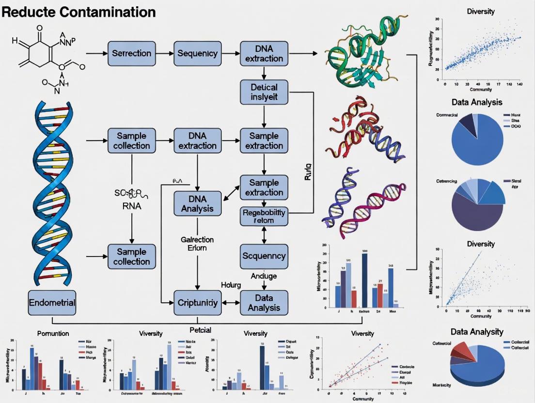
The following diagram illustrates the complete experimental workflow for endometrial microbiome studies, highlighting critical contamination control points:
Diagram 1: Comprehensive workflow for endometrial microbiome research highlighting critical control points for contamination prevention.
Advanced Methodological Considerations
Beyond 16S: Multi-Omics Approaches
While 16S rRNA sequencing has been foundational in characterizing the endometrial microbiome, several limitations necessitate advanced approaches:
- Shotgun metagenomics: Provides greater taxonomic resolution and functional insights compared to 16S sequencing, revealing microbial signatures that remain undetected by amplicon-based approaches [8].
- Metatranscriptomics: Assesses functionally active microorganisms rather than merely present taxa.
- Metabolomics: Identifies microbial metabolites that mediate host-microbe interactions.
- Culturomics: Enables isolation of viable bacterial strains for functional validation and therapeutic development [8].
Integration with Host Factors
The endometrial microbiome does not exist in isolation but interacts with numerous host factors:
- Hormonal influences: The microbiome fluctuates across the menstrual cycle, with increased microbial populations observed during the proliferative phase and decreased diversity during the luteal phase [9].
- Immune interactions: The endometrial microbiome modulates local immune responses through cytokine signaling and immune cell recruitment [8] [15].
- Genetic factors: Host genetic variants in immune-related genes may influence susceptibility to specific microbial community types [15].
- Systemic connections: The gut-endometrial axis represents a bidirectional communication pathway where gut microbial metabolites can influence endometrial function [16].
The paradigm shift from a sterile endometrium to recognition of a functional microbial niche represents a fundamental advancement in reproductive medicine. This new understanding brings both opportunities and challenges for researchers. The methodological framework presented here provides a foundation for conducting robust endometrial microbiome research while acknowledging the current limitations and ongoing developments in this rapidly evolving field. As technologies advance and standardized protocols emerge, the endometrial microbiome promises to become an increasingly important factor in diagnosing and treating reproductive disorders, ultimately improving outcomes for women worldwide.
The study of the endometrial microbiome is a rapidly advancing field in reproductive medicine. Historically considered a sterile site, the endometrium is now recognized as a low-biomass microbial niche, where the bacterial presence is estimated to be 100 to 10,000 times lower than in the vagina [8] [17]. This characteristic makes research in this area particularly vulnerable to contamination, which can severely compromise the validity of findings. Proper contamination control is therefore not merely a technical detail but a foundational requirement for producing reliable and clinically relevant data. This guide addresses the major contamination risks and provides actionable troubleshooting protocols for researchers.
FAQs: Addressing Common Contamination Concerns
1. Why is the endometrial microbiome considered a "low-biomass" environment, and why does this pose a special challenge?
The endometrial cavity contains a very small quantity of microbial DNA compared to other body sites like the vagina or gut. This low microbial load means that even minute amounts of contaminating DNA from reagents, the sampling equipment, or the laboratory environment can be amplified during sequencing. This contamination can easily overwhelm the true endometrial microbial signal, leading to distorted or entirely false results [8] [13]. In a low-biomass setting, distinguishing a true microbial resident from a contaminant is one of the most significant methodological hurdles.
2. What is the greatest risk of contamination during the sampling procedure?
The single greatest risk is cross-contamination from the lower genital tract. To obtain an endometrial sample, a catheter or device must pass through the non-sterile vagina and cervix, which are high-biomass environments dominated by a distinct microbial community [8] [13]. Without stringent precautions, the sample will collect microbes from this passage, making the resulting analysis reflective of the vaginal microbiome rather than the endometrial one.
3. How can I determine if my sequencing results include contaminating DNA from reagents?
The most reliable method is to include negative control samples in your workflow. These controls, which consist of nuclease-free water or unused collection swabs, should be processed in parallel with your biological samples through every stage—DNA extraction, PCR amplification, and sequencing [18] [8]. Any bacterial sequences detected in these negative controls are highly likely to be contaminants derived from reagents or laboratory processes. These sequences should be identified and subtracted from your biological sample data in a process called "decontamination."
4. We use sterile single-lumen catheters for sampling, but our results still show high concordance with vaginal profiles. What might be going wrong?
Single-lumen catheters can pick up contaminants from the cervix and vagina as they are inserted. A recommended solution is to adopt a double-lumen catheter system, similar to those used for embryo transfer. This system features an outer sheath that protects an inner, sterile sampling catheter from contact with the lower genital tract, thereby significantly reducing the risk of cross-contamination [13]. Furthermore, a rigorous cleaning protocol for the cervix and vagina with sterile saline before catheter insertion is essential to minimize the microbial load in the passage.
Troubleshooting Guide: Identifying and Solving Contamination Issues
Table: Troubleshooting Common Contamination Problems
| Problem | Potential Cause | Solution |
|---|---|---|
| High abundance of typical vaginal taxa (e.g., Lactobacillus, Gardnerella) in endometrial samples. | Cross-contamination during transcervical sampling [8] [13]. | Use a double-lumen catheter system. Implement thorough vaginal/cervical cleansing with sterile saline prior to sampling [13]. |
| Detection of common environmental bacteria (e.g., Sphingomonas, Arthrobacter) in both samples and negative controls. | Contamination from laboratory reagents or DNA extraction kits [8] [13]. | Include negative controls (reagent-only) in every batch. Use microbiome-specific DNA extraction kits designed for low biomass. Filter out taxa found in negative controls from your dataset. |
| Inconsistent microbial profiles between technical replicates of the same sample. | Contamination during sample processing in the lab or inconsistent DNA extraction. | Standardize all laboratory protocols. Use clean lab benches and UV irradiation. Process samples in smaller, randomized batches to identify batch-specific contamination. |
| Unexpectedly high microbial diversity in endometrial samples. | Potential contamination from multiple sources (vaginal, reagent, environmental). | Validate findings with a culture-based method (e.g., culturomics) if possible [17]. Re-assess the sampling and processing pipeline for breaks in sterile technique. |
Experimental Protocols for Contamination Control
Validated Sampling Protocol for Endometrial Fluid
This protocol is designed to minimize cross-contamination during sample acquisition [13].
- Pre-Sampling Preparation: Place the patient in a lithotomy position. Insert a vaginal speculum. Perform a rigorous cleaning of the cervix and vagina using abundant sterile saline.
- Catheter Insertion: Under ultrasound guidance, insert the outer sheath of a double-lumen catheter, taking care to avoid contact with the vaginal walls. If contact occurs, replace the sheath.
- Sample Aspiration: Advance the protected inner catheter through the sheath into the upper endometrial cavity. Attach a 20 mL syringe to create strong negative pressure and slowly aspirate the endometrial fluid while withdrawing the catheter.
- Sample Handling: Gently suspend the aspirated material in 150 µL of sterile saline in a sterile Eppendorf tube. The distal tip (2-3 mm) of the catheter can be cut directly into the tube. Store the sample immediately at -80°C.
Protocol for Controlling Reagent and Laboratory Contamination
This protocol is critical for ensuring the integrity of low-biomass samples [18] [8].
- Negative Controls: For every batch of samples processed, include at least two negative controls. These should be:
- A "reagent blank" with nuclease-free water taken through DNA extraction and library preparation.
- A "sampling control" such as an unused, sterilized swab or catheter processed identically to the patient samples.
- DNA Extraction: Use a DNA extraction kit specifically validated for low-biomass samples and microbial DNA enrichment, such as the QIAamp DNA Microbiome Kit.
- Bioinformatic Decontamination: After sequencing, process the data using a bioinformatic pipeline that identifies and removes contaminants. Any Operational Taxonomic Unit (OTU) or Amplicon Sequence Variant (ASV) present in the negative controls at a level above a set threshold (e.g., 0.01% of the total reads) should be subtracted from the biological samples.
The Scientist's Toolkit: Essential Reagents & Materials
Table: Key Reagents and Materials for Low-Biomass Endometrial Microbiome Studies
| Item | Function | Considerations for Contamination Control |
|---|---|---|
| Double-Lumen Catheter | To obtain endometrial samples while minimizing contact with the cervicovaginal microbiome [13]. | Opt for sterile, single-use devices. The inner catheter should remain shielded until deployment in the uterine cavity. |
| Nuclease-Free Water | Serves as a diluent and as a critical negative control. | Use certified nuclease-free, sterile water. Aliquot to avoid repeated use from a single container. |
| Microbiome-Specific DNA Extraction Kit | To efficiently isolate microbial DNA from a background of human DNA in low-biomass samples. | Kits like the QIAamp DNA Microbiome Kit include steps to deplete host DNA, enriching for microbial signals [13]. |
| PCR Reagents | To amplify the bacterial 16S rRNA gene for sequencing. | Use high-fidelity polymerase. Include multiple PCR-negative controls (water as template) to detect contamination in the amplification step. |
| Sterile Saline Solution | For cleaning the cervix and vagina prior to catheter insertion. | Essential for reducing the microbial load in the sampling path [13]. |
Workflow: Contamination-Control in Endometrial Microbiome Research
The diagram below outlines a robust workflow integrating key contamination control measures into the research pipeline.
In the rapidly evolving field of low-biomass microbiome research, particularly the study of the endometrial microenvironment, contamination control transcends technical consideration to become a scientific imperative. The female reproductive tract hosts a microbial gradient, with the endometrium representing a low-biomass environment—containing an estimated 100 to 10,000 times fewer bacteria than the vagina [19] [20]. This fundamental characteristic makes research in this area exceptionally vulnerable to contamination, which can distort data, lead to erroneous conclusions, and potentially result in clinical misdiagnosis. This technical support guide addresses the critical sources and consequences of contamination and provides evidence-based troubleshooting methodologies to ensure research integrity and patient safety.
Understanding the Risks: FAQs on Contamination Consequences
FAQ 1: Why is low-biomass endometrial microbiome research particularly vulnerable to contamination?
The vulnerability stems from the inherent nature of the endometrial environment. The bacterial load in the uterus is extremely low, while adjacent sites like the vagina and cervix are microbial hotspots. During transcervical sampling, even minimal contact with the lower reproductive tract can introduce contaminating DNA that overwhelms the true endometrial signal. Furthermore, standard laboratory reagents and kits often contain trace amounts of bacterial DNA that can be amplified and misinterpreted as genuine signal in these sensitive assays [19] [21] [22].
FAQ 2: What are the primary consequences of contamination in research and clinical diagnostics?
The consequences are severe and multi-faceted:
- Misleading Microbial Profiles: Contamination can create a false representation of the endometrial microbial community, suggesting the presence of bacteria that are not actually native to the endometrium [21] [17].
- Faulty Correlations with Disease: Incorrect microbial profiles can lead to spurious associations between specific bacteria and clinical conditions like infertility, endometriosis, or recurrent pregnancy loss. For example, a study found that a dysbiotic endometrial microbiota profile featuring bacteria like Gardnerella, Streptococcus, and Staphylococcus was associated with unsuccessful reproductive outcomes, while Lactobacillus dominance was linked to live births [23] [20]. Contamination could severely undermine the validity of such findings.
- Compromised Clinical Diagnostics: As endometrial microbiome analysis moves towards clinical application, contamination risks translating into misdiagnosis. This could lead to inappropriate treatments, such as unnecessary antibiotic courses or the rejection of viable embryos in IVF cycles based on a falsely diagnosed "unfavorable" uterine environment [9] [20].
- Hindered Scientific Progress: Contradictory results between studies, often stemming from differing levels of contamination control, create confusion and slow down the establishment of a true "core" healthy endometrial microbiome [9] [19].
FAQ 3: How can I distinguish true endometrial microbiota from contamination introduced during sampling?
No single method is foolproof, but a combination of strategies increases confidence:
- Paired Sampling: Collect and sequence samples from both the vagina and the endometrium from the same patient. A significant difference in the microbial composition suggests a distinct endometrial community and minimizes the likelihood that the endometrial profile is merely cross-contamination [13] [17].
- Rigorous Controls: Include negative controls (e.g., blank extraction kits, sterile swabs, or saline processed alongside samples) throughout the entire workflow—from sampling to sequencing. Any sequences appearing prominently in these controls are likely contaminants [21] [22].
- Standardized Sampling Protocols: Using specialized equipment like double-lumen catheters designed for embryo transfer can dramatically reduce contamination from the cervix and vagina during sample collection [13] [9].
Troubleshooting Guide: Mitigating Contamination at Every Stage
Stage 1: Sample Collection
Challenge: Contamination from the lower reproductive tract (vagina and cervix) during transcervical access. Solution: Implement a sterile, double-catheter sampling protocol.
- Experimental Protocol (Double-Lumen Catheter Technique):
- Patient Preparation: Place the patient in a lithotomy position. Insert a vaginal speculum.
- Cleaning: Thoroughly clean the cervix and vagina with abundant sterile saline to remove mucus and debris [13] [9].
- First Catheter Insertion: Under ultrasound guidance, insert the first (outer) catheter, taking extreme care to avoid contact with the vaginal walls. If contact occurs, replace the catheter [13].
- Second Catheter Insertion: Introduce the second (inner) catheter through the first, advancing it into the uterine fundus.
- Aspiration: While slowly retrieving the inner catheter, perform a firm aspiration with a large syringe (e.g., 20 mL) to generate strong negative pressure and collect the scant endometrial fluid [13].
- Sample Handling: Suspend the aspirated content in a sterile saline solution and immediately freeze at -80°C until analysis [13].
The following diagram illustrates the critical steps and contamination control points in this protocol:
Stage 2: Laboratory Processing
Challenge: Contamination from laboratory reagents, kits, and the environment during DNA extraction and library preparation. Solution: Meticulous use of controls and validated protocols for low-biomass samples.
- Experimental Protocol (DNA Extraction with Controls):
- Pre-digestion: For endometrial biopsies, include a pre-digestion step with lysozyme, lysostaphin, and mutanolysin to effectively degrade the cell walls of a wide range of bacteria [20].
- Negative Controls: Process "blank" samples (containing only the elution buffer or saline) alongside every batch of endometrial samples. These blanks undergo the exact same DNA extraction and sequencing process [21] [22].
- DNA Extraction Kits: Use kits specifically designed for low-biomass samples or microbial DNA enrichment, such as the QIAamp DNA Microbiome Kit or QIAamp DNA Blood Mini Kit [13] [20].
- Reagent Verification: Sequence these negative controls. Any bacterial taxa identified in the blanks must be considered potential contaminants and subtracted from the experimental samples during bioinformatic analysis [21] [22].
Stage 3: Data Analysis & Bioinformatics
Challenge: Differentiating contaminant DNA sequences from genuine endometrial microbiota signals. Solution: Implement a rigorous bioinformatic filtering pipeline.
- Experimental Protocol (Bioinformatic Decontamination):
- Sequence Filtering: Use established pipelines (e.g., QIIME2) to filter low-quality sequences, trim primers, and remove chimeras [23].
- Contaminant Removal: Systematically remove any Operational Taxonomic Units (OTUs) found in your negative control samples from your experimental dataset. Tools like
decontam(an R package) can statistically facilitate this process [23]. - Exclusion of Common Contaminants: A priori, exclude bacterial genera known to be common contaminants in reagents and kits, such as Sphingomonas and Arthrobacter [13].
The Scientist's Toolkit: Essential Research Reagent Solutions
The following table details key reagents and materials critical for reducing contamination in endometrial microbiome studies.
| Item | Function & Rationale | Example Products & Kits |
|---|---|---|
| Double-Lumen Catheter | Allows transcervical access to the endometrium while minimizing contact with the cervical and vaginal microbiome, the primary source of sample contamination. | Embryo transfer catheter (e.g., Gynétics) [13] [20] |
| Pre-digestion Enzyme Mix | Enhances lysis of difficult-to-break bacterial cell walls in low-biomass samples, improving DNA yield and representation. | Lysozyme, Lysostaphin, Mutanolysin [20] |
| Low-Biomass DNA Extraction Kit | Optimized for extracting microbial DNA from samples with low bacterial load, often including steps to deplete host DNA. | QIAamp DNA Microbiome Kit, QIAamp DNA Blood Mini Kit [13] [20] |
| 16S rRNA Gene Primers & Kits | For targeted amplification and sequencing of hypervariable regions to profile bacterial communities. Choice of regions (e.g., V3-V4, V4-V5) can affect taxonomic resolution. | Ion 16S Metagenomics Kit (amplifies V2,4,8 and V3,6,7-9) [20] |
| Negative Control Reagents | Sterile water or saline used to process blanks alongside samples to identify contaminating DNA from reagents and the laboratory environment. | Nuclease-free water, Sterile saline solution [21] [22] |
Advanced Validation: Culturomics as a Contamination Control Tool
Challenge: Sequencing detects both live bacteria and free DNA fragments, making it difficult to confirm a viable endometrial microbiome. Solution: Supplement sequencing with culturomics, a high-throughput culture approach.
- Experimental Protocol (Culturomics Validation):
- High-Throughput Culture: Plate endometrial samples on a wide array of culture media and incubate under various (an)aerobic conditions to cultivate as many viable microorganisms as possible [17].
- Species Identification: Identify pure colonies using MALDI-TOF MS or 16S rRNA gene sequencing [17].
- Data Integration: Compare the species identified via culturomics with those from sequencing data. The presence of a species in both datasets provides strong evidence that it is a viable member of the endometrial microbiota and not merely contaminating DNA. A study using this method found that only 28% of species, on average, were shared between paired vaginal and endometrial samples from the same patient, supporting the existence of a unique endometrial microbiome [17].
The relationship between different methodological approaches and their ability to validate a true microbiome is summarized below:
In endometrial microbiome research, the path from reliable data to clinical utility is paved with rigorous contamination control. The consequences of oversight are not merely academic; they extend to the very real potential of clinical misdiagnosis and inappropriate patient treatment. By adopting the stringent protocols, troubleshooting guides, and multi-method validation strategies outlined in this technical support document, researchers can fortify their work against contamination, thereby ensuring that findings accurately reflect the uterine microenvironment and can be confidently translated into clinical practice.
Proven Protocols: Best Practices for Sample Collection, Handling, and Processing
Frequently Asked Questions (FAQs)
Q1: Why is sampling technique especially critical in low-biomass endometrial microbiome research? The endometrial environment has an extremely low bacterial biomass, estimated to be 100 to 10,000 times less than the vaginal microbiome [8] [19]. When working with such minimal microbial presence, the DNA from contaminating sources introduced during sampling can easily outweigh or distort the signal from the actual endometrial microbiota, leading to misleading results [24] [8].
Q2: What is the primary risk of using a transcervical approach for endometrial sampling? The primary risk is cross-contamination with the cervical and vaginal microbiota [8] [19] [14]. As the catheter passes through the cervix, it can pick up microbial DNA from the lower reproductive tract, which is typically more abundant and diverse. This can confound the interpretation of the true endometrial microbial composition [8] [14].
Q3: How does a double-lumen catheter design help reduce contamination? A double-lumen catheter features two separate channels. This design allows one lumen to be used for the instillation of a sterile solution (like saline), while the other is used for aspiration. The fluid flow can help clear the catheter of contaminants from the cervical passage before collecting the actual endometrial sample, thereby providing a cleaner specimen [14].
Q4: What are the key procedural steps to minimize contamination during sample collection? Key steps include [14]:
- Strict aseptic technique: Using sterile gloves and avoiding contact with non-sterile surfaces.
- Minimizing exposure: Reducing the time the catheter is exposed to the cervical canal.
- Standardized protocols: Following a consistent and validated sampling workflow.
- Using negative controls: Processing nuclease-free water alongside clinical samples to detect environmental or reagent contamination.
Q5: Beyond catheter choice, what other factors are essential for reliable results? A holistic approach is necessary. Critical factors include [24] [8] [19]:
- Standardized DNA extraction protocols optimized for low biomass.
- Rigorous bioinformatic analysis that includes contamination removal workflows.
- Comprehensive reporting of all steps from sampling to data analysis to allow for reproducibility and critical evaluation.
Troubleshooting Guides
Problem: High Abundance of Vaginal Taxa in Endometrial Samples
Potential Cause: Contamination during transcervical catheter insertion.
Solutions:
- Validate with Controls: Implement and process negative control samples (e.g., nuclease-free water) in parallel with patient samples. If the same taxa appear in the negative controls, they are likely contaminants [14].
- Employ a Clearing Lumen: If using a double-lumen catheter, ensure an adequate volume of sterile saline is instilled and wasted via the clearing lumen before aspirating the sample lumen [14].
- Bioinformatic Decontamination: Use specialized computational tools (e.g., decontam, SourceTracker) to identify and subtract contaminant sequences based on their prevalence in negative controls or their known source [24].
Problem: Inconsistent Microbiome Profiles Between Replicate Samples
Potential Cause: Inconsistent sampling technique or low microbial biomass leading to stochastic effects.
Solutions:
- Operator Training: Ensure all personnel are uniformly trained and competent in the standardized sampling protocol [25] [26].
- Sample Adequacy: Vigorously vortex the endometrial tissue sample in PBS to ensure homogeneous resuspension of the low biomass material before DNA extraction [14].
- Technical Replicates: Process multiple technical replicates per sample to distinguish true signal from technical noise.
Problem: Failure to Detect Any Bacterial DNA
Potential Cause: Inhibitors in the sample affecting PCR, insufficient sample material, or overly stringent decontamination.
Solutions:
- Check DNA Yield: Semi-quantitatively assess extracted DNA quality by agarose gel electrophoresis [14].
- Inhibition Testing: Use spiked-in internal controls to check for the presence of PCR inhibitors.
- Review Decontamination Thresholds: Re-evaluate the stringency of bioinformatic contamination removal parameters, as they might be removing true, low-abundance signals [24].
Experimental Protocols for Validation
Protocol 1: In-Vitro Validation of Catheter Contamination
This protocol is adapted from methodologies used to evaluate drug adsorption in central venous catheters [27].
Objective: To quantify the level of contaminant carry-over by a specific catheter type under controlled conditions.
Materials:
- Double-lumen catheter (or catheter under investigation)
- Sterile saline
- Synthetic microbial community of known composition (Mock Community)
- DNA extraction kit
- PCR/qPCR reagents
- Sequencer
Methodology:
- Simulate Contamination: Submerge the tip of the catheter in a solution containing the synthetic microbial community to simulate passage through the vagina/cervix.
- Clearing Procedure: Withdraw the catheter and use one lumen to flush with sterile saline while aspirating from the second lumen, mimicking the clinical clearing procedure.
- Sample the Effluent: Collect the aspirated saline effluent.
- Control: Directly sample the synthetic community solution as a positive control.
- Analysis: Extract DNA from both the effluent and control samples. Perform 16S rRNA gene sequencing and compare the microbial composition and biomass (via qPCR) between the two.
Expected Outcome: A well-designed catheter and effective clearing protocol will result in a significant reduction or absence of the mock community signal in the effluent sample.
Protocol 2: In-Situ Validation via Sample-Site Comparison
Objective: To clinically validate the sampling technique by comparing microbiota results from different sampling sites.
Methodology:
- Sample Collection: During a surgical procedure (e.g., hysterectomy), collect samples in this order [14]:
- Vaginal Swab
- Cervical Swab
- Endometrial Sample using the double-lumen catheter transcervically.
- Direct Endometrial Tissue Biopsy obtained surgically after opening the uterus (considered the "gold standard").
- Sequencing and Analysis: Process all samples through identical DNA extraction and sequencing protocols.
Analysis: Compare the beta-diversity and taxonomic composition. A valid transcervical catheter sample should be more similar to the direct endometrial biopsy than to the vaginal or cervical swabs.
Data Presentation: Comparative Analysis of Sampling Methods
The table below summarizes key considerations for different sampling approaches, synthesizing insights from clinical and low-biomass research.
Table 1: Comparison of Endometrial Microbiome Sampling Methods
| Sampling Method | Contamination Risk | Key Advantages | Key Limitations | Ideal Use Case |
|---|---|---|---|---|
| Double-Lumen Catheter | Moderate (Reduced by clearing lumen) | Less invasive than biopsy; designed to minimize cross-contamination. | Still requires passage through cervix; procedure must be meticulously followed. | Outpatient settings and longitudinal studies where minimal invasiveness is key. |
| Single-Lumen Catheter | High | Minimally invasive, readily available. | High potential for carry-over of cervical/vaginal microbiota. | Less recommended for low-biomass research unless validated with extensive controls. |
| Direct Endometrial Biopsy | Low (if collected aseptically during surgery) | Avoids the cervico-vaginal canal entirely; considered the gold standard for purity. | Highly invasive; requires a surgical setting (e.g., hysterectomy). | Validation studies to benchmark the accuracy of less invasive methods. |
| Transvaginal Aspiration | High | Can be performed without hysteroscopy. | The catheter tip is exposed to the entire vaginal vault during collection. | Use requires extensive validation with negative controls and bioinformatic decontamination. |
Table 2: Contamination Control and Diagnostic Metrics in Catheter Sampling
| Parameter | Value/Result | Context & Implication |
|---|---|---|
| Biomass Difference | 100 - 10,000x lower than vagina [8] [19] | Highlights the extreme susceptibility of endometrial samples to contamination. |
| Pooled vs. Individual Lumen Sampling Sensitivity | 69.23% (Pooled) vs. ~71.4% (Individual, proximal port) [28] | In diagnostic settings, sampling lumens individually is more sensitive than pooling; supports the value of dedicated lumens. |
| Key Contamination Control | Negative controls (nuclease-free water) [14] | Essential for distinguishing environmental/reagent contaminants from true sample microbiota. |
| Impact of Delayed Processing | Not Quantified | Anecdotal evidence suggests prompt processing after collection minimizes overgrowth of contaminants. |
Methodological Visualization
Diagram: Low-Biomass Endometrial Sampling Workflow
Diagram: Contamination Source Decision Pathway
The Scientist's Toolkit: Research Reagent Solutions
Table 3: Essential Materials for Low-Biomass Endometrial Microbiome Research
| Item | Function in Research | Key Consideration |
|---|---|---|
| Double-Lumen Catheter | To obtain endometrial fluid samples while minimizing cross-contamination from the cervix. | The "clearing" lumen is critical. Verify compatibility with your DNA extraction protocol (e.g., material does not inhibit PCR). |
| >0.5% Chlorhexidine with Alcohol or 0.5% Povidone Iodine [25] [14] [26] | For skin and cervical os antisepsis before catheter insertion to reduce introduction of surface contaminants. | Must be allowed to dry completely before catheter insertion to be effective and avoid interfering with subsequent molecular biology. |
| Sterile Saline (DNA/RNA Free) | Used as the flushing and aspiration medium in the catheter. | Must be certified nuclease-free to avoid introducing external DNA that would be amplified in sequencing. |
| DNA Extraction Kit for Low Biomass | To lyse microbial cells and purify trace amounts of DNA from small volume samples. | Choose kits validated for low biomass, high efficiency, and low contamination. Include a carrier RNA if recommended. |
| Mock Microbial Community | A defined mix of microbial cells or DNA used as a positive control to assess extraction and sequencing performance. | Essential for identifying technical biases and quantifying sensitivity limits in your entire workflow. |
| Nuclease-Free Water | Serves as a negative control during DNA extraction and PCR to monitor for reagent/environmental contamination [14]. | Must be processed in parallel with all patient samples through the entire workflow, from extraction to sequencing. |
Essential Personal Protective Equipment (PPE) and Decontamination Procedures
In low-biomass endometrial microbiome research, where microbial DNA concentrations are minimal, proper Personal Protective Equipment (PPE) and decontamination procedures are critical. Contamination from researchers or the environment can severely compromise data integrity, as the target DNA "signal" can be easily overwhelmed by contaminant "noise" [4]. This guide provides essential protocols to help researchers maintain sample integrity from collection to analysis.
Frequently Asked Questions
Q1: Why is specialized PPE so crucial for low-biomass endometrial microbiome studies? Low-biomass environments like the endometrial cavity contain significantly fewer bacteria than other body sites, making them exceptionally vulnerable to contamination. Humans are the largest source of contamination, shedding millions of particles daily [29]. Proper PPE creates a necessary barrier between the researcher and the sample to prevent introducing external microbial DNA that could distort research findings [4].
Q2: What is the most common error in PPE practice that leads to sample contamination? Improper doffing (removal) of PPE is a frequent critical error. Removing contaminated PPE incorrectly can expose both the wearer and the sample to contaminants. Even if PPE provides protection during use, improper removal can negate all previous contamination controls [30] [31]. Consistent training in dedicated doffing sequences is essential.
Q3: Can I reuse disposable PPE in my experiments? Generally, no. Most disposable PPE is designed for single use. Washing or reusing disposable items changes their protective properties and barrier capabilities, rendering them ineffective. Exceptions exist for some reusable equipment like specific goggles or elastomeric respirators, but only if decontamination follows the manufacturer's precise instructions [30].
Q4: Our lab is beginning endometrial microbiome sampling. What are the critical controls we need? Incorporating various controls is mandatory for data credibility:
- Sampling Controls: Include sterile swabs exposed to the air in the sampling environment, swabs of PPE surfaces, and empty collection vessels.
- Processing Controls: Use DNA extraction blank controls.
- Purpose: These controls help identify contamination sources introduced during collection and processing, allowing for accurate interpretation of results [4] [32].
Q5: How do we verify that our decontamination procedures for equipment are effective? Use a combination of:
- Chemical Decontamination: For example, using 80% ethanol to kill microorganisms followed by a nucleic acid degrading solution (e.g., bleach) to remove residual DNA [4].
- Physical Methods: Autoclaving or UV-C light sterilization for equipment that can withstand it [4] [33].
- Validation: Test the effectiveness of your decontamination protocols using microbial cultures or quantitative PCR on swabs taken from decontaminated surfaces.
Essential PPE for Low-Biomass Microbiome Research
The following table details the essential PPE components for handling low-biomass samples, moving from highest to lowest priority environments.
Table 1: Essential PPE for Low-Biomass Microbiome Research
| PPE Component | Required For | Specifications & Best Practices |
|---|---|---|
| Gloves | All handling stages [4] | Powder-free nitrile; double-gloving for highest cleanliness (e.g., ISO Class 5); sterile where required [29]. |
| Full-Body Coveralls | Cleanrooms, sampling procedures [4] | Non-linting, low-particulate fabric (e.g., SMS); front-zip or 2-piece suits; attached hoods for highest protection [29]. |
| Head Covers | All environments | Bouffant caps or shrouded hoods that fully cover hair, neck, and shoulders [29]. |
| Face Masks | All environments | Surgical-style to reduce droplets; N95 respirators or PAPR for higher-risk settings [29] [4]. |
| Eye Protection | When splashes are possible | Sealed goggles; reusable models with anti-fog coating and high-temperature sterilization capability are cost-effective [29]. |
| Shoe Covers/Boots | Cleanrooms and labs | Slip-resistant, fully encapsulating footwear; cleanroom-dedicated shoes are ideal [29]. |
Decontamination Procedures
Workflow for Sample Collection and Processing
The diagram below outlines a contamination-aware workflow for collecting and processing low-biomass endometrial samples.
Decontamination Methods for Equipment
Table 2: Decontamination Methods for Research Equipment
| Method | Best For | Procedure & Limitations |
|---|---|---|
| Chemical (Bleach) | Surfaces, non-corrosive equipment | Use sodium hypochlorite to degrade DNA. Effective against cell-free DNA that autoclaving may leave behind [4]. |
| Autoclaving | Heat-tolerant tools, glassware | Standard 121°C moist-heat sterilization. Kills viable cells but may not remove persistent DNA [4]. |
| UV-C Radiation | Surfaces, some respirators | Uses short-wavelength ultraviolet light. Effective for decontaminating flat surfaces and certain PPE during supply crises [33]. |
| Ethanol Wiping | Quick surface decontamination | 80% ethanol kills microorganisms but does not effectively remove DNA. Should be combined with a DNA removal step [4]. |
The Scientist's Toolkit: Key Research Reagent Solutions
Table 3: Essential Reagents for Endometrial Microbiome Research
| Reagent / Kit | Specific Function | Application in Endometrial Studies |
|---|---|---|
| RNAlater Solution | Stabilizes nucleic acids immediately after collection | Preserves endometrial fluid and tissue samples during transport and storage [34]. |
| QIAamp DNA Microbiome Kit | Extracts microbial DNA while depleting host DNA | Critical for enriching low-abundance bacterial DNA from high-host-DNA samples [13]. |
| Ion 16S Metagenomics Kit | Amplifies 7 hypervariable regions of the 16S rRNA gene | Provides comprehensive taxonomic profiling in endometrial microbiome studies [34]. |
| PowerSoil DNA Isolation Kit | Standard for challenging soil samples; effective for low-biomass | Adapted for endometrial biopsies to improve DNA yield from difficult-to-lyse bacteria [34]. |
| Allplex BV Assay | Multiplex real-time PCR for bacterial vaginosis | Quantifies BV-related bacteria and assesses Lactobacillus abundance in parallel with NGS [13]. |
Experimental Protocol: Endometrial Microbiome Analysis
Workflow for Endometrial Microbiome Analysis
The following diagram outlines the key steps in the laboratory analysis of endometrial microbiome samples.
Detailed Methodology
Sample Collection & DNA Extraction
- Sampling Technique: Use a double-lumen catheter system under ultrasound guidance to minimize cervical/vaginal contamination. Perform firm aspiration with a 20ml syringe while slowly retrieving the catheter [13].
- DNA Extraction: For endometrial fluid, perform pre-digestion with lysozyme, lysostaphin, and mutanolysin at 37°C for 30 minutes before using the QIAamp DNA Blood Mini Kit. For tissue samples, mechanically disrupt 25mg of tissue in a TissueLyser LT before nucleic acid purification with QIAsymphony [34].
16S rRNA Gene Sequencing & Analysis
- Library Preparation: Amplify hypervariable regions (V2-4-8 and V3-6,7-9) using the Ion 16S Metagenomics Kit with 30 PCR cycles. Prepare libraries starting from 50ng of pooled amplicons using the Ion Plus Fragment Library Kit and Ion Xpress Barcode Adaptors [34].
- Bioinformatic Processing: Process raw sequencing data with tools like the decontam R package for prevalence-based filtering of contaminant Operational Taxonomic Units (OTUs). Exclude common contaminant genera such as Sphingomonas and Arthrobacter [32].
Troubleshooting Common Contamination Issues
Problem: High levels of human skin flora (e.g., Staphylococcus, Corynebacterium) in samples.
- Potential Cause: Inadequate PPE or improper donning/doffing procedure.
- Solution: Implement structured training on PPE use. Ensure full-body coveralls, gloves, and masks are worn correctly before sampling. Establish a dedicated doffing area with visible instructions [29] [31].
Problem: Consistent detection of specific bacteria across samples and negative controls.
- Potential Cause: Contaminated reagents or laboratory environment.
- Solution: Test all reagents for microbial DNA contamination before use. Use UV-irradiated or DNA-free reagents. Include multiple negative controls (extraction blanks, sterile swabs) to identify contamination sources [4].
Problem: Discrepancy between endometrial and vaginal microbiome profiles suggesting cross-contamination.
- Potential Cause: Contamination during the transcervical sampling procedure.
- Solution: Use double-lumen catheters and avoid contact with vaginal walls. Clean the cervix and vagina thoroughly with sterile saline before catheter insertion [13].
DNA Extraction and Kit Selection for Microbial DNA Enrichment
The study of the endometrial microbiome represents a significant frontier in reproductive health, with research indicating that its composition can be a useful biomarker for predicting reproductive outcomes such as live birth [34]. However, this research is conducted on samples with extremely low bacterial biomass, where microbial DNA is outnumbered by host DNA and is highly susceptible to contamination [32] [4]. Under these conditions, the DNA extraction methodology becomes not merely a preliminary step but a critical determinant of data accuracy and reliability. This technical support center provides targeted guidance to help researchers navigate the specific challenges of microbial DNA enrichment in low-biomass endometrial studies, with a core focus on reducing contamination and optimizing protocols for meaningful results.
FAQs: Fundamental Questions on DNA Extraction for Low-Biomass Microbiome Studies
1. Why is DNA extraction methodology particularly critical for low-biomass endometrial microbiome studies?
DNA extraction is the largest source of experimental variability in microbiome studies [35]. For low-biomass environments like the endometrium, where bacterial abundance is 10²–10⁴ times lower than in the vagina, the proportional impact of any contaminating DNA from reagents, kits, or the sampling process is vastly magnified [34] [35]. This can lead to false positives and erroneous conclusions about the constitutive microbiome. A validated, consistent DNA extraction protocol is therefore essential to distinguish true microbial signal from noise [4] [36].
2. What are the minimum standards for reporting DNA extraction methods in publications?
To ensure reproducibility and reliability, especially in low-biomass research, scientists should adhere to three minimal standards [35]:
- Detailed Reporting: Provide a thorough description of the DNA extraction method that allows another laboratory to exactly reproduce all procedures.
- Control Reporting: Include and report data from positive and negative controls in every DNA extraction batch, detailing coefficients of variation for positive controls and contamination levels in blanks.
- Protocol Consistency: Utilize the same DNA extraction protocol across all samples within a study and in multi-site studies that plan to pool data.
3. How can I improve DNA yield from low-biomass endometrial samples?
Several strategies can enhance DNA recovery:
- Mechanical Lysis: Incorporate bead-beating with a mixture of bead sizes to ensure uniform and efficient lysis of both Gram-positive and Gram-negative bacteria [37] [36].
- Chemical and Enzymatic Lysis: Use a pre-digestion step with enzymes like lysozyme, lysostaphin, and mutanolysin to degrade robust bacterial cell walls [34].
- Co-precipitants: Add agents like agar, glycogen, or sodium alginate early in the extraction process to act as a co-precipitant, reducing DNA loss during precipitation steps and significantly increasing yield from low-biomass specimens [38].
Troubleshooting Guide: Common Issues and Solutions
| Problem | Primary Cause | Recommended Solution |
|---|---|---|
| Low DNA Yield | Inefficient lysis of robust Gram-positive bacteria. | Implement bead-beating with high-density beads [37] [36] and enzymatic pre-digestion [34]. |
| High Host DNA Contamination | Sample dominated by human endometrial cells. | Use a commercial kit designed to deplete host DNA, thereby enriching for microbial DNA [13] [36]. |
| Sample Degradation | Activity of endogenous nucleases in tissue. | Flash-freeze samples in liquid nitrogen after collection and store at -80°C. Keep samples on ice during preparation [39]. |
| PCR Inhibition | Co-purification of inhibitors from the sample or reagents. | Use a DNA purification kit equipped with inhibitor removal technology [37]. Ensure complete washing of the silica membrane [39]. |
| High Contaminant Signal in Sequencing | Contamination from reagents, kits, or the laboratory environment. | Incorporate and sequence negative controls (e.g., blank swabs, reagent blanks) and use bioinformatic tools like the decontam R package to identify and remove contaminant sequences [32] [4]. |
Experimental Workflow: From Sample Collection to Sequencing
The following diagram illustrates a contamination-aware workflow for profiling the endometrial microbiome, integrating critical control points.
The Scientist's Toolkit: Essential Reagents & Kits
The table below lists key reagents and materials used in low-biomass endometrial microbiome research.
| Item | Function & Rationale |
|---|---|
| Double-Lumen Catheter | Minimizes contamination during transcervical sampling by protecting the inner catheter from contact with the cervical and vaginal microbiome [13]. |
| DNA/RNA Shield or RNAlater | Preservation solution that stabilizes nucleic acids immediately after sample collection, preventing degradation during storage [39] [37]. |
| BashingBeads / Lysing Matrix | Ultra-high density beads for mechanical lysis (bead-beating) to ensure unbiased, efficient breakdown of all microbial cell types, including tough Gram-positive bacteria [37]. |
| Enzymes (Lysozyme, Mutanolysin) | Used in a pre-digestion step to chemically degrade bacterial cell walls, complementing mechanical lysis for maximum DNA recovery [34]. |
| Host DNA Depletion Kit | Selectively removes human DNA, thereby enriching the relative proportion of microbial DNA for more efficient sequencing of the target microbiome [13] [36]. |
| Silica Spin Columns | Purify DNA by binding it in the presence of high-salt buffers, allowing contaminants and inhibitors to be washed away [39] [37]. |
| Mock Microbial Community | A defined mix of microbial cells or DNA serving as a positive control to evaluate the accuracy and bias of the entire DNA extraction and sequencing workflow [35] [37]. |
| DNA-Free Water & Reagents | Certified low-bioburden reagents are essential to minimize the introduction of external contaminant DNA that can compromise low-biomass studies [37] [4]. |
Incorporating a Comprehensive Suite of Negative and Process Controls
Frequently Asked Questions
What is the single biggest source of contamination in low-biomass microbiome studies? Contamination can be introduced at any stage, but reagents and laboratory kits are a pervasive source because their microbial DNA is co-extracted and amplified alongside your target sample DNA. For this reason, including reagent blanks (also known as extraction controls) in every processing batch is non-negotiable [4].
My negative control shows bacterial growth. Are my sample results invalid? Not necessarily. The purpose of negative controls is to identify the "contamination signature" of your workflow. If your samples are significantly different from your controls in microbial composition and biomass, your results may still be valid. However, if the control and sample profiles are similar, the data is likely unreliable and should be interpreted with extreme caution or discarded [4].
How can I prevent cross-contamination between my samples during processing? A major source of cross-contamination is the use of 96-well plates, where a shared seal can lead to well-to-well leakage. To mitigate this:
- Randomize samples across the plate to avoid grouping low- and high-biomass samples.
- Consider using single tubes (like barcoded Matrix Tubes) for lysis instead of plates, which has been shown to drastically reduce well-to-well contamination [40].
- Leave empty wells or blank controls between samples, especially those with differing biomass [40].
Beyond DNA sequencing, what other controls are needed for a robust study? A comprehensive control strategy includes several types of controls that accompany your samples from collection to sequencing [4]:
- Sampling Controls: Field blanks, swabs of the air, or swabs of gloved hands.
- Process Controls: Reagent blanks (for extraction and library preparation) and PCR negatives.
- Positive Controls: Use of mock microbial communities to assess the accuracy and sensitivity of your entire workflow.
Troubleshooting Guide
Problem: Negative controls show high levels of bacterial DNA, obscuring the true signal in my endometrial samples.
| Problem Step | Possible Root Cause | Solution and Recommended Action |
|---|---|---|
| Sample Collection | Contamination from the collector's skin, gloves, or the sampling environment. | - Decontaminate equipment with 80% ethanol followed by a DNA-degrading solution (e.g., bleach, where safe). [4] - Use sterile, single-use sampling devices like Tao Brush or Cornier cannula. [41] [20] - Wear appropriate PPE (gloves, mask, clean suit) to limit human-derived contamination. [4] |
| Sample Storage & Transport | Degradation or contamination during storage. | - Store samples immediately at -80°C in a DNA/RNA stabilizing solution like RNAlater. [20] |
| Nucleic Acid Extraction | Contaminating DNA from reagents, kits, or the lab environment. Cross-contamination between samples in 96-well plates. | - Include reagent blanks with every batch of extractions. [4] - Use kits specifically validated for low-biomass samples, such as the MagMAX Microbiome Ultra Nucleic Acid Isolation Kit. [40] - Adopt a tube-based lysis method (e.g., the Matrix method) instead of plate-based lysis to significantly reduce well-to-well contamination. [40] |
| Library Preparation & Sequencing | Contamination from enzymes, barcodes, or the sequencing run itself. | - Include a PCR negative control (water instead of template DNA) to detect kit/environmental contaminants. - Use of unique dual indices is critical to identify and correct for index hopping during sequencing. |
Quantitative Impact of Improved Contamination Control
The following table summarizes experimental data comparing a standard plate-based extraction method with a tube-based (Matrix) method, demonstrating the quantitative benefits of optimizing workflows for low-biomass samples [40].
| Metric | Conventional 96-Well Plate Method | Matrix Tube Method (Single-Tube) |
|---|---|---|
| Percentage of Contaminated Blanks | 19% (128 out of 672 blanks) | 2% (14 out of 672 blanks) |
| Average DNA Concentration in Contaminated Blanks | 0.21 ng/µL | 0.026 ng/µL |
| Compatibility with Metabolomics | Requires separate aliquots | Enables paired nucleic acid and metabolite extraction from a single sample |
| Well-to-Well Cross-Contamination Risk | High | Significantly Reduced |
The Scientist's Toolkit: Essential Research Reagents & Materials
| Item or Reagent | Function in Low-Biomass Endometrial Research |
|---|---|
| Tao Brush or Cornier Cannula | Sterile, single-use devices for collecting endometrial fluid and biopsy samples while minimizing contamination from the lower reproductive tract. [41] [20] |
| RNAlater Stabilization Solution | Preserves nucleic acids in endometrial samples immediately after collection, preventing microbial population shifts during transport. [20] |
| DNA Degrading Solution (e.g., Bleach) | Used to decontaminate work surfaces and non-disposable equipment by destroying contaminating DNA traces, making surfaces "DNA-free." [4] |
| MagMAX Microbiome Ultra Nucleic Acid Isolation Kit | A commercially available kit optimized for co-extraction of DNA and RNA from difficult samples, showing high performance in microbiome studies. [40] |
| Matrix Tubes | Barcoded, single tubes used as an alternative to 96-well plates for sample lysis, effectively eliminating the problem of well-to-well cross-contamination. [40] |
| Lysozyme, Lysostaphin, Mutanolysin | Enzymes used in a pre-digestion step to effectively lyse the tough cell walls of Gram-positive bacteria, ensuring complete DNA extraction and representative community profiles. [20] |
| Mock Microbial Community | A defined mix of known microorganisms used as a positive control to validate the entire workflow, from DNA extraction to sequencing, and to assess bias and sensitivity. [4] |
Experimental Workflow with Integrated Controls
The following diagram outlines a robust experimental protocol for low-biomass endometrial microbiome research, integrating critical control points at every stage to ensure data validity.
Detailed Protocol: Endometrial Fluid Collection and Processing
This protocol is adapted from multi-centre studies that successfully characterized the endometrial microbiome [41] [20].
Patient Preparation and Sample Collection:
- With the patient in the lithotomy position, clean the vagina and cervix with a dry cotton swab to remove mucus and debris.
- Introduce a disinfected speculum.
- Using a sterile, flexible catheter (e.g., Cook Medical Tao Brush or Gynétics catheter), carefully introduce it into the uterine cavity, avoiding contact with the vaginal walls.
- Aspirate endometrial fluid (volume ~20-80 µL). Stop suction at the internal cervical os during catheter removal to prevent contamination.
- For an endometrial biopsy, use a device like a Cornier cannula to scrape the endometrium and obtain a tissue sample (50-70 mg).
Sample Preservation:
- Wipe the external surface of the catheter with a sterile gauze.
- Expel the endometrial fluid into a cryotube containing 50 µL of RNAlater solution.
- Transfer the tissue biopsy to a cryotube containing 1.5 mL of RNAlater.
- Store samples at 4°C for up to 4 hours, then transport at room temperature before long-term storage at -80°C.
DNA Extraction with Pre-digestion for Low Biomass:
- For Endometrial Fluid: Perform a pre-digestion step at 37°C for 30 minutes using a lysozyme, lysostaphin, and mutanolysin cocktail to degrade tough bacterial cell walls [20].
- For Endometrial Biopsy: Mechanically disrupt ~25 mg of tissue in a TissueLyser after a 3-hour proteinase K digestion at 56°C [20].
- Proceed with nucleic acid purification using a dedicated kit (e.g., QIAamp DNA Blood Mini kit or automated systems like QIAsymphony).
- CRITICAL STEP: Include a minimum of one reagent-only negative control for every batch of samples processed.
16S rRNA Gene Sequencing and Analysis:
- Amplify hypervariable regions of the 16S rRNA gene (e.g., V3-V4) using barcoded primers.
- Prepare amplicon libraries using a kit such as the Ion Plus Fragment Library Kit or Illumina's Nextera XT.
- Sequence the pooled library on a platform like the Illumina MiSeq.
- Process the sequencing data through a bioinformatic pipeline (e.g., QIIME 2, Mothur) that includes a step to subtract sequences found in your negative controls from your sample data.
Troubleshooting Common Pitfalls and Optimizing Your Workflow for Cleaner Data
Identifying and Mitigating Batch Effects and Processing Bias
FAQ: Understanding the Core Concepts
What are batch effects and processing biases in the context of low-biomass microbiome research?
In low-biomass microbiome studies, batch effects are technical variations introduced when samples are processed in different groups (batches) by different personnel, using different reagent lots, or at different times [42] [43]. Processing biases refer to the variable efficiency of different experimental steps (e.g., DNA extraction, amplification) in recovering different microbial taxa [42] [43]. In higher-biomass samples, these issues may merely add noise, but in low-biomass samples like endometrial tissue, where the target microbial signal is faint, they can completely obscure true biological signals or generate artifactual ones [42] [4].
Why are low-biomass samples like the endometrium particularly vulnerable?
Low-biomass samples are characterized by a very small amount of microbial DNA. Consequently, even trace amounts of contaminating DNA from reagents, kits, or the laboratory environment can constitute a large proportion, or even the majority, of the sequenced material [42] [4]. This means that the contaminant "noise" can easily overwhelm the true biological "signal," leading to misleading conclusions about the microbial community present in the sample [4].
FAQ: Troubleshooting and Experimental Design
My negative controls show high microbial biomass. What should I do?
If your negative controls (e.g., blank extractions, no-template controls) show a high number of sequences, this is a clear indicator of significant contamination.
- Immediate Action: Do not proceed with downstream biological interpretation. The validity of your experimental samples is compromised.
- Investigation: Audit your laboratory procedures. Reagents, kits, and laboratory surfaces are common contamination sources [4]. Ensure you are using DNA-free reagents and sterilized plasticware.
- Long-term Solution: Implement and consistently use a rigorous system of process controls in every batch of sample processing. This is not optional for low-biomass research [42] [4].
My samples cluster by sequencing run or extraction date, not by study group. Is this a batch effect?
Yes, this is a classic signature of a batch effect. If in your Principal Component Analysis (PCA) or PCoA plots, samples group together based on the technical batch (e.g., the day they were processed) rather than the biological variable of interest (e.g., CE vs. non-CE), it indicates that technical variation is dominating your data [44]. A real-world re-analysis of a fetal microbiome study demonstrated how an unaccounted batch effect led to the false conclusion that a specific microbe was present in fetal samples when it was actually a contaminant introduced in one processing batch [44].
How can I tell if an observed microbe is a true signal or a contaminant?
This is a central challenge. Key strategies include:
- Use of Controls: Compare the abundance of the microbe in your biological samples to its abundance in your various negative controls. Microbes that are more abundant in samples than in controls are more likely to be true signals [42] [4].
- Consult Contamination Databases: Resources like the "Weiss Lab Contamination Database" catalog microbial taxa commonly found in reagents and kits.
- Biological Plausibility: Assess whether the identified microbe is known to inhabit the endometrial environment or is a common environmental contaminant (e.g., Sphingomonas, Pseudomonas) [45] [46].
Troubleshooting Guide: A Practical Table
The following table outlines common issues, their potential causes, and recommended solutions.
| Problem | Potential Causes | Recommended Solutions |
|---|---|---|
| High diversity in negative controls | Contaminated reagents, improper sterile technique, kit contamination [43] [4] | Use new, DNA-free reagent lots; implement UV/bleach decontamination of workspaces and equipment; include multiple types of negative controls [4] |
| Samples cluster by processing batch | Non-randomized sample processing, different reagent lots, different personnel [42] [44] | Randomize cases and controls across all processing batches; use statistical batch correction methods (e.g., ComBat, RUV-III-NB) [47] [48] |
| Inconsistent results between study replicates | Uncontrolled well-to-well leakage during PCR, variable DNA extraction efficiency [42] [43] | Use physical barriers between wells in PCR plates; employ unique dual-indexed primers to identify and filter cross-talk; validate and standardize DNA extraction protocols [42] [4] |
| Low sequencing depth/sensitivity | Insufficient microbial DNA, inefficient lysis of certain taxa, PCR inhibition [43] | Incorporate a standardized microbial mock community to assess sensitivity and bias; optimize sample lysis protocols (e.g., bead-beating) [43] |
Key Experimental Protocols for Robust Research
Protocol for a Rigorous Sample Collection and DNA Extraction Workflow
This protocol is designed to minimize contamination and bias from the outset, specifically for endometrial tissue sampling.
Detailed Steps:
- Patient Preparation & Sterile Field: The cervix and vagina must be thoroughly disinfected prior to sample collection to minimize ascending contamination [45] [46].
- Sterile Tissue Collection: Endometrial tissue should be collected using a disposable sterile negative pressure suction probe guided by ultrasound. The probe must enter the uterine cavity without contacting the cervix or vaginal walls to prevent contamination from the lower reproductive tract [45] [46].
- Multiple Aliquots: Immediately after collection, the tissue specimen should be divided into multiple aliquots in disposable sterile cryopreservation tubes. One aliquot is for histological diagnosis (e.g., CD138 staining for CE), and the others are for DNA analysis [45] [46].
- Storage: Store aliquots at -80°C until DNA extraction to preserve nucleic acid integrity [43].
- Contemporaneous DNA Extraction: Extract DNA from all samples and controls in the same batch using the same kit and lot number to minimize batch effects. The CTAB method or commercially available kits validated for low-biomass samples can be used [45] [46].
- Include Negative Controls: For every batch of extractions, include at least two each of the following controls [42] [4]:
- Blank Extraction Control: Lysis buffer without any sample.
- Swab/Kit Control: A swab or collection tube from the same lot, exposed to the air in the operating theatre.
- DNA-Free Water Control: Water carried through the entire extraction and PCR process.
Protocol for a Contamination-Aware Bioinformatics Workflow
This workflow should be applied after raw sequencing data is generated.
Detailed Steps:
- Quality Filtering & Denoising: Process raw sequencing reads through a pipeline like QIIME2. This includes trimming adapters, quality filtering, denoising (e.g., with DADA2 or Deblur), and chimera removal to generate a high-quality Amplicon Sequence Variant (ASV) table [45] [47].
- Perform Batch Effect Diagnosis: Conduct a Principal Component Analysis (PCA) or Principal Coordinates Analysis (PCoA) using the raw ASV table. Color the samples by technical variables (e.g., extraction date, sequencing run) and biological variables (e.g., CE status). If samples cluster strongly by technical variables, a batch effect is present [44].
- Apply Decontamination Algorithm: Use tools like the "decontam" R package to identify and remove ASVs that are likely contaminants. This can be done using the "prevalence" method, which compares the prevalence of ASVs in true samples versus negative controls [44]. Crucially, this method requires that controls and samples are processed in the same batches.
- Apply Batch Effect Correction: If a batch effect is diagnosed, apply a computational correction method. RUV-III-NB has been benchmarked as particularly robust for microbiome count data, as it uses negative binomial regression and can leverage technical replicates [48]. Other methods include ComBat-seq [48].
- Conduct Biological Analysis: Only after these cleaning and correction steps should you proceed with analyses of alpha-diversity, beta-diversity, and differential abundance to test your biological hypotheses.
The Scientist's Toolkit: Essential Research Reagents and Materials
| Item | Function in Low-Biomass Endometrial Research | Key Considerations |
|---|---|---|
| Disposable Sterile Suction Probe | To collect endometrial tissue without contacting the cervix/vagina, minimizing cross-contamination [45] [46] | Must be single-use and DNA-free. Verify sterility certification from manufacturer. |
| DNA Decontamination Solution (e.g., Bleach, DNA-ExitusPlus) | To remove ambient DNA from work surfaces and non-disposable equipment before sample processing [4] | Requires careful application and rinsing with DNA-free water to avoid inhibiting downstream PCR. |
| DNA-Free Water and Reagents | For use in all molecular biology steps (e.g., PCR, blank controls) to prevent introducing microbial DNA [4] | Must be certified "DNA-free" or "PCR-grade." Test new lots before use. |
| Negative Control Swabs/Tubes | From the same manufacturing lot as used for sample collection, to control for contaminants in the collection materials themselves [4] | Should be exposed to the sampling environment air but not used on a patient. |
| Unique Dual-Indexed PCR Primers | To label each sample with a unique combination of indexes before pooling for sequencing, allowing bioinformatic identification and removal of well-to-well leakage (index hopping) [42] [4] | Essential for multiplexing samples on high-throughput sequencers. |
| Microbial Mock Community (Standardized) | A known mix of microbial cells or DNA used to assess bias in DNA extraction, amplification efficiency, and to benchmark bioinformatic pipelines [43] [48] | Allows quantification of processing bias by comparing expected vs. observed abundances. |
Strategies to Minimize Well-to-Well Leakage (Cross-Contamination)
In low-biomass endometrial microbiome research, where microbial DNA can be 100 to 10,000 times less abundant than in vaginal samples, well-to-well contamination presents a critical methodological challenge. This form of cross-contamination, where DNA or sequence reads transfer between adjacent wells on processing plates, can disproportionately impact results and lead to spurious conclusions. The shared seal and minimal separation between wells in standard 96-well plates create an environment ripe for contamination during nucleic acid extraction and library preparation. Implementing robust strategies to mitigate this risk is therefore essential for producing reliable, reproducible data in studies of the uterine microenvironment.
Understanding the Problem and Its Impact
What is well-to-well leakage and why is it a critical concern in endometrial microbiome studies?
Well-to-well leakage, or cross-contamination, is the unintended transfer of microbial DNA between adjacent samples during processing in multi-well plates. This occurs because the wells in standard 96-well plates have little physical separation and are connected by a single, shared seal. During vigorous shaking or centrifugation steps, material can escape one well and contaminate its neighbors. In low-biomass environments like the endometrium, where bacterial presence is minimal, even tiny amounts of contaminating DNA can drastically skew results, potentially leading to false positives and incorrect characterization of microbial communities.
How can I tell if my endometrial microbiome samples have been affected by well-to-well contamination?
Suspicious patterns in your data can indicate well-to-well contamination. These include observing nearly identical microbial profiles in samples that are physically adjacent on a processing plate, or finding high-abundance taxa from one sample appearing in neighboring negative controls. Quantitative PCR results showing detectable DNA in extraction blank controls located near high-biomass samples is another strong indicator. Systematic reviews have found that nearly 20% of blank controls can be contaminated in plate-based methods, with contamination levels averaging 0.21 ng/µL.
Table: Quantitative Comparison of Contamination in Extraction Methods
| Extraction Method | Percentage of Contaminated Blanks | Average Contamination Concentration | Key Advantage |
|---|---|---|---|
| Conventional 96-well Plate | 19% (128/672 blanks) | 0.21 ng/µL | High-throughput, established protocols |
| Matrix Tube Method | 2% (14/672 blanks) | 0.026 ng/µL | 8.5-fold reduction in contamination rate |
Troubleshooting Guide & FAQs
FAQ: Our lab must use 96-well plates for high-throughput processing. What steps can we take to minimize well-to-well leakage?
If you must use 96-well plates, implement these procedural safeguards:
- Sample Randomization: Do not group samples with similar microbial profiles or drastically different biomass levels (e.g., high-biomass vaginal swabs and low-biomass endometrial aspirates) next to each other on the same plate. Randomize their positions to disperse risk.
- Strategic Blank Placement: Surround high-biomass samples with negative control blanks (e.g., extraction blanks). This practice, as used in validation studies, helps monitor and contain potential leakage.
- Careful Seal Handling: When removing plate seals, lift slowly and evenly to prevent aerosol formation. Data suggests most contamination occurs on one side of the plate, potentially due to consistent right-handed or left-handed removal techniques by technicians.
FAQ: We are designing a new study on the endometrial microbiome. What is the most effective way to prevent well-to-well contamination from the start?
For new studies, the most effective strategy is to adopt a tube-based workflow instead of a plate-based lysis system. The "Matrix Method" uses individual, barcoded Matrix Tubes for sample collection and initial processing. This eliminates the shared-seal environment of a 96-well plate, physically isolating samples and preventing cross-talk. This method has been shown to reduce well-to-well contamination to just 2% of blanks, with an average concentration of only 0.026 ng/µL—an order of magnitude lower than plate-based methods. It also allows for paired nucleic acid and metabolomic analyses from a single sample.
FAQ: Beyond sample processing, what other sources of contamination should we control in low-biomass endometrial studies?
A comprehensive contamination control strategy is vital. Key considerations include:
- Sample Collection: Use single-use, DNA-free collection catheters and swabs. Thoroughly clean the cervix and vagina with a dry swab before introducing the uterine catheter to minimize contamination from the lower reproductive tract.
- Laboratory Reagents: Check that all preservation solutions and reagents are DNA-free. Include multiple negative controls (e.g., empty collection vessels, aliquots of preservation solution) that accompany your samples through every processing step.
- Personnel and Environment: Laboratory personnel should wear appropriate personal protective equipment (PPE) including gloves, masks, and lab coats to reduce contamination from human sources.
Experimental Protocols for Validation
Protocol: Validating a New DNA Extraction Workflow for Contamination
Before processing study samples, conduct a validation run to assess the level of well-to-well contamination in your chosen workflow.
Experimental Setup:
- Obtain swabs from a small number (e.g., 3-4) of high-biomass samples (e.g., human feces or vaginal swabs can serve as a model).
- For a single 96-well plate or tube rack, place these swabs in non-adjacent positions.
- Fill all remaining positions with your negative control, which is a sterile swab immersed in the DNA/RNA stabilization solution you normally use (e.g., RNAlater).
Processing:
- Process the entire plate or rack alongside your standard negative controls (e.g., pure water) through your entire DNA extraction protocol.
Quantitative Analysis:
- Quantify the DNA in every sample and control well using a highly sensitive method, such as quantitative PCR (qPCR) targeting the 16S rRNA gene.
- Calculate the percentage of negative control wells that show detectable levels of DNA. A well-validated method should have a very low percentage of contaminated blanks (e.g., <5%).
Sequencing Analysis:
- Sequence all samples, including the blanks.
- Analyze the sequencing data to determine if the microbial profiles of the blank controls resemble those of the adjacent high-biomass samples, which would indicate well-to-well leakage.
Research Reagent Solutions
Table: Essential Materials for Mitigating Well-to-Well Contamination
| Item Name | Function/Description | Role in Contamination Control |
|---|---|---|
| Matrix Tubes (e.g., Thermo Fisher #3741) | 1 mL, pre-barcoded, single tubes that assemble into a 96-tube rack. | Act as both collection and processing vessels, eliminating the shared-seal environment of 96-well plates and preventing well-to-well leakage. |
| MagMAX Microbiome Ultra Nucleic Acid Isolation Kit | A widely used, commercially available kit for microbiome DNA extraction. | Can be adapted for tube-based lysis (as in the Matrix Method) instead of using the provided bead plate, maintaining efficacy while reducing contamination. |
| 95% (vol/vol) Ethanol | A common laboratory reagent. | Used in the Matrix Method to stabilize microbial communities and serve as a solvent for metabolite extraction prior to nucleic acid isolation. |
| RNAlater Stabilization Solution | A reagent for stabilizing and protecting cellular RNA and DNA in unfrozen samples. | Standard for preserving endometrial fluid and biopsy samples during transport and storage, preventing microbial growth and degradation. |
Workflow Visualization
The following diagram contrasts a problematic traditional workflow with an improved, contamination-aware protocol for processing endometrial samples.
Success in low-biomass endometrial microbiome research hinges on technical rigor. Well-to-well leakage is a pervasive but solvable problem. By understanding its sources, adopting tube-based methods where possible, implementing careful plate-handling practices, and consistently using negative controls, researchers can significantly reduce this form of contamination. A proactive and comprehensive contamination control strategy is the foundation for generating data that accurately reflects the true composition of the endometrial microenvironment, ultimately advancing our understanding of its role in reproductive health and disease.
Frequently Asked Questions
Q1: What are the most critical red flags in negative control results that should halt my analysis? The most critical red flags include:
- High read counts in negative controls comparable to your experimental samples
- Presence of taxa known to be common contaminants (e.g., Sphingomonas, Arthrobacter) in multiple negative controls
- Consistent detection of the same microbial signatures across negative controls processed with the same kits or in the same batch
- Unexpected high biodiversity in low-biomass samples like endometrial tissue without corresponding negative control assessment
Q2: My negative controls show microbial signatures. Should I discard my entire dataset? Not necessarily. A contaminated negative control doesn't automatically invalidate your dataset, but it requires careful computational decontamination. Tools like Decontam [49] or Squeegee [50] can statistically identify and remove contaminant sequences, allowing you to salvage valuable data while maintaining analytical rigor.
Q3: How many negative controls are sufficient for a robust endometrial microbiome study? While there's no universal standard, recent methodologies in endometrial microbiome research typically include multiple negative controls across different processing stages [13]. Best practices suggest:
- At least one kit negative control per DNA extraction batch
- One PCR negative control per amplification batch
- Environmental controls during sample collection when possible
Q4: Can I rely on relative abundance thresholds alone to filter out contaminants? No. Using relative abundance thresholds as your sole filtering method is problematic because it removes rare but legitimate taxa and may not remove abundant contaminants [49]. A combination of frequency-based and prevalence-based methods combined with negative control profiling provides more robust contamination identification.
Troubleshooting Guides
Problem: Consistent Contaminant Detection Across Multiple Samples
Symptoms:
- Same low-abundance taxa appearing across biologically distinct samples
- Microbial signatures persisting after standard filtering protocols
- Unexpectedly high alpha diversity in low-biomass endometrial samples
Solutions:
- Implement statistical decontamination using the prevalence method in the Decontam package, which identifies contaminants based on their higher prevalence in negative controls compared to true samples [49]
- Apply computational contaminant detection with Squeegee when negative controls are unavailable by identifying taxa shared across ecologically distinct sample types [50]
- Cross-reference findings with known contaminant databases and previous literature on endometrial microbiome studies [13] [20]
Table 1: Common Contaminant Genera in Endometrial Microbiome Studies
| Genus | Typical Source | Suggested Action |
|---|---|---|
| Sphingomonas | DNA extraction kits | Explicit exclusion [13] |
| Arthrobacter | Laboratory reagents | Explicit exclusion [13] |
| Pseudomonas | Water systems | Statistical removal |
| Bacillus | Laboratory environments | Evaluate prevalence in controls |
Problem: Discrepant Findings Between Sample Types
Symptoms:
- Different microbial profiles between endometrial fluid and tissue biopsies from the same patient
- Inconsistent results between technical replicates
- Poor correlation between sampling methods
Solutions:
- Standardize sampling protocols using double-lumen catheters for endometrial fluid collection to minimize cervical contamination [13]
- Process all sample types (fluid, tissue, controls) in the same batch to control for technical variability
- Implement stringent bioinformatic filtering using pipelines like VTAM that optimize parameters to minimize false positives and false negatives [51]
Problem: Low Biomass Challenges in Endometrial Samples
Symptoms:
- Low DNA yield from endometrial samples
- High host-to-microbial DNA ratio
- Inconsistent amplification across samples
Solutions:
- Employ specialized DNA extraction kits with host DNA depletion capabilities, such as the QIAamp DNA Microbiome Kit [20]
- Utilize mechanical and enzymatic pre-treatment including lysozyme incubation and tissue homogenization to improve microbial lysis [20]
- Implement absolute quantification methods by integrating synthetic spike-in standards to distinguish true presence from contamination [52]
Table 2: Performance Comparison of Contaminant Identification Tools
| Tool | Method | Input Requirements | Best For |
|---|---|---|---|
| Decontam [49] | Prevalence/Frequency-based | Negative controls or DNA concentration | Studies with available controls |
| Squeegee [50] | De novo similarity | Multiple sample types | When controls are unavailable |
| VTAM [51] | Optimization-based | Mock communities and replicates | Marker gene studies with internal controls |
Experimental Protocols
Protocol 1: Comprehensive Negative Control Strategy for Endometrial Microbiome Studies
Materials:
- Sterile double-lumen embryo transfer catheters [13]
- DNA-free reagents (verified by sequencing)
- RNAlater solution for sample preservation [20]
- QIAamp DNA Blood Mini Kit or QIAamp DNA Microbiome Kit [20]
Procedure:
- Sample Collection
Control Processing
- Process kit controls (reagents only) alongside each batch of extractions
- Include environmental controls from the sampling procedure
- Process controls through identical extraction and amplification steps as experimental samples
DNA Extraction and Library Preparation
- Perform mechanical and enzymatic pre-digestion for difficult-to-lyse bacteria [20]
- Use the same DNA extraction kit for all samples and controls
- Include PCR negative controls in each amplification batch
Protocol 2: Computational Decontamination Workflow
Tools Required:
- Decontam R package (version 1.0 or higher) [49]
- Squeegee for de novo contamination detection [50]
- VTAM for metabarcoding data validation [51]
Procedure:
- Pre-processing
- Generate amplicon sequence variants (ASVs) or OTUs using standard pipelines
- Create a feature table encompassing all samples and controls
Contaminant Identification with Decontam
- Apply prevalence method using the
isContaminant()function with themethod="prevalence"option - Input negative control samples as the
negparameter - Use a threshold of 0.5 for contaminant identification
- Apply prevalence method using the
Validation with Alternative Methods
- Run Squeegee to identify potential contaminants shared across sample types
- Compare results between methods for consensus
- Remove confidently identified contaminants from downstream analysis
The Scientist's Toolkit
Table 3: Essential Research Reagent Solutions for Contamination Control
| Item | Function | Application Notes |
|---|---|---|
| Double-lumen catheters [13] | Minimize cervical contamination during endometrial sampling | Use the same type as for embryo transfer procedures |
| QIAamp DNA Microbiome Kit [20] | Host DNA depletion and microbial DNA enrichment | Includes specific protocols for low-biomass samples |
| RNAlater solution [20] | Sample preservation without refrigeration | Maintains microbial composition integrity during transport |
| Ion 16S metagenomics kit [20] | Amplification of multiple hypervariable regions | Covers V2-4-8 and V3-6,7-9 regions for comprehensive profiling |
| Commercial preservation buffers [52] | Microbial integrity maintenance for fecal samples | Enables room temperature storage before processing |
Workflow Diagrams
Negative Control Assessment Workflow
Multi-Method Contaminant Identification
Choosing Between 16S rRNA Sequencing and Shotgun Metagenomics
In low-biomass endometrial microbiome research, selecting the appropriate sequencing method is crucial for generating reliable data. The choice between 16S rRNA gene sequencing and shotgun metagenomic sequencing significantly impacts your ability to detect true biological signals amidst potential contamination. This guide provides a detailed comparison to help you select the optimal method for your specific research questions and experimental constraints.
FAQ: Method Selection & Contamination Control
What is the fundamental difference between these methods for low-biomass work?
16S rRNA Sequencing is a targeted amplicon approach that amplifies and sequences specific hypervariable regions of the bacterial 16S ribosomal RNA gene. It primarily identifies and profiles only bacteria and archaea present in a sample [53] [54].
Shotgun Metagenomic Sequencing fragments all DNA in a sample—microbial and host—without targeting specific genes. This allows for the simultaneous identification of bacteria, archaea, fungi, viruses, and other microorganisms, while also profiling microbial genes and functional pathways [53] [55].
Which method is more sensitive to contamination in low-biomass endometrial studies?
Both methods are highly susceptible, but they face different contamination challenges:
- 16S rRNA Sequencing: Its PCR amplification step can amplify trace contaminant DNA with the same efficiency as target DNA. However, it is less affected by high levels of host (human) DNA, as primers are specific to prokaryotes [56] [55].
- Shotgun Metagenomics: This method sequences all DNA, meaning contaminant DNA is not preferentially amplified. Nevertheless, in samples with high host DNA (like endometrial tissue), the scarce microbial DNA signal can be overwhelmed, requiring deeper, more costly sequencing or host DNA depletion protocols to detect microbes [54] [42] [55].
What is the minimum microbial biomass required for reliable results?
For 16S rRNA sequencing, studies suggest a lower limit of 10^6 bacterial cells per sample is necessary for robust and reproducible microbiota analysis. Below this threshold, the sample's true compositional identity can be lost, and contaminants may dominate the profile [56].
For shotgun metagenomics, the limit is less defined but higher than for 16S. One study noted that samples with less than 10^7 microbes result in biased analysis [56]. The required biomass is also closely tied to the ratio of microbial to host DNA.
Technical Comparison Table
The following table summarizes the key technical differences between the two methods in the context of low-biomass research.
| Factor | 16S rRNA Sequencing | Shotgun Metagenomic Sequencing |
|---|---|---|
| Cost per Sample | Lower (~$50 USD) [54] | Higher (Starting at ~$150; shallow shotgun can be closer to 16S cost) [54] |
| Taxonomic Resolution | Genus-level (sometimes species) [54] | Species and strain-level resolution [54] [55] |
| Taxonomic Coverage | Bacteria and Archaea only [53] [54] | All domains (Bacteria, Archaea, Fungi, Viruses, Protists) [54] [55] |
| Functional Profiling | No direct profiling; requires indirect prediction (e.g., PICRUSt) [54] | Yes; directly identifies microbial genes and metabolic pathways [53] [54] |
| Sensitivity to Host DNA | Low (due to targeted PCR) [55] | High (requires mitigation via sequencing depth or host DNA removal) [54] [55] |
| Minimum DNA Input | Low (can work with <1 ng DNA due to PCR amplification) [55] | Higher (typically requires a minimum of 1 ng/μL) [55] |
| Bioinformatics Complexity | Beginner to Intermediate [54] | Intermediate to Advanced [54] |
Experimental Protocol for Low-Biomass Studies
Essential Pre-sequencing Steps to Minimize Contamination
- Sample Collection with PPE: Use single-use, DNA-free collection tools. Personnel should wear gloves, masks, and clean suits to limit contamination from skin and clothing [4].
- Rigorous Decontamination: Decontaminate surfaces and reusable equipment with 80% ethanol followed by a nucleic acid degrading solution (e.g., bleach, UV-C light) to remove viable cells and residual DNA [4].
- Include Multiple Control Samples: Critical for identifying contamination sources in your data.
- Negative Controls: Include extraction blanks (molecular-grade water processed alongside samples), empty collection tube swabs, and no-template PCR controls [4] [42] [57].
- Positive Controls: Use a diluted mock microbial community with a known composition to assess sensitivity, bias, and the lower limit of detection for your workflow [56] [58].
- Avoid Batch Confounding: Design your experiment so that case and control samples are randomized and processed together in the same batch (e.g., same DNA extraction kit lot, same sequencing run). Do not process all cases in one batch and all controls in another [42].
Optimized 16S rRNA Gene Sequencing Protocol for Low Biomass
This protocol refines standard steps to enhance sensitivity and reduce bias [56].
- DNA Extraction:
- Method: Use a protocol involving prolonged mechanical lysing (e.g., bead beating) to break a wide range of bacterial cell walls.
- Kit: Employ silica-membrane column-based kits (e.g., ZymoBIOMICS DNA Miniprep Kit) for better DNA yield and purity from low-biomass samples compared to bead absorption or chemical precipitation methods [56].
- PCR Amplification:
- Protocol: A semi-nested PCR protocol has been shown to better represent true microbiota composition from low-biomass samples compared to a standard single-step PCR [56].
- Primer Selection: Target appropriate hypervariable regions (e.g., V3-V4, V4) for your research question, as primer choice can bias taxonomic representation [53].
- Sequencing: Clean up amplified DNA, pool libraries in equal proportions, and sequence on a platform like Illumina MiSeq [53] [56].
Shotgun Metagenomic Sequencing Protocol
- DNA Extraction & Host DNA Depletion: Extract DNA using a robust kit. For endometrial samples, consider implementing a host DNA depletion step (e.g., using kits with probes targeting human DNA) to enrich for microbial sequences [42] [55].
- Library Preparation: Use a tagmentation-based library prep kit (e.g., Unison Ultralow DNA NGS Library Preparation Kit) designed for low-input DNA [57].
- Sequencing Depth: Opt for deeper sequencing to compensate for high host DNA content. "Shallow shotgun" is a cost-effective option for high-microbial-biomass samples like stool but is less suitable for low-biomass, high-host-DNA samples like endometrial tissue [54] [55].
Data Analysis & Decontamination Workflow
A critical step after sequencing is the computational identification and removal of contaminant sequences.
Choosing a Decontamination Tool
Several R packages are available, each with different strengths:
decontam: A widely used package that combines control-based and sample-based (prevalence or frequency) methods to identify contaminant sequences [58] [59].micRoclean: A newer package offering two pipelines. The "Original Composition Estimation" pipeline is ideal when well-to-well contamination is a concern, while the "Biomarker Identification" pipeline strictly removes contaminants for differential abundance analysis [59].SCRuB: Effective at modeling and removing contamination, including well-to-well leakage, when control samples are available [59].MicrobIEM: A user-friendly tool that leverages control samples for decontamination [59].
Research Reagent Solutions
The following table lists essential reagents and materials for conducting robust low-biomass microbiome studies.
| Item | Function | Example Products / Notes |
|---|---|---|
| DNA Extraction Kit | To isolate microbial DNA with high yield and purity from low-biomass samples. | ZymoBIOMICS DNA Miniprep Kit [56] [57], QIAamp DNA Microbiome Kit [57]. Test multiple kits for your sample type. |
| Mock Community | A positive control with known microbial composition to assess technical bias and sensitivity. | ZymoBIOMICS Microbial Community Standard [56] [58]. Use a dilution series to find detection limits. |
| Molecular Grade Water | A negative control to identify contaminating DNA present in reagents and kits. | 0.1 µm filtered, DNA-free water (e.g., Sigma-Aldrich W4502) [57]. |
| Spike-in Control | An internal control added to the sample to monitor extraction and sequencing efficiency. | ZymoBIOMICS Spike-in Control I [57]. |
| Host Depletion Kit | For shotgun metagenomics, to enrich microbial DNA by removing host (human) DNA. | Kits with probes targeting human DNA (e.g., NEBNext Microbiome DNA Enrichment Kit). |
| Computational Tools | To identify and remove contaminant sequences from the final data post-sequencing. | R packages: decontam [57] [58], micRoclean [59], SCRuB [59]. |
Validation and Impact: Ensuring Data Fidelity and Its Clinical Relevance
Benchmarking Against Consensus Guidelines and Standards
Frequently Asked Questions (FAQs)
Q1: What makes endometrial microbiome research particularly vulnerable to contamination? The endometrium is a low-biomass environment, meaning it contains minimal microbial DNA compared to other body sites [9] [60]. When using standard DNA-based sequencing approaches, any contaminating DNA from external sources can be disproportionately amplified and sequenced. This makes the contaminant "noise" capable of overwhelming the true biological "signal," leading to spurious results and incorrect conclusions [4] [24].
Q2: What are the primary sources of contamination I need to consider? Contamination can be introduced at virtually every stage of your workflow. Key sources include:
- Sample Collection: Contamination from the cervicovaginal canal during passage, from the operator's skin, or from non-sterile collection equipment [9].
- Laboratory Reagents & Kits: DNA extraction kits and other reagents often contain trace microbial DNA, often called the "kitome" [4] [61].
- Laboratory Environment & Personnel: Microbial cells or DNA can be introduced from the air, lab surfaces, or from researchers via shedding or aerosols [4].
- Cross-Contamination: DNA can transfer between samples on the same DNA extraction plate, a phenomenon known as well-to-well contamination [62].
Q3: My negative controls show microbial signals. Does this invalidate my entire study? Not necessarily. The presence of contaminants in your negative controls is a common challenge, especially in low-biomass research. Rather than invalidating the study, this data is crucial for informing your decontamination process. By characterizing the contaminants in your controls, you can make informed decisions about which taxa to filter out of your biological samples prior to analysis [4] [62]. The key is to transparently report the contaminants and your removal workflow.
Q4: Are there specific sampling techniques to minimize vaginal contamination? Yes. To avoid contamination during the passage through the cervix, use a double-lumen catheter [9] [60]. This involves placing an outer sheath after thoroughly cleaning the cervix and vagina with a sterile saline solution. A sterile inner catheter is then advanced through the sheath to collect the endometrial fluid or biopsy, preventing contact with the cervical and vaginal walls [9].
Troubleshooting Guides
Issue 1: Inconsistent Microbiome Profiles Between Replicates
Potential Cause: Cross-contamination during DNA extraction in 96-well plates.
Solution:
- Review Extraction Plate Layout: Re-examine the layout of your extraction plate. Studies have shown that well-to-well contamination is more likely to occur between adjacent wells on the same plate [62].
- Strain-Resolved Analysis: Re-analyze your data using a strain-tracking method. If the same bacterial strain appears in samples that are geographically close on the extraction plate but are biologically unrelated, this is a strong indicator of cross-contamination [62].
- Corrective Action: Redesign future extraction plates by randomizing sample placement. Do not place low-biomass samples next to high-biomass samples, which can act as contamination sources.
Issue 2: High Background Noise in Sequencing Data from Low-Biomass Samples
Potential Cause: Contamination from laboratory reagents or the kitome.
Solution:
- Analyze Negative Controls: Sequence multiple negative controls (e.g., blank water samples) that have undergone the exact same DNA extraction and library preparation process as your biological samples.
- Use Contamination Detection Tools: Employ computational tools like GRIMER, which can generate interactive reports comparing the taxa in your samples to a built-in database of over 210 genera and 627 species commonly reported as contaminants [61].
- Decontaminate In Silico: Use the list of taxa identified in your negative controls to inform a filtering step, removing these sequences from your biological datasets before ecological analysis [4] [61].
Issue 3: Uncertainty in Distinguishing True Lactobacillus Dominance from Vaginal Contamination
Potential Cause: Inadequate sterilization of the cervix and vagina prior to sample collection, or use of a single-lumen collection device.
Solution:
- Optimize Sampling Protocol: Implement a strict sterilization and sampling procedure involving multiple professionals as needed [9]. Use a double-lumen catheter to physically shield the sample from the lower genital tract [9] [60].
- Include Control Swabs: As a quality control measure, take a swab of the cleansed cervical area immediately before inserting the sampling catheter. Sequencing this swab can help identify any persistent contaminants that may have been introduced during the procedure [4].
Experimental Protocols for Contamination Control
Protocol 1: Implementing a Contamination-Aware Sampling Design
This protocol outlines steps from sample collection to storage to minimize contamination introduction [4] [9].
- Pre-sampling Decontamination: Decontaminate all surfaces and equipment. Treat with 80% ethanol to kill microbes, followed by a nucleic acid degrading solution (e.g., dilute bleach, UV-C irradiation) to remove residual DNA [4].
- Use Personal Protective Equipment (PPE): Researchers should wear gloves, masks, and clean lab coats to reduce contamination from skin and aerosols. Change gloves between samples [4].
- Cervical Cleaning & Sterile Sampling: Clean the cervix and vagina with sterile saline. Use a double-lumen catheter under ultrasound guidance to collect the endometrial sample without contacting the vaginal walls [9].
- Collect and Process Controls:
- Storage: Store samples at -80°C in pre-sterilized, DNA-free containers.
Protocol 2: A Standardized Pipeline for Endometrial Microbiome Analysis
This protocol, based on a review of 64 studies, provides a framework for consistent analysis [63].
- Sample Collection: Adhere to a strict, contamination-aware sampling method as described in Protocol 1.
- DNA Extraction: Perform extraction in a clean, dedicated workspace. Include the negative and positive controls from Protocol 1 in every extraction batch.
- Sequencing & Bioinformatic Processing:
- Use unique dual indexes during library preparation to minimize index switching [4] [62].
- Sequence all samples and controls on the same platform.
- Process raw sequencing data through a standardized bioinformatics pipeline (e.g., QIIME 2, mothur) for quality filtering, denoising, and amplicon sequence variant (ASV) generation.
- Contamination Removal:
- Identify Contaminants: Compare ASVs in biological samples to those in your negative controls.
- Apply Filtering: Use statistical packages (e.g.,
decontamin R) or manual curation to remove putative contaminants.
- Statistical Analysis & Reporting: Analyze the decontaminated dataset. Report all controls, decontamination steps, and filtering thresholds used in your publication [63] [4].
Research Reagent Solutions
Table 1: Essential Materials for Endometrial Microbiome Research
| Item | Function in the Experiment | Key Considerations |
|---|---|---|
| Double-Lumen Catheter | To collect endometrial fluid or biopsy while minimizing contamination from the cervix and vagina. | Ensures the sample is representative of the uterine environment and not the lower reproductive tract [9]. |
| DNA/RNA-Free Water | Used as a negative control and for preparing molecular biology reagents. | Essential for identifying contamination derived from the water and other reagents used in the workflow [4]. |
| ZymoBIOMICS Microbial Community Standard (D6300) | A defined mock microbial community used as a positive control. | Validates the entire workflow from DNA extraction to sequencing and bioinformatic analysis [62]. |
| DNA Decontamination Solution (e.g., bleach) | To remove trace DNA from work surfaces and non-disposable equipment. | Critical for reducing environmental contamination in pre-PCR areas. Note: sterility is not the same as being DNA-free [4]. |
| GRIMER Software Tool | An open-source tool for visual exploration and contamination detection in microbiome data. | Integrates study data with a curated list of common contaminant taxa to help identify and remove contaminants [61]. |
Experimental Workflow Diagram
Contamination-Aware Research Workflow
Table 2: Common Contaminants and Their Prevalence in Controls
| Taxon | Prevalence in Negative Controls (from literature) | Common Source | Recommended Action |
|---|---|---|---|
| Cutibacterium acnes | Frequently detected [62] | Human skin, laboratory reagents [62] | Filter out if found in controls. |
| Pseudomonas spp. | Common [61] | Water systems, lab environment | Scrutinize if not dominant in biological samples. |
| Bacillus spp. | Common [61] | Laboratory dust, surfaces | Consider common lab contaminants. |
| Ralstonia spp. | Common [61] | DNA extraction kits ("kitome") | Filter based on control analysis. |
| Lactobacillus spp. | Potential contaminant in endometrial studies [9] | Vaginal microbiota during sampling | Critical to differentiate from true signal using rigorous sampling. |
Table 3: Troubleshooting Common Experimental Issues
| Problem | Possible Cause | Solution | Evidence Level |
|---|---|---|---|
| High background in sequencing data. | Reagent contamination. | Include and sequence multiple negative controls; use in-silico removal tools. [4] [61] | Consensus Guideline [4] |
| Inconsistent results between replicates. | Well-to-well cross-contamination. | Re-analyze data with strain-tracking; randomize plate layout in future runs. [62] | Case Study [62] |
| Lactobacillus dominance in all samples. | Vaginal contamination during sampling. | Use double-lumen catheter for sample collection. [9] [60] | Observational Study [9] |
Correlating Microbiome Profiles with Clinical Outcomes (e.g., Implantation Success)
Frequently Asked Questions (FAQs)
FAQ 1: What defines a "healthy" versus "dysbiotic" endometrial microbiome profile? A Lactobacillus-dominant endometrial microbiome profile is typically associated with endometrial homeostasis and favorable reproductive outcomes, including implantation success [8]. In contrast, a dysbiotic state is characterized by increased microbial diversity and a decreased relative abundance of Lactobacillus. This dysbiosis often involves the enrichment of anaerobic taxa such as Gardnerella, Atopobium, Prevotella, and Streptococcus, and is linked to chronic endometritis, implantation failure, and adverse IVF results [8].
FAQ 2: What are the primary methodological challenges in low-biomass endometrial microbiome studies? The main challenges include:
- High Contamination Risk: The endometrial microbiome is a low-biomass environment, making it highly susceptible to contamination during transcervical sampling from vaginal and cervical microbiota, or from laboratory reagents [8].
- Lack of Standardization: Discrepancies between studies reflect a lack of standardized protocols for sampling, DNA extraction, and bioinformatic analysis [8].
- Biomass Limitations: The bacterial presence in the uterus is estimated to be 100 to 10,000 times less than in the vagina, increasing the risk of amplifying contaminating DNA sequences and leading to data misinterpretation [8].
FAQ 3: Which sequencing methods are most appropriate for endometrial microbiome studies? While 16S rRNA gene sequencing (e.g., targeting V3–V4 or V4–V5 regions) can distinguish between Lactobacillus-dominant and non-dominant communities, shotgun metagenomics provides superior resolution. Shotgun metagenomics reveals greater diversity at the species and strain level and can uncover microbial signatures that remain undetected by 16S sequencing [8].
FAQ 4: How can I visually identify contamination or dysbiosis in my dataset? Specific visualization tools can help identify patterns indicative of contamination or dysbiosis:
- PCoA Plots: Use Principal Coordinates Analysis (PCoA) plots of beta diversity to see if your negative control samples (e.g., blank extraction kits) cluster separately from your endometrial samples. Clustering of true samples with controls suggests significant contamination [64].
- Bar Charts and Heatmaps: Visualize relative taxonomic abundance with bar charts or heatmaps to quickly identify samples where non-Lactobacillus taxa (like Gardnerella or Prevotella) dominate, suggesting dysbiosis [64].
FAQ 5: What statistical and visualization workflows are recommended for analysis? Comprehensive workflows for the statistical analysis and visualization of microbiome data are available in R packages like the microeco package [65]. These protocols detail data preprocessing, normalization, alpha and beta diversity analysis, differential abundance testing, and machine learning, and provide extensive visualization code. For exploring metabolic interactions, the MicroMap resource offers a curated network visualization of human microbiome metabolism [66].
Troubleshooting Guides
Issue 1: Inconsistent Microbiome Profiles Between Replicates
Potential Cause: Cross-contamination during sample collection or DNA extraction from low-biomass samples.
Solution:
- Implement Rigorous Controls: Include multiple negative controls at each stage (e.g., sterile swabs during sampling, blank extraction kits, and PCR-grade water in amplification steps) [8].
- Standardize Sampling Protocol: Use a consistent, minimally invasive technique for transcervical sampling, such as using a sterile catheter protected from the cervix, to reduce vaginal/cervical contamination [8].
- Bioinformatic Decontamination: Use bioinformatic tools (e.g., Decontam in R) to identify and remove contaminant sequences identified in your negative controls from your experimental dataset.
Issue 2: Failure to Correlate Microbiome Profile with Clinical Outcome
Potential Cause: Inadequate statistical power or inappropriate data normalization methods for compositional data.
Solution:
- Cohort Size: Ensure an adequately sized cohort. Small sample sizes are a key limitation in many studies and lack the power to detect significant associations [8].
- Data Normalization: Use normalization methods appropriate for microbiome data's compositional and sparse nature. The
microecoR package protocol emphasizes selecting different normalized data for each analysis type based on best practices [65]. - Multi-Omics Integration: Consider integrating other data types, such as metabolomics or host immunological markers, to provide functional context and strengthen observed correlations [8].
Issue 3: Low DNA Yield from Endometrial Samples
Potential Cause: The inherently low bacterial biomass of the endometrial environment.
Solution:
- Concentrate DNA: Use a vacuum concentrator or speed vacuum to elute DNA in a smaller volume (e.g., 10-15 µL).
- Optimize PCR: Increase the number of PCR cycles and use high-fidelity, low-biobuffer polymerases designed for challenging samples.
- Kit Selection: Use DNA extraction kits specifically validated for low-biomass microbiomes, which typically involve larger sample input volumes and more rigorous lysis steps.
Experimental Protocols for Key Experiments
Protocol 1: Endometrial Fluid Aspirate Sampling for Microbiome Analysis
Objective: To obtain a representative sample of the endometrial microbiome while minimizing contamination from the lower reproductive tract.
Materials:
- Sterile speculum
- Povidone-iodine or sterile saline for cervical cleansing
- Wallace Catheter or similar (e.g., embryo transfer catheter with inner sheath)
- Sterile syringe (1-3 mL)
- DNA/RNA-free cryovials
- -80°C freezer
Procedure:
- Place a sterile speculum and visualize the cervix.
- Gently cleanse the ectocervix with a povidone-iodine swab, followed by a sterile saline swab to remove residual antiseptic.
- Carefully introduce the outer catheter sheath through the cervical os without contacting the vaginal walls.
- Pass the inner catheter through the sheath into the uterine cavity.
- Attach a sterile syringe to the inner catheter and gently aspirate 0.5 - 1 mL of endometrial fluid.
- Retract the inner catheter back into the sheath and remove the entire assembly from the uterus.
- Expel the aspirate into a DNA/RNA-free cryovial and immediately freeze on dry ice or in a -80°C freezer.
Protocol 2: 16S rRNA Gene Amplicon Sequencing and Basic Analysis with Negative Control Processing
Objective: To characterize the taxonomic composition of the endometrial microbiome and account for contaminating sequences.
Materials:
- DNA extraction kit (e.g., DNeasy PowerSoil Pro Kit, suitable for low biomass)
- PCR reagents and primers for the 16S V4 region (e.g., 515F/806R)
- Illumina MiSeq or similar sequencing platform
- QIIME 2 or R (with DADA2 and phyloseq packages) for analysis
Procedure:
- DNA Extraction: Extract genomic DNA from all endometrial samples and multiple negative control samples (e.g., blank extraction kits) using the same batch of reagents.
- Library Preparation: Amplify the V4 region of the 16S rRNA gene via PCR and prepare libraries for sequencing.
- Bioinformatic Processing:
- Use QIIME 2 or DADA2 in R to denoise sequences, resolve Amplicon Sequence Variants (ASVs), and assign taxonomy.
- Create a feature table of ASV counts across all samples (including negative controls).
- Contamination Identification:
- In R, use the
decontampackage (prevalence or frequency method) to identify ASVs that are significantly more prevalent in your negative controls than in true samples. - Remove these contaminating ASVs from the final dataset.
- In R, use the
- Downstream Analysis:
- Calculate alpha diversity (e.g., Chao1, Shannon index) and beta diversity (e.g., Weighted/Unweighted UniFrac, Bray-Curtis).
- Visualize beta diversity using a PCoA plot.
- Perform statistical tests (e.g., PERMANOVA) to determine if microbiome composition differs significantly between clinical outcome groups (e.g., successful vs. failed implantation).
Data Presentation
Table 1: Key Bacterial Taxa Associated with Endometrial Health and Dysbiosis
| Taxonomic Level | Taxon Name | Association with Endometrial Status | Correlation with Clinical Outcome |
|---|---|---|---|
| Genus | Lactobacillus | Homeostasis / Health | Favorable reproductive outcomes and implantation success [8] |
| Genus | Gardnerella | Dysbiosis | Associated with chronic endometritis and adverse IVF outcomes [8] |
| Genus | Atopobium | Dysbiosis | Linked to implantation failure [8] |
| Genus | Prevotella | Dysbiosis | Associated with chronic endometritis and adverse IVF outcomes [8] [12] |
| Genus | Streptococcus | Dysbiosis | Linked to implantation failure and adverse IVF outcomes [8] |
| Genus | Fusobacterium | Dysbiosis | May exacerbate endometriosis [12] |
Table 2: Essential Research Reagent Solutions for Endometrial Microbiome Studies
| Reagent / Material | Function | Application Note |
|---|---|---|
| Wallace Catheter (or similar) | Minimally invasive sample collection | Protects sample from cervical/vaginal contamination during aspiration [8] |
| DNeasy PowerSoil Pro Kit | DNA extraction from low-biomass samples | Effective lysis of difficult-to-break gram-positive bacteria; includes inhibitors removal |
| 16S rRNA V4 Primers (515F/806R) | Amplification of the target gene region | Standardized primers for microbiome studies; compatible with Illumina sequencing |
| Decontam R Package | Statistical identification of contaminants | Crucial for identifying and removing contaminant sequences from low-biomass data |
| microeco R Package | Statistical analysis and visualization | A comprehensive workflow for microbiome data analysis, from preprocessing to machine learning [65] |
| CellDesigner Software | Network visualization | Used to explore the MicroMap resource for visualizing microbiome metabolism [66] |
Signaling Pathways and Workflows
FAQs: Fundamental Concepts and Best Practices
What makes low-biomass microbiome studies particularly challenging? Low-biomass samples, such as those from the endometrium, contain minimal microbial DNA. This makes them highly susceptible to contamination from external sources (e.g., reagents, equipment, personnel) and cross-contamination between samples. In these cases, contaminating DNA can constitute most or even all of the detected signal, leading to spurious results [4] [67].
Why is the "sterile womb" paradigm no longer accepted? Recent sensitive molecular tools have challenged the historical belief that the upper female reproductive tract is sterile. Evidence now suggests that some women harbor detectable levels of bacteria in the endometrium. However, confirming genuine microbial signatures and not contamination remains a primary research challenge [68] [15] [69].
What are the minimal reporting standards for contamination control? A 2025 consensus statement outlines that researchers should report the following:
- The types of negative controls used (e.g., extraction blanks, kit controls, sampling controls).
- The specific methods used for decontamination of equipment and surfaces.
- The bioinformatic tools and thresholds applied to identify and remove contaminating sequences [24] [4].
Which sample types require the most stringent contamination controls? In women's health research, the most critical samples are those from low-biomass environments. These include endometrial tissue, fallopian tube swabs, peritoneal fluid, and ovarian samples. Higher-biomass samples, like vaginal and rectal swabs, are less prone to being overwhelmed by contamination but still require careful handling [68] [70] [4].
Troubleshooting Guide: Common Problems and Solutions
| Problem | Potential Cause | Recommended Solution |
|---|---|---|
| High abundance of common contaminants (e.g., Acinetobacter, Pseudomonas) in negative controls and patient samples. | Contaminated reagents (e.g., DNA extraction kits) or laboratory surfaces. | Use DNA-free reagents; decontaminate surfaces with sodium hypochlorite (bleach) or UV-C light; include multiple negative controls from different reagent lots [4] [67]. |
| Low DNA yield from endometrial samples. | Inadequate sample mass or inefficient cell lysis during DNA extraction. | Use specialized DNA extraction kits optimized for low biomass and tough-to-lyse bacteria; incorporate a bead-beating step [70] [67]. |
| Inconsistent microbial profiles across samples from the same patient or group. | Cross-contamination between samples during collection or processing. | Use single-use, DNA-free collection swabs and vessels; decontaminate gloves between samples; process samples in a dedicated clean space [68] [4]. |
| Failure to distinguish true signal from noise in sequencing data. | Lack of appropriate bioinformatic contamination removal. | Use prevalence-based statistical tools (e.g., the Decontam R package) to identify and remove taxa that are more abundant in negative controls than in true samples [68] [24]. |
Experimental Protocols: Key Methodologies from cited studies
Sample Collection and Handling (from PMC11218081)
- Patient Preparation: Exclude participants who have used antibiotics within two weeks, have had sexual intercourse within 48 hours, or have used vaginal douching prior to sampling [68].
- Sample Collection: Under aseptic conditions, collect swabs from multiple sites (e.g., lower vagina, cervical os, endometrium, fallopian tubes, ovaries, rectum) using microbiome-specific swabs (e.g., VWR Swab Liquid Plastic Amies). Swabs should be rotated five times against the site of interest [68].
- Controls: Collect multiple negative controls, including an open LB agar plate left during sample collection (for airborne contaminants) and a swab of non-sterile equipment (e.g., dissection knife) [68].
- Storage: Immediately freeze samples at -80°C within 30 minutes of collection. Ship samples on dry ice to preserve integrity [68] [70].
DNA Extraction and 16S rRNA Gene Sequencing (from PMC11218081)
- DNA Extraction: Use the QiAmp Mini DNA kit or the MO BIO Powersoil DNA extraction kit, the latter of which is optimized for environmental samples and includes a bead-beating step to facilitate lysis of robust microorganisms [68] [70].
- PCR Amplification: Amplify the V1-V2 hypervariable regions of the 16S rRNA gene using fusion primers. The use of the V4 region is also common and provides an optimal amplicon length for Illumina sequencing [68] [70].
- Sequencing: Perform sequencing on an Illumina MiSeq platform using version 3 chemistry [68] [70].
Bioinformatic Contamination Removal (from PMC11218081)
- Pipeline: Process sequence data using the MiSeq SOP pipeline in Mothur.
- Contamination Identification: Use the prevalence-based method in the Decontam R package (v1.6.0) with a threshold of 0.5 to identify and remove taxonomic units that are likely contaminants [68].
- Additional Filtering: Apply post-decontamination filters, such as including only Operational Taxonomic Units (OTUs) with a minimum of 5 counts in at least 10% of samples, to further refine the data [68].
The Scientist's Toolkit: Research Reagent Solutions
| Item | Function | Example Use Case |
|---|---|---|
| MO BIO Powersoil DNA Kit | DNA extraction optimized for environmental samples; effective for breaking down tough cell walls. | Standardized DNA extraction from endometrial tissue and swabs [70] [67]. |
| Decontam R Package | Prevalence-based or frequency-based statistical identification of contaminant sequences in marker-gene and metagenomic data. | Bioinformatic removal of contaminant OTUs identified in negative controls [68] [24]. |
| VWR Swab Liquid Plastic Amies | Specially designed swabs for microbiome sample collection, supplied in a non-breakable, transport tube. | Collection of microbial samples from the female genital tract and rectum [68]. |
| Sodium Hypochlorite (Bleach) | DNA-degrading solution for surface and equipment decontamination. Note: ethanol alone kills cells but does not fully remove DNA. | Decontaminating laboratory surfaces and reusable equipment before sample processing [4]. |
Visualizing the Experimental and Analytical Workflow
Low-Biomass Microbiome Study Workflow
Microbial Signatures in Endometrial Cancer vs. Benign Conditions
Fundamental Differences in Microbiome Composition
How do the basic characteristics of the endometrial and vaginal microbiomes differ? The endometrial and vaginal microbiomes represent two distinct ecological niches within the female reproductive tract. Understanding their fundamental differences is crucial for proper experimental design and interpretation.
Table: Core Characteristics of Vaginal vs. Endometrial Microbiomes
| Characteristic | Vaginal Microbiome | Endometrial Microbiome |
|---|---|---|
| Biomass | High-biomass environment [8] | Low-biomass environment (100-10,000x less than vagina) [8] [20] |
| Diversity (Alpha-diversity) | Lower diversity [2] | Higher alpha-diversity [2] [9] |
| Typical Dominant Organisms | Lactobacillus spp. often >90% [15] | Variable Lactobacillus dominance; more diverse bacterial communities [2] |
| Common Non-Lactobacillus Taxa | Gardnerella, Atopobium, Prevotella (in dysbiosis) [15] | Corynebacterium, Staphylococcus, Prevotella, Propionibacterium [2] |
| pH Environment | Acidic (pH 3.5-4.5) maintained by lactic acid [15] | pH less characterized but likely less acidic |
Troubleshooting Guide:
- Problem: Endometrial samples show vaginal-like microbiota profiles suggesting contamination.
- Solution: Implement rigorous sampling protocols with double-lumen catheters and vaginal cleaning. Include contamination controls during DNA extraction and sequencing [13].
- Validation: Compare paired vaginal and endometrial samples from the same patient - they should show distinct profiles in most cases [2].
Sampling Methodologies and Contamination Control
What are the best practices for obtaining pure endometrial samples without vaginal contamination? Sampling methodology is the most critical factor in obtaining accurate endometrial microbiome data. The low biomass of the endometrial environment makes it exceptionally vulnerable to contamination during sampling procedures.
Table: Endometrial Sampling Methodologies for Microbiome Research
| Method | Procedure | Contamination Risk | Key Considerations |
|---|---|---|---|
| Double-Lumen Catheter | Outer sheath protects inner catheter during transcervical passage [13] | Low | Considered gold standard for reducing contamination; mimics embryo transfer technique |
| Pipelle Biopsy | Single-lumen catheter for endometrial tissue collection [2] | Moderate-High | Higher risk of cervical/vaginal contamination during passage |
| Endometrial Fluid Aspiration | Aspiration of uterine fluid with embryo transfer catheter [20] | Moderate | May not fully represent tissue-adherent microbiota |
| Transfundal Sampling | Uterine sampling during hysterectomy, avoiding cervix [13] | Very Low | Not feasible for clinical studies; serves as reference method |
Experimental Protocol: Double-Lumen Catheter Sampling for Endometrial Microbiome
- Patient Preparation: Place patient in lithotomy position; insert vaginal speculum
- Cleaning: Thoroughly clean cervix and vagina with sterile saline solution [13]
- Catheter Insertion: Under ultrasound guidance, insert outer sheath through cervix, avoiding contact with vaginal walls
- Sample Collection: Pass inner catheter through sheath; aspirate endometrial fluid with 20mL syringe [13]
- Handling: Transfer sample to cryotube with RNAlater; store at -80°C until processing [20]
- Controls: Include procedural controls (sterile saline processed identically) to detect contamination
Analytical Considerations for Low-Biomass Samples
What are the key methodological considerations for 16S rRNA sequencing of endometrial samples? The low bacterial biomass in endometrial samples presents unique analytical challenges that require specialized approaches to avoid false results from contamination or technical artifacts.
Table: 16S rRNA Sequencing Approaches for Reproductive Tract Microbiomes
| Hypervariable Region | Taxonomic Resolution | Lactobacillus Species Differentiation | Key Advantages/Limitations |
|---|---|---|---|
| V1-V2 | High for genital tract taxa [2] | Good differentiation of common species [2] | Better for L. crispatus, L. iners, L. gasseri, L. jensenii [2] |
| V2-V3 | Moderate-High | Moderate differentiation | Slightly increased detection of CST IV and NLD [2] |
| V3-V4 | Moderate | Limited differentiation | Most commonly used in general microbiome studies |
| V4 | Moderate | Poor differentiation | Broad bacterial coverage but limited for genital tract specifics |
Experimental Protocol: DNA Extraction and Sequencing for Low-Biomass Samples
- Sample Pre-treatment: For endometrial fluid, use lysozyme, lysostaphin, mutanolysin, and Triton X-100 to degrade bacterial cell walls [20]
- DNA Extraction: Use kits designed for low-biomass samples (e.g., QIAamp DNA Microbiome Kit) that deplete host DNA and enrich microbial DNA [13]
- Library Preparation: Amplify hypervariable regions with 30 PCR cycles; include negative controls at extraction and amplification steps [20]
- Sequencing: Use Illumina MiSeq or similar platform; aim for >50,000 reads per sample after quality filtering [2]
- Bioinformatic Filtering: Remove contaminants identified in negative controls (e.g., Sphingomonas, Arthrobacter) [13]
Troubleshooting Guide:
- Problem: Low sequencing reads from endometrial samples.
- Solution: Optimize pre-digestion step; increase PCR cycles (but not beyond 35); use larger sample input volumes when possible.
- Problem: Contamination dominating endometrial profiles.
- Solution: Implement rigorous bioinformatic decontamination using package like 'decontam'; exclude samples with <1000 reads after filtering.
Clinical Correlations and Functional Implications
How do differences between endometrial and vaginal microbiomes relate to reproductive outcomes? The distinct compositions of endometrial and vaginal microbiomes have significant implications for reproductive success, with endometrial microbiota showing stronger correlation with certain reproductive outcomes.
Table: Reproductive Outcomes Associated with Microbiome Profiles
| Microbiome Profile | Vaginal Definition | Endometrial Definition | Reproductive Outcomes |
|---|---|---|---|
| Lactobacillus-Dominant (LD) | CST I, II, III, V (≥50% Lactobacillus) [2] | ≥90% Lactobacillus [20] | Higher implantation (75% vs 45%), pregnancy, and live birth rates [71] [20] |
| Non-Lactobacillus-Dominant (NLD) | CST IV (<50% Lactobacillus) [2] | <90% Lactobacillus [20] | Decreased implantation rates; associated with inflammatory environment [72] |
| Dysbiotic Profile | CST IV with specific pathogens [15] | Enriched in Gardnerella, Streptococcus, Staphylococcus [20] | Strong association with implantation failure and pregnancy loss [20] [72] |
Experimental Protocol: Correlating Microbiome Profiles with Clinical Outcomes
- Patient Stratification: Classify patients into LD (≥90% Lactobacillus) vs NLD (<90% Lactobacillus) based on endometrial microbiota [20]
- Outcome Measures: Track implantation rate, clinical pregnancy, ongoing pregnancy, and live birth rates [20]
- Statistical Analysis: Use multivariate analysis to control for confounders (age, BMI, embryo quality)
- Validation: In cases of discordance between vaginal and endometrial classifications, prioritize endometrial profile for outcome prediction [2]
The Scientist's Toolkit: Essential Research Reagents and Materials
Table: Essential Research Reagents for Endometrial Microbiome Studies
| Reagent/Material | Specific Product Examples | Application Purpose | Critical Notes |
|---|---|---|---|
| Sample Collection | Double-lumen embryo transfer catheter (e.g., Gynétics) [13]; Cornier Pipelle [20] | Transcervical sampling minimizing contamination | Outer sheath protects inner catheter from vaginal contamination |
| DNA Extraction | QIAamp DNA Microbiome Kit [13]; QIAamp DNA Blood Mini Kit [20] | Host DNA depletion and microbial DNA enrichment | Essential for low-biomass samples; includes pre-digestion steps |
| Sample Preservation | RNAlater solution [20] | Stabilization of nucleic acids before extraction | Maintains integrity during transport and storage |
| 16S Amplification | Ion 16S Metagenomics Kit (covers V2-4-8, V3-6,7-9) [20] | Targeted amplification of bacterial communities | Hypervariable region selection affects taxonomic resolution |
| Library Preparation | Ion Plus Fragment Library Kit [20] | Preparation for next-generation sequencing | Barcoding enables multiplexing of samples |
| Negative Controls | Sterile saline, extraction controls [13] | Identification of contamination sources | Must be processed identically to clinical samples |
| Bioinformatic Tools | MicrobAT, QIIME2, decontam R package [13] | Data analysis and contaminant removal | Statistical identification of contaminants based on negative controls |
Frequently Asked Questions (FAQs)
Q: Can vaginal microbiome profiling substitute for endometrial sampling in clinical practice? A: No. While related, vaginal microbiota does not accurately reflect endometrial microbiota composition. Studies show discordance in Lactobacillus dominance status between sites in approximately 23% of patients, with significant implications for reproductive outcomes. Endometrial sampling provides unique diagnostic information not available from vaginal sampling alone [2] [71].
Q: What constitutes a "normal" or healthy endometrial microbiome? A: The definition continues to evolve, but current evidence indicates that a Lactobacillus-dominated profile (≥90% Lactobacillus) is associated with better reproductive outcomes. However, the endometrial microbiome naturally has higher diversity than the vagina, and the precise thresholds for "normal" may vary between populations and individuals [9] [20].
Q: How can researchers distinguish true endometrial microbiota from contamination during sampling? A: Multiple approaches are necessary: (1) Use double-lumen catheters to minimize contamination during sampling; (2) Include extensive negative controls processed identically to samples; (3) Implement bioinformatic decontamination using packages that identify contaminants based on their prevalence in negative controls; (4) Compare paired vaginal-endometrial samples - true endometrial taxa should differ from vaginal composition [13].
Q: Does the choice of 16S rRNA hypervariable region significantly impact endometrial microbiome results? A: Yes. The V1-V2 and V2-V3 regions provide better resolution for genital tract Lactobacillus species compared to V3-V4 or V4 alone. Studies show differences in detection rates of community state types and specific bacterial species based on the hypervariable region selected [2].
Q: What interventions show promise for correcting dysbiotic endometrial microbiota? A: Current approaches include antibiotic therapy targeting specific pathogens (particularly in chronic endometritis), probiotic supplementation (oral or vaginal), and combined antibiotic-probiotic protocols. However, treatment efficacy varies, and standardized protocols are still emerging. Cure rates for converting NLD to LD endometrium range from 30-79% depending on the protocol and specific bacterial composition [72].
Conclusion
Mastering contamination control is not merely a technical detail but a foundational requirement for generating reliable data in low-biomass endometrial microbiome research. By integrating rigorous experimental design—from specialized sampling with double-lumen catheters to the systematic use of controls—with vigilant bioinformatic decontamination, researchers can confidently distinguish true biological signal from artifact. The future of this field hinges on the widespread adoption of these standardized practices, which will enable the development of robust microbial biomarkers for diagnostic and therapeutic applications, ultimately translating into improved outcomes in reproductive medicine and women's health.
References
- 1. The microbiota continuum along the female reproductive tract and its relation to uterine-related diseases | Nature Communications [https://www.nature.com/articles/s41467-017-00901-0]
- 2. Differential characteristics of vaginal versus endometrial microbiota in IVF patients | Scientific Reports [https://www.nature.com/articles/s41598-024-82466-9]
- 3. Microbiota of Female Genital Tract – Functional Overview of Microbial Flora From Vagina to Uterine Tubes and Placenta - PMC [https://pmc.ncbi.nlm.nih.gov/articles/PMC9853993/]
- 4. Guidelines for preventing and reporting contamination in ... [https://www.nature.com/articles/s41564-025-02035-2]
- 5. How to Troubleshoot Sequencing Preparation Errors (NGS ... [https://www.cd-genomics.com/resource-sequencing-preparation-troubleshooting.html]
- 6. Comparing Vaginal and Endometrial Microbiota Using ... [https://pmc.ncbi.nlm.nih.gov/articles/PMC10055768/]
- 7. High-Throughput Sequencing Methods for the Detection of ... [https://pmc.ncbi.nlm.nih.gov/articles/PMC11512301/]
- 8. Endometrial Microbiome and Reproductive Receptivity [https://pmc.ncbi.nlm.nih.gov/articles/PMC12609489/]
- 9. The Endometrial Microbiota: Challenges and Prospects [https://pmc.ncbi.nlm.nih.gov/articles/PMC10534531/]
- 10. What is EMMA? (Endometrial Microbiome Metagenomic ... [https://bocahindonesia.com/en/endometrial-microbiome-metagenomic-analysis/]
- 11. Analysis of the characteristics of endometrial fluid samples ... [https://pmc.ncbi.nlm.nih.gov/articles/PMC12554706/]
- 12. Reproductive tract microbiome dysbiosis associated with ... [https://www.frontiersin.org/journals/cellular-and-infection-microbiology/articles/10.3389/fcimb.2025.1519690/full]
- 13. Endometrial microbiome: sampling, assessment, and ... [https://www.nature.com/articles/s41598-022-12095-7]
- 14. Main differential endometrial microbiota associated with ... [https://www.frontiersin.org/journals/endocrinology/articles/10.3389/fendo.2025.1504609/full]
- 15. Microbial regulators of physiological and reproductive ... [https://www.nature.com/articles/s41522-025-00839-y]
- 16. The Role of the Gut Microbiota in Female Reproductive ... [https://www.mdpi.com/2075-1729/15/5/762]
- 17. Comparing Vaginal and Endometrial Microbiota Using ... [https://www.mdpi.com/1422-0067/24/6/5947]
- 18. Main differential endometrial microbiota associated with ... [https://pmc.ncbi.nlm.nih.gov/articles/PMC12375461/]
- 19. Endometrial Microbiome and Reproductive Receptivity [https://www.mdpi.com/1422-0067/26/21/10796]
- 20. Endometrial microbiota composition is associated with ... [https://microbiomejournal.biomedcentral.com/articles/10.1186/s40168-021-01184-w]
- 21. Lessons learned from the prenatal microbiome controversy [https://microbiomejournal.biomedcentral.com/articles/10.1186/s40168-020-00946-2]
- 22. Analysing endometrial microbiome: methodological ... [https://pubmed.ncbi.nlm.nih.gov/33532852/]
- 23. Investigation of the Endometrial Microbiome in Recurrent ... [https://pmc.ncbi.nlm.nih.gov/articles/PMC12414468/]
- 24. Guidelines for preventing and reporting contamination in ... [https://pubmed.ncbi.nlm.nih.gov/40542287/]
- 25. Strategies for Prevention of Catheter-Related Infections in ... [https://www.cdc.gov/infection-control/hcp/intravascular-catheter-related-infection/prevention-strategies.html]
- 26. Guidelines for the Prevention of Intravascular Catheter-related ... [https://pmc.ncbi.nlm.nih.gov/articles/PMC3106269/]
- 27. Modeling and simulation approaches to evaluate ... [https://pmc.ncbi.nlm.nih.gov/articles/PMC4008333/]
- 28. The comparative accuracy of pooled vs. individual blood ... [https://bmcinfectdis.biomedcentral.com/articles/10.1186/s12879-022-07605-x]
- 29. Best Practices for Personal Protective Equipment (PPE) in ... [https://gaible.com/best-practices-ppe-guide-to-contamination-control]
- 30. Questions About Personal Protective Equipment (PPE) [https://www.fda.gov/medical-devices/personal-protective-equipment-infection-control/questions-about-personal-protective-equipment-ppe]
- 31. PPE in Healthcare: How to Prevent Exposure and ... [https://www.assp.org/news-and-articles/ppe-in-healthcare-how-to-prevent-exposure-and-contamination]
- 32. Low microbial biomass within the reproductive tract of mid- ... [https://www.sciencedirect.com/science/article/pii/S0022030221002538]
- 33. Decon UV-C | Wisconsin State Laboratory of Hygiene PPE [https://www.slh.wisc.edu/ppe-decon-uv-c/]
- 34. Endometrial microbiota composition is associated with ... [https://pmc.ncbi.nlm.nih.gov/articles/PMC8725275/]
- 35. DNA extraction for human microbiome studies: the issue of ... [https://pmc.ncbi.nlm.nih.gov/articles/PMC6802309/]
- 36. Optimising DNA extraction for microbiome studies [https://blog.microbiomeinsights.com/optimising-dna-extraction-for-microbiome-studies]
- 37. ZymoBIOMICS DNA Miniprep Kit - Microbial DNA Extraction [https://www.zymoresearch.com/products/zymobiomics-dna-miniprep-kit?srsltid=AfmBOopWkX1-QVk9OW0bjOTnngkJpb8XTe3OSxdEXZeA0DcQ5xKbEB83]
- 38. Recovery of microbial DNA by agar-containing solution ... [https://www.nature.com/articles/s41598-023-46890-7]
- 39. Troubleshooting Guide for Genomic DNA Extraction & ... [https://www.neb.com/en-us/tools-and-resources/troubleshooting-guides/troubleshooting-guide-for-t3010?srsltid=AfmBOopuav3dE91T5AUL5V1xnD-4RM19-ss3YUk39MPk3VczEg5agMzU]
- 40. Clearing the plate: a strategic approach to mitigate well- ... [https://pmc.ncbi.nlm.nih.gov/articles/PMC11494942/]
- 41. Characterization of the Endometrial Microbiome in Patients ... [https://pmc.ncbi.nlm.nih.gov/articles/PMC10054712/]
- 42. Planning and analyzing a low-biomass microbiome study [https://pmc.ncbi.nlm.nih.gov/articles/PMC12445924/]
- 43. Identifying biases and their potential solutions in human ... [https://microbiomejournal.biomedcentral.com/articles/10.1186/s40168-021-01059-0]
- 44. Batch effects account for the main findings of an in utero ... [https://microbiomejournal.biomedcentral.com/articles/10.1186/s40168-020-00949-z]
- 45. Endometrial microbiome during early pregnancy among ... [https://www.frontiersin.org/journals/cellular-and-infection-microbiology/articles/10.3389/fcimb.2025.1615182/full]
- 46. Endometrial microbiome during early pregnancy among ... [https://pmc.ncbi.nlm.nih.gov/articles/PMC12380780/]
- 47. Data pre-processing for analyzing microbiome data [https://pmc.ncbi.nlm.nih.gov/articles/PMC10569954/]
- 48. Assessing and removing the effect of unwanted technical ... [https://www.nature.com/articles/s41598-022-26141-x]
- 49. Simple statistical identification and removal of contaminant ... [https://pmc.ncbi.nlm.nih.gov/articles/PMC6298009/]
- 50. De novo identification of microbial contaminants in low ... [https://www.nature.com/articles/s41467-022-34409-z]
- 51. VTAM: A robust pipeline for validating metabarcoding data ... [https://www.sciencedirect.com/science/article/pii/S200103702300034X]
- 52. The Gut Microbiome as a Biomarker and Therapeutic ... [https://www.mdpi.com/2073-4409/14/22/1779]
- 53. 16S rRNA Sequencing VS Shotgun Sequencing for Microbial Research - CD Genomics [https://www.cd-genomics.com/microbioseq/resource-16s-rrna-sequencing-vs-shotgun-sequencing-for-microbial-research]
- 54. 16S rRNA Gene Sequencing vs. Shotgun Metagenomic Sequencing [https://blog.microbiomeinsights.com/16s-rrna-sequencing-vs-shotgun-metagenomic-sequencing]
- 55. 16S vs Metagenomics: What's the Difference? | CosmosID [https://www.cosmosid.com/blog/16s-vs-metagenomics-whats-the-difference/]
- 56. Refinement of 16S rRNA gene analysis for low biomass ... [https://www.nature.com/articles/s41598-021-90226-2]
- 57. Deciphering the impact of contaminating microbiota in DNA ... [https://pmc.ncbi.nlm.nih.gov/articles/PMC12502690/]
- 58. Controlling for Contaminants in Low-Biomass 16S rRNA ... [https://pmc.ncbi.nlm.nih.gov/articles/PMC6550369/]
- 59. an R package for decontaminating low-biomass 16S-rRNA ... [https://www.frontiersin.org/journals/bioinformatics/articles/10.3389/fbinf.2025.1556361/full]
- 60. The Endometrial Microbiota: Challenges and Prospects [https://www.mdpi.com/1648-9144/59/9/1540]
- 61. Contamination detection and microbiome exploration with ... [https://pmc.ncbi.nlm.nih.gov/articles/PMC10061425/]
- 62. Using strain-resolved analysis to identify contamination in ... [https://microbiomejournal.biomedcentral.com/articles/10.1186/s40168-023-01477-2]
- 63. A Standard Pipeline for Analyzing the Endometrial ... [https://pubmed.ncbi.nlm.nih.gov/38720154/]
- 64. Choosing the Perfect Way to Visualize Your Microbiome Data [https://bitesizebio.com/78637/visualize-microbiome-data/]
- 65. A workflow for statistical analysis and visualization of ... [https://pubmed.ncbi.nlm.nih.gov/40770112/]
- 66. MicroMap: a network visualisation resource for human ... [https://www.nature.com/articles/s41522-025-00853-0]
- 67. Optimizing methods and dodging pitfalls in microbiome research [https://microbiomejournal.biomedcentral.com/articles/10.1186/s40168-017-0267-5]
- 68. Microbial signatures and continuum in endometrial cancer ... [https://pmc.ncbi.nlm.nih.gov/articles/PMC11218081/]
- 69. Gut microbiota and endometrial cancer: research progress on ... [https://pmc.ncbi.nlm.nih.gov/articles/PMC11737052/]
- 70. Frequently Asked Questions [https://microbiomeinsights.com/faq/]
- 71. The relationship between vaginal and endometrial ... [https://pubmed.ncbi.nlm.nih.gov/40790160/]
- 72. The role of the endometrial microbiome in embryo ... [https://www.sciencedirect.com/science/article/abs/pii/S0165037824000019]