Advanced Paternal Age and the Sperm Epigenome: Mechanisms, Consequences, and Clinical Implications for Offspring Health
This article synthesizes current research on the profound impact of advanced paternal age (APA) on the sperm epigenome and its consequences for embryonic development and offspring health.
Advanced Paternal Age and the Sperm Epigenome: Mechanisms, Consequences, and Clinical Implications for Offspring Health
Abstract
This article synthesizes current research on the profound impact of advanced paternal age (APA) on the sperm epigenome and its consequences for embryonic development and offspring health. It explores foundational epigenetic mechanisms—including DNA methylation, histone modification, and non-coding RNAs—that are altered in sperm with aging. The content details methodological approaches for profiling these changes, addresses challenges in data interpretation and confounding factors, and validates findings through comparative analyses with maternal aging effects and clinical cohort studies. Aimed at researchers, scientists, and drug development professionals, this review highlights the paternal origins of health and disease, underscoring the urgent need for epigenetic risk assessment in clinical counseling and the development of preventative biomedical strategies.
The Aging Male Germline: Foundational Shifts in the Sperm Epigenome
The sperm epigenome represents a sophisticated regulatory layer beyond the DNA sequence, serving as a critical interface between paternal environmental exposures, including aging, and offspring development. Over recent decades, a consistent trend toward delayed parenthood has elevated the importance of understanding how advanced paternal age (APA) affects reproductive outcomes and offspring health. Epidemiological studies reveal that children of older fathers face increased risks for neurodevelopmental disorders such as autism spectrum disorder and schizophrenia, with offspring of men aged ≥50 years having a 2.2-fold higher autism risk compared to those with fathers under 30 [1] [2]. These associations are increasingly attributed to age-associated alterations in the sperm epigenome rather than solely genetic mutations.
Sperm epigenetics encompasses three principal pillars: DNA methylation, histone modifications, and non-coding RNAs (ncRNAs). These interconnected systems undergo precise reprogramming during spermatogenesis but display significant susceptibility to aging-related deterioration. The sperm epigenome is uniquely structured to support both sperm function and early embryonic development, with epigenetic marks capable of transmitting paternal environmental information to the next generation. This technical guide examines each epigenetic pillar in detail, emphasizing methodological approaches, quantitative age-related changes, and implications for offspring health within the framework of advanced paternal age research.
DNA Methylation
Fundamental Characteristics and Functions
DNA methylation in sperm involves the covalent addition of a methyl group to cytosine bases, primarily within CpG dinucleotides, creating a distinctive methylation landscape crucial for spermatogenesis and genomic regulation. The sperm methylome features unique characteristics, including parent-of-origin-specific methylation at imprinted regions, where DNA from each parent displays completely differential methylation patterns—fully methylated at paternal imprinting control regions (ICRs) and fully unmethylated at maternal ICRs [3]. This imprinting is essential for normal development, with improper methylation linked to various disorders.
Approximately 5-15% of sperm DNA remains packaged by histones rather than protamines, and these retained nucleosomes are strategically positioned at genomic loci important for embryonic development, including developmental gene promoters, microRNA-encoding genes, and imprinted loci [1]. This specific retention suggests functional importance in transcriptional regulation during early development. DNA methylation in these regions typically suppresses aberrant transcription, maintaining genome stability and ensuring proper gene expression patterns in the resulting embryo.
Age-Associated Alterations and Functional Consequences
Advanced paternal age induces progressive, reproducible changes in sperm DNA methylation patterns. Genome-wide analyses reveal that aging predominantly induces hypomethylation, with one study identifying 1,162 (74%) of 1,565 age-related differentially methylated regions (ageDMRs) as hypomethylated and 403 (26%) as hypermethylated [4]. These alterations are non-randomly distributed across the genome, with chromosome 19 showing a significant twofold enrichment of ageDMRs.
Table 1: Characteristics of Age-Related Differentially Methylated Regions (AgeDMRs) in Sperm
| Characteristic | Hypomethylated AgeDMRs | Hypermethylated AgeDMRs |
|---|---|---|
| Genomic Distribution | Preferentially located around transcription start sites (TSS), exons, and introns | Enriched in gene-distal intergenic regions |
| Median Distance to TSS | 1,368 bp | 17,205 bp |
| Average Methylation Level | Primarily in medium methylation range (20-80%) | Primarily in medium methylation range (20-80%) |
| Biological Processes Affected | Embryonic development, nervous system development, synaptic function | Distinct from hypomethylated regions; functional enrichment less characterized |
Notably, ageDMRs are enriched in genes associated with embryonic and neuronal development, with replicated genes showing significant functional enrichments in 41 biological processes related to development and the nervous system, and 10 cellular components associated with synapses and neurons [4]. This supports the hypothesis that paternal age effects on the sperm methylome contribute to offspring neurodevelopmental outcomes. A separate study confirmed that approximately 7% of genes with age-associated DNA methylation changes in placenta overlapped with genes previously reported to show altered methylation in sperm of older men, with seven common genes linked to autism spectrum disorder susceptibility [5].
Methodological Approaches for DNA Methylation Analysis
Common Techniques
Whole Genome Bisulfite Sequencing (WGBS): Provides base-resolution methylation maps across the entire genome, enabling comprehensive identification of differentially methylated regions. This method treats DNA with bisulfite, converting unmethylated cytosines to uracils while methylated cytosines remain protected.
Reduced Representation Bisulfite Sequencing (RRBS): Offers a cost-effective alternative by enriching for CpG-dense regions, covering approximately 1-3 million CpGs in promoters, enhancers, and other regulatory elements. A typical RRBS workflow on 73 sperm samples can identify 1,565 significant ageDMRs [4].
Illumina Methylation Arrays: Utilizes beadchip technology (e.g., EPIC/850K array) to interrogate methylation at predefined CpG sites (~850,000 sites), balancing comprehensive coverage with throughput for larger cohort studies. This approach was employed in placenta studies to identify APA-associated methylation changes [5].
Quality Control Considerations
For sperm-specific methylation analyses, critical quality control measures include:
- Verification of imprinting control regions to exclude somatic cell contamination
- Assessment of protamine-to-histone ratios
- Evaluation of global methylation patterns
- Confirmation of expected methylation patterns at known imprinted loci
Table 2: DNA Methylation Analysis Techniques in Sperm Epigenetics Research
| Method | Resolution | Coverage | Advantages | Limitations |
|---|---|---|---|---|
| WGBS | Base-level | Genome-wide | Unbiased comprehensive coverage | Higher cost; computational intensity |
| RRBS | Regional | ~1-3M CpGs | Cost-effective for CpG-rich regions | Limited coverage of intergenic regions |
| Methylation Arrays | Single CpG | ~850K sites | High-throughput; standardized | Limited to predefined CpG sites |
| Targeted Bisulfite Sequencing | Base-level | Selected regions | High depth for specific loci | Requires prior knowledge of regions of interest |
Histone Modifications
Histone Retention and Chromatin Organization
During spermiogenesis, the vast majority of nucleosomal histones (∼85-95%) are replaced by protamine proteins, achieving an extraordinary level of DNA compaction—6 to 20 times more compact than somatic cell chromatin [3]. This protamine-based packaging safeguards the paternal genome during transit and facilitates nuclear decompaction post-fertilization. The remaining 5-15% of histones are not randomly distributed but strategically retained at specific genomic loci crucial for embryonic development, including developmental gene promoters, microRNA clusters, and imprinted regions [1].
These retained nucleosomes often display bivalent histone modifications—simultaneous presence of activating (H3K4me3) and repressing (H3K27me3) marks—a hallmark of pluripotency also observed in embryonic stem cells [3]. This configuration is believed to maintain developmental genes in a "poised" state, ready for appropriate activation or repression during embryogenesis. The specific retention patterns suggest an active, functional role in transmitting epigenetic information to the next generation rather than representing incomplete protamine replacement.
Post-Translational Modifications and Their Functional Roles
Sperm histones carry distinctive post-translational modifications (PTMs) that influence chromatin structure and gene regulation. Key modifications include:
Histone H4 hyperacetylation: Facilitates histone-to-protamine transition by neutralizing histone positive charges and loosening chromatin structure during spermatogenesis [1].
Histone variant incorporation: During fertilization, maternal histone variants (H3.3, H1FOO, H2A.Z) replace paternal protamines, mediating chromatin remodeling essential for zygotic genome activation [1].
Testis-specific histone variants: Specialized variants expressed during spermatogenesis contribute to the unique chromatin architecture of male germ cells.
These modifications create a unique epigenetic landscape in sperm that influences both immediate sperm function and long-term developmental programming of the embryo.
Age-Related Changes and Research Techniques
While research on age-associated alterations to sperm histone modifications is less extensive than for DNA methylation, emerging evidence suggests significant changes occur. Advanced age may affect the distribution of retained histones, their modification patterns, and the precise regulation of histone-protamine transition. These alterations potentially impact the fidelity of epigenetic information transmission to the embryo.
Methodological approaches for histone analysis include:
Chromatin Immunoprecipitation Sequencing (ChIP-seq): Identifies genome-wide localization of specific histone modifications and histone variants.
Mass Spectrometry: Precisely quantifies histone PTMs and their stoichiometry.
Immunofluorescence and Immunohistochemistry: Visualizes spatial distribution and abundance of histone modifications in sperm cells.
Western Blotting: Semi-quantitatively assesses global levels of specific histone modifications.
Diagram 1: Sperm Chromatin Organization
Non-Coding RNAs
Diversity and Functions in Sperm
Sperm contain a complex population of non-coding RNAs (ncRNAs) that extend beyond their traditional role as transcriptionally silent cells. This RNA repertoire includes:
Long non-coding RNAs (lncRNAs): >200 nucleotides in length, regulate gene expression through epigenetic modifications, transcriptional regulation, and post-transcriptional control [6].
MicroRNAs (miRNAs): ~22 nucleotides, typically regulate gene expression post-transcriptionally by binding target mRNAs.
tRNA-derived fragments (tRFs): Produced from precursor or mature tRNAs, potential regulatory functions.
PIWI-interacting RNAs (piRNAs): 26-31 nucleotides, primarily involved in transposon silencing in germ cells.
These ncRNAs are not merely remnants of spermatogenesis but are strategically delivered to the oocyte during fertilization, potentially influencing embryonic development and gene expression [7]. Their composition and abundance provide insights into male reproductive biology and fertility regulation.
Age-Related Expression Changes
Advanced paternal age significantly alters the sperm ncRNA profile. A recent high-throughput sequencing study comparing sperm from men ≥40 years to those <40 years revealed substantial differences, identifying 8,154 differentially expressed lncRNAs—4,031 downregulated and 4,123 upregulated [6]. Additionally, 2,930 significantly differentially expressed mRNAs were detected (1,155 upregulated, 1,775 downregulated).
Functional enrichment analysis indicates these age-related RNA expression changes affect pathways including:
- Metabolic processes
- RNA transport
- Protein hydrolysis
- Developmental signaling pathways
Through comprehensive lncRNA-mRNA network analysis, researchers constructed a network of 178 co-expressed lncRNAs and mRNAs, suggesting coordinated regulatory relationships disrupted by aging [6].
Methodologies for ncRNA Profiling
RNA Extraction and Quality Control
Sperm RNA extraction requires specialized protocols to efficiently lyse these highly specialized cells while maintaining RNA integrity. Critical considerations include:
- Effective removal of protamines and dissociation of compacted chromatin
- DNase treatment to eliminate genomic DNA contamination
- Quality assessment using microfluidic analyzers (e.g., Bioanalyzer)
- Ribosomal RNA depletion for sequencing applications
High-Throughput Sequencing Approaches
RNA Sequencing (RNA-seq): Provides comprehensive, unbiased profiling of the entire sperm transcriptome, enabling detection of known and novel transcripts.
Small RNA Sequencing: Specifically enriches for and sequences small RNA species (miRNAs, piRNAs, tRFs), requiring specialized library preparation protocols.
Single-Cell RNA Sequencing: Resolves transcriptomic heterogeneity within sperm populations, though challenging due to sperm's compacted chromatin.
Validation and Functional Studies
Reverse Transcription Quantitative PCR (RT-qPCR): Validates sequencing results and quantifies specific ncRNAs of interest. Used in developing the Spermatozoa Function Index based on AURKA, HDAC4, and CARHSP1 expression [7].
In Situ Hybridization: Localizes specific ncRNAs within sperm cells.
Functional assays: Include embryo microinjection of candidate ncRNAs to assess developmental effects.
Integrated Epigenetic Alterations in Advanced Paternal Age
Interplay Between Epigenetic Mechanisms in Aging Sperm
The three epigenetic pillars do not function in isolation but exhibit complex crosstalk in the aging male germline. Age-related changes in DNA methylation patterns may influence histone modification landscapes, while non-coding RNAs can target both DNA methylation and histone modification machinery. This interconnectedness creates cascading effects that amplify epigenetic dysregulation with advancing age.
Notably, oxidative stress emerges as a potential unifying mechanism driving multiple epigenetic alterations in aging sperm. Elevated reactive oxygen species (ROS) in the testicular microenvironment of older men can directly damage DNA, affect DNA methyltransferase activity, alter histone modification patterns, and change ncRNA expression profiles [8]. This multifactorial epigenetic deterioration contributes to the declining reproductive competence observed in advanced paternal age.
Transgenerational Implications and Placental Mediation
Sperm epigenetic alterations associated with aging do not merely affect sperm function but can persist post-fertilization and influence embryonic and fetal development. Recent evidence identifies the placenta as a potential mediator between paternal age and offspring neurodevelopment. A 2025 study discovered that advanced paternal age correlates with DNA methylation alterations in the placenta at up to 688 genes, with predominant hypomethylation (65%), including eight imprinted loci [5].
Approximately 7% of genes with age-associated DNA methylation changes in placenta overlapped with genes previously reported to show altered methylation in sperm of older men, with seven common genes linked to autism spectrum disorder susceptibility [5]. This suggests that sperm epigenetic marks can be transmitted to the feto-placental unit, potentially affecting offspring brain development and behavior.
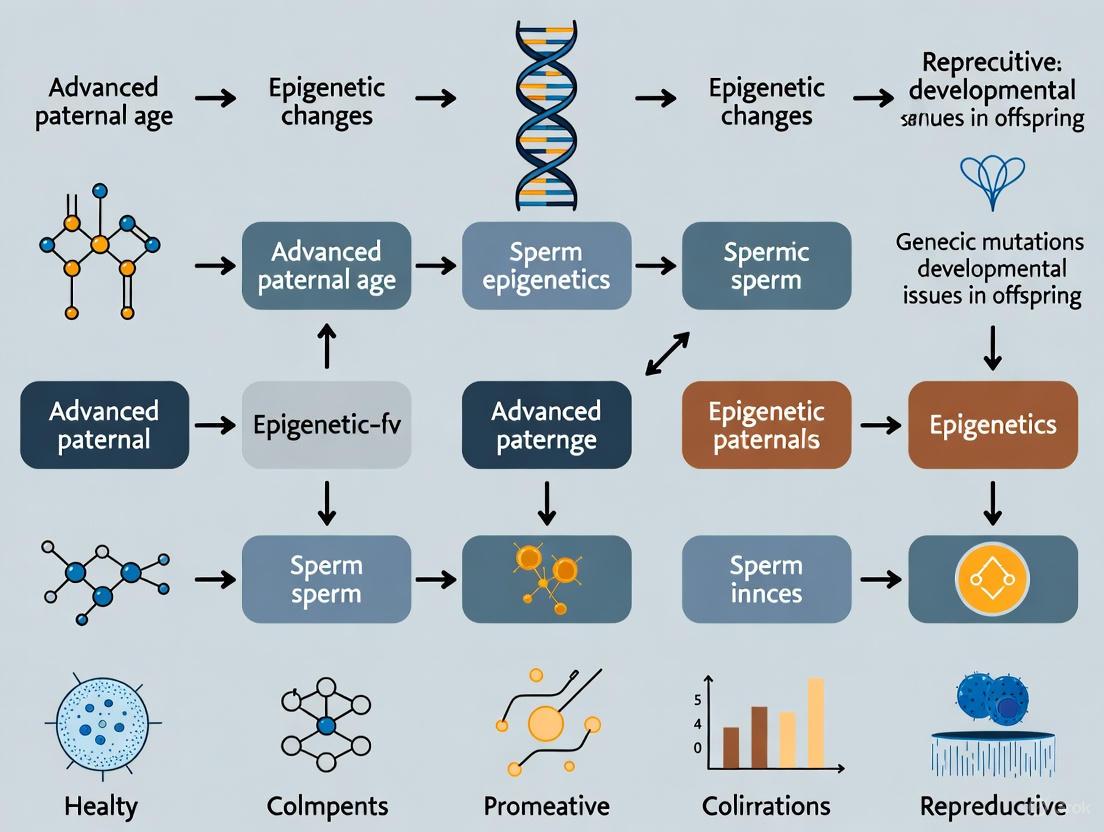
Diagram 2: Paternal Age to Offspring Development Pathway
The Scientist's Toolkit: Essential Research Reagents and Methodologies
Table 3: Essential Research Reagents for Sperm Epigenetics Studies
| Reagent Category | Specific Examples | Research Applications | Technical Considerations |
|---|---|---|---|
| Bisulfite Conversion Kits | EZ DNA Methylation kits (Zymo Research) | Convert unmethylated cytosines to uracils for methylation analysis | Optimization required for sperm DNA due to unique compaction |
| Methylation Arrays | Illumina Infinium MethylationEPIC | Genome-wide methylation profiling at ~850,000 CpG sites | Covers many regulatory regions relevant to development |
| Protamine Extraction Reagents | DTT-containing buffers, acid extraction protocols | Assess protamine ratios and histone retention | Protamine P1/P2 imbalance associated with DNA damage |
| Histone Modification Antibodies | H3K4me3, H3K27me3, H4ac, H3.3 | ChIP-seq, immunofluorescence for histone characterization | Specificity validation critical for sperm applications |
| RNA Isolation Systems | miRNeasy kits (Qiagen) with DNase treatment | Simultaneous extraction of large and small RNAs | Must effectively disrupt protamine-bound chromatin |
| Library Prep Kits | SMARTer smRNA-seq, TruSeq RNA library kits | Preparation of sequencing libraries for ncRNA profiling | Different protocols required for different RNA classes |
| Sperm Separation Media | Isolate Sperm Separation Medium (Irvine Scientific) | Density gradient purification of motile sperm | Minimizes somatic cell contamination for epigenetic analyses |
Emerging Technical Approaches
Multi-omics Integration: Combined analysis of DNA methylation, histone modifications, and ncRNA expression from the same sample provides comprehensive epigenetic profiling.
Single-Cell Epigenetic Technologies: Methods like scATAC-seq and scChIP-seq enable resolution of epigenetic heterogeneity within sperm populations.
Epigenome Editing: CRISPR-based systems targeting DNA methylation or histone modifications allow functional validation of specific epigenetic marks.
Computational Prediction Models: Sperm epigenetic clocks derived from machine learning algorithms on methylation data can predict biological age and potential reproductive outcomes [3].
The core epigenetic pillars in sperm—DNA methylation, histone modifications, and non-coding RNAs—represent dynamic, interconnected systems that undergo significant alterations with advanced paternal age. These changes extend beyond mere associations with reduced fertility, potentially contributing to increased risks of neurodevelopmental disorders in offspring through transmitted epigenetic information.
Future research directions should prioritize longitudinal studies tracking epigenetic changes in individual men over time, developing standardized protocols for clinical sperm epigenetic assessment, and exploring intervention strategies to mitigate age-related epigenetic deterioration. The development of diagnostic tools based on sperm epigenetic signatures offers promising approaches for personalized risk assessment and reproductive counseling for couples with advanced paternal age.
As delayed parenthood continues to be a societal trend, understanding these fundamental epigenetic mechanisms and their age-related alterations becomes increasingly crucial for addressing associated challenges in reproductive medicine and offspring health.
Aging precipitates a profound remodeling of the sperm epigenome, characterized by a dual phenomenon of widespread hypermethylation juxtaposed with highly specific locus-specific hypomethylation. This epigenetic erosion, a consequence of accumulating stochastic errors and potential environmental exposures, is now recognized as a significant contributor to the declining reproductive fitness of older males and an elevated risk of neurodevelopmental disorders in their offspring. This whitepaper synthesizes current research to delineate the precise patterns of these methylation shifts, detail the experimental methodologies for their identification, and discuss the implications for embryonic development and transgenerational health. Framed within the broader context of advanced paternal age research, we posit that sperm DNA methylation signatures serve as a molecular clock and a potential biomarker for assessing reproductive and offspring health risks.
The global trend toward delayed parenthood has intensified focus on the consequences of advanced paternal age. While the maternal age effect on offspring aneuploidy is well-established, the influence of older fathers on child health, particularly the risk for complex neurodevelopmental disorders such as autism spectrum disorder (ASD) and schizophrenia, is an area of growing concern [9]. The mechanism underlying this increased risk extends beyond the well-documented increase in de novo genetic mutations. The sperm epigenome, a comprehensive landscape of DNA methylation, histone modifications, and non-coding RNAs, is particularly vulnerable to the aging process.
DNA methylation, the addition of a methyl group to cytosine bases in a CpG context, is a fundamental epigenetic mark crucial for cellular regulation. In sperm, this methylation pattern is uniquely organized to facilitate both gamete function and early embryonic programming. However, the male germline, with its continuous spermatogonial stem cell divisions throughout life, is susceptible to the accumulation of epigenetic errors. The error rate for copying epigenetic marks is estimated to be 10 to 100-fold higher than for genetic information, making epimutations a major consequence of aging [10]. This whitepaper explores the specific patterns of these age-associated epigenetic changes, focusing on the paradoxical co-occurrence of global and locus-specific methylation alterations, and their demonstrated links to offspring health.
Global Patterns of DNA Methylation Changes with Age
High-resolution methylome analyses from multiple independent studies have consistently revealed that aging in men is associated with two dominant, simultaneous trends in sperm DNA methylation: a tendency toward global hypermethylation and the emergence of specific locus-specific hypomethylation.
Evidence for Global Hypermethylation
Longitudinal studies, which track the same individuals over time, provide the most robust evidence for age-related epigenetic change. One such study using whole-genome bisulfite sequencing (WGBS) of sperm samples collected 10 to 18 years apart from the same donors confirmed that while inter-individual variation is significant, age-dependent changes are detectable and consistent [11] [12]. Earlier paired-sample analyses using methylation arrays found significant global hypermethylation with age, as measured by the methylation of long interspersed elements (LINEs) [13]. This suggests a widespread, albeit subtle, increase in methylation levels across large genomic regions as a hallmark of the aging sperm.
Prevalence of Locus-Specific Hypomethylation
Despite the trend of global gain, the most pronounced and reproducible age-associated differentially methylated regions (ageDMRs) are frequently hypomethylated. A recent study using reduced representation bisulfite sequencing (RRBS) on 73 sperm samples identified 1,565 regions significantly correlated with donor age. The direction of this association was highly skewed, with 1,162 (74%) being hypomethylated and only 403 (26%) hypermethylated with advancing age [4]. This indicates that specific genomic loci are particularly vulnerable to loss of methylation. These hypomethylated ageDMRs are not randomly distributed; they are preferentially located near transcription start sites (TSS), within exons and introns, suggesting a targeted effect on gene regulatory elements [4].
Table 1: Summary of Age-Associated DMRs from Key Studies
| Study (Citation) | Technique | Sample Size & Design | Total AgeDMRs | Hypomethylated (%) | Hypermethylated (%) | Key Genomic Features |
|---|---|---|---|---|---|---|
| Bernhardt et al. [4] | RRBS | 73 men (cross-sectional) | 1,565 | 1,162 (74%) | 403 (26%) | Hypo-DMRs near promoters; Hyper-DMRs in intergenic regions |
| Jenkins et al. [13] | Methylation Array | 17 men (longitudinal, 9-19 yr gap) | 147 | 139 (~95%) | 8 (~5%) | Associated with neuropsychiatric disorders |
| Global Effects Study [11] [12] | WGBS | 10 men (longitudinal, 10-18 yr gap) | Significant genome-wide change | Contrasting signatures by location | Contrasting signatures by location | Correlates with gene density, centromeres |
Genomic Distribution and Functional Enrichment of AgeDMRs
The non-random distribution of ageDMRs provides critical insights into their potential functional impact on offspring. Hypomethylated ageDMRs are significantly enriched in genes involved in embryonic and neuronal development [9] [4]. A meta-analysis of genome-wide studies found that the 241 genes with ageDMRs replicated in at least one study showed significant functional enrichments in 41 biological processes associated with development and the nervous system and in 10 cellular components associated with synapses and neurons [4]. This enrichment strongly supports the hypothesis that paternal age effects on the sperm methylome are a key mediator of the increased risk for neurodevelopmental disorders in offspring.
Furthermore, certain chromosomes appear to be more susceptible. Chromosome 19, for instance, shows a highly significant twofold enrichment of sperm ageDMRs, potentially related to its high gene density and CpG content [4]. A prime example of a specific, well-replicated locus is the BEGAIN (brain enriched guanylate kinase associated) gene. Its promoter region shows significant age-associated hypomethylation in sperm, a change that has also been detected in the cord blood of male offspring, illustrating the potential for intergenerational transmission [10].
Table 2: Key Replicated Genes with Sperm AgeDMRs and Associated Offspring Risks
| Gene Symbol | Function/Putative Role | Methylation Trend with APA | Replicated Associations |
|---|---|---|---|
| BEGAIN | Synaptic function, postsynaptic density structure | Hypomethylation | Paternal age, ASD susceptibility, hypomethylation in offspring cord blood [10] |
| GRM7 | Glutamate metabotropic receptor, neurodevelopment | Hypermethylation (in placenta) | Overlaps with APA-associated placental DMRs; linked to neurodevelopment [5] |
| EBF3 | Transcription factor, early B-cell factor | Hypermethylation (in placenta) | Overlaps with APA-associated placental DMRs; linked to neurodevelopment [5] |
| FOXG1 | Forkhead box transcription factor, forebrain development | Hypermethylation (in placenta) | Overlaps with APA-associated placental DMRs; linked to neurodevelopment [5] |
Methodologies for Profiling Sperm DNA Methylation
Accurate assessment of the sperm methylome requires sensitive and quantitative techniques. The choice of methodology depends on the research goals, balancing resolution, genome coverage, and cost.
Whole-Genome Bisulfite Sequencing (WGBS)
WGBS is the gold standard for comprehensive methylome analysis, providing single-base-pair resolution of methylation levels across the entire genome, including regions not covered by arrays like repetitive elements and centromeres [11] [12].
- Workflow: Genomic DNA is treated with sodium bisulfite, which converts unmethylated cytosines to uracils (read as thymines in sequencing), while methylated cytosines remain unchanged. The converted DNA is then subjected to high-throughput sequencing.
- Analysis: By mapping the sequencing reads to a reference genome and calculating the ratio of C-to-T conversions at each CpG site, a quantitative methylation level is determined.
- Advantage: As demonstrated in recent longitudinal studies, WGBS is powerful for discovering novel age-sensitive genomic regions and assessing global methylome variation and stability within and between individuals [12].
Reduced Representation Bisulfite Sequencing (RRBS)
RRBS offers a cost-effective alternative that enriches for CpG-dense regions, such as CpG islands and gene promoters, by using restriction enzymes (e.g., MspI) to digest genomic DNA [4].
- Workflow: DNA is digested, size-selected for fragments rich in CpGs, and then subjected to bisulfite treatment and sequencing.
- Advantage: It provides high coverage of functionally relevant regulatory regions at a lower cost than WGBS, making it suitable for larger cohort studies. The study by Bernhardt et al. [4] utilized RRBS to identify over 1,500 ageDMRs.
Bisulfite Pyrosequencing
For targeted, high-precision quantification of methylation at specific loci (e.g., for validating candidate genes like BEGAIN), bisulfite pyrosequencing is the method of choice [10].
- Workflow: The genomic region of interest is amplified by PCR from bisulfite-converted DNA. The PCR product is then sequenced using a pyrosequencer, which quantitatively incorporates nucleotides in real-time, allowing for precise measurement of the percentage of methylation at each CpG site in the amplicon.
- Advantage: It is highly quantitative, reproducible, and ideal for analyzing a small number of loci across many samples.
The following diagram illustrates the logical workflow for designing a study to investigate age-associated epigenetic erosion in sperm.
The Scientist's Toolkit: Key Research Reagents and Solutions
The following table details essential materials and reagents used in the featured experiments for studying sperm DNA methylation.
Table 3: Research Reagent Solutions for Sperm Methylation Analysis
| Reagent / Kit / Solution | Function / Application | Key Considerations |
|---|---|---|
| Sperm DNA Isolation Kit | Purification of high-quality, contaminant-free genomic DNA from sperm cells. | Must effectively remove protamines and ensure no somatic cell contamination. |
| Sodium Bisulfite Conversion Kit | Chemical treatment that deaminates unmethylated cytosines to uracils, enabling methylation detection. | High conversion efficiency (>99%) is critical; must minimize DNA degradation. |
| Illumina DNA MethylationEPIC BeadChip | Array-based interrogation of over 850,000 CpG sites across the genome. | Cost-effective for large cohorts; covers enhancers, gene bodies, and promoters. |
| WGBS Library Prep Kit | Preparation of sequencing libraries from bisulfite-converted DNA for whole-genome analysis. | Must be compatible with bisulfite-treated DNA, which is often fragmented. |
| RRBS Library Prep Kit | Preparation of sequencing libraries that enrich for CpG-rich genomic regions. | Relies on restriction enzyme digestion (e.g., MspI) for representation reduction. |
| Pyrosequencing Assay & Reagents | Targeted, quantitative analysis of methylation at specific, short genomic regions. | Requires prior PCR amplification and specific sequencing primers for the locus. |
| CpG Methyltransferase (M.SssI) | Positive control for methylation assays; used to fully methylate DNA in vitro. | Essential for establishing assay baselines and controls. |
Implications for Offspring Health and Development
The functional enrichment of ageDMRs in neurodevelopmental genes is not merely correlative. Epidemiological and mechanistic studies are increasingly linking these paternal epigenetic changes to concrete offspring outcomes.
- Neurodevelopmental Disorders: Advanced paternal age is a well-established risk factor for autism spectrum disorder (ASD) and schizophrenia [9] [13]. The finding that ageDMRs in sperm are overrepresented at genes like BEGAIN, which is also hypomethylated in the brain of children with ASD, provides a plausible mechanistic link [10]. Furthermore, a 2025 study found that approximately 7% of genes with age-associated methylation changes in the placenta overlapped with those altered in sperm of older men, including seven genes previously linked to ASD susceptibility [5].
- Perinatal Outcomes: Beyond neurodevelopment, paternal age has been independently associated with adverse birth outcomes. A large 2025 population-based cohort study found that neonates born to fathers aged 35-44 years had higher risks of preterm birth (PTB) and caesarean section compared to those with fathers aged 25-34, even after adjusting for maternal age and other confounders [14].
- Transgenerational Epigenetic Inheritance: Evidence from both animal models and human studies suggests that a portion of these sperm epigenetic marks can escape the widespread reprogramming that occurs after fertilization. Altered methylation patterns have been observed in the placenta and cord blood of offspring from older fathers, indicating that the sperm epigenome can influence the fetal epigenome and environment [5] [10]. The placenta, as a surrogate fetal tissue, shows that advanced paternal age impacts common epigenetic loci in both sperm and the feto-placental unit [5].
The phenomenon of age-associated epigenetic erosion in sperm—global hypermethylation coupled with functionally targeted hypomethylation—represents a significant and modifiable risk factor for impaired reproduction and offspring health. The consistency of findings across different methodologies and cohorts underscores the robustness of this paternal age effect. The development of sperm epigenetic clocks that accurately predict a man's chronological age further highlights the stability and progressive nature of these changes [9] [15].
Future research must focus on several key areas:
- Establishing Causality: Moving beyond correlation to definitively prove that specific sperm ageDMRs directly cause altered developmental trajectories and disease risk in offspring, using animal models and sophisticated in vitro assays.
- Interaction with Lifestyle and Environment: Elucidating how paternal factors such as diet, obesity, smoking, and exposure to endocrine-disrupting chemicals interact with and potentially accelerate age-related epigenetic erosion [16].
- Clinical Translation: Developing standardized epigenetic assays for use in clinical andrology and ART workflows to provide personalized risk assessments for couples with advanced paternal age. Preconception interventions aimed at mitigating these adverse epigenetic marks hold promise for improving reproductive and generational health [16] [15].
In conclusion, understanding the patterns, mechanisms, and consequences of the aging sperm epigenome is not just an academic pursuit but a pressing public health imperative in an era of increasingly delayed fatherhood.
Abstract Spermiogenesis involves a dramatic reorganization of the paternal genome, where most histones are replaced by protamines to achieve extreme nuclear compaction. The precise retention of a small subset of histones at specific genomic loci is, however, critical for embryogenesis. This review details the molecular mechanisms of histone-to-protamine transition and the functional consequences of its dysregulation. We examine how advanced paternal age and environmental exposures disrupt these epigenetic processes, leading to alterations in the sperm epigenome. These changes, including aberrant histone retention and DNA methylation, compromise paternal chromatin architecture, which in turn can impair zygotic genome activation, embryonic development, and the long-term health of the offspring. The discussion is framed within the context of developing diagnostic and therapeutic strategies for male-factor infertility.
Following fertilization, the sperm contributes more than just a haploid genome to the embryo. It delivers a highly specialized and compacted nucleus, a centriole, and oocyte-activating factors [17]. Crucially, it also carries a unique epigenetic landscape that interacts with ooplasmic factors to guide embryonic development. The sperm epigenome is characterized by its extensive packaging by protamines, yet between 1% (in mice) and 15% (in humans) of the genome remains associated with histones [17] [18]. These retained histones are not randomly distributed; they are strategically positioned at promoters and enhancers of genes critical for development, such as those governing transcription regulation, embryonic patterning, and metabolism [18]. This structured retention suggests a role in bookmarking developmental genes for activation in the early embryo.
The proper establishment of this epigenome—through the processes of histone replacement, protamine incorporation, and the setting of DNA methylation marks—is therefore paramount for embryonic competence. A growing body of evidence indicates that this process is susceptible to disruption. Advanced paternal age, dietary factors, and exposure to environmental toxins can alter the fidelity of histone retention and the sperm's broader epigenetic profile [17] [19] [4]. This review explores the intricate relationship between the molecular mechanisms of chromatin repackaging in sperm, the impact of paternal factors on its integrity, and the consequent effects on chromatin architecture and embryonic viability.
Molecular Mechanisms of Histone-to-Protamine Transition
The transformation of a round spermatid into a mature spermatozoon involves a radical re-organization of chromatin, facilitated by a tightly orchestrated sequence of events where histones are successively replaced by transition proteins and then by protamines.
The Role of Histone Variants and Modifications
The process is initiated in round and elongating spermatids with the incorporation of testis-specific histone variants, which create a more open and dynamic chromatin structure primed for replacement.
- Linker Histone Variants: H1T2 is essential for subsequent protamine incorporation and proper nuclear condensation; its mutation leads to male infertility [20]. HILS1 is another testis-specific linker histone highly expressed in elongating spermatids, though its precise function in mammals requires further elucidation [20].
- Core Histone Variants: The testis-specific H2A variant H2AL2 is critical for facilitating the invasion of transition proteins (TPs) into the nucleosome. H2AL2 incorporation destabilizes the nucleosome core, creating a flexible structure that allows TPs to bind and displace histones [18] [20]. The simultaneous presence of the H2B variant TH2B further contributes to this genome-wide chromatin transition [18].
- Histone Post-Translational Modifications: Histone hyperacetylation, particularly of H4, serves as a critical signal for histone eviction. The testis-specific bromodomain protein BRDT recognizes acetylated lysines on histone tails, initiating a "chromatin squeezing" process that facilitates the removal of histones and their replacement with transition proteins [18]. Other modifications, such as histone crotonylation, have also been implicated in a subsequent, BRDT-independent wave of histone removal [18].
The Involvement of Architectural Proteins
The protein CTCF, a key architect of 3D genome organization, plays a direct role in histone retention. Conditional depletion of CTCF before spermiogenesis leads to specific defects in histone H2B retention in mature sperm, indicating that CTCF helps define the genomic loci where nucleosomes are preserved [18]. These CTCF-bound regions, often co-localized with cohesin complexes, are frequently found at enhancers and super-enhancers, suggesting a mechanism for preserving regulatory information across generations [18].
Table 1: Key Histone Variants and Their Roles in Spermiogenesis
| Histone Variant | Stage of Expression | Primary Function | Phenotype of Knockout/Mutation |
|---|---|---|---|
| H1T2 | Round & elongating spermatids | Necessary for protamine incorporation and nuclear condensation [20] | Male infertility; delayed nuclear condensation, reduced protamine levels [20] |
| H2AL2 | Condensing spermatids | Facilitates transition protein invasion by assembling open nucleosomes [18] [20] | Genome-wide compaction defects in sperm [18] |
| TH2A / TH2B | Early primary spermatocytes | Contributes to open chromatin structure; regulates total histone levels and chromatin dynamics [20] | Double-knockout causes male infertility with impaired TP2 incorporation [20] |
The following diagram illustrates the sequential and coordinated workflow of the histone-to-protamine transition:
Diagram 1: The workflow of histone-to-protamine transition during spermiogenesis, showing the key replacement steps and the parallel pathway for specific histone retention.
Paternal Age and Disruption of the Sperm Epigenome
Advanced paternal age is a major factor compromising the integrity of the sperm epigenome. The effects are multifaceted, impacting both genetic and epigenetic integrity.
Sperm DNA Fragmentation and Integrity
With increasing age, the cumulative number of spermatogonial cell divisions rises, leading to a higher load of DNA damage. Men over 50 show increased rates of sperm DNA fragmentation, which can overwhelm the oocyte's repair capacity post-fertilization [17] [19]. This can result in embryonic defects, developmental arrest, and an increased risk of diseases in offspring, including tumors, behavioral abnormalities, and shortened lifespan in mouse models [17]. The effect of sperm DNA damage on the embryo is quantitative and variable, dependent on the interplay between the extent of damage and the oocyte's repair capability [17].
Age-Related Epigenetic Alterations
Perhaps more strikingly, the epigenetic copying process during spermatogenesis is error-prone, and these errors accumulate with age.
- DNA Methylation Changes: Genome-wide studies reveal that advanced paternal age is associated with significant changes in sperm DNA methylation. A 2023 RRBS study identified 1,565 age-related differentially methylated regions (ageDMRs) in human sperm, with a strong skew (74%) towards hypomethylation [4]. These ageDMRs were often located within genic regions, and hypomethylated ageDMRs were notably closer to transcription start sites. Functional enrichment analysis of genes with replicable ageDMRs points to their involvement in biological processes tied to development and the nervous system, providing a plausible mechanistic link between advanced paternal age and increased offspring risk for neurodevelopmental disorders like autism and schizophrenia [4] [21].
- Altered Histone Retention and Modifications: While the search results focus on DNA methylation, the processes of histone retention and the histone-to-protamine transition are also vulnerable. Recent research using natural aging mouse models, presented at ASRM 2025, reinforces that male aging directly affects sperm epigenetics, including modifications that can impact offspring health and neurodevelopment [22]. The proper establishment of histone marks and their retention is an active process that can be perturbed over time.
Table 2: Impact of Advanced Paternal Age on Sperm and Offspring Health
| Parameter | Change with Advanced Age | Potential Consequence for Embryo/Offspring |
|---|---|---|
| Semen Volume | Decreases [19] | Reduced fertility potential |
| Sperm Motility | Decreases [19] | Reduced fertility potential |
| Sperm DNA Fragmentation | Increases [17] [19] | Developmental arrest, mosaic aneuploidy, increased disease risk in offspring [17] |
| Sperm DNA Methylation | Widespread hypomethylation, some hypermethylation at specific loci [4] | Altered gene expression in embryo, increased risk of neurodevelopmental disorders [21] [4] |
| De Novo Mutations | Increases [19] | Increased risk of monogenic disorders |
Impacts on Embryonic Chromatin Architecture and Developmental Competence
The altered paternal epigenome delivered at fertilization has direct consequences for the events that follow, particularly the remodeling of the paternal pronucleus and the initiation of zygotic genome activation (ZGA).
Chromatin Reprogramming and Totipotency
After fertilization, the highly compacted sperm genome must be rapidly reprogrammed into a functional nucleus within the zygote. The oocyte cytoplasm contains factors that actively decondense the paternal chromatin, removing protamines and repackaging the DNA with histones of maternal origin. The correct retention of paternal histones at specific loci is thought to serve as a "bookmarking" mechanism, potentially helping to guide this repackaging and the subsequent activation of the embryonic genome [18] [23]. The establishment of an open, decondensed chromatin architecture is a hallmark of totipotent cells, such as those in the early embryo and pluripotent embryonic stem cells (ESCs) [24]. This open structure is believed to be permissive for the broad gene expression potential required for totipotency.
Consequences of Paternal Epigenetic Errors
When the sperm delivers an epigenome with aberrant histone retention or DNA methylation, it can disrupt this carefully orchestrated reprogramming.
- Impaired Zygotic Genome Activation (ZGA): Mis-bookmarked genes may fail to be properly activated during ZGA, a critical transition for embryonic development. The aberrant expression of endogenous retroviral elements, which are normally activated during ZGA, has been linked to developmental defects [23].
- Faulty Chromatin Architecture: Errors in the paternal epigenetic blueprint can lead to the failure to establish correct 3D chromatin structures in the early embryo. Since chromatin organization in the nucleus is tightly linked to gene regulation, this can have cascading effects on lineage specification and embryonic viability [23] [25]. Studies in ESCs show that depletion of architectural proteins like CTCF leads to a disruption of chromatin domains and can upregulate transcriptional programs associated with totipotency, highlighting the delicate balance required for proper genome folding and cell fate determination [23].
Research Methods and Experimental Toolkit
Investigating the mechanisms of histone retention and their functional outcomes requires a multidisciplinary approach. Below is a summary of key methodologies and reagents central to this field.
Table 3: The Scientist's Toolkit: Key Reagents and Methods for Sperm Epigenetics Research
| Tool / Reagent | Function / Application | Key Detail / Consideration |
|---|---|---|
| Chromatin Immunoprecipitation (ChIP) | Maps genome-wide localization of retained histones, specific histone modifications (e.g., H4 acetylation), and architectural proteins like CTCF in sperm [18]. | Requires specialized protocols for sperm chromatin, which is highly cross-linked and resistant to fragmentation. |
| Reduced Representation Bisulfite Sequencing (RRBS) / WGBS | Profiles genome-wide DNA methylation patterns in sperm [4]. | Identifies age-related or exposure-related differentially methylated regions (DMRs). |
| Hi-C and related techniques | Captures 3D chromatin architecture and interactions in nuclei [25]. | Can be applied to pronuclei or early embryos to study paternal chromatin reorganization. |
| Electron Spectroscopic Imaging (ESI) | Directly visualizes global chromatin ultrastructure and compaction levels [24]. | Used to show open chromatin in pluripotent embryonic cells versus compacted chromatin in differentiated cells. |
| Conditional Knockout Mouse Models | Enables gene deletion (e.g., Ctcf, H1t2) at specific stages of spermatogenesis to study gene function [18] [20]. | Essential for establishing causality in histone retention and protamination processes. |
| BRDT Inhibitors | Chemical probes that disrupt bromodomain function, used to study the role of histone acetylation in histone eviction [18]. | Useful for experimentally inducing errors in the histone-to-protamine transition. |
The following diagram outlines a generalized experimental workflow for investigating paternal epigenetic effects on embryonic development:
Diagram 2: A generalized experimental workflow for studying the impact of paternal factors on the sperm epigenome and intergenerational health outcomes.
The precise regulation of histone retention and protamination is a cornerstone of paternal epigenetic inheritance. The data compellingly show that this process is not merely structural but is fundamentally informational, encoding a blueprint that helps guide early embryonic development. The vulnerability of this process to advanced paternal age, as evidenced by accumulating DNA fragmentation and stochastic epigenetic errors, represents a significant and underappreciated factor in reproductive health and offspring outcomes.
Future research must focus on elucidating the precise mechanisms that target histone retention to specific genomic loci and how these retained marks survive the global epigenetic reprogramming that occurs after fertilization. From a clinical perspective, the development of sensitive diagnostic assays that profile the sperm epigenome—including histone retention maps and methylation signatures—holds promise for better predicting IVF outcomes and understanding idiopathic infertility. Furthermore, understanding these mechanisms opens the potential for therapeutic interventions, whether through lifestyle modifications or pharmacological agents, to mitigate the adverse effects of advanced paternal age and improve embryonic competence.
Advanced paternal age (APA) is a well-established risk factor for neurodevelopmental disorders (NDDs) such as autism spectrum disorder (ASD) and schizophrenia in offspring. While de novo genetic mutations have historically been the primary explanation, emerging research underscores the significant role of epigenetic alterations in the paternal germline. This whitepaper synthesizes current evidence on how age-related changes in the sperm epigenome, particularly through the dysregulation of the transcriptional repressor REST/NRSF (Neuron-Restrictive Silencer Factor), contribute to aberrant neurodevelopment. We detail the molecular mechanisms, present consolidated quantitative data from key studies, describe critical experimental protocols for investigating this pathway, and provide a toolkit of essential research reagents. Understanding these epigenetic pathways opens new avenues for therapeutic intervention and risk assessment in families with older fathers.
The trend towards delayed parenthood has increased steadily over recent decades for economic, social, and cultural reasons, elevating concern about the impacts of advanced parental age on offspring health [4]. While the maternal age effect has been long recognized, compelling epidemiological data now robustly link advanced paternal age (APA) to an increased incidence of complex neurodevelopmental disorders in offspring, including autism spectrum disorder (ASD) and schizophrenia [26] [27]. For instance, an increase of 10 years in paternal age is associated with a 21% higher risk of ASD [26].
The traditional explanation for this "paternal age effect" centered on the accumulation of de novo genetic mutations in male germ cells, which undergo more cumulative cell divisions over a man's lifetime [27]. However, the error rate for copying epigenetic information is at least an order of magnitude higher than for genetic information [4]. Consequently, the sperm epigenome of older males is endowed with many more epigenetic than DNA sequence changes. This whitepaper explores the paradigm that age-related epigenetic alterations in sperm are a key mechanistic link between APA and offspring NDD risk, with a particular focus on the central role of the transcriptional regulator REST/NRSF.
Core Mechanistic Pathways
The Central Role of REST/NRSF Dysregulation
REST/NRSF is a master transcriptional repressor of neuronal genes in non-neuronal tissues and neural progenitor cells. It maintains cellular pluripotency and prevents premature differentiation by repressing a vast network of neuronal genes—in silico studies suggest it may regulate thousands of targets [28]. It binds to a conserved 21-bp DNA sequence known as the Neuron Restrictive Silencer Element (NRSE) or RE-1 and recruits chromatin-modifying complexes containing co-repressors like mSin3 and CoREST, which in turn recruit histone deacetylases (HDACs) and histone methyltransferases to enforce a repressive chromatin state [28].
Recent evidence from both murine and human studies positions REST/NRSF at the intersection of paternal aging and neurodevelopmental risk. A pivotal study in mice demonstrated that sperm from aged fathers exhibits DNA hypomethylation specifically at genomic regions enriched for REST/NRSF binding motifs. Consequently, offspring of aged fathers showed upregulation of REST/NRSF target genes in the embryonic forebrain and exhibited abnormal vocal communication, a model of social communication deficits seen in NDDs. Crucially, administering a DNA demethylating drug to young males phenocopied this offspring phenotype, directly linking paternal sperm hypomethylation to the effect [29].
The regulation of REST/NRSF itself is complex. Another study identified that EHMT1, a histone methyltransferase implicated in Kleefstra syndrome, regulates REST/NRSF protein levels indirectly by repressing a set of microRNAs (including miR-153, miR-26a, and miR-142). When EHMT1 function is reduced, these miRNAs are upregulated, leading to suppressed translation of NRSF/REST mRNA and premature neuronal differentiation in human iPSC models. This miRNA set is significantly enriched for association with Intellectual Disability (ID) and schizophrenia, revealing a broad molecular pathway connecting epigenetic regulation, miRNA-mediated gene control, and neurodevelopment [30] [31].
Table 1: Key Evidence Linking REST/NRSF to Paternal Age and Neurodevelopmental Risk
| Study Model | Key Finding Related to REST/NRSF | Functional Outcome in Offspring |
|---|---|---|
| Mouse Model [29] | Sperm from aged fathers showed hypomethylation at REST/NRSF binding motifs; Forebrains of embryos showed upregulation of REST/NRSF target genes. | Abnormal ultrasonic vocalization, modeling social communication deficits. |
| Human iPSC (Kleefstra Syndrome Model) [30] [31] | Reduced EHMT1 activity led to miRNA upregulation (miR-153, miR-26a, miR-142), which suppressed NRSF/REST protein translation. | Aberrant neuronal gene expression and premature neurodevelopment. |
| Human & Rat Model [26] | Differential regulation of miRNAs miR-132 and miR-134 (implicated in synaptic plasticity) was found in both APA humans and rats. | Social-communication deficits, repetitive behaviors, and higher anxiety. |
Broader Epigenetic Dysregulation in the Sperm Epigenome
Beyond the REST/NRSF pathway, APA is associated with widespread epigenetic changes in human sperm. A 2023 RRBS study of 73 sperm samples identified 1,565 age-related differentially methylated regions (ageDMRs). Notably, 74% of these ageDMRs were hypomethylated, and they were preferentially located near transcription start sites and within genic regions, suggesting a direct impact on gene regulation. In contrast, hypermethylated ageDMRs were more common in gene-distal regions [4]. A functional enrichment analysis of genes with replicable sperm ageDMRs revealed significant associations with biological processes related to development and the nervous system, and cellular components associated with synapses and neurons [4].
These age-related sperm methylation changes are not merely correlative. A 2024 study on human blastocysts from donor oocyte cycles (controlling for maternal age) found that offspring of APA fathers already showed significant methylome and transcriptome alterations at the blastocyst stage. The inner cell mass (ICM), which gives rise to the embryo proper, showed significant enrichment for differential methylation and expression in neuronal signaling pathways, providing a direct link from the sperm epigenome to the earliest embryonic stages [32].
The following diagram synthesizes the primary mechanistic pathway linking advanced paternal age to an increased risk of neurodevelopmental disorders in offspring, focusing on REST/NRSF dysregulation.
Quantitative Data Synthesis
The following tables consolidate key quantitative findings from recent studies on the paternal age effect, providing a clear overview for researchers.
Table 2: Sperm Methylome Changes with Advanced Paternal Age
| Metric | Findings | Study Details |
|---|---|---|
| Total AgeDMRs Identified | 1,565 significant ageDMRs (0.4% of 360,264 analyzed regions) [4]. | RRBS on 73 sperm samples from males attending a fertility center. |
| Direction of Change | 1,162 (74%) were hypomethylated; 403 (26%) were hypermethylated with age [4]. | |
| Genomic Location | Hypomethylated ageDMRs were closer to TSS (median 1,368 bp). Hypermethylated ageDMRs were more distal (median 17,205 bp) [4]. | |
| Functional Enrichment | 241 replicated ageDMR-associated genes showed enrichment for 41 biological processes related to development and the nervous system [4]. |
Table 3: Neurodevelopmental and Behavioral Outcomes in Offspring
| Model System | Observed Phenotype | Associated Molecular Changes |
|---|---|---|
| Human Offspring (Epidemiology) | Increased risk for Autism Spectrum Disorder and Schizophrenia [26] [27]. | Personality traits of schizotypy and neuroticism correlated with paternal age in healthy subjects (N=677) [26]. |
| Human Blastocysts (APA-derived) | N/A (Preimplantation stage) | Significant differential methylation and transcription in Inner Cell Mass (ICM); enrichment for neuronal signaling pathways [32]. |
| Rat Model (APA) | Social-communication deficits, fewer pro-social ultrasonic vocalizations, repetitive behaviors, higher anxiety [26]. | Differential regulation of miR-132 and miR-134 in both rats and humans; brain morphological changes in prefrontal and medial temporal cortex [26]. |
| Mouse Model (APA) | Abnormal vocal communication [29]. | Upregulation of REST/NRSF target genes in the embryonic forebrain [29]. |
Detailed Experimental Protocols
To facilitate replication and further research, this section outlines detailed methodologies from key studies cited in this whitepaper.
Protocol: Establishing a Murine Paternal-Aging Model and Analyzing Offspring
This protocol is adapted from the 2021 study that identified the role of REST/NRSF hypo-methylation [29].
Animal Model Generation:
- Group Definition: Define "aged" and "young" fathers (e.g., 12-month-old vs. 3-month-old mice).
- Mating: Mate aged and young males with young, reproductively mature females to control for maternal age and confounders.
- Offspring Phenotyping: Assess offspring behavior using standardized tests. A key assay is the recording and analysis of ultrasonic vocalizations (USV) in pups separated from their mothers to model social communication.
Molecular Analysis of Sperm:
- Sperm Collection: Collect sperm from the cauda epididymis of aged and young males.
- DNA Extraction & Methylome Analysis: Extract genomic DNA and perform Whole-Genome Bisulfite Sequencing (WGBS) or reduced representation bisulfite sequencing (RRBS). Align sequences to a reference genome and call methylation status for all CpGs.
- Bioinformatic Analysis: Identify differentially methylated regions (DMRs) between age groups. Perform motif enrichment analysis on the DMRs to test for significant enrichment of the REST/NRSF binding motif.
Molecular Analysis of Offspring Brain:
- Tissue Collection: Dissect embryonic or postnatal forebrain tissues from offspring.
- Gene Expression Analysis: Extract RNA and perform RNA-Sequencing (RNA-Seq). Alternatively, use qRT-PCR to validate specific gene targets.
- Data Integration: Cross-reference the list of dysregulated genes in the offspring brain with known REST/NRSF target genes using existing databases (e.g., ChIP-Seq data).
Pharmacological Intervention:
- To establish causality, treat young male mice with a DNA methyltransferase inhibitor (e.g., 5-azacytidine) prior to mating.
- Assess whether the offspring of drug-treated young fathers phenocopy the behavioral and molecular phenotypes observed in the offspring of aged fathers.
Protocol: Analyzing Sperm and Blastocyst Methylation in Humans
This protocol is based on studies that identified ageDMRs in human sperm and linked them to early embryonic changes [4] [32] [33].
Sample Collection and Preparation:
- Sperm Samples: Obtain human sperm samples from fertility clinics or sperm banks with detailed donor information (age, BMI, semen parameters). Select samples to control for confounding factors (e.g., normozoospermic samples).
- Blastocyst Samples: Use surplus, high-quality cryopreserved blastocysts from donor oocyte IVF cycles to control for maternal age and oocyte quality. Mechanically separate blastocysts into Inner Cell Mass (ICM) and Trophectoderm (TE) samples.
DNA Extraction and Methylation Sequencing:
- Extract DNA from sperm and from micro-dissected ICM/TE samples. For low-input blastocyst samples, use an ultra-low input WGBS protocol (e.g., Zymo Research's Pico Methyl-Seq kit or a scSPLAT-based method) [32].
- Perform Whole Genome Bisulfite Sequencing (WGBS) on an Illumina platform (e.g., NovaSeq 6000) to achieve genome-wide, base-resolution methylation data.
Bioinformatic Processing and Analysis:
- Quality Control & Trimming: Use tools like TrimGalore and FastQC to assess data quality and trim adapters.
- Alignment: Align bisulfite-treated reads to the human reference genome (e.g., hg19/GRCh38) using a dedicated aligner like Bismark.
- Methylation Calling: Use Bismark or Methyldackel to generate methylation call files, calculating the fraction of methylated CpGs at each site.
- Differential Methylation Analysis: Identify DMRs using software packages like DSS or
minfi. Statistical significance is typically set at FDR ≤ 0.05 with an absolute methylation difference ≥ 10%. - Functional Enrichment: Annotate DMRs to genomic features (promoters, exons, etc.) and perform gene ontology (GO) and pathway enrichment analysis using tools like
clusterProfiler.
The workflow for this integrated epigenetic analysis, from sample collection to data interpretation, is outlined below.
The Scientist's Toolkit: Research Reagent Solutions
Table 4: Essential Reagents and Resources for Investigating APA and REST/NRSF Pathways
| Reagent / Resource | Specifications & Function | Example Application |
|---|---|---|
| Illumina MethylationEPIC BeadChip | Covers > 850,000 CpG sites; cost-effective for large cohort screening. | Initial discovery of ageDMRs in human sperm cohorts [33]. |
| Whole Genome Bisulfite Sequencing (WGBS) | Provides single-base resolution, genome-wide methylation data. Gold standard. | Ultra-low input WGBS on micro-dissected human blastocysts [32]. |
| Bismark Software | A dedicated aligner and methylation caller for bisulfite-seq data. | Standard for mapping sequencing reads and quantifying methylation in WGBS/RRBS studies [4] [32]. |
| Anti-NRSF/REST Antibody | For Chromatin Immunoprecipitation (ChIP). e.g., Rabbit anti-NRSF (Santa Cruz; sc-25398). | Determining NRSF binding to chromatin in neuronal cultures or tissue [34]. |
| DSS R Package | Statistical software for finding DMRs from WGBS or RRBS data. | Used with Bioconductor for DMR detection in sperm and blastocyst studies [32]. |
| EHMT1 Mutant iPSCs | Patient-derived or CRISPR/Cas9-generated iPSCs with haploinsufficiency of EHMT1. | Modeling Kleefstra syndrome to study the EHMT1-miRNA-REST pathway in human neurons [30]. |
| miRNA Inhibitors & Mimics | Synthetic oligonucleotides to knock down or overexpress specific miRNAs (e.g., miR-153). | Functional validation of miRNA-mediated suppression of REST/NRSF translation [30]. |
The evidence is compelling that epigenetic changes in sperm, particularly those affecting the REST/NRSF regulatory network and other neurodevelopmental genes, constitute a significant and plausible mechanism underlying the increased risk of NDDs in the offspring of older fathers. The REST/NRSF pathway acts as a critical node, integrating signals from paternal age-related sperm hypomethylation, regulatory miRNAs, and chromatin modifiers like EHMT1 to orchestrate neuronal gene expression programs.
Future research must focus on several key areas:
- Mechanistic Resolution: Further elucidate how exactly sperm DNA hypomethylation at REST motifs leads to its dysregulation in the developing embryo.
- Transgenerational Persistence: While one study suggested age-associated sperm methylation patterns are largely reset and not directly inherited trans-generationally [33], other epidemiological data suggest transgenerational effects [27]. This discrepancy requires resolution with larger, well-controlled studies.
- Therapeutic Translation: Explore whether these epigenetic marks can be modified or if the downstream pathways they disrupt (e.g., with small molecules that disrupt NRSF's interaction with chromatin remodelers [28]) offer viable therapeutic targets.
By moving beyond a purely genetic model of the paternal age effect, this epigenetic paradigm offers a more dynamic and potentially reversible framework for understanding and mitigating the associated risks for the next generation.
Accumulation of De Novo Mutations and Clonal Selection in the Aging Testis
Advanced paternal age is increasingly recognized as a significant factor impacting male reproductive health and offspring outcomes. This technical review examines the molecular mechanisms driving the accumulation of de novo mutations and clonal selection processes within the aging testis. We synthesize current evidence demonstrating how oxidative stress, impaired DNA repair, and selfish selection of spermatogonial stem cells (SSCs) with mutations in specific signaling pathways collectively contribute to a phenomenon known as the "paternal age effect" (PAE). The progressive nature of these changes creates distinct genomic and epigenomic landscapes in sperm from older males, with profound implications for transgenerational inheritance. Understanding these mechanisms is critical for developing targeted interventions to mitigate age-related deterioration of male reproductive function and associated risks to offspring health.
The demographic shift toward delayed parenthood has intensified research focus on paternal aging consequences. While female reproductive aging has been extensively studied, understanding of male reproductive aging remains less developed. Over recent decades, the average age of fatherhood has increased significantly, reaching approximately 30.9 years in developed countries [35]. This trend is concerning given that advanced paternal age (≥40 years) constitutes a substantial risk factor for impaired fertility and offspring disorders through mechanisms fundamentally different from maternal age effects [35].
The continuous nature of spermatogenesis throughout postnatal life in males presents a unique biological context for mutation accumulation. Unlike female germ cells, which are largely arrested in meiotic prophase I from birth, male germ cells undergo continuous divisions, with spermatogonial stem cells (SSCs) dividing every approximately 16 days in humans [36]. This persistent replicative activity, combined with cumulative exposure to environmental stressors over a lifetime, creates ideal conditions for the introduction and propagation of genetic alterations in the male germline.
This review examines the current state of knowledge regarding de novo mutation accumulation and clonal selection dynamics in the aging testis, with particular emphasis on: (1) molecular mechanisms underlying mutation initiation and propagation; (2) experimental approaches for detecting and characterizing mutant clones; and (3) potential therapeutic strategies targeting age-related testicular dysfunction.
Molecular Mechanisms of Mutation Accumulation
Oxidative Stress and DNA Damage
Aging testes exhibit a state of persistent oxidative stress resulting from the cumulative effects of environmental exposures and intrinsic metabolic processes [35]. Reactive oxygen species (ROS) progressively damage cellular components, including lipids, proteins, and DNA. In germ cells, oxidative DNA damage manifests primarily as single- and double-strand breaks, base modifications, and abasic sites. Although multiple DNA repair pathways operate in germ cells, their efficiency declines with age due to both reduced expression of repair enzymes and potential saturation of repair capacity [35].
Table 1: Age-Related Changes in Testicular Environment and Germ Cells
| Parameter | Change with Aging | Functional Consequences |
|---|---|---|
| Oxidative Stress | Increased ROS production | Elevated DNA damage, lipid peroxidation, protein damage |
| DNA Repair Capacity | Impaired repair pathways | Accumulation of unrepaired lesions, increased mutation load |
| Sertoli Cell Function | Damaged morphology, disrupted blood-testis barrier | Impaired support and protection of spermatogenic cells |
| Leydig Cell Function | Reduced numbers and steroidogenic capacity | Decreased testosterone production, altered spermatogenesis |
| Spermatogonial Stem Cells | Impaired proliferation, reduced numbers, nuclear fragmentation | Diminished spermatogenic efficiency, reduced sperm output |
The integrity of spermatogenesis depends heavily on the support functions of testicular somatic cells, which also deteriorate with age. Sertoli cells, which provide crucial structural and nutritional support for developing germ cells, exhibit damaged morphology and disrupted function in aged testes, including impairment of the blood-testis barrier [35]. Similarly, Leydig cells show functional decline, resulting in reduced testosterone production that further compromises the spermatogenic microenvironment [35].
Epigenetic Alterations
In addition to genetic changes, aging produces distinct epigenetic landscapes in male germ cells. These include alterations in DNA methylation patterns, histone modifications, and non-coding RNA expression profiles [35]. Age-related epigenetic changes modify gene expression in germ cells, affect the DNA damage response, and generate de novo epigenetic mutations that can be transmitted to sperm and potentially inherited by offspring [35]. Single-cell transcriptomic analyses of human testes across the reproductive lifespan have revealed that somatic cells exhibit stronger aging responses than germ cells, with specific waves of age-related changes emerging in peritubular cells in the 30s and functional alterations in steroid metabolism in Leydig cells by the 50s [37].
Clonal Selection in the Aging Testis
Selfish Spermatogonial Selection
The "selfish selection" model represents a paradigm for understanding how certain mutations become disproportionately prevalent in sperm from older men. This process begins when rare gain-of-function mutations occur spontaneously in genes encoding components of growth factor receptor-RAS signaling pathways, particularly fibroblast growth factor receptors (FGFR2, FGFR3), RAS oncogene homologs (HRAS, KRAS), and the tyrosine phosphatase PTPN11 [38]. These mutations confer a selective advantage to the affected SSCs, enabling them to proliferate and expand clonally within the testicular niche over time, outcompeting neighboring non-mutant stem cells [38] [36].
Table 2: Key Genes Frequently Mutated in Selfish Spermatogonial Selection
| Gene | Associated Offspring Disorders | Cancer Associations | Prevalence in Aged Testes |
|---|---|---|---|
| FGFR2 | Apert, Crouzon, Pfeiffer syndromes | Yes | Identified in multiple tubule clusters |
| FGFR3 | Achondroplasia, thanatophoric dysplasia | Yes | Frequent in immunopositive tubules |
| PTPN11 | Noonan syndrome | Yes | Detected in multiple studies |
| HRAS | Costello syndrome | Yes | Present in tubule clusters |
| KRAS | Noonan syndrome | Yes | Identified in aged testes |
This clonal expansion process, termed "selfish" because it benefits the stem cell at the potential expense of organismal fitness, results in the progressive accumulation of sperm carrying these pathogenic mutations in the ejaculate of aging men [36]. The same mutations that promote stem cell survival and expansion often cause severe developmental disorders when transmitted to offspring, including skeletal dysplasias, craniosynostosis syndromes, and cancer predisposition syndromes [38].
Testicular Open Niche and Clonal Dynamics
The testis provides a unique "open niche" environment for SSCs, lacking anatomical confinement and allowing extensive SSC migration and dynamic interactions with the microenvironment [39]. Recent single-cell RNA sequencing and lineage tracing studies in aged mouse testes have revealed that GFRα1+ SSCs maintain gene expression heterogeneity during aging and continue to exhibit accelerated proliferation and persistent motility in older mice [39]. However, a subset of SSCs characterized by low expression of Egr4 and Cops5 shows impaired differentiation capacity and fails to form spermatids [39].
Intriguingly, these non-sperm-forming SSC clones increase in proportion among total SSC clones and expand spatially within the testicular open niche in old mice, a phenomenon not observed in young animals [39]. The expansion of functionally compromised SSC clones may occupy limited niche space, reducing the availability of functional SSCs and potentially contributing to age-related declines in sperm production and genetic diversity [39].
Diagram 1: Clonal Dynamics in Aging Testis. The diagram illustrates the transition from normal spermatogenesis in young testes to the expansion of mutant and non-functional spermatogonial stem cell (SSC) clones in aged testes, leading to increased production of sperm carrying paternal age effect (PAE) mutations.
Experimental Approaches and Methodologies
Detection and Isolation of Mutant Clones
Advanced methodologies have been developed to identify and characterize mutant SSC clones in human testes. The following protocol represents the current state-of-the-art approach for visualizing the origins of selfish de novo mutations:
Protocol 1: Identification of Pathogenic Mutations in Human Seminiferous Tubules
Tissue Collection and Preparation: Obtain human testicular tissues from organ donors or patients undergoing orchectomy for coincidental pathologies unrelated to infertility or parenchymal malignancy. Fix tissues in formalin and embed in paraffin (FFPE) or use fresh-frozen specimens depending on downstream applications [38].
Immunohistochemical Screening: Perform immunohistochemical staining of testicular sections using antibodies against spermatogonial markers (MAGEA4, FGFR3) and downstream signaling effectors (phospho-AKT) to identify "immunopositive tubules" exhibiting increased numbers of positively-stained spermatogonia [38].
Laser-Capture Microdissection (LCM): Using laser-capture microdissection, isolate selected immunopositive tubules and adjacent normal-appearing tubules as controls. This approach preserves cellular context while enabling genetic analysis of specific tubular regions [38].
DNA Extraction and Whole Genome Amplification: Extract DNA from microdissected tubule segments and perform whole genome amplification to generate sufficient DNA material for sequencing applications, addressing challenges related to limited starting material [38].
Targeted Sequencing: Sequence coding regions of candidate genes (typically 100-150 genes associated with PAE disorders and cancer) using targeted enrichment technologies such as HaloPlex. This focused approach increases sequencing depth and sensitivity for detecting low-frequency mutations [38].
Variant Validation: Confirm putative mutations using dideoxy-sequencing of non-amplified DNA from corresponding tubules in adjacent sections. This critical step distinguishes true mutations from amplification artifacts [38].
Single-Cell Transcriptomic Profiling
Single-cell RNA sequencing (scRNA-seq) has emerged as a powerful tool for characterizing age-related changes in testicular cell populations:
Protocol 2: Single-Cell RNA Sequencing of Aging Testicular Cells
Tissue Dissociation: Prepare single-cell suspensions from human testicular tissues using enzymatic digestion (collagenase, trypsin) combined with mechanical dissociation. Preserve cell viability while achieving complete tissue dissociation [37] [40].
Cell Capture and Library Preparation: Capture individual cells using microfluidic platforms (e.g., 10x Genomics Chromium). Perform reverse transcription, cDNA amplification, and library construction according to established protocols [37].
Sequencing and Data Processing: Sequence libraries on appropriate platforms (Illumina). Process raw sequencing data through standard pipelines for alignment, quality control, and unique molecular identifier (UMI) counting [37].
Bioinformatic Analysis: Conduct unsupervised clustering, differential expression analysis, and trajectory inference using established algorithms (Seurat, Monocle). Identify aging-associated transcriptional changes across different cell types [37].
Integration with Clinical Metadata: Correlate transcriptional signatures with donor age, body mass index, and other relevant clinical parameters to identify potential modifiers of testicular aging [37] [40].
Diagram 2: Experimental Workflow for Identifying Selfish Mutations. The flowchart outlines the key steps in detecting and validating pathogenic mutations in human seminiferous tubules, from tissue collection through to final analysis and applications.
The Scientist's Toolkit: Essential Research Reagents
Table 3: Key Research Reagents for Studying Testicular Aging and Clonal Selection
| Reagent/Category | Specific Examples | Research Application | Technical Considerations |
|---|---|---|---|
| Spermatogonial Markers | MAGEA4, FGFR3, GFRα1, PLZF | Identification and isolation of spermatogonial populations | Antibody validation critical for specific applications |
| Signaling Pathway Markers | Phospho-AKT, phospho-ERK | Detection of activated signaling pathways in mutant clones | Requires careful fixation and staining optimization |
| Single-Cell RNA Seq Platforms | 10x Genomics Chromium, Fluidigm C1 | Transcriptomic profiling of testicular cell populations | Cell viability and quality crucial for successful outcomes |
| Lineage Tracing Systems | Cre-lox, Confetti reporters | Fate mapping of SSC clones in animal models | Specific promoters needed for spermatogonial targeting |
| Laser-Capture Microdissection | Arcturus XT, PALM MicroBeam | Isolation of specific tubule regions for genetic analysis | Specialized training required for optimal precision |
| Targeted Sequencing Panels | HaloPlex, SureSelect | Mutation detection in PAE genes | Custom design possible for research-specific goals |
Clinical Implications and Therapeutic Perspectives
Offspring Health Risks
The clinical implications of selfish spermatogonial selection are substantial, with advanced paternal age associated with increased risk of multiple disorders in offspring. These include specific monogenic conditions (Apert syndrome, achondroplasia, Noonan syndrome) resulting from PAE mutations in FGFR2, FGFR3, and PTPN11 genes [38]. Additionally, epidemiological studies demonstrate associations between advanced paternal age and complex neuropsychiatric disorders (autism spectrum disorder, schizophrenia), congenital heart defects, and skeletal abnormalities [41].
Recent population-based cohort studies further indicate that paternal age independently associates with adverse pregnancy outcomes, including preterm birth and caesarean section, with a J-shaped dose-response relationship observed for preterm birth risk [14]. The relative importance of paternal age in predicting these outcomes appears similar to, or in some cases even exceeds, that of maternal age [14].
Emerging Therapeutic Strategies
Current research is exploring several approaches to mitigate age-related testicular dysfunction:
Antioxidant Interventions: Combating oxidative stress through supplementation with vitamins C, D, and E, zinc, and selenium shows promise for improving sperm quality in older males [41]. These compounds function as free radical scavengers, reducing DNA damage and lipid peroxidation in germ cells.
Anti-Inflammatory Approaches: COX-2 inhibitors (e.g., NS398) have demonstrated potential to enhance testosterone synthesis and reduce inflammation in aged testes [41]. Given the prominent role of inflammatory processes in testicular aging, modulation of immune responses represents a promising therapeutic avenue.
Hormonal Modulation: While testosterone replacement therapy (TRT) improves some systemic aspects of hypogonadism, it does not address the fundamental spermatogenic defects in aged testes and may even suppress sperm production [41]. Alternative approaches targeting hypothalamic-pituitary signaling without inhibiting spermatogenesis are under investigation.
Combination Therapies: Integrated protocols combining hormonal interventions with antioxidant treatments may provide synergistic benefits, addressing multiple aspects of testicular aging simultaneously [41]. Personalized approaches based on individual patterns of age-related testicular dysfunction represent the future direction for clinical management.
The accumulation of de novo mutations and clonal selection processes in the aging testis represents a fundamental biological phenomenon with significant implications for male fertility and offspring health. The selfish selection of spermatogonial stem cells carrying mutations in growth factor receptor-RAS signaling pathways creates a progressive increase in the genetic burden transmitted to subsequent generations. Advanced experimental approaches, including laser-capture microdissection and single-cell transcriptomics, are providing unprecedented insights into the cellular and molecular dynamics underlying testicular aging. Future research directions should focus on elucidating the precise mechanisms connecting specific mutations to clonal expansion advantages, developing interventions to mitigate age-related germline deterioration, and establishing clinical guidelines for counseling older men pursuing paternity. As demographic trends continue toward delayed fatherhood, understanding and addressing the consequences of male reproductive aging becomes increasingly imperative for public health.
Profiling and Interpreting the Aged Sperm Epigenome: Methodological Approaches and Clinical Correlations
The age of fathers at conception is steadily rising in many countries, bringing increased research focus on the potential impacts of advanced paternal age on offspring health. Epidemiological studies have linked advanced paternal age to an elevated risk of neurodevelopmental disorders in offspring, such as autism spectrum disorder, suggesting that age-associated molecular changes in sperm may underlie these observations [5]. While traditional semen analysis parameters like volume and motility decline with age, the most significant functional changes may occur at the epigenetic level [42]. The sperm epigenome, comprising DNA methylation patterns, histone modifications, and chromatin organization, serves as a crucial template that can influence embryonic development and long-term offspring health [43].
Investigating these mechanisms requires advanced technological approaches capable of detecting subtle epigenetic alterations in limited biological material. Ultra-low input whole-genome bisulfite sequencing (WGBS) and single-cell epigenomic technologies have emerged as powerful tools for characterizing the molecular legacy of paternal aging, offering single-base resolution and cell-to-cell heterogeneity analysis [44]. These methods enable researchers to move beyond bulk tissue analysis to examine the epigenetic landscape of individual sperm cells and early embryos, providing unprecedented insights into how paternal age affects reproductive outcomes and intergenerational health.
Core Technological Foundations
Ultra-Low Input Whole-Genome Bisulfite Sequencing (WGBS)
Whole-genome bisulfite sequencing represents the gold standard for comprehensive DNA methylation analysis at single-base resolution across the entire genome. The fundamental principle relies on bisulfite conversion of DNA, wherein unmethylated cytosines are converted to uracils (and subsequently read as thymines during sequencing), while methylated cytosines remain unchanged [45]. This treatment creates sequence differences that allow for quantitative mapping of methylation patterns.
For the analysis of precious clinical samples like human sperm or early embryos, where material is often limited, ultra-low input variations of WGBS have been developed. These protocols overcome the challenges of minimal starting material through optimized bisulfite conversion and library preparation methods that maximize efficiency while maintaining representation [44]. Key innovations include:
- Optimized bisulfite conversion chemistry that reduces DNA degradation while ensuring complete conversion
- Methylated adapter sequences to preserve library molecules during amplification
- Reduced purification steps to minimize sample loss
- Whole-genome amplification strategies specifically validated for bisulfite-converted DNA
The resulting data provides a comprehensive methylation landscape, enabling identification of differentially methylated regions (DMRs) associated with paternal age or other clinical variables [45].
Single-Cell Epigenomic Technologies
Single-cell epigenomic technologies have revolutionized our understanding of cellular heterogeneity in complex tissues, including sperm populations and preimplantation embryos. While individual sperm cells present unique challenges due to their highly compacted chromatin structure, recent methodological breakthroughs have enabled robust epigenomic profiling at single-cell resolution [46].
For chromatin organization analysis, an enhanced single-cell Hi-C (scHi-C) protocol has been developed specifically for mammalian sperm. This approach addresses the technical challenge of poor chromatin accessibility in highly condensed sperm nuclei through treatment with dithiothreitol (DTT), urea, and heparin, which significantly improves enzyme accessibility while preserving biological relevance [46]. This optimization increased DNA contacts per cell by more than 50-fold compared to standard protocols, enabling high-resolution reconstruction of 3D genome structures from individual sperm cells.
Single-cell DNA methylome sequencing builds upon bisulfite conversion principles but incorporates cell-specific barcoding and amplification strategies to assign methylation patterns to individual cells [44]. Methods such as scBS-seq (single-cell bisulfite sequencing) and snmC-seq2 (single-nucleus methylcytosine sequencing) have been successfully applied to germ cells and embryos, revealing cell-to-cell heterogeneity in epigenetic patterns that bulk analyses would obscure.
Table 1: Comparison of Key Low-Input Epigenomic Technologies
| Technology | Resolution | Primary Application | Key Strengths | Technical Challenges |
|---|---|---|---|---|
| Ultra-low input WGBS | Single-base | Genome-wide DNA methylation profiling | Comprehensive coverage; Quantitative methylation levels | Input material requirements; Bisulfite-induced degradation |
| Single-cell BS-seq | Single-base, single-cell | Cellular heterogeneity of methylation | Identifies epigenetic heterogeneity; No bulk averaging effects | Sparse coverage per cell; Amplification biases |
| scHi-C | 1-100kb, single-cell | 3D chromatin architecture | Preserves spatial organization; Identifies structural variations | Data sparsity; Complex computational analysis |
| scATAC-seq | Single-nucleosome, single-cell | Chromatin accessibility | Maps open chromatin regions; Inference of regulatory elements | Limited to accessible regions; Sequence depth requirements |
Experimental Protocols and Workflows
Ultra-Low Input WGBS Wet-Lab Protocol
The successful implementation of ultra-low input WGBS requires meticulous attention to each step of the experimental workflow to maximize data quality while minimizing technical artifacts:
Sample Preparation and Bisulfite Conversion:
- Input DNA Preparation: Extract DNA using methods that minimize degradation. For very low inputs (10-100 cells), include carrier RNA to improve recovery.
- Bisulfite Conversion: Treat DNA with sodium bisulfite using optimized kits specifically designed for low inputs. Typical conditions: 95°C for 5-10 minutes (denaturation) followed by 50-60°C for 4-16 hours (conversion).
- Desalting and Purification: Remove bisulfite salts using spin columns or magnetic beads. Avoid ethanol precipitation which can lead to significant sample loss with low inputs.
Library Preparation and Sequencing:
- Adapter Ligation: Use methylated adapters to preserve sequence identity during amplification. Ligation time and temperature should be optimized for fragmented, single-stranded bisulfite-converted DNA.
- Library Amplification: Perform limited-cycle PCR (typically 10-15 cycles) using bisulfite-converted DNA-compatible polymerases.
- Library Quality Control: Assess library quality using High Sensitivity DNA chips on Bioanalyzer or TapeStation. Expect a broad smear from 200-600bp.
- Sequencing: Run on Illumina platforms with recommended read lengths of 100-150bp paired-end to adequately cover converted regions [45] [47].
Critical Considerations:
- Include unmethylated and methylated control DNA to assess conversion efficiency
- Process samples in batches with appropriate controls to minimize batch effects
- Use unique molecular identifiers (UMIs) to account for amplification biases
Single-Cell Sperm Epigenomics Protocol
The unique chromatin composition of sperm necessitates specialized protocols for single-cell analysis:
Sperm Processing and Decondensation:
- Sperm Lysis: Treat sperm with SDS-based lysis buffer to remove membranes and cytoplasmic components.
- Chromatin Decondensation: Incubate with optimized decondensation buffer containing DTT (10-50mM), urea (1-2M), and heparin (0.1-0.5mg/mL) for 30-60 minutes at room temperature [46].
- Nuclei Isolation: Purify nuclei using density gradient centrifugation or fluorescence-activated cell sorting (FACS).
Single-Cell Library Construction:
- Single-Cell Partitioning: Use microfluidics platforms (10X Genomics, Dolomite) or manual cell picking to isolate individual sperm nuclei.
- Tagmentation or Crosslinking: For scATAC-seq, use Tn5 transposase; for scHi-C, perform formaldehyde crosslinking followed by restriction enzyme digestion.
- Barcoding and Amplification: Incorporate cell-specific barcodes during library amplification to enable multiplexing.
- Library Purification: Size-select libraries to remove adapter dimers and large fragments [46] [44].
Quality Assessment:
- Verify library complexity using fragment analyzers
- Sequence pilot libraries to assess data quality before scaling up
- Include bulk samples as controls for technical validation
Visualization of Experimental Workflows
Ultra-Low Input WGBS Workflow
Figure 1: Ultra-Low Input WGBS Experimental Workflow
Single-Cell Sperm Epigenomics Workflow
Figure 2: Single-Cell Sperm Epigenomics Workflow
Key Research Findings and Data Synthesis
Impact of Advanced Paternal Age on Sperm Epigenetics
Advanced paternal age induces specific alterations to the sperm epigenome that can be systematically characterized using the technologies described above:
Table 2: Age-Associated Epigenetic Changes in Sperm
| Epigenetic Feature | Change with Advanced Age | Functional Consequences | Detection Method |
|---|---|---|---|
| DNA methylation | Hypomethylation at 65% of affected loci, including 8 imprinted regions | Altered gene expression in placenta and offspring; neurodevelopmental effects | WGBS, EPIC array [5] |
| Sperm chromatin structure | Increased DNA fragmentation index (DFI) | Reduced fertilization capacity; embryonic developmental defects | SCSA, COMET assay [42] |
| Chromatin organization | Preservation of A/B compartments but loss of TADs and chromatin loops | Altered nuclear architecture; potential impact on gene regulation | scHi-C [46] |
| Histone modifications | Altered H3K4me3 at developmental gene promoters | Dysregulation of embryonic developmental programs | ChIP-seq [43] |
| Sperm telomere length | Shortened telomeres | Associated with early pregnancy loss and chromosomal abnormalities | qPCR, TRF analysis [21] |
Recent research has revealed that approximately 7% of genes showing age-associated DNA methylation changes in placenta overlap with genes previously identified as having altered methylation in spermatozoa of older men [5]. Notably, seven genes common to both placenta and sperm have been implicated in susceptibility to autism spectrum disorder, providing a potential mechanistic link between advanced paternal age and neurodevelopmental outcomes in offspring.
Three-Dimensional Genome Architecture in Sperm
Single-cell Hi-C analysis of mammalian sperm has revealed fundamental differences in chromatin organization compared to somatic cells. While sperm genomes maintain chromosomal territories and A/B compartments similar to somatic cells, they notably lack topologically associating domains (TADs) and chromatin loops [46]. This distinct organizational principle may have implications for paternal epigenetic inheritance.
Reconstructed 3D genome structures from single sperm cells faithfully reproduce species-specific nuclear morphologies, with mouse sperm exhibiting distinct apical hooks and human sperm showing smooth oval shapes [46]. These structural analyses have revealed that sex chromosomes in sperm are exclusively located in the nuclear center, likely reflecting the post-meiotic sex chromatin (PMSC) compartment formed during meiosis.
The Scientist's Toolkit: Essential Research Reagents and Materials
Table 3: Key Research Reagent Solutions for Sperm Epigenomics
| Reagent/Category | Specific Examples | Function in Experimental Workflow |
|---|---|---|
| Bisulfite conversion kits | EZ DNA Methylation kits (Zymo Research), Epitect Bisulfite kits (Qiagen) | Convert unmethylated cytosines to uracils while preserving methylated cytosines |
| Chromatin decondensation reagents | DTT, Urea, Heparin | Disrupt sperm protamine-DNA complexes for enzyme accessibility [46] |
| Single-cell partitioning platforms | 10X Genomics Chromium, Dolomite Nadia | Isolate individual cells for single-cell analysis with barcoding |
| Library preparation kits | Illumina DNA Prep, Accel-NGS Methyl-Seq DNA Library Kit | Convert bisulfite-treated DNA into sequencing-ready libraries |
| Enzymes for chromatin analysis | Tn5 transposase (for ATAC-seq), MNase (for nucleosome mapping), Restriction enzymes (for Hi-C) | Fragment DNA or chromatin in specific patterns for epigenomic mapping |
| Methylation-specific controls | Unmethylated λ phage DNA, Methylated control DNA | Monitor bisulfite conversion efficiency and detect technical artifacts |
| Bioinformatics tools | Bismark, nf-core/methylseq, Seurat, Cooler | Process sequencing data, align reads, call methylation, analyze 3D structure [45] |
Bioinformatics Analysis Pipelines
WGBS Data Processing
The analysis of WGBS data requires specialized computational approaches to account for bisulfite conversion:
Primary Analysis Workflow:
- Quality Control: FastQC (v0.11.9) and MultiQC (v1.10.1) for assessing read quality and adapter contamination [45].
- Adapter Trimming: Trim Galore! (v0.6.7) with dual-purpose adapter removal and quality trimming.
- Alignment: Bismark (v0.23.1) using bowtie2 against in-silico bisulfite-converted reference genomes.
- Methylation Calling: Extract methylation metrics per cytosine using Bismark methylation extractor.
- Differential Methylation: metilene (v0.2.8) for DMR identification or MethylKit for single-site analysis.
For large-scale analyses, automated workflows like nf-core/methylseq (v2.4.0) provide containerized, reproducible pipelines that can be deployed on cloud platforms like Google Cloud using Google Batch for scalable computing [45].
Single-Cell Epigenomic Data Analysis
Single-cell data introduces additional computational challenges related to sparsity and heterogeneity:
Key Analytical Steps:
- Demultiplexing: CellRanger (10X) or custom scripts to assign reads to individual cells based on barcodes.
- Quality Filtering: Remove cells with low read counts, high mitochondrial content, or evidence of doublets.
- Normalization: SCTransform (Seurat) or cisTopic (for scATAC-seq) to account for technical variability.
- Dimension Reduction: PCA, t-SNE, or UMAP for visualization and clustering.
- Integration: Harmony or Seurat's CCA to combine multiple datasets.
- Annotation: Transfer labels from reference datasets or manual annotation based on marker features.
For single-cell Hi-C data, specialized tools like Cooler and Higashi process contact matrices and reconstruct 3D genome structures [46].
Applications in Advanced Paternal Age Research
The integration of ultra-low input WGBS and single-cell epigenomics has enabled significant advances in understanding how advanced paternal age impacts sperm quality and offspring health:
Identifying Paternal Age-Associated Epigenetic Marks: Studies comparing sperm from younger and older men have identified specific DNA methylation patterns associated with advanced paternal age. These altered methylation sites are enriched at genes involved in neurodevelopment, including GRM7, EBF3, and FOXG1 [5]. The placenta appears to be particularly sensitive to these paternally inherited epigenetic changes, serving as both a recorder and potential mediator of paternal age effects.
Linking Sperm Epigenetics to Embryonic Development: Research has demonstrated that sperm histone modifications, particularly H3K4me3, mark genes important for embryonic development [43]. These epigenetic marks are retained in mature sperm and may help establish transcriptional programs in the early embryo. Alterations to these marks in older men, whether through environmental exposures or age-related changes, could therefore directly impact embryonic gene expression and development.
Clinical Implications for Assisted Reproductive Technology: While sperm quality parameters (volume, motility) and DNA integrity (DFI) show clear decline with advancing paternal age, the relationship with ART outcomes is complex [42]. Some studies have found that paternal age does not significantly affect clinical pregnancy or live birth rates in ART cycles, suggesting that embryonic compensatory mechanisms or ART selection processes may mitigate some age-related effects. However, the long-term health consequences for offspring conceived from older fathers' sperm remain an area of active investigation.
These applications highlight the transformative potential of advanced sequencing technologies in elucidating the mechanistic links between paternal age, sperm epigenetics, and intergenerational health outcomes.
Correlating Sperm Epigenetic Marks with Blastocyst Transcriptomes and Lineage Differentiation
The trend toward delayed parenthood has increased over past decades, bringing heightened research focus on the implications of advanced paternal age (APA). While the effects of advanced maternal age have been long recognized, evidence now clearly establishes that APA is independently associated with adverse reproductive and offspring outcomes [48] [32]. These include increased risks for neurodevelopmental disorders such as autism spectrum disorder and schizophrenia, as well as subtle impaired neurocognitive outcomes during infancy and childhood [48] [32].
The underlying mechanisms extend beyond well-documented genetic mutations to encompass epigenetic alterations in sperm. The sperm epigenome serves as a critical template for embryo development, carrying information that can influence developmental trajectories and offspring health [43]. During each replication cycle in the continuously dividing male germline, epigenetic marks must be correctly copied to daughter cells, with an error rate at least one order of magnitude higher for epigenetic than for genetic information [48]. This review synthesizes current evidence linking age-associated epigenetic changes in sperm to transcriptional and epigenetic alterations in preimplantation embryos, with particular focus on the first lineage differentiation into inner cell mass (ICM) and trophectoderm (TE).
Sperm Epigenetic Alterations with Advanced Paternal Age
DNA Methylation Changes in Aging Sperm
Advanced paternal age correlates with predictable alterations in the sperm DNA methylome. A 2023 study using reduced representation bisulfite sequencing (RRBS) on 73 sperm samples from men attending a fertility center identified 1,565 regions (0.4% of analyzed regions) showing significant correlation between sperm methylation and donor age after FDR adjustment [48].
Table 1: Characteristics of Age-Related Differentially Methylated Regions (ageDMRs) in Human Sperm
| Characteristic | Finding | Implication |
|---|---|---|
| Direction of Change | 1,162 (74%) hypomethylated; 403 (26%) hypermethylated | Strong bias toward hypomethylation with advancing age |
| Genomic Distribution | 1,152 (74%) located within genic regions; 1,002 genes with symbols | Predominantly affects protein-coding regions of genome |
| Relation to TSS | Hypomethylated ageDMRs closer to transcription start sites | Potential for direct transcriptional regulation |
| Chromosomal Distribution | Chromosome 19 shows twofold enrichment | Non-random genomic distribution suggests functional significance |
The functional enrichment of genes affected by sperm ageDMRs is particularly revealing. Among 241 genes replicated across multiple studies, significant functional enrichments occur in 41 biological processes associated with development and the nervous system, and in 10 cellular components associated with synapses and neurons [48]. This supports the hypothesis that paternal age effects on the sperm methylome particularly affect offspring behavior and neurodevelopment.
Intergenerational Transmission Mechanisms
The sperm epigenome contains several types of information carriers that can transmit paternal environmental information to the next generation:
Histone Modifications: In mature human sperm, approximately 15% of histones are retained, with specific enrichment at developmental gene promoters [43]. Key modifications include H3K4me3, which localizes to promoters implicated in embryo development, and H3K4me2, found at promoters involved in spermatogenesis and cellular homeostasis [43].
DNA Methylation: This remains the most extensively studied epigenetic mark, with age-related changes preferentially affecting neurodevelopmental genes [48].
Non-coding RNAs: These represent another vector for epigenetic information transfer, though they were not a primary focus in the cited age studies.
The transmission of these epigenetic marks is facilitated by their ability to resist reprogramming after fertilization, allowing paternal epigenetic information to influence embryonic gene expression and development [43].
Embryonic Molecular Consequences of Paternal Epigenetic Inheritance
Methylome and Transcriptome Alterations in Early Blastocysts
A 2024 study provides direct evidence that paternal aging impacts embryonic molecular profiles as early as the blastocyst stage [32] [49]. Using donor oocyte IVF cycles to control for maternal age effects, researchers investigated the methylome and transcriptome of mechanically separated inner cell mass (ICM) and trophectoderm (TE) lineages from blastocysts sired by either advanced paternal age (≥50 years) or young fathers.
Table 2: Molecular Alterations in APA-Derived Blastocysts Compared to Young Father-Derived Blastocysts
| Molecular Profile | ICM Lineage Findings | TE Lineage Findings |
|---|---|---|
| Methylome (DMRs) | Significant enrichment for neuronal signaling pathways, neurodevelopmental disorders, and imprinted genes | Significant enrichment for neuronal signaling pathways, neurodevelopmental disorders, and imprinted genes |
| Transcriptome (DEGs) | Significant enrichment of neurodevelopmental signaling pathways | No significant signaling pathways or gene ontology terms identified |
| Functional Correlation | Direct link to neurodevelopmental disorders | Limited functional disruption at transcriptome level |
This study revealed that despite normal semen parameters in aged fathers, significant molecular alterations were present in both embryonic lineages, with the ICM showing more substantial transcriptome disruption [32]. This suggests the future fetus may be particularly vulnerable to APA effects, while initial implantation potential (mediated by TE) may remain intact.
Temporal Dynamics of Epigenetic Transmission
The journey of paternal epigenetic information through early development involves several critical stages:
Fertilization: Sperm delivers epigenetic marks including DNA methylation, histones with modifications, and non-coding RNAs to the oocyte [43].
Epigenetic Reprogramming: After fertilization, the paternal genome undergoes rapid active demethylation, though some regions escape this process [50].
Lineage Specification: By the blastocyst stage, differential epigenetic patterns emerge between ICM and TE lineages [50].
Transcriptional Impact: Epigenetic marks inherited from sperm influence embryonic gene expression, particularly at neurodevelopmental loci [32].
The following diagram illustrates the experimental workflow for investigating paternal age effects on embryonic development:
Technical Approaches and Methodological Considerations
Epigenomic Profiling Technologies
Advanced genomic technologies enable comprehensive mapping of epigenetic marks in limited biological material:
DNA Methylation Analysis:
- Whole Genome Bisulfite Sequencing (WGBS): Provides base-resolution methylome maps but requires higher input DNA [32].
- Reduced Representation Bisulfite Sequencing (RRBS): Offers cost-effective methylation analysis of CpG-rich regions [48].
- Enzymatic Methyl-seq (EM-seq): Emerging alternative that avoids DNA-damaging bisulfite conversion [51].
Chromatin Analysis:
- ChIP-seq: Maps histone modifications and transcription factor binding, though challenging in sperm due to protamine dominance [43].
- ATAC-seq: Assesses chromatin accessibility, useful for identifying regulatory elements.
For blastocyst studies, ultra-low input protocols are essential. The 2024 study used an adapted WGBS protocol with approximately 0.5 pg of methylation sequencing spike-in controls and Splinted Ligation Adapter Tagging (scSPLAT) for library preparation [32].
Lineage-Specific Analysis of Blastocysts
Mechanical separation of ICM and TE tissues followed by concurrent DNA and RNA isolation enables coupled methylome and transcriptome analysis from the same embryos [32]. This approach revealed that although both lineages show methylation disruptions associated with APA, transcriptional consequences are more pronounced in the ICM, suggesting lineage-specific vulnerability [32].
Research Reagent Solutions for Sperm-Blastocyst Studies
Table 3: Essential Research Reagents and Platforms for Sperm and Blastocyst Epigenetic Studies
| Reagent/Platform | Application | Key Features |
|---|---|---|
| EZ DNA Methylation-Direct Kit (Zymo Research) | Bisulfite conversion of low-input DNA | Maintains DNA integrity for WGBS of limited samples |
| NEBNext Single Cell/Low Input RNA Library Prep Kit | Transcriptome sequencing of ICM/TE samples | Optimized for minute RNA quantities from lineage separation |
| Illumina NovaSeq 6000 | High-throughput sequencing for methylome/transcriptome | 150 bp PE reads sufficient for differential methylation analysis |
| Dynabeads mRNA DIRECT Micro Kit | Concurrent DNA/RNA isolation from single blastocysts | Enables paired multi-omics from precious samples |
| NimbleGen ChIP-on-Chip Arrays | Histone modification profiling | Alternative to sequencing for histone mark identification |
| Bismark Alignment Software | Mapping bisulfite-converted reads | Handles cytosine conversion for methylation analysis |
| DESeq2 / DSS | Differential expression/methylation analysis | Statistical rigor for identifying significant changes |
Signaling Pathways and Biological Processes Affected
The functional consequences of paternal age-related epigenetic changes converge remarkably on specific biological systems:
Neurodevelopmental Signaling Pathways
Genes with age-associated differential methylation in sperm and corresponding alterations in blastocysts show significant enrichment in pathways crucial for brain development and function [48] [32]. These include:
- Neuronal signaling pathways enriched in both ICM and TE methylomes
- Synapse formation and function genes
- Neuron differentiation and patterning processes
The following diagram illustrates the conceptual relationship between paternal aging and offspring neurodevelopmental risk:
Genomic Regions of Interest
Beyond neurodevelopmental pathways, specific genomic regions show particular susceptibility to paternal age effects:
- Imprinted Genes: Eight imprinted loci show altered methylation in placenta associated with APA, suggesting vulnerability of these epigenetic regulation regions [5].
- Chromosome 19: Demonstrates twofold enrichment for sperm ageDMRs, indicating non-random genomic distribution [48].
- Transposable Elements: These may be particularly vulnerable to age-related epigenetic dysregulation, with consequences for genomic stability [50].
The correlation between sperm epigenetic marks and blastocyst molecular profiles represents a critical mechanism underlying paternal age effects on offspring health. Evidence consistently demonstrates that advanced paternal age alters the sperm epigenome, particularly at loci involved in neurodevelopment, and these changes are detectable in preimplantation embryos with potential lineage-specific consequences.
Future research directions should include:
- Longitudinal Studies: Tracking offspring outcomes from epigenetically characterized blastocysts.
- Multi-Omics Integration: Combining epigenomic, transcriptomic, and proteomic data from the same samples.
- Intervention Strategies: Exploring whether epigenetic errors can be corrected or mitigated.
- Cross-Species Validation: Utilizing animal models to test functional hypotheses.
Understanding these relationships has profound implications for both basic reproductive biology and clinical practice, potentially informing risk assessment and intervention strategies for children conceived by older fathers.
A paradigm shift is occurring in reproductive biology, moving beyond the paternal contribution of solely genetic information to a model where the sperm epigenome serves as a dynamic interface between paternal environment and offspring health. This technical review synthesizes evidence from animal and human studies demonstrating that sperm DNA methylation signatures are not only sensitive to paternal exposures but can be transmitted to the embryo, influencing developmental pathways and predisposing offspring to specific phenotypes. We examine the molecular mechanisms enabling this form of epigenetic inheritance, detail methodologies for methylation analysis, and explore the translational potential of these signatures as biomarkers for predicting reproductive outcomes and offspring health. This synthesis underscores the critical role of paternal epigenetic factors in the developmental origins of health and disease.
The traditional view of sperm's role in reproduction has been fundamentally redefined. It was previously thought that sperm exclusively contributed its genome to the egg [43]. However, compelling evidence now demonstrates that the sperm epigenome, particularly DNA methylation, carries molecular memories of paternal environmental exposures that can influence embryonic development and offspring phenotypes [43] [52]. This non-genetic form of inheritance provides a plausible mechanism for the observed links between paternal factors such as age, diet, stress, and toxicant exposures and outcomes in offspring, including neurodevelopmental disorders, metabolic conditions, and birth defects [43] [21].
The investigation of sperm DNA methylation sits at the intersection of environmental epigenetics, developmental biology, and clinical reproduction. Advanced paternal age and obesity have both been independently associated with alterations to the sperm methylome, with potential consequences for offspring health [53] [21] [22]. Understanding how environment-induced changes to the sperm epigenome are transmitted at fertilization, resist reprogramming in the embryo, and ultimately drive phenotypic changes is a central focus of current research [43]. This guide provides a comprehensive technical overview of the field, from fundamental mechanisms in animal models to biomarker development in human cohorts, framed within the context of advancing paternal age research.
Molecular Mechanisms of Sperm DNA Methylation and Embryonic Inheritance
Establishment and Remodeling of the Sperm Methylome
DNA methylation, the addition of a methyl group to a cytosine nucleotide, is a key epigenetic mark that typically leads to transcriptional silencing when located in gene promoter regions [52]. The establishment of the sperm DNA methylation landscape is a precisely orchestrated process during germ cell development (spermatogenesis), involving waves of genome-wide demethylation and de novo methylation [52].
- Epigenetic Reprogramming in Primordial Germ Cells (PGCs): Upon migration to the gonadal ridge, PGCs undergo global DNA demethylation, erasing most parental epigenetic marks to regain totipotency. This erasure includes imprinted genes, which must be re-established in a sex-specific manner [52].
- De Novo Methylation in Prospermatogonia: During fetal development, the male germline undergoes de novo methylation established by DNA methyltransferases (DNMT3A, DNMT3B) and maintained by DNMT1. This process is particularly critical for silencing retrotransposons and establishing genomic imprinting at Differentially Methylated Regions (DMRs) [52].
- Genomic Imprinting: A key outcome of this process is genomic imprinting, which results in the parent-of-origin-specific monoallelic expression of approximately 100 genes in mammals. Correct methylation of Imprinting Control Regions (ICRs) in sperm is therefore absolutely essential for normal embryonic development [54] [52].
The following diagram illustrates the key stages of epigenetic reprogramming during male germ cell development:
Transmission and Post-Fertilization Fate
A central question in the field is how sperm-derived methylation marks resist the extensive reprogramming that occurs in the zygote shortly after fertilization. While the paternal genome undergoes active demethylation, specific regions, including most imprinted DMRs and certain repetitive elements, are known to resist this global erasure [43] [52]. This resistance is the fundamental basis for the direct transmission of paternal epigenetic information. These retained marks can subsequently influence gene expression patterns in the early embryo, potentially directing developmental trajectories and establishing long-term health phenotypes [43]. Nearly 26% of 5-methylcytosine (5mC) residues in the paternal genome are retained during early embryogenesis, underscoring the potential scope of this influence [54].
Environmental Perturbations of Sperm DNA Methylation
The sperm methylome is not a static entity but is sensitive to a variety of internal and external factors, with advanced paternal age being a primary focus.
Impact of Paternal Age, Diet, and Stress
Table 1: Environmental Exposures and Their Impact on Sperm DNA Methylation
| Exposure | Observed Methylation Changes | Associated Offspring Phenotypes | Key Studies |
|---|---|---|---|
| Advanced Paternal Age | Epigenetic age acceleration; Alterations at imprinted and developmental gene loci [53] [55] [21]. | Increased risk of neurodevelopmental disorders (e.g., ASD, schizophrenia), birth complications, and complex diseases [21] [22]. | [53] [55] [21] |
| Obesity / High-Fat Diet | Trend towards epigenetic age acceleration; Altered methylation at metabolism-related genes [53]. | Metabolic disorders, altered lipid metabolism in offspring [53]. | [53] |
| Chronic Psychosocial Stress | Genome-wide alterations in DNA methylation profiles in male germ cells [56]. | Behavioral and physiological stress responses in offspring [56]. | [56] |
| Idiopathic Infertility | Distinct DMR signature differentiating fertile vs. infertile men; hypermethylation at specific imprinted genes (e.g., H19, ZAC) [54] [57]. | Recurrent pregnancy loss (RPL); potential impact on embryonic viability [54]. | [54] [57] |
Sperm Epigenetic Age (SEA) as a Biomarker
Chronological age is a strong predictor of DNA methylation patterns in sperm, enabling the construction of sperm epigenetic clocks to estimate biological age [53] [55]. This Sperm Epigenetic Age (SEA) is a promising biomarker that may capture the cumulative impact of environmental exposures and genetic predispositions on the male germline.
- Association with Fecundity: SEA is positively associated with the time taken to achieve pregnancy, independent of chronological age [55].
- Relationship with Semen Parameters: SEA is not associated with standard semen parameters (count, concentration, motility) but is significantly associated with subtle defects in sperm head morphology (e.g., head length, perimeter, presence of pyriform and tapered shapes) [55]. This suggests SEA provides unique information not captured by conventional semen analysis.
- Environmental Influences: Factors such as exposure to phthalates have been linked to higher SEA, indicating it is a modifiable metric sensitive to environmental toxicants [55].
Methodologies for Analyzing Sperm DNA Methylation
The choice of methodology is critical and depends on the research question, ranging from targeted analysis of candidate loci to genome-wide discovery.
DNA Extraction and Somatic Cell Contamination
A critical first step is the purification of sperm DNA. Due to sperm's unique chromatin packaging with protamines, DNA extraction requires a reducing agent like Tris(2-carboxyethyl)phosphine (TCEP) to efficiently break disulfide bonds [55]. Furthermore, given the distinct methylomes of somatic and germ cells, it is essential to confirm the absence of somatic cell contamination. This is typically achieved by assessing the methylation status of loci like DLK1 or H19, which are highly methylated in somatic cells but unmethylated in sperm [53] [55].
Profiling Techniques: From Targeted to Genome-Wide
Table 2: Technical Approaches for Sperm DNA Methylation Analysis
| Method | Principle | Throughput | Key Applications | Considerations |
|---|---|---|---|---|
| Pyrosequencing | Sequencing-by-synthesis of bisulfite-converted DNA to provide quantitative data at single-CpG resolution. | Low (Targeted) | Validation of candidate DMRs; Clinical biomarker assays (e.g., for RPL) [54]. | High accuracy and quantitative precision for a few loci. |
| Infinium MethylationEPIC BeadChip | Array-based hybridization covering >850,000 CpG sites across the genome. | High (Genome-wide) | Epigenome-wide association studies (EWAS); Construction of epigenetic clocks [53] [55]. | Cost-effective for large cohorts; covers promoter, enhancer, and intergenic regions. |
| Methylated DNA Immunoprecipitation Sequencing (MeDIP-Seq) | Antibody-based immunoprecipitation of methylated DNA followed by sequencing. | High (Genome-wide) | Discovery of DMRs in low-density CpG regions; Identifying epigenetic signatures for infertility and drug responsiveness [57]. | Enriches for methylated regions; effective for surveying the majority of the genome. |
| Whole Genome Bisulfite Sequencing (WGBS) | Gold-standard method using bisulfite treatment to resolve methylation status of nearly every cytosine in the genome. | Very High (Comprehensive) | Base-resolution reference methylomes; Discovery studies without pre-selection [43]. | Most comprehensive and expensive; requires high sequencing depth. |
The following diagram outlines a typical workflow for a genome-wide methylation study, from sample collection to data interpretation:
The Scientist's Toolkit: Essential Research Reagents
Table 3: Key Reagents and Kits for Sperm Methylation Studies
| Reagent / Kit | Function | Application Note |
|---|---|---|
| Somatic Cell Lysis Buffer (e.g., 0.1% SDS, 0.5% Triton X-100) | Selective lysis of round cells and leukocytes in semen samples without lysing sperm [54]. | Critical for obtaining pure sperm DNA free of confounding somatic signals. Incubation typically for 6 hours at room temperature [54]. |
| TCEP (Tris(2-carboxyethyl)phosphine) | A stable, room-temperature reducing agent that breaks protamine disulfide bonds to release sperm DNA [55]. | Superior to dithiothreitol (DTT) for sperm DNA extraction protocols; enables efficient lysis without Proteinase K [55]. |
| Bisulfite Conversion Kit (e.g., MethylCode, EZ DNA Methylation) | Chemical treatment that converts unmethylated cytosines to uracils, while methylated cytosines remain as cytosines. | The cornerstone of most methylation detection methods. Efficiency of conversion must be monitored. |
| PyroMark PCR Kit | Provides optimized polymerase and buffers for amplification of bisulfite-converted DNA templates. | Essential for downstream pyrosequencing validation to avoid bias in amplification. |
| Infinium MethylationEPIC BeadChip | Microarray for genome-wide methylation analysis at >850,000 CpG sites. | Standard for EWAS and epigenetic clock construction. Includes content from enhancers and intergenic regions identified in the FANTOM5 and ENCODE projects. |
Translational Applications and Biomarker Development
The strong association between aberrant sperm DNA methylation and negative reproductive and offspring outcomes is driving the development of clinical biomarkers.
Diagnostic Biomarkers for Male Infertility and Recurrent Pregnancy Loss
Idiopathic male infertility and recurrent pregnancy loss (RPL) are key areas for diagnostic application. Studies have identified specific DMR signatures that can distinguish fertile from infertile men with high accuracy [57]. For RPL, a combination of five imprinted genes (IGF2-H19 DMR, IG-DMR, ZAC, KvDMR, and PEG3) was developed into a diagnostic model. Using a probability score threshold of 0.61, this model identified epigenetically abnormal sperm samples with 90.41% specificity and 70% sensitivity, correctly classifying 97% of control samples [54]. This provides a powerful tool to guide patient counseling and explore underlying causes of idiopathic RPL.
Predicting Therapeutic Responsiveness
Epigenetic signatures can also stratify patients for therapeutic intervention. In a study on FSH therapy for idiopathic infertility, researchers identified 56 DMRs that differentiated patients who responded to treatment (showing 2-3 fold increases in sperm concentration/motility) from non-responders [57]. This "FSH responder" signature was distinct from the general infertility signature, highlighting the potential of epigenetics to move reproductive medicine towards personalized treatment strategies.
The evidence is compelling: sperm DNA methylation signatures provide a functional link between the paternal environment, including advanced age, and the phenotype of the next generation. The field has progressed from observational associations in animal models to the development of validated diagnostic biomarkers in human cohorts.
Future research must focus on several fronts:
- Mechanistic Deep Dive: Further elucidating how specific sperm-methylated regions escape embryonic reprogramming and influence transcription in the developing embryo.
- Longitudinal Human Studies: Large, prospective studies are needed to firmly connect paternal exposure, specific methylation changes in sperm, and long-term child health outcomes.
- Clinical Integration: Standardizing and validating epigenetic biomarkers like SEA and the RPL gene panel for routine clinical use to improve diagnosis, prognosis, and treatment of male factor infertility.
The integration of sperm epigenetics into the broader framework of developmental origins of health and disease promises to revolutionize our understanding of inheritance and open novel avenues for preventative medicine.
The diagnostic evaluation of male infertility has, for decades, relied primarily on the standard semen analysis, which assesses key parameters such as sperm concentration, motility, and morphology according to World Health Organization guidelines [58]. While this analysis serves as a crucial baseline assessment, its ability to predict fertility outcomes remains limited, as it offers scant insight into the functional competence of sperm or the molecular underpinnings of idiopathic infertility [59] [7]. This diagnostic gap is particularly salient within the context of advanced paternal age, a growing trend in modern societies, which is associated not only with declining conventional semen parameters but also with increased risks for adverse reproductive and offspring health outcomes [19] [4]. The search for more definitive biomarkers has consequently shifted toward the molecular realm, with the sperm epigenome emerging as a critical interface between paternal factors and embryonic development. This technical guide explores the evolving landscape of epigenetic testing in male fertility workups, framing it within a broader research thesis on how advanced paternal age alters the sperm epigenome and impacts reproductive competence.
The Genetic and Epigenetic Landscape of Sperm Dysfunction
Genetic Variants in Male Infertility
Whole-genome sequencing studies have illuminated a higher burden of genomic variants in men with sperm dysfunction compared to normozoospermic controls [60]. These investigations have identified several nonsynonymous missense and loss-of-function mutations exclusively in infertile men, implicating genes critical for sperm flagellar function and motility.
Table 1: Key Genetic Variants Associated with Sperm Dysfunction and Male Infertility
| Gene | Variant Type | Predicted Functional Impact | Associated Sperm Phenotype |
|---|---|---|---|
| DNAJB13 | Missense (p.Ile159Asn) | Alters protein structure/function [60] | Teratozoospermia, PCD [60] |
| DNAH2 | Frameshift (p.Lys1414ArgfsTer29) | Truncated protein [60] | Asthenoteratozoospermia [60] |
| CFAP61 | Missense (p.Arg568Trp) | Impairs protein function [60] | Sperm flagellar defects [60] |
| FSIP2 | Nonsense (p.Gln5809Ter, p.Cys8Ter) | Premature stop codons [60] | Abnormal sperm morphology [60] |
| CATSPER1 | Missense (p.Arg558Trp) | Alters protein structure/function [60] | Asthenozoospermia [60] |
| MNS1 | Missense (p.Asp217Asn) | Alters protein structure/function [60] | Severe oligoasthenoteratozoospermia [60] |
Age-Associated Epigenetic Alterations in Sperm
Advanced paternal age is correlated with profound, reproducible changes in the sperm DNA methylome. These changes are not random but are enriched in genes controlling development and neurological functions, providing a plausible molecular mechanism for the observed increased risks of neurodevelopmental disorders in the offspring of older fathers [4].
Table 2: Characteristics of Age-Related Differentially Methylated Regions (ageDMRs) in Human Sperm
| Characteristic | Finding | Technical Note |
|---|---|---|
| Genomic Distribution | 1,565 significant ageDMRs identified (0.4% of analyzed regions) [4] | Analysis via Reduced Representation Bisulfite Sequencing (RRBS) [4] |
| Methylation Direction | 74% hypomethylated, 26% hypermethylated with age [4] | Highly skewed distribution |
| Genomic Context | Hypomethylated ageDMRs are closer to Transcription Start Sites; Hypermethylated ageDMRs are often gene-distal [4] | Suggests differential regulatory impact |
| Functional Enrichment | Replicated ageDMR-associated genes are enriched in biological processes related to development and the nervous system [4] | Links paternal age to offspring neurodevelopment |
| Chromosomal Enrichment | Significant twofold enrichment of ageDMRs on chromosome 19 [4] | Not observed in marmoset ortholog |
Figure 1: Relationship between advanced paternal age, sperm DNA methylation changes, and potential offspring outcomes. Age-associated hypomethylation occurs predominantly near gene promoters, while hypermethylation is more common in distal regulatory regions, both potentially affecting offspring neurodevelopment.
Methodologies in Epigenetic Biomarker Discovery and Validation
Genome-Wide Methylation Analysis Protocols
The discovery of epigenetic biomarkers for male infertility relies on sophisticated genome-wide methylation profiling techniques. The following protocol for Reduced Representation Bisulfite Sequencing (RRBS) is adapted from a study investigating age-related methylation changes in human sperm [4].
Experimental Protocol 1: Reduced Representation Bisulfite Sequencing (RRBS) for Sperm DNA Methylation Analysis
- Sample Preparation and DNA Extraction: Purify sperm cells using a discontinuous density gradient (e.g., 45%-90% PureSperm) to remove somatic cell contamination. Extract genomic DNA using a commercial kit (e.g., QIAamp DNA Mini Kit) with modifications for sperm, including a prolonged incubation with DTT and Proteinase K to efficiently break down sperm chromatin [60] [4].
- Library Preparation for RRBS:
- Digest 100-200 ng of genomic DNA with the restriction enzyme MspI (recognition site: CCGG).
- Perform end-repair and A-tailing of the digested fragments.
- Ligate methylated adapters to the fragments.
- Size-select the library to enrich for fragments between 150-400 bp.
- Bisulfite Conversion: Treat the size-selected library with sodium bisulfite using a dedicated kit (e.g., EZ DNA Methylation-Gold Kit). This process converts unmethylated cytosines to uracils, while methylated cytosines remain unchanged.
- Sequencing and Bioinformatic Analysis:
- Amplify the converted library by PCR and sequence on a high-throughput platform (e.g., Illumina).
- Align the sequenced reads to a bisulfite-converted reference genome using aligners like Bismark or BS-Seeker.
- Calculate methylation levels for each cytosine as the percentage of reads reporting a cytosine versus a thymine.
- Identify Differentially Methylated Regions (DMRs) using statistical packages such as DSS or metilene, with multiple-testing correction (FDR < 0.05).
Transcriptomic Biomarker Profiling
Beyond DNA methylation, the sperm transcriptome provides a rich source of functional biomarkers. The following protocol details the development of a Spermatozoa Function Index (SFI) based on the expression of key genes [7].
Experimental Protocol 2: RT-qPCR-Based Spermatozoa Function Index (SFI) Assay
- Sperm Processing and RNA Extraction:
- Isolate motile spermatozoa using a bilayer density gradient (e.g., 90% and 45% Isolate Sperm Separation Medium) and centrifugation.
- Extract total RNA from the purified sperm pellet using a commercial kit with DNase I treatment to remove genomic DNA contamination [7].
- Reverse Transcription Quantitative PCR (RT-qPCR):
- Synthesize cDNA from the extracted RNA.
- Perform qPCR reactions in triplicate for the target genes AURKA (mitosis regulation), HDAC4 (epigenetic modulation), and CARHSP1 (early embryonic development), alongside reference genes for normalization.
- Calculate relative expression levels using the ΔΔCq method.
- Index Calculation and Stratification:
- Integrate the gene expression data with the number of motile spermatozoa per ejaculate to compute the SFI score.
- Stratify samples based on ROC-derived SFI thresholds:
- SFI > 320: Normal function
- SFI 290-320: Intermediate function
- SFI < 290: Low function [7]
Figure 2: Workflow for assessing sperm functional competence using the Spermatozoa Function Index (SFI), which combines molecular biomarker expression with traditional semen parameters.
Integrating Epigenetic Biomarkers into Clinical and Research Practice
Diagnostic and Prognostic Applications
Epigenetic biomarkers show significant promise in refining male infertility diagnosis and predicting treatment outcomes. A systematic review of 89 studies identified several robust molecular biomarkers with excellent diagnostic potential [61]. For instance, the level of sperm DNA fragmentation, as measured by the DNA Fragmentation Index (DFI), has a median Area Under the Curve (AUC) of 0.67 for diagnosing fertility issues and predicting Assisted Reproductive Technology (ART) outcomes. The presence of the histone variant γH2AX, a marker of DNA double-strand breaks, shows even higher predictive value (AUC median = 0.93) [61]. In the realm of transcriptomics, miR-34c-5p in semen is a well-characterized and robust biomarker (AUC median = 0.78) [61]. Furthermore, proteomic analyses have identified TEX101 levels in seminal plasma as having excellent diagnostic potential (AUC median = 0.69) for inferring sperm quality and fertilizing capacity [61].
Perhaps one of the most clinically significant applications is the use of epigenetic signatures to predict therapeutic responsiveness. A landmark study demonstrated that genome-wide sperm DNA methylation patterns could distinguish idiopathic infertility patients who were responsive to follicle-stimulating hormone (FSH) therapy from those who were non-responsive [57]. This approach identified 56 distinct DMRs associated with FSH responsiveness, offering a novel tool for personalizing infertility treatment and improving clinical trial design [57].
The Impact of Paternal Lifestyle and Environment
The sperm epigenome is not a static entity but is dynamically shaped by a multitude of modifiable factors. A comprehensive review highlights that paternal lifestyle and environmental exposures leave distinct epigenetic "signatures" in sperm that can influence early embryo development and offspring health [16]. Key factors include:
- Obesity and Diet: Linked to altered DNA methylation and small non-coding RNA (sncRNA) profiles in sperm, which are associated with impaired sperm parameters and metabolic dysfunction in offspring. Diets high in fat and sugar or deficient in folate are particularly detrimental [16].
- Smoking: Associated with differentially methylated regions in genes involved in anti-oxidation and insulin signaling, as well as reduced sperm motility and morphology [16].
- Endocrine-Disrupting Chemicals (EDCs): Exposure to compounds like BPA and phthalates can induce transgenerational DNA methylation changes, affecting fertility and disease risk in subsequent generations [16].
- Paternal Stress: Correlates with altered sperm miRNA/piRNA profiles and DNA methylation, with behavioral and metabolic effects observed across generations in animal models [16].
These findings underscore male preconception health as a powerful, modifiable lever for improving fertility, embryo viability, and the long-term health of children.
The Scientist's Toolkit: Essential Reagents and Assays
Table 3: Key Research Reagent Solutions for Sperm Epigenetic Studies
| Reagent / Assay | Primary Function | Application in Research |
|---|---|---|
| PureSperm / Isolate Gradients | Density gradient medium for purifying motile sperm and removing somatic cell contamination [60] [7] | Essential pre-processing step for all molecular analyses to ensure sample purity. |
| QIAamp DNA Mini Kit (with DTT/Proteinase K) | Genomic DNA extraction from hard-to-lyse sperm cells [60] | Fundamental for downstream DNA methylation analyses (RRBS, MeDIP-seq). |
| Sodium Bisulfite Conversion Kit | Chemical conversion of unmethylated cytosine to uracil for methylation detection [4] | Core reagent for bisulfite-based sequencing methods (RRBS, WGBS). |
| Methylated DNA Immunoprecipitation (MeDIP) | Antibody-based enrichment of methylated DNA sequences [57] | Genome-wide methylation profiling, particularly effective for low-density CpG regions. |
| RT-qPCR Assays for AURKA, HDAC4, CARHSP1 | Quantification of gene expression biomarkers for sperm functional competence [7] | Used to calculate the Spermatozoa Function Index (SFI) for diagnostic stratification. |
| Immunobead Assay | Detection of anti-sperm antibodies on sperm surface [59] [58] | Diagnostic tool for immunologic infertility when agglutination is observed. |
The integration of epigenetic testing into the male fertility workup represents a paradigm shift from a purely descriptive to a mechanistic understanding of sperm dysfunction. The evidence is clear: advanced paternal age and other environmental factors induce specific and functionally relevant alterations in the sperm epigenome that are not captured by conventional semen analysis. Biomarkers derived from DNA methylation, sperm RNA content, and proteins in seminal plasma are demonstrating significant diagnostic and prognostic value, with the potential to personalize treatments such as FSH therapy [57] [61].
For researchers and drug development professionals, the path forward requires a concerted effort in several key areas. First, there is a need for large, longitudinal human cohorts to establish causality and dose-response relationships between paternal exposures, epigenetic changes, and clinical outcomes [16]. Second, the field must move towards standardized epigenome assays (e.g., MethylationEPIC arrays, small-RNA sequencing) that can be reliably incorporated into andrology and ART workflows. Finally, clinical trials testing the efficacy of preconception lifestyle interventions on reversing adverse sperm epigenetic marks are essential to translate these discoveries into tangible clinical benefits. By embracing this multi-omics, epigenetic-focused framework, the scientific community can dramatically enhance ART outcomes and proactively address the intergenerational transmission of disease risk associated with male factor infertility.
The trend toward delayed parenthood has become a significant demographic shift in developed societies, leading to increased interest in the ramifications of advanced parental age on reproductive success and offspring health. While the impact of maternal age has been extensively documented, the consequences of advanced paternal age (APA) have only recently garnered substantial scientific attention [62] [63]. Accumulating evidence indicates that APA is associated with decreased reproductive success and increased risk of various health conditions in offspring, including neurodevelopmental disorders, psychiatric conditions, and certain genetic syndromes [62] [63]. The molecular mechanisms underlying these associations remain incompletely understood, but growing research points to sperm epigenetics as a critical mediating factor.
The sperm epigenome encompasses multiple regulatory layers, including DNA methylation, histone modifications, and non-coding RNAs, which collectively influence gene expression without altering the underlying DNA sequence [64] [65]. Unlike female gametes, male germ cells undergo continuous replication throughout a man's reproductive life, with spermatogonial stem cells dividing approximately 800 times by age 50 [4]. This prolonged replicative activity provides ample opportunity for both genetic and epigenetic errors to accumulate. Research indicates that the error rate for epigenetic mark copying is at least one order of magnitude higher than for genetic information, positioning epigenetic dysregulation as a potentially significant mechanism for paternal age effects [4].
This technical guide explores the current state of biomarker discovery for predictive epigenetic signatures in sperm, with particular emphasis on their utility for assessing offstream health risks. We examine the methodologies for identifying and validating these biomarkers, their functional implications for embryonic development and offspring health, and their potential applications in clinical and research settings. The integration of these epigenetic biomarkers into reproductive medicine holds promise for improving risk assessment, guiding therapeutic interventions, and ultimately safeguarding the health of future generations.
Impact of Advanced Paternal Age on Sperm Epigenetics
Age-Related Changes in Sperm DNA Methylation
DNA methylation, involving the addition of a methyl group to the C-5 position of cytosine residues primarily in CpG dinucleotides, represents one of the most extensively studied epigenetic modifications in the context of paternal aging [64] [65]. Multiple genome-wide studies have consistently demonstrated that advanced paternal age is associated with significant alterations in sperm DNA methylation patterns [4] [63].
Technological advances in epigenetic profiling have enabled the identification of specific age-related differentially methylated regions (ageDMRs) in sperm. Bernhardt et al. utilized reduced representation bisulfite sequencing (RRBS) on 73 sperm samples from men aged 25.8 to 50.4 years and identified 1,565 regions significantly correlated with donor age [4]. The direction of methylation changes was notably skewed, with approximately 74% of ageDMRs exhibiting hypomethylation and only 26% showing hypermethylation with advancing age [4]. These ageDMRs were not randomly distributed throughout the genome; chromosome 19 showed a highly significant twofold enrichment, suggesting regional susceptibility to age-related epigenetic changes [4].
The genomic distribution of ageDMRs provides important clues about their potential functional significance. Hypomethylated ageDMRs were preferentially located near transcription start sites, exons, and introns, while hypermethylated DMRs were predominantly found in gene-distal intergenic regions [4]. This distribution pattern suggests that age-related hypomethylation may more directly influence gene regulation through effects on promoter accessibility and transcriptional initiation.
Table 1: Characteristics of Age-Related Differentially Methylated Regions (ageDMRs) in Human Sperm
| Characteristic | Finding | Study |
|---|---|---|
| Total ageDMRs identified | 1,565 regions | Bernhardt et al. [4] |
| Hypomethylated ageDMRs | 74% (1,162/1,565) | Bernhardt et al. [4] |
| Hypermethylated ageDMRs | 26% (403/1,565) | Bernhardt et al. [4] |
| Genomic distribution | Chromosome 19 enrichment (2-fold) | Bernhardt et al. [4] |
| CpG context | Enriched in CpG shores (45.5%), depleted in CpG islands (13.2%) | Jenkins et al. [63] |
| Promoter DMRs | 25.5% of ageDMRs in promoter regions | Jenkins et al. [63] |
Functional Enrichment of Age-Related Epigenetic Changes
Pathway analyses of genes associated with ageDMRs have revealed consistent enrichment in specific biological processes, particularly those related to early embryonic development and nervous system function [4] [63]. Jenkins et al. reported that age-associated sperm DMRs were enriched in pathways involving muscle structure development, embryonic organ morphogenesis, spinal cord development, forebrain development, neuron differentiation, and behavior [63]. Similarly, Bernhardt et al. found that genes with replicated ageDMRs across studies showed significant functional enrichments in 41 biological processes associated with development and the nervous system, and in 10 cellular components associated with synapses and neurons [4].
These findings align with epidemiological observations linking advanced paternal age with increased risks for neurodevelopmental disorders in offspring, including autism spectrum disorder, schizophrenia, and attention deficit hyperactivity disorder [62] [63]. The convergence of molecular and epidemiological evidence strengthens the hypothesis that paternal age effects on the sperm methylome represent a mechanistic pathway influencing offspring neurodevelopment.
Methodological Approaches for Epigenetic Biomarker Discovery
Genome-Wide Methylation Profiling Technologies
The identification of predictive epigenetic signatures requires robust and sensitive methodologies for DNA methylation analysis. Several technological platforms are currently employed in sperm epigenetic research, each with distinct advantages and limitations.
Microarray-based approaches, such as the Illumina Infinium MethylationEPIC BeadChip, which targets over 850,000 CpG sites, provide a cost-effective solution for large-scale epigenome-wide association studies [66]. This technology was successfully employed by Lee et al. to identify epigenetic age predictors in semen samples, leading to the development of a prediction model based on three CpG sites with an error of approximately 5 years [66]. However, microarray technologies have limited applicability for forensic samples or clinical specimens with low DNA quantity and quality due to their relatively high DNA input requirements.
Next-generation sequencing-based methods offer higher resolution and genome-wide coverage. Reduced representation bisulfite sequencing (RRBS) provides a cost-efficient approach by targeting CpG-rich regions, making it suitable for studies with multiple samples [4]. Whole-genome bisulfite sequencing (WGBS) remains the gold standard for comprehensive methylation profiling but involves higher costs and computational demands [4]. More recently, enzymatic methyl-seq (EM-seq) has emerged as a promising alternative that avoids the DNA-damaging bisulfite conversion step through enzymatic treatment, resulting in lower GC bias and requiring less sequencing coverage [51].
Table 2: Comparison of Major DNA Methylation Analysis Technologies
| Technology | Coverage | Advantages | Limitations | Typical Applications |
|---|---|---|---|---|
| Infinium MethylationEPIC BeadChip | ~850,000 CpGs | Cost-effective, high-throughput, well-established analysis pipelines | Limited to predefined CpG sites, requires high DNA quality and quantity | Large cohort studies, biomarker discovery |
| RRBS | ~1-3 million CpGs | Focuses on CpG-rich regions, cost-effective for multiple samples | Incomplete genome coverage, bias toward CpG islands | Targeted epigenomic studies, differential methylation analysis |
| WGBS | ~28 million CpGs | Comprehensive single-base resolution, complete genome coverage | High cost, computational intensity, DNA degradation from bisulfite | Reference methylomes, discovery without prior assumptions |
| EM-seq | Comparable to WGBS | Minimal DNA damage, less GC bias, lower sequencing coverage needed | Newer method with less established protocols | Applications requiring high DNA integrity, potentially forensic samples |
Targeted Validation and Age Prediction Modeling
Following initial discovery phases, candidate epigenetic biomarkers require validation using targeted approaches in independent sample sets. Targeted bisulfite massively parallel sequencing enables high-resolution analysis of specific genomic regions of interest while conserving resources [66]. This approach was employed to validate ten novel age-correlated differentially methylated sites and three previously reported markers in an independent set of 125 semen-derived DNA samples [66].
For the development of predictive models, linear regression algorithms are commonly applied to DNA methylation data to create epigenetic clocks for age prediction [66] [4]. Lee et al. developed a 3-CpG model that predicted age with a mean absolute error of approximately 5 years [66]. More recently, a refined model based on 6 CpGs from newly identified genes (SH2B2, EXOC3, IFITM2, and GALR2) and the previously known FOLH1B gene achieved a mean absolute error of 5.1 years in an independent test set [66]. These epigenetic clocks not only serve as potential biomarkers for chronological age but may also provide insights into biological aging processes relevant to reproductive outcomes.
Figure 1: Workflow for identification and validation of epigenetic age biomarkers in sperm. The process begins with sample collection and proceeds through wet-lab procedures (yellow), bioinformatic analysis (green), and final validation steps (red).
Functional Consequences of Sperm Epigenetic Changes
Impact on Reproductive Outcomes
The functional significance of age-related sperm epigenetic changes extends to measurable impacts on reproductive success. Evidence from studies of couples undergoing infertility treatment indicates that sperm DNA methylation mediates the association between advanced paternal age and poorer reproductive outcomes [63]. In a study of 47 couples seeking infertility treatment, higher male age was associated with lower likelihood of fertilization, poor embryo development, and reduced live birth rates, with sperm DNA methylation changes accounting for a substantial portion of these effects [63].
High-dimensional mediation analysis identified four specific genes (DEFB126, TPI1P3, PLCH2, and DLGAP2) with age-related sperm differential methylation that collectively accounted for 64% of the effect of male age on lower fertilization rate [63]. These findings provide compelling evidence for sperm methylation as a biological mechanism underlying age-induced poor reproductive outcomes and identify candidate genes that may mediate these effects.
Beyond fertilization rates, sperm epigenetic quality appears to influence early embryonic development. Sperm from older males has been associated with reduced day 3 and day 5 high-quality embryo development, suggesting that paternal epigenetic factors contribute to embryonic developmental competence [63]. These findings have significant implications for assisted reproductive technologies, as they highlight the importance of considering paternal factors, including age and epigenetic profile, in predicting treatment success.
Intergenerational and Transgenerational Effects
The transmission of epigenetic information from sperm to offspring represents a potential pathway for the inheritance of paternally acquired traits and disease susceptibilities. While extensive epigenetic reprogramming occurs during gametogenesis and early embryonic development, certain genomic regions, particularly imprinted genes, escape this reprogramming and maintain their parent-of-origin methylation patterns [64] [65].
Aberrant DNA methylation in paternally imprinted genes has been associated with adverse offspring outcomes. For example, altered methylation status of genes such as small nuclear ribonucleoprotein polypeptide N (SNRPN) has been linked to enlarged placentomes and large offspring syndrome in mammals [64]. In humans, the Beckwith-Wiedemann syndrome phenotype, observed in association with ART, results from loss-of-imprinting at the KCNQ1 gene [64]. These findings underscore the critical importance of maintaining proper epigenetic regulation in sperm for normal embryonic development and offspring health.
Beyond imprinting disorders, age-related epigenetic changes in sperm have been linked to broader health outcomes in offspring. Epidemiological studies have consistently demonstrated associations between advanced paternal age and increased risks for neurodevelopmental disorders, including autism spectrum disorder and schizophrenia [62] [63]. Molecular studies provide a plausible mechanism for these observations, as genes with age-related sperm DMRs are significantly enriched in pathways related to brain development and neuronal function [4] [63].
Figure 2: Proposed pathway linking advanced paternal age to offspring health risks through sperm epigenetic alterations. Age-related changes in multiple epigenetic marks potentially influence embryonic development and program long-term health outcomes.
The Scientist's Toolkit: Essential Research Reagents and Materials
Table 3: Essential Research Reagents for Sperm Epigenetic Studies
| Reagent/Material | Function | Example Applications | Technical Notes |
|---|---|---|---|
| SupraSperm density gradient | Sperm selection and purification | Isolation of motile sperm, removal of somatic cell contamination | Routine in ART procedures; ensures analysis of purified sperm population [67] |
| Sodium bisulfite | DNA conversion for methylation analysis | Distinguishes methylated from unmethylated cytosines | DNA-damaging; requires optimization for degraded samples [66] [4] |
| MethylationEPIC BeadChip | Genome-wide methylation profiling | Epigenome-wide association studies, biomarker discovery | Covers >850,000 CpGs; requires high-quality DNA [66] |
| Proteinase K | Protein digestion in DNA extraction | Liberation of DNA from sperm chromatin | Critical for efficient DNA extraction from highly compacted sperm DNA [51] |
| RNase A | RNA contamination removal | Ensures pure DNA preparation for methylation analysis | Prevents RNA contamination in DNA methylation assays [51] |
| Antibodies for 5-methylcytosine | Immunoenrichment of methylated DNA | MeDIP-seq for methylation analysis | Alternative to bisulfite-based methods; preserves DNA quality [64] |
| Trypsin | Protein digestion for proteomic analysis | Mass spectrometry-based protein and phosphoprotein profiling | Identifies age-related changes in sperm proteome [67] |
Future Directions and Clinical Applications
Advanced Biomarker Development
The field of sperm epigenetic biomarker discovery is rapidly evolving, with several promising directions emerging. The integration of multi-omics approaches that combine DNA methylation data with other molecular profiles, such as proteomic and transcriptomic data, may yield more comprehensive biomarkers of paternal reproductive health [67]. Recent proteomic analyses have revealed 46 differentially expressed proteins and 94 differentially phosphorylated sites in sperm from men of different age groups, with these molecular changes linked to key reproductive processes such as sperm motility, spermatogenesis, and sperm binding to zona pellucida [67].
Another promising avenue involves the development of epigenetic clocks specifically optimized for sperm DNA. While current models can predict chronological age with a mean absolute error of approximately 5 years, future refinements may improve accuracy and, more importantly, distinguish between chronological and biological age in the context of reproductive capacity [66]. Such tools could prove valuable in clinical settings for assessing male reproductive potential and guiding treatment decisions.
Technological advances in epigenetic analysis continue to drive progress in biomarker discovery. The emergence of long-read sequencing technologies with native epigenetic detection capabilities may overcome current limitations related to DNA degradation during bisulfite conversion and provide more comprehensive methylation haplotyping information [51]. Additionally, microfluidic platforms for low-input epigenetic analyses may enable the characterization of sperm epigenetics in cases of severe male factor infertility where sample material is limited.
Clinical Translation and Ethical Considerations
The translation of sperm epigenetic biomarkers into clinical practice requires careful consideration of multiple factors, including analytical validation, clinical utility, and ethical implications. From a technical perspective, biomarkers must demonstrate robust performance across diverse populations and laboratory settings before implementation in clinical decision-making. Standardization of protocols and establishment of quality control metrics will be essential for reliable application in diagnostic laboratories.
In reproductive medicine, sperm epigenetic biomarkers may find application in several domains, including fertility assessment, prognostication of ART success, and risk stratification for offspring health. For example, Jenkins et al. demonstrated that sperm DNA methylation mediates the association between male age and reproductive outcomes, suggesting potential utility in predicting treatment success [63]. Similarly, the identification of specific epigenetic signatures associated with neurodevelopmental risk could inform counseling for couples considering parenthood at advanced ages.
The ethical dimensions of sperm epigenetic testing warrant careful consideration. The potential for epigenetic biomarkers to predict offspring health risks raises questions about informed consent, reproductive autonomy, and the communication of uncertain risk information. Furthermore, the influence of environmental factors on the sperm epigenome introduces complex questions about responsibility and intervention. As research in this field advances, parallel development of ethical guidelines will be essential to ensure responsible translation of scientific discoveries into clinical practice.
The discovery of predictive epigenetic signatures in sperm represents a promising frontier in reproductive medicine, with significant implications for understanding and mitigating the health risks associated with advanced paternal age. Current evidence strongly supports the role of DNA methylation changes as a key mechanism linking paternal age to reproductive outcomes and offspring health. Methodological advances in epigenetic profiling have enabled the identification of specific age-related methylation patterns enriched in genes involved in embryonic development and neurodevelopment, providing a plausible molecular basis for observed epidemiological associations.
While significant progress has been made, several challenges remain. The limited overlap of specific ageDMRs across studies suggests substantial heterogeneity, possibly reflecting methodological differences, population diversity, or the influence of confounding factors. Future research incorporating larger, more diverse cohorts and standardized analytical approaches will be essential for validating robust epigenetic biomarkers. Additionally, longitudinal studies examining the stability of these epigenetic marks and their persistence in offspring will provide critical insights into their functional significance.
The integration of sperm epigenetic biomarkers into clinical practice holds promise for improving risk assessment, guiding treatment decisions in assisted reproduction, and ultimately safeguarding the health of future generations. As this field advances, interdisciplinary collaboration between basic scientists, clinicians, and ethicists will be essential to ensure responsible translation of research findings and to address the complex ethical considerations inherent in reproductive technologies.
Navigating Complexity: Confounding Factors, Data Interpretation, and Risk Mitigation
Disentangling Genetic Predisposition from Environmentally-Induced Epigenetic Changes
The study of epigenetic inheritance has revolutionized our understanding of how environmental exposures and lifestyle factors can generate phenotypic changes that transcend generations without altering the underlying DNA sequence. Within this paradigm, research on advanced paternal age and its impact on sperm epigenetics provides a powerful model system for disentangling genetic predisposition from environmentally-induced epigenetic changes. As paternal age increases, the sperm epigenome undergoes significant modification, creating a natural experiment that reveals how cumulative environmental exposures and aging-related processes can alter epigenetic marks in germ cells, potentially affecting offspring health outcomes [21] [22].
Mounting evidence from both animal models and human studies indicates that paternal aging affects sperm epigenetics through distinct mechanisms from genetic mutations alone. These epigenetic modifications can influence gene function in the developing embryo and have been associated with increased risk for neurodevelopmental disorders, cardiovascular conditions, and other health issues in offspring [21] [22]. This technical guide explores the sophisticated methodological approaches required to distinguish genetic from epigenetic contributions to disease risk, with particular emphasis on the advanced paternal age model system that offers unique insights into environmentally-influenced epigenetic transmission.
Fundamental Epigenetic Mechanisms and Paternal Age Effects
Types of Epigenetic Modifications
Epigenetic regulation comprises several interconnected mechanisms that modify gene expression without changing DNA sequence. In the context of paternal aging, each of these mechanisms can be influenced by the cumulative effects of environmental exposures and physiological changes associated with advancing age:
DNA Methylation: This process involves the addition of a methyl group to the 5-carbon of a cytosine ring, primarily in CpG dinucleotides, resulting in 5-methylcytosine (5-mC). DNA methylation is catalyzed by DNA methyltransferases (DNMTs) and typically leads to gene silencing when occurring in promoter regions. Age-associated DNA methylation changes in sperm include both global hypomethylation and site-specific hypermethylation, which have been linked to altered offspring phenotypes in model systems [68] [69].
Histone Modifications: Histone proteins undergo post-translational modifications including methylation, acetylation, and phosphorylation, which alter chromatin structure and DNA accessibility. The sperm genome is particularly vulnerable to age-related changes in histone modification patterns, especially in regions that escape protamine replacement [69].
3D Genome Organization: The spatial architecture of the genome, including topologically associated domains (TADs) and chromatin looping, regulates gene expression by controlling physical interactions between regulatory elements. Paternal age has been associated with alterations in higher-order chromatin structure in sperm, potentially disrupting normal gene regulation in offspring [70].
RNA-Mediated Regulation: Non-coding RNAs, including microRNAs (miRNAs) and small non-coding RNAs (sncRNAs), can regulate gene expression post-transcriptionally. Sperm from older males shows distinct RNA profiles, and these paternally-derived RNAs may contribute to epigenetic inheritance by influencing early embryonic development [68] [69].
Paternal Age as a Model for Environmental Epigenetics
Advanced paternal age represents a unique natural experiment that encapsulates the interplay between time-dependent environmental exposures and biological aging processes. Several key mechanisms have been proposed to explain how paternal age influences the sperm epigenome:
Accumulation of Epigenetic Errors: With increasing age, sperm progenitor cells undergo more rounds of replication, potentially leading to the gradual accumulation of epigenetic errors through imperfect maintenance of epigenetic marks during cell division [21].
Oxidative Stress: Age-dependent increases in oxidative stress can directly alter epigenetic marks by affecting the activity of epigenetic modifier enzymes and through oxidative damage to DNA that influences methylation patterns [21].
Environmental Exposures: Older individuals have typically experienced longer cumulative exposure to environmental toxins, nutrition variations, and other epigenetic modifiers that can progressively reshape the sperm epigenome [68] [69].
Table 1: Key Epigenetic Changes Associated with Advanced Paternal Age
| Epigenetic Mechanism | Specific Changes in Sperm | Potential Offspring Effects |
|---|---|---|
| DNA Methylation | Global hypomethylation; Hypermethylation at specific loci (e.g., imprinting control regions) | Altered growth trajectories; Increased risk of neurodevelopmental disorders |
| Histone Modifications | Changes in H3K4me3, H3K27me3 at developmental gene promoters | Disrupted embryonic gene expression programs |
| Sperm RNA Content | Differential expression of miRNAs and tRNAs | Altered maternal mRNA degradation in oocyte; Modified embryonic transcription |
| Chromatin Organization | Increased DNA fragmentation; Altered nuclear architecture | Reduced fertility; Epigenetic instability in embryo |
Methodological Approaches for Disentanglement
Epigenomic Profiling Technologies
Disentangling genetic from epigenetic factors requires sophisticated profiling technologies capable of mapping epigenetic marks at high resolution. Current methodologies can be broadly categorized into sequencing-based and array-based approaches:
Sequencing-Based DNA Methylation Analysis:
Whole-Genome Bisulfite Sequencing (WGBS): Considered the gold standard for DNA methylation analysis, WGBS provides single-base resolution methylation maps across the entire genome. This method relies on bisulfite conversion, which transforms unmethylated cytosines to uracils while leaving methylated cytosines unchanged. Following sequencing, methylation status is determined by comparing converted sequences to a reference genome [71].
Reduced Representation Bisulfite Sequencing (RRBS): This method combines restriction enzyme digestion with bisulfite sequencing to enrich for CpG-rich regions, providing a cost-effective alternative to WGBS that covers approximately 1-5% of the genome with high resolution [71].
Oxidative Bisulfite Sequencing (OxBS-seq) and Tet-Assisted Bisulfite Sequencing (TAB-seq): These specialized techniques allow researchers to distinguish between 5mC and its oxidation derivative 5-hydroxymethylcytosine (5hmC), which has distinct functional consequences and may be particularly relevant in aging studies [71].
Chromatin Conformation Capture Technologies:
The three-dimensional organization of chromatin plays a crucial role in gene regulation and can be altered by paternal age. Chromosome conformation capture (3C) and its derivatives enable researchers to map spatial genomic interactions:
Hi-C: A high-throughput version of 3C that captures genome-wide chromatin interactions through restriction enzyme digestion, proximity ligation, and paired-end sequencing. Systematic evaluations have identified that protocols using DpnII digestion with additional DSG or EGS cross-linking provide optimal detection of both chromatin loops and compartments [70] [72].
Micro-C: An enhanced version of Hi-C that uses MNase digestion to achieve nucleosome-resolution mapping of chromatin interactions. This approach is particularly valuable for detecting fine-scale changes in chromatin organization that may be associated with paternal aging [72].
Table 2: Comparison of Major Epigenomic Profiling Technologies
| Technology | Resolution | Coverage | Key Applications in Paternal Age Research |
|---|---|---|---|
| WGBS | Single-base | Genome-wide | Comprehensive identification of age-related methylation changes |
| RRBS | Single-base | CpG-rich regions | Cost-effective screening of regulatory regions |
| ChIP-seq | 100-500 bp | Protein-specific targets | Mapping age-related changes in histone modifications |
| Hi-C | 1-10 kb | Genome-wide | Identifying alterations in 3D chromatin architecture |
| Micro-C | Nucleosome-level | Genome-wide | High-resolution mapping of age-related structural changes |
Integrated Experimental Designs
Disentangling genetic from epigenetic effects requires carefully controlled experimental designs that can separate these intertwined influences:
Multigenerational Cohort Studies: Longitudinal studies tracking families across multiple generations provide valuable data for distinguishing genetic inheritance from epigenetic transmission. Such studies must carefully document paternal age, environmental exposures, and health outcomes across generations [68] [73].
Animal Models with Controlled Breeding: Studies using isogenic animal strains (e.g., inbred mice) can control for genetic variation while specifically investigating the effects of advanced paternal age on sperm epigenetics and offspring health. Recent research using natural aging mouse models has demonstrated that male aging directly affects sperm epigenetics and offspring health outcomes, including increased risk of neurodevelopmental disorders [22].
Cross-Fostering and Embryo Transfer Experiments: These approaches help distinguish between germline epigenetic inheritance and postnatal environmental influences by transferring embryos or cross-fostering offspring between young and old fathers.
Sperm Injection Techniques: Using sperm from males of different ages for assisted reproduction technologies (e.g., ICSI) while maintaining constant maternal factors can directly test the contribution of paternal age to epigenetic inheritance.
The following diagram illustrates the integrated experimental workflow for studying paternal age effects on sperm epigenetics and offspring outcomes:
Diagram 1: Experimental workflow for paternal age epigenetics studies. This integrated approach combines multi-omics profiling of sperm and offspring tissues with advanced computational analysis to disentangle genetic and epigenetic effects.
Advanced Analytical Frameworks
Network-Based Approaches
Multiplex comorbidity networks provide powerful analytical frameworks for quantifying the relative contributions of genetic and environmental risk factors to disease pathogenesis. In this approach, researchers construct a human disease multiplex network (HDMN) where nodes represent disorders connected by different types of links: phenotypic comorbidity (based on patient co-occurrence data), shared genetic mechanisms, pathway-based associations, and toxicogenomic relationships [73].
By quantifying the similarity between phenotypic comorbidity patterns and molecular mechanism-based networks for each disease, researchers can derive scores that indicate how strongly genetic versus environmental factors contribute to specific disorders. This approach has revealed that while most diseases are dominated by genetic risk factors, environmental influences prevail for conditions such as depressions, certain cancers, and dermatitis [73].
When applied to paternal age-related disorders, network medicine approaches can help determine whether the comorbidities associated with advanced paternal age are better explained by genetic mutations (which accumulate with age due to increased replication errors) or by epigenetic mechanisms (which are more directly influenced by environmental exposures).
Statistical Models for Disentanglement
Several specialized statistical approaches have been developed to separate genetic from epigenetic effects:
Mendelian Randomization: This technique uses genetic variants as instrumental variables to test for causal relationships between exposures (e.g., paternal age) and outcomes, while accounting for confounding factors.
Multigenerational Structural Equation Modeling: Complex models that simultaneously estimate genetic, shared environmental, and epigenetic transmission pathways across multiple generations.
Epigenome-Wide Association Studies (EWAS) with Family Controls: By comparing epigenetic profiles within families, researchers can distinguish environmentally-induced epigenetic changes from those that are genetically mediated.
The following diagram illustrates the conceptual relationship between paternal age, epigenetic mechanisms, and offspring outcomes:
Diagram 2: Paternal age effects on sperm epigenetics and offspring health. Advanced paternal age influences multiple epigenetic mechanisms in sperm, which collectively contribute to various offspring health outcomes through altered embryonic development.
The Scientist's Toolkit: Research Reagent Solutions
Table 3: Essential Research Reagents for Paternal Age Epigenetics Studies
| Research Tool | Specific Application | Key Function in Experimental Pipeline |
|---|---|---|
| Bisulfite Conversion Kits | DNA methylation analysis | Chemical conversion of unmethylated cytosines to uracils for methylation detection |
| Methylation-Sensitive Restriction Enzymes | Regional methylation analysis | Selective digestion of unmethylated DNA regions to enrich for methylated sequences |
| Anti-5-methylcytosine Antibodies | MeDIP experiments | Immunoprecipitation of methylated DNA fragments for sequencing |
| MNase | Chromatin fragmentation | Digestion of linker DNA between nucleosomes for high-resolution chromatin studies |
| Cross-linking Reagents (FA, DSG, EGS) | 3D genome architecture studies | Preservation of chromatin interactions for chromosome conformation capture assays |
| CTCF and Cohesin Antibodies | Chromatin loop mapping | Identification of architectural protein binding sites that define topological domains |
| DNMT Inhibitors | Mechanistic studies | Experimental manipulation of DNA methylation patterns to test causal relationships |
| Single-Cell Multi-Omics Kits | Low-input epigenomics | Simultaneous profiling of multiple epigenetic layers from limited sperm samples |
Technical Protocols for Key Experiments
Sperm Epigenome Profiling Workflow
Sample Collection and Preparation:
- Collect sperm samples from donors across a range of ages with detailed environmental exposure histories
- Isolate sperm cells using density gradient centrifugation to ensure purity
- Extract genomic DNA using gentle extraction methods to preserve epigenetic marks
- Isect RNA simultaneously using dual extraction protocols for integrated analyses
Library Preparation for WGBS:
- Fragment DNA by sonication to 200-300 bp fragments
- Treat DNA with sodium bisulfite using optimized conversion kits (e.g., EZ DNA Methylation kits)
- Prepare sequencing libraries with methylation-aware adapters
- Sequence on Illumina platforms to achieve >30X coverage of the genome
Data Analysis Pipeline:
- Align bisulfite-treated reads using specialized aligners (Bismark, BS-Seeker)
- Extract methylation calls with stringent quality filters
- Identify differentially methylated regions (DMRs) associated with paternal age
- Integrate with gene annotation and regulatory element databases
- Validate key findings using targeted bisulfite sequencing
Chromatin Conformation Capture (Hi-C 3.0 Protocol)
Based on systematic evaluations of 3C-based methods, the optimized Hi-C 3.0 protocol provides enhanced detection of both chromatin loops and compartments:
Cross-linking and Chromatin Preparation:
- Cross-link cells with 1% formaldehyde followed by incubation with 3 mM DSG
- Lyse cells and isolate nuclei
- Digest chromatin with DpnII restriction enzyme
- Fill ends with biotinylated nucleotides and ligate under dilute conditions
Library Preparation and Sequencing:
- Reverse cross-links and purify DNA
- Shear DNA to 300-500 bp fragments
- Pull down biotinylated ligation products with streptavidin beads
- Prepare sequencing libraries and sequence on Illumina platforms (aim for 500 million - 1 billion read pairs for mammalian genomes)
Data Processing and Analysis:
- Process raw sequencing data using standardized pipelines (HiC-Pro, HiCExplorer)
- Generate and normalize contact matrices at multiple resolutions
- Identify topological associated domains (TADs) and chromatin loops
- Perform compartment analysis using principal component analysis
- Compare chromatin organization between sperm from young versus old males
The disentanglement of genetic predisposition from environmentally-induced epigenetic changes represents a frontier in understanding disease etiology and intergenerational health effects. Research on advanced paternal age and sperm epigenetics provides a powerful model system for this disentanglement, revealing how cumulative environmental exposures and aging processes reshape the epigenetic landscape of germ cells with potential consequences for subsequent generations.
Future research in this field will benefit from several emerging technologies and approaches. Single-cell multi-omics technologies will enable comprehensive profiling of epigenetic heterogeneity within sperm populations. CRISPR-based epigenetic editing tools will allow functional validation of specific epigenetic marks in animal models. Long-read sequencing technologies will provide more comprehensive mapping of epigenetic marks in the context of genetic variation. Additionally, integrated analysis of paternal exposure histories with detailed sperm epigenome mapping will help identify specific environmental factors that most significantly impact the paternal epigenome.
As these methodologies advance, they will increasingly enable researchers to distinguish genetic from epigenetic contributions to disease risk, opening new avenues for preventive interventions and therapeutic strategies that target modifiable epigenetic factors rather than fixed genetic determinants.
The Paternal Origins of Health and Disease (POHaD) paradigm posits that environmental factors affecting fathers can program offspring health trajectories. Research into advanced paternal age and sperm epigenetics must account for critical covariates like paternal BMI and lifestyle, and maternal age to isolate true paternal age effects. This technical guide provides a structured framework for integrating these covariates into experimental design, ensuring robust and interpretable results in the study of intergenerational epigenetic inheritance.
Quantitative Evidence: Paternal Factors and Offspring Outcomes
A synthesis of current literature reveals key quantitative relationships between paternal factors, sperm quality, and offspring health. The data below summarizes effect sizes and associations crucial for power calculations and covariate selection.
Table 1: Paternal Age, BMI, and Lifestyle: Associations with Semen Parameters and Offspring Health
| Paternal Factor | Associated Outcome | Effect Size / Association | Study Details / References |
|---|---|---|---|
| Advanced Paternal Age | Time to Pregnancy | 5x increase for men >45 vs. <25 years [15] | Observational Study |
| Pregnancy Rate (IUI) | Significant decrease when male partner >30 years [15] | Cohort of 17,000 IUI cycles | |
| Risk of Preterm Birth | 14% increased risk for fathers ≥45 years [74] | Population-based study | |
| Risk of NICU Admission | 14% increased risk for fathers ≥45 years [74] | Population-based study | |
| Paternal Obesity (BMI ≥30) | Odds of Infertility | OR = 1.66 (95% CI: 1.53–1.79) [75] | Meta-analysis (115,158 participants) |
| Odds of Azoospermia | OR = 1.81 (95% CI: 1.23–2.66) [76] | Meta-analysis (~13,000 men) | |
| Sperm Concentration & Count | 21-24.9% reduction in overweight men [76] | Study of 1,558 men | |
| Child Waist-to-Height Ratio | β = 0.04 (95% CI: 0.01, 0.07) at age 9 [77] | Lifeways Cross-Generation Cohort (160 pairs) | |
| Paternal Healthy Lifestyle Score (HLS) | Child Waist-to-Height Ratio | Association with low HLS (0-2 points) [77] | Lifeways Cross-Generation Cohort |
Table 2: Comparative Parental Influence on Early Offspring BMI
| Parental Exposure | Offspring Outcome | Effect Size & Timing | Study Details |
|---|---|---|---|
| Maternal Obesity | Infant BMI | +0.8 kg/m² at birth and ages 2-3.5 years [78] | Fels Longitudinal Study (912 infants) |
| Paternal Obesity | Infant BMI | Smaller effect than maternal obesity, distinct growth curves [78] | Fels Longitudinal Study |
| Maternal BMI | Daughter's BMI | Significant correlation from age 1.5 years [79] | Fels Longitudinal Study (212 pairs) |
| Maternal BMI | Son's BMI | Significant correlation from age 5-6 years [79] | Fels Longitudinal Study (215 pairs) |
Key Covariates in Paternal Epigenetics Research
Paternal BMI and Body Composition
- Mechanistic Basis: Paternal obesity is linked to sperm epigenetic alterations, including changes in DNA methylation patterns [76] and sperm chromatin accessibility [80]. These changes can affect developmental and metabolic pathways in the offspring.
- Considerations for Study Design:
- Measure and Control: Use measured pre-conception BMI rather than self-reported data. Categorize BMI according to WHO standards.
- Account for Comorbidities: Consider that obesity often co-occurs with other metabolic conditions like insulin resistance, which may independently influence sperm epigenetics.
Paternal Lifestyle Factors
Lifestyle is a complex, multifactorial covariate encompassing diet, physical activity, smoking, and alcohol use.
- Composite Scoring: A Paternal Healthy Lifestyle Score (HLS) can be derived from five factors: diet quality, physical activity level, BMI, smoking status, and alcohol consumption [77]. This provides a more integrated view than analyzing factors in isolation.
- Dietary Quality: Assess using tools like the Healthy Eating Index (HEI)-2015. A high-quality diet (top 40% HEI) is considered a healthy factor [77].
- Physical Activity: Measure using metabolic equivalent of task (MET)-min/week. Meeting guidelines (≥450 MET-min/week of moderate-to-vigorous activity) is a healthy factor [77].
- Smoking and Alcohol: Classify as healthy factors if the father is a non-smoker and has no-to-moderate alcohol intake (<14 units/week) [77].
Maternal Age and Health Status
- Interaction Effects: The effect of advanced paternal age on neonatal outcomes like preterm birth and cesarean section is complexly intertwined with maternal age [21]. Studies should not treat paternal age in isolation.
- Statistical Control: Always include maternal age as a continuous or categorical variable in multivariate models. For a more nuanced view, include an interaction term (paternal age * maternal age) to test for synergistic effects.
Additional Covariates
- Socioeconomic Status (SES): Factors like paternal education level and household income are associated with health behaviors and access to care and should be included in questionnaires [77].
- Paternal Birth Year and Parity: These factors can help control for secular trends and birth order effects [79].
Experimental Protocols for Sperm Epigenetic Analysis
Detailed methodologies are critical for reproducibility in paternal epigenetics research.
Protocol: Assay for Transposase-Accessible Chromatin with Sequencing (ATAC-Seq) in Sperm
This protocol is used to map genome-wide chromatin accessibility in sperm from obese and control fathers [80].
- Sperm Collection and Processing: Collect sperm from subjects (e.g., C57BL/6 mice fed a Western diet for obesity model). Isolate sperm cells using standard density gradient centrifugation.
- Nuclei Isolation and Tagmentation: Lyse sperm cells to isolate nuclei. Treat the nuclei with the Tn5 transposase enzyme. This enzyme simultaneously fragments the DNA and inserts adapter sequences into open, nucleosome-free regions of the genome.
- Library Preparation and Purification: Amplify the tagmented DNA via PCR to create a sequencing library. Purify the final library using SPRI beads.
- Sequencing and Data Analysis: Sequence the libraries on a high-throughput platform (e.g., Illumina). Align sequences to a reference genome. Identify peaks of signal corresponding to regions of open chromatin. Perform differential accessibility analysis (e.g., using tools like DESeq2) to compare obese and control groups. Data is often deposited in public repositories like NCBI GEO (e.g., GSE263011) [80].
Protocol: Sperm DNA Methylation Analysis
This protocol assesses age- or lifestyle-related epigenetic changes.
- DNA Extraction: Extract genomic DNA from sperm samples using a commercial kit designed for sperm cells or tough-to-lyse tissues.
- Bisulfite Conversion: Treat the DNA with sodium bisulfite, which converts unmethylated cytosines to uracils (read as thymines in sequencing), while methylated cytosines remain unchanged.
- Interrogation:
- Epigenome-Wide Association Study (EWAS): Use a microarray platform (e.g., Illumina Infinium MethylationEPIC BeadChip) for a cost-effective survey of methylation at >850,000 CpG sites.
- Whole-Genome Bisulfite Sequencing (WGBS): For a comprehensive, base-resolution map of the entire methylome, subject the converted DNA to next-generation sequencing.
- Bioinformatic Analysis: Align sequences to a bisulfite-converted reference genome. Calculate methylation levels at each CpG site. Use statistical models (e.g., R
minfiorDSSpackages) to identify differentially methylated regions (DMRs) associated with paternal exposure.
Visualizing Pathways and Workflows
The following diagrams illustrate the core concepts and methodological workflows.
Figure 1. Pathway from paternal exposure to offspring outcome. This diagram outlines the conceptual framework for studying paternal origins of health and disease, highlighting how key covariates can modify or confound the relationship between paternal factors and offspring health. HLS: Healthy Lifestyle Score.
Figure 2. ATAC-Seq workflow for sperm chromatin analysis. This experimental workflow details the process from sample collection to data analysis for identifying differences in sperm chromatin accessibility between experimental groups, such as obese and control fathers.
The Scientist's Toolkit: Essential Research Reagents
Table 3: Research Reagent Solutions for Paternal Epigenetics Studies
| Reagent / Material | Function / Application | Key Considerations |
|---|---|---|
| Tn5 Transposase | Enzyme for simultaneous fragmentation and tagging of open chromatin in ATAC-Seq protocols. | Critical for library preparation from sperm nuclei, which have a unique protamine-based structure [80]. |
| Bisulfite Conversion Kit | Chemical treatment for identifying DNA methylation sites by converting unmethylated cytosines to uracils. | The gold standard for preparing DNA for both microarray and sequencing-based methylation analysis. |
| Illumina Infinium MethylationEPIC BeadChip | Microarray for epigenome-wide association studies (EWAS) profiling methylation at >850,000 CpG sites. | A cost-effective solution for large cohort studies; covers CpG islands, gene promoters, and enhancers. |
| Protamine Assessment Kits | For quantifying the P1/P2 protamine ratio in sperm, a measure of nuclear protein composition and maturity. | An unbalanced P1/P2 ratio is associated with DNA damage and abnormal sperm function [15]. |
| Sperm DNA Fragmentation Kits | (e.g., SCSA, TUNEL) to measure DNA strand breaks, a key parameter of sperm quality. | DNA fragmentation is associated with poor pregnancy outcomes and increases with paternal age [21]. |
| Healthy Eating Index (HEI) | A validated metric for assessing dietary quality based on a food frequency questionnaire (FFQ). | Used to calculate the dietary component of a composite Healthy Lifestyle Score (HLS) [77]. |
Distinguishing Intergenerational from Transgenerational Inheritance in Paternal Age Studies
This technical guide provides a comprehensive framework for distinguishing between intergenerational and transgenerational epigenetic inheritance within the specific context of advanced paternal age research. As evidence mounts regarding the influence of paternal age on offspring health through epigenetic mechanisms, precise classification of inheritance patterns becomes paramount for experimental design, data interpretation, and therapeutic development. This whitepaper delineates conceptual definitions, methodological approaches, and analytical considerations for researchers investigating how age-associated epigenetic changes in sperm can propagate across generations, with particular emphasis on distinguishing direct exposure effects from true transgenerational inheritance.
Epigenetic inheritance describes the transmission of epigenetic markers (DNA methylation, histone modifications, non-coding RNAs) from one generation to subsequent generations without altering the primary DNA sequence [81]. In paternal age studies, this phenomenon is particularly relevant as aging sperm accumulates epigenetic alterations that may influence offspring phenotypes. The critical distinction between intergenerational and transgenerational inheritance hinges on whether the generation being studied was directly exposed to the initial environmental stressor (in this case, advanced paternal age) through their own germline [82].
For paternal lineage studies, this distinction follows specific generational patterns. When an aged father (F0) is exposed to an environmental stressor, his sperm (F1 generation) is directly exposed, making effects on his direct offspring intergenerational inheritance. Only if these epigenetic marks persist to the F2 generation (the grandchildren) who were not directly exposed to the original paternal environmental exposure does this constitute transgenerational epigenetic inheritance [82]. This conceptual framework is fundamental for designing rigorous studies that can accurately attribute observed epigenetic effects to the correct inheritance mechanism.
Defining Inheritance Types in Paternal Lineage
Intergenerational Inheritance
Intergenerational inheritance refers to the direct transmission of epigenetic marks from the exposed father (F0) to his directly exposed offspring (F1). In the context of paternal age studies, this occurs because:
- The aged father (F0) and his developing sperm (F1 generation) are both directly impacted by age-associated epigenetic alterations [82]
- These alterations can directly affect embryonic development, fetal programming, and postnatal phenotypes
- The transmission occurs without the F1 generation's germline being directly exposed to the aging process of the grandfather
This direct transmission mechanism means that observed epigenetic effects in F1 offspring cannot be distinguished from potential direct environmental exposures during their own development without additional controlled breeding designs.
Transgenerational Inheritance
Transgenerational inheritance occurs when epigenetic marks persist to the F2 generation (grand-offspring) or beyond in paternal lineage studies [82]. This is significant because:
- The F2 generation's germline was never directly exposed to the original aged paternal environment
- Any persistent epigenetic marks must have survived epigenetic reprogramming during gametogenesis and embryogenesis
- This represents true epigenetic inheritance across generations that cannot be explained by direct exposure
Proof of transgenerational epigenetic inheritance requires demonstration that epigenetic alterations persist beyond the F2 generation in paternal lineage studies [82]. Currently, conclusive evidence for this phenomenon in mammals remains limited and requires further investigation [82].
Table 1: Key Differences Between Intergenerational and Transgenerational Inheritance in Paternal Lineage Studies
| Characteristic | Intergenerational Inheritance | Transgenerational Inheritance |
|---|---|---|
| Generational Scope | F0 to F1 only | F0 to F2 or beyond |
| Germline Exposure | F1 germline directly exposed | F2 germline not directly exposed |
| Evidence in Paternal Age Studies | Well-documented | Limited and requiring further validation |
| Epigenetic Reprogramming Hurdles | Single reprogramming event | Multiple reprogramming events |
| Experimental Design Requirements | Comparison of F0 and F1 | Multigenerational studies through F2 at minimum |
Molecular Mechanisms in Paternal Age Epigenetics
Advanced paternal age induces various epigenetic alterations in sperm that may facilitate intergenerational and transgenerational inheritance. Key mechanisms include:
DNA Methylation Alterations
Aged sperm exhibits distinct methylation patterns, particularly hypo-methylation in specific genomic regions. Research demonstrates that aged mouse sperm shows increased hypo-methylated regions enriched in REST/NRSF binding motifs, subsequently affecting expression of neurodevelopmental genes in offspring [29]. This age-associated methylation erosion may underlie increased risk for neurodevelopmental disorders in children of older fathers.
Histone Modifications and Chromatin Remodeling
Histone modifications represent another mechanism for epigenetic inheritance. These include post-translational modifications to histone proteins (acetylation, methylation, phosphorylation) that alter chromatin structure and gene accessibility [81]. With advanced paternal age, cumulative alterations to sperm histone modifications may influence embryonic gene expression patterns and developmental trajectories.
Non-Coding RNA-Mediated Inheritance
Sperm from aged males contains distinct populations of non-coding RNAs (miRNAs, piRNAs, tsRNAs) that may directly influence embryonic development [81]. These RNAs can regulate gene expression in the early embryo and potentially establish persistent epigenetic marks in fetal germlines, enabling transgenerational inheritance.
Diagram 1: Molecular pathways of paternal age epigenetic inheritance
Methodological Approaches for Distinguishing Inheritance Types
Breeding Study Designs
Proper experimental design is crucial for distinguishing intergenerational from transgenerational effects. For paternal lineage studies:
- F1 Generation Analysis: Compare offspring from young vs. aged fathers under controlled conditions
- F2 Generation Analysis: Breed F1 offspring with young, unexposed partners and assess F2 phenotypes
- F3 Generation Analysis: For conclusive transgenerational evidence, continue to F3 generation through paternal lineage
Critical control measures include in vitro fertilization to control for maternal effects, cross-fostering to account for postnatal influences, and controlled environmental conditions throughout the experiment.
Epigenetic Profiling Techniques
Comprehensive epigenetic assessment across generations requires multiple complementary approaches:
- Whole-genome bisulfite sequencing of sperm from F0, F1, and F2 generations to track DNA methylation patterns
- Chromatin immunoprecipitation sequencing for histone modification profiling
- Small RNA sequencing to characterize non-coding RNA populations in sperm
- Integrated analysis to identify epigenetic marks that persist across generations
Table 2: Key Methodologies for Studying Paternal Age Epigenetic Inheritance
| Methodology | Application | Considerations for Paternal Age Studies |
|---|---|---|
| Whole-Genome Bisulfite Sequencing | Comprehensive DNA methylation profiling | Identify age-associated hypo-methylated regions; track across generations |
| ChIP-Seq | Histone modification mapping | Assess chromatin state changes in sperm; correlate with embryonic gene expression |
| Small RNA-Seq | Non-coding RNA characterization | Profile miRNA, piRNA, tRNA fragments; examine epigenetic carrier potential |
| ATAC-Seq | Chromatin accessibility mapping | Identify altered regulatory regions in aged sperm |
| Multi-Generational Breeding Designs | Distinguishing inheritance types | Required for transgenerational evidence; resource-intensive |
Statistical and Bioinformatic Considerations
Robust statistical approaches are essential for distinguishing true transgenerational inheritance from chance observations:
- Multiple testing correction for epigenome-wide analyses
- Confounder adjustment for genetic and environmental factors
- Longitudinal epigenetic analysis to track mark persistence
- Pathway enrichment analysis to identify biological processes affected by inherited epigenetic changes
Experimental Protocols for Paternal Age Epigenetic Studies
Murine Model Protocol for Multigenerational Inheritance
This protocol outlines a comprehensive approach for studying paternal age effects across generations using murine models:
Animal Model Establishment:
- Utilize C57BL/6J mice with controlled genetic background
- Establish two F0 groups: Young (3-month) vs. Aged (12-15-month) males
- Mate with young nulliparous females (8-10 weeks) to generate F1 offspring
- Collect sperm from F0 males for baseline epigenetic analysis
F1 Generation Analysis:
- Randomly select F1 male offspring from each group for phenotypic assessment
- Mate F1 males with young, unexposed females to produce F2 generation
- Collect tissues from F1 for molecular analysis (brain, liver, gonads)
- Preserve sperm from F1 males for epigenetic profiling
F2 Generation and Beyond:
- Continue breeding through paternal line to F3 generation
- Assess phenotypes at each generation (developmental, behavioral, metabolic)
- Perform integrated epigenetic analysis across generations
- Include appropriate statistical power calculations for group sizes
Epigenetic Analysis Workflow:
- Extract DNA/RNA from sperm and tissues at each generation
- Perform whole-genome bisulfite sequencing with minimum 30X coverage
- Conduct RNA-seq for transcriptomic profiling
- Integrate datasets to identify persistently inherited epigenetic marks
Diagram 2: Experimental workflow for multigenerational paternal age studies
Sperm Epigenetic Profiling Protocol
Detailed methodology for comprehensive sperm epigenetic analysis:
Sperm Collection and Processing:
- Isolate sperm from cauda epididymis
- Remove somatic cells via density gradient centrifugation
- Extract genomic DNA using phenol-chloroform method
- Isolate RNA with Trizol protocol, enriching for small RNAs
DNA Methylation Analysis:
- Perform bisulfite conversion using EZ DNA Methylation Kit
- Conduct whole-genome bisulfite sequencing (WGBS)
- Align sequences to reference genome using Bismark
- Identify differentially methylated regions (DMRs) with methylKit
- Validate key DMRs with pyrosequencing
Histone Modification Profiling:
- Crosslink chromatin with 1% formaldehyde
- Sonicate chromatin to 200-500 bp fragments
- Perform ChIP with antibodies against H3K4me3, H3K27me3, H3K9me3
- Sequence immunoprecipitated DNA and input controls
- Analyze data with MACS2 for peak calling
Small RNA Sequencing:
- Prepare libraries with NEBNext Small RNA Library Prep Kit
- Sequence on Illumina platform (50 bp single-end)
- Align reads to reference genome with Bowtie
- Quantify miRNA, piRNA, and tRNA fragments
- Perform differential expression analysis with DESeq2
The Scientist's Toolkit: Research Reagent Solutions
Table 3: Essential Research Reagents for Paternal Age Epigenetic Studies
| Reagent/Category | Specific Examples | Application in Paternal Age Studies |
|---|---|---|
| DNA Methylation Analysis | EZ DNA Methylation Kit (Zymo Research), Premium Bisulfite kits | Bisulfite conversion for WGBS; targeted methylation analysis |
| Histone Modification Tools | H3K4me3, H3K27me3, H3K9me3 antibodies (Active Motif, Abcam) | ChIP-seq for sperm histone mark characterization |
| RNA Sequencing Kits | NEBNext Small RNA Library Prep, SMARTer smRNA-seq kits | Small RNA profiling in sperm and tissues |
| Epigenetic Modulators | 5-azacytidine, Trichostatin A, BIX-01294 | Experimental manipulation to validate epigenetic mechanisms |
| Single-Cell Epigenetic Tools | 10X Genomics Multiome, scBS-seq protocols | Cell-type specific epigenetic analysis in heterogeneous tissues |
| Bioinformatic Tools | Bismark, methylKit, Seurat, MACS2 | Analysis of sequencing data; identification of inherited marks |
Data Interpretation and Analytical Framework
Criteria for Establishing Inheritance Type
Researchers should apply stringent criteria when classifying inheritance patterns:
Intergenerational Inheritance Evidence:
- Epigenetic alterations observed in F1 offspring tissues
- Correlation with paternal age and offspring phenotypes
- Direct exposure of F1 germline to aged paternal environment
- Potential confounders ruled out (maternal effects, postnatal environment)
Transgenerational Inheritance Evidence:
- Persistent epigenetic alterations in F2 or F3 generations
- Absence of direct exposure in F2/F3 germline to original aged paternal environment
- Survival of epigenetic marks through reprogramming barriers
- Functional consequences on phenotype in unexposed generations
Integration with Genetic and Environmental Factors
Epigenetic inheritance does not occur in isolation but interacts with genetic and environmental factors:
- Gene-environment interactions may modulate epigenetic inheritance strength
- Genetic background influences susceptibility to paternal age effects
- Environmental exposures can reinforce or diminish inherited epigenetic patterns
- Sex-specific effects must be considered in transmission patterns
Distinguishing between intergenerational and transgenerational epigenetic inheritance in paternal age studies requires rigorous experimental designs, comprehensive molecular profiling, and careful data interpretation. While evidence for intergenerational effects of advanced paternal age is robust, conclusive demonstration of transgenerational inheritance in mammals remains limited and requires further investigation through well-controlled multigenerational studies.
Future research directions should focus on:
- Elucidating molecular mechanisms that enable evasion of epigenetic reprogramming
- Developing intervention strategies to mitigate detrimental paternal age effects
- Establishing biomarkers for assessing transgenerational epigenetic risk in clinical settings
- Exploring potential reversibility of inherited epigenetic marks
As the field advances, precise classification of inheritance types will be crucial for understanding the long-term impact of paternal age on offspring health and developing targeted therapeutic approaches for associated disorders.
Challenges in Differentiating Somatic vs. Germline Mosaicism and Their Contributions to Risk
The trend of delayed parenthood in developed countries has brought the biological consequences of advanced paternal age into sharp focus for researchers and clinicians. A critical and often overlooked aspect of this phenomenon is the role of genetic mosaicism, which presents significant challenges for diagnosis and risk assessment. Mosaicism refers to the presence of two or more populations of cells with different genotypes in one individual who has developed from a single fertilized egg. Within the context of advanced paternal age and sperm epigenetics research, understanding the distinction between somatic mosaicism (affecting body cells) and germline mosaicism (affecting reproductive cells) is paramount, as each carries distinct implications for individual health and transgenerational risk. Somatic mosaic mutations are defined as mutations that occur in some cells of the soma of a single individual, resulting in a mixture of mutation-positive cells with non-mutated cells [83]. In contrast, germline mosaicism, also known as gonadal mosaicism, means that some sperm or eggs have a gene mutation that may not be present in other tissues of the body, such as the blood which is typically used for genetic testing [84].
The differentiation between these two types of mosaicism is particularly challenging yet crucial in modern reproductive medicine. For males of advanced paternal age, the continuous division of spermatogonial stem cells increases the opportunity for post-zygotic mutations to arise, leading to germline mosaicism that can be transmitted to offspring without being detectable in the father's somatic tissues [15] [62]. This review examines the technical challenges in distinguishing somatic from germline mosaicism, analyzes their respective contributions to disease risk, and explores the interplay between advanced paternal age, sperm epigenetics, and mosaic states.
Biological Mechanisms and Developmental Origins
Developmental Timing and Cell Lineage Specification
The fundamental difference between somatic and germline mosaicism stems from the developmental timing at which the mutation occurs and the cell lineages that are affected. Somatic mosaicism results from mutations occurring during mitotic cell divisions in the embryo with subsequent clonal expansion of the affected cells [84]. The clinical effect of somatic mosaicism depends critically upon the developmental stage at which the mutation occurs. A mutation that occurs very early on in embryonic development is likely to affect many somatic tissues and may also include the germline cells (termed gonosomal mosaicism) [84]. In contrast, mutations occurring later in development may give rise to a phenotype that is confined to a single body region or even to a single organ.
Germline mosaicism arises through the occurrence of a mutation de novo in a germline cell or one of its precursors during the early embryonic development of the parent [84]. Since mitotic divisions predominate in both spermatogenesis and oogenesis, most germline mutations are likely to be mitotic rather than meiotic in origin. This is particularly relevant for advanced paternal age, as the male germline undergoes more cell divisions throughout life compared to the female germline, creating more opportunities for mutations to occur in the spermatogonial stem cells [15] [62].
The following diagram illustrates how mutations at different developmental stages lead to distinct mosaic patterns:
Molecular Mechanisms Driving Mosaicism
Multiple molecular mechanisms can give rise to mosaic states, with implications for both somatic and germline manifestations:
DNA replication errors: Polymerase errors may result in nucleotide misincorporation or small insertions or deletions in the germline or soma. Over time, DNA accumulates numerous lesions, and DNA polymerization across these lesions is especially error-prone [83]. The aging process may usher in new selective mechanisms that allow for clones with somatic alterations acquired years or decades beforehand, which were previously selectively neutral, to become positively selected and expand in relative frequency [85].
Chromosomal segregation errors: Somatic errors in chromosomal segregation in early development induce an extraordinarily high rate of aneuploidy. A review of 36 published studies showed that of 815 human preimplantation embryos, only 177 (22%) were diploid while 73% were mosaic [83]. In most cases, these were diploid-aneuploid mosaic embryos.
Retrotransposition events: RNA-templated DNA polymerases are another cause of genomic instability. Successful retrotransposition of LINE elements is dependent upon functional protein products from long interspersed elements, and these elements escape epigenetic repression during early embryonic development [83].
Epigenetic dysregulation: In the aging male germline, the error rate of copying epigenetic marks is at least one order of magnitude higher than for genetic information [4]. This results in spermatozoa from older males being endowed with many more epigenetic than DNA sequence changes, contributing to a different form of functional mosaicism.
Technical Challenges in Detection and Differentiation
Limitations of Current Detection Methodologies
Distinguishing between somatic and germline mosaicism presents significant technical challenges due to limitations in sensitivity, specificity, and tissue accessibility. Current technologies have varying capabilities for detecting mosaic events, as outlined in the table below:
Table 1: Detection Limits of Mosaicism Identification Technologies
| Technology | Detection Limit | Primary Applications | Limitations in Mosaicism Detection |
|---|---|---|---|
| Karyotyping | 5-10% mosaic fraction | Aneuploidy detection in prenatal diagnosis | Low resolution; requires metaphase cells |
| FISH | 3-5% mosaic fraction [86] | Targeted aneuploidy detection; cryptic mosaicism | Limited to targeted regions; low throughput |
| SNP Microarrays | 5-15% mosaic fraction [85] | Genome-wide copy number variant detection | Limited to larger structural variations |
| Whole Exome Sequencing | 5-10% VAF (Variant Allele Fraction) | Mendelian disease gene discovery | Limited to coding regions; coverage gaps |
| Whole Genome Sequencing | 1-5% VAF | Comprehensive variant discovery | Higher cost; computational challenges |
| Single-Cell Sequencing | Theoretical 100% for sequenced cells | Cell lineage tracing; direct mosaicism detection | Technical artifacts; limited throughput |
A key challenge is that germline mosaicism is usually only discovered when it leads to inherited conditions in multiple progeny, as mutations in germ cells are not detectable in standard somatic tissue samples like blood [84]. Furthermore, low-grade mosaicism occurring in less than 3-5% of the respective tissues can only be ascertained by FISH methods on large cell populations from the different tissue samples [86].
Tissue Sampling Considerations
Accurate differentiation between somatic and germline mosaicism requires appropriate tissue sampling strategies:
- Multi-tissue sampling: Analysis of multiple tissues (blood, skin, buccal cells) can help determine the distribution of mosaic variants and infer developmental timing.
- Germline tissue access: Direct analysis of germ cells is ideal but practically challenging. Sperm is more accessible for analysis than oocytes, allowing for better assessment of male germline mosaicism [84].
- Clonal expansion requirements: Detection of genetic mosaicism requires two fundamental features: (1) the acquisition of a somatic mutation that goes unrepaired and (2) survival and subsequent clonal expansion of the mutated cell [85].
The following workflow diagram illustrates a comprehensive approach to detecting and differentiating mosaic states:
Case Study: Technical Challenges in Mosaic CYBB Detection
A case study of a VEO-IBD patient with a mosaic de novo pathogenic allele in CYBB illustrates the technical complexities involved. Researchers detected a nonsense mutation in CYBB (p.W380X) in a patient with infantile-onset granulomatous colitis. Surprisingly, only approximately 70% of sequence reads carried the nonsense allele, with the remainder carrying the wild-type allele [87]. This finding was confirmed via Sanger sequencing in an independent clinical genetics laboratory. To determine the origin and developmental timing of the mutation, the research team sequenced DNA from several flow cytometry-sorted immune cell subsets (neutrophils, monocytes, CD4+ T cells, CD19+ B cells, NK cells) and compared these with buccal swabs and hair follicles from the patient [87]. The mutation was present in around 70% of all immune cell subsets but in a lower percentage of buccal cells, demonstrating tissue-specific distribution patterns characteristic of somatic mosaicism that arose after germlayer specification but affected multiple hematopoietic lineages.
Contribution to Disease Risk and Clinical Implications
Risk Profiles Associated with Mosaicism Types
The clinical implications and risk profiles differ significantly between somatic and germline mosaicism, with particular relevance in the context of advanced paternal age:
Table 2: Risk Profiles of Somatic vs. Germline Mosaicism
| Risk Category | Somatic Mosaicism | Germline Mosaicism |
|---|---|---|
| Transmission Risk | Not transmitted to progeny [83] | Significant risk of transmission to offspring; can explain why clinically normal parents have multiple affected children [84] |
| Cancer Risk | Directly contributes to cancer pathogenesis through clonal expansion [85]; mosaic chromosomal alterations in blood associated with hematological cancers [85] | Can transmit cancer predisposition syndromes to offspring; increased frequency in syndromes with high new mutation rates [84] |
| Neurodevelopmental Risk | Somatic mutations in brain associated with neurodevelopmental disorders [83] | Offspring risk for autism, schizophrenia linked to advanced paternal age and germline mutations [62] [63] |
| Impact of Paternal Age | General increase with age across tissues [85] | Strong correlation with advanced paternal age due to spermatogonial stem cell divisions [15] [62] |
| Detection in Routine Diagnostics | Potentially detectable in accessible tissues | Often undetectable in standard blood tests; may require sperm analysis [84] |
Paternal Age as a Risk Factor for Germline Mosaicism
Advanced paternal age represents a significant risk factor for germline mosaicism through several biological mechanisms:
- Spermatogonial stem cell divisions: The number of spermatogonial cell divisions increases from 35 times at puberty to more than 800 times at the age of 50 years [4]. Each replication cycle provides an opportunity for new mutations to occur in the germline.
- Accumulation of epigenetic errors: The error rate of copying epigenetic marks is at least one order of magnitude higher for epigenetic than for genetic information [4]. Sperm from older males consequently contains many more epigenetic alterations.
- Age-related sperm DNA methylation changes: Research has identified that male age is associated with alterations in sperm methylation at numerous CpG sites and regions, which are enriched in genes associated with embryonic development and neurodevelopment [4] [63].
The contribution of advanced paternal age to de novo mutations and the potential for germline mosaicism creates important implications for genetic counseling, particularly for conditions such as autism, schizophrenia, and skeletal dysplasias where paternal age effects are well-established [62].
Research Toolkit: Methodologies and Reagents
Essential Research Reagents and Solutions
Table 3: Essential Research Reagents for Mosaicism Studies
| Reagent/Solution | Function | Application Examples |
|---|---|---|
| Bisulfite Conversion Reagents | Converts unmethylated cytosines to uracils while leaving methylated cytosines unchanged | Detection of age-related sperm methylation changes [4] [63] |
| Whole Genome Amplification Kits | Amplifies limited DNA samples for multiple analyses | Single-cell sequencing; preimplantation genetic testing [83] |
| Fluorescence In Situ Hybridization Probes | Visualizes specific chromosomal regions in intact cells | Detection of low-grade/cryptic Trisomy 21 mosaicism [86] |
| DNA Methylation Inhibitors/Analogues | Modifies or tracks DNA methylation patterns | Functional validation of ageDMRs [4] |
| Cell Sorting Antibodies | Isulates specific cell populations for analysis | Separation of immune cell subsets for mosaic variant quantification [87] |
| Single-Cell RNA-seq Reagents | Profiles transcriptomes of individual cells | Analysis of age-related changes in testicular cell types [62] |
Experimental Protocols for Mosaicism Research
Protocol for Assessing Sperm DNA Methylation Age-Related Changes
Principle: Reduced Representation Bisulfite Sequencing (RRBS) provides a cost-effective method for genome-wide methylation analysis of CpG-rich regions, suitable for detecting age-related methylation changes in sperm.
Procedure:
- Sperm DNA Extraction: Isolate DNA from sperm samples using proteinase K digestion followed by phenol-chloroform extraction or commercial kits.
- MspI Digestion: Digest DNA with MspI restriction enzyme (cuts CCGG sites regardless of methylation status) to enrich for CpG-rich regions.
- Library Preparation: Perform end repair, A-tailing, and adapter ligation following standard Illumina library preparation protocols.
- Bisulfite Conversion: Treat library with sodium bisulfite using commercial kits (e.g., EZ DNA Methylation-Lightning Kit) to convert unmethylated cytosines to uracils.
- PCR Amplification: Amplify the library with methylation-aware polymerase.
- Sequencing: Sequence on Illumina platform (typically 5-10 million reads per sample).
- Bioinformatic Analysis: Align reads to reference genome using Bismark or similar tools; quantify methylation levels at CpG sites; identify differentially methylated regions (DMRs) associated with age using statistical packages like methylKit or DSS [4].
Applications: This protocol was used to identify 1,565 age-associated DMRs in sperm, with 74% being hypomethylated and 26% hypermethylated with advancing age [4].
Protocol for Detecting Low-Level Somatic Mosaicism
Principle: Deep amplicon sequencing with unique molecular identifiers (UMIs) enables detection of low-frequency mosaic variants below the threshold of conventional sequencing.
Procedure:
- Multi-Tissue DNA Extraction: Extract DNA from blood, buccal cells, skin fibroblasts, and when available, sperm or testicular tissue.
- Targeted PCR with UMIs: Design primers flanking regions of interest; incorporate UMIs during initial amplification steps to correct for PCR errors and duplicates.
- High-Depth Sequencing: Sequence at ultra-high depth (>10,000X coverage) using Illumina MiSeq or similar platforms.
- Variant Calling with UMI Correction: Process raw reads to group duplicates by UMIs; call variants only supported by multiple independent molecules.
- Clonal Validation: For putative mosaic variants, validate by orthogonal method (e.g., digital PCR, droplet digital PCR).
- Quantification Across Tissues: Compare variant allele fractions across different tissues to infer developmental timing and germline status [83] [87].
Applications: This approach can detect mosaicism at levels as low as 0.1-1%, enabling identification of somatic mosaicism that would be missed by conventional clinical testing.
The differentiation between somatic and germline mosaicism remains a significant challenge with profound implications for risk assessment, particularly in the context of advanced paternal age. Current technologies are limited in their ability to detect low-level mosaicism, especially in inaccessible tissues like the germline. The emerging trends toward delayed parenthood underscore the urgency of addressing these technical challenges.
Future research directions should focus on:
- Developing more sensitive detection methods for low-level mosaicism in clinical samples
- Establishing standardized multi-tissue sampling protocols for mosaicism assessment
- Creating predictive models for transmission risk based on mosaic variant characteristics and paternal age
- Exploring interventions to mitigate age-related epigenetic changes in the male germline
As evidence continues to accumulate regarding the significant role of paternal age in germline mosaicism and epigenetic alterations, researchers and clinicians must incorporate these considerations into both basic research designs and clinical counseling frameworks. The integration of advanced genomic technologies with epigenetic profiling will be essential for unraveling the complex relationships between somatic and germline mosaicism and their collective contributions to disease risk across generations.
The concept of the sperm epigenome as a dynamic, environmentally-sensitive entity has fundamentally reshaped our understanding of paternal inheritance, particularly in the context of advanced paternal age. The epigenome, comprising various molecular modifications that regulate gene expression without altering the DNA sequence itself, serves as a critical interface between paternal environmental exposures and offspring health outcomes [88]. This technical review explores the burgeoning field of epigenetic rescue—the potential to reverse or mitigate adverse epigenetic alterations in sperm through targeted lifestyle and pharmacological strategies. Within the framework of advanced paternal age research, these interventions aim to counteract the age-associated accumulation of epigenetic errors, including aberrant DNA methylation, altered histone retention, and dysregulated pools of small non-coding RNAs (sncRNAs) [21] [22]. The imperative for such rescue paradigms is underscored by human cohort studies confirming that paternal overweight at conception doubles offspring obesity risk and compromises metabolic health, effects linked to sperm-borne epigenetic factors [89].
The molecular architecture of the sperm epigenome is exceptionally specialized. During spermatogenesis, germ cells undergo extensive epigenetic reprogramming, establishing sex-specific patterns that are vulnerable to disruption [90]. The mature sperm nucleus is characterized by extreme chromatin compaction, with most histones replaced by protamines; however, the retained nucleosomes (approximately 5-15%) are strategically positioned at key regulatory regions of the genome, including imprinting control centers and promoters of developmental genes [91] [92]. This foundational understanding is critical for designing rescue interventions that target the most relevant epigenetic pathways.
Mechanisms of Epigenetic Dysregulation
Key Pathways and Molecular Targets
Epigenetic dysregulation manifests through several interconnected mechanisms, each offering potential nodes for therapeutic intervention. The following diagram illustrates the core pathways and their functional relationships in the context of paternal epigenetic inheritance.
Quantitative Assessment of Epigenetic Aberrations
The following table summarizes key epigenetic alterations associated with paternal factors and their documented functional consequences, providing a quantitative basis for assessing intervention targets.
Table 1: Quantified Epigenetic Alterations and Functional Correlates
| Paternal Factor | Epigenetic Alteration | Quantified Change/Association | Functional Consequence |
|---|---|---|---|
| Advanced Age | Sperm DNA methylation drift | Altered methylation at 446 genes in pancreatic islets of offspring mice [64] | Transgenerational inheritance of metabolic dysfunction [21] [22] |
| Obesity / HFD | Sperm mt-tsRNAs | Upregulation correlated with BMI; paternal overweight doubles offspring obesity risk (OR=2.26) [89] | Glucose intolerance, insulin resistance in offspring [64] [89] |
| Smoking | Sperm DNA methylation | Hyper-methylation in genes for anti-oxidation and insulin signaling [64] [93] | Reduced sperm motility and morphology [16] |
| Environmental Stress | Sperm miRNAs/piRNAs and methylation | Altered profiles in F0 fathers; behavioral/metabolic effects in F1 generation [16] | Increased offspring sensitivity to stress, depressive-like behaviors [64] |
Lifestyle Intervention Strategies
Diet, Exercise, and Nutritional Supplementation
Dietary modification represents a first-line strategy for epigenetic rescue. Compelling evidence from murine models demonstrates that acute high-fat diet (HFD) exposure specifically affects epididymal spermatozoa, altering the sncRNA pool and inducing glucose intolerance in male offspring [89]. Conversely, dietary restoration for 4 weeks reversed both paternal metabolic parameters and adverse sperm epigenetic signatures [89]. This reversibility is foundational to the rescue concept. Key dietary interventions include:
- Macronutrient Balancing: Shifting from high-fat/high-sugar diets to balanced macronutrient profiles to prevent sperm DNA hypermethylation and sncRNA dysregulation associated with metabolic syndrome [64] [16].
- Micronutrient Supplementation: Ensuring adequate folate intake is critical, as testicular MTHFR deficiency can cause sperm DNA hypomethylation [93]. Folate participates in the one-carbon metabolism cycle, providing methyl groups for DNA methylation reactions.
- Caloric Restriction and Fasting: Emerging data suggest that controlled fasting regimens may enhance mitochondrial function and reverse age-related epigenetic drift, though protocols require further standardization [89].
Physical activity operates as a synergistic intervention with diet. A human study investigating endurance training detected significant changes in DNA methylation within transposon regions linked to nervous system development [64]. The proposed mechanism involves exercise-induced modulation of DNMT and TET enzyme activity, enzymes responsible for adding and removing DNA methylation, respectively [64] [92].
Avoidance of Environmental Exposures
Tobacco smoke and endocrine-disrupting chemicals (EDCs) represent pervasive exposures with documented epigenetic consequences. Smoking induces DNA hypermethylation in genes related to anti-oxidation and insulin resistance, correlating with impaired sperm parameters [64] [16]. EDCs, including bisphenol A (BPA) and phthalates, can induce transgenerational DNA methylation changes, affecting fertility and offspring disease risk [64] [93]. Intervention protocols must include:
- Structured Smoking Cessation Programs: Documented to partially normalize sperm methylation signatures within 3-6 months.
- Systematic Reduction of EDC Exposure: Involving lifestyle audits to minimize plastic use, avoid canned foods, and check workplace chemical risks [16].
- Stress Management: Chronic stress correlates with altered sperm miRNA and methylation profiles; incorporating mindfulness, improved sleep hygiene, and structured physical activity can mitigate these effects [64] [16].
Pharmacological and Technological Interventions
Epigenetic Modifying Compounds
Targeted pharmacological agents offer a direct approach to rewriting the sperm epigenome. These compounds primarily target the "writers" and "erasers" of epigenetic marks.
Table 2: Pharmacological Agents for Epigenetic Rescue
| Agent Class | Specific Agent / Example | Molecular Target | Experimental Outcome / Mechanism |
|---|---|---|---|
| DNMT Inhibitor | 5-aza-2'-deoxycytidine [90] | DNA Methyltransferases (DNMTs) | Depletes DNA methylation, varying gene expression; used to study methylation's role in fertilization [90] |
| Mitochondrial Enhancers | Compounds reversing mitochondrial dysfunction [89] | Mitochondrial Electron Transport Chain | Upstream regulation of mt-tsRNAs; improves sperm motility and metabolic offspring health [89] |
| Histone Deacetylase (HDAC) Inhibitors | Experimental models | HDAC enzymes | Increases histone acetylation, potentially facilitating proper histone-to-protamine exchange [90] |
The experimental use of 5-aza-deoxycytidine in mice has demonstrated that depletion of DNA methylation directly affects gene expression and can influence pregnancy outcomes, validating DNMTs as a druggable target for rescue [90]. However, the challenge of specificity remains significant, as global inhibition of epigenetic modifiers can have widespread off-target effects.
Assisted Reproductive Technology (ART) and Epigenetic Selection
Within the clinical context of ART, technological interventions are being developed to select for gametes with optimal epigenetic profiles. Sperm epigenetic patterns are predictive of embryo quality in IVF, forming the basis for epigenetic diagnostic panels [91] [92]. Key technological advances include:
- Epigenetic Screening Panels: Clinical assays are in development to profile DNA methylation at key imprinted genes (e.g., H19, MEST, SNRPN) and sncRNA patterns in sperm, providing prognostic value for embryo development and offspring health [64] [92].
- Intracytoplasmic Sperm Injection (ICSI) with Epigenetic Selection: The integration of epigenetic criteria into sperm selection for ICSI holds promise for improving outcomes. Research indicates there are short-lived intervals where sperm is most likely to fertilize an egg, which can be guided by epigenetic markers [22].
Experimental Protocols for Validation
Protocol for Assessing Sperm Epigenetic Rescue in a Murine Model
This detailed protocol is designed to rigorously test the efficacy of lifestyle or pharmacological interventions on rescuing age-associated sperm epigenetic defects.
1. Animal Model and Grouping:
- Use a natural aging mouse model (e.g., C57BL/6J males at 12-14 months as advanced age group vs. 2-3 months as young control) [22].
- Intervention Group: Aged males subjected to a defined rescue paradigm (e.g., 8-week dietary restriction + exercise + candidate drug).
- Control Groups: i) Age-matched controls on standard diet, ii) Young controls.
2. Intervention Administration:
- Diet: Provide defined isocaloric diet with optimized folate and methyl-donor content. Avoid high-fat (60%) formulations used to induce defects [89].
- Exercise: Implement voluntary wheel running or forced treadmill protocol (e.g., 30 min/day, 5 days/week).
- Pharmacological Agent: Adminstrate via drinking water or daily gavage at predetermined concentration based on prior pharmacokinetic studies.
3. Tissue and Sperm Collection:
- Euthanize mice post-intervention. Collect epididymides for sperm isolation.
- Collect testis tissue for histology and molecular analysis. Preserve tissues for RNA/DNA extraction (snap-freeze in liquid N2) and histology (fix in Bouin's solution).
4. Epigenetic Endpoint Analysis:
- Sperm sncRNA Sequencing: Extract total RNA from purified sperm. Prepare sncRNA libraries focusing on <40 nt fraction. Sequence on Illumina platform. Bioinformatic pipeline: adapter trimming, alignment to genome, quantification of sncRNAs (miRNAs, piRNAs, tsRNAs), differential expression analysis (DESeq2) [89].
- Sperm DNA Methylation Analysis: Perform Methylated DNA Immunoprecipitation (MeDIP-Seq) or Whole-Genome Bisulfite Sequencing (WGBS) on sperm DNA. Align reads and call differentially methylated regions (DMRs) comparing intervention vs. control groups [64] [92].
- Histone Modification Analysis: Use chromatin immunoprecipitation (ChIP) with antibodies against H3K4me3 (active mark) and H3K9me3 (repressive mark) followed by qPCR at developmentally critical gene promoters [88] [90].
5. Functional Validation via Embryo/Offspring Analysis:
- Mate treated males with unexposed wild-type females.
- Analyze two-cell embryos using single-embryo RNA-seq to assess transcriptional programs [89].
- Raise offspring to adulthood and conduct metabolic phenotyping (glucose tolerance tests, insulin sensitivity measurements) and behavioral assays to quantify rescue of age-associated defects [89].
The Scientist's Toolkit: Key Research Reagent Solutions
The following table catalogs essential reagents and their applications for investigating sperm epigenetics and intervention efficacy.
Table 3: Research Reagent Solutions for Sperm Epigenetics
| Reagent / Tool | Specific Example / Catalog Number | Function in Experimental Pipeline |
|---|---|---|
| DNMT Inhibitor | 5-aza-2'-deoxycytidine (Decitabine) [90] | Experimental tool to deplete DNA methylation and study its functional role in sperm function and fertilization. |
| Antibody for 5-Methylcytosine | Mouse Monoclonal Anti-5-Methylcytosine [90] | Detection and immunoprecipitation of methylated DNA (e.g., for MeDIP-Seq or immunostaining). |
| sncRNA Sequencing Kit | Illumina Small RNA Library Prep Kit | Preparation of sequencing libraries for profiling sperm-borne miRNAs, piRNAs, and tsRNAs. |
| BrdT Inhibitor | Experimental compounds [64] | To study the role of hyperacetylation and histone removal during spermiogenesis, as BRDT binds hyperacetylated H4. |
| Mitochondrial Stress Kit | Seahorse XF Cell Mito Stress Test | Functional assessment of sperm mitochondrial respiration and identification of mitochondrial dysfunction. |
| ChIP-Grade Antibodies | Anti-H3K4me3, Anti-H3K9me3, Anti-H3K27ac [88] [90] | Mapping the genomic localization of specific histone modifications in sperm chromatin. |
The potential for epigenetic rescue represents a paradigm shift in addressing the consequences of advanced paternal age and other paternal environmental exposures. The evidence reviewed confirms that the sperm epigenome is not a static entity but is dynamically responsive to both detrimental and beneficial influences. Lifestyle interventions, including dietary optimization, exercise, and exposure avoidance, constitute a feasible first-line approach for mitigating epigenetic risk. Pharmacological strategies, though requiring further validation for specificity and safety, offer a more direct route for rewriting erroneous epigenetic marks.
The translational pathway for these interventions necessitates:
- Longitudinal Human Cohorts: Large-scale studies tracking men through lifestyle modifications, with serial analysis of sperm epigenetic marks and correlation with child health outcomes [16].
- Standardized Epigenome Assays: Development of consensus protocols for clinical sperm epigenomic analysis (e.g., MethylationEPIC array, small-RNA profiling) for integration into andrology and ART workflows [16] [92].
- Mechanistic Animal Studies: Continued use of murine models to dissect the precise molecular pathways by which rescue interventions exert their effects, particularly those involving mitochondrial function and intergenerational RNA transfer [89].
The ultimate goal is the integration of epigenetic rescue strategies into clinical preconception care, empowering prospective fathers to positively influence the lifelong health trajectory of their children.
Validated Risks and Comparative Biology: From Embryo to Offspring Outcomes
The rising trend of delayed paternity has brought the implications of advanced paternal age (APA) on offspring health into sharp focus. Epidemiological studies consistently link APA to an increased risk of neurodevelopmental disorders, including autism spectrum disorder and schizophrenia, in children [32] [5]. While genetic mutations in sperm are a known contributor, research increasingly points to epigenetic mechanisms as a critical pathway for the non-genetic transmission of disease susceptibility from older fathers to their offspring [32]. The period of preimplantation development represents a window of exceptional vulnerability, characterized by extensive epigenetic reprogramming [32] [94]. This technical guide synthesizes recent evidence demonstrating that APA-associated methylome and transcriptome dysregulation is detectable as early as the first embryonic lineage differentiation into the inner cell mass (ICM) and trophectoderm (TE). Utilizing data from controlled human blastocyst studies, we delineate the specific epigenetic and transcriptional alterations in these lineages and discuss their implications for future research and therapeutic development.
Background: Paternal Aging and Early Embryonic Development
The Preimplantation Embryo and Lineage Specification
The human preimplantation embryo undergoes a meticulously orchestrated series of events leading to the formation of the blastocyst. This structure comprises two primary lineages: the trophectoderm (TE), which gives rise to extra-embryonic tissues like the placenta, and the inner cell mass (ICM), which subsequently differentiates into the epiblast (EPI, forming the embryo proper) and the primitive endoderm (PrE) [95] [96]. The first lineage segregation, resulting in the ICM and TE, is governed by highly conserved signaling pathways, including Hippo, Wnt/β-catenin, and TGF-β [96]. Crucially, this period involves a near-complete erasure and re-establishment of the epigenome, making it particularly susceptible to external and internal perturbations, including those originating from aged gametes [32] [94].
APA and the Sperm Epigenome
The paternal age effect is hypothesized to be multifactorial. Beyond the well-documented increase in de novo mutations [97], APA is associated with epigenetic alterations in sperm, including changes in DNA methylation patterns [32] [5]. A landmark study using ultra-accurate DNA sequencing revealed that the proportion of sperm carrying disease-causing mutations rises from about 2% in men in their early 30s to 3-5% in middle-aged and older men, a process driven in part by natural selection within the testes [97]. These sperm-borne epigenetic marks can escape the post-fertilization reprogramming waves and be transmitted to the preimplantation embryo, potentially disrupting the delicate process of lineage specification and impacting fetal and postnatal health [32].
Experimental Validation in Human Blastocysts
Study Design and Methodologies
To definitively isolate the effect of paternal aging from maternal confounding factors, researchers have utilized a robust experimental design involving donor oocyte IVF cycles [32]. This approach ensures that any observed molecular alterations in the embryo can be attributed to the paternal contribution. High-quality, karyotypically normal blastocysts are typically grouped based on paternal age: APA (e.g., ≥ 50 years) versus young fathers (e.g., ≤ 36 years).
The core experimental workflow involves:
- Mechanical Dissection: Individual blastocysts are manually separated into pure ICM and TE lineage samples [32].
- Concurrent Multi-Omic Profiling: DNA and RNA are isolated from the same lineage samples for parallel analysis.
- Methylome Sequencing: Utilizing ultra-low input Whole Genome Bisulfite Sequencing (WGBS) to profile genome-wide DNA methylation at single-base resolution. Post-sequencing, reads are aligned, and differentially methylated CpGs (DMCs) and regions (DMRs) are identified using bioinformatic tools like Bismark and DSS [32].
- Transcriptome Sequencing: Employing single-cell or low-input RNA sequencing (e.g., NEBNext Single Cell/Low input RNA library prep kit). Differential gene expression analysis is performed using tools like DESeq2 to identify significantly dysregulated genes [32].
- Bioinformatic Integration: Data integration and pathway analysis (e.g., GO, KEGG, Reactome) are conducted to interpret the biological significance of the observed methylation and expression changes [32].
Key Findings: Lineage-Specific Dysregulation
Research using the above design has revealed that APA induces significant and concurrent molecular alterations in both the ICM and TE of human blastocysts, though the nature of this disruption is lineage-dependent.
Table 1: Summary of APA-Associated Molecular Alterations in Blastocyst Lineages
| Lineage | Methylome Alterations | Transcriptome Alterations | Key Enriched Pathways & Associations |
|---|---|---|---|
| Inner Cell Mass (ICM) | Significant differential methylation [32] | Significant differential gene expression [32] | Neurodevelopmental signaling pathways; Association with neurodevelopmental disorders (ASD, schizophrenia); Imprinted genes [32] |
| Trophectoderm (TE) | Significant differential methylation; Patterns largely overlap with ICM DMRs [32] | No significant signaling pathways or GO terms identified from DEGs [32] | Neuronal signaling pathways; Association with neurodevelopmental disorders; Imprinted genes [32] |
The data reveals a critical insight: while APA induces epigenetic dysregulation in both tissue lineages, significant transcriptional dysregulation in neurodevelopmental pathways is specific to the ICM. This suggests that the ICM, the progenitor of the future fetus, is more susceptible to transcriptional disruption from APA-derived epigenetic abnormalities. In contrast, the TE appears transcriptionally resilient, which may explain why embryos from APA fathers are not initially compromised in their implantation potential [32].
Visualizing the Experimental Workflow and Key Pathways
The following diagram illustrates the integrated multi-omics approach used to validate APA-associated dysregulation in human blastocysts.
Signaling Pathways Implicated in APA Dysregulation
The observed molecular alterations in APA-derived blastocysts are enriched within specific developmental signaling pathways, as shown below.
The Scientist's Toolkit: Essential Research Reagents & Platforms
Table 2: Key Research Reagents and Platforms for Blastocyst Multi-Omics Analysis
| Item/Category | Function/Application | Specific Examples (from search results) |
|---|---|---|
| Blastocyst Source | Controls for maternal age confounders | Donor oocyte IVF cycles [32] |
| Lineage Separation | Mechanical isolation of pure ICM and TE populations | Mechanical microdissection [32] |
| DNA/RNA Co-Isolation | Concurrent nucleic acid extraction from same sample | Modified Dynabeads mRNA DIRECT Micro Kit (supernatant for DNA, beads for RNA) [32] |
| Methylome Profiling | Genome-wide DNA methylation mapping | Ultra-low input Whole Genome Bisulfite Sequencing (WGBS); EZ DNA Methylation-Direct Kit; scNOMeRe-seq [32] [94] |
| Transcriptome Profiling | Genome-wide gene expression mapping | Low-input/ Single-Cell RNA-Seq; NEBNext Single Cell/Low input RNA library prep kit; MATQ-seq [32] [94] |
| Bioinformatic Tools | Data alignment, differential analysis, and pathway enrichment | Bismark, DSS, DESeq2, clusterProfiler, ReactomePA [32] |
Discussion and Future Directions
The validation of APA-associated dysregulation in human blastocysts underscores the profound impact of the paternal epigenome on the earliest stages of human development. The lineage-specificity of the transcriptional defects, with the ICM showing greater vulnerability, provides a plausible mechanistic link between advanced paternal age and increased risk for offspring neurodevelopmental disorders. This work firmly places the sperm epigenome as a key target for understanding transgenerational disease risk.
Future research should focus on:
- Longitudinal Tracking: Determining how these early blastocyst-level alterations propagate through gestation and influence placental function (a TE derivative) and fetal brain development (an ICM/EPI derivative). Recent work finding common methylome alterations in sperm and placenta associated with neurodevelopmental disorders offers a promising avenue [5].
- Functional Validation: Using stem cell models derived from ICM (e.g., embryonic stem cells) to functionally validate the impact of specific DMRs on neurodevelopmental pathways.
- Intervention Strategies: Exploring whether optimized ART culture conditions or small molecules can mitigate the negative epigenetic legacy of APA, thereby improving offspring health outcomes.
For researchers and drug development professionals, these findings highlight the importance of considering paternal factors in the etiology of neurodevelopmental disorders and open new avenues for diagnostic and therapeutic innovation centered on the paternal germline.
Comparative Analysis of Paternal vs. Maternal Age Effects on Embryonic Programming and Offspring Health
The rising trend of delayed parenthood in many parts of the world has intensified research interest in how advanced parental age affects offspring health through embryonic programming. While the detrimental effects of advanced maternal age (AMA) are well-documented, emerging evidence indicates that advanced paternal age (APA) also significantly influences embryonic development and long-term offspring health through distinct yet complementary biological pathways. This whitepaper provides a comprehensive technical analysis of how paternal and maternal age independently and interactively shape developmental trajectories, with particular focus on epigenetic mechanisms, placental development, and neurodevelopmental outcomes. Framed within the context of advanced paternal age and sperm epigenetics research, this review synthesizes current understanding from molecular, clinical, and epidemiological perspectives to inform future research and therapeutic development.
Quantitative Analysis of Parental Age Effects on Offspring Health Risks
Paternal Age-Associated Offspring Risks
Table 1: Quantified Health Risks Associated with Advanced Paternal Age
| Outcome Category | Specific Condition | Paternal Age Threshold | Increased Risk Magnitude | References |
|---|---|---|---|---|
| Birth Defects | Urogenital abnormalities | ≥40 years | OR: 1.28 (95% CI: 1.07-1.52) | [98] |
| Cardiovascular abnormalities | ≥40 years | OR: 1.10 (95% CI: 1.01-1.20) | [98] | |
| Facial deformities | ≥40 years | OR: 1.08 (95% CI: 1.00-1.17) | [98] | |
| Chromosome disorders | ≥40 years | OR: 1.30 (95% CI: 1.12-1.52) | [98] | |
| Perinatal Complications | Low birth weight | ≥45 years | 14% increased risk | [99] |
| Preterm birth | ≥45 years | 14% increased risk | [99] | |
| Seizures | ≥45 years | 18% increased risk | [99] | |
| NICU admission | ≥50 years | 28% increased risk | [99] | |
| Assisted Reproduction | Miscarriage (donor egg cycles) | >45 years | 23.8% vs. 16.3% in younger men | [100] |
| Reduced live birth rate (donor egg) | >45 years | 35.1% vs. 41% in younger men | [100] | |
| Neurodevelopmental Disorders | Autism Spectrum Disorders | Per decade increase | Consistent association | [101] [21] |
| Schizophrenia | Per decade increase | Consistent association | [101] |
Maternal Age-Associated Offspring Risks
Table 2: Quantified Health Risks Associated with Advanced Maternal Age
| Outcome Category | Specific Condition | Maternal Age Threshold | Increased Risk Magnitude | References |
|---|---|---|---|---|
| Pregnancy Outcomes | Gestational diabetes (≥50 yo) | ≥50 years | 29.6% incidence | [101] |
| Pregnancy-induced hypertension (≥50 yo) | ≥50 years | 33.3% incidence | [101] | |
| Preterm birth (≥50 yo) | ≥50 years | 37% incidence | [101] | |
| Assisted Reproduction | Implantation failure | >39 years | RR increases 4.2% per year after 40 | [102] |
| Pregnancy loss | >43 years | RR increases 3.2% per year after 40 | [102] | |
| Live birth rate decrease | >40 years | Significant decline from this threshold | [102] | |
| Offspring Health | Fetal mortality (40-49 vs. ≥50) | ≥50 years | aOR: 2.20 (95% CI: 1.01-4.75) | [101] |
Biological Mechanisms of Parental Age Effects
Paternal Age Mechanisms: Epigenetic and Genetic Pathways
Advanced paternal age primarily impacts offspring health through two interconnected biological pathways: accumulated genetic mutations in sperm and age-related epigenetic alterations.
Genetic Mutation Accumulation: Spermatogenesis continues throughout a man's life, involving continuous cell divisions that accumulate approximately two new mutations in sperm DNA per year of aging [99]. This results in increased de novo germline mutations that elevate risks for monogenic disorders (e.g., Apert syndrome, achondroplasia) and polygenic disorders in offspring. A next-generation genome sequencing study of parent-offspring trios found that the number of paternal de novo germline mutations in offspring increased by an estimated 4% with each additional year of paternal age at conception [101].
Epigenetic Alterations: APA is associated with systematic changes in the sperm epigenome, particularly DNA methylation patterns. Recent research has identified that advanced paternal age correlates with altered DNA methylation at up to 688 genes in the human placenta, with 65% showing hypomethylation, including eight imprinted loci [5]. Approximately 7% of genes with age-associated DNA methylation changes in placenta overlapped with genes previously reported to show altered DNA methylation in spermatozoa of older men. Notably, seven genes common to both placenta and spermatozoa have been identified in association with susceptibility to autism spectrum disorder, including GRM7, EBF3, and FOXG1, which show sex-specific hypermethylation patterns linked to neurodevelopment [5].
Additional sperm quality parameters affected by paternal age include declines in semen volume, sperm motility, and sperm morphology, along with increases in DNA fragmentation [101]. These factors may contribute to impaired embryo viability, as evidenced by reduced pregnancy rates after IVF even among paired recipients using the same oocyte donor [101].
Figure 1: Biological Pathways of Advanced Paternal Age Effects on Offspring Health
Maternal Age Mechanisms: Oocyte and Uterine Environment Factors
Advanced maternal age impacts offspring health through both oocyte-quality dependent and independent pathways, with growing evidence supporting significant roles for uterine aging.
Oocyte-Centric Mechanisms: The most well-established mechanism involves the exponential increase in oocyte aneuploidy with maternal age, resulting from chromosomal mis-segregation during meiosis. This leads to increased rates of embryonic aneuploidy, miscarriage, and chromosomal disorders such as trisomy 21 [103]. Additional oocyte-quality factors include accumulation of DNA damage, mitochondrial dysfunction, and impaired cytoplasmic maturation.
Uterine Environment Mechanisms: Emerging research demonstrates that the aging uterus contributes significantly to adverse pregnancy outcomes independent of oocyte quality. A multicenter retrospective cohort study of 33,141 single embryo transfers using donor oocytes from young women found that maternal age independently predicted worsening reproductive outcomes, with implantation failure rates increasing by 4.2% per year and pregnancy loss rates increasing by 3.2% per year after age 40 [102]. This suggests uterine aging plays a significant role in age-related detrimental effects, challenging traditional paradigms that attribute reproductive decline solely to embryo factors.
Mouse model studies provide mechanistic insights into how AMA affects embryonic development through uterine environment effects. Transcriptomic analyses of conceptuses from aging females reveal that AMA increases transcriptional heterogeneity across all fetal tissues, particularly in the developing brain [103]. These neurodevelopmental alterations are likely induced by defects in placental development, as exposure of trophoblast stem cells to aging uterine stromal cell-conditioned medium interferes with normal proliferation and causes precocious differentiation, recapitulating placental defects observed in aged females [103].
Figure 2: Biological Pathways of Advanced Maternal Age Effects on Offspring Health
Experimental Models and Methodologies
Murine Models for Maternal Age Effects
Animal Model System: The C57Bl/6N mouse strain serves as an established model for studying maternal age effects. Virgin females are housed until target ages: "young" (7-13 weeks) and "aged" (43-50 weeks) [103].
Tissue Collection Protocol:
- Generate timed pregnancies by inter se matings, designating the day of vaginal plug observation as embryonic day (E) 0.5
- Harvest conceptuses at E10.5, matched by somite stage to control for developmental timing
- Dissect placental and embryonic tissues (brain, heart, facial prominences)
- Snap-freeze tissues in liquid nitrogen and store at -80°C for transcriptomic analysis
RNA Sequencing Methodology:
- Extract RNA using mirVana miRNA isolation kit
- Assess RNA quality and quantity via Nanodrop spectrophotometer
- Prepare libraries using NEB Ultra II Directional RNA Library Prep kit
- Sequence on Illumina NovaSeq 6000 using 50 bp paired-end protocol to depth of 14-31 million reads per library
- Align reads to reference genome (GRCm38) using STAR aligner
- Generate count tables using htseq and perform differential expression analysis with DESeq2 package (FDR < 0.1, log2fold change ≥ 2) [103]
Trophoblast Stem Cell (TSC) Model:
- Culture TSCs with conditioned medium from aging uterine stromal cells
- Assess proliferation and differentiation patterns via immunofluorescence and transcriptomic analysis
- Compare to TSCs exposed to young uterine stromal cell-conditioned medium as control
Human Cohort Studies for Paternal Age Effects
Placental Epigenomics Study Design:
- Collect 64 placenta samples from prospective birth cohort stratified by paternal age
- Isolate DNA from placental tissue and interrogate methylation using Illumina 850K array
- Process data through quality control pipelines and normalize using standard algorithms
- Identify differentially methylated positions (DMPs) with nominal p-values < 0.05
- Define differentially methylated regions (DMRs) using region-based analysis approaches
- Conduct pathway enrichment analysis for genes associated with DMRs
- Compare placental methylation patterns with previously published sperm methylation data from older men [5]
Large-Scale Birth Cohort Analysis:
- Analyze population-level birth data (e.g., 40 million live births from CDC/NCHS database)
- Stratify by paternal age categories (<25, 25-34, 35-44, 45-55, >55 years)
- Control for confounding variables (maternal age, race, education, marital status, smoking, access to care)
- Calculate adjusted odds ratios for specific birth outcomes using multivariate regression models [99]
Donor Egg IVF Study Methodology:
- Identify first-time donor egg IVF cycles (n=1,700+) from multiple fertility clinics
- Include only cycles with oocytes from young donors (mean age 26) and frozen sperm
- Restrict to single blastocyst transfers to minimize confounding variables
- Stratify outcomes by paternal age categories with focus on >45 years threshold
- Analyze embryo development, fertilization rates, miscarriage rates, and live birth outcomes [100]
The Scientist's Toolkit: Essential Research Reagents
Table 3: Key Research Reagents for Parental Age Effects Studies
| Reagent/Resource | Specific Application | Function in Experimental Design | Example Sources |
|---|---|---|---|
| C57Bl/6N Mice | Maternal age studies | Genetically defined model for controlled aging studies | Charles River Laboratories [103] |
| Illumina MethylationEPIC 850K BeadChip | Placental/sperm epigenomics | Genome-wide DNA methylation profiling | Illumina [5] |
| mirVana miRNA Isolation Kit | RNA extraction from tissues | High-quality RNA preparation for transcriptomics | ThermoFisher Invitrogen [103] |
| NEB Ultra II Directional RNA Library Prep Kit | RNA sequencing library preparation | Strand-specific transcriptome libraries | New England Biolabs [103] |
| STAR Aligner | RNA-seq data analysis | Spliced read alignment to reference genome | Open source [103] |
| DESeq2 R Package | Differential expression analysis | Statistical analysis of RNA-seq count data | Bioconductor [103] |
| Trophoblast Stem Cells (TSCs) | Uterine environment studies | In vitro model of placental development | ATCC, research laboratories [103] |
| Conditioned Medium from Uterine Stromal Cells | Maternal age microenvironment | Modeling effects of aged uterine environment on development | Primary cell cultures [103] |
Discussion and Future Research Directions
Comparative Analysis of Parental Age Effects
The mechanisms through which advanced parental age influences offspring health demonstrate both parallels and distinctions. While both maternal and advanced paternal age are associated with increased genetic risks, the nature of these genetic alterations differs substantially. Maternal age primarily increases the risk of meiotic errors leading to aneuploidy, whereas paternal age progressively increases point mutations through accumulated cell divisions in spermatogenesis [101] [103].
Epigenetic mechanisms represent a crucial intersection in parental age effects, with both parents contributing to epigenetic dysregulation but through different biological pathways. Paternal age effects manifest primarily through sperm DNA methylation alterations that are transmitted to the offspring and reflected in the placental epigenome [5]. Maternal age effects, conversely, appear to operate more through aging of the uterine environment that disrupts normal placental development and consequently affects fetal programming, particularly brain development [103] [102].
The developmental stage sensitivity to parental age effects also appears to differ. Paternal age seems to exert its strongest influence during very early development through genetic and epigenetic programming of the embryo, with particular impact on neurodevelopmental trajectories. Maternal age effects span the entire gestational period, from pre-implantation (through oocyte quality) to late gestation (through uterine environment and placental function).
Therapeutic Implications and Intervention Strategies
The elucidation of these mechanistic pathways opens several promising avenues for therapeutic development:
Epigenetic-Targeted Interventions: Identification of specific DNA methylation alterations associated with advanced paternal age provides potential targets for epigenetic therapies. Small molecule compounds that modulate DNA methyltransferase or histone deacetylase activity could potentially correct age-associated epigenetic dysregulation.
Uterine Rejuvenation Strategies: The demonstration of uterine aging as a significant factor in reproductive decline suggests the uterus as a potential target for anti-aging therapies [102]. Candidate approaches include senolytic compounds to clear aged uterine stromal cells, mitochondrial-targeted antioxidants to improve uterine environment, and hormonal interventions to optimize uterine receptivity.
Sperm Quality Intervention: Given the association between sperm DNA fragmentation and adverse pregnancy outcomes in older men [21], antioxidant interventions to reduce oxidative damage in sperm represent a promising preventive strategy.
Preconception Screening: Development of advanced diagnostic panels incorporating sperm epigenomic analysis, sperm DNA fragmentation assessment, and uterine receptivity biomarkers could enable personalized risk stratification and targeted interventions.
Future Research Priorities
Several key knowledge gaps merit priority attention in future research:
Interactive Effects: Studies specifically designed to disentangle the interactive effects of simultaneous advanced maternal and paternal age, which remains poorly characterized despite its clinical relevance [21].
Longitudinal Offspring Follow-up: Comprehensive longitudinal studies tracking children of older parents through adulthood to fully characterize lifetime health trajectories [21].
Transgenerational Inheritance: Investigation of potential transgenerational inheritance of age-associated epigenetic alterations, with particular attention to whether de novo mutations accumulated in older parents compound in subsequent generations [101].
Mechanism-Based Biomarkers: Development and validation of mechanism-based biomarkers that reflect biological rather than chronological parental age to improve risk prediction.
Intervention Trials: Preclinical and clinical trials of candidate interventions targeting identified mechanisms of parental age effects, beginning with epigenetic modulators and uterine rejuvenation strategies.
This comparative analysis underscores the complexity of parental age effects on offspring health and emphasizes the need for integrated research approaches that address both paternal and maternal contributions to developmental programming. The expanding understanding of these mechanisms provides a robust foundation for developing targeted interventions to mitigate risks associated with delayed parenthood.
The demographic landscape of parenthood is shifting globally, with a steady increase in paternal age at childbirth. From 1972 to 2015, the mean paternal age in the United States increased from 27.4 to 30.9 years, with approximately 9% of all births now fathered by men over 40 [74]. While the association between advanced maternal age and adverse neonatal outcomes has been extensively documented, the independent and combined effects of paternal age represent an emerging frontier in reproductive epidemiology. This whitepaper synthesizes population-based evidence on the correlation between paternal age and key neonatal outcomes—preterm birth, NICU admission, and birth defects—framed within the context of advanced paternal age and sperm epigenetics research. Growing evidence indicates that paternal factors, including age, can modify the sperm epigenome through DNA methylation, histone modifications, and alterations in small non-coding RNAs, creating epigenetic signatures that may influence embryonic development and long-term offspring health [16].
Quantitative Synthesis of Population-Based Evidence
Paternal Age and Preterm Birth
Table 1: Association Between Paternal Age and Preterm Birth (PTB)
| Paternal Age Group | Risk of Preterm Birth (Relative Risk, RR) | 95% Confidence Interval (CI) | Study Population |
|---|---|---|---|
| 35-44 years | 1.15 | 1.10-1.19 | 783,988 trios [104] |
| >44 years | 1.36 | 1.09-1.70 | 69,964 births [105] |
| ≥45 years | 1.14* | - | Clinical review [74] |
*Derived from NICU admission data correlation; precise RR not provided in source.
A large population-based cohort study of 783,988 mother–neonate–father trios in China found that neonates born to fathers aged 35-44 years had a 15% higher risk of preterm birth compared to those born to fathers aged 25-34 years, after adjusting for confounders [104]. The risk further increased to 36% for fathers over 44 years in a separate retrospective study [105]. The association demonstrated a J-shaped dose-response relationship, with risk gradually decreasing from age 20 to a nadir around 30 years, then steadily increasing thereafter [104]. The relative importance of paternal age in predicting preterm birth was found to be similar to, or even higher than, that of maternal age in the population-based analysis [104].
Paternal Age and NICU Admission
Table 2: Association Between Paternal Age and NICU Admission/Other Birth Complications
| Outcome | Paternal Age Group | Increased Risk | 95% CI (if available) | Source |
|---|---|---|---|---|
| NICU admission | ≥45 years | 14% | - | [74] |
| Low birth weight | ≥45 years | 14% | - | [74] |
| Seizures | ≥45 years | 18% | - | [74] |
| Caesarean section | 35-44 years | 7% | 1.06-1.09 | [104] |
Infants born to fathers aged 45 years or older had a 14% higher risk of NICU admission, alongside increased risks of low birth weight (14%) and seizures (18%) [74]. The increased rate of caesarean delivery (7% higher for fathers aged 35-44 years) may represent an indirect contributor to neonatal complications requiring specialized care [104].
Paternal Age and Birth Defects
Table 3: Association Between Paternal Age and Birth Defects by Category
| Birth Defect Category | Paternal Age Group | Odds Ratio (OR) | 95% CI | Source |
|---|---|---|---|---|
| Urogenital abnormalities | <20 years | 1.50 | 1.03-2.19 | [98] |
| ≥40 years | 1.28 | 1.07-1.52 | [98] | |
| Chromosome disorders | <20 years | 1.38 | 1.12-1.52 | [98] |
| ≥40 years | 1.30 | 1.12-1.52 | [98] | |
| Cardiovascular abnormalities | ≥40 years | 1.10 | 1.01-1.20 | [98] |
| Facial deformities | ≥40 years | 1.08 | 1.00-1.17 | [98] |
| Any birth defect | 40-44 years | 1.08 | 1.04-1.12 | [106] |
| 45-49 years | 1.08 | 1.02-1.14 | [106] | |
| ≥50 years | 1.15 | 1.06-1.24 | [106] |
A systematic review and meta-analysis found that both young (<20 years) and advanced (≥40 years) paternal age are associated with increased risks of specific birth defects [98]. The most pronounced effects were observed for urogenital abnormalities and chromosome disorders. A population-based study of over 5 million births confirmed these trends, showing a statistically significant increase in any birth defect with advancing paternal age (p=0.0155 for trend) [106]. The study also identified increased risks for heart defects, tracheo-oesophageal fistula/oesophageal atresia, musculoskeletal/integumental anomalies, and Down's syndrome with advanced paternal age [106].
Biological Mechanisms: Paternal Age and Sperm Epigenetics
The association between advanced paternal age and adverse neonatal outcomes is biologically plausible through several interconnected mechanisms involving genetic and epigenetic modifications in sperm.
Accumulation of Genetic Mutations
Unlike females who are born with their full complement of oocytes, males continuously produce sperm throughout their reproductive lives. By age 20, sperm cells have undergone approximately 150 divisions, increasing to 800 divisions by age 50 [74]. Each cell division carries the risk of copy errors, leading to an increased burden of de novo mutations in sperm of older men. This explains the association between advanced paternal age and certain autosomal dominant conditions such as Apert syndrome, Crouzon syndrome, and Pfeiffer syndrome [74], as well as skeletal dysplasias like achondroplasia [74].
Epigenetic Alterations
The sperm epigenome undergoes significant age-related changes that may contribute to adverse offspring outcomes:
- DNA methylation: Aging is associated with hypermethylation of certain gene regions and global hypomethylation, potentially affecting gene regulation in the embryo [16].
- Histone modifications: Changes in histone retention and post-translational modifications may alter chromatin structure and gene expression patterns [16].
- Small non-coding RNAs: Alterations in sperm miRNA, piRNA, and other small RNA profiles can influence embryonic gene regulation and development [16].
These epigenetic changes form the basis of the Paternal Origins of Health and Disease (POHaD) paradigm, which posits that paternal factors can modify the sperm epigenome, yielding epigenetic changes maintained in offspring that may affect gene regulation and physiology [104].
Sperm Quality and DNA Integrity
Studies demonstrate that increasing paternal age is negatively associated with conventional sperm parameters and DNA integrity:
- Semen volume, progressive motility, and total motility significantly decline with advancing age [42].
- Sperm DNA fragmentation index (DFI) increases with paternal age, with DFI >30% associated with significant challenges to natural conception and increased risk of pre-implantation embryonic abnormalities and early miscarriage [42].
Methodological Approaches in Population Studies
Core Experimental Protocols
Large-Scale Cohort Study Design
The population-based retrospective cohort study by Zhou et al. (2025) serves as a methodological exemplar [104] [14]:
- Population: 783,988 mother–neonate–father trios from the National Free Preconception Checkups Project (NFPCP) in Guangdong Province, China (2014-2019).
- Exposure Measurement: Paternal age at the maternal last menstrual period, analyzed as both continuous and categorical variable.
- Outcome Assessment: Preterm birth (PTB), caesarean section, small for gestational age (SGA), and perinatal infant death (PID) ascertained from medical records.
- Statistical Analysis: Modified Poisson regression models to estimate relative risk (RR) and 95% CI; restricted cubic splines to model non-linear relationships; logistic regression to analyze relative importance of predictors; additive interaction analysis between paternal and maternal age.
Sperm Quality and DNA Integrity Assessment
Frontiers in Aging study protocol [42]:
- Sample Collection and Analysis: 6,805 sperm quality analyses and 1,253 sperm DNA fragmentation index (DFI) analyses from Chinese males aged 20-63 years.
- Semen Parameters: Volume (mL), concentration (million/mL), progressive motility (%), total motility (%)
- DFI Measurement: Using sperm chromatin structure assay (SCSA) or terminal deoxynucleotidyl transferase dUTP nick end labeling (TUNEL) assay.
- Quality Control: Exclusion of men with abnormal chromosomal karyotype, history of cryptorchidism, malignancy, radiation exposure, reproductive tract infection, azoospermia, smoking, excessive alcohol consumption, hypertension, diabetes, or obesity (BMI ≥28).
The Scientist's Toolkit: Essential Research Reagents and Materials
Table 4: Key Research Reagent Solutions for Paternal Age Studies
| Reagent/Material | Application | Function | Example Source/Protocol |
|---|---|---|---|
| Sperm chromatin structure assay (SCSA) kit | Sperm DNA fragmentation analysis | Quantifies sperm DNA damage using acridine orange staining and flow cytometry | [42] |
| TUNEL assay kit | Sperm DNA fragmentation analysis | Detects DNA strand breaks via fluorescence labeling | [42] |
| DNA methylation profiling array (e.g., MethylationEPIC) | Sperm epigenomic analysis | Genome-wide assessment of DNA methylation patterns | [16] |
| Small RNA sequencing kit | Sperm epigenetic analysis | Comprehensive profiling of miRNA, piRNA, and other small non-coding RNAs | [16] |
| Standardized semen analysis reagents | Sperm quality assessment | Measurement of volume, concentration, motility per WHO guidelines | [42] |
| Modified Poisson regression statistical packages | Epidemiological analysis | Estimates relative risk for common outcomes without overestimation | [104] |
| Restricted cubic spline software implementation | Dose-response analysis | Models non-linear relationships without categorization | [104] |
Research Implications and Future Directions
The accumulated evidence indicates that paternal age is independently associated with preterm birth, NICU admission, and specific categories of birth defects, with effect sizes that are statistically significant but generally modest in magnitude. The biological plausibility of these associations is supported by age-related changes in sperm genetics and epigenetics. However, several research gaps remain that warrant investigation:
- Mechanistic Studies: Further research is needed to elucidate the precise epigenetic mechanisms linking paternal age to specific neonatal outcomes and to distinguish these from confounding maternal factors.
- Intervention Development: Preconception interventions targeting paternal lifestyle factors (diet, weight management, smoking cessation) may help mitigate adverse epigenetic changes [16].
- Standardized Assessment: Development of clinical guidelines for incorporating paternal age assessment into preconception counseling and prenatal care.
- Transgenerational Effects: Longitudinal studies examining the potential multigenerational impact of paternal age-related epigenetic changes.
The evidence synthesized in this review underscores the importance of considering paternal factors in reproductive health and provides a foundation for future research into the epigenetic mechanisms underlying paternal age effects on offspring health outcomes.
{remove the blank line above this line before submitting}
Overlap with Environmental Insults: How APA Epigenetics Mirrors Effects of Toxins and Poor Diet
The paternal germline epigenome serves as a dynamic interface between environmental exposures and offspring health. Recent advances reveal that advanced paternal age (APA) induces epigenetic alterations in sperm that functionally converge with the effects of environmental insults, such as toxins and poor diet. This whitepaper synthesizes current evidence demonstrating shared molecular pathways—including DNA methylation changes, histone modifications, and alterations in non-coding RNA profiles—between APA and environmental factors. We provide a detailed analysis of the mechanistic overlaps, summarize quantitative data in comparative tables, and outline essential experimental protocols for investigating this convergence. The findings underscore the significant implications for transgenerational disease risk and highlight novel targets for therapeutic intervention in epigenetic-driven pathologies.
Advanced paternal age (APA) is increasingly recognized as a significant factor influencing offspring health and neurodevelopmental trajectories, including an elevated risk for autism spectrum disorder (ASD) and other complex conditions [21] [107]. The biological underpinnings of this phenomenon are rooted in the sperm epigenome, a layer of molecular information that is vulnerable to change throughout a man's life. Concurrently, extensive research has established that environmental insults, including chemical toxins and dietary deficiencies, can induce profound alterations in the paternal epigenetic landscape, which are capable of being transmitted to the next generation [108] [109].
This whitepaper posits that the epigenetic consequences of APA are not isolated but significantly mirror and potentially synergize with the effects of known environmental toxicants. This convergence occurs through shared molecular pathways, primarily involving DNA methylation, histone modification, and the regulation of sperm-borne small RNAs. Understanding this overlap is critical for a holistic risk assessment and for developing precise epigenetic-based therapies to mitigate transgenerational disease risk. This document provides a technical guide for researchers and drug development professionals, framing the discussion within the broader context of paternal germline epigenetic research.
Molecular Mechanisms of Epigenetic Dysregulation
The epigenetic machinery in sperm is highly susceptible to both intrinsic factors like aging and extrinsic environmental exposures. The following sections detail the key mechanisms dysregulated in both contexts.
DNA Methylation Alterations
DNA methylation, involving the addition of a methyl group to cytosine bases, is a cornerstone of epigenetic regulation. Both APA and environmental insults can disrupt the delicate balance of this process.
APA-Associated Changes: APA has been linked to alterations in sperm DNA methylation patterns. These changes are often observed in genomic regions controlling neurodevelopment and neural differentiation [108] [21]. The accumulation of epigenetic errors over successive cell divisions in the male germline is a hypothesized mechanism, leading to a measurable sperm epigenetic age that can deviate from chronological age.
Toxin-Induced Hypomethylation: Exposure to environmental toxins can directly interfere with the enzymatic machinery responsible for maintaining DNA methylation. For instance, Bisphenol A (BPA) is a well-characterized endocrine disruptor that induces DNA hypomethylation [109]. Animal studies demonstrate that maternal BPA exposure reduces methylation at the agouti gene locus in offspring, leading to visible phenotypic changes like yellow coat color and obesity. This effect can be counteracted by maternal supplementation with methyl-donor nutrients like folic acid or genistein [109].
Dietary Influences: Nutrients are fundamental components of the one-carbon metabolism pathway that generates S-adenosylmethionine (SAM), the primary methyl donor for DNA methylation. Diets deficient in methyl-donating nutrients (e.g., folate, choline, vitamin B12) can lead to widespread DNA hypomethylation [109]. This state is associated with genomic instability and inappropriate gene expression in offspring.
Histone Modifications and Chromatin Remodeling
While sperm chromatin is largely packaged with protamines, retained nucleosomes (5-15% in humans) are enriched at gene promoters of developmental importance and are hotspots for epigenetic regulation.
Modification by Metabolites: Dietary components can give rise to metabolites that act as direct substrates or inhibitors for histone-modifying enzymes. Butyrate, produced by gut microbiota from dietary fiber, acts as a potent inhibitor of histone deacetylases (HDACs) [110] [109]. This leads to increased histone acetylation, a mark of open chromatin and active gene expression. Similarly, the grape-derived compound malvidin-3′-O-glucoside has been shown to reduce the expression of HDAC2, promoting resilience against stress in mice [110].
Novel Acylations from Food Additives: Recent research has uncovered novel epigenetic modifications derived directly from food components. Sodium benzoate, a common preservative, can be metabolized to benzoyl-CoA, which serves as a substrate for histone lysine benzoylation [110]. This discovery reveals a direct pathway by which a dietary chemical can alter the epigenetic landscape.
Sperm-Borne Small Non-Coding RNAs
Beyond DNA and histone modifications, sperm carry a repertoire of small non-coding RNAs (sncRNAs), including tRNA-derived small RNAs (tsRNAs) and microRNAs (miRNAs), that serve as another vector for epigenetic inheritance.
Environmental Responsiveness: Paternal exposure to stressors such as a high-fat diet or traumatic stress can dramatically alter the profile of sncRNAs in sperm [108]. These changes are not merely correlative; injections of sperm RNAs from stressed males into fertilized oocytes are sufficient to recapitulate aspects of the paternal phenotype in the resulting offspring, providing functional evidence for their role as epigenetic carriers.
Potential Link to APA: Although the connection between APA and specific sncRNA changes is an active area of investigation, it is established that the RNA landscape in sperm evolves with age. The functional convergence with diet and stress-induced sncRNA profiles suggests a likely, though not yet fully elucidated, mechanistic overlap.
The diagram below illustrates how these disparate factors converge on common epigenetic mechanisms in the paternal germline, ultimately influencing offspring development and health.
Comparative Analysis: Quantitative Data and Pathways
The mechanistic parallels between APA and environmental factors are substantiated by quantitative data from human and animal studies. The tables below synthesize key findings and highlight shared pathogenic pathways.
Table 1: Comparative Epigenetic Alterations from Paternal Insults
| Paternal Insult | Specific Exposure / Condition | Key Epigenetic Change | Associated Offspring Phenotype | Key References |
|---|---|---|---|---|
| Advanced Paternal Age | Age >40 years | DNA methylation changes at genes involved in neurogenesis and CNS development (e.g., BDNF, NR3C1) | Increased risk of ASD, schizophrenia, and other neurodevelopmental disorders [21] [107] | [108] [21] [107] |
| Environmental Toxins | Bisphenol A (BPA) | DNA hypomethylation (e.g., at agouti locus); Reduced global methylation | Obesity, metabolic dysfunction [109] | [109] |
| Benzo[a]pyrene | Altered DNA methylation of imprinted genes | Intergenerational effects on development [108] | [108] | |
| Poor Diet / Metabolic State | High-Fat Diet / Obesity | Altered sperm sncRNA profiles (tsRNAs, miRNAs); DNA methylation changes | Impaired glucose tolerance, obesity [108] | [108] |
| Methyl-Donor Deficiency (Low Folate, Choline) | Global DNA hypomethylation; Gene-specific methylation loss | Developmental abnormalities, altered disease susceptibility [109] | [109] |
Table 2: Shared Molecular Pathways and Therapeutic Targets
| Shared Pathway / Target | Role in Epigenetic Regulation | Effect of APA | Effect of Toxins/Poor Diet | Therapeutic Potential |
|---|---|---|---|---|
| DNA Methyltransferases (DNMTs) | "Writers" of DNA methylation; maintain methylation patterns. | Altered expression/activity leading to erosion of methylation patterns. | Inhibition or substrate deprivation (e.g., by BPA, methyl-deficiency). | DNMT inhibitors (e.g., 5-Azacytidine) are FDA-approved for hematologic malignancies [111]. |
| Histone Deacetylases (HDACs) | "Erasers" of histone acetylation; promote chromatin compaction. | Potential impact on nucleosome retention patterns in sperm. | Inhibited by microbial/dietary metabolites (e.g., butyrate). | HDAC inhibitors (e.g., Vorinostat) are FDA-approved; novel, more specific inhibitors in development [110] [111]. |
| Ten-Eleven Translocation (TET) Enzymes | "Erasers" of DNA methylation; initiate demethylation. | Potential dysregulation contributing to age-related methylation loss. | Activated or dysregulated by various environmental stressors. | Emerging target for precise epigenetic editing therapies [108] [111]. |
| One-Carbon Metabolism | Generates SAM, the universal methyl donor for DNMTs. | Possible age-related decline in metabolic efficiency. | Directly impaired by deficiencies in folate, B12, B6, choline. | Nutritional supplementation (e.g., folic acid) is a well-established intervention to support methylation [109]. |
Experimental Protocols and Research Toolkit
To investigate the overlap between APA and environmental insults, robust and reproducible experimental models are essential. The following section outlines key methodologies.
In Vivo Paternal Exposure Models
This protocol establishes a rodent model to compare the effects of APA and dietary stress on the sperm epigenome and offspring health.
Animal Grouping:
- Group 1 (APA Control): Young male rodents (e.g., 3-month-old mice) fed a standard control diet.
- Group 2 (APA Experimental): Aged male rodents (e.g., 12-15-month-old mice) fed a standard control diet.
- Group 3 (Dietary Insult): Young male rodents fed a defined "insult" diet (e.g., high-fat diet, methyl-donor deficient diet, or diet containing low-dose BPA) for a minimum of one spermatogenic cycle (~8 weeks in mice).
- Group 4 (Combined Exposure): Aged male rodents fed the "insult" diet.
Tissue Collection and Sperm Isolation:
- Euthanize males and collect epididymides and vas deferens.
- Isolate sperm by swimming-out into a physiological buffer, followed by centrifugation to separate sperm from somatic cells and debris.
- Flash-freeze sperm pellets in liquid nitrogen for subsequent molecular analysis.
Outcome Measures in Offspring (F1 Generation):
- Mate exposed males with unexposed, young females.
- Track offspring for weight, growth, and metabolic parameters (glucose tolerance, insulin sensitivity).
- Conduct behavioral phenotyping using standardized batteries (e.g., open field, social interaction, marble burying) to assess neurodevelopmental outcomes.
- Collect offspring tissues for molecular analysis to trace transgenerational epigenetic inheritance.
Genome-Scale Sperm Methylation Analysis
This protocol details the steps for analyzing DNA methylation in sperm samples from the experimental models, using methods like the CHARM array or whole-genome bisulfite sequencing (WGBS).
DNA Extraction and Bisulfite Conversion:
- Extract high-quality, high-molecular-weight DNA from sperm using a commercial kit optimized for sperm cells (e.g., with protocols to disulfide bond breakage for protamine removal).
- Treat 500 ng - 1 µg of DNA with sodium bisulfite using a commercial kit (e.g., EZ DNA Methylation Kit). This treatment converts unmethylated cytosines to uracils (which are read as thymines in sequencing), while methylated cytosines remain unchanged.
Microarray Hybridization or Sequencing:
- For CHARM array: Fragment the bisulfite-converted DNA, label, and hybridize to the array according to manufacturer specifications.
- For WGBS: Construct sequencing libraries from the bisulfite-converted DNA. Sequence on an Illumina platform to achieve >30x genome coverage.
Bioinformatic and Statistical Analysis:
- Align sequencing reads or process array data to a bisulfite-converted reference genome.
- Call methylation levels for each CpG site, calculating the ratio of reads containing a C versus a T.
- Identify Differentially Methylated Regions (DMRs) between experimental groups using statistical packages (e.g.,
DSS,methylSig). - Annotate DMRs to genomic features (promoters, enhancers, gene bodies) and perform pathway enrichment analysis (e.g., GO, KEGG) to identify biological processes most affected by the epigenetic changes.
Table 3: The Scientist's Toolkit: Essential Research Reagents
| Reagent / Material | Function in Experimental Protocol | Example Product/Catalog |
|---|---|---|
| Sperm DNA Extraction Kit | Isolves high-quality DNA from sperm cells, which have unique protamine-bound chromatin. | Qiagen DNeasy Blood & Tissue Kit (with optional reducing agent pretreatment). |
| Bisulfite Conversion Kit | Chemically converts unmethylated cytosine to uracil for downstream methylation analysis. | Zymo Research EZ DNA Methylation-Lightning Kit. |
| CHARM Microarray | Genome-scale platform for interrogating DNA methylation levels at over 4 million CpG sites. | CHARM 4.2 Array (Roche NimbleGen). |
| Illumina Sequencing Platform | High-throughput sequencing for Whole-Genome Bisulfite Sequencing (WGBS). | Illumina NovaSeq 6000. |
| Methyl-Donor Deficient Diet | Defined diet lacking in folate, choline, and other methyl-donors to induce hypomethylation. | Teklad TD.01376 or equivalent. |
| HDAC Inhibitor | Small molecule inhibitor used to probe the role of histone acetylation in models. | Sodium Butyrate (Sigma B5887); Vorinostat (SAHA, SML0061). |
Implications for Drug Development and Therapeutic Interventions
The convergence of epigenetic pathways presents unique opportunities for therapeutic intervention. The shared molecular targets between APA and environmental insults suggest that strategies developed for one context may be applicable to the other.
Precision Epigenomic Modulators: First-generation epigenetic drugs, such as DNA methyltransferase inhibitors (e.g., Azacitidine) and HDAC inhibitors (e.g., Vorinostat), have shown efficacy but suffer from poor specificity and toxicity [111]. The new generation of therapies focuses on achieving locus-specific epigenetic modification. Technologies such as zinc finger proteins (ZFPs), CRISPR-dCas9 systems fused to epigenetic editors, and engineered RNA-based scaffolds are being developed to target specific genes or regulatory regions [111]. For instance, a therapy designed to demethylate a specific tumor suppressor gene silenced by environmental toxins could, in principle, be adapted to correct an age-related hypermethylation event in sperm or the early embryo.
Nutritional and Metabolite-Based Interventions: The well-established link between diet and the epigenome offers a more accessible, albeit less precise, therapeutic avenue. Supplementation with methyl-donors (folate, choline, betaine) has been shown to counteract the hypomethylating effects of BPA in animal models [109]. Similarly, metabolites like butyrate or bioactive compounds from foods like broccoli (sulforaphane) and grapes (resveratrol, dihydrocaffeic acid) can modulate HDAC and DNMT activity [110] [109]. For men of advanced paternal age, targeted nutritional strategies could be developed to support the integrity of the sperm epigenome.
Sperm Epigenetic Diagnostics: Before interventions can be applied, robust diagnostics are needed. Profiling the sperm epigenome through epigenome-wide association studies (EWAS) can identify signatures of combined risk (e.g., APA + toxin exposure) [107]. This is exemplified by research identifying specific sperm DMRs associated with paternal autistic traits and child autistic traits in an autism-enriched cohort [107]. Such biomarkers could be used to assess individual transgenerational risk and guide the application of preventative strategies or future therapies.
The evidence is compelling: the epigenetic impacts of advanced paternal age are not a unique phenomenon but rather a part of a spectrum of responses to cumulative cellular and environmental stress. The sperm epigenome functions as a molecular archive, recording exposures to time (aging), chemicals (toxins), and nutritional status (diet). The resulting alterations—in DNA methylation, histone modifications, and sncRNAs—converge on common pathways that disrupt developmental programming in the offspring, predisposing them to neurodevelopmental and metabolic diseases.
This mechanistic overlap provides a powerful framework for future research. It argues for integrated models that study APA not in isolation but in combination with realistic environmental exposures. For the field of drug development, this convergence highlights the promise of targeted epigenetic therapies that can correct dysregulated marks, regardless of their origin. As we advance in our ability to perform precise epigenetic editing and conduct large-scale sperm epigenomic profiling, the potential grows for diagnostic and therapeutic strategies that can mitigate the transgenerational risks associated with paternal factors, safeguarding the health of future generations.
Advanced paternal age (APA) is increasingly recognized as a factor influencing offspring health and development across species. This whitepaper synthesizes current research on the epigenetic mechanisms underlying paternal age effects, comparing findings from murine models with emerging evidence from primate and human studies. We examine the conservation of key molecular pathways, particularly sperm DNA methylation changes and their intergenerational impacts on neurodevelopment. Our analysis reveals both conserved and species-specific patterns of age-related epigenetic alterations, with significant implications for understanding transgenerational inheritance of disease risk. The experimental protocols, analytical frameworks, and reagent solutions detailed herein provide researchers with essential methodologies for investigating paternal age effects across model systems, facilitating future studies in epigenetic toxicology and therapeutic development.
The demographic trend toward delayed parenthood has accelerated research into the biological consequences of advanced paternal age across mammalian species. While maternal age effects have been extensively documented, the impact of paternal aging on offspring outcomes represents an emerging frontier in developmental epigenetics [112] [113]. Growing evidence suggests that APA induces epigenetic alterations in sperm that can influence embryonic development and offspring health trajectories, particularly regarding neurodevelopmental disorders [114] [32]. This whitepaper examines the cross-species conservation of these paternal age effects, focusing on epigenetic mechanisms that may be shared across murine models and primate systems.
The value of animal models in paternal age research stems from their controlled genetic backgrounds, standardized environmental conditions, and accessibility to tissues across the lifespan [115]. Rodent studies have unequivocally established that increased paternal age associates with decreased sperm quality and adverse progeny outcomes, while human cohort studies have identified correlations between APA and increased risks for neurodevelopmental disorders, childhood cancers, and certain birth anomalies [115] [116]. Understanding which molecular pathways are conserved across species is crucial for extrapolating mechanistic insights from model organisms to human health applications and pharmaceutical development.
Comparative Analysis of Paternal Age Effects Across Species
Epigenetic Alterations in Sperm
DNA methylation represents the most extensively studied epigenetic modification in paternal aging research. Cross-species analyses reveal both conserved and species-specific patterns of age-related methylation changes:
Table 1: Species-Specific Age-Related Differentially Methylated Regions (ageDMRs)
| Species | AgeDMRs Identified | Genomic Characteristics | Methylation Direction |
|---|---|---|---|
| Human | NFKB2, RASGEF1C, RPL6 [117] | Regulatory regions with medium methylation levels (20-80%) [117] | Hypomethylation with age [117] |
| Bovine | CHD7, HDAC11, PAK1, PTK2B [117] | Regions with medium methylation levels and large variation [117] | Hypermethylation with age [117] |
| Mouse | Def6, Nrxn2, Tbx19 [117] | Mainly intergenic and intron regions [117] | Hypomethylation with age [117] |
Remarkably, comparative analyses of orthologous regulatory regions in humans, bovines, and mice have demonstrated that sperm ageDMRs are predominantly species-specific with minimal overlap between datasets [117]. Orthologous regions in species not showing a particular age effect were typically either hypermethylated (>80%) or hypomethylated (<20%), suggesting that regions with intermediate methylation levels are most susceptible to age-related changes in a species-specific manner [117].
Despite species-specific loci, some conserved epigenetic patterns emerge. In both mice and humans, ribosomal DNA (rDNA) methylation shows an age-related increase that appears conserved across somatic tissues and the male germline [117]. Additionally, a mouse model of paternal aging revealed hypo-methylated regions in sperm DNA that were significantly enriched with binding motifs for REST/NRSF (RE1-silencing transcription factor/neuron-restrictive silencer factor), a pivotal regulator of brain development [114]. This finding suggests potential conserved mechanisms for neurodevelopmental effects.
Intergenerational Impacts on Offspring
APA effects on offspring manifest across multiple domains, with neurodevelopmental outcomes showing particular conservation:
Table 2: Offspring Outcomes Associated with Advanced Paternal Age Across Species
| Species | Neurodevelopmental Effects | Perinatal Outcomes | Other Health Impacts |
|---|---|---|---|
| Mouse | Abnormal ultrasonic vocalization patterns, reduced syllable diversity [114] | Not reported | Altered gene expression in embryonic brain [114] |
| Human | Increased risk of autism spectrum disorder, schizophrenia [114] [116] | Increased risk of preterm birth, caesarean section [14] | Higher risk of childhood cancers, birth defects [115] [116] |
| Rat | Learning and memory alterations [115] | Decreased fetal weight, increased neonatal deaths [115] | Not reported |
In murine models, offspring of aged fathers exhibit measurable behavioral abnormalities, including altered vocal communication patterns during infancy. One study found that pups from aged fathers emitted 36.1% fewer ultrasonic vocalization syllables with reduced diversity compared to those from young fathers [114]. These behavioral changes corresponded with epigenetic alterations, as gene ontology analyses of hypo-DMRs in sperm from aged mice showed significant enrichment for genes related to "learning," "learning or memory," and "neuron part" [114].
Human studies parallel these findings, with APA consistently associated with increased risk for neurodevelopmental disorders including autism spectrum disorder and schizophrenia [114] [116]. The convergence of evidence from animal models and human observational studies strengthens the proposition that paternal age effects on neurodevelopment represent a conserved phenomenon across mammalian species.
Experimental Approaches and Methodologies
Sperm Methylome Analysis
Bisulfite sequencing represents the gold standard for DNA methylation analysis in sperm cells. The following protocol details the comprehensive approach used in cross-species paternal age studies:
Sample Collection and DNA Extraction
- Purify sperm cells through density gradient centrifugation (e.g., PureSperm 40/80, BoviPure) to eliminate somatic cell contamination [117]
- Isinate DNA using commercial kits (e.g., DNeasy Blood and Tissue Kit, Qiagen) [117]
- Assess DNA concentration and purity via spectrophotometry (e.g., NanoDrop) [117]
Bisulfite Conversion and Library Preparation
- Perform bisulfite conversion using optimized kits (e.g., EpiTect Fast 96 Bisulfite Kit, Qiagen) [117]
- For whole-genome bisulfite sequencing (WGBS), use ultra-low input protocols (e.g., Zymo Research WGBS prep) with spike-in controls for validation [32]
- Alternative targeted approaches include SureSelect Methyl-Seq (Agilent) for cost-effective profiling of specific regions [114]
- Amplify libraries using PCR with bisulfite-converted DNA-compatible polymerases [117]
Sequencing and Data Analysis
- Sequence on Illumina platforms (NovaSeq 6000 for WGBS, NextSeq500 for targeted approaches) [32]
- Trim adapters and low-quality bases using TrimGalore, assess quality with FastQC [32]
- Align to reference genome using Bismark or similar bisulfite-aware aligners [32]
- Identify differentially methylated regions (DMRs) using specialized tools (e.g., MOABS, DSS) with thresholds typically set at FDR ≤ 0.05 and absolute methylation difference ≥ 10% [114] [32]
- Annotate DMRs to genomic features and perform enrichment analyses [32]
Sperm Methylome Analysis Workflow
Embryonic Tissue Lineage Analysis
Investigating paternal age effects in early embryos requires precise separation of tissue lineages:
Blastocyst Processing
- Obtain high-quality human blastocysts from donor oocyte IVF cycles to control for maternal age effects [32]
- Perform mechanical separation of inner cell mass (ICM) and trophectoderm (TE) using micromanipulation techniques [32]
- For concurrent DNA and RNA isolation, use modified Dynabeads mRNA DIRECT Micro Kit protocols [32]
Dual-Omics Profiling
- Process DNA for methylome analysis via WGBS as described above [32]
- Prepare RNA for transcriptome sequencing using low-input RNA library prep kits (e.g., NEBNext Single Cell/Low Input RNA) [32]
- Sequence RNA libraries (1×76 bp on Illumina NextSeq500) and map reads to reference genome (e.g., hg19) using gSNAP [32]
- Quantify gene expression as FPKM with Cufflinks and identify differentially expressed genes using DESeq2 [32]
Integrated Data Analysis
- Perform functional enrichment analyses (GO, KEGG, Reactome) using clusterProfiler and ReactomePA [32]
- Conduct unsupervised hierarchical clustering of significant genes/DMRs using Pearson's rank correlation [32]
- Validate findings through comparison with external datasets (e.g., ChIP-Atlas for transcription factor binding) [114]
Conserved Molecular Pathways and Mechanisms
REST/NRSF Signaling Pathway
A key finding from murine models is the involvement of REST/NRSF in mediating paternal age effects on neurodevelopment:
REST/NRSF Neurodevelopmental Pathway
Target methylome analyses of sperm from aged mice identified significant enrichment of REST/NRSF binding motifs within hypo-methylated regions [114]. These motifs were detected in 19 out of 96 hypo-DMRs, with ChIP-Atlas analysis confirming REST/NRSF binding in 37 out of 96 hypo-DMRs [114]. Correspondingly, gene set enrichment analyses revealed upregulation of REST/NRSF target genes in the forebrain of embryos from aged fathers [114]. This pathway represents a compelling conserved mechanism, as REST/NRSF is a fundamental regulator of neurogenesis and neuronal gene expression.
Administration of a DNA demethylation drug to young male mice phenocopied the abnormal vocal communication patterns observed in offspring of aged fathers, supporting a causal role for DNA hypomethylation in this pathway [114]. The motor cortex, responsible for vocal communication, was specifically thinner in pups derived from aged fathers, particularly in deeper layers, providing a neuroanatomical correlate to the behavioral findings [114].
Oxidative Stress and Sperm Quality
Across species, oxidative stress mechanisms contribute to age-related sperm alterations:
- Sperm Parameters: Aging associates with declines in semen volume, sperm motility, and normal morphology in both humans and rodent models [112] [115]
- DNA Fragmentation: Systematic reviews indicate that 17 of 19 studies found significant associations between APA and increased sperm DNA fragmentation [115]
- Antioxidant Defenses: Transgenic mouse studies demonstrate that mice overexpressing catalase (an antioxidant enzyme) exhibit reduced age-dependent sperm loss and decreased oxidative DNA lesions [115]
- Hormonal Changes: Age-related declines in testosterone and increases in FSH observed in both human and Brown Norway rat models suggest conserved endocrine mechanisms [112] [115]
The Scientist's Toolkit: Essential Research Reagents and Materials
Table 3: Key Research Reagents for Paternal Age Effect Studies
| Category | Specific Reagents/Kits | Application | Key Features |
|---|---|---|---|
| Sperm Processing | PureSperm 40/80 (Nidacon), BoviPure (Nidacon) | Sperm purification via density gradient centrifugation | Removes somatic cells, bacteria [117] |
| DNA Methylation Analysis | EpiTect Fast 96 Bisulfite Kit (Qiagen), EZ DNA Methylation-Direct Kit (Zymo Research) | Bisulfite conversion of DNA | Efficient conversion, works with low inputs [117] [32] |
| Library Preparation | NEBNext Single Cell/Low Input RNA Kit, SureSelect Methyl-Seq (Agilent) | RNA/DNA library prep for sequencing | Optimized for limited starting material [114] [32] |
| Epigenetic Clocks | Custom panels (ELOVL2, CDCC102B, SLC12A5) | Age estimation via methylation analysis | Conservation across species demonstrated [118] |
| Data Analysis | Bismark, MOABS, DSS, clusterProfiler | Bioinformatics analysis of sequencing data | Specialized for methylation/expression data [114] [32] |
Discussion and Future Directions
The evidence compiled in this whitepaper demonstrates both conserved and species-specific aspects of paternal age effects. While specific ageDMRs show remarkable species specificity, broader mechanisms like REST/NRSF pathway dysregulation and oxidative stress responses appear more conserved across mammalian species [117] [114]. This has important implications for drug development, suggesting that therapeutic targets addressing fundamental epigenetic regulatory mechanisms may have broader applicability across species.
Several challenges remain in cross-species research on paternal age effects. The absence of standardized epigenetic clocks across species complicates direct comparisons, though recent progress in developing methylation-based age estimators for multiple mammalian species shows promise [118]. Additionally, most human studies remain correlational, while animal models enable controlled experimentation but may not fully recapitulate human reproductive aging.
Future research directions should include:
- Development of standardized cross-species epigenetic clocks for paternal aging [118]
- Investigation of potential reversal strategies for age-related epigenetic changes [16]
- Large-scale longitudinal human cohorts to establish causality [16]
- Exploration of transgenerational inheritance beyond first-generation offspring [115]
- Integration of multi-omics approaches to elucidate gene-environment interactions in paternal aging
The emerging understanding of paternal age effects across species underscores the importance of considering paternal factors in reproductive health, toxicology assessments, and pharmaceutical development. As evidence accumulates, preconception counseling and public health initiatives may increasingly incorporate paternal age considerations, while drug development pipelines may need to account for paternal age as a variable influencing therapeutic efficacy and safety in offspring.
Conclusion
The evidence unequivocally establishes that advanced paternal age induces significant alterations in the sperm epigenome, which can act as a template for embryonic development and predispose offspring to adverse health outcomes, particularly neurodevelopmental disorders. The convergence of mechanisms—including DNA hypo-methylation at specific loci like REST/NRSF targets, disrupted histone retention, and clonal expansion of mutations—provides a compelling multihit model for paternal-age-related disease risk. Future research must prioritize longitudinal human studies, refine non-invasive epigenetic biomarkers for clinical use, and explore interventions to mitigate these risks. For biomedical research and drug development, this field opens new avenues for pre-conception risk assessment and novel therapeutic strategies aimed at safeguarding the health of future generations by addressing paternal contributio
References
- 1. Epigenetic Mechanisms of Paternal Stress in Offspring ... [https://pmc.ncbi.nlm.nih.gov/articles/PMC7837765/]
- 2. The Growing Evidence Linking Paternal Age and Autism [https://www.stepaheadaba.com/blog/do-older-fathers-cause-autism]
- 3. Sperm epigenetics and aging - Jenkins [https://tau.amegroups.org/article/view/20225/html]
- 4. Age-related methylation changes in the human sperm ... [https://www.aging-us.com/article/204546/text]
- 5. Advanced Paternal Age Impacts Common Loci in the ... [https://pubmed.ncbi.nlm.nih.gov/41159265/?utm_source=FeedFetcher&utm_medium=rss&utm_campaign=None&utm_content=0xg0ltB9IaEqEAiUOKhgoUkCsKVd-ibxpiQXcNIy3KG&fc=None&ff=20251031165815&v=2.18.0.post22+67771e2]
- 6. Age-related changes in lncRNA expression in sperm [https://pubmed.ncbi.nlm.nih.gov/40529033/]
- 7. The Combined Expression Profiles of Epigenetic ... [https://www.mdpi.com/2073-4425/16/11/1314]
- 8. Multi-Omics Perspectives on Testicular Aging - PubMed Central [https://pmc.ncbi.nlm.nih.gov/articles/PMC12190642/]
- 9. Age-associated epigenetic changes in mammalian sperm [https://pmc.ncbi.nlm.nih.gov/articles/PMC9825272/]
- 10. Effects of paternal and chronological age on BEGAIN ... [https://www.aging-us.com/article/205275/text]
- 11. Global effects of identity and aging on the human sperm ... [https://pubmed.ncbi.nlm.nih.gov/37550724/]
- 12. Global effects of identity and aging on the human sperm ... [https://clinicalepigeneticsjournal.biomedcentral.com/articles/10.1186/s13148-023-01541-6]
- 13. Age-Associated Sperm DNA Methylation Alterations [https://journals.plos.org/plosgenetics/article?id=10.1371/journal.pgen.1004458]
- 14. Paternal age and neonatal outcomes: a population-based ... [https://pubmed.ncbi.nlm.nih.gov/40040855/]
- 15. Sperm epigenetics and aging - PMC [https://pmc.ncbi.nlm.nih.gov/articles/PMC6087840/]
- 16. Review reveals how paternal lifestyle shapes sperm ... [https://www.news-medical.net/news/20251017/Review-reveals-how-paternal-lifestyle-shapes-sperm-epigenetics-and-offspring-health.aspx]
- 17. Paternal Effects in Mammalian Reproduction [https://pmc.ncbi.nlm.nih.gov/articles/PMC11931271/]
- 18. The Interplay Between Replacement and Retention of ... [https://pmc.ncbi.nlm.nih.gov/articles/PMC7378789/]
- 19. Effects of increased paternal age on sperm quality ... [https://pmc.ncbi.nlm.nih.gov/articles/PMC4455614/]
- 20. Essential Role of Histone Replacement and Modifications ... [https://www.frontiersin.org/journals/genetics/articles/10.3389/fgene.2019.00962/full]
- 21. Paternal age and neonatal health: unraveling epigenetic ... [https://pmc.ncbi.nlm.nih.gov/articles/PMC12448181/]
- 22. CCRM at ASRM 2025: A Recap of Our Fertility Research & ... [https://www.ccrmivf.com/ccrm-fertility-asrm-2025-research-studies]
- 23. Genome architecture and totipotency: An intertwined ... [https://pmc.ncbi.nlm.nih.gov/articles/PMC10281046/]
- 24. Global Chromatin Architecture Reflects Pluripotency and ... [https://journals.plos.org/plosone/article?id=10.1371/journal.pone.0010531]
- 25. Chromatin architecture reorganization during stem cell ... [https://www.nature.com/articles/nature14222]
- 26. Advanced paternal age as a risk factor for ... - PubMed Central [https://pmc.ncbi.nlm.nih.gov/articles/PMC7310295/]
- 27. Advanced paternal age effects in neurodevelopmental ... [https://www.nature.com/articles/tp2016294]
- 28. NRSF and Its Epigenetic Effectors - PubMed Central - NIH [https://pmc.ncbi.nlm.nih.gov/articles/PMC6316267/]
- 29. Paternal age affects offspring via an epigenetic mechanism ... [https://pubmed.ncbi.nlm.nih.gov/33399271/]
- 30. NRSF/REST lies at the intersection between epigenetic ... [https://www.nature.com/articles/s41398-022-02199-z]
- 31. NRSF/REST lies at the intersection between epigenetic ... [https://pubmed.ncbi.nlm.nih.gov/36216811/]
- 32. Paternal aging impacts expression and epigenetic markers as ... [https://humgenomics.biomedcentral.com/articles/10.1186/s40246-024-00599-4]
- 33. Age-associated sperm DNA methylation patterns do not ... [https://pmc.ncbi.nlm.nih.gov/articles/PMC6921445/]
- 34. NRSF-dependent epigenetic mechanisms contribute to ... [https://www.nature.com/articles/mp2016240]
- 35. Male aging in germ cells: What are we inheriting? - PMC [https://pmc.ncbi.nlm.nih.gov/articles/PMC11837248/]
- 36. Non-neutral clonal selection and its potential role in ... [https://www.frontiersin.org/journals/cell-and-developmental-biology/articles/10.3389/fcell.2022.942652/full]
- 37. Single-cell transcriptomic atlas of the human testis across ... [https://www.nature.com/articles/s43587-025-00824-2]
- 38. Visualizing the origins of selfish de novo mutations in ... [https://pmc.ncbi.nlm.nih.gov/articles/PMC4780627/]
- 39. Age‐Dependent Clonal Expansion of Non–Sperm ... [https://pmc.ncbi.nlm.nih.gov/articles/PMC12151898/]
- 40. Single-cell analysis of human testis aging and correlation ... [https://www.sciencedirect.com/science/article/pii/S1534580722002441]
- 41. Emerging Therapies Targeting Male Reproductive Aging ... | IntechOpen [https://www.intechopen.com/chapters/1233093]
- 42. Increasing age in men is negatively associated with sperm ... [https://www.frontiersin.org/journals/aging/articles/10.3389/fragi.2025.1603916/full]
- 43. Emerging evidence that the mammalian sperm epigenome ... [https://www.nature.com/articles/s41467-023-37820-2]
- 44. Into the microscale: Low-input sequencing technologies and ... [https://www.the-innovation.org/article/doi/10.59717/j.xinn-med.2023.100041]
- 45. Whole-genome bisulfite sequencing data analysis learning ... [https://pmc.ncbi.nlm.nih.gov/articles/PMC11264297/]
- 46. Three-dimensional genome structures of single ... [https://www.nature.com/articles/s41467-025-59055-z]
- 47. Bisulfite Beginners Guide 101 [https://www.zymoresearch.com/pages/bisulfite-beginner-guide?srsltid=AfmBOooI_yZ4tNoxKqb-r5jCz_9UBS5JIZF1aBr54s2BPpl9n-LW8BKI]
- 48. Age-related methylation changes in the human sperm ... [https://pmc.ncbi.nlm.nih.gov/articles/PMC10042684/]
- 49. Paternal aging impacts expression and epigenetic markers ... [https://pubmed.ncbi.nlm.nih.gov/38532526/]
- 50. 揭示早期胚胎发育过程中表观遗传重编程的时空密码 [https://life.tongji.edu.cn/7a/72/c12615a359026/page.htm]
- 51. Sperm DNA methylation landscape and its links to male ... [https://www.nature.com/articles/s41437-025-00756-y]
- 52. Epigenetics of Male Infertility: The Role of DNA Methylation [https://www.frontiersin.org/journals/cell-and-developmental-biology/articles/10.3389/fcell.2021.689624/full]
- 53. The combined effect of obesity and aging on human ... sperm DNA [https://www.nature.com/articles/s41598-020-71979-8?error=cookies_not_supported&code=4654a594-9b7e-4e44-bb7e-7c420afa2103]
- 54. DNA methylation biomarkers to identify epigenetically ... [https://pmc.ncbi.nlm.nih.gov/articles/PMC10498810/]
- 55. Associations between Sperm Age and Semen Parameters... Epigenetic [https://pmc.ncbi.nlm.nih.gov/articles/PMC10887546/]
- 56. Chronic social defeat stress alters DNA methylation profiles ... [https://www.sciencedirect.com/science/article/abs/pii/S0006291X2500364X]
- 57. Sperm DNA Methylation Epimutation Biomarkers for Male ... [https://www.nature.com/articles/s41598-019-52903-1]
- 58. Semen Analysis - StatPearls - NCBI Bookshelf - NIH [https://www.ncbi.nlm.nih.gov/books/NBK564369/]
- 59. Seminal biomarkers for the evaluation of male infertility [https://pmc.ncbi.nlm.nih.gov/articles/PMC4854096/]
- 60. Identifying potential genetic biomarkers for sperm ... [https://www.nature.com/articles/s41598-025-23897-w]
- 61. A systematic review identifying fertility biomarkers in semen [https://www.sciencedirect.com/science/article/pii/S0015028222002722]
- 62. Impact of Advanced Paternal Age on Fertility and Risks of ... [https://pmc.ncbi.nlm.nih.gov/articles/PMC9957550/]
- 63. Sperm DNA methylation mediates the association of male ... [https://www.nature.com/articles/s41598-020-80857-2]
- 64. How do lifestyle and environmental factors influence the ... [https://clinicalepigeneticsjournal.biomedcentral.com/articles/10.1186/s13148-025-01815-1]
- 65. Redox-Driven Epigenetic Modifications in Sperm [https://pmc.ncbi.nlm.nih.gov/articles/PMC12108309/]
- 66. Epigenetic age prediction in semen – marker selection and ... [https://pmc.ncbi.nlm.nih.gov/articles/PMC8386575/]
- 67. Advanced Paternal Age and Sperm Proteome Dynamics [https://www.mdpi.com/2073-4409/14/11/813]
- 68. Exploring the evidence for epigenetic regulation of ... [https://pmc.ncbi.nlm.nih.gov/articles/PMC8219763/]
- 69. Environmental exposures influence multigenerational ... [https://clinicalepigeneticsjournal.biomedcentral.com/articles/10.1186/s13148-024-01762-3]
- 70. Chromosome conformation capture approaches to ... [https://pmc.ncbi.nlm.nih.gov/articles/PMC9905691/]
- 71. A Comparative Overview of Epigenomic Profiling Methods [https://pmc.ncbi.nlm.nih.gov/articles/PMC8340004/]
- 72. Systematic evaluation of chromosome conformation ... [https://www.nature.com/articles/s41592-021-01248-7]
- 73. Disentangling genetic and environmental risk factors for ... [https://www.nature.com/articles/srep39658]
- 74. How does a father's age affect fertility and a baby's health? [https://utswmed.org/medblog/older-fathers-fertility/]
- 75. The impact of selected modifiable lifestyle factors on male ... [https://pmc.ncbi.nlm.nih.gov/articles/PMC7848840/]
- 76. Male Obesity: Epigenetic Origin and Effects in Sperm and ... [https://pmc.ncbi.nlm.nih.gov/articles/PMC5768668/]
- 77. Associations between a paternal healthy lifestyle score and ... [https://pmc.ncbi.nlm.nih.gov/articles/PMC11635592/]
- 78. Stronger influence of maternal than paternal obesity on ... [https://pmc.ncbi.nlm.nih.gov/articles/PMC3765070/]
- 79. Body mass index associations between mother and ... [https://pmc.ncbi.nlm.nih.gov/articles/PMC5478810/]
- 80. Paternal obesity induces changes in sperm chromatin ... [https://www.sciencedirect.com/science/article/pii/S2405844024100746]
- 81. Transgenerational epigenetic inheritance [https://en.wikipedia.org/wiki/Transgenerational_epigenetic_inheritance]
- 82. Inter- or transgenerational epigenetic inheritance? [https://thisisepigenetics.ca/about-epigenetics/intergenerational-versus-transgenerational-epigenetic-inheritance-parental]
- 83. Somatic Mosaicism in the Human Genome - PMC [https://pmc.ncbi.nlm.nih.gov/articles/PMC4276927/]
- 84. Germline Mosaicism - an overview [https://www.sciencedirect.com/topics/biochemistry-genetics-and-molecular-biology/germline-mosaicism]
- 85. Mosaicism, aging and cancer - PMC [https://pmc.ncbi.nlm.nih.gov/articles/PMC6367020/]
- 86. Germinal and Somatic Trisomy 21 Mosaicism : How Common is it... [https://pubmed.ncbi.nlm.nih.gov/21358985/]
- 87. Somatic mosaicism and common genetic variation ... [https://www.nature.com/articles/s41467-019-14275-y]
- 88. Epigenetic mechanisms in neurodevelopmental and ... [https://pmc.ncbi.nlm.nih.gov/articles/PMC3596876/]
- 89. Epigenetic inheritance of diet-induced and sperm-borne ... [https://www.nature.com/articles/s41586-024-07472-3]
- 90. Epigenetics and its role in male infertility - PMC [https://pmc.ncbi.nlm.nih.gov/articles/PMC3288140/]
- 91. Sperm chromatin: Evaluation, epigenetic signatures and ... [https://www.sciencedirect.com/science/article/pii/S0171933524000463]
- 92. Sperm epigenetics and male infertility - Human Genomics [https://humgenomics.biomedcentral.com/articles/10.1186/s40246-024-00626-4]
- 93. How do lifestyle and environmental factors influence the ... [https://pubmed.ncbi.nlm.nih.gov/39819375/]
- 94. Single-cell multiomics sequencing reveals the functional ... [https://www.nature.com/articles/s41467-021-21409-8]
- 95. Defining the three cell lineages of the human blastocyst by ... [https://pmc.ncbi.nlm.nih.gov/articles/PMC4582176/]
- 96. Signaling Pathways in Human Blastocyst Development [https://pmc.ncbi.nlm.nih.gov/articles/PMC12452331/]
- 97. Harmful DNA changes in the sperm found to rise sharply ... [https://www.news-medical.net/news/20251008/Harmful-DNA-changes-in-the-sperm-found-to-rise-sharply-with-age.aspx]
- 98. Effect of paternal age on offspring birth defects - PubMed Central [https://pmc.ncbi.nlm.nih.gov/articles/PMC7803514/]
- 99. Older fathers associated with increased birth risks [https://med.stanford.edu/news/all-news/2018/10/older-fathers-associated-with-increased-birth-risks.html]
- 100. ESHRE 2025: Impact of Advanced Paternal Age on Donor ... [https://www.emjreviews.com/reproductive-health/news/impact-of-advanced-paternal-age-on-donor-egg-ivf-success/]
- 101. Assisted reproduction with advancing paternal and ... [https://www.asrm.org/practice-guidance/ethics-opinions/assisted-reproduction-with-advancing-paternal-and-maternal-age-an-ethics-committee-opinion-2025/]
- 102. Advanced maternal age was associated with an annual ... [https://www.sciencedirect.com/science/article/pii/S0015028225004388]
- 103. Advanced Maternal Age Differentially Affects Embryonic ... [https://pmc.ncbi.nlm.nih.gov/articles/PMC9818809/]
- 104. Paternal age and neonatal outcomes: a population-based ... [https://pmc.ncbi.nlm.nih.gov/articles/PMC11878789/]
- 105. Association Between Paternal Age and Birth Weight in ... [https://www.frontiersin.org/journals/endocrinology/articles/10.3389/fendo.2021.706369/full]
- 106. Paternal age and birth defects: how strong is the association? [https://pubmed.ncbi.nlm.nih.gov/17164268/]
- 107. Epigenetic changes in sperm are associated with paternal ... [https://www.nature.com/articles/s41380-023-02046-7]
- 108. Sperm epigenetics and influence of environmental factors - PMC [https://pmc.ncbi.nlm.nih.gov/articles/PMC6034033/]
- 109. Nutrition & the Epigenome [https://learn.genetics.utah.edu/content/epigenetics/nutrition/]
- 110. Diet and the epigenome | Nature Communications [https://www.nature.com/articles/s41467-018-05778-1]
- 111. Drugging the epigenome in the age of precision medicine [https://pmc.ncbi.nlm.nih.gov/articles/PMC9832256/]
- 112. Advanced paternal age and reproductive outcome - PMC [https://pmc.ncbi.nlm.nih.gov/articles/PMC3735149/]
- 113. Advanced Paternal Age in Focus: Unraveling Its Influence ... [https://www.mdpi.com/2077-0383/13/10/2731]
- 114. Paternal age affects offspring via an epigenetic mechanism ... [https://pmc.ncbi.nlm.nih.gov/articles/PMC7857438/]
- 115. Advanced Paternal Age and Future Generations - PMC [https://pmc.ncbi.nlm.nih.gov/articles/PMC9218097/]
- 116. Is advanced paternal age a health risk for the offspring? [https://www.sciencedirect.com/science/article/pii/S0015028216630911]
- 117. Species-Specific Paternal Age Effects and Sperm ... [https://pmc.ncbi.nlm.nih.gov/articles/PMC8870257/]
- 118. Approaching the mystery of aging by the epigenetic clock [https://link.springer.com/article/10.1007/s10329-024-01148-2]