Advanced Sperm Head Morphology Classification: From Manual Assessment to Deep Learning
This comprehensive review examines the evolution of sperm head morphology classification techniques, spanning from traditional manual methods to cutting-edge artificial intelligence approaches.
Advanced Sperm Head Morphology Classification: From Manual Assessment to Deep Learning
Abstract
This comprehensive review examines the evolution of sperm head morphology classification techniques, spanning from traditional manual methods to cutting-edge artificial intelligence approaches. We explore the foundational classification systems (WHO, David, Kruger) that underpin clinical assessment and detail the methodological transition toward automated analysis using conventional machine learning and deep convolutional neural networks. The article addresses critical challenges in standardization, dataset quality, and algorithmic optimization, while providing rigorous validation frameworks for comparing model performance across diverse datasets. Targeted at researchers and drug development professionals, this synthesis of current evidence highlights how AI-driven classification can overcome human subjectivity limitations, potentially revolutionizing male infertility diagnosis and high-throughput drug screening applications.
Understanding Sperm Head Morphology: Classification Systems and Biological Significance
Clinical Importance of Sperm Morphology in Male Fertility Assessment
Sperm morphology, which refers to the size, shape, and structural integrity of spermatozoa, is a fundamental parameter in semen analysis and a critical indicator of male fertility potential [1]. Historically, infertility has been a documented concern for millennia, with references dating back 4000 years [1]. Today, according to the World Health Organization (WHO), infertility affects approximately 17.5% of the adult population globally, underscoring the need for accurate diagnostic tools [1] [2]. The assessment of sperm morphology plays a vital role in diagnosing male infertility, informing treatment decisions, and selecting viable sperm for assisted reproductive technologies (ART) such as in vitro fertilization (IVF) and intracytoplasmic sperm injection (ICSI) [1] [3].
However, the clinical value and application of sperm morphology assessment are subjects of ongoing debate and refinement. Traditional manual evaluation methods are highly subjective, labor-intensive, and suffer from significant inter-observer variability [1] [2]. Furthermore, recent expert guidelines have challenged some long-standing practices, suggesting a significant simplification of routine assessment while emphasizing the detection of specific monomorphic abnormalities [3] [4]. Concurrently, advances in computational science, particularly deep learning and automated systems, are transforming the field by providing standardized, objective, and reproducible diagnostic outcomes [1] [2]. This technical guide explores the clinical importance of sperm morphology within the broader research context of sperm head morphology classification techniques, providing researchers and scientists with a comprehensive overview of current standards, emerging methodologies, and clinical applications.
The Clinical Role of Sperm Morphology
Diagnostic and Prognostic Value
Sperm morphology analysis serves as a key tool in the assessment of male fertility status. Abnormal sperm shape can indicate underlying reproductive pathologies and has been correlated with fertilization success in ART. The proportion of morphologically normal sperm is a key indicator identified by WHO for semen analysis [2]. Studies have shown that sperm morphology can provide prognostic information; for instance, abnormal sperm morphology has been linked with reduced fertilization rates in standard IVF, though its predictive value for ICSI outcomes is less clear [3].
The reference value for normal sperm morphology in fertile populations is notably low. A recent 2025 study measuring morphological parameters of 29,994 sperm from a fertile male population found that the percentage of sperm with normal head morphology was only 9.98% [2]. This establishes a crucial baseline for distinguishing between fertile and infertile populations, though it also highlights the challenges of using a parameter with inherently low normal rates for clinical prognostication.
Evolving Clinical Guidelines
The French BLEFCO Working Group's 2025 expert review has prompted significant reconsideration of conventional practices in sperm morphology assessment [3] [4]. Their recommendations represent a paradigm shift toward simplified, targeted evaluation:
- R1: Does not recommend systematic detailed analysis of abnormalities (or groups of abnormalities) during routine assessment.
- R2: Recommends qualitative or quantitative methods for detecting specific monomorphic abnormalities (e.g., globozoospermia, macrocephalic spermatozoa syndrome, pinhead spermatozoa syndrome, multiple flagellar abnormalities).
- R3: Finds insufficient evidence for the clinical utility of multiple sperm defect indexes (TZI, SDI, MAI) and does not recommend their use.
- R4: Supports the use of qualified and validated automated systems based on cytological analysis after staining.
- R5: Does not recommend using the percentage of normal morphology sperm as a prognostic criterion before IUI, IVF, or ICSI, or for selecting the ART procedure [3] [4].
These guidelines challenge current practices, citing the overall low level of evidence from existing studies, and suggest maintaining detection of monomorphic sperm abnormalities while simplifying other aspects of routine assessment.
Traditional Assessment and Reference Values
Manual Microscopic Evaluation
Traditional sperm morphology assessment relies on manual microscopic examination of stained semen smears, typically using the Papanicolaou method recommended by the WHO [2]. This process involves:
- Sample Preparation: Semen samples are fixed and smeared onto glass slides.
- Staining: Smears are stained using the Papanicolaou method, which involves sequential immersion in hematoxylin for nuclear staining and EA-50/G-6 orange for cytoplasmic staining, with dehydration steps in between [2].
- Manual Evaluation: Experienced technicians examine at least 200 sperm (preferably 1000) under 100x oil immersion objective, classifying them as normal or abnormal based on strict Kruger criteria [2].
This method is limited by its subjectivity, inefficiency, and significant inter-observer variability, creating a pressing need for automated, standardized systems [1] [2].
Reference Morphometric Parameters
Establishing precise reference values for sperm morphology is essential for accurate diagnosis. The following table summarizes key morphometric parameters from a recent study of fertile males, providing a benchmark for normal sperm head morphology:
Table 1: Sperm Head Morphometric Parameters in a Fertile Male Population (n=21, 29,994 sperm) [2]
| Parameter | Description | Reference Value |
|---|---|---|
| Head Length (HL) | Distance between the two furthest points along the long axis | 4.63 μm |
| Head Width (HW) | Perpendicular distance between the two furthest points on the short axis | 2.86 μm |
| Head Area (HA) | Area calculated based on the contour of the head | 10.28 μm² |
| Head Perimeter (HP) | Length of the boundary surrounding the head | 13.72 μm |
| Ellipticity (L/W) | Ratio of head length to width | 1.62 |
| Acrosome Area (AcA) | Area of the acrosome cap-like structure | 5.24 μm² |
| Acrosome Ratio (AcR) | Ratio of acrosome area to head area | 50.97 % |
| Normal Morphology | Percentage of sperm with normal head morphology | 9.98 % |
These parameters, measured using Computer-Assisted Sperm Analysis (CASA), provide a quantitative foundation for male infertility diagnostics and sperm selection in ART, particularly for ICSI [2]. It is noteworthy that the 5th and 6th editions of the WHO manual describe only three sperm head morphology parameters (length, width, and length/width ratio), limiting the comprehensive description of spermatozoa in various clinical situations [2].
Advanced Classification Techniques
Computer-Assisted Sperm Analysis (CASA)
CASA systems represent the first major step toward automating semen analysis. These systems can rapidly analyze multiple sperm samples and significantly reduce errors caused by manual subjectivity, providing high repeatability [2]. A typical CASA setup includes:
- Microscope: Upright microscope with 100x oil immersion objective.
- Camera: CMOS-based microscope camera with high resolution (e.g., 1920 × 1200) and frame rate (≥70 fps).
- Automated Platform: Slide scanning platform with XYZ-axis automatic movement and focus adjustment.
- Analysis Software: Algorithms for sperm location, counting, segmentation, and parameter calculation [2].
The SSA-II Plus system, for instance, calculates the focal plane by capturing a series of Z-axis images, selecting the clearest to identify the optimal focal plane before analyzing morphological parameters for classification as normal or abnormal [2].
Deep Learning and Ensemble Approaches
Recent breakthroughs in deep learning have transformed sperm morphology assessment, with convolutional neural networks (CNNs) emerging as a dominant paradigm for automated feature extraction and classification [1]. A 2025 study proposed a novel ensemble-based classification framework that significantly outperforms traditional methods:
Table 2: Advanced Multi-Level Ensemble Learning Framework for Sperm Morphology Classification [1]
| Component | Description | Implementation |
|---|---|---|
| Feature Extraction | Multiple EfficientNetV2 variants | CNN architectures for deep feature extraction |
| Feature-Level Fusion | Combining features from multiple CNNs | Leverages complementary strengths of different feature representations |
| Classification | Hybrid machine learning classifiers | Support Vector Machines (SVM), Random Forest (RF), Multi-Layer Perceptron with Attention (MLP-Attention) |
| Decision-Level Fusion | Soft voting ensemble | Enhances robustness and accuracy by combining classifier outputs |
| Performance | Evaluated on Hi-LabSpermMorpho dataset (18 classes) | 67.70% accuracy, significantly outperforming individual classifiers |
This approach addresses critical limitations of previous methods by mitigating class imbalance and enhancing generalizability through multi-level fusion strategies [1]. The integration of attention mechanisms further improves model interpretability and focus on relevant morphological features.
Experimental Workflow for Ensemble Classification
The following diagram illustrates the experimental workflow for advanced ensemble-based sperm morphology classification:
The Researcher's Toolkit
Essential Research Reagents and Materials
The following table details key reagents and materials essential for conducting sperm morphology research, particularly for studies involving traditional staining and advanced computational analysis:
Table 3: Essential Research Reagents and Materials for Sperm Morphology Studies
| Item | Function/Application | Specifications/Alternatives |
|---|---|---|
| Papanicolaou Stain | Recommended by WHO for sperm morphology staining; differentiates cellular components through nuclear and cytoplasmic staining [2]. | Includes Harris's hematoxylin, G-6 orange, EA-50 green |
| Ethanol Series | Dehydration and rehydration of semen smears during staining process [2]. | 50%, 80%, 95%, and 100% concentrations |
| Hi-LabSpermMorpho Dataset | Comprehensive dataset for training/evaluating ML models; contains 18 distinct sperm morphology classes [1]. | Alternative datasets: HuSHeM, SCIAN-SpermMorphoGS |
| EfficientNetV2 Models | CNN architectures for deep feature extraction; balance of accuracy and efficiency [1]. | Multiple variants (B0, B1, B2) for ensemble learning |
| SCIAN-SpermMorphoGS Dataset | Public dataset for sperm head morphology classification; used for benchmarking [1]. | Contains annotated sperm head images |
| CASA System (SSA-II Plus) | Automated sperm analysis; measures morphometric parameters (length, width, area, acrosome ratio) [2]. | Components: Olympus CX43 microscope, CMOS camera, automated scanning platform |
Sperm morphology assessment remains a crucial, though evolving, component of male fertility evaluation. While traditional manual methods are increasingly supplemented by automated systems, the clinical application of morphology data is becoming more nuanced. Recent expert guidelines recommend a simplified approach focused on detecting specific monomorphic abnormalities rather than relying on the percentage of normal forms for ART prognosis. Simultaneously, advances in deep learning and ensemble classification methods are addressing the limitations of traditional assessment by providing more objective, standardized, and comprehensive morphological analysis. These technological innovations, particularly multi-level fusion techniques combining multiple CNN architectures and machine learning classifiers, demonstrate significant improvements in classification accuracy and robustness. As research in sperm head morphology classification continues to evolve, the integration of sophisticated computational approaches with clinically relevant parameters promises to enhance both diagnostic precision and treatment selection in reproductive medicine.
Sperm morphology assessment serves as a critical diagnostic tool in male fertility evaluation, providing insights into the functional potential of spermatozoa and informing treatment strategies for assisted reproductive technologies (ART). The shape and structure of sperm are directly linked to their ability to penetrate and fertilize oocytes. Traditional classification frameworks established by the World Health Organization (WHO), David, and Kruger provide standardized methodologies for evaluating sperm morphology, yet differ significantly in their stringency, clinical application, and prognostic value. These systems form the foundation of modern andrological assessment and continue to evolve alongside technological advancements. Within the broader context of sperm head morphology classification research, understanding these established frameworks is essential for developing more accurate, automated systems and for interpreting historical clinical data [5] [6].
This technical guide examines the core principles, methodologies, and applications of the WHO, David, and Kruger classification criteria. It provides a detailed comparative analysis for researchers and clinicians, outlining experimental protocols, key reagents, and data interpretation guidelines. As the field moves toward increased automation and artificial intelligence (AI)-based classification, the principles embedded in these traditional systems continue to inform the development of next-generation diagnostic tools [5] [7].
Core Classification Frameworks
WHO Guidelines
The World Health Organization has established evolving standards for sperm morphology assessment through successive editions of its laboratory manual. The framework focuses on basic semen parameters and provides reference values for fertility potential.
- WHO 4th Edition (1999): Used a more liberal classification approach, setting the lower reference limit for normal forms at 14% [8].
- WHO 5th Edition (2010) and 6th Edition (2021): Adopted stricter morphology assessment, aligning more closely with Kruger criteria, and lowered the reference limit for normal forms to 4% [8]. The current guidelines describe sperm head morphology using three primary parameters: length (L), width (W), and length-to-width ratio (L/W) [2].
The WHO system evaluates multiple sperm components:
- Head: Should be smooth, regularly contoured, and generally oval in shape, with a well-defined acrosome covering 40%-70% of the head area.
- Neck and Midpiece: Should be slender, regular, and slightly thinner than the head base.
- Tail: Should be straight, uniform, thinner than the midpiece, and approximately 45μm long [9].
David's Classification (Modified)
David's classification represents a detailed morphological assessment system widely used, particularly in France, before the global adoption of stricter criteria. This method employs a more comprehensive approach to categorizing sperm abnormalities based on their specific characteristics and locations.
- Comprehensive Categorization: Classifies abnormalities into specific types including head defects (microcephalic, macrocephalic, tapered, pyriform, double heads), midpiece defects, and tail defects [10] [11].
- Clinical Utility: A comparative prospective analysis found David's classification less predictive of fertilization success in IVF compared to strict criteria (Kruger), with a correlation coefficient of 0.07 (p=0.47) versus 0.22 (p=0.014) for strict criteria [10].
The system's detailed categorization provides valuable information for diagnostic purposes but demonstrates higher inter-laboratory variability due to its reliance on technician expertise and subjective interpretation.
Kruger Strict Criteria
Kruger (or Tygerberg) strict criteria represent the most stringent system for sperm morphology assessment, emphasizing precise morphometric measurements and rigorous defect classification. This approach has gained widespread adoption in clinical settings, particularly for predicting outcomes in assisted reproduction.
- Stringent Evaluation: Applies strict morphometric measurements requiring apparently normal spermatozoa to be measured for head size (approximately 5-6μm in length, 2.5-3.5μm in width) with a length-to-width ratio of 1.5-1.75 [9] [8].
- Classification Thresholds:
- >14% normal forms: High probability of fertility
- 4-14% normal forms: Fertility slightly decreased
- <4% normal forms: Fertility extremely impaired (diagnosis of teratozoospermia) [9]
The strict criteria consider any deviation from ideal morphology as abnormal, including borderline forms, resulting in lower percentages of normal sperm but potentially higher predictive value for ART success [9] [8].
Comparative Analysis of Classification Systems
Quantitative Comparison
Table 1: Comparative Analysis of Traditional Sperm Morphology Classification Frameworks
| Parameter | WHO 4th Edition (1999) | WHO 5th/6th Edition (2010/2021) | David's Classification | Kruger Strict Criteria |
|---|---|---|---|---|
| Lower Reference Limit | 14% normal forms | 4% normal forms | Varies by implementation | 4% normal forms |
| Head Length | Not strictly defined | 3.7-4.7μm [2] | Not strictly defined | 5-6μm [9] |
| Head Width | Not strictly defined | 2.5-3.2μm [2] | Not strictly defined | 2.5-3.5μm [9] |
| Head L/W Ratio | Not strictly defined | 1.3-1.8 [2] | Not strictly defined | 1.5-1.75 [9] |
| Primary Application | Basic fertility assessment | Basic fertility assessment | Diagnostic categorization | ART outcome prediction |
| Correlation with WHO4 | - | - | Moderate correlation (r=0.49) [10] | High correlation (r=0.94) [8] |
| Predictive Value for IVF | Limited | Limited | Lower (r=0.07) [10] | Higher (r=0.22) [10] |
Table 2: Sperm Head Morphometry in Fertile Population (Papanicolaou Staining, n=21)
| Parameter | Mean Value | Standard Reference |
|---|---|---|
| Normal Head Morphology | 9.98% | - |
| Head Length (μm) | 4.17 | 3.7-4.7 [2] |
| Head Width (μm) | 2.92 | 2.5-3.2 [2] |
| Head Area (μm²) | 9.71 | - |
| Head Perimeter (μm) | 11.52 | - |
| Ellipticity (L/W Ratio) | 1.44 | 1.3-1.8 [2] |
| Acrosome Area (μm²) | 4.89 | - |
Methodological and Clinical Implications
The correlation between WHO4 and Kruger WHO5 morphology assessments is remarkably high (Spearman correlation coefficient = 0.94), with only 0.4% of samples showing discordant classification [8]. This suggests that despite different threshold values, the systems identify similar patterns of abnormality in most clinical samples.
Recent expert guidelines (French BLEFCO Group, 2025) challenge current practices, recommending against using normal morphology percentage as a prognostic criterion before IUI, IVF, or ICSI, citing low overall evidence from studies [3]. This represents a significant shift in thinking about the clinical application of these traditional classification systems.
For sperm selection in ICSI, the detection of specific monomorphic abnormalities remains clinically valuable. These include globozoospermia (round-headed sperm without acrosomes), macrocephalic spermatozoa syndrome (sperm with giant heads and extra chromosomes), and pinhead spermatozoa syndrome (minimal to no paternal DNA content) [9] [3].
Experimental Protocols for Morphology Assessment
Standardized Staining and Slide Preparation
Consistent sample preparation is fundamental to reliable morphology assessment across all classification systems. The Papanicolaou staining method remains the gold standard recommended by WHO manuals.
Protocol: Papanicolaou Staining for Sperm Morphology
- Fixation: Prepare smears from liquefied semen and fix by immersion in 95% ethanol (v/v) for at least 15 minutes [2].
- Rehydration: Rehydrate smears stepwise in:
- 80% ethanol (v/v) for 30 seconds
- 50% ethanol (v/v) for 30 seconds
- Purified water for 30 seconds [2]
- Nuclear Staining: Stain with Harris's hematoxylin for 4 minutes, remove excess dye with water [2].
- Cytoplasmic Destaining: Dip smears in acidic ethanol 4-8 times, rinse in water to restore blue nuclear color, then place in Scott's solution followed by cold tap water for 5 minutes [2].
- Cytoplasmic Staining:
- Dehydrate through 50%, 80%, and 95% ethanol (v/v)
- Stain with G-6 orange for 1 minute
- Dehydrate in 95% ethanol
- Stain with EA-50 green for 1 minute for cytoplasm and nucleoli [2]
- Final Processing: Complete dehydration in 95% ethanol (v/v) and 100% ethanol, clear in xylene, and mount with appropriate medium [2].
Microscopy and Assessment Methodology
Accurate morphology assessment requires standardized microscopy techniques and evaluation protocols.
Protocol: Microscopy and Sperm Evaluation
- Equipment Setup:
- Use an upright microscope with 100x oil immersion objective lens
- Employ a CMOS-based microscope camera with ≥1920×1200 resolution
- Ensure proper calibration with microscope micrometer [2]
Sample Analysis:
- Systematically evaluate a minimum of 100-200 sperm cells per sample
- Assess cells in multiple fields to ensure representative sampling
- For complex classification systems (25 categories), accuracy decreases to 53±3.69% without training [7]
Quality Control:
The following workflow diagram illustrates the integrated process of sperm morphology assessment using both traditional and advanced computational approaches:
Essential Research Reagents and Materials
Table 3: Essential Research Reagents for Sperm Morphology Assessment
| Reagent/Material | Specification | Research Application |
|---|---|---|
| Papanicolaou Stain | Harris's hematoxylin, G-6 orange, EA-50 green | Differential staining of sperm head (acrosome, post-acrosomal region) and tail structures [2] |
| Ethanol Series | 50%, 80%, 95% concentrations | Dehydration and rehydration of sperm smears during staining procedure [2] |
| Fixative Solution | 95% ethanol (v/v) | Preservation of sperm morphology prior to staining [2] |
| Microscope Slides | Standard glass slides (1mm thickness) | Sample preparation for microscopic analysis |
| Coverslips | No. 1.5 thickness (0.16-0.19mm) | Optimal for high-resolution oil immersion microscopy |
| Immersion Oil | Type A or equivalent (viscosity 150-200 cSt) | High-resolution microscopy with 100x objective |
| Computer-Assisted Sperm Analysis (CASA) | SSA-II Plus system or equivalent | Automated sperm morphometry and classification [2] |
| Quality Control Slides | Pre-stained reference slides | Standardization and proficiency testing across technicians [7] |
| Training Tool Dataset | Expert-validated sperm image libraries | Standardized training using ground-truth classifications (improves accuracy from 53% to 90% in 25-category system) [7] |
Emerging Technologies and Future Directions
Artificial Intelligence and Automated Classification
Traditional classification frameworks are increasingly being supplemented and potentially supplanted by AI-driven technologies. Deep learning algorithms demonstrate significant potential in overcoming the limitations of subjective manual assessment.
- Convolutional Neural Networks (CNNs) can automatically extract features from sperm images for classification, achieving accuracies exceeding 90% in some studies [5].
- Segmentation Architectures such as U-Net enable precise delineation of sperm components (head, midpiece, tail), facilitating automated morphometric analysis [5].
- Transfer Learning approaches adapt pre-trained models on large image datasets (e.g., ImageNet) to sperm morphology classification, addressing challenges of limited training data [6].
The emergence of large, annotated datasets like SVIA (Sperm Videos and Images Analysis), containing 125,000 annotated instances for object detection and 26,000 segmentation masks, is critical for training robust AI models [5].
Standardization and Quality Assurance
Recent research demonstrates that standardized training tools based on machine learning principles can significantly improve morphologist accuracy. Untrained users initially show high variation (CV=0.28) and low accuracy (53±3.69%) in complex 25-category classification systems, but with structured training, accuracy improves to 90±1.38% with reduced diagnostic time (7.0±0.4s to 4.9±0.3s per image) [7].
The establishment of "ground truth" through expert consensus labeling, similar to approaches used in machine learning, is essential for standardizing morphological classification and reducing inter-laboratory variability [7].
Traditional classification frameworks including WHO, David, and Kruger criteria have established the foundational principles of sperm morphology assessment. While these systems differ in stringency and application, they share common emphasis on standardized preparation, staining, and evaluation methodologies. The Kruger strict criteria currently offer the highest prognostic value for ART outcomes, though recent guidelines question the utility of morphology percentages alone for treatment selection.
As the field evolves toward automated AI-based systems, the morphological principles embedded in these traditional frameworks will continue to inform algorithm development and validation. Future research directions should focus on integrating morphological assessment with functional parameters, developing standardized large-scale datasets, and establishing consensus on the clinical application of morphology data in personalized treatment protocols.
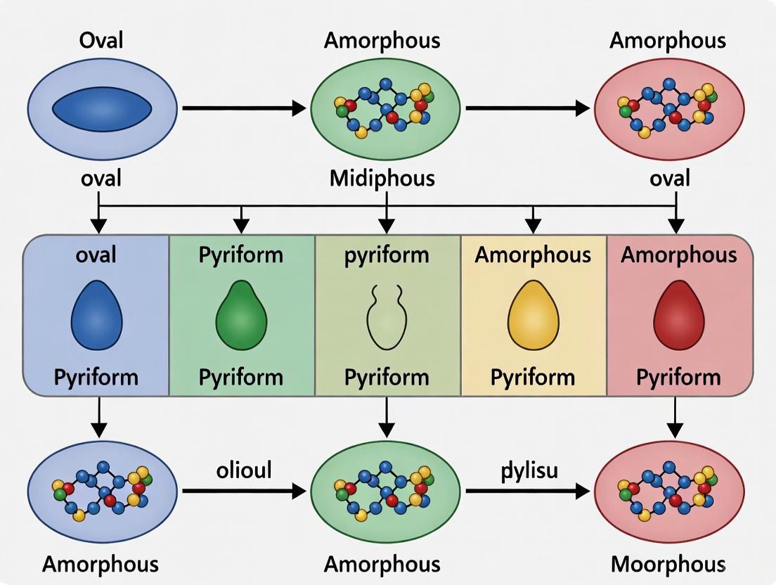
Sperm head morphology serves as a critical indicator of male fertility, with specific morphological defects closely linked to spermiogenesis malfunctions and reduced fertilization potential [12]. The precise classification of sperm head abnormalities is not only essential for clinical diagnosis but also for advancing research in male infertility and developing targeted therapeutic strategies. Among the wide spectrum of defects, tapered, pyriform, microcephalic, macrocephalic, and amorphous heads represent key categories that present significant challenges for both manual assessment and automated classification systems [13] [14]. This technical guide provides an in-depth examination of these five critical sperm head abnormalities, offering researchers and drug development professionals a comprehensive resource encompassing quantitative morphometrics, etiological factors, clinical correlations, and advanced classification methodologies essential for rigorous scientific investigation.
Comprehensive Analysis of Key Sperm Head Abnormalities
Quantitative Morphometric Parameters and Clinical Significance
Table 1: Comprehensive Characteristics of Key Sperm Head Abnormalities
| Abnormality Type | Key Morphological Features | Morphometric Parameters | Primary Etiological Factors | Clinical Impact on Fertility |
|---|---|---|---|---|
| Tapered | Cigar-shaped, constricted near tail [9] [15] | Length >5.0 µm, Width <3.0 µm or <2.0 µm [16] | Varicocele, thermal exposure [9] [15] | Abnormal chromatin packaging, aneuploidy [9] |
| Pyriform | Pear-shaped appearance [13] | Similar to tapered but distinct shape [13] | Associated with environmental pollution [12] | Contributes to overall morphology deterioration [12] |
| Microcephalic | Abnormally small head [9] [15] | Length <3.0 µm, Width <2.0 µm [16] | Genetic traits, defective acrosome [9] | Reduced or absent genetic material [9] [15] |
| Macrocephalic | Giant head, often multiple tails [9] [15] | Length >5.0 µm, Width >3.0 µm [16] | Aurora kinase C gene mutation [9] | Extra chromosomes, fertilization failure [9] |
| Amorphous | Grossly malformed, irregular shape [13] [14] | No consistent measurements, highly variable | Urogenital infections, genetic factors [16] | Severe fertilization impairment [13] |
Detailed Etiological Mechanisms and Functional Consequences
Tapered Head Sperm represent a distinct category characterized by elongated, cigar-shaped heads that appear constricted near the tail region [9] [15]. Beyond the morphological appearance, these sperm often contain abnormal chromatin packaging and demonstrate higher rates of aneuploidy [9]. The etiological factors primarily include varicocele and constant exposure of the scrotum to elevated temperatures, such as from frequent sauna use or occupational exposures [9] [15]. From a functional perspective, the abnormal shape compromises the sperm's ability to penetrate the zona pellucida effectively, while the chromatin abnormalities may impact embryonic development even if fertilization occurs.
Pyriform (Pear-Shaped) Sperm share some visual similarities with tapered heads but present a distinctive pear-like morphology [13]. Research has demonstrated a significant association between increased prevalence of pyriform sperm and environmental pollution exposure, particularly in urban industrial areas [12]. This suggests that environmental toxins may disrupt the delicate process of nuclear reshaping during spermiogenesis, leading to this specific abnormality pattern. The clinical significance lies in the contribution of this defect to overall sperm morphology deterioration in populations exposed to industrial pollutants.
Microcephalic Sperm are characterized by head dimensions significantly below normal ranges, with length less than 3.0 µm and width less than 2.0 µm [16]. These sperm often present with defective acrosomes or significantly reduced genetic material [9]. A specific subtype known as pinhead sperm contains minimal to no paternal DNA content and may indicate underlying diabetic conditions [9]. The functional consequence is severe, as these sperm typically lack the necessary genetic material and enzymatic capacity for successful oocyte fertilization.
Macrocephalic Sperm represent the opposite extreme, with head dimensions exceeding normal parameters (length >5.0 µm, width >3.0 µm) [16]. These sperm frequently carry extra chromosomes and often present with multiple tails [9]. Research has linked this condition to homozygous mutations in the aurora kinase C gene, suggesting a genetic basis that could potentially be transmitted to male offspring [9]. The presence of excess genetic material and structural abnormalities virtually eliminates the fertilization capability of these sperm.
Amorphous Sperm constitute a heterogeneous category encompassing various gross morphological irregularities without consistent patterning [13] [14]. This category presents significant challenges for classification systems due to the wide spectrum of manifestations [13]. Etiological factors are diverse, including urogenital tract infections and genetic predispositions [16]. The clinical impact is severe, with amorphous sperm demonstrating markedly reduced fertilization potential in both natural and assisted reproduction contexts.
Advanced Classification Methodologies
Manual Morphological Assessment Protocols
Traditional sperm morphology assessment relies on microscopic evaluation following standardized staining procedures. The Kruger Strict Criteria, now adopted by the World Health Organization in its 5th and 6th editions, defines normal morphology as 4% or more normal forms in a semen sample [9] [17]. Laboratories typically evaluate 200 sperm per sample, classifying them according to strict dimensional and morphological parameters [16] [17]. Normal sperm heads must demonstrate a smooth oval configuration with well-defined acrosomes covering 40-70% of the head area, measuring 4.0-5.5 μm in length and 2.5-3.5 μm in width [16] [18]. Despite standardization efforts, manual assessment suffers from significant inter-observer variability, with coefficients of variation reaching 80% for morphology assessment compared to 19.2% for sperm density and 15.1% for motility [17].
Computational and Deep Learning Approaches
Table 2: Advanced Sperm Morphology Classification Algorithms and Performance
| Methodology | Key Features | Dataset Applications | Reported Performance | Advantages/Limitations |
|---|---|---|---|---|
| Two-Stage SVM Classification [14] | Shape-based measures, ensemble feature selection | SCIAN-MorphoSpermGS (5-class) | Comparable to human expert | Handles inter-class similarities well |
| Custom CNN Architecture [13] | Multiple filter sizes, fewer parameters | SCIAN (5-class), HuSHeM (4-class) | 88% recall (SCIAN), 95% recall (HuSHeM) | Effective for low-resolution images |
| CBAM-enhanced ResNet50 with DFE [18] | Attention mechanisms, deep feature engineering | SMIDS (3-class), HuSHeM (4-class) | 96.08% accuracy (SMIDS), 96.77% (HuSHeM) | State-of-the-art performance, high interpretability |
| Contrastive Meta-learning [19] | Auxiliary tasks, meta-learning | Confidential datasets | Not specified | Addresses limited data availability |
| APDL Dictionary Learning [14] | Adaptive dictionary learning, patch extraction | HuSHeM (4-class) | Competitive with contemporary methods | Minimal parameters, robust to variations |
Advanced computational approaches have emerged to address the limitations of manual sperm morphology assessment. Early machine learning systems employed feature extraction based on morphological characteristics followed by classification using support vector machines (SVM) or k-nearest neighbors (k-NN) algorithms [14] [18]. Contemporary deep learning approaches have demonstrated remarkable performance, with hybrid architectures like CBAM-enhanced ResNet50 combined with deep feature engineering achieving accuracies of 96.08% on the SMIDS dataset and 96.77% on the HuSHeM dataset, representing significant improvements over baseline CNN performance [18]. These systems typically process input images through multiple stages including preprocessing, segmentation, feature extraction, and classification, leveraging convolutional neural networks to learn discriminative features directly from sperm head images [13].
Sperm Morphology Classification Workflow
Experimental Protocols and Research Reagents
Standardized Staining and Slide Preparation
For manual morphological assessment, semen samples are typically prepared using staining techniques that provide sufficient contrast for detailed morphological evaluation. Common staining methods include Diff-Quick kits [12] and modified Hematoxylin/Eosin procedures [14]. The staining process must be carefully standardized, as variations in preparation, fixation, and staining methodologies significantly influence sperm morphology evaluation results [16]. Following staining, slides are examined under high-magnification microscopy (typically 100x oil immersion), and at least 200 sperm per sample are systematically evaluated and classified according to established morphological criteria [16] [17].
Deep Learning Model Training Protocol
The implementation of deep learning approaches for sperm morphology classification follows a structured experimental pipeline. For the CBAM-enhanced ResNet50 architecture described by Kılıç (2025), the protocol involves:
- Dataset Partitioning: Rigorous 5-fold cross-validation to ensure statistical significance of results [18].
- Data Augmentation: Application of rotation, flipping, and scaling to address limited dataset sizes and improve model generalization [13].
- Model Architecture: Integration of Convolutional Block Attention Module (CBAM) with ResNet50 backbone to enhance focus on morphologically relevant features [18].
- Feature Engineering: Extraction of high-dimensional features from multiple network layers (CBAM, Global Average Pooling, Global Max Pooling) followed by dimensionality reduction using Principal Component Analysis (PCA) and feature selection methods including Chi-square test and Random Forest importance [18].
- Classification: Implementation of Support Vector Machines with RBF/Linear kernels and k-Nearest Neighbors algorithms on processed feature sets [18].
This approach has demonstrated significant time savings, reducing analysis time from 30-45 minutes per sample manually to less than 1 minute automatically while maintaining high accuracy [18].
Deep Learning Model Architecture
Research Reagent Solutions
Table 3: Essential Research Reagents and Materials for Sperm Morphology Analysis
| Reagent/Material | Specification | Research Application | Function |
|---|---|---|---|
| Staining Kits | Diff-Quick, Hematoxylin/Eosin [12] [14] | Slide preparation for manual assessment | Cellular contrast and detail enhancement |
| Fixatives | Ethanol-based solutions [16] | Sample preservation | Maintain structural integrity during processing |
| Reference Datasets | SCIAN-MorphoSpermGS, HuSHeM, SMIDS [13] [18] | Algorithm training/validation | Gold-standard annotated image collections |
| Imaging Systems | High-magnification microscopy (100x oil) [16] | Image acquisition | High-resolution sperm visualization |
| Computational Frameworks | TensorFlow, PyTorch [13] [18] | Deep learning implementation | Neural network development and training |
The precise classification of tapered, pyriform, microcephalic, macrocephalic, and amorphous sperm heads represents a critical component of male fertility assessment and spermiogenesis research. While manual classification following WHO guidelines remains the clinical standard, significant advancements in computational approaches, particularly deep learning architectures enhanced with attention mechanisms and feature engineering, have demonstrated exceptional performance in automating this complex morphological analysis. Future research directions should focus on developing larger, more diverse annotated datasets, improving model interpretability for clinical adoption, and establishing standardized protocols that bridge computational and clinical practices. The integration of these advanced classification techniques into research and diagnostic pipelines holds significant promise for objective, reproducible, and efficient sperm morphology analysis, ultimately advancing both infertility treatment and drug development initiatives in male reproductive health.
Sperm morphology is a critical parameter in male fertility assessment, with sperm head defects being the most prevalent morphological abnormality identified in clinical populations [20]. Within the broader research on sperm head morphology classification techniques, understanding the specific functional implications of these defects is paramount for advancing diagnostic and therapeutic strategies. Traditional manual morphological analysis is subjective and prone to significant inter-observer variability, highlighting the need for more standardized, objective approaches [5] [18]. This guide synthesizes current research to delineate the quantitative relationships between specific sperm head abnormalities and functional fertilization potential, providing researchers and drug development professionals with a detailed technical framework for experimental investigation.
Quantitative Analysis of Head Defects and Functional Impairments
Clinical studies on specific patient cohorts provide robust data linking particular head defect types to measurable declines in semen quality parameters. These correlations suggest that distinct head abnormalities may arise from disruptions during different stages of spermatogenesis and have varying impacts on fertilization capacity [20].
Table 1: Prevalence and Functional Impact of Specific Sperm Head Defects
| Head Defect Type | Relative Prevalence | Primary Functional Association | Key Semen Parameter Affected |
|---|---|---|---|
| Round Head | High | Teratozoospermia, impaired zona pellucida binding | Normal Morphology [20] |
| Tapered Head | High | Teratozoospermia, abnormal acrosome function | Normal Morphology [20] |
| Microcephalous Head | Moderate | Teratozoospermia, genetic material deficiency | Normal Morphology [20] |
| Macrocephalous Head | Moderate | Teratozoospermia, chromosomal abnormalities | Normal Morphology [20] |
| Abnormal Acrosome | Moderate | Impaired oocyte penetration | Fertilization Rate [20] |
Table 2: Correlation Strength Between Defect Categories and Semen Parameters
| Morphological Defect Category | Correlation with Morphology (r) | Correlation with Motility (r) | Strongest Predictor For |
|---|---|---|---|
| Any Head Defect | -0.82* | -0.45* | Teratozoospermia [20] |
| Neck-Midpiece Defect | -0.61* | -0.76* | Asthenozoospermia [20] |
| Tail Defect | -0.53* | -0.81* | Asthenozoospermia [20] |
| Cytoplasmic Residue | -0.38* | -0.42* | Necrozoospermia [20] |
*Spearman correlation coefficients are illustrative; exact values vary by study population.
Experimental Protocols for Morpho-Functional Analysis
Clinical Population Study and Manual Morphology Classification
Objective: To evaluate the incidence of specific sperm morphological abnormalities in a clinical cohort and assess their associations with semen quality and sperm functionality [20].
Materials:
- Participants: A cohort of men (e.g., n=2,923) aged 17-57 years attending an infertility clinic [20].
- Semen Samples: Collected via masturbation after a recommended abstinence period.
Methodology:
- Semen Analysis: Perform basic semen analysis according to WHO guidelines, assessing volume, concentration, motility, and vitality [20].
- Slide Preparation: Prepare semen smears on glass slides. Fix and stain using a Romanowsky-type stain (e.g., RAL Diagnostics kit) to visualize sperm structures [21].
- Morphology Classification: Examine stained smears under oil immersion at 100x magnification. Classify at least 200 spermatozoa per sample per the modified David classification [21] or WHO criteria [20].
- Head Defects: Include tapered, thin, microcephalous, macrocephalous, multiple heads, abnormal post-acrosomal region, and abnormal acrosome [21].
- Data Collection: Record the count and type of each specific abnormality. Categorize participants into normal and low semen parameter groups based on count, motility, and normal morphology thresholds [20].
- Statistical Analysis:
- Use Spearman correlation to assess relationships between specific defect types and semen parameters.
- Perform binary logistic regression to determine the predictive potential of specific defects for semen quality disorders [20].
Deep Learning-Based Classification and Analysis
Objective: To develop an automated, objective system for classifying sperm head morphology using deep learning, trained on expert-annotated datasets [18] [21].
Materials:
- Datasets: Publicly available datasets like SMIDS (3,000 images) or HuSHeM (216 images), or a custom dataset (e.g., SMD/MSS with 1,000+ images) [18] [21].
- Computational Resources: Workstation with GPU and Python environment (v3.8) with deep learning libraries (e.g., TensorFlow, PyTorch) [21].
Methodology:
- Data Acquisition & Labeling:
- Image Pre-processing:
- Data Augmentation: Augment the dataset to balance morphological classes and improve model generalization using techniques like rotation, flipping, and scaling. This can expand a dataset from 1,000 to over 6,000 images [21].
- Model Building & Training:
- Architecture Selection: Employ a Convolutional Neural Network (CNN). Advanced approaches can integrate a ResNet50 backbone enhanced with a Convolutional Block Attention Module (CBAM) to focus on salient features like the head and acrosome [18].
- Training: Partition data into training (80%) and testing (20%) sets. Train the model to classify sperm into predefined morphological classes [21].
- Model Evaluation: Evaluate performance on the test set using metrics like accuracy, precision, and recall. McNemar's test can confirm statistical significance versus baselines [18].
Diagram 1: AI-Based Morphology Analysis Workflow
The Scientist's Toolkit: Essential Research Reagents and Materials
Table 3: Key Reagents and Materials for Sperm Morphology-Function Research
| Item Name | Function/Application | Example/Specification |
|---|---|---|
| RAL Diagnostics Stain | Staining sperm smears for clear visualization of morphological structures (head, acrosome, midpiece, tail) [21]. | Romanowsky-type stain kit. |
| Computer-Assisted Semen Analysis (CASA) System | Automated acquisition of sperm images and initial morphometric analysis (head length/width, tail length) [21]. | MMC CASA system with digital camera. |
| Public Sperm Morphology Datasets | Benchmarking and training AI models for classification tasks. Provides a standardized foundation for research. | SMIDS (3,000 images), HuSHeM (216 images), VISEM-Tracking (656k+ objects) [5] [18]. |
| Convolutional Neural Network (CNN) Model | Core deep learning architecture for automated image classification and feature extraction from sperm images [18] [21]. | Custom CNN or pre-trained ResNet50 with CBAM attention module [18]. |
| Data Augmentation Tools | Artificially expanding dataset size and diversity to improve AI model robustness and prevent overfitting. | Python libraries (e.g., TensorFlow, Keras) for rotation, scaling, flipping [21]. |
The rigorous correlation of specific sperm head defects, such as round and tapered heads, with functional impairments like teratozoospermia provides a crucial evidence base for clinical diagnostics and drug development. The experimental protocols outlined, particularly those leveraging deep learning with architectures like CBAM-enhanced ResNet50, demonstrate a path toward standardized, objective analysis. These methodologies enable high-accuracy classification that can significantly reduce inter-observer variability and processing time. Future research integrating these automated classification techniques with functional fertilization assays will be essential for validating the predictive power of specific morphological defects and developing targeted interventions to overcome male infertility.
Current Challenges in Manual Morphology Assessment and Inter-expert Variability
Sperm morphology assessment is a cornerstone of male fertility evaluation, providing critical insights into testicular and epididymal function [22]. Historically, this analysis has been performed manually by trained embryologists and technicians following standardized guidelines like those from the World Health Organization (WHO). However, this manual process remains one of the most challenging and subjective components of semen analysis [5] [22]. The inherent variability in human visual assessment, combined with differences in training, methodology interpretation, and classification systems, has resulted in significant inter-expert variability that continues to challenge diagnostic consistency and clinical utility [23] [24]. This technical guide examines the current challenges in manual sperm morphology assessment, quantifies the extent and impact of inter-expert variability, and explores emerging solutions aimed at standardizing this critical diagnostic parameter within the broader context of sperm head morphology classification research.
Core Challenges in Manual Assessment
The manual assessment of sperm morphology faces several fundamental challenges that contribute to diagnostic variability and limit clinical utility.
Subjectivity and Classification Complexity
The fundamental challenge in manual sperm morphology assessment lies in its inherent subjectivity. Technicians must evaluate complex morphological features across sperm head, neck, and tail structures, with the WHO classification system recognizing 26 distinct types of abnormal morphology [22]. This complexity is compounded by the need to analyze at least 200 sperm per sample to obtain a statistically reliable assessment, a tedious process prone to fatigue-induced error and subjective interpretation [18]. Studies have reported kappa values as low as 0.05–0.15 between trained technicians, highlighting substantial diagnostic disagreement even among experts working within the same classification system [18]. This variability stems from the challenge of consistently applying qualitative criteria to biological specimens that often exhibit borderline or ambiguous morphological features.
Methodological and Training Inconsistencies
Despite the publication of standardized WHO methodologies, significant differences persist in laboratory practices regarding semen preparation, staining techniques, and classification criteria. A study examining Australian laboratories between 2010-2019 found that although adoption of the WHO 5th edition (WHO5) methodology increased from 50% to 94% over a decade, substantial between-laboratory variability persisted throughout this period [24]. This suggests that even with standardized guidelines, differences in implementation and interpretation continue to affect results. Additionally, the lack of standardized, accessible training tools has been identified as a critical gap. Research has shown that without standardized training, novice morphologists exhibit remarkably high variation (coefficient of variation = 0.28) with accuracy scores ranging from 19% to 77% on the same samples [7].
Table 1: Factors Contributing to Inter-laboratory Variability in Sperm Morphology Assessment
| Factor Category | Specific Variables | Impact on Results |
|---|---|---|
| Methodological | Semen preparation methods, staining techniques (Diff-Quik vs. Papanicolaou), manual vs. computerized analysis | Affects morphological appearance and measurement values [23] |
| Classification Systems | Strict criteria, WHO 1987-2010 editions, David modified criteria | Different definitions of normality and reference intervals [24] |
| Personnel | Level of experience, training quality, subjective interpretation | High inter-observer variability even with same methodology [7] |
| Quality Assurance | Participation in EQA programs, internal quality control procedures | Laboratories implementing rigorous QA show improved precision [24] |
Quantifying Inter-Expert Variability
Substantial research efforts have been dedicated to measuring and understanding the extent of inter-expert variability in sperm morphology assessment.
Historical Evidence of Variability
The challenge of inter-expert variability in sperm morphology assessment is not new. A 1999 comparative study demonstrated moderate agreement between inter-laboratory computer readings (ICC = 0.72) and lower inter-laboratory agreement for manual assessments, highlighting that variability has been a persistent concern for decades [23]. This foundational research also identified that staining techniques significantly impact consistency, with Diff-Quik staining showing better reliability for both manual and computer analysis compared to Papanicolaou staining [23]. The study concluded that despite standardized "strict criteria," high inter-laboratory variability remained for the manual method, establishing a benchmark against which subsequent improvements could be measured.
Contemporary Evidence and Training Impact
Recent research continues to demonstrate significant variability in sperm morphology assessment. A 2025 study utilizing a Sperm Morphology Assessment Standardisation Training Tool revealed that untrained users assessing the same samples exhibited dramatically different accuracy rates depending on classification system complexity: 81.0% for 2-category (normal/abnormal), 68% for 5-category, 64% for 8-category, and just 53% for 25-category classification systems [7]. This demonstrates that more complex classification systems, while potentially providing more detailed information, also introduce greater interpretation variability. The same study also found that structured training could significantly improve these metrics, with trained cohorts achieving 94.9%, 92.9%, 90%, and 82.7% accuracy respectively for the same classification systems [7]. This underscores the critical role of standardized training in reducing variability.
Table 2: Quantifying Variability Across Classification System Complexities
| Classification System Complexity | Untrained User Accuracy (%) | Trained User Accuracy (%) | Inter-Expert Agreement |
|---|---|---|---|
| 2-category (Normal/Abnormal) | 81.0 ± 2.5 | 94.9 ± 0.66 | Highest agreement (73-98% depending on expertise) [7] |
| 5-category (By defect location) | 68 ± 3.59 | 92.9 ± 0.81 | Moderate agreement [7] |
| 8-category (Cattle industry standard) | 64 ± 3.5 | 90 ± 0.91 | Lower agreement [7] |
| 25-category (Individual defects) | 53 ± 3.69 | 82.7 ± 1.05 | Lowest agreement [7] |
Impact on Clinical Relevance and Standardization Efforts
The high degree of variability in manual sperm morphology assessment has raised questions about its clinical utility and prompted standardization initiatives.
Challenges in Clinical Interpretation
The French BLEFCO Group's 2025 expert review directly addressed the clinical relevance of sperm morphology assessment, stating: "There is insufficient evidence to demonstrate the clinical value of indexes of multiple sperm defects (TZI, SDI, MAI) in investigation of infertility and before ART" [3]. They further recommended against using the percentage of normal forms as a prognostic criterion before IUI, IVF, or ICSI procedures [3]. This challenging of current practices highlights how variability in assessment compromises clinical interpretation. When results differ significantly between laboratories and technicians, clinicians cannot reliably use morphology parameters to guide treatment decisions or predict outcomes, potentially diminishing the diagnostic value of this traditionally important parameter.
Standardization and Quality Assurance Programs
External Quality Assurance (EQA) programs have demonstrated both the extent of variability and the potential for improvement through standardized approaches. Data from the Australian External Quality Assurance Programme showed that adoption of WHO5 methodology increased from approximately 50% to over 90% of laboratories between 2010-2019 [24]. This standardization correlated with improved between-laboratory precision over time, though significant variability persisted [24]. The same program also revealed a sustained reduction in the percentage of normal forms reported for the same samples over this period, suggesting either changing interpretive criteria or improved recognition of subtle abnormalities [24]. These findings highlight that while standardization improves consistency, achieving true harmonization remains challenging.
Emerging Solutions and Methodological Innovations
Several innovative approaches are being developed to address the challenges of inter-expert variability in sperm morphology assessment.
Artificial Intelligence and Automated Systems
Recent advances in artificial intelligence (AI) and deep learning offer promising solutions to the subjectivity of manual assessment. Deep learning models have demonstrated remarkable performance, with one framework combining Convolutional Block Attention Module (CBAM) with ResNet50 architecture achieving 96.08% accuracy on the SMIDS dataset and 96.77% on the HuSHeM dataset [18]. These approaches not only provide objective, reproducible assessments but also significantly reduce analysis time from 30-45 minutes per sample to less than one minute [18]. Additionally, AI systems can maintain consistent performance across laboratories, independent of local expertise levels [5] [18]. However, the development of these systems faces its own challenges, particularly the need for large, high-quality, annotated datasets for training [5] [21].
Standardized Training Tools and Protocols
Structured training tools based on machine learning principles have shown significant promise in reducing inter-expert variability. Research demonstrates that using a 'Sperm Morphology Assessment Standardisation Training Tool' with expert consensus labels ("ground truth") can improve novice morphologist accuracy from 53% to 90% even for complex 25-category classification systems [7]. These tools apply principles of supervised learning similar to those used to train AI models, but for human technician education. The study also found that training significantly reduced diagnostic speed from 7.0±0.4s to 4.9±0.3s per image while improving accuracy, demonstrating that both efficiency and consistency can be enhanced through standardized training protocols [7].
Diagram 1: Manual Assessment Workflow and Variability Sources
Experimental Protocols for Variability Assessment
To systematically evaluate and address inter-expert variability, researchers have developed specific experimental approaches.
Protocol for Measuring Inter-Expert Agreement
A comprehensive protocol for quantifying inter-expert variability involves multiple stages. First, sperm samples are prepared using standardized protocols (either liquefied semen or washed samples) and stained with consistent techniques (Diff-Quik recommended for optimal consistency) [23]. Multiple images of individual spermatozoa are then captured using standardized microscopy systems. For the validation core, a subset of images (typically 1,000-2,000) is selected and independently classified by multiple domain experts (three or more) following defined classification criteria (WHO, David modified, etc.) [25] [21]. Experts should work blindly without knowledge of others' assessments. Statistical analysis then measures agreement using intraclass correlation coefficients (ICC) for continuous data or kappa statistics for categorical classifications, with additional analysis of partial agreement scenarios (2/3 experts agreeing) and complete consensus (3/3 experts) [21].
Protocol for Training Effectiveness Assessment
To evaluate training interventions, researchers have employed rigorous pre-post testing designs. Novice morphologists (n=16-22) complete an initial assessment using standardized image sets across multiple classification systems (2-category, 5-category, 8-category, 25-category) to establish baseline accuracy and speed [7]. Participants then undergo structured training using tools that provide immediate feedback on classification accuracy. Following training, participants complete repeated assessments over time (e.g., 14 tests across 4 weeks) to measure improvement in both accuracy and diagnostic speed [7]. Statistical analysis includes paired t-tests or ANOVA to compare pre-post accuracy, calculation of coefficients of variation to assess consistency improvement, and correlation analysis between time spent and accuracy achieved.
Diagram 2: AI-Based Classification Workflow for Reduced Variability
The Researcher's Toolkit
Table 3: Essential Research Reagents and Tools for Sperm Morphology Studies
| Tool/Reagent | Specification/Function | Research Application |
|---|---|---|
| Staining Kits | Diff-Quik, Papanicolaou, RAL Diagnostics, Hematoxylin/Eosin | Enhances morphological feature visualization; Diff-Quik shows superior inter-observer agreement [23] |
| Classification References | WHO 2010/2021 manuals, David modified criteria, Kruger strict criteria | Standardized classification systems; WHO5 adopted by 94% of laboratories over decade [24] |
| Image Datasets | SCIAN-MorphoSpermGS (1,854 images), HuSHeM (216 images), SMIDS (3,000 images), SMD/MSS (1,000+ images) | Benchmarking and algorithm training; quality datasets critical for AI development [5] [25] |
| Quality Assurance Tools | External Quality Assurance (EQA) programs, Training tools with expert consensus labels | Monitoring and improving laboratory performance; trained users show 30%+ accuracy improvement [7] [24] |
| AI/ML Frameworks | Convolutional Neural Networks (CNN), ResNet50, CBAM attention modules, SVM classifiers | Automated classification achieving 96%+ accuracy with minimal variability [18] |
The challenges in manual sperm morphology assessment and significant inter-expert variability remain substantial barriers to standardized male fertility evaluation. Evidence demonstrates that variability stems from multiple sources including methodological differences, classification system complexity, and individual interpreter subjectivity. While standardization initiatives like WHO guidelines and quality assurance programs have improved consistency, fundamental challenges persist. Emerging solutions, particularly artificial intelligence systems and standardized training tools, show remarkable promise for overcoming these limitations. Deep learning approaches have demonstrated expert-level classification accuracy while eliminating inter-observer variability, and structured training protocols can significantly improve human technician consistency. Future research should focus on expanding high-quality annotated datasets, validating AI systems across diverse clinical settings, and developing integrated human-AI collaboration frameworks that leverage the strengths of both approaches to provide reproducible, clinically meaningful morphology assessment.
From Manual to Automated: Technical Approaches in Sperm Head Classification
Traditional Manual Techniques and Computer-Assisted Semen Analysis (CASA) Systems
Semen analysis is a cornerstone of male fertility assessment, providing critical diagnostic information for infertility treatment. For decades, manual microscopy served as the primary method for semen analysis. However, this approach is characterized by significant subjectivity, labor-intensive processes, and considerable inter-observer variability. The evolution of Computer-Assisted Semen Analysis (CASA) systems represents a paradigm shift toward automation, offering quantitative data on sperm dynamic parameters with enhanced speed and consistency. Recent advancements integrate artificial intelligence (AI) and deep learning algorithms to further improve analytical accuracy, particularly in complex areas like sperm morphology classification. This technical guide examines both traditional and automated semen analysis methodologies within the context of sperm head morphology classification research, providing researchers and drug development professionals with a comprehensive framework for methodological selection and implementation.
Comparative Analysis: Manual Techniques vs. CASA Systems
Traditional Manual Semen Analysis
Traditional manual semen analysis relies on visual assessment by trained technicians using conventional light microscopy. The core manual parameters include:
- Sperm Concentration: Typically assessed using a hemocytometer chamber, where sperm in a diluted sample are counted within a defined grid.
- Sperm Motility: Semen is placed on a warmed slide, and a technician categorizes a minimum of 200 sperm into progressive motile (PR), non-progressive motile (NP), or immotile categories based on visual judgment.
- Sperm Morphology: Smears are stained and examined under oil immersion. Technicians classify sperm as having normal or abnormal morphology based on strict criteria outlined by the World Health Organization, assessing head, midpiece, and tail defects.
The principle limitations of manual analysis are its inherent subjectivity and variability. Studies report high inter-observer variability, with kappa values as low as 0.05–0.15 for morphology assessment, indicating substantial diagnostic disagreement even among experts [18]. The process is also time-consuming, requiring 30–45 minutes per sample for a complete analysis [18].
Computer-Assisted Semen Analysis (CASA) Systems
CASA systems automate semen analysis by combining optical microscopy, digital video recording, and sophisticated computer algorithms to track and analyze sperm cells. The fundamental principle involves capturing multiple sequential images of a semen sample loaded into a specialized chamber. Image analysis algorithms then:
- Identify and localize sperm cells within each frame.
- Track sperm movement across consecutive frames to calculate kinematic parameters.
- Classify sperm based on motility patterns and morphological features.
Modern CASA systems incorporate artificial intelligence, utilizing neural network-based image recognition to identify sperm and optical flow methods to track sperm targets [26]. This allows for the measurement of a wide range of parameters, including concentration, motility percentages, velocity parameters (e.g., VCL, VSL, VAP), and detailed morphological measurements.
Table 1: Performance Comparison of Manual vs. CASA Semen Analysis
| Parameter | Manual Analysis | CASA Systems | Comparative Notes |
|---|---|---|---|
| Concentration | Hemocytometer count | Automated particle counting | CASA results can be ~14% lower than manual counts [27]. |
| Motility | Visual categorization (~200 sperm) | Algorithm-based tracking & classification | CASA may report motility ~21% higher than manual assessment [27]. |
| Morphology | Visual classification by strict criteria | AI-based shape and structure analysis | CASA morphology results can be ~87% lower than manual [27]. High inter-observer variability (up to 40%) in manual analysis [18]. |
| Linearity/Progression | Subjective assessment | Quantitative parameters (e.g., STR, LIN) | CASA provides objective, numerical data not available manually. |
| Throughput | ~30-45 minutes/sample [18] | < 1 minute/sample for AI systems [18] | CASA offers significant time savings. |
| Objectivity | Low (Subjective) | High (Algorithm-driven) | CASA reduces technician-based variability. |
| Repeatability | Low to Moderate | High for normal samples; poorer for oligozoospermia/asthenozoospermia [26] | CASA precision depends on sample quality. |
Performance Evaluation and Technical Limitations
Accuracy and Precision of CASA Systems
Performance validation is critical for CASA system implementation. Studies evaluating systems like the GSA-810 have established key performance metrics:
- Linearity and Range: The GSA-810 system demonstrates a wide linear detection range for sperm concentration (2–100 × 10⁶/mL), with R² values ≥0.99, allowing direct analysis of samples with concentrations between 50–100 × 10⁶/mL without dilution [26].
- Precision: The coefficient of variation (CV) for sperm concentration and progressive motility (PR) is inversely correlated with the parameter value itself. Higher sperm concentrations and PR values yield better repeatability. CVs for abnormal morphology and abnormal head morphology are typically below 5% [26].
- Limitations: CASA systems show poorer repeatability for oligozoospermia (low concentration) and asthenozoospermia (low motility) samples [26]. Their accuracy can also be influenced by technical settings and the specific system used, as different CASA systems employ different algorithms, leading to potential variability in results [28].
Influence of Technical Settings on CASA Results
CASA results are highly dependent on instrument configuration and analysis conditions. Researchers must standardize these parameters to ensure reproducible data:
- Frame Rate: Analysis of the same sperm recordings at 25 Hz and 50 Hz shows significantly higher measured velocity values at the higher frame rate due to better capture of the side-to-side motion of sperm heads [28].
- Analysis Chamber: Different disposable counting chambers are available with varying depths (10μm or 20μm) and chamber numbers, which can affect sperm movement and focusing [29].
- Temperature Control: Maintaining a stable temperature (e.g., 36.5°C ± 0.5°C) on a heated stage is critical, as sperm motility is temperature-sensitive. Studies show no significant difference in motility within 1–10 minutes under stable temperature conditions [26].
Table 2: Key Experimental Protocols for CASA System Validation
| Experiment | Core Methodology | Key Metrics & Controls |
|---|---|---|
| Quality Control (Concentration) | Repeated analysis (n=10) of latex bead suspensions with known nominal values (e.g., 80.0 ± 8.0 × 10⁶/mL) [26]. | Accuracy (mean vs. target), Coefficient of Variation (CV). |
| Linearity of Concentration | Serial dilution (e.g., 2 to 50 times) of high-concentration samples (~100 × 10⁶/mL) with own seminal plasma [26]. | Measured value vs. Theoretical value, R² of correlation curve. |
| Short-Term Repeatability | 10 repeated analyses of the same fresh semen sample (n=30 samples) using the CASA system [26]. | CV for concentration, motility, and morphology parameters. |
| Temperature/Time Stability | Analyze sperm motility once every minute for 10 minutes while maintaining platform at 36.5°C ± 0.5°C [26]. | Change in PR and motility percentages over time. |
| Morphology Accuracy | Prepare sperm smears, stain (e.g., Diff-Quik), and analyze morphology by both CASA and manual technician (blinded) [26]. | Coincidence rate = (A1 + B1)/(A + B) × 100%. A1: Normal by both; B1: Abnormal by both. |
Advanced Computational Approaches in Sperm Morphology Classification
The Evolution from Conventional ML to Deep Learning
The analysis of sperm morphology, particularly head morphology, represents a significant challenge due to the subtle variations defining normality. The field has transitioned through distinct computational phases:
- Conventional Machine Learning: Early approaches relied on handcrafted feature extraction. Techniques like Bayesian Density Estimation or Support Vector Machines (SVM) were applied to manually engineered features (shape descriptors, texture). These models achieved accuracies up to 90% in classifying sperm heads into categories like normal, tapered, pyriform, and small/amorphous [5]. However, their performance was limited by the quality and comprehensiveness of the manual feature engineering.
- Deep Learning and AI: Convolutional Neural Networks (CNNs) automate feature extraction and have demonstrated superior performance. For instance, a framework combining a ResNet50 backbone with a Convolutional Block Attention Module achieved test accuracies of 96.08% on the SMIDS dataset and 96.77% on the HuSHeM dataset [18]. These systems can process samples in under one minute, a drastic reduction from the 30-45 minutes required for manual assessment [18].
Simulation and Algorithm Validation
A critical challenge in developing CASA algorithms is the lack of ground-truth data for validation. To address this, researchers have developed sophisticated simulation tools that generate life-like semen images with controllable parameters. These simulations model:
- Sperm Cell Image: Generates 2D images of sperm with head and flagellum, applying point spread functions to mimic microscope optics [30].
- Sperm Swimming Modes: Incorporates four distinct movement patterns: linear mean, circular, hyperactive, and immotile [30].
These simulated environments allow for objective assessment of segmentation, localization, and tracking algorithms using metrics like Multi-Object Tracking Accuracy, providing a robust platform for CASA algorithm development before clinical validation [30].
Essential Research Tools and Experimental Workflows
Research Reagent Solutions
The following table details key materials and reagents essential for conducting semen analysis in a research setting.
Table 3: Essential Research Reagents and Materials for Semen Analysis
| Item | Function/Application | Example Specifications |
|---|---|---|
| Disposable Counting Chambers | Analyze motility, concentration, and pH. Standardizes sample depth for imaging. | HT CASA Chamber; Depths: 10μm, 20μm; Configurations: 2, 4, or 6 chambers [29]. |
| Latex Bead QC Suspensions | Quality control material for validating the accuracy and precision of sperm concentration measurements. | Nominal values: e.g., (80.00 ± 8.0) × 10⁶/mL and (15.00 ± 1.5) × 10⁶/mL [26]. |
| Staining Kits (Morphology) | Differentiate sperm structures (head, acrosome, midpiece, tail) for morphological analysis. | SpermBlue (multispecies), Diff-Quik, Sperm Stain Ready-to-Use [29]. |
| QC-Beads | Beads preparation for quality control in concentration analysis [29]. | - |
| Fluorochrome Preparations | Enable motility and concentration analysis under fluorescence microscopy [29]. | - |
| Phosphate Buffered Saline (PBSt) | Used for simple semen washing and preparation of sample dilutions [29]. | Supplied as tablets for convenient solution preparation. |
Standardized Experimental Workflows
The following diagrams illustrate the core workflows for manual and CASA-based semen analysis, highlighting the procedural and logical relationships.
Manual Semen Analysis Workflow
CASA System Analysis Workflow
Traditional manual semen analysis, while foundational, is beset by subjectivity and variability. CASA systems offer a transformative alternative, providing high-throughput, objective, and quantitative data, especially for sperm concentration and motility. The integration of artificial intelligence, particularly deep learning with attention mechanisms, is rapidly advancing the capabilities of CASA, bringing expert-level accuracy and consistency to the complex task of sperm morphology classification. For researchers and drug development professionals, the selection of an analytical method must align with the specific requirements of the study, weighing the need for throughput and objectivity against the current limitations of automated systems in analyzing pathologically low-quality samples. The ongoing development of standardized, high-quality annotated datasets and robust simulation tools will be crucial for the continued evolution and validation of next-generation CASA algorithms.
Within the broader research on sperm head morphology classification techniques, conventional machine learning (ML) models remain foundational. These models provide a critical benchmark for evaluating newer deep learning approaches and offer high interpretability, which is often essential in clinical diagnostics [5]. Male infertility is a significant global health concern, with male factors contributing to approximately 50% of all infertility cases [5]. The analysis of sperm morphology—particularly the head, which contains the genetic material—is a crucial laboratory test for male fertility assessment [5]. However, manual morphological evaluation is characterized by substantial workload, subjectivity, and significant inter-observer variability, hindering consistent clinical diagnosis [5] [3].
Automated analysis using conventional machine learning provides a pathway to more objective and reproducible assessments. This technical guide details the implementation of three core conventional ML algorithms—Support Vector Machines (SVM), k-means clustering, and Bayesian Classifiers—for sperm head morphology classification. The focus is on the critical role of feature engineering in transforming raw sperm image data into meaningful features that enable these models to accurately distinguish between normal and pathological sperm forms, thereby contributing to standardized, objective fertility assessments.
The Critical Role of Feature Engineering in Sperm Morphology Analysis
Feature engineering is the process of selecting, creating, and transforming raw data into features that are more effectively understood by machine learning models [31] [32]. In the context of sperm head morphology, this involves converting raw pixel values from microscopic images into quantifiable descriptors of shape, texture, and size. Effective feature engineering directly influences model performance by improving accuracy, reducing overfitting, enhancing model interpretability, and increasing computational efficiency [31].
The process typically involves several key steps [31] [32]:
- Feature Creation: Generating new features based on domain knowledge of sperm head morphology (e.g., head ellipticity, acrosome ratio).
- Feature Transformation: Adjusting features through normalization, scaling, or mathematical transformations to ensure consistency.
- Feature Selection: Choosing the most relevant subset of features to reduce dimensionality and prevent overfitting.
For sperm morphology analysis, the inherent complexity of the data—with structural variations in head, neck, and tail compartments—presents fundamental challenges that robust feature engineering helps to overcome [5].
Table: Key Feature Categories for Sperm Head Morphology Analysis
| Feature Category | Description | Example Features |
|---|---|---|
| Shape-Based Descriptors | Quantify the geometric properties of the sperm head. | Area, Perimeter, Eccentricity, Ellipticity, Major/Minor Axis Length, Solidity [5] [33]. |
| Texture-Based Descriptors | Capture the surface and internal intensity patterns of the sperm head. | Entropy, Contrast, Homogeneity, Energy (calculated from Gray-Level Co-occurrence Matrices) [5]. |
| Dimensional Features | Describe the size and proportions of the sperm head according to WHO guidelines. | Head Length (4.0–5.5 μm), Head Width (2.5–3.5 μm), Aspect Ratio [18]. |
Conventional Machine Learning Models: Architectures and Applications
Support Vector Machines (SVM) in Sperm Classification
Support Vector Machines are powerful, discriminative classifiers that find the optimal hyperplane to separate different classes in a high-dimensional feature space. Their effectiveness in sperm morphology classification has been demonstrated in numerous studies, particularly when combined with carefully engineered features [34] [18] [33].
A recent hybrid approach combined deep feature engineering with SVM classification, achieving state-of-the-art performance [34] [18]. The methodology involved:
- Deep Feature Extraction: Using a Convolutional Neural Network (CNN) with a ResNet50 backbone enhanced with a Convolutional Block Attention Module (CBAM) to extract high-dimensional feature representations from sperm images.
- Feature Processing: Applying Principal Component Analysis (PCA) for dimensionality reduction and noise reduction in the deep feature space.
- SVM Classification: Training a Support Vector Machine with a Radial Basis Function (RBF) kernel on the processed features.
This hybrid pipeline (GAP + PCA + SVM RBF) achieved test accuracies of 96.08% ± 1.2% on the SMIDS dataset and 96.77% ± 0.8% on the HuSHeM dataset, significantly outperforming the baseline CNN and demonstrating the potent synergy between deep feature extraction and conventional SVM classifiers [34] [18].
k-means Clustering for Sperm Image Segmentation
The k-means algorithm is an unsupervised clustering technique that partitions data into k distinct clusters based on feature similarity. In sperm morphology analysis, it is particularly valuable for segmenting different regions of the sperm, such as the acrosome and nucleus [5] [33].
Chang et al. proposed a two-stage framework for the detection and segmentation of acrosome and nucleus parts [33]:
- Sperm Head Localization: The k-means clustering algorithm (typically with k=3 to segment background, cytoplasm, and nucleus) is applied to the image to locate the sperm head region, achieving a 98% success rate in detection.
- Acrosome and Nucleus Segmentation: The segmented head region is further processed using clustering combined with histogram statistical methods in different color spaces to separate the acrosome and nucleus, achieving 80% correct assignments [33].
This method's strength lies in its simplicity and efficiency for the initial segmentation task. However, a key limitation is that it primarily focuses on the head part, and segmentation of the mid-piece and tail is also crucial for a complete morphological analysis [33].
Bayesian Classifiers for Probabilistic Morphology Assessment
Bayesian classifiers are probabilistic models that apply Bayes' theorem with strong (naïve) independence assumptions between features. They are known for their robustness and good average performance, even when the independence assumption is not fully met [35].
Bijar et al. developed a model for classifying sperm heads into multiple morphological categories (normal, tapered, pyriform, small/amorphous) using a Bayesian Density Estimation-based approach, achieving a high accuracy of 90% [5]. The model involved a standardized pipeline where shape-based descriptors and other feature engineering techniques were used for the manual extraction of sperm cell features, which were then fed into the Bayesian classifier.
To address biological heterogeneity—such as variations between different replicate samples from the same patient—a Hierarchical Naïve Bayes classifier has been proposed [35]. This model accounts for within-sample variability by using a Bayesian hierarchical framework, where the parameters of individual patients are considered to be related through common population-level hyper-parameters. This approach provides a more robust probabilistic classification, especially when heterogeneity differs across classes, and has been shown to improve accuracy over the standard Naïve Bayes model in contexts like Tissue Microarray (TMA) data [35].
Experimental Protocols and Performance Benchmarking
Detailed Methodologies for Key Experiments
Experiment 1: Hybrid Deep Feature Engineering with SVM [34] [18]
- Dataset Preparation: Use benchmark datasets like SMIDS (3000 images, 3-class: normal, abnormal, non-sperm) or HuSHeM (216 images, 4-class). Apply 5-fold cross-validation.
- Backbone Feature Extraction: Employ a pre-trained ResNet50 architecture integrated with a Convolutional Block Attention Module (CBAM) to extract feature maps, focusing the model on morphologically relevant regions.
- Feature Pooling and Selection: Extract features from multiple layers (CBAM, Global Average Pooling - GAP, Global Max Pooling - GMP). Apply feature selection methods such as PCA, Chi-square test, or Random Forest importance.
- SVM Training and Validation: Train an SVM classifier with RBF kernel on the selected feature set. Validate performance on held-out test sets, reporting accuracy, precision, and recall.
Experiment 2: Two-Stage Segmentation using k-means [33]
- Image Preprocessing: Convert original image to suitable color spaces (e.g., LAB, HSV). Apply median filtering to reduce noise.
- Head Region Segmentation: Apply k-means clustering (k=3) to the preprocessed image to partition pixels into background, cytoplasm, and nucleus. Use the cluster with the smallest area and most intense staining to identify the nucleus.
- Acrosome and Nucleus Differentiation: Within the segmented head region, apply a second clustering step or histogram-based thresholding in a different color channel to separate the acrosome from the nucleus.
- Validation: Manually verify segmentation results against expert annotations, calculating dice coefficients for acrosome, nucleus, and overall head segmentation.
Experiment 3: Hierarchical Naïve Bayes for Heterogeneous Data [35]
- Data Structuring: Organize data to reflect the hierarchical nature (e.g., multiple replicate measurements per patient).
- Model Specification: Define the hierarchical model where individual patient parameters are drawn from a common population distribution (hyper-parameters).
- Parameter Learning: Efficiently learn the model parameters (including hyper-parameters) from the training dataset using closed-form equations or iterative methods.
- Classification: For a new sample, compute the posterior probability of belonging to each class (e.g., aggressive vs. indolent tumor, or normal vs. abnormal sperm) by integrating over the hierarchical structure, and assign the class with the highest probability.
Performance Comparison of Conventional ML Models
Table: Performance Benchmarking of Conventional ML Models in Sperm Morphology Analysis
| Model | Key Features | Dataset | Reported Performance | Limitations |
|---|---|---|---|---|
| SVM with Deep Features [34] [18] | PCA on CNN (ResNet50+CBAM) features + SVM RBF | SMIDS, HuSHeM | 96.08% accuracy (SMIDS), 96.77% accuracy (HuSHeM) | High dependency on quality of feature extraction; complex pipeline. |
| k-means + Histogram Stats [33] | Clustering in different color spaces for segmentation | Custom Dataset | 98% head detection, 80% acrosome/nucleus segmentation | Limited to head segmentation; performance depends on image staining and color space. |
| Bayesian Density Estimation [5] | Shape-based morphological features | Custom Dataset | 90% accuracy (4-class head classification) | Relies exclusively on shape-based features; may miss texture information. |
| Wavelet + SVM [33] | Wavelet-based features with SVM classifier | SMIDS | 82.33% accuracy | Lower performance compared to descriptor-based or deep learning methods. |
| Descriptor-based + SVM [33] | Handcrafted descriptor features with SVM | SMIDS | 85.42% accuracy | Requires manual design of features; may not capture all relevant patterns. |
The Scientist's Toolkit: Research Reagent Solutions
Table: Essential Materials and Computational Tools for Sperm Morphology Analysis
| Item/Tool Name | Function/Application | Specifications/Usage |
|---|---|---|
| SCIAN-MorphoSpermGS [5] [33] | Benchmark dataset for sperm head morphology classification. | Contains 1,854 stained sperm images classified into five classes: normal, tapered, pyriform, small, amorphous. |
| HuSHeM Dataset [5] [18] | Public dataset for sperm head morphology analysis. | Comprises 725 images, though only 216 sperm head images are publicly available; stained and higher resolution. |
| SMIDS Dataset [5] [33] | Dataset for sperm detection and classification. | Contains 3,000 images across three classes: abnormal, non-sperm, and normal sperm heads. |
| VISEM-Tracking Dataset [5] | Multi-modal dataset for sperm analysis. | Provides 656,334 annotated objects with tracking details; includes low-resolution unstained sperm videos and images. |
| SimpleImputer (sklearn) [32] | Handling missing data in feature sets. | Used for imputing missing values (e.g., from imperfect segmentation) with strategies like mean, median, or most_frequent. |
| PCA (Principal Component Analysis) [34] [18] | Dimensionality reduction for feature engineering. | Reduces noise and computational complexity of high-dimensional feature spaces before classification (e.g., in SVM pipelines). |
| Hamilton Thorne CASA-II [33] | Commercial Computer-Aided Semen Analysis system. | Used as a benchmark or for generating preliminary data; provides objective sperm characteristics but can be costly and complex. |
Conventional machine learning models, when coupled with rigorous feature engineering, establish a strong baseline for automated sperm head morphology classification. Techniques such as SVM offer powerful discrimination, k-means provides effective segmentation, and Bayesian models deliver robust probabilistic classification, especially with hierarchical data structures. The performance benchmarks demonstrate that these methods can achieve high accuracy, with hybrid approaches like deep feature extraction combined with SVM classifiers reaching up to 96% accuracy [34] [18].
These conventional methods provide a foundation of interpretability and efficiency against which emerging deep learning techniques must be evaluated. Their continued refinement and integration into clinical workflows hold the potential to significantly standardize fertility assessments, reduce diagnostic variability, and improve patient care outcomes in reproductive medicine [5] [3]. Future work should focus on developing more standardized, high-quality annotated datasets and creating hybrid models that leverage the strengths of both conventional and deep learning approaches.
The analysis of sperm head morphology is a cornerstone of male fertility assessment, providing critical insights into biological function and reproductive potential [21]. Traditionally, this analysis has been a manual process conducted by experienced embryologists, making it inherently subjective, labor-intensive, and difficult to standardize across laboratories [36]. The "Deep Learning Revolution" offers a paradigm shift, introducing powerful convolutional neural network (CNN) architectures that automate and standardize this vital clinical task. This technical guide explores the transformative role of deep learning, focusing on the seminal CNN architectures VGG16 and ResNet50, and the methodology of transfer learning, all within the context of advancing sperm head morphology classification research. By leveraging these technologies, researchers can overcome the limitations of manual analysis, developing systems that deliver rapid, objective, and highly accurate morphological assessments [21] [36].
Foundational CNN Architectures in Computer Vision
The breakthrough of CNNs in image classification began with models like AlexNet and was further solidified by the development of more sophisticated architectures such as VGG16 and ResNet50. These models form the backbone of many modern computer vision applications, including medical image analysis.
VGG16: Simplicity through Depth
The VGG16 model, proposed by the Visual Geometry Group at the University of Oxford, is a convolutional neural network architecture renowned for its simplicity and effectiveness [37] [38]. Its key characteristic is its depth, consisting of 16 layers—13 convolutional layers and 3 fully connected layers [37]. The architecture is uniform, using only 3x3 filters with a stride of 1 pixel and same padding throughout the network, with max-pooling layers of 2x2 with a stride of 2 for spatial downsampling [37] [38]. This successive reduction of spatial dimensions while increasing the number of feature maps (from 64 to 512) allows the network to learn a rich hierarchy of features, from simple edges to complex object representations.
VGG16 achieved a 92.7% test accuracy on the massive ImageNet dataset, which contains 14 million images across 1000 classes, establishing itself as a powerful model for image recognition [37]. However, this performance comes with significant computational costs; the model has 138 million parameters, resulting in large model weights (528 MB) and slow training times, which can lead to the vanishing gradient problem in very deep networks [37].
ResNet50: Addressing the Vanishing Gradient
ResNet50, a 50-layer convolutional neural network, was developed by Microsoft Research in 2015 to address a fundamental limitation in deep neural networks: the vanishing gradient problem [39] [40] [41]. As networks become deeper, gradients can become exceedingly small during backpropagation, hindering effective training and leading to performance degradation. ResNet50 overcomes this through residual connections (or skip connections) that allow the network to bypass multiple stages of computation and directly transport data from lower layers to upper layers [39] [41].
The architecture employs a bottleneck design within each residual block, consisting of three convolutional layers: a 1x1 convolution to reduce dimensionality, a 3x3 convolution as the bottleneck, and a final 1x1 convolution to restore dimensionality [39] [40]. This design allows for efficient computation and parameter usage while enabling the training of very deep networks without performance degradation. ResNet50 won the ImageNet Large-Scale Visual Recognition Challenge (ILSVRC) in 2015 and has since become a cornerstone model in computer vision [39] [41].
Table 1: Comparative Analysis of VGG16 and ResNet50 Architectures
| Feature | VGG16 | ResNet50 |
|---|---|---|
| Total Layers | 16 (13 Convolutional + 3 Fully Connected) [37] | 50 Layers [39] |
| Key Innovation | Deep, uniform structure with small 3x3 filters [37] [38] | Residual learning with skip connections [39] [41] |
| Core Building Block | Stacked 3x3 convolutional layers | Bottleneck block (1x1, 3x3, 1x1 conv) [39] |
| Parameter Count | ~138 million [37] | ~25.6 million [40] |
| ImageNet Top-5 Accuracy | 92.7% [37] | ~95%+ (ILSVRC 2015 winner) [41] |
| Primary Challenge Addressed | Proving efficacy of very deep networks | Vanishing gradients in deep networks [39] [41] |
Diagram 1: VGG16 vs. ResNet50 architectural comparison, highlighting the sequential nature of VGG16 versus the residual block structure of ResNet50.
Transfer Learning: Leveraging Pre-trained Models for Medical Imaging
Transfer learning is a critical methodology in deep learning, particularly valuable in domains like medical imaging where large, annotated datasets are scarce and computationally expensive to produce [39]. The process involves taking a model pre-trained on a large-scale dataset (such as ImageNet) and adapting it to a new, specific task.
The Transfer Learning Process
The standard transfer learning workflow for sperm morphology classification involves several key stages. First, a pre-trained model (e.g., VGG16 or ResNet50) is loaded with weights learned from ImageNet. These models have already learned generic feature detectors (edges, textures, shapes) that are broadly useful for visual tasks [36]. The classifier head (typically the fully connected layers at the top of the network) is then replaced and customized for the new task—in this case, classifying the four sperm head types (normal, tapered, pyriform, amorphous) instead of the original 1000 ImageNet classes [36]. The new model is then trained (fine-tuned) on the target sperm morphology dataset. During this phase, the earlier layers of the network, which capture general features, can be frozen or lightly fine-tuned, while the new classifier head is trained from scratch [36]. This approach significantly reduces training time, computational cost, and the amount of required data while improving overall performance.
Experimental Protocol for Sperm Morphology Classification
Research by Nunes et al. (2021) provides a clear experimental framework for applying transfer learning to sperm head classification [36]. Their study utilized the HuSHeM dataset, a publicly available dataset containing 216 sperm cell images (54 normal, 53 tapered, 57 pyriform, and 52 amorphous) categorized according to WHO criteria by three specialists [36]. Each RGB image was originally 131x131 pixels. A crucial pre-processing step involved cropping and rotating the sperm heads to a uniform direction to ensure consistent input for the model. This was achieved using an automated OpenCV-based program that performed denoising, conversion to monochrome, Sobel operator filtering for gradient calculation, low-pass filtering, adaptive thresholding, morphological operations, and elliptical fitting to precisely crop the head region, resulting in a final input size of 64x64 pixels [36].
The model architecture was based on a modified AlexNet (a predecessor to VGG16 and ResNet50). The researchers adopted its feature extraction architecture and pre-trained parameters but redesigned the classification network by adding Batch Normalization layers to improve performance and stability [36]. The training protocol involved using the pre-training parameters from ImageNet for feature extraction without fine-tuning that part of the network, which kept computational costs low. The dataset was split, with 80% used for training and 20% held out for testing [36]. This method achieved an average accuracy of 96.0% and an average precision of 96.4% on the HuSHeM dataset, outperforming previous approaches while being computationally efficient [36].
Table 2: Sperm Morphology Classification Experimental Setup
| Component | Specification | Purpose/Rationale |
|---|---|---|
| Dataset | HuSHeM (216 images, 4 classes) [36] | Public benchmark for sperm head classification |
| Pre-processing | Cropping & rotation to 64x64 grayscale [36] | Standardizes input, focuses model on head morphology |
| Base Model | AlexNet with pre-trained ImageNet weights [36] | Leverages general feature knowledge via transfer learning |
| Key Modification | Added Batch Normalization layers [36] | Improves training stability and convergence |
| Data Split | 80% Training, 20% Testing [36] | Standard validation practice for model evaluation |
| Reported Accuracy | 96.0% [36] | Benchmark for performance on this specific task |
Diagram 2: The transfer learning workflow, showing how knowledge from a large source domain (ImageNet) is transferred to a specialized target domain (sperm morphology).
The Scientist's Toolkit: Research Reagent Solutions
Table 3: Essential Resources for Sperm Morphology Deep Learning Research
| Resource / Reagent | Function / Description | Example / Specification |
|---|---|---|
| Public Datasets | Provides benchmark data for training and evaluating models. | HuSHeM (216 images, 4 classes) [36], SCIAN-MorphoSpermGS (1854 images, 5 classes) [36] |
| Data Augmentation | Artificially expands dataset size to improve model generalization. | Rotation, flipping, scaling, color adjustments [21] |
| Deep Learning Frameworks | Software libraries for building and training neural networks. | TensorFlow, PyTorch, Keras (Used for ResNet50 implementation) [40] [41] |
| Pre-trained Models | Starting point for transfer learning, provides feature extraction. | VGG16, ResNet50 (Pre-trained on ImageNet) [37] [39] [40] |
| Optimization Algorithms | Updates model parameters to minimize error during training. | Stochastic Gradient Descent (SGD), often with momentum [39] |
| Microscopy & Staining | Prepares and images semen samples for analysis. | RAL Diagnostics staining kit [21], Bright field mode with oil immersion x100 objective [21] |
The integration of advanced CNN architectures like VGG16 and ResNet50 through transfer learning represents a transformative advancement in the automation of sperm head morphology classification. These deep learning models directly address the critical challenges of standardization, objectivity, and efficiency that have long plagued manual morphological assessment [21] [36]. The experimental success of these approaches, demonstrated by accuracy rates exceeding 96% on benchmark datasets, underscores their potential for clinical application [36]. As research in this field progresses, the continued refinement of these models, coupled with the creation of larger and more diverse datasets, promises to deliver robust tools that will significantly enhance the diagnosis and treatment of male infertility, ultimately improving patient care outcomes in reproductive medicine.
Multi-label Classification Systems for Complex Abnormalities
Sperm morphology assessment represents a cornerstone of male fertility evaluation, providing critical diagnostic and prognostic information for assisted reproductive technology (ART) outcomes. Traditional analysis relies on manual microscopic examination, a process inherently limited by substantial subjectivity and significant inter-observer variability [42] [7]. The complexity is further amplified when addressing complex abnormalities, as a single sperm cell can simultaneously present defects across its head, mid-piece, and tail, necessitating a multi-label classification framework [5] [1].
This technical guide explores the advancement from simple categorical systems to sophisticated multi-label classification systems engineered to handle the intricate reality of sperm morphological defects. Framed within broader thesis research on sperm head morphology classification techniques, this document provides researchers and drug development professionals with a comprehensive overview of the computational methodologies, experimental protocols, and research tools driving innovation in this critical field of reproductive medicine.
The Clinical Imperative for Standardized Morphology Assessment
Sperm morphology is a key parameter in semen analysis, with the percentage of normal forms serving as a predictor for the success of ART procedures such as in vitro fertilization (IVF) and intracytoplasmic sperm injection (ICSI) [42]. The World Health Organization (WHO) has progressively revised the reference threshold for morphologically normal sperm, with the current 5th edition establishing a lower reference limit of ≥4% [42]. This low threshold underscores the high prevalence of abnormal forms in clinical samples and the necessity of a nuanced understanding of these defects.
The clinical management of patients, particularly the choice between conventional IVF and ICSI, is heavily influenced by sperm morphology results. When the percentage of normal forms falls below 4%, fertilization rates with IUI and IVF are generally poor, making ICSI the preferred treatment [42]. This critical decision point highlights the necessity for accurate, reproducible, and standardized morphology assessment to avoid misdiagnoses and inadequate patient treatment.
Classification Frameworks: From Single-Label to Multi-Label
A variety of classification systems exist, varying significantly in their complexity and diagnostic granularity.
Table 1: Hierarchy of Sperm Morphology Classification Systems
| System Complexity | Category Examples | Primary Use Case | Reported Untrained Accuracy | Reported Trained Accuracy |
|---|---|---|---|---|
| 2-Category [7] | Normal, Abnormal | Initial screening in sheep industry | 81.0% | 98.0% |
| 5-Category [7] | Normal; Head defect; Mid-piece defect; Tail defect; Cytoplasmic droplet | Location-based defect analysis | 68.0% | 97.0% |
| 8-Category [7] | Normal; Pyriform head; Knobbed acrosome; Vacuoles; etc. | Detailed abnormality profiling in cattle industry | 64.0% | 96.0% |
| 25-Category [7] | All individual defects defined | Maximum granularity for research | 53.0% | 90.0% |
While simpler systems (e.g., 2-category) yield higher assessment accuracy and lower inter-observer variation, they provide limited diagnostic information [7]. The trend in research and advanced diagnostics is toward systems that can leverage the granularity of a 25-category system while mitigating its complexity through automation and standardized training. Critically, multi-label classification acknowledges that a single sperm can belong to multiple abnormal categories simultaneously (e.g., a sperm with a pyriform head and a bent tail), which is a more realistic representation of morphological defects than forcing a single-label assignment [5].
Advanced Multi-Label Computational Approaches
To address the limitations of manual classification, machine learning (ML) and deep learning (DL) have emerged as transformative technologies. The evolution has progressed from conventional ML models to more sophisticated deep learning and ensemble strategies.
Conventional Machine Learning and its Limitations
Early automated approaches relied on traditional machine learning algorithms. The typical pipeline involved manual extraction of features—such as shape-based descriptors (e.g., head length and width), texture, and grayscale intensity—followed by classification using models like Support Vector Machines (SVM) or K-nearest neighbors (KNN) [5]. For instance, one study using Bayesian Density Estimation achieved 90% accuracy in classifying sperm heads into four morphological categories [5].
However, these methods are fundamentally constrained by their dependency on handcrafted features, which are difficult to design, may not capture all relevant morphological nuances, and do not scale well to complex, multi-label tasks [5] [1].
Deep Learning and Ensemble Architectures
Convolutional Neural Networks (CNNs) have revolutionized the field by automatically learning discriminative features directly from image data. CNNs have demonstrated superior performance in sperm head segmentation and classification, achieving precision and recall values exceeding 93% and 91%, respectively [1].
For the complex task of multi-label classification across multiple sperm components (head, mid-piece, tail), ensemble-based approaches have shown significant promise. These methods combine the strengths of multiple models to enhance robustness and accuracy.
Table 2: Ensemble Learning Frameworks for Sperm Morphology Classification
| Study | Core Approach | Key Models/Techniques | Dataset | Reported Performance |
|---|---|---|---|---|
| Çelik et al. [1] | Feature-level & Decision-level Fusion | Multiple EfficientNetV2 variants; SVM, Random Forest, MLP-Attention; Soft Voting | Hi-LabSpermMorpho (18 classes) | 67.70% Accuracy |
| Spencer et al. [1] | CNN Ensemble | VGG16, DenseNet-161, ResNet-34 with a meta-classifier | HuSHeM | 98.2% F1-Score |
| Yuzkat et al. [1] | Ensemble Learning | Multiple CNN models combined | Multiple Datasets | High Classification Accuracy |
| Ilhan et al. [1] | Voting Mechanisms | VGG16 and GoogleNet | - | Significant Accuracy Improvement |
The ensemble framework proposed by Çelik et al. is particularly noteworthy. It employs feature-level fusion by combining features extracted from multiple EfficientNetV2 models, thereby leveraging complementary feature representations. This is followed by decision-level fusion using soft voting across classifiers like SVM, Random Forest, and a Multi-Layer Perceptron with an Attention mechanism (MLP-A) to arrive at a final, robust prediction [1]. This approach effectively mitigates issues of class imbalance and enhances the generalizability of the model.
The Role of Contrastive Learning
A frontier in multi-label classification research involves the use of contrastive learning. This paradigm helps models learn effective representations by pulling semantically similar samples (e.g., sperm images with the same defect) closer in the embedding space while pushing dissimilar samples apart [43]. In the context of hierarchical multi-label classification, contrastive learning can be used to model the complex relationships between labels, capturing both their correlative and distinctive information [44].
For example, Hierarchical Contrastive Learning (HCL) recasts multi-label classification as a multi-task learning problem, incorporating a hierarchical contrastive loss function. This allows the model to understand that a "head defect" is more closely related to a "pyriform head" than to a "tail defect," thereby improving classification accuracy for complex, co-occurring abnormalities [44].
Diagram 1: Hierarchical Contrastive Learning Workflow for multi-label classification, showing the integration of label relationship knowledge.
Experimental Protocols for Multi-Label System Development
Data Preparation and Staining Protocol
Standardized sample preparation is the foundation of reliable analysis. The WHO-recommended protocol is as follows [42]:
- Collection and Liquefaction: Semen samples are collected in a sterile container and incubated at 37°C for 30 minutes to allow liquefaction.
- Smear Preparation: A 10 µL aliquot of well-mixed semen is placed on a clean frosted slide and spread smoothly using a second slide at a 45° angle to create an even smear. Slides are prepared in duplicate and air-dried.
- Staining: The gold standard is the Papanicolaou stain, though Diff-Quik is also used. For Diff-Quik:
- Immerse the dry slide in fixative five times, then air-dry for 15 minutes.
- Immerse the slide three times in Solution I for 10 seconds.
- Drain excess stain and immerse five times in Solution II for 10 seconds.
- Rinse gently in water, dry vertically, and mount with a coverslip.
Establishing Ground Truth for Model Training
Creating a high-quality dataset is critical for supervised learning. The "ground truth" is established through a process of expert consensus to minimize individual subjectivity [7].
- Image Sourcing: A large number of sperm images are acquired using bright-field microscopy under 1000x oil immersion.
- Expert Annotation: Multiple expert morphologists independently classify each sperm image according to the target classification system (e.g., 25-category).
- Consensus Labeling: Only images where a pre-defined consensus (e.g., agreement among a majority of experts) is reached are included in the final "ground-truth" dataset. This curated dataset is used to train both human morphologists and machine learning models [7].
Model Training and Evaluation Protocol
The workflow for developing a multi-label ensemble model, as detailed by Çelik et al., can be summarized as follows [1]:
- Data Partitioning: The ground-truth dataset (e.g., Hi-LabSpermMorpho with 18,456 images across 18 classes) is split into training, validation, and test sets.
- Feature Extraction: Multiple CNN architectures (e.g., EfficientNetV2-S, -M, -L) are used to extract feature vectors from the training images.
- Feature-Level Fusion: The extracted feature vectors from different models are concatenated into a unified, high-dimensional feature representation.
- Classifier Training: The fused feature set is used to train multiple classifiers (SVM, Random Forest, MLP-Attention).
- Decision-Level Fusion: Predictions from the individual classifiers are aggregated using a soft voting mechanism to produce the final multi-label prediction.
- Evaluation: The model is evaluated on the held-out test set using metrics such as accuracy, F1-score, and label-based precision and recall.
Diagram 2: Ensemble model protocol with multi-level fusion, showing the pathway from image input to final classification.
The Scientist's Toolkit: Essential Research Reagents and Materials
Table 3: Key Research Reagents and Materials for Sperm Morphology Analysis
| Item | Function/Description | Application in Research |
|---|---|---|
| Diff-Quik Stain [42] | A rapid Romanowsky-type stain consisting of a fixative, eosin (Solution I), and thiazine dyes (Solution II). It differentially stains acrosomal (light blue) and post-acrosomal (dark blue) regions. | Routine, rapid staining for manual and automated morphology assessment. |
| Papanicolaou Stain [42] | Considered the "gold standard" for sperm morphology assessment. Provides excellent nuclear and cytoplasmic detail. | High-precision manual analysis and creation of gold-standard datasets for AI model training. |
| HSMA-DS Dataset [5] | Human Sperm Morphology Analysis DataSet: 1,457 non-stained, noisy, low-resolution sperm images. | Benchmarking for classification algorithms under non-ideal conditions. |
| HuSHeM Dataset [5] | Human Sperm Head Morphology: 725 stained, higher-resolution images (216 publicly available). | Focused development and testing of sperm head-specific classification models. |
| SVIA Dataset [5] | Sperm Videos and Images Analysis: A large-scale dataset with 125,000 annotated instances for detection, 26,000 segmentation masks, and 125,880 cropped images for classification. | Training large-scale, deep learning models for detection, segmentation, and multi-label classification. |
| Hi-LabSpermMorpho Dataset [1] | A comprehensive dataset containing 18,456 image samples across 18 distinct sperm morphology classes. | Training and evaluating complex ensemble models for fine-grained, multi-label classification. |
| Ocular Micrometer [42] | A calibrated graticule placed in the microscope eyepiece to accurately measure sperm dimensions (head length 5-6 µm, width 2.5-3.5 µm). | Essential for manual validation and for establishing strict morphological criteria for "normal" sperm. |
The evolution from subjective manual assessment to automated multi-label classification systems marks a paradigm shift in sperm morphology analysis. By embracing advanced computational strategies such as deep ensemble learning and hierarchical contrastive learning, these systems are poised to overcome the longstanding challenges of reproducibility and subjectivity. The development of standardized, high-quality datasets and rigorous training protocols ensures that these models are both robust and clinically relevant. For researchers and drug development professionals, these technologies offer powerful tools to enhance diagnostic accuracy, refine patient stratification for ART, and ultimately, improve outcomes in the treatment of male factor infertility.
The morphological analysis of sperm heads is a critical diagnostic procedure in male fertility assessment. Traditional manual evaluation under microscopy is often subjective, leading to inter-observer variability, while many Computer-Aided Sperm Analysis (CASA) systems are limited in functionality and struggle with noisy or low-quality samples [45]. The World Health Organization (WHO) emphasizes the use of stained smears to reveal fine morphological defects that are otherwise difficult to detect, underscoring the need for highly accurate and automated classification systems [45].
In recent years, deep learning has emerged as a powerful tool for automating complex classification tasks. Multimodal fusion and ensemble deep learning represent two of the most promising avenues for enhancing the performance, robustness, and generalizability of diagnostic models [46] [47]. Multimodal fusion involves integrating data from different sources or modalities to form a unified representation, thereby providing a more holistic understanding of the subject than any single data source can offer [46]. Concurrently, ensemble learning leverages the strengths of multiple models to arrive at a collective decision that is often superior to that of any individual constituent model [45] [47].
This technical guide explores the application of these advanced computational frameworks—specifically, multi-model Convolutional Neural Network (CNN) fusion and ensemble deep learning—within the context of sperm head morphology classification. We will delve into detailed methodologies, experimental protocols, and quantitative results, providing researchers and drug development professionals with a roadmap for implementing these cutting-edge techniques.
Core Concepts and Definitions
From Unimodal to Multimodal and Ensemble Frameworks
A unimodal approach relies on a single type of data (e.g., only text or only images) to solve a problem. While simpler, this approach fails to capture auxiliary information that could be crucial for comprehensive decision-making [46]. For instance, a unimodal sperm classifier might use only bright-field microscopy images, potentially missing contextual or stain-specific features.
Multimodal fusion addresses this limitation by combining complementary information from multiple data sources or representations. In the context of image analysis, this can involve fusing features extracted from different layers of a neural network (multilayer fusion) [48] or integrating image data with other data types, such as clinical information [47]. The fusion process itself can occur at different stages:
- Early Fusion: Combining raw data or low-level features.
- Late Fusion: Combining the decisions or predictions of multiple models.
- Hybrid Fusion: A combination of early and late fusion strategies [46].
Ensemble Deep Learning is a strategy that employs multiple deep learning models to solve a single problem. The core idea is that a group of "weak" learners can come together to form a "strong" learner, thereby reducing variance, minimizing overfitting, and improving generalization [45] [47]. Ensembles can be constructed from models with different architectures (e.g., combining CNNs and Transformers) or from multiple instances of the same architecture trained under different conditions [47].
The Rationale in Sperm Morphology Analysis
Sperm morphology classification is inherently challenging due to high inter-class similarity (e.g., between different head defects) and significant intra-class variability [45]. A single model may specialize in recognizing certain features but fail on others. A multimodal ensemble framework allows for a divide-and-conquer strategy, where different models or fusion pathways can be optimized for specific abnormality categories, such as head, neck, or tail defects [45]. This hierarchical approach leads to more robust and accurate classification systems.
A Two-Stage Ensemble Framework for Sperm Morphology
A seminal study demonstrates the effectiveness of a two-stage, divide-and-ensemble deep learning framework for classifying sperm morphology across 18 distinct classes [45]. This methodology is particularly adept at reducing misclassification between visually similar categories.
Experimental Protocol and Workflow
The following diagram illustrates the logical workflow of the two-stage classification system.
Figure 1. Two-stage divide-and-ensemble workflow for sperm morphology classification. The first stage routes images to one of two broad categories, and the second stage uses a specialized ensemble model for fine-grained classification within that category [45].
1. Dataset and Preprocessing:
- Dataset: The model is trained and evaluated on the Hi-LabSpermMorpho dataset, a large-scale, expert-labeled dataset comprising RGB images of spermatozoa [45].
- Staining: Images are acquired using bright-field microscopy with three different Diff-Quick staining protocols (BesLab, Histoplus, and GBL) to enhance morphological features [45].
- Classes: The dataset is classified into 18 categories consistent with the WHO 2021 classification, covering head, neck, and tail abnormalities. Head defects are the most prevalent, with amorphous heads being a particularly common subtype [45].
2. Two-Stage Classification Architecture:
- Stage 1 - The Splitter: A dedicated deep learning model (the "splitter") acts as a router. It does not perform a final classification but instead categorizes each input sperm image into one of two broad, high-level groups:
- Category 1: Head and neck region abnormalities.
- Category 2: Normal morphology and tail-related abnormalities [45].
- Stage 2 - Category-Specific Ensembles: For each of the two high-level categories, a separate, customized ensemble model is deployed. Each ensemble integrates four distinct deep learning architectures, such as DeepMind's NFNet-F4 and various Vision Transformer (ViT) variants. This ensemble is responsible for the fine-grained classification of the specific abnormality within its assigned category [45].
3. Multi-Staged Ensemble Voting Mechanism: Instead of conventional majority voting, the framework employs a structured multi-stage voting strategy to enhance decision reliability. In this mechanism, each model within the ensemble casts both a primary vote and a secondary vote. This approach mitigates the influence of dominant classes and ensures more balanced decision-making across the various sperm abnormalities [45].
Key Research Reagents and Materials
Table 1: Essential research reagents and materials for implementing the two-stage ensemble framework.
| Item | Function & Description | Relevance to Experiment |
|---|---|---|
| Hi-LabSpermMorpho Dataset | A large-scale, expert-labeled dataset of sperm morphology images with 18 distinct classes. | Provides the essential, high-quality ground-truth data required for training and validating the complex ensemble model. [45] |
| Diff-Quick Staining Kits | A Romanowsky-type stain used to prepare sperm smears for microscopy. Enhances contrast and reveals morphological details. | Critical for sample preparation. The study used three versions (BesLab, Histoplus, GBL) to test model robustness. [45] |
| Bright-Field Microscope | An optical microscope that uses transmitted light through a specimen. | Standard equipment for acquiring the initial digital images of stained sperm samples. [45] |
| Deep Learning Models (NFNet, ViT) | Pre-trained architectures used as feature extractors and classifiers within the ensemble. | NFNet-based models were identified as particularly effective. ViTs provide a complementary, attention-based approach. [45] |
Quantitative Performance Results
The proposed two-stage framework demonstrated a statistically significant improvement over traditional single-model approaches and unstructured ensembles.
Table 2: Classification accuracy of the two-stage ensemble model across different staining protocols [45].
| Staining Protocol | Classification Accuracy | Performance Improvement |
|---|---|---|
| BesLab | 69.43% | +4.38% over prior approaches |
| Histoplus | 71.34% | +4.38% over prior approaches |
| GBL | 68.41% | +4.38% over prior approaches |
The two-stage system substantially reduced misclassification among visually similar categories, confirming its enhanced ability to detect subtle morphological variations that are critical for accurate clinical diagnosis [45].
Multimodal Fusion in Medical Imaging
The principle of fusing information from multiple sources is a powerful and generalizable concept. Beyond ensemble methods that fuse model predictions, multimodal fusion integrates fundamentally different types of data.
CNN-based Multimodal Medical Image Fusion
In medical imaging, different modalities provide complementary information. For example, CT scans excel at visualizing bony structures, while MRI provides superior soft tissue contrast [49] [50]. Fusing these images creates a single, information-rich output that can enhance diagnostic accuracy.
Protocol: Convolutional Neural Network (CNN) for Image Fusion
- Process: CNN-based fusion methods automatically learn hierarchical feature representations from the source images. The network is trained to preserve the most salient features from each input modality in the final fused image [49] [51].
- Fusion Levels: This can occur at the pixel level (combining raw pixels), feature level (combining extracted features from intermediate layers), or decision level (combining final model outputs) [49].
- Advantages over Traditional Methods: Deep learning-based fusion has been shown to deliver far better results in both qualitative and quantitative analyses compared to conventional methods like wavelet transform or Principal Component Analysis (PCA). It better preserves features and is more adaptable to different data characteristics [49] [50].
Fusion of Imaging and Clinical Data
A powerful extension of multimodal fusion involves integrating image data with non-image clinical information. A study on glioma subtype classification (Glioblastoma Multiforme vs. Low-Grade Glioma) effectively demonstrates this approach.
Protocol: Ensemble Fusion AI (EFAI) for Glioma Subtyping
- Image Feature Extraction: Multiple deep learning models (both CNNs and Transformers) are used to extract rich feature sets from Whole Slide Images (WSIs) of histopathology samples [47].
- Feature Ensemble: The top-performing models are selected, and their feature sets are ensembled to create a superior imaging feature representation [47].
- Multimodal Fusion: The ensembled image features are then concatenated with features extracted from patient clinical data. This combined feature vector provides a comprehensive view of the patient's condition [47].
- Classification: A machine learning classifier is finally trained on this multimodal feature set to perform the diagnosis [47].
Results: This Ensemble Fusion AI (EFAI) approach achieved a classification accuracy of 93.6% and an Area Under the Curve (AUC) of 0.967, significantly outperforming models that used only histopathology images or clinical data alone [47]. This underscores the tremendous value of integrating disparate data types.
Visualization and Technical Implementation
Diagram of a Generalized Multimodal Fusion Pipeline
The following diagram outlines a generalizable pipeline for fusing image data with clinical data, synthesizing the principles from the cited research.
Figure 2. A generalized pipeline for multimodal ensemble fusion, integrating image and clinical data for enhanced diagnostic classification [45] [47].
The Scientist's Toolkit: Key Reagent Solutions
Table 3: Key deep learning architectures and computational tools for building fusion and ensemble models.
| Item | Function & Description | Relevance to the Field |
|---|---|---|
| NFNet (Normalizer-Free Network) | A CNN architecture that achieves high performance without using batch normalization. | Identified as particularly effective for sperm morphology classification due to its stability and accuracy. [45] |
| Vision Transformer (ViT) | A transformer model adapted for image classification by treating image patches as a sequence. | Provides a complementary approach to CNNs, using self-attention to capture global context within an image. Often used in ensembles. [45] [47] |
| Feature Map Transformation (FDSFM) | A module designed to address the challenge of fusing feature maps of different sizes from multiple layers or models. | Enables effective multilayer and multimodal fusion by standardizing feature map dimensions before concatenation or fusion. [48] |
| Multi-Staged Voting Mechanism | An ensemble decision strategy where models cast primary and secondary votes. | Increases classification reliability and mitigates the influence of dominant classes in imbalanced datasets. [45] |
The integration of multi-model CNN fusion and ensemble deep learning represents a paradigm shift in the automated analysis of sperm head morphology. The reviewed research demonstrates that a structured, two-stage ensemble framework can achieve a significant improvement in classification accuracy—over 4% in a complex 18-class problem—by effectively reducing misclassification among visually similar abnormalities [45].
The generalizability of these approaches is evidenced by their success in other medical domains, such as glioma classification, where the fusion of image features with clinical data pushes accuracy above 93% [47]. The consistent theme is that leveraging multiple, complementary sources of information, whether from different models, different network layers, or entirely different data modalities, yields a more robust and accurate diagnostic system.
For researchers and drug development professionals, the path forward involves curating high-quality, well-labeled datasets and embracing a modular, ensemble-based approach to model building. The future will likely see increased use of advanced fusion techniques, including attention mechanisms and transformer models, to further enhance the precision and interpretability of these systems, ultimately leading to more reliable tools for reproductive healthcare [49] [45].
Overcoming Technical Challenges in Automated Classification Systems
In the field of male infertility research, sperm morphology analysis (SMA) represents a significant diagnostic challenge, with male factors contributing to approximately 50% of infertility cases globally [5]. The clinical assessment of sperm morphology requires the analysis of over 200 sperms according to World Health Organization (WHO) standards, which categorize abnormalities across head, neck, and tail regions encompassing 26 distinct morphological types [5]. This complex analytical framework, combined with the substantial workload of manual observation, creates a critical bottleneck in clinical diagnosis and research advancement. The inherent subjectivity of manual analysis further compounds these challenges, introducing significant variability and hindering reproducible results in male fertility assessment [5].
Within this context, automated sperm recognition systems based on machine learning (ML) and deep learning (DL) have emerged as promising solutions to standardize morphological evaluation. However, the development of robust AI models is fundamentally constrained by the lack of standardized, high-quality annotated datasets [5]. Deep learning architectures, particularly Convolutional Neural Networks (CNNs), require multidimensional data extraction and analysis from large-scale datasets to achieve effective automatic feature extraction and model training [5]. This paper examines the specific limitations affecting sperm morphology datasets and explores how data augmentation techniques, combined with benchmark datasets like SMD/MSS, are addressing these challenges to advance classification techniques in sperm head morphology research.
Critical Limitations in Current Sperm Morphology Datasets
The transition toward deep learning-based sperm morphology analysis has exposed significant gaps in data availability and quality. Current publicly available datasets face several interconnected limitations that impact model generalization and clinical applicability.
Quantitative and Qualitative Deficiencies
An analysis of existing sperm morphology datasets reveals consistent limitations in both scale and quality, as detailed in Table 1. These constraints directly impact the performance and generalizability of deep learning models trained on these datasets.
Table 1: Analysis of Existing Human Sperm Morphology Datasets
| Dataset Name | Year | Image Count | Key Characteristics | Primary Limitations |
|---|---|---|---|---|
| HSMA-DS [5] | 2015 | 1,457 images from 235 patients | Non-stained, noisy, low resolution | Limited sample size, image quality issues |
| HuSHeM [5] | 2017 | 725 images (only 216 publicly available) | Stained, higher resolution | Extremely limited public availability |
| MHSMA [5] | 2019 | 1,540 grayscale sperm head images | Non-stained, noisy, low resolution | Limited to sperm heads only, quality issues |
| SCIAN-MorphoSpermGS [5] | 2017 | 1,854 sperm images | Stained, higher resolution, 5-class classification | Limited morphological diversity |
| SVIA [5] | 2022 | 4,041 low-resolution images and videos | Extensive annotations: 125,000 instances for detection, 26,000 segmentation masks | Low-resolution, unstained specimens |
| VISEM-Tracking [5] | 2023 | 656,334 annotated objects with tracking details | Multi-modal with videos and tracking data | Limited morphological annotation detail |
Annotation Complexity and Standardization Challenges
The annotation process for sperm morphology presents unique difficulties that extend beyond simple image labeling. Sperm may appear intertwined in images, or only partial structures may be displayed when cells are at the image edges, fundamentally affecting analytical accuracy [5]. Furthermore, comprehensive defect assessment requires simultaneous evaluation of head, vacuoles, midpiece, and tail abnormalities, substantially increasing annotation complexity and requiring specialized expertise [5]. The absence of standardized processes for sperm morphology slide preparation, staining, image acquisition, and annotation creates inconsistent data quality across research institutions and clinical laboratories, ultimately limiting the development of generalized models capable of robust performance across diverse clinical settings.
Data Augmentation Techniques for Overcoming Data Limitations
Data augmentation encompasses a suite of techniques that artificially expand training datasets by generating modified versions of existing data, effectively addressing data scarcity and diversity limitations [52]. These methods are particularly valuable for deep learning applications in medical imaging, where data collection is often expensive, time-consuming, and constrained by privacy considerations [53].
Fundamental Augmentation Approaches
Table 2: Data Augmentation Techniques for Sperm Image Analysis
| Technique Category | Specific Methods | Application in Sperm Morphology | Impact on Model Performance |
|---|---|---|---|
| Geometric Transformations | Rotation, Translation, Scaling, Flipping, Cropping [54] [55] [53] | Creates orientation, position, and size variance | Improves invariance to sperm rotation and positioning in images |
| Color Space Adjustments | Brightness, Contrast, Saturation, Hue modification [54] [55] [53] | Simulates varying staining intensities and lighting conditions | Enhances robustness to laboratory preparation variations |
| Noise Injection | Gaussian noise, Salt-and-pepper noise [54] [55] | Mimics image acquisition artifacts and sensor noise | Improves model resilience to real-world image quality issues |
| Advanced Techniques | Generative Adversarial Networks (GANs), Neural Style Transfer [54] [53] | Generates entirely new synthetic sperm images | Addresses extreme data scarcity and class imbalance |
Advanced Augmentation Frameworks
Beyond basic image manipulations, advanced augmentation approaches leverage sophisticated deep learning architectures to generate more diverse and realistic training data. Generative Adversarial Networks (GANs) have demonstrated remarkable capability in producing synthetic medical images that preserve essential morphological characteristics while expanding dataset diversity [53]. In sperm morphology analysis, GANs can generate synthetic samples for underrepresented abnormality classes, effectively addressing class imbalance issues that commonly plague classification models [5].
Another emerging approach, neural style transfer, separates image content from style and recomposes content with alternative stylistic elements [53]. This technique could potentially normalize staining variations across different laboratory protocols, enhancing model generalization across clinical settings. These advanced methods represent a paradigm shift from simply expanding dataset size to strategically enhancing dataset diversity and quality.
The SMD/MSS Dataset Framework and Experimental Protocols
The SMD/MSS dataset represents a benchmarked resource specifically designed to address the limitations of previous sperm morphology datasets. This framework incorporates comprehensive annotations of 12 morphological defects across head, midpiece, and tail regions, enabling more nuanced classification models [56].
Dataset Composition and Annotation Standards
The SMD/MSS dataset establishes rigorous annotation protocols to ensure consistent labeling across specimens. Each sperm image receives multi-level annotations capturing structural abnormalities at the head, midpiece, and tail levels, with detailed characterization of specific defect types within each anatomical region [56]. This granular approach supports both binary classification (normal/abnormal) and fine-grained morphological analysis, providing researchers with flexibility in model development based on specific clinical requirements.
Deep Learning Experimental Protocol
Recent research utilizing the SMD/MSS dataset has employed the ResNet50 architecture, a convolutional neural network with 50 layers that has demonstrated strong performance in various image classification tasks [56]. The experimental protocol typically involves these critical steps:
Data Preprocessing: Images are standardized through resizing to consistent dimensions, normalization of pixel values, and application of stain normalization techniques to reduce laboratory-specific variations.
Data Partitioning: The dataset is divided into training, validation, and test sets using stratified sampling to maintain consistent class distribution across splits, typically following an 70-15-15 ratio.
Augmentation Strategy: Implementation of a comprehensive augmentation pipeline including rotation (±15°), horizontal flipping, brightness variation (±20%), contrast adjustment (±15%), and synthetic sample generation for underrepresented classes.
Model Training: Training conducted with transfer learning approaches, initializing weights from pre-trained models and fine-tuning on the sperm morphology dataset using categorical cross-entropy loss and Adam optimizer.
Evaluation Metrics: Comprehensive assessment using accuracy, precision, recall, F1-score, and per-class metrics to ensure balanced performance across all morphological categories.
This methodological framework has demonstrated effective classification across various sperm morphology classes, establishing a robust baseline for future research [56].
Research Reagent Solutions for Sperm Morphology Analysis
The development of reliable experimental protocols in sperm morphology research requires specific reagents and computational resources. Table 3 outlines essential research reagents and their functions in the analytical pipeline.
Table 3: Essential Research Reagents and Computational Resources
| Reagent/Resource | Category | Function in Research |
|---|---|---|
| Staining Solutions (e.g., Diff-Quik, Papanicolaou) | Laboratory Reagent | Enhances contrast for morphological visualization of sperm components |
| Fixatives (e.g., Glutaraldehyde, Formalin) | Laboratory Reagent | Preserves sperm structure during processing and analysis |
| ResNet50 Architecture | Computational Resource | Deep CNN backbone for feature extraction and classification [56] |
| Data Augmentation Libraries (e.g., Albumentations, Imgaug) | Computational Resource | Applies geometric and color transformations to expand training datasets [54] |
| GAN Frameworks (e.g., PyTorch, TensorFlow) | Computational Resource | Generates synthetic sperm images to address class imbalance [53] |
| SMD/MSS Dataset | Data Resource | Benchmark dataset with comprehensive morphological annotations [56] |
Advanced Classification Frameworks: Meta-Learning Approaches
Recent advances in sperm head morphology classification have introduced sophisticated frameworks that combine multiple learning paradigms to enhance generalization capabilities. The Contrastive Meta-learning with Auxiliary Tasks framework represents a cutting-edge approach that addresses the fundamental challenge of limited data availability [19].
This framework integrates contrastive learning, which learns effective feature representations by comparing similar and dissimilar sample pairs, with meta-learning principles that enable the model to rapidly adapt to new classification tasks with minimal examples [19]. The incorporation of auxiliary tasks, such as predicting rotation angles or solving jigsaw puzzles of image patches, provides additional self-supervised learning signals that guide the model toward more robust feature extraction without requiring extensive labeled data.
This architecture demonstrates how carefully designed learning frameworks can maximize information extraction from limited datasets, potentially reducing dependency on massive annotated collections while maintaining classification accuracy.
The integration of comprehensive datasets like SMD/MSS with strategic data augmentation methodologies represents a transformative approach to overcoming historical limitations in sperm morphology analysis. These techniques collectively address the fundamental challenge of data scarcity while enhancing model robustness and generalization capabilities. The continued refinement of annotation standards, coupled with advanced augmentation strategies and innovative learning frameworks, is establishing a new paradigm in male infertility research.
Future research directions should focus on developing standardized augmentation policies specific to sperm morphology characteristics, establishing quality assessment metrics for synthetic data generation, and creating multi-center collaborative frameworks for dataset expansion. As these technical advancements mature, they promise to accelerate the development of highly accurate, clinically viable decision support systems that can standardize sperm morphology analysis across diverse healthcare settings, ultimately improving diagnostic consistency and treatment outcomes in male infertility management.
In the field of biomedical research, particularly in domains relying on subjective image analysis like sperm head morphology classification, the establishment of reliable "ground truth" annotations represents a foundational challenge. Ground truth refers to reference data that is accepted as representing the true state of the phenomenon being studied. In morphological analysis, this constitutes a set of expertly validated classifications against which new observations, human trainees, or machine learning algorithms can be benchmarked. The inherent subjectivity of visual assessment, where even experts may disagree on classification, necessitates robust strategies to achieve consensus and ensure reproducibility [7] [57]. This whitepaper details the methodologies for establishing expert consensus to create high-quality annotated datasets, framed within the specific context of advancing sperm head morphology research.
The necessity for these strategies is underscored by research revealing significant variability in quantitative measurements between institutions and even among experts within the same field. One multicenter comparison study found that differences in implementation and definition could lead to variations of up to 50% in key metrics like the Dice Similarity Coefficient (DSC) and 3D Hausdorff Distance (HD) when analyzing the same data [57]. Such discrepancies highlight that without a standardized and traceable ground truth, comparing results across studies or validating new diagnostic tools becomes problematic. Establishing a reliable ground truth through expert consensus is therefore not merely an academic exercise but a prerequisite for meaningful scientific and clinical progress.
Expert Consensus Methodologies for Ground Truth Establishment
The core challenge in ground truth establishment is reconciling the subjective interpretations of multiple experts into a single, reliable dataset. The application of machine learning principles, specifically the concept of "ground-truth" established by the consensus of multiple experts, has been validated as an effective strategy for training human morphologists [7]. This section outlines the primary methodological frameworks.
The Consensus-Driven Annotation Workflow
A structured, multi-stage process is critical for generating high-quality ground-truth labels. This workflow ensures that annotations are consistent, accurate, and reflective of collective expert knowledge.
- Image Selection and Preparation: The process begins with the curation of a representative set of high-quality images. For sperm morphology, this involves standardized sample preparation, staining, and image acquisition to minimize technical artifacts that could influence interpretation [5].
- Blind Independent Review: Multiple domain experts independently classify each image within the defined classification system without knowledge of each other's assessments. This prevents bias and ensures that each annotation reflects an unbiased professional opinion.
- Comparison and Discrepancy Detection: The independent annotations are systematically compared. Areas of agreement are automatically provisionally accepted, while discrepancies are flagged for review. Research in sperm morphology has shown that without consensus, expert agreement on a simple normal/abnormal classification can be as low as 73% [7].
- Reconciliation of Disagreements: For images with conflicting annotations, experts engage in a structured discussion, reviewing the image together and referencing standardized classification criteria to reach a unanimous decision.
- Final Ground Truth Establishment: The reconciled and agreed-upon annotations are compiled into the final ground truth dataset. This dataset is then used for training new personnel, validating automated systems, or as a gold standard for quality control.
Classification System Design and Impact
The design of the classification system itself is a critical variable influencing the reliability of ground truth. Studies have quantitatively demonstrated that the complexity of the classification system directly impacts annotator accuracy and agreement.
Table 1: Impact of Classification System Complexity on Annotation Accuracy [7]
| Classification System Complexity | Number of Categories | Reported Untrained User Accuracy (%) | Reported Trained User Accuracy (%) |
|---|---|---|---|
| Binary | 2 | 81.0 | 98.0 |
| Location-Based | 5 | 68.0 | 97.0 |
| Specific Defect-Based | 8 | 64.0 | 96.0 |
| Granular Defect-Based | 25 | 53.0 | 90.0 |
The data clearly indicates that simpler classification systems (e.g., 2-category normal/abnormal) yield higher initial agreement and accuracy, while more complex systems (e.g., 25-category) introduce more decision points, increasing variability. Therefore, the choice of a classification system must balance the need for detailed morphological insight with the practical requirement of achieving reliable consensus.
Implementation in Sperm Morphology Research
The methodologies described above are directly applicable to the field of male infertility and sperm head morphology classification. Recent expert guidelines have questioned the clinical value of detailed abnormality analysis but emphasize the necessity of detecting specific monomorphic syndromes like globozoospermia and macrocephalic spermatozoa, which requires high-contrast, reliable identification [3]. The establishment of ground truth is fundamental to this endeavor.
Experimental Protocol for Validating Training Tools
A 2025 study validated a 'Sperm Morphology Assessment Standardisation Training Tool' using machine learning principles and expert consensus, providing a template for protocol design [7].
- Objective: To determine if a standardized training tool could improve the accuracy and reduce the variation of novice sperm morphologists across different classification systems.
- Dataset Preparation: A curated set of sperm images was labeled using a consensus-driven expert panel to establish the ground truth. This ensured that trainee performance was measured against a validated standard.
- Experimental Design:
- Experiment 1: Two cohorts of novice morphologists (n=22 and n=16) performed classification tests. The first cohort was untrained, while the second received initial training with visual aids and instructional videos. Accuracy was measured across 2, 5, 8, and 25-category systems.
- Experiment 2: A separate cohort underwent repeated training and testing over a four-week period to measure learning progression and improvement in diagnostic speed.
- Outcome Measures: Primary outcomes were classification accuracy (agreement with ground truth) and time taken per image. Variation between users was also quantified.
- Key Findings: The study found that untrained users showed high variation (CV=0.28) and low accuracy, particularly in complex systems. Trained users showed significant improvement, with accuracy in the 2-category system reaching 98% and diagnostic speed increasing by 30% over the training period [7].
The Research Toolkit for Sperm Morphology
Table 2: Essential Research Reagents and Materials for Sperm Morphology Analysis
| Item | Function/Description | Relevance to Ground Truth |
|---|---|---|
| Standardized Staining Kits | (e.g., Diff-Quik, Papanicolaou) Used to provide consistent contrast and visualization of sperm structures (head, acrosome, midpiece, tail) [5]. | Critical for producing uniform, high-quality images for expert review, reducing preparation-based variability. |
| Phase-Contrast Microscope | Essential for viewing unstained, live sperm for motility and basic morphology assessment. | Allows for initial assessment and selection of sperm for more detailed morphological analysis. |
| High-Resolution Microscope with Digital Camera | Enables capture of high-fidelity images for expert annotation, consensus building, and dataset creation. | The primary tool for generating the raw image data that forms the basis of the ground truth dataset. |
| "Ground Truth" Image Datasets | Curated collections of sperm images with validated, expert-consensus annotations (e.g., SVIA, VISEM-Tracking) [5]. | Serves as the gold standard for training new staff, validating new algorithms, and conducting proficiency testing. |
| Computer-Assisted Semen Analysis (CASA) Systems | Provides objective, automated analysis of sperm concentration and motility. Emerging systems integrate morphology assessment [7]. | Automated systems must be validated against expert-derived ground truth to ensure their clinical accuracy [3]. |
| Quality Control (QC) Samples | Slides or images with known, ground-truthed morphological profiles used for periodic proficiency testing. | Ensures long-term consistency and reliability of morphological assessments by both human and automated systems. |
Validation and Quality Assurance
Establishing ground truth is not a one-time event but requires an ongoing commitment to quality assurance. The reliability of the consensus-derived annotations must be rigorously validated.
Validation Framework for Ground Truth Annotations
A systematic framework is required to ensure that the established ground truth meets the necessary standards of accuracy and consistency for its intended use.
- Trainee Performance Validation: The ground truth dataset is used to train and evaluate novice morphologists. Success is measured by the trainee's ability to achieve a pre-defined accuracy threshold (e.g., >95% for a 2-category system) when classifying a validation image set, demonstrating effective knowledge transfer from the expert consensus [7].
- Algorithm Performance Benchmarking: In the context of AI and deep learning, the consensus-derived ground truth serves as the benchmark for training and validating automated sperm morphology analysis systems. Model performance is quantified using metrics like accuracy, precision, and recall against this gold standard [5] [19].
- Inter-Laboratory Proficiency Testing: As highlighted by multicenter studies, variation between institutions is a major challenge [57]. A shared ground truth dataset allows different laboratories to calibrate their assessments, reducing inter-observer and inter-institutional variability and ensuring that results are comparable across research and clinical settings.
Advanced Applications: AI and Machine Learning
The demand for standardized, high-quality annotated datasets has surged with the advent of artificial intelligence (AI) and machine learning (ML) in sperm morphology analysis. Deep learning (DL) models, in particular, rely on large volumes of accurately labeled data for training [5]. The consensus strategies outlined in this document are directly responsible for the quality of these datasets.
Current research focuses on overcoming the limitations of conventional ML models by applying advanced deep learning algorithms for the segmentation and classification of complete sperm structures (head, neck, tail) [5]. Furthermore, techniques like Contrastive Meta-learning with Auxiliary Tasks are being explored to create more generalized and robust classification models for human sperm head morphology [19]. The performance and reliability of all these advanced computational models are fundamentally constrained by the quality of the expert-derived ground truth used in their development. Without a validated starting point, even the most sophisticated algorithm will produce unreliable and non-generalizable results.
Establishing a reliable ground truth through expert consensus is a critical, multi-stage process that underpins advancements in sperm morphology research and clinical diagnostics. By implementing a structured workflow for consensus building, selecting an appropriate classification system, and adhering to rigorous validation protocols, researchers can create the high-quality annotated datasets necessary to train skilled personnel, develop robust AI tools, and ensure reproducible results across the scientific community. As the field moves towards increasingly automated solutions, the role of meticulously crafted, consensus-driven ground truth will only become more central to achieving accurate, standardized, and clinically meaningful morphological assessments.
Class Imbalance Problems and Technical Solutions
Class imbalance is a prevalent challenge in machine learning where the distribution of instances across different classes is highly disproportionate. This skewness causes the majority class to dominate the dataset, leading to biased model performance that optimizes for overall accuracy while failing to identify the critical minority class [58] [59]. In practical applications, this imbalance poses significant problems because the minority class often represents the most important cases, such as fraudulent transactions in finance, rare diseases in medical diagnostics, or specific morphological defects in sperm cell analysis [58] [5].
The fundamental issue with imbalanced datasets lies in how standard learning algorithms operate. Most algorithms are designed to maximize overall accuracy and reduce error rates, which naturally leads them to favor predictions of the majority class. When one class represents 90-99% of the training data, a model can achieve deceptively high accuracy simply by always predicting the majority class, while completely failing to identify the minority cases that are often the primary focus of the analysis [60]. This problem is particularly acute in medical domains like sperm morphology classification, where abnormal sperm types are rare compared to normal sperm, yet their identification is crucial for accurate infertility diagnosis and treatment [5].
The Metric Trap and Evaluation Challenges
Traditional evaluation metrics like accuracy can be profoundly misleading when dealing with imbalanced datasets. This phenomenon, known as the "metric trap," occurs because high accuracy scores can mask poor performance on minority class prediction [60]. For example, in a dataset where 94% of transactions are legitimate, a model that always predicts "non-fraudulent" would achieve 94% accuracy while being completely useless for the actual task of fraud detection [60].
Appropriate Evaluation Metrics
For imbalanced classification problems, researchers should employ metrics that provide a more nuanced view of model performance:
- Precision and Recall: Precision measures the accuracy of positive predictions, while recall measures the ability to identify all positive instances
- F1-Score: The harmonic mean of precision and recall, providing a balanced evaluation
- Area Under the ROC Curve (AUC-ROC): Measures the model's ability to distinguish between classes across different classification thresholds [59]
These metrics remain relevant in specialized domains like sperm morphology analysis, where models must accurately identify rare abnormal sperm types amid predominantly normal samples [5].
Resampling Techniques for Imbalanced Data
Resampling methods constitute the most straightforward approach to addressing class imbalance by modifying the dataset composition to create a more balanced class distribution. These techniques can be implemented before model training and require no changes to the underlying algorithms [60].
Undersampling Methods
Undersampling reduces the number of instances in the majority class to match the minority class size. The simplest approach, Random Undersampling, randomly removes majority class examples, but may discard potentially valuable information [60]. More sophisticated techniques include:
- Tomek Links: Identifies and removes majority class instances that form "Tomek Links" - pairs of very close instances of opposite classes where the instances are nearest neighbors of each other. Removing these majority class instances increases the separation between classes [60].
- NearMiss: Uses distance metrics to select majority class instances for removal, aiming to preserve those that are most representative while reducing class imbalance [60].
Oversampling Methods
Oversampling increases the number of minority class instances to balance the class distribution. While Random Oversampling simply duplicates existing minority class examples, this can lead to overfitting [60]. Advanced methods include:
- SMOTE (Synthetic Minority Over-sampling Technique): Generates synthetic minority class examples by interpolating between existing minority instances. For each minority class instance, SMOTE identifies its k-nearest neighbors, then creates synthetic examples along the line segments joining the instance with its neighbors [60].
- ADASYN (Adaptive Synthetic Sampling): An extension of SMOTE that adaptively generates more synthetic samples for minority class examples that are harder to learn, focusing on the decision boundary region [59].
Hybrid Methods
Hybrid approaches combine both undersampling and oversampling techniques:
- SMOTEENN: Combines SMOTE with Edited Nearest Neighbors (ENN), which cleans the data by removing examples from both classes that are misclassified by their k-nearest neighbors
- SMOTETomek: Applies SMOTE for oversampling followed by Tomek Links for undersampling to remove ambiguous examples [59]
Table 1: Comparison of Resampling Techniques
| Technique | Type | Mechanism | Advantages | Limitations |
|---|---|---|---|---|
| Random Undersampling | Undersampling | Randomly removes majority class instances | Simple, reduces training time | Potential loss of important information |
| Random Oversampling | Oversampling | Duplicates minority class instances | Simple, no information loss | Can cause overfitting |
| SMOTE | Oversampling | Creates synthetic minority instances | Introduces diversity in minority class | May generate noisy samples |
| Tomek Links | Undersampling | Removes ambiguous majority instances | Improves class separation | Limited impact on highly imbalanced data |
| NearMiss | Undersampling | Selects majority instances based on distance | Preserves decision boundary | Computationally intensive |
Algorithmic and Advanced Techniques
Beyond data-level approaches, several algorithmic modifications and advanced techniques can directly address class imbalance during model training.
Cost-Sensitive Learning
Cost-sensitive learning incorporates misclassification costs directly into the learning process by assigning higher penalties for misclassifying minority class examples [59]. This approach forces the algorithm to pay more attention to the minority class without modifying the training data distribution. Many algorithms, including Support Vector Machines and decision trees, can be adapted for cost-sensitive learning by incorporating class weights or custom loss functions.
Ensemble Methods
Ensemble techniques combine multiple models to improve overall performance on imbalanced data:
- Bagging: Builds multiple models on bootstrap samples of the training data; variants like BalancedRandomForest create bootstrap samples with balanced class distributions
- Boosting: Sequentially builds models where each subsequent model focuses on previously misclassified examples; algorithms like AdaBoost can effectively handle imbalance by giving higher weights to minority class instances [59]
- Stacking: Combines predictions from multiple base models using a meta-learner, which can be tuned to optimize for metrics beyond accuracy [59]
Advanced Deep Learning Approaches
For complex domains like sperm morphology analysis, deep learning approaches with specialized architectures have shown promising results:
- Contrastive Meta-learning with Auxiliary Tasks: Recent research has demonstrated that frameworks combining contrastive learning with meta-learning and auxiliary tasks can improve generalization on imbalanced sperm morphology datasets by learning more robust feature representations [19]
- Transfer Learning: Leveraging pre-trained models on large datasets and fine-tuning on domain-specific imbalanced data can improve performance, especially when labeled data is scarce [59]
- One-class Classification: In scenarios where the minority class is poorly defined, techniques like One-Class SVM or Isolation Forest model only the majority class, identifying anomalies or outliers [59]
Experimental Protocols for Class Imbalance Research
Robust experimental design is crucial for evaluating class imbalance treatment methods. A standardized protocol enables fair comparison across different techniques and datasets.
Benchmark Dataset Selection and Preparation
For sperm morphology analysis research, several public datasets are available for benchmarking:
Table 2: Sperm Morphology Analysis Datasets for Imbalanced Learning Research
| Dataset Name | Sample Size | Class Distribution | Key Features | Research Applications |
|---|---|---|---|---|
| HSMA-DS [5] | 1,457 sperm images | Normal vs. abnormal with multiple subclasses | ×400 and ×600 magnification images | Binary and multi-class morphology classification |
| MHSMA [5] | 1,540 grayscale images | Cropped sperm heads with morphology labels | 128×128 pixel images | Feature learning for head abnormalities |
| VISEM-Tracking [61] | 29,196 video frames | Normal, pinhead, and cluster categories | 20 videos of 30 seconds with tracking data | Sperm detection, tracking, and motility analysis |
| SVIA [5] | 125,000 annotated instances | Object detection and segmentation masks | Multi-modal with videos and images | Detection, segmentation, and classification tasks |
Standardized Evaluation Protocol
A rigorous experimental setup should include:
- Dataset Stratification: Split data into training, validation, and test sets while preserving the original class distribution across splits
- Baseline Establishment: Train models on unmodified data to establish baseline performance using multiple metrics (precision, recall, F1-score, AUC-ROC)
- Technique Application: Apply various imbalance treatment methods to the training set only, preserving the original test distribution
- Statistical Validation: Use appropriate statistical tests (e.g., confidence intervals, hypothesis tests) to compare performance across methods [62]
Performance Assessment Framework
Research indicates that the performance degradation due to class imbalance becomes more severe as the imbalance ratio increases. Studies show that performance loss is relatively modest (below 5%) for minority class proportions down to 10%, but increases rapidly to approximately 20% loss when the minority class represents only 1% of data [62]. Different algorithm families show varying sensitivity to imbalance, with Support Vector Machines demonstrating relative robustness compared to other paradigms [62].
Application to Sperm Morphology Classification
The challenge of class imbalance is particularly relevant in sperm morphology analysis, where normal sperm vastly outnumber specific abnormal types, yet accurate classification of abnormalities is critical for clinical diagnosis.
Dataset Characteristics and Challenges
Sperm morphology datasets typically exhibit several forms of imbalance:
- Binary Imbalance: Normal sperm significantly outnumber abnormal sperm in most samples
- Multi-class Imbalance: Among abnormal sperm, different defect types (head, neck, tail abnormalities) occur at different frequencies
- Annotation Scarcity: High-quality annotated data is limited due to the expertise required and complexity of sperm structure annotation [5]
Specialized Solutions for Sperm Morphology Analysis
Domain-specific approaches have emerged to address imbalance in sperm classification:
- Multi-task Learning: Simultaneously learning related tasks (e.g., head segmentation, morphology classification) can improve feature learning for minority classes
- Structured Segmentation: Decomposing sperm analysis into head, neck, and tail components helps address partial class imbalances [5]
- Data Augmentation: Domain-specific transformations including rotation, staining simulation, and magnification changes increase effective minority class samples [5]
Research Reagent Solutions
Table 3: Essential Research Materials for Sperm Morphology Analysis Experiments
| Reagent/Resource | Function/Application | Specification Notes |
|---|---|---|
| Phase-contrast Microscope [61] | Visualization of unstained sperm preparations | Olympus CX31 with heated stage (37°C) for motility preservation |
| UEye UI-2210C Camera [61] | Video capture for motility analysis | Microscope-mounted, capable of 30fps recording |
| LabelBox Annotation Tool [61] | Manual bounding box and classification labeling | Web-based interface for collaborative annotation |
| Feulgen Stain [63] | DNA-specific staining for head morphology | Enables precise nuclear and acrosome assessment |
| YOLOv5 Framework [61] | Deep learning-based detection baseline | Pre-trained models adaptable for sperm detection |
Technical Implementation and Workflow
Implementing effective class imbalance solutions requires careful attention to technical details and workflow design.
Experimental Workflow for Imbalance Treatment
The following diagram illustrates a comprehensive experimental workflow for addressing class imbalance in sperm morphology classification:
Model Architecture for Sperm Morphology Classification
For deep learning approaches to sperm morphology classification with class imbalance, the following architecture has proven effective:
Class imbalance remains a significant challenge in machine learning, particularly in specialized domains like sperm morphology classification where minority classes hold critical importance. While numerous techniques exist to address this problem—from simple resampling methods to sophisticated algorithmic approaches—no single solution universally outperforms others across all scenarios. The effectiveness of each method depends on factors including the degree of imbalance, dataset size, and specific application requirements.
Future research directions in class imbalance treatment for medical imaging include few-shot learning for extreme class imbalance, self-supervised pre-training to reduce annotation dependency, and explainable AI methods to build trust in minority class predictions. For sperm morphology analysis specifically, the development of larger, more diverse datasets with standardized annotation protocols will be essential for advancing the field and developing robust clinical decision support systems [5].
The integration of multiple approaches—combining data-level, algorithmic, and architectural solutions—typically yields the best results. Researchers should implement comprehensive evaluation frameworks using appropriate metrics and statistical validation to ensure that their solutions genuinely improve minority class identification without sacrificing overall model performance.
The accurate classification of sperm head morphology is a critical component in the diagnosis of male infertility. This process, however, is inherently challenging due to the subjective nature of manual assessment and the presence of image artifacts such as noise, intensity variations, and complex backgrounds. Advanced image pre-processing techniques have emerged as fundamental tools to overcome these limitations, enabling the development of robust and automated analysis systems. This technical guide provides an in-depth examination of three core pre-processing domains—denoising, normalization, and segmentation—within the specific context of sperm head morphology classification research. By detailing current methodologies, experimental protocols, and performance outcomes, this document serves as a comprehensive resource for researchers, scientists, and drug development professionals working to standardize and enhance male fertility diagnostics.
Fundamental Techniques in Image Pre-processing
Image pre-processing serves as the foundational step in computational analysis pipelines, directly impacting the performance of downstream tasks such as feature extraction and machine learning-based classification. In the domain of sperm morphology analysis, raw microscopic images often present challenges that must be addressed to ensure analytical accuracy.
Denoising is crucial because microscopy images, particularly those captured using optical systems, inherently contain noise that can obscure critical morphological details. This noise originates from various sources, including low-light conditions during acquisition and electronic interference. Removing this noise is essential for improving image quality while preserving key features like edges, textures, and fine details of the sperm head, midpiece, and tail [64] [65].
Normalization addresses the problem of intensity heterogeneity. Staining variations, differences in slide thickness, and scanner/vendor discrepancies can lead to significant intensity variations across images. This variability can severely degrade the performance of machine learning models by introducing bias toward certain acquisition conditions. Normalization techniques standardize the intensity ranges across images, ensuring that models learn genuine morphological features rather than artifact-based patterns [66] [67].
Segmentation involves the precise delineation of sperm structures from the background and from each other. Accurate segmentation of the head, midpiece, and tail is a prerequisite for any subsequent morphological measurement or classification. The complexity of this task is heightened by the presence of cellular debris, overlapping sperm, and non-Gaussian noise in the images [68].
Table 1: Core Challenges in Sperm Image Pre-processing and Their Impact
| Challenge | Cause | Impact on Analysis |
|---|---|---|
| Image Noise | Low-light acquisition, electronic interference [65] | Obscures morphological details, reduces feature extraction accuracy [64] |
| Intensity Heterogeneity | Staining variations, different scanners/protocols [67] | Introduces bias, reduces model generalizability across datasets [66] |
| Complex Background & Debris | Semen sample impurities, non-sperm cells [22] | Complicates sperm detection and segmentation, leads to false positives [68] |
Denoising Techniques
Deep Learning-Based Denoising
Deep learning has dramatically advanced the field of image denoising, moving beyond the capabilities of traditional filters. Unlike classical algorithms which apply predefined filters, deep learning models learn to separate noise from signal directly from data, providing more intelligent, content-aware noise reduction [64].
Supervised Denoising involves training a model using paired datasets of noisy and clean (ground truth) images. The model learns the mapping between the two, allowing it to accurately remove noise while preserving biologically relevant structures. The AI4Life Microscopy Supervised Denoising Challenge highlights the superiority of this approach, as it ensures more reliable and high-quality image restoration compared to unsupervised methods, which lack explicit ground truth references [64]. Convolutional Neural Networks (CNNs) are particularly well-suited for this task. Their layered architecture enables them to learn complex features of noise and image content. By training on large datasets, they can effectively remove noise while preserving important image details [69]. For instance, a novel CNN-based approach for denoising Transmission Electron Microscopy (TEM) images demonstrated robust performance across various noise types, including Gaussian and salt-and-pepper noise, even with limited training data [69].
Technical Protocol: Implementing a CNN for Denoising
- Data Preparation: Acquire paired noisy/clean images. If experimental acquisition of multiple measurements is impractical, generate a synthetic dataset using simulation methods, such as Density Functional Theory (DFT) calculations, to create clean images and introduce controlled noise [69].
- Model Architecture: Implement a CNN architecture with an encoder-decoder structure. The encoder contracts spatial dimensions while learning feature representations, and the decoder reconstructs a denoised image.
- Training: Use a loss function such as Mean Squared Error (MSE) or L1 loss between the model's output and the ground truth clean image. Employ an optimizer like Adam to minimize the loss.
- Evaluation: Quantify performance using metrics like Peak Signal-to-Noise Ratio (PSNR) and Structural Similarity Index (SSIM). For example, a DnCNN model achieved a PSNR of 37.01 and SSIM of 0.924 on a denoising task [65].
Traditional Denoising Methods
While deep learning often delivers superior results, traditional algorithms remain relevant for specific applications or when large training datasets are unavailable. These methods can be broadly categorized into spatial and transfer domain techniques [65].
Spatial Domain methods operate directly on pixel values. Common techniques include:
- Median Filter: Effective for salt-and-pepper noise, it replaces a pixel's value with the median of neighboring pixels.
- Gaussian Filter: A linear filter that smooths an image by convolution with a Gaussian kernel, effective for Gaussian noise but may blur edges.
Transfer Domain methods convert the image to an alternative domain for processing.
- Fourier Transform: Converts the image to the frequency domain, allowing the design of filters (e.g., Wiener filter) to attenuate noise-specific frequencies.
- Wavelet Transform: Dissects the image signal into frequency components across different scales. Wavelet thresholding is a powerful technique for noise reduction, as it can preserve edges better than Fourier-based methods [65].
Table 2: Comparison of Denoising Techniques
| Method | Principle | Advantages | Limitations |
|---|---|---|---|
| Supervised CNN | Learns noise mapping from paired data [64] [69] | High accuracy, content-aware, preserves details | Requires large, paired datasets |
| Median Filter | Replaces pixel with neighborhood median [65] | Simple, effective for impulse noise | May blur fine details and edges |
| Gaussian Filter | Averages pixels with Gaussian weighting [65] | Simple, effective for Gaussian noise | Blurs edges and fine details |
| Wavelet Denoising | Thresholding in wavelet domain [65] | Better edge preservation than linear filters | Choice of wavelet and threshold is critical |
Image Normalization Methods
Normalization is a critical pre-processing step to harmonize image intensities, which is especially important when combining data from multiple sources or scanners.
Classical Normalization Techniques
Several classical intensity normalization techniques are used in medical image analysis, each with distinct mechanics and use cases.
- z-Score Normalization: This method scales the intensity values of an image to have a mean of zero and a standard deviation of one. It is one of the most widely used techniques and has been shown to provide robust performance in radiomics studies [70].
- Min-Max Normalization: This technique linearly rescales the image intensities to a fixed range, typically [0, 1] or [-1, 1]. It is simple to implement but can be sensitive to outliers [70].
- Histogram Matching (HM): This method transforms the intensity distribution of an input image to match that of a reference image or a standard histogram. It is particularly useful for standardizing the appearance of images from different acquisitions [67].
- Percentile-based Normalization (Perc): This approach uses the 5th and 95th percentiles as the minimum and maximum values for scaling, effectively clipping outliers and making the normalization more robust [67]. A combination of percentile pre-processing followed by histogram matching (Perc-HM) has been shown to work well for classification tasks with heterogeneous data [67].
The effect of normalization is often more pronounced with smaller training datasets and may be less critical with increasing abundance of training data [66].
Advanced and Deep Learning Normalization
For complex multi-site data, advanced methods have been developed.
- Quantile Transformation: This normalization technique maps the data to a uniform distribution based on quantiles, ensuring that the normalized output follows a predefined distribution. It can outperform other methods on some datasets [70].
- Deep Learning-based Normalization: These methods use architectures like auto-encoders with adversarial loss. The generator aims to normalize the image, while discriminators are trained to predict acquisition parameters (e.g., from DICOM headers). The adversarial loss forces the generator to remove scanner-specific information, effectively homogenizing the data [67].
Table 3: Comparison of Image Normalization Methods
| Method | Principle | Best For | Performance Notes |
|---|---|---|---|
| z-Score | Sets mean=0, standard deviation=1 [70] | General-purpose use | Best average performer in radiomics studies [70] |
| Min-Max | Linearly scales to a fixed range [70] | Preserving original value relationships | Sensitive to outliers |
| Histogram Matching (HM) | Matches intensity histogram to a reference [67] | Standardizing multi-scanner data | Works well in combination with other methods [67] |
| Percentile (Perc) | Uses 5th/95th percentiles as min/max [67] | Datasets with outliers | Robust to extreme intensity values |
| Quantile | Maps intensities to a uniform distribution [70] | Handling non-Gaussian intensity distributions | Can outperform others on specific datasets [70] |
Segmentation Algorithms
Segmentation is a fundamental step that isolates the sperm structures (head, midpiece, tail) for subsequent morphological analysis.
Energy-Based Model for Segmentation
Traditional thresholding methods like Otsu's method can fail when image histograms are not Gaussian or when classes have significantly different variances [68]. To address this, Energy-Based Models (EBMs) have been developed. The EBM represents the probability distribution of data (e.g., grayscale histogram) using an energy function, often inspired by the Boltzmann distribution [68].
The core idea is to model the grayscale histogram of an image using an optimal density function ( g^(x) ) that minimizes the Kullback-Leibler (KL) divergence from a baseline density ( f_b(x) ) (e.g., a Gaussian distribution). The model is defined as: [ g^(x) = \arg \min{g(x) \in \mathscr{F}} KL{g(x) || fb} = f^(x)f_b(x) ] which can be represented in an EBM form as: [ g^(x) = \exp{-X^{\top}\varvec{\gamma}} ] where ( X ) is a vector of predictors and ( \varvec{\gamma} ) is a parameter vector [68]. This model can handle non-Gaussian noise and complex distributions. For segmentation, this EBM is extended to include change points (thresholds) ( \tau ): [ G(x) := \exp{-X^{\top}\varvec{\gamma{0}}I\left( x \le \tau \right) -X^{\top}\varvec{\gamma{1}}I\left( x > \tau \right)} ] The algorithm can automatically determine the optimal number of classes and switch between Gaussian and non-Gaussian modeling as needed, providing improved accuracy for bimodal and multimodal grayscale images compared to traditional methods like Otsu's and adaptive K-means [68].
Deep Learning for Segmentation
Convolutional Neural Networks (CNNs), particularly architectures like U-Net, have become the state-of-the-art for many biomedical image segmentation tasks. These networks can learn hierarchical features directly from data, eliminating the need for manual feature engineering and providing superior performance in the presence of noise and complex backgrounds [22] [65].
Technical Protocol: Sperm Image Pre-processing for Deep Learning The following protocol, adapted from a study on deep learning for sperm morphology classification, outlines a complete pre-processing pipeline [21]:
Data Acquisition and Labeling:
- Acquire sperm images using a microscope equipped with a digital camera (e.g., MMC CASA system with a 100x oil immersion objective) [21].
- Prepare smears according to WHO guidelines and stain with a suitable stain (e.g., RAL Diagnostics kit) [21].
- Have multiple experts classify each spermatozoon according to a standard classification system (e.g., modified David classification). Use only images with a high degree of expert agreement for training [21].
Image Pre-processing:
- Data Cleaning: Identify and handle missing values or inconsistencies. Remove images where sperm are overlapping or only partially visible [21] [22].
- Denoising: Apply a denoising algorithm (e.g., a CNN-based denoiser or a traditional median filter) to remove noise attributable to insufficient lighting or poor staining [21].
- Normalization/Standardization: Rescale image intensities. A common approach is to resize images to a fixed size (e.g., 80x80 pixels) and convert them to grayscale, normalizing the pixel values to a common scale [21].
- Data Augmentation: If the dataset is small or has imbalanced morphological classes, apply augmentation techniques such as rotation, flipping, and scaling to increase the size and diversity of the training set [21].
Model Training and Evaluation:
- Partition the pre-processed dataset into training (e.g., 80%) and testing (e.g., 20%) subsets [21].
- Train a CNN model (e.g., VGG16, U-Net) on the training set. Transfer learning, where a network pre-trained on a large dataset like ImageNet is fine-tuned on the sperm image dataset, can be highly effective [71].
- Evaluate the model's performance on the held-out test set using metrics such as accuracy, Dice coefficient (for segmentation), and true positive rate [21] [71].
The Scientist's Toolkit: Research Reagent Solutions
Table 4: Essential Materials and Reagents for Sperm Morphology Analysis
| Item | Function/Application | Example/Specification |
|---|---|---|
| Optical Microscope & Camera | Image acquisition from sperm smears [21] | MMC CASA system, 100x oil immersion objective [21] |
| Staining Kit | Provides contrast for morphological assessment [21] | RAL Diagnostics staining kit [21] |
| Public Datasets | For training and benchmarking algorithms | HuSHeM, SCIAN, SMD/MSS datasets [21] [71] |
| Deep Learning Framework | Implementing and training CNN models | Python with TensorFlow/PyTorch [21] |
| Normalization Software | Implementing intensity normalization techniques | Python libraries (e.g., Scikit-learn, OpenCV) [67] [70] |
The integration of advanced denoising, normalization, and segmentation techniques is paramount for building reliable and automated sperm head morphology classification systems. Deep learning-based denoising offers content-aware restoration, while a careful selection of normalization methods—tailored to dataset size and heterogeneity—is critical for model generalizability. Furthermore, modern segmentation algorithms like Energy-Based Models effectively handle the non-Gaussian noise and complex histograms typical of microscopic sperm images. By systematically implementing the protocols and methodologies outlined in this guide, researchers can significantly enhance the quality of their image analysis pipelines, paving the way for more objective, accurate, and high-throughput diagnostic tools in male infertility.
In the field of biomedical image analysis, particularly in human sperm head morphology classification, researchers face a fundamental challenge: balancing model complexity with training time. As deep learning models grow more sophisticated to achieve higher accuracy, their computational demands and training times can become prohibitive, especially when working with large-scale medical image datasets [5]. This trade-off is particularly critical in clinical and research settings where rapid, accurate sperm morphology analysis can significantly impact diagnostic efficiency and male infertility treatment outcomes [7].
The pursuit of computational efficiency is not merely about reducing training time but about developing optimization strategies that maintain diagnostic-grade accuracy while making the most effective use of available computational resources. Techniques such as precision reduction, computation graph optimization, and efficient attention mechanisms have demonstrated substantial improvements in training throughput across machine learning domains [72], while specialized approaches like contrastive meta-learning show promise for generalized classification in sperm morphology analysis [19].
Core Optimization Techniques for Efficient Model Training
Precision Reduction for Accelerated Computation
Machine learning models traditionally use FP32 (single-precision floating point) by default, but this high precision is often unnecessary for many applications. Lowering precision can significantly boost training speed and reduce memory usage with minimal effort [72].
On modern hardware like NVIDIA A100 GPUs, the performance benefits are substantial:
- FP32 achieves 19.5 TFLOPS
- BF16/FP16 can reach 312 TFLOPS (16x higher theoretical performance)
In practical applications, adjusting precision has yielded measurable improvements. In a language model training test, throughput increased from 43,023.81 tokens/sec to 49,470.75 tokens/sec—a 15% speedup achieved with minimal code changes through torch.autocast(device_type=device, dtype=torch.bfloat16) [72]. This approach is particularly valuable in sperm morphology classification where inference speed may be crucial for clinical applications.
Computation Graph Optimization with torch.compile
PyTorch 2.0 introduced torch.compile, which significantly accelerates model execution by optimizing computation graphs. Instead of executing PyTorch code eagerly (line by line), torch.compile captures and optimizes the entire computation graph before execution, leading to better GPU utilization and faster training [72].
The mechanism achieves speedups through several approaches:
- Graph Capture & Optimization: The entire computation graph is captured and optimized before execution, removing redundant operations
- Kernel Fusion: Instead of launching multiple GPU kernels, operations are merged into a single optimized kernel
- Reduced Python Overhead: Eliminates unnecessary Python function calls, loops, indexing, and tensor operations
- Backend-Specific Optimizations: Uses compilers like TorchInductor to generate optimized code tailored for CUDA, CPU, or other backends
In practical testing, this approach increased token throughput from 49,470.25 tokens/sec to 118,456.53 tokens/sec—a 140%+ speedup achievable with a single line of code: model = torch.compile(model) [72].
Efficient Attention Mechanisms
For Transformer-based architectures increasingly used in medical image analysis, FlashAttention provides an optimized attention mechanism designed to speed up models while reducing memory usage. It minimizes redundant memory operations and efficiently utilizes GPU compute resources through IO-aware implementation [72].
The performance benefits are substantial. By implementing FlashAttention, token throughput increased from 118,456.53 tokens/sec to 171,479.74 tokens/sec—a 45% performance boost achievable with minimal code changes in PyTorch: y = F.scaled_dot_product_attention(q, k, v, is_causal=True) [72]. For sperm head morphology classification involving sequential analysis of multiple image features, such optimizations can dramatically reduce experimental iteration times.
Memory Alignment and Distributed Training
In CUDA programming, aligning array sizes to powers of two can significantly improve performance, as many CUDA operations are optimized for sizes that are multiples of 16, 32, 64, etc. This reduces memory fragmentation and improves parallelism [72].
In one language model training test, adjusting vocabulary size from 50,257 to 50,304 (which is 786 × 64) increased token throughput from 171,479.74 tokens/sec to 178,021.89 tokens/sec [72].
For larger-scale experiments, distributed training across multiple GPUs using torch.distributed enables significant throughput improvements. In testing with 8 A100 GPUs, token throughput increased from 178,021.89 tokens/sec to 1,272,195.65 tokens/sec—a 6.1x speedup [72]. While ideal scaling might suggest 8x improvement, factors like synchronization overhead and inter-GPU communication prevent perfect linear scaling, though the gains remain substantial.
Table 1: Performance Impact of Computational Optimization Techniques
| Optimization Technique | Throughput Before | Throughput After | Performance Gain | Implementation Complexity |
|---|---|---|---|---|
| Precision Reduction (FP32 to BF16/FP16) | 43,023.81 tokens/sec | 49,470.75 tokens/sec | 15% | Low |
| torch.compile | 49,470.25 tokens/sec | 118,456.53 tokens/sec | 140% | Low |
| FlashAttention | 118,456.53 tokens/sec | 171,479.74 tokens/sec | 45% | Low |
| Memory Alignment | 171,479.74 tokens/sec | 178,021.89 tokens/sec | ~4% | Low |
| Multi-GPU Training (8 A100) | 178,021.89 tokens/sec | 1,272,195.65 tokens/sec | 614% | High |
Application to Sperm Morphology Classification
The Dataset Challenge in Sperm Morphology Analysis
A significant challenge in sperm morphology classification is the lack of standardized, high-quality annotated datasets. Deep learning relies on multidimensional data extraction and analysis, enabling automatic feature extraction and training, but requires quality and diversity in datasets to guarantee model generalization ability [5].
Several public datasets have been developed for sperm morphology analysis, each with limitations:
- HSMA-DS (Human Sperm Morphology Analysis DataSet): 1,457 sperm images from 235 patients, but uses unstained sperm with noise and low resolution [5]
- MHSMA (Modified Human Sperm Morphology Analysis Dataset): 1,540 grayscale sperm head images, but suffers from similar limitations of being unstained, noisy, and low resolution [5]
- SCIEN-MorphoSpermGS: 1,854 stained sperm images with higher resolution, classified into five classes: normal, tapered, pyriform, small, and amorphous [5]
- SVIA (Sperm Videos and Images Analysis): More comprehensive dataset with 125,000 annotated instances for object detection, 26,000 segmentation masks, and 125,880 cropped image objects for classification tasks [5]
The inherent complexity of sperm morphology, particularly structural variations in head, neck, and tail compartments, presents fundamental challenges for developing robust automated analysis systems [5]. This complexity directly impacts the balance between model sophistication and training efficiency.
Conventional Machine Learning vs. Deep Learning Approaches
In sperm morphology analysis, conventional machine learning algorithms have demonstrated considerable success but face fundamental limitations. Approaches using K-means, support vector machines (SVM), and decision trees are limited by their non-hierarchical structures and handcrafted features [5].
These methods heavily rely on manually designed image features (e.g., grayscale intensity, edge detection, and contour analysis) for effective sperm image segmentation. For instance, one study proposed a Bayesian Density Estimation-based model achieving 90% accuracy in classifying sperm heads into four morphological categories (normal, tapered, pyriform, and small/amorphous) [5]. However, such models typically detect normal sperm exclusively through shape-based morphological labeling and classification, lacking the nuanced feature extraction capabilities of deep learning approaches.
Deep learning algorithms address these limitations by automatically learning relevant features from data, but at the cost of increased computational complexity and training time. More sophisticated approaches like contrastive meta-learning with auxiliary tasks show promise for generalized classification of human sperm head morphology, potentially offering better performance with appropriate optimization techniques [19].
Table 2: Comparison of ML Approaches for Sperm Morphology Classification
| Approach | Key Features | Accuracy Range | Computational Demand | Limitations |
|---|---|---|---|---|
| Traditional ML (K-means, SVM, Decision Trees) | Manual feature engineering, shape-based descriptors | Up to 90% in controlled studies [5] | Low to Moderate | Limited to pre-defined features, struggles with complex morphology |
| Deep Learning (CNN, RNN) | Automatic feature extraction, hierarchical learning | Varies by architecture and dataset quality | High | Requires large datasets, extensive training time |
| Advanced DL (Contrastive Meta-learning) | Generalized classification, multi-task learning | Research stage [19] | Very High | Complex implementation, specialized expertise required |
Experimental Protocol for Efficient Sperm Morphology Classification
Based on current research, below is a detailed experimental protocol for implementing computationally efficient sperm morphology classification:
Phase 1: Data Preparation and Preprocessing
- Image Acquisition: Capture sperm images using standardized microscopy protocols. One validated approach uses an Olympus BX53 microscope with differential interference contrast (DIC) and phase contrast objectives at 40× magnification with high numerical apertures (0.75 for phase contrast, 0.95 for DIC) to maximize resolution [73].
- Image Cropping: Implement a machine-learning algorithm to crop field-of-view images to show one sperm per image, reducing complexity for subsequent analysis [73].
- Expert Consensus Labeling: Establish "ground truth" through multiple expert morphologists independently classifying images, with only those achieving 100% consensus used for training (4,821 out of 9,365 images in one study) [73].
- Data Augmentation: Apply controlled transformations (rotation, flipping, brightness adjustment) to increase dataset diversity without collecting new samples.
Phase 2: Model Selection and Optimization
- Architecture Selection: Choose model architecture based on complexity constraints—traditional ML for limited data, DL for large datasets.
- Precision Strategy: Implement mixed-precision training using torch.autocast with BF16/FP16 to accelerate computation [72].
- Compilation Optimization: Apply
torch.compileto the model for computation graph optimization [72]. - Attention Optimization: For Transformer components, implement FlashAttention for memory-efficient processing [72].
Phase 3: Distributed Training Setup
- Multi-GPU Configuration: Implement
torch.distributedfor data-parallel training across multiple GPUs [72]. - Gradient Synchronization: Configure Distributed Data Parallel (DDP) for efficient gradient synchronization across devices [72].
- Memory Alignment: Ensure tensor dimensions align with CUDA-optimized sizes (multiples of 16, 32, 64, etc.) [72].
Phase 4: Validation and Testing
- Cross-Validation: Implement k-fold cross-validation to assess model robustness.
- Performance Benchmarking: Compare training time, inference speed, and accuracy against baseline models.
- Statistical Analysis: Evaluate significance of performance differences using appropriate statistical tests.
Diagram 1: Experimental workflow for efficient sperm morphology classification, showing the four major phases from data preparation through validation.
Advanced Optimization Algorithms
Gradient Descent and Adaptive Learning Rates
Gradient Descent serves as the foundational optimization algorithm for training machine learning models, operating by iteratively adjusting parameters to minimize a loss function. The core update rule is: Δx = −η∇C, where Δx represents the parameter change, η is the learning rate controlling step size, and ∇C is the gradient indicating the direction of fastest increase [74].
The learning rate (η) is a crucial hyperparameter in this process. With a small learning rate, models update parameters in very small steps, leading to slow convergence and potential trapping in local minima. With a high learning rate, models may overshoot the minimum and oscillate without settling, potentially causing divergence in extreme cases [74].
Adaptive learning rate methods like Adam, Adagrad, and RMSprop dynamically adjust the learning rate during training. These methods start with larger steps and refine them as training progresses, balancing speed and stability [74]. For sperm morphology classification where feature scales may vary significantly, such adaptive methods can significantly improve training efficiency.
Bayesian Optimization and Metaheuristic Approaches
For hyperparameter tuning and optimization of expensive-to-evaluate functions, Bayesian optimization provides a rigorous framework by maintaining and updating probability distributions over possible solutions. The method constructs a probabilistic surrogate model (typically a Gaussian Process) that captures both predicted value μ(x) and uncertainty σ(x) at any point in the search space [74].
Metaheuristic optimization algorithms guide lower-level heuristic techniques in optimizing complex search spaces, with many inspired by natural behaviors:
- Genetic Algorithms: Mimic natural evolution, using selection to identify solutions that form a population evaluated by an objective function
- Particle Swarm Optimization: Simulates bird flocking, where particles adjust velocities based on their own and neighbors' successful experiences [74]
These approaches can be particularly valuable when optimizing multiple competing objectives in sperm morphology classification, such as balancing accuracy against inference speed for clinical deployment.
Diagram 2: Optimization algorithms for navigating complex loss landscapes, showing multiple approaches from gradient descent to metaheuristic methods.
Table 3: Research Reagent Solutions for Sperm Morphology Classification
| Resource Category | Specific Tool/Platform | Function/Purpose | Application Context |
|---|---|---|---|
| Imaging Hardware | Olympus BX53 microscope with DIC optics | High-resolution sperm image acquisition | Standardized image capture for training datasets [73] |
| Annotation Tools | Custom web interface with expert consensus | Ground truth establishment for training data | Creating validated datasets with 100% expert agreement [73] |
| Computational Framework | PyTorch with torch.compile | Optimized computation graph execution | Accelerating model training through graph optimization [72] |
| Precision Management | torch.autocast with BF16/FP16 | Reduced precision computation | Faster training with minimal accuracy impact [72] |
| Attention Optimization | FlashAttention | Memory-efficient attention mechanism | Accelerating transformer-based models [72] |
| Distributed Training | torch.distributed (DDP) | Multi-GPU training coordination | Scaling training across multiple accelerators [72] |
| Optimization Algorithms | Adam, Bayesian Optimization | Hyperparameter tuning and model optimization | Efficient navigation of complex loss landscapes [74] |
| Performance Monitoring | Custom training tool with instant feedback | Accuracy assessment and proficiency tracking | Real-time evaluation of classification performance [7] |
The balance between model complexity and training time represents a fundamental consideration in developing practical sperm morphology classification systems. Through strategic implementation of optimization techniques—including precision reduction, computation graph optimization, efficient attention mechanisms, and distributed training—researchers can achieve substantial improvements in computational efficiency without compromising diagnostic accuracy.
The experimental protocols and optimization strategies outlined in this work provide a roadmap for developing computationally efficient sperm morphology classification systems that maintain high accuracy while reducing training time and resource requirements. As the field advances, continued refinement of these approaches will be essential for translating research innovations into clinically viable tools for male fertility assessment.
Performance Metrics and Comparative Analysis of Classification Techniques
The morphological analysis of sperm heads is a critical diagnostic tool in male fertility assessment. Traditional manual methods, however, are notoriously subjective and prone to significant inter-observer variability, with studies reporting up to 40% disagreement between expert evaluators [75]. This lack of standardization has impeded both clinical diagnostics and research reproducibility. In response, the andrology and bioinformatics communities have developed public, expert-annotated image datasets to serve as standardized benchmarks for objective evaluation. These benchmarks are indispensable for the development and fair comparison of computer-assisted sperm analysis (CASA) and novel artificial intelligence (AI) algorithms, enabling quantifiable progress in the field [25] [5].
This whitepaper provides an in-depth technical guide to three pivotal public benchmarks for human sperm head morphology classification: HuSHeM, SCIAN-MorphoSpermGS, and SMIDS. Aimed at researchers and drug development professionals, it details their creation, composition, and application in training and validating machine learning models. By framing this within the broader context of morphological classification research, we aim to equip scientists with the knowledge to select appropriate benchmarks, implement rigorous experimental protocols, and critically assess the state of the art in automated sperm morphology analysis.
Dataset Specifications and Comparative Analysis
A clear understanding of the technical specifications of each dataset is fundamental for researchers to select the most appropriate benchmark for their specific research questions. The table below provides a quantitative summary of the three datasets for direct comparison.
Table 1: Technical Specifications of Sperm Morphology Benchmark Datasets
| Feature | HuSHeM | SCIAN-MorphoSpermGS | SMIDS |
|---|---|---|---|
| Total Images | 216 (publicly available from 725) [75] [5] | 1,854 [25] [76] | 3,000 [75] [5] |
| Classification Classes | 4-class [75] | 5-class (Normal, Tapered, Pyriform, Small, Amorphous) [25] [76] | 3-class (Normal, Abnormal, Non-sperm) [75] [5] |
| Staining Protocol | Stained, higher resolution [5] | Modified Hematoxylin/Eosin [25] | Stained sperm images [5] |
| Ground Truth | Expert-classification [5] | Majority vote from 3 domain experts [25] [76] | Expert-classification [75] |
| Key Characteristic | Focuses exclusively on sperm head morphology | First public gold-standard for 5-class head shape classification; reports high inter-expert variability [76] | Includes a distinct "non-sperm" class, useful for debris discrimination |
| Reported Inter-Expert Variability | Information not specified in search results | High (Quantified with Fleiss' Kappa) [76] | Information not specified in search results |
Experimental Protocols and Workflows
The utility of a public dataset is determined not only by its contents but also by the rigor of its construction and the standard methodologies employed for model development and evaluation.
Gold-Standard Creation and Annotation
The creation of a reliable benchmark requires a meticulous process from sample preparation to final label assignment. The following workflow generalizes the protocols used for datasets like SCIAN-MorphoSpermGS and SMD/MSS [25] [21].
Diagram 1: Gold-standard creation workflow.
The foundational step in benchmark creation involves preparing semen smears from patient samples, typically fixed and stained (e.g., with a modified Hematoxylin/Eosin protocol) to accentuate cellular structures [25]. High-resolution images of individual spermatozoa are then captured using a microscope equipped with a digital camera, often at 100x oil immersion [21]. Each isolated sperm head image is subsequently classified independently by multiple domain experts according to established criteria like those from the WHO or David's classification [25] [21]. A critical final step is the analysis of inter-expert agreement using statistical measures like Fleiss' Kappa, acknowledging the inherent subjectivity of the task. The final gold-standard label for each image is typically assigned by majority vote among the experts [76].
AI Model Development Pipeline
Once a benchmark dataset is established, it serves as the foundation for developing and validating AI models. A standard pipeline involves data preparation, model training, and rigorous evaluation.
Diagram 2: AI model development pipeline.
The pipeline begins with pre-processing raw images to ensure consistency; this includes resizing, normalization, and denoising to mitigate artifacts from staining or acquisition [21]. To address limited dataset sizes and improve model generalizability, data augmentation techniques—such as random rotations, flips, and contrast adjustments—are applied to artificially expand the training set [21]. The dataset is then partitioned into training and testing subsets (e.g., 80/20 split) to allow for unbiased evaluation. Model training follows, often using Convolutional Neural Networks (CNNs) or more advanced architectures like ResNet50 with attention modules, which automatically learn discriminative features from the images [75]. The trained model's performance is rigorously evaluated on the held-out test set using metrics like accuracy, and the results are statistically analyzed to establish a benchmark for the dataset [75].
Performance Benchmarking and State of the Art
Benchmarking studies reveal the performance leaps enabled by modern deep learning. The table below summarizes reported results from recent research on these datasets.
Table 2: Reported Model Performance on Public Benchmarks
| Dataset | Best Reported Model / Approach | Reported Performance | Key Findings / Clinical Impact |
|---|---|---|---|
| HuSHeM | ResNet50 + CBAM + Deep Feature Engineering (GAP + PCA + SVM RBF) [75] | 96.77% ± 0.8% Accuracy [75] | Significant improvement (10.41%) over baseline CNN. Demonstrates value of attention mechanisms and feature engineering. |
| SCIAN-MorphoSpermGS | Fourier Descriptor + Support Vector Machine (SVM) [76] | ~49% Mean Correct Classification [76] | Highlights the high difficulty of fine-grained 5-class classification and high variability within abnormal subcategories. |
| SMIDS | ResNet50 + CBAM + Deep Feature Engineering (GAP + PCA + SVM RBF) [75] | 96.08% ± 1.2% Accuracy [75] | Significant improvement (8.08%) over baseline CNN. Model excels in distinguishing sperm from non-sperm objects. |
The performance gap between the newer models on HuSHeM/SMIDS and the earlier results on SCIAN-MorphoSpermGS is striking. The ~49% mean correct classification rate achieved by traditional shape descriptors and classifiers on SCIAN-MorphoSpermGS underscores the profound challenge of fine-grained, multi-class sperm head categorization [76]. In contrast, state-of-the-art deep learning frameworks combining advanced architectures like ResNet50, attention mechanisms (CBAM), and sophisticated feature engineering have achieved accuracies exceeding 96% on HuSHeM and SMIDS [75]. This demonstrates the superior ability of deep learning models to capture complex, discriminative features. The clinical implications are substantial, with such systems offering the potential to standardize assessments, reduce analysis time from 30-45 minutes to under a minute, and minimize inter-laboratory variability [75].
The Scientist's Toolkit: Essential Research Reagents and Materials
The following table details key reagents, tools, and software used in the creation of these benchmarks and the development of associated AI models, as cited in the research.
Table 3: Key Research Reagents and Solutions for Sperm Morphology Benchmarking
| Item Name / Category | Specification / Example | Primary Function in Research |
|---|---|---|
| Staining Reagent | Modified Harris' Hematoxylin and 1% Eosin [25] | Differentiates nucleus (blue) and acrosome/mid-piece/tail (pink-orange) for morphological assessment. |
| Image Acquisition System | Microscope with digital camera and 100x oil immersion objective [25] [21] | Captures high-resolution digital images of spermatozoa for subsequent analysis and dataset building. |
| Annotation & Analysis Software | Web-based expert labeling tools; IBM SPSS Statistics [25] [21] | Facilitates blinded image classification by experts and statistical analysis of inter-observer agreement. |
| Deep Learning Framework | Python 3.x with TensorFlow/PyTorch; CNNs (e.g., ResNet50), SVM [75] [21] | Provides the programming environment and algorithms for developing and training automated classification models. |
| Data Augmentation Tools | Image transformations (rotation, flipping, scaling) integrated in deep learning frameworks [21] | Artificially increases dataset size and diversity to improve model robustness and prevent overfitting. |
| Performance Evaluation Metrics | Accuracy, Precision, Recall, F1-Score, McNemar's Test [75] [76] | Quantifies model performance and establishes statistical significance of improvements over baselines. |
Sperm morphology analysis, the microscopic examination of sperm size, shape, and structural integrity, serves as a critical diagnostic tool in male fertility assessment. According to World Health Organization guidelines, normal sperm morphology is characterized by an oval head (length: 4.0–5.5 μm, width: 2.5–3.5 μm), an intact acrosome covering 40–70% of the head, and a single, uniform tail [18]. The clinical significance of this analysis stems from its strong correlation with fertilization success in both natural conception and assisted reproductive technologies [5] [21].
Traditional manual analysis, performed by trained embryologists, suffers from substantial limitations. The process is notoriously subjective, with studies reporting inter-observer variability as high as 40% and kappa values indicating minimal agreement (0.05–0.15) even among experts [18]. This manual assessment is also labor-intensive, requiring technicians to classify at least 200 sperm per sample—a process consuming 30–45 minutes per case [18]. These challenges have motivated the development of automated approaches, including conventional machine learning (ML) and deep learning (DL) systems, to standardize and accelerate sperm morphology classification.
This technical review provides a comprehensive accuracy comparison between manual evaluation, conventional machine learning, and deep learning approaches for sperm head morphology classification. Framed within broader thesis research on classification techniques, this analysis equips researchers and drug development professionals with the quantitative evidence and methodological understanding necessary to select appropriate analytical frameworks for reproductive medicine applications.
Methodological Approaches: Principles and Workflows
Manual Morphological Assessment
Manual assessment remains the historical gold standard despite its limitations. The standardized protocol involves semen collection, slide preparation using staining methods (typically Papanicolaou), and microscopic examination by trained technicians [2]. Experts systematically evaluate individual sperm cells against strict morphological criteria, classifying them as normal or abnormal based on specific defects affecting the head, neck/midpiece, or tail [21] [18].
A key challenge in manual analysis is the substantial expertise requirement. As noted in studies establishing reference values, analysis must be performed by "experienced morphological examiners" often with "more than 10 years of relevant experience" to ensure consistent interpretation of classification criteria [2]. This dependency on human expertise introduces significant variability, even among highly trained professionals.
Conventional Machine Learning Approaches
Conventional machine learning approaches automate classification through a multi-stage pipeline requiring substantial manual feature engineering. The standard workflow comprises:
- Image Pre-processing: Techniques like wavelet denoising and directional masking remove noise and enhance relevant features [18].
- Feature Extraction: Manual identification and quantification of morphological descriptors including shape-based parameters (head length, width, area, perimeter, ellipticity), texture features, and grayscale intensity profiles [5] [18].
- Classifier Training: Application of algorithms such as Support Vector Machines (SVM), Random Forests, or Decision Trees to differentiate morphological classes based on the engineered features [5] [77].
These systems fundamentally depend on domain expertise to identify discriminative features, which simultaneously represents their primary limitation. The requirement for manual feature engineering constrains their ability to capture subtle morphological variations potentially significant for clinical assessment [18].
Deep Learning Approaches
Deep learning represents a paradigm shift by automatically learning hierarchical feature representations directly from raw pixel data. Convolutional Neural Networks (CNNs) have emerged as the dominant architecture for sperm image analysis [21] [18] [78]. The typical deep learning workflow involves:
- Data Preparation: Collecting and annotating sperm image datasets, often followed by data augmentation techniques (rotation, flipping, scaling) to expand limited training data and improve model robustness [21].
- Model Training: Optimizing CNN parameters through forward and backward propagation to minimize classification error. Popular architectures include ResNet50, VGG, and customized networks [21] [18].
- Validation & Inference: Evaluating model performance on unseen test data to assess generalizability before deployment for automated classification [21].
Advanced implementations increasingly incorporate attention mechanisms (e.g., Convolutional Block Attention Module - CBAM) that enable the network to focus computational resources on morphologically relevant regions such as head shape and acrosome integrity [18]. Some hybrid approaches combine deep feature extraction with traditional classifiers, using CNNs for automated feature learning followed by SVM for final classification [18].
Comparative Accuracy Analysis
Quantitative Performance Comparison
The table below synthesizes quantitative accuracy metrics reported across multiple studies for manual, conventional machine learning, and deep learning approaches to sperm morphology classification.
Table 1: Accuracy Comparison of Sperm Morphology Classification Approaches
| Classification Approach | Reported Accuracy (%) | Dataset/Sample Information | Key Limitations |
|---|---|---|---|
| Manual Assessment | High inter-observer variability (up to 40% coefficient of variation) [18] | Based on analysis of ≥200 sperm per sample [2] | Subjective, time-consuming (30-45 minutes/sample), requires expert training, low inter-lab reproducibility |
| Conventional ML | ~90% for multi-class head morphology [5] | Bayesian model classifying 4 head types [5] | Relies on manual feature engineering, struggles with complex or subtle abnormalities |
| Deep Learning | 55%-92% (CNN on SMD/MSS dataset) [21] | 1,000 images extended to 6,035 via augmentation [21] | Requires large datasets, computational resources, "black box" interpretability challenges |
| Deep Learning with Advanced Architectures | 96.08% (CBAM-enhanced ResNet50 on SMIDS) [18] | 3,000 images, 3-class dataset [18] | Complex implementation, extensive hyperparameter tuning needed |
| Deep Learning with Feature Engineering | 96.77% (Hybrid CNN+Feature Selection on HuSHeM) [18] | 216 images, 4-class dataset [18] | Combining multiple techniques increases system complexity |
Critical Analysis of Performance Claims
When interpreting these accuracy metrics, several methodological considerations emerge. First, dataset characteristics significantly influence performance. Models evaluated on larger, more diverse datasets (e.g., SMIDS with 3,000 images) generally demonstrate better generalizability than those trained on limited samples [18]. Second, classification granularity affects achievable accuracy. Binary classification (normal/abnormal) typically yields higher accuracy than multi-class approaches distinguishing specific defect types [21]. Third, data augmentation strategies can artificially inflate performance metrics if not properly validated with independent test sets [21].
The most compelling evidence comes from direct comparative studies. One investigation utilizing deep feature engineering with CBAM-enhanced ResNet50 demonstrated statistically significant improvements of 8.08% on SMIDS and 10.41% on HuSHeM datasets compared to baseline CNN performance [18]. These results suggest that hybrid approaches combining deep learning with traditional feature selection may offer superior performance for specific morphological classification tasks.
Experimental Protocols and Research Workflows
Standardized Manual Assessment Protocol
For reproducible manual sperm morphology analysis, the following protocol adapted from WHO guidelines and contemporary research should be implemented:
- Sample Preparation: Collect semen samples after 2-7 days of abstinence. Allow liquefaction at room temperature (≤1 hour). Prepare smears using 95% ethanol fixation followed by Papanicolaou staining [2].
- Microscopy: Examine slides using 100× oil immersion objective under bright-field microscopy. Ensure proper illumination and calibration using microscope micrometers [2].
- Assessment Procedure: Systematically scan slides and evaluate at least 200 sperm cells per sample. Classify each sperm according to standardized criteria (e.g., David or WHO classification) [21].
- Quality Control: Implement internal and external quality assessment programs. Utilize multiple blinded evaluators to calculate inter-observer agreement statistics [21].
Deep Learning Implementation Protocol
For researchers implementing deep learning approaches for sperm classification, the following protocol provides a methodological foundation:
- Data Acquisition and Annotation: Capture sperm images using standardized microscopy (100× oil immersion). Annotate images through consensus among multiple experts, documenting agreement levels [21].
- Data Preprocessing: Resize images to consistent dimensions (e.g., 80×80 pixels). Apply normalization to scale pixel values. Implement data augmentation (rotation, flipping, brightness adjustment) to increase dataset diversity [21].
- Model Selection and Training: Select appropriate architecture (ResNet50, CNN). Partition data into training (80%), validation (10%), and test sets (10%). Train with optimization algorithms, monitoring for overfitting [21] [18].
- Evaluation and Interpretation: Assess performance using accuracy, precision, recall, F1-score. Implement Grad-CAM or similar techniques to visualize discriminative regions and validate clinical relevance [18].
Diagram 1: Methodological comparison of classification approaches
Technical Implementation and Research Toolkit
Essential Research Reagents and Materials
Table 2: Essential Research Reagents and Materials for Sperm Morphology Studies
| Category | Specific Items | Research Function | Example Implementation |
|---|---|---|---|
| Sample Preparation | Papanicolaou stain, 95% ethanol, Optixcell extender | Sample fixation, staining, and preservation | Maintains cellular integrity for morphological analysis [2] [78] |
| Microscopy Systems | Olympus CX43 microscope, 100× oil immersion objective, CMOS camera | High-resolution image acquisition for analysis | Standardized image capture at appropriate magnification [2] |
| CASA Systems | SSA-II Plus system, MMC CASA system | Automated sperm parameter measurement | Provides initial morphometric data (head length, width, area) [21] [2] |
| Computational Resources | NVIDIA GPUs, Python 3.8, TensorFlow/PyTorch | Model training and evaluation | Enables efficient deep learning implementation [21] [18] |
| Annotation Tools | Roboflow, Custom annotation software | Dataset preparation for machine learning | Facilitates expert labeling of training data [78] |
Implementation Considerations for Research Settings
Research laboratories implementing these classification approaches should consider several practical aspects. For manual assessment, the primary constraint is expert availability and training time. Establishing consensus protocols and regular quality control sessions is essential for maintaining consistency [21]. For conventional ML approaches, the critical requirement is domain expertise for feature engineering. These methods work best with structured, tabular data derived from well-defined morphological parameters [5]. For deep learning implementations, the primary challenges are computational resources and data requirements. Successful implementation typically needs thousands of labeled images and GPU acceleration for efficient training [21] [18].
Diagram 2: Hybrid deep learning architecture with feature engineering
The comprehensive accuracy comparison presented in this analysis demonstrates a clear evolution in sperm morphology classification capabilities. Manual assessment, while established as the historical reference standard, exhibits significant limitations in reproducibility and scalability due to inherent human subjectivity. Conventional machine learning approaches offer partial automation but remain constrained by their dependency on manual feature engineering, which limits their ability to detect subtle morphological patterns.
Deep learning approaches represent the most significant advancement, achieving superior accuracy (up to 96.77% in optimized implementations) while eliminating the need for manual feature engineering [18]. The most promising developments combine deep feature learning with traditional machine learning classifiers and attention mechanisms, creating hybrid systems that leverage the strengths of multiple approaches.
For research applications, the selection of an appropriate classification methodology should be guided by specific project requirements. Manual assessment remains valuable for establishing ground truth annotations. Conventional ML approaches offer practical solutions for well-defined classification tasks with limited data. Deep learning systems provide the highest accuracy and automation for large-scale studies but require substantial computational resources and technical expertise.
Future research directions should focus on developing more interpretable deep learning models, creating larger and more diverse public datasets, and establishing standardized validation protocols. As these technologies mature, they hold significant promise for transforming sperm morphology analysis from a subjective assessment to a precise, quantitative discipline with enhanced diagnostic value in clinical andrology and reproductive medicine.
Analyzing Model Performance Across Different Morphological Classes
The automated classification of human sperm head morphology represents a critical frontier in male fertility diagnostics, with model performance varying significantly across different morphological classes. This whitepaper synthesizes current research on artificial intelligence (AI) applications in sperm morphology analysis, examining the evolution from conventional machine learning to deep learning approaches. Within the broader context of sperm head morphology classification techniques research, we demonstrate that while deep learning models show superior generalization across complex morphological categories, their performance remains constrained by dataset limitations and algorithmic architectures. The transition to multi-stage classification frameworks and contrastive meta-learning approaches has yielded notable improvements in classifying challenging categories such as amorphous and pyriform sperm heads. This technical assessment provides researchers and drug development professionals with quantitative performance benchmarks, detailed methodological protocols, and standardized visualization tools to advance the field toward more reliable, clinical-grade diagnostic systems.
Male infertility affects approximately 50% of infertility cases globally, with sperm morphology analysis serving as a cornerstone diagnostic procedure [5] [42]. The classification of sperm into distinct morphological categories provides crucial insights for assessing male fertility potential and determining appropriate assisted reproductive technologies [42]. Traditional manual morphology assessment suffers from substantial subjectivity, inter-observer variability, and reproducibility challenges, driving the adoption of automated classification systems [5] [14].
The World Health Organization (WHO) has established strict criteria for sperm morphology classification, defining normal spermatozoa and categorizing various abnormal types including defects in the head, neck, and tail regions [42]. According to WHO standards, the current reference threshold for morphologically normal forms is ≥4% [42], though studies of fertile populations have reported normal morphology percentages around 9.98% [79]. The accurate classification of abnormal sperm heads—including tapered, pyriform, small, and amorphous categories—presents particular challenges for both human evaluators and computational models [14].
This technical guide examines the performance landscape of computational models across these morphological classes, focusing on the evolution from conventional machine learning to contemporary deep learning approaches. By synthesizing current research findings and providing standardized assessment frameworks, this work aims to support researchers in developing more robust and clinically applicable sperm morphology classification systems.
Quantitative Performance Analysis Across Morphological Classes
Conventional Machine Learning Performance
Traditional machine learning approaches to sperm head morphology classification typically rely on handcrafted feature extraction followed by classification algorithms. These methods have demonstrated varying performance across morphological classes:
Table 1: Performance of Conventional Machine Learning Models by Morphological Class
| Morphological Class | Reported Accuracy | Key Features | Limitations |
|---|---|---|---|
| Normal | 87-92% | Shape-based descriptors, elliptic fit, contour regularity | Limited texture and contextual feature utilization |
| Tapered | 83-89% | Anterior-posterior width ratio, elongation metrics | Confusion with normal class in borderline cases |
| Pyriform | 80-85% | Pear-shaped contour analysis, acrosomal position | Sensitivity to segmentation inaccuracies |
| Small | 85-90% | Absolute size parameters, area-to-perimeter ratios | Boundary definition challenges with amorphous class |
| Amorphous | 78-83% | Irregularity indices, symmetry measures | High variability within class characteristics |
Research by Bijar et al. achieved approximately 90% accuracy in classifying sperm heads into four morphological categories using Bayesian Density Estimation [5]. Similarly, a two-stage classification scheme combining ensemble feature selection with SVM-based cascade classification demonstrated performance comparable to human experts across all five primary morphological classes [14].
The SCIAN-MorphoSpermGS dataset has served as a benchmark for conventional algorithms, with studies reporting high accuracy for normal sperm classification but reduced performance for amorphous and pyriform categories due to their morphological complexity and variability [14]. These systems typically employed shape-based descriptors including elliptic fit, radial coordinates, and symmetry measures, with classification accuracy heavily dependent on precise segmentation.
Deep Learning Advancements
Deep learning approaches have demonstrated remarkable improvements in classifying challenging morphological categories, particularly through hierarchical feature learning:
Table 2: Deep Learning Model Performance by Morphological Class
| Morphological Class | Reported Accuracy | Architecture Advantages | Data Requirements |
|---|---|---|---|
| Normal | 94-97% | Multi-scale feature integration, contextual awareness | 1,000+ annotated samples |
| Tapered | 90-93% | Subtle contour deviation detection | Enhanced edge annotation |
| Pyriform | 88-91% | Anterior-posterior asymmetry learning | Varying orientation examples |
| Small | 91-95% | Relative scale invariance | Multi-magnification training |
| Amorphous | 85-89% | Irregular pattern recognition | Diverse abnormality examples |
Recent research utilizing contrastive meta-learning with auxiliary tasks has shown particularly strong performance in generalized classification scenarios, demonstrating robust feature representation learning across highly variable morphological classes [19]. The emergence of larger annotated datasets such as SVIA (Sperm Videos and Images Analysis), containing 125,000 annotated instances for object detection and 26,000 segmentation masks, has been instrumental in advancing deep learning performance [5].
The MHSMA (Modified Human Sperm Morphology Analysis Dataset), comprising 1,540 images of different sperm types, has enabled deep learning models to extract features such as acrosome, head shape, and vacuoles with increasing precision [5]. Nevertheless, limitations in dataset quality including low resolution, limited sample size, and insufficient categories continue to constrain model performance, particularly for rare morphological abnormalities [5].
Experimental Protocols and Methodologies
Dataset Preparation and Annotation Standards
Standardized dataset preparation is fundamental for reproducible model performance across morphological classes:
Sample Collection and Preparation: Semen samples should be collected following WHO guidelines with 2-7 days of sexual abstinence. Samples must be allowed to liquefy at room temperature for no longer than 1 hour before processing [79]. For viscous samples, proteolytic enzymes such as α-chymotrypsin or bromelain can be added with incubation at 37°C for an additional 10 minutes [42].
Staining Protocols: The Papanicolaou staining method, recommended as the gold standard by WHO, should be implemented as follows [42]:
- Fix smears in 95% ethanol (v/v) for at least 15 minutes
- Rehydrate through graded ethanol series (80%, 50%) and purified water
- Stain nuclei with Harris's hematoxylin for 4 minutes
- Differentiate in acidic ethanol (4-8 dips)
- Rinse in water and treat with Scott's solution
- Dehydrate through graded ethanol series (50%, 80%, 95%)
- Counterstain with G-6 orange for 1 minute
- Complete staining with EA-50 green for 1 minute
- Final dehydration in 95% and 100% ethanol followed by xylene clearing
Image Acquisition: Imaging should be performed using a microscope with 100× oil immersion objective and 10× eyepiece, coupled with a high-resolution camera (minimum 1920 × 1200 resolution) [79]. The automated scanning platform should capture a minimum of 400 sperm or 100 fields per sample to ensure statistical significance.
Annotation Guidelines: Each sperm image requires annotation by multiple experienced technicians following strict WHO criteria:
- Normal Head: Smooth, regularly contoured oval shape; 5-6μm long, 2.5-3.5μm wide; well-defined acrosome covering 40-70% of head area; no more than two small vacuoles occupying ≤20% of head area [42]
- Abnormal Categories: Borderline forms should be consistently classified as abnormal with specific defect categorization (tapered, pyriform, small, amorphous)
Conventional Machine Learning Pipeline
The established pipeline for conventional sperm head classification comprises sequential stages:
Segmentation: Implement two-stage sperm head segmentation using k-means clustering for initial region detection followed by mathematical morphology refinement [14]. Employ multiple color spaces (RGB, HSV, Lab) to enhance segmentation accuracy across different staining intensities.
Feature Extraction: Extract comprehensive feature sets including:
- Shape Descriptors: Elliptic fit, compactness, elongation, symmetry indices, radial coordinates
- Texture Features: Haralick features, local binary patterns, granulometric curves
- Size Parameters: Head area, perimeter, length, width, length-to-width ratio
- Acrosomal Characteristics: Acrosome area, acrosome-to-head area ratio, acrosomal position
Feature Selection: Apply ensemble feature selection techniques combining filter, wrapper, and embedded methods to identify optimal feature subsets for each morphological class [14].
Classification: Implement two-stage cascade classification with initial normal/abnormal separation followed by fine-grained abnormal categorization using SVM with radial basis function kernels [14].
Deep Learning Implementation
Contemporary deep learning frameworks for sperm morphology classification:
Architecture Selection: Implement convolutional neural networks with residual connections to facilitate training depth while preserving gradient flow. Consider U-Net architectures for simultaneous segmentation and classification tasks.
Contrastive Meta-Learning: For generalized classification across diverse morphological classes, implement contrastive meta-learning frameworks with auxiliary tasks to improve feature discrimination [19]. This approach particularly benefits underrepresented abnormal categories.
Training Protocol:
- Utilize transfer learning from pretrained networks on natural image datasets
- Apply extensive data augmentation including rotation, flipping, color variation, and elastic deformations
- Implement balanced sampling strategies to address class imbalance
- Employ progressive image resolution scaling during training
Validation Framework: Perform k-fold cross-validation with strict separation of samples from the same donors across folds. Implement consensus evaluation from multiple clinical experts as ground truth reference.
Visualization Framework
Sperm Morphology Classification Workflow
The following diagram illustrates the comprehensive workflow for sperm morphology classification, integrating both conventional and deep learning approaches:
Sperm Morphology Classification Workflow: This diagram illustrates the integrated pipeline from sample preparation through morphological classification, highlighting both conventional and deep learning pathways.
Performance Comparison Visualization
Model Performance Across Morphological Classes: This visualization compares classification accuracy between conventional machine learning and deep learning approaches for different sperm morphological categories.
Research Reagent Solutions and Essential Materials
Table 3: Essential Research Reagents and Materials for Sperm Morphology Analysis
| Category | Specific Product/Instrument | Application Purpose | Technical Specifications |
|---|---|---|---|
| Staining Kits | Papanicolaou Stain (WHO Gold Standard) | Nuclear and cytoplasmic differentiation | Harris's hematoxylin, G-6 orange, EA-50 green components |
| Diff-Quik Rapid Stain | Rapid morphological assessment | Triarylmethane fixative, xanthene & thiazine dyes | |
| Microscopy Systems | Olympus CX43 Upright Microscope | High-resolution sperm imaging | 100× oil immersion objective, 10× eyepiece, 1.52 RI oil |
| CMOS Microscope Camera | Digital image acquisition | 1920×1200 resolution, ≥70 fps, 1/1.2-inch sensor | |
| Computer Systems | SSA-II Plus CASA System | Automated sperm analysis | Intel i5 processor, NVIDIA 1660 graphics, Z-axis focusing |
| BM8000 Automated Stage | Slide scanning automation | 8-slide capacity, XYZ-axis movement, auto-focus | |
| Analysis Software | Custom MATLAB/Python Implementation | Feature extraction and classification | Shape descriptors, texture analysis, SVM/CNN architectures |
| Deep Learning Frameworks | Neural network implementation | TensorFlow/PyTorch with contrastive meta-learning support | |
| Consumables | Ocular Micrometer | Precise sperm dimension measurement | Calibrated to microscope specifications |
| Sterile Semen Collection Containers | Sample integrity maintenance | WHO-compliant materials, sterile packaging |
The performance of classification models across different sperm morphological classes demonstrates significant dependence on both algorithmic approach and dataset quality. Conventional machine learning methods provide solid baseline performance, particularly for well-defined morphological classes like normal and small sperm heads, with reported accuracy ranging from 87-92% and 85-90% respectively [14]. However, these methods struggle with complex morphological categories such as amorphous heads, where performance drops to 78-83% due to high shape variability and inadequate feature representation [14].
Deep learning approaches have substantially improved classification accuracy across all morphological classes, achieving 94-97% for normal sperm and 85-89% for the challenging amorphous category [5] [19]. The advent of contrastive meta-learning frameworks with auxiliary tasks represents a particularly promising direction for handling inter-class variability and dataset limitations [19]. Nevertheless, the field continues to face challenges in dataset standardization, with issues of low resolution, limited sample size, and insufficient categories constraining model generalization [5].
Future research directions should prioritize the development of larger, more diverse annotated datasets; the integration of multi-modal features including motility and DNA fragmentation data; and the implementation of explainable AI techniques to enhance clinical trust and adoption. Through continued refinement of classification methodologies and standardized performance assessment across morphological classes, the research community can advance toward truly reliable, clinical-grade sperm morphology analysis systems that effectively support male infertility diagnosis and treatment selection.
Sperm morphology assessment is a cornerstone of male fertility evaluation, providing crucial diagnostic and prognostic information for assisted reproductive technologies (ART). Despite its importance, this analytical technique remains plagued by significant subjectivity and inter-laboratory variability, primarily due to the lack of robust, standardized training and proficiency assessment methods. The inherent limitations of manual assessment—including human bias, inconsistent application of classification criteria, and the absence of traceable standards—have compromised result reliability across clinical and research settings [73] [7]. This whitepaper examines the development, implementation, and validation of standardized training tools that leverage expert consensus and adaptive learning methodologies to achieve unprecedented levels of accuracy and reproducibility in sperm morphology assessment, with particular focus on sperm head morphology classification.
Current external quality control programs primarily determine accuracy by comparing population data across laboratories, focusing only on the percentage of normal sperm and accepting variation within ±2 standard deviations from the mean. This approach introduces substantial variation as each morphologist assesses different individual sperm and provides no insight into accuracy for specific morphological categories within complex classification systems [73]. The emergence of artificial intelligence (AI) and machine learning in semen analysis has further highlighted the necessity for rigorously validated training data, as these systems require "ground truth" datasets for effective training—a standard that should equally apply to human morphologists [5] [22].
Development of a Standardized Training Tool: Methodological Framework
Core Architecture and Design Principles
The sperm morphology assessment standardization training tool represents a paradigm shift in morphological training methodology. Developed as an interactive web interface, the tool is founded on machine learning principles of supervised learning, utilizing expert-validated "ground truth" data as its foundation [73] [7]. The system architecture was designed to address three critical requirements for effective standardization:
- Adaptability: Compatibility with different microscope optics, morphological classification systems, and species
- Validation: Incorporation of rigorously validated reference data established through multi-expert consensus
- Proficiency Assessment: Capability to provide both instant feedback for training and objective proficiency metrics [73]
The tool's development followed a structured methodology focusing on image quality, classification rigor, and user experience to ensure effective implementation across diverse laboratory settings.
Image Acquisition and Processing Pipeline
The foundation of any effective training tool is a robust dataset of high-quality, accurately classified images. The methodological framework for image acquisition and processing involves several meticulously executed stages:
Table 1: Image Acquisition Specifications for Training Tool Development
| Parameter | Specification | Purpose |
|---|---|---|
| Microscope | Olympus BX53 | High-resolution imaging |
| Objectives | 40× magnification with DIC (NA 0.95) and phase contrast (NA 0.75) | Maximize resolution and clarity |
| Camera | Olympus DP28 with 8.9-megapixel CMOS sensor | Capture fine morphological details |
| Images per Ram | 50 fields of view (FOV) | Ensure representative sampling |
| Total Images | 3,600 FOV images from 72 rams | Comprehensive dataset foundation |
| Processing | Machine-learning algorithm to crop single sperm per image | Isolate individual sperm for assessment |
Following acquisition, a novel machine-learning algorithm processed the 3,600 field-of-view images to isolate individual sperm, resulting in 9,365 single-sperm images. This individual isolation was crucial for eliminating ambiguity during the assessment training process [73].
Establishment of Ground Truth through Expert Consensus
The critical validation phase involved establishing reliable "ground truth" classifications through a rigorous multi-expert consensus process. Three experienced assessors independently classified all 9,365 individual sperm images. Only sperm images with 100% consensus across all assessors for every label were integrated into the final training dataset—a stringent criterion that resulted in 4,821 validated images (51.5% of the initial collection) [73].
This consensus approach directly addresses the documented variation in morphological assessment, where even expert morphologists show only 73% agreement on normal/abnormal classification for ram sperm images [7]. The resulting curated dataset provides the validated foundation essential for both training accuracy and objective proficiency assessment.
Adaptive Classification System Framework
To maximize utility across different clinical and research applications, the training tool incorporates a comprehensive 30-category classification system that can be adapted to various commonly used classification schemes [73]. This design allows the tool to be configured for:
- Binary classification (normal/abnormal) commonly used in sheep industry assessment [7]
- 5-category location-based systems (normal; head defect; midpiece defect; tail defect; cytoplasmic droplet) [7]
- 8-category systems used by Australian cattle veterinarians [7]
- Complex research systems requiring detailed abnormality specification
This flexible architecture ensures the tool's relevance across species, applications, and evolving classification methodologies.
Experimental Validation and Efficacy Assessment
Proficiency Assessment Protocols
The validation of the training tool involved structured experiments to quantify its effectiveness in improving assessment accuracy and reducing variability. The experimental design evaluated performance across multiple dimensions:
Experiment 1: Baseline Proficiency Assessment This initial study evaluated untrained novice morphologists (n=22) across four classification systems of varying complexity. Participants completed assessments without prior training using the tool to establish baseline performance metrics [7].
Experiment 2: Longitudinal Training Efficacy A second cohort (n=16) underwent structured training using the tool over a four-week period, with repeated assessments to measure improvement in accuracy, reduction in variability, and changes in classification speed [7].
Quantitative Efficacy Results
The experimental results demonstrated significant improvements in assessment proficiency across all measured parameters:
Table 2: Proficiency Assessment Results Across Classification Systems
| Classification System | Untrained Accuracy | Trained Accuracy (Test 1) | Fully Trained Accuracy (Test 14) | Improvement |
|---|---|---|---|---|
| 2-category (normal/abnormal) | 81.0% ± 2.5% | 94.9% ± 0.66% | 98.0% ± 0.43% | +17.0% |
| 5-category (location-based) | 68.0% ± 3.59% | 92.9% ± 0.81% | 97.0% ± 0.58% | +29.0% |
| 8-category (cattle veterinarians) | 64.0% ± 3.5% | 90.0% ± 0.91% | 96.0% ± 0.81% | +32.0% |
| 25-category (comprehensive) | 53.0% ± 3.69% | 82.7% ± 1.05% | 90.0% ± 1.38% | +37.0% |
The data reveals several critical findings. First, untrained users demonstrated both high variation (CV=0.28) and significantly lower accuracy, particularly with more complex classification systems. Second, after just one intensive training day, accuracy improved dramatically across all systems (p<0.001). Finally, continued training over four weeks resulted in further significant improvement in both accuracy (82% to 90%, p<0.001) and diagnostic speed (7.0±0.4s to 4.9±0.3s per image, p<0.001) for the most complex 25-category system [7].
The most substantial improvements occurred in the most complex classification systems, suggesting that structured training provides the greatest benefit when assessment criteria are most challenging. Additionally, the reduction in variability across assessors indicates movement toward true standardization of morphological classification.
Training Tool Validation Workflow
Complementary Technological Advances in Sperm Morphology Analysis
Automated Morphology Assessment Systems
While training tools enhance human assessment capabilities, parallel advances in automated sperm morphology analysis offer complementary standardization potential. Recent developments in deep learning-based approaches have demonstrated significant potential for overcoming the limitations of both conventional manual assessment and earlier machine learning methods [5] [22].
Conventional machine learning algorithms (K-means, support vector machines, decision trees) achieved some success in sperm morphology classification but were fundamentally limited by their reliance on handcrafted features and non-hierarchical structures. These methods typically achieved accuracy rates of approximately 90% for basic sperm head classification but struggled with complex multi-category systems and complete sperm structural analysis [22].
Deep learning approaches have shown remarkable improvements by automatically learning relevant features from large datasets. Chen et al. (2022) developed the SVIA (Sperm Videos and Images Analysis) dataset containing 125,000 annotated instances for object detection, 26,000 segmentation masks, and 125,880 cropped image objects for classification tasks [5] [22]. This extensive dataset has enabled the development of more robust models capable of segmenting and classifying complete sperm structures including head, midpiece, and tail abnormalities.
Stained-Free Analysis Methodologies
Recent research has also addressed the limitations of traditional staining methods through innovative stained-free sperm morphology measurement techniques. One novel approach combines a multi-scale part parsing network with a measurement accuracy enhancement strategy for non-stained sperm morphology analysis [80].
This method integrates instance segmentation and semantic segmentation to achieve instance-level parsing of sperm, enabling precise measurement of morphological parameters for each individual sperm. To address measurement errors caused by reduced resolution in non-stained sperm images, the method employs statistical analysis and signal processing techniques including interquartile range (IQR) outlier filtering, Gaussian filtering for data smoothing, and robust correction techniques to extract maximum morphological features [80].
Experimental validation demonstrated that this approach achieves 59.3% APvolp, surpassing the state-of-the-art AIParsing method by 9.20%, and reduces measurement errors in sperm head, midpiece, and tail parameters by up to 35.0% compared to evaluations based solely on segmentation results [80].
Implementation Framework for Laboratory Standardization
Integration with Quality Management Systems
Successful implementation of standardization tools requires systematic integration into laboratory quality management systems. The following framework provides a structured approach for laboratories seeking to implement these tools for training and proficiency assessment:
Laboratory Implementation Phases
Essential Research Reagent Solutions
Successful implementation of sperm morphology standardization requires specific laboratory resources and reagents. The following table details essential materials and their functions:
Table 3: Essential Research Reagents and Materials for Sperm Morphology Assessment
| Resource Category | Specific Examples | Function and Application |
|---|---|---|
| Microscopy Systems | Olympus BX53 with DIC optics, 40× objectives (NA 0.75-0.95) | High-resolution imaging for morphological analysis |
| Image Acquisition | Olympus DP28 camera (8.9-megapixel CMOS sensor) | Capture fine morphological details with sufficient resolution |
| Classification Systems | 2-category (normal/abnormal), 5-category (location-based), 8-category (cattle veterinarians), 25-category (comprehensive) | Standardized frameworks for abnormality classification |
| Validation Materials | Expert-validated image datasets (4,821 images with 100% consensus) | Ground truth reference for training and proficiency testing |
| Staining Methodologies | Various histological stains (method-dependent) | Enhanced contrast for specific morphological features |
| Computational Resources | Multi-scale part parsing networks, measurement accuracy enhancement algorithms | Automated analysis and error reduction |
Discussion and Future Directions
The development and validation of standardization tools for sperm morphology assessment represent a significant advancement toward reducing subjectivity and improving reproducibility in male fertility evaluation. The demonstrated efficacy of these tools across multiple classification systems and user experience levels confirms their potential to transform morphological assessment practices in both clinical and research settings.
Future developments in this field will likely focus on several key areas. First, expansion of these tools to incorporate species-specific morphological characteristics will broaden their applicability beyond the currently validated models. Second, integration of artificial intelligence for personalized training pathways could further optimize the efficiency of proficiency development. Finally, the combination of human training tools with automated assessment systems may create hybrid models that leverage the strengths of both approaches while mitigating their respective limitations [5] [22] [80].
The implementation of these standardization tools comes at a critical time, as recent clinical guidelines have questioned the prognostic value of traditional sperm morphology assessment while still acknowledging its importance for detecting specific monomorphic abnormalities [3]. By improving accuracy and reducing variability, these tools may help restore confidence in morphological assessment as a valuable component of comprehensive male fertility evaluation.
Standardization tools for sperm morphology training and proficiency assessment represent a transformative approach to addressing long-standing challenges in morphological evaluation. Through the implementation of validated ground truth datasets, adaptive learning methodologies, and structured proficiency assessment, these tools demonstrably improve accuracy, reduce variability, and increase assessment efficiency across classification systems of varying complexity.
The integration of these tools into laboratory quality management systems, complemented by advances in automated analysis technologies, provides a comprehensive framework for elevating standardization in sperm morphology assessment. As these tools continue to evolve and expand their capabilities, they hold significant promise for enhancing the reliability, reproducibility, and clinical utility of sperm morphological evaluation in both research and diagnostic contexts.
The clinical validation of sperm head morphology classification techniques represents a critical juncture in male fertility assessment. This process rigorously evaluates how well automated classification systems correlate with established fertility outcomes and diagnoses, ensuring these technological advancements translate into genuine clinical utility [22]. The move towards automated, artificial intelligence (AI)-based systems is primarily driven by the documented limitations of manual analysis, which is inherently subjective, suffers from significant inter-observer variability, and constitutes a substantial workload for clinicians [81] [22]. This technical guide details the methodologies and metrics essential for validating these advanced classification techniques within the broader context of sperm head morphology research.
Quantitative Performance of Classification Models
The performance of sperm morphology classification models is quantitatively assessed using a standard set of metrics derived from confusion matrix analysis (e.g., True Positives, False Positives, True Negatives, False Negatives). The following table summarizes the reported performance ranges of various conventional machine learning (ML) and deep learning (DL) models as documented in recent literature reviews [22].
Table 1: Performance Metrics of Sperm Morphology Classification Models
| Model Type | Reported Accuracy Range | Reported Precision | Reported AUC-ROC | Key Features/Limitations |
|---|---|---|---|---|
| Conventional ML (e.g., Support Vector Machine, Bayesian Density) | 49% - 90% [22] | >90% (SVM on sperm heads) [22] | 88.59% (SVM) [22] | Relies on handcrafted features (shape, texture); limited to head classification; performance varies significantly by dataset [22]. |
| Deep Learning (DL) (Convolutional Neural Networks) | 55% - 92% [81] | Information Not Specified | Information Not Specified | Automates feature extraction; potential for whole sperm (head, neck, tail) analysis; performance linked to dataset size and quality [81] [22]. |
Experimental Protocols for Method Validation
A critical examination of the cited literature reveals two dominant experimental paradigms for developing and validating automated sperm classification systems.
Deep Learning Model Development and Training
This protocol, as described in the study utilizing the SMD/MSS dataset, focuses on creating a predictive model from scratch [81].
- Data Acquisition: Individual spermatozoa images are acquired using a Computer-Aided Sperm Analysis (CASA) system, such as the MMC CASA system. The cited study began with a base of 1,000 individual sperm images [81].
- Expert Annotation and Ground Truth Establishment: A panel of at least three experts classifies each sperm image based on a standardized classification system (e.g., the modified David classification). This consensus serves as the ground truth for model training [81].
- Data Augmentation: To enhance dataset balance and size, thereby improving model generalizability, techniques such as image transformations are applied. In the cited example, the dataset was expanded from 1,000 to 6,035 images [81].
- Model Construction and Training: A Convolutional Neural Network (CNN) architecture is designed. This algorithm is then trained on the augmented and annotated dataset, learning to associate image features with expert-classified morphology labels [81].
- Testing and Performance Validation: The trained model's performance is evaluated on a held-out test set of images not used during training, with metrics like accuracy calculated to assess diagnostic performance [81].
Conventional Machine Learning Pipeline for Classification
This protocol outlines the steps for models that rely on manually extracted features, which have been more common but show limitations [22].
- Image Pre-processing: Initial processing of raw sperm images to normalize illumination and reduce noise.
- Manual Feature Extraction: Experts manually identify and quantify specific image features. Commonly used features include:
- Classifier Training: A machine learning classifier (e.g., Support Vector Machine, Bayesian Density Estimator, decision tree) is trained using the manually extracted features as input and the expert classification as the target output [22].
- Performance Evaluation: The classifier's ability to differentiate between morphological categories (e.g., normal vs. abnormal, or specific head shapes like tapered, pyriform) is assessed using metrics such as accuracy, precision, and AUC-ROC [22].
Visualizing the Deep Learning Validation Workflow
The following diagram illustrates the end-to-end experimental workflow for developing and validating a deep learning model for sperm morphology classification, as detailed in Section 3.1.
The Researcher's Toolkit: Essential Reagents and Materials
The following table catalogues key reagents, datasets, and computational tools central to research in automated sperm morphology classification.
Table 2: Key Research Reagent Solutions for Sperm Morphology Analysis
| Item Name | Function/Application | Specific Examples / Notes |
|---|---|---|
| Public Sperm Image Datasets | Provides standardized, annotated image data for training and benchmarking machine learning models. | HSMA-DS, MHSMA (1,540 images), VISEM-Tracking, SVIA dataset (125,000 annotated instances) [22]. |
| Data Augmentation Tools | Algorithmically expands training datasets by creating modified versions of images, improving model robustness and generalizability. | Techniques include image transformations (rotation, scaling, flipping) used to increase dataset size (e.g., from 1,000 to 6,035 images) [81]. |
| Conventional ML Classifiers | Algorithms used to classify sperm based on manually engineered features. | Support Vector Machine (SVM), Bayesian Density Estimation, K-means clustering, decision trees [22]. |
| Deep Learning Frameworks | Software libraries used to design, train, and validate complex models like Convolutional Neural Networks (CNNs) for end-to-end sperm image analysis. | Frameworks enabling CNN model creation for automated feature extraction and classification [81] [22]. |
| Staining Reagents | Used to prepare semen slides for microscopy, providing contrast to visualize sperm structures (head, midpiece, tail). | Stains are required for morphological assessment under microscopy, though specific stains (e.g., Diff-Quik) are not named in the results [22]. |
Visualizing the Conventional ML Classification Pipeline
For contrast and comparison, the following diagram outlines the workflow for conventional machine learning approaches, which depend on manual feature extraction.
Conclusion
The field of sperm head morphology classification is undergoing a transformative shift from subjective manual assessment toward standardized, AI-driven approaches. Deep learning models, particularly CNNs utilizing transfer learning, demonstrate remarkable potential to exceed human expert accuracy while providing unprecedented standardization and throughput. Current research underscores that robust, expert-validated datasets and appropriate data augmentation are fundamental to developing reliable classification systems. Future directions should focus on creating larger, more diverse datasets, developing explainable AI for clinical trust, and validating these systems in real-world diagnostic and drug development settings. The integration of these advanced classification techniques into clinical practice promises to enhance male infertility diagnosis accuracy, enable high-throughput toxicological screening, and ultimately improve patient outcomes through more precise reproductive assessments.
References
- 1. Advanced Multi-Level Ensemble Learning Approaches for Comprehensive Sperm Morphology Assessment [https://www.mdpi.com/2075-4418/15/12/1564]
- 2. Morphological Parameters of 29994 sperm in a fertile male ... [https://www.frontiersin.org/journals/endocrinology/articles/10.3389/fendo.2025.1546290/full]
- 3. An Expert Review From the French BLEFCO Group [https://pubmed.ncbi.nlm.nih.gov/41185394/]
- 4. Recommendations for Human Sperm Morphology Assessment in 2025: An Expert Review From the French BLEFCO Group [https://hal.inrae.fr/hal-05374415v1]
- 5. Deep learning-based approach for sperm morphology analysis [https://pmc.ncbi.nlm.nih.gov/articles/PMC12538767/]
- 6. Revolutionizing Sperm Analysis with AI: A Review of ... [https://www.mdpi.com/2079-3197/13/6/132]
- 7. Use of a sperm morphology assessment standardisation ... [https://www.nature.com/articles/s41598-025-07515-3]
- 8. Assessing the clinical value of the Kruger strict morphology ... [https://pmc.ncbi.nlm.nih.gov/articles/PMC8267392/]
- 9. Sperm Morphology (Size and Shape) [https://lomalindafertility.com/infertility/men/sperm-morphology/]
- 10. Sperm morphology assessment using David's classification [https://hal.science/hal-01129554]
- 11. Correlation between semen quality and the seminal ... [https://www.frontiersin.org/journals/endocrinology/articles/10.3389/fendo.2025.1583272/full]
- 12. Impaired semen quality, an increase of sperm morphological ... [https://journals.plos.org/plosone/article?id=10.1371/journal.pone.0258900]
- 13. Deep Learning-Based Morphological Classification of ... [https://pmc.ncbi.nlm.nih.gov/articles/PMC7277990/]
- 14. Automatic classification of human sperm head morphology [https://www.sciencedirect.com/science/article/abs/pii/S0010482517300835]
- 15. Sperm Heads: How the Shape Impacts Fertility [https://www.londonspermbank.com/blog/what-the-shape-of-your-sperm-says-about-you/]
- 16. Measurement and significance of sperm morphology - PMC [https://pmc.ncbi.nlm.nih.gov/articles/PMC3739393/]
- 17. Sperm Morphology [https://www.maleinfertilityguide.com/sperm-morphology]
- 18. Deep feature engineering for accurate sperm morphology ... [https://journals.plos.org/plosone/article?id=10.1371/journal.pone.0330914]
- 19. A generalized classification of human sperm head ... [https://www.sciencedirect.com/science/article/pii/S0925231225011269]
- 20. Specific sperm morphological abnormalities and their ... [https://pubmed.ncbi.nlm.nih.gov/41204830/]
- 21. Deep-learning based model for sperm morphology ... [https://pmc.ncbi.nlm.nih.gov/articles/PMC12599357/]
- 22. Deep learning-based approach for sperm morphology analysis [https://bmcurol.biomedcentral.com/articles/10.1186/s12894-025-01946-w]
- 23. and inter-laboratory variability in the assessment of sperm ... [https://pubmed.ncbi.nlm.nih.gov/10438423/]
- 24. Human sperm morphology assessment since 2010 [https://www.sciencedirect.com/science/article/abs/pii/S1472648321005769]
- 25. Gold-standard for computer-assisted morphological sperm ... [https://www.sciencedirect.com/science/article/abs/pii/S0010482517300525]
- 26. Performance evaluation of sperm concentration, motility ... [https://pmc.ncbi.nlm.nih.gov/articles/PMC10756940/]
- 27. Manual versus computer-automated semen analysis [https://pubmed.ncbi.nlm.nih.gov/25551959/]
- 28. Different computer-assisted sperm analysis (CASA) ... [https://www.sciencedirect.com/science/article/abs/pii/S0093691X13002616]
- 29. Protocols of Microptic disposables for semen analysis [https://www.micropticsl.com/documents-support/protocols/]
- 30. An assessment tool for computer-assisted semen analysis ... [https://www.nature.com/articles/s41598-022-20943-9]
- 31. What is Feature Engineering? - Machine Learning [https://www.geeksforgeeks.org/machine-learning/what-is-feature-engineering/]
- 32. Feature Engineering in Machine Learning [https://www.analyticsvidhya.com/blog/2021/10/a-beginners-guide-to-feature-engineering-everything-you-need-to-know/]
- 33. Multi-model CNN fusion for sperm morphology analysis [https://www.sciencedirect.com/science/article/abs/pii/S0010482521005849]
- 34. Deep feature engineering for accurate sperm morphology ... [https://pubmed.ncbi.nlm.nih.gov/40929291/]
- 35. A hierarchical Naïve Bayes Model for handling sample ... [https://pmc.ncbi.nlm.nih.gov/articles/PMC1698579/]
- 36. Automatic Microscopy Analysis with Transfer Learning for ... [https://www.mdpi.com/2076-3417/11/12/5369]
- 37. - VGG | 16 model - GeeksforGeeks CNN [https://www.geeksforgeeks.org/computer-vision/vgg-16-cnn-model/]
- 38. Understanding VGG : A Powerful Deep Learning Model for... | Medium 16 [https://smuhabdullah.medium.com/understanding-vgg16-a-powerful-deep-learning-model-for-image-recognition-d40b074fd01c]
- 39. ResNet50 - an overview | ScienceDirect Topics [https://www.sciencedirect.com/topics/computer-science/resnet50]
- 40. Understanding ResNet50: A Comprehensive Guide to the ... [https://www.thinkingstack.ai/blog/business-use-cases-11/a-comprehensive-guide-to-resnet50-architecture-and-implementation-56]
- 41. ResNet: Revolutionizing Deep Learning in Image ... [https://viso.ai/deep-learning/resnet-residual-neural-network/]
- 42. Sperm Morphology Assessment in the Era of ... [https://pmc.ncbi.nlm.nih.gov/articles/PMC9253798/]
- 43. Contrastive Enhanced Learning for Multi-Label Text ... [https://www.mdpi.com/2076-3417/14/19/8650]
- 44. Hierarchical contrastive learning for multi-label text ... [https://www.nature.com/articles/s41598-025-97597-w]
- 45. Category-Aware Two-Stage Divide-and-Ensemble ... [https://pmc.ncbi.nlm.nih.gov/articles/PMC12428259/]
- 46. Multimodal fusion techniques: Review, data representation ... [https://www.sciencedirect.com/science/article/abs/pii/S0925231225014997]
- 47. Multimodal Ensemble Fusion Deep Learning Using ... [https://pmc.ncbi.nlm.nih.gov/articles/PMC12011355/]
- 48. Multilayer Multimodal Fusion of Deep Neural Networks for ... [https://arxiv.org/html/2401.00728v1]
- 49. A Review of Deep Learning-based Multi-modal Medical ... [https://openbioinformaticsjournal.com/VOLUME/18/ELOCATOR/e18750362370697/FULLTEXT/]
- 50. Multimodal medical image fusion using convolutional ... [https://pmc.ncbi.nlm.nih.gov/articles/PMC9708736/]
- 51. Multi-focus image fusion with a deep convolutional neural ... [https://www.sciencedirect.com/science/article/abs/pii/S1566253516302081]
- 52. for Data Augmentation Machine Learning [https://www.akkio.com/data-augmentation-for-machine-learning]
- 53. The Essential Guide to Data Deep Augmentation in Learning [https://www.v7labs.com/blog/data-augmentation-guide]
- 54. 12+ Data Techniques for Augmentation -Efficient ML Data [https://research.aimultiple.com/data-augmentation-techniques/]
- 55. - Wikipedia Data augmentation [https://en.wikipedia.org/wiki/Data_augmentation]
- 56. adaptive medical image analyzes and classification, a robust self ... [https://www.semanticscholar.org/paper/AMIAC%3A-adaptive-medical-image-analyzes-and-a-robust-Iqbal-Qureshi/fbf93fe43205896360e31c472a17afe544bdd5c6]
- 57. Multicenter comparison of measures for quantitative ... [https://www.sciencedirect.com/science/article/pii/S2405631622000999]
- 58. Class Imbalance: Techniques, Challenges, and Solutions ... [https://medium.com/ai-enthusiast/class-imbalance-techniques-challenges-and-solutions-for-machine-learning-models-49929d54f31f]
- 59. 7 Techniques to Handle Class Imbalance in Machine Learning [https://data-pilot.com/blog/7-techniques-to-handle-class-imbalance-in-machine-learning/]
- 60. 10 Techniques to Solve Imbalanced Classes in Machine ... [https://www.analyticsvidhya.com/blog/2020/07/10-techniques-to-deal-with-class-imbalance-in-machine-learning/]
- 61. VISEM-Tracking, a human spermatozoa tracking dataset [https://www.nature.com/articles/s41597-023-02173-4]
- 62. Class imbalance revisited: a new experimental setup to ... [https://link.springer.com/article/10.1007/s10115-014-0794-3]
- 63. Quantification and classification of human sperm morphology ... [https://pubmed.ncbi.nlm.nih.gov/3384107/]
- 64. AI4Life Microscopy Supervised Denoising Challenge 2025 [https://ai4life-mdc25.grand-challenge.org/]
- 65. Recent Advancements in Microscopy Image Enhancement ... [https://arxiv.org/html/2509.15363v1]
- 66. Image normalization techniques and their effect on the ... [https://www.sciencedirect.com/science/article/pii/S0720048X2500172X]
- 67. Comparison of Image Normalization Methods for Multi-Site ... [https://www.mdpi.com/2076-3417/13/15/8923]
- 68. Energy-based segmentation methods for images with non- ... [https://www.nature.com/articles/s41598-025-09211-8]
- 69. CNN-based TEM image denoising from first principles [https://www.sciencedirect.com/science/article/abs/pii/S0927025625004896]
- 70. The effect of feature normalization methods in radiomics [https://insightsimaging.springeropen.com/articles/10.1186/s13244-023-01575-7]
- 71. Deep learning for the classification of human sperm [https://www.sciencedirect.com/science/article/abs/pii/S0010482519302112]
- 72. Performance Optimization in Machine Learning [https://medium.com/@bingqian/performance-optimization-in-machine-learning-best-practices-and-techniques-996063e74fa7]
- 73. Development of a sperm morphology assessment ... [https://pmc.ncbi.nlm.nih.gov/articles/PMC12036963/]
- 74. Machine Learning Optimization: Best Techniques and ... [https://www.neuralconcept.com/post/machine-learning-based-optimization-methods-use-cases-for-design-engineers]
- 75. Best model class-wise performance on HuSHeM. [https://figshare.com/articles/dataset/Best_model_class-wise_performance_on_HuSHeM_/30099166]
- 76. Gold-standard for computer-assisted morphological sperm ... [https://scian.cl/scientific-image-analysis/gold-standard-for-computer-assisted-morphological-sperm-analysis/]
- 77. Comparison of deep and conventional machine learning ... [https://www.nature.com/articles/s41598-024-75114-9]
- 78. Design and Implementation of a Deep Learning System to ... [https://www.mdpi.com/2306-7381/12/10/1015]
- 79. Morphological Parameters of 29994 sperm in a fertile male ... [https://pmc.ncbi.nlm.nih.gov/articles/PMC11925775/]
- 80. A Stained-Free Sperm Morphology Measurement Method ... [https://www.mdpi.com/1424-8220/25/3/592]
- 81. Deep-learning based model for sperm morphology ... [https://pubmed.ncbi.nlm.nih.gov/41203404/]