Advanced Strategies for Epigenetic Profiling in Low Sperm Concentration Samples: A Guide for Researchers and Drug Developers
This article provides a comprehensive resource for researchers and drug development professionals navigating the challenges of epigenetic profiling in oligospermic samples.
Advanced Strategies for Epigenetic Profiling in Low Sperm Concentration Samples: A Guide for Researchers and Drug Developers
Abstract
This article provides a comprehensive resource for researchers and drug development professionals navigating the challenges of epigenetic profiling in oligospermic samples. It synthesizes foundational knowledge on the distinct epigenetic landscape of low-concentration sperm, detailing methodological adaptations for sample processing, library construction, and data analysis. The content further explores troubleshooting strategies for common pitfalls, validates findings through multi-optic integration and functional assays, and compares the efficacy of traditional versus modern profiling technologies. By addressing these core intents, this guide aims to enhance the reliability and clinical translation of epigenetic data derived from male infertility research.
The Epigenetic Landscape of Oligospermia: Foundations and Research Gaps
Linking Sperm DNA Methylation to Male Infertility and Sperm Quality Parameters
FAQs: Sperm DNA Methylation and Male Infertility
1. What is the functional relationship between sperm DNA methylation and male fertility? Sperm DNA methylation is an essential epigenetic mechanism that regulates gene expression during spermatogenesis. Aberrant methylation—either hypermethylation or hypomethylation at specific genomic regions—is directly correlated with impaired sperm function and male infertility. These alterations can affect critical sperm quality parameters, including concentration, motility, and morphology, ultimately reducing reproductive success [1] [2].
2. Which specific genes show altered methylation in infertile men? Research has identified several key genes where aberrant methylation is consistently linked to poor sperm quality. The table below summarizes some of the most significant genes and their associations.
Table 1: Key Genes with Aberrant Methylation in Male Infertility
| Gene Name | Methylation Alteration | Associated Sperm/Spermatogenesis Defects |
|---|---|---|
| MTHFR [2] [3] | Hypermethylation | Non-obstructive azoospermia, oligoasthenospermia, idiopathic infertility |
| H19 [1] [2] | Hypomethylation | Reduced sperm concentration and motility |
| DAZL [1] | Hypermethylation | Impaired spermatogenesis, decreased sperm function |
| MEST [1] | Hypermethylation | Low sperm concentration, motility, and abnormal morphology |
| GNAS [1] | Hypomethylation | Oligozoospermia |
3. Can advanced paternal age affect the sperm epigenome? Yes, advanced paternal age is associated with significant changes in the sperm DNA methylome. Studies using high-throughput sequencing have identified numerous age-related differentially methylated regions (ageDMRs). A predominant pattern is observed where approximately 74% of these regions become hypomethylated, while 26% become hypermethylated with increasing age. These changes are enriched in genes related to embryonic and neuronal development, potentially impacting offspring health [4].
4. How is sperm DNA methylation analyzed experimentally? The two primary high-resolution methods for genome-wide sperm methylome analysis are:
- Whole-Genome Bisulfite Sequencing (WGBS): Considered the gold standard. It involves treating DNA with sodium bisulfite, which converts unmethylated cytosines to uracils, allowing for single-base-pair resolution mapping of 5-methylcytosine (5mC) [5].
- Enzymatic Methyl-Seq (EM-seq): A newer, enzymatic method that maps 5mC and 5hmC without the DNA-damaging bisulfite conversion. EM-seq requires lower sequencing coverage and is less prone to GC bias compared to WGBS [5].
5. Does epigenetic profiling predict outcomes in Assisted Reproductive Technology (ART)? Emerging evidence suggests it can, particularly for intrauterine insemination (IUI). Research shows that assessing methylation variability in a panel of 1,233 gene promoters can significantly augment the predictive power of standard semen analysis. Men with "excellent" epigenetic profiles had significantly higher pregnancy and live birth rates with IUI compared to those with "poor" profiles. However, IVF with intracytoplasmic sperm injection (ICSI) appears to overcome this epigenetic instability, resulting in similar live birth rates across different methylation profile groups [6].
Troubleshooting Guides
Guide 1: Handling Low Sperm Concentration for Methylation Analysis
Problem: Inadequate DNA yield from low-concentration semen samples for reliable methylation profiling.
Solution: Implement optimized protocols for DNA extraction and library preparation designed for limited starting material.
Step 1: Sample Collection and Fixation
- Collect milt via manual stripping and centrifuge briefly (e.g., 13,000 × g for 1 minute).
- Carefully remove the supernatant.
- For long-term storage, fix the sperm pellet in absolute ethanol and store at -20°C. This preserves DNA integrity for subsequent analysis [5].
Step 2: Specialized DNA Extraction
- Use a salt-based precipitation method optimized for sperm.
- Digest the fixed pellet overnight at 55°C using a lysis solution containing SDS and proteinase K.
- Add RNase A to remove RNA contamination.
- Precipitate proteins with a high-salt solution (e.g., 5M NaCl).
- Recover DNA by precipitating with isopropanol, followed by centrifugation [5].
Step 3: Library Preparation Choice
- For very low-yield samples, choose EM-seq over WGBS if possible. EM-seq's enzymatic treatment is less damaging to DNA, making it more robust for limited or partially degraded samples and resulting in lower sequencing coverage requirements [5].
Guide 2: Interpreting Inconsistent Methylation Results
Problem: Discrepancies in reported methylation patterns for the same gene or condition across different studies.
Solution: Critically evaluate methodological and cohort-related variables.
Action 1: Verify the Analyzed Genomic Region
- Check if studies are analyzing identical differentially methylated regions (DMRs). For example, hypermethylation of the MTHFR promoter DMR is linked to infertility, but this may not be observed in other gene regions [3].
Action 2: Account for Patient Heterogeneity
- Stratify results based on specific sperm phenotypes. Aberrant methylation of the MEST gene is reported in men with oligozoospermia, azoospermia with maturation arrest, and abnormal protamine ratios—each a distinct clinical presentation [1]. Inconsistent findings may arise from mixed patient cohorts.
Action 3: Correlate with Functional Parameters
- Always correlate methylation status with sperm quality parameters. Regional methylation changes are biologically significant when they are linked to functional outcomes, such as a resource trade-off between sperm concentration and kinematics, as seen in Arctic charr studies [5].
The Scientist's Toolkit: Research Reagent Solutions
Table 2: Essential Reagents and Kits for Sperm Methylation Research
| Reagent / Kit | Function | Specific Application Example |
|---|---|---|
| Proteinase K | Digests proteins and nucleases during cell lysis. | Overnight digestion of sperm pellet in lysis solution [5]. |
| RNase A | Degrades RNA to purify genomic DNA. | Incubation post-lysis to remove RNA contamination from sperm DNA extract [5]. |
| Sodium Bisulfite | Chemical conversion of unmethylated cytosine to uracil. | Library preparation for WGBS to identify methylation sites [2] [3]. |
| Bisulfite Conversion Kit | Standardized kit for efficient and complete bisulfite treatment. | Converting sperm DNA for subsequent quantitative methylation-specific PCR (qMSP) of the MTHFR promoter [3]. |
| EM-seq Kit | Enzymatic mapping of 5mC and 5hmC without bisulfite. | Library preparation for high-resolution methylome sequencing that avoids DNA fragmentation [5]. |
| DNMT & TET Enzymes | Catalyze methylation (DNMTs) and demethylation (TETs). | Functional studies to understand the establishment and maintenance of the sperm methylome [1] [2]. |
| Chromosome-Specific DNA Probes (CEP) | Fluorescently labeled probes for chromosome enumeration. | Fluorescence in situ hybridization (FISH) to assess sperm aneuploidy, often correlated with epigenetic errors [7] [8]. |
Experimental Protocols
Protocol 1: Enzymatic Methyl-Seq (EM-seq) for Sperm Methylome Profiling
Objective: To perform genome-wide profiling of 5-methylcytosine (5mC) and 5-hydroxymethylcytosine (5hmC) in sperm DNA using a non-destructive enzymatic method [5].
Workflow:
Step-by-Step Procedure:
DNA Extraction:
- Extract high-molecular-weight genomic DNA from sperm cells using a salt-based precipitation method [5].
- Quantify DNA using a fluorometer and assess purity via spectrophotometry (A260/280 ratio ~1.8).
EM-seq Library Preparation:
- Use a commercial EM-seq kit. The enzymatic treatment sequentially protects 5mC and 5hmC from deamination, while unmethylated cytosines are deaminated to uracils.
- Perform the recommended enzymatic reactions (e.g., TET2 and APOBEC enzymes) as per the manufacturer's protocol [5].
Sequencing and Data Analysis:
- Sequence the resulting libraries on an appropriate high-throughput sequencing platform (e.g., Illumina).
- Align sequences to a reference genome and use bioinformatic tools to calculate methylation levels at each cytosine position.
Protocol 2: Quantitative Methylation-Specific PCR (qMSP) for Targeted Gene Analysis
Objective: To quantitatively assess the methylation status of a specific gene promoter or DMR (e.g., MTHFR) in sperm DNA [3].
Workflow:
Step-by-Step Procedure:
Bisulfite Conversion:
- Treat 1 μg of extracted sperm DNA with sodium bisulfite using a commercial kit. This converts unmethylated cytosines to uracils, while methylated cytosines remain unchanged.
- Purify the bisulfite-converted DNA and elute in a suitable buffer [3].
qMSP Amplification:
- Design two sets of primers: one set specific for the methylated sequence (after bisulfite conversion) and one for the unmethylated sequence.
- Prepare two separate PCR reactions for each sample, each containing the bisulfite-converted DNA template, either the methylated or unmethylated primer set, and a PCR master mix with a DNA-binding dye [3].
- Run quantitative PCR with the following cycling conditions:
- Initial denaturation: 95°C for 5 minutes.
- 40 cycles of:
- Denaturation: 95°C for 40 seconds.
- Annealing: 58°C for 40 seconds.
- Extension: 72°C for 60 seconds.
- Final extension: 72°C for 5 minutes [3].
Data Analysis:
- Determine the cycle threshold (Ct) values for both reactions.
- Calculate the relative methylation level using a standard curve or the ΔΔCt method, comparing the results from the methylated and unmethylated reactions.
Within the context of a broader thesis on handling low sperm concentration for epigenetic profiling, understanding specific epigenetic alterations is paramount. In male infertility research, particularly cases involving oligospermia (low sperm count), asthenozoospermia (reduced sperm motility), and teratozoospermia (abnormal sperm morphology), the dysregulation of DNA methylation has emerged as a critical epigenetic hallmark. This technical support guide synthesizes current research to help scientists troubleshoot experiments aimed at profiling these methylation changes in challenging, low-concentration samples.
FAQs: Epigenetic Alterations in Male Infertility
1. What is the fundamental link between DNA methylation and male infertility? DNA methylation is a key epigenetic mechanism involving the addition of a methyl group to cytosine bases, typically at CpG dinucleotides, which generally leads to gene silencing [9] [10]. During spermatogenesis, the genome undergoes extensive epigenetic reprogramming, including waves of demethylation and de novo methylation, to form highly specialized sperm [9] [11]. Dysregulation of this carefully orchestrated process can result in abnormal sperm parameters and is a recognized factor in the etiopathogenesis of male infertility [9] [12] [10]. Many cases of idiopathic infertility are now suspected to have underlying DNA methylation defects [9].
2. Which specific genes show consistent hypermethylation in common sperm abnormalities? Research has identified several genes with consistently abnormal methylation patterns associated with poor semen parameters. The tables below summarize key hypermethylated genes linked to oligospermia, asthenozoospermia, and teratozoospermia.
Table 1: Hypermethylated Imprinted Genes in Sperm Abnormalities
| Gene | Imprint Status | Associated Sperm Abnormality | Reported Methylation Change |
|---|---|---|---|
| MEST (PEG1) | Maternally imprinted | Oligospermia, Recurrent Pregnancy Loss | Hypermethylation [9] [13] |
| H19 | Paternally imprinted | Oligospermia, general infertility | Hypermethylation [9] [10] |
| PEG3 | Maternally imprinted | Oligospermia, Recurrent Pregnancy Loss | Hypermethylation [13] |
| IGF-2 | Maternally imprinted | Asthenozoospermia | Hypermethylation (specific CpG sites) [13] |
| ZAC | Maternally imprinted | Recurrent Pregnancy Loss | Hypermethylation [13] |
Table 2: Hypermethylated Non-Imprinted Genes in Sperm Abnormalities
| Gene | Gene Function | Associated Sperm Abnormality | Reported Methylation Change |
|---|---|---|---|
| MTHFR | Folate metabolism | General male infertility | Hypermethylation [10] |
3. How does severe sperm DNA damage relate to methylation errors? Aberrant DNA methylation is more prevalent in males with poor sperm quality, especially those with severe sperm DNA damage. A 2022 study found that men with a DNA Fragmentation Index (DFI) ≥ 30% showed significant hypomethylation at 111 specific CpG sites and significant differences in the overall methylation levels of imprinted genes like MEG3, IGF-2, MEST, and PEG3 compared to those with DFI < 30% [13]. This suggests a strong link between the integrity of the sperm DNA molecule and the fidelity of its epigenetic marks.
4. Beyond DNA methylation, what other epigenetic factors are involved? Male infertility involves a complex "sperm epigenetic code" that includes:
- Histone Post-Translational Modifications (HPTMs): Despite the histone-to-protamine transition, retained sperm histones carry modifications (e.g., H4K16ac) crucial for embryogenesis. Aberrations are linked to conditions like asthenoteratozoospermia [14].
- Chromatin Remodeling Complexes (CRCs): These complexes are essential for the chromatin remodeling and histone displacement during spermiogenesis. Their dysfunction can lead to spermatogenesis failure [11].
- Sperm RNA Cargo: Sperm deliver a complex population of RNAs (including miRNAs, tRNA fragments, and circRNAs) that can influence embryo development and may be altered by environmental stressors [15] [14].
Troubleshooting Guides for Epigenetic Profiling
Guide 1: Handling Low Sperm Concentration for Methylation Analysis
Problem: Insufficient DNA yield from low-concentration semen samples for robust bisulfite sequencing. Solution:
- Density Gradient Centrifugation: Use protocols to separate motile sperm from immotile sperm, seminal plasma, and somatic cell contamination [13]. Somatic cells have a different methylome and will confound results.
- Somatic Cell Lysis: If contamination is identified, treat the entire sample with a "swimming-up" technique or specific lysis buffers prior to genomic DNA isolation [13].
- Low-Input Protocol Kits: For library preparation, utilize modern kits designed for low-input or single-cell DNA methylation analysis (e.g., enzymatic methyl-seq - EM-seq), which require less starting material and cause less DNA damage than traditional bisulfite sequencing [5].
- Whole-Genome Amplification (WGA): Consider using WGA kits validated for methylation studies, though be aware of potential amplification bias.
Guide 2: Interpreting Inconsistent or Weak Methylation Signals
Problem: Data from a low-concentration sample shows high background noise or fails to reach statistical significance in differential methylation analysis. Solution:
- Increase Sequencing Depth: For low-input samples, a higher sequencing coverage might be necessary to confidently call methylated cytosines.
- Validate with Targeted Methods: Confirm genome-wide results using a targeted, bisulfite-based method like next-generation sequencing-based multiple methylation-specific PCR (NGS-based MS-PCR) on a subset of key genes (MEST, H19, etc.) [13]. This is highly sensitive for validating specific loci.
- Spike-In Controls: Use methylated and unmethylated spike-in controls during library preparation to control for technical efficiency and bias [16].
- Check Sample Quality: Re-assess the DNA integrity (e.g., DNA Fragmentation Index via SCSA) of the sample, as severe DNA damage can co-occur with and potentially obscure true methylation signals [13].
Guide 3: Accounting for Environmental and Lifestyle Confounders
Problem: High inter-sample variability in methylation data makes it difficult to isolate the signal related to sperm parameters. Solution:
- Strict Participant Criteria: During study design, exclude individuals with heavy smoking, high alcohol consumption, or known direct exposure to environmental pollutants to reduce confounding effects [13].
- Collect Metadata: Systematically record metadata such as age, BMI, medication use, and lifestyle factors for use as covariates in your statistical models.
- Utilize Public Data: When available, use public epigenomic datasets from healthy, normozoospermic individuals as a baseline for comparison.
The Scientist's Toolkit: Essential Reagents & Materials
Table 3: Key Research Reagent Solutions for Sperm Epigenetic Profiling
| Item / Reagent | Function / Application | Example / Note |
|---|---|---|
| Anti-5-Methylcytosine (5mC) Antibody | Immunoprecipitation of methylated DNA for MeDIP-seq. | Critical for antibody-based methylation profiling; must be validated for MeDIP [16]. |
| Sodium Bisulfite | Chemical conversion of unmethylated cytosine to uracil for bisulfite sequencing. | Core reagent for gold-standard methylation analysis; can degrade DNA [17]. |
| DNMT/TET Enzymes | Catalyze DNA methylation (de novo by DNMT3A/B) and active demethylation. | Used in functional studies to manipulate methylation states [11]. |
| EZ DNA Methylation-Gold Kit | Complete kit for bisulfite conversion of DNA. | Common commercial solution for efficient and reliable conversion [13]. |
| Enzymatic Methyl-seq (EM-seq) Kit | Enzyme-based library prep for methylation sequencing as an alternative to bisulfite. | Lower DNA input requirement, less GC bias, and reduced DNA damage [5]. |
| MethylTarget NGS-based MS-PCR | Targeted bisulfite sequencing for specific gene panels. | High-sensitivity validation for key imprinted genes [13]. |
| Acridine Orange & Flow Cytometer | Sperm Chromatin Structure Assay (SCSA) to measure DNA Fragmentation Index (DFI). | Essential for correlating methylation errors with sperm DNA integrity [13]. |
Experimental Workflows & Signaling Pathways
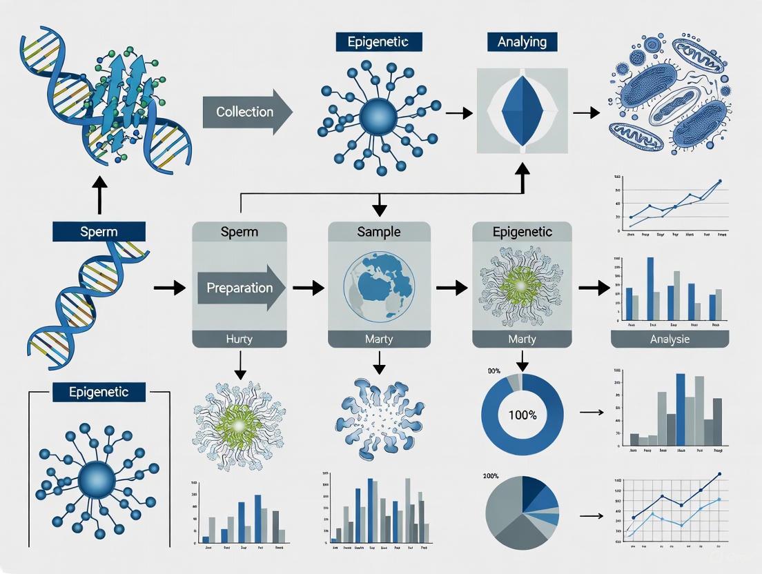
Diagram: Experimental Workflow for Sperm DNA Methylation Analysis
The diagram below illustrates a generalized workflow for profiling sperm DNA methylation, from sample collection to data integration.
Diagram: DNA Methylation Machinery in Spermatogenesis
This diagram outlines the key enzymes and processes that establish and maintain DNA methylation patterns during male germ cell development.
This case study investigates the sperm DNA methylation profile of patients with Kallmann Syndrome (KS) after gonadotropin or pulsatile GnRH therapy, contextualized within the challenges of epigenetic profiling research involving low sperm concentration [18] [19].
Table 1: Summary of Key DNA Methylation Findings in Kallmann Syndrome Sperm
| Metric | Finding in KS Patients vs. Healthy Controls | Notes / Associated Genes |
|---|---|---|
| Overall Methylation | Significantly higher [18] [19] | Reflects downstream epigenetic consequences of congenital hormone deficiency and its treatment [19]. |
| Differentially Methylated Regions (DMRs) | 4,749 total DMRs identified [18] | - |
| Hypermethylated DMRs | 4,020 [18] | Affects genes linked to neuronal function, migration, and GnRH secretion [18]. |
| Hypomethylated DMRs | 729 [18] | - |
| DMRs in Known KS-Related Genes | Present [18] | Includes CHD7, DCC, IL17RD, NELFA, and SEMA3E [18]. |
| Spermatogenesis-Related Genes | 1,938 identified within gene body [18] | Significant enrichment in chromosome remodeling pathways [18]. |
| Core Spermatogenesis Genes with Correlated Semen Parameters | BRCA1, H3FC3, HSP90AA1 [18] | Methylation status correlates with semen quality [18]. |
Table 2: Sperm Functional Index (SFI) Correlation with Standard Semen Parameters [20]
| Sperm Sample Category (by WHO criteria) | Percentage with Normal SFI | Percentage with Low SFI |
|---|---|---|
| All Normospermic Samples (n=342) | 57% | 37% |
| Stringent Normospermic Samples (≥50 million/mL, ≥50% motility, ≥14% morphology; n=81) | 67.9% | 22.2% |
Frequently Asked Questions (FAQs)
Q1: What is the primary epigenetic alteration found in the sperm of treated Kallmann Syndrome patients? The primary alteration is a significant increase in overall DNA methylation. A study identified 4,749 Differentially Methylated Regions (DMRs), with the vast majority (4,020) being hypermethylated. These DMRs affect genes crucial for neuronal function and GnRH secretion, as well as key KS-related genes like CHD7 and SEMA3E [18] [19].
Q2: Why should I be concerned about sperm concentration for epigenetic profiling? Sperm concentration is directly linked to the amount of high-quality DNA that can be isolated for downstream assays. Low concentration can lead to insufficient DNA yield, compromising data quality and reliability. Furthermore, even samples with normal concentration can have functional deficiencies, as shown by the Spermatozoa Function Index (SFI), where 37% of normospermic samples showed low molecular function [20].
Q3: My sperm sample has low concentration. What is the minimum cell number for chromatin analysis? The required cell number depends on the specific technique. While standard Chromatin Immunoprecipitation (ChIP) may require more cells, advanced methods like CUT&RUN are designed to work with far fewer. The CUT&RUN technique can successfully determine chromatin occupancy of a specific protein with approximately 500,000 cells [21].
Q4: After density gradient centrifugation, my sperm DNA yield is low. What could be the cause? This is a common challenge when processing low-concentration samples. The issue could be:
- Insufficient starting material: The initial sperm count may be too low for the standard protocol.
- Cell loss during washing steps: Pelleted sperm in low-concentration samples can be loose and easily lost. Consider reducing the number of washes or being exceptionally careful during supernatant removal.
- Inefficient lysis: Sperm cells have highly compacted chromatin, making DNA extraction difficult. Ensure your lysis buffer is appropriate and that lysis is complete [20] [22].
Troubleshooting Guides
Guide 1: Low DNA Yield from Low-Concentration Sperm Samples
Problem: Insufficient DNA is recovered after extraction for subsequent bisulfite sequencing or other epigenetic analyses.
Solutions:
- Maximize Input: Use the entire purified sperm pellet from the processing protocol. Avoid splitting samples.
- Optimize Lysis: Visually confirm complete cell lysis under a microscope if possible [22]. Ensure your lysis protocol is specifically validated for spermatozoa's tough membrane.
- Minimize Loss: Use carrier RNA or glycogen during precipitation steps to aid in visualizing and recovering small DNA pellets.
- Alternative Kits: Use DNA extraction kits validated for low-input or single-cell applications.
Guide 2: High Background/Noise in Chromatin Immunoprecipitation (ChIP)
Problem: The ChIP experiment results in high signal in the negative control (e.g., IgG) or non-specific genomic regions.
Solutions:
- Check Antibody Specificity: The most common cause is an antibody not qualified for ChIP. Not all antibodies that work for western blotting will work in ChIP [22].
- Optimize Chromatin Amount: Too much chromatin or antibody in the IP reaction can cause high background [22].
- Verify Sonication: Ensure chromatin is sheared to the appropriate fragment size (200–600 bp). Check sonication efficiency by running a sample on an agarose gel. Incomplete lysis or shearing can cause problems [22].
- Include Controls: Always run a no-antibody control and an IgG control to establish baseline background, and use a positive control antibody for a known genomic target to confirm protocol success [21].
Experimental Protocols
Protocol 1: Sperm Processing and DNA Extraction for Low-Concentration Samples
Objective: To isolate high-quality genomic DNA from human sperm with low concentration for reduced representation bisulfite sequencing (RRBS) or other methylome profiling.
Reagents:
- Sperm Washing Buffer (e.g., 1x Human Tubal Fluid (HTF))
- Discontinuous Density Gradient (e.g., 40% and 80% Percoll or Isolate)
- Phosphate-Buffered Saline (PBS)
- Lysis Buffer for sperm (e.g., containing SDS and DTT)
- DNA Extraction Kit (magnetic bead-based kits are recommended for low yields)
Methodology:
- Liquefaction: Allow freshly collected semen sample to liquefy for 30–60 minutes at 37°C [19].
- Purification: Layer 1 mL of semen over a discontinuous density gradient (1 mL 80% solution on bottom, 1 mL 40% on top). Centrifuge at 300 × g for 20 minutes [19].
- Wash: Carefully remove the supernatant and resuspend the sperm pellet in 5 mL of 1x HTF or PBS. Centrifuge at 200 × g for 5 minutes. Repeat this wash step once more [19].
- Lysis and DNA Extraction: Resuspend the final purified sperm pellet in a specialized lysis buffer. Proceed with genomic DNA extraction according to your chosen kit's instructions, eluting in a small volume (e.g., 20-30 µL) to maximize concentration [19].
Protocol 2: Reduced Representation Bisulfite Sequencing (RRBS)
Objective: To perform genome-wide DNA methylation analysis on sperm DNA.
Reagents:
- High-quality, extracted sperm DNA (concentration ≥ 50 ng/µL, A260/280 = 1.8–2.0) [19].
- Restriction Enzyme (e.g., MspI)
- Bisulfite Conversion Kit
- RRBS Library Prep Kit (e.g., Acegen Rapid RRBS Library Prep Kit)
- Library Quantification Kit
Methodology:
- DNA Digestion: Digest genomic DNA with a methylation-insensitive restriction enzyme (MspI) to enrich for CpG-rich regions [19].
- Library Construction: Perform end-repair, A-tailing, and adapter ligation to the digested fragments [19].
- Bisulfite Treatment: Treat the adapter-ligated library with bisulfite to convert unmethylated cytosines to uracils.
- PCR Amplification: Amplify the converted library.
- Quality Control and Sequencing: Validate the final library's size distribution and concentration before submitting for next-generation sequencing [19].
Signaling Pathways & Workflows
Experimental Workflow for KS Sperm Methylation Profiling
Biological Pathways Affected in KS
Research Reagent Solutions
Table 3: Essential Reagents for Sperm Epigenetic Profiling Experiments
| Reagent / Kit | Function / Application | Example/Note |
|---|---|---|
| Percoll / Isolate Sperm Separation Medium | Purification of motile sperm from semen using discontinuous density gradient centrifugation [20] [19]. | Creates 40% and 80% layers for separation. |
| FineMag Universal Genomic DNA Extraction Kit | Extraction of high-quality genomic DNA from purified sperm pellets [19]. | Magnetic bead-based method. |
| Acegen Rapid RRBS Library Prep Kit | Preparation of sequencing libraries for Reduced Representation Bisulfite Sequencing [19]. | Designed for methylation profiling. |
| NEB Next Ultra II DNA Library Prep Kit | Preparation of sequencing libraries for ChIP-seq or other NGS applications [23]. | For chromatin immunoprecipitated DNA. |
| Protein A or G Magnetic Beads | Affinity-based pull-down of antibody-protein complexes in ChIP assays [23]. | Used for immunoprecipitation. |
| Micrococcal Nuclease (MNase) | Enzyme used for chromatin digestion in techniques like Protect-seq or MNase-seq [23]. | Identifies inaccessible chromatin domains. |
| M.CviPI GpC Methyltransferase | Enzyme used for chromatin accessibility studies via nucleosome footprinting [23]. | Maps open chromatin regions. |
| Validated Antibodies (for ChIP/CUT&RUN) | Target-specific histone modifications or transcription factors. | Must be qualified for ChIP (e.g., H3K4me3, H3K27me3, H3K27ac) [21]. |
Frequently Asked Questions: Sperm Epigenetics in Research
Q1: Why should I profile epigenetic marks in samples with poor motility or morphology?
Aberrant epigenetic patterns are a major feature of dysfunctional sperm. Even if concentration is normal, poor motility (asthenozoospermia) or morphology (teratozoospermia) is often linked to epigenetic defects that can affect fertilization and embryo development. Research shows that abnormal DNA methylation in genes like MEST and DAZL is consistently associated with impaired sperm parameters, providing a molecular explanation for idiopathic infertility [1].
Q2: What are the key epigenetic marks to investigate in low-quality sperm samples? The three pillars of sperm epigenetics are:
- DNA Methylation: The addition of a methyl group to cytosine in CpG dinucleotides. Hypermethylation of genes like
MESTand hypomethylation of imprinted genes likeH19andGNASare linked to poor sperm quality [1]. - Histone Modifications: Post-translational modifications to histone proteins. Retention of histones with specific modifications (e.g., H3K4me3) at developmental gene promoters is crucial for embryogenesis [24].
- Non-coding RNAs (ncRNAs): Small RNAs that can carry epigenetic information to the embryo [1].
Q3: My sample has low motility. What specific epigenetic alterations should I anticipate? Studies comparing high-motile (HM) and low-motile (LM) sperm populations reveal consistent patterns. You may find:
- Altered DNA Methylation in Structural Genes: Methylation variation in genes functionally related to sperm DNA organization and chromatin maintenance [25].
- Repetitive Element Remodeling: Hypomethylation of satellite regions within pericentromeric positions, which is crucial for maintaining chromosome structure [25].
- Global Hypermethylation: A trend of broad DNA hypermethylation across multiple loci has been associated with poor sperm motility and concentration [25].
Q4: Can epigenetic defects in sperm affect embryo development? Yes, emerging evidence indicates that the sperm epigenome serves as a template for embryo development. Errors in the establishment of epigenetic marks, such as altered H3K4me3 at gene promoters, can lead to misregulation of gene expression in the early embryo and are implicated in developmental defects [24].
Troubleshooting Guide: Common Experimental Challenges
Problem: High background noise in DNA methylation analysis of low-concentration samples.
- Solution: Ensure thorough bisulfite conversion and use PCR protocols optimized for converted DNA. For genome-wide studies, the Methyl-binding domain (MBD) approach can be used to select for hypermethylated regions prior to sequencing, improving signal-to-noise ratio [25].
Problem: Inconsistent results when analyzing histone modifications.
- Solution: The histone-to-protamine exchange during spermatogenesis means only a small fraction of histones are retained in mature sperm (1% in mice, up to 15% in men) [24]. Use a sufficient number of cells and validated, high-affinity antibodies for chromatin immunoprecipitation (ChIP). Confirm the specificity of your assay with positive and negative control genomic regions.
Problem: Separating high and low motile sperm populations for comparative analysis.
- Solution: Use a Percoll or other density gradient centrifugation. This method has been successfully used to fractionate sperm into high and low motile populations, resulting in a significant improvement in velocity parameters (VSL, VCL, VAP) and amplitude of lateral head displacement (ALH) in the high-motile fraction [25].
Quantitative Data: Sperm Parameters and Associated Epigenetic Marks
The table below summarizes key genes with established links between their epigenetic status and specific sperm abnormalities.
Table 1: Genes with Impaired Methylation and Associated Sperm Abnormalities
| Condition | Gene Name | Epigenetic Alteration | Functional Role of Gene |
|---|---|---|---|
| Oligoasthenoteratozoospermia | MEST |
Hypermethylation [1] | Hydrolase activity [1] |
| Oligoasthenoteratozoospermia | GNAS |
Hypomethylation [1] | G-protein alpha subunit [1] |
| Oligozoospermia | DAZL |
Promoter Hypermethylation [1] | Germ cell development and differentiation [1] |
| Non-obstructive Azoospermia | SOX30 |
Hypermethylation [1] | Transcription factor for spermatogenesis [1] |
| Abnormal Motility/Morphology | H19 |
Hypomethylation [1] | Imprinted gene (IGF2 regulator) [1] |
| Low Motility (Bos taurus) | BTSAT4 |
Hypomethylation in HM sperm [25] | Repetitive satellite element, chromosome structure [25] |
Experimental Protocol: Genome-Wide Methylation Profiling of Sperm Populations
This protocol is adapted from a study on bovine sperm [25] and can be a guide for designing your experiment.
1. Sperm Sample Preparation and Fractionation
- Isolate sperm cells and fractionate into high-motile (HM) and low-motile (LM) populations using a Percoll gradient.
- Assess and record sperm quality parameters (e.g., VSL, VCL, VAP, ALH) for each population to confirm successful separation.
2. DNA Extraction and Methylation Enrichment
- Extract genomic DNA from the HM and LM sperm populations.
- Use a Methyl-binding domain (MBD) approach to enrich for hypermethylated genomic regions. This step is particularly useful for focusing on the highly methylated sperm genome.
3. Bisulfite Sequencing and Bioinformatics
- Perform bisulfite conversion on the enriched DNA to convert unmethylated cytosines to uracils.
- Prepare sequencing libraries and perform high-throughput sequencing.
- Map the sequenced reads to a reference genome and calculate cytosine methylation levels at single-base resolution.
- Identify Differentially Methylated Regions (DMRs) by comparing methylation patterns between HM and LM groups. A common threshold is a false discovery rate (FDR) of < 0.05.
Experimental Workflow and Signaling Pathways
Diagram 1: Sperm Epigenetic Analysis Workflow
Diagram 2: Sperm Epigenetic Marks and Embryonic Consequences
The Scientist's Toolkit: Essential Research Reagents
Table 2: Key Reagents for Sperm Epigenetic Profiling
| Reagent / Material | Function in Research | Example Application |
|---|---|---|
| Percoll Gradient | Separates sperm subpopulations based on density and motility. | Isolation of high and low motile sperm for comparative epigenetic analysis [25]. |
| MBD (Methyl-Binding Domain) Beads | Enriches for highly methylated DNA fragments from the genome. | Used prior to bisulfite sequencing to focus on methylated regions and improve data quality [25]. |
| Bisulfite Conversion Kit | Chemically converts unmethylated cytosine to uracil, allowing methylation status to be read via sequencing. | Fundamental step for whole-genome bisulfite sequencing or targeted methylation assays [1] [25]. |
| Antibodies for Histone Modifications | Binds specific histone post-translational modifications for enrichment and analysis. | Used in ChIP-seq to map the genome-wide location of marks like H3K4me3 in sperm [24]. |
| DNMT / TET Inhibitors | Chemical tools to manipulate the activity of enzymes that write or erase DNA methylation. | Used in model systems to study the cause-and-effect relationship between methylation and sperm function [1]. |
Frequently Asked Questions (FAQs) for Researchers
FAQ 1: What defines 'unexplained male infertility' in a research context, and what are its diagnostic boundaries?
Unexplained male infertility, often termed idiopathic infertility, is diagnosed when a male presents with the inability to achieve a pregnancy despite standard clinical evaluations returning normal results [26]. This includes a normal physical examination and semen analysis parameters (concentration, motility, morphology) according to World Health Organization guidelines [26] [27]. It is estimated that idiopathic factors account for 10% to 20% of male infertility cases, representing a significant gap in our diagnostic capabilities [26]. Essentially, it is a diagnosis of exclusion when routine tests cannot identify a cause.
FAQ 2: Beyond standard semen analysis, what emerging biomarkers show promise for investigating unexplained infertility?
Standard semen analysis often fails to explain all causes of infertility, as its power to predict fertility outcomes remains limited [28]. Emerging research focuses on molecular and epigenetic biomarkers:
- Sperm DNA Methylation: The stability of DNA methylation patterns at gene promoters is a crucial epigenetic regulator. A novel Epigenetic Sperm Quality Test (SpermQT) has been developed, which analyzes variability in 1,233 gene promoters [28]. This test can categorize sperm quality into "Excellent," "Average," and "Poor" based on the number of dysregulated promoters and has shown a significant correlation with intrauterine insemination (IUI) outcomes [28].
- Genetic Variants: Whole-genome sequencing of sperm from infertile men has revealed a higher burden of genomic variants compared to normozoospermic men [29]. Specific missense, frameshift, and nonsense mutations in genes critical for sperm flagellar function and motility (e.g., DNAJB13, MNS1, CFAP61, FSIP2) have been identified as potential biomarkers for sperm dysfunction, even in cases that might otherwise be classified as idiopathic [29].
FAQ 3: Our lab consistently encounters samples with low sperm concentration. What is a validated protocol for processing these samples for epigenetic profiling?
Processing samples with low sperm concentration requires careful purification to isolate sperm DNA free from somatic cell contamination, which is critical for accurate epigenetic analysis. The following workflow is adapted from validated research methodologies [28] [29]:
- Sample Purification: Use a 45%-90% PureSperm gradient for centrifugation (500 g for 20 minutes) to separate sperm from somatic cells and debris in the semen sample [29].
- Washing: Wash the resulting pellet twice with a suitable medium like Ham's F-10, containing serum albumin and antibiotics [29].
- Sperm Isolation (Swim-up): Overlay the pellet with more medium and incubate at 37°C. After 45 minutes, separate the supernatant, which contains motile sperm, from the pellet [29].
- DNA Isolation: Extract genomic DNA from the purified sperm using a commercial kit, such as the QIAamp DNA Mini Kit, with a specific lysis buffer containing DTT and Proteinase K to ensure efficient DNA release from sperm cells [29].
- Quality Control: A critical step is to verify the absence of somatic cell DNA contamination. This can be done by ensuring the mean methylation value of all CpG sites in the differentially methylated region of the DLK1 gene is less than 0.24, which is indicative of a pure sperm DNA sample [28].
FAQ 4: How does epigenetic sperm quality correlate with outcomes from different Assisted Reproductive Technologies (ART)?
Research indicates that the type of ART procedure can overcome epigenetic instability to varying degrees. The following table summarizes key findings from a study on DNA methylation variability and clinical outcomes [28]:
| Sperm Quality Category | Dysregulated Promoters | IUI Live Birth Rate | IVF/ICSI Live Birth Rate | Clinical Significance |
|---|---|---|---|---|
| Excellent | ≤ 3 | 44.8% | No significant difference found among groups | IUI is a viable option |
| Average | 4 - 21 | Intermediate Rate | No significant difference found among groups | Consider ART based on full clinical picture |
| Poor | ≥ 22 | 19.4% | No significant difference found among groups | IUI success is significantly lower; IVF/ICSI can overcome this deficit [28] |
The data strongly suggests that IVF with Intracytoplasmic Sperm Injection (ICSI) appears to bypass the negative impact of high epigenetic instability, as live birth rates were not significantly different among the sperm quality groups when this method was used [28].
Troubleshooting Guide: Common Experimental Challenges
Problem: Inconsistent DNA Methylation Array Results
- Potential Cause: Somatic cell contamination in the sperm sample. White blood cells or other somatic cells have vastly different methylation patterns than sperm and will confound results.
- Solution: Implement rigorous somatic cell removal during sample processing using a PureSperm gradient [29]. Always perform quality control on your isolated DNA by checking the methylation level of the DLK1 imprinting control region to confirm the sample is free of significant somatic DNA [28].
Problem: Low DNA Yield from Low-Concentration Sperm Samples
- Potential Cause: Standard DNA extraction protocols may be inefficient for the unique structure of sperm chromatin, which is highly compacted with protamines.
- Solution: Modify the standard kit protocol by using an initial lysis step with a buffer containing DTT (a reducing agent) and Proteinase K to effectively break down the dense sperm nuclear matrix and release DNA [29]. This improves yield and purity.
Problem: Unable to Correlate Genetic Data with Sperm Phenotype
- Potential Cause: Focusing on a single omics layer or a small number of candidate genes may miss the complex, polygenic nature of reproductive traits.
- Solution: Adopt a multi-omics, systems biology approach [28] [30] [29]. Integrate data from WGS, transcriptomics, and proteomics to pinpoint high-confidence candidate genes and pathways. Cross-omics concordance helps prioritize variants for deeper functional validation [29].
Experimental Workflow & Pathway Diagrams
Sperm Processing for Epigenetic Analysis
Diagnostic & Research Pathway for Idiopathic Infertility
The Scientist's Toolkit: Essential Research Reagents & Materials
The following table details key materials and their functions for investigating unexplained male infertility through epigenetic and genetic profiling.
| Research Reagent / Material | Function in Experimental Protocol |
|---|---|
| PureSperm Gradient | Density gradient medium for purifying sperm cells from seminal plasma and contaminating somatic cells (e.g., leukocytes), a critical step for clean epigenetic data [29]. |
| Ham's F-10 Medium | A balanced salt solution used for washing and suspending sperm pellets during processing, helping to maintain cell viability [29]. |
| QIAamp DNA Mini Kit | A commercial silica-membrane-based system for the isolation of high-quality genomic DNA from purified sperm cells [29]. |
| Dithiothreitol (DTT) | A reducing agent added to the lysis buffer to break the disulfide bonds in sperm protamines, enabling efficient release of DNA [29]. |
| Proteinase K | A broad-spectrum serine protease used to digest proteins and nucleases during cell lysis, facilitating DNA liberation and stability [29]. |
| Infinium MethylationEPIC Array | A microarray platform used for genome-wide DNA methylation analysis, covering over 850,000 CpG sites to identify epigenetic variability [28]. |
| DLK1 Region Probes | Specific genomic probes used as a quality control metric to detect somatic cell contamination in sperm DNA samples based on methylation signature [28]. |
Robust Methodologies for Epigenetic Analysis in Limited Sperm Samples
Sperm separation is a critical preparatory step in assisted reproductive technology (ART), aimed at isolating motile, morphologically normal, and genetically intact sperm from seminal plasma for procedures such as intrauterine insemination (IUI), in vitro fertilization (IVF), and intracytoplasmic sperm injection (ICSI) [31]. Effective semen preparation methods separate spermatozoa from seminal plasma and other constituents that might inhibit fertilization, including moribund and immature sperm cells, leucocytes, and bacteria [31]. Among conventional methods, density gradient centrifugation (DGC) and swim-up are well-established, while microfluidic sorting represents a more recent advancement [32] [31]. The choice of technique significantly impacts sperm quality, influencing sperm DNA fragmentation (sDF) and reactive oxygen species (ROS) levels, which are crucial for successful fertilization, embryo development, and clinical pregnancy rates [32] [33]. This resource provides a technical guide for researchers, focusing on applying density gradient centrifugation to samples with varying motile populations within the context of epigenetic profiling research.
Comparative Analysis of Separation Techniques
Performance Metrics Across Techniques
The table below summarizes key performance outcomes from comparative studies, highlighting the efficacy of different sperm preparation methods.
Table 1: Comparative Performance of Sperm Preparation Techniques
| Technique | Total Motility (%) | Progressive Motility (%) | DNA Fragmentation Index (DFI) (%) | Key Advantages | Key Limitations |
|---|---|---|---|---|---|
| Density Gradient Centrifugation (DGC) | 70.1 ± 3.5 [32] | 58.4 ± 3.1 [32] | 25.6 ± 2.3 (Fresh) [32] | Efficient for diverse sample qualities; improves motility in hyperuricemia [34]; removes debris/bacteria [31]. | Centrifugation may increase ROS and sDF [32] [33]. |
| Swim-Up | ~85.3 (Inferred) [32] | ~72.5 (Inferred) [32] | 15.4 ± 1.8 (Fresh) [32] | Simple, economical; selects highly motile sperm [31]. | Low recovery in oligoasthenozoospermia [31]. |
| Microfluidic Sorting | 85.3 ± 3.2 [32] | 72.5 ± 2.8 [32] | 8.2 ± 1.5 (Fresh) [32] | Minimal mechanical stress; preserves DNA integrity [32] [35]. | Early devices had complex fabrication and low throughput [32] [35]. |
DNA Fragmentation: A Critical Consideration
Sperm DNA fragmentation is a paramount concern for epigenetic profiling and embryo viability. Research indicates that DGC can increase sDF in approximately 50% of samples, a phenomenon linked to a 50% lower pregnancy probability [33]. This increase is attributed to centrifugation-induced oxidative stress [32]. In contrast, swim-up and microfluidic techniques are gentler, resulting in significantly lower post-processing DFI [32] [33]. Analyzing viable sDF (DNA fragmentation in live sperm) provides a more accurate assessment of damage post-selection than total sDF, as it is not confounded by the removal of dead spermatozoa [33].
Detailed Protocol: Density Gradient Centrifugation
Reagents and Materials
Table 2: Essential Research Reagent Solutions for DGC
| Reagent/Material | Function/Description | Example Product (Supplier) |
|---|---|---|
| Density Gradient Medium | Silane-coated colloidal silica solution forming discontinuous layers for sperm separation based on density. | ISolate (Fujifilm Irvine Scientific) [36], PureSperm (Nidacon) [33], SpermGrad (Vitrolife) [34] |
| Sperm Washing Medium | Buffered salt solution for washing and resuspending sperm post-centrifugation; supports sperm viability. | Modified HTF Medium [36], SpermRinse (Vitrolife) [34] |
| Conical Centrifuge Tubes | Sterile tubes for creating density gradients and conducting centrifugation. | 15 mL conical tubes [32] |
Step-by-Step Experimental Workflow
Step 1: Gradient Preparation Prepare a discontinuous gradient by carefully layering solutions of different densities in a sterile conical centrifuge tube. A typical configuration uses 1.0 - 1.5 mL of a lower-density solution (e.g., 45% or 50%) over 1.0 - 1.5 mL of a higher-density solution (e.g., 80% or 90%), taking care not to mix the layers [32] [34] [33]. Use commercially available solutions or dilute stock solutions per manufacturer instructions (e.g., to make 45% from 90%, mix 1:1 with medium) [36] [37].
Step 2: Sample Layering and Centrifugation Thoroughly mix the liquefied semen sample. Gently layer 1-2 mL of the raw semen on top of the prepared density gradient. Centrifuge the tube at 300 × g for 15 minutes at room temperature [32] [34]. This force allows denser, motile, and morphologically normal sperm to pass through the gradient and form a pellet, while other components are retained in the upper layers or at interfaces.
Step 3: Pellet Washing and Resuspension After centrifugation, carefully aspirate and discard the supernatant. Resuspend the resulting sperm pellet in 2-3 mL of sperm washing medium. Centrifuge again at 300 × g for 5-10 minutes to wash away residual gradient material [34] [33]. Discard the supernatant and resuspend the final purified sperm pellet in a suitable buffer (e.g., 0.3-0.5 mL of G-IVF PLUS) for subsequent analysis or use in ART [34].
Troubleshooting and FAQs
Q1: Our post-DGC sperm recovery is low, especially from oligozoospermic samples. How can we optimize this? A: Low recovery is a known limitation of DGC in severe oligozoospermia [31]. To mitigate this, consider using "mini-gradient" protocols with reduced volumes (e.g., 0.5 mL per layer and 0.5 mL semen) to concentrate the sperm population [31] [37]. Ensure the initial sample is well-mixed and layered carefully to prevent premature mixing with the gradient. For samples with extremely low counts, simple washing or direct microfluidic processing might be more appropriate to maximize recovery, though at the potential cost of purity [31] [35].
Q2: We observe high DNA fragmentation in sperm after DGC. What is the cause, and how can it be reduced? A: High post-DGC DNA fragmentation is likely due to centrifugation-induced oxidative stress generating reactive oxygen species (ROS) [32] [33]. To reduce this:
- Minimize centrifugal force: Use the minimum required g-force and time (e.g., 300 × g for 15 min is standard) [32].
- Consider alternative methods: For epigenetic profiling research where DNA integrity is paramount, gentler techniques like swim-up (for high-quality samples) or microfluidic sorting are recommended, as they yield significantly lower DFI [32] [33].
- Assay viable sDF: Implement a LiveTUNEL assay to accurately measure DNA damage in viable sperm populations post-selection, as this unmask damage that total sDF might obscure [33].
Q3: How does DGC specifically benefit samples from populations with metabolic conditions like hyperuricemia (HUA)? A: DGC demonstrates a specific therapeutic effect in HUA-associated sperm dysfunction. HUA impairs sperm motility via oxidative stress and metabolic dysregulation. While baseline progressive motility (PR%) is often lower in HUA samples, DGC processing can increase PR% to over 90%, with a significantly greater improvement (ΔPR%) in HUA groups compared to controls [34]. This effect is likely due to DGC's capacity to scavenge ROS and optimize the cellular energy supply during processing.
Q4: When should we choose DGC over swim-up or microfluidics? A: The choice depends on sample quality and research objectives. The following decision tree can guide method selection:
Density gradient centrifugation remains a powerful and versatile workhorse for sperm separation, particularly effective for samples with compromised motility, such as in hyperuricemia, and for processing infectious samples [31] [34]. However, researchers must be vigilant about its potential to induce sperm DNA fragmentation via oxidative stress during centrifugation [32] [33]. For research focused on epigenetic profiling, where the integrity of the paternal genome is paramount, the choice of sperm separation technique is critical. While DGC offers robust recovery, gentler methods like swim-up or advanced microfluidic chips may be superior for isolating sperm with the highest DNA integrity, ultimately providing a more reliable biological material for downstream epigenetic analyses [32] [38] [33].
DNA Extraction and Quality Control for Low-Input Samples
Frequently Asked Questions (FAQs)
Q1: Why is somatic cell contamination a particular concern for sperm epigenetic studies? Sperm and somatic cells have vastly different DNA methylation patterns. Sperm DNA is hypomethylated in many promoter regions, while somatic cell DNA is typically highly methylated in these same areas. Even low-level contamination (below 5% of total cells) can significantly bias methylation analysis, leading to false interpretations of hypermethylation in sperm samples. This risk is heightened in oligozoospermic samples where somatic cells may constitute a greater proportion of the total cell population [39] [40].
Q2: What are the critical pre-analytical factors affecting DNA yield from low-input samples? Sample quality and handling before extraction significantly impact DNA yield. Key factors include: using EDTA rather than heparin as an anticoagulant (heparin inhibits downstream reactions), proper storage conditions (samples should be processed immediately or frozen at -80°C to prevent degradation), and patient factors (samples from pediatric or immunocompromised patients may naturally contain fewer white blood cells, yielding less DNA) [41] [42].
Q3: How can I improve DNA yield from low-cell-count samples? For samples with low cell counts, you can: increase the input volume where possible (e.g., double the blood volume), extend the lysis incubation time to 30 minutes at 56°C, ensure reagents like Proteinase K are fresh and active, and use specialized "low input" protocols that reduce buffer volumes to maintain optimal DNA concentration for binding efficiency [41] [42].
Q4: What quality control metrics should I check for extracted DNA intended for epigenetic assays? Beyond standard concentration measurements (preferably using Qubit rather than Nanodrop for accuracy), check A260/280 and A260/230 ratios. A260/280 < 1.6 suggests protein contamination, while A260/230 < 2.0 indicates residual salts or organic compounds. For epigenetic applications like methylation profiling, ensure sufficient DNA quantity (typically ≥500ng) as these assays rely on detecting subtle genomic changes that become statistically insignificant with low input [41].
Troubleshooting Guides
Problem: Low DNA Yield
| Observed Issue | Potential Causes | Recommended Solutions |
|---|---|---|
| Low yield from frozen cell pellets | Pellet thawed/resuspended too abruptly; cells lost | Thaw pellets slowly on ice; use cold PBS for gentle resuspension; pipette up and down 5-10 times until uniformly turbid [43]. |
| Low yield from blood samples | Sample aging; DNase activity; improper handling | Use fresh whole blood (<1 week old); add lysis buffer directly to frozen samples; follow species-specific protocols to prevent hemoglobin precipitate formation [43] [42]. |
| Low yield from tissue samples | Large tissue pieces; membrane clogging; nuclease degradation | Cut tissue into smallest possible pieces; grind with liquid nitrogen; centrifuge lysate to remove fibers; use proper storage (-80°C) [43]. |
| Column-based extraction failures | Column overload; incomplete binding; incorrect lysis volume | Reduce input material for DNA-rich tissues; ensure appropriate lysis volume for cell count; use wide-bore tips for HMW DNA [43] [42]. |
Problem: DNA Quality Issues
| Observed Issue | Potential Causes | Recommended Solutions |
|---|---|---|
| DNA degradation | Improper sample storage; high nuclease content; extended heating | Flash-freeze samples in liquid nitrogen; store at -80°C; process tissues immediately; limit heating times during resuspension [43] [42]. |
| Protein contamination | Incomplete digestion; fibrous tissues; membrane clogging | Extend digestion time (30min-3hrs) after tissue dissolves; centrifuge lysate to remove fibers; use recommended input amounts [43]. |
| Salt contamination | Guanidine salt carryover; buffer contact with upper column | Avoid touching upper column area with pipette tip; transfer lysate without foam; close caps gently to prevent splashing [43]. |
| RNA contamination | Too much input material; insufficient lysis time | Use recommended input amounts; extend lysis time by 30min-3hrs to improve RNase A efficiency [43]. |
Comprehensive Workflow for Sperm DNA Extraction and QC
The following workflow integrates physical processing, chemical treatment, and computational analysis to ensure high-quality sperm DNA for epigenetic studies:
Figure 1: Comprehensive workflow for sperm DNA extraction and quality control for epigenetic studies.
Somatic Contamination Assessment Using DNA Methylation Biomarkers
For research focusing on sperm epigenetic profiling, assessing somatic cell contamination through DNA methylation biomarkers is essential. The comparison of Infinium Human Methylation 450K BeadChip data for sperm and blood samples identified 9,564 CpG sites that are highly methylated in blood (>80%) but minimally methylated in sperm (<20%) and not differentially methylated in infertility. These can serve as sensitive markers for detecting somatic DNA contamination [39] [40].
Key Biomarker Application:
- When performing whole-genome methylation sequencing or microarray analysis, monitor a panel of these biomarker CpG sites
- Apply a 15% methylation cutoff during data analysis to eliminate samples with significant somatic contamination
- This bioinformatic checkpoint complements physical separation methods to ensure data integrity
Research Reagent Solutions for Low-Input Samples
| Reagent/Kit | Specific Function | Application Notes |
|---|---|---|
| Somatic Cell Lysis Buffer (0.1% SDS, 0.5% Triton X-100) | Selective lysis of somatic cells while preserving sperm integrity | Incubate 30min at 4°C; repeat if microscopic examination shows residual contamination [39] [40]. |
| Proteinase K | Digests nuclear proteins for DNA release | Use fresh aliquots; extend digestion time (30min-3hrs) for fibrous tissues; adjust volume based on tissue type [43] [5]. |
| Magnetic Bead-Based Extraction Kits | DNA binding and purification with higher recovery than columns | Particularly effective for low-input samples; better yields with minimal handling loss [41] [42]. |
| RNase A | Removes RNA contamination that can affect quantification and downstream applications | Add after protein digestion; incubate at 37°C for 60min; essential for accurate DNA quantification [5]. |
| Wide-Bore Pipette Tips | Handling high molecular weight DNA without shearing | Critical for maintaining DNA integrity; avoid vortexing with HMW DNA [42]. |
| Sperm Separation Media (Percoll/Isolate gradients) | Isolates sperm from round cells and debris | Use discontinuous density gradients (40%/80%); centrifuge at 300g for 20min [19] [20]. |
Technical Protocols for Key Procedures
Protocol 1: Somatic Cell Lysis from Semen Samples
- Wash fresh semen samples twice with 1X PBS by centrifugation at 200g for 15min at 4°C
- Inspect sample under microscope (20X objective) to assess somatic cell contamination level
- Incubate with freshly prepared somatic cell lysis buffer (0.1% SDS, 0.5% Triton X-100 in ddH2O) for 30min at 4°C
- Re-examine under microscope to confirm somatic cell removal
- If somatic cells persist, repeat centrifugation and SCLB treatment
- Pellet purified sperm by centrifugation, followed by PBS wash [39] [40]
Protocol 2: Salt-Based DNA Extraction from Low-Input Sperm
- Digest 5μL sperm pellet overnight at 55°C in lysis solution (SSTNE buffer + 10% SDS + Proteinase K)
- Add 5μL RNase A (2mg/mL) and incubate at 37°C for 60min
- Precipitate proteins by adding 0.7 volume of 5M NaCl
- Transfer 400μL supernatant to new tube and precipitate DNA with equal volume isopropanol
- Centrifuge at 14,000g for 5min, wash DNA pellet with ethanol
- Resuspend in TE buffer or nuclease-free water [5]
Protocol 3: Comprehensive Quality Control Assessment
- Quantification: Use Qubit fluorometer with dsDNA HS Assay for accurate concentration measurement of low-concentration samples
- Purity Assessment: Check A260/280 ratio (ideal: 1.8-2.0) and A260/230 ratio (ideal: 2.0-2.2) using spectrophotometry
- Integrity Verification: Run agarose gel electrophoresis to confirm high molecular weight DNA without smearing
- Functional QC: For epigenetic studies, analyze control CpG sites known to be differentially methylated between sperm and somatic cells [39] [41]
For researchers investigating male infertility, particularly studies involving precious samples with low sperm concentration or compromised DNA quality, selecting the appropriate epigenomic profiling tool is a critical first step. DNA methylation, a key epigenetic mark, plays a fundamental role in spermatogenesis and gamete function [1]. Aberrant methylation patterns in sperm have been consistently linked to impaired spermatogenesis and poor sperm quality, including issues with motility, morphology, and DNA integrity [1] [44].
Two powerful sequencing-based methods dominate the field for genome-wide DNA methylation analysis: Reduced Representation Bisulfite Sequencing (RRBS) and Whole-Genome Bisulfite Sequencing (WGBS). Both methods rely on bisulfite conversion chemistry, where unmethylated cytosines are converted to uracils (and read as thymines after PCR), while methylated cytosines remain unchanged [45]. The choice between them involves a careful trade-off between genomic coverage, resolution, cost, and data analysis requirements. This guide provides a detailed comparison and troubleshooting resource to help you successfully implement these techniques in your research on male fertility.
Technical Comparison: RRBS vs. WGBS at a Glance
The table below summarizes the core technical specifications and performance characteristics of RRBS and WGBS to guide your selection.
Table 1: Technical Comparison of RRBS and WGBS for DNA Methylation Profiling
| Feature | Reduced Representation Bisulfite Sequencing (RRBS) | Whole-Genome Bisulfite Sequencing (WGBS) |
|---|---|---|
| Fundamental Principle | Uses restriction enzymes (e.g., MspI) to digest genome, enriching for CpG-rich regions prior to bisulfite sequencing [46] [47]. | Subjects the entire genome to bisulfite conversion and sequencing, without prior enrichment [48] [45]. |
| Genomic Coverage | Targeted; covers ~1-3% of the genome, focusing on CpG islands, promoters, and other CpG-dense regions [46] [47]. | Comprehensive; covers >90% of CpGs in the genome, including intergenic and low-CpG-density regions [48] [49]. |
| Resolution | Single-base resolution for the regions it covers [48] [47]. | Truly genome-wide, single-base resolution [48] [45]. |
| Ideal for Sperm Research | Cost-effective profiling of methylation changes in gene promoters and CpG-rich areas associated with spermatogenesis [44]. | Unbiased discovery of methylation defects across the entire sperm genome, including imprinted gene clusters [1]. |
| Typical Input DNA | 10-200 ng [49]. Can be adapted for low input. | 10-200 ng; however, higher inputs may yield better coverage [49] [45]. |
| Relative Cost | Lower (sequences only a fraction of the genome) [46] [47]. | Higher (sequences the entire genome) [48] [46]. |
| Key Limitation | Bias towards high-CpG-density regions; may miss biologically relevant changes in low-density areas [48] [46]. | Higher cost and data load; requires significant computational resources for analysis [48] [45]. |
Decision Framework and Experimental Workflow
The following diagram illustrates the key decision points for selecting and implementing RRBS or WGBS in your research on low sperm concentration.
Frequently Asked Questions (FAQs) and Troubleshooting
FAQ 1: How do we handle low sperm concentration and DNA quantity for these assays?
Challenge: Semen samples from infertile patients often yield low concentrations of sperm and, consequently, low amounts of DNA, which can be further degraded during the harsh bisulfite conversion process [50].
Solutions:
- Optimize DNA Extraction: Use dedicated protocols for sperm cells, often involving density gradient centrifugation for isolation, as used in a recent asthenospermia study [44].
- Validate DNA Quality: Prior to library prep, use a QC method like the PCR-based assay that evaluates amplification success across different amplicon lengths to assess the degree of bisulfite-induced fragmentation [50].
- Consider Enzymatic Conversion: For extremely low-input samples (as low as 100 pg), consider Enzymatic Methyl-seq (EM-seq). This method avoids the DNA-damaging extremes of pH and temperature used in traditional bisulfite conversion, resulting in higher library yields, longer insert sizes, and better CpG coverage from minimal input [49].
- Follow Input Guidelines: Adhere to kit-specific protocols for low DNA input. Using too much bisulfite-converted DNA in a PCR reaction (e.g., >500 ng) can be counterproductive; recommended inputs are often in the 2-4 µl range of eluted DNA [51].
FAQ 2: Our bisulfite conversion efficiency is low, leading to unreliable data. What went wrong?
Challenge: Incomplete bisulfite conversion is a major source of technical variability and leads to overestimation of methylation levels [50] [45].
Troubleshooting Guide:
- Ensure DNA Purity: Particulate matter in the DNA sample can inhibit conversion. If present, centrifuge the sample at high speed and use only the clear supernatant for the conversion reaction [51].
- Verify Reaction Conditions: Ensure all liquid is at the bottom of the tube and not on the cap or walls. Use a commercial bisulfite conversion kit known for robustness and always include controls [51] [45].
- Spike-in Control: Spike your sample with an unmethylated λ-bacteriophage or other non-methylated DNA. The conversion rate can then be precisely calculated by analyzing the C-to-T conversion rate in this control, with targets typically >99.5% [45].
- Avoid Over-degradation: Overly aggressive bisulfite treatment (long incubation, high temperature) can completely fragment DNA. If using a non-kit protocol, ensure a balanced approach that maximizes conversion while minimizing degradation [50].
FAQ 3: We are getting high duplication rates and poor coverage in our WGBS/RRBS libraries. How can we improve this?
Challenge: High duplication rates and patchy genome coverage often stem from low library complexity, which can be exacerbated by DNA degradation during bisulfite treatment [49].
Solutions:
- Check DNA Fragmentation: For WGBS, the bisulfite process itself fragments DNA. If performing additional shearing (e.g., sonication), optimize conditions to avoid over-fragmenting, which reduces complexity.
- Use Post-Bisulfite Adaptor Tagging (PBAT): This method involves adding sequencing adaptors after bisulfite conversion, which can improve library yields from degraded samples by using the converted fragments more efficiently [49].
- Switch to EM-seq: As noted in FAQ 1, EM-seq produces significantly less DNA damage, leading to higher-complexity libraries with lower duplication rates and superior coverage, especially in high-GC regions [49].
- Optimize PCR Amplification: Use a minimal number of PCR cycles. Employ polymerases suitable for bisulfite-converted DNA (e.g., hot-start Taq polymerase) and avoid proof-reading enzymes, as they cannot read through uracil [51].
Table 2: Key Research Reagent Solutions for RRBS and WGBS Experiments
| Item | Function | Considerations for Sperm Research |
|---|---|---|
| Methylation-Sensitive Restriction Enzyme (e.g., MspI) | Digests genomic DNA for RRBS, enriching for CpG-rich fragments [47] [49]. | Enzyme choice defines genomic representation. MspI (cuts CCGG) is standard, but other enzymes can bias coverage towards promoters or gene bodies [46]. |
| Sodium Bisulfite | Chemical reagent that converts unmethylated cytosine to uracil, enabling methylation detection [45]. | Highly degrading; use high-purity reagents and controlled conditions to preserve scarce sperm DNA [50] [49]. |
| Bisulfite Conversion Kit | Commercial kit optimized for complete conversion and DNA cleanup. | Simplifies workflow and improves reproducibility. Essential for handling multiple low-concentration samples. |
| Specialized Polymerase (e.g., Hot-Start Taq) | Amplifies bisulfite-converted DNA for library construction [51]. | Must be able to read templates containing uracil (dUTP). Proof-reading polymerases are not recommended [51]. |
| Methylated & Unmethylated Control DNA | Positive and negative controls for bisulfite conversion efficiency and assay validation. | Crucial for verifying the entire workflow, especially when working with novel patient cohorts. |
| Bioinformatics Tools (e.g., Bismark, BS-Seeker2) | Aligns bisulfite-converted reads to a reference genome and calls methylated cytosines [52] [47]. | Standard aligners cannot be used. Bismark is a widely used, accurate option, though it can be computationally intensive for WGBS [52] [47]. |
Detailed Protocol: RRBS for Sperm DNA
This protocol is adapted from methodologies used in recent studies on asthenospermia and oligoasthenospermia [44].
Step 1: Sperm Isolation and DNA Extraction
- Collect semen samples after 2-7 days of sexual abstinence.
- Isolate sperm cells using discontinuous density gradient centrifugation (e.g., 40% and 80% Percoll layers) per WHO guidelines [44].
- Extract genomic DNA from the purified sperm cell pellet using a standard phenol-chloroform protocol or a commercial kit designed for genomic DNA.
- Quantify DNA using a fluorometer and assess purity via spectrophotometry (260/280 ratio ~1.8).
Step 2: RRBS Library Preparation
- Digestion: Digest 10-100 ng of high-quality sperm genomic DNA with the MspI restriction enzyme.
- End-Repair and A-Tailing: Perform end-repair on the digested fragments and add an 'A' base to the 3' ends.
- Adaptor Ligation: Ligate methylated sequencing adaptors to the A-tailed fragments. Methylated adaptors are resistant to digestion in subsequent steps.
- Size Selection: Use bead-based clean-up to select a size range of fragments (e.g., 150-300 bp) to enrich for CpG-rich regions.
- Bisulfite Conversion: Treat the size-selected DNA with sodium bisulfite using a commercial kit. This is the critical step where unmethylated Cs are converted to Us.
- PCR Amplification: Amplify the converted libraries using a polymerase suitable for bisulfite-converted templates for a limited number of cycles (e.g., 12-15) to enrich for adaptor-ligated fragments.
- Library QC: Validate the final library using a Bioanalyzer or TapeStation and quantify by qPCR.
Step 3: Sequencing and Data Analysis
- Sequence the libraries on an appropriate Illumina platform to obtain single-end or paired-end reads.
- Analyze the data using a standardized pipeline [47]:
- Quality Control: Use FastQC and Trim Galore! to assess read quality and trim adaptors.
- Alignment: Map bisulfite-converted reads to a reference genome (e.g., hg38) using a specialized aligner like Bismark or BS-Seeker2 [47].
- Methylation Calling: Extract methylation calls for each cytosine in a CpG context from the aligned BAM files.
- Differential Methylation: Use packages like
DSSordmrseqin R to identify Differentially Methylated Regions (DMRs) between case and control groups [52]. Recent studies in male infertility have successfully identified DMRs in genes like BDNF, RBMX, and ASZ1 using this approach [44].
Frequently Asked Questions (FAQs)
Q1: What are the primary challenges when constructing sequencing libraries from low-concentration sperm samples? The main challenges include obtaining sufficient high-quality genetic material, minimizing amplification bias, and preserving epigenetic information. Low sperm concentration directly reduces the amount of available DNA and RNA, making subsequent library construction difficult. Amplification of these limited materials can introduce significant bias and noise, while suboptimal handling may lead to the loss of valuable epigenetic markers such as DNA methylation patterns. [53] [54]
Q2: My sperm samples have very low motility. Are there any novel technologies that can help select the best cells for analysis? Yes, emerging microfluidic technologies show great promise. For samples with extremely low motility (e.g., only 1% live sperm), a high-throughput, label-free sperm selection system has been developed. This system uses microfluidic droplet technology and deformable hydrogel materials to analyze the metabolic activity of single cells, enabling the selection of live sperm with over 90% accuracy. In validation studies, this technology improved the average percentage of live sperm in processed samples from 1% to 76%, significantly enhancing subsequent fertilization and embryonic development success rates. [55]
Q3: What key factors should I consider during sample preparation to avoid damaging low-input sperm samples? When preparing low-input sperm samples, pay close attention to the following:
- Temperature Control: Sperm are highly sensitive to temperature. The testicles' optimal operating temperature is 32-35°C, significantly lower than core body temperature. Brief heat exposure can activate transposons and cause DNA damage. Avoid any unnecessary temperature increases, such as from hot baths, saunas, or improper sample handling. [54] [56]
- Physical Stress: Minimize excessive centrifugation and pipetting, which can further damage fragile sperm cells.
- Processing Time: Reduce the time between sample collection and processing to maintain cell viability and epigenetic integrity. [56] [57]
Q4: How does male age impact the success of library construction and amplification for epigenetic profiling? Advanced paternal age can affect both genetic and epigenetic quality. Research indicates that men aged 25-35 typically have the best sperm quality. After age 40, sperm DNA fragmentation rates increase, and epigenetic modifications may become more unstable. These age-related changes can lead to increased sequencing errors, higher background noise during library construction, and potential biases in epigenetic data interpretation. [56] [57]
Troubleshooting Guides
Issue 1: Insufficient DNA/RNA Yield from Low-Concentration Samples
Problem: After extracting genetic material from low-concentration sperm samples, the quantity is insufficient for standard library construction protocols.
Solutions:
- Utilize Whole Genome Amplification (WGA): For extremely limited DNA, employ multiple displacement amplification (MDA) based WGA technology. This method uses φ29 DNA polymerase and random hexamer primers for highly efficient amplification with minimal bias.
- Implement Carrier RNA Strategy: Add carrier RNA during the extraction process to reduce surface adsorption losses, then remove it before library construction.
- Apply Microscale Extraction Kits: Use specialized kits designed for low-input samples, which optimize reagent ratios and reaction volumes to improve recovery rates.
Preventive Measures:
- Pre-extraction quality assessment using sensitive fluorescence quantification methods
- Optimize cell lysis conditions to maximize release of genetic material
- Implement strict QC checkpoints before proceeding to library construction
Issue 2: High Amplification Bias in Low-Input Samples
Problem: Significant bias occurs during PCR amplification of low-input samples, resulting in uneven genome coverage and compromised data quality.
Solutions:
- Optimize PCR Conditions: Reduce PCR cycle numbers and increase initial template input as much as possible. Use high-fidelity enzymes with strong processivity.
- Employ Unique Molecular Identifiers (UMIs): Incorporate UMIs during reverse transcription or early amplification stages to correct for amplification bias and duplicate reads during data analysis.
- Utilize Linear Preamplification: Implement non-exponential preamplification strategies such as in vitro transcription (IVT) to initially amplify material while maintaining relative abundance relationships.
Optimization Workflow:
- Determine minimum input requirement for your specific application
- Test different polymerase systems for bias characteristics
- Validate with known control samples to quantify bias
- Implement computational correction methods
Issue 3: Poor Library Complexity from Limited Starting Material
Problem: Libraries constructed from low-input sperm samples show poor complexity, with high duplicate rates and inadequate genome coverage.
Solutions:
- Implement Tagmentation-Based Methods: Use transposase-based library construction methods (such as ATAC-seq) that simultaneously fragment and tag DNA, reducing steps and improving efficiency.
- Optimize Size Selection: Use double-sided size selection to remove too short or too long fragments, improving library uniformity.
- Apply Duplicate Removal Strategies: Use UMIs to distinguish true biological duplicates from PCR duplicates during data analysis.
Quality Control Metrics:
- Establish minimum complexity thresholds for proceeding with sequencing
- Use spike-in controls to monitor amplification efficiency
- Implement sequencing saturation analysis to determine optimal sequencing depth
Table 1: Impact of Sperm Quality on Assisted Reproduction Outcomes
| Parameter | Normal Range | Suboptimal Range | Critical Level | Clinical Impact |
|---|---|---|---|---|
| Sperm Concentration | ≥15 million/mL | 5-15 million/mL | <5 million/mL | Directly affects fertilization success [54] [56] |
| Progressive Motility (A+B) | ≥32% | 10-32% | <10% | Reduces likelihood of natural conception [56] |
| Normal Morphology | ≥4% | 1-4% | <1% | Associated with fertilization failure [56] |
| DNA Fragmentation Index | <15% | 15-30% | >30% | Linked to increased miscarriage rates [56] [57] |
Table 2: Comparison of Amplification Methods for Low-Input Samples
| Method | Minimum Input | Advantages | Limitations | Best Applications |
|---|---|---|---|---|
| Multiple Displacement Amplification (MDA) | 1-10 cells | Uniform coverage, low amplification bias | Chimera formation, over-representation of small fragments | Whole genome sequencing, methylation analysis |
| PCR-Based WGA | Single cell | High efficiency, rapid | Significant amplification bias, shorter fragments | Target sequencing, mutation detection |
| Linear Amplification via IVT | 10-100 cells | Maintains relative abundance | RNA only, complex procedure | Transcriptome analysis, single-cell RNA-seq |
| Tagmentation-Based Library Prep | 100-1000 cells | Simple workflow, fast | Insert size bias, sequence preference | ATAC-seq, epigenomic profiling |
Experimental Protocols
Protocol 1: Low-Input Sperm Whole Genome Amplification
Principle: This protocol uses φ29 DNA polymerase for isothermal amplification, enabling efficient whole genome amplification from minimal sperm samples while maintaining relatively uniform coverage and integrity, suitable for subsequent sequencing library construction.
Materials and Reagents:
- φ29 DNA polymerase and reaction buffer
- Random hexamer primers
- dNTP mix
- Pyrophosphatase
- Single-strand binding protein
- DNA clean-up beads or columns
Procedure:
- Sample Preparation: Extract DNA from low-concentration sperm samples and quantify using sensitive fluorescence methods. If the sample volume is extremely small, proceed directly to step 2 without quantification.
- Denaturation: Dilute DNA in nuclease-free water, heat at 95°C for 3 minutes, then immediately place on ice.
- Amplification Reaction Setup:
- 10× φ29 buffer: 5 μL
- dNTPs (10 mM each): 2 μL
- Random hexamers (100 μM): 2 μL
- Single-strand binding protein (10 μg/μL): 1 μL
- Inorganic pyrophosphatase (0.1 U/μL): 1 μL
- φ29 DNA polymerase (10 U/μL): 2 μL
- Template DNA: X μL (1-10 ng recommended)
- Nuclease-free water to 50 μL
- Amplification Reaction: Incubate at 30°C for 4-8 hours, then heat at 65°C for 10 minutes to terminate the reaction.
- Product Purification: Use DNA clean-up beads or columns to purify amplified products, eluting in 20-30 μL nuclease-free water.
- Quality Control: Analyze 1 μL of product by agarose gel electrophoresis, which should show a smear distribution from 1-20 kb. Quantify using fluorescence methods.
Notes:
- For extremely low input samples (<10 cells), add carrier DNA to improve reaction efficiency
- Optimize amplification time based on input amount - lower input requires longer amplification
- Include negative controls (no template) to monitor contamination
Protocol 2: Microfluidic Selection of Sperm from Low-Concentration Samples
Principle: Based on metabolic activity, this protocol uses microfluidic droplet technology to encapsulate individual sperm cells, converting acidic metabolites produced by cellular respiration into detectable signals, enabling high-throughput, label-free selection of viable sperm.
Materials and Reagents:
- Microfluidic droplet chip (BLASTO-Chip or equivalent)
- Fluorinated oil with surfactant
- Deformable hydrogel materials
- Sperm culture medium
- Metabolic indicator dyes (optional)
- Collection tubes with anti-adhesion treatment
Procedure:
- Sample Preparation: Process semen samples using standard density gradient centrifugation to remove seminal plasma and non-viable cells. Resuspend the sperm pellet in appropriate culture medium.
- Droplet Generation:
- Load sample into the microfluidic device according to manufacturer's instructions
- Adjust flow rates to optimize for single-cell encapsulation
- Monitor droplet formation under microscope to ensure uniform size and single-cell occupancy
- Metabolic Activity Screening:
- Collect droplets in a temperature-controlled chamber
- Incubate for 15-30 minutes to allow metabolic activity to occur
- Monitor hydrogel transformation as indicator of cellular activity
- Cell Recovery:
- Sort droplets based on metabolic activity using microfluidic sorting or collection
- Break droplets to release selected sperm cells using appropriate methods (electrical, chemical, or mechanical)
- Collect viable sperm in fresh culture medium
- Validation:
- Assess sperm viability using trypan blue exclusion or similar method
- Evaluate motility if applicable
- Proceed to DNA/RNA extraction or other downstream applications
Notes:
- Optimize flow rates for each sample type to maximize single-cell encapsulation efficiency
- Include quality control samples with known viability to validate system performance
- Maintain sterile conditions throughout the process to prevent contamination
- The method is particularly valuable for samples with less than 5% initial motility [55]
Experimental Workflows and Signaling Pathways
Low-Input Sperm Sample Processing Workflow
Sperm Cell Signaling Pathways Affecting Epigenetic Regulation
The Scientist's Toolkit: Research Reagent Solutions
Table 3: Essential Reagents and Materials for Low-Input Sperm Analysis
| Category | Item | Function | Application Notes |
|---|---|---|---|
| Sample Collection & Processing | Density Gradient Media | Separates motile sperm from seminal plasma | Critical for removing debris and non-viable cells in low-concentration samples |
| Sperm Washing Medium | Removes contaminants while maintaining viability | Formulated to support fragile sperm cells | |
| Proteinase K | Digests proteins for efficient nucleic acid release | Essential for complete lysis of resilient sperm cells | |
| Nucleic Acid Handling | Carrier RNA | Improves recovery in low-concentration extractions | Added during extraction, removed before amplification |
| Single-Strand Binding Protein | Stabilizes DNA during amplification | Reduces template loss in WGA reactions | |
| φ29 DNA Polymerase | Isothermal amplification with high processivity | Preferred for WGA due to low error rate and strong strand displacement | |
| Library Construction | Tagmentation Enzyme Mix | Simultaneously fragments and tags DNA | Reduces sample loss in low-input library prep |
| Unique Molecular Identifiers (UMIs) | Tags individual molecules for duplicate removal | Essential for accurate quantification in amplified samples | |
| Size Selection Beads | Removes too short/long fragments | Improves library uniformity and sequencing quality | |
| Quality Assessment | Fluorescent Nucleic Acid Stains | Sensitive quantification of low-concentration samples | More accurate than UV absorbance for limited material |
| Electrophoresis Chips | Assess nucleic acid integrity | Requires minimal sample input compared to traditional gels | |
| Advanced Technologies | Microfluidic Droplet Chips | Single-cell encapsulation and analysis | Enables metabolic selection of viable sperm from poor samples [55] |
| Deformable Hydrogel Materials | Detects cellular metabolic activity | Basis for label-free sperm selection technologies [55] |
Foundational Concepts: DMCs, DMRs, and DMGs
Q: What are DMCs, DMRs, and DMGs, and why are they important in male infertility research?
A: In DNA methylation analysis, the key concepts are DMCs, DMRs, and DMGs. Understanding them is crucial for interpreting epigenetic data.
- DMC (Differentially Methylated CpG site): This refers to a single cytosine base within a CpG dinucleotide that shows a statistically significant difference in methylation levels between comparison groups (e.g., infertile men vs. fertile donors) [58].
- DMR (Differentially Methylated Region): A DMR is a genomic region containing multiple adjacent DMCs [58]. Identifying DMRs is biologically more meaningful than analyzing single CpG sites, as coordinated methylation changes across a region are more likely to have a functional impact on gene regulation.
- DMG (Differentially Methylated Gene): A DMG is a gene that has at least one DMR annotated to its promoter or gene body [58]. Researchers then categorize DMGs as either Hyper-DMGs (showing increased methylation, potentially leading to gene silencing) or Hypo-DMGs (showing decreased methylation, potentially associated with gene activation) [58].
In the context of male infertility, these markers help identify genes and pathways critical for spermatogenesis that may be epigenetically dysregulated. For instance, aberrant methylation of genes like DAZL, MEST, and GNAS has been consistently linked to impaired spermatogenesis, poor sperm motility, and abnormal sperm morphology [1].
Analytical Workflows: From Raw Data to Biological Insight
Q: What is a typical bioinformatics workflow for identifying DMRs and DMGs?
A: A robust bioinformatics pipeline for genome-wide DNA methylation analysis involves multiple steps, from quality control to functional interpretation. The workflow below outlines this process, which is applicable to data from whole-genome bisulfite sequencing (WGBS) or enzymatic methyl sequencing (EM-seq) [59].
Q: What are the specific criteria and methods for calling a DMR?
A: DMR detection uses specific statistical and genomic criteria to distinguish true biological signals from background noise. One common method uses a binary segmentation algorithm combined with statistical tests [58]. A typical set of thresholds for defining a DMR is as follows:
Table: Example Criteria for DMR Identification
| Parameter | Threshold | Purpose |
|---|---|---|
| CpG Sequencing Depth | ≥ 5x | Ensures sufficient data coverage for reliable measurement [58]. |
| Methylation Difference (Δ) | ≥ 0.2 (20%) | Captures substantial biological changes, not minor fluctuations [58]. |
| Minimum CpGs in Region | ≥ 5 | Ensures the finding is a regional effect, not a single outlier site [58]. |
| Max Distance Between CpGs | ≤ 300 bp | Defines the co-location of CpGs to be considered part of the same region [58]. |
| Statistical Significance (p-value) | < 0.05 | Determines if the observed difference is unlikely due to chance [58]. |
After DMRs are identified, they are annotated to genomic features like promoters and gene bodies to generate a list of DMGs [58]. The final step involves functional enrichment analysis (e.g., using Gene Ontology (GO) and the Kyoto Encyclopedia of Genes and Genomes (KEGG)) to determine if the DMGs are overrepresented in specific biological pathways, such as those governing metabolic processes or spermatogenesis [58].
Troubleshooting Guides and FAQs
Q: We are working with low-concentration sperm samples. How does this impact the choice of sequencing method?
A: Low DNA input is a major challenge. Traditional bisulfite sequencing (BS-seq) degrades DNA, with 84-96% of material lost during the process, making it suboptimal for precious samples [59]. Enzymatic methyl sequencing (EM-seq) is a superior alternative for low-concentration samples. EM-seq uses enzymatic reactions rather than harsh bisulfite treatment, resulting in much less DNA damage. It can produce high-quality libraries with as little as 0.5 ng of input DNA, compared to the 200 ng typically required for BS-seq [59]. This 400-fold reduction in input requirement makes EM-seq particularly valuable for infertility research where sample material is often limited.
Q: What are the best practices for aligning bisulfite-converted reads, and what are common pitfalls?
A: Alignment is a critical step with major implications for accuracy. There are two primary types of aligners, each with trade-offs:
Table: Comparison of Bisulfite Read Aligners
| Aligner Type | How It Works | Pros & Cons | Example Tools |
|---|---|---|---|
| Three-letter Aligner | Converts all 'C's to 'T's in both reads and reference genome for alignment. | Pro: Higher mapping accuracy. Con: Slightly lower genomic coverage [59]. | Bismark [59], BS-Seeker2/3 [59] |
| Wild-card Aligner | Replaces 'C's in the reference genome with a wild-card (Y) that matches both 'C' and 'T'. | Pro: Faster alignment and higher coverage. Con: Can overestimate methylation levels [59]. | BSMAP [59] |
A common pitfall is a low bisulfite conversion rate (<98%), which leads to inaccurate cytosine calling and an overestimation of global methylation [59]. Always run rigorous quality control (e.g., with FastQC) on raw reads and include control sequences in your experiment to monitor the conversion efficiency.
Q: How do we choose between different DMR identification tools?
A: The choice of tool depends on your biological question and data type. Different tools use distinct algorithms, which can yield different results.
Table: Overview of DMR Identification Tools
| Tool | Methodology | Key Features & Considerations |
|---|---|---|
| HOME | Machine Learning (Support Vector Machine) | Scores cytosines and groups them into DMRs; precise boundary detection. Pre-built model is designed for mammalian data [59]. |
| MethylC-analyzer | Statistical Comparison | Identifies DMRs by comparing average methylation levels (Δ) and statistical significance between groups [59]. |
| metilene | Binary Segmentation & Statistical Tests | Uses the Mann-Whitney U test and Kolmogorov-Smirnov test; works well with the predefined criteria in the table above [58]. |
Q: Our analysis revealed many DMGs. What is the next step to interpret their biological function?
A: After generating a list of DMGs, the next crucial step is functional enrichment analysis. This process uses statistical tests (e.g., a hypergeometric test) to determine if your DMGs are significantly overrepresented in certain known biological pathways or processes [58]. Key databases to query include:
- Gene Ontology (GO): Categorizes genes into Biological Processes, Molecular Functions, and Cellular Components [58].
- Kyoto Encyclopedia of Genes and Genomes (KEGG): Identifies enrichment in specific metabolic and signaling pathways [58].
- Reactome: Provides detailed information on molecular pathways and cascades [58].
For example, in male infertility, you might find your Hyper-DMGs are enriched for pathways like "cell differentiation," "meiotic cell cycle," or "reproductive process," providing a mechanistic hypothesis for the observed infertility.
The Scientist's Toolkit
Table: Essential Research Reagent Solutions for DNA Methylation Analysis
| Item | Function/Application |
|---|---|
| Bisulfite Conversion Kit | Chemically converts unmethylated cytosine to uracil for BS-seq, allowing for the subsequent identification of methylated positions [60]. |
| EM-seq Kit | An enzymatic alternative to bisulfite conversion that minimizes DNA damage, ideal for low-input or degraded samples [59]. |
| Methylated DNA Control | A positive control to verify the efficiency of your conversion or enrichment protocol. |
| Chromatin Immunoprecipitation (ChIP) Kit | For analyzing histone modifications or transcription factor binding, which can be integrated with DNA methylation data [60]. |
| DNA Methylation-Sensitive Restriction Enzymes | For methods that rely on enzymatic digestion to profile methylation, often used in microarray-based platforms [60]. |
| Infinium MethylationEPIC BeadChip Array | A microarray that profiles methylation at over 930,000 CpG sites across the genome, a cost-effective alternative to WGBS for large cohorts [60]. |
Troubleshooting Common Pitfalls and Optimizing Experimental Design
Frequently Asked Questions (FAQs)
Q1: What are the most critical steps to optimize in the MBD-seq protocol when working with limited sperm samples? The most critical steps are maintaining the optimal DNA-to-beads ratio and using high-stringency washes. A carefully optimized protocol uses 0.02 μL of prepared MBD-seq beads per 1 ng of DNA input (equivalent to 7 ng protein per ng DNA) for all samples to ensure consistent enrichment. With such optimization, robust data can be generated from inputs as low as 15 ng of genomic DNA, with some quality reduction observed at 5-10 ng inputs [61].
Q2: How can I verify that my bisulfite conversion has been efficient and complete? It is best practice to process methylated and non-methylated DNA standards in parallel with your experimental samples. After conversion and sequencing, the methylated standard should show nearly 100% methylation and the non-methylated standard nearly 0% methylation. Significant deviation from these expected values indicates issues with the conversion process, primer bias, or other workflow problems [62].
Q3: My MBD-seq data shows high background noise. What could be the cause? High background noise often results from a sub-optimal DNA-to-bead ratio or insufficiently stringent wash steps during the methylated DNA capture phase. This can lead to the non-specific binding of unmethylated DNA fragments. Re-optimizing the enrichment protocol and using a kit with a proven low background noise level, such as the MethylMiner, is recommended [61] [63].
Q4: Can MBD-seq detect all types of DNA methylation? No. MBD-seq is specific for CpG methylation (mCG). It will not detect non-CpG methylation (mCH) nor hydroxymethylation (hmC). While this is sufficient for most human tissues where >99.9% of methylation is mCG, studies focusing on human brain tissue or other contexts with substantial mCH or hmC require complementary enrichment methods [61].
Troubleshooting Guides
Table 1: Troubleshooting MBD Enrichment for Low-Concentration Sperm Samples
| Problem | Potential Cause | Solution |
|---|---|---|
| Low CpG coverage | Sub-optimized enrichment protocol leading to inefficient capture. | Use a rigorously optimized protocol with a fixed DNA-to-bead ratio (0.02 µL beads/1 ng DNA) [61] [62]. |
| High background noise | Non-specific binding of unmethylated DNA during capture. | Increase stringency of wash steps; ensure the use of a high-specificity MBD protein like MBD2 [61] [63]. |
| Poor reproducibility between samples | Inconsistent input DNA quality or quantity, or variation in enrichment conditions. | Precisely quantify sperm DNA post-extraction; use technical replicates and include methylated/non-methylated DNA controls in every run [62]. |
| Inability to detect isolated CpGs | Protocol bias towards regions of high CpG density. | Use a low-salt elution buffer (e.g., 0.5M NaCl) during enrichment to capture fragments with lower methylation density [62] [63]. |
Table 2: Troubleshooting Bisulfite Conversion in Epigenetic Profiling
| Problem | Potential Cause | Solution |
|---|---|---|
| Incomplete conversion | Degraded DNA, insufficient bisulfite treatment time/concentration, or incomplete DNA denaturation. | Use fresh, high-quality DNA; ensure complete denaturation before conversion; validate with control DNA [62] [64]. |
| Over-degradation of DNA | Overly long incubation times during the harsh bisulfite conversion reaction. | Precisely control reaction times and temperature; use commercial kits optimized for minimal DNA degradation [65]. |
| PCR amplification bias post-conversion | Primers that do not amplify methylated and unmethylated sequences with equal efficiency. | Design and validate bisulfite-specific primers using methylated and non-methylated DNA standards to check for amplification bias [62]. |
| Inability to distinguish 5mC from 5hmC | Technical limitation of standard bisulfite conversion. | Standard bisulfite treatment cannot distinguish between 5mC and 5hmC. To study 5hmC, use specific enrichment approaches like hMe-Seal [61] [65]. |
Experimental Workflows & Visualization
MBD-Seq Workflow for Sperm DNA
Bisulfite Conversion Control System
The Scientist's Toolkit: Key Research Reagent Solutions
Table 3: Essential Reagents for MBD Enrichment and Bisulfite-Based Assays
| Reagent / Kit | Function | Key Consideration for Low Sperm Concentration |
|---|---|---|
| MethylMiner MBD-Seq Kit | Uses MBD2 protein for high-affinity capture of methylated DNA. | Allows for tailoring enrichment using low-salt elution to access more CpGs; shows low background noise [61] [63]. |
| Methylated & Non-Methylated DNA Standards | Controls for bisulfite conversion efficiency, primer bias, and overall workflow validation. | Process in parallel with precious sperm samples to distinguish sample quality issues from workflow failures [62]. |
| Bisulfite Conversion Kit | Chemically converts unmethylated cytosine to uracil for downstream sequencing. | Select kits optimized for minimal DNA degradation and high conversion efficiency, critical for limited samples [65] [64]. |
| Targeted Bisulfite Sequencing Panels | For follow-up validation of top MBD-seq hits at single-base resolution. | A cost-effective strategy after MBD-seq screening to obtain high-resolution data for key genomic regions [61]. |
Frequently Asked Questions (FAQs)
Q1: What is a confounding factor in the context of epigenetic research on sperm? A confounder is an extraneous variable that is associated with both the exposure (e.g., a potential toxin) and the outcome (e.g., a specific sperm DNA methylation pattern) in a study. If not accounted for, it can distort the observed results, making it seem like there is a relationship where none exists, or obscuring a real one [66] [67]. For example, if you are studying the effect of a medication on sperm epigenetics, and older age is both linked to a higher likelihood of taking that medication and to natural epigenetic changes in sperm, then age is a confounder that must be controlled [8].
Q2: Why is patient age a critical confounder in sperm epigenetic studies? Patient age is a major confounder because advanced paternal age is independently associated with increased sperm DNA fragmentation, higher rates of sperm aneuploidy (an abnormal number of chromosomes), and altered epigenetic patterns [8]. Research has shown that the average aneuploidy rate in sperm increases significantly in men over 55, and fertilization rates via ART decrease from 87.7% in men aged 25-30 to 46.0% in men over 55 [8]. Failing to control for age could lead a researcher to mistakenly attribute these age-related epigenetic changes to another exposure under investigation.
Q3: How can lifestyle factors like smoking act as confounders? Lifestyle factors can directly impact sperm quality and epigenetics. Smoking tobacco is a known risk factor for male infertility and can induce oxidative stress, which is linked to sperm DNA damage and aberrant DNA methylation [1] [68]. If a study group exposed to an industrial chemical has a higher proportion of smokers than the control group, the observed epigenetic alterations could be due to smoking and not the chemical. Therefore, data on smoking, alcohol consumption, and other lifestyle factors must be collected and statistically adjusted for [68].
Q4: What is the problem with confounding by medication history? Many medications can interfere with the hormonal axis regulating spermatogenesis or directly affect testicular function. For instance, hormone therapies, treatments for erectile dysfunction, or certain antibiotics can alter sperm production and quality [68]. If medication use is unevenly distributed between your case and control groups, it can confound your results. A detailed medical history is essential to identify and control for this.
Q5: My sample size is small, and I have many potential confounders. What is the best statistical approach? With a small sample size and multiple confounders, stratification (e.g., Mantel-Haenszel estimator) can become impractical as it creates too many sparse subgroups [66]. In this scenario, multivariate regression models (like logistic or linear regression) are the most practical tool. They allow you to simultaneously adjust for the effects of several confounders (e.g., age, smoking status, and medication use) while examining the relationship between your primary exposure and sperm epigenetic outcome [66].
Troubleshooting Guide: Identifying and Controlling for Confounders
Problem: Inconsistent or irreproducible methylation results between study cohorts.
- Potential Cause: Uncontrolled confounding factors, such as significant differences in the age distribution or lifestyle habits between the cohorts.
- Solution:
- At the Design Stage: Implement restriction by setting strict, uniform inclusion criteria (e.g., only enrolling men aged 25-40). Use matching to ensure your case and control groups have similar distributions for key confounders like age and BMI [66] [67].
- At the Analysis Stage: Use statistical adjustment. For a small number of confounders, stratification is effective. For multiple confounders, employ multivariate regression models (logistic or linear) to isolate the effect of your variable of interest [66].
Problem: A strong correlation is found, but it may not be causal.
- Potential Cause: The correlation is driven by a third, unmeasured or unaccounted-for variable.
- Solution: Re-evaluate the literature to identify potential common causes of both your exposure and outcome. If possible, go back and collect data on these variables. In analysis, techniques like sensitivity analysis can be used to estimate how strong an unmeasured confounder would need to be to explain away your result.
Problem: Unexpectedly, no association is found where one was hypothesized.
- Potential Cause: Confounding may be obscuring a real effect. For example, if a medication being studied has a mild negative effect on sperm epigenetics, but the treated group is significantly younger than the control group, the protective effect of youth might cancel out the negative effect of the medication.
- Solution: Thoroughly explore your data. Check the distribution of known confounders like age, BMI, and smoking status between groups. If imbalances are found, statistically adjust for them using the methods described above [66].
Experimental Protocols for Key Investigations
Protocol 1: Standardized Semen Analysis and Patient History Collection
Purpose: To establish a baseline of sperm parameters and systematically capture key confounding variables from every study participant. Detailed Methodology:
- Semen Collection: Collect semen sample after a recommended abstinence of 2-7 days via masturbation into a wide-mouthed, nontoxic container. The sample must be delivered to the lab within 1 hour of collection and allowed to liquefy at 37°C for up to 60 minutes [27].
- Core Semen Analysis: Perform analysis according to WHO guidelines [27].
- Volume: Record in mL.
- Concentration and Total Count: Use a hemocytometer or computer-assisted system. Total sperm count = volume x concentration [27].
- Motility: Assess percentage of progressive, non-progressive, and immotile sperm.
- Vitality: Stain (e.g., eosin-nigrosin) to determine the percentage of live sperm, especially if motility is low [27].
- Morphology: Evaluate the percentage of sperm with normal forms using strict (Tygerberg) criteria [27].
- Patient History Questionnaire: Administer a standardized questionnaire to capture:
- Age: Record date of birth.
- Lifestyle: Document current smoking status (pack-years), alcohol intake (units/week), and recreational drug use.
- Medication History: List all prescribed and over-the-counter medications, including duration of use.
- Medical History: Include history of varicocele, genital tract infections, diabetes, and surgeries (e.g., vasectomy reversal) [68].
Protocol 2: Sperm DNA Methylation Analysis via Bisulfite Sequencing
Purpose: To profile genome-wide cytosine methylation patterns in sperm DNA, comparing groups while controlling for confounders. Detailed Methodology:
- Sperm Processing: Isolate sperm cells from seminal plasma using a density gradient centrifugation (e.g., Percoll). This can also be used to fractionate sperm into sub-populations (e.g., high vs. low motile) for more targeted analysis [25].
- DNA Extraction: Use a kit designed for genomic DNA extraction, ensuring high purity and integrity.
- Bisulfite Conversion: Treat DNA with sodium bisulfite, which converts unmethylated cytosines to uracils, while methylated cytosines remain unchanged.
- Library Preparation & Sequencing: Prepare sequencing libraries from the bisulfite-converted DNA and perform whole-genome bisulfite sequencing (WGBS) or targeted bisulfite sequencing on a high-throughput platform [25].
- Bioinformatic Analysis:
- Alignment: Map sequenced reads to a bisulfite-converted reference genome.
- Methylation Calling: Calculate the methylation percentage for each cytosine in a CpG context.
- Differential Methylation Analysis: Identify differentially methylated regions (DMRs) between experimental groups using statistical packages (e.g.,
DSS,methylKit). Include confounding factors (age, BMI, etc.) as covariates in your statistical model to control for their effects [66] [25].
Experimental Workflow and Logical Relationships
Sperm Epigenetics Study Workflow
Relationship Between Confounders, Exposure, and Outcome
The Scientist's Toolkit: Research Reagent Solutions
Table: Essential Materials for Sperm Epigenetic Profiling Studies
| Item | Function/Application | Key Considerations |
|---|---|---|
| Percoll Gradient | To isolate high- and low-motile sperm populations from a raw semen sample for comparative epigenomic analysis [25]. | Reduces cellular heterogeneity, enabling cleaner detection of motility-associated epigenetic marks. |
| Methylation-Dependent Enrichment Kits | Kits utilizing Methyl-Binding Domain (MBD) proteins to capture and sequence the hypermethylated genomic fraction [25]. | A cost-effective alternative to whole-genome bisulfite sequencing for focusing on highly methylated regions. |
| Bisulfite Conversion Kit | The core chemical process that converts unmethylated cytosines to uracils for subsequent sequencing, allowing single-base resolution methylation mapping [25]. | Conversion efficiency must be >99% to ensure accurate methylation calls. Protect DNA from fragmentation. |
| DNA Integrity Number (DIN) Assay | To assess the quality and fragmentation of sperm genomic DNA before proceeding with costly library preparation [8]. | High-quality, high-molecular-weight DNA is crucial for robust sequencing results. |
| Multivariate Statistical Software | Software packages (e.g., R with limma, DSS) capable of performing regression analysis with multiple covariates to adjust for confounders [66]. |
Essential for the final analytical step to isolate the true effect of the exposure from the noise of confounders. |
Table: WHO 2010 Reference Limits for Semen Analysis [27]
| Parameter | Lower Reference Limit |
|---|---|
| Semen Volume | 1.5 mL |
| Total Sperm Number | 39 million per ejaculate |
| Sperm Concentration | 15 million per mL |
| Total Motility | 40% |
| Progressive Motility | 32% |
| Vitality | 58% live |
| Morphology (Normal Forms) | 4% |
Table: Impact of Paternal Age on ART Outcomes (Adapted from Cheung et al., 2019 [8])
| Paternal Age Group | Fertilization Rate (%) | Clinical Pregnancy Rate (%) | Pregnancy Loss Rate (%) |
|---|---|---|---|
| 25-30 years | 87.7 | 80.0 | Not Specified |
| >55 years | 46.0 | 0.0 | Not Specified |
FAQs on DMR Analysis in Sperm Epigenetic Profiling
1. What are the minimum coverage and statistical thresholds for robust DMR calling?
Establishing rigorous thresholds is critical for identifying biologically relevant Differentially Methylated Regions (DMRs) rather than technical artifacts. The table below summarizes widely accepted minimum criteria for DMR calling from bisulfite sequencing data, which are particularly crucial when working with limited samples like low-concentration sperm [58].
Table 1: Key Thresholds for DMR Identification from Bisulfite Sequencing Data
| Parameter | Recommended Threshold | Functional Rationale |
|---|---|---|
| CpG Site Coverage Depth | ≥ 5x per site | Ensures reliable measurement of methylation levels at individual cytosines [58]. |
| Methylation Difference (Δβ) | ≥ 0.2 (or 20%) | Filters out small, likely biologically insignificant changes [58]. |
| Number of CpGs in a DMR | ≥ 5 | Defines a region, increasing confidence over single-site variation [58]. |
| Maximum Distance Between CpGs | ≤ 300 bp | Ensures CpG sites are sufficiently clustered to form a coherent region [58]. |
| Statistical Significance (p-value) | < 0.05 | Standard threshold for statistical significance [58]. |
| Multiple Testing Correction | Q-value (FDR < 0.05) | Controls false discovery rate across thousands of tested regions [58]. |
2. How can we ensure data quality from low-concentration sperm samples?
Quality control (QC) is the first and most critical step. Always assess the bisulfite conversion efficiency, which should be >99%, as measured by the conversion of unmethylated cytosines in a spike-in control (e.g., λ-bacteriophage DNA) [45]. For sperm samples specifically, also evaluate standard sperm parameters (motility, morphology) as these can correlate with epigenetic patterns [25]. Low mapping efficiency or unusual coverage distribution in sequencing data can indicate issues with sample quality or library preparation.
3. Our study has limited sperm DNA. What are the best methodological choices?
For genome-wide studies with low DNA input, bisulfite-based methods are preferred due to their very low input requirements (picogram to nanogram scale) [45]. If whole-genome bisulfite sequencing (WGBS) is too costly for your cohort, high-density methylation arrays (e.g., Illumina MethylationEPIC v2.0) are a robust alternative, requiring as little as 250 ng of DNA and providing single-CpG-site resolution for over 950,000 sites [69] [70]. These arrays have demonstrated high reproducibility (>98% between technical replicates) and are validated for use with FFPE samples, indicating robustness [69].
4. What functional analysis should follow DMR identification?
After identifying DMRs, the next step is biological interpretation through functional enrichment analysis.
- Annotation: Annotate DMRs to genomic features like promoters, gene bodies, and enhancers [58].
- Gene Ontology (GO) & Pathway Analysis: Perform GO and KEGG pathway enrichment analysis on genes associated with DMRs (Differentially Methylated Genes, DMGs) using a hypergeometric test to understand the biological processes and pathways involved [58] [25].
- Focus on Relevance: In sperm studies, pay special attention to pathways related to chromatin organization, spermatogenesis, and embryonic development [25].
Troubleshooting Guide: Common Issues and Solutions
Table 2: Troubleshooting DMR Analysis with Problematic Sperm Samples
| Problem | Potential Cause | Solution |
|---|---|---|
| Low coverage after sequencing | Insufficient DNA input leading to poor library complexity. | Use whole-genome amplification prior to library prep or switch to a microarray platform designed for low input [45] [69]. |
| High background noise in DMRs | Incomplete bisulfite conversion. | Include a unmethylated control DNA (e.g., λ-phage) in your bisulfite reaction and rigorously monitor conversion rates [45]. |
| Too many or too few DMRs | Overly lenient or stringent statistical thresholds. | Perform a sensitivity analysis: test how the number of DMRs changes with different p-value and methylation difference cutoffs to find a stable set. |
| Failure to replicate DMRs in validation | False positives from initial screening or technical batch effects. | Validate key DMRs using an independent technique (e.g., pyrosequencing or targeted bisulfite sequencing) on a new set of samples [45]. |
The Scientist's Toolkit: Essential Reagents and Platforms
Table 3: Key Research Reagent Solutions for Methylation Analysis
| Item | Function/Benefit |
|---|---|
| Sodium Bisulfite | The core chemical that converts unmethylated cytosine to uracil, enabling methylation status to be read as sequence information [45]. |
| λ-bacteriophage DNA | An unmethylated spike-in control to accurately measure bisulfite conversion efficiency (>99% is expected) [45]. |
| Methylated DNA Immunoprecipitation (MeDIP) Kit | An affinity-based method to enrich for methylated DNA fragments, useful for reducing sequencing costs when WGBS is not feasible [45]. |
| Infinium MethylationEPIC v2.0 BeadChip | A microarray for cost-effective, high-throughput profiling of over 950,000 CpG sites, ideal for large cohort studies [69] [70]. |
| Repitools R Package | A bioinformatics software package for quality assessment, visualization, and statistical analysis of epigenomics data [71]. |
Workflow Diagram: Decision Path for DMR Threshold Selection
This diagram outlines the logical process for establishing and applying rigorous thresholds in a DMR analysis pipeline.
In epigenetic profiling research, a significant challenge arises when working with severely depleted sperm samples, such as those from oligozoospermic men. These samples are not only limited in quantity but are also particularly vulnerable to somatic DNA contamination, which can severely compromise the integrity of sperm-specific epigenetic data [40]. Furthermore, the clinical failure to obtain sperm during fresh IVF cycles, though rare (occurring in approximately 0.3% of cycles), is a devastating outcome that underscores the need for robust retrieval and handling strategies [72]. This guide outlines a comprehensive troubleshooting approach, from initial sperm retrieval to sample preparation, ensuring that even the most challenging samples are viable for accurate epigenetic analysis.
Troubleshooting Guide: FAQs on Handling Low-Concentration Sperm Samples
1. Why is somatic cell contamination a critical issue in epigenetic studies of oligozoospermic samples, and how can it be detected?
Semen samples, especially from oligozoospermic individuals, are frequently contaminated with somatic cells like leukocytes. The epigenome of these somatic cells is fundamentally different from that of sperm. Even low-level contamination can produce a proxy methylation signal that is misinterpreted as a true epigenetic alteration in sperm, leading to erroneous conclusions [40].
- Detection Methods: A multi-faceted approach is required:
- Microscopic Examination: Initial inspection under a microscope can identify somatic cells when present in significant numbers. However, this method often fails to detect contamination at levels below 5% [40].
- Biomarker Analysis: After DNA methylation analysis, specific CpG sites can serve as contamination markers. Researchers have identified 9,564 CpG sites that are highly methylated in blood cells (>80% methylation) but have low methylation in sperm (<20%). Assessing these markers in your data can reveal hidden contamination [40].
- Data Analysis Cut-off: Applying a 15% cut-off during differential methylation data analysis can help eliminate the influence of residual somatic contamination that persists despite other cleaning steps [40].
2. What are the optimal sperm retrieval techniques for men with non-obstructive azoospermia (NOA) to maximize yield for research?
In men with NOA, sperm production is focal and sparse. The goal of retrieval is to obtain an adequate number of sperm for both immediate use and cryopreservation while minimizing damage to the testis [73].
- Open Techniques: Testicular Sperm Extraction (TESE) is a common open procedure that allows for the sampling of large volumes of testicular parenchyma. This is often necessary due to the very low concentration of sperm in the testes of men with NOA [73].
- Percutaneous Techniques: Percutaneous testicular biopsy using a large-core biopsy gun (e.g., 14-gauge) can be effective. It is less invasive than open surgery but may yield lower numbers of sperm and offers less consistency for cryopreservation [73].
- Sperm Characteristics: It is critical to note that sperm retrieved directly from the testis are typically immotile or exhibit only sluggish twitching. This lack of motility does not indicate non-viability, as these sperm have not yet acquired motility through epididymal transit. With several hours of in vitro incubation, testicular sperm often begin to show motility [73].
3. When is sample pooling justified, and what are the key methodological considerations?
Pooling multiple samples or multiple ejaculates from the same individual is a strategy used to obtain sufficient biological material for epigenetic assays like reduced representation bisulfite sequencing (RRBS) [74].
- Justification: Pooling is justified when working with severely depleted samples where a single ejaculate provides insufficient DNA for analysis. It can help average out environmentally or physiologically driven variations in individual ejaculates [74].
- Methodology: A validated approach involves pooling 2-5 ejaculates per individual. This strategy was successfully used in a large-scale bull study to create a representative sample for each subject, minimizing the confounding effects of variations in single ejaculates and strengthening the identification of fertility-related epigenetic biomarkers [74].
The tables below consolidate key quantitative findings from clinical and research studies relevant to handling severely depleted samples.
Table 1: Outcomes of Fresh IVF Cycles with Failed Sperm Retrieval [72]
| Parameter | Finding | Clinical Significance |
|---|---|---|
| Incidence of Failed Retrieval | 0.3% (719 of 243,291 cycles) | A rare but devastating clinical event. |
| Most Common Anticipated Sperm Source | Ejaculation (57.6%) | Failure is not limited to surgical retrieval cases. |
| Oocyte Cryopreservation Rate | 87.2% | Most female partners had eggs frozen, allowing for future cycles. |
| Subsequent IVF Attempt Rate | 37% | Majority of affected couples did not pursue further IVF. |
| Repeat Failure in Subsequent Cycle | 6% (of embryo transfer failures) | Highlights the persistent challenge in some cases. |
Table 2: Sperm Aneuploidy and Clinical Outcomes by Paternal Age [8]
| Paternal Age Group | Average Aneuploidy Rate | Fertilization Rate (IVF) | Clinical Pregnancy Rate |
|---|---|---|---|
| 25-30 years | Not Specified | 87.7% | 80.0% |
| >55 years | 9.6% | 46.0% | 0.0% |
Experimental Protocols
This protocol is essential for purifying sperm samples prior to epigenetic analysis.
- Wash: Fresh semen samples are washed twice with 1X PBS by centrifugation at 200 g for 15 minutes at 4°C.
- Initial Inspection: The pellet is inspected under a microscope (e.g., Nikon Eclipse Ti-S with 20X objective) to identify the level of somatic cell contamination and perform an initial sperm count.
- Lysis: The sample is incubated with freshly prepared Somatic Cell Lysis Buffer (SCLB) (0.1% SDS, 0.5% Triton X-100 in ddH2O) for 30 minutes at 4°C.
- Post-Lysis Inspection: The sample is checked again under a microscope to detect remaining somatic cells, and a sperm count is repeated.
- Repeat Lysis (if needed): If somatic cells are still detected, the sample is centrifuged to obtain a pellet, and the SCLB treatment is repeated.
- Final Wash: Once no somatic cells are detected, the purified sperm pellet is washed with PBS and prepared for DNA extraction.
This open surgical technique is used for men with non-obstructive azoospermia.
- Anesthesia & Exposure: The procedure is performed under local or general anesthesia. The testis is delivered through a scrotal incision.
- Avascular Incision: Using an operating microscope, an avascular area on the tunica albuginea is identified. The tunica is incised with a 15-blade or ultrasharp knife.
- Parenchyma Extraction: Approximately 500 mg of testicular parenchyma is excised with sharp, curved iris scissors and immediately placed in HTF culture medium supplemented with 6% Plasmanate.
- Tissue Dispersal: The tissue is mechanically dispersed to isolate individual tubules:
- Initial dispersal is performed by pressing the tissue between two sterile glass slides.
- The tissue is then minced with sterile scissors in HTF medium.
- The resulting suspension is passed sequentially through a 24-gauge angiocatheter.
- Microscopic Examination: A wet preparation of the suspension is examined under a phase contrast microscope at 100x and 400x magnification to identify spermatozoa.
Workflow Visualization
The following diagram illustrates the integrated strategy for handling severely depleted samples, from retrieval to epigenetic analysis.
The Scientist's Toolkit: Essential Research Reagents
Table 3: Key Reagents for Sperm Retrieval and Epigenetic Profiling
| Reagent / Material | Function | Example Use Case |
|---|---|---|
| Somatic Cell Lysis Buffer (SCLB) | Lyses contaminating somatic cells (e.g., leukocytes) in semen samples while preserving sperm integrity. | Purifying oligozoospermic samples prior to DNA extraction for methylation analysis [40]. |
| HTF Culture Medium with Plasmanate | A nutrient-rich medium used to maintain sperm viability during and after retrieval procedures. | Processing testicular tissue extracted during TESE to keep sperm viable for analysis or cryopreservation [73]. |
| Reduced Representation Bisulfite Sequencing (RRBS) | A high-throughput technique for analyzing DNA methylation at a genome-wide scale, requiring relatively low DNA input. | Profiling the sperm methylome of fertile vs. subfertile individuals to identify epigenetic biomarkers [74]. |
| Infinium Human Methylation BeadChip | A microarray platform for interrogating the methylation status of hundreds of thousands of CpG sites. | Identifying somatic contamination biomarkers and performing epigenome-wide association studies [40]. |
| Percutaneous Biopsy Gun (14-gauge) | A minimally invasive device for obtaining core samples of testicular tissue for sperm retrieval. | A sperm retrieval technique for men with obstructive azoospermia or as part of testicular mapping [73]. |
Incorporating AI and Machine Learning for Enhanced Data Modeling and Prediction
Frequently Asked Questions (FAQs)
Q1: Our research team is working with low-concentration sperm samples. What are the most critical data quality checks we should perform before beginning epigenetic profiling? A1: Before profiling, ensure your data meets these critical quality criteria [75]:
- Unique Identifiers: Verify that unique identifiers (e.g., sample IDs) are consistent across all data sources (e.g., clinical records, lab results, methylation arrays) to accurately merge datasets without errors [75].
- Outcome Data Consistency: Confirm that the outcome data you aim to predict (e.g., pregnancy success, DNA fragmentation index) is well-defined, documented, and consistent in format across all records [75].
- Sample Purity: Assess DNA sample purity to confirm the absence of significant somatic cell contamination, which can drastically alter epigenetic signals. One method is to check methylation levels at imprinted genes like DLK1; average levels below 4% indicate highly purified sperm DNA [76].
Q2: Which DNA methylation technique is most suitable for low-concentration sperm samples in a clinical research setting? A2: The choice involves a trade-off between cost, genome coverage, and DNA input requirements [77] [78].
- Illumina Infinium Methylation BeadChip arrays (e.g., EPIC array) are often the most practical choice for clinical epigenetics. They provide a cost-effective and rapid analysis of over 850,000 CpG sites with a relatively low DNA input requirement, making them suitable for large-scale studies [78].
- Whole-Genome Bisulfite Sequencing (WGBS) offers comprehensive, single-base resolution across the entire genome but demands higher costs, more computational resources, and greater DNA input, which may be prohibitive for very low-concentration samples [77].
- Reduced Representation Bisulfite Sequencing (RRBS) is a more cost-effective sequencing-based method that covers CpG-rich regions, offering a balance between depth and cost [77].
Q3: We have collected DNA methylation data from our samples. What is a robust machine learning workflow to build a predictive model for reproductive outcomes? A3: A robust workflow follows these key stages [78]:
- Data Preprocessing and Harmonization: This is critical. Address batch effects and platform discrepancies using normalization and harmonization techniques. Clean your data by handling missing values and outliers [77] [79].
- Data Splitting: Split your dataset into three parts: a training set to train the model, a testing set to evaluate its performance, and a validation set (ideally from a different source) to assess generalizability. If data is limited, use k-fold cross-validation [78].
- Model Training and Selection: For high-dimensional methylation data (tens to hundreds of thousands of CpG sites), algorithms like Random Forest, Support Vector Machines (SVM), or regularized regression (Elastic Net) are effective starting points. They can handle complex interactions and perform feature selection [77] [76] [78].
- Model Validation: Always validate the final model's performance on an external, unseen dataset. Metrics like the Area Under the Curve (AUC) are particularly useful for evaluating classification performance, especially with imbalanced datasets [76] [78].
Q4: Our predictive model for embryo quality based on sperm epigenetics performs well on our internal data but poorly on external datasets. What could be the cause? A4: This is a common challenge known as overfitting or lack of generalizability. Key causes and solutions include [77]:
- Cohort Bias and Limited Data: Models trained on limited or imbalanced cohorts from a single geographical area or clinic may not generalize. Solution: Perform external validation across multiple sites and populations. Use ensemble methods or foundational models pretrained on large, diverse datasets (e.g., MethylGPT) that can be fine-tuned with your data [77].
- Batch Effects: Technical variation between different experimental batches or platforms can cripple model performance. Solution: Apply rigorous batch effect correction algorithms during data preprocessing [77].
- Feature Selection Overfitting: The model may have learned noise specific to your dataset. Solution: Incorporate stricter feature selection methods, such as Elastic Net, which reduces overfitting by penalizing model complexity [76].
Troubleshooting Guides
Problem 1: High DNA Fragmentation in Low-Concentration Samples
Symptoms: Poor amplification in downstream assays (e.g., PCR, microarray), inconsistent bisulfite conversion, and high data noise.
Solution Protocol:
- Diagnose: Quantify DNA fragmentation using the Sperm Chromatin Structural Assay (SCSA) to determine the DNA Fragmentation Index (DFI) [76].
- Optimize Isolation: Use a 50% density gradient centrifugation to isolate sperm with better DNA integrity from the seminal plasma and debris [76].
- Adapt Lysis Protocol: Implement a specialized DNA extraction protocol for sperm. Use a lysis buffer containing guanidine thiocyanate and a reducing agent like tris (2-carboxyethyl) phosphine (TCEP) to efficiently disrupt the dense, protamine-packed sperm chromatin, improving DNA yield and quality [76].
- Leverage AI for Selection: If available, use deep learning models trained to identify spermatozoa with higher DNA integrity from images, which can help in selecting better-quality sperm for analysis [80].
Problem 2: Inaccurate Predictions from a Clinical Risk Model
Symptoms: The model's predictions do not align with observed clinical outcomes when deployed.
Solution Protocol:
- Audit Data Quality: Re-check the input data for consistency, missing values, and adherence to the expected format of the model. Ensure that new data is preprocessed identically to the training data [75].
- Check for Data Drift: Investigate if the statistical properties of the incoming data have changed over time compared to the data the model was trained on (e.g., due to changes in lab equipment or patient population).
- Re-calibrate the Model: Implement a continuous learning feedback loop where model outcomes (e.g., confirmed pregnancy success) are tracked and fed back to update and retrain the model periodically [81].
- Simplify the Model: If the model is too complex, it may be overfitting. Consider using simpler, more interpretable models like logistic regression or decision trees to establish a baseline performance and ensure key biomarkers are robust [79].
Problem 3: Integrating Diverse Data Types for a Holistic Model
Symptoms: Difficulty combining clinical, lifestyle, and high-dimensional epigenetic data into a single, effective predictive model.
Solution Protocol:
- Structured Data Unification: Create a unified dataset using unique patient identifiers. Clearly define all data fields (e.g., "smoking status," "sperm concentration") and standardize values (e.g., always "USA" not "United States") [75].
- Dimensionality Reduction: For high-dimensional data like methylation arrays, use feature selection techniques (e.g., identifying differentially methylated regions - DMRs) or algorithms like Elastic Net that automatically select the most predictive features, reducing the number of CpG sites from hundreds of thousands to a manageable number [77] [76].
- Use Ensemble or Multi-Input Models: Employ machine learning techniques that can handle mixed data types. For example:
Table 1: Predictive Performance of Different Biomarkers for Time-to-Pregnancy
This table summarizes the performance of various biomarkers in predicting pregnancy success within 12 menstrual cycles, as reported in a study of 281 couples from a general population cohort [76].
| Biomarker Category | Specific Biomarker / Index | Area Under Curve (AUC) | 95% Confidence Interval | Key Components |
|---|---|---|---|---|
| Individual Biomarker | Sperm Mitochondrial DNA Copy Number (mtDNAcn) | 0.68 | 0.58 – 0.78 | N/A |
| Multiparameter Biomarker | Unweighted Ranked-Sperm Quality Index (ranked-SQI) | Not specified | - | Multiple conventional semen parameters |
| Multiparameter Biomarker | Machine Learning Elastic Net SQI (ElNet-SQI) | 0.73 | 0.61 – 0.84 | mtDNAcn + 8 key semen parameters |
Table 2: Comparison of Primary DNA Methylation Profiling Techniques
This table compares common techniques for measuring DNA methylation for clinical epigenetic research [77] [78].
| Technique | Key Feature | Applications | Key Limitation for Low-Concentration Samples |
|---|---|---|---|
| Infinium Methylation BeadChip | Cost-effective, rapid, genome-wide coverage of predefined CpG sites (~850k sites) | Large-scale clinical studies, biomarker discovery | Moderate DNA input required; coverage limited to predefined sites |
| Whole-Genome Bisulfite Sequencing (WGBS) | Single-base resolution, comprehensive genome coverage | Detailed methylation mapping, discovery of novel DMRs | High cost, high DNA input, computationally intensive |
| Reduced Representation Bisulfite Sequencing (RRBS) | Targets CpG-rich regions, more cost-effective than WGBS | Methylation profiling in regulatory regions | Coverage biased towards CpG islands and promoters |
Experimental Workflows and Signaling Pathways
Diagram 1: Predictive Modeling Workflow
Diagram 2: Sperm Epigenetic Analysis Pathway
The Scientist's Toolkit: Research Reagent Solutions
Table 3: Essential Materials for Sperm Epigenetic Profiling
| Item | Function/Benefit | Key Consideration for Low-Concentration Samples |
|---|---|---|
| Density Gradient Centrifugation Media | Isolates sperm with better morphology and DNA integrity from seminal plasma. | Critical for enriching viable sperm from low-concentration samples, improving downstream analysis success [76]. |
| Reducing Agent Lysis Buffer | Efficiently disrupts disulfide bonds in protamine-packed sperm chromatin using agents like TCEP. | Maximizes DNA yield from limited samples, which is essential for reliable epigenetic profiling [76]. |
| Infinium Methylation BeadChip | Microarray for cost-effective, genome-wide methylation analysis at ~850,000 CpG sites. | The balance of comprehensive coverage and moderate DNA input makes it a standard choice for clinical cohorts [78]. |
| Bisulfite Conversion Kit | Treats DNA to convert unmethylated cytosines to uracils, enabling methylation detection. | Conversion efficiency must be high and DNA degradation minimal to ensure data quality from precious samples. |
| Probe-based Digital PCR (dPCR) | Absolutely quantifies target sequences, such as mitochondrial DNA copy number. | Requires minimal DNA input and provides high-precision quantification, ideal for low-concentration samples [76]. |
Validating Epigenetic Findings and Comparative Analysis of Technologies
FAQs: Addressing Core Experimental Challenges
Q1: What are the primary functional consequences of finding a Differentially Methylated Region (DMR) in a sperm epigenetics study?
DMRs can significantly influence gene activity. When a DMR is located in a crucial regulatory region, such as a promoter, it can silence or activate genes essential for proper embryo development. Research confirms a strong negative correlation between promoter methylation and gene expression, which is a fundamental epigenetic control mechanism [83]. In sperm, aberrant methylation of genes like MEST and DAZL has been linked to impaired spermatogenesis and reduced sperm function, potentially affecting the developmental competence of the embryo [1].
Q2: How can I determine if a DMR identified in low-concentration sperm samples is functionally relevant for embryonic development? Functional validation is a multi-step process. The primary approach involves integrating DNA methylation data with gene expression data from embryos.
- Correlation Analysis: A strong negative correlation (e.g., promoter hypermethylation with low gene expression) suggests functional impact. Studies have successfully used Pearson correlation analysis to find remarkably high correlations (e.g., r = 0.81 for
PTPRT) between methylation and expression [84]. - Temporal Analysis: DNA methylation at imprinted genes can be dynamic during early embryogenesis. It is crucial to analyze methylation across multiple developmental stages (e.g., blastocyst, implantation) to confirm stable DMRs, as variability is often highest at the blastocyst stage [85].
- Gene Ontology Analysis: Investigate if the genes associated with your DMRs are enriched for biological processes critical for development, such as chromatin organization, DNA structure maintenance, and metabolic regulation [25].
Q3: Why is the genomic location (e.g., promoter, intron, CpG island) of a DMR important for its functional interpretation? The functional impact of a DMR is highly dependent on its genomic context:
- Promoters: Methylation in promoter regions, particularly those containing CpG Islands (CGIs), is classically associated with transcriptional silencing [84] [83].
- Gene Bodies: Intragenic methylation (within introns or exons) is more complex and can be associated with alternative splicing or have a neutral effect. Notably, a significant proportion of DMRs are found within introns and gene bodies [84] [25].
- Repetitive Elements: DMRs in satellite regions and pericentromeric positions are implicated in chromosome structure maintenance, and their hypomethylation in high-motile sperm suggests a role in sperm function [25].
Q4: What specific genes and pathways should I prioritize for investigation in a low sperm concentration context? Prioritize genes with established roles in spermatogenesis, sperm function, and early embryogenesis. The table below summarizes key genes frequently reported in the literature whose aberrant methylation is associated with male infertility and poor sperm parameters [1].
Table 1: Key Genes with Impaired Methylation Linked to Male Infertility
| Gene Name | Function | Methylation Alteration | Associated Sperm Condition |
|---|---|---|---|
| MEST | Hydrolase activity | Hypermethylation | Oligozoospermia, Abnormal morphology [1] |
| H19 | Imprinted gene (IGF2 regulator) | Hypomethylation | Low sperm concentration, motility [1] |
| DAZL | Germ cell development & differentiation | Promoter Hypermethylation | Impaired spermatogenesis [1] |
| GNAS | G-protein alpha subunit | Hypomethylation | Oligozoospermia [1] |
| RHOX | Spermatogenesis, germ cell viability | Hypermethylation | Idiopathic male infertility [1] |
Troubleshooting Guides
Guide 1: Resolving Discrepancies Between DMR and Gene Expression Data
Problem: You have identified a DMR in a gene's promoter from sperm samples, but subsequent gene expression analysis in embryos shows no significant change.
Solution: Follow this systematic troubleshooting workflow to identify potential causes.
Investigative Steps:
- Confirm DMR Location: Re-annotate the DMR. Functional effects are strongest for promoter-CGI DMRs. An intronic DMR in an "opensea" region might have a more subtle, regulatory effect that is not detectable as a bulk change in total gene expression [84].
- Analyze the Correct Developmental Stage: Embryonic genome activation and the stability of paternal epigenetic marks are stage-specific. The DMR's effect on expression might occur at a later developmental stage than the one you analyzed. DNA methylation patterns are highly dynamic pre-implantation, and analysis might need to extend to post-implantation stages [83] [85].
- Investigate Histone Modifications: DNA methylation does not act in isolation. Repressive (H3K9me3, H3K27me3) or active (H3K36me3) histone modifications can reinforce or counteract the DNA methylation signal. Techniques like scEpi2-seq allow for simultaneous measurement of both marks in single cells and have shown that H3K27me3 can co-occur with lowly methylated regions, indicating complex interactions [86].
- Assess DMR Stability: The paternal genome undergoes global demethylation after fertilization. Ensure that the sperm DMR you identified is resistant to this reprogramming. In bovines, methylation at imprinted DMRs can be variable in blastocysts before stabilizing around implantation [85].
Guide 2: Validating DMRs from Limited Low-Concentration Sperm Samples
Problem: Obtaining sufficient DNA for high-quality, genome-wide methylation analysis from low-concentration sperm samples is challenging, leading to potential false positive DMRs.
Solution: Employ optimized protocols and validation strategies tailored for low-input samples.
Table 2: Methodologies for Methylation Analysis from Low-Input Samples
| Method | Best For | Key Advantage | Consideration for Low Input |
|---|---|---|---|
| Reduced Representation Bisulfite Sequencing (RRBS) | Genome-wide, cost-effective profiling of CpG-rich regions. | Requires less input DNA (e.g., 50 ng genomic DNA) while providing broad coverage [84]. | Ideal for precious samples; uses methylation-insensitive enzymes to target informative regions. |
| TET-Assisted Pyridine Borane Sequencing (TAPS) | Ultra-low input and single-cell multi-omics. | Less DNA degradation compared to bisulfite treatment, higher compatibility with other assays like histone modification profiling [86]. | Emerging method; allows for profiling of both DNA methylation and chromatin state from the same scarce sample. |
| Bisulfite Pyrosequencing | Targeted, high-resolution validation of specific DMRs. | Highly quantitative and accurate; excellent for validating top candidates from RRBS/TAPS. | Requires prior knowledge of target region; perfect for confirming DMRs in a new cohort of samples. |
Validation Workflow:
- Discovery Phase: Use RRBS on a pooled set of your highest-quality low-concentration samples to identify candidate DMRs [84].
- Technical Confirmation: Perform targeted bisulfite pyrosequencing on the same samples used in the discovery phase to confirm the methylation status of your top DMRs. This technique is highly quantitative and works well with limited DNA [85].
- Biological Validation: Design pyrosequencing assays for your confirmed DMRs and run them on a new, independent set of low-concentration sperm samples. This final step confirms the robustness and reproducibility of your findings.
The Scientist's Toolkit: Research Reagent Solutions
Table 3: Essential Reagents and Kits for Functional Epigenetic Studies
| Item | Function | Example (from Search Results) |
|---|---|---|
| DNA Methylation Inhibitors | Small molecules to experimentally test causality of methylation. | DNMT Inhibitors (DNMTi): 5-azacitidine, Decitabine. Approved for clinical use to reverse hypermethylation [87]. |
| Bisulfite Conversion Kits | To treat DNA for methylation analysis, converting unmethylated cytosines to uracils. | EZ methylation direct kit: Used for bisulfite modification of genomic DNA from individual embryos and low-input samples [85]. |
| Low-Input DNA Extraction Kits | To purify high-quality genomic DNA from limited sperm samples. | AllPrep DNA/RNA Micro Kit: Allows for simultaneous extraction of DNA and RNA from the same sample, crucial for correlation studies [85]. |
| Methyl-Binding Domain (MBD) Kits | To enrich for methylated DNA sequences prior to sequencing. | Used for methylation-enrichment in bull sperm samples to focus sequencing on the highly methylated fraction of the genome [25]. |
| Antibodies for Histone Modifications | For ChIP-seq or scCUT&TAG to study the interplay between DNA methylation and histone marks. | Antibodies against H3K27me3, H3K9me3, H3K36me3. Used in scEpi2-seq for simultaneous profiling with DNA methylation [86]. |
Technical Support Center
Frequently Asked Questions (FAQs)
Q1: For a study with limited budget processing hundreds of low-concentration sperm samples for methylation profiling, which technology is more cost-effective? Microarrays are substantially more cost-effective for large-scale profiling studies. While Next-Generation Sequencing (NGS) provides a more comprehensive, discovery-based view of the methylome, microarrays reduce costs and increase throughput significantly, making them preferable for projects involving hundreds to thousands of samples where the goal is rapid profiling rather than novel discovery [88].
Q2: We aim to discover novel non-coding RNAs in sperm related to dysfunction. Which platform should we use? NGS is the unequivocal choice for this objective. RNA-Seq methods can identify various novel transcripts without prior knowledge, including non-coding RNAs such as microRNA (miRNA), long non-coding RNA (lncRNA), and pseudogenes. Microarrays, by contrast, suffer from fundamental 'design bias' and can only return results for regions for which probes have been pre-designed [88] [89].
Q3: Our goal is routine genotyping for a genome-wide association study (GWAS). Which technology is best? For typical GWAS processing thousands of samples, microarrays are still widely adopted. They are substantially less expensive than NGS and much more conducive to this high-throughput requirement. While NGS can capture a wider spectrum of variants, the cost of whole-genome sequencing remains prohibitive for large sample sizes [88].
Q4: Why did my NGS library from a low-concentration sperm sample have a very low yield? Low library yield is a common issue when working with limited input material. Primary causes and corrective actions are summarized below [90].
Table: Troubleshooting Low NGS Library Yield
| Root Cause | Impact on Yield | Corrective Action |
|---|---|---|
| Poor Input Quality/Degradation | Reduced library complexity; enzyme inhibition. | Re-purify input sample; use fluorometric quantification (e.g., Qubit) over UV absorbance; ensure high purity ratios (260/230 > 1.8). |
| Suboptimal Purification | Inadvertent loss of target fragments during cleanup. | Precisely adjust bead-to-sample ratios during cleanups; avoid over-drying magnetic beads. |
| Inefficient Adapter Ligation | Low incorporation of sequencing adapters. | Titrate adapter-to-insert molar ratios to find optimum; ensure fresh ligase and optimal reaction conditions. |
Q5: We see a sharp peak at ~120 bp in our Bioanalyzer results. What does this indicate? A sharp peak around 70-90 bp (or ~120 bp including barcodes and adapters) is a classic signal of adapter dimer contamination. This results from the unintended ligation of adapters to each other instead of to your target DNA fragments. This consumes reagents and can dominate your sequencing run. To resolve this, optimize your adapter concentration and use rigorous size selection methods (e.g., with magnetic beads) to remove these small, unwanted products before amplification [90].
Quantitative Technology Comparison
The choice between microarrays and NGS depends heavily on your research goals, application, and project constraints. The following table provides a direct comparison to guide your selection.
Table: Microarrays vs. Next-Generation Sequencing - A Comparative Overview
| Feature | Microarrays | Next-Generation Sequencing (NGS) |
|---|---|---|
| Technology Principle | Hybridization-based; relies on fluorescence detection of pre-defined probes [91]. | Sequencing-by-synthesis; determines the precise order of nucleotides [91]. |
| Best For | Profiling known targets; high-throughput, low-cost genotyping (GWAS); rapid diagnostic tests [88]. | Discovery of novel variants, transcripts, and splice junctions; comprehensive epigenetic analysis [88] [89]. |
| Dynamic Range & Resolution | Limited dynamic range and high background noise [89]. | Wider dynamic range and higher resolution, providing more precise data [88] [89]. |
| Cost Per Sample | Lower cost, especially for large studies [88] [89]. | Higher cost, though prices have declined significantly. Targeted sequencing (e.g., exome) can reduce cost [88]. |
| Sample & Data Throughput | High sample throughput, suitable for thousands of samples [88]. | Lower sample throughput relative to microarrays for large studies; generates massive, complex datasets [88]. |
| Data Analysis | Well-established, standardized methods and public databases [88] [89]. | Complex data analysis; requires significant bioinformatics expertise and resources [88]. |
| Key Applications in Male Infertility Research | • Methylation profiling of known loci (e.g., imprinted genes) [25].• Cytogenetic studies [88]. | • Whole-genome methylation analysis (Whole-genome bisulfite sequencing) [25].• Discovering novel genetic variants in sperm dysfunction [29].• Characterizing the seminal microbiome (16S rRNA sequencing) [92]. |
Experimental Protocols for Key Applications
Protocol 1: Genome-Wide Methylation Analysis of Sperm Using Bisulfite Sequencing
This protocol is used to investigate cytosine methylation at single-base resolution, crucial for identifying epigenetic markers of sperm quality [25].
Sperm Separation and DNA Extraction:
Bisulfite Conversion:
- Treat extracted DNA with sodium bisulfite. This reaction converts unmethylated cytosines to uracils, while methylated cytosines remain unchanged.
Library Preparation & Sequencing:
- Prepare sequencing libraries from the bisulfite-converted DNA. This typically involves DNA fragmentation, end-repair, adapter ligation, and PCR amplification [25].
- Perform whole-genome sequencing on an NGS platform (e.g., Illumina).
Data Analysis:
- Align sequences to a reference genome, accounting for C-to-T conversions.
- Calculate methylation levels for each cytosine in a CpG context.
- Identify Differentially Methylated Regions (DMRs) between HM and LM populations.
The following workflow diagram illustrates the key steps in this protocol:
Protocol 2: Microarray-Based Methylation Profiling
This protocol is optimized for profiling methylation at known genomic regions, such as CpG islands, in a high-throughput manner [25].
Sperm Separation and DNA Extraction:
- As described in Protocol 1, separate sperm populations and extract high-purity DNA.
Methylation Enrichment and Microarray Hybridization:
- Digest genomic DNA with a methylation-sensitive restriction enzyme.
- Alternatively, use methyl-binding domain (MBD) proteins to enrich for hypermethylated DNA fragments [25].
- The enriched fragments are fluorescently labeled and hybridized to a microarray slide containing pre-designed probes for genes of interest (e.g., chromatin organization genes).
Data Acquisition and Analysis:
- Scan the microarray slide to detect fluorescence signals.
- Normalize the data and compare signal intensities between sample groups to identify relative differences in methylation.
The Scientist's Toolkit: Essential Research Reagents
Table: Key Reagents for Sperm Epigenetic Profiling
| Reagent / Kit | Function | Application Context |
|---|---|---|
| Percoll Gradient | Separates spermatozoa based on motility and density; isolates high and low motile populations for comparative analysis [25]. | Sample preparation for both microarray and NGS workflows. |
| QIAamp DNA Mini Kit | Solid-phase extraction for purifying genomic DNA from sperm cells; yields high-purity DNA suitable for sensitive downstream applications [29]. | DNA extraction prior to bisulfite conversion or fragmentation. |
| EZ1 RNA Cell Mini Kit | Purifies total RNA, including small RNA species, with an on-column DNase digestion step to remove genomic DNA contamination [89]. | RNA extraction for transcriptomic studies (RNA-Seq). |
| Illumina Stranded mRNA Prep Kit | Prepares sequencing libraries from messenger RNA (mRNA); includes steps for poly-A selection, fragmentation, and adapter ligation [89]. | Library preparation for RNA-Seq to study sperm transcriptome. |
| Sodium Bisulfite Conversion Reagents | Chemically converts unmethylated cytosine to uracil, allowing for the discrimination of methylated bases during sequencing [25]. | Essential step for whole-genome bisulfite sequencing (NGS). |
| PureSperm Gradients | Purifies sperm samples by removing somatic cells and debris, reducing contamination in genetic and epigenetic analyses [29]. | Sample preparation to ensure analysis of pure sperm cell population. |
Frequently Asked Questions (FAQs)
Q1: What are the common approaches for integrating multi-omics data? There are two primary methodological frameworks:
- Knowledge-driven integration: This approach uses prior knowledge from existing databases (e.g., KEGG pathways, protein-protein interaction networks) to connect key features (like genes, proteins, or metabolites) identified across different omics layers. It is excellent for identifying known biological processes but is largely limited to model organisms and can be biased towards existing knowledge [94].
- Data & model-driven integration: This approach applies statistical models or machine learning algorithms to detect key features and patterns that co-vary across omics layers. It is less constrained by existing knowledge and is more suitable for novel discovery, though it encompasses a wide variety of methods with no single consensus approach [94].
Q2: What specific challenges exist for multi-omics studies on samples with low sperm concentration? Epigenetic profiling of low-concentration samples presents specific technical hurdles. Key challenges include:
- Limited Starting Material: Low sperm counts yield less DNA/RNA, making standard protocols for whole-genome bisulfite sequencing or RNA-seq difficult without amplification, which can introduce bias.
- Cell Heterogeneity: The ejaculate may contain a mixed population of sperm cells with varying epigenetic states, and low counts complicate subsequent cell sorting or single-cell analyses.
- Data Integration Complexity: The inherent noisiness of data from limited samples can be magnified when trying to correlate subtle epigenetic changes with transcriptomic or genetic data.
Q3: How can I preprocess my multi-omics data to ensure successful integration? Proper preprocessing is critical for generating compatible data. Essential steps include [95]:
- Standardization and Harmonization: Normalize data to account for differences in measurement units, sample size, or concentration. Convert data to a common scale and remove technical biases or batch effects.
- Data Formatting: Unify data into a compatible format, such as a sample-by-feature matrix (e.g., n samples by k genomic features), suitable for most machine learning and statistical analysis methods.
- Metadata Annotation: Rich metadata (data describing the samples, equipment, and software used) is essential for accurate interpretation and reuse of data. Always release both raw and preprocessed data where possible.
Q4: Which tools are available for multi-omics data integration? Several platforms and tools are designed to assist researchers:
- OmicsAnalyst: A web-based platform that supports data and model-driven integration. It helps identify key correlated features across omics layers, perform clustering, and visualize data in 3D scatter plots and networks [94].
- mixOmics (R) and INTEGRATE (Python): These are software packages that provide a range of statistical and machine learning methods for the integrative analysis of multi-omics datasets [95].
- Multi-omics Toolbox (MOTBX): This platform provides a comprehensive suite of tools, SOPs, protocols, and guidelines for multi-omics research, including epigenomics and transcriptomics [96].
Troubleshooting Common Experimental Issues
| Problem | Possible Causes | Solutions & Checks |
|---|---|---|
| Low DNA/RNA Yield from Sperm | Low cell count, inefficient lysis, or sample degradation. | Use specialized kits for low-input material; assess sample integrity (e.g., Bioanalyzer); implement whole-genome amplification (WGA) or targeted sequencing with caution [8]. |
| High Technical Variation in Methylation Data | Incomplete bisulfite conversion, batch effects, or low cell count leading to stochastic effects. | Include control DNA for conversion efficiency; randomize samples across sequencing runs; use batch effect correction algorithms (e.g., ComBat) during data analysis [95]. |
| Failure to Identify Cross-Omics Correlations | Incorrect data scaling, insufficient statistical power, or true biological disconnection. | Ensure data is properly normalized and harmonized; consider feature selection to reduce dimensionality; increase sample size if possible [97] [95]. |
| Poor Clustering of Samples in Integrated Analysis | Dominant technical artifacts, inappropriate integration method, or the underlying biology does not cluster by the expected condition. | Perform rigorous quality control (QC) and outlier analysis; try different integration algorithms (e.g., MOFA, DIABLO); validate findings with prior knowledge or orthogonal methods [94]. |
Experimental Protocols for Key Workflows
Protocol 1: Integrated Epigenetic and Transcriptomic Profiling from Low-Input Sperm Samples
This protocol is adapted from methodologies used in bovine embryo research and male infertility studies [97] [8].
Sperm Isolation and Lysis:
- Purify sperm cells using a discontinuous density gradient centrifugation to isolate morphologically normal sperm and remove seminal plasma and other cell types.
- For low-concentration samples, the entire purified sample may be used. Lyse sperm cells using a specialized lysis buffer containing DTT and SDS to break down the highly compacted, protamine-bound chromatin.
Simultaneous Nucleic Acid Extraction:
- Use a commercial kit that allows for the co-extraction of DNA and RNA from the same lysate. This ensures that the different molecular layers are profiled from an identical cell population, critical for integration.
DNA Methylation Sequencing (for low inputs):
- Library Prep: Perform whole-genome bisulfite sequencing (WGBS) using a library preparation kit validated for low inputs. This may involve a post-bisulfite adapter tagging (PBAT) method to minimize DNA loss.
- Bioinformatic Processing: Map sequenced reads to a reference genome and perform methylation calling. Identify Differentially Methylated Regions (DMRs) between sample groups (e.g., fertile vs. infertile). Genes associated with these DMRs are considered Differentially Methylated Genes (DMGs) [97].
RNA Sequencing (for low inputs):
- Library Prep: Construct RNA-seq libraries from the extracted RNA using a SMART-seq or similar kit designed for ultra-low inputs and single cells to amplify the full-length transcriptome.
- Bioinformatic Processing: Map reads, quantify gene expression (e.g., using FPKM or TPM), and identify Differentially Expressed Genes (DEGs) between sample groups [97] [8].
Data Integration Analysis:
- Overlap Analysis: Identify genes that are both DMGs and DEGs. This direct overlap can reveal genes whose expression is potentially regulated by methylation [97].
- Multi-Omics Clustering: Subject the overlapped gene list to hierarchical clustering based on their expression patterns across samples and conditions. This can reveal coherent biological patterns, such as groups of genes that are simultaneously hypermethylated and down-regulated in infertile patients [97].
- Pathway Enrichment: Perform functional enrichment analysis (e.g., KEGG, GO) on the identified gene clusters to understand the biological pathways affected by the coordinated epigenetic and transcriptomic changes [97].
Research Reagent Solutions
| Item/Category | Function in Multi-Omics Research |
|---|---|
| Density Gradient Media (e.g., Enhance-S Plus) | Purifies motile, morphologically normal sperm from semen, reducing cellular heterogeneity for profiling [8]. |
| Dithiothreitol (DTT) | A reducing agent critical for breaking disulfide bonds in protamines during sperm cell lysis, enabling access to DNA and RNA [8]. |
| Low-Input WGBS Kit | Facilitates library preparation for bisulfite sequencing from minimal DNA, essential for low-concentration samples. |
| Low-Input RNA-Seq Kit (e.g., SMART-seq) | Amplifies minute amounts of RNA for constructing high-quality sequencing libraries, preserving transcript representation. |
| DNA Methyltransferase (DNMT) & TET Enzyme Assays | Probes the activity of enzymes that add or remove DNA methylation, respectively, providing functional insights into epigenetic states [1]. |
| HDAC Inhibitors (e.g., Trichostatin A) | Inhibits histone deacetylases; used in research to investigate the role of histone acetylation in gene expression and sperm function [98]. |
Workflow Diagram for Multi-Omics Integration
The diagram below visualizes the core logical workflow for integrating multi-omics data, from sample processing to biological insight, specifically tailored for a low sperm concentration context.
Pathway Analysis of Integrated Multi-Omics Data
After integrating data, pathway analysis reveals the biological mechanisms affected. The diagram below illustrates how key epigenetic changes identified in sperm can converge on pathways critical for male fertility.
Male infertility is a significant concern, traditionally evaluated through standard semen analysis which assesses concentration, motility, and morphology. However, these parameters offer limited insight into the molecular and functional competence of sperm, often failing to predict natural fertility or Assisted Reproductive Technology (ART) outcomes reliably. A paradigm shift is underway, acknowledging that a substantial proportion of infertility cases originate from male-related factors, with epigenetic profiles emerging as crucial determinants of sperm function and embryonic potential.
The Spermatozoa Function Index (SFI) is a novel composite diagnostic tool that integrates molecular biomarkers with traditional semen parameters to provide a more robust assessment of sperm functionality and fertility potential. This technical support document provides a comprehensive guide for researchers and clinicians on implementing, troubleshooting, and interpreting the SFI within the specific context of epigenetic profiling studies, particularly those involving challenging samples with low sperm concentration.
The Scientist's Toolkit: Research Reagent Solutions
The table below outlines essential materials and reagents required for the evaluation of sperm function and epigenetic profiling, with a focus on procedures relevant to the SFI.
Table 1: Key Research Reagents and Materials for Sperm Function and Epigenetic Analysis
| Item Name | Function/Application | Relevant Experimental Context |
|---|---|---|
| Isolate Sperm Separation Medium | A bilayer density gradient medium for isolating and purifying motile spermatozoa from semen samples [20]. | Sample preparation for SFI analysis and other molecular assays. |
| Proteinase K | A broad-spectrum serine protease for digesting proteins and nucleases during DNA extraction [5]. | DNA extraction from sperm for subsequent epigenetic analysis (e.g., methylation sequencing). |
| RNase A | An enzyme that degrades single-stranded RNA, used to remove RNA contamination during DNA purification [5]. | Preparation of high-purity DNA for epigenetic studies. |
| SSTNE Lysis Solution | A specialized buffer for cell lysis and nuclear isolation; components like spermine help stabilize chromatin [5]. | DNA extraction from sperm cells, particularly for methylation analyses. |
| S-adenosyl methionine (SAM) | The primary methyl group donor for DNA methylation reactions catalyzed by DNA methyltransferases (DNMTs) [1]. | Fundamental component in studies of epigenetic mechanisms. |
| Sodium Bisulfite | Chemical used for treating DNA to convert unmethylated cytosines to uracils, allowing for the mapping of methylated cytosines [25]. | Gold-standard treatment for DNA methylation sequencing (e.g., Bisulfite Sequencing). |
| Enzymatic Methyl-seq (EM-seq) Kits | A recent technology that uses enzymatic treatment instead of bisulfite to map 5mC and 5hmC, avoiding DNA fragmentation [5]. | An alternative to bisulfite sequencing for methylome-wide profiling. |
Core Concepts: Understanding the SFI and Sperm Epigenetics
What is the Spermatozoa Function Index (SFI)?
The SFI is a composite index developed to provide a more nuanced assessment of sperm functional competence than standard semen analysis alone. It integrates the expression levels of three key molecular biomarkers—AURKA, HDAC4, and CARHSP1—with the number of motile spermatozoa in a sample [20]. This combination creates a powerful signature that can reveal subclinical sperm dysfunctions even in samples classified as normal by World Health Organization (WHO) criteria.
What are the molecular biomarkers in the SFI and their functions?
- AURKA (Aurora Kinase A): A master regulator of mitosis and cell cycle progression. It plays a critical role in spermatogenesis [20].
- HDAC4 (Histone Deacetylase 4): An epigenetic modulator involved in chromatin remodeling through histone modification. Proper chromatin packaging is essential for sperm function [20].
- CARHSP1 (Calcium Regulated Heat Stable Protein 1): Links calcium signaling to sperm function and is implicated in early embryonic development [20].
Why is epigenetic profiling crucial in male infertility research?
Epigenetics involves heritable changes in gene function that do not alter the DNA sequence itself [1]. In sperm, these modifications—including DNA methylation, histone modifications, and non-coding RNAs—are highly specialized and regulate spermatogenesis and early embryonic development [1]. Aberrant epigenetic patterns are strongly linked to male infertility, poor sperm quality, and impaired embryo development [1] [25]. For instance, abnormal methylation of genes like MEST, DAZL, and H19 is associated with impaired spermatogenesis, reduced sperm motility, and abnormal morphology [1].
Experimental Protocols & Workflows
SFI Calculation and Interpretation Protocol
The following workflow outlines the key steps for processing a semen sample and calculating its SFI value.
Detailed Methodology [20]:
- Sample Collection and Preparation: Collect fresh ejaculate and analyze within 30-60 minutes. Standard parameters (volume, concentration, motility, morphology) are evaluated manually and/or using a semi-automated system according to WHO guidelines.
- Isolation of Motile Spermatozoa: Layer the semen sample on a bilayer density gradient (e.g., 90% and 45% Isolate Sperm Separation Medium). Centrifuge at 300× g for 15 minutes. Discard the supernatant and wash the resulting sperm pellet in a modified buffer.
- RNA Extraction and cDNA Synthesis: Extract total RNA from the purified sperm pellet. Synthesize complementary DNA (cDNA) using a reverse transcriptase enzyme.
- Gene Expression Quantification by RT-qPCR: Perform Real-Time Quantitative Polymerase Chain Reaction (RT-qPCR) assays for AURKA, HDAC4, and CARHSP1. Use appropriate housekeeping genes for normalization.
- Data Integration and SFI Calculation: For each of the three genes, established thresholds (determined via biostatistical modeling on a training dataset) are used to classify expression as normal or reduced. These classifications, combined with the number of motile spermatozoa, are integrated into the final SFI value using a proprietary algorithm.
- Interpretation: The final SFI value is interpreted as follows:
- SFI > 320: Normal sperm function.
- SFI 290 - 320: Intermediate function.
- SFI < 290: Low sperm function.
Protocol for Sperm Methylation Analysis in Low-Concentration Samples
Working with low-concentration samples requires optimized protocols for robust epigenetic data.
Detailed Methodology (adapted from [5] and [1]):
Maximized DNA Extraction:
- Use a salt-based precipitation method (e.g., SSTNE buffer) to minimize DNA loss.
- For very low cell counts, digest the entire sample overnight at 55°C in a lysis solution containing SDS and Proteinase K.
- Add RNase A and incubate at 37°C for 60 minutes to remove RNA.
- Precipitate proteins with a high-salt solution (e.g., 5M NaCl). Transfer the supernatant and precipitate DNA with isopropanol.
- Centrifuge, wash the DNA pellet, and resuspend in a small volume of elution buffer to maximize concentration.
Library Preparation for Methylation Sequencing:
- For low-input DNA, consider enzymatic-based methylation sequencing (EM-seq) as an alternative to whole-genome bisulfite sequencing (WGBS). EM-seq avoids the harsh bisulfite treatment that can cause severe DNA fragmentation and loss [5].
- Proceed with EM-seq or WGBS library preparation according to manufacturer's protocols, with potential for increased PCR amplification cycles if input is below standard recommendations.
Bioinformatic Analysis:
- Align sequencing reads to a reference genome.
- Determine methylation levels at individual CpG sites or regions.
- Identify Differentially Methylated Regions (DMRs) by comparing case and control groups. Focus on gene promoters, CpG Islands (CGIs), and gene bodies, particularly in genes functionally related to spermatogenesis (e.g., DAZL, MEST), chromatin organization, and embryonic development [1] [25].
Troubleshooting Guides & FAQs
Common Technical Challenges and Solutions
Table 2: Troubleshooting Common Issues in SFI and Epigenetic Profiling
| Problem | Possible Cause | Solution |
|---|---|---|
| Low RNA yield from sperm sample | Low cell count; inefficient lysis; RNA degradation. | - Optimize lysis conditions. - Use carriers during RNA precipitation. - Ensure all equipment and solutions are RNase-free. |
| High variability in RT-qPCR (SFI biomarkers) | Inconsistent RNA quality; suboptimal cDNA synthesis; PCR inhibition. | - Standardize RNA quality control (RIN/RQI). - Use a high-fidelity reverse transcriptase. - Include appropriate controls (no-template, no-RT). |
| Poor DNA yield for methylation studies | Sample with severe oligospermia; inefficient extraction. | - Use specialized, low-loss extraction kits. - Elute DNA in a small, precise volume. - Consider EM-seq over WGBS for better library prep efficiency [5]. |
| Inconsistent methylation results | Incomplete bisulfite conversion; low sequencing coverage; cellular heterogeneity. | - Strictly control bisulfite conversion conditions. - Ensure sufficient sequencing depth. - Use purified motile sperm populations to reduce biological noise [20] [25]. |
Frequently Asked Questions (FAQs)
Q1: Can the SFI identify sperm dysfunction in samples that are classified as normal by standard semen analysis? Yes. Validation studies on 627 semen samples revealed that among the 342 normospermic samples, only 57% had a normal SFI. Strikingly, 37% of normospermic samples exhibited a low SFI, indicating underlying molecular dysfunctions that standard analysis fails to detect [20].
Q2: How does sperm DNA methylation relate to assisted reproductive outcomes? Sperm DNA methylation is a significant predictor for certain ART outcomes. One study found that men with "excellent" sperm methylation profiles had significantly higher intrauterine insemination (IUI) pregnancy and live birth rates (51.7% and 44.8%) compared to those with "poor" profiles (19.4% for both). Interestingly, IVF/ICSI outcomes were not significantly different among the groups, suggesting ICSI can overcome high levels of epigenetic instability [6].
Q3: What are some key epigenetic genes whose aberrant methylation is linked to male infertility? Numerous genes show consistent associations. The table below summarizes critical genes and the functional consequences of their aberrant methylation.
Table 3: Key Genes with Aberrant Methylation Linked to Male Infertility [1]
| Gene Name | Epigenetic Alteration | Associated Sperm Phenotype / Condition |
|---|---|---|
| MEST | Hypermethylation | Low sperm concentration, motility, abnormal morphology; Recurrent pregnancy loss. |
| H19 | Hypomethylation | Reduced sperm concentration and motility. |
| DAZL | Hypermethylation | Impaired spermatogenesis, decreased sperm function, oligoasthenoteratozoospermia. |
| GNAS | Hypomethylation | Oligozoospermia. |
| RHOX cluster | Hypermethylation | Idiopathic male infertility, abnormalities in multiple sperm parameters. |
| TET enzymes | Reduced mRNA levels | Oligozoospermia, asthenozoospermia. |
Q4: In the context of low sperm concentration, what is the relationship between sperm motility and epigenetic profile? Studies comparing high motile (HM) and low motile (LM) sperm populations, even from the same ejaculate, reveal distinct epigenetic landscapes. LM populations often show methylation variations in genes involved in chromatin organization and DNA structure maintenance. Furthermore, differential methylation in repetitive satellite regions within pericentromeric areas suggests that proper epigenetic regulation of chromosome structure is crucial for sperm function [25]. This underscores the importance of selecting motile sperm populations for analysis, as they are epigenetically distinct.
Visualization of Molecular Relationships
The following diagram illustrates the logical relationship between the SFI's molecular components, sperm function, and embryonic potential, integrating the core concepts discussed.
Frequently Asked Questions
Q1: What are the most common sources of bias in predictive models for Assisted Reproductive Technology (ART) outcomes, and how can they be mitigated? Predictive models in healthcare, including those for ART, are vulnerable to several types of bias that can limit their generalizability and fairness. Common sources include:
- Non-Representative Training Data: Models trained on data from specific ethnic, demographic, or geographic populations may perform poorly on other groups [99]. For instance, a model built on data from one fertility clinic may not work well for a patient population with different genetic backgrounds or lifestyle factors.
- Data Preprocessing Flaws: How missing data, outliers, and data inconsistencies are handled can introduce bias [99].
- Feature Selection: Omitting clinically relevant variables that are important from a patient perspective can reduce the model's accuracy and "ground truth" [99].
Mitigation Strategies:
- Diverse Data Collection: Ensure training datasets are multi-center and encompass the demographic and clinical heterogeneity of the target population [99].
- Public and Patient Involvement (PPI): Involving patients in the development process helps identify which data should be collected and can highlight potential biases that may not be apparent to researchers and clinicians alone [99].
- Rigorous Validation: Perform validation on separate, diverse datasets not used during model training and use techniques like cross-validation to assess performance robustness [99].
Q2: Why might a predictive model for live birth perform well in development but fail in clinical practice for patients with male factor infertility? A model may fail clinically for male factor infertility due to:
- Inadequate Epigenetic Features: Many traditional models rely on standard semen analysis parameters (concentration, motility, morphology). For men with idiopathic infertility or low sperm concentration, sperm DNA methylation patterns and other epigenetic features are critical predictors of ART success but are often not included in the model [1] [6].
- Treatment-Specific Performance: A model might be trained on a mix of IVF/ICSI cycles, but its predictions may not hold for specific procedures. For example, sperm epigenetic instability significantly impacts intrauterine insemination (IUI) success but may be overcome by ICSI [6]. Using an IUI-trained model for an ICSI population, or vice versa, would lead to failure.
Q3: How can researchers validate the clinical utility of a new epigenetic biomarker for predicting live birth? Validation should be a multi-stage process:
- Retrospective Cohort Study: First, analyze stored sperm samples from previous ART cycles to identify a specific epigenetic signature (e.g., methylation levels of a panel of gene promoters) that distinguishes between successful and failed live births [6].
- Prospective Validation: Apply the identified biomarker to a new, independent cohort of patients prospectively. This confirms the biomarker's predictive power.
- Clinical Endpoint Correlation: The biomarker must be rigorously correlated with concrete clinical endpoints, such as pregnancy rates and live birth rates, not just intermediate outcomes like fertilization rate [6].
- Comparison to Standard of Care: Demonstrate that the new biomarker provides predictive information above and beyond what is already known from standard parameters like female age, embryo grade, and sperm concentration [100].
Troubleshooting Guides
Issue 1: Poor Model Generalizability Across Patient Populations
| Step | Action | Rationale & Additional Context |
|---|---|---|
| 1. Diagnose | Conduct subgroup analysis based on key demographics (e.g., ethnicity, cause of infertility, clinic location). | Performance metrics can be misleading if they are high only for the majority subgroup. This pinpoints specific populations for which the model fails [99]. |
| 2. Correct | Augment the training dataset with underrepresented groups or apply algorithmic fairness techniques. | Rebalancing the data used to build the model is the most direct way to address representation bias. |
| 3. Validate | Test the refined model on a held-out, multi-center validation cohort. | Ensures that the corrections have actually improved performance without causing overfitting. |
Issue 2: Low Predictive Power for Male Factor Infertility
| Step | Action | Rationale & Additional Context |
|---|---|---|
| 1. Feature Audit | Review the model's input features. Are epigenetic markers included? | Standard semen parameters often fail to fully capture sperm function. Sperm DNA methylation is a key biomarker for fertility potential [1] [6]. |
| 2. Assay Integration | Incorporate a targeted epigenetic assay, such as an analysis of DNA methylation variability for a defined panel of gene promoters. | Studies show that panels assessing methylation in 1233 gene promoters can significantly augment the predictive ability of semen analysis, especially for IUI outcomes [6]. |
| 3. Model Retraining | Retrain the model using the new epigenetic data combined with standard clinical features. | This creates a new, more powerful model that integrates molecular and clinical information. |
Experimental Protocols
Protocol 1: Development of a Machine Learning Model for Live Birth Prediction
This protocol is adapted from a study that developed a model for predicting live birth after fresh embryo transfer [100].
1. Data Collection and Preprocessing
- Data Source: Collect de-identified records from patients undergoing fresh embryo transfer. The cited study analyzed 11,728 records with 55 pre-pregnancy features [100].
- Primary Outcome: Define the outcome as a live birth event (yes/no).
- Data Cleaning: Handle missing values using appropriate imputation methods (e.g., the non-parametric
missForestmethod used for mixed-type data) [100]. - Feature Selection: Use a combination of statistical significance (e.g., p ≤ 0.05) and feature importance ranking from a preliminary model (e.g., Random Forest), followed by validation from clinical experts to eliminate irrelevant variables [100].
2. Model Training and Evaluation
- Algorithm Selection: Train multiple machine learning models to compare performance. Commonly used ones include:
- Random Forest (RF)
- eXtreme Gradient Boosting (XGBoost)
- Gradient Boosting Machines (GBM)
- Artificial Neural Network (ANN) [100]
- Hyperparameter Tuning: Use a grid search approach with 5-fold cross-validation to optimize model parameters, using the Area Under the Curve (AUC) as the evaluation metric [100].
- Performance Assessment: Evaluate the final model on a held-out test set. Report AUC, accuracy, sensitivity, specificity, and F1 score. In the cited study, the Random Forest model achieved an AUC exceeding 0.8 [100].
3. Model Interpretation
- Feature Importance: Identify the most influential features in the best-performing model. Key predictors often include female age, grades of transferred embryos, number of usable embryos, and endometrial thickness [100].
- Explainability: Use techniques like Partial Dependence (PD) plots or Accumulated Local (AL) plots to understand how key features affect the predicted outcome [100].
Protocol 2: Assessing Sperm Epigenetic Profile as a Biomarker for ART Outcomes
This protocol outlines how to evaluate sperm DNA methylation for its predictive power in fertility treatments [6].
1. Sample Collection and Cohort Definition
- Cohorts: Define two cohorts: a control group of proven fertile sperm donors and a patient group of men seeking fertility assessment/treatment [6].
- Sample Processing: Collect sperm samples following standard protocols and extract DNA for epigenetic analysis.
2. Epigenetic Analysis
- Genome-Wide Methylation Profiling: Perform DNA methylation analysis (e.g., using microarrays or sequencing) on sperm samples.
- Identify Informative Regions: Compare methylation patterns between fertile donors and the infertility cohort to identify specific gene promoters with highly variable methylation in infertile men [6]. The cited study used a panel of 1233 gene promoters.
3. Outcome Correlation and Statistical Analysis
- Categorize Patients: Classify patients into groups (e.g., "Poor," "Average," "Excellent") based on the level of dysregulation in the identified epigenetic panel [6].
- Correlate with ART Outcomes: Link these categories to clinical outcomes after controlling for female factors. Analyze pregnancy and live birth rates across different procedures (IUI and IVF/ICSI).
- Statistical Tests: Use appropriate statistical tests (e.g., chi-square) to determine if outcome differences between epigenetic categories are significant. The cited study found significant differences in IUI live birth rates (19.4% vs. 44.8%) between "Poor" and "Excellent" groups [6].
The Scientist's Toolkit: Research Reagent Solutions
| Item | Function in the Context of Predictive Model Research for ART |
|---|---|
| Electronic Health Record (EHR) Data | The foundational data source containing de-identified patient demographics, treatment cycles, medication, and clinical outcomes for model training [100]. |
| DNA Methylation Assay Kits | Kits designed for bisulfite conversion and subsequent sequencing or array-based profiling to quantify methylation levels in sperm DNA [1] [6]. |
| Machine Learning Platforms (e.g., R, Python with caret, xgboost) | Software environments and libraries used to preprocess data, train, validate, and interpret predictive models [100]. |
| Sperm Processing Reagents | Media, buffers, and density gradients for isolating motile sperm from semen samples prior to epigenetic or genetic analysis. |
| Teachback Tools (e.g., Teachable Machine) | Interactive tools used to involve patients (PPI) in the research process, helping them understand how machine learning models work so they can provide informed input [99]. |
Conclusion
Epigenetic profiling of low-concentration sperm is not only feasible but is a critical frontier in understanding male infertility. Success hinges on a meticulous, integrated approach that combines optimized wet-lab methods for low-input samples with sophisticated computational and AI-driven analyses. The key takeaways are the consistent identification of specific hypermethylation patterns associated with poor sperm parameters, the necessity of rigorous validation to confirm biological and clinical relevance, and the emerging power of combinatorial biomarkers over single-parameter assessments. Future research must focus on longitudinal studies to establish causality, the development of standardized clinical protocols for epigenetic diagnostics, and the exploration of targeted epigenetic therapies. For drug development, these profiles offer novel biomarkers for assessing the efficacy and safety of new treatments on male reproductive health, paving the way for more personalized and effective interventions.
References
- 1. Sperm epigenetics and male infertility - PubMed Central - NIH [https://pmc.ncbi.nlm.nih.gov/articles/PMC11149391/]
- 2. A DNA Methylation Perspective on Infertility - PMC [https://pmc.ncbi.nlm.nih.gov/articles/PMC10743303/]
- 3. The Study of MTHFR Methylation on Sperm Parameters in ... [https://pmc.ncbi.nlm.nih.gov/articles/PMC12206355/]
- 4. Age-related methylation changes in the human sperm ... [https://www.aging-us.com/article/204546/text]
- 5. Sperm DNA methylation landscape and its links to male ... [https://www.nature.com/articles/s41437-025-00756-y]
- 6. Epigenetic determinants of reproductive potential augment ... [https://www.sciencedirect.com/science/article/pii/S2666335X23000496]
- 7. Assessment of sperm chromosomal abnormalities using ... [https://pmc.ncbi.nlm.nih.gov/articles/PMC11534918/]
- 8. Genetic and epigenetic profiling of the infertile male | PLOS One [https://journals.plos.org/plosone/article?id=10.1371/journal.pone.0214275]
- 9. Epigenetics of Male Infertility: The Role of DNA Methylation [https://pmc.ncbi.nlm.nih.gov/articles/PMC8339558/]
- 10. Epigenetics of Male Infertility: The Role of DNA Methylation [https://www.frontiersin.org/journals/cell-and-developmental-biology/articles/10.3389/fcell.2021.689624/full]
- 11. Epigenetic regulation by DNA methylation, histone ... [https://link.springer.com/article/10.1007/s00018-025-05831-5]
- 12. DNA methylation in spermatogenesis and male infertility [https://pmc.ncbi.nlm.nih.gov/articles/PMC5038464/]
- 13. Poor semen parameters are associated with abnormal ... - PMC [https://pmc.ncbi.nlm.nih.gov/articles/PMC9647922/]
- 14. Sperm epigenetic code: implications in reproductive health ... [https://pmc.ncbi.nlm.nih.gov/articles/PMC12434305/]
- 15. Sperm chromatin accessibility's involvement in the ... [https://www.nature.com/articles/s41398-023-02684-z]
- 16. How to Choose Antibodies in Epigenetics Research [https://www.bosterbio.com/blog/post/Antibodies-in-epigenetics-research?srsltid=AfmBOopm1lywZNBdWxu-Ugr_EEHOKDIRsfn3jlFuvq-Tx_iCFr9RPOxz]
- 17. Epigenomic data analysis [https://geneviatechnologies.com/bioinformatics-analyses/epigenomics-data-analysis/]
- 18. DNA Methylation in Kallmann : Impacts on Neuronal... Syndrome [https://pubmed.ncbi.nlm.nih.gov/41032047/]
- 19. Sperm DNA methylation profiling in patients with Kallmann ... [https://pmc.ncbi.nlm.nih.gov/articles/PMC12538556/]
- 20. The Combined Expression Profiles of Epigenetic ... [https://www.mdpi.com/2073-4425/16/11/1314]
- 21. Chromatin Analysis Services [https://www.mdanderson.org/research/research-resources/core-facilities/epigenomics-profiling-core/services/chromatin-analysis.html]
- 22. Chromatin Analysis Support – Troubleshooting [https://www.thermofisher.com/tr/en/home/technical-resources/technical-reference-library/epigenetics-support-center/chromatin-analysis-support/chromatin-analysis-support-troubleshooting.html]
- 23. Chromatin Analysis [https://www.neb.com/en-us/products/epigenetics/chromatin-analysis?srsltid=AfmBOoqxCV-_656CtMWfH-oqNLg0LYsjRD8EfNdnkZy12U44YVxE19cQ]
- 24. Emerging evidence that the mammalian sperm epigenome ... [https://www.nature.com/articles/s41467-023-37820-2]
- 25. Epigenetic analysis of high and low motile sperm populations ... [https://bmcgenomics.biomedcentral.com/articles/10.1186/s12864-019-6317-6]
- 26. Male Infertility - StatPearls - NCBI Bookshelf [https://www.ncbi.nlm.nih.gov/books/NBK562258/]
- 27. Semen Analysis - StatPearls - NCBI Bookshelf - NIH [https://www.ncbi.nlm.nih.gov/books/NBK564369/]
- 28. Epigenetic determinants of reproductive potential augment ... [https://pmc.ncbi.nlm.nih.gov/articles/PMC10843460/]
- 29. Identifying potential genetic biomarkers for sperm ... [https://www.nature.com/articles/s41598-025-23897-w]
- 30. Biomarker-based high-throughput sperm phenotyping [https://pubmed.ncbi.nlm.nih.gov/39522272/]
- 31. Sperm Separation and Selection Techniques to Mitigate ... [https://pmc.ncbi.nlm.nih.gov/articles/PMC11856810/]
- 32. Comparative Efficacy of Swim-Up, Density-Gradient ... [https://pmc.ncbi.nlm.nih.gov/articles/PMC12049673/]
- 33. effect on DNA fragmentation in viable spermatozoa [https://www.nature.com/articles/s41598-019-43981-2]
- 34. Density gradient centrifugation specifically improves sperm ... [https://pmc.ncbi.nlm.nih.gov/articles/PMC12593767/]
- 35. Simple and rapid microfluidic device for processing raw ... [https://www.nature.com/articles/s41598-025-09884-1]
- 36. Sperm Processing Media FAQ [https://fujifilmbiosciences.fujifilm.com/us/resources/sperm-processing-media-faq.html]
- 37. Sperm Separation and Selection Techniques to Mitigate ... [https://www.mdpi.com/2075-1729/15/2/302]
- 38. A biomimetic sperm selection device for routine ... [https://www.sciencedirect.com/science/article/pii/S1472648324006229]
- 39. Tackling somatic DNA contamination in sperm epigenetic ... [https://www.frontiersin.org/journals/reproductive-health/articles/10.3389/frph.2025.1506117/full]
- 40. Tackling somatic DNA contamination in sperm epigenetic ... [https://pmc.ncbi.nlm.nih.gov/articles/PMC11835817/]
- 41. Troubleshooting Low DNA Yield from Blood Samples [https://www.cambrianbioworks.com/blog/troubleshooting-low-dna-yield-from-blood-samples-a-guide-for-clinical-labs]
- 42. Troubleshooting Guide for Monarch HMW DNA Extraction ... [https://www.neb.com/en-us/tools-and-resources/troubleshooting-guides/troubleshooting-guide-for-monarch-hmw-dna-extraction-kits-neb-t3050-and-t3060?srsltid=AfmBOoqnz_Z1Dc9J6fSJ9fqIndljGfC1tJwPNriJygBt2_531QLNajVR]
- 43. Troubleshooting Guide for Genomic DNA Extraction & ... [https://www.neb.com/en-us/tools-and-resources/troubleshooting-guides/troubleshooting-guide-for-t3010?srsltid=AfmBOook3KacZ8imLRu9eNrRHuWE3kvf96JOH8N5JlzjLGAVLZEzezlm]
- 44. DNA methylation patterns in patients with asthenospermia and ... [https://pmc.ncbi.nlm.nih.gov/articles/PMC11181631/]
- 45. A Practical Guide to the Measurement and Analysis of DNA ... [https://pmc.ncbi.nlm.nih.gov/articles/PMC6775954/]
- 46. Opportunities and limitations of reduced representation ... [https://pmc.ncbi.nlm.nih.gov/articles/PMC6504643/]
- 47. Overview of RRBS Data Analysis: Pipeline, Alignment ... [https://www.cd-genomics.com/overview-of-rrbs-data-analysis-pipeline-alignment-tools-databases-and-challenges.html]
- 48. MeDIP-seq vs. RRBS vs. WGBS [https://www.cd-genomics.com/resource-medip-seq-vs-rrbs-vs-wgbs.html]
- 49. Bisulfite sequencing, but at what cost? [https://www.neb.com/en-us/nebinspired-blog/bisulfite-sequencing-but-at-what-cost?srsltid=AfmBOoqHtLFPhuh4s65bkcoePmY2TLR4HxiLPjxgwRCJeXNF8Yf9bnHN]
- 50. A new method for accurate assessment of DNA quality after ... [https://pmc.ncbi.nlm.nih.gov/articles/PMC1865059/]
- 51. DNA Methylation Analysis Support—Troubleshooting [https://www.thermofisher.com/us/en/home/technical-resources/technical-reference-library/epigenetics-support-center/dna-methylation-analysis-support/dna-methylation-analysis-support-troubleshooting.html]
- 52. From WGBS and RRBS Methylation Calls to Biological Insights [https://ngs101.com/how-to-analyze-dna-methylation-data-for-absolute-beginners-part-3-from-wgbs-and-rrbs-methylation-calls-to-biological-insights/]
- 53. 供精助孕技术的社会伦理问题 [https://yizhe.dmu.edu.cn/article/doi/10.12014/j.issn.1002-0772.2022.04.06?viewType=HTML]
- 54. 男性精子合格率不足20%?多篇研究聚焦男性生育力 ... [https://news.bioon.com/article/b3c5e6023945.html]
- 55. 新研发的高通量精子选择系统在低活力病例中显示出良好的结果 [https://www.ebiotrade.com/newsf/2024-8/20240811070403285.htm]
- 56. 八成精子质量不达标,生殖中心专家提醒:这些危险因素要避免 [https://news.qq.com/rain/a/20250220A07B7I00]
- 57. 男性不育症 [https://www.cacm.org.cn/spskl/item/?item_id=10046]
- 58. Identification of DMC, DMR, and DMG in DNA Methylation ... [https://www.cd-genomics.com/resource-dmc-dmr-dmg-in-dna-methylation-analysis.html]
- 59. Computational Workflow for Genome-Wide DNA ... [https://pmc.ncbi.nlm.nih.gov/articles/PMC12602173/]
- 60. Essential Tools for Epigenetic Research [https://www.psomagen.com/blog/essential-tools-for-epigenetic-research]
- 61. MBD-seq - realities of a misunderstood method for high- ... [https://pmc.ncbi.nlm.nih.gov/articles/PMC7153543/]
- 62. Controls FOR DNA METHYLATION ASSAYS [https://www.zymoresearch.com/blogs/blog/controls-for-dna-methylation-assays?srsltid=AfmBOoo_PBUf4lfAnHmxmp39gfmHKeEEw9XWHX-_J3F2gcGM3pNRReJV]
- 63. Evaluation of Methyl-Binding Domain Based Enrichment ... [https://journals.plos.org/plosone/article?id=10.1371/journal.pone.0132205]
- 64. DNA Methylation Analysis: Choosing the Right Method - PMC [https://pmc.ncbi.nlm.nih.gov/articles/PMC4810160/]
- 65. A Comparative Overview of Epigenomic Profiling Methods [https://www.frontiersin.org/journals/cell-and-developmental-biology/articles/10.3389/fcell.2021.714687/full]
- 66. How to control confounding effects by statistical analysis [https://pmc.ncbi.nlm.nih.gov/articles/PMC4017459/]
- 67. Confounding: What it is and how to deal with it [https://www.sciencedirect.com/science/article/pii/S0085253815529748]
- 68. Low sperm count - Diagnosis and treatment [https://www.mayoclinic.org/diseases-conditions/low-sperm-count/diagnosis-treatment/drc-20374591]
- 69. Analyze methylation sites across the genome [https://www.illumina.com/techniques/microarrays/methylation-arrays.html]
- 70. Comprehensive Guide to DNA Methylation Arrays in ... [https://www.cd-genomics.com/resource-comprehensive-guide-dna-methylation-arrays-epigenetic-research.html]
- 71. Repitools: an R package for the analysis of enrichment- ... [https://pmc.ncbi.nlm.nih.gov/articles/PMC2887051/]
- 72. Inability to obtain sperm for fresh IVF cycles [https://pmc.ncbi.nlm.nih.gov/articles/PMC7418387/]
- 73. SPERM RETRIEVAL FOR ASSISTED REPRODUCTIVE ... [https://www.stonybrookmedicine.edu/patientcare/urology/sperm_retrieval]
- 74. Predicting male fertility from the sperm methylome [https://clinicalepigeneticsjournal.biomedcentral.com/articles/10.1186/s13148-022-01275-x]
- 75. Is Your Data Ready for Predictive AI? Answer These 5 ... [https://www.propair.ai/insights/is-your-data-ready-for-predictive-ai-answer-these-5-questions-to-find-out/]
- 76. A machine learning approach using semen parameters ... [https://pmc.ncbi.nlm.nih.gov/articles/PMC12496432/]
- 77. DNA methylation and machine learning - PubMed Central - NIH [https://pmc.ncbi.nlm.nih.gov/articles/PMC12512482/]
- 78. Machine learning and clinical epigenetics: a review of ... [https://clinicalepigeneticsjournal.biomedcentral.com/articles/10.1186/s13148-020-00842-4]
- 79. What Is Predictive AI? | IBM [https://www.ibm.com/think/topics/predictive-ai]
- 80. Artificial Intelligence for Clinical Management of Male ... [https://link.springer.com/article/10.1007/s11934-024-01239-z]
- 81. Predictive AI Use Cases, Architecture, and Business Value [https://aerospike.com/blog/predictive-ai/]
- 82. Artificial intelligence and deep learning algorithms for ... [https://www.sciencedirect.com/science/article/pii/S0010482524013878]
- 83. DNA methylation dynamics of the human preimplantation ... [https://pmc.ncbi.nlm.nih.gov/articles/PMC4178976/]
- 84. a comprehensive analysis of DNA methylation and gene ... [https://pmc.ncbi.nlm.nih.gov/articles/PMC12217985/]
- 85. DNA methylation dynamics at imprinted genes during bovine ... [https://bmcdevbiol.biomedcentral.com/articles/10.1186/s12861-015-0060-2]
- 86. Single-cell multi-omic detection of DNA methylation and ... [https://www.nature.com/articles/s41592-025-02847-4]
- 87. Current trends in clinical trials and the development of ... [https://pmc.ncbi.nlm.nih.gov/articles/PMC11233149/]
- 88. Next-Generation Sequencing vs. Microarrays [https://www.genengnews.com/news/next-generation-sequencing-vs-microarrays/]
- 89. An updated comparison of microarray and RNA-seq for ... [https://pmc.ncbi.nlm.nih.gov/articles/PMC12016467/]
- 90. How to Troubleshoot Sequencing Preparation Errors (NGS ... [https://www.cd-genomics.com/resource-sequencing-preparation-troubleshooting.html]
- 91. Microarrays vs Next Generation Sequencing (NGS) [https://basepaws.com/blog/microarrays-vs-next-generation-sequencing]
- 92. Next-Generation Sequencing to Elucidate the Semen ... [https://pmc.ncbi.nlm.nih.gov/articles/PMC10819355/]
- 93. Sperm epigenetics and sperm RNAs as drivers of male ... [https://pmc.ncbi.nlm.nih.gov/articles/PMC11835981/]
- 94. Frequently Asked Questions (FAQs) [https://www.omicsanalyst.ca/docs/FaqView.xhtml]
- 95. Ten quick tips for avoiding pitfalls in multi-omics data ... [https://pmc.ncbi.nlm.nih.gov/articles/PMC10325053/]
- 96. Frequently Asked Questions - Multi-omics Toolbox (MOTBX) [https://motbx.eatris.eu/frequently-asked-questions/]
- 97. Application of multi-omics data integration and machine learning approaches to identify epigenetic and transcriptomic differences between in vitro and in vivo produced bovine embryos | PLOS One [https://journals.plos.org/plosone/article?id=10.1371/journal.pone.0252096]
- 98. Multi-omics integration strategies for animal epigenetic studies — A review - PMC [https://www.ncbi.nlm.nih.gov/pmc/articles/PMC8255897/]
- 99. Patient perspective on predictive models in healthcare [https://pmc.ncbi.nlm.nih.gov/articles/PMC11751774/]
- 100. Predictive models for live birth outcomes following fresh ... [https://translational-medicine.biomedcentral.com/articles/10.1186/s12967-025-07045-6]