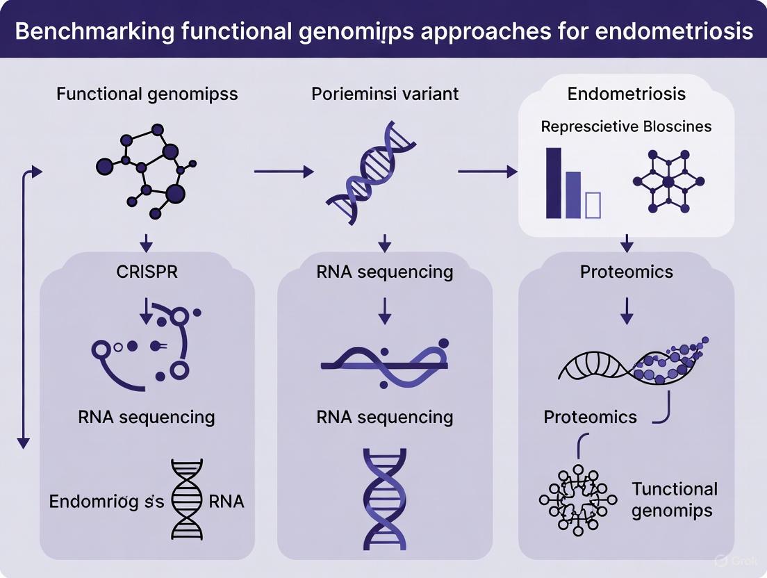
Benchmarking Functional Genomics Approaches for Endometriosis: From Variant Discovery to Therapeutic Translation
Endometriosis is a complex gynecological disorder affecting ~10% of women, with a significant heritable component.
Benchmarking Functional Genomics Approaches for Endometriosis: From Variant Discovery to Therapeutic Translation
Abstract
Endometriosis is a complex gynecological disorder affecting ~10% of women, with a significant heritable component. This article provides a comprehensive benchmarking framework for functional genomics approaches aimed at translating endometriosis-associated genetic variants from genome-wide association studies (GWAS) into mechanistic insights and therapeutic targets. We explore foundational genetic architecture, methodological applications of transcriptomics and eQTL mapping, optimization strategies for data analysis, and comparative validation of emerging technologies. Aimed at researchers and drug development professionals, this review synthesizes current methodologies to prioritize candidate genes, understand tissue-specific regulation, and overcome challenges in variant functionalization, ultimately bridging the gap between genetic susceptibility and personalized treatment strategies.
The Genetic Architecture of Endometriosis: From GWAS Loci to Pathobiological Pathways
Global Prevalence and Disease Burden
Endometriosis is a significant global health issue, affecting approximately 10% of women of reproductive age worldwide, which translates to nearly 190 million individuals [1]. This chronic, inflammatory condition involves the presence of endometrial-like tissue outside the uterine cavity and is associated with substantial morbidity, including chronic pelvic pain, infertility, and reduced quality of life [2] [1].
Table 1: Global Epidemiological Indicators of Endometriosis (1990-2021)
| Indicator | 1990 Value | 2021 Value | Trend (1990-2021) |
|---|---|---|---|
| Incident Cases | Not specified | 3.45 million (95% UI: 2.44 to 4.6 million) | Increased by 3.51% [3] |
| DALYs | Not specified | 2.05 million (95% UI: 1.20 to 3.13 million) | Increased by 12.03% [3] |
| Age-Standardized Incidence Rate | Baseline | Not specified | Decreasing trend (EAPC: -1.01) [3] |
| Peak Age Groups for Incidence | 20-24 years | 20-24 years | Consistent across study period [3] |
| Peak Age Groups for DALYs | 25-29 years | 25-29 years | Consistent across study period [3] |
The age-standardized rates for incidence and disability-adjusted life years (DALYs) have shown a slight decreasing trend globally from 1990 to 2021, with an estimated annual percentage change (EAPC) of approximately -1.01% for incidence and -0.99% for DALYs [3]. However, the absolute number of cases and DALYs has increased, primarily driven by population growth [4].
The disease burden distribution varies by socioeconomic development, with higher age-standardized incidence and DALY rates observed in low Sociodemographic Index (SDI) regions compared to high SDI regions [3]. This disparity highlights the impact of healthcare access and resource availability on disease management and outcomes.
Diagnostic Challenges and Delays
Diagnostic Timeframes and Barriers
The diagnostic journey for endometriosis remains profoundly challenging, with significant delays between symptom onset and definitive diagnosis. Current evidence indicates an average diagnostic delay of 7 to 12 years across healthcare systems [2] [1] [5]. This prolonged timeframe represents a critical gap in patient care that substantially impacts quality of life and disease progression.
Table 2: Factors Contributing to Diagnostic Delays in Endometriosis
| Factor Category | Specific Contributors | Impact Magnitude (Effect Size) |
|---|---|---|
| Patient-Related | Delay in seeking medical attention; Symptom normalization; Social stigma | Pooled SMD: 1.94 (95% CI: 1.62-2.27, p<0.001) [6] |
| Provider-Related | Misdiagnosis; Reliance on non-specific diagnostics; Lack of awareness | Pooled SMD: 2.00 (95% CI: 1.72-2.28, p<0.001) [6] |
| System-Related | Referral pathway complexities; Geographic disparities; Limited access to specialists | Insufficient data for meta-analysis but qualitatively confirmed [6] |
Root Causes of Diagnostic Challenges
The extensive diagnostic delays stem from multiple interconnected factors:
Symptom Variability and Non-Specificity: Endometriosis presents with diverse symptoms including dysmenorrhea, dyspareunia, chronic pelvic pain, abnormal uterine bleeding, and infertility [2]. This heterogeneity often leads to misdiagnosis as other conditions such as irritable bowel syndrome (IBS) or pelvic inflammatory disease (PID) [6].
Normalization of Menstrual Pain: Sociocultural acceptance of dysmenorrhea as "normal" contributes to patient delays in seeking care and provider dismissal of symptoms [7]. As one expert notes, "Menstrual cramps are the only type of pain that we as human beings accept as a normal phenomenon" [7].
Invasive Diagnostic Gold Standard: Laparoscopic surgery with histological confirmation remains the definitive diagnostic method [5], creating a significant barrier due to its invasiveness, cost, and requirement for specialized surgical expertise.
Healthcare Access Disparities: Individuals from low-income and rural areas face additional barriers including limited access to specialized care and diagnostic facilities [2] [6].
Current and Emerging Diagnostic Methodologies
Established Diagnostic Approaches
Current diagnostic protocols in clinical practice include:
Clinical Evaluation: Comprehensive patient history focusing on pain characteristics, menstrual patterns, and associated symptoms [1]. The World Health Organization emphasizes that "a careful menstrual health history including pain, heaviness of bleeding, and associated symptoms can help with diagnosis" [1].
Imaging Techniques: Transvaginal ultrasound represents the first-line imaging tool for detecting endometriotic lesions, particularly ovarian endometriomas and deep infiltrating endometriosis [2]. MRI may be utilized for more complex cases or preoperative planning.
Surgical Confirmation: Laparoscopy remains the gold standard, allowing direct visualization and histological confirmation of endometriotic lesions [5].
Emerging Molecular Diagnostic Technologies
Research efforts are focusing on developing non-invasive diagnostic approaches through advanced functional genomics and biomarker discovery:
Table 3: Experimental Protocols for Genomic Biomarker Discovery
| Methodology | Experimental Protocol | Key Findings | Performance Metrics |
|---|---|---|---|
| Machine Learning Classification [8] | - Case-control study with transcriptomic data- Applied AdaBoost, XGBoost, Stochastic Gradient Boosting, Bagged CART- Five-fold cross-validation | Identified potential biomarker genes: CUX2, CLMP, CEP131, EHD4, CDH24, ILRUN, LINC01709, HOTAIR, SLC30A2, NKG7 | Bagged CART performance:Accuracy: 85.7%Sensitivity: 100%Specificity: 75%F1-score: 85.7% |
| Spatial Transcriptomics [9] | - Spatial transcriptomics and RNAscope- Single-cell resolution analysis- Mapping transcriptional activity across endometrial tissue | Provides mechanistic insights into role of risk genes in women's health; Identifies gene expression networks driving disease progression | Research ongoing; Focused on establishing human genomics framework for mechanistic insights |
| Hormonal Biomarker Analysis [5] | - Measurement of aromatase (CYP19A1) expression in endometrial tissues- Meta-analysis of 17 studies with 1,279 participants | Aromatase demonstrated highest diagnostic accuracy among hormonal biomarkers | Pooled performance:Sensitivity: 79%Specificity: 89% |
Research Reagent Solutions for Endometriosis Investigation
Table 4: Essential Research Reagents for Endometriosis Functional Genomics
| Reagent/Category | Specific Examples | Research Application |
|---|---|---|
| Transcriptomic Profiling | RNA-seq platforms; Spatial transcriptomics solutions; RNAscope | Single-cell transcriptional analysis; Spatial orientation of gene expression [9] [8] |
| Machine Learning Algorithms | AdaBoost; XGBoost; Stochastic Gradient Boosting; Bagged CART | Classification of endometriosis cases; Biomarker identification from genomic data [8] |
| Genomic Analysis Tools | GWAS datasets; Polygenic risk modeling; DNA methylation profiling | Identification of risk loci (WNT4, VEZT, GREB1); Epigenetic modification analysis [5] |
| Hormonal Assays | Aromatase (CYP19A1) expression analysis; Estrogen metabolite measurement | Assessment of hormonal dependencies; Diagnostic biomarker validation [5] |
The significant prevalence and profound diagnostic challenges of endometriosis underscore the critical need for innovative diagnostic approaches. While current clinical methods remain dependent on invasive surgical confirmation, emerging functional genomics technologies offer promising pathways toward non-invasive, accurate, and timely diagnosis.
The integration of multi-omics data with machine learning classification models demonstrates potential for revolutionizing endometriosis diagnosis, with current models already achieving promising accuracy metrics exceeding 85% [8]. These computational approaches, combined with spatial transcriptomics and advanced biomarker panels, represent the future of endometriosis diagnostics that may ultimately eliminate the current unacceptable diagnostic delays of 7-12 years.
For researchers in the field, focusing on standardized validation of biomarker panels across diverse populations and developing accessible diagnostic platforms will be essential to translate these genomic advances into clinical practice. The benchmarking of these functional genomics approaches will play a crucial role in establishing reliable, reproducible diagnostic protocols that can significantly improve patient outcomes through early detection and intervention.
Key Findings from Genome-Wide Association Studies (GWAS)
Genome-wide association studies (GWAS) have revolutionized the identification of genetic variants associated with complex diseases, enabling breakthroughs in understanding disease etiology and therapeutic development. By analyzing hundreds of thousands to millions of single-nucleotide polymorphisms (SNPs) across thousands of individuals, GWAS pinpoint genomic regions where genetic variations correlate with disease risk. This approach has been particularly transformative for conditions with substantial heritability but complex etiology, such as endometriosis, where familial aggregation and twin studies indicate approximately 52% heritability [10]. The successful application of GWAS has evolved from single-population analyses to large-scale meta-analyses that enhance statistical power by combining datasets across multiple studies and populations [10] [11]. For endometriosis specifically, GWAS has transitioned from initial candidate gene studies with limited success to comprehensive genome-wide approaches that have revealed numerous susceptibility loci, providing insights into the molecular pathways underlying this heterogeneous condition [10] [12].
Major GWAS Discoveries in Endometriosis
Established Genetic Loci and Biological Pathways
GWAS have identified multiple genetic loci associated with endometriosis risk, revealing important biological pathways involved in disease pathogenesis. Meta-analyses of endometriosis GWAS have demonstrated remarkable consistency across studies and populations, with six loci achieving genome-wide significance: rs12700667 on 7p15.2, rs7521902 near WNT4, rs10859871 near VEZT, rs1537377 near CDKN2B-AS1, rs7739264 near ID4, and rs13394619 in GREB1 [10]. These findings highlight genes involved in sex steroid regulation, hormone metabolism, and developmental pathways. Notably, most of these loci show stronger effect sizes in moderate-to-severe (Stage III/IV) endometriosis, suggesting they may be particularly relevant for the development of advanced disease [10]. More recent studies have added ESR1, CYP19A1, HSD17B1, VEGF, and GnRH to the list of novel loci associated with endometriosis, further expanding our understanding of the genetic architecture underlying this condition [12].
Table 1: Key Endometriosis Susceptibility Loci Identified Through GWAS
| SNP Identifier | Chromosomal Location | Nearest Gene(s) | Reported P-value | Potential Biological Function |
|---|---|---|---|---|
| rs12700667 | 7p15.2 | Inter-genic | 1.6 × 10⁻⁹ | Regulatory region [10] |
| rs7521902 | 1p36.12 | WNT4 | 1.8 × 10⁻¹⁵ | Developmental pathways [10] |
| rs10859871 | 12q22 | VEZT | 4.7 × 10⁻¹⁵ | Cell adhesion [10] |
| rs1537377 | 9p21.3 | CDKN2B-AS1 | 1.5 × 10⁻⁸ | Cell cycle regulation [10] |
| rs7739264 | 6p22.3 | ID4 | 6.2 × 10⁻¹⁰ | Developmental pathways [10] |
| rs13394619 | 14q23.3 | GREB1 | 4.5 × 10⁻⁸ | Hormone regulation [10] |
| rs10965235 | 9p21.3 | CDKN2B-AS1 | 5.57 × 10⁻¹² | First identified in Japanese population [10] |
Population-Specific Findings and Ethnic Variations
While many endometriosis risk loci show consistency across populations, some variations exist between different ethnic groups. The first endometriosis GWAS in a Japanese population identified rs10965235 in CDKN2B-AS1 as a significant risk variant [10]. In Taiwanese populations, GWAS have revealed different susceptibility loci, including rs10739199 and rs2025392 in PTPRD, rs1998998 on chromosome 14, and rs6576560 on chromosome 15 [13]. After imputation, strong signals were observed for rs10822312 on chromosome 10 and rs58991632 and rs2273422 on chromosome 20 [13]. Importantly, expression quantitative trait locus (eQTL) analysis in the Taiwanese population identified rs13126673 as a significant cis-eQTL for the INTU gene, with the risk allele associated with altered INTU expression in endometriotic tissues [13]. These population-specific findings highlight the importance of diverse cohort inclusion in GWAS to fully capture the genetic architecture of endometriosis across ethnicities.
Benchmarking GWAS Methodologies and Applications
GWAS Versus Rare Variant Burden Tests
GWAS and rare variant burden tests represent complementary approaches for identifying trait-relevant genes, each with distinct strengths and limitations. Burden tests aggregate rare protein-coding variants (typically loss-of-function variants) within a gene to create a "burden genotype" that is tested for association with phenotypes [14]. Systematic analysis of 209 quantitative traits in the UK Biobank reveals that these methods systematically prioritize different genes, with burden tests favoring trait-specific genes (those primarily affecting the studied trait with minimal effects on others), while GWAS also capture highly pleiotropic genes (affecting multiple traits) often missed by burden tests [14]. This distinction arises because burden test association strength depends on both trait importance and the aggregate frequency of loss-of-function variants, which are kept rare by natural selection [14]. For comprehensive gene discovery, both approaches are valuable: burden tests identify genes with strong, trait-specific effects, while GWAS captures broader polygenic architecture including pleiotropic genes.
Table 2: Comparison of GWAS and Burden Test Methodologies
| Feature | GWAS | Burden Tests |
|---|---|---|
| Variant Type | Common SNPs (typically minor allele frequency >1%) | Rare variants (often loss-of-function) |
| Study Design | Population-based | Population-based |
| Statistical Approach | Single-marker analysis | Gene-based aggregation |
| Primary Output | Associated genomic loci | Associated genes |
| Gene Prioritization | Trait importance | Trait specificity |
| Pleiotropy Detection | Identifies highly pleiotropic genes | Prioritizes trait-specific genes |
| Functional Interpretation | Requires follow-up functional studies | Direct gene-level interpretation |
Advancements in GWAS Meta-Analysis and Multi-Omics Integration
The statistical power of GWAS has been dramatically enhanced through meta-analysis approaches that combine data across multiple studies. For example, a GWAS meta-analysis of body weight traits in chickens identified 77 novel independent variants and 59 candidate genes that were not detected in single-population studies [11]. This approach has proven equally valuable in endometriosis research, where meta-analyses of four GWAS and four replication studies including 11,506 cases and 32,678 controls confirmed the significance of multiple loci [10]. Beyond simple meta-analysis, integration of GWAS with functional genomic data represents a powerful strategy for elucidating disease mechanisms. Integration with expression quantitative trait loci (eQTL) has been particularly fruitful, enabling researchers to connect disease-associated variants with genes whose expression they regulate [13]. For instance, combining GWAS with eQTL mapping in endometriosis research revealed that rs13126673 regulates expression of the INTU gene, with the risk allele associated with altered RNA secondary structure [13]. Further multi-omics integration with epigenetic data, proteomics, and metabolomics provides a more comprehensive understanding of endometriosis pathophysiology and identifies potential diagnostic biomarkers and therapeutic targets [12].
From Genetic Associations to Therapeutic Targets
Mendelian randomization (MR) has emerged as a powerful method for evaluating causal relationships between genetically predicted exposures and disease outcomes, offering a robust approach for identifying potential therapeutic targets. Applying MR analysis to endometriosis, researchers have identified RSPO3 as a potential therapeutic target, with external validation and colocalization analysis confirming the robustness of this association [15]. Experimental validation using ELISA, RT-qPCR, and Western blotting demonstrated elevated RSPO3 levels in both plasma and endometriotic tissues from patients compared to controls [15]. This exemplifies how GWAS findings can be translated into potential clinical applications through systematic functional follow-up. Additional promising approaches include polygenic risk scores (PRS) that aggregate risk across multiple genetic variants to predict individual disease risk, potentially enabling earlier diagnosis and intervention [12]. Machine learning methods also show promise for enhancing genomic prediction, as demonstrated by multi-variant deep neural network approaches that improve endometriosis disease prediction accuracy [16].
Experimental Protocols in Modern GWAS Research
Standardized GWAS Workflow
Contemporary GWAS follows a standardized workflow to ensure robust and reproducible results. The process begins with sample collection from carefully phenotyped cases and controls, followed by genotyping using microarray platforms such as the Infinium Global Screening Array (Illumina) or Axiom arrays (Thermo Fisher Scientific) [17]. After genotyping, extensive quality control is performed to exclude samples with sex discordance, call rates <90%, excessive heterozygosity, or relatedness (Pihat ≥ 0.2), and to remove variants deviating from Hardy-Weinberg equilibrium or with low minor allele frequency [17]. Population stratification is addressed through principal component analysis, typically including the first several principal components as covariates in association tests [11]. Association analysis employs linear mixed models in tools such as GCTA-fastGWA or REGENIE to test for genotype-phenotype associations while controlling for confounding factors [11]. For meta-analyses, tools such as METAL implement fixed-effect inverse variance weighting to combine results across studies [11]. Significant findings are then annotated and interpreted through integration with functional genomic datasets.
The Scientist's Toolkit: Essential Research Reagents and Platforms
Table 3: Essential Research Reagents and Platforms for GWAS
| Reagent/Platform | Function | Example Use Case |
|---|---|---|
| Affymetrix Axiom TWB Array | Genotyping array with 653,291 SNP probes | GWAS in Taiwanese population [13] |
| Infinium Global Screening Array-24 | BeadChip for genome-wide genotyping | GWAS of SARS-CoV-2 vaccine response [17] |
| Illumina 60K SNP BeadChip | Medium-density genotyping array | Chicken body weight traits GWAS [11] |
| PLINK v1.9/2.0 | Quality control and association analysis | Standardized QC pipelines [11] [17] |
| METAL | Meta-analysis of multiple GWAS | Combining results across cohorts [11] |
| GENotype-Tissue Expression (GTEx) | eQTL reference database | Functional annotation of GWAS hits [13] |
| SOMAscan V4 | Multiplexed proteomic assay | Protein quantitative trait loci mapping [15] |
| Human R-Spondin3 ELISA Kit | Protein quantification | Validation of RSPO3 levels [15] |
GWAS continues to evolve from simply identifying associated loci toward elucidating biological mechanisms and enabling clinical translation. For endometriosis research, future directions include larger multi-ancestry meta-analyses to improve power and portability of polygenic risk scores, deeper integration with functional genomics through single-cell multi-omics, and application of advanced machine learning methods for variant prioritization [12] [16]. The systematic benchmarking of different genomic approaches reveals their complementary strengths: GWAS captures broad polygenic architecture, burden tests identify genes with strong biological effects, and integrative methods connect variants to function. As these methodologies mature and datasets expand, GWAS will increasingly deliver on its promise to transform our understanding of endometriosis pathophysiology and accelerate the development of improved diagnostics and targeted therapeutics.
Prioritized Endometriosis Risk Genes and Their Chromosomal Distribution
Endometriosis, a chronic inflammatory condition affecting an estimated 10% of reproductive-age women, demonstrates substantial heritability of approximately 50% [18]. Advances in genomic technologies have enabled the identification of numerous genetic variants associated with disease susceptibility. However, translating these associations into biologically meaningful mechanisms and therapeutic targets requires sophisticated functional prioritization. This guide benchmarks contemporary genomic approaches for prioritizing endometriosis risk genes, comparing their methodological frameworks, output data, and applicability to drug development pipelines. We present a systematic comparison of multi-omics integration strategies, tissue-specific regulatory mapping, and functional validation protocols that collectively illuminate the chromosomal architecture of endometriosis risk.
Tabular Comparison of Prioritized Risk Genes and Genomic Approaches
Table 1: Chromosomal Distribution of Prioritized Endometriosis Risk Genes
| Chromosome | Representative SNP | Prioritized Gene(s) | Effect Size (OR) | p-value | Functional Pathway |
|---|---|---|---|---|---|
| 1 | rs12037376 | WNT4 | 1.16 (1.12–1.19) | 8.87 × 10^−17 | Hormone signaling, development [18] |
| 2 | rs11674184 | GREB1 | 1.13 (1.10–1.15) | 2.67 × 10^−17 | Estrogen regulation [18] |
| 2 | rs10167914 | IL1A | 1.12 (1.08–1.15) | 1.10 × 10^−9 | Inflammation, IL-1 signaling [18] [19] |
| 4 | rs1903068 | KDR | 1.11 (1.07–1.13) | 1.04 × 10^−11 | Angiogenesis (VEGFR2) [18] |
| 6 | rs71575922 | SYNE1 | 1.11 (1.07–1.15) | 2.02 × 10^−8 | Cytoskeletal organization [18] |
| 9 | rs1537377 | CDKN2B-AS1 | 1.09 (1.06–1.12) | 1.33 × 10^−10 | Cell cycle regulation [18] |
| 12 | rs4762326 | VEZT | 1.08 (1.05–1.11) | 2.20 × 10^−9 | Cell adhesion [18] |
| 2 | - | IL1B | - | - | Inflammation, IL-1 signaling [19] |
| 11 | - | RSPO3 | - | - | WNT signaling, angiogenesis [15] |
Table 2: Benchmarking Functional Genomics Approaches in Endometriosis Research
| Methodology | Key Prioritized Genes | Tissue/Cellular Context | Strengths | Limitations |
|---|---|---|---|---|
| GWAS + eQTL Integration [20] | MICB, CLDN23, GATA4 | Uterus, ovary, colon, ileum, blood | Identifies tissue-specific regulation; reveals constitutive regulatory patterns | Limited to healthy tissues in GTEx; may miss disease-specific effects |
| Multi-layered Genomic Prioritization (END) [21] | TNF, IL6, IL6R, JAK family | Cross-tissue, immune focus | Superior recovery of known drug targets (AUC performance); identifies repurposing candidates | Complex computational requirements; limited validation data |
| Mendelian Randomization + Experimental Validation [15] | RSPO3, FLT1 | Plasma proteins, endometriosis lesions | Estishes causal inference; direct clinical translation | Dependent on quality of protein QTL datasets; resource-intensive |
| scRNA-seq + GWAS Integration (scDRS) [19] | IL1A, IL1B, KDR, CALCRL | M2 macrophages, dendritic cells, endothelial cells | Identifies specific cellular mediators; reveals heterogeneity within cell types | Requires specialized single-cell expertise; high computational cost |
| Deep Neural Networks [16] | Not specified in extract | Not specified | Potential for enhanced predictive power with complex data | "Black box" limitations; interpretability challenges |
Experimental Protocols for Endometriosis Gene Prioritization
Genomic Prioritization with Multi-layered Predictors
The END framework employs a systematic approach to target prioritization [21]:
Predictor Preparation: Three genomic datasets are integrated:
- nGene: Nearby genes defined by GWAS significance (p < 5×10^−8) and linkage disequilibrium (R² < 0.8)
- cGene: Conformation genes identified through promoter capture Hi-C data
- eGene: Expression genes derived from eQTL mapping studies
Predictor Importance Evaluation: Random forest algorithms evaluate the relative importance of cGene and eGene predictors compared to the conventional nGene baseline.
Predictor Combination: Direct (sum, max, harmonic) and indirect (Fisher's, logistic, order statistic) methods combine informative predictors.
Performance Benchmarking: The area under the ROC curve (AUC) quantifies performance in separating clinical proof-of-concept targets (drugs reaching phase 2+) from simulated controls, demonstrating superiority over Naïve and Open Targets approaches [21].
Tissue-Specific eQTL Mapping
This methodology links endometriosis-associated variants to their regulatory effects [20]:
Variant Selection: 465 unique endometriosis-associated variants with genome-wide significance (p < 5×10^−8) are curated from GWAS Catalog.
Tight Selection: Six biologically relevant tissues (uterus, ovary, vagina, sigmoid colon, ileum, peripheral blood) are selected from GTEx v8.
eQTL Identification: Variants are cross-referenced with tissue-specific eQTL data, retaining only significant associations (FDR < 0.05).
Functional Annotation: Slope values indicating effect direction/magnitude are recorded. A slope of +1.0 indicates a twofold expression increase, while -1.0 reflects a 50% decrease.
Pathway Analysis: Prioritized genes are analyzed against MSigDB Hallmark and Cancer Hallmarks gene sets to identify enriched biological pathways.
Mendelian Randomization for Causal Inference
This approach establishes causal relationships between biomarkers and endometriosis risk [15]:
Instrumental Variable Selection: Genetic variants (SNPs) strongly associated with exposures (plasma proteins, metabolites) are selected (p < 5×10^−8, R² < 0.001, F-statistic > 10).
Data Sources: Large-scale GWAS summary statistics for plasma proteins (4,907 cis-pQTLs from 35,559 individuals) and endometriosis (20,190 cases/130,160 controls from FinnGen).
MR Analysis: Two-sample MR conducted using inverse-variance weighted, MR-Egger, and weighted median methods to test causal effects.
Experimental Validation: ELISA measures target protein concentration in patient plasma (EM vs. controls). RT-qPCR and Western blot analyze gene and protein expression in tissue samples.
Single-Cell Disease Risk Scoring (scDRS)
This method identifies cell types mediating genetic risk [19]:
Cell Atlas Construction: 118,103 CD45-positive immune cells from endometriosis lesions and control tissues are sequenced and clustered into 15 immune populations.
Risk Scoring: scDRS software integrates single-cell transcriptomes with GWAS data (23,492 cases/450,668 controls) to calculate disease association scores per cell.
Cell-Type Association Testing: Distributions of scores are tested for cell type-level association and heterogeneity.
Pathway Correlation Analysis: Gene expression correlated with risk scores identifies enriched pathways (PROGENy) and candidate mediator genes.
Visualizing Key Pathways and Workflows
IL-1 Signaling Pathway in Endometriosis Pathogenesis
Genomic Prioritization Workflow Comparison
The Scientist's Toolkit: Essential Research Reagents
Table 3: Key Research Reagent Solutions for Endometriosis Genomics
| Reagent/Resource | Primary Application | Function in Research | Example Implementation |
|---|---|---|---|
| GTEx v8 Database [20] | eQTL mapping | Provides normal tissue-specific gene expression and regulation data | Identify baseline regulatory effects of endometriosis risk variants across six relevant tissues |
| SOMAscan V4 Platform [15] | Proteomic quantification | Aptamer-based multiplexed immunoassay for large-scale protein quantification | Measure 4,907 plasma protein levels for pQTL analysis in Mendelian randomization |
| Human R-Spondin3 ELISA Kit [15] | Protein validation | Quantitative measurement of RSPO3 concentration in patient plasma | Validate MR predictions in clinical samples (endometriosis vs. control patients) |
| scDRS Software [19] | Single-cell genomics | Integrates single-cell transcriptomes with GWAS data to identify risk-associated cell types | Identify M2 macrophages as primary mediators of endometriosis genetic risk |
| PROGENy [19] | Pathway activity analysis | Estimates pathway activity from transcriptomic data at single-cell resolution | Correlate NF-κB and TNF-α signaling with genetic risk scores in myeloid cells |
| Anakinra [19] | Functional validation | IL-1 receptor antagonist for pathway blockade | Demonstrate dose-dependent reduction in pain and angiogenesis in vivo |
Discussion and Future Directions
The integration of multiple genomic approaches reveals a complex architecture of endometriosis risk distributed across multiple chromosomes, with distinct patterns of tissue-specific regulation and cellular mediation. Chromosomes 1, 2, 6, and 9 emerge as key risk loci, with genes predominantly involved in hormonal response (WNT4, GREB1), inflammation (IL1A, IL1B), and angiogenesis (KDR, RSPO3) [20] [18] [19].
The benchmarking of functional genomics approaches demonstrates complementary strengths: tissue-specific eQTL mapping establishes constitutive regulatory patterns [20]; multi-layered prioritization (END) optimally identifies druggable targets [21]; Mendelian randomization provides causal inference [15]; and single-cell integration identifies specific cellular mediators [19]. Notably, the convergence of evidence across methods strengthens confidence in certain pathways, particularly IL-1 signaling, which is implicated through eQTL effects, cellular scoring, and functional validation showing that IL-1 receptor antagonism (anakinra) reduces pain and angiogenic signaling [19].
For drug development professionals, these prioritization strategies nominate both repurposing opportunities (IL-6R, JAK inhibitors, anakinra) [21] [19] and novel target candidates (RSPO3) [15]. Future efforts should focus on integrating these complementary approaches into unified frameworks and expanding diverse population representation to ensure equitable translation of genomic discoveries into effective therapeutics for this complex disease.
Biological Pathways Implicated by Genetic Associations
Endometriosis is a complex, chronic inflammatory disease affecting approximately 10% of women of reproductive age, characterized by the presence of endometrial-like tissue outside the uterine cavity [22]. The pathophysiology of this condition involves a multifaceted interplay of genetic predisposition, inflammatory processes, hormonal dysregulation, and altered cellular mechanisms. Over the past decade, significant advances in genomic technologies have enabled researchers to identify specific genetic variants and biological pathways that contribute to endometriosis susceptibility and progression.
The integration of large-scale genome-wide association studies (GWAS) with functional genomic approaches has been particularly transformative in elucidating the molecular architecture of endometriosis [23] [20]. These approaches have helped bridge the gap between statistical genetic associations and their functional consequences, providing unprecedented insights into the biological mechanisms driving disease pathogenesis. This review synthesizes current evidence on the key biological pathways implicated by genetic associations in endometriosis, with a specific focus on benchmarking various functional genomics methodologies used to validate and characterize these pathways.
Key Genetic Associations and Implicated Pathways
Immune and Inflammatory Pathways
Substantial genetic evidence points to dysregulation of immune and inflammatory pathways as a central component of endometriosis pathogenesis. Large-scale genetic studies have demonstrated significant associations between endometriosis and various immunological diseases, suggesting shared genetic architecture [23].
Table 1: Genetic Correlations Between Endometriosis and Immune Diseases
| Immune Condition | Genetic Correlation (rg) | P-value | Suggested Causal Relationship |
|---|---|---|---|
| Osteoarthritis | 0.28 | 3.25 × 10⁻¹⁵ | Shared genetic basis |
| Rheumatoid Arthritis | 0.27 | 1.5 × 10⁻⁵ | Potential causal link (OR = 1.16) |
| Multiple Sclerosis | 0.09 | 4.00 × 10⁻³ | Shared biological mechanisms |
| Coeliac Disease | Phenotypic association only | - | Increased comorbidity risk |
| Psoriasis | Phenotypic association only | - | Increased comorbidity risk |
Genetic correlation analyses reveal significant positive correlations between endometriosis and osteoarthritis (rg = 0.28), rheumatoid arthritis (rg = 0.27), and multiple sclerosis (rg = 0.09) [23]. Mendelian randomization analysis further suggests a potential causal association between endometriosis and rheumatoid arthritis (OR = 1.16, 95% CI = 1.02-1.33) [23]. These findings indicate that shared genetic factors contribute to the co-occurrence of endometriosis with various immune-mediated conditions.
Expression quantitative trait loci (eQTL) analyses have identified specific genes within these pathways that are regulated by endometriosis-associated genetic variants. Key immune-related genes include IL1A (interleukin 1, alpha), IL33 (interleukin 33), and HLA-DRA (major histocompatibility complex, class II, DR alpha) [24]. The enrichment of these genes in immune pathways highlights the critical role of aberrant immune responses in endometriosis development.
Hormonal Response Pathways
Hormonal dysregulation, particularly involving estrogen signaling, represents a cornerstone of endometriosis pathophysiology. Genetic studies have identified several key genes involved in hormonal responses that contribute to endometriosis susceptibility.
Table 2: Key Hormonal Pathway Genes in Endometriosis
| Gene | Function | Genetic Evidence | Regulatory Impact |
|---|---|---|---|
| ESR1 | Encodes estrogen receptor alpha | GWAS significant association [24] | Master regulator of estrogen response |
| GREB1 | Early estrogen response gene | GWAS significant association [24] | Mediates estrogen-induced cell growth |
| FSHB | Encodes follicle-stimulating hormone beta subunit | GWAS significant association [24] | Regulates gonadotropin signaling |
| WNT4 | Wingless-type MMTV integration site family | GWAS significant association [24] | Involved in uterine development and hormone response |
Functional genomic approaches have demonstrated that endometriosis-associated variants regulate the expression of these genes in a tissue-specific manner. In reproductive tissues such as the uterus, ovary, and vagina, risk variants predominantly affect genes involved in hormonal response, tissue remodeling, and cellular adhesion [20]. This tissue-specific regulatory pattern suggests that genetic variants may disrupt hormonal homeostasis specifically in the reproductive microenvironment, facilitating the establishment and growth of ectopic endometrial lesions.
Cell Aging and Senescence Pathways
Recent multi-omic studies have revealed the significant involvement of cell aging-related genes in endometriosis pathogenesis. A comprehensive analysis integrating GWAS data with expression quantitative trait loci (eQTLs), methylation QTLs (mQTLs), and protein QTLs (pQTLs) has identified several cell aging genes with causal associations to endometriosis [25].
This integrated approach identified 196 CpG sites in 78 genes, alongside 18 eQTL-associated genes and 7 pQTL-associated proteins linked to both cell aging and endometriosis risk [25]. Notably, the MAP3K5 gene displays contrasting methylation patterns associated with endometriosis risk, suggesting that specific methylation patterns downregulate this gene, thereby increasing endometriosis susceptibility [25]. Validation in independent cohorts confirmed the THRB gene and ENG protein as risk factors for endometriosis development [25].
The involvement of cell aging pathways is further supported by the dysregulation of specific senescence-associated factors in endometriotic tissues. SIRT1, a key regulator of cellular metabolism and longevity, is upregulated in endometriotic tissues and promotes epithelial-mesenchymal transition and cell proliferation [25]. Additionally, the NLRP3 inflammasome, intricately linked to cell aging through mechanisms involving inflammation, oxidative stress, and mitochondrial dysfunction, contributes to the maintenance of endometriosis by creating a pro-inflammatory environment through the senescence-associated secretory phenotype (SASP) [25].
Somatic Mutations and Cancer-Associated Pathways
Beyond germline genetic variants, emerging evidence indicates that somatic mutations in cancer-associated genes play a crucial role in endometriosis pathogenesis and progression. Narrative reviews of the literature have identified recurrent somatic mutations in several key cancer driver genes [26].
Table 3: Somatic Mutations in Endometriosis Lesions
| Gene | Frequency in Lesions | Primary Function | Role in Endometriosis |
|---|---|---|---|
| KRAS | Common | GTPase involved in cell signaling | Promotes growth and survival of endometriotic cells |
| ARID1A | 20-40% of ovarian endometriomas | Chromatin remodeling | Loss disrupts gene expression programs |
| PIK3CA | Less common | Lipid kinase in PI3K/AKT pathway | Enhances proliferative signaling |
| PTEN | Less common | Tumor suppressor phosphatase | Loss permits unrestrained cell growth |
These recurrent somatic mutations are thought to arise from oxidative stress caused by retrograde menstruation and iron overload, driving mutagenesis that promotes fibrotic rather than malignant outcomes in most cases of endometriosis [26]. Distinct mutational patterns between epithelial and stromal components and across different lesions indicate oligoclonal origins and independent clonal evolution of endometriotic lesions [26].
The presence of cancer driver mutations in a benign condition represents a paradoxical phenomenon. The PTEN/PI3K/AKT/GSK-3β/β-catenin signaling pathway has been identified as particularly important in the inhibition of epithelial-mesenchymal transition in endometriosis [26]. Additionally, PFKFB3 promotes endometriosis cell proliferation via enhancing the protein stability of β-catenin, further highlighting the involvement of cancer-related pathways in this benign condition [26].
Experimental Approaches for Pathway Validation
Genome-Wide Association Studies (GWAS) and Meta-Analyses
GWAS represents the foundational approach for identifying genetic variants associated with endometriosis risk. The standard protocol involves:
Study Population: Large-scale cohorts of endometriosis cases with surgical confirmation and ethnically matched controls. Recent studies have utilized sample sizes exceeding 20,000 cases and 400,000 controls [25] [23].
Genotyping and Imputation: Genome-wide genotyping using high-density arrays followed by imputation to reference panels to increase genomic coverage.
Association Analysis: Statistical testing for association between each genetic variant and endometriosis case-control status, with genome-wide significance threshold of P < 5 × 10⁻⁸ [20].
Meta-Analysis: Combining results across multiple studies to increase statistical power and identify additional loci.
Functional Annotation: Annotation of associated variants using databases such as the GWAS Catalog (EFO_0001065) to identify potential functional consequences [20] [24].
This approach has identified 465 unique genome-wide significant variants associated with endometriosis, distributed across all autosomes and the X chromosome, with chromosome 8 harboring the highest number of variants (n=66) [20].
Expression Quantitative Trait Loci (eQTL) Mapping
eQTL analysis determines how genetic variants influence gene expression levels. The standard methodology includes:
Tissue Selection: Analysis across multiple physiologically relevant tissues, including reproductive tissues (uterus, ovary, vagina), intestinal tissues (sigmoid colon, ileum), and peripheral blood [20].
RNA Extraction and Sequencing: Extraction of high-quality RNA followed by RNA sequencing to quantify gene expression levels.
Statistical Analysis: Testing for associations between genetic variants and gene expression levels using linear models, with multiple testing correction (FDR < 0.05) [20].
Data Integration: Cross-referencing GWAS-significant variants with tissue-specific eQTL datasets from resources such as GTEx v8 [20].
This approach has revealed tissue-specific regulatory profiles for endometriosis-associated variants, with immune and epithelial signaling genes predominating in intestinal tissues and peripheral blood, while reproductive tissues show enrichment for genes involved in hormonal response and tissue remodeling [20].
Multi-Omic Mendelian Randomization
Multi-omic summary-based Mendelian randomization (SMR) integrates data from GWAS, eQTLs, mQTLs, and pQTLs to assess causal relationships between molecular traits and disease risk. The protocol involves:
Data Collection: Acquisition of summary statistics from large-scale GWAS and QTL studies for endometriosis and cell aging-related genes [25].
Instrument Selection: Selection of top cis-QTLs within a ± 1000 kb window around candidate genes using a P-value threshold of 5.0 × 10⁻⁸ [25].
SMR Analysis: Testing for causal effects of gene expression, DNA methylation, or protein abundance on endometriosis risk.
Heterogeneity Testing: Application of HEIDI test to distinguish pleiotropy from linkage (P-HEIDI > 0.05 indicates no significant heterogeneity) [25].
Colocalization Analysis: Identification of shared genetic variants between QTLs and GWAS signals using posterior probability thresholds (PPH4 > 0.5) [25].
This integrated approach has successfully identified causal relationships between specific methylation patterns, gene expression changes, and endometriosis risk, highlighting promising therapeutic targets [25].
Figure 1: Multi-omic Integration Approach for Pathway Identification. This workflow illustrates how diverse genomic datasets are integrated to identify biological pathways in endometriosis.
Signaling Pathways in Endometriosis: Molecular Interactions
The integration of genetic findings has helped elucidate several key signaling pathways that drive endometriosis pathogenesis. These pathways interact in a complex network that influences the establishment, survival, and growth of ectopic endometrial lesions.
Figure 2: Core Signaling Pathways in Endometriosis. This diagram illustrates the key molecular pathways and their interactions in endometriosis pathogenesis.
The estrogen signaling pathway serves as a central regulator in endometriosis, with genetic variants affecting key genes including ESR1, GREB1, and WNT4 [24]. These genes collectively enhance estrogen responsiveness, promoting the survival and growth of ectopic endometrial tissue. The WNT4 gene, in particular, plays additional roles in uterine development and may facilitate the improper implantation of endometrial cells [24].
The inflammatory response pathway involves multiple cytokines and immune regulators, including IL1A, IL33, and HLA-DRA [24]. These factors create a pro-inflammatory microenvironment that supports the establishment of endometriotic lesions by evading immune surveillance and promoting angiogenesis. The genetic correlations between endometriosis and classical autoimmune diseases further underscore the importance of immune dysregulation in this condition [23].
Fibrotic transformation is driven by somatic mutations in cancer-associated genes such as KRAS, ARID1A, and PIK3CA [26]. These mutations promote a fibrotic rather than malignant phenotype, leading to the characteristic adhesions and tissue distortion seen in advanced endometriosis. The PTEN/PI3K/AKT/GSK-3β/β-catenin signaling pathway appears particularly important in regulating the epithelial-mesenchymal transition that underlies fibrotic progression [26].
Cellular senescence pathways contribute to endometriosis through genes such as MAP3K5, SIRT1, and THRB [25]. These genes influence the senescence-associated secretory phenotype (SASP), which maintains a chronic inflammatory state and supports lesion persistence. The identification of these pathways through multi-omic Mendelian randomization approaches highlights their causal role in disease pathogenesis [25].
Research Reagent Solutions for Experimental Investigation
Table 4: Essential Research Reagents for Endometriosis Pathway Investigation
| Reagent Category | Specific Examples | Research Application | Key Features |
|---|---|---|---|
| DNA Extraction Kits | Qiagen QIAamp Circulating Nucleic Acid Kit [27] | Cell-free DNA extraction from serum | Optimized for low-concentration circulating DNA |
| GWAS Arrays | Illumina Infinium Global Screening Array | Genome-wide genotyping | High-density SNP coverage for association studies |
| RNA Sequencing Kits | Illumina TruSeq Stranded Total RNA | Transcriptome analysis | Comprehensive gene expression profiling |
| Spatial Transcriptomics | 10x Genomics Visium Spatial Gene Expression | Spatial mapping of gene expression in lesions | Preserves tissue architecture while capturing transcriptome data |
| Methylation Arrays | Illumina Infinium MethylationEPIC | Genome-wide methylation profiling | Coverage of >850,000 methylation sites |
| QTL Reference Data | GTEx v8 Database [20] | Expression quantitative trait loci mapping | Tissue-specific eQTL data across 52 tissues |
| Functional Annotation Tools | Ensembl Variant Effect Predictor (VEP) [20] | Variant functional annotation | Predicts consequences of genetic variants |
| Pathway Analysis Resources | MSigDB Hallmark Gene Sets [20] | Biological pathway enrichment | Curated gene sets for functional analysis |
These research reagents enable the comprehensive investigation of genetic associations and biological pathways in endometriosis. The Qiagen QIAamp Circulating Nucleic Acid Kit has been specifically utilized for extracting cell-free DNA from serum samples in endometriosis studies, demonstrating significantly elevated cf-DNA levels in patients compared to controls (3.9-fold increase) [27]. The GTEx v8 database provides critical reference data for eQTL analyses across multiple tissues relevant to endometriosis, including uterus, ovary, vagina, and intestinal tissues [20].
Spatial transcriptomics approaches, mentioned in functional genomics projects, enable the investigation of transcriptional activity in single cells while preserving their spatial orientation across endometrial tissue [9]. This method provides valuable mechanistic insights into the role of risk genes in women's health by maintaining the architectural context of endometriotic lesions.
The integration of large-scale genetic studies with functional genomic approaches has substantially advanced our understanding of the biological pathways implicated in endometriosis pathogenesis. Immune and inflammatory pathways, hormonal response systems, cellular senescence mechanisms, and cancer-associated signaling networks collectively contribute to the development and progression of this complex condition.
Methodologically, the field has evolved from simple association studies to sophisticated multi-omic integrations that combine GWAS with eQTL, mQTL, and pQTL data. These approaches have enabled researchers to move beyond statistical associations to establish causal relationships and identify specific molecular mechanisms. Benchmarking of these methodologies reveals that each approach offers distinct advantages, with multi-omic Mendelian randomization providing particularly powerful insights into causal pathways.
The biological pathways identified through these genetic approaches represent promising targets for therapeutic development. Notably, the shared genetic basis between endometriosis and other immune conditions opens up opportunities for repurposing existing therapies across these conditions [23]. Additionally, the involvement of cell aging pathways suggests potential applications of senolytic agents in endometriosis management [25].
As functional genomics technologies continue to advance, particularly through single-cell and spatial transcriptomics approaches, we can anticipate further refinement of our understanding of endometriosis pathophysiology. These advances will likely enable more personalized approaches to diagnosis and treatment, ultimately improving outcomes for individuals affected by this challenging condition.
The Challenge of Non-Coding Variants and Tissue-Specific Effects
The interpretation of non-coding genetic variants represents a fundamental challenge in modern genetics, particularly in complex diseases such as endometriosis. While genome-wide association studies (GWAS) have identified numerous variants associated with endometriosis risk, the majority reside in non-coding regions, complicating the understanding of their functional consequences [28]. The regulatory effects of these variants often exhibit tissue-specific patterns, necessitating advanced computational tools that can accurately predict their impact across different biological contexts. This comparison guide objectively evaluates the performance of leading functional genomics approaches specifically for endometriosis research, providing researchers with experimental data and methodologies to inform their analytical strategies.
Benchmarking Computational Tools for Variant Prioritization
Performance Comparison of Non-Coding Variant Tools
Advanced computational frameworks have emerged to address the challenge of prioritizing functional non-coding variants by leveraging deep learning and multi-label learning approaches. These tools integrate diverse genomic annotations to predict tissue-specific regulatory effects, with significant implications for understanding endometriosis pathophysiology.
Table 1: Performance Metrics of Leading Non-Coding Variant Prioritization Tools
| Tool | Core Methodology | Tissue-Specific Capabilities | Reported AUROC | Key Advantages |
|---|---|---|---|---|
| TVAR [29] | Multi-label learning-based deep neural network | Predicts functionality across 49 GTEx tissues | 0.77 (average across tissues) | Learns relationships between epigenomics and eQTLs across tissues, considering tissue correlation |
| RegVar [30] | Deep neural network (DNN) framework | Predicts tissue-specific impact on target genes | Surpasses existing methods (specific values not provided) | Links regulatory variants to potential target genes; available as web server |
| BRAIN-MAGNET [31] | Convolutionally neural network | Brain-focused but framework applicable to other tissues | Functionally validated for neurological traits | Predicts non-coding regulatory element activity from DNA sequence alone |
| CADD [29] | Supervised machine learning | Limited tissue-specific capabilities | Inferior to TVAR in comparative evaluations [29] | Established benchmark; integrates multiple annotations |
| DeepSEA [29] | Deep learning | Limited tissue-specific capabilities | Outperformed by TVAR [29] | Predicts chromatin effects from sequence |
TVAR demonstrates superior performance in direct comparisons, outperforming five existing state-of-the-art tools including DeepSEA and DANN (also deep learning-based methods) across multiple test scenarios including ClinVar, fine-mapped GWAS loci, and MPRA-validated variants [29]. This multi-label learning approach is particularly valuable for endometriosis research as it learns the shared and tissue-specific eQTL effects across multiple tissues simultaneously, capturing the complex regulatory architecture relevant to a disease that affects diverse tissue types.
Experimental Validation in Endometriosis Research
Recent research has applied tissue-specific functional genomics approaches specifically to endometriosis-associated genetic variants. A 2025 study systematically investigated the regulatory effects of 465 endometriosis-associated GWAS variants across six physiologically relevant tissues: uterus, ovary, vagina, sigmoid colon, ileum, and peripheral blood [20] [32].
Table 2: Tissue-Specific eQTL Effects of Endometriosis-Associated Variants
| Tissue | Predominant Biological Pathways | Key Regulated Genes | Research Implications |
|---|---|---|---|
| Reproductive Tissues (Uterus, Ovary, Vagina) | Hormonal response, tissue remodeling, cellular adhesion | GATA4 | Direct relevance to pelvic lesions and disease pathogenesis |
| Intestinal Tissues (Colon, Ileum) | Immune signaling, epithelial signaling | CLDN23 | Understanding intestinal endometriosis and shared mucosal immunity |
| Peripheral Blood | Immune and inflammatory pathways | MICB | Potential for non-invasive biomarker development |
This research identified clear tissue specificity in the regulatory profiles of eQTL-associated genes. In reproductive tissues, genes involved in hormonal response, tissue remodeling, and adhesion were enriched, while immune and epithelial signaling genes predominated in intestinal tissues and peripheral blood [20]. Key regulators such as MICB, CLDN23, and GATA4 were consistently linked to hallmark pathways including immune evasion, angiogenesis, and proliferative signaling [20]. Notably, a substantial subset of regulated genes was not associated with any known pathway, suggesting potential novel regulatory mechanisms in endometriosis pathophysiology.
Experimental Protocols for Benchmarking Studies
TVAR Methodology and Implementation
The TVAR framework employs a sophisticated multi-label learning approach to predict tissue-specific functionality of non-coding variants. The detailed methodology includes:
Input Features: TVAR utilizes 1247-dimensional functional annotations from multiple databases including ENCODE, Roadmap Epigenomics, and FANTOM5 [29]. These encompass chromatin states, transcription factor binding sites, histone modifications, and other epigenomic features.
Data Preprocessing: Principal component analysis (PCA) is applied to input features to prevent model overfitting during training [29]. This dimensionality reduction step retains the most informative components of the high-dimensional epigenomic data.
Model Architecture: The deep neural network implements multi-label learning to simultaneously output functional scores across 49 GTEx tissues [29]. This architecture specifically learns the correlations between tissues, leveraging shared regulatory mechanisms while capturing tissue-specific effects.
Training Approach: TVAR is trained on eQTL data from the GTEx project, learning the relationships between high-dimensional epigenomics and eQTLs across tissues [29]. The model incorporates the natural correlation among tissues to understand both shared and tissue-specific eQTL effects.
Scoring System: The framework outputs both tissue-specific functional annotations and a unified G-score that provides an integrated functional score for each variant at the organism level [29].
The source code for TVAR and its precomputed scores on ClinVar, fine-mapped GWAS loci, GTEx eQTLs, and MPRA-validated variants are publicly available at https://github.com/haiyang1986/TVAR [29].
Endometriosis-Specific eQTL Mapping Protocol
The 2025 multi-tissue eQTL analysis for endometriosis employed the following rigorous experimental methodology [20]:
Variant Selection: 710 genome-wide significant genetic associations for endometriosis were retrieved from the GWAS Catalog (EFO_0001065), filtered to 465 unique variants with standardized rsIDs and p-values < 5×10^-8 [20].
eQTL Mapping: Variants were cross-referenced with tissue-specific eQTL data from GTEx v8 across six biologically relevant tissues: uterus, ovary, vagina, sigmoid colon, ileum, and peripheral blood [20].
Statistical Thresholds: Only significant eQTLs with false discovery rate (FDR) adjusted p-values < 0.05 were retained for analysis [20]. Slope values indicating the direction and magnitude of regulatory effects were extracted for each variant-gene-trio.
Functional Annotation: Ensembl Variant Effect Predictor (VEP) was used to determine genomic location and functional context of each variant [20].
Pathway Analysis: Regulated genes were analyzed using MSigDB Hallmark gene sets and Cancer Hallmarks collections to identify enriched biological pathways [20].
Biological Pathways in Endometriosis Pathogenesis
The integration of tissue-specific functional genomics data has revealed several key biological pathways through which non-coding genetic variants contribute to endometriosis pathogenesis. These pathways provide a mechanistic framework for understanding how regulatory variants influence disease risk and progression.
The pathway analysis reveals that endometriosis-associated non-coding variants predominantly dysregulate three core biological processes: hormonal response, immune function, and tissue remodeling [20] [28]. These findings align with the known pathophysiology of endometriosis as an estrogen-dependent inflammatory disorder characterized by ectopic tissue implantation and survival.
Table 3: Essential Research Reagents and Computational Resources for Non-Coding Variant Analysis
| Resource | Type | Primary Function | Access Information |
|---|---|---|---|
| GTEx Database [20] | Data Resource | Tissue-specific eQTL reference | https://gtexportal.org/home/ |
| Ensembl VEP [20] | Computational Tool | Functional variant annotation | https://www.ensembl.org/Tools/VEP |
| TVAR [29] | Computational Tool | Tissue-specific variant prioritization | https://github.com/haiyang1986/TVAR |
| RegVar [30] | Computational Tool | Regulatory variant impact prediction | https://regvar.omic.tech/ |
| GWAS Catalog [20] | Data Resource | Curated genome-wide association data | https://www.ebi.ac.uk/gwas/ |
| MSigDB Hallmark [20] | Data Resource | Curated biological pathway gene sets | http://www.gsea-msigdb.org/gsea/msigdb |
| UK Biobank WGS [33] | Data Resource | Large-scale whole-genome sequencing data | Application required |
| Prime Editing [34] | Experimental Method | High-throughput variant functional validation | Protocol-dependent |
This toolkit provides researchers with essential resources for investigating non-coding variants in endometriosis, spanning from computational prediction to functional validation. The integration of these resources enables a comprehensive approach to variant prioritization, functional annotation, and experimental confirmation.
The challenge of interpreting non-coding variants in endometriosis requires sophisticated computational approaches that account for tissue-specific regulatory effects. Benchmarking studies demonstrate that advanced deep learning frameworks like TVAR and RegVar outperform earlier methods in prioritizing functional non-coding variants, while experimental validation using eQTL mapping and high-throughput editing approaches provides crucial biological confirmation. The integration of these computational and experimental methodologies offers a powerful strategy for elucidating the functional impact of non-coding genetic variation in endometriosis pathogenesis, ultimately advancing our understanding of this complex disorder and informing the development of targeted therapeutic interventions.
Functional Genomics Toolkit: eQTL Mapping, Transcriptomics, and Multi-Omic Integration
Expression Quantitative Trait Loci (eQTL) Analysis Across Relevant Tissues
Functional genomics has revolutionized the identification of mechanistic drivers of complex diseases. For endometriosis, a chronic inflammatory condition affecting millions of women worldwide, Expression Quantitative Trait Loci (eQTL) analysis has emerged as a powerful approach to bridge the gap between genetic association signals and functional molecular consequences [20]. This approach enables researchers to identify genetic variants that regulate gene expression levels in tissues relevant to disease pathophysiology.
This guide provides an objective comparison of eQTL methodologies and their applications in endometriosis research, benchmarking their performance against alternative functional genomics approaches. We present standardized experimental protocols, quantitative comparisons of tissue-specific findings, and essential research tools to empower genomic medicine development for this complex disorder.
Comparative Performance of Genomic Approaches
Table 1: Performance Benchmarking of Functional Genomics Approaches in Endometriosis Research
| Analytical Method | Primary Output | Statistical Power | Tissue Specificity | Functional Resolution | Key Limitations |
|---|---|---|---|---|---|
| eQTL Mapping | Gene expression regulation by genetic variants | High (n=31,684 in eQTLGen) [25] | Moderate (varies by tissue availability) | Gene-level | Limited to cis-regulatory effects; dependent on tissue availability |
| sQTL Mapping | Splicing regulation by genetic variants | Moderate (n=206 endometrial samples) [35] | High (endometrium-specific) | Isoform-level | Requires specialized transcriptomic data; computationally intensive |
| Multi-omic SMR | Causal relationships across molecular layers | High (n=21,779 cases/449,087 controls) [25] | Limited (often blood-based QTLs) | Multi-omics (genome, epigenome, transcriptome, proteome) | Dependent on QTL coverage; prone to pleiotropy |
| Mendelian Randomization + eQTL | Causal gene-disease relationships | High (n=4,511 cases/231,771 controls) [36] | Variable by eQTL source | Gene-level | Requires specific instrumental variable assumptions |
| Deep Neural Networks | Genomic prediction models | Moderate (dataset-dependent) [16] | Not inherently tissue-specific | Variant-level | "Black box" interpretation; high computational demands |
Tissue-Specific eQTL Findings in Endometriosis
Table 2: Tissue-Specific eQTL Regulation of Endometriosis Risk Genes
| Tissue | Number of Significant eQTLs | Key Regulated Genes | Enriched Biological Pathways | Average Effect Size (Slope) |
|---|---|---|---|---|
| Uterus | 147 [20] | GREB1, WASHC3 [35] | Hormonal response, Tissue remodeling | +0.52 to -0.61 [20] |
| Ovary | 132 [20] | MICB, GATA4 [20] | Angiogenesis, Proliferative signaling | +0.48 to -0.57 [20] |
| Vagina | 118 [20] | CLDN23 [20] | Cell adhesion, Extracellular matrix organization | +0.43 to -0.49 [20] |
| Sigmoid Colon | 156 [20] | MICB, ILRUN [20] [8] | Immune signaling, Epithelial barrier function | +0.55 to -0.62 [20] |
| Ileum | 142 [20] | CLMP, CUX2 [20] [8] | Inflammatory response, Cell migration | +0.51 to -0.58 [20] |
| Peripheral Blood | 171 [20] | NKG7, CEP131 [20] [8] | Systemic immune inflammation, Cytokine production | +0.46 to -0.53 [20] |
Experimental Protocols for eQTL Analysis
Standard eQTL Mapping Workflow
Protocol 1: Primary eQTL Mapping and Integration
- Variant Selection: Curate endometriosis-associated variants from GWAS Catalog (EFO_0001065) with genome-wide significance (p < 5 × 10⁻⁸) [20].
- Quality Control: Retain only variants with standardized rsIDs; remove duplicates, keeping the entry with the lowest p-value.
- Functional Annotation: Use Ensembl Variant Effect Predictor (VEP) to determine genomic location (intronic, exonic, intergenic, UTR) and consequence.
- eQTL Integration: Cross-reference variants with GTEx v8 database; retain significant eQTLs (FDR < 0.05) across six relevant tissues: uterus, ovary, vagina, sigmoid colon, ileum, peripheral blood.
- Effect Quantification: Extract slope values indicating direction and magnitude of regulatory effects. A slope of +1.0 indicates a twofold expression increase, while -1.0 reflects 50% decrease [20].
- Gene Prioritization: Apply dual criteria: (1) genes regulated by the highest number of eQTL variants, (2) genes with the strongest average slope values.
Advanced Multi-omic Integration Protocol
Protocol 2: Multi-omic Summary-based Mendelian Randomization (SMR)
- Data Harmonization: Obtain summary statistics for endometriosis GWAS (e.g., 21,779 cases/449,087 controls) [25], eQTL (eQTLGen, n=31,684), mQTL (blood methylation), and pQTL (protein QTL) data.
- Instrument Selection: Select top cis-QTLs (±1000 kb window, P < 5.0 × 10⁻⁸) as instrumental variables; exclude SNPs with allele frequency differences >0.2 between datasets.
- SMR Analysis: Test associations between molecular traits (expression, methylation, protein) and endometriosis using SMR software (v1.3.1).
- Heterogeneity Testing: Apply HEIDI test to distinguish pleiotropy from linkage (P-HEIDI > 0.05 indicates valid instrument).
- Colocalization Analysis: Use 'coloc' R package to identify shared causal variants between QTLs and GWAS signals (posterior probability H4 > 0.5 indicates colocalization).
- Multi-SNP SMR: Conduct sensitivity analysis using all SNPs within QTL probe window (P < 5E-8, LD r² < 0.9 with top SNPs).
Biological Pathways and Regulatory Mechanisms
Tissue-Specific Regulatory Patterns in Endometriosis
The tissue-specific eQTL analysis reveals distinct regulatory patterns across biologically relevant tissues. In reproductive tissues (uterus, ovary, vagina), endometriosis risk variants predominantly regulate genes involved in hormonal response, tissue remodeling, and cell adhesion [20]. Notably, single-cell validation shows epithelial-mesenchymal transition (EMT) occurring in eutopic endometrium, with altered CDH1 expression and interaction between ciliated epithelial cells and immune cells [36].
In contrast, intestinal tissues (sigmoid colon, ileum) and peripheral blood show enrichment for immune signaling pathways and epithelial barrier function [20]. This dichotomy suggests that endometriosis genetic risk operates through both reproductive tissue-specific mechanisms and systemic immune-inflammatory processes.
The Scientist's Toolkit: Essential Research Reagents
Table 3: Essential Research Reagents for Endometriosis eQTL Studies
| Reagent/Resource | Specifications | Application in Endometriosis Research | Example Sources |
|---|---|---|---|
| GTEx v8 Database | 17,382 samples, 838 donors, 52 tissues | Primary source for tissue-specific eQTL effects [20] | GTEx Portal |
| GWAS Catalog Data | EFO_0001065, 465 unique variants | Curated endometriosis-associated variants [20] | NHGRI-EBI GWAS Catalog |
| eQTLGen Consortium | 31,684 individuals, blood eQTLs | Large-scale blood eQTL reference [25] | eQTLGen |
| SMR Software | Version 1.3.1, HEIDI test implementation | Multi-omic causal inference analysis [25] | CNS Genomics |
| coloc R Package | Bayesian colocalization, PPH4 > 0.5 | Identifying shared genetic signals [25] | CRAN |
| TwoSampleMR Package | IVW, MR-Egger, weighted median methods | Mendelian randomization analysis [36] | CRAN |
| QIAamp Circulating NA Kit | 1mL serum input, carrier RNA | Cell-free DNA extraction for biomarker studies [27] | Qiagen |
| Human IL-6 ELISA Kit | Sensitivity: <0.7 pg/mL, 4.5h protocol | Inflammatory biomarker quantification [37] | R&D Systems |
| suPARnostic ELISA Kit | Sensitivity: 0.6 ng/mL, 2h protocol | Soluble urokinase receptor measurement [37] | ViroGates |
eQTL analysis across multiple tissues provides crucial functional context for endometriosis genetic associations, revealing both shared and tissue-specific regulatory mechanisms. When benchmarked against alternative functional genomics approaches, eQTL mapping offers balanced performance in statistical power, tissue specificity, and functional resolution.
The integration of eQTL data with other molecular QTLs (sQTLs, mQTLs, pQTLs) through multivariate methods like SMR significantly enhances causal inference and biological insight. However, tissue availability remains a constraint, with reproductive tissues being underrepresented in current public datasets.
For drug development professionals, these findings highlight promising therapeutic targets, including MICB for immune modulation, GREB1 for hormonal pathways, and MAP3K5 for cell aging interventions [20] [35] [25]. Future methodological advances in single-cell eQTL mapping and multi-omic integration will further accelerate the translation of genetic discoveries into clinical applications for endometriosis management.
Spatial Transcriptomics for Single-Cell Resolution in Lesions
Spatially Resolved Transcriptomics (SRT) has emerged as a pivotal technological advancement, enabling researchers to probe the spatial organization of the molecular foundation behind life's mysteries, including the pathogenesis of human diseases [38]. For complex conditions such as endometriosis, where lesions exhibit intricate cellular organization and microenvironmental interactions, understanding the "where" behind gene expression is as critical as understanding the "what." Imaging-based spatial transcriptomics (iST) fills a critical methodological gap by characterizing gene expression profiles and localizing them on histological tissue sections, thereby preserving the contextual interactions present in the tissue [39]. This capability is particularly vital for studying lesion biology, where, for instance, spatial transcriptomics has highlighted increased signaling between the lesion epithelium and macrophages, emphasizing the role of the epithelium in driving lesion inflammation [40].
This guide provides an objective comparison of three leading commercial iST platforms—10x Genomics Xenium, NanoString CosMx SMI, and Vizgen MERSCOPE—based on recent, rigorous benchmarking studies. We focus on their application to formalin-fixed paraffin-embedded (FFPE) tissues, the standard in clinical pathology, thereby enabling the translation of research findings using vast archival tissue banks [39] [41].
Platform Performance Comparison
Independent, systematic benchmarking studies published in 2025 have directly compared the performance of the major iST platforms using controlled experiments on FFPE tissues. The collective findings reveal significant differences in their technical capabilities and data output quality.
Key Performance Metrics
The table below summarizes quantitative performance data from evaluations using FFPE tissue microarrays (TMAs), which provide a standardized format for cross-platform comparison [39] [41].
Table 1: Performance Metrics of Imaging-Based Spatial Transcriptomics Platforms
| Performance Metric | 10x Genomics Xenium | NanoString CosMx | Vizgen MERSCOPE |
|---|---|---|---|
| Transcript Counts per Cell | Consistently high [41] | Highest among platforms [39] | Lower than Xenium and CosMx [41] |
| Specificity (Low False Discovery) | High; minimal target genes expressed at negative control levels [39] | Variable; some key markers (e.g., CD3D) expressed at negative control levels [39] | Not fully assessed due to lack of negative control probes [39] |
| Concordance with Orthogonal Data (e.g., RNA-seq) | High concordance measured [41] | High concordance measured [41] | Data shows concordance but with varying false discovery rates [41] |
| Cell Segmentation Accuracy | Varies between unimodal (UM) and multimodal (MM) segmentation [39] | Performance varies; pathologist review needed for accuracy [39] | Varies; different error frequencies compared to others [41] |
| Sub-clustering Capability | Slightly more clusters than MERSCOPE [41] | Slightly more clusters than MERSCOPE [41] | Fewer clusters identified compared to Xenium and CosMx [41] |
Gene Panel Design and Coverage
A critical differentiator among platforms is their approach to gene panel design, which directly impacts the biological questions a study can address.
Table 2: Gene Panel Characteristics and Experimental Flexibility
| Characteristic | 10x Genomics Xenium | NanoString CosMx | Vizgen MERSCOPE |
|---|---|---|---|
| Standard Panel Size | 289-plex (Lung panel) + custom [39] | 1,000-plex (Human Universal Cell Characterization) [39] | 500-plex (Immuno-Oncology Panel) [39] |
| Customization | Fully customizable or standard panels [41] | Standard panel with optional add-on genes [41] | Fully customizable or standard panels [41] |
| Shared Gene Overlap | 93 genes shared with all platforms; 154 with CosMx; 118 with MERFISH [39] | 93 genes shared with all platforms; 302 with MERFISH; 154 with Xenium [39] | 93 genes shared with all platforms; 302 with CosMx; 118 with Xenium [39] |
| Tissue Imaging Area | Covers the whole tissue area mounted on the slide [39] | Requires region selection (FOVs); may not cover whole tissue cores [39] | Covers the whole tissue area mounted on the slide [39] |
Experimental Protocols for Benchmarking
The comparative data presented above were generated through rigorously controlled experiments. The following methodology details how such benchmarks are established, providing a template for researchers seeking to validate platform performance for their specific applications.
Sample Preparation and Platform Processing
- Tissue Source: Benchmarking studies utilized FFPE surgically resected lung adenocarcinoma and pleural mesothelioma samples in Tissue Microarrays (TMAs) [39]. Another study used TMAs containing 17 tumor and 16 normal tissue types to ensure broad applicability [41].
- Sectioning: Serial 5 μm sections were cut from the TMAs. Using serial sections is crucial as it allows for the analysis of nearly identical tissue regions across different technology platforms [39].
- Platform Processing: The serial TMA sections were submitted to each company (10x Genomics, NanoString, Vizgen) to run their respective single-cell imaging-based ST assays according to their standard, manufacturer-recommended protocols [39] [41]. An intentional deviation in one benchmark involved using matched baking times after slicing for all platforms to ensure a head-to-head comparison on equally prepared tissue [41].
- Validation with Orthogonal Methods: The spatial transcriptomics data was compared to data obtained from the same specimens using bulk RNA sequencing, GeoMx Digital Spatial Profiling, multiplex immunofluorescence (mIF), and hematoxylin and eosin (H&E) staining. This step is critical for verifying the accuracy of gene expression measurements and cell type annotations [39].
Data Analysis and Pathology Review
- Cell Segmentation: Each platform's data was processed using the manufacturer's standard base-calling and segmentation pipeline (e.g., CellPose) [41] [42]. The performance of these algorithms was assessed by evaluating transcript presence in cells and individual cell area sizes [39].
- Cell Filtering: Cells with low transcript counts were filtered out. The specific thresholds can vary by platform; for example, one study filtered CosMx cells with fewer than 30 transcripts and MERFISH/Xenium cells with fewer than 10 transcripts [39].
- Pathologist-led Phenotyping: A key step involved manual phenotyping evaluation by pathologists. This assessment used H&E and mIF-stained sections of the samples as a morphological ground truth to judge the pathological meaningfulness of the automated cell type annotations produced by each platform [39].
- Specificity Assessment: The expression levels of target gene probes were compared to negative control probes included in the panels (except for MERFISH, which uses blank probes) to determine the false discovery rate and assay specificity [39].
Figure 1: Experimental workflow for benchmarking spatial transcriptomics platforms, highlighting the use of serial sections from the same FFPE block for cross-platform comparison and orthogonal validation.
The Scientist's Toolkit: Essential Research Reagents and Materials
Successful spatial transcriptomics studies, particularly in a challenging field like endometriosis research, depend on a suite of specialized reagents and materials.
Table 3: Essential Research Reagents and Materials for Spatial Transcriptomics
| Item | Function | Example Use Case |
|---|---|---|
| Formalin-Fixed Paraffin-Embedded (FFPE) Tissue | The standard format for clinical sample preservation; maintains tissue morphology and allows use of archival samples. [41] | Creating a cellular atlas of endometriosis from archival surgical specimens. [43] |
| Tissue Microarrays (TMAs) | Contain multiple small tissue cores in a single block; enable highly parallel, standardized analysis across many samples on one slide. [39] [41] | Comparing lesion microenvironments from multiple patients simultaneously under identical experimental conditions. |
| Gene-Specific Probe Panels | Sets of oligonucleotide probes designed to bind and detect target RNA transcripts; define the set of genes measurable in the experiment. [44] | Targeting a custom panel of immune, stromal, and epithelial markers relevant to endometriosis pathophysiology. |
| Fluorescent Reporters / Barcodes | Fluorophore-labeled probes that bind to the gene-specific probes; their unique optical signatures or binary codes allow gene identification over multiple imaging rounds. [44] | Cyclic staining and imaging to decode the spatial locations of hundreds to thousands of genes. |
| Morphology Stains (H&E, DAPI) | Provide histological context; DAPI stains nuclei and is critical for guiding automated cell segmentation algorithms. [39] [45] | Correlating transcriptional data with tissue and nuclear morphology for accurate cell boundary definition. |
| Immunofluorescence (IF) Antibodies | Allow simultaneous detection of protein epitopes; used for cell segmentation (e.g., membrane stains) and validating protein-level expression. [45] | Integrating protein expression data with transcriptomic data (multimodal analysis) in the same tissue section. |
Biological Insights and Application to Endometriosis
Applying single-cell spatial transcriptomics to lesions has begun to yield profound biological insights, offering a model for how these technologies can elucidate complex diseases.
In endometriosis, a condition characterized by endometrial-like tissue growing outside the uterus, spatial context is paramount. A single-cell transcriptomic atlas of endometriosis revealed that the epithelium, stroma, and proximal mesothelial cells of endometriomas show dysregulation of pro-inflammatory pathways and upregulation of complement proteins [43]. Furthermore, a specific spatial transcriptomic analysis of superficial peritoneal endometriotic lesions identified that the lesion epithelium orchestrates inflammatory signaling and promotes a pro-repair phenotype in macrophages, providing a new role for complement 3 (C3) in lesion pathobiology [40]. This finding—that signaling between the lesion epithelium and macrophages is 3.7-fold higher in lesions—exemplifies the power of iST to identify and quantify specific cellular interactions that drive disease [40].
Figure 2: Key spatially resolved signaling pathway in endometriosis lesions, where the epithelium drives inflammation and macrophage reprogramming.
The benchmarking data reveals that no single platform is universally superior; the optimal choice depends heavily on the specific research objectives and sample characteristics.
- Choose 10x Genomics Xenium when your priority is a combination of high sensitivity (transcript counts), high specificity (low background), and reliable cell type clustering for a focused gene panel. Its strong performance in FFPE tissues makes it suitable for translational studies using archival samples [39] [41].
- Choose NanoString CosMx when your study requires the largest standard gene panel (1,000+ genes) to explore broad biological pathways without custom design. Be aware that its field-of-view selection might not cover entire large tissue cores, and pathologist review of cell segmentation is recommended [39].
- Choose Vizgen MERSCOPE when whole-tissue coverage and a customizable panel are key requirements. Its binary barcoding strategy is robust, though benchmarking suggests it may yield lower transcript counts and fewer cell sub-clusters compared to the other platforms [39] [41].
For endometriosis research, which often relies on precious, archival FFPE samples, all three platforms are viable. The decision should be guided by whether the experimental question is best answered by a broad, hypothesis-generating panel (CosMx) or a more focused, custom panel optimized for sensitivity and specificity (Xenium, MERSCOPE). As these technologies continue to evolve rapidly, their increasing resolution and decreasing costs will undoubtedly unlock deeper layers of understanding of the spatial mechanisms governing lesion development and progression.
Gene Expression Profiling in Endometriotic vs. Healthy Tissue
Endometriosis, defined by the presence of endometrial-like tissue outside the uterine cavity, is a common, chronic inflammatory disease affecting approximately 10% of reproductive-age women [12]. A definitive diagnosis often requires invasive laparoscopic surgery, with an average delay of 7-10 years from symptom onset [12]. Understanding the precise molecular alterations through gene expression profiling is therefore critical for developing non-invasive diagnostic tools and targeted therapies.
This guide provides a comparative analysis of gene expression patterns between endometriotic lesions and healthy endometrial tissue. It synthesizes findings from key genomic technologies—including microarrays, single-cell RNA sequencing, and genome-wide association studies (GWAS)—to benchmark their utility in delineating the molecular signatures of endometriosis and its subtypes. The content is structured to aid researchers and drug development professionals in selecting appropriate methodological frameworks for specific research objectives.
Comparative Analysis of Lesion Types and Molecular Signatures
Endometriosis is not a single entity but encompasses distinct subtypes, primarily categorized as superficial peritoneal (SUP), ovarian endometrioma (OMA), and deeply infiltrating endometriosis (DIE). These subtypes demonstrate unique transcriptional profiles.
Key Molecular Differences Between Lesion Subtypes
Table 1: Gene Expression Differences Between Endometriosis Lesion Subtypes
| Lesion Subtype | Key Differential Gene Expression Findings | Response to Hormonal Treatment | Notable Pathways/Receptors |
|---|---|---|---|
| Ovarian Endometrioma (OMA) | Gene expression profile significantly different from both SUP and DIE [46]. | Strongest response to estrogen-suppression medication; altered gene expression profile observed [46]. | ESR2 (Estrogen Receptor Beta): Differentially expressed and correlated genes vary with medication [46]. |
| Superficial Peritoneal (SUP) | Gene expression profile is distinct from OMA [46]. | Effect of medication on gene expression profile not observed [46]. | |
| Deeply Infiltrating (DIE) | Gene expression profile is distinct from OMA [46]. | Effect of medication on gene expression profile not observed [46]. |
Gene expression profiles effectively distinguish endometriosis lesion subtypes, with OMA being the most transcriptionally distinct [46]. This has direct therapeutic implications, as the effect of pre-surgical hormonal medication (designed to suppress systemic estrogen) significantly alters the gene expression profile in OMA, but not in SUP or DIE [46]. Within OMA, the oestrogen receptor 2 (ESR2) appears to be a key mediator, as genes correlated with ESR2 differ significantly between medicated and non-medicated samples [46].
Signaling Pathway Alterations in Ectopic Stromal Cells
Single-cell and spatial transcriptomic profiling has revealed that ectopic endometrial stromal (EnS) cells retain the cyclical gene expression patterns of their eutopic counterparts but also acquire unique pro-disease signatures [47]. A critical finding is the upregulation of WNT5A and aberrant activation of non-canonical WNT signaling in these cells, which may facilitate lesion establishment [47]. Furthermore, interactions between ectopic EnS cells and distinct populations of ovarian stromal cells (OSCs) create microenvironments characterized by fibrosis and inflammation [47].
The following diagram summarizes the key cellular interactions and signaling pathways in the ectopic microenvironment:
Genomic Evidence for Endometriosis as a Systemic Condition
Beyond localized pelvic disease, genomic evidence increasingly supports recognizing endometriosis as a systemic inflammatory disease [21] [48]. This perspective explains its high comorbidity with other immune-mediated conditions.
Genomics-led target prioritization (the 'END' approach) outperforms conventional methods by integrating multi-layered genomic datasets (GWAS, regulatory genomics, protein interactome) [21]. This framework reveals molecular hallmarks of a systemic disorder and identifies two key therapeutic strategies:
- Drug Repurposing: Shared targets with immune diseases suggest opportunities for using existing immunomodulators, such as TNF, IL6/IL6R blockades, and JAK inhibitors [21] [48].
- Disease-Specific Targeting: Genes highly prioritized only in endometriosis point to unique processes like neutrophil degranulation, which may facilitate the metastasis-like spread of lesions [21].
Pathway crosstalk analysis identifies AKT1 as a critical node, underscoring therapeutic interest in the PI3K/AKT/mTOR pathway, while also highlighting ESR1 as a target of ongoing clinical trials [21].
Experimental Data and Methodologies
Key Experimental Protocols in Gene Expression Profiling
Robust gene expression profiling relies on standardized methodologies from sample collection to data analysis. The following workflow outlines the primary steps for a microarray-based study, as utilized in major datasets [46] [49].
Diagram Title: Gene Expression Profiling Workflow for Endometriosis Research
Detailed Methodology:
Sample Collection and Phenotyping: Tissues (ectopic lesions, eutopic endometrium, control endometrium) are snap-frozen shortly after collection [49]. Critical associated clinical data, referred to as "deep phenotyping," includes:
RNA Extraction and Microarray Hybridization: Total RNA is extracted from tissues. For microarray analysis, RNA is hybridized to platforms such as the Illumina HumanHT-12 v4 bead chip, which probes over 47,000 transcripts [46] [49].
Data Normalization and Pre-processing: Raw data is processed using software like Illumina GenomeStudio. Standard steps include:
- Background correction.
- Application of a detection p-value threshold (e.g., p < 0.05) to filter for reliably expressed probes.
- Quantile normalization and log2 transformation to make samples comparable [46].
Bioinformatic Analysis:
- Differential Expression: Conducted using the
limmapackage in R, comparing groups (e.g., lesion vs. control) while controlling for covariates like menstrual stage [46]. Significance is adjusted for multiple testing (e.g., Benjamini-Hochberg FDR). - Pathway Analysis: Tools like ClusterProfiler identify biological pathways enriched with differentially expressed genes [46].
- Cell-Type Enrichment: Tools like xCell estimate relative abundances of 64 cell types from bulk tissue gene expression data [46].
- Differential Expression: Conducted using the
Quantitative Gene Expression Data
Table 2: Key Differentially Expressed Genes and Biomarkers in Endometriosis
| Gene / Biomarker | Function / Pathway | Expression Change in Endometriosis | Potential Clinical Utility |
|---|---|---|---|
| ESR2 (ERβ) | Estrogen receptor [46] | Differential expression in OMA [46] | Predicts response to hormonal treatment; potential therapeutic target [46] |
| WNT5A | Non-canonical WNT signaling [47] | Upregulated in ectopic stromal cells [47] | Potential target for non-hormonal therapy [47] |
| SFRP2 | Secreted frizzled-related protein 2 [49] | High expression in lesions [49] | Potential serum or histologic border marker [49] |
| Cell-free DNA (Cf-DNA) | Marker of cellular death [27] | 3.9x higher in serum vs. controls [27] | Non-invasive diagnostic biomarker [27] |
| Methylation Profile | Epigenetic regulation [27] | Differential methylation in 9 genes [27] | Non-invasive diagnostic biomarker [27] |
Leveraging the appropriate databases, tools, and reagents is fundamental for successful research in this field.
Table 3: Key Research Reagent Solutions for Endometriosis Genomics
| Resource Name | Type | Key Function / Application | Reference |
|---|---|---|---|
| EndometDB | Relational Database & Web Tool | Interactive browsing of gene expression data from 115 patients and 53 controls; links expression to clinical features. | [49] |
| Illumina HumanHT-12 v4 | Microarray Platform | Genome-wide expression profiling of >47,000 transcripts; used in major endometriosis studies. | [46] [49] |
| limma R Package | Bioinformatics Software | Statistical analysis for differential gene expression from microarray or RNA-seq data. | [46] |
| xCell | Bioinformatics Tool | Cell type enrichment analysis from bulk tissue gene expression data. | [46] |
| ClusterProfiler | Bioinformatics Tool | Functional enrichment analysis (GO, KEGG) of gene lists. | [46] |
| Cell-free DNA Kit | Laboratory Reagent | Extraction of circulating nucleic acids from serum/plasma for non-invasive biomarker studies. | [27] |
Gene expression profiling has fundamentally advanced our understanding of endometriosis, moving the field beyond a homogeneous view of the disease. The key takeaways for researchers are:
- Lesion Specificity Matters: The major subtypes (OMA, SUP, DIE) exhibit distinct transcriptional programs and differential responses to hormonal treatment, with OMA being particularly responsive via ESR2 [46]. Research and drug development must account for this heterogeneity.
- The Systemic View is Genomically Validated: Advanced prioritization methods provide strong genomic evidence for endometriosis as a systemic inflammatory disease, revealing opportunities for drug repurposing and novel, disease-specific targeting of pathways like neutrophil degranulation [21] [48].
- Microenvironment is Key: Single-cell analyses show that disease progression is driven not only by defects in ectopic endometrial cells but also by their interaction with unique local stromal populations via pathways like WNT5A [47].
- The Biospecimen Fallacy: A critical caveat is that eutopic endometrium is not a substitute for ectopic lesions [50]. Experimental designs must use the appropriate biospecimen to answer the research question, as nearly half of all publicly available 'endometriosis' datasets contain only eutopic endometrium, which does not represent true disease tissue [50].
Integrating data from genomics, transcriptomics, and epigenetics through structured databases and bioinformatic tools offers the most promising path toward non-invasive diagnostics and personalized, effective therapies.
The regulation of gene expression relies on a complex layer of control mechanisms known as epigenetics, which operate without altering the underlying DNA sequence. Two fundamental components of this regulatory system are DNA methylation and histone modifications. Rather than functioning in isolation, these systems engage in continuous crosstalk, creating an integrated epigenetic landscape that determines cellular transcriptional states [51] [52]. Understanding the interplay between these mechanisms is particularly crucial for unraveling the pathogenesis of complex diseases such as endometriosis, where both DNA methylation patterns and histone modification profiles are significantly dysregulated [12].
DNA methylation involves the addition of a methyl group to the 5-position of cytosine bases, primarily within CpG dinucleotides, leading to stable, long-term gene silencing [53] [54]. Histone modifications, conversely, encompass covalent post-translational changes to histone proteins—including methylation, acetylation, and phosphorylation—that dynamically influence chromatin accessibility and structure [55] [54]. The coordination between these systems enables cells to establish and maintain precise gene expression programs essential for development, cellular differentiation, and tissue-specific function [51] [52].
Molecular Mechanisms of Epigenetic Crosstalk
Protein Domains as Structural Mediators
The molecular machinery that connects DNA methylation and histone modifications centers on specialized protein domains that recognize and interpret epigenetic marks:
ADD Domains (ATRX-Dnmt3-Dnmt3L): Found in de novo DNA methyltransferases DNMT3A and DNMT3B, along with their regulatory partner DNMT3L, these domains specifically recognize and bind to unmethylated histone H3 lysine 4 (H3K4me0). This binding recruits DNA methylation activity to genomic regions lacking H3K4 methylation, effectively linking the absence of an activating histone mark to the establishment of repressive DNA methylation [52].
CXXC Domains: Present in histone methyltransferases such as MLL1 and associated proteins, these domains bind unmethylated CpG dinucleotides. This interaction ensures that H3K4 methylation—an activating mark—is targeted to genomic regions with unmethylated DNA, particularly CpG islands [52].
MBD (Methyl-CpG Binding Domains): Proteins containing MBDs, such as MeCP2 and MBD1, recognize and bind methylated DNA. These proteins then recruit histone-modifying complexes, including histone deacetylases (HDACs) and histone methyltransferases like SUV39H1, which promote the formation of repressive chromatin states marked by H3K9 methylation [53] [52].
Coordination in Heterochromatin Formation
The synergistic relationship between DNA methylation and histone modifications is particularly evident in heterochromatin assembly and maintenance:
H3K9 Methylation-Guided DNA Methylation: Histone H3 lysine 9 methyltransferases (e.g., SUV39H1, SETDB1) create binding sites for heterochromatin protein 1 (HP1), which in turn recruits DNA methyltransferases. This creates a self-reinforcing cycle where H3K9 methylation promotes DNA methylation, and vice versa [51] [54].
H3K36 Methylation and Gene Body Methylation: actively transcribed genes show a characteristic pattern where H3K36me3 (deposited by SETD2 during transcription elongation) recruits DNMT3B, leading to gene body methylation. This methylation helps suppress spurious transcription initiation within gene bodies, maintaining transcriptional fidelity [54].
Polycomb and DNA Methylation Interplay: Regions marked by H3K27me3 (deposited by Polycomb Repressive Complex 2) in embryonic stem cells often become targets for DNA methylation during cellular differentiation, providing a more stable form of gene silencing [51].
The following diagram illustrates the key molecular pathways connecting histone modifications and DNA methylation:
Figure 1: Molecular Pathways of Epigenetic Crosstalk. This diagram illustrates how specific protein domains mediate reciprocal relationships between histone modifications and DNA methylation, creating self-reinforcing epigenetic states.
Technological Landscape for Multi-Omic Epigenetic Analysis
Established Methodologies for Individual Epigenetic Marks
Researchers have developed robust methodologies for profiling either DNA methylation or histone modifications independently:
DNA Methylation Detection:
- Bisulfite Sequencing (WGBS): The gold standard for genome-wide DNA methylation analysis, bisulfite conversion treatment followed by sequencing distinguishes methylated from unmethylated cytosines [56] [54].
- TET-Assisted Pyridine Borane Sequencing (TAPS): A bisulfite-free method that converts 5-methylcytosine to uracil, offering less DNA damage and compatibility with other assays [56].
Histone Modification Detection:
- Chromatin Immunoprecipitation Sequencing (ChIP-seq): The established method for genome-wide mapping of histone modifications using antibodies specific to modified histones [55] [54].
- CUT&TAG: A more recent alternative to ChIP-seq that uses protein A-Tn5 transposase fusions for more efficient target profiling [56].
Emerging Multi-Omic Technologies
The field has progressively moved toward methods capable of capturing multiple epigenetic layers simultaneously:
scEpi2-seq (Single-cell Epi2-seq): This breakthrough methodology enables joint profiling of histone modifications and DNA methylation in single cells [56]. The technique leverages TAPS for bisulfite-free DNA methylation detection while using antibody-tethered micrococcal nuclease (MNase) to target specific histone modifications. Key advantages include:
- True multi-omic readout at single-cell resolution
- Preservation of DNA integrity through TAPS chemistry
- Detection of nucleosome positioning patterns
- High correlation with established single-omics references (Pearson's r > 0.8) [56]
Methylation-Guided Chromatin Architecture Analysis: Advanced computational approaches now integrate DNA methylation data with histone modification profiles to reconstruct three-dimensional genome organization, revealing how DNA methylation patterns influence topologically associating domain (TAD) boundary formation and chromatin compartmentalization [54].
Table 1: Comparison of Major Epigenomic Profiling Technologies
| Technology | Target Epigenetic Marks | Resolution | Key Advantages | Key Limitations |
|---|---|---|---|---|
| WGBS | DNA methylation only | Single-base | Quantitative, gold standard for 5mC | DNA degradation, cannot distinguish 5mC/5hmC |
| ChIP-seq | Histone modifications only | ~200 bp | Established, robust | High cell input requirements, antibody-dependent |
| scEpi2-seq | Histone modifications + DNA methylation | Single-cell, single-molecule | True multi-omic, preserves DNA integrity | Complex workflow, emerging technology |
| scCUT&TAG | Histone modifications only | Single-cell | Low cell input requirements | Limited to histone modifications only |
Experimental Design and Benchmarking Data
Protocol for Integrated Epigenomic Profiling
The scEpi2-seq methodology represents the current state-of-the-art for simultaneous detection of histone modifications and DNA methylation. The detailed workflow encompasses:
Cell Preparation and Barcoding:
- Single-cell suspension preparation and permeabilization
- Incubation with histone modification-specific antibodies (e.g., H3K27me3, H3K9me3, H3K36me3)
- Addition of protein A-MNase fusion protein to target antibody-bound nucleosomes
- Single-cell sorting into 384-well plates containing unique barcodes via fluorescence-activated cell sorting (FACS)
Library Preparation and Multi-Omic Detection:
- MNase digestion initiation through calcium addition to release histone-bound fragments
- DNA fragment end-repair and A-tailing
- Ligation of adaptors containing cell barcodes, unique molecular identifiers (UMIs), and sequencing handles
- TET-assisted pyridine borane (TAPS) conversion of 5-methylcytosine to uracil
- Pooled library preparation with in vitro transcription, reverse transcription, and PCR amplification
- Paired-end sequencing to capture both histone modification locations (via fragment mapping) and DNA methylation status (via C-to-T conversions) [56]
Performance Metrics and Benchmarking
Recent applications of scEpi2-seq have yielded quantitative performance data:
Table 2: scEpi2-seq Performance Metrics Across Cell Lines
| Performance Metric | K562 Cells | RPE-1 hTERT Cells | Interpretation |
|---|---|---|---|
| Cells passing QC | 60.2-77.9% | 35.4-40.6% | Method efficiency varies by cell type |
| CpGs detected per cell | >50,000 | Comparable | High coverage enables robust analysis |
| Fraction of reads in peaks (FRiP) | 0.72-0.88 | High | Excellent signal-to-noise ratio |
| Correlation with orthogonal methods | Pearson's r > 0.8 | Similar | Strong validation against established technologies |
| 5mC levels in H3K36me3 domains | ~50% | Higher than K562 | Expected biological variation |
| 5mC levels in H3K27me3 domains | 8-10% | Low to intermediate | Repressive mark association |
Application of this technology to K562 and RPE-1 hTERT FUCCI cell lines has revealed how DNA methylation maintenance is influenced by local chromatin context. Specifically, regions marked by H3K36me3 (associated with active transcription) showed significantly higher DNA methylation levels (~50%) compared to regions marked by repressive H3K27me3 or H3K9me3 (8-10% methylation) [56]. This pattern aligns with the known distribution of DNA methylation across different functional genomic domains.
Applications in Endometriosis Research
Epigenetic Dysregulation in Endometriosis Pathogenesis
The integration of DNA methylation and histone modification data has provided crucial insights into endometriosis pathogenesis:
Promoter Hyper methylation and Tumor Suppressor Silencing:
- Endometriotic lesions show coordinated DNA hypermethylation and repressive histone marks at tumor suppressor gene promoters
- The IRX2 tumor suppressor exemplifies this mechanism, with promoter hypermethylation coupled to H3K4me3 depletion in abnormal silencing [54]
- This synergistic silencing creates a more stable repressive state than either modification alone
Transcriptional Dysregulation in Endometrial Tissue:
- Genes involved in hormone response (ESR1, CYP19A1), inflammation, and pain perception show coordinated epigenetic alterations
- WNT4 and VEZT, genes critical for endometrial function, exhibit both DNA methylation changes and associated histone modifications in endometriosis [12] [57]
- Epigenetic alterations in nerve growth factor (NGF) and pain pathway genes correlate with endometriosis-associated pain symptoms [57]
Multi-Omic Signature Discovery:
- Integrated analysis has revealed that differentially expressed genes in endometriosis are enriched for specific epigenetic states, including bivalent promoters (carrying both H3K4me3 and H3K27me3) in stem cell-like populations [12]
- Endometriosis risk variants identified through genome-wide association studies (GWAS) are enriched in genomic regions with specific epigenetic signatures, particularly in endometrial and immune cells [57]
Functional Validation Approaches
Several experimental strategies have emerged for validating integrated epigenetic findings in endometriosis:
- Spatial Transcriptomics: Mapping gene expression patterns in tissue context while preserving architectural information about endometriotic lesions [9]
- Epigenetic Editing: Using CRISPR-based systems to directly modify epigenetic marks at candidate genes to establish causal relationships
- Drug Screening: Testing compounds that target epigenetic regulators (e.g., DNMT inhibitors, HDAC inhibitors) on endometriosis cell models
The Scientist's Toolkit: Essential Research Reagents
Table 3: Key Research Reagents for Integrated Epigenetic Studies
| Reagent Category | Specific Examples | Research Application |
|---|---|---|
| Histone Modification Antibodies | Anti-H3K27me3, Anti-H3K4me3, Anti-H3K9me3, Anti-H3K36me3 | Chromatin immunoprecipitation, CUT&TAG, scEpi2-seq |
| DNA Methylation Detection Enzymes | TET enzymes, DNMTs, APOBEC enzymes | TAPS conversion, methylation editing, bisulfite conversion |
| Epigenetic Inhibitors | 5-aza-2'-deoxycytidine (DNMT inhibitor), GSK126 (EZH2 inhibitor) | Functional validation of epigenetic mechanisms |
| Single-Cell Platform Reagents | 10x Genomics Chromium, BD Rhapsody | Single-cell multi-omic profiling |
| Spatial Transcriptomics Kits | 10x Visium, Nanostring GeoMx | Spatial mapping of gene expression in endometriosis lesions |
The integration of DNA methylation and histone modification data represents a transformative approach in functional genomics, particularly for complex diseases like endometriosis. The development of technologies like scEpi2-seq that simultaneously capture multiple epigenetic layers at single-cell resolution provides unprecedented insight into the cooperative nature of epigenetic regulation. The consistent finding that these systems work in concert—with DNA methylation often providing stable long-term silencing while histone modifications enable more dynamic responses—highlights the biological importance of their coordination.
For endometriosis research, multi-omic epigenetic profiling offers particular promise for resolving the molecular heterogeneity of lesions, identifying novel therapeutic targets, and developing much-needed diagnostic and prognostic biomarkers. Future directions will likely focus on expanding multi-omic profiling to include additional epigenetic layers (e.g., chromatin accessibility, three-dimensional architecture), longitudinal studies to track epigenetic changes during disease progression, and the development of more sophisticated computational methods to model and predict epigenetic states. As these technologies become more accessible and comprehensive, they will undoubtedly accelerate the translation of epigenetic findings into clinical applications for endometriosis diagnosis and treatment.
Mendelian Randomization for Causal Inference and Target Prioritization
Mendelian Randomization (MR) has emerged as a powerful statistical methodology in functional genomics, leveraging genetic variants as instrumental variables to infer causal relationships between biological exposures and disease outcomes. This approach is particularly valuable for prioritizing therapeutic targets, as it minimizes confounding and avoids reverse causation, mimicking a randomized controlled trial at the genetic level [58]. Within endometriosis research—a complex gynecological disorder affecting approximately 10% of reproductive-aged women—MR is increasingly applied to translate genomic discoveries into mechanistic insights and druggable targets [9] [59]. This guide provides a comparative analysis of experimental MR frameworks and their application in benchmarking functional genomics approaches for endometriosis variant research.
Comparative Analysis of MR Applications in Endometriosis
The integration of MR with multi-omics data has identified several potential causal biomarkers and therapeutic targets for endometriosis. The table below summarizes key findings from recent studies.
Table 1: Causal Targets for Endometriosis Identified via Mendelian Randomization Studies
| Target Category | Specific Target | Causal Effect | Proposed Mechanism | Supporting Evidence |
|---|---|---|---|---|
| Plasma Proteins | RSPO3 | Risk-increasing | Not fully elucidated; requires functional validation | MR-pQTL colocalization; clinical sample validation (ELISA) [15] |
| Circulating Cytokines | TRAIL (TNFSF10) | Protective (β = -0.061, p = 2.267e-6) | Immune modulation; apoptosis signaling | MR-IVW; WGCNA; qRT-PCR validation of downstream gene DSG2 [60] |
| Gene Expressions | HNMT, CCDC28A, FADS1, MGRN1 | Varies by gene | Epithelial-mesenchymal transition (EMT); immune microenvironment modulation | eQTL-MR with transcriptomic and single-cell data integration [36] |
These studies demonstrate how MR triangulates evidence across genetic, transcriptomic, and proteomic layers to nominate high-confidence candidates for functional validation.
Experimental Protocols for MR in Target Prioritization
Implementing MR for causal inference requires rigorous adherence to established protocols to ensure valid and reproducible results. The following workflow details the standard methodology.
Diagram Title: MR Experimental Workflow
Instrumental Variable Selection
Genetic instruments are typically single-nucleotide polymorphisms (SNPs) located in cis-regions of target genes (cis-eQTLs or cis-pQTLs), associated with gene expression or protein levels at genome-wide significance (P < 5 × 10⁻⁸) [36] [15]. To mitigate linkage disequilibrium, clumping is performed (r² < 0.001, distance = 10,000 kb). The strength of each instrument is quantified using the F-statistic (F > 10 indicates a strong instrument) [15].
Data Harmonization and Primary Analysis
Effect alleles are harmonized across exposure and outcome datasets to ensure consistent directionality. The inverse-variance weighted (IVW) method serves as the primary MR analysis, providing an overall causal estimate by meta-analyzing Wald ratios for individual SNPs [60] [58].
Sensitivity and Colocalization Analyses
Robustness of causal inferences is assessed through several sensitivity analyses:
- MR-Egger Regression: Tests for and adjusts directional pleiotropy, with a significant intercept indicating potential bias [36].
- Weighted Median/Mode: Provide consistent estimates even if up to 50% of genetic variants are invalid instruments [61].
- Cochran's Q Statistic: Assesses heterogeneity among SNP-specific estimates [61].
- Colocalization Analysis: Determines if the same genetic variant influences both exposure and outcome (e.g., posterior probability > 0.75) [58] [15].
Functional Validation
MR findings require experimental validation. Common approaches include:
- Transcriptomic Analysis: qRT-PCR to confirm differential expression of candidate genes in clinical samples [60].
- Protein Assays: ELISA or Western blot to verify protein-level differences [58] [15].
- Single-Cell RNA Sequencing: Resolves cell-type-specific expression patterns in complex tissues [36].
- In Vitro Functional Assays: Investigate phenotypic consequences of target modulation (e.g., cell viability, invasion) [58].
The Scientist's Toolkit: Essential Research Reagents
Successful implementation of MR studies requires specific reagents and databases. The following table catalogues essential resources for MR-driven research in endometriosis.
Table 2: Key Research Reagent Solutions for MR Studies
| Reagent/Resource | Function | Application Example |
|---|---|---|
| GWAS Summary Statistics | Source of genetic associations for exposure and outcome traits. | UK Biobank (ebi-a-GCST90018839), FinnGen R12 release for endometriosis case-control data [36] [15]. |
| QTL Datasets (eQTL/pQTL) | Provide genetic instruments for gene expression or protein levels. | eQTLGen (blood eQTLs), GTEx (tissue-specific eQTLs), Ferkingstad et al. plasma pQTLs [36] [15]. |
| TwoSampleMR R Package | Performs MR analyses, sensitivity tests, and result visualization. | Harmonizing datasets, running IVW/MR-Egger methods, generating forest and scatter plots [36]. |
| Open Targets Platform | Integrates genetic, genomic, and chemical data for target prioritization. | Assessing druggability and prior evidence for candidate genes post-MR discovery [62]. |
| SOMAscan Platform | High-throughput proteomic assay for pQTL discovery. | Measuring levels of ~5,000 plasma proteins for large-scale pQTL mapping [15]. |
| Human R-Spondin3 ELISA Kit | Quantifies target protein concentration in patient plasma/serum. | Validating elevated RSPO3 levels in endometriosis patients versus controls [15]. |
Signaling Pathways in Endometriosis Identified via MR
MR studies have helped elucidate several causal pathways in endometriosis pathogenesis. The following diagram synthesizes these findings into a coherent signaling network.
Diagram Title: Endometriosis Signaling Pathways
This integrated pathway model illustrates how MR-identified targets interface with established endometriosis pathophysiology. The model highlights that genetic variants influence intermediate molecular phenotypes (e.g., cytokine signaling, plasma proteins), which subsequently contribute to core disease processes including immune dysregulation, hormonal imbalance, and epithelial-mesenchymal transition (EMT), ultimately driving clinical manifestations like chronic pain and infertility [36] [60] [59].
Overcoming Analytical Hurdles: Best Practices for Data Processing and Interpretation
Addressing Tissue Specificity in eQTL Effect Sizes
Understanding the tissue-specific nature of expression quantitative trait loci (eQTLs) represents a fundamental challenge in post-genome-wide association study (GWAS) biology. Most disease-associated variants identified through GWAS reside in non-coding regions, suggesting their primary mechanism of action involves regulating gene expression rather than altering protein structure [63]. However, the majority of disease loci lack clear explanations from current eQTL data, creating a significant interpretation gap in complex trait genetics [64]. This challenge is particularly acute in endometriosis research, where disease-associated variants must be understood across multiple relevant tissues, including reproductive tissues (uterus, ovary), gastrointestinal tissues (sigmoid colon, ileum), and systemic compartments (peripheral blood) [20].
The core challenge stems from the dynamic regulatory landscape of the human genome—genetic variants can exhibit strikingly different effects on gene expression depending on cellular context. Early eQTL studies utilizing bulk tissues demonstrated that while many genetic effects on expression are shared across tissues, a significant proportion show tissue-specific patterns [65] [66]. Recent advances in single-cell technologies and sophisticated statistical methods have revealed that this tissue specificity is even more pervasive than initially recognized, with important implications for interpreting disease mechanisms [64] [66]. For endometriosis research, accounting for this tissue context is not merely methodological refinement but a prerequisite for accurate gene discovery and pathway identification.
This comparison guide evaluates the leading methodologies for detecting and characterizing tissue-specific eQTL effects, with particular emphasis on their application to endometriosis research. We provide objective performance assessments, detailed experimental protocols, and practical implementation guidance to empower researchers in selecting optimal approaches for their specific functional genomics questions.
Methodological Comparison: Approaches for Tissue-Specific eQTL Detection
Table 1: Comparative Analysis of Tissue-Specific eQTL Detection Methods
| Method Category | Key Features | Tissue Resolution | Statistical Power | Implementation Complexity | Best-Suited Applications |
|---|---|---|---|---|---|
| Bulk Tissue Meta-analysis | Combines summary statistics across tissues; accounts for effect heterogeneity [67] | Tissue-level | Moderate to High | Low to Moderate | Initial discovery phase; resource-efficient screening |
| Cell-Type Deconvolution | Estimates cell-type proportions from bulk data; models interactions [64] | Estimated cell-type proportions | Moderate | Moderate | Studies with limited access to rare cell types |
| Single-Cell eQTL Mapping | Direct measurement per cell type; captures context-dependent effects [66] [68] | Individual cell types | Lower per cell type (requires larger n) | High | Detailed mechanistic studies; rare cell type analysis |
| Colocalization Methods (e.g., CAFEH) | Accounts for allelic heterogeneity; fine-mapping of causal variants [64] | Tissue or cell-type level | High for causal variant identification | High | Prioritizing causal genes at disease loci |
Performance Benchmarks and Limitations
Each methodological approach carries distinct advantages and limitations for endometriosis research. Bulk tissue meta-analysis methods, such as Meta-Tissue, effectively combine information across multiple tissues to increase power for detecting eQTLs with effects in multiple tissues, while properly accounting for correlation structures when tissues come from the same individuals [67]. However, these approaches may miss eQTLs with opposite effects in different tissues, which appear to be biologically important—one study found that 7.4% of eQTL genes showed opposite directional effects between tissues, including closely related tissues like cerebellum and brain cortex [63].
Single-cell eQTL mapping represents the gold standard for resolution, enabling direct identification of cell-type-specific effects without estimation. A recent lung tissue study demonstrated that while most eQTLs are shared across cell types (median pairwise sharing of 93.5%), cell-type-specific eQTLs do exist and are more likely to be located further from transcription start sites, suggesting they may impact enhancers rather than promoters [66]. The limitation of this approach is substantially reduced statistical power, requiring larger sample sizes to detect effects within individual cell types.
Advanced colocalization methods like CAFEH (Colocational and Fine-mapping in the Presence of Allelic Heterogeneity) address a critical challenge in traditional approaches—the presence of multiple causal variants within a locus that may have tissue-specific effects. CAFEH outperforms previous colocalization methods by explicitly modeling allelic heterogeneity, which otherwise leads to inflated estimates of tissue sharing [64]. This is particularly relevant for endometriosis, where tissue-specific regulatory mechanisms likely underlie disease pathogenesis.
Experimental Design and Benchmarking Protocols
Standardized Workflow for Cross-Tissue eQTL Analysis
Diagram 1: Comprehensive eQTL tissue specificity analysis workflow
Protocol for Detecting Opposite eQTL Effects
A particularly informative analysis for endometriosis research involves identifying eQTLs with opposite directional effects across tissues. These variants, where the same allele increases expression in one tissue while decreasing it in another, may be crucial for understanding tissue-specific disease mechanisms [63]. The standardized protocol involves:
Top eQTL Identification: For each gene, identify the most significant eQTL (lowest p-value) in each tissue using a standardized window around the transcription start site (typically 1 Mb) [63].
LD-based Pairing: For each gene-tissue pair, determine if the top eQTLs are in linkage disequilibrium (r² > 0.8) using reference panels like the 1000 Genomes Project [63].
Directional Assessment: Compare the effect sizes (β values) of LD-matched eQTLs across tissue pairs. Classify as "opposite effects" when the product of effect sizes is negative (βxx × βxy ≤ 0 and βyx × βyy ≤ 0) [63].
Enrichment Analysis: Test for enrichment of opposite eQTLs in functional genomic elements (enhancers, promoters) using annotations from resources like ENCODE or Roadmap Epigenomics.
This approach has revealed that opposite eQTLs are enriched near transcription start sites and show evidence of epigenetic regulation, suggesting they may impact fundamental regulatory mechanisms [63].
Single-Cell eQTL Mapping Protocol
For studies requiring cell-type resolution, the pseudobulk approach has emerged as a robust method for single-cell eQTL mapping:
Cell Type Identification: Process single-cell RNA sequencing data using standard tools (Seurat, Scanpy) to identify cell populations based on marker gene expression [66].
Pseudobulk Creation: Aggregate counts across cells within the same cell type and donor to create pseudobulk expression profiles [66].
Quality Filtering: Retain cell types with sufficient representation (typically ≥40 donors with ≥5 cells per donor) to ensure statistical power [66].
eQTL Mapping: Perform standard eQTL mapping on pseudobulk profiles using linear mixed models that account for technical covariates and genetic relatedness.
Effect Size Sharing Analysis: Apply multivariate adaptive shrinkage (e.g., mashr) to estimate patterns of effect sharing across cell types, classifying eQTLs as global, multi-cell-type, or cell-type-specific based on effect size consistency [66].
This approach has been successfully applied to map eQTLs across 38 lung cell types, revealing that cell-type-specific eQTLs are more likely to be involved in disease and have larger effect sizes [66].
Signaling Pathways and Analytical Frameworks in Endometriosis
Tissue-Specific Regulatory Networks in Endometriosis Pathogenesis
Diagram 2: Endometriosis genetic regulation across tissues
Integrative analyses of endometriosis risk variants with multi-tissue eQTL data have revealed distinctive regulatory patterns across physiologically relevant tissues. In reproductive tissues (uterus, ovary), endometriosis-associated eQTLs predominantly affect genes involved in hormonal response, tissue remodeling, and cellular adhesion [20]. In contrast, the same risk variants regulate immune and epithelial signaling genes in gastrointestinal tissues and peripheral blood [20]. This tissue-specific regulatory architecture suggests distinct mechanistic contributions to disease pathogenesis—reproductive tissues may influence lesion establishment and growth, while systemic immune and inflammatory processes likely modulate disease progression and symptoms.
Notable examples of tissue-specific regulatory effects include genes such as MICB, CLDN23, and GATA4, which have been connected to immune evasion, angiogenesis, and proliferative signaling in a tissue-dependent manner [20]. Additionally, multi-omic Mendelian randomization studies have identified specific genes like MAP3K5 that show opposite methylation-expression relationships in endometriosis, highlighting the complex regulatory mechanisms that operate in a tissue-specific manner [25].
Table 2: Essential Research Resources for Tissue-Specific eQTL Studies
| Resource Category | Specific Tools/Databases | Primary Application | Key Features | Access Considerations |
|---|---|---|---|---|
| eQTL Reference Data | GTEx Portal [20] [64] [63], eQTLGen [64] [25], GWAS Catalog [20] [36] | Baseline regulatory effect estimation | Multi-tissue coverage, standardized processing | Public access with some restrictions |
| Analysis Software | METASOFT [64], COLOC [64], CAFEH [64], SMR [25] [69] | Statistical inference of tissue-specific effects | Specialized for heterogeneous effect sizes | Mostly open-source |
| Functional Annotation | Ensembl VEP [20], Roadmap Epigenomics, CellAge [25] | Biological interpretation of identified eQTLs | Genomic context, regulatory element overlap | Publicly accessible |
| Specialized Reagents | Single-cell RNA-seq kits, Targeted genotyping arrays, Epigenetic profiling kits | Experimental validation of computational predictions | Cell-type resolution, multi-omic integration | Commercial vendors |
The GTEx (Genotype-Tissue Expression) resource remains the cornerstone reference dataset for multi-tissue eQTL studies, providing standardized eQTL data across 49 tissues from 838 post-mortem donors [64] [63] [69]. For endometriosis-specific investigations, complementary data from reproductive tissues is essential, though sample sizes for female-specific tissues in GTEx are more limited. The eQTLGen consortium provides particularly powerful blood eQTL data from 31,684 individuals, offering substantial power for detecting trans-eQTLs and context-dependent effects [64] [25].
Statistical software for detecting tissue-specific effects has evolved substantially. CAFEH addresses the critical challenge of allelic heterogeneity by modeling multiple causal variants within a locus, providing more accurate tissue-specificity estimates than earlier methods like COLOC [64]. Summary-data-based Mendelian randomization (SMR) enables efficient integration of eQTL data with GWAS summary statistics to test causal relationships between gene expression and complex traits [25] [69]. For single-cell eQTL mapping, the pseudobulk approach implemented in tools like LIMIX provides robust statistical framework for cell-type-specific eQTL discovery [66].
Addressing tissue specificity in eQTL effect sizes is not merely a methodological concern but a fundamental requirement for advancing endometriosis research. The integration of multi-tissue eQTL data with endometriosis GWAS findings has already revealed novel susceptibility genes and potential mechanisms, including CISD2, GREB1, and SULT1E1, which exhibit tissue-specific regulatory relationships with disease risk [69]. Furthermore, the discovery of opposite eQTL effects between tissues highlights the complex regulatory architecture that may underlie tissue-specific disease processes in endometriosis [63].
As single-cell technologies become more accessible and sample sizes increase, the resolution of tissue-specific eQTL maps will continue to improve. However, methodological considerations around statistical power, multiple testing, and functional validation will remain critical. The most productive research strategies will likely combine computational integration of large-scale reference data with targeted experimental validation in disease-relevant cell types and tissues.
For the endometriosis research community, prioritizing studies that include multiple female-relevant tissues and developing specialized analytical frameworks for reproductive system biology will be essential for translating genetic discoveries into mechanistic insights and therapeutic opportunities. The tools and methodologies reviewed here provide a foundation for these next-generation investigations into the tissue-specific genetic regulation of endometriosis.
Optimizing Low-Coverage Sequencing and Variant Calling Parameters
In the field of endometriosis research, whole-exome sequencing has emerged as a powerful tool for identifying potential genetic contributors to this complex gynecological condition [70] [71]. However, comprehensive genetic studies often face practical constraints regarding sequencing depth due to cost considerations and sample availability. Low-coverage sequencing, typically defined as coverage below 10x, presents a cost-effective alternative but introduces significant challenges for accurate variant detection, particularly for rare variants with potential clinical significance [72].
The genetic architecture of endometriosis suggests a polygenic model with contributions from both common and rare variants [71]. Studies of multigenerational families affected by endometriosis have utilized whole-exome sequencing to identify candidate genes, but such approaches typically require sufficient sequencing depth to distinguish true variants from sequencing artifacts [71]. Low-coverage strategies must therefore optimize the balance between cost-efficiency and detection accuracy, especially when investigating somatic mutations in ovarian endometriosis that may have implications for understanding its potential association with ovarian carcinoma [70].
This article examines current methodologies for optimizing variant calling parameters in low-coverage sequencing data, with particular emphasis on applications in endometriosis research. We compare the performance of various computational approaches, from traditional alignment-based methods to emerging machine learning techniques, and provide guidance for researchers seeking to maximize information recovery from limited sequencing data.
Comparative Performance of Low-Coverage Sequencing and Analysis Methods
Performance Metrics Across Sequencing and Variant Calling Strategies
Table 1: Comparison of low-coverage sequencing and variant calling approaches
| Method | Optimal Coverage | Variant Type | Accuracy Metrics | Strengths | Limitations |
|---|---|---|---|---|---|
| Skim-Sequencing with STITCH [72] | 0.01x-0.05x | SNP | R²=0.71-0.76 (TMB concordance); IQS >0.80 | Cost-effective; suitable for large breeding populations | Requires reference panels; complex implementation |
| Tumor-Only ML (LightGBM) [73] | Not specified | Somatic SNVs | AUC>94% (TCGA); eliminates racial bias in TMB | High accuracy; reduces germline false positives | Requires extensive training data |
| Ivar Variant Calling [74] [75] | Not specified | iSNVs, INDELs | High precision with optimized parameters | Specialized for viral data; integrated trim/consensus | Limited documentation for low-coverage WES |
| GATK Best Practices [76] | >30x (standard); adaptable | SNVs, Indels, CNVs | F-score>0.99 (high coverage) | Well-validated; extensive documentation | Performance drops significantly below 10x coverage |
| Assembly-Based SV Calling [77] | 5x-10x (minimal) | Structural Variants | High accuracy for large SVs | Effective for large insertions; precise breakpoints | Computationally intensive; lower genotype accuracy at 5-10x |
Experimental Protocols for Method Validation
Skim-Sequencing with Imputation Protocol
The skim-sequencing approach combined with STITCH imputation has been systematically evaluated in complex genomes, demonstrating practical utility for low-coverage applications [72]. Key experimental steps include:
- Library Preparation and Sequencing: DNA extraction followed by library construction with multiplexing of 576-1248 samples per lane on Illumina NovaSeq platforms, achieving target coverage of 0.05x.
- Variant Discovery: A subset of samples (n=445) sequenced at higher coverage (2x) used to identify high-quality SNPs using BCFtools with filtering thresholds: minimum quality score 60, depth within mean ±2 standard deviations, data in ≥50% of samples, and minor allele frequency >5%.
- Genotype Imputation: STITCH algorithm (v.1.6.9) implementation with optimization of ancestral haplotypes (K=8-12 found optimal), followed by post-processing filtration based on STITCH information score (>0.80), heterozygosity rate (5-50%), and minor allele frequency (>1%).
- Validation: Using 46 high-coverage samples (17x) as ground truth, down-sampled to various coverages (0.01x, 0.04x, 0.07x, 0.10x) to assess imputation accuracy through genotype concordance, imputation quality score (IQS), and coefficient of determination (R²).
This protocol demonstrated that coverage as low as 0.04x could achieve reasonable accuracy when combined with sophisticated imputation approaches, with diminishing returns beyond 0.10x coverage [72].
Machine Learning Approach for Tumor-Only Variant Calling
For somatic variant detection in tumor-only samples without matched normals—a relevant scenario for clinical endometriosis studies where normal tissue may be unavailable—a machine learning framework has shown promising results [73]. The experimental methodology includes:
- Feature Engineering: Thirty features derived from tumor-only variant calling, including germline database frequency, COSMIC somatic mutation database counts, read-based statistics (VAF, major allele frequency), trinucleotide context, and local copy number information.
- Model Training: Three machine learning models (TabNet, XGBoost, LightGBM) trained on 105 TCGA tumor samples across seven cancer subtypes, with truth labels established using matched-normal variant calling pipelines.
- Performance Validation: Evaluation on holdout test datasets including TCGA samples (sarcoma, breast adenocarcinoma, endometrial carcinoma) and metastatic melanoma samples, with assessment of AUC-ROC, TMB concordance, and racial bias mitigation.
- Benchmarking: Comparison against existing statistical methods (PureCN) demonstrating superior performance of ML approaches, with LightGBM achieving R²=0.76 for TMB concordance compared to R²=0.006 without ML classification.
This approach demonstrates particular value for clinical applications where matched normal samples are unavailable, effectively addressing the significant false positive rates (approximately 67%) typically associated with tumor-only variant calling [73].
Methodological Workflows for Variant Detection
Standard Germline Variant Calling Workflow
Diagram Title: Germline variant calling workflow
The standard germline variant calling workflow begins with raw sequencing data in FASTQ format, which undergoes comprehensive quality control using tools like FastQC to assess sequence quality, GC content, and potential contaminants [76] [78]. Following quality assessment, reads are trimmed to remove adapter sequences and low-quality bases using tools such as Trimmomatic [78]. The cleaned reads are then aligned to a reference genome using aligners like BWA-Mem, producing alignment files in BAM format [76] [71].
Post-alignment processing includes duplicate marking to identify PCR artifacts using Picard or Sambamba, and base quality score recalibration to correct for systematic errors in base quality scores [76]. For low-coverage data, these preprocessing steps are particularly critical as they significantly impact downstream variant calling accuracy. Variant calling is then performed using tools such as GATK HaplotypeCaller or Samtools, which identify positions that differ from the reference genome [76]. The resulting variant calls undergo filtering based on quality metrics, depth of coverage, and other parameters before final annotation using tools like ANNOVAR or SnpEff to predict functional consequences [78].
Low-Coverage Optimization with Imputation Workflow
Diagram Title: Low-coverage optimization with imputation
For low-coverage sequencing data specifically, the imputation-based optimization workflow provides a robust alternative to standard variant calling approaches. This method begins with low-coverage whole-exome sequencing (typically 0.1-0.5x) followed by alignment to a reference genome using standard tools like BWA [72]. A critical differentiator is the inclusion of a variant discovery step using a subset of samples sequenced at higher coverage (e.g., 2x) to establish a high-quality SNP set that serves as a reference panel for imputation [72].
The core of this approach involves genotype imputation using algorithms like STITCH, which leverage haplotype information from the reference panel to infer missing genotypes in the low-coverage samples [72]. Key parameters that require optimization include the number of ancestral haplotypes (K), with values between 8-12 generally providing the best balance between accuracy and computational efficiency [72]. Following imputation, rigorous quality filtering is essential, typically retaining only variants with information scores >0.80, heterozygosity rates between 5-50%, and minor allele frequency >1% [72]. The final imputed genotype dataset should be validated against high-coverage truth sets where available, assessing metrics such as genotype concordance, imputation quality score (IQS), and R² for dosage correlations [72].
Essential Research Reagents and Computational Tools
Key Research Reagent Solutions for Endometriosis Genomics
Table 2: Essential research reagents and computational tools
| Category | Specific Tool/Reagent | Application in Endometriosis Research | Performance Considerations |
|---|---|---|---|
| Sequencing Platforms | Illumina NovaSeq | Whole-exome sequencing of endometriosis samples | 100x coverage recommended for standard WES [71] |
| Exome Capture Kits | Agilent Custom V2 | Target enrichment for protein-coding regions | Enables uniform coverage across exome [73] |
| Alignment Tools | BWA-Mem | Read alignment to reference genome | Standard for germline variant detection [76] [71] |
| Variant Callers | GATK HaplotypeCaller | Germline SNV/Indel detection | F-score >0.99 at high coverage [76] |
| Variant Callers | FreeBayes | Germline variant detection | Used in familial endometriosis WES studies [71] |
| Somatic Callers | PureCN | Tumor-only somatic variant calling | Bayesian approach; inferior to ML methods [73] |
| Variant Annotation | ANNOVAR/SnpEff | Functional consequence prediction | Critical for prioritizing candidate genes [71] [78] |
| Imputation Tools | STITCH | Genotype imputation for low-coverage data | Effective at 0.05x coverage with K=8-12 [72] |
| ML Classifiers | LightGBM/XGBoost | Somatic vs. germline classification | AUC >94%; reduces TMB bias [73] |
Discussion and Implementation Guidelines
Practical Considerations for Endometriosis Research
The optimization of low-coverage sequencing and variant calling parameters presents distinct considerations for endometriosis research. Studies investigating the genetic basis of endometriosis have successfully utilized whole-exome sequencing at standard coverage (≈100x) to identify candidate genes in multigenerational families [71]. However, for larger cohort studies or when analyzing somatic mutations in ovarian endometriosis and their potential progression to ovarian carcinoma, low-coverage approaches with imputation may offer a cost-effective alternative [70] [72].
Research into the relationship between ovarian endometriosis and ovarian carcinoma has revealed that while these conditions share somatic mutations, cancer-associated mutations in endometriosis years prior to carcinoma may not directly associate with malignant transformation [70]. This finding underscores the importance of accurate variant detection at low frequencies, which may be challenging with low-coverage approaches. The machine learning methods discussed in Section 2.2.2 may be particularly valuable in such scenarios, as they demonstrate improved sensitivity for distinguishing somatic from germline variants in tumor-only samples [73].
For researchers implementing these approaches, specific recommendations include:
Coverage Requirements: For skim-sequencing with imputation, target coverage of 0.05x provides reasonable accuracy, with diminishing returns beyond 0.10x coverage [72]. If focusing on specific candidate genes previously associated with endometriosis (such as LAMB4, EGFL6, NAV3, ADAMTS18, SLIT1, and MLH1), targeted sequencing at higher depth may be more efficient than whole-exome approaches at low coverage [71].
Parameter Optimization: When using STITCH for imputation, optimize the number of ancestral haplotypes (K parameter), with values of 8-12 generally providing the best balance between accuracy and computational efficiency [72]. For machine learning approaches, ensure training data includes relevant tissue types and capture kits to maximize performance [73].
Quality Control: Implement rigorous quality filters, particularly for low-coverage data. For imputed data, information scores >0.80 provide optimal accuracy, while for raw variant calls, consider more stringent thresholds for depth and quality scores to compensate for reduced coverage [72] [76].
Validation Strategies: Where possible, validate variant calls using orthogonal methods or by comparing with high-coverage truth sets. In endometriosis research, this may include Sanger sequencing of candidate variants or comparison with previously validated variants from databases such as ClinVar [71].
Optimizing low-coverage sequencing and variant calling parameters requires careful consideration of the trade-offs between cost, coverage, and accuracy. For endometriosis research, where both germline predisposition variants and somatic mutations in ectopic lesions are of interest, a hybrid approach may be optimal: using lower-coverage sequencing with imputation for initial discovery in large cohorts, followed by targeted deep sequencing of candidate regions in validation samples. The continued development of computational methods, particularly machine learning approaches for variant classification and imputation algorithms for low-coverage data, promises to further enhance the utility of cost-efficient sequencing strategies in endometriosis genomics.
Strategies for False Positive Control in Genetic Association Studies
In the field of genetic association studies, false positive findings represent a significant challenge that can misdirect research efforts and compromise the validity of scientific discoveries. This is particularly critical in the context of endometriosis research, where complex disease etiology and modest genetic effect sizes increase vulnerability to spurious associations. The proliferation of genome-wide association studies (GWAS) has yielded numerous proposed genetic associations, yet an "alarming proportion" of initial findings prove irreproducible upon subsequent investigation [79]. This comparison guide objectively evaluates the performance of established and emerging strategies for false positive control, providing researchers with evidence-based recommendations for robust genetic association study design and analysis within a benchmarking framework for functional genomics approaches to endometriosis research.
Comprehensive Comparison of False Positive Control Methods
Table 1: Performance Comparison of Major False Positive Control Strategies
| Method Category | Specific Methods | Key Mechanisms | Best-Suited Population Structures | Limitations |
|---|---|---|---|---|
| Population Stratification Control | Genomic Control (GC) | Estimates inflation factor (λ) from multiple markers to correct test statistics | Assumes uniform inflation across all markers [80] | Poor performance with discrete population structures [80] |
| Principal Component Analysis (Eigenstrat) | Uses genetic axes of variation to correct for ancestry differences | Effective for admixed and hierarchical populations [80] | Requires careful selection of principal components [80] | |
| Adjusted Logistic Regression | Adjusts for population structure via covariates (PCs or population labels) | Maintains correct false positive rates across most structures [80] | Computational intensity for large datasets [80] | |
| Study Design & Replication | True Report Probability Framework | Uses Bayesian approach incorporating prior probability and replication | Low prior probability scenarios requiring multiple validations [79] | Dependent on accurate prior probability estimation [79] |
| Quality Assessment | Q-Genie Tool | Systematic quality rating across 11 methodological domains | Systematic reviews and meta-analyses [81] | Requires approximately 20 minutes per study [81] |
| Advanced Association Tests | Information-Theoretic Approaches | Nonlinear entropy transformation of allele frequencies | Small minor allele frequency variants [82] | Less established in diverse genetic architectures [82] |
| Meta-Analysis Methods | REMETA | Efficient meta-analysis using sparse reference LD files | Large-scale exome sequencing studies [83] | Requires appropriate LD reference [83] |
Table 2: Quantitative Performance Metrics Across Methodologies
| Method | False Positive Rate Control | Power Retention | Implementation Complexity | Computational Requirements |
|---|---|---|---|---|
| Genomic Control | Variable (0.05-0.15) depending on population structure [80] | High (>0.85) in unstructured populations [80] | Low | Low |
| Eigenstrat | Good (0.04-0.06) in admixed populations [80] | Moderate to high (0.75-0.90) [80] | Medium | Medium |
| Adjusted Logistic Regression | Excellent (consistently ~0.05) across structures [80] | High (0.80-0.95) [80] | Medium | High |
| Replication Strategies | Superior with multiple studies (TRP>0.9 with 3+ replications) [79] | Dependent on individual study power [79] | High (requires additional studies) | Variable |
| REMETA | Well-calibrated across traits including case-control imbalance [83] | High for rare variants in gene-based tests [83] | Medium | Medium |
Experimental Protocols for Key Methodologies
Population Stratification Control Simulation Protocol
The evaluation of population stratification control methods follows a rigorous simulation approach based on empirical genetic data to ensure biological relevance [80]:
- Dataset Construction: Utilize real diplotype frequencies from reference populations to establish empirical distributions of possible diplotypes.
- Population Simulation: Generate subpopulations through random mating of origin populations, creating discrete, admixed, or hierarchical structures according to study design.
- Disease Model Implementation: Apply Wright's genetic model for bi-allelic markers with susceptibility alleles, maintaining Hardy-Weinberg equilibrium.
- Case-Control Assignment: Compute genotype frequencies for cases and controls using disease prevalence and penetrance values.
- Method Application: Test each stratification control method (GC, Eigenstrat, Adjusted Regression) on the simulated data.
- Performance Assessment: Calculate false positive rates and power across 1000+ simulations for robust estimates.
This protocol enables direct comparison of method performance across different stratification scenarios, providing practical guidance for selecting appropriate methods based on study population characteristics [80].
True Report Probability Assessment Protocol
The True Report Probability (TRP) framework provides a Bayesian approach to assess the validity of significant findings:
- Define Prior Probability (π): Estimate the pre-study likelihood of a true association based on previous evidence or subject-matter considerations (range typically 0.001-0.01 for novel associations) [79].
- Establish Statistical Parameters: Set α-level (typically 0.05) and statistical power (1-β) based on sample size and effect size expectations.
- Calculate Initial TRP: Compute using the formula: TRP = [π × (1-β)] / [π × (1-β) + (1-π) × α] [79].
- Plan Replication Strategy: For desired TRP threshold (e.g., >0.8), determine the required number of replication studies (k) using the extended formula: TRP(k+1) = 1 / [1 + ( (1-π)/π × (α/(1-β))^(k+1) )] [79].
- Implement Sequential Testing: Conduct replication studies, potentially with increasing sample sizes to maintain power across studies.
This protocol demonstrates that replication is more effective than single large studies for increasing confidence in genetic associations, particularly when prior probabilities are low [79].
Quality Assessment Using Q-Genie Tool
The Q-Genie tool provides systematic quality assessment for genetic association studies:
- Domain Evaluation: Rate studies across 11 domains: rationale for study, sample selection, technical and non-technical classification of genetic variants, outcome classification, bias discussion, sample size adequacy, statistical analysis plan, statistical methods, testing of genetic assumptions, and result interpretation [81].
- Likert Scale Scoring: Assign scores of 1-7 (poor to excellent) for each domain, with detailed descriptors guiding consistent ratings [81].
- Quality Categorization: Classify studies as low (≤35), moderate (36-44), or high (≥45) quality based on total scores [81].
- Application in Meta-Analysis: Use quality scores to weight studies or conduct sensitivity analyses by excluding low-quality studies.
The Q-Genie tool demonstrates excellent psychometric properties, with high inter-rater reliability and strong correlation with journal impact factors and citation counts, supporting its validity [81].
Visualizing Method Selection and Application
Population Stratification Control Selection Guide
Diagram 1: Method selection based on population structure (Title: Population Stratification Control Selection)
True Report Probability Enhancement Through Replication
Diagram 2: Enhancing true report probability through replication (Title: TRP Enhancement Through Replication)
Table 3: Key Research Reagent Solutions for Endometriosis Genetic Studies
| Resource Category | Specific Tools/Databases | Primary Function | Application in Endometriosis Research |
|---|---|---|---|
| Genetic Databases | GTEx v8 Database | Provides tissue-specific eQTL data for functional annotation [20] | Identify regulatory effects of endometriosis variants across uterus, ovary, etc. [20] |
| GWAS Catalog | Repository of published genome-wide association studies [20] | Curate established endometriosis-associated variants (EFO_0001065) [20] | |
| Analysis Tools | REMETA | Efficient meta-analysis of gene-based tests using summary statistics [83] | Combine endometriosis association signals across diverse cohorts [83] |
| REGENIE | Whole-genome regression for association testing accounting for polygenicity [83] | Detect endometriosis risk variants while controlling for population structure [83] | |
| Q-Genie Tool | Quality assessment instrument for genetic association studies [81] | Evaluate methodological rigor of endometriosis genetic studies in meta-analyses [81] | |
| Annotation Resources | Ensembl VEP | Functional annotation of genetic variants [20] | Predict consequences of endometriosis-associated SNPs [20] |
| Cancer Hallmarks Platform | Functional interpretation of gene sets in pathological contexts [20] | Identify pathways enriched in endometriosis (e.g., angiogenesis, immune evasion) [20] | |
| Experimental Platforms | Spatial Transcriptomics | Gene expression profiling with tissue spatial context [9] | Characterize endometrial tissue microenvironment in endometriosis [9] |
The benchmarking of false positive control strategies for endometriosis genetic research reveals that method selection must be guided by study context, population structure, and available resources. Adjusted logistic regression approaches consistently maintain appropriate false positive rates across diverse population structures, while replication strategies following the True Report Probability framework provide the most robust protection against spurious findings. For endometriosis research specifically, integration of GTEx data for functional annotation and REMETA for cross-study meta-analysis represents a powerful combination for distinguishing genuine associations. As functional genomics advances in endometriosis, employing these evidence-based false positive control methods will be essential for generating reliable insights into disease mechanisms and potential therapeutic targets.
Handling Population Heterogeneity and Sub-Phenotype Stratification
Endometriosis is a complex gynecological disorder characterized by substantial population heterogeneity and diverse clinical presentations, presenting significant challenges for research and therapeutic development. The disease affects approximately 10% of women of reproductive age globally, yet manifests through varied symptom profiles, lesion locations, and molecular drivers [59] [12]. This heterogeneity has complicated diagnosis—often delayed by 7-10 years—and hampered the development of effective treatments, as interventions typically yield variable responses across different patient subgroups [12] [84].
The integration of functional genomics with advanced computational approaches has emerged as a transformative strategy for deconvoluting this complexity. By leveraging multi-omics data and machine learning algorithms, researchers can now identify distinct molecular sub-phenotypes within the broader endometriosis population, enabling more precise stratification for both basic research and clinical trials [59] [85]. This review systematically compares the leading methodologies for sub-phenotype stratification, evaluating their experimental frameworks, performance metrics, and applicability across different research contexts.
Molecular Sub-Phenotypes: Characteristics and Identification Methods
Established and Emerging Endometriosis Sub-Phenotypes
Endometriosis heterogeneity manifests across multiple biological layers, necessitating stratification approaches that capture distinct disease mechanisms. The table below summarizes key sub-phenotypes identified through recent multi-omics studies.
Table 1: Characterized Endometriosis Sub-Phenotypes and Identification Methods
| Sub-Phenotype Category | Key Characteristics | Primary Identification Method | Molecular/Genetic Features |
|---|---|---|---|
| Ovarian Endometriosis | Endometrioma formation, distinct from superficial disease | GWAS, single-cell RNA sequencing | Different genetic architecture from peritoneal disease; specific risk loci [86] |
| Systemic Inflammatory | Multi-organ effects, widespread inflammatory microenvironments | Genomic target prioritization (END method), pathway analysis | Enrichment in neutrophil degranulation, IL-6/JAK-STAT signaling [85] |
| GI-Dominant | Prominent gastrointestinal symptoms, often misdiagnosed | EHR-based clustering (PAM algorithm) | Distinct from classic pain phenotype; absence of typical pelvic pain markers [87] |
| Classic Pain | Severe pelvic pain, dysmenorrhea, chronic pain | Patient-level clustering (MGM model) | High rates of hormonal intervention response; pain medication usage [87] |
| Deep Infiltrating | Deep tissue invasion, complex adhesions | Machine learning (RF model) with clinical/imaging features | Associated with negative sliding sign, bilateral ovarian endometriomas [88] |
Genetic Architecture Underlying Sub-Phenotypes
Large-scale genomic studies have revealed substantial genetic heterogeneity across endometriosis sub-types. A recent GWAS meta-analysis of 60,674 cases identified 42 genome-wide significant loci comprising 49 distinct association signals, explaining up to 5.01% of disease variance [86]. Critically, this analysis demonstrated that ovarian endometriosis has a different genetic basis than superficial peritoneal disease, confirming distinct molecular origins for these clinical sub-types. The study further revealed shared genetic architecture between endometriosis and other pain conditions, including migraine and multi-site pain, suggesting pain-specific sub-phenotypes may have distinct neuro-inflammatory mechanisms [86].
Comparative Analysis of Stratification Approaches
Performance Benchmarking of Computational Methods
Multiple computational frameworks have been developed to address population heterogeneity in endometriosis. The table below provides a quantitative comparison of their performance characteristics based on published validations.
Table 2: Performance Comparison of Sub-Phenotype Stratification Methods
| Method/Approach | Data Input Requirements | Stratification Accuracy | Key Strengths | Implementation Complexity |
|---|---|---|---|---|
| Genomics-led Prioritization (END) | GWAS summary statistics, Hi-C, eQTL data, protein interactome | AUC: Superior to Open Targets and Naïve prioritization | Identifies repurposing opportunities for existing immunomodulators | High (requires multi-layered genomic integration) [85] |
| Random Forest (RF) Model | 18 clinical/imaging features including sliding sign, CA125, ovarian endometriomas | AUC: 0.744 for severe endometriosis | Explainable predictions via SHAP analysis; handles non-linear relationships | Medium (requires clinical feature engineering) [88] |
| Note-Level Clustering (PAM) | EHR clinical notes annotated for symptoms | Silhouette width: 0.76 (K=3 clusters) | Identifies feature-absent and GI-dominant phenotypes | Low-Medium (requires NLP annotation) [87] |
| Patient-Level Clustering (MGM) | Aggregated patient symptom profiles | Cluster membership probability: 0.97 (K=2 clusters) | Stable patient subgroups; links phenotypes to treatment patterns | Low (works with structured symptom data) [87] |
| Deep Neural Network (DNN) | Multi-variant genomic data | Specific metrics not provided in available literature | Potential for capturing complex non-linear gene-gene interactions | High (requires large training cohorts) [16] |
Experimental Protocols for Key Methodologies
Genomics-Led Target Prioritization (END) Workflow
The END framework employs a systematic four-step protocol for target prioritization [85]:
- Genomic Predictor Preparation: Integrates GWAS summary statistics (SNPs with P < 5×10⁻⁸ in LD R² < 0.8), promoter capture Hi-C data, and eQTL datasets to define nearby genes (nGene), conformation genes (cGene), and expression genes (eGene).
- Predictor Importance Evaluation: Uses random forest to evaluate the importance of cGene and eGene predictors relative to the conventional nGene baseline.
- Predictor Combination: Applies direct (sum, max, harmonic) or indirect (Fisher's, logistic, order statistic) combination strategies to generate affinity scores for candidate target genes.
- Benchmarking: Compares performance against naive drug target frequency and Open Targets approaches using AUC metrics for separating clinical proof-of-concept targets.
Machine Learning Model Development for Severe Disease Prediction
The random forest model for severe endometriosis prediction was developed through the following protocol [88]:
- Feature Selection: LASSO regression with tenfold cross-validation applied to 39 clinical variables to identify 18 features with nonzero coefficients.
- Model Training: Seven machine learning algorithms (LR, rpart, RF, XGBoost, SVM, KNN, NNET) trained using 10-fold cross-validation and hyperparameter tuning via grid search.
- Model Evaluation: Performance assessment using AUROC and accuracy analysis on an independent test set (80:20 train-test split).
- Interpretation: Application of SHapley Additive exPlanations (SHAP) to evaluate feature contributions and generate personalized risk assessments.
Visualization of Core Methodologies and Pathways
Multi-Omics Integration Workflow for Sub-Phenotype Identification
Key Signaling Pathways in Endometriosis Sub-Phenotypes
The Scientist's Toolkit: Essential Research Reagents and Solutions
Table 3: Key Research Reagents for Endometriosis Sub-Phenotype Investigation
| Reagent/Solution Category | Specific Examples | Research Application | Considerations for Use |
|---|---|---|---|
| Genomic Profiling Tools | GWAS array datasets, Promoter capture Hi-C, eQTL reference panels | Genetic risk locus identification, regulatory element mapping | Population-specific LD structure; cell-type specificity for regulatory data [12] [85] |
| Single-Cell RNA Sequencing | 10X Genomics Chromium, Smart-seq2 protocols | Cellular heterogeneity analysis, cell-type specific expression signatures | Fresh tissue processing critical; dissociation protocol optimization needed [84] |
| Immunohistochemistry Antibodies | β-catenin, CD56 (NK cell marker), CD68 (macrophage marker), CD16 | Cellular localization and protein expression quantification | Validation in endometriosis tissue recommended; fixation effects on epitopes [89] |
| Cell Culture Models | Primary endometriotic stromal cells, immortalized cell lines | Functional validation of genetic findings, drug screening | Maintain progesterone resistance in culture; microenvironment recapitulation [59] |
| Cytokine/Analyte Detection | Multiplex immunoassays (IL-6, TNF-α, IL-8), CA125 ELISA | Inflammatory profiling, biomarker validation | Consider menstrual cycle phase in sampling; standardized collection protocols [59] [88] |
| Machine Learning Platforms | R mlr3 package, Python scikit-learn, SHAP interpretation | Predictive model development, feature importance analysis | Clinical feature standardization critical; dataset size requirements for complex models [88] [87] |
The integration of multi-omics data with advanced computational methods has fundamentally advanced our capacity to address population heterogeneity in endometriosis research. Through systematic benchmarking, we identify that genomics-led prioritization (END) and random forest models currently demonstrate superior performance for molecular sub-phenotype discrimination, while clustering approaches offer accessible solutions for clinical symptom stratification.
For drug development pipelines, we recommend a tiered stratification approach: initial genomic screening to identify targetable pathways specific to inflammatory or hormonal sub-phenotypes, followed by machine learning classification using clinically accessible features for patient enrollment in clinical trials. This strategy leverages the complementary strengths of diverse methodological approaches while addressing practical constraints in translational research.
Future methodology development should focus on integrated frameworks that simultaneously capture genetic, molecular, and clinical dimensions of heterogeneity, ultimately enabling precision medicine approaches that match therapeutics to the specific sub-phenotype drivers in individual patients.
Bioinformatic Pipelines for Functional Annotation of Non-Coding Variants
The functional interpretation of non-coding variants represents a significant bottleneck in translating whole genome sequencing (WGS) findings into actionable biological insights for complex diseases like endometriosis. While approximately 80% of the human genome contains functional elements, the majority of disease-associated variants identified through genome-wide association studies (GWAS) reside in non-coding regions, suggesting they exert effects through gene regulation rather than protein alteration [90] [91]. This challenge is particularly acute in endometriosis research, where genetic associations explain less than 10% of disease cases, highlighting the urgent need for sophisticated annotation pipelines to decipher the potential regulatory impacts of non-coding variants [27].
The complexity of endometriosis as a systemic inflammatory disease, with its multifaceted pathogenesis involving hormonal dysregulation, immune dysfunction, and epigenetic modifications, further compounds this challenge [21] [27]. Successful annotation requires integrating diverse genomic evidence—from regulatory element mapping to chromatin interaction data—to prioritize variants likely to impact disease mechanisms. This comparison guide evaluates current computational methodologies against the specific demands of endometriosis genomics, providing performance metrics and experimental frameworks to guide researchers in selecting appropriate tools for their functional annotation pipelines.
Performance Benchmarking of Annotation Tools
Comparative Performance Across Tool Categories
Table 1: Performance Metrics of Non-Coding Variant Annotation Tools
| Tool Category | Representative Tools | Primary Genomic Context | AUROC Range | Strengths | Limitations |
|---|---|---|---|---|---|
| Integrator/Aggregator | GWAVA, Open Targets | Genome-wide regulatory regions | 0.67-0.85 | Combines multiple annotation sources; Good for prioritization | Performance varies by genomic context |
| Splicing-focused | SpliceAI, SPIDEX | Canonical & cryptic splice sites | 0.448-0.803 | Excellent for splice disruption; Clinically validated | Limited to splicing effects |
| UTR-specific | 5utr, UTRannotator | 5' and 3' untranslated regions | N/A | Specialized for UTR function | Narrow genomic focus |
| Regulatory Investigators | DeepSEA, Basenji | Enhancers, promoters | ~0.75 | Tissue-specific predictions | Computational intensity |
Performance benchmarking reveals that tool efficacy varies significantly across genomic contexts. A comprehensive assessment of 24 computational methods on four independent non-coding variant benchmark datasets demonstrated that performance was most acceptable for rare germline variants from ClinVar (AUROC: 0.4481–0.8033) but substantially poorer for rare somatic variants from COSMIC (AUROC: 0.4984–0.7131), common regulatory variants from eQTL data (AUROC: 0.4837–0.6472), and disease-associated common variants from GWAS (AUROC: 0.4766–0.5188) [92]. This context-dependence underscores the importance of tool selection based on specific variant types and research questions.
For endometriosis research specifically, the "END" prioritization approach, which leverages multi-layered genomic datasets (GWAS summary statistics, promoter capture Hi-C, and eQTL data) recovered existing proof-of-concept therapeutic targets and outperformed competing approaches like Open Targets and Naïve prioritization [21]. This demonstrates the value of disease-specific integrative approaches over generic annotation pipelines.
Computational Performance and Practical Implementation
Table 2: Computational Requirements and Output Characteristics
| Tool | Input Requirements | Processing Time | Annotation Capacity | Parallelization Support | Key Output Metrics |
|---|---|---|---|---|---|
| SpliceAI | VCF, BED | Moderate | High | Yes | Delta score (splicing disruption) |
| GWAVA | VCF | Fast | High | Limited | Functional impact score (0-1) |
| DeepSEA | Genomic coordinates | Slow | Moderate | Yes | Regulatory effect probabilities |
| UTRannotator | VCF | Fast | Limited to UTRs | No | UTR functional consequences |
Independent benchmarking of 10 "investigator" tools on a controlled dataset of 86,132 variants revealed significant differences in computational efficiency and variant annotation capacity [93]. Tools exhibited varying abilities to distinguish pathogenic from benign variants in non-coding regions, with performance metrics highly dependent on the specific genomic context being evaluated (intronic, intergenic, UTR, or ncRNA). This comprehensive assessment highlighted that optimal tool selection must balance predictive accuracy with computational feasibility, especially when scaling to genome-wide analyses in large endometriosis cohorts.
Experimental Protocols for Tool Benchmarking
Reference Dataset Curation and Validation Framework
Establishing robust benchmark datasets is fundamental for objective tool comparison. The following protocol outlines a standardized approach for generating ground-truth data:
Variant Collection: Curate known pathogenic and benign non-coding variants from specialized databases (e.g., ncVarDB, which contains 721 pathogenic and 7,228 benign non-coding variants) [93]. For endometriosis-specific applications, incorporate confirmed regulatory variants from endometriosis GWAS loci [21].
Coordinate Harmonization: Ensure consistent genomic coordinate systems (e.g., convert between hg19 and hg38 using LiftOver tools) to maintain annotation accuracy across tools using different reference genomes [93].
Background Variant Integration: Merge curated variant sets with population-scale variants (e.g., from the Genome in a Bottle project) to simulate realistic analytical scenarios and assess false positive rates in diverse genomic contexts [93].
Functional Validation Mapping: Annotate variants with experimental evidence from endometriosis-relevant assays:
- Chromatin interaction data (Hi-C) linking regulatory variants to candidate genes
- Endometriosis-specific eQTL information
- Epigenomic marks from endometrial tissues (ENCODE/Roadmap Epigenomics)
- Expression quantitative trait loci (eQTL) from endometriosis lesions [21]
Performance Assessment Methodology
Quantitative tool evaluation should employ standardized metrics applied uniformly across all tested methods:
Annotation Coverage: Calculate the percentage of input variants successfully annotated by each tool, as incomplete annotation can significantly impact practical utility [93].
Predictive Accuracy: Determine standard classification metrics using the curated benchmark dataset:
- Sensitivity = TP/[TP+FN]
- Specificity = TN/[TN+FP]
- Precision = TP/[TP+FP]
- Accuracy = [TP+TN]/[TP+TN+FP+FN]
- Area Under the Receiver Operating Characteristic Curve (AUROC) [93]
Computational Efficiency: Measure wall-clock processing time and memory requirements using standardized hardware configurations, noting parallelization capabilities that enable scaling for large endometriosis cohorts [93].
Clinical Concordance: Assess agreement with clinically validated variants from resources like ClinVar, with particular attention to endometriosis-relevant genes and pathways [91].
Biological Relevance: For endometriosis-specific applications, evaluate enrichment of highly-ranked variants in relevant pathways (e.g., hormone response, inflammation, WNT signaling) and cellular contexts (e.g., endometrial stroma, immune cells) [21].
Signaling Pathways in Endometriosis: Annotation Priorities
Key Molecular Pathways for Therapeutic Targeting
Functional annotation efforts in endometriosis should prioritize variants potentially disrupting several key pathways identified through genomic studies:
PI3K/AKT/mTOR Pathway: Genomic analyses have identified AKT1 as a critical gene in endometriosis pathogenesis, with the PI3K/AKT/mTOR pathway representing a promising therapeutic target. Variants in regulatory regions modulating this pathway should be prioritized for functional validation [21].
Hormone Response Pathways: Target genes at the leading prioritization in endometriosis genomics highlight the importance of estrogen response pathways, with ESR1 identified as a key target. This is supported by active clinical trials targeting ESR1 in endometriosis [21].
Neutrophil Degranulation Pathway: Genes highly prioritized only in endometriosis (as opposed to shared with immune-mediated diseases) reveal disease-specific therapeutic potential in targeting neutrophil degranulation, which facilitates metastasis-like spread to distant organs causing inflammatory microenvironments [21].
WNT Signaling Pathway: Epigenetic studies have identified distinctive expression profiles involving WNT signaling pathway genes in ectopic endometrium, suggesting regulatory variants affecting this pathway may contribute to disease pathogenesis [27].
Table 3: Key Research Reagents and Computational Resources for Non-Coding Variant Annotation
| Resource Category | Specific Resources | Primary Application | Relevance to Endometriosis Research |
|---|---|---|---|
| Variant Databases | ncVarDB, ClinVar, COSMIC | Benchmarking and validation | Pathogenic variant sets for performance assessment |
| Population Variants | 1000 Genomes, gnomAD | Background frequency filtering | Ancestry-specific variant prioritization |
| Regulatory Annotations | ENCODE, Roadmap Epigenomics | Functional element prediction | Endometrial tissue-specific regulatory marks |
| Chromatin Interactions | Promoter Capture Hi-C | Linking variants to target genes | Identifying long-range regulatory connections in endometriosis loci |
| Expression Data | GTEx, endometriosis eQTL catalogs | Expression consequence prediction | Tissue-specific regulatory impacts |
| Pathway Resources | KEGG, MSigDB, Reactome | Biological context interpretation | Pathway enrichment for prioritized variants |
| Experimental Validation | Massively Parallel Reporter Assays (MPRA), CRISPR screens | Functional confirmation | Direct testing of variant effects in cellular models |
The ncFN framework represents a particularly valuable resource for endometriosis research, as it enables comprehensive functional annotation of non-coding RNAs based on a global heterogeneous biomolecular network [94]. This approach integrates ncRNA-ncRNA, ncRNA-protein coding gene, and protein coding gene-protein coding gene interactions, which is crucial given the emerging role of ncRNA dysregulation in endometriosis pathogenesis [94].
For computational implementation, the Variant Effect Predictor (VEP) and ANNOVAR provide foundational annotation capabilities, while specialized tools like SpliceAI (for splicing predictions) and GWAVA (for regulatory variant prioritization) offer more focused functionality [93] [90] [91]. The "END" prioritization method has demonstrated particular utility for endometriosis research by effectively integrating multi-layered genomic datasets to identify therapeutic targets [21].
Based on comprehensive benchmarking studies, optimal functional annotation of non-coding variants in endometriosis research requires a tiered, context-aware approach:
For splicing variant annotation: SpliceAI and CADD show superior performance for identifying splice-disruptive variants, with AUROC values up to 0.803 for rare germline variants [92] [93]. These tools should be prioritized when analyzing intronic regions or synonymous coding variants that may affect splicing.
For genome-wide regulatory variant prioritization: GWAVA and similar integrator tools provide valuable prioritization capabilities, with demonstrated utility in ranking non-coding variants from GWAS fine-mapping studies [91]. However, performance varies across genomic contexts, suggesting these tools work best as part of an ensemble approach.
For endometriosis-specific applications: The "END" prioritization approach, which combines GWAS signals with regulatory genomics (Hi-C, eQTL) and protein interactome data, has shown superior performance for recovering known therapeutic targets in endometriosis [21]. This disease-specific integration strategy outperforms generic prioritization approaches.
For non-coding RNA functional annotation: The ncFN framework provides comprehensive annotation capabilities for diverse ncRNA types (miRNAs, lncRNAs, circRNAs) through its global interaction network approach [94], which is particularly relevant given the emerging role of ncRNAs in endometriosis pathogenesis.
The field continues to evolve rapidly, with deep learning approaches showing promise for improving prediction accuracy. However, current tools already provide substantial value for prioritizing non-coding variants in endometriosis research when applied through systematic benchmarking frameworks and validated against disease-specific functional genomics data.
Benchmarking Technologies and Translational Validation for Clinical Application
Comparative Analysis of SNP Callers and Sequencing Platforms
The identification of genetic variants associated with endometriosis is crucial for understanding its pathogenesis and developing targeted therapies. Endometriosis, affecting approximately 10% of women of reproductive age, demonstrates high heritability, yet its genetic architecture remains incompletely characterized [95] [96]. While a recent large-scale genome-wide association study (GWAS) meta-analysis identified 42 genomic loci associated with endometriosis risk, these collectively explain only about 5% of disease variance [95] [96]. This limited explanatory power underscores the critical need for more sensitive and accurate approaches in genomic analysis, including advanced sequencing platforms and sophisticated variant calling algorithms.
This review provides a comparative analysis of single nucleotide polymorphism (SNP) calling methodologies and sequencing platforms within the specific context of endometriosis research. We evaluate the performance of various whole exome sequencing (WES) platforms and computational pipelines for identifying endometriosis-associated variants, with a focus on technical reproducibility, variant detection accuracy, and applicability to complex trait genomics. As endometriosis research increasingly leverages combinatorial analytics and multi-omics approaches, the selection of appropriate genomic technologies becomes paramount for discovering novel genetic risk factors and potential therapeutic targets [95] [15].
Experimental Protocols and Benchmarking Methodologies
Whole Exome Sequencing Platform Comparison
A comprehensive evaluation of four commercially available WES platforms was conducted on the DNBSEQ-T7 sequencer, providing standardized performance metrics relevant to endometriosis genomics research [97]. The study design incorporated rigorous technical replicates and controlled hybridization conditions to enable robust cross-platform comparisons.
Sample Preparation and Library Construction: The evaluation utilized HapMap-CEPH NA12878 reference DNA and PancancerLight 800 gDNA Reference Standard (G800). A total of 72 DNA libraries were prepared from NA12878 using the MGIEasy UDB Universal Library Prep Set on an MGISP-960 Automated System. After fragmentation (100-700 bp) and size selection (220-280 bp), libraries underwent end repair, adapter ligation, and pre-PCR amplification with unique dual indexing [97].
Exome Capture Platforms: The study compared four exome capture systems:
- TargetCap Core Exome Panel v3.0 (BOKE Bioscience)
- xGen Exome Hyb Panel v2 (Integrated DNA Technologies)
- EXome Core Panel (Nanodigmbio Biotechnology)
- Twist Exome 2.0 (Twist Bioscience) [97]
Hybridization Methods: Two distinct enrichment approaches were implemented: (1) manufacturer-specific protocols with respective reagents, and (2) a uniform MGI enrichment workflow (MGIEasy Fast Hybridization and Wash Kit) applied across all platforms. Post-capture amplification utilized 12 PCR cycles before sequencing on DNBSEQ-T7 with PE150 configuration, targeting >100× mapped coverage [97].
Bioinformatics Processing and Variant Calling
Alignment and Variant Detection: Processing of paired-end reads followed Genome Analysis Toolkit (GATK) best practices implemented in MegaBOLT v2.3.0.0, which integrates accelerated algorithms including BWA and GATK HaplotypeCaller. All quality control, alignment, and variant calling were performed using standardized in-house scripts, with public variant datasets for hg19 and dbSNP build 151 applied to enhance variant calling accuracy [97].
Combinatorial Analytics Approach: For endometriosis-specific variant analysis, the PrecisionLife combinatorial analytics platform was employed to identify multi-SNP disease signatures in a white European UK Biobank cohort (3,809 cases, 459,124 controls). This methodology identified combinations of 2-5 SNPs significantly associated with endometriosis prevalence, with reproducibility assessed in a multi-ancestry American cohort from All of Us [95] [96].
Table 1: Key Experimental Materials and Research Reagents
| Category | Specific Product | Manufacturer/Provider | Application in Endometriosis Genomics |
|---|---|---|---|
| Sequencing Platform | DNBSEQ-T7 | MGI | High-throughput WES for variant discovery |
| Exome Capture Panels | Twist Exome 2.0 | Twist Bioscience | Target enrichment for coding regions |
| xGen Exome Hyb Panel v2 | Integrated DNA Technologies | Hybridization-based exome capture | |
| TargetCap Core Exome Panel v3.0 | BOKE Bioscience | Solution-based exome targeting | |
| Library Preparation | MGIEasy UDB Universal Library Prep Set | MGI | Fragment processing and NGS library construction |
| Bioinformatics Tools | MegaBOLT v2.3.0.0 | MGI | Integrated variant calling pipeline |
| PrecisionLife combinatorial platform | PrecisionLife Ltd. | Multi-SNP signature identification |
Performance Comparison of Sequencing Platforms
Platform-Specific Technical Metrics
The four evaluated WES platforms demonstrated generally comparable performance with distinct technical characteristics relevant to endometriosis variant detection:
Table 2: Performance Metrics of WES Platforms on DNBSEQ-T7
| Platform | Capture Specificity | Uniformity of Coverage | GC Bias | Variant Detection Accuracy |
|---|---|---|---|---|
| BOKE TargetCap | Comparable across platforms | Superior uniformity | Minimal bias | High concordance (SNPs/Indels) |
| IDT xGen | Reproducible between replicates | Consistent performance | Controlled effect | Robust detection sensitivity |
| Nad EXome | Technical stability | Uniform depth distribution | Standard profile | Reliable variant calling |
| Twist Exome | Enhanced target enrichment | Optimized coverage | Low deviation | Precision in SNP identification |
All platforms exhibited comparable reproducibility and superior technical stability on the DNBSEQ-T7 sequencer. The established workflow for probe hybridization capture demonstrated broad compatibility across commercial exome kits, providing uniform performance independent of probe brand [97].
Variant Calling Concordance and Sensitivity
Analysis of variant detection across platforms revealed high concordance rates for SNP identification, with minimal platform-specific biases. The comparative assessment demonstrated that all four platforms achieved satisfactory performance for rare and common variant detection, with sensitivity exceeding 99% for high-confidence variant calls in well-covered exonic regions [97]. This technical reliability is particularly important for endometriosis research, where combinatorial analyses of multiple SNPs in specific patterns have revealed 1,709 disease signatures comprising 2,957 unique SNPs that demonstrate significant association with endometriosis prevalence [95].
Analytical Approaches for Endometriosis Variant Detection
Complementary Methodologies in Endometriosis Genomics
Different analytical frameworks offer distinct advantages for unraveling the genetic architecture of endometriosis:
Table 3: Comparison of Analytical Approaches for Endometriosis Genetics
| Methodology | Key Features | Applications in Endometriosis | Limitations |
|---|---|---|---|
| GWAS | Genome-wide significance testing (P < 5×10^-8) | Identification of 42 risk loci | Explains only 5% of disease variance |
| Combinatorial Analytics | Multi-SNP signatures (2-5 SNP combinations) | Discovery of 1,709 disease signatures; 75 novel genes | Computational complexity |
| Mendelian Randomization | Causal inference using genetic instruments | Identified RSPO3 as potential therapeutic target | Limited by available genetic instruments |
| Transcriptomic Integration | Correlation of genotype with expression data | Revealed EndMT-related gene signatures | Tissue-specific expression patterns |
Functional Validation of Endometriosis-Associated Variants
Following variant identification, functional validation represents a critical step in translational genomics. For endometriosis, this has included:
- Protein-Level Validation: Enzyme-linked immunosorbent assay (ELISA) confirmation of RSPO3 protein levels in plasma and tissues of endometriosis patients compared to controls [15].
- Tissue-Specific Expression: Reverse transcription quantitative PCR (RT-qPCR) and immunohistochemistry to verify gene and protein expression in endometriotic lesions [15].
- Pathway Analysis: Integration of variant data with biological pathways including cell adhesion, proliferation, cytoskeleton remodeling, angiogenesis, fibrosis, and neuropathic pain mechanisms [95].
Integrated Workflow for Endometriosis Variant Discovery
The following workflow diagram illustrates the integrated experimental and computational pipeline for comprehensive endometriosis variant analysis:
Biological Pathways and Computational Integration in Endometriosis
The combinatorial analytics approach has revealed several key biological pathways enriched in endometriosis-associated genetic signatures. These pathways provide critical context for interpreting variant functional significance and guiding therapeutic development:
Based on our comprehensive analysis, we recommend the following best practices for SNP calling and sequencing platform selection in endometriosis research:
Platform Selection: The evaluated WES platforms (BOKE, IDT, Nad, and Twist) all provide technically robust performance on DNBSEQ-T7 sequencers, with choice dependent on specific project requirements for capture efficiency and uniformity [97].
Analytical Approach: Combinatorial analytics outperforms traditional GWAS for detecting multi-factorial risk signatures in endometriosis, with 58-88% reproducibility across diverse cohorts and identification of 75 novel gene associations [95].
Functional Integration: Combine genetic variant data with transcriptomic profiling to identify core pathogenic pathways, particularly endothelial-mesenchymal transition (EndMT) processes characterized by genes such as FGF2, ITGB1, VIM, and CDH11 [98].
Validation Strategy: Implement multi-level validation encompassing computational reproducibility across cohorts (e.g., UK Biobank to All of Us), analytical verification using orthogonal methods, and experimental confirmation through protein assays and tissue staining [95] [15].
This comparative analysis demonstrates that while current WES platforms provide technically comparable performance for variant detection, significant advances in endometriosis genetics will require sophisticated analytical approaches that move beyond single-variant association testing to combinatorial models that better reflect the complex, polygenic nature of this disease.
Validation of Candidate Biomarkers in Independent Patient Cohorts
The journey of a candidate biomarker from initial discovery to clinical application is a rigorous process, with validation in independent patient cohorts representing a pivotal step in establishing its true diagnostic worth. This is particularly true for a complex and enigmatic disease like endometriosis, a chronic gynecological condition affecting an estimated 10% of women of reproductive age globally [5]. The current diagnostic gold standard for endometriosis is invasive laparoscopic surgery, a requirement that contributes to an average diagnostic delay of 7 to 11 years from symptom onset, during which time the disease may progress and significantly impair a patient's quality of life [99] [5]. This substantial unmet clinical need has driven intense research into discovering non-invasive biomarkers.
However, the discovery of a promising biomarker is only the first step. Independent validation—the process of testing the biomarker's performance in a separate, distinct group of patients—is essential to confirm that initial promising results are not due to chance, overfitting, or the unique characteristics of the discovery cohort. Without successful validation in independent cohorts, a biomarker lacks the robustness and generalizability required for clinical application. This guide compares the performance of various validated and emerging biomarker panels for endometriosis, providing researchers and drug development professionals with a clear, data-driven overview of the current state of the field. We frame this comparison within the broader context of benchmarking functional genomics approaches, highlighting how different methodologies—from transcriptomics to machine learning—are being leveraged to solve a persistent diagnostic challenge.
Comparative Performance of Validated Biomarker Panels
The following table summarizes key validation data for several biomarker panels that have been assessed in independent patient cohorts for the diagnosis of endometriosis.
Table 1: Comparative Performance of Endometriosis Biomarker Panels in Validation Studies
| Biomarker Panel / Approach | Biomarker Class | Sample Type | Reported Sensitivity | Reported Specificity | Area Under the Curve (AUC) | Key Validation Cohort Detail |
|---|---|---|---|---|---|---|
| FAS, PRKAR2B, CSF2RB [100] | Apoptosis-Related Genes (ARGs) | Endometrial Tissue | Not Specified | Not Specified | 0.933 (External Validation) | Validated in independent dataset GSE23339; nomogram model showed high predictive accuracy. |
| Bacterial EV Small RNAs [101] | Microbial Transcriptomics | Serum | Not Specified | Not Specified | 0.91 | Combination of 6 specific RNA sequences; cohort: 14 patients vs. 34 controls. |
| CA-125, CCR1 mRNA, MCP-1 [99] | Glycoprotein, Chemokine, Cytokine | Blood | 92.2% | 81.6% | Not Specified | A multimarker panel demonstrating improved performance over CA-125 alone. |
| Machine Learning (Bagged CART) [8] | Genomic Transcriptomics | Endometrial Tissue | 100% | 75% | Not Specified | Model based on transcriptomic data (16 cases, 22 controls); metrics from 5-fold cross-validation. |
| Aromatase (CYP19A1) [5] | Hormonal Enzyme | Menstrual Blood / Tissue | 79% | 89% | 0.977 | Meta-analysis of 17 studies (1,279 participants); high diagnostic accuracy. |
The data in Table 1 reveals a trend toward multi-marker panels and advanced analytical methods outperforming single biomarkers. For instance, while the classic biomarker CA-125 has limited diagnostic power on its own (sensitivity ~50%, specificity ~72% for all stages) [99], its performance significantly improves when combined with other molecules like chemokines, as shown in the table. Furthermore, novel approaches leveraging machine learning on transcriptomic data [8] or focusing on apoptosis-related pathways [100] show exceptional promise, with validation AUCs exceeding 0.9. The emergence of biomarkers derived from bacterial extracellular vesicles (BEVs) also highlights the growing recognition of the host-microbiome interaction in endometriosis pathogenesis [101].
Experimental Protocols for Biomarker Validation
A robust validation protocol is fundamental to generating reliable and reproducible data. The following section outlines standard and emerging methodologies cited in endometriosis biomarker research.
Standard Validation Workflow for Genomic Biomarkers
The validation of genomic biomarkers typically follows a multi-stage process, as exemplified by several studies in the search results [100] [102]. Key experimental steps include:
- Cohort Sourcing and Phenotyping: The foundation of any validation study is a well-characterized, independent cohort. The ENDOmarker study protocol serves as an exemplary model, detailing the standardized collection of biospecimens (endometrial biopsy, blood, urine) from women with and without surgically visualized endometriosis. This protocol emphasizes precise phenotyping at the time of surgery, using the revised American Society for Reproductive Medicine (rASRM) classification system to stage the disease [99] [102].
- RNA Extraction and Sequencing: For transcriptomic studies, total RNA is extracted from tissue samples (e.g., eutopic endometrium). The quality and quantity of RNA are assessed before proceeding with high-throughput sequencing, such as RNA-seq, to generate gene expression data [8].
- Bioinformatic and Machine Learning Analysis: Raw sequencing data is processed to identify differentially expressed genes (DEGs). As demonstrated in recent studies, machine learning algorithms are then applied to this high-dimensional data to identify the most predictive biomarker signatures. Common methods include:
- Support Vector Machine-Recursive Feature Elimination (SVM-RFE): Used to rank and select the most important feature genes by recursively considering smaller and smaller sets of features [100].
- Least Absolute Shrinkage and Selection Operator (LASSO) Regression: A regression analysis method that performs both variable selection and regularization to enhance the prediction accuracy and interpretability of the statistical model it produces [100].
- Bagged CART (Classification and Regression Trees): An ensemble method that creates multiple decision trees from bootstrapped samples of the training data and aggregates their predictions to improve stability and accuracy [8].
- Independent Technical Validation: Candidate biomarkers identified through computational methods are typically validated using an independent technical method, such as Reverse Transcription-Quantitative Polymerase Chain Reaction (RT-qPCR), on a subset of samples to confirm differential expression [100].
- Statistical and Clinical Validation: The final and most critical step involves evaluating the diagnostic performance of the biomarker panel. This is done by applying the model to a completely separate, independent cohort (e.g., a dataset from a different repository like GSE23339) [100]. Performance is measured using:
- Receiver Operating Characteristic (ROC) curves and the Area Under the Curve (AUC).
- Nomogram Construction: A predictive model that calculates the individual probability of a patient having endometriosis by combining the values of several biomarkers.
- Decision Curve Analysis (DCA): Assesses the clinical utility of the nomogram by quantifying the net benefits against a range of threshold probabilities [100].
Workflow Diagram: Biomarker Validation Pipeline
The following diagram illustrates the logical flow of a comprehensive biomarker validation pipeline, integrating the key stages from cohort establishment to clinical application.
The Scientist's Toolkit: Essential Research Reagents and Materials
Successful execution of biomarker validation studies requires a suite of reliable research reagents and platforms. The following table details key solutions used in the featured experiments and the broader field.
Table 2: Key Research Reagent Solutions for Biomarker Validation
| Research Reagent / Platform | Function in Validation Workflow | Specific Application Example |
|---|---|---|
| RNA Extraction Kits | Isolation of high-quality, intact total RNA from tissue or biofluids. | Preparing samples from endometrial biopsies for RNA-seq analysis [8] [100]. |
| RNA-seq Library Prep Kits | Preparation of sequencing libraries from RNA for high-throughput profiling. | Generating transcriptomic data from patient and control endometrium [8]. |
| RT-qPCR Assays | Independent technical validation of gene expression levels for candidate biomarkers. | Confirming the differential expression of FAS, CSF2RB, and PRKAR2B [100]. |
| ELISA/Multiplex Immunoassays | Quantification of protein levels of soluble biomarkers (e.g., cytokines, CA-125) in serum/plasma. | Measuring panels of serum cytokines like MCP-1 for composite biomarker tests [99] [102]. |
| Machine Learning Platforms (e.g., R, Python with scikit-learn) | Providing the computational environment for feature selection, model building, and statistical validation. | Implementing SVM-RFE, LASSO, and Bagged CART algorithms [8] [100]. |
| Indirect Calorimeter | Measurement of resting energy expenditure (REE) in metabolic studies. | Used in biomarker studies linking host metabolism to disease outcome, as in the CERTIM cohort [103]. |
The validation of candidate biomarkers in independent patient cohorts is a non-negotiable prerequisite for advancing non-invasive diagnostics for endometriosis. The current landscape, as detailed in this guide, demonstrates a clear shift from single biomarkers to multi-modal, often genomics-driven, panels validated using sophisticated machine learning models. The promising performance of apoptosis-related genes and transcriptomic classifiers, with AUCs consistently above 0.9 in external validation sets, signals a maturing of the field [8] [100].
Future progress will likely be driven by several key factors. First, the establishment of large, meticulously phenotyped biobanks, as championed by the ENDOmarker study, will provide the essential raw material for robust discovery and validation [102]. Second, the integration of artificial intelligence and multi-omics data (genomics, proteomics, metabolomics) holds the potential to uncover even more precise and personalized biomarker signatures [5]. Finally, as research continues to elucidate the role of the immune system [100] and even the microbiome [101] in endometriosis, novel biomarker classes will undoubtedly emerge. For researchers and drug developers, the continued rigorous application of independent validation cohorts remains the cornerstone of efforts to translate these promising discoveries into tools that can truly alleviate the diagnostic burden for millions of women.
Endometriosis, a chronic inflammatory condition affecting approximately 10% of women of reproductive age, presents significant diagnostic challenges, often requiring 7-10 years for definitive identification via invasive laparoscopy [104] [105] [106]. This landscape is rapidly evolving with the emergence of novel functional genomics approaches centered on two key biomarker classes: bacterial extracellular vesicles (BEVs) and host-derived small RNAs. These molecular entities offer unprecedented insights into the host-microbe interactions and inflammatory pathways driving endometriosis pathogenesis, while simultaneously presenting opportunities for non-invasive diagnostic applications [101] [107] [105].
This comparison guide provides an objective benchmarking analysis of these technological approaches, evaluating their performance characteristics, experimental requirements, and applicability across different endometriosis variants and research contexts. We present standardized experimental protocols, quantitative performance data, and analytical frameworks to enable researchers to select optimal methodologies for specific functional genomics applications in endometriosis research and drug development.
Bacterial Extracellular Vesicles (BEVs) in Endometriosis
BEVs are nanoscale (20-400 nm), membrane-bound particles secreted by bacteria that carry bioactive cargo including proteins, nucleic acids, and metabolites [107] [108]. In endometriosis, BEVs function as critical mediators in the gut-reproductive axis, facilitating interkingdom communication between the microbiome and host reproductive tissues [107] [105]. Specifically, BEVs from endometriosis-associated bacteria like Fusobacterium nucleatum have been demonstrated to significantly enhance the migration capacity of endometrial mesenchymal cells and promote M2 macrophage polarization, establishing a pro-inflammatory microenvironment conducive to lesion development [101].
Table 1: Benchmarking BEV-Based Diagnostic Approaches for Endometriosis
| Bacterial Source | Analytical Target | Sample Type | Sensitivity/Specificity | AUC Value | Reference |
|---|---|---|---|---|---|
| Fusobacterium nucleatum | 6 small RNA sequences | Serum | High diagnostic accuracy | 0.91 | [101] |
| Multiple vaginal bacteria | BEV small RNA profiles | Serum | Differentiated patients vs controls | Not specified | [101] |
| Gut microbiota | BEV proteins/LPS | Serum/Feces | Correlation with dysbiosis | Under investigation | [107] [105] |
Small RNA Biomarkers in Endometriosis
Small RNAs, particularly microRNAs (miRNAs), represent another promising biomarker class for endometriosis. These short (19-24 nucleotide) non-coding RNAs are remarkably stable in circulation, packaged within host-derived extracellular vesicles or complexed with proteins, enabling their detection in diverse biofluids including plasma, serum, and menstrual fluid [104] [106]. Research has identified distinctive small RNA signatures associated with endometriosis pathogenesis, with specific miRNA profiles demonstrating consistent differential expression patterns between patients and healthy controls [104] [109].
Table 2: Performance Benchmarking of Small RNA Biomarkers in Endometriosis
| RNA Biomarker | Expression Pattern | Sample Type | Population Studied | Diagnostic Potential (ROC Analysis) | Reference |
|---|---|---|---|---|---|
| miR-451a | Significantly decreased | Plasma | Indian women (n=12 patients, 11 controls) | Promising | [104] |
| miR-20a-5p | Significantly decreased | Plasma | Indian women (n=12 patients, 11 controls) | Promising | [104] |
| let-7b, miR-150-5p, miR-17-5p, miR-3613-5p, miR-342-3p, miR-125b-5p, miR-21-5p | Varied expression patterns | Plasma | Indian women (n=12 patients, 11 controls) | Under investigation | [104] |
| miR-22-3p, miR-320a | Significantly upregulated | Serum EVs | Mixed population | Associated with implantation outcomes | [109] |
| miR-200 family, miR-145-5p | Dysregulated | Follicular fluid EVs | Mixed population | Correlated with oocyte quality | [109] |
Experimental Protocols & Methodologies
BEV Isolation and Small RNA Sequencing Protocol
Objective: To isolate bacterial extracellular vesicles (BEVs) from biological samples and characterize their small RNA content for endometriosis biomarker discovery.
Table 3: Key Research Reagents for BEV Isolation and Small RNA Analysis
| Research Reagent | Function/Application | Experimental Role |
|---|---|---|
| Differential Ultracentrifugation | BEV isolation based on size/density | Primary isolation of BEVs from biological fluids |
| Iodixanol (OptiPrep) Density Gradient | BEV purification based on buoyant density | High-purity BEV separation from contaminating particles |
| Nanoparticle Tracking Analysis (NTA) | Particle size distribution and concentration | BEV quantification and size characterization (20-400 nm range) |
| Transmission Electron Microscopy (TEM) | Morphological visualization | BEV structural validation and imaging |
| Comprehensive small RNA sequencing | High-throughput RNA profiling | Identification of BEV-derived small RNA biomarkers |
| Quantitative RT-PCR (qRT-PCR) | Targeted RNA quantification | Validation of specific small RNA biomarkers |
Methodological Details:
BEV Isolation: Bacterial cultures or biological samples are subjected to sequential centrifugation steps (500 × g for 10 minutes to remove cells; 16,500 × g for 20 minutes to remove debris) followed by ultracentrifugation at 160,000 × g for 2-4 hours to pellet BEVs [101] [108]. For enhanced purity, the pellet is resuspended and subjected to iodixanol density gradient ultracentrifugation (1.11-1.13 g/mL density range)[ccitation:8].
BEV Characterization: Isolated BEVs are quantified using nanoparticle tracking analysis (NTA) to determine particle size distribution and concentration, with typical endometriosis-associated BEVs ranging from 20-300 nm [101] [107]. Transmission electron microscopy provides morphological validation of intact, spherical, bilayer-bound vesicles [110].
RNA Extraction and Sequencing: BEV RNA is extracted using commercial kits with modifications for small RNA retention. RNA quality is assessed via bioanalyzer, followed by library preparation specifically optimized for small RNA species and comprehensive sequencing on platforms such as Illumina [101].
Bioinformatic Analysis: Sequencing reads are processed through adapter trimming, quality filtering, and alignment to reference genomes. Differential expression analysis identifies significantly enriched small RNAs in endometriosis cases versus controls [101] [104].
Circulating Small RNA Profiling Protocol
Objective: To isolate and profile small RNAs from circulating extracellular vesicles in patient biofluids for endometriosis detection and stratification.
Methodological Details:
Sample Collection and Processing: Blood samples are collected in EDTA-containing tubes and processed within 2 hours. Plasma is separated via centrifugation at 2,500 × g for 15 minutes, followed by a second centrifugation at 16,500 × g for 20 minutes to remove residual cells and platelets [104]. Menstrual fluid is collected using menstrual cups and processed with differential ultracentrifugation [110].
EV Isolation from Biofluids: Total EVs are isolated from prepared plasma/menstrual fluid using ultracentrifugation (160,000 × g for 2 hours) or size-exclusion chromatography. For specific subpopulations, immunoaffinity capture with antibodies against surface markers (CD9, CD63, CD81) can be employed [109] [106].
RNA Extraction and Quality Control: RNA is extracted from isolated EVs using commercial kits with modifications to enhance small RNA recovery. RNA integrity and concentration are assessed via bioanalyzer with special attention to the small RNA fraction [104].
qRT-PCR Profiling: For targeted analysis, specific miRNAs are quantified using stem-loop reverse transcription followed by TaqMan-based qPCR with appropriate normalization to reference genes [104]. For discovery approaches, small RNA sequencing is performed as described in section 3.1.
Analytical Performance Benchmarking
Diagnostic Accuracy Metrics
Table 4: Comparative Diagnostic Performance of Emerging Biomarker Platforms
| Platform/Biomarker Class | Sensitivity Range | Specificity Range | AUC Values | Sample Size (Current Literature) | Stage Detection Capability |
|---|---|---|---|---|---|
| BEV small RNA signatures | Not specified | Not specified | 0.91 (6-gene combination) | 14 patients, 34 controls [101] | Advanced stage |
| Circulating miRNA panels | Variable across studies | Variable across studies | Promising in ROC analysis | 12 patients, 11 controls [104] | Advanced stage |
| Menstrual fluid EV proteomics | Not specified | Not specified | Under investigation | 8 patients, 9 controls [110] | Early stage potential |
| Conventional laparoscopy | High (definitive) | High (definitive) | Not applicable | Gold standard | All stages |
Technical Implementation Benchmarking
Table 5: Technical Implementation Requirements and Challenges
| Parameter | BEV-Based Approaches | Small RNA Profiling |
|---|---|---|
| Sample requirements | Serum, vaginal secretions, peritoneal fluid | Plasma, serum, menstrual fluid, follicular fluid |
| Infrastructure needs | Ultracentrifugation, NTA, TEM, sequencing | RNA extraction, qPCR, sequencing |
| Analytical complexity | High (host-microbe separation challenges) | Moderate (normalization challenges) |
| Cost considerations | High (specialized equipment, sequencing) | Moderate (reagents, sequencing) |
| Standardization status | Early development (isolation protocols vary) | Moderate (established RNA protocols) |
| Reproducibility challenges | BEV isolation efficiency, bacterial contamination | Reference gene selection, RNA stability |
| Multi-center validation | Limited | Emerging |
Pathophysiological Context and Functional Insights
BEV-Mediated Signaling in Endometriosis
BEVs contribute to endometriosis pathogenesis through multiple interconnected mechanisms. BEVs from bacteria such as Fusobacterium nucleatum have been demonstrated to enhance the migration capacity of endometrial mesenchymal cells and promote the polarization of macrophages toward the M2 phenotype, establishing an immune-tolerant microenvironment [101]. Additionally, BEVs can traverse biological barriers, entering systemic circulation from the gut or reproductive tract to modulate distal sites, potentially explaining the systemic inflammatory manifestations of endometriosis [107] [105].
BEVs from Gram-negative bacteria contain lipopolysaccharide (LPS) which activates Toll-like receptor 4 (TLR4) signaling, driving pro-inflammatory cytokine production (IL-6, IL-8, TNF-α) and creating a inflammatory milieu that supports lesion survival and angiogenesis [107] [105]. This signaling cascade further promotes the establishment of neurovascular networks associated with pain sensitization in endometriosis patients.
Small RNA Signaling Networks in Endometriosis
Small RNAs, particularly miRNAs, regulate fundamental processes in endometriosis pathogenesis through post-transcriptional modulation of gene expression networks. Specific miRNA families including miR-200, miR-451a, and miR-20a-5p demonstrate consistent dysregulation in endometriosis patients, influencing key pathways such as TGF-β signaling, extracellular matrix remodeling, and hormonal response elements [104] [109] [106].
These small RNAs are packaged into host-derived extracellular vesicles, enabling their transport to target cells where they modulate cellular processes including proliferation, invasion, and immune evasion. EV-derived miRNAs such as miR-22-3p and miR-320a have been associated with impaired implantation window and progesterone resistance, directly linking molecular signatures to clinical reproductive outcomes [109].
BEV and small RNA technologies represent complementary approaches with distinct advantages for endometriosis research. BEV profiling offers unique insights into host-microbiome interactions and systemic inflammatory signaling, while small RNA analysis provides a window into host cellular responses and regulatory networks. The selection between these approaches should be guided by specific research objectives: BEV analysis for microbiome-focused investigations and small RNA profiling for understanding host cellular mechanisms.
Future methodology development should focus on standardizing isolation protocols, establishing reference materials, and validating multi-analyte panels that integrate both biomarker classes. The promising diagnostic performance of BEV small RNAs (AUC=0.91) and circulating miRNAs highlights their translational potential, though larger multicenter validation studies are needed before clinical implementation [101] [104]. As these technologies mature, they hold significant promise for advancing personalized medicine approaches in endometriosis management, potentially enabling non-invasive diagnosis, molecular stratification, and targeted therapeutic interventions.
Endometriosis, a chronic and often debilitating gynecological condition, affects approximately 10% of women of reproductive age worldwide [15] [111]. This estrogen-dependent disorder, characterized by the growth of endometrial-like tissue outside the uterine cavity, causes chronic pelvic pain, menstrual pain, and infertility [15]. Despite its prevalence, treatment options remain limited, often providing only symptomatic relief without addressing the underlying molecular mechanisms [112]. The elusive pathogenesis of endometriosis results in diagnostic delays averaging 7-10 years and limited therapeutic efficacy beyond symptomatic control [111].
Functional genomics has emerged as a powerful approach for unraveling the complex pathophysiology of endometriosis and identifying novel therapeutic targets. Among the most promising candidates are R-Spondin 3 (RSPO3) and c-Jun N-terminal kinase (JNK) pathways, which represent distinct but potentially interconnected molecular mechanisms driving disease progression. This review employs a comparative framework to benchmark these targets, evaluating the genetic evidence, mechanistic insights, and therapeutic implications derived from various functional genomics methodologies. By systematically analyzing the strength of evidence for each target, we aim to provide researchers and drug development professionals with a critical assessment of the most promising directions for future endometriosis therapeutics.
Genetic Evidence and Target Identification
RSPO3: Emerging Genetic Candidate
Recent large-scale genetic studies have consistently implicated RSPO3 as a significant risk factor for endometriosis. Mendelian randomization (MR) analyses, which use genetic variants as instrumental variables to infer causal relationships, have provided compelling evidence for RSPO3's role in endometriosis pathogenesis.
Table 1: Genetic Evidence Supporting RSPO3 as Endometriosis Therapeutic Target
| Study Type | Data Source | Sample Size | Key Findings | Effect Size (OR) | P-value |
|---|---|---|---|---|---|
| Mendelian Randomization | UKB-PPP & FinnGen R10 | 16,588 cases; 111,583 controls | RSPO3 identified as risk factor | 1.60 (95% CI: 1.38-1.86) | < 3.06 × 10⁻⁵ |
| Proteome-wide MR | UK Biobank Pharma Proteomics | 2,923 plasma proteins | RSPO3 significant after multiple testing | N/A | Bonferroni-corrected |
| Bayesian Colocalization | FinnGen R12 | 20,190 cases; 130,160 controls | Strong evidence of shared causal variants | PPH4 > 0.7 | Robust |
A comprehensive MR analysis of 2,923 plasma proteins identified RSPO3 as one of six significant protein-endometriosis pairs, with a notable odds ratio of 1.60 (95% CI: 1.38-1.86) [112]. This association surpassed stringent multiple testing corrections and was further validated through summary-data-based MR (SMR) analyses and heterogeneity in dependent instruments (HEIDI) tests. Bayesian colocalization analyses provided additional evidence, demonstrating that RSPO3 and endometriosis share causal genetic variants (posterior probability of hypothesis 4 > 0.7) [112]. These findings are further corroborated by single-cell transcriptomic analyses revealing elevated RSPO3 expression in stromal cells and fibroblasts within endometriosis lesions [112].
JNK Pathway: Inflammatory Signaling Nexus
While the genetic evidence for JNK in endometriosis is less direct than for RSPO3, multiple lines of evidence position this pathway as a critical mediator of disease-related inflammation and cellular stress. The JNK pathway operates as a component of non-canonical Wnt signaling and is activated in response to inflammatory cytokines abundant in the endometriosis microenvironment [111] [113].
In the peritoneal fluid of women with endometriosis, activated macrophages and other immune cells secrete pro-inflammatory cytokines including interleukin (IL)-1β and tumor necrosis factor (TNF)-α, which can activate JNK signaling [111]. Once activated, JNK phosphorylates various transcription factors, including c-Jun, which regulates genes involved in apoptosis, proliferation, and inflammation—all processes dysregulated in endometriosis. Single-cell RNA sequencing has identified distinct macrophage subpopulations in endometriosis lesions that resemble tumor-associated macrophages and contribute to this inflammatory milieu [111].
Mechanistic Insights and Pathway Analyses
RSPO3 Signaling Mechanisms
RSPO3, a secreted cysteine-rich glycoprotein, functions as a potent amplifier of Wnt/β-catenin signaling through a sophisticated molecular mechanism. The canonical understanding posits that RSPO3 enhances Wnt signaling by binding to leucine-rich repeat-containing G-protein coupled receptors (LGR4-6) and inhibiting the transmembrane E3 ubiquitin ligases RNF43 and ZNRF3, which normally promote degradation of Wnt receptors [114]. This stabilization of Wnt receptor complexes sensitizes cells to available Wnt ligands, leading to β-catenin accumulation and activation of target genes.
Table 2: Comparative Pathway Mechanisms: RSPO3 vs. JNK in Endometriosis
| Feature | RSPO3-Mediated Signaling | JNK Pathway |
|---|---|---|
| Primary Classification | Wnt/β-catenin pathway amplifier | Non-canonical Wnt/MAPK pathway |
| Key Receptors | LGR4/5/6, FZD, LRP5/6 | ROR, RYK, FZD |
| Core Components | RSPO3, LGR, ZNRF3/RNF43, β-catenin | JNK, c-Jun, ATF2 |
| Downstream Effects | Cell proliferation, stemness, EMT | Cell migration, inflammation, apoptosis |
| Contextual Effects in EM | Pro-invasive, pro-fibrotic | Pro-inflammatory, pain signaling |
| Crosstalk with PI3K/AKT | Direct activation shown in ovarian cancer [115] | Indirect through inflammatory mediators |
However, emerging research reveals additional complexity in RSPO3 signaling relevant to endometriosis. In ovarian cancer, RSPO3 has been demonstrated to promote invasiveness through PI3K/AKT pathway activation and modulation of epithelial-mesenchymal transition (EMT), independent of the canonical Wnt/β-catenin pathway [115]. This alternative signaling axis may explain RSPO3's potent effects on endometriosis lesion establishment and progression. The protein's structural domains—including furin-like cysteine-rich domains and a thrombospondin type 1 repeat—facilitate its interaction with multiple extracellular components, creating a signaling network that influences various cellular processes [116].
RSPO3-JNK Pathway Crosstalk: This diagram illustrates the distinct signaling mechanisms of RSPO3 (red nodes) through Wnt amplification and JNK (yellow nodes) through inflammatory activation, highlighting potential convergence on cellular processes driving endometriosis.
JNK Signaling Cascade
The JNK pathway, part of the mitogen-activated protein kinase (MAPK) family, transduces signals from cell surface receptors to intracellular targets. In endometriosis, JNK activation occurs primarily through non-canonical Wnt signaling involving FZD receptors partnering with ROR and RYK coreceptors [113]. This activation leads to phosphorylation of transcription factors such as c-Jun and ATF2, which regulate genes involved in inflammation, cell migration, and apoptosis—processes fundamental to endometriosis pathogenesis.
The inflammatory microenvironment of endometriosis creates a self-sustaining cycle of JNK activation. Cytokines like IL-1β and TNF-α, abundant in the peritoneal fluid of affected women, continuously stimulate JNK signaling, which in turn promotes further cytokine production [111]. This inflammatory cascade contributes to pain sensitization, angiogenesis, and cell survival within endometriosis lesions. Additionally, JNK activation interacts with other key pathways dysregulated in endometriosis, including TGF-β signaling, which further promotes fibrosis and lesion maintenance.
Experimental Approaches and Methodologies
Genomic Validation Workflows
Functional genomics approaches for target validation in endometriosis research employ sophisticated multi-stage methodologies that integrate diverse data types and experimental techniques.
Functional Genomics Workflow: This diagram outlines the sequential approach for target validation, from initial genetic discovery (blue) through statistical validation (red) to functional confirmation (green).
The experimental workflow typically begins with large-scale genetic data integration from genome-wide association studies (GWAS) and protein quantitative trait loci (pQTL) analyses [15] [112]. For RSPO3 validation, researchers utilized summary-level data from the UK Biobank Pharmaceutical Proteomics Project (UKB-PPP) encompassing 2,923 plasma proteins and endometriosis GWAS data from the FinnGen consortium (16,588 cases and 111,583 controls) [112]. Instrumental variables were selected based on cis-pQTLs meeting genome-wide significance (P < 5 × 10⁻⁸), with linkage disequilibrium clumping (r² < 0.001, clump distance = 1 Mb) to ensure independence [15].
MR analyses employed inverse variance weighting for proteins with multiple instrumental variables and the Wald ratio method for those with single variants [112]. Sensitivity analyses included Cochran's Q test for heterogeneity, MR-Egger regression for directional pleiotropy, and Steiger filtering to ensure the correct causal direction [15]. Validation steps incorporated summary-data-based MR (SMR) with heterogeneity in dependent instruments (HEIDI) tests to distinguish linkage from pleiotropy, followed by Bayesian colocalization analysis to calculate posterior probabilities for shared causal variants [112].
Functional Validation Techniques
Following genomic identification, functional validation of targets like RSPO3 employs diverse experimental approaches:
Clinical Sample Analyses: Studies collected blood and lesion tissues from endometriosis patients undergoing surgical treatment (n=20) with control samples from patients without endometrial diseases (n=20) [15]. Exclusion criteria included hormonal drug use within six months, intrauterine device placement, or history of malignant tumors [15].
Protein Quantification: Enzyme-linked immunosorbent assay (ELISA) using commercial Human R-Spondin3 ELISA kits enables quantitative measurement of RSPO3 levels in patient plasma [15]. The protocol involves incubating samples in antibody-coated plates, adding detection antibodies, substrate solution, and measuring optical density at 450nm with calculation of sample concentration against standard curves.
Molecular Characterization: Reverse transcription quantitative polymerase chain reaction (RT-qPCR) assesses RSPO3 mRNA expression using specific primers (Forward: 5'-TGTCAGTATTGTGCACTGTGAGGT-3', Reverse: 5'-TCGGACCCGTGTTTCAGTCC-3') with GAPDH as internal control [115]. Western blotting analyzes protein expression and pathway activation using antibodies against RSPO3, p-Akt, t-Akt, β-catenin, E-cadherin, and GAPDH [115].
Cellular Functional Assays: In vitro models utilizing RSPO3-knockdown and overexpression in relevant cell lines (e.g., SKOV3, OVCAR3) assess functional impact through Cell Counting Kit-8, colony formation, wound healing, and Matrigel transwell assays [115]. Transcriptome sequencing of manipulated cells identifies downstream pathways and biological processes.
Therapeutic Implications and Target Assessment
RSPO3-Targeted Therapeutic Strategies
The compelling genetic evidence for RSPO3 in endometriosis has spurred development of targeted therapeutic approaches. Several intervention strategies are currently under investigation:
Antibody-Based Inhibition: Monoclonal antibodies targeting RSPO3 or its receptors (LGR4/5/6) represent a promising therapeutic avenue. These biologics aim to disrupt the interaction between RSPO3 and its receptors, thereby reducing Wnt pathway amplification [117]. Preclinical studies in ovarian cancer models demonstrate that RSPO3 inhibition significantly reduces cell invasiveness and metastatic potential [115].
Small Molecule Inhibitors: The development of small molecules that block RSPO3 signaling is advancing, with some candidates designed to interfere with RSPO3 binding to LGR receptors or ZNRF3/RNF43 [117]. These compounds offer potential advantages in terms of administration and tissue penetration compared to antibody-based approaches.
Nucleic Acid-Based Therapeutics: Antisense oligonucleotides and RNA interference strategies targeting RSPO3 mRNA are being explored to reduce RSPO3 expression at the source [117]. A patent application specifically claims methods for treating endometriosis using RSPO3 inhibitors, including antisense oligonucleotides, ribozymes, and RNAi agents that target RSPO3 nucleic acids [117].
The therapeutic potential of RSPO3 inhibition is bolstered by its specific expression pattern. Single-cell analyses reveal that RSPO3 exhibits elevated expression in stromal cells and fibroblasts within endometriosis lesions, suggesting that targeted therapy could achieve tissue-specific effects while minimizing systemic impact [112].
JNK Pathway Modulation
Targeting the JNK pathway presents an alternative therapeutic strategy focused on inflammation and cellular stress response. Several JNK inhibitors have been developed and evaluated in preclinical models of inflammatory diseases:
Small Molecule JNK Inhibitors: Compounds such AS601245, SP600125, and CC-930 selectively inhibit JNK activity and have demonstrated efficacy in reducing inflammation in various disease models. These inhibitors typically function by targeting the ATP-binding site of JNK enzymes, preventing phosphorylation of downstream substrates.
Peptide Inhibitors: Cell-permeable peptides that disrupt JNK signaling complexes offer a more specific approach to pathway inhibition. These peptides often mimic docking sites or scaffolding interactions required for JNK activation and substrate recognition.
The therapeutic rationale for JNK inhibition in endometriosis centers on breaking the cycle of inflammation and pain sensitization. By reducing JNK activation, these inhibitors may alleviate the inflammatory microenvironment that sustains endometriosis lesions and contributes to pain perception.
Research Reagent Solutions
Table 3: Essential Research Reagents for RSPO3 and JNK Pathway Investigation
| Reagent Category | Specific Examples | Research Application | Key Features |
|---|---|---|---|
| Antibodies | Anti-RSPO3 (Abcam ab233113) | Western blot, IHC | Rabbit monoclonal, validates RSPO3 protein expression |
| Anti-phospho-Akt (CST #4060) | Pathway activation | Detects Akt phosphorylation at Ser473 | |
| Anti-β-catenin (CST #8480) | Wnt signaling readout | Monoclonal, distinguishes nuclear localization | |
| Assay Kits | Human R-Spondin3 ELISA Kit | Protein quantification | Sandwich ELISA, plasma/serum samples |
| Cell Counting Kit-8 (CCK-8) | Cell proliferation | Non-radioactive, high sensitivity | |
| Cell Lines | SKOV3 (ATCC HTB-77) | In vitro functional studies | Ovarian cancer origin, responsive to RSPO3 |
| OVCAR3 (ATCC HTB-161) | Invasion/migration assays | Represents gynecological tissue context | |
| qPCR Reagents | RSPO3 primers (F: TGTCAGTATT... ) | Gene expression analysis | Validated sequence, human-specific |
| Green qPCR SuperMix | mRNA quantification | SYBR Green-based, high efficiency |
Comparative Assessment and Future Directions
Target Evaluation Framework
When benchmarking RSPO3 and JNK as therapeutic targets for endometriosis, several factors distinguish their therapeutic potential and development maturity:
Genetic Evidence Strength: RSPO3 possesses substantially stronger human genetic validation, with MR studies demonstrating causal involvement in endometriosis and colocalization evidence supporting shared genetic mechanisms with disease risk [15] [112]. The JNK pathway, while mechanistically plausible, lacks equivalent direct genetic support in endometriosis specifically.
Therapeutic Development Potential: RSPO3-targeted therapies benefit from the extracellular accessibility of the target—a secreted protein that interacts with cell surface receptors [117]. This characteristic facilitates antibody-based and protein-based intervention strategies. JNK inhibitors face greater challenges due to the intracellular nature of the kinases and potential pleiotropic effects given JNK's involvement in multiple physiological processes.
Mechanistic Understanding: The RSPO3-Wnt signaling axis is well-characterized structurally and biochemically, with detailed understanding of its interactions with LGR receptors and RNF43/ZNRF3 ubiquitin ligases [116] [114]. JNK signaling, while also extensively studied, exhibits greater contextual variability in its biological outcomes, potentially complicating therapeutic predictions.
Clinical Translation Considerations: RSPO3 inhibition may offer a more targeted approach with potentially fewer systemic effects, given its specific expression in stromal compartments of endometriosis lesions [112]. JNK inhibition, affecting broader inflammatory processes, might offer benefits for pain management but with greater potential for off-target effects.
Knowledge Gaps and Research Opportunities
Despite significant advances, important questions remain for both therapeutic targets. For RSPO3, key uncertainties include the precise mechanisms of its cell-type-specific effects in endometriosis lesions, the potential compensatory roles of other R-spondin family members, and the long-term consequences of pathway modulation. For JNK, greater understanding is needed regarding isoform-specific functions in endometriosis and the optimal balance between anti-inflammatory efficacy and immune suppression.
Future research directions should include the development of more sophisticated preclinical models that recapitulate the complex microenvironment of endometriosis, advanced delivery strategies for target-specific intervention, and combinatorial approaches that address the multifactorial nature of the disease. The integration of single-cell multi-omics with spatial transcriptomics will further refine our understanding of cellular contexts for these targets within endometriosis lesions.
In conclusion, while both RSPO3 and JNK pathways represent promising therapeutic directions for endometriosis, RSPO3 currently possesses stronger genetic validation and more straightforward therapeutic targeting potential. However, the inflammatory focus of JNK modulation may offer complementary benefits, particularly for pain management. The continued application of functional genomics approaches will be essential for further refining these therapeutic strategies and ultimately delivering improved treatments for endometriosis patients.
Utility of Polygenic Risk Scores in Diagnosis and Prognosis
Endometriosis, a chronic inflammatory gynecological condition affecting approximately 10% of women of reproductive age, presents a significant diagnostic challenge with current delays ranging from 7 to 12 years from symptom onset [28] [5]. The disease is characterized by a strong genetic component, with heritability estimates of 47-51% [118]. In recent years, polygenic risk scores (PRS) have emerged as a promising tool for quantifying genetic susceptibility by aggregating the effects of numerous genetic variants into a single predictive measure [119]. This review objectively evaluates the utility of PRS in the diagnosis and prognosis of endometriosis, benchmarking its performance against alternative approaches and contextualizing its value within functional genomics research. We provide a comprehensive comparison of experimental data, detailed methodologies, and essential research tools to inform researchers, scientists, and drug development professionals working in this evolving field.
Performance Benchmarking of Endometriosis PRS
Diagnostic Performance Across Cohorts
Multiple validation studies have demonstrated the consistent association between PRS and endometriosis risk across diverse populations. A 2021 study investigating a 14-variant PRS found significant associations in surgically confirmed cases from a Western Danish referral center (OR = 1.59, p = 2.57×10⁻⁷) and cases from the Danish Twin Registry (OR = 1.50, p = 0.0001) [119]. When combining these Danish cohorts, each standard deviation increase in PRS was associated with endometriosis (OR = 1.57, p = 2.5×10⁻¹¹) [119]. These findings were successfully replicated in the much larger UK Biobank cohort (OR = 1.28, p < 2.2×10⁻¹⁶), demonstrating robustness across sample types and populations [119] [120].
Table 1: Performance of Endometriosis PRS Across Validation Cohorts
| Cohort | Case Definition | Sample Size | Odds Ratio per SD | P-value |
|---|---|---|---|---|
| Western Danish Referral Center | Surgically confirmed | 249 cases, 348 controls | 1.59 | 2.57×10⁻⁷ |
| Danish Twin Registry | ICD-10 codes | 140 cases, 316 controls | 1.50 | 0.0001 |
| Combined Danish Cohorts | Mixed | 389 cases, 664 controls | 1.57 | 2.5×10⁻¹¹ |
| UK Biobank | ICD-10 codes | 2,967 cases, 256,222 controls | 1.28 | <2.2×10⁻¹⁶ |
Performance Across Endometriosis Subtypes
The discriminatory ability of PRS extends across major endometriosis subtypes, suggesting it captures a generalized risk rather than specificity for particular lesion locations. In the combined Danish cohorts, the PRS demonstrated significant associations with ovarian endometriosis (OR = 1.72, p = 6.7×10⁻⁵), infiltrating endometriosis (OR = 1.66, p = 2.7×10⁻⁹), and peritoneal endometriosis (OR = 1.51, p = 2.6×10⁻³) [119]. Notably, the same PRS showed no significant association with adenomyosis (endometriosis of the uterus), suggesting distinct genetic architectures despite clinical similarities [119].
Table 2: PRS Performance by Endometriosis Subtype in Combined Danish Cohorts
| Subtype | ICD-10 Codes | Odds Ratio per SD | P-value |
|---|---|---|---|
| Ovarian | N80.1 | 1.72 | 6.7×10⁻⁵ |
| Infiltrating | N80.4, N80.5 | 1.66 | 2.7×10⁻⁹ |
| Peritoneal | N80.2, N80.3 | 1.51 | 2.6×10⁻³ |
| All endometriosis | N80.1-N80.9 | 1.57 | 2.5×10⁻¹¹ |
Comparison with Alternative Diagnostic Approaches
When benchmarked against other biomarker classes, PRS demonstrates complementary strengths and limitations. Traditional biomarkers like CA125 show limited specificity as they can be elevated in various gynecological conditions [28]. Hormonal biomarkers such as aromatase (CYP19A1) have demonstrated promising diagnostic accuracy with 79% sensitivity and 89% specificity in meta-analyses [5]. Inflammatory biomarkers including cytokines (IL-1, MIF) and immune factors reflect the inflammatory nature of endometriosis but lack standardized cutoff values [5].
The current consensus indicates that PRS alone lacks sufficient discriminative accuracy for stand-alone clinical diagnosis but may add significant value when combined with classical clinical risk factors and symptoms [119] [121]. This integrated approach represents a promising direction for developing urgently needed risk stratification tools.
Experimental Protocols and Methodologies
PRS Development and Validation Workflow
The standard workflow for PRS development and validation involves multiple structured phases, from initial variant selection through to clinical application. The following diagram illustrates this multi-stage process:
Key Methodological Details
Variant Selection and Weighting: The foundational PRS study utilized 14 genome-wide significant lead SNPs identified from a large-scale endometriosis GWAS meta-analysis comprising 17,045 cases and 191,596 controls [119]. When index SNPs failed assay design, region-wide significant variants in linkage disequilibrium were substituted (e.g., rs77294520 replaced rs760794 in the GREB1 locus) [119]. Effect sizes (beta coefficients) from the discovery GWAS were used as weights for the risk alleles.
PRS Calculation Methods: The actual score calculation employs the PLINK software's "score" function, which computes the weighted sum of risk alleles for each individual [121] [118]. Both weighted (using beta coefficients) and unweighted (simple risk allele count) approaches can be implemented, though weighted approaches generally demonstrate superior performance [121].
Quality Control Procedures: Rigorous quality control is essential prior to PRS calculation. Standard pipelines include: exclusion of samples with ≥15% missing rates; removal of markers with call rates <95%; exclusion of samples with heterozygosity rates >3 standard deviations from the mean; removal of variants violating Hardy-Weinberg equilibrium (p < 1×10⁻⁵); and principal component analysis to identify and remove population outliers [121]. For imputed data, markers with INFO scores <0.80 and minor allele frequency <0.01 are typically excluded [121].
Statistical Analysis: Association between PRS and endometriosis status is typically tested using logistic regression, adjusting for principal components to account for population stratification [119] [118]. The PRS is often standardized (converted to z-scores) to facilitate interpretation as odds ratios per standard deviation increase [118].
Signaling Pathways and Pleiotropic Relationships
PRS Phenome-Wide Association Study (PheWAS)
PRS-PheWAS approaches have revealed the pleiotropic nature of endometriosis genetic risk, demonstrating associations with various health conditions, biomarkers, and reproductive factors beyond diagnosed disease [118]. This methodology enables investigation of the genetic liability to endometriosis irrespective of diagnosis status, revealing associations that persist in females without endometriosis and even in males, highlighting sex-specific pathways in the comorbidity patterns of endometriosis.
Key Biological Insights
A pivotal finding from PRS-PheWAS investigations is the association between genetic liability to endometriosis and lower testosterone levels [118]. Follow-up Mendelian randomization analysis suggested a causal effect of lower testosterone on endometriosis risk, revealing a previously underappreciated hormonal influence in disease etiology [118]. This finding persisted in sensitivity analyses excluding diagnosed endometriosis cases, indicating it is not merely a consequence of the disease [118].
The PRS also demonstrated associations with reproductive factors including earlier age at menarche and alterations in menstrual cycle characteristics [118]. These relationships highlight the interconnected nature of reproductive development and endometriosis risk, potentially reflecting shared genetic regulation of hormonal pathways.
The Scientist's Toolkit: Essential Research Reagents and Materials
Table 3: Essential Research Reagents for Endometriosis PRS Studies
| Reagent/Resource | Specifications | Research Function | Example Implementation |
|---|---|---|---|
| Genotyping Array | Illumina Global Screening Array or similar | Genome-wide variant detection | Used in [121] with iScan system for sample genotyping |
| Quality Control Tools | PLINK (v1.9/v2.0), FlashPCA | Data filtering, population stratification control | Principal component analysis to adjust for ancestry [121] [118] |
| Imputation Reference | TOPMed Panel (Version R2, GRCh38) | Enhancement of variant coverage | Imputation of missing genotypes on TOPMed server [121] |
| PRS Calculation Software | PLINK score function, SBayesR | Polygenic risk score generation | Weighted PRS calculation using effect sizes [121] [118] |
| Statistical Analysis Platform | R statistical environment | Association testing, result visualization | Logistic regression for PRS-phenotype associations [121] [118] |
| BioSample Repository | UK Biobank, Danish Twin Registry | Validation cohort sourcing | Large-scale replication in diverse populations [119] [118] |
Limitations and Future Directions
Despite promising results, current PRS models for endometriosis face several limitations. The discriminative accuracy remains insufficient for standalone clinical use, with approximately 5.01% of disease variance explained by current GWAS loci [118] [28]. A 2022 study found inverse associations between PRS and spread of endometriosis, involvement of the gastrointestinal tract, and hormone treatment, but these lost significance when calculating p for trend, demonstrating limited prognostic utility for clinical presentation [121].
Future research directions should focus on developing more sophisticated PRS models that incorporate rare variants, epigenetic modifications, and functional genomic annotations [28]. Integration of PRS with other omics technologies (proteomics, metabolomics) and artificial intelligence approaches represents a promising avenue for enhanced diagnostic and prognostic precision [5]. Additionally, population-specific PRS models are needed given the genetic heterogeneity observed across different ethnicities [28].
For drug development, the pleiotropic relationships identified through PRS-PheWAS offer new insights into potential therapeutic targets. The causal relationship with testosterone levels, for instance, suggests hormonal pathways that might be modulated for intervention [118]. As PRS methodologies continue to evolve, their integration with functional genomics approaches will be crucial for translating genetic insights into clinically actionable tools for endometriosis diagnosis, prognosis, and treatment.
Conclusion
Benchmarking functional genomics approaches is pivotal for deciphering the molecular pathophysiology of endometriosis. The integration of GWAS findings with multi-omics data—particularly eQTL mapping, spatial transcriptomics, and epigenetic profiling—has enabled significant progress in prioritizing candidate genes, understanding tissue-specific regulation, and identifying novel therapeutic targets like RSPO3 and inflammatory pathways such as JNK. Future efforts must focus on refining analytical methods to account for tissue and population heterogeneity, expanding diverse cohort inclusion, and validating biomarkers in clinical settings. The continued application and refinement of these functional genomics strategies promise to accelerate the development of non-hormonal, disease-modifying therapies and personalized management approaches for this complex condition, ultimately improving outcomes for the millions of women affected worldwide.
References
- 1. Endometriosis [https://www.who.int/news-room/fact-sheets/detail/endometriosis]
- 2. Primary Care Approach to Endometriosis: Diagnostic ... [https://pmc.ncbi.nlm.nih.gov/articles/PMC12251040/]
- 3. Global, regional, and national burdens of endometriosis ... [https://www.frontiersin.org/journals/medicine/articles/10.3389/fmed.2025.1562196/full]
- 4. an analysis of the global burden of disease study 2021 and ... [https://bmcwomenshealth.biomedcentral.com/articles/10.1186/s12905-025-04043-0]
- 5. Potential biomarkers for early detection of endometriosis [https://link.springer.com/article/10.1007/s13353-025-01021-y]
- 6. Factors contributing to the delayed diagnosis of ... [https://www.frontiersin.org/journals/medicine/articles/10.3389/fmed.2025.1576490/full]
- 7. 'A whole-body disease': Why endometriosis is so difficult to ... [https://news.yale.edu/2025/11/13/whole-body-disease-why-endometriosis-so-difficult-diagnose-and-treat]
- 8. Prediction of genomic biomarkers for endometriosis using ... [https://pubmed.ncbi.nlm.nih.gov/40671745/]
- 9. Functional Genomics in Endometriosis [https://vbn.aau.dk/en/projects/functional-genomics-in-endometriosis/]
- 10. Genetic variants underlying risk of endometriosis [https://pmc.ncbi.nlm.nih.gov/articles/PMC4132588/]
- 11. Genome-Wide Association Study and Meta-Analysis Uncovers ... [https://pmc.ncbi.nlm.nih.gov/articles/PMC12385673/]
- 12. New Insights into Genetics of Endometriosis—A ... [https://pmc.ncbi.nlm.nih.gov/articles/PMC10340419/]
- 13. Integration of genome-wide association study and ... [https://www.nature.com/articles/s41598-020-79515-4]
- 14. Specificity, length and luck drive gene rankings in ... [https://www.nature.com/articles/s41586-025-09703-7]
- 15. New therapeutic targets for endometriosis predicted ... [https://www.frontiersin.org/journals/genetics/articles/10.3389/fgene.2025.1631446/full]
- 16. An extensive multi-variant deep neural network approach ... [https://www.sciencedirect.com/science/article/pii/S092523122502168X]
- 17. A genome-wide association study identifies new loci ... [https://www.frontiersin.org/journals/immunology/articles/10.3389/fimmu.2025.1639825/full]
- 18. Should Genetics Now Be Considered the Pre-eminent ... [https://pmc.ncbi.nlm.nih.gov/articles/PMC7863762/]
- 19. M2 Macrophages are Major Mediators of Germline Risk of ... [https://pmc.ncbi.nlm.nih.gov/articles/PMC12591210/]
- 20. Regulatory Effects of Endometriosis-Associated Genetic Variants [https://pmc.ncbi.nlm.nih.gov/articles/PMC12385710/]
- 21. Genomic Evidence Supports the Recognition of ... [https://pmc.ncbi.nlm.nih.gov/articles/PMC8983833/]
- 22. Real world perspectives on endometriosis disease ... [https://www.nature.com/articles/s44294-024-00052-w]
- 23. The phenotypic and genetic association between ... [https://pubmed.ncbi.nlm.nih.gov/40262193/]
- 24. Endometriosis Gene Set [https://maayanlab.cloud/Harmonizome/gene_set/Endometriosis/GWAS+Catalog+SNP-Phenotype+Associations+2025]
- 25. Multiomic insight into the involvement of cell aging related ... [https://www.nature.com/articles/s41598-025-96711-2]
- 26. The role of somatic mutations in endometriosis [https://www.sciencedirect.com/science/article/pii/S266657192500012X]
- 27. Quantification of Free Circulating DNA and Differential ... [https://www.mdpi.com/2218-273X/15/1/69]
- 28. New Insights into Genetics of Endometriosis—A ... [https://www.mdpi.com/2075-4418/13/13/2265]
- 29. assessing tissue-specific functional effects of non-coding ... [https://pmc.ncbi.nlm.nih.gov/articles/PMC9563698/]
- 30. RegVar: Tissue-specific Prioritization of Non-coding ... [https://www.sciencedirect.com/science/article/pii/S1672022921002564]
- 31. A functional genomics atlas for interpretation of non-coding ... [https://www.sciencedirect.com/science/article/pii/S0092867425012346?dgcid=rss_sd_all]
- 32. Regulatory Effects of Endometriosis-Associated Genetic ... [https://pubmed.ncbi.nlm.nih.gov/40863222/]
- 33. Estimation and mapping of the missing heritability ... [https://www.nature.com/articles/s41586-025-09720-6]
- 34. €1.5 million to uncover disease risk hiding in non-coding ... [https://www.crick.ac.uk/news/2025-09-04_eu15-million-to-uncover-disease-risk-hiding-in-non-coding-dna]
- 35. Regulation of RNA splicing in endometrial tissue and its ... [https://www.sciencedirect.com/science/article/pii/S2589004225014683]
- 36. A new perspective on endometriosis: Integrating eQTL ... [https://link.springer.com/article/10.1007/s10142-025-01543-y]
- 37. Evaluation of inflammatory serum parameters as a ... [https://www.nature.com/articles/s41598-025-05719-1]
- 38. Spatially resolved transcriptomics: a comprehensive review ... [https://www.sciencedirect.com/science/article/pii/S1673852723000759]
- 39. Comparison of imaging based single-cell resolution spatial ... [https://www.nature.com/articles/s41467-025-63414-1]
- 40. Spatial transcriptomic analysis identifies epithelium- ... [https://www.sciencedirect.com/science/article/pii/S2589004225000495]
- 41. Systematic benchmarking of imaging spatial ... [https://www.nature.com/articles/s41467-025-64990-y]
- 42. Single-cell spatial transcriptomics unravels cell states and ... [https://pmc.ncbi.nlm.nih.gov/articles/PMC11907085/]
- 43. Single-cell transcriptomic analysis of endometriosis [https://pmc.ncbi.nlm.nih.gov/articles/PMC10950360/]
- 44. A practical guide for choosing an optimal spatial ... [https://bmcgenomics.biomedcentral.com/articles/10.1186/s12864-025-11235-3]
- 45. Comparison of spatial transcriptomics technologies using ... [https://genomebiology.biomedcentral.com/articles/10.1186/s13059-025-03624-4]
- 46. Gene expression profiles separate endometriosis lesion ... [https://pmc.ncbi.nlm.nih.gov/articles/PMC10666321/]
- 47. Single-cell and spatial transcriptomic profiling revealed ... [https://www.sciencedirect.com/science/article/pii/S2666979X24003665]
- 48. Genomic evidence: The recognition of endometriosis as an ... [https://www.endonews.com/genomic-evidence-the-recognition-of-endometriosis-as-an-inflammatory-systemic-disease]
- 49. A relational database to identify differentially expressed ... [https://www.nature.com/articles/s41597-020-00623-x]
- 50. Endometriosis is not the endometrium: Reviewing the over- ... [https://elifesciences.org/articles/103825]
- 51. Linking DNA methylation and histone modification [https://www.nature.com/articles/nrg2540]
- 52. Molecular coupling of DNA methylation and histone methylation [https://pmc.ncbi.nlm.nih.gov/articles/PMC3039846/]
- 53. Understanding the relationship between DNA methylation and ... [https://pmc.ncbi.nlm.nih.gov/articles/PMC4316174/]
- 54. Histone PTMs vs. DNA Methylation [https://www.creative-proteomics.com/blog/histone-ptms-vs-dna-methylation.htm]
- 55. Histone modifications and DNA methylation act cooperatively ... [https://pmc.ncbi.nlm.nih.gov/articles/PMC9717517/]
- 56. Single-cell multi-omic detection of DNA methylation and ... [https://www.nature.com/articles/s41592-025-02847-4]
- 57. The genetic basis of endometriosis and comorbidity with ... [https://www.nature.com/articles/s41588-023-01323-z]
- 58. Identifying Druggable Genes via Mendelian Randomization [https://pmc.ncbi.nlm.nih.gov/articles/PMC12024999/]
- 59. Endometriosis-associated infertility: Multi-omics insights into ... [https://pmc.ncbi.nlm.nih.gov/articles/PMC12527851/]
- 60. Association Between Circulating Cytokines and ... [https://pmc.ncbi.nlm.nih.gov/articles/PMC11984317/]
- 61. Mendelian randomization of immune cell phenotypes to ... [https://www.nature.com/articles/s41408-025-01277-x]
- 62. Prioritizing target-disease associations with novel safety and ... [https://pmc.ncbi.nlm.nih.gov/articles/PMC6614395/]
- 63. Biological characterization of expression quantitative trait ... [https://www.nature.com/articles/s41431-019-0468-4]
- 64. Redefining tissue specificity of genetic regulation of gene ... [https://pmc.ncbi.nlm.nih.gov/articles/PMC8874223/]
- 65. Tissue-Specific Effects of Genetic and Epigenetic Variation ... [https://pmc.ncbi.nlm.nih.gov/articles/PMC4310612/]
- 66. Cell-type-specific and disease-associated expression ... [https://www.nature.com/articles/s41588-024-01702-0]
- 67. Effectively Identifying eQTLs from Multiple Tissues by ... [https://journals.plos.org/plosgenetics/article/info%3Adoi%2F10.1371%2Fjournal.pgen.1003491]
- 68. eQTL studies: from bulk tissues to single cells [https://www.sciencedirect.com/science/article/pii/S1673852723001133]
- 69. Cross-Tissue Regulatory Network Analyses Reveal Novel ... [https://www.mdpi.com/2079-7737/13/11/871]
- 70. Genomic alterations in ovarian endometriosis and ... [https://pmc.ncbi.nlm.nih.gov/articles/PMC11063555/]
- 71. Identification of Candidate Genes for Endometriosis in a ... [https://pmc.ncbi.nlm.nih.gov/articles/PMC12383487/]
- 72. 低覆盖度全基因组浅层测序与基因型填补技术在中间偃麦草 ... [https://www.ebiotrade.com/newsf/2025-10/20251024091419593.htm]
- 73. Fast, accurate, and racially unbiased pan-cancer tumor ... [https://www.nature.com/articles/s41698-022-00340-1]
- 74. Ivar:多维度功能助力测序数据变异分析流程优化原创 [https://blog.csdn.net/weshengxin/article/details/145171634]
- 75. Ivar:多维度功能助力测序数据变异分析流程优化 [https://cloud.tencent.com/developer/article/2487422]
- 76. Best practices for variant calling in clinical sequencing [https://genomemedicine.biomedcentral.com/articles/10.1186/s13073-020-00791-w]
- 77. NC | 三代测序鉴定SV的方法权衡- 生物信息与育种 [https://www.cnblogs.com/miyuanbiotech/p/18250271]
- 78. 重测序数据怎么分析 [https://www.fanruan.com/blog/article/433135/]
- 79. Effectiveness of strategies to increase the validity of findings from... [https://bmcmedresmethodol.biomedcentral.com/articles/10.1186/1471-2288-10-47]
- 80. Accounting for Population Stratification in Practice: A Comparison of... [https://journals.plos.org/plosone/article?id=10.1371/journal.pone.0028845]
- 81. Assessing the quality of published genetic association studies ... [https://bmcgenomdata.biomedcentral.com/articles/10.1186/s12863-015-0211-2]
- 82. Genetic Association Studies: An Information Content ... [https://pmc.ncbi.nlm.nih.gov/articles/PMC3468889/]
- 83. Computationally efficient meta-analysis of gene-based ... [https://www.nature.com/articles/s41588-025-02390-0]
- 84. Real world perspectives on endometriosis disease ... [https://pmc.ncbi.nlm.nih.gov/articles/PMC11802455/]
- 85. Genomic Evidence Supports the Recognition of ... [https://www.frontiersin.org/journals/immunology/articles/10.3389/fimmu.2022.758440/full]
- 86. New Study Sheds Light on Endometriosis Genetics and ... [https://frontlinegenomics.com/new-study-sheds-light-on-endometriosis-genetics-and-pain-mechanisms/]
- 87. Phenotyping Adolescent Endometriosis: Characterizing ... [https://www.medrxiv.org/content/10.1101/2025.02.10.25321215v1]
- 88. Identification and validation of a novel machine learning ... [https://www.nature.com/articles/s41598-025-96093-5]
- 89. Risk-stratification machine learning model using ... [https://pubmed.ncbi.nlm.nih.gov/38008739/]
- 90. Genome-wide functional annotation of variants [https://www.frontiersin.org/journals/pharmacology/articles/10.3389/fphar.2025.1474026/full]
- 91. Functional annotation of non-coding sequence variants [https://pmc.ncbi.nlm.nih.gov/articles/PMC5015703/]
- 92. Performance Comparison of Computational Methods for ... [https://www.sciencedirect.com/science/article/pii/S167202292200016X]
- 93. Exploration of Tools for the Interpretation of Human Non- ... [https://pmc.ncbi.nlm.nih.gov/articles/PMC9654743/]
- 94. ncFN: a comprehensive non-coding RNA function ... [https://pmc.ncbi.nlm.nih.gov/articles/PMC12550578/]
- 95. Identification and Validation of Novel Combinatorial ... [https://www.medrxiv.org/content/10.1101/2025.08.13.25333595v2]
- 96. Identification and Validation of Novel Combinatorial ... [https://pubmed.ncbi.nlm.nih.gov/40832379/]
- 97. Performance comparison of four exome capture platforms ... [https://pmc.ncbi.nlm.nih.gov/articles/PMC12551171/]
- 98. Integrative transcriptomic analysis identifies shared EndMT ... [https://www.nature.com/articles/s41598-025-20302-4]
- 99. Update on Biomarkers for the Detection of Endometriosis [https://pmc.ncbi.nlm.nih.gov/articles/PMC4512573/]
- 100. Analysis of diagnostic apoptosis-related biomarkers and ... [https://www.nature.com/articles/s41598-025-19007-5]
- 101. Functional biomarkers for endometriosis targeting small ... [https://www.sciencedirect.com/science/article/pii/S2405844025025009]
- 102. A Protocol to Phenotype Bio-specimens for Discovery ... - PMC [https://pmc.ncbi.nlm.nih.gov/articles/PMC5899676/]
- 103. Development and validation of a host-dependent, PDL1 ... [https://www.sciencedirect.com/science/article/pii/S2352396421004230]
- 104. Circulating microRNAs and endometriosis - PubMed Central [https://pmc.ncbi.nlm.nih.gov/articles/PMC12538115/]
- 105. The Critical Role of Host and Bacterial Extracellular ... [https://pmc.ncbi.nlm.nih.gov/articles/PMC11591939/]
- 106. Extracellular vesicles in endometriosis: role and potential [https://www.frontiersin.org/journals/endocrinology/articles/10.3389/fendo.2024.1365327/full]
- 107. Bacterial Extracellular Vesicles and Their Role in PCOS ... [https://www.news-medical.net/health/Bacterial-Extracellular-Vesicles-and-Their-Role-in-PCOS-and-Endometriosis-Explained.aspx]
- 108. Bacterial Extracellular Vesicles: An Emerging Concern in ... [https://pmc.ncbi.nlm.nih.gov/articles/PMC12565848/]
- 109. ''Non-Invasive Extracellular Vesicle Biomarkers in ... [https://www.mdpi.com/1467-3045/47/11/956]
- 110. Potential Role of Menstrual Fluid‐Derived Small ... [https://pmc.ncbi.nlm.nih.gov/articles/PMC11911541/]
- 111. Precision Therapeutic and Preventive Molecular Strategies ... [https://www.mdpi.com/1422-0067/26/16/7706]
- 112. Identifying novel potential drug targets for endometriosis via ... [https://pmc.ncbi.nlm.nih.gov/articles/PMC11257892/]
- 113. Wnt signaling activation: targets and therapeutic ... [https://pmc.ncbi.nlm.nih.gov/articles/PMC8341040/]
- 114. R-spondin 3 deletion induces Erk phosphorylation to ... [https://elifesciences.org/articles/84171]
- 115. RSPO3 is a marker candidate for predicting tumor ... [https://pmc.ncbi.nlm.nih.gov/articles/PMC7723610/]
- 116. Advances in the Evolution and Function of the RSPO Gene ... [https://www.preprints.org/manuscript/202511.0904]
- 117. Treatment Of Endometriosis With R-Spondin 3 (RSPO3) ... [https://patents.google.com/patent/US20240084298A1/en]
- 118. Polygenic risk score phenome-wide association study reveals ... [https://bmcmedicine.biomedcentral.com/articles/10.1186/s12916-023-03184-z]
- 119. Polygenic Risk Score Prediction for Endometriosis - PMC [https://pmc.ncbi.nlm.nih.gov/articles/PMC9580817/]
- 120. Polygenic Risk Score Prediction for Endometriosis [https://www.ukbiobank.ac.uk/publications/polygenic-risk-score-prediction-for-endometriosis/]
- 121. Applicability of polygenic risk scores in endometriosis clinical ... [https://bmcwomenshealth.biomedcentral.com/articles/10.1186/s12905-022-01788-w]