Breaking the Data Bottleneck: Strategies for Managing and Analyzing Reproductomics Data in Biomedical Research
The emerging field of reproductomics, which applies multi-omics technologies to reproductive medicine, faces significant data management bottlenecks that hinder research progress and clinical translation.
Breaking the Data Bottleneck: Strategies for Managing and Analyzing Reproductomics Data in Biomedical Research
Abstract
The emerging field of reproductomics, which applies multi-omics technologies to reproductive medicine, faces significant data management bottlenecks that hinder research progress and clinical translation. This article explores the foundational challenges of managing vast, complex reproductive datasets, examines methodological approaches for data integration and analysis, discusses optimization strategies to enhance data quality and reproducibility, and evaluates validation frameworks for predictive models. Targeting researchers, scientists, and drug development professionals, we provide a comprehensive roadmap for navigating data management challenges in reproductomics to accelerate discoveries in reproductive health and assisted reproductive technologies.
Understanding the Reproductomics Data Deluge: Sources, Scale, and Complexity
Reproductomics is a rapidly emerging field that utilizes computational tools and multi-omics technologies to analyze and interpret reproductive data with the aim of improving reproductive health outcomes [1]. This discipline investigates the complex interplay between hormonal regulation, environmental factors, genetic predisposition (including DNA composition and epigenome), and resulting biological responses [1]. By integrating data from genomics, transcriptomics, epigenomics, proteomics, metabolomics, and microbiomics, reproductomics provides a comprehensive framework for understanding the molecular mechanisms underlying various physiological and pathological processes in reproduction [1].
The field has significantly advanced our understanding of diverse reproductive conditions including infertility, polycystic ovary syndrome (PCOS), premature ovarian insufficiency (POI), uterine fibroids, and reproductive cancers [1]. Through the application of machine learning algorithms, gene editing technologies, and single-cell sequencing techniques, reproductomics enables researchers to predict fertility outcomes, correct genetic abnormalities, and analyze gene expression patterns at individual cell resolution [1].
Data Management Bottlenecks in Reproductomics Research
Core Data Challenges
The analysis and interpretation of vast omics data in reproductive research is complicated by the cyclic regulation of hormones and multiple other factors [1]. Researchers face several significant bottlenecks in data management:
Data Volume and Complexity: The advent of high-throughput omics technologies has led to a situation where data volumes vastly surpass our ability to thoroughly analyze and interpret them [1]. While millions of gene expression datasets are available in public repositories like the Gene Expression Omnibus (GEO) and ArrayExpress, this abundance can become an impediment, requiring powerful tools for distilling biologically significant conclusions [1].
Underutilization of Data: A substantial proportion of data generated by high-throughput techniques remains considerably underutilized. Many researchers tend to concentrate on a restricted subset of available data to draw comparisons with their own results rather than fully exploiting the wealth of available information [1].
Integrative Analysis Challenges: Reproductomics involves correlating data from multiple omics layers, which presents challenges in both execution and interpretation [1]. For instance, understanding the relationship between epigenomic modifications (such as DNA methylation) and transcriptomic fluctuations requires sophisticated analytical approaches that can account for non-linear associations [1].
Table 1: Common Data Management Bottlenecks in Reproductomics
| Bottleneck Category | Specific Challenge | Impact on Research |
|---|---|---|
| Data Heterogeneity | Variations in data types, scales, and distributions across omics modalities [2] [3] | Complicates integration and requires extensive normalization |
| High-Dimensionality | Significantly more variables (features) than samples (HDLSS problem) [3] | Increases risk of overfitting and reduces generalizability of models |
| Missing Values | Incomplete datasets across omics modalities [3] | Hampers downstream integrative bioinformatics analyses |
| Technical Variability | Batch effects and measurement inaccuracies [4] [5] | Reduces experimental reproducibility and introduces confounding noise |
Multi-Omics Integration Strategies for Reproductomics
Integration Approaches
The integration of heterogeneous multi-omics data presents a cascade of challenges involving unique data scaling, normalization, and transformation requirements for each individual dataset [3]. Effective integration strategies must account for the regulatory relationships between datasets from different omics layers to accurately reflect the nature of this multidimensional data [3].
Table 2: Multi-Omics Data Integration Strategies for Reproductomics
| Integration Strategy | Technical Approach | Advantages | Limitations |
|---|---|---|---|
| Early Integration | Concatenates all omics datasets into a single large matrix [6] [3] | Simple and easy to implement [3] | Creates complex, noisy, high-dimensional matrix; discounts dataset size differences and data distribution [3] |
| Mixed Integration | Separately transforms each omics dataset into new representation before combining [3] | Reduces noise, dimensionality, and dataset heterogeneities [3] | Requires careful weighting of different data modalities |
| Intermediate Integration | Simultaneously integrates multi-omics datasets to output multiple representations [3] | Captures both common and omics-specific patterns [6] | Requires robust pre-processing to handle data heterogeneity [3] |
| Late Integration | Analyzes each omics separately and combines final predictions [6] [3] | Adapts well to specificities of each source [6] | Does not capture inter-omics interactions [3] |
| Hierarchical Integration | Includes prior regulatory relationships between different omics layers [3] | Embodies intent of trans-omics analysis; reveals interactions across layers [3] | Limited generalizability; often focuses on specific omics types [3] |
Advanced Computational Frameworks
Several advanced computational frameworks have been developed specifically for multi-omics integration in biomedical research:
CustOmics: A versatile deep-learning based strategy for multi-omics integration that employs a two-phase approach. In the first phase, training is adapted to each data source independently before learning cross-modality interactions in the second phase. This approach succeeds at taking advantage of all sources more efficiently than other strategies and can provide interpretable results in a multi-source setting [6].
DeepMoIC: A framework utilizing deep Graph Convolutional Networks (GCN) for multi-omics data integration. This approach extracts compact representations from omics data using autoencoder modules and incorporates a patient similarity network through the similarity network fusion algorithm. The method handles non-Euclidean data and explores high-order omics information effectively [2].
INTRIGUE: A set of computational methods to evaluate and control reproducibility in high-throughput experiments. These approaches are built upon a novel definition of reproducibility that emphasizes directional consistency when experimental units are assessed with signed effect size estimates [4].
Troubleshooting Guides and FAQs for Reproductomics Experiments
Data Quality and Preprocessing Issues
FAQ: How can I handle missing values in my multi-omics dataset before integration?
- Challenge: Omics datasets often contain missing values, which can hamper downstream integrative bioinformatics analyses [3].
- Solution: Implement an imputation process to infer missing values in incomplete datasets before statistical analyses. The specific imputation method (e.g., k-nearest neighbors, matrix factorization, or model-based approaches) should be selected based on the nature of the missing data (missing completely at random, missing at random, or missing not at random) and the specific omics data type.
- Prevention: During experimental design, incorporate technical replicates and quality control measures to minimize missing data. Use standardized protocols for data generation to reduce technical variability.
FAQ: How can I address the high-dimensionality (many features, few samples) problem in my reproductomics study?
- Challenge: The high-dimension low sample size (HDLSS) problem occurs when variables significantly outnumber samples, leading machine learning algorithms to overfit these datasets and decreasing their generalizability on new data [3].
- Solution:
- Apply dimensionality reduction techniques such as Principal Component Analysis (PCA), Non-negative Matrix Factorization (NMF), or autoencoders [6] [2].
- Utilize regularization methods (L1/L2 regularization) in predictive models to prevent overfitting.
- Implement feature selection approaches based on biological knowledge or statistical criteria to reduce the feature space.
- Employ cross-validation strategies that account for the high-dimensionality setting.
- Advanced Approach: Use deep learning architectures like autoencoders to extract compact representations from high-dimensional omics data [2].
Data Integration and Analysis Issues
FAQ: What integration strategy should I choose for my heterogeneous multi-omics reproductomics data?
- Decision Framework:
- Choose Early Integration for simple, straightforward datasets with similar dimensions and distributions across omics types [3].
- Select Mixed or Intermediate Integration when you need to balance source-specific characteristics with cross-omics interactions [6] [3].
- Opt for Late Integration when omics sources have dramatically different characteristics or when analyzing each source independently is preferable [6].
- Consider Hierarchical Integration when prior knowledge of regulatory relationships between omics layers is available and crucial for your analysis [3].
- Implementation Tip: For reproductomics studies investigating hormonal cycling effects, Mixed or Intermediate integration strategies often perform best as they can accommodate cyclic variations while capturing cross-omics interactions.
FAQ: How can I improve the reproducibility of my reproductomics data analysis?
- Fundamental Practice: Ensure documentation of methods in such a way that their deduction is fully transparent. This requires detailed description of methods used to obtain data and making the full dataset and code to calculate results easily accessible [7] [8].
- Technical Solutions:
- Utilize version control systems (e.g., Git) for all code and analysis scripts.
- Implement containerization (e.g., Docker, Singularity) to capture complete computational environments.
- Adopt workflow management systems (e.g., Nextflow, Snakemake) for automated, reproducible analysis pipelines.
- Maintain detailed electronic lab notebooks that document both wet-lab and computational procedures.
- Quality Assessment: Incorporate quality control (QC) and quality assessment (QA) approaches to spot analytical issues, reduce experimental variability, and increase confidence in analytical results [5].
Interpretation and Validation Issues
FAQ: Why do I get different biomarker signatures when analyzing similar reproductomics datasets?
- Challenge: Discrepancies in experimental design, sampling procedures, data processing pipelines, and data presentation standards can lead to inconsistent findings across studies [1].
- Solution:
- Perform meta-analysis using robust rank aggregation methods to compare distinct gene lists and identify common overlapping genes [1].
- Analyze raw expression datasets when possible, rather than processed results.
- Utilize systems biology approaches that integrate multiple omics layers to generate more robust computational models [1].
- Case Example: In endometrial receptivity studies, a meta-analysis of nine datasets identified 57 potential biomarkers, with only a small subset (SPP1, PAEP, GPX3, etc.) showing consistent evidence across studies [1].
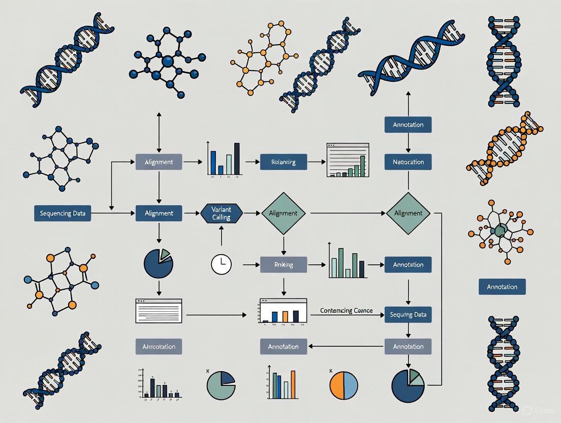
Experimental Workflows in Reproductomics
The typical workflow for reproductomics research involves multiple stages from experimental design through data integration and interpretation. The following diagram illustrates a generalized workflow for reproductomics studies:
Reproductomics Experimental Workflow
Essential Research Reagents and Computational Tools
Research Reagent Solutions for Reproductomics
Table 3: Essential Research Reagents and Platforms for Reprodutomics Studies
| Reagent/Platform | Function | Application in Reproductomics |
|---|---|---|
| High-Throughput Sequencers | Comprehensive DNA/RNA sequencing | Genomic and transcriptomic profiling of reproductive tissues [1] |
| Mass Spectrometers | Protein and metabolite identification and quantification | Proteomic and metabolomic analysis of reproductive samples [1] |
| DNA Methylation Kits | Assessment of epigenetic modifications | Epigenomic studies of hormonal regulation in endometrial tissue [1] |
| Single-Cell RNA Seq Kits | Gene expression profiling at individual cell level | Analysis of cellular heterogeneity in ovarian follicles or testicular tissue [1] |
| Cell Culture Media | Maintenance of primary reproductive cells | In vitro models of endometrial receptivity or gametogenesis [1] |
Computational Tools for Reproductomics Data Analysis
Table 4: Key Computational Tools and Databases for Reproductomics
| Tool/Database | Function | Application Context |
|---|---|---|
| Gene Expression Omnibus (GEO) | Public repository of functional genomics data | Accessing endometrial transcriptome data for receptivity studies [1] |
| Human Gene Expression Endometrial Receptivity Database (HGEx-ERdb) | Specialized database of endometrial gene expression | Identifying genes associated with endometrial receptivity (contains 19,285 genes) [1] |
| INTRIGUE | Statistical framework for reproducibility assessment | Evaluating and controlling reproducibility in high-throughput reproductomics experiments [4] |
| CustOmics | Deep learning-based multi-omics integration | Integrating heterogeneous omics data for classification and survival prediction [6] |
| DeepMoIC | Graph convolutional network for multi-omics | Cancer subtype classification in reproductive cancers [2] |
FAQs on Omics Data Management
1. What are the most common bottlenecks in omics data analysis? The three most pressing challenges are data processing, inefficient bioinformatics infrastructure, and collaboration between different teams. Data pre-processing is a significant bottleneck, requiring improved infrastructure to make analysis accessible to those without advanced programming skills [9].
2. Where should I deposit my different types of omics data? Appropriate repositories depend on your data type. Here are the recommended destinations [10]:
Table: Recommended Repositories for Omics Data
| Data Type | Data Formats | Repository |
|---|---|---|
| DNA sequence data (amplicon, metagenomic) | Raw FASTQ | NCBI SRA |
| RNA sequence data (RNA-Seq) | Raw FASTQ | NCBI SRA |
| Functional genomics data (ChIP-Seq, methylation seq) | Metadata, processed data, raw FASTQ | NCBI GEO (raw data to SRA) |
| Genome assemblies | FASTA or SQN file | NCBI WGS |
| Mass spectrometry data (metabolomics, proteomics) | Raw mass spectra, MZML, MZID | ProteomeXChange, Metabolomics Workbench |
| Feature observation tables | BIOM format (HDF5), tab-delimited text | NCEI, Zenodo, or Figshare |
3. How can I effectively integrate different types of omics data (e.g., transcriptomic and metabolomic)? Several data-driven approaches exist for integration without prior biological knowledge [11]:
- Statistical & Correlation-based: Use simple scatter plots, Pearson’s/Spearman’s correlation, or Procrustes analysis to relate features from different datasets.
- Correlation Networks: Construct networks where nodes represent biological entities and edges are based on correlation thresholds. Tools like WGCNA can identify clusters (modules) of highly correlated features across omics layers [11].
- Multivariate & Machine Learning Methods: Employ tools like the
mixOmicsR package for methods such as sparse PLS-Discriminant Analysis, or use other AI-driven integration frameworks [12] [11].
4. What are the key considerations for creating accessible data visualizations? Color should not be the only way information is conveyed. Ensure sufficient contrast (a 3:1 ratio is recommended by WCAG 2.1) for all critical graphical elements against their background. Use additional cues like textures, shapes, divider lines, and accessible axes to assist in data interpretation [13].
Troubleshooting Guides
Issue: Managing the Sheer Volume and Storage of Omics Data
Problem: The enormous volume of FASTQ, BAM, and other omics files complicates storage, processing, and analysis [14].
Solution:
- Immediate Redundant Storage: Upon receipt, immediately store raw sequencing data (e.g., FASTQ files) in two separate locations (e.g., a local drive and an institutional cloud drive) [10].
- Systematic Tracking: Record file names, associated projects, and metadata in a centralized, backed-up spreadsheet [10].
- Scheduled Archiving: Differentiate between raw, intermediate, and processed data. Implement a regularly scheduled backup plan for storage locations. Archive intermediate files from computationally intensive analyses until project completion or public deposition [10].
- Leverage Repositories: Submit raw data and final data analysis products to relevant public repositories for long-term archiving, as detailed in the FAQ above [10].
Issue: Integrating Multi-Omics Datasets with Missing Values
Problem: Integrating multiple omics views (e.g., genomics, proteomics) is challenging due to complex interactions and datasets with various view-missing patterns [15].
Solution:
- Advanced Computational Methods: Employ frameworks designed for incomplete multi-view observations. One effective approach is a deep variational information bottleneck method, which applies the information bottleneck framework to marginal and joint representations of the observed views. By modeling the joint representations as a product of marginal representations, it can efficiently learn from datasets with different missing-value patterns [15].
Issue: Overcoming Data Quality and Collaboration Bottlenecks
Problem: Data quality issues and inefficient collaboration between bioinformaticians, biologists, and management staff can delay data interpretation [9] [14].
Solution:
- Data Quality Monitoring: Establish data quality monitoring standards to ensure decisions are based on reliable data. Recognize that "not all data is created equal" [14].
- Democratize Data Analysis: Utilize interactive data analysis and visualization platforms (e.g., Omics Playground) that allow biologists to explore data without advanced programming skills. This bridges the gap between technical and non-technical team members [9].
- Iterative Discussions: Facilitate iterative discussions between all stakeholders (biologists, bioinformaticians, project managers) to fully understand the underlying biological mechanisms, a process that can take weeks to months [9].
Standardized Experimental Protocols
Protocol 1: Integrated Transcriptomic and Metabolomic Profiling of Plant Tissues
This protocol is adapted from a study on Breynia androgyna and provides a framework for generating coupled transcriptome and metabolome datasets [16].
1. Sample Collection and Preparation
- Cultivate plants under controlled conditions.
- Harvest tissue samples (e.g., leaf, flower, fruit, stem) and immediately process them to preserve RNA integrity and metabolite stability.
- Grind tissues into a fine powder under liquid nitrogen.
2. Metabolite Extraction and LC-MS Profiling
- Extraction: For each tissue type, add acidified methanol (HPLC grade) to the powdered sample. Vortex and sonicate the mixture to enhance metabolite solubilization. Centrifuge, filter the supernatant, and transfer to LC-MS vials. Prepare triplicate biological replicates [16].
- LC-MS Instrumentation:
- Chromatography: Use a UPLC system with a C18 column (e.g., Thermo Scientific Acclaim Polar Advantage II).
- Mass Spectrometry: Perform detection using a mass spectrometer (e.g., MicrOTOF-Q III) in positive electrospray ionization (ESI+) mode, scanning an m/z range of 50-1000 [16].
- Data Processing: Process raw data with appropriate software (e.g., Compass DataAnalysis). Apply an internal standard (e.g., vanillic acid) for normalization. Export data for statistical analysis in CSV format [16].
3. RNA Extraction, Sequencing, and Transcriptome Assembly
- RNA Extraction: Isolate total RNA using a modified protocol. Assess RNA quality using an Agilent Bioanalyzer (RIN > 7.0) and quantify via Nanodrop [16].
- Library Prep and Sequencing: Construct strand-specific cDNA libraries (e.g., using SureSelect RNA Library Prep Kit). Sequence on an Illumina HiSeq platform (e.g., 150 bp paired-end) [16].
- De Novo Transcriptome Assembly: Trim raw reads for adapters and quality (Phred score > Q20). Perform de novo assembly using Trinity software. Predict coding sequences (CDS) with TransDecoder [16].
- Functional Annotation: Annotate assembled unigenes using BLASTx against public databases (Nr, Swiss-Prot, KEGG, GO) with an e-value cutoff of 1e-5 [16].
Table: Key Research Reagents and Materials
| Item | Function |
|---|---|
| Acidified Methanol (HPLC grade) | Extraction of secondary metabolites from plant tissue powder. |
| UPLC System with C18 Column | Chromatographic separation of complex metabolite mixtures. |
| Q-TOF Mass Spectrometer | High-resolution mass detection for accurate metabolite profiling. |
| RNA Isolation Kit | Extraction of high-quality, intact total RNA. |
| Agilent 2100 Bioanalyzer | Assessment of RNA Integrity Number (RIN) to ensure sequencing quality. |
| SureSelect RNA Library Prep Kit | Construction of strand-specific cDNA libraries for sequencing. |
| Illumina HiSeq Platform | High-throughput sequencing of transcriptome libraries. |
The following workflow diagram illustrates the integrated transcriptomic and metabolomic profiling protocol:
Protocol 2: A Strategy for Multi-Omics Data Integration
This protocol outlines a knowledge-free, data-driven integration strategy for correlating features from different omics platforms, as reviewed in recent literature [11].
1. Data Preprocessing
- Format all omics datasets into matrices where rows represent samples and columns represent features (e.g., gene counts, metabolite intensities).
- Perform platform-specific normalization, log-transformation, and scaling as required.
2. Correlation-Based Integration using xMWAS
- Tool: Use the
xMWASplatform for pairwise association analysis [11]. - Analysis: The tool performs integration by combining Partial Least Squares (PLS) components and regression coefficients to determine association scores between features from different omics datasets [11].
- Network Construction: Generate an integrative network graph where nodes are omics features and edges are drawn if they meet a predefined threshold for association score and statistical significance [11].
- Community Detection: Identify highly interconnected communities within the network using a multilevel community detection algorithm that iteratively maximizes modularity [11].
The following diagram illustrates the logical flow of the data integration process:
Frequently Asked Questions (FAQs)
FAQ 1: What are the most common causes of poor data quality in single-cell sequencing, and how can I identify them? Poor data quality in single-cell sequencing often arises from issues during library preparation or the sequencing run itself. Common problems include adapter contamination, an overabundance of reads from a few highly expressed genes (sequence duplication), and a high percentage of base calls with low confidence (per-base N content) [17].
You can identify these issues by running quality control (QC) tools like FastQC on your raw FASTQ files. Key modules in the FastQC report to examine are the "Adapter Content," which should be near zero; the "Per Base N Content," which should be consistently at or near zero across the entire read length; and the "Sequence Duplication Levels," where high duplication can be expected but should be interpreted with caution as FastQC is not UMI-aware [17]. For multiple samples, use MultiQC to aggregate reports into a single view [17].
FAQ 2: My bioinformatics pipeline produces different results each time I run it, even with the same input data. Why does this happen, and how can I fix it? This irreproducibility is often caused by the inherent randomness (stochasticity) in some bioinformatics algorithms or by tools that are sensitive to the order in which input data is processed [18]. For instance, some structural variant callers can produce different variant call sets when the read order is shuffled [18].
To fix this, you should:
- Set Random Seeds: Ensure that any tool using a probabilistic algorithm allows you to set a fixed random seed. Record this seed in your workflow documentation [19].
- Use Containerized Workflows: Implement your pipeline using container technologies like Docker and workflow languages like the Common Workflow Language (CWL). This freezes the entire computational environment—including software versions, dependencies, and system libraries—guaranteeing identical execution every time [20] [19].
- Demand Provenance: Use workflow features, like the
--provenanceflag in CWL, to generate a detailed record of all inputs, outputs, and computational steps [20].
FAQ 3: I have successfully identified biomarker candidates in my discovery cohort, but they fail in a follow-up study. Is this a validation or a replication problem? This is a classic challenge and hinges on the distinction between validation and replication. Replication tests the same association under nearly identical circumstances (similar population, identical lab procedures and analysis pipelines) in an independent sample. Validation, particularly external validation, tests the association in a different population, which may differ in ethnicity, data collection methods, or other systematic factors [21].
Your issue could be related to either or both:
- If the follow-up study used a different population or platform, it was a validation effort. The failure could be due to these systematic differences or because the initial discovery was a false positive [21].
- To ensure replication, the original and confirmatory study samples should be independent but similar, and the laboratory and computational methods must be identical [21].
To avoid this, pre-plan your confirmation strategy, use large enough sample sizes, and employ stringent statistical corrections in the discovery phase [21].
FAQ 4: How can I integrate data from different 'omics studies that used different experimental designs? Integrating disparate 'omics studies is challenging due to differences in sample collection, platforms, and data processing. A powerful computational strategy is in-silico data mining and meta-analysis [1].
This involves:
- Data Mining: Collecting raw or processed data from public repositories like the Gene Expression Omnibus (GEO) with analogous research questions [1].
- Robust Integration: Using methods like the robust rank aggregation to identify common genes across studies that may have used different technologies. This approach was successfully used to define a meta-signature for endometrial receptivity by analyzing data from nine different studies [1].
- Systems Biology: A holistic, interdisciplinary approach that combines data from genomics, transcriptomics, and other 'omics to build computational models that can interpret complex cellular behavior beyond what isolated data can show [1].
Troubleshooting Guides
Issue: Inconsistent Alignment and Variant Calling Across Technical Replicates
Problem: Your bioinformatics tools (aligners, variant callers) produce inconsistent results when processing data from the same biological sample that was sequenced in multiple technical replicates (different library preps or sequencing runs).
Explanation: Technical variability is inherent in sequencing experiments. Bioinformatics tools should be robust enough to accommodate this and produce consistent results (achieving genomic reproducibility). However, some tools introduce deterministic biases (e.g., reference bias in aligners) or stochastic variations (e.g., from random algorithms), leading to irreproducible results [18].
Solution:
- Assess Genomic Reproducibility: Actively evaluate your tools using technical replicates. The focus is not on accuracy against a gold standard, but on consistency across replicates [18].
- Choose Reproducible Tools: Prefer tools that are known to be consistent. For example, one study found Bowtie2 produced consistent results irrespective of read order, while BWA-MEM showed variability with shuffled reads [18].
- Standardize the Pipeline: Implement your entire analysis, from read alignment to variant calling, in a reproducible workflow framework like CWL or Nextflow, and containerize it with Docker [20] [19]. This ensures that every component and its version are fixed.
Table 1: Key Metrics for Single-Cell RNA-seq Raw Data QC
| QC Metric | Tool/Method | Interpretation of a Good Result | Common Pitfalls |
|---|---|---|---|
| Per Base Sequence Quality | FastQC |
Quality score boxes remain in the green area for all positions; a gradual drop at the end is common [17]. | Scores falling into red area indicate poor quality calls, suggesting the need for quality trimming [17]. |
| Adapter Content | FastQC |
The cumulative percentage of adapter sequence is negligible across the read [17]. | High levels indicate incomplete adapter removal during library prep, requiring trimming [17]. |
| Per Base N Content | FastQC |
Percentage of bases called as 'N' is consistently at or near 0% [17]. | Any noticeable non-zero N content indicates issues with sequencing quality or library prep [17]. |
| Sequence Duplication | FastQC |
A diverse library where the majority of sequences show low duplication levels [17]. | High duplication can trigger a warning but is expected in single-cell data; tool is not UMI-aware [17]. |
Issue: Managing and Analyzing Large, Multimodal Reproductomics Datasets
Problem: You are generating large volumes of high-throughput multimodal data (e.g., genomic, transcriptomic, proteomic) on reproductive processes, but the data complexity creates a bottleneck. Conventional computational methods are inadequate for processing, integrating, and interpreting this data.
Explanation: The field of reproductomics investigates the interplay between hormonal regulation, genetics, and environmental factors. The vast amount of data generated by various 'omics technologies often surpasses our ability to thoroughly analyze it, leading to a data management bottleneck where biologically significant conclusions are difficult to distill [1].
Solution:
- Employ a Systems Biology Approach: Move beyond analyzing single 'omics layers in isolation. Use interdisciplinary approaches to combine genomics, transcriptomics, and other data types into unified computational models for a holistic view of reproductive systems [1].
- Utilize Integrated Computational Tools: Leverage machine learning algorithms for predicting outcomes (e.g., fertility), single-cell sequencing techniques for granular cell-level analysis, and R/Bioconductor packages like
PharmacoGxfor standardizing the analysis of large pharmacogenomic datasets [1] [20]. - Implement Robust Workflows: Create scalable, flexible, and reproducible bioinformatics pipelines using frameworks like the Common Workflow Language (CWL) to ensure your analysis is transparent and can be shared and validated by the community [20].
Experimental Protocols
Detailed Protocol: Creating a Reproducible Pharmacogenomic Analysis Pipeline
This protocol outlines the methodology for creating a portable and reproducible pipeline to process pharmacogenomic data, combining pharmacological (e.g., drug response) and molecular (e.g., gene expression) profiles into a single, shareable data object [20].
1. Workflow Specification:
- Define the pipeline using the Common Workflow Language (CWL). Each processing step must be a CWL-encoded tool with explicit definitions for inputs, outputs, base commands, and requirements (e.g., Docker container, computational resources) [20].
- Combine these steps into a CWL workflow that specifies the execution order and data flow between them.
2. Computational Environment:
- Containerization: Package each tool and its dependencies into a Docker image. This isolates the analysis environment, ensuring that operating system, library versions, and software versions remain consistent [20] [19].
- Resource Management: The CWL workflow handles resource allocation (compute and memory) for each step, which is often a limitation of conventional scripting [20].
3. Data Processing and Object Creation:
- Data Curation: Curate cell line and drug annotation files. The pipeline should assign unique identifiers to each, which are used to validate data integrity in subsequent steps [20].
- Drug Response Computation: Within the R environment, use the
PharmacoGxpackage to compute standard drug sensitivity metrics from raw dose-response data. Key metrics include: - Molecular Profile Integration: Incorporate processed molecular data (e.g., RNA-seq, SNP arrays) into the object.
- PharmacoSet (PSet) Assembly: The
PharmacoGxpackage assembles all curated annotations, computed drug responses, and molecular profiles into a unified R object called a PharmacoSet (PSet) [20].
4. Provenance Tracking and Sharing:
- Execute the CWL workflow with the
--provenanceflag. This generates a Research Object—a bundled container with all input files, output files, and metadata, including checksums for granular data provenance [20]. - Assign a Digital Object Identifier (DOI) to the final PSet and workflow on a data repository (e.g., Harvard Dataverse) to ensure persistent access and sharing [20].
Diagram: Reproducible Bioinformatics Pipeline
The Scientist's Toolkit: Research Reagent Solutions
Table 2: Essential Computational Tools for Reproductomics Data Management
| Tool / Resource | Function | Application in Reproductomics |
|---|---|---|
| Common Workflow Language (CWL) | A standard for describing data analysis workflows in a way that is portable and scalable across different software environments [20]. | Ensures pharmacogenomic and other reproductomics pipelines are reproducible, transparent, and can be executed identically by other researchers [20]. |
| Docker Containers | OS-level virtualization to package software and all its dependencies into a standardized unit, ensuring the software runs the same regardless of the host environment [19]. | Freezes the entire computational environment for a bioinformatics tool or pipeline, eliminating "works on my machine" problems and guaranteeing long-term reproducibility [19]. |
| PharmacoGx (R/Bioconductor) | An R package that provides computational methods to process, analyze, and integrate large-scale pharmacogenomic datasets [20]. | Creates unified PharmacoSet (PSet) objects from reproductive cell line screens, combining drug response and molecular data for easy sharing and secondary analysis [20]. |
| FastQC | A quality control tool that provides an overview of basic statistics and potential issues in raw high-throughput sequencing data [17]. | The first line of defense for identifying sequencing or library preparation artifacts in single-cell or bulk sequencing data from reproductive tissues [17]. |
| Robust Rank Aggregation | A computational meta-analysis method designed to compare distinct gene lists and identify common overlapping genes [1]. | Identifies consensus biomarker signatures (e.g., for endometrial receptivity) by integrating gene lists from multiple, disparate transcriptomics studies [1]. |
FAQs: Navigating Data and Methodological Hurdles
FAQ 1: What are the most common causes of inconsistent results in endometrial receptivity (ER) clinical trials?
Inconsistent results often stem from a combination of methodological and data-related bottlenecks:
- Lack of Standardized Protocols: Many studies use bespoke, in-house protocols for sample preparation and analysis. This makes it difficult to reproduce experiments or compare results across different laboratories [22]. For instance, preparations like Platelet-Rich Plasma (PRP) vary significantly in "platelet concentration, leukocyte and fibrin content," and administration methods, leading to heterogeneous findings on its efficacy [23].
- Heterogeneous Patient Populations and Definitions: A core challenge is the lack of a universal definition for conditions like Recurrent Implantation Failure (RIF). Studies use varying criteria for the number of failed cycles and embryo quality, creating non-uniform cohorts that compromise data comparability [24].
- Manual Data Handling Errors: Sample preparation stages in advanced analyses like proteomics are "highly manual and time-consuming," making them prone to human error. This can lead to mischaracterization and unreliable data, which is particularly problematic with sensitive clinical samples that are difficult to reacquire [22].
FAQ 2: Which data-driven technologies show the most promise for overcoming current ER assessment limitations?
Emerging technologies are focusing on integration and automation to improve objectivity and predictive power.
- Deep Learning (DL) and Multi-Modal Data Fusion: DL models, particularly convolutional neural networks (CNNs), can analyze routine ultrasound images to identify subtle features associated with ER states that are imperceptible to the human eye. One study achieved superior predictive accuracy for Recurrent Pregnancy Loss (RPL) risk by using a fusion model that integrated a CNN for image analysis (ResNet-50) and another network for clinical tabular data (TabNet) [25].
- Multi-Omics Integration: Advanced computational methods, such as variational information bottleneck approaches, are being developed to integrate complex data from multiple "omics" techniques (e.g., transcriptomics, proteomics). This is crucial for understanding the complex interactions within and across different biological systems, even when data is incomplete [15].
- Automated Sample Preparation: Laboratory automation, including automated liquid handlers, is a key solution for overcoming manual workflow bottlenecks. Automation reduces human error, increases throughput, and standardizes sample processing, which enhances the reproducibility and reliability of proteomic and other molecular data [22].
FAQ 3: What is the current clinical evidence for the efficacy of Endometrial Receptivity Analysis (ERA)?
The evidence for ERA is mixed and reflects the broader crisis in standardizing molecular diagnostics.
- Supporting Evidence: A large 2025 retrospective study of 3,605 patients with previous failed embryo transfers found that personalized embryo transfer (pET) guided by ERA significantly improved clinical pregnancy and live birth rates for both RIF and non-RIF patients. It also reduced the early abortion rate in the non-RIF group [26].
- Contradictory Evidence: Other research indicates that ERA may not be effective and "could even be detrimental to pregnancy rates" [23]. A 2019 systematic review and meta-analysis concluded that while results from modern molecular tests like ERA are promising, more data is needed before they can be universally recommended, as many conventional markers show poor predictive ability for clinical pregnancy [27].
Troubleshooting Common Experimental Scenarios
Scenario: Investigating a Novel ER Biomarker with Inconsistent Proteomics Data
Problem: Your mass spectrometry (LC-MS) results are inconsistent, with high technical variation between replicates.
Solution: Implement an automated and standardized sample preparation workflow.
- Step 1: Diagnose the Bottleneck. The primary issue is likely the reliance on manual, low-throughput sample preparation techniques (e.g., cell lysis, protein digestion, peptide purification). These repetitive, meticulous tasks are susceptible to user-derived errors and sample-to-sample variation [22].
- Step 2: Apply the Fix.
- Adopt Automation: Integrate an automated liquid handling system to execute critical pipetting and preparation steps. This minimizes human error and variation [22].
- Standardize the Protocol: Use a standardized, commercially available kit for sample preparation instead of an in-house protocol. This ensures consistency and improves reproducibility across your lab and others [22].
- Process Controls: Include a standardized control sample in every batch to monitor technical variability and batch effects introduced during preparation.
- Step 3: Validate the Data.
- After implementing automation, re-run a set of samples and controls.
- Compare the coefficient of variation for protein abundance measurements before and after the change. A significant decrease indicates improved data quality and reliability.
Quantitative Data Synthesis
Table 1: Comparative Reproducibility of Key ER Research Methodologies
| Methodology | Key Measurable Output(s) | Common Sources of Data Variance | Evidence Level for Improving Live Birth Rate (LBR) |
|---|---|---|---|
| Endometrial Receptivity Array (ERA) | Personalized Window of Implantation (WOI) | Biopsy timing, RNA sequencing platform, algorithmic interpretation of transcriptomic signature | Mixed: Large studies show benefit in RIF [26], while others show no effect or detriment [23]. |
| Endometrial Scratching | Clinical Pregnancy Rate | Technique (instrument, depth), timing in relation to cycle, operator skill | Controversial: Recent well-designed RCTs found no beneficial effect [23]. |
| Platelet-Rich Plasma (PRP) for Thin Endometrium | Endometrial Thickness (mm), LBR | Preparation method (platelet/leukocyte concentration), number of infusions | Uncertain: Small randomized studies show conflicting results; Cochrane review finds overall evidence uncertain [23]. |
| Proteomic LC-MS/MS | Protein Identification & Quantification | Manual sample prep, trypsin digestion efficiency, LC column performance | Promising but not yet translational; dependent on standardized workflows [22]. |
| Deep Learning on Ultrasound | RPL Risk Probability Score | Image acquisition parameters, model architecture, clinical data integration | Emerging: Shows high accuracy for risk stratification in initial studies [25]. |
Table 2: Clinical Evidence for ERA from a Large-Scale Study (n=3,605) [26]
| Patient Group & Intervention | Clinical Pregnancy Rate | Live Birth Rate | Early Abortion Rate |
|---|---|---|---|
| Non-RIF with npET (n=1,744) | 58.3% | 48.3% | 13.0% |
| Non-RIF with pET (n=301) | 64.5% | 57.1% | 8.2% |
| RIF with npET (after PSM) | 49.3% | 40.4% | Not Specified |
| RIF with pET (after PSM) | 62.7% | 52.5% | Not Specified |
Experimental Protocols
Protocol: Endometrial Tissue Sampling for Transcriptomic Analysis (ERA)
This protocol is based on the hormone replacement therapy (HRT) cycle methodology used in recent clinical studies [26].
1. Objective: To obtain a standardized endometrial tissue sample for RNA extraction and subsequent transcriptomic analysis to determine the window of implantation (WOI).
2. Materials:
- Estrogen supplements (e.g., oral or transdermal)
- Progesterone (P) supplements (e.g., intramuscular injection)
- Pipelle endometrial suction catheter or similar device
- Speculum
- RNA preservation solution (e.g., RNAlater)
- Sterile gloves and antiseptic solution
3. Step-by-Step Workflow:
4. Key Considerations:
- Timing: The biopsy must be performed after a specific duration of progesterone exposure (e.g., 5 days in an HRT cycle) [26].
- Sample Handling: The tissue must be immediately placed in RNAlater to preserve RNA integrity.
- Clinical Data: Record the exact timing of the biopsy relative to the start of progesterone and the patient's hormone levels.
Protocol: Developing a Deep Learning Model for ER Assessment from Ultrasound
This protocol is adapted from a study on automated RPL risk assessment [25].
1. Objective: To develop a fusion deep learning model that integrates grayscale ultrasound images and clinical data to assess endometrial receptivity and stratify RPL risk.
2. Materials:
- Ultrasound Machine: With capability to export DICOM or high-resolution image files.
- Computing Environment: GPU-accelerated workstation (e.g., with NVIDIA CUDA support).
- Software: Python with deep learning libraries (PyTorch/TensorFlow), and scikit-learn.
3. Step-by-Step Workflow:
4. Key Considerations:
- Data Quality: Ultrasound images must be standardized (e.g., longitudinal section of the endometrium). Clinical data (e.g., age, BMI, hormone levels) must be curated.
- Pre-processing: Images are typically resized (e.g., to 224x224 pixels) and undergo data augmentation (random flips, rotations) to improve model generalizability [25].
- Validation: Use a strict train-test split or k-fold cross-validation and report performance on the held-out test set to avoid overfitting.
The Scientist's Toolkit: Key Research Reagent Solutions
Table 3: Essential Materials for Advanced Endometrial Receptivity Research
| Item | Function in Research | Specific Example / Note |
|---|---|---|
| Endometrial Biopsy Catheter | To obtain endometrial tissue samples for histology, transcriptomics, or proteomics. | Pipelle de Cornier is commonly used for minimal discomfort sampling. |
| RNAlater Stabilization Solution | To immediately preserve RNA integrity in tissue samples for gene expression studies like ERA. | Critical for ensuring accurate transcriptomic profiles from biopsies [26]. |
| Automated Liquid Handler | To automate and standardize sample preparation steps for proteomics and molecular assays. | Systems like Beckman Coulter's Biomek series can overcome manual workflow bottlenecks [22]. |
| Pre-trained Deep Learning Models | As a starting point for developing custom image analysis models for ultrasound or histology images. | Models like ResNet-50 can be used with transfer learning for medical image analysis [25]. |
| Hormone Assay Kits | To quantitatively measure serum levels of estradiol (E2) and progesterone (P) for cycle monitoring. | An optimal E2/P ratio has been correlated with a lower rate of displaced WOI [26]. |
| Liquid Chromatography Tandem Mass Spectrometry (LC-MS/MS) | To identify and quantify protein expression and post-translational modifications in endometrial samples. | The quality of this analysis is entirely dependent on the preceding sample preparation [22]. |
Ethical and Regulatory Considerations in Reproductive Data Storage and Sharing
Frequently Asked Questions (FAQs) and Troubleshooting Guides
General Data Sharing Principles
Q1: What are the core ethical principles I should follow for managing reproductive data? The 5Cs of data ethics provide a foundational framework for managing sensitive data [28].
- Consent: Obtain informed and voluntary consent before data collection or use. Individuals must understand what data is collected, how it will be used, and by whom. Consent should be easy to withdraw [28].
- Collection: Only collect data that is necessary for a specific, stated purpose. Avoid gathering excessive or irrelevant information [28].
- Control: Give individuals control over their own data, including the rights to access, review, update, and know who has access to it [28].
- Confidentiality: Protect data from unauthorized access, breaches, or leaks using security measures like encryption and access controls [28].
- Compliance: Adhere to all relevant legal and regulatory requirements, such as GDPR or institutional policies [28].
Q2: What does "FAIR" mean in the context of data sharing, and why is it important? FAIR is a set of guiding principles to make data Findable, Accessible, Interoperable, and Reusable [29]. Adhering to FAIR principles helps maximize the impact and utility of your research data by ensuring others can easily locate, understand, and use it. This promotes transparency, reproducibility, and accelerates scientific discovery by enabling meta-analyses and method development [29].
Ethics, Privacy, and Human Subject Data
Q3: How should I handle data derived from human research participants? Sharing human data requires balancing participant privacy with the benefits of data sharing [30] [29]. Key steps include:
- De-identification: Adequately de-identify personal information prior to sharing to protect participant privacy and mitigate risk [30].
- Informed Consent: For studies generating genomic data, informed consent documents should state what data types will be shared, for what purposes, and whether sharing will be open or controlled-access [30].
- Controlled-Access: For data that is challenging to fully de-identify or still poses privacy risks (e.g., genomic, detailed phenotypic, or qualitative data), use a controlled-access repository where only qualified researchers can access the data [30] [29].
Q4: My data is too sensitive for public release. What are my sharing options? Not all data can be shared publicly. Consider these strategies to make your data as accessible as possible [31] [29]:
- Controlled-Access Sharing: Submit individual-level raw data to a controlled-access repository like the European Genome-phenome Archive (EGA) or dbGaP [29].
- Share Processed Data: Make summary statistics (e.g., means, variant-level association statistics) available, even if raw data cannot be shared [29].
- Data Simulation: Generate and share simulated data that is statistically similar to the original data. This allows others to reproduce your analysis workflow without accessing the identifiable original data [29].
Q5: How long am I allowed to store research data? You should not store personal data for longer than necessary [32]. The GDPR states data should be stored for the shortest time possible, and you must be able to justify your retention period. Develop policies for standard retention periods and regularly review data to erase or anonymize it when it is no longer needed [32].
Practical Data Management
Q6: I'm overwhelmed by the amount of data my omics experiments generate. What is the bottleneck, and how can I address it? The field of reproductomics has reached a "data management bottleneck," where data volumes vastly surpass our ability to thoroughly analyze and interpret them [1]. To overcome this:
- Utilize Computational Tools: Employ robust bioinformatic applications and machine learning algorithms for processing and analysis [1].
- Adopt a Systems Biology Approach: Use an interdisciplinary approach that amalgamates genomics, transcriptomics, proteomics, etc., to generate comprehensive computational models [1].
- Use Public Repositories: Deposit your data in public repositories to leverage community resources and tools [1].
Q7: Where should I submit my reproductive genomics data?
- Primary Repository: The NHGRI expects researchers to use the AnVIL repository for a variety of data types, including genomic and non-genomic data from human participants [30].
- Registration: Large-scale human genomic studies must be registered in dbGaP [30].
- Alternatives: If you wish to use an alternative repository, you must propose and justify it in your Data Management and Sharing (DMS) Plan for assessment prior to funding [30].
Q8: What is the difference between raw, intermediate, and processed data? Understanding these categories helps in deciding what to share [31]:
- Raw Data: The initial output from an instrument (e.g., FASTQ files from an RNA-seq experiment) [31].
- Intermediate Data: Data products between raw data and final findings (e.g., gene expression estimates, VCF files from variant calling) [31].
- Findings: The final results, plots, and statistics produced by analyzing the data [31]. It is recommended to share as much raw and intermediate data as possible to ensure reproducibility and enable secondary analyses [31].
Data Sharing Tiers and Repository Comparison
The table below summarizes the primary methods for sharing genomic data, balancing accessibility with privacy protection [31].
| Sharing Method | Description | Ideal For | Examples |
|---|---|---|---|
| Public Sharing | Data is released without barriers for reuse. | Non-human data; fully anonymized human data with minimal re-identification risk. | Gene Expression Omnibus (GEO), AnVIL (open data) [30] [31] |
| Controlled-Access | Access is granted to qualified researchers who apply and agree to specific terms. | Individual-level human genomic, phenotypic, or other sensitive data where privacy risks exist. | dbGaP, European Genome-phenome Archive (EGA) [30] [29] |
| Upon-Request | Data is shared directly by the researcher upon receipt of a request. | Generally discouraged as it is inefficient and can lead to delays and inequitable access. | N/A |
Experimental Protocol: Implementing a Responsible Data Sharing Workflow
This protocol outlines the key steps for ethically sharing research data, particularly data derived from human participants in reproductive studies, in alignment with FAIR principles and regulatory requirements [30] [29].
1. Pre-Collection: Planning and Consent
- Develop a Data Management & Sharing (DMS) Plan: Outline the types of data and metadata you will generate, the standards you will use, and your chosen repositories [30] [29].
- Secure Informed Consent: For prospective studies, ensure consent documents explicitly state plans for data sharing, including the data types, purposes (e.g., general research use), and access mode (e.g., controlled-access) [30].
2. Pre-Publication: Data Preparation and Documentation
- De-identify Data: Remove all direct identifiers. For sensitive datasets, assess the risk of re-identification from quasi-identifiers or the data itself [30] [31].
- Organize and Document Metadata: Create detailed metadata using structured formats and community-standard ontologies (e.g., Experiment Factor Ontology) [30] [31]. Describe the sample information (e.g., tissue type, diagnosis) and handling protocols (e.g., library preparation method) thoroughly.
3. Submission and Release
- Select an Appropriate Repository: Choose a repository based on your data type and privacy needs (see Table above) [30] [29].
- Submit Data and Metadata: Follow the repository's submission guide. NHGRI expects data to be submitted to the selected repository before publication-related manuscript acceptance [30].
- Obtain a Persistent Identifier: Ensure the repository issues a unique, citable identifier (e.g., a DOI) for your dataset [29].
The Scientist's Toolkit: Research Reagent Solutions
The table below lists key resources and tools essential for managing and analyzing reproductive omics data.
| Item | Function |
|---|---|
| AnVIL (NHGRI's Repository) | A primary cloud-based platform for storing, sharing, and analyzing genomic and related data, supporting both controlled and open access [30]. |
| dbGaP (Database of Genotypes and Phenotypes) | A controlled-access repository designed to archive and distribute the results of studies that investigate the interaction of genotype and phenotype [30]. |
| FASTQ File Format | The standard text-based format for storing both a biological sequence (usually nucleotide sequence) and its corresponding quality scores from sequencing instruments [31]. |
| VCF (Variant Call Format) | A standardized text file format used for storing gene sequence variations (e.g., SNPs, indels) called from sequencing experiments [31]. |
| Experiment Factor Ontology (EFO) | An ontology that provides systematic descriptions of experimental variables, such as disease, tissue, and treatment, enabling structured metadata annotation [31]. |
| Machine Learning Models (e.g., in Kipoi) | Pre-trained models that can be repurposed for new genomic analyses, such as predicting variant effects or gene expression [31]. |
Workflow Diagram: Data Classification for Ethical Sharing
The following diagram illustrates the decision process for classifying and selecting the appropriate sharing method for research data, with a focus on human subjects.
Troubleshooting Common Scenarios
Problem: A colleague requests data via email, but the data is sensitive.
- Solution: Avoid ad-hoc "upon-request" sharing for sensitive data. Direct your colleague to the controlled-access repository (e.g., dbGaP, EGA) where your data is housed and guide them through the standard application process. This ensures consistent, fair, and secure access for all researchers [31] [29].
Problem: My dataset is complex and includes multiple omics layers. I'm unsure how to make it FAIR.
- Solution: Utilize an integrative in-silico analysis approach. Employ systems biology frameworks to amalgamate data from genomics, transcriptomics, and other omics fields into a unified computational model. Deposit the entire dataset in a single, appropriate repository like AnVIL whenever possible, and use rich, structured metadata with controlled vocabularies to describe all data layers comprehensively [1] [30].
Problem: I am using legacy biospecimens that lack explicit consent for broad data sharing.
- Solution: NHGRI expects explicit consent for all human data generated by its funded research. If you propose to use specimens lacking such consent, you must document a request for an exception in your DMS Plan, providing scientific justification for using these specific data sources [30].
Computational Approaches and Analytical Frameworks for Reproductomics Data
Technical Troubleshooting Guides
Data Integration & Preprocessing Challenges
Q: How can I handle inconsistent data formats and structures from different studies? A: This is a common challenge in data integration. Solutions include:
- Use ETL/ELT Tools: Implement Extract, Transform, Load (ETL) or Extract, Load, Transform (ELT) tools to manage different data formats and structures. These tools streamline the process of data extraction, transformation into a standard format, and loading into a target system, which reduces manual errors [33].
- Automated Data Profiling: Use automated data profiling tools to identify data quality issues such as missing values, inconsistencies, and duplicates early in the process [34].
- Establish Data Governance: Create data governance frameworks with clear ownership and data standards to ensure a common understanding and consistent usage of data across projects [34] [33].
Q: My integrated dataset has poor quality (missing values, duplicates). How can I fix it? A: Data quality is paramount for reliable analysis.
- Implement Data Cleansing: Use data cleansing tools, especially those with AI capabilities for pattern recognition, to identify and correct complex data quality issues [34].
- Proactive Validation: Check for errors or inconsistencies soon after collecting data, before integrating it into your primary dataset. This prevents poor-quality data from corrupting your analysis [33].
- Set Validation Rules: Implement validation rules at data entry points to prevent bad data from entering the system in the first place [34].
Reproducibility & Workflow Management
Q: How can I ensure my in-silico analysis is reproducible? A: Reproducibility is a critical challenge in digital medicine and computational biology.
- Adopt Open Science Practices: Utilize frameworks like the Open Science Framework (OSF) to make data and workflows easily accessible to collaborators and for future replication. This includes pre-registering studies to state research questions and methods upfront [35].
- Use Workflow Management Systems (WMS): Automate in-silico data analysis processes through WMS. These systems provide flexible and extensible tools for creating, deploying, and documenting data integration and analysis pipelines, ensuring that the same steps can be followed to achieve consistent results [36].
- Thorough Documentation: Maintain detailed documentation of all data sources, transformation steps, software versions, and parameters used. This is a cornerstone of reproducible research [35].
Q: My analysis workflow is becoming too complex and unmanageable. What can I do? A: As projects grow, complexity can hinder progress.
- Automate with WMS: As noted above, Workflow Management Systems are designed specifically to manage complex, multi-step computational workflows, reducing manual intervention and the potential for error [36].
- Implement Data Lineage Tools: Use tools that provide data lineage, giving you visibility into how data flows, where it starts, what changes it has gone through, and where it is delivered. This helps build trust, detect anomalies, and optimize performance [37].
Computational & Scalability Issues
Q: My data processing is too slow for large-scale multi-omics datasets. How can I improve performance? A: Large data volumes can overwhelm traditional methods.
- Leverage Modern Platforms: Adopt modern data management platforms that support massively parallel processing (MPP), Spark processing, or elastic processing. These can handle multiple terabytes of data simultaneously, saving time and money [37] [33].
- Utilize Serverless Architecture: To ease infrastructure management, use serverless data integration architectures where zero effort is needed to provision and maintain underlying cloud instances. This allows researchers to focus on analysis rather than administrative work [37].
- Apply Incremental Data Loading: Break data into smaller segments and load them incrementally rather than attempting to load the entire dataset at once [33].
Biological Interpretation & Validation
Q: After integrating data, I struggle with the biological interpretation of the results. A: Moving from data to biological insight is a key challenge.
- Leverage Network-Based Methods: Use network-based multi-omics integration methods (e.g., network propagation, graph neural networks) that contextualize results within known biological networks (PPI, metabolic pathways). This provides a systems-level view and helps in identifying biologically relevant patterns [38].
- Consult Multi-Omics Resources: Platforms like InSilico DB provide access to a large number of curated public genomic datasets, which can be used for comparison and validation of your findings against existing data [39].
Frequently Asked Questions (FAQs)
Q: What are the biggest data management bottlenecks in reproductomics research? A: The primary bottlenecks include:
- Data Quality and Consistency: Inconsistencies arising from combining data from various sources with different formats and accuracy levels [34].
- Data Security and Privacy Compliance: Ensuring integrated data, especially sensitive health information, adheres to regulations like GDPR and HIPAA [34].
- Legacy System Integration: Difficulties in connecting older, outdated systems with modern technologies due to proprietary formats and limited documentation [34].
- The Reproducibility Crisis: A significant portion of scientific findings cannot be replicated, often due to a lack of transparency, data sharing, and standardized workflows [40] [35].
Q: How can I securely integrate data while maintaining patient privacy? A: Security must be integrated into the process.
- Implement Robust Security Measures: Protect sensitive data using encryption (both in transit and at rest), data masking, anonymization techniques, and granular role-based access controls [34] [33].
- Regular Audits: Conduct regular security audits and penetration testing to identify and address vulnerabilities [34].
- Choose Compliant Tools: Select data integration solutions that are designed with strong security features and compliance frameworks in mind [33].
Q: What should I do if my data sources update at different rates? A: This "velocity" problem is common with heterogeneous sources.
- Thorough Documentation: Maintain detailed records of all source systems, including their data change protocols and rates [33].
- Create Asynchronous Processes: Design workflows with asynchronous processes for performance-intensive operations to prevent slower systems from bottlenecking the entire pipeline [34].
Experimental Protocols & Data Tables
Protocol: A Standard Workflow for Network-Based Multi-Omics Integration
This protocol is adapted from methodologies reviewed in network-based multi-omics studies [38].
- Data Collection & Curation: Gather omics data (e.g., genomics, transcriptomics) from disparate sources, such as public repositories (e.g., GEO, TCGA) via hubs like InSilico DB [39].
- Quality Control & Preprocessing: Perform automated data profiling and cleansing. Handle missing values, normalize data, and correct for batch effects.
- Network Construction/Selection: Obtain or construct a relevant biological network (e.g., a Protein-Protein Interaction network from STRING database or a gene co-expression network).
- Data Integration: Map the preprocessed multi-omics data onto the network. This can be done using various methods:
- Network Propagation/Diffusion: To smooth data and identify active network regions.
- Graph Neural Networks (GNNs): To learn complex patterns from the network-structured data.
- Analysis & Model Training: Perform the core analysis, such as identifying differentially active subnetworks, predicting drug responses, or classifying disease subtypes.
- Validation: Validate findings using independent datasets or through experimental follow-up.
- Interpretation & Reporting: Interpret the results in the context of the underlying biology. Use data lineage tools to document the entire workflow for reproducibility.
Table 1: Common Data Integration Challenges and Solutions in Reproductomics
| Challenge | Impact on Research | Recommended Solution |
|---|---|---|
| Data Quality & Consistency [34] | Undermines integrated view, leads to flawed analytics and decision-making. | Implement automated data profiling & cleansing tools; establish data governance [34] [33]. |
| Reproducibility Crisis [40] [35] | Shakes confidence in scientific validity; policies/treatments may be based on unverified findings. | Adopt Open Science practices; pre-register studies; use Workflow Management Systems (WMS) [35] [36]. |
| Computational Scalability [38] [33] | Overwhelms traditional methods, causing long processing times and inability to handle large data. | Use modern platforms with parallel processing (e.g., Spark); apply incremental data loading [37] [33]. |
| Semantic Heterogeneity [34] | Different data sources use varying formats, schemas, and languages, complicating integration. | Use ETL/ELT tools and managed integration solutions to transform data into a uniform format [33]. |
| Security & Privacy Compliance [34] | Risk of data breaches, reputational damage, and violation of regulations (GDPR, HIPAA). | Implement encryption, data masking, anonymization, and role-based access controls [34] [33]. |
Table 2: Key Software Tools and Platforms for In-Silico Data Mining
| Tool / Platform Category | Example(s) | Primary Function in Research |
|---|---|---|
| Workflow Management Systems (WMS) [36] | (e.g., Nextflow, Snakemake) | Automate and manage complex, multi-step computational data analysis pipelines. |
| Data Integration & ETL/ELT [37] [33] | Informatica Cloud Data Integration, Talend, Estuary Flow | Extract, transform, and load data from disparate sources into a unified format for analysis. |
| Genomic Datasets Hub [39] | InSilico DB | Provides curated, ready-to-analyze genomic datasets from public repositories like GEO and TCGA. |
| Network Analysis & Multi-Omics [38] | (Various specialized algorithms) | Integrate multi-omics data using biological networks (PPI, GNNs) for discovery. |
| Open Science Framework [35] | Open Science Framework (OSF) | Facilitates collaborative project management, data sharing, and study pre-registration. |
Workflow and Pathway Visualizations
In-Silico Data Mining Workflow
Network-Based Multi-Omics Integration
Data Management Bottlenecks in Reprodutomics
The Scientist's Toolkit: Research Reagent Solutions
Table 3: Essential "Reagents" for In-Silico Data Mining
| Item / Resource | Type | Primary Function |
|---|---|---|
| InSilico DB [39] | Data Repository Hub | Provides efficient access to curated, normalized, and ready-to-analyze public genomic datasets, bridging the gap between data repositories and analysis tools. |
| Workflow Management System (WMS) [36] | Software Platform | Automates in-silico data analysis processes, enabling the creation of reproducible, scalable, and manageable computational pipelines. |
| ETL/ELT Tools [37] [33] | Data Integration Software | Automates the extraction of data from sources, its transformation into a unified format, and loading into a target system, solving heterogeneous data structure challenges. |
| Biological Networks (PPI, GRN) [38] | Knowledgebase / Data | Provides a structured framework (nodes and edges) for integrating and interpreting multi-omics data in a biologically meaningful context. |
| Open Science Framework (OSF) [35] | Collaborative Platform | Facilitates project management, data sharing, and study pre-registration to enhance transparency, collaboration, and ultimately, reproducibility. |
Machine Learning Algorithms for Predicting Fertility Outcomes and Treatment Success
Technical Support Center: Troubleshooting Guides and FAQs
This technical support center is designed for researchers and scientists encountering challenges in developing and deploying machine learning (ML) models within the field of reproductomics, with a specific focus on predicting fertility treatment outcomes. The guidance is framed within the critical context of overcoming data management bottlenecks that commonly hinder research progress in this domain.
Frequently Asked Questions (FAQs)
FAQ 1: My model's performance is inconsistent and unreliable. What are the most critical features to include to build a robust predictive model?
A primary challenge in predictive modeling is feature selection. Based on a systematic review of ML in Assisted Reproductive Technology (ART), the following features are most frequently reported as critical for model performance [41]:
- Female Age: Consistently the most important predictor across studies, largely due to its correlation with egg quality and quantity [42] [41].
- Ovarian Reserve Markers: Such as Anti-Müllerian Hormone (AMH) levels, which indicate egg quantity [42].
- Follicle Counts and Size Distribution: Particularly the number of follicles in the 16-20 mm and 11-15 mm diameter ranges, which are key for determining the optimal time for trigger injection [43].
- Body Mass Index (BMI): A BMI outside the normal range (19-24.9) is associated with lower pregnancy and higher miscarriage rates [42].
- Stimulation Parameters and Hormone Levels: Including estradiol levels during treatment [43].
Table 1: Key Predictive Features for Fertility Outcome Models
| Feature Category | Specific Features | Clinical/Technical Rationale |
|---|---|---|
| Patient Demographics | Female Age, BMI | Age is the strongest predictor of egg quality; BMI impacts pregnancy rates [42] [41]. |
| Ovarian Response | Follicle Count (11-15mm, 16-20mm), AMH, Estradiol Level | Directly measures response to stimulation and yield of mature oocytes [43]. |
| Treatment Protocol | Stimulation Drugs, Trigger Timing, Type of Treatment (e.g., ICSI) | Different protocols suit different patient profiles and impact egg retrieval success [43] [44]. |
| Historical Data | Total Previous Treatment Attempts, Previous Pregnancy History | Provides context on patient-specific treatment history and cumulative success chances [44]. |
FAQ 2: What are the best-performing machine learning algorithms for predicting fertility outcomes?
The choice of algorithm depends on your data structure and the specific outcome you wish to predict. A systematic review identified that while many algorithms are used, several show strong performance [41]. Furthermore, advanced implementations often use ensemble methods to combine the strengths of multiple algorithms [45].
Table 2: Commonly Used ML Algorithms and Their Performance in Reproductomics
| Algorithm | Reported Performance (Examples) | Strengths and Common Use Cases |
|---|---|---|
| Support Vector Machine (SVM) | Frequently applied technique (44.44% of reviewed studies) [41]. | Effective in high-dimensional spaces [41]. |
| Random Forest (RF) | AUC: 0.72-0.83; Accuracy: ~65-77% [41]. | Handles mixed data types, provides feature importance, robust to overfitting [41]. |
| Gradient Boosting (XGBoost, LightGBM, CatBoost) | Used in top-performing ensembles; LightGBM offers superior performance on large-scale data and memory efficiency [45]. | High accuracy, native handling of categorical features (CatBoost), fast training [45]. |
| Bayesian Network Model | Accuracy: 91.3%; AUC: 0.997 [41]. | Models probabilistic relationships between variables. |
| Logistic Regression (LR) | Often used as a baseline model for comparison [41]. | Simple, interpretable, good for establishing a performance baseline. |
FAQ 3: I am facing significant data management bottlenecks, from siloed data to poor quality. How can I address this?
Data management challenges are a major bottleneck in reproductomics research. Here are specific solutions aligned with common problems [46] [14]:
- Challenge: Sheer Volume and Multiple Data Storages (Silos)
- Challenge: Data Quality
- Solution: Implement rigorous data quality monitoring standards. This includes automated checks for accuracy and completeness. Not all data is equally valuable; processes must prioritize retaining high-quality, accurate data critical for research [14].
- Challenge: Lack of Skilled Resources
- Solution: Invest in training for existing staff and leverage automation tools for data preprocessing, integration, and analysis to reduce the manual burden and dependency on a large number of highly specialized data scientists [14].
FAQ 4: How can I validate that my model's predictions are clinically meaningful and not just statistically significant?
Beyond standard metrics like AUC and accuracy, clinical validation is key. This involves:
- Causal Inference for Treatment Optimization: Move beyond prediction to causal inference. One study used a ML model to optimize the day of trigger injection, framing the decision as "trigger today vs. wait another day." The model, using a T-learner with bagged decision trees, recommended a different trigger day than the physician in nearly half the cases. Following the model's recommendation would have yielded significantly more fertilized oocytes and usable blastocysts per stimulation cycle on average [43].
- Comparison to Established Benchmarks: Compare your model's performance against established clinical tools, such as the SART IVF Success Prediction Tool, which uses national data to give patients personalized success estimates [47]. Your model should offer a tangible improvement.
- Prospective Validation: The ultimate test is a prospective study where clinical decisions are guided by the model and outcomes are compared to a control group, as proposed in the trigger timing research [43].
Detailed Experimental Protocols
Protocol 1: Developing an Ensemble Model for IVF Success Prediction
This protocol is based on a project that developed an advanced ensemble system using a large dataset of 346,418 fertility treatment records [45].
1. Data Preprocessing:
- Dropping Irrelevant Columns: Remove columns with high missingness or irrelevance to the prediction target (e.g., 'PGS 시술 여부' in the cited study) [45].
- Handling Boolean Columns: Convert binary categorical columns (e.g., '배란 자극 여부', '단일 배아 이식 여부') to a boolean data type. Retain NaN values or address them explicitly based on the data model [45].
- Converting Categorical to Numeric: Clean ordinal columns by removing suffixes (e.g., '회') and converting ranges (e.g., '6 이상' to 6). Map categorical codes (e.g., '시술 시기 코드') to integers using predefined dictionaries [45].
2. Model Architecture (Ensemble Blending):
- Base Models Selection: Select multiple complementary gradient boosting algorithms to build a robust ensemble. The cited project used four [45]:
- LightGBM: For its Gradient-Based One-Sided Sampling (GOSS), efficiency with large datasets, and fast inference.
- CatBoost: For its native handling of categorical features without extensive preprocessing and built-in overfitting protection.
- (The other two base models were not specified in the provided excerpt).
- Optimized Blending Techniques: Combine the predictions of the base models using a blending technique (e.g., stacked generalization or weighted averaging) to produce the final, optimized prediction.
The workflow for this protocol can be summarized as follows:
Diagram 1: Ensemble Model Development Workflow
Protocol 2: Implementing a Causal Inference Model for Trigger Timing Optimization
This protocol details the methodology for using ML not just for prediction, but for optimizing a specific clinical decision: the timing of the trigger injection [43].
1. Problem Framing:
- Frame the decision as a binary choice on each day of stimulation: to trigger on that day or to wait one more day.
2. Model and Data:
- Algorithm: Use a T-learner for causal inference, with bagged decision trees as the base learners for robust inference [43].
- Input Features: Use all available patient characteristics and stimulation parameters available on a given day. The most important features in the cited model were, in order [43]:
- Number of follicles 16-20 mm in diameter
- Number of follicles 11-15 mm in diameter
- Estradiol level
- Outcome Measures: The model is trained to maximize the yield of two key outcomes:
- Fertilized oocytes (2PNs)
- Total usable blastocysts
3. Evaluation:
- Compare the model's recommended trigger day to the physician's actual decision in a historical cohort.
- Calculate the average outcome improvement in 2PNs and usable blastocysts if the model's recommendation had been followed.
The logical structure of the causal inference process is shown below:
Diagram 2: Causal Inference for Trigger Timing
The Scientist's Toolkit: Research Reagent Solutions
Table 3: Essential Materials and Tools for Reproductomics ML Research
| Item / Solution | Function in Research | Example / Note |
|---|---|---|
| Centralized Data Platform | Serves as a single source of truth for all product information, breaking down data silos and ensuring team-wide data consistency [46]. | Platforms like OpenBOM are designed to manage complex data structures, though similar principles apply to clinical data hubs. |
| Automated Data Integration Tools | Automates the extraction and integration of data from various sources (e.g., EHRs, Lab systems) to create unified datasets for analysis, reducing manual errors [46] [14]. | CAD-integrated BOM creation is an example; the equivalent is seamless EHR integration for clinical variables [46]. |
| SART Database & Prediction Tool | Provides a benchmark and source of aggregated, national-level data for model training and validation. The prediction tool offers a clinical baseline for comparison [47]. | Critical for understanding population-level statistics and validating the generalizability of a model. |
| Gradient Boosting Libraries (LightGBM, CatBoost, XGBoost) | Software libraries that provide high-performance, scalable implementations of gradient boosting algorithms, which are often top performers in structured data tasks like outcome prediction [45]. | The "Base Models" in an ensemble approach [45]. |
| Synthetic Data Generation Tools | Generates artificial data that mimics the statistical properties of real patient data. Used for software testing and model validation without privacy concerns, overcoming data access barriers [48]. | Useful for testing data pipelines and augmenting datasets where certain edge cases are rare. |
Frequently Asked Questions (FAQs) on Multi-Omics Data Management
FAQ 1: What are the primary data management bottlenecks in multi-omics reproductive studies? The primary bottlenecks involve the integration and analysis of vast, heterogeneous datasets from different omics layers (genomics, transcriptomics, proteomics, metabolomics, epigenomics). Key challenges include data compatibility, the need for advanced bioinformatics tools for interpretation, and the development of computational models that can handle dynamic interactions across different tissues, developmental stages, and environmental stresses [49].
FAQ 2: Which computational methods are recommended for integrating multi-omics data in reproductive biology? Probabilistic factor models, such as Multi-Omics Factor Analysis (MOFA), are widely used. Other methods include multi-table ordination, dimensionality reduction techniques, and gene regulatory network analysis. The choice of method depends on the specific biological question and the types of omics data being integrated [50].
FAQ 3: How can researchers ensure data quality and implement open science practices in reproductomics? Ensuring data quality involves rigorous quality assessment of both input data and model outputs. Adopting open science practices includes careful data management, sharing protocols, and using reproducible workflows. Discussion and adherence to responsible conduct in data sciences are crucial for robust and sharable research outcomes [50].
FAQ 4: What omics technologies are enhancing Assisted Reproductive Technologies (ART)? Genomics, transcriptomics, proteomics, and metabolomics are providing a deeper understanding of the molecular mechanisms underlying fertility and embryo development. For instance, transcriptomic analyses of gametes and embryos, and proteomic studies of seminal fluid and endometrial receptivity, are contributing to improved ART outcomes [49].
Troubleshooting Guides for Reproductive Multi-Omics Experiments
This section addresses common experimental and data-related challenges.
Experimental Protocol: Oligonucleotide Analysis by Mass Spectrometry
Application in Reproductomics: Used in studies of small non-coding RNAs (e.g., miRNAs) in gametes and embryos, which are critical for understanding gene regulation in fertility [51] [49].
Detailed Methodology:
- Sample Preparation: Use plastic containers (vials, for mobile phases) instead of glass to prevent leaching of alkali metal ions.
- Reagents: Use MS-grade solvents and additives certified for low metal ion content.
- Water: Use freshly purified water that has not been exposed to glass.
- LC System Preparation: Flush the entire LC system with 0.1% formic acid in water overnight prior to analysis to remove metal ions from the flow path.
- Chromatographic Separation: Employ a small-pore reversed-phase column or a size-exclusion chromatography (SEC) column in a 1D or 2D-LC setup to separate oligonucleotides from low molecular weight contaminants, including metal ions, just prior to MS detection [52].
General Principles for Troubleshooting Experiments
- Change One Thing at a Time: When a problem arises, only change one variable at a time, observe the outcome, and then decide on the next step. This isolates the root cause and avoids unnecessary replacement of functional parts [52].
- Plan Experiments Carefully: Meticulous planning prevents preventable mistakes. An experiment that fails due to poor design is unlikely to be repeated, potentially causing the loss of a valid idea [52].
Common Molecular Biology Issues
The table below summarizes common wet-lab issues relevant to omics sample preparation.
Table 1: Troubleshooting Common Molecular Biology Experiments
| Problem | Potential Causes | Suggested Solutions |
|---|---|---|
| Contamination | Contaminated reagents, equipment, or non-aseptic techniques. | Implement strict aseptic techniques; regularly decontaminate work surfaces [53]. |
| Low DNA/RNA Yield | Improper sample handling, inadequate lysis, or degradation. | Optimize protocols; use fresh reagents; ensure appropriate sample storage conditions [53]. |
| PCR Issues (e.g., nonspecific amplification, poor efficiency) | Suboptimal primer design, incorrect annealing temperatures, poor template quality. | Systematically optimize parameters like annealing temperature; evaluate primer design and template quality [53]. |
Data Integration and Analysis Bottlenecks
The table below outlines key data-related challenges and solutions in reproductomics.
Table 2: Troubleshooting Data Management and Integration Bottlenecks
| Bottleneck | Impact on Research | Potential Solutions |
|---|---|---|
| Handling large, heterogeneous datasets | Difficulty in storage, processing, and analysis; requires substantial computational resources. | Use of high-performance computing (HPC) systems; efficient data compression algorithms; cloud computing platforms [49]. |
| Integrating multi-omics data | Incompatibility of data types and scales; difficulty in discerning biologically meaningful patterns. | Apply data integration frameworks like MOFA; use of multivariate statistical models; development of species-specific computational pipelines [50] [49]. |
| Modeling complex biological systems | Understanding dynamic interactions between genes, proteins, and metabolites across different biological conditions. | Employ systems biology approaches; develop machine learning algorithms; build cell-specific regulatory networks [49]. |
Visualization of Workflows and Pathways
Multi-Omics Integration Workflow
The following diagram illustrates a generalized workflow for integrative multi-omics analysis in reproductive biology, from data generation to biological insight.
Oligonucleotide Analysis Troubleshooting
This diagram provides a logical pathway for troubleshooting common issues in the MS analysis of oligonucleotides, such as miRNAs.
The Scientist's Toolkit: Research Reagent Solutions
Table 3: Essential Research Reagents and Materials for Reproductive Multi-Omics
| Item | Function in Reproductomics | Example Application |
|---|---|---|
| CRISPR-Cas9 System | Precision gene editing to investigate gene function in fertility and embryonic development [49]. | Functional validation of candidate genes identified in genomic studies of infertile patients [49]. |
| Antioxidant Additives | Mitigate oxidative stress during gamete and embryo manipulation to improve viability and quality [51]. | Adding gallocatechin to cryopreservation media to improve post-thaw sperm motility and reduce ROS [51]. |
| MOFA+ Software | Integrates multiple omics data sets to identify latent factors driving variation in the data [50]. | Joint analysis of transcriptomic and proteomic data from ovarian granulosa cells to identify coordinated pathways in infertility [50] [51]. |
| Single-Cell RNA-seq Kits | Profile gene expression in individual cells, crucial for understanding rare cell populations in gonads and embryos [54]. | Defining the lineage roadmap of somatic cells in developing testes and ovaries at single-cell resolution [54]. |
| Mass Spectrometry-Grade Solvents | Ensure high sensitivity and low background in LC-MS analyses, especially for metabolites and oligonucleotides [52]. | Profiling metabolites in follicular fluid or analyzing microRNAs without metal ion adduction for clean spectra [52] [51]. |
The field of reproductive medicine, particularly in vitro fertilization (IVF), is inherently data-intensive, relying on the precise interpretation of complex biological information to select viable embryos for transfer. Traditional methods in embryo selection and IVF monitoring have long been hampered by subjective assessments and manual processes, creating significant data management bottlenecks in reproductomics research. These bottlenecks limit the scalability and reproducibility of findings across different research settings. The integration of Artificial Intelligence (AI), particularly machine learning (AI/ML) and deep learning, is now automating these manual processes, introducing objectivity, standardization, and enhanced predictive power to the field. By leveraging large, multimodal datasets, AI technologies are overcoming critical hurdles in data analysis and management, enabling a more efficient and accurate pathway from experimental data to clinical application in reproductive medicine [55] [56]. This transformation is pivotal for advancing reproductomics, which involves the comprehensive computational analysis of omics data to understand reproductive health and disease.
Technical Support & Troubleshooting
Researchers and scientists integrating AI into embryo selection and IVF workflows often encounter specific technical challenges. This section addresses common issues, their probable causes, and solutions.
Frequently Asked Questions (FAQs)
Q1: Our AI model for classifying embryo quality performs well on training data but generalizes poorly to new, unseen data from a different clinic. What could be the cause? A: This is a classic case of overfitting, where the model has learned patterns specific to your training set that are not universally applicable. This can also be due to dataset shift, where the data distribution in the new clinic differs from your original data.
- Solutions:
- Increase Data Diversity: Incorporate data from multiple clinics with varying protocols and patient demographics during training [55].
- Employ Federated Learning: Utilize federated learning frameworks, which allow for collaborative model training across institutions without sharing raw patient data, thus improving generalizability while preserving privacy [55].
- Data Augmentation: Use techniques like generative adversarial networks (GANs) to create synthetic embryo images, expanding the diversity of your training dataset and improving model robustness [57].
- Regularization: Implement stronger regularization techniques during model training to prevent over-reliance on specific features in the training data.
Q2: We are experiencing high variability in embryo image quality due to different microscope settings, which is degrading our AI's performance. How can we standardize the input? A: Inconsistent image pre-processing is a common source of performance degradation.
- Solutions:
- Implement a Standardized Pre-processing Pipeline: Establish a fixed protocol for image calibration, color normalization, and contrast adjustment for all input images.
- Utilize Robust AI Architectures: Employ convolutional neural networks (CNNs) that are less sensitive to variations in lighting and contrast, or use domain adaptation techniques to align features from different domains [56].
- Quality Control Check: Integrate an initial AI module that assesses input image quality and flags images that do not meet predefined clarity or contrast standards before they enter the main analysis pipeline.
Q3: The time-lapse imaging system generates a massive volume of image and video data for each embryo. How can we manage this data deluge efficiently? A: The high-throughput nature of time-lapse technology creates a significant data storage and processing bottleneck.
- Solutions:
- Leverage Cloud Computing: Utilize scalable cloud storage and computing resources to handle large datasets without overburdening local infrastructure.
- Automated Feature Extraction: Train your models to extract and store only the most relevant morphokinetic parameters (e.g., timing of cell divisions) rather than storing every raw image, drastically reducing the data footprint [58] [59].
- Implement Tiered Data Storage: Adopt a policy where raw data is archived in cost-effective long-term storage after key features have been extracted for model use.
Q4: Our deep learning model for sperm selection is a "black box." How can we build trust in its predictions among embryologists? A: The lack of interpretability can hinder clinical adoption.
- Solutions:
- Adopt Explainable AI (XAI) Techniques: Integrate methods like Grad-CAM (Gradient-weighted Class Activation Mapping) to generate heatmaps that highlight which parts of a sperm or embryo image the model is using to make its decision [60] [61]. This provides a visual explanation for the model's output.
- Human-in-the-Loop Validation: Design a workflow where the AI's top recommendations are presented to embryologists for final confirmation, fostering collaboration and allowing for continuous validation of the AI's performance [55].
Troubleshooting Guide for Common Technical Issues
The table below outlines specific experimental issues, their diagnostic signals, and recommended corrective actions.
Table 1: Troubleshooting Guide for AI Implementation in Embryology
| Problem | Possible Cause | Solution |
|---|---|---|
| Poor Model Accuracy from the Start | Insufficient or low-quality training data [61]. | Curate a larger, higher-quality dataset with consistent annotations from multiple experts. Use data augmentation techniques. |
| Model Performance Degrades Over Time | Data drift: changes in patient population or laboratory equipment [56]. | Implement continuous monitoring of model performance and periodic retraining with new data (continuous learning). |
| Inconsistent Results Between Replicates | Non-standardized embryo culture conditions affecting development [62]. | Strictly control and document environmental variables (temperature, gas concentration). Use the model as a tool within a standardized SOP. |
| AI Sperm Selection Model Overlooks Viable Sperm | Algorithmic bias in training data towards certain morphological features [59] [56]. | Audit training datasets for diversity and representativeness. Retrain the model with a more balanced dataset that includes rare but viable sperm morphologies. |
| Inability to Integrate AI Tool with Lab's LIMS | Lack of interoperability and standardized data formats. | Choose AI platforms with open APIs (Application Programming Interfaces) and work with IT specialists to ensure compatibility with your Laboratory Information Management System (LIMS). |
Experimental Protocols & Data Management
Successfully implementing AI requires robust experimental protocols and a clear strategy for managing the complex data lifecycle in reproductomics.
Protocol for Developing an AI-Based Embryo Selection Model
This protocol outlines the key steps for creating a convolutional neural network (CNN) model to predict embryo viability from time-lapse images.
Data Acquisition & Curation:
- Imaging: Culture embryos in a time-lapse incubator system (e.g., Embryoscope or Primo Vision) capturing images every 5-20 minutes for 5-6 days [58] [63].
- Labeling: Label the image datasets with known clinical outcomes, such as implantation success or blastocyst formation, confirmed by follow-up. This requires linkage with clinical records in a secure, anonymized manner.
- Annotation: Have multiple expert embryologists annotate the images for key morphokinetic parameters (e.g., time to 2-pronuclear (2PN) fade, time to 2-cell, 3-cell, 4-cell, etc.) to create a gold-standard training set [58] [59].
Pre-processing & Standardization:
- Image Cleaning: Correct for variations in illumination and focus across different focal planes and time points.
- Data Augmentation: Artificially expand your dataset by applying random, realistic transformations to the images (e.g., rotation, slight changes in brightness/contrast) to improve model generalizability [57].
Model Training & Validation:
- Architecture Selection: Choose a suitable CNN architecture (e.g., ResNet, Inception) as a base and adapt it for your specific task.
- Training: Use a supervised learning approach, feeding the pre-processed images and their corresponding labels into the network. The model learns to associate image patterns with the successful outcome.
- Validation: Strictly separate your data into training, validation, and test sets. Use k-fold cross-validation on the training/validation sets to tune hyperparameters. Finally, evaluate the model's performance only on the held-out test set to get an unbiased estimate of its real-world performance [55].
Deployment & Continuous Monitoring:
- Integrate the trained model into a clinical decision support system (CDSS) that provides embryologists with predictive scores for each embryo.
- Establish a feedback loop where the model's predictions and subsequent clinical outcomes are logged to monitor for performance drift and to create new data for future retraining [55] [56].
Quantitative Performance of AI in Embryology
The following table summarizes key quantitative findings from research on AI applications in embryo and sperm selection, providing benchmarks for expected performance.
Table 2: Performance Metrics of AI in Key Reproductive Applications
| Application Area | AI Technology | Reported Performance Metric | Comparative Baseline |
|---|---|---|---|
| Embryo Selection & Viability Prediction | Deep Learning (e.g., CNN on time-lapse images) | 66.5% overall accuracy in embryo selection; 70.1% success rate in predicting clinical pregnancy [57]. | Outperforms traditional morphological assessment by embryologists, improving IVF success rates by 15-20% [57]. |
| Blastocyst Development Classification | Deep Learning with Synthetic Data | 97% accuracy in classifying embryo development stages when trained with a combination of real and synthetic images [57]. | Superior to models trained on real data alone, demonstrating the value of data augmentation. |
| Sperm Selection (ICSI) | AI with Microfluidic Technology (e.g., STAR system) | Enables identification and recovery of viable sperm in severe male factor infertility cases (e.g., severe azoospermia) previously considered non-viable, leading to successful pregnancies [57]. | Surpasses the capabilities of manual selection under the microscope in complex cases. |
| Follicle Measurement for Stimulation Monitoring | Deep Learning (e.g., CR-Unet on ultrasound images) | Reduces variability in follicle diameter measurements between clinicians; suggests follicular area is a more reliable biomarker than diameter [59]. | Automates a time-consuming, subjective manual process, increasing consistency and workflow speed. |
The Scientist's Toolkit: Research Reagent Solutions
Implementing the protocols above requires a suite of key technologies and computational tools. The following table details these essential "research reagents" for AI-driven reproductomics.
Table 3: Essential Research Tools for AI in Embryo Selection and IVF Monitoring
| Tool / Technology | Function in Research | Specific Example / Note |
|---|---|---|
| Time-Lapse Incubator with Imaging | Generates the primary multimodal dataset (images, morphokinetics) for model training. Provides continuous, non-invasive monitoring [58] [63]. | Embryoscope, Primo Vision. Systems differ in illumination (bright-field vs. dark-field) and culture methods (individual vs. group) [58]. |
| Convolutional Neural Network (CNN) | The core AI architecture for analyzing spatial, grid-like data such as embryo and sperm images. Excels at feature detection and pattern recognition [55] [56]. | Architectures like ResNet or Inception are commonly used as a starting point (backbone) for transfer learning. |
| Generative Adversarial Network (GAN) | Used for data augmentation by generating high-quality, synthetic embryo images to increase the size and diversity of training datasets, combating overfitting [55] [57]. | Helps overcome data scarcity and privacy issues by creating realistic, anonymized data. |
| Federated Learning Framework | A distributed machine learning approach that enables model training across multiple institutions without centralizing the raw data. Addresses data privacy and security concerns [55]. | Crucial for multi-center studies and for building more generalizable models while complying with data protection regulations. |
| Graphical Processing Unit (GPU) | Provides the necessary high-performance computing power to train complex deep learning models on large image datasets in a feasible timeframe [61]. | An essential hardware component for any serious AI research and development lab. |
| Laboratory Information Management System (LIMS) | Manages the metadata lifecycle, linking embryo image data with patient demographics, stimulation protocols, and clinical outcomes in a structured way [62]. | Critical for creating the high-quality, annotated datasets required for supervised learning. |
Workflow Visualization
The following diagram illustrates the integrated data management and AI analysis workflow for embryo selection, from data acquisition to clinical decision-making, highlighting how AI automates manual processes and addresses bottlenecks.
Figure 1: AI-Driven Workflow for Embryo Selection. This diagram outlines the three-phase pipeline for implementing AI in embryo selection, demonstrating the flow from multimodal data acquisition through model development to clinical deployment and the essential feedback loop for continuous learning. The process automates the analysis of complex image data, which is a primary manual bottleneck in traditional reproductomics research.
Frequently Asked Questions (FAQs)
1. What are the primary data integration strategies in multi-omics analysis, and how do I choose? Multi-omics data integration strategies are broadly categorized by when the data types are combined in the analytical workflow. The choice depends on your biological question, data structure, and the goal of the analysis [64].
- Early Integration: Raw or pre-processed data from multiple omics layers are concatenated into a single matrix before being input into a model. This approach is simple but can be challenging with high-dimensional data as it may not account for the specific statistical properties of each data type [65].
- Intermediate Integration: The model learns a shared representation or latent space from the different omics datasets. This is a powerful approach for capturing complex, non-linear interactions between omics layers and is a common feature of deep learning methods like autoencoders [64] [65].
- Late Integration: Separate models are built for each omics data type, and their outputs (e.g., predictions, extracted features) are combined in a final step. This preserves the uniqueness of each data type but may overlook lower-level interactions between them [64] [65].
2. How can we address the critical challenge of missing data in multi-omics studies? Missing data, where one or more omics layers are absent for some samples, is a common bottleneck. Advanced computational methods, particularly generative deep learning models, are designed to handle this [15] [65].
- Generative Models: Techniques like Variational Autoencoders (VAEs) and Generative Adversarial Networks (GANs) can learn the underlying distribution of the data to impute or generate missing modalities. For example, a variational information bottleneck approach can efficiently learn from samples with various view-missing patterns [15] [65].
- Model Architecture: Designing models that do not require a complete set of omics data for all samples is key. This allows for flexible integration and maximizes the use of all available data, even from incomplete samples [15].
3. Our integrated analysis produced a list of candidate biomarkers. How can we move toward biological interpretation and mechanistic insight? Moving from a statistical output to biological understanding requires leveraging prior knowledge and specialized tools [66] [1].
- Pathway & Network Analysis: Use tools like
clusterProfilerordecoupleRto perform enrichment analysis against pathway databases (e.g., Reactome) to see if your candidates are involved in known biological processes [66]. - Network Visualization: Platforms like Cytoscape allow you to visualize your molecules (e.g., genes, proteins) as networks, revealing interactions and functional modules. Plugins like BinGO can perform Gene Ontology enrichment directly on networks [66].
- Mechanistic Integration: Tools like COSMOS use mechanistic prior knowledge networks to generate hypotheses that connect multi-omics data, helping to explain how changes in one omics layer (e.g., epigenomics) might affect another (e.g., metabolomics) [66].
4. What are the main bottlenecks in transitioning from multi-omics data generation to discovery in reproductomics? The field is experiencing a significant shift where the primary bottleneck is no longer data generation, but data management, integration, and interpretation [67] [68] [1].
- The Analysis Bottleneck: The sheer volume and complexity of data outpace our ability to analyze it thoroughly. Sophisticated mathematical and computational approaches are required to find meaningful correlations across omics layers [67].
- Computational Resources & Expertise: Storing, processing, and analyzing large multi-omics datasets requires substantial computing power and interdisciplinary teams with bioinformatics expertise, which can be a limitation for many labs [68].
- Biological Complexity: In reproductomics, factors like cyclic hormonal regulation and complex gene-environment interactions further complicate data analysis and interpretation, creating a "data management bottleneck" where information is underutilized [1].
Troubleshooting Guides
Issue: Inconsistent Findings from Multi-Omics Data Integration
Problem: Results from integrating different omics datasets (e.g., transcriptomics and proteomics) are discordant and cannot be reconciled into a coherent biological narrative.
| Potential Cause | Diagnostic Steps | Solution |
|---|---|---|
| Non-linear relationships between molecular layers (e.g., mRNA and protein abundance). | Perform correlation analysis between paired omics features. Check for post-transcriptional/translational regulation evidence in literature. | Use methods that capture non-linear dynamics (e.g., deep learning). Integrate additional data (e.g., epigenomics) to explain discordance [1]. |
| Incorrect data pre-processing/normalization, leading to technical artifacts. | Re-examine quality control (QC) metrics for each dataset. Check for batch effects using Principal Component Analysis (PCA). | Re-process data using standardized pipelines. Apply batch effect correction algorithms (e.g., ComBat). Ensure consistent normalization across all samples [69]. |
| Temporal misalignment of samples; biological layers change at different rates. | Review sample collection protocols. Analyze time-course data if available. | Align samples by biological phase (e.g., menstrual cycle stage). Use dynamic models for time-series integration [1]. |
Issue: Poor Performance of Machine Learning Models on Integrated Data
Problem: A model trained on concatenated or integrated multi-omics data shows low predictive accuracy for the clinical outcome (e.g., pregnancy success) and fails to generalize to new data.
| Potential Cause | Diagnostic Steps | Solution |
|---|---|---|
| High dimensionality and low sample size ("curse of dimensionality"). | Calculate the feature-to-sample ratio. Check for model overfitting (e.g., high performance on training, low on test). | Apply dimensionality reduction (PCA, UMAP) or feature selection methods before integration. Use models designed for high-dimensional data (e.g., regularized models, autoencoders) [64] [65]. |
| Suboptimal integration strategy for the specific data and question. | Evaluate model performance using different integration strategies (early, intermediate, late). | Switch integration strategy. For example, use intermediate integration with an autoencoder to learn a compressed, informative latent space instead of simple early concatenation [64] [65]. |
| Noisy or irrelevant features are drowning out the true biological signal. | Perform feature importance analysis. Check correlation of top features with the outcome. | Implement sparse models that perform embedded feature selection (e.g., mixOmics). Use biological knowledge to pre-filter features [70]. |
Troubleshooting Model Performance
Multi-Omics Integration Strategies at a Glance
The following table summarizes the core methodologies for combining data from different omics sources, a critical step in overcoming the data analysis bottleneck [64] [69] [65].
| Integration Strategy | Description | Key Advantages | Key Challenges | Example Tools/Methods |
|---|---|---|---|---|
| Early Integration | Concatenating raw or pre-processed features from multiple omics into a single input matrix. | Simple to implement. | Does not account for data-type specific noise; can suffer from the "curse of dimensionality". | Standard machine learning models (e.g., SVM, Random Forest) on concatenated data. |
| Intermediate Integration | Learning a joint representation or latent space that captures shared information across omics. | Captures complex, non-linear interactions; effective for high-dimensional data. | Can be computationally complex; may require substantial tuning. | Autoencoders, MOFA+, Deep Canonical Correlation Analysis [65] [66]. |
| Late Integration | Building separate models for each omics type and combining their final outputs. | Preserves the specificity of each data type; allows for modular analysis. | May miss lower-level interactions between different omics layers. | MOLI method for drug response prediction [65]. |
Essential Computational Tools for Multi-Omics Research
This table details key software tools and databases that form the core toolkit for multi-omics data integration and interpretation, helping to address the analysis bottleneck [66] [70].
| Tool Name | Category | Primary Function | Relevance to Reproductomics |
|---|---|---|---|
| mixOmics | R Package / Integration | Provides a wide range of multivariate methods for dimension reduction and integration with a focus on variable selection. | Ideal for identifying key biomarkers from high-dimensional transcriptomic or metabolomic data in reproductive tissues [70]. |
| Cytoscape | Visualization / Network | Network visualization and analysis, allowing for the mapping of multi-omics data onto biological pathways. | Visualize interaction networks of candidate genes/proteins in conditions like endometriosis or PCOS [66]. |
| clusterProfiler | R Package / Enrichment | Statistical analysis and visualization of functional profiles for genes and gene clusters. | Determine if a list of differentially expressed genes from endometrial studies is enriched in specific biological pathways [66]. |
| COSMOS | Tool / Mechanistic Integration | Uses prior knowledge networks to generate mechanistic hypotheses connecting multi-omics data (e.g., transcriptomics, metabolomics). | Formulate testable hypotheses on how metabolic shifts in the endometrium might affect transcriptomic profiles [66]. |
| MOFA2 | R Package / Integration | A flexible unsupervised framework for multi-omics data integration using factor analysis. | Discover latent factors driving variation in multi-omics data from cohorts of patients with infertility [66]. |
| STRING | Database / Protein Network | A database of known and predicted protein-protein interactions. | Validate and explore potential physical interactions between proteins identified in proteomic screens of sperm or oocytes [66]. |
Toolkit for Biological Interpretation
A Protocol for Multi-Omics Data Integration Using mixOmics
This protocol outlines a standard workflow for integrating two omics data types (e.g., transcriptomics and metabolomics) using the mixOmics R package, a common approach to begin tackling integrated data analysis [70].
Objective: To identify robust, multi-omics biomarkers associated with a phenotypic outcome (e.g., high vs. low endometrial receptivity) by integrating two matched omics datasets.
Step-by-Step Methodology:
Data Preprocessing and Input:
- Normalization: Independently normalize and log-transform each omics dataset (e.g., transcript counts from RNA-seq, peak areas from metabolomics) using standard methods for each data type. Ensure data is cleaned and missing values are appropriately handled.
- Formatting: Prepare the data as two matched matrices (
XandY), where rows are the same samples (patients/cells) and columns are variables (e.g., genes, metabolites). A response vectorYindicating the phenotypic group can also be used for supervised analyses.
Running an Integration Method:
- Select Method: Choose a multivariate method within
mixOmicssuited to your goal. For unsupervised exploration, use Principal Component Analysis (PCA). For identifying correlated structures between two datasets, use Projection to Latent Structures (PLS). For supervised classification with a phenotypic outcome, use PLS-Discriminant Analysis (PLS-DA). - Execute Code:
- Select Method: Choose a multivariate method within
Model Tuning and Validation:
- Parameter Tuning: Use the
tune.splsda()function to perform cross-validation and determine the optimal number of components (ncomp) and the number of variables to select (keepX) in each component to avoid overfitting. - Validation: Assess the model's performance and significance using repeated cross-validation and permutation testing.
- Parameter Tuning: Use the
Visualization and Interpretation:
- Sample Plots: Use
plotIndiv()to visualize how samples cluster in the reduced component space, colored by their phenotypic group. - Variable Plots: Use
plotVar()to visualize how variables from both datasets correlate in the same component space, highlighting multi-omics associations. - Variable Selection: Extract the list of selected variables (biomarkers) for each component using the
selectVar()function.
- Sample Plots: Use
Overcoming Data Quality, Standardization, and Reproducibility Challenges
Troubleshooting Guides
Guide 1: Addressing Low Reproducibility in Metabolite Identification
Problem: Metabolite measurements show low consistency across technical or biological replicates, leading to unreliable data.
Affected Environment: Mass spectrometry-based metabolomics experiments, particularly in reproductomics research studying cyclic hormonal regulation [71] [72].
| Possible Cause | Diagnostic Steps | Solution |
|---|---|---|
| High technical variation | Calculate correlation between replicate pairs; check if less highly ranked signals show gradual reduction in correlation [71] | Apply Maximum Rank Reproducibility (MaRR) procedure to identify and filter irreproducible metabolites [71] |
| Insufficient data quality control | Compute Relative Standard Deviation (RSD) across pooled quality control samples for each feature [71] | Remove metabolites with RSD above predetermined cutoff (e.g., 20-30%) [71] |
| Inconsistent sample processing | Review laboratory notebooks and standard operating procedures for deviations [73] [74] | Implement and adhere to detailed Standard Operating Procedures (SOPs); document all protocol variations [75] [74] |
Guide 2: Managing Operator-Dependent Variation in Sample Processing
Problem: Experimental results vary significantly between different operators despite using identical protocols.
Affected Environment: Low-throughput experimental biomedicine with intricate protocols and extensive metadata [74].
| Possible Cause | Diagnostic Steps | Solution |
|---|---|---|
| Undocumented protocol deviations | Compare operator workflow observations with written protocols [74] | Create comprehensive data management plan documenting all procedures; use video recording for critical steps [76] [74] |
| Inconsistent data documentation | Audit metadata completeness for experimental runs [73] | Implement sustainable metadata standards; capture creator names, dates, methodology, and geographical location [73] |
| Variable instrument operation | Analyze repeatability (same operator/instrument) versus reproducibility (different operators/instruments) [75] | Establish standardized instrument configurations and procedures across operators [75] |
Frequently Asked Questions (FAQs)
Q1: What is the fundamental difference between repeatability and reproducibility in experimental measurements?
A1: Repeatability represents variation in repeated measurements on the same sample using the same system and operator. Reproducibility is the variation observed when operator, instrumentation, time, or location is changed [75]. In proteomics, reproducibility could describe variation between two different instruments in the same laboratory or two instruments in completely different laboratories [75].
Q2: How can we quantitatively assess reproducibility in our high-throughput metabolomics data?
A2: You can apply the Maximum Rank Reproducibility (MaRR) procedure, a nonparametric approach that detects the change from reproducible to irreproducible signals using a maximal rank statistic [71]. This method effectively controls the False Discovery Rate (FDR) and does not require parametric assumptions on the underlying distributions of reproducible metabolites [71]. The method is implemented in the open-source R package marr available from Bioconductor.
Q3: What file naming conventions best support data reproducibility and management?
A3: File names should be unique, consistent, informative, and easily sortable. Best practices include:
- Including elements such as project acronym, study title, location, investigator, year(s) of study, data type, version number, and file type [73]
- Using lower-case names which are less software and platform dependent [73]
- Avoiding spaces and special characters (use underscores or dashes instead) [73]
- Including a date string to indicate versioning [73]
Example:
Sevilleta_LTER_NM_2001_NPP.csv[73]
Q4: What critical information must we document to ensure experimental reproducibility?
A4: You should document: title of the dataset, creator names, unique identifier, project dates, subject keywords, funding agency, intellectual property rights, language, data sources, geographical location, and detailed methodology [73]. Well-organized and well-documented data enable validation and building on results, establishing scientific credibility [74].
Q5: How does sample complexity affect measurement reproducibility in proteomics?
A5: Interestingly, sample complexity does not necessarily affect peptide identification repeatability, even as numbers of identified spectra change by an order of magnitude [75]. However, the most repeatable peptides are those corresponding to conventional tryptic cleavage sites, those producing intense MS signals, and those resulting from proteins generating many distinct peptides [75].
Table 1: Reproducibility Metrics in Mass Spectrometry-Based Analyses
| Measurement Type | Typical Overlap/Reproducibility Range | Key Influencing Factors |
|---|---|---|
| Peptide identification (technical replicates on single instrument) | 35-60% overlap in peptide lists [75] | Tryptic cleavage conventionality, MS signal intensity, number of distinct peptides per protein [75] |
| Protein identification | Higher repeatability and reproducibility than peptide identification [75] | Instrument type (Orbitrap shows greater stability across technical replicates) [75] |
| Metabolite identification (technical vs. biological replicates) | Higher reproducibility for technical vs. biological replicates [71] | Data processing methods, correlation between replicate pairs [71] |
| Cross-instrument reproducibility | Lags behind single-instrument repeatability by several percent [75] | Instrument calibration, standardization of procedures [75] |
Table 2: Impact of Experimental Factors on Measurement Variability
| Factor | Impact on Repeatability | Impact on Reproducibility |
|---|---|---|
| Instrument type | Orbitraps show higher repeatability but aberrant performance occasionally erases gains [75] | Reproducibility among different instruments of same type lags behind repeatability [75] |
| Standard Operating Procedures | Improves consistency across technical replicates [75] | Essential for minimizing inter-laboratory and inter-operator variation [75] |
| Data analysis algorithms | Different database search algorithms affect identification consistency [75] | Algorithm choice and parameter settings significantly impact cross-study comparisons [75] |
Experimental Protocols
Protocol 1: Assessing Reproducibility Using Maximum Rank Reproducibility (MaRR)
Application: Examine reproducibility of ranked lists from replicate MS-Metabolomics experiments [71].
Detailed Methodology:
- Rank Metabolites: Rank metabolite features by any numeric value (abundance, test statistic, p-value, q-value, or fold change score) [71]
- Apply MaRR Procedure: Utilize the nonparametric MaRR approach to detect the transition from reproducible to irreproducible signals by minimizing the mean squared error between the observed and theoretical survival function [71]
- Control FDR: The procedure effectively controls the False Discovery Rate under realistic MS-Metabolomics data settings with gradual reduction in correlation between replicate pairs for less highly ranked signals [71]
- Interpret Results: The method does not make parametric assumptions on the underlying distributions or dependence structures of reproducible metabolites [71]
Protocol 2: Evaluating Repeatability and Reproducibility in LC-MS/MS Proteomics
Application: Measure variation in peptide and protein identifications across instruments and laboratories [75].
Detailed Methodology:
- Sample Preparation: Use standardized sample mixtures (e.g., NCI-20 defined dynamic range protein mix, Sigma UPS 1 defined equimolar protein mix, yeast lysate) [75]
- Data Acquisition: Perform LC-MS/MS experiments across multiple instruments (e.g., Thermo LTQ and Orbitrap instruments) following Standard Operating Procedures when specified [75]
- Database Search: Convert MS/MS spectra to standard format; match to database sequences using search algorithms (e.g., MyriMatch); filter to 2% FDR [75]
- Statistical Analysis: Calculate overlap in peptide lists between replicates; analyze protein spectral counts across technical replicates; compare performance between instrument types [75]
Visualizations
Diagram 1: Experimental Variability Assessment Pathway
Diagram 2: Data Management Lifecycle for Reproducibility
The Scientist's Toolkit
Table 3: Essential Research Reagent Solutions for Reproductiveomics
| Reagent/Resource | Function | Application Context |
|---|---|---|
| Standard Protein Mixtures (NCI-20, Sigma UPS 1) | Defined dynamic range and equimolar protein references for instrument calibration [75] | Proteomics reproducibility assessment; instrument performance validation [75] |
| Yeast Lysate | Complex biological proteome reference material [75] | System suitability testing; complexity effects on identification repeatability [75] |
| MaRR R Package | Nonparametric reproducibility assessment for ranked metabolite lists [71] | Identifying reproducible metabolites in MS-Metabolomics experiments [71] |
| IDPicker Software | FDR filtering and parsimony application to protein lists [75] | Proteomic identification filtering and analysis [75] |
| Sustainable Metadata Standards (DDI, Dublin Core, EML) | Consistent data description for discovery and preservation [73] | Documenting reproductiveomics data context, content, and structure [73] |
Metabolomics, the comprehensive study of small molecules in biological systems, faces significant challenges in data reproducibility and comparability. Standardization initiatives are critical for overcoming data management bottlenecks, particularly in "reproductomics"—the pursuit of reproducible omics research. These guidelines provide troubleshooting and methodological support to ensure that metabolomic data is reliable, comparable across studies, and suitable for regulatory applications.
Key Standardization Concepts and Terminology
Reproducible vs. Replicable Research
- Reproducible: Authors provide all necessary data and computer codes to run the analysis again, recreating the results.
- Replicable: A new study arrives at the same scientific findings as a previous study, collecting new data and completing new analyses [77].
Quality Management Processes
- Quality Assurance (QA): All planned and systematic activities implemented before samples are collected to provide confidence that analytical processes will fulfill predetermined quality requirements.
- Quality Control (QC): Operational techniques and activities used to measure and report quality requirements during and after data acquisition [78].
Troubleshooting Guides and FAQs
Pre-Analytical Phase
Q1: Our metabolomic data shows inconsistent results between batches. What quality control samples should we implement?
A: Implement a comprehensive system of quality control samples as recommended by the metabolomics community [78]:
System Suitability Samples: Analyze a solution containing 5-10 authentic chemical standards dissolved in a chromatographically suitable diluent before running biological samples. Acceptance criteria should include: mass-to-charge (m/z) error <5 ppm, retention time error <2%, and acceptable peak area variation ±10%.
Blank Samples: Run "blank" gradients with no sample to identify impurities from solvents or separation system contamination.
Pooled QC Samples: Create a pooled sample from all study samples and analyze it repeatedly throughout the batch to assess intra-study reproducibility and correct for systematic errors.
Isotopically-Labelled Internal Standards: Add to each sample to assess system stability for individual analyses.
Q2: How can we convert our mass spectrometry data from conditional units to actual concentrations without building calibration curves for each substance?
A: Implement the SantaOmics (Standardization algorithm for nonlinearly transformed arrays in Omics) algorithm, which uses intrinsic properties of blood plasma as stable internal standards [79]. The protocol involves:
- Fragment Analysis: Select a fragment width of m/z 50 at the lowest m/z values of the mass spectrum.
- Peak Arrangement: Arrange mass spectrometric peaks within the fragment by decreasing intensity.
- Curve Approximation: Build an approximation curve using a power equation (y = ax^b + c).
- Knee Point Identification: Determine the knee point (maximum curvature) using first and second derivatives.
- Normalization: Use the knee point to establish normalization values, then shift the fragment iteratively by m/z 1 until the entire spectrum is processed.
- Standardization: Divide each mass peak intensity by the normalization curve value at the corresponding m/z point [79].
This approach demonstrated remarkable stability with a knee point coefficient of variation of only 7.7%, despite biological variation of metabolites averaging 46% CV [79].
Analytical and Data Processing Phase
Q3: What are the minimum reporting standards for submitting metabolomics data in regulatory toxicology studies?
A: The MEtabolomics standaRds Initiative in Toxicology (MERIT) provides specific guidelines for regulatory applications [80]:
Table 1: Minimum Reporting Standards for Regulatory Metabolomics
| Category | Minimum Reporting Requirements |
|---|---|
| Study Design | Experimental design, sample collection procedures, randomization, blinding |
| Sample Preparation | Extraction methods, solvent systems, purification steps |
| Instrumental Analysis | Platform specifications, ionization methods, chromatographic conditions |
| Data Processing | Peak picking, alignment, normalization procedures, identification criteria |
| Quality Control | System suitability results, QC sample frequency, acceptance criteria |
| Data Analysis | Statistical methods, fold-change thresholds, false discovery rate control |
| Metabolite Identification | Identification confidence levels, database references, spectral matching |
These standards are being developed into an OECD Metabolomics Reporting Framework (MRF) for international regulatory acceptance [80].
Q4: Our laboratory struggles with data fragmentation across multiple systems. How can we improve data management workflows?
A: Implement these data management strategies to overcome bottlenecks [81] [82]:
Consistent File Hierarchy: Create standardized project folder structures across all studies with clear documentation (README.txt files) describing research questions and methodologies.
Centralized Data Repository: Use a Laboratory Information Management System (LIMS) to consolidate information into a single source rather than maintaining disconnected data sources.
Digital Sample Tracking: Implement digital workflows with real-time visibility and tracking throughout the testing process to improve sample traceability.
Automated Data Exchange: Facilitate seamless data flow between product development environments and other organizational systems using API integrations [46].
Comprehensive Documentation: Maintain detailed codebooks that describe all variables, data types, value representations, and derivation procedures to enable interoperability.
Experimental Protocols for Standardized Metabolomics
Protocol 1: System Suitability Testing for Untargeted Metabolomics
Materials and Reagents:
- System suitability check solution containing 5-10 reference compounds
- Appropriate chromatographic solvents (LC-MS grade)
- Mass spectrometer with appropriate ionization source
Procedure:
- Prepare system suitability solution with analytes distributed across m/z and retention time ranges.
- Run blank gradient to confirm system cleanliness.
- Analyze system suitability sample at beginning of each batch.
- Assess critical parameters:
- Mass accuracy (≤5 ppm from theoretical mass)
- Retention time stability (≤2% variation)
- Peak area reproducibility (≤10% variation)
- Peak symmetry (no splitting or excessive tailing)
- Only proceed with sample analysis if all acceptance criteria are met.
- Reanalyze system suitability sample at end of batch as quality indicator [78].
Protocol 2: SantaOmics Algorithm for Label-Free Standardization
Computational Requirements:
- MATLAB R2010a or compatible computational environment
- Raw mass spectrometry data in appropriate format
- Sufficient computational resources for iterative processing
Procedure:
- Data Input: Load mass spectrometry peak lists with m/z values and corresponding intensities.
- Spectral Fragmentation: Define starting fragment at lowest m/z values (width = m/z 50).
- Intensity Sorting: Within each fragment, arrange peaks in decreasing intensity order.
- Power Function Fitting: Apply fit function with power equation (y = ax^b + c) to intensity values.
- Knee Point Calculation:
- Compute first and second derivatives of fitted curve
- Identify point of maximum curvature as knee point
- Set as normalization value for fragment midpoint
- Iterative Processing: Shift fragment by m/z 1 and repeat until entire spectrum is processed.
- Normalization Curve: Apply smoothing spline approximation to all normalization points.
- Intensity Standardization: Divide each original peak intensity by corresponding normalization curve value [79].
Visual Workflows and Diagrams
Diagram 1: Metabolomics Data Management Workflow
Metabolomics Data Management Workflow
Diagram 2: SantaOmics Standardization Algorithm
SantaOmics Standardization Algorithm
Research Reagent Solutions for Metabolomics
Table 2: Essential Research Reagents and Materials for Metabolomics Studies
| Reagent/Material | Function/Purpose | Example Specifications |
|---|---|---|
| Isotopically-Labelled Standards | Internal standards for quantification and quality control | Stable isotope-labeled amino acids, fatty acids, metabolites |
| System Suitability Mix | Verify instrument performance before sample analysis | 5-10 authenticated standards covering m/z and retention time ranges |
| Quality Control Pool | Monitor analytical performance throughout batch | Pooled representative study samples |
| Sample Preparation Solvents | Metabolite extraction and protein precipitation | LC-MS grade methanol, acetonitrile, water with 0.1% formic acid |
| Reference Materials | Inter-laboratory standardization and method validation | NIST Standard Reference Materials, commercially available pooled plasma |
| Chromatographic Columns | Compound separation prior to mass spectrometry | C18, HILIC, or other appropriate chemistries for metabolite classes |
Advanced Troubleshooting
Q5: How can we apply metabolomics in regulatory toxicology while meeting stringent quality standards?
A: The MERIT project identifies four key scenarios and associated best practices [80]:
Benchmark Dose Modeling: Use metabolomics data to derive points of departure for risk assessment by modeling dose-response relationships.
Chemical Grouping and Read-Across: Apply untargeted metabolomics to group chemicals based on similar metabolic signatures rather than structural similarity alone.
Adverse Outcome Pathways: Utilize metabolomics to identify key events in toxicological pathways and establish causal associations with adverse outcomes.
Toxicokinetics: Employ metabolomics to measure exposure chemicals and discover metabolic biotransformation products.
For all applications, implement rigorous QA/QC processes including system suitability testing, internal standards, pooled QCs, and adherence to minimum reporting standards [80].
Q6: What workflow principles support reproducible data analysis in metabolomics?
A: Implement a phased workflow approach to enhance reproducibility [77]:
Explore Phase: Initial data investigation, data cleaning, and hypothesis generation focused on communication within the research team.
Refine Phase: Method development, analytical refinement, and preliminary analyses with documentation for specialized colleagues.
Produce Phase: Final analyses, validation, and preparation of research products for broad communication including publications, data packages, and code repositories.
Throughout all phases, maintain version control, comprehensive documentation, and data provenance tracking to ensure complete reproducibility of all findings [77].
Improving Reproducibility Through Robust Experimental Design and Documentation
Technical Support Center
Troubleshooting Guides
Guide 1: Resolving "Unreproducible Experimental Results"
Problem: An experiment yields results that cannot be consistently replicated by your team or other research groups.
Diagnosis and Solution:
| Step | Diagnostic Question | Action/Mitigation |
|---|---|---|
| 1 | Are all experimental variables and parameters fully documented? | Create a standardized experimental protocol template that must be completed for every experiment. Mandate the logging of all parameters, including environmental conditions, reagent lot numbers, and equipment calibration dates [83]. |
| 2 | Is the source data traceable to the analysis? | Implement a Traceability Matrix to formally link raw data files, processed data, and final results. This ensures the path from source data to conclusion is auditable [83]. |
| 3 | Could unaccounted biological variability be a factor? | Review and document the handling and provenance of all biological materials. Standardize the definition of positive and negative control groups for every experimental run to account for variability [84]. |
| 4 | Are data silos causing inconsistent data access? | Evaluate and adopt modern data architectures like Data Fabric to create a unified, real-time layer for accessing distributed data sources, or Data Mesh to decentralize data ownership to domain experts while maintaining central governance [85] [86]. |
Guide 2: Addressing "Data Management Bottlenecks in Reproducibility"
Problem: Data is difficult to find, share, or validate, slowing down research progress and compromising the integrity of results.
Diagnosis and Solution:
| Step | Diagnostic Question | Action/Mitigation |
|---|---|---|
| 1 | Is data scattered across silos (e.g., individual laptops, lab servers)? | Prioritize breaking down data silos by integrating systems. This is a critical architectural concern for 2025, essential for enabling advanced analytics and AI [86]. |
| 2 | Are researchers spending excessive time searching for data? | Migrate workflows to cloud-native data management platforms for elastic scalability and universal accessibility. Empower teams with low-code/no-code tools for self-service data integration, reducing dependency on IT [85]. |
| 3 | Is data quality and trustworthiness a barrier? | Leverage AI and automation to automate data cleaning, classification, and cataloging. This improves data quality and frees up researchers to focus on analysis [85]. Implement robust data platforms with strong governance controls to ensure data accuracy for AI initiatives [86]. |
| 4 | Are we collecting too much irrelevant data? | Shift focus from "big data" to "small data." Identify and prioritize the collection of the most relevant, high-quality data to avoid "data swamps" and accelerate analysis [86]. |
Frequently Asked Questions (FAQs)
Q1: What is the most critical element to document to ensure experimental reproducibility? A1: Beyond the core protocol, documenting the complete data lineage is critical. This is achieved through a Traceability Matrix, which links every result back to its raw source data and the specific version of the analysis script used. This creates an auditable trail for every finding [83].
Q2: How can we effectively manage the complexity of data from large, multi-site collaborative studies? A2: Adopt a domain-based data management approach, such as a Data Mesh architecture. This allows data to reside anywhere while empowering domain-specific teams (e.g., a specialized lab) to own and manage their data as a product. This maintains agility and scalability while ensuring data quality through a central governance framework [86].
Q3: Our team uses many different software tools, leading to data in inconsistent formats. How can we improve this? A3: This is a common challenge. You should:
- Standardize: Define and enforce standard data formats and metadata schemas across the team.
- Integrate: Use a Data Fabric architecture to seamlessly connect disparate data sources, providing a unified view without needing to replace existing systems [85].
- Automate: Implement low-code/no-code platforms to build consistent data integration workflows, allowing non-technical team members to transform and combine data without custom coding [85].
Q4: What is a simple way to visualize our experimental procedure to reduce errors? A4: Create a Lab Procedure Flowchart. A quality flowchart includes five key elements [84]:
- Experimental Set-up Diagram: A labeled diagram of the experiment.
- Input Arrow (Independent variable): What you change.
- Output Arrow (Dependent variable): The data you measure.
- Constants: All factors that remain the same.
- Control Groups: The positive and negative controls.
Quantitative Impact of Data Management
The following table summarizes the quantitative burdens of poor data management, which directly hinder reproducible research.
| Data Management Issue | Quantitative Impact | Source |
|---|---|---|
| Manual Data Handling & Siloed Information | Accounts for 63% loss in engineering productivity. | [87] |
| Time Spent Searching for Data | Engineers spend 30-40% of their time searching, often finding incorrect or outdated information. | [87] |
| Fixing Errors from Wrong Data | Engineers spend an additional 20% of their time fixing errors caused by using incorrect data. | [87] |
| Prevalence of Data Silos | By 2025, over 50% of organizations deploying AI will face challenges from disconnected data initiatives. | [86] |
| Use of Error-Prone Data Sharing Methods | 74% of engineers use spreadsheets, and 72% use emails for data sharing. | [87] |
Experimental Protocol: Framework for a Robust Experiment
This methodology provides a structured framework for designing a reproducible experiment, adaptable to various wet-lab and computational studies.
1. Pre-Experimental Design and Documentation
- Define Objectives & Hypothesis: Clearly state the scientific question and the testable hypothesis.
- Establish a Traceability Matrix: Create a table linking each experimental requirement (e.g., "measure gene X expression") to its specific test case (e.g., "qPCR assay for gene X") and the resulting data output [83].
- Document the Experimental Setup: Create a labeled diagram of the experimental setup, identifying all key components and materials [84].
2. Execution and Data Acquisition
- Identify Variables: Formally define the Independent (input), Dependent (output), and Controlled variables in your flowchart [84].
- Implement Controls: Include both positive and negative control groups in the experimental design to validate the assay and account for background noise [84].
- Log All Parameters: Meticulously record all reagent lot numbers, equipment settings, software versions, and environmental conditions that are not defined as constants [83].
3. Data Management and Analysis
- Automate Data Capture: Where possible, use automated systems to capture data directly from instruments to prevent transcription errors.
- Version Control: Use version control systems (e.g., Git) for all analysis scripts and computational workflows.
- Apply Robust Data Practices: Focus on high-quality "small data" relevant to the problem. Leverage AI/automation tools for data cleaning and cataloging to ensure data quality [85] [86].
4. Reporting and Archiving
- Compile a Test Report: Generate a summary report of the test execution, including pass/fail status, defects found, and overall quality assessment [83].
- Finalize with a Test Closure Report: Upon completion, create a final document summarizing achievements, resolved defects, lessons learned, and recommendations for future work [83].
Experimental Workflow Visualization
Data Management Architecture Visualization
The Scientist's Toolkit: Research Reagent & Material Solutions
| Item/Reagent | Function/Explanation in Experimental Context |
|---|---|
| Positive Control | A known effective substance or sample used to confirm the experimental system is working correctly. Its success validates the entire protocol [84]. |
| Negative Control | A known ineffective substance or sample (e.g., placebo, buffer) used to identify background signal or contamination, establishing a baseline for results [84]. |
| Traceability Matrix | A document (often a table) that provides auditable proof that every experimental requirement has been tested and links results back to raw source data [83]. |
| Standardized Protocol Template | A pre-formatted document ensuring all critical information (reagent lots, equipment IDs, environmental conditions) is captured consistently for every experiment [83]. |
| Data Fabric/Mesh Architecture | Modern data management frameworks that, respectively, unify disparate data sources or decentralize data ownership to domains. They are key to overcoming data silos, a major reproducibility bottleneck [85] [86]. |
Technical Support Center
Troubleshooting Guides
Issue 1: Inefficient Genomic Data Processing and Integration
- Problem: Data parsing from multiple sources (sample metadata, AST outputs) is performed manually, causing significant delays and introducing human error into the analysis pipeline [88].
- Symptoms: Long turnaround times, inconsistent data formats, difficulty merging datasets, and errors in final integrated results.
- Solution:
- Implement Automated Data Parsing Pipelines: Use workflow platforms like Data-flo to construct visual dataflows that automatically clean, transform, and integrate data from disparate sources into consistent formats [88].
- Utilize Workflow Managers: For sequence analysis, employ workflow managers like Nextflow to ensure pipelines are reproducible and scalable. Combine this with containerization technologies (Docker, Singularity) to manage software dependencies seamlessly [88].
- Standardize Metadata: Adhere to standardized metadata reporting checklists, such as the MIxS (Minimal Information about Any (x) Sequence) standards, to ensure data is findable, accessible, interoperable, and reusable (FAIR) from the start [89].
Issue 2: Computational Bottlenecks in Secondary Analysis
- Problem: Analytical pipelines are overwhelmed by the volume of raw sequencing data, making computation a major cost and time bottleneck [90].
- Symptoms: Analyses taking days to complete, high cloud computing costs, inability to process large datasets efficiently.
- Solution:
- Evaluate Computational Trade-offs: Consider using data sketching for a orders-of-magnitude speed-up by using lossy approximations, accepting a slight reduction in accuracy for a significant gain in speed [90].
- Leverage Hardware Accelerators: For specific, well-defined pipelines, use hardware acceleration (e.g., Illumina Dragen on AWS) to reduce processing time from tens of hours to less than one, though at a higher direct compute cost [90].
- Adopt Domain-Specific Languages: Use specialized programming languages and libraries designed for bioinformatics to handle complex operations more efficiently and reproducibly [90].
Issue 3: Process Bottlenecks and Lack of Transparency
- Problem: Workflows are hindered by approval delays, unclear decision-makers, and redundant steps, preventing team members from understanding process status [91] [92].
- Symptoms: Tasks stuck in "waiting for approval," team members unsure of responsibilities, duplicate work being performed.
- Solution:
- Map and Analyze the Workflow: Create a visual workflow diagram (e.g., a BPMN or Swimlane diagram) to identify bottlenecks, redundancies, and areas for improvement [93] [91].
- Define Decision-Makers: Clearly specify who is responsible for approvals at each stage to reduce ambiguity. Empower junior staff to approve certain steps to keep projects moving [92].
- Implement Project Management Tools: Use software that provides visual, color-coded views of project status, priority, and responsibility, fostering transparency and accountability [92].
Frequently Asked Questions (FAQs)
Q1: What are the most critical KPIs to track when optimizing a genomic data workflow? To effectively measure workflow optimization, track these key performance indicators (KPIs) [91]:
Table: Key Performance Indicators for Workflow Optimization
| KPI | Description | Example Measurement |
|---|---|---|
| Task Completion Time | Average time to complete a specific task or process. | Invoice processing reduced from 3 days to 1 day [91]. |
| Error Rate | Percentage of tasks that contain errors or require rework. | Data entry errors reduced from 5% to 2% [91]. |
| Cost Per Transaction | Average cost associated with each business transaction or process. | Cost to process a sample reduced from $10 to $8 [91]. |
| Employee Productivity Rate | Average output of an employee over a specific period. | Support tickets handled per day increased from 20 to 30 [91]. |
Q2: How can we balance the trade-off between analysis speed and accuracy? This is a fundamental consideration. The choice depends on your specific objective [90].
- For a clinically actionable report requiring high accuracy, a slower, best-practice pipeline (e.g., GATK) may be necessary despite longer compute times.
- For a rapid, preliminary analysis (e.g., for a hypothesis or patient gut microbiome check), a faster, more targeted analysis using alignment-free methods or data sketching may be sufficient, accepting a minor trade-off in accuracy for speed [90]. Document the chosen method and its limitations clearly.
Q3: Our team struggles with inconsistent metadata, hindering data reuse. What is the best practice? The core best practice is to use community-accepted standards. The MIxS (Minimal Information about Any (x) Sequence) standards provide a unifying framework for reporting the contextual data associated with genomic studies [89]. Implementing this ensures that your data is reusable and reproducible, which is critical for both your future self and the wider research community. Always submit complete and accurate metadata to public archives alongside sequence data [89].
Q4: What is a straightforward first step to begin automating our workflows? Start by eliminating unnecessary manual data entry [94]. Identify a single, repetitive process, such as compiling sample metadata from forms into a spreadsheet.
- Map the current manual process.
- Use conditional logic in a form tool to auto-populate fields based on previous answers [94].
- Integrate the form with your database or sample tracking system so data flows without manual intervention [94]. This reduces time and errors, providing a quick win that builds momentum for further automation.
Experimental Protocols and Workflow Visualization
Detailed Methodology for Implementing an Automated Genomic Analysis Pipeline
This protocol outlines the steps to implement a reproducible and automated bioinformatics workflow for genomic pathogen surveillance, based on successful implementations in international laboratories [88].
1. Pre-analysis Data Integration
- Objective: Automate the cleaning and integration of sample metadata and antimicrobial susceptibility testing (AST) data.
- Procedure:
- Use a data transformation tool like Data-flo to build a visual dataflow [88].
- Input raw metadata and AST files (e.g., from VITEK systems) into the dataflow.
- Construct a series of data adaptors to clean, transform, and merge these files into a single, consistent format.
- Output a standardized table ready for integration with genomic results.
2. Automated Sequence Analysis Workflow
- Objective: Process raw sequence reads (FASTQ) to generate assembled genomes, AMR predictions, and phylogenetic data reproducibly.
- Procedure:
- Environment Setup: Install Ubuntu Linux, Java Runtime Environment, Nextflow, and Docker/Singularity on a workstation or server [88].
- Pipeline Execution: Use a Nextflow workflow manager to run a predefined pipeline. The pipeline should be packaged with containerized software (Docker/Singularity) to manage dependencies [88].
- Typical Pipeline Steps:
- Quality control of raw reads (FastQC).
- De novo genome assembly (SPAdes).
- Species confirmation (Kraken2).
- Antimicrobial resistance gene prediction (ABRicate, RGI).
- Multilocus sequence typing (MLST).
- Phylogenetic analysis (Snippy).
3. Post-analysis Integration and Visualization
- Objective: Combine epidemiological, laboratory, and genomic results into a unified dataset for interpretation.
- Procedure:
Workflow Visualization with Graphviz
The following diagram illustrates the logical structure and data flow of the automated genomic analysis pipeline described above.
Automated Genomic Analysis Pipeline
The Scientist's Toolkit: Research Reagent Solutions
This table details key non-bench materials and software solutions essential for implementing automated computational workflows.
Table: Essential Tools for Computational Workflow Optimization
| Item | Function |
|---|---|
| Workflow Manager (Nextflow) | A software tool that enables the scalable and reproducible execution of complex, multi-step computational pipelines across different computing environments [88]. |
| Containerization (Docker/Singularity) | Technology that packages software and all its dependencies into a standardized unit (a container), ensuring that it runs reliably and consistently regardless of the computing environment [88]. |
| Data Transformation Tool (Data-flo) | A platform that allows users to build visual dataflows for automatically parsing, cleaning, and integrating data from multiple sources and formats without extensive command-line expertise [88]. |
| Project Management Software | A platform (e.g., Teamwork.com, Asana) that provides visual tools like Kanban boards and Gantt charts to document workflows, assign tasks, track progress, and enhance team transparency and accountability [92]. |
| MIxS Standards Checklist | A standardized checklist of mandatory metadata fields that must be reported with genomic data to ensure it is reusable, reproducible, and interoperable (FAIR) [89]. |
Troubleshooting Guides
Handling Missing Values in Categorical Data
Problem: A significant number of entries in the "PatientSmokingStatus" column are empty, causing model training to fail.
Solution: Implement a systematic approach to identify and handle missing categorical data.
Step-by-Step Procedure:
- Identify and Quantify Missingness: Begin by calculating the count and percentage of missing values for each categorical feature [95].
- Visualize Missing Data Patterns: Use a heatmap to understand if missingness in one variable correlates with another [95].
- Select an Imputation Strategy: Choose a method based on the nature of your data and the missingness mechanism (MCAR, MAR, MNAR) [95].
- For simplicity and small datasets: Use Mode Imputation (replacing missing values with the most frequent category) [96] [95].
- For datasets where missingness may be informative: Use Constant Value Replacement (assigning a label like "Unknown") [95].
- For complex datasets requiring robust handling: Use Multiple Imputation, which creates several plausible datasets and pools the results to account for imputation uncertainty [95].
Best Practices:
- Avoid simply deleting records with missing values, as this can create bias in your sample [97] [98].
- Always document and justify the chosen imputation strategy for transparency and reproducibility [97] [98].
Problem: Genomic, transcriptomic, and proteomic data from the same patient cohort have different file formats, scales, and identifiers, making integrated analysis impossible.
Solution: Employ a multi-step data integration and transformation pipeline.
Step-by-Step Procedure:
- Data Merging: Use common fields (e.g., patient ID, gene symbol) to merge multiple data sources. Carefully select the type of join (e.g., inner, left) based on your analysis goals [99].
- Ensure Data Consistency: Resolve schema and format inconsistencies by standardizing categorical variables, reconciling units of measurement, and aligning variable definitions [99].
- Address High Dimensionality: Apply dimensionality reduction techniques like Principal Component Analysis (PCA) to reduce the number of predictor variables, which helps avoid overfitting and simplifies model interpretation [96] [65] [99].
- Choose an Integration Framework: For multi-omics data, select a deep learning integration method suitable for your task [65]:
- Early Integration: Concatenating all features from different omics into a single input vector.
- Intermediate Integration: Using architectures like autoencoders to learn a shared latent representation from all modalities.
- Late Integration: Training separate models on each data type and then combining their predictions.
Best Practices:
- Validate data integrity after merging by checking for and handling newly introduced missing values or duplicates [99].
- Be cautious of data leakage; ensure preprocessing steps are learned only from the training data [99].
Normalizing Data from Multiple Platforms
Problem: Gene expression data from microarray and RNA-seq technologies show different distributions and value ranges, causing models to be biased towards one platform.
Solution: Apply feature scaling to make variables comparable.
Step-by-Step Procedure:
- Assess Data Distribution: Before scaling, check the distribution of your numerical variables for skewness and outliers [96].
- Select a Scaling Method:
- For algorithms assuming normally distributed data (e.g., linear regression): Use Z-score Standardization, which transforms data to have a mean of 0 and a standard deviation of 1 [96] [99].
- For algorithms requiring a fixed range (e.g., neural networks): Use Min-Max Normalization, which scales features to a given range, often [0, 1] [96] [99].
- For data with significant outliers: Use Robust Scaling, which uses the median and interquartile range and is less influenced by extreme values [99].
Best Practices:
- Fit the scaler only on the training data and then use it to transform both training and test sets to avoid data leakage [99].
- Remember that Z-score standardization does not automatically make your data normally distributed; it only changes the mean and standard deviation [96].
Frequently Asked Questions (FAQs)
Why does data pre-processing take so much time, and is it really necessary?
Data pre-processing is crucial because real-world data is often noisy, inconsistent, and incomplete [96]. The principle of "garbage in, garbage out" (GIGO) applies directly to data analysis; poor quality data will lead to unreliable results and misleading conclusions [96] [97]. In fact, data scientists spend up to 80% of their time on data preparation tasks, including collecting, cleaning, and organizing data [97]. This investment is necessary to ensure the validity, reproducibility, and quality of any subsequent analysis [96].
What is the single best method for handling missing values?
There is no universal "best" method for handling missing values [98]. The optimal strategy depends on the context of your research project, the mechanism behind the missing data (MCAR, MAR, MNAR), and the proportion of data that is missing [97] [98]. Simple methods like mean/mode imputation or deletion can be a starting point but have limitations, such as potentially changing the underlying data distribution or introducing bias [97]. More advanced techniques like multiple imputation or K-nearest neighbors (KNN) imputation often provide more robust results by accounting for the uncertainty of the missing values [98] [95].
How does multi-omics data integration enhance drug repositioning?
Multi-omics data integration provides a holistic view of biological systems by combining complementary information from various molecular layers (genome, proteome, transcriptome, etc.) [100] [65]. This comprehensive approach can reveal complex interactions and networks underlying diseases. For drug repositioning—finding new therapeutic uses for existing drugs—integrating data on chemical structures, molecular targets, and gene expression profiles allows for a more accurate prediction of a drug's therapeutic class and its potential effects on different disease pathways [101]. This can significantly accelerate the translation of known compounds into new clinical uses [101] [102].
Experimental Protocols & Data Presentation
Table 1: Common Strategies for Handling Missing Values in Datasets
| Strategy | Best Used When | Advantages | Limitations |
|---|---|---|---|
| Deletion (Listwise) [97] | Data is Missing Completely at Random (MCAR) and missing values are a very small percentage of the dataset. | Simple and fast. | Can create significant bias and reduce statistical power if data is not MCAR [97]. |
| Mean/Median/Mode Imputation [96] [97] | As a simple baseline, or when the missing data is minimal and MCAR. | Easy to implement and preserves all other data points. | Does not account for uncertainty, can distort variable relationships and variance [97]. |
| Multiple Imputation [95] | Data is Missing at Random (MAR) and a more accurate, robust estimate is required. | Accounts for imputation uncertainty, produces valid statistical inferences. | Computationally intensive and complex to implement [95]. |
| K-Nearest Neighbors (KNN) Imputation | Instances have meaningful neighbors in the feature space. | Uses feature similarity for more accurate imputation. | Computationally expensive for large datasets; choice of 'k' is important. |
| Adding a "Missing" Indicator | Missingness itself is believed to be informative (e.g., MNAR). | Captures potential information from the missingness pattern. | Increases dimensionality of the data. |
Table 2: Comparison of Feature Scaling Techniques for Normalization
| Technique | Formula | Use Case | Impact on Data |
|---|---|---|---|
| Z-Score Standardization [96] [99] | ( z = \frac{x - \mu}{\sigma} ) | When data distribution is roughly Gaussian; used in PCA, clustering, and algorithms that assume centered data. | Mean=0, Std=1. Distribution shape is unchanged. |
| Min-Max Normalization [96] [99] | ( X{norm} = \frac{X - X{min}}{X{max} - X{min}} ) | When bounds are known; required for algorithms like neural networks and image processing. | Bounds data to a fixed range (e.g., [0, 1]). Sensitive to outliers. |
| Robust Scaling [99] | ( X_{robust} = \frac{X - Median}{IQR} ) | When data contains significant outliers. | Uses median and IQR; robust to outliers. |
Detailed Methodology: Multi-Omics Data Integration for Drug Repositioning
This protocol is adapted from a machine-learning approach for drug repositioning that integrates chemical, target, and gene expression data [101].
Data Collection:
- Chemical Structure Data: Obtain SMILES strings or molecular fingerprints for FDA-approved drugs.
- Target Information: Compile data on drug target proteins from public databases.
- Gene Expression Data: Source data from resources like the Connectivity Map (CMap), which contains gene expression profiles from cell lines treated with various compounds [101].
Similarity Kernel Construction:
- Compute a chemical similarity kernel by calculating pairwise distances between drug molecular fingerprints.
- Compute a target-based similarity kernel by measuring the proximity of drug targets within a human protein-protein interaction network.
- Compute a gene expression similarity kernel based on the correlation between drug-induced gene expression profiles.
Data Integration and Model Training:
- Project each similarity kernel using Classical Multidimensional Scaling (cMDS) to create technically efficient input features [101].
- Combine the projected kernels into a single information layer.
- Train a multi-class Support Vector Machine (SVM) classifier to predict the therapeutic class (e.g., using the ATC classification system) of each drug based on the integrated kernels [101].
Analysis and Repositioning Hints:
- The final classification for a drug is determined by the most frequently predicted ATC code across multiple iterations.
- Systematically analyzed misclassifications are re-interpreted as potential drug repositioning opportunities after rigorous statistical evaluation [101].
Workflow Visualization
Data Pre-processing Workflow
Multi-Omics Data Integration Approaches
The Scientist's Toolkit: Research Reagent Solutions
Table 3: Essential Tools and Data Resources for Multi-Omics Research
| Tool / Resource | Type | Primary Function | Key Application in Research |
|---|---|---|---|
| Python (Pandas, Scikit-learn) [103] | Programming Library | Data manipulation, cleaning, and application of machine learning models. | The primary environment for building and executing custom data pre-processing pipelines. |
| The Cancer Genome Atlas (TCGA) [100] | Data Repository | Provides a large collection of harmonized multi-omics (genomics, transcriptomics, epigenomics) and clinical data for various cancer types. | A benchmark resource for developing and testing multi-omics integration algorithms in cancer research. |
| Clinical Proteomic Tumor Analysis Consortium (CPTAC) [100] | Data Repository | Houses proteomics data corresponding to TCGA tumor samples. | Enables integrated proteogenomic analyses to bridge genotype and phenotype. |
| Autoencoders (Deep Learning) [65] | Algorithm / Method | Non-linear dimensionality reduction and learning of shared latent representations from multiple data modalities. | Used for intermediate integration of multi-omics data to uncover complex, non-linear relationships. |
| Multiple Imputation by Chained Equations (MICE) [95] | Statistical Method / R Package | Generates multiple plausible imputed datasets for missing values, accounting for imputation uncertainty. | Provides a robust statistical approach for handling missing data in clinical and omics datasets before analysis. |
| Principal Component Analysis (PCA) [96] [99] | Algorithm / Method | Linear dimensionality reduction to identify key patterns and reduce dataset volume while preserving variance. | Used for data exploration, visualization, and as a pre-processing step to mitigate the curse of dimensionality. |
Evaluating Predictive Models and Ensuring Clinical Translation
In the field of reproductomics research, where managing complex, high-dimensional data is paramount, benchmarking machine learning (ML) models is a critical step for ensuring reliable and clinically useful predictive tools. Effective benchmarking goes beyond simple accuracy metrics to provide a comprehensive assessment of a model's performance, robustness, and potential for clinical integration. However, this process is often hampered by significant data management bottlenecks, including heterogeneous data formats, missing data, class imbalances, and data leakage issues that can compromise model validity. This technical support guide provides researchers, scientists, and drug development professionals with practical troubleshooting guidance and experimental protocols for navigating these challenges when benchmarking clinical prediction models.
Key Performance Metrics for Clinical Prediction Models
Comprehensive Metrics Table
When evaluating clinical prediction models, relying on a single metric provides an incomplete picture of model performance. The table below summarizes key metrics across different performance characteristics essential for comprehensive benchmarking in reproductomics research.
Table 1: Key Performance Metrics for Clinical Prediction Models
| Metric Category | Specific Metric | Interpretation | Use Case in Reproductomics |
|---|---|---|---|
| Overall Performance | Brier Score | Measures average squared difference between predicted probabilities and actual outcomes (0=perfect, 1=worst) | Assess overall accuracy of risk predictions for reproductive outcomes |
| Discrimination | C-statistic (AUC) | Measures ability to distinguish between classes (0.5=random, 1=perfect) | Discriminate between successful/unsuccessful reproductive outcomes |
| Calibration | Calibration-in-the-large | Checks if overall predicted risks match observed event rates | Validate if predicted pregnancy probabilities match observed rates |
| Calibration slope | Assesses if predictor effects are too extreme (slope <1) or too moderate (slope >1) | Evaluate if biomarker effects are properly scaled in risk models | |
| Clinical Usefulness | Net Benefit | Incorporates clinical consequences of decisions at a specific probability threshold | Decision support for fertility treatment recommendations |
| Resolution | Ability to generate different risks for different patients | Stratify patients into distinct risk categories for personalized protocols |
These metrics should be reported together rather than in isolation, as they provide complementary information about model performance [104] [105]. For instance, a model may have excellent discrimination (high C-statistic) but poor calibration, leading to systematically overestimated or underestimated risks that could misinform clinical decisions in reproductomics applications.
Advanced and Specialized Metrics
For dynamic prediction models that update risk estimates over time, such as those used for monitoring reproductive health trajectories, additional evaluation approaches are needed. These include visualization of time-dependent receiver operating characteristic (ROC) and precision-recall curves, as well as utility functions that reward early predictions and penalize late predictions [106]. Recent methodological guidance also recommends using decision-analytic measures (e.g., Net Benefit) over simplistic classification metrics that ignore clinical consequences, as they better reflect the real-world impact of model-based decisions [105].
Troubleshooting Common Benchmarking Issues
Frequently Asked Questions (FAQs)
Table 2: Troubleshooting Common Benchmarking Problems
| Problem Category | Specific Issue | Possible Causes | Solutions |
|---|---|---|---|
| Data Quality | Data imbalance in reproductive outcomes | Rare events (e.g., specific infertility conditions), biased sampling | Use auditing tools (e.g., IBM's AI Fairness 360), synthetic minority over-sampling technique (SMOTE) [107] [108] |
| Missing values in multi-omics data | Incomplete data extraction, measurement errors | Use model-based imputation (e.g., multivariate imputation by chained equations - MICE) instead of mean/mode imputation [108] | |
| Model Performance | Overfitting | Too few samples per feature, excessive model complexity | Reduce layers, apply regularization, cross-validation, feature reduction [107] |
| Underfitting | Oversimplified model, insufficient features | Increase model complexity, remove noise from dataset [107] | |
| Poor calibration despite good discrimination | Incorrect scaling of predicted probabilities | Apply recalibration methods (Platt scaling, Isotonic regression) [108] | |
| Implementation | Model not reusable across studies | Lack of standardized data formats, institutional-specific variables | Implement common data models, use harmonized variable definitions [106] |
| "Black box" model distrust | Complex algorithms without interpretability features | Benchmark against interpretable models, use SHAP explanations [106] | |
| Technical Errors | Data leakage | Improper preprocessing before validation, temporal inconsistencies | Perform data preparation within cross-validation folds, withhold validation dataset until development complete [107] |
| Pipeline rerunning unnecessarily | Same source directory for multiple steps, improper caching | Decouple source-code directories for each step, use isolated source_directory paths [109] |
Advanced Troubleshooting Scenarios
Data Drift in Longitudinal Reproductomics Studies: Data drift occurs when model performance degrades over time due to changes in data distributions, which is particularly relevant for long-term reproductive health studies. To address this, implement continuous monitoring using methods like the Kolmogorov-Smirnov test or Population Stability Index. Adaptive model training techniques that adjust parameters in response to distribution changes and ensemble methods that combine models trained on different data subsets can also mitigate drift effects [107].
Computational Bottlenecks in Multi-Omics Integration: Reproductomics research often involves integrating genomics, proteomics, and clinical data, creating significant computational challenges. When proteogenomic workflows take excessively long to process data, consider high-performance computing solutions, including parallel processing architectures and optimized database search strategies [110]. For ML pipelines, techniques such as parallelized hyperparameter tuning and efficient data loading can dramatically reduce computation time.
Experimental Protocols for Robust Benchmarking
Standardized Validation Protocol
Data Partitioning Strategy: Instead of simple data splitting, use resampling techniques such as bootstrapping or cross-validation to assess model performance and overfitting. These approaches maximize data usage and provide more reliable performance estimates [105]. Avoid data splitting or split sampling, as it constrains sample size at both model derivation and validation, leading to imprecise estimates of predictive performance [105].
External Validation Framework: Conduct external validation using data from different but plausibly related settings to evaluate model generalizability. This is particularly important for reproductomics research aiming for broad clinical applicability [105]. Ensure the external validation dataset represents the target patient population and clinical settings where the model will be deployed.
Performance Assessment Pipeline:
- Calculate discrimination metrics (C-statistic)
- Evaluate calibration (calibration plot, calibration-in-the-large, calibration slope)
- Assess overall performance (Brier score)
- Compute clinical utility (Net Benefit across decision thresholds)
- Compare against established clinical standards or baseline models
Sample Size Determination Protocol
Appropriate sample size is critical for developing stable clinical prediction models. Common approaches include:
Events Per Variable Rule: Ensure a minimum of 10 events per candidate predictor parameter to minimize overfitting [108]. For binary outcomes, this means having at least 10 events (and 10 non-events) per feature included in the model.
Sample Size Calculation Formulas: For continuous outcomes, use established formulas that account for the number of predictors, anticipated R², and desired precision of estimation [108]. For binary and time-to-event outcomes, leverage specialized methods that consider the outcome prevalence or event rate.
Consideration for Complex Models: Machine learning models with many parameters typically require larger sample sizes. When working with complex algorithms, consider the total number of parameters being estimated rather than just the number of input features.
Workflow Visualization
Table 3: Essential Resources for Clinical Prediction Model Benchmarking
| Resource Category | Specific Tool/Solution | Primary Function | Application in Reproductomics |
|---|---|---|---|
| Statistical Software | R with pROC package | ROC curve analysis and comparison | Compare discriminatory performance of different fertility prediction models [108] |
| Python scikit-learn | Machine learning pipeline development | Implement end-to-end model training and validation for reproductive outcome prediction | |
| Model Interpretation | SHAP (SHapley Additive exPlanations) | Explain black-box model predictions | Interpret complex ML models for infertility treatment response [106] |
| Data Imputation | MICE (Multivariate Imputation by Chained Equations) | Advanced handling of missing data | Address missing laboratory values in multi-omics reproductive datasets [108] |
| Fairness Assessment | AI Fairness 360 (IBM) | Detect and mitigate bias in models | Audit models for biases across different demographic groups in reproductive health [107] |
| Validation Frameworks | TRIPOD (Transparent Reporting of multivariable prediction model) | Reporting guidelines for prediction models | Ensure comprehensive reporting of model development and validation [105] |
| Computational Tools | High-performance computing (HPC) clusters | Process large-scale proteogenomic data | Manage computational demands of integrated multi-omics analysis [110] |
Effective benchmarking of machine learning models for clinical prediction in reproductomics research requires a systematic approach that addresses both methodological considerations and data management challenges. By implementing comprehensive performance metrics, following standardized experimental protocols, and utilizing appropriate troubleshooting strategies, researchers can develop robust, clinically relevant predictive tools. Future directions in this field include developing standardized benchmarking platforms specific to reproductive medicine, establishing guidelines for dynamic model updating in longitudinal studies, and creating frameworks for efficient integration of diverse data modalities while maintaining interpretability and clinical utility.
Troubleshooting Guide: Common IV&V Bottlenecks in Reprodutomics
This guide addresses common challenges encountered when establishing reproducible results across different laboratories, with a focus on data management bottlenecks in reproductomics research.
Table 1: Troubleshooting Common IV&V and Data Management Bottlenecks
| Problem Area | Specific Symptoms | Possible Causes | Recommended Actions |
|---|---|---|---|
| Data Quality & Concordance | Low inter-rater reliability; Poor agreement in data categorization between labs. [111] | Inconsistent category definitions for data encoding; Lack of standardized terminology. [111] | Refine category definitions; Incorporate statistical tests for inter-rater reliability over an adequate sample size. [111] |
| Data Accessibility & Silos | Inability to aggregate or unify disparate datasets; Stalled analytics and AI initiatives. [86] | Centralized data systems creating bottlenecks; Disconnected data initiatives; Lack of decentralized ownership. [86] | Adopt domain-based data management (e.g., Data Mesh) to empower business teams; Prioritize strategies to integrate data systems. [86] |
| Model Performance & Generalizability | Models perform well on initial data but poorly on new data or in external validation. [112] | Data snooping; Selection of models with poor generalizability; Overfitting to the original dataset. [112] | Use holdout validation sets for independent testing; Implement a formal IV&V process to test and evaluate modeling products. [112] |
| Process & Resource Bottlenecks | Tasks stalled waiting for data or approvals; Lack of resources for data processing. [113] | High centralization creating task dependencies; Competition for limited computational or analytical resources. [113] | Take a holistic view of work systems; Differentiate between task bottlenecks (solved by process change) and resource bottlenecks (solved by adding resources). [113] |
| Data Integrity & Reproducibility | Inconsistent experimental results; Difficulty replicating published findings. [112] [114] | Poorly validated research tools (e.g., antibodies); Inadequate reporting of reagent specifics and protocols. [114] | Use reagents with advanced verification for the intended application; Ensure complete reporting of all reagent specifics (e.g., clone, isotype) in methods sections. [114] |
Frequently Asked Questions (FAQs)
Q1: What is the fundamental goal of Independent Verification and Validation (IV&V) in a multi-laboratory setting? The goal is to ensure that scientific results and data are reproducible and reliable across different research teams and locations. DARPA defines IV&V as "the verification and validation of a system or software product by an organization that is technically, managerially, and financially independent from the organization responsible for developing the product." [112] This independent cross-checking is a critical tool for promoting reproducibility, especially when data privacy or other sensitivities preclude a fully open science approach. [112]
Q2: Our consortium struggles with data silos that hinder cross-lab analysis. What architectural approaches can help? Two modern approaches are Data Fabric and Data Mesh. [85]
- Data Fabric: Provides a unified, governed layer over your disparate data sources, whether on-premises or in the cloud. It facilitates real-time data integration and accessibility, ensuring a consistent view of data across the organization without requiring a "rip and replace" of existing systems. [86]
- Data Mesh: This is a decentralized paradigm that treats data as a product. It aligns data ownership with business domains (e.g., your individual research labs), empowering them to manage and provide their own high-quality, consumable data products. This reduces bottlenecks caused by centralized data teams and creates a more scalable and agile ecosystem. [86]
Q3: What quantitative metrics should we use to benchmark data quality during a data migration or encoding project? You should implement widely accepted statistical process control methods. One project focusing on encoding prescription data used the weighted average of matched medication instructions between independent reviewers as a key metric, which was 43% in their case. [111] Furthermore, they measured inter-rater agreement using Cohen's Kappa (K), finding strong agreement for short and long instructions (K=0.82 and K=0.85, respectively) and moderate agreement for medium instructions (K=0.61). [111] These kinds of metrics provide a reproducible benchmark for data quality.
Q4: How can we improve the generalizability of our predictive models in "reproductomics"? A key strategy is to use holdout validation sets for independent testing. [112] This involves setting aside a portion of your data that is not used during the model training process. The IV&V team can then use this blinded holdout set to provide an independent gauge of model performance, which helps identify models that have been overfitted to the original dataset and lack generalizability. [112]
Q5: What is a critical first step for ensuring reagent quality and experimental reproducibility? For critical tools like antibodies, move beyond basic validation to advanced verification for your specific application and sample type. [114] This means selecting antibodies that have been tested using methods like:
- Genetic validation: Using CRISPR-Cas9 or RNAi to knock down the gene of interest and confirm loss of signal.
- Orthogonal validation: Measuring target expression with two differentially raised antibodies recognizing the same target.
- Immunoprecipitation followed by mass spectrometry: To definitively identify the antibody's target(s). [114] Always report the specific clone and validation data in your methods to facilitate replication.
Experimental Protocols for Cross-Lab IV&V
Protocol: Independent Inter-Rater Reliability Assessment for Data Categorization
This methodology is designed to establish concordance in data encoding across different research sites, a common bottleneck in reproductomics.
1. Objective: To quantify the level of agreement between independent reviewers at different laboratories when categorizing the same set of experimental data using a standardized terminology.
2. Materials:
- A representative, statistically adequate sample of raw data requiring categorization (e.g., medication instructions, phenotypic descriptions). [111]
- A predefined set of category definitions and encoding rules.
- At least two terminologists or domain experts who are independent of each other.
3. Methodology:
- Step 1: Categorization. Provide the same data sample and category definitions to two or more independent reviewers. Each reviewer works separately to encode the data. [111]
- Step 2: Matching. Compare the categorized outputs from all reviewers. Identify entries where the categorization matches and where discrepancies exist. [111]
- Step 3: Analysis. Calculate the following metrics:
- Weighted Average Match: The overall percentage of data points where reviewers agreed on the categorization. One study reported a 43% match rate. [111]
- Cohen's Kappa (K): A more robust statistic that measures inter-rater agreement for qualitative items, correcting for agreement by chance. Interpret values as: K=0.81-1.00 (Strong agreement), K=0.61-0.80 (Moderate agreement). [111]
- Step 4: Refinement. Use the discrepancies identified to refine the category definitions and mitigate future errors. This is an iterative process for improving data quality. [111]
Protocol: IV&V Framework for Predictive Model Generalizability
This protocol outlines a structured approach for an independent team to verify and validate predictive models developed by primary research teams.
1. Objective: To independently test and evaluate the performance and generalizability of predictive models using holdout validation sets and standardized metrics.
2. Materials:
- The final predictive model and its output from the primary research team.
- A centrally managed, blinded holdout validation dataset that was not used in model training. [112]
- A secure data enclave for analysis, implementing appropriate cyber security controls (e.g., NIST 800-53). [112]
3. Methodology:
- Step 1: Centralized Data Curation. The IV&V team establishes and manages a secure data platform. This ensures data consistency and security for all participating teams and provides a centralized facility for data pre-processing. [112]
- Step 2: Blinded Test & Evaluation. The IV&V team runs the primary team's model against the blinded holdout validation set. This step is conducted without input from the primary team to ensure independence. [112]
- Step 3: Performance Assessment. The model's predictions are compared against the ground truth of the holdout set. Standard performance metrics (e.g., AUC, accuracy, F1-score) are calculated independently.
- Step 4: Reporting. The IV&V team provides a report on the model's performance on the independent data, offering a clear gauge of its real-world generalizability and robustness outside the original development environment. [112]
Workflow Visualization
IV&V Model Validation Workflow
The Scientist's Toolkit: Research Reagent Solutions
For research tools whose performance is critical to reproducibility, advanced verification is essential. Below is a framework for antibody validation, a common reagent in reproductomics.
Table 2: Antibody Advanced Verification Methods for Reproducible Research
| Verification Method | Brief Description | Function in Validation |
|---|---|---|
| Genetic Validation (CRISPR/iRNA) | Knocks down/out the target gene of interest. [114] | Confirms specificity by demonstrating loss of antibody signal upon target reduction. [114] |
| Orthogonal Validation | Uses a second, differentially raised antibody against the same target. [114] | Provides independent confirmation of target expression patterns and increases confidence. [114] |
| IP-MS (Immunoprecipitation-Mass Spec) | Immunoprecipitates the target, followed by identification via mass spectrometry. [114] | Directly and comprehensively identifies all proteins bound by the antibody, confirming target specificity. [114] |
| Functional Validation | Measures a downstream biological effect after antibody binding. [114] | Verifies that the antibody not only binds but also functionally engages with the target (e.g., blocking activity). [114] |
| Application-Specific Verification | Tests the antibody within the specific experimental context (e.g., IHC, WB, Flow). [114] | Ensures the antibody performs reliably in the exact protocol and sample type used for the research. [114] |
Comparative Analysis of Predictive Models for IVF and IUI Treatment Outcomes
Technical Support Center: Troubleshooting Predictive Modeling in Reproductomics
Frequently Asked Questions (FAQs)
FAQ 1: What is the typical performance difference between predictive models for IVF versus IUI? IVF prediction models generally demonstrate higher performance metrics due to more controlled laboratory conditions and a greater number of measurable parameters. Random Forest models for IVF prediction have shown accuracy around 0.76-0.80, while IUI models typically achieve accuracy around 0.71-0.85 [115] [116]. The AUC for IVF models can reach 0.73 compared to 0.70-0.84 for IUI models across different studies [115] [117].
FAQ 2: Which clinical features are most predictive for IVF and IUI outcomes? For both treatments, female age consistently ranks as the most significant predictor. Other critical features include follicle stimulation hormone (FSH) levels, endometrial thickness, infertility duration, and semen parameters (especially for IUI) [115] [117] [118]. Specifically for IUI, sperm motility and concentration along with female BMI show high predictive importance [117].
FAQ 3: What are the common data management bottlenecks in reproductomics research? The primary bottlenecks include handling massive genomic datasets where computational analysis has become more costly than sequencing itself, managing diverse data types (demographics, clinical history, laboratory results), and addressing missing data which typically affects 3.7-4.09% of records in fertility studies [115] [1] [90]. Integration of multi-omics data (genomics, transcriptomics, proteomics) presents additional computational challenges [1].
FAQ 4: How many treatment cycles should be included for optimal model training? Studies indicate diminishing returns beyond 3-4 IUI cycles, with most pregnancies occurring within the first four attempts [118]. For IVF, data from multiple retrieval cycles (typically 2-7) per patient improves model reproducibility, with oocyte number and fertilization rate showing highest cycle-to-cycle consistency (r = 0.81-0.84) [119].
FAQ 5: Which machine learning algorithms perform best for treatment outcome prediction? Random Forest consistently demonstrates strong performance for both IVF and IUI prediction [115]. For IUI, novel approaches combining complex network-based feature engineering with stacked ensembles (CNFE-SE) have achieved AUC of 0.84 [117]. Support Vector Machines and Artificial Neural Networks also show promising results, with neural networks achieving approximately 72% accuracy in some IUI studies [115] [116].
Troubleshooting Guides
Issue 1: Poor Model Generalizability Across Multiple Clinics Symptoms: High performance on training data but significant performance degradation on external validation datasets. Solution: Implement stacked ensemble methods that combine multiple classifiers [117]. Utilize complex network-based feature engineering to capture deeper relationships in the data. Source data from multiple infertility centers with different patient demographics and treatment protocols [115]. Prevention: Apply rigorous cross-validation techniques (e.g., 10-fold cross-validation) and avoid overreliance on single-center data [115].
Issue 2: Handling Missing and Imbalanced Data Symptoms: Bias toward majority class (treatment failure) and reduced sensitivity in predicting successful outcomes. Solution: For missing data (typically 3.7-4.09% in fertility studies), use Multi-Level Perceptron imputation rather than traditional mean imputation [115]. For class imbalance in IUI data (where success rates may be only 14.41-18.04%), employ appropriate sampling techniques or weighting strategies [115] [117]. Prevention: Establish standardized data collection protocols across participating clinics and implement prospective data validation checks.
Issue 3: Computational Bottlenecks in Large-Scale Reproductomics Data Symptoms: Extremely long processing times for model training and inability to process high-dimensional feature spaces. Solution: Implement data sketching techniques for orders-of-magnitude speed-up through strategic approximations [90]. Utilize GPU acceleration and domain-specific libraries for genomic data analysis. Consider feature reduction strategies while preserving clinical relevance. Prevention: Design efficient data pipelines that can handle the increasing volume of omics data, which often surpasses computational capabilities [1] [90].
Performance Comparison of Predictive Models
Quantitative Comparison of Model Performance
Table 1: Performance Metrics of Predictive Models for IVF and IUI Outcomes
| Model Type | Treatment | Accuracy | AUC | Sensitivity | Specificity | Key Predictors |
|---|---|---|---|---|---|---|
| Random Forest [115] | IVF/ICSI | 0.76 | 0.73 | 0.76 | N/R | Age, FSH, Endometrial Thickness |
| Random Forest [115] | IUI | 0.84 | 0.70 | 0.84 | N/R | Infertility Duration, Female Age |
| CNFE-SE [117] | IUI | 0.85 | 0.84 | 0.79 | 0.91 | Sperm Motility, Female BMI |
| Neural Network [116] | IUI | 0.72 | N/R | 0.76 | 0.67 | Multiple Parameters |
| Stacked Ensemble [117] | IUI | N/R | 0.84 | 0.79 | 0.91 | Complex Network Features |
Table 2: Success Rates by Treatment Type and Maternal Age
| Treatment | <35 Years | 35-37 Years | 38-40 Years | >40 Years | Key Determining Factors |
|---|---|---|---|---|---|
| IUI [120] [118] | 15-20% | 10-12% | 7-10% | 3-9% | Sperm Parameters (TMC >5M) |
| IVF [120] | 50-60% | 40-45% | 25-30% | <15% | Oocyte Yield, Fertilization Rate |
| IUI-OS [121] | ~31% | N/R | N/R | N/R | Stimulation Protocol |
Experimental Protocols for Model Development
Protocol 1: Data Collection and Preprocessing for IVF/ICSI Prediction Data Sources: Retrospective data from multiple infertility centers, including 733 IVF/ICSI treatment cycles with 38 features per patient [115]. Inclusion Criteria: Completed IVF/ICSI cycles, no donor gametes, first three treatment cycles only, complete data for essential parameters [115]. Missing Data Handling: Implement Multi-Level Perceptron (MLP) for missing value imputation (superior to traditional methods), addressing approximately 4.09% missing data [115]. Data Partitioning: 80/20 split for training and testing with 10-fold cross-validation to prevent overfitting [115].
Protocol 2: Complex Network-Based Feature Engineering for IUI Data Preparation: Collect demographic characteristics, historical patient data, clinical diagnosis, treatment plans, prescribed drugs, semen quality, and laboratory tests from large-scale datasets (11,255 IUI cycles) [117]. Network Construction: Create three complex networks based on patient data similarities to engineer advanced features capturing non-linear relationships [117]. Model Architecture: Implement stacked ensemble classifier combining multiple base classifiers with a meta-learner for improved performance [117]. Validation: Use comprehensive metrics including AUC, sensitivity, specificity, and accuracy with rigorous cross-validation [117].
Workflow Visualization
Predictive Modeling Pipeline for Reproductomics
Diagram 1: Predictive Modeling Workflow with Data Management Bottlenecks
Algorithm Selection Decision Framework
Diagram 2: Algorithm Selection Framework for Treatment Outcome Prediction
Research Reagent Solutions: Computational Tools for Reproductomics
Table 3: Essential Computational Tools for Reproductomics Research
| Tool Category | Specific Tools/Techniques | Primary Function | Application in Reproductomics |
|---|---|---|---|
| Machine Learning Algorithms | Random Forest, SVM, ANN, Stacked Ensemble | Treatment outcome prediction, Feature importance ranking | Predicting clinical pregnancy, Live birth rate estimation [115] [117] |
| Data Preprocessing Methods | Multi-Level Perceptron Imputation, k-fold Cross-validation | Handling missing data, Preventing overfitting | Addressing 3.7-4.09% missing data in fertility datasets [115] |
| Feature Engineering Techniques | Complex Network Analysis, RF Feature Importance | Identifying key predictors, Creating derived features | Determining age, FSH, endometrial thickness as top predictors [115] [117] |
| Validation Frameworks | Hold-out Validation, DeLong's Algorithm | Model performance assessment, Statistical comparison | Evaluating AUC significance and model robustness [115] [122] |
| Computational Infrastructure | Cloud Computing, GPU Acceleration | Handling large-scale genomic data | Managing omics data bottlenecks in reproductive research [1] [90] |
| Data Mining Approaches | Robust Rank Aggregation, Text Mining | Identifying biomarkers, Integrating study findings | Endometrial receptivity biomarker discovery [1] |
Troubleshooting Guides
Common Problem: Model Predictions Are Inaccurate or Unreliable
Problem Description: A predictive model developed for identifying patients at high risk of readmission is producing inconsistent and inaccurate forecasts, rendering it unreliable for clinical use.
Identifying Symptoms:
- Predictions fluctuate significantly without changes to the underlying patient data.
- The model performs well on training data but poorly on new, unseen patient data.
- Clinical staff report that alerts generated by the model do not align with their expert assessment.
Resolution Path:
Investigate Data Quality and Preprocessing:
- Action: Audit the data pipelines for missing values, incorrect data entry, or inconsistencies in unit measurements.
- Check: Ensure that the data preprocessing steps (e.g., normalization, handling of outliers) applied during model training are being correctly applied to the live data.
Validate Model Generalization:
- Action: Evaluate the model against a held-out validation dataset or new data from a different time period.
- Check: Review performance metrics like Area Under the Curve (AUC), sensitivity, and specificity to see if they have dropped compared to initial training [123].
Check for Concept Drift:
- Action: Assess whether the patterns the model learned have become less relevant over time due to changes in patient population or clinical practices.
- Solution: Retrain the model periodically with recent data to maintain its predictive accuracy [124].
Common Problem: Clinical Workflow Integration Failures
Problem Description: A validated predictive analytics tool is not being adopted by clinical staff because it disrupts established workflows, leading to alerts being ignored.
Identifying Symptoms:
- The tool requires clinicians to log into a separate system outside their normal routine.
- Alert fatigue is reported, with too many false positives.
- Resistance from healthcare providers who find the tool cumbersome.
Resolution Path:
Analyze and Map the Clinical Workflow:
- Action: Conduct interviews with doctors and nurses to understand their current process for patient monitoring and decision-making.
- Goal: Identify the most natural and least disruptive point for the tool to present its predictions.
Optimize Alert Design and Integration:
- Action: Integrate the predictive alerts directly into the Electronic Health Record (EHR) system.
- Solution: Instead of raw scores, provide clear, actionable recommendations (e.g., "Consider renal function test due to high risk of renal failure") to support, not replace, clinical judgment [124].
Provide Education and Demonstrate Value:
- Action: Organize training sessions that show how the tool can augment their expertise, citing real-world cases where it successfully predicted complications [123].
- Goal: Foster trust and demonstrate the tool's role in enabling proactive care.
Common Problem: Data Security and Patient Privacy Concerns
Problem Description: The implementation of a new data-intensive predictive model is stalled due to concerns from the hospital's compliance office about data security and patient privacy regulations.
Identifying Symptoms:
- Inability to access necessary patient data for model development or operation due to privacy restrictions.
- Concerns raised about the security of the server or cloud environment where the model and data reside.
Resolution Path:
Implement Strict Data Governance and Anonymization:
- Action: Ensure all patient data is anonymized or de-identified before being used in model training or inference, in compliance with regulations like HIPAA [124].
- Check: Use role-based access controls to ensure only authorized personnel can access sensitive data.
Choose a Secure Data Management Infrastructure:
- Action: Utilize a secure, modern data management platform that supports encryption, governance automation, and maintains auditable trails for data access and model decisions [125]. This is critical for safety standards like ISO 26262 in adjacent industries, illustrating the level of rigor required.
Frequently Asked Questions (FAQs)
Q: Our research data is siloed across different domains (e.g., genomics, clinical notes, lab results). What architecture can help unify it? A: Consider implementing a Data Fabric or Data Mesh architecture. A Data Fabric provides a unified, governed layer to seamlessly integrate your distributed data sources, offering a consistent view for analysis [85]. Alternatively, a Data Mesh decentralizes data ownership, treating data as a product owned by specific business domains (e.g., a genomics domain, a clinical domain), which can improve scalability and accountability in complex research environments [85].
Q: Is it safe to use predictive models in real-world patient care? A: Safety is paramount. A model is only as safe as the data it's trained on. It's crucial to ensure the model is transparent, thoroughly validated for accuracy, and checked for bias before deployment. It should be used as a tool to augment, not replace, a clinician's judgment [124].
Q: How can we manage the large and complex datasets common in reproducomics, like sequencing data and medical images? A: Modern cloud-native data management platforms are designed for this challenge. They offer elastic scalability, allowing you to expand storage and computing power on demand. They are also cost-effective, operating on a pay-as-you-go model, and ensure data is accessible to collaborate with global teams [85].
Q: A significant portion of my team's time is spent manually searching for and formatting data. How can we improve this? A: You are not alone; surveys show engineers can spend 30-40% of their time just searching for data [125]. Empowering your team with low-code/no-code data integration tools can drive self-service, allowing scientists to connect and transform data across systems with visual interfaces instead of writing complex code [85].
Q: What is the realistic performance improvement we can expect from a machine learning model over established clinical tools? A: Performance gains can be significant. A retrospective study on critical care predictions found that deep learning models significantly outperformed standard clinical reference tools. For example, the Area Under the Curve (AUC) for predicting mortality improved by 0.24, and for predicting renal failure, it improved by 0.24 [123].
| Clinical Outcome Predicted | Standard Tool AUC | Machine Learning Model AUC | Absolute AUC Improvement | P-value |
|---|---|---|---|---|
| Mortality | Not Reported | Not Reported | 0.24 (95% CI: 0.19-0.29) | <0.0001 |
| Renal Failure | Not Reported | Not Reported | 0.24 (95% CI: 0.13-0.35) | <0.0001 |
| Postoperative Bleeding | Not Reported | Not Reported | 0.29 (95% CI: 0.23-0.35) | <0.0001 |
| Validation on External Dataset (Mortality) | Not Reported | Not Reported | 0.18 (95% CI: 0.07-0.29) | 0.0013 |
| Productivity Issue | Percentage of Time Lost | Estimated Annual Cost Impact (10-person team) |
|---|---|---|
| Searching for data, often finding incorrect info | 30% - 40% | Approximately $1.8 million |
| Fixing errors from using wrong data | ~20% | (Included in total above) |
Detailed Experimental Protocol: Model Training and Validation
This protocol outlines the methodology for developing and validating a deep learning model to predict clinical complications, based on a retrospective study [123].
1. Data Collection and Preprocessing:
- Data Source: Obtain retrospective data from Electronic Health Records (EHRs). The primary dataset should include patient demographics, vital signs, laboratory test results, medication records, and clinical outcomes.
- Inclusion/Exclusion Criteria: Define clear criteria for selecting patient cases for the study (e.g., adult patients who underwent major open heart surgery).
- Data Cleaning: Address missing values through appropriate imputation methods or exclusion. Identify and correct erroneous data entries.
- Feature Engineering: Normalize or standardize continuous variables. Encode categorical variables into a format suitable for model input.
2. Model Training:
- Algorithm Selection: Employ a Recurrent Neural Network (RNN), which is suited for sequential data like time-series patient records [123].
- Training Setup: Split the data into training and validation sets (e.g., 80/20 split). Train the RNN to learn patterns from the historical data that correlate with the target complications (e.g., mortality, renal failure, bleeding).
3. Model Testing and Validation:
- Performance Metrics: Evaluate the model using a hold-out test dataset. Calculate key metrics including:
- Area Under the Curve (AUC) of the Receiver Operating Characteristic (ROC) curve.
- Sensitivity (true positive rate).
- Specificity (true negative rate).
- Positive Predictive Value (PPV) and Negative Predictive Value (NPV).
- Comparative Analysis: Compare the model's performance against established standard-of-care clinical reference tools to quantify improvement [123].
- External Validation: Where possible, retrospectively validate the model on an entirely separate, external dataset (e.g., the MIMIC-III public dataset) to assess generalizability [123].
Workflow Visualization Diagrams
Predictive Model Implementation Workflow
Data Management Architecture for Research
The Scientist's Toolkit: Research Reagent Solutions
Table 3: Essential Data Management and Analytical Tools
| Item / Solution | Function |
|---|---|
| Data Fabric Architecture | Provides a unified, real-time, and governed layer to integrate disparate data sources (genomics, clinical, imaging), simplifying access for researchers [85]. |
| Data Mesh Approach | Decentralizes data ownership, enabling domain-specific teams (e.g., genomics, proteomics) to manage and provide their data as high-quality, consumable products [85]. |
| Cloud-Native Data Platform | Offers elastic, scalable storage and computing resources to handle large datasets (e.g., sequencing data) efficiently and cost-effectively [85] [125]. |
| Recurrent Neural Network (RNN) | A type of deep learning model particularly effective for analyzing time-series data, such as sequential patient records from the ICU, for real-time prediction of complications [123]. |
| Low-Code/No-Code Platforms | Empowers researchers and analysts without deep programming expertise to build data integration workflows and analyses through visual interfaces, accelerating insight generation [85]. |
Reproductomics research utilizes high-throughput omics technologies (genomics, transcriptomics, proteomics, epigenomics, metabolomics) to understand reproductive processes and diseases [1]. However, a significant data management bottleneck has emerged: the growing gap between our ability to generate omics data and our capacity to validate predictive models across diverse populations [68]. External validation is the process of evaluating how well a predictive model performs on data collected from different settings, populations, or healthcare environments than those used for its development [126]. In machine learning, validation often focuses on internal performance metrics, whereas medical validation requires confirming consistent performance in real-world clinical applications [126]. This technical support center provides essential guidance for researchers addressing these critical validation challenges.
Frequently Asked Questions (FAQs)
Q1: What exactly is external validation and why is it crucial in reproductomics studies?
External validation assesses how well a predictive model performs when applied to new patient populations from different clinical settings, geographical regions, or time periods [126]. It is crucial because models developed in one specific context may not maintain their performance when applied elsewhere due to differences in patient demographics, treatment protocols, diagnostic criteria, or data collection methods. Without proper external validation, predictive models in reproductive medicine risk producing inaccurate predictions that could lead to inappropriate clinical decisions [126].
Q2: How does external validation differ from internal validation?
Internal validation evaluates model performance using data from the same source or population used for development, while external validation tests performance on completely independent datasets from different settings [126]. The table below summarizes key differences:
Table 1: Comparison of Validation Approaches
| Characteristic | Internal Validation | External Validation |
|---|---|---|
| Data Source | Same institution/population as development data | Different institutions, populations, or time periods |
| Primary Focus | Model optimization and parameter tuning | Generalizability and real-world applicability |
| Performance | Typically higher due to same-data testing | Often lower, but more realistic |
| Clinical Relevance | Limited to specific development context | Broad applicability across settings |
| Regulatory Value | Necessary but insufficient for clinical adoption | Essential for regulatory approval and clinical implementation |
Q3: What are common reasons for model failure during external validation?
Several factors can cause models to perform poorly during external validation:
- Population differences: Genetic diversity, ethnic variations, or different disease prevalence across regions [126]
- Clinical practice variations: Differences in treatment protocols, diagnostic criteria, or surgical techniques [127]
- Data quality issues: Inconsistent measurement techniques, missing data, or different laboratory standards [127]
- Temporal shifts: Changes in disease patterns or healthcare practices over time [126]
- Concept drift: Evolution of disease phenotypes or reclassification of conditions [126]
Q4: What are the specific data management bottlenecks in reproductomics that affect external validation?
Reproductomics faces several unique data management challenges:
- Volume and complexity: Integrated omics data generates massive, multi-dimensional datasets [1] [68]
- Data silos: Isolated data storage systems that hinder information flow between departments or institutions [128]
- Missing data: Incomplete datasets common in clinical settings that complicate analysis [127]
- Integration challenges: Difficulty combining data from diverse sources with different formats and standards [128]
- Resource constraints: Limited computational resources and skilled personnel for data analysis [68]
Technical Troubleshooting Guides
Troubleshooting Failed External Validation
Table 2: External Validation Failure Guide
| Observation | Possible Causes | Solutions |
|---|---|---|
| Poor model performance on new population | Population differences in genetics, disease prevalence, or risk factors | Recalibrate model thresholds; include population-specific variables; collect more diverse training data |
| Decreased accuracy metrics (AUC, sensitivity, specificity) | Overfitting to development population; spectrum bias | Implement regularization techniques; apply Bayesian adjustments; develop ensemble models |
| Variable effects differ across populations | Effect modification; differing clinical practices | Include interaction terms; develop population-specific models; use hierarchical modeling |
| Missing data patterns affect performance | Different data collection protocols; documentation practices | Implement multiple imputation; use models robust to missing data; standardize data collection |
| Model fails temporal validation | Changing treatment standards; disease classification updates | Continuous monitoring; scheduled model updates; implement technovigilance protocols |
Implementing Effective External Validation Strategies
Protocol: Cross-Sectional External Validation
- Identify validation sites with meaningful differences from development setting (different geography, healthcare systems, patient demographics) [126]
- Standardize data elements while maintaining site-specific clinical workflows
- Perform concordance analysis to identify variables with significantly different distributions
- Calculate performance metrics (AUC, calibration slopes, Brier scores) comparing development vs. validation performance
- Implement decision curve analysis to evaluate clinical utility across settings [127]
Protocol: Longitudinal External Validation
- Collect prospective data from original development sites at different time points (ideally years apart) [126]
- Monitor for concept drift (changing disease definitions) and label shift (reclassification of conditions)
- Evaluate temporal performance degradation using statistical process control methods
- Establish update triggers based on pre-specified performance thresholds
Experimental Protocols for External Validation
Comprehensive External Validation Methodology
The following workflow outlines a rigorous approach to external validation:
Protocol: Multi-Center External Validation for Reproductive Medicine Models
Site Selection Criteria
- Include at least 3-5 centers with different patient populations [126]
- Ensure representation of diverse ethnic and socioeconomic groups
- Include both academic and community practice settings
- Document key differences in clinical protocols and practices
Data Harmonization
- Map local data elements to common data model
- Standardize variable definitions across sites
- Implement central quality control checks
- Resolve coding discrepancies through consensus
Statistical Analysis Plan
- Pre-specify primary and secondary endpoints
- Define performance thresholds for success
- Plan subgroup analyses to identify heterogeneity
- Include sensitivity analyses for missing data
Performance Assessment
Case Study: Successful External Validation Implementation
The PATHFx tool for estimating survival in patients with skeletal metastases demonstrates successful external validation methodology:
Table 3: External Validation Performance Metrics - PATHFx Example [127]
| Validation Cohort | Sample Size | 3-Month AUC | 12-Month AUC | Key Findings |
|---|---|---|---|---|
| Training Set (US) | 189 | 0.82 | 0.83 | Initial development performance |
| Scandinavian Validation | 815 | 0.82 | 0.79 | Successful first external validation |
| Italian Validation | 287 | 0.80 | 0.77 | Broad applicability across European centers |
| Performance Threshold | - | >0.70 | >0.70 | Pre-specified success criteria |
The PATHFx validation demonstrated that despite physiological similarities between patients across regions, differences in referral patterns and treatment philosophies necessitated external validation to ensure broad applicability [127].
The Scientist's Toolkit: Research Reagent Solutions
Table 4: Essential Resources for External Validation Studies
| Resource Category | Specific Tools/Solutions | Application in External Validation |
|---|---|---|
| Statistical Software | R, Python with scikit-learn, JMP | Performance metric calculation, statistical analysis [127] |
| Data Harmonization Tools | OHDSI Common Data Model, REDCap | Standardizing data elements across sites |
| Validation Platforms | FasterAnalytics, DecisionQ | Applying models to new datasets [127] |
| Performance Assessment | pmsampsize, RISCA | Sample size calculation and validation metrics |
| Data Visualization | Tableau, R ggplot2 | Creating accessible visualizations for diverse audiences [129] [130] |
| Model Monitoring | MLflow, Weights & Biases | Tracking model performance over time |
Visualization Best Practices for Diverse Audiences
Effective visualization is essential for communicating validation results to diverse stakeholders. The following diagram illustrates common external validation failure points and solutions:
Color Accessibility Guidelines for Visualizations:
- Use colorblind-friendly palettes (blue/orange rather than red/green) [129] [130]
- Ensure sufficient contrast between colors and backgrounds
- Supplement color encoding with patterns, labels, or shapes
- Test visualizations with color deficiency simulators [129]
- Tableau's built-in colorblind-friendly palette is specifically designed for accessibility [129]
External validation is not merely a technical checkpoint but a fundamental requirement for clinically useful predictive models in reproductomics. By implementing systematic validation protocols, addressing data management bottlenecks, and adhering to visualization best practices, researchers can develop models that truly generalize across diverse patient populations. Continuous monitoring through technovigilance frameworks, similar to pharmacovigilance for drug safety, ensures maintained performance as clinical practices and disease patterns evolve [126]. Through these rigorous approaches, the field of reproductomics can overcome current validation challenges and deliver reliable tools that improve reproductive health outcomes across global populations.
Conclusion
The data management bottlenecks in reproductomics represent both a significant challenge and a substantial opportunity for advancing reproductive medicine. By implementing robust computational frameworks, standardized protocols, and rigorous validation practices, researchers can transform these bottlenecks into pipelines for discovery. Future progress will depend on interdisciplinary collaboration, development of specialized bioinformatic tools tailored to reproductive data's unique characteristics, and creating shared resources that facilitate data integration while addressing ethical considerations. As these strategies mature, reproductomics promises to deliver increasingly personalized, predictive, and effective interventions for infertility and reproductive disorders, ultimately improving patient outcomes and expanding the boundaries of reproductive possibility.
References
- 1. Reproductomics: Exploring the Applications and Advancements of Computational Tools - PMC [https://pmc.ncbi.nlm.nih.gov/articles/PMC11629954/]
- 2. DeepMoIC: multi - omics via deep graph... data integration [https://bmcgenomics.biomedcentral.com/articles/10.1186/s12864-024-11112-5]
- 3. in Challenges - multi omics data integration [https://blog.biostrand.ai/challenges-in-multi-omics-data-integration]
- 4. Quantify and Control Reproducibility in High-throughput Experiments - PMC [https://pmc.ncbi.nlm.nih.gov/articles/PMC8240032/]
- 5. Review of Issues and Solutions to Data Analysis Reproducibility and Data Quality in Clinical Proteomics - PubMed [https://pubmed.ncbi.nlm.nih.gov/31552637/]
- 6. CustOmics: A versatile deep-learning based strategy for multi - omics ... [https://pmc.ncbi.nlm.nih.gov/articles/PMC10019780/]
- 7. Reproducibility - Wikipedia [https://en.wikipedia.org/wiki/Reproducibility]
- 8. What is Reproducibility and Replicability? - Tools for Reproducibility - Research Guides at University of Arkansas [https://libguides.uark.edu/ReproTools]
- 9. Top Challenges in Omics Analysis w/ Ming "Tommy" Tang Data [https://bigomics.ch/blog/three-main-challenges-in-omics-data-analysis-and-the-future-of-bioinformatics-interview-with-tommy-tang/]
- 10. Omics Data Guidelines - NOAA Omics Data Management Guide [https://noaa-omics-dmg.readthedocs.io/en/latest/omics-data-guidelines.html]
- 11. Algorithms and tools for data-driven omics integration to achieve multilayer biological insights: a narrative review | Journal of Translational Medicine | Full Text [https://translational-medicine.biomedcentral.com/articles/10.1186/s12967-025-06446-x]
- 12. , Proteomic, and Genomic Integration Strategies... Metabolomic Data [https://pmc.ncbi.nlm.nih.gov/articles/PMC4562606/]
- 13. Color palettes and accessibility features for data visualization | by Shixie | _carbondesign | Medium [https://medium.com/carbondesign/color-palettes-and-accessibility-features-for-data-visualization-7869f4874fca]
- 14. Top 11 Data and Solutions in Management Challenges 2025 [https://theecmconsultant.com/data-management-challenges/]
- 15. [2102.03014] A Variational Information Bottleneck Approach to... [https://arxiv.org/abs/2102.03014]
- 16. Integrated transcriptomic and metabolomic profiling of Breynia... [https://bmcresnotes.biomedcentral.com/articles/10.1186/s13104-025-07521-8]
- 17. 3. Raw data processing — Single-cell best practices [https://www.sc-best-practices.org/introduction/raw_data_processing.html]
- 18. Genomic reproducibility in the bioinformatics era | Genome Biology | Full Text [https://genomebiology.biomedcentral.com/articles/10.1186/s13059-024-03343-2]
- 19. Reproducible bioinformatics project: a community for reproducible bioinformatics analysis pipelines | BMC Bioinformatics | Full Text [https://bmcbioinformatics.biomedcentral.com/articles/10.1186/s12859-018-2296-x]
- 20. Creating reproducible pharmacogenomic analysis pipelines | Scientific Data [https://www.nature.com/articles/s41597-019-0174-7]
- 21. Find the Needle in the Haystack, Then Find It Again: Replication and Validation in the ‘Omics Era - PMC [https://pmc.ncbi.nlm.nih.gov/articles/PMC7408356/]
- 22. Revolutionizing Proteomics: Unlocking the potential of automation to... [https://www.news-medical.net/life-sciences/Revolutionizing-Proteomics-Unlocking-the-potential-of-automation-to-overcome-industry-bottlenecks.aspx]
- 23. Frontiers | Embryo versus endometrial : untangling... receptivity [https://www.frontiersin.org/journals/endocrinology/articles/10.3389/fendo.2025.1537847/full]
- 24. Frontiers | Approaches to Improve Endometrial Receptivity in Case of Repeated Implantation Failures [https://www.frontiersin.org/journals/cell-and-developmental-biology/articles/10.3389/fcell.2021.613277/full]
- 25. Frontiers | Automated assessment of endometrial receptivity for screening recurrent pregnancy loss risk using deep learning-enhanced ultrasound and clinical data [https://www.frontiersin.org/journals/physiology/articles/10.3389/fphys.2024.1404418/full]
- 26. The study on the clinical efficacy of endometrial receptivity analysis and influence factors of displaced window of implantation | Scientific Reports [https://www.nature.com/articles/s41598-025-91745-y]
- 27. Conventional and modern markers of endometrial ... receptivity [https://pubmed.ncbi.nlm.nih.gov/30624659/]
- 28. : Frameworks, Principles & Challenges ( Data ) Ethics 2025 [https://atlan.com/data-ethics-101/]
- 29. Sharing biological data: why, when, and how - PMC [https://pmc.ncbi.nlm.nih.gov/articles/PMC10390076/]
- 30. Data Sharing Policies and Expectations [https://www.genome.gov/about-nhgri/Policies-Guidance/Data-Sharing-Policies-and-Expectations]
- 31. Responsible, practical genomic data sharing that accelerates research - PMC [https://pmc.ncbi.nlm.nih.gov/articles/PMC7974070/]
- 32. guide | Storing Data - TPXimpact storage ethics data ethically [https://www.tpximpact.com/knowledge-hub/insights/ethical-data-storage]
- 33. 11+ Most Common Data & Solutions | Estuary Integration Challenges [https://estuary.dev/blog/data-integration-challenges/]
- 34. Overcome Data in Integration - Challenges -Nizant 2025 Data [https://datanizant.com/data-integration-challenges/]
- 35. Solving the Reproducibility Crisis in Scientific Research - Enago Academy [https://www.enago.com/academy/solving-the-reproducibility-crisis-in-scientific-research/]
- 36. Automation of in - silico analysis processes through workflow... data [https://pubmed.ncbi.nlm.nih.gov/18056132/]
- 37. Overcoming Data | Informatica Integration Challenges [https://www.informatica.com/blogs/data-integration-challenges-and-problems.html.html.html.html.html]
- 38. Network-based multi-omics integrative analysis methods in drug discovery: a systematic review | BioData Mining | Full Text [https://biodatamining.biomedcentral.com/articles/10.1186/s13040-025-00442-z]
- 39. DB genomic datasets hub: an efficient starting point for... InSilico [https://genomebiology.biomedcentral.com/articles/10.1186/gb-2012-13-11-r104]
- 40. The reproducibility crisis in the age of digital medicine | npj Digital Medicine [https://www.nature.com/articles/s41746-019-0079-z]
- 41. in Machine infertility learning ... predicting treatment success [https://pmc.ncbi.nlm.nih.gov/articles/PMC12017416/]
- 42. What Are My Chances of Success with Fertility Treatment? [https://www.illumefertility.com/fertility-blog/chances-of-success-fertility-treatment]
- 43. A machine learning algorithm can optimize the day of trigger to improve in vitro fertilization outcomes - PubMed [https://pubmed.ncbi.nlm.nih.gov/34256948/]
- 44. Estimation Methods for Infertility Treatment Success: Comparison of Four Methods - PMC [https://pmc.ncbi.nlm.nih.gov/articles/PMC8536827/]
- 45. doondu/IVFSuccessPredictor-Aimers 2025 : AI research to predict ... [https://github.com/doondu/IVFSuccessPredictor-Aimers2025]
- 46. Overcoming Data : 5 Key Management to... Bottlenecks Challenges [https://www.openbom.com/blog/overcoming-data-management-bottlenecks-5-key-challenges-to-expedite-new-product-development]
- 47. IVF Success Prediction Tool - Fertility Answers - Baton Rouge - Lafayette [https://www.fertilityanswers.com/in-vitro-fertilization/ivf-success-predictor-quiz/]
- 48. The Top 5 Test Data in Challenges (and How to Avoid Them) 2025 [https://www.syntho.ai/test-data-challenges/]
- 49. Frontiers | Integrating omics approaches in livestock biotechnology... [https://www.frontiersin.org/journals/animal-science/articles/10.3389/fanim.2025.1551244/full]
- 50. Integrative analysis of multi-omics data – Course and Conference Office [https://www.embl.org/about/info/course-and-conference-office/events/mmd24-01/]
- 51. in Systems Medicine — Explore... - Peeref Biology Reproductive [https://www.peeref.com/journals/7741/systems-biology-in-reproductive-medicine]
- 52. Everywhere! An Assortment of Topics from Pittcon... Troubleshooting [https://www.chromatographyonline.com/view/troubleshooting-everywhere-an-assortment-of-topics-from-pittcon-2025]
- 53. and FAQs | Creative Enzymes Troubleshooting [https://molecular-tools.creative-enzymes.com/troubleshooting-and-faqs.html]
- 54. Developmental Reproductive Group | National Institute of... Biology [https://www.niehs.nih.gov/research/atniehs/labs/rdbl/pi/developmental]
- 55. The prospect of artificial intelligence to personalize assisted reproductive technology | npj Digital Medicine [https://www.nature.com/articles/s41746-024-01006-x]
- 56. Artificial Intelligence in IVF Laboratories: Elevating Outcomes Through... [https://pmc.ncbi.nlm.nih.gov/articles/PMC11727220/]
- 57. in AI : How Artificial Intelligence Is Transforming Fertility Treatments... IVF [https://hegdefertility.com/blog/ai-in-ivf-how-artificial-intelligence-is-transforming-fertility-treatments-in-2025/]
- 58. Embryo selection: the role of time-lapse monitoring - PMC [https://pmc.ncbi.nlm.nih.gov/articles/PMC4290130/]
- 59. Artificial Intelligence in Assisted Reproductive Technology: A New Era in Fertility Treatment - PMC [https://pmc.ncbi.nlm.nih.gov/articles/PMC11966177/]
- 60. Artificial intelligence for medicine : Navigating the endless frontier [https://the-innovation.org/article/doi/10.59717/j.xinn-med.2025.100120]
- 61. Artificial intelligence in reproductive medicine - PMC [https://pmc.ncbi.nlm.nih.gov/articles/PMC6733338/]
- 62. Clinical and laboratory key performance indicators in IVF: A consensus between the Italian Society of Fertility and Sterility and Reproductive Medicine (SIFES-MR) and the Italian Society of Embryology, Reproduction and Research (SIERR) - PMC [https://www.ncbi.nlm.nih.gov/pmc/articles/PMC10310668/]
- 63. Time-Lapse Embryo Monitoring: Maximize Success Rates | Nova IVF Fertility [https://www.novaivffertility.com/fertility-help/time-lapse-embryo-monitoring]
- 64. strategies of Integration - multi for machine learning analysis omics data [https://pubmed.ncbi.nlm.nih.gov/34285775/]
- 65. Deep learning-based approaches for multi - omics ... data integration [https://biodatamining.biomedcentral.com/articles/10.1186/s13040-024-00391-z]
- 66. Further learning | Introduction to multi-omics data integration and visualisation [https://www.ebi.ac.uk/training/materials/introduction-to-multiomics-data-integration-and-visualisation-materials/further-learning/]
- 67. How Omics Analysis Software can solve Analysis Data Bottlenecks [https://bigomics.ch/blog/the-current-bottleneck-in-omics-data-analysis/]
- 68. From big data analysis to personalized medicine for all: challenges and... [https://bmcmedgenomics.biomedcentral.com/articles/10.1186/s12920-015-0108-y]
- 69. Data integration in biological research: an overview - PMC [https://pmc.ncbi.nlm.nih.gov/articles/PMC4557916/]
- 70. mixOmics – From Single to Multi-Omics Data Integration [https://mixomics.org/]
- 71. Reproducibility of mass spectrometry based metabolomics data - PMC [https://pmc.ncbi.nlm.nih.gov/articles/PMC8424977/]
- 72. : Exploring the Applications and Advancements of... Reproductomics [https://pubmed.ncbi.nlm.nih.gov/39530905/]
- 73. Best Practices to Managing Research Data - Research Data Management - Research Guides at Rowan University [https://libguides.rowan.edu/researchdatamanagement/best_practices]
- 74. Best practices for data management and sharing in experimental biomedical research - PubMed [https://pubmed.ncbi.nlm.nih.gov/38451234/]
- 75. Repeatability and Reproducibility in Proteomic Identifications by Liquid Chromatography—Tandem Mass Spectrometry - PMC [https://pmc.ncbi.nlm.nih.gov/articles/PMC2818771/]
- 76. Foundational Practices of Research Data Management [https://riojournal.com/article/56508/]
- 77. Principles for data analysis workflows | PLOS Computational Biology [https://journals.plos.org/ploscompbiol/article?id=10.1371/journal.pcbi.1008770]
- 78. Guidelines and considerations for the use of system suitability and quality control samples in mass spectrometry assays applied in untargeted clinical metabolomic studies | Metabolomics [https://link.springer.com/article/10.1007/s11306-018-1367-3]
- 79. Label-free data standardization for clinical metabolomics - PMC [https://pmc.ncbi.nlm.nih.gov/articles/PMC5329969/]
- 80. Use cases, best practice and reporting standards for metabolomics in regulatory toxicology | Nature Communications [https://www.nature.com/articles/s41467-019-10900-y]
- 81. Data Management: The First Step in Reproducible Research - PMC [https://pmc.ncbi.nlm.nih.gov/articles/PMC10880825/]
- 82. Overcoming Data in... | Lab Management Bottlenecks Manager [https://www.labmanager.com/overcoming-data-management-bottlenecks-in-high-throughput-clinical-labs-32941]
- 83. Test Documentation: Best Practices with Examples - testRigor AI-Based Automated Testing Tool [https://testrigor.com/blog/test-documentation-best-practices-with-examples/]
- 84. AP Biology - Lab Procedure Flowchart [https://sites.google.com/a/walthampublicschools.org/apbio/lab-help/lab-procedure-flowchart]
- 85. 5 emerging data trends to follow for management [Part 1] - Calibo 2025 [https://calibo.com/blog/data-management-trends-2025-1/]
- 86. Trends to Watch in Enterprise 2025 | S&P Global Data Management [https://www.spglobal.com/market-intelligence/en/news-insights/research/2025-trends-to-watch-in-enterprise-data-management]
- 87. Modern Data : Overcoming Management in... - SemiWiki Bottlenecks [https://semiwiki.com/eda/361051-modern-data-management-overcoming-bottlenecks-in-semiconductor-engineering/]
- 88. Overcoming Data Bottlenecks in Genomic Pathogen Surveillance - PMC [https://pmc.ncbi.nlm.nih.gov/articles/PMC8634317/]
- 89. Frontiers | A standards perspective on genomic data reusability and reproducibility [https://www.frontiersin.org/journals/bioinformatics/articles/10.3389/fbinf.2025.1572937/full]
- 90. Navigating bottlenecks and trade-offs in genomic data analysis - PMC [https://pmc.ncbi.nlm.nih.gov/articles/PMC10204111/]
- 91. 10 Workflow Best Practices for 2024 | Comidor Optimization [https://www.comidor.com/blog/business-process-management/workflow-optimization-best-practices/]
- 92. : 7 ways to streamline your systems to get... Workflow optimization [https://www.teamwork.com/blog/workflow-optimization/]
- 93. : Symbols, Uses, and Examples [2025] • Asana Workflow Diagram [https://asana.com/resources/workflow-diagram]
- 94. Examples, Strategies, & Best... | Kissflow Workflow Optimization [https://kissflow.com/workflow/workflow-optimization-tips-to-sharpen-business-processes/]
- 95. Ultimate Guide to Missing Categorical Data [https://www.numberanalytics.com/blog/ultimate-missing-categorical-data-guide]
- 96. - Data : Why do we care? | by Chitra... | Medium Pre processing [https://medium.com/@chitrark/data-pre-processing-why-do-we-care-567a518c308c]
- 97. Why and what do we need to know about data - pre ? processing [https://blog.ukdataservice.ac.uk/data-pre-processing/]
- 98. in Handling Science Missing Values Data [https://www.cloudthat.com/resources/blog/handling-missing-values-in-data-science]
- 99. preprocessing: a comprehensive step-by-step guide Data [https://www.future-processing.com/blog/data-preprocessing-a-comprehensive-step-by-step-guide/]
- 100. Multi-omics Data Integration, Interpretation, and Its Application - PMC [https://pmc.ncbi.nlm.nih.gov/articles/PMC7003173/]
- 101. Drug repositioning: a machine-learning approach through data integration | Journal of Cheminformatics [https://link.springer.com/article/10.1186/1758-2946-5-30]
- 102. Multiomics data integration for drug discovery - ScienceDirect [https://www.sciencedirect.com/science/chapter/edited-volume/abs/pii/B9780443330322000010]
- 103. - Data Pre processing [https://www.scholarhat.com/tutorial/datascience/data-pre-processing]
- 104. for the Comparative Analysis of Performance Risk... Metrics Clinical [https://pubmed.ncbi.nlm.nih.gov/34601947/]
- 105. Methodological guidance for the evaluation and updating of clinical prediction models: a systematic review | BMC Medical Research Methodology | Full Text [https://bmcmedresmethodol.biomedcentral.com/articles/10.1186/s12874-022-01801-8]
- 106. of Evaluation for Machine ... Learning Models Clinical Prediction [https://pmc.ncbi.nlm.nih.gov/articles/PMC9177058/]
- 107. Fix Model Training Errors: Overfitting & Data Imbalance [https://viso.ai/deep-learning/model-training-errors/]
- 108. Foundations of Machine -Based Learning ... Clinical Prediction [https://link.springer.com/chapter/10.1007/978-3-030-85292-4_4]
- 109. Troubleshooting ML pipelines - Azure Machine Learning | Microsoft Learn [https://learn.microsoft.com/en-us/azure/machine-learning/how-to-debug-pipelines?view=azureml-api-1]
- 110. Methods for Proteogenomics Data Analysis, Challenges , and... [https://pmc.ncbi.nlm.nih.gov/articles/PMC7853650/]
- 111. Establishing Trust in Pharmaceutical Data with an Independent ... [https://pubmed.ncbi.nlm.nih.gov/37128382/]
- 112. Efforts to enhance reproducibility in a human... | F1000Research [https://f1000research.com/articles/12-1430]
- 113. Improve Workflows by Managing Bottlenecks [https://sloanreview.mit.edu/article/improve-workflows-by-managing-bottlenecks/]
- 114. Invitrogen Antibody Validation | Thermo Fisher Scientific - IO [https://www.thermofisher.com/io/en/home/life-science/antibodies/invitrogen-antibody-validation.html?icid=ab-search-learning-ab-validation]
- 115. Internal validation and comparison of predictive to determine... models [https://pmc.ncbi.nlm.nih.gov/articles/PMC9068696/]
- 116. (PDF) Using artificial intelligence to predict the intrauterine... [https://www.academia.edu/144127148/Using_artificial_intelligence_to_predict_the_intrauterine_insemination_success_rate_among_infertile_couples]
- 117. CNFE-SE: a novel approach combining complex network-based feature... [https://link.springer.com/article/10.1186/s12911-020-01362-0]
- 118. factors for intrauterine insemination outcomes: a review Predictive [https://fertilityresearchandpractice.biomedcentral.com/articles/10.1186/s40738-020-00092-1]
- 119. Understanding reproducibility of human IVF traits to predict next IVF cycle outcome - PMC [https://pmc.ncbi.nlm.nih.gov/articles/PMC4171406/]
- 120. vs IUI : Which one should you choose? IVF [https://arcivf.com/iui-vs-ivf-success-rate-2025-guide/]
- 121. IVF and IUI in couples with unexplained infertility (FIIX study): study protocol of a non-inferiority randomized controlled trial - PMC [https://pmc.ncbi.nlm.nih.gov/articles/PMC7508023/]
- 122. Harnessing Artificial to Intelligence Ovarian Stimulation... Predict [https://pmc.ncbi.nlm.nih.gov/articles/PMC11259766/]
- 123. Machine learning for real-time prediction of complications in critical... [https://pubmed.ncbi.nlm.nih.gov/30274956/]
- 124. Analytics in Healthcare: Improving Predictive ... - KDnuggets Patient [https://www.kdnuggets.com/predictive-analytics-in-healthcare-improving-patient-outcomes]
- 125. Modern Data : Overcoming Management in... - SemiWiki Bottlenecks [https://semiwiki.com/eda/keysight-eda/361051-modern-data-management-overcoming-bottlenecks-in-semiconductor-engineering/]
- 126. Sharing reliable information worldwide: healthcare strategies based on... [https://bmcmedinformdecismak.biomedcentral.com/articles/10.1186/s12911-025-02883-2]
- 127. How do we estimate survival? External of a tool for survival... validation [https://pmc.ncbi.nlm.nih.gov/articles/PMC4443666/]
- 128. : 10 Main Data and Ways to Solve Them Management Challenges [https://www.modeso.ch/blog/data-management-10-real-world-problems-and-how-to-solve-them]
- 129. 5 Tips on Designing Colorblind-Friendly Visualizations [https://www.tableau.com/blog/examining-data-viz-rules-dont-use-red-green-together]
- 130. A Guide to Colorblind-Friendly Visualizations – Data Column | Institute for Advanced Analytics [https://datacolumn.iaa.ncsu.edu/blog/2022/09/16/a-guide-to-colorblind-friendly-visualizations/]