Convolutional Neural Networks for Sperm Classification: A Comprehensive Guide for Biomedical Research
This article provides a comprehensive exploration of Convolutional Neural Networks (CNNs) for automated sperm morphology analysis, tailored for researchers and drug development professionals.
Convolutional Neural Networks for Sperm Classification: A Comprehensive Guide for Biomedical Research
Abstract
This article provides a comprehensive exploration of Convolutional Neural Networks (CNNs) for automated sperm morphology analysis, tailored for researchers and drug development professionals. It covers the foundational need for AI in standardizing subjective manual assessments, details the methodological pipeline from dataset creation to model architecture, addresses critical troubleshooting aspects like data augmentation and fairness, and validates performance against expert benchmarks and conventional methods. By synthesizing current evidence and applications, this review serves as a technical resource for developing robust, clinically applicable deep learning tools in reproductive medicine.
The Critical Need for AI in Sperm Morphology Analysis
In the field of male fertility research, the morphological analysis of sperm is a cornerstone of diagnostic evaluation. For decades, this assessment has relied on manual examination by trained experts, a method governed by standardized guidelines from the World Health Organization (WHO). Despite its status as the historical gold standard, manual assessment is increasingly recognized as a significant bottleneck in the pipeline of reproductive biology research and clinical diagnostics. The inherent subjectivity of the human eye and the complex, time-consuming nature of the process introduce substantial challenges to reproducibility, thereby impacting the reliability of scientific findings and clinical decisions. This whitepaper delineates the core limitations of manual sperm assessment, with a specific focus on the issues of subjectivity and reproducibility. Furthermore, it frames these challenges within the context of a burgeoning solution: the application of Convolutional Neural Networks (CNNs) for automated, objective, and standardized sperm classification. The transition to deep learning-based methodologies is not merely a technical enhancement but a necessary evolution to bolster the scientific rigor in the field of reproductive medicine.
The Core Challenges of Manual Assessment
Subjectivity and Expert Disagreement
The manual evaluation of sperm morphology is fundamentally a subjective exercise, heavily reliant on the expertise and judgment of the individual analyst. This subjectivity directly leads to significant inter-expert variability, where different specialists may classify the same sperm cell differently.
A critical study highlighting this issue developed the Sperm Morphology Dataset/Medical School of Sfax (SMD/MSS) and meticulously analyzed agreement between three experts. The results, summarized in Table 1, reveal a startling lack of consensus [1].
Table 1: Inter-Expert Agreement on Sperm Morphology Classification
| Agreement Scenario | Description | Findings from SMD/MSS Study |
|---|---|---|
| Total Agreement (TA) | All three experts assigned the same morphological label. | Only achieved for a fraction of the dataset. |
| Partial Agreement (PA) | Two out of three experts agreed on the same label. | A common outcome, indicating frequent disagreement. |
| No Agreement (NA) | All three experts provided different classifications. | Occurred with notable frequency. |
This inconsistency stems from the challenge of interpreting subtle morphological features. According to a recent review, "manual observation involves a substantial workload and is always influenced by the subjectivity of observers, thereby hindering clinical diagnosis" [2]. The problem is exacerbated by the complexity of the classification system itself, which involves assessing the head, neck, and tail for 26 different types of abnormalities based on WHO standards [2].
Reproducibility and the "Reproducibility Crisis"
Reproducibility, defined as the ability to obtain consistent results when an experiment is repeated under the same conditions, is a cornerstone of scientific validity. Manual sperm morphology assessment suffers from poor reproducibility, a manifestation of a broader "reproducibility crisis" in biomedical research [3].
The reproducibility problem in this context is two-fold:
- Intra-laboratory Reproducibility: The same technician may struggle to produce identical results when analyzing the same sample at different times due to fatigue or subtle changes in judgment.
- Inter-laboratory Reproducibility: Different laboratories, even when following the same WHO guidelines, can produce vastly different results for the same sample. This is powerfully illustrated by data from an External Quality Assessment (EQA) programme, where multiple reference laboratories assessed the same sperm motility videos. Considerable variation was observed between their manual assessments, which was attributed to the subjective nature of the task. This inter-laboratory variation is a critical challenge, as it directly impacts the training of deep learning models, which rely on consistent "ground truth" data [4].
The functional impact of this is a lack of standardization across clinics and research studies, making it difficult to compare results, validate findings, and establish universally applicable clinical thresholds.
Quantitative Data: Manual vs. Automated Performance
The limitations of manual analysis become starkly evident when its performance is quantified and compared against emerging deep-learning techniques. Studies have shown that even experts using traditional computer-assisted semen analysis (CASA) systems achieve limited accuracy, which more advanced deep learning models are now surpassing.
Table 2: Performance Comparison of Sperm Classification Methods
| Study / Model | Dataset | Methodology | Key Performance Metric | Result |
|---|---|---|---|---|
| Chang et al. [5] | SCIAN-MorphoSpermGS | Cascade Ensemble of SVM (CE-SVM) with manual feature extraction. | Average True Positive Rate | 58% |
| Shaker et al. [5] | SCIAN-MorphoSpermGS | Adaptive Patch-based Dictionary Learning (APDL). | Average True Positive Rate | 62% |
| Deep CNN (VGG16) [5] | HuSHeM | Transfer learning with VGG16 architecture. | Average True Positive Rate | 94.1% |
| Deep CNN (VGG16) [5] | SCIAN-MorphoSpermGS | Transfer learning with VGG16 architecture. | Average True Positive Rate | 62% |
| DCNN (ResNet-50) [4] | EQA Motility Videos | Predicting WHO motility categories from video. | Mean Absolute Error (MAE) for progressive motility | 0.05 (Three-category model) |
The performance of deep learning models is intrinsically linked to the quality of the data they are trained on. The SMD/MSS study, which utilized a deep learning algorithm, reported a wide accuracy range from 55% to 92%, underscoring that model performance is highly dependent on the quality and consistency of the expert annotations used for training [1]. This further emphasizes how subjectivity in manual assessment propagates into and limits the potential of new technologies.
The Convolutional Neural Network Solution
Convolutional Neural Networks represent a paradigm shift in sperm image analysis. Unlike traditional machine learning that requires manual, and often subjective, feature engineering (e.g., measuring head area or perimeter), CNNs automatically learn hierarchical features directly from raw pixel data. This end-to-end learning approach bypasses human bias in feature selection.
The typical workflow for a CNN-based sperm classification system, as detailed in several studies, involves a structured pipeline from data acquisition to model inference [1] [5] [4]. The following diagram illustrates this process, highlighting how it addresses the limitations of manual methods.
Experimental Protocols in Deep Learning for Sperm Analysis
The implementation of CNNs for sperm classification follows a rigorous, multi-stage experimental protocol. The methodology from the SMD/MSS study provides a clear example [1]:
- Sample Preparation & Data Acquisition: Semen smears are prepared from patient samples following WHO guidelines and stained. The MMC CASA system (an optical microscope with a digital camera) is then used to acquire images of individual spermatozoa at 100x magnification using bright-field mode.
- Expert Classification & Ground Truth Labeling: Each sperm image is independently classified by multiple experts based on a standardized classification system (e.g., modified David classification with 12 defect classes). A ground truth file is compiled, documenting the image name, expert classifications, and morphometric data.
- Image Pre-processing: This critical step aims to denoise images and standardize the input. It involves:
- Data Cleaning: Identifying and handling inconsistencies.
- Normalization/Standardization: Rescaling images to a common size (e.g., 80x80 pixels) and converting them to a common scale or grayscale to ensure no feature dominates the learning process due to magnitude differences.
- Data Augmentation: To address the challenge of limited and imbalanced dataset sizes, techniques such as rotation, flipping, and scaling are used to artificially expand the database and improve model generalization. The SMD/MSS study, for instance, expanded its dataset from 1,000 to 6,035 images through augmentation [1].
- Model Training & Evaluation: A CNN architecture (e.g., VGG16, ResNet-50) is employed, often using transfer learning. The dataset is partitioned (e.g., 80% for training, 20% for testing). The model is trained to minimize the difference between its predictions and the expert-derived ground truth labels. Performance is then evaluated on the unseen test set using metrics like accuracy, true positive rate, or mean absolute error.
Essential Research Reagent Solutions
The transition to robust, CNN-based sperm classification systems relies on a foundation of specific materials and data resources. The table below details key components essential for research in this field.
Table 3: Key Research Reagents and Resources for CNN-Based Sperm Analysis
| Item Name | Type | Function & Application in Research |
|---|---|---|
| MMC CASA System [1] | Hardware | An integrated system of microscope and camera for acquiring standardized digital images and videos of sperm for analysis. |
| RAL Diagnostics Staining Kit [1] | Chemical Reagent | Used to prepare semen smears, enhancing the contrast and visibility of sperm structures for morphological evaluation. |
| SMD/MSS Dataset [1] | Data | A dataset of 1,000+ sperm images classified by multiple experts according to the modified David classification, used for training and validating models. |
| VISEM-Tracking Dataset [6] | Data | A multi-modal dataset containing 20 annotated videos for sperm tracking and motility analysis, supporting supervised machine learning. |
| HuSHeM & SCIAN Datasets [5] | Data | Publicly available reference datasets of sperm head images classified into WHO categories, used for benchmarking classification algorithms. |
| Pre-trained CNN Models (e.g., VGG16, ResNet-50) [5] [4] | Software/Model | Established deep learning architectures used as a starting point for transfer learning, significantly reducing required data and training time. |
The limitations of manual sperm assessment—primarily its inherent subjectivity and consequent poor reproducibility—pose a significant challenge to the advancement of reproductive biology and the consistency of clinical diagnostics. Quantitative evidence demonstrates clear expert disagreement and performance ceilings for traditional methods. Within the broader thesis of understanding CNNs for sperm classification, these limitations are not merely problems to be documented but are the very justification for a paradigm shift. Deep learning approaches, particularly CNNs, offer a path toward automation, standardization, and enhanced accuracy by learning directly from data, thereby mitigating human bias. The successful implementation of this technology hinges on the availability of high-quality, consistently annotated datasets and rigorous experimental protocols. As the field moves forward, the focus must be on creating larger, more standardized datasets and developing robust, transparent AI tools to overcome the long-standing challenges of manual assessment and usher in a new era of reproducible research and reliable male fertility diagnostics.
Shortcomings of Conventional CASA Systems and Machine Learning
The morphological assessment of sperm is a cornerstone of male fertility diagnosis, providing critical insights into a patient's reproductive health. For decades, this analysis has relied on conventional computer-assisted semen analysis (CASA) systems and traditional machine learning (ML) approaches. These methods aim to bring objectivity to a field historically plagued by subjectivity and inter-laboratory variability. Despite their initial promise, these conventional systems face fundamental limitations in their ability to accurately classify the complex and subtle morphological features of human spermatozoa. Within the broader context of research on convolutional neural networks (CNN) for sperm classification, understanding these shortcomings is essential for driving innovation toward more robust, automated, and accurate diagnostic solutions. This technical guide provides an in-depth analysis of the methodological and practical limitations of conventional CASA and ML systems, framing them as the critical problem statement that modern deep learning approaches seek to overcome.
Technical Limitations of Conventional CASA Systems
Conventional CASA systems were developed to automate semen analysis and reduce the subjectivity inherent in manual assessments. However, their technical architecture introduces several significant constraints that limit their diagnostic reliability and clinical utility.
Limited Morphological Discrimination: Traditional CASA systems demonstrate a limited ability to accurately distinguish spermatozoa from non-sperm cells and debris in a sample [1]. Furthermore, they show poor performance in classifying specific abnormalities, particularly those related to the midpiece and tail, which are crucial for sperm function and motility [1] [7]. Their analytical capabilities are often restricted to basic head morphometrics, failing to provide a comprehensive morphological assessment.
Dependence on Image Quality: The performance of these systems is heavily dependent on optimal sample preparation and staining [1]. Variations in staining intensity, smear thickness, or the presence of background artifacts can significantly degrade analysis accuracy. They often produce unsatisfactory results with low-quality microscopic images, a common challenge in routine clinical settings [1].
Inflexibility and High Cost: Conventional CASA systems represent closed, proprietary platforms that are not easily adaptable to new classification schemes or the detection of novel morphological defects [8]. This inflexibility, coupled with their high acquisition cost, has hindered their widespread adoption, particularly in smaller laboratories and in the livestock industry [8] [7].
Table 1: Core Technical Shortcomings of Conventional CASA Systems
| Shortcoming Category | Specific Technical Limitations | Impact on Clinical Diagnosis |
|---|---|---|
| Analytical Capability | Limited discrimination of sperm from debris; Poor detection of midpiece and tail defects [1] [7] | Incomplete morphological profile; Potential misdiagnosis of sperm dysfunctions |
| Image Processing | High sensitivity to staining quality and background noise [1] | Reduced reliability and reproducibility across different laboratories and technicians |
| System Flexibility & Access | Closed, proprietary architecture; High initial capital investment [8] | Inability to adapt to new clinical findings; Barriers to widespread implementation |
Methodological Flaws in Traditional Machine Learning Approaches
Before the ascendancy of deep learning, traditional machine learning approaches represented the state-of-the-art in automated sperm classification. These methods, however, are hampered by fundamental methodological flaws rooted in their reliance on manual feature engineering.
The Burden of Manual Feature Engineering
The primary limitation of conventional ML models is their dependence on handcrafted features for classification [2] [5]. This process requires domain experts to pre-define and extract specific quantitative descriptors from sperm images, such as:
- Shape-based descriptors: Head area, perimeter, aspect ratio, and eccentricity [5].
- Texture features: Measures of acrosomal and nuclear texture [2].
- Mathematical descriptors: Zernike moments, Fourier descriptors, and geometric Hu moments to capture complex shapes [5].
This manual feature extraction is not only time-consuming but also inherently biased and incomplete. The performance of the model is strictly limited by the human expert's ability to identify and quantify which features are relevant for classification. Subtle but clinically significant morphological patterns may be overlooked if they are not captured by the pre-defined feature set [2] [5].
Performance and Generalization Issues
The reliance on manual feature engineering directly leads to problems with model performance and generalizability. As shown in experimental studies, traditional ML models exhibit limited classification accuracy. For instance, a Cascade Ensemble of Support Vector Machines (CE-SVM) achieved an average true positive rate of only 58% on the SCIAN-MorphoSpermGS dataset for classifying sperm heads into five categories [5]. Another Bayesian Density Estimation model reached 90% accuracy but relied exclusively on shape-based features, ignoring other discriminative information like texture and intensity [2].
Furthermore, these models often generalize poorly to new datasets. Features engineered from images captured under specific conditions (e.g., a particular microscope, stain, or lighting) may not be relevant or may manifest differently in images from other sources, leading to a significant drop in performance [2].
Table 2: Comparative Performance of Traditional ML versus Deep Learning for Sperm Head Classification
| Classification Method | Dataset | Key Methodology | Reported Performance |
|---|---|---|---|
| Cascade Ensemble SVM (CE-SVM) [5] | SCIAN-MorphoSpermGS | Manual extraction of shape descriptors (area, perimeter, Zernike moments) fed into a classifier | 58% average true positive rate |
| Adaptive Patch-based Dictionary Learning (APDL) [5] | HuSHeM | Class-specific dictionaries trained from image patches | 92.3% average true positive rate (for full expert agreement) |
| Bayesian Density Estimation [2] | Not Specified | Manual shape-based feature extraction and classification | 90% accuracy |
| Deep CNN (VGG-16 Transfer Learning) [5] | HuSHeM | Automated feature learning from raw image input | 94.1% average true positive rate |
The Foundational Challenge: Data Limitations
A critical bottleneck affecting both conventional CASA and traditional ML is the scarcity of high-quality, standardized data for model development and validation.
- Lack of Standardized Datasets: The development of robust models is hindered by the absence of large, diverse, and high-quality annotated datasets [2] [9]. Many existing public datasets, such as HSMA-DS and MHSMA, are characterized by low resolution, limited sample size, and insufficient representation of all morphological categories [2] [9]. This imbalance can bias models toward over-represented classes.
- Subjectivity in Ground Truth: The "ground truth" labels used for training are themselves derived from human experts, who often disagree due to the intrinsic subjectivity of morphological assessment [1]. This inter-observer variability introduces noise into the training data, limiting the maximum achievable performance of any model trained on it [1].
Experimental Protocols for Benchmarking Sperm Classification Models
To quantitatively evaluate and compare the performance of different sperm classification algorithms, researchers employ standardized experimental protocols. Below is a detailed methodology for a typical comparative study, as referenced in the literature.
Protocol: Benchmarking ML vs. CNN for Sperm Head Morphology Classification
1. Objective: To compare the classification accuracy and efficiency of a traditional Machine Learning model (e.g., Support Vector Machine - SVM) against a Deep Learning model (e.g., CNN using Transfer Learning) on a public dataset of human sperm head images.
2. Datasets:
- Primary Dataset: HuSHeM (Human Sperm Head Morphology) or SCIAN-MorphoSpermGS [5].
- Preparation: Images are typically pre-processed, including resizing to a uniform dimension (e.g., 224x224 pixels for VGG16), and normalized. The dataset is split into training (~80%), validation (~10%), and test (~10%) sets.
3. Traditional ML Pipeline (Control Arm):
- Feature Extraction: For each sperm head image, manually extract a set of features. This includes:
- Model Training: Train a classifier, such as an SVM with a radial basis function (RBF) kernel, using the extracted feature vectors. Optimize hyperparameters (e.g., regularization parameter C, gamma) via cross-validation on the training set.
- Evaluation: The trained SVM model is used to predict classes on the held-out test set. Performance metrics (Accuracy, Precision, Recall, F1-score) are recorded.
4. Deep Learning Pipeline (Test Arm):
- Model Selection & Preparation: Use a pre-trained CNN architecture like VGG16 [5]. Remove the original classification head and replace it with a new one tailored to the number of sperm morphology classes (e.g., Normal, Tapered, Pyriform, Small, Amorphous).
- Training (Transfer Learning):
- Stage 1: Freeze the convolutional base and only train the new classification head. Use a low learning rate.
- Stage 2 (Fine-tuning): Unfreeze some of the higher-level layers in the convolutional base and continue training with a very low learning rate to adapt the pre-learned features to the sperm image domain [5].
- Evaluation: The fine-tuned CNN is evaluated on the same test set as the SVM. The same performance metrics are calculated for a direct comparison.
5. Outcome Measures:
- Primary: Average True Positive Rate (TPR) or F1-score across all classes.
- Secondary: Computational time for feature extraction and training, model robustness to image noise.
The Scientist's Toolkit: Key Reagents and Materials
The experimental workflows for developing sperm classification models rely on a suite of essential reagents, instruments, and computational tools. The following table details these key resources.
Table 3: Essential Research Resources for Sperm Morphology Analysis Studies
| Category | Item / Solution | Specific Function in Research Context |
|---|---|---|
| Sample Preparation & Staining | RAL Diagnostics Stain / Diff-Quik [1] [9] | Provides contrast for visualizing sperm structures under a bright-field microscope for manual analysis and traditional CASA. |
| Formaldehyde (2%) [8] | Used for fixing sperm samples to preserve morphology during image acquisition in flow cytometry studies. | |
| Image Acquisition | Bright-field Microscope (100x oil) [1] | The standard instrument for acquiring high-magnification images of stained sperm smears. |
| ImageStreamX Mark II (IBFC) [8] | Image-based flow cytometer that enables high-throughput, single-cell imaging of thousands of sperm, pairing with deep learning. | |
| Confocal Laser Microscope [9] | Captures high-resolution, z-stack images of unstained, live sperm, facilitating label-free morphological analysis. | |
| Datasets & Software | Public Datasets (HuSHeM, SCIAN) [5] [2] | Provide benchmark, human-annotated sperm images for training and validating new machine learning models. |
| Python with TensorFlow/Keras [1] [4] | The primary programming environment and libraries for building, training, and testing deep convolutional neural networks. | |
| Computational Models | Pre-trained CNNs (VGG16, ResNet50) [5] [9] [4] | Established network architectures used as a starting point for transfer learning, significantly reducing required data and training time. |
Conventional CASA systems and traditional machine learning approaches are fundamentally constrained by their analytical inflexibility, dependence on error-prone manual feature engineering, and vulnerability to data quality issues. The shortcomings detailed in this document—from their limited discriminatory capabilities to their poor generalizability—create a clear imperative for a paradigm shift. This evidence-based analysis of their technical and methodological limitations establishes a strong foundational rationale for the adoption of end-to-end deep learning solutions, which promise to overcome these hurdles through automated feature learning and enhanced classification performance, thereby advancing the field of automated sperm morphology analysis.
Male infertility is a prevalent global health issue, contributing to approximately 50% of infertility cases among couples [1] [10] [11]. Within clinical andrology, sperm morphology—which assesses the size, shape, and structural integrity of sperm components (head, midpiece, and tail)—represents a fundamental parameter in male fertility assessment [11] [12]. Despite its established role, traditional morphology assessment faces significant challenges related to subjectivity, standardization, and reproducibility [1] [11]. The emergence of artificial intelligence (AI), particularly convolutional neural networks (CNNs), offers transformative potential for overcoming these limitations, enabling automated, precise, and high-throughput sperm classification systems that could revolutionize both infertility diagnosis and clinical decision-making for assisted reproductive technologies (ART) [1] [5] [11].
The Clinical Problem: Limitations of Conventional Morphology Assessment
Standardization Challenges and Subjectivity
The manual assessment of sperm morphology remains fraught with variability. Despite guidelines from the World Health Organization (WHO), the process is highly dependent on technician expertise and subjective interpretation [13] [1]. This assessment is particularly challenging as it requires classification based on stringent WHO criteria encompassing 26 distinct abnormality types across the head, neck, and tail structures [11]. A significant workload is involved, as analysts must evaluate over 200 sperm per sample, leading to inter-observer and intra-observer variability that compromises result reproducibility and clinical reliability [11]. Recent expert reviews have questioned the analytical reliability and clinical utility of conventional sperm morphology assessment, noting "huge variability in performance and interpretation" [13].
Clinical Relevance and Diagnostic Value
The clinical value of sperm morphology as a standalone prognostic marker is increasingly debated. The French BLEFCO Group's 2025 guidelines explicitly recommend against using the percentage of normal-form sperm as a prognostic criterion before intrauterine insemination (IUI), in vitro fertilization (IVF), or intracytoplasmic sperm injection (ICSI) [13]. The overall level of evidence supporting current practices is considered low, challenging the traditional reliance on morphology thresholds for ART selection [13]. Nevertheless, morphology retains clinical importance for detecting specific monomorphic syndromes like globozoospermia and macrocephalic sperm head syndrome, which have profound implications for fertility outcomes [13].
Table 1: Sperm Morphology Parameters by Age Group in Fertile and Subfertile Populations
| Age Range | Normal Morphology (%) - Fertile | Normal Morphology (%) - General | Sperm Concentration (million/mL) | Motility (%) |
|---|---|---|---|---|
| 18-29 Years | 11.5-20.5% [14] | ~20% [15] | 53.85-127.05 [14] | 52-66 [14] |
| 30-39 Years | 9-19% [14] | Not Reported | 54.51-117.68 [14] | 48-61 [14] |
| 40-49 Years | 9-16% [14] | <20% (declining) [15] | 47.44-100.8 [14] | 47-60 [14] |
Table 2: Temporal Trends in Semen Parameters (2000-2019, n=8,990 samples)
| Parameter | Trend Over Time | Statistical Significance |
|---|---|---|
| Sperm Morphology | Significant decrease [15] | p<0.001 [15] |
| Semen Volume | Significant decrease [15] | p<0.001 [15] |
| Sperm Motility | Significant decrease [15] | p<0.001 [15] |
| Sperm Concentration | Remained fairly constant [15] | p=0.100 [15] |
Experimental Paradigms in Sperm Morphology Research
Conventional Machine Learning Approaches
Traditional machine learning approaches for sperm classification have relied on manually engineered features extracted from sperm images. These include shape-based descriptors such as head area, perimeter, eccentricity, Zernike moments, and Fourier descriptors [5] [11]. Classifiers such as Support Vector Machines (SVM), k-nearest neighbors, and decision trees were then applied to these features. Representative studies include:
- Chang et al. (2017): Implemented a Cascade Ensemble of SVM (CE-SVM) classifiers achieving 58% average true positive rate on a dataset with expert consensus [5].
- Bijar et al. (2016): Developed a Bayesian Density Estimation model achieving 90% accuracy for classifying sperm heads into four morphological categories [11].
- Mirsky et al. (2017): Trained an SVM classifier on 1,400 human sperm cells, achieving 88.59% area under the ROC curve and precision rates above 90% [11].
These conventional methods, while foundational, face limitations including dependence on manual feature engineering, limited generalization across datasets, and focus primarily on sperm heads rather than complete sperm structures [11].
Deep Learning and CNN-Based Classification
Convolutional Neural Networks (CNNs) represent a significant advancement by automatically learning relevant features directly from raw pixel data, eliminating the need for manual feature engineering [5] [11]. Key methodological approaches include:
- Transfer Learning: Retraining pre-trained networks (e.g., VGG16) on sperm image datasets, achieving up to 94.1% true positive rate on benchmark datasets [5].
- Custom CNN Architectures: Developing specialized networks for sperm classification, such as the model trained on the SMD/MSS dataset which achieved accuracy ranging from 55% to 92% across morphological classes [1].
- Data Augmentation: Techniques to expand limited datasets, as demonstrated in the SMD/MSS study which increased the dataset from 1,000 to 6,035 images [1].
CNN Workflow for Sperm Classification
The Researcher's Toolkit: Essential Materials and Methods
Table 3: Key Research Reagent Solutions for Sperm Morphology Analysis
| Reagent/Equipment | Function/Application | Specification/Example |
|---|---|---|
| Papanicolaou Stain | Differential staining of sperm structures (acrosome, nucleus, midpiece) | WHO-recommended staining method [16] |
| RAL Diagnostics Stain | Sperm staining for morphological analysis | Used in SMD/MSS dataset creation [1] |
| SSA-II Plus CASA System | Computer-Assisted Sperm Analysis for automated morphology measurement | Measures head length, width, area, perimeter, ellipticity, acrosome area [16] |
| MMC CASA System | Image acquisition for sperm morphology datasets | Used for SMD/MSS dataset with 100x oil immersion objective [1] |
| Harris's Hematoxylin | Nuclear staining in Papanicolaou method | Stains nuclei for 4 minutes in standardized protocol [16] |
| EA-50 Green | Cytoplasmic staining in Papanicolaou method | Stains cytoplasm and nucleoli in standardized protocol [16] |
Dataset Curation and Annotation Protocols
The development of robust CNN models requires high-quality, annotated datasets. Key methodological considerations include:
- Expert Annotation: The SMD/MSS dataset employed three independent experts with extensive experience in semen analysis to classify each spermatozoon according to modified David classification [1].
- Inter-Expert Agreement Analysis: Statistical evaluation of expert consensus (no agreement, partial agreement, total agreement) using Fisher's exact test with significance at p<0.05 [1].
- Standardized Staining: Following WHO manual standards for smear preparation and Papanicolaou staining to ensure consistency [16].
- Image Acquisition Specifications: Using 100x oil immersion objectives, bright field microscopy, and standardized camera systems [1] [16].
Clinical Problem and AI Solution Framework
Current Research Gaps and Future Directions
Despite significant advances, several challenges remain in the application of CNNs for sperm morphology classification. The lack of standardized, high-quality annotated datasets continues to hinder model generalization [11]. Current public datasets (e.g., HuSHeM, SCIAN, SMD/MSS) often suffer from limitations in sample size, image quality, and diversity of morphological representations [1] [5] [11]. Future research priorities should include:
- Development of large-scale, multi-center datasets with standardized annotation protocols
- Creation of models capable of segmenting and classifying complete sperm structures (head, midpiece, tail) rather than focusing solely on head morphology
- Validation of clinical utility through prospective studies correlating CNN classifications with ART outcomes
- Exploration of explainable AI techniques to enhance clinical trust and adoption
The integration of CNN-based sperm morphology assessment into clinical workflows represents a promising frontier in reproductive medicine, with potential to standardize diagnostic criteria, improve ART success rates, and provide deeper insights into the complex relationship between sperm structure and function.
The manual assessment of sperm morphology is widely recognized as a challenging parameter to standardize due to its inherent subjectivity, which often relies heavily on the operator's expertise and experience [1]. This methodological variability presents significant obstacles in male infertility diagnostics, where accurate morphological evaluation serves as a crucial parameter for clinical decision-making. Within reproductive biology laboratories worldwide, the absence of automated, standardized systems for sperm classification has necessitated dependence on manual techniques that demonstrate considerable inter-laboratory and inter-technician variability despite established WHO guidelines [11].
Convolutional Neural Networks (CNNs), a specialized class of deep learning algorithms particularly suited for image processing and classification tasks, offer a promising technological solution to these standardization challenges [17]. These artificial neural networks automatically learn hierarchical feature representations directly from image data, eliminating the need for manual feature engineering that has limited conventional machine learning approaches in sperm morphology analysis [11]. The application of CNNs to sperm classification enables the development of objective, consistent analytical systems capable of operating without operator fatigue or subjective interpretation biases.
Quantitative Performance of CNN Models in Sperm Analysis
Research studies have demonstrated varying performance levels for CNN models applied to sperm classification tasks, with implementation details and architectural choices significantly influencing outcomes. The tables below summarize key quantitative findings from recent investigations.
Table 1: Performance Metrics of CNN Models in Sperm Morphology Classification
| Study | Dataset Size | Classes | Accuracy | Key Metrics |
|---|---|---|---|---|
| SMD/MSS Dataset [1] | 1,000 images (expanded to 6,035 via augmentation) | 12 morphological classes (David classification) | 55-92% | Accuracy range across morphological classes |
| ResNet-50 Motility Classification [4] | 65 video recordings | 3 motility categories (progressive, non-progressive, immotile) | N/A | MAE: 0.05, Pearson's r: 0.88-0.89 |
| ResNet-50 Motility Classification [4] | 65 video recordings | 4 motility categories (including rapid/slow progressive) | N/A | MAE: 0.07, Pearson's r: 0.673 (rapid progressive) |
Table 2: Comparison of Conventional ML versus Deep Learning Approaches
| Feature | Conventional ML | Deep Learning (CNN) |
|---|---|---|
| Feature Extraction | Manual (shape, texture, thresholds) [11] | Automatic (learned from data) [1] |
| Architecture | Non-hierarchical (SVM, K-means, decision trees) [11] | Hierarchical layered structure [17] |
| Performance | Variable (49-90% accuracy for head classification) [11] | Higher accuracy potential (up to 92%) [1] |
| Generalization | Limited, dataset-specific [11] | Enhanced with diverse training data [1] |
| Structural Coverage | Primarily head-only classification [11] | Complete sperm structure (head, midpiece, tail) [1] |
Experimental Protocols and Methodologies
Dataset Development and Preparation Protocol
The creation of the SMD/MSS (Sperm Morphology Dataset/Medical School of Sfax) dataset exemplifies a rigorous approach to dataset development for CNN training in sperm classification research [1]. The protocol encompasses several critical stages:
Sample Collection and Inclusion Criteria: Semen samples are collected from patients with a sperm concentration of at least 5 million/mL, excluding samples with concentrations exceeding 200 million/mL to prevent image overlap and facilitate capture of whole spermatozoa [1]. This selective approach ensures image quality while maintaining morphological diversity.
Slide Preparation and Staining: Smears are prepared according to WHO manual guidelines and stained with standardized staining kits (e.g., RAL Diagnostics staining kit) to enhance morphological feature visibility and consistency across samples [1].
Image Acquisition: Using an MMC CASA system with an optical microscope equipped with a digital camera, images are captured in bright field mode with an oil immersion ×100 objective. Each image contains a single spermatozoon comprising head, midpiece, and tail structures [1].
Expert Annotation and Ground Truth Establishment: Each spermatozoon undergoes manual classification by three independent experts with extensive experience in semen analysis. Classification follows the modified David classification system, which includes 12 classes of morphological defects: 7 head defects (tapered, thin, microcephalous, macrocephalous, multiple, abnormal post-acrosomal region, abnormal acrosome), 2 midpiece defects (cytoplasmic droplet, bent), and 3 tail defects (coiled, short, multiple) [1].
Inter-expert Agreement Analysis: The level of agreement among the three experts is statistically assessed using Fisher's exact test, with classifications categorized as no agreement (NA), partial agreement (PA: 2/3 experts agree), or total agreement (TA: 3/3 experts agree) [1].
Data Augmentation Methodology
To address the common challenge of limited dataset size in medical imaging applications, researchers employ data augmentation techniques to artificially expand the training dataset [1]. The SMD/MSS dataset implementation demonstrates this approach, initially comprising 1,000 images but expanded to 6,035 images after applying augmentation [1]. This process enhances class balance across morphological categories and improves model robustness.
CNN Architecture and Training Protocol
The implementation of CNN models for sperm classification follows a standardized computational workflow:
Image Pre-processing: Raw images undergo cleaning to handle missing values, outliers, or inconsistencies. Normalization or standardization transforms numerical features to a common scale, typically resizing images to standardized dimensions (e.g., 80×80×1 grayscale with linear interpolation strategy) [1].
Data Partitioning: The entire image set is divided into two subsets through random splitting: 80% for model training and 20% for testing. From the training subset, 20% is typically extracted for validation during the training process [1].
Model Implementation: The algorithm is developed using a convolutional neural network architecture implemented in Python (version 3.8) [1]. For motility assessment, studies have successfully employed ResNet-50 architecture trained on optical flow-based images generated by Lucas-Kanade method to capture temporal movement information [4].
Training Configuration: Models are trained using the Adam optimizer with a learning rate of 0.0004, calculating loss via mean absolute error (MAE). Training typically employs a maximum of 1,000 epochs with early stopping implemented if validation performance doesn't improve for 15 consecutive epochs [4].
Validation Method: Ten-fold cross-validation is recommended to compensate for limited dataset sizes, where one-tenth of the data serves as an independent validation set excluded from training in each fold [4].
Visualization of CNN Workflow for Sperm Classification
The following diagram illustrates the complete experimental workflow for CNN-based sperm morphology analysis, from sample preparation to model validation:
CNN Workflow for Sperm Analysis
The Scientist's Toolkit: Essential Research Reagents and Materials
Table 3: Essential Research Materials for CNN-Based Sperm Morphology Studies
| Item | Specification/Function |
|---|---|
| Semen Samples | Minimum concentration 5 million/mL; exclusion of samples >200 million/mL to prevent image overlap [1] |
| Staining Kit | RAL Diagnostics staining kit for enhanced morphological feature visibility [1] |
| Microscope System | MMC CASA system with optical microscope, digital camera, and oil immersion ×100 objective [1] |
| Image Annotation Software | Tools for expert classification and ground truth establishment [1] |
| Data Augmentation Tools | Software for image transformation and dataset expansion [1] |
| CNN Framework | Python 3.8 with deep learning libraries (TensorFlow, Keras, PyTorch) [1] [4] |
| Computational Resources | GPUs with sufficient memory for training deep neural networks [17] |
| Validation Tools | Statistical analysis software (IBM SPSS, GraphPad Prism) for performance evaluation [1] [4] |
Convolutional Neural Networks represent a transformative technology with significant potential to standardize and automate sperm morphology analysis, addressing long-standing challenges of subjectivity and variability in male infertility diagnostics. The methodologies and experimental protocols outlined provide researchers with a comprehensive framework for implementing CNN-based classification systems, while the performance metrics demonstrate the current capabilities and future potential of these approaches. As dataset quality continues to improve and model architectures advance, CNN-based systems are poised to become invaluable tools in reproductive medicine, offering unprecedented consistency, efficiency, and accuracy in sperm morphology assessment.
Building a CNN Pipeline for Sperm Classification: From Data to Diagnosis
The application of Convolutional Neural Networks (CNNs) for sperm classification represents a paradigm shift in male fertility diagnostics. This transition from manual, subjective assessment to automated, objective analysis is critically dependent on the foundational elements of high-quality public datasets and robust annotation standards [11]. The inherent subjectivity of sperm morphology evaluation, with reported inter-observer variability as high as 40%, underscores the necessity for standardized, data-driven approaches [18]. Within the broader context of CNN research for sperm classification, datasets serve not only as training resources but also as essential benchmarks for comparing algorithmic performance, validating new techniques, and ensuring clinical relevance [5] [19]. This technical guide examines the current landscape of public datasets, details the annotation methodologies that establish reliable ground truth, and outlines experimental protocols that leverage these resources to advance CNN model development for reproductive medicine.
Public Datasets for Sperm Morphology Analysis
The development of robust CNN models requires access to well-curated, public datasets. These datasets provide the essential raw material for training, validation, and benchmarking. Several key datasets have emerged as community standards, each with distinct characteristics and annotation schemes.
Table 1: Key Public Datasets for Sperm Morphology Analysis
| Dataset Name | Sample Size | Annotation Classes | Annotation Standard | Key Features |
|---|---|---|---|---|
| SCIAN-MorphoSpermGS [5] [19] | 1,854 sperm head images | 5 classes (Normal, Tapered, Pyriform, Small, Amorphous) | WHO criteria by 3 experts | Focuses exclusively on sperm heads; established gold-standard for comparison |
| HuSHeM [5] [18] | 216 sperm images | 4 morphological classes | Expert classifications | Publicly available reference database for algorithm baselining |
| SMD/MSS [1] | 1,000 original images (extended to 6,035 via augmentation) | 12 classes based on modified David classification | Classified by 3 experts | Covers head, midpiece, and tail anomalies; includes data augmentation |
| SVIA [11] | 125,000 annotated instances | Object detection, segmentation, classification | Not specified | Large-scale dataset with annotations for multiple computer vision tasks |
The selection of an appropriate dataset is a critical first step in CNN pipeline development. Researchers must consider the alignment between the dataset's annotation scheme (e.g., WHO vs. David classification) and their clinical or research objectives. Furthermore, dataset size and diversity significantly impact model generalizability. The SMD/MSS dataset demonstrates a common strategy to mitigate limited sample sizes: employing data augmentation techniques to artificially expand the dataset to 6,035 images, thereby enhancing model robustness [1]. For research focusing specifically on sperm head morphology, the SCIAN and HuSHeM datasets provide targeted benchmarks.
Annotation Standards and Ground Truth Establishment
The accuracy of a supervised CNN model is fundamentally bounded by the quality of its labels. Establishing reliable ground truth for sperm images is particularly challenging due to the intrinsic subjectivity of morphological assessment. The field has therefore developed consensus-based methodologies to create a reproducible "gold standard."
The Expert Consensus Model
The most prevalent strategy for annotating sperm morphology datasets involves aggregating classifications from multiple domain experts. This approach directly addresses the issue of inter-observer variability. Key datasets exemplify this method:
- SCIAN-MorphoSpermGS: Three referent domain experts classified each sperm head image according to WHO criteria [19].
- SMD/MSS: Three experts from the laboratory independently classified each spermatozoon based on the modified David classification [1].
The level of agreement among experts can be used to stratify data quality. The SMD/MSS study defined three agreement scenarios: No Agreement (NA), Partial Agreement (PA) where 2/3 experts agree, and Total Agreement (TA) where 3/3 experts concur [1]. Models can then be trained and evaluated on subsets with higher agreement rates to enhance reliability.
Quantifying Annotation Quality and Training Impact
The subjectivity of sperm morphology classification directly impacts the performance of both human morphologists and AI models. Studies have quantified the performance of novice morphologists across different levels of classification complexity, as shown in the table below. Furthermore, structured training has been proven to significantly improve assessment accuracy.
Table 2: Impact of Classification System Complexity and Training on Morphology Assessment Accuracy
| Classification System | Number of Categories | Untrained User Accuracy | Trained User Accuracy | Key Study |
|---|---|---|---|---|
| Normal/Abnormal | 2 | 81.0% | 98% | [20] |
| Defect Location | 5 | 68.0% | 97% | [20] |
| Detailed Defects | 8 | 64.0% | 96% | [20] |
| Granular Defects | 25 | 53.0% | 90% | [20] |
The data reveals a clear inverse relationship between the number of classification categories and initial annotation accuracy. However, structured training can dramatically improve performance across all system complexities, with one study showing accuracy improvements from 82% to 90% after four weeks of training, while also increasing diagnostic speed [20]. This underscores the importance of both expert-led standardization and comprehensive training for establishing reliable ground truth.
Experimental Protocols for CNN-Based Sperm Classification
Leveraging public datasets for CNN development involves a standardized experimental pipeline, from data pre-processing to model evaluation. The following protocols synthesize methodologies from recent seminal studies.
Data Pre-processing and Augmentation
- Image Cleaning and Denoising: Raw microscopic images often contain noise from insufficient lighting or poor staining. Initial processing aims to denoise images for accurate signal estimation [1]. This includes handling missing values, outliers, and inconsistencies.
- Normalization/Standardization: Numerical features are brought to a common scale to prevent any single feature from dominating the learning process. A common approach is resizing images (e.g., to 80×80×1 grayscale) using linear interpolation [1].
- Data Augmentation: To address limited dataset sizes and class imbalance, techniques such as random rotations, flips, and color variations are used. The SMD/MSS dataset was expanded from 1,000 to 6,035 images through augmentation, which helps balance morphological classes and improve model generalizability [1].
CNN Model Architectures and Training Strategies
- Transfer Learning: A prevalent strategy involves fine-tuning pre-trained CNNs (e.g., VGG16, ResNet50) initially trained on large-scale datasets like ImageNet [5]. This approach is computationally efficient and effective, especially with limited sperm image data.
- Custom Architectures with Attention Mechanisms: Advanced frameworks integrate attention modules like the Convolutional Block Attention Module (CBAM) with architectures such as ResNet50. This allows the network to focus on morphologically relevant regions (e.g., head shape, acrosome) while suppressing background noise [18].
- Hybrid Deep Feature Engineering (DFE): This paradigm combines deep CNN feature extraction with classical machine learning classifiers. One study extracted features using a CBAM-enhanced ResNet50, applied Principal Component Analysis (PCA) for dimensionality reduction, and used a Support Vector Machine (SVM) for classification, achieving a test accuracy of 96.08% on the SMIDS dataset—an 8% improvement over the baseline CNN [18].
Model Evaluation and Benchmarking
Rigorous evaluation is essential for validating model performance and ensuring clinical utility. Standard practices include:
- Data Partitioning: Splitting the dataset into training (e.g., 80%) and testing (e.g., 20%) subsets, with a portion of the training set used for validation [1].
- Performance Metrics: Reporting standard metrics such as accuracy, true positive rate, F1-score, and area under the receiver operating characteristic curve (AUC-ROC) [5] [18].
- Benchmarking Against State-of-the-Art: Comparing results with existing methods and human expert performance on the same public datasets (e.g., SCIAN, HuSHeM) is crucial for contextualizing advancements [5] [19].
The Researcher's Toolkit
Table 3: Essential Research Reagents and Computational Tools for Sperm Morphology CNN Research
| Tool / Resource | Type | Function in the Research Pipeline |
|---|---|---|
| RAL Diagnostics Staining Kit [1] | Wet Lab Reagent | Prepares semen smears for morphological analysis by providing contrast. |
| Modified Hematoxylin/Eosin [19] | Wet Lab Reagent | Stains sperm cells to distinguish nuclei (hematoxylin) and acrosomes/midpieces/tails (eosin). |
| MMC CASA System [1] | Hardware | Computer-Assisted Semen Analysis system for automated image acquisition from sperm smears. |
| SCIAN & HuSHeM Datasets [5] [19] | Data Resource | Public gold-standard datasets for model training, validation, and benchmarking. |
| VGG16, ResNet50 [5] [18] | Software/Model | Pre-trained CNN architectures used as backbones for transfer learning. |
| Convolutional Block Attention Module (CBAM) [18] | Software/Model | Attention mechanism integrated into CNNs to focus learning on salient sperm features. |
| Python 3.8 & PyTorch/TensorFlow [1] [18] | Software | Core programming language and deep learning frameworks for implementing and training models. |
| Support Vector Machine (SVM) [18] | Software/Model | Classical classifier used in hybrid Deep Feature Engineering pipelines after deep feature extraction. |
The advancement of convolutional neural networks for sperm classification is inextricably linked to progress in dataset development and curation. Publicly available, gold-standard datasets like SCIAN-MorphoSpermGS and HuSHeM provide the foundational bedrock for training and benchmarking, while rigorous annotation protocols based on multi-expert consensus establish the reliable ground truth necessary for clinical relevance. Future efforts must focus on expanding the size, diversity, and granularity of these public resources, particularly with complete sperm structures (head, midpiece, tail) and across diverse patient populations. By adhering to standardized experimental protocols and leveraging emerging techniques like attention mechanisms and hybrid deep feature engineering, researchers can develop increasingly accurate, robust, and clinically impactful CNN models for male fertility assessment.
Image pre-processing is a critical prerequisite for building robust and accurate deep learning models in computer vision. In the specialized domain of sperm classification research, where model predictions can directly influence clinical diagnoses and treatment pathways, the consistency and quality of input data are paramount. Convolutional Neural Networks (CNNs) are highly sensitive to the input they receive; variations in image quality, noise, and color channels can significantly impact feature extraction and, consequently, classification performance [1] [4]. This technical guide provides an in-depth examination of three core pre-processing techniques—denoising, normalization, and grayscale conversion—framed within the context of developing reliable CNN models for sperm morphology analysis.
The challenges in sperm image analysis are distinct. Datasets are often characterized by a high degree of subjectivity in ground-truth labels, class imbalance, and images captured under varying conditions [1] [21]. Effective pre-processing mitigates these issues by standardizing input data, suppressing irrelevant noise, and highlighting salient morphological features, such as head shape and tail structure, which are crucial for accurate classification according to systems like the modified David classification [1]. This document outlines the theoretical underpinnings, practical methodologies, and experimental protocols for implementing these techniques, providing researchers with a structured framework to enhance their deep learning pipelines.
Denoising
Image denoising is the process of removing unwanted noise signals from a corrupted image, with the primary goal of enhancing image quality by removing noise while preserving important structural details such as textures, edges, and contours [22]. In the context of sperm image analysis, noise can arise from various sources during acquisition, including sensor limitations on microscopes, insufficient lighting, transmission errors, or poorly stained semen smears [1] [22]. This noise can obscure critical morphological features, leading to misclassification by a CNN. Denoising is therefore not merely an enhancement step but a vital one for ensuring the model focuses on biologically relevant features.
Real-world noise, such as that found in medical images, is often complex, non-Gaussian, and spatially variant [22]. While a common benchmark in research is Additive White Gaussian Noise (AWGN) with a fixed noise level, real-world noise profiles are often more complex and signal-dependent [23] [22]. The choice of denoising technique must carefully balance noise suppression with the preservation of fine, diagnostically significant details.
Key Methods and Experimental Protocols
Denoising methods can be broadly categorized into classical and deep learning-based approaches. The following table summarizes the characteristics of several key methods.
Table 1: Comparison of Image Denoising Methods
| Method | Domain | Noise Handling Capability | Edge Preservation | Computational Complexity |
|---|---|---|---|---|
| Gaussian Pyramid (GP) [22] | Multiscale | High for real-world noise | High | Low |
| Wavelet Transform [22] | Transform | High for Gaussian noise | Moderate | Moderate |
| Median Filter [22] | Spatial | Low (Salt & Pepper) | Moderate | Low |
| Non-Local Means (NLM) [22] | Spatial | Moderate to High | Excellent | High |
| CNN-based Denoisers [23] [22] | Data-driven | High (with sufficient data) | High | Very High (Training) / Moderate (Inference) |
Gaussian Pyramid (GP) Workflow: A multi-scale GP approach has demonstrated strong performance for real-world images, offering a favorable balance between denoising quality and computational efficiency [22]. The typical workflow is as follows:
- Noise Estimation: Analyze the image to estimate the level and type of noise present.
- Pyramid Construction: Iteratively apply low-pass filtering (e.g., with a Gaussian kernel) and downsampling to create a series of images at progressively lower resolutions (the pyramid).
- Denoising at Each Level: Apply a denoising algorithm (which could be a simple filter or a more complex method) at each level of the pyramid. Noise is more easily attenuated at coarser levels.
- Feature Fusion & Reconstruction: Reconstruct the full-resolution denoised image by fusing the processed information from all pyramid levels, preserving fine details from higher resolutions [22].
Deep Learning-Based Denoising: Recent trends involve using CNNs and hybrid architectures for denoising. For instance, the winning method in the NTIRE 2025 Image Denoising Challenge employed a hybrid transformer-convolutional architecture and was trained on datasets like DIV2K and LSDIR [23]. Key strategies from state-of-the-art models include:
- Hybrid Architectures: Combining transformer blocks (to capture global features) with convolutional layers (to capture local features) [23].
- Advanced Loss Functions: Utilizing losses like the Wavelet Transform loss to help the model escape local optima during training [23].
- Progressive Learning & Ensembling: Training models progressively and using self-ensemble techniques during inference to boost performance [23].
Diagram 1: Gaussian pyramid denoising
Application to Sperm Image Analysis
For sperm morphology classification, a practical approach is to start with computationally efficient methods like the Gaussian Pyramid, which has been validated on medical images [22]. The protocol below can be integrated into a CNN pipeline:
Experimental Protocol: Denoising Sperm Images with a Gaussian Pyramid
- Input: Raw grayscale sperm image (e.g., 80x80 pixels).
- Parameters: Set the number of pyramid levels (e.g., 5) and the standard deviation for the Gaussian kernel (e.g., σ=1.0).
- Process: Implement the GP workflow as described above.
- Output: Denoised image ready for subsequent pre-processing steps.
- Validation: Quantitatively, compare the PSNR (Peak Signal-to-Noise Ratio) and SSIM (Structural Similarity Index) of the denoised image against a clean reference, if available. Qualitatively, ensure that sperm head boundaries and tail structures remain sharp and are not smoothed over [22].
Normalization
Normalization is a standardization technique that transforms pixel intensity values to a common scale. This step is crucial for stabilizing and accelerating the training of CNNs. Without normalization, features with inherently larger numerical ranges (like pixel intensities from 0-255) can disproportionately dominate the gradient updates, leading to an unstable and slow training process. By controlling the input distribution, normalization helps the optimizer converge faster and often to a better minimum [1].
In sperm image analysis, normalization mitigates variations caused by differences in staining intensity, slide thickness, and microscope lighting conditions. This ensures that the CNN's learning is focused on morphological differences between sperm classes, rather than being biased by technical artifacts.
Key Methods and Experimental Protocols
The core objective of normalization is to rescale pixel values. A common method is Min-Max Normalization, which maps the original pixel values to a [0, 1] range using the formula: I_normalized = (I - I_min) / (I_max - I_min), where I is the original image, and I_min and I_max are its minimum and maximum pixel values.
Beyond input normalization, normalization layers within the CNN architecture itself (e.g., Batch Normalization) are standard. A benchmark study evaluated four such methods for a CNN-based object detection task, with the following results [24]:
Table 2: Comparison of Normalization Methods within a CNN
| Normalization Method | Impact on Training Stability | Impact on Classification Accuracy | Impact on Convergence Speed |
|---|---|---|---|
| Batch Normalization (BN) | High | High | Fast |
| Layer Normalization (LN) | High | Moderate | Moderate |
| Instance Normalization (IN) | Moderate | Moderate | Moderate |
| Group Normalization (GN) | High | High | Fast |
Experimental Protocol: Normalizing Sperm Images for CNN Input
- Input: Denoised grayscale sperm image.
- Calculation: Compute the minimum (
I_min) and maximum (I_max) pixel intensity values from the entire image. - Transformation: Apply the Min-Max normalization formula to every pixel in the image.
- Output: A normalized image where all pixel values reside in the [0, 1] range.
- Integration: This pre-processed image is then fed into the CNN, which may also contain internal normalization layers like Batch Normalization to further stabilize training [24] [1].
Diagram 2: Min-max normalization
Grayscale Conversion
Grayscale conversion simplifies an image by transforming it from a multi-channel color space (e.g., RGB) to a single-channel representation where each pixel value represents its perceived brightness or luminance. This reduces the computational complexity of the model, as the number of input parameters is cut by two-thirds [25] [26].
The decision to use grayscale is application-dependent. For sperm classification, where the diagnostic criteria are predominantly based on shape and structural morphology (head size, acrosome shape, tail coiling) rather than color, grayscale is often sufficient and can be beneficial [1]. It simplifies the input, forcing the model to prioritize structural features over potentially misleading color variations from staining. However, if color provides a meaningful signal—for instance, in distinguishing different stain types—RGB should be retained [25].
Key Methods and Experimental Protocols
The most common algorithms for grayscale conversion involve calculating a weighted average of the R, G, and B channels. The choice of weights impacts the perceived luminance.
Table 3: Grayscale Conversion Algorithms
| Algorithm | Formula (for each pixel) | Rationale | Suitability for Sperm Images |
|---|---|---|---|
| Luminosity | 0.299*R + 0.591*G + 0.114*B |
Approximates human luminance perception. | High (Recommended) |
| Average | (R + G + B) / 3 |
Simple, but can dull contrast. | Moderate |
| Desaturation | (max(R, G, B) + min(R, G, B)) / 2 |
Creates a flat, less dynamic image. | Low |
Experimental Protocol: Converting Sperm Images to Grayscale
- Input: Raw RGB sperm image.
- Channel Separation: Split the image into its constituent Red, Green, and Blue channels.
- Weighted Combination: For each pixel, compute the grayscale value using the Luminosity method:
Gray = 0.299*R + 0.591*G + 0.114*B. This method best preserves contrast relevant to human vision, which can aid in both manual and automated analysis [25] [26]. - Output: A single-channel grayscale image.
- Resizing: As reported in sperm morphology studies, the grayscale image is often resized to a standard dimension (e.g., 80x80 pixels) using linear interpolation before being fed into the CNN [1].
Diagram 3: Grayscale conversion
The Scientist's Toolkit
This section details essential reagents, datasets, and software tools as utilized in recent deep learning studies for sperm image analysis.
Table 4: Research Reagent Solutions for Sperm Image Analysis
| Item | Function / Description | Example / Citation |
|---|---|---|
| SMD/MSS Dataset | A published dataset of sperm images with annotations based on the modified David classification, used for training and validation. | [1] |
| RAL Diagnostics Stain | A staining kit used to prepare semen smears, enhancing the visibility of sperm structures under a microscope. | [1] |
| MMC CASA System | A Computer-Assisted Sperm Analysis system used for automated image acquisition and initial morphometric measurements. | [1] |
| DIV2K & LSDIR Datasets | High-resolution, general-image datasets often used for pre-training denoising models, which can be leveraged via transfer learning. | [23] |
| ResNet-50 Architecture | A deep CNN architecture that has been successfully applied to sperm motility and morphology classification tasks. | [4] |
| Python with Keras/TensorFlow | Primary programming language and deep learning libraries used for implementing and training CNN models. | [1] [4] |
Integrated Pre-processing Pipeline and Experimental Protocol
For a comprehensive sperm classification project, these techniques are combined into a sequential pipeline. The following workflow and protocol provide a template for a robust experiment.
Integrated Pre-processing Workflow for Sperm Classification:
Raw RGB Image → Grayscale Conversion → Denoising → Normalization → CNN for Classification
Detailed Experimental Protocol:
- Data Acquisition: Acquire sperm images using a standardized protocol (e.g., 100x oil immersion objective, bright-field mode, stained smears) [1]. Ensure a diverse dataset that represents all morphological classes of interest.
- Pre-processing Pipeline:
- Grayscale Conversion: Convert all RGB images to grayscale using the Luminosity method.
- Denoising: Apply a Gaussian Pyramid-based denoising method with optimized parameters.
- Normalization: Apply Min-Max normalization to rescale pixel values to [0, 1].
- Data Partitioning: Randomly split the processed dataset into three sets: 80% for training, 10% for validation, and 10% for testing [1].
- Model Training & Evaluation:
- Model Selection: Choose a suitable CNN architecture (e.g., ResNet-50).
- Training: Train the model on the pre-processed training set. Use the validation set for hyperparameter tuning and to avoid overfitting.
- Evaluation: Evaluate the final model's performance on the held-out test set. Report standard metrics such as accuracy, and for imbalanced datasets, use ROC AUC, precision, and recall [21].
- Ablation Study: To quantify the impact of each pre-processing step, conduct an ablation study. Train and evaluate the model with different pre-processing configurations (e.g., with/without denoising, with grayscale vs. RGB) and compare the performance metrics.
Denoising, normalization, and grayscale conversion are not mere ancillary steps but foundational components of a successful deep learning pipeline for sperm image classification. By systematically implementing these techniques, researchers can significantly enhance the signal-to-noise ratio in their data, standardize inputs for stable model training, and focus computational resources on the most salient morphological features. The protocols and comparisons provided herein serve as a guide for developing more accurate, reliable, and robust CNN models, ultimately advancing the field of automated semen analysis and its application in clinical andrology.
Convolutional Neural Networks (CNNs) have emerged as a cornerstone technology for automating and standardizing sperm morphology analysis, a critical yet challenging component of male infertility diagnostics. Traditional manual assessment suffers from significant subjectivity, with reported inter-observer variability as high as 40% among even trained experts [11] [18]. This technical guide examines the evolution of CNN architectures within this specialized domain, tracing the pathway from custom-built models to sophisticated transfer learning and feature engineering approaches. The progression mirrors broader trends in medical image analysis while addressing unique challenges specific to sperm classification, including limited dataset availability, class imbalance, and the need for precise morphological feature extraction across head, midpiece, and tail structures [1] [11].
The Challenge of Sperm Morphology Analysis
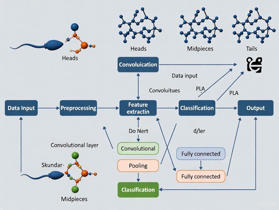
Sperm morphology analysis represents a significant classification challenge within medical image analysis. According to World Health Organization standards, normal sperm morphology is characterized by an oval head measuring 4.0–5.5 μm in length and 2.5–3.5 μm in width, with an intact acrosome covering 40–70% of the head and a single, uniform tail [18]. However, the modified David classification system expands this into 12 distinct morphological defect classes: seven head defects (tapered, thin, microcephalous, macrocephalous, multiple, abnormal post-acrosomal region, abnormal acrosome), two midpiece defects (cytoplasmic droplet, bent), and three tail defects (coiled, short, multiple) [1].
The fundamental challenges in automating this analysis include substantial inter-expert variability (with kappa values as low as 0.05–0.15 reported between technicians), lengthy manual evaluation times (30–45 minutes per sample), and inconsistent standards across laboratories [18]. Furthermore, creating high-quality annotated datasets is particularly challenging due to sperm sometimes appearing intertwined in images, partial structures being displayed at image edges, and the simultaneous assessment required for head, vacuoles, midpiece, and tail abnormalities [11].
Custom CNN Architectures: Foundational Approaches
Early approaches to automated sperm morphology classification focused on developing custom CNN architectures trained from scratch on domain-specific datasets. These models typically employed fundamental convolutional building blocks to learn hierarchical feature representations directly from sperm images.
A representative study by researchers at the Medical School of Sfax developed a custom CNN algorithm implemented in Python 3.8 for spermatozoa classification [1]. Their methodology followed a structured pipeline:
- Image Acquisition: 1000 images of individual spermatozoa were acquired using the MMC CASA system, with each image containing a single spermatozoon comprising head, midpiece, and tail.
- Data Augmentation: The original dataset was expanded from 1000 to 6035 images using augmentation techniques to balance morphological classes.
- Pre-processing: Images underwent cleaning to handle missing values and outliers, followed by normalization and resizing to 80×80×1 grayscale using linear interpolation.
- Architecture: A sequential CNN model was designed with multiple convolutional layers for feature extraction, though specific architectural details were not elaborated in the available literature.
- Training: The augmented dataset was partitioned with 80% for training and 20% for testing, with 20% of the training subset used for validation.
This custom CNN approach achieved accuracy ranging from 55% to 92%, demonstrating feasibility but highlighting limitations in robustness and generalization [1]. The performance variability underscores the challenges of designing effective custom architectures with limited data.
Table 1: Performance Comparison of Custom CNN Architectures
| Study | Dataset | Classes | Pre-processing | Reported Accuracy |
|---|---|---|---|---|
| Sfax Medical School [1] | SMD/MSS (6035 images) | 12 (David classification) | 80×80×1 grayscale, data augmentation | 55%-92% |
| Custom CNN Baseline [18] | SMIDS (3000 images) | 3-class | Not specified | ~88% |
The following diagram illustrates the typical workflow for developing and training custom CNN architectures for sperm morphology analysis:
Transfer Learning: Leveraging Pre-trained Models
Transfer learning has emerged as a powerful alternative to custom CNNs, particularly valuable when dealing with limited medical imaging datasets. This approach utilizes architectures pre-trained on large-scale natural image datasets (e.g., ImageNet) and adapts them to the specialized domain of sperm morphology analysis.
A comprehensive study by Kılıç (2025) implemented a hybrid architecture integrating ResNet50 backbones with Convolutional Block Attention Module (CBAM) attention mechanisms [18]. The methodology incorporated:
- Backbone Selection: ResNet50 pre-trained on ImageNet served as the foundational feature extractor, leveraging learned hierarchical representations.
- Attention Mechanism Integration: CBAM sequentially applied channel-wise and spatial attention to feature maps, enabling the network to focus on morphologically relevant sperm structures.
- Deep Feature Engineering Pipeline: Multiple feature extraction layers (CBAM, Global Average Pooling, Global Max Pooling, pre-final) were combined with 10 distinct feature selection methods.
- Classification Head: Support Vector Machines with RBF/Linear kernels and k-Nearest Neighbors algorithms replaced traditional softmax layers for final classification.
This transfer learning approach demonstrated exceptional performance, achieving test accuracies of 96.08% ± 1.2% on the SMIDS dataset (3000 images, 3-class) and 96.77% ± 0.8% on the HuSHeM dataset (216 images, 4-class) [18]. These results represented significant improvements of 8.08% and 10.41% respectively over baseline CNN performance, with McNemar's test confirming statistical significance (p < 0.001).
Table 2: Performance of Transfer Learning Approaches with Feature Engineering
| Model Architecture | Feature Engineering | Classifier | SMIDS Accuracy | HuSHeM Accuracy |
|---|---|---|---|---|
| ResNet50 + CBAM [18] | Global Average Pooling + PCA | SVM RBF | 96.08% ± 1.2% | 96.77% ± 0.8% |
| ResNet50 Baseline [18] | None (End-to-End) | Softmax | ~88% | ~86% |
| Ensemble CNN (Spencer et al.) [18] | Stacked Generalization | Meta-Learner | 95.2% | Not reported |
The following diagram illustrates the architecture and workflow of the CBAM-enhanced ResNet50 model for sperm morphology classification:
Experimental Protocols and Methodologies
Dataset Preparation and Annotation
Robust dataset creation is fundamental for effective CNN model development. The SMD/MSS (Sperm Morphology Dataset/Medical School of Sfax) provides a representative example of systematic dataset development [1]:
- Sample Collection: Semen samples were obtained from 37 patients with sperm concentrations of at least 5 million/mL, excluding samples with high concentrations (>200 million/mL) to avoid image overlap.
- Smear Preparation and Staining: Smears were prepared following WHO manual guidelines and stained with RAL Diagnostics staining kit.
- Image Acquisition: Images were captured using an MMC CASA system with bright field mode and an oil immersion 100x objective, with each image containing a single spermatozoon.
- Expert Annotation: Three experts with extensive experience in semen analysis independently classified each spermatozoon according to the modified David classification, with disagreements resolved through consensus.
- Data Augmentation: Techniques included geometric transformations (rotation, scaling, flipping) and photometric adjustments (brightness, contrast) to expand the dataset from 1000 to 6035 images and address class imbalance.
Model Training and Evaluation
Standardized training protocols ensure reproducible model performance:
- Data Partitioning: Random splitting into training (80%), validation (20% of training set), and test sets (20% of total dataset) [1].
- Cross-Validation: Implementation of 5-fold cross-validation for robust performance estimation [18].
- Evaluation Metrics: Comprehensive assessment using accuracy, precision, recall, F1-score, and area under receiver operating characteristic curve (AUC-ROC).
- Statistical Testing: Application of McNemar's test for comparing model performance with statistical significance [18].
The Scientist's Toolkit: Research Reagent Solutions
Table 3: Essential Materials and Reagents for Sperm Morphology Analysis Research
| Item | Function | Example/Specification |
|---|---|---|
| MMC CASA System [1] | Automated image acquisition and initial morphometric analysis | Bright field mode, oil immersion 100x objective |
| RAL Diagnostics Staining Kit [1] | Sperm staining for morphological visualization | Follows WHO manual guidelines |
| HydroCel Geodesic Sensor Net [27] | High-density EEG recording for neurofeedback studies | 128-channel MR-compatible system |
| EGI GES 400 System [27] | EEG signal acquisition with artifact correction | Field isolation containment system |
| Selenium Library [28] | Web page rendering for contrast testing automation | Headless browser simulation |
| OpenCV EAST Model [28] | Text detection in accessibility testing | Pre-trained text detection model |
| Tesseract OCR [28] | Optical character recognition for text element identification | Python wrapper (Pytesseract) |
Comparative Analysis and Future Directions
The evolution from custom CNNs to transfer learning with attention mechanisms and feature engineering represents significant progress in sperm morphology classification. Custom CNNs offer the advantage of domain-specific architecture optimization but require substantial data and computational resources to achieve adequate performance [1]. In contrast, transfer learning approaches leverage pre-trained representations, accelerating convergence and improving performance, particularly on limited medical datasets [18].
The integration of attention mechanisms like CBAM addresses a critical challenge in medical image analysis by enabling models to focus on morphologically relevant regions while suppressing background noise [18]. This capability is particularly valuable for sperm morphology analysis, where subtle structural differences in head shape, acrosome integrity, and tail configuration determine classification outcomes.
Future research directions likely include:
- Development of larger, more diverse publicly available datasets with standardized annotations
- Exploration of vision transformers and other emerging architectures
- Integration of multimodal data (e.g., combining imagery with clinical parameters)
- Enhanced explainability through improved visualization techniques
- Real-time analysis capabilities for clinical deployment during assisted reproductive procedures
The demonstrated success of deep learning approaches, particularly transfer learning with attention mechanisms and feature engineering, highlights their potential to transform sperm morphology analysis from a subjective, variable-dependent assessment to an objective, standardized diagnostic tool in reproductive medicine.
The morphological evaluation of sperm is a cornerstone of male fertility assessment, providing critical diagnostic and prognostic information. In clinical andrology, two structured classification frameworks are predominant: the World Health Organization (WHO) criteria and the Modified David classification [1] [11]. The accurate classification of sperm morphology is notoriously challenging, characterized by significant subjectivity and inter-observer variability among even experienced technicians [18]. Convolutional Neural Networks (CNNs), a class of deep learning models specialized in processing spatial data like images, offer a powerful solution for automating this analysis [29] [30]. These models can learn hierarchical feature representations directly from pixel data, enabling them to discern subtle morphological patterns that may elude manual assessment [5]. This technical guide explores the integration of these established clinical frameworks with advanced CNN methodologies, detailing how this synergy is revolutionizing sperm morphology analysis for research and drug development.
Classification Frameworks: A Comparative Analysis
The Modified David Classification
The Modified David classification system provides a granular categorization of sperm defects, focusing on specific anatomical components. It is particularly valued for its detailed approach to characterizing abnormalities across the sperm cell's structure [1].
Table 1: Modified David Classification Criteria
| Anatomical Component | Defect Category | Morphological Description |
|---|---|---|
| Head | Tapered (a) | Head shape narrows significantly [1]. |
| Thin (b) | Head width is abnormally reduced [1]. | |
| Microcephalous (c) | Head size is abnormally small [1]. | |
| Macrocephalous (d) | Head size is abnormally large [1]. | |
| Multiple (e) | Presence of multiple heads [1]. | |
| Abnormal Post-Acrosomal Region (f) | Irregularity in the region posterior to the acrosome [1]. | |
| Abnormal Acrosome (g) | Acrosome is misshapen or improperly formed [1]. | |
| Midpiece | Cytoplasmic Droplet (h) | Retention of excess cytoplasm [1]. |
| Bent (j) | Midpiece exhibits a sharp angulation [1]. | |
| Tail | Coiled (n) | Tail is coiled upon itself [1]. |
| Short (l) | Tail length is abnormally short [1]. | |
| Multiple (o) | Presence of multiple tails [1]. | |
| Associated Anomalies | CN | Multiple defects present across different components [1]. |
The WHO Classification Criteria
The World Health Organization (WHO) manual outlines standardized criteria for semen analysis, promoting consistency across laboratories globally. Its approach to morphology is often structured around classifying entire sperm into normative categories rather than enumerating every specific defect [5] [11].
Table 2: WHO Morphology Categories
| Category | Morphological Description | Key Defining Features |
|---|---|---|
| Normal | Smooth, oval head with well-defined acrosome; intact midpiece and tail [5]. | Head length: 4.0–5.5 µm; Width: 2.5–3.5 µm; Acrosome covers 40-70% of head [18]. |
| Tapered | Head exhibits a narrowing or tapered form [5]. | - |
| Pyriform | Head shape resembles a pear (widening at the base) [5]. | - |
| Small | Head dimensions are below the normal range [5]. | - |
| Amorphous | Head shape is irregular and does not fit other categories [5]. | Includes various head shape abnormalities. |
CNN Architectures for Sperm Classification: Experimental Protocols
The application of CNNs to sperm classification typically follows a structured pipeline, from data acquisition to model deployment. The choice between using the Modified David or WHO criteria directly influences the dataset annotation and the final output layer of the CNN model.
Data Acquisition and Pre-processing Protocol
A critical first step is the creation of a high-quality, annotated dataset. The SMD/MSS dataset protocol involves acquiring images of individual spermatozoa from semen samples using a microscope equipped with a digital camera (e.g., MMC CASA system) at 100x magnification under bright field mode [1]. Each sperm image is then independently classified by multiple experts according to the chosen classification framework (e.g., the 12 classes of the Modified David system) to establish a robust ground truth [1]. Key pre-processing steps include:
- Data Cleaning: Identifying and handling missing values or inconsistencies [1].
- Normalization/Standardization: Resizing images to a uniform dimension and scaling pixel values. A common approach is resizing to 80x80 pixels and converting to grayscale [1].
- Data Augmentation: To address limited dataset size and class imbalance, techniques such as rotation, flipping, and brightness adjustments are employed to artificially expand the dataset [1] [31]. For instance, the SMD/MSS dataset was expanded from 1,000 to 6,035 images via augmentation [1].
CNN Model Design and Training Methodologies
Two primary deep-learning approaches are prevalent in the literature: transfer learning and custom hybrid models.
Transfer Learning with Pre-trained CNNs: This methodology involves taking a CNN model pre-trained on a large, general image dataset (e.g., ImageNet) and retraining it on the specialized sperm morphology dataset. A standard protocol uses the VGG16 architecture, where the final fully connected layers are replaced and retrained to predict the number of sperm classes (e.g., WHO categories). The model is first trained with the earlier layers frozen, followed by a "fine-tuning" stage where all layers are unfrozen and trained with a very low learning rate [5]. This approach has achieved true positive rates of 94.1% on the HuSHeM dataset classifying WHO categories [5].
Hybrid Attention-Based Models: A more advanced protocol involves enhancing modern architectures like ResNet50 with attention mechanisms. The Convolutional Block Attention Module (CBAM) can be integrated, which sequentially applies channel and spatial attention to feature maps, helping the model focus on morphologically significant regions like the head or tail [18]. A comprehensive deep feature engineering (DFE) pipeline can be added, extracting features from multiple network layers (CBAM, Global Average Pooling) and applying feature selection methods like Principal Component Analysis (PCA) before final classification with a Support Vector Machine (SVM) [18]. This hybrid protocol has reported test accuracies of 96.77% on the HuSHeM dataset [18].
Workflow and System Diagrams
The following diagram illustrates the end-to-end pipeline for developing a CNN model for sperm morphology classification, integrating both the Modified David and WHO criteria.
CNN Model Training and Evaluation Protocol
This diagram details the internal processes of the "Model Training & Validation" and "Model Evaluation" stages, highlighting the use of transfer learning and performance assessment.
The Scientist's Toolkit: Research Reagents and Materials
Successful implementation of a CNN-based sperm classification system relies on a suite of essential reagents, materials, and computational tools.
Table 3: Essential Research Reagents and Materials
| Item Name | Function/Description | Application Note |
|---|---|---|
| RAL Diagnostics Staining Kit | Stains sperm cells for clear visualization of morphological structures [1]. | Critical for creating consistent, high-contrast images for manual annotation and model input. |
| MMC CASA System | An integrated system (microscope, camera, software) for acquiring and storing sperm images [1]. | Provides standardized digital images; CASA morphometric tools can pre-measure head/tail dimensions [1]. |
| SMD/MSS Dataset | A dataset of 1,000+ individual sperm images annotated per Modified David classification [1]. | Serves as a benchmark for training and validating models on detailed defect analysis. |
| HuSHeM & SCIAN Datasets | Publicly available datasets with sperm images annotated per WHO categories [5]. | Enable benchmarking of model performance against prior machine learning approaches [5]. |
| Pre-trained CNN Models (VGG16, ResNet50) | Deep learning models pre-trained on the ImageNet dataset, available in frameworks like PyTorch and Keras [5] [18]. | The foundation for transfer learning, significantly reducing required data and training time [5]. |
| GPU Accelerator (e.g., NVIDIA) | Hardware for high-performance parallel computation. | Essential for efficiently training complex deep learning models, reducing process from days to hours. |
The morphological analysis of sperm is a cornerstone of male fertility assessment, but traditional methods are plagued by subjectivity, time-consuming processes, and significant inter-observer variability [11]. Convolutional Neural Networks (CNNs) have emerged as transformative tools for automating sperm analysis, enabling precise detection and segmentation of sperm components through their ability to learn hierarchical features directly from image data [32] [5]. These advanced applications extend beyond basic classification to provide detailed morphological characterization of sperm structures, including heads, acrosomes, nuclei, midpieces, and tails, which is crucial for accurate fertility diagnosis and treatment planning [33].
The evolution from traditional machine learning to deep learning represents a paradigm shift in sperm morphology analysis. Earlier approaches relied on manually engineered features such as shape descriptors, thresholding, and clustering algorithms, which often struggled with accuracy and generalizability across diverse sample conditions [11]. Deep learning models, particularly those based on CNN architectures, have demonstrated superior performance by automatically learning relevant features from raw pixel data, thereby overcoming limitations of previous methods and establishing new benchmarks for precision in sperm detection and segmentation tasks [5].
Deep Learning Architectures for Sperm Segmentation
Core Architectures and Their Applications
Multiple deep learning architectures have been adapted and optimized for sperm segmentation tasks, each offering distinct advantages for specific aspects of sperm morphology analysis. The following table summarizes the primary architectures and their documented performance:
Table 1: Deep Learning Architectures for Sperm Segmentation
| Architecture | Primary Application | Reported Performance | Key Advantages |
|---|---|---|---|
| U-Net | Segmentation of sperm heads, acrosomes, and nuclei | Dice coefficient: 95% (head), 94% (acrosome), 95% (nucleus) [34] | Excellent with limited training data; precise boundary detection |
| Mask R-CNN | Instance segmentation of sperm parts | mAP >0.80 for isolating human round spermatids [35] | Simultaneous detection and segmentation; handles overlapping objects |
| Cascade Mask R-CNN | Noninvasive identification of human round spermatids | High precision in double-blind tests [35] | Multi-stage refinement for improved accuracy |
| YOLOv7 | Bovine sperm morphology analysis | mAP@50: 0.73, Precision: 0.75, Recall: 0.71 [36] | Real-time processing capabilities; balanced accuracy-efficiency tradeoff |
| VGG16 with Transfer Learning | Sperm head classification | Average true positive rate: 94.1% on HuSHeM dataset [5] | Leverages pre-trained features; effective even with limited data |
U-Net has demonstrated particular effectiveness in biomedical image segmentation, with its encoder-decoder structure and skip connections enabling precise localization of sperm components. When combined with transfer learning techniques, U-Net achieved remarkable Dice coefficients of 0.96 for sperm heads, 0.94 for acrosomes, and 0.95 for nuclei, significantly outperforming previous state-of-the-art methods [34]. This architecture's strength lies in its ability to maintain spatial context while integrating features at multiple scales, making it ideal for segmenting small, intricate sperm structures.
For detection and segmentation tasks requiring high precision in clinical applications, Cascade Mask R-CNN has shown exceptional performance. In one study focused on identifying human round spermatids (hRSs) for assisted reproduction techniques, this architecture achieved a mean average precision (mAP) exceeding 0.80 [35]. The cascaded structure progressively refines detection quality, with each stage specializing in distinguishing difficult cases, making it particularly valuable for identifying subtle morphological features critical to fertility assessment.
Experimental Protocols and Implementation
The implementation of deep learning models for sperm segmentation follows carefully designed experimental protocols to ensure robustness and clinical applicability. A typical workflow involves multiple stages of data preparation, model training, and validation:
Table 2: Standard Experimental Protocol for Sperm Segmentation Models
| Protocol Stage | Key Components | Considerations |
|---|---|---|
| Data Acquisition | Microscope specification (e.g., Optika B-383Phi with 100x oil immersion), standardized staining (e.g., RAL Diagnostics kit), sample preparation [1] [36] | Consistency in magnification, lighting conditions, and staining protocols |
| Annotation | Manual segmentation by multiple experts, establishment of ground truth, resolution of inter-expert disagreements [33] | Quality control through expert consensus; use of public datasets (e.g., SCIAN-SpermSegGS) |
| Preprocessing | Image denoising, contrast enhancement, normalization, resizing (e.g., 80×80×1 grayscale) [1] | Handling of non-uniform illumination; separation of sperm from debris |
| Data Augmentation | Rotation, flipping, brightness adjustment, scaling, elastic transformations [1] | Addressing class imbalance; increasing dataset diversity |
| Model Training | Transfer learning from pre-trained weights, hyperparameter tuning, cross-validation [34] [5] | Selection of appropriate base architecture; learning rate optimization |
| Evaluation | Dice coefficient, mean Average Precision (mAP), precision, recall, comparison with state-of-the-art [35] [34] | Statistical validation; clinical relevance of metrics |
A critical success factor in these protocols is the application of transfer learning, where models pre-trained on large-scale natural image datasets (e.g., ImageNet) are fine-tuned on specialized sperm image datasets. This approach has demonstrated substantial improvements in segmentation accuracy, particularly when working with limited annotated medical data [34] [5]. For instance, implementing U-Net with transfer learning resulted in significantly higher Dice coefficients with less dispersion and fewer failure cases compared to training from scratch [34].
Figure 1: Experimental workflow for sperm segmentation models, showing the pipeline from data acquisition to clinical application.
Performance Analysis and Comparative Evaluation
Quantitative Performance Metrics
The evaluation of sperm segmentation models employs multiple quantitative metrics to assess different aspects of performance. The following table compiles key results from recent studies to facilitate comparative analysis:
Table 3: Comparative Performance of Sperm Segmentation Models
| Study | Model | Dataset | Key Metrics | Performance |
|---|---|---|---|---|
| Movahed et al. [33] | CNN + SVM | SCIAN Gold-standard | Dice Coefficient | Head: 91%, Acrosome: 82%, Nucleus: 87% |
| U-Net with Transfer Learning [34] | U-Net | SCIAN-SpermSegGS | Dice Coefficient | Head: 96%, Acrosome: 94%, Nucleus: 95% |
| Deep Learning for Round Spermatids [35] | Cascade Mask R-CNN | 3,457 microscope images | mAP | >0.80 |
| Bovine Sperm Analysis [36] | YOLOv7 | 277 annotated images | mAP@50, Precision, Recall | 0.73, 0.75, 0.71 |
| VGG16 Transfer Learning [5] | VGG16 | HuSHeM, SCIAN | Average True Positive Rate | 94.1% (HuSHeM), 62% (SCIAN) |
The performance variation across datasets highlights the significant impact of data quality and annotation consistency on model effectiveness. The higher true positive rate achieved on the HuSHeM dataset (94.1%) compared to the SCIAN dataset (62%) using the same VGG16 architecture underscores the importance of standardized annotation protocols and dataset characteristics in model evaluation [5]. This discrepancy may be attributed to differences in inter-expert agreement rates during ground truth establishment, with the SCIAN dataset potentially containing more challenging cases with higher expert disagreement.
Clinical Validation and Robustness Assessment
Beyond technical metrics, the clinical validation of sperm segmentation models requires rigorous testing through double-blind experiments and biological verification. In one notable study, the expression of PRM1 and/or PNA (established markers for round spermatids) was observed in all cells noninvasively selected by the AI model during independent double-blind testing, confirming the model's biological accuracy and effectiveness for clinical application [35]. This approach of correlating algorithmic predictions with established biological markers represents the gold standard for validating the clinical utility of sperm segmentation systems.
Robustness to variations in image quality, staining protocols, and sample preparation remains a significant challenge in practical deployment. Studies have addressed this through comprehensive data augmentation strategies, including rotation, flipping, brightness adjustment, and scaling techniques to increase dataset diversity and enhance model generalization [1]. Additionally, the use of cross-validation methodologies helps ensure that performance metrics reflect true generalizability rather than dataset-specific optimization.
Research Reagent Solutions and Essential Materials
The development and implementation of effective sperm segmentation systems require carefully selected materials and reagents to ensure consistent, high-quality image data. The following table details key components used in referenced studies:
Table 4: Essential Research Reagents and Materials for Sperm Segmentation Studies
| Category | Specific Product/System | Function/Application | Reference |
|---|---|---|---|
| Staining Kits | RAL Diagnostics staining kit | Sperm staining for morphology analysis | [1] |
| Microscopy Systems | Optika B-383Phi microscope | High-resolution image acquisition | [36] |
| Image Analysis Software | PROVIEW application, MMC CASA system | Image capture and preliminary analysis | [1] [36] |
| Fixation Systems | Trumorph system | Pressure and temperature fixation for dye-free evaluation | [36] |
| Semen Extenders | Optixcell (IMV Technologies) | Semen sample preservation and preparation | [36] |
| Public Datasets | SCIAN-SpermSegGS, HuSHeM | Model training and benchmarking | [34] [5] |
| Annotation Tools | Roboflow | Image labeling and dataset management | [36] |
Standardized staining protocols using commercial kits such as RAL Diagnostics ensure consistent contrast and coloration across samples, which is crucial for developing robust segmentation algorithms [1]. Similarly, advanced microscopy systems with standardized magnification (typically 100x oil immersion) and lighting conditions minimize technical variability that could adversely affect model performance. The availability of public datasets with expert annotations has been instrumental in advancing the field, enabling reproducible benchmarking of different approaches and accelerating methodological improvements.
Figure 2: Standardized workflow for dataset creation, showing key stages from sample collection to model development.
Future Directions and Research Challenges
Despite significant advances in sperm detection and segmentation, several challenges remain that require further research and development. The lack of standardized, high-quality annotated datasets continues to hinder progress, with issues including limited sample sizes, insufficient morphological diversity, and inter-laboratory variability in annotation protocols [11]. Future efforts should focus on establishing large-scale, multi-center datasets with consistent annotation standards to facilitate the development of more robust and generalizable models.
Emerging research directions include the integration of multi-task learning for simultaneous segmentation and classification, the development of lightweight models for real-time clinical applications, and the incorporation of explainable AI techniques to enhance clinician trust and adoption [32]. Additionally, while current systems have demonstrated strong performance on stained sperm images, there is growing interest in analyzing unstained, live sperm for applications in intracytoplasmic sperm injection (ICSI) and other assisted reproduction technologies [5]. This presents unique technical challenges due to reduced contrast and more subtle morphological features, requiring specialized architectures and training approaches.
The ultimate goal of these technological advancements is to create fully integrated, automated systems that can provide comprehensive sperm morphological analysis with minimal human intervention, standardizing diagnostic procedures across clinical settings and improving accessibility to advanced fertility care. As deep learning methodologies continue to evolve, their integration with conventional semen analysis protocols promises to transform the landscape of male infertility diagnosis and treatment.
Overcoming Practical Challenges in Deep Learning Model Deployment
In the field of medical image analysis, particularly in specialized domains such as sperm morphology classification, data scarcity presents a significant barrier to developing robust deep-learning models. Data augmentation has emerged as a critical strategy to mitigate this challenge by artificially expanding training datasets, thereby improving model generalization and performance. This technical review examines data augmentation techniques within the context of convolutional neural network (CNN) applications for sperm classification research, providing researchers with methodologies, quantitative comparisons, and practical implementation protocols.
The application of CNNs to sperm morphology classification is inherently limited by the difficulty of acquiring large, expertly annotated datasets. Traditional manual assessment is subjective and time-consuming, while Computer-Assisted Semen Analysis (CASA) systems face limitations in accurately distinguishing sperm components and classifying abnormalities [1] [5]. Data augmentation addresses these data limitations by generating synthetic training samples, enabling models to learn invariant features and reducing overfitting to limited original data [37].
Data Augmentation Techniques: A Taxonomy and Their Applications
Data augmentation techniques can be broadly categorized into traditional transformations and synthetic data generation approaches. The selection and application of these techniques are particularly crucial in medical imaging domains like sperm analysis, where dataset imbalances and limited samples are common challenges [38].
Traditional Image Transformations
Basic geometric transformations modify the spatial orientation and structure of images, including techniques such as rotation, flipping, translation, and scaling. These transformations help models become invariant to positional and orientational variations of sperm cells in images [37]. Photometric transformations adjust the visual appearance of images through brightness/contrast modifications, color jittering, and adding noise. These simulate variations in staining intensity, lighting conditions, and microscope settings that occur during actual semen analysis [1] [37].
More advanced traditional techniques include elastic transformations that simulate natural deformations and distortions that might occur during sample preparation [37]. These are particularly relevant for sperm morphology analysis, as they can help models recognize cells with abnormal shapes or structural deformities.
Synthetic Data Generation
Generative Adversarial Networks (GANs) represent a more advanced approach, creating entirely new synthetic images that maintain the statistical properties of the original dataset [38] [39]. GANs have shown promise in medical imaging for generating realistic synthetic samples of underrepresented classes, effectively addressing class imbalance issues common in sperm morphology datasets where certain abnormality types may be rare [38].
Hybrid approaches that combine traditional transformations with generative models have demonstrated particularly strong performance. One study on corneal topographic map classification reported that a hybrid data augmentation approach achieved 99.54% accuracy, significantly outperforming individual techniques [39].
Quantitative Impact of Data Augmentation on Sperm Classification
The application of data augmentation techniques has demonstrated measurable improvements in the performance of CNN-based sperm classification systems. The table below summarizes key quantitative results from recent studies:
Table 1: Performance Impact of Data Augmentation on Sperm Classification Models
| Study/Dataset | Augmentation Approach | Original Dataset Size | Augmented Dataset Size | Reported Accuracy/Performance |
|---|---|---|---|---|
| SMD/MSS Dataset [1] | Multiple augmentation techniques | 1,000 images | 6,035 images | 55% to 92% accuracy |
| HuSHeM Dataset [5] | Transfer learning with VGG16 | N/A | N/A | 94.1% true positive rate |
| SCIAN Dataset [5] | Transfer learning with VGG16 | N/A | N/A | 62% true positive rate |
| Bovine Sperm Morphology [40] | YOLOv7 framework | 277 annotated images | N/A | mAP@50 of 0.73, Precision 0.75, Recall 0.71 |
| Corneal Topographic Map [39] | Hybrid augmentation (traditional + GAN) | N/A | N/A | 99.54% accuracy |
Beyond accuracy improvements, data augmentation significantly enhances model robustness. Studies have shown that augmented models maintain better performance across varying image conditions, including different staining intensities, magnification levels, and lighting conditions [1] [37]. This is particularly valuable for clinical deployment where consistency across laboratories and equipment is challenging.
Table 2: Data Augmentation Techniques and Their Specific Applications in Sperm Morphology Analysis
| Augmentation Category | Specific Techniques | Application in Sperm Classification | Impact on Model Performance |
|---|---|---|---|
| Geometric Transformations | Rotation, flipping, translation, scaling | Invariance to sperm orientation and position in images | Improves generalization across different sample preparations |
| Photometric Adjustments | Brightness/contrast, color jittering, noise addition | Robustness to staining variations and microscope settings | Maintains performance across laboratory protocols |
| Advanced Transformations | Elastic deformation, perspective distortion | Simulation of abnormal shapes and deformities | Enhances detection of structural abnormalities |
| Generative Models | GANs, VAEs | Generating rare abnormality types | Addresses class imbalance in training data |
| Hybrid Approaches | Combined traditional + generative methods | Comprehensive dataset expansion | Highest reported accuracy in medical imaging tasks |
Experimental Protocols and Methodologies
Implementation Framework for Sperm Morphology Analysis
Based on successful implementations in recent literature, the following experimental protocol provides a framework for applying data augmentation to sperm classification research:
Dataset Preparation and Annotation
- Acquire sperm images using standardized protocols (e.g., MMC CASA system with ×100 objective) [1]
- Establish expert annotation consensus following recognized classification systems (modified David classification or WHO criteria) [1] [5]
- Perform initial dataset analysis to identify class imbalances and representation gaps
Preprocessing Pipeline
- Apply image normalization and standardization to common scale (e.g., 80×80×1 grayscale) [1]
- Implement noise reduction techniques to address microscope imaging artifacts
- Extract individual sperm cells from larger microscopic fields when necessary
Data Augmentation Strategy
- Apply geometric transformations: ±15° rotation, horizontal flipping, minor translation (±5% shift) [37]
- Implement photometric adjustments: brightness (±20%), contrast (±15%), slight color variations
- Utilize advanced techniques: elastic deformations for abnormal shape simulation
- For severe class imbalances, incorporate GAN-based synthetic generation of rare morphological classes [38]
Model Training and Validation
- Implement train-test splits (typically 80-20) with strict separation [1]
- Apply real-time augmentation during training for dynamic sample variation
- Utilize transfer learning from pre-trained networks (e.g., VGG16) when sample size is limited [5]
- Employ k-fold cross-validation for robust performance estimation
The following workflow diagram illustrates the complete experimental pipeline for implementing data augmentation in sperm classification research:
Case Study: SMD/MSS Dataset Implementation
A recent study utilizing the Sperm Morphology Dataset/Medical School of Sfax (SMD/MSS) provides a concrete example of successful augmentation implementation [1]. The researchers began with 1,000 original sperm images classified according to the modified David classification system, which includes 12 distinct morphological defect categories. Through systematic application of data augmentation techniques, they expanded the dataset to 6,035 images, effectively addressing the class imbalance issue. The resulting CNN model achieved classification accuracy ranging from 55% to 92%, demonstrating the significant impact of comprehensive data augmentation on model performance [1].
Table 3: Essential Research Materials and Computational Tools for Sperm Morphology Analysis
| Resource Category | Specific Tool/Technique | Application in Research |
|---|---|---|
| Image Acquisition Systems | MMC CASA System, Optika B-383Phi Microscope | Standardized capture of sperm images under consistent conditions |
| Annotation Platforms | Expert consensus protocols, Digital labeling tools | Ground truth establishment for model training and validation |
| Computational Frameworks | Python 3.8, TensorFlow, PyTorch, OpenCV | Implementation of CNN architectures and augmentation pipelines |
| Pre-trained Models | VGG16, VGG19, InceptionV3 | Transfer learning initialization for limited data scenarios |
| Data Augmentation Libraries | Albumentations, TorchIO, MONAI | Efficient implementation of transformation and generation techniques |
| Evaluation Metrics | Accuracy, Precision, Recall, mAP@50 | Quantitative assessment of model performance across morphological classes |
Data augmentation represents a fundamental methodology for addressing data scarcity in sperm classification research, enabling the development of robust CNN models capable of matching or exceeding expert-level performance. Through systematic application of both traditional transformations and advanced generative approaches, researchers can significantly expand effective training datasets, improve model generalization, and address class imbalance issues inherent in medical imaging domains. As deep learning continues to transform reproductive medicine, data augmentation remains an essential component of the methodological framework, paving the way for more accurate, automated, and accessible sperm morphology analysis systems that can enhance diagnostic precision and ultimately improve clinical outcomes in infertility treatment.
Managing Class Imbalance and Expert Label Disagreement
The application of deep learning, particularly Convolutional Neural Networks (CNNs), to biological classification tasks represents a frontier in automated medical analysis. Within the specific domain of sperm classification research, these models offer the potential to standardize assessments, improve reproducibility, and unlock novel, quantifiable biomarkers for male fertility. However, the development of robust and reliable CNN models in this field is critically hampered by two interconnected, fundamental challenges: severe class imbalance and inherent expert label disagreement.
Class imbalance arises from the natural non-uniform distribution of sperm morphology and motility types. Normal, functional spermatozoa are often the majority class, while critical abnormal categories (e.g., tapered, pyriform, amorphous) are inherently rarer. This imbalance skews model training, leading to predictions that are biased toward the majority class and fail to identify clinically significant anomalies [5] [41]. Compounding this issue is the problem of label disagreement. Even among trained experts, visual classification of sperm based on World Health Organization (WHO) criteria is subjective, leading to inconsistent "ground truth" labels for the same image [42] [4]. When a CNN is trained on this noisy label data, its performance and reliability are fundamentally limited.
This technical guide delves into advanced methodologies to overcome these twin challenges. We will explore state-of-the-art techniques for class balancing and for incorporating label uncertainty directly into the training process, providing a comprehensive framework for developing more accurate, robust, and clinically credible CNN models for sperm classification.
Technical Foundations: Core Challenges in Sperm Analysis
The Class Imbalance Problem
In machine learning, class imbalance occurs when the distribution of examples across classes is not uniform. A model trained on such data can achieve deceptively high accuracy by simply always predicting the majority class, while failing entirely to detect the minority classes that are often of greatest clinical interest [41]. In sperm analysis, this is a pervasive issue.
- Morphological Imbalance: The occurrence of specific abnormal sperm shapes (e.g., small, amorphous) is naturally low compared to normal forms. For instance, in the SCIAN dataset, this imbalance contributes to the difficulty for models to achieve an average true positive rate beyond 62% for multi-class classification, even with advanced techniques [5].
- Motility Imbalance: Similar skew exists in motility data, where the proportion of progressively motile, non-progressively motile, and immotile spermatozoa is rarely equal. This can lead to models that are accurate overall but poor at predicting the specific categories of non-progressive and immotile sperm, which are crucial for diagnosis [4].
The performance metrics for a model trained on imbalanced data are misleading. As illustrated in a canonical example, a model can achieve 99% accuracy on a dataset where only 1% of samples have a disease by simply classifying everyone as healthy. This renders metrics like accuracy useless, necessitating the use of precision, recall, F1-score, and AUC-PR [41].
The Expert Label Disagreement Problem
The "ground truth" for training supervised learning models in sperm analysis is typically established by manual expert annotation. However, this standard is inherently noisy. Studies have shown that even among well-trained readers, disagreement on labels is common, with discrepancy rates often around 5-10% for challenging cases in medical image interpretation [42].
This disagreement stems from several factors:
- Subjectivity in Visual Criteria: The WHO guidelines, while standardized, still require subjective judgment calls on borderline cases.
- Variation in Expertise: Differences in training and experience across technicians and labs lead to interpretation variances.
- Inherently Ambiguous Samples: Some sperm images possess features that make them difficult to categorically assign to a single class.
When a CNN is trained using a single consensus label (e.g., a majority vote), it is forced to learn from an over-simplified and often incorrect representation of reality. The model is penalized for recognizing the visual patterns that led one expert to assign a different label, ultimately "hiding" this inherent uncertainty from the learning process and producing overconfident and poorly calibrated predictions [42].
Technical Solutions for Class Imbalance
Addressing class imbalance is a multi-faceted endeavor, involving strategic adjustments to the data, the model's objective function, and the evaluation protocol. The following table summarizes the core techniques.
Table 1: Techniques for Mitigating Class Imbalance in CNN Models
| Technique | Core Principle | Best-Suited For | Advantages | Limitations |
|---|---|---|---|---|
| Class Weights [43] [41] | Adjusts the loss function to penalize misclassifications of minority class samples more heavily. | CNNs, Tree-based models (XGBoost), Logistic Regression. | Simple to implement; does not alter the dataset; no risk of overfitting from data replication. | Requires a capable framework; the weighting strategy may need tuning. |
| Focal Loss [44] [41] | A dynamic loss function that reduces the loss for well-classified examples, focusing learning on hard-to-classify samples. | Deep learning models, particularly in object detection and severe class imbalance scenarios. | Highly effective for severe imbalance; automates the focus on difficult examples. | Introduces new hyperparameters (α, γ) that require tuning. |
| SMOTE [45] | Generates synthetic samples for the minority class by interpolating between existing instances in feature space. | Logistic Regression, SVM, and other models that benefit from balanced feature spaces. | Increases the diversity of minority class representations; helps prevent model from ignoring minority classes. | Can lead to overfitting if synthetic samples are not representative; not recommended for tree-based models [41]. |
| Bi-Level Class Balancing (BLCB) [44] | A hierarchical approach that first balances major classes (e.g., vessel/non-vessel), then sub-classes (e.g., thick/thin vessels). | Complex multi-class problems with hierarchical structure and multiple levels of imbalance. | Addresses imbalance at multiple granularities; can be highly effective for complex biological structures. | More complex architecture and training regimen. |
| Threshold Tuning [41] | Moving the classification decision threshold from the default 0.5 to a value that maximizes a target metric (e.g., F1-score). | All probabilistic classifiers, as a final tuning step. | Simple, post-hoc method that can significantly boost recall or precision for a target class. | Does not change the underlying model learning, only the interpretation of its outputs. |
Experimental Protocol: Implementing Class Weights in a CNN
A highly effective and commonly adopted technique for CNN-based sperm classification is the use of class weights. The following protocol outlines its implementation:
Calculate Class Weights: Compute the weight for each class. A common method is to use the inverse of the class frequency.
weight_class_i = total_samples / (num_classes * count_samples_in_class_i)This automatically assigns a higher weight to underrepresented classes. Most deep learning frameworks can calculate this automatically via aclass_weight='balanced'parameter.Integrate into Loss Function: Pass the calculated dictionary of class weights to the loss function during model compilation. For a multi-class classification problem using categorical cross-entropy, the loss for each sample is scaled by the weight of its true class.
Model Training: Train the CNN model using the weighted loss function. The optimizer will now prioritize correcting errors on the minority class samples due to their higher contribution to the total loss.
Evaluation: Validate the model's performance on a held-out, stratified test set using metrics such as F1-score, precision, and recall for each class, rather than overall accuracy. Compare these metrics against a baseline model trained without class weights to quantify the improvement.
Table 2: Key Research Reagents for Sperm Classification Experiments
| Reagent / Resource | Specifications / Function |
|---|---|
| Public Datasets | HuSHeM [5], SCIAN [5], ESHRE-SIGA EQA [4]. Provide benchmark data for morphology and motility. |
| Pre-trained CNN Models | VGG16 [5], ResNet-50 [4]. Enable transfer learning, reducing data and computational requirements. |
| Data Augmentation | Rotation, flipping, scaling. Artificially expands training set and improves model robustness to variations. |
| Optical Flow Algorithms | Lucas-Kanade method [4]. Converts video sequences of sperm motility into single images representing motion patterns. |
| Stratified K-Fold Cross-Validation | Ensures that each fold of data preserves the percentage of samples for each class, leading to more reliable performance estimates. |
Technical Solutions for Expert Label Disagreement
Moving beyond a single, hard label is crucial for building models that reflect the true uncertainty in biological data. The following approaches directly incorporate inter-expert disagreement into the training loop.
Label Encoding and Training Strategies
Table 3: Strategies for Handling Expert Label Disagreement
| Strategy | Label Encoding | Training Approach | Outcome |
|---|---|---|---|
| Majority Vote Training (MVT) [42] | Single hard label from the most common expert vote. | Standard supervised learning. | Simple but hides uncertainty; produces overconfident models. |
| Average Vote Training (AVT) [42] | Soft label as a probability vector (e.g., [0.8, 0.2] for 2 experts voting normal, 1 abnormal). | Model is trained to predict the probability distribution using KL-divergence or mean squared error. | Model learns the ambiguity, providing a calibrated probability output. |
| Random Vote Training (RVT) [42] | For each training epoch, a label is randomly selected from the pool of expert labels for a given sample. | Model is exposed to the full range of expert opinions over many epochs. | Model learns a more generalized representation and becomes less sensitive to label noise. |
Experimental Protocol: Implementing Average Vote Training (AVT)
The AVT method provides a robust way to train a CNN using the soft labels derived from multiple experts.
Label Aggregation: For each sperm image
i, compile the labels fromRexperts. Instead of taking a majority vote, compute the average vote (proportion) for each class. For example, if three experts label an image as [Normal, Normal, Amorphous], the soft label vector would be[0.66, 0.0, 0.33, ...]for the classes [Normal, Tapered, Amorphous, ...].Model Architecture Adjustment: The final layer of the CNN must use a softmax activation function to output a probability distribution over the classes. The number of neurons must match the number of morphology classes (e.g., 5 for WHO categories).
Loss Function Selection: Replace the standard categorical cross-entropy loss with a loss function suitable for soft labels, such as Kullback-Leibler (KL) Divergence or Categorical Cross-Entropy configured for probabilistic targets. KL Divergence measures how one probability distribution diverges from a second and is ideal for this task.
Loss = KL(Soft_Label || Model_Prediction)Training and Inference: Train the model using the soft labels. During inference, the model's output is a well-calibrated probability distribution, directly reflecting the certainty (or uncertainty) of the classification, akin to the disagreement level among the original human experts.
Integrated Workflow and Visualization
A robust experimental pipeline for sperm classification must integrate solutions for both imbalance and disagreement. The following diagram and workflow outline this integrated process.
Diagram 1: Integrated workflow for managing class imbalance and label disagreement in sperm classification CNNs. The process integrates soft label encoding (AVT/RVT) and class-balanced loss functions to produce a calibrated, robust model.
Workflow Description
Data Preparation & Labeling: The process begins with the collection of raw sperm images or videos. Crucially, each sample is annotated by multiple andrology experts. These individual labels are aggregated, and a soft label is generated for each image using either the Average Vote Training (AVT) or Random Vote Training (RVT) strategy [42].
Model Training with Balancing: The soft-labeled dataset is pre-processed and augmented. A CNN (e.g., a pre-trained VGG16 or ResNet-50 fine-tuned for this task) is used for feature extraction [5] [4]. The training loop incorporates two key modifications:
- Class Balancing: The loss function (e.g., Categorical Cross-Entropy) is weighted based on class distribution or replaced with Focal Loss to mitigate the effects of class imbalance [44] [43] [41].
- Soft Label Training: The weighted loss function is calculated using the soft probability labels (from Step 1) instead of hard labels, using an appropriate measure like KL Divergence [42].
Output & Evaluation: The trained model outputs a calibrated probability distribution across the possible sperm classes, directly reflecting the model's certainty and the inherent ambiguity of the input. The model is evaluated using a comprehensive suite of metrics (e.g., per-class F1-score, AUC-PR) on a held-out test set to ensure it performs reliably across all categories, not just the majority class [41].
The path to robust and clinically applicable deep learning models in sperm classification research requires a direct and methodical confrontation of the field's fundamental data challenges. Relying on simplistic labeling and standard training procedures leads to models that are brittle, biased, and unreflective of biological reality.
As demonstrated, the synergistic application of techniques designed for class imbalance—such as class-weighted loss or focal loss—and strategies for expert label disagreement—such as Average Vote Training—provides a powerful framework for advancement. This integrated approach guides the CNN to learn a more nuanced and generalized representation of sperm morphology and motility. The resulting models do not merely output a categorical guess but provide a calibrated probability that honestly represents the classification certainty, thereby enhancing their utility as decision-support tools for researchers and clinicians in the field of drug development and reproductive medicine. Future work should focus on the integration of these methods with emerging architectures like feedback-attention networks [44] [46] and their validation in large-scale, multi-center clinical trials.
Ensuring Model Fairness and Mitigating Algorithmic Bias
The integration of Convolutional Neural Networks (CNNs) into reproductive medicine, particularly for sperm classification, represents a significant advancement in addressing the long-standing challenges of subjective and inconsistent manual morphological analysis [1] [7]. While these deep learning models demonstrate remarkable accuracy in classifying sperm abnormalities—achieving performance metrics ranging from 55% to 92% in recent studies—their clinical utility and ethical deployment depend critically on addressing embedded algorithmic biases [1]. The "bias in, bias out" paradigm is particularly concerning in healthcare AI, where systematic unfairness can exacerbate existing healthcare disparities and lead to discriminatory outcomes against vulnerable patient populations [47]. In the specialized domain of sperm classification, biases may manifest as performance disparities across diverse demographic groups, imaging modalities, or clinical protocols, ultimately compromising diagnostic reliability and patient care. This technical guide provides a comprehensive framework for identifying, assessing, and mitigating algorithmic bias within CNN architectures specifically designed for sperm morphology classification, ensuring these powerful diagnostic tools adhere to the highest standards of fairness and equity in clinical practice.
Understanding Bias Origins in Sperm Classification Models
Algorithmic bias in sperm classification systems can originate from multiple sources throughout the model development lifecycle. Understanding these origins is fundamental to implementing effective mitigation strategies.
Data Collection and Representation Bias
The foundation of any robust CNN model is a comprehensive and representative dataset. In sperm classification research, significant biases can emerge during data acquisition. The Sperm Morphology Dataset/Medical School of Sfax (SMD/MSS), for instance, initially contained only 1,000 individual spermatozoa images, which were subsequently augmented to 6,035 images to balance morphological classes [1]. Such limited initial datasets risk underrepresenting rare morphological anomalies or demographic variations. Representation bias occurs when training data does not adequately reflect the true population diversity, potentially leading to systematically poorer performance on samples from underrepresented groups. This is particularly problematic in medical imaging, where anatomical variations across ethnicities, age groups, or geographical regions may exist.
Human and Labeling Bias
Human bias represents a particularly insidious challenge in medical AI development. In sperm morphology assessment, implicit biases among clinical experts can influence ground truth labeling, as classification often relies on subjective interpretation of the modified David classification criteria [1]. Studies analyzing inter-expert agreement in sperm classification have demonstrated concerning variability, with scenarios ranging from no agreement (NA) to partial agreement (PA) and total agreement (TA) among experts [1]. When these subjective assessments become training labels for CNNs, the models inevitably inherit and potentially amplify human biases. Furthermore, confirmation bias may lead researchers to prioritize data or model configurations that align with pre-existing beliefs, thereby skewing the entire development process.
Algorithmic and Deployment Bias
The architectural choices and optimization objectives in CNN design can introduce additional biases. Models optimized primarily for overall accuracy may inadvertently sacrifice performance on minority morphological classes. Deployment bias emerges when models trained under specific laboratory conditions face distribution shifts in real-world clinical environments, such as variations in staining techniques, microscope configurations, or imaging protocols [1] [48]. This problem is compounded by the "black-box" nature of complex deep learning models, which often lack transparency in how specific features contribute to classification decisions, making bias detection and remediation more challenging.
Table 1: Common Bias Types in Sperm Classification Models
| Bias Type | Origin Phase | Impact on Sperm Classification | Example Scenario |
|---|---|---|---|
| Representation Bias | Data Collection | Poor performance on rare sperm abnormalities | Dataset contains insufficient examples of multiple tail defects (class O) |
| Labeling Bias | Data Annotation | Model inherits subjective expert interpretations | Disagreement among experts on classification of "abnormal acrosome" (class G) |
| Algorithmic Bias | Model Development | Systematic errors across demographic groups | Performance disparity across ethnic groups not represented in training data |
| Deployment Bias | Clinical Implementation | Performance degradation in new clinical settings | Model trained on bright-field microscopy struggles with phase-contrast images |
Methodologies for Bias Assessment and Quantification
Rigorous bias assessment requires both technical metrics and clinical validation frameworks specifically adapted for sperm classification tasks.
Technical Fairness Metrics
Quantifying model fairness involves measuring performance disparities across relevant subgroups. For sperm classification models, these subgroups may be defined by morphological classes, patient demographics, or imaging protocols. Key fairness metrics include demographic parity, which ensures classification outcomes are independent of sensitive attributes; equalized odds, which requires similar true positive and false positive rates across groups; and equal opportunity, which focuses on maintaining comparable true positive rates across subgroups [47]. These statistical measures should be computed not only for overall accuracy but also for each morphological class in the modified David classification system (e.g., tapered heads, microcephalous, coiled tails) to identify specific failure modes [1].
Clinical Validation Protocols
Technical metrics alone are insufficient for comprehensive bias assessment in clinical applications. Cross-validation with independent expert review establishes clinical ground truth. The methodology employed in the SMD/MSS dataset development provides a robust template: three independent experts with extensive experience in semen analysis classified each spermatozoon, with statistical analysis of inter-expert agreement using Fisher's exact test [1]. This approach enables quantification of the inherent subjectivity in morphological assessment and provides confidence bounds for model performance. Additionally, prospective clinical validation studies comparing CNN classifications with clinical outcomes (e.g., fertilization success) represent the ultimate test of model fairness and utility.
Table 2: Bias Assessment Metrics for Sperm Classification Models
| Metric Category | Specific Metrics | Calculation Method | Target Threshold |
|---|---|---|---|
| Overall Performance | Accuracy, F1-Score, AUC-ROC | Standard formulae applied per morphological class | Accuracy >80% for all major classes [1] |
| Fairness Statistics | Demographic Parity, Equalized Odds, Equal Opportunity | Probability differences across sensitive subgroups | Difference <5% between subgroups |
| Expert Agreement | Cohen's Kappa, Fleiss' Kappa, IRA | Inter-rater agreement statistics | Kappa >0.6 (substantial agreement) [1] |
| Clinical Correlation | Sensitivity, Specificity, PPV, NPV | Performance against clinical outcomes or expert consensus | Sensitivity >85% for critical abnormalities |
Bias Mitigation Strategies for Sperm Classification CNNs
Data-Centric Mitigation Approaches
Addressing bias begins with curating comprehensive datasets. The SMD/MSS dataset exemplifies this approach through systematic data augmentation techniques that balance underrepresented morphological classes [1]. Advanced strategies include generative adversarial networks (GANs) to synthesize rare morphological variants, and strategic oversampling of minority classes during training. Emerging 3D sperm datasets, such as the 3D-SpermVid repository containing 121 multifocal video-microscopy hyperstacks, provide additional dimensions for robust feature learning [49]. Data collection should explicitly document demographic and clinical metadata to enable stratified analysis of model performance across relevant subgroups.
Algorithmic Mitigation Techniques
Algorithmic mitigation involves incorporating fairness constraints directly into the CNN training process. Pre-processing techniques transform training data to remove correlations between sensitive attributes and morphological features, while maintaining diagnostic relevance. In-processing approaches modify the loss function to include fairness regularizers that penalize performance disparities across subgroups. For sperm classification tasks, this might involve weighted loss functions that assign higher penalties to misclassifications of rare abnormalities. Post-processing techniques adjust model outputs after inference by applying different decision thresholds for different subgroups to equalize performance metrics [47]. Model architectures that enhance interpretability, such as attention mechanisms that highlight discriminative morphological features, can also facilitate bias detection by making classification rationales more transparent to clinical experts.
Implementation and Monitoring Frameworks
Bias mitigation continues throughout the model deployment lifecycle. Continuous monitoring systems should track performance metrics across morphological classes and demographic subgroups, with automated alerts for performance degradation or emerging disparities. Integration with fairness assessment toolkits such as IBM AI Fairness 360 (AIF360) or Microsoft Fairlearn provides standardized metrics and visualization dashboards for ongoing bias surveillance [50]. For clinical deployments, establishing model governance committees with diverse expertise—including embryologists, andrologists, and ethicists—ensures multidisciplinary oversight of fairness considerations.
Experimental Protocols for Bias Detection
Dataset Construction and Annotation
A robust experimental protocol for bias assessment begins with comprehensive dataset documentation. The SMD/MSS protocol specifies detailed inclusion criteria (sperm concentration ≥5 million/mL), exclusion criteria (concentration >200 million/mL to avoid image overlap), and standardized staining procedures (RAL Diagnostics kit) [1]. These specifications enable consistent replication and facilitate identification of potential bias sources. Annotation protocols should employ multiple independent experts with documented inter-rater reliability statistics. In case of disagreement, consensus mechanisms or adjudication by senior specialists establishes ground truth.
Model Training and Validation
CNN architectures for sperm classification should be trained with explicit bias detection in mind. This involves stratified data splitting to ensure all morphological classes and potential subgroups are represented in training, validation, and test sets. The SMD/MSS approach of using 80% of data for training and 20% for testing provides a reasonable baseline, with further division of the training set for validation [1]. Training protocols should include checkpointing and evaluation of fairness metrics at regular intervals, not just overall accuracy. Ablation studies that systematically vary training data composition help identify dependencies on specific data sources or augmentation techniques.
Comprehensive Model Auditing
Model auditing extends beyond technical performance to encompass clinical validity and fairness. This involves executing inference on carefully curated challenge sets containing rare morphological variants, edge cases with expert disagreement, and samples from diverse demographic sources. Performance disparities should be statistically analyzed using appropriate tests (e.g., McNemar's test for paired proportions) with correction for multiple comparisons. Explainability techniques such as Grad-CAM or LIME can visualize which morphological features the model prioritizes for classification, allowing clinical experts to identify potentially spurious correlations or problematic feature dependencies.
Visualization of Bias Assessment Workflow
The following diagram illustrates the comprehensive workflow for ensuring fairness in sperm classification models, integrating technical and clinical validation components:
Bias Assessment Workflow for Sperm Classification
Implementing robust bias assessment and mitigation requires specialized tools and resources. The following table catalogs essential solutions for fairness-focused sperm classification research:
Table 3: Research Reagent Solutions for Bias-Aware Sperm Classification
| Resource Category | Specific Solutions | Application in Sperm Classification | Key Features |
|---|---|---|---|
| Fairness Toolkits | IBM AIF360, Microsoft Fairlearn, Google What-If Tool | Bias metrics calculation and visualization across morphological classes | 70+ fairness metrics (AIF360), interactive visualization (What-If Tool) [50] |
| Sperm Datasets | SMD/MSS Dataset, 3D-SpermVid, VISEM-Tracking | Model training and validation with diverse morphological examples | 6,035 augmented images (SMD/MSS), 121 3D video hyperstacks (3D-SpermVid) [1] [49] |
| Annotation Platforms | Custom Excel Templates, Specialized Annotation Software | Standardized labeling per modified David classification | Multi-expert annotation support, discrepancy resolution workflows [1] |
| Model Architectures | CNN, MobileNet, U-Net variants | Base architectures for sperm morphology classification | Transfer learning compatibility, multi-scale feature learning [1] [48] |
The integration of convolutional neural networks into sperm classification research represents a paradigm shift in male fertility assessment, offering the potential to overcome the subjectivity and inconsistency that have long plagued manual morphological analysis. However, realizing this potential requires meticulous attention to model fairness and algorithmic bias throughout the development lifecycle. By implementing comprehensive bias assessment protocols—encompassing both technical metrics and clinical validation—and deploying appropriate mitigation strategies at the data, algorithmic, and implementation levels, researchers can develop CNN models that are not only accurate but also equitable and reliable across diverse populations and clinical settings. The frameworks and methodologies presented in this technical guide provide a roadmap for ensuring that AI-powered sperm classification systems advance reproductive medicine in an ethically responsible manner, ultimately enhancing patient care through more consistent, objective, and equitable diagnostic outcomes.
The manual assessment of sperm morphology is a cornerstone of male fertility evaluation, yet it remains plagued by significant challenges that hinder its effectiveness in clinical workflows. This process is notoriously subjective and time-intensive, often requiring 30-45 minutes per sample and relying heavily on technician expertise, with studies reporting inter-observer variability as high as 40% and kappa values as low as 0.05–0.15, indicating substantial diagnostic disagreement [18]. Computer-Assisted Semen Analysis (CASA) systems brought a degree of automation but face limitations in accurately classifying sperm with midpiece and tail abnormalities and are often hampered by high costs and complex operation, restricting their widespread adoption [1] [51]. These challenges directly impact the critical clinical metrics of speed, cost, and diagnostic consistency.
Convolutional Neural Networks (CNNs) offer a transformative solution by automating, standardizing, and accelerating semen analysis. The primary value proposition of deep learning in this clinical context lies in its potential to reduce operational costs by automating a manual task, increase processing speed from minutes to seconds, and enhance diagnostic objectivity through data-driven interpretation, thereby directly addressing the core workflow inefficiencies in reproductive medicine [1] [52] [18]. This guide details the technical implementation of CNN models optimized for these clinical imperatives.
Performance & Quantitative Benchmarking
The efficacy of a model for clinical use is determined by its accuracy, computational efficiency, and operational speed. The table below summarizes the performance of various CNN architectures and traditional methods on benchmark datasets, providing a basis for comparison.
Table 1: Performance Benchmarking of Sperm Classification Models
| Model/Approach | Dataset | Key Performance Metrics | Reported Clinical Workflow Advantage |
|---|---|---|---|
| CBAM-enhanced ResNet50 + Deep Feature Engineering [18] | SMIDS (3-class) | 96.08% Accuracy | Reduces analysis time from 30-45 minutes to <1 minute per sample. |
| CBAM-enhanced ResNet50 + Deep Feature Engineering [18] | HuSHeM (4-class) | 96.77% Accuracy | Provides interpretable results via Grad-CAM visualization. |
| VGG16 with Transfer Learning [5] [52] | HuSHeM | 94.1% True Positive Rate | Avoids excessive computation; viable with few examples. |
| VGG16 with Transfer Learning [5] [52] [51] | SCIAN | 62% True Positive Rate | Standardizes assessment, reducing inter-observer variability. |
| Proposed CNN (SMD/MSS Dataset) [1] | SMD/MSS (12-class) | 55% to 92% Accuracy | Enables automation and standardization of semen analysis. |
| Adaptive Patch-based Dictionary Learning (APDL) [5] | HuSHeM | 92.3% Average True Positive Rate | A non-deep learning benchmark for comparison. |
| Cascade Ensemble of SVM (CE-SVM) [5] | HuSHeM | 78.5% Average True Positive Rate | A traditional machine learning benchmark. |
The data illustrates that modern, attention-enhanced CNN architectures consistently achieve expert-level accuracy (exceeding 94% on the HuSHeM dataset) [5] [18]. More notably, they offer revolutionary improvements in processing speed, a direct contributor to reduced operational costs in a clinical setting.
Experimental Protocols & Detailed Methodologies
Protocol A: Transfer Learning with a Pre-trained VGG16 Network
Transfer learning leverages features from a large, general-purpose image dataset (like ImageNet), enabling high performance even with limited medical data, thus optimizing for cost and development time [5] [52].
Data Acquisition & Preparation:
- Datasets: Use publicly available datasets like HuSHeM [5] or SCIAN-MorphoSpermGS [5] [51]. The SMD/MSS dataset, built from 1000 images expanded to 6035 via data augmentation, is another example [1].
- Pre-processing: Resize all images to a fixed size of 150x150 pixels [51]. Convert images to grayscale if required [1]. Apply normalization by scaling pixel values to a [0, 1] range.
Data Augmentation (for Training Set): To increase dataset diversity and improve model generalizability, apply random transformations including rotation, horizontal/vertical flipping, and zooming [1] [51].
Model Architecture & Training:
- Base Model: Load the VGG16 architecture pre-trained on ImageNet, excluding its top classification layers.
- Classifier Head: Add new, trainable layers on top: a Global Average Pooling layer, followed by a Fully Connected (Dense) layer with 512 units and ReLU activation, and a final Dense output layer with softmax activation (units corresponding to the number of morphological classes).
- Training Regimen:
- Phase 1 (Feature Extraction): Freeze the weights of the pre-trained VGG16 base. Train only the newly added classifier head using a low learning rate (e.g., 1e-4) for approximately 100 epochs.
- Phase 2 (Fine-tuning): Unfreeze a portion of the deeper layers in the VGG16 base. Continue training the entire unfrozen model with an even lower learning rate (e.g., 1e-5) for another 100 epochs. This step adapts the pre-trained features to the specific task of sperm morphology [5].
Evaluation: Evaluate the model on a held-out test set (typically 20% of the data) [1], reporting metrics like accuracy, precision, recall, and F1-score.
Protocol B: Attention-Based Classification with CBAM-enhanced ResNet50
This protocol enhances model interpretability and accuracy by forcing the network to focus on morphologically relevant regions of the sperm, such as the head shape and acrosome integrity [18].
Data Pre-processing & Augmentation: Follow the same steps as Protocol A.
Model Architecture:
- Backbone: Use a ResNet50 architecture, pre-trained on ImageNet, as the feature extractor.
- Attention Integration: Integrate Convolutional Block Attention Modules (CBAM) at strategic points within the ResNet50 backbone. The CBAM sequentially applies:
- Channel Attention: A feed-forward network that highlights 'what' features are meaningful.
- Spatial Attention: A convolutional layer that highlights 'where' the informative regions are located.
- Deep Feature Engineering (DFE): Extract high-dimensional feature maps from multiple layers of the attention-augmented network (e.g., CBAM output, Global Average Pooling, pre-final layer). Apply feature selection and dimensionality reduction techniques like Principal Component Analysis (PCA) to these combined features. Finally, train a shallow classifier (e.g., Support Vector Machine with RBF kernel) on this optimized feature set [18].
Training: Train the entire model (ResNet50 backbone, CBAM modules, and classifier) end-to-end. Alternatively, for the DFE variant, train the backbone and then use its outputs to train a separate SVM.
Interpretability Analysis: Use Gradient-weighted Class Activation Mapping (Grad-CAM) on the final model to generate visual explanations. This produces a heatmap overlay on the input image, showing the regions that most influenced the classification decision, which is critical for clinical trust and verification [18].
The Scientist's Toolkit: Research Reagent Solutions
Successful development and deployment of a clinical-grade sperm classification system rely on several key components, from data to software.
Table 2: Essential Research Materials and Tools for CNN-based Sperm Classification
| Item Name / Category | Function / Purpose | Exemplars / Specifications |
|---|---|---|
| Public Sperm Image Datasets | Provides standardized, annotated data for training and benchmarking models. | HuSHeM [5], SCIAN-MorphoSpermGS [5] [51], SMIDS [18] [51], SMD/MSS [1], SVIA [2] |
| Data Augmentation Tools | Artificially expands training dataset to improve model robustness and combat overfitting. | Integrated in deep learning frameworks (e.g., TensorFlow/Keras, PyTorch). Techniques: rotation, flipping, zooming [1]. |
| Pre-trained CNN Models | Serves as a foundational feature extractor, reducing required data and computation (Transfer Learning). | VGG16 [5] [52], ResNet50 [18], Xception [18] (Pre-trained on ImageNet). |
| Attention Modules | Enhances model accuracy and interpretability by focusing network on salient image regions. | Convolutional Block Attention Module (CBAM) [18]. |
| Feature Selection & Dimensionality Reduction | Improves classifier performance by reducing noise and computational complexity in feature space. | Principal Component Analysis (PCA), Chi-square test, Random Forest importance [18]. |
| Classification Algorithms | The final model that makes the morphological class prediction. | Support Vector Machine (SVM) with RBF/Linear kernels, k-Nearest Neighbors (k-NN) [18], or a final Softmax layer in a CNN [5]. |
| Interpretability Libraries | Generates visual explanations for model decisions, building clinical trust and aiding validation. | Grad-CAM visualization [18]. |
Benchmarking Performance and Clinical Validation of AI Models
In the field of male fertility research, convolutional neural networks (CNNs) have emerged as powerful tools for automating and standardizing the analysis of sperm quality, particularly in the assessment of sperm morphology and motility [5]. These deep learning models are designed to process data with a grid-like topology, such as microscopic images of sperm cells, extracting hierarchical features through convolutional layers, pooling layers, and fully connected layers [53]. However, the development of any CNN model for sperm classification remains incomplete without a rigorous evaluation of its performance using appropriate metrics. The selection of evaluation metrics is not merely a technical formality but a critical decision that determines how model performance is measured, compared, and ultimately trusted for clinical or research applications.
Accuracy, precision, recall, and mean Average Precision (mAP) represent fundamental metrics that provide complementary insights into a model's classification capabilities. Each metric reflects a different aspect of model quality, and their relative importance varies significantly depending on the specific clinical or research context within sperm analysis [54] [55]. For instance, in a diagnostic scenario where missing abnormal sperm cells (false negatives) could lead to incorrect fertility assessments, recall might be prioritized over precision. Conversely, in research settings focused on identifying specific morphological subtypes for genetic studies, precision might be more critical. Understanding the mathematical foundations, interpretations, and trade-offs between these metrics is therefore essential for researchers developing CNN-based solutions for sperm classification.
Core Metric Definitions and Mathematical Foundations
The Confusion Matrix: Foundation for Classification Metrics
All primary classification metrics derive from the confusion matrix, which provides a complete breakdown of a model's predictions versus actual labels. In binary classification for sperm analysis (e.g., normal vs. abnormal sperm), the confusion matrix organizes predictions into four categories:
- True Positives (TP): Sperm correctly identified as having a specific abnormality (e.g., tapered head).
- True Negatives (TN): Normal sperm correctly classified as normal.
- False Positives (FP): Normal sperm incorrectly flagged as abnormal (false alarm).
- False Negatives (FN): Abnormal sperm missed by the model and classified as normal.
This foundational breakdown enables the calculation of more sophisticated performance metrics that answer specific questions about model behavior [55].
Metric Definitions and Formulae
Table 1: Fundamental Binary Classification Metrics
| Metric | Definition | Formula | Interpretation in Sperm Analysis |
|---|---|---|---|
| Accuracy | Overall correctness of the model | (TP+TN)/(TP+TN+FP+FN) [54] | How often the model is correct overall across all sperm classifications |
| Precision | Accuracy when the model predicts positive | TP/(TP+FP) [54] | When the model flags a sperm as abnormal, how often it is actually abnormal |
| Recall (Sensitivity) | Ability to find all positive instances | TP/(TP+FN) [54] | The model's ability to identify all truly abnormal sperm in a sample |
| F1 Score | Harmonic mean of precision and recall | 2(PrecisionRecall)/(Precision+Recall) [54] | Balanced measure for when both false positives and false negatives matter |
The Critical Role of Class Imbalance
In real-world sperm analysis datasets, class imbalance is the rule rather than the exception. For example, in human semen samples, the proportion of normal sperm morphology can be as low as 4% according to WHO standards, creating a naturally imbalanced classification problem [8]. This imbalance dramatically affects the interpretation of metrics, particularly accuracy.
A model that simply classifies every sperm as "normal" in a dataset with 95% normal sperm would achieve 95% accuracy while being clinically useless for identifying abnormalities [55]. This phenomenon, known as the "accuracy paradox," underscores why researchers must look beyond accuracy when evaluating models for sperm classification tasks where the target class (abnormalities) is rare but clinically significant.
Metric Selection Guidance for Sperm Classification Research
Context-Dependent Metric Prioritization
The choice of which metric to optimize depends heavily on the clinical or research objective and the relative costs associated with different types of classification errors:
Prioritize Recall when false negatives are more costly than false positives. For example, in initial screening for severe morphological defects that definitively indicate infertility, missing affected sperm (false negatives) is more problematic than occasional false alarms [54] [55].
Prioritize Precision when false positives are more costly. In automated sperm selection for ICSI (intracytoplasmic sperm injection), falsely classifying normal sperm as abnormal might unnecessarily limit the pool of available sperm, while high precision ensures that selected sperm are indeed normal [5].
Use F1 Score when seeking a balance between precision and recall, particularly for imbalanced datasets where both metric types provide important information [54].
Rely on Accuracy only for balanced datasets where classes are approximately equally represented and all error types have similar costs [54].
Visualizing Metric Relationships
The relationship between key metrics and their trade-offs can be visualized through the following workflow:
Performance Metrics in Sperm Classification Research
Quantitative Performance in Recent Studies
Recent research applying CNNs to sperm classification demonstrates how these metrics translate to practical performance:
Table 2: CNN Performance in Sperm Analysis Applications
| Study Focus | Model Architecture | Key Results | Clinical/Research Context |
|---|---|---|---|
| Sperm Motility Classification [4] | ResNet-50 with optical flow preprocessing | MAE: 0.05 (3-category), 0.07 (4-category); Correlation with manual assessment: r=0.88 for progressive motility | Automated WHO motility categorization reduces subjectivity in infertility testing |
| Boar Sperm Morphology [8] | CNN with Image-Based Flow Cytometry | F1 scores: 96.73% (20x), 98.55% (40x), 99.31% (60x) | High-throughput morphology classification for animal breeding and research |
| Human Sperm Head Morphology [5] | VGG16 (Transfer Learning) | Average True Positive Rate: 94.1% on HuSHeM dataset | Standardizing sperm morphology assessment according to WHO criteria |
| Sperm Morphology Augmentation [1] | Custom CNN with Data Augmentation | Accuracy range: 55% to 92% across morphological classes | Addressing dataset limitations through augmentation techniques |
Experimental Protocols for Metric Evaluation
Protocol for Sperm Motility Assessment Using CNNs
Based on the ResNet-50 methodology for classifying sperm motility into WHO categories [4]:
Video Acquisition: Record videos of wet semen preparations at 400x magnification, maintaining temperature at 37°C using pre-heated slides and temperature-controlled microscope stages.
Optical Flow Preprocessing: Apply Lucas-Kanade optical flow estimation to compress temporal movement information into single images representing sperm motion characteristics.
Model Configuration:
- Architecture: ResNet-50 with Global Average Pooling layer
- Optimizer: Adam with learning rate of 0.0004
- Loss Function: Mean Absolute Error (MAE)
- Validation: Tenfold cross-validation to compensate for limited dataset size
Training Regimen: Train for maximum of 1,000 epochs with early stopping if no improvement on validation dataset for 15 consecutive epochs.
Performance Evaluation: Compare model predictions against manual assessments from multiple reference laboratories using Pearson's correlation coefficient and difference plots with Bland-Altman analysis.
Protocol for Sperm Morphology Classification
Based on CNN approaches for morphological assessment [8] [1]:
Sample Preparation:
- Fix sperm samples in 2% formaldehyde for 40 minutes at room temperature
- Wash in PBS and store at +4°C until image acquisition
- Prepare smears following WHO guidelines with appropriate staining
Image Acquisition:
- Use high-resolution imaging systems (MMC CASA or ImageStreamX Mark II)
- Capture images at multiple magnifications (20x, 40x, 60x, 100x)
- Ensure single sperm per image for clear morphological assessment
Expert Annotation:
- Multiple experienced morphologists classify each sperm independently
- Establish ground truth through consensus or majority voting
- Use standardized classification systems (WHO, David, or Kruger criteria)
Data Augmentation:
- Apply techniques to address class imbalance (rotation, flipping, scaling)
- Expand dataset size to improve model generalization
Model Training & Evaluation:
- Implement appropriate CNN architecture (VGG16, ResNet-50, or custom)
- Use k-fold cross-validation for robust performance estimation
- Report multiple metrics (accuracy, precision, recall, F1) for comprehensive assessment
The Scientist's Toolkit: Essential Research Reagents and Materials
Table 3: Essential Materials for CNN-Based Sperm Analysis Research
| Reagent/Equipment | Specification/Function | Research Application |
|---|---|---|
| Formaldehyde | 2% solution for sperm fixation [8] | Preserves sperm morphology for consistent imaging |
| Phosphate Buffered Saline (PBS) | Washing buffer after fixation [8] | Removes excess fixative and prepares samples for staining |
| RAL Diagnostics Staining Kit | Sperm morphology staining [1] | Enhances contrast for morphological feature identification |
| ImageStreamX Mark II | Image-based flow cytometry system [8] | High-throughput single sperm imaging for large dataset creation |
| MMC CASA System | Computer-assisted semen analysis with camera [1] | Automated image acquisition from sperm smears |
| Temperature-Stage Microscope | Maintains 37°C during imaging [4] | Essential for accurate motility assessment |
Advanced Metrics: Understanding mAP for Object Detection
While accuracy, precision, and recall are essential for classification tasks, sperm analysis often requires more advanced evaluation metrics, particularly when moving beyond simple classification to object detection or instance segmentation tasks. Mean Average Precision (mAP) has emerged as the standard metric for evaluating object detection models in computer vision, including applications in locating and classifying multiple sperm within microscopic images.
From Precision/Recall to Average Precision
The journey to mAP begins with the precision-recall curve, which visualizes the trade-off between precision and recall across different classification thresholds. Average Precision (AP) summarizes this curve as the weighted mean of precisions at each threshold, with the increase in recall from the previous threshold used as the weight. For sperm detection tasks, this provides a more comprehensive view of model performance than single-threshold metrics.
mAP Calculation and Interpretation
mAP extends this concept by calculating the average AP across all object classes (e.g., different morphological defect types) and sometimes across multiple Intersection-over-Union (IoU) thresholds. IoU measures the overlap between predicted bounding boxes and ground truth annotations. In sperm analysis research, this is particularly valuable for evaluating models that must both locate and classify sperm in complex microscopic images containing multiple cells and debris.
The evaluation of convolutional neural networks for sperm classification requires careful selection and interpretation of performance metrics that align with clinical and research objectives. While accuracy provides a general measure of performance, precision, recall, and F1 score offer more nuanced insights, particularly given the inherent class imbalances in sperm morphology datasets. As research in this field advances, incorporating more sophisticated metrics like mAP for detection tasks will further enhance the development of robust, reliable AI tools for male fertility assessment. The experimental protocols and metric frameworks outlined in this review provide researchers with a foundation for rigorous model evaluation, ultimately contributing to standardized, reproducible AI applications in reproductive medicine.
The application of artificial intelligence (AI) in biomedical research has revolutionized many diagnostic procedures, including the analysis of sperm morphology for infertility investigations. Infertility affects approximately 15% of couples globally, with male factors contributing to about half of all cases [1] [11]. Sperm morphology analysis represents a critical component in male fertility assessment, but its manual evaluation is characterized by substantial subjectivity, workload intensity, and dependency on technical expertise [1] [11]. This technical guide provides an in-depth comparative analysis between Convolutional Neural Networks (CNN) and conventional machine learning models within the specific context of sperm classification research, offering researchers and drug development professionals a comprehensive framework for selecting and implementing appropriate AI methodologies.
The fundamental hierarchy of AI technologies positions machine learning (ML) as a subset of AI, with deep learning representing a specialized subfield of ML, and neural networks forming the architectural backbone of deep learning algorithms [56]. Conventional machine learning models typically require extensive human intervention for feature extraction and perform well on structured datasets, while deep learning models automate feature extraction and excel with complex, unstructured data such as images [57] [56]. This distinction becomes particularly significant in sperm morphology analysis, where the visual complexity and subtle variations in sperm structures present unique analytical challenges.
Fundamental Architectural Differences
Conventional Machine Learning Models
Conventional machine learning approaches for sperm image analysis typically follow a multi-stage pipeline requiring significant manual intervention. These methods rely heavily on handcrafted feature extraction techniques where domain experts identify and quantify specific visual characteristics from sperm images. Common extracted features include shape-based descriptors (e.g., Hu moments, Zernike moments, Fourier descriptors), grayscale intensity statistics, edge detection patterns, and contour analyses [11]. Following feature extraction, researchers employ various classification algorithms such as Support Vector Machines (SVM), Random Forests, decision trees, or k-nearest neighbors (KNN) to categorize sperm based on these engineered features [58] [11].
The effectiveness of conventional ML models is constrained by the quality and comprehensiveness of the manually designed features. For instance, in sperm head classification, Bijar et al. achieved 90% accuracy using Bayesian Density Estimation with shape-based features, while Chang et al. reported only 49% classification accuracy using Fourier descriptors and SVM, highlighting the critical impact of feature selection on model performance [11]. These approaches primarily focus on individual sperm components rather than complete sperm structures, with most studies limited to classifying sperm heads as normal or abnormal without comprehensive analysis of head, neck, and tail anomalies [11].
Convolutional Neural Networks (CNNs)
CNNs represent a fundamental shift in approach through their ability to automatically learn hierarchical feature representations directly from raw pixel data. Inspired by the biological visual cortex, CNNs consist of multiple layers that progressively learn increasingly abstract features, beginning with simple edges and patterns in early layers and advancing to complex morphological structures in deeper layers [59]. This end-to-end learning capability eliminates the need for manual feature engineering, allowing the network to discover discriminative features that might be overlooked by human experts [58] [56].
The architectural composition of CNNs typically includes convolutional layers that detect local patterns through learnable filters, pooling layers that reduce spatial dimensions while retaining important features, and fully connected layers that perform final classification based on the extracted features [59]. This structure enables CNNs to capture spatial hierarchies in images, making them particularly adept at identifying subtle morphological variations in sperm cells across head, midpiece, and tail structures [1]. The capacity for transfer learning further enhances CNN utility, where models pre-trained on large datasets can be fine-tuned for sperm classification tasks, significantly reducing data requirements and training time [51].
Performance Comparison in Biological Image Analysis
Quantitative Performance Metrics
Table 1: Performance comparison between CNN and conventional ML models across various applications
| Application Domain | Model Type | Specific Algorithm | Performance Metrics | Reference |
|---|---|---|---|---|
| Sperm Morphology Classification | Conventional ML | Bayesian Density Estimation | 90% accuracy (head morphology only) | [11] |
| Sperm Morphology Classification | Conventional ML | Fourier Descriptor + SVM | 49% accuracy (non-normal heads) | [11] |
| Sperm Motility Classification | Deep Learning | ResNet-50 (3-category) | MAE: 0.05, Pearson's r: 0.88-0.89 | [4] |
| Sperm Motility Classification | Deep Learning | ResNet-50 (4-category) | MAE: 0.07 | [4] |
| Supply Chain Cost Prediction | Conventional ML | Multiple (RF, SVM, MLP, DT) | RMSE: >0.528, R²: <0.953 | [58] |
| Supply Chain Cost Prediction | Deep Learning | CNN | RMSE: 0.528, R²: 0.953 | [58] |
| Sperm Morphology (SMD/MSS) | Deep Learning | Custom CNN | Accuracy: 55-92% | [1] |
Qualitative Advantages and Limitations
Table 2: Characteristics comparison between conventional ML and CNN approaches
| Characteristic | Conventional Machine Learning | Convolutional Neural Networks |
|---|---|---|
| Feature Extraction | Manual, requires domain expertise | Automatic, learned from data |
| Data Requirements | Smaller, structured datasets | Large volumes of data |
| Computational Demand | Lower, can run on CPUs | Higher, typically requires GPUs/TPUs |
| Interpretability | High, transparent decision process | Lower, "black box" nature |
| Implementation Complexity | Moderate | High, requires specialized expertise |
| Handling Unstructured Data | Limited, requires preprocessing | Excellent, native capability |
| Adaptability to New Tasks | Low, often requires redesign | High, transfer learning possible |
| Hardware Dependencies | Standard computing resources | Specialized hardware beneficial |
CNNs demonstrate superior performance in sperm morphology analysis due to their ability to capture spatial hierarchies and automatically learn relevant features from raw images. The hierarchical feature learning capability allows CNNs to identify complex patterns across different structural components of sperm, including head shape anomalies, midpiece defects, and tail abnormalities [1] [11]. This comprehensive analysis surpasses conventional ML methods, which typically focus on isolated sperm components through manually designed features.
However, conventional ML models maintain advantages in scenarios with limited data availability and when interpretability is crucial for clinical acceptance. The "black box" nature of deep learning decisions presents challenges in medical contexts where diagnostic justification is required [57] [56]. Furthermore, conventional methods like SVM have demonstrated strong performance in specific sperm classification tasks, with one study reporting 88.59% area under the ROC curve and precision rates above 90% for sperm head classification [11].
Experimental Protocols for Sperm Image Analysis
Dataset Preparation and Preprocessing
Robust dataset preparation is fundamental for both conventional ML and CNN approaches. The SMD/MSS (Sperm Morphology Dataset/Medical School of Sfax) dataset development exemplifies proper protocol, beginning with sample collection from 37 patients with sperm concentrations of at least 5 million/mL [1]. Samples with high concentrations (>200 million/mL) were excluded to prevent image overlap and facilitate capture of complete sperm structures [1]. Image acquisition utilized the MMC CASA system with bright field mode and oil immersion ×100 objective, with each image containing a single spermatozoon comprising head, midpiece, and tail [1].
Data annotation quality is critical and typically involves multiple experts. In the SMD/MSS dataset, three experts with extensive experience in semen analysis performed manual classification according to modified David classification, which includes 12 classes of morphological defects: 7 head defects (tapered, thin, microcephalous, macrocephalous, multiple, abnormal post-acrosomal region, abnormal acrosome), 2 midpiece defects (cytoplasmic droplet, bent), and 3 tail defects (coiled, short, multiple) [1]. Data augmentation techniques are often employed to address class imbalance and expand dataset size, with the SMD/MSS dataset growing from 1,000 to 6,035 images after augmentation [1].
Preprocessing steps typically include:
- Data Cleaning: Identifying and handling missing values, outliers, or inconsistencies
- Normalization/Standardization: Bringing numerical features to a common scale
- Image Resizing: Standardizing dimensions (e.g., 80×80×1 grayscale images)
- Denoising: Reducing noise signals from insufficient lighting or poor staining [1]
Model Implementation Frameworks
Conventional ML Pipeline:
- Feature Extraction: Manual design and computation of features (shape descriptors, texture metrics, intensity statistics)
- Feature Selection: Identifying the most discriminative features for classification
- Classifier Training: Optimizing algorithm parameters on labeled data
- Validation: Assessing performance on held-out test sets
CNN Implementation Protocol:
- Data Partitioning: Splitting data into training (80%), validation, and test sets (20%)
- Architecture Selection: Choosing appropriate network structure (e.g., ResNet-50, custom CNN)
- Training Configuration: Setting optimization parameters (learning rate, batch size, epochs)
- Model Fitting: Iterative weight adjustment through backpropagation
- Performance Evaluation: Assessing on test data using multiple metrics [1] [4]
For sperm motility assessment, researchers have successfully implemented ResNet-50 architecture trained on optical flow-based images generated by Lucas-Kanade method, compressing temporal information about sperm movements into a single image interpretable by the CNN [4]. This approach achieved mean absolute error of 0.05-0.07, significantly outperforming the baseline, with strong correlations (Pearson's r=0.88-0.89) between manual and CNN-predicted motility values [4].
The Scientist's Toolkit: Essential Research Reagents and Materials
Table 3: Essential research reagents and materials for sperm image analysis studies
| Item | Specification | Function/Purpose | Example Implementation |
|---|---|---|---|
| Microscope System | Phase-contrast optics, 400x magnification | Basic requirement for all examinations of unstained fresh semen preparations | Olympus CX31 microscope [6] |
| Imaging Camera | Microscope-mounted digital camera | Video recording for subsequent analysis | UEye UI-2210C camera (IDS Imaging) [6] |
| Temperature Control | Heated microscope stage (37°C) | Maintain physiological temperature during analysis | Pre-heated slides on temperature-controlled stage [4] [6] |
| Staining Kit | RAL Diagnostics staining kit | Sample preparation for morphological assessment | Staining of semen smears [1] |
| Annotation Software | LabelBox platform | Manual annotation of bounding boxes and classifications | Sperm tracking and classification [6] |
| CASA System | MMC CASA system | Computer-assisted semen analysis for initial assessment | Image acquisition and morphometric analysis [1] |
| Data Augmentation Tools | Python libraries (e.g., TensorFlow, Keras) | Expand dataset size and balance morphological classes | Increasing image count from 1,000 to 6,035 [1] |
| Deep Learning Framework | Python 3.8 with deep learning libraries | CNN model development and training | Custom CNN implementation [1] |
Implementation Considerations for Sperm Classification Research
Data Requirements and Availability
The performance disparity between conventional ML and CNN approaches is significantly influenced by data availability. CNNs typically require substantial datasets to achieve optimal performance, which presents challenges in medical domains where annotated data is scarce. Several public datasets have been developed to address this limitation, including:
- SMD/MSS Dataset: Contains 1,000 initially collected sperm images expanded to 6,035 through augmentation, with annotations based on modified David classification [1]
- VISEM-Tracking: Provides 20 video recordings of 30 seconds (29,196 frames) with manually annotated bounding boxes and sperm characteristics [6]
- HSMA-DS (Human Sperm Morphology Analysis DataSet): Includes 1,457 sperm images with expert-annotated features (vacuole, tail, midpiece, and head abnormality) [6]
- HuSHeM and SCIAN-MorphoSpermGS: Focus on sperm head morphology classification into categories (normal, tapered, pyriform, small, amorphous) [51]
- SVIA (Sperm Videos and Images Analysis): Contains 101 short video clips (1-3 seconds) with 125,000 annotated object locations [6]
Computational Resource Considerations
Conventional machine learning models generally have lower computational requirements and can often run effectively on standard CPU-based systems. In contrast, CNN training typically benefits from specialized hardware accelerators such as GPUs or TPUs, particularly when working with large image datasets or complex architectures like ResNet-50, InceptionV3, or VGG19 [51] [57]. Training times for CNNs can range from hours to days depending on dataset size and model complexity, while conventional ML models often train in minutes to hours [57].
The comparative analysis between CNNs and conventional machine learning models for sperm classification reveals a complex trade-off between performance and practicality. CNNs demonstrate superior capabilities in handling the intricate morphological patterns present in sperm images, achieving higher accuracy (55-92%) and stronger correlation with expert assessments in motility classification (Pearson's r=0.88-0.89) [1] [4]. The automatic feature learning hierarchy inherent in CNN architectures enables discovery of discriminative patterns that may elude manual feature engineering approaches.
However, conventional machine learning models maintain relevance in scenarios with limited annotated data, restricted computational resources, or when model interpretability is clinically essential. The implementation decision framework should consider dataset size, feature complexity, interpretability requirements, and available computational resources. As research in automated sperm analysis advances, hybrid approaches that leverage the strengths of both methodologies may offer the most promising direction, combining the transparency of conventional ML with the representational power of deep learning for comprehensive sperm morphology assessment in clinical and research settings.
Within the field of male fertility research, the development of Convolutional Neural Networks (CNNs) for sperm classification promises a new era of objectivity and automation. However, the validation of these sophisticated models presents a fundamental challenge: the definition of a reliable gold standard. Traditional approaches often rely on manual classifications provided by human experts, but a growing body of evidence indicates that this practice is fraught with inherent subjectivity and inter-observer variability. This technical guide explores the critical paradigm of using inter-expert agreement not merely as a measure of data quality, but as the core benchmark against which AI models are validated. Framed within broader thesis research on CNNs for sperm classification, this document provides researchers and drug development professionals with the methodologies and frameworks necessary to robustly evaluate their algorithms, acknowledging that in a domain of subjective truth, the consensus among experts is the most valid ground truth attainable.
The Critical Role of Inter-Expert Agreement in Model Validation
The Problem of Subjective Gold Standards
In medical artificial intelligence (AI), human expert labels are conventionally treated as the gold standard, representing the correct answers for a given dataset against which a model's predictions are compared [60]. However, this practice assumes a level of infallibility and consistency that often does not exist in reality. Manual sperm morphology assessment is recognized as a challenging parameter to standardize due to its subjective nature, making it heavily reliant on the operator's expertise [1]. This subjectivity is not unique to morphology but extends to other parameters like motility assessment. Studies have reported considerable variation in manual motility results between different laboratories, which directly impacts the training and performance evaluation of deep learning models [4].
The integrity of the gold standard is paramount. As one study on diabetic retinopathy screening highlighted, errors in human labels, even at a small percentage, can significantly affect the performance evaluation of deep learning algorithms in real-world scenarios [60]. This finding is directly transferable to the field of semen analysis, where the "ground truth" is often established through similar manual grading processes.
Quantifying Expert Disagreement
A critical first step in leveraging inter-expert agreement as a benchmark is to systematically quantify the level of disagreement among experts. Research on sperm morphology assessment has formalized this process by defining specific agreement scenarios among multiple experts. One study established three distinct agreement levels that provide a framework for understanding classification complexity:
- Total Agreement (TA): All experts (e.g., 3/3) agree on the same label for all categories of a sperm cell's morphology.
- Partial Agreement (PA): A majority of experts (e.g., 2/3) agree on the same label for at least one category.
- No Agreement (NA): There is no consensus among the experts on the classification [1].
This stratification allows researchers to create tiered datasets based on agreement level, enabling more nuanced model training and validation. The distribution of these agreement levels across a dataset serves as a direct indicator of the classification task's inherent difficulty.
Quantitative Analysis of Inter-Expert Variability
Variability Across Classification Systems
The complexity of the sperm classification system directly impacts inter-expert reliability. Research demonstrates that as the number of categories in a classification system increases, the accuracy of untrained morphologists decreases significantly, highlighting the challenge of maintaining expert consensus in fine-grained classification tasks. The table below summarizes the performance of untrained users across different classification systems:
Table 1: Accuracy of Untrained Morphologists Across Different Classification Systems
| Classification System | Number of Categories | Untrained User Accuracy |
|---|---|---|
| Normal/Abnormal | 2 | 81.0 ± 2.5% |
| Location-Based | 5 | 68.0 ± 3.6% |
| Cattle Industry Standard | 8 | 64.0 ± 3.5% |
| Detailed Morphology | 25 | 53.0 ± 3.7% |
This data reveals a clear inverse relationship between classification complexity and initial assessment accuracy, with performance dropping markedly as the system becomes more detailed. The high variation among untrained users (coefficient of variation = 0.28) further underscores the subjectivity inherent in sperm morphology assessment [20].
Intra-Expert vs. Inter-Expert Variance
Beyond disagreements between different experts, variability can also exist within the assessments of a single expert over time. A study on sperm DNA fragmentation using the TUNEL assay quantified this intra-expert variance by having a single expert annotate fluorescent images on two separate occasions, ten months apart, while blinded to previous annotations. The analysis revealed:
- Per-sperm agreement: 81% consistency in annotations
- Per-patient agreement: Absolute mean difference of 13.7% in reported SDF percentage with a standard deviation of 19.5% [61]
This intra-expert discrepancy highlights the inherent subjectivity even at the individual level and emphasizes the need for multi-expert consensus to establish reliable ground truth for AI model training.
Experimental Protocols for Establishing Expert Consensus
Methodology for Multi-Expert Annotation
To establish a robust benchmark based on inter-expert agreement, researchers must implement systematic protocols for data annotation. The following workflow outlines a standardized approach for multi-expert annotation of sperm images:
Diagram 1: Expert Consensus Establishment Workflow
Sample Preparation and Image Acquisition: The process begins with standardized sample collection. For sperm morphology analysis, smears should be prepared following WHO manual guidelines and stained with appropriate staining kits [1]. Image acquisition requires consistency in equipment and settings, typically using an optical microscope with a digital camera, often at 100x oil immersion objective in bright field mode [1]. For motility assessment, videos of wet preparations should be recorded at 400x magnification with maintained temperature at 37°C, capturing randomly chosen fields for 5-10 seconds each to allow assessment of at least 200 spermatozoa [4].
Expert Classification and Annotation: Each sperm image should be independently classified by multiple experts with extensive experience in semen analysis. For morphology assessment, this may involve classification according to established systems like the modified David classification, which includes 12 classes of morphological defects covering head, midpiece, and tail anomalies [1]. For each image, experts should document their classifications in a standardized format, such as a shared spreadsheet with dedicated sections for each expert [1].
Agreement Analysis and Consensus Establishment: After individual classifications are complete, the level of agreement among experts should be calculated using statistical measures. Researchers can use software like IBM SPSS Statistics to assess the level of agreement, with statistical differences between experts in each morphology class evaluated by Fisher's exact test with significance at p < 0.05 [1]. Based on the agreement levels (TA, PA, NA), consensus labels can be established, with totally agreedupon cases providing the highest quality ground truth.
Protocol for Adjudication of Discrepant Cases
For cases with expert disagreements, a formal adjudication process is necessary:
- Blinded Reassessment: Images with discrepant classifications should be reviewed by independent adjudicators (e.g., senior ophthalmologists for DR grading) who are masked to both the AI predictions and the original grading results [60].
- Back-to-Back Method: Adjudication should be performed independently by multiple adjudicators using a back-to-back method in accordance with established guidelines [60].
- Final Determination: Cases with persistent discrepancies between adjudicators should be referred to a senior specialist whose conclusion serves as the final determination [60].
Table 2: Research Reagent Solutions for Sperm Classification Studies
| Reagent/Equipment | Specification | Primary Function |
|---|---|---|
| MMC CASA System | Optical microscope with digital camera, x100 oil immersion objective | Image acquisition from sperm smears [1] |
| RAL Diagnostics Staining Kit | Standardized staining reagents | Semen smear staining for morphology analysis [1] |
| ApopTag Plus Peroxidase Kit | In situ apoptosis detection kit | TUNEL assay for sperm DNA fragmentation detection [61] |
| Phase Contrast Microscopy Setup | 400x magnification, temperature control at 37°C | Motility assessment and video recording [4] |
| VitruvianMD VisionMD Camera | With image capture suite | Digital imaging of semen smear slides [61] |
Implementation in AI Model Development
Tiered Training Based on Agreement Levels
A sophisticated approach to leveraging inter-expert agreement involves implementing tiered training strategies based on agreement levels. This methodology uses the consensus among experts to weight training examples, prioritizing high-agreement cases, especially in the initial phases of model development. The following workflow illustrates how this tiered approach can be integrated into the CNN training pipeline:
Diagram 2: Tiered Training Based on Agreement Levels
Data Partitioning Strategy: The annotated dataset should be partitioned based on agreement tiers. The "Total Agreement" subset serves as the most reliable training data and initial validation benchmark. The "Partial Agreement" subset can be used for progressive fine-tuning, while the "No Agreement" subset may require special handling or exclusion from training, as these cases represent the most challenging classifications where even experts disagree.
Performance Evaluation: When evaluating model performance, researchers should analyze accuracy separately for each agreement tier. This stratified evaluation provides insights into how the model performs on clear-cut cases versus ambiguous ones. A well-designed model should achieve highest performance on the TA subset, with potentially lower performance on PA and NA subsets, mirroring the human expert difficulty with these cases.
Performance Metrics and Benchmarking
When using inter-expert agreement as a benchmark, traditional performance metrics take on additional dimensions:
- Agreement-Tiered Accuracy: Calculate precision, recall, and F1-score separately for each agreement tier (TA, PA, NA) to understand model performance across the certainty spectrum.
- Expert-Level Benchmarking: Compare model performance not just against a composite ground truth, but against individual expert performance. The goal should be for the model to perform within the range of inter-expert variation.
- Disagreement Analysis: Systematically analyze cases where the model disagrees with the consensus, categorizing error types and identifying potential patterns.
This approach reframes the validation paradigm from simply matching a potentially flawed gold standard to achieving performance that aligns with the spectrum of expert opinion.
Case Studies in Reproductive Medicine
Sperm Morphology Classification with Modified David Criteria
A seminal study developing a predictive model for sperm morphological evaluation utilizing artificial neural networks trained on the Sperm Morphology Dataset/Medical School of Sfax (SMD/MSS) dataset demonstrated the practical application of inter-expert agreement as a benchmark [1]. The research involved:
- Dataset Construction: Acquisition of 1000 images of individual spermatozoa using the MMC CASA system, with each spermatozoon classified by three experts according to the modified David classification [1].
- Data Augmentation: Expansion of the dataset to 6035 images through augmentation techniques to balance morphological classes [1].
- Agreement Stratification: Formal analysis of inter-expert agreement distribution, categorizing cases into Total Agreement, Partial Agreement, and No Agreement scenarios [1].
- Model Performance: The deep learning model achieved accuracy ranging from 55% to 92%, with variation across different morphological classes and agreement levels [1].
This case study highlights how explicit measurement of inter-expert agreement provides crucial context for interpreting model performance, with higher accuracy expected on totally agreed-upon cases.
Sperm Motility Assessment Using Reference Laboratories
Another relevant case comes from research on sperm motility assessment using deep convolutional neural networks, where the ground truth was established using mean values from multiple reference laboratories participating in an external quality assessment programme [4]. Key aspects included:
- Data Collection: Videos of wet preparations of 65 fresh semen samples were obtained from the ESHRE-SIGA EQA Programme for Semen Analysis and assessed by personnel from four to ten different laboratories [4].
- Ground Truth Establishment: The mean values of motility results provided by the reference laboratories were used as the training target for the DCNN models [4].
- Performance Benchmarking: The DCNN-predicted values were compared against the range of interlaboratory variation, with researchers noting that for most samples, the DCNN-predicted value fell within this range [4].
This approach acknowledges that even expert assessments vary and positions successful model performance as falling within the spectrum of expert opinion rather than matching an arbitrary single expert.
The validation of convolutional neural networks for sperm classification against inter-expert agreement represents a paradigm shift in how we conceptualize ground truth in subjective medical domains. This approach acknowledges that in fields where even experts disagree, the consensus among multiple specialists provides the most robust benchmark available. The methodologies outlined in this guide—from systematic multi-expert annotation and agreement quantification to tiered training strategies—provide researchers with a framework for developing more robust, clinically relevant AI models. As the field advances, this validation philosophy promises to bridge the gap between laboratory performance and real-world clinical utility, ultimately enhancing the reliability of AI-assisted semen analysis in both research and clinical practice.
The analysis of sperm morphology is a cornerstone of male fertility assessment, directly influencing the success of assisted reproductive technologies (ART) such as intracytoplasmic sperm injection (ICSI). Traditional methods rely on manual evaluation by embryologists, a process that is not only time-consuming and labor-intensive but also inherently subjective and variable [62]. The emergence of Computer-Aided Sperm Analysis (CASA) systems aimed to mitigate these issues, yet even advanced systems often require operator intervention, introducing potential bias [62] [63].
The convergence of convolutional neural networks (CNNs) with smartphone-based imaging and microfluidic technologies represents a paradigm shift, enabling the development of objective, automated, and accessible platforms for sperm classification. This integration is particularly powerful for analyzing unstained, live sperm, preserving their viability for subsequent clinical use—a significant advantage over traditional staining methods [9]. This technical guide explores these emerging trends, framing them within the broader research objective of leveraging CNNs to advance sperm classification.
Technical Foundations
The Role of Convolutional Neural Networks in Sperm Analysis
CNNs have become the dominant architecture for image-based sperm analysis tasks, including detection, classification, and segmentation. Their ability to automatically learn hierarchical features from raw pixel data makes them exceptionally suited for identifying complex morphological patterns in sperm cells.
Recent research has systematically compared various deep learning models for the critical task of multi-part sperm segmentation, which involves delineating the head, acrosome, nucleus, neck, and tail. The performance of these models varies based on the structure being segmented [62]:
- Mask R-CNN, a two-stage instance segmentation model, has demonstrated superior performance in segmenting smaller and more regular structures like the head, nucleus, and acrosome. It achieves this through its region proposal mechanism, which allows for precise mask generation for each object instance [62].
- U-Net, with its encoder-decoder architecture and skip connections, excels at capturing contextual information and fine-grained details. This makes it particularly effective for segmenting the long, thin, and morphologically complex sperm tail, where it achieves the highest Intersection over Union (IoU) [62].
- YOLOv8, a single-stage object detection and segmentation model, offers a compelling balance of speed and accuracy. It performs comparably to, or even slightly better than, Mask R-CNN in segmenting the neck region, highlighting the capability of single-stage models to rival two-stage architectures in specific tasks [62] [64].
Smartphone Imaging as an Analytical Hub
Modern smartphones are equipped with high-resolution cameras, powerful processors, and ubiquitous connectivity, transforming them into portable, cost-effective analytical instruments. In sperm analysis, smartphones function as both the image acquisition device and the computational platform for running AI models [65] [64].
The primary challenge in smartphone-based colorimetric sensing is the variability in lighting conditions, capture angle, and camera hardware, which can lead to inconsistent color readings and reduced analytical accuracy [64]. To address this, researchers employ innovative solutions such as:
- Controlled Lighting Enclosures: Minimizing ambient light interference during image capture [65].
- Reference Markers and Color Calibration: Including AruCo markers or color barcodes within the imaging frame to standardize image perspective and correct for color variations across different devices [64].
- Cross-Device Frameworks: Developing AI models that maintain robust performance across multiple smartphone models without requiring retraining [65].
Microfluidic Platforms for Sample Processing
Microfluidics, the science of manipulating small fluid volumes (typically microliters to nanoliters) in miniaturized channels, provides the "sample-to-answer" interface for sperm analysis. These lab-on-a-chip devices offer high precision while reducing reagent consumption and analysis time [66].
Fabrication materials are chosen based on the application requirements:
- Polydimethylsiloxane (PDMS): Favored for its optical clarity, flexibility, and ease of fabrication, making it suitable for prototyping [66].
- Polymethylmethacrylate (PMMA) and Cyclic Olefin Copolymer (COC): These polymers offer superior durability, chemical resistance, and low autofluorescence, making them ideal for mass-produced, robust diagnostic devices [66].
- Paper: Cellulose-based microfluidic devices are extremely low-cost, portable, and leverage capillary action for fluid transport without the need for external pumps, ideal for disposable point-of-care tests [64].
Table 1: Key Performance Metrics of Deep Learning Models for Sperm Segmentation (Adapted from [62])
| Model | Strengths | Optimal Use Case | Quantitative Performance Highlights |
|---|---|---|---|
| Mask R-CNN | High precision for small, regular structures | Segmentation of head, nucleus, and acrosome | Slightly higher IoU for nucleus than YOLOv8; surpasses YOLO11 for acrosome |
| U-Net | Global perception, multi-scale feature fusion | Segmentation of morphologically complex tails | Achieved the highest IoU for tail segmentation |
| YOLOv8 | Balance of speed and accuracy, single-stage efficiency | Neck segmentation; real-time applications | Performed comparably or slightly better than Mask R-CNN for neck segmentation |
Implementation and Methodology
Integrated System Architecture
A typical integrated system for smartphone-based, microfluidic sperm analysis follows a structured workflow, from sample introduction to result visualization. The diagram below outlines this architecture and process.
Detailed Experimental Protocols
Protocol 1: Smartphone-Based Colorimetric Semen Analysis using YOLOv8
This protocol details a method for quantifying sperm count and pH using a paper-based microfluidic kit and a smartphone [64].
Microfluidic Device Fabrication:
- Design: Use design software (e.g., AutoCAD) to create a pattern featuring multiple channels, reaction zones, and AruCo markers for perspective correction.
- Laser Cutting: Fabricate the designed channels and reaction zones on Whatman filter paper using a CO2 laser cutter.
- Chemical Modification: Treat the reaction zones with specific reagents to induce color changes corresponding to sperm count (e.g., based on enzyme activity) and pH (e.g., using pH-sensitive dyes).
Image Acquisition and Pre-processing:
- Sample Application: Apply the semen sample to the designated inlet of the paper-based device.
- Image Capture: After color development, capture an image of the device using a smartphone camera. It is critical to perform this step under a variety of lighting conditions and device orientations to build a robust dataset.
- Standardization:
- Detect the AruCo markers in the captured image.
- Apply a perspective warp to correct for any angular distortion.
- Use pattern matching to isolate the sensing regions.
- Resize the extracted region to a standardized dimensions (e.g., 256x256 pixels).
CNN Model Training with Synthetic Imagery:
- Synthetic Data Generation: To overcome the challenge of scarce, manually annotated data, use a game engine (e.g., Unity) with custom shaders to procedurally generate synthetic images of the test strips. These images should mimic the sensing regions and incorporate a wide range of color variations and lighting conditions.
- Model Fine-Tuning: Use the generated synthetic imagery to fine-tune a pre-trained YOLOv8 model. The model learns to draw bounding boxes around the reaction zones and classify them based on the color intensity or hue corresponding to specific sperm counts or pH levels.
Analysis:
- Deploy the fine-tuned YOLOv8 model on the smartphone.
- Process new, standardized images of test strips through the model to obtain quantitative estimates for pH and sperm count.
Protocol 2: AI Assessment of Unstained Live Sperm Morphology using Confocal Microscopy and ResNet50
This protocol describes a method for classifying normal vs. abnormal sperm using high-resolution images of live, unstained sperm [9].
Sample Preparation and Imaging:
- Sample Dispensing: Dispense a 6 µL droplet of semen onto a standard two-chamber slide with a depth of 20 µm.
- Confocal Imaging: Image the sperm using a confocal laser scanning microscope (e.g., LSM 800) at 40x magnification in confocal mode (Z-stack). Set the Z-stack interval to 0.5 µm, covering a total range of 2 µm to ensure cellular features are in focus.
Dataset Curation and Annotation:
- Categorization: Manually annotate well-focused sperm images into categories (e.g., normal sperm, abnormal head, abnormal neck, abnormal tail) based on WHO guidelines. Normal sperm are characterized by a smooth oval head, a length-to-width ratio of 1.5–2, no vacuoles, a slender neck, and a uniform tail.
- Expert Validation: Ensure a high coefficient of correlation (e.g., >0.95) between annotations from different embryologists and researchers to guarantee label consistency.
CNN Model Development and Training:
- Model Selection: Employ a ResNet50 architecture, leveraging transfer learning. This model is pre-trained on large-scale image datasets (e.g., ImageNet) and fine-tuned for the specific task of sperm classification.
- Training: Train the model on the annotated dataset of unstained sperm images, minimizing the difference between predicted and actual labels. Use a dataset split (e.g., 90%/10%) for training and validation.
Validation:
- Performance Metrics: Evaluate the model on a held-out test set, reporting standard metrics such as accuracy, precision, and recall. The referenced study achieved a test accuracy of 0.93, with precision and recall for abnormal sperm morphology at 0.95 and 0.91, respectively [9].
- Clinical Correlation: Compare the AI model's assessment of normal morphology rates with results from CASA and conventional semen analysis to establish clinical validity.
Table 2: Analytical Performance of Integrated Smartphone-Microfluidic Systems
| Analyte / Application | Detection Principle | Detection Range | Limit of Detection (LOD) | Key Model & Metric |
|---|---|---|---|---|
| Liver Biomarkers [65] | Colorimetric reaction imaged by smartphone | 0.1–20 mg/dL (Bilirubin)10–300 U/L (ALT, AST) | 0.05 mg/dL (Bilirubin)2.5 U/L (AST) | CNN regression (R² = 0.997) |
| Semen pH & Count [64] | Colorimetric paper sensor | Clinical range for pH (7.2-8.0) and count | Not Specified | YOLOv8 (Accuracy = 0.86) |
| Sperm Morphology [9] | Confocal microscopy & AI | Classification of normal/abnormal | Not Specified | ResNet50 (Accuracy = 0.93) |
Performance Analysis and Validation
The integration of smartphones, microfluidics, and CNNs consistently demonstrates performance metrics that meet or exceed traditional methods. The ResNet50-based model for unstained sperm morphology assessment showed a stronger correlation with CASA (r=0.88) and conventional semen analysis (r=0.76) than the correlation between the two traditional methods themselves (r=0.57) [9]. This indicates that AI can provide a more objective and consistent standard.
For point-of-care colorimetric tests, the use of synthetic data to train models like YOLOv8 is a pivotal innovation. It directly addresses the data scarcity problem, enabling the achievement of high accuracy (86%) even with a limited number of physical samples, thereby accelerating development and improving reliability [64].
Furthermore, these integrated systems are designed for practical use. The cross-device smartphone adaptability framework ensures that analytical performance remains robust across different smartphone models without the need for retraining, a critical feature for widespread deployment [65]. The processing speed of these models, such as an average of 0.0056 seconds per image for the ResNet50 model, makes real-time analysis feasible [9].
The Scientist's Toolkit: Essential Research Reagents and Materials
Table 3: Key Research Reagent Solutions for Smartphone-Microfluidic Sperm Analysis
| Item | Function / Description | Application in Research |
|---|---|---|
| PDMS (Polydimethylsiloxane) | A transparent, biocompatible elastomer used for prototyping microfluidic chips. | Ideal for creating custom channels for sperm sorting or analysis in lab-scale devices [66]. |
| Whatman Filter Paper | A high-quality cellulose paper used as a substrate for paper-based microfluidics. | Serves as the base material for fabricating low-cost, disposable colorimetric sensors for semen pH and count [64]. |
| AruCo Markers | Fiducial markers with a unique binary pattern for easy and robust detection in computer vision. | Integrated into the microfluidic device design to enable automatic perspective correction and ROI alignment during smartphone image analysis [64]. |
| Unity Game Engine | A powerful platform for creating 2D/3D visualizations and simulations. | Used to generate high-fidelity synthetic images of colorimetric tests for data augmentation and CNN training [64]. |
| Pre-trained CNN Models (YOLOv8, ResNet50) | Deep learning models previously trained on large, general image datasets. | Used as a starting point for transfer learning, significantly reducing the amount of task-specific data and time needed to train accurate sperm classifiers [9] [64]. |
Future Perspectives and Challenges
Despite significant progress, several challenges and opportunities for future work remain. The high heterogeneity of sperm cells and MSCs requires models that are robust to immense biological variation [67]. Future efforts should focus on developing interpretable AI models that not only provide a classification but also offer insights into the morphological features driving the decision, which is crucial for clinical adoption [67].
The absence of standardized protocols for AI implementation in this field is a major hurdle. The community would benefit greatly from the creation of open-access, annotated datasets and standardized validation frameworks to allow for direct comparison between different methodologies [67] [9].
Finally, the transition from a research prototype to a clinically approved diagnostic tool requires clear regulatory pathways. Future research must include robust clinical validation studies and address issues related to data privacy, algorithm reliability, and integration into clinical workflows to ensure successful translation and impact on patient care [67].
Conclusion
Convolutional Neural Networks demonstrate significant potential to revolutionize sperm morphology analysis by automating a traditionally subjective and variable clinical task. The synthesis of current research reveals that while CNNs can achieve high accuracy, often comparable to or exceeding expert agreement, their success is contingent on high-quality, annotated datasets and careful attention to model fairness and clinical integration. Future directions should focus on creating large, diverse, and standardized public datasets, developing explainable AI models to build clinical trust, and integrating CNN-based classification with other diagnostic modalities like small RNA sequencing for a holistic male fertility assessment. The ongoing convergence of deep learning with accessible technologies like smartphone-based imaging promises to democratize high-quality infertility diagnostics, ultimately advancing both biomedical research and clinical outcomes.
References
- 1. Deep-learning based model for sperm morphology ... [https://pmc.ncbi.nlm.nih.gov/articles/PMC12599357/]
- 2. Deep learning-based approach for sperm morphology analysis [https://pmc.ncbi.nlm.nih.gov/articles/PMC12538767/]
- 3. A Guide to Reproducibility in Preclinical Research - PMC [https://pmc.ncbi.nlm.nih.gov/articles/PMC6314499/]
- 4. Sperm motility assessed by deep convolutional neural ... [https://www.nature.com/articles/s41598-023-41871-2]
- 5. Deep learning for the classification of human sperm [https://www.sciencedirect.com/science/article/abs/pii/S0010482519302112]
- 6. VISEM-Tracking, a human spermatozoa tracking dataset [https://www.nature.com/articles/s41597-023-02173-4]
- 7. Revolutionizing Sperm Analysis with AI: A Review of ... [https://www.mdpi.com/2079-3197/13/6/132]
- 8. Deep learning classification method for boar sperm ... [https://pmc.ncbi.nlm.nih.gov/articles/PMC12368927/]
- 9. Artificial intelligence model for the assessment of unstained ... [https://pmc.ncbi.nlm.nih.gov/articles/PMC12060770/]
- 10. New male infertility research focuses on sperm formation [https://hudson.org.au/news/new-male-infertility-research-focuses-on-sperm-formation/]
- 11. Deep learning-based approach for sperm morphology analysis [https://bmcurol.biomedcentral.com/articles/10.1186/s12894-025-01946-w]
- 12. Semen Analysis, Male Fertility Test ... [https://healthcare.utah.edu/fertility/treatments/diagnostic-testing/semen-analysis]
- 13. An Expert Review From the French BLEFCO Group [https://pubmed.ncbi.nlm.nih.gov/41185394/]
- 14. Male Fertility Age Chart & Sperm Count Chart [https://thisisneeded.com/blogs/fertility/male-fertility-age-chart?srsltid=AfmBOoqjR-9RQNiq99dZ9J46_5wjd7Ns-Kn0zMexMsCDoTXfor0n8lmT]
- 15. Changes in Semen Analysis over Time: A Temporal Trend ... [https://pmc.ncbi.nlm.nih.gov/articles/PMC10042654/]
- 16. Morphological Parameters of 29994 sperm in a fertile male ... [https://www.frontiersin.org/journals/endocrinology/articles/10.3389/fendo.2025.1546290/full]
- 17. Deep Learning & Neural Networks: Future of Machine Learning [https://online.nyit.edu/blog/deep-learning-and-neural-networks]
- 18. Deep feature engineering for accurate sperm morphology ... [https://journals.plos.org/plosone/article?id=10.1371/journal.pone.0330914]
- 19. Gold-standard for computer-assisted morphological sperm ... [https://www.sciencedirect.com/science/article/abs/pii/S0010482517300525]
- 20. Use of a sperm morphology assessment standardisation ... [https://www.nature.com/articles/s41598-025-07515-3]
- 21. Machine learning approaches for image classification in ... [https://pmc.ncbi.nlm.nih.gov/articles/PMC11883239/]
- 22. Efficient real-world image denoising using multi-scale ... [https://www.nature.com/articles/s41598-025-23942-8]
- 23. The Tenth NTIRE 2025 Image Denoising Challenge Report [https://arxiv.org/html/2504.12276v1]
- 24. Benchmarking Normalization Methods for a CNN Based ... [https://www.sciencedirect.com/science/article/pii/S1877050925022707]
- 25. When to Use Grayscale as a Preprocessing Step [https://blog.roboflow.com/when-to-use-grayscale-as-a-preprocessing-step/]
- 26. Gray Scaling with the Algorithms [https://medium.com/@mjbharmal2002/gray-scaling-with-the-algorithms-b83f87975885]
- 27. EEG decoding with spatiotemporal convolutional neural ... [https://pmc.ncbi.nlm.nih.gov/articles/PMC11199199/]
- 28. Detecting low-contrast text in websites using Machine ... [https://superlinear.eu/insights/articles/detecting-low-contrast-text-in-websites-using-machine-learning-a-guide]
- 29. A Study of CNN and Transfer Learning in Medical Imaging [https://www.mdpi.com/2071-1050/15/7/5930]
- 30. Convolutional neural networks in medical image ... [https://pmc.ncbi.nlm.nih.gov/articles/PMC7778711/]
- 31. A Holistic Strategy of Modified Superpixel Segmentation ... [https://link.springer.com/article/10.1007/s44196-025-00877-6]
- 32. Application of convolutional neural networks in medical images [https://pmc.ncbi.nlm.nih.gov/articles/PMC11074758/]
- 33. Automatic segmentation of Sperm's parts in microscopic ... [https://www.sciencedirect.com/science/article/abs/pii/S0010482519301404]
- 34. Impact of transfer learning for human sperm segmentation ... [https://pubmed.ncbi.nlm.nih.gov/34364259/]
- 35. Development and validation of a deep learning model ... [https://www.sciencedirect.com/science/article/pii/S209012322500219X]
- 36. Design and Implementation of a Deep Learning System to ... [https://pmc.ncbi.nlm.nih.gov/articles/PMC12567593/]
- 37. Data Augmentation: The Ultimate Guide for 2025 [https://www.ultralytics.com/blog/the-ultimate-guide-to-data-augmentation-in-2025]
- 38. Data augmentation for medical imaging: A systematic ... [https://www.sciencedirect.com/science/article/abs/pii/S001048252201099X]
- 39. Hybrid data augmentation strategies for robust deep ... [https://pubmed.ncbi.nlm.nih.gov/39832385/]
- 40. Design and Implementation of a Deep Learning System to ... [https://www.mdpi.com/2306-7381/12/10/1015]
- 41. Class Imbalance in Machine Learning — The Complete ... [https://medium.com/@dewasheesh.rana/class-imbalance-in-machine-learning-the-complete-2025-guide-with-code-math-visuals-best-731edc76fd1c]
- 42. Incorporating label uncertainty during the training ... [https://pmc.ncbi.nlm.nih.gov/articles/PMC11839851/]
- 43. Optimizing Multiclass Classification Using Convolutional ... [https://www.mdpi.com/2079-9292/14/4/705]
- 44. A deep convolutional neural network-based novel class ... [https://www.nature.com/articles/s41598-025-04952-y]
- 45. Frontiers | Integrating convolutional with ensemble... neural networks [https://www.frontiersin.org/journals/medicine/articles/10.3389/fmed.2025.1657889/full]
- 46. Object-based feedback attention in convolutional neural ... [https://www.nature.com/articles/s41598-024-80717-3]
- 47. Bias recognition and mitigation strategies in artificial ... [https://www.nature.com/articles/s41746-025-01503-7]
- 48. Optimized deep learning model for comprehensive medical ... [https://www.sciencedirect.com/science/article/abs/pii/S0925231224019532]
- 49. 3D+t Multifocal Imaging Dataset of Human Sperm [https://www.nature.com/articles/s41597-025-05177-4]
- 50. Top 10 AI Fairness Assessment Tools Solutions in 2025 [https://www.devopsschool.com/blog/top-10-ai-fairness-assessment-tools-solutions-in-2025-features-pros-cons-comparison/]
- 51. Multi-model CNN fusion for sperm morphology analysis [https://www.sciencedirect.com/science/article/abs/pii/S0010482521005849]
- 52. Deep learning for the classification of human sperm [https://pubmed.ncbi.nlm.nih.gov/31279166/]
- 53. Convolutional Neural Network (CNN) in Machine Learning [https://www.geeksforgeeks.org/deep-learning/convolutional-neural-network-cnn-in-machine-learning/]
- 54. Classification: Accuracy, recall, precision, and related metrics [https://developers.google.com/machine-learning/crash-course/classification/accuracy-precision-recall]
- 55. Accuracy vs. precision vs. recall in machine learning [https://www.evidentlyai.com/classification-metrics/accuracy-precision-recall]
- 56. AI vs. Machine Learning vs. Deep Learning vs. Neural ... [https://www.ibm.com/think/topics/ai-vs-machine-learning-vs-deep-learning-vs-neural-networks]
- 57. Machine Learning vs. Neural Networks: Differences & ... [https://www.carmatec.com/blog/machine-learning-vs-neural-networks-key-differences-and-future-trends/]
- 58. Comparison of deep and conventional machine learning ... [https://www.nature.com/articles/s41598-024-75114-9]
- 59. A Comprehensive Overview and Comparative Analysis on ... [https://arxiv.org/abs/2305.17473]
- 60. Impact of Gold-Standard Label Errors on Evaluating ... [https://pmc.ncbi.nlm.nih.gov/articles/PMC11358665/]
- 61. Validation of an Artificial Intelligence Tool for the Detection ... [https://arxiv.org/html/2510.11142v1]
- 62. Deep Learning Models for Multi-Part Morphological ... [https://pmc.ncbi.nlm.nih.gov/articles/PMC12115634/]
- 63. Deep learning-based sperm motility and morphology ... [https://www.sciencedirect.com/science/article/pii/S2352914824000157]
- 64. Leveraging synthetic imagery and YOLOv8 for a novel ... [https://pubs.rsc.org/en/content/articlehtml/2025/sd/d4sd00348a]
- 65. Smartphone-integrated portable microfluidic platform for ... [https://www.nature.com/articles/s41598-025-29431-2]
- 66. Microfluidic Sensors Integrated with Smartphones for ... [https://www.mdpi.com/2072-666X/16/7/835]
- 67. Current Trends and Future Opportunities of AI-Based ... [https://pmc.ncbi.nlm.nih.gov/articles/PMC12564989/]