Cross-Tissue eQTL Analysis: Decoding Endometriosis Genetics for Therapeutic Insights
This article provides a comprehensive resource for researchers and drug development professionals on the application of cross-tissue expression quantitative trait locus (eQTL) analysis to interpret genetic variants in endometriosis.
Cross-Tissue eQTL Analysis: Decoding Endometriosis Genetics for Therapeutic Insights
Abstract
This article provides a comprehensive resource for researchers and drug development professionals on the application of cross-tissue expression quantitative trait locus (eQTL) analysis to interpret genetic variants in endometriosis. It covers the foundational rationale for moving beyond single-tissue studies, explores advanced methodologies like TWAS and Mendelian randomization, and addresses key optimization challenges in single-cell eQTL mapping. By synthesizing recent findings and methodological advances, this review highlights how cross-tissue frameworks identify novel susceptibility genes, reveal tissue-specific regulatory mechanisms, and illuminate causal pathways, ultimately bridging the gap between genetic associations and the functional pathogenesis of endometriosis to inform targeted therapeutic strategies.
Unraveling Endometriosis: The Genetic Imperative for Cross-Tissue Investigation
Endometriosis is a chronic, estrogen-dependent inflammatory condition, defined by the presence of endometrial-like tissue outside the uterine cavity, affecting approximately 10% of women of reproductive age globally [1] [2]. It presents a formidable challenge in gynecological health, leading to chronic pelvic pain, dysmenorrhea, and infertility. The disease etiology is multifactorial, arising from a complex interplay of genetic, hormonal, immune, and environmental factors [3]. A substantial body of evidence, including twin and family studies, underscores a significant genetic component, with heritability estimates reaching 50-51% [4] [5]. This application note delineates the genetic architecture of endometriosis, critically examines the limitations of Genome-Wide Association Studies (GWAS), and presents advanced genomic methodologies, with a specific focus on cross-tissue expression Quantitative Trait Locus (eQTL) analysis, for the functional interpretation of risk variants and the identification of novel therapeutic targets.
Heritability and Established Genetic Risk Factors
The genetic predisposition to endometriosis is well-established. Familial clustering studies indicate that first-degree relatives of affected women have a five- to seven-fold increased risk of developing the condition [3]. Furthermore, familial cases often manifest with an earlier onset and more severe symptoms compared to sporadic cases [3]. This inherited risk is not monogenic but polygenic, involving the cumulative effect of numerous common and rare genetic variants.
Early genetic research, including family-based linkage studies, identified susceptibility regions on chromosomes 10q26, 7p13–15, and 20p13 [3]. The subsequent advent of GWAS has significantly accelerated the discovery of common genetic variants, or single-nucleotide polymorphisms (SNPs), associated with endometriosis risk. These studies have successfully identified multiple risk loci in genes involved in sex steroid signaling (e.g., ESR1, WNT4, GREB1), cellular growth, and development [3] [5] [6].
Table 1: Key Genetic Loci Associated with Endometriosis Risk from GWAS
| Gene/ Locus | Function/Pathway | Reported Odds Ratio (OR) / Risk | Citation |
|---|---|---|---|
| WNT4 | Reproductive tract development, hormone signaling | ~1.5 to 2.0-fold increased risk | [5] [6] |
| ESR1 | Estrogen receptor, hormone signaling | Increased risk | [5] [6] |
| GREB1 | Estrogen-regulated cell growth | Increased risk | [5] |
| VEZT | Cell adhesion | Increased risk | [6] |
| FN1 | Cell adhesion and migration | Increased risk | [5] |
| CDKN2B-AS1 | Cell cycle regulation | Increased risk | [5] |
Critical Limitations of Genome-Wide Association Studies
Despite their substantial contributions, GWAS possess inherent limitations that restrict a complete understanding of endometriosis pathogenesis.
- Missing Heritability: A significant fraction of the heritability estimated from family studies remains unaccounted for by GWAS-identified variants [3]. This "missing heritability" is attributed to rare variants with larger effect sizes, which are poorly captured by standard GWAS arrays, as well as structural variants and epigenetic modifications.
- Non-Coding Variants and Functional Interpretation: The majority of GWAS-identified risk variants reside in non-coding regions of the genome [1], making it challenging to pinpoint the causal gene and understand the biological mechanism. These variants are believed to exert their effects by regulating gene expression rather than altering protein structure, but linking them to their target genes is non-trivial.
- Focus on Common Variants: Traditional GWAS are designed to detect common variants (typically with a minor allele frequency >5%), leaving the contribution of rare, potentially high-penetrance variants largely unexplored [3].
- Limited Portrayal of Polygenicity: Endometriosis is highly polygenic, with current GWAS having identified dozens of loci, but likely hundreds or thousands more contribute minimally to risk, creating a complex genetic architecture that is difficult to deconvolute [3] [5].
Table 2: Limitations of GWAS in Endometriosis Research
| Limitation | Description | Advanced Approaches to Bridge the Gap |
|---|---|---|
| Missing Heritability | GWAS-identified common variants explain only a fraction of the known familial risk. | Whole-exome/whole-genome sequencing to identify rare variants; Family-based study designs [3]. |
| Non-Coding Variants | Over 90% of risk SNPs are in intronic or intergenic regions, obscuring function. | Functional genomics (eQTL, epigenomics) to link variants to target genes and pathways [1] [7]. |
| Tissue-Specific Effects | GWAS provides a systemic risk signal but not tissue-specific regulatory context. | Cross-tissue eQTL analysis (uterus, ovary, immune cells) [1] [7]. |
| Polygenic Complexity | Disease risk is influenced by many genes of small effect acting additively/synergistically. | Polygenic risk scores (PRS); Systems biology and network analyses [3] [6]. |
Application Note: Cross-Tissue eQTL Analysis for Variant Interpretation
Rationale and Workflow
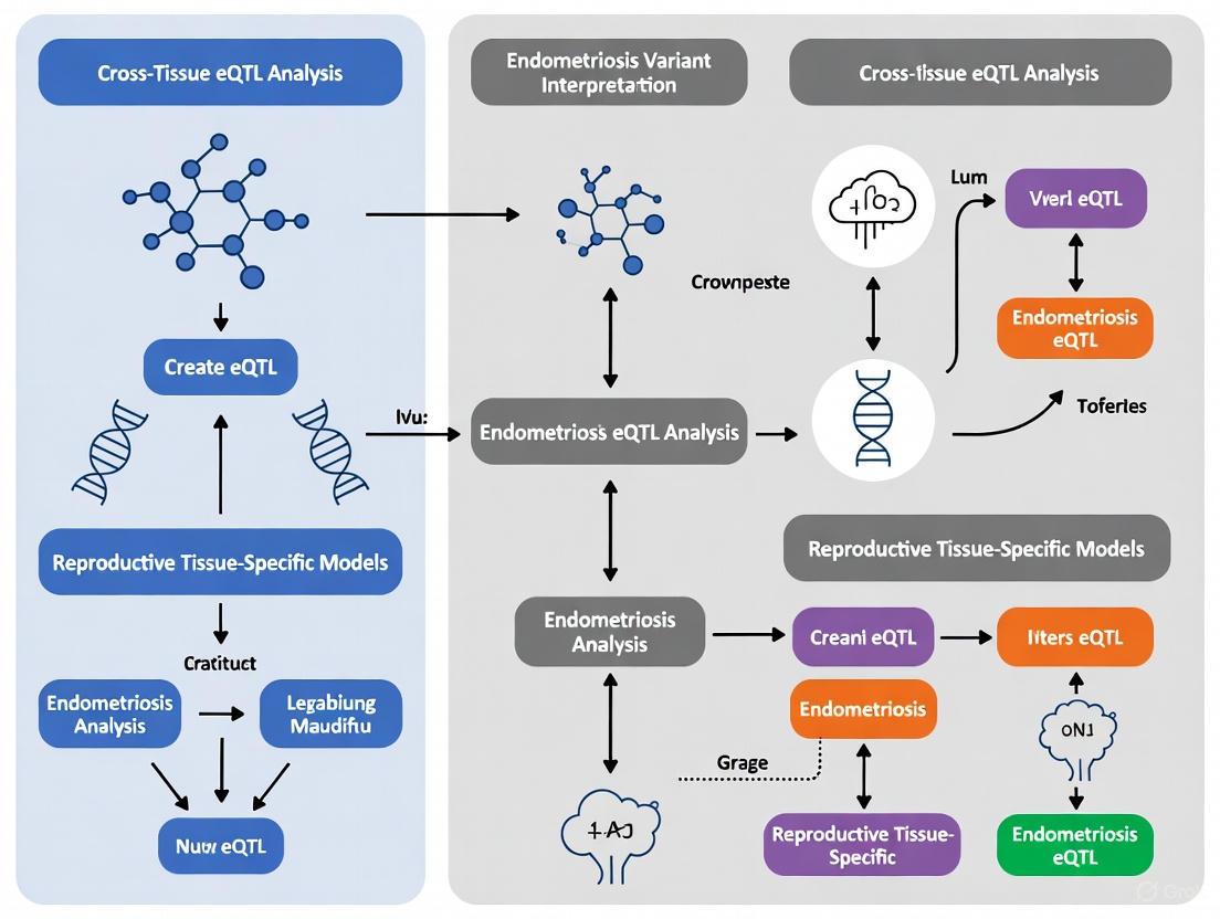
To overcome the limitations of GWAS, integrating genetic association data with functional genomic data is paramount. Expression Quantitative Trait Locus (eQTL) analysis is a powerful method to identify genetic variants that influence gene expression levels. Cross-tissue eQTL analysis is particularly relevant for endometriosis, as genetic risk variants may exert their effects in a tissue-specific manner, including reproductive tissues (uterus, ovary), tissues commonly affected by lesions (colon, ileum), and the systemic immune environment (peripheral blood) [1] [8].
The following workflow diagram outlines the core process for integrating GWAS and multi-tissue eQTL data to prioritize candidate genes and formulate mechanistic hypotheses.
Protocol: Multi-Tissue eQTL Analysis for Endometriosis-Associated Variants
Objective: To functionally characterize endometriosis-associated GWAS variants by identifying their regulatory effects on gene expression across six physiologically relevant tissues.
Materials and Software:
- Hardware: High-performance computing cluster.
- Software: R or Python with bioinformatics packages (e.g.,
TwoSampleMR,coloc), PLINK. - Data Sources:
Procedure:
Variant Selection and Annotation:
- Retrieve all genome-wide significant (p < 5×10⁻⁸) endometriosis associations from the GWAS Catalog (EFO_0001065).
- Annotate variants using Ensembl VEP to determine genomic location (e.g., intronic, intergenic).
Tissue Selection and eQTL Mapping:
- Select tissues relevant to endometriosis pathophysiology: uterus, ovary, vagina, sigmoid colon, ileum, and whole blood.
- Cross-reference the list of GWAS variants with the GTEx eQTL datasets for each selected tissue.
- Retain only significant eQTL associations (False Discovery Rate, FDR < 0.05). Record the regulated gene, effect size (slope), and adjusted p-value for each variant-gene-tissue trio.
Gene Prioritization:
- Criterion A (Variant Count): Prioritize genes that are regulated by the highest number of independent eQTL variants in a given tissue.
- Criterion B (Effect Size): Prioritize genes based on the magnitude of the regulatory effect (absolute slope value). A slope of +1.0 indicates a twofold increase in expression per alternative allele.
- Generate a final list of high-priority candidate genes for downstream analysis.
Functional Interpretation:
Expected Output:
- A list of high-confidence candidate genes (e.g., MICB, CLDN23, GATA4) whose expression is modulated by endometriosis risk variants.
- Insights into tissue-specific regulatory patterns: Immune and epithelial signaling genes may predominate in colon and blood, while hormonal response and tissue remodeling genes may be highlighted in ovary and uterus [1].
The Scientist's Toolkit: Research Reagent Solutions
Table 3: Essential Reagents and Resources for Endometriosis Genetic Research
| Item | Function/Application | Example/Provider |
|---|---|---|
| SOMAscan Platform | Multiplexed immunoaffinity assay for large-scale plasma protein quantification (pQTL studies). | SomaLogic [9] |
| Human R-Spondin3 ELISA Kit | Quantitative measurement of RSPO3 protein levels in patient plasma for target validation. | BOSTER Biological Technology [9] |
| Illumina Whole-Exome/Genome Sequencing | Identification of rare coding and regulatory variants in familial or case-control cohorts. | Illumina Platforms [3] |
| GTEx v8 eQTL Datasets | Publicly available repository of tissue-specific gene expression regulation. | GTEx Portal [1] [8] |
| TwoSampleMR R Package | Statistical tool for performing Mendelian Randomization analysis to infer causality. | CRAN Repository [9] [7] |
| Seurat R Package | Comprehensive toolkit for the analysis and interpretation of single-cell RNA-sequencing data. | Satija Lab [7] [10] |
Complementary Genomic Approaches
Beyond eQTL analysis, other advanced genomic strategies are proving invaluable.
- Mendelian Randomization (MR): This method uses genetic variants as instrumental variables to infer causal relationships between modifiable exposures (e.g., protein levels) and disease. Recent MR studies have identified RSPO3 as a potential causal plasma protein, nominating it as a novel therapeutic target for endometriosis [9].
- Family-Based Whole-Exome Sequencing (WES): To address the "rare variant" gap, WES in multigenerational families has identified novel candidate genes (e.g., LAMB4, EGFL6) that co-segregate with disease, supporting a polygenic, additive model [3]. The following diagram illustrates this complementary approach.
- Integration with Single-Cell and Other Omics: Combining eQTL findings with single-cell RNA-sequencing from eutopic and ectopic endometrium can reveal cell-type-specific expression of candidate genes and alterations in the cellular microenvironment, such as epithelial-mesenchymal transition and immune cell interactions [7] [10].
Endometriosis is a complex genetic disorder where GWAS has successfully illuminated the polygenic nature of disease risk but has also revealed significant limitations. The path forward requires a shift from mere variant discovery to functional interpretation. Cross-tissue eQTL analysis represents a critical framework for bridging this gap, enabling researchers to map GWAS variants to their target genes and regulatory contexts across disease-relevant tissues. When integrated with other powerful methods like Mendelian randomization, family-based sequencing, and single-cell genomics, this approach provides a comprehensive strategy to decipher the molecular pathophysiology of endometriosis, ultimately accelerating the development of much-needed diagnostic biomarkers and targeted therapeutics.
Genome-wide association studies (GWAS) have revolutionized our understanding of the genetic architecture of complex diseases, identifying thousands of statistical associations between genetic variants and disease susceptibility. However, a significant challenge remains: the majority of these disease-associated variants reside in non-coding regions of the genome, making their functional interpretation difficult [11]. Approximately 95% of high-confidence fine-mapped single nucleotide polymorphisms (SNPs) from GWAS are located in non-coding and flanking regions, implicating a substantial role for non-coding variation in disease [11]. These non-coding variants are now understood to exert their phenotypic effects primarily through the regulation of gene expression by altering regulatory elements such as enhancers, transcription factor binding sites, and chromatin state [11].
Expression quantitative trait loci (eQTLs) have emerged as a powerful framework for addressing this interpretative challenge. eQTLs are genomic loci that regulate gene expression levels and can be classified based on their proximity to the gene they influence: cis-eQTLs typically affect genes proximal to the variant, while trans-eQTLs influence genes distant from the variant, often on different chromosomes [12]. By identifying genetic variants that influence gene expression, eQTL analysis provides a mechanistic bridge between non-coding GWAS hits and their potential biological consequences, enabling researchers to generate testable hypotheses about causal genes and regulatory mechanisms [11] [12].
The integration of eQTL data is particularly crucial in the context of endometriosis research, where GWAS has identified multiple susceptibility loci, yet the functional characterization of these variants remains incomplete [2] [8]. This application note provides a comprehensive framework for employing eQTL analyses to elucidate the functional impact of non-coding variants identified in endometriosis GWAS, with specific protocols for cross-tissue investigation and variant prioritization.
Key Concepts and Analytical Framework
Fundamentals of eQTL Analysis
Expression quantitative trait loci represent a critical link between genetic variation and gene expression. At their core, eQTLs are genomic regions where genetic variation (e.g., SNPs) correlates with differences in mRNA expression levels of target genes. The cis/trans distinction is fundamental: cis-eQTLs typically operate on genes located close to the variant (usually within 1 Mb) and likely affect local regulatory elements such as promoters and enhancers, while trans-eQTLs influence genes further away, often through intermediate molecules like transcription factors or through complex regulatory networks [12].
The statistical power of eQTL mapping depends on several factors, including sample size, tissue context, and technical variability. Larger sample sizes increase the ability to detect eQTLs, particularly those with modest effects or those active in specific cell subtypes. Tissue context is equally critical, as regulatory effects often show considerable tissue specificity due to differences in chromatin accessibility, transcription factor availability, and epigenetic modifications [2] [12]. This is especially relevant for endometriosis, where eQTL effects may differ between reproductive tissues, immune cells, and even intestinal tissues known to be affected by the disease [2] [8].
eQTLs in Disease Mapping
The primary value of eQTL analysis in disease research lies in its ability to provide functional context for GWAS findings. When a GWAS-identified risk variant colocalizes with an eQTL, it suggests that the variant may influence disease risk by modulating the expression of a specific gene. This colocalization analysis significantly enhances the biological interpretation of GWAS signals and facilitates the prioritization of candidate causal genes for functional validation [13] [14].
For endometriosis, recent studies have demonstrated the utility of this approach. By cross-referencing endometriosis-associated GWAS variants with eQTL data from the GTEx database across six physiologically relevant tissues (uterus, ovary, vagina, colon, ileum, and peripheral blood), researchers have identified tissue-specific regulatory patterns [2] [8]. In reproductive tissues, eQTL-associated genes were enriched for functions related to hormonal response, tissue remodeling, and adhesion, while in colon, ileum, and peripheral blood, immune and epithelial signaling genes predominated [8]. This tissue-specific functional characterization provides crucial insights into the molecular pathophysiology of endometriosis.
Table 1: Key eQTL Databases and Resources for Endometriosis Research
| Resource | Description | Relevance to Endometriosis |
|---|---|---|
| GTEx Portal [13] [8] | Repository of tissue-specific eQTL data from 54 non-diseased tissue sites across 49 tissues | Provides baseline regulatory information for uterus, ovary, vagina, colon, ileum, and blood |
| eQTpLot [13] | R package for visualization of colocalization between eQTL and GWAS signals | Enables intuitive visualization of endometriosis GWAS and eQTL data integration |
| RatGTEx Portal [15] | Gene expression and eQTL data for different rat tissues | Offers cross-species validation opportunities for candidate genes |
| GWAS Catalog [8] | Curated repository of all published GWAS and their associated variants | Source of endometriosis-associated variants for functional follow-up |
Experimental Protocols
Protocol 1: Cross-Tissue eQTL Analysis for Endometriosis-Associated Variants
Purpose and Principles
This protocol describes a systematic approach to identify the regulatory effects of endometriosis-associated genetic variants across multiple tissues. The methodology is based on integrating GWAS summary statistics with tissue-specific eQTL data to identify genes whose expression is potentially influenced by endometriosis risk variants [2] [8]. The cross-tissue perspective is particularly valuable for endometriosis, given the disease's presentation in multiple tissue types and the potential involvement of systemic immune factors.
Equipment and Reagents
- Computational environment (R statistical platform, Python)
- High-performance computing cluster or workstation with minimum 16GB RAM
- Endometriosis GWAS summary statistics (publicly available from GWAS Catalog or FinnGen)
- Tissue-specific eQTL data (GTEx v8 or later version)
Procedure
Variant Selection and Curation
- Retrieve endometriosis-associated variants from the GWAS Catalog (EFO_0001065) with genome-wide significance (p < 5 × 10⁻⁸) [8].
- Exclude variants without standardized rsIDs and remove duplicates, retaining the entry with the lowest p-value for each unique variant.
- Annotate remaining variants using Ensembl Variant Effect Predictor (VEP) to determine genomic location (intronic, exonic, intergenic, UTR) and nearest genes.
Tight Selection and eQTL Extraction
- Select tissues physiologically relevant to endometriosis: uterus, ovary, vagina, sigmoid colon, ileum, and peripheral blood [8].
- Cross-reference curated variants with tissue-specific eQTL data from GTEx portal.
- Retain only significant eQTLs (false discovery rate [FDR] < 0.05) and extract the following information for each: regulated gene, slope (effect size and direction), adjusted p-value, and tissue.
Data Integration and Prioritization
- For each tissue, prioritize genes based on: (1) frequency of regulation by eQTL variants, and (2) strength of regulatory effects (absolute slope values) [8].
- Generate a unified table cross-referencing all significant variant-gene-trait associations.
Functional Interpretation
- Perform functional enrichment analysis using MSigDB Hallmark gene sets and Cancer Hallmarks collections.
- Categorize regulated genes into biological pathways and note tissue-specific patterns.
- Identify genes not associated with known pathways as potential novel regulatory mechanisms.
Timing and Troubleshooting
- Timing: 3-5 days for complete analysis, depending on computational resources and dataset size.
- Troubleshooting: If few significant eQTLs are detected, consider relaxing the FDR threshold to < 0.1 or including variants with suggestive GWAS significance (p < 1 × 10⁻⁶). For functional interpretation challenges, expand the pathway databases to include custom endometriosis-relevant gene sets.
Protocol 2: Visualizing eQTL-GWAS Colocalization with eQTpLot
Purpose and Principles
This protocol describes the use of the eQTpLot R package to generate comprehensive visualizations of colocalization between eQTL and GWAS signals [13]. Effective visualization is crucial for interpreting complex genetic data and communicating findings. eQTpLot provides specialized plots that integrate eQTL and GWAS information, including directional effects and linkage disequilibrium patterns, offering advantages over simpler visualization tools.
Equipment and Reagents
- R environment (version 4.0.0 or higher) with eQTpLot package installed
- Required R packages: biomaRt, dplyr, GenomicRanges, ggnewscale, ggplot2, ggplotfy, ggpubr, gridExtra, Gviz, LDheatmap, patchwork
- GWAS summary statistics in standard format (SNP, CHR, BP, P, etc.)
- cis-eQTL summary statistics (e.g., from GTEx portal)
Procedure
Data Preparation
- Format GWAS summary statistics as a data frame with columns: SNP (rsID), CHR (chromosome), BP (base position), P (p-value), and other optional fields.
- Format eQTL data as a data frame with columns: SNP, GENE (gene symbol), TISSUE, NES (normalized effect size), P (p-value).
- Optional: Prepare pairwise LD information for variants in the region of interest.
Basic eQTpLot Implementation
- Load required libraries and input data frames into R.
- Execute the core eQTpLot function, specifying: GWAS data frame, eQTL data frame, gene name, GWAS trait, and tissue type.
- Generate the five-panel visualization showing: (1) colocalization of GWAS and eQTL signals, (2) correlation between GWAS and eQTL p-values, (3) enrichment of eQTLs among trait-significant variants, (4) LD landscape, and (5) direction of effect relationships.
Advanced Configuration
- For directional analysis, set congruence = TRUE to divide variants into congruous (same direction of effect on gene expression and GWAS trait) and incongruous (opposite directions) groups.
- For multi-tissue visualization, set tissue to a list of tissues or "all" for pan-tissue analysis, specifying the collapse method ("min", "median", "mean", or "meta").
- Customize visual aesthetics using the available theme and formatting options.
Output and Interpretation
- Export publication-quality figures in appropriate formats (PDF, PNG).
- Interpret colocalization evidence based on spatial overlap of significant signals and correlation patterns.
- Note directional relationships to hypothesize whether increased gene expression would promote or suppress disease risk.
Timing and Troubleshooting
- Timing: 1-2 days for data preparation and visualization generation.
- Troubleshooting: If visualizations are cluttered, focus on specific genomic regions or apply more stringent p-value thresholds. For memory issues with large datasets, subset data to regions of interest before visualization.
The following diagram illustrates the workflow for cross-tissue analysis and visualization:
Table 2: Key Analytical Tools for eQTL Integration in Endometriosis Research
| Tool/Resource | Function | Application Context |
|---|---|---|
| ANNOVAR [11] | Functional annotation of genetic variants | Initial characterization of endometriosis-associated variants |
| RegulomeDB [11] | Non-coding specific variant annotation with regulatory information | Prioritizing variants likely to affect regulatory elements |
| FUMA [11] | Annotation and visualization of GWAS results | Integrated platform for GWAS variant functional mapping |
| GTEx Portal [8] | Tissue-specific eQTL database | Primary source of regulatory information across relevant tissues |
| eQTpLot [13] | Visualization of eQTL-GWAS colocalization | Generating intuitive plots for publications and presentations |
| Reveal [16] | Visual analytics for eQTL data | Exploring complex associations in patient cohort data |
| FUSION [14] | TWAS software for single-tissue analysis | Imputing gene expression and testing associations with endometriosis |
| UTMOST [14] | Cross-tissue TWAS framework | Identifying genes with consistent regulatory effects across tissues |
Data Interpretation and Analysis
Key Parameters and Quantitative Benchmarks
Successful interpretation of eQTL analyses requires careful attention to multiple statistical parameters and biological contexts. The following table outlines key metrics and their interpretation in the context of endometriosis research:
Table 3: Key Statistical Parameters for eQTL Analysis Interpretation
| Parameter | Interpretation | Recommended Threshold |
|---|---|---|
| eQTL FDR | Statistical significance of variant-gene expression association | < 0.05 for discovery; < 0.01 for validation |
| Slope/Effect Size | Direction and magnitude of expression change per allele | Consider biological context; ±0.2-0.5 may be meaningful |
| Colocalization Probability | Likelihood that eQTL and GWAS signals share causal variant | PPH4 > 0.7 considered strong evidence [14] |
| Tissue Specificity Index | Measure of how tissue-specific an eQTL effect is | Lower values indicate broader activity across tissues |
| Variant Effect Predictor | Functional consequence annotation | Prioritize regulatory annotations (enhancer, promoter) |
Advanced Analytical Approaches
For deeper mechanistic insights, researchers can employ several advanced analytical frameworks:
Transcriptome-Wide Association Studies (TWAS): This approach integrates eQTL and GWAS data to identify genes whose genetically regulated expression is associated with endometriosis risk. Both single-tissue (FUSION) and cross-tissue (UTMOST) methods can be applied, with the latter particularly valuable for detecting genes with consistent effects across multiple tissues [14].
Mendelian Randomization (MR): Using genetic variants as instrumental variables, MR can test for causal relationships between gene expression and endometriosis risk. This approach provides stronger evidence for potential therapeutic targets [14].
Network and Mediation Analyses: These methods can elucidate the mechanisms through which eQTL effects influence endometriosis risk, potentially identifying mediating factors such as blood lipid levels or hip circumference, as recently demonstrated for several endometriosis-associated genes [14].
The following diagram illustrates the relationship between different analytical approaches in translating GWAS findings to functional insights:
Discussion and Future Perspectives
The integration of eQTL analysis with GWAS findings represents a paradigm shift in our ability to interpret non-coding genetic variation in endometriosis. The protocols outlined here provide a systematic approach to identify and validate the regulatory mechanisms through which endometriosis-associated variants potentially influence disease risk. The cross-tissue perspective is particularly important, as recent research has demonstrated distinct regulatory profiles in reproductive versus intestinal and immune tissues [2] [8].
Looking forward, several emerging technologies and methodologies promise to further enhance our understanding of endometriosis genetics. Single-cell eQTL mapping will enable the resolution of regulatory effects in specific cell types relevant to endometriosis, such as endometrial stromal cells, specific immune cell populations, and endothelial cells. Multi-omic integration of eQTLs with other molecular QTLs (such as histone modification QTLs, methylation QTLs, and protein QTLs) will provide a more comprehensive view of the regulatory landscape. Finally, functional validation using CRISPR-based approaches in appropriate cellular models will be essential to move from statistical associations to causal mechanisms.
The application of these advanced eQTL methodologies in endometriosis research has already begun to yield novel insights, identifying candidate susceptibility genes such as CISD2, GREB1, and SULT1E1, and suggesting potential mediating factors in disease pathogenesis [14]. As these approaches become more widely adopted and integrated with functional studies, they will undoubtedly accelerate the translation of genetic discoveries into improved diagnostic and therapeutic strategies for endometriosis.
Why Cross-Tissue Analysis? Moving Beyond the Endometrium in Disease Pathogenesis
The pathogenesis of endometriosis, a chronic inflammatory disease affecting an estimated 190 million women worldwide, has long been a focus of reproductive medicine research [1] [8]. While traditional investigations have centered on the eutopic endometrium, emerging evidence underscores that endometriosis is a systemic disorder with manifestations across multiple tissue environments. The limitation of single-tissue analyses becomes particularly evident when considering that most genome-wide association study (GWAS)-identified variants reside in non-coding regions with unknown regulatory functions [17]. Cross-tissue expression quantitative trait locus (eQTL) analysis has thus emerged as a transformative approach that enables researchers to map the tissue-specific regulatory effects of genetic variants, revealing novel mechanisms in endometriosis pathogenesis that extend far beyond the uterine lining [1] [14].
This paradigm shift recognizes that endometriosis lesions commonly affect diverse anatomical sites, including ovaries, pelvic peritoneum, intestinal surfaces, and in rare cases, the sigmoid colon and ileum [1] [8]. Furthermore, peripheral blood captures systemic immune and inflammatory signals relevant to disease pathophysiology [8]. Cross-tissue analysis provides a functional framework to bridge the gap between genetic associations and biological mechanisms by answering a critical question: How do endometriosis-associated genetic variants regulate gene expression across different tissue contexts relevant to disease manifestation? [1]
Key Rationale for Cross-Tissue Investigation
Tissue-Specific Regulatory Profiles Reveal Distinct Pathogenic Mechanisms
Comprehensive eQTL analyses demonstrate that endometriosis-associated variants exert profoundly tissue-specific effects [1]. In reproductive tissues (uterus, ovary, vagina), these variants predominantly regulate genes involved in hormonal response, tissue remodeling, and cellular adhesion. In contrast, within intestinal tissues (colon, ileum) and peripheral blood, the same variants preferentially target genes governing immune signaling and epithelial function [1] [8]. This fundamental observation explains why limiting analysis to endometrial tissue provides an incomplete picture of endometriosis pathogenesis.
Table 1: Tissue-Specific Enrichment of Biological Pathways in Endometriosis
| Tissue Type | Dominant Biological Pathways | Key Regulator Genes |
|---|---|---|
| Reproductive Tissues (Uterus, Ovary, Vagina) | Hormonal response, Tissue remodeling, Cellular adhesion | GREB1, SULT1E1, IL1A [1] [14] |
| Intestinal Tissues (Colon, Ileum) | Immune signaling, Epithelial function | MICB, CLDN23 [1] |
| Peripheral Blood | Systemic immune response, Inflammatory signaling | GIMAP4, TOP3A, MKNK1 [1] [18] |
Expanding the Spectrum of Susceptibility Genes
Cross-tissue analyses have successfully identified novel susceptibility genes that would remain undetected in single-tissue studies. For instance, integrative approaches combining GWAS with multi-tissue eQTL data have revealed candidate genes including CISD2, EFR3B, GREB1, IMMT, SULT1E1, and UBE2D3 [14]. Notably, the expression of IMMT across 21 different tissues and UBE2D3 in 7 tissues demonstrated causal relationships with endometriosis risk, highlighting the value of surveying gene expression effects across diverse tissue contexts [14].
Additional validation studies have confirmed MKNK1 and TOP3A as ovarian endometriosis risk genes, with both genes showing upregulated expression in ectopic and eutopic endometrium compared to normal controls [18]. Functional experiments demonstrated that knockdown of these genes significantly inhibited the migration, invasion, and proliferation of ectopic endometrial stromal cells, providing mechanistic insights into their roles in disease pathogenesis [18].
Experimental Protocols for Cross-Tissue eQTL Analysis
Protocol 1: Fundamental Multi-Tissue eQTL Analysis
This protocol outlines the foundational methodology for identifying tissue-specific regulatory effects of endometriosis-associated genetic variants [1] [8].
Materials and Equipment
- GWAS Catalog data for endometriosis (EFO_0001065)
- GTEx v8 database access
- Ensembl Variant Effect Predictor (VEP)
- Statistical computing environment (R or Python)
Procedure
Variant Selection and Annotation
- Retrieve genome-wide significant endometriosis-associated variants (p < 5 × 10^(-8)) from the GWAS Catalog
- Filter to include only variants with valid rsIDs
- Annotate variants using Ensembl VEP to determine genomic locations and associated genes
Tight Selection Criteria
- Select tissues with biological relevance to endometriosis pathophysiology
- Include reproductive tissues: uterus, ovary, vagina
- Include intestinal tissues: sigmoid colon, ileum
- Include peripheral blood (whole blood) to capture systemic immune signals
eQTL Identification
- Cross-reference endometriosis-associated variants with tissue-specific eQTL data from GTEx v8
- Apply false discovery rate (FDR) correction (FDR < 0.05)
- Extract significant eQTLs along with their slope values (effect size and direction)
Functional Interpretation
- Prioritize genes based on either frequency of regulation by eQTLs or strength of regulatory effects (slope values)
- Perform pathway enrichment analysis using MSigDB Hallmark gene sets and Cancer Hallmarks collections
Protocol 2: Advanced Cross-Tissue Transcriptome-Wide Association Study (TWAS)
This protocol describes an advanced integrative approach that combines eQTL and GWAS data to identify novel susceptibility genes across multiple tissues [14] [19].
Materials and Equipment
- GWAS summary statistics for endometriosis (e.g., from FinnGen consortium)
- GTEx v8 eQTL data across 47 tissues
- UTMOST software for cross-tissue TWAS
- FUSION software for single-tissue TWAS
- MAGMA software for gene-based association analysis
- TwoSampleMR R package for Mendelian randomization
Procedure
Data Preparation and Integration
- Obtain GWAS summary data for endometriosis and its subtypes
- Acquire multi-tissue eQTL data from GTEx v8, excluding male-specific tissues
- Harmonize data formats and coordinate systems across datasets
Cross-Tissue TWAS Implementation
- Perform cross-tissue analysis using UTMOST with group lasso penalty
- Conduct single-tissue analysis using FUSION for comparison
- Validate significant associations using MAGMA gene-based analysis
Causal Inference and Validation
- Apply Mendelian randomization to test causal relationships between gene expression and endometriosis risk
- Perform colocalization analysis to assess shared causal variants between eQTL and GWAS signals
- Execute two-sample network MR to identify mediating factors in causal pathways
Functional Annotation
- Conduct bioinformatics analyses to examine expression patterns of identified genes
- Perform enrichment analyses to elucidate biological functions and pathways
Table 2: Key Analytical Methods for Cross-Tissue Transcriptomic Analysis
| Method Category | Specific Tools/Approaches | Primary Application |
|---|---|---|
| Cross-Tissue TWAS | UTMOST (Unified Test for Molecular Signature) | Identifies genes with shared and tissue-specific eQTL effects [14] [19] |
| Single-Tissue TWAS | FUSION (Functional Summary-based Imputation) | Tests gene-trait associations in individual tissues [14] |
| Gene-Based Association | MAGMA (Multi-marker Analysis of GenoMic Annotation) | Validates significant associations from TWAS [14] |
| Causal Inference | Mendelian Randomization (MR), Colocalization | Tests causal relationships and shared genetic mechanisms [20] [14] |
| Advanced Multi-Tissue | MTWAS (Partitioning cross-tissue and tissue-specific effects) | Enhances prediction accuracy by classifying eQTLs [19] |
The Scientist's Toolkit: Essential Research Reagent Solutions
Table 3: Key Research Reagents and Resources for Cross-Tissue Endometriosis Research
| Resource Category | Specific Resource | Function and Application |
|---|---|---|
| Genetic Databases | GWAS Catalog (EFO_0001065) | Source of endometriosis-associated genetic variants [1] [8] |
| Expression Databases | GTEx v8 | Provides tissue-specific eQTL data across 49 tissues [1] [14] |
| Analytical Tools | Ensembl VEP | Functional annotation of genetic variants [1] [8] |
| Cross-Tissue TWAS | UTMOST Software | Identifies genes with cross-tissue regulatory effects [14] [19] |
| Single-Cell Analysis | scRNA-seq, scATAC-seq | Resolves cellular heterogeneity and identifies rare cell populations [21] [22] |
| Methylation Analysis | Illumina Infinium MethylationEPIC BeadChip | Profiles genome-wide DNA methylation patterns [23] |
| Functional Validation | Immunohistochemistry, Knockdown assays | Confirms protein expression and functional roles of candidate genes [18] |
Advanced Integrative Approaches
Single-Cell Resolution in Cross-Tissue Analysis
Single-cell technologies have revealed remarkable cellular heterogeneity within endometrial tissue, identifying distinct subpopulations of epithelial, stromal, and immune cells that contribute differentially to endometriosis pathogenesis [21] [22]. These approaches have uncovered that the eutopic endometrium in women with endometriosis exhibits a pro-inflammatory phenotype involving both immune and non-immune cell types [22]. Furthermore, single-cell RNA sequencing has provided evidence of epithelial-mesenchymal transition (EMT) in eutopic endometrium, characterized by reduced epithelial cell proportions and altered CDH1 expression [20].
Epigenetic Dimension of Cross-Tissue Regulation
DNA methylation analyses have established that menstrual cycle phase is a major source of epigenetic variation in endometrial tissue, accounting for significant changes in methylation profiles that potentially regulate genes and pathways responsible for endometrial function [23]. mQTL (methylation quantitative trait loci) analysis has identified 118,185 independent cis-mQTLs in endometrial tissue, including 51 associated with endometriosis risk, providing functional evidence for epigenetic mechanisms contributing to disease pathogenesis [23].
Methodological Innovations in Multi-Tissue Prediction
The recently developed MTWAS framework significantly enhances prediction accuracy by partitioning and aggregating both cross-tissue and tissue-specific genetic effects [19]. This method incorporates a non-parametric imputation strategy for inaccessible tissues and classifies eQTLs into cross-tissue eQTLs and tissue-specific eQTLs using a stepwise selection procedure based on the extended Bayesian information criterion [19]. Compared to single-tissue methods, MTWAS demonstrates an average improvement in prediction R² of 47.4% over PrediXcan and 9.2% over UTMOST across 47 GTEx tissues [19].
Cross-tissue analysis represents a paradigm shift in endometriosis research, moving beyond the traditional endometrial-centric view to embrace the systemic complexity of this debilitating condition. By integrating multi-tissue eQTL data with GWAS findings through sophisticated computational frameworks, researchers can now decipher the functional consequences of genetic variants across biologically relevant tissues. The methodologies outlined in this application note provide a comprehensive roadmap for implementing cross-tissue analyses, from fundamental eQTL mapping to advanced multi-tissue TWAS and single-cell resolution approaches. As these techniques continue to evolve, they promise to unlock novel therapeutic targets and diagnostic biomarkers that address the multifaceted nature of endometriosis pathogenesis across tissue environments.
Endometriosis is a complex, estrogen-dependent inflammatory disease with a significant heritable component, affecting approximately 10% of reproductive-aged women globally [24] [25]. Genome-wide association studies (GWAS) have identified numerous genetic variants associated with endometriosis risk; however, the majority reside in non-coding regions, complicating the interpretation of their functional significance [1]. Expression quantitative trait locus (eQTL) analysis provides a powerful framework to bridge this gap by identifying genetic variants that regulate gene expression in a tissue-specific manner.
Cross-tissue eQTL analysis is particularly crucial for endometriosis, a condition involving multiple biologically relevant tissues. This approach allows researchers to identify how endometriosis-associated genetic variants exert their effects by modulating gene expression not only in reproductive tissues like the uterus and ovary but also in gastrointestinal and systemic immune tissues, reflecting the disease's complex pathophysiology and comorbidity profile [1] [24]. This application note details standardized protocols for identifying and interpreting cross-tissue eQTLs in endometriosis research, enabling the prioritization of candidate causal genes and biological mechanisms.
Tissue-Specific eQTL Landscape in Endometriosis
Rationale for Tissue Selection
The pathophysiology of endometriosis extends beyond the reproductive tract, necessitating investigation across multiple tissue types:
- Uterus: Primary source of ectopic endometrial tissue via retrograde menstruation [24]
- Ovary: Common site for endometrioma formation [24]
- Gastrointestinal Tissues (Sigmoid Colon, Ileum): Locations for deep infiltrating endometriosis, contributing to painful defecation (dyschezia) and other GI symptoms [1] [24]
- Systemic Immune Tissues (Peripheral Blood): Captures systemic inflammatory and immune dysregulation characteristic of endometriosis [1]
Table 1: Tissue-Specific eQTL Patterns in Endometriosis
| Tissue | Key Regulated Genes | Enriched Biological Pathways | Research Implications |
|---|---|---|---|
| Uterus | GREB1, WASHC2 [26] | Hormone response, tissue remodeling, cell adhesion [1] [25] | Identifies genes with direct relevance to endometrial proliferation and implantation |
| Ovary | MICB, GATA4 [1] | Hormonal response, inflammation, angiogenesis [1] | Illuminates mechanisms in ovarian endometrioma formation and associated infertility |
| Sigmoid Colon/Ileum | CLDN23 [1] | Immune signaling, epithelial barrier function [1] | Reveals pathways contributing to deep infiltrating disease and GI comorbidities |
| Peripheral Blood | Multiple immune regulators [1] | Immune activation, inflammatory response [1] | Provides accessible biomarkers and insights into systemic inflammation |
Experimental Protocols for Cross-Tissue eQTL Analysis
Protocol 1: Identification of Endometriosis-Associated eQTLs
Objective: To identify genetic variants that regulate gene expression in tissues relevant to endometriosis pathophysiology.
Materials and Reagents:
- GWAS summary statistics for endometriosis
- Genotype and RNA-seq data from target tissues (uterus, ovary, colon, ileum, blood)
- Computational resources for large-scale genomic analysis
Methodology:
- Variant Curation: Compile endometriosis-associated variants from GWAS Catalog (EFO_0001065) with genome-wide significance (p < 5 × 10⁻⁸) [1]
- Tissue-Specific eQTL Mapping: Cross-reference curated variants with eQTL data from GTEx database v8 for uterus, ovary, sigmoid colon, ileum, and whole blood [1]
- Statistical Validation: Apply false discovery rate (FDR) correction (FDR < 0.05) to identify significant eQTLs [1]
- Effect Direction Analysis: Record slope values indicating direction and magnitude of effect on gene expression [1]
- Functional Annotation: Use Ensembl Variant Effect Predictor (VEP) to determine genomic context of significant eQTLs [1]
Expected Outcomes: A comprehensive map of endometriosis-associated variants that function as eQTLs across biologically relevant tissues.
Protocol 2: Splicing QTL (sQTL) Analysis in Endometrial Tissue
Objective: To identify genetic variants that regulate alternative splicing in endometrial tissue across the menstrual cycle and in endometriosis.
Materials and Reagents:
- Endometrial tissue samples (n=206) from women with and without endometriosis [26]
- RNA-seq data and genotype information [26]
- Computational pipeline for splicing quantification (e.g., LeafCutter, rMATS)
Methodology:
- Sample Collection and Stratification: Collect endometrial tissue samples across menstrual cycle phases (proliferative and secretory) from both cases and controls [26]
- RNA Sequencing and Quality Control: Perform high-depth RNA sequencing with standard quality control metrics
- Splicing Quantification: Calculate percent spliced in (PSI) values for all intron clusters [26]
- sQTL Mapping: Test associations between genetic variants and splicing ratios using a linear model, correcting for multiple testing [26]
- Integration with GWAS: Colocalize sQTL signals with endometriosis GWAS signals to prioritize functionally relevant splicing events [26]
Expected Outcomes: Identification of sQTLs contributing to endometriosis risk, such as those affecting GREB1 and WASHC3 genes [26].
Protocol 3: Multi-omic Mendelian Randomization for Causal Inference
Objective: To integrate multi-omics data for causal association testing between cell aging-related genes and endometriosis risk.
Materials and Reagents:
- Summary statistics from endometriosis GWAS [27]
- Blood eQTL, methylation QTL (mQTL), and protein QTL (pQTL) data [27]
- SMR software (version 1.3.1) and R package 'coloc' [27]
Methodology:
- Data Harmonization: Obtain summary statistics for endometriosis GWAS and various QTL types from public repositories [27]
- Summary-based Mendelian Randomization (SMR): Perform SMR analysis to test causal effects of gene expression, methylation, and protein abundance on endometriosis risk [27]
- Heterogeneity in Dependent Instruments (HEIDI) Test: Differentiate pleiotropy from linkage (P-HEIDI > 0.05) [27]
- Colocalization Analysis: Assess probability of shared causal variants between QTLs and GWAS signals (PPH4 > 0.5) [27]
- Multi-omic Integration: Analyze causal chains (e.g., mQTL → eQTL → pQTL → endometriosis) to elucidate mechanistic pathways [27]
Expected Outcomes: Identification of causal genes and proteins (e.g., MAP3K5, ENG) in endometriosis pathogenesis, revealing potential therapeutic targets [27].
Visualization of Experimental Workflows
Figure 1: Comprehensive workflow for cross-tissue eQTL analysis in endometriosis research
Figure 2: Logical pathway from genetic variant to endometriosis phenotype through tissue-specific regulation
The Scientist's Toolkit: Essential Research Reagents
Table 2: Essential Research Reagents for Endometriosis eQTL Studies
| Reagent/Resource | Function | Example/Source |
|---|---|---|
| GTEx Database v8 | Reference dataset for tissue-specific eQTLs | GTEx Portal [1] |
| GWAS Catalog | Repository of endometriosis-associated variants | EFO_0001065 [1] |
| SMR Software | Statistical tool for summary-data-based Mendelian randomization | SMR v1.3.1 [27] |
| Coloc R Package | Bayesian test for colocalization of QTL and GWAS signals | R package 'coloc' [27] |
| Ensembl VEP | Functional annotation of genetic variants | Ensembl Variant Effect Predictor [1] |
| Tissue Biobanks | Source of biologically relevant tissues for validation | Endometrial, ovarian, GI tissues [26] |
| RNA-seq Platforms | Transcriptome profiling for eQTL and sQTL discovery | High-throughput sequencing [26] |
Discussion and Future Directions
Cross-tissue eQTL analysis represents a powerful approach for elucidating the functional mechanisms through which genetic variants influence endometriosis risk. The protocols outlined herein enable researchers to move beyond simple association signals to identify tissue-specific regulatory mechanisms that contribute to this complex disease. Future directions in this field include the integration of single-cell eQTL maps to resolve cell-type-specific effects, development of multi-ethnic resources to address population diversity, and application of these findings to drug target prioritization and biomarker development.
The consistent identification of genes involved in hormonal regulation, inflammation, and cell adhesion across multiple tissues [1] [25] highlights the interconnected pathways driving endometriosis pathogenesis and provides a roadmap for future therapeutic development.
Endometriosis is a complex gynecological disorder with a substantial genetic component, underpinned by the regulatory effects of genetic variants on gene expression across tissues. Cross-tissue expression quantitative trait locus (eQTL) analysis has emerged as a powerful strategy to functionally characterize endometriosis-associated genetic variants identified through genome-wide association studies (GWAS) and link them to candidate susceptibility genes [1] [8]. This approach has been instrumental in identifying and validating several key genes, including CISD2, GREB1, SULT1E1, and UBE2D3, which play critical roles in endometriosis pathogenesis through diverse molecular mechanisms [28] [29]. These genes contribute to disease risk through tissue-specific regulatory mechanisms involving hormonal response, cell survival, inflammation, and protein modification pathways. This primer provides a comprehensive overview of the established functions, regulatory mechanisms, and experimental approaches for studying these four susceptibility genes, with particular emphasis on their roles in the molecular pathophysiology of endometriosis.
Gene Summaries and Key Characteristics
Table 1: Summary of Key Susceptibility Genes in Endometriosis
| Gene Name | Full Name | Chromosomal Location | Primary Function | Role in Endometriosis |
|---|---|---|---|---|
| CISD2 | CDGSH Iron Sulfur Domain 2 | Not specified in sources | Iron-sulfur cluster protein; regulates cellular iron homeostasis and endoplasmic reticulum function | Cross-tissue causal relationships with EMT risk; implicated in 17 tissues; may mediate effects through blood lipids and hip circumference [28] |
| GREB1 | Growth Regulating Estrogen Receptor Binding 1 | Not specified in sources | Early-response gene in estrogen receptor signaling; regulates hormone-dependent cell growth | Significant association with endometriosis risk through genetically regulated splicing events; identified in multiple endometriosis subtypes [26] [28] |
| SULT1E1 | Sulfotransferase Family 1E Member 1 | Not specified in sources | Estrogen sulfotransferase; catalyzes inactivation of estrogens via sulfonation | Candidate susceptibility gene for endometriosis and endometriosis of the ovary; regulates local estrogen availability [28] |
| UBE2D3 | Ubiquitin Conjugating Enzyme E2 D3 | Not specified in sources | Ubiquitin-conjugating enzyme; involved in protein ubiquitination and degradation | Causal relationships with EMT risk in 7 tissues; potential mediator through blood lipids and hip circumference [28] |
Table 2: Experimental Evidence Supporting Gene-Disease Associations
| Gene Name | Genetic Evidence | Functional Evidence | Tissue Specificity | Key References |
|---|---|---|---|---|
| CISD2 | TWAS, MR, colocalization (PPH4 > 0.7) | Bioinformatics analysis; pathway enrichment | 17 tissues showed causal relationships | [28] |
| GREB1 | sQTL analysis, TWAS, MR | Splicing QTLs in endometrial tissue | Endometrial-specific splicing discovered | [26] [28] |
| SULT1E1 | TWAS, gene-based analysis | Hormone metabolism pathways | Endometriosis of the ovary | [28] |
| UBE2D3 | TWAS, MR, colocalization (PPH4 > 0.7) | Bioinformatics analysis; mediation analysis | 7 tissues showed causal relationships | [28] |
Detailed Gene Profiles
CISD2 (CDGSH Iron Sulfur Domain 2)
CISD2 encodes a protein containing a CDGSH iron-sulfur domain that localizes to the outer mitochondrial membrane and plays a role in cellular iron homeostasis and endoplasmic reticulum integrity. Through cross-tissue transcriptome-wide association studies (TWAS) and Mendelian randomization (MR) analyses, CISD2 has been identified as a novel candidate susceptibility gene for endometriosis, with predicted expression showing significant association with disease risk [28]. The gene demonstrates causal relationships with endometriosis risk across 17 different tissues, highlighting its pervasive role in disease pathogenesis. Furthermore, CISD2 exhibits strong colocalization evidence with endometriosis (with posterior probability of hypothesis 4 > 0.7), suggesting a shared causal variant between gene expression and disease risk [28]. Two-sample network MR analyses have revealed that CISD2 may potentially influence endometriosis risk through mediation effects involving blood lipids and hip circumference, indicating a potential metabolic component to its mechanism of action in endometriosis pathophysiology [28].
GREB1 (Growth Regulating Estrogen Receptor Binding 1)
GREB1 functions as an early-response gene in estrogen receptor signaling pathways and plays a critical role in hormone-dependent cell growth and differentiation. Research has identified GREB1 as significantly associated with endometriosis risk through genetically regulated splicing events discovered via splicing quantitative trait loci (sQTL) analysis in endometrial tissue [26]. This gene represents one of the two key genes (along with WASHC3) whose splicing mechanisms in endometrium have been directly linked to endometriosis genetic risk through integration of sQTL data with endometriosis GWAS data [26]. Beyond general endometriosis risk, GREB1 has been specifically implicated in multiple endometriosis subtypes, including endometriosis of the ovary, endometriosis of the pelvic peritoneum, endometriosis of the rectovaginal septum and vagina, and deep infiltrating endometriosis [28]. The discovery of GREB1 splicing variants associated with endometriosis highlights the importance of transcript-level analyses, which can reveal regulatory mechanisms not apparent in gene-level expression analyses [26].
SULT1E1 (Sulfotransferase Family 1E Member 1)
SULT1E1 encodes an estrogen sulfotransferase that catalyzes the sulfonation of estrogens, particularly estradiol, leading to their inactivation and decreased biological activity. This enzyme plays a crucial role in regulating local estrogen availability in target tissues, including the endometrium. Through transcriptome-wide association studies, SULT1E1 has been identified as a candidate susceptibility gene for overall endometriosis risk and specifically for endometriosis of the ovary [28]. The involvement of SULT1E1 in endometriosis pathogenesis underscores the central role of estrogen signaling and metabolism in the disease process. By controlling the local bioavailability of active estrogens in endometrial and endometriotic tissues, SULT1E1 represents a key regulatory node in the hormonal milieu that drives endometriosis establishment and progression. The genetic association of SULT1E1 with endometriosis, particularly ovarian endometriosis, provides mechanistic insights into how genetic variation may influence local estrogen homeostasis and contribute to disease development.
UBE2D3 (Ubiquitin Conjugating Enzyme E2 D3)
UBE2D3 belongs to the E2 ubiquitin-conjugating enzyme family and plays a role in the ubiquitin-proteasome pathway, which mediates targeted degradation of cellular proteins. This enzyme is involved in various cellular processes, including cell cycle regulation, DNA repair, and signal transduction. Cross-tissue analyses have identified UBE2D3 as a novel candidate gene whose predicted expression is associated with endometriosis risk [28]. MR analyses have demonstrated that the expression of UBE2D3 in 7 different tissues shows causal relationships with endometriosis risk [28]. Additionally, UBE2D3 exhibits strong colocalization evidence with endometriosis (PPH4 > 0.7), supporting a shared genetic basis between gene expression regulation and disease susceptibility [28]. Similar to CISD2, two-sample network MR analyses suggest that UBE2D3 may influence endometriosis risk through mediation effects involving blood lipids and hip circumference, indicating potential metabolic pathways in its mechanism of action [28].
Experimental Protocols and Methodologies
Transcriptome-Wide Association Study (TWAS) Protocol
Objective: To identify genes whose genetically regulated expression is associated with endometriosis risk by integrating eQTL and GWAS data.
Workflow Steps:
- Data Collection: Obtain summary-level GWAS data for endometriosis from large consortia (e.g., FinnGen R11 release with 18,260 cases and 119,468 controls) [28]. Acquire eQTL data from relevant tissues (e.g., GTEx v8 dataset encompassing 47 tissues) [28].
- Expression Imputation: Utilize unified test for molecular signature (UTMOST) for cross-tissue TWAS analysis and functional summary-based imputation (FUSION) for single-tissue analysis [28].
- Association Testing: Test the association between imputed gene expression and endometriosis risk using TWAS models.
- Validation: Conduct multi-marker analysis of genomic annotation (MAGMA) analyses to validate significant associations [28].
- Result Interpretation: Prioritize genes showing consistent associations across multiple tissues or analytical methods.
TWAS Analysis Workflow: This diagram illustrates the sequential steps in transcriptome-wide association studies, from data collection to functional follow-up.
Splicing Quantitative Trait Loci (sQTL) Analysis Protocol
Objective: To identify genetic variants that influence alternative splicing patterns in endometrial tissue and their association with endometriosis risk.
Workflow Steps:
- Sample Collection: Process endometrial tissue samples from well-phenotyped participants (e.g., 206 women of European ancestry) with precise menstrual cycle phase determination [26].
- RNA Sequencing: Perform high-throughput RNA sequencing to capture transcriptomic data, including alternative splicing events.
- Genotyping: Conduct genome-wide genotyping to obtain genetic variants for all samples.
- Splicing Quantification: Quantify alternative splicing events using metrics such as percent spliced in (PSI) values for intron clusters.
- sQTL Mapping: Identify genetic variants associated with splicing variations using specialized sQTL analysis tools.
- Integration with GWAS: Overlap sQTL signals with endometriosis GWAS loci to identify potential causal genes [26].
- Functional Validation: Perform experimental validation of splicing events using RT-qPCR or other molecular techniques.
Mendelian Randomization and Colocalization Analysis Protocol
Objective: To assess causal relationships between gene expression in specific tissues and endometriosis risk, and to determine whether genetic associations share causal variants.
Workflow Steps:
- Instrument Selection: Select genetic variants associated with gene expression as instrumental variables for MR analysis (e.g., cis-eQTLs with p < 5×10⁻⁸) [28].
- MR Analysis Implementation: Apply MR methods (e.g., inverse-variance weighted, MR-Egger) to estimate causal effects of gene expression on endometriosis risk.
- Sensitivity Analyses: Conduct sensitivity analyses to assess MR assumptions and potential pleiotropy.
- Colocalization Analysis: Perform colocalization analysis (e.g., using COLOC package) to calculate posterior probabilities for shared causal variants between eQTL and GWAS signals [28].
- Mediation Analysis: Implement two-sample network MR to explore potential mediating factors in significant gene-endometriosis associations [28].
Research Reagent Solutions
Table 3: Essential Research Reagents for Endometriosis Genetic Studies
| Reagent/Category | Specific Examples | Application and Function | Example Sources |
|---|---|---|---|
| eQTL Databases | GTEx v8, endometriosis-specific eQTL datasets | Provide reference data for genetic regulation of gene expression across tissues | [1] [28] |
| GWAS Resources | FinnGen R11/R12, UK Biobank, Endometrial Cancer Association Consortium | Supply genotype-phenotype association data for prioritization of candidate genes | [28] [30] |
| Genotyping Arrays | Illumina Infinium MethylationEPIC BeadChip, standard GWAS arrays | Enable genome-wide genetic variant profiling and methylation analysis | [23] |
| RNA Sequencing Kits | High-throughput RNA-seq kits with strand-specific protocol | Facilitate transcriptome profiling and alternative splicing analysis | [26] |
| ELISA Kits | Human R-Spondin3 ELISA Kit, other protein-specific kits | Allow protein quantification in plasma and tissue samples | [9] |
| Cell Culture Assays | Endometrial cell lines, wound healing/scratc assays, proliferation assays | Enable functional validation of candidate genes in cellular models | [30] |
Molecular Pathways and Mechanisms
The four susceptibility genes operate within interconnected molecular pathways that drive endometriosis pathogenesis. GREB1 functions as a key mediator of estrogen receptor signaling, promoting the growth and survival of endometrial cells in ectopic locations [26] [28]. SULT1E1 counterbalances this estrogenic activity by inactivating estrogens through sulfonation, creating a delicate homeostasis in local estrogen signaling within the endometriotic microenvironment [28]. CISD2 contributes to cellular iron homeostasis and mitochondrial function, potentially influencing oxidative stress responses and cellular adaptability in endometriotic lesions [28]. Meanwhile, UBE2D3 participates in the ubiquitin-proteasome system, regulating the turnover of key proteins involved in cell cycle progression, inflammation, and hormone signaling pathways relevant to endometriosis establishment and progression [28].
Gene Interaction Network: This diagram illustrates the molecular pathways through which the four susceptibility genes influence endometriosis risk.
The integration of cross-tissue eQTL analysis with endometriosis GWAS has been particularly powerful in identifying these genes and their mechanisms. Studies have revealed that endometriosis-associated genetic variants display tissue-specific regulatory profiles, with reproductive tissues showing particular enrichment for genes involved in hormonal response, tissue remodeling, and cellular adhesion [1] [8]. Furthermore, advanced analytical approaches including transcriptome-wide association studies, Mendelian randomization, and colocalization analyses have enabled researchers to move beyond mere association to establish causal relationships between genetically regulated expression of these genes and endometriosis risk [28] [29].
The continuing investigation of CISD2, GREB1, SULT1E1, and UBE2D3, along with other emerging candidate genes, promises to enhance our understanding of endometriosis pathophysiology and reveal new opportunities for therapeutic intervention. These genes represent key nodes in the complex molecular network that underlies endometriosis susceptibility and progression, highlighting the value of integrative genetic approaches in elucidating the mechanisms of this common yet enigmatic disorder.
Advanced Analytical Frameworks: Integrating TWAS, MR, and Colocalization
Endometriosis is a chronic, estrogen-dependent inflammatory disease affecting approximately 10% of women of reproductive age, with a substantial genetic component accounting for approximately 50% of disease risk [1] [14]. While genome-wide association studies (GWAS) have identified numerous susceptibility loci for endometriosis, the majority reside in non-coding regions, complicating the interpretation of their functional consequences [1] [31]. Expression quantitative trait locus (eQTL) analysis provides a powerful framework to bridge this gap by identifying genetic variants that influence gene expression levels [32].
Integrating eQTL data from resources like the Genotype-Tissue Expression (GTEx) project with endometriosis GWAS summary statistics enables researchers to prioritize candidate genes and elucidate tissue-specific regulatory mechanisms in endometriosis pathogenesis [1] [33]. This protocol details a comprehensive computational workflow for this integration, with emphasis on cross-tissue analysis for enhanced variant interpretation in endometriosis research.
Background and Rationale
The Functional Genomics Gap in Endometriosis Research
Traditional GWAS have identified over 465 genome-wide significant variants associated with endometriosis risk, yet these explain only ~1.75% of the total disease risk variance [1] [14]. This limited explanatory power stems from challenges in linking non-coding variants to their target genes and accounting for tissue-specific regulatory effects. Endometriosis involves multiple tissue types, including reproductive tissues (uterus, ovary, vagina) and frequently affected extra-pelvic sites (sigmoid colon, ileum), each with distinct gene regulatory profiles [1].
eQTL Integration as a Solution
eQTL analysis maps genetic variants associated with changes in gene expression, providing a functional context for GWAS hits. The GTEx project offers a comprehensive resource of cis-eQTLs across 49 human tissues, including those relevant to endometriosis [14]. Integration approaches can identify:
- Candidate genes whose expression is influenced by endometriosis-risk variants
- Tissue-specific mechanisms highlighting relevant pathological contexts
- Regulatory pathways connecting genetic risk to disease biology [1] [14]
Table 1: Key eQTL-GWAS Integration Findings in Endometriosis
| Study Approach | Key Identified Genes | Tissues with Significant eQTLs | Proposed Mechanisms |
|---|---|---|---|
| Multi-tissue eQTL analysis [1] | MICB, CLDN23, GATA4 | Colon, ileum, blood, ovary, uterus, vagina | Immune evasion, angiogenesis, proliferative signaling |
| Taiwanese GWAS-eQTL integration [33] | INTU (via rs13126673) | Uterus, ovarian endometriotic tissue | Cell polarity and tissue organization |
| Cross-tissue TWAS [14] | CISD2, GREB1, SULT1E1, UBE2D3 | Multiple tissues including uterus | Hormone response, blood lipid mediation |
Materials and Research Reagent Solutions
Table 2: Essential Data Resources for eQTL-GWAS Integration
| Resource | Description | Application in Workflow | Access Information |
|---|---|---|---|
| GTEx Portal v8 | eQTL data from 49 tissues, 838 donors [1] [14] | Primary source of tissue-specific eQTL information | https://gtexportal.org/home/ |
| GWAS Catalog | Curated collection of published GWAS associations [1] | Source of endometriosis risk variants | https://www.ebi.ac.uk/gwas/ |
| FinnGen Consortium R11 | Large-scale GWAS including endometriosis phenotypes [14] | Source of endometriosis summary statistics | https://www.finngen.fi/en |
| eQTLGen Consortium | Blood eQTLs from 31,684 individuals [31] | Replication and blood-specific analysis | https://eqtlgen.org/ |
| 1000 Genomes Project | Reference panel for genotype imputation [34] | LD reference for colocalization analysis | https://www.internationalgenome.org/ |
Computational Tools and Software
Table 3: Essential Computational Tools and Platforms
| Tool/Pipeline | Function | Key Features | Reference |
|---|---|---|---|
| eQTL Catalogue workflows | Standardized eQTL analysis | Containerized, reproducible RNA-seq quantification and association testing | [34] |
| eQTLQC | Automated quality control for eQTL data | Processes multi-source heterogeneous data with minimal manual intervention | [35] |
| PLINK 1.9 | Genotype data quality control | Relatedness estimation, population stratification analysis | [32] [34] |
| QTLtools | Molecular QTL discovery | Association testing, permutation testing, functional annotation | [35] [34] |
| FUSION/UTMOST | TWAS and cross-tissue analysis | Imputes gene expression and tests associations with traits | [14] |
| SMR & HEIDI | Mendelian randomization and pleiotropy testing | Tests causal relationships and distinguishes linkage from pleiotropy | [31] |
Experimental Protocol
Data Acquisition and Preprocessing
- Source endometriosis GWAS data from public repositories (e.g., GWAS Catalog, FinnGen) or generate study-specific summary statistics [1].
- Apply quality filters: Retain variants with genome-wide significance (p < 5 × 10⁻⁸) and standardize identifiers (rsIDs) [1].
- Annotate variants using Ensembl VEP to determine genomic locations (intronic, exonic, intergenic, UTR) [1].
eQTL Data Processing
- Download tissue-specific eQTL summary statistics from GTEx v8 for relevant tissues: uterus, ovary, vagina, colon, ileum, and peripheral blood [1].
- Apply significance threshold: Retain eQTLs with false discovery rate (FDR) < 0.05 [1].
- Extract effect sizes (slopes) indicating direction and magnitude of expression change per alternative allele [1].
Core Integration Workflow
Variant Overlap Analysis
- Cross-reference endometriosis-associated variants with tissue-specific eQTL datasets [1].
- Identify significant overlaps where GWAS risk variants also function as eQTLs in relevant tissues.
- Apply multiple testing correction using FDR < 0.05 to control false positives [1].
Gene Prioritization Strategies
- Variant-centric approach: Prioritize genes regulated by multiple independent endometriosis-risk variants [1].
- Effect-size approach: Focus on genes with the strongest regulatory effects (largest absolute slope values) [1].
- Tissue-specific patterns: Identify genes showing consistent eQTL effects across multiple relevant tissues [1].
Advanced Integration Approaches
Transcriptome-Wide Association Study (TWAS)
- Train expression prediction models using GTEx eQTL data with FUSION (single-tissue) or UTMOST (cross-tissue) [14].
- Impute gene expression into endometriosis GWAS summary statistics.
- Test associations between predicted gene expression and endometriosis risk [14].
Colocalization Analysis
- Test for shared causal variants between eQTL and GWAS signals using coloc R package [31].
- Calculate posterior probabilities for five competing hypotheses (H0-H4).
- Prioritize genes with strong evidence of colocalization (PPH4 > 0.7) [14] [31].
- Test causal relationships between gene expression and endometriosis risk using SMR software [31].
- Apply HEIDI test to distinguish pleiotropy from linkage (P-HEIDI > 0.05) [31].
- Integrate multi-omic QTLs: Include methylation QTLs (mQTLs) and protein QTLs (pQTLs) for comprehensive mechanistic insights [31].
Data Interpretation and Validation
Functional Annotation of Candidate Genes
- Pathway enrichment analysis: Use MSigDB Hallmark gene sets and Cancer Hallmarks collections to identify overrepresented biological pathways [1].
- Tissue-specific pattern recognition: Note that immune and epithelial signaling genes often predominate in intestinal tissues, while reproductive tissues typically show enrichment for hormonal response, tissue remodeling, and adhesion pathways [1].
- Novel gene investigation: Allocate specific attention to regulated genes not associated with known pathways, as these may indicate novel regulatory mechanisms in endometriosis [1].
Validation Strategies
- Independent cohort replication: Validate findings in independent endometriosis datasets (e.g., UK Biobank, eQTLGen) [31].
- Experimental validation: Confirm eQTL effects in endometriosis-relevant cell types or tissues using RT-qPCR [33].
- Multi-omic consistency: Check consistency across QTL types (eQTLs, mQTLs, pQTLs) to strengthen evidence for causal genes [31].
Technical Considerations
Quality Control Protocols
Genotype Data QC
- Sample-level QC: Remove samples with high missing genotype rates (>5%), gender mismatches, or cryptic relatedness [32] [35].
- Variant-level QC: Exclude variants with high missingness (>5%), Hardy-Weinberg equilibrium violations (p < 10⁻⁶), or low minor allele frequency (MAF < 0.01) [32].
- Population stratification: Calculate principal components and include as covariates in association models [32] [34].
Expression Data QC
- Basic QC: Remove genes with low expression (TPM < 0.1 in ≥80% samples) and samples with poor alignment (<10 million mapped reads) [35].
- Gender verification: Check expression of gender-specific genes (RPS4Y1, XIST) to identify sample swaps [35].
- Outlier detection: Use Relative Log Expression (RLE) analysis and correlation-based hierarchical clustering to identify problematic samples [35].
- Normalization: Apply conditional quantile normalization (CQN) for gene-level counts or inverse normal transformation for transcript usage values [34].
Statistical Power Considerations
- Sample size awareness: eQTL discovery requires hundreds of samples for sufficient power; leverage large consortia (GTEx, eQTLGen) when possible [32].
- Multiple testing correction: Use false discovery rate (FDR) control rather than Bonferroni correction for correlated tests [1] [34].
- Effect size interpretation: Note that even moderate slope values (±0.5) may represent meaningful regulatory effects in disease-relevant genes [1].
Anticipated Results and Applications
Successful implementation of this workflow typically identifies dozens to hundreds of endometriosis-risk variants with regulatory potential across tissues. Key successes include:
- Novel gene discovery: Identification of genes like INTU, CISD2, and GREB1 with previously unappreciated roles in endometriosis [33] [14].
- Mechanistic insights: Revelation of tissue-specific pathways, such as immune function in intestinal tissues and hormonal response in reproductive tissues [1].
- Therapeutic target prioritization: Causal genes like MAP3K5 emerging as potential therapeutic targets based on multi-omic evidence [31].
This protocol provides a comprehensive framework for integrating GTEx eQTL data with endometriosis GWAS summary statistics, enabling researchers to move beyond variant discovery to mechanistic understanding of endometriosis pathogenesis.
Transcriptome-wide association studies (TWAS) represent a powerful methodological framework that integrates genetic variation with gene expression data to identify genes whose regulated expression is associated with complex traits and diseases [36]. Unlike genome-wide association studies (GWAS) that primarily identify variant-trait associations, TWAS enables the prioritization of candidate causal genes by testing associations between genetically predicted gene expression and phenotypes of interest [37]. This approach provides enhanced biological interpretability by focusing on functional genomic units rather than non-coding variants of uncertain significance [36].
Within the specific context of endometriosis research, TWAS methodologies offer particular promise. Endometriosis is a common gynecological condition with substantial heritability (approximately 50%), yet identified GWAS loci explain only a small fraction of disease risk variance [14]. The tissue-specific nature of endometriosis pathophysiology makes cross-tissue TWAS approaches especially valuable for identifying susceptibility genes whose expression may contribute to disease mechanisms across multiple relevant tissues [29] [14].
This protocol focuses on two complementary TWAS methodologies: FUSION for single-tissue analysis and UTMOST for cross-tissue investigation. When applied to endometriosis research, these approaches have identified novel susceptibility genes including CISD2, GREB1, SULT1E1, and UBE2D3 [29] [14], providing new insights into the genetic architecture of this complex disorder.
Theoretical Foundation and Key Concepts
Core Principles of TWAS
TWAS operates on the fundamental premise that many trait-associated variants identified through GWAS exert their effects by regulating gene expression [37]. The methodology consists of two primary stages: (1) building models to predict genetic components of gene expression using expression quantitative trait locus (eQTL) data from reference panels, and (2) assessing associations between genetically predicted expression and the trait of interest using GWAS summary statistics [36] [37].
This approach offers several advantages over traditional GWAS. By aggregating genetic effects across multiple cis-variants, TWAS improves statistical power for gene-based association testing [36]. Additionally, it provides more direct biological interpretation by linking traits to gene expression mechanisms rather than non-coding variants [36]. The method also naturally incorporates tissue context through eQTL reference data, enabling investigation of tissue-specific regulatory mechanisms [36].
FUSION Framework
FUSION (Functional Summary-based Imputation) implements single-tissue TWAS by constructing predictive models of gene expression using various statistical approaches including BLUP, BSLMM, LASSO, and Elastic Net [38]. The method computes TWAS association statistics by combining GWAS Z-scores with predicted gene expression weights, with linkage disequilibrium (LD) structure estimated from reference populations [39] [38].
A key feature of FUSION is its conditional and joint analysis capability, which distinguishes independent gene expression signals from those driven by LD with nearby associations [39] [38]. This is particularly valuable for identifying multiple independent associations within a single genomic locus.
UTMOST Framework
UTMOST (Unified Test for Molecular Signatures) employs a cross-tissue TWAS approach that captures both shared eQTL effects across tissues and tissue-specific regulatory features [39]. The method uses group-lasso regularization to model covariance structures of SNP effects across multiple tissues, then integrates single-tissue association statistics using the Generalized Berk-Jones (GBJ) test [39] [40].
This cross-tissue approach enhances detection power for genes with consistent regulatory effects across multiple tissues while preserving sensitivity to strong tissue-specific effects [39]. For endometriosis research, this is particularly relevant given the potential involvement of multiple tissue types in disease pathogenesis.
Complementary Validation Methods
Robust TWAS analysis typically incorporates several validation approaches. Multi-marker Analysis of GenoMic Annotation (MAGMA) performs gene-set association analysis by aggregating SNP-level statistics to gene-level scores [39] [14]. Summary-data-based Mendelian Randomization (SMR) and Bayesian colocalization assess causal relationships and shared causal variants between gene expression and traits [39] [40]. Fine-mapping methods like FOCUS (Fine-mapping of Causal Gene Sets) assign posterior inclusion probabilities to identify the most probable causal genes within associated loci [39].
Computational Protocols
Data Acquisition and Preparation
For endometriosis research, obtain GWAS summary statistics from publicly available resources such as the FinnGen consortium (e.g., R11 release including 18,260 cases and 119,468 controls for endometriosis) [14]. The summary statistics file must contain SNP identifiers, effect alleles, other alleles, and Z-scores [38]. Ensure data is derived from European ancestry populations when using European reference panels to avoid confounding from population-specific LD structures [41].
eQTL Reference Data
Download pre-computed expression weights from the GTEx portal (v8 recommended) encompassing 49 human tissues [38]. For endometriosis-specific analysis, exclude male-specific tissues and prioritize tissues relevant to reproductive pathology [14]. The weight files contain SNP effect sizes for predicting gene expression using various statistical models [38].
LD Reference Panel
Acduce the 1000 Genomes European LD reference panel provided with FUSION software, which is essential for accurate estimation of linkage disequilibrium between SNPs [38]. This reference enables proper adjustment of covariance structures in association testing.
Software Implementation
FUSION Installation and Execution
Install FUSION by downloading the software package from the Gusev Lab repository and installing required R dependencies [38]. Execute single-tissue TWAS analysis using the following command structure:
Process each chromosome separately and combine results across the genome [38]. For conditional analysis to identify independent signals, use the FUSION.assoc_test.R --conditional flag with the --joint parameter for joint analysis of multiple genes [39] [38].
UTMOST Implementation
Download UTMOST from the designated GitHub repository and install required Python and R dependencies [39] [40]. Execute cross-tissue analysis using:
UTMOST will automatically perform single-tissue association tests across all specified tissues followed by cross-tissue integration using the GBJ test [39] [40].
Analytical Workflow
The following diagram illustrates the complete TWAS workflow for endometriosis gene discovery:
Figure 1: Comprehensive TWAS workflow for endometriosis gene discovery integrating FUSION, UTMOST, and validation approaches.
Statistical Analysis and Multiple Testing
Apply false discovery rate (FDR) correction separately to FUSION and UTMOST results with significance threshold of FDR < 0.05 [39] [40]. For endometriosis analysis, consider a two-stage approach: first identify genes significant in cross-tissue analysis (UTMOST), then validate in tissue-specific contexts (FUSION) [14].
For conditional analysis, genes retaining significance after adjusting for correlated local genes are considered independently associated, while those losing significance represent marginal/LD-dependent signals [39] [40].
Application to Endometriosis Research
Case Study: Endometriosis Susceptibility Genes
Recent application of integrated TWAS approaches to endometriosis has revealed several novel susceptibility genes. The following table summarizes key genes identified through cross-tissue and single-tissue analyses:
Table 1: Endometriosis Susceptibility Genes Identified through TWAS
| Gene Symbol | TWAS Methods with Support | Tissues with Significant Associations | Potential Biological Mechanism |
|---|---|---|---|
| CISD2 | UTMOST, FUSION, MAGMA | 17 tissues including uterine and ovarian | Regulation of blood lipids and hip circumference [14] |
| GREB1 | UTMOST, FUSION, MAGMA | Ovary, pelvic peritoneum, rectovaginal | Estrogen-regulated gene involved in cell growth [29] [14] |
| SULT1E1 | UTMOST, FUSION | Multiple reproductive tissues | Estrogen sulfonation, hormone metabolism [29] [14] |
| UBE2D3 | UTMOST, FUSION, MAGMA | 7 tissues including uterine | Ubiquitin-conjugating enzyme, cell cycle regulation [14] |
| IL1A | FUSION | Ovarian endometriosis | Inflammatory cytokine signaling [29] |
| EFR3B | UTMOST, FUSION | Adrenal gland, multiple other tissues | Potential role in cell signaling pathways [14] |
Methodological Comparison for Endometriosis
The complementary strengths of FUSION and UTMOST are evident in endometriosis research. The following table compares their performance characteristics:
Table 2: Performance Comparison of FUSION vs. UTMOST in Endometriosis Analysis
| Analytical Characteristic | FUSION (Single-Tissue) | UTMOST (Cross-Tissue) |
|---|---|---|
| Number of significant genes detected in endometriosis | 615 genes [14] | 22 genes [14] |
| Tissue resolution | High (tissue-specific effects) | Moderate (integrated cross-tissue) |
| Detection power for tissue-shared effects | Reduced | Enhanced [39] |
| Detection power for tissue-specific effects | Enhanced | Reduced |
| Computational intensity | Moderate | High |
| Interpretation complexity | Lower (direct tissue mapping) | Higher (requires tissue deconvolution) |
| Recommended application phase | Validation and tissue localization | Primary discovery |
Causal Inference and Validation
For genes showing significant associations in TWAS, implement additional causal inference analyses:
Summary-data-based Mendelian Randomization (SMR) tests causal relationships between gene expression and endometriosis risk using top cis-eQTLs as instrumental variables [14] [40]. Apply heterogeneity in dependent instruments (HEIDI) test to distinguish pleiotropy from linkage (HEIDI p < 0.01 indicates pleiotropy) [40] [42].
Bayesian colocalization assesses whether GWAS and eQTL signals share common causal variants [39] [14]. Calculate posterior probabilities for five hypotheses, with PPH4 > 0.7 considered strong evidence for colocalization [14] [40].
In endometriosis research, these approaches have confirmed causal relationships for genes including CISD2, IMMT, and UBE2D3 across multiple tissues [14].
Research Reagent Solutions
Table 3: Essential Research Resources for Endometriosis TWAS
| Resource Category | Specific Resource | Application in Endometriosis TWAS | Access Information |
|---|---|---|---|
| eQTL Reference Data | GTEx v8 (49 tissues) | Primary reference for expression prediction | dbGaP authorized access [14] [38] |
| GWAS Summary Statistics | FinnGen R11 (Endometriosis) | Disease association statistics | https://finngen.gitbook.io/ [14] |
| LD Reference Panel | 1000 Genomes European | Linkage disequilibrium estimation | https://alkesgroup.broadinstitute.org/ [38] |
| TWAS Software | FUSION | Single-tissue TWAS implementation | http://gusevlab.org/projects/fusion/ [38] |
| TWAS Software | UTMOST | Cross-tissue TWAS implementation | https://github.com/Joker-Jerome/UTMOST [39] |
| Validation Tool | MAGMA | Gene-set association analysis | https://ctg.cncr.nl/software/magma [39] [14] |
| Causal Inference | SMR/HEIDI | Mendelian randomization analysis | https://yanglab.westlake.edu.cn/software/smr/ [40] [42] |
| Results Database | TWAS Atlas | Catalog of published TWAS associations | https://ngdc.cncb.ac.cn/twas/ [41] |
Troubleshooting and Optimization
Common Analytical Challenges
Limited detection power for genes with weak genetic regulation: Focus on genes with significant heritability (HSQ > 0.05 in FUSION output) and incorporate multiple validation approaches [38].
Confounding by LD: Implement conditional and joint analyses to distinguish independent signals from LD-driven associations [39] [40]. For endometriosis, this is particularly important in genomic regions with multiple candidate genes.
Tissue relevance: For endometriosis, prioritize tissues with known disease relevance including ovary, pelvic peritoneum, and uterine tissues [14]. However, maintain broad tissue investigation as novel mechanisms may operate in unexpected tissues.
Interpretation Guidelines
Significant TWAS associations indicate correlation between genetically regulated expression and disease risk, not necessarily causality [36]. Interpret results considering:
- Colocalization evidence supporting shared causal variants
- Conditional analysis results indicating independence from known GWAS signals
- Tissue consistency across multiple relevant tissues
- Biological plausibility in endometriosis pathophysiology
For endometriosis, particular attention should be paid to genes involved in hormone response, inflammation, and cellular proliferation pathways based on known disease mechanisms [14].
Integrated FUSION and UTMOST frameworks provide complementary approaches for identifying endometriosis susceptibility genes through transcriptome-wide association studies. The protocol outlined here enables comprehensive investigation of both tissue-specific and cross-tissue genetic regulation mechanisms in endometriosis pathogenesis. Validation through MAGMA, SMR, and colocalization analyses strengthens causal inference and prioritizes candidate genes for functional follow-up studies. As reference datasets expand and methodological innovations continue, TWAS approaches will play an increasingly central role in elucidating the genetic architecture of complex gynecological disorders like endometriosis.
Core Concepts and Assumptions of Mendelian Randomization
Mendelian Randomization (MR) is an epidemiological method that uses genetic variants as instrumental variables (IVs) to estimate the causal effect of a modifiable exposure on a disease or trait outcome. Its power derives from the random assignment of genetic alleles at conception, which, in principle, mimics a randomized controlled trial and minimizes biases from confounding factors and reverse causation that often plague observational studies [43] [44].
For a genetic variant to be a valid instrument, it must satisfy three core assumptions, illustrated in the diagram below:
Valid and Invalid Genetic Instruments - This diagram contrasts a valid genetic instrument that satisfies the three core MR assumptions (left) with an invalid instrument violating the assumptions through horizontal pleiotropy (right).
- Relevance: The genetic instrument must be strongly associated with the exposure of interest [43] [45].
- Independence: The genetic instrument must not be associated with any confounders of the exposure-outcome relationship [43] [45].
- Exclusion Restriction: The genetic instrument must affect the outcome only through the exposure, and not via any alternative biological pathways, a violation known as horizontal pleiotropy [43] [45].
Summary-data-based MR (SMR) is an extension that uses summary-level statistics from Genome-Wide Association Studies (GWAS) to test for a causal effect, significantly increasing practicality and power by leveraging large, publicly available datasets [46] [47].
Table: Key Terminology in Mendelian Randomization
| Term | Definition | Key Consideration |
|---|---|---|
| Instrumental Variable (IV) | A variable (here, a genetic variant) used to estimate causal relationships [43]. | Must satisfy the three core assumptions. |
| Horizontal Pleiotropy | When a genetic variant influences the outcome through a pathway independent of the exposure [43] [45]. A major threat to MR validity. | Addressed via sensitivity analyses (e.g., MR-Egger, MR-PRESSO). |
| Weak Instrument Bias | Bias that occurs when the genetic instruments explain only a small proportion of variance in the exposure [43]. | Mitigated by using strong instruments (e.g., F-statistic >10). |
| One-sample MR (1SMR) | MR analysis where genetic associations with exposure and outcome are estimated in the same sample [43] [45]. | Flexible but can be prone to winner's curse and confounding. |
| Two-sample MR (2SMR) | MR analysis where genetic associations with exposure and outcome are estimated in two independent, non-overlapping samples [43] [45]. | Increases power and reduces bias; now the standard approach. |
| Inverse-Variance Weighted (IVW) | The primary MR method that meta-analyzes the ratio estimates of individual SNPs to obtain a causal estimate [43]. | Provides precise estimate but biased by pleiotropy. |
Application in Endometriosis Research
In endometriosis research, MR and related methods have been powerful for identifying novel susceptibility genes and elucidating potential causal risk factors. A key advancement is the integration with expression Quantitative Trait Loci (eQTL) data, which allows researchers to test whether the genetic predisposition to altered gene expression in specific tissues has a causal effect on disease risk. This approach, sometimes termed SMR, moves beyond genetic association to implicate specific genes and tissues in disease pathogenesis [14] [46].
For instance, a cross-tissue investigation integrating eQTL data from the GTEx project with endometriosis GWAS data from the FinnGen consortium identified several genes whose predicted expression levels are causally linked to endometriosis risk. The study employed a unified test for molecular signatures (UTMOST) for cross-tissue analysis and FUSION for single-tissue analysis [14].
Table: Candidate Causal Genes for Endometriosis Identified via SMR/TWAS
| Gene Symbol | Tissues with Causal Evidence | Potential Mediating Factor | Notes |
|---|---|---|---|
| CISD2 | 17 tissues | Blood lipids, Hip circumference | Strong colocalization evidence (PPH4 > 0.7) [14]. |
| IMMT | 21 tissues | - | Strong colocalization evidence (PPH4 > 0.7) [14]. |
| UBE2D3 | 7 tissues | Blood lipids, Hip circumference | Strong colocalization evidence (PPH4 > 0.7) [14]. |
| EFR3B | Adrenal gland | Blood lipids, Hip circumference | Implicated in cross-tissue analysis [14]. |
| GREB1 | Multiple | - | Associated with ovarian, pelvic peritoneal, and deep endometriosis subtypes [14]. |
| SULT1E1 | - | - | Identified for overall endometriosis and ovarian endometriosis [14]. |
These findings were further explored using network MR, which revealed that genes like CISD2, EFR3B, and UBE2D3 might influence endometriosis risk partly by regulating blood lipid levels and hip circumference, suggesting a complex interplay between genetics, metabolism, and body composition in the disease's etiology [14].
Detailed Experimental Protocol for SMR Analysis
This protocol outlines the steps for conducting a Summary-data-based Mendelian Randomization analysis to assess the causal effect of a specific gene's expression (exposure) on a disease (outcome), using endometriosis as an example.
SMR Analysis Workflow - A step-by-step diagram for performing a summary-data-based Mendelian randomization study, from data preparation to interpretation.
Data Acquisition and Preparation
- GWAS Summary Data for Outcome: Obtain summary-level statistics for the outcome of interest. For endometriosis, the FinnGen consortium (e.g., R11 release) is a common source, providing data for overall endometriosis and its subtypes (e.g., ovarian, pelvic peritoneum) [14].
- Metrics Needed: SNP ID (rsID), effect allele, other allele, effect size (beta or OR), standard error, p-value.
- eQTL/pQTL Summary Data for Exposure: Obtain summary-level data for the molecular exposure (gene expression or protein levels). The Genotype-Tissue Expression (GTEx) project is a primary resource for eQTLs across multiple tissues. For proteins, resources like the deCODE study or UK Biobank plasma pQTLs are used [14] [48].
- Crucial Consideration: Select tissues relevant to the disease pathology. For endometriosis, this could include uterus, ovary, vagina, and also non-reproductive tissues like whole blood, given the systemic nature of the disease [14].
Instrumental Variable Selection
- Focus on cis-variants: Select genetic instruments that are located within a 1 Mb region around the gene's transcription start site (
cis-pQTLsorcis-eQTLs). This reduces the likelihood of horizontal pleiotropy [48]. - Genome-wide Significance: Apply a stringent p-value threshold (typically ( p < 5 × 10^{-8} )) to ensure a strong association with the exposure [48].
- Linkage Disequilibrium (LD) Clumping: If multiple significant SNPs are in high LD, perform clumping to retain only the independent lead SNP, using a reference panel like the 1000 Genomes Project.
- Calculate F-statistic: Assess instrument strength for each SNP using the formula ( F = (R^2 \times (N - 2)) / (1 - R^2) ), where ( R^2 ) is the proportion of variance in the exposure explained by the SNP and ( N ) is the sample size. An F-statistic > 10 is a standard threshold to mitigate weak instrument bias [48].
Data Harmonization
- Allele Alignment: Ensure the effect alleles for the same SNP are consistent between the exposure and outcome datasets. Flip the sign of effect sizes if necessary to align on the same reference allele.
- Palindromic SNPs: Remove SNPs with A/T or G/C alleles if their strand orientation is ambiguous to avoid errors.
SMR and HEIDI Test Execution
- SMR Analysis: Perform the core SMR analysis. For a single instrument, the causal effect estimate (( \beta{SMR} )) is calculated using the Wald ratio: ( \beta{SMR} = \beta{ZY} / \beta{ZX} ), where ( \beta{ZY} ) is the SNP-outcome effect and ( \beta{ZX} ) is the SNP-exposure effect [46]. For multiple instruments, the Inverse-Variance Weighted (IVW) method is used.
- HEIDI Test: The Heterogeneity in Dependent Instruments (HEIDI) test is a critical follow-up to distinguish between a true causal effect and a scenario where the SMR signal is driven by two different SNPs in high LD [46] [47].
- Interpretation: A HEIDI test p-value ≥ 0.05 suggests no significant heterogeneity, supporting a causal inference. A p-value < 0.05 indicates the SMR result is likely confounded by LD and should be interpreted with caution [47].
Sensitivity and Validation Analyses
- Colocalization Analysis: Test the probability that the genetic association signals for the exposure and outcome share a single causal variant at a given locus. A high posterior probability for H4 (PPH4 > 0.8) supports a shared causal variant, strengthening the SMR finding [14] [48].
- Alternative MR Methods: Apply robust MR methods less sensitive to pleiotropy, such as MR-Egger and weighted median, to assess the consistency of the causal estimate [43] [49].
- Reverse MR Analysis: Test the causal effect of the outcome on the exposure to rule out reverse causation.
Table: Key Resources for Conducting SMR Studies in Endometriosis
| Resource / Reagent | Function in Analysis | Example Sources |
|---|---|---|
| GWAS Summary Data | Provides genetic association estimates with the disease outcome. | FinnGen, Endometrial Cancer Association Consortium (ECAC), UK Biobank [14] [46]. |
| eQTL Summary Data | Provides genetic association estimates with gene expression levels across tissues. Serves as the exposure dataset. | GTEx (Genotype-Tissue Expression) Project, CAGE (Consortium for the Architecture of Gene Expression) [14] [46]. |
| pQTL Summary Data | Provides genetic association estimates with plasma protein levels. Used for proteome-wide MR. | deCODE study, UK Biobank plasma pQTL datasets [48]. |
| LD Reference Panel | Used for clumping SNPs and estimating linkage disequilibrium. | 1000 Genomes Project. |
| SMR Software | Primary software for performing SMR and HEIDI tests. | SMR tool (developed by Yang Lab) [46]. |
| MR Sensitivity Software | Platforms for running a suite of MR methods and sensitivity analyses. | TwoSampleMR and MR-PRESSO packages in R [49]. |
| Colocalization Software | Tools to perform colocalization analysis. | coloc R package. |
Unraveling the functional mechanism by which genetic variants identified in Genome-Wide Association Studies (GWAS) influence disease risk remains a central challenge in genomic medicine. This is particularly true for complex diseases like endometriosis, a chronic inflammatory condition affecting millions of women worldwide, where the majority of susceptibility loci lie in non-coding regions of the genome [1]. A powerful approach to address this challenge is colocalization analysis, a statistical method that tests whether the genetic association signals from a GWAS and an expression Quantitative Trait Locus (eQTL) study are driven by the same underlying causal variant [50]. Successful colocalization suggests that a GWAS risk variant may exert its effect by modulating the expression of a specific gene, thereby providing a mechanistic hypothesis for functional validation. This application note provides a detailed protocol for performing and interpreting colocalization analyses, framed within the context of endometriosis research, to bridge the gap between genetic association and biological function.
Background and Principles
The "Colocalization Gap" in Endometriosis Genetics
Despite the conceptual elegance of colocalization, a significant disparity, often termed the "colocalization gap," is frequently observed where many GWAS hits do not show evidence of shared causal variants with eQTLs [51]. Recent research highlights that this can be partly attributed to the limited statistical power of many eQTL studies; larger sample sizes are required to detect the full spectrum of regulatory signals, many of which are distal and have smaller effect sizes [52]. Furthermore, regulatory effects are often highly tissue-specific. In endometriosis, for instance, a variant might regulate a gene in uterine or ovarian tissues but not in peripheral blood, a commonly profiled tissue [1]. Therefore, employing eQTL data from biologically relevant tissues is critical for meaningful colocalization in disease-specific contexts.
Key Statistical Hypotheses in Colocalization
The coloc R package, a widely used tool for this analysis, employs a Bayesian framework to evaluate five competing hypotheses for a given genomic region [50]:
- H0: No association with either the trait (endometriosis) nor gene expression.
- H1: Association with the trait only.
- H2: Association with gene expression only.
- H3: Association with both the trait and gene expression, but with distinct causal variants.
- H4: Association with both the trait and gene expression, with a shared causal variant.
A high posterior probability for H4 (PPH4) indicates strong evidence for colocalization. Traditionally, coloc assumed all variants in a region were equally likely to be causal a priori. However, recent advances allow for the integration of variant-specific prior probabilities, leveraging functional genomic annotations to improve power and resolution [50].
Experimental Protocol: A Workflow for Colocalization Analysis
The following section provides a step-by-step protocol for performing a colocalization analysis between endometriosis GWAS signals and eQTL data.
Data Collection and Preprocessing
Table 1: Essential Data Sources for Colocalization Analysis
| Data Type | Description | Example Source | Key Considerations |
|---|---|---|---|
| GWAS Summary Statistics | Association p-values, effect sizes (beta), and standard errors for variants with endometriosis. | FinnGen Consortium (R11 release) [14] | Ensure a sufficient number of genome-wide significant loci. Use the same genome build as eQTL data. |
| eQTL Summary Statistics | Association p-values and normalized effect sizes (NES) for variant-gene expression pairs. | GTEx Portal (v8) [1], eQTLGen [53] | Prioritize tissues relevant to endometriosis (e.g., uterus, ovary, vagina) [1]. |
| Linkage Disequilibrium (LD) Data | Pairwise correlation (R²) between variants in the region of interest. | 1000 Genomes Project Phase 3 [50] | Use a reference panel that matches the ancestry of your GWAS and eQTL cohorts. |
| Gene Coordinates | Genomic locations (chromosome, start, stop) for genes of interest. | GENCODE, Ensembl | Match the genome build of other datasets. |
Procedure:
- Identify Loci: Select independent, genome-wide significant lead variants from the endometriosis GWAS.
- Define Regions: For each lead variant, define a genomic region for analysis (e.g., ±100 kb or ±500 kb from the variant) [53].
- Harmonize Data: Extract summary statistics for all variants within the defined region from both the GWAS and eQTL datasets.
- Ensure variant identifiers (e.g., rsIDs) and alleles are consistent between datasets.
- Alleles should be harmonized to the same strand and reference genome.
Implementing Colocalization with Variant-Specific Priors
Procedure:
- Install Software: Install the necessary R packages.
Prepare Priors (Optional but Recommended): Calculate variant-specific prior probabilities. One effective approach uses the distance between the variant and the gene's transcription start site (TSS) [50].
Run Colocalization Analysis: Perform the colocalization analysis for one gene-GWAS locus pair using the
coloc.abf()function.Interpret Results: The primary output is the posterior probability for each hypothesis (H0-H4). A PPH4 > 0.8 is generally considered strong evidence for colocalization [53].
Visualization and Validation
Procedure:
- Generate Colocalization Plots: Use the
eQTpLotR package to create comprehensive visualizations of the colocalization results [54].eQTpLotgenerates a multi-panel plot showing colocalization, correlation of p-values, enrichment, and the LD structure of the locus.
- Sensitivity Analysis: Conduct sensitivity analyses to ensure the colocalization result is robust. The
colocpackage'ssusieextension can be used to relax the single causal variant assumption.
Table 2: Troubleshooting Common Colocalization Issues
| Problem | Potential Cause | Solution |
|---|---|---|
| Low PPH4 (H4 probability) | Distinct causal variants; insufficient power; tissue mismatch. | Use larger eQTL studies [52]; try different tissues [1]; check for allelic heterogeneity. |
| High PPH3 (distinct causal variants) | Close but distinct causal variants in high LD. | Use fine-mapping (e.g., SuSiE) and variant-specific priors to break ties [50]. |
| Inconsistent variant IDs/alleles | Data from different genome builds or strands. | Harmonize datasets to the same build and ensure all alleles are on the forward strand. |
The Scientist's Toolkit
Table 3: Research Reagent Solutions for Colocalization Analysis
| Reagent / Resource | Function | Example/Description |
|---|---|---|
| GTEx v8 eQTL Data | Provides tissue-specific gene expression regulation data. | eQTL summary statistics for 49 tissues, including uterus and ovary [1]. |
| coloc R Package | Performs Bayesian colocalization to test for shared causal variants. | Core software for calculating posterior probabilities for hypotheses H0-H4 [50]. |
| eQTpLot R Package | Visualizes colocalization results and the genomic context. | Generates integrated plots for GWAS/eQTL colocalization [54]. |
| FinnGen GWAS Data | Provides genetic association data for endometriosis and subtypes. | Summary statistics from the R11 release, including clinical diagnosis codes [14]. |
| Variant-specific Priors | Incorporates functional information to improve colocalization power. | Priors derived from eQTL-TSS distance or functional annotations (e.g., ABC score) [50]. |
| SuSiE Fine-mapping | Accounts for multiple causal variants within a locus. | Can be integrated with coloc for more robust analysis in complex loci [50]. |
Workflow and Signaling Diagram
The following diagram illustrates the logical workflow and analytical process for a colocalization analysis, from data preparation to biological interpretation.
Figure 1: Colocalization analysis workflow for identifying candidate causal genes from GWAS loci.
Application in Endometriosis Research
Integrating colocalization with other analytical methods like Transcriptome-Wide Association Studies (TWAS) and Mendelian Randomization (MR) can powerfully triangulate causal genes in endometriosis. For example, a cross-tissue analysis identified GREB1, SULT1E1, and UBE2D3 as putative causal genes for endometriosis risk, with subsequent MR and colocalization providing evidence for a causal relationship [14]. This multi-faceted approach revealed that the influence of some genes on endometriosis risk may be mediated by modifiable risk factors like blood lipid levels [14].
The application of colocalization analysis is moving beyond simple discovery. By clarifying the specific genes and tissues through which genetic risk operates, it provides a solid foundation for drug target validation and the development of novel therapeutic strategies for endometriosis [53].
Endometriosis is a chronic, estrogen-dependent inflammatory gynecological condition characterized by the presence of endometrial-like tissue outside the uterine cavity, affecting approximately 5-10% of women of reproductive age globally [1]. The disease presents substantial diagnostic challenges, with an average delay of 8 years from symptom onset to confirmed diagnosis [55]. Despite its high heritability (estimated around 50%), the precise molecular mechanisms underlying endometriosis pathogenesis remain incompletely elucidated [14].
Advanced genomic integration approaches are now enabling researchers to uncover novel genetic associations and their functional consequences. This application note details a comprehensive analytical framework combining cross-tissue transcriptome-wide association studies (TWAS), Mendelian randomization (MR), and network mediation analysis to identify susceptibility genes and their potential mechanistic pathways in endometriosis. The study specifically highlights the role of blood lipids and anthropometric measures as mediators in the genetic risk architecture of endometriosis.
Key Findings
Identified Susceptibility Genes and Their Tissue-Specific Effects
Integrated analysis revealed six novel candidate susceptibility genes for endometriosis through cross-tissue transcriptomic investigations. The table below summarizes the key genes identified and their tissue-specific regulatory profiles:
Table 1: Novel Susceptibility Genes for Endometriosis Identified Through Cross-Tissue TWAS
| Gene Symbol | Full Name | Tissues with Significant Causal Effects | Colocalization Evidence (PPH4) | Potential Biological Functions |
|---|---|---|---|---|
| CISD2 | CDGSH Iron Sulfur Domain 2 | 17 tissues | >0.7 | Iron-sulfur cluster binding, cellular iron homeostasis |
| EFR3B | EFR3 Homolog B | Adrenal gland | N/A | Phosphatidylinositol metabolism, cell signaling |
| GREB1 | Growth Regulating Estrogen Receptor Binding 1 | Multiple (including ovary-specific) | N/A | Estrogen-regulated growth factor, cell proliferation |
| IMMT | Inner Membrane Mitochondrial Protein | 21 tissues | >0.7 | Mitochondrial membrane organization, energy metabolism |
| SULT1E1 | Sulfotransferase Family 1E Member 1 | Ovary-specific | N/A | Estrogen sulfation, hormone inactivation |
| UBE2D3 | Ubiquitin Conjugating Enzyme E2 D3 | 7 tissues | >0.7 | Protein ubiquitination, protein degradation |
The tissue specificity of these genetic effects is particularly notable. For instance, while IMMT expression influenced endometriosis risk across 21 diverse tissues, EFR3B demonstrated significant effects only in the adrenal gland, highlighting the complex tissue-specific regulatory architecture of endometriosis susceptibility [14].
For endometriosis subtypes, distinct genetic associations emerged: GREB1, IL1A, and SULT1E1 were identified for ovarian endometriosis, while GREB1 alone was associated with pelvic peritoneal, rectovaginal, and deep infiltrating endometriosis [14].
Network Mediation Analysis Reveals Intermediate Phenotypes
Network MR analysis elucidated the potential mechanistic pathways through which the identified susceptibility genes influence endometriosis risk. The investigation revealed two primary categories of mediators:
Table 2: Mediators in Genetic Pathways to Endometriosis Risk Identified Through Network MR
| Mediator Category | Specific Mediators | Genes Involved | Proportion Mediated | Potential Mechanism |
|---|---|---|---|---|
| Blood Lipids | Triglycerides (TG) | CISD2, EFR3B, UBE2D3 | 3.3% (for Olsenella → TG → Endometriosis) [56] | Inflammatory pathways, estrogen metabolism |
| Blood Lipids | High-Density Lipoprotein (HDL) | Not specified | Protective effect (OR: 0.79) [57] | Anti-inflammatory effects, cholesterol homeostasis |
| Anthropometric Measures | Hip Circumference (HC) | CISD2, EFR3B, UBE2D3 | Not quantified | Adipose tissue distribution, sex hormone production |
Bidirectional MR analyses further confirmed that elevated triglyceride levels may increase endometriosis risk (OR: 1.19), while HDL may exert protective effects (OR: 0.79) [57]. Additionally, the relationship between gut microbiome and endometriosis appears partially mediated by triglycerides, with specific genera such as Olsenella influencing endometriosis risk through effects on triglyceride levels (3.3% mediation proportion) [56].
Experimental Protocols
Cross-Tissue Transcriptome-Wide Association Study (TWAS)
- GWAS Summary Statistics: Obtain endometriosis GWAS data from FinnGen consortium R11 release, including overall endometriosis (18,260 cases/119,468 controls) and subtype analyses [14].
- Expression Quantitative Trait Loci (eQTL) Data: Download tissue-specific eQTL data from GTEx v8, encompassing 47 non-male-specific tissues [1] [14].
- Data Quality Control: Apply standard QC filters, including minor allele frequency (MAF > 0.01), Hardy-Weinberg equilibrium (p > 1×10⁻⁶), and imputation quality (R² > 0.6).
Analytical Workflow
- Cross-Tissue TWAS: Perform unified test for molecular signatures (UTMOST) with group lasso penalty to identify genes with cross-tissue eQTL effects [14].
- Single-Tissue TWAS: Conduct tissue-specific analysis using FUSION software for each of the 47 tissues [14].
- Gene-Based Association Testing: Validate findings using multi-marker analysis of genomic annotation (MAGMA) [14].
- Multiple Testing Correction: Apply Bonferroni correction based on the number of tested genes (p < 0.05/number of genes).
Figure 1: Cross-Tissue TWAS Workflow for Gene Discovery
Mendelian Randomization and Colocalization Analysis
Two-Sample Mendelian Randomization
- Instrument Selection: Identify genetic instruments strongly associated with exposure (p < 5×10⁻⁸) with F-statistic >10 to avoid weak instrument bias [56] [14].
- MR Analysis Methods:
- Primary Method: Inverse variance weighted (IVW) with random effects
- Sensitivity Analyses: MR-Egger, weighted median, simple mode, weighted mode
- Additional Testing: MR-PRESSO for outlier detection and correction
- Bidirectional MR: Assess reverse causation by testing the effect of endometriosis on potential mediators [57] [56].
Colocalization Analysis
- Bayesian Approach: Calculate posterior probabilities for five colocalization hypotheses (PPH0-PPH4)
- Significance Threshold: Define strong colocalization evidence as PPH4 > 0.7 [14]
- Regional Visualization: Generate locus comparison plots for candidate regions
Figure 2: MR and Colocalization Analysis Framework
Network MR for Mediation Analysis
Two-Step MR Approach
- Step 1 - Exposure-Mediator Effect: Estimate the effect of genetic instruments for susceptibility genes on potential mediators (blood lipids, hip circumference) [14]
- Step 2 - Mediator-Outcome Effect: Estimate the effect of genetically predicted mediators on endometriosis risk [14]
- Mediation Proportion Calculation:
- Calculate total effect (θ) of genes on endometriosis
- Calculate indirect effect (θ₁ × θ₂) through mediators
- Compute proportion mediated as (θ₁ × θ₂)/θ [56]
Multivariable MR with Bayesian Model Averaging
- Method Implementation: Apply MR-BMA to prioritize likely causal mediators among correlated risk factors [56]
- Model Comparison: Calculate posterior model probabilities for all possible combinations of mediators
- Causal Factor Selection: Identify mediators with high marginal inclusion probabilities
Signaling Pathways and Biological Mechanisms
The integrative analysis revealed several key biological pathways through which the identified susceptibility genes and mediators may influence endometriosis risk:
Lipid Metabolism and Inflammatory Signaling
Elevated triglyceride levels may promote endometriosis development through pro-inflammatory mechanisms, while HDL appears to exert protective effects [57]. The gut microbiome-endometriosis axis, mediated by triglycerides, suggests a complex interplay between microbial metabolites, lipid signaling, and pelvic inflammation [56].
Hormonal Regulation Pathways
SULT1E1 mediates estrogen sulfonation and inactivation, representing a direct molecular link between genetic susceptibility and the estrogen-dependent nature of endometriosis [14]. GREB1, as an estrogen-regulated growth factor, may influence lesion proliferation and survival through hormone-responsive pathways.
Mitochondrial Function and Cellular Metabolism
IMMT, involved in mitochondrial membrane organization, and CISD2, related to iron-sulfur cluster binding, suggest alterations in cellular energy metabolism and iron homeostasis may contribute to endometriosis pathogenesis [14].
Figure 3: Proposed Pathway Network for Endometriosis Risk
The Scientist's Toolkit
Essential Research Reagent Solutions
Table 3: Key Research Reagents and Resources for Endometriosis Genetic Studies
| Resource Category | Specific Resource | Key Features/Applications | Source/Reference |
|---|---|---|---|
| GWAS Data | FinnGen R11 Release | 18,260 endometriosis cases, 119,468 controls; subtype information | [14] |
| eQTL Data | GTEx v8 Database | 47 non-male-specific tissues; sample sizes: 73-706 per tissue | [1] [14] |
| Analysis Software | UTMOST | Cross-tissue TWAS with group lasso penalty | [14] |
| Analysis Software | FUSION | Single-tissue TWAS with summary-based imputation | [14] |
| Analysis Software | MR-BMA | Multivariable MR with Bayesian model averaging | [56] |
| Biobank Data | UK Biobank | Lipid data (n=393,193-441,016) for mediation analysis | [57] [56] |
| Functional Annotation | Ensembl VEP | Variant effect prediction and functional annotation | [1] |
| Pathway Analysis | MSigDB Hallmark Sets | Gene set enrichment analysis for functional interpretation | [1] |
This comprehensive case study demonstrates the powerful integration of cross-tissue TWAS, Mendelian randomization, and network mediation analysis to elucidate the complex genetic architecture of endometriosis. The identification of six novel susceptibility genes (CISD2, EFR3B, GREB1, IMMT, SULT1E1, and UBE2D3) and their mediation through blood lipids and hip circumference provides novel insights into endometriosis pathophysiology.
The methodological framework outlined here offers researchers a robust protocol for investigating complex trait genetics, with specific applications for endometriosis but broader relevance to other complex diseases. The findings highlight potential therapeutic targets and risk stratification approaches that may eventually address the significant diagnostic delays and treatment challenges currently facing endometriosis patients.
Future directions should include functional validation of identified genes in disease-relevant cell and animal models, prospective validation of lipid-modifying interventions for endometriosis risk reduction, and development of integrated risk prediction models incorporating genetic, metabolic, and clinical factors.
Navigating Technical Challenges in Single-Cell and Bulk eQTL Mapping
Expression quantitative trait locus (eQTL) mapping has evolved substantially with the advent of single-cell RNA sequencing (scRNA-seq), enabling the identification of genetic variants that influence gene expression at unprecedented cellular resolution. For complex diseases like endometriosis, where tissue-specific and cell-type-specific regulatory mechanisms are paramount, single-cell eQTL (sc-eQTL) mapping offers unique insights into the functional consequences of non-coding genetic variants identified through genome-wide association studies (GWAS) [1] [14]. However, optimizing analytical workflows—particularly normalization and aggregation strategies—is critical for maximizing discovery power while maintaining biological fidelity. This protocol details best practices for processing scRNA-seq data and adapting bulk eQTL methods to optimize sc-eQTL mapping, with specific application to endometriosis research.
Key Optimization Strategies for sc-eQTL Mapping
Normalization and Aggregation Methods
The transition from bulk to single-cell eQTL mapping requires careful consideration of how gene expression values are normalized and aggregated across cells to create donor-specific or donor-run-specific profiles. Different approaches significantly impact detection power and false discovery rates [58].
Table 1: Aggregation and Normalization Strategies for sc-eQTL Mapping
| Aggregation Method | Normalization Approach | Aggregation Level | Key Characteristics |
|---|---|---|---|
| d-mean | Single-cell level (scran) | Donor | Mean of normalized counts across all cells per donor |
| d-median | Single-cell level (scran) | Donor | Median of normalized counts across all cells per donor |
| d-sum | Pseudo-bulk level (TMM) | Donor | Sum of counts followed by TMM normalization |
| dr-mean | Single-cell level (scran) | Donor and run | Accounts for technical batch effects across runs |
| dr-median | Single-cell level (scran) | Donor and run | Robust to outliers, accounts for batch effects |
| dr-sum | Pseudo-bulk level (TMM) | Donor and run | Sum per donor-run combination with TMM normalization |
For endometriosis research, where samples may be processed across multiple technical batches, donor-run (dr) aggregation methods provide superior accounting of technical variation. The choice of normalization method is intrinsically linked to the aggregation approach: mean and median aggregation typically employ single-cell level normalization using scran [58], implemented through tools like scater [58], while sum aggregation utilizes pseudo-bulk level normalization with the Trimmed Mean of M-values (TMM) method [58].
Covariate Adjustment and Statistical Modeling
Appropriate covariate adjustment is essential for controlling confounding factors in sc-eQTL mapping. Linear mixed models (LMMs) have emerged as a powerful framework, as they can account for repeated measurements from the same donor and population structure through random effects [58]. For endometriosis studies, where analyzing multiple relevant tissues (uterus, ovary, ileum, colon, vagina, and blood) is valuable [1], incorporating tissue or cell type as a covariate is crucial.
The inclusion of expression covariates, such as probabilistic estimation of expression residuals (PEER) factors or principal components, helps control for hidden confounders. Studies indicate that optimized covariate adjustment can yield up to twice as many eQTL discoveries compared to default approaches ported from bulk studies [58].
Meta-Analysis Approaches for Enhanced Power
Given the typically smaller sample sizes of individual scRNA-seq studies, meta-analysis approaches significantly improve detection power for sc-eQTLs. Weighted meta-analysis (WMA) integrating summary statistics from multiple datasets has proven particularly effective [59].
Table 2: Weighting Strategies for sc-eQTL Meta-Analysis
| Weight Type | Description | Use Case |
|---|---|---|
| Sample size | Square root of cohort sample size | Standard approach, widely applicable |
| Standard error | Inverse square of eQTL effect standard error | Highest performance when effect size precision data available |
| Counts per cell | Average number of molecules detected per cell | Captures technical quality of single-cell data |
| Cells per donor | Average number of cells per donor | Reflects cellular sequencing depth |
| Total molecules | Total number of molecules detected per cohort | Comprehensive quality metric |
Research demonstrates that standard-error-based weighting outperforms sample-size-based approaches, detecting approximately 50% more eGenes [59]. When standard errors are unavailable, single-cell-specific metrics like counts per cell and average number of cells per donor provide superior alternatives, improving eGene identification by 36% on average compared to sample-size weighting [59].
Experimental Protocols for sc-eQTL Mapping
Sample Preparation and Quality Control
For endometriosis sc-eQTL studies, collect relevant tissues (uterine endometrium, ovarian, peritoneal, or intestinal lesions) following standard surgical procedures. Process samples immediately for single-cell isolation using appropriate dissociation protocols. For blood-based studies, isolate peripheral blood mononuclear cells (PBMCs) using density gradient centrifugation [60].
Quality Control Steps:
- Filter low-quality cells using thresholds for mitochondrial gene percentage (>20% typically indicates stressed/dying cells) and unique gene counts
- Remove doublets using computational tools like DoubletFinder or scrublet
- Discard poor-quality batches based on overall metrics (cell number, viability, sequencing saturation)
- Retain only samples with at least 5 cells per aggregation unit to ensure reliable expression estimates [58]
Cell Type Assignment and Annotation
Perform clustering using standardized scRNA-seq workflows (Seurat, Scanpy) followed by cell type annotation using marker genes. For endometriosis, key cell types include epithelial cells, stromal fibroblasts, endothelial cells, and various immune cell populations. Validate annotations using known marker genes:
- Epithelial cells: EPCAM, KRTT, CD9
- Stromal cells: PDGFRB, CD10, VCAM1
- Immune cells: PTPRC (CD45), with subsets identified by specific markers
Normalization and Aggregation Workflow
Diagram Title: sc-eQTL Normalization and Aggregation Workflow
eQTL Mapping and Statistical Analysis
Perform cis-eQTL mapping for variants within 1 Mb of each gene's transcription start site. Use linear mixed models implemented in tools like TensorQTL, LIMIX, or GENESIS. Include the following covariates:
- Genotype principal components (typically 3-5) to account for population structure
- Expression principal components or PEER factors (number determined by sample size)
- Relevant technical covariates (sequencing batch, processing date)
- Biological covariates (age, menstrual cycle phase for endometriosis studies)
For conditional analyses, include the top eQTL as a covariant when identifying secondary signals.
Meta-Analysis Protocol
When combining multiple datasets, apply these steps:
- Harmonize summary statistics across datasets (same build, strand alignment)
- Apply quality filters (MAF > 0.05, HWE p > 10^-6, call rate > 95%)
- Select optimal weighting strategy based on available information
- Perform weighted meta-analysis using tools like METAL or custom scripts
- Correct for multiple testing using Benjamini-Hochberg FDR
Application to Endometriosis Research
Cross-Tissue Regulatory Mechanisms
Endometriosis-associated genetic variants display remarkable tissue-specific regulatory effects [1]. In reproductive tissues (uterus, ovary, vagina), eQTLs predominantly influence genes involved in hormonal response, tissue remodeling, and cell adhesion. In contrast, intestinal tissues (sigmoid colon, ileum) and blood show enrichment for immune and epithelial signaling genes [1].
Key endometriosis susceptibility genes identified through integrative eQTL analyses include CISD2, EFRB, GREB1, IMMT, SULT1E1, and UBE2D3 [14]. These genes demonstrate tissue-specific regulatory patterns and colocalization with endometriosis GWAS signals, suggesting potential causal mechanisms.
Special Considerations for Endometriosis Studies
- Cellular Heterogeneity: Endometriosis lesions contain diverse cell populations with potentially distinct regulatory mechanisms. Always perform cell-type-specific eQTL mapping when sample sizes permit.
- Hormonal Context: Account for menstrual cycle phase and hormone therapy in covariate adjustment, as hormonal fluctuations significantly impact gene expression in endometrium-derived tissues.
- Disease States: Compare eQTL effects between eutopic endometrium, ectopic lesions, and control tissues to identify disease-specific regulatory changes.
The Scientist's Toolkit
Table 3: Essential Research Reagents and Computational Tools
| Category | Item | Function/Application |
|---|---|---|
| Wet Lab | 10X Chromium Single Cell Gene Expression | High-throughput scRNA-seq library preparation |
| MACS Human PBMC Isolation Kit | Immune cell isolation from blood samples | |
| Collagenase/Hyaluronidase Enzyme Mix | Tissue dissociation for solid endometriosis samples | |
| DMEM/F-12 with HEPES | Transport and processing medium for tissue samples | |
| Computational | Seurat/Singlet | scRNA-seq quality control, clustering, and annotation |
| scran/scater | Single-cell specific normalization | |
| TensorQTL | Fast cis-eQTL mapping optimized for single-cell data | |
| METAL | Weighted meta-analysis of summary statistics | |
| FUSION/UTMOST | Transcriptome-wide association study integration |
Optimized normalization and aggregation strategies are fundamental for robust sc-eQTL mapping in endometriosis research. The recommended workflow emphasizes donor-run level aggregation with scran normalization for mean/median approaches or TMM normalization for sum aggregation, coupled with appropriate covariate adjustment in linear mixed models. For multi-study integration, weighted meta-analysis using single-cell-specific metrics (counts per cell, cells per donor) substantially enhances detection power. Implementation of these optimized protocols will accelerate the identification of functional genetic mechanisms in endometriosis, ultimately advancing target discovery and therapeutic development.
In the field of genomics, particularly in the functional interpretation of disease-associated genetic variants, large sample sizes are crucial for achieving sufficient statistical power. This is especially true for endometriosis research, where identifying expression quantitative trait loci (eQTLs) requires substantial datasets to detect modest regulatory effects. However, privacy regulations such as the General Data Protection Regulation (GDPR) often restrict data sharing, creating significant analytical bottlenecks. Federated meta-analysis of summary statistics has emerged as a powerful solution, enabling privacy-preserving collaborations across institutions while maintaining analytical rigor. This approach is particularly valuable for cross-tissue eQTL analysis in endometriosis, where tissue-specific regulatory effects may be subtle yet biologically significant.
Table 1: Key Challenges in Endometriosis eQTL Research and Federated Solutions
| Challenge | Impact on Statistical Power | Federated Solution |
|---|---|---|
| Data Fragmentation | Reduced sample size per study decreases power to detect eQTLs, especially for cell-type-specific effects | Federated meta-analysis pools summary statistics, increasing effective sample size |
| Privacy Restrictions | Limits or prevents data sharing, reducing cohort size and introducing selection bias | Privacy-preserving algorithms enable analysis without raw data sharing |
| Cross-Study Heterogeneity | Inflated false positive rates or attenuated effect sizes in traditional meta-analysis | Federated approaches like weighted meta-analysis account for technical variability |
| Tissue Specificity | Limited power to detect eQTLs in under-represented tissues relevant to endometriosis | Cross-tissue TWAS methods leverage shared regulatory effects across tissues |
Power Limitations in Traditional Approaches
Sample Size Constraints in Endometriosis Research
Endometriosis genetic studies face particular challenges in achieving adequate sample sizes. Genome-wide association studies (GWAS) have identified numerous loci associated with endometriosis risk, but these explain only a small fraction of disease heritability. For instance, a large GWAS meta-analysis of 17,045 endometriosis patients identified 14 significant genetic loci, yet these accounted for merely 1.75% of the total risk variance [28]. This limited explanatory power underscores the need for larger sample sizes and more powerful analytical approaches, particularly for functional genomic studies like eQTL analysis that seek to mechanistically link genetic variants to gene regulation.
The statistical power to detect eQTLs is further complicated by the tissue-specific nature of gene regulation. Endometriosis involves multiple tissues beyond the reproductive tract, including intestinal sites and pelvic peritoneum. A multi-tissue eQTL analysis of endometriosis-associated variants examined six physiologically relevant tissues: peripheral blood, sigmoid colon, ileum, ovary, uterus, and vagina [2] [1] [8]. Each tissue demonstrated distinct regulatory profiles, with reproductive tissues showing enrichment for genes involved in hormonal response, tissue remodeling, and adhesion, while intestinal tissues and blood showed predominance of immune and epithelial signaling genes [8]. This tissue specificity necessitates large sample sizes across multiple tissue types to comprehensively map regulatory mechanisms.
Limitations of Conventional Meta-Analysis
Traditional meta-analysis approaches face significant limitations when applied to distributed genomic datasets. While standard meta-analysis tools such as METAL and GWAMA are well-established in the field, they can lose statistical power in the presence of cross-study heterogeneity [61]. This heterogeneity is particularly problematic in endometriosis research, where phenotypic characterization, confounding factors, and technical protocols may vary substantially across studies.
The accuracy of meta-analysis can be substantially attenuated when datasets show heterogeneous distributions of phenotypes or confounding factors across cohorts [61]. This is especially relevant for endometriosis, where disease subtypes, clinical presentations, and tissue sampling methods may differ significantly across research centers. Conventional meta-analysis approaches may yield inaccurate estimation of joint results and misleading conclusions under such conditions [61].
Federated Solutions for Enhanced Power
Federated Learning Frameworks
Federated learning approaches have been developed specifically to address power limitations while preserving privacy. The DataSHIELD platform implements federated analysis through a client-server structure where only aggregated statistics are shared rather than individual-level data [62]. This approach maintains privacy while enabling analyses with statistical power equivalent to pooled data analysis. The platform incorporates disclosure protection mechanisms including validity checks on minimum non-zero counts of observational units and limits on the maximum number of parameters in regression models [62].
The sPLINK tool represents a hybrid federated approach designed specifically for genome-wide association studies. Unlike conventional meta-analysis, sPLINK performs privacy-aware GWAS on distributed datasets while preserving analytical accuracy [61]. The tool employs a three-component architecture consisting of client, compensator, and server elements that collectively enable secure computation without revealing individual-level data or original parameter values. This approach demonstrates equivalent accuracy to pooled data analysis while maintaining privacy protection [61].
Table 2: Comparison of Federated Analysis Platforms for Genomic Research
| Platform | Primary Application | Key Features | Privacy Safeguards |
|---|---|---|---|
| DataSHIELD | General biomedical research | Client-server architecture, iterative analysis | Disclosure checks, minimum cell size enforcement |
| sPLINK | Genome-wide association studies | Hybrid federated approach, one-shot analysis | Noise addition with compensation, parameter masking |
| Federated CSDID | Causal inference, difference-in-differences | Treatment effect estimation across multiple time periods | Privacy-preserving point estimates, federated averaging |
Weighted Meta-Analysis for Single-Cell eQTLs
For single-cell eQTL studies in endometriosis research, where sample sizes are inherently limited, federated weighted meta-analysis (WMA) has emerged as a particularly valuable approach. This method integrates summary statistics across datasets using dataset-specific weights to account for technical variability across scRNA-seq experiments, including differences in mRNA capture efficiency, experimental protocols, and sequencing strategies [63]. The weighted approach improves power to detect cell-type-specific eQTLs by leveraging information across multiple studies while respecting privacy constraints that prevent sharing of genotype data [63].
The implementation of weighted meta-analysis for single-cell eQTL studies involves optimizing weighting strategies to maximize detection power. Different weighting schemes can be applied based on study-specific characteristics such as sample size, sequencing depth, or cell-type composition. This optimized federated approach enables researchers to identify context-specific genetic regulatory effects that may be crucial for understanding endometriosis pathophysiology across different tissue microenvironments [63].
Application Notes: Cross-Tissue eQTL Analysis for Endometriosis
Experimental Protocol: Federated Cross-Tissue TWAS
The following protocol outlines the steps for implementing a federated transcriptome-wide association study (TWAS) for cross-tissue eQTL analysis in endometriosis:
Step 1: Data Preparation and Harmonization
- Retrieve endometriosis GWAS summary statistics from public repositories (e.g., GWAS Catalog, FinnGen)
- Obtain tissue-specific eQTL reference panels (GTEx v8) for relevant tissues: uterus, ovary, vagina, colon, ileum, and peripheral blood
- Harmonize variant identifiers across datasets and ensure consistent allele coding
Step 2: Federated Analysis Setup
- Install DataSHIELD client-side package on analyst machine
- Configure server-side packages on each participating institution's server
- Establish secure communication channels using Opal infrastructure
Step 3: Cross-Tissue TWAS Implementation
- Apply unified test for molecular signature (UTMOST) for cross-tissue TWAS
- Conduct functional summary-based imputation (FUSION) for single-tissue analyses
- Perform multi-marker analysis of genomic annotation (MAGMA) for validation
Step 4: Mendelian Randomization and Colocalization
- Implement summary-data-based Mendelian randomization (SMR) to test causal relationships
- Conduct colocalization analysis to assess shared causal variants between eQTLs and endometriosis risk
- Apply false discovery rate (FDR) correction for multiple testing (recommended threshold: FDR < 0.05)
Step 5: Sensitivity Analyses
- Perform two-sample network Mendelian randomization to identify mediating factors
- Test robustness of findings to different modeling assumptions and weighting schemes
- Validate results through comparison with alternative federated methods
Federated TWAS workflow for endometriosis eQTL analysis
Protocol for Federated Difference-in-Differences Analysis in Policy Evaluation
For evaluating the impact of health policies or interventions on endometriosis outcomes across multiple jurisdictions with privacy restrictions, the following protocol implements a federated difference-in-differences (DID) approach:
Step 1: Study Design and Variable Definition
- Define treatment and control groups based on policy implementation status
- Identify pre-treatment and post-treatment periods
- Specify outcome variables relevant to endometriosis care (e.g., diagnostic delays, treatment access)
Step 2: Federated CSDID Model Specification
- Implement the Callaway and Sant'Anna DID estimator (CSDID) in DataSHIELD environment
- Choose appropriate control group (never-treated or not-yet-treated individuals)
- Select estimation method: doubly robust (DR), inverse-probability weighting (IPW), or outcome regression (OR)
Step 3: Privacy-Preserving Estimation
- Compute federated point estimates using federated averaging
- Calculate asymptotic standard errors through distributed computation
- Generate distributionally equivalent bootstrapped standard errors
Step 4: Parallel Trends Assumption Testing
- Assess the parallel trends assumption using pre-treatment data
- Implement conditional parallel trends tests with covariate adjustment
- Validate model specification through sensitivity analyses
Step 5: Interpretation and Reporting
- Aggregate ATT estimates across treatment periods and groups
- Apply multiple testing correction where appropriate
- Generate federated summary reports compliant with privacy constraints
The Scientist's Toolkit: Research Reagent Solutions
Table 3: Essential Research Reagents and Computational Tools for Federated eQTL Analysis
| Tool/Reagent | Function | Application in Endometriosis Research |
|---|---|---|
| GTEx v8 Database | Reference dataset of tissue-specific eQTLs | Provides baseline regulatory effects across tissues relevant to endometriosis |
| DataSHIELD Platform | Federated analysis infrastructure | Enables privacy-preserving multi-center eQTL studies |
| sPLINK Tool | Federated genome-wide association testing | Identifies genetic associations without sharing individual genotype data |
| Ensembl VEP | Variant effect prediction | Functional annotation of endometriosis-associated genetic variants |
| Cancer Hallmarks Platform | Functional pathway analysis | Identifies biological pathways enriched for endometriosis eQTL genes |
| UTMOST Software | Cross-tissue TWAS implementation | Detects shared eQTL effects across multiple tissues |
| FUSION Tool | Single-tissue TWAS analysis | Identifies tissue-specific regulatory mechanisms |
Visualization of Key Signaling Pathways
Analysis of endometriosis-associated eQTLs has revealed several key signaling pathways that show tissue-specific regulatory patterns:
Tissue-specific signaling pathways in endometriosis
The diagram illustrates how endometriosis-associated genetic variants regulate distinct biological pathways across different tissues. In reproductive tissues (uterus, ovary, vagina), genes such as GREB1 and SULT1E1 are enriched in hormonal response pathways and tissue remodeling processes [8]. In contrast, intestinal tissues and blood show predominance of immune-related genes like MICB involved in immune evasion pathways [8]. Several key regulators including CLDN23 and GATA4 consistently appear across multiple tissues, influencing shared processes such as angiogenesis and proliferative signaling [8].
Federated meta-analysis of summary statistics represents a powerful approach for addressing critical power limitations in endometriosis genetic research. By enabling privacy-preserving collaborations across institutions, these methods facilitate the large sample sizes needed to detect subtle regulatory effects while complying with data protection regulations. The application of federated learning frameworks like DataSHIELD and sPLINK to cross-tissue eQTL analysis has demonstrated particular utility for elucidating the tissue-specific regulatory architecture of endometriosis. As these methods continue to evolve, they promise to accelerate discovery in endometriosis genetics while maintaining rigorous privacy protection, ultimately contributing to improved diagnosis and treatment strategies for this complex condition.
In the field of genetic research on complex diseases such as endometriosis, single-cell RNA sequencing (scRNA-seq) has emerged as a transformative technology for identifying cell-type-specific expression quantitative trait loci (eQTLs). These regulatory variants are crucial for interpreting the functional consequences of disease-associated genetic variants identified through genome-wide association studies (GWAS) [2] [28]. However, the limited sample sizes typical of scRNA-seq studies constrain the statistical power for eQTL detection, necessitating sophisticated meta-analysis approaches that combine data from multiple datasets [59].
Traditional meta-analysis methods for bulk RNA-seq often rely on sample size-based weighting, but this approach proves suboptimal for single-cell data where technological variability, sequencing depth, and cellular throughput significantly influence data quality and eQTL discovery power [59]. This Application Note outlines advanced weighting strategies specifically designed for scRNA-seq eQTL meta-analysis, with particular emphasis on their application in endometriosis research, where understanding the cross-tissue regulatory mechanisms of genetic variants is essential for unraveling disease pathophysiology [2] [8] [28].
The Critical Role of scRNA-seq Meta-Analysis in Endometriosis Research
Endometriosis, a chronic inflammatory condition affecting millions worldwide, possesses a substantial genetic component with heritability estimated around 50% [28]. Recent GWAS have identified multiple susceptibility loci for endometriosis, yet most reside in non-coding regions, complicating the interpretation of their functional significance [2] [8]. Integration of eQTL data helps bridge this gap by revealing how these variants regulate gene expression in a tissue-specific manner.
Single-cell eQTL mapping offers particular advantages for endometriosis research by enabling the identification of cell-type-specific regulatory effects within the complex cellular heterogeneity of endometrial and ectopic lesions [2]. The endometrium contains diverse cell types including epithelial, stromal, and immune cells, each potentially responding differently to genetic risk variants. Furthermore, endometriosis affects multiple tissues throughout the pelvic cavity, including ovaries, pelvic peritoneum, and intestinal segments, creating a complex landscape of tissue-specific gene regulation [8] [28].
Bulk tissue eQTL studies in endometriosis have revealed distinct regulatory profiles across different tissue types. In colon, ileum, and peripheral blood, immune and epithelial signaling genes predominate, while reproductive tissues show enrichment of genes involved in hormonal response, tissue remodeling, and adhesion [2] [8]. However, these bulk approaches mask cell-type-specific effects, highlighting the need for single-cell resolution to fully understand endometriosis pathogenesis.
Limitations of Sample Size-Based Weighting
In bulk RNA-seq meta-analyses, weighting by the square root of sample size is a established approach [59]. However, this method fails to account for critical parameters specific to single-cell data that significantly influence eQTL detection power:
- Technical variability: scRNA-seq protocols differ substantially in sensitivity, gene detection rates, and cellular throughput [64] [59]
- Data quality disparities: Dataset quality varies based on mRNA capture efficiency, sequencing depth, and cell viability [59]
- Cell-type composition: Differences in cell-type proportions across datasets affect eQTL detection power for specific cell populations
These limitations necessitate more sophisticated weighting approaches that better capture the technical and biological factors influencing eQTL discovery in single-cell data.
Advanced Weighting Strategies for scRNA-seq eQTL Meta-Analysis
Dataset-Specific Weighting Metrics
Comprehensive benchmarking studies have identified several superior alternatives to sample size-based weighting for scRNA-seq eQTL meta-analysis [59]:
Table 1: Performance Comparison of scRNA-seq Meta-Analysis Weighting Strategies
| Weighting Strategy | Basis for Weight | Advantages | Performance Gain over Sample Size |
|---|---|---|---|
| Standard Error | Precision of eQTL effect estimate | Optimal statistical properties for fixed-effect models | 50% more eGenes detected, F1* score +0.17 |
| Counts Per Cell | Average molecules detected per cell | Captures sequencing depth and data quality | 36% more eGenes on average, F1* score +0.112 |
| Average Cells Per Donor | Mean cell count per individual | Reflects cellular resolution power | Similar improvement to counts per cell |
| Total Molecules Per Cohort | Total UMIs across all cells | Combines sample size and sequencing depth | Moderate improvement |
Among these, standard error-based weighting demonstrates the strongest performance when analyzing multiple datasets, increasing eGene discovery by 50% compared to sample-size-based approaches [59]. However, in pairwise meta-analyses, metrics such as counts per cell and average number of cells per donor outperform other strategies in most scenarios [59].
Technology-Aware Weighting Considerations
scRNA-seq encompasses diverse technological approaches with distinct characteristics that influence eQTL detection [64]:
Table 2: scRNA-seq Technology Considerations for Meta-Analysis
| Technology Type | Key Characteristics | eQTL Detection Strengths | Weighting Considerations |
|---|---|---|---|
| Droplet-based (10X Genomics) | High cellular throughput, 3' end counting, higher sparsity | Optimal for identifying cell-type-specific effects in abundant populations | Weight by cell count or total molecules |
| Full-length (Smart-seq2) | Higher sensitivity, full-transcript coverage, lower throughput | Better for detecting isoform-specific eQTLs and low-abundance transcripts | Weight by gene detection rates or counts per cell |
| Split-pool combinatorial indexing | Extreme scalability, no physical cell isolation | Cost-effective for very large sample sizes | Weight by sample size or sequencing depth |
These technological differences necessitate careful consideration when designing weighting strategies for cross-platform meta-analyses. For consistency, it is advisable to prioritize datasets generated with similar technologies when possible, or to implement platform-specific normalization approaches [64] [59].
Integrated Protocol for scRNA-seq eQTL Meta-Analysis in Endometriosis Research
The following diagram illustrates the complete workflow for scRNA-seq eQTL meta-analysis in endometriosis studies:
Step-by-Step Protocol
Sample Preparation and Single-Cell Sequencing
Tissue Collection and Dissociation
- Collect endometrial biopsies from endometriosis cases and controls with documented clinical metadata including menstrual cycle phase, disease stage, and symptom profile [23]
- Process tissues immediately or preserve using appropriate fixation methods (e.g., methanol-free formaldehyde for compatibility with downstream assays)
- Dissociate tissues into single-cell suspensions using optimized enzymatic protocols (collagenase IV + DNase I) with viability maintained >80% [64] [65]
scRNA-seq Library Preparation
- Select appropriate scRNA-seq technology based on research goals:
- Prepare libraries according to manufacturer protocols with appropriate unique molecular identifiers (UMIs) to correct for amplification bias
- Sequence to sufficient depth (recommended: 50,000-100,000 reads per cell for 10X Genomics) [65]
Genotyping
- Extract DNA from adjacent tissue or blood samples
- Perform genome-wide genotyping using SNP arrays or whole-genome sequencing
- Impute to reference panels for comprehensive variant coverage
Data Processing and Quality Control
Primary Analysis
- Process raw sequencing data through standard pipelines:
- Perform quality control filtering:
- Remove cells with <500 detected genes or >20% mitochondrial content
- Exclude putative doublets using appropriate tools (e.g., DoubletFinder)
- Align reads to appropriate reference genome (GRCh38 recommended)
Cell-type Annotation
- Perform dimensionality reduction (PCA, UMAP) and clustering
- Annotate cell types using reference databases (CellMarker, PanglaoDB) [66] and manual curation based on canonical markers:
- Endometrial epithelial cells (EPCAM, KRTT)
- Stromal fibroblasts (PDGFRA, DECORIN)
- Immune subsets (CD45+ PTPRC, with further subdivision)
- Validate annotations using known endometrial cell-type signatures [66]
Pseudobulk Expression Matrices
- Aggregate expression counts for each donor within each cell type
- Normalize using standard approaches (e.g., DESeq2 median ratio method)
- Filter lowly expressed genes (<10 counts in <10% of samples)
eQTL Mapping and Meta-Analysis
Dataset-Specific eQTL Mapping
- For each dataset independently, perform cis-eQTL mapping using linear models:
- Test variants within 1 Mb of transcription start sites
- Include relevant covariates: genotyping principal components, donor age, menstrual cycle phase
- For endometriosis studies: include disease status as covariate when combining cases and controls
- Generate summary statistics (effect sizes, standard errors, p-values) for all variant-gene pairs
- For each dataset independently, perform cis-eQTL mapping using linear models:
Weight Calculation
- Calculate dataset-specific weights using optimal metrics:
- Standard error: Preferred when available [59]
- Counts per cell: Average molecules per cell across dataset
- Average cells per donor: Mean cell count per individual
- For endometriosis-specific considerations, prioritize weights that account for cellular composition differences between eutopic endometrium and ectopic lesions
- Calculate dataset-specific weights using optimal metrics:
Weighted Meta-Analysis
- Implement weighted meta-analysis using inverse variance or effect size-based approaches:
- Use METAL or custom implementations for flexible weighting [59]
- Apply Fisher's method for p-value combination when standard errors unavailable
- Perform cross-tissue integration using methods like UTMOST to identify shared regulatory effects [28]
- Implement weighted meta-analysis using inverse variance or effect size-based approaches:
The Scientist's Toolkit: Essential Research Reagent Solutions
Table 3: Key Reagents and Tools for scRNA-seq eQTL Studies in Endometriosis
| Category | Specific Product/Platform | Function | Application Notes |
|---|---|---|---|
| Single-Cell Platforms | 10X Genomics Chromium X | High-throughput scRNA-seq | Ideal for population-scale studies; compatible with frozen samples [65] |
| Parse Biosciences Evercode WT | Scalable scRNA-seq | No specialized equipment needed; well-suited for multi-site collaborations | |
| Analysis Software | Cell Ranger | Primary analysis of 10X data | Essential processing pipeline; generates count matrices [65] |
| Trailmaker | Cloud-based analysis platform | User-friendly interface; no coding required [67] | |
| BBrowserX | scRNA-seq data exploration | Supports multi-omics integration; paid license required [67] | |
| Reference Databases | CellMarker 2.0 | Cell-type marker database | Essential for annotation of endometrial cell types [66] |
| GTEx Portal | Tissue-specific eQTL reference | Critical for cross-tissue comparisons [8] [28] | |
| GWAS Catalog | Disease-associated variants | Source for endometriosis-risk variants [2] [8] | |
| Meta-Analysis Tools | METAL | General-purpose meta-analysis | Supports multiple weighting schemes [59] |
| FUSION | TWAS and eQTL integration | Enables cross-tissue transcriptomic imputation [28] |
Application to Endometriosis Variant Interpretation
Cross-Tissue Regulatory Network Analysis
The weighting strategies described enable powerful cross-tissue analyses for endometriosis research. Recent studies have identified several genes whose expression across different tissues influences endometriosis risk, including:
- CISD2, EFR3B, GREB1, IMMT, SULT1E1, and UBE2D3 show cross-tissue regulatory effects [28]
- GREB1 demonstrates particularly strong associations across multiple endometriosis subtypes [28]
- MICB, CLDN23, and GATA4 emerge as key regulators in reproductive tissues [2] [8]
Advanced meta-analysis approaches reveal that these genes often participate in shared pathways despite tissue-specific expression patterns, including immune evasion, angiogenesis, and proliferative signaling [2].
Integration with Endometriosis Disease Mechanisms
The diagram below illustrates how scRNA-seq eQTL meta-analysis informs endometriosis variant interpretation:
Methodological Considerations for Endometriosis Studies
Several endometriosis-specific factors require special consideration in scRNA-seq eQTL meta-analyses:
Menstrual Cycle Phase
- Endometrial gene expression shows profound variation across the menstrual cycle [23]
- Account for cycle phase in statistical models through categorical covariates or phase-specific analyses
- Consider leveraging cycle phase as a context for identifying dynamic eQTLs
Disease Heterogeneity
- Endometriosis encompasses multiple subtypes with distinct molecular profiles [68] [28]
- Perform stratified analyses by disease stage (rASRM I-IV) and lesion location when sample sizes permit
- Consider integrative approaches that combine eQTL data with epigenetic information such as methylation QTLs [23]
Cell-type Proportion Considerations
- Endometriosis cases and controls may differ in cellular composition of sampled tissues
- Include cell-type proportions as covariates in eQTL models
- Specifically test for interactions between genotype and disease status on gene expression
Moving beyond simple sample size-based weighting in scRNA-seq eQTL meta-analysis represents a critical methodological advancement for endometriosis research. By implementing optimized weighting strategies that account for single-cell-specific technical parameters, researchers can significantly enhance power to detect cell-type-specific regulatory effects of endometriosis risk variants.
The integration of these advanced meta-analysis approaches with cross-tissue regulatory network analyses provides a powerful framework for translating GWAS discoveries into mechanistic insights about endometriosis pathophysiology. As single-cell technologies continue to evolve and sample sizes increase, these methods will become increasingly essential for unraveling the complex genetic architecture of endometriosis and identifying novel therapeutic targets.
Future directions in this field include the development of multi-omic meta-analysis approaches that simultaneously integrate scRNA-seq, epigenetic, and proteomic data, as well as methods that explicitly model cellular dynamics across the menstrual cycle. These advances promise to further accelerate the interpretation of genetic risk factors in endometriosis and other complex gynecological conditions.
Endometriosis is a complex, chronic inflammatory disease characterized by the presence of endometrial-like tissue outside the uterine cavity, affecting approximately 10% of women of reproductive age [2] [69]. Traditional research has heavily relied on eutopic endometrium studies to unravel the molecular mechanisms of endometriosis pathogenesis. However, this approach presents a significant pitfall: it fails to capture the substantial cellular heterogeneity and tissue-specific regulatory mechanisms that operate across different anatomical sites affected by the disease. Endometriosis lesions develop in diverse extra-uterine locations including the ovaries, pelvic peritoneum, rectovaginal septum, intestine, and more rarely, distant organs [2] [70]. The limitation of studying only eutopic endometrium becomes particularly evident in the context of genetic variant interpretation, where expression quantitative trait loci (eQTLs) demonstrate remarkable tissue-specific effects [2] [8] [14]. This application note establishes a comprehensive methodological framework for cross-tissue eQTL analysis to address this critical gap in endometriosis research, enabling researchers to move beyond the constraints of eutopic-endometrium-only studies and develop more effective, targeted therapeutic strategies.
Background: The Multi-Tissue Nature of Endometriosis
The pathophysiology of endometriosis involves multiple tissue types with distinct molecular profiles. While the eutopic endometrium provides valuable baseline information, studies have consistently demonstrated that regulatory mechanisms differ significantly across reproductive tissues, intestinal tissues, and systemic environments [2] [8]. Recent genetic evidence confirms that endometriosis-associated variants exert tissue-specific regulatory effects, with distinct functional enrichment patterns observed in uterine tissues compared to ovarian tissues, intestinal tissues, and peripheral blood [2] [14]. This tissue specificity explains why therapeutic approaches developed solely from eutopic endometrial studies have demonstrated limited efficacy, as they fail to account for the diverse microenvironments in which endometriosis lesions actually persist and progress.
The cellular heterogeneity of endometriosis extends beyond tissue location to encompass diverse cell populations including epithelial cells, stromal cells, and immune cells, each contributing differently to disease pathogenesis across anatomical sites [71] [72]. Single-cell transcriptomic analyses have revealed stem-like epithelial and stromal populations that establish pro-inflammatory and pro-fibrotic microenvironments in ectopic lesions, with distinct behaviors not fully mirrored in eutopic endometrium [71]. Furthermore, immune dysregulation varies significantly across lesion locations, involving T cells, B cells, mast cells, macrophages, and natural killer cells in tissue-specific patterns that influence disease chronicity and treatment response [71].
Experimental Design and Workflow
The following workflow diagram illustrates the integrated multi-tissue approach for proper interpretation of endometriosis-associated genetic variants:
Figure 1: Comprehensive workflow for cross-tissue eQTL analysis in endometriosis research.
Key Experimental Protocols
Protocol 1: Multi-Tissue eQTL Analysis for Endometriosis-Associated Variants
Objective: To identify and characterize tissue-specific regulatory effects of endometriosis-associated genetic variants across six physiologically relevant tissues.
Materials and Reagents:
- GWAS Catalog data (EFO_0001065 for endometriosis)
- GTEx v8 eQTL database
- Ensembl Variant Effect Predictor (VEP)
- MSigDB Hallmark Gene Sets and Cancer Hallmarks platform
- Computational resources for bioinformatic analysis
Methodology:
- Variant Selection and Curation: Retrieve all genome-wide significant endometriosis associations (p < 5 × 10⁻⁸) from the GWAS Catalog. Filter variants to include only those with standardized rsIDs, removing duplicates to create a non-redundant variant set [2] [8].
- Functional Annotation: Annotate variants using Ensembl VEP to determine genomic locations (intronic, exonic, intergenic, UTR) and identify associated genes [8].
- Tightly Selection: Select six physiologically relevant tissues: uterus, ovary, vagina, sigmoid colon, ileum, and peripheral blood (whole blood). These represent reproductive tissues commonly affected by endometriosis, intestinal tissues involved in deep infiltrating disease, and systemic immune environment [2] [8].
- eQTL Mapping: Cross-reference endometriosis-associated variants with tissue-specific eQTL data from GTEx v8. Retain only significant eQTLs (FDR < 0.05). For each variant-gene-trio association, record the regulated gene, slope (effect size and direction), adjusted p-value, and tissue [2] [8].
- Gene Prioritization: Prioritize candidate genes using two complementary approaches: (a) genes most frequently regulated by eQTL variants across tissues, and (b) genes with the strongest regulatory effects (largest absolute slope values) [2] [8].
- Functional Interpretation: Perform pathway enrichment analysis using MSigDB Hallmark gene sets and Cancer Hallmarks collections to identify biological processes disproportionately represented among eQTL-regulated genes [2] [8].
Expected Outcomes: Identification of tissue-specific regulatory patterns, with immune and epithelial signaling genes predominating in intestinal tissues and blood, while hormonal response and tissue remodeling genes enrich in reproductive tissues [2].
Protocol 2: Cross-Tissue Transcriptome-Wide Association Analysis
Objective: To integrate transcriptomic data across multiple tissues to identify novel susceptibility genes for endometriosis.
Materials and Reagents:
- FinnGen R11 GWAS data for endometriosis and subtypes
- GTEx v8 multi-tissue expression data
- UTMOST (Unified Test for Molecular Signature) software
- FUSION (Functional Summary-based Imputation) platform
- MAGMA (Multi-marker Analysis of GenoMic Annotation) tool
Methodology:
- Data Integration: Obtain summary-level GWAS data for endometriosis and its subtypes from FinnGen R11 release. Acquire multi-tissue expression data from GTEx v8, excluding male-specific tissues [14].
- Cross-Tissue TWAS: Perform unified cross-tissue transcriptome-wide association study (TWAS) using UTMOST, which applies a group lasso penalty to identify shared cross-tissue eQTL effects while preserving tissue-specific effects [14].
- Single-Tissue Validation: Conduct complementary single-tissue TWAS using FUSION for each of the 47 tissues analyzed [14].
- Gene-Based Association Testing: Perform MAGMA analysis to validate significant associations through gene-based testing [14].
- Mendelian Randomization and Colocalization: Apply two-sample Mendelian randomization to assess causal relationships between gene expression in specific tissues and endometriosis risk. Conduct colocalization analysis to evaluate whether GWAS and eQTL signals share causal variants [14].
- Network Mendelian Randomization: Perform two-sample network MR to identify potential mediators (e.g., blood lipid levels, hip circumference) in the causal pathways connecting identified genes to endometriosis risk [14].
Expected Outcomes: Identification of novel susceptibility genes (e.g., CISD2, EFRB, GREB1, IMMT, SULT1E1, UBE2D3) whose expression across various tissues influences endometriosis risk, with insight into potential mediating factors [14].
Data Presentation and Analysis
Table 1: Tissue-Specific Functional Enrichment Patterns in Endometriosis eQTL Analysis
| Tissue Category | Specific Tissues | Predominant Biological Processes | Key Representative Genes | Regulatory Specificity |
|---|---|---|---|---|
| Reproductive Tissues | Uterus, Ovary, Vagina | Hormonal response, Tissue remodeling, Cellular adhesion | GREB1, SULT1E1 | Strong tissue-specific effects with minimal sharing across tissues |
| Intestinal Tissues | Sigmoid colon, Ileum | Immune signaling, Epithelial barrier function, Inflammatory response | MICB, CLDN23 | Significant sharing between intestinal tissues, moderate sharing with blood |
| Systemic Immune Environment | Peripheral blood | Immune cell regulation, Inflammatory signaling, Cytokine production | Multiple immune regulators | Broadly shared effects with intestinal tissues, minimal sharing with reproductive tissues |
Data derived from multi-tissue eQTL analysis of 465 endometriosis-associated variants [2] [8].
Table 2: Key Endometriosis-Associated Genes Identified Through Cross-Tissue Analyses
| Gene Symbol | Primary Function | Tissues with Significant Regulatory Effects | Associated Hallmark Pathways | Potential Therapeutic Relevance |
|---|---|---|---|---|
| GREB1 | Estrogen-regulated growth factor | Multiple reproductive tissues | Hormonal response, Angiogenesis | Potential target for hormonal therapy optimization |
| SULT1E1 | Estrogen sulfonation | Ovary, Uterus | Estrogen metabolism, Hormonal signaling | May influence local estrogen availability in lesions |
| MICB | Immune regulation | Colon, Ileum, Blood | Immune evasion, Stress response | Potential immunomodulatory target |
| CLDN23 | Epithelial barrier function | Intestinal tissues | Cell junction organization, Barrier integrity | Relevant for deep infiltrating endometriosis |
| CISD2 | Iron metabolism | 17 tissues including uterus | Cellular iron homeostasis, Oxidative stress | May contribute to iron-related toxicity in lesions |
| UBE2D3 | Protein ubiquitination | 7 tissues including ovary | Protein degradation, Cell cycle regulation | Potential node for targeted protein degradation therapies |
Data synthesized from multi-tissue eQTL and cross-tissue TWAS studies [2] [8] [14].
The following diagram illustrates the contrasting molecular profiles discovered across different tissue environments in endometriosis:
Figure 2: Distinct molecular profiles across tissue environments in endometriosis, demonstrating why eutopic-endometrium-only studies provide incomplete understanding of disease mechanisms.
The Scientist's Toolkit: Essential Research Reagents and Platforms
Table 3: Key Research Reagent Solutions for Cross-Tissue Endometriosis Studies
| Reagent/Platform | Primary Function | Application in Endometriosis Research | Key Features |
|---|---|---|---|
| GTEx v8 Database | Reference eQTL dataset | Tissue-specific regulatory variant mapping | 47 tissues, 706 samples maximum per tissue, significant eQTLs (FDR < 0.05) |
| Ensembl VEP | Variant effect prediction | Functional annotation of endometriosis-associated variants | Genomic context, consequence prediction, regulatory region annotation |
| MSigDB Hallmark Gene Sets | Curated biological pathway database | Functional interpretation of eQTL-regulated genes | 50 well-defined biological states and processes |
| Cancer Hallmarks Platform | Oncology-focused pathway analysis | Identification of proliferative and invasive mechanisms in lesions | Includes emerging hallmarks like immune evasion and cellular energetics |
| UTMOST Software | Cross-tissue TWAS analysis | Identification of susceptibility genes with shared effects across tissues | Group lasso penalty for cross-tissue effect detection |
| FUSION Platform | Single-tissue TWAS implementation | Tissue-specific susceptibility gene identification | Uses summary-level GWAS and eQTL reference data |
| 10x Genomics Single Cell Platform | Single-cell RNA sequencing | Cellular heterogeneity characterization in endometrium and lesions | Enables identification of rare cell populations and state transitions |
| Endometrial Organoid Cultures | 3D in vitro modeling | Study of endometrial epithelium in physiological context | Recapitulates glandular architecture, hormone responsiveness |
Essential tools and platforms for comprehensive multi-tissue endometriosis research [2] [8] [71].
Discussion and Future Perspectives
The integration of cross-tissue eQTL analysis with single-cell transcriptomic approaches represents a paradigm shift in endometriosis research, directly addressing the critical limitation of eutopic-endometrium-only studies. The data presented demonstrate unequivocally that genetic variants associated with endometriosis risk exert tissue-specific regulatory effects, with distinct functional consequences across reproductive, intestinal, and systemic immune environments [2] [8] [14]. This tissue specificity explains why therapeutic strategies developed from eutopic endometrial studies alone have demonstrated limited success—they fail to account for the diverse molecular landscapes in which endometriosis lesions actually develop and persist.
Future research directions should prioritize the development of more comprehensive tissue banks that include matched eutopic endometrium and multiple ectopic lesion types from the same individuals, enabling direct comparison of regulatory mechanisms across tissues within a controlled genetic background. Additionally, the integration of emerging single-cell epigenomic technologies with spatial transcriptomics will provide unprecedented resolution of cellular heterogeneity and microenvironmental influences on gene regulation in different lesion types. The recent identification of novel susceptibility genes through cross-tissue TWAS approaches, such as CISD2, EFRB, GREB1, IMMT, SULT1E1, and UBE2D3, opens new avenues for therapeutic development that specifically target the tissue-specific mechanisms driving endometriosis pathogenesis [14].
From a translational perspective, these findings underscore the necessity of tissue-specific therapeutic strategies for endometriosis. Drugs designed to target mechanisms operative in ovarian endometriomas may prove ineffective for deep infiltrating intestinal endometriosis, and vice versa, due to the fundamental differences in their regulatory architectures. Furthermore, the demonstration that blood lipid levels and hip circumference may mediate genetic risk for endometriosis [14] highlights the complex interplay between genetic predisposition, systemic metabolism, and local tissue environments that must be considered in both research and clinical management of this multifaceted disease.
This application note establishes that overcoming the pitfall of eutopic-endometrium-only studies is essential for advancing our understanding of endometriosis pathogenesis and developing effective therapeutic interventions. The comprehensive methodological framework presented here—encompassing multi-tissue eQTL analysis, cross-tissue transcriptome-wide association studies, and single-cell resolution of cellular heterogeneity—provides researchers with the tools necessary to address the fundamental tissue specificity of endometriosis. By adopting this multi-tissue perspective and leveraging the emerging resources and technologies detailed in this document, the research community can accelerate progress toward personalized, mechanism-based treatments for this complex and debilitating disease.
Best Practice Guidelines for Robust and Reproducible sc-eQTL Discovery
Expression quantitative trait locus (eQTL) mapping has revolutionized our understanding of how genetic variation influences gene expression. The advent of single-cell RNA sequencing (scRNA-seq) has enabled eQTL analysis at cellular resolution, allowing researchers to identify cell-type-specific regulatory effects that were previously obscured in bulk tissue analyses. For complex diseases like endometriosis—a chronic, estrogen-dependent inflammatory condition affecting approximately 10% of reproductive-age women—single-cell eQTL (sc-eQTL) mapping offers unprecedented opportunities to decipher cell-type-specific causal mechanisms [1] [2].
Endometriosis exhibits pronounced tissue-specific regulatory patterns, with recent multi-tissue eQTL analyses revealing that regulatory effects of endometriosis-associated variants differ significantly across reproductive tissues (uterus, ovary, vagina) compared to intestinal tissues and peripheral blood [1] [14]. This tissue specificity underscores the limitation of bulk tissue eQTL studies and highlights the potential of sc-eQTL approaches to dissect the precise cellular contexts in which endometriosis-associated genetic variants operate. This Application Note establishes best practices for robust and reproducible sc-eQTL discovery, with specific application to endometriosis research.
Best Practices for Single-Cell eQTL Study Design and Analysis
Experimental Design Considerations for Optimal Power
The fundamental challenge in sc-eQTL mapping is balancing sequencing depth, donor count, and cell count per donor within budget constraints. Extensive benchmarking studies have demonstrated that statistical power is maximized by prioritizing larger donor numbers over deep sequencing per cell [73]. For population-scale sc-eQTL studies, designs incorporating 1,000-2,000 donors with moderate cell counts (typically 500-2,000 cells per donor) provide robust power for detecting cell-type-specific effects [73] [74].
When designing endometriosis sc-eQTL studies, researchers should consider including multiple relevant tissues—both reproductive (uterus, ovary) and extra-pelvic sites (sigmoid colon, ileum)—based on evidence that endometriosis-associated variants show distinct regulatory effects across these tissues [1]. Additionally, power calculations should account for the cellular heterogeneity of endometriosis lesions, which typically contain multiple immune, stromal, and epithelial cell populations, each potentially exhibiting distinct regulatory architectures.
Data Processing and Normalization Strategies
The transformation of raw single-cell expression counts into normalized measurements suitable for eQTL mapping requires careful consideration of aggregation methods and normalization approaches. Three primary aggregation strategies have been systematically benchmarked:
Table 1: Comparison of sc-eQTL Aggregation and Normalization Methods
| Aggregation Level | Normalization Method | Key Advantages | Limitations | Recommended Use Cases |
|---|---|---|---|---|
| Donor-level mean/median (d-mean/d-median) | scran [21] (on logged counts) | Maximizes cells per donor; simple design | May mask batch effects | Large studies with minimal technical variability |
| Donor-run-level mean/median (dr-mean/dr-median) | scran [21] (on logged counts) | Accounts for batch effects; handles multiple runs per donor | More complex modeling; reduces cells per sample | Studies with significant batch effects or multiple sequencing runs |
| Donor-level sum (d-sum) | TMM (edgeR) on pseudo-bulk counts | Leverages robust bulk methods; preserves biological variability | May be sensitive to extreme counts | Studies aiming to compare with bulk eQTL results |
Empirical evaluations using matched bulk and single-cell data from induced pluripotent stem cells (iPSCs) have demonstrated that the donor-run-level aggregation combined with scran normalization typically maximizes replication rates with bulk eQTL results, considered the gold standard [73]. For endometriosis studies, where sample availability may be limited and batch effects pronounced due to surgical collection timing, the donor-run approach provides superior control of technical variability.
Covariate Adjustment and Statistical Modeling
Appropriate covariate adjustment is critical for controlling false positives in sc-eQTL mapping. Linear mixed models (LMMs) have emerged as the preferred statistical framework as they effectively account for population structure, hidden confounders, and repeated measurements (when using donor-run aggregation) [73]. The inclusion of probabilistic estimates of measurement error (PEER) factors or principal components derived from the genotype matrix as covariates further enhances specificity.
For cell-type-specific sc-eQTL mapping in endometriosis, we recommend first performing cell type annotation using established marker genes, followed by pseudo-bulk aggregation within each cell type of interest. The model should include genotype as a fixed effect, with demographic variables (age, ancestry), technical covariates (sequencing batch, depth), and genetic principal components as fixed effects, and donor identity as a random effect when appropriate.
Advanced Meta-Analysis Approaches for sc-eQTL Studies
Weighted Meta-Analysis Strategies
Due to the typically smaller sample sizes of individual scRNA-seq datasets compared to bulk studies, meta-analysis of multiple datasets is often necessary to achieve sufficient statistical power. Federated weighted meta-analysis (WMA) approaches that integrate summary statistics without sharing individual-level genotype data are particularly valuable for privacy-sensitive multi-center studies [59].
Systematic evaluation of weighting strategies has revealed that standard error-based weighting performs best when integrating five or more datasets, detecting approximately 50% more eGenes than simple sample-size-based weighting [59]. However, for pairwise meta-analyses, single-cell-specific weights—particularly counts per cell and average number of cells per donor—outperform traditional approaches, improving eGene discovery by 36% on average [59].
Table 2: Performance Comparison of Weighting Strategies for sc-eQTL Meta-Analysis
| Weighting Strategy | Use Case | Relative Performance | Key Advantage | Practical Consideration |
|---|---|---|---|---|
| Standard error | 5+ datasets | Best (50% more eGenes vs. sample size) | Optimal statistical properties | Requires sharing standard errors |
| Counts per cell | Pairwise meta-analysis | 36% more eGenes vs. sample size | Captures data quality | Readily available in most datasets |
| Average cells per donor | Pairwise meta-analysis | Best in 8/10 pairwise tests | Reflects cellular resolution | Easy to compute and share |
| Sample size | General use | Baseline | Simple to implement | Suboptimal for single-cell data |
For endometriosis research, where multiple datasets may derive from different technologies (10X Genomics, Smart-Seq2) or tissue sources, adopting standard error-based weights for large-scale integrations and counts per cell weights for smaller combinations is recommended.
Integrating Bulk and Single-Cell eQTL Data
The JOBS (joint model viewing bulk eQTLs as a weighted sum of sc-eQTLs) method represents a significant advancement for enhancing power in sc-eQTL discovery [75]. This approach leverages large bulk eQTL datasets (e.g., eQTLGen, with >30,000 individuals) to improve the estimation of cell-type-specific effects from smaller sc-eQTL studies.
When applied to the OneK1K sc-eQTL dataset (982 individuals, 14 immune cell types), JOBS increased eQTL discovery by 586%, effectively expanding the scRNA-seq sample size by 353% without additional data generation [75]. For endometriosis research, where large-scale bulk eQTL references are available (e.g., GTEx, eQTLGen), JOBS provides a powerful framework to boost discovery in smaller cell-type-specific studies.
Experimental Protocols for sc-eQTL Mapping
Protocol 1: Basic sc-eQTL Mapping Workflow
This protocol outlines the core workflow for sc-eQTL mapping from processed single-cell expression data.
Input Requirements:
- Processed single-cell expression matrix (cells × genes)
- Donor genotype data (VCF format)
- Donor and cell metadata
Procedure:
Cell Type Annotation
- Perform clustering using Seurat (v4+) or Scanpy (v1.9+)
- Annotate cell types using established marker genes
- Quality control: Remove clusters with ambiguous identity or low-quality cells
Pseudo-bulk Expression Aggregation
- For each donor and cell type, aggregate counts using donor-run-level summation
- Apply quality filters: Retain only donor-run combinations with ≥5 cells
- Normalize aggregated counts using scran (for mean/median) or TMM (for sum aggregation)
Genotype Processing
- Perform standard QC: call rate >95%, MAF >1%, HWE p-value >1×10^-6
- Impute missing genotypes using reference panels (e.g., 1000 Genomes)
- Calculate genetic principal components to account for population structure
eQTL Mapping
- For each cell type, test associations between genotypes and normalized expression
- Use linear mixed models (LMMs) implemented in LIMIX or tensorQTL
- Include covariates: genetic PCs, sex, age, batch effects, PEER factors
Multiple Testing Correction
- Perform 1,000 gene-level permutations to establish empirical null distribution
- Apply Benjamini-Hochberg FDR correction to the top eQTL per gene
- Report significant eQTLs at FDR <10% (standard) or <5% (stringent)
Expected Output:
- List of significant eQTL associations (SNP-gene pairs) per cell type
- Effect sizes (slope), standard errors, and p-values for each association
- Summary statistics (number of eGenes, variance explained)
Protocol 2: Meta-analysis of Multiple sc-eQTL Datasets
This protocol describes how to integrate sc-eQTL summary statistics from multiple studies.
Input Requirements:
- Summary statistics from individual sc-eQTL studies
- Dataset characteristics: sample size, cells per donor, counts per cell, technology
Procedure:
Data Harmonization
- Align to common reference genome (GRCh38)
- Standardize SNP and gene identifiers
- Match effect alleles across studies
Weight Calculation
- Calculate dataset-specific weights based on standard errors (preferred) or single-cell metrics (counts per cell, cells per donor)
- For JOBS analysis with bulk eQTL, estimate cell type weights by minimizing squared differences between bulk and aggregated sc-eQTL effects
Meta-analysis Execution
- Apply weighted meta-analysis using METAL or custom implementation
- For fixed-effect models: βmeta = Σ(wi × βi) / Σ(wi)
- Calculate combined standard errors and p-values
Quality Assessment
- Evaluate heterogeneity using Cochran's Q or I² statistics
- Assess replication rates with independent datasets
- Compare with bulk eQTL results for biological validation
Expected Output:
- Integrated summary statistics across datasets
- Meta-analysis quality metrics (heterogeneity, consistency)
- Enhanced catalog of cell-type-specific eQTLs
Visualization of sc-eQTL Workflows and Analytical Relationships
sc-eQTL Mapping and Meta-analysis Workflow
JOBS Method for Bulk and Single-Cell eQTL Integration
Table 3: Key Research Reagents and Computational Tools for sc-eQTL Studies
| Resource Category | Specific Tool/Resource | Primary Function | Application in Endometriosis Research |
|---|---|---|---|
| Sequencing Technologies | 10X Genomics Chromium | High-throughput scRNA-seq | Profiling cellular heterogeneity in endometriosis lesions |
| Smart-Seq2 | Full-length transcript coverage | Deep characterization of rare cell populations | |
| Computational Tools | Seurat/Scanpy | Single-cell data processing | Cell type identification in endometrial tissues |
| tensorQTL/LIMIX | High-performance eQTL mapping | Efficient testing of genetic associations | |
| METAL | Meta-analysis of summary statistics | Integrating multiple endometriosis sc-eQTL datasets | |
| Reference Datasets | GTEx v8 | Multi-tissue bulk eQTL references | Benchmarking tissue-specific effects |
| eQTLGen | Large blood bulk eQTL | Immune component of endometriosis | |
| OneK1K/TenK10K | sc-eQTL references | Comparison with disease-specific findings | |
| Methodologies | JOBS | Bulk-sc eQTL integration | Boosting power in limited sample studies |
| Weighted Meta-Analysis | Combining multiple studies | Increasing discovery across endometriosis cohorts |
Implementing these best practices for sc-eQTL discovery will significantly advance endometriosis research by enabling the identification of cell-type-specific regulatory mechanisms underlying genetic susceptibility. The integration of large-scale bulk eQTL resources with emerging single-cell datasets through sophisticated meta-analysis approaches represents a powerful strategy to overcome the sample size limitations inherent in current sc-eQTL studies. As single-cell technologies continue to evolve and datasets expand, these guidelines provide a framework for robust, reproducible sc-eQTL discovery that will accelerate the translation of genetic findings into therapeutic insights for endometriosis and other complex diseases.
From Gene Discovery to Biological Insight: Validation and Functional Context
Within the framework of cross-tissue expression quantitative trait loci (eQTL) analysis for endometriosis research, validating analytical methods is a critical prerequisite for generating reliable biological insights. Genome-wide association studies (GWAS) have successfully identified numerous genetic variants associated with endometriosis risk, yet most reside in non-coding regions, complicating the identification of their functional gene targets [1]. Gene-based analysis methods like MAGMA (Multi-marker Analysis of GenoMic Annotation) provide a powerful framework for bridging this gap by mapping GWAS signals to genes, thus facilitating the prioritization of candidate causal genes [76]. This application note details standardized protocols for benchmarking MAGMA's performance and establishing its concordance with bulk eQTL data, with a specific focus on applications in endometriosis genetics.
Core MAGMA Framework
The foundational MAGMA algorithm operates through a two-stage process for gene-based association testing. First, it assigns single nucleotide polymorphisms (SNPs) to genes based on their physical genomic proximity, typically using a window that includes the gene body plus upstream and downstream flanking regions [76]. Second, it aggregates SNP-level association statistics from GWAS summary data into a gene-level test statistic, employing a modified version of Brown's method that rigorously accounts for linkage disequilibrium (LD) between SNPs [76]. This approach effectively evaluates the combined association of all SNPs within a gene locus with the trait of interest.
E-MAGMA Enhancement
The E-MAGMA (eQTL-informed MAGMA) extension represents a significant methodological refinement for functional gene prioritization. Rather than relying solely on physical proximity, E-MAGMA assigns SNPs to their putatively regulated genes using tissue-specific eQTL information [76]. This is crucial for endometriosis research, as regulatory genetic effects are often highly tissue-specific [1] [28]. The algorithm integrates significant eQTL pairs (e.g., those with an FDR < 0.05) from reference panels like GTEx (v8), thereby directly linking risk variants to genes whose expression they regulate in physiologically relevant tissues such as the uterus, ovary, and pelvic peritoneum [76] [1]. This eQTL-informed annotation more accurately reflects the biological mechanism through which non-coding risk variants influence disease pathogenesis.
Benchmarking and Validation Protocols
Experimental Design for Performance Benchmarking
A robust protocol for benchmarking MAGMA and E-MAGMA against other gene-based methods involves the use of simulated phenotype data, which allows for the controlled evaluation of statistical power and type I error rates.
- Data Preparation: Obtain genotype data from a reference cohort (e.g., from the QIMR Adult Twin Study) and perform rigorous quality control. This includes excluding SNPs with high missingness (>1%), low minor allele frequency (MAF < 0.05), and non-founders, resulting in a cleaned dataset [76].
- Phenotype Simulation: Using software like GCTA, simulate phenotypes based on real eQTL reference data from relevant tissues (e.g., GTEx whole blood or uterus). For each gene with at least one significant cis-eQTL, generate phenotypic values where the proportion of variation explained by the eQTLs (eQTL-h²) is set to 1%, 2%, and 5% to represent a range of genetic architectures [76].
- GWAS Execution: Perform GWAS on each simulated phenotype using tools like Plink to generate summary statistics [76].
- Comparative Analysis: Run multiple gene-based methods, including MAGMA, E-MAGMA, S-PrediXcan, TWAS FUSION, and SMR, on the simulated GWAS summary statistics. Compare their performance based on the number of true positive causal genes identified at a defined significance threshold, while also evaluating type I error rates using simulations where eQTL-h² is set to 0% [76].
Protocol for Establishing Concordance with Bulk eQTLs
Establishing concordance between MAGMA findings and bulk eQTL data is essential for validating the functional relevance of prioritized genes. The following workflow outlines this process, with specific application to endometriosis.
Step 1: Data Curation
- Collect endometriosis GWAS summary statistics from public repositories or consortia (e.g., FinnGen, Endometriosis Association Consortium) [28] [77].
- Obtain bulk eQTL summary statistics from relevant tissues. For endometriosis, key tissues include:
- Primary data sources are the GTEx Portal (v8 for tissue-specific data) and the eQTLGen Consortium (for blood-specific data from 31,684 samples) [1] [78].
Step 2: Gene-Based Analysis Execution
- Run standard MAGMA analysis using a predefined genomic annotation file for proximity-based mapping.
- Run E-MAGMA analysis using tissue-specific eQTL annotation files from GTEx. The analysis should be performed separately for each relevant tissue to assess tissue-specificity [76] [1].
Step 3: Concordance Assessment
- For genes significantly associated with endometriosis in MAGMA/E-MAGMA (e.g., after multiple testing correction), determine whether the lead GWAS variants within the gene locus are significant eQTLs for that same gene in the bulk eQTL reference data.
- Quantify the percentage overlap and statistical enrichment. A significant concordance indicates that the genetic association is mediated through gene expression regulation, strengthening the functional candidacy of the gene [1] [28].
Step 4: Functional Triangulation
- Integrate findings with other functional genomic data where available. For instance, colocalization analysis (e.g., using SMR) can test whether the same underlying genetic variant is responsible for both the GWAS signal and the eQTL signal [28].
- Annotate prioritized genes with their biological functions using resources like the MSigDB Hallmark gene sets to identify overrepresented pathways (e.g., hormonal response, immune evasion, angiogenesis) [1].
Performance Metrics and Data Presentation
Systematic benchmarking, as described in the protocol, yields critical quantitative data for method selection and interpretation.
Table 1: Comparative Performance of Gene-Based Methods from Simulation Studies
| Method | Core Approach | Statistical Power (Simulated eQTL-h² = 1%) | Advantages | Limitations |
|---|---|---|---|---|
| MAGMA | Proximity-based SNP assignment | Baseline | Fast; robust to LD; provides gene-level p-values | Does not infer functional mechanisms |
| E-MAGMA | eQTL-informed SNP assignment | Superior to other methods [76] | Identifies functional gene targets; tissue-specific | Power depends on quality and scope of eQTL reference |
| S-PrediXcan | Expression imputation | Lower than E-MAGMA [76] | Tests association of imputed expression with trait | Limited to genes with heritable, predictable expression |
| TWAS/FUSION | Expression imputation | Lower than E-MAGMA [76] | Similar to S-PrediXcan; flexible weight calculation | Same as S-PrediXcan |
| SMR | Mendelian Randomization | Information not available in search results | Tests putative causal effect of expression on trait | Sensitive to LD and pleiotropy; requires HEIDI test |
Table 2: Exemplar Concordance Findings in Endometriosis Research
| Gene | MAGMA p-value | eQTL Tissue | eQTL SNP (rsID) | eQTL p-value | Regulatory Effect (Slope) | Biological Pathway |
|---|---|---|---|---|---|---|
| GREB1 | < 1.0 × 10⁻⁸ [28] | Uterus, Ovary | Lead GWAS variant | < 0.05 (FDR) | Positive | Hormonal Response, Tissue Remodeling [1] [28] |
| SULT1E1 | < 1.0 × 10⁻⁸ [28] | Uterus, Ovary | Lead GWAS variant | < 0.05 (FDR) | Negative | Estrogen Metabolism [28] |
| CISD2 | < 1.0 × 10⁻⁸ [28] | Multiple (17 Tissues) | Lead GWAS variant | < 0.05 (FDR) | Information not available | Cell Survival, Mediated by Blood Lipids [28] |
| MICB | Significant in analysis [1] | Colon, Ileum, Blood | Lead GWAS variant | < 0.05 (FDR) | Information not available | Immune Evasion [1] |
The Scientist's Toolkit
Successfully implementing these protocols requires a suite of key reagents, datasets, and software tools.
Table 3: Essential Research Reagents and Resources
| Resource Name | Type | Primary Function in Analysis | Relevance to Endometriosis |
|---|---|---|---|
| GTEx (v8) | eQTL Reference Dataset | Provides tissue-specific eQTL annotations for E-MAGMA and concordance checks. | Contains data for uterus, ovary, and other disease-relevant tissues [76] [1]. |
| eQTLGen Consortium | eQTL Reference Dataset | Provides a large-scale blood-based eQTL resource for systemic immune profiling. | Useful for analyzing the inflammatory component of endometriosis [78]. |
| E-MAGMA Software | Analysis Software | Converts GWAS summary statistics into gene-level statistics using eQTL information. | Core tool for functional gene prioritization [76]. |
| Plink | Analysis Software | Performs GWAS on simulated or real genotype data to generate summary statistics. | Foundational tool for data processing and analysis [76]. |
| GCTA | Analysis Software | Simulates phenotypes with known genetic architecture for benchmarking. | Essential for evaluating statistical power and type I error rates [76]. |
| FinnGen R11 GWAS | Disease GWAS Data | Provides summary statistics for endometriosis and its subtypes for real-world analysis. | Large, recent dataset for primary analysis [28]. |
| MSigDB Hallmark Sets | Functional Annotation | Provides curated gene sets for biological pathway enrichment analysis of prioritized genes. | Interprets results in the context of known pathways like angiogenesis and inflammation [1]. |
Analytical Workflow and Interpretation
The relationship between different analytical methods and the evidence they provide for gene prioritization can be conceptualized as follows. This diagram illustrates how methods providing functional evidence, like E-MAGMA, offer stronger validation.
Interpreting Results and Addressing Discrepancies
- High Concordance: When MAGMA/E-MAGMA prioritized genes are also supported by bulk eQTL evidence, confidence in their functional role is high. These genes should be prioritized for downstream experimental validation [1] [28].
- Discordant Findings: If a gene is significant in MAGMA but lacks eQTL support, its association might be mediated through non-regulatory mechanisms (e.g., protein structure changes), or the relevant regulatory context may be absent from the bulk eQTL reference (e.g., a specific cell type or disease state) [79].
- Tissue Specificity: A key advantage of E-MAGMA is its ability to reveal tissue-specific regulatory relationships. A gene might be prioritized only when using uterus-specific eQTLs but not blood-specific eQTLs, providing crucial insight into the disease mechanism [1].
This application note provides a standardized framework for benchmarking MAGMA and validating its findings against bulk eQTL datasets. The outlined protocols for performance simulation and concordance analysis are critical for establishing rigor and reproducibility in endometriosis genomics research. The E-MAGMA extension, which directly integrates functional eQTL information, consistently outperforms proximity-based mapping and other eQTL-informed methods in identifying putative causal genes, making it a superior choice for gene prioritization [76]. By applying these protocols, researchers can robustly identify and validate candidate genes, thereby generating more reliable hypotheses regarding the molecular pathophysiology of endometriosis and accelerating the discovery of novel therapeutic targets.
Expression quantitative trait locus (eQTL) analysis has emerged as a powerful framework for interpreting the functional consequences of disease-associated genetic variants identified through genome-wide association studies (GWAS) [80]. In complex diseases such as endometriosis, understanding whether genetic effects on gene expression are tissue-shared or tissue-specific is crucial for pinpointing causal genes and pathogenic mechanisms [81] [1]. This Application Note provides detailed protocols for quantifying these genetic effect correlations across tissues, specifically within the context of endometriosis research, enabling researchers to dissect the tissue-specific transcriptional architecture underlying disease susceptibility.
Key Concepts and Quantitative Foundations
Genetic variants regulating gene expression can function in cis (typically within 1 Mb of the gene) or in trans (distally, often on different chromosomes) [80]. For endometriosis, which involves both reproductive tissues and ectopic lesion sites, quantifying the sharing of eQTL effects across relevant tissues helps prioritize candidate causal genes.
Quantitative Evidence from Endometrial eQTL Studies
Table 1: Summary of Key Quantitative Findings on eQTL Sharing from Endometrial Studies
| Finding | Metric | Value | Context | Source |
|---|---|---|---|---|
| Shared eQTLs | Proportion of endometrial eQTLs present in other tissues | 85% | 444 sentinel cis-eQTLs identified | [81] |
| Novel Endometrial eQTLs | Number of novel cis-eQTLs | 327 | Significant at P < 2.57 × 10⁻⁹ | [81] |
| Genetic Effect Correlation | High correlation of genetic effects | N/A | Between endometrium and other reproductive (uterus, ovary) and digestive tissues (salivary gland, stomach) | [81] |
| Tissue Enrichment | Significant heritability enrichment | FDR < 0.05 | Endometriosis GWAS signal enriched in genes highly expressed in reproductive tissues | [81] |
These findings support a model where the majority of genetic regulation of endometrial gene expression is shared across tissues, particularly those with biological similarity [81] [82]. However, a substantial number of tissue-specific regulatory effects exist, underscoring the need for tissue-focused analyses.
Core Experimental Protocols
Protocol 1: Cross-Tissue Transcriptome-Wide Association Study (TWAS)
Objective: To identify genes whose genetically predicted expression levels are associated with endometriosis risk by integrating data across multiple tissues.
Materials & Reagents:
- GWAS Summary Statistics: For endometriosis (e.g., from FinnGen R11: 18,260 cases, 119,468 controls) [14].
- eQTL Reference Data: Multi-tissue dataset (e.g., GTEx v8, 49 tissues from 838 donors) [40] [14].
- Software: UTMOST for cross-tissue analysis [40] [14].
Procedure:
- Data Preparation: Obtain and harmonize endometriosis GWAS summary statistics and eQTL reference data from GTEx v8.
- Cross-Tissue Model Training: Use UTMOST to train a predictive model of gene expression. This model applies a group lasso penalty to identify shared cross-tissue eQTL effects while preserving strong tissue-specific effects [14].
- Association Testing: Impute the genetic component of gene expression and test for association with endometriosis risk across all tissues simultaneously.
- Statistical Significance: Apply a false discovery rate (FDR) correction. Genes with FDR < 0.05 are considered significant [40].
- Validation: Validate significant genes using complementary methods like Multi-marker Analysis of Genomic Annotation (MAGMA) [14].
The following workflow diagram illustrates the key steps of this protocol:
Protocol 2: Quantifying eQTL Effect Correlation Across Tissues
Objective: To measure the correlation of genetic effects on gene expression between the endometrium and other disease-relevant tissues.
Materials & Reagents:
- Endometrial eQTL Dataset: Study-specific or publicly available dataset (e.g., from 206 endometrial samples) [81].
- Comparative eQTL Data: Data from other tissues (e.g., uterus, ovary, vagina, colon, ileum, blood from GTEx) [81] [1].
- Software: R or Python for statistical computing.
Procedure:
- eQTL Identification: Identify significant cis-eQTLs (e.g., P < 2.57 × 10⁻⁹) in your endometrial dataset and in each comparison tissue from the reference panel [81].
- Effect Size Extraction: For each shared significant eQTL-gene pair, extract the effect size (slope) and standard error from all tissues where it is significant. The slope indicates the direction and magnitude of the allele's effect on expression [1].
- Correlation Calculation: Calculate the pair-wise Pearson correlation coefficient of the effect sizes between the endometrium and every other tissue.
- Visualization: Generate a correlation heatmap to visually assess clusters of tissues with similar genetic regulatory architectures (e.g., reproductive vs. digestive tissues) [81].
- Categorization: Classify eQTLs as tissue-shared (significant in endometrium and multiple other tissues) or tissue-specific (significant only in endometrium) based on a presence-absence pattern.
Protocol 3: Colocalization and Causal Inference Analysis
Objective: To determine if the same underlying genetic variant is responsible for both the eQTL signal and the GWAS signal for endometriosis, providing evidence for a potential causal gene.
Materials & Reagents:
- Software: SMR for summary-data-based Mendelian randomization, and
colocR package for colocalization analysis [14] [27].
Procedure:
- Locus Selection: Focus on genomic regions containing significant endometriosis GWAS hits and TWAS-significant genes.
- Summary-data-based MR (SMR): Use top cis-eQTLs (P < 5 × 10⁻⁸ within a 1000 kb window of the gene) as instrumental variables to test for a causal effect of gene expression on endometriosis risk [14] [27].
- Heterogeneity Test: Apply the HEIDI test to distinguish between pleiotropy (a single causal variant) and linkage (multiple correlated variants). A P-HEIDI > 0.05 suggests support for pleiotropy [27].
- Colocalization Analysis: Perform colocalization analysis within a defined window (e.g., ±1000 kb) around the top eQTL. Calculate the posterior probability for hypothesis 4 (PPH4), which indicates both traits share a single causal variant. A PPH4 > 0.75 is considered strong evidence for colocalization [14].
- Interpretation: Genes that pass both SMR (P < 0.05) and colocalization (PPH4 > 0.75) thresholds are high-confidence candidate causal genes.
The logical relationship and workflow between TWAS, SMR, and colocalization analyses are shown below:
The Scientist's Toolkit: Essential Research Reagents
Table 2: Key Research Reagents and Resources for Cross-Tissue eQTL Analysis
| Resource / Reagent | Function / Application | Example Sources / Identifiers |
|---|---|---|
| GTEx Dataset (v8) | Primary source of multi-tissue eQTL data for cross-tissue correlation and model training. | GTEx Portal [1] [40] |
| Endometriosis GWAS Summary Stats | Outcome data for TWAS and SMR analyses to link gene expression to disease risk. | FinnGen (R11: e.g., ID N14_ENDOMETRIOSIS), GWAS Catalog (e.g., ebi-a-GCST90018839) [7] [14] [27] |
| Endometrial-Specific eQTL Data | Critical for identifying tissue-specific regulation not captured in broader datasets. | http://reproductivegenomics.com.au/shiny/endoeqtlrna/ [81] |
| TWAS Software (FUSION) | Software for performing single-tissue TWAS analysis. | http://gusevlab.org/projects/fusion/ [40] [14] |
| Cross-Tissue TWAS Software (UTMOST) | Software for performing cross-tissue TWAS analysis. | https://github.com/Joker-Jerome/UTMOST [40] [14] |
| SMR & HEIDI Test Software | Tool for Mendelian randomization and pleiotropy testing between gene expression and traits. | SMR Software (version 1.3.1) [27] |
| Colocalization Analysis Package | R package to test for shared causal variants between molecular and trait associations. | coloc R package [27] |
Application to Endometriosis Research
Applying these protocols has yielded significant insights into the genetic architecture of endometriosis. Cross-tissue TWAS and SMR analyses have identified novel susceptibility genes such as CISD2, GREB1, and SULT1E1, with effects mediated through tissues including the uterus and ovary [14]. Furthermore, these approaches have successfully pinpointed potential target genes at known endometriosis risk loci, moving from non-coding GWAS hits to plausible biological mechanisms [81].
A critical finding is the tissue-specificity of regulatory profiles. While immune and epithelial signaling genes are prominent in digestive tissues (e.g., colon, ileum) and blood, reproductive tissues (uterus, ovary) show enrichment for genes involved in hormonal response, tissue remodeling, and adhesion [1]. This underscores the necessity of including reproductively relevant tissues in these analyses.
Concluding Remarks
The protocols outlined herein provide a robust framework for quantifying tissue-shared and tissue-specific genetic regulation, which is fundamental to interpreting the functional consequences of non-coding genetic variants associated with endometriosis. As sample sizes of tissue-specific eQTL studies grow and methods for single-cell analyses advance, the resolution of these maps will improve dramatically. This will empower the discovery of novel therapeutic targets and enhance the foundation for precision medicine in endometriosis and other complex genetic diseases.
Phenome-Wide Association Studies (PheWAS) represent a paradigm shift in genetic epidemiology, reversing the traditional genome-wide association study (GWAS) approach. While GWAS investigates genetic contributors to a single disease, PheWAS starts with a specific genetic variant and systematically scans across hundreds or thousands of phenotypes to uncover pleiotropic effects—where one genetic variant influences multiple seemingly unrelated traits [83]. This hypothesis-free approach has become feasible through large biobanks linking DNA repositories to dense phenotypic information, often derived from electronic health records (EHRs) [83]. The core strength of PheWAS lies in its ability to reveal novel genetic associations, define disease subtypes, identify drug repurposing opportunities, and elucidate the genetic architecture underlying clinical comorbidities.
In the context of endometriosis research, integrating PheWAS with expression quantitative trait loci (eQTL) analysis enables researchers to move beyond simple variant-trait associations toward understanding the functional mechanisms and tissue-specific regulatory effects that drive comorbidity patterns. This integrated approach is particularly valuable for endometriosis, a condition with well-established but mechanistically complex relationships with immune, inflammatory, and pain-related disorders [84]. This application note details the methodologies, applications, and practical implementation of PheWAS with a specific focus on illuminating the genetic connections between endometriosis and its comorbid traits.
Core Principles and Methodologies
Conceptual Foundation and Study Design
The PheWAS approach operates on a reverse genetics principle, mirroring traditional model organism research where a gene is disrupted and resulting phenotypes are observed [83]. In human genetics, this translates to selecting a genetic variant of interest (e.g., a GWAS-identified endometriosis risk variant) and testing its association across a curated "phenome"—a comprehensive collection of phenotypes systematically derived from medical histories, laboratory values, imaging results, and patient-reported outcomes.
The typical PheWAS workflow involves several critical steps: (1) defining the genetic input (single nucleotide polymorphisms [SNPs], gene-based burden, or polygenic risk scores); (2) curating the phenome by aggregating and standardizing diagnostic codes, laboratory measurements, and other phenotypic data; (3) performing association tests between the genetic input and all available phenotypes with appropriate multiple testing corrections; and (4) interpreting and validating results in the context of existing biological knowledge [83] [85].
Key Technical Considerations
Phenome Curation represents perhaps the most methodologically challenging aspect of PheWAS implementation. EHR data requires significant processing to transform "messy" clinical information into research-grade phenotypes. Current best practices employ sophisticated algorithms that combine billing codes (e.g., ICD-10), medication records, laboratory values, and natural language processing of clinical notes to define case and control status with high positive predictive values (typically >95%) [83]. For continuous traits like biomarker measurements, normalization and accounting for temporal trends are essential.
Statistical Framework must account for the massive multiple testing burden inherent in scanning hundreds of phenotypes. While Bonferroni correction is commonly applied, more sophisticated false discovery rate controls are increasingly utilized. Additionally, careful consideration of population stratification, relatedness, and clinical covariates (e.g., age, sex, ancestry) is crucial for robust association testing.
Table 1: Comparison of Genetic Study Designs
| Feature | GWAS | PheWAS |
|---|---|---|
| Starting Point | Single phenotype | Single genetic variant |
| Primary Goal | Identify genetic variants associated with a specific trait | Identify all traits associated with a specific genetic variant |
| Analysis Scale | Millions of variants tested against one phenotype | Hundreds/thousands of phenotypes tested against one variant |
| Key Strength | Discovery of novel risk loci for specific diseases | Uncovering pleiotropy and genetic relationships between diseases |
| Multiple Testing Burden | Based on number of variants tested | Based on number of phenotypes tested |
Integrating PheWAS with Cross-Tissue eQTL Analysis in Endometriosis Research
Establishing the Endometriosis-Immune Comorbidity Network
Recent research has demonstrated robust phenotypic and genetic associations between endometriosis and various immunological diseases. A comprehensive 2025 study analyzing UK Biobank data found that endometriosis patients show significantly increased risk (30-80%) of classical autoimmune (rheumatoid arthritis, multiple sclerosis, coeliac disease), autoinflammatory (osteoarthritis), and mixed-pattern (psoriasis) diseases [84]. Crucially, genetic correlation analyses revealed shared genetic architecture between endometriosis and osteoarthritis (rg = 0.28), rheumatoid arthritis (rg = 0.27), and multiple sclerosis (rg = 0.09), suggesting common biological mechanisms rather than merely clinical associations [84].
Mendelian randomization analysis further supported a potential causal relationship between endometriosis and rheumatoid arthritis (OR = 1.16), indicating that endometriosis genetic liability may directly increase risk for this autoimmune condition [84]. Subsequent eQTL analyses identified specific genes affected by shared risk variants, highlighting promising candidate genes including BMPR2 (2q33.1), BSN (3p21.31), MLLT10 (10p12.31) shared with osteoarthritis, and XKR6 (8p23.1) shared with rheumatoid arthritis [84].
Tissue-Specific Regulatory Mechanisms Revealed by Multi-Tissue eQTL
Integrating tissue-specific eQTL data significantly enhances the functional interpretation of PheWAS-identified associations. A recent multi-tissue eQTL analysis of endometriosis-associated genetic variants across six physiologically relevant tissues (uterus, ovary, vagina, sigmoid colon, ileum, and peripheral blood) revealed striking tissue specificity in regulatory profiles [1] [2]. In gastrointestinal tissues (colon, ileum) and peripheral blood, immune and epithelial signaling genes predominated, while reproductive tissues showed enrichment of genes involved in hormonal response, tissue remodeling, and adhesion [1].
Key regulatory genes identified through this integrated approach include:
- MICB: Involved in immune evasion mechanisms
- CLDN23: Associated with epithelial barrier function and angiogenesis
- GATA4: Linked to proliferative signaling in reproductive contexts [1]
Notably, a substantial subset of regulated genes was not associated with any known pathway, indicating potential novel regulatory mechanisms in endometriosis comorbidity [1]. This tissue-specific regulatory complexity underscores the limitation of single-tissue approaches and the necessity of cross-tissue eQTL mapping for comprehensive variant interpretation.
Table 2: Tissue-Specific Regulatory Patterns of Endometriosis-Associated eQTL Genes
| Tissue Category | Dominant Biological Processes | Example Genes | Comorbidity Implications |
|---|---|---|---|
| Reproductive Tissues (uterus, ovary, vagina) | Hormonal response, tissue remodeling, cell adhesion | GATA4, CLDN23 | Disease-specific mechanisms |
| Gastrointestinal Tissues (colon, ileum) | Immune signaling, epithelial barrier function | MICB, CLDN23 | Gut-specific autoimmune comorbidities |
| Systemic Immune (peripheral blood) | Immune cell regulation, inflammatory signaling | Multiple MHC genes | Systemic autoimmune associations |
Experimental Protocols and Workflows
Protocol: Integrated PheWAS-eQTL Analysis for Endometriosis Comorbidity Mapping
Step 1: Variant Selection and Functional Annotation
- Retrieve genome-wide significant endometriosis-associated variants from GWAS Catalog (EFO_0001065) [1]
- Apply quality control filters: retain variants with p < 5×10^-8 and valid rsIDs
- Annotate variants using Ensembl Variant Effect Predictor (VEP) to determine genomic locations and predicted functional impact
- Output: Curated set of 465 high-confidence endometriosis-associated variants [1]
Step 2: Cross-Tissue eQTL Mapping
- Cross-reference endometriosis variants with tissue-specific eQTL data from GTEx v8 database [1]
- Include biologically relevant tissues: uterus, ovary, vagina, sigmoid colon, ileum, peripheral blood
- Apply significance threshold: false discovery rate (FDR) < 0.05
- Extract slope values (effect size and direction) for significant variant-gene-trio associations
- Output: Tissue-specific eQTL associations for endometriosis risk variants
Step 3: PheWAS Execution
- Access curated phenome data from biobank resources (e.g., UK Biobank, with appropriate approvals) [84]
- Define phenotypic domains: include binary disease diagnoses and continuous traits (biomarkers, imaging phenotypes)
- Perform association testing between endometriosis risk variants and all available phenotypes
- Apply multiple testing correction (Bonferroni or FDR)
- Output: Comprehensive variant-phenotype association profile
Step 4: Integration and Triangulation
- Overlap PheWAS-significant traits with eQTL-identified genes
- Perform pathway enrichment analysis using MSigDB Hallmark gene sets and Cancer Hallmarks collections [1]
- Conduct Mendelian randomization to test causal relationships between endometriosis and significant comorbidities
- Output: Functionally annotated genetic network linking endometriosis variants to comorbid traits via tissue-specific regulatory mechanisms
Protocol: Single-Cell Validation of Candidate Genes
Step 1: Single-Cell RNA Sequencing Data Acquisition
- Download relevant single-cell datasets from Gene Expression Omnibus (e.g., GSE213216, GSE179640) [7]
- Process data including normalization, batch effect correction, and cell type annotation
Step 2: Cell Type-Specific Expression Analysis
- Identify expression patterns of candidate genes (e.g., HNMT, CCDC28A, FADS1, MGRN1) across cell types [7]
- Compare epithelial, stromal, and immune cell populations between eutopic endometrium and ectopic lesions
Step 3: Cell-Cell Communication Analysis
- Infer ligand-receptor interactions using tools like CellChat or NicheNet
- Focus on ciliated epithelial cells expressing CDH1 and KRT23 [7]
- Identify altered signaling pathways between disease and normal states
Visualization and Data Interpretation
Workflow Diagram: Integrated PheWAS-eQTL Analysis
Pathway Diagram: Endometriosis-Comorbidity Genetic Network
The Scientist's Toolkit: Essential Research Reagents and Platforms
Table 3: Key Research Resources for Integrated PheWAS-eQTL Studies
| Resource Category | Specific Tools/Databases | Primary Function | Application Context |
|---|---|---|---|
| Genetic Variant Databases | GWAS Catalog, GWAS Atlas | Access summary statistics for endometriosis and comorbid traits | Variant selection and functional annotation [1] |
| eQTL Resources | GTEx Portal, eQTLGen | Tissue-specific expression quantitative trait loci data | Mapping variants to regulated genes across tissues [1] [84] |
| Biobank Data | UK Biobank, All of Us, Electronic Medical Records and Genomics Network (eMERGE) | Large-scale genotype-phenotype linked data | PheWAS execution and validation [85] [84] [83] |
| Functional Annotation Platforms | FUMA, Ensembl VEP | Functional mapping and annotation of GWAS variants | SNP prioritization and functional interpretation [86] |
| Single-Cell Data Resources | Gene Expression Omnibus (GEO), CellXGene | Single-cell RNA sequencing datasets | Cell type-specific validation of candidate genes [7] |
| Analysis Pipelines | TwoSampleMR, PLINK, FUMA | Mendelian randomization, genetic association testing | Statistical analysis and causal inference [7] [84] [86] |
The integration of PheWAS with cross-tissue eQTL analysis represents a powerful framework for advancing endometriosis research beyond simple variant discovery toward mechanistic understanding of its complex comorbidity patterns. This approach has already demonstrated substantial utility in elucidating the shared genetic architecture between endometriosis and immune conditions such as rheumatoid arthritis, multiple sclerosis, and osteoarthritis [84]. The tissue-specific regulatory patterns revealed by multi-tissue eQTL analyses provide critical biological context for interpreting these genetic relationships [1].
Future methodological developments will likely focus on refining phenome curation through natural language processing and multimodal data integration, expanding multi-omic QTL mapping to include chromatin accessibility and histone modification QTLs [87], and developing sophisticated statistical methods for cross-phenotype causal inference. For drug development professionals, this integrated approach offers promising opportunities for identifying novel therapeutic targets with efficacy across multiple conditions and for repurposing existing therapies based on shared genetic mechanisms. As biobank resources continue to expand and multi-omic technologies become more accessible, the application of integrated PheWAS-eQTL frameworks will play an increasingly central role in unraveling the complex genetic relationships between endometriosis and its numerous comorbid conditions.
Application Note: Leveraging Endometrial Cancer Molecular Staging to Inform Endometriosis Variant Interpretation
The integration of molecular classification into gynecological disease assessment represents a paradigm shift in patient stratification. While endometrial cancer (EC) management has rapidly incorporated molecular subtyping into clinical staging systems, endometriosis research has concurrently advanced in understanding the genetic architecture through genome-wide association studies (GWAS). This application note explores how methodological frameworks and analytical approaches from EC molecular staging can enhance the functional interpretation of endometriosis-associated genetic variants, creating a cross-disciplinary research bridge for improved variant prioritization and mechanistic insight.
Molecular Classification Systems: Parallels and Distinctions
Table 1: Comparative Molecular Frameworks in Gynecological Conditions
| Feature | Endometrial Cancer | Endometriosis |
|---|---|---|
| Primary Classification System | FIGO 2023 staging integrating molecular subgroups with histopathology [88] [89] | No standardized clinical molecular classification; research-based variant prioritization [1] [7] |
| Key Molecular Subgroups | POLEmut, dMMR/MSI-H, p53abn, NSMP [89] | Tissue-specific eQTL regulatory patterns [1] |
| Established Prognostic Value | Well-defined; directs adjuvant therapy decisions [88] [89] | Emerging; identifies pathogenic mechanisms and potential therapeutic targets [1] [7] |
| Primary Data Sources | TCGA; clinical trial validation [89] | GWAS Catalog; GTEx database; single-cell atlases [1] [7] |
| Analytical Validation | IHC, sequencing, MSI testing clinically validated [89] | eQTL MR, transcriptomic integration in research setting [7] |
The FIGO 2023 EC staging system exemplifies successful integration of molecular features (POLE status, MMR deficiency, p53 abnormalities) with traditional clinicopathological parameters [88] [89]. This unified approach has demonstrated superior prognostic discrimination, particularly for nonaggressive histological subtypes [88]. Similarly, endometriosis research has identified tissue-specific regulatory profiles for GWAS-identified variants, with reproductive tissues showing enrichment for genes involved in hormonal response, tissue remodeling, and adhesion [1].
Cross-Tissue eQTL Analytical Framework
Table 2: Quantitative eQTL Effects Across Relevant Tissues
| Tissue Type | Primary Regulatory Patterns | Key Pathway Enrichment | Notable Regulated Genes |
|---|---|---|---|
| Reproductive Tissues (Uterus, Ovary, Vagina) | Hormonal response, tissue remodeling, adhesion pathways [1] | Epithelial-mesenchymal transition, angiogenesis [1] [7] | CDH1, KRT23 [7] |
| Intestinal Tissues (Colon, Ileum) | Immune and epithelial signaling predominance [1] | Inflammatory response, immune cell recruitment | MICB, CLDN23 [1] |
| Peripheral Blood | Systemic immune and inflammatory signals [1] | Immune surveillance, cytokine signaling | GATA4 [1] |
The multi-tissue eQTL analysis approach provides a powerful framework for understanding the functional consequences of non-coding genetic variants. In endometriosis, this has revealed significant tissue specificity in regulatory profiles, with distinct patterns emerging between reproductive tissues (enriched for hormonal response and tissue remodeling genes) and intestinal tissues/peripheral blood (dominated by immune and epithelial signaling genes) [1]. This analytical approach mirrors the tissue-contextual understanding that has advanced endometrial cancer classification.
Experimental Protocols
Protocol 1: Multi-Tissue eQTL Analysis for Variant Prioritization
Purpose
To identify and prioritize endometriosis-associated genetic variants based on their tissue-specific regulatory effects across physiologically relevant tissues.
Materials and Reagents
- GWAS-identified variants from GWAS Catalog (EFO_0001065)
- Tissue-specific eQTL data from GTEx v8 database
- Functional annotation tools: Ensembl Variant Effect Predictor (VEP)
- Pathway analysis resources: MSigDB Hallmark gene sets, Cancer Hallmarks platform
Procedure
Variant Selection and Annotation
- Retrieve endometriosis-associated variants from GWAS Catalog using ontology identifier EFO_0001065
- Apply significance threshold (p < 5 × 10⁻⁸) and retain only variants with valid rsIDs
- Annotate variants using Ensembl VEP to determine genomic location and functional context [1]
eQTL Identification
- Cross-reference selected variants with tissue-specific eQTL data from GTEx v8
- Include biologically relevant tissues: uterus, ovary, vagina, sigmoid colon, ileum, peripheral blood
- Retain significant eQTLs (FDR < 0.05) and record regulated genes, slope values, and adjusted p-values [1]
Gene Prioritization
- Prioritize genes based on two criteria: frequency of regulation by eQTL variants and strength of regulatory effects (slope values)
- Calculate average slope values across tissues to identify strongest regulatory impacts [1]
Functional Interpretation
- Submit prioritized gene lists to Cancer Hallmarks platform
- Analyze against MSigDB Hallmark Gene Sets and Cancer Hallmark Gene Set collections
- Identify enriched biological pathways and hallmark categories [1]
Protocol 2: Integrated eQTL Mendelian Randomization with Transcriptomic Validation
Purpose
To investigate causal relationships between genetically regulated gene expression and endometriosis risk while controlling for confounding factors.
Materials and Reagents
- Transcriptome and genotype data from Westra et al. meta-analysis (5,311 European individuals)
- GWAS summary statistics for endometriosis (ebi-a-GCST90018839: 4,511 cases, 231,771 controls)
- R package TwoSampleMR for Mendelian randomization analysis
- GEO datasets: GSE25628, GSE11691, GSE23339, GSE7305, GSE7307 for differential expression analysis
- Single-cell datasets: GSE213216, GSE179640 for cellular resolution validation [7]
Procedure
Instrumental Variable Selection
- Identify strongly associated SNPs (P < 5 × 10⁻⁸) as instrumental variables using TwoSampleMR
- Apply linkage disequilibrium parameters (R² < 0.001, clumping distance = 10,000 kb)
- Filter for strong instruments (F-statistic > 10) [7]
Mendelian Randomization Analysis
- Perform MR using inverse variance-weighted (IVW) method as primary analysis
- Conduct sensitivity analyses: MR-Egger, simple mode, weighted median, weighted mode
- Apply significance threshold (P < 0.05) using IVW method [7]
Transcriptomic Integration
- Download and merge multiple GEO datasets for differential expression analysis
- Perform batch effect correction using principal component analysis
- Identify differentially expressed genes between normal endometrium, eutopic endometrium, and ectopic lesions [7]
Single-Cell Validation
- Analyze single-cell datasets to validate findings at cellular resolution
- Examine cell-type specific expression patterns
- Perform cell communication analysis to identify interacting cell populations [7]
Visualization: Experimental Workflows
Multi-Tissue eQTL Analysis Workflow
Integrated Mendelian Randomization Framework
The Scientist's Toolkit: Research Reagent Solutions
Table 3: Essential Research Reagents and Resources
| Reagent/Resource | Function | Application Context |
|---|---|---|
| GTEx v8 Database | Provides tissue-specific eQTL data from multiple human tissues | Identification of regulatory effects of genetic variants across relevant tissue types [1] |
| GWAS Catalog | Repository of published GWAS results and associations | Source of endometriosis-associated genetic variants for functional characterization [1] |
| Ensembl VEP | Functional annotation of genetic variants | Prediction of variant consequences, genomic context, and functional regions [1] |
| TwoSampleMR R Package | Mendelian randomization analysis framework | Causal inference between genetically regulated expression and disease risk [7] |
| MSigDB Hallmark Gene Sets | Curated collections of biologically defined gene sets | Functional interpretation of prioritized genes through pathway enrichment [1] |
| GEO Datasets | Public repository of functional genomics data | Transcriptomic validation across normal, eutopic, and ectopic endometrium [7] |
| Single-Cell Atlas Data | Cell-type resolved transcriptomic profiles | Cellular localization and interaction analysis for mechanistic insights [7] |
Discussion and Future Directions
The comparative analysis between endometrial cancer molecular classification and endometriosis variant interpretation reveals significant opportunities for methodological cross-pollination. The robust molecular subtydating framework successfully implemented in EC provides a template for developing similar classification systems in endometriosis. Future research should focus on validating the tissue-specific regulatory mechanisms identified through eQTL analysis and exploring their potential as therapeutic targets.
The identification of epithelial-mesenchymal transition (EMT) in eutopic endometrium, with specific involvement of ciliated epithelial cells expressing CDH1 and KRT23, provides a mechanistic link between genetic susceptibility and disease pathogenesis [7]. This finding, coupled with the observed interactions between ciliated epithelial cells and immune populations (NK cells, T cells, B cells), suggests promising directions for therapeutic intervention targeting the immune microenvironment.
As endometriosis research continues to adopt advanced genomic methodologies from oncology, the integration of multi-omics data, single-cell resolution, and functional validation will be essential for translating genetic discoveries into clinically actionable insights. The cross-disciplinary approach outlined in this application note provides a framework for accelerating this translation.
Within the framework of a broader thesis on cross-tissue expression quantitative trait loci (eQTL) analysis for endometriosis, functional enrichment analysis serves as a critical bridge between genetic association and biological mechanism. Endometriosis, a chronic inflammatory disease, shares hallmark features with oncogenic processes, including dysregulated proliferation, angiogenesis, and immune evasion [1]. Genome-wide association studies (GWAS) have identified numerous risk variants for endometriosis; however, most reside in non-coding regions, obscuring their functional impact [1] [14]. Integrating cross-tissue eQTL data, which reveals how genetic variants regulate gene expression across different organs, with pathway enrichment analysis allows researchers to systematically identify the oncogenic and immune pathways through which these genetic variants operate, thereby illuminating novel therapeutic targets for drug development.
Key Findings from Cross-Tissue eQTL Studies in Endometriosis
Recent integrative analyses of endometriosis GWAS data with multi-tissue eQTL datasets have uncovered specific genes and pathways with validated roles in disease etiology. The table below summarizes key susceptibility genes identified through transcriptome-wide association studies (TWAS) and related methods.
Table 1: Novel Susceptibility Genes for Endometriosis Identified via Cross-Tissue Analytical Methods
| Gene Symbol | Associated Function/Pathway | Analytical Method(s) of Identification | Potential Mechanistic Role in Endometriosis |
|---|---|---|---|
| CISD2 | Cellular metabolism; Mediated by blood lipids and hip circumference [14] | TWAS, MR, Colocalization [14] | Influences EMT risk through metabolic and anthropometric mediators |
| EFR3B | Cellular signaling; Mediated by blood lipids and hip circumference [14] | TWAS, MR, Colocalization [14] | Modulates disease risk via systemic physiological factors |
| GREB1 | Hormonal response, Tissue remodeling [1] [14] | TWAS, FUSION, MAGMA [14] | A key regulator of estrogen-induced growth and development |
| IMMT | Mitochondrial organization and function [14] | TWAS, MR, Colocalization [14] | Impacts cellular energy metabolism in disease tissues |
| SULT1E1 | Estrogen metabolism and inactivation [14] | TWAS, MR [14] | Crucial for local hormonal balance by sulfonating estrogens |
| UBE2D3 | Protein ubiquitination; Mediated by blood lipids [14] | TWAS, MR, Colocalization [14] | Affects proteostasis and signaling pathways relevant to EMT |
Functional characterization of eQTLs has further revealed a consistent pattern of tissue-specific pathway activation. In reproductive tissues such as the ovary and uterus, endometriosis-associated eQTL genes are predominantly enriched in pathways related to hormonal response (e.g., estrogen and progesterone signaling), tissue remodeling, and cell adhesion [1]. In contrast, in intestinal tissues (sigmoid colon, ileum) and peripheral blood, the regulated genes are overwhelmingly involved in immune signaling and epithelial function [1]. Key regulators such as MICB, CLDN23, and GATA4 are consistently linked to cancer-associated hallmarks, including immune evasion, angiogenic signaling, and sustained proliferative signaling [1] [2].
Experimental Protocols for Functional Enrichment Analysis
This section provides detailed methodologies for performing over-representation analysis (ORA) and Gene Set Enrichment Analysis (GSEA), the two cornerstone approaches for interpreting gene lists derived from omics experiments, such as eQTL studies [90].
Protocol for Over-Representation Analysis (ORA)
ORA is used to determine whether a pre-defined list of genes (e.g., genes regulated by endometriosis-associated eQTLs) is statistically overrepresented in any known biological pathways [91] [92].
Step-by-Step Workflow using g:Profiler
- Input List Preparation (Foreground): Compile a list of genes of interest (e.g., genes whose expression is significantly associated with endometriosis risk variants via eQTL analysis). Use standardized gene identifiers such as ENSEMBL gene IDs or official gene symbols to ensure accurate mapping [92].
- Background List Definition: Define a set of genes representing the appropriate genomic context. This is typically the full set of genes analyzed in the originating experiment (e.g., all genes tested in the eQTL analysis). Using a custom background corrects for technical and biological biases, such as transcriptome sequencing depth [92].
- Tool Execution:
- Access the g:Profiler web tool (https://biit.cs.ut.ee/gprofiler/) [90] [93].
- Paste the foreground gene list.
- Under "Advanced Options," upload or select the custom background set. Alternatively, select "Only annotated genes" for a species-specific universal background [92].
- Select the relevant organism (e.g., Homo sapiens).
- Choose data sources for the analysis. For comprehensive pathway analysis, select Gene Ontology (GO) Biological Process, Molecular Function, Cellular Component, and pathway databases like Reactome and WikiPathways [92].
- Set the significance threshold. It is recommended to use a multiple testing-adjusted p-value, such as the False Discovery Rate (FDR) or g:SCS, with a cutoff of < 0.05 [92] [93].
- Results Interpretation:
- The output will be a list of enriched terms ranked by statistical significance.
- Apply term size filters (e.g., include only pathways with 3 to 300 genes) to remove overly broad or excessively narrow terms, enhancing interpretability [92].
- Analyze the results to identify key biological themes. The "GEM" (Generic Enrichment Map) file format can be downloaded for direct visualization in Cytoscape [92].
Protocol for Gene Set Enrichment Analysis (GSEA)
GSEA evaluates whether the members of a predefined gene set (e.g., an oncogenic pathway) are randomly distributed or found primarily at the top or bottom of a ranked list of all genes from an experiment [90] [92]. This is particularly useful for detecting subtle but coordinated expression changes in a pathway.
Step-by-Step Workflow using the GSEA Software
- Ranked List Preparation:
- Create a ranked list of all genes measured in your experiment (e.g., all genes tested in the eQTL analysis).
- The ranking metric should reflect the strength and direction of association with the trait of interest. For eQTL analysis, this can be derived from the eQTL slope or p-value [1]. A common method is to calculate a rank score as
-log10(p-value) * sign(slope), where the slope indicates the direction of the effect on gene expression [92]. - Save the file in a
.rnkformat, which is a tab-delimited file with gene identifiers and their rank score.
- Gene Set Selection:
- Tool Execution:
- Download and launch the GSEA desktop application (http://www.gsea-msigdb.org/gsea) [92].
- Load your prepared
.rnkfile. - Select the appropriate gene set database from MSigDB (e.g.,
h.all.vX.X.symbols.gmtfor Hallmark sets). - Set the permutation type to "gene_set" for a pre-ranked analysis.
- Run the analysis with default parameters initially. The key output is the Enrichment Score (ES), which reflects the degree to which a gene set is overrepresented at the extremes of the ranked list [94] [90].
- Results Interpretation:
- A Normalized Enrichment Score (NES) is calculated to allow comparison across gene sets. The statistical significance is assessed by a False Discovery Rate (FDR) q-value [90].
- Focus on gene sets with
FDR q-value < 0.25, as is standard for GSEA, andNOM p-value < 0.05[90]. - The leading-edge subset—the core group of genes that primarily contribute to the enrichment signal—should be examined for biological interpretation [90].
Figure 1: A generalized workflow for functional enrichment analysis, covering both ORA and GSEA methodologies.
Visualization of Enrichment Results
Effective visualization is crucial for interpreting the often complex results of enrichment analyses. The following diagrams and techniques are standard in the field.
Enrichment Map Visualization
An Enrichment Map creates a network of enriched pathways where nodes represent gene sets and connecting edges represent the degree of gene overlap between them. This helps collapse redundant terms and visually identifies major thematic clusters [90] [92].
Figure 2: A conceptual Enrichment Map network showing clustered pathways commonly identified in endometriosis analyses, including immune, hormonal, and oncogenic themes.
Basic Plot Creation for Results Communication
Simple bar plots and bubble plots are highly effective for summarizing top enrichment results. The R code below demonstrates how to create these basic visualizations.
Table 2: Example Data Frame of Simulated Enrichment Results
| Pathway | GeneRatio | pvalue | Count |
|---|---|---|---|
| Estrogen Response Early | 0.05 | 1.2e-08 | 15 |
| Inflammatory Response | 0.07 | 3.5e-07 | 21 |
| Angiogenesis | 0.04 | 2.1e-05 | 12 |
| EMT | 0.03 | 7.8e-04 | 9 |
Table 3: Key Research Reagent Solutions for Functional Enrichment Analysis
| Resource Category | Specific Tool / Database | Function and Application |
|---|---|---|
| eQTL & Genomic Data | GTEx (Genotype-Tissue Expression) Portal [1] [14] | Provides tissue-specific eQTL data to link genetic variants to gene expression. Fundamental for cross-tissue analysis. |
| GWAS Data Repository | GWAS Catalog [1], FinnGen Consortium [14] | Sources of summary-level data for genetic associations with endometriosis and other traits. |
| Pathway & Gene Set Databases | MSigDB (Molecular Signatures Database) [1] [90] | A comprehensive collection of annotated gene sets, including the curated "Hallmark" sets ideal for oncogenic/immune analysis. |
| Gene Ontology (GO) [90] [91] | Provides structured terms (Biological Process, Molecular Function, Cellular Component) for functional annotation. | |
| Reactome, WikiPathways [90] [93] | Manually curated, detailed pathway databases for in-depth mechanistic insights. | |
| Enrichment Analysis Software | g:Profiler [90] [92] | A web-based tool for fast over-representation analysis against multiple databases. |
| GSEA Software [90] [92] | A desktop application for performing gene set enrichment analysis on ranked gene lists. | |
| Enrichr [94] [93] | A user-friendly web-based tool for ORA with a modern interface and extensive library support. | |
| Visualization & Network Analysis | Cytoscape with EnrichmentMap App [90] [92] | An open-source platform for visualizing molecular interaction networks and enrichment results as interconnected maps. |
| R/Bioconductor [91] | A programming environment offering powerful packages (e.g., clusterProfiler) for custom enrichment analysis and visualization. |
Conclusion
Cross-tissue eQTL analysis has fundamentally advanced our understanding of endometriosis by systematically identifying putatively causal genes and revealing their operation within tissue-specific and shared regulatory networks. Methodologies like TWAS and MR have been crucial for transitioning from mere genetic associations to functional insights, implicating genes such as CISD2, GREB1, and SULT1E1 in disease etiology. Future research must prioritize increased sample sizes, the development of dedicated endometriotic lesion eQTL catalogs, and the integration of single-cell multi-omics to deconvolute cell-type-specific effects within the lesion microenvironment. These efforts, coupled with the application of drug repurposing platforms informed by TWAS findings, promise to translate these genetic discoveries into much-needed diagnostic and therapeutic strategies for this complex disease.
References
- 1. Regulatory Effects of Endometriosis-Associated Genetic Variants [https://pmc.ncbi.nlm.nih.gov/articles/PMC12385710/]
- 2. Regulatory Effects of Endometriosis-Associated Genetic ... [https://pubmed.ncbi.nlm.nih.gov/40863222/]
- 3. Identification of Candidate Genes for Endometriosis in a ... [https://pmc.ncbi.nlm.nih.gov/articles/PMC12383487/]
- 4. Endometriosis - on the intersection of modern ... [https://www.nature.com/articles/s41431-025-01977-9]
- 5. Genetic Predisposition and Pathogenesis in Endometriosis [https://papers.ssrn.com/sol3/papers.cfm?abstract_id=5391939]
- 6. Genetic Insights into Endometriosis for Targeted Treatment [https://endoexcellencecenter.com/unraveling-endometriosis-a-genetic-perspective/]
- 7. A new perspective on endometriosis: Integrating eQTL ... [https://link.springer.com/article/10.1007/s10142-025-01543-y]
- 8. Regulatory Effects of Endometriosis-Associated Genetic ... [https://www.mdpi.com/2079-9721/13/8/248]
- 9. New therapeutic targets for endometriosis predicted ... [https://www.frontiersin.org/journals/genetics/articles/10.3389/fgene.2025.1631446/full]
- 10. Unraveling shared diagnostic genes and cellular ... [https://www.nature.com/articles/s41598-025-93146-7]
- 11. Demystifying non-coding GWAS variants: an overview of ... [https://pmc.ncbi.nlm.nih.gov/articles/PMC9585674/]
- 12. Decoding eQTLs: unraveling their role in ... [https://www.frontiersin.org/journals/immunology/articles/10.3389/fimmu.2025.1624263/full]
- 13. eQTpLot: a user-friendly R package for the visualization of ... [https://biodatamining.biomedcentral.com/articles/10.1186/s13040-021-00267-6]
- 14. Cross-Tissue Regulatory Network Analyses Reveal Novel ... [https://www.mdpi.com/2079-7737/13/11/871]
- 15. eQTL mapping [https://ratgenes.org/research-projects/eqtl/]
- 16. Reveal—visual eQTL analytics - PMC [https://pmc.ncbi.nlm.nih.gov/articles/PMC3436809/]
- 17. Beyond endometriosis GWAS: from Genomics to ... [https://pmc.ncbi.nlm.nih.gov/articles/PMC5693320/]
- 18. Identification of MKNK1 and TOP3A as ovarian ... [https://www.sciencedirect.com/science/article/pii/S2001037023000478]
- 19. Partitioning and aggregating cross-tissue ... [https://www.nature.com/articles/s41467-024-49924-4]
- 20. A new perspective on endometriosis: Integrating eQTL ... [https://pmc.ncbi.nlm.nih.gov/articles/PMC11947010/]
- 21. Exploring distinct properties of endometrial stem cells through ... [https://pmc.ncbi.nlm.nih.gov/articles/PMC10734114/]
- 22. Whole-Tissue Deconvolution and scRNAseq Analysis ... [https://www.frontiersin.org/journals/immunology/articles/10.3389/fimmu.2021.788315/full]
- 23. Global endometrial DNA methylation analysis reveals ... [https://www.nature.com/articles/s42003-023-05070-z]
- 24. Endometriosis: Future Biological Perspectives for Diagnosis ... [https://pmc.ncbi.nlm.nih.gov/articles/PMC11595046/]
- 25. New Insights into Genetics of Endometriosis—A ... [https://pmc.ncbi.nlm.nih.gov/articles/PMC10340419/]
- 26. Regulation of RNA splicing in endometrial tissue and its ... [https://pmc.ncbi.nlm.nih.gov/articles/PMC12359188/]
- 27. Multiomic insight into the involvement of cell aging related ... [https://www.nature.com/articles/s41598-025-96711-2]
- 28. Cross-Tissue Regulatory Network Analyses Reveal Novel ... [https://pmc.ncbi.nlm.nih.gov/articles/PMC11591882/]
- 29. Cross-Tissue Regulatory Network Analyses Reveal Novel ... [https://pubmed.ncbi.nlm.nih.gov/39596826/]
- 30. GWAS meta-analysis identifies five susceptibility loci for ... [https://www.sciencedirect.com/science/article/pii/S2352396425002749]
- 31. Multiomic insight into the involvement of cell aging related ... [https://pmc.ncbi.nlm.nih.gov/articles/PMC12019183/]
- 32. A brief guide to analyzing expression quantitative trait loci [https://pmc.ncbi.nlm.nih.gov/articles/PMC11600780/]
- 33. Integration of genome-wide association study and ... [https://www.nature.com/articles/s41598-020-79515-4]
- 34. Methods < eQTL Catalogue < EMBL-EBI [https://www.ebi.ac.uk/eqtl/Methods/]
- 35. A pipeline for RNA-seq based eQTL analysis with automated ... [https://bmcbioinformatics.biomedcentral.com/articles/10.1186/s12859-021-04307-0]
- 36. Transcriptome-wide association studies: recent advances ... [https://www.nature.com/articles/s42003-023-05279-y]
- 37. Transcriptome-Wide Association Studies (TWAS) [https://pmc.ncbi.nlm.nih.gov/articles/PMC10846672/]
- 38. TWAS / FUSION [http://gusevlab.org/projects/fusion/]
- 39. Cross-tissue transcriptome-wide association and ... [https://pmc.ncbi.nlm.nih.gov/articles/PMC12622666/]
- 40. A cross-tissue transcriptome-wide association study ... [https://www.nature.com/articles/s41598-025-89246-z]
- 41. Documentation - TWAS Atlas - CNCB-NGDC [https://ngdc.cncb.ac.cn/twas/documents]
- 42. Novel insight of critical genes involved in breast cancer brain ... [https://bmccancer.biomedcentral.com/articles/10.1186/s12885-025-14095-y]
- 43. A Guide to Understanding Mendelian Randomization Studies [https://pmc.ncbi.nlm.nih.gov/articles/PMC11833605/]
- 44. Mendelian Randomization: Concepts and Scope - PMC [https://pmc.ncbi.nlm.nih.gov/articles/PMC8725623/]
- 45. A Review of Mendelian Randomization: Assumptions ... [https://www.jomes.org/journal/view.html?uid=1076&vmd=Full]
- 46. Exploring potential causal genetic variants and genes for ... [https://pmc.ncbi.nlm.nih.gov/articles/PMC11651742/]
- 47. SMR-guided molecular subtyping and machine learning ... [https://www.nature.com/articles/s41598-025-85471-8]
- 48. Systematic proteome-wide Mendelian randomization to ... [https://www.nature.com/articles/s42003-024-07403-y]
- 49. Efficient Mendelian randomization analysis with self ... [https://www.sciencedirect.com/science/article/pii/S0002929725002332]
- 50. Variant-specific priors clarify colocalisation analysis [https://journals.plos.org/plosgenetics/article?id=10.1371/journal.pgen.1011697]
- 51. GWAS and eQTL disparity | Nature Methods [https://www.nature.com/articles/s41592-023-02133-1]
- 52. Higher eQTL power reveals signals that boost GWAS ... [https://pubmed.ncbi.nlm.nih.gov/40799583/]
- 53. Integrating Genomic, eQTL, and Mendelian Randomization ... [https://pmc.ncbi.nlm.nih.gov/articles/PMC12618184/]
- 54. RitchieLab/eQTpLot: Visualization of Colocalization ... [https://github.com/RitchieLab/eQTpLot]
- 55. Promising advances in endometriosis research to watch in 2025 [https://www.labiotech.eu/best-biotech/endometriosis-research-latest-advancements/]
- 56. mediate the effects of gut microbiome on Blood ... lipids endometriosis [https://lipidworld.biomedcentral.com/articles/10.1186/s12944-024-02096-y]
- 57. The relationship between blood and lipids ... endometriosis [https://pubmed.ncbi.nlm.nih.gov/39703074/]
- 58. Optimizing expression quantitative trait locus mapping ... [https://pmc.ncbi.nlm.nih.gov/articles/PMC8223300/]
- 59. Optimized summary-statistic-based single-cell eQTL meta- ... [https://www.nature.com/articles/s41598-025-08808-3]
- 60. Single‐cell‐eQTL mapping in circulating immune cells reveals ... [https://pmc.ncbi.nlm.nih.gov/articles/PMC12479124/]
- 61. sPLINK: a hybrid federated tool as a robust alternative to meta ... [https://genomebiology.biomedcentral.com/articles/10.1186/s13059-021-02562-1]
- 62. Federated difference-in-differences with multiple time periods ... [https://pmc.ncbi.nlm.nih.gov/articles/PMC11532944/]
- 63. Optimized summary-statistic-based single-cell eQTL meta- ... [https://pubmed.ncbi.nlm.nih.gov/40759673/]
- 64. Navigating single-cell RNA-sequencing: protocols, tools ... [https://pmc.ncbi.nlm.nih.gov/articles/PMC12085826/]
- 65. Single cell RNA-seq: An introductory overview and tools for ... [https://www.10xgenomics.com/blog/single-cell-rna-seq-an-introductory-overview-and-tools-for-getting-started]
- 66. An overview of computational methods in single-cell ... [https://pmc.ncbi.nlm.nih.gov/articles/PMC12065632/]
- 67. Best single-cell RNA-sequencing data analysis tools in 2024 [https://www.biomage.net/blog/best-single-cell-rna-sequencing-data-analysis-tools-in-2024]
- 68. Weighted Gene Co-expression Network Analysis of ... [https://pubmed.ncbi.nlm.nih.gov/30369943/]
- 69. Interventions for endometriosis-related pain and infertility [https://pmc.ncbi.nlm.nih.gov/articles/PMC12625880/]
- 70. Endometriosis 2025 – all your questions answered [https://hudson.org.au/news/endometriosis-2025-all-your-questions-answered/]
- 71. Unlocking the Secrets of the Endometrium: Stem Cells, Niches ... [https://pmc.ncbi.nlm.nih.gov/articles/PMC12561017/]
- 72. Comprehensive single-cell transcriptome analysis of ... [https://www.nature.com/articles/s41598-025-99468-w]
- 73. Optimizing expression quantitative trait locus mapping ... [https://genomebiology.biomedcentral.com/articles/10.1186/s13059-021-02407-x]
- 74. Single-cell genetics identifies cell type-specific causal ... [https://www.medrxiv.org/content/10.1101/2025.08.28.25334614v1]
- 75. An atlas of single-cell eQTLs dissects autoimmune disease ... [https://pmc.ncbi.nlm.nih.gov/articles/PMC12008810/]
- 76. E-MAGMA: an eQTL-informed method to identify risk genes ... [https://pmc.ncbi.nlm.nih.gov/articles/PMC8388029/]
- 77. Meta-analysis identifies five novel loci associated with ... [https://www.nature.com/articles/ncomms15539]
- 78. Home - eQTLGen Phase II [https://www.eqtlgen.org/]
- 79. Identification of genetic variants that impact gene co ... [https://genomebiology.biomedcentral.com/articles/10.1186/s13059-023-02897-x]
- 80. eQTLs as causal instruments for the reconstruction of ... [https://www.frontiersin.org/journals/endocrinology/articles/10.3389/fendo.2022.949061/full]
- 81. Tissue specific regulation of transcription in endometrium and ... [https://pmc.ncbi.nlm.nih.gov/articles/PMC7048713/]
- 82. Genetic Regulation of Transcription in the Endometrium in ... [https://www.frontiersin.org/journals/reproductive-health/articles/10.3389/frph.2021.795464/full]
- 83. a new method for functional genomics in humans - PMC [https://pmc.ncbi.nlm.nih.gov/articles/PMC5471509/]
- 84. The phenotypic and genetic association between ... [https://pubmed.ncbi.nlm.nih.gov/40262193/]
- 85. A PheWAS approach to identify associations of GBA1 ... [https://www.nature.com/articles/s41531-025-00901-8]
- 86. Tutorial - FUMA [https://fuma.ctglab.nl/tutorial]
- 87. Multiomic QTL mapping reveals phenotypic complexity of ... [https://pmc.ncbi.nlm.nih.gov/articles/PMC11960542/]
- 88. Comparative Prognostic Evaluation of the Revised ... [https://pmc.ncbi.nlm.nih.gov/articles/PMC12468407/]
- 89. SEOM-GEICO clinical guidelines on endometrial cancer ... [https://link.springer.com/article/10.1007/s12094-025-04046-1]
- 90. Pathway enrichment analysis and visualization of omics data ... [https://pmc.ncbi.nlm.nih.gov/articles/PMC6607905/]
- 91. Fundamentals and Visualization of Enrichment Analysis in ... [https://www.protocols.io/view/enrichment-analysis-fundamentals-and-visualization-c5rxy57n.html]
- 92. Pathway and Network Analysis Workflow - GitHub Pages [https://hanruizhang.github.io/Pathway-Network-Analysis/]
- 93. Nine quick tips for pathway enrichment analysis - PMC [https://pmc.ncbi.nlm.nih.gov/articles/PMC9371296/]
- 94. Functional Enrichment Analysis [https://cytoscape.org/cytoscape-tutorials/protocols/functional-enrichment-analysis/]