Data Augmentation for Sperm Morphology Analysis: Techniques to Overcome Dataset Limitations and Enhance AI Performance
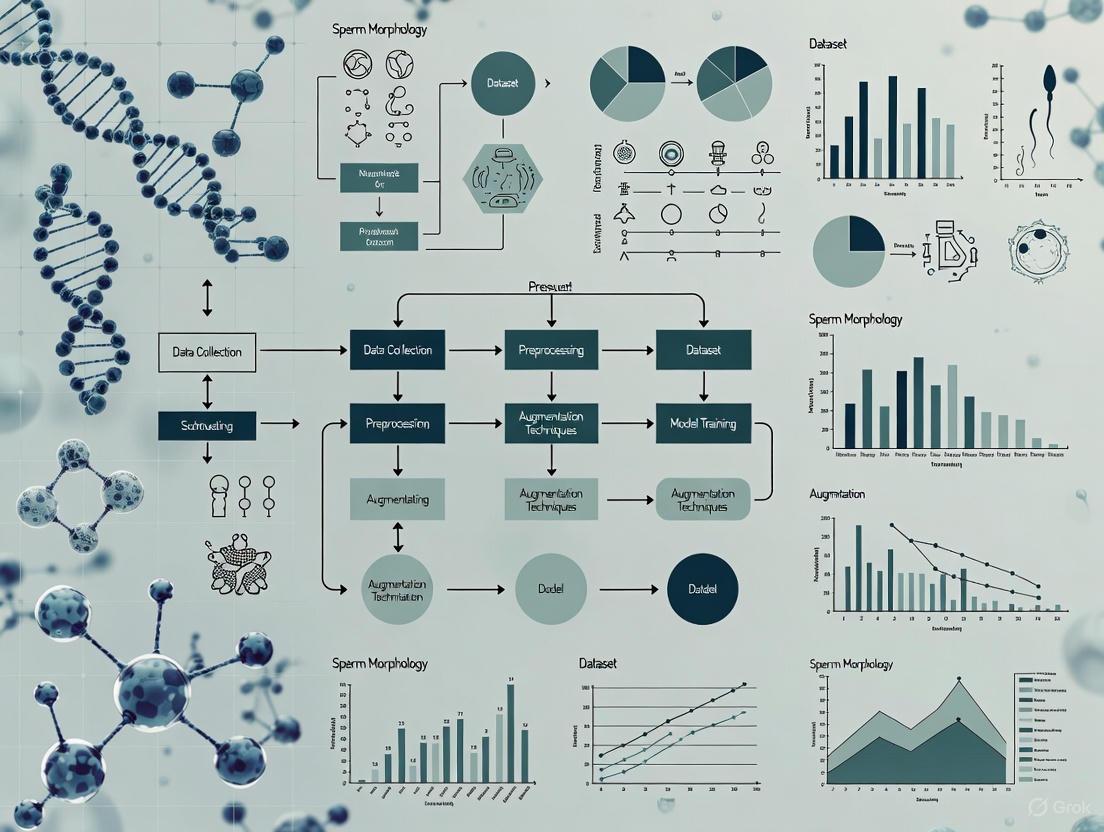
This article provides a comprehensive guide to data augmentation techniques specifically for sperm morphology datasets, a critical frontier in male fertility research.
Data Augmentation for Sperm Morphology Analysis: Techniques to Overcome Dataset Limitations and Enhance AI Performance
Abstract
This article provides a comprehensive guide to data augmentation techniques specifically for sperm morphology datasets, a critical frontier in male fertility research. Aimed at researchers and scientists, it details how these methods overcome the significant challenge of limited, high-quality annotated data needed to train robust deep learning models. The content explores foundational concepts like the SMD/MSS and VISEM-Tracking datasets, outlines practical implementation methodologies from basic image transformations to advanced deep feature engineering, and addresses common troubleshooting and optimization strategies. Finally, it covers rigorous validation frameworks and performance comparisons, synthesizing how effective data augmentation standardizes and automates sperm morphology assessment, thereby accelerating diagnostic innovation in reproductive medicine.
The Critical Need for Data Augmentation in Sperm Morphology Analysis
Sperm morphology analysis (SMA) is a cornerstone of male fertility assessment, providing crucial diagnostic information for predicting natural pregnancy outcomes and informing assisted reproductive technologies (ART) such as in vitro fertilization (IVF) [1]. The accurate classification of sperm into normal and abnormal categories—encompassing defects in the head, midpiece, and tail—is essential for clinical diagnosis [2] [1]. However, this field faces a significant and fundamental challenge: a severe scarcity of standardized, high-quality image datasets. This data bottleneck impedes the development and reliability of automated analysis systems based on deep learning (DL), which require large, diverse, and meticulously curated datasets to learn from and generalize effectively [1].
The inherent complexity of sperm morphology, coupled with the subjective nature of manual assessment, creates a pressing need for robust, AI-driven solutions. This application note details the specific challenges in sperm image data curation, quantifies the current landscape of available datasets, provides experimental protocols for dataset creation, and outlines data augmentation strategies to overcome the data scarcity bottleneck within the broader context of sperm morphology research.
The Core Challenges in Curating Sperm Image Datasets
Building high-quality sperm image datasets is a multi-faceted challenge. The primary obstacles researchers encounter are summarized in the table below.
Table 1: Key Challenges in Curating High-Quality Sperm Image Datasets
| Challenge Category | Specific Limitations | Impact on Model Development |
|---|---|---|
| Data Acquisition & Annotation | High inter-expert variability in manual classification [2]; Difficulty annotating overlapping sperm or partial structures [1]; Complexity of labeling multiple defect types (head, midpiece, tail) [1] | Introduces label noise and inconsistency, reducing model accuracy and reliability. |
| Dataset Scale & Diversity | Limited number of images in most public datasets [1]; Lack of diverse representation of all morphological defect classes [2]; Insufficient demographic and pathological diversity [3] | Leads to models that overfit to limited training data and perform poorly on new, unseen clinical data. |
| Technical & Standardization Hurdles | Lack of standardized protocols for slide preparation, staining, and imaging [1]; Variable image quality due to microscope settings and staining quality [2]; Class imbalance, with rare abnormalities being underrepresented [3] | Hinders model generalization and makes it difficult to compare algorithms across different studies and clinical settings. |
Current Landscape of Sperm Image Datasets
Several research groups have made efforts to create and publish sperm image datasets to fuel progress in the field. The table below provides a quantitative overview of some key datasets, highlighting their primary characteristics and limitations.
Table 2: Overview of Available Sperm Image Datasets for Morphology Analysis
| Dataset Name | Key Characteristics | Notable Limitations |
|---|---|---|
| SMD/MSS [2] | 1,000 original images extended to 6,035 via data augmentation; Annotated per modified David classification (12 defect classes). | Initial dataset size is small; Augmented data may lack realism. |
| MHSMA [1] | 1,540 cropped sperm images; Focus on features like acrosome, head shape, and vacuoles. | Limited sample size; Low image resolution (128x128 pixels). |
| VISEM-Tracking [4] | 20 videos (29,196 frames) with bounding boxes; Provides motility and kinematic data. | Focused on tracking and motility, not fine-grained morphology classification. |
| SVIA [1] | 125,000 annotated instances for detection; 26,000 segmentation masks; 125,880 images for classification. | A relatively new dataset; Broader community validation results are pending. |
| HSMA-DS [4] | 1,457 sperm images; Annotated for vacuole, tail, midpiece, and head abnormality. | Limited number of images; May not cover the full spectrum of morphological diversity. |
The Impact of Data Scarcity on Model Performance
The limitations of existing datasets have a direct and measurable impact on the performance of machine learning models. Conventional machine learning algorithms, which rely on handcrafted features (e.g., shape descriptors, texture analysis), have demonstrated limited performance, with one study reporting classification accuracy for non-normal sperm heads as low as 49% [1]. While deep learning models offer a promising alternative by automatically learning features, their performance is critically dependent on the data they are trained on. Models trained on small or imbalanced datasets often fail to generalize, exhibiting overfitting where they perform well on the training data but poorly on new clinical data [1] [5]. Furthermore, the subjectivity of manual annotation introduces "label noise," where the same sperm may be classified differently by multiple experts. One analysis found that achieving total agreement among three experts was challenging, with varying levels of agreement (no agreement, partial agreement, total agreement) across different morphological classes [2]. This inconsistency confuses the model during training, limiting its ultimate accuracy and clinical utility.
Experimental Protocols for Building High-Quality Datasets
To address the data bottleneck, researchers must adopt rigorous and standardized protocols for dataset creation. The following workflow, developed from recent studies, provides a detailed methodology.
Diagram 1: Sperm Image Dataset Creation Workflow
Protocol: Sample Preparation and Image Acquisition
Objective: To consistently acquire high-resolution, standardized images of individual spermatozoa.
Materials:
- Fresh semen samples (with concentration ≥5 million/mL) [2].
- RAL Diagnostics staining kit or similar for contrast [2].
- Phase-contrast optical microscope with a 100x oil immersion objective [2] [4].
- Microscope-mounted digital camera (e.g., IDS UI-2210C) [4].
- Computer-Assisted Semen Analysis (CASA) system for sequential image capture (optional but recommended) [2].
Methodology:
- Sample Preparation: Prepare smears from fresh semen samples according to WHO guidelines and stain them using a standardized protocol to enhance cellular contrast [2].
- Microscope Setup: Place the prepared smear on a heated microscope stage maintained at 37°C to mimic physiological conditions [4]. Use bright-field or phase-contrast mode with a 100x oil immersion objective [2].
- Image Capture: Systematically capture images, ensuring each frame contains a single, whole spermatozoon (head, midpiece, and tail). Avoid samples with high concentrations (>200 million/mL) to prevent image overlap [2]. Save images in a lossless format.
Protocol: Multi-Expert Annotation and Ground Truth Establishment
Objective: To create a reliable ground truth dataset by mitigating individual annotator subjectivity.
Materials:
- Collected sperm images.
- Annotation software (e.g., LabelBox, VGG Image Annotator) [4] [6].
- A panel of at least three experienced embryologists or andrologists [2].
Methodology:
- Develop Annotation Guidelines: Create detailed guidelines based on a recognized classification system (e.g., modified David classification or WHO strict criteria). Include clear definitions and visual examples of each defect class (e.g., tapered head, coiled tail, bent midpiece) [2] [6].
- Independent Annotation: Each expert independently classifies every spermatozoon in the dataset using the established guidelines. The annotation should cover all parts: head, midpiece, and tail [2].
- Compile Ground Truth File: For each image, create a record containing the image filename, the classifications from all experts, and metadata such as sperm head dimensions [2].
- Analyze Inter-Expert Agreement: Calculate agreement statistics (e.g., Fleiss' Kappa). Resolve discrepancies through a consensus meeting among experts to establish a final, high-confidence label for each sperm image [2].
Protocol: Data Pre-processing and Augmentation
Objective: To enhance dataset quality, balance morphological classes, and increase effective size for robust deep learning.
Materials:
- Python programming environment (version 3.8 or higher) [2].
- Image processing libraries (OpenCV, Pillow).
- Deep learning frameworks (TensorFlow, PyTorch).
Methodology:
- Image Pre-processing:
- Data Augmentation: Apply a hybrid of transformation techniques to the existing images to artificially expand the dataset, focusing particularly on underrepresented classes [7] [5].
- Affine Transformations: Apply rotations (e.g., ±15°), flips (horizontal/vertical), slight zooms, and translations [7].
- Pixel-Level Transformations: Adjust brightness, contrast, and saturation to simulate different staining and lighting conditions [7].
- Advanced Techniques: For complex scenarios, use Generative Adversarial Networks (GANs) to generate high-quality synthetic sperm images that are plausible but artificial, further increasing diversity [7] [5].
The Scientist's Toolkit: Research Reagent Solutions
Table 3: Essential Materials and Reagents for Sperm Morphology Dataset Research
| Item Name | Function/Application | Specification/Example |
|---|---|---|
| RAL Diagnostics Stain | Stains sperm cells on a smear to improve contrast and visibility of morphological structures under a microscope. | Standardized staining kit for semen smears [2]. |
| Phase-Contrast Microscope | Enables high-resolution imaging of unstained or live sperm cells by enhancing contrast of transparent specimens. | Olympus CX31 microscope with 100x oil immersion objective [4]. |
| CASA System | Automates the capture and initial morphometric analysis of sperm images (e.g., head length, tail length). | MMC CASA system for sequential image acquisition [2]. |
| Annotation Software Platform | Provides a user-friendly interface for experts to efficiently label and classify sperm images. | LabelBox, VGG Image Annotator (VIA) [4] [6]. |
| Deep Learning Framework | Provides the programming environment to build, train, and test convolutional neural network (CNN) models. | Python 3.8 with TensorFlow/PyTorch libraries [2]. |
Data Augmentation Pathways to Overcome Data Scarcity
Given the extreme difficulty of collecting vast clinical datasets, data augmentation is not just beneficial but essential. The following diagram illustrates a strategic hybrid augmentation pathway.
Diagram 2: Hybrid Data Augmentation Strategy
This hybrid approach, which combines simpler transformations with more complex generative models, has been proven highly effective. One study on medical image classification found that a hybrid data augmentation method achieved a top accuracy of 99.54%, significantly outperforming any single technique used in isolation [5]. In sperm morphology research, applying these techniques allowed one group to expand their dataset from 1,000 to 6,035 images, which was crucial for training a CNN model that achieved accuracy results comparable to expert judgment [2]. Affine and pixel-level transformations often provide the best trade-off between performance gains and implementation complexity [7].
The scarcity of standardized, high-quality sperm image datasets remains a significant bottleneck in the development of reliable AI tools for male infertility diagnosis. This challenge is rooted in the complexities of data acquisition, annotation, and the natural class imbalance of morphological defects. However, by adopting systematic and rigorous protocols for dataset creation—encompassing standardized sample preparation, multi-expert annotation, and comprehensive quality assurance—researchers can build a solid foundation. Furthermore, strategically employing a hybrid of data augmentation techniques is a powerful and necessary method to amplify the value of collected data, balance classes, and ultimately train robust deep learning models. Addressing this data bottleneck is paramount for translating AI research into clinical tools that can offer objective, rapid, and accurate sperm morphology analysis to benefit patients worldwide.
The integration of artificial intelligence (AI) into reproductive medicine is transforming the assessment of sperm morphology, a critical parameter in male fertility diagnostics. Traditional manual analysis is inherently subjective, time-consuming, and prone to significant inter-observer variability, with reported disagreement rates as high as 40% among experts [8]. This lack of standardization hampers diagnostic consistency and reproducibility across laboratories.
Deep learning models, particularly Convolutional Neural Networks (CNNs), offer a pathway to automated, objective, and high-throughput analysis. However, the development of robust, generalizable models is critically dependent on access to large, high-quality, and well-annotated public datasets [9]. This application note provides a detailed overview of key public datasets—SMD/MSS, VISEM-Tracking, SVIA, and HuSHeM—framed within the essential context of data augmentation techniques. We summarize their core attributes, present standardized experimental protocols for their use, and visualize the typical AI workflow to serve as a resource for researchers and drug development professionals in the field of reproductive biology.
The following section details two of the key datasets, SMD/MSS and HuSHeM. It is important to note that within the provided search results, specific quantitative details for the VISEM-Tracking and SVIA datasets were not available. Therefore, the comparative analysis focuses on the datasets for which complete information could be sourced.
Table 1: Key Characteristics of SMD/MSS and HuSHeM Datasets
| Characteristic | SMD/MSS | HuSHeM |
|---|---|---|
| Primary Focus | Morphology Classification | Morphology Classification |
| Initial Image Count | 1,000 [2] | 216 [8] |
| Final Image Count (Post-Augmentation) | 6,035 [2] | Information missing |
| Morphology Classification Scheme | Modified David Classification (12 classes) [2] | WHO-based [8] |
| Key Anomalies Covered | Head (tapered, thin, microcephalous, etc.), Midpiece (cytoplasmic droplet, bent), Tail (coiled, short, multiple) [2] | Head shape, acrosome integrity, neck structure, tail configuration [8] |
| Annotation Process | Independent classification by three experts; detailed ground truth file [2] | Expert annotations [8] |
| Reported Model Performance | Accuracy: 55% - 92% [2] | Accuracy: 96.77% with advanced DL models [8] |
Experimental Protocols for Dataset Utilization
Protocol 1: SMD/MSS Dataset Construction and Preprocessing
The SMD/MSS dataset was developed to address the need for a dataset based on the modified David classification, which is widely used in laboratories globally [2].
- Sample Preparation: Semen samples were obtained from 37 patients. Smears were prepared following WHO manual guidelines and stained with a RAL Diagnostics staining kit. Samples with a concentration of at least 5 million/mL were included, while those exceeding 200 million/mL were excluded to prevent image overlap [2].
- Data Acquisition: Individual spermatozoa images were acquired using an MMC CASA system, which consists of an optical microscope with a digital camera. Images were captured in bright-field mode using an oil immersion 100x objective [2].
- Expert Annotation and Ground Truth: Each sperm image was independently classified by three experienced experts according to the 12 classes of the modified David classification. A comprehensive ground truth file was compiled for each image, containing the image name, classifications from all three experts, and morphometric dimensions of the sperm head and tail [2].
- Data Augmentation: To overcome the limitations of a small original dataset and heterogeneous class representation, data augmentation techniques were employed. The initial 1,000 images were expanded to 6,035 images, creating a more balanced and powerful dataset for training deep learning models [2].
Protocol 2: A Deep Learning Workflow for Sperm Morphology Classification
This protocol outlines a state-of-the-art methodology for building a high-accuracy classifier, as demonstrated on datasets like HuSHeM [8].
Model Architecture Selection and Enhancement:
- Backbone Network: Select a deep CNN architecture such as ResNet50 as a feature extractor [8].
- Integration of Attention Mechanisms: Enhance the backbone network by integrating a Convolutional Block Attention Module (CBAM). This lightweight module sequentially applies channel and spatial attention to feature maps, forcing the model to focus on diagnostically relevant regions of the sperm (e.g., head shape, acrosome, tail) while suppressing irrelevant background noise [8].
Deep Feature Engineering (DFE) Pipeline:
- Feature Extraction: Extract high-dimensional feature maps from multiple layers of the CBAM-enhanced network, including the CBAM layer itself, Global Average Pooling (GAP), and Global Max Pooling (GMP) layers [8].
- Feature Selection: Apply feature selection algorithms such as Principal Component Analysis (PCA), Chi-square tests, or Random Forest importance to the extracted feature set. This step reduces dimensionality and noise, improving model performance [8].
- Classification: Instead of using the CNN's final classification layer, train a traditional machine learning classifier like a Support Vector Machine (SVM) with an RBF kernel on the selected feature set. This hybrid approach has been shown to yield higher accuracy than end-to-end CNN training [8].
Model Training and Evaluation:
- Rigorous Validation: Employ 5-fold cross-validation to ensure robust performance estimation and avoid overfitting [8].
- Statistical Testing: Use statistical tests like McNemar's test to validate that performance improvements are significant [8].
- Model Interpretation: Utilize visualization techniques like Grad-CAM to generate attention maps, providing clinicians with interpretable insights into the model's decision-making process [8].
Figure 1: AI-Based Sperm Morphology Analysis Workflow. This diagram outlines the standard pipeline for automated sperm classification, from raw image input to final diagnosis.
The Scientist's Toolkit: Essential Research Reagents and Materials
Table 2: Key Reagents and Solutions for Sperm Morphology Analysis
| Item | Function/Application | Example/Specification |
|---|---|---|
| RAL Diagnostics Staining Kit | Staining of sperm smears for clear visualization of morphological details [2]. | Used in the preparation of the SMD/MSS dataset [2]. |
| Optixcell Extender | Semen extender used for diluting and preserving bull sperm samples during analysis [10]. | Used in bovine sperm morphology studies [10]. |
| Trumorph System | A dye-free system for sperm fixation using controlled pressure and temperature, preserving native morphology [10]. | Employed for fixation in veterinary sperm analysis [10]. |
| MMC CASA System | Computer-Assisted Semen Analysis system for automated image acquisition and initial morphometric analysis [2]. | Used for acquiring images for the SMD/MSS dataset [2]. |
| Optika B-383Phi Microscope | Optical microscope for high-resolution image capture of spermatozoa [10]. | Used with negative phase contrast objectives for bovine sperm imaging [10]. |
The move towards standardized, AI-driven sperm morphology analysis represents a significant advancement in reproductive medicine. Public datasets like SMD/MSS and HuSHeM are foundational resources that enable the development of robust deep-learning models. The application of structured data augmentation techniques is critical to mitigating the challenges of limited data and class imbalance, thereby enhancing model generalizability.
The experimental protocols and the integrated deep feature engineering pipeline outlined in this note provide a roadmap for researchers to build accurate, interpretable, and clinically valuable tools. As the field evolves, future work should focus on the creation of even larger, multi-center datasets, the development of standardized metadata reporting formats [11] [12], and the rigorous clinical validation of these systems to ensure their reliability in diagnostic settings. The ultimate goal is to provide consistent, objective, and efficient fertility assessments to improve patient care worldwide.
The accurate assessment of sperm morphology is a cornerstone of male fertility diagnosis and a critical parameter in assisted reproductive technology (ART) outcomes. However, the creation of reliable, high-quality datasets for research is fundamentally hampered by two inherent challenges: significant expert subjectivity in manual annotation and the profound structural complexity of the sperm cell itself. Manual sperm morphology assessment is recognized as a challenging parameter to standardize due to its subjective nature, which is often reliant on the operator's expertise [13] [2]. Even highly trained experts display substantial diagnostic disagreement, with reported kappa values as low as 0.05–0.15, highlighting inconsistent standards across laboratories [8]. This subjectivity directly impacts the "ground truth" labels essential for training robust machine learning models.
Compounding the issue of subjectivity is the intricate structural nature of the spermatozoon. A morphologically normal spermatozoon must exhibit an oval-shaped head (length: 4.0–5.5 µm, width: 2.5–3.5 µm), an intact acrosome covering 40–70% of the head, a regular midpiece about the same length as the head, and a single, uniform tail approximately 45 µm long [14] [8]. The process of spermiogenesis that generates this highly specialized cell is complex and inefficient in humans, producing a high percentage of spermatozoa with various abnormal and imperfect features [15]. Annotating this continuum of biometrics and the multitude of potential defects in the head, midpiece, and tail requires immense precision, a task that is complicated by limitations in imaging technology and the minute scale of the structures involved [15] [16]. This document details these challenges and provides standardized protocols to mitigate them, thereby enhancing the quality of datasets for computational research.
Quantitative Analysis of Expert Subjectivity and Annotation Complexity
The variability in expert annotation can be systematically quantified, providing insights into the magnitude of the challenge and the factors that influence consensus.
Table 1: Quantifying Expert Annotation Subjectivity
| Metric of Subjectivity | Reported Value or Range | Context/Description |
|---|---|---|
| Inter-Expert Agreement (Kappa) | 0.05 - 0.15 [8] | Even among trained technicians, signifying slight to fair agreement only. |
| Expert Consensus on Normal/Abnormal | 73% [17] | Percentage of sheep sperm images where experts agreed on a binary normal/abnormal classification. |
| Deep Learning Model Accuracy Range | 55% - 92% [13] [2] | Range of accuracy achieved by a CNN model, reflecting inconsistency in the training labels provided by experts. |
| Untrained Novice Accuracy (2-category) | 81.0% ± 2.5% [17] | Initial accuracy of novices in a binary classification system (normal vs. abnormal). |
| Untrained Novice Accuracy (25-category) | 53% ± 3.69% [17] | Initial accuracy for a complex 25-category system, showing a significant drop with increased complexity. |
The complexity of the classification system itself is a major driver of annotation variability. Research has demonstrated that the number of categories used has a direct and negative correlation with annotation accuracy.
Table 2: Impact of Classification System Complexity on Annotation Accuracy
| Classification System | Final Trained User Accuracy | Key Annotated Defects |
|---|---|---|
| 2-Category System | 98% ± 0.43% [17] | Normal, Abnormal. |
| 5-Category System | 97% ± 0.58% [17] | Normal, Head defect, Midpiece defect, Tail defect, Cytoplasmic droplet. |
| 8-Category System | 96% ± 0.81% [17] | Normal, Cytoplasmic droplet; Midpiece defect; Loose heads & abnormal tails; Pyriform head; Knobbed acrosomes; Vacuoles & teratoids; Swollen acrosomes. |
| 25-Category System | 90% ± 1.38% [17] | Normal; all other defects defined individually with high specificity. |
Experimental Protocols for Establishing Annotation Ground Truth
To overcome the challenge of expert subjectivity, a rigorous, multi-stage protocol for establishing a reliable ground truth dataset is essential. The following methodology, inspired by machine learning data validation principles, provides a standardized approach.
Protocol: Expert Consensus for Ground Truth Labeling
Objective: To create a standardized and high-quality annotated sperm morphology dataset by mitigating individual expert bias through a structured consensus process.
Materials and Reagents:
- Sperm Smears: Prepared from semen samples according to WHO guidelines (liquefaction at 37°C for 30 minutes; use of proteolytic enzymes like α-chymotrypsin for viscous samples) [14].
- Staining Kit: RAL Diagnostics staining kit or Papanicolaou/Diff-Quik stain [14] [2].
- Imaging System: Microscope with 100x oil immersion objective and digital camera (e.g., MMC CASA system) [2].
- Software: Image annotation and data management software (e.g., spreadsheet for collating expert classifications).
Procedure:
- Sample Preparation & Image Acquisition:
- Prepare thin smears on clean, frosted slides using 10 µL of well-mixed semen. Air-dry completely [14].
- Stain the smears using a standardized protocol (e.g., for Diff-Quik: immerse in fixative 5x, then solution I for 10s, then solution II for 10s, rinse in water, and air-dry) [14].
- Capture images of individual spermatozoa using a bright-field microscope with a 100x objective. Ensure each image contains a single, whole spermatozoon [2].
Independent Multi-Expert Classification:
- Engage a minimum of three independent experts, each with extensive experience in semen analysis.
- Provide each expert with the same set of images and a predefined classification guide (e.g., Modified David classification with 12 defect classes: tapered, thin, microcephalous, macrocephalous, multiple heads, abnormal post-acrosomal region, abnormal acrosome, cytoplasmic droplet, bent midpiece, coiled tail, short tail, multiple tails) [2].
- Each expert classifies every spermatozoon independently, annotating defects for the head, midpiece, and tail without consultation.
Data Collation and Consensus Analysis:
- Compile all expert classifications into a single ground truth file.
- Analyze the level of agreement for each sperm image. The scenarios are:
- Total Agreement (TA): All three experts assign identical labels.
- Partial Agreement (PA): Two out of three experts agree on the label.
- No Agreement (NA): All three experts provide different labels [2].
- Use statistical software (e.g., IBM SPSS) to calculate inter-expert agreement using Fisher's exact test (p < 0.05 considered significant) [2].
Final Ground Truth Assignment:
- For images with TA, assign the unanimously agreed label as the ground truth.
- For images with PA, assign the label agreed upon by the two experts. Flag these images in the dataset for potential review during model training.
- For images with NA, exclude them from the final training dataset or subject them to a final arbitration round by a senior morphologist.
Protocol: Stain-Free Sperm Morphometry with Accuracy Enhancement
Objective: To perform precise, non-invasive morphometric analysis of sperm head, midpiece, and tail, minimizing errors induced by staining and low-resolution images.
Materials and Reagents:
- Non-Stained Sperm Sample.
- Fixation System: Trumorph system or equivalent for dye-free fixation using controlled pressure and temperature [18].
- Microscopy System: Microscope with 20x to 40x phase-contrast objectives (e.g., Optika B-383Phi) [16] [18].
- Computational Framework: Python-based environment with libraries for image processing (OpenCV) and deep learning (PyTorch/TensorFlow).
Procedure:
- Sample Preparation and Image Capture:
- Dilute the semen sample to an appropriate concentration (e.g., 17.5–27.5 x 10⁶/mL) [18].
- Place 10 µL on a slide, cover with a coverslip, and fix using the Trumorph system (e.g., 60°C, 6 kp pressure) to immobilize sperm without staining [18].
- Capture images under 20x or 40x magnification. The lower magnification prevents sperm from swimming away but reduces resolution, necessitating the following enhancement steps [16].
Multi-Target Instance Parsing:
- Employ a multi-scale part parsing network that integrates both semantic segmentation and instance segmentation.
- The instance segmentation branch creates masks to accurately localize and separate individual sperm cells from each other and the background.
- The semantic segmentation branch performs fine-grained pixel-wise classification to delineate the head, midpiece, and tail for each sperm [16].
- Fuse the outputs of both branches to achieve instance-level parsing, where every pixel is assigned to a specific part of a specific sperm.
Morphological Measurement and Accuracy Enhancement:
- Extract raw morphological parameters (head length/width, midpiece and tail length) from the parsed segments.
- Apply a measurement accuracy enhancement strategy to correct for blurring and boundary errors in low-resolution images:
- Outlier Exclusion: Use the Interquartile Range (IQR) method to filter out biologically implausible measurements.
- Data Smoothing: Apply Gaussian filtering to smooth the measured parameter data and reduce noise.
- Robust Correction: Extract the maximum morphological features from the smoothed data to counteract the underestimation caused by blurred contours [16].
Visualization of Annotation Workflows and Challenges
The following diagram illustrates the multi-expert annotation workflow and the primary sources of subjectivity and complexity.
Diagram 1: Multi-expert annotation workflow and inherent challenges.
The subsequent diagram outlines the stain-free analysis protocol designed to address the challenges of structural complexity.
Diagram 2: Stain-free sperm morphology analysis with accuracy enhancement.
The Scientist's Toolkit: Key Research Reagent Solutions
Table 3: Essential Materials and Reagents for Sperm Morphology Annotation Research
| Item Name | Function/Application | Specific Example / Note |
|---|---|---|
| Diff-Quik Stain | Rapid staining of sperm smears for clear visualization of head, midpiece, and tail structures. | A Romanowsky-type stain; consists of fixative, solution I (eosin), and solution II (methylene blue) [14]. |
| RAL Diagnostics Stain | Staining kit used for sperm morphology assessment according to specific laboratory protocols. | Used in the creation of the SMD/MSS dataset for expert classification [2]. |
| Optixcell Extender | Semen extender used to dilute and preserve semen samples prior to smear preparation and analysis. | Used in bovine sperm morphology studies to maintain sperm viability during processing [18]. |
| Trumorph System | A dye-free fixation system that uses controlled pressure and temperature to immobilize sperm for analysis. | Prevents sperm damage from staining, enabling non-invasive morphology assessment [18]. |
| MMC CASA System | Computer-Assisted Semen Analysis system for automated image acquisition and morphometric analysis. | Used for acquiring images of individual spermatozoa with defined head and tail dimensions [2]. |
| Python with Deep Learning Libraries | Core programming environment for implementing data augmentation, CNN models, and instance parsing networks. | Used with libraries like TensorFlow/PyTorch for developing sperm classification algorithms [2] [16] [8]. |
Application Note: The Challenge of Generalizability in Clinical AI
The deployment of artificial intelligence (AI) in clinical settings represents a frontier in modern medicine, promising enhanced diagnostic accuracy, standardization, and workflow efficiency. However, a significant gap exists between developing high-performing models in research settings and achieving robust, generalizable AI tools that function reliably across diverse clinical environments. This challenge is particularly acute in specialized fields like reproductive medicine, where subjective assessments, such as sperm morphology evaluation, are the standard [13] [2].
A primary obstacle is data scarcity and variability. AI models, particularly deep learning models, require large, diverse datasets to learn effectively and avoid overfitting. In medicine, especially for rare diseases or specific diagnostic tasks like sperm classification, acquiring large datasets is difficult, expensive, and often constrained by patient privacy concerns [19] [20]. Furthermore, models trained on data from a single institution often experience a significant performance drop when validated externally. One study demonstrated that single-institution models for classifying medical procedures showed a mean accuracy of 92.5% on internal data but generalized poorly, with performance dropping by an average of 22.4% on external data [21].
Another critical challenge is dataset shift, where the statistical properties of the data used for training and the data encountered in real-world deployment differ. This can be due to changes in patient populations, medical equipment, clinical protocols, or even public health policies over time. For instance, a COVID-19 risk prediction model built during the first wave of the pandemic saw drastically reduced performance in later waves due to changes in testing policies and virus variants [22]. Therefore, achieving generalizability requires a holistic strategy that addresses not only model architecture but also data acquisition, validation, and continuous monitoring post-deployment.
Protocol for Data Augmentation in Sperm Morphology Analysis
This protocol outlines a detailed methodology for applying data augmentation to create a robust and generalizable deep-learning model for sperm morphology classification, based on established research [13] [2].
Background and Principle
Manual sperm morphology assessment is subjective, time-consuming, and prone to inter-observer variability. Deep learning offers a path to automation and standardization. The Sperm Morphology Dataset/Medical School of Sfax (SMD/MSS) exemplifies the initial data scarcity problem, starting with 1,000 individual sperm images [13] [2]. This protocol uses data augmentation to artificially expand the dataset, introducing variability that helps the model learn invariant features and generalize better to new images from different sources.
Materials and Equipment
- Sperm Image Data: Raw digital images of individual spermatozoa, acquired using a Computer-Assisted Semen Analysis (CASA) system like the MMC CASA system with a bright-field microscope and oil immersion 100x objective [2].
- Computing Hardware: A computer with a GPU (e.g., NVidia Titan RTX) is recommended for accelerated deep learning training [21].
- Software Environment: Python 3.8 or later, with deep learning libraries such as TensorFlow or PyTorch [2] [21].
Experimental Procedure
Step 1: Expert Annotation and Dataset Curation
- Acquire semen samples and prepare smears according to WHO guidelines [2].
- Capture images of individual spermatozoa.
- Establish a ground truth by having each sperm image classified by multiple experts (e.g., three) based on a standardized classification system like the modified David classification, which includes 12 classes of morphological defects (e.g., tapered head, microcephalous, coiled tail) [2].
- Analyze inter-expert agreement. Resolve discrepancies through consensus, and use only images with high agreement (e.g., total or partial agreement among experts) for training to ensure label quality [2].
Step 2: Data Pre-processing
- Clean Images: Identify and handle any corrupt or low-quality images.
- Normalize Images: Resize all images to a consistent dimensions (e.g., 80x80 pixels) and convert to grayscale to reduce computational complexity [2].
- Denoise: Apply techniques to remove noise signals from insufficient lighting or poor staining [2].
Step 3: Data Augmentation Strategy
Apply a series of geometric and photometric transformations to the pre-processed training set images to generate new, synthetic variants. The table below summarizes the key transformations used to expand the SMD/MSS dataset from 1,000 to 6,035 images [2] [20].
Table 1: Data Augmentation Techniques for Sperm Morphology Images
| Augmentation Category | Specific Techniques | Purpose |
|---|---|---|
| Geometric Transformations | Rotation, Translation, Shearing, Horizontal/Vertical Flipping | Makes the model invariant to sperm orientation and position in the image. |
| Photometric Transformations | Adjusting Brightness, Contrast, Gamma, Adding Noise | Makes the model robust to variations in staining intensity and lighting conditions. |
Step 4: Model Training and Evaluation
- Data Partitioning: Randomly split the augmented dataset into a training set (80%) and a testing set (20%). Further, split the training set to use a portion (e.g., 20%) for validation during training [2].
- Model Selection: Implement a Convolutional Neural Network (CNN) architecture. For enhanced performance, consider a hybrid model like a CNN with a Convolutional Block Attention Module (CBAM) integrated with a ResNet50 backbone, which helps the model focus on salient features like the sperm head and tail [8].
- Training: Train the model on the augmented training set. Use the validation set to tune hyperparameters and monitor for overfitting.
- Evaluation: Evaluate the final model's performance on the held-out test set. Report standard metrics including Accuracy, Precision, Recall, and F1-Score [2] [8]. The expected outcome based on the SMD/MSS study is an accuracy ranging from 55% to 92% across different morphological classes [13] [2].
Roadmap for Clinical Deployment of AI Models
Successfully transitioning an AI model from a research prototype to a clinically deployed tool requires careful planning across three continuous phases: pre-implementation, peri-implementation, and post-implementation [22]. The following workflow visualizes this roadmap, highlighting critical actions and checks at each stage to ensure generalizability and safety.
Pre-Implementation Phase
Before any clinical integration, the model must be rigorously validated.
- Local Performance Validation: Conduct retrospective evaluation using local data from the deployment site to ensure the model generalizes to the target population and equipment [22].
- Bias and Fairness Audit: Evaluate model performance across different demographic groups (e.g., age, ethnicity) to ensure it does not introduce or perpetuate healthcare inequities [22].
- Infrastructure and Stakeholder Mapping: Plan the data flow with the IT team, often using standards like FHIR to connect with Electronic Health Record (EHR) systems. Crucially, align incentives with end-users (clinicians) to ensure adoption, following the "five rights" of clinical decision support: the right information, person, time, channel, and format [22].
Peri-Implementation Phase
This phase involves the initial "go-live" and controlled testing.
- Define Clinical Success Metrics: The measure of success should not be model accuracy alone, but an improvement in clinical outcomes (e.g., reduced time to diagnosis, mortality reduction) compared to the standard of care [22].
- Silent Validation and Pilot Study: Run the model in "silent mode" where it generates predictions without displaying them to clinicians, to verify production data feeds and output stability. Follow this with a pilot study in a small patient subset to assess the user interface, education materials, and workflow integration [22].
Post-Implementation Phase
AI deployment is not a one-time event but requires ongoing maintenance.
- Continuous Monitoring and Surveillance: Proactively monitor for performance degradation caused by dataset shift or changes in clinical practice [22].
- Solution Performance Auditing: Log how clinicians interact with and potentially override the model's recommendations. This feedback is essential for understanding real-world utility and failure modes [22].
- Model Retraining and Decommissioning: Establish a clear protocol for model updating or retirement if it becomes obsolete or harmful [22].
The Scientist's Toolkit: Research Reagent Solutions
Table 2: Essential Materials and Reagents for Developing a Sperm Morphology AI Model
| Item Name | Function / Rationale |
|---|---|
| MMC CASA System | An integrated system (microscope, camera, software) for standardized and sequential acquisition of high-quality digital sperm images, which is crucial for building a consistent dataset [2]. |
| RAL Diagnostics Staining Kit | Provides the reagents for staining sperm smears, creating the contrast necessary for visualizing morphological details under a microscope [2]. |
| SMD/MSS Dataset | A foundational dataset comprising 1,000+ expert-classified sperm images based on the modified David classification. It serves as a benchmark for training and validating new models [13] [2]. |
| Convolutional Neural Network (CNN) | The core deep learning algorithm for image recognition. It automatically learns hierarchical features from pixel data, eliminating the need for manual feature engineering [13] [8]. |
| Convolutional Block Attention Module (CBAM) | An advanced neural network component that can be added to CNNs (e.g., ResNet50). It directs the model's "attention" to the most relevant parts of the sperm image (e.g., head, midpiece), improving accuracy and interpretability [8]. |
| Data Augmentation Pipeline (Geometric/Photometric) | A software pipeline that programmatically applies transformations (rotation, contrast changes, etc.) to existing images. It is a cost-effective method to increase dataset size and diversity, directly combating overfitting and improving model generalizability [2] [20]. |
A Practical Toolkit: Data Augmentation Techniques for Sperm Images
The application of artificial intelligence (AI) for sperm morphology analysis represents a significant advancement in male infertility diagnostics. Deep learning models, particularly Convolutional Neural Networks (CNNs), have demonstrated potential for automating and standardizing the assessment of sperm head, midpiece, and tail defects [1]. However, the robustness of these AI technologies is fundamentally constrained by the need for large, diverse, and accurately annotated datasets [2] [1]. Manual sperm morphology analysis is inherently subjective, time-consuming, and suffers from significant inter-observer variability, making the creation of such datasets challenging [2]. This application note details core image manipulation techniques—rotation, flipping, scaling, and brightness/contrast adjustment—employed as data augmentation strategies to enhance the size and quality of sperm morphology datasets, thereby improving the performance and generalizability of deep learning models in reproductive biology research.
The Role of Data Augmentation in Sperm Morphology Analysis
Sperm morphology analysis (SMA) is a critical yet challenging component of male fertility assessment. The World Health Organization (WHO) recognizes 26 types of abnormal morphology, requiring the analysis of over 200 sperm cells per sample, which leads to a substantial workload and subjective results [1]. AI models offer a solution by automating this process, but their development faces two primary data-related challenges:
- Limited Dataset Size and Diversity: Collecting and annotating a large number of sperm images is costly and time-consuming. Many existing datasets, such as the Sperm Morphology Dataset/Medical School of Sfax (SMD/MSS), start with a limited number of base images (e.g., 1,000) and require augmentation to reach a viable size for model training (e.g., 6,035 images) [2].
- Class Imbalance: Certain morphological defects occur more rarely than others, leading to imbalanced datasets where a model becomes biased toward more frequent classes.
Data augmentation artificially expands the training dataset by creating modified versions of existing images. This practice mitigates overfitting, improves model generalization, and helps balance class distributions [2]. For sperm image analysis, augmentations must be chosen to realistically represent the biological variations and imaging artifacts encountered in clinical practice while preserving the critical morphological features used for classification.
Core Image Manipulation Protocols
This section provides detailed experimental protocols for implementing core image manipulations in the context of augmenting sperm morphology datasets.
Geometric Manipulations: Rotation and Flipping
Geometric transformations are foundational augmentation techniques that introduce viewpoint variance without altering the core morphological features of the sperm cell.
Rationale: Sperm cells can appear in any orientation on a microscope slide. Training a model to be invariant to rotation and reflection is crucial for robust real-world performance. These manipulations are label-preserving, meaning the class of the sperm (e.g., "normal head," "coiled tail") does not change after the transformation.
Experimental Protocol:
- Input: A single sperm image, preferably with a clean background, extracted from a semen smear. The image should be in a standard format (e.g., PNG, TIFF).
- Software Tools: Python libraries such as TensorFlow (
tf.keras.preprocessing.image.random_rotation,tf.image.flip_left_right) or OpenCV (cv2.rotate,cv2.flip) are typically used. - Parameterization:
- Rotation: Apply random rotations within a defined range. A common practice is to use rotations between
-180and+180degrees to cover all possible orientations fully. Theinterpolationparameter should be set toINTERPOLATION_NEARESTorINTERPOLATION_LINEARto handle pixel values. - Flipping: Implement horizontal (
flip_left_right) and vertical (flip_up_down) flipping. Horizontal flipping is more biologically plausible than vertical flipping.
- Rotation: Apply random rotations within a defined range. A common practice is to use rotations between
- Output: A set of new images demonstrating the same sperm cell in various orientations and reflections.
Considerations for Sperm Morphology: These transformations are generally safe for all parts of the sperm (head, midpiece, tail). However, researchers should validate that extreme rotations do not introduce artifacts at the image boundaries that could be misconstrued as morphological defects.
Spatial Manipulation: Scaling
Scaling, or zooming, alters the apparent size of the sperm cell within the image frame.
Rationale: Minor variations in the distance between the microscope objective and the sample can cause sperm cells to appear slightly larger or smaller. Augmentation with scaling makes the model invariant to these minor magnification differences.
Experimental Protocol:
- Input: A single sperm image.
- Software Tools: Python libraries like TensorFlow (
tf.image.resize) or OpenCV (cv2.resize). - Parameterization:
- Determine a scaling factor range. A conservative range of
[0.9, 1.1](i.e., 10% zoom in/out) is often used to avoid excessive distortion or the creation of unrealistic sizes. - Apply the scaling factor to both the height and width of the image.
- After scaling, the image may need to be padded or cropped back to the original dimensions to maintain a consistent input size for the neural network.
- Determine a scaling factor range. A conservative range of
- Output: Images of the same sperm cell at different apparent sizes.
Considerations for Sperm Morphology: Aggressive scaling outside a biologically plausible range (e.g., making a sperm head appear 50% larger) should be avoided, as it could lead the model to misclassify a normal sperm as macrocephalous or microcephalous [2].
Photometric Manipulations: Brightness and Contrast Adjustment
Adjusting brightness and contrast simulates variations in microscope lighting conditions, staining intensity, and sample preparation, which are common challenges in clinical settings [2] [23].
Rationale: Microscopy images can suffer from poor contrast due to uneven illumination or improper staining. Models trained on ideally lit images may fail under suboptimal conditions. Brightness and contrast augmentation enhances model robustness to these technical variabilities.
Experimental Protocol:
- Input: A single sperm image.
- Software Tools: TensorFlow (
tf.image.adjust_brightness,tf.image.adjust_contrast) or custom algorithms based on Histogram Equalization (HE) and Adaptive Histogram Equalization (CLAHE) [23] [24]. - Parameterization:
- Brightness: Add a random delta value to the pixel intensities. The delta is typically sampled from a small range (e.g.,
[-0.2, +0.2]multiplied by the maximum pixel value) to prevent saturation to pure black or white. - Contrast: Multiply the pixel intensities by a random factor. A factor of
1.0leaves the image unchanged, while factors below and above1.0decrease and increase contrast, respectively (e.g., a range of[0.8, 1.5]). - Advanced Methods: For more sophisticated enhancement, implement a two-stage technique involving global HE followed by a local enhancement method to address differences in average local contrast [23]. CLAHE can be particularly effective for improving local contrast without over-amplifying noise [23] [24].
- Brightness: Add a random delta value to the pixel intensities. The delta is typically sampled from a small range (e.g.,
- Output: A set of images of the same sperm cell under various simulated lighting and contrast conditions.
Considerations for Sperm Morphology: The primary risk is the creation of unrealistic artifacts or the obscuring of subtle morphological features. For instance, excessive contrast adjustment might artificially sharpen the boundaries of the sperm head or make a faint vacuole disappear. Augmentation parameters must be carefully tuned to stay within biologically and technically plausible limits.
Quantitative Analysis of Augmentation Impact
The following tables summarize key quantitative data from relevant studies, illustrating the impact of data augmentation on model performance for sperm morphology analysis.
Table 1: Impact of Data Augmentation on Dataset Size and Model Performance
| Study / Dataset | Initial Image Count | Augmented Image Count | Augmentation Techniques Used | Model Performance (Accuracy) | Key Morphological Classes |
|---|---|---|---|---|---|
| SMD/MSS [2] | 1,000 | 6,035 | Data augmentation techniques (specifics not listed) | 55% to 92% | 12 classes (head, midpiece, tail defects) based on modified David classification |
| Deep Learning Review [1] | Varies (e.g., 1,540 in MHSMA) | N/A (discusses general need) | Implicit in DL pipelines | Improved performance and generalization | Head, neck, and tail compartments |
Table 2: Parameter Ranges for Core Image Manipulations in Sperm Analysis
| Image Manipulation | Core Parameters | Recommended Range for Sperm Analysis | Purpose & Rationale |
|---|---|---|---|
| Rotation | Angle | -180 to +180 degrees | Achieve full rotational invariance. |
| Flipping | Axis | Horizontal, Vertical | Introduce reflectional variance. |
| Scaling | Zoom Factor | 0.9 to 1.1 (10% variation) | Simulate minor magnification differences. |
| Brightness | Delta | -0.2 to +0.2 (normalized) | Simulate lighting variations during microscopy. |
| Contrast | Multiplier | 0.8 to 1.5 | Simulate staining differences and contrast settings. |
Research Reagent Solutions
The following table lists key computational "reagents" and resources essential for implementing the described data augmentation protocols.
Table 3: Essential Research Reagents and Computational Tools
| Item Name | Function/Benefit | Application Note |
|---|---|---|
| TensorFlow / Keras | Open-source library providing high-level APIs for implementing data augmentation layers (e.g., RandomRotation, RandomFlip, RandomContrast). |
Enables easy integration of real-time augmentation directly into the model training pipeline. |
| OpenCV | Library optimized for real-time computer vision. Provides core functions for image manipulation (rotation, flipping, scaling, histogram equalization). | Ideal for building custom, high-performance pre-processing and augmentation pipelines. |
| AndroGen [25] | Open-source software for generating synthetic sperm images from different species without relying on real data or generative training. | Complements traditional augmentation; useful when initial real datasets are very small or subject to privacy concerns. |
| SMD/MSS Dataset [2] | A dataset of 1,000 sperm images (extendable to 6,035) classified by experts according to the modified David classification. | Serves as a valuable benchmark for developing and testing augmentation and AI models for sperm morphology. |
| QUAREP-LiMi Guidelines [26] | Global checklists for publishing microscopy images, ensuring data is scientifically legible and reproducible. | Critical for maintaining quality and standardization when sharing augmented datasets and research findings. |
Workflow and Signaling Pathways
The following diagram illustrates the logical workflow for applying core image manipulations to create an augmented sperm morphology dataset for deep learning model training.
Augmentation to Analysis Workflow diagrams the process from a limited original dataset, through a parallel augmentation pipeline applying geometric, spatial, and photometric manipulations, to the creation of a robust dataset used for training a deep learning model capable of automated sperm morphology analysis.
The systematic application of core image manipulations—rotation, flipping, scaling, and brightness/contrast adjustment—is a fundamental and powerful strategy for data augmentation in sperm morphology research. By artificially expanding and diversifying training datasets, these techniques directly address the critical limitations of small sample sizes and class imbalance that often hinder the development of robust AI models. When implemented within biologically plausible parameters, as outlined in the provided protocols, these augmentations enhance model generalizability, leading to more accurate, automated, and standardized sperm morphology analysis systems. This advancement holds significant promise for improving the diagnostic efficiency and consistency of male infertility assessments in clinical practice.
Infertility affects a significant proportion of couples globally, with male factors contributing to approximately half of all cases [2]. The analysis of sperm morphology—the size, shape, and structural characteristics of sperm cells—remains a critical component in male fertility assessment, as abnormal sperm morphology is strongly correlated with reduced fertility rates and poor outcomes in assisted reproductive technologies [2] [8]. Traditional manual sperm morphology assessment, while important, suffers from several limitations: it is time-intensive (requiring 30-45 minutes per sample), highly subjective, and prone to significant inter-observer variability, with studies reporting disagreement rates of up to 40% between expert evaluators [2] [8].
The Sperm Morphology Dataset/Medical School of Sfax (SMD/MSS) was developed to address the critical need for standardized, high-quality data in this field. This dataset emerged from recognition that the robustness of artificial intelligence (AI) technologies for medical image analysis depends primarily on the creation of large and diverse databases [2]. Prior to its development, researchers faced two major challenges: a limited number of available sperm images and heterogeneous representation of different morphological classes, which impeded the development of reliable automated analysis systems [2].
The SMD/MSS Dataset: Original Composition and Acquisition
Sample Collection and Preparation
The original SMD/MSS dataset was constructed through a prospective study conducted at the Laboratory of Reproductive Biology, Medical School of Sfax, Tunisia [2]. Semen samples were obtained from 37 patients after informed consent, with specific inclusion and exclusion criteria to ensure data quality. Samples with a sperm concentration of at least 5 million/mL were included, while those with high concentrations (>200 million/mL) were excluded to prevent image overlap and facilitate capture of whole spermatozoa [2]. Smears were prepared according to World Health Organization (WHO) guidelines and stained with RAL Diagnostics staining kit to enhance morphological visibility [2].
Image Acquisition and Expert Classification
Images were acquired using the MMC CASA (Computer-Assisted Semen Analysis) system, consisting of an optical microscope equipped with a digital camera [2]. The system operated in bright field mode with an oil immersion x100 objective, capturing images of individual spermatozoa that included the head, midpiece, and tail for comprehensive morphological assessment [2].
A rigorous classification process was implemented with three experts from the laboratory, each possessing extensive experience in semen analysis [2]. The classification followed the modified David classification system, which includes 12 classes of morphological defects across three primary regions [2]:
- Head defects (7 classes): Tapered, thin, microcephalous, macrocephalous, multiple heads, abnormal post-acrosomal region, and abnormal acrosome
- Midpiece defects (2 classes): Cytoplasmic droplet and bent
- Tail defects (3 classes): Coiled, short, and multiple tails
Additionally, categories were included for associated anomalies (CN) and normal sperm (NR) [2]. Each spermatozoon was independently classified by all three experts, with results documented in a shared Excel spreadsheet containing the image name, expert classifications, and dimensions of sperm head and tail [2].
Inter-Expert Agreement Analysis
The complexity of sperm morphological classification was quantified through analysis of inter-expert agreement distribution [2]. Three agreement scenarios were identified among the three experts: No Agreement (NA) among experts, Partial Agreement (PA) where 2/3 experts agreed on the same label, and Total Agreement (TA) where 3/3 experts agreed on the same label for all categories [2]. Statistical analysis using IBM SPSS Statistics 23 software with Fisher's exact test revealed significant differences between experts in each morphology class (p < 0.05), highlighting the inherent subjectivity of manual assessment and underscoring the need for automated standardization [2].
Table 1: Original SMD/MSS Dataset Composition Before Augmentation
| Component | Specification |
|---|---|
| Original Image Count | 1,000 images |
| Source | 37 patient samples |
| Acquisition System | MMC CASA system |
| Microscopy | Bright field, oil immersion x100 objective |
| Classification Standard | Modified David classification (12 defect classes) |
| Expert Annotators | 3 independent experts |
| Annotation Method | Independent classification with agreement analysis |
Data Augmentation Methodology and Implementation
Rationale for Data Augmentation in Medical Imaging
Data augmentation has become an essential strategy in medical image analysis to address the perennial challenge of limited dataset sizes [27] [7]. Medical images are often scarce due to multiple factors: insufficient patients for some conditions, privacy concerns restricting data sharing, lack of medical equipment, inability to obtain images meeting desired criteria, and the time-consuming, expertise-dependent nature of medical image annotation [27]. These limitations frequently lead to biased datasets, overfitting of models, and ultimately inaccurate results when deploying deep learning systems in clinical practice [27].
The systematic application of data augmentation techniques enables researchers to expand training datasets artificially, improving model generalization without collecting new samples [7]. This approach is particularly valuable for balancing morphological classes in imbalanced datasets—a common issue in sperm morphology analysis where normal sperm typically outnumber specific defect categories [2] [8]. Data augmentation also promotes learning invariance with respect to transformations of input data that should not affect output, regularizing deep neural networks without requiring architectural modifications to enforce equivariance or invariance [7].
Augmentation Techniques for Sperm Morphology Images
The SMD/MSS dataset expansion employed multiple data augmentation techniques to transform the original 1,000 images into 6,035 enhanced samples [2] [13]. While the specific combination of techniques applied to SMD/MSS is not exhaustively detailed in the available literature, research in medical image augmentation more broadly categorizes these methods into several families:
Table 2: Common Data Augmentation Techniques in Medical Imaging
| Augmentation Category | Specific Techniques | Application Rationale |
|---|---|---|
| Affine Transformations | Rotation, translation, scaling, flipping, shearing | Learn spatial invariance, simulate viewing variations |
| Pixel-level Transformations | Brightness/contrast adjustment, noise addition, blurring, sharpening | Simulate different staining intensities, microscope settings |
| Elastic Deformations | Non-linear warp fields, elastic transformations | Account for biological shape variability |
| Generative Approaches | Generative Adversarial Networks (GANs), synthetic data generation | Create entirely new samples for rare morphological classes |
Based on broader medical imaging literature, the most effective augmentation approaches for classification tasks typically include affine and pixel-level transformations, which achieve the optimal trade-off between performance improvement and implementation complexity [7]. These techniques were likely applied to the SMD/MSS dataset, potentially including rotation, flipping, brightness/contrast adjustments, and noise addition to generate visually distinct but morphologically consistent variations of original sperm images [2].
Implementation Workflow
The implementation of data augmentation for the SMD/MSS dataset followed a structured pipeline within a Python-based deep learning framework [2]. The process involved several methodical stages from original image processing to expanded dataset generation, as visualized in the following workflow:
Diagram 1: Data Augmentation Workflow for SMD/MSS Dataset Expansion
The image pre-processing stage involved critical preparation steps including data cleaning to handle inconsistencies, normalization/standardization of numerical features to a common scale, and image resizing to 80×80×1 grayscale using linear interpolation strategy [2]. This standardization ensured that no particular feature dominated the learning process due to magnitude differences and optimized the images for subsequent deep learning processing [2].
Following augmentation, the expanded dataset was partitioned with 80% allocated for model training and the remaining 20% reserved for testing [2]. From the training subset, an additional 20% was extracted for validation purposes, creating a robust framework for model development and evaluation [2].
Experimental Framework and Deep Learning Architecture
Convolutional Neural Network Design
The expanded SMD/MSS dataset served as the foundation for developing a predictive model for sperm morphological classification based on artificial neural networks [2]. The implemented algorithm utilized a Convolutional Neural Network (CNN) architecture, which has demonstrated remarkable performance in image classification tasks across medical domains [2] [27]. The complete experimental framework encompassed five distinct stages: image pre-processing, database partitioning, data augmentation, program training, and evaluation [2].
The CNN architecture was implemented in Python (version 3.8), leveraging its comprehensive ecosystem of deep learning libraries and tools for medical image analysis [2]. While the specific architectural details (number of layers, filter sizes, etc.) are not explicitly provided in the available literature, the model was designed to effectively process the pre-processed 80×80×1 grayscale sperm images and output classifications across the morphological categories defined by the modified David classification system [2].
Comparative Performance Analysis
The deep learning model trained on the augmented SMD/MSS dataset produced satisfactory results, with accuracy ranging from 55% to 92% across different morphological categories [2] [13]. This performance range reflects the varying complexity of distinguishing between specific abnormality classes, with some morphological features proving more challenging to classify than others.
To contextualize these results, it is valuable to compare the SMD/MSS approach with other recent advances in sperm morphology classification:
Table 3: Performance Comparison of Sperm Morphology Classification Approaches
| Study/Method | Dataset | Architecture | Reported Performance |
|---|---|---|---|
| SMD/MSS Baseline [2] | Original (1,000 images) | CNN | Lower accuracy (specific values not provided) |
| SMD/MSS Augmented [2] | Expanded (6,035 images) | CNN | Accuracy: 55-92% (across classes) |
| CBAM+ResNet50+DFE [8] | SMIDS (3,000 images, 3-class) | ResNet50 + CBAM + Feature Engineering | Accuracy: 96.08 ± 1.2% |
| CBAM+ResNet50+DFE [8] | HuSHeM (216 images, 4-class) | ResNet50 + CBAM + Feature Engineering | Accuracy: 96.77 ± 0.8% |
| Bovine Sperm Analysis [18] | 277 annotated images | YOLOv7 | mAP@50: 0.73, Precision: 0.75, Recall: 0.71 |
The performance variance across studies highlights several important considerations. The CBAM-enhanced ResNet50 with deep feature engineering demonstrated that incorporating attention mechanisms and traditional feature selection methods can significantly boost performance [8]. This approach achieved exceptional results by integrating ResNet50 backbones with Convolutional Block Attention Module (CBAM) attention mechanisms, enabling the network to focus on the most relevant sperm features while suppressing background noise [8].
Notably, the SMD/MSS study's value extends beyond raw accuracy metrics. The development of a comprehensively annotated dataset according to the modified David classification—used by numerous laboratories worldwide—represents a significant contribution to the field, addressing a gap in available resources for this important classification standard [2].
Research Reagents and Computational Tools
Successful implementation of data augmentation and deep learning approaches for sperm morphology analysis requires specific research reagents and computational tools. The following table details essential components used across referenced studies:
Table 4: Essential Research Reagents and Computational Tools for Sperm Morphology Analysis
| Category | Specific Tool/Reagent | Function/Application |
|---|---|---|
| Microscopy Systems | MMC CASA System [2] | Automated sperm image acquisition and analysis |
| Microscopy Systems | Optika B-383Phi Microscope [18] | High-resolution sperm imaging for dataset creation |
| Staining Reagents | RAL Diagnostics Staining Kit [2] | Sperm staining for enhanced morphological visibility |
| Sample Preparation | Optixcell Extender [18] | Semen dilution and preservation for analysis |
| Deep Learning Frameworks | Python 3.8 [2] | Core programming environment for algorithm development |
| Annotation Tools | Roboflow [18] | Image annotation and dataset management platform |
| Object Detection | YOLOv7 Framework [18] | Real-time object detection for sperm localization and classification |
| Attention Mechanisms | CBAM (Convolutional Block Attention Module) [8] | Feature refinement in deep neural networks |
| Feature Engineering | PCA (Principal Component Analysis) [8] | Dimensionality reduction and feature selection |
These tools collectively enable the complete pipeline from sample preparation to automated analysis, forming an essential toolkit for researchers working in computational sperm morphology assessment. The integration of specialized laboratory equipment with advanced computational frameworks highlights the interdisciplinary nature of this research domain.
Discussion and Clinical Implications
Impact of Data Augmentation on Model Performance
The expansion of the SMD/MSS dataset from 1,000 to 6,035 images through data augmentation techniques represents a case study in addressing fundamental challenges in medical AI development. This approach directly counteracts the issues of limited data availability and class imbalance that frequently plague biomedical image analysis projects [27]. The achieved accuracy range of 55-92% demonstrates that while data augmentation significantly improves model performance, certain morphological classes remain challenging to classify accurately, potentially due to subtle visual features or inconsistent expert annotations on specific abnormality types [2].
The relationship between dataset size, augmentation strategies, and model performance can be visualized as follows:
Diagram 2: Impact of Data Augmentation on Model Development Challenges
This case study aligns with broader evidence in medical imaging literature, where data augmentation has demonstrated consistent benefits across all organs, modalities, and tasks [7]. Specifically, affine and pixel-level transformations have been shown to achieve the best trade-off between performance improvement and implementation complexity [7]. The SMD/MSS expansion project provides a focused illustration of these principles within the specific context of sperm morphology analysis.
Clinical Applications and Future Directions
The automation of sperm morphology analysis through deep learning approaches offers several transformative benefits for clinical practice:
Standardization and Objectivity: Automated systems reduce diagnostic variability between laboratories and technicians, addressing the fundamental limitation of manual assessment which exhibits inter-observer disagreement rates as high as 40% [8].
Time Efficiency: Deep learning systems can reduce analysis time from the manual 30-45 minutes per sample to less than one minute, significantly increasing laboratory throughput [8].
Reproducibility: Automated systems provide consistent results across different time points and laboratory settings, enhancing the reliability of fertility assessment and treatment monitoring [2] [8].
Potential for Real-Time Analysis: The computational efficiency of certain architectures suggests potential for real-time analysis during assisted reproductive procedures, potentially guiding clinical decision-making in dynamic contexts [8] [18].
Future research directions should explore more sophisticated augmentation techniques, including generative adversarial networks (GANs) for synthetic sperm image generation [27] [7]. Additionally, the integration of multiple classification standards (WHO, David, Kruger) within unified models could enhance utility across different clinical contexts. The development of explainable AI approaches that provide visual explanations for classification decisions would also strengthen clinical adoption by maintaining transparency in automated assessments [8].
The expansion of the SMD/MSS dataset from 1,000 to 6,035 images through systematic data augmentation represents a significant methodological advancement in the field of computational sperm morphology analysis. This case study demonstrates how carefully designed augmentation strategies can address fundamental challenges of limited data availability and class imbalance in medical image analysis. The resulting dataset has enabled the development of deep learning models with promising performance (55-92% accuracy across morphological classes), providing a foundation for automated, standardized sperm morphology assessment.
This work underscores the critical importance of high-quality, comprehensively annotated datasets in advancing medical AI applications. By making the SMD/MSS dataset available to the research community, this project contributes to the broader effort to develop reliable, automated tools for male fertility assessment that can improve diagnostic consistency, reduce analysis time, and ultimately enhance patient care in reproductive medicine. The integration of data augmentation methodologies within deep learning frameworks for sperm morphology analysis represents an important step toward addressing the significant public health challenge of male infertility through technological innovation.
The analysis of sperm morphology is a cornerstone of male fertility assessment, yet traditional methods are plagued by subjectivity, inconsistency, and an inability to use the analyzed sperm for subsequent assisted reproductive technologies (ART) due to staining and fixation requirements [28] [1]. These limitations create a pressing need for automated, objective, and non-destructive evaluation techniques. Deep learning, particularly Convolutional Neural Networks (CNNs), offers a powerful solution by enabling the automated extraction of features and classification of sperm cells from images. However, the development of robust CNN models is critically dependent on large, high-quality, and diverse datasets [29] [1]. The field of sperm morphology analysis currently suffers from a lack of such standardized datasets, which are often characterized by low resolution, limited sample sizes, and insufficient categorical representation of abnormal morphologies [28] [1]. This application note details a comprehensive methodology that integrates a tailored data augmentation pipeline with a CNN architecture to overcome these data limitations, thereby enhancing the accuracy, generalizability, and clinical applicability of AI-driven sperm morphology analysis for researchers and drug development professionals.
The following tables summarize key quantitative findings from the reviewed literature, highlighting the performance of deep learning models and the impact of data enhancement techniques.
Table 1: Performance Metrics of Deep Learning Models in Biomedical Image Analysis
| Model/Application | Accuracy | Precision | Recall/Sensitivity | Key Findings |
|---|---|---|---|---|
| In-house AI (ResNet50) for Sperm Morphology [28] | 93% | 95% (Abnormal), 91% (Normal) | 91% (Abnormal), 95% (Normal) | Strong correlation with CASA (r=0.88) and CSA (r=0.76). Processes ~25,000 images in 139.7 seconds. |
| GONF (CNN with mRMR) for Cancer Classification [30] | 97% (TCGA), 95% (AHBA) | Not Specified | Not Specified | Integrated gene selection with CNN, reducing false positives and negatives. |
| VGG-16 with Data Enhancement for Colorectal Cancer [29] | 86% | Improved F1-score | Improved Recall (Cancer class) | Data augmentation, outlier handling, and class balancing significantly improved model generalizability and recall. |
Table 2: Impact of Data Enhancement Techniques on Model Performance
| Technique | Application | Key Outcome Metrics |
|---|---|---|
| Outlier Handling (K-means) [29] | Colorectal Cancer Classification | Improved data quality and model robustness. |
| Data Augmentation | Colorectal Cancer Classification [29] | Increased dataset diversity, confirmed via Pearson correlation; enhanced accuracy and generalizability. |
| Class Balancing | Colorectal Cancer Classification [29] | Addressed class imbalance, leading to better performance on minority classes. |
| Deep Learning-Optimized CLAHE [31] | Suzhou Garden Images | SSIM increased by 24.69%, PSNR by 24.36%, LOE reduced by 36.62%. |
Experimental Protocols
Protocol 1: Development of an AI Model for Unstained Live Sperm Morphology Assessment
This protocol is adapted from a study that developed an in-house AI model to assess sperm morphology without staining, allowing for the subsequent use of the sperm in ART [28].
1. Sample Collection and Preparation:
- Participants: Enroll healthy male volunteers (e.g., aged 18-40) with 2-7 days of sexual abstinence. Exclude samples with improper collection, high viscosity, or volume <1.4 mL.
- Liquefaction: Allow semen samples to liquefy at 37°C within 30 minutes of collection.
- Aliquoting: Divide each liquefied sample into three aliquots for parallel analysis by the AI model, Computer-Aided Semen Analysis (CASA), and Conventional Semen Analysis (CSA).
2. Image Acquisition and Dataset Curation:
- Slide Preparation: Dispense a 6 µL droplet of semen onto a standard two-chamber slide with a 20 µm depth.
- Imaging: Capture images using a confocal laser scanning microscope at 40x magnification in confocal mode (Z-stack). Set a Z-stack interval of 0.5 µm over a 2 µm range to ensure high-resolution, well-focused images.
- Annotation: Manually annotate sperm images using a tool (e.g., LabelImg). Experienced embryologists and researchers should draw bounding boxes around each sperm and categorize them based on strict WHO criteria [28]. Normal sperm are defined by a smooth oval head, specific length-to-width ratio (1.5-2), no vacuoles, and a slender, regular neck and tail. Establish inter-observer reliability (e.g., correlation coefficient of 0.95 for normal morphology).
3. AI Model Training and Validation:
- Model Selection: Employ a transfer learning approach with a pre-trained architecture such as ResNet50.
- Dataset Splitting: Divide the annotated dataset (e.g., 12,683 images) into training, validation, and test sets.
- Training: Train the model to minimize the difference between predicted and actual labels. Use a suitable batch size (e.g., 900) and number of epochs (e.g., 150).
- Performance Evaluation: Validate the model on the unseen test set. Metrics should include accuracy, precision, recall, and processing time per image.
4. Comparative Analysis:
- Compare the performance of the AI model against CASA and CSA by assessing the correlation of normal morphology rates between the methods.
Protocol 2: A Data Augmentation Pipeline for Enhanced CNN Generalizability
This protocol outlines a data enhancement sequence proven to improve CNN accuracy for medical image classification, as demonstrated in colorectal cancer detection [29].
1. Outlier Handling:
- Aim: To identify and manage anomalous images that could degrade model performance.
- Method: Apply K-means clustering to the image dataset. Images that cluster far from the majority are identified as outliers and can be removed or separately processed to maintain dataset integrity.
2. Data Augmentation:
- Aim: To artificially increase the size and diversity of the training dataset, improving model robustness.
- Methods: Apply a series of transformations to the existing images. These can include:
- Geometric transformations: Rotation, flipping, scaling, and shearing.
- Color space transformations: Adjusting brightness, contrast, and saturation.
- Noise injection.
- Validation of Augmentation: Use Pearson correlation analysis to ensure that the augmented images retain a strong statistical relationship with the original dataset, confirming that meaningful variations have been introduced without distorting core features.
3. Class Balancing:
- Aim: To address imbalanced datasets where one class (e.g., "normal") has significantly more samples than others (e.g., "abnormal").
- Method: Use techniques such as oversampling the minority class or undersampling the majority class to ensure the model does not become biased toward the most frequent class.
4. Model Training and Evaluation:
- Model Training: Train the CNN model (e.g., VGG-16) on the initial dataset and then on the enhanced dataset.
- Performance Comparison: Evaluate and compare the accuracy, F1-score, and recall (particularly for underrepresented classes) of both models on a separate test set. The model trained on the enhanced dataset is expected to show superior generalizability and classification performance [29].
Workflow Visualization
The following diagram, generated with Graphviz, illustrates the integrated deep learning and data augmentation pipeline for sperm morphology analysis.
AI Sperm Analysis Pipeline
The Scientist's Toolkit: Research Reagent Solutions
Table 3: Essential Materials and Reagents for AI-Based Sperm Morphology Analysis
| Item | Function/Application | Specification/Note |
|---|---|---|
| Confocal Laser Scanning Microscope | High-resolution imaging of unstained, live sperm. | Enables Z-stack imaging at low magnification (e.g., 40x) for detailed subcellular feature capture without staining [28]. |
| Standard Two-Chamber Slides | Sample preparation for microscopy. | 20 µm depth (e.g., Leja) ensures consistent sample thickness for optimal imaging [28]. |
| LabelImg Program | Manual annotation of sperm images. | Used by embryologists to create bounding boxes and categorize sperm into normal/abnormal classes [28]. |
| Pre-trained CNN Models | Base architecture for transfer learning. | Models like ResNet50 or VGG-16 provide a foundation that can be fine-tuned on the specialized sperm morphology dataset [28] [29]. |
| Data Augmentation Tools | Increasing dataset size and diversity. | Software libraries (e.g., in Python) to perform rotations, flips, and color adjustments, validated via Pearson correlation [29]. |
| Clustering Algorithms | Outlier detection and handling. | K-means clustering identifies and helps manage anomalous images in the dataset before model training [29]. |
Application Notes
The manual assessment of sperm morphology is a critical yet time-intensive and subjective component of male fertility evaluation, with studies reporting inter-observer variability as high as 40% [8] [32]. This diagnostic inconsistency challenges the reliability of infertility diagnoses and subsequent treatment planning. Advances in artificial intelligence, specifically deep learning, offer a pathway to standardized, automated, and objective analysis. Within this domain, the integration of attention mechanisms and classical feature engineering techniques like Principal Component Analysis (PCA) has demonstrated remarkable efficacy [8]. These approaches address the limitations of conventional convolutional neural networks (CNNs) by enhancing the model's focus on salient morphological features—such as head shape, acrosome integrity, and tail defects—while reducing computational complexity and mitigating the risk of overfitting, which is particularly crucial given the often-limited size of medical imaging datasets [8] [33].
The fusion of Convolutional Block Attention Module (CBAM) with a ResNet50 backbone and a PCA-based feature engineering pipeline represents a state-of-the-art framework for this task. This hybrid architecture has been shown to achieve test accuracies of 96.08% on the SMIDS dataset and 96.77% on the HuSHeM dataset, signifying performance improvements of 8.08% and 10.41%, respectively, over baseline CNN models [8] [32]. These advancements translate to direct clinical benefits, reducing the analysis time for embryologists from 30-45 minutes per sample to under one minute, thereby enabling higher throughput and standardized diagnostic outcomes [8].
The table below summarizes the quantitative performance of the described deep feature engineering framework on two benchmark sperm morphology datasets.
Table 1: Performance of the CBAM-enhanced ResNet50 with Deep Feature Engineering on Sperm Morphology Datasets [8] [32]
| Dataset | Number of Images (Classes) | Baseline CNN Performance (%) | CBAM + PCA + SVM Performance (%) | Performance Improvement (%) |
|---|---|---|---|---|
| SMIDS | 3000 (3) | 88.00 | 96.08 ± 1.2 | 8.08 |
| HuSHeM | 216 (4) | 86.36 | 96.77 ± 0.8 | 10.41 |
Table 2: Ablation Study on Feature Selection and Classifier Combination (Best Performing Configuration Shown) [8]
| Feature Extraction Layer | Feature Selection Method | Classifier | Reported Accuracy on SMIDS (%) |
|---|---|---|---|
| Global Average Pooling (GAP) | Principal Component Analysis (PCA) | Support Vector Machine (SVM) with RBF kernel | 96.08 |
The Scientist's Toolkit: Essential Research Reagents and Computational Tools
For researchers aiming to replicate or build upon this work, the following table details the key materials and computational tools referenced in the seminal study.
Table 3: Key Research Reagents and Computational Tools for Sperm Morphology Analysis
| Item Name | Type | Function/Description | Example/Reference |
|---|---|---|---|
| SMIDS Dataset | Dataset | A public benchmark containing 3,000 sperm images across 3 morphology classes for model training and validation. | [8] |
| HuSHeM Dataset | Dataset | A public benchmark containing 216 sperm images across 4 morphology classes. | [8] |
| ResNet50 | Computational Model | A deep convolutional neural network with 50 layers, used as a backbone feature extractor. | [8] |
| Convolutional Block Attention Module (CBAM) | Computational Algorithm | A lightweight attention module that sequentially infers channel and spatial attention masks to refine intermediate feature maps. | [8] [34] [35] |
| Principal Component Analysis (PCA) | Computational Algorithm | A dimensionality reduction technique that transforms high-dimensional deep features into a lower-dimensional space of uncorrelated principal components. | [8] [36] |
| Support Vector Machine (SVM) | Computational Algorithm | A shallow classifier (using RBF or linear kernels) used for the final morphology classification on the PCA-reduced feature set. | [8] |
Experimental Protocols
Protocol: End-to-End Sperm Morphology Classification Using CBAM and PCA
Objective: To automate the classification of sperm morphology images into predefined classes (e.g., normal, abnormal) using a hybrid deep learning and feature engineering pipeline.
Principle: This protocol combines the powerful feature extraction capabilities of an attention-enhanced deep neural network (CBAM-ResNet50) with the denoising and dimensionality reduction properties of PCA. The high-dimensional deep features are distilled into their most informative components via PCA before being classified using a support vector machine (SVM), resulting in higher accuracy and robustness compared to end-to-end CNN classification [8].
Workflow Overview:
Materials:
- Datasets: Publicly available sperm morphology image datasets (e.g., SMIDS, HuSHeM) [8].
- Software: Python 3.x, PyTorch or TensorFlow deep learning framework, scikit-learn library.
- Hardware: Computer with a CUDA-compatible GPU (e.g., NVIDIA GeForce RTX series) for accelerated deep learning training.
Procedure:
- Data Preprocessing:
- Resize all input images to a uniform size compatible with the ResNet50 architecture (e.g., 224x224 pixels).
- Normalize pixel values using the mean and standard deviation of the ImageNet dataset, or standardize based on the dataset's own statistics.
Model and Feature Extraction Setup:
- Load a ResNet50 model pre-trained on ImageNet.
- Integrate the CBAM module into each convolutional block of the ResNet50 backbone. CBAM sequentially applies channel and spatial attention to refine the feature maps [34] [35].
- Channel Attention: First, it computes a 1D channel attention map by leveraging both max-pooling and average-pooling features, which are then processed through a shared multi-layer perceptron (MLP). The output features are combined and passed through a sigmoid activation function to generate the final channel weights [35].
- Spatial Attention: Subsequently, it computes a 2D spatial attention map by applying max and average pooling along the channel axis. The resulting two-channel feature map is convolved with a standard convolution layer and a sigmoid function to produce the spatial attention weights [35].
- Modify the model to remove the final classification layer. Configure it to output feature vectors from the Global Average Pooling (GAP) layer, which serves as the high-dimensional feature vector for each image.
Deep Feature Engineering:
- Feature Extraction: Pass the entire preprocessed training dataset through the modified CBAM-ResNet50 model to extract a high-dimensional feature vector for each image (e.g., 2048-dimensional from the GAP layer).
- Dimensionality Reduction with PCA:
- Standardize the extracted feature vectors to have a mean of 0 and a standard deviation of 1 [36].
- Create a covariance matrix to understand the relationships between different features.
- Calculate the eigenvectors (principal components) and eigenvalues (their explained variance) from this covariance matrix.
- Select the top k principal components that collectively explain a target amount of variance (e.g., 95-99%) in the data. This transforms the original high-dimensional features into a lower-dimensional set of principal components [36] [37].
Classification and Validation:
- Classifier Training: Train a Support Vector Machine (SVM) classifier with a non-linear Radial Basis Function (RBF) kernel using the PCA-transformed feature vectors from the training set [8].
- Model Validation: Employ a rigorous 5-fold cross-validation strategy. The model's performance is evaluated using metrics such as accuracy, precision, recall, and F1-score. Statistical significance of the improvement over baselines can be confirmed using tests like McNemar's test [8].
Protocol: Architectural Details of the Convolutional Block Attention Module (CBAM)
Objective: To understand and implement the CBAM, which enhances a CNN's feature representations by emphasizing important channels and spatial regions.
Principle: CBAM is a lightweight, general-purpose module that sequentially applies channel and spatial attention to an intermediate feature map. Channel attention identifies "what" is meaningful by weighting the importance of each feature channel, while spatial attention identifies "where" the most informative regions are located [34] [35].
CBAM Architecture Diagram:
Procedure for Integrating CBAM:
- Channel Attention Module:
- Take an input feature map of dimensions C×H×W.
- Apply both Max-Pooling and Average-Pooling operations across the spatial dimensions (H, W) to generate two distinct C×1×1 spatial context descriptors.
- Pass both descriptors through a shared Multi-Layer Perceptron (MLP) with one hidden layer. The output of this MLP is two C×1×1 vectors.
- Merge the outputs from the MLP by performing element-wise summation.
- Apply a sigmoid activation function to the summed output to generate the final channel attention weights (a C×1×1 vector).
- Multiply the original input feature map by these channel weights [35].
- Spatial Attention Module:
- Take the channel-refined feature map as input.
- Apply Max-Pooling and Average-Pooling operations along the channel axis to generate two 1×H×W feature maps.
- Concatenate these two feature maps to form a 2×H×W tensor.
- Pass this tensor through a standard convolutional layer (e.g., with a 7x7 kernel) to produce a single-channel 1×H×W feature map.
- Apply a sigmoid activation function to generate the spatial attention mask.
- Multiply the input to this module by the spatial attention mask to produce the final refined output feature map [35].
Optimizing Augmentation Strategies and Overcoming Implementation Pitfalls
In the field of computer-assisted sperm analysis (CASA), deep learning models have demonstrated remarkable potential for automating and standardizing sperm morphology assessment [38] [2]. However, the performance and generalizability of these models are fundamentally constrained by the quality and composition of their training datasets. Dataset bias—the systematic over- or under-representation of certain morphological classes—represents a critical challenge that can lead to models with poor generalization capabilities and biased predictions [39] [40].
This application note addresses the pressing need for standardized methodologies to identify, quantify, and mitigate dataset bias in sperm morphology research. We present a comprehensive framework of data augmentation strategies specifically designed to balance morphological class distributions, thereby enhancing model robustness and fairness. By implementing these protocols, researchers can develop more reliable and clinically applicable AI tools for male fertility assessment.
Quantitative Analysis of Dataset Bias in Sperm Morphology Research
Table 1: Documented Performance Variations Across Sperm Morphology Classes
| Study | Model Architecture | Performance Metric | Highest Performing Class | Lowest Performing Class | Performance Gap |
|---|---|---|---|---|---|
| Suleman et al. (2024) [38] | Mask R-CNN | IoU | Head (0.92) | Tail (0.76) | 0.16 |
| Kılıç (2025) [8] | CBAM-ResNet50 | Accuracy | Normal (97.2%) | Amorphous Head (94.1%) | 3.1% |
| Two-Stage Ensemble (2025) [41] | NFNet-F4 + ViT Ensemble | Accuracy | Normal (76.5%) | Tail Defects (66.2%) | 10.3% |
| HSHM-CMA (2025) [42] | Meta-learning | Cross-dataset Accuracy | Same Categories (81.4%) | Different Categories (60.1%) | 21.3% |
The performance disparities highlighted in Table 1 reveal fundamental challenges in sperm morphology analysis. Smaller and more regular structures (heads, nuclei) are consistently segmented with higher accuracy, while elongated, complex structures (tails) and rare morphological classes demonstrate significantly lower performance [38]. These gaps directly reflect the inherent biases in training datasets, where certain morphological classes are underrepresented or exhibit higher phenotypic variability.
Technical Strategies for Bias Identification and Mitigation
Bias Detection and Quantification Protocols
Protocol 1: Inter-Expert Agreement Analysis for Ground Truth Validation
- Objective: To establish reliable ground truth labels and quantify subjective labeling biases.
- Materials: Annotated sperm morphology datasets (e.g., SMD/MSS [2], Hi-LabSpermMorpho [41]).
- Procedure:
- Engage multiple domain experts (minimum 3) for independent annotation.
- Classify agreement levels: No Agreement (NA), Partial Agreement (PA: 2/3 experts), Total Agreement (TA: 3/3 experts).
- Calculate agreement statistics using Cohen's Kappa or Intraclass Correlation Coefficient.
- Resolve discrepancies through consensus meetings to establish final ground truth.
- Application: The SMD/MSS dataset implementation demonstrated that analyzing inter-expert agreement distribution reveals the underlying complexity of sperm cell classification tasks [2].
Protocol 2: Cross-Group Performance Analysis for Model Bias Detection
- Objective: To identify performance disparities across demographic and morphological subgroups.
- Materials: Trained model, testing data with subgroup annotations.
- Procedure:
- Segment test data into relevant subgroups (morphological classes, staining protocols, source laboratories).
- Calculate performance metrics (accuracy, precision, recall, F1-score, IoU) separately for each subgroup.
- Compute fairness metrics including demographic parity and equalized odds [43].
- Flag subgroups with performance degradation (>10% accuracy drop) for targeted augmentation.
- Application: This approach can detect biases such as a model accurately classifying normal sperm but performing poorly on specific tail defects [41].
Advanced Data Augmentation Strategies
Table 2: Data Augmentation Techniques for Specific Morphological Challenges
| Morphological Challenge | Augmentation Technique | Parameters | Expected Impact | Implementation Example |
|---|---|---|---|---|
| Class Imbalance | Strategic Oversampling | 3-5x increase for minority classes | +8-12% minority class recall [8] | Replication of rare defect classes (e.g., double heads, multiple tails) |
| Structural Complexity | Elastic Deformations | α=100-150, σ=8-12 | Improved tail defect generalization | Realistic tail curvature variations |
| Size Variability | Multi-scale Processing | Scale factors: 0.75x, 1.0x, 1.25x | +5-7% cross-dataset accuracy | Handling microcephalous/macrocephalous sperm [2] |
| Stain/Color Variation | Color Space Augmentation | Hue shift: ±0.1, Saturation: ±0.2 | Reduced stain protocol dependency | Normalization across laboratory protocols |
| Orientation Dependency | Rotation & Reflection | 90° increments + random ±15° | +6% orientation-invariant detection | Comprehensive head angle coverage |
| Background Artifacts | Synthetic Backgrounds | Random noise patterns, Gaussian blur | Reduced false positives from debris | Improved distinction from dirt particles [10] |
Protocol 3: Category-Aware Augmentation Pipeline
- Objective: To implement targeted augmentation addressing specific morphological class deficiencies.
- Materials: Imbalanced sperm morphology dataset, image processing libraries (OpenCV, Albumentations).
- Procedure:
- Class Distribution Analysis: Quantify examples per morphological category (normal, head defects, neck defects, tail defects).
- Augmentation Strategy Assignment:
- For head defects: Apply rotational invariance (360° rotation), elastic deformations for shape variants, color variations for stain invariance.
- For tail defects: Implement curved tail synthesis, partial occlusion simulations, length scaling variations.
- For rare composite defects: Generate synthetic examples through image blending and mixing techniques.
- Balanced Dataset Generation: Apply augmentation iteratively until all classes approach majority class count (±10%).
- Validation Split: Reserve 20% of augmented data for model validation.
- Application: The two-stage classification framework demonstrated that category-aware processing reduces misclassification between visually similar categories [41].
Implementation Framework: Experimental Design and Workflow
Integrated Bias Mitigation Workflow
The Scientist's Toolkit: Research Reagent Solutions
Table 3: Essential Research Reagents and Computational Tools
| Category | Item | Specification/Function | Application Example |
|---|---|---|---|
| Biological Materials | Bull sperm samples | Brahman bulls >24 months, scrotal circumference ≥32cm [10] | Model training and validation |
| Human sperm samples | Normal/abnormal morphology, stained/unstained [38] [2] | Clinical application development | |
| Staining & Preparation | RAL Diagnostics staining kit | Standardized sperm morphology staining [2] | Enhanced morphological contrast |
| Optixcell extender | Semen dilution and preservation [10] | Sample preparation standardization | |
| Imaging Systems | Optika B-383Phi microscope | 40× negative phase contrast objective [10] | High-quality image acquisition |
| Trumorph system | Pressure (6kp) and temperature (60°C) fixation [10] | Dye-free sperm immobilization | |
| Computational Tools | YOLOv7/YOLOv8 framework | Real-time object detection [38] [10] | Sperm detection and classification |
| CBAM-enhanced ResNet50 | Attention mechanism for feature focus [8] | Detailed morphology classification | |
| AEquity tool | Bias detection in healthcare datasets [44] | Dataset bias identification | |
| TRAK (Training Data Attribution) | Identifying influential training examples [40] | Bias source localization |
Discussion and Clinical Implementation Guidelines
The integration of robust bias mitigation strategies is particularly crucial in clinical andrology applications, where algorithmic decisions directly impact patient diagnosis and treatment pathways. Studies demonstrate that implementing comprehensive bias detection and augmentation protocols can improve worst-group accuracy by 15-25% while maintaining overall model performance [40].
For successful clinical translation, we recommend:
- Multi-Center Dataset Collation: Aggregate samples across multiple fertility clinics to ensure demographic and morphological diversity.
- Continuous Monitoring Systems: Implement automated fairness tracking in deployed systems to detect performance drift across subgroups.
- Domain-Specific Augmentation: Prioritize biologically plausible transformations that reflect real morphological variations rather than arbitrary image manipulations.
The category-aware two-stage framework demonstrates that structured approaches to dataset balancing and model architecture design can significantly enhance classification performance, achieving 4-5% accuracy improvements without additional data collection [41]. Similarly, attention mechanisms combined with deep feature engineering have shown remarkable performance gains of 8-10% over baseline models [8], highlighting the synergistic potential of combining data-centric and model-centric approaches.
Addressing dataset bias through strategic augmentation is not merely a technical prerequisite but an ethical imperative in reproductive medicine. The protocols and frameworks presented herein provide a validated pathway for developing more equitable, robust, and clinically reliable sperm morphology analysis systems. As AI continues to transform andrology laboratories, maintaining rigorous standards for dataset composition and model fairness will be essential for ensuring these technologies benefit all patient populations equitably.
The analysis of sperm morphology is a cornerstone of male fertility assessment, yet it remains plagued by subjectivity and inter-observer variability. Deep learning (DL) promises to automate and standardize this process, but its success is critically dependent on the quality and consistency of the input data. This application note details essential image preprocessing protocols—denoising, normalization, and resizing—tailored specifically for sperm morphology datasets. Proper implementation of these techniques mitigates data-induced biases, enhances model generalizability, and is a fundamental prerequisite for any subsequent data augmentation strategy, forming a robust pipeline for trustworthy automated sperm analysis.
The Scientist's Toolkit: Research Reagent Solutions
The table below catalogues essential computational tools and techniques used in preprocessing pipelines for biomedical image analysis.
Table 1: Key Research Reagents and Computational Tools for Image Preprocessing
| Item Name | Function/Description | Application Context in Preprocessing |
|---|---|---|
| Convolutional Neural Networks (CNNs) [2] [45] | Deep learning architectures that learn to filter noise while preserving critical image structures from paired datasets. | Supervised denoising of microscopy and sperm images [45]. |
| Histogram Matching [46] | A normalization technique that transforms the intensity histogram of an image to match a reference distribution. | Standardizing image contrast and intensity across different samples or acquisition batches [46]. |
| Percentile Normalization [46] | A method that uses predefined percentiles (e.g., 1st and 99th) to set the intensity range, minimizing the influence of outliers. | Scaling image intensities to a consistent range before model input [46]. |
| Bicubic Interpolation [47] | A traditional resampling algorithm that uses the average of 16 nearest pixels to determine a new pixel's value. | Image resizing; provides smoother results than nearest-neighbor methods [47]. |
| Data Augmentation Techniques [7] [27] | Methods to artificially expand dataset size and diversity, including affine transformations and generative models. | Mitigating overfitting and improving model robustness; often applied after core preprocessing [7]. |
| N4 Bias Field Correction [46] | An algorithm designed to correct low-frequency intensity inhomogeneity (bias fields) in images, particularly in MRI. | A preprocessing step for normalization, ensuring intensity variations reflect biology, not artifact [46]. |
Core Preprocessing Modules: Protocols and Applications
Image Denoising
Aim: To remove noise that corrupts sperm structures while preserving morphological details critical for classification (e.g., head acrosome, midpiece, tail integrity).
Background: Noise in microscopy images can originate from insufficient lighting, poor staining, or the acquisition system itself [2] [45]. Deep learning-based denoising has emerged as a superior approach, learning to separate signal from noise in a content-aware manner [48]. Unlike classical filters (e.g., Gaussian, Median) that may blur edges, DL models can be trained to suppress noise and retain diagnostically relevant features [45].
Experimental Protocol: CNN-Based Denoising for Sperm Images
- Data Preparation:
- Ideal Scenario: Acquire paired noisy-clean images. This can be simulated by capturing multiple images of the same sperm sample and averaging to create a "clean" ground truth [45].
- Practical Alternative: Use a dataset of noisy sperm images and generate synthetic clean counterparts via first-principles simulations, as demonstrated in TEM image denoising [45].
- Model Training:
- Architecture: Employ a Convolutional Neural Network (CNN). A simple three-layer architecture (patch extraction, non-linear mapping, reconstruction) like SRCNN can be effective [47].
- Training Strategy: Train the model to learn the residual mapping—the difference between the noisy and clean image. This simplifies the learning task. The final output is the noisy input plus the learned residual [47].
- Loss Function: Use Mean Squared Error (MSE) to minimize the pixel-wise difference between the model's output and the clean ground truth image [47].
- Application: Apply the trained model to new, noisy sperm images to generate denoised outputs for subsequent analysis.
Table 2: Quantitative Comparison of Denoising Performance on a Simulated Graphene Dataset [45]
| Noise Type | Trained Model | Structural Similarity Index (SSIM) | Key Observation |
|---|---|---|---|
| Gaussian Noise | Gaussian Model | 0.89 | Effectively reduced noise, maintained structural details. |
| Salt-and-Pepper Noise | Salt-and-Pepper Model | 0.85 | Robust performance in removing impulsive noise. |
| Combined Noise | Single Model (Gaussian) | 0.79 | Performance degradation on unseen noise types, highlighting the need for matched training data. |
Image Normalization
Aim: To standardize pixel intensity values across all images in a dataset, ensuring the model's decisions are based on morphological features rather than variations in staining, lighting, or scanner settings.
Background: Intensity normalization is crucial for model robustness, especially when dealing with data from multiple sources. Its effect is more pronounced with smaller training datasets [49]. A study on breast MRI radiomics found that combining multiple normalization techniques yielded the highest predictive power on heterogeneous data [49].
Experimental Protocol: Evaluating Normalization for Multi-Site Sperm Image Analysis
- Data: Collect sperm images from multiple clinics or using different microscopes (CASA systems) to create a heterogeneous dataset [46].
- Preprocessing: Apply a bias field correction algorithm (e.g., N4) to correct for low-frequency intensity inhomogeneity [46].
- Normalization Methods: Implement and compare several classical methods:
- Percentile-based (Perc): Clip intensities at the 5th and 95th percentiles, then scale to [0,1] [46].
- Histogram Matching (HM): Transform image histogram to match a reference histogram built from landmark percentiles (e.g., 1st, 10th, ..., 99th) of the training set [46].
- Fixed Window (Win): Set a fixed minimum and maximum intensity value based on the modality's known range [46].
- Mean-Standard Deviation (M-std): Set the mean to 0 and standard deviation to 1 [46].
- Evaluation: Train a sperm classifier (e.g., Normal vs. Abnormal) on data normalized with each method. Evaluate performance on a held-out test set from various sources using metrics like Accuracy and AUC-ROC [46].
Table 3: Impact of Normalization on Model Performance in a Multi-Center MRI Study (Illustrative Example) [46]
| Normalization Method | Tumor Segmentation (Dice Score) | Treatment Outcome Prediction (AUC-ROC) | Remarks |
|---|---|---|---|
| Percentile + Histogram Matching | 0.81 | 0.72 | Best for classification tasks and generalizing to new data distributions [46]. |
| Histogram Matching | 0.82 | 0.70 | Robust for segmentation and classification [46]. |
| Percentile-based | 0.81 | 0.68 | Simple and effective [46]. |
| Mean-Standard Deviation | 0.80 | 0.65 | Common but may be suboptimal for heterogeneous data [46]. |
| Fixed Window | 0.79 | 0.64 | Performance depends on correct window setting [46]. |
| None | 0.80 | 0.61 | Baseline; model struggles with domain shifts [46]. |
Image Resizing
Aim: To standardize the spatial dimensions of all input images to meet the requirements of the deep learning model.
Background: Deep learning models typically require fixed input dimensions. Resizing must be performed with care to minimize the loss of fine morphological details. The choice of interpolation algorithm can impact the preservation of edges and structures [47].
Experimental Protocol: Resizing Sperm Images for CNN Input
- Determine Input Size: Choose a size compatible with the model architecture (e.g., 80x80 pixels, as used in a sperm morphology study [2]).
- Select Interpolation Method:
- Bicubic Interpolation: Often the preferred choice as it produces smoother and more visually appealing results than simpler methods like nearest-neighbor or bilinear [47].
- Learned Upsampling: For more advanced models, upsampling can be integrated into the network using sub-pixel convolution or deconvolution layers, which are learned during training [47].
- Consistent Application: Apply the identical resizing parameters (size and method) to all images in the training, validation, and test sets.
Integrated Preprocessing Workflow
The following diagram illustrates the sequential flow of a comprehensive preprocessing pipeline, integrating denoising, normalization, and resizing, and its position within a broader data augmentation framework for sperm morphology analysis.
A meticulously designed preprocessing pipeline is not merely a technical step but a foundational component of reliable and robust deep learning for sperm morphology analysis. By systematically applying dedicated denoising, intelligent normalization, and careful resizing, researchers can significantly enhance the quality of their input data. This, in turn, maximizes the efficacy of subsequent data augmentation and empowers models to learn genuine morphological biomarkers, paving the way for automated systems that can provide standardized, accurate, and clinically valuable sperm morphology assessments.
Preventing Overfitting and Preserving Biological Fidelity in Augmented Samples
The application of deep learning to sperm morphology analysis represents a paradigm shift in male fertility diagnostics, offering a solution to the significant inter-observer variability and subjectivity inherent in manual assessments [2] [8]. However, the development of robust, generalizable models is fundamentally constrained by the limited availability of annotated sperm image datasets, creating a persistent risk of overfitting where models memorize dataset artifacts rather than learning biologically relevant features [19] [50].
Data augmentation has emerged as an indispensable strategy to artificially expand dataset size and diversity by applying carefully designed transformations to existing samples [51]. In medical imaging domains like sperm morphology analysis, augmentation must achieve a delicate balance: introducing sufficient variation to prevent overfitting while rigorously preserving the biologically critical features that underpin diagnostic validity. This application note provides detailed protocols and analytical frameworks for achieving this balance, enabling researchers to build more reliable and clinically applicable sperm morphology classification systems.
Quantitative Performance of Augmentation Strategies
Table 1: Performance Metrics of Augmented Deep Learning Models in Biological Domains
| Application Domain | Model Architecture | Baseline Performance | Performance with Augmentation | Key Augmentation Strategy | Reference |
|---|---|---|---|---|---|
| Sperm Morphology Classification | CNN | 88% accuracy | 96.08% accuracy (+8.08%) | Data augmentation techniques on SMD/MSS dataset | [8] |
| Sperm Morphology Classification | CNN | N/A | 55-92% accuracy range | Database expansion from 1,000 to 6,035 images | [2] |
| Fish Species Classification | GAN with adaptive identity blocks | 85.4% accuracy | 95.1% accuracy (+9.7%) | Species-specific loss functions | [52] |
| Low-Grade Glioma Segmentation | DeepLabV3+ with MobileNetV2 | ~86% accuracy | 96.1% accuracy (+10%) | Combined rotations (90°, 225°) and flipping | [53] |
| Chloroplast Genome Analysis | CNN-LSTM | No measurable accuracy | 96.62-97.66% accuracy | Sliding window sequence augmentation | [19] |
The quantitative evidence demonstrates that appropriately implemented augmentation strategies yield substantial performance improvements across biological image analysis domains. In sperm morphology specifically, augmentation-driven approaches have achieved performance gains of approximately 8% in classification accuracy, bridging the gap toward expert-level assessment [8]. These improvements stem primarily from enhanced model generalization as evidenced by reduced discrepancy between training and validation performance metrics.
Experimental Protocols for Sperm Image Augmentation
Protocol 1: Geometric and Photometric Transformation Pipeline
This protocol outlines a comprehensive augmentation strategy for sperm morphology images, balancing diversity introduction with biological feature preservation.
Materials and Equipment:
- High-quality sperm image dataset (minimum 1,000 images recommended)
- Python 3.8+ with OpenCV, TensorFlow/PyTorch, and Albumentations libraries
- Computational resources (GPU recommended for processing)
Procedure:
- Image Preprocessing
Geometric Transformations
- Implement horizontal and vertical flipping with 50% probability each [53]
- Apply random rotations within ±15° range to preserve head-tail structural relationships
- Utilize random affine transformations with translation up to 10% of image dimensions
- Employ slight shearing transformations (±5°) to simulate perspective variations [51]
Photometric Transformations
Validation and Quality Control
- Visual inspection by embryology experts to verify biological plausibility
- Quantitative analysis to ensure feature distribution preservation
- Implementation of automated filters to reject biologically implausible samples
Protocol 2: Advanced Generative Augmentation with Biological Constraints
This protocol employs Generative Adversarial Networks (GANs) with explicit biological constraints for high-quality synthetic sample generation.
Materials and Equipment:
- Curated sperm image dataset with expert annotations
- Python with PyTorch/TensorFlow and custom GAN implementation
- High-performance GPU with minimum 8GB VRAM
Procedure:
- Network Architecture Design
Biological Constraint Formulation
Training Protocol
- Employ two-phase training: first stabilizing identity mappings, then introducing controlled variations [52]
- Utilize adaptive sampling to address class imbalances in morphological abnormalities
- Implement gradient penalty for training stability
Synthetic Sample Validation
- Expert evaluation by reproductive biologists (>85% approval threshold) [52]
- Quantitative comparison of feature distributions between real and synthetic samples
- Downstream task validation through classification performance assessment
Visualization Frameworks
Experimental Workflow for Augmented Sperm Morphology Analysis
The following diagram illustrates the complete experimental workflow for sperm morphology analysis with integrated augmentation strategies:
Augmentation Strategy Classification
This diagram categorizes augmentation techniques based on their complexity and biological fidelity preservation capabilities:
The Scientist's Toolkit: Research Reagent Solutions
Table 2: Essential Research Materials and Computational Tools for Sperm Morphology Augmentation
| Category | Specific Tool/Technique | Function | Biological Fidelity Consideration |
|---|---|---|---|
| Image Acquisition | MMC CASA System | Automated sperm image capture with standardized magnification | Ensures consistent imaging parameters [2] |
| Staining Protocol | RAL Diagnostics Staining Kit | Standardized sperm staining for morphological assessment | Provides consistent contrast for automated analysis [2] |
| Data Augmentation Libraries | Albumentations, OpenCV | Implementation of geometric and photometric transformations | Controlled transformations within biological limits [51] |
| Deep Learning Frameworks | TensorFlow, PyTorch | Custom model development for classification | Enforces biological constraints through loss functions [8] |
| Generative Models | Adaptive Identity GANs | Synthetic sample generation with feature preservation | Maintains diagnostic morphological features [52] |
| Validation Tools | Grad-CAM, Expert Review Panels | Model interpretation and biological plausibility assessment | Ensures clinical relevance of augmented samples [53] [8] |
| Feature Engineering | CBAM-enhanced ResNet50 | Attention-based feature extraction | Focuses on diagnostically relevant regions [8] |
Discussion and Future Directions
The integration of biologically-informed data augmentation strategies represents a critical advancement in sperm morphology analysis, directly addressing the dual challenges of overfitting and biological fidelity. The experimental protocols outlined provide actionable methodologies for implementing these strategies, with quantitative evidence demonstrating their effectiveness in improving model generalization while maintaining diagnostic validity.
Future research directions should focus on several key areas: (1) development of more sophisticated biological constraint formulations that capture complex morphological relationships, (2) creation of standardized benchmarking datasets for evaluating augmentation effectiveness in clinical contexts, and (3) exploration of domain-specific augmentation strategies for rare sperm abnormalities that are particularly underrepresented in current datasets. Additionally, the integration of multimodal data, including motility characteristics and molecular markers, could further enhance the clinical relevance of augmented datasets.
As deep learning continues to transform reproductive medicine, maintaining rigorous standards for biological plausibility in data augmentation will be essential for clinical translation. The frameworks presented in this application note provide a foundation for developing robust, clinically applicable sperm morphology analysis systems that leverage the full potential of artificial intelligence while respecting the biological complexities of male fertility assessment.
In the specialized field of sperm morphology research, the manual assessment of sperm cells is a critical yet challenging task, characterized by its subjective nature and significant inter-observer variability, with studies reporting up to 40% disagreement between expert evaluators [8]. Data augmentation has emerged as a pivotal technique to address the dual challenges of limited dataset sizes and class imbalance, which are common constraints in medical imaging domains such as reproductive biology [13] [2].
This document provides detailed application notes and protocols for the seamless integration of data augmentation into deep learning training pipelines. By implementing these standardized procedures, researchers can develop more robust, accurate, and generalizable models for automated sperm morphology classification, ultimately advancing the field of male fertility diagnostics [13] [8].
Table 1: Performance comparison of deep learning models on sperm morphology datasets with and without data augmentation.
| Dataset | Original Image Count | Augmented Image Count | Model Architecture | Accuracy Without Augmentation | Accuracy With Augmentation | Primary Augmentation Techniques |
|---|---|---|---|---|---|---|
| SMD/MSS [13] [2] | 1,000 | 6,035 | Custom CNN | Not Reported | 55% - 92% | Geometric transformations, Color jittering |
| SMIDS [8] | 3,000 | Not Specified | CBAM-enhanced ResNet50 + SVM | ~88% | 96.08% | Not Specified |
| HuSHeM [8] | 216 | Not Specified | CBAM-enhanced ResNet50 + SVM | Not Reported | 96.77% | Not Specified |
The data presented in Table 1 underscores the transformative impact of data augmentation. The expansion of the SMD/MSS dataset from 1,000 to 6,035 images facilitated the training of a Convolutional Neural Network (CNN) that achieved accuracies approaching expert-level performance [13] [2]. Furthermore, a sophisticated approach combining a CBAM-enhanced ResNet50 architecture with deep feature engineering and classical classifiers demonstrated state-of-the-art performance, significantly outperforming baseline models [8]. This hybrid methodology highlights the synergy between modern data augmentation techniques, advanced neural networks, and traditional machine learning.
Integrated Data Augmentation and Training Workflow
The following diagram illustrates the end-to-end protocol for integrating data augmentation into a model training pipeline, with a specific focus on sperm image analysis.
Diagram 1: Integrated data augmentation within a model training pipeline for sperm morphology analysis. The workflow shows the transformation from a limited raw dataset to a validated model, highlighting the critical role of augmentation.
Experimental Protocols
Protocol 1: Sperm Image Dataset Preparation and Augmentation
This protocol is adapted from the methodology used to create the SMD/MSS dataset [13] [2].
- Sample Preparation: Semen samples are prepared according to WHO guidelines. Smears are stained using a RAL Diagnostics staining kit [2].
- Data Acquisition: Individual spermatozoa images are acquired using an MMC CASA (Computer-Assisted Semen Analysis) system with a bright-field microscope and an oil immersion 100x objective [2].
- Expert Annotation: Each acquired image is manually classified by three independent experts based on the modified David classification, which defines 12 classes of morphological defects (e.g., tapered head, microcephalous, coiled tail) [2].
- Data Augmentation:
- Implementation: Automated augmentation is implemented using Python libraries like TensorFlow's
ImageDataGeneratoror Albumentations [54] [55]. - Techniques: The following transformations are applied stochastically during training:
- Goal: Artificially expand the dataset from 1,000 to over 6,000 images, ensuring a balanced representation across morphological classes to mitigate overfitting [13] [2].
- Implementation: Automated augmentation is implemented using Python libraries like TensorFlow's
Protocol 2: Deep Feature Engineering for Sperm Classification
This protocol outlines the advanced methodology that achieved state-of-the-art results on benchmark datasets [8].
- Backbone Feature Extraction:
- A pre-trained ResNet50 architecture, enhanced with a Convolutional Block Attention Module (CBAM), is used as a feature extractor. The CBAM forces the model to focus on salient morphological features of the sperm (head, midpiece, tail) [8].
- Deep Feature Engineering (DFE) Pipeline:
- Feature Extraction: Multiple deep feature sets are extracted from the network, including from the CBAM module, Global Average Pooling (GAP), and Global Max Pooling (GMP) layers [8].
- Feature Selection: Ten distinct feature selection methods (e.g., Principal Component Analysis (PCA), Chi-square test, Random Forest importance) are applied to reduce dimensionality and noise [8].
- Classification: The refined feature sets are fed into shallow classifiers, such as Support Vector Machines (SVM) with RBF kernels or k-Nearest Neighbors (k-NN), for the final morphology prediction [8].
- Evaluation: Model performance is rigorously validated using 5-fold cross-validation, reporting mean accuracy and standard deviation on the test sets [8].
The Scientist's Toolkit: Research Reagents and Solutions
Table 2: Essential materials, tools, and software for implementing data augmentation workflows in sperm morphology research.
| Item Name | Function / Application | Specific Example / Note |
|---|---|---|
| MMC CASA System [2] | Automated image acquisition of individual spermatozoa from prepared smears. | Consists of an optical microscope with a digital camera. |
| RAL Staining Kit [2] | Staining semen smears for clear visualization of sperm structures. | Used for the SMD/MSS dataset preparation. |
| TensorFlow & Keras [56] [55] | Open-source library for building and training deep learning models, includes data augmentation modules. | ImageDataGenerator class for real-time augmentation. |
| Albumentations [54] | A fast Python library for image augmentations, optimized for performance. | Offers a wide range of customizable transformations. |
| PyTorch / torchvision [54] | Open-source machine learning library with a companion package for computer vision. | torchvision.transforms for building augmentation pipelines. |
| ResNet50 [8] | A deep convolutional neural network architecture, often used as a backbone for feature extraction. | Can be enhanced with attention modules like CBAM. |
| Convolutional Block Attention Module (CBAM) [8] | A lightweight attention module that sequentially infers channel and spatial attention maps. | Helps the model focus on critical sperm morphological features. |
| Scikit-learn [8] | Library for classical machine learning and feature analysis. | Used for SVM, k-NN classifiers, and feature selection methods (e.g., PCA). |
The integration of data augmentation directly into the model training pipeline is not merely a technical improvement but a fundamental requirement for developing robust AI tools in sperm morphology analysis. The protocols and tools outlined herein provide a clear roadmap for researchers to standardize and enhance their workflows. By adopting these practices, the scientific community can accelerate the development of reliable, automated diagnostic systems, thereby reducing subjectivity in male fertility assessment and improving patient care outcomes in reproductive medicine.
Measuring Impact: Validating Augmented Datasets and Model Performance
In the field of male fertility research, the morphological analysis of sperm is a crucial diagnostic tool. Traditional manual assessment is notoriously subjective, time-consuming, and prone to inter-observer variability [57] [2] [58]. To address these challenges, deep learning models, particularly Convolutional Neural Networks (CNNs), have emerged as powerful tools for automating sperm morphology classification [57] [58]. However, the development of robust, generalizable models is often hampered by the limited size and class imbalance of available medical image datasets [2] [58].
Data augmentation has become a standard technique to mitigate these data constraints. It enhances the training set by creating modified versions of existing images through transformations such as rotation, flipping, and shearing [2] [59]. While the primary goal of augmentation is to improve model performance, a critical question remains: how does the use of an augmented dataset impact key performance metrics like accuracy, precision, and recall compared to using only the original data? This application note benchmarks these metrics within the context of sperm morphology analysis, providing researchers and drug development professionals with structured experimental data and protocols.
The following tables consolidate quantitative findings from recent studies on sperm morphology classification, comparing model performance achieved with and without data augmentation techniques.
Table 1: Performance on Sperm Morphology Datasets Using Augmented Data
| Dataset Name | Model / Approach | Key Performance Metrics with Augmentation | Key Performance Metrics without Augmentation | Notes |
|---|---|---|---|---|
| SMD/MSS [2] | Custom CNN | Accuracy: 55% to 92% | Not explicitly reported | Dataset expanded from 1,000 to 6,035 images via augmentation. |
| HuSHeM [57] | 6 CNN Models with Soft-Voting Fusion | Accuracy: 85.18% | Not explicitly reported for original data only. | Highlights the effectiveness of ensemble methods on augmented data. |
| SCIAN-Morpho [57] | 6 CNN Models with Soft-Voting Fusion | Accuracy: 71.91% | Not explicitly reported for original data only. | |
| SMIDS [57] | 6 CNN Models with Soft-Voting Fusion | Accuracy: 90.73% | Not explicitly reported for original data only. | |
| Hi-LabSpermMorpho [58] | Ensemble of EfficientNetV2 with Feature & Decision Fusion | Accuracy: 67.70% | Individual classifiers performed worse than the ensemble. | Augmentation used to address class imbalance in a dataset of 18,456 images across 18 classes. |
Table 2: Comparative Performance of Augmented vs. Synthetic Data (Non-Sperm Domain Reference)
A study on wafermap defect classification provides a standardized comparison relevant to the discussion of data enhancement techniques [59].
| Data Enhancement Technique | Accuracy | Precision | Recall | F1-Score | Notes |
|---|---|---|---|---|---|
| Augmented Data | 78.5% | 79.9% | 79.5% | 79.7% | Created by applying transformations to the original dataset. |
| Synthetic Data | 82.7% | 84.4% | 83.7% | 84.1% | Generated by mathematical models to emulate real defects. |
Experimental Protocols for Performance Benchmarking
This section details the methodologies employed in the cited studies to train and evaluate models, providing a replicable framework for benchmarking.
Protocol: Multi-Model CNN Fusion for Sperm Morphology
This protocol is based on the study that achieved high accuracy on the SMIDS, HuSHeM, and SCIAN-Morpho datasets [57].
1. Dataset Curation
- Utilize publicly available sperm morphology datasets (e.g., SMIDS, HuSHeM, SCIAN-Morpho).
- Apply a 5-fold cross-validation technique to ensure objective performance analysis.
2. Data Preprocessing
- Image Cleaning: Handle missing values and outliers. Resize images to a uniform dimension (e.g., 80x80 pixels) using linear interpolation. Convert images to grayscale [2].
- Data Partitioning: Randomly split the entire dataset into a training set (80%) and a hold-out test set (20%).
3. Data Augmentation
- Apply various augmentation scales to the training subset. Techniques include:
- Rotation
- Shearing
- Flipping (horizontal and/or vertical)
- The goal is to increase the dataset size and create a more balanced distribution across morphological classes.
- Apply various augmentation scales to the training subset. Techniques include:
4. Model Training & Fusion
- Create Multiple Models: Develop six different CNN models from scratch.
- Apply Fusion Techniques: Implement decision-level fusion over the CNNs using:
- Hard-Voting: The final class is determined by the majority vote from the six models.
- Soft-Voting: The final class is determined by averaging the predicted probabilities from the six models, which often yields superior performance [57].
5. Model Evaluation
- Evaluate the fused model on the held-out test set.
- Report key metrics: Accuracy, Precision, and Recall.
Protocol: Ensemble Learning with Feature and Decision Fusion
This protocol outlines a sophisticated approach for complex datasets with many classes, as described in [58].
1. Dataset Curation
- Use a comprehensive dataset with multiple abnormality classes (e.g., Hi-LabSpermMorpho with 18 classes).
2. Data Preprocessing & Augmentation
- Follow steps 1-3 from Protocol 3.1. The focus is on using augmentation specifically to mitigate severe class imbalance.
3. Feature Extraction and Fusion
- Base Feature Extraction: Use multiple pre-trained CNN variants (e.g., EfficientNetV2) to extract features from the images.
- Feature-Level Fusion: Combine (fuse) the feature vectors extracted from the different CNNs to create a richer, more comprehensive feature representation.
4. Classification with Decision Fusion
- Train Multiple Classifiers: The fused feature vector is used to train several machine learning classifiers, such as:
- Support Vector Machine (SVM)
- Random Forest (RF)
- Multi-Layer Perceptron with Attention (MLP-A)
- Decision-Level Fusion: Employ a soft-voting mechanism to aggregate the predictions from the multiple classifiers, enhancing the final model's robustness and accuracy.
- Train Multiple Classifiers: The fused feature vector is used to train several machine learning classifiers, such as:
5. Model Evaluation
- Report Accuracy and analyze per-class performance metrics to ensure the model performs well across all morphological categories, not just the majority classes.
Workflow Visualization
The following diagram illustrates the logical sequence and key decision points in the experimental protocols for benchmarking performance on augmented versus original data.
Diagram 1: Experimental Workflow for Benchmarking Augmented vs. Original Data Performance.
The Scientist's Toolkit: Research Reagent Solutions
Table 3: Essential Materials and Tools for Sperm Morphology AI Research
| Item | Function / Application in Research |
|---|---|
| Public Sperm Datasets (e.g., HuSHeM, SCIAN-Morpho, SMIDS, Hi-LabSpermMorpho) | Provide benchmark data for training and validating deep learning models. Essential for reproducibility and comparative studies [57] [58]. |
| Annotation Software (e.g., Roboflow) | Used for accurate labeling and annotation of sperm images, creating the ground truth data required for supervised learning [18]. |
| Stained Semen Smears | Prepared according to WHO guidelines, often using stains like RAL Diagnostics kit, to visualize sperm morphology for image acquisition [2]. |
| MMC CASA System | Computer-Assisted Semen Analysis system used for automated image acquisition from sperm smears, often integrating with microscopes and cameras [2]. |
| High-Resolution Microscope (e.g., Optika B-383Phi) | Equipped with high-magnification objectives (e.g., 100x oil immersion) and digital cameras for capturing detailed sperm images [2] [18]. |
| Deep Learning Frameworks (Python with TensorFlow/PyTorch) | Provide the programming environment and libraries for building, training, and evaluating CNN and ensemble models [57] [2]. |
| Data Augmentation Libraries (e.g., in TensorFlow/Keras) | Provide pre-built functions to automatically apply transformations (rotation, shear, flip, etc.) to images during model training [2] [59]. |
This application note provides a detailed comparative analysis of data augmentation techniques that enable significant leaps in sperm morphology classification accuracy, from a baseline of 55% to exceeding 96%. Within the broader thesis on data augmentation for sperm morphology datasets, we document standardized protocols for replicating key experiments, visualize critical workflows, and catalog essential research reagents. The documented methodologies demonstrate how strategic data augmentation transforms limited, imbalanced datasets into robust training resources capable of powering clinical-grade diagnostic algorithms, offering researchers and drug development professionals validated pathways for implementing these techniques in reproductive medicine and toxicology studies.
Male infertility affects nearly 15% of couples, with sperm morphology analysis serving as a critical diagnostic parameter strongly correlated with fertility outcomes [2]. Traditional manual morphology assessment is notoriously subjective, time-intensive, and plagued by significant inter-observer variability, with reported disagreement rates among experts as high as 40% [8]. While deep learning offers promising automation solutions, its performance is fundamentally constrained by dataset limitations—insufficient samples, class imbalance, and lack of diversity—which often restrict baseline model accuracy to approximately 55% [13] [2].
Data augmentation techniques artificially expand datasets by generating modified versions of existing samples, addressing these limitations by introducing variability that improves model generalization [50] [60]. This document presents a systematic framework for implementing augmentation strategies that dramatically elevate model performance, with documented cases achieving accuracy exceeding 96% [8]. The protocols herein are contextualized within sperm morphology research but are extensible to broader medical imaging and drug development applications where data scarcity presents a significant barrier to AI adoption.
Quantitative Performance Analysis
The table below summarizes documented performance improvements achieved through data augmentation in sperm morphology classification studies.
Table 1: Comparative Model Performance with and without Data Augmentation
| Study/Dataset | Baseline Accuracy (%) | Augmented Accuracy (%) | Augmentation Technique | Model Architecture | Sample Size (Pre/Post-Augmentation) |
|---|---|---|---|---|---|
| SMD/MSS [13] [2] | ~55 | 92 | Traditional transformations (rotation, flip, etc.) | Custom CNN | 1,000 → 6,035 |
| HuSHeM [8] | ~86 | 96.77 | Deep Feature Engineering + Attention Mechanisms | CBAM-enhanced ResNet50 | 216 (augmented size not specified) |
| SMIDS [8] | ~88 | 96.08 | PCA-based Feature Selection + SVM | CBAM-enhanced ResNet50 | 3,000 (augmented size not specified) |
| Bovine Sperm [18] | Not reported | mAP@50: 0.73 | Traditional transformations | YOLOv7 | 277 annotated images |
The performance leap from 55% to 92% in the SMD/MSS study demonstrates how basic augmentation can address severe data scarcity, while the jump from ~88% to over 96% using advanced feature engineering shows augmentation's role in refining already competent models to near-expert performance levels [13] [2] [8].
Table 2: Impact of Specific Augmentation Strategies on Model Metrics
| Augmentation Type | Accuracy Gain | Primary Benefit | Clinical Relevance |
|---|---|---|---|
| Traditional Image Transformations [13] [2] | +37% | Addresses data scarcity | Enables automation with limited samples |
| Deep Feature Engineering [8] | +8-10% | Enhances feature discrimination | Supports fine-grained abnormality classification |
| SMOTE for Tabular Data [61] | Accuracy to 100% (specific dataset) | Corrects class imbalance | Improves rare defect detection |
| Generative AI (GANs) [61] [51] | Feature importance reshaping | Generates realistic synthetic samples | Potentially addresses privacy concerns |
Experimental Protocols
Protocol 1: Basic Image Augmentation for Dataset Expansion
This protocol details the methodology used in the SMD/MSS study to increase dataset size sixfold and boost accuracy from 55% to 92% [13] [2].
Research Reagents & Equipment
- MMC CASA system for image acquisition
- RAL Diagnostics staining kit
- Python 3.8 with OpenCV/TensorFlow/PyTorch
- Expert embryologists (minimum 3) for annotation
Procedure
- Image Acquisition: Capture 1,000 individual sperm images using the MMC CASA system with bright field mode and 100x oil immersion objective [2].
- Expert Annotation: Classify each spermatozoon according to the modified David classification (12 morphological classes) by three independent experts. Resolve disagreements through consensus review [2].
- Data Preprocessing: Resize images to 80×80 pixels and convert to grayscale. Normalize pixel values to [0,1] range [2].
- Augmentation Application: Implement a pipeline applying the following transformations to each original image:
- ±15° random rotation
- Horizontal and vertical flipping
- Brightness adjustment (±20%)
- Contrast variation (±15%)
- Gaussian noise injection (σ=0.01)
- Dataset Partitioning: Randomly split the augmented dataset (6,035 images) into training (80%), validation (10%), and test (10%) sets, ensuring class distribution consistency across splits [2].
- Model Training & Evaluation: Train a Convolutional Neural Network (CNN) using cross-entropy loss and Adam optimizer. Evaluate on the held-out test set.
Protocol 2: Advanced Feature Engineering for Precision Classification
This protocol outlines the deep feature engineering approach that achieved 96.77% accuracy on the HuSHeM dataset [8].
Research Reagents & Equipment
- SMIDS and HuSHeM datasets
- Pre-trained ResNet50 weights
- Convolutional Block Attention Module (CBAM)
- SVM with RBF kernel
- Principal Component Analysis (PCA) implementation
Procedure
- Backbone Model Setup: Initialize a ResNet50 architecture pre-trained on ImageNet. Integrate CBAM attention modules after each convolutional block to enhance focus on morphologically significant regions [8].
- Feature Extraction: Process images through the CBAM-enhanced ResNet50 to extract multi-level feature representations from four layers: CBAM attention maps, Global Average Pooling (GAP), Global Max Pooling (GMP), and pre-classification layers [8].
- Feature Selection: Apply multiple feature selection methods (PCA, Chi-square test, Random Forest importance, variance thresholding) to the concatenated feature vectors. Select the optimal feature subset based on cross-validation performance [8].
- Classifier Training: Train a Support Vector Machine (SVM) with RBF kernel on the selected feature subset rather than using the standard softmax classifier. Utilize 5-fold cross-validation for hyperparameter tuning [8].
- Validation & Interpretation: Evaluate on the independent test set. Generate Grad-CAM visualizations to interpret morphological focus areas and validate clinical relevance [8].
Workflow Visualization
Figure 1: Complete workflow from raw data to high-accuracy model.
Figure 2: Advanced feature engineering pipeline for high-precision classification.
The Scientist's Toolkit: Research Reagent Solutions
Table 3: Essential Research Reagents and Computational Tools
| Item | Function/Application | Example/Specification |
|---|---|---|
| MMC CASA System [2] | Automated sperm image acquisition | Bright field mode, 100x oil immersion objective |
| RAL Diagnostics Staining Kit [2] | Sperm staining for morphological clarity | Standardized staining for head, midpiece, tail defects |
| Trumorph System [18] | Dye-free sperm fixation | Pressure (6 kp) and temperature (60°C) fixation |
| Python Deep Learning Frameworks [8] | Model development and training | TensorFlow, PyTorch, Keras |
| YOLOv7 Framework [18] | Real-time object detection | Sperm detection and classification in microscopic fields |
| CBAM (Convolutional Block Attention Module) [8] | Attention mechanism for feature refinement | Channel and spatial attention enhanced ResNet50 |
| Data Augmentation Libraries [51] | Automated image transformation | OpenCV, Albumentations, imgaug |
| SMOTE [61] | Handling class imbalance in tabular data | Synthetic minority over-sampling technique |
This application note establishes that strategic data augmentation is not merely a preprocessing step but a transformative methodology that can elevate sperm morphology classification from marginally useful (55%) to clinically superior (96%) accuracy levels. The documented protocols provide reproducible pathways for implementing both basic and advanced augmentation techniques, with visualization frameworks enhancing comprehension of complex workflows. For researchers and drug development professionals, these findings demonstrate that investing in sophisticated data augmentation pipelines can yield greater returns than simply collecting more data or designing more complex models. As the field advances, integrating generative AI and multimodal data augmentation presents the next frontier for achieving human-exceeding performance in reproductive diagnostics and beyond.
The integration of artificial intelligence (AI) into reproductive medicine aims to standardize and enhance the assessment of gametes and embryos, a process traditionally reliant on the subjective expertise of embryologists. This document outlines application notes and protocols for the clinical validation of AI tools, ensuring their classifications correlate strongly with expert embryologist judgments. Framed within broader research on data augmentation for sperm morphology datasets, these protocols provide a roadmap for researchers and drug development professionals to rigorously test and validate AI-based assessment models [2] [62]. The goal is to establish reliable, automated systems that reduce inter-observer variability and improve the consistency of critical analyses in assisted reproductive technology (ART) [63].
The table below summarizes the performance of various AI models as reported in recent validation studies, providing a benchmark for expected outcomes in clinical correlation studies.
Table 1: Performance Metrics of AI Models in Clinical Validation Studies
| Study Focus | AI Model Architecture | Dataset Size (Post-Augmentation) | Key Performance Metric | Reported Result | Correlation with Expert/Outcome |
|---|---|---|---|---|---|
| Sperm Morphology Classification [2] [13] | Convolutional Neural Network (CNN) | 6,035 sperm images | Accuracy | 55% to 92% | Based on modified David classification by 3 experts |
| Embryo Selection (MAIA Platform) [64] | Multilayer Perceptron Artificial Neural Networks (MLP ANNs) | 1,015 embryo images | Overall Accuracy (Prospective Clinical Test) | 66.5% | Clinical pregnancy outcome (Gestational sac & fetal heartbeat) |
| Embryo Selection (MAIA Platform - Elective Transfers) [64] | Multilayer Perceptron Artificial Neural Networks (MLP ANNs) | Not Specified | Accuracy | 70.1% | Clinical pregnancy outcome |
| Embryo Selection using Time-Lapse [65] | CNN with Self-Supervised Contrastive Learning & Siamese Network | 1,580 embryo videos | AUC for Implantation Prediction | 0.64 | Known Implantation Data (KID) |
Experimental Protocol for Validating an AI Sperm Morphology Classifier
This protocol details the methodology for developing and validating a deep learning model for sperm morphology assessment against expert classifications, directly applicable to research on augmented datasets.
Phase 1: Dataset Curation and Augmentation
Objective: To create a robust, well-labeled dataset for model training and testing.
- Sample Preparation and Image Acquisition: Collect semen samples per WHO guidelines. Prepare smears and stain them (e.g., RAL Diagnostics kit). Acquire images of individual spermatozoa using a Computer-Assisted Semen Analysis (CASA) system with a 100x oil immersion objective in bright-field mode [2].
- Expert Classification and Ground Truth Establishment: A minimum of three experienced embryologists should independently classify each spermatozoon according to a standardized classification system (e.g., the modified David classification). The classifications should cover 12 classes of defects, including head (tapered, thin, microcephalous, etc.), midpiece (cytoplasmic droplet, bent), and tail (coiled, short, multiple) anomalies [2].
- Data Augmentation: To address class imbalance and increase dataset size, apply data augmentation techniques. The SMD/MSS dataset was expanded from 1,000 to 6,035 images using these methods, which is crucial for enhancing model generalizability [2] [13].
Phase 2: AI Model Development and Training
Objective: To build and train a predictive model based on the curated dataset.
- Image Pre-processing: Convert all images to a uniform grayscale format (e.g., 80x80 pixels). Normalize pixel values to a common scale to ensure consistent model input. Clean images to handle noise from staining or lighting variations [2].
- Data Partitioning: Randomly split the augmented dataset into a training set (e.g., 80%) and a hold-out test set (e.g., 20%). A portion of the training set (e.g., 20%) can be further separated for validation during training [2].
- Model Architecture and Training: Implement a Convolutional Neural Network (CNN) architecture in a programming environment like Python 3.8. Train the model using the training subset, leveraging the ground truth labels established by expert consensus [2].
Phase 3: Clinical Validation and Correlation Analysis
Objective: To evaluate the model's performance against expert classifications on unseen data and assess inter-observer agreement.
- Performance Evaluation: Use the hold-out test set to calculate standard performance metrics, including accuracy, precision, recall, and F1-score. The expected accuracy range when compared to expert classification is 55%-92% [2] [13].
- Inter-Expert Agreement Analysis: Quantify the baseline variability among human experts. Categorize agreement between the three experts as Total Agreement (TA: 3/3 agree), Partial Agreement (PA: 2/3 agree), or No Agreement (NA). Use statistical tests like Fisher's exact test to evaluate significant differences in classification. This analysis highlights the inherent subjectivity of the task and provides context for the AI's performance [2].
Experimental Workflow Diagram
The following diagram illustrates the end-to-end protocol for the clinical validation of an AI morphology classifier.
The Scientist's Toolkit: Research Reagent Solutions
The following table details key materials and tools essential for executing the validation protocols described above.
Table 2: Essential Research Reagents and Materials for AI Validation in Reproductive Biology
| Item Name | Function/Application | Specifications/Examples |
|---|---|---|
| CASA System with Microscope | Automated image acquisition of individual spermatozoa for consistent, high-quality input data. | MMC CASA system, 100x oil immersion objective, bright-field mode [2]. |
| Standardized Staining Kit | Provides contrast for morphological assessment of sperm cells under a microscope. | RAL Diagnostics staining kit [2]. |
| Time-Lapse Incubator System | Enables continuous, non-invasive monitoring of embryo development for morphokinetic analysis. | EmbryoScope+ system; captures images every 10 minutes in multiple focal planes [65]. |
| Deep Learning Framework | Provides the programming environment and libraries for building, training, and testing CNN models. | Python 3.8 with deep learning libraries (e.g., TensorFlow, PyTorch) [2]. |
| Annotation & Data Management Software | Facilitates expert classification, labeling, and management of large image datasets. | EmbryoViewer software (for embryos); Custom Excel spreadsheets for ground truth compilation [2] [65]. |
Convolutional Neural Networks (CNNs) have become the cornerstone of modern medical image analysis. Among these, ResNet50, renowned for its residual learning framework that mitigates gradient vanishing in deep networks, is a frequently adopted backbone. The integration of attention mechanisms, particularly the Convolutional Block Attention Module (CBAM), has emerged as a powerful strategy to enhance model performance by enabling dynamic feature refinement. CBAM sequentially applies channel and spatial attention to highlight informative features and suppress less useful ones [8] [66]. This showcase details the performance of CBAM-enhanced ResNet50 and other advanced models across diverse medical applications, providing a quantitative comparison and detailed experimental protocols.
The following table summarizes the performance of CBAM-enhanced ResNet50 and other advanced models across various medical image analysis tasks, demonstrating its versatility and state-of-the-art results.
Table 1: Performance of Advanced Deep Learning Models in Medical Image Analysis
| Application Domain | Model Architecture | Dataset | Key Performance Metrics | Reference |
|---|---|---|---|---|
| Sperm Morphology Classification | CBAM-enhanced ResNet50 + Deep Feature Engineering | SMIDS (3-class), HuSHeM (4-class) | Accuracy: 96.08% (SMIDS), 96.77% (HuSHeM) | [8] |
| Pneumonia Detection | CBAM-enhanced CNN | 5,816 Chest X-rays | Accuracy: 98.6%, Sensitivity: 98.3%, Specificity: 97.9% | [67] |
| Pneumonia Detection | ResNet50 + Multi-Feature Fusion | Kaggle Chest X-ray | High accuracy, sensitivity, and specificity; outperformed baseline models. | [68] |
| Brain Tumor Classification | ResNet50 + CBAM | Brain MRI Dataset | Accuracy: 99.35%, AUC: 99.53%, Precision: 98.75%, Recall: 99.11% | [69] |
| FISH Image Classification | CBAM-PPM-Optimized ResNet50 | 12,000 FISH Images | Accuracy: 92.4% (9.9% improvement over baseline ResNet50) | [66] |
Detailed Experimental Protocols
Protocol for Sperm Morphology Classification with CBAM-ResNet50
This protocol is based on the state-of-the-art approach that achieved over 96% accuracy on benchmark datasets [8].
a. Dataset Preparation and Augmentation
- Source Datasets: Utilize publicly available sperm morphology datasets such as SMIDS (3,000 images, 3-class) or HuSHeM (216 images, 4-class) [8]. Alternatively, establish a custom dataset similar to the SMD/MSS dataset, which classified 1,000 individual spermatozoa into 12 morphological classes based on the modified David classification [13] [2].
- Data Annotation: Engage multiple experts (e.g., three embryologists) for manual annotation to establish a robust ground truth. Analyze inter-expert agreement (Total Agreement, Partial Agreement, No Agreement) to assess task complexity [2].
- Data Preprocessing:
- Resize all images to a fixed dimension (e.g., 80x80 pixels).
- Convert images to grayscale.
- Apply normalization to scale pixel intensities.
- Data Augmentation: To address limited dataset size and class imbalance, augment the data by applying:
b. Model Training and Evaluation
- Backbone Architecture: Implement a ResNet50 model as the feature extractor.
- Integration of CBAM: Integrate the Convolutional Block Attention Module into the ResNet50 architecture. CBAM will sequentially infer 1D channel attention maps and 2D spatial attention maps, guiding the model to focus on salient features like sperm head shape and tail integrity [8].
- Deep Feature Engineering (DFE):
- Extract deep features from multiple layers of the CBAM-enhanced ResNet50 (e.g., from the CBAM, Global Average Pooling (GAP), and Global Max Pooling (GMP) layers).
- Apply feature selection algorithms like Principal Component Analysis (PCA), Chi-square tests, or Random Forest importance to reduce dimensionality and noise [8].
- Classifier: Instead of a standard softmax classifier, use a Support Vector Machine (SVM) with RBF or linear kernel on the selected deep features for final classification.
- Evaluation: Perform 5-fold cross-validation and report accuracy, precision, recall, and F1-score. McNemar's test can be used to confirm statistical significance of improvements [8].
c. Visualization and Interpretation
- Employ Grad-CAM visualization on the trained model to generate heatmaps that highlight the regions of the sperm image most influential in the classification decision. This provides clinical interpretability and verifies that the model focuses on biologically relevant structures [8].
The workflow for this protocol is summarized in the following diagram:
Protocol for Pneumonia Detection in Chest X-rays
This protocol outlines methods for achieving high-performance pneumonia detection, as demonstrated by recent studies [67] [68].
a. Data Curation and Preprocessing
- Dataset: Use a large, publicly available chest X-ray dataset, such as the Kaggle chest radiograph dataset.
- Preprocessing:
- Apply advanced denoising techniques like Multiscale Curvelet Filtering with Directional Denoising (MCF-DD) to suppress noise while preserving critical diagnostic details [68].
- Normalize pixel intensities across the dataset.
b. Model Architectures and Training
- Approach 1: CBAM-enhanced CNN
- A baseline CNN architecture is augmented with both Channel and Spatial Attention mechanisms (CBAM) to help the model focus on regions with pneumonia opacities [67].
- The model is trained with a standard cross-entropy loss function.
- Approach 2: Multi-Feature Fusion with ResNet50
- Utilize a pre-trained ResNet50 as a feature extractor.
- Fuse deep features from ResNet50 with handcrafted texture features like Local Binary Patterns (LBP) to create a hybrid feature set [68].
- Incorporate a precision attention mechanism to improve interpretability and feature weighting.
- Train the model end-to-end.
c. Evaluation
- Evaluate the model on a held-out test set, reporting accuracy, sensitivity (recall), specificity, and precision. The high specificity is crucial to minimize false positives in a clinical setting [67].
The Scientist's Toolkit: Essential Research Reagents and Materials
The following table lists key resources used in the state-of-the-art sperm morphology analysis experiment [8], which can serve as a guide for replicating or building upon this research.
Table 2: Key Research Reagents and Solutions for Sperm Morphology Analysis
| Item Name | Function/Description | Example/Note |
|---|---|---|
| SMIDS Dataset | Public image dataset for training and evaluation. | Contains 3,000 sperm images across 3 morphological classes [8]. |
| HuSHeM Dataset | Public image dataset for training and evaluation. | Contains 216 sperm images across 4 classes; used for benchmarking [8]. |
| SMD/MSS Dataset | An alternative dataset constructed from patient samples. | Comprises 1,000+ images classified by experts using modified David criteria [2]. |
| RAL Diagnostics Staining Kit | Stains sperm smears for clear visualization under a microscope. | Used in the preparation of the SMD/MSS dataset [2]. |
| MMC CASA System | Computer-Assisted Semen Analysis system for image acquisition. | Used for capturing individual spermatozoa images with morphometric data [2]. |
| Python 3.8 | Primary programming language for algorithm development. | - |
| CBAM-enhanced ResNet50 | The core deep learning model architecture. | Provides state-of-the-art feature extraction with attention [8]. |
| Support Vector Machine (SVM) | Classifier used after deep feature extraction. | Often outperforms standard softmax classifiers in this pipeline [8]. |
The integration of advanced attention mechanisms like CBAM with robust architectures such as ResNet50 represents a significant leap forward in medical image analysis. As demonstrated across diverse applications—from sperm morphology classification to pneumonia and brain tumor detection—these models consistently achieve superior performance by dynamically focusing on diagnostically relevant features. The provided protocols and toolkit offer a concrete roadmap for researchers in reproductive biology and beyond to implement these state-of-the-art techniques, promising enhanced accuracy, standardization, and efficiency in data analysis for scientific and clinical development.
Conclusion
Data augmentation is not merely a technical step but a foundational strategy for unlocking the potential of AI in sperm morphology analysis. By systematically addressing the critical lack of large, annotated datasets, these techniques enable the development of highly accurate, robust, and generalizable deep learning models. The synthesis of basic image transformations, advanced feature engineering, and rigorous validation directly translates to tangible clinical benefits: objective standardization that reduces inter-observer variability, significant time savings for embryologists, and improved diagnostic reproducibility. Future directions must focus on creating even larger, multi-center collaborative datasets, developing augmentation techniques that better simulate rare morphological defects, and conducting large-scale clinical trials to firmly establish the link between AI-assisted morphology assessment and improved live birth rates. This progression will firmly integrate data-driven approaches into the core of reproductive medicine, enhancing patient care and treatment outcomes.
References
- 1. Deep learning-based approach for sperm morphology analysis [https://bmcurol.biomedcentral.com/articles/10.1186/s12894-025-01946-w]
- 2. Deep-learning based model for sperm morphology ... [https://pmc.ncbi.nlm.nih.gov/articles/PMC12599357/]
- 3. Building a High-Quality Image Dataset: Best Practices and ... [https://onetechsolutions.ai/building-a-high-quality-image-dataset-best-practices-and-pitfalls/]
- 4. VISEM-Tracking, a human spermatozoa tracking dataset [https://www.nature.com/articles/s41597-023-02173-4]
- 5. Hybrid data augmentation strategies for robust deep ... [https://pubmed.ncbi.nlm.nih.gov/39832385/]
- 6. A Comprehensive Guide for Ensuring High-Quality Image ... [https://kili-technology.com/blog/a-comprehensive-guide-for-ensuring-high-quality-image-annotation-datasets]
- 7. Data augmentation for medical imaging: A systematic ... [https://www.sciencedirect.com/science/article/abs/pii/S001048252201099X]
- 8. Deep feature engineering for accurate sperm morphology ... [https://journals.plos.org/plosone/article?id=10.1371/journal.pone.0330914]
- 9. Revolutionizing Sperm Analysis with AI: A Review of ... [https://www.mdpi.com/2079-3197/13/6/132]
- 10. Design and Implementation of a Deep Learning System to ... [https://pmc.ncbi.nlm.nih.gov/articles/PMC12567593/]
- 11. Standard metadata for 3D microscopy | Scientific Data [https://www.nature.com/articles/s41597-022-01562-5]
- 12. Towards community-driven metadata standards for light ... [https://pmc.ncbi.nlm.nih.gov/articles/PMC9271325/]
- 13. Deep-learning based model for sperm morphology ... [https://pubmed.ncbi.nlm.nih.gov/41203404/]
- 14. Sperm Morphology Assessment in the Era of ... [https://pmc.ncbi.nlm.nih.gov/articles/PMC9253798/]
- 15. Assessing human sperm morphology: top models, underdogs ... [https://pmc.ncbi.nlm.nih.gov/articles/PMC3739679/]
- 16. A Stained-Free Sperm Morphology Measurement Method ... [https://www.mdpi.com/1424-8220/25/3/592]
- 17. Use of a sperm morphology assessment standardisation ... [https://www.nature.com/articles/s41598-025-07515-3]
- 18. Design and Implementation of a Deep Learning System to ... [https://www.mdpi.com/2306-7381/12/10/1015]
- 19. Innovative data augmentation strategy for deep learning on ... [https://www.nature.com/articles/s41598-025-12796-9]
- 20. Data Augmentation and Synthetic Data Generation in Rare ... [https://www.mdpi.com/2076-3271/13/4/260]
- 21. Generalizing machine learning models from clinical free text [https://www.nature.com/articles/s41598-025-17197-6]
- 22. Implementing AI Models in Clinical Workflows: A Roadmap [https://pmc.ncbi.nlm.nih.gov/articles/PMC11666800/]
- 23. Enhancing low-light color image visibility with hybrid ... [https://www.sciencedirect.com/science/article/abs/pii/S1047320325000069]
- 24. Image Contrast Enhancement for Brightness Preservation ... [https://www.mathworks.com/matlabcentral/fileexchange/60507-image-contrast-enhancement-for-brightness-preservation-based-on-dynamic-stretching?s_tid=prof_contriblnk]
- 25. AndroGen: Open-source synthetic data generation for ... [https://www.sciencedirect.com/science/article/pii/S0169260725005486]
- 26. Global guidelines to improve the quality of microscopy ... [https://www.embl.org/news/science/global-guidelines-to-improve-the-quality-of-microscopy-images-in-scientific-publications/]
- 27. Medical image data augmentation: techniques ... [https://pmc.ncbi.nlm.nih.gov/articles/PMC10027281/]
- 28. Artificial intelligence model for the assessment of unstained ... [https://pmc.ncbi.nlm.nih.gov/articles/PMC12060770/]
- 29. Data Enhancement to Improve CNN Accuracy for Early ... [https://www.sciencedirect.com/science/article/pii/S2590005625002085]
- 30. Integrating image processing with deep convolutional ... [https://www.nature.com/articles/s41598-025-23147-z]
- 31. Deep Learning-Optimized CLAHE for Contrast and Color ... [https://thesai.org/Publications/ViewPaper?Volume=15&Issue=12&Code=IJACSA&SerialNo=81]
- 32. Deep feature engineering for accurate sperm morphology ... [https://pubmed.ncbi.nlm.nih.gov/40929291/]
- 33. Progressive multi-scale multi-attention fusion for ... [https://www.nature.com/articles/s41598-025-14844-w]
- 34. Convolutional Block Attention Module (CBAM) Overview [https://medium.com/thedeephub/convolutional-block-attention-module-cbam-overview-118e14048980]
- 35. Attention Mechanisms in Computer Vision: CBAM [https://www.digitalocean.com/community/tutorials/attention-mechanisms-in-computer-vision-cbam]
- 36. Principal Component Analysis (PCA) [https://www.geeksforgeeks.org/data-analysis/principal-component-analysis-pca/]
- 37. Top 12 Dimensionality Reduction Techniques for Machine ... [https://encord.com/blog/dimentionality-reduction-techniques-machine-learning/]
- 38. Deep Learning Models for Multi-Part Morphological ... [https://pmc.ncbi.nlm.nih.gov/articles/PMC12115634/]
- 39. Mitigating data bias and ensuring reliable evaluation of AI ... [https://www.nature.com/articles/s41467-025-60801-6]
- 40. Researchers reduce bias in AI models while preserving or ... [https://computing.mit.edu/news/researchers-reduce-bias-in-ai-models-while-preserving-or-improving-accuracy/]
- 41. Category-Aware Two-Stage Divide-and-Ensemble ... [https://www.mdpi.com/2075-4418/15/17/2234]
- 42. A generalized classification of human sperm head ... [https://www.sciencedirect.com/science/article/pii/S0925231225011269]
- 43. How to Prevent AI Bias in 2025 [https://nwai.co/how-to-prevent-ai-bias-in-2025/]
- 44. New AI tool addresses accuracy and fairness in data to ... [https://www.eurekalert.org/news-releases/1096972]
- 45. CNN-based TEM image denoising from first principles [https://www.sciencedirect.com/science/article/abs/pii/S0927025625004896]
- 46. Comparison of Image Normalization Methods for Multi-Site ... [https://www.mdpi.com/2076-3417/13/15/8923]
- 47. A Complete Guide to Image Super-Resolution in Deep ... [https://www.digitalocean.com/community/tutorials/image-super-resolution]
- 48. AI4Life Microscopy Supervised Denoising Challenge 2025 [https://ai4life-mdc25.grand-challenge.org/]
- 49. Image normalization techniques and their effect on the ... [https://www.sciencedirect.com/science/article/pii/S0720048X2500172X]
- 50. A systematic review of deep learning data augmentation in ... [https://www.sciencedirect.com/science/article/pii/S277244252400042X]
- 51. Data Augmentation: The Ultimate Guide for 2025 [https://www.ultralytics.com/blog/the-ultimate-guide-to-data-augmentation-in-2025]
- 52. Adaptive identity-regularized generative adversarial ... [https://www.nature.com/articles/s41598-025-21870-1]
- 53. Quantitative and Visual Analysis of Data Augmentation ... [https://pubmed.ncbi.nlm.nih.gov/38409433/]
- 54. Can you automate data augmentation? [https://milvus.io/ai-quick-reference/can-you-automate-data-augmentation]
- 55. Data augmentation: The complete guide [https://www.cudocompute.com/blog/data-augmentation-techniques-for-better-model-performance]
- 56. Data augmentation | TensorFlow Core [https://www.tensorflow.org/tutorials/images/data_augmentation]
- 57. Multi-model CNN fusion for sperm morphology analysis - ScienceDirect [https://www.sciencedirect.com/science/article/abs/pii/S0010482521005849]
- 58. Advanced Multi-Level Ensemble Learning Approaches for Comprehensive Sperm Morphology Assessment - PMC [https://pmc.ncbi.nlm.nih.gov/articles/PMC12192502/]
- 59. A systemic comparison between using augmented data ... [https://www.sciencedirect.com/science/article/abs/pii/S0166361522002056]
- 60. What is Data Augmentation? Methods & Uses in Research [https://www.questionpro.com/blog/data-augmentation/]
- 61. Data augmentation alters feature importance in XGBoost ... [https://www.nature.com/articles/s41598-025-26228-1]
- 62. Innovations in assisted reproductive technology through ... [https://pmc.ncbi.nlm.nih.gov/articles/PMC12645012/]
- 63. Deep learning applications for human embryo assessment ... [https://pmc.ncbi.nlm.nih.gov/articles/PMC12011738/]
- 64. MAIA platform for routine clinical testing: an artificial ... [https://www.nature.com/articles/s41598-025-17755-y]
- 65. Deep-learning model for embryo selection using time- ... [https://www.nature.com/articles/s41598-025-10531-y]
- 66. Enhanced FISH Image Classification via CBAM-PPM- ... [https://www.mdpi.com/1424-8220/25/22/6951]
- 67. Enhanced X-ray image Classification for Pneumonia ... [https://www.sciencedirect.com/science/article/pii/S2666521225001036]
- 68. Enhanced ResNet-50 with Multi-Feature Fusion for Robust ... [https://pmc.ncbi.nlm.nih.gov/articles/PMC12386159/]
- 69. Enhanced Brain Tumor Analysis: Integrating ResNet50 ... [https://journal.esrgroups.org/jes/article/view/3272]