Deep Learning for Sperm Morphology Classification: A Comprehensive Review for Biomedical Research and Clinical Translation
This article provides a comprehensive examination of deep learning (DL) applications in sperm morphology classification, a critical yet subjective component of male fertility assessment.
Deep Learning for Sperm Morphology Classification: A Comprehensive Review for Biomedical Research and Clinical Translation
Abstract
This article provides a comprehensive examination of deep learning (DL) applications in sperm morphology classification, a critical yet subjective component of male fertility assessment. We explore the foundational concepts driving the shift from conventional manual analysis and machine learning towards deep neural networks, primarily Convolutional Neural Networks (CNNs). The review details the complete methodological pipeline, from dataset creation and augmentation to model architecture and training. It further addresses key challenges such as data standardization, model interpretability, and performance optimization, synthesizing current troubleshooting strategies. Finally, we present a comparative analysis of model performance against expert evaluations and traditional methods, highlighting validated accuracy metrics. This synthesis is tailored for researchers, scientists, and drug development professionals seeking to understand and advance the integration of robust, automated AI solutions in reproductive biology and clinical andrology.
The Paradigm Shift: From Manual Microscopy to AI-Driven Sperm Morphology Analysis
Male infertility is a prevalent global health issue, contributing to approximately 50% of all infertility cases [1] [2]. Among the various diagnostic parameters, sperm morphology analysis (SMA) stands as a cornerstone evaluation, providing crucial insights into male reproductive potential and underlying testicular function [3] [1]. Traditional manual morphology assessment, however, faces significant challenges including substantial inter-observer variability, subjectivity, and poor reproducibility due to the complex nature of sperm morphology, which encompasses 26 different types of abnormalities across the head, neck, and tail compartments [1] [4].
The integration of artificial intelligence (AI) and deep learning (DL) technologies is now revolutionizing this diagnostic landscape. These advanced computational approaches offer the potential to overcome human limitations, enabling automated, precise, and high-throughput sperm morphology analysis [3] [1]. This document outlines the current evidence, quantitative performance metrics, and detailed experimental protocols for implementing AI-driven sperm morphology analysis within research and clinical settings, framed within the context of deep learning research for sperm classification.
Performance Metrics of AI Models in Sperm Analysis
Table 1: Performance of Various AI Models in Male Infertility and Sperm Analysis
| Application Area | AI Model/Technique | Reported Performance | Sample Size |
|---|---|---|---|
| Male Infertility Prediction (Overall) | Various ML Models (Median Accuracy) | Accuracy: 88% | 43 studies [5] |
| Male Infertility Prediction | Artificial Neural Networks (ANN) | Accuracy: 84% | 7 studies [5] |
| Male Fertility Diagnostics | Hybrid MLFFN-ACO Framework | Accuracy: 99%, Sensitivity: 100% | 100 clinical profiles [2] |
| Sperm Morphology Classification | Support Vector Machine (SVM) | AUC-ROC: 88.59%, Precision >90% | 1,400 sperm cells [1] [4] |
| Sperm Head Morphology Classification | Bayesian Density Estimation | Accuracy: 90% | Not specified [1] |
| Non-Obstructive Azoospermia Sperm Retrieval Prediction | Gradient Boosting Trees (GBT) | AUC: 0.807, Sensitivity: 91% | 119 patients [4] |
| Multi-Target Sperm Parsing | Multi-Scale Part Parsing Network | Achieved 59.3% APvolp | Not specified [6] |
Experimental Protocols for AI-Based Sperm Morphology Analysis
Protocol 1: Deep Learning-Based Sperm Morphology Classification
Principle: This protocol utilizes a deep neural network to automatically segment and classify sperm structures from stained or unstained images, significantly reducing analytical workload and inter-observer variability [1] [4].
Materials & Reagents:
- Sperm sample slides
- Staining solutions (if using stained-based methods)
- Microscope with digital imaging capabilities
- High-performance computing workstation with GPU
- Labeled sperm image dataset for model training
Procedure:
- Sample Preparation & Image Acquisition:
- Prepare semen samples according to WHO-standardized protocols for smear preparation and staining [7] [1].
- Capture high-resolution digital images of sperm cells using a microscope with a consistent magnification factor (typically 100x oil immersion) [1].
- For unstained methods, use phase-contrast microscopy under 20x magnification to prevent sperm damage [6].
Data Annotation & Preprocessing:
- Annotate a minimum of 200 sperm cells per sample, labeling head, neck, and tail compartments along with specific abnormality types [1].
- Apply data augmentation techniques (rotation, flipping, brightness adjustment) to increase dataset diversity and size [1].
- Normalize pixel values and resize images to consistent dimensions for model input.
Model Training & Validation:
- Implement a convolutional neural network (CNN) architecture such as U-Net or Mask R-CNN for segmentation tasks [1] [6].
- Divide data into training (70%), validation (15%), and test sets (15%) using stratified sampling to maintain class balance.
- Train model with backpropagation and optimization algorithm (e.g., Adam) with appropriate learning rate scheduling.
- Validate model performance using cross-validation and compare against expert andologist annotations.
Morphological Analysis & Reporting:
- Generate segmentation masks for individual sperm structures.
- Calculate morphological parameters (head length/width, midpiece length, tail length) from segmented structures.
- Classify sperm into normal/abnormal categories based on WHO criteria or laboratory-specific thresholds.
- Output quantitative report including percentage of normal forms and specific defect types.
Protocol 2: Stained-Free Sperm Morphology Measurement with Multi-Target Instance Parsing
Principle: This protocol employs a novel multi-scale part parsing network combining semantic and instance segmentation for non-invasive sperm morphology assessment, eliminating potential sperm damage from staining procedures [6].
Materials & Reagents:
- Fresh semen samples
- Makler counting chamber or similar
- Phase-contrast microscope
- Computer with dedicated parsing software
Procedure:
- Sample Preparation & Imaging:
- Load fresh, unprocessed semen sample into counting chamber.
- Capture video sequences or multiple image frames using phase-contrast microscope at 20x magnification.
- Ensure adequate focus and illumination to maximize image clarity without staining.
Multi-Target Instance Parsing:
- Process images through multi-scale part parsing network integrating instance and semantic segmentation branches.
- The instance segmentation branch creates masks for accurate sperm localization.
- The semantic segmentation branch provides detailed segmentation of sperm parts (head, midpiece, tail).
- Fuse outputs from both branches for comprehensive instance-level parsing.
Measurement Accuracy Enhancement:
- Apply interquartile range (IQR) method to exclude morphological measurement outliers.
- Implement Gaussian filtering to smooth measurement data while preserving essential features.
- Utilize robust correction techniques to extract maximum morphological features of sperm.
- Compare enhanced measurements against ground truth data to validate accuracy.
Quality Control & Interpretation:
- Verify parsing accuracy by comparing automated results with manual assessment of subset of images.
- Calculate key morphological parameters for each sperm instance.
- Generate comprehensive report highlighting distribution of normal and abnormal forms.
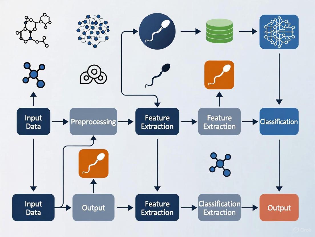
Workflow Visualization
AI-Based Sperm Morphology Analysis Workflow
Multi-Target Instance Parsing Network
Research Reagent Solutions & Essential Materials
Table 2: Essential Research Reagents and Materials for AI-Based Sperm Morphology Analysis
| Item | Function/Application | Implementation Notes |
|---|---|---|
| Staining Solutions (e.g., Diff-Quik, Papanicolaou) | Enhances contrast for traditional and automated morphology analysis | Required for stained-based methods; may cause sperm damage [1] |
| Phase-Contrast Microscope | Enables observation of unstained, live sperm | Essential for non-invasive methods; 20x magnification recommended [6] |
| Makler Counting Chamber | Standardized sperm concentration and motility assessment | Provides consistent imaging field for analysis [6] |
| Multi-Scale Part Parsing Network | Software for instance-level sperm parsing | Combines instance and semantic segmentation; key for unstained analysis [6] |
| Public Sperm Datasets (e.g., HSMA-DS, VISEM-Tracking, SVIA) | Training and validation of AI models | SVIA dataset contains 125,000 annotated instances [1] |
| Hybrid MLFFN-ACO Framework | Bio-inspired optimization for fertility diagnosis | Combines neural networks with ant colony optimization; reported 99% accuracy [2] |
| Measurement Accuracy Enhancement Algorithm | Reduces errors in low-resolution images | Uses IQR, Gaussian filtering, and robust correction techniques [6] |
The integration of artificial intelligence, particularly deep learning approaches, into sperm morphology analysis represents a paradigm shift in male infertility diagnostics. The quantitative evidence demonstrates that these technologies can achieve high accuracy rates exceeding 88% in classification tasks, significantly reducing subjectivity and variability inherent in manual assessments [5] [4]. The presented protocols and methodologies provide researchers with standardized approaches for implementing these advanced analytical techniques, with particular emphasis on both stained and stain-free applications. As these technologies continue to evolve, future directions should focus on multicenter validation trials, development of standardized datasets, and enhanced model interpretability to facilitate broader clinical adoption and ultimately improve diagnostic precision in male infertility evaluation [4] [2].
The assessment of sperm morphology remains a cornerstone in the clinical evaluation of male infertility, providing critical diagnostic and prognostic information [8] [9]. Traditional analysis relies on manual examination by trained technicians using microscopy, a method outlined in the World Health Organization (WHO) laboratory manuals [10]. Despite efforts to standardize these procedures, conventional manual assessment is fraught with significant challenges that compromise its reliability and clinical utility. These limitations primarily manifest as excessive subjectivity, poor reproducibility, and substantial workload burden for technicians [8] [1]. This application note systematically details these constraints and their implications for both clinical practice and research, framing them within the broader context of advancing deep learning-based solutions for sperm morphology classification. The inherent variability in manual methods not only affects diagnostic accuracy but also hinders the development of consistent treatment pathways for infertility, underscoring the urgent need for automated, standardized approaches leveraging artificial intelligence technologies.
Core Limitations of Conventional Manual Assessment
Subjectivity and Inter-Expert Variability
The fundamental challenge in manual sperm morphology analysis stems from its inherent subjectivity, which permeates every stage of the assessment process. This subjectivity arises from multiple sources, including differences in technician training, experience, and individual interpretation of complex morphological criteria.
Complex Classification Standards: According to WHO standards, sperm morphology is divided into head, neck, and tail compartments, with up to 26 distinct types of abnormal morphology recognized [8] [1]. Technicians must simultaneously evaluate abnormalities across multiple structures—head, vacuoles, midpiece, and tail—which substantially increases annotation difficulty and introduces interpretive variability [8].
Quantitative Evidence of Disagreement: Studies quantifying inter-expert agreement reveal concerning levels of discrepancy. In the development of the SMD/MSS dataset, researchers documented three separate agreement scenarios among three experts: no agreement (NA) among experts, partial agreement (PA) where 2/3 experts concurred, and total agreement (TA) where all three experts shared the same classification [10]. Statistical analysis using Fisher's exact test confirmed significant differences between experts in each morphology class (p < 0.05) [10]. This variability directly impacts the reliability of clinical diagnoses and treatment decisions based on morphology assessments.
Table 1: Documented Inter-Expert Variability in Sperm Morphology Classification
| Study | Expert Agreement Scenario | Description | Impact on Classification |
|---|---|---|---|
| SMD/MSS Dataset Development [10] | No Agreement (NA) | 0/3 experts agree on classification | Complete diagnostic inconsistency |
| Partial Agreement (PA) | 2/3 experts agree on the same label | Moderate reliability, potential misclassification | |
| Total Agreement (TA) | 3/3 experts agree on all categories | High reliability but rarely achieved |
Reproducibility Challenges
The reproducibility of sperm morphology analysis is compromised by both technical and human factors, creating substantial barriers to consistent clinical assessment and reliable research outcomes.
Inter-Laboratory Variability: Despite the availability of standardized WHO protocols, different laboratories frequently implement varying sample preparation, staining techniques, and classification interpretations [9]. This lack of standardized protocols across institutions means that results from one laboratory may not be directly comparable to those from another, complicating longitudinal studies and multi-center research initiatives [8] [9].
Sample Preparation Inconsistencies: Variations in staining methods (e.g., RAL Diagnostics staining kit, Papanicolaou stain) and smear preparation techniques introduce pre-analytical variables that affect morphological appearance and subsequent classification [10]. These technical discrepancies compound the interpretive variations between technicians, creating a compounded reproducibility problem spanning both sample preparation and analysis phases.
Substantial Workload and Operational Inefficiency
The operational burden of manual sperm morphology analysis creates practical constraints on laboratory throughput and introduces fatigue-related errors that further compromise accuracy.
Labor-Intensive Process: WHO guidelines recommend the analysis and counting of more than 200 sperms per sample to obtain statistically meaningful morphology assessments [8] [1]. Given the need to evaluate each sperm across multiple morphological compartments (head, neck, and tail) against 26 potential abnormality types, this process demands considerable time and focused attention from skilled technicians [8].
Economic and Workflow Implications: The substantial time investment required for each analysis limits laboratory throughput and increases operational costs. Additionally, technician fatigue during extended evaluation sessions can introduce additional errors and inconsistencies, particularly in high-volume clinical settings [1]. This workload burden has direct implications for patient wait times and accessibility of comprehensive fertility testing services.
Quantitative Comparison of Methodological Limitations
Table 2: Comparative Analysis of Manual vs. Deep Learning Approaches in Sperm Morphology Assessment
| Parameter | Manual Assessment | Deep Learning Approaches | Clinical & Research Implications |
|---|---|---|---|
| Subjectivity | High inter-expert variability (significant differences at p<0.05) [10] | Eliminates human interpretive variation | DL enables standardized diagnosis across clinics |
| Reproducibility | Poor inter-laboratory consistency due to protocol variations [9] | High reproducibility with consistent algorithms | Enables multi-center research with comparable results |
| Workload | High: requires analysis of >200 sperm per sample by expert [8] | Automated processing of large image volumes | Increases laboratory throughput and reduces costs |
| Classification Accuracy | Variable (55-92% vs. expert consensus) [10] | Higher and more consistent (up to 94.1% TPR reported) [11] | More reliable fertility prognosis and treatment planning |
| Throughput | Limited by technician capacity and fatigue | Rapid batch processing capabilities | Scalable for high-volume screening applications |
| Standardization | Difficult to achieve across operators and centers | Inherently standardized once validated | Creates consistent diagnostic thresholds |
Experimental Protocols for Evaluating Methodological Limitations
Protocol for Quantifying Inter-Expert Variability
Purpose: To systematically evaluate and quantify the degree of subjectivity in manual sperm morphology assessment among different experts.
Materials:
- Sperm samples prepared according to WHO standard protocols
- RAL Diagnostics staining kit or equivalent
- Optical microscope with 100x oil immersion objective
- MMC CASA system or similar for image acquisition
- Data collection spreadsheet for expert annotations
Procedure:
- Sample Preparation: Prepare semen smears from samples with varying morphological profiles. Ensure sperm concentration is at least 5 million/mL but exclude samples >200 million/mL to avoid image overlap [10].
- Image Acquisition: Capture 37±5 images per sample using bright field mode with oil immersion 100x objective. Ensure each image contains a single spermatozoon with clear visualization of head, midpiece, and tail structures [10].
- Expert Classification: Engage multiple experienced technicians (minimum of 3) to independently classify each spermatozoon according to modified David classification (12 classes of morphological defects) [10].
- Data Collection: Create a shared spreadsheet where each expert documents morphological classifications for each sperm component without consultation.
- Statistical Analysis:
- Categorize agreement scenarios: No Agreement (NA), Partial Agreement (PA), and Total Agreement (TA) [10].
- Use Fisher's exact test to evaluate statistical differences between experts for each morphology class, with significance set at p<0.05 [10].
- Calculate intraclass correlation coefficients (ICC) to measure reliability of continuous measurements.
Protocol for Assessing Reproducibility Across Laboratories
Purpose: To evaluate the reproducibility of sperm morphology assessments across different laboratories and imaging conditions.
Materials:
- Standardized sperm sample aliquots
- Multiple microscope systems with different configurations
- Phase contrast, Hoffman modulation contrast, and bright field imaging capabilities [12]
- Sample preparation reagents consistent across sites
Procedure:
- Sample Distribution: Prepare identical aliquots from well-characterized semen samples and distribute to participating laboratories.
- Multi-Center Imaging: Acquire images using different microscope brands, imaging modes (bright field, phase contrast, Hoffman modulation contrast), and magnifications (10x, 20x, 40x, 60x, 100x) to simulate real-world clinical variation [12].
- Standardized Analysis: Implement the same classification criteria across all sites using the modified David classification or WHO standards [10].
- Data Integration and Comparison:
- Calculate coefficient of variation across laboratories for each morphological parameter.
- Assess intraclass correlation coefficients (ICC) for precision (0.97 reported in rigorous studies) and recall across sites [12].
- Perform ablation studies to determine how each imaging variable (magnification, mode, preprocessing) affects morphological classification consistency.
Experimental Workflow for Methodological Evaluation
Experimental Workflow for Evaluating Methodological Limitations
The Scientist's Toolkit: Research Reagent Solutions
Table 3: Essential Research Reagents and Materials for Sperm Morphology Studies
| Research Reagent/Material | Function/Application | Protocol Considerations |
|---|---|---|
| RAL Diagnostics Staining Kit | Provides differential staining of sperm structures for morphological assessment | Follow manufacturer instructions for consistent staining intensity and contrast [10] |
| VISEM-Tracking Dataset | Publicly available dataset containing 656,334 annotated objects with tracking details | Enables algorithm training and benchmarking without additional sample collection [8] |
| SVIA Dataset | Comprehensive dataset with 125,000 annotated instances, 26,000 segmentation masks | Supports detection, segmentation, and classification tasks in DL development [8] |
| HuSHeM Dataset | Human Sperm Head Morphology dataset with stained, higher resolution images | Useful for head-specific classification algorithms; limited to 216 publicly available images [8] [11] |
| SCIAN-MorphoSpermGS Dataset | Gold-standard dataset with 1,854 sperm images across five WHO classes | Provides expert-validated ground truth for training and validation [8] |
| MMC CASA System | Computer-Assisted Semen Analysis for standardized image acquisition | Ensures consistent magnification (100x oil immersion) and imaging conditions [10] |
The limitations of conventional manual sperm morphology assessment—subjectivity, poor reproducibility, and substantial workload—present significant barriers to accurate male infertility diagnosis and treatment. Quantitative evidence demonstrates concerning inter-expert variability with statistical significance, while operational constraints limit laboratory efficiency and consistency. These methodological challenges directly impact clinical decision-making and highlight the critical need for standardized, automated approaches. Deep learning-based classification systems represent a promising solution, offering the potential to overcome these limitations through automated feature extraction, consistent application of morphological criteria, and significantly reduced analytical workload. By addressing the fundamental constraints of conventional methods, deep learning approaches can enhance diagnostic reliability, enable multi-center research collaboration, and ultimately improve patient care in reproductive medicine.
Sperm morphology analysis is a cornerstone of male fertility assessment, with a demonstrated significant correlation between abnormal sperm forms and infertility [1]. For decades, the evaluation of sperm shape was a manual, subjective process, highly dependent on the technician's expertise and leading to significant inter-laboratory variability [10] [11]. The introduction of Computer-Assisted Semen Analysis (CASA) systems promised a new era of standardization and objectivity. However, traditional CASA often fell short, struggling with accurately distinguishing sperm from debris and classifying subtle midpiece and tail abnormalities [10] [13].
The emergence of deep learning represents a paradigm shift, moving from automated measurement to intelligent classification. This evolution leverages convolutional neural networks (CNNs) to automatically learn discriminative features from raw sperm images, eliminating the need for manual feature extraction and offering a path toward highly accurate, reproducible, and rapid sperm morphology classification [11] [1]. These Application Notes detail the experimental protocols and quantitative evidence driving this technological transition, providing a framework for researchers to implement and advance these methods.
From Manual Analysis to CASA: The First Steps in Automation
The Workflow and Limitations of Traditional CASA
Traditional CASA systems were designed to bring objectivity to semen analysis by automating the image acquisition and measurement processes. A typical CASA workflow involves loading a prepared semen sample onto a microscope stage equipped with a digital camera. The system then captures multiple images or video sequences, which are processed to identify sperm cells and quantify parameters like concentration and motility [13].
For morphology, CASA systems relied on extracting handcrafted morphometric features from sperm images. These features typically included:
- Head dimensions: Length, width, area, and perimeter.
- Head shape descriptors: Eccentricity, elongation, and shape factors.
- Complex descriptors: Fourier descriptors, Zernike moments, and Hu moments to capture contour and texture details [11].
These extracted features were then fed into conventional machine learning classifiers, such as Support Vector Machines (SVM) or k-nearest neighbors (k-NN), to categorize sperm into morphological classes [1].
Despite their contribution to standardization, these systems faced fundamental limitations. Their performance was heavily dependent on image quality and often failed in the presence of cellular debris or when sperm were agglutinated. Furthermore, their reliance on pre-defined features made them inflexible and unable to generalize well to the vast and subtle spectrum of sperm abnormalities, particularly in the midpiece and tail [10] [1]. This often resulted in unsatisfactory performance and limited their routine clinical adoption for robust morphological assessment.
A Research-Grade CASA Simulation Protocol
To objectively assess and validate CASA algorithms without the constraints of variable real-world image quality, researchers have developed simulation tools that generate life-like semen images with known, controllable ground-truth parameters [13].
Protocol: Simulating Semen Images for Algorithm Validation
Sperm Cell Modeling:
- Head Generation: Model the sperm head as a generally oval shape. The image is created by defining a center point and applying point spread functions to simulate the head's core and membrane, resulting in a final, realistic head image [13].
- Flagellum Generation: Model the tail as a thin, flexible curve. Define a series of points along the intended tail path and apply a different point spread function to generate a pixelated representation of the flagellum. The head and tail images are then merged to form a complete sperm cell [13].
Motion Path Modeling: Implement different swimming modes to create dynamic video sequences. The four primary modes are:
- Linear Mean: Progressive movement along a relatively straight path.
- Circular: Movement along a circular trajectory.
- Hyperactive: High-amplitude, non-progressive thrashing.
- Immotile: No movement, representing dead or non-motile sperm [13].
Multi-Cell Image Synthesis: Populate a simulated image frame by generating multiple sperm cells, each with a defined position and swimming mode. Add controlled levels of noise and background intensity variation to mimic real-world microscopy conditions [13].
Algorithm Testing: Use the simulated image sequences, where all parameters (positions, shapes, paths) are known, as a ground-truth benchmark to quantitatively evaluate the performance of segmentation, localization, and tracking algorithms using metrics like precision, recall, and Multi-Object Tracking Accuracy (MOTA) [13].
The Deep Learning Revolution in Sperm Morphology
Core Architectural Principles
Deep learning, particularly Convolutional Neural Networks (CNNs), has overcome many limitations of traditional CASA by learning relevant features directly from the data. CNNs consist of multiple layers that automatically and hierarchically learn to detect patterns, from simple edges and gradients in early layers to complex morphological structures like acrosomes and tail bends in deeper layers [11]. Common approaches include:
- Transfer Learning: Fine-tuning pre-existing networks, such as VGG16, which were originally trained on large-scale datasets like ImageNet. This approach is computationally efficient and effective, especially with limited medical image data [11].
- End-to-End Object Detection: Employing architectures like YOLO (You Only Look Once) that perform both sperm detection and classification in a single pass, optimizing for speed and efficiency suitable for clinical workflows [14].
Quantitative Performance Comparison
The transition from conventional machine learning to deep learning has yielded measurable improvements in classification accuracy, as evidenced by studies on public datasets.
Table 1: Performance Comparison of Sperm Classification Methods on Public Datasets
| Dataset | Method | Key Features | Reported Performance | Reference |
|---|---|---|---|---|
| HuSHeM | Cascade Ensemble-SVM (CE-SVM) | Shape-based descriptors (Area, Eccentricity, Zernike moments) | 78.5% Average True Positive Rate | [11] |
| HuSHeM | Deep CNN (VGG16 Transfer Learning) | Automated feature extraction from raw images | 94.1% Average True Positive Rate | [11] |
| SCIAN (Partial Agreement) | CE-SVM | Manual feature engineering | 58% Average True Positive Rate | [11] |
| SCIAN (Partial Agreement) | Deep CNN (VGG16 Transfer Learning) | Automated feature extraction from raw images | 62% Average True Positive Rate | [11] |
| Bovine Sperm Dataset | YOLOv7 | Single-stage detection & classification | 0.73 mAP@50, 0.75 Precision, 0.71 Recall | [14] |
The following diagram illustrates the typical end-to-end workflow for developing a deep learning-based sperm morphology classification system.
Protocol for Implementing a Deep Learning Morphology Classifier
Protocol: Building a CNN-based Sperm Morphology Classifier
Dataset Curation and Augmentation:
- Image Acquisition: Acquire images of individual spermatozoa using a microscope with a 100x oil immersion objective, preferably under bright-field mode. Staining (e.g., RAL Diagnostics kit) is typically used for fixed smears [10].
- Expert Annotation: Have multiple experienced embryologists or andrologists classify each sperm image according to a standardized classification system (e.g., modified David classification or WHO criteria). Resolve discrepancies through consensus [10].
- Data Augmentation: To address class imbalance and prevent overfitting, artificially expand the dataset using techniques such as random rotation, flipping, scaling, brightness/contrast adjustment, and elastic deformations. For example, a base set of 1,000 images can be expanded to over 6,000 images [10].
Model Training:
- Pre-processing: Resize all images to a uniform dimensions (e.g., 80x80 pixels). Convert to grayscale and normalize pixel values [10].
- Data Partitioning: Split the augmented dataset randomly into training (80%), validation (10-20%), and hold-out test (20%) sets [10].
- Model Selection and Fine-Tuning:
- Option A (Transfer Learning): Use a pre-trained network like VGG16. Replace the final classification layer with a new one matching the number of sperm morphology classes. First, train only the new layer, then fine-tune all layers with a very low learning rate [11].
- Option B (Object Detection): For frameworks like YOLOv7, format the annotations accordingly and train the model to both locate and classify sperm in an image [14].
- Training: Train the model using the training set, monitoring loss and accuracy on the validation set to avoid overfitting.
Evaluation and Deployment:
- Performance Metrics: Evaluate the final model on the held-out test set. Report standard metrics including accuracy, precision, recall, F1-score, and area under the ROC curve (AUC-ROC) [14].
- Inference: Deploy the trained model to classify new, unseen sperm images. The model outputs a probability distribution over the possible morphological classes for each input image.
The Scientist's Toolkit: Essential Research Reagents and Materials
The successful implementation of a deep learning morphology system relies on a foundation of wet-lab and computational tools.
Table 2: Key Research Reagents and Solutions for Sperm Morphology Analysis
| Item | Function / Application | Example / Specification |
|---|---|---|
| RAL Staining Kit | Stains sperm cells on fixed smears for clear visualization of morphological details under bright-field microscopy. | RAL Diagnostics kit [10] |
| Optixcell Extender | Semen extender used to dilute and preserve bull sperm samples for morphological analysis. | IMV Technologies [14] |
| Trumorph System | A dye-free fixation system that uses controlled pressure and temperature to immobilize sperm for morphology evaluation. | Proiser R+D, S.L. [14] |
| MMC CASA System | An integrated system comprising an optical microscope and camera for automated image acquisition and initial analysis. | Used for acquiring individual sperm images [10] |
| B-383Phi Microscope | A microscope used for high-resolution imaging of sperm cells, often paired with image capture software. | Optika, with 40x negative phase contrast objective [14] |
| Python with Deep Learning Libraries | The primary programming environment for developing, training, and testing deep learning models (CNNs, YOLO). | Python 3.8, TensorFlow/PyTorch, OpenCV [10] [14] |
| Roboflow | Web-based tool for annotating images, managing datasets, and performing preprocessing and augmentation. | Used for labeling and preparing training data [14] |
The evolution from CASA to deep learning marks a significant maturation of automation in sperm morphology analysis. While CASA provided initial steps toward objectivity, its dependence on handcrafted features was a fundamental constraint. Deep learning, with its capacity for end-to-end learning from raw pixel data, has demonstrated superior performance and offers a robust framework for standardizing this critical diagnostic procedure. The detailed protocols and quantitative comparisons provided here equip researchers to contribute to this rapidly advancing field, pushing the boundaries of accuracy, efficiency, and accessibility in male fertility assessment.
Deep learning, a subset of artificial intelligence (AI), has emerged as a transformative technology for analyzing complex biological data. Its capacity to automatically learn hierarchical features from raw input data makes it particularly well-suited for medical image analysis tasks that have traditionally relied on manual, subjective assessment. In the field of reproductive biology, deep learning approaches are revolutionizing the analysis of sperm morphology—a key diagnostic parameter in male fertility assessment. Convolutional Neural Networks (CNNs), a specialized class of deep neural networks, have demonstrated remarkable success in processing image data by mimicking the hierarchical structure of biological visual processing systems.
The application of these technologies to sperm morphology classification addresses significant challenges in conventional analysis methods. Traditional manual assessment is notoriously subjective, time-consuming, and prone to inter-observer variability, while earlier computer-assisted semen analysis (CASA) systems have shown limited ability to accurately distinguish spermatozoa from cellular debris and classify specific morphological abnormalities [10] [1]. Deep learning models, particularly CNNs, offer the potential for automation, standardization, and acceleration of semen analysis while achieving accuracy levels comparable to human experts [15].
Neural Networks and CNNs: Architectural Foundations
Fundamental Neural Network Components
At their core, neural networks are computational models inspired by the structure and function of the human brain. The basic building block is the artificial neuron, which receives inputs, applies a mathematical transformation, and produces an output. These neurons are organized into layers—typically an input layer, one or more hidden layers, and an output layer—with connections between them having associated weights that are adjusted during training [16].
The fundamental components of a neural network include:
- Layers: Stacked sets of neurons that process information sequentially
- Weights and Biases: Parameters that determine the strength of connections between neurons
- Activation Functions: Mathematical functions that introduce non-linearity, enabling the network to learn complex patterns (e.g., ReLU, sigmoid, tanh)
- Loss Functions: Objectives that the network optimizes during training
- Optimizers: Algorithms that adjust weights and biases to minimize the loss function [16]
Convolutional Neural Networks (CNNs)
CNNs represent a specialized neural network architecture designed specifically for processing grid-like data such as images. Their unique structural properties make them exceptionally well-suited for visual data analysis tasks, including biological image classification. Unlike traditional fully-connected networks, CNNs employ three key architectural features:
Convolutional Layers: These layers apply a series of filters (kernels) across the input image to detect spatial hierarchies of features, from simple edges and textures in early layers to complex morphological patterns in deeper layers. Each filter slides across the input, computing dot products to generate feature maps that highlight specific characteristics present in the image [11] [16].
Pooling Layers: Typically inserted between convolutional layers, pooling operations (e.g., max pooling, average pooling) reduce the spatial dimensions of feature maps while retaining the most salient information. This dimensionality reduction provides translational invariance and decreases computational complexity while preventing overfitting [11].
Fully-Connected Layers: In the final stages of the network, these traditional neural network layers integrate the high-level features extracted by the convolutional and pooling layers to perform the final classification task, such as categorizing sperm into normal versus abnormal morphological classes [11].
The training process for both standard neural networks and CNNs involves forward propagation of input data, calculation of loss between predictions and ground truth, and backward propagation of errors to adjust weights using optimization algorithms like gradient descent. This iterative process enables the network to gradually improve its performance on the designated task [16].
CNN Basic Architecture for Image Classification
Application to Sperm Morphology Classification
Problem Formulation and Significance
Sperm morphology analysis represents a critical diagnostic procedure in male fertility assessment, with the proportion and types of morphologically abnormal spermatozoa providing valuable prognostic information for natural conception and assisted reproductive outcomes. According to World Health Organization (WHO) standards, sperm morphology is evaluated across three primary components: head, midpiece, and tail, with numerous specific abnormality patterns identified within each category [10] [1].
The clinical challenge stems from the subjective nature of manual assessment, which relies heavily on technician expertise and demonstrates significant inter-laboratory variability. Furthermore, the process is labor-intensive, requiring classification of 200 or more spermatozoa per sample—a time-consuming task that contributes to diagnostic inconsistency [1]. Deep learning approaches directly address these limitations by providing automated, standardized classification with reduced operator dependency and potentially higher throughput.
Comparative Performance of Deep Learning Approaches
Recent research has demonstrated the effectiveness of deep learning models for sperm morphology classification, with several studies reporting performance metrics approaching or exceeding expert-level accuracy. The following table summarizes quantitative results from key studies in the field:
Table 1: Performance Comparison of Deep Learning Models for Sperm Morphology Classification
| Study | Dataset | Model Architecture | Key Performance Metrics | Classification Categories |
|---|---|---|---|---|
| SMD/MSS Study (2025) [15] [10] | SMD/MSS (1,000 images augmented to 6,035) | Custom CNN | Accuracy: 55-92% (variation across morphological classes) | 12 classes based on modified David classification |
| Deep Learning for Classification (2019) [11] | HuSHeM Dataset | VGG16 (Transfer Learning) | Average True Positive Rate: 94.1% | 5 WHO categories: Normal, Tapered, Pyriform, Small, Amorphous |
| Deep Learning for Classification (2019) [11] | SCIAN Dataset | VGG16 (Transfer Learning) | Average True Positive Rate: 62% | 5 WHO categories |
| Current Literature Review (2025) [1] | Multiple Public Datasets | Various Deep Learning Models | Accuracy range: 59-92% across studies | Varies by study (typically 3-12 morphological classes) |
The variation in reported performance metrics highlights several important considerations for implementing deep learning solutions in this domain. Dataset characteristics—including size, quality, annotation consistency, and class balance—significantly influence model performance. Additionally, the specific architectural choices and training methodologies employed impact the resulting classification accuracy [1].
Experimental Protocols for Sperm Morphology Classification
Dataset Curation and Preparation Protocol
Purpose: To systematically collect, annotate, and preprocess sperm images for training and evaluating deep learning models.
Materials and Equipment:
- Microscope with 100x oil immersion objective
- Digital camera system
- Stained semen smears (RAL Diagnostics staining kit)
- Computer with image acquisition software
- Data annotation platform
Procedure:
- Sample Preparation: Prepare semen smears according to WHO guidelines [10]. Apply RAL Diagnostics staining to enhance cellular structure visualization.
- Image Acquisition: Capture individual sperm images using an MMC CASA system or equivalent. Use bright-field microscopy with 100x oil immersion objective. Ensure each image contains a single spermatozoon with clear visualization of head, midpiece, and tail structures [10].
- Expert Annotation: Engage multiple experienced embryologists (minimum 3) to independently classify each sperm image according to modified David classification or WHO criteria. The classification should encompass:
- Head defects: Tapered, thin, microcephalous, macrocephalous, multiple, abnormal post-acrosomal region, abnormal acrosome
- Midpiece defects: Cytoplasmic droplet, bent
- Tail defects: Coiled, short, multiple [10]
- Ground Truth Establishment: Resolve annotation discrepancies through consensus meetings or majority voting. Compile final classifications into a ground truth file containing image name, expert classifications, and morphological measurements [10].
- Data Augmentation: Address class imbalance and limited dataset size by applying augmentation techniques including:
- Rotation and flipping
- Brightness and contrast adjustment
- Scaling and translation
- Synthetic image generation (if applicable) [15]
- Data Partitioning: Split the dataset into training (80%), validation (10%), and test (10%) sets, ensuring representative distribution of all morphological classes across partitions [10].
CNN Model Development and Training Protocol
Purpose: To design, implement, and train a convolutional neural network for automated sperm morphology classification.
Materials and Software:
- Python programming environment (version 3.8+)
- Deep learning frameworks (TensorFlow, PyTorch, or Keras)
- GPU-accelerated computing resources
- Preprocessed and annotated sperm image dataset
Procedure:
- Image Preprocessing:
- Resize all images to uniform dimensions (e.g., 80×80 pixels)
- Apply normalization to scale pixel values to [0,1] range
- Convert images to grayscale if color information is not diagnostically relevant
- Apply noise reduction algorithms to enhance image quality [10]
Model Architecture Design:
- Implement a CNN architecture with convolutional, pooling, and fully-connected layers
- Consider transfer learning using pre-trained networks (e.g., VGG16, ResNet) when training data is limited [11]
- Include appropriate regularization techniques (dropout, batch normalization) to prevent overfitting
Model Training:
- Initialize model parameters using established weight initialization strategies
- Define appropriate loss function (categorical cross-entropy for multi-class classification)
- Select optimization algorithm (Adam, SGD) with appropriate learning rate
- Implement batch training with batch size optimized for available computational resources
- Train model for sufficient epochs while monitoring validation loss to avoid overfitting [11]
Model Evaluation:
Sperm Morphology Analysis Workflow
The Scientist's Toolkit: Research Reagent Solutions
Table 2: Essential Materials and Reagents for Deep Learning-Based Sperm Morphology Analysis
| Item | Specification/Example | Function/Purpose |
|---|---|---|
| Microscope System | MMC CASA system with 100x oil immersion objective | High-resolution image acquisition of individual spermatozoa |
| Staining Kit | RAL Diagnostics staining kit | Enhances contrast and visualization of sperm morphological structures |
| Annotation Software | Custom Excel templates or specialized annotation platforms | Systematic documentation of expert morphological classifications |
| Data Augmentation Tools | Python libraries (TensorFlow, Keras, PyTorch) | Expands dataset size and diversity through image transformations |
| Deep Learning Framework | TensorFlow, PyTorch, Keras with Python 3.8+ | Provides infrastructure for implementing and training CNN models |
| Computational Resources | GPU-accelerated workstations (NVIDIA CUDA-compatible) | Enables efficient training of computationally intensive deep learning models |
| Performance Metrics Package | Scikit-learn, custom evaluation scripts | Quantifies model performance through accuracy, precision, recall, F1-score |
| Public Datasets | HuSHeM, SCIAN, SVIA datasets [1] [11] | Provides benchmark data for model development and comparative performance assessment |
Implementation Considerations and Future Directions
The implementation of deep learning systems for sperm morphology classification presents several practical considerations. Dataset quality and annotation consistency remain paramount, as models are highly dependent on training data quality. The SMD/MSS study highlighted the importance of addressing inter-expert variability in annotations, reporting scenarios with no agreement (NA), partial agreement (PA), and total agreement (TA) among the three experts [10]. Future research directions include developing more sophisticated data augmentation techniques, integrating multiple classification frameworks (WHO, David, Kruger), and exploring explainable AI methods to enhance clinical trust and adoption [15] [1].
As the field advances, the integration of deep learning-based morphology assessment into comprehensive semen analysis systems offers the potential to transform male fertility diagnostics. By providing standardized, automated, and objective classification, these technologies can enhance diagnostic consistency across laboratories and improve patient care through more reliable fertility assessment and treatment planning.
Building the Model: A Technical Deep Dive into Deep Learning Pipelines for Sperm Classification
The development of robust deep learning models for sperm morphology classification is critically dependent on the availability of high-quality, well-annotated datasets. Within this field, three significant datasets have emerged: SMD/MSS (Sperm Morphology Dataset/Medical School of Sfax), VISEM, and SVIA (Sperm Videos and Images Analysis dataset). These datasets address a pressing need in male infertility research, where manual sperm morphology analysis remains highly subjective, challenging to standardize, and dependent on technician experience [10] [1]. The SMD/MSS dataset provides meticulously classified individual sperm images focusing on detailed morphological defects according to the modified David classification [10]. In contrast, the VISEM dataset offers a multi-modal resource containing video recordings of spermatozoa alongside extensive clinical and biological data from participants [17] [18]. The SVIA dataset represents a large-scale collection with diverse annotations suitable for multiple computer vision tasks, including object detection, segmentation, and classification [19]. Together, these resources enable the training and validation of sophisticated deep learning algorithms, moving the field toward automated, standardized, and accurate sperm morphology analysis.
Dataset Comparison and Characteristics
The SMD/MSS, VISEM, and SVIA datasets vary significantly in scale, content type, and annotation focus, making them suitable for different research applications within sperm morphology analysis.
Table 1: Quantitative Comparison of Sperm Morphology Datasets
| Characteristic | SMD/MSS | VISEM | SVIA |
|---|---|---|---|
| Primary Content | 1,000 individual sperm images (extended to 6,035 with augmentation) [10] | 20 annotated videos (29,196 frames) + 166 unlabeled clips [17] | 101 video clips, 125,000 object locations, 26,000 segmentation masks [19] |
| Annotation Focus | Morphological defects (head, midpiece, tail) per modified David classification [10] | Bounding boxes, tracking IDs, sperm motility [17] | Bounding boxes, segmentation masks, object categories [19] |
| Data Modalities | Static images | Videos, clinical data, biological samples [18] | Videos, images |
| Key Strengths | Expert classification by multiple andrologists; CASA morphometrics [10] | Multi-modal; tracking annotations; clinical correlation potential [17] [18] | Large-scale; diverse annotations for multiple computer vision tasks [19] |
| Primary Use Cases | Sperm morphology classification; defect identification [10] | Sperm tracking; motility analysis; multi-modal prediction [17] | Object detection; segmentation; classification [19] |
Table 2: Detailed Annotation Specifications
| Dataset | Annotation Types | Class Labels/Details | Annotation Format |
|---|---|---|---|
| SMD/MSS | Morphological class per spermatozoon [10] | 12 defect classes: 7 head defects, 2 midpiece defects, 3 tail defects [10] | Image filename codes (A: Tapered, B: Thin, etc.); Ground truth file [10] |
| VISEM-Tracking | Bounding boxes; tracking IDs [17] | 0: normal sperm, 1: sperm clusters, 2: small/pinhead [17] | YOLO format text files; CSV with sperm counts [17] |
| SVIA | Bounding boxes; segmentation masks; object categories [19] | Normal, pin, amorphous, tapered, round, multi-nucleated head sperm, impurities [19] | Category information; segmentation masks; independent images [19] |
Dataset Curation and Annotation Protocols
SMD/MSS Dataset Curation
The SMD/MSS dataset was developed through a rigorous multi-step curation process designed to maximize quality and consistency for morphological classification tasks.
Sample Preparation and Acquisition: Smears were prepared from semen samples obtained from 37 patients following World Health Organization (WHO) guidelines and stained with RAL Diagnostics staining kit. Samples with sperm concentrations of at least 5 million/mL were included, while those exceeding 200 million/mL were excluded to prevent image overlap and facilitate capture of whole sperm. Images were acquired using an MMC CASA system comprising an optical microscope with a digital camera using bright field mode with an oil immersion 100x objective. The system captured morphometric data including head width and length, and tail length for each spermatozoon [10].
Expert Annotation and Quality Control: Each spermatozoon underwent manual classification by three experienced experts following the modified David classification, which includes 12 classes of morphological defects: 7 head defects (tapered, thin, microcephalous, macrocephalous, multiple, abnormal post-acrosomal region, abnormal acrosome), 2 midpiece defects (cytoplasmic droplet, bent), and 3 tail defects (coiled, short, multiple) [10]. The inter-expert agreement was systematically analyzed across three scenarios: no agreement (NA), partial agreement (PA) where 2/3 experts agreed, and total agreement (TA) where all three experts agreed on the same label for all categories. Statistical analysis using Fisher's exact test was performed to assess differences between experts for each morphological class [10].
Data Augmentation: To address class imbalance and limited data issues, augmentation techniques were applied to expand the original 1,000 images to 6,035 images, creating a more balanced representation across morphological classes [10].
VISEM Dataset Curation
The VISEM dataset represents a unique multi-modal resource curated with an emphasis on video data and clinical correlations.
Multi-modal Data Collection: Data was originally collected for studies on overweight and obesity in relation to male reproductive function. The dataset includes 85 male participants aged 18 years or older, with video recordings of spermatozoa placed on a heated microscope stage (37°C) and examined under 400x magnification using an Olympus CX31 microscope. Videos were captured using a UEye UI-2210C camera and saved as AVI files [18]. In addition to video data, the dataset incorporates standard semen analysis results, sperm fatty acid profiles, fatty acid composition of serum phospholipids, demographic data, and sex hormone measurements [18].
Tracking Annotation Protocol: For the VISEM-Tracking subset, 20 video recordings of 30 seconds each (comprising 29,196 frames) were selected based on diversity to obtain as many varied tracking samples as possible [17]. Annotation was performed by data scientists using the LabelBox tool in close collaboration with male reproduction researchers. Biologists verified all annotations to ensure correctness [17]. Each annotated video folder contains extracted frames, bounding box labels for each frame, and bounding box labels with corresponding tracking identifiers. All bounding box coordinates are provided in YOLO format, with text files containing class labels and unique tracking IDs to identify individual spermatozoa throughout videos [17].
Data Structure and Organization: The dataset is organized with 20 sub-folders for annotated videos, each containing extracted frames, bounding box labels per frame, and labels with tracking identifiers. Additional CSV files contain participant-related data, semen analysis results, sex hormone levels, and sperm counts per frame [17].
SVIA Dataset Curation
The SVIA dataset was curated as a large-scale resource for computer-aided sperm analysis, with extensive annotations supporting multiple computer vision tasks.
Large-scale Data Collection and Annotation: The dataset preparation began in 2017 and involved approximately four years of work, resulting in more than 278,000 annotated objects [19]. Fourteen reproductive doctors and biomedical scientists performed annotations, with verification by six reproductive doctors and biomedical scientists. The dataset includes normal and abnormal sperm categories, including pin, amorphous, tapered, round, and multi-nucleated head sperm, as well as impurities [19].
Structured Data Organization: The SVIA dataset is organized into three distinct subsets supporting different research applications. Subset-A contains 125,000 object locations with bounding box annotations from 101 videos. Subset-B includes 26,000 segmentation masks from 10 videos. Subset-C provides 125,000 independent images of sperm and impurities for classification tasks [19].
Quality Assurance: The extensive annotation process involved multiple specialists to ensure accuracy and consistency across the large-scale dataset. The inclusion of various abnormality types and impurities enhances the dataset's utility for real-world applications where such distinctions are clinically relevant [19].
Experimental Protocols for Deep Learning Applications
Sperm Morphology Classification with SMD/MSS
Data Preprocessing: Images underwent cleaning to handle missing values, outliers, and inconsistencies. Normalization or standardization transformed numerical features to a common scale, ensuring no particular feature dominated the learning process. Images were resized using linear interpolation strategy to 80×80×1 grayscale to standardize input dimensions [10].
Dataset Partitioning: The entire image set was randomly divided into training (80%) and testing (20%) subsets. From the training subset, 20% was further extracted for validation during model development, ensuring robust evaluation on unseen data [10].
Deep Learning Architecture: A Convolutional Neural Network (CNN) architecture was implemented in Python 3.8, comprising five stages: image preprocessing, database partitioning, data augmentation, program training, and evaluation. The model was trained to classify sperm images into the various morphological categories defined in the annotation protocol [10].
Sperm Detection and Tracking with VISEM-Tracking
Baseline Detection Model: Researchers established baseline sperm detection performance using the YOLOv5 deep learning model trained on the VISEM-Tracking dataset [17]. This provided a benchmark for subsequent research and demonstrated the dataset's utility for training complex DL models to analyze spermatozoa.
Object Tracking Methodology: The tracking identifiers provided with bounding boxes enable development and evaluation of sperm tracking algorithms. These algorithms can analyze movement patterns, classify spermatozoa based on motility, and compute kinematic parameters essential for comprehensive sperm quality assessment [17].
Multi-task Learning with SVIA Dataset
Object Detection Experiments: For Subset-A, researchers evaluated five deep learning models for object detection: Single Shot MultiBox Detector (SSD), RetinaNet, Faster RCNN, and YOLO-v3/v4. Performance was assessed using evaluation metrics including accuracy, precision, recall, and F1-score calculated from confusion matrices [19].
Image Segmentation Benchmarking: For Subset-B, four traditional image segmentation methods (Markov Random Field, Watershed, Otsu thresholding, Region Growing) and four deep learning-based methods (k-means, U-net, SegNet, and Mask RCNN) were compared to segment original images [19].
Image Denoising Evaluation: For Subset-C, 13 kinds of conventional noise were added to original images, followed by application of different denoising methods including DnCNN, U-net, and traditional filters to assess robustness and image enhancement capabilities [19].
The Scientist's Toolkit: Research Reagent Solutions
Table 3: Essential Research Reagents and Materials
| Item | Function/Application | Dataset Context |
|---|---|---|
| RAL Diagnostics Staining Kit | Sperm smear staining for morphological analysis | SMD/MSS sample preparation [10] |
| Olympus CX31 Microscope | Optical microscopy with 400x magnification for video recording | VISEM video acquisition [17] [18] |
| UEye UI-2210C Camera | Microscope-mounted camera for video capture (50 FPS) | VISEM video recording [17] [18] |
| MMC CASA System | Computer-assisted semen analysis for image acquisition and morphometrics | SMD/MSS data collection [10] |
| Heated Microscope Stage | Maintains samples at 37°C for physiological motility assessment | VISEM sample preparation [17] [18] |
| LabelBox Annotation Tool | Web-based platform for bounding box and tracking annotation | VISEM-Tracking annotation [17] |
The SMD/MSS, VISEM, and SVIA datasets represent significant advancements in resources for sperm morphology classification using deep learning. Each dataset offers unique strengths: SMD/MSS provides detailed morphological classification following standardized clinical guidelines, VISEM offers multi-modal data with clinical correlations, and SVIA delivers large-scale annotations for diverse computer vision tasks. The rigorous curation protocols, including multi-expert annotation, quality control measures, and comprehensive documentation, ensure these datasets meet the demanding requirements of deep learning research. By addressing critical challenges in data quality, annotation consistency, and clinical relevance, these resources facilitate the development of robust, standardized, and clinically applicable deep learning solutions for male infertility assessment. Future work should focus on expanding dataset diversity, developing standardized evaluation benchmarks, and exploring federated learning approaches to leverage these resources while addressing privacy concerns in medical data.
In the field of male fertility research, deep learning for sperm morphology classification has emerged as a powerful tool to overcome the subjectivity and variability of manual analysis by embryologists [1]. The performance and generalizability of these models are fundamentally constrained by the quality, quantity, and balance of the training data. This document outlines standardized protocols for data preprocessing and augmentation, specifically tailored for sperm image analysis, to enhance model robustness and clinical applicability. These procedures are critical for building reliable automated systems that can standardize fertility assessment, reduce diagnostic variability, and improve patient care outcomes in reproductive medicine [20].
Data Preprocessing Techniques
Proper preprocessing of raw sperm images is essential to mitigate confounding artifacts and prepare data for effective model training.
Image Denoising and Cleaning
Raw images acquired from optical microscopes often contain noise from insufficient lighting, uneven staining, or cellular debris [10] [21].
- Objective: Remove noise signals that overlap with sperm images while preserving morphological structures.
- Methods: Implement wavelet denoising or median filtering to reduce high-frequency noise without blurring critical edges defining sperm head contours, acrosome boundaries, and tail structures [22] [20].
Normalization and Standardization
Consistent pixel value scaling ensures stable model convergence by mitigating variations in staining intensity and illumination [10].
- Procedure: Rescale pixel intensity values to a common range, typically [0, 1] or [-1, 1], by dividing by the maximum possible pixel value. For dataset-wide standardization, rescale images to have zero mean and unit variance [10].
- Specifications: In the SMD/MSS dataset implementation, images were resized to 80×80 pixels using a linear interpolation strategy and converted to grayscale (1 channel) [10].
Data Partitioning
A rigorous split of the dataset prevents data leakage and ensures unbiased evaluation.
- Standard Protocol: Randomly partition the entire dataset into three subsets:
- Training Set (80%): Used for model parameter learning.
- Validation Set (10%): Used for hyperparameter tuning and model selection.
- Test Set (10%): Used only once for the final evaluation of the model's generalization performance [10].
- Cross-Validation: For smaller datasets, employ k-fold cross-validation (e.g., 5-fold) to maximize data usage and obtain more reliable performance estimates [20].
Table 1: Standardized Data Preprocessing Pipeline for Sperm Morphology Analysis
| Processing Stage | Core Objective | Recommended Technique | Key Parameters |
|---|---|---|---|
| Denoising | Reduce imaging artifacts & noise | Wavelet Denoising, Median Filtering | Kernel size: 3×3, Wavelet: 'db8' |
| Color Normalization | Standardize stain intensity & contrast | Grayscale conversion, Min-Max Scaling | Target range: [0,1], Output channels: 1 |
| Spatial Standardization | Uniform input dimensions for the network | Resizing with Linear Interpolation | Target size: 80×80 pixels [10] |
| Data Partitioning | Ensure unbiased model training & test | Random Split, Stratified K-Fold | Train/Val/Test: 80/10/10, K=5 [10] [20] |
Data Augmentation Strategies
Data augmentation artificially expands the training dataset by creating modified versions of existing images, which is crucial for combating overfitting and improving model generalizability, especially given the limited size of many medical datasets [10] [1].
Geometric Transformations
These are fundamental augmentation techniques that alter the spatial orientation of sperm images, teaching the model to be invariant to these changes.
- Techniques: Include random rotations (e.g., ±15°), horizontal and vertical flips, slight translations (±10% of image width/height), and zooming (90-110% of original scale) [21].
- Application: In sperm morphology analysis, flipping and small rotations are particularly effective as they simulate different microscopic viewing angles without distorting critical morphological features [23].
Photometric Transformations
These adjustments modify the pixel intensity values to make the model robust to variations in staining and lighting conditions.
- Techniques: Adjust image brightness (±20%), contrast (0.8-1.2 factor), and add Gaussian noise to simulate different staining intensities and acquisition conditions [21].
- Consideration: Transformations should be applied conservatively to avoid altering the diagnostic appearance of sperm structures, such as the acrosome or midpiece.
Advanced and Synthetic Data Generation
For severe class imbalance or data scarcity, more advanced techniques are required.
- Mix-up: A data augmentation strategy that creates new samples by linearly combining pairs of existing images and their labels. This encourages the model to learn smoother decision boundaries [23].
- Synthetic Data Generation: Tools like AndroGen provide an open-source solution for generating highly customizable, morphologically diverse synthetic sperm images and video sequences without relying on extensive real image collections or training generative models [24].
- Mechanism: AndroGen uses a parameterized rendering algorithm based on multivariate normal distributions to model sperm morphometric parameters (e.g., head length/width, midpiece length/width, tail length/width) from published scientific literature, ensuring biological plausibility [24].
- Output: It can generate images with simultaneous bounding box annotations (for detection) and segmentation masks (for segmentation), supporting multiple species including human, horse, and boar [24].
Table 2: Quantitative Impact of Data Augmentation on Model Performance
| Dataset / Study | Initial Size | Augmented Size | Augmentation Methods Used | Reported Model Performance (Accuracy) |
|---|---|---|---|---|
| SMD/MSS Dataset [10] [15] | 1,000 images | 6,035 images | Geometric transformations, Photometric adjustments | 55% to 92% (across different morphological classes) |
| CBAM-ResNet50 (SMIDS) [20] | 3,000 images | - | Mix-up, Attention mechanisms, Deep Feature Engineering | 96.08% ± 1.2% |
| Lung Sounds (VGG-11) [23] | - | - | Spectrogram Flipping, Mix-up, SpecMix | F1-score: 75.4% (test phase) |
Diagram 1: Sperm image preprocessing pipeline.
Experimental Protocol: Application to Sperm Morphology Classification
This protocol details the application of preprocessing and augmentation for training a deep learning model to classify sperm morphology based on the modified David classification [10].
Materials and Dataset Preparation
- Source Dataset: Utilize the Sperm Morphology Dataset/Medical School of Sfax (SMD/MSS) or similar, which includes 12 classes of morphological defects (e.g., tapered head, microcephalous, bent midpiece, coiled tail) [10].
- Expert Annotation: Each sperm image must be independently classified by multiple experts (e.g., three) to establish a reliable ground truth. Analyze inter-expert agreement (Total Agreement, Partial Agreement, No Agreement) to gauge task complexity [10].
- Initial Preprocessing:
- Clean the data: Identify and handle any corrupt or unreadable images.
- Denoise: Apply a wavelet denoising filter.
- Normalize: Rescale pixel values to the [0, 1] range.
- Standardize: Resize all images to a uniform 80×80 pixel resolution and convert to grayscale [10].
- Partition: Perform an 80/20 train/test split, followed by a 80/20 split of the training set to create a training and validation subset (resulting in 64% train, 16% validation, 20% test) [10].
Data Augmentation Implementation
- Tool: Use a deep learning framework like TensorFlow or PyTorch.
- Augmentation Pipeline: On the training set only, apply a real-time augmentation pipeline that includes:
- Random horizontal and vertical flipping.
- Random rotation within a ±15-degree range.
- Random brightness and contrast adjustments (max delta=0.2).
- For addressing class imbalance, integrate the Mix-up technique with an alpha value of 0.2 [23].
Model Training and Evaluation
- Model Architecture: Implement a Convolutional Neural Network (CNN), such as a ResNet50 backbone enhanced with a Convolutional Block Attention Module (CBAM) to help the network focus on morphologically relevant regions [20].
- Training: Train the model using the augmented training set. Monitor loss and accuracy on the validation set to avoid overfitting.
- Evaluation: Finally, evaluate the model on the held-out test set, which has not been used in any part of the training or validation process, to assess its true generalizability. Report standard metrics including accuracy, precision, recall, and F1-score [20].
Diagram 2: Data augmentation strategy.
The Scientist's Toolkit: Research Reagent Solutions
Table 3: Essential Tools and Reagents for Automated Sperm Morphology Analysis
| Item / Tool | Function / Description | Application Context |
|---|---|---|
| RAL Diagnostics Staining Kit | Standardized staining of semen smears for clear visualization of sperm structures (head, midpiece, tail). | Sample preparation for image acquisition according to WHO guidelines [10]. |
| MMC CASA System | Computer-Assisted Semen Analysis system for automated image acquisition from stained sperm smears using a microscope with a digital camera. | Standardized capture of individual sperm images at 100x oil immersion [10]. |
| AndroGen Software Tool | Open-source tool for generating parametric, synthetic sperm images and videos, creating customizable datasets for machine learning. | Overcoming data scarcity and privacy limitations; generating data for detection, segmentation, and tracking tasks [24]. |
| SMD/MSS Dataset | A public dataset of 1000+ individual sperm images, classified by experts based on the modified David classification (12 defect classes). | Benchmarking and training deep learning models for sperm morphology classification [10]. |
| TensorFlow / PyTorch | Open-source machine learning frameworks used to build, train, and deploy deep neural networks for image classification. | Implementing CNN architectures (e.g., ResNet50), preprocessing pipelines, and data augmentation protocols [21] [20]. |
The analysis of sperm morphology is a cornerstone of male fertility assessment, providing critical diagnostic and prognostic information. Traditional manual evaluation, however, is plagued by subjectivity, significant inter-observer variability, and time-intensive procedures, with reported disagreement rates among experts as high as 40% [20]. These limitations have catalyzed the development of automated, objective analysis systems, with deep learning emerging as a particularly transformative technology. Within this domain, Convolutional Neural Networks (CNNs) have established themselves as the predominant and most successful architecture for sperm image classification tasks [8] [20]. Their ability to automatically learn hierarchical and discriminative features from raw pixel data—such as the subtle morphological variations in sperm head shape, acrosome integrity, and tail defects—makes them exceptionally suited for this clinical application. This document outlines the key CNN architectures, experimental protocols, and resources that form the foundation of modern, AI-driven sperm morphology analysis.
Predominant CNN Architectures and Performance
Research has explored a range of CNN-based models, from custom-built networks to sophisticated adaptations of established architectures enhanced with attention mechanisms. The following table summarizes the performance of several key models reported in recent literature.
Table 1: Performance of CNN Architectures in Sperm Morphology Classification
| Model Architecture | Key Features | Dataset(s) Used | Reported Performance | Reference |
|---|---|---|---|---|
| CBAM-enhanced ResNet50 | Residual learning blocks; Convolutional Block Attention Module (CBAM) for focused feature learning | SMIDS (3-class), HuSHeM (4-class) | 96.08% accuracy (SMIDS), 96.77% accuracy (HuSHeM) | [20] |
| DenseNet169 | Dense connectivity between layers to promote feature reuse; mitigates vanishing gradient | HuSHeM, SCIAN | 97.78% accuracy (HuSHeM), 78.79% accuracy (SCIAN) | [25] |
| Custom CNN | Basic convolutional network; data augmentation | SMD/MSS (12-class) | 55% to 92% accuracy (range across classes) | [10] [15] |
| Stacked Ensemble | Combination of multiple CNNs (e.g., VGG16, ResNet-34, DenseNet) | HuSHeM | ~98.2% accuracy | [20] |
The integration of attention mechanisms, such as the Convolutional Block Attention Module (CBAM), represents a significant advancement. These modules allow the network to dynamically focus computational resources on the most informative spatial regions and feature channels of the sperm image—for instance, the head acrosome or midpiece structure—while suppressing irrelevant background noise [20]. This leads to more robust and interpretable models. Furthermore, hybrid approaches that combine deep CNN feature extraction with classical machine learning classifiers (e.g., Support Vector Machines) have demonstrated state-of-the-art performance, achieving accuracy improvements of over 8% compared to end-to-end CNN models alone [20].
Standardized Experimental Protocol for Sperm Morphology Classification
To ensure reproducible and reliable results, the following structured experimental protocol is recommended. The workflow is designed to systematically address common challenges in medical image analysis, such as limited data and class imbalance.
Diagram 1: Sperm morphology classification workflow.
Sample Preparation, Data Acquisition, and Annotation
- Sample Preparation: Semen samples are prepared as smears according to WHO guidelines and stained using standardized kits (e.g., RAL Diagnostics staining kit) [10]. Samples should have a concentration of at least 5 million/mL to ensure sufficient data, but high-concentration samples (>200 million/mL) should be excluded to prevent image overlap [10].
- Data Acquisition: Images of individual spermatozoa are captured using a Computer-Assisted Semen Analysis (CASA) system, such as the MMC CASA system. Acquisition should use an oil immersion 100x objective in bright-field mode to ensure high-resolution images suitable for morphological analysis [10].
- Expert Annotation & Ground Truth: Each sperm image is independently classified by multiple experienced embryologists (typically three) to establish a reliable ground truth. Classification should follow a recognized morphological classification system, such as the modified David classification (which defines 12 classes of defects across the head, midpiece, and tail) or the WHO criteria [10] [20]. A ground truth file is compiled, detailing the image name, annotations from all experts, and morphometric data.
Data Preprocessing and Augmentation
This critical phase prepares the raw image data for effective model training.
- Image Preprocessing:
- Cleaning: Handle missing values, outliers, and inconsistencies [10].
- Normalization: Resize images to a standard dimension (e.g., 80x80 pixels) and convert to grayscale to reduce computational complexity. Pixel values are normalized to a common scale, often [0, 1] [10].
- Denoising: Apply techniques to reduce noise from poor lighting or staining artifacts [10].
- Data Augmentation:
- To overcome the challenge of small, imbalanced datasets, apply augmentation techniques to artificially expand the dataset and improve model generalization. The SMD/MSS dataset, for instance, was expanded from 1,000 to 6,035 images using augmentation [10] [15]. Common operations include:
- Geometric transformations: Random cropping, horizontal/vertical flipping.
- Color space adjustments: Modifications to brightness, contrast.
- To overcome the challenge of small, imbalanced datasets, apply augmentation techniques to artificially expand the dataset and improve model generalization. The SMD/MSS dataset, for instance, was expanded from 1,000 to 6,035 images using augmentation [10] [15]. Common operations include:
Model Training, Validation, and Evaluation
- Data Partitioning: The augmented dataset is randomly split into three subsets:
- Training set (80%): Used to train the model.
- Validation set (20% of training): Used for hyperparameter tuning and to monitor for overfitting during training.
- Test set (20%): Held out for the final, unbiased evaluation of the model's performance [10].
- Model Training:
- The selected CNN architecture (e.g., ResNet50, DenseNet169) is trained on the training set.
- A 5-fold cross-validation strategy is highly recommended for a robust evaluation of model performance, especially with limited data [20].
- Performance Evaluation: The final model is evaluated on the unseen test set using standard metrics, including Accuracy, Sensitivity, Specificity, and F1-Score [20].
Successful implementation of a CNN-based sperm classification system relies on a suite of key resources, from datasets to software.
Table 2: Essential Research Reagents and Resources
| Category | Item / Tool | Function / Application | Example / Reference |
|---|---|---|---|
| Datasets | HuSHeM, SMIDS, SMD/MSS, SCIAN-MorphoSpermGS | Provide benchmark, publicly available image data for training and validating models. | [8] [20] [25] |
| Imaging Hardware | CASA System, Optical Microscope, Staining Kits | Standardizes the process of acquiring high-quality, consistent sperm images for analysis. | MMC CASA system, RAL Diagnostics kit [10] |
| Software & Libraries | Python, PyTorch, TensorFlow, Scikit-learn | Provides the programming environment and core libraries for building, training, and evaluating deep learning models. | Python 3.8 [10] |
| CNN Architectures | ResNet, DenseNet, Custom CNNs, VGG | The core model architectures that perform feature extraction and classification; often used as a backbone. | ResNet50, DenseNet169 [20] [25] |
| Feature Engineering | PCA, Chi-square test, Random Forest, SVM | Techniques for optimizing the feature space extracted by CNNs to improve classifier performance. | PCA + SVM RBF [20] |
Application Note: Data Management and Preprocessing Protocol
Dataset Curation and Augmentation for Sperm Morphology Analysis
Protocol Objective: Establish a standardized pipeline for acquiring, annotating, and augmenting sperm microscopy image data to support robust deep learning model training.
Experimental Methodology: Based on the SMD/MSS (Sperm Morphology Dataset/Medical School of Sfax) development protocol, researchers should conduct the following steps [10]:
- Sample Preparation: Collect semen samples with concentration ≥5 million/mL, excluding samples >200 million/mL to prevent image overlap. Prepare smears following WHO guidelines and stain with RAL Diagnostics staining kit.
- Data Acquisition: Use MMC CASA system with bright field mode and oil immersion x100 objective for image capture. Capture approximately 37±5 images per sample, ensuring each image contains a single spermatozoon with head, midpiece, and tail visible.
- Expert Annotation: Engage three independent experts with extensive semen analysis experience to classify each spermatozoon according to modified David classification (12 defect classes: 7 head defects, 2 midpiece defects, 3 tail defects). Resolve disagreements through consensus review.
- Data Augmentation: Apply transformation techniques including rotation, flipping, brightness/contrast adjustment, and elastic deformations to address class imbalance. The SMD/MSS dataset expanded from 1,000 to 6,035 images after augmentation [10].
Quantitative Dataset Metrics: The following table summarizes key characteristics of publicly available sperm morphology datasets:
Table 1: Sperm Morphology Dataset Comparative Analysis
| Dataset | Sample Size | Classes | Annotation Standard | Notable Features |
|---|---|---|---|---|
| SMD/MSS [10] | 1,000 → 6,035 (after augmentation) | 12 | Modified David classification | Multi-expert annotation, data augmentation applied |
| SMIDS [20] | 3,000 | 3 | WHO-based | Used for state-of-the-art model validation |
| HuSHeM [20] | 216 | 4 | Strict morphology | Benchmark for comparative studies |
| SVIA [1] | 125,000 annotated instances | Multiple | Comprehensive annotation | Includes object detection, segmentation, and classification tasks |
Data Preprocessing Workflow
Image Preprocessing Protocol [10]:
- Data Cleaning: Identify and handle missing values, outliers, or inconsistencies
- Normalization: Resize images to 80×80×1 grayscale using linear interpolation
- Denoising: Apply filtering techniques to address insufficient lighting or poor staining
- Data Partitioning: Implement 80/20 train-test split, with 20% of training set reserved for validation
Application Note: Deep Learning Model Development
Architecture Selection and Optimization
Experimental Protocol: Comparative analysis of architecture performance for sperm morphology classification:
Table 2: Model Performance Benchmarking on Standardized Datasets
| Model Architecture | Dataset | Accuracy | Improvement Over Baseline | Key Innovation |
|---|---|---|---|---|
| CBAM-enhanced ResNet50 + Deep Feature Engineering [20] | SMIDS | 96.08% ± 1.2 | 8.08% | Attention mechanisms + feature selection |
| CBAM-enhanced ResNet50 + Deep Feature Engineering [20] | HuSHeM | 96.77% ± 0.8 | 10.41% | Attention mechanisms + feature selection |
| CNN (Baseline) [10] | SMD/MSS | 55-92% | - | Basic convolutional architecture |
| Stacked CNN Ensemble [20] | HuSHeM | 95.2% | - | Multiple architecture fusion |
| Conventional ML (SVM) [1] | Various | 49-90% | - | Handcrafted features |
Implementation Protocol - CBAM-ResNet50 with Deep Feature Engineering [20]:
- Backbone Architecture: Implement ResNet50 with Convolutional Block Attention Module (CBAM) for channel and spatial attention
- Feature Extraction: Extract features from multiple layers (CBAM, Global Average Pooling, Global Max Pooling, pre-final)
- Feature Selection: Apply 10 distinct selection methods including PCA, Chi-square test, Random Forest importance, variance thresholding
- Classification: Utilize Support Vector Machines with RBF/Linear kernels and k-Nearest Neighbors algorithms
- Validation: Implement 5-fold cross-validation with statistical significance testing (McNemar's test)
Experimental Workflow Visualization
Protocol: Clinical Implementation Framework
Deployment Readiness Assessment
Protocol Objective: Establish criteria for transitioning validated models to clinical environments.
Validation Protocol [10] [20]:
- Inter-Expert Agreement Analysis: Compare model performance against expert consensus using total agreement (3/3 experts), partial agreement (2/3 experts), and no agreement metrics
- Statistical Validation: Conduct McNemar's test to confirm statistical significance (p < 0.05) of performance improvements
- Clinical Benchmarking: Ensure model accuracy (≥96%) surpasses inter-expert variability (reported up to 40% disagreement [20])
- Computational Efficiency: Verify processing time reduction from manual 30-45 minutes to <1 minute per sample [20]
Integration Pathway for Clinical Deployment
Implementation Protocol [26]:
- Phase 1: Organizational Readiness Assessment (2-4 weeks)
- Evaluate EHR system API capabilities (Epic, Cerner, Meditech)
- Assess data quality and standardization
- Identify executive champion with budget authority
- Define clear clinical pain points addressable by AI
Phase 2: High-Impact Use Case Prioritization (2-3 weeks)
- Focus on applications with maximum clinical impact (57% of physicians prioritize administrative burden reduction [26])
- Align with institutional strategic goals
- Define success metrics and evaluation criteria
Phase 3: Technical Integration
- Develop HIPAA-compliant data pipelines
- Implement model serving infrastructure
- Establish continuous monitoring and validation systems
The Scientist's Toolkit: Research Reagent Solutions
Table 3: Essential Resources for Sperm Morphology Classification Research
| Resource Category | Specific Solution | Function/Application | Implementation Notes |
|---|---|---|---|
| Dataset Resources | SMD/MSS Dataset [10] | Benchmark dataset with multi-expert annotations | 1,000 images extendable to 6,035 via augmentation |
| SVIA Dataset [1] | Comprehensive dataset for multiple tasks | 125,000 annotations, 26,000 segmentation masks | |
| Model Architectures | CBAM-enhanced ResNet50 [20] | Attention-based feature extraction | Achieves 96.08% accuracy on SMIDS |
| TabTransformer [27] | Transformer for tabular clinical data | Available via SageMaker JumpStart | |
| Clinical Integration | SageMaker JumpStart [27] | Deployment platform for AI models | Supports classification and regression tasks |
| EHR Integration Tools [26] | Bridge between AI models and clinical systems | Epic, Cerner, Meditech compatibility | |
| Validation Frameworks | Statistical Significance Testing [20] | Model performance validation | McNemar's test for clinical relevance |
| Expert Consensus Protocol [10] | Ground truth establishment | Three-expert annotation with agreement metrics |
Protocol: Performance Validation and Interpretation
Model Validation and Explainability
Experimental Protocol [20]:
- Attention Visualization: Implement Grad-CAM attention visualization to highlight morphologically relevant regions (head shape, acrosome integrity, tail defects)
- Feature Space Analysis: Apply t-SNE visualization to contextual embeddings to cluster semantically similar classes
- Robustness Testing: Evaluate performance against missing and noisy data features
- Clinical Interpretability: Provide case-based reasoning supporting classification decisions
Quantitative Performance Metrics:
- Accuracy Benchmark: Target >96% accuracy on standardized datasets [20]
- Statistical Significance: Achieve p < 0.05 in McNemar's test against baseline models
- Clinical Efficiency: Reduce analysis time from 30-45 minutes to <1 minute per sample [20]
- Inter-Laboratory Consistency: Eliminate subjective variability (reported kappa values 0.05-0.15 for manual assessment [20])
Sperm morphology analysis is a cornerstone of male fertility assessment, yet it faces significant challenges in standardization and reproducibility due to its subjective nature [10] [1]. While deep learning (DL) has emerged as a powerful tool for automating sperm classification, contemporary models predominantly focus on image analysis alone [1]. This narrow focus ignores a critical dimension: the rich, contextual information embedded in clinical data. An isolated morphological assessment, whether manual or automated, provides an incomplete diagnostic picture. The integration of morphological image data with clinical and demographic information represents the next frontier in developing robust, clinically relevant decision-support systems for reproductive medicine. This protocol outlines the methodology for creating such integrated models, moving beyond mere classification to a more holistic assessment of male fertility potential.
Quantitative Landscape of Sperm Morphology Data
The development of integrated models relies on the availability of high-quality, annotated datasets. The table below summarizes key quantitative parameters from recent research, providing a reference for dataset construction and model benchmarking.
Table 1: Reference Sperm Morphometry from a Fertile Population (n=21) [28]
| Morphometric Parameter | Mean Value (±SD) |
|---|---|
| Head Length (µm) | 4.32 ± 0.25 |
| Head Width (µm) | 2.92 ± 0.25 |
| Head Area (µm²) | 9.87 ± 1.21 |
| Head Perimeter (µm) | 13.56 ± 0.98 |
| Ellipticity (L/W Ratio) | 1.48 ± 0.15 |
| Acrosome Area (µm²) | 4.21 ± 0.89 |
| Acrosome Ratio (%) | 42.75 ± 7.52 |
| Percentage of Normal Forms | 9.98% |
The performance of deep learning models is directly tied to the scale and quality of the training data. The following table catalogues several datasets highlighted in the literature, noting their primary content and a key characteristic.
Table 2: Available Datasets for Sperm Morphology and Motility Analysis
| Dataset Name | Primary Content | Key Characteristics | Reference |
|---|---|---|---|
| SMD/MSS | 1,000 individual sperm images (augmented to 6,035) | Annotated per modified David classification (12 defect classes). | [10] |
| VISEM-Tracking | 20 videos (29,196 frames) | Manually annotated bounding boxes & tracking IDs; includes clinical data. | [17] |
| SVIA | 101 video clips & images | 125,000 annotated instances for detection; 26,000 segmentation masks. | [1] |
| MHSMA | 1,540 cropped sperm images | Focus on head, vacuole, midpiece, and tail abnormalities. | [1] |
Experimental Protocols for Integrated Model Development
Protocol 1: Curating a Multi-Modal Dataset
Objective: To systematically collect, annotate, and integrate sperm images with corresponding clinical data.
Materials:
- Semen samples from consented participants.
- Staining reagents (e.g., RAL Diagnostics kit, Papanicolaou stain) [10] [28].
- Optical microscope with a digital camera and CASA system (e.g., MMC CASA, SSA-II PLUS) [10] [28].
- Data annotation software (e.g., LabelBox) [17].
Methodology:
- Sample Preparation and Image Acquisition: Prepare semen smears according to WHO guidelines and stain them [10] [28]. Use a CASA system or a microscope with a camera (e.g., 100x oil immersion objective) to capture images or videos [10] [17]. Ensure each image contains a single spermatozoon for classification tasks [10].
- Expert Annotation and Ground Truth Establishment: For each sperm image, a minimum of three experienced technicians should perform manual classification based on a standardized system (e.g., modified David classification or WHO strict criteria) [10] [28]. Resolve disagreements through consensus. Annotate images for defects in the head, midpiece, and tail. For videos, annotate bounding boxes and tracking IDs [17].
- Clinical Data Collection: Compile a companion clinical data file for each sample. This should include, but not be limited to:
- Data Pre-processing and Augmentation:
- Images: Resize images to a uniform scale (e.g., 80x80 pixels). Convert to grayscale and normalize pixel values [10]. Employ data augmentation techniques (e.g., rotation, flipping, brightness adjustment) to increase dataset size and improve model generalization, as demonstrated in the expansion of the SMD/MSS dataset from 1,000 to 6,035 images [10].
- Clinical Data: Handle missing values through imputation or removal. Normalize or standardize numerical features to a common scale to prevent certain features from dominating the model learning process [10] [29].
Protocol 2: Building and Validating the Integrated Model
Objective: To design a deep learning architecture that fuses image and clinical data, and to rigorously evaluate its performance.
Materials:
- High-performance computing system (GPU recommended).
- Python 3.8+ with deep learning frameworks (e.g., TensorFlow, PyTorch).
- Data science libraries (e.g., Scikit-learn, Pandas, NumPy).
Methodology:
- Model Architecture Design:
- Image Branch: A Convolutional Neural Network (CNN) is used to extract high-level features from sperm images. This typically involves multiple convolutional and pooling layers [10].
- Clinical Data Branch: The structured clinical data is processed through a series of fully connected (Dense) layers.
- Fusion Point: The feature vectors from both branches are concatenated at a fusion layer. This combined feature vector is then passed to further fully connected layers for the final classification or regression output.
- Model Training: The model is trained using the multi-modal dataset. The dataset is partitioned, with 80% used for training and 20% held out for final testing [10]. From the training subset, a portion (e.g., 20%) is used for validation during training to tune hyperparameters and detect overfitting [10].
- Model Validation and Performance Evaluation:
- Validation Techniques: Use K-Fold Cross-Validation to obtain a robust estimate of model performance, especially with smaller datasets [30]. For larger datasets, the holdout method is sufficient [31].
- Performance Metrics: Move beyond simple accuracy. Employ a suite of metrics including Precision, Recall, F1-Score, and ROC-AUC to thoroughly evaluate the model's discriminatory power [32]. Compare the model's performance against the inter-expert agreement rate among human technicians, which is often sub-100%, providing a realistic benchmark [10].
Workflow Visualization
The following diagram illustrates the end-to-end process for developing an integrated model for sperm analysis, from data preparation to clinical application.
The Scientist's Toolkit: Essential Research Reagents & Materials
Table 3: Essential Reagents and Materials for Integrated Sperm Analysis Research
| Item | Function / Application |
|---|---|
| RAL Diagnostics Staining Kit / Papanicolaou Stain | Provides differential staining for sperm structures (acrosome, nucleus, midpiece) enabling clear morphological assessment under a light microscope. [10] [28] |
| Computer-Assisted Sperm Analysis (CASA) System | Automated platform for acquiring and initially analyzing sperm images and videos; provides objective morphometric parameters (head length, width, area) and motility data. [10] [28] |
| Phase-Contrast Microscope with Heated Stage | Allows for the examination of unstained, live sperm preparations for motility analysis; the heated stage maintains a physiological temperature of 37°C. [17] |
| High-Resolution Microscope Camera (e.g., CMOS-based) | Captures high-quality digital images and video frames from the microscope for subsequent computational analysis. [28] |
| Data Annotation Platform (e.g., LabelBox) | Software tool that enables researchers to manually draw bounding boxes and classify sperm in images and video sequences, creating the ground-truth labels for supervised learning. [17] |
| Python with Deep Learning Frameworks (TensorFlow/PyTorch) | The primary programming environment for building, training, and validating custom deep learning models, including CNNs and multi-input architectures. [10] |
Overcoming Obstacles: Addressing Data, Model, and Performance Challenges
In deep learning research for sperm morphology classification, the availability of standardized, high-quality annotated datasets presents a critical bottleneck. The performance of any deep learning model is profoundly dependent on the data used for learning [33]. This challenge is particularly acute in the medical domain, where data collection is often constrained by privacy concerns, the scarcity of expert annotators, and the inherent complexity of biological samples [33] [1]. In sperm morphology analysis, traditional manual assessment is not only time-intensive but also highly subjective, with studies reporting inter-observer variability as high as 40% and kappa values as low as 0.05–0.15, indicating significant diagnostic disagreement even among trained experts [20]. This manual process can take 30–45 minutes per sample, underscoring the need for automated solutions [20].
This application note details practical strategies and protocols to overcome the data bottleneck, with a specific focus on building robust datasets for deep learning-based sperm morphology classification. By addressing key challenges such as dataset size, annotation quality, and class imbalance, researchers can develop models that achieve expert-level accuracy, thereby standardizing and accelerating male fertility diagnostics.
Quantitative Analysis of Existing Sperm Morphology Datasets
A review of recent literature reveals several key datasets used for training deep learning models in sperm morphology analysis. The table below summarizes their characteristics, highlighting the variations in scale and class representation that directly impact model generalizability.
Table 1: Characteristics of Key Sperm Morphology Datasets in Deep Learning Research
| Dataset Name | Initial Image Count | Final Image Count (After Augmentation) | Number of Morphological Classes | Staining Method(s) | Primary Annotation Basis |
|---|---|---|---|---|---|
| SMD/MSS [10] | 1,000 | 6,035 | 12 (Head, Midpiece, Tail) | RAL Diagnostics | Modified David Classification |
| Hi-LabSpermMorpho [34] | Not Specified | Not Specified | 18 | BesLab, Histoplus, GBL | WHO 2021 Classification |
| HuSHeM [20] | 216 | 216 | 4 | Not Specified | Strict Morphology Criteria |
| SMIDS [20] | 3,000 | 3,000 | 3 | Not Specified | Not Specified |
| MHSMA [1] | 1,540 | 1,540 | Multiple (Head Features) | Not Specified | Not Specified |
| SVIA [1] | 125,000 (instances) | 125,000 | Multiple | Not Specified | Object Detection, Segmentation |
The data demonstrates a common trend: initial datasets are often limited in size, necessitating the use of data augmentation to achieve a sufficient volume for effective deep learning model training [10]. Furthermore, the number of morphological classes defined, ranging from 3 to 18, reflects different clinical focuses and annotation guidelines, which is a major source of inconsistency across studies [1] [34].
Core Protocols for Dataset Creation and Annotation
The following protocols provide a structured methodology for creating a standardized, high-quality annotated dataset for sperm morphology classification.
Protocol 1: Sample Preparation and Image Acquisition
This protocol ensures consistent and high-quality input data for annotation and model training.
Reagents & Materials:
- Fresh semen samples (sperm concentration ≥ 5 million/mL) [10].
- Microscope slides and coverslips.
- RAL Diagnostics staining kit or equivalent (e.g., Diff-Quick variants: BesLab, Histoplus, GBL) [10] [34].
- Bright-field microscope with a 100x oil immersion objective [10].
- Digital camera mounted on the microscope or a customized mobile phone imaging setup [34].
- MMC CASA (Computer-Assisted Semen Analysis) system or similar for initial image capture and storage [10].
Procedure:
- Smear Preparation: Prepare semen smears according to WHO manual guidelines to ensure an even, mono-layer distribution of sperm, avoiding overlapping cells that complicate annotation [10].
- Staining: Stain the smears using a standardized protocol (e.g., RAL Diagnostics or a Diff-Quick variant) to enhance the contrast of morphological features in the head, midpiece, and tail [10] [34].
- Image Capture: Using the microscope and camera, capture images of individual spermatozoa. Ensure each image contains a single, whole spermatozoon (head, midpiece, and tail) [10].
- Curation: Manually review and exclude images where sperm are overlapping, only partially visible, or obscured by significant debris [1].
- Storage: Save images in a lossless or high-quality format (e.g., PNG) and assign a unique filename to each image.
Protocol 2: Expert Annotation and Consensus Building
This protocol establishes a rigorous, multi-expert annotation process to create a reliable ground truth, which is the foundation of a high-quality dataset.
Reagents & Materials:
Procedure:
- Guideline Development: Create detailed annotation guidelines based on a recognized classification system (e.g., WHO 2021 or Modified David Classification) [10] [34]. The guidelines must include:
- Definitions and visual examples for each morphological class (e.g., amorphous head, tapered head, coiled tail).
- Rules for handling sperm with multiple defects (associated anomalies) [10].
- Instructions for dealing with edge cases and ambiguous morphology.
- Multi-Expert Annotation: Have at least three experienced embryologists or andrologists classify each sperm image independently [10] [20]. Each expert should document the morphological class for the head, midpiece, and tail for every spermatozoon.
- Inter-Expert Agreement Analysis: Analyze the level of agreement among the experts. Categorize agreement as:
- Total Agreement (TA): All three experts assign the same label.
- Partial Agreement (PA): Two out of three experts agree.
- No Agreement (NA): No consensus among experts [10].
- Ground Truth Consolidation: Use only images with Total Agreement (TA) or a consensus-derived label (from PA cases) for the final ground truth dataset. Images with No Agreement (NA) should be reviewed in a consensus meeting or excluded.
- Inter-Annotator Agreement (IAA) Metric: Calculate IAA scores, such as Cohen's Kappa, to quantitatively measure consistency between annotators and ensure the reliability of the labels [37].
- Guideline Development: Create detailed annotation guidelines based on a recognized classification system (e.g., WHO 2021 or Modified David Classification) [10] [34]. The guidelines must include:
Protocol 3: Data Augmentation and Preprocessing
This protocol outlines techniques to artificially expand the dataset and prepare images for model training, which is crucial for mitigating overfitting and improving model robustness.
Reagents & Materials:
- The ground truth dataset from Protocol 2.
- Computing environment with deep learning libraries (e.g., Python, TensorFlow, PyTorch).
Procedure:
- Data Cleansing: Identify and handle any inconsistent or missing labels from the annotation process.
- Class Imbalance Analysis: Analyze the distribution of images across morphological classes. Identify under-represented classes that require targeted augmentation.
- Data Augmentation: Apply a suite of augmentation techniques to balance the classes and increase dataset size. Techniques include:
- Geometric Transformations: Rotation, flipping, scaling, and shearing.
- Pixel-level Transformations: Adjusting brightness, contrast, and adding noise [10].
- Advanced Techniques: For severe class imbalance, consider using Generative Adversarial Networks (GANs) to create synthetic sperm images, potentially involving human experts to validate the synthetic cases [33].
- Image Preprocessing: Standardize all images by:
Workflow Visualization and Experimental Strategies
The following diagram synthesizes the core protocols into a unified, high-level workflow for dataset creation, incorporating advanced strategies like Human-in-the-Loop (HITL) to address the data bottleneck.
Diagram 1: Integrated Workflow for Dataset Creation with HITL Strategy. This workflow combines core experimental protocols (green) with an advanced Human-in-the-Loop (HITL) cycle (red) to efficiently create high-quality datasets. The HITL cycle uses synthetic data generation and active learning to strategically leverage expert time.
Advanced Experimental Strategy: Human-in-the-Loop (HITL) Machine Learning
To further address the data bottleneck, an advanced strategy involves integrating human expertise directly into the learning process [33]. This can be implemented as follows:
- Synthetic Data Generation with Expert Validation: Use a Generative Adversarial Network (GAN) to create synthetic sperm images. Human experts then act as a validation layer, identifying synthetic cases and providing feedback on their realism. This information is used to add constraints to the GAN, improving the quality of subsequent synthetic data in an iterative, Interactive ML process [33].
- Active Learning for Targeted Annotation: The trained model is used to identify unlabeled or "suspect" data points where its prediction confidence is lowest. Experts are then tasked to label only this strategically selected subset of data, maximizing the efficiency of their annotation effort [33].
The Scientist's Toolkit: Essential Research Reagents and Materials
The table below lists key reagents, tools, and software solutions essential for executing the protocols and building a sperm morphology dataset.
Table 2: Essential Research Reagent Solutions for Sperm Morphology Dataset Creation
| Item Name | Function/Application | Specific Example / Note |
|---|---|---|
| RAL Diagnostics Staining Kit | Stains semen smears to enhance contrast of sperm structures for microscopy. | Used in the SMD/MSS dataset creation [10]. |
| Diff-Quick Staining Variants | Alternative rapid staining methods for sperm morphology. | Includes BesLab, Histoplus, and GBL, used for the Hi-LabSpermMorpho dataset [34]. |
| MMC CASA System | Computer-Assisted Semen Analysis system for automated image acquisition and initial morphometric analysis. | Facilitates the capture and storage of individual sperm images [10]. |
| Bright-field Microscope | High-resolution imaging of stained sperm smears. | Should be equipped with a 100x oil immersion objective [10]. |
| Annotation Software (Proprietary) | Platform for managing and executing image labeling tasks with collaboration features. | Labelbox, Kili; offer support, maintenance, and robust APIs [35] [37]. |
| Annotation Software (Open-Source) | Freely available tool for image and video annotation, customizable for specific workflows. | CVAT, Labelstudio; requires more technical expertise to implement [35]. |
| Python with Deep Learning Libs | Core programming environment for implementing data augmentation and deep learning models. | Using libraries like TensorFlow or PyTorch is standard [10] [20]. |
| Generative Adversarial Network (GAN) | AI model for generating synthetic training data to augment limited datasets. | Can be conditional (CTGAN) to generate specific morphological classes [33]. |
Creating standardized, high-quality annotated datasets is a foundational step in advancing deep learning for sperm morphology classification. By adhering to rigorous protocols for sample preparation, multi-expert annotation, and strategic data augmentation, researchers can effectively break through the data bottleneck. Furthermore, the adoption of advanced frameworks such as Human-in-the-Loop machine learning, which integrates synthetic data generation and active learning, promises a more efficient and scalable path forward. These strategies collectively provide a roadmap for developing robust, reliable, and clinically applicable AI tools that can standardize fertility assessment and enhance diagnostic outcomes in reproductive medicine.
Deep learning has revolutionized the analysis of sperm morphology, offering the potential to automate a process traditionally plagued by subjectivity and inter-expert variability. However, the clinical deployment of these models is often hampered by two interconnected challenges: overfitting and poor generalization. Overfitting occurs when a model learns patterns specific to the training data, including noise and irrelevant details, rather than the underlying biological features that define sperm morphological classes. This leads to models that perform exceptionally well on their training data but fail to maintain this performance on new, unseen data from different sources or acquisition protocols [38].
The problem is particularly acute in medical imaging domains like sperm morphology analysis, where datasets may be limited, heterogeneous, and expensive to annotate. Studies have demonstrated that overfitting can harm robust performance to a "very large degree," significantly impacting the real-world clinical utility of deep learning models [39]. For sperm morphology classification specifically, performance variations are evident, with one study reporting accuracy ranging from 55% to 92% depending on the morphological class, highlighting the generalization challenges for specific abnormality types [10]. Therefore, enhancing model robustness through systematic strategies is not merely an academic exercise but a crucial requirement for clinical adoption.
Quantitative Analysis of Overfitting in Morphology Classification
The table below summarizes key performance indicators of overfitting and their manifestation in sperm morphology classification tasks, synthesizing evidence from recent studies:
Table 1: Performance Indicators of Overfitting in Sperm Morphology Classification Models
| Indicator | Manifestation in Sperm Morphology Models | Reported Performance Gap |
|---|---|---|
| Accuracy Discrepancy | Significant drop from training to validation/test accuracy | Training accuracy >95% vs. test accuracy 55-92% range reported [10] |
| Class-Wise Performance Variance | Inconsistent precision and recall across morphological classes | Certain sperm abnormality classes exhibit notably lower precision and recall [40] |
| Dataset-Specific Performance | High performance on original dataset but poor cross-dataset generalization | Models trained on one dataset (e.g., BOT-IOT) show up to 6.2% performance drop on others [41] |
| Effect of Early Stopping | Improved robust test performance via proper training termination | Matching performance gains of advanced algorithmic improvements [39] |
The effectiveness of various robustness strategies has been quantitatively evaluated in computational imaging studies. The following table compares the impact of different approaches on model generalization:
Table 2: Comparative Effectiveness of Robustness-Enhancement Strategies
| Strategy | Reported Impact on Generalization | Applicability to Sperm Morphology |
|---|---|---|
| Data Augmentation | Enriched dataset from 1,000 to 6,035 images; improved model accuracy [10] | High - directly addresses limited dataset sizes common in medical domains |
| Transfer Learning | Enables robust feature learning, especially with limited training data [38] | High - leverages pre-trained models on large datasets (e.g., ImageNet) |
| Ensemble Learning | Weighted voting ensembles achieved 100% accuracy on certain benchmark datasets [41] | Medium - computationally expensive but effective for final classification |
| Regularization (Dropout) | Prevents over-reliance on specific neural pathways, reduces overfitting [38] | High - simple to implement in most network architectures |
| Early Stopping | Prevents overfitting by halting training at validation performance optimum [39] [38] | High - universally applicable with minimal computational overhead |
Experimental Protocols for Enhancing Robustness
Comprehensive Data Augmentation and Preprocessing
Purpose: To increase dataset diversity and size, enabling models to learn invariant features and reduce sensitivity to spurious correlations in the training data.
Materials:
- Original sperm image dataset (e.g., SMD/MSS dataset with 1,000 images) [10]
- Image processing library (OpenCV, Scikit-image)
- Deep learning framework (PyTorch, TensorFlow)
Procedure:
- Image Acquisition: Collect sperm images following standardized protocols (e.g., RAL staining, 100x oil immersion objective) [10]
- Geometric Transformations:
- Apply random rotations (±15°)
- Implement horizontal and vertical flipping (p=0.5)
- Perform random cropping (85-100% of original area)
- Add slight scaling variations (±10%)
- Photometric Transformations:
- Adjust brightness and contrast (±20% variation)
- Modify gamma correction (range 0.8-1.2)
- Add Gaussian noise (σ=0.01-0.05)
- Advanced Augmentation:
- Apply Mixup: create composite images through linear interpolation (α=0.2) [38]
- Use CutMix: replace random patches with patches from other images
- Validation: Maintain original aspect ratios of sperm structures. Preserve morphological ground truth annotations through transformation tracking.
Expected Outcome: Expansion of dataset by 5-6x, with improved model invariance to acquisition variations and staining differences.
Regularization and Optimization Protocol
Purpose: To constrain model complexity and prevent overfitting while maintaining learning capacity for discriminative morphological features.
Materials:
- Initialized deep learning model (e.g., ResNet50, CNN)
- Training/validation split of augmented dataset
- Optimization framework (e.g., PyTorch with Adam optimizer)
Procedure:
- L2 Regularization:
- Apply weight decay of 1e-4 in optimizer configuration
- Monitor weight norms during training to ensure proper constraint
- Dropout Implementation:
- Insert dropout layers after fully connected layers (rate=0.5)
- For convolutional networks, use SpatialDropout (rate=0.1-0.2)
- Disable dropout during inference and evaluation
- Batch Normalization:
- Add batch normalization after each convolutional layer
- Use separate batch statistics for training and inference modes
- Adaptive Optimization:
- Configure Adam optimizer with learning rate=1e-4, β₁=0.9, β₂=0.999
- Implement learning rate reduction on plateau (factor=0.5, patience=5 epochs)
- Early Stopping:
- Monitor validation loss with patience=10 epochs
- Restore best weights when training terminates
Expected Outcome: Improved validation performance with training-validation gap reduced to <2%, indicating better generalization.
Cross-Dataset Validation Framework
Purpose: To objectively assess model generalization across diverse data sources and acquisition conditions.
Materials:
- Multiple sperm morphology datasets (e.g., SMD/MSS, HuSHeM, SVIA)
- Pre-trained model from previous protocols
- Evaluation metrics framework
Procedure:
- Dataset Preparation:
- Standardize image formats and resolutions across datasets
- Harmonize annotation schemas (e.g., map to David's classification)
- Ensure no patient overlap between training and test sets
- Feature Distribution Analysis:
- Compute dataset-specific feature embeddings using pre-trained model
- Visualize using UMAP/t-SNE to identify domain shifts [42]
- Progressive Validation:
- First: Validate on hold-out test set from same distribution
- Second: Cross-validate on different clinics/labs data
- Third: Evaluate on publicly available benchmark datasets
- Domain Adaptation (optional):
- Fine-tune final layers on small sample from target domain
- Apply domain adversarial training if significant shift detected
Expected Outcome: Quantified generalization gap and identification of specific morphological classes with poorest cross-dataset performance.
The Scientist's Toolkit: Research Reagent Solutions
Table 3: Essential Research Reagents and Computational Tools for Robust Sperm Morphology Analysis
| Reagent/Tool | Function | Specification/Usage |
|---|---|---|
| SMD/MSS Dataset | Benchmark dataset for model development | 1,000 sperm images extended to 6,035 via augmentation; 12 morphological classes based on David's classification [10] |
| MMC CASA System | Standardized image acquisition | Microscope with digital camera, 100x oil immersion, bright field mode [10] |
| RAL Diagnostics Stain | Sperm staining for morphological clarity | Standardized staining protocol per WHO guidelines [10] |
| Data Augmentation Pipeline | Dataset expansion and diversification | Geometric & photometric transformations, Mixup, CutMix [10] [38] |
| Transfer Learning Framework | Leveraging pre-trained models | ResNet50 pre-trained on ImageNet, fine-tuned on sperm data [40] |
| LayerUMAP | Model interpretability and diagnosis | Visualizes hidden layer representations to identify learning patterns [42] |
| Ensemble Methods | Improved prediction robustness | Weighted voting combining CNN, BiLSTM, Random Forest predictions [41] |
Workflow Visualization for Robust Model Development
The following diagram illustrates the integrated workflow for developing robust sperm morphology classification models, incorporating multiple strategies to address overfitting and improve generalization:
Figure 1: Comprehensive workflow for developing robust sperm morphology classification models, integrating data augmentation, regularization, appropriate architecture selection, and rigorous cross-dataset validation.
Tackling overfitting and improving generalization in sperm morphology classification requires a systematic, multi-faceted approach that addresses both data and model limitations. By implementing comprehensive data augmentation, appropriate regularization strategies, ensemble methods, and rigorous cross-dataset validation, researchers can develop models that maintain high performance across diverse clinical settings. The protocols and analyses presented provide a roadmap for creating robust, clinically viable deep learning solutions that can standardize sperm morphology assessment and advance male fertility research. As these methodologies continue to evolve, their integration into clinical workflows promises to reduce subjectivity and improve diagnostic consistency in reproductive medicine.
Sperm morphology analysis is a cornerstone of male fertility assessment, with the World Health Organization (WHO) recommending the evaluation of at least 200 sperm per sample across multiple structural components, including the head, acrosome, nucleus, neck/midpiece, and tail [8]. Traditional manual analysis is notoriously subjective, labor-intensive, and exhibits significant inter-laboratory variability [8] [43]. While the initial wave of computer-aided sperm analysis (CASA) systems and conventional machine learning algorithms brought automation, they predominantly focused on sperm head analysis due to its relatively simpler morphology and clearer imaging characteristics [8] [44]. These early approaches relied on handcrafted feature extraction—such as grayscale intensity, edge detection, and contour analysis—followed by classifiers like Support Vector Machines (SVM) or K-means clustering [8] [43]. However, this head-centric paradigm provides an incomplete diagnostic picture, as abnormalities in the tail and midpiece are critical indicators of sperm function and male infertility [43] [45]. This application note examines the technical limitations of head-only analysis and details the advanced deep learning methodologies and experimental protocols that are enabling a crucial transition towards automated, comprehensive full-structure sperm segmentation.
Technical Limitations of Head-Only and Conventional Analysis
The restriction of morphological analysis to the sperm head represents a significant diagnostic compromise. A mature sperm's functionality depends on the integrated integrity of all its components: the head carries the genetic material, the acrosome facilitates oocyte penetration, the neck provides energy, and the tail enables motility [45]. Focusing solely on the head ignores critical defects in other parts that are equally detrimental to fertility. From a technical perspective, conventional machine learning algorithms are fundamentally ill-equipped for full-structure analysis. Their reliance on manually engineered features is computationally inefficient and fails to generalize across the vast morphological diversity and staining variations found in clinical samples [8]. Furthermore, these algorithms struggle profoundly with segmenting elongated, thin, and complex structures like sperm tails, especially in environments with low contrast, non-uniform illumination, and overlapping cells or debris [43] [46]. This inherent limitation hinders the development of a truly automated and objective sperm morphology analysis system, creating a bottleneck in clinical infertility diagnostics.
Advanced Deep Learning Architectures for Full-Structure Segmentation
Deep learning models, with their capacity for hierarchical feature learning directly from pixel data, have emerged as the solution for segmenting the entire sperm structure. These models have evolved beyond simple classification to perform sophisticated tasks like instance-aware part segmentation, which detects each sperm in an image and simultaneously segments its constituent parts [45] [46]. The following table summarizes the performance of state-of-the-art models on multi-part segmentation tasks, highlighting their respective strengths.
Table 1: Performance Comparison of Deep Learning Models on Sperm Part Segmentation
| Model | Sperm Part | Key Metric | Reported Score | Key Advantage |
|---|---|---|---|---|
| Mask R-CNN [45] | Head, Acrosome, Nucleus | IoU (Intersection over Union) | Slightly higher than YOLOv8 (exact value N/A) | Robustness for smaller, regular structures |
| U-Net [45] | Tail | IoU | Highest among models (exact value N/A) | Superior for long, morphologically complex structures |
| YOLOv8 [45] | Neck | IoU | Comparable or slightly better than Mask R-CNN | Strong performance in single-stage detection |
| Proposed Attention-based Network [46] | All Parts (Head, Midpiece, Tail, etc.) | AP(^p_{vol}) (Average Precision) | 57.2% | 9.2% improvement over RP-R-CNN; reduces context loss & feature distortion |
| Cascade SAM (CS3) [47] | Overlapping Sperm (Heads & Tails) | (Performance superior to existing methods) | (Exact metrics N/A) | Unsupervised resolution of sperm overlap in clinical images |
These models address specific challenges. For instance, the proposed attention-based network by [46] introduces a refinement module that compensates for the context loss and feature distortion inherent in the standard "detect-then-segment" paradigm of models like Mask R-CNN, which is particularly problematic for sperm's slim, elongated shape. Meanwhile, the Cascade SAM (CS3) framework tackles the pervasive issue of sperm overlap in clinical samples by applying the Segment Anything Model (SAM) in a cascade: first to segment sperm heads, then to iteratively segment simple and complex tails, before finally matching and joining them into complete sperm masks [47].
Workflow Diagram: Full-Structure Sperm Segmentation
The following diagram illustrates a generalized, high-level workflow for full-structure sperm segmentation, integrating principles from top-down instance segmentation and specialized cascade approaches for handling complex cases like overlapping tails.
Detailed Experimental Protocols
Protocol 1: Multi-Part Segmentation Using an Instance-Aware Part Segmentation Network
This protocol is adapted from the work of [46] and describes the procedure for training and applying a network designed to segment all parts of a sperm while associating them with the correct instance.
1. Sample Preparation and Image Acquisition:
- Prepare semen smears using standardized staining protocols (e.g., following WHO guidelines) to enhance contrast of sperm structures [43] [44].
- Capture high-resolution RGB images (e.g., 780x580 pixels) using a phase-contrast microscope with a 40x or higher objective lens under consistent, uniform illumination conditions [44].
- For live, unstained sperm analysis, use specialized fixation techniques (e.g., pressure and temperature control in the Trumorph system) to immobilize cells without dye [45] [14].
2. Dataset Curation and Annotation:
- Curate a dataset of images containing a diverse range of sperm morphologies, including normal and abnormal heads, necks, and tails.
- Annotate each image meticulously to create ground truth masks. This involves pixel-level labeling for each part of every sperm: head, acrosome, nucleus, midpiece, and tail [46]. A minimum of 200 sperm per sample should be annotated as per WHO standards [8].
- Split the annotated dataset into training, validation, and test sets (e.g., 60/20/20). Apply data augmentation techniques such as rotation, translation, brightness, and color jittering to increase dataset size and improve model robustness [48].
3. Model Training:
- Initialize the model with a backbone network (e.g., ResNet) pre-trained on a large dataset like ImageNet to leverage transfer learning.
- The network should follow a "detect-then-segment" paradigm, first detecting sperm via a Region Proposal Network (RPN), then cropping and resizing regions of interest (ROIs) via ROI Align for part segmentation [46].
- To mitigate context loss and feature distortion from ROI cropping/resizing, incorporate an attention-based refinement module. This module uses preliminary segmented masks as spatial cues and merges them with high-resolution, multi-scale features from a Feature Pyramid Network (FPN) to refine the final part masks [46].
- Train the model using a loss function that combines detection loss (for bounding boxes) and segmentation loss (e.g., Dice loss or cross-entropy loss for the part masks).
4. Model Evaluation:
- Evaluate the model on the held-out test set using metrics such as Average Precision based on parts (AP(^p)), Intersection over Union (IoU), and Dice coefficient for each sperm part [45] [46].
- Compare the results against state-of-the-art top-down methods like RP-R-CNN to validate performance improvements, particularly for slender structures like the tail [46].
Protocol 2: Handling Sperm Overlap with Cascade SAM (CS3)
This protocol, based on [47], details an unsupervised method to segment individual sperm in images where cells are overlapping, a common challenge in clinical samples.
1. Data Preparation:
- Collect a dataset of unlabeled sperm images where a significant portion contains overlapping sperm. The CS3 study used approximately 2,000 such images [47].
- A subset of these images (e.g., 240) should be expertly annotated to serve as an evaluation benchmark.
2. Cascade Segmentation Process:
- Stage 1 - Head Segmentation: Apply the Segment Anything Model (SAM) with prompts or in a prompt-less mode to generate initial masks for all easily identifiable sperm heads in the image.
- Stage 2 - Simple Tail Segmentation: Remove the segmented head regions from the image to create a modified version. Apply SAM again to this modified image to segment the "simple" tails—those that are untangled and clearly visible.
- Stage 3 - Complex Tail Segmentation: For remaining overlapping tail structures, iteratively apply SAM. After each round of segmentation, remove the successfully segmented tail parts and process the remaining image. This cascade continues until SAM's segmentation outputs stabilize across two successive rounds [47].
- Stage 4 - Mask Matching and Assembly: For each sperm instance, algorithmically match the segmented head mask with its corresponding tail mask. This is based on spatial proximity and connectivity cues. Join the matched head and tail masks to reconstruct a complete mask for each individual sperm.
3. Resolution of Persistent Overlaps:
- For a small subset of highly complex, intertwined tails that resist cascade separation, apply a post-processing "enlargement and bold" operation to the problematic image regions. This enhances the visibility of tail boundaries, facilitating final segmentation [47].
4. Evaluation:
- Quantitatively evaluate the final assembled sperm masks against the expert-annotated ground truth using instance segmentation metrics like mAP (mean Average Precision).
- Qualitatively assess the improvement in segmenting overlapping sperm compared to a single application of SAM or other baseline methods.
The Scientist's Toolkit: Key Research Reagent Solutions
Table 2: Essential Materials and Tools for Sperm Morphology Segmentation Research
| Item Name | Function/Application in Research |
|---|---|
| SCIAN-MorphoSpermGS / Gold-standard Dataset [43] [44] | Public benchmark dataset of stained sperm images with hand-segmented ground truths for heads, acrosomes, and nuclei. Used for training and validating segmentation algorithms. |
| SVIA Dataset [8] [45] | A large-scale dataset containing low-resolution, unstained sperm images and videos. Provides annotations for object detection, segmentation (26,000 masks), and classification tasks. |
| VISEM-Tracking Dataset [8] | A multi-modal dataset featuring over 656,000 annotated objects with tracking details. Useful for integrating motility analysis with morphology segmentation. |
| HuSHeM Dataset [8] [48] | A public dataset focused specifically on human sperm head morphology, containing images of normal and abnormal heads (amorphous, pyriform, tapered). |
| Segment Anything Model (SAM) [47] | A foundational, promptable segmentation model. Can be used as a core component in cascade frameworks (like CS3) to handle complex segmentation challenges like overlapping sperm. |
| Trumorph System [14] | A commercial system for dye-free, pressure- and temperature-based fixation of sperm, enabling morphology analysis of live, unstained samples. |
| YOLOv7/v8 Framework [45] [14] | An efficient, single-stage object detection framework that can be integrated into segmentation pipelines for the rapid initial detection of sperm instances in images. |
| Mask R-CNN Framework [45] | A two-stage instance segmentation framework that serves as a strong baseline and core architecture for many advanced sperm part segmentation networks. |
The progression from sperm head-only analysis to comprehensive full-structure segmentation represents a paradigm shift in the automated assessment of male fertility. While conventional machine learning and early CASA systems were constrained by their reliance on handcrafted features and their inability to process complex morphological structures, advanced deep learning architectures are now overcoming these barriers. Models like the attention-based instance-aware network and the Cascade SAM (CS3) framework directly address the critical challenges of segmenting slender, curved tails and resolving overlapping sperm in dense clinical samples. The experimental protocols and tools detailed in this application note provide a roadmap for researchers and drug development professionals to implement these state-of-the-art methodologies. The continued development and validation of these systems promise to deliver the reproducible, objective, and highly accurate sperm morphology analysis necessary to advance both clinical diagnostics and reproductive research.
The application of deep learning to sperm morphology classification presents a unique set of challenges, primarily revolving around the performance trade-off between model accuracy and computational efficiency. Achieving high classification accuracy is crucial for reliable clinical diagnostics, while computational efficiency ensures that these models can be deployed in real-world settings, including clinics with limited hardware resources. This document outlines a comprehensive set of optimization techniques, from data preparation to model deployment, designed to bridge this performance gap. The protocols are contextualized within sperm morphology analysis, leveraging recent research to provide actionable guidance for developing robust, efficient, and accurate deep learning models for this critical biomedical application.
Optimization Technique Tables & Protocols
The following table summarizes key optimization techniques, their impact on model performance, and their applicability to sperm morphology classification.
Table 1: Optimization Techniques for Deep Learning Models in Sperm Morphology Classification
| Technique Category | Specific Method | Impact on Accuracy | Impact on Efficiency | Primary Use Case in Morphology Analysis |
|---|---|---|---|---|
| Data Optimization | Data Augmentation [10] | Increases (+5-37% in cited study) [10] | Slight training overhead | Balancing morphological classes (e.g., head, midpiece defects) |
| Parameter Optimization | Hyperparameter Tuning [49] | Maintains or enhances | Reduces computational costs | Optimizing learning rate, batch size for CNN training |
| Model Compression | Pruning [49] [50] | Minimal loss when applied correctly | Significantly reduces model size & inference time | Removing unnecessary connections in classification networks |
| Model Compression | Quantization (PT-PQ) [50] | Typically <2% drop in utility [50] | 75%+ reduction in model size [49] | Deploying models on edge devices in clinics |
| Architecture Design | Lightweight Networks (e.g., LiteLoc) [51] | Maintains high precision | 3.3x faster inference than benchmarks [51] | Designing efficient CNNs from scratch |
Detailed Experimental Protocols
Protocol 1: Data Augmentation for Sperm Morphology Dataset Curation
This protocol is adapted from the methodology used to create the SMD/MSS dataset [10].
- Objective: To generate a robust and balanced dataset for training a sperm morphology classification model, mitigating overfitting and improving model generalization.
- Materials and Reagents:
- Procedure:
- Sample Preparation & Staining: Prepare semen smears according to WHO guidelines and stain using the RAL Diagnostics kit to ensure clear visualization of sperm structures [10].
- Image Acquisition: Capture images of individual spermatozoa using a 100x oil immersion objective in bright-field mode. Ensure each image contains a single spermatozoon with a clear view of the head, midpiece, and tail [10].
- Expert Annotation: Have at least three domain experts classify each spermatozoon independently based on a standardized classification system (e.g., modified David classification). Resolve discrepancies through consensus [10].
- Data Augmentation: Apply a suite of augmentation techniques to the raw images to increase dataset size and balance the representation of rare morphological classes. Techniques should include:
- Geometric transformations: Rotation (±15°), scaling (0.8x-1.2x), and horizontal/vertical flipping.
- Color space adjustments: Slight variations in brightness and contrast to simulate different staining intensities.
- The SMD/MSS study increased its dataset from 1,000 to 6,035 images using such techniques [10].
- Validation: Partition the augmented dataset into training (80%), validation (10%), and test (10%) sets. Use the validation set for hyperparameter tuning and the test set for the final, unbiased evaluation of model performance [10].
Protocol 2: Post-Training Optimization for Clinical Deployment
This protocol is based on workflows for optimizing models in low-resource environments (LREs) [50].
- Objective: To convert a pre-trained, high-accuracy sperm morphology classification model into a lightweight version suitable for deployment on standard clinic hardware without a significant loss in diagnostic utility.
- Materials:
- A pre-trained model (e.g., a CNN for morphology classification) from a framework like PyTorch or TensorFlow.
- Validation dataset (a subset of the data from Protocol 1).
- Optimization toolkit (e.g., TensorFlow Lite, OpenVINO Toolkit).
- Procedure:
- Graph Optimization (GO):
- Input: Pre-trained model.
- Process: Apply a combination of techniques including node merging, kernel optimization, and group convolution optimizations. This simplifies the model's computational graph.
- Validation: Run the optimized model on the validation set. Proceed only if the drop in accuracy (e.g., Dice score for segmentation, accuracy for classification) is less than a pre-defined threshold (e.g., 2%) [50].
- Post-Training Parameter Quantization (PT-PQ):
- Input: Graph-optimized model.
- Process: Convert the model's parameters from 32-bit floating-point (FP32) format to 8-bit integers (INT8). This drastically reduces the model's memory footprint and computational requirements [49] [50].
- Validation: Again, validate the quantized model on the validation set to ensure utility is maintained within acceptable limits.
- Graph Optimization (GO):
- Performance Metrics: Quantify the success of the optimization by measuring:
- Model Utility: Accuracy on the held-out test set.
- Model Runtime: Latency (inference time) and peak memory usage during inference.
- Successful application of these techniques has been shown to maintain model utility while significantly improving runtime and memory efficiency across various medical imaging tasks [50].
Workflow Visualization
Sperm Morphology Analysis Optimization Workflow
The following diagram illustrates the end-to-end workflow for developing an optimized deep learning model for sperm morphology classification, integrating the protocols described above.
Deep Active Optimization for Complex Systems
For research scenarios involving the optimization of complex, high-dimensional systems with limited data—such as discovering optimal experimental parameters—advanced pipelines like Deep Active Optimization can be employed. The following diagram outlines the DANTE pipeline.
The Scientist's Toolkit: Research Reagent Solutions
Table 2: Essential Materials for Sperm Morphology Deep Learning Research
| Item Name | Specification / Example | Function in Research Context |
|---|---|---|
| Sperm Morphology Dataset (SMD/MSS) | 1,000+ images, extended via augmentation to 6,035+ [10] | Provides a foundational, annotated dataset for training and benchmarking classification models based on the modified David classification. |
| VISEM-Tracking Dataset | 20 video recordings (29,196 frames) with bounding boxes [17] | Enables research on sperm motility, tracking, and detection, complementing static morphology analysis. |
| Staining Reagent | RAL Diagnostics staining kit [10] | Prepares semen smears for microscopy, ensuring clear visualization and differentiation of sperm structures (head, midpiece, tail). |
| Image Acquisition System | MMC CASA system [10] | An integrated microscope and camera system for standardized and sequential acquisition of sperm images. |
| Optimization Framework | OpenVINO Toolkit, TensorRT [49] [50] | Provides tools for graph optimization and quantization (Post-Training Optimization) to enhance model inference speed for deployment. |
| Lightweight Network Architecture | LiteLoc-style CNN with Dilated Convolutions [51] | A template for building efficient models from scratch, balancing receptive field and computational cost for image analysis tasks. |
Benchmarking Success: Performance Validation and Comparative Analysis of AI Models
In the field of male fertility research, the classification of sperm morphology using deep learning represents a significant advancement toward standardizing a traditionally subjective diagnostic procedure. The evaluation of such models hinges on robust performance metrics—namely accuracy, sensitivity, specificity, and the Area Under the Curve (AUC)—which provide critical insights into their diagnostic potential and reliability for clinical application [52] [1]. This document outlines the core principles of these metrics and provides detailed protocols for their calculation and interpretation within the context of sperm morphology classification research.
Core Performance Metrics and Their Definitions
The performance of a deep learning model for sperm classification is typically evaluated against a ground truth established by expert andrologists. The fundamental comparisons are summarized in a confusion matrix, from which key metrics are derived (Table 1).
Table 1: Fundamental Performance Metrics for Sperm Morphology Classification
| Metric | Calculation | Clinical Interpretation in Sperm Morphology |
|---|---|---|
| Accuracy | (TP + TN) / (TP + TN + FP + FN) | Overall ability to correctly identify both normal and abnormal spermatozoa. |
| Sensitivity (Recall) | TP / (TP + FN) | Ability to correctly identify truly abnormal sperm (e.g., those with head defects), minimizing missed abnormalities. |
| Specificity | TN / (TN + FP) | Ability to correctly identify truly normal sperm, minimizing false alarms. |
| Precision | TP / (TP + FP) | When the model flags a sperm as abnormal, the probability that it is truly abnormal. |
| AUC | Area under the ROC curve | Overall diagnostic performance across all possible classification thresholds. |
In a recent study utilizing a deep learning model to identify spermatozoa with zona pellucida-binding capability—a functional marker for fertilization potential—the model demonstrated a sensitivity of 97.6%, a specificity of 96.0%, and an overall accuracy of 96.7% [53]. Another study focusing on bull sperm morphology using a YOLOv7 framework reported a precision of 0.75 and a recall of 0.71 [14]. These metrics provide a quantitative foundation for assessing the model's performance against clinical requirements.
Performance Data from Key Studies
Research in automated sperm morphology analysis has yielded promising results across both human and veterinary fields. The following table summarizes quantitative findings from recent, representative studies.
Table 2: Reported Performance Metrics from Recent Sperm Morphology Studies
| Study Focus / Model | Reported Accuracy | Reported Sensitivity/Recall | Reported Specificity | Reported Precision | AUC / Other Metrics |
|---|---|---|---|---|---|
| Human Sperm ZP-Binding Prediction (VGG13) [53] | 96.7% | 97.6% | 96.0% | 95.2% | Not Specified |
| Bull Sperm Morphology (YOLOv7) [14] | -- | 0.71 (Recall) | -- | 0.75 | mAP@50: 0.73 |
| Human Sperm Morphology (CNN on SMD/MSS) [10] [15] | 55% - 92% | -- | -- | -- | -- |
| Conventional ML for Sperm Head Classification (SVM) [1] | -- | -- | -- | >90% | AUC-ROC: 88.59% |
The variation in performance, such as the wide accuracy range (55%-92%) reported in one study, can be attributed to several factors [10]. These include the quality and size of the training dataset, the complexity of the classification schema (e.g., modified David classification with 12 defect classes), and the level of inter-expert agreement used to establish the ground truth [10] [1].
Experimental Protocol for Model Evaluation
This protocol describes a standardized method for training a deep learning model for sperm morphology classification and evaluating its performance using the relevant metrics.
Materials and Reagents
Table 3: Essential Research Reagent Solutions for Sperm Morphology Analysis
| Item Name | Function / Application in the Workflow |
|---|---|
| RAL Diagnostics Staining Kit [10] | Staining of semen smears to enhance morphological features for microscopic evaluation. |
| Optixcell Extender [14] | Dilution and preservation of bull semen samples for morphological analysis. |
| Diff-Quik Stain [53] | Staining of human sperm smears for morphological assessment and image acquisition. |
| Pressure & Temperature Fixation System (e.g., Trumorph) [14] | Dye-free fixation of spermatozoa on a slide using controlled pressure and temperature, immobilizing them for morphology evaluation. |
Evaluation Procedure
Dataset Preparation and Ground Truth Establishment
- Collect semen samples and prepare smears following standardized protocols (e.g., WHO guidelines) [10].
- Acquire images of individual spermatozoa using a microscope system (e.g., bright-field microscope with a 100x oil immersion objective or a CASA system) [10] [14].
- Establish a reliable ground truth by having multiple experienced experts classify each sperm image independently based on a recognized classification system (e.g., WHO criteria or modified David classification) [10]. Resolve discrepancies through consensus.
Data Preprocessing and Partitioning
- Clean and Annotate: Manually review images, excluding those with debris or overlapping cells. Annotate each sperm image according to the established ground truth.
- Pre-process: Resize images to a uniform dimension (e.g., 80X80 pixels). Convert to grayscale and normalize pixel values to a common scale, such as [0, 1], to standardize input for the model [10].
- Split Dataset: Randomly partition the annotated dataset into a training set (e.g., 80%) for model development and a hold-out test set (e.g., 20%) for final evaluation [10].
Model Training and Validation
- Select a deep learning architecture, such as a Convolutional Neural Network (CNN) or YOLO network [10] [14].
- Train the model on the training set. It is considered good practice to further split the training set to use a portion (e.g., 20%) for internal validation during training to monitor for overfitting [10].
- Apply data augmentation techniques (e.g., rotation, flipping) to the training images to increase dataset size and improve model generalizability [10].
Model Testing and Performance Metric Calculation
- Use the trained model to generate predictions on the unseen test set.
- Compare the model's predictions against the ground truth for the test set to populate the confusion matrix (counting TP, FP, TN, FN).
- Calculate the performance metrics—Accuracy, Sensitivity, Specificity, and Precision—using the formulas in Table 1.
- Generate a Receiver Operating Characteristic (ROC) curve by varying the model's classification threshold and plotting the True Positive Rate (Sensitivity) against the False Positive Rate (1-Specificity). Calculate the AUC [52].
The following workflow diagram illustrates the key stages of this experimental protocol:
Analysis and Interpretation of Results
When evaluating the results, a high AUC value (e.g., >0.9) indicates excellent overall model performance in distinguishing between classes [52]. However, the choice between prioritizing high sensitivity or high specificity should be guided by the clinical or research question. For instance, a model designed for initial screening to identify potential abnormalities might prioritize high sensitivity to minimize false negatives, whereas a model used for confirmatory diagnosis might require high specificity to minimize false positives [53]. Researchers must also consider the limitations of accuracy in imbalanced datasets and rely on a comprehensive view of all metrics, particularly precision and recall (F1-score), and the AUC, for a complete assessment [1].
Within the field of medical artificial intelligence (AI), and particularly in the domain of sperm morphology classification, the quest for a reliable benchmark to validate deep learning models is paramount. Traditional metrics like accuracy can be misleading, as a model might achieve high technical scores without aligning with human expert judgment [54]. This application note posits that inter-expert agreement is not merely a metric but should be elevated to the status of a gold standard for benchmarking deep learning systems. This paradigm shift ensures that models are validated against the collective wisdom of human experts, fostering the development of tools that are both technically sound and clinically relevant.
In clinical and research settings, the assessment of sperm morphology is inherently challenging. Manual evaluation, as outlined by the World Health Organization (WHO), is highly subjective, difficult to standardize, and heavily reliant on the technician's expertise [10] [1]. This subjectivity naturally leads to variability among experts. Consequently, a deep learning model's performance should not be measured against a single "correct" answer, but rather against the spectrum of expert opinions. A model that robustly replicates this spectrum demonstrates true reliability and utility. Research in other medical AI domains, such as crash narrative classification and lung ultrasound, has demonstrated an inverse relationship where models with higher technical accuracy can show lower agreement with human experts, underscoring the critical distinction between accuracy and true expert alignment [54] [55].
Quantitative Evidence of Expert Variability
The establishment of inter-expert agreement as a benchmark requires a clear understanding of the existing levels of consensus in the field. The following table summarizes key findings from recent studies that have quantified agreement in sperm morphology assessment and related areas.
Table 1: Documented Inter-Expert Agreement in Medical Assessments
| Field of Study | Nature of Task | Level of Agreement Documented | Citation |
|---|---|---|---|
| Sperm Morphology Classification | Classification into 12 morphological classes using modified David criteria | Total Agreement (TA): 3/3 experts agreed on all categories for a given spermPartial Agreement (PA): 2/3 experts agreed on at least one categoryNo Agreement (NA): Experts disagreed on categories | [10] |
| Adverse Event Evaluation | Causality assessment of Adverse Drug Reactions (ADRs) | All four experts agreed on overall causality in only 32% of cases | [56] |
| Lung Ultrasound (LUS) | Multi-label classification of LUS findings (e.g., B-line, consolidation) | Without AI assistance, inter-reader agreement for binary discrimination (normal vs. abnormal) was substantial (κ = 0.73) | [55] |
The data reveals that perfect consensus among experts is the exception rather than the rule. In sperm morphology, the task's complexity is reflected in the distribution of agreement levels, providing a realistic baseline against which to measure model performance. A model should not be expected to achieve 100% accuracy if human experts themselves do not consistently reach full consensus.
Experimental Protocol for Establishing the Gold Standard
Implementing inter-expert agreement as a benchmark requires a structured methodology. The following protocol, aligned with the Guidelines for Reporting Reliability and Agreement Studies (GRRAS), provides a roadmap for researchers [57].
Phase 1: Expert Panel Assembly and Instrument Development
- Define the Rater Population: Select a panel of experts (typically 3 or more) with documented experience in semen analysis and sperm morphology classification. Their expertise levels and training backgrounds should be specified [57] [10].
- Develop the Annotation Instrument: Create a standardized labeling rubric based on a recognized classification system (e.g., WHO criteria, modified David classification) [10] [28]. The instrument should precisely define rubric criteria for each morphological class (e.g., head defects: tapered, thin, microcephalous) to minimize ambiguity [57].
- Create a Reference Dataset: Curate a diverse set of sperm images that represents the full spectrum of morphological classes. The sample size should be justified statistically to ensure reliability [57] [10].
Phase 2: Data Annotation and Agreement Quantification
- Blinded Annotation: Each expert should independently annotate the entire reference dataset. Independence and blinding are crucial to prevent bias [57].
- Quantify Inter-Expert Agreement: Calculate agreement statistics using the following metrics:
- Cohen's Kappa (κ): For two raters, measures agreement on categorical labels, accounting for chance [57] [58].
- Fleiss' Kappa: An extension of Cohen's Kappa for more than two raters [58].
- Krippendorff's Alpha: A highly flexible metric that can handle multiple raters, different levels of measurement, and missing data [58].
- Percentage Agreement: The simple proportion of times raters agree, but this does not account for chance [58].
Table 2: Statistical Measures for Inter-Expert Agreement
| Metric | Best For | Interpretation | Application Example |
|---|---|---|---|
| Cohen's Kappa (κ) | Two raters, categorical data | ≥0.80: Strong agreement0.60-0.79: Moderate agreement<0.60: Weak agreement | Comparing annotations between two senior embryologists. |
| Fleiss' Kappa | More than two raters, categorical data | Values interpreted similarly to Cohen's Kappa. | Measuring agreement across a panel of three or more experts. |
| Krippendorff's Alpha | Multiple raters, various data types (nominal, ordinal), missing data | α ≥ 0.800: Reliable agreementα < 0.667: Unreliable agreement | A robust choice for complex annotation tasks with an expert panel. |
| Intraclass Correlation (ICC) | Continuous or ordinal data from multiple raters | ICC ≥ 0.9: High reliabilityICC 0.75-0.9: Good reliability | Assessing agreement on continuous measures like sperm head length or area [28]. |
Phase 3: Model Benchmarking and Analysis
- Establish the Ground Truth: The consolidated expert annotations form the ground truth. This can be done via:
- Majority Vote: The most common label assigned by the experts becomes the ground truth.
- Random Expert Sampling: During model training, a randomly chosen expert's annotation is used as the ground truth for each training example, which can help the model learn the inherent variability and has been shown to outperform majority vote in some medical segmentation tasks [59].
- Benchmark Model Performance: Train the deep learning model and evaluate its predictions against the expert-derived ground truth. Standard metrics like accuracy, F1-score, and AUC-ROC should be reported.
- Measure Model-Expert Agreement: Crucially, calculate the agreement between the model's predictions and the annotations of each individual expert (not just the consolidated ground truth) using the same statistics (e.g., Krippendorff's Alpha). A high-performing model will demonstrate strong agreement with individual experts, closely mirroring the inter-expert agreement level.
The workflow below summarizes the protocol for using inter-expert agreement to benchmark a deep learning model for sperm morphology classification:
The Scientist's Toolkit: Research Reagent Solutions
The following table details essential materials and methodological solutions referenced in the studies cited herein, which are crucial for implementing the described protocol.
Table 3: Key Research Reagents and Methodological Solutions
| Item / Solution | Function / Description | Example from Literature |
|---|---|---|
| SMD/MSS Dataset | A dataset of 1000+ sperm images classified by experts according to the modified David classification, used for training and validation. | Enhanced to 6035 images via data augmentation [10]. |
| VISEM-Tracking Dataset | An open dataset providing video recordings of spermatozoa with manually annotated bounding boxes and tracking data, useful for motility and kinematics analysis. | Contains 20 video recordings of 30 seconds (29,196 frames) [17]. |
| Computer-Assisted Sperm Analysis (CASA) | An automated system for acquiring and analyzing sperm images, reducing subjective errors inherent in manual assessment. | Used for image acquisition and morphometric analysis (e.g., head length, width, area) [10] [28]. |
| Papanicolaou Staining | A staining method recommended by the WHO manual for semen analysis, used to prepare sperm slides for morphological examination. | Used for sperm fixation and staining to enable detailed morphological analysis [28]. |
| Data Augmentation Techniques | Computational methods to artificially expand a dataset's size and diversity, improving model generalizability. | Used to balance morphological classes in the SMD/MSS dataset [10]. |
| Convolutional Neural Network (CNN) | A class of deep learning neural networks most commonly applied to analyzing visual imagery, such as sperm classification. | A CNN architecture was implemented in Python for spermatozoa classification [10]. |
| GRRAS Guidelines | (Guidelines for Reporting Reliability and Agreement Studies): A checklist to ensure accurate, transparent, and standardized reporting in reliability studies. | Provides a 15-item framework for reporting the context, procedures, and results of agreement studies [57]. |
Adopting inter-expert agreement as the gold standard for benchmarking deep learning models in sperm morphology classification represents a paradigm shift towards more clinically relevant and robust AI validation. This approach explicitly acknowledges and incorporates the inherent subjectivity of expert morphological assessment, ensuring that models are trained and evaluated against a realistic representation of biological interpretation. The provided protocol and toolkit offer a practical framework for researchers to implement this standard, ultimately fostering the development of AI tools that not only achieve high technical scores but also earn the trust of clinicians and researchers in the demanding field of reproductive medicine.
The assessment of sperm morphology is a critical, yet challenging, component of male fertility diagnosis. Traditional manual analysis is inherently subjective and time-consuming, leading to significant inter-laboratory variability [10] [8]. The automation of this process using artificial intelligence (AI) offers a path toward standardization and improved accuracy. Within AI, two primary approaches are employed: Conventional Machine Learning (ML) and Deep Learning (DL). This application note provides a structured performance comparison of these methodologies within the context of sperm morphology classification, detailing experimental protocols, quantitative outcomes, and essential research tools to guide scientists in this field.
Theoretical Framework and Performance Comparison
Conventional ML and DL represent a hierarchy within AI. ML algorithms learn from structured data, often requiring human experts to perform "feature engineering"—defining relevant characteristics (e.g., sperm head area, ellipticity) for the model to process [60] [61]. In contrast, DL, a subset of ML, utilizes artificial neural networks with many layers to automatically learn hierarchical features directly from raw data, such as images, with minimal human intervention [60] [62].
The table below summarizes the core differences between these two approaches, which directly influence their performance and applicability.
Table 1: Fundamental Differences Between Conventional ML and Deep Learning
| Aspect | Conventional Machine Learning | Deep Learning |
|---|---|---|
| Data Requirements | Works well with smaller, structured datasets [60] [63] | Requires large, labeled datasets (thousands to millions of samples) [60] [62] |
| Feature Engineering | Manual: requires domain expertise to define and extract features [60] [1] | Automatic: learns relevant features directly from raw data [60] [63] |
| Interpretability | High; models are often transparent and explainable (e.g., Decision Trees) [63] [61] | Low; often considered a "black box" due to complex network layers [62] [63] |
| Computational Load | Lower; can be run on standard CPUs [60] [61] | Higher; typically requires powerful GPUs/TPUs for efficient training [60] [62] |
| Ideal Data Type | Structured, tabular data [63] [64] | Unstructured data (images, audio, text) [63] [61] |
When applied to sperm morphology analysis (SMA), these theoretical differences translate into distinct performance outcomes, as evidenced by recent research. The following table synthesizes key quantitative findings.
Table 2: Performance Comparison in Sperm Morphology Classification
| Study / Model | Methodology | Key Performance Metrics | Notes |
|---|---|---|---|
| Bijar et al. [1] | Conventional ML (Bayesian Density, Shape Descriptors) | Accuracy: 90% (sperm head classification) | Limited to head shape only; required manual feature extraction. |
| Mirsky et al. [1] | Conventional ML (Support Vector Machine) | AUC-ROC: 88.59%, Precision: >90% | Classified sperm heads as "good" or "bad" based on manually defined features. |
| Chang et al. [1] | Conventional ML (Fourier Descriptor, SVM) | Accuracy: ~49% (non-normal head classification) | Highlights variability and potential limitations of conventional ML. |
| SMD/MSS Study [10] | Deep Learning (CNN on augmented dataset) | Accuracy: 55% to 92% (range across classes) | Accuracy varied by morphological class; demonstrates potential on complex, full-structure tasks. |
| General DL Advantage [8] [1] | Deep Learning (CNNs, RNNs) | Superior performance on complex segmentation and whole-sperm analysis (head, midpiece, tail) | Automatically learns to distinguish sperm from debris and classifies multiple defect types. |
Experimental Protocols for Sperm Morphology Classification
Protocol for a Conventional ML Workflow
This protocol is based on methodologies described in the literature for traditional computer vision analysis of sperm [1].
A. Image Acquisition and Preprocessing
- Sample Preparation: Prepare semen smears following WHO guidelines and stain with an appropriate dye (e.g., RAL Diagnostics kit) [10].
- Data Acquisition: Capture images of individual spermatozoa using a microscope equipped with a camera (e.g., MMC CASA system) under 100x oil immersion [10].
- Image Cleaning: Convert images to grayscale. Apply filters (e.g., Gaussian blur) to reduce noise. Use thresholding techniques to separate sperm cells from the background [10].
B. Manual Feature Engineering
- Segmentation: Employ algorithms like K-means clustering to isolate the sperm head, midpiece, and tail [1].
- Feature Extraction: Calculate quantitative features for each sperm component:
- Head: Area, perimeter, length, width, ellipticity, and texture descriptors (Hu moments, Zernike moments) [1].
- Midpiece & Tail: Length, width, and curvature. These features are compiled into a structured dataset (e.g., a CSV file).
C. Model Training and Evaluation
- Data Labeling: Each sperm image is labeled by expert andrologists according to a standard classification system (e.g., modified David classification or WHO criteria) [10] [8].
- Model Selection: Train a classifier such as a Support Vector Machine (SVM), Random Forest, or Decision Tree on the extracted features [1].
- Validation: Evaluate model performance using metrics like accuracy, precision, recall, and AUC-ROC on a held-out test set [1].
Protocol for a Deep Learning Workflow
This protocol outlines the steps for implementing a DL approach, as seen in studies using Convolutional Neural Networks (CNNs) [10].
A. Dataset Curation and Augmentation
- Expert Annotation: Acquire sperm images and have them meticulously labeled by multiple experts to establish a robust ground truth. Resolve discrepancies in labeling through consensus [10].
- Data Augmentation: Artificially expand the dataset to improve model generalizability and combat overfitting. Apply transformations such as rotation, flipping, scaling, and brightness adjustment to existing images [10]. For example, the SMD/MSS dataset was expanded from 1,000 to over 6,000 images via augmentation [10].
B. Model Architecture and Training
- Model Selection: Implement a Convolutional Neural Network (CNN). A typical architecture may include:
- Input Layer: Takes normalized sperm images (e.g., 80x80 pixels grayscale) [10].
- Convolutional & Pooling Layers: Multiple stacks to automatically detect features (edges → textures → shapes) [60].
- Fully Connected Layers: Integrate learned features for the final classification.
- Output Layer: Uses a softmax activation for multi-class classification (e.g., normal, tapered, microcephalous, etc.) [10].
- Training: Train the model on a GPU-powered system using frameworks like TensorFlow or PyTorch. Use a categorical cross-entropy loss function and an optimizer like Adam [61].
C. Model Validation
- Performance Metrics: Evaluate the trained model on a separate test set. Report accuracy, per-class accuracy, and confusion matrices to understand model behavior across different morphological defects [10].
- Cross-Validation: Perform k-fold cross-validation to ensure the model's performance is consistent and not dependent on a particular data split [41].
The Scientist's Toolkit: Essential Research Reagents and Materials
The following table lists key resources required for developing an automated sperm morphology classification system, based on the protocols and studies cited.
Table 3: Essential Research Reagents and Solutions for Automated Sperm Morphology Analysis
| Item | Function / Description | Example / Reference |
|---|---|---|
| Staining Kits | Provides contrast for microscopic visualization of sperm structures. | RAL Diagnostics kit [10] |
| Reference Datasets | Publicly available datasets for training and benchmarking models. | SMD/MSS [10], VISEM-Tracking [8], SVIA [8] |
| Programming Languages & Libraries | Core software tools for implementing ML/DL models and data analysis. | Python 3.8 [10], Scikit-learn (for conventional ML) [60], TensorFlow, PyTorch (for DL) [60] [61] |
| Computer-Assisted Semen Analysis (CASA) System | Automated microscope system for standardized image acquisition. | MMC CASA System [10] |
| Computational Hardware | Powerful processors necessary, especially for training deep learning models. | GPUs (Graphics Processing Units) [60] [62] |
The performance comparison reveals a clear trade-off. Conventional ML models can achieve high accuracy (e.g., 90% [1]) for specific, well-defined tasks like sperm head classification and are advantageous due to their lower computational cost and higher interpretability. However, their performance is heavily reliant on manual, domain-specific feature engineering, which is time-consuming and may fail to capture the full complexity of sperm morphology, leading to inconsistent results [1].
Deep Learning models, particularly CNNs, offer a more powerful and automated alternative. They excel at analyzing the complete sperm structure (head, midpiece, tail) and can learn subtle, complex features directly from images, reducing the need for expert-defined features [8]. While they require large, high-quality annotated datasets and significant computational resources, they hold the greatest promise for developing robust, standardized, and highly accurate clinical diagnostic tools for male infertility [10] [8].
In conclusion, the choice between conventional ML and DL depends on the research objectives, data availability, and computational resources. For initial proof-of-concept or analysis focused on a single feature, conventional ML remains viable. For building a comprehensive, high-performance, and automated clinical-grade system, deep learning is the superior approach. Future work should focus on creating larger, more diverse, and standardized public datasets to further unlock the potential of deep learning in reproductive medicine.
Within the broader scope of a thesis on sperm morphology classification using deep learning, this document addresses the critical phase of clinical validation. The ultimate value of an automated sperm morphology analysis system lies not in its diagnostic accuracy per se, but in its ability to predict tangible reproductive outcomes, such as pregnancy and live birth. Artificial Intelligence (AI) models, particularly deep learning algorithms, have demonstrated high proficiency in classifying sperm defects [10] [1]. However, their transition from a research tool to a clinical asset necessitates rigorous validation against clinical endpoints. This application note provides a detailed protocol for designing and executing clinical validation studies that correlate AI-derived sperm morphology predictions with reproductive success, thereby establishing their clinical utility and prognostic value.
Quantitative Data from AI and Clinical Correlation Studies
The following tables summarize key quantitative findings from recent studies that illustrate the performance of AI models in semen analysis and their correlation with clinical outcomes.
Table 1: Performance Metrics of Deep Learning Models in Sperm and Embryo Analysis
| Study Focus | Dataset Characteristics | AI Model Architecture | Key Performance Metrics | Reported Clinical Correlation |
|---|---|---|---|---|
| Sperm Morphology Classification [10] | 1,000 images extended to 6,035 via augmentation (SMD/MSS dataset); 12 morphological classes. | Convolutional Neural Network (CNN) | Accuracy: 55% to 92% (variation across morphological classes). | Highlights clinical interest and correlation with fertility; requires further outcome-based validation. |
| Embryo Selection for IVF [65] | 1,580 embryo videos from 460 patients. | Self-supervised Contrastive Learning CNN + Siamese Network + XGBoost | Prediction of implantation: AUC = 0.64. | Directly predicts embryo implantation potential, a key reproductive outcome. |
| Personalized Ovarian Stimulation [66] | Data from 17,791 patients. | Adaptive Ensemble AI Model (ACA-FI, IRF) | Increased clinical pregnancy rate from 0.452 to 0.512 (p < 0.001). | AI-driven protocol selection directly improved pregnancy rates and reduced costs. |
Table 2: Reference Sperm Morphometry from a Fertile Population for Validation Baselines [28]
| Morphological Parameter | Mean Value (±SD or Range) | Clinical Significance |
|---|---|---|
| Normal Head Morphology | 9.98% | Establishes a baseline for comparison with patient populations. |
| Head Length (μm) | Provided as reference values. | Critical for defining "normal" ranges in AI classification tasks. |
| Head Width (μm) | Provided as reference values. | Essential for training and validating CASA and AI systems. |
| Head Area (μm²) | Provided as reference values. | Quantifiable feature for deep learning models. |
| Ellipticity (L/W Ratio) | Provided as reference values. | Key parameter in WHO guidelines for sperm morphology assessment. |
Experimental Protocols for Clinical Validation
Protocol: Correlating AI-Based Sperm Morphology Scores with Clinical Pregnancy
1. Objective: To validate that the proportion of morphologically normal sperm, as classified by a deep learning model, is a significant predictor of clinical pregnancy.
2. Materials and Reagents:
- Semen Samples: From patients undergoing fertility evaluation or treatment (e.g., IUI, IVF).
- Staining Kit: RAL Diagnostics kit or Papanicolaou stain for sperm smear preparation [10] [28].
- Imaging System: Microscope with a 100x oil immersion objective and a digital camera, or a dedicated CASA system (e.g., MMC CASA system, Suiplus SSA-II) [10] [28].
- AI Model: A pre-trained CNN for sperm morphology classification (e.g., based on the SMD/MSS dataset) [10].
- Clinical Data: Outcome data (clinical pregnancy confirmed via fetal heartbeat) for each sample/provider.
3. Methodology:
- Sample Preparation and Staining: Prepare semen smears according to WHO guidelines [28]. Fix smears in 95% ethanol and stain using the Papanicolaou method to clearly differentiate the sperm head, midpiece, and tail [28].
- Image Acquisition: Capture images of individual spermatozoa using the configured microscopy system. Ensure a minimum of 200 spermatozoa are imaged per sample to meet statistical robustness [1].
- AI Classification: Process the acquired images through the deep learning model. The model should classify each spermatozoon into categories (e.g., normal, abnormal head, abnormal midpiece, abnormal tail) based on a standardized classification like the modified David classification [10].
- Data Aggregation & Statistical Analysis:
- For each patient, calculate the percentage of spermatozoa classified as morphologically normal by the AI.
- Divide patients into cohorts based on pregnancy success (pregnancy vs. no pregnancy).
- Use statistical tests (e.g., t-test, Mann-Whitney U test) to compare the mean "percent normal sperm" between the two cohorts.
- Perform a regression analysis to model the relationship between the AI-derived "percent normal sperm" and the probability of clinical pregnancy.
Protocol: Validating AI Predictions Against Embryo Implantation Success
1. Objective: To assess whether AI-derived sperm quality metrics can predict the success of embryo implantation in IVF/ICSI cycles, independent of maternal factors.
2. Materials and Reagents:
- Semen Sample: From the male partner undergoing IVF/ICSI treatment.
- Oocytes: Metaphase II (MII) oocytes from the female partner.
- Culture Media: e.g., G-TL global culture medium [65].
- Time-Lapse Incubator: e.g., EmbryoScope+ system for continuous embryo monitoring [65].
- AI Model: As described in Protocol 3.1.
3. Methodology:
- Sperm Analysis: Analyze the semen sample used for fertilization using the AI model as in Protocol 3.1. Generate a sperm quality index (e.g., % normal forms, a specific anomaly score).
- Embryo Culture and Transfer: Fertilize oocytes via IVF or ICSI. Culture all resulting embryos in a time-lapse incubator under stable conditions (5% O2, 6% CO2, 37°C) [65]. Select embryos for transfer based solely on standard morphological grading by embryologists, blinded to the AI sperm analysis results.
- Outcome Tracking and Correlation:
- Record implantation outcome for each transferred embryo (Yes/No), known as Known Implantation Data (KID).
- For each cycle, correlate the AI-derived sperm quality index with the implantation outcome of the resulting embryos.
- Use statistical models (e.g., generalized linear mixed models) to account for the male partner being the unit of analysis and to control for female age and embryo quality. An AUC analysis can be used to evaluate the predictive power of the sperm index for implantation [65].
Visualization of Workflows and Relationships
Clinical Validation Workflow
Multi-modal AI Framework for Outcome Prediction
The Scientist's Toolkit: Research Reagent Solutions
Table 3: Essential Materials and Reagents for AI-Driven Sperm Morphology Studies
| Item | Function/Application | Example Protocols/Notes |
|---|---|---|
| RAL Staining Kit / Papanicolaou Stain | Provides differential staining of sperm structures (head, midpiece, tail) for precise morphological assessment. | Used for preparing semen smears for imaging; critical for creating high-quality datasets [10] [28]. |
| Computer-Assisted Sperm Analysis (CASA) System | Automated platform for acquiring and initially analyzing sperm images. Reduces subjective error in basic morphometry. | Systems like MMC CASA or Suiplus SSA-II PLUS can be integrated with AI for enhanced classification [10] [28]. |
| Time-Lapse Incubator (TLI) | Enables continuous, non-invasive monitoring of embryo development, providing morphokinetic data for outcome correlation. | EmbryoScope+ system captures images at set intervals for dynamic embryo assessment [67] [65]. |
| Public Sperm Datasets | Provides benchmark data for training, validating, and comparing deep learning models. | Examples: VISEM-Tracking (motility) [17], SMD/MSS (morphology) [10], HSMA-DS (morphology) [1]. |
| Convolutional Neural Network (CNN) | The core deep learning architecture for image-based tasks; automatically learns hierarchical features from sperm images. | Implemented in Python; trained on annotated datasets for classification of sperm defects [10] [67] [65]. |
| World Health Organization (WHO) Guidelines | The international standard for semen analysis procedures, ensuring consistency and validity of results. | Adherence to the WHO manual is mandatory for sample preparation, staining, and basic analysis [28] [1]. |
Conclusion
The integration of deep learning into sperm morphology classification represents a transformative advancement with the potential to standardize and automate a critical diagnostic procedure in male fertility. This review has synthesized the journey from foundational concepts through methodological implementation, problem-solving, and rigorous validation. Key takeaways confirm that DL models, particularly CNNs, can achieve accuracy levels comparable to expert embryologists, offering a solution to the longstanding issues of subjectivity and inter-observer variability. The successful application of data augmentation and sophisticated architectures addresses initial data scarcity challenges. Future directions must focus on the development of larger, multi-center, and more diverse datasets to enhance model generalizability, the clinical integration of these systems into routine andrology workflows, and the exploration of explainable AI to build trust among clinicians. The continued evolution of these technologies promises not only to refine infertility diagnostics but also to provide deeper insights into male reproductive biology, ultimately improving patient care and outcomes in assisted reproduction.
References
- 1. Deep learning-based approach for sperm morphology analysis [https://bmcurol.biomedcentral.com/articles/10.1186/s12894-025-01946-w]
- 2. Enhancing the precision of male fertility diagnostics ... [https://www.nature.com/articles/s41598-025-25052-x]
- 3. Deep learning-based approach for sperm morphology ... [https://pubmed.ncbi.nlm.nih.gov/41121112/]
- 4. Artificial intelligence (AI) approaches to male infertility in IVF [https://eurjmedres.biomedcentral.com/articles/10.1186/s40001-025-02479-6]
- 5. Predicting Male Infertility Using Artificial Neural Networks [https://pmc.ncbi.nlm.nih.gov/articles/PMC11011284/]
- 6. A Stained-Free Sperm Morphology Measurement Method ... [https://www.mdpi.com/1424-8220/25/3/592]
- 7. An Expert Review From the French BLEFCO Group [https://pubmed.ncbi.nlm.nih.gov/41185394/]
- 8. Deep learning-based approach for sperm morphology analysis [https://pmc.ncbi.nlm.nih.gov/articles/PMC12538767/]
- 9. Limitations of Semen Analysis as a Test of Male Fertility ... [https://pmc.ncbi.nlm.nih.gov/articles/PMC4254491/]
- 10. Deep-learning based model for sperm morphology ... [https://pmc.ncbi.nlm.nih.gov/articles/PMC12599357/]
- 11. Deep learning for the classification of human sperm [https://www.sciencedirect.com/science/article/abs/pii/S0010482519302112]
- 12. Testing the generalizability and effectiveness of deep learning ... [https://pmc.ncbi.nlm.nih.gov/articles/PMC11110326/]
- 13. An assessment tool for computer-assisted semen analysis ... [https://www.nature.com/articles/s41598-022-20943-9]
- 14. Design and Implementation of a Deep Learning System to ... [https://www.mdpi.com/2306-7381/12/10/1015]
- 15. Deep-learning based model for sperm morphology ... [https://pubmed.ncbi.nlm.nih.gov/41203404/]
- 16. AI for Deep Learning: Visualizing Neural Network ... [https://reelmind.ai/blog/ai-for-deep-learning-visualizing-neural-network-architectures]
- 17. VISEM-Tracking, a human spermatozoa tracking dataset [https://www.nature.com/articles/s41597-023-02173-4]
- 18. VISEM [https://datasets.simula.no/visem/]
- 19. SVIA dataset: A new dataset of microscopic videos and ... [https://www.sciencedirect.com/science/article/pii/S0208521621001480]
- 20. Deep feature engineering for accurate sperm morphology ... [https://journals.plos.org/plosone/article?id=10.1371/journal.pone.0330914]
- 21. 一种基于深度神经网络模型的精子形态分析方法 [https://patents.google.com/patent/CN110458821A/zh]
- 22. A Novel Data Augmentation Strategy for Robust Deep ... [https://arxiv.org/abs/2507.12645]
- 23. Sensitivity analysis of data augmentation methods on ... [https://www.nature.com/articles/s41598-025-23106-8]
- 24. AndroGen:面向精子分析自动化的开源合成数据生成工具 [https://www.ebiotrade.com/newsf/2025-10/20251031084805431.htm]
- 25. Classification of Human Sperm Based on Morphology ... [https://www.atlantis-press.com/proceedings/icasmi-24/126011712]
- 26. AI in Healthcare 2025: Complete Implementation Guide [https://www.sully.ai/blog/ai-in-healthcare-the-2025-complete-guide-for-healthcare-leaders]
- 27. Bringing the power of deep learning to data in tables [https://www.amazon.science/blog/bringing-the-power-of-deep-learning-to-data-in-tables]
- 28. Morphological Parameters of 29994 sperm in a fertile male ... [https://pmc.ncbi.nlm.nih.gov/articles/PMC11925775/]
- 29. Enhancing Healthcare Data Integration: A Machine Learning ... [https://pmc.ncbi.nlm.nih.gov/articles/PMC12150698/]
- 30. 4 techniques of evaluating the performance of deep ... [https://medium.com/pydatascience/4-techniques-of-evaluating-the-performance-of-deep-learning-models-using-validation-35ef4b12e8d8]
- 31. Training, validation, and test data sets [https://en.wikipedia.org/wiki/Training,_validation,_and_test_data_sets]
- 32. AI Model Validation: Best Practices for Accuracy & Reliability [https://galileo.ai/blog/best-practices-for-ai-model-validation-in-machine-learning]
- 33. Addressing the data bottleneck in medical deep learning ... [https://link.springer.com/article/10.1007/s00521-023-09197-2]
- 34. Category-Aware Two-Stage Divide-and-Ensemble ... [https://pmc.ncbi.nlm.nih.gov/articles/PMC12428259/]
- 35. A Beginner's Guide to Data Annotation: Tips and Best ... [https://www.springbord.com/blog/beginners-guide-to-data-annotation-tips-and-best-practices/]
- 36. The Ultimate Annotation Guide for Machine Learning [https://keymakr.com/blog/the-ultimate-annotation-guide-for-machine-learning/]
- 37. Best practices to scale human data annotation for large ... [https://sigma.ai/scale-data-annotation-large-datasets/]
- 38. Strategies to Improve the Robustness and Generalizability ... [https://pmc.ncbi.nlm.nih.gov/articles/PMC12014193/]
- 39. Overfitting in adversarially robust deep learning [https://proceedings.mlr.press/v119/rice20a.html]
- 40. Advancements in automated sperm morphology analysis [https://link.springer.com/article/10.1007/s11042-024-20188-w]
- 41. Comparative analysis of deep learning and traditional ... [https://www.nature.com/articles/s41598-025-16553-w]
- 42. layerUMAP: A tool for visualizing and understanding deep ... [https://pmc.ncbi.nlm.nih.gov/articles/PMC9678764/]
- 43. Automatic segmentation of Sperm's parts in microscopic ... [https://www.sciencedirect.com/science/article/abs/pii/S0010482519301404]
- 44. Automatic detection and segmentation of sperm head ... [https://www.sciencedirect.com/science/article/abs/pii/S0169260716304023]
- 45. Deep Learning Models for Multi-Part Morphological ... [https://www.mdpi.com/1424-8220/25/10/3093]
- 46. Automated Sperm Morphology Analysis Based on Instance ... [https://arxiv.org/html/2408.00112v1]
- 47. CS3: Cascade SAM for Sperm Segmentation [https://papers.miccai.org/miccai-2024/180-Paper0747.html]
- 48. Automated Deep Learning Model for Sperm Head ... [https://www.mdpi.com/2076-3417/14/23/11303]
- 49. AI Model Optimization Techniques for Enhanced ... [https://www.netguru.com/blog/ai-model-optimization]
- 50. Optimization of deep learning models for inference in low ... [https://www.sciencedirect.com/science/article/abs/pii/S0010482525009667]
- 51. Scalable and lightweight deep learning for efficient high ... [https://www.nature.com/articles/s41467-025-62662-5]
- 52. Diagnostic accuracy of deep learning in medical imaging [https://www.nature.com/articles/s41746-021-00438-z]
- 53. Automatic identification of human spermatozoa with zona ... [https://pubmed.ncbi.nlm.nih.gov/40487847/]
- 54. Accuracy is Not Agreement: Expert-Aligned Evaluation of ... [https://arxiv.org/abs/2504.13068]
- 55. Deep-learning model accurately classifies multi-label lung ... [https://www.nature.com/articles/s41598-024-72484-y]
- 56. Interexpert Agreement on Adverse Events' Evaluation [https://www.appliedclinicaltrialsonline.com/view/interexpert-agreement-adverse-events-evaluation]
- 57. Achieving Inter-Rater Agreement and Inter-Rater Reliability ... [https://pmc.ncbi.nlm.nih.gov/articles/PMC12033844/]
- 58. A Guide to Inter-rater Reliability and Annotator Agreement ... [https://imerit.net/resources/blog/human-vs-model-agreement-how-inter-rater-consistency-shapes-benchmark-reliability/]
- 59. Random Expert Sampling for Deep Learning Segmentation ... [https://pmc.ncbi.nlm.nih.gov/articles/PMC11291713/]
- 60. Deep Learning vs Machine Learning: Key Differences [https://ischool.syracuse.edu/deep-learning-vs-machine-learning/]
- 61. Machine learning vs deep learning: business impact ... [https://agileengine.com/machine-learning-vs-deep-learning-business-impact-explained/]
- 62. ML vs Deep Learning: Understanding Excellent AI's Core ... [https://hblabgroup.com/ml-vs-deep-learning/]
- 63. Deep Learning vs. Traditional Machine Learning: Which is ... [https://www.alliancetek.com/blog/post/2025/03/11/deep-learning-vs-traditional-ml.aspx]
- 64. Deep Learning vs Machine Learning: Which Fits Your ... [https://www.scalosoft.com/blog/deep-learning-vs-machine-learning-which-fits-your-business-needs-2025/]
- 65. Deep-learning model for embryo selection using time- ... [https://www.nature.com/articles/s41598-025-10531-y]
- 66. the optimal protocol choice for IVF-ET [https://www.sciencedirect.com/science/article/pii/S2090123225008355]
- 67. Deep learning applications for human embryo assessment ... [https://pmc.ncbi.nlm.nih.gov/articles/PMC12011738/]