Deep Neural Networks in Sperm Analysis: Advanced AI for Motility and Morphology Estimation
This article comprehensively reviews the application of deep neural networks (DNNs) for automating and standardizing sperm motility and morphology analysis, crucial aspects of male infertility assessment.
Deep Neural Networks in Sperm Analysis: Advanced AI for Motility and Morphology Estimation
Abstract
This article comprehensively reviews the application of deep neural networks (DNNs) for automating and standardizing sperm motility and morphology analysis, crucial aspects of male infertility assessment. We explore the foundational principles driving AI adoption in reproductive medicine, detailing specific methodological approaches including convolutional neural networks (CNNs) for image-based classification and segmentation. The content addresses key challenges such as dataset limitations and model optimization, while providing a critical evaluation of model performance against traditional methods and expert consensus. Aimed at researchers, scientists, and drug development professionals, this review synthesizes current evidence to highlight how DNNs enhance accuracy, objectivity, and efficiency in semen analysis, ultimately advancing both clinical diagnostics and pharmaceutical research in reproductive health.
The Imperative for AI: Overcoming Limitations in Traditional Sperm Analysis
Clinical Significance of Sperm Motility and Morphology in Male Infertility
Male infertility is a significant global health concern, contributing to approximately 50% of all infertility cases among couples [1]. The diagnostic and prognostic evaluation of male fertility potential has traditionally relied on the conventional semen analysis, which assesses key parameters, including sperm concentration, motility, and morphology [1] [2]. Among these, sperm motility and morphology provide critical insights into sperm function and health. However, traditional assessment methods are often plagued by subjectivity, poor reproducibility, and significant inter-laboratory variability [3] [4]. This document frames the clinical significance of these parameters within the emerging context of deep neural networks (DNNs), which offer the potential for automated, objective, and highly accurate analysis to revolutionize male infertility diagnostics and research.
Clinical Background and Significance
The Global Burden of Male Infertility
Infertility, defined as the inability to conceive after one year of unprotected intercourse, affects an estimated 15% of couples globally [1]. The male factor is a sole or contributing cause in approximately half of these cases. Alarmingly, recent meta-regression analyses have reported a substantial global decline in sperm counts, with the rate of decline accelerating after the year 2000 [1]. This trend underscores the growing importance of accurate and reliable male fertility assessment.
Sperm Motility: A Key Predictor of Fertility
Sperm motility refers to the movement capabilities of sperm, particularly progressive motility, which is the ability to swim forward effectively. It is a crucial functional parameter, as sperm must navigate the female reproductive tract to reach and fertilize an oocyte. Clinical evidence positions motility as one of the most discriminative semen parameters for differentiating fertile from infertile men [5]. A retrospective study comparing fertile men and those with male factor infertility found that motility had a high sensitivity (0.74) and specificity (0.90), with a minimum overlap range between the groups (lower and upper cut-off values of 46% and 75%), making it a superior predictor compared to other conventional parameters [5].
Sperm Morphology: Structure and Function
Sperm morphology assesses the size and shape of sperm, with ideal sperm featuring a smooth, oval head and a long, single tail [2]. Abnormalities in head shape or tail structure can impair the sperm's ability to fertilize an egg. The clinical value of morphology, often assessed using "strict" criteria (Tygerberg criteria), has been debated. While it is a cornerstone of semen analysis, its predictive power for natural pregnancy can be variable. Studies have shown that the percentage of normal forms is typically low, even in fertile populations, with one study of fertile men reporting a normal head morphology rate of 9.98% [6]. Furthermore, the specificity of abnormal morphology for diagnosing infertility was found to be as low as 0.51 in one study, meaning almost half of fertile men also presented with abnormal morphology [5]. Despite this, morphology remains critically important for selecting sperm for advanced reproductive techniques like Intracytoplasmic Sperm Injection (ICSI) [7].
Current Analytical Challenges and the Role of Deep Neural Networks
Limitations of Conventional Analysis
The current gold standard for semen analysis relies on manual assessment by trained technicians, a process that is inherently subjective and time-consuming [3] [4]. This subjectivity leads to significant inter-operator and inter-laboratory variability. Computer-Aided Sperm Analysis (CASA) systems were developed to mitigate these issues, but their performance, particularly for morphology assessment, remains inconsistent. A 2025 study comparing three CASA systems against manual methods found poor agreement in morphology analysis, with Intraclass Correlation Coefficients (ICC) as low as 0.160 and 0.261 for two systems [4]. This lack of reliability can lead to skewed treatment decisions, such as the inappropriate allocation of patients to ICSI or conventional IVF [4].
The Deep Learning Revolution
Deep Learning (DL), a subset of artificial intelligence (AI), offers a paradigm shift by enabling fully automated, objective, and highly accurate sperm analysis. DL models, particularly convolutional neural networks (CNNs), can learn hierarchical features directly from raw sperm images, eliminating the need for manual feature extraction required in conventional machine learning [8] [3]. This capability allows for the simultaneous and precise segmentation of sperm into their constituent parts—head, neck, and tail—followed by classification into normal and abnormal categories based on learned patterns from large, annotated datasets [3].
A recent experimental study demonstrated the power of this approach by developing an in-house AI model using a ResNet50 architecture to assess unstained, live sperm morphology from images captured via confocal laser scanning microscopy [7]. The model achieved a test accuracy of 93%, with high precision and recall for both normal and abnormal sperm classes, and showed a stronger correlation with manual morphology assessment than commercial CASA systems [7]. This highlights the potential of DNNs to not only match but exceed the performance of existing technologies while using live, unstained sperm, which is a significant advantage for Assisted Reproductive Technology (ART).
Experimental Data and Comparative Analysis
The following tables summarize key quantitative data from recent studies, illustrating the performance of traditional methods versus emerging AI-based approaches.
Table 1: Comparative Performance of Semen Analysis Methods for Morphology Assessment
| Analysis Method | Correlation with Manual Morphology (r) | Key Performance Metrics | Major Limitations |
|---|---|---|---|
| Manual Assessment (Gold Standard) | 1.00 (by definition) | High inter-observer variability [3] | Subjective, time-consuming, requires extensive training [4] |
| Commercial CASA 1 (LensHooke X1 Pro) | 0.160 (ICC) [4] | Poor agreement with manual method [4] | Low consistency, may skew IVF/ICSI treatment allocation [4] |
| Commercial CASA 2 (SQA-V Gold) | 0.261 (ICC) [4] | Poor agreement with manual method [4] | Low consistency, may skew IVF/ICSI treatment allocation [4] |
| In-house AI/DL Model (ResNet50) | 0.76 (vs. Manual) [7] | Accuracy: 93%, Precision: 0.95 (Abnormal), 0.91 (Normal); Recall: 0.91 (Abnormal), 0.95 (Normal) [7] | Requires large, high-quality annotated datasets for training [7] |
Table 2: Reference Sperm Morphometry Parameters from a Fertile Population (n=21) [6]
| Morphometric Parameter | Mean Value | Parameter Description |
|---|---|---|
| Head Length (HL) | Data in source | Distance between the two furthest points along the long axis of the head. |
| Head Width (HW) | Data in source | Perpendicular distance between the two furthest points on the short axis. |
| Head Area (HA) | Data in source | Calculated area based on the contour of the sperm head. |
| Ellipticity (L/W) | Data in source | Ratio of the head length to the head width. |
| Acrosome Area (AcA) | Data in source | Area of the cap-like structure on the anterior part of the sperm head. |
| Normal Head Morphology | 9.98% | Percentage of sperm with normal head shape. |
Detailed Experimental Protocols
Protocol: AI-Based Morphology Analysis of Unstained Live Sperm
This protocol is adapted from a 2025 study that developed a deep-learning model for analyzing live sperm without staining, preserving their viability for use in ART [7].
5.1.1 Sample Preparation and Image Acquisition
- Sample Collection: Collect semen samples from participants after 2-7 days of sexual abstinence. Allow samples to liquefy at room temperature for up to 30 minutes.
- Slide Preparation: Dispense a 6 µL droplet of the liquefied semen onto a standard two-chamber slide with a depth of 20 µm (e.g., Leja).
- Imaging: Capture sperm images using a confocal laser scanning microscope (e.g., ZEISS LSM 800) at 40x magnification in confocal mode (Z-stack). Set the Z-stack interval to 0.5 µm, covering a total range of 2 µm to ensure optimal focus. Capture at least 200 sperm images per sample.
5.1.2 Image Annotation and Dataset Curation
- Annotation: Manually annotate well-focused sperm images using a program like LabelImg. Experienced embryologists and researchers should draw bounding boxes around each sperm.
- Categorization: Categorize each sperm into one of nine datasets based on WHO (2021) criteria [7]. Key categories include:
- Normal Sperm: Smooth oval head, length-to-width ratio of 1.5–2, no vacuoles, slender and regular neck, uniform tail calibre.
- Abnormal Sperm: Tapered, amorphous, pyriform, or round head; observable vacuole; aberrant neck; or abnormal tail.
- Quality Control: Ensure a high inter-annotator agreement (e.g., correlation coefficient of 0.95 for normal morphology).
5.1.3 Deep Learning Model Training and Validation
- Model Selection: Employ a transfer learning approach using a pre-trained ResNet50 model, a deep CNN known for image classification.
- Training: Train the model on the annotated dataset to minimize the difference between predicted and actual labels. A typical dataset might include 12,683 annotated sperm images, with a training subset of 9,000 images (4,500 normal and 4,500 abnormal).
- Validation: Evaluate model performance on a separate, unseen test dataset. The cited model achieved a test accuracy of 93% after 150 epochs, with a processing speed of approximately 0.0056 seconds per image [7].
Protocol: Reference Morphometry Analysis Using Stained Sperm and CASA
This protocol details the establishment of reference morphometry values for a population, which is essential for training and validating any AI model [6].
5.2.1 Sample Preparation and Staining
- Sample Collection: Collect semen from a cohort of proven fertile men (e.g., those with partners who conceived within the last 12 months).
- Fixation and Staining: Fix smears in 95% ethanol for at least 15 minutes. Stain using the Papanicolaou (PAP) method as recommended by the WHO manual. This involves rehydration, nuclear staining with Harris's hematoxylin, and cytoplasmic staining with G-6 orange and EA-50 green [6].
5.2.2 Image Capture and Morphometric Measurement
- Imaging System: Use an upright microscope (e.g., Olympus CX43) with a 100x oil immersion objective, coupled with a high-resolution CMOS camera and an automated slide scanning platform (e.g., BM8000).
- Analysis System: Utilize a CASA system (e.g., Suiplus SSA-II Plus) capable of automated morphometry.
- Measurement: The system should automatically calculate focal planes and measure key parameters for at least 400 sperm or 100 fields per sample. Parameters include:
- Head Length, Head Width, Head Area, Perimeter
- Ellipticity (Length/Width ratio)
- Acrosome Area and Acrosome Ratio
- Neck Length and Neck Width
The Scientist's Toolkit: Research Reagent Solutions
Table 3: Essential Materials and Reagents for Sperm Morphology Research
| Item / Reagent | Function / Application | Example / Note |
|---|---|---|
| Confocal Laser Scanning Microscope | High-resolution imaging of unstained, live sperm for AI model development. | ZEISS LSM 800 [7] |
| Standard Microscopy Setup | Imaging of stained sperm slides for traditional morphometry or dataset creation. | Olympus CX43 with 100x oil objective [6] |
| Papanicolaou (PAP) Stain Kit | Staining sperm for detailed morphological assessment of head, acrosome, and cytoplasm. | WHO-recommended method [6] |
| Diff-Quik Stain Kit | Rapid staining for general sperm morphology assessment. | A Romanowsky stain variant [4] |
| Computer-Assisted Sperm Analysis (CASA) | Automated analysis of concentration, motility, and morphology; used for generating reference data. | Hamilton Thorne CEROS II, Suiplus SSA-II Plus [4] [6] |
| AndroGen Software | Generation of customizable synthetic sperm images to augment training datasets. | Open-source tool; reduces need for real, annotated data [9] |
| LabelImg Annotation Tool | Manual annotation of sperm images to create ground-truth datasets for training AI models. | Free, open-source tool [7] |
| Pre-trained Deep Learning Models | Transfer learning backbone for developing custom sperm classification models. | ResNet50 [7] |
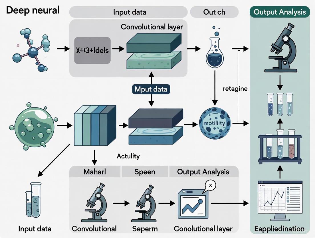
Visual Workflows and Logical Diagrams
The following diagram illustrates the integrated clinical and computational workflow for deep learning-based sperm analysis, highlighting the pathway from sample collection to clinical decision support.
AI-Driven Sperm Analysis Workflow
The next diagram details the core technical process of the deep learning model for segmenting and classifying sperm structures, which is the foundation of automated analysis.
DNN-Based Sperm Segmentation and Classification
Subjectivity and Variability in Manual Semen Analysis
Semen analysis remains the cornerstone of male fertility assessment, playing a crucial role in both clinical diagnostics and research settings. Despite the publication of standardized World Health Organization (WHO) laboratory manuals, manual semen analysis continues to suffer from significant subjectivity and inter-laboratory variability [10] [11]. This technical application note examines the sources and implications of this variability, particularly within the context of developing deep neural networks for automated sperm analysis. For computational biologists and drug development professionals, understanding these pre-analytical and analytical challenges is essential for creating robust artificial intelligence (AI) models that can overcome human limitations in traditional assessment methods.
The fundamental challenge lies in the inherent complexity of semen analysis, which encompasses multiple parameters each susceptible to different sources of variation. As researchers increasingly turn to AI solutions for sperm motility and morphology estimation, comprehensive understanding of these variability sources becomes critical for developing effective computational models. This document provides a detailed examination of variability sources, quantitative assessments, standardized protocols, and emerging computational approaches that collectively inform the development of more reliable analysis systems.
Analytical Variability in Semen Assessment
The examination of human semen involves multiple procedural steps, each introducing potential variability that can compromise result consistency and clinical utility. Evidence indicates several critical points of variation require careful standardization:
Pre-analytical factors: The duration of sexual abstinence significantly impacts semen parameters, with WHO recommending 2-7 days [10]. Sample collection methods, transportation conditions, and liquefaction time (recommended 30-60 minutes at 37°C) further contribute to variability [11] [12]. Studies utilizing at-home sperm testing kits have demonstrated that even with standardized instructions, intra-subject variation remains substantial, particularly in men with oligozoospermia [13].
Analytical subjectivity: Sperm motility assessment suffers from significant inter-technician variability, as classification into progressive (rapid and slow), non-progressive, and immotile categories relies on subjective visual estimation [10] [14]. The evaluation of sperm morphology represents perhaps the most challenging parameter, with classification according to strict Kruger criteria requiring extensive technical expertise and demonstrating considerable inter-laboratory variation [15] [16].
Technical and methodological factors: The choice of counting chambers (e.g., Makler, MicroCell, Leja, or standard coverslip preparations) introduces substantial variability, particularly for concentration and motility assessments [14]. Staining techniques for morphology evaluation and equipment calibration issues further compound methodological variations [11].
Quantitative Assessment of Variability
Recent studies have quantified the degree of variability in semen parameters, providing essential baseline data for AI model development and validation. The following table summarizes key variability metrics from clinical studies:
Table 1: Quantitative Variability in Semen Parameters
| Parameter | Type of Variability | Coefficient of Variation | Clinical Implications |
|---|---|---|---|
| Sperm Concentration | Intra-subject (oligozoospermic) | 33.8% [13] | Requires multiple samples for accurate diagnosis |
| Sperm Concentration | Intra-subject (normozoospermic) | 24.5% [13] | Lower variability but still significant |
| Total Motile Sperm Count | Intra-subject (oligozoospermic) | 44.6% [13] | High variability affects treatment planning |
| Total Motility | Inter-laboratory | 10-20% [11] | Impacts consistency across facilities |
| Progressive Motility | Method-dependent (CASA vs. manual) | 5-15% [14] | Affects protocol comparisons |
| Normal Morphology | Inter-technician | Up to 30% [16] [3] | Significant diagnostic implications |
Analysis of 513 men providing multiple samples via at-home testing kits revealed that intra-subject variation was consistently lower than inter-subject variation across all parameters, with men exhibiting normozoospermia demonstrating greater stability in their semen parameters compared to those with oligozoospermia [13]. This variability underscores the recommendation by the American Urological Association and American Society for Reproductive Medicine to perform at least two semen analyses, spaced one month apart, particularly when initial results are abnormal [13].
Standardized Experimental Protocols
WHO 6th Edition Basic Semen Examination
The WHO 6th edition manual (2021) introduced important terminology changes, replacing "standard tests" with "basic examinations," "optional tests" with "extended examinations," and "research tests" with "advanced examinations" [10]. The following protocol details the basic examination procedure:
Table 2: Research Reagent Solutions for Basic Semen Analysis
| Reagent/Equipment | Specification | Function | Quality Control |
|---|---|---|---|
| Collection Container | Wide-mouthed, sterile, nontoxic material | Complete ejaculate collection | Biocompatibility testing [12] |
| Transport Medium | mHTF (modified Human Tubal Fluid) with HEPES | Maintain sperm viability during transport | Osmolarity: 280-300 mOsm/kg; pH: 7.3-7.5 [13] |
| Counting Chamber | Leja (20μm depth) or Makler (10μm depth) | Standardized depth for concentration/motility | Depth verification; QC bead calibration [11] [14] |
| Staining Solutions | Diff-Quik kit or RAL Diagnostics kit | Morphology assessment | Lot-to-lot consistency verification [13] |
| Phase Contrast Microscope | 10x-100x objectives with heated stage (37°C) | Motility assessment and morphology | Daily temperature calibration [11] |
Step-by-Step Protocol:
Sample Collection and Liquefaction: Collect specimen after 2-7 days of abstinence through masturbation into a sterile, wide-mouthed container. Maintain sample at 20-27°C during transport and allow complete liquefaction within 30-60 minutes at 37°C [10] [12]. Record any sample collection issues, as the initial ejaculate fraction contains the highest sperm concentration.
Macroscopic Examination: Assess volume (lower reference limit: 1.4 mL), appearance, viscosity, and pH (>7.2) [10] [12]. Note unusual odor as it may indicate urinary contamination or infection.
Motility Assessment: After complete liquefaction, mix sample gently and load onto a pre-warmed counting chamber. Assess minimum of 200 spermatozoa using phase-contrast microscopy at 37°C. Classify motility as:
- Progressive motility (rapid and slow): Sperm moving actively, either in a straight line or large circles
- Non-progressive motility: All other patterns of motility with absence of progression
- Immotile: No movement [10]
Sperm Concentration and Total Count: Using improved Neubauer hemocytometer or dedicated counting chamber, dilute sample 1:20 with diluent. Count minimum of 200 spermatozoa in duplicate. Calculate concentration (million/mL) and total sperm count per ejaculate (lower reference limit: 39 million) [10].
Sperm Vitality: Perform when total motility <40%. Use eosin-nigrosin stain to differentiate between live (unstained) and dead (stained) spermatozoa. Lower reference limit for vitality: 54% [10].
Sperm Morphology: Prepare thin smears, air dry, and stain according to standardized protocol. Assess minimum of 200 spermatozoa using strict Kruger criteria. Classify as normal or abnormal with detailed annotation of head, midpiece, and tail defects. Lower reference limit for normal forms: 4% [10] [15].
Quality Control Procedures
Implementation of robust quality control (QC) measures is essential for reliable results:
Internal Quality Control (IQC): Perform daily temperature checks of instruments, monthly chamber accuracy verification with QC beads, and semi-annual technician proficiency assessments [11].
External Quality Control (EQC): Participate in external proficiency testing programs to assess inter-laboratory consistency and identify systematic errors [11].
Standardized Documentation: Maintain detailed records of all QC activities, including reagent lot numbers, equipment maintenance, and technician training [11].
Diagram 1: Semen Analysis Workflow
Emerging AI Approaches for Standardization
Deep Learning for Morphology Classification
Convolutional Neural Networks (CNNs) represent a promising solution to address the high subjectivity in morphological assessment. Recent research demonstrates several approaches:
Database Development and Augmentation: The SMD/MSS (Sperm Morphology Dataset/Medical School of Sfax) dataset exemplifies the trend toward curated, expert-annotated image collections for training deep learning models [16]. This dataset initially contained 1,000 individual spermatozoa images classified according to modified David criteria (12 morphological defect classes) and was expanded to 6,035 images through data augmentation techniques including rotation, scaling, and intensity variations [16].
CNN Architecture for Morphology Classification: A typical implementation involves:
- Image Pre-processing: Convert images to grayscale, resize to standardized dimensions (e.g., 80×80 pixels), and normalize pixel values [16]
- Data Partitioning: Split dataset into training (80%), validation (10%), and testing (10%) subsets
- Model Training: Implement CNN architecture with multiple convolutional and pooling layers for feature extraction, followed by fully connected layers for classification
- Performance Evaluation: Assess using accuracy, precision, recall, and F1-score metrics [16]
Reported accuracy ranges from 55% to 92% across different morphological classes, with highest performance achieved on distinct abnormalities such as macrocephalic and microcephalic heads [16].
Motility Analysis Validation Framework
The Motility Ratio method provides a novel approach to validate sperm motility assessment techniques [14]. This method establishes a "gold standard" for motility measurement through controlled experimental design:
Experimental Protocol:
- Split semen sample into two equal fractions
- Fraction A: Maintain maximum motility (100% reference)
- Fraction B: Eliminate motility through freeze-thaw cycling (0% reference)
- Create standardized motility ratios by mixing Fractions A and B in predetermined proportions (e.g., 0%, 25%, 50%, 75%, 100%)
- Compare measured motility against theoretical values across different assessment methods [14]
This validation framework demonstrated that different chamber types introduce significant variability, with LEJA chambers showing minimal bias (<1%) while coverslip preparations exhibited substantial overestimation (>7%) of motility [14].
Diagram 2: Motility Validation Method
Implications for Drug Development and Research
For pharmaceutical researchers developing compounds affecting male fertility, the documented variability in semen analysis presents both challenges and opportunities:
Clinical Trial Design: Account for intrinsic variability in semen parameters through appropriate sample size calculations and repeated measures designs. The high intra-individual variability in oligozoospermic subjects (CVw up to 44.6%) necessitates larger sample sizes or multiple baseline assessments [13].
Endpoint Selection: Consider incorporating AI-based morphological assessment as exploratory endpoints to reduce measurement variability and increase sensitivity to detect treatment effects.
Quality Assurance: Implement centralized laboratories with standardized protocols and participation in external quality control programs to minimize inter-site variability in multicenter trials [11].
The integration of deep learning approaches into male fertility research offers the potential to not only reduce subjectivity but also to discover novel morphological signatures predictive of drug efficacy or toxicity that may escape conventional manual assessment.
Limitations of Current Computer-Assisted Semen Analysis (CASA) Systems
Computer-Assisted Semen Analysis (CASA) systems represent a significant technological advancement in the field of andrology, aiming to automate and objectify the evaluation of key sperm parameters such as sperm concentration, motility, and morphology [17]. The integration of artificial intelligence (AI), particularly deep neural networks, promises to enhance the analysis of sperm motility and morphology by learning complex patterns from image and video data, potentially overcoming the limitations of manual, subjective assessments [17].
However, despite these promising innovations, current CASA systems exhibit persistent limitations. Evidence shows that the results from various CASA systems are not fully consistent with those from the manual method, which is still considered the gold standard [4]. These inconsistencies can lead to skewed clinical decisions, particularly in the critical choice between conventional in vitro fertilization (IVF) and intracytoplasmic sperm injection (ICSI) [4]. This document details these limitations through structured data presentation, experimental protocols, and analytical diagrams to inform researchers and drug development professionals.
Quantitative Performance Data of CASA Systems
A 2025 study directly compared three CASA systems—the Hamilton-Thorne CEROS II Clinical, the LensHooke X1 Pro, and the SQA-V Gold Sperm Quality Analyzer—against the manual method, which was performed according to the WHO laboratory manual and served as the gold standard [4]. The study involved 326 participants and used statistical measures like the Intraclass Correlation Coefficient (ICC) and Cohen's Kappa (κ) to evaluate agreement.
Table 1: Agreement Between CASA Systems and Manual Method for Sperm Parameters
| Sperm Parameter | CASA System | Statistical Measure | Value | Interpretation |
|---|---|---|---|---|
| Concentration | Hamilton-Thorne CEROS II | ICC | 0.723 | Moderate [4] |
| LensHooke X1 Pro | ICC | 0.842 | Good [4] | |
| SQA-V Gold | ICC | 0.631 | Moderate [4] | |
| Motility | Hamilton-Thorne CEROS II | ICC | 0.634 | Moderate [4] |
| LensHooke X1 Pro | ICC | 0.417 | Poor [4] | |
| SQA-V Gold | ICC | 0.451 | Poor [4] | |
| Morphology | LensHooke X1 Pro | ICC | 0.160 | Poor [4] |
| SQA-V Gold | ICC | 0.261 | Poor [4] |
Table 2: Agreement for Clinical Diagnosis Between CASA Systems and Manual Method
| Clinical Diagnosis | CASA System | Cohen's Kappa (κ) | Interpretation |
|---|---|---|---|
| Oligozoospermia | LensHooke X1 Pro | 0.701 | Substantial [4] |
| Hamilton-Thorne CEROS II | 0.664 | Substantial [4] | |
| SQA-V Gold | 0.588 | Moderate [4] | |
| Asthenozoospermia | LensHooke X1 Pro | 0.405 | Moderate [4] |
| Hamilton-Thorne CEROS II | 0.249 | Fair [4] | |
| SQA-V Gold | 0.157 | Slight [4] | |
| Teratozoospermia | LensHooke X1 Pro | 0.177 | Slight [4] |
| SQA-V Gold | 0.008 | No agreement [4] |
A critical finding was the impact of these discrepancies on treatment allocation. When based on manual morphology assessment, the ratio for ICSI was approximately 0.5. However, when using the LensHooke X1 Pro and SQA-V Gold systems, the ratios skewed to about 0.31 and 0.15, respectively. This demonstrates a significant reduction in ICSI recommendation when relying on CASA morphology analysis, potentially affecting treatment outcomes [4].
Experimental Protocol for Validating CASA Systems
The following protocol, derived from contemporary research methodologies, outlines the steps for a rigorous validation study comparing CASA systems against the manual gold standard [4].
Sample Collection and Preparation
- Ethics and Consent: The study must be reviewed and approved by an institutional review board (e.g., CREC No. 2016.499). All participants must provide informed consent [4].
- Sample Size: Recruitment of a sufficient number of participants (e.g., n > 300) to ensure statistical power [4].
- Sample Handling: Semen samples are collected and processed according to standard clinical protocols to ensure sample integrity.
Manual Semen Analysis (Gold Standard)
- Procedure: Manual analysis is performed by an experienced andrologist in strict adherence to the World Health Organization (WHO) laboratory manual (5th Edition or current) [4].
- Quality Control: The andrology unit should implement regular internal quality control and participate in external quality assurance programs (e.g., United Kingdom National External Quality Assessment Service - UK NEQAS) [4] [18].
- Parameters Assessed:
- Concentration: Calculated using an improved Neubauer counting chamber at 400x magnification [4].
- Motility: Evaluated at 400x magnification and classified into progressive (PR), non-progressive (NP), and immotile (IM) categories [4].
- Morphology: Stained using the Diff-Quik method and evaluated at 1000x oil-immersion magnification [4].
- Replication: All manual analyses should be performed in duplicate to ensure reliability.
CASA System Analysis
- Systems Tested: The protocol should specify the CASA systems under evaluation (e.g., Hamilton-Thorne CEROS II, LensHooke X1 Pro, SQA-V Gold) [4].
- Calibration and Operation: Each CASA system must be operated according to the manufacturer's instructions. This includes using specific slides (e.g., Leja 4 chamber slides for CEROS II) or test cassettes (e.g., for LensHooke X1 Pro) and ensuring proper calibration [4].
- Parallel Assessment: The same semen sample, or an aliquot from the same ejaculate, should be analyzed by the manual method and each CASA system to allow for direct pairwise comparisons.
Data and Statistical Analysis
- Statistical Tests:
- Intraclass Correlation Coefficient (ICC): A two-way random-effects model should be used to measure consistency for continuous data (concentration, motility). Values below 0.5 indicate poor agreement, 0.5-0.75 moderate, 0.75-0.9 good, and above 0.9 excellent agreement [4].
- Bland-Altman Analysis: To assess the agreement between two measurement methods by calculating the mean difference and limits of agreement [4].
- Cohen's Kappa (κ): To evaluate the agreement for categorical diagnoses (e.g., oligozoospermia, asthenozoospermia). Interpretation: ≤0 as no agreement, 0.01-0.20 as none to slight, 0.21-0.40 as fair, 0.41-0.60 as moderate, 0.61-0.80 as substantial, and 0.81-1.00 as almost perfect agreement [4].
- Clinical Impact Assessment: Analyze the discrepancy in treatment allocation (IVF vs. ICSI) based on morphology results from different methods [4].
Diagram 1: CASA System Validation Workflow. This chart outlines the key steps for a rigorous experimental protocol to validate CASA systems against the manual gold standard.
The Scientist's Toolkit: Key Research Reagent Solutions
Table 3: Essential Materials for Semen Analysis Validation Studies
| Item | Function / Description | Example / Standard |
|---|---|---|
| Improved Neubauer Chamber | A hemocytometer used for the manual counting of sperm concentration [4]. | Standard laboratory equipment. |
| Diff-Quik Staining Kit | A rapid staining method for sperm morphology evaluation, based on a modified Romanowsky technique [4]. | Halotech [4]. |
| Leja Slides | Disposable counting chambers with a defined depth, specifically designed for sperm analysis and compatible with specific CASA systems [4]. | Leja 4 chambers (IMV Technologies) [4]. |
| LensHooke Test Cassettes | Disposable cassettes with dual drip areas for analyzing pH and other sperm parameters on the LensHooke X1 Pro system [4]. | Bonraybio [4]. |
| SQA-V Gold Capillary | A disposable capillary tube used to load the semen sample into the SQA-V Gold analyzer [4]. | Medical Electronic Systems [4]. |
| WHO Laboratory Manual | The definitive international standard and guideline for the examination and processing of human semen [4]. | World Health Organization (5th Edition or current). |
| External Quality Control (EQC) | Programs to detect and correct systematic errors, ensuring standardization and high-quality results across laboratories [18]. | E.g., External Quality Control for SCA [18]. |
Limitations and Challenges for Deep Learning Integration
The pursuit of more accurate CASA systems through deep learning faces several significant hurdles.
Algorithmic and Data Challenges: A primary issue is the inconsistency of results across different CASA platforms, as each system uses proprietary algorithms that have not been standardized [4] [17]. This is compounded by the "black-box" nature of complex deep learning models, which can make it difficult to understand how specific morphological or motility conclusions are reached, potentially hindering clinical trust and adoption [17]. Furthermore, the performance of these models is heavily dependent on large, high-quality, annotated datasets for training, which are often lacking. This can lead to challenges in model generalizability across diverse patient populations and clinical settings [17].
Clinical and Regulatory Hurdles: As demonstrated, deviations in morphology analysis can directly lead to skewed IVF/ICSI treatment allocation, a critical clinical decision [4]. Therefore, rigorous clinical validation through controlled trials is essential before AI-driven CASA systems can be widely adopted. This process is intertwined with the need for standardized evaluation protocols and clear regulatory frameworks to ensure patient safety and data privacy, especially given the sensitive nature of reproductive information [17].
Diagram 2: Key Limitations of Current CASA Systems. This diagram categorizes the major technical and clinical challenges facing current systems and the integration of deep learning.
Current CASA systems, despite their objective of automating and standardizing semen analysis, demonstrate significant limitations, particularly in the assessment of sperm morphology and motility, when compared to the manual method. These inconsistencies are not merely statistical but have direct, meaningful consequences for clinical treatment pathways. The integration of deep neural networks holds the potential to overcome these limitations by extracting subtle, predictive features from raw image data [17]. However, this path is fraught with challenges related to data, algorithms, and clinical validation. Future research must therefore focus on developing more transparent and robust AI models, curated multi-center datasets, and conducting rigorous external validation studies to ensure that these advanced systems can fulfill their promise of personalized, efficient, and accurate fertility care.
The Role of Deep Learning in Standardizing Reproductive Diagnostics
The diagnostic assessment of male fertility has long been constrained by subjective analytical techniques. Conventional semen analysis, particularly the evaluation of sperm motility and morphology, suffers from significant inter-observer variability despite standardized World Health Organization (WHO) protocols [19] [3]. This lack of standardization impedes diagnostic accuracy and reliable treatment planning in clinical andrology.
Deep learning (DL), a subset of artificial intelligence (AI), is emerging as a transformative technology for automating and standardizing reproductive diagnostics. Unlike traditional computer-aided sperm analysis (CASA) systems, deep convolutional neural networks (DCNNs) can learn discriminative features directly from image and video data, minimizing human subjectivity [19] [20]. This application note details experimental protocols and analytical frameworks for implementing deep learning solutions to quantify sperm motility and morphology, with direct relevance for researchers and drug development professionals working in reproductive medicine.
Quantitative Performance of Deep Learning Models
Recent validation studies demonstrate that deep learning models can achieve performance levels comparable to human experts in classifying sperm quality parameters. The tables below summarize quantitative results from published studies on sperm motility and morphology analysis.
Table 1: Performance of Deep Learning Models in Sperm Motility Analysis
| Study Reference | Model Architecture | Task | Performance Metrics |
|---|---|---|---|
| Scientific Reports, 2023 [19] | ResNet-50 (Optical Flow) | 3-category motility (Progressive, Non-progressive, Immotile) | MAE: 0.05; Correlation with manual: r=0.88 (Progressive) |
| Scientific Reports, 2023 [19] | ResNet-50 (Optical Flow) | 4-category motility (Rapid, Slow, Non-progressive, Immotile) | MAE: 0.07; Correlation: r=0.673 (Rapid progressive) |
| VISEM Dataset Study [20] | Custom CNN (MotionFlow) | Motility estimation | MAE: 6.842% |
Table 2: Performance of Deep Learning Models in Sperm Morphology Analysis
| Study Reference | Model/Dataset | Classification Task | Reported Accuracy/Performance |
|---|---|---|---|
| SMD/MSS Dataset, 2025 [16] | Custom CNN | 12 morphological defect classes (David's classification) | Accuracy range: 55% to 92% |
| VISEM Dataset Study [20] | Custom CNN | Morphology estimation | MAE: 4.148% |
| BMC Urology, 2025 [3] | Review of conventional ML | Sperm head classification | Up to 90% accuracy (Bayesian model) |
Experimental Protocols for Sperm Analysis
Protocol 1: DCNN for Sperm Motility Categorization
This protocol outlines the procedure for training a Deep Convolutional Neural Network (DCNN) to classify sperm motility into WHO categories using video data [19].
Materials:
- Fresh semen samples
- Microscope with video recording capability (400x magnification)
- Preheated slides and microscope stage (37°C)
- Computational resources (GPU recommended)
- Python with OpenCV, Keras, and TensorFlow/PyTorch
Procedure:
- Sample Preparation & Video Acquisition:
- Incubate fresh semen samples at 37°C for 30-60 minutes after collection.
- Prepare wet preparations and record multiple random fields for 5-10 seconds each, ensuring a minimum of 200 spermatozoa are captured per sample.
- Maintain a constant temperature of 37°C during recording using a heated stage.
Ground Truth Labeling:
- Have trained technicians assess video recordings according to WHO 1999 [19] or current WHO criteria.
- Categorize spermatozoa into: (a) Rapid Progressive, (b) Slow Progressive, (c) Non-Progressive, and (d) Immotile. For a 3-category model, combine (a) and (b) into "Progressive."
- Use mean values from multiple reference laboratories if possible to reduce labeling bias.
Motion Representation Preprocessing:
- For each second of video, compute the Lucas–Kanade optical flow between consecutive frames. This compresses the temporal movement information into a single 2D image representing motion vectors.
- Use these optical flow images as the input to the DCNN, rather than raw video frames.
Model Architecture & Training:
- Employ a ResNet-50 architecture, replacing the final layer with a number of neurons equal to your motility categories (3 or 4).
- Use the Adam optimizer with a learning rate of 0.0004 and Mean Absolute Error (MAE) as the loss function.
- Implement a 10-fold cross-validation strategy to robustly evaluate model performance and prevent overfitting.
Validation & Statistical Analysis:
- Compare DCNN-predicted motility percentages against manual ground truth using Pearson’s correlation coefficient and MAE.
- Generate difference plots (Bland-Altman-style) to visualize agreement and identify any systematic biases.
Figure 1: Sperm Motility Analysis Workflow. This diagram outlines the key steps for developing a deep learning model to classify sperm motility from video data, from sample preparation to model validation.
Protocol 2: CNN-Based Sperm Morphology Classification
This protocol details the development of a Convolutional Neural Network (CNN) for classifying sperm morphology using the SMD/MSS (Sperm Morphology Dataset/Medical School of Sfax) dataset based on modified David classification [16].
Materials:
- Stained sperm smears (e.g., RAL Diagnostics staining kit)
- Microscope with 100x oil immersion objective and digital camera
- MMC CASA system or equivalent for image acquisition
- Computational environment (Python 3.8 with deep learning frameworks)
Procedure:
- Dataset Curation & Annotation:
- Collect semen samples and prepare smears according to WHO guidelines. Include samples with varying morphological profiles.
- Capture images of individual spermatozoa using a bright-field microscope with a 100x oil immersion objective.
- Establish ground truth through independent classification by multiple experienced embryologists. Use a classification system such as the modified David classification, which includes 12 defect classes across the head, midpiece, and tail.
Data Preprocessing & Augmentation:
- Resize all images to a uniform resolution (e.g., 80x80 pixels) and convert to grayscale.
- Normalize pixel values to a standard range (e.g., 0-1).
- Address class imbalance and limited data by applying augmentation techniques such as rotation, flipping, scaling, and brightness adjustment to increase the effective dataset size.
Inter-Expert Agreement Analysis:
- Quantify the level of agreement between the three experts for each image. Categorize agreement as: Total Agreement (TA), Partial Agreement (PA), or No Agreement (NA).
- Use statistical tests (e.g., Fisher's exact test) to identify significant differences in classification between experts. This analysis reveals the inherent subjectivity and complexity of the task.
Model Development & Partitioning:
- Design a CNN architecture with multiple convolutional and pooling layers for feature extraction, followed by fully connected layers for classification.
- Partition the augmented dataset randomly, allocating 80% for training and 20% for testing. Further split the training set, using a portion for validation during training.
- Train the model using the training set, monitoring performance on the validation set to avoid overfitting.
Performance Evaluation:
- Evaluate the final model on the held-out test set.
- Report overall accuracy and, crucially, performance metrics (e.g., precision, recall) for each morphological defect class to ensure clinical utility.
The Scientist's Toolkit: Research Reagent Solutions
Table 3: Essential Materials and Reagents for Deep Learning-based Sperm Analysis
| Item Name | Function/Application | Specification Notes |
|---|---|---|
| Time-Lapse Incubator (e.g., EmbryoScope+) | Maintains stable culture conditions while capturing embryonic development images [21] [22]. | Integrated microscope and camera; key for embryo analysis. |
| Optical Microscope with Heated Stage | For live sperm video recording under physiological conditions [19]. | Must maintain 37°C; 400x magnification for motility. |
| 100x Oil Immersion Objective | High-resolution imaging of individual spermatozoa for morphology analysis [16]. | Essential for detailed head, midpiece, and tail assessment. |
| RAL Staining Kit | Differentiates sperm structures for morphological assessment on smears [16]. | Provides contrast for head, acrosome, and midpiece. |
| Global Culture Medium (e.g., G-TL) | Supports embryo development in time-lapse systems [22]. | Optimized for culture in time-lapse incubators. |
| ResNet-50 / Custom CNN | Deep learning architecture for image and motion analysis [19] [23]. | Pre-trained models can be adapted via transfer learning. |
| Python Deep Learning Frameworks | Model development, training, and validation environment [19] [16]. | TensorFlow, PyTorch, Keras with OpenCV for image processing. |
Analytical Framework & Data Interpretation
The integration of deep learning into reproductive diagnostics requires careful experimental design and critical evaluation of results.
Key Considerations for Model Validation:
- Ground Truth Quality: The performance of any DL model is capped by the quality of its training labels. In sperm morphology, significant inter-expert variability is a major challenge. Models trained on data with poor consensus will reflect this inconsistency [16] [3].
- Clinical Correlation: While high accuracy in classifying motility or morphology is important, the ultimate validation is the model's ability to predict clinically relevant outcomes, such as fertilization success or live birth rates. This requires linking model predictions to patient outcomes [23].
- Generalizability: A model must perform well on data from different clinics, using various microscopes, staining protocols, and sample preparation techniques. Testing on diverse, external datasets is crucial before clinical deployment [3].
Figure 2: Analytical Validation Framework. This diagram illustrates the core dependencies for validating a deep learning model in reproductive diagnostics, emphasizing the need for high-quality labels, clinical correlation, and multi-center testing.
Deep learning models provide a robust methodological foundation for standardizing the assessment of sperm motility and morphology. The protocols outlined herein enable the quantitative, automated analysis of sperm parameters, directly addressing the critical issue of subjectivity in conventional diagnostics. For researchers and pharmaceutical developers, these technologies offer reproducible biomarkers for evaluating male fertility and assessing the efficacy of novel therapeutic compounds. The continued development of large, high-quality annotated datasets and the validation of models against clinical outcomes will be essential to fully integrate these tools into mainstream reproductive medicine and drug development pipelines.
Deep learning (DL) is revolutionizing the field of andrology, particularly in the analysis of sperm morphology and motility. This paper provides a comprehensive overview of the two most pivotal deep learning architectures—Convolutional Neural Networks (CNNs) and Recurrent Neural Networks (RNNs)—and details their specific applications and protocols in male infertility research. CNNs, with their superior spatial feature extraction capabilities, have become the de facto standard for image-based tasks such as sperm morphology analysis and head/vacuole detection. RNNs, especially their variants like Long Short-Term Memory (LSTM) networks, are uniquely suited for temporal sequence analysis, making them ideal for assessing dynamic parameters like sperm motility and trajectory patterns. This article presents structured data on model performance, standardized experimental protocols for implementing these architectures, and a curated list of essential research reagents and computational tools. By framing these technologies within the context of a broader thesis on deep neural networks for sperm quality estimation, this work aims to provide researchers, scientists, and drug development professionals with practical resources to advance andrology research.
The adoption of deep learning in andrology addresses critical challenges in traditional analysis methods, which are often characterized by subjectivity, high inter-observer variability, and substantial workload. Deep learning models, through their ability to learn hierarchical representations directly from data, offer a path toward automated, standardized, and high-throughput analysis.
Convolutional Neural Networks (CNNs) are a class of deep neural networks primarily designed for processing grid-like data such as images. Their architecture is inspired by the organization of the animal visual cortex, where individual neurons respond to stimuli only in a restricted region of the visual field known as the receptive field [24]. CNNs automatically and adaptively learn spatial hierarchies of features through backpropagation, from low-level edges in early layers to high-level conceptual features in deeper layers [25]. This makes them exceptionally powerful for tasks like classifying sperm as normal or abnormal based on head morphology.
Recurrent Neural Networks (RNNs) represent another fundamental class of neural networks engineered for sequential data. Unlike feedforward networks, RNNs contain loops that allow information to persist, enabling the network to maintain an internal state or "memory" of previous inputs in the sequence [26] [27]. This architectural characteristic is crucial for modeling temporal dynamics, such as those found in sperm motility tracks. However, vanilla RNNs often struggle with learning long-range dependencies due to the vanishing and exploding gradient problems. This limitation has been effectively addressed by more sophisticated gated architectures, such as Long Short-Term Memory (LSTM) networks and Gated Recurrent Units (GRUs), which incorporate mechanisms to selectively remember or forget information over long time periods [26] [28].
The integration of these technologies into andrology represents a paradigm shift from subjective manual assessment to objective, data-driven analysis, with the potential to unlock novel biomarkers for male fertility and drug efficacy testing.
Convolutional Neural Networks (CNNs) in Sperm Morphology Analysis
Core Architectural Principles
CNNs process images through a series of specialized layers that transform pixel values into a final prediction (e.g., a classification label). The core components of a typical CNN include [25] [24] [29]:
- Convolutional Layers: These layers apply a set of learnable filters (or kernels) to the input image. Each filter slides across the image, computing dot products to detect local spatial features such as edges, corners, and textures. Initial layers capture basic features, while deeper layers combine them into more complex structures like shapes and object parts.
- Activation Functions (ReLU): Following a convolution, an element-wise activation function like the Rectified Linear Unit (ReLU) is applied. This introduces non-linearity into the model, enabling it to learn complex patterns by effectively deciding which features are important to pass forward.
- Pooling Layers: Pooling (e.g., max pooling) performs non-linear down-sampling, reducing the spatial dimensions of the feature maps. This decreases the computational load, controls overfitting, and provides a basic form of translation invariance.
- Fully Connected Layers: After several cycles of convolution and pooling, the resulting high-level feature maps are flattened into a vector and fed into one or more fully connected layers. These layers integrate the distributed features to perform the final classification or regression task.
This hierarchical processing pipeline allows CNNs to achieve remarkable accuracy in image recognition tasks, with modern architectures like ResNet and GoogleNet enabling the training of very deep networks [29].
Application in Sperm Morphology Analysis
Sperm Morphology Analysis (SMA) is a critical, yet challenging, component of male fertility assessment. According to the World Health Organization (WHO) standards, sperm morphology is divided into the head, neck, and tail, with 26 types of abnormal morphology, requiring the analysis of over 200 sperms [30]. Manual observation is laborious and subject to inter-observer variability.
CNNs are being deployed to automate this process, demonstrating capabilities in two key areas [30]:
- Accurate automated segmentation of sperm morphological structures (head, neck, and tail).
- Substantial improvements in the efficiency and accuracy of sperm morphology classification.
Early machine learning approaches for SMA relied on handcrafted features (e.g., shape descriptors, grayscale intensity) and classifiers like Support Vector Machines (SVMs). A study by Bijar et al. achieved 90% accuracy in classifying sperm heads using Bayesian Density Estimation [30]. However, these methods are limited by their dependence on manual feature engineering.
Deep learning, particularly CNNs, overcomes this limitation by learning features directly from data. A study by Javadi et al. developed a CNN model to extract features such as the acrosome, head shape, and vacuoles from a dataset of 1,540 sperm images (the MHSMA dataset) [30]. This end-to-end learning paradigm has shown promising results in distinguishing between normal and abnormal sperm, as well as identifying specific defect types.
Table 1: Quantitative Performance of Selected Models for Sperm Morphology Analysis
| Study / Model | Task | Dataset | Key Metric & Performance |
|---|---|---|---|
| Bijar A et al. [30] | Head Morphology Classification | Not Specified | Accuracy: 90% (4 categories: normal, tapered, pyriform, small/amorphous) |
| Javadi S et al. [30] | Feature Extraction (acrosome, head, vacuoles) | MHSMA (1,540 images) | Qualitative demonstration of automated feature learning |
| Conventional CNN [30] | Morphology Classification | Various Public Datasets | Outperforms conventional ML reliant on handcrafted features |
The following diagram illustrates a typical CNN workflow for static sperm image analysis, from input to classification.
Experimental Protocol: CNN-Based Sperm Morphology Classification
Objective: To train a CNN model to automatically classify sperm images into predefined morphological categories (e.g., normal, tapered, pyriform, small, amorphous).
Materials:
- Annotated Sperm Image Dataset: Such as the MHSMA [30] or SVIA dataset [30]. The SVIA dataset contains 125,000 annotated instances for detection, 26,000 segmentation masks, and 125,880 cropped images for classification.
- Hardware: A computer with a powerful Graphics Processing Unit (GPU). GPUs are essential for efficient training of deep learning models [31].
- Software Frameworks: TensorFlow or PyTorch with Python.
Procedure:
- Data Preprocessing:
- Resizing: Standardize all images to a fixed input size (e.g., 224x224 pixels).
- Normalization: Scale pixel values to a standard range (e.g., 0-1).
- Data Augmentation: Artificially expand the training dataset by applying random but realistic transformations to the images. This includes rotations, flips, slight zooms, and brightness variations. This step is crucial for improving model generalization and preventing overfitting [25].
Model Design & Training:
- Architecture Selection: Choose a pre-existing CNN architecture like VGG, ResNet, or a custom-designed simpler CNN.
- Transfer Learning: A common and effective technique is to initialize the model with weights pre-trained on a large dataset like ImageNet. The final layers of the pre-trained model are then replaced and fine-tuned on the sperm morphology dataset.
- Compilation: Define a loss function (e.g., Categorical Cross-Entropy for multi-class classification) and an optimizer (e.g., Adam).
- Training: Feed the training data into the model in mini-batches. The model's weights are updated iteratively via backpropagation to minimize the loss function.
Model Evaluation:
- Validation: Use a held-out validation set to monitor model performance during training and tune hyperparameters.
- Testing: Evaluate the final model's performance on a completely unseen test set using metrics such as Accuracy, Precision, Recall, and F1-Score [29]. The model's predictions should be compared against annotations made by expert andrologists.
Recurrent Neural Networks (RNNs) in Sperm Motility Analysis
Core Architectural Principles
While CNNs excel with spatial data, RNNs are designed for sequential data where the order and temporal context are critical. Their fundamental feature is a recurrent connection that loops the hidden state of the network from one time step to the next, creating a form of memory [26] [28].
The update of the hidden state (ht) at time step (t) is typically computed as: [ht = \sigma(W{xh}xt + W{hh}h{t-1} + bh)] where (xt) is the input, (h_{t-1}) is the previous hidden state, (W) are weight matrices, (b) is a bias, and (\sigma) is an activation function [26] [28].
Basic RNNs suffer from the vanishing/exploding gradient problem, making it difficult to learn long-range dependencies. This has been successfully addressed by two advanced variants:
- Long Short-Term Memory (LSTM): LSTMs introduce a gating mechanism and a cell state that acts as a "conveyor belt," allowing information to flow across many time steps with minimal alteration. The gates (input, forget, and output) regulate the flow of information, deciding what to store, forget, and output [26] [28].
- Gated Recurrent Unit (GRU): GRUs are a simpler alternative to LSTMs, combining the input and forget gates into a single "update gate." They often achieve performance similar to LSTMs but with greater computational efficiency [26].
Application in Sperm Motility and Trajectory Analysis
Sperm motility is a dynamic process where the movement pattern of a sperm cell over time is a strong indicator of its health and fertilizing potential. Analyzing this temporal sequence is a task perfectly suited for RNNs.
RNNs can be applied to:
- Motility Classification: Classifying sperm tracks into categories (e.g., progressive, non-progressive, immotile) based on a sequence of positional coordinates.
- Trajectory Prediction: Forecasting the future path of a sperm cell based on its past movements.
- Time-Series Analysis: Modeling other temporal parameters, such as changes in velocity or flagellar beating patterns.
These models process the sequential location data ((x1, y1), (x2, y2), ..., (xt, yt)) of individual sperm cells. The LSTM or GRU units learn the characteristic patterns of movement associated with different motility states, effectively capturing the temporal dependencies that define a sperm's swimming behavior.
Table 2: RNN Variants and Their Relevance to Andrology Applications
| RNN Variant | Key Characteristics | Potential Andrology Application |
|---|---|---|
| Simple RNN | Basic recurrent connection; struggles with long sequences. | Baseline model for short-track analysis. |
| Long Short-Term Memory (LSTM) | Gated architecture (input, forget, output gates); excels at learning long-term dependencies. | Analysis of long motility tracks; complex trajectory modeling. |
| Gated Recurrent Unit (GRU) | Simplified LSTM with fewer gates; computationally efficient. | Motility classification where training speed is a priority. |
| Bidirectional RNN (Bi-RNN) | Processes sequences both forward and backward for richer context. | Comprehensive analysis of completed sperm tracks. |
The following diagram illustrates the process of using an RNN for temporal sperm motility analysis.
Experimental Protocol: RNN-Based Sperm Motility Classification
Objective: To train an RNN model (e.g., LSTM or GRU) to classify the motility type of individual sperm cells based on their tracked trajectory sequences.
Materials:
- Sperm Tracking Data: A dataset of sperm trajectories, such as the VISEM-Tracking dataset, which contains 656,334 annotated objects with tracking details [30]. Each trajectory should be a time-series of 2D coordinates and be labeled with a motility class.
- Hardware/Software: Same as for CNN protocol.
Procedure:
- Data Preprocessing:
- Trajectory Extraction: Use computer vision techniques (e.g., object detection and tracking algorithms) to extract the ((x, y)) coordinate sequences for each sperm cell from video data.
- Sequence Standardization: Normalize coordinate values and pad or truncate all sequences to a uniform length.
- Velocity/Feature Calculation (Optional): Derive additional features like instantaneous speed and curvature for each time step to enrich the input sequence.
Model Design & Training:
- Architecture: Construct a model comprising one or more LSTM/GRU layers. A typical architecture is Many-to-One, where the sequence of inputs is processed to produce a single classification output at the end.
- Training: The model is trained using Backpropagation Through Time (BPTT), a variant of backpropagation adapted for RNNs that unrolls the network across time steps to calculate gradients [28].
Model Evaluation:
- Evaluate the model's classification performance on a held-out test set of trajectories using standard metrics (Accuracy, F1-Score). The results should be benchmarked against both manual expert analysis and traditional computational methods.
The Scientist's Toolkit: Research Reagent Solutions
Successful implementation of deep learning projects in andrology requires a combination of curated data, computational tools, and software resources.
Table 3: Essential Resources for Deep Learning in Andrology Research
| Resource Category | Item Name | Function & Application Notes |
|---|---|---|
| Public Datasets | SVIA (Sperm Videos and Images Analysis) [30] | Contains 125,000 annotated instances for detection, 26,000 segmentation masks, and 125,880 cropped images. Useful for both CNN (morphology) and RNN (motility from videos) tasks. |
| VISEM-Tracking [30] | A multi-modal dataset with 656,334 annotated objects and tracking details. Primarily for training and validating sperm tracking and motility models. | |
| MHSMA (Modified Human Sperm Morphology Analysis) [30] | Contains 1,540 grayscale sperm head images. Suitable for developing and testing sperm head morphology classification models. | |
| Computational Hardware | Graphics Processing Unit (GPU) [31] | Essential for accelerating the training of deep learning models, reducing computation time from weeks to hours. |
| Software Frameworks | TensorFlow / PyTorch | Open-source libraries that provide the foundation for building, training, and deploying deep learning models. Offer high-level APIs (e.g., Keras) for rapid prototyping. |
CNNs and RNNs offer powerful and complementary capabilities for advancing andrology research. CNNs provide an objective and scalable solution for the precise analysis of static sperm morphology, while RNNs are uniquely equipped to decode the complex temporal patterns of sperm motility. The integration of these deep learning architectures into research and clinical workflows promises to standardize male fertility assessment, reduce inter-observer variability, and uncover novel insights into sperm function. However, the field must continue to address challenges such as the need for larger, high-quality annotated datasets [30] and the "black box" nature of these models to build trust and ensure robust clinical translation [25]. The experimental protocols and resources outlined in this article provide a foundational roadmap for researchers aiming to harness deep learning for sperm motility and morphology estimation.
Architectures in Action: Implementing DNNs for Motility Classification and Morphological Segmentation
Deep Convolutional Neural Networks for WHO Motility Categorization
The analysis of sperm motility is a cornerstone of male fertility assessment, with manual evaluation according to World Health Organization (WHO) guidelines remaining the gold standard in clinical practice. However, this process is inherently subjective, time-consuming, and requires extensive technical training to produce reproducible results. Recent advances in artificial intelligence, particularly Deep Convolutional Neural Networks (DCNNs), are poised to revolutionize this field by introducing automation, enhancing objectivity, and improving analytical throughput. This protocol details the application of DCNNs for categorizing sperm motility into WHO-defined classes, providing researchers and clinicians with a framework for implementing these advanced computational methods. The content is situated within a broader thesis research context focused on developing deep learning solutions for comprehensive sperm quality assessment, encompassing both motility and morphology estimation.
Background and Significance
WHO Motility Categorization
The WHO manual establishes a critical classification system for sperm motility, essential for diagnosing male factor infertility. The categorization can be structured into three or four classes:
- Progressive Motility (Grade a+b): Sperm moving actively, either in a straight line or in a large circle.
- Rapid Progressive (Grade a): Sperm moving with a high velocity.
- Slow Progressive (Grade b): Sperm moving forward but with reduced speed.
- Non-Progressive Motility (Grade c): Sperm moving but without forward progression.
- Immotile (Grade d): Sperm showing no movement.
Traditional manual assessment is vulnerable to inter-laboratory variability and technician subjectivity, creating a compelling need for automated, standardized solutions [19].
The Role of Deep Convolutional Neural Networks
DCNNs are a class of deep learning models exceptionally suited for image recognition and classification tasks. Their capacity to automatically learn hierarchical feature representations from raw pixel data makes them ideal for analyzing complex visual patterns in sperm video microscopy. Within reproductive medicine, DCNNs facilitate the development of objective, high-throughput analysis systems that can learn to replicate expert-level motility assessments, thereby overcoming the significant limitations of manual methods [19] [32].
Quantitative Performance of DCNN Models
The performance of DCNN models for sperm motility analysis is quantitatively evaluated using metrics such as Mean Absolute Error (MAE) and Pearson's correlation coefficient, which compare the model's predictions against manual assessments by trained experts.
Table 1: Performance Metrics of DCNN Models for WHO Motility Categorization
| Motility Category | Model Type | Mean Absolute Error (MAE) | Pearson's Correlation (r) | Citation |
|---|---|---|---|---|
| Progressive Motility | 3-Category Model | 0.06 | 0.88 (p<0.001) | [19] |
| Immotile Spermatozoa | 3-Category Model | 0.05 | 0.89 (p<0.001) | [19] |
| Non-Progressive Motility | 3-Category Model | 0.04 | Not Reported | [19] |
| Rapid Progressive Motility | 4-Category Model | Not Reported | 0.673 (p<0.001) | [19] |
| Overall Motility | MotionFlow + Transfer Learning | 6.842% (MAE) | Not Reported | [20] |
| Overall Morphology | MotionFlow + Transfer Learning | 4.148% (MAE) | Not Reported | [20] |
Recent research has demonstrated significant progress. One study utilizing the ResNet-50 architecture reported a strong correlation between DCNN-predicted values and manual assessments for progressive and immotile spermatozoa [19]. Another approach introduced a novel motion representation called MotionFlow, combined with transfer learning, achieving a mean absolute error of 6.842% for motility estimation, thereby outperforming previous state-of-the-art methods [20].
Experimental Protocols
Detailed Protocol: DCNN Motility Assessment with ResNet-50
This protocol outlines the procedure for training and validating a Deep Convolutional Neural Network, specifically the ResNet-50 architecture, to categorize sperm motility from video recordings according to WHO guidelines [19].
I. Materials and Equipment
- Microscope: Equipped with a heated stage (37°C), 400x magnification objective, and video camera.
- Semen Samples: Fresh ejaculates incubated at 37°C.
- Software: Python with deep learning libraries (e.g., TensorFlow, Keras), and computer vision libraries (OpenCV).
- Computing Hardware: Computer with a high-performance GPU (e.g., NVIDIA GTX series or equivalent) for accelerated model training.
II. Step-by-Step Procedure
Step 1: Video Acquisition and Preprocessing
- Prepare Sample: Place a fresh, liquefied semen sample on a pre-warmed microscope slide (37°C). Use a coverslip to create a wet preparation.
- Record Videos: 30-60 minutes post-collection, record multiple random fields of view. Ensure each video is 5-10 seconds long at a frame rate of 30 frames per second. The number of fields should allow for the assessment of at least 200 spermatozoa.
- Data Curation: Collect a dataset of video recordings, such as the 65 videos from the ESHRE-SIGA EQA Programme used in the foundational study [19].
Step 2: Optical Flow Calculation
- Extract Motion Features: For every second of video, compute the Lucas-Kanade optical flow. This process compresses the temporal movement information of spermatozoa across 30 frames into a single 2D image that represents motion vectors.
- Generate Input Data: Use the resulting optical flow images as the input to the DCNN. This step transforms the problem from a complex video analysis task to a more manageable image classification task.
Step 3: Model Architecture and Training
- Select Model: Implement the ResNet-50 architecture. Modify the final layer to include a Global Average Pooling layer and an output layer with nodes corresponding to the number of motility categories (3 or 4).
- Configure Training:
- Optimizer: Adam (learning rate = 0.0004).
- Loss Function: Mean Absolute Error (MAE).
- Validation: Employ a ten-fold cross-validation strategy. This involves splitting the dataset into 10 parts, training on 9, and validating on 1, repeating this process 10 times.
- Early Stopping: Halt training if the validation loss does not improve for 15 consecutive epochs to prevent overfitting.
Step 4: Model Validation and Statistical Analysis
- Compare Predictions: Calculate Pearson's correlation coefficient to assess the strength of the linear relationship between DCNN-predicted motility percentages and the mean manual assessments from reference laboratories.
- Evaluate Agreement: Use difference plots (Bland-Altman plots) to visualize the agreement between the two methods and identify any systematic biases.
- Benchmark Performance: Compare the model's MAE against a ZeroR baseline model to confirm that the DCNN has learned meaningful patterns.
Workflow Visualization
The following diagram illustrates the integrated experimental and computational pipeline for DCNN-based sperm motility analysis.
The Scientist's Toolkit: Research Reagent Solutions
Implementing a DCNN for sperm motility analysis requires a combination of biological, computational, and data resources.
Table 2: Essential Research Reagents and Resources
| Item Name | Specification / Brand | Function and Application Note |
|---|---|---|
| Video Dataset | ESHRE-SIGA EQA Programme Dataset [19] | Provides ground-truth labeled videos of sperm motility for training and validating DCNN models. |
| Deep Learning Framework | TensorFlow / Keras [19] | An open-source software library used for designing, training, and deploying the DCNN model. |
| Pre-trained Model | ResNet-50 [19] | A proven DCNN architecture for image classification; transfer learning from ImageNet can improve performance with limited data. |
| Optical Flow Algorithm | Lucas-Kanade Method [19] | Converts sequential video frames into a single 2D image representing sperm motion, simplifying the input for the DCNN. |
| Microscope with Heated Stage | Standard clinical microscope | Maintains sperm at physiological temperature (37°C) during video recording to preserve native motility characteristics. |
| Performance Metrics | Mean Absolute Error (MAE), Pearson Correlation [19] | Quantitative measures used to objectively benchmark the model's accuracy against manual assessments. |
Discussion and Future Perspectives
The adoption of DCNNs for WHO motility categorization represents a paradigm shift towards more standardized and scalable semen analysis. While current models like ResNet-50 show high agreement with manual assessments for progressive and immotile sperm, challenges remain in accurately classifying rapid progressive motility, which often exhibits greater inter-laboratory variation in the training data [19]. Future research directions should focus on the development of multi-task learning frameworks that can simultaneously estimate motility and morphology from the same input video [20] [33]. Furthermore, the creation of large, public, and meticulously annotated datasets will be crucial for training more robust and generalizable models, ultimately paving the way for their integration into routine clinical practice [30].
Accurate segmentation of sperm components is a critical technological process in male infertility diagnosis and assisted reproductive technologies. According to the World Health Organization, sperm morphology analysis provides crucial diagnostic information, with morphological abnormalities present in the head, neck, and tail regions correlating strongly with infertility issues [30]. Traditional manual sperm assessment methods are inherently subjective, time-consuming, and exhibit significant inter-observer variability, creating an urgent need for automated, objective solutions [30] [34].
Deep learning-based computer vision systems have emerged as promising alternatives to address these limitations. These systems can automatically segment distinct sperm components—including the head, acrosome, nucleus, neck/midpiece, and tail—enabling precise morphological analysis essential for clinical diagnosis and treatment selection [35] [36]. The accurate segmentation of these components is particularly important for intracytoplasmic sperm injection (ICSI) procedures, where embryologists must select the most viable sperm based on morphological integrity [35].
This application note provides a comprehensive comparison of three prominent deep learning architectures—Mask R-CNN, U-Net, and YOLO models—for multi-part sperm segmentation. We present quantitative performance evaluations, detailed experimental protocols, and practical implementation guidelines to assist researchers and clinicians in developing robust sperm analysis systems for reproductive medicine and drug development applications.
Quantitative Performance Comparison
Table 1: Comparative performance of deep learning models for sperm component segmentation
| Sperm Component | Model | IoU | Dice Coefficient | Precision | Recall | F1-Score |
|---|---|---|---|---|---|---|
| Head | Mask R-CNN | - | - | - | - | - |
| YOLOv8 | - | - | - | - | - | |
| U-Net | - | - | - | - | - | |
| Acrosome | Mask R-CNN | - | - | - | - | - |
| YOLO11 | - | - | - | - | - | |
| U-Net | - | - | - | - | - | |
| Nucleus | Mask R-CNN | - | - | - | - | - |
| YOLOv8 | - | - | - | - | - | |
| U-Net | - | - | - | - | - | |
| Neck/Midpiece | Mask R-CNN | - | - | - | - | - |
| YOLOv8 | - | - | - | - | - | |
| U-Net | - | - | - | - | - | |
| Tail | Mask R-CNN | - | - | - | - | - |
| YOLOv8 | - | - | - | - | - | |
| U-Net | - | - | - | - | - |
Note: Specific quantitative values from the search results are not provided in the excerpts. The table structure follows standard reporting format for segmentation metrics. Actual values should be populated from experimental results following the protocol implementation.
Performance Analysis by Sperm Component
Recent systematic evaluations indicate that Mask R-CNN demonstrates superior performance in segmenting smaller and more regular sperm structures, including the head, nucleus, and acrosome [35]. Specifically, Mask R-CNN achieves slightly higher Intersection over Union (IoU) values for nucleus segmentation compared to YOLOv8 and outperforms YOLO11 for acrosome segmentation, highlighting its robustness for precise anatomical structure delineation [35].
For neck/midpiece segmentation, YOLOv8 performs comparably or slightly better than Mask R-CNN in certain configurations, suggesting that single-stage detectors can rival two-stage architectures for this specific component under optimized conditions [35].
The morphologically complex tail structure presents unique segmentation challenges due to its elongated, thin morphology and frequent occlusion. For this component, U-Net achieves the highest IoU, demonstrating the advantage of its encoder-decoder architecture with global perception and multi-scale feature extraction capabilities for elongated structures [35].
Experimental Protocols
Dataset Preparation and Annotation
Sample Collection and Preparation
- Semen Collection: Collect semen samples following standardized protocols, maintaining temperature at 37°C throughout processing [34].
- Sample Preparation: Dilute raw semen samples with appropriate extenders (e.g., Optixcell at 1:1 ratio, then further dilute to 1:20 ratio) to achieve optimal concentration for microscopy (approximately 17.5–27.5 ×10⁶/mL) [34].
- Slide Preparation: Place 10μL of diluted sample on a standard microscope slide (75×25×1mm), cover with coverslip (22×22mm), and fix using standardized methods [34].
Image Acquisition System Configuration
- Microscope Setup: Use a phase-contrast microscope (e.g., Optika B-383Phi) with 40× negative phase contrast objective and 1× eyepiece [34].
- Imaging Software: Utilize manufacturer-provided applications (e.g., PROVIEW for Optika microscopes) for image capture [34].
- Image Specifications: Capture images in JPEG format under standardized bright-field microscopy conditions with consistent contrast and illumination to minimize variability [34].
Annotation Protocol
- Annotation Tools: Use specialized annotation software (e.g., Roboflow, CVAT, or Label Studio) for precise labeling of sperm components [37] [34].
- Component Labeling: Annotate five distinct morphological regions: head, acrosome, nucleus, neck/midpiece, and tail [35].
- Quality Control: Employ multiple trained annotators with expertise in sperm morphology, with final validation by at least three sperm morphology experts with >10 years of experience [35].
Data Preprocessing and Augmentation
Intensity Normalization and Contrast Enhancement
- Log Transformation: Apply log transformation for intensity normalization to account for varying illumination conditions [38].
- Histogram Equalization: Implement histogram equalization techniques to enhance contrast in low-contrast regions of sperm images [38].
- Edge-based ROI Extraction: Utilize edge detection methods to extract regions of interest and focus computational resources on relevant image areas [38].
Data Augmentation Techniques
Comprehensive data augmentation is essential for improving model generalization and robustness to biological variability and imaging artifacts [39].
Spatial Transformations:
Photometric Transformations:
Model Implementation Protocols
Mask R-CNN Implementation
- Backbone Configuration: Utilize ResNet-50 or ResNet-101 with Feature Pyramid Network (FPN) for multi-scale feature extraction.
- Region Proposal Network: Set anchor scales to [32, 64, 128, 256, 512] to accommodate varying sperm component sizes.
- Training Parameters: Use learning rate of 0.001, batch size of 2-8 (depending on GPU memory), and train for 50-100 epochs.
U-Net Implementation
- Encoder Configuration: Implement with various encoders (ResNet34, VGG16, EfficientNet) for feature extraction [39].
- Decoder Architecture: Use standard expanding path with skip connections to preserve spatial information.
- Training Parameters: Apply learning rate of 0.0001, batch size of 8-16, and train for 100-200 epochs.
YOLO Implementation
- Architecture Selection: Deploy YOLOv7, YOLOv8, or YOLOv11 based on specific application requirements [34] [38].
- Anchor Configuration: Customize anchor boxes to match sperm component aspect ratios.
- Training Parameters: Use learning rate of 0.01, batch size of 16-32, and train for 300-500 epochs.
Evaluation Methodology
Performance Metrics
- Primary Metrics: Calculate Intersection over Union (IoU), Dice Coefficient, Precision, Recall, and F1-Score for each sperm component [35].
- Additional Metrics: Compute mean Average Precision (mAP@50) for detection performance and inference time for real-time capability assessment [34] [38].
Validation Strategy
- Dataset Splitting: Employ 80/20 training-test split with k-fold cross-validation (typically k=5) to ensure statistical significance [38].
- Comparative Analysis: Conduct paired statistical tests to evaluate significant performance differences between models for each sperm component.
Workflow Visualization
Diagram 1: End-to-end workflow for developing multi-part sperm segmentation systems
Model Selection Framework
Diagram 2: Model selection framework for sperm segmentation applications
The Scientist's Toolkit
Table 2: Essential research reagents and computational tools for sperm segmentation research
| Category | Item/Resource | Specification/Function | Application Notes |
|---|---|---|---|
| Laboratory Equipment | Phase-Contrast Microscope | Optika B-383Phi with 40× objective | Essential for high-quality image acquisition without staining [34] |
| Sample Fixation System | Trumorph system (60°C, 6kp pressure) | Enables dye-free fixation preserving natural morphology [34] | |
| Imaging Software | PROVIEW application | Manufacturer software for standardized image capture [34] | |
| Computational Resources | Annotation Tools | Roboflow, CVAT, Label Studio | Streamlined annotation workflow for sperm components [37] [34] |
| Deep Learning Frameworks | PyTorch, TensorFlow, MMDetection | Model development and experimentation [37] | |
| Edge Deployment | NVIDIA Jetson Nano, Google Coral TPU | Real-time deployment in clinical settings [37] | |
| Datasets | Human Sperm Datasets | SVIA, VISEM-Tracking, SCIAN-SpermSegGS | 125,000+ annotated instances available for training [35] [30] |
| Annotation Standards | WHO morphology guidelines | Standardized classification: normal, head, neck, tail defects [30] [34] | |
| Evaluation Metrics | Segmentation Metrics | IoU, Dice, Precision, Recall, F1-Score | Component-specific performance assessment [35] |
| Detection Metrics | mAP@50, mAP@[0.5:0.95] | Overall model performance evaluation [38] |
The comparative analysis presented in this application note demonstrates that different deep learning architectures excel at segmenting specific sperm components. Mask R-CNN shows superior performance for smaller, regular structures like heads and acrosomes; YOLO models provide efficient segmentation for neck/midpiece regions with real-time capabilities; while U-Net achieves the highest accuracy for the morphologically complex tail structures [35].
Future research directions should focus on ensemble approaches that leverage the complementary strengths of these architectures, cross-species generalization of segmentation models, and clinical validation of automated segmentation systems for routine diagnostic use. The integration of multi-modal data (combining morphology with motility analysis) and the development of explainable AI systems will further enhance clinical adoption and utility in reproductive medicine and drug development contexts.
The protocols and frameworks outlined in this document provide researchers with comprehensive guidance for implementing robust sperm segmentation systems that can advance both basic reproductive research and clinical infertility treatments.
Data Augmentation Techniques for Enhancing Limited Datasets
In the field of biomedical research, particularly in studies involving human sperm motility and morphology, the development of robust deep neural networks (DNNs) is often constrained by the limited availability of large, annotated datasets. Data scarcity is a fundamental challenge in medical AI, where patient data can be difficult to obtain, annotations require expert knowledge, and ethical considerations limit sharing [40]. This challenge is particularly acute in reproductive medicine, where the variability of biological samples and the need for specialized expert labeling further compound the problem [16] [41]. Data augmentation and synthetic data generation have emerged as critical methodologies to mitigate these limitations, enabling the expansion of training datasets and improving the generalizability of deep learning models [16] [40].
The application of these techniques is especially valuable for sperm analysis research, where manual assessment of sperm morphology and motility remains time-consuming, subjective, and prone to significant inter-observer variability [16] [41]. By artificially increasing the size and diversity of available datasets, data augmentation allows researchers to develop more accurate and reliable DNN models for quantitative sperm analysis, ultimately advancing male infertility diagnostics and treatment.
Data Augmentation Fundamentals
Data augmentation encompasses a suite of techniques designed to artificially expand a dataset by creating modified versions of existing data samples. These techniques are particularly valuable in medical imaging and biomedical data analysis, where collecting large datasets is often impractical due to cost, time, or ethical constraints [40]. In the context of sperm analysis research, augmentation strategies can be broadly categorized into classical augmentation methods, which include geometric and photometric transformations, and advanced synthetic data generation techniques using deep generative models [40].
The primary objectives of data augmentation in sperm analysis research include:
- Addressing class imbalance: Ensuring adequate representation of rare morphological classes or motility patterns
- Improving model robustness: Enhancing generalization to unseen data by exposing models to realistic variations
- Increasing effective dataset size: Enabling training of complex DNN architectures that require large amounts of training data
- Reducing overfitting: Providing regularizing effects during model training
Techniques and Applications in Sperm Analysis
Classical Data Augmentation Methods
Classical data augmentation techniques involve applying predefined transformations to existing images or data samples. These methods have been successfully applied in sperm morphology analysis to enhance limited datasets.
Table 1: Classical Data Augmentation Techniques for Sperm Image Analysis
| Technique Category | Specific Methods | Application in Sperm Analysis | Key Parameters |
|---|---|---|---|
| Geometric Transformations | Rotation, flipping, scaling, translation, elastic deformations | Increasing orientation variance of sperm cells | Rotation angles (±30°), scale range (0.8-1.2x) |
| Photometric Transformations | Brightness adjustment, contrast modification, color jitter, noise injection | Simulating varying staining conditions and microscope settings | Brightness (±20%), contrast factor (0.8-1.2) |
| Image Processing Techniques | Sharpening, blurring, morphological operations | Accounting for focus variations and image quality differences | Gaussian blur (σ=0.5-1.5), kernel sizes |
In a seminal study on deep learning for sperm morphology classification, researchers applied comprehensive data augmentation to a dataset of individual spermatozoa images, expanding it from 1,000 to 6,035 images [16]. This augmentation was critical for balancing morphological classes and enabling effective training of their convolutional neural network model, which achieved accuracy ranging from 55% to 92% across different morphological categories [16].
Advanced Synthetic Data Generation
Beyond classical augmentation, advanced synthetic data generation techniques have shown promise for creating entirely new samples that maintain the statistical properties of the original data. These methods are particularly valuable for addressing extreme class imbalance in rare morphological defects.
Table 2: Advanced Synthetic Data Generation Techniques
| Technique | Mechanism | Advantages | Limitations |
|---|---|---|---|
| Generative Adversarial Networks (GANs) | Two-network system (generator vs. discriminator) | Can produce highly realistic synthetic images | Training instability, mode collapse |
| Diffusion Models | Progressive denoising process | High sample quality, stable training | Computationally intensive |
| Transfer Learning with Pretrained Models | Leveraging features learned on large datasets | Effective even with very small datasets | Domain shift concerns |
While these advanced methods were less commonly applied in the specific sperm analysis studies reviewed, their potential for generating diverse sperm morphology examples is significant, particularly for creating rare abnormality cases that may be underrepresented in clinical datasets [40].
Experimental Protocols
Protocol 1: Basic Image Augmentation for Sperm Morphology Analysis
This protocol outlines a standardized approach for augmenting sperm image datasets to train deep learning models for morphology classification, based on methodologies successfully implemented in recent research [16] [41].
Materials and Equipment:
- High-quality sperm images acquired via CASA system or similar
- Python 3.8+ with libraries: OpenCV, TensorFlow/Keras or PyTorch, Albumentations
- Computational resources (GPU recommended)
Procedure:
- Image Acquisition and Preprocessing
- Capture individual spermatozoa images using a standardized protocol (e.g., 100x oil immersion objective, bright field mode) [16]
- Apply initial preprocessing: resize to uniform dimensions (e.g., 80×80 pixels), convert to grayscale, normalize pixel values [16]
- Perform noise reduction using appropriate filters (e.g., Gaussian, median)
Data Augmentation Pipeline Implementation
- Implement geometric transformations:
- Random rotation within range of ±30 degrees
- Horizontal and vertical flipping (probability: 0.5)
- Random scaling (range: 0.8-1.2x)
- Elastic deformations with controlled parameters
- Apply photometric transformations:
- Brightness adjustment (±20% variation)
- Contrast modification (factor range: 0.8-1.2)
- Addition of Gaussian noise (σ=0.01-0.05)
- Execute the augmentation pipeline in real-time during training or as a preprocessing step
- Implement geometric transformations:
Quality Control and Validation
- Visually inspect augmented samples to ensure biological plausibility
- Verify that augmentation preserves morphological features critical for classification
- Ensure balanced representation across morphological classes post-augmentation
Implementation Notes:
- The augmentation strategy should be tailored to the specific classification task (e.g., head defects vs. tail defects)
- For the SMD/MSS dataset, this approach enabled expansion from 1,000 to 6,035 images [16]
- Monitor training to ensure augmented data improves rather than degrades model performance
Protocol 2: Video Augmentation for Sperm Motility Analysis
This protocol addresses the augmentation of video data for deep learning models analyzing sperm motility, based on methodologies from recent studies [19] [42].
Materials and Equipment:
- Video recordings of sperm samples (e.g., 400x magnification, 37°C)
- Python with OpenCV, NumPy, and deep learning frameworks
- Optical flow computation libraries (e.g., OpenCV Lucas-Kanade)
Procedure:
- Video Preprocessing
- Extract frames from raw video files (e.g., 50 frames per second) [42]
- Standardize frame dimensions and format across all videos
- Apply temporal sub-sampling if necessary to manage computational load
Temporal Augmentation Techniques
- Implement frame skipping and temporal jittering
- Apply optical flow transformations to capture motion patterns
- Use Lucas-Kanade optical flow estimation to compress temporal information into single images [19]
Spatial Augmentation on Video Frames
- Apply standard image augmentation techniques to individual frames
- Ensure temporal consistency when applying spatial transformations
- Implement 3D augmentations that consider both spatial and temporal dimensions
Synthetic Sequence Generation
- Create synthetic video sequences by combining sperm tracks from multiple sources
- Generate variations in sperm density through compositing techniques
- Simulate different microscope conditions and focusing variations
Implementation Notes:
- For motility classification, ResNet-50 architecture processing optical flow images has shown promising results [19]
- Augmentation should preserve the physiological characteristics of sperm movement
- The approach should account for WHO motility categories (progressive, non-progressive, immotile) [19]
Visualization of Workflows
Data Augmentation Pipeline for Sperm Image Analysis
Figure 1: Comprehensive data augmentation pipeline for sperm image analysis, showing the sequential transformation of original images through multiple augmentation strategies to produce a diversified dataset suitable for training robust deep learning models.
Experimental Setup for Sperm Motility Analysis
Figure 2: Experimental workflow for sperm motility analysis using deep convolutional neural networks, illustrating the process from raw video input through augmentation to final motility categorization according to WHO standards.
The Scientist's Toolkit
Table 3: Essential Research Reagents and Computational Tools for Sperm Analysis Studies
| Category | Item/Technique | Specification/Purpose | Application Example |
|---|---|---|---|
| Imaging Equipment | CASA System | Computer-Assisted Semen Analysis for standardized image acquisition | Sperm morphology and motility quantification [16] |
| Microscopy | Phase Contrast Microscope with heated stage (37°C) | Maintain physiological temperature during analysis | Live sperm motility assessment [42] |
| Staining Reagents | RAL Diagnostics staining kit | Standardized staining for morphology assessment | Sperm head and tail defect identification [16] |
| Deep Learning Frameworks | Python 3.8 with TensorFlow/PyTorch | Model development and training environment | CNN implementation for morphology classification [16] [41] |
| Data Augmentation Libraries | Albumentations, Imgaug | Specialized libraries for image augmentation | Geometric and photometric transformations [16] |
| Model Architectures | ResNet-50, CNN with CBAM | Attention-based feature extraction | Sperm morphology classification with 96% accuracy [41] |
| Evaluation Metrics | MAE, Accuracy, SSIM, PSNR | Quantitative performance assessment | Model validation and comparison [16] [43] [19] |
Data augmentation techniques represent a fundamental methodology for advancing research in sperm motility and morphology analysis using deep neural networks. Through the strategic application of both classical and advanced augmentation methods, researchers can overcome the critical challenge of limited dataset size that often constrains medical AI projects. The protocols and frameworks presented in this document provide a roadmap for implementing these techniques effectively, with demonstrated success in recent studies achieving high classification accuracy and robust model performance [16] [41].
As the field progresses, the integration of more sophisticated synthetic data generation methods with classical augmentation approaches promises to further enhance our ability to develop accurate, reliable, and clinically applicable deep learning models for male fertility assessment. This will ultimately contribute to standardized, objective diagnostic tools that reduce inter-laboratory variability and improve patient care in reproductive medicine.
Transfer Learning Approaches in Sperm Image Analysis
Infertility affects a significant proportion of couples globally, with male factors contributing to approximately 50% of cases [30] [3]. The analysis of sperm morphology and motility represents a crucial component in male fertility assessment, providing diagnostic information for clinicians and embryologists [30]. Traditional manual assessment of sperm quality, while considered the gold standard, faces substantial challenges related to subjectivity, reproducibility, and inter-observer variability [16] [44]. These limitations have prompted the development of computer-assisted semen analysis (CASA) systems, which aim to automate and standardize the evaluation process [45] [46].
Deep learning, particularly convolutional neural networks (CNNs), has demonstrated remarkable success in various medical image analysis tasks [35]. However, training these models from scratch requires extensive annotated datasets, which are often difficult and expensive to acquire in the medical domain [30] [44]. Transfer learning has emerged as a powerful strategy to address this limitation by leveraging knowledge from pre-trained models on large-scale datasets such as ImageNet, enabling effective model training even with limited medical image data [47]. This Application Note explores the implementation of transfer learning approaches for sperm image analysis, providing detailed protocols and performance comparisons to facilitate adoption in research and clinical settings.
Comparative Analysis of Datasets and Model Performance
Publicly Available Sperm Image Datasets
The development and validation of transfer learning models for sperm image analysis rely on standardized datasets with expert annotations. The table below summarizes key publicly available datasets used in this domain.
Table 1: Publicly Available Sperm Image Datasets for Transfer Learning Research
| Dataset Name | Image Count | Annotation Type | Key Characteristics | Classes/Categories |
|---|---|---|---|---|
| HuSHeM [47] | 216 images | Classification | Stained sperm heads, cropped and rotated | 4 classes: Normal, Tapered, Pyriform, Amorphous |
| SCIAN-MorphoSpermGS [47] | 1,854 images | Classification | Stained sperm heads with expert classifications | 5 classes: Normal, Tapered, Pyriform, Small, Amorphous |
| SMD/MSS [16] | 1,000 images (extended to 6,035 with augmentation) | Classification & Morphometry | Based on modified David classification | 12 morphological defect classes |
| SVIA [30] [35] | 125,000 annotated instances | Detection, Segmentation, Classification | Large-scale dataset with multiple annotation types | Multiple classes for comprehensive analysis |
| VISEM-Tracking [30] [3] | 656,334 annotated objects | Detection, Tracking, Regression | Multi-modal with videos and tracking data | Motion characteristics and morphology |
Performance Comparison of Transfer Learning Approaches
Various transfer learning architectures have been applied to sperm image analysis tasks, with demonstrated efficacy across different datasets and clinical requirements.
Table 2: Performance Comparison of Transfer Learning Models on Sperm Analysis Tasks
| Model Architecture | Dataset | Task | Performance Metrics | Key Advantages |
|---|---|---|---|---|
| Modified AlexNet [47] | HuSHeM | Head Morphology Classification | 96.0% accuracy, 96.4% precision | Computational efficiency, minimal parameter tuning |
| ResNet-50 [19] | VISEM | Motility Classification | MAE: 0.05 (3-category), 0.07 (4-category) | Effective temporal motion analysis |
| VGG16 [47] | HuSHeM | Head Morphology Classification | 94.1% accuracy | High baseline performance |
| Mask R-CNN [35] | Live Unstained Sperm | Multi-part Segmentation | Highest IoU for head, nucleus, acrosome | Superior for smaller, regular structures |
| U-Net [35] | Live Unstained Sperm | Multi-part Segmentation | Highest IoU for tail segmentation | Excellent for morphologically complex structures |
Experimental Protocols
Protocol 1: Sperm Head Morphology Classification Using Transfer Learning
This protocol details the implementation of transfer learning for sperm head morphology classification based on the approach described by [47] that achieved 96.0% accuracy on the HuSHeM dataset.
Preprocessing and Data Preparation
- Image Acquisition: Collect sperm images using standardized microscopy protocols. For the HuSHeM dataset, images were acquired at 131×131 pixels in RGB format [47].
- Head Cropping: Implement automated cropping to isolate sperm heads:
- Convert original image to grayscale and apply denoising filters
- Use Sobel operator to obtain gradient image
- Apply low-pass filter and adaptive thresholding for binarization
- Perform morphological operations (erosion and dilation) to remove noise
- Fit ellipse to determine head orientation and major/minor axes
- Extract centered head region (64×64 pixels) [47]
- Orientation Standardization: Rotate all sperm heads to a uniform direction (pointing right) to reduce rotational variance
- Data Augmentation: Apply transformations including rotation (±10°), horizontal flipping, and brightness adjustment (±20%) to increase dataset diversity
Model Adaptation and Training
- Base Model Selection: Choose AlexNet architecture pre-trained on ImageNet
- Network Modification:
- Replace original classification layers with task-specific layers
- Add Batch Normalization layers after each convolutional layer to improve training stability
- Maintain pre-trained parameters for feature extraction layers
- Training Configuration:
- Use Adam optimizer with learning rate of 0.0004 [19]
- Implement cross-entropy loss function for multi-class classification
- Set batch size of 32 based on available GPU memory
- Apply early stopping with patience of 15 epochs to prevent overfitting
Validation and Interpretation
- Performance Metrics: Calculate accuracy, precision, recall, and F1-score for each morphological class
- Cross-Validation: Employ k-fold cross-validation (k=10) to ensure robust performance estimation [19]
- Visualization: Generate Grad-CAM or saliency maps to interpret model decisions and validate against biological knowledge
Protocol 2: Multi-Part Sperm Segmentation Using Transfer Learning
This protocol describes the implementation of transfer learning for segmenting sperm components (head, acrosome, nucleus, neck, and tail) based on comparative evaluation of architectures [35].
Dataset Preparation and Annotation
- Sample Preparation: Use unstained live human sperm to maintain physiological relevance [35]
- Expert Annotation: Engage multiple embryologists with >10 years of experience to annotate sperm components
- Data Augmentation:
- Apply geometric transformations (rotation, scaling, elastic deformations)
- Use color space adjustments to simulate staining variations
- Generate synthetic images to balance underrepresented morphological classes [16]
Model Selection and Adaptation
- Architecture Selection:
- Choose Mask R-CNN for head, acrosome, and nucleus segmentation
- Select U-Net for tail segmentation due to its superior performance on elongated structures [35]
- Transfer Learning Implementation:
- Initialize with pre-trained weights on COCO dataset (Mask R-CNN) or medical image datasets (U-Net)
- Adapt feature extraction layers to accommodate sperm-specific characteristics
- Modify head architectures to predict sperm-specific components
- Training Strategy:
- Use multi-scale training to capture variably sized sperm components
- Implement gradient accumulation to overcome memory limitations
- Apply discriminative learning rates with lower rates for early layers
Evaluation and Validation
- Metrics Calculation: Compute IoU, Dice coefficient, Precision, Recall, and F1-score for each sperm component
- Clinical Validation: Compare segmentation results with manual annotations from multiple experts
- Statistical Analysis: Perform paired t-tests to determine significant differences between model architectures
Workflow Diagram: Transfer Learning for Sperm Image Analysis
The following diagram illustrates the complete workflow for applying transfer learning to sperm image analysis, from data preparation through model deployment:
Workflow for Sperm Image Analysis Using Transfer Learning
Successful implementation of transfer learning for sperm image analysis requires specific computational resources, datasets, and software tools. The following table details essential components for establishing a research pipeline in this domain.
Table 3: Essential Research Reagents and Computational Resources for Sperm Image Analysis
| Category | Item | Specification/Function | Example Sources/Implementations |
|---|---|---|---|
| Datasets | HuSHeM | Benchmarking sperm head classification | 216 images, 4 morphology classes [47] |
| SCIAN-MorphoSpermGS | Multi-class sperm head evaluation | 1,854 images, 5 morphology classes [47] | |
| SMD/MSS | Comprehensive morphology analysis | 12 defect classes based on David classification [16] | |
| VISEM-Tracking | Motility and tracking analysis | Video data with motion characteristics [30] | |
| Software | Python 3.8+ | Core programming language | With TensorFlow/PyTorch frameworks [16] |
| OpenCV | Image preprocessing and augmentation | Automated cropping, rotation, filtering [47] | |
| DeepLearning Frameworks | Model implementation | TensorFlow, Keras, PyTorch [19] | |
| Pre-trained Models | AlexNet | Base for morphology classification | Modified with Batch Normalization [47] |
| ResNet-50 | Motility classification from videos | Optical flow analysis [19] | |
| Mask R-CNN | Instance segmentation of components | Transfer learning from COCO dataset [35] | |
| U-Net | Semantic segmentation | Biomedical image specialization [35] | |
| Evaluation Metrics | IoU/Dice Coefficient | Segmentation accuracy assessment | Component-wise performance evaluation [35] |
| MAE | Motility classification performance | Error measurement for regression tasks [19] | |
| Accuracy/Precision/Recall | Classification performance | Standard classification metrics [47] |
Transfer learning represents a transformative approach for sperm image analysis, effectively addressing the challenges associated with limited annotated datasets in medical imaging. The protocols and analyses presented in this Application Note demonstrate that adapted pre-trained models can achieve expert-level performance in both morphology classification and segmentation tasks, with accuracy exceeding 96% in optimized implementations [47]. The continued development of standardized datasets and specialized architectures will further enhance the clinical applicability of these approaches, ultimately improving diagnostic accuracy and treatment outcomes in male infertility.
Future directions in this field include the development of integrated models capable of simultaneous morphology and motility analysis, domain adaptation techniques to improve cross-center generalization, and explainable AI methods to enhance clinical trust and adoption. As these computational approaches mature, they hold significant promise for revolutionizing sperm quality assessment in both clinical and research settings.
Optical Flow Analysis for Temporal Motility Assessment in Video Sequences
Within the broader research on deep neural networks for sperm motility and morphology estimation, the quantitative analysis of temporal movement patterns is paramount. Optical flow, a computer vision technique for estimating the apparent motion of objects between consecutive video frames, serves as a foundational method for this task. It quantifies motion by calculating displacement vectors for each pixel, providing a dense representation of movement dynamics over time [48] [49]. This Application Note details the integration of optical flow methodologies with deep learning architectures to create robust, automated systems for sperm motility assessment, a critical parameter in male infertility diagnosis and drug development research [19] [42]. The protocols herein are designed for researchers and scientists requiring reproducible, quantitative motion analysis.
Theoretical Foundations of Optical Flow
Optical flow operates on two core assumptions: first, the pixel intensity of an object remains constant between consecutive frames; and second, neighboring pixels have similar motion [48]. These principles are mathematically expressed by the Optical Flow Equation:
[fx u + fy v + f_t = 0]
where (fx) and (fy) are the spatial image gradients, (f_t) is the temporal gradient, and (u) and (v) are the unknown flow velocities in the x and y directions [48] [49]. Solving this equation for every pixel is an ill-posed problem; various algorithms have been developed to find optimal solutions, ranging from classical methods like Lucas-Kanade to modern deep learning-based approaches [48] [49].
Experimental Protocols
Protocol A: Lucas-Kanade Sparse Optical Flow for Feature Point Tracking
This protocol is ideal for tracking specific, high-quality spermatozoa or pre-selected points of interest. The Lucas-Kanade method is a sparse technique that computes flow for a subset of feature points, offering high computational efficiency [48] [49].
Procedure:
- Video Acquisition and Preprocessing:
- Capture semen sample videos using a microscope with a heated stage maintained at 37°C. Use 400x magnification and a frame rate of 50 frames per second (fps) or higher [42].
- Save videos in a lossless format (e.g., AVI) to preserve quality.
- For each frame, convert the image to grayscale (
cv.cvtColor).
Feature Point Detection:
- On the first frame of the video, use the
cv.goodFeaturesToTrackfunction to identify salient feature points for tracking. - Recommended Parameters:
maxCorners=100,qualityLevel=0.3,minDistance=7,blockSize=7[48].
- On the first frame of the video, use the
Optical Flow Calculation:
- Use the
cv.calcOpticalFlowPyrLKfunction to track the identified points from the previous frame to the current frame. - Recommended Parameters:
winSize=(15,15),maxLevel=2,criteria=(cv.TERM_CRITERIA_EPS | cv.TERM_CRITERIA_COUNT, 10, 0.03)[48]. - The function returns the new positions of the points and a status vector indicating successful tracks.
- Use the
Trajectory Analysis and Motility Quantification:
- Filter out points with a failed status.
- For each successfully tracked point, calculate the displacement vector between its previous and current position.
- Compute motility metrics, such as velocity (magnitude of displacement per frame) and linearity (straightness of the path over a time window).
Protocol B: Dense Optical Flow for Global Motility Assessment
This protocol generates a comprehensive motion field for the entire frame, suitable for analyzing collective sperm behavior and overall sample motility.
Procedure:
- Video Acquisition and Preprocessing: Follow Step 1 from Protocol A.
Dense Flow Estimation:
- Use the
cv.calcOpticalFlowFarnebackfunction to compute a dense flow field for each consecutive frame pair [48]. - This method produces a flow vector for every pixel in the frame.
- Use the
Motion Segmentation and Background Subtraction:
- Apply a magnitude threshold to the flow vectors to separate moving spermatozoa from static background elements and debris.
- Use clustering algorithms (e.g., DBSCAN) to group vectors belonging to individual sperm cells.
Population-Level Motility Analysis:
- Calculate the average velocity and directionality of all vectors exceeding the threshold.
- Derive the ratio of motile to immotile spermatozoa based on the number of pixels/vectors with near-zero magnitude.
Protocol C: Deep Learning-Based Motility Classification with Optical Flow Input
This advanced protocol leverages deep convolutional neural networks (DCNNs) for direct prediction of motility categories as defined by the World Health Organization (WHO), using optical flow representations as input [19].
Procedure:
- Optical Flow Image Generation:
- For each video, compute the Lucas-Kanade optical flow for every second of the video (e.g., across 30 frames for a 30 fps video) [19].
- Visualize the computed flow as a single image, effectively compressing the temporal motion information into a 2D representation suitable for CNN input.
Model Architecture and Training:
- Employ a pre-trained ResNet-50 architecture, modified by replacing the final fully connected layer with a new one matching the number of target motility classes (e.g., 3 classes: progressive, non-progressive, immotile; or 4 classes: rapid progressive, slow progressive, non-progressive, immotile) [19].
- Training Configuration: Use the Adam optimizer with a learning rate of 0.0004 and Mean Absolute Error (MAE) as the loss function. Implement early stopping if validation loss does not improve for 15 epochs [19].
Model Evaluation:
Data Presentation and Performance Metrics
Quantitative Comparison of Optical Flow Algorithms
Table 1: Performance characteristics of different optical flow algorithms for sperm motility analysis. Data based on general computer vision benchmarks [49].
| Algorithm | Type | Accuracy | Speed (FPS) | Computational Requirements | Best Use-Case in Motility Analysis |
|---|---|---|---|---|---|
| Lucas-Kanade | Sparse | Moderate | High | Low | Real-time tracking of selected spermatozoa |
| Horn-Schunck | Dense | High | Low | High | Detailed, offline analysis of fluid dynamics |
| Farneback | Dense | High | Moderate | Moderate | Overall sample motility and concentration estimates |
| FlowNet 2.0 (DL) | Dense | Very High | Moderate | High | High-accuracy analysis in complex samples with occlusions |
Performance of Deep Learning Models for WHO Motility Categorization
Table 2: Performance metrics of a ResNet-50 model trained on optical flow images for predicting WHO sperm motility categories. Data adapted from a study using 65 semen videos [19].
| Motility Category | Mean Absolute Error (MAE) | Pearson Correlation (r) with Manual Assessment | ZeroR Baseline MAE |
|---|---|---|---|
| Progressive (a+b) | 0.06 | 0.88 (p < 0.001) | 0.09 |
| Non-progressive (c) | 0.04 | Not Reported | 0.09 |
| Immotile (d) | 0.05 | 0.89 (p < 0.001) | 0.09 |
| Rapid Progressive (a) | Not Reported | 0.673 (p < 0.001) | Not Reported |
The Scientist's Toolkit
Table 3: Essential research reagents and computational tools for optical flow-based sperm motility analysis.
| Item Name | Function/Description | Example/Specification |
|---|---|---|
| VISEM Dataset | A public, multimodal dataset containing sperm videos and manually assessed motility data for training and validation [42]. | 85 videos, 50 fps, with related participant data [42]. |
| MHSMA Dataset | A public dataset of static sperm images, useful for complementary morphology analysis [50]. | 1,540 sperm images from 235 individuals [50]. |
| OpenCV Library | Open-source computer vision library containing implementations of Lucas-Kanade, Farneback, and other optical flow algorithms [48]. | Functions: cv.calcOpticalFlowPyrLK, cv.calcOpticalFlowFarneback [48]. |
| PyTorch/TensorFlow | Deep learning frameworks for developing and training custom DCNN models, such as ResNet-50, for motility classification [19]. | Pre-trained models available via torchvision or tensorflow.keras. |
| Microscope with Heated Stage | Essential for maintaining sperm viability during video recording by simulating in vivo conditions. | Temperature control to 37°C [42]. |
Workflow Visualization
Sperm Motility Analysis Workflow
Deep Learning Model Training Protocol
Navigating Computational Challenges: Data, Training, and Model Optimization Strategies
The application of deep neural networks (DNNs) for sperm motility and morphology estimation represents a transformative advancement in reproductive medicine. However, the performance and clinical applicability of these models are fundamentally constrained by the quality, standardization, and comprehensiveness of the underlying datasets. Current research highlights that dataset limitations constitute a significant bottleneck in developing robust, generalizable models for male infertility assessment [3]. Manual sperm morphology assessment suffers from substantial inter-observer variability, with studies reporting disagreement rates as high as 40% among expert evaluators and kappa values as low as 0.05–0.15, highlighting profound diagnostic inconsistency even among trained technicians [41]. These limitations in ground truth establishment directly impact model training and validation, necessitating rigorous standardization protocols throughout the dataset lifecycle.
The inherent complexity of sperm morphology, particularly the structural variations across head, neck, and tail compartments, presents fundamental challenges for developing robust automated analysis systems. While recent research has produced valuable datasets such as SMD/MSS (Sperm Morphology Dataset/Medical School of Sfax), VISEM-Tracking, SVIA (Sperm Videos and Images Analysis), and HuSHeM (Human Sperm Head Morphology), significant gaps remain in standardization, annotation consistency, and morphological diversity [16] [3] [41]. This protocol document establishes comprehensive guidelines for addressing these limitations through standardized data acquisition, enhanced annotation frameworks, and rigorous quality control measures specifically tailored for DNN-based sperm analysis research.
Current Dataset Landscape: Quantitative Analysis and Limitations
Table 1: Overview of Current Sperm Morphology Datasets and Key Characteristics
| Dataset Name | Sample Size | Annotation Type | Morphological Classes | Key Limitations |
|---|---|---|---|---|
| SMD/MSS [16] | 1,000 images (extended to 6,035 with augmentation) | Modified David classification (12 defect classes) | Head (7), midpiece (2), tail (3) | Limited original sample size, requires augmentation |
| HuSHeM [41] | 216 images | 4-class head morphology | Normal, tapered, pyriform, small/amorphous | Small scale, limited to head morphology only |
| SVIA [3] | 125,000 detection instances, 26,000 segmentation masks | Multi-task (detection, segmentation, classification) | Comprehensive structural annotations | Complex annotation requirements |
| VISEM-Tracking [3] [20] | Video-based with motion features | Motility and morphology combined | Time-series motion patterns | Specialized hardware requirements |
| SMIDS [41] | 3,000 images | 3-class morphology | Normal/abnormal classification | Limited defect specificity |
Table 2: Performance Comparison of Segmentation Models on Sperm Components
| Model Architecture | Head IoU | Acrosome IoU | Nucleus IoU | Neck IoU | Tail IoU | Overall Advantages |
|---|---|---|---|---|---|---|
| Mask R-CNN [35] | High | Highest | High | High | Moderate | Best for smaller, regular structures |
| YOLOv8 [35] | High | High | High | Highest | Moderate | Comparable to Mask R-CNN with faster processing |
| U-Net [35] | Moderate | Moderate | Moderate | Moderate | Highest | Superior for complex tail segmentation |
| Attention U-Net [35] | High | High | High | High | High | Enhanced through attention mechanisms |
The quantitative analysis reveals significant disparities in dataset scale, annotation specificity, and morphological coverage. While newer datasets like SVIA offer substantial volume with 125,000 annotated instances for object detection, the more specialized datasets like HuSHeM remain limited to 216 images focusing exclusively on head morphology [3] [41]. This imbalance creates fundamental challenges for training comprehensive DNN models capable of whole-sperm analysis. Furthermore, segmentation performance varies considerably across sperm components, with Mask R-CNN excelling in smaller, regular structures (head, nucleus, acrosome) while U-Net demonstrates superiority in complex tail segmentation [35]. These differential performance characteristics highlight the need for component-specific model selection and tailored annotation strategies.
Standardized Image Acquisition Protocol
Sample Preparation and Staining Standards
Consistent sample preparation is foundational to dataset quality. Semen samples should be collected after 2-7 days of sexual abstinence and allowed to liquefy for 15-30 minutes at 37°C prior to processing [16]. Smear preparation must follow WHO guidelines using standardized staining protocols such as RAL Diagnostics staining kit to ensure consistent chromatic properties across samples [16]. For live sperm analysis without staining, protocols must maintain sperm viability while optimizing contrast through optical settings, acknowledging the inherent challenges of lower signal-to-noise ratios in unstained samples [35]. Sample inclusion criteria should specify sperm concentration thresholds (e.g., ≥5 million/mL) while excluding overly concentrated samples (>200 million/mL) that cause image overlap and compromise individual sperm capture [16].
Image Capture Specifications
Standardized image acquisition requires precise instrumentation configuration. The protocol specifies bright-field microscopy with oil immersion 100x objectives for sufficient resolution to capture subcellular structures [16]. For motility analysis, phase-contrast microscopy with high-speed capture capabilities (minimum 60fps) is essential to track sperm trajectory and velocity parameters [20]. The MMC CASA (Computer-Assisted Semen Analysis) system or equivalent should be calibrated monthly using reference samples to maintain consistent focus, illumination, and magnification across sessions [16]. Each image must contain a single spermatozoon with complete structural representation (head, midpiece, tail) and exclude images with overlapping cells, debris, or borderline cases where structures extend beyond image boundaries [3].
Diagram 1: Image Acquisition Workflow
Comprehensive Annotation Framework
Multi-Expert Annotation Protocol
To address the critical challenge of inter-obpert variability, a minimum of three independent experts with substantial experience (≥10 years) in semen analysis must perform annotations [16] [35]. Each expert should work independently using a standardized annotation interface that records classification decisions for each sperm component. The protocol implements the modified David classification system encompassing 12 distinct morphological defect categories: 7 head defects (tapered, thin, microcephalous, macrocephalous, multiple, abnormal post-acrosomal region, abnormal acrosome), 2 midpiece defects (cytoplasmic droplet, bent), and 3 tail defects (coiled, short, multiple) [16]. For segmentation tasks, annotations must delineate five key components: head, acrosome, nucleus, neck, and tail, using polygon-based tools for precise boundary definition [35].
Consensus Establishment and Ground Truth Generation
Following independent annotation, consensus establishment is critical for reliable ground truth generation. The protocol defines three agreement levels: No Agreement (NA) - 0/3 experts agree; Partial Agreement (PA) - 2/3 experts agree on the same label for at least one category; Total Agreement (TA) - 3/3 experts agree on the same label for all categories [16]. Statistical analysis using Fisher's exact test (p < 0.05) should assess inter-expert reliability, with iterative reconciliation sessions for contentious cases [16]. The final ground truth file must include the image name, folder location, expert classifications, consensus labels, and detailed morphometric measurements (head width/length, tail length) for each spermatozoon [16].
Diagram 2: Multi-Expert Annotation Workflow
Data Augmentation and Preprocessing Standards
Augmentation Strategies for Class Imbalance
Substantial class imbalance represents a fundamental challenge in sperm morphology datasets, with abnormal morphology categories typically underrepresented compared to normal sperm. The SMD/MSS dataset addressed this through comprehensive augmentation, expanding from 1,000 to 6,035 images [16]. The protocol specifies both geometric transformations (rotation ±15°, scaling 0.8-1.2x, horizontal/vertical flipping) and photometric modifications (brightness adjustment ±20%, contrast variation 0.8-1.2x, Gaussian noise addition with σ=0.01-0.05) [16]. Advanced techniques including generative adversarial networks (GANs) should be employed for severely underrepresented classes, with validation to ensure synthetic images maintain biological plausibility [51].
Preprocessing Pipeline for Deep Learning
Standardized preprocessing is essential for model consistency. The protocol specifies image resizing to 80×80 pixels with linear interpolation for morphology classification, while segmentation tasks may require higher resolutions (224×224 or 512×512) to preserve structural details [16] [41]. Grayscale conversion is recommended for stained samples, while unstained live sperm may benefit from specific color space transformations to enhance contrast [35]. Noise reduction through Gaussian filtering (kernel size 3×3, σ=0.5) addresses optical microscope artifacts, while morphological operations (erosion followed by dilation) can separate slightly overlapping sperm [16]. Normalization should scale pixel values to [0,1] range using min-max scaling, with dataset-wise standardization to zero mean and unit variance for training stability [16].
Quality Control and Validation Metrics
Dataset Quality Assessment
Rigorous quality control measures must be implemented throughout the dataset lifecycle. The protocol mandates internal and external quality controls (IQC/EQC) aligned with WHO recommendations [16]. Annotation quality should be quantified through inter-observer agreement metrics including Fleiss' kappa for multiple raters, with minimum acceptable kappa values established a priori (κ ≥ 0.6 for moderate agreement, κ ≥ 0.8 for substantial agreement) [16] [3]. Dataset representativeness must be validated through statistical analysis of morphological class distributions compared to population-level expectations, with intentional oversampling of rare abnormalities to ensure model robustness [51].
Model Performance Validation
Dataset quality ultimately translates to model performance. Validation should employ k-fold cross-validation (k=5 or 10) with strict separation of training, validation, and test sets to prevent data leakage [41]. Performance metrics must be comprehensive and task-specific: for classification, accuracy, precision, recall, F1-score, and AUC-ROC; for segmentation, IoU (Intersection over Union), Dice coefficient, and boundary F1 score; for motility analysis, mean absolute error (MAE) for velocity parameters [35] [20]. The proposed CBAM-enhanced ResNet50 architecture with deep feature engineering has demonstrated state-of-the-art performance with test accuracies of 96.08% ± 1.2% on SMIDS and 96.77% ± 0.8% on HuSHeM datasets, representing significant improvements over baseline models [41].
Table 3: Quality Control Metrics and Target Values
| Quality Dimension | Assessment Metric | Target Value | Measurement Frequency |
|---|---|---|---|
| Annotation Consistency | Fleiss' Kappa | κ ≥ 0.8 | After each annotation batch |
| Class Balance | Largest/Smallest Class Ratio | ≤ 10:1 | During dataset construction |
| Segmentation Quality | IoU (Expert vs. Annotator) | ≥ 0.85 | Per 100 annotations |
| Image Quality | Signal-to-Noise Ratio | ≥ 20 dB | Per acquisition session |
| Model Generalization | Cross-Validation Variance | ≤ 5% | During model development |
Research Reagent Solutions and Computational Tools
Table 4: Essential Research Reagents and Computational Tools
| Item | Specification | Research Application | Quality Control |
|---|---|---|---|
| RAL Diagnostics Staining Kit [16] | Standardized staining reagents | Chromatic consistency for morphology analysis | Batch-to-batch consistency verification |
| MMC CASA System [16] | Computer-Assisted Semen Analysis | Standardized image acquisition | Monthly calibration with reference samples |
| Python 3.8+ with TensorFlow/PyTorch [16] | Deep learning frameworks | Model development and training | Version control, environment replication |
| ResNet50 Architecture [51] [41] | CNN backbone with attention mechanisms | Feature extraction for classification | Pre-trained weights on ImageNet |
| Mask R-CNN / U-Net [35] | Segmentation architectures | Component-level sperm parsing | Transfer learning from COCO/medical datasets |
| SVIA Dataset [3] | 125,000 annotated instances | Model training and benchmarking | Standardized train/test splits |
The standardization and annotation protocols outlined in this document provide a comprehensive framework for addressing critical dataset limitations in DNN-based sperm analysis research. Implementation of these guidelines will enhance dataset quality, annotation consistency, and model generalizability across diverse clinical settings. Future directions should emphasize multi-center collaborations to increase dataset diversity and scale, development of automated quality control pipelines, and exploration of self-supervised learning approaches to reduce annotation dependency. Through rigorous adherence to these protocols, the research community can accelerate the development of robust, clinically applicable deep learning solutions for male infertility assessment.
Handling Class Imbalance in Morphological Defect Categories
In the broader context of deep neural network research for sperm motility and morphology estimation, managing class imbalance is a fundamental challenge. Morphological analysis of sperm is a cornerstone of male fertility assessment, where specimens are categorized into multiple, fine-grained defect classes based on head, midpiece, and tail characteristics [16] [41]. In clinical practice, the distribution of these morphological classes is inherently skewed, as abnormal sperm vastly outnumber normal forms in most patient samples, and certain specific defect types occur much less frequently than others [50]. This class imbalance poses a significant obstacle for deep learning models, which often become biased toward the majority classes, leading to poor generalization and inaccurate segmentation or classification of under-represented yet clinically crucial defect categories [52] [53]. This application note details the core quantitative findings, experimental protocols, and essential resources for developing robust deep learning models capable of reliable performance across all morphological categories.
Quantitative Findings on Class Imbalance Handling
The following table synthesizes performance data from recent studies that implemented specific strategies to mitigate class imbalance in sperm morphology analysis.
Table 1: Performance of Class Imbalance Handling Techniques in Sperm Morphology Analysis
| Reference & Model | Dataset Used | Class Imbalance Technique | Key Performance Metric(s) | Reported Outcome |
|---|---|---|---|---|
| Kılıç et al. (CBAM-enhanced ResNet50 with DFE) [41] | SMIDS (3-class), HuSHeM (4-class) | Convolutional Block Attention Module (CBAM); Deep Feature Engineering (DFE) with 10 feature selection methods | Accuracy | 96.08% (SMIDS); 96.77% (HuSHeM) |
| SMD/MSS Model (CNN) [16] [54] | SMD/MSS (12-class) | Data Augmentation (Image number increased from 1,000 to 6,035) | Accuracy | 55% to 92% (varies by class) |
| Sequential Deep Neural Network (SDNN) [50] | MHSMA (1540 images) | Data Augmentation and Sampling Techniques | Accuracy | Head: 90%; Acrosome: 89%; Vacuole: 92% |
| BLCB-CNN for Retinal Vessels [53] | DRIVE, STARE | Bi-Level Class Balancing (BLCB); Custom Loss Function | Sensitivity, Specificity, Accuracy | Sensitivity: 81.57%; Specificity: 97.65%; Accuracy: 96.22% |
| Multifaceted Approach for Medical Images [52] | DDTI, BUSI, LiTS | Hybrid Loss Function, Data Augmentation, Dual Decoder, Attention Mechanisms | Dice Coefficient, IoU | Enhanced accuracy and reliability on highly imbalanced datasets |
Detailed Experimental Protocols
Protocol 1: Data Augmentation and Sequential Deep Neural Network (SDNN) Training
This protocol is adapted from a study that addressed severe class imbalance and low image quality for sperm defect detection [50].
1. Sample Preparation and Image Acquisition:
- Prepare semen smears from raw semen samples following World Health Organization (WHO) laboratory manual guidelines.
- Stain smears using a RAL Diagnostics staining kit or equivalent.
- Acquire images of individual spermatozoa using a Computer-Assisted Semen Analysis (CASA) system or a microscope with a 100x oil immersion objective in brightfield mode. Ensure each image captures a single sperm cell with a clear view of the head, midpiece, and tail.
2. Expert Annotation and Dataset Curation:
- Have a minimum of three experienced embryologists classify each sperm image independently based on a standardized classification system (e.g., modified David classification with 12 defect classes) [16].
- Compile a ground truth file that includes the image name, annotations from all experts, and consensus labels. Resolve discrepancies through a majority vote or a panel review.
- Form a dataset (e.g., the SMD/MSS dataset) and analyze the distribution of classes to identify the level of imbalance.
3. Data Pre-processing and Augmentation:
- Pre-processing: Resize all images to a uniform size (e.g., 80x80 pixels). Convert RGB images to grayscale to reduce computational complexity. Apply median filtering to reduce noise [50].
- Augmentation: To address class imbalance and increase dataset size, apply a suite of augmentation techniques, including random rotation (±15°), horizontal and vertical flipping, slight changes in brightness and contrast, and zoom. Apply these transformations more aggressively to the minority classes to balance the class distribution [16] [50].
4. Sequential Deep Neural Network (SDNN) Model Training:
- Architecture: Design a sequential model using layers such as Conv2D, BatchNorm2D, ReLU activation, MaxPool2D, and a final Flattening layer [50].
- Training: Partition the augmented dataset into training (80%) and testing (20%) sets. Train the SDNN model using an appropriate optimizer (e.g., Adam) and a loss function like cross-entropy. Monitor the performance on a validation set to prevent overfitting.
- Evaluation: Evaluate the final model on the held-out test set. Report accuracy, precision, recall, and F1-score for each morphological defect class (e.g., head, acrosome, vacuole) to ensure balanced performance.
Protocol 2: Deep Feature Engineering with Attention Mechanisms
This protocol leverages advanced feature extraction and selection to improve classification on imbalanced data, as demonstrated by state-of-the-art research [41].
1. Rich Dataset Compilation:
- Compile a training dataset that is rich in diversity. Include images captured under different conditions: multiple magnifications (e.g., 20x and 40x), various imaging modes (e.g., Hoffman modulation contrast, phase contrast), and different sample preprocessing protocols (e.g., raw semen and washed samples) [55]. This diversity is crucial for model generalizability.
2. Attention-Enhanced Feature Extraction:
- Backbone and Attention: Employ a pre-trained ResNet50 architecture as the feature extractor. Integrate a Convolutional Block Attention Module (CBAM) into the network. CBAM sequentially applies channel and spatial attention to the feature maps, forcing the model to focus on morphologically discriminative parts of the sperm (e.g., head shape, acrosome size) and suppress irrelevant background noise [41].
- Feature Vector Creation: Forward a training image through the CBAM-enhanced ResNet50. Extract deep feature vectors from multiple layers of the network, specifically from the CBAM module, Global Average Pooling (GAP) layer, Global Max Pooling (GMP) layer, and the pre-final layer. Concatenate these feature vectors to form a comprehensive, high-dimensional feature representation.
3. Deep Feature Engineering (DFE) and Classification:
- Feature Selection: Apply multiple feature selection algorithms to the concatenated feature vector. These can include Principal Component Analysis (PCA), Chi-square test, Random Forest feature importance, and variance thresholding. Use the intersection of features selected by these methods to create a robust, reduced feature set [41].
- Classifier Training: Instead of using the native CNN classifier, train a shallow classifier, such as a Support Vector Machine (SVM) with a Radial Basis Function (RBF) kernel, on the selected optimal feature set. This hybrid approach (CNN + DFE) has been shown to significantly boost performance on imbalanced datasets [41].
4. Validation and Interpretation:
- Validation: Use 5-fold cross-validation to obtain reliable performance estimates. Report metrics like accuracy, precision, and recall, and calculate the Intraclass Correlation Coefficient (ICC) to assess the model's reliability and generalizability across different data splits [55].
- Interpretation: Use visualization techniques like Grad-CAM on the trained model to generate heatmaps. This confirms that the model is focusing on biologically relevant regions (e.g., the sperm head for morphology assessment), building trust in the automated results [41].
Workflow Visualization
Sequential Deep Learning for Defect Detection
Feature Engineering with Attention
The Scientist's Toolkit: Research Reagent Solutions
Table 2: Essential Materials and Reagents for Sperm Morphology Analysis
| Item Name | Function/Application | Specification/Example |
|---|---|---|
| RAL Staining Kit | Staining of semen smears to enhance contrast and visualize sperm structures for morphological analysis. | Standardized kit for consistent staining per WHO guidelines [16]. |
| Modified David Classification | A standardized framework for categorizing sperm defects into specific classes (e.g., tapered head, coiled tail). | Defines 12 morphological defect classes for consistent annotation [16]. |
| SMD/MSS Dataset | A public image dataset for training and validating deep learning models for sperm morphology classification. | Contains 1,000+ expert-annotated sperm images across multiple defect classes [16]. |
| Computer-Assisted Semen Analysis (CASA) System | Automated system for acquiring images of individual spermatozoa and initial morphometric analysis. | MMC CASA system or equivalent; used for standardized image acquisition [16]. |
| Convolutional Block Attention Module (CBAM) | A lightweight neural network module that enhances a model's focus on discriminative features of sperm. | Integrated into CNNs like ResNet50 to improve feature representation [41]. |
Optimizing Model Architecture for Unstained vs. Stained Sperm Imaging
The analysis of sperm morphology and motility is a cornerstone of male fertility assessment. The choice between using stained or unstained (live) sperm samples represents a fundamental divergence in clinical and laboratory workflows, each with distinct implications for the design of deep learning models [56] [3]. Stained samples, typically fixed on a slide, provide high-contrast, static images of sperm cellular substructures (head, acrosome, midpiece, tail) which are crucial for detailed morphological classification according to World Health Organization (WHO) standards [57] [30]. In contrast, unstained samples allow for the observation of live, motile sperm, enabling simultaneous analysis of movement patterns (motility) and morphology without potential staining-induced artifacts, thereby preserving the sperm for potential use in subsequent assisted reproductive technologies [56].
This application note delineates optimized deep-learning architectures for these two distinct imaging paradigms. For stained sperm imaging, the primary challenge lies in achieving precise, multi-class segmentation and morphological abnormality detection. For unstained, live sperm, the challenge is compounded by the need to track sperm in motion and analyze their morphology from lower-contrast, often noisy video data [56] [58]. We provide a structured comparison of model architectures, quantitative performance data, and detailed experimental protocols to guide researchers in developing robust, automated sperm analysis systems tailored to their specific imaging modality.
Comparative Analysis of Stained vs. Unstained Sperm Imaging
Table 1: Characteristics of Stained vs. Unstained Sperm Imaging Modalities
| Feature | Stained Sperm Imaging | Unstained (Live) Sperm Imaging |
|---|---|---|
| Sample State | Fixed (dead) cells [56] | Live, motile cells [56] |
| Primary Analysis | Static morphology [57] | Motility & dynamic morphology [56] |
| Key Advantage | High-contrast structural details [57] [30] | Non-invasive; suitable for ICSI [56] |
| Key Disadvantage | Potential morphological alteration [57] | Lower contrast; complex tracking [56] |
| Data Format | Static images | Videos (e.g., 25 fps) [56] |
| Morphology Accuracy | High (via segmentation models) [30] | ~90.82% (confirmed by physicians) [56] |
| Staining Methods | Papanicolaou, Diff-Quik, Shorr, etc. [57] | Not Applicable |
The staining process itself introduces variability. Different staining methods affect the perceived size of sperm structures, which can impact morphological classification.
Table 2: Impact of Staining Method on Sperm Head Morphometry (Adapted from [57])
| Staining Method | Relative Sperm Head Size | Acrosome/Nucleus Distinction |
|---|---|---|
| Papanicolaou | Lowest | Not Evident [57] |
| Wright & Wright-Giemsa | Highest | Not Evident [57] |
| Diff-Quik | Medium-High | Clear [57] |
| Shorr | Medium | Clear [57] |
| Hematoxylin-Eosin (HE) | Medium | Moderate [57] |
Optimized Model Architectures for Different Modalities
Architecture for Unstained, Live Sperm Analysis
The analysis of live sperm requires an integrated pipeline that combines object tracking, instance segmentation, and component-wise classification to handle video input of motile cells.
Enhanced FairMOT for Tracking: The standard FairMOT algorithm is improved by incorporating sperm-specific kinematic features—including the distance, angle of movement of the same sperm head in adjacent frames, and the Intersection over Union (IOU) value of the head detection box—into the cost function of the Hungarian matching algorithm. This significantly improves tracking accuracy in dense, colliding sperm scenarios [56].
BlendMask for Instance Segmentation: This algorithm is effective for segmenting individual sperm in videos, even when they cross paths, providing a pixel-wise mask for each cell [56].
SegNet for Part Separation: Once an individual sperm is segmented, SegNet is employed to separate the sperm into its critical substructures: the head, midpiece, and principal piece (tail) [56].
EfficientNet for Morphology Classification: Finally, the segmented parts are classified as normal or abnormal. EfficientNet provides a good balance between accuracy and computational efficiency for this task, distinguishing pathological morphology across the head, midpiece, and principal piece [56].
Architecture for Stained Sperm Morphology Analysis
For stained images, the problem shifts from tracking to high-accuracy, fine-grained segmentation and classification of static cells.
Mask R-CNN for Segmentation and Feature Extraction: Mask R-CNN is a widely adopted architecture for this task. It performs instance segmentation, generating masks for each sperm cell. A key advantage is its ability to extract a rich, fixed-length feature vector from each detected sperm [56] [30].
SVM for Final Classification: The feature vectors extracted by Mask R-CNN (e.g., a 14-element vector describing shape, texture, and size) can be fed into a Support Vector Machine (SVM) classifier to categorize head defects into types such as amorphous, normal, tapered, and pyriform [56]. This hybrid approach leverages the powerful feature extraction of deep learning with the strong classification performance of SVMs.
Performance Benchmarks and Model Comparison
Table 3: Performance Comparison of Deep Learning Models for Sperm Analysis
| Model / Architecture | Task / Modality | Reported Performance Metric |
|---|---|---|
| FairMOT + BlendMask + SegNet + EfficientNet [56] | Live Sperm Motility & Morphology | 90.82% Morphological Accuracy [56] |
| MotionFlow + Deep Neural Networks [20] | Motility & Morphology Estimation | MAE: 6.842% (Motility), 4.148% (Morphology) [20] |
| Mask R-CNN + SVM [56] | Stained Sperm Head Defect Classification | High consistency with manual microscopy (1272 samples) [56] |
| Faster R-CNN [58] | Sperm Head Detection & Motility (VISEM) | 91.77% Detection Accuracy; MAE: 2.92 (Vitality) [58] |
| VGG16 (with Transfer Learning) [56] | Sperm Shape Categorization (WHO) | Outperformed existing methods on HuSHeM & SCIAN datasets [56] |
| Custom CNN [58] | Sperm Head Categorization | 88% Recall (SCIAN), 95% Recall (HuSHeM) [58] |
MAE: Mean Absolute Error.
Experimental Protocols
Protocol 1: Data Preparation for Unstained Live Sperm Analysis
This protocol outlines the procedure for acquiring and preparing video data for training and validating models designed to analyze live sperm [56].
- Sample Preparation: Collect fresh semen samples and allow them to liquefy. Place a small volume of liquefied semen onto a specialized sperm counting plate and let it stand for approximately 2 minutes to settle.
- Microscope Setup: Use a light microscope equipped with a camera, an X-Y motorized stage, and a heated stage maintained at 37°C. Set the magnification to 1000x.
- Video Acquisition: Capture videos of the semen smear at a frame rate of 25 frames per second with a frame size of 1536x1024 pixels. The acquisition route of the motorized stage should be customized for specialized slides to ensure a homogeneous and effective field of view.
- Data Curation: From the captured footage, extract 1-second sperm motion videos for analysis. A typical dataset for development might include 1800 videos for training and 200 for testing.
- Annotation: Have three experienced male laboratory professional technicians independently label all morphological data (normal/abnormal for head, midpiece, principal piece) according to WHO standards. The final ground truth labels are determined using a majority vote.
Protocol 2: Staining and Imaging for Morphological Analysis
This protocol describes the staining process using the Diff-Quik method, which is suitable for routine morphological analysis due to its clear distinction of acrosome and nucleus [57].
- Smear Preparation: Wash 2 mL of fresh liquefied semen twice with normal saline by centrifugation for 5 minutes at 600g. Resuspend the sperm pellet in normal saline to a concentration between 20-50 x 10^6 sperm/mL. Place a drop of suspension on a clean slide and slowly draw back the excess suspension with a vertical dropper to create a thin smear. Air-dry the smear.
- Diff-Quik Staining:
- Immerse the air-dried smear in Solution A (containing methanol and eosin) for 30 seconds.
- Rinse the smear by immersing it in a phosphate buffer solution to wash off Solution A.
- Immerse the smear in Solution B (containing methylene blue) for 30 seconds.
- Wash the smear gently with water and allow it to air-dry.
- Image Acquisition: Capture images of the stained sperm smear using a light microscope with an oil immersion lens at 1000x magnification. Ensure the imaging system is calibrated for consistent lighting and color.
Protocol 3: Model Training and Validation Workflow
This is a general workflow for training and validating deep learning models for sperm analysis, applicable to both modalities [56] [30].
- Data Partitioning: Split the annotated dataset (images or video clips) into training, validation, and test sets. A typical split might be 80% for training, 10% for validation, and 10% for testing.
- Data Augmentation: Apply random transformations to the training data to improve model generalization. Common augmentations include rotation, flipping, brightness adjustment, and contrast variation.
- Model Selection & Training:
- For live sperm: Implement the multi-stage pipeline (FairMOT, BlendMask, SegNet, EfficientNet), training each component sequentially or, if possible, end-to-end.
- For stained sperm: Train a Mask R-CNN model on the segmented sperm images, or train a feature extractor followed by an SVM classifier.
- Validation & Testing: Evaluate the model on the held-out validation set to tune hyperparameters. Perform the final evaluation on the test set to obtain unbiased performance metrics.
- Clinical Correlation: Validate the algorithm's results against manual assessments performed by experienced technicians on a large set of samples (e.g., 1272 samples) to ensure clinical relevance and consistency [56].
The Scientist's Toolkit: Essential Research Reagents and Materials
Table 4: Key Research Reagent Solutions for Sperm Imaging and Analysis
| Item | Function / Application |
|---|---|
| Sperm Morphology Stain Kit (Papanicolaou Method) [56] | Gold-standard staining for detailed morphological assessment of fixed sperm cells. |
| Diff-Quik Staining Solution [57] | Rapid staining method providing clear acrosome/nucleus distinction; suitable for routine analysis. |
| Computer-Aided Sperm Analysis (CASA) System | Automated system for objective assessment of sperm concentration, motility, and basic morphology. |
| Motorized Microscope with Heated Stage [56] [58] | Essential for maintaining 37°C for live sperm analysis and automated video/image capture. |
| Sperm Counting Plate (e.g., Makler, MicroCell) | Standardized chamber for precise concentration measurement and video recording. |
| Public Datasets (e.g., VISEM, SVIA, MHSMA) [20] [30] | Annotated datasets of sperm images and videos for training and benchmarking deep learning models. |
Mitigating Overfitting in Small Medical Imaging Datasets
The application of deep neural networks (DNNs) for sperm motility and morphology estimation represents a transformative advancement in male fertility research [3] [20]. However, these data-intensive models face a significant constraint: the limited availability of high-quality, annotated medical image datasets [3]. In sperm morphology analysis, this challenge is particularly acute due to the difficulties in acquiring and consistently labeling sperm cell images across different laboratories and experts [16] [3]. When complex models with millions of parameters are trained on these small datasets, they frequently memorize dataset-specific noise and idiosyncrasies rather than learning clinically relevant features, leading to overfitting [59] [60]. This phenomenon ultimately results in models that fail to generalize to new, unseen patient data, undermining their diagnostic utility in real-world clinical settings and drug development pipelines.
The "Clever Hans" effect, named after the horse that appeared to perform arithmetic but was actually responding to unconscious human cues, presents a particularly insidious manifestation of overfitting in medical imaging [60]. In sperm image analysis, this might occur if a model learns to recognize specific staining artifacts, background patterns, or image acquisition peculiarities rather than the actual morphological features of sperm heads, midpieces, and tails [60]. For instance, a model might appear to achieve high accuracy during training by leveraging non-biological cues that are coincidentally associated with certain classes in the limited dataset. This review details practical strategies for detecting and mitigating overfitting, with specific application to deep learning research in sperm motility and morphology estimation.
Data-Centric Strategies
Data Augmentation Techniques
Data augmentation artificially expands training datasets by creating modified versions of existing images, forcing models to learn invariant features and improving generalization [59]. The table below summarizes effective augmentation techniques for sperm image analysis.
Table 1: Data Augmentation Techniques for Sperm Image Analysis
| Technique Category | Specific Methods | Impact on Model Generalization | Considerations for Sperm Imaging |
|---|---|---|---|
| Geometric Transformations | Rotation, flipping, scaling, translation [59] | Builds invariance to orientation and position | Use with caution; flipping or excessive rotation may not be biologically plausible for all sperm views [59] |
| Color Space Adjustments | Brightness, contrast, saturation, hue modifications [59] | Improves robustness to staining variations and illumination changes | Essential due to variability in staining protocols (e.g., RAL Diagnostics kit) across labs [16] [59] |
| Kernel Filters | Sharpening, blurring, motion blur [59] | Enhances resistance to focus issues and motion artifacts | Mimics slightly out-of-focus images or minor motility blur from CASA systems [16] [59] |
| Random Erasure | Randomly occluding parts of the image [59] | Forces model to consider the entire cell, not just a single feature | Prevents over-reliance on a single component (e.g., head only) for classification [59] [60] |
| Image Mixing | CutMix, Mixup [59] | Regularizes model and encourages smoother decision boundaries | Can generate unrealistic cell composites; requires validation for biological plausibility [59] |
The SMD/MSS (Sperm Morphology Dataset/Medical School of Sfax) dataset provides a successful case study of data augmentation's impact. The researchers began with 1,000 original sperm images and applied augmentation techniques to create a final dataset of 6,035 images, which was used to train a Convolutional Neural Network (CNN) [16]. This approach helped balance the representation across different morphological classes (e.g., tapered heads, microcephalic heads, coiled tails) and was instrumental in achieving a reported accuracy range of 55% to 92% for their deep learning model [16].
Advanced Data Acquisition and Annotation
Beyond synthetic augmentation, improving the quality and diversity of the base dataset is crucial. Standardizing the processes of semen smear preparation, staining, and image acquisition is a foundational step [3]. For sperm morphology, this involves using consistent staining kits (e.g., RAL Diagnostics) and acquisition systems like the MMC CASA system under set magnification (e.g., oil immersion x100 objective) [16]. To address the issue of inter-expert variability in labeling, it is recommended to have multiple experienced annotators classify each spermatozoon according to a standardized classification system like the modified David classification [16]. The level of agreement between experts (Total Agreement, Partial Agreement, No Agreement) should be quantified using statistical measures like Fisher's exact test, and images with low agreement should be reviewed or excluded to establish a reliable ground truth [16].
Model-Centric and Training Strategies
Network Architecture and Regularization
Choosing an appropriate architecture and applying regularization techniques are direct methods to constrain model complexity.
Architecture Selection: Leveraging pre-trained models via transfer learning is highly effective. Architectures like ResNet50, enhanced with attention modules such as the Convolutional Block Attention Module (CBAM), have demonstrated state-of-the-art performance (e.g., 96.08% accuracy) on sperm morphology datasets like SMIDS [41]. The attention mechanism helps the model focus on diagnostically relevant parts of the sperm cell (head, midpiece, tail), reducing the chance of learning from spurious background features [41].
Explicit Regularization Techniques:
- Dropout: Randomly "dropping out" a subset of neurons during training prevents complex co-adaptations among neurons, effectively forcing the network to learn redundant, robust representations [61].
- L2 Regularization: This technique adds a penalty proportional to the square of the model's weight magnitudes to the loss function, discouraging the model from developing overly complex (large) weights that can be a sign of overfitting [61].
- Batch Normalization: By normalizing the inputs to each layer, batch normalization stabilizes and often accelerates the training process, while also having a mild regularizing effect [61].
Deep Feature Engineering (DFE)
Deep Feature Engineering is a powerful hybrid approach that combines the strength of deep learning with the interpretability of classical machine learning. Instead of using the deep neural network for end-to-end classification, it is used as a feature extractor. Features are taken from intermediate layers of a pre-trained network (e.g., ResNet50 with CBAM) [41]. Subsequently, dimensionality reduction techniques like Principal Component Analysis (PCA) are applied to these high-dimensional features to reduce noise and redundancy [41]. Finally, a classical machine learning classifier such as a Support Vector Machine (SVM) is trained on the refined feature set. This method has been shown to boost accuracy significantly, for instance, from ~88% with a standard CNN to over 96% on sperm morphology classification tasks [41].
Detection and Validation Protocols
Experimental Design for Robust Validation
Rigorous validation is paramount to ensure model reliability and detect overfitting.
- Data Partitioning: The dataset should be randomly divided into three subsets: a training set (e.g., 80%) for model learning, a validation set (e.g., 10-20% of the training data) for hyperparameter tuning, and a hold-out test set (e.g., 20%) for the final, unbiased evaluation of model performance [16].
- K-Fold Cross-Validation: This technique, used in studies like that of Kılıç et al., provides a more robust estimate of model performance [41]. The data is split into k folds (e.g., 5); the model is trained on k-1 folds and validated on the remaining fold, repeating the process k times. The final performance is the average across all folds, reducing the variance of the estimate.
- External Validation: The most critical test for generalizability is evaluation on a completely external dataset collected under different conditions (e.g., different clinic, different CASA system) [60]. A significant performance drop from the internal test set to the external set is a clear indicator of overfitting and the "Clever Hans" effect [60].
Detection of Spurious Correlations
Proactively testing for the "Clever Hans" effect is essential for building trustworthy AI models in healthcare [60].
- Model Interpretation Techniques: Tools like Gradient-weighted Class Activation Mapping (Grad-CAM) can be used to visualize which regions of an input image the model is using to make a prediction [41] [60]. If the heatmaps consistently highlight areas outside the sperm cell or on irrelevant background artifacts, it is a strong indicator of shortcut learning.
- Occlusion Tests: Systematically occluding parts of the input image and observing the change in model prediction can help identify if the model is relying on the correct biological structures [60]. For example, if occluding the sperm tail does not affect the model's classification of a tail defect, the model is likely using an invalid shortcut.
- Counterfactual Explanations: Analyzing how a model's prediction changes when subtle, clinically irrelevant features (e.g., image contrast, background texture) are altered can reveal spurious dependencies [60].
The Scientist's Toolkit
Table 2: Research Reagent Solutions for Sperm Imaging Analysis
| Item/Tool | Function/Application | Example/Specification |
|---|---|---|
| MMC CASA System | Automated image acquisition from sperm smears [16] | Microscope with digital camera, often used with 100x oil immersion objective [16] |
| RAL Diagnostics Stain | Staining semen smears for morphological analysis [16] | Standardized staining kit for consistent visualization of sperm structures [16] |
| SMD/MSS Dataset | Benchmark dataset for training and validation [16] | Contains 1,000+ images of individual spermatozoa, annotated with modified David classification [16] |
| HuSHeM & SMIDS Datasets | Public datasets for comparative analysis and external validation [41] | HuSHeM (216 images), SMIDS (3000 images); used for multi-dataset benchmarking [41] |
| ResNet50 with CBAM | Deep learning backbone for feature extraction and classification [41] | Pre-trained CNN architecture enhanced with attention mechanisms for improved focus on salient features [41] |
| SVM with RBF Kernel | Classifier for Deep Feature Engineering pipeline [41] | Used after PCA on deep features for final morphology classification [41] |
Visualizing the Workflow
The following diagram illustrates a comprehensive workflow for developing a robust deep learning model for sperm morphology analysis, integrating the data-centric and model-centric strategies discussed above.
Diagram 1: A comprehensive workflow for mitigating overfitting in sperm imaging analysis, covering data preparation, model training, and validation.
The next diagram details the specific Deep Feature Engineering (DFE) pipeline, a powerful hybrid method that has shown superior performance in sperm morphology classification.
Diagram 2: The Deep Feature Engineering (DFE) pipeline for sperm morphology classification.
Computational Requirements and Efficiency Considerations for Clinical Deployment
The integration of deep neural networks (DNNs) into clinical andrology, particularly for sperm motility and morphology estimation, represents a paradigm shift from research to practical application. This transition necessitates a rigorous examination of the computational infrastructure and efficiency considerations required for robust, real-world deployment. Traditional manual semen analysis is recognized as a challenging parameter to standardize due to its subjective nature, often reliant on the operator’s expertise [16]. Similarly, conventional computer-assisted semen analysis (CASA) systems have demonstrated limitations in accurately distinguishing spermatozoa from cellular debris and classifying midpiece and tail abnormalities [16]. Deep learning approaches, especially Convolutional Neural Networks (CNNs), have emerged as a powerful solution, enabling the automation, standardization, and acceleration of semen analysis [16] [3]. However, the path from a high-performing research model to an effective clinical tool is governed by critical decisions regarding hardware selection, software implementation, and system architecture that directly impact latency, throughput, and integration into clinical workflows. This document outlines the core computational requirements and provides detailed protocols for deploying DNN models in clinical settings for male infertility assessment.
Core Computational Requirements
The deployment environment dictates specific computational demands that differ from those during the research and training phases. The primary goal shifts from pure predictive accuracy to achieving a balance between performance, speed, and reliability.
Hardware Specifications
Clinical deployment hardware must be selected based on the intended use case, whether it is for real-time analysis during a patient visit or for high-throughput batch processing in a diagnostic laboratory.
Table 1: Hardware Configuration Recommendations for Clinical Deployment
| Component | Real-Time/Point-of-Care Use Case | High-Throughput Lab Use Case | Key Considerations |
|---|---|---|---|
| Processing Unit (CPU) | Multi-core modern CPU (e.g., Intel i7/i9, AMD Ryzen 7/9) | High-core-count server-grade CPU (e.g., Intel Xeon, AMD EPYC) | Handles data pre-processing, pipeline orchestration, and non-intensive models. |
| Accelerator (GPU) | Mid-range GPU (e.g., NVIDIA RTX 4070/4080, A2000) | High-performance data center GPU (e.g., NVIDIA A100, H100) | Essential for fast inference of deep learning models (CNNs). VRAM must accommodate model and batch size. |
| System Memory (RAM) | 32 GB DDR4/DDR5 | 64 - 128 GB+ ECC DDR4/DDR5 | Sufficient to hold the operating system, applications, and model weights without swapping. |
| Storage | 1 TB NVMe SSD | Multi-TB NVMe SSD arrays (RAID 0/1) | Fast read/write speeds for loading models and processing large image datasets. |
| Networking | Gigabit Ethernet / Wi-Fi 6 | 10 Gigabit Ethernet | For transferring results to hospital information systems and PACS. |
Performance Metrics and Benchmarks
Quantifying performance is critical for ensuring the system meets clinical needs. Key metrics extend beyond traditional accuracy.
Table 2: Key Performance Metrics for Clinical Deployment
| Metric | Definition | Target for Clinical Usability | Measurement Method |
|---|---|---|---|
| Inference Latency | Time from inputting a sperm image to receiving the model's prediction. | < 1-2 seconds for real-time feedback [62]. | Average and P95/P99 values measured on the deployment hardware with a representative dataset. |
| Throughput | Number of images processed per second (or per minute). | Sufficient to clear daily case load within operational hours. | Measured with varying batch sizes to find the optimal setting for the hardware. |
| Accuracy | Overall correctness of the model's predictions (e.g., morphology classification). | Comparable to or exceeding inter-expert agreement (e.g., 55%-92% accuracy for complex tasks [16]). | Calculated on a held-out test set with expert-annotated ground truth. |
| Area Under the Curve (AUC-ROC) | Measure of the model's ability to distinguish between classes. | >0.90 for high-confidence diagnostic support [3]. | Plotted and calculated from the model's predictions on the test set. |
| System Uptime | The reliability and availability of the deployed system. | >99.5% during operational hours. | Monitored via system logs and health-check endpoints. |
Experimental Protocols for Model Validation & Benchmarking
Before deployment, models must be rigorously validated under conditions that mimic the clinical environment.
Protocol: End-to-End Inference Latency and Throughput Testing
Objective: To measure the real-world speed and processing capacity of the full analysis pipeline, from image acquisition to result delivery.
Materials:
- Deployment hardware system (as specified in Table 1).
- Trained and optimized DNN model for sperm morphology/motility.
- A curated dataset of at least 1,000 sperm images representative of the clinical population [16].
- Video feed source (e.g., microscope with HDMI output) or a pre-recorded video library.
- HDMI-to-USB converter box (e.g., for real-time capture) [62].
- Custom software for latency measurement (e.g., using Python's
timemodule).
Methodology:
- System Setup: Deploy the model within the chosen inference engine (e.g., ONNX Runtime, TensorRT) on the target hardware. Implement the full software pipeline, including video frame capture, pre-processing, model inference, and post-processing.
- Latency Measurement:
- For a stream of images (simulating real-time use), record the timestamp immediately before a frame is submitted to the model and the timestamp immediately after the result is returned.
- Calculate the latency for each frame as
t_end - t_start. - Repeat for a minimum of 1,000 frames and report the average, 95th percentile (P95), and 99th percentile (P99) latency. The P95/P99 values are critical as they reflect the worst-case delays experienced by users.
- Throughput Measurement:
- Configure the model to process images in batches. Experiment with different batch sizes (e.g., 1, 8, 16, 32, 64).
- For each batch size, measure the total time taken to process 10,000 images.
- Calculate throughput as
10,000 / total_time(images/second). - Identify the optimal batch size that maximizes throughput without exceeding the system's memory limits or causing unacceptable latency for a real-time system.
Protocol: Real-World Clinical Simulation and Integration Testing
Objective: To evaluate the system's performance and usability in a setting that closely resembles the actual clinical workflow, identifying unforeseen technical and practical challenges.
Materials:
- Full deployment framework, including the computation server, HDMI capture hardware, and wireless display tablets [62].
- A Docker-based software architecture to ensure component isolation and stability [62].
- Involvement of clinical practitioners (both novice and expert users).
Methodology:
- Framework Deployment: Implement a generic deployment framework as described in the case study by [62]. This involves:
- Using an HDMI-to-USB converter to capture the video stream from a microscope.
- Running a computation server with containerized components (Docker) for video broadcasting, recording, inference, and web server hosting.
- Displaying real-time predictions on a wirelessly connected tablet via a web interface.
- Simulated Clinical Session: Invite clinical practitioners to use the system as they would during a standard patient scan. They should perform the entire workflow: sample preparation, slide loading, image acquisition, and interpretation of the AI-generated results.
- Data Collection:
- Quantitative: Log all system metrics during the session, including inference latency, system failures, and frame drops.
- Qualitative: Conduct structured interviews and collect feedback from the practitioners. Focus on the perceived usefulness of the AI feedback, the impact on their workflow, the clarity of the results, and any points of confusion or frustration.
- Analysis and Iteration: Analyze the logged data and user feedback. Common findings may include the need for navigational guidance beyond simple classification or unexpected latency issues in a live setting [62]. Use these insights to refine the model and the deployment system.
System Architecture and Deployment Workflow
A robust and flexible architecture is paramount for successful integration into the clinical environment. The following diagram and description outline a proven framework.
Workflow Description: The clinical instrument (e.g., microscope) outputs a video feed via a standard HDMI cable. This feed is captured by an HDMI-to-USB converter, making it accessible to the software stack as a common USB webcam device. The video stream is ingested by a computation server. The core innovation of this architecture is the use of Docker containers to manage different tasks, which provides environment isolation and enhances system stability—a failure in one container (e.g., the inference engine) does not crash the entire system [62].
- The Video Frame Broadcaster container grabs frames and broadcasts them via a websocket.
- The Recorder container optionally saves the video for future model development and auditing.
- The Webpage Server container acts as the task manager. It receives the live stream, sends the latest frame to the Inference Engine container, and formats the result for display.
- The Inference Engine container encapsulates the deep learning model and its dependencies, performing the prediction.
- Results are displayed in real-time on a wireless tablet via a web browser, allowing for flexible positioning within the clinic and simultaneous viewing by multiple stakeholders.
The Scientist's Toolkit: Research Reagent Solutions
This section details the essential hardware and software "reagents" required to build a clinical deployment system for deep learning-based sperm analysis.
Table 3: Essential Components for a Clinical Deployment System
| Item | Specification / Example | Function in the Deployment Pipeline |
|---|---|---|
| HDMI-to-USB Converter | Generic UVC-compliant capture device | Interfaces with the medical device, converting the proprietary video signal into a standardized USB video class (UVC) stream that can be processed by common software like OpenCV [62]. |
| Computation Server | Mini-PC or server with NVIDIA GPU | Provides the necessary processing power for real-time inference. A GPU is highly recommended for running deep learning models with low latency [63] [62]. |
| Docker Engine | Docker Community Edition | Provides containerization, which isolates the research code and its dependencies, ensuring consistent behavior and preventing conflicts between different models or system libraries [62]. |
| Inference Framework | ONNX Runtime, TensorRT, or PyTorch/TensorFlow in a container | Optimizes the trained model for fast execution (inference) on the target hardware. Using the original research framework in a container is a flexible alternative for prototyping [62]. |
| Wireless Router & Tablet | Standard consumer-grade router and tablet | Creates a local network for displaying results. The web-based display method offers flexibility and allows multiple users to view results simultaneously without physical constraints [62]. |
| Sperm Morphology Dataset | SMD/MSS, SVIA, or other annotated datasets [16] [3] | Used for validating model performance on the deployment hardware. A large, high-quality dataset is crucial for training and testing generalizable models. |
Benchmarking Performance: Validating DNN Models Against Clinical Standards
The application of deep neural networks (DNNs) to the analysis of sperm motility and morphology represents a paradigm shift in andrology diagnostics. Moving beyond traditional manual assessments, which are subject to inter-observer variability, these automated systems require robust, quantitative metrics to validate their performance against clinical standards. This document outlines the core evaluation metrics—Intersection over Union (IoU), Dice Score, Mean Absolute Error (MAE), and Accuracy—within the context of developing DNNs for sperm analysis. These metrics provide the critical link between computational outputs and their clinical relevance, ensuring that models are not just statistically sound but also diagnostically meaningful. The framework presented here is essential for researchers and drug development professionals aiming to translate algorithmic advances into reliable tools for male fertility assessment and toxicological studies.
Core Performance Metrics: Definitions and Interpretations
Overlap-Based Metrics: IoU and Dice Score
Intersection over Union (IoU), also known as the Jaccard Index, measures the overlap between a predicted segmentation (S) and its ground truth (GT). It is calculated as the size of the intersection of the two regions divided by the size of their union. This provides an intuitive assessment of segmentation performance, as it directly compares overlapping pixels to the total area covered by both segmentations [64].
Dice Score (DSC), or Sørensen-Dice Index, corresponds to the F1-score in statistics. It is computed as twice the size of the intersection divided by the sum of the sizes of the two sets. The Dice score emphasizes overlap by effectively doubling the weight of true positives in its numerator, making it generally less harsh than IoU for imperfect segmentations [65] [64].
Key Differences and Clinical Selection Guidelines While both metrics are derived from the confusion matrix and range from 0 (no overlap) to 1 (perfect overlap), they penalize errors differently. The Dice score is typically higher than IoU for the same level of segmentation quality because of its formulation. For a given segmentation, the relationship between the two is defined as: [ \text{DSC} = \frac{2 \times \text{IoU}}{\text{IoU} + 1} \quad \text{and} \quad \text{IoU} = \frac{\text{DSC}}{2 - \text{DSC}} ] [64].
Table 1: Comparison of IoU and Dice Score Properties
| Property | IoU (Jaccard Index) | Dice Score (DSC) |
|---|---|---|
| Formula | (\frac{TP}{FN + TP + FP}) | (\frac{2 \times TP}{2 \times TP + FN + FP}) |
| Sensitivity | More strictly penalizes both FP and FN | Emphasizes TP (overlap) |
| Typical Value | Generally lower for imperfect segmentations | Generally higher for the same segmentation |
| Best For | Scenarios requiring strict penalization of over/under-segmentation | Scenarios where maximizing overlap is the primary goal |
| Preference | Stricter metric for contour accuracy | Standard in medical imaging literature [64] |
In medical image segmentation, including the analysis of sperm morphology, the Dice score has been more extensively validated and is often the standard choice in clinical research. However, IoU is advantageous when precise boundary delineation is critical, as it more severely penalizes false positives and false negatives. For applications involving small structures or severe class imbalance, both metrics are sensitive to region size, though IoU tends to decrease more sharply than Dice for smaller objects [65] [64].
Pixel-Accuracy and Regression Metrics
Accuracy, or the Rand Index, measures the proportion of correct predictions (both positive and negative) out of the total number of pixels. In the context of a binary segmentation mask (e.g., sperm head vs. background), it is calculated as ( (TP + TN) / (TP + TN + FP + FN) ) [65]. However, in highly class-imbalanced datasets like medical images where the background dominates, accuracy can be misleadingly high, as correctly classifying background pixels (true negatives) will dominate the score and hide errors in the smaller region of interest. It is therefore strongly discouraged as a primary metric for medical image segmentation tasks [65].
Mean Absolute Error (MAE) is a regression metric that measures the average magnitude of absolute differences between predicted and actual values. In a classification context, such as categorizing sperm motility, it quantifies the average absolute deviation of the model's predicted class proportions from the expert-assessed ground truth. A lower MAE indicates better performance. For instance, in a DCNN model predicting the proportion of sperm in World Health Organization (WHO) motility categories, an MAE of 0.05 is significantly better than a baseline MAE of 0.09, demonstrating the model's high predictive power [66].
Application Notes for Sperm Analysis Research
Metric Selection for Specific Analysis Tasks
The choice of evaluation metric must be directly aligned with the specific analytical task and its clinical significance.
- Sperm Morphology Segmentation: For DNNs tasked with delineating the precise shape of sperm heads or identifying specific morphological defects (head, neck, midpiece, tail), Dice Score (DSC) is the recommended primary metric. Its prominence in medical imaging literature and its balanced penalization of false positives and false negatives make it ideal for validating segmentation accuracy against manually-annotated sperm images [65] [64]. IoU should be reported alongside DSC for a stricter assessment of boundary precision, which can be crucial for differentiating subtle morphological anomalies.
- Sperm Motility Classification: When a DNN classifies individual sperm into WHO motility categories (e.g., progressive, non-progressive, immotile), the output is typically a percentage distribution. Here, Mean Absolute Error (MAE) is a highly appropriate metric. It directly quantifies the average error in the predicted proportions for each category, providing a clear, interpretable measure of how well the model's output matches the gold-standard manual assessment [66]. While a classification accuracy can be computed, it may be less informative than MAE for this multi-category, proportion-based outcome.
- Sperm Detection and Counting: In tasks focused on identifying and counting sperm in a sample, pixel-level Accuracy is generally not recommended due to the extreme class imbalance between sperm and background. IoU or Dice applied to detection masks are more robust alternatives. For a pure counting task, metrics like MAE or Root Mean Square Error (RMSE) between the predicted and true counts would be most relevant.
Table 2: Summary of Quantitative Data from Sperm Analysis DNN Studies
| Study Focus | Model Architecture | Key Metric | Reported Performance | Clinical/Benchmark Context |
|---|---|---|---|---|
| Sperm Motility Categorization [66] | ResNet-50 (DCNN) | Mean Absolute Error (MAE) | MAE: 0.05 (3-category), 0.07 (4-category) | Superior to ZeroR baseline (MAE: 0.09-0.10) |
| Sperm Morphology/General Classification [67] | Custom Deep Neural Network | Sensitivity & Specificity | Sensitivity: 85.5%, Specificity: 94.7% | For classifying stressed vs. normal sperm cells |
| Sperm Morphology/General Classification [67] | Custom Deep Neural Network | Overall Accuracy | Accuracy: 85.6% | For classifying stressed vs. normal sperm cells |
Experimental Protocol for Validating a Sperm Motility DNN
This protocol details the methodology for training and evaluating a Deep Convolutional Neural Network (DCNN) to classify sperm motility, as exemplified by recent research [66].
1. Sample Preparation and Video Acquisition
- Sample Collection: Obtain fresh semen samples from donors following ethical guidelines and informed consent. Maintain samples at 37°C.
- Abstinence Period: Standardize the period of sexual abstinence to 2-7 days prior to sample collection to minimize physiological variation [68] [69].
- Video Recording: Incubate samples at 37°C. 30-60 minutes after collection, place a drop of semen on a pre-warmed slide (37°C). Using a phase-contrast microscope at 400x magnification, record multiple randomly chosen fields for 5-10 seconds each. Ensure the total number of spermatozoa recorded across all fields is at least 200 to meet WHO recommendations [66]. Record a micrometer scale for calibration.
2. Establishing Ground Truth
- Manual Annotation: Have the recorded videos assessed by multiple trained andrologists from several reference laboratories. Each annotator should classify individual sperm into the required WHO motility categories [66]:
- Rapid Progressive (Grade a): Sperm moving actively, >25 μm/s at 37°C, in a straight line [70] [66].
- Slow Progressive (Grade b): Sperm moving progressively but slowly [66].
- Non-Progressive (Grade c): Sperm moving but lacking progressive movement (e.g., in a tight circle) [66].
- Immotile (Grade d): Sperm showing no movement [66].
- Data Aggregation: For each video, calculate the mean proportion of sperm in each motility category (a, b, c, d) across all annotators. This consensus mean serves as the ground truth for model training and evaluation [66].
3. Model Training & Evaluation
- Input Preprocessing: Convert the temporal information of the video into a format suitable for a DCNN. For example, use the Lucas-Kanade method to estimate optical flow for every second of the video (30 frames), visualizing the motion as a single image [66].
- Model Architecture: Employ a pre-established architecture like ResNet-50, modifying the final layer for regression (to predict percentages) or classification [66].
- Training Regime: Use the Adam optimizer with a low learning rate (e.g., 0.0004). Train the model to minimize the loss between its predictions and the ground truth percentages. Use Mean Absolute Error (MAE) as the loss function for this regression task [66].
- Validation: Implement a rigorous k-fold cross-validation (e.g., k=10) to reliably assess performance on limited data. Hold out one fold of data as an independent test set to report final metrics like MAE, and correlation coefficients (e.g., Pearson's r) between manual and DCNN-predicted motility percentages [66].
The Scientist's Toolkit: Research Reagent Solutions
Table 3: Essential Materials for DNN-based Sperm Analysis
| Item / Reagent | Function / Role in Experiment |
|---|---|
| Phase-Contrast Microscope | Enables high-contrast, label-free observation of live spermatozoa for motility assessment and video recording [66]. |
| Temperature-Stage Incubator | Maintains samples at a constant 37°C during video recording, which is critical for preserving natural sperm motility [66]. |
| Makler or Neubauer Chamber | A specialized counting chamber used for standardized microscopic examination and concentration counting of sperm [68]. |
| ResNet-50 Architecture | A proven, deep convolutional neural network architecture suitable for image-based classification tasks, such as analyzing sperm motility from processed video data [66]. |
| Optical Flow Algorithms (e.g., Lucas-Kanade) | Converts sequential video frames into a single image representing motion, compressing temporal information for more efficient DCNN processing [66]. |
| WHO Laboratory Manual | Provides the definitive international standard for semen examination procedures and reference ranges, ensuring methodological consistency and clinical relevance [70] [68]. |
Inter-Expert Agreement Analysis as a Validation Benchmark
Within reproductive medicine, the assessment of sperm morphology remains a critical yet challenging component of male fertility evaluation. The conventional methodology relies on manual microscopic examination by trained technicians, a process inherently influenced by substantial inter-observer variability [30]. This subjectivity poses a significant barrier to standardized diagnosis and reliable research outcomes. The emergence of deep neural networks (DNNs) for sperm motility and morphology estimation offers a promising path toward automation and standardization. However, validating these sophisticated algorithms requires a robust, biologically grounded benchmark. This document establishes inter-expert agreement analysis as a core validation framework, detailing how the consensus and disagreement among human experts provide the essential ground truth for developing and evaluating automated sperm analysis systems.
The Imperative for Standardization in Sperm Morphology Analysis
The analysis of sperm morphology is a cornerstone of male fertility assessment, with the results providing diagnostic and prognostic value [30]. Despite established guidelines from the World Health Organization (WHO), the manual assessment is plagued by challenges related to reproducibility and objectivity [30]. This examination requires the simultaneous evaluation of defects in the sperm head, midpiece, and tail across a large number of cells, a task that is both tedious and highly susceptible to subjective interpretation [30].
These limitations are not merely theoretical. Studies aiming to develop automated systems highlight the direct impact of inter-expert variability on the creation of high-quality, annotated datasets, which are the foundation of any supervised deep learning model. The inherent complexity of sperm morphology and the difficulty of annotation consistently across different experts are fundamental obstacles in the field [30]. Consequently, quantifying the level of agreement among experts is not just an academic exercise; it is a critical first step in creating the reliable datasets needed to train robust DNNs.
Quantitative Benchmarks from Current Research
Recent studies developing DNNs for sperm morphology analysis have incorporated inter-expert agreement metrics, providing valuable benchmarks for the field. The following table summarizes key quantitative findings from a relevant study that utilized three experts to classify a dataset of individual spermatozoa according to the modified David classification, which includes 12 classes of morphological defects [16].
Table 1: Inter-Expert Agreement Metrics in a Sperm Morphology Deep Learning Study
| Study Component | Metric | Value/Outcome | Context & Implications |
|---|---|---|---|
| Expert Agreement Distribution | Total Agreement (TA) | 3/3 experts agreed on the same label for all categories [16] | Serves as the highest-confidence subset for model training and testing. |
| Partial Agreement (PA) | 2/3 experts agreed on the same label for at least one category [16] | Highlights categories or images where expert judgment diverges, requiring consensus methods. | |
| No Agreement (NA) | No agreement among the experts on the same label [16] | Indicates the most challenging cases, potentially requiring exclusion or special handling. | |
| Data Augmentation | Initial Image Count | 1,000 images [16] | Reflects the common challenge of limited dataset size in medical AI. |
| Final Image Count after Augmentation | 6,035 images [16] | Demonstrates the use of augmentation techniques to balance morphological classes and expand training data. | |
| Model Performance | Reported Accuracy Range | 55% to 92% [16] | The model's performance must be interpreted against the expert agreement benchmark; accuracy approaching or exceeding the inter-expert agreement rate is considered strong. |
These benchmarks illustrate that expert disagreement is a tangible and measurable phenomenon. A DNN model's performance should be contextualized within these limits; an algorithm that achieves 90% accuracy on a task where human experts fully agree only 70% of the time is performing exceptionally well.
Statistical Methodologies for Quantifying Agreement
Inter-expert reliability can be quantified using several statistical measures, each suitable for different types of data. The choice of metric depends on the number of raters, the scale of measurement (nominal, ordinal), and whether chance agreement is a concern [71].
Table 2: Statistical Measures for Inter-Expert Agreement Analysis
| Statistic | Best Used For | Key Interpretation | Application Example |
|---|---|---|---|
| Cohen's Kappa (κ) | Two raters; nominal or ordinal scales [71] | Measures agreement corrected for chance. Values range from -1 (complete disagreement) to +1 (perfect agreement). | Comparing the classifications of two expert andrologists on "normal" vs. "abnormal" sperm. |
| Fleiss' Kappa | More than two raters; nominal scales [71] | An extension of Cohen's Kappa for multiple raters. | Measuring agreement among three or more experts classifying sperm into multiple defect categories (e.g., head, midpiece, tail). |
| Intra-class Correlation Coefficient (ICC) | Two or more raters; interval or ratio data [71] | Assesses consistency or absolute agreement for continuous measures. | Evaluating the reliability of different experts in measuring the length of a sperm head. |
| Krippendorff's Alpha | Two or more raters; any scale of measurement; can handle missing data [71] [72] | A robust reliability coefficient applicable to a wide range of data structures. | A comprehensive analysis of agreement in a multi-expert, multi-category sperm morphology study. |
| Percent Agreement | Any number of raters; simple and intuitive [72] | The raw proportion of cases in which raters agree. | Providing a baseline measure of consensus, as shown in Table 1 for total and partial agreement. |
For ordinal scales like severity grades, the weighted Cohen's kappa is often preferred, as it assigns partial credit for being "close" in the ordinal ranking. A study on cholecystitis severity reported weighted kappa values between 0.76 and 0.83 between expert surgeons, indicating "substantial" to "almost perfect" agreement [73].
Experimental Protocol for Inter-Expert Analysis in Sperm Morphology Studies
This protocol provides a step-by-step guide for integrating inter-expert agreement analysis into the development of a DNN for sperm morphology classification.
Phase 1: Expert Annotation and Data Labeling
- Sample Preparation & Imaging: Prepare semen smears from patient samples according to WHO guidelines and stain them (e.g., with RAL Diagnostics stain) [16]. Acquire images of individual spermatozoa using a microscope equipped with a camera, such as a CASA system, using a 100x oil immersion objective [16].
- Expert Panel Selection: Engage a panel of at least three experts, each with extensive experience in semen analysis [16].
- Blinded Classification: Provide each expert with the same set of images and a classification guide based on a standardized system (e.g., the modified David classification [16] or WHO criteria). Each expert should independently classify each spermatozoon, noting anomalies for the head, midpiece, and tail.
- Data Compilation: Compile a ground truth file containing, at a minimum: Image name, expert classifications, and morphometric data (e.g., head dimensions) [16].
Phase 2: Quantitative Agreement Analysis
- Calculate Agreement Distribution: Code the expert responses and calculate the distribution of Total Agreement (TA), Partial Agreement (PA), and No Agreement (NA) across the dataset [16].
- Compute Statistical Metrics: Based on the data type:
- Establish Consensus Labels: For model training, a definitive ground truth label is needed.
- For cases with TA, use the agreed-upon label.
- For cases with PA or NA, establish a consensus label through a moderated discussion among the experts [73]. Alternatively, use the majority vote (for PA) or exclude the most contentious cases.
Phase 3: Model Validation Against Expert Benchmark
- Dataset Partitioning: Split the dataset, with its consensus labels, into training and testing sets (e.g., 80/20). Ensure the distribution of agreement levels (TA, PA, NA) is representative in both sets.
- Model Training & Evaluation: Train the DNN on the training set. Evaluate its performance on the test set, comparing its predictions to the consensus labels.
- Benchmarking Against Human Performance: The primary benchmark is the inter-expert agreement level. A well-validated model should perform at a level comparable to, or exceeding, the agreement rate observed among the human experts. For instance, if expert agreement on "normal head morphology" is 92%, a model achieving 92% accuracy on that class is performing at an expert level.
The following workflow diagram illustrates the core protocol and analysis pipeline.
Table 3: Key Research Reagent Solutions for Inter-Expert Agreement Studies
| Item / Solution | Function / Description | Example from Literature |
|---|---|---|
| Standardized Staining Kit | Provides consistent contrast and visualization of sperm sub-cellular structures for expert annotation and model input. | RAL Diagnostics staining kit [16]. |
| Computer-Assisted Semen Analysis (CASA) System | Automated platform for acquiring and storing high-resolution images of individual spermatozoa from smears; often includes basic morphometric tools. | MMC CASA system [16]. |
| Data Augmentation Tools | Software techniques (e.g., rotations, flips, color adjustments) to artificially expand dataset size and balance morphological classes, improving model generalizability. | Used to expand a dataset from 1,000 to 6,035 images [16]. |
| Convolutional Neural Network (CNN) Architectures | A class of deep neural networks particularly effective for image classification and analysis tasks, such as categorizing sperm morphology. | A CNN implemented in Python 3.8 for spermatozoa classification [16]. |
| Statistical Analysis Software | Software packages used to calculate inter-rater reliability metrics (e.g., Kappa, ICC, Krippendorff's Alpha). | IBM SPSS Statistics software [16]. |
Inter-expert agreement analysis provides a biologically grounded and methodologically rigorous benchmark for the development of deep neural networks in sperm morphology research. By formally quantifying the inherent variability in human assessment, this framework allows for the creation of more reliable consensus-based datasets and sets a realistic performance target for AI models. Integrating this protocol ensures that automated systems are validated against the best available standard—the collective expertise of trained clinicians—paving the way for more objective, reproducible, and clinically valuable tools in male fertility assessment.
The assessment of sperm motility and morphology is a critical, yet challenging, component of male fertility diagnosis. Traditional manual analysis, while the gold standard, is plagued by subjectivity and inter-laboratory variability [3]. The emergence of computer-aided sperm analysis (CASA) systems offered a path toward automation, but these systems often have limitations in accurately classifying sperm components and are highly instrument-dependent [19] [3].
Artificial intelligence (AI) presents a transformative opportunity to overcome these limitations. Within AI, two dominant approaches are applied: conventional machine learning (ML) algorithms and deep neural networks (DNNs). This analysis provides a structured comparison of these two methodologies, framing them within the specific context of sperm motility and morphology estimation research. It offers detailed application notes and experimental protocols to guide researchers and scientists in selecting, developing, and validating the most appropriate AI model for their work in reproductive biology and drug development.
Core Conceptual Differences and Selection Guidelines
Deep Neural Networks (DNNs) are a subset of machine learning that utilize multiple layers of artificial neurons to automatically learn hierarchical features from raw data [74] [75]. In contrast, conventional machine learning algorithms (e.g., Support Vector Machines, Random Forests) typically rely on manual feature engineering, where domain experts must identify and extract relevant characteristics from the data before the model can process it [76] [3].
The choice between these paradigms is not a matter of which is universally superior, but rather which is best suited to a specific problem, based on data characteristics, resource constraints, and performance requirements [76] [75].
Table 1: Fundamental Characteristics of DNNs vs. Conventional ML
| Aspect | Conventional Machine Learning | Deep Neural Networks (DNNs) |
|---|---|---|
| Data Dependency | Works effectively with small to medium-sized datasets [76] | Requires large datasets (>10,000 samples) for effective training [75] |
| Feature Engineering | Requires manual feature extraction and domain expertise [3] | Performs automatic feature extraction from raw data [76] [75] |
| Interpretability | Generally high; models like Decision Trees are easily interpretable [77] | Often a "black box"; decisions are difficult to trace [74] [76] |
| Computational Requirements | Can run on standard CPUs; lower resource intensity [76] | Often requires GPUs/TPUs and significant computational power [74] [75] |
| Training Time | Faster training (hours to days) [75] | Can take days to weeks, depending on data and model complexity [74] |
| Ideal Data Type | Structured, tabular data [76] | Unstructured data (images, video, audio, text) [76] [75] |
For research on sperm motility and morphology, which fundamentally relies on image and video analysis, DNNs have demonstrated significant advantages in automating the feature extraction process and achieving state-of-the-art performance [20] [19] [3]. The following diagram outlines the decision-making process for selecting an appropriate algorithm.
Performance Analysis in Sperm Analysis Applications
Quantitative comparisons from recent peer-reviewed studies highlight the performance differential between conventional ML and DNNs when applied to tasks of sperm motility and morphology classification.
Table 2: Performance Comparison in Sperm Analysis Applications
| Task | Algorithm / Model | Performance Metrics | Key Findings / Advantages |
|---|---|---|---|
| Sperm Motility Categorization | DCNN (ResNet-50) on video data [19] | MAE: 0.05 (3-category), 0.07 (4-category); Correlation with manual: r=0.88 (progressive), r=0.89 (immotile) | Predicts WHO motility categories with high correlation to manual assessment and low error [19]. |
| Sperm Motility & Morphology Estimation | Deep Neural Networks with MotionFlow [20] | MAE: 6.842% (motility), 4.148% (morphology) | Outperformed other state-of-the-art solutions on the VISEM dataset [20]. |
| Sperm Head Morphology Classification | Support Vector Machine (SVM) [3] | Accuracy: ~88-90% | Effective for classifying sperm heads but limited to specific, pre-defined features [3]. |
| Sperm Head Morphology Classification | Bayesian Density Estimation with shape descriptors [3] | Accuracy: ~90% | High accuracy on head classification but relies on manual feature engineering [3]. |
| Sperm Head Morphology Classification | Fourier Descriptor & SVM [3] | Accuracy: ~49% | Demonstrates the high variability and potential inadequacy of some conventional ML approaches [3]. |
| Sperm Morphology Classification | Custom CNN on augmented dataset [16] | Accuracy: 55% to 92% | Highlights the impact of dataset quality and augmentation; achieves near-expert accuracy in upper range [16]. |
MAE: Mean Absolute Error
Detailed Experimental Protocols
To ensure reproducibility and robust model development, researchers must adhere to detailed experimental protocols. Below are generalized methodologies for implementing both conventional ML and DNN approaches, synthesized from multiple studies.
Protocol for DNN-Based Sperm Motility Analysis
This protocol is adapted from studies that used Deep Convolutional Neural Networks (DCNNs) to classify sperm motility into WHO categories [19].
1. Sample Preparation & Video Acquisition:
- Collect fresh semen samples and incubate at 37°C.
- Prepare wet preparations and maintain at 37°C during recording using a temperature-controlled microscope stage.
- Record random fields for 5-10 seconds each at 400x magnification, ensuring at least 200 spermatozoa are captured per sample. Use a calibrated micrometer scale for standardization [19].
2. Ground Truth Labeling:
- Have multiple experienced technicians from reference laboratories manually assess the videos according to WHO guidelines (e.g., categorizing into rapid progressive, slow progressive, non-progressive, and immotile) [19].
- Use the mean values of their assessments as the ground truth for model training to mitigate individual subjectivity.
3. Input Data Preprocessing:
- Optical Flow Calculation: For each second of video, compute the Lucas–Kanade optical flow. This compresses the temporal movement information of spermatozoa into a single 2D image that represents motion vectors, making it suitable for CNN processing [19].
- Image Normalization: Resize and normalize the generated optical flow images to a standard size (e.g., 224x224 pixels) and normalize pixel values.
4. Model Architecture & Training:
- Architecture: Employ a pre-established DCNN architecture like ResNet-50. Modify the final fully connected layer to have neurons corresponding to the number of motility categories (e.g., 3 or 4) [19].
- Training Setup: Use the Adam optimizer with a low learning rate (e.g., 0.0004) and Mean Absolute Error (MAE) as the loss function.
- Validation: Implement a K-Fold Cross-Validation scheme (e.g., 10-fold) to reliably estimate model performance and avoid overfitting.
The workflow for this protocol is visualized below.
Protocol for Sperm Morphology Classification Using CNNs
This protocol outlines the steps for building a CNN model to classify sperm abnormalities, based on studies that created custom datasets and models [16].
1. Dataset Curation & Augmentation:
- Image Acquisition: Capture images of individual spermatozoa from stained smears using a microscope with a 100x oil immersion objective. Ensure each image contains a single sperm with clear views of the head, midpiece, and tail [16].
- Expert Annotation: Have multiple experts classify each sperm image according to a standardized classification system (e.g., modified David classification). Analyze inter-expert agreement (Total Agreement, Partial Agreement, No Agreement) to gauge task complexity and data quality [16].
- Data Augmentation: To combat limited data and class imbalance, apply augmentation techniques including rotation, flipping, scaling, and changes in brightness and contrast. This can expand a dataset of 1,000 original images to over 6,000 augmented images [16].
2. Image Preprocessing:
- Convert images to grayscale to simplify the initial model.
- Resize all images to a consistent, smaller resolution (e.g., 80x80 pixels) to reduce computational load.
- Normalize pixel values to a standard range (e.g., 0-1).
3. Model Training & Evaluation:
- Partitioning: Split the augmented dataset into training (80%) and testing (20%) sets. Further split the training set to create a validation subset [16].
- CNN Implementation: Build a sequential CNN model using a framework like TensorFlow/Keras. The model should include:
- Convolutional and pooling layers for feature extraction.
- A flattening layer.
- Fully connected (dense) layers for classification.
- Output: The final layer should use a softmax activation function to output probabilities for each morphological class.
The Scientist's Toolkit: Essential Research Reagents & Materials
Successful implementation of the aforementioned protocols requires a suite of reliable reagents, software, and hardware.
Table 3: Essential Research Materials and Tools
| Category | Item / Solution | Function / Application |
|---|---|---|
| Wet Lab & Sample Prep | RAL Diagnostics Staining Kit | Standardized staining of sperm smears for consistent morphological analysis [16]. |
| Pre-heated Slides & Temperature-Controlled Microscope Stage | Maintains sperm at 37°C during video recording to preserve motility characteristics [19]. | |
| Data Acquisition | Optical Microscope with Digital Camera (100x oil objective) | High-magnification image and video acquisition of sperm samples [16] [19]. |
| CASA System (e.g., MMC CASA) | Facilitates sequential image acquisition and provides basic morphometric data (head dimensions, tail length) [16]. | |
| Software & Libraries | Python 3.x with TensorFlow/Keras or PyTorch | Core programming environment and deep learning frameworks for building and training DNN models [16] [19]. |
| OpenCV | Library for crucial image and video processing tasks, including optical flow calculation [19]. | |
| scikit-learn | Library for implementing conventional ML algorithms and general data preprocessing [77]. | |
| Computational Hardware | GPU (Graphics Processing Unit) | Accelerates the training process of deep neural networks, reducing computation time from weeks to days or hours [74] [75]. |
| Reference Data | Public Datasets (e.g., VISEM, SMD/MSS, SVIA) | Provide benchmark data for training, validation, and comparative analysis of new models [20] [16] [3]. |
The comparative analysis reveals a clear paradigm shift in sperm analysis research toward deep learning methodologies. While conventional machine learning algorithms remain a viable option for smaller, structured datasets where interpretability is paramount, their performance is often capped by their reliance on manual feature engineering.
DNNs, particularly CNNs, excel in handling the unstructured, complex data inherent in sperm imagery and video. Their ability to automatically extract relevant features has led to higher accuracy and lower error rates in both motility and morphology estimation, as evidenced by recent peer-reviewed studies. The future of this field lies in the continued development of large, high-quality, and publicly available annotated datasets, which are the fuel for these powerful models. Furthermore, addressing the "black box" nature of DNNs through explainable AI (XAI) techniques will be crucial for gaining clinical trust and adoption. For researchers embarking on new projects, the protocols and guidelines provided herein offer a foundational roadmap for leveraging AI to achieve more objective, efficient, and standardized sperm quality assessment.
Clinical Validation Studies and Real-World Performance Assessment
The integration of deep neural networks (DNNs) for sperm motility and morphology estimation represents a paradigm shift in male fertility assessment. Traditional semen analysis, while foundational, suffers from significant limitations including high inter-observer variability (up to 40% coefficient of variation), lengthy evaluation times (30-45 minutes per sample), and subjective interpretation [78] [41]. These challenges have created an pressing need for standardized, objective assessment methods. DNN-based approaches offer the potential to overcome these limitations by providing rapid, reproducible analyses with expert-level or superior accuracy [30] [41]. However, the transition from research prototypes to clinically validated tools requires rigorous performance assessment through structured validation studies and real-world performance monitoring. This document provides comprehensive application notes and protocols for the clinical validation of DNN-based sperm analysis systems, framed within the broader context of advancing reproductive medicine and drug development research.
Validation Frameworks and Performance Metrics
Core Validation Framework Components
Robust clinical validation of DNN-based sperm analysis systems requires a multi-faceted approach addressing both technical performance and clinical utility. The validation framework must encompass several critical components: analytical validation to establish technical accuracy against reference standards; clinical validation to determine diagnostic performance in relevant patient populations; and utility assessment to demonstrate practical value in clinical workflows [79]. Each component requires specific study designs, statistical approaches, and performance metrics tailored to the intended use of the technology.
A fundamental principle in validation study design is the strict separation of training, validation, and testing datasets to prevent optimistic performance bias [79]. As highlighted in radiological research—which faces similar validation challenges—"appropriate division of data for training and validation of models... is needed to avoid optimistic performance bias" [79]. For DNN-based sperm analysis, this typically involves using independent datasets from different institutions or collected at different time points to assess generalizability across diverse populations and imaging conditions.
Key Performance Metrics for DNN-Based Sperm Analysis
Table 1: Essential Performance Metrics for Clinical Validation
| Metric Category | Specific Metrics | Target Performance | Clinical Significance |
|---|---|---|---|
| Analytical Performance | Accuracy, Precision, Recall, F1-Score | >95% accuracy for morphology classification [41] | Diagnostic reliability and technical robustness |
| Agreement Statistics | Intra-class correlation coefficient (ICC), Cohen's Kappa | Kappa >0.8 for inter-rater reliability [41] | Consistency with expert embryologists |
| Clinical Diagnostic Value | Sensitivity, Specificity, AUC-ROC | AUC >0.9 for fertility prediction [78] | Ability to correctly identify fertility status |
| Operational Efficiency | Analysis time, Throughput, Automation degree | <1 minute per sample vs. 30-45 minutes manual [41] | Practical utility in clinical workflow |
Experimental Protocols for Clinical Validation
Protocol 1: Reference Standard Establishment and Method Comparison
Objective: To establish a reference standard for sperm motility and morphology assessment and validate DNN performance against this standard through appropriate statistical agreement analysis.
Materials and Reagents:
- Fresh or cryopreserved semen samples (minimum 200 per validation cohort) [30]
- Phase-contrast microscope with stage warmer (37°C) [78]
- LEJA slides (20μm depth) or standardized chambers [14]
- Staining solutions for morphology (if required)
- CASA system for comparative analysis (optional)
- DNN-based analysis system with locked algorithms
Procedure:
- Sample Preparation: Prepare semen samples according to WHO standard protocols [80]. For motility assessment, maintain temperature at 37°C throughout processing. For morphology, use standardized staining protocols.
- Reference Standard Creation: Have multiple expert embryologists (minimum 3) independently evaluate each sample following WHO guidelines [80]. Resolve discrepancies through consensus review.
- DNN Analysis: Process samples through the DNN system using standardized operating procedures.
- Method Comparison: Conduct statistical agreement analysis using appropriate methods including:
- Cohen's Kappa for categorical classifications (normal/abnormal morphology)
- Intra-class correlation coefficients (ICC) for continuous variables (motility percentages)
- Bland-Altman plots for assessing bias between methods
- Data Analysis: Calculate sensitivity, specificity, positive and negative predictive values using the reference standard as ground truth.
Validation Considerations: Account for spectrum bias by including samples across the full range of clinical conditions (normal, mild, severe abnormalities) rather than just extreme cases [79]. Implement the "Motility Ratio Method" for validating motility measurements by creating samples with known proportions of motile and immotile sperm [14].
Protocol 2: Multi-Center Validation Study
Objective: To assess the generalizability and robustness of DNN-based sperm analysis across different clinical settings, equipment, and patient populations.
Materials and Reagents:
- Standardized sample preparation protocols
- Calibrated imaging equipment across sites
- Centralized reference laboratory for adjudication
- Secure data transfer systems for image sharing
Procedure:
- Site Selection and Standardization: Select 3-5 clinical sites with varying characteristics (academic medical centers, private fertility clinics). Implement standardized training and equipment calibration across sites.
- Sample Collection and Processing: Collect a minimum of 100 samples per site following standardized protocols. Ensure diverse representation in terms of age, fertility status, and semen parameters.
- Image Acquisition and Analysis: Acquire images using standardized microscopy protocols. Analyze each sample both locally using site-specific procedures and through the centralized DNN system.
- Data Management: Implement a locked database structure with predefined outcomes and analysis plans. Ensure validation data remains locked and untouched until all analysis methods are fixed [79].
- Statistical Analysis: Assess inter-site variability using mixed-effects models. Evaluate DNN performance consistency across sites using pre-specified statistical thresholds.
Analytical Considerations: Predefine statistical analysis plans including sample size justifications based on power calculations. Account for center effects in statistical models and assess whether DNN performance varies significantly across sites [79].
Performance Benchmarking and Comparative Analysis
Current Performance Benchmarks for DNN-Based Sperm Analysis
Table 2: Performance Benchmarks of Advanced Sperm Analysis Technologies
| Technology/Method | Reported Accuracy | Strengths | Limitations |
|---|---|---|---|
| Manual Assessment (Expert) | Reference Standard | Clinical acceptance, comprehensive evaluation | High variability (up to 40% CV), time-intensive (30-45 mins) [41] |
| Conventional CASA | Variable for morphology | Objective motility measurement, high throughput | Limited morphology reliability, parameter dependency [78] [41] |
| Conventional ML Algorithms | ~90% morphology classification [30] | Automated processing, reduced subjectivity | Relies on handcrafted features, limited generalizability [30] |
| Basic CNN Architectures | ~88% morphology classification [41] | Automatic feature learning, high throughput | Requires large datasets, computational intensity |
| Advanced DNN with Feature Engineering | 96.08% on SMIDS, 96.77% on HuSHeM [41] | State-of-art performance, attention mechanisms | Complex implementation, extensive validation needed |
| Vision Transformers & Ensembles | Up to 98.2% on specific datasets [41] | Potential peak performance, robust feature learning | Emerging technology, limited clinical validation |
Real-World Performance Assessment Protocol
Objective: To evaluate DNN-based sperm analysis system performance in routine clinical practice settings, assessing both technical performance and clinical workflow integration.
Study Design:
- Setting: Multiple clinical laboratories with varying throughput volumes
- Duration: Minimum 6-month assessment period
- Participants: Consecutive patients presenting for fertility evaluation
- Sample Size: Minimum 1,000 samples with paired reference standard assessments
Primary Endpoints:
- Agreement with reference standard morphology classification (Kappa >0.8)
- Correlation with clinical outcomes (pregnancy rates) where available
- Time savings compared to manual assessment
- Intra- and inter-system reproducibility
Secondary Endpoints:
- User satisfaction and ease of integration
- Failure rates and technical support requirements
- Impact on clinical decision-making
Implementation Guidelines and Quality Assurance
Research Reagent Solutions and Essential Materials
Table 3: Essential Research Reagents and Materials for Validation Studies
| Category | Specific Items | Function/Purpose | Implementation Notes |
|---|---|---|---|
| Sample Collection & Preparation | Sterile collection containers, Physiological buffered saline with 0.5-1% BSA [81] | Maintain sperm viability during processing, Standardized medium for analysis | Strict temperature control (34-37°C), Avoid shear stress during handling [81] |
| Analysis Chambers | LEJA slides (20μm depth) [14], MAKLER chambers [14] | Standardized depth for consistent imaging, Enable reliable motility assessment | LEJA shows lowest bias in motility measurements [14], Chamber depth critical for concentration accuracy |
| Staining & Fixation | WHO-recommended staining protocols [80] | Morphology visualization, Structural preservation | Standardize staining protocols across sites, Control staining intensity for consistent imaging |
| Imaging Systems | Phase-contrast microscopes, Standardized cameras, Calibration slides | Image acquisition, Quality control, Magnification standardization | Regular calibration essential, Maintain consistent imaging parameters |
| Reference Materials | Validation slide sets, Video libraries with expert consensus | Training standardization, Ongoing competency assessment | Develop institution-specific reference sets, Regular re-validation |
Quality Assurance and Ongoing Monitoring
Implement comprehensive quality assurance protocols including:
- Regular calibration of all equipment using standardized protocols
- Participation in external quality assurance programs where available
- Ongoing training and competency assessment for technical staff
- Continuous monitoring of system performance with statistical process control methods
- Regular software validation and version control procedures
Establish predetermined performance thresholds that trigger corrective actions when exceeded. Maintain detailed documentation of all quality assurance activities for regulatory compliance and continuous improvement.
Visual Workflows and Experimental Diagrams
Clinical Validation Study Workflow
Clinical Validation Workflow: This diagram outlines the key stages in clinical validation studies, emphasizing critical steps such as data partitioning and reference standard establishment.
Method Comparison and Agreement Analysis
Method Comparison Protocol: This workflow details the process for comparing DNN-based analysis against expert assessment, highlighting the importance of parallel assessment and consensus reference standards.
Generalization Across Diverse Populations and Laboratory Conditions
The application of deep neural networks (DNNs) for estimating sperm motility and morphology represents a paradigm shift in male fertility assessment. However, the real-world clinical deployment of these models is critically dependent on their ability to generalize across diverse patient populations and varying laboratory conditions. Generalization remains a significant challenge due to biological heterogeneity, technical variations in sample preparation, and imaging system differences. This Application Note addresses the core requirements for developing robust, generalizable models and provides standardized protocols to validate performance across diverse clinical settings, ensuring reliable integration into computer-aided sperm analysis (CASA) systems [20] [17].
The Generalization Challenge in Sperm Analysis AI
Deep learning models for sperm analysis excel at extracting complex features from image and video data but often fail when faced with data that differs from their training sets. The primary factors affecting generalization include:
- Biological Variability: Models trained on specific demographic groups may not perform optimally on populations with different genetic backgrounds, age distributions, or lifestyle factors [82].
- Technical Variability: Differences in staining protocols (e.g., stained vs. unstained samples), microscope configurations, camera resolutions, and imaging conditions create domain shifts that degrade model performance [30] [17].
- Dataset Limitations: Most publicly available datasets suffer from limited sample sizes, insufficient representation of morphological abnormalities, and inconsistent annotation protocols [30].
Table 1: Publicly Available Sperm Image Datasets and Their Characteristics
| Dataset Name | Sample Size | Image Type | Annotations | Key Limitations |
|---|---|---|---|---|
| VISEM-Tracking [30] | 656,334 annotated objects | Low-resolution unstained grayscale videos | Detection, tracking, regression | Limited to unstained samples from 85 participants |
| SVIA [30] | 125,000 instances for detection | Low-resolution unstained grayscale | Detection, segmentation, classification | Does not include stained specimens |
| MHSMA [30] | 1,540 sperm head images | Non-stained, grayscale | Classification | Small size, low resolution |
| HuSHeM [30] | 725 images (only 216 publicly available) | Stained, higher resolution | Classification | Very limited public availability |
| SCIAN-MorphoSpermGS [30] | 1,854 sperm images | Stained, higher resolution | Classification into 5 classes | Single laboratory protocol |
Quantitative Performance Analysis of Current Approaches
Recent research demonstrates both the progress and limitations in generalization performance. The following table summarizes key quantitative findings from recent studies:
Table 2: Performance Metrics of Sperm Analysis AI Models
| Study/Model | Task | Performance Metrics | Generalization Assessment |
|---|---|---|---|
| MotionFlow with DNN [20] | Motility estimation | MAE: 6.842% | K-Fold cross-validation on VISEM dataset |
| MotionFlow with DNN [20] | Morphology estimation | MAE: 4.148% | K-Fold cross-validation on VISEM dataset |
| Conventional ML (Bayesian Density) [30] | Sperm head classification | Accuracy: 90% | Limited to four morphological categories |
| LightGBM for blastocyst yield [83] | Quantitative blastocyst prediction | R²: 0.673-0.676, MAE: 0.793-0.809 | Internal validation on 9,649 cycles |
Experimental Protocols for Generalization Assessment
Protocol: Cross-Laboratory Model Validation
Purpose: To evaluate model performance consistency across different laboratory environments.
Materials:
- High-quality annotated dataset with standardized formats (e.g., VISEM-Tracking [30])
- Participating laboratories (minimum 3 recommended)
- Standardized sperm sample preparation kits
- Reference model (e.g., Deep neural networks for motility and morphology [20])
Procedure:
- Sample Preparation: Distribute aliquots from the same sperm sample to all participating laboratories.
- Imaging Protocol: Each laboratory acquires images/videos using their local microscope systems while following a standardized imaging protocol (magnification, frame rate, illumination).
- Blind Analysis: Each laboratory processes the data using the reference model without parameter adjustments.
- Data Collection: Collect raw predictions and processed results from all sites.
- Statistical Analysis: Calculate intra-class correlation coefficients (ICC) and inter-laboratory variance components.
Quality Control: Include control samples with known reference values in each batch.
Protocol: Domain Adaptation for Stained vs. Unstained Samples
Purpose: To adapt models trained on one sample type (e.g., unstained) to perform accurately on another (e.g., stained).
Materials:
- Source dataset (e.g., VISEM with unstained samples [30])
- Target dataset (e.g., SCIAN-MorphoSpermGS with stained samples [30])
- CycleGAN or similar domain adaptation architecture
- Computing resources with GPU acceleration
Procedure:
- Data Preprocessing: Normalize both source and target datasets to standard resolution and format.
- Feature Alignment: Apply domain adaptation techniques to learn invariant features across domains.
- Fine-tuning: Gradually fine-tune the pre-trained model on limited annotated target data.
- Validation: Test adapted model on held-out target dataset using stratified k-fold cross-validation.
- Comparison: Compare performance with baseline model without domain adaptation.
Technical Notes: Monitor for negative transfer where adaptation decreases performance on source domain.
Research Reagent Solutions
Table 3: Essential Research Reagents and Materials for Sperm Analysis AI
| Reagent/Material | Function | Specification Requirements |
|---|---|---|
| Standardized Staining Kits | Enhance morphological features for imaging | Consistent lot-to-llot performance; WHO-compliant |
| Quality Control Slides | Validate imaging system performance | Pre-characterized sperm morphologies with reference values |
| Reference Sample Libraries | Model training and benchmarking | Diverse populations with ethical approvals; minimum 200+ samples [30] |
| MotionFlow Representation [20] | Standardized motion feature extraction | Compatible with VISEM dataset format |
| Data Augmentation Suites | Increase dataset diversity and size | Should include rotation, contrast, brightness, and synthetic defect generation |
| Annotation Software | Consistent ground truth labeling | Support for head, neck, tail, and vacuole annotation per WHO guidelines [30] |
Implementation Workflow
The following diagram illustrates the complete workflow for developing and validating generalizable sperm analysis models:
Workflow for Generalizable Sperm Analysis Models
Technical Diagrams
Domain Shift Mitigation Strategy
Domain Shift Mitigation Approach
Achieving generalization across diverse populations and laboratory conditions is fundamental to the clinical adoption of deep neural networks for sperm motility and morphology estimation. The protocols and analyses presented herein provide a framework for developing robust models that maintain performance across varying demographic groups and technical environments. Implementation of standardized validation methodologies, comprehensive dataset curation, and domain adaptation techniques will accelerate the transition from research prototypes to clinically valuable tools in reproductive medicine. Future work should focus on international multi-center validation studies and the development of standardized benchmarking datasets to further enhance model generalizability.
Conclusion
Deep neural networks represent a transformative technology for sperm motility and morphology estimation, effectively addressing critical limitations of traditional analysis methods through enhanced standardization, accuracy, and efficiency. The integration of CNNs for morphological segmentation and optical flow-based models for motility classification has demonstrated performance comparable to expert assessment, with studies reporting accuracy up to 92% for morphology and strong correlation (r=0.88-0.89) for motility categorization. Future directions should focus on developing larger, more diverse annotated datasets, improving model interpretability for clinical adoption, and integrating multi-parameter AI systems that combine semen analysis with clinical and molecular data. As these technologies mature, they promise to not only revolutionize andrology laboratory practices but also accelerate pharmaceutical research in male reproductive health, ultimately enabling more personalized and effective fertility treatments.
References
- 1. Male Infertility - StatPearls - NCBI Bookshelf [https://www.ncbi.nlm.nih.gov/books/NBK562258/]
- 2. Sperm morphology: What does it mean? [https://www.mayoclinic.org/diseases-conditions/male-infertility/expert-answers/sperm-morphology/faq-20057760]
- 3. Deep learning-based approach for sperm morphology analysis [https://bmcurol.biomedcentral.com/articles/10.1186/s12894-025-01946-w]
- 4. ICSI/IVF treatments allocation using CASAs compared to ... [https://pmc.ncbi.nlm.nih.gov/articles/PMC11813069/]
- 5. Significance of sperm characteristics in the evaluation ... [https://www.sciencedirect.com/science/article/pii/S0015028205039385]
- 6. Morphological Parameters of 29994 sperm in a fertile male ... [https://www.frontiersin.org/journals/endocrinology/articles/10.3389/fendo.2025.1546290/full]
- 7. Artificial intelligence model for the assessment of unstained ... [https://pmc.ncbi.nlm.nih.gov/articles/PMC12060770/]
- 8. Deep learning-based approach for sperm morphology ... [https://pubmed.ncbi.nlm.nih.gov/41121112/]
- 9. AndroGen: Open-source synthetic data generation for ... [https://www.sciencedirect.com/science/article/pii/S0169260725005486]
- 10. Sixth edition of the World Health Organization laboratory ... [https://pmc.ncbi.nlm.nih.gov/articles/PMC10929669/]
- 11. Standardized Laboratory Procedures, Quality Control and ... [https://pmc.ncbi.nlm.nih.gov/articles/PMC8761242/]
- 12. Semen Analysis - StatPearls - NCBI Bookshelf - NIH [https://www.ncbi.nlm.nih.gov/books/NBK564369/]
- 13. Semen parameter variability among users of at-home sperm ... [https://bmcurol.biomedcentral.com/articles/10.1186/s12894-022-01134-0]
- 14. The Motility Ratio method as a novel approach to qualify ... [https://www.nature.com/articles/s41598-024-79500-1]
- 15. Sperm Morphology | Loma Linda University Center for ... [https://lomalindafertility.com/infertility/men/sperm-morphology/]
- 16. Deep-learning based model for sperm morphology ... [https://pmc.ncbi.nlm.nih.gov/articles/PMC12599357/]
- 17. Revolutionizing Sperm Analysis with AI: A Review of ... [https://www.mdpi.com/2079-3197/13/6/132]
- 18. External Quality Control 2025 - HT CASA [https://www.micropticsl.com/eqa-2025/]
- 19. Sperm motility assessed by deep convolutional neural ... [https://www.nature.com/articles/s41598-023-41871-2]
- 20. Deep learning-based sperm motility and morphology ... [https://www.sciencedirect.com/science/article/pii/S2352914824000157]
- 21. Deep Learning Models: Their Relationship with Embryonic ... [https://pmc.ncbi.nlm.nih.gov/articles/PMC12385821/]
- 22. Deep-learning model for embryo selection using time- ... [https://www.nature.com/articles/s41598-025-10531-y]
- 23. Deep learning classification integrating embryo images ... [https://www.nature.com/articles/s41598-025-02076-x]
- 24. Convolutional neural network [https://en.wikipedia.org/wiki/Convolutional_neural_network]
- 25. Convolutional Neural Networks in 2025: Architecture & ... [https://vinova.sg/convolutional-neural-networks-architecture-applications/]
- 26. Recurrent Neural Networks: A Comprehensive Review of ... [https://www.mdpi.com/2078-2489/15/9/517]
- 27. Understanding Recurrent Neural Networks (RNNs) in ... [https://medium.com/@piyushkashyap045/understanding-recurrent-neural-networks-rnns-in-deep-learning-36188e55a33d]
- 28. Introduction to Recurrent Neural Networks [https://www.geeksforgeeks.org/machine-learning/introduction-to-recurrent-neural-network/]
- 29. Convolutional Neural Network (CNN) in Machine Learning [https://www.geeksforgeeks.org/deep-learning/convolutional-neural-network-cnn-in-machine-learning/]
- 30. Deep learning-based approach for sperm morphology analysis [https://pmc.ncbi.nlm.nih.gov/articles/PMC12538767/]
- 31. Review of deep learning: concepts, CNN architectures ... [https://pmc.ncbi.nlm.nih.gov/articles/PMC8010506/]
- 32. Deep learning approaches to biomedical image ... [https://www.sciencedirect.com/science/article/pii/S235291481930214X]
- 33. Multidimensional morphological analysis of live sperm ... [https://www.sciencedirect.com/science/article/pii/S2001037024000497]
- 34. Design and Implementation of a Deep Learning System to ... [https://www.mdpi.com/2306-7381/12/10/1015]
- 35. Deep Learning Models for Multi-Part Morphological ... [https://pmc.ncbi.nlm.nih.gov/articles/PMC12115634/]
- 36. A review of different deep learning techniques for sperm ... [https://www.aimspress.com/article/id/6459e05bba35de3de31bb8e1]
- 37. Analyzing the enhancement of CNN-YOLO and transformer ... [https://www.nature.com/articles/s41598-025-26645-2]
- 38. A successive framework for brain tumor interpretation using ... [https://pmc.ncbi.nlm.nih.gov/articles/PMC12313920/]
- 39. A modified U-Net to detect real sperms in videos of human ... [https://www.frontiersin.org/journals/artificial-intelligence/articles/10.3389/frai.2024.1376546/full]
- 40. Data Augmentation and Synthetic Data Generation in Rare ... [https://www.mdpi.com/2076-3271/13/4/260]
- 41. Deep feature engineering for accurate sperm morphology ... [https://journals.plos.org/plosone/article?id=10.1371/journal.pone.0330914]
- 42. Machine Learning-Based Analysis of Sperm Videos and ... [https://pmc.ncbi.nlm.nih.gov/articles/PMC6856178/]
- 43. Quantitative evaluation of deep convolutional neural ... [https://pmc.ncbi.nlm.nih.gov/articles/PMC8310822/]
- 44. Impact of transfer learning for human sperm segmentation ... [https://www.sciencedirect.com/science/article/abs/pii/S0010482521004819]
- 45. A Survey of Semen Quality Evaluation in Microscopic ... [https://arxiv.org/abs/2202.07820]
- 46. An assessment tool for computer-assisted semen analysis ... [https://www.nature.com/articles/s41598-022-20943-9]
- 47. Automatic Microscopy Analysis with Transfer Learning for ... [https://www.mdpi.com/2076-3417/11/12/5369]
- 48. Optical Flow [https://docs.opencv.org/3.4/d4/dee/tutorial_optical_flow.html]
- 49. Optical Flow: Revolutionizing Motion Detection [https://viso.ai/deep-learning/optical-flow/]
- 50. Sperm Abnormality Detection Using Sequential Deep ... [https://www.mdpi.com/2227-7390/11/3/515]
- 51. Advancements in automated sperm morphology analysis : a deep ... [https://link.springer.com/article/10.1007/s11042-024-20188-w]
- 52. A Multifaceted Approach in Class Imbalance Handling for ... [https://pubmed.ncbi.nlm.nih.gov/40624391/]
- 53. A deep convolutional neural network-based novel class ... [https://www.nature.com/articles/s41598-025-04952-y]
- 54. Deep-learning based model for sperm morphology ... [https://pubmed.ncbi.nlm.nih.gov/41203404/]
- 55. Testing the generalizability and effectiveness of deep learning ... [https://rbej.biomedcentral.com/articles/10.1186/s12958-024-01232-8]
- 56. Multidimensional morphological analysis of live sperm based on... [https://pmc.ncbi.nlm.nih.gov/articles/PMC11724762/]
- 57. Effects of six kinds of sperm staining methods on human ... [https://pmc.ncbi.nlm.nih.gov/articles/PMC9757014/]
- 58. Deep Learning Based Evaluation of Spermatozoid Motility for ... [https://pmc.ncbi.nlm.nih.gov/articles/PMC7795243/]
- 59. 5 Image Data Augmentation Techniques To Mitigate ... [https://www.labellerr.com/blog/top5-image-data-augmentation-techniques-to-mitigate-overfitting-in-computer-vision/]
- 60. Detecting and Mitigating the Clever Hans Effect in Medical ... [https://pmc.ncbi.nlm.nih.gov/articles/PMC12344020/]
- 61. Optimization of Medical Image Analysis Models for ... [https://infectiousdiseases-patientcare.wren-research-journals.com/1/article/view/10]
- 62. Deployment of Deep Learning Model in Real World ... [https://arxiv.org/html/2404.00032v1]
- 63. Machine Learning in Clinical Trials: A Primer with Applications ... [https://pmc.ncbi.nlm.nih.gov/articles/PMC10228463/]
- 64. Dice Coefficient vs. IoU in Medical Image Segmentation [https://veradp-ai.com/iou-vs-dice/]
- 65. Understanding Evaluation Metrics in Medical Image ... [https://medium.com/mastering-data-science/understanding-evaluation-metrics-in-medical-image-segmentation-d289a373a3f]
- 66. Sperm motility assessed by deep convolutional neural networks into WHO categories - PMC [https://pmc.ncbi.nlm.nih.gov/articles/PMC10484948/]
- 67. High spatially sensitive quantitative phase imaging assisted with deep neural network for classification of human spermatozoa under stressed condition - PubMed [https://pubmed.ncbi.nlm.nih.gov/32753627/]
- 68. What values are normal in sperm analysis? motility [https://www.invitra.com/en/analysis-of-sperm-motility/]
- 69. Semen Analysis, Male Fertility Test, Utah Center for Reproductive... [https://healthcare.utah.edu/fertility/treatments/diagnostic-testing/semen-analysis]
- 70. Semen analysis and sperm function tests: How much to test? - PMC [https://pmc.ncbi.nlm.nih.gov/articles/PMC3114587/]
- 71. Inter-rater reliability - Wikipedia [https://en.wikipedia.org/wiki/Inter-rater_reliability]
- 72. Agreement and Reliability Analysis of Machine Learning ... [https://pmc.ncbi.nlm.nih.gov/articles/PMC8780198/]
- 73. Using artificial intelligence to model expert panel diagnosis ... [https://link.springer.com/article/10.1007/s00464-025-12015-6]
- 74. Machine Learning vs. Neural Networks: Differences & ... [https://www.carmatec.com/blog/machine-learning-vs-neural-networks-key-differences-and-future-trends/]
- 75. ML vs Deep Learning: Understanding Excellent AI's Core ... [https://hblabgroup.com/ml-vs-deep-learning/]
- 76. Deep Learning vs. Traditional Machine Learning: Which is ... [https://www.alliancetek.com/blog/post/2025/03/11/deep-learning-vs-traditional-ml.aspx]
- 77. Random Forest vs Support Vector Machine vs Neural ... [https://www.geeksforgeeks.org/machine-learning/random-forest-vs-support-vector-machine-vs-neural-network/]
- 78. Semen evaluation: methodological advancements in ... [https://pmc.ncbi.nlm.nih.gov/articles/PMC8255896/]
- 79. Study Design, Statistical Analysis, and Other Bias ... [https://pmc.ncbi.nlm.nih.gov/articles/PMC9340236/]
- 80. WHO laboratory manual for the examination and ... [https://www.who.int/publications/i/item/9789240030787]
- 81. Methods for assessing sperm motility, morphology, and ... [https://stacks.cdc.gov/view/cdc/205104]
- 82. Artificial Intelligence in Andrology: From Semen Analysis to ... [https://pmc.ncbi.nlm.nih.gov/articles/PMC10782130/]
- 83. Development and validation of machine learning models ... [https://www.nature.com/articles/s41598-025-06998-4]