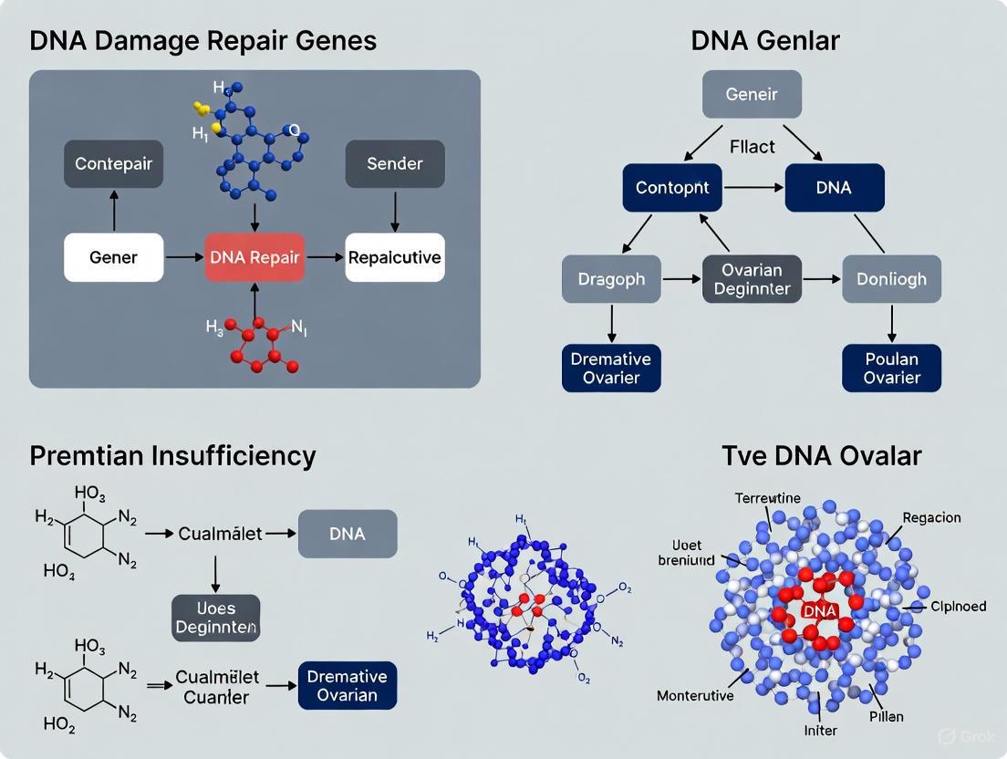
DNA Damage Repair Genes in Premature Ovarian Insufficiency: From Molecular Pathogenesis to Clinical Translation
Premature Ovarian Insufficiency (POI) is a significant cause of female infertility, with genetic factors accounting for 20-25% of cases.
DNA Damage Repair Genes in Premature Ovarian Insufficiency: From Molecular Pathogenesis to Clinical Translation
Abstract
Premature Ovarian Insufficiency (POI) is a significant cause of female infertility, with genetic factors accounting for 20-25% of cases. A substantial proportion of these involve genes critical for DNA damage repair, particularly those managing DNA double-strand breaks (DSBs) via homologous recombination (HR) and non-homologous end joining (NHEJ). This article synthesizes current research for a scientific audience, exploring how defects in key genes like BRCA2, MRE11, RAD50, NBS1, and others disrupt meiotic fidelity and folliculogenesis, leading to accelerated ovarian reserve depletion. We examine the functional consequences of specific mutations, advanced model systems for mechanistic validation, biomarker development for patient stratification, and the emerging therapeutic landscape, including synthetic lethality approaches and DDR-targeting agents. The convergence of genetic insights, functional assays, and clinical application frameworks provides a roadmap for developing targeted interventions for this complex disorder.
Unraveling the Genetic Landscape: How DNA Repair Defects Trigger POI
Genetic Landscape of Premature Ovarian Insufficiency
Premature Ovarian Insufficiency (POI) is a clinically heterogeneous disorder characterized by the loss of ovarian function before the age of 40, affecting approximately 1-3.7% of women [1] [2]. It represents a significant cause of female infertility and is diagnosed by amenorrhea, elevated follicle-stimulating hormone (FSH >25 IU/L), and low estrogen levels [3]. While its etiologies are diverse, genetic factors play a pivotal role, accounting for an estimated 20-25% of cases [1] [3]. Large-scale genomic studies have revolutionized our understanding of POI pathogenesis, revealing that a substantial proportion of cases stem from defects in DNA damage repair processes, with genomic instability emerging as a central mechanism.
Table 1: Genetic Contribution to POI from Recent Large-Scale Studies
| Study Feature | Nature Medicine 2023 (n=1,030) [4] | Other Large Cohort [2] |
|---|---|---|
| Cases with Identified P/LP Variants | 23.5% (242/1030) | 29.3% |
| Contribution of Meiosis/DNA Repair Genes | 48.7% (94/193) of cases with known gene mutations | High yield (Specifics not provided) |
| Primary vs. Secondary Amenorrhea | PA: 25.8% genetic yield; SA: 17.8% genetic yield | Not Specified |
| Novel Candidate Genes Identified | 20 genes (e.g., LGR4, CPEB1, KASH5, ALOX12, ZP3) | 9 genes (e.g., HELQ, CENPE, SWI5) |
The genetic architecture of POI is highly heterogeneous. A 2023 whole-exome sequencing study of 1,030 patients found that pathogenic or likely pathogenic (P/LP) variants in known POI-causative genes accounted for 18.7% of cases [4]. Notably, genes involved in meiosis and DNA repair pathways constituted the largest functional group, responsible for nearly half (48.7%) of these genetically explained cases [4]. This highlights the fundamental importance of genomic maintenance for ovarian longevity. Furthermore, the genetic contribution is more pronounced in women with primary amenorrhea (25.8%) compared to those with secondary amenorrhea (17.8%), suggesting that more severe genetic defects may lead to earlier manifestations of ovarian dysfunction [4].
Mechanisms of Genomic Instability in POI
DNA Double-Strand Break Repair in Meiosis
The integrity of the female germline relies on the precise repair of DNA double-strand breaks (DSBs), which are the most detrimental type of DNA lesion [5]. During meiosis, programmed DSBs are intentionally introduced to initiate homologous recombination (HR), a process essential for genetic diversity and accurate chromosome segregation [1] [5]. Defects in this delicate process can trigger oocyte apoptosis, depleting the ovarian reserve and leading to POI.
The DSB repair pathway via homologous recombination involves a complex, coordinated sequence of events:
- DSB Formation and End Resection: Programmed DSBs are initiated by the SPO11 topoisomerase [5]. The broken ends are then resected to generate 3' single-stranded DNA (ssDNA) overhangs.
- Strand Invasion and D-loop Formation: The recombinases RAD51 and its meiotic paralog DMC1 coat the ssDNA and mediate the invasion of a homologous DNA template, forming a displacement loop (D-loop) [6] [5]. The BRCA2 protein plays a critical role in loading RAD51/DMC1 onto the DNA [6].
- Holliday Junction Resolution: The resulting HR intermediates, Holliday junctions, are resolved to produce crossover or non-crossover products, completing the genetic exchange [5].
Figure 1: Homologous Recombination Pathway for DSB Repair in Meiosis. Key proteins involved at each step are indicated in parentheses.
Key Genes and Pathways Implicated in POI
Mutations in a growing list of genes involved in the HR pathway and other DNA repair mechanisms have been linked to POI, as illustrated by both human genetic studies and mouse models.
BRCA2: Biallelic pathogenic variants in the BRCA2 gene, critical for RAD51/DMC1 loading, have been identified in POI patients [6] [4]. A 2025 study using a mouse model with compound heterozygous Brca2 variants demonstrated that these mutations impair the recruitment of RAD51 and DMC1 to DSB sites during meiotic prophase I [6]. This leads to synaptic defects, persistent DNA damage (marked by γH2AX), and a significant reduction in the primordial follicle pool at birth due to oocyte apoptosis, ultimately causing infertility [6].
Cohesins and Synaptonemal Complex: The synaptonemal complex (SC) is a protein structure that forms between homologous chromosomes during meiosis. Genes encoding SC components (SYCP1, SYCP2, SYCP3) and cohesins like STAG3 are essential for proper chromosome pairing and recombination [1]. Recessive mutations in STAG3 and SYCE1 cause meiotic arrest in both humans and mice, resulting in massive oocyte degeneration and POI [1].
MCM8 and MCM9: These genes encode proteins that form a helicase complex involved in HR. Mutations in MCM8 and MCM9 are associated with autosomal recessive POI [1]. Cells from patients with these mutations show hypersensitivity to chromosomal breaks, confirming a defect in DNA DSB repair [1]. In mice, loss of Mcm8 or Mcm9 leads to meiotic recombination defects and oocyte depletion [1].
Table 2: Key DNA Repair Genes Implicated in POI and Their Functions
| Gene | Function in DNA Repair/Dosage Sensitivity | Associated POI Phenotype | Cellular/Model Phenotype |
|---|---|---|---|
| BRCA2 [6] [4] | Loads RAD51/DMC1 onto ssDNA for strand invasion during HR. | Primary Amenorrhea, POI | Defective RAD51/DMC1 recruitment, synaptic defects, oocyte apoptosis, increased tumor risk. |
| STAG3 [1] | Meiosis-specific component of the cohesin ring, essential for chromosome pairing. | Primary Amenorrhea, POI | Meiotic arrest, oocyte degeneration, sterile in KO mice. |
| MCM8 [1] | Helicase involved in homologous recombination (HR) repair. | Primary Amenorrhea, POI | Hypersensitivity to chromosomal breaks, oocyte depletion in KO mice. |
| MCM9 [1] | Interacts with MCM8; involved in HR. | Primary Amenorrhea, POI, short stature | Impaired DSB repair, oocyte degeneration in KO mice. |
| HFM1 [4] | Meiosis-specific DNA helicase, involved in HR intermediate processing. | Secondary Amenorrhea | Not specified in results. |
| SPIDR [4] | Scaffold protein that facilitates HR and recruits DNA repair proteins. | Secondary Amenorrhea | Not specified in results. |
| MSH4 [4] | Meiosis-specific protein involved in stabilizing Holliday junctions. | Not specified | Not specified in results. |
Experimental Protocols for Investigating Genomic Instability in POI
Whole-Exome Sequencing (WES) and Variant Validation
Objective: To identify pathogenic genetic variants in POI patients and establish a molecular diagnosis [4].
Methodology:
- Cohort Selection: Recruit patients meeting the ESHRE diagnostic criteria for POI (amenorrhea with elevated FSH >25 IU/L before age 40). Exclude individuals with chromosomal abnormalities, autoimmune diseases, or iatrogenic causes [4].
- DNA Extraction and WES: Perform high-quality DNA extraction from peripheral blood. Conduct whole-exome sequencing using a standardized platform (e.g., Illumina) with an appropriate exome capture kit [4].
- Variant Calling and Filtration: Map sequencing reads to the human reference genome. Filter variants by removing common polymorphisms (e.g., MAF >0.01 in gnomAD) and artifacts. Prioritize rare, protein-altering variants (nonsense, frameshift, splice-site, missense) [4].
- Variant Pathogenicity Assessment: Annotate variants using databases (ClinVar, gnomAD) and in silico prediction tools (CADD, SIFT, PolyPhen-2). Classify variants according to ACMG guidelines [4].
- Functional Validation (for VUS): For Variants of Uncertain Significance (VUS), conduct functional assays to establish pathogenicity. In the Nature Medicine study, 75 VUSs in genes like BLM, HFM1, and MCM8 were experimentally tested, with 55 confirmed as deleterious and 38 upgraded to "Likely Pathogenic" [4].
- Phase Confirmation: For patients with two heterozygous mutations in the same gene, confirm the variants are in trans (on different alleles) using techniques like T-clone sequencing or 10x Genomics linked-read technology [4].
Figure 2: Workflow for Genetic Diagnosis of POI via Whole-Exome Sequencing.
Mouse Model Generation and Meiotic Analysis
Objective: To model human BRCA2 variants and investigate their impact on oocyte development and meiosis [6].
Methodology:
- Mouse Model Generation: Use CRISPR-Cas9 or embryonic stem cell technology to generate mice carrying compound heterozygous Brca2 variants mirroring those found in a human POI pedigree (e.g., Brca2c.68-1G>C and Brca2c.4384-4394del) [6].
- Fertility and Ovarian Phenotyping: Mate mutant females with wild-type males to assess fertility. Collect ovaries at different developmental timepoints (e.g., postnatal day 0.5, 21) for histological analysis. Perform follicle counting on ovarian sections to quantify the establishment and depletion of the ovarian reserve [6].
- Immunofluorescence Staining for Germ Cells and Apoptosis: Use antibodies against germ cell markers (e.g., DDX4/MVH, STELLA) to quantify germ cell numbers at embryonic (E11.5, E18.5) and postnatal (P0.5) stages. Co-stain with apoptosis markers (e.g., cleaved PARP) to assess oocyte loss [6].
- Meiotic Chromosome Spread Analysis: Isolate fetal ovaries (e.g., E17.5). Prepare oocyte chromosome spreads and immunostain with antibodies against synaptonemal complex proteins (SYCP3, SYCP1) to visualize chromosome synapsis and progression through meiotic prophase I (Leptotene, Zygotene, Pachytene, Diplotene) [6].
- Assessment of DNA Damage and Recombinase Recruitment: Co-stain oocyte spreads with antibodies against γH2AX (marker for unrepaired DSBs) and recombinases (RAD51, DMC1). Analyze the localization and focus formation of these proteins to evaluate the efficiency of DSB repair [6].
The Scientist's Toolkit: Research Reagent Solutions
Table 3: Essential Reagents and Materials for POI DNA Repair Research
| Reagent / Material | Function / Application | Example Use Case |
|---|---|---|
| Anti-SYCP3 / SYCP1 Antibody | Labels the synaptonemal complex lateral/elements; visualizes chromosome synapsis in meiosis. | Immunostaining of meiotic chromosome spreads to detect synaptic defects in mutant oocytes [6]. |
| Anti-γH2AX Antibody | Detects phosphorylated histone H2AX, a marker of DNA double-strand breaks. | Identifying persistent, unrepaired DSBs in pachytene/diplotene-stage oocytes [6]. |
| Anti-RAD51 / DMC1 Antibody | Visualizes the recombinase enzymes central to homologous recombination. | Assessing the recruitment and focus formation of recombinases at DSB sites [6]. |
| Anti-DDX4 (MVH) Antibody | Germ cell-specific marker; identifies oocytes in ovarian tissue sections. | Quantifying primordial germ cell and oocyte numbers during embryonic and postnatal development [6]. |
| CRISPR-Cas9 System | Genome editing tool for generating precise genetic variants in cell lines or animal models. | Creating mouse models with patient-specific BRCA2 mutations to study their pathogenic mechanism [6]. |
| Mitomycin C | DNA crosslinking agent that induces interstrand crosslinks, repaired by HR. | Testing chromosomal breakage sensitivity and HR proficiency in patient-derived fibroblasts [1]. |
The Critical Role of Programmed DNA Double-Strand Breaks in Meiosis
Meiosis is the specialized cell division that produces haploid gametes from diploid progenitor cells, a process fundamental to sexual reproduction. The most prominent feature of meiosis is homologous recombination, which enhances genetic diversity and ensures the precise segregation of chromosomes during prophase I. This process is initiated by the programmed induction of DNA double-strand breaks (DSBs), which are catalyzed by the evolutionarily conserved SPO11 protein complex in most sexually reproducing organisms [7] [8]. These programmed DSBs are not random genomic accidents but rather carefully orchestrated events controlled regarding their number, timing, and positioning within the genome [8]. The critical nature of this process is starkly revealed in the context of premature ovarian insufficiency (POI), where mutations in meiotic recombination genes, including those involved in DSB formation and repair, constitute a significant genetic cause of this condition affecting approximately 1-3% of women [9] [10] [11]. This whitepaper provides an in-depth technical examination of programmed DSBs in meiosis, with particular emphasis on their relevance to POI research and drug development.
Molecular Machinery of DSB Formation: The SPO11 Complex
Biochemical Characterization of the SPO11-TOP6BL Complex
The catalytic core responsible for meiotic DSB formation is the SPO11-TOP6BL complex, which has recently been successfully purified and characterized after decades of research efforts [7]. SPO11 shares extensive homology with the Top6A subunit of the DNA topoisomerase VI (topo VI) family, while its partner TOP6BL (homologous to human C11orf80, also known as Gm960 in mice) represents the orthologue of the Top6B subunit [7].
- Structural Organization: Size-exclusion chromatography and cross-linking experiments reveal that the mouse SPO11-TOP6BL complex can form both heterodimers (approximately 108-118 kDa) and heterotetramers (approximately 250 kDa) in solution, though the heterodimer appears to form more readily and exhibits greater stability [7].
- DNA Cleavage Mechanism: The SPO11 complex cleaves DNA and covalently attaches to the 5′ terminus of DNA breaks in vitro. This DNA-cleavage activity is Mg²⁺-dependent but ATP-independent, distinguishing it biochemically from its ancestral topoisomerase VI [7].
- DNA Binding Specificity: Electrophoretic mobility shift assays (EMSAs) demonstrate that the SPO11 complex binds most efficiently to DNA structures with 5′ overhangs (Kd of 20.8 ± 0.39 nM for double 5′ overhang), with approximately two-fold weaker affinity for 3′ overhangs and blunt ends [7].
Table 1: DNA Binding Affinities of SPO11 Complex to Various DNA Structures
| DNA Structure | Dissociation Constant (Kd) | Relative Affinity |
|---|---|---|
| Double 5′ overhang | 20.8 ± 0.39 nM | Highest |
| Single 5′ overhang | 30.5 ± 2.63 nM | High |
| 3′ overhang | 43.5 ± 2.15 nM | Moderate |
| Blunt end | 47.8 ± 2.14 nM | Moderate |
| Double hairpin | 68.4 ± 3.06 nM | Lowest |
The critical importance of SPO11 is confirmed by knock-in mouse studies, where a point mutation in SPO11 that disrupts Mg²⁺ binding abolishes DSB formation entirely, establishing a non-redundant mechanistic framework for the initial step of meiotic recombination [7].
Genomic Distribution and Regulation of DSB Formation
Meiotic DSBs occur preferentially within defined genomic regions called DSB hotspots. In budding yeast, these hotspots average 189 base pairs in width, while in mice they average 143 base pairs [8]. The distribution of DSBs is non-random, with notable enrichment observed within a ∼100 kb region near chromosome ends across all chromosomes [12]. This telomere-guided mechanism increases relative DSB density on small chromosomes, providing an interference-independent system that helps ensure all chromosomes receive at least one crossover per homolog pair [12].
Interestingly, substantial DSB activity also occurs in pericentromeric regions where crossover formation is largely suppressed, suggesting that centromeric suppression of recombination occurs primarily at the level of break repair rather than DSB formation [12]. This spatial regulation highlights the sophisticated control mechanisms governing where DSBs are formed and how they are subsequently processed.
DSB Resection: From Double-Strand Break to Single-Stranded Substrate
The Two-Step Resection Mechanism
Following DSB formation, the 5'-terminal strands of broken DNA are resected by a coordinated nuclease cascade to yield the 3' single-stranded DNA (ssDNA) overhangs necessary for homology search and strand invasion [8]. This resection process follows a two-step mechanism:
- Resection Initiation (Short-range resection): The conserved Mre11-Rad50-Xrs2 (MRX) complex in yeast (MRN complex in mammals), in conjunction with Sae2 (CtIP in vertebrates), cleaves the Spo11-bound strands using Mre11's endonuclease activity and degrades ssDNA toward the DSB using its 3'-to-5' exonuclease activity [8]. This step removes approximately 300-400 nucleotides from the break site in yeast [8].
- Long-range Resection: More processive 5'-to-3' exonuclease activities extend the degradation further from the DSB. In yeast, Exo1 primarily carries out this step, while in mouse spermatocytes, MRN-CtIP appears responsible for most resection tract length, with EXO1 contributing a smaller polishing function [8].
Advanced Methods for Measuring Meiotic Resection
Recent advances in next-generation sequencing have revolutionized the analysis of meiotic DSB resection, providing nucleotide-resolution insights across entire genomes [8].
Table 2: Methods for Measuring Meiotic DNA End Resection
| Method | Description | Organisms Applied | Key Features |
|---|---|---|---|
| S1-seq, END-seq | ssDNA at resected DSBs is digested by single-strand-specific nucleases, and resulting blunted DNA products are captured by adapter ligation and sequenced. | Yeast, mice | Direct measurement of resection endpoints; nucleotide resolution; genome-wide |
| DMC1-SSDS + SPO11-oligo sequencing | DMC1-bound DNA fragments are captured by ChIP, followed by sequencing library generation with hairpin-forming adaptors to enrich ssDNA. Combined with SPO11 cleavage site mapping. | Mice, plants, humans | Genome-wide maps of recombination initiation sites; indirect resection measurement |
| Native/Denaturing 2D-gel Southern Blotting | Genomic DNA is digested, separated by 2D electrophoresis (native then denaturing), transferred to membrane, and hybridized with specific probes. | Yeast | Single hotspot analysis; visualizes recombination intermediates |
| RE-qPCR | Genomic DNA is digested with restriction enzymes targeting resected regions, followed by qPCR with primers flanking cut sites to quantify ssDNA. | Yeast | Quantitative but spatially limited to restriction sites tested |
These advanced methods have revealed that resection tract lengths average approximately 822 nucleotides in yeast and 1,117 nucleotides in mice, though significant variation exists between individual DSB events [8].
Diagram 1: DSB resection process
The POI Connection: Meiotic Recombination Failure and Ovarian Insufficiency
Genetic Links Between Meiotic Genes and POI
Premature ovarian insufficiency (POI) is characterized by the cessation of ovarian function before age 40, affecting approximately 1-3% of women [11]. The condition presents with menstrual irregularities, infertility, elevated gonadotropins, and estrogen deficiency, carrying significant long-term health implications including osteoporosis, cardiovascular disease, and cognitive decline [9] [10]. Meiotic recombination defects represent a substantial genetic contribution to POI pathogenesis, with mutations in more than 75 genes implicated in the disorder, many involved in meiosis and DNA repair [10].
Table 3: Meiotic DNA Repair Genes Implicated in Premature Ovarian Insufficiency
| Gene | Full Name | Function in Meiosis | POI Association |
|---|---|---|---|
| SPO11 | Sporulation 11 | Catalyzes formation of meiotic DSBs | Critical for initiation of meiotic recombination [7] |
| MCM8 | Minichromosome maintenance complex component 8 | Participates in homologous recombination during meiosis and DSB repair with MCM9 | Mutations linked to POI [9] |
| MCM9 | Minichromosome maintenance complex component 9 | Forms complex with MCM8; important in DNA mismatch repair | Mutations linked to POI [9] |
| HFM1 | Helicase for meiosis 1 | Required for crossover formation and complete synapsis | Mutations associated with POI [9] |
| MSH4/5 | MutS Homolog 4/5 | Participate in homologous recombination repair for DSBs | Mutations associated with POI [9] |
| DMC1 | DNA Meiotic Recombinase 1 | Involved in meiotic homologous recombination | Mutations associated with POI [9] |
| HELB | DNA Helicase B | Contributes to ovarian function and menopause timing | Genetic variants associated with POI [13] |
The genetic basis of POI is highly diverse, with various gene mutations affecting critical meiotic processes. Turner syndrome (X chromosome abnormalities) and fragile X premutation (FMR1 gene) represent the most frequent genetic triggers, but numerous other meiotic genes contribute significantly to POI risk [9] [10]. A recent cohort study identified twenty POI-associated genes involved in gonadogenesis, meiosis, follicular development, and ovulation, highlighting the genetic complexity of this condition [9].
Changing Etiological Spectrum and Diagnostic Considerations
The etiological landscape of POI has evolved significantly over recent decades. A comparative study between a historical cohort (1978-2003) and a contemporary cohort (2017-2024) revealed striking changes: iatrogenic causes increased from 7.6% to 34.2%, autoimmune causes rose from 8.7% to 18.9%, while idiopathic cases decreased from 72.1% to 36.9% [10]. Genetic causes remained relatively stable at approximately 10-12% [10].
This shifting etiology reflects both improved diagnostic capabilities and changing medical practices, particularly the success of oncologic treatments that unfortunately carry gonadotoxic effects. For researchers and clinicians, these findings underscore the importance of comprehensive genetic testing in POI cases, especially for women with family history or early-onset symptoms, as identifying specific meiotic gene mutations can provide prognostic information and guide therapeutic decisions.
Experimental Models and Research Tools
POI Induction Models for Meiotic Research
Several well-established experimental models enable the study of meiotic defects and ovarian insufficiency:
- Chemotherapy-induced POI Models: Cyclophosphamide (CPA) administration in rats provides a robust model for studying chemotherapy-induced ovarian damage. CPA exposure leads to characteristic nuclear changes in primordial follicles, including pyknotic nuclei representing early signs of oocyte damage, oocyte shrinkage, and disruption of mitochondrial morphology [14].
- Cisplatin-induced POI: Rats administered daily cisplatin (5 mg/kg for 3 days) develop acute ovarian toxicity mimicking human treatment-induced ovarian insufficiency [14].
- Environmental Toxicant Models: Exposure to atmospheric particulate matter, endocrine-disrupting chemicals, pesticides, microplastics, heavy metals, and cigarette smoke can induce POI in experimental systems, reflecting environmental contributions to ovarian decline [9].
The Scientist's Toolkit: Essential Research Reagents
Table 4: Key Research Reagents for Meiotic DSB and POI Investigations
| Reagent/Category | Function/Application | Examples/Specifications |
|---|---|---|
| SPO11-TOP6BL Complex | In vitro reconstitution of meiotic DSB formation | His–SPO11 and TOP6BL–Flag purified from Expi293F cells [7] |
| Anti-DMC1 Antibodies | Chromatin immunoprecipitation for mapping meiotic recombination sites | Used in DMC1-SSDS to capture DMC1-bound DNA fragments [8] |
| S1 Nuclease / Exonuclease Cocktail | Digest ssDNA at resected DSBs for END-seq/S1-seq | E. coli exonuclease T and exonuclease VII [8] |
| Mouse Spermatocyte Systems | Ex vivo meiotic recombination studies | Primary spermatocytes for analysis of resection mechanisms [8] |
| Ovarian Granulosa Cell Cultures | In vitro POI pathogenesis studies | Human GCs for lncRNA expression and functional analyses [11] |
Emerging Research Directions and Therapeutic Implications
Noncoding RNAs in Meiotic Regulation and POI
Long noncoding RNAs (lncRNAs) have emerged as pivotal regulators of granulosa cell function and follicular development, contributing significantly to POI pathogenesis [11]. Several lncRNAs show promise as diagnostic biomarkers and therapeutic targets:
- GCAT1: Downregulation leads to ovarian dysfunction by regulating p27, affecting granulosa cell proliferation [11].
- PVT1: Overexpression can restore ovarian function by reducing Foxo3a-induced granulosa cell apoptosis [11].
- ZNF674-AS1: Regulates granulosa cell proliferation, glycolysis, and AMPK activation via interaction with ALDOA [11].
- HOTAIR: Promotes proliferation by regulating miR-148b-3p/ATG14-mediated autophagy pathway [11].
The therapeutic potential of targeting lncRNAs is supported by studies showing that HOTAIR expression has a protective effect in alleviating cisplatin-induced POI toxicity [11].
Protective Strategies Against Meiotic Damage
Research into protective agents against chemotherapy-induced gonadal damage has identified several promising compounds with potential clinical applications:
- Ginsenoside Rb1: Attenuates cyclophosphamide-induced testicular toxicity through free radical scavenging, improving sperm count, motility, and membrane integrity [14].
- Rutin: Preserves Sertoli-cell and germ-cell integrity by sustaining glutathione and key antioxidant enzyme activities while activating Akt/PPAR-γ signaling [14].
- Dexpanthenol: Mitigates nephrotoxicity induced by cisplatin via AMPK-mediated PI3K/Akt and MAPK/JNK/ERK signaling pathways [14].
- Leonurine hydrochloride: Shows protective effects against pyroptosis in premature ovarian insufficiency via regulating NLRP3/GSDMD pathway [14].
Diagram 2: POI pathogenesis pathways
Programmed DNA double-strand breaks represent both a fundamental biological process essential for sexual reproduction and a potential vulnerability point whose dysregulation contributes significantly to premature ovarian insufficiency. The precise formation of meiotic DSBs by the SPO11-TOP6BL complex, followed by their controlled resection and repair through homologous recombination, ensures proper chromosome segregation and genomic diversity in gametes. Defects in any component of this elaborate machinery can disrupt folliculogenesis and accelerate ovarian aging, leading to POI.
For researchers and drug development professionals, understanding these mechanistic links provides crucial insights for developing targeted diagnostic and therapeutic strategies. The expanding repertoire of research tools—from in vitro reconstitution systems to advanced sequencing methods—enables unprecedented dissection of meiotic processes and their dysfunction in POI. Meanwhile, emerging protective agents and the potential of lncRNA-based therapies offer promising avenues for clinical intervention. As our molecular understanding of meiotic recombination deepens, so too does our capacity to address the clinical challenges of premature ovarian insufficiency through mechanism-driven approaches.
Premature Ovarian Insufficiency (POI) is a significant cause of female infertility, affecting 1-3% of women of reproductive age. While its etiology is heterogeneous, genetic factors account for approximately 20-25% of cases, with DNA repair pathways playing a crucial role. This technical review examines the molecular mechanisms of homologous recombination (HR), non-homologous end joining (NHEJ), and related pathways in POI pathogenesis. We synthesize current research demonstrating how deficiencies in double-strand break (DSB) repair mechanisms contribute to ovarian follicle depletion and dysfunction. The analysis covers key genes, protein complexes, clinical genetic findings, and experimental approaches, providing researchers with a comprehensive framework for investigating DNA repair defects in POI. Emerging evidence confirms that DSB repair genes constitute the largest functional group among validated POI-associated genes, highlighting their fundamental importance in ovarian maintenance and function.
Premature Ovarian Insufficiency (POI) is clinically defined as the cessation of ovarian function before age 40, characterized by menstrual disturbances (amenorrhea or oligomenorrhea), elevated gonadotropin levels (FSH >25 IU/L), and estrogen deficiency [5] [15]. The condition affects approximately 1-3.7% of women and represents a major cause of female infertility [2] [4]. POI patients often experience comorbid conditions including osteoporosis, cardiovascular dysfunction, and neurological issues, extending beyond reproductive health concerns [11].
The etiological landscape of POI encompasses genetic, autoimmune, iatrogenic, and environmental factors, with more than half of cases remaining idiopathic [15]. Genetic studies have identified mutations in over 90 genes associated with POI, with DNA damage repair genes representing a substantial proportion [4]. Whole-exome sequencing of 1,030 POI patients revealed that genetic factors contribute to 23.5% of cases, with genes involved in meiosis or homologous recombination accounting for nearly half (48.7%) of the genetically explained cases [4].
DNA Damage in Ovarian Function
DNA integrity is fundamental to ovarian reserve and oocyte quality. The female germline must maintain genomic stability throughout meiotic divisions and long periods of meiotic arrest. Oocytes are particularly vulnerable to DNA damage due to their prolonged prophase I arrest, which can extend up to 50 years in humans [16]. Endogenous and exogenous factors including reactive oxygen species (ROS), ionizing radiation, and chemotherapeutic agents constantly threaten DNA integrity [5] [16]. Unrepaired DNA damage triggers apoptosis of oocytes and follicular depletion, directly leading to diminished ovarian reserve and POI.
Table 1: DNA Damage Sources and Their Impact on Ovarian Function
| Damage Source | Type of DNA Lesion | Primary Repair Pathway | Ovarian Impact |
|---|---|---|---|
| Endogenous ROS | Oxidative base damage, SSBs, DSBs | BER, HR, NHEJ | Accelerated follicular atresia |
| Programmed meiotic breaks | DSBs | HR (meiotic-specific) | Meiotic arrest if unrepaired |
| Ionizing radiation | Complex DSBs, SSBs | HR, NHEJ | Direct oocyte destruction |
| Chemotherapeutic agents | Interstrand crosslinks, DSBs | FA pathway, HR | Premature follicle depletion |
| Replication stress | DSBs, stalled replication forks | HR, NHEJ | Impaired folliculogenesis |
DNA Double-Strand Break Repair Pathways
DNA double-strand breaks (DSBs) represent the most severe type of DNA damage, with each cell experiencing approximately 10 DSBs daily [5]. DSBs are classified into two categories: programmed DSBs, which occur during meiosis and are essential for genetic diversity, and accidental DSBs, resulting from endogenous or exogenous genotoxic stressors [5]. Mammalian cells have evolved two primary DSB repair mechanisms: homologous recombination (HR) and non-homologous end joining (NHEJ), with alternative end-joining (alt-EJ) serving as a backup pathway.
Homologous Recombination (HR) Pathway
HR is an error-free repair mechanism that utilizes sister chromatids as templates, restricting its activity primarily to the S and G2 phases of the cell cycle [5] [17]. This pathway is particularly crucial for meiotic DSB repair and maintaining ovarian follicle pool integrity.
The HR mechanism proceeds through several well-defined steps:
- End Resection: The MRE11-RAD50-NBS1 (MRN) complex, in conjunction with CtIP, initiates 5'→3' end resection, generating short 3' single-stranded DNA (ssDNA) overhangs. Further processing by EXO1 exonuclease and DNA2 helicase-nuclease extends these overhangs [5].
- Strand Invasion: Replication protein A (RPA) coats and stabilizes the ssDNA overhangs, preventing secondary structure formation. BRCA2 then facilitates replacement of RPA with RAD51 to form the nucleoprotein filament, which mediates strand invasion into the homologous DNA template [5]. In meiosis, the RAD51 paralog DMC1 plays a specialized role in strand invasion [5].
- Holliday Junction Formation & Resolution: The invading strand primes DNA synthesis, forming Holliday junctions. These intermediates are subsequently resolved through synthesis-dependent strand annealing or double Holliday junction pathways, resulting in accurate repair without loss of genetic information [5].
Table 2: Key HR Genes Implicated in POI Pathogenesis
| Gene | Protein Function | POI-Associated Mutations | Phenotypic Features |
|---|---|---|---|
| BRCA2 | RAD51 loading, strand invasion | Monoallelic and biallelic mutations | POI, cancer predisposition |
| MCM8/9 | Helicase complex, DSB resection | Biallelic LoF mutations | Isolated POI, primary amenorrhea |
| HFM1 | Holliday junction resolution | Monoallelic and biallelic mutations | Isolated POI, meiotic arrest |
| MSH4 | Meiotic complex, junction processing | Biallelic mutations | Isolated POI, synaptic defects |
| RAD51 | Strand invasion, nucleofilament formation | Rare variants reported | POI, cancer risk |
| DMC1 | Meiotic strand invasion | Rare variants reported | Isolated POI, meiotic arrest |
Non-Homologous End Joining (NHEJ) Pathway
NHEJ is considered the dominant DSB repair pathway in mammalian cells, operating throughout the cell cycle but most active in G1 phase [17]. Unlike HR, NHEJ directly ligates broken DNA ends without requiring a homologous template, making it inherently error-prone and associated with small insertions or deletions (indels).
The canonical NHEJ (c-NHEJ) mechanism involves:
- End Recognition: The Ku70-Ku80 heterodimer rapidly binds to DSB ends, protecting them from further resection and recruiting downstream repair factors [5] [16].
- End Processing: DNA-dependent protein kinase catalytic subunit (DNA-PKcs) is recruited and activated, phosphorylating various substrates. The Artemis nuclease, activated by DNA-PKcs, processes damaged or incompatible DNA ends [16].
- Ligation: The XRCC4-DNA ligase IV complex, stabilized by XLF, catalyzes the ligation of processed DNA ends, completing the repair process [16].
Comparative studies in human fibroblasts have demonstrated that NHEJ is both faster (completed in ~30 minutes) and more efficient than HR, with compatible end joining (NHEJ-C) being twice as efficient as incompatible end joining (NHEJ-I) [17].
Alternative End-Joining (Alt-EJ) Pathways
Alt-EJ pathways, also known as microhomology-mediated end joining (MMEJ), operate as backup mechanisms when c-NHEJ is compromised. These pathways typically utilize 2-20 nucleotide microhomologous sequences for end alignment and are more mutagenic than c-NHEJ [5]. While the specific components of alt-EJ are less defined, they often involve PARP1, XRCC1-DNA ligase III, and other factors [5]. The contribution of alt-EJ deficiencies to POI pathogenesis remains an active research area, with recent studies identifying novel alt-EJ genes like HELQ in POI patients [2].
Experimental Analysis of DNA Repair in POI
Methodologies for Assessing Repair Efficiency
Research into DNA repair pathways in POI employs both cellular models and advanced molecular techniques to quantify repair efficiency and identify pathogenic variants.
Reporter Assay Systems: Fluorescent reporter substrates stably integrated into the genome enable real-time monitoring of DSB repair pathways in living cells [17]. These systems typically use engineered GFP genes disrupted by artificial introns containing endonuclease recognition sites (e.g., I-SceI). Upon DSB induction and successful repair, functional GFP is reconstituted, allowing quantification of repair efficiency via flow cytometry [17].
Table 3: Research Reagent Solutions for DNA Repair Studies
| Research Tool | Application | Key Features | Utility in POI Research |
|---|---|---|---|
| I-SceI Endonuclease System | Induction of site-specific DSBs | Creates defined DSBs with compatible or incompatible ends | Enables controlled study of repair pathway efficiency |
| GFP-Based Reporter Constructs | Monitoring repair outcomes | Chromosomal integration for physiological relevance | Quantitative comparison of HR vs. NHEJ activity |
| HCA2-hTERT Fibroblasts | Normal human cell model | Telomerase-immortalized while maintaining normal cell characteristics | Study DNA repair in non-transformed human cells |
| Southern Blot Verification | Confirmation of single-copy integration | Ensures consistent reporter cassette copy number | Eliminates copy number variation as a confounding factor |
| Flow Cytometry (FACS) | Quantification of repair efficiency | Simultaneous detection of GFP+ (repaired) and DsRed+ (transfected) cells | Normalizes for transfection efficiency variations |
Whole Exome Sequencing (WES): Large-scale WES studies of POI cohorts have been instrumental in identifying novel DNA repair genes associated with POI. The standard approach includes:
- Cohort recruitment following ESHRE guidelines (amenorrhea + elevated FSH >25 IU/L)
- Exclusion of non-genetic causes (chromosomal abnormalities, autoimmune diseases, iatrogenic factors)
- Variant calling with filtering against population databases (gnomAD)
- Pathogenicity assessment following ACMG guidelines
- Functional validation of prioritized variants [4]
This approach identified 195 pathogenic/likely pathogenic variants in 59 known POI-causative genes in 18.7% of 1,030 patients, with DNA repair genes representing the largest functional category [4].
Pathway Regulation and Crosstalk
The choice between HR and NHEJ is tightly regulated throughout the cell cycle and influenced by the nature of the DNA break. Key regulatory mechanisms include:
- Cell Cycle Dependence: HR is active in S/G2 phases when sister chromatids are available, while NHEJ operates throughout the cycle but predominates in G1 [17].
- End Resection Control: Extensive 5'→3' resection commits repair to HR, while limited resection favors NHEJ. The MRN complex, CtIP, and DNA2 play critical roles in this decision point [5].
- Kinase Signaling: ATM and ATR kinases coordinate DNA damage response, phosphorylating downstream effectors that influence pathway choice [16].
Experimental evidence suggests competition between pathways, with NHEJ generally being faster and more efficient in cycling human cells (NHEJ-C: 6×, NHEJ-I: 3× more efficient than HR) [17]. This kinetic advantage may explain the predominance of NHEJ in many cell types, though HR remains essential for meiotic progression and oocyte quality maintenance.
Diagram 1: DSB Repair Pathway Choice Regulation. The decision between NHEJ and HR is primarily determined by cell cycle phase, with NHEJ dominating in G1 and HR in S/G2 phases. Key regulatory steps include end resection, which commits to HR, and Ku complex binding, which promotes NHEJ.
Clinical Implications and Therapeutic Perspectives
Genetic Diagnosis and Counseling
The high prevalence of DNA repair gene mutations in POI underscores the importance of comprehensive genetic testing. Recent studies report diagnostic yields of 29.3% using expanded gene panels, with DNA repair genes like HELQ, C17orf53 (HROB), and SWI5 identified as novel POI-associated genes [2]. Genetic diagnosis enables:
- Personalized Medicine: Identification of patients with cancer predisposition genes (e.g., BRCA2) allows for enhanced surveillance and risk-reducing interventions [2].
- Fertility Counseling: Patients with biallelic mutations in DNA repair genes often present with primary amenorrhea and have poorer prospects for residual ovarian function [4].
- Comorbidity Management: Approximately 8.5% of POI cases represent manifestations of multi-organ genetic disorders, requiring coordinated care [2].
Emerging Therapeutic Strategies
Understanding DNA repair mechanisms in POI has inspired several innovative therapeutic approaches:
- In Vitro Activation (IVA): Manipulation of DNA repair pathways, particularly those involving PTEN and Hippo signaling, may promote primordial follicle activation [2]. Genetic diagnosis could help identify patients most likely to benefit from IVA techniques.
- Antioxidant Interventions: Given the role of oxidative stress in oocyte DNA damage, antioxidant strategies targeting ROS reduction may help preserve ovarian function in susceptible individuals [16].
- Mitophagy Regulation: Novel pathways involving mitochondrial autophagy (mitophagy) have been identified as potential therapeutic targets for preserving oocyte quality [2].
DNA double-strand break repair pathways, particularly homologous recombination and non-homologous end joining, play fundamental roles in maintaining ovarian function and preventing premature ovarian insufficiency. The comprehensive characterization of these pathways has revealed their critical importance in meiotic progression, follicular development, and oocyte survival. Ongoing research continues to elucidate the complex regulatory mechanisms governing DNA repair pathway choice and efficiency in the ovarian context. The integration of genetic findings with functional studies provides promising avenues for developing targeted interventions that may ultimately preserve fertility in women at risk for POI. Future directions include exploring the therapeutic potential of modulating DNA repair pathways, developing more accurate diagnostic panels encompassing novel DNA repair genes, and investigating gene-environment interactions that impact DNA repair efficiency in the ovary.
Premature ovarian insufficiency (POI) is a significant clinical disorder characterized by the loss of ovarian function before age 40, affecting approximately 3.5% of the female population [18]. This condition presents not only as a reproductive issue but also as a multisystemic endocrine disorder with far-reaching health implications. The pathogenesis of POI is remarkably heterogeneous, encompassing genetic, iatrogenic, autoimmune, and environmental factors [19]. Among these, defects in DNA damage repair pathways have emerged as critical contributors to pathological ovarian aging, with the Fanconi anemia (FA) pathway and its associated genes, particularly BRCA2 (FANCD1), representing high-penetrance genetic factors in POI etiology [6] [20] [21].
The FA pathway comprises a complex network of at least 22 genes that coordinate the repair of DNA interstrand cross-links (ICLs)—highly toxic lesions that impede DNA replication and transcription [20]. This pathway maintains genomic integrity through a sophisticated cascade of lesion recognition, ubiquitination, and downstream repair processes. When compromised, deficiencies in this pathway not only predispose individuals to cancer predisposition syndromes but also significantly impact reproductive lifespan and ovarian function [22] [21]. This technical review examines the mechanistic roles of BRCA2 and the broader FA pathway in POI pathogenesis, integrates quantitative genetic data, details essential experimental methodologies, and visualizes the complex molecular relationships underlying this connection.
The Fanconi Anemia Pathway: Core Components and Molecular Mechanisms
Architectural Organization of the FA/BRCA Pathway
The Fanconi anemia pathway operates through a meticulously coordinated multi-step process that can be categorized into three primary functional tiers: upstream recognition and activation, midstream ID complex formation, and downstream repair execution [22] [20]. This sophisticated system ensures genomic stability by resolving highly toxic DNA interstrand cross-links (ICLs).
Table 1: Core Components of the Fanconi Anemia Pathway
| Tier | Gene/Protein | Alternative Name | Primary Function |
|---|---|---|---|
| Upstream | FANCA, FANCB, FANCC, FANCE, FANCF, FANCG, FANCL, FANCT | UBE2T | Forms FA core complex with E3 ubiquitin ligase activity |
| FANCM | - | Recruits core complex to DNA damage site | |
| Midstream | FANCI, FANCD2 | - | Forms ID2 complex; activation via mono-ubiquitination |
| Downstream | FANCD1 | BRCA2 | Homologous recombination (HR) repair |
| FANCS | BRCA1 | Homologous recombination (HR) repair | |
| FANCN | PALB2 | Homologous recombination (HR) repair; BRCA2 linker | |
| FANCJ | BRIP1 | DNA helicase; interacts with BRCA1 | |
| FANCO | RAD51C | HR mediator complex | |
| FANCR | RAD51 | DNA strand exchange in HR | |
| FANCQ | XPF | DNA endonuclease for ICL incision | |
| FANCP | SLX4 | Scaffold for nucleases in ICL processing | |
| FANCV | REV7/MAD2L2 | Translesion synthesis (TLS) |
The pathway initiates when the FANCM-FAAP24 complex recognizes stalled replication forks at ICL sites and recruits the FA core complex (a multi-subunit E3 ubiquitin ligase comprising FANCA, B, C, E, F, G, L, and FAAP100) [22]. This core complex then mono-ubiquitinates the FANCI-FANCD2 (ID2) heterodimer, which serves as the central activation step in the pathway. The ubiquitinated ID2 complex subsequently recruits downstream effector proteins—including nucleases (FANCQ/XPF, FANCP/SLX4), translesion synthesis polymerases (FANCV/REV7), and most critically, the homologous recombination machinery (BRCA2/FANCD1, BRCA1/FANCS, PALB2/FANCN, RAD51/FANCR)—to complete ICL repair through coordinated incision, transfusion synthesis, and homologous recombination [22] [20].
The following diagram illustrates the sequential activation and repair process of the FA/BRCA pathway:
The FA pathway specifically addresses DNA interstrand cross-links (ICLs), which can originate from both endogenous metabolic processes and exogenous environmental or therapeutic exposures [20]. Endogenous inducers primarily include reactive aldehydes (e.g., formaldehyde, acetaldehyde, malondialdehyde) generated during cellular metabolism, as well as reactive nitrogen species from nitrous acid metabolism. Exogenous sources encompass chemotherapeutic agents (e.g., cisplatin, mitomycin C, nitrogen mustards), environmental pollutants, and psoralens. The following table categorizes these ICL-inducing agents and their mechanisms of action:
Table 2: Sources and Mechanisms of DNA Interstrand Cross-Link Formation
| Source | Specific Agents | Primary Cross-Linking Mechanism | Biological Context |
|---|---|---|---|
| Endogenous | Formaldehyde, Acetaldehyde, Malondialdehyde | Forms methylene bridges between opposing DNA bases (dG-dG, dG-dA, dA-dA) | Cellular metabolism; lipid peroxidation |
| Reactive Nitrogen Species | Cross-links between guanines in DNA double helix | Nitrate/nitrite metabolism | |
| Exogenous | Nitrogen Mustards (cyclophosphamide, melphalan) | Alkylates N7/O6 of dG, N3/N1 of dA, forming dG-dG, dG-dA cross-links | Chemotherapy for lymphoma, myeloma, ovarian cancer |
| Platinum-based drugs (cisplatin, carboplatin) | Forms adducts with N7 of dG, leading to intrastrand and interstrand cross-links | Treatment of ovarian, cervical, breast cancers | |
| Psoralens | Pyrone/furan rings form cyclobutane adducts between thymidines | PUVA therapy for skin diseases | |
| Mitomycin C | Cross-links complementary strands of DNA at 5'-CpG-3' sequences | Chemotherapy and research (chromosome breakage test) |
BRCA2/FANCD1: A Nexus Between DNA Repair and Ovarian Function
Molecular Functions of BRCA2 in Meiotic Homologous Recombination
BRCA2 (FANCD1) serves as a central mediator in the homologous recombination (HR) pathway, playing an indispensable role in repairing DNA double-strand breaks (DSBs) through its regulation of RAD51 nucleoprotein filament formation [6] [23]. During meiosis, programmed DSBs occur as an essential step in genetic recombination, and their precise repair is critical for producing genetically balanced gametes. BRCA2 facilitates this process by directly interacting with RAD51 through its BRC repeats, promoting the assembly of RAD51 on single-stranded DNA, and protecting RAD51 nucleoprotein filaments from disassembly by anti-recombinases [6].
In mammalian oocytes, meiotic prophase I begins during fetal development and arrests at the diplotene stage until ovulation. Throughout this prolonged arrest period, oocytes remain vulnerable to DNA damage accumulation. BRCA2 deficiency disrupts the timely recruitment of RAD51 and its meiotic counterpart DMC1 to programmed DSBs, leading to persistent DNA damage marked by γH2AX foci, synaptic defects between homologous chromosomes, and ultimately, massive oocyte apoptosis around the time of birth [6]. This compromised meiotic HR efficiency directly impairs the establishment of the primordial follicle pool, resulting in the significantly diminished ovarian reserve characteristic of POI.
Evidence from Murine Models of BRCA2 Deficiency
Recent research utilizing a viable Brca2 germline-deficient mouse model (Brca2c.68-1G>C/c.4384-4394del) has provided compelling mechanistic insights into BRCA2-related POI pathogenesis [6] [23]. This model, which carries compound heterozygous variants mirroring those identified in a Chinese POI pedigree, demonstrates complete female infertility with markedly atrophic ovaries lacking follicles by 2 months of age. Detailed histological and immunofluorescence analyses revealed that while primordial germ cell proliferation appeared normal during embryonic development (E11.5-E18.5), a significant reduction in oocyte numbers occurred postnatally (P0.5), accompanied by increased oocyte apoptosis as indicated by cleaved-PARP staining [6].
Oocyte spreads from BRCA2-deficient fetal ovaries (E17.5) exhibited profound meiotic defects, including:
- Delayed meiotic progression: Reduced percentage of pachytene-stage oocytes with retention in leptotene and zygotene stages
- Synaptic abnormalities: Over 60% of pachytene-like oocytes showed non-homologous and incomplete synapsis
- Persistent DNA damage: γH2AX foci remained evident at pachytene-like and diplotene-like stages, particularly around unsynapsed chromosomal regions
- Defective recombinase recruitment: Impaired recruitment of RAD51 and DMC1 to programmed DSB sites [6]
These findings establish that BRCA2 deficiency primarily disrupts the homologous recombination repair of meiotic DSBs rather than affecting primordial germ cell proliferation, leading to accelerated oocyte depletion and POI.
Quantitative Genetic Evidence: FA Genes in POI and Cancer Risk
The clinical spectrum associated with FA gene mutations extends beyond classical Fanconi anemia to include isolated POI and cancer predisposition syndromes. The table below summarizes key FA genes with established roles in POI pathogenesis and their associated cancer risks:
Table 3: FA/BRCA Pathway Genes in POI and Associated Cancer Risks
| Gene | FA Nomenclature | Role in POI | Associated Cancers | Experimental Evidence |
|---|---|---|---|---|
| BRCA2 | FANCD1 | Biallelic variants cause POI; heterozygous carriers have earlier menopause | Breast, ovarian, pancreatic, prostate | Mouse model (oocyte meiotic defects, infertility) [6] [23] |
| BRCA1 | FANCS | Potential POI association; modulates ovarian aging | Breast, ovarian, prostate | Cohort studies, cancer risk data [21] |
| PALB2 | FANCN | Biallelic variants linked to POI | Breast, pancreatic, ovarian | FA patient cohorts [21] |
| BRIP1 | FANCJ | Mutations associated with POI and primary ovarian failure | Ovarian, breast | Genetic association studies [22] |
| RAD51C | FANCO | Potential role in ovarian dysfunction | Ovarian, breast | Limited case reports [22] |
Notably, biallelic BRCA2 variants induce a particularly severe phenotype, with POI representing one manifestation of a broader systemic disorder. The Brca2c.68-1G>C/c.4384-4394del mouse model demonstrated not only ovarian failure but also increased tumor susceptibility, highlighting the dual impact of BRCA2 deficiency on reproductive health and cancer risk [6]. This connection underscores the necessity for comprehensive tumor surveillance in POI patients with BRCA2 mutations beyond routine gynecological care.
Experimental Methodologies for Investigating FA/BRCA Pathway in POI
Chromosome Breakage Test for FA Pathway Function
The chromosome breakage test remains the gold-standard diagnostic assay for evaluating FA pathway integrity in clinical and research settings [24]. This cytogenetic approach assesses cellular hypersensitivity to DNA crosslinking agents:
Protocol Overview:
- Sample Collection: Obtain peripheral blood lymphocytes (or skin fibroblasts for follow-up testing)
- Crosslinker Exposure: Treat cells with diepoxybutane (DEB) or mitomycin C (MMC)
- Metaphase Analysis: Harvest cells during metaphase and prepare chromosome spreads
- Microscopic Evaluation: Score for chromosomal aberrations (breaks, gaps, radial figures)
- Interpretation: FA-positive cells exhibit significantly increased aberration rates (>10 breaks/cell vs. <1 in normal cells)
Key Considerations:
- DEB is preferred over MMC due to lower false-positive rates [24]
- Testing should be performed in accredited laboratories with FA expertise
- Somatic mosaicism may require follow-up fibroblast testing if blood testing is negative despite high clinical suspicion
Meiotic Prophase I Analysis in Oocyte Spreads
The evaluation of meiotic progression in fetal oocytes provides critical insights into BRCA2/FA pathway function in ovarian development:
Detailed Protocol:
- Oocyte Collection: Isolate fetal ovaries at appropriate gestational stages (E16.5-E18.5 in mice)
- Spread Preparation: Dissociate ovarian tissue and transfer oocytes to slides using hypotonic solution and fixative
- Immunostaining: Incubate with primary antibodies against:
- SYCP3 (axial/lateral element marker)
- SYCP1 (central element marker)
- γH2AX (DNA damage marker)
- RAD51/DMC1 (recombinase proteins)
- Microscopy and Analysis: Capture images using fluorescence or super-resolution microscopy; classify meiotic stages (leptotene, zygotene, pachytene, diplotene) based on chromosome morphology and synapsis completeness
This methodology enabled the discovery that Brca2-deficient oocytes exhibit persistent γH2AX foci and impaired RAD51/DMC1 recruitment at pachytene-like stages [6].
The following diagram illustrates the experimental workflow for generating and analyzing BRCA2-deficient mouse models:
The Scientist's Toolkit: Essential Research Reagents
Table 4: Key Research Reagents for Investigating FA/BRCA Pathway in POI Models
| Reagent/Category | Specific Examples | Research Application | Technical Function |
|---|---|---|---|
| DNA Crosslinking Agents | Diepoxybutane (DEB), Mitomycin C (MMC) | Chromosome breakage test [24] | Induce ICLs to challenge FA pathway |
| Meiotic Marker Antibodies | Anti-SYCP3, Anti-SYCP1, Anti-γH2AX | Oocyte spread analysis [6] | Visualize synaptonemal complex, DNA damage |
| Recombinase Antibodies | Anti-RAD51, Anti-DMC1 | Meiotic progression studies [6] | Assess recruitment to DNA break sites |
| Germ Cell Markers | Anti-DDX4/MVH, Anti-STELLA | Germ cell quantification [6] | Identify and count germ cells/oocytes |
| Apoptosis Assays | Cleaved-PARP staining, TUNEL assay | Oocyte depletion studies [6] | Detect apoptotic cells in ovarian tissue |
| Mouse Models | Brca2c.68-1G>C/c.4384-4394del | POI mechanism studies [6] | In vivo modeling of human BRCA2 variants |
Clinical Implications and Future Research Directions
The established connection between BRCA2/FA pathway defects and POI carries significant clinical implications beyond understanding disease etiology. First, women with POI, particularly those with early-onset or familial patterns, should be considered for genetic evaluation of BRCA2 and other FA genes, as these findings may inform cancer surveillance strategies for both patients and their families [6] [21]. Second, the mechanistic insights into oocyte depletion pathways may reveal potential therapeutic targets to preserve ovarian function in women carrying BRCA mutations or undergoing gonadotoxic cancer treatments.
Future research should focus on several key areas: (1) elucidating the precise molecular mechanisms by which specific BRCA2 variants disrupt meiotic homologous recombination; (2) developing targeted interventions to protect oocyte quality in women with FA pathway deficiencies; and (3) establishing genotype-phenotype correlations that enable personalized risk assessment for both POI and cancer in mutation carriers. As our understanding of DNA damage response pathways in ovarian aging deepens, the integration of this knowledge into clinical practice promises to improve the reproductive and overall health outcomes for women with BRCA2/FA pathway disorders.
The MRE11-RAD50-NBS1 (MRN) complex serves as a primary sensor and signaling hub for DNA double-strand breaks (DSBs), playing indispensable roles in meiotic recombination, DNA repair pathway choice, and genome maintenance. Within the context of premature ovarian insufficiency (POI), defective meiotic recombination represents a significant pathogenic mechanism, with genetic disruptions in DNA damage repair genes accounting for a substantial proportion of cases. This technical review comprehensively examines the molecular architecture and functional dynamics of the MRN complex, detailing its mechanistic contributions to homologous recombination and meiotic progression. We further explore how mutations in MRN components and associated meiotic machinery contribute to ovarian dysfunction and POI pathogenesis, providing structured experimental methodologies and research tools for investigating these complex molecular relationships. The synthesized information presented herein aims to facilitate advanced research into the genetic underpinnings of POI and the development of targeted therapeutic strategies.
Meiotic recombination represents an essential process for genetic diversity and accurate chromosome segregation during gametogenesis, requiring precise formation and repair of programmed DNA double-strand breaks (DSBs). The MRN complex stands at the epicenter of these events, functioning as a master regulator that detects DSBs, initiates repair machinery, and activates critical cell cycle checkpoints. In the specialized context of meiosis, the complex ensures proper homologous chromosome pairing, synapsis, and crossover formation—processes fundamental to producing viable oocytes.
Premature ovarian insufficiency (POI), characterized by the cessation of ovarian function before age 40, affects approximately 1-5% of women and represents a significant cause of female infertility. Growing evidence implicates defective meiotic recombination as a prominent contributor to POI pathogenesis, with genetic factors accounting for an estimated 20-25% of cases [3] [25]. Whole-exome sequencing studies have identified pathogenic mutations in DNA repair genes in approximately 23.5% of POI patients, with genes involved in meiosis and homologous recombination comprising the largest category (48.7%) of genetically explained cases [4]. Within this genetic landscape, the MRN complex and associated meiotic recombination machinery play critical roles in maintaining ovarian reserve, as their dysfunction can trigger meiotic arrest, DSB accumulation, and accelerated oocyte apoptosis [5] [25].
Structural Organization of the MRN Complex
The MRN complex consists of three core proteins that form a highly conserved molecular machine with multifaceted capabilities in DNA binding, processing, and signaling. The structural configuration and functional domains of each component are detailed below.
Table 1: Core Components of the MRN Complex
| Protein | Gene | Size (aa) | Key Domains | Primary Functions |
|---|---|---|---|---|
| MRE11 | MRE11 | 708 | Nuclease domain (aa 1-342), RAD50-binding domain (aa 429-482), DNA-binding domains (aa 407-421; 643-692) | 3'→5' exonuclease, endonuclease activities, initial DNA end recognition and processing |
| RAD50 | RAD50 | 1312 | N/C-terminal ATPase head domains, coiled-coil domain, Zn²⁺-hook (CxxC motif, aa 635-734) | ATP-dependent DNA tethering, bridging of DNA ends, structural scaffolding |
| NBS1 | NBN | 754 | FHA domain (aa 24-109), BRCT domains (aa 114-183; 221-291), MRE11-binding domain (aa 675-697), ATM-binding domain (aa 734-754) | Regulatory adapter, protein recruitment, ATM activation, complex stabilization |
The functional architecture of the MRN complex exhibits dynamic conformational states regulated by nucleotide binding and DNA interactions. In its ATP-bound state, RAD50 forms a closed conformation with the coiled-coil domains zipped together, creating a clamp around DNA molecules. This configuration positions the MRE11 dimer at the base of the RAD50 ATPase heads, temporarily blocking access to its nuclease active sites. Upon ATP hydrolysis, the complex transitions to an open state where MRE11 gains access to DNA ends for resection activities [26] [27]. The NBS1 subunit serves as a flexible regulatory platform, mediating interactions with downstream repair proteins through its phosphopeptide-binding FHA and BRCT domains, while its C-terminal region directly recruits and activates the ATM kinase [26] [27] [28].
Functional Mechanisms in Meiotic Recombination
DSB Sensing and ATM Activation
During meiotic prophase I, programmed DSBs are introduced by SPO11-topoisomerase VI complexes at recombination hotspots determined by PRDM9 [5]. The MRN complex exhibits avid binding to these DSB sites within chromatin loops, where it initiates a cascade of signaling events. Central to this process is the recruitment and activation of the ataxia-telangiectasia mutated (ATM) kinase, which occurs through direct interaction between the C-terminal region of NBS1 and ATM [26] [27]. This interaction stimulates ATM autophosphorylation at Ser1981, initiating a signaling cascade that phosphorylates downstream targets including CHEK2, TP53, BRCA1, and H2AX (forming γH2AX foci) [26] [27]. ATM additionally phosphorylates all three MRN components—NBS1 (Ser278 and Ser343), RAD50 (Ser635), and MRE11 (Ser676, Ser678, Ser681)—inducing a conformational shift from an open to closed state that enhances DNA binding activity [26].
DSB End Resection and Repair Pathway Regulation
Following DSB recognition, the MRN complex coordinates 5'→3' end resection to generate 3' single-stranded DNA (ssDNA) overhangs essential for homologous recombination. MRE11 endonuclease activity initiates resection by creating an entry site for additional nucleases, while its exonuclease activity processes DNA ends in conjunction with CTIP and EXO1 [5] [27]. The resulting 3' ssDNA overhangs are rapidly coated by replication protein A (RPA), which prevents secondary structure formation and nuclease degradation. Subsequently, RAD51 displaces RPA with the assistance of BRCA2, forming nucleoprotein filaments that invade homologous DNA templates—a critical step in HR [5]. The MRN complex plays a pivotal role in directing repair pathway choice by promoting HR through its resection activities while simultaneously inhibiting non-homologous end joining (NHEJ) [27].
Table 2: MRN Complex Functions in Meiotic Recombination
| Functional Process | Key Activities | MRN Components Involved | Interacting Partners |
|---|---|---|---|
| DSB Sensing & Signaling | DSB recognition, ATM recruitment and activation, γH2AX formation | NBS1 (primary), MRE11, RAD50 | ATM, H2AX, MDC1 |
| End Processing | 5'→3' end resection, SPO11 cleavage, oligonucleotide release | MRE11 (nuclease activities), RAD50 (tethering) | CTIP, EXO1, RPA |
| Synapsis & Strand Exchange | Homologous pairing, synaptonemal complex formation, D-loop stabilization | RAD50 (structural scaffolding), MRE11 (alignment) | RAD51, DMC1, HOP2-MND1 |
| Crossover Control | Double Holliday junction formation, crossover/noncrossover differentiation | Full complex coordination | MSH4-MSH5, BLM, MUS81-EME1 |
Meiotic Prophase Progression and Crossover Formation
The MRN complex operates within the architectural framework of meiotic chromosomes, where sister chromatids are organized into linear arrays of loops connected by a cohesin-based axis. MRN components localize to these axes, enabling local DNA interactions to juxtapose homologous chromosomes and facilitate synapsis [29]. During zygotene, stable strand invasion intermediates called single-end invasions (SEIs) form coincident with synaptonemal complex (SC) polymerization, representing the earliest detectable crossover-specific joint molecules [29]. These SEIs mature into double Holliday junctions (dHJs), which are resolved exclusively into crossovers—essential connections that combine with sister-chromatid cohesion to create chiasmata that ensure proper chromosome segregation during meiosis I [29].
MRN Complex Defects and Premature Ovarian Insufficiency
Genetic Evidence Linking MRN to POI
While bi-allelic mutations in MRN complex components typically cause severe syndromes such as Nijmegen breakage syndrome (NBS1 mutations) or ataxia-telangiectasia-like disorder (MRE11 mutations), growing evidence indicates that hypomorphic variants can present with isolated POI [30] [28]. In NBS, POI manifests as one of the degenerative changes, with murine models demonstrating that disruption of Nbs1 leads to oogenesis failure due to meiotic defects [25]. Similarly, mice with Mre11 mutations exhibit premature oocyte elimination resulting from defective homologous chromosome pairing and DSB repair during meiotic prophase I [25]. In humans, a study identified a homozygous nonsense variant in NBN (c.871C>T, p.Gln291*) in a patient with apparently isolated POI, despite NBN mutations typically causing NBS with its associated neurological and immunological manifestations [30]. This case highlights how variants in pleiotropic genes can present with tissue-specific phenotypes, expanding the clinical spectrum of MRN-related disorders.
Broader Landscape of Meiotic Recombination Genes in POI
Beyond the core MRN complex, numerous genes involved in meiotic recombination have been implicated in POI pathogenesis through next-generation sequencing studies. A 2023 whole-exome sequencing study of 1,030 POI patients identified pathogenic variants in meiotic genes in approximately 23.5% of cases, with genes involved in meiosis and homologous recombination accounting for the largest proportion (48.7%) of genetically explained cases [4]. The study further identified 20 novel POI-associated genes with significant enrichment of loss-of-function variants, many participating in meiosis (CPEB1, KASH5, MCMDC2, MEIOSIN, NUP43, RFWD3, SHOC1, SLX4, STRA8) [4].
Table 3: Meiotic Recombination Genes Implicated in Premature Ovarian Insufficiency
| Gene | Protein Function | Meiotic Stage | POI Association Evidence |
|---|---|---|---|
| MCM8/9 | Helicase complex, DSB resection, homologous recombination | Prophase I | Multiple pathogenic variants identified in POI patients [4] |
| HFM1 | DNA helicase, crossover formation, synapsis | Prophase I | Recurrent mutations in POI cohorts [4] [9] |
| MSH4/5 | MutS homolog, stabilizes Holliday junctions | Pachytene | Heterozygous and homozygous variants in POI patients [4] |
| DMC1 | Meiotic recombinase, strand exchange | Prophase I | Critical for meiotic HR; mutations associated with POI [9] |
| SPO11 | DSB formation, initiation of recombination | Leptotene | Core catalytic subunit; essential for meiotic initiation [5] |
| BRCA2 | RAD51 loading, strand invasion | Prophase I | Mutations associated with POI risk [4] |
| STAG3 | Meiotic cohesin, chromosome structure | Prophase I | Homozygous mutations cause POI [25] |
| SYCE1 | Synaptonemal complex assembly | Zygotene | Mutations associated with POI [25] |
The phenotypic spectrum of meiotic gene mutations in POI varies considerably, with some patients presenting with primary amenorrhea and others with secondary amenorrhea. Notably, patients with primary amenorrhea demonstrate a higher frequency of biallelic and multi-het pathogenic variants, suggesting that cumulative genetic defects or more severe meiotic impairments affect clinical severity [4]. This genetic heterogeneity reflects the complex nature of meiotic recombination and the multitude of protein complexes required for its successful execution.
Experimental Methodologies for MRN-POI Research
Genetic Screening Approaches
Comprehensive genetic analysis represents the foundational approach for establishing associations between MRN complex defects and POI. The following protocol outlines key steps for targeted screening:
Patient Selection & Diagnostic Criteria: Enroll women meeting POI diagnostic criteria: amenorrhea for ≥4 months before age 40 with elevated FSH levels >25 IU/L on two occasions >4 weeks apart. Exclude patients with known non-genetic causes (autoimmune disorders, ovarian surgery, chemotherapy/radiation) [4] [30].
Next-Generation Sequencing: Perform whole-exome sequencing or customized gene panel sequencing targeting known POI-associated genes and meiotic recombination factors. Recommended panels should include core MRN components (MRE11, RAD50, NBN) and approximately 100 additional genes involved in DNA damage response and meiotic recombination [4] [30].
Variant Filtering & Annotation: Implement bioinformatic pipelines to prioritize rare variants (population frequency <0.01 in gnomAD) with predicted deleterious effects. Focus on protein-truncating variants (nonsense, frameshift, canonical splice site) and missense variants affecting conserved functional domains [4] [30].
Functional Validation: For variants of uncertain significance (VUS), employ functional assays including:
- Immunofluorescence for MRN complex nuclear localization
- γH2AX and RAD51 focus formation assays to assess DSB repair proficiency
- Clonogenic survival assays following ionizing radiation
- Assessment of ATM activation and downstream signaling [26] [27]
Cytological Assessment of Meiotic Defects
For direct evaluation of meiotic progression in model systems, the following methodologies provide critical insights:
Immunofluorescence of Meiotic Chromosomes: Utilize spread spermatocytes or oocytes from animal models with conditional MRN mutations. Stain with antibodies against SYCP3 (axial elements), SYCP1 (central element), γH2AX (DSB marker), and RAD51/DMC1 (recombination markers). Quantify synapsis defects, unresolved DSBs, and aberrant recombination foci [29] [25].
Histological Analysis of Ovarian Tissue: Section ovarian tissue from murine models at multiple timepoints (postnatal days 2, 7, 14, and 28) to evaluate follicle dynamics. Count primordial, primary, secondary, and antral follicles; significant depletion of primordial follicle pool indicates accelerated oocyte loss characteristic of meiotic defects [25].
Embryonic Oocyte Culture & Live Imaging: Culture E15.5 fetal ovaries to monitor meiotic prophase I progression in real-time using fluorescent reporters for chromosome synapsis and DNA damage response activation [25].
The Scientist's Toolkit: Research Reagent Solutions
Table 4: Essential Research Reagents for MRN Complex and Meiotic Studies
| Reagent/Category | Specific Examples | Research Application | Technical Notes |
|---|---|---|---|
| Antibodies for IHC/IF | Anti-MRE11 (Abcam, ab214), Anti-RAD50 (Novus, NB100-417), Anti-NBS1 (Sigma, N3162), Anti-γH2AX (Millipore, 05-636) | Protein localization, focus formation, expression analysis | Validate for meiotic spread preparations; species-specific secondary antibodies required |
| Cell Lines | GM07166 (NBS1 mutant), ATLD2 (MRE11 mutant), HeLa MRP3 (RAD50 hypomorph) | Functional complementation assays, protein interaction studies | Maintain in recommended media with appropriate antibiotics; monitor mycoplasma contamination |
| Animal Models | Nbs1ΔB/ΔB mice, Mre11ATLD1/ATLD1 mice, Rad50s/s mice | In vivo meiotic analysis, ovarian phenotyping | Conditional alleles recommended for fertility studies; tissue-specific Cre drivers required |
| Small Molecule Inhibitors | Mirin (MRE11 nuclease inhibitor), PFM01 (RAD50 inhibitor), KU-55933 (ATM inhibitor) | Functional dissection of complex activities, synthetic lethality screens | Titrate concentrations carefully; assess cytotoxicity and off-target effects |
| NGS Panels | Custom HaloPlex panels (Agilent), TruSight Cancer Sequencing Panel (Illumina) | Targeted sequencing of DNA repair genes | Include 95+ known POI genes; validate capture efficiency; maintain mean coverage >100x |
The MRN complex represents a central regulatory node in meiotic recombination whose functional integrity is essential for ovarian maintenance and fertility. Its multifaceted roles in DSB sensing, end resection, ATM activation, and repair pathway coordination underscore why genetic lesions in its components or associated meiotic machinery can precipitate premature ovarian insufficiency. The expanding genetic landscape of POI, with meiotic recombination defects comprising nearly half of genetically explained cases, highlights the critical importance of maintaining genomic stability during oogenesis.
Future research directions should focus on several key areas: (1) elucidating the structural dynamics of the MRN complex during meiotic progression using advanced imaging techniques; (2) establishing genotype-phenotype correlations for MRN variants to improve prognostic accuracy and genetic counseling; (3) developing targeted interventions to mitigate meiotic defects in carriers of pathogenic variants; and (4) exploring synthetic lethal approaches that leverage MRN deficiencies for therapeutic benefit in cancer contexts while preserving ovarian function. As our understanding of meiotic recombination complexes continues to evolve, so too will opportunities for innovative diagnostics and targeted interventions for premature ovarian insufficiency and other human disorders of genome instability.
The genetic architecture of human disease, particularly in the context of premature ovarian insufficiency (POI), has traditionally been conceptualized through a monogenic lens, where single-gene mutations are considered both necessary and sufficient to cause pathology. However, emerging evidence reveals a more complex spectrum of inheritance, wherein oligogenic models—the combined action of variants in a limited number of genes—significantly influence disease expressivity, penetrance, and clinical presentation. POI, characterized by the loss of ovarian function before age 40, provides a compelling model for studying this spectrum. While over 90 genes have been implicated in POI, a substantial proportion of cases remain without a molecular diagnosis, and significant variability exists in the presentation and severity among carriers of identical pathogenic variants [11]. This whitepaper examines the current understanding of genetic lesions in POI, focusing on the interplay between monogenic mutations and oligogenic inheritance patterns within the context of DNA damage repair pathways, and explores the methodological approaches for dissecting this complexity.
Monogenic Mutations in Premature Ovarian Insufficiency
The monogenic model of POI is supported by the identification of pathogenic variants in numerous genes critical for ovarian function. These genes can be broadly categorized into those involved in DNA damage repair, folliculogenesis, steroidogenesis, and immune regulation.
Key Monogenic Players and Pathways
Table 1: Selected Monogenic Drivers of POI and Their Functions
| Gene | Primary Function | Inheritance Pattern | Molecular Consequence |
|---|---|---|---|
| HELB | DNA repair: DNA exonuclease | Not specified | Impaired DNA double-strand break repair [13] |
| FOXL2 | Granulosa cell differentiation, oxidative stress response | Autosomal dominant | Dysregulated granulosa cell function, increased apoptosis [11] |
| BMP15 | Oocyte-derived growth factor, follicular development | X-linked | Altered folliculogenesis and oocyte quality [11] |
| FMR1 | RNA processing, neuronal development | X-linked | Premature ovarian aging (in premutation carriers) [11] |
| AMH/AMHR2 | Follicular growth regulation, Müllerian duct regression | Autosomal dominant | Depletion of the primordial follicle pool [11] |
A notable example of a recently discovered monogenic contributor is HELB, in which genetic variants impair its DNA exonuclease activity, leading to defective DNA double-strand break repair and ultimately promoting ovarian aging [13]. This finding underscores the critical role of DNA damage repair pathways in maintaining ovarian reserve.
In the monogenic paradigm, the primary causal mutation is considered both necessary and sufficient to cause disease. However, even for highly penetrant alleles, the clinical presentation can vary significantly, suggesting the influence of additional modifying factors [31].
The Oligogenic Model in Complex Disease
The oligogenic model represents an intermediate architecture between monogenic and polygenic inheritance, proposing that the combined effects of a small number of variants (two to five), each with moderate-to-large effect sizes, can determine whether an individual crosses the threshold for clinical disease.
Conceptual Framework of Oligogenicity
Oligogenicity differs fundamentally from other inheritance patterns:
- Contrast with Monogenic Inheritance: In classic monogenic disorders, a single variant is sufficient to cause disease. In oligogenic models, individual variants are insufficient alone but can cause disease when combined [32].
- Contrast with Polygenic Inheritance: Polygenic risk involves the additive effect of hundreds or thousands of common small-effect variants. Oligogenic inheritance involves a limited number of rare, larger-effect variants [33].
This model is particularly relevant for neurodevelopmental disorders and POI, where the combined burden of rare damaging variants across multiple genes can lead to more severe phenotypes [33] [32]. The principle is illustrated in the following diagram of disease susceptibility models:
Evidence for Oligogenic Architecture in POI and Related Disorders
Growing evidence supports the oligogenic model in POI. Research on developmental disorders has demonstrated that carrying multiple (2-5) rare damaging variants across known disease-associated genes has an additive adverse effect on cognitive and socioeconomic traits [33]. Specifically:
- Individuals with two rare variants showed intermediate phenotypic severity
- Those with three or more variants exhibited the most severe phenotypes
- The overall burden of both rare and common variants can modify phenotypic expressivity [33]
In the context of POI, this suggests that the combination of variants in multiple ovarian function genes (e.g., DNA repair genes combined with folliculogenesis genes) may determine whether an individual presents with clinical POI or merely subclinical reductions in ovarian reserve.
Methodologies for Dissecting Genetic Architecture
Experimental Approaches for Variant Detection
Table 2: Key Methodologies for Genetic Architecture Studies
| Methodology | Application | Key Outcome Measures | Considerations |
|---|---|---|---|
| Whole Exome/Genome Sequencing | Identification of rare coding variants (pLoF, deleterious missense) | Number and type of variants per individual; Variant burden correlation with phenotype | Large sample sizes required for statistical power [33] |
| Copy Number Variation (CNV) Analysis | Detection of large deletions/duplications in syndromic DD loci | Identification of multigenic CNVs affecting contiguous genes | Important for known POI-related syndromic regions [33] |
| Polygenic Score (PGS) Calculation | Quantification of common variant burden for relevant traits | EA-PGS, intelligence PGS; Assessment of PGS interaction with rare variants | Requires large GWAS summary statistics for calculation [33] |
| Regression Analysis | Testing association between variant burden and phenotypic traits | Effect sizes (β coefficients, odds ratios) for continuous and binary traits | Must control for covariates (age, ancestry, sequencing batch) [33] |
Integrated Workflow for Genetic Analysis
The following diagram illustrates a comprehensive workflow for analyzing both monogenic and oligogenic contributions to POI:
Table 3: Research Reagent Solutions for POI Genetic Studies
| Reagent/Resource | Function/Application | Example Use in POI Research |
|---|---|---|
| DDG2P Database | Curated list of genes with confirmed roles in developmental disorders | Defining target gene set for variant burden analysis (599 autosomal dominant DD genes) [33] |
| REVEL Score | Pathogenicity prediction for missense variants | Classifying deleterious missense variants (REVEL > 0.7) in POI-associated genes [33] |
| UK Biobank Data | Large-scale cohort with genetic and phenotypic data | Studying subclinical effects of POI variants in population cohort [33] |
| Educational Attainment PGS | Polygenic score for educational attainment | Assessing modifier effect of common variants on cognitive traits in variant carriers [33] |
| Granulosa Cell Models | In vitro systems for functional validation | Studying lncRNA-mediated regulation of proliferation, apoptosis, and hormone signaling [11] |
DNA Damage Repair Genes: A Focal Point in POI Pathology
DNA damage repair genes represent a crucial functional category in POI pathogenesis, operating through both monogenic and oligogenic mechanisms. The central role of DNA repair in maintaining ovarian reserve makes this pathway particularly vulnerable to genetic perturbation.
Molecular Pathways and Interactions
The following diagram illustrates the key DNA damage repair pathways and their interactions in ovarian function:
As shown in the diagram, DNA damage repair processes are intricately connected to ovarian function through multiple mechanisms. The HELB gene, recently implicated in POI, functions in DNA end resection during homologous recombination—a pathway particularly important in meiotic oocytes [13]. Defects in this pathway lead to accumulation of DNA damage, triggering apoptosis and depletion of the primordial follicle pool.
Long non-coding RNAs (lncRNAs) have emerged as important regulators of DNA damage response in ovarian tissue. For instance:
- HOTAIR promotes granulosa cell proliferation by regulating the miR-148b-3p/ATG14-mediated autophagy pathway [11]
- DANCR negatively regulates granulosa cell aging; DANCR knockout increases p53 protein levels, inducing cellular aging and exacerbating follicular atresia [11]
- PVT1 downregulation promotes granulosa cell apoptosis by inducing Foxo3a [11]
These findings position lncRNAs as potential modifiers of DNA damage response in ovarian tissue, potentially contributing to oligogenic inheritance patterns in POI.
The genetic architecture of premature ovarian insufficiency encompasses a broad spectrum, ranging from fully penetrant monogenic mutations to complex oligogenic interactions. DNA damage repair genes serve as a crucial nexus in this spectrum, with their integrity being essential for maintenance of ovarian reserve. The emerging oligogenic model, wherein the combined effects of multiple rare variants determine disease expression, provides a powerful framework for explaining the variable penetrance and expressivity observed in POI families. Future research integrating large-scale sequencing data with functional studies in appropriate model systems will be essential for unraveling the complex genetic interactions underlying this clinically heterogeneous condition. Furthermore, understanding these mechanisms will pave the way for improved genetic counseling, risk prediction, and targeted therapeutic interventions for women at risk for POI.
The establishment of the female ovarian reserve is a critical process in mammalian reproduction, one that is rigorously safeguarded by quality control mechanisms that eliminate defective oocytes during fetal development. A central aspect of this quality control is the faithful execution of meiotic homologous recombination, a process initiated by programmed DNA double-strand breaks (DSBs) and cemented by chromosomal synapsis [34]. Synapsis involves the precise alignment of homologous chromosomes, facilitated by a proteinaceous structure called the synaptonemal complex (SC), and enables the repair of DSBs via homologous recombination [34] [1]. Failure in synapsis (asynapsis) or in DSB repair triggers apoptosis in oocytes, thereby preventing the transmission of genetic defects and preserving genomic integrity [34] [35]. Within the broader context of Premature Ovarian Insufficiency (POI) research, understanding these mechanisms is paramount, as a growing body of evidence implicates pathogenic variants in DNA repair and meiotic genes as a leading cause of this condition [1] [4] [6]. This whitepaper delineates the molecular pathways that translate synapsis failure into oocyte apoptosis, integrating key experimental data and methodologies relevant for researchers and drug development professionals.
The Molecular Architecture of Meiotic Synapsis and Surveillance
Key Structures and Proteins in Meiotic Synapsis
The accurate progression of meiotic prophase I relies on several core structures and proteins. The synaptonemal complex (SC) forms a zipper-like structure between homologous chromosomes, with SYCP2 and SYCP3 forming the lateral elements and SYCP1 comprising the transverse filaments [34] [1]. The cohesin complex, including STAG3, forms a ring that encircles sister chromatids and is essential for chromosome pairing [1]. Meiotic recombination is initiated by the topoisomerase-related enzyme SPO11, which generates programmed DSBs [34]. These breaks are then resected to produce single-stranded DNA (ssDNA) overhangs, which are bound by the recombinases RAD51 and the meiosis-specific DMC1 to catalyze strand invasion and homology-directed repair [34] [6].
Surveillance Mechanisms: The Synapsis Checkpoint
To ensure fidelity, a surveillance mechanism often referred to as the synapsis checkpoint eliminates oocytes with persistent asynapsis. This checkpoint utilizes HORMAD1 and HORMAD2, which preferentially bind to unsynapsed chromosome axes and act as sensors for asynapsis [34]. Two non-mutually exclusive models have been proposed for checkpoint activation:
- The Dual-Checkpoint Model: This model posits two distinct pathways. The first is a DSB-branch that triggers apoptosis via DNA damage response (DDR) signaling from persistent ssDNAs. The second is a synapsis-branch where axis-bound HORMAD1/2 recruit and activate the ATR kinase independently of DSBs, leading to oocyte elimination [34].
- The DSB-Dependent Checkpoint Model: This model suggests that asynapsis leads to checkpoint activation primarily because HORMAD1/2 hinder DSB repair, resulting in elevated levels of unrepaired ssDNAs and consequent DDR signaling [34].
Notably, oocyte elimination occurs even in Spo11−/− oocytes which lack programmed DSBs, suggesting that the synapsis surveillance mechanism can operate without persistent ssDNAs, lending support to the dual-checkpoint model [34].
Table 1: Key Proteins in Meiotic Synapsis and Surveillance
| Protein/Gene | Function | Phenotype of Deficiency |
|---|---|---|
| SPO11 | Initiates meiotic recombination by generating programmed DSBs. | Asynapsis, oocyte apoptosis, absence of RAD51/DMC1 foci [34]. |
| SYCP3 | Structural component of the lateral elements of the SC. | Synapsis failure, POI [1]. |
| STAG3 | Component of the meiotic cohesin complex. | Meiotic arrest, massive oocyte degeneration, POI and azoospermia [1]. |
| HORMAD1/2 | Checkpoint proteins that bind unsynapsed axes. | Failure to activate apoptosis in asynaptic oocytes [34]. |
| DMC1 | Meiosis-specific recombinase, catalyzes strand invasion. | Persistent DSBs, asynapsis, oocyte elimination [34] [35]. |
| MCMDC2 | Meiosis-specific helicase involved in HR. | Meiotic defects, identified as a novel POI-associated gene [4]. |
From Synapsis Failure to Apoptotic Signaling
Downstream Apoptotic Executors in Oocytes
The DNA damage and synapsis checkpoints converge on the core apoptotic machinery to execute oocyte elimination. Key effectors of this process are the BCL-2 pathway members. The transcription factors p53 and p63, activated by the checkpoint kinase CHK2, transcriptionally upregulate the pro-apoptotic factors PUMA and NOXA [35]. These proteins inhibit pro-survival BCL-2 family members, leading to the activation of BAX. BAX promotes mitochondrial outer membrane permeabilization, triggering the activation of the initiator caspase, Caspase 9 (CASP9), which in turn cleaves and activates effector caspases like Caspase 3 (CASP3) to execute apoptosis [35] [36].
Research demonstrates the critical role of this pathway in oocyte elimination. Deletion of Puma and Noxa together, or deletion of Bax alone, rescues a significant proportion of oocytes in Dmc1−/− and Msh5−/− mouse models, which have persistent meiotic DSBs and asynapsis [35]. Conversely, oocyte elimination in Spo11−/− mice, which lack programmed DSBs, is not rescued by deletion of these BCL-2 pathway components, indicating the existence of a genetically distinct checkpoint pathway for asynapsis that is independent of persistent DNA damage [35].
Alternative and Regulatory Pathways
Other regulatory layers fine-tune the apoptotic decision. The X-linked Inhibitor of Apoptosis (XIAP) protein counteracts CASP9 and CASP3 activity. Casp9−/− ovaries retain more oocytes, and this loss is prevented by XIAP overexpression, whereas Xiap deficiency accelerates oocyte loss [36]. This suggests an ongoing balance between pro-apoptotic CASP9 and anti-apoptotic XIAP throughout oocyte development [36].
Furthermore, the LINE1 retrotransposon has been implicated as a trigger for oocyte elimination. Epigenetic derepression of LINE1 during meiotic progression can lead to overexpression of LINE1 ORF1 protein (L1ORF1p), which is associated with DNA damage and asynapsis. Oocytes with high LINE1 expression are preferentially eliminated by CASP9-dependent apoptosis [36].
The following diagram synthesizes the primary pathway from synapsis failure to oocyte apoptosis, integrating the key molecular players and the two main checkpoint models.
Experimental Methods for Investigating Synapsis and Apoptosis
Cytological Analysis of Meiotic Prophase I
A cornerstone technique in this field is the cytological spread of oocytes, allowing for the direct visualization of chromosomes and recombination proteins.
Protocol: Oocyte Spread and Immunofluorescence [34] [6]
- Isolation: Collect ovaries from fetal or neonatal mice at specified timepoints (e.g., E17.5, P0).
- Dissociation: Mechanically dissociate the ovaries to release oocytes.
- Spreading: Transfer cell suspension onto glass slides and fix with a specific paraformaldehyde-based fixation buffer (e.g., 2% PFA, 0.03% SDS, followed by 2% PFA alone).
- Blocking and Staining: Incubate slides with blocking buffer (e.g., PBS with 3% BSA, 1% goat serum, 0.005% Triton X-100) followed by primary antibodies. Common targets include:
- SYCP3: Stains the lateral elements of the synaptonemal complex, revealing chromosome axes.
- γH2AX: A marker for DSBs and unrepaired DNA damage.
- RPA2 or RAD51/DMC1: Markers for ssDNA and ongoing recombination.
- Imaging and Analysis: After incubation with fluorescent secondary antibodies and DAPI counterstain, image slides using confocal microscopy. Analyze the localization and number of foci, and assess synapsis by evaluating SYCP3 and SYCP1 (central element) co-localization.
Quantifying Oocyte Apoptosis and Population Dynamics
Protocol: Oocyte Counting in Wholemount Ovaries [36] This method provides an accurate count of the total oocyte pool in neonatal ovaries.
- Fixation: Isolate and fix whole ovaries in cold methanol:DMSO (4:1) and store at -20°C.
- Staining: Rehydrate ovaries, permeabilize with Triton X-100, and incubate with primary antibodies (e.g., TRA98 for germ cells, TAp63α for oocytes) for several days.
- Clearing: Serially dehydrate ovaries in methanol and clear in benzyl alcohol:benzoyl benzoate (1:2).
- Imaging and 3D Counting: Image the entire cleared ovary using confocal microscopy with z-stacking. Use 3D object-counting software (e.g., IMARIS) to count TRA98-positive and TAp63α-positive cells and estimate the total oocyte number.
Protocol: Flow Cytometry-Based Apoptosis Assay [37] This rapid, quantitative method measures apoptosis based on changes in nuclear morphology.
- Nuclei Isolation: Homogenize ovarian or brain tissue and isolate nuclei via sucrose gradient centrifugation.
- Flow Cytometry: Analyze nuclei using a flow cytometer. Apoptotic nuclei, which are smaller and have condensed chromatin, exhibit distinct forward scatter (FSC, indicating size) and side scatter (SSC, indicating granularity/complexity) properties compared to healthy nuclei.
- Gating and Quantification: Gate the population of nuclei with low FSC and high SSC to identify and quantify the apoptotic fraction.
Table 2: Quantitative Data on Oocyte Rescue in Mouse Models
| Genotype | P21 Oocyte Count (% of Control) | Key Molecular Defect | Implication |
|---|---|---|---|
Dmc1−/− or Msh5−/− |
~0% [35] | Persistent DSBs, asynapsis [35] | DNA damage checkpoint is activated. |
Dmc1−/− Puma−/− Noxa−/− |
38% of Puma−/− Noxa−/− control [35] |
Persistent DSBs, asynapsis [35] | PUMA/NOXA are key for apoptosis. |
Msh5−/− Bax−/− |
26% of Bax−/− control [35] |
Persistent DSBs, asynapsis [35] | BAX is a critical executioner. |
Spo11−/− |
~0% [34] | Asynapsis, no programmed DSBs [34] | Checkpoint operates without SPO11-DSBs. |
Spo11−/− Bax−/− |
No rescue [35] | Asynapsis [35] | Asynapsis-induced apoptosis is BAX-independent. |
Casp9−/− |
Increased fetal oocytes [36] | N/A | CASP9 is hub for developmental apoptosis. |
The Scientist's Toolkit: Key Research Reagents
Table 3: Essential Reagents for Investigating Synapsis and Apoptosis
| Reagent / Assay | Function / Target | Specific Application |
|---|---|---|
| Anti-SYCP3 Antibody | Labels meiotic chromosome axes. | Visualizing synapsis progression and identifying asynapsis in spread oocytes [34] [6]. |
| Anti-γH2AX Antibody | Marks sites of DNA double-strand breaks. | Detecting unrepaired meiotic DSBs and general DNA damage [36] [6]. |
| Anti-RAD51/DMC1 Antibody | Labels recombinase foci on ssDNA. | Assessing the status of homologous recombination and DSB repair [34] [6]. |
| Anti-Cleaved Caspase 3 Antibody | Detects activated Caspase 3. | A direct marker for cells undergoing apoptosis [6]. |
| TUNEL Assay | Labels DNA fragmentation. | Detecting late-stage apoptosis (requires caution, as it can also label necrotic cells) [37]. |
Spo11−/−, Dmc1−/−, Msh5−/− Mice |
Genetic models of meiotic failure. | Dissecting the requirements for DSBs, recombination, and synapsis in checkpoint activation [34] [35]. |
Puma−/− Noxa−/−, Bax−/− Mice |
Genetic models of apoptotic deficiency. | Testing the necessity of specific BCL-2 pathway components in oocyte elimination [35]. |
| Oocyte Spread & Immunofluorescence | Cytological analysis of meiosis. | The gold-standard method for analyzing meiotic chromosome morphology and protein localization [34] [6]. |
| Flow Cytometry of Nuclear Morphology | Physical detection of apoptotic nuclei. | Rapid, quantitative measurement of apoptosis rates in tissue samples without need for staining [37]. |
Clinical and Therapeutic Implications in POI
The molecular pathway from synapsis failure to oocyte apoptosis is directly relevant to the pathogenesis of Premature Ovarian Insufficiency (POI). Large-scale genomic studies have revealed that defects in meiotic and DNA repair genes are a major genetic cause of POI, accounting for a significant proportion of cases [4] [2]. For instance, biallelic variants in BRCA2 have been identified in POI patients, and mouse models mirroring these variants recapitulate the human condition, showing defective RAD51/DMC1 recruitment, unrepaired DSBs, synapsis failure, and subsequent oocyte depletion [6]. Beyond BRCA2, numerous other genes involved in this pathway, including MCMDC2, HFM1, MSH4, and SPO11, are listed in POI gene panels [4].
Understanding these mechanisms opens avenues for therapeutic intervention. While directly preventing the apoptosis of quality-control-eliminated oocytes may not be desirable due to the risk of aneuploidy, strategies to improve meiotic fidelity could be beneficial. For example, targeting regulatory nodes like the BCL-2 pathway or upstream stress responses might offer strategies to protect the ovarian reserve in certain clinical contexts, such as during chemotherapy [35]. Furthermore, the identification of lncRNAs that regulate granulosa cell apoptosis, autophagy, and mitochondrial function presents a new frontier for diagnostic biomarker development and potential RNA-based therapeutics [11]. As our molecular understanding deepens, so does the potential to diagnose, manage, and treat forms of infertility rooted in meiotic failure.
From Gene to Function: Model Systems and Mechanistic Insights in POI Research
The functional characterization of DNA damage repair genes is a cornerstone of precision medicine, particularly in the context of cancer and reproductive disorders. BRCA2, a central mediator of homologous recombination (HR), exemplifies this principle. While its role as a tumor suppressor is well-established, its function in germ cell development is equally critical. Biallelic loss-of-function variants in BRCA2 are associated with a severe Fanconi anemia phenotype (FA-D1) characterized by childhood cancer susceptibility and, paradoxically, viability despite the gene's essential nature [38]. Furthermore, large-scale genetic studies have highlighted BRCA2 as a key regulator of the age at natural menopause, with rare biallelic variants identified in patients with Premature Ovarian Insufficiency (POI) [6] [39] [23]. The investigation of these clinical phenotypes necessitates robust in vivo models that can recapitulate human genetic conditions.
This technical guide details the establishment and phenotypic characterization of mouse models for biallelic BRCA2 variants, with a specific focus on their application in POI research. These models are indispensable for elucidating the mechanistic role of BRCA2 in meiotic homologous recombination and for understanding the dual impact of its deficiency on fertility and cancer risk [6]. The following sections provide a comprehensive overview of the molecular pathogenesis, model generation, and multi-system phenotyping protocols that form the basis of this functional research.
Molecular Pathogenesis and the Rationale for Mouse Models
The core function of BRCA2 is facilitating the loading of the RAD51 recombinase onto single-stranded DNA, a critical step in the repair of DNA double-strand breaks (DSBs) via homologous recombination. This process is vital for both somatic genome stability and the successful completion of meiotic prophase I in germ cells.
In patients, biallelic BRCA2 mutations often present as compound heterozygosity, where the combination of variants determines the severity of the phenotype. A key challenge in modeling these conditions in vivo is that complete, germline knockout of Brca2 in mice results in early embryonic lethality [6] [23]. To overcome this, researchers employ specific variant combinations that allow for viability while still disrupting BRCA2 function sufficiently to model human disease. The pathogenicity of variants is often evaluated using high-throughput functional assays, such as saturation genome editing, which can classify thousands of BRCA2 variants as pathogenic or benign based on their impact on cell fitness [40]. These functional data directly inform the selection of variants for in vivo modeling.
The diagram below illustrates the molecular consequences of biallelic BRCA2 loss, from the genetic lesion to the organismal phenotype, with a focus on the ovarian phenotype central to POI.
Model Generation and Validation: A Technical Workflow
Model Design and Genetic Engineering Strategy
A viable mouse model that recapitulates a human POI pedigree was recently generated using a compound heterozygous approach [6] [23]. The model combined two distinct variants:
- Brca2 c.68-1G>C: A splice-site mutation homologous to a human variant, which leads to aberrant transcripts (e.g., p.D23_L24del and a product lacking Exon 3), perturbing N-terminal partner interactions.
- Brca2 c.4384-4394del (p.K1462X): A truncating variant functionally equivalent to the human p.Y1480X, disrupting C-terminal structural domains.
This dual-domain perturbation strategy compromises BRCA2 function but avoids the complete null state that causes embryonic lethality. The model is generated by intercrossing heterozygous Brca2 c.68-1G>C/+ and Brca2 c.4384-4394del/+ mice [23]. The resulting compound heterozygous mice (genotyped as Brca2c.68-1G>C/c.4384-4394del, hereafter MT) are born at sub-Mendelian ratios, indicating partial embryonic lethality, and exhibit reduced body weight [23].
Genotypic and Transcriptional Validation
Rigorous validation is required to confirm that the intended genetic lesions are present and have the predicted molecular consequences.
- Genotyping: Standard PCR-based methods are used to identify mice carrying both alleles.
- Transcript Analysis: RNA is isolated from tissues like testes and ovaries. RT-PCR followed by TA cloning and Sanger sequencing is performed to identify and quantify aberrant splicing products. In MT mice, a significant majority of transcripts (e.g., 59% in males, 87.5% in females) are aberrant, confirming the functional impact of the variants [23].
- Protein Modeling: Schematic representation predicts the expression of abnormal, truncated protein products from both alleles [23].
The following workflow outlines the key steps for generating and validating these models.
Comprehensive Phenotypic Characterization
The validated MT model undergoes extensive phenotypic analysis to determine its fidelity in recapitulating human disease traits, focusing on fertility, meiotic function, and long-term health consequences.
Reproductive Phenotype and Ovarian Reserve Establishment
Female MT mice are completely infertile when mated with wild-type males [6] [23]. Histological and morphological assessments reveal:
- Gross Anatomy: Significantly smaller ovaries compared to wild-type littermates.
- Ovarian Histology: A profound loss of follicles is evident by postnatal day 21 (P21), with ovaries from 2-month-old and 5-month-old MT mice appearing devoid of follicles and atrophic, mirroring the ovarian phenotype in human POI [23].
- Germ Cell Dynamics: Immunofluorescence staining for germ cell markers (e.g., STELLA, DDX4) shows that while germ cell numbers are not significantly reduced at embryonic stages E11.5 and E18.5, a significant reduction in oocyte count occurs around birth (P0.5). This is coupled with a marked increase in oocyte apoptosis, as indicated by cleaved-PARP staining, pinpointing the perinatal period as the critical window for oocyte loss and impaired ovarian reserve establishment [23].
Quantification of Phenotypic Traits
Table 1: Quantitative Phenotypic Summary of Brca2 c.68-1G>C/c.4384-4394del (MT) Mice
| Phenotypic Category | Parameter Measured | Observation in MT Mice | Experimental Method |
|---|---|---|---|
| Viability & Growth | Mendelian Ratio at Birth | Sub-Mendelian frequency | Genotyping of offspring [23] |
| Body Weight at 8 Weeks | Significantly reduced | Gravimetric measurement [23] | |
| Female Fertility | Litter Production | 0% (Completely infertile) | Mating with WT males [23] |
| Ovary Size | Significantly smaller | Morphological analysis [23] | |
| Follicle Number at P21 | Severely reduced | Histological follicle counting [23] | |
| Oocyte Apoptosis at P0.5 | Significantly increased | Cleaved-PARP immunofluorescence [23] | |
| Meiotic Progression | Pachytene Oocytes at E17.5 | Reduced percentage | Oocyte spread, SYCP1/SYCP3 staining [6] |
| Synaptic Abnormalities | >60% of pachytene-like oocytes | Oocyte spread, SYCP1/SYCP3 staining [6] | |
| DNA Repair Metrics | Unrepaired DSBs (γH2AX foci) | Persistent signal in diplotene | γH2AX immunostaining [6] |
| Long-Term Health | Tumor Susceptibility | Increased incidence | Longitudinal monitoring and histopathology [6] |
Analysis of Meiotic Defects and DNA Repair Capacity
The infertility phenotype is rooted in defective meiosis during fetal development. Analysis of oocyte spreads from E17.5 fetal ovaries provides direct evidence of this failure:
- Delayed Meiotic Progression: A lower percentage of oocytes reach the pachytene stage in MT ovaries, with many arrested in earlier leptotene and zygotene stages [6].
- Synapsis Abnormalities: In MT oocytes that morphologically resemble the pachytene stage, over 60% exhibit severe synapsis defects, including nonhomologous synapsis and incomplete synapsis [6].
- Persistent DNA Damage: Immunostaining for γH2AX, a marker for unrepaired DSBs, reveals persistent foci in pachytene-like and diplotene-like oocytes from MT mice, particularly around unsynapsed chromosomal regions. This indicates a failure to repair programmed DSBs [6].
- Recombinase Recruitment Failure: The fundamental molecular defect is the impaired recruitment of the recombinases RAD51 and DMC1 to the sites of meiotic DSBs, a process directly mediated by BRCA2. This failure prevents the successful execution of homologous recombination [6] [23].
The Scientist's Toolkit: Essential Reagents and Assays
Table 2: Key Research Reagent Solutions for BRCA2 Mouse Model Analysis
| Reagent / Assay | Specific Example | Function in Analysis |
|---|---|---|
| Genotyping Tools | Allele-specific PCR primers | Confirm presence of Brca2 c.68-1G>C and c.4384-4394del variants [23] |
| Transcript Validation | TA Cloning Kit | Isolate and sequence individual RT-PCR products to quantify aberrant splicing [23] |
| Meiosis Analysis | Anti-SYCP3 / Anti-SYCP1 Antibodies | Visualize synaptonemal complex structure and identify synapsis defects in oocyte spreads [6] |
| DNA Damage Marker | Anti-γH2AX Antibody | Detect and quantify unresolved DNA double-strand breaks by immunofluorescence [6] |
| HR Recombinase Marker | Anti-RAD51 / Anti-DMC1 Antibodies | Assess recruitment of key homologous recombination proteins to meiotic chromosomes [6] [23] |
| Apoptosis Marker | Anti-Cleaved PARP Antibody | Identify and quantify apoptotic oocytes in ovarian sections [23] |
| Germ Cell Marker | Anti-DDX4 (MVH) / Anti-STELLA | Label and count germ cells and oocytes during development [23] |
| Functional Assay | HAP1 Cell SGE (Saturation Genome Editing) | High-throughput functional classification of BRCA2 variant pathogenicity prior to in vivo modeling [40] |
Discussion: Applications and Translational Insights
The successful generation of a viable biallelic Brca2 mouse model provides a powerful platform for dissecting the gene's role in a the context of a living organism. This model system offers several key applications and insights:
- Mechanism Elucidation: The model directly demonstrates that BRCA2 is dispensable for primordial germ cell proliferation but is absolutely essential for meiotic homologous recombination in oocytes by facilitating recombinase loading [6] [23].
- Dual Disease Modeling: It confirms the pathogenicity of biallelic BRCA2 variants for POI and simultaneously recapitulates the increased tumor susceptibility observed in FA-D1 patients and BRCA2 carriers. This underscores the dual impact of BRCA2 deficiency on germline and somatic tissue integrity [6].
- Translational Relevance: These findings highlight the necessity of tumor surveillance in POI patients identified with biallelic BRCA2 mutations [6]. Furthermore, the model serves as a preclinical tool for testing potential therapeutic interventions aimed at preserving fertility or preventing cancer in high-risk individuals.
In conclusion, the meticulous design, validation, and phenotyping of biallelic BRCA2 variant mouse models, as detailed in this guide, are instrumental in bridging the gap between human genetic observations and a functional understanding of disease pathogenesis. These models are indispensable for advancing our knowledge of DNA repair genes in premature ovarian insufficiency and beyond.
Premature ovarian insufficiency (POI) is a significant cause of female infertility, characterized by the loss of ovarian function before age 40. A growing body of evidence implicates defective meiotic prophase as a key pathogenic mechanism in POI. During meiotic prophase I, homologous chromosomes pair, synapse, and undergo recombination, processes orchestrated by the synaptonemal complex (SC) and dependent on precise repair of programmed DNA double-strand breaks (DSBs) [5]. Mutations in genes encoding SC components and DNA damage repair proteins are increasingly identified as contributors to POI, disrupting chromosomal synapsis, crossover formation, and oocyte development [4] [41]. This technical guide details methodologies for analyzing meiotic prophase defects in oocytes, with particular focus on SC staining techniques that enable visualization and quantification of chromosomal abnormalities underlying reproductive pathologies.
Meiotic Prophase Fundamentals and POI Pathogenesis
Synaptonemal Complex Architecture and Meiotic Progression
The synaptonemal complex is a meiosis-specific proteinaceous structure that mediates synapsis between homologous chromosomes. Its tripartite architecture consists of two lateral elements (LEs) and a central region. SYCP3 constitutes a major component of the LEs, which align with the chromosome axes, while SYCP1 forms transverse filaments that connect homologous LEs. Central element proteins, including SYCE2, stabilize this structure [42] [43]. During normal meiotic prophase I, the SC assembles in a coordinated sequence: leptotene (axis formation), zygotene (initiation of synapsis), pachytene (full synapsis), and diplotene (desynapsis) [44]. Defects in SC assembly or disassembly can trigger oocyte apoptosis or produce aneuploid gametes, directly contributing to POI and pregnancy loss [42] [45].
DNA Damage Repair in Meiotic Recombination
Programmed DSBs, catalyzed by SPO11, initiate meiotic recombination. Repair occurs primarily through homologous recombination, requiring coordinated action of enzymes including RAD51, DMC1, and numerous others [5]. Proper DSB repair within the SC context ensures crossover formation, which physically links homologous chromosomes to facilitate their accurate segregation. Mutations in DSB repair genes (HFM1, MCM8, MCM9, MSH4, MSH5) are prevalent in POI patients, causing persistent meiotic DSBs, synapsis failure, and subsequent oocyte depletion [4].
Technical Approaches: Oocyte Spreads and Immunofluorescence
Oocyte Spread Preparation Protocol
This protocol enables two-dimensional chromosomal spreading for high-resolution microscopic analysis of meiotic structures.
Materials:
- Hypotonic Extraction Buffer: 30 mM Tris-HCl, 50 mM sucrose, 17 mM trisodium citrate dihydrate, 5 mM EDTA, 0.5 mM DTT, 0.5 mM PMSF (pH 8.2)
- Fixative Solution: 2% paraformaldehyde, 0.03% sodium dodecyl sulfate (pH 9.2-9.4 with NaOH)
- PBS-M: Phosphate-buffered saline with 0.04% milk powder
- Superfrost Plus microscope slides
- Parafilm or coverslips
Procedure:
- Isolation of Oocytes: Dissect ovaries from experimental models (e.g., mice, zebrafish) in PBS. Isolate germinal vesicle (GV)-stage oocytes by mechanical disruption or enzymatic digestion (e.g., collagenase).
- Hypotonic Treatment: Transfer oocytes to hypotonic extraction buffer for 30-60 minutes at room temperature. This swells cells and loosens cytoplasmic attachments.
- Spreading: Transfer individual oocytes onto a glass slide pre-coated with 1% paraformaldehyde containing 0.1% Triton X-100. Gently dissociate the oocyte using a fine needle to release chromosomes.
- Fixation: Allow slides to air dry in a humid chamber for 2-4 hours, then post-fix in 2% paraformaldehyde for 10 minutes.
- Washing: Rinse slides three times in PBS-M to remove residual fixative.
- Storage: Slides can be stored at -80°C or processed immediately for immunofluorescence.
Synaptonemal Complex Staining and Immunofluorescence
Primary Antibodies:
- Anti-SYCP3: Lateral element marker (Mouse monoclonal, 1:500)
- Anti-SYCP1: Transverse filament marker (Rabbit polyclonal, 1:500)
- Anti-SYCE2: Central element marker (Guinea pig polyclonal, 1:1000)
- Anti-γH2AX: DSB marker (Mouse monoclonal, 1:1000)
- Anti-RAD51/DMC1: Recombination machinery (Rabbit polyclonal, 1:500)
- Anti-MLH1: Crossover marker (Mouse monoclonal, 1:200)
Procedure:
- Blocking: Incubate slides in blocking solution (PBS-M with 5% normal goat serum and 0.1% Triton X-100) for 1 hour at room temperature.
- Primary Antibody Incubation: Apply appropriate primary antibody combinations diluted in blocking solution. Incubate overnight at 4°C in a humid chamber.
- Washing: Wash slides 3 × 10 minutes in PBS-M with gentle agitation.
- Secondary Antibody Incubation: Apply fluorophore-conjugated secondary antibodies (e.g., Alexa Fluor 488, 555, 647) diluted 1:1000 in blocking solution. Incubate for 1 hour at 37°C in the dark.
- DNA Staining: Counterstain with DAPI (0.5 µg/mL) for 10 minutes.
- Mounting: Apply antifade mounting medium and coverslip. Seal with nail polish.
- Microscopy: Image using a confocal or structured illumination microscope with a 63× or 100× oil immersion objective.
Table 1: Key Antibodies for Synaptonemal Complex and DNA Damage Analysis
| Target | Localization/Function | Application in POI Research |
|---|---|---|
| SYCP3 | Lateral element of SC | Assess chromosome synapsis and axis formation [44] |
| SYCP1 | Transverse filament of SC | Evaluate synapsis initiation and elongation [42] |
| SYCE2 | Central element of SC | Analyze SC stabilization and maturation [42] |
| γH2AX | Sites of DNA double-strand breaks | Quantify unrepaired meiotic DSBs [44] |
| MLH1 | Mismatch repair protein at crossover sites | Count crossover numbers and distribution [44] |
| RAD51/DMC1 | Recombination mediators | Assess recombination progression and repair [5] |
Quantitative Analysis of Meiotic Defects
Scoring Synapsis Defects and DSB Repair
Analysis of prepared spreads should include quantitative assessment of key meiotic parameters:
Synapsis Assessment:
- Sycp3 Length Measurement: Use image analysis software (e.g., ImageJ) to measure SYCP3-stained axes in zygotene and pachytene nuclei.
- Synapsis Completion: Score nuclei for complete, partial, or absent SYCP1 staining along SYCP3 axes.
- Univalent Formation: Count unpaired chromosome axes at diakinesis/metaphase I.
DSB Repair Analysis:
- γH2AX Foci Quantification: Count γH2AX foci in leptotene/zygotene nuclei (initiated DSBs) and pachytene nuclei (unrepaired DSBs).
- Crossover Analysis: Count MLH1 foci in pachytene nuclei (normal range: 20-25 in mice; species-dependent).
Table 2: Characteristic Meiotic Defects in POI-Linked Mutations
| Genetic Model | Synapsis Phenotype | DSB Repair Efficiency | Reproductive Outcome | Reference |
|---|---|---|---|---|
| Sycp1 mutant (Zebrafish) | Complete synapsis failure | Severely reduced | Male sterility, female subfertility, malformed progeny | [42] |
| Syce2 mutant (Zebrafish) | Partial synapsis (sub-telomeric only) | Reduced but not abolished | Fertile with mosaic aneuploidy in offspring | [42] |
| Fbxw24 KO (Mouse) | Delayed SC disassembly | Increased unrepaired DSBs | Complete female infertility, oocyte death | [44] |
| POI patients (Various meiotic genes) | Synapsis defects, univalents | Persistent meiotic DSBs | Ovarian dysgenesis, primary amenorrhea | [4] |
Research Reagent Solutions
Table 3: Essential Research Reagents for Meiotic Prophase Analysis
| Reagent/Category | Specific Examples | Research Application |
|---|---|---|
| SC Component Antibodies | Anti-SYCP3, Anti-SYCP1, Anti-SYCE2 | Visualizing synaptonemal complex structure and synapsis progression [42] [44] |
| DNA Damage/Repair Markers | Anti-γH2AX, Anti-RAD51, Anti-DMC1, Anti-MLH1 | Quantifying DSB formation and repair, crossover designation [5] [44] |
| Secondary Detection | Fluorophore-conjugated antibodies, Antifade mounting media | Signal amplification and preservation for fluorescence microscopy |
| Microscopy Platforms | Confocal, STORM, STED, Structured illumination | High-resolution imaging of meiotic chromosomes and protein localization |
| Animal Models | Mouse, Zebrafish with targeted mutations (Sycp1, Syce2, Fbxw24) | In vivo functional analysis of POI-associated genes [42] [44] |
Integration with POI Research and Therapeutic Development
The technical approaches outlined here enable direct assessment of how mutations in DNA damage repair genes disrupt meiotic progression. Recent large-scale genetic studies have identified pathogenic variants in meiotic genes (HFM1, MCM8, MCM9, MSH4, MSH5, SYCE1) in approximately 23.5% of POI patients [4]. SC staining in corresponding animal models reveals that these mutations cause persistent γH2AX foci, synapsis failure, and reduced crossover formation, culminating in oocyte depletion [42] [4].
These methodologies facilitate:
- Functional Validation of novel POI gene variants identified through sequencing studies.
- Mechanistic Studies of environmental toxicants (e.g., endocrine disruptors) that exacerbate meiotic defects in genetically susceptible individuals [9].
- Therapeutic Development by providing quantitative endpoints for evaluating interventions targeting meiotic prophase progression.
Meiotic Defect Pathogenesis in POI
Oocyte Spread and Staining Workflow
The integration of oocyte spreading and SC staining techniques with advancing genetic findings provides a powerful approach for dissecting the meiotic basis of premature ovarian insufficiency. These methods enable direct visualization of the chromosomal consequences of POI-associated mutations, bridging the gap between genetic diagnosis and mechanistic understanding in human infertility.
Premature ovarian insufficiency (POI) is a significant cause of female infertility, characterized by the loss of ovarian function before age 40. Growing evidence implicates defective DNA repair mechanisms, particularly in the handling of DNA double-strand breaks (DSBs), as a key pathogenic factor in POI [5] [1]. The proper recruitment and function of DNA repair proteins to sites of damage are critical for maintaining ovarian follicle reserve and meiotic fidelity. This technical guide focuses on monitoring three essential DNA damage response proteins—γH2AX, RAD51, and DMC1—using immunofluorescence microscopy. These markers provide distinct information about DNA damage recognition and repair pathway activation: γH2AX serves as a sensitive early marker for DSBs [46], RAD51 is essential for homologous recombination repair in mitotic and meiotic cells [47], while DMC1 performs a meiotic-specific function in homologous chromosome pairing and repair [47]. Within POI research, understanding the dynamics of these proteins offers crucial insights into how mutations in DNA repair genes disrupt ovarian function, potentially revealing novel therapeutic targets for fertility preservation.
Biological Background and Significance
DNA Double-Strand Break Repair Pathways
DNA double-strand breaks represent the most severe type of DNA damage, with each cell experiencing approximately 10 DSBs daily [5]. Eukaryotic cells employ two primary repair mechanisms: non-homologous end joining (NHEJ) and homologous recombination (HR). NHEJ directly ligates broken DNA ends throughout the cell cycle but is error-prone, while HR utilizes sister chromatids as templates during S and G2 phases, providing greater repair fidelity [5]. The choice between these pathways has profound implications for genomic stability, with HR being particularly crucial in meiotic cells for generating genetic diversity while preventing catastrophic chromosomal rearrangements.
Meiotic Specialization of DNA Repair
Meiosis presents unique challenges for DNA repair, requiring specialized mechanisms to ensure proper homologous chromosome segregation. Programmed DSBs are introduced by the SPO11 topoisomerase [47], initiating recombination between homologous chromosomes rather than sister chromatids. This process involves the formation of a protrusion of single-stranded DNA with 3' overhangs, which becomes bound by the RPA complex before loading of the recombinases RAD51 and DMC1 [47]. Recent research in Arabidopsis thaliana demonstrates that these recombinases function in spatially distinct compartments at meiotic DSB sites, with RAD51 and DMC1 localizing to opposite sides of a break [47]. This spatial separation is regulated by the ATR kinase and the axial element protein ASY1 (Hop1 in yeast), ensuring proper interhomolog bias in meiotic repair.
Connection to Premature Ovarian Insufficiency
POI has been linked to mutations in numerous DNA repair genes, particularly those involved in meiotic HR. Pathogenic variants in STAG3, SYCE1, MCM8, and MCM9 have been identified in patients with POI, all encoding proteins critical for meiotic progression [1]. For instance, STAG3, a component of the cohesin ring, is essential for chromosome pairing during meiosis, and its loss leads to oocyte degeneration in mice [1]. Similarly, MCM8 and MCM9 proteins, which contain conserved helicase domains, participate in HR, and their deficiency results in meiotic arrest and follicular depletion [1]. Monitoring the recruitment and focus formation of γH2AX, RAD51, and DMC1 provides a direct window into the functional integrity of these repair pathways in ovarian tissues and experimental models of POI.
Experimental Workflow for Immunofluorescence Analysis
The following diagram illustrates the comprehensive workflow for assessing DNA repair protein recruitment, from sample preparation through quantitative analysis:
Sample Preparation and Quality Control
Proper sample preparation is foundational to obtaining reliable immunofluorescence data. For DNA repair studies, cells are typically exposed to controlled DNA damage induction, most commonly using ionizing radiation (2-10 Gy) or radiomimetic chemicals like neocarzinostatin [46]. Following damage induction, cells must be fixed promptly at appropriate time points to capture the dynamic recruitment of repair proteins.
- Fixation and Permeabilization: Paraformaldehyde (4%) fixation for 15-20 minutes at room temperature optimally preserves cellular architecture while maintaining antigen accessibility. Subsequent permeabilization with 0.1-0.5% Triton X-100 for 10-15 minutes enables antibody penetration to nuclear targets [48]. Inadequate permeabilization is a common cause of weak nuclear signal.
- Blocking: Effective blocking with 5% normal serum from the host species of the secondary antibody significantly reduces nonspecific binding. For challenging samples, protein blocking with 1-5% BSA combined with dye charge blocking can further improve signal-to-noise ratio [48].
- Quality Assessment: Before proceeding with immunostaining, assess sample quality by checking for excessive autofluorescence using a "no antibody" control [49]. Cellular morphology should remain intact with minimal background fluorescence.
Immunostaining Protocol
The sequential application of antibodies must be optimized for each target antigen to ensure specific labeling while minimizing cross-reactivity.
Table 1: Primary Antibodies for DNA Repair Proteins
| Target | Recommended Dilution | Incubation Conditions | Function in DNA Repair |
|---|---|---|---|
| γH2AX | 1:500 - 1:2000 | Overnight at 4°C | Early DSB marker; phosphorylated histone variant [46] |
| RAD51 | 1:200 - 1:1000 | 2 hours at RT or overnight at 4°C | Strand invasion in HR; mitotic and meiotic function [47] |
| DMC1 | 1:100 - 1:500 | Overnight at 4°C | Meiotic-specific recombinase; interhomolog bias [47] |
Following primary antibody incubation, samples should be washed 3-5 times with PBS containing 0.05% Tween-20 (PBST) to remove unbound antibody. Species-appropriate secondary antibodies conjugated to fluorophores (e.g., Alexa Fluor 488, 555, 647) are then applied at 1:500-1:1000 dilution for 1-2 hours at room temperature protected from light [48]. Critical controls include no primary antibody controls to assess secondary antibody specificity and isotype controls to confirm binding specificity [49].
Image Acquisition and Microscope Setup
Consistent image acquisition parameters are essential for quantitative comparisons between samples.
- Microscope Selection: For routine foci counting, high-quality widefield epifluorescence microscopy is often sufficient. However, for thick samples or precise colocalization studies, confocal microscopy provides superior optical sectioning and resolution [50].
- Acquisition Parameters: Maintain identical exposure times, light intensity, and detector gain across compared samples. Avoid signal saturation by ensuring the dynamic range of detected signals remains within the linear detection limits of the camera [49]. The Shannon-Nyquist criterion should be followed for optimal spatial sampling, with pixel sizes at least half the optical resolution limit of the microscope [49].
- Multi-channel Imaging: When imaging multiple fluorophores, acquire channels sequentially to prevent bleed-through. Verify channel alignment and registration using multicolor fluorescent beads [49].
Quantitative Analysis of DNA Repair Foci
Automated Foci Counting with Fiji/ImageJ
High-throughput, unbiased analysis of DNA repair foci can be achieved using automated image analysis pipelines. The following diagram illustrates the computational workflow for foci quantification:
A standardized automated workflow for γH2AX foci quantification using a custom Fiji macro has been developed, providing high-throughput analysis with single-nucleus resolution [46]. This approach minimizes user bias and enhances reproducibility across experiments. The macro typically includes the following processing steps:
- Image Preprocessing: Background subtraction using a rolling-ball algorithm and application of noise-reduction filters to enhance foci detection.
- Nuclear Segmentation: Identification of individual nuclei based on DAPI staining using intensity thresholding and watershed algorithms for separating touching nuclei.
- Foci Detection: Within each nuclear region of interest, foci are identified based on size (typically 0.2-1.0 μm²) and intensity thresholds relative to local background.
- Data Output: The macro exports quantitative data including foci counts per nucleus, nuclear area, fluorescence intensity, and foci spatial distribution.
Data Interpretation and Statistical Analysis
Proper statistical analysis is crucial for drawing valid conclusions from foci quantification experiments.
Table 2: Kinetic Profiles of DNA Repair Proteins Following Damage Induction
| Protein | Peak Recruitment Time | Resolution Time | Key Regulatory Factors |
|---|---|---|---|
| γH2AX | 15-30 minutes | 2-24 hours | ATM/ATR kinases; PP2A/PP4 phosphatases [46] |
| RAD51 | 2-4 hours | 6-12 hours | BRCA2; RAD51 paralogs; RAD54 [5] |
| DMC1 | 4-6 hours | 8-24 hours | ATR kinase; ASY1/HOP1; HOP2-MND1 [47] |
For rigorous statistical analysis, researchers should:
- Blind Analysis: Perform image acquisition and analysis without knowledge of experimental conditions to prevent unconscious bias [49].
- Adequate Sampling: Analyze a sufficient number of cells (typically 50-100 per condition) across multiple biological replicates to ensure statistical power.
- Normalization: Account for variations in cell cycle stage and nuclear volume, as these factors influence basal foci levels.
- Multiple Comparison Tests: When comparing more than two conditions, use appropriate statistical tests (e.g., ANOVA with post-hoc testing) with corrections for multiple comparisons.
The Scientist's Toolkit: Essential Research Reagents
Table 3: Key Research Reagents for DNA Repair Protein Immunofluorescence
| Reagent Category | Specific Examples | Function in Experiment |
|---|---|---|
| DNA Damage Inducers | Ionizing radiation; Neocarzinostatin; Camptothecin | Induce controlled DNA damage to trigger repair protein recruitment |
| Primary Antibodies | Anti-γH2AX (phospho S139); Anti-RAD51; Anti-DMC1 | Specific detection of target DNA repair proteins |
| Secondary Antibodies | Alexa Fluor-conjugated antibodies (488, 555, 647) | Fluorescent detection of primary antibodies with high sensitivity |
| Nuclear Counterstains | DAPI; Hoechst 33342 | Nuclear segmentation and cell counting |
| Mounting Media | ProLong Gold; Vectashield with DAPI | Preserve fluorescence and prevent photobleaching |
| Image Analysis Software | Fiji/ImageJ; Celleste; Commercial HCA systems | Quantitative analysis of foci formation and protein recruitment |
Technical Considerations and Troubleshooting
Methodological Pitfalls and Solutions
Successful implementation of these techniques requires attention to potential technical challenges:
- Signal Specificity: Non-specific staining can arise from antibody cross-reactivity or inadequate blocking. Validate antibodies using knockout cell lines or siRNA knockdown when possible. Include appropriate controls such as no primary antibody, isotype controls, and competition with immunizing peptides [49].
- Photobleaching: Fluorophore degradation during imaging can compromise quantitative measurements. Use antifade mounting media such as ProLong Gold and minimize light exposure during sample preparation and imaging [48].
- Autofluorescence: Cellular autofluorescence can obscure specific signals, particularly in fixed tissues. Address this by imaging "no primary antibody" controls to identify autofluorescence patterns, using dyes in the far-red spectrum where autofluorescence is lower, or applying chemical reducers like sodium borohydride to diminish autofluorescence [48].
- Colocalization Artifacts: Apparent colocalization can result from chromatic aberration or bleed-through between channels. Verify microscope alignment with multicolor beads, acquire channels sequentially, and use statistical methods like Manders coefficients for rigorous colocalization analysis [49].
Adaptation for POI Research Models
The protocols described can be adapted for various experimental models relevant to POI research:
- Mouse Ovarian Sections: Optimal results require careful antigen retrieval (e.g., citrate buffer at 95°C for 20 minutes) and extended blocking to address high autofluorescence in ovarian tissue.
- Human Primary Granulosa Cells: These primary cells often have higher basal DNA damage levels; include appropriate unstimulated controls and consider cell cycle analysis to interpret foci counts accurately.
- Meiotic Chromosome Spreads: For direct assessment of meiotic repair protein dynamics, adapt the protocol for chromosome spreads from prophase I oocytes, requiring different fixation conditions (often including hypotonic treatment and methanol:acetic acid fixation).
Immunofluorescence-based tracking of γH2AX, RAD51, and DMC1 recruitment provides powerful insights into DNA repair dynamics in both somatic and meiotic contexts. The standardized protocols and analytical approaches outlined in this guide enable rigorous quantification of DNA repair protein kinetics, with particular relevance for understanding the molecular pathogenesis of premature ovarian insufficiency. By implementing these methodologies with attention to technical details and appropriate controls, researchers can generate reproducible, quantitative data on DNA repair efficiency and identify functional deficiencies associated with POI-causing genetic variants. This approach not only advances our understanding of ovarian biology but also facilitates the development of diagnostic tools and targeted interventions for fertility preservation.
Premature ovarian insufficiency (POI) is a significant cause of female infertility, affecting approximately 3.7% of women before age 40. While genetic factors contribute to 20-25% of cases, the extensive genetic heterogeneity of POI necessitates the identification of novel candidate genes. Recent evidence has established cytoskeleton-associated protein 5 (CKAP5) as a crucial maintenance factor for ovarian reserve through dual mechanisms in DNA damage repair and autophagy regulation. This whitepaper synthesizes current research on CKAP5 pathophysiology, mechanistic insights, and methodological approaches for investigating its role in ovarian aging, providing a technical framework for researchers and drug development professionals focused on reproductive aging and infertility therapeutics.
Premature ovarian insufficiency represents a complex disorder characterized by the cessation of ovarian function before age 40, with elevated follicle-stimulating hormone (>25 IU/L) and amenorrhea [51]. The condition affects approximately 3.7% of women globally and carries significant health implications beyond infertility, including increased risks of osteoporosis, cardiovascular disease, and depression [51] [52]. While over 90 genes have been associated with POI, known genetic factors explain only 20-25% of cases, with the largest contemporary sequencing study of 1,030 patients identifying genetic causes in 23.5% of cases [4]. This substantial diagnostic gap has driven investigation into novel genetic candidates, particularly those involved in fundamental biological processes such as DNA damage repair.
The ovary represents the first and fastest organ to undergo aging in the human body, with its function beginning to decline in the mid-30s [53]. This accelerated aging process is characterized by the coordinated changes across multiple ovarian cell types, exhibiting a unified pattern not typically observed in other tissues [53]. Recent investigations have identified CKAP5, a microtubule-associated protein previously studied primarily in mitotic regulation, as a significant contributor to ovarian reserve maintenance through mechanisms connecting cytoskeletal integrity to DNA damage response pathways [51].
CKAP5: From Cytoskeletal Regulator to Ovarian Maintenance Factor
Gene Structure and Basic Functions
Cytoskeleton-associated protein 5 (CKAP5), also known as ch-TOG (colonic and hepatic tumor over-expressed gene), is encoded on chromosome 11p11.2 in humans and belongs to the evolutionarily conserved XMAP215/TOG family of microtubule-associated proteins [54]. The protein structure includes five TOG domains (Tumor Overexpressed Gene) containing HEAT repeats in the N-terminal region, which facilitate binding to tubulin and regulation of microtubule dynamics [54] [55]. As a microtubule regulator, CKAP5 fulfills multiple cellular roles: (1) promoting microtubule nucleation and elongation; (2) maintaining centrosome integrity and participating in spindle formation; (3) binding to chromosomal kinetochores to regulate chromosome segregation during mitosis; and (4) constituting the oocyte microtubule organizing center during meiosis [51] [54].
Expression Patterns in Ovarian Context
CKAP5 demonstrates significant expression in ovarian tissues, with particularly high expression observed in the ovaries of perinatal mice [51]. During mouse oocyte meiotic maturation, CKAP5 expression is relatively low at the germinal vesicle stage but gradually increases, reaching peak levels at metaphase II [55]. The protein localizes to the spindle apparatus during meiotic division but disperses into the cytoplasm upon pronuclear formation [55]. This cell cycle-dependent localization pattern aligns with spindle dynamics and suggests specific functions during critical phases of oocyte development.
Mechanistic Insights: CKAP5 in Ovarian Reserve Maintenance
Dual Pathway Regulation
Recent research has elucidated CKAP5's role in maintaining ovarian reserve through two parallel mechanisms:
Figure 1: CKAP5 Regulatory Pathways in Ovarian Reserve Maintenance. CKAP5 deficiency impairs both DNA damage repair through ATM and autophagy via ATG7, leading to reduced primordial follicle pool and accelerated follicular atresia.
Evidence from Model Systems
Heterozygous Ckap5 knockout mice exhibit a pronounced POI-like phenotype characterized by a significantly reduced primordial follicle pool and accelerated follicular atresia [51]. Single-cell RNA sequencing analysis of ovarian tissues from these mice revealed that ovarian aging associated with CKAP5 deficiency results from impaired DNA damage repair and autophagy pathways, leading to increased follicular apoptosis alongside both quantitative and qualitative compromises in oocyte competence [51]. These findings establish CKAP5 insufficiency as a direct driver of ovarian reserve depletion through mechanisms that extend beyond its traditional cytoskeletal functions.
Table 1: Phenotypic Characteristics of CKAP5 Deficiency Across Species
| Characteristic | Human Clinical Presentation | Mouse Model Findings |
|---|---|---|
| Ovarian Reserve | Diminished ovarian reserve (DOR) | Reduced primordial follicle pool |
| Follicular Dynamics | Accelerated follicular atresia | Increased follicular apoptosis |
| Genetic Evidence | Loss-of-function variant identified | Heterozygous knockout recapitulates phenotype |
| Molecular Pathways | Impaired DNA repair and autophagy | Disrupted ATM and ATG7 signaling |
Interaction Networks and Protein Partnerships
Co-immunoprecipitation coupled with mass spectrometry has identified specific molecular interactions underlying CKAP5's ovarian functions. CKAP5 binds directly to clathrin heavy chain (CLTC), forming a complex essential for proper spindle assembly and chromosome congression during mouse oocyte maturation [55]. This CKAP5-CLTC interaction appears critical for maintaining meiotic spindle stability, with disruption leading to failure in first polar body extrusion and severe defects in chromosome alignment [55]. Additionally, CKAP5 forms complexes with additional partners including TACC3 (transforming acidic coiled-coil 3), creating a stabilization system for microtubules in both mitotic and meiotic spindles.
Methodological Approaches for CKAP5 Investigation
Experimental Workflow for CKAP5 Functional Analysis
Figure 2: Experimental Workflow for CKAP5 Functional Analysis. Comprehensive approach integrating human transcriptomic data with functional validation in model systems.
Key Research Reagent Solutions
Table 2: Essential Research Reagents for CKAP5 Investigation
| Reagent/Category | Specific Examples | Research Application |
|---|---|---|
| Genetic Models | Heterozygous Ckap5 knockout mice | In vivo ovarian reserve assessment |
| Antibodies | Anti-CKAP5, Anti-ATG7, Anti-ATM | Protein localization and expression analysis |
| Cell Culture Models | Granulosa cells from DOR patients | Human cell pathway analysis |
| Microscopy Tools | Confocal microscopy with tubulin staining | Spindle assembly and chromosome alignment |
| Pharmacological Agents | Nocodazole, Taxol | Microtubule dynamics perturbation studies |
| Sequencing Approaches | scRNA-seq, Bulk RNA-seq, WES | Transcriptomic profiling and variant identification |
Detailed Methodological Protocols
Granulosa Cell Isolation and RNA Sequencing
Human granulosa cells (GCs) are obtained from patients undergoing IVF or intracytoplasmic sperm injection following informed consent. The protocol involves:
- Centrifugation of follicular fluid at 1500 rpm for 10 minutes
- Transfer to lymphocyte separation medium (e.g., P8800, Solarbio)
- Isolation of GCs according to manufacturer specifications
- Storage at -80°C until RNA extraction
- Bulk RNA sequencing using Illumina platforms
- Differential expression analysis with DESeq R package (threshold: P < 0.01 and |Fold Change| > 1.5)
- Weighted gene co-expression network analysis (WGCNA) with power β = 8 as soft-thresholding parameter [51]
Gene Burden Analysis in POI Cohorts
For genetic association studies:
- Recruit 93 POI patients and 465 matched controls (menopausal women aged 45-65 with normal menstrual history before 40)
- Perform whole-exome sequencing with read depth >30 and variant allele frequency >0.3
- Filter for rare variants (minor allele frequency <1% in GnomAD, ExAC, and 1000 Genomes)
- Apply gene-burden analysis using SKAT-O method to evaluate associations between genetic variants and POI [51]
Oocyte Microinjection and Knockdown Studies
Functional assessment of CKAP5 in oocyte maturation:
- Collect germinal vesicle (GV) stage oocytes from mice
- Microinject with Ckap5 morpholino (MO) to knock down gene expression
- Culture oocytes in milrinone-free medium for 16 hours to allow maturation
- Assess polar body extrusion rates as indicator of successful meiosis
- Fix oocytes at specific stages for immunofluorescence analysis
- Stain with anti-tubulin and DNA markers to evaluate spindle assembly and chromosome alignment [55]
Clinical Translation and Therapeutic Implications
CKAP5 in Human POI Pathology
The clinical significance of CKAP5 in human POI was established through the identification of a specific loss-of-function variant (NM0001008938.4, c.630 + 7630 + 11delCAAAA) in POI patients, resulting in protein truncation and functional impairment [51]. This finding demonstrates the direct relevance of CKAP5 pathways to human ovarian insufficiency and provides a genetic marker for potential screening applications. The variant was identified through whole-exome sequencing of POI patients and validated through burden analysis comparing frequency in cases versus controls.
Integration with Broader DNA Repair Pathways in Ovarian Aging
CKAP5 represents one component of an expanding network of DNA damage repair genes implicated in POI pathogenesis. Large-scale sequencing studies have revealed that genes involved in meiotic processes and homologous recombination repair constitute the largest functional category among known POI-associated genes, accounting for approximately 48.7% of genetically explained cases [4]. Within this network, CKAP5 appears to occupy a strategic position, connecting structural microtubule functions with DNA damage response mechanisms. This integrative role positions CKAP5 as a potential modulator of ovarian aging trajectories.
Emerging Therapeutic Approaches
Investigation of ovarian rejuvenation strategies has identified several promising approaches with potential relevance to CKAP5-related pathways:
mTOR Inhibition:
- Rapamycin, an mTOR inhibitor, has demonstrated geroprotective effects in preclinical models
- The VIBRANT clinical trial (Validating Benefits of Rapamycin for Reproductive Aging Treatment) enrolled 50 healthy reproductive-age women, with half receiving weekly rapamycin for three months
- Preliminary results indicate the drug is well-tolerated, with some participants reporting improved memory and well-being
- Proposed mechanism: reduction in monthly follicle activation, thereby preserving ovarian reserve [53]
Stem Cell-Based Interventions:
- The Stem Cell Regenera protocol utilizing G-CSF mobilization of peripheral blood stem cells followed by intraovarian injection of stem cell factor-enriched platelet-rich plasma
- Clinical study involving 145 women with diminished ovarian reserve showed oocyte activation in 68.28% of patients
- 7.07% achieved spontaneous gestation with 14.14% achieving pregnancy through IVF [56]
- Correlation between successful outcomes and CD34+ cell mobilization (optimal cutoff: 5248.8 cells/mm³)
CKAP5 has emerged as a significant novel candidate in ovarian reserve maintenance, extending the genetic architecture of premature ovarian insufficiency beyond traditionally recognized DNA repair genes like BRCA2. Its dual functionality in regulating both DNA damage repair through ATM and autophagy via ATG7 provides a mechanistic framework explaining its critical role in preserving the primordial follicle pool and preventing accelerated follicular atresia. The consistent phenotype observed across human genetic studies, murine models, and in vitro experimentation underscores the fundamental nature of CKAP5 in ovarian biology.
Future research directions should include:
- Comprehensive characterization of the CKAP5 interactome in human oocytes and granulosa cells
- Development of organoid models incorporating CKAP5-deficient cells for therapeutic screening
- Longitudinal studies to determine if CKAP5 variants predict early menopause or diminished ovarian reserve
- Investigation of potential synergistic relationships between CKAP5 and other ovarian maintenance genes
- Exploration of CKAP5 as a potential biomarker for ovarian aging trajectory
The integration of CKAP5 into the expanding network of POI-associated genes reinforces the centrality of DNA damage response pathways in ovarian aging while providing new targets for therapeutic development. As the field moves toward more personalized approaches to fertility preservation and treatment, understanding the role of genes like CKAP5 will be essential for risk prediction, diagnosis, and targeted interventions for premature ovarian insufficiency.
The maintenance of genomic integrity in oocytes is fundamental to female fertility and reproductive lifespan. Oocytes, particularly those dormant for decades, face significant threats from DNA damage. The cellular fate of damaged oocytes—determined by the intricate balance between DNA repair, autophagy, and apoptosis—is a critical area of research, especially within the context of Premature Ovarian Insufficiency (POI). This whitepaper delves into the distinct characteristics of the DNA Damage Response (DDR) in oocytes, highlighting the mechanistic switch from TAp63-mediated apoptosis in primordial follicles to a repair-preferential state in mature oocytes, and the newly uncovered role of diminished autophagy in failed repair. By integrating recent genomic findings on POI and providing detailed experimental methodologies, this guide aims to equip researchers with the tools to advance our understanding and therapeutic approaches to ovarian aging and POI.
Premature Ovarian Insufficiency (POI) is a clinical condition characterized by the loss of ovarian function before the age of 40, affecting approximately 1-3.7% of women and causing infertility, among other health issues [9] [11]. A finite ovarian follicle pool is established during fetal development, and oocytes within primordial follicles remain arrested in meiotic prophase I for decades, making them highly susceptible to an accumulation of DNA lesions [57]. The fidelity of the DNA damage response (DDR) machinery in these long-lived oocytes is therefore a critical determinant of a woman's reproductive lifespan.
The etiology of POI is highly heterogeneous, with genetic factors accounting for an estimated 7-30% of cases [5]. Large-scale genomic studies have identified numerous POI-associated genes, a significant proportion of which are involved in DNA repair processes, particularly the repair of DNA double-strand breaks (DSBs) via homologous recombination (HR) [5] [4]. For instance, a 2023 whole-exome sequencing study of 1,030 POI patients found that nearly 19% had pathogenic variants in known POI-causative genes, with genes implicated in meiosis or HR accounting for 48.7% of the genetically explained cases [4]. This solidifies the central role of genome maintenance in ovarian function.
This whitepaper will dissect the unique DDR in oocytes, focusing on the interplay between the decision to repair DNA damage or initiate apoptosis, and the emerging, critical role of autophagy in ensuring repair efficiency. The content is framed for researchers and drug development professionals seeking to understand the molecular underpinnings of POI and identify potential therapeutic targets.
Distinct DNA Damage Response in Oocytes
While the core components of the DDR are conserved across cell types, mammalian oocytes exhibit several distinctive features that have profound implications for their quality and survival.
The Developmental Switch: From Apoptosis to Repair Preference
The response to DNA damage in oocytes is not static but evolves with follicular development. This is primarily governed by the expression dynamics of the pro-apoptotic protein TAp63.
- In Primordial Follicles: Oocytes express high levels of TAp63, a homolog of the tumor suppressor p53. In response to DSBs, the canonical ATM-CHK2 pathway sequentially phosphorylates and activates TAp63 [57]. Activated TAp63 then transcriptionally upregulates the BH3-only pro-apoptotic proteins PUMA and NOXA, which initiate the mitochondrial apoptotic pathway, leading to oocyte death (See Figure 1). This serves as a stringent quality-control mechanism to eliminate severely damaged oocytes from the very limited pool [57].
- In Fully-Grown Oocytes: As follicles develop, the expression of TAp63 gradually decreases [57]. Consequently, fully-grown oocytes, which are a valuable resource for ovulation, exhibit a reduced apoptotic response. Instead, these oocytes rely more heavily on their DNA repair pathways to manage DNA damage, prioritizing survival to maximize the chances of producing offspring [57].
Diagram 1: TAp63-Mediated Apoptosis in Primordial Oocytes
Attenuated Cell Cycle Checkpoints
Unlike somatic cells, which arrest robustly at the G2/M checkpoint in the presence of DNA damage to allow time for repair, fully-grown oocytes display a weakened or absent G2/M DNA damage checkpoint [57]. This means that oocytes with DNA damage can still undergo germinal vesicle breakdown (GVBD) and progress into meiosis I. This continuation of meiosis with unrepaired damage is a major contributor to aneuploidy, leading to miscarriage or developmental disorders in the resulting embryo [58] [57].
The Critical Role of Autophagy in Oocyte DNA Repair
Recent groundbreaking research has uncovered a previously overlooked factor in oocyte DNA repair: autophagy. Autophagy is a cellular "self-eating" process that degrades and recycles cytoplasmic components, and it is known to be activated by DNA damage in somatic cells [59].
Diminished Autophagy and Failed DNA Repair
A 2024 study in Nature Communications made a critical discovery: full-grown mouse and porcine oocytes fail to activate autophagy in response to exogenous DSB inducers like etoposide [58]. This failure is linked to several detrimental outcomes:
- Inefficient DNA Repair: The study demonstrated that DNA-damaged oocytes were unable to efficiently recruit the key HR repair protein RAD51 to damage sites, leading to persistent DSBs [58].
- Altered Chromatin State: DNA-damaged oocytes exhibited a more "closed" chromatin conformation. It is hypothesized that the lack of autophagy prevents the necessary chromatin remodeling required for repair proteins to access the damaged DNA [58].
- Increased Aneuploidy: As a consequence of failed repair, a high proportion (~80%) of DNA-damaged oocytes that matured developed aneuploidy [58].
Autophagy Induction as a Rescue Mechanism
Importantly, the study found that inducing autophagy in DNA-damaged oocytes (e.g., with rapamycin) rescued the deficient DDR. This intervention resulted in [58]:
- Reduced γH2AX foci (a marker of DSBs).
- Increased RAD51 localization to DNA.
- Improved chromatin architecture.
- A significant reduction in aneuploidy incidence.
Furthermore, autophagy activity is naturally reduced in oocytes from maternally aged females, which harbor more severe DNA damage, suggesting this mechanism is highly relevant to age-related fertility decline [58].
Diagram 2: Autophagy's Role in Oocyte DNA Damage Repair
Genetic Links to POI: Insights from Genomic Studies
The critical nature of DNA repair for ovarian function is underscored by the growing list of POI-associated genes involved in these pathways. Large-scale sequencing studies have significantly expanded this genetic landscape.
Table 1: Selected DNA Damage Repair and Meiosis Genes Associated with Premature Ovarian Insufficiency (POI)
| Gene | Function in Oocytes | Association with POI | Reference |
|---|---|---|---|
| MCM8/MCM9 | Forms a helicase complex; involved in homologous recombination (HR) repair of DSBs. | Pathogenic variants account for a significant proportion of genetically explained POI cases. | [9] [4] |
| HFM1 | Meiosis-specific DNA helicase; essential for crossover formation and chromosome synapsis. | Mutations identified in patients with both primary and secondary amenorrhea. | [9] [4] |
| MSH4/MSH5 | Forms a heterodimer crucial for meiotic recombination and HR repair. | Mutations lead to meiotic defects and are a recognized cause of POI. | [9] [5] |
| BRCA2 | Key factor in RAD51-mediated strand invasion during HR. | Monoallelic and biallelic mutations have been associated with POI. | [5] [4] |
| SPO11 | Initiates programmed DSB formation during meiotic recombination. | Essential for meiosis; defects lead to gametogenesis failure. | [5] |
| FANCE | A core component of the Fanconi Anemia DNA repair pathway. | Identified as a potential therapeutic target for POI through genomic analyses. | [60] |
| RAB2A | Involved in the regulation of autophagy. | Identified as a potential therapeutic target for POI through genomic analyses. | [60] |
Recent druggability assessments have highlighted FANCE (involved in DNA repair) and RAB2A (involved in autophagy regulation) as promising candidate targets for POI treatment, based on integrated genomic and Mendelian randomization analyses [60]. Furthermore, non-coding RNAs, particularly long non-coding RNAs (lncRNAs), are emerging as pivotal regulators of granulosa cell function, apoptosis, autophagy, and the response to DNA damage, positioning them as potential diagnostic biomarkers and therapeutic targets for POI [11].
Experimental Protocols for Assessing DNA Repair and Cell Fate
This section provides detailed methodologies for key experiments elucidating the relationship between DNA damage, autophagy, and apoptosis in oocytes, based on cited studies.
Protocol: Inducing and Quantifying DNA Damage in Oocytes
This protocol is adapted from studies investigating the DDR in full-grown oocytes [58].
Objective: To assess the capacity of oocytes to repair exogenous DNA damage and the consequences of failed repair.
Materials:
- Germinal Vesicle (GV) stage oocytes collected from sexually mature mice.
- Milrinone-containing M2/M16 medium to maintain meiotic arrest.
- Etoposide (a topoisomerase II inhibitor, a DSB inducer) dissolved in DMSO.
- Control medium with equivalent DMSO concentration.
- Milrinone-free medium for in vitro maturation (IVM).
Method:
- Oocyte Collection & Treatment: Collect GV oocytes and divide into experimental (Etoposide, e.g., 50 µg/ml for 3 hours) and control (DMSO) groups in milrinone-containing medium.
- In Vitro Maturation: Wash oocytes thoroughly to remove etoposide/milrinone and culture in milrinone-free IVM medium to allow meiotic resumption.
- DNA Damage Quantification:
- Immunofluorescence for γH2AX: Fix oocytes at desired stage (GV after treatment, or Metaphase II after maturation), permeabilize, and stain with anti-γH2AX antibody. Use fluorescence intensity and foci counting as a measure of DSBs.
- Alkaline Comet Assay: A highly sensitive method to detect DNA fragmentation. Embed oocytes in low-melting-point agarose on a slide, lyse cells, and run electrophoresis under alkaline conditions. DNA damage is quantified by measuring the "tail moment" (tail length × intensity) using specialized software.
- Functional Outcome Assessment:
- Aneuploidy Screening: In MII oocytes, use in situ chromosome spreading followed by immunofluorescence for kinetochores and centromeres, or whole-chromosome painting (FISH), to count chromosomes.
- Time-Lapse Imaging: Monitor chromosome segregation dynamics during meiosis I (e.g., lagging chromosomes, anaphase bridges) in live oocytes.
Protocol: Evaluating Autophagy Activation in DNA-Damaged Oocytes
This protocol is central to investigating the link discovered between autophagy and DDR failure [58].
Objective: To determine if DNA damage induces autophagy in oocytes and to test the effect of autophagy induction/inhibition on repair efficiency.
Materials:
- GV oocytes from young and maternally aged mice.
- Etoposide (DSB inducer).
- Rapamycin (autophagy inducer).
- Bafilomycin A1 (autophagy inhibitor, prevents lysosomal degradation).
- Antibodies for LC3-II (a marker of autophagosomes) and p62/SQSTM1 (an autophagy substrate that decreases with flux).
Method:
- Treatment Groups: Incubate GV oocytes in the following conditions:
- Control (DMSO)
- Etoposide
- Rapamycin
- Etoposide + Rapamycin
- Etoposide + Bafilomycin A1
- Assessing Autophagic Flux:
- Western Blot: Analyze pools of oocytes for LC3-I to LC3-II conversion and p62 degradation. Higher LC3-II and lower p62 indicate increased autophagic flux.
- Immunofluorescence: Stain oocytes for LC3 and observe punctate formation, which indicates autophagosome accumulation. Co-staining with DNA damage markers (γH2AX) can be performed on the same oocytes.
- Correlating with DNA Repair: Perform DNA damage quantification (as in Protocol 5.1) on the parallel groups. The key experiment is to see if rapamycin co-treatment reduces γH2AX signal and aneuploidy in etoposide-treated oocytes.
The Scientist's Toolkit: Key Research Reagents
Table 2: Essential Research Reagents for Investigating DNA Damage and Fate in Oocytes
| Reagent / Assay | Function / Target | Application in Oocyte Research |
|---|---|---|
| Etoposide | Topoisomerase II inhibitor | Induces DNA double-strand breaks (DSBs) to study the DNA damage response and its consequences. |
| Rapamycin | mTOR inhibitor; autophagy inducer | Used to activate autophagy and test its role in rescuing DNA repair deficits in oocytes. |
| γH2AX Antibody | Phosphorylated histone H2AX (Ser139) | A sensitive marker for immunolocalization and quantification of DSBs. |
| Alkaline Comet Assay | Detects DNA single/double-strand breaks | Measures the level of DNA fragmentation in individual oocytes. |
| LC3-II Antibody | Microtubule-associated protein light chain 3-II | Marker for autophagosomes; used via Western blot or immunofluorescence to monitor autophagy. |
| Anti-TAp63α Antibody | Oocyte-specific pro-apoptotic isoform | Used to detect and quantify TAp63 protein, particularly in primordial and primary follicles. |
| In Situ Chromosome Spreading | Cytogenetic technique | Allows for visualization and counting of all chromosomes in MII oocytes or zygotes to assess aneuploidy. |
The fate of an oocyte facing DNA damage is not determined by a single pathway but by the integrated and developmentally regulated crosstalk between apoptosis, DNA repair, and autophagy. The recent discovery that autophagy is a critical facilitator of efficient DNA repair in oocytes provides a new paradigm for understanding age-related and idiopathic female infertility, particularly POI. The failure of this mechanism in full-grown oocytes, especially with advanced maternal age, directly links to the generation of aneuploid embryos.
The expanding genetic landscape of POI, rich with DNA repair and meiotic genes, validates the biological significance of these mechanisms. Future therapeutic strategies for POI and ovarian aging may involve pharmacologically modulating autophagy or using lncRNAs as diagnostic tools and therapeutic targets to enhance the oocyte's innate capacity to maintain genomic integrity. The experimental frameworks provided herein offer a roadmap for researchers to further dissect these critical connections and translate these findings into clinical applications.
The intricate dialogue between the nucleus and mitochondria represents a fundamental biological axis critical for cellular homeostasis. This review examines the sophisticated interplay between DNA repair pathways, epigenetic regulation, and mitochondrial function, with specific application to the pathogenesis of premature ovarian insufficiency (POI). As a condition characterized by the loss of ovarian function before age 40, POI represents a significant cause of female infertility, with genetic factors accounting for 20-25% of cases [3]. Emerging evidence reveals that mutations in DNA damage-repair genes, particularly those involved in double-strand break (DSB) repair, constitute a substantial portion of these genetic contributors [5]. The functional integrity of mitochondrial processes and the epigenetic landscape collectively influence the manifestation and progression of POI, creating a complex regulatory network that bridges nuclear and mitochondrial compartments. This synthesis aims to elucidate these bridging mechanisms, providing a technical foundation for researchers and drug development professionals investigating therapeutic interventions for POI and related conditions.
DNA Damage Repair Mechanisms and Their Relevance to POI
DNA Double-Strand Break Repair Pathways
Genomic integrity is continuously challenged by endogenous and exogenous threats, with DNA double-strand breaks representing the most cytotoxic lesions. Two principal pathways orchestrate DSB repair: non-homologous end joining (NHEJ) and homologous recombination (HR) [5]. The choice between these pathways is cell cycle-dependent, with NHEJ operating throughout the cycle but predominating in G1 phase, while HR is restricted to S and G2 phases when sister chromatids are available as templates [5].
Non-Homologous End Joining initiates when the Ku70-Ku80 heterodimer recognizes and binds to broken DNA ends, protecting them from nucleolytic degradation and recruiting DNA-dependent protein kinase catalytic subunit (DNA-PKcs) [61]. This complex facilitates direct ligation of DNA ends through a series of polymerases, nucleases, and ligases, making NHEJ inherently error-prone but efficient [5]. Classical NHEJ (cNHEJ) represents the major pathway, with alternative NHEJ (Alt-NHEJ) serving as a backup mechanism when key cNHEJ components are deficient [5].
Homologous Recombination employs a more faithful mechanism utilizing homologous sequences for repair. The process begins with DNA end resection where the MRN complex (Mre11-Rad50-Nbs1) together with CtIP and BRCA1 initiates 5'-3' nucleolytic degradation of DNA ends [61] [5]. Subsequent steps involve further processing by EXO1 and the WRN1-DNA2 complex, generating 3' single-stranded DNA overhangs. Replication protein A (RPA) stabilizes these overhangs before being replaced by RAD51 (or its meiotic paralog DMC1) with BRCA2 assistance, forming a nucleoprotein filament that invades homologous DNA sequences to initiate repair synthesis [5].
DNA Repair Deficiencies in Premature Ovarian Insufficiency
The connection between DSB repair deficiency and POI pathogenesis is well-established in both syndromic and non-syndromic cases. Meiotic recombination represents a specialized context where programmed DSBs are essential for genetic diversity, with defects in this process frequently leading to ovarian dysfunction [5].
Table 1: DNA Damage Response Genes Implicated in Premature Ovarian Insufficiency
| Gene | Function in DDR | Associated POI Phenotype | Molecular Consequence |
|---|---|---|---|
| ATM | DNA damage sensor kinase that initiates DSB response | Ataxia-telangiectasia with gonadal dysplasia [3] | Defective cell cycle checkpoint activation; impaired DSB repair |
| MRE11 | Component of MRN complex; damage sensing and end resection | Non-syndromic POI [5] | Defective HR repair; persistent meiotic DSBs |
| NBS1 | Component of MRN complex; recruits ATM to damage sites | Non-syndromic POI [5] | Impaired damage signaling; faulty checkpoint activation |
| RAD51 | Strand invasion protein essential for HR | Non-syndromic POI [5] | Defective homologous pairing and strand exchange |
| BRCA1 | Promotes end resection and RAD51 loading | Non-syndromic POI [5] | Impaired HR repair; genomic instability |
| DMC1 | Meiotic-specific recombinase | Non-syndromic POI [5] | Defective meiotic recombination; gametogenesis failure |
In the context of POI, the formation of programmed DSBs during meiosis is particularly crucial. This process is initiated by PRDM9, which determines recombination hotspots through its histone methyltransferase activity, specifically trimethylating histone H3 at lysine 4 and lysine 36 (H3K4me3, H3K36me3) [5]. PRDM9 then recruits topoisomerase VI (comprising SPO11 and TopoVIBL) to catalyze DNA double-strand cleavage through transesterification [5]. A pre-DSB recombinosome containing IHO1, MEI1, MEI4, REC114, and ANKRD31 facilitates this process, with mutations in any component potentially disrupting normal DSB formation and leading to meiotic arrest [5].
Beyond meiotic defects, accumulating evidence suggests that somatic ovarian cells also require efficient DNA repair mechanisms for maintaining ovarian reserve. Granulosa cells surrounding oocytes are particularly vulnerable to DNA damage accumulation, with defective repair triggering apoptosis and premature follicle depletion [3]. The radioresistance of cancer cells has provided insights into these mechanisms, as efficient DNA damage response (DDR) pathways contribute to treatment resistance while highlighting the vulnerability of oocytes to similar insults [61].
Epigenetic Regulation at the Nuclear-Mitochondrial Interface
Metabolic Regulation of Epigenetic Modifications
The interconnection between mitochondrial metabolism and nuclear epigenetics represents a sophisticated mechanism of cellular signaling. Mitochondria supply critical metabolites that serve as substrates or cofactors for epigenetic enzymes, thereby directly influencing the epigenetic landscape [62] [63].
Table 2: Mitochondrial Metabolites Regulating Epigenetic Modifications
| Metabolite | Mitochondrial Source | Epigenetic Role | Enzymes Regulated |
|---|---|---|---|
| Acetyl-CoA | Pyruvate dehydrogenase; β-oxidation | Substrate for histone acetyltransferases | HATs [62] |
| α-Ketoglutarate (α-KG) | TCA cycle | Cofactor for JmjC-domain histone demethylases and TET DNA demethylases | KDM5, TET enzymes [62] |
| S-adenosylmethionine (SAM) | One-carbon metabolism | Methyl donor for DNA and histone methyltransferases | DNMTs, HMTs [62] |
| NAD+ | TCA cycle; electron transport chain | Co-substrate for sirtuin deacetylases | SIRT1, SIRT3, SIRT6 [62] |
| Fumarate | TCA cycle | Inhibitor of α-KG-dependent dioxygenases | KDMs, TETs [62] |
| Succinate | TCA cycle | Inhibitor of α-KG-dependent dioxygenases | KDMs, TETs [62] |
The compartmentalization of these metabolic pathways ensures tight coupling between mitochondrial status and nuclear gene expression. For instance, S-adenosylmethionine (SAM), the universal methyl donor, is synthesized from dietary methionine and folate-derived one-carbon units, with mitochondria housing a significant portion of one-carbon metabolism enzymes [62]. Fluctuations in mitochondrial function directly impact SAM availability, thereby influencing DNA and histone methylation patterns globally.
Similarly, α-ketoglutarate (α-KG) generated in the TCA cycle serves as an essential cofactor for JmjC-domain histone demethylases and Ten-Eleven Translocation (TET) DNA demethylases [62]. Conversely, the TCA cycle intermediates fumarate and succinate act as competitive inhibitors of these α-KG-dependent enzymes. In mitochondrial diseases, accumulation of fumarate and succinate creates an "oncometabolite" effect, leading to hypermethylation of histones and DNA through inhibition of demethylase activities [63].
Mitochondrial DNA Epigenetics
While mitochondrial DNA lacks histones, it undergoes epigenetic regulation through DNA methylation and non-histone protein packaging. Mammalian mtDNA is organized into nucleoprotein complexes called nucleoids, with mitochondrial transcription factor A (TFAM) serving as the primary packaging protein [64]. Each nucleoid is approximately 100 nm in diameter and contains one copy of mtDNA packaged by multiple TFAM molecules [64].
The discovery of mitochondrial DNA methyltransferases (mtDNMT1) confirmed the presence of regulatory methylation in mtDNA [65]. The mitochondrial genome exhibits 5-methylcytosine (5mC) and 5-hydroxymethylcytosine (5hmC) modifications, although the extent and functional significance of mtDNA methylation remain active research areas [64] [65]. Evidence suggests that mtDNA methylation patterns influence transcription, with alterations in mtDNMT1 activity affecting transcription from both light and heavy strands of mtDNA [65].
Notably, mitochondrial stress can trigger retrograde signaling that alters nuclear DNA methylation patterns. In mouse models, mitochondrial dysfunction induces hypermethylation of nuclear genes encoding mitochondrial proteins, suggesting a compensatory mechanism to reduce mitochondrial biogenesis [63]. This nuclear epigenetic response to mitochondrial status represents a key bidirectional communication pathway.
Mitochondrial Function in Cellular Energetics and Signaling
Mitochondrial Metabolism and Quality Control
Mitochondria serve as multifunctional organelles beyond their canonical role in ATP production through oxidative phosphorylation. Their functions encompass biosynthesis of amino acids, lipids, iron-sulfur clusters, regulation of calcium homeostasis, redox signaling, and apoptosis [66]. The mitochondrial proteome comprises over 1500 proteins, with only 13 encoded by mtDNA [64]. This genetic architecture necessitates sophisticated import mechanisms and coordination between nuclear and mitochondrial genomes.
Mitochondrial quality control mechanisms are essential for maintaining functional integrity. These include:
- Mitochondrial dynamics: Balanced fission and fusion allow content mixing and segregation of damaged components
- Mitophagy: Selective autophagy of damaged mitochondria prevents accumulation of dysfunctional organelles
- Mitochondrial-derived vesicles: Targeted removal of oxidized components without eliminating entire organelles
- Proteostasis: Molecular chaperones and proteases maintain protein folding and degrade misfolded proteins
Dysregulation of these quality control mechanisms contributes to the accumulation of mitochondrial damage, a hallmark of aging and age-related pathologies [62].
Mitochondrial Dysfunction in POI Pathogenesis
The association between mitochondrial dysfunction and POI emerges through several interconnected mechanisms. As oocytes contain the highest mitochondrial copy number of any human cell type and rely heavily on mitochondrial ATP production during maturation and fertilization, they are particularly vulnerable to mitochondrial defects [3].
Several nuclear-encoded mitochondrial genes have been implicated in POI, including:
- RMND1: Required for meiotic nuclear division 1 homolog; involved in mitochondrial translation
- MRPS22: Mitochondrial ribosomal protein S22; essential for mitoribosome assembly
- LRPPRC: Leucine-rich pentatricopeptide repeat motif protein; regulates mitochondrial mRNA stability [3]
Mutations in these genes disrupt oxidative phosphorylation, increase reactive oxygen species (ROS) production, and impair ATP generation, ultimately leading to oocyte apoptosis and follicle depletion [3]. The exceptionally high energy demands of meiotic division make oocytes particularly sensitive to these bioenergetic deficits.
Additionally, mitochondrial dysfunction contributes to the aging process through accelerated epigenetic aging. Recent research demonstrates that mitochondrial DNA variants are associated with older epigenetic age in early adulthood and older biological age in late 20s, independent of conventional risk factors [67]. This premature aging phenotype directly impacts ovarian reserve and function, providing a mechanistic link between mitochondrial dysfunction and POI.
Technical Methodologies for Investigating Nuclear-Mitochondrial Crosstalk
Experimental Approaches for Assessing DNA Repair and Mitochondrial Function
Integrated DDR-Mitochondrial Function Assay This protocol evaluates how DNA damage response activation impacts mitochondrial function and vice versa.
Reagents and Equipment:
- γH2AX antibody (DNA damage marker)
- MitoTracker Red CMXRos (mitochondrial membrane potential)
- MitoSOX Red (mitochondrial superoxide indicator)
- Seahorse XF Analyzer (mitochondrial respiration)
- Comet assay reagents (DNA damage quantification)
- RAD51 antibody (HR efficiency)
- Oligomycin, FCCP, rotenone/antimycin A (mitochondrial stress test compounds)
Procedure:
- Induce DNA damage using ionizing radiation (2-10 Gy) or chemotherapeutic agents (e.g., 5-μM camptothecin for 4 hours)
- At 0, 2, 8, and 24 hours post-treatment, collect cells for analysis
- Assess DNA damage response:
- Immunofluorescence for γH2AX and RAD51 foci formation
- Alkaline comet assay for DNA strand breaks
- Western blot for ATM, ATR, CHK1, CHK2 phosphorylation
- Evaluate mitochondrial function:
- Measure oxygen consumption rate (OCR) and extracellular acidification rate (ECAR) using Seahorse XF Analyzer
- Quantify mitochondrial membrane potential using MitoTracker Red CMXRos (100 nM, 30 min)
- Assess mitochondrial ROS production using MitoSOX Red (5 μM, 10 min)
- Determine ATP levels using luciferase-based assay
- Correlate DDR activation kinetics with mitochondrial functional parameters
Epigenetic-Metabolic Profiling in Patient-Derived Cells This methodology characterizes how mitochondrial dysfunction alters the epigenetic landscape in POI-relevant cell models.
Reagents and Equipment:
- Liquid chromatography-mass spectrometry (LC-MS) for metabolite quantification
- Illumina MethylationEPIC BeadChip for DNA methylation analysis
- Chromatin immunoprecipitation (ChIP) reagents for histone modifications
- Targeted mtDNA sequencing platform
- Mitochondrial isolation kit
Procedure:
- Establish primary granulosa cell cultures from POI patients and age-matched controls
- Isolate mitochondrial and cytosolic fractions using differential centrifugation
- Quantify mitochondrial metabolites:
- LC-MS analysis of SAM, SAH, α-KG, succinate, fumarate, NAD+, and acetyl-CoA
- Normalize metabolite levels to protein concentration
- Profile epigenetic modifications:
- Genome-wide DNA methylation analysis using MethylationEPIC array
- ChIP-seq for H3K4me3, H3K27ac, H3K9me3 modifications
- Oxidative bisulfite sequencing for 5hmC quantification
- Perform integrated bioinformatic analysis:
- Identify differentially methylated regions (DMRs)
- Correlate metabolite levels with epigenetic modifications
- Pathway enrichment analysis of affected genomic regions
Research Reagent Solutions for Nuclear-Mitochondrial Studies
Table 3: Essential Research Reagents for Investigating Nuclear-Mitochondrial Crosstalk
| Reagent/Category | Specific Examples | Research Application | Technical Notes |
|---|---|---|---|
| DNA Damage Inducers | Ionizing radiation, camptothecin, etoposide, olaparib | Activate specific DDR pathways; assess functional consequences | Titrate concentration to achieve sublethal damage for mechanistic studies |
| Mitochondrial Stress Probes | Oligomycin, FCCP, rotenone, antimycin A, MitoTEMPO | Dissect specific ETC functions; modulate mitochondrial ROS | Use in Seahorse assays to measure bioenergetic capacity |
| Epigenetic Modulators | 5-aza-2'-deoxycytidine, GSK-J4, AGI-5198, nicotinamide | Target DNA methyltransferases, histone demethylases, sirtuins | Validate specificity with downstream epigenetic marks |
| Metabolic Intermediates | Cell-permeable α-KG (DM-αKG), dimethyl succinate, methylpyruvate | Bypass metabolic blocks; test metabolite-epigenetic relationships | Confirm intracellular uptake and conversion to active forms |
| Mitochondrial Gene Editing Tools | DdCBE, TALEDs, mitoZFNs, mitoTALENs [66] | Manipulate mtDNA heteroplasmy; model mitochondrial diseases | Optimize delivery using AAV or lipid nanoparticles |
| Live-Cell Imaging Probes | MitoTracker系列, MitoSOX, JC-1, mt-GFP | Visualize mitochondrial dynamics, membrane potential, ROS | Combine with DNA damage markers (γH2AX) for correlative imaging |
Visualization of Nuclear-Mitochondrial Signaling Pathways
Diagram 1: Bidirectional Nuclear-Mitochondrial Signaling in POI Pathogenesis. This integrated pathway illustrates how DNA damage response, epigenetic regulation, and mitochondrial function interact through anterograde (red) and retrograde (green) signaling to influence POI phenotypic outcomes.
The bridging mechanisms between DNA repair, epigenetic regulation, and mitochondrial function represent a dynamic regulatory network with profound implications for understanding premature ovarian insufficiency. The evidence reviewed establishes that deficiencies in DNA damage response pathways, particularly those involved in meiotic recombination, constitute a fundamental lesion in many POI cases. These nuclear defects communicate with mitochondrial compartments through multiple signaling mechanisms, while mitochondrial dysfunction reciprocally influences nuclear genome stability and epigenetic patterning.
The metabolic regulation of epigenetic modifications provides a particularly promising avenue for therapeutic development. As mitochondrial metabolites serve as essential cofactors for chromatin-modifying enzymes, interventions targeting mitochondrial metabolism may offer opportunities to modulate the epigenetic landscape in POI. Similarly, the discovery of mtDNA-specific epigenetic modifications opens new possibilities for diagnostic biomarker development and targeted interventions.
Future research directions should prioritize:
- Developing tissue-specific delivery systems for mitochondrial gene editing tools
- Elucidating the precise molecular mechanisms of metabolite shuttling between compartments
- Establishing integrated biomarkers combining DNA repair capacity, mitochondrial function, and epigenetic age
- Exploring small molecule interventions that simultaneously target multiple nodes in this network
The complexity of nuclear-mitochondrial crosstalk in POI underscores the necessity of systems-level approaches that transcend traditional disciplinary boundaries. By bridging these mechanistic domains, researchers can develop more comprehensive diagnostic strategies and targeted interventions for this clinically challenging condition.
Navigating Complexity: Biomarkers, Resistance, and Therapeutic Targeting
Premature ovarian insufficiency (POI) is a significant clinical disorder characterized by the loss of ovarian function before the age of 40, affecting approximately 1-3.7% of the female population [41] [68]. This condition leads to infertility, hormonal imbalances, and increased long-term health risks, including osteoporosis and cardiovascular disease. A substantial proportion of POI cases have an underlying genetic basis, with growing evidence implicating defects in DNA damage repair pathways, particularly homologous recombination (HR), in its pathogenesis [69] [41].
The concept of "genomic scarring" resulting from defective DNA repair provides a promising framework for identifying women at risk for POI. Homologous recombination deficiency (HRD) leaves specific mutational patterns in the genome, which can serve as durable records of historical DNA repair status [70] [71]. This whitepaper explores the potential of HRD-associated genomic signatures as predictive biomarkers for POI risk, framing this approach within the broader context of DNA damage repair gene research in ovarian aging and insufficiency.
DNA Damage Repair and Ovarian Function
The Critical Role of DNA Repair in Ovarian Reserve
The establishment and maintenance of ovarian reserve depend on the precise functionality of DNA repair pathways throughout folliculogenesis. From primordial germ cell development through meiotic prophase and follicular maturation, ovarian cells must faithfully repair DNA damage to preserve genomic integrity [41].
Table 1: DNA Damage Repair Genes Implicated in POI Pathogenesis
| Gene | DNA Repair Pathway | Evidence in POI | Associated Ovarian Phenotype |
|---|---|---|---|
| FANCA, FANCM, FANCD1 | Fanconi Anemia/HR | Patients with biallelic pathogenic variants show gonadal dysfunction [41] | Reduced PGC numbers, decreased ovarian reserve, infertility |
| BRCA1, BRCA2 | Homologous Recombination | Associated with heritable POI risk [41] | Impaired meiotic progression, genomic instability in oocytes |
| SMARCAL1 | DNA Replication Stress Response | Novel association identified in pediatric cancer patients [69] | Ovarian dysfunction (emerging evidence) |
| MRE11, NBN | Double-Strand Break Repair | Syndromic forms of POI [41] | Ovarian dysgenesis, primary amenorrhea |
During early fetal development, primordial germ cells (PGCs) undergo rapid mitotic divisions, requiring precise DNA replication and repair mechanisms to maintain genomic stability. Defects in genes such as those in the Fanconi anemia pathway (e.g., FANCA, FANCM, FANCD1) can impair PGC proliferation, leading to reduced ovarian reserve and infertility [41]. As oocytes enter meiotic prophase I, they become particularly dependent on homologous recombination for the faithful repair of programmed double-strand breaks, with defects in HR genes potentially causing meiotic arrest and follicular depletion.
Homologous Recombination Deficiency and Genomic Scarring
Homologous recombination represents a high-fidelity pathway for repairing DNA double-strand breaks. When compromised, cells increasingly rely on error-prone backup repair mechanisms such as non-homologous end joining (NHEJ) and theta-mediated end joining (TMEJ) [70]. These alternative pathways leave characteristic mutational patterns or "genomic scars" that serve as historical records of HR deficiency.
The HRD phenotype results in specific genomic aberrations through three primary mechanisms:
- Accumulation of single-base substitutions (Signature 3) with uniform incidence across trinucleotide contexts
- Small insertions and deletions (indels) characterized by microhomology (≥5 bp deletions)
- Large-scale structural variants including loss of heterozygosity (LOH) and large-scale state transitions [70] [72]
These genomic scars persist in the cellular record, providing a detectable signature of historical HR deficiency even if functional HR is later restored through genetic reversion events [71].
HRD-Associated Genomic Signatures as Potential POI Biomarkers
Mutational Signature Analysis
The cataloging of mutational signatures in cancer genomes has provided a framework for understanding the scars left by defective DNA repair. Several distinct signatures have been specifically associated with homologous recombination deficiency:
Table 2: HRD-Associated Genomic Signatures with Potential POI Application
| Signature Type | Genomic Features | Detection Method | Association with HRD |
|---|---|---|---|
| Signature 3 (SBS3) | Uniform base substitutions across 96 trinucleotide contexts [70] | Whole genome/exome sequencing; NMF deconvolution | Biallelic BRCA1/2 inactivation; PALB2, RAD51C, RAD51D loss [70] |
| ID6 | ≥5 bp deletions with microhomology (mode: 2bp) [70] | Whole genome sequencing; NMF analysis | BRCA1/2 deficiency; Polθ-mediated TMEJ activity [70] |
| HRD Score | Composite of LST, LOH, TAI [72] | SNP array/sequencing-based scoring | Genomic instability from HR deficiency [72] [73] |
| Large Rearrangements | Tandem duplications (1-10kb); deletions [72] | Whole genome sequencing | BRCA1 vs. BRCA2 specific patterns [72] |
Signature 3, initially identified in tumors with biallelic BRCA1/2 inactivation, demonstrates a relatively uniform incidence of each of the 96 possible trinucleotide base substitutions [70]. This signature has been associated not only with BRCA1/2 loss but also with epigenetic silencing of BRCA1 and biallelic inactivation of downstream HR genes including PALB2, RAD51C, and RAD51D.
Small insertion and deletion signatures, particularly ID6, show strong association with HR deficiency. ID6 is characterized by ≥5 base pair deletions with associated microhomology, representing products of alternative end-joining repair mechanisms such as theta-mediated end joining (TMEJ) [70]. The presence of templated insertions at repair junctions appears to be highly specific for Polθ-mediated TMEJ repair, serving as a distinctive marker of HR deficiency.
Composite Genomic Scarring Scores
Composite HRD scores integrating multiple genomic features have been developed to capture the complexity of HR deficiency. These scores typically incorporate three key metrics:
- Loss of heterozygosity (LOH) events
- Large-scale state transitions (LST)
- Telomeric allelic imbalance (TAI)
The composite HRD score has demonstrated predictive value for response to platinum-based chemotherapy and PARP inhibitors in ovarian cancers, suggesting its potential utility in assessing HR deficiency in other contexts [72] [73]. In the context of POI, similar scoring approaches could potentially identify individuals with underlying HR deficiency contributing to ovarian dysfunction.
Experimental Approaches for HRD Signature Detection
Sample Collection and DNA Sequencing
Protocol 4.1: Sample Processing for HRD Signature Analysis
- Sample Collection: Obtain genomic DNA from peripheral blood lymphocytes or buccal swabs. For retrospective analysis, use archived DNA samples from POI cohorts.
- Quality Control: Assess DNA quality and quantity using fluorometric methods (e.g., Qubit) and fragment analysis (e.g., TapeStation). Minimum requirement: 100ng DNA with DIN >7.0.
- Library Preparation: Utilize commercial whole genome sequencing kits (e.g., Illumina DNA Prep) following manufacturer's protocols. Include unique dual indexes to enable sample multiplexing.
- Sequencing: Perform whole genome sequencing at minimum 30x coverage on Illumina platforms. Include matched normal samples where possible to distinguish germline from somatic variants.
For targeted approaches, focus sequencing on known POI-associated DNA repair genes including BRCA1/2, FANCA, FANCM, FANCD1, and emerging candidates such as SMARCAL1 [69] [41].
Bioinformatic Analysis of HRD Signatures
Protocol 4.2: Computational Detection of HRD Genomic Scars
Variant Calling:
- Process raw sequencing data through standard GATK best practices pipeline
- Identify single nucleotide variants (SNVs) and small indels using Mutect2 and HaplotypeCaller
- Detect structural variants using Manta and Delly algorithms
Signature Extraction:
- Decompose mutational profiles using SigProfiler or deconstructSigs R package
- Extract Signature 3 (SBS3) using non-negative matrix factorization (NMF)
- Quantify microhomology-mediated indels using algorithms such as MSIseq
HRD Scoring:
- Calculate genomic scar scores using scarHRD R package or commercial algorithms
- Determine LOH, LST, and TAI metrics according to established thresholds
- Generate composite HRD score (genomic instability score) based on the sum of three scar scores
Functional Validation of HR Deficiency
Protocol 4.3: Functional Assessment of HR Activity
RAD51 Foci Formation Assay:
- Culture primary fibroblasts or lymphoblastoid cell lines from POI patients
- Expose cells to 10 Gy ionizing radiation or 1µM PARP inhibitor for 6 hours
- Immunostain for RAD51 and γH2AX foci
- Quantify RAD51 foci per nucleus; <5 foci/nucleus indicates HR deficiency
TMEJ Activity Measurement:
- Transfert with TMEJ reporter plasmid containing microhomology flanks
- Measure GFP+ cells via flow cytometry 72 hours post-transfection
- Elevated TMEJ activity (>2-fold increase) supports HR deficiency
Recent technological advances enable more dynamic assessment of DNA repair functionality. Live-cell DNA sensors, such as the fluorescent sensor developed by Utrecht University, allow real-time tracking of DNA damage and repair in living cells, providing a more physiological assessment of repair capacity [74].
Research Reagent Solutions
Table 3: Essential Research Reagents for HRD Signature Detection
| Reagent/Category | Specific Examples | Application | Function |
|---|---|---|---|
| DNA Sequencing Kits | Illumina DNA Prep; TruSeq DNA PCR-Free | WGS library preparation | Comprehensive variant detection for signature analysis |
| HRD Scoring Algorithms | scarHRD (R package); LSTScore | Genomic scar quantification | Calculate LOH, LST, TAI metrics and composite HRD scores |
| Signature Analysis Tools | SigProfiler; deconstructSigs | Mutational signature extraction | Deconvolute SNV and indel signatures from variant data |
| Live-Cell DNA Sensors | Utrecht University Sensor [74] | Real-time repair tracking | Monitor DNA damage and repair dynamics in living cells |
| Antibodies for Functional Assays | Anti-RAD51; anti-γH2AX | Immunofluorescence staining | Visualize DNA repair foci formation after damage induction |
| TMEJ Reporter Systems | GFP-based reporters with MH flanks | Alternative repair pathway assessment | Quantify theta-mediated end joining activity |
Clinical Applications and Future Directions
The integration of HRD genomic signatures into POI risk assessment offers promising avenues for clinical translation. Current biomarkers for POI, such as anti-Müllerian hormone (AMH), provide information about current ovarian reserve but limited predictive value for future decline [68]. Genomic scars of HR deficiency could potentially identify at-risk individuals earlier in the reproductive lifespan, creating opportunities for fertility preservation interventions.
Emerging research suggests that machine learning approaches may enhance biomarker discovery in complex traits. Frameworks such as MarkerPredict, which integrate network topology and protein features, could be adapted to identify novel predictive biomarkers for POI risk based on DNA repair pathway status [75].
Future research directions should include:
- Prospective validation of HRD signatures in well-characterized POI cohorts
- Development of targeted sequencing panels covering key DNA repair genes and genomic scar regions
- Integration of functional DNA repair assays with genomic signature analysis
- Exploration of pharmacogenomic applications for women with HRD-associated POI
The connection between DNA damage repair deficiency and ovarian aging positions genomic scar analysis as a promising component of a comprehensive POI risk assessment strategy. As sequencing technologies become more accessible and analytical methods more refined, these approaches may eventually enable personalized risk prediction and early intervention for women at genetic risk of premature ovarian insufficiency.
The DNA Damage Response (DDR) is a complex surveillance network essential for maintaining genomic integrity, comprising DNA repair pathways, cell cycle checkpoints, and apoptosis mechanisms [76]. When DNA damage occurs, whether from endogenous sources like reactive oxygen species or exogenous sources such as genotoxic chemicals, DDR proteins sense the lesions and initiate coordinated signaling cascades that halt the cell cycle to facilitate repair [77]. In cancer therapy, targeted inhibition of specific DDR components has emerged as a powerful strategy to exploit the inherent genomic instability of tumor cells, selectively killing them while sparing normal tissues [78]. The clinical success of PARP inhibitors (PARPi) in homologous recombination repair (HR)-deficient cancers validated this approach, prompting extensive research into other DDR targets including ATR, CHK1, and WEE1 kinases [79] [80].
The relevance of DDR inhibition extends beyond oncology into conditions like premature ovarian insufficiency (POI), where DDR gene mutations contribute to accelerated ovarian follicle depletion. Understanding the mechanisms and therapeutic applications of DDR inhibitors thus provides insights for multiple fields of biomedical research [77]. This whitepaper examines the current state of PARP, ATR, and WEE1 inhibitors, detailing their mechanisms, resistance patterns, combination strategies, and experimental approaches for researchers and drug development professionals.
Molecular Mechanisms of DDR Inhibitors
PARP Inhibitors: Pioneering Synthetic Lethality
PARP1, the most abundant poly(ADP-ribose) polymerase, functions as a primary DNA damage sensor that recognizes single-strand breaks (SSBs) and facilitates repair through base excision repair (BER) pathways [78] [77]. Upon binding to DNA damage sites, PARP1 catalyzes poly(ADP-ribosyl)ation (PARylation) of itself and other nuclear proteins, recruiting additional repair factors like XRCC1 to complete the repair process [77]. PARP inhibitors (PARPi) competitively bind to the PARP catalytic domain, preventing NAD+ utilization and PARylation [78]. Beyond mere enzymatic inhibition, PARPi trap PARP-DNA complexes by preventing PARP dissociation from damaged DNA, converting transient SSBs into replication-associated double-strand breaks (DSBs) during DNA replication [79].
The therapeutic efficacy of PARPi relies on the principle of synthetic lethality in homologous recombination (HR)-deficient cells, particularly those with BRCA1/2 mutations [79] [77]. While HR-proficient cells can repair PARPi-induced DSBs through error-free homologous recombination, HR-deficient cells lack this capacity and resort to error-prone alternative repair pathways, leading to genomic instability and cell death [79]. The clinical validation of this approach has led to FDA approval of several PARPi (olaparib, rucaparib, niraparib, talazoparib) for BRCA-mutated ovarian, breast, pancreatic, and prostate cancers [79] [77].
ATR Inhibitors: Targeting the Replication Stress Response
ATR (ataxia telangiectasia and Rad3-related) is a serine/threonine kinase belonging to the phosphoinositide 3-kinase-related kinase (PIKK) family that serves as the primary regulator of the replication stress response [81] [82]. ATR activation occurs when replication forks stall, generating stretches of single-stranded DNA coated by replication protein A (RPA), which recruits ATR through its binding partner ATRIP [81]. Once activated, ATR phosphorylates numerous downstream targets, including CHK1, to initiate cell cycle arrest, stabilize replication forks, and promote DNA repair [76].
ATR inhibitors (ATRi) induce synthetic lethality in ATM-deficient tumor cells and sensitize cancer cells to various DNA-damaging agents [82]. Unlike normal cells that can activate ATM-mediated compensatory DDR pathways when ATR is inhibited, cancer cells with underlying DDR defects (e.g., ATM loss) become uniquely vulnerable to ATR inhibition [82]. Recent advances include the development of PROTAC-based ATR degraders (e.g., compound ZS-7), which offer enhanced selectivity and potential to overcome resistance associated with small-molecule kinase inhibitors [82].
WEE1 Inhibitors: Abrogating the G2/M Checkpoint
WEE1 is a tyrosine kinase that regulates the G2/M cell cycle checkpoint by phosphorylating and inactivating CDK1, thereby preventing mitotic entry and allowing time for DNA repair before cell division [83] [84]. Many cancer cells, particularly those with TP53 mutations, lack functional G1 checkpoint control and consequently exhibit heightened dependence on the G2/M checkpoint for DNA damage repair [84]. WEE1 inhibitors (e.g., adavosertib/AZD1775) abrogate this critical checkpoint by preventing CDK1 phosphorylation, forcing cells with unrepaired DNA damage into premature mitosis, resulting in mitotic catastrophe [83] [84].
Beyond cell cycle abrogation, WEE1 inhibition also impacts DNA replication through dysregulation of CDK1 and CDK2 activity, increasing replication origin firing and exacerbating replication stress, particularly in p53-deficient backgrounds [84]. This dual mechanism of action—simultaneously disrupting cell cycle control and DNA replication—underlies the therapeutic potential of WEE1 inhibitors as monotherapies and combination agents.
Table 1: Key DDR Inhibitors and Their Mechanisms
| Inhibitor Class | Molecular Target | Primary Mechanism | Synthetic Lethal Context |
|---|---|---|---|
| PARP Inhibitors | PARP1/2 enzymatic activity | PARP trapping & impaired SSB repair | HR deficiency (BRCA1/2 mutations) |
| ATR Inhibitors | ATR kinase activity | Replication stress response inhibition | ATM deficiency; high replication stress |
| WEE1 Inhibitors | WEE1 kinase activity | G2/M checkpoint abrogation | TP53 mutations; G1 checkpoint loss |
Resistance Mechanisms to DDR Inhibitors
PARP Inhibitor Resistance
Despite initial efficacy, drug resistance frequently limits the long-term effectiveness of PARP inhibitors. The major resistance mechanisms to PARPi include:
- HR Restoration: BRCA reversion mutations that restore homologous recombination function represent the best-characterized PARPi resistance mechanism, observed in 40-70% of resistant ovarian cancers [79]. Additional HR restoration mechanisms include epigenetic changes (e.g., BRCA1 promoter demethylation), downregulation of BRCA1/2 mRNA degradation factors, and alterations in other HR pathway components (PALB2, RAD51) [79] [77].
- Reduced PARP Trapping: Tumor cells may develop resistance through reduced PARP expression or mutations in PARP1 (e.g., E988K) that diminish PARP trapping efficiency without affecting enzymatic activity [79].
- Replication Fork Stabilization: Upregulation of factors that stabilize stalled replication forks (e.g., EZH2, RAD52) can protect against PARPi-induced replication stress, independent of HR status [79].
- Drug Efflux Pumps: Increased expression of drug efflux transporters like P-glycoprotein reduces intracellular PARPi accumulation [79].
- SLFN11 Inactivation: Loss of Schlafen-11 (SLFN11), which promotes replication fork arrest under PARPi-induced stress, confers resistance in approximately 30-40% of ovarian and small cell lung cancers [79].
Cross-Resistance and Novel Approaches
Significant cross-resistance exists between PARPi and platinum-based chemotherapies, largely mediated through overlapping HR restoration mechanisms [79]. To counter resistance, research has focused on combination strategies that target multiple DDR pathways simultaneously or sequentially. Emerging technologies like PROTAC degraders offer potential advantages over catalytic inhibitors by completely eliminating target proteins, potentially overcoming resistance caused by target overexpression or mutations [82].
Table 2: Major PARP Inhibitor Resistance Mechanisms
| Resistance Category | Specific Mechanisms | Frequency in Ovarian Cancer |
|---|---|---|
| HR Restoration | BRCA reversion mutations; BRCA1 promoter demethylation; RAD51 overexpression | 40-70% |
| Altered PARP Function | Reduced PARP1 expression; PARP1 hypomorphic mutations (E988K) | 10-20% |
| Replication Fork Protection | EZH2 upregulation; RAD52 activation | 15-30% |
| Drug Transport | P-glycoprotein overexpression | 10-15% |
| SLFN11 Inactivation | SLFN11 epigenetic silencing; protein downregulation | 30-40% |
Synergistic Combinations of DDR Inhibitors
PARPi with ATRi
The combination of PARP and ATR inhibitors demonstrates strong synergy by simultaneously inducing replication stress (PARPi) and disabling the primary cellular response to that stress (ATRi) [80]. Mechanistically, PARPi increases replication-associated DNA lesions that require ATR-mediated signaling for stabilization and repair. ATR inhibition abrogates cell cycle checkpoints and disrupts HR repair, creating a state of "induced synthetic lethality" even in HR-proficient cells [80]. In preclinical models, this combination showed significant activity in both HR-proficient and HR-deficient ovarian cancer cells, with synergistic increases in apoptosis and reduced colony formation [80]. Clinical trials (NCT03462342) have confirmed the tolerability and preliminary efficacy of olaparib combined with the ATR inhibitor ceralasertib in high-grade serous ovarian carcinoma [80].
PARPi with WEE1i
Combining PARP and WEE1 inhibitors represents a rational two-pronged attack on DNA damage response. PARPi increases DNA damage and replication stress, while WEE1i forces cells through the G2/M checkpoint regardless of damage, leading to mitotic catastrophe [80] [84]. This combination is particularly effective in TP53-mutant tumors that already rely heavily on the G2/M checkpoint due to G1 checkpoint deficiency [84]. Phase I trials have demonstrated that olaparib with adavosertib is well-tolerated, with ongoing investigations to optimize dosing schedules and identify predictive biomarkers [80].
PARPi with ATMi
Combining PARP with ATM inhibition provides a unique approach to simultaneously disrupt complementary DNA repair pathways. While PARPi impairs SSB repair and induces replication stress, ATMi disrupts the cellular response to double-strand breaks, particularly in HR-proficient cells [85]. This combination suppresses both non-homologous end joining (NHEJ) and homologous recombination pathways, creating a state of repair pathway collapse independent of underlying HR status [85]. Interestingly, PARPi-ATMi combinations also decrease NF-κB p65 phosphorylation, potentially contributing to enhanced cytotoxicity through non-canonical signaling effects not observed with ATRi combinations [85].
Experimental Approaches and Methodologies
Assessing DDR Inhibitor Efficacy
Standardized experimental protocols are essential for evaluating DDR inhibitor activity and synergistic combinations:
Cell Viability and Clonogenic Survival
- Methodology: Cells are seeded in multi-well plates at optimized densities and treated with DDR inhibitors alone or in combination for 24 hours, followed by culture in drug-free media for 10-14 days to allow colony formation [80].
- Analysis: Colonies are fixed with methanol:acetic acid (3:1), stained with crystal violet, and counted manually. Data are normalized to vehicle-treated controls to determine percentage survival [80].
- Key Considerations: Use 0.5% DMSO as vehicle control; include appropriate cell line controls (e.g., HR-proficient vs. HR-deficient pairs); perform dose-response curves for IC50 determination.
DNA Repair Functional Assays
- HR Proficiency Assessment: Immunofluorescence microscopy for RAD51 foci formation in γH2AX-positive cells after DNA damage induction. HR-deficient cells show impaired RAD51 foci formation despite presence of γH2AX foci [80].
- Replication Stress Measurement: Quantification of γH2AX foci as markers of DNA double-strand breaks following PARPi treatment. Typically, 3-10-fold increases indicate significant replication stress induction [80].
- Cell Cycle Analysis: Flow cytometry to detect drug-induced cell cycle changes, particularly S-phase accumulation (approximately 2-fold increase with PARPi) and G2/M checkpoint abrogation with WEE1i [80].
In Vitro Combination Studies
For combination studies, researchers should:
- Establish single-agent dose-response curves for each inhibitor
- Use fixed-ratio combinations based on IC50 values
- Employ synergy analysis models (e.g., Chou-Talalay) to determine combination indices
- Include appropriate isogenic cell line pairs (e.g., BRCA1/2 mutant vs. corrected) to confirm mechanism-specific effects [80]
Typical working concentrations for DDR inhibitors in cell-based assays:
- PARPi (rucaparib): 10 μM (approximating clinical Cmax of ~6 μM) [80]
- ATRi (VE-821): 1 μM [80]
- CHK1i (PF-477736): 50 nM [80]
- WEE1i (MK-1775): 100 nM [80]
Research Reagent Solutions
Table 3: Essential Research Reagents for DDR Inhibitor Studies
| Reagent Category | Specific Examples | Research Applications |
|---|---|---|
| DDR Inhibitors | Rucaparib (PARPi); VE-821 (ATRi); MK-1775 (WEE1i); KU-55933 (ATMi) | Target validation; combination studies; resistance mechanisms |
| Cell Line Models | BRCA-mutant UWB1.289 + corrected UWB1.289+BRCA1; BRCA2-mutant V-C8 + corrected V-C8.B2 | HRD vs HRP comparisons; synthetic lethality validation |
| Antibodies | Phospho-CHK1 (S345, S296); phospho-CDK1 (Y15); γH2AX; RAD51 | Western blot; immunofluorescence for DNA repair and checkpoint activation |
| Functional Assay Kits | Colony formation; comet assay; homologous recombination reporter; cell cycle analysis | Quantitative assessment of DNA repair capacity and replication stress |
Signaling Pathways and Experimental Workflows
DDR Inhibitor Signaling Network
DDR Inhibitor Screening Workflow
Future Directions and Clinical Applications
The field of DDR inhibitor therapeutics continues to evolve with several promising directions:
Novel Therapeutic Platforms
- PROTAC Degraders: Targeted protein degradation technology represents the next frontier in DDR targeting, with first-generation ATR degraders already demonstrating potent antitumor activity in ATM-deficient models [82]. These molecules offer potential advantages in selectivity, resistance management, and tissue distribution compared to catalytic inhibitors.
- Biomarker Development: Refinement of predictive biomarkers beyond BRCA mutations includes genomic scarring signatures, functional RAD51 foci formation assays, and replication stress markers (CCNE1 amplification, MYC overexpression) to better identify susceptible tumors [79] [84].
Combination Strategies
- Immunotherapy Integration: Emerging evidence suggests DDR inhibition enhances antitumor immunity through increased neoantigen load, cytosolic DNA accumulation, and STING pathway activation, providing rationale for combinations with immune checkpoint inhibitors [84].
- Vertical Pathway Inhibition: Simultaneous targeting of multiple nodes within the DDR network (e.g., PARP + ATR + WEE1 inhibition) may prevent compensatory activation and overcome resistance, though careful management of overlapping toxicities will be required [80] [84].
Applications Beyond Oncology In premature ovarian insufficiency research, understanding DDR inhibitor mechanisms provides insights into how mutations in DDR genes (BRCA1/2, ATM, MRE11) contribute to accelerated ovarian aging through follicle depletion. The tools and experimental approaches developed for cancer therapeutics can be adapted to study oocyte preservation and prevention of ovarian failure in high-genetic-risk populations.
DDR inhibitors represent a transformative approach in cancer therapy, with PARP inhibitors establishing the clinical proof-of-concept and newer ATR and WEE1 inhibitors expanding the therapeutic landscape. The strategic combination of these agents capitalizes on synthetic lethal relationships and cancer-specific vulnerabilities, particularly in tumors with pre-existing DDR deficiencies. As research advances, refined patient selection biomarkers, novel therapeutic platforms like PROTAC degraders, and rational combination strategies will likely expand the utility of DDR inhibitors across broader patient populations. The methodologies and reagents outlined in this technical guide provide researchers with the essential tools to advance this rapidly evolving field, with potential applications extending to premature ovarian insufficiency and other conditions linked to DDR pathway dysfunction.
The convergence of research on Premature Ovarian Insufficiency (POI) and cancer biology has revealed a shared molecular foundation: the critical role of DNA damage repair (DDR) genes in maintaining genomic integrity. POI, affecting approximately 1-3.7% of women under 40, is characterized by the early loss of ovarian function and is a major cause of female infertility [1] [4] [2]. A significant genetic etiology underpins POI, with pathogenic variants in DNA repair and meiotic genes accounting for a substantial proportion of cases [1] [4]. This review explores the synthetic lethality paradigm as a targeted therapeutic strategy for tumors arising in individuals with POI-associated DNA repair deficiencies. We examine how mutations in POI-causative genes create unique genetic vulnerabilities in cancer cells, providing a roadmap for precision oncology that exploits the very DNA repair defects that initially contributed to ovarian insufficiency.
DNA Repair Pathways and POI-Associated Tumorigenesis
DNA Double-Strand Break Repair in Ovarian Function
The faithful repair of DNA double-strand breaks (DSBs) is paramount for preserving ovarian reserve and preventing malignant transformation. DSBs, among the most cytotoxic DNA lesions, occur at a rate of approximately 10 events per cell daily and are repaired primarily through two distinct mechanisms: homologous recombination (HR) and non-homologous end joining (NHEJ) [5]. HR is an error-free pathway that operates in the S and G2 phases of the cell cycle, utilizing sister chromatids as templates for precise repair. In contrast, NHEJ directly ligates broken DNA ends throughout the cell cycle but is inherently error-prone [5]. During meiosis, programmed DSBs are essential for genetic diversity through chromosomal recombination, a process initiated by the SPO11 topoisomerase and regulated by PRDM9, which determines recombination hotspot locations [5]. Defects in key HR genes, including BRCA1, BRCA2, MCM8, MCM9, and HFM1, disrupt this delicate process, leading to meiotic arrest, oocyte apoptosis, and follicular depletion—the hallmarks of POI [5] [1] [4].
POI as a Marker of Underlying DNA Repair Defects
Large-scale genetic studies have illuminated the substantial contribution of DNA repair gene defects to POI pathogenesis. Whole-exome sequencing of 1,030 POI patients revealed that pathogenic or likely pathogenic variants in known POI-causative genes account for approximately 18.7% of cases, with genes involved in meiosis and homologous recombination representing the largest functional category (48.7% of genetically explained cases) [4]. Furthermore, association analyses identified 20 novel POI-associated genes with a significant burden of loss-of-function variants, many participating in gonadogenesis, meiosis, and folliculogenesis [4]. These findings position POI not merely as a reproductive disorder but as a potential clinical marker of underlying genomic instability. This instability, while causing ovarian dysfunction through oocyte depletion, simultaneously creates context-specific vulnerabilities in malignant cells that can be therapeutically exploited through synthetic lethal approaches.
Table 1: Prevalence of Pathogenic Variants in DNA Repair Genes in a POI Cohort (n=1,030)
| Gene Category | Example Genes | Percentage of POI Cases with P/LP Variants | Primary DNA Repair Function |
|---|---|---|---|
| Meiosis/HR Genes | HFM1, MCM8, MCM9, MSH4, SPIDR, BRCA2 | 9.1% (94/1030) | Homologous recombination, meiotic progression |
| Mitochondrial Function Genes | AARS2, HARS2, POLG, TWNK | 2.9% (30/1030) | Mitochondrial DNA integrity and replication |
| Other (Metabolic, Autoimmune) | GALT, AIRE | 1.3% (13/1030) | General cellular homeostasis |
Data adapted from [4]; P/LP: Pathogenic/Likely Pathogenic
The Synthetic Lethality Principle: From Concept to Clinical Application
Fundamental Mechanisms
Synthetic lethality (SL) describes a genetic interaction where simultaneous disruption of two genes leads to cell death, whereas a defect in either gene alone is viable [86] [87]. This concept, first observed in Drosophila studies in the 1920s, provides a powerful framework for cancer-specific targeting [86] [87]. The clinical translation of this paradigm leverages the fact that cancer cells frequently harbor specific DNA repair deficiencies (e.g., BRCA1/2 mutations) as part of their oncogenic transformation. Therapeutic inhibition of a complementary DNA repair pathway (e.g., PARP-mediated base excision repair) creates a synthetic lethal interaction that selectively kills the cancer cell while sparing normal tissues with at least one functional repair pathway [86].
The PARP-BRCA Paradigm
The most successful clinical application of synthetic lethality involves PARP inhibitors (PARPis) in cancers with BRCA1/2 mutations. Poly (ADP-ribose) polymerase (PARP), particularly PARP-1, is a critical enzyme in the base excision repair (BER) pathway, responsible for repairing single-strand breaks (SSBs) [86] [87]. PARPis induce synthetic lethality through dual mechanisms. First, they catalytically inhibit PARP, leading to the accumulation of unrepaired SSBs that collapse into double-strand breaks (DSBs) during DNA replication. Second, they trap PARP enzymes on DNA, creating cytotoxic lesions that stall replication forks [86] [87]. In healthy cells, these DSBs are faithfully repaired by homologous recombination mediated by BRCA1 and BRCA2 proteins. However, in BRCA-deficient cancer cells, the loss of HR capability forces reliance on error-prone backup repair pathways like non-homologous end joining (NHEJ), resulting in genomic instability and cell death [86]. This mechanistic understanding has established PARPis as a cornerstone of precision medicine for BRCA-mutant ovarian, breast, pancreatic, and prostate cancers [87].
Diagram 1: PARP Inhibitor Synthetic Lethality Mechanism in BRCA-Deficient Cells
Exploiting POI-Associated DNA Repair Defects
Expanding the Therapeutic Landscape Beyond BRCA
While the PARP-BRCA axis represents a validated synthetic lethal interaction, the genetic landscape of POI reveals numerous additional DNA repair deficiencies that present potential therapeutic opportunities. POI-associated genes encompass a broad spectrum of DDR components, including those involved in helicase function (MCM8, MCM9, HFM1), meiotic recombination (MSH4, SPIDR), cohesin complex (STAG3), and synaptonemal complex (SYCE1, SYCP3) [1] [4]. These molecular defects, while causing ovarian failure through meiotic impairment, may create cancer-specific vulnerabilities analogous to BRCA deficiency. For instance, MCM8 and MCM9, which form a helicase complex essential for HR repair, are frequently mutated in POI patients [1] [4]. Cancers arising in individuals with MCM8/9 deficiencies may therefore exhibit HR deficiency (HRD) and consequent sensitivity to PARPis or other DDR-targeted agents.
Table 2: POI-Associated DNA Repair Genes and Potential Synthetic Lethal Partners
| POI Gene | DNA Repair Function | Potential Synthetic Lethal Target | Therapeutic Class | Rationale |
|---|---|---|---|---|
| MCM8/MCM9 | Helicase complex for homologous recombination | PARP | PARP Inhibitors | HR deficiency creates PARPi sensitivity |
| HFM1 | DNA helicase involved in meiotic recombination | ATR | ATR Inhibitors | Loss of helicase function increases replication stress |
| SPIDR | Scaffold for HR repair protein recruitment | WEE1 | WEE1 Inhibitors | Impaired HR increases dependence on G2/M checkpoint |
| BRCA2 | Homologous recombination mediator | PARP | PARP Inhibitors | Validated clinical synthetic lethal pair |
| STAG3 | Meiotic cohesin complex component | DNA-PKcs | DNA-PK Inhibitors | Cohesin mutations may alter DSB repair pathway choice |
| FANCM | DNA translocase, resolves replication stress | SMARCAL1 | - | Overlapping roles in unwinding DNA secondary structures [88] |
ATR and WEE1 as Synthetic Lethal Targets in POI Context
Beyond PARP, other key nodes in the DNA damage response network present compelling synthetic lethal targets, particularly in the context of POI-associated genetic defects. ATR (ataxia telangiectasia and Rad3-related protein) and WEE1 are central regulators of the DNA replication stress response and cell cycle checkpoints [86]. ATR is recruited to single-stranded DNA (ssDNA) coated with replication protein A (RPA) during replication stress, where it activates checkpoint kinase 1 (Chk1) to induce cell cycle arrest and facilitate DNA repair [86]. Similarly, WEE1 phosphorylates and inhibits CDK1/2 to enforce G2/M checkpoint arrest, preventing entry into mitosis with unreplicated or damaged DNA [86]. In cancers with underlying HR deficiencies, such as those associated with POI genotypes, the increased basal level of replication stress creates a heightened dependence on the ATR-Chk1 and WEE1 checkpoint pathways. Pharmacological inhibition of ATR or WEE1 in this context induces premature mitosis and catastrophic genomic instability, selectively eliminating HR-deficient cancer cells [86].
Experimental Approaches for Synthetic Lethality Discovery
CRISPR-Based Screening Methodologies
The systematic identification of synthetic lethal interactions has been revolutionized by CRISPR-based functional genomics. Genome-wide CRISPR-Cas9 screens enable the systematic knockout of thousands of genes in isogenic cell lines with or without specific DNA repair defects (e.g., POI-associated gene mutations) to identify genetic dependencies [89] [88]. The experimental workflow typically involves:
- Library Design: A pooled sgRNA library targeting the entire genome or a specific gene subset (e.g., DNA repair genes) is cloned into lentiviral vectors.
- Cell Line Engineering: Isogenic cell lines are generated with knockout of POI-associated genes (e.g., MCM8, MCM9, HFM1) using CRISPR-Cas9.
- Genetic Screening: Both wild-type and mutant cells are transduced with the sgRNA library at low multiplicity of infection to ensure single integration events.
- Phenotypic Selection: Cells are cultured for multiple generations, and sgRNA abundance is quantified by next-generation sequencing before and after selection.
- Hit Identification: Depleted sgRNAs in mutant versus wild-type cells indicate synthetic lethal interactions [89] [88].
Recent advances include combinatorial CRISPR screens that simultaneously target gene pairs, CRISPR interference (CRISPRi) for tunable gene knockdown, and base editing screens that introduce specific nucleotide variants to model patient mutations more accurately [89].
Diagram 2: CRISPR Screening Workflow for Synthetic Lethality Discovery
The Scientist's Toolkit: Essential Research Reagents
Table 3: Key Research Reagents for Synthetic Lethality Investigations
| Reagent/Solution | Function | Application Examples |
|---|---|---|
| CRISPR-Cas9 System | Precision genome editing | Knockout of POI-associated genes (e.g., MCM8, STAG3) in cell lines |
| sgRNA Libraries | Targeted gene knockdown | Genome-wide or DNA repair-focused synthetic lethality screens |
| PARP Inhibitors | Inhibit base excision repair | Validation of BRCA-like synthetic lethality in HRD models |
| ATR Inhibitors | Disrupt replication stress response | Targeting cells with underlying replication stress |
| WEE1 Inhibitors | Abrogate G2/M checkpoint | Force premature mitosis in DNA-damaged cells |
| Mitomycin C | DNA crosslinking agent | Induce chromosomal breaks; assess repair deficiency [1] |
| γH2AX Staining | Marker of DNA double-strand breaks | Quantify DNA damage and repair capacity |
| High-Content Imaging Systems | Multiparametric phenotypic analysis | Assess cell death, cell cycle arrest, DNA damage markers |
Current Challenges and Future Directions
Addressing Therapeutic Resistance
Despite the clinical success of synthetic lethal approaches, particularly PARPis, acquired resistance remains a significant challenge, developing in 40-70% of patients [87]. Multiple resistance mechanisms have been identified, including reversion mutations that restore HR proficiency, replication fork stabilization, and drug efflux pump upregulation [87]. Overcoming resistance requires combination strategies that simultaneously target multiple vulnerabilities. For tumors with POI-associated DNA repair defects, rational combinations might include PARPis with ATR or WEE1 inhibitors to simultaneously impair complementary DNA repair pathways [86] [87]. Additionally, the integration of immunotherapy with synthetic lethal approaches holds promise, as DNA damage can enhance tumor immunogenicity through increased neoantigen burden and activation of innate immune signaling [87].
Biomarker Development and Patient Stratification
A critical frontier in advancing synthetic lethality for POI-associated tumors is the development of robust biomarkers for patient stratification. While BRCA1/2 mutation status currently guides PARPi therapy, the expanding spectrum of POI-associated DNA repair genes necessitates broader genomic biomarkers. Homologous recombination deficiency (HRD) scores, which quantify genomic scars resulting from impaired HR, may help identify tumors with functional HR deficiency beyond BRCA mutations [4] [2]. Furthermore, functional assays that directly measure HR capacity in patient-derived cells could provide complementary biomarkers for therapy selection. As genetic testing becomes more integrated into POI diagnosis, with up to 29.3% of cases receiving a genetic diagnosis, opportunities emerge for lifelong cancer risk management and personalized surveillance strategies for these individuals [2].
The synthetic lethality paradigm represents a transformative approach for targeting the unique genetic vulnerabilities in cancers arising against a background of POI-associated DNA repair deficiencies. By exploiting the molecular defects that underlie both ovarian insufficiency and cancer predisposition, this strategy offers a roadmap for precision oncology that is both biologically rational and clinically effective. The expanding genetic landscape of POI, with its myriad of DNA repair gene mutations, provides a rich substrate for identifying novel synthetic lethal interactions beyond the established PARP-BRCA axis. Future progress will depend on continued functional genomic screening, the development of sophisticated biomarkers, and innovative clinical trial designs that prospectively validate these interactions in molecularly defined patient populations. Ultimately, the convergence of POI genetics and cancer therapeutics promises to deliver more effective and personalized cancer care for this unique patient population.
Premature ovarian insufficiency (POI) is a clinically heterogeneous condition characterized by the loss of ovarian function before the age of 40, affecting approximately 1-3.7% of women worldwide [10] [19]. A significant subset of POI cases has a genetic etiology, with DNA damage repair (DDR) genes representing a crucial and often underdiagnosed category. The European Society for Medical Oncology (ESMO) Precision Oncology Working Group has emphasized the critical importance of accurate genetic testing, noting that the identification of germline pathogenic variants (GPVs) has profound implications for both the proband and family members, potentially guiding interventions such as risk-reducing surgery and intensive surveillance [90]. Despite advances in genomic technologies, a substantial proportion of POI cases remain idiopathic, creating a pressing need for more comprehensive diagnostic strategies that integrate multi-gene panels with functional assays to elucidate pathogenic mechanisms, particularly in the context of DDR deficiencies.
The Expanding Genetic Landscape of POI and DNA Damage Repair
Etiological Spectrum and the Role of DDR Genes
The etiology of POI is multifactorial, encompassing genetic, autoimmune, iatrogenic, and environmental factors. Historically, a large majority of cases were classified as idiopathic; however, recent studies demonstrate a shifting etiological landscape driven by improved diagnostic capabilities. A comparative analysis of historical (1978-2003) and contemporary (2017-2024) POI cohorts revealed a significant reduction in idiopathic cases from 72.1% to 36.9%, with a corresponding fourfold increase in identifiable iatrogenic causes (from 7.6% to 34.2%) and a twofold increase in autoimmune cases (from 8.7% to 18.9%) [10]. Genetic causes have remained consistently significant, accounting for approximately 9.9-11.6% of cases [10].
Table 1: Current Etiological Distribution of Premature Ovarian Insufficiency
| Etiology | Prevalence in Contemporary Cohorts (%) | Key Examples/Associations |
|---|---|---|
| Genetic | 9.9% | Chromosomal abnormalities (Turner syndrome, FMR1 premutation), DDR gene mutations |
| Autoimmune | 18.9% | Hashimoto's thyroiditis, Addison's disease, SLE, rheumatoid arthritis |
| Iatrogenic | 34.2% | Chemotherapy (alkylating agents), radiotherapy, ovarian surgery |
| Idiopathic | 36.9% | Unknown etiology, potentially including undiagnosed genetic causes |
DNA damage repair processes are fundamental to ovarian function, particularly in meiotic cells where double-strand breaks (DSBs) are programmed events during recombination. The oocyte's extensive lifespan and limited repair capacity make it particularly vulnerable to accumulated DNA damage. Recent genetic analyses have identified numerous POI-associated genes involved in DDR pathways, including those critical for homologous recombination (HR), non-homologous end joining (NHEJ), and other repair mechanisms [19]. A comprehensive genetic analysis involving nearly 70,000 women identified 44 genes associated with POI, many of which are intimately connected to DNA damage and repair processes [19].
Specific DDR Genes Implicated in POI Pathogenesis
Several specific DDR genes have been firmly linked to POI pathogenesis. Mutations in MCM8, MCM9, BRCA1, BRCA2, ATM, ATR, RAD51, RAD51C, RAD51D, BLM, ERCC6, and NBN have all been associated with POI in various studies [19]. These genes encode proteins that function in diverse DDR pathways, from DSB repair to nucleotide excision repair. For instance, BRCA2 plays a crucial role in HR repair by loading RAD51 onto single-stranded DNA, while MCM8 and MCM9 form a complex involved in DSB repair through HR. The synthetic lethal relationship between certain DDR genes, such as the interaction between FANCM and SMARCAL1 discovered through CRISPR interference screening, highlights the complex functional relationships within the DDR network that may be relevant to POI pathogenesis [91].
Multi-gene Panels: Comprehensive Molecular Profiling for POI
Technical Implementation and Analytical Performance
Multi-gene panel testing represents a significant advancement over single-gene testing approaches, enabling simultaneous assessment of multiple susceptibility genes with improved efficiency and tissue conservation. In oncology contexts, which provide a relevant technical parallel for POI testing, next-generation sequencing (NGS) panels such as the Oncomine Dx Target Test (ODxTT) can detect 46 genes via DNA and RNA sequencing, while PCR-based panels like the AmoyDx Pan Lung Cancer PCR panel target 11 genes [92]. The analytical performance of these platforms in clinical settings is robust, with success rates of 99.5% and driver oncogene detection rates of 52.4-69.7% in non-small cell lung cancer [92], demonstrating the technical feasibility of comprehensive genetic profiling.
Table 2: Key Research Reagent Solutions for DNA Damage Repair Studies in POI
| Research Tool | Type/Platform | Research Application in POI/DDR |
|---|---|---|
| SPIDR Library | CRISPRi dual-guide library | Systematically maps genetic interactions across 548 DDR genes [91] |
| MCPH1-based sensor | Fluorescent live-cell sensor (tandem BRCT domain) | Real-time tracking of DNA damage via γH2AX binding; monitors repair kinetics [93] |
| Oncomine Dx Target Test | NGS panel (46 genes) | Detects DDR gene mutations; validated for clinical specimen use (FFPE) [92] |
| AmoyDx Pan Lung Cancer PCR panel | Multi-PCR panel (11 genes) | Rapid detection of driver mutations in limited specimens [92] |
Clinical Utility and Evidence-Based Gene Selection
The clinical utility of multi-gene panels depends critically on the appropriate selection of genes based on evidence linking them to clinical outcomes. The ESMO Precision Oncology Working Group has established a framework for evaluating genes for inclusion on panels, prioritizing those with impact on cancer risk estimation, clinical actionability, and cancer-related mortality [90]. While developed for breast cancer, this framework provides a valuable model for POI panel development. The group recommended six high-priority genes (BRCA1, BRCA2, PALB2, RAD51C, RAD51D, and TP53 for early-onset breast cancer), with possible addition of BRIP1 [90]. This evidence-based approach to gene selection ensures that identified variants have meaningful clinical implications rather than representing variants of uncertain significance (VUS).
Functional Assays: Illuminating DDR Pathway Dynamics in POI
CRISPR-Based Screening for Synthetic Lethality in DDR
CRISPR-based technologies have revolutionized the functional assessment of gene interactions, particularly in the context of DDR pathways. The SPIDR (Systematic Profiling of Interactions in DNA Repair) library represents a cutting-edge approach, utilizing CRISPR interference (CRISPRi) to comprehensively map approximately 150,000 gene-level interactions across 548 core DDR genes [91]. This dual-guide RNA system enables robust silencing of two defined genes simultaneously, allowing researchers to identify synthetic lethal relationships where simultaneous inactivation of two genes results in cell death while individual inactivation is tolerated.
The experimental workflow involves several critical steps:
- Library Design: At least two sgRNAs targeting each DDR gene are paired with every other sgRNA, including mismatched variants for essential genes to blunt individual essential knockdown phenotypes [91].
- Cell Line Engineering: A clonal RPE-1 TP53 knockout cell line stably expressing catalytically inactive Cas9 fused to a KRAB transcriptional repressor domain is generated [91].
- Lentiviral Transduction: Cells are transduced with the dual-sgRNA lentiviral library at appropriate multiplicity of infection.
- Growth-Based Selection: Cells are harvested at initial (T0, 96 hours post-transduction) and final (T14, 14 days later) time points to identify sgRNA pairs that inhibit proliferation [91].
- Sequencing and Analysis: Next-generation sequencing quantifies sgRNA abundance, with genetic interactions identified using specialized analytical pipelines like GEMINI [91].
This approach has successfully identified novel synthetic lethal relationships in the DDR network, such as the interaction between WDR48-USP1 and LIG1/FEN1, and between FANCM and SMARCAL1, providing insights relevant to POI pathogenesis [91].
Real-Time Monitoring of DNA Damage Repair Dynamics
Novel live-cell imaging technologies now enable direct observation of DNA repair processes, moving beyond static snapshots to dynamic monitoring of repair kinetics. A recently developed fluorescent sensor, engineered from the tandem BRCT domain of MCPH1, binds specifically to γH2AX - a histone mark that appears at sites of DNA double-strand breaks [93]. Unlike antibody-based methods that require cell fixation and provide only static images, this probe associates and dissociates rapidly from damage sites, allowing real-time tracking of repair progression in living cells and organisms.
The experimental protocol involves:
- Sensor Construction: The tandem BRCT domain of MCPH1 is fused to a fluorescent tag (e.g., GFP) and cloned into an appropriate expression vector [93].
- Cell Line Generation: Target cells (including ovarian granulosa cell lines or primary oocytes) are transduced with the sensor construct to establish stable expression.
- Damage Induction and Imaging: Cells are exposed to genotoxic agents (etoposide, ultraviolet light, or chemotherapeutic agents known to cause POI) and imaged using live-cell microscopy [93].
- Kinetic Analysis: The formation and resolution of damage foci are quantified over time, providing metrics for repair efficiency and capacity.
This approach has been validated in multiple systems, including the nematode C. elegans, where it revealed programmed DNA breaks during gametogenesis [93]. Applied to POI models, this technology could illuminate how mutations in DDR genes affect the spatiotemporal dynamics of DNA repair in oocytes and ovarian somatic cells.
Integrated Diagnostic Framework: Combining Multi-gene Panels with Functional Validation
The most powerful approach to overcoming diagnostic challenges in POI involves integrating multi-gene panel testing with functional assays to establish both genetic etiology and biological mechanism. The following workflow represents a comprehensive strategy:
- Initial Genetic Screening: Perform multi-gene panel testing on POI patients, including established DDR genes and emerging candidates.
- Variant Interpretation: Classify identified variants using established guidelines, noting variants of uncertain significance (VUS).
- Functional Validation: Subject VUS and novel variants to CRISPR-based screens to identify synthetic lethal interactions and pathway dependencies.
- Mechanistic Studies: Utilize live-cell imaging and other functional assays to characterize the impact of pathogenic variants on DNA repair kinetics and fidelity.
- Clinical Correlation: Integrate genetic and functional data with clinical phenotypes to establish genotype-phenotype relationships.
This integrated approach addresses the limitations of either method alone, combining the comprehensive nature of multi-gene panels with the mechanistic insights provided by functional assays.
The integration of multi-gene panels and functional assays represents a transformative approach to unraveling the complex etiology of premature ovarian insufficiency, particularly concerning DNA damage repair deficiencies. Multi-gene panels provide comprehensive molecular profiling, while CRISPR-based screens and real-time DNA damage sensors illuminate the functional consequences of genetic variants. This integrated diagnostic framework enables researchers and clinicians to move beyond merely identifying variants to understanding their mechanistic contributions to ovarian dysfunction. As these technologies continue to evolve and become more accessible, they promise to reduce the proportion of idiopathic POI cases, facilitate early intervention strategies, and ultimately improve reproductive outcomes for women at risk of premature ovarian senescence.
Emerging evidence underscores a critical clinical intersection between premature ovarian insufficiency (POI) and hereditary cancer susceptibility, particularly involving BRCA2 and other DNA damage repair (DDR) genes. This whitepaper synthesizes current research demonstrating that biallelic BRCA2 variants constitute a significant genetic etiology for POI while concurrently elevating lifetime cancer risk. We elucidate the molecular mechanisms through which BRCA2 deficiency disrupts meiotic homologous recombination, leading to oocyte depletion and premature follicular atresia. Furthermore, we present evidence-based tumor surveillance protocols tailored for this unique patient population, integrating quantitative risk assessment data with clinical management guidelines from leading oncology societies. The complex interplay between reproductive aging and carcinogenesis revealed by these findings necessitates a coordinated care model involving reproductive endocrinologists, gynecologic oncologists, and genetic counselors to optimize both fertility and long-term oncologic outcomes.
The DNA damage response (DDR) pathway represents a fundamental biological nexus connecting reproductive aging with cancer pathogenesis. Large-scale genome-wide association studies have consistently highlighted DDR genes, including BRCA2, as crucial regulators of the age at natural menopause [23]. Pathogenic variants in these genes disrupt critical DNA repair mechanisms, creating a dual predisposition to both premature ovarian decline and malignant transformation.
POI, characterized by cessation of ovarian function before age 40, affects approximately 1% of women and represents a pathologic acceleration of ovarian aging. Genetic defects account for a substantial proportion of POI cases, with recent whole-exome sequencing studies identifying pathogenic variants in 23.5% of idiopathic POI patients [23]. Notably, genes associated with meiosis and the DDR pathway constitute the largest functional category among these genetic variations, positioning BRCA2 as a gene of paramount interest in both reproductive and oncologic contexts.
The BRCA2 protein plays an indispensable role in homologous recombination (HR), the high-fidelity repair pathway for DNA double-strand breaks (DSBs). During meiotic prophase I in oogenesis, BRCA2 facilitates the recruitment of recombinases RAD51 and DMC1 to programmed DSB sites, enabling accurate chromosome synapsis and crossover formation [23]. Compromise of this process triggers oocyte apoptosis and premature follicle depletion, establishing the mechanistic foundation for POI development in BRCA2 mutation carriers.
Molecular Mechanisms: BRCA2 Deficiency in POI and Tumorigenesis
Experimental Models and Methodologies
Recent research utilizing viable mouse models with compound heterozygous Brca2 variants (Brca2c.68-1G>C/c.4384-4394del) has provided unprecedented insights into the dual impact of BRCA2 deficiency on germline development and somatic tissue stability. These models recapitulate mutations identified in human POI pedigrees, enabling systematic investigation of BRCA2 functions across the reproductive lifespan.
Key Methodological Approaches:
- Germline-deficient mouse model generation: Introduction of compound heterozygous variants mirroring those identified in a Chinese POI pedigree (c.68-1 G > C plus c.4440 T > G [p.Y1480X]) through targeted genetic engineering [23].
- Fertility assessment: Continuous mating of mutant female mice with wild-type males over six months to evaluate reproductive capacity and generate fertility curves [23].
- Ovarian histomorphometry: Ovaries collected at embryonic day (E) 11.5, E18.5, postnatal day (P) 0.5, P21, 2 months, and 5 months for histological analysis. Tissue fixed in 4% paraformaldehyde, paraffin-embedded, serially sectioned at 5μm thickness, and stained with hematoxylin and eosin for follicle counting and ovarian structure evaluation [23].
- Meiotic progression analysis: Oocyte spreads from E17.5 fetal ovaries immunostained against SYCP1 and SYCP3 markers of the synaptonemal complex to evaluate homologous chromosome synapsis. Immunofluorescence against γH2AX to identify unrepaired DNA double-strand breaks [23].
- Transcript variant characterization: RNA extraction from ovarian and testicular tissues followed by PCR amplification and Sanger sequencing to identify aberrant splicing products and quantify their relative expression through TA cloning and sequencing of multiple clones [23].
- Apoptosis assessment: Immunofluorescence staining for cleaved-PARP in postnatal oocytes to quantify rates of programmed cell death during ovarian reserve establishment [23].
Pathophysiological Sequence
The experimental findings reveal a coherent pathophysiological sequence linking BRCA2 deficiency to POI development:
- Impaired Meiotic Homologous Recombination: BRCA2 deficiency disrupts the recruitment of RAD51 and DMC1 to programmed DNA double-strand breaks during meiotic prophase I, compromising the fidelity of homologous recombination [23].
- Synaptic Defects and Meiotic Arrest: Oocytes from mutant mice exhibit significant synaptic abnormalities, including nonhomologous and incomplete synapsis, with over 60% of pachytene-like oocytes demonstrating these defects [23].
- Persistent DNA Damage Signaling: Immunostaining reveals sustained γH2AX foci at pachytene-like and diplotene-like stages, indicating failure to resolve programmed DNA breaks [23].
- Accelerated Oocyte Apoptosis: Mutant ovaries demonstrate significantly increased oocyte apoptosis around birth (P0.5), as evidenced by cleaved-PARP staining, impairing the establishment of the primordial follicle pool [23].
- Premature Follicle Depletion: Follicle counts show dramatic reduction by postnatal day 21, with complete follicle absence by 2-5 months, recapitulating the ovarian phenotype of human POI [23].
*dot graph TD; graph [bgcolor=transparent]; node [style=filled, fillcolor=#F1F3F4, fontcolor=#202124, color=#5F6368]; edge [color=#4285F4, arrowsize=0.75];
Figure 1. Pathophysiological pathway linking BRCA2 deficiency to premature ovarian insufficiency and cancer susceptibility. BRCA2 impairment disrupts double-strand break (DSB) repair during meiosis, leading to synaptic defects, oocyte apoptosis, and ultimately POI. Parallel impairment in somatic cell DSB repair promotes genomic instability and carcinogenesis.
Clinical Evidence: Cancer Risk Quantification in POI Populations
Epidemiological studies provide compelling evidence for the association between POI and increased cancer incidence, substantiating the molecular links revealed in experimental models.
Population-Based Clinical Data
A recent case-control population-based study utilizing records from two major Utah academic health systems (1995-2022) identified 613 women with POI and examined cancer incidence in these patients and their relatives through linkage with the Utah Cancer Registry [94]. The findings demonstrated:
Table 1: Cancer Risk Assessment in POI Patients and Relatives
| Cohort | Cancer Type | Odds Ratio | 95% Confidence Interval | P-value |
|---|---|---|---|---|
| POI Patients | Breast Cancer | 2.20 | 1.30-3.47 | 0.0023 |
| POI Patients | Ovarian Cancer | Increased* | Nominally significant | - |
| Second-degree Relatives | Breast Cancer | 1.28 | 1.08-1.52 | 0.0078 |
| Second-degree Relatives | Colon Cancer | 1.50 | 1.14-1.94 | 0.0036 |
| First-degree Relatives | Prostate Cancer | 1.64 | 1.18-2.23 | 0.0026 |
| Second-degree Relatives | Prostate Cancer | 1.54 | 1.32-1.79 | <0.001 |
| Third-degree Relatives | Prostate Cancer | 1.33 | 1.20-1.48 | <0.001 |
*The study reported a nominally significant increase in ovarian cancer risk, though specific OR values were not provided in the abstract [94].
This study notably found that probands with POI were diagnosed with cancer at a mean age of 59.5 ± 12.7 years, while their POI diagnosis occurred at 36.5 ± 4.3 years [94]. This substantial interval between POI presentation and cancer detection highlights the critical window for implementing enhanced surveillance strategies.
BRCA2-Associated Cancer Risk Profiles
Women who carry pathogenic BRCA2 variants face substantially elevated lifetime risks for multiple cancers beyond the well-established breast and ovarian cancer associations:
Table 2: BRCA2 Mutation-Associated Cancer Risks and Management Timeline
| Cancer Type | Lifetime Risk | Recommended Surveillance Initiation | Screening Modality |
|---|---|---|---|
| Breast Cancer (Female) | 69% [95] | Age 25 (or earlier based on family history) [96] | Annual breast MRI with contrast (25-75 yrs); Annual mammogram (30-75 yrs) [96] [95] |
| Ovarian Cancer | 17% [95] | Risk-reducing salpingo-oophorectomy at 40-45 yrs [96] | Consider transvaginal ultrasound and CA-125 at 30-35 yrs if RRSO deferred [95] |
| Pancreatic Cancer | Increased | 50 yrs (or earlier based on family history) [96] | Annual MRCP or EUS or both [96] |
| Prostate Cancer | Increased | 40 yrs [96] | Specific antigen testing; consider baseline MRI at 50 [96] |
| Male Breast Cancer | Increased | 35 yrs [96] | Breast self-exam awareness; clinical breast exam every 12 months [96] |
The BRCA2-associated breast cancers demonstrate distinct histopathologic characteristics, with frequency of estrogen receptor (ER) and progesterone receptor (PR) positivity similar to sporadic cases, but maintaining high histologic grade comparable to BRCA1-mutated cancers [95]. Additionally, in those with hormone receptor-positive HER2-negative disease, high Oncotype DX scores are associated with BRCA1/BRCA2 mutations [95].
Tumor Surveillance Framework: Evidence-Based Protocols
Breast Cancer Surveillance
The National Comprehensive Cancer Network (NCCN) recommends a multi-modal approach to breast cancer surveillance for BRCA2 mutation carriers, incorporating advanced imaging technologies and clinical assessment:
Table 3: Comprehensive Breast Cancer Surveillance Protocol
| Modality | Initiation Age | Frequency | Key Considerations |
|---|---|---|---|
| Breast Self-Awareness | 18 years | Ongoing | Particularly informative at end of menstrual cycle [96] |
| Clinical Breast Exam | 25 years | Every 6-12 months | Continue even after risk-reducing mastectomy [96] |
| Breast MRI with Contrast | 25 years | Annual | Continue to age 75; highest sensitivity for early detection [96] [95] |
| Mammogram (with consideration of tomosynthesis) | 30 years | Annual | Alternate with MRI schedule; continued to age 75 [96] [95] |
| Risk-Reducing Mastectomy | Individualized | - | Discuss benefits/risks; 90% risk reduction; nipple-sparing techniques available [96] [95] |
| Chemoprevention | Individualized | - | Discuss tamoxifen or other estrogen-blocking drugs [96] |
Prospective studies demonstrate that this intensive surveillance approach detects cancers at earlier stages. The Dutch prospective cohort study showed improved breast cancer metastases-free survival for BRCA mutation carriers screened with both MRI and mammography compared to mammography alone (for BRCA1: HR 0.30, 95% CI 0.08-1.13, p=0.055) [95].
Emerging imaging modalities under investigation include abbreviated protocol MRI, ultrafast/accelerated MRI with time-resolved angiography with stochastic trajectories (TWIST) acquisitions, and contrast-enhanced digital mammography (CEDM). A retrospective study of 904 patients, including 82 BRCA mutation carriers, demonstrated CEDM sensitivity of 87.5% versus 50% for 2D mammography (P=.03) [95].
Gynecologic Cancer Risk Management
For ovarian cancer risk reduction, RRSO remains the most effective intervention, recommended between ages 40-45 for BRCA2 mutation carriers, or upon completion of childbearing [96]. RRSO reduces ovarian cancer incidence by 70-85% and is associated with reductions in all-cause mortality in BRCA1/2 mutation carriers [97] [95].
Critical Surgical Considerations:
- Pathology Processing: Ovaries and fallopian tubes must be sectioned serially at 2mm intervals using the Sectioning and Extensively Examining the FIMbriated end (SEE-FIM) protocol to detect occult malignancies [96] [97].
- Timing Considerations: The dominant site for early malignancies in RRSO specimens is the distal fallopian tube, making complete salpingectomy essential for optimal risk reduction [97].
- Hormonal Management: For women without personal breast cancer history, hormone therapy can be considered to mitigate surgical menopause symptoms, with regimen selection (estrogen-alone versus estrogen-plus-progestin) dependent on hysterectomy status [96] [97].
Oral contraceptive pills demonstrate significant ovarian cancer risk reduction (approximately 50% in both average-risk and BRCA1/2 carriers), though considerations regarding potential breast cancer risk elevation must be individualized [97].
Additional Solid Tumor Surveillance
For pancreatic cancer screening, recent NCCN guidelines (2025) removed the family history requirement for BRCA2 mutation carriers, recommending initiation at age 50 (or earlier based on family history) with annual MRCP (magnetic resonance cholangiopancreatography) or EUS (endoscopic ultrasound) or both [96]. The American Society of Gastrointestinal Endoscopy similarly recommends annual screening with MRI/MRCP or EUS beginning at age 50 [96].
Prostate cancer surveillance for male BRCA2 carriers should initiate at age 40 with specific antigen testing, with discussion of baseline MRI at age 50 [96].
The Scientist's Toolkit: Essential Research Reagents and Methodologies
Table 4: Key Research Reagent Solutions for BRCA2-POI Investigations
| Reagent/Resource | Function | Application in Featured Studies |
|---|---|---|
| Brca2c.68-1G>C/c.4384-4394del Mouse Model | Models compound heterozygous BRCA2 variants mirroring human POI pedigree | In vivo investigation of germline development, meiotic progression, and tumor susceptibility [23] |
| Anti-SYCP1/SYCP3 Antibodies | Immunostaining of synaptonemal complex structures | Evaluation of homologous chromosome synapsis in meiotic oocytes [23] |
| Anti-γH2AX Antibody | Detection of unresolved DNA double-strand breaks | Identification of persistent DNA damage signaling in meiotic progression [23] |
| Anti-cleaved-PARP Antibody | Apoptosis marker in oocytes | Quantification of programmed cell death during ovarian reserve establishment [23] |
| TA Cloning System | Molecular characterization of transcript variants | Identification and quantification of aberrant splicing products in mutant tissues [23] |
| SEE-FIM Protocol | Pathological processing of ovarian tissue | Comprehensive examination of fallopian tubes and ovaries for occult malignancy [96] [97] |
The established mechanistic link between BRCA2 deficiency, POI, and cancer susceptibility necessitates a paradigm shift in clinical management for women presenting with premature ovarian decline. POI should be recognized as a potential marker of underlying cancer predisposition, particularly involving DDR genes like BRCA2, prompting consideration of genetic evaluation and personalized surveillance strategies.
Future research priorities include:
- Development of validated risk prediction tools integrating genetic, hormonal, and environmental factors to stratify cancer risk in POI patients.
- Exploration of the safety and efficacy of hormone replacement therapy in POI patients with BRCA2 mutations, balancing quality of life concerns against potential oncologic risks.
- Investigation of novel screening modalities and biomarkers for early cancer detection in high-risk POI populations.
- Elucidation of additional genetic determinants beyond BRCA2 that contribute to the POI-cancer phenotype continuum.
The integration of reproductive and oncologic care through collaborative models involving reproductive endocrinologists, gynecologic oncologists, and genetic counselors represents the optimal approach for addressing the complex needs of this unique patient population.
Bench to Bedside: Validating Targets and Comparative Analysis Across Fields
DNA damage repair (DDR) genes constitute a critical defense network that maintains genomic integrity across all cell types. When compromised, these genes can precipitate a spectrum of disorders with seemingly divergent pathologies, including premature ovarian insufficiency (POI) and various cancers. POI, characterized by the loss of ovarian function before age 40, affects 1-3.7% of women and represents a primary cause of female infertility [98]. Conversely, defective DDR pathways are a hallmark of carcinogenesis, with numerous DNA repair genes classified as tumor suppressors. This whitepaper examines the shared genetic landscape between POI and cancer, exploring how mutations in common DDR genes can lead to such clinically disparate outcomes through tissue-specific mechanisms. Understanding this comparative pathogenesis is essential for developing targeted therapeutic strategies and personalized medical management for affected individuals.
Genetic Landscape of DNA Repair Genes in POI and Cancer
Overlapping Gene Sets and Pathogenic Variants
The genetic architecture of POI reveals significant overlap with cancer susceptibility genes, particularly those involved in double-strand break (DSB) repair through homologous recombination (HR). A comprehensive study of 375 POI patients identified a genetic diagnosis in 29.3% of cases, with DNA repair/meiosis genes representing the largest functional category (37.4% of solved cases) [98]. Among these, several genes classically associated with cancer predisposition demonstrate a pathogenic role in POI.
BRCA2, a well-established tumor suppressor, exemplifies this overlap. Biallelic BRCA2 variants have been identified in POI patients across different ethnicities [6]. Functional studies using a viable mouse model (Brca2c.68-1G>C/c.4384-4394del) mirroring human POI variants demonstrated that BRCA2 deficiency impairs the recruitment of RAD51 and DMC1 to programmed DNA double-strand breaks during meiotic homologous recombination, causing postnatal oocyte depletion [6]. This mechanistic link between BRCA2 dysfunction and ovarian failure provides a molecular basis for the clinical observation that BRCA2 variant carriers experience a more rapid decline in ovarian reserve than non-carriers [6].
Beyond BRCA2, recent research has identified numerous additional DNA repair genes with dual roles in ovarian function and cancer suppression. These include FANCM, ERCC6, MSH4, MSH6, HELQ, SWI5, and C17orf53 (HROB) [99] [98]. The oligogenic inheritance pattern observed in POI further complicates this landscape, with approximately 35.5% of POI patients harboring multiple heterozygous variants across different DDR genes compared to only 8.2% of controls (OR, 6.20; 95% CI: 3.60-10.60; P = 1.50 × 10⁻¹⁰) [99]. Specific gene combinations, such as RAD52 and MSH6, have been experimentally validated as pathogenic digenic pairs [99].
Table 1: Key DNA Repair Genes Implicated in Both POI and Cancer
| Gene | Primary DNA Repair Pathway | Role in POI | Associated Cancer Risks |
|---|---|---|---|
| BRCA2 | Homologous Recombination | Impairs meiotic DSB repair in oocytes | Breast, ovarian, pancreatic |
| MSH6 | Mismatch Repair | Oligogenic inheritance with RAD52 | Colorectal, endometrial |
| RAD52 | Homologous Recombination | Digenic interactions in POI pathogenesis | Breast, ovarian |
| FANCM | Fanconi Anemia Pathway | Meiotic progression and follicle depletion | Breast cancer |
| HELQ | Helicase Activity | Chromosomal instability in oocytes | Ovarian cancer |
| SWI5 | Homologous Recombination | Increased chromosomal breakage | Not fully characterized |
Clinical Evidence of Cancer Risk in POI Populations
Epidemiological studies provide compelling clinical evidence linking POI with increased cancer incidence. A recent case-control population-based study examining records from 1995-2022 found that women with POI face a significantly elevated risk of breast cancer (OR, 2.20; 95% CI, 1.30-3.47; P = .0023) [94]. The same study also noted a nominally significant increase in ovarian cancer risk among POI patients [94].
This cancer predisposition extends beyond probands to their relatives, revealing a familial pattern of cancer susceptibility. Second-degree relatives of women with POI showed increased risks of breast (OR, 1.28; 95% CI, 1.08-1.52; P = .0078) and colon cancer (OR, 1.50; 95% CI, 1.14-1.94; P = .0036) [94]. Notably, prostate cancer risk was elevated across first- (OR, 1.64), second- (OR, 1.54), and third-degree relatives (OR, 1.33), suggesting inherited DDR defects as the common underlying factor [94].
The clinical implications are substantial, as 37.4% of POI cases with identified genetic causes carry variants in tumor/cancer susceptibility genes, necessitating lifelong monitoring for potential malignancies [98].
Molecular Mechanisms and Pathogenic Pathways
Meiotic Homologous Recombination Defects
The faithful execution of meiotic homologous recombination is paramount for ovarian function, as defects in this process represent a primary pathogenic mechanism linking DDR gene deficiencies to POI. During meiotic prophase I, programmed DNA double-strand breaks are introduced by SPO11-topoisomerase VI complexes at recombination hotspots determined by PRDM9 [5]. These programmed DSBs are essential for genetic exchange between homologous chromosomes, but their improper repair triggers oocyte apoptosis and follicle depletion.
BRCA2 plays a critical role in loading RAD51 and its meiotic counterpart DMC1 onto single-stranded DNA overhangs at DSB sites, facilitating strand invasion and homology search [6]. In Brca2-deficient oocytes, impaired recruitment of these recombinases results in persistent γH2AX staining (a marker of unrepaired DSBs) at pachytene and diplotene stages, particularly around unsynapsed chromosomal regions [6]. This repair failure activates the pachytene checkpoint, leading to meiotic arrest and oocyte elimination, ultimately depleting the primordial follicle pool and manifesting clinically as POI.
Additional HR pathway components, including RAD52, MSH6, and HELQ, contribute to this process, with digenic or oligogenic combinations potentially exacerbating meiotic defects [99]. The protein-protein interaction networks among these factors reveal dense connections centered on DNA recombination, DNA repair complex assembly, and double-strand break repair pathways [99].
DNA Damage Repair and Immune Regulation
Beyond their core functions in genomic maintenance, DDR genes significantly influence immune regulation through complex interactions that may explain tissue-specific manifestations. A pancancer analysis of DDR gene mutations revealed their substantial impact on immune regulatory gene expression, including immune stimulators, inhibitors, and major histocompatibility complex (MHC) pathway components [100].
DDR-deficient tumors often exhibit higher tumor mutational burden (TMB) and increased neoantigen presentation, potentially enhancing response to immune checkpoint inhibitors [100]. For instance, BRCA1/BRCA2-deficient tumors frequently demonstrate cytosolic DNA accumulation that activates the cGAS-STING pathway, resulting in type I interferon production and enhanced immune surveillance [100]. However, these tumors may counterintuitively upregulate PD-L1 expression, suppressing T-cell activation and promoting immune evasion [100].
This DDR-immune interplay may have particular relevance for POI, where autoimmune mechanisms contribute to ovarian dysfunction in a subset of patients. The NF-κB pathway, identified as a novel signaling cascade in POI pathogenesis, provides a potential link between DNA damage and inflammatory responses in the ovarian microenvironment [98].
Alternative Repair Pathways and Genomic Instability
When primary DNA repair pathways falter, cells employ alternative mechanisms that often sacrifice fidelity for survival, potentially driving pathological outcomes. In BRCA2-deficient contexts, backup repair pathways such as alternative non-homologous end joining (Alt-NHEJ) may be activated, increasing genomic instability through error-prone repair [5].
The specific repair pathway engaged depends on cell cycle phase and tissue context. Homologous recombination operates exclusively in S and G2 phases when sister chromatids are available as templates, while NHEJ functions throughout the cell cycle but predominates in G1 [5]. This cell cycle dependence may explain tissue-specific vulnerability, as oocytes arrested in meiotic prophase I rely heavily on HR for programmed DSB repair.
Mutations in DDR genes can also disrupt the balance between different repair pathways. For example, ATM depletion not only impairs DSB signaling but also reduces MHC class I molecule expression, potentially compromising antigen presentation and immune surveillance [100]. Likewise, ERCC1-deficient non-small cell lung cancer cells exhibit an amplified type I interferon signature associated with increased lymphocytic infiltration [100].
Table 2: DNA Repair Pathways and Their Roles in POI and Cancer
| Repair Pathway | Key Genes | Mechanism in POI | Mechanism in Cancer |
|---|---|---|---|
| Homologous Recombination | BRCA2, RAD51, RAD52, HELQ | Defective meiotic DSB repair, oocyte apoptosis | Genomic instability, tumor suppressor loss |
| Non-Homologous End Joining | KU70/80, DNA-PKcs | Not primary pathway in oocytes | Error-prone repair, oncogenic translocations |
| Mismatch Repair | MSH6, MLH1, MSH2 | Meiotic progression defects | Microsatellite instability, hypermutation |
| Fanconi Anemia Pathway | FANCM, FANCA, FANCC | Meiotic recombination defects | Chromosomal instability, crosslink sensitivity |
Experimental Models and Methodologies
Animal Models for Functional Validation
Genetically engineered mouse models have been instrumental in elucidating the mechanistic links between DDR gene defects and POI pathogenesis. The Brca2c.68-1G>C/c.4384-4394del mouse model, which carries compound heterozygous variants mirroring those identified in a Chinese POI pedigree, has provided particularly valuable insights [6]. These mice exhibit infertility and abnormal establishment of ovarian reserve, with significantly reduced oocyte numbers accompanied by increased apoptotic oocytes shortly after birth [6].
The experimental workflow for characterizing such models typically involves:
- Histological analysis of ovarian sections at multiple developmental stages (e.g., E11.5, E18.5, P0.5, P21) to track follicle depletion dynamics
- Immunofluorescence staining for germ cell markers (STELLA, DDX4) and apoptosis indicators (cleaved PARP)
- Oocyte spreading experiments with immunostaining for synaptonemal complex proteins (SYCP1, SYCP3) and DNA damage markers (γH2AX) to evaluate meiotic progression
- Assessment of recombinase recruitment through RAD51/DMC1 staining to quantify HR efficiency at meiotic DSBs
These approaches have demonstrated that BRCA2 deficiency does not significantly affect primordial germ cell proliferation but specifically disrupts meiotic HR, leading to synaptic abnormalities and persistent DSBs in prophase I oocytes [6].
Genomic and Transcriptomic Profiling
Bulk and single-cell RNA sequencing technologies have enabled comprehensive molecular characterization of DDR-deficient tissues. In POI research, these approaches have identified distinct gene expression signatures and molecular subtypes with clinical relevance.
A prognostic signature based on 13 DNA repair genes was developed for lung adenocarcinoma (LUAD) through univariate and multivariate Cox regression analysis of TCGA data [101]. The analytical workflow included:
- Differential expression analysis of DDR genes between tumor and normal tissues
- Survival association testing via univariate Cox regression to identify prognosis-related DDR genes
- Risk score construction using multivariate Cox regression coefficients
- Validation in independent datasets (e.g., GSE72094) using Kaplan-Meier survival analysis and ROC curves
- Molecular subtyping based on DDR gene expression patterns via consensus clustering
Similar methodologies have been applied to bladder cancer, where an 8-gene DDR signature (CAD, HDAC10, JDP2, LDLR, PDGFRA, POLA2, SREBF1, and STAT1) demonstrated prognostic value across multiple datasets [102]. The area under the ROC curve reached 0.771 in the training cohort and 0.827 in external validation, confirming the robust predictive power of DDR-based classifiers [102].
Live-Cell Imaging of DNA Repair Dynamics
Recent technological advances have enabled real-time visualization of DNA damage and repair processes in living cells and organisms. Researchers at Utrecht University developed a fluorescent sensor from a natural protein domain that binds transiently to damaged DNA without disrupting cellular repair mechanisms [74].
This experimental system allows researchers to:
- Track damage appearance and resolution continuously in individual living cells
- Monitor repair protein recruitment kinetics to damage sites
- Quantity repair efficiency across different genetic backgrounds
- Map damage locations genome-wide when combined with other molecular components
The sensor has been validated in both cell culture and C. elegans models, successfully detecting programmed DNA breaks during development [74]. This technology offers significant advantages over traditional endpoint assays (e.g., immunostaining with γH2AX or RAD51 antibodies) by providing dynamic temporal data from individual cells rather than static snapshots from fixed populations.
The Scientist's Toolkit: Research Reagent Solutions
Table 3: Essential Research Reagents for DNA Repair Studies in POI and Cancer
| Reagent/Category | Specific Examples | Research Application | Key Function |
|---|---|---|---|
| Genetically Engineered Mouse Models | Brca2c.68-1G>C/c.4384-4394del mice [6] | In vivo functional validation | Model human POI-associated variants in controlled genetic background |
| Live-Cell DNA Damage Sensors | Utrecht University fluorescent sensor [74] | Real-time repair kinetics | Visualize DNA damage and repair protein recruitment in living cells |
| Meiotic Spread Protocols | SYCP1/SYCP3 immunostaining [6] | Meiotic progression analysis | Evaluate synaptonemal complex formation and chromosomal synapsis |
| DDR Gene Panels | 88-gene targeted NGS panel [98] | Clinical genetic diagnosis | Comprehensive screening of known POI-associated DDR genes |
| Antibodies for DDR Markers | γH2AX, RAD51, DMC1, BRCA2 [6] | Immunofluorescence/ immunohistochemistry | Detect DNA damage foci and repair protein localization |
| Whole Exome Sequencing | Illumina-based platforms [99] | Novel gene discovery | Identify novel DDR genes in idiopathic POI cases |
Discussion and Future Directions
Therapeutic Implications and Personalized Medicine
The molecular dissection of DDR pathways in POI has profound implications for personalized medical management. Genetic diagnosis can identify the 37.4% of POI cases with tumor/cancer susceptibility genes, enabling proactive surveillance and early intervention strategies [98]. For example, women with POI caused by BRCA2 mutations may benefit from enhanced breast cancer screening protocols similar to those implemented for BRCA carriers without POI.
The emerging understanding of DDR-immune interactions also presents therapeutic opportunities. Immune checkpoint inhibitors have shown enhanced efficacy in DDR-deficient tumors with high mutational burden [100]. Whether similar approaches could modulate the ovarian microenvironment in POI remains speculative but represents an intriguing research direction.
For fertility preservation, genetic diagnosis may help identify patients who could benefit from in vitro follicular activation (IVA) by predicting residual ovarian reserve [98]. Patients with DDR defects that block follicular development but preserve a pool of dormant follicles might be ideal candidates for this experimental technique.
Unresolved Questions and Research Opportunities
Despite significant advances, critical questions remain regarding the tissue-specific manifestations of DDR defects. Why do certain DDR gene mutations preferentially affect ovarian function while others predominantly promote tumorigenesis? The answers likely involve tissue-specific repair pathway preferences, differential expression of compensatory mechanisms, and developmental stage-specific consequences.
The contribution of oligogenic inheritance to POI pathogenesis warrants further investigation. As approximately 35.5% of POI patients carry multiple heterozygous DDR variants [99], understanding the epistatic interactions between these variants and their cumulative impact on DNA repair capacity represents a priority research area.
Finally, the potential role of post-translational regulatory mechanisms, including the newly identified NF-κB pathway and mitophagy processes in POI [98], opens exciting avenues for therapeutic intervention beyond genetic defects themselves.
The comparative pathogenesis of DNA repair genes in POI and cancer reveals a complex interplay of shared genetic determinants and tissue-specific mechanistic outcomes. Defects in homologous recombination, particularly in genes like BRCA2, disrupt the essential process of meiotic DNA repair in oocytes while simultaneously predisposing to malignant transformation in somatic tissues. This dual risk necessitates integrated clinical management strategies that address both reproductive and oncological concerns. Future research should focus on elucidating the modifiers and mechanisms that determine phenotypic specificity, developing targeted interventions that can prevent or ameliorate both ovarian failure and cancer in affected individuals, and exploring the potential of immune modulation and other novel therapeutic approaches informed by the intricate connections between DNA repair pathways and tissue homeostasis.
Premature ovarian insufficiency (POI) is a complex disorder affecting approximately 3.7% of women worldwide, characterized by the loss of ovarian function before age 40 [99]. While genetic factors contribute to 20-30% of cases, the molecular etiology remains largely unknown in most patients [99] [5] [103]. This technical guide examines the application of gene-burden analysis in large POI cohorts to validate novel gene-disease relationships, with specific focus on DNA damage repair genes. We present comprehensive methodologies, analytical frameworks, and practical tools for establishing statistically robust gene-disease associations, providing researchers with a rigorous approach to overcome the challenges of POI's significant genetic heterogeneity.
Premature ovarian insufficiency represents a clinically and genetically heterogeneous disorder with profound implications for female fertility and long-term health. The condition is diagnosed based on oligomenorrhea or amenorrhea for at least 4 months before age 40 with elevated follicle-stimulating hormone levels (>25 IU/L) on two occasions [103]. Despite nearly 90 genes being associated with POI, variants in these known genes account for only a small fraction of patients, with recent large-scale sequencing identifying pathogenic variants in approximately 18.7-23.5% of cases [103]. This "missing heritability" underscores the need for sophisticated statistical approaches like gene-burden analysis to uncover novel genetic contributors, particularly in the context of DNA repair pathways essential for ovarian function maintenance.
The role of DNA damage repair genes in POI pathogenesis has emerged as a particularly significant area of investigation. DNA double-strand breaks (DSBs)—among the most damaging types of DNA lesions—require precise repair through homologous recombination (HR) and non-homologous end joining (NHEJ) pathways to maintain genomic integrity in oocytes [5]. The connection between DSB repair and POI is strengthened by observations that numerous genes regulating programmed DSB formation and damage repair trigger follicular atresia and oocyte apoptosis when dysregulated, establishing DNA repair mechanisms as central to POI pathology [5].
Burden Analysis Methodology for POI Genetics
Core Principles and Definitions
Gene-burden analysis, also known as gene-based association testing, aggregates rare genetic variants within a gene and tests for differences in variant burden between cases and controls. This approach increases statistical power to detect genes enriched with rare pathogenic variants compared to single-variant analyses. In the context of POI, burden analysis has revealed that approximately 35.5% of patients carry multiple variants in POI-related genes compared to only 8.2% of controls (odds ratio 6.20, P = 1.50 × 10⁻¹⁰) [99].
The analytical framework operates under the hypothesis that genes intolerant to functional variation—particularly those essential for biological processes like DNA repair and meiosis—will show increased burden of rare deleterious variants in POI cases versus matched controls. This method is especially powerful for detecting contributions from rare variants (typically with minor allele frequency <0.01) that individually have modest effects but collectively significantly impact disease risk.
Cohort Design and Sample Considerations
Robust burden analysis requires carefully characterized cohorts with adequate sample sizes to achieve statistical power given the multiple testing burden. Recent landmark studies in POI genetics have utilized cohorts ranging from 93 to 1,030 patients and 465 to 5,000 controls [99] [103]. Key considerations in cohort design include:
- Precise phenotyping: Clear adherence to ESHRE diagnostic criteria with distinction between primary (PA) and secondary amenorrhea (SA)
- Population stratification: Matching cases and controls by genetic ancestry to minimize false positives
- Family history: Documenting familial versus sporadic cases, as familial cases often show stronger genetic effects
- Sample size requirements: Larger cohorts (n>500) enable detection of genes contributing to 1-2% of cases
The distinct genetic architecture between POI subtypes merits special attention. Recent evidence indicates higher genetic contribution in PA (25.8%) compared to SA (17.8%), with biallelic and multi-het variants more frequent in PA cases [103]. This suggests more severe genetic defects in early-onset disease forms.
Table 1: Key Considerations for POI Cohort Design in Burden Analysis
| Design Aspect | Recommendation | Rationale |
|---|---|---|
| Sample Size | ≥500 cases, ≥1000 controls | Sufficient power for rare variant detection |
| Phenotyping | Strict ESHRE criteria | Reduces clinical heterogeneity |
| Amenorrhea Type | Stratify PA vs SA | Different genetic architecture |
| Control Selection | Population-matched, post-reproductive age | Reduces false positives from population stratification |
| Sequencing Depth | >30x WES or WGS | Accurate rare variant calling |
Sequencing and Variant Annotation Protocols
Whole-exome sequencing (WES) represents the current standard for burden analyses, though whole-genome sequencing (WGS) is increasingly utilized for comprehensive variant discovery. The technical workflow encompasses:
DNA Sequencing Protocol:
- Library preparation: KAPA HyperPrep or Illumina Nextera Flex
- Capture: IDT xGen Exome Research Panel v2 or Illumina Nextera DNA Exome
- Sequencing: Illumina NovaSeq 6000 (150bp paired-end)
- Target coverage: >30x mean depth with >90% of target bases ≥20x
Variant Calling and Quality Control:
- Alignment: BWA-MEM to GRCh38 reference genome
- Variant calling: GATK HaplotypeCaller following GATK best practices
- Quality thresholds: QD < 2.0, FS > 60.0, MQ < 40.0, MQRankSum < -12.5, ReadPosRankSum < -8.0
- Sample-level QC: contamination < 3%, concordance > 0.99, transition/transversion ratio 2.0-2.1
Variant Annotation and Filtering:
- Functional annotation: ANNOVAR or VEP for consequence prediction
- Population frequency: gnomAD v2.1.1 (MAF < 0.01 filter)
- Pathogenicity prediction: CADD > 20, REVEL > 0.5, SIFT, PolyPhen-2
- Gene constraint: pLI > 0.9 or LOEUF < 0.35
The variant annotation process must carefully balance sensitivity and specificity, as overly stringent filtering may eliminate genuine pathogenic variants while lenient thresholds increase false positives.
Statistical Framework for Burden Analysis
Analytical Models and Testing Strategies
Burden analysis tests the cumulative effect of multiple rare variants within a gene by aggregating them into a single test unit. The fundamental statistical approach involves:
Variant Aggregation:
- LoF-focused: Include only putative loss-of-function variants (stop-gain, frameshift, splice-site)
- Missense-inclusive: Incorporate predicted deleterious missense variants
- Weighted schemes: Apply functional weights (e.g., CADD, REVEL) to variants
Statistical Models:
- Burden test: Collapses variants into a single burden score per individual
- Sequence Kernel Association Test (SKAT): Models variant effects independently
- SKAT-O: Optimally combines burden and SKAT approaches
For a gene with m variants, the burden test statistic is computed as:
[ Q{burden} = \left( \sum{j=1}^m wj \cdot (y - \mu0)^T G_j \right)^2 ]
Where (wj) are variant weights, (y) is the phenotype vector, (\mu0) is the predicted mean under null, and (G_j) is the genotype vector for variant j.
Multiple Testing Correction:
- Gene-level significance: Bonferroni correction for ~20,000 genes (P < 2.5 × 10⁻⁶)
- False discovery rate: Benjamini-Hochberg procedure (FDR < 0.05)
Recent POI studies have identified several genes reaching genome-wide significance using these approaches, with RAD52 (P = 5.28 × 10⁻⁴) and MSH6 (P = 5.98 × 10⁻⁴) ranking among the top associations [99].
Integration of Functional Annotations
Incorporating functional genomic data significantly enhances burden analysis by prioritizing biologically relevant variants and genes. Key integration approaches include:
Pathway-Based Burden Testing:
- Gene set enrichment: Test for burden enrichment in predefined pathways
- Protein-protein interaction: Network-based propagation of burden signals
Recent applications in POI have demonstrated significant enrichment in DNA damage repair (P = 4.04 × 10⁻⁹) and meiotic pathways, highlighting the value of functional annotation [99].
Functional Validation Integration:
- Expression quantitative trait loci (eQTL): Colocalization of burden signals with gene expression
- Protein structure mapping: Variant mapping to functional protein domains
- In silico mutagenesis: Predict variant effects on protein function
Integrated analyses have confirmed the pathogenicity of specific variant combinations, such as RAD52 and MSH6, through platforms like ORVAL, which assesses digenic disease mechanisms [99].
Application to DNA Damage Repair Genes in POI
Key Genes and Mechanisms
DNA damage repair genes play essential roles in maintaining ovarian reserve and function, particularly through their functions in meiotic recombination and repair of double-strand breaks. Burden analyses have identified numerous HR and NHEJ pathway genes with significant associations to POI:
Table 2: DNA Damage Repair Genes Implicated in POI Through Burden Analysis
| Gene | Repair Pathway | Variant Burden (Cases vs Controls) | Biological Role in Ovarian Function |
|---|---|---|---|
| RAD52 | Homologous Recombination | 9.7% in POI vs 0% controls [99] | DNA break repair, meiotic recombination |
| MSH6 | Mismatch Repair | Significant enrichment (P = 5.98 × 10⁻⁴) [99] | Meiotic progression, mutation prevention |
| BRCA2 | Homologous Recombination | 0.3% in large cohort [103] | Meiotic double-strand break repair |
| MCM8/9 | Meiotic Recombination | 0.5% combined frequency [103] | Helicase complex, meiotic HR |
| HFM1 | Meiotic Recombination | 0.6% in POI cohort [103] | Meiotic specific DNA helicase |
| SPIDR | Homologous Recombination | 0.7% in SA cases [103] | Scaffold protein for HR repair |
The oligogenic basis of POI is particularly evident in DNA repair pathways, with 77.8% of patients with RAD52 variants carrying additional variants in POI-related genes (MSH6, TEP1, POLG, MLH1, or NUP107) [99]. This complex genetic architecture underscores the need to evaluate variant combinations rather than single genes in isolation.
Protein-Protein Interaction Networks
Protein-protein interaction (PPI) network analysis reveals that POI-associated DNA repair genes, including RAD52 and MSH6, cluster in functional modules related to DNA damage response, including DNA recombination, double-strand break repair, and homologous recombination pathways [99]. These networks provide biological validation for burden analysis findings and highlight core pathological mechanisms in POI.
Figure 1: DNA Damage Repair Pathways in POI Pathogenesis. Key POI-associated genes cluster in specific repair pathways essential for ovarian follicle preservation.
Advanced Analytical Approaches
Integration with Multi-Omics Data
Combining burden analysis with complementary genomic approaches enhances gene-disease validation and provides mechanistic insights:
Mendelian Randomization (MR) Integration:
- Transcriptome-wide MR: Identifies genes whose expression causally influences POI risk
- Colocalization analysis: Determines shared causal variants between gene expression and POI
Recent MR analyses have identified four genes (HM13, FANCE, RAB2A, and MLLT10) with significant causal relationships to POI, with FANCE and RAB2A showing strong colocalization evidence [60]. FANCE, involved in Fanconi anemia DNA repair pathway, represents a particularly compelling candidate connecting DNA repair deficiency to POI.
Proteomic and Metabolomic Integration:
- Plasma protein MR: Identifies circulating biomarkers with causal POI links
- Metabolite integration: Reveals metabolic consequences of DNA repair defects
Integrated analyses have implicated fibroblast growth factor 23, neurotrophin-3, and sphinganine-1-phosphate as potential POI biomarkers, suggesting downstream effects of DNA repair dysfunction [104].
Oligogenic and Digenic Models
The oligogenic inheritance model—where variants in multiple genes collectively cause disease—is particularly relevant to POI. Statistical frameworks for evaluating oligogenic effects include:
Variant Co-occurrence Analysis:
- ORVAL platform: Predicts pathogenicity of variant combinations
- Digenic Effect predictor: Classifies pairs as "true digenic" or "monogenic + modifier"
Application of these approaches has confirmed the pathogenicity of RAD52 and MSH6 combinations in POI, with different MSH6 variants classified as either "true digenic" or "monogenic + modifier" depending on specific loci [99].
Cumulative Variant Burden:
- Gene count models: Test association between number of affected genes and phenotype severity
- Weighted polygenic risk scores: Incorporate effect size estimates for each gene
Notably, higher variant numbers in POI patients correlate with earlier disease onset, supporting an oligogenic model where cumulative genetic burden influences clinical severity [99].
The Scientist's Toolkit: Research Reagent Solutions
Table 3: Essential Research Reagents for POI Burden Analysis Studies
| Reagent/Category | Specific Examples | Application in POI Research |
|---|---|---|
| Sequencing Kits | IDT xGen Exome Research Panel, Illumina Nextera DNA Exome | Target enrichment for WES |
| Library Prep | KAPA HyperPrep, Illumina DNA Prep | Sequencing library construction |
| Variant Callers | GATK HaplotypeCaller, DeepVariant | Germline variant discovery |
| Annotation Tools | ANNOVAR, VEP, InterVar | Functional variant interpretation |
| Pathogenicity Predictors | CADD, REVEL, SIFT, PolyPhen-2 | Missense variant prioritization |
| Statistical Packages | SKAT-O, PLINK/SEQ, REGENIE | Burden analysis implementation |
| Pathway Databases | GeneCards PathCards, Reactome, KEGG | Functional enrichment analysis |
| Interaction Networks | STRING, HI-Union, ORVAL | Protein interaction and oligogenic analysis |
| Control Cohorts | gnomAD, UK Biobank, in-house controls | Population frequency reference |
Experimental Protocols
Comprehensive Burden Analysis Workflow
Figure 2: Comprehensive Burden Analysis Workflow for POI Gene Discovery. The end-to-end pipeline from cohort design to functional validation.
Detailed Molecular Validation Protocol
CRISPR/Cas9 Gene Editing in Oocyte Models:
- Design sgRNAs targeting identified POI-associated variants
- Transfect human granulosa cell lines (KGN, COV434) or generate mouse models
- Assess DNA repair functionality through:
- γH2AX immunofluorescence for DSB quantification
- RAD51 foci formation assays for HR efficiency
- Comet assays for DNA damage levels
- Evaluate meiotic progression in oocyte-specific knockin models
Functional Consequences on Ovarian Function:
- Follicle counting in ovarian sections from animal models
- Hormone measurement (AMH, FSH, estradiol) in cell culture supernatants
- Apoptosis assays (TUNEL, caspase-3) in ovarian tissues
- Transcriptome analysis of DNA damage response pathways
Burden analysis in large POI cohorts has proven instrumental for validating novel gene-disease relationships, particularly for DNA damage repair genes. The methodology's power to detect oligogenic effects and variant combinations represents a significant advance over single-variant approaches. Integration with functional genomics, multi-omics data, and protein interaction networks provides a comprehensive framework for establishing biological plausibility. As cohort sizes expand and statistical methods refine, burden analysis will continue to illuminate the genetic architecture of POI, ultimately enabling improved genetic counseling, risk prediction, and targeted therapeutic development for this complex disorder.
The analysis of DNA damage response (DDR) gene expression has emerged as a powerful tool for prognostication in acute myeloid leukemia (AML), offering insights that may translate to premature ovarian insufficiency (POI) research. This whitepaper synthesizes current evidence from AML studies demonstrating how DDR gene expression signatures predict clinical outcomes, disease progression, and therapeutic responses. We further explore the methodological frameworks for quantifying DDR expression and the potential application of these approaches in understanding POI pathogenesis. The integration of DDR biomarkers across these disciplines promises to advance personalized medicine approaches in both oncology and reproductive medicine.
DNA damage response mechanisms represent fundamental biological processes that maintain genomic integrity across all cell types. In acute myeloid leukemia, systematic profiling of DDR gene expression has transitioned from basic research to clinically relevant biomarker discovery, enabling refined prognosis and therapeutic targeting. Simultaneously, growing evidence implicates DDR deficiency in the pathogenesis of premature ovarian insufficiency, suggesting potential for cross-disciplinary methodological application. This technical guide examines the established frameworks for DDR biomarker implementation in AML and explores their potential translation to POI research, providing experimental protocols, visualization tools, and analytical approaches for researchers investigating DDR genes in both contexts.
The prognostic significance of DDR genes in AML is well-established, with distinct expression patterns correlating with clinical outcomes. Research demonstrates that AML patients can be stratified into prognostic groups based on DDR transcriptomic signatures, with clusters showing significantly different survival profiles [105]. This precision medicine approach has identified specific DDR genes with remarkable prognostic value, including AIM2 and CDC42BPA, whose combined expression powerfully predicts survival outcomes [105] [106]. Similarly, in POI research, DDR genes have gained prominence, with biallelic BRCA2 variants identified as causative factors in POI pathogenesis through impaired meiotic homologous recombination [6]. These parallel developments highlight the shared molecular pathways and create opportunities for cross-disciplinary methodological exchange.
Key DDR Biomarkers in AML and Their Potential Relevance to POI
Established DDR Prognostic Biomarkers in AML
Table 1: Key DDR Genes with Prognostic Significance in AML
| Gene Symbol | Prognostic Association in AML | Proposed Mechanism | Potential Relevance to POI |
|---|---|---|---|
| AIM2 | Favorable prognosis when highly expressed [105] | Induces pyroptosis in AML cells with cytosolic DNA [105] | Potential role in oocyte apoptosis regulation |
| CDC42BPA | Poor prognosis when highly expressed [105] | Promotes chemotherapy resistance via TP53-dependent autophagy [105] | Possible involvement in follicular atresia |
| POLQ | Poor prognosis when highly expressed [105] | Confers resistance to genotoxic therapies via TMEJ pathway [105] | Meiotic DNA repair implications |
| BRCA2 | Conflicting reports (good prognosis in some studies [107]) | Homologous recombination repair [107] | Established POI gene via meiotic HR defects [6] |
| RAD51 | Poor prognosis when highly expressed [108] | Homologous recombination repair [108] | Critical for meiotic recombination in oocytes |
| PARP1 | Poor prognosis when highly expressed [108] | Base excision repair and PARP inhibitor target [108] | Potential therapeutic target for POI |
| XRCC1 | Poor prognosis when highly expressed [108] | Base excision repair [108] | Possible role in oocyte DNA repair |
Comprehensive expression analysis of DNA repair genes in AML reveals that overexpression of multiple DDR genes, including PARP1, XRCC1, and RAD51, is associated with poor overall survival probability [108]. Furthermore, Cox regression analysis has identified these as independent risk factors for OS, supporting their utility as prognostic biomarkers [108].
DNA Repair Pathway-Based Scoring Systems
Table 2: DNA Repair Pathway Scores with Prognostic Value in CN-AML
| DNA Repair Pathway | Prognostic Value | Key Genes in Pathway | Technical Application |
|---|---|---|---|
| Homologous Recombination Repair (HRR) | Independent prognostic factor [107] | BRCA2, RAD51, RAD52, XRCC2 [107] | HRR score predictive of survival |
| Nucleotide Excision Repair (NER) | Independent prognostic factor [107] | ERCC1, ERCC8, XPA, COPS6 [107] | NER score predictive of survival |
| Base Excision Repair (BER) | Significant in univariate analysis [107] | APEX1, XRCC1, UNG [107] | BER score associated with outcome |
| Fanconi Anemia Pathway | Significant in univariate analysis [107] | FAN1, BRCA2, ATR [107] | FANC score prognostic |
| Mismatch Repair (MMR) | Significant in univariate analysis [107] | MSH3, PMS2, RFC2 [107] | MMR score prognostic |
Research demonstrates that a global DNA repair score incorporating multiple pathways provides superior prognostic capability compared to individual genes alone. In cytogenetically normal AML (CN-AML), this approach has identified high-risk patients with significantly poorer median overall survival (233 days vs. not reached) [107] [109]. The mathematical construction of these pathway scores involves summing the beta coefficients of Cox models for each prognostic gene, weighted by expression status relative to MaxStat determined cutoffs [107].
Experimental Protocols for DDR Expression Analysis
Gene Expression Profiling from Clinical Samples
Protocol 1: DDR Gene Expression Analysis Using Microarray Data
Sample Preparation
- Obtain primary patient samples (bone marrow for AML, granulosa cells or ovarian tissue for POI)
- Extract total RNA using standard TRIzol or column-based methods
- Assess RNA quality using Bioanalyzer (RIN >7.0 required)
- Process RNA for hybridization according to platform specifications (e.g., Affymetrix GeneChip)
Data Processing
- Normalize raw intensity values using Robust Multi-array Average (RMA) algorithm
- Annotate probesets using current genome annotations
- Filter genes based on median absolute deviation to reduce background noise [105]
- Perform consensus clustering to identify patient subgroups based on DDR signatures
Bioinformatic Analysis
- Conduct survival analysis using Kaplan-Meier curves and log-rank tests
- Identify optimal expression cutpoints using MaxStat R function [107]
- Perform multivariate Cox regression adjusting for clinical covariates
- Validate findings in independent datasets (e.g., TARGET-AML) [105]
Protocol 2: Targeted DDR Expression Analysis Using PCR Arrays
Custom Array Design
- Select target DDR genes based on literature and preliminary data (e.g., 22 DDR genes [108])
- Design primers with uniform annealing temperatures (60°C ± 2°C)
- Include multiple housekeeping genes (e.g., GAPDH, ACTB, B2M)
- Incorporate genomic DNA contamination controls and reverse transcription controls
Experimental Procedure
- Convert RNA to cDNA using RT^2 First Strand Kit
- Combine cDNA with SYBR Green master mix
- Load samples into custom RT^2 Profiler PCR Array
- Run quantitative PCR using manufacturer's recommended cycling conditions
Data Analysis
- Calculate ΔCt values relative to housekeeping genes
- Convert to Z-scores for cross-sample comparison [108]
- Define DDR-overexpressed group as samples with Z-score >1.5 for at least one gene [108]
- Perform survival analysis between DDR-overexpressed and non-overexpressed groups
Functional Validation of DDR Biomarkers
Protocol 3: CRISPR Screening for Essential DDR Genes
Library Design
- Select sgRNA library targeting DDR genes of interest
- Include minimum of 4 sgRNAs per gene with appropriate controls
- Clone library into lentiviral expression vector
Functional Screening
- Transduce target cells at low MOI to ensure single integration
- Select transduced cells with appropriate antibiotics
- Harvest genomic DNA at multiple timepoints
- Amplify integrated sgRNAs by PCR and sequence
Data Analysis
- Process sequencing data to count sgRNA reads
- Calculate gene essentiality scores using MAGeCK or BAGEL algorithms
- Compare essentiality between molecular subtypes
- Validate hits using individual sgRNAs and functional assays
Visualization of DDR Pathways and Analytical Approaches
DDR Pathway Signaling in AML and Potential POI Counterparts
DDR Pathway Signaling in Disease - This diagram illustrates conserved DNA damage response pathways with divergent clinical implications in AML versus POI. Note that HRR pathway overexpression confers poor prognosis in AML, while HRR deficiency causes meiotic defects in POI.
Analytical Workflow for DDR Biomarker Discovery
DDR Biomarker Discovery Workflow - This workflow outlines the systematic process for identifying and validating DDR biomarkers, from sample collection through functional validation. The process emphasizes analytical rigor through independent validation cohorts.
Table 3: Essential Research Reagents for DDR Expression Studies
| Reagent/Resource | Specifications | Application | Example Sources |
|---|---|---|---|
| BEAT AML 2.0 Dataset | 612 AML patient specimens [105] | DDR biomarker discovery | Beat AML Program |
| TARGET AML Dataset | 427 recurrent AML patients [105] | Validation cohort | NCI TARGET Initiative |
| Custom RT^2 Profiler PCR Array | 22 DDR gene panel [108] | Targeted DDR expression | Qiagen |
| REPAIRtoire Database | 175 DNA repair genes across 6 pathways [107] | Gene selection | REPAIRtoire |
| MaxStat R Package | Optimal cutpoint determination [107] | Statistical analysis | CRAN Repository |
| DepMap Portal | CRISPR screening data [107] | Gene essentiality | Broad Institute |
| Nanostring PanCancer IO 360 | 50+ DDR genes included [105] | Expression profiling | Nanostring Technologies |
| TCGA-LAML Dataset | 15 APL + 155 non-APL AML [108] | Expression validation | NCI Genomic Data Commons |
The comprehensive analysis of DDR gene expression profiles has fundamentally advanced risk stratification in AML, with direct implications for POI research. AML studies have demonstrated that coordinated expression patterns across DDR pathways—rather than single gene alterations—provide the most robust prognostic information. The successful development of DDR-based scoring systems in AML offers a methodological blueprint for similar approaches in POI, where meiotic recombination defects represent a fundamental disease mechanism.
Future directions should focus on applying AML-derived analytical frameworks to POI patient cohorts, particularly exploring whether DDR expression signatures in granulosa cells or other accessible tissues can predict ovarian reserve depletion rates. Additionally, the therapeutic implications of DDR biomarkers—such as POLQ inhibition in high-expressing AML [105]—suggest parallel opportunities for targeted interventions in POI patients with specific DDR deficiencies. Through continued cross-disciplinary exchange, the precision medicine approaches pioneered in oncology may transform the diagnosis and management of premature ovarian insufficiency.
Functional Validation of Variants of Uncertain Significance (VUS) in POI
Premature Ovarian Insufficiency (POI) is a clinically heterogeneous condition characterized by the loss of ovarian function before the age of 40, affecting approximately 3.7% of women and representing a major cause of female infertility [4] [99]. The molecular etiology of POI remains elusive in a significant proportion of cases, though genetic factors are estimated to contribute to 20-25% of diagnoses [99]. The expansion of genetic testing through whole-exome sequencing (WES) and whole-genome sequencing (WGS) has dramatically increased the identification of genetic variants in patients with POI. However, a substantial number of these discoveries are classified as Variants of Uncertain Significance (VUS), which represent a critical interpretive challenge for clinicians and researchers [4].
VUS are genetic alterations for which the pathological impact is unknown, creating ambiguity in diagnosis and genetic counseling. Resolving this ambiguity through functional validation is therefore paramount for advancing clinical diagnostics and understanding disease mechanisms. A compelling framework for investigating VUS in POI centers on DNA damage repair (DDR) genes. The DDR system is fundamental for maintaining genomic integrity, and pathogenic variants in DDR genes lead to genome instability, which is increasingly recognized as a key contributor to POI pathogenesis [110]. This is particularly relevant in the context of meiosis, a process essential for oocyte development that relies heavily on precise DNA repair mechanisms. Recent large-scale genomic studies have substantiated this link, revealing that genes implicated in meiosis and homologous recombination repair constitute the largest proportion of genetic findings in established POI cases, accounting for nearly half (48.7%) of detected cases with pathogenic variants [4]. This whitepaper provides an in-depth technical guide for the functional validation of VUS within the context of DDR genes in POI research, outlining current methodologies, experimental protocols, and analytical frameworks for researchers and drug development professionals.
The Genetic Landscape of POI and the Role of DDR Genes
Quantitative Genetic Contributions to POI
Large-cohort studies have systematically quantified the contribution of genetic variants to POI. The identification of pathogenic (P) and likely pathogenic (LP) variants in known POI-causative genes accounts for approximately 18.7% of cases [4]. Furthermore, association analyses comparing POI cohorts with controls have identified additional novel genes with a significantly higher burden of loss-of-function variants, bringing the cumulative contribution of P/LP variants in known and novel genes to 23.5% of POI cases [4]. The distribution of these variants reveals distinct genetic architectures between clinical subtypes. Patients with primary amenorrhea (PA) show a higher contribution from P/LP variants (25.8%) compared to those with secondary amenorrhea (SA) (17.8%), and they also harbor a greater frequency of biallelic and multi-het variants, suggesting that cumulative effects of genetic defects influence clinical severity [4].
Table 1: Genetic Contribution of Pathogenic Variants in POI
| Category | Contribution to POI | Key Observations |
|---|---|---|
| Overall P/LP in Known Genes | 18.7% (193/1030 cases) [4] | - |
| Cumulative (Known + Novel Genes) | 23.5% (242/1030 cases) [4] | - |
| Primary Amenorrhea (PA) | 25.8% (31/120 cases) [4] | Higher frequency of biallelic and multi-het variants |
| Secondary Amenorrhea (SA) | 17.8% (162/910 cases) [4] | Predominantly monoallelic variants |
| Top Gene Ontology | Meiosis/DNA Damage Repair (48.7%) [4] | Largest proportion of genetically explained cases |
The Centrality of DNA Damage Repair and Oligogenic Inheritance
The functional annotation of genes implicated in POI underscores the centrality of biological processes governing genomic integrity. A significant burden of variants falls upon genes involved in meiosis, homologous recombination, and DNA damage repair pathways [4] [99]. This includes genes such as HFM1, SPIDR, BRCA2, MSH4, MSH6, and RAD52 [4] [99]. Protein-protein interaction (PPI) network analyses confirm that POI-associated genes like RAD52 and MSH6 are intricately linked to processes including DNA recombination, double-strand break repair, and the homologous recombination pathway [99].
Emerging evidence also points to oligogenic inheritance as an important model in POI. Studies have found that a significantly higher proportion of patients with POI (35.5%) are heterozygous for multiple variants in POI-related genes compared to controls (8.2%) [99]. This oligogenic model may explain the variable penetrance, clinical heterogeneity, and sporadic presentation of many POI cases. For instance, the co-occurrence of variants in RAD52 and MSH6 has been validated as a pathogenic digenic combination [99]. This has direct implications for the functional validation of VUS, as the pathogenic potential of a VUS in one gene may be contingent upon the presence of a variant in an interacting gene.
Methodologies for Functional Validation of VUS
The functional validation of VUS is a multi-step process designed to assess the biochemical and cellular consequences of a genetic variant. The following sections provide detailed experimental protocols for key assays, with a focus on VUS in DDR genes.
In Silico Analysis and Pathogenicity Prediction
Before embarking on labor-intensive wet-lab experiments, comprehensive in silico analysis is crucial for prioritizing VUS for functional studies.
- Variant Annotation and Filtering: Annotate the VUS using tools like ANNOVAR against reference databases (e.g., gnomAD, jMorp, ChinaMAP) [110]. Filter out common polymorphisms (typically with minor allele frequency, MAF > 0.01) as they are unlikely to cause a rare disease like POI [4].
- Pathogenicity Prediction: Utilize a suite of computational tools to predict the impact of the variant:
- CADD (PHRED-scaled score): Scores >20 suggest a variant is among the top 1% of deleterious substitutions in the human genome. Most P/LP variants (94.4%) in POI genes have CADD scores >20 [4].
- SIFT, PolyPhen-2, MutationTaster: Predict the functional effect of missense variants.
- Gene-specific predictors: For DDR genes, analyze the variant's location within critical functional domains (e.g., DNA-binding domains, helicase domains, protein-interaction motifs).
- ACMG/AMP Guidelines Application: Classify the variant according to the American College of Medical Genetics and Genomics (ACMG) guidelines [4]. In silico evidence (PP3/BP4) contributes to this classification but is rarely sufficient on its own.
Functional Assays for DDR Gene VUS
The gold standard for VUS resolution involves experimental evidence (ACMG evidence code PS3). The following protocols are tailored for DDR genes involved in POI.
Cell-Based Assay: Ionizing Radiation (IR) Sensitivity and Focus Formation
This assay tests the functional integrity of the DDR pathway by measuring the cellular response to induced DNA damage.
- Objective: To determine if a VUS in a DDR gene (e.g., BRCA1, RAD52) compromises the cell's ability to repair DNA double-strand breaks (DSBs).
- Protocol:
- Cell Line Generation: Create isogenic cell models. Introduce the VUS into a DDR-deficient cell line (e.g., BRCA1-/- or RAD52-/-) using CRISPR/Cas9-mediated homology-directed repair (HDR) or stable cDNA expression. A wild-type (WT) rescue and an empty vector control are essential.
- DNA Damage Induction: Treat the generated cell lines (VUS, WT, vector control) with a defined dose of ionizing radiation (e.g., 5-10 Gy) or a DSB-inducing drug (e.g., etoposide).
- Immunofluorescence and Microscopy:
- At fixed time points post-irradiation (e.g., 1, 6, 24 hours), fix and permeabilize the cells.
- Stain with antibodies against key DDR proteins that form nuclear foci at DSB sites, such as γH2AX (marker of DSBs), RAD51 (marker of homologous recombination), or 53BP1 (marker of NHEJ).
- Counterstain with DAPI to visualize nuclei.
- Quantitative Analysis:
- Using confocal microscopy, quantify the number and intensity of foci per nucleus in at least 100 cells per condition.
- Expected Outcome: Functional WT rescue will show robust focus formation post-irradiation, similar to healthy controls. A pathogenic VUS will result in significantly reduced or delayed focus formation (e.g., impaired RAD51 foci), resembling the deficient vector control.
Table 2: Key Research Reagent Solutions for Functional Validation
| Reagent / Tool | Function / Application | Example Usage in POI/DDR Research |
|---|---|---|
| CRISPR/Cas9 System | Precise genome editing to introduce VUS into cell lines. | Creating isogenic cell models for BRCA1, MCM9, or RAD52 VUS. |
| Antibodies for DDR Markers | Detection of DNA repair protein localization and activation. | γH2AX (DSB marker), RAD51 (HR marker), 53BP1 (NHEJ marker) for immunofluorescence. |
| Ionizing Radiation / Chemicals | Induction of specific DNA lesions to stress the DDR pathway. | Etoposide (Topoisomerase II inhibitor) to induce DSBs; Methyl methanesulfonate (MMS) for alkylation damage. |
| Plasmids for Complementation | Functional rescue in knockout cells to test variant impact. | Wild-type and VUS cDNA expression vectors for genes like HFM1 or MSH4. |
| Sanger Sequencing / T-clone | Confirmation of variant introduction and determination of phase. | Verifying CRISPR edits and confirming biallelic or in trans configuration of variants [4]. |
Biochemical Assay: Protein-Protein Interaction (PPI) Analysis
Many DDR proteins function within multi-subunit complexes. A VUS might disrupt specific protein interactions.
- Objective: To assess whether a VUS affects the binding of the mutant protein to its known functional partners.
- Protocol (Co-Immunoprecipitation - Co-IP):
- Expression Constructs: Generate expression vectors for tagged versions of the WT and VUS protein (e.g., FLAG-tagged) and its known binding partner (e.g., MYC-tagged).
- Transfection: Co-transfect HEK293T cells with plasmids expressing the tagged WT/VUS protein and its partner.
- Cell Lysis and Immunoprecipitation:
- Lyse cells 48 hours post-transfection with a mild non-denaturing lysis buffer.
- Incubate the lysate with an anti-FLAG antibody conjugated to beads.
- Wash the beads thoroughly to remove non-specifically bound proteins.
- Analysis:
- Elute the bound proteins and analyze by Western blotting.
- Probe the blot with anti-MYC antibody to detect the co-precipitated partner and with anti-FLAG to confirm the IP of the bait protein.
- Expected Outcome: A pathogenic VUS will show a marked reduction in the amount of co-precipitated binding partner compared to the WT control, indicating a disrupted protein interaction.
In Vitro Functional Assay: ATPase/Helicase Activity
For DDR genes encoding enzymes like helicases (e.g., BLM, RECQL4), direct biochemical activity can be measured.
- Objective: To quantitatively evaluate the enzymatic activity of a purified WT versus VUS protein.
- Protocol:
- Protein Purification: Express and purify recombinant WT and VUS proteins (e.g., N-terminal His-tag) from a bacterial or insect cell system.
- Activity Assay: Set up reactions containing the purified protein, ATP, and a specific substrate (e.g., a forked DNA duplex for helicase activity). Use a fluorescent ATP-regeneration system or a radiolabeled DNA substrate to track reaction progress.
- Kinetic Analysis: Measure the initial rate of the reaction (ATP hydrolysis or DNA unwinding) over time using a spectrophotometer or gel electrophoresis. Determine kinetic parameters (Km, Vmax).
- Expected Outcome: A pathogenic VUS will demonstrate significantly reduced enzymatic velocity or impaired substrate binding compared to the WT enzyme.
Data Integration and Pathogenicity Assessment
The data derived from functional assays must be rigorously integrated to reach a definitive conclusion about the VUS.
- Establishing a Threshold for Pathogenicity: For quantitative assays (e.g., enzymatic activity, foci counts), it is critical to define a threshold for functional impairment. A reduction of >50-70% in activity or signal intensity compared to WT is often used as a benchmark for a damaging effect, though this should be calibrated for each specific assay and gene.
- ACMG/AMP Code Application: Positive results from well-validated functional assays provide PS3 (Strong) evidence for pathogenicity. Conversely, functional results showing no significant difference from WT can provide BS3 (Strong) evidence supporting a benign classification [4].
- Contextualizing Oligogenic Findings: If a VUS is found in a patient who also carries a variant in a different DDR gene, the functional impact may be additive or synergistic. In such cases, combinatorial experiments (e.g., co-expressing both VUS) may be necessary. The ORVAL platform and similar tools can help predict the pathogenicity of variant combinations [99].
- Clinical Correlation: Finally, the functional data should be correlated with the patient's clinical phenotype and family history (if available) to ensure the biological finding aligns with the clinical presentation.
The functional validation of VUS is a critical, multi-faceted endeavor essential for translating genomic discoveries into actionable clinical insights for POI. Framing this effort within the context of DNA damage repair genes provides a mechanistically rational and highly productive strategy, given the prominent role of DDR pathways in ovarian function. By employing a structured pipeline of in silico prediction, cell-based assays for DNA repair proficiency, biochemical analyses of protein function, and careful data integration, researchers can systematically resolve the ambiguity of VUS. This not only refines diagnostic accuracy and genetic counseling for patients and families but also deepens our fundamental understanding of the biological mechanisms—including the emerging paradigm of oligogenic inheritance—that underpin premature ovarian insufficiency. The continued development and standardization of these functional protocols are paramount for advancing the field and developing novel therapeutic strategies.
The successful clinical translation of DNA Damage Response (DDR) inhibitors in oncology represents one of the most significant advances in targeted cancer therapy over the past decade. Beginning with the approval of the first PARP inhibitor in 2014, this therapeutic class has demonstrated the profound clinical potential of targeting DNA repair mechanisms [111] [112]. Simultaneously, research into premature ovarian insufficiency (POI) has revealed that DNA damage accumulation and compromised repair capacity are fundamental to ovarian aging and follicular depletion [9] [16]. This whitepaper explores the strategic translation of DDR inhibitor development frameworks from oncology to reproductive medicine, proposing a structured approach to leverage validated oncology pathways for addressing POI.
Premature ovarian insufficiency affects approximately 1-3% of women under 40 and is characterized by hypergonadotropic hypogonadism, menstrual disturbances, and infertility [11]. The condition represents a state of accelerated ovarian aging wherein the primordial follicle pool is prematurely exhausted. mounting evidence indicates that deficient DNA repair mechanisms contribute significantly to this pathological process [9] [16]. Genetic studies have identified multiple POI-associated genes with DNA repair functions, including ATM, MCM8, MCM9, and FANCA [9]. Environmental toxicants further compound this vulnerability by generating reactive oxygen species that cause oxidative DNA damage in oocytes [9] [16]. This shared foundation in DNA damage pathophysiology between cancer and POI establishes a compelling rationale for cross-disciplinary therapeutic translation.
Foundational Science: DNA Damage Response Mechanisms Across Tissues
Core DNA Repair Pathways and Their Biological Roles
The DNA damage response network comprises multiple specialized pathways that maintain genomic integrity across all cell types. These pathways can be broadly categorized into caretakers that directly repair DNA lesions and gatekeepers that regulate cell cycle progression and cell fate decisions in response to damage [111]. The major repair pathways include:
- Homologous Recombination (HR): An error-free repair pathway for DNA double-strand breaks that operates primarily in the S and G2 phases of the cell cycle, utilizing sister chromatids as templates [16]
- Non-Homologous End Joining (NHEJ): An error-prone mechanism for direct ligation of double-strand breaks that functions throughout the cell cycle [16]
- Base Excision Repair (BER): Repairs small base lesions and single-strand breaks caused by oxidation, alkylation, or deamination [111]
- Nucleotide Excision Repair (NER): Addresses bulky, helix-distorting DNA lesions such as pyrimidine dimers caused by UV radiation [112]
- Mismatch Repair (MMR): Corrects base-base mismatches and insertion-deletion loops that occur during DNA replication [112]
These fundamental mechanisms operate in all nucleated cells, including oocytes and somatic components of the ovary, to protect against exogenous and endogenous DNA damage [16].
DDR Pathways in Ovarian Function and POI Pathogenesis
In the context of ovarian biology, DNA repair capacity is particularly critical for oocyte quality and survival. Oocytes remain arrested in meiotic prophase I for decades, during which they are vulnerable to accumulating DNA damage [16]. Robust DDR mechanisms are therefore essential for maintaining genomic integrity throughout a woman's reproductive lifespan. The central role of DNA repair in ovarian function is evidenced by the number of POI-associated genes with direct DDR functions, including:
Table 1: DNA Damage Response Genes Implicated in Premature Ovarian Insufficiency
| Gene | Chromosomal Location | DDR Function | Reference |
|---|---|---|---|
| ATM | 11q22.3 | Serine/threonine kinase; cell cycle checkpoint signaling | [9] |
| MCM8 | 20p12.3 | Homologous recombination during meiosis; DSB repair with MCM9 | [9] |
| MCM9 | 6q22.31 | Forms complex with MCM8; DNA mismatch repair | [9] |
| FANCA | 16q24.3 | Regulates meiosis, germ cell development, and DNA damage repair | [9] |
| FANCL | 2p16.1 | Regulates germ cell development and DNA damage repair | [9] |
| ERCC6 | 10q11.23 | Essential factor in transcription-coupled double-strand break repair | [9] |
| NBN | 8q21.3 | Involved in DNA damage repair | [9] |
Beyond these genetic factors, environmental toxicants including atmospheric particulate matter, endocrine-disrupting chemicals, pesticides, and heavy metals can induce oxidative DNA damage in ovarian tissue [9]. When DNA repair capacity is insufficient to address this damage, oocyte apoptosis and follicular atresia ensue, ultimately depleting the ovarian reserve and precipitating POI [16].
Translational Framework: Adapting Oncology Development Paradigms
DDR Inhibitor Development Pathways in Oncology
The development of DDR inhibitors in oncology has followed a structured pathway from target identification through clinical validation. Key stages in this process include high-throughput screening using viability assays and DNA damage reporters, in vitro validation with clonogenic survival assays, and in vivo testing in xenograft models with conformal radiation delivery [113]. This established framework provides a template for reproductive medicine applications.
Table 2: Established Oncology DDR Inhibitor Development Pipeline
| Development Stage | Key Methodologies | Primary Endpoints | Translational Relevance to POI |
|---|---|---|---|
| Target Identification | High-throughput siRNA screens; DNA damage reporter assays (DDRR) | Enhancement ratio >1.3; bioluminescence fold changes >2 | Identify oocyte-specific DDR vulnerabilities |
| In Vitro Validation | Clonogenic survival assays; cell cycle analysis; apoptosis assays | Radiation enhancement ratio (RER); mean inactivation dose (MID) | Assess follicular survival and oocyte quality |
| Mechanistic Studies | Immunofluorescence for γH2AX/ RAD51 foci; Western blot for DDR players | Repair pathway inhibition; checkpoint activation | Elucidate oocyte-specific DDR mechanisms |
| In Vivo Efficacy | Xenograft models; genetically engineered mice; Small Animal Radiation Research Platform | Tumor growth delay; survival extension | Test ovarian reserve protection in POI models |
| * Biomarker Development* | DNA repair functionality assays; genomic instability scoring | Predictive value for treatment response | Identify POI patients most likely to benefit |
Critical to the success of DDR inhibitors in oncology has been the application of the synthetic lethality principle, wherein simultaneous disruption of two DNA repair pathways proves fatal to cells while individual disruptions are viable [111] [114]. PARP inhibitors exemplify this approach, demonstrating profound efficacy in tumors with pre-existing homologous recombination deficiencies such as those with BRCA1/2 mutations [111] [114]. This same principle may be applied in reverse for POI - rather than inhibiting DDR to kill cancer cells, the goal would be to enhance DDR to protect ovarian tissue.
Key DDR Targets with Translational Potential
Multiple DDR targets with established inhibitor development programs in oncology present compelling opportunities for reproductive medicine translation:
- PARP (Poly ADP-ribose Polymerase): Involved in base excision repair; multiple inhibitors (Olaparib, Rucaparib, Niraparib) FDA-approved [111] [114]
- ATR (Ataxia Telangiectasia and Rad3 Related): Central kinase in replication stress response; inhibitors in clinical development (AZD6738, M6620) [113] [114]
- ATM (Ataxia Telangiectasia Mutated): Serine/threonine kinase that responds to DNA double-strand breaks; inhibitors in clinical trials [111] [115]
- DNA-PK (DNA-Dependent Protein Kinase): Critical for non-homologous end joining; multiple inhibitors in development (AZD7648, M9831) [114]
- WEE1: Tyrosine kinase regulating G2/M checkpoint; inhibitor (ZN-c3) with FDA fast track designation [115]
The extensive chemical matter, pharmacokinetic data, and safety profiles accumulated for these targets during oncology drug development can significantly accelerate their repurposing for POI applications.
Experimental Design: Methodologies for POI Application
Core Assays for Evaluating DDR Modulation in Ovarian Models
The gold-standard clonogenic survival assay used in oncology DDR inhibitor development [113] can be adapted for ovarian research by replacing tumor cell lines with ovarian cells or follicles. The standard protocol involves:
- Treatment Application: Exponentially growing cells are treated with minimally-toxic concentrations of DDR modulators
- Damage Induction: Samples are exposed to DNA-damaging agents (e.g., ionizing radiation, chemotherapy drugs)
- Plating and Incubation: Single-cell suspensions are plated at clonogenic density and incubated for 8-14 days to allow colony formation
- Analysis: Colonies are fixed, stained, and counted; plating efficiency is calculated for each condition
- Data Interpretation: Survival curves are fitted using the linear-quadratic equation; enhancement ratios are calculated to quantify protective effects [113]
For ovarian-specific applications, this assay can be modified using:
- Primordial follicle counts in ovarian cortical strips
- Oocyte-granulosa cell co-culture systems
- Whole ovarian follicle isolation and culture
Orthogonal Assays for DNA Damage Quantification
Beyond clonogenic survival, multiple orthogonal assays developed for oncology DDR assessment can be applied to ovarian research:
DNA Damage Reporter Assays
- Utilize luciferase-based reporters sensitive to ATM/ATR kinase activity
- Enable real-time monitoring of DDR activation and inhibition
- Provide dynamic readouts across multiple timepoints [113]
Immunofluorescence-Based Damage Foci Scoring
- Quantify γH2AX foci as markers of DNA double-strand breaks
- Assess RAD51 foci formation as indicators of homologous recombination proficiency
- Evaluate colocalization of multiple DDR proteins at damage sites [116]
Apoptosis-Specific Reporter Assays
- Employ luciferase fragments separated by caspase-3 cleavage sites
- Detect activation of apoptotic pathways in response to unrepaired DNA damage
- Provide specific measurement of cell death mechanisms [113]
The Scientist's Toolkit: Essential Research Reagents
Table 3: Key Research Reagent Solutions for DDR Studies in Ovarian Biology
| Reagent/Category | Specific Examples | Research Application | Considerations for Ovarian Research |
|---|---|---|---|
| Viability Assays | CellTiter-Glo Luminescent Cell Viability Assay | High-throughput screening of DDR modulator toxicity | Adapt for 3D ovarian follicle culture systems |
| DNA Damage Reporters | Bioluminescent DDR reporters (e.g., ATM/CHK2 substrates) | Real-time monitoring of DDR activation in live cells | Validate in oocyte-specific expression systems |
| Antibodies for DDR Players | Anti-γH2AX, anti-RAD51, anti-phospho-ATM/ATR substrates | Immunofluorescence and Western blot detection | Optimize for ovarian tissue sections |
| DDR Inhibitors/Modulators | PARPi (Olaparib); ATRi (AZD6738); DNA-PKi (AZD7648) | Tool compounds for mechanistic studies | Dose optimization for protective vs. toxic effects |
| Apoptosis Detection | Caspase-3/7 activation assays; Annexin V staining | Quantification of cell death pathways | Distinguish oocyte vs. granulosa cell apoptosis |
| 3D Culture Systems | Ovarian cortical explants; synthetic scaffolds | Physiologically relevant microenvironments | Maintain follicle architecture and cell-cell contacts |
Signaling Pathways and Experimental Workflows
The diagram below illustrates the core DNA damage response signaling pathway in mammalian cells, highlighting key targets with translational potential from oncology to POI:
The following diagram outlines a proposed translational workflow for adapting oncology DDR inhibitor development strategies to POI therapeutic discovery:
Clinical Translation Considerations
Biomarker Development for Patient Stratification
A critical lesson from oncology DDR inhibitor development is the necessity of predictive biomarkers for patient selection. PARP inhibitors demonstrated their most profound efficacy in BRCA-mutant tumors, establishing a precedent for biomarker-driven patient stratification [111] [114]. Similarly, successful translation of DDR-targeting approaches to POI will require identification of patients most likely to benefit based on their DNA repair profile.
Potential biomarker approaches for POI include:
- Genetic profiling of known DNA repair genes associated with POI (e.g., MCM8, MCM9, ATM)
- Functional DNA repair assays in peripheral blood lymphocytes or skin fibroblasts
- Oxidative stress biomarkers correlated with DNA damage burden
- Ovarian reserve parameters combined with DNA damage markers in granulosa cells
Research has demonstrated that DNA repair gene expression patterns can predict differential responses to DNA-damaging therapies, as shown in cervical cancer where TP53BP1, MCM9, POLR2F and SIRT6 expression correlated with radiation response [116]. Similar principles could be applied to identify POI patients with specific DNA repair deficiencies that might respond to targeted interventions.
Clinical Trial Design Considerations
Clinical development of DDR-targeted approaches for POI faces unique challenges compared to oncology, including different risk-benefit considerations and the need for long-term follow-up. Key considerations include:
- Population Selection: Women with known DNA repair gene mutations vs. idiopathic POI
- Intervention Timing: Preventive approaches before significant follicle loss vs. therapeutic approaches after POI diagnosis
- Endpoint Selection: Traditional ovarian reserve parameters (AMH, AFC) vs. functional endpoints (pregnancy, live birth)
- Safety Monitoring: Long-term follow-up for potential off-target effects given the fundamental role of DDR in all cells
The established safety profiles of DDR inhibitors from oncology trials provide valuable preliminary data, but reproductive-aged women may have unique safety considerations including potential effects on future pregnancy outcomes and long-term health.
The successful translation of DDR inhibitors from basic discovery to clinical application in oncology provides a valuable template for similar approaches in reproductive medicine. The shared underlying biology of DNA damage response mechanisms between these seemingly disparate fields creates unprecedented opportunities for cross-disciplinary collaboration and knowledge transfer. By adapting validated screening approaches, mechanistic assays, and clinical development strategies from oncology, the field of reproductive medicine can accelerate the development of novel interventions for premature ovarian insufficiency.
Key priorities for advancing this translational paradigm include:
- Establishing collaborative networks between oncology and reproductive medicine researchers
- Developing standardized assays for assessing DNA repair capacity in ovarian tissue
- Creating validated animal models that recapitulate human POI pathophysiology
- Generating comprehensive DNA repair gene expression atlas of human ovarian cells across the reproductive lifespan
- Designing innovative clinical trial frameworks that address the unique challenges of POI therapeutic development
As our understanding of the molecular mechanisms underlying ovarian aging continues to advance, targeted modulation of DNA damage response pathways represents a promising strategy for addressing the significant unmet medical need in premature ovarian insufficiency. The established success of DDR-focused drug development in oncology provides both scientific rationale and practical roadmap for similar approaches in reproductive medicine.
Conclusion
The investigation of DNA damage repair genes has fundamentally reshaped our understanding of Premature Ovarian Insufficiency, positioning it as a disorder of genomic instability. The path from a genetic variant to POI involves critical failures in meiotic homologous recombination, leading to oocyte depletion. Research leveraging sophisticated model systems has been instrumental in validating gene causality and unraveling underlying mechanisms, from defective recombinase recruitment to persistent DNA damage signaling. The growing recognition of shared pathways between POI and cancer, notably involving BRCA2, underscores the necessity for integrated patient management that addresses both reproductive and oncological risks. Future research must focus on expanding the genetic spectrum of POI, developing functional assays for clinical variant interpretation, and exploring targeted therapeutic strategies, including DDR inhibitor repurposing and novel interventions aimed at preserving ovarian function in genetically susceptible individuals.
References
- 1. Primary ovarian insufficiency, meiosis and DNA repair - PMC [https://pmc.ncbi.nlm.nih.gov/articles/PMC7283561/]
- 2. Genetic landscape of a large cohort of Primary Ovarian ... [https://www.sciencedirect.com/science/article/pii/S2352396422004285]
- 3. Genetic insights into the complexity of premature ovarian ... [https://rbej.biomedcentral.com/articles/10.1186/s12958-024-01254-2]
- 4. Landscape of pathogenic mutations in premature ovarian ... [https://www.nature.com/articles/s41591-022-02194-3]
- 5. DNA double-strand break genetic variants in patients ... - PMC [https://pmc.ncbi.nlm.nih.gov/articles/PMC10332088/]
- 6. Biallelic BRCA2 variants induce premature ovarian ... [https://www.nature.com/articles/s42003-025-08426-9]
- 7. In vitro reconstitution of meiotic DNA double-strand-break ... [https://www.nature.com/articles/s41586-024-08551-1]
- 8. INSIGHT INTO MEIOTIC DNA END RESECTION [https://pmc.ncbi.nlm.nih.gov/articles/PMC12453631/]
- 9. The risk factors, pathogenesis and treatment of premature ... [https://pmc.ncbi.nlm.nih.gov/articles/PMC12175422/]
- 10. Changing Etiological Spectrum of Premature Ovarian ... [https://pmc.ncbi.nlm.nih.gov/articles/PMC12248835/]
- 11. The implications of LncRNAs and premature ovarian ... [https://ovarianresearch.biomedcentral.com/articles/10.1186/s13048-025-01878-x]
- 12. Mapping of Meiotic Single-Stranded DNA Reveals Double ... [https://www.sciencedirect.com/science/article/pii/S0960982207022026]
- 13. Genetic variants in HELB contribute to premature ovarian ... [https://insight.jci.org/articles/view/191122]
- 14. Review Decoding premature ovarian insufficiency: A full ... [https://www.sciencedirect.com/science/article/abs/pii/S0278691525004879]
- 15. Genetic insights into the complexity of premature ovarian ... [https://pmc.ncbi.nlm.nih.gov/articles/PMC11295921/]
- 16. Molecular regulation of DNA damage and repair in female ... [https://pmc.ncbi.nlm.nih.gov/articles/PMC11323701/]
- 17. Comparison of nonhomologous end joining and ... [https://pmc.ncbi.nlm.nih.gov/articles/PMC2695993/]
- 18. Evidence-based guideline: Premature Ovarian Insufficiency [https://www.asrm.org/practice-guidance/practice-committee-documents/evidence-based-guideline-premature-ovarian-insufficiency--2024/]
- 19. The risk factors, pathogenesis and treatment of premature ... [https://ovarianresearch.biomedcentral.com/articles/10.1186/s13048-025-01714-2]
- 20. Comprehensive review on Fanconi anemia: insights into DNA ... [https://ojrd.biomedcentral.com/articles/10.1186/s13023-025-03896-w]
- 21. BRCA1-, BRCA2-, and PALB2-related Fanconi anemia [https://pubmed.ncbi.nlm.nih.gov/41172994/?utm_source=FeedFetcher&utm_medium=rss&utm_campaign=None&utm_content=10UQVR81oEgBsJc_bHnnGBVZYTt5FX3P1SdeduA6tT_DRgtQew&fc=None&ff=20251101204911&v=2.18.0.post22+67771e2]
- 22. Potential role of Fanconi anemia pathway in the pathogenesis ... [https://pmc.ncbi.nlm.nih.gov/articles/PMC12404022/]
- 23. Biallelic BRCA2 variants induce premature ovarian ... [https://pmc.ncbi.nlm.nih.gov/articles/PMC12297597/]
- 24. Diagnosis of Fanconi Anemia: Testing and Genetic ... [https://fanconi.org/clinical-care-guidelines/diagnosis-of-fanconi-anemia-testing-and-genetic-counseling/]
- 25. Meiotic Recombination Defects and Premature Ovarian ... [https://www.frontiersin.org/journals/cell-and-developmental-biology/articles/10.3389/fcell.2021.652407/full]
- 26. DNA Repair Mechanisms, Protein Interactions and ... [https://pmc.ncbi.nlm.nih.gov/articles/PMC9656488/]
- 27. MRE11-RAD50-NBS1 complex alterations and DNA damage ... [https://molecular-cancer.biomedcentral.com/articles/10.1186/s12943-019-1100-5]
- 28. MRN complex [https://en.wikipedia.org/wiki/MRN_complex]
- 29. Meiotic Recombination: The Essence of Heredity - PMC [https://pmc.ncbi.nlm.nih.gov/articles/PMC4665078/]
- 30. Identification of variants in pleiotropic genes causing “ ... [https://pmc.ncbi.nlm.nih.gov/articles/PMC6117257/]
- 31. Genetic Modifiers and Oligogenic Inheritance - PMC [https://pmc.ncbi.nlm.nih.gov/articles/PMC4448705/]
- 32. Rare variants and the oligogenic architecture of autism [https://www.sciencedirect.com/science/article/abs/pii/S0168952522000701]
- 33. Genetic modifiers of rare variants in monogenic ... [https://www.nature.com/articles/s41588-024-01710-0]
- 34. Chromosomal synapsis defects can trigger oocyte apoptosis ... [https://pmc.ncbi.nlm.nih.gov/articles/PMC9177993/]
- 35. The BCL-2 pathway preserves mammalian genome ... [https://www.nature.com/articles/s41467-020-16441-z]
- 36. Interplay between Caspase 9 and X-linked Inhibitor of ... [https://www.nature.com/articles/s41419-019-2019-x]
- 37. Rapid assay for quantitative measurement of apoptosis in ... [https://www.sciencedirect.com/science/article/abs/pii/S0165027006000550]
- 38. A comprehensive functional characterization of BRCA2 ... [https://pmc.ncbi.nlm.nih.gov/articles/PMC3167357/]
- 39. Biallelic BRCA2 variants induce premature ovarian ... [https://pubmed.ncbi.nlm.nih.gov/40715368/]
- 40. Functional evaluation and clinical classification of BRCA2 ... [https://www.nature.com/articles/s41586-024-08388-8]
- 41. Primary ovarian insufficiency: update on clinical and ... [https://www.frontiersin.org/journals/endocrinology/articles/10.3389/fendo.2024.1464803/full]
- 42. Distinct cellular and reproductive consequences of meiotic ... [https://pmc.ncbi.nlm.nih.gov/articles/PMC12425308/]
- 43. PLOS Genetics [https://journals.plos.org/plosgenetics/article?id=10.1371/journal.pgen.1011656]
- 44. FBXW24 controls female meiotic prophase progression by ... [https://pmc.ncbi.nlm.nih.gov/articles/PMC9299759/]
- 45. mechanisms linking POI and RSA - PMC - PubMed Central - NIH [https://pmc.ncbi.nlm.nih.gov/articles/PMC12515496/]
- 46. Protocol for Quantifying γH2AX Foci in Irradiated Cells ... [https://pubmed.ncbi.nlm.nih.gov/40873480/]
- 47. The Recombinases DMC1 and RAD51 Are Functionally and ... [https://pmc.ncbi.nlm.nih.gov/articles/PMC3442587/]
- 48. Immunofluorescence Information [https://www.thermofisher.com/us/en/home/life-science/cell-analysis/cell-analysis-learning-center/cell-analysis-antibodies-information/immunofluorescence-information.html]
- 49. A beginner's guide to rigor and reproducibility in fluorescence ... [https://pmc.ncbi.nlm.nih.gov/articles/PMC6080651/]
- 50. A Beginner's Guide to a Publication Quality Image ... [https://www.nsh.org/blogs/ashley-stewart/2024/05/02/a-beginners-guide-to-a-publication-quality-image]
- 51. CKAP5 deficiency induces premature ovarian insufficiency [https://pmc.ncbi.nlm.nih.gov/articles/PMC12032925/]
- 52. Novel variants in women with premature ovarian function ... [https://pmc.ncbi.nlm.nih.gov/articles/PMC7550486/]
- 53. Repurposed Drug May Delay Menopause, Slow Ovarian ... [https://nihrecord.nih.gov/2025/07/04/repurposed-drug-may-delay-menopause-slow-ovarian-aging]
- 54. CKAP5 [https://en.wikipedia.org/wiki/CKAP5]
- 55. Cytoskeleton-associated protein 5 and clathrin heavy chain ... [https://pmc.ncbi.nlm.nih.gov/articles/PMC5392264/]
- 56. Stem cell therapy found to boost fertility in women with ... [https://www.contemporaryobgyn.net/view/stem-cell-therapy-found-to-boost-fertility-in-women-with-ovarian-failure]
- 57. Distinct characteristics of the DNA damage response in ... [https://pmc.ncbi.nlm.nih.gov/articles/PMC10907590/]
- 58. Increased DNA damage in full-grown oocytes is correlated ... [https://www.nature.com/articles/s41467-024-53559-w]
- 59. DNA Damage Response and Autophagy: A Meaningful ... [https://pmc.ncbi.nlm.nih.gov/articles/PMC5116470/]
- 60. Identifying therapeutic targets for primary ovarian insufficiency ... [https://ovarianresearch.biomedcentral.com/articles/10.1186/s13048-024-01524-y]
- 61. The interplay between DNA damage response and ... [https://pmc.ncbi.nlm.nih.gov/articles/PMC12580142/]
- 62. Review Article The nuclear-mitochondrial crosstalk in aging [https://www.sciencedirect.com/science/article/abs/pii/S0891584925001625]
- 63. Mitochondria and Epigenetics – Crosstalk in Homeostasis ... [https://www.sciencedirect.com/science/article/abs/pii/S0962892417300284]
- 64. Mitochondrial DNA: Epigenetics and Environment - PMC [https://pmc.ncbi.nlm.nih.gov/articles/PMC6941438/]
- 65. Mitochondrial regulation of epigenetics and its role in human ... [https://pmc.ncbi.nlm.nih.gov/articles/PMC3368816/]
- 66. Engineered mitochondria in diseases: mechanisms ... [https://www.nature.com/articles/s41392-024-02081-y]
- 67. Mitochondrial DNA variants and their impact on epigenetic ... [https://www.nature.com/articles/s41398-025-03235-4]
- 68. The need to identify novel biomarkers for prediction of ... [https://mefj.springeropen.com/articles/10.1186/s43043-022-00100-y]
- 69. Investigation of DNA Damage Response Genes Validates ... [https://pubmed.ncbi.nlm.nih.gov/41066719/]
- 70. Homologous recombination deficiency: how genomic ... [https://pmc.ncbi.nlm.nih.gov/articles/PMC8377996/]
- 71. Genomic scars as biomarkers of homologous recombination ... [https://pmc.ncbi.nlm.nih.gov/articles/PMC4053155/]
- 72. Genomic signatures as predictive biomarkers of ... [https://www.sciencedirect.com/science/article/pii/S0959804917312546]
- 73. Signatures associated with homologous recombination ... [https://www.frontiersin.org/journals/oncology/articles/10.3389/fonc.2022.854999/full]
- 74. Scientists capture stunning real-time images of DNA ... [https://www.sciencedaily.com/releases/2025/11/251123085554.htm]
- 75. MarkerPredict: predicting clinically relevant ... [https://www.nature.com/articles/s41540-025-00603-0]
- 76. DNA damage response inhibitors: Mechanisms and ... [https://www.sciencedirect.com/science/article/abs/pii/S0305737217301470]
- 77. Molecular mechanism of PARP inhibitor resistance [https://www.oncoscience.us/article/610/text/]
- 78. PARP Inhibitors: Clinical Relevance, Mechanisms of Action ... [https://pmc.ncbi.nlm.nih.gov/articles/PMC7509090/]
- 79. Clinical approaches to overcome PARP inhibitor resistance [https://pmc.ncbi.nlm.nih.gov/articles/PMC12123805/]
- 80. ATR, CHK1 and WEE1 inhibitors cause homologous ... [https://www.nature.com/articles/s41416-024-02745-0]
- 81. DNA Damage Sensing by the ATM and ATR Kinases - PMC [https://pmc.ncbi.nlm.nih.gov/articles/PMC3753707/]
- 82. Discovery of the first ataxia telangiectasia and Rad3 ... [https://www.sciencedirect.com/science/article/abs/pii/S0223523424000394]
- 83. WEE1 inhibition in cancer therapy: Mechanisms, synergies ... [https://pubmed.ncbi.nlm.nih.gov/40187712/]
- 84. What are the therapeutic applications for WEE1 inhibitors? [https://synapse.patsnap.com/article/what-are-the-therapeutic-applications-for-wee1-inhibitors]
- 85. Characterization of DNA damage repair pathway utilization ... [https://www.sciencedirect.com/science/article/pii/S1936523324002468]
- 86. Synthetic Lethality‐Based Targets and Their Exploration in ... [https://pmc.ncbi.nlm.nih.gov/articles/PMC12381573/]
- 87. Perspectives on cancer therapy—synthetic lethal precision ... [https://www.nature.com/articles/s41420-025-02418-8]
- 88. SYNTHETIC LETHALITY IN DNA DAMAGE RESPONSE [https://cornlab.com/synthetic-lethality-in-dna-damage-response-published-in-nature/]
- 89. Synthetic lethality in cancer drug discovery: challenges and ... [https://pubmed.ncbi.nlm.nih.gov/40935881/]
- 90. Breast cancer germline multigene panel testing in ... [https://pubmed.ncbi.nlm.nih.gov/40523834/]
- 91. Comprehensive interrogation of synthetic lethality in the ... [https://www.nature.com/articles/s41586-025-08815-4]
- 92. The Current Achievements of Multi-Gene Panel Tests in ... [https://www.mdpi.com/2072-6694/16/9/1670]
- 93. DNA Damage Sensor Reveals Repair in Real Time [https://www.genengnews.com/topics/translational-medicine/dna-damage-sensor-reveals-repair-in-real-time/]
- 94. Breast Cancer Is Increased in Women With Primary Ovarian ... [https://pubmed.ncbi.nlm.nih.gov/38996041/]
- 95. Risk Management for BRCA1/BRCA2 mutation carriers ... [https://pmc.ncbi.nlm.nih.gov/articles/PMC7863621/]
- 96. BRCA2: Options for Managing Risk [https://www.facingourrisk.org/info/hereditary-cancer-and-genetic-testing/hereditary-cancer-genes-and-risk/genes-by-name/brca2/risk-management]
- 97. Reproductive and hormonal considerations in women at ... [https://www.asrm.org/practice-guidance/practice-committee-documents/reproductive-and-hormonal-considerations-2019/]
- 98. Genetic landscape of a large cohort of Primary Ovarian ... [https://pmc.ncbi.nlm.nih.gov/articles/PMC9475279/]
- 99. Oligogenic basis of premature ovarian insufficiency [https://ovarianresearch.biomedcentral.com/articles/10.1186/s13048-024-01351-1]
- 100. Pancancer analysis of DNA damage repair gene mutations ... [https://www.nature.com/articles/s41598-025-99965-y]
- 101. DNA Repair-Based Gene Expression Signature and ... [https://www.frontiersin.org/journals/medicine/articles/10.3389/fmed.2020.615981/full]
- 102. Prognostic Analysis of Differentially Expressed DNA ... [https://www.por-journal.com/journals/pathology-and-oncology-research/articles/10.3389/pore.2022.1610267/full]
- 103. Landscape of pathogenic mutations in premature ovarian insufficiency [https://www.nature.com/articles/s41591-022-02194-3?error=cookies_not_supported&code=5867c39b-26cf-46fd-96b5-8c066197f57c]
- 104. Noninvasive markers for warning premature ovarian ... [https://pmc.ncbi.nlm.nih.gov/articles/PMC12150474/]
- 105. DNA damage response genes as biomarkers of ... [https://www.nature.com/articles/s41375-024-02269-9]
- 106. DNA damage response genes as biomarkers of ... [https://pmc.ncbi.nlm.nih.gov/articles/PMC11147752/]
- 107. DNA Repair Expression Profiling to Identify High-Risk ... [https://pmc.ncbi.nlm.nih.gov/articles/PMC7599826/]
- 108. Comprehensive DNA repair gene expression analysis and ... [https://pubmed.ncbi.nlm.nih.gov/34789078/]
- 109. DNA Repair Expression Profiling to Identify High-Risk ... [https://www.mdpi.com/2072-6694/12/10/2874]
- 110. Pathogenic variants in human DNA damage repair genes ... [https://pmc.ncbi.nlm.nih.gov/articles/PMC11211533/]
- 111. A new wave of innovations within the DNA damage response [https://www.nature.com/articles/s41392-023-01548-8]
- 112. DNA damage repair: historical perspectives, mechanistic ... [https://www.nature.com/articles/s41392-021-00648-7]
- 113. Translation of DNA damage response inhibitors as ... - PMC [https://pmc.ncbi.nlm.nih.gov/articles/PMC8602768/]
- 114. Advancing cancer therapy: new frontiers in targeting DNA ... [https://pmc.ncbi.nlm.nih.gov/articles/PMC11449873/]
- 115. Anti-cancer immune responses to DNA damage ... [https://www.frontiersin.org/journals/oncology/articles/10.3389/fonc.2022.998388/full]
- 116. DNA repair gene expression is associated with differential ... [https://www.nature.com/articles/s41598-020-59383-8]