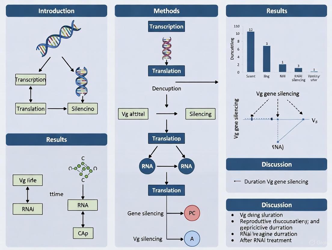
Duration of Vg Gene Silencing After RNAi Treatment: A Comprehensive Guide for Researchers
This article provides a detailed analysis of the factors determining the duration of Vitellogenin (Vg) gene silencing following RNA interference (RNAi) treatment, a critical parameter for research and therapeutic development.
Duration of Vg Gene Silencing After RNAi Treatment: A Comprehensive Guide for Researchers
Abstract
This article provides a detailed analysis of the factors determining the duration of Vitellogenin (Vg) gene silencing following RNA interference (RNAi) treatment, a critical parameter for research and therapeutic development. We explore the foundational mechanisms of RNAi persistence, compare methodological approaches for inducing short-term to long-term silencing, and outline strategies for troubleshooting and optimizing silencing longevity. Furthermore, we discuss validation techniques for confirming knockdown duration and compare RNAi with alternative gene-silencing technologies like CRISPR. This resource is tailored for scientists and drug development professionals aiming to design robust, reproducible RNAi experiments with controlled temporal effects.
Understanding RNAi Dynamics: How Long Does Gene Silencing Last?
RNA interference (RNAi) is a conserved biological mechanism that uses sequence-specific gene silencing to regulate gene expression and defend against pathogenic nucleic acids [1]. This process, central to modern genetic research and therapeutic development, is triggered by double-stranded RNA (dsRNA), which leads to the degradation of complementary messenger RNA (mRNA) [2]. The core of this process involves small interfering RNAs (siRNAs) and the RNA-induced silencing complex (RISC), which work in concert to identify and destroy target mRNA [3] [4].
This technical resource focuses on the application of this mechanism, particularly in the context of studying the duration of Vg (vitellogenin) gene silencing after RNAi treatment. The Vg gene, which codes for a major yolk protein precursor, is critical for reproduction in many insects and has become an important target for RNAi-based pest control strategies [5] [6]. Understanding the core mechanism from siRNA to mRNA degradation is fundamental for designing effective experiments and troubleshooting common issues in RNAi research.
Core Mechanism: From siRNA to mRNA Degradation
The pathway from siRNA loading to target mRNA degradation is a precise, multi-step process. The diagram below illustrates this core mechanism and its application in Vg gene silencing experiments.
Step-by-Step Mechanism Breakdown
The RNAi mechanism involves the following key steps:
Dicer Processing: The enzyme Dicer, an RNase III endonuclease, recognizes and cleaves long double-stranded RNA (dsRNA) into short double-stranded fragments of 21-23 base pairs with 2-nucleotide 3' overhangs. These fragments are the siRNAs [2] [7].
RISC Loading and Activation: The double-stranded siRNA is transferred to the RISC Loading Complex (RLC), which includes proteins like R2D2 in Drosophila. This complex facilitates the integration of the siRNA into the core RISC machinery [4] [2].
Strand Selection and RISC Maturation: The RISC complex unwinds the siRNA duplex. The strand with the less thermodynamically stable 5' end is selected as the "guide strand", while the other "passenger strand" is degraded. The guide strand is then loaded into the Argonaute protein, the catalytic heart of RISC [4] [2].
Target Recognition and Cleavage: The mature RISC, armed with the single-stranded siRNA guide, scans cytoplasmic mRNAs for complementary sequences. Upon finding a perfect or near-perfect match, the Argonaute (Ago2) protein—acting as a "Slicer" enzyme—cleaves the target mRNA [3] [4] [2]. In mammals, Ago2 is the only Argonaute protein with catalytic cleavage activity [8].
mRNA Degradation: The cleaved mRNA fragments are rapidly degraded by cellular exonucleases, preventing translation and effectively silencing the gene [4].
Troubleshooting RNAi Experiments
Common Experimental Issues and Solutions
| Problem Area | Specific Issue | Possible Causes | Recommended Solutions |
|---|---|---|---|
| Inefficient Gene Silencing | Low knockdown efficiency | - Low transfection efficiency [9]- Suboptimal siRNA design [9]- Mutations in the dsRNA/siRNA construct [9] | - Optimize transfection reagent and DNA:lipid ratio [9]- Perform a time-course assay to find peak knockdown [9]- Re-design siRNA for target region; verify oligo sequence [9] |
| Off-Target Effects | Silencing of non-target genes | - Sequence-dependent: siRNA partially binds non-target mRNAs [10] [8]- Sequence-independent: siRNA triggers immune response (e.g., interferon) [10] [8] | - Use bioinformatics tools for specific siRNA design [10]- Use pooled siRNAs to reduce individual siRNA concentration [7]- Incorporate chemical modifications into siRNA [7] |
| Control and Specificity | Verifying RNAi specificity | - Lack of proper controls- miRNA-like effects on 3' UTRs | - Include multiple negative control siRNAs (scrambled sequence)- Use rescue experiments with siRNA-resistant target gene [10] |
| Construct Issues | Problems with shRNA/miRNA vectors | - Incorrect oligo design or annealing [9]- Mutated plasmid inserts [9]- Difficulty sequencing hairpin region [9] | - Verify oligo complementarity (Top: 5'-CACC, Bottom: 5'-AAAA) [9]- Sequence plasmid clones; up to 20% may have mutations [9]- Add DMSO to sequencing reaction; use purified plasmid DNA [9] |
Duration of Vg Gene Silencing: Experimental Data
Research on the Vg gene provides a concrete example of measuring silencing duration. The following table summarizes quantitative data from two RNAi-based pest control studies, showing the persistence of gene silencing effects over time.
| Insect Species | Target Gene | dsRNA Dose | Silencing Efficiency Over Time | Observed Phenotypic Effects |
|---|---|---|---|---|
| Red Palm Weevil(Rhynchophorus ferrugineus) [6] | RfVg(Vitellogenin) |
Not Specified | - 15 days post-injection: 95% suppression- 20 days post-injection: 96.6% suppression- 25 days post-injection: 99% suppression | - Dramatic failure of Vg protein expression- Atrophied ovaries or no oogenesis- Eggs did not hatch |
| Almond Moth(Cadra cautella) [5] | CcVg(Vitellogenin) |
Not Specified | - 48 hours post-injection: ~90% suppression | - Low fecundity and egg hatchability- Eggs laid but failed to hatch due to insufficient yolk proteins |
Experimental Protocol: Measuring Long-Term Silencing of Vg
The following workflow, based on the cited Vg studies [5] [6], provides a methodology for evaluating the duration of gene silencing after RNAi treatment.
Detailed Protocol Steps:
Target Selection and dsRNA Design: Identify a unique, target-specific region within the Vg gene transcript (e.g., 3538–3938 bp in
CcVg[5]) that shows very low or no homology to other genes in the organism's genome to ensure specificity.dsRNA Synthesis: Synthesize dsRNA in vitro using a method such as T7 RiboMAX Express RNAi System, targeting the selected unique region of the Vg gene.
Experimental Delivery: Introduce the dsRNA into the experimental organism. In insect studies, this is often achieved via microinjection directly into the hemocoel (body cavity) of adult females or specific larval stages [5] [6].
Sample Collection (Time-Course): Collect tissue samples (e.g., fat body) at multiple time points post-injection. For example, in the red palm weevil study, samples were taken at 15, 20, and 25 days to assess the duration of silencing [6].
Molecular Validation: Isolate total RNA from samples and perform quantitative real-time PCR (qRT-PCR) to measure the relative expression levels of the target Vg mRNA. This quantitatively confirms the level and duration of gene silencing [5] [6].
Phenotypic Validation:
Data Analysis: Correlate the molecular data (mRNA and protein reduction) with the phenotypic data to determine the effective duration of gene silencing and its functional consequences.
| Reagent / Resource | Function in RNAi Experiments | Key Considerations |
|---|---|---|
| Synthetic siRNA | Chemically synthesized siRNA for direct introduction into cells, triggering RNAi [3] [8]. | - Can be chemically modified for increased stability and reduced off-target effects [8].- Effects are transient [8]. |
| shRNA/miRNA Vectors | Plasmid-based systems for endogenous expression of short hairpin RNA (shRNA) or artificial miRNA [9] [8]. | - Enables long-term gene silencing from a single application [8].- Requires sequencing of clones to verify insert sequence [9]. |
| Dicer Suppressors | Enzymes (e.g., Dicer, Drosha) that process long dsRNA or pre-miRNA into functional siRNAs/miRNAs [2] [7]. | - Essential for the innate RNAi pathway; their activity level can affect knockdown efficiency. |
| Argonaute Proteins (Ago2) | The catalytic core of RISC that binds the guide strand and cleaves the target mRNA [4] [2]. | - Ago2 is the primary "Slicer" enzyme in mammals [4].- Critical for the catalytic step of mRNA degradation. |
| Transfection Reagents | Chemicals or polymers that facilitate the delivery of siRNA or plasmid vectors into cells [3] [9]. | - Optimization of DNA:lipid ratio is critical for efficiency [9].- Can cause cytotoxicity at high concentrations. |
| Positive Control siRNAs | Validated siRNAs targeting a well-characterized gene (e.g., GAPDH, Luciferase). | - Essential for validating that the RNAi machinery in the cell type is functional. |
| qRT-PCR Assays | Used to quantitatively measure the reduction in target mRNA levels after RNAi treatment [5] [6]. | - The gold-standard method for confirming knockdown efficiency. |
Frequently Asked Questions (FAQs)
Q1: Why is my siRNA not producing any gene silencing effect? A1: This common problem can have several causes. First, verify the sequence of your siRNA or shRNA construct by sequencing, as mutated inserts are a frequent issue [9]. Second, optimize your transfection conditions, including cell confluency and the ratio of transfection reagent to nucleic acid [9]. Finally, ensure you are using a positive control siRNA to confirm your system is functional.
Q2: How can I distinguish between true RNAi effects and off-target toxicity? A2: Include multiple control siRNAs with the same nucleotide composition but no sequence homology to your target. The most rigorous approach is to perform a rescue experiment by expressing an siRNA-resistant version of your target gene; if the phenotype is reversed, the effect is specific [10]. Also, monitor for general cell health and consider using lower siRNA concentrations to minimize non-specific immune activation [8].
Q3: For long-term silencing studies, should I use synthetic siRNAs or expressed shRNAs? A3: The choice depends on your experimental needs. Synthetic siRNAs are easier to deliver and allow for chemical modifications but produce transient silencing (days) [8]. shRNAs expressed from vectors lead to long-term silencing (weeks to months) from a single application, as they are continuously transcribed inside the cell [8]. For Vg silencing studies lasting several weeks, viral vectors delivering shRNAs are often necessary.
Q4: How long can I expect Vg gene silencing to last after a single dsRNA injection?
A4: As shown in the data table (Section 3.2), the duration can be significant. In the red palm weevil, a single injection of RfVg dsRNA led to greater than 95% suppression for at least 25 days [6]. The longevity depends on factors like the stability of the dsRNA, the turnover rate of the target mRNA and protein, and the specific biological system.
Q5: What are the key parameters to confirm successful and specific Vg silencing? A5: A comprehensive validation includes three levels:
- Molecular: qRT-PCR shows a significant reduction in Vg mRNA levels [5] [6].
- Biochemical: SDS-PAGE or Western blot shows a reduction in Vg protein [6].
- Phenotypic: Observation of reduced fecundity, poor ovarian development, and low egg hatchability, confirming the biological efficacy of the silencing [5] [6].
FAQ 1: What is the typical duration of siRNA-induced gene silencing?
The duration of siRNA-induced silencing is fundamentally transient, typically lasting from 2 to 7 days in standard in vitro cell cultures. The exact timeframe is highly dependent on experimental conditions, including cell type, transfection efficiency, and the proliferation rate of the cells [11].
The table below summarizes key factors and their impact on silencing duration:
| Factor | Impact on Duration | Notes |
|---|---|---|
| Cell Proliferation Rate | High | In fast-dividing cells, siRNA is diluted with each cell division, shortening effect [11]. |
| siRNA Design & Modifications | Medium-High | Chemically modified siRNAs (e.g., Accell) can enhance stability and duration [11]. |
| Delivery Method | Medium | Viral-delivered shRNAs can enable long-term silencing, unlike synthetic siRNA [11]. |
| Target Gene/Turnover | Medium | Silencing of genes with stable, long-lived proteins may show a delayed phenotypic effect. |
For in vivo applications, advanced delivery technologies like lipid nanoparticles (LNPs) and GalNAc-conjugates have significantly extended silencing duration, enabling dosing intervals of several weeks or even months in therapeutic contexts [12] [13] [14].
FAQ 2: Why is my siRNA silencing effect fading too quickly?
Rapid loss of silencing is a common challenge. The primary cause is the transient nature of synthetic siRNA in dividing cells. Troubleshoot using the table below:
| Problem Cause | Troubleshooting Strategy | Experimental Protocol Adjustments |
|---|---|---|
| Cell Division Dilution | Use non-dividing cells or repeated dosing. | For difficult-to-transfect cells, consider Accell modified siRNAs for repeated dosing or viral-mediated delivery of shRNA for stable expression [11]. |
| Inefficient Transfection | Optimize delivery and validate knockdown. | Include a positive control siRNA (e.g., targeting a readily detectable gene) to confirm transfection protocol efficiency [11] [15]. |
| Ineffective siRNA | Re-design and validate siRNA efficacy. | Use a reporter-based validation system. Fuse the siRNA target sequence to a reporter gene (e.g., EGFP, Firefly luciferase) for quantitative efficacy measurement [16]. |
| Poor siRNA Stability | Use chemically modified siRNA. | Select siRNAs with 2'-O-methyl, 2'-fluoro, or phosphorothioate (PS) backbone modifications to resist nuclease degradation [13]. |
Experimental Protocol: Reporter-Based siRNA Validation System
This protocol allows for quantitative assessment of siRNA efficacy before testing on the endogenous gene [16].
1. Principle A short synthetic DNA fragment containing the proposed siRNA target sequence is cloned into the 3' untranslated region (3'UTR) of a reporter gene (e.g., Enhanced Green Fluorescent Protein - EGFP, or Firefly luciferase - Fluc). The ability of the siRNA to inhibit reporter expression directly measures its knockdown efficiency.
2. Materials
- Vectors: pEGFP-3'UTR, pFluc-3'UTR, or pDual (for simultaneous expression of EGFP and Fluc) for the targeting reporter. pHsH1 or pDual vectors for triggering siRNA/shRNA expression [16].
- Cells: Mammalian cell line of interest.
- Reagents: Lipofectamine 2000, cell culture media, PBS, trypsin-EDTA, lysis buffers, Dual-Luciferase Reporter Assay System, Micro BCA assay kit.
3. Workflow Diagram
4. Procedure
- Construct Reporter Plasmid: Clone the synthetic DNA oligonucleotide containing your siRNA target sequence into the 3'UTR of the reporter plasmid (e.g., pEGFP-3'UTR) using appropriate restriction enzymes (e.g., BglII, HindIII) [16].
- Construct siRNA Expression Plasmid: Clone the same target sequence into an siRNA or shRNA expression vector (e.g., pHsH1).
- Co-transfection: Seed cells in a 24-well or 48-well plate. The next day, co-transfect cells with a fixed amount of the reporter plasmid and the siRNA expression plasmid (or a synthetic siRNA) using a transfection reagent like Lipofectamine 2000 [16].
- Harvest and Analyze: After 24-72 hours, harvest cells.
- For luciferase: Lyse cells and measure activity using the Dual-Luciferase Reporter Assay System, normalizing to a co-transfected control (e.g., Renilla luciferase).
- For EGFP: Analyze fluorescence intensity using a flow cytometer or fluorescence plate reader.
- Calculate Efficacy: Compare the reporter signal in cells transfected with the experimental siRNA versus a negative control siRNA. siRNA efficacy (%) = [1 - (Signalexperimental / Signalcontrol)] × 100.
The Scientist's Toolkit: Key Research Reagent Solutions
| Item | Function & Application |
|---|---|
| ON-TARGETplus siRNA | Pre-designed siRNAs with patented chemical modifications to reduce off-target effects, ideal for gene silencing validation studies [11]. |
| Accell siRNA | Chemically modified siRNAs designed for delivery in difficult-to-transfect cells (e.g., primary cells) without the need for transfection reagents, enabling repeated dosing [11]. |
| Lipofectamine 2000 | A common cationic lipid-based transfection reagent for delivering siRNA, microRNAs, and shRNA into a wide range of cell types [16]. |
| pDual Vector | A bicistronic vector expressing both EGFP and Fluc reporters from a single plasmid, useful for constructing the reporter-based siRNA validation system [16]. |
| siRNAEfficacyDB | A public database integrating experimentally validated siRNA efficacy data (3,544 records), useful for informing siRNA design and predicting activity [17]. |
| Tri-GalNAc Conjugates | A delivery technology for in vivo applications that enables highly efficient targeting of siRNA to hepatocytes in the liver, used in several approved therapeutics [12] [14]. |
FAQ 3: How can I design siRNAs for longer-lasting effects?
While the core limitation is transient delivery, strategic design and delivery choices can maximize the longevity of the silencing effect.
- Utilize Advanced Delivery Platforms: For in vivo work, use GalNAc-siRNA conjugates for liver targets or LNPs for other tissues. These platforms protect the siRNA, facilitate cellular uptake, and can provide effects lasting for months from a single dose [12] [13] [14].
- Employ Viral Vectors for shRNA: For long-term, stable gene silencing in cell lines or animal models, use viral vectors (e.g., lentivirus) to express short hairpin RNAs (shRNAs), which are processed into siRNA inside the cell, providing persistent knockdown [11].
- Apply Chemical Modifications: Select siRNAs that incorporate 2'-O-methyl, 2'-fluoro, and phosphorothioate linkages. These modifications increase nuclease resistance and plasma half-life, extending the window of activity [13].
- Leverage Machine Learning Design Tools: Use services (e.g., from GenScript) that employ machine learning models trained on large-scale siRNA efficacy screens to select sequences with high predicted potency and specificity, improving the odds of a strong, durable knockdown [13].
A fundamental challenge in RNA interference (RNAi) research is that the silencing of a target gene is not always permanent. The duration of the silencing effect is influenced by a complex interplay of factors. For researchers focusing on long-term functional studies or therapeutic development, understanding and controlling these factors is crucial. This guide breaks down the key elements that influence silencing longevity and provides troubleshooting advice for common experimental hurdles.
Frequently Asked Questions (FAQs)
FAQ 1: What are the most critical factors that determine how long my RNAi effect will last? The longevity of gene silencing is primarily governed by three key areas:
- Cellular Division Rate: In rapidly dividing cells, the silencing effect is diluted as the siRNA is distributed among daughter cells. Knockdown typically lasts less than a week in such environments. In contrast, in non-dividing or slowly dividing cells (e.g., hepatocytes), silencing can persist for 3-4 weeks [18].
- Stability of the siRNA and Target Protein: The intracellular half-life of the siRNA molecule itself and the turnover rate of the target protein significantly impact how long the phenotypic effect lasts. You must target a protein with a relatively short half-life to see a rapid effect, and use a highly stable siRNA to maintain it [18].
- Dosage and Delivery Efficiency: The initial dose of dsRNA/siRNA and the efficiency of its delivery into the target cells set the ceiling for the potential duration and magnitude of silencing [19] [18].
FAQ 2: I achieved strong initial knockdown, but the effect is short-lived. How can I extend the duration? This is a common issue. Here are several strategies to troubleshoot:
- Optimize dsRNA/siRNA Design: Use longer dsRNA molecules (>60 bp) or Dicer-substrate siRNAs (27-mer), as they generate a more diverse and potent pool of siRNAs, leading to more sustained silencing [19] [20].
- Employ Chemical Modifications: Incorporate chemical modifications (e.g., 2'-O-Methyl, Phosphorothioate) into your siRNA to enhance its stability against nucleases, which directly prolongs its active life within the cell [21].
- Consider a Delivery Platform for Sustained Release: For in vivo work, switch from transient transfection to methods that allow for sustained release, such as viral vectors expressing short hairpin RNAs (shRNAs) or optimized lipid nanoparticles [20] [21].
- Re-dose According to Kinetics: Model the kinetics of your target protein recovery and establish a dosing schedule that re-administers siRNA before the protein levels fully rebound [18].
FAQ 3: Why does the longevity of silencing vary so much between different cell types or insect species? Variability arises from intrinsic biological differences:
- Systemic RNAi Machinery: Some species and cell types have robust machinery for the systemic spread of the RNAi signal (e.g., SID-1 channels), leading to more potent and sustained effects. Others lack these components [20].
- Cellular Uptake and Intracellular Trafficking: The efficiency with which cells take up exogenous dsRNA/siRNA and route it to the correct intracellular compartment for RISC loading is highly variable and a major source of species-specific differences [19].
- Potency of the Immune Response: The same dsRNA can trigger different degrees of immune activation in different cell types or organisms, which can interfere with the core RNAi mechanism and confound longevity readings [22].
Troubleshooting Guide: Silencing Longevity
| Problem | Potential Cause | Recommended Solution |
|---|---|---|
| Rapid loss of silencing effect in cell culture. | Rapid dilution of siRNA due to high cell division rate. | Use a stably integrated shRNA expression system; Re-transfect according to a kinetically determined schedule [18]. |
| Weak or no silencing effect from the start. | Inefficient dsRNA/siRNA design; Poor delivery; Targeting a highly stable protein. | Redesign dsRNA to target a different mRNA region; Use a validated positive control; Optimize transfection protocol; Verify delivery efficiency [19]. |
| Silencing is effective in one insect species but not in another. | Differences in dsRNA uptake mechanisms or nuclease activity in the gut/hemolymph. | Screen multiple target genes; Test the use of nanoparticle carriers to protect dsRNA [19] [23]. |
| High off-target effects or cytotoxicity. | siRNA sequence triggers an immune response or has miRNA-like off-target effects. | Re-design siRNA using bioinformatic tools to avoid immunostimulatory motifs and seed region matches to off-target genes; Use modified nucleotides (e.g., 2'-O-Methyl) to reduce off-targeting [22]. |
Experimental Protocols & Data
Case Study: Sustained Silencing of the Vitellogenin (Vg) Gene
The following table summarizes quantitative data from a key study investigating the long-term knockdown of the Vitellogenin (Vg) gene in the red palm weevil, a critical experiment for understanding silencing longevity in vivo [24].
Table 1: Long-term Knockdown of Vg Gene Expression Post-dsRNA Injection
| Time Point Post-injection | % Suppression of Vg mRNA | Observed Phenotypic Effect |
|---|---|---|
| 15 days | 95% | Dramatic failure of Vg protein expression. |
| 20 days | 96.6% | Atrophied ovaries, failure of oogenesis. |
| 25 days | 99% | No egg hatchability. |
Methodology:
- dsRNA Design: A unique 400 bp region (position 3538–3938 bp) of the RfVg transcript with low homology to other genes was selected.
- dsRNA Synthesis: Target dsRNA was synthesized using standard in vitro transcription kits.
- Delivery: Adult female weevils received a single dorsal micro-injection of 2 μg of RfVg-dsRNA.
- Validation: Knockdown was verified via qRT-PCR at multiple time points. Phenotypic effects on ovary development and egg hatchability were assessed visually and biologically.
This protocol demonstrates that a single, well-designed dsRNA application can achieve near-total silencing for over three weeks, leading to a persistent and profound biological effect.
Pathway and Workflow Diagrams
Diagram 1: Factors governing silencing longevity.
Diagram 2: Workflow for long-lasting silencing experiments.
Table 2: Key Research Reagent Solutions for RNAi Longevity Studies
| Item | Function in Research | Application Note |
|---|---|---|
| Long dsRNA (>60 bp) | Triggers a more potent and sustained RNAi response by generating multiple siRNAs [19]. | Ideal for non-mammalian systems (e.g., insects, plants). Essential for initial gene screening. |
| Chemically Modified siRNAs (2'-OMe, PS, LNA) | Increases nuclease resistance, reduces immunostimulation, and minimizes off-target effects, prolonging therapeutic activity [21] [22]. | Critical for in vivo applications. Modifications must be carefully placed to avoid disrupting RISC loading. |
| Stable shRNA Expression Vectors | Provides continuous, long-term endogenous production of siRNA from within the cell, bypassing delivery and dilution challenges [20] [18]. | The gold standard for creating stable knockdown cell lines. Requires viral or other methods for genomic integration. |
| GalNAc-siRNA Conjugates | Enables highly efficient and targeted delivery of siRNA to hepatocytes, enabling sustained silencing with very infrequent dosing [21]. | A breakthrough for therapeutic siRNA development targeting liver-expressed genes. |
| Cationic Lipid/Polymer Carriers | Formulates siRNA into nanoparticles, protecting it during systemic delivery and enhancing cellular uptake [18] [21]. | Widely used for in vitro and in vivo work. Optimization of lipid composition is key to efficacy and reducing toxicity. |
The Role of Cell Division and Protein Turnover in Ending the Effect
Frequently Asked Questions (FAQs)
FAQ 1: What is the typical duration of VEGF gene silencing after a single siRNA transfection? In readily transfected cells treated with potent siRNAs, near-maximal gene silencing (>80% knockdown) can typically be achieved for 5 to 7 days post-transfection. The maximal knockdown effect is often observed around day 2. While significant silencing may still be detectable at day 10, the effect progressively diminishes after the first week [25].
FAQ 2: Why does siRNA-mediated silencing eventually end? The primary reason is the dilution of the siRNA effect due to cell division. In rapidly dividing cells, the intracellular concentration of siRNA and the RISC complex is reduced with each cell division, leading to a recovery of target protein levels within about a week. In non-dividing cells, however, silencing can persist for 3 to 4 weeks. The natural degradation of the siRNA molecules and the turnover of the target protein itself also contribute to the effect's duration [18].
FAQ 3: Can I prolong silencing by increasing the siRNA concentration? No, raising the siRNA concentration (e.g., from 5 nM to 50 nM) does not typically improve or prolong the silencing effect. Once the RNA-induced silencing complex (RISC) is saturated with a highly potent siRNA, any excess siRNA is likely rapidly degraded, sequestered, or excreted from the cell. Using higher-than-needed concentrations primarily increases the risk of off-target effects without enhancing longevity [25].
FAQ 4: How does the target protein's half-life influence the observed silencing kinetics? The half-life of the target protein directly impacts how quickly you observe a reduction in protein levels after mRNA knockdown. Even after successful mRNA degradation, pre-existing VEGF protein molecules will persist until they complete their natural lifecycle. Proteins with longer half-lives will require more time to be depleted, meaning the full phenotypic effect of silencing may not be immediate.
FAQ 5: What is the difference between a knockdown and a knockout in the context of VEGF research?
- Knockdown (e.g., via RNAi): A temporary, partial reduction of VEGF gene expression at the messenger RNA (mRNA) level. It is reversible and allows researchers to study the effects of reducing, but not completely eliminating, VEGF [10].
- Knockout (e.g., via CRISPR-Cas9): A permanent, complete disruption of the VEGF gene at the DNA level. This creates a irreversible loss of function, which is useful for definitive functional studies but may be lethal for essential genes [10].
Troubleshooting Guide
| Problem | Possible Cause | Suggested Solution |
|---|---|---|
| Short silencing duration in a fast-dividing cell line | Rapid dilution of siRNA and RISC due to high cell proliferation rate [18]. | Use a repeated transfection strategy or consider alternative models like non-dividing cells for longer-term studies. |
| Insufficient knockdown efficiency | Inefficient siRNA design, poor transfection efficiency, or low siRNA potency [26]. | Re-optimize transfection protocol, use chemically modified siRNAs (e.g., Silencer Select with LNA) for improved performance, and validate with multiple siRNA sequences [25]. |
| High off-target effects | siRNA concentration is too high, or the sequence lacks specificity [25]. | Use the lowest effective siRNA concentration (e.g., 5 nM) and utilize advanced bioinformatic tools for siRNA design to minimize sequence homology with off-target genes [25] [26]. |
| No observable phenotypic effect despite mRNA knockdown | Slow turnover rate of the existing VEGF protein pool masking the molecular effect [27]. | Allow more time post-transfection for the VEGF protein to degrade and monitor the phenotype over a longer time course. |
| Inconsistent results between replicates | Variability in cell confluence or transfection efficiency. | Standardize cell seeding density and passage number, and ensure consistent transfection reagent mixing across samples. |
The following table consolidates key experimental findings on the duration of siRNA-induced gene silencing.
Table 1: Kinetics of siRNA-Mediated Gene Silencing
| Cell Type / Model | Observed Silencing Duration | Key Influencing Factor | Reference |
|---|---|---|---|
| HeLa / BJ cells (dividing) | 5-7 days of >80% knockdown after single transfection (5 nM siRNA) [25]. | siRNA potency and transfection efficiency [25]. | [25] |
| Rapidly dividing cell lines (in vitro) | Protein levels recover to pre-treatment values in <1 week [18]. | Rate of cell division [18]. | [18] |
| Non-dividing fibroblasts (in vitro) | Protein levels take >3 weeks to return to steady-state [18]. | Lack of dilution from cell division [18]. | [18] |
| Subcutaneous tumors in mice (in vivo) | Knockdown lasts ~10 days [18]. | Cell division in tumor microenvironment [18]. | [18] |
| Non-dividing hepatocytes in mice (in vivo) | Knockdown lasts 3–4 weeks [18]. | Lack of dilution from cell division [18]. | [18] |
Table 2: Impact of Experimental Variables on Silencing Duration
| Experimental Variable | Impact on Duration | Practical Implication | Reference |
|---|---|---|---|
| Repeated Transfection | Can prolong silencing. A second transfection at day 4 improved knockdown at days 6-11 in some cases [25]. | A viable strategy to extend the window of silencing for longer-term experiments [25]. | [25] |
| Increasing siRNA Concentration | No significant prolongation. No improvement from 5 nM to 50 nM [25]. | Use minimal effective dose to saturate RISC and avoid off-target effects [25]. | [25] |
| Target Protein Half-Life | Governs the rate of protein-level knockdown. Slower turnover delays phenotypic manifestation [27]. | Critical for planning the timing of downstream phenotypic assays post-transfection. | [27] |
Detailed Experimental Protocols
Protocol 1: Assessing the Duration of VEGF Silencing
This protocol outlines the steps to measure the longevity of VEGF silencing in a cell culture model.
Key Research Reagent Solutions:
- Validated VEGF siRNA: Chemically modified siRNAs (e.g., Silencer Select) for improved specificity and longevity [25].
- Negative Control siRNA: A non-targeting siRNA to control for non-sequence-specific effects.
- Transfection Reagent: A reliable cationic lipid or polymer (e.g., Oligofectamine) [18].
- qRT-PCR Assay: For quantifying VEGF mRNA levels at multiple time points.
- ELISA Kit: For measuring VEGF protein secretion or intracellular levels.
Methodology:
- Cell Seeding: Seed easy-to-transfect cells (e.g., HeLa or a relevant cancer cell line) in a multi-well plate at a consistent, sub-confluent density (e.g., 500 cells/well in a 96-well format) and culture overnight [25].
- Transfection: Transfect cells with VEGF siRNA and control siRNA at a low, effective concentration (e.g., 5 nM). Include an untreated control. Use optimized concentrations of transfection agent (e.g., 0.15 µL/well for HeLa) [25].
- Time-Course Harvesting: At defined time points post-transfection (e.g., days 1, 2, 3, 5, 7, and 10), harvest cells for analysis.
- For mRNA analysis: Lyse cells directly and use a Cells-to-CT kit for direct qRT-PCR [25].
- For protein analysis: Collect cell lysates and/or culture supernatants for ELISA.
- Data Analysis: Express VEGF mRNA and protein levels at each time point relative to the negative control siRNA. Plot the percentage of knockdown over time to visualize the duration of the effect.
Protocol 2: Investigating Contribution of Protein Turnover
This protocol uses a pulse-SILAC (Stable Isotope Labeling by Amino acids in Cell culture) method to directly measure protein synthesis and turnover rates in control and VEGF-silenced cells.
Key Research Reagent Solutions:
- SILAC Media: Heavy isotope-labeled amino acids (e.g., Lys⁸ and Arg¹⁰).
- VEGF siRNA & Control siRNA: As in Protocol 1.
- Mass Spectrometry System: For proteome-wide analysis of protein abundance and turnover.
Methodology:
- Cell Culture & Transfection: Grow cells in SILAC "light" media. Perform transfection with VEGF or control siRNA as described in Protocol 1.
- Pulse Labeling: At the peak of VEGF silencing (e.g., 48 hours post-transfection), switch the media to SILAC "heavy" media.
- Time-Course Sampling: Harvest cells at multiple time points after the media switch (e.g., 0, 6, 12, 24, 48 hours).
- Sample Processing and MS Analysis: Mix samples from each time point with a common internal standard. Process for mass spectrometry analysis to measure the incorporation of heavy isotopes into VEGF and other proteins over time [27].
- Kinetic Modeling: Calculate the half-lives of VEGF and other proteins by fitting the isotopic incorporation data to exponential decay curves. Compare the half-lives between VEGF-siRNA and control cells to see if silencing indirectly affects protein stability.
Signaling Pathways and Experimental Workflows
VEGF Signaling and Silencing Impact
The following diagram illustrates the VEGF signaling pathway and the points where siRNA-mediated silencing and downstream phenotypic effects occur.
VEGF Signaling and siRNA Impact
Experimental Workflow for Duration Analysis
This diagram outlines the logical workflow for a time-course experiment designed to analyze the duration of siRNA-mediated silencing.
Silencing Duration Workflow
Achieving Prolonged Silencing: From siRNA to shRNA Vectors
FAQs and Troubleshooting Guides
Frequently Asked Questions
Q1: What is the key advantage of using synthetic siRNAs over other RNAi methods for transient silencing? Synthetic siRNAs are designed to mimic the natural end products of Dicer cleavage and can be directly transfected into cells, leading to rapid gene knockdown without the need for transcription from a vector. This makes them ideal for transient silencing experiments where permanent genetic modification is not desired [28] [10].
Q2: How long does synthetic siRNA-mediated silencing typically last in mammalian cell cultures? The duration of silencing is transient, typically lasting from 3 to 7 days in standard cell cultures. The effect is "diluted out" as cells divide, and multiple transfections may be needed for longer-term studies. The extent and duration of protein depletion also depend on the protein's half-life; short-lived proteins may be significantly reduced within 3-5 days, whereas long-lived proteins may show little depletion [28].
Q3: What are the major causes of off-target effects, and how can they be minimized? Off-target effects can be sequence-independent (e.g., triggering interferon responses) or sequence-dependent (cross-hybridization to transcripts with limited complementarity). To minimize these:
- Use low siRNA concentrations (often below 20 nM) [29] [28].
- Utilize sophisticated design algorithms that avoid problematic sequences [30] [10].
- Consider using diced siRNA pools (d-siRNAs), which are highly complex pools where the concentration of any single, problematic siRNA is too low to cause significant off-target effects [29].
- Employ chemically modified siRNAs, such as Stealth RNAi, which are engineered to reduce off-target potential [30] [10].
Q4: My siRNA is not producing the expected knockdown. What should I check?
- Transfection Efficiency: Ensure your cells are transfectable and optimize transfection conditions. Use a fluorescently labeled control RNA to monitor efficiency [30] [28].
- siRNA Sequence: Verify that the siRNA sequence is specific and effective for your target gene. Screening multiple siRNAs against different regions of the target mRNA is recommended [9].
- Protein Half-life: If measuring protein knockdown, remember that proteins with long half-lives may not show significant reduction in transient assays [28].
- Experimental Timing: Perform a time-course experiment to determine the peak knockdown, which usually occurs 24-72 hours post-transfection [9].
Troubleshooting Common Problems
| Problem | Possible Cause | Recommended Solution |
|---|---|---|
| Low Knockdown Efficiency | Low transfection efficiency; non-optimal siRNA sequence; target with long half-life. | Use a validated positive control; optimize transfection reagent and DNA:lipid ratio; try multiple siRNAs; perform a time-course [28] [9]. |
| High Cell Toxicity | Off-target effects; activation of interferon response; transfection reagent toxicity. | Lower siRNA concentration; use specialized, less toxic transfection reagents; switch to diced siRNA pools or Stealth RNAi [29] [30] [28]. |
| Inconsistent Results Between Replicates | Inconsistent transfection; poor cell health; serum containing tetracycline (for inducible systems). | Standardize cell passage number and confluency; ensure consistent transfection mix; use tetracycline-free serum [9]. |
| No Gene Silencing Observed | siRNA sequence does not effectively target the mRNA; mutations in the siRNA sequence. | Re-design and test new siRNA sequences; sequence the siRNA plasmid or oligo to confirm identity if applicable [9]. |
Quantitative Data on Silencing Dynamics
Table 1: Silencing Time-Course and Efficiency of Different siRNA Modalities
Data compiled from referenced studies on siRNA and d-siRNA performance.
| siRNA Modality | Typical Onset of Knockdown | Peak Knockdown | Duration of Effect | Key Advantages |
|---|---|---|---|---|
| Synthetic siRNA | 24 hours | 48-72 hours | 3-7 days (transient) | Rapid delivery; high knockdown potency; well-established protocols [28]. |
| Diced siRNA (d-siRNA) Pools | 24 hours | 48-72 hours | 3-7 days (transient) | Highly complex pools; significantly reduced off-target effects; effective for hard-to-target genes [29]. |
| Stealth RNAi | 24 hours | 48-72 hours | 3-7 days (transient) | Proprietary chemistry reduces off-target effects and improves stability; blunt-ended 25mer duplex [30]. |
| shRNA (Lentiviral) | 48-72 hours | 5-7 days | Stable/inducible (weeks) | Suitable for long-term or inducible knockdown in hard-to-transfect cells [28]. |
Table 2: Impact of siRNA Concentration on Specificity and Efficiency
Based on studies analyzing mRNA changes via microarray to assess off-target effects [29] [28].
| siRNA Concentration | Knockdown Efficiency | Risk of Off-Target Effects | Recommended Use Case |
|---|---|---|---|
| High (>50 nM) | Potentially very high | Substantial risk; can silence hundreds of non-target genes | Not recommended for specific silencing; may trigger stress responses. |
| Moderate (20-50 nM) | High | Significant off-target risk | Use with caution, only if lower concentrations are ineffective. |
| Low (1-20 nM) | Effective and specific | Minimized risk; changes are more likely target-specific | Ideal for most experiments to ensure phenotypic specificity [28]. |
Experimental Protocols
Protocol 1: Transient Transfection of Synthetic siRNAs in Adherent Cells
This is a standard protocol for achieving transient gene silencing in common cell lines.
Materials:
- Synthetic siRNA (e.g., Silencer Select, Stealth RNAi)
- Appropriate transfection reagent (e.g., Lipofectamine RNAiMAX)
- Opti-MEM or similar serum-free medium
- Healthy, proliferating cells
Workflow Diagram:
Procedure:
- Day 1: Cell Plating. Plate cells in antibiotic-free growth medium to reach 50-70% confluency at the time of transfection (typically 18-24 hours later).
- Day 2: Transfection Complex Formation.
- Dilute the siRNA (e.g., 5-20 nM final concentration) in a tube with Opti-MEM Medium.
- Mix the appropriate amount of transfection reagent in a separate tube with Opti-MEM.
- Combine the two mixtures, vortex gently, and incubate for 5-20 minutes at room temperature to allow complex formation.
- Transfection. Add the siRNA-reagent complexes dropwise to the cells. Gently swirl the plate to ensure even distribution.
- Incubation and Analysis.
Protocol 2: Using Diced siRNA (d-siRNA) Pools for High-Specificity Silencing
This protocol is ideal when off-target effects are a major concern, as it leverages a complex pool of siRNAs.
Materials:
- Long dsRNA template (500-1000 bp) homologous to your target.
- Recombinant Dicer enzyme or bacterial RNase III.
- Purification kit to clean up the d-siRNA reaction.
Workflow Diagram:
Procedure:
- Template Generation. Generate a long double-stranded RNA (dsRNA) template (~500-1000 bp) corresponding to your target gene sequence by in vitro transcription [29].
- Dicing Reaction. Incubate the long dsRNA with a recombinant dicing enzyme (Dicer or bacterial RNase III) to digest it into a complex pool of 21-23 nt siRNAs [29] [28].
- Purification. Purify the resulting d-siRNA pool to remove enzymes and reaction components.
- Transfection. Transfect the d-siRNA pool into cells using the same methodology as for synthetic siRNAs. The low concentration of any individual siRNA in the pool minimizes the risk of off-target effects while effectively silencing the intended target [29].
The Scientist's Toolkit: Key Research Reagent Solutions
Table 3: Essential Reagents for Transient Silencing Experiments
| Reagent / Solution | Function in Experiment | Key Considerations |
|---|---|---|
| Synthetic siRNA | The effector molecule that directs sequence-specific mRNA cleavage. | Select based on validated design algorithms (e.g., from supplier or public databases); chemical modifications can enhance stability and specificity [30] [10]. |
| Transfection Reagent | Forms complexes with siRNA to facilitate its delivery into cells. | Choose based on cell type (e.g., Lipofectamine RNAiMAX for standard lines); primary cells may require specialized reagents or viral delivery [28]. |
| Opti-MEM Medium | A low-serum medium used for diluting siRNA and transfection reagent, improving complex formation. | Essential for reducing toxicity and maximizing transfection efficiency during complex formation. |
| Diced siRNA (d-siRNA) | A highly complex pool of siRNAs that mitigates off-target effects. | Must be generated in-house from a long dsRNA template; recommended when specificity is paramount [29]. |
| Fluorescent Control siRNA | A non-targeting, fluorescently-labeled siRNA used to monitor and optimize transfection efficiency. | Critical for troubleshooting and validating protocol success in new cell lines [30]. |
| Positive Control siRNA | An siRNA targeting a ubiquitously expressed gene (e.g., GAPDH, cyclophilin B). | Provides a benchmark for knockdown efficiency and validates that the experimental system is working [30]. |
Sustained Knockdown Using Plasmid and Viral Vector-Driven shRNA
Troubleshooting Guide
Why is there no detectable knockdown of my target gene?
Problem: After transducing cells with your shRNA vector, you do not observe a reduction in your target gene's expression.
Solutions:
- Test multiple shRNA sequences: Not all shRNAs are effective. When testing 3-4 shRNAs for a gene, typically 2-3 produce reasonable knockdown, but it's possible none work by chance. Always test more shRNAs, preferably literature-validated ones, or use a "cocktail" mixture targeting the same gene [31].
- Verify your validation assay:
- RT-qPCR: Use sensitive RT-qPCR with primers spanning an exon-exon junction to avoid genomic DNA amplification. Validate primers by running the PCR product on a gel or by sequencing. Always include a minus-reverse transcription (RT) control [31].
- Western Blot: Be aware of non-specific antibody binding that can cause false positive bands, misleadingly suggesting a lack of knockdown. Verify antibody specificity [31].
- Check shRNA target coverage: Your shRNA might only target a subset of the gene's transcript isoforms. Design shRNAs to target all relevant isoforms unless studying a specific one [31].
- Sequence your construct: Up to 20% of clones may contain mutated inserts (e.g., 1-2 bp deletions), leading to poor RNAi response. Sequence positive transformants to confirm the correct shRNA insert sequence [9].
- Optimize transduction/transfection:
- Low Transfection Efficiency: Ensure antibiotics are not present during transfection and cells are at the proper confluency. Optimize the amount of transfection reagent and DNA used [9].
- Viral Transduction: For lentiviral vectors, ensure Polybrene is present during transduction. Use a higher multiplicity of infection (MOI) and consider selecting stably transduced cells with an antibiotic like Zeocin [9].
Why is my inducible shRNA system showing high background (leaky expression) or no induction?
Problem: For Tet-On or similar inducible systems, you observe shRNA expression even without the inducer, or no expression after adding the inducer.
Solutions:
- Check serum components: Fetal bovine serum (FBS) lots can contain tetracycline. Use certified tetracycline-free FBS for inducible systems to prevent basal expression [9].
- Verify repressor cell line: Ensure you are using a cell line that stably expresses the Tet repressor (e.g., T-REx cell line) [9].
- Confirm inducer amount and timing: Ensure enough tetracycline (or equivalent) is added. Cells should be treated 3-24 hours after transfection, and knockdown is typically assayed 24-96 hours post-induction [9].
- Inspect the shRNA sequence: Verify that the shRNA sequence does not contain more than three tandem thymidines (Ts), as this can cause premature transcription termination [9].
Why is the knockdown effect transient or not sustained?
Problem: The initial knockdown of the target gene is lost after several cell divisions or over time.
Solutions:
- Use integrating viral vectors: For long-term expression in dividing cells, use viral vectors that integrate into the host genome, such as lentiviral vectors (LVs). Note that LVs use an HIV backbone and have safety considerations regarding insertional mutagenesis [32].
- Generate stable cell pools: After transduction, use antibiotic selection to eliminate untransduced cells and create a stable population where the shRNA construct is maintained [9].
- Consider vector silencing: If using plasmids, prolonged culture can lead to promoter silencing. Using viral or mammalian promoters resistant to silencing may help sustain expression.
- For in vivo delivery: Recombinant adeno-associated virus (rAAV) vectors are favored for in vivo applications due to their ability to persist episomally and provide sustained transgene expression in targeted, non-dividing tissues [33].
Frequently Asked Questions (FAQs)
What percentage of shRNAs typically show strong knockdown?
On average, only about 20-30% of shRNAs tested show a strong knockdown effect. Approximately 50-70% have a noticeable knockdown effect, meaning a significant portion may be ineffective [31].
What are the critical steps in cloning shRNA oligos to avoid mutations?
- Oligo Design and Quality: Ensure top and bottom strand oligos are perfectly complementary. Use high-quality, HPLC- or PAGE-purified oligos to minimize the risk of mutations [9].
- Annealing Conditions: Anneal equal molar amounts of oligos by heating to 95°C followed by incubation at room temperature for 5-10 minutes [9].
- Sequencing Troubleshooting: If sequencing through the hairpin is difficult, try:
- Using high-quality plasmid prep kits.
- Adding DMSO to the sequencing reaction (final concentration of 5%).
- Increasing the amount of template DNA.
- Using a sequencing kit with dGTP instead of dITP [9].
How should I handle non-detect (Ct=40) values in my qPCR knockdown validation data?
Replacing non-detects with a fixed value (e.g., 40 cycles) introduces significant bias in estimating absolute (ΔCt) and differential (ΔΔCt) expression [34]. A non-detect in the target gene typically biases absolute expression estimates downward. A better approach is to use statistical methods that model the missing data mechanism, such as those implemented in the nondetects R package, to reduce bias [34].
What viral vector is best for sustained in vivo knockdown?
rAAV vectors are often preferred for in vivo shRNA delivery due to their [33] [32]:
- Favorable safety profile: Non-pathogenic and elicit mild immune responses.
- Sustained expression: Persist episomally in non-dividing cells, providing long-term expression.
- High tissue tropism: Specific serotypes can target different organs efficiently.
A key limitation is their small packaging capacity (~4.7 kb), which can be circumvented by using compact promoters or smaller Cas orthologs if combining with CRISPR systems [33].
Experimental Protocols & Data
| Metric | Typical Success Rate | Notes / Reference |
|---|---|---|
| shRNAs with noticeable knockdown | 50-70% | A noticeable effect is observed. [31] |
| shRNAs with strong knockdown | 20-30% | Represents the most effective constructs. [31] |
| Functional shRNAs per gene tested | 2-3 out of 3-4 | Testing multiple constructs is standard practice. [31] |
| Clones with mutated shRNA inserts | Up to 20% | Highlights the necessity of sequencing verification. [9] |
Protocol: Validating Knockdown Efficiency via RT-qPCR
- RNA Extraction: Isolate high-quality total RNA from transfected/transduced cells and control cells using a method that minimizes genomic DNA contamination.
- cDNA Synthesis: Perform reverse transcription with a robust reverse transcriptase. Include a minus-RT control (a reaction without the enzyme) for each sample to assess genomic DNA contamination.
- Primer Design:
- Design primers to span an exon-exon junction where possible.
- Use tools like NCBI Primer-BLAST to check for specificity.
- Validate primer efficiency by running the PCR product on an agarose gel and/or by sequencing the product [31].
- qPCR Run:
- Run samples in technical replicates.
- Include a stable reference gene (e.g., GAPDH, β-actin) for normalization.
- Data Analysis: Calculate ΔΔCt values. Use appropriate statistical methods to handle non-detects instead of setting them to a fixed value of 40 [34].
Protocol: Sequencing the shRNA Insert
- Plasmid Preparation: Prepare high-quality plasmid DNA using a commercial purification kit (e.g., PureLink HQ Mini Prep Kit) [9].
- Sequencing Reaction: Set up the sequencing reaction with:
- 100-200 ng of plasmid DNA.
- A vector-specific primer that flanks the insertion site.
- Add DMSO to a final concentration of 5% to help resolve secondary structures formed by the hairpin [9].
- Sequence Analysis: Manually inspect the chromatogram to confirm the exact sequence of the inserted shRNA oligo.
Signaling Pathways and Workflows
Diagram: Mechanism of shRNA-Mediated Gene Knockdown
Diagram: Troubleshooting Workflow for Failed shRNA Knockdown
The Scientist's Toolkit: Research Reagent Solutions
| Reagent / Material | Function / Application | Key Considerations |
|---|---|---|
| High-Purity Oligonucleotides | For cloning shRNA sequences into vectors. | Use HPLC or PAGE-purified oligos to prevent mutations that abolish function [9]. |
| One Shot Stbl3 E. coli | Chemically competent cells for cloning lentiviral and other unstable shRNA vectors. | Stabilizes DNA with direct repeats; reduces unwanted recombination events [9]. |
| PureLink Plasmid Purification Kits | For preparing high-quality, transfection-grade plasmid DNA. | Essential for reliable sequencing and transfection; miniprep DNA is not recommended [9]. |
| Lipofectamine 2000 | A common transfection reagent for delivering plasmid DNA to cells. | Optimize DNA:lipid ratio (e.g., 1:2 to 1:3); avoid antibiotics in medium during transfection [9]. |
| Polybrene | A polycation used to enhance viral transduction efficiency. | Critical for lentiviral transduction of many cell types [9]. |
| Tetracycline-Free FBS | Fetal bovine serum for use with inducible (Tet-On/Off) expression systems. | Prevents leaky basal expression caused by tetracycline contaminants in standard FBS [9]. |
| DMSO | Used as an additive in sequencing reactions. | Helps resolve secondary structures in shRNA hairpins during sequencing (5% final concentration) [9]. |
| Nondetects R Package | A specialized tool for the statistical analysis of qPCR data containing non-detect (Ct=40) values. | Provides a less biased method for analyzing knockdown data compared to setting values to 40 [34]. |
Frequently Asked Questions (FAQs)
FAQ 1: Why is my LNP formulation showing high cytotoxicity? High cytotoxicity is often linked to the composition and charge of the Lipid Nanoparticle (LNP). The use of cationic lipids can increase toxic potential. It is recommended to use modern ionizable lipids, which are neutral at physiological pH but become positively charged in the acidic environment of endosomes, thus minimizing damage to cell membranes. Furthermore, the inclusion of PEG-lipid conjugates can improve stability and reduce unwanted interactions with cellular components, thereby lowering cytotoxicity [35].
FAQ 2: My LNPs achieve high cellular uptake but low functional gene silencing. What is the barrier? This is a common issue where LNPs are successfully internalized but fail to release their cargo into the cytosol. The primary barrier is inefficient endosomal escape. Recent research using live-cell microscopy shows that even when LNPs trigger endosomal membrane damage (marked by galectin recruitment), only a small fraction of the RNA cargo is actually released into the cytosol. A significant number of damaged endosomes contain no detectable RNA, indicating a segregation of the ionizable lipid from the RNA payload during the endosomal sorting process [36]. To improve this, focus on optimizing the ionizable lipid's pKa and structure to promote the formation of the inverted hexagonal lipid phase necessary for endosomal disruption [35] [37].
FAQ 3: How can I quickly screen a large library of LNP formulations for optimal performance? Traditional low-throughput methods are a major bottleneck. Implementing High-Throughput Screening (HTS) strategies is key. This involves using automated systems and microfluidic technologies for combinatorial synthesis, allowing you to formulate hundreds to thousands of LNPs in parallel (e.g., in 384-well plates) with minimal reagent use [35]. For characterization, employ High-Throughput Characterization (HTC) tools like multi-well plate-based Dynamic Light Scattering (DLS) for rapid profiling of size, polydispersity index (PDI), and surface charge. Subsequently, use multiplexed in vitro assays and barcoded in vivo studies to screen for biological outcomes like transfection efficiency and biodistribution [35].
FAQ 4: What is the optimal pKa range for the ionizable lipid in siRNA- and mRNA-LNPs for hepatic delivery? While the traditional target pKa for ionizable lipids has been around 6.4, recent studies have expanded the acceptable range. For effective in vivo hepatic delivery of mRNA, LNPs with a pKa between 6.2 and 7.4 have shown potency. Beyond pKa, the buffering capacity of the ionizable lipid is also emerging as a valuable predictive parameter for successful delivery [37].
FAQ 5: How does dsRNA length impact RNAi efficacy in experimental applications? For research involving double-stranded RNA (dsRNA) to trigger RNAi, the length is a critical factor. Short dsRNAs (<27 nt) often show limited knockdown efficiency compared to longer molecules (>60 nt). This is because longer dsRNAs are processed by Dicer into multiple siRNA strands, increasing the number of effective siRNAs and the likelihood of successful target mRNA degradation. A positive correlation between dsRNA length and silencing efficiency has been observed in various models [38]. The optimal length must be determined empirically, but a broad range from ~200 bp to over 1500 bp has been used successfully [38].
Troubleshooting Guides
Issue: Low Nucleic Acid Encapsulation Efficiency
Potential Causes and Solutions:
- Cause 1: Incorrect lipid composition ratio, particularly insufficient ionizable lipid or helper lipid.
- Solution: Systemically vary the molar ratios of the four core LNP components (ionizable lipid, phospholipid, cholesterol, PEG-lipid) using a Design of Experiment (DoE) approach. High-throughput platforms can rapidly test these combinatorial libraries to identify optimal ratios [35].
- Cause 2: Inefficient mixing during LNP formation.
- Solution: Implement microfluidic-based synthesis. This technology provides superior control over mixing dynamics, leading to monodisperse LNPs with high encapsulation efficiency and excellent batch-to-batch reproducibility compared to bulk methods [35].
Issue: Rapid Clearance and PoorIn VivoBiodistribution
Potential Causes and Solutions:
- Cause 1: Opsonization and uptake by the mononuclear phagocyte system (MPS).
- Cause 2: Negative surface charge promoting non-specific interactions.
Issue: Inconsistent Experimental Results (Batch-to-Batch Variability)
Potential Causes and Solutions:
- Cause: Manual, non-standardized LNP preparation methods.
- Solution: Adopt automated, closed-loop workflows for LNP synthesis and characterization. This minimizes human error and ensures consistent process parameters. Utilizing microfluidic chips for synthesis is highly recommended to achieve monodisperse formulations [35].
Data and Protocol Summaries
Table 1: Key Physicochemical Properties for LNP Optimization
| Property | Target Range | Analytical Technique | Impact on Performance |
|---|---|---|---|
| Size | 20-200 nm [35] | Dynamic Light Scattering (DLS) | Influences biodistribution, cellular uptake, and immune activation. |
| Polydispersity Index (PDI) | <0.5 [35] | Dynamic Light Scattering (DLS) | Indicates homogeneity of the LNP population; lower PDI ensures consistent behavior. |
| Zeta Potential | Near-neutral to slightly negative | Electrophoretic Light Scattering | Affects stability, circulation time, and interaction with cell membranes. |
| pKa of Ionizable Lipid | 6.2 - 7.4 (for hepatic mRNA delivery) [37] | TNS Assay / Computational (CpHMD) [40] | Critical for endosomal escape; must be tunable around endosomal pH. |
| Encapsulation Efficiency | >90% | RiboGreen / UV-Vis Spectroscopy | Determines the fraction of protected, functional nucleic acid cargo. |
Table 2: Essential Research Reagents for LNP Development
| Reagent / Material | Function | Key Considerations |
|---|---|---|
| Ionizable Lipids (e.g., DLin-MC3-DMA) | Promotes endosomal escape; core component of LNP structure. | The chemical structure (headgroup, linker, tails) is paramount for activity; explore combinatorial libraries [35] [37]. |
| PEG-Lipid | Stabilizes the LNP, reduces aggregation, and extends circulation half-life. | The chain length and molar ratio can impact efficacy and potentially inhibit cellular uptake if too high [35]. |
| Cholesterol | Enhances structural integrity and facilitates membrane fusion. | A key helper lipid that modulates LNP stability and fluidity [35]. |
| Helper Phospholipids (e.g., DSPC) | Contributes to the LNP bilayer structure and improves encapsulation. | Another helper lipid that supports the LNP architecture and can influence delivery efficiency [35]. |
| Fluorescently Labeled RNA (e.g., Cy5-siRNA) | Allows for tracking of cellular uptake and intracellular trafficking. | Crucial for live-cell imaging studies to visualize LNP entry and cargo release [36]. |
| Galectin-9 Biosensor | A marker for detecting endosomal membrane damage. | Used in microscopy to identify endosomes that have been compromised by LNPs, correlating with potential cargo release [36]. |
Detailed Experimental Protocol: Assessing Intracellular LNP Trafficking with Live-Cell Imaging
This protocol is based on research that identified multiple inefficiencies in cytosolic delivery [36].
Objective: To visualize and quantify the key steps in LNP-mediated RNA delivery, including cellular uptake, endosomal membrane damage, and cargo release.
Materials:
- Cells of interest (e.g., hepatocytes)
- LNPs formulated with fluorescently labeled ionizable lipid (e.g., BODIPY-MC3) and fluorescently labeled RNA (e.g., AlexaFluor 647-siRNA)
- Live-cell imaging medium
- Confocal or super-resolution microscope with environmental chamber (maintaining 37°C and 5% CO₂)
- Expression vector for a fluorescently tagged membrane damage sensor (e.g., Galectin-9-GFP)
Method:
- Cell Preparation: Seed cells into glass-bottom imaging dishes and culture until they reach 60-80% confluency. If using a damage sensor, transfert cells with the Galectin-9-GFP construct 24-48 hours before the experiment.
- LNP Treatment: Replace the medium with pre-warmed live-cell imaging medium. Add the dual-fluorescently labeled LNPs to the cells. A typical working concentration for siRNA-LNPs is 50 nM (0.72 µg/mL), but this should be optimized for your system [36].
- Image Acquisition: Place the dish on the microscope stage. Acquire time-lapse images every 5-15 minutes for 1-4 hours. Use appropriate laser lines and filters to simultaneously capture signals for the lipid (e.g., BODIPY, green), the RNA (e.g., AlexaFluor 647, far-red), and the damage sensor (e.g., GFP, green - if used separately from the lipid label).
- Data Analysis:
- Co-localization Analysis: Quantify the overlap between the ionizable lipid signal and the RNA signal over time. A decrease in co-localization indicates segregation of the components.
- Damage Kinetics: Track the recruitment of the Galectin-9 sensor to LNP-containing endosomes. Note the time from uptake to damage.
- Cargo Release: Identify vesicles that are positive for both the damage sensor and the RNA cargo. The fraction of damaged endosomes with detectable RNA is the "hit rate," which is often low (~70% for siRNA, ~20% for mRNA) [36].
Visualizations
LNP Development and Screening Workflow
Intracellular Barriers to LNP Delivery
RNAi Experimental Protocol & Workflow
This section details a standardized methodology for achieving and evaluating Vg gene silencing, incorporating critical control points and a systematic workflow.
Experimental Workflow for Vg Gene Silencing
The following diagram outlines the key stages of a typical RNAi experiment, from design to analysis.
Detailed Protocol
- dsRNA/siRNA Design: Design siRNAs or dsRNAs targeting the Vg mRNA sequence. Tools provided by commercial suppliers (e.g., Thermo Fisher's Silencer Select, Stealth RNAi) can be used for pre-designed, guaranteed options [41]. For long dsRNA (common in insect studies), a 200-400 bp fragment targeting a unique region of the Vg transcript is typical [42].
- Delivery Method: Choose an appropriate delivery method.
- Transfection: Use lipid-based transfection reagents for cell cultures. Optimize the ratio of transfection reagent to siRNA and the final siRNA concentration (a range of 5-100 nM is a common starting point) [41].
- Microinjection: For in vivo studies in insects, microinjection of dsRNA into the hemocoel is a standard method [42] [43].
- Feeding: For certain insects, delivering dsRNA via oral consumption (e.g., in artificial diet or expressed in transgenic plants) is effective [42].
- Time-Course Experiment: The duration of gene silencing is not static. To determine the peak and persistence of Vg knockdown, a time-course experiment is essential [41]. Assess mRNA levels at multiple time points post-treatment (e.g., 24, 48, 72, and 96 hours). Protein-level analysis should follow later time points due to protein turnover rates [41].
- Knockdown Assessment:
- mRNA Level: Use quantitative real-time PCR (qRT-PCR) to measure changes in Vg transcript levels. Ensure the qRT-PCR assay target site is positioned within the region targeted by the siRNA to accurately detect cleavage products. Cycle threshold (Ct) values should ideally be below 35 in a 40-cycle experiment [41].
- Protein Level: Use Western blotting or immunohistochemistry if a specific Vg antibody is available. Remember that a reduction in protein may lag behind mRNA knockdown [41].
- Phenotypic Scoring: For Vg, which is critical for wing development, a semi-quantitative scoring system can be adapted to document the severity of wing defects, similar to systems used for other wing development genes [44] [43].
Troubleshooting Common Issues
FAQ 1: I administered the siRNA, but I see no reduction in Vg mRNA. What went wrong?
This is a common issue often related to experimental setup or delivery.
- Confirm Transfection Efficiency: The most critical step. Use a fluorescently labeled, non-targeting (scrambled) siRNA and visualize under a microscope to confirm the siRNA is entering the cells. Alternatively, use a validated positive control siRNA (e.g., targeting a housekeeping gene like GAPDH) in parallel to demonstrate your system is functional [41].
- Check siRNA Quality and Design: Ensure the siRNA or dsRNA is of high quality and correctly designed. Test multiple siRNAs targeting different regions of the Vg transcript to find an effective one [41]. For vector-based systems, sequence the construct to verify the insert is correct and without mutations [45].
- Optimize Experimental Conditions: Cell density, siRNA concentration, and the amount of transfection reagent all require optimization. We recommend testing siRNA concentrations between 5 nM and 100 nM [41]. For in vivo delivery, check the injection technique and dsRNA integrity.
FAQ 2: I get good mRNA knockdown, but I don't observe the expected wing phenotype. Why?
A disconnect between molecular and phenotypic data can occur for several reasons.
- Consider Protein Turnover Rate: The Vg protein may be very stable. Even with reduced mRNA, pre-existing protein could persist for a long time. Extend your observation period and perform a time course to monitor for phenotypic changes at later time points (e.g., 96-120 hours) [41].
- Check Functional Redundancy: Other genes or pathways might compensate for the loss of Vg. Investigate the expression of related genes or use a more severe knockdown strategy.
- Verify Phenotypic Readout: Ensure your method for scoring the wing phenotype (e.g., wrinkled wings, impaired expansion) is sensitive enough to detect partial defects. Refer to established scoring guidelines [44].
FAQ 3: My RNAi treatment is causing high mortality or obvious toxicity in my cells/organisms. How can I fix this?
Toxicity can stem from the RNAi molecule itself or the delivery method.
- Run a Transfection Reagent Control: Treat cells with the transfection reagent only to determine if the cells are sensitive to the reagent itself [41].
- Titrate the siRNA/dsRNA Dose: High concentrations of siRNA/dsRNA can induce off-target effects or activate innate immune responses. Lower the concentration of siRNA used [41].
- Use a Proper Negative Control: Always include a non-targeting siRNA (scrambled sequence) to distinguish sequence-specific effects from non-specific toxicity. Ensure this control is subjected to the exact same conditions as your experimental siRNA [41].
Key Data & Reagent Solutions
Table 1: Typical Time-Course Data for RNAi-Mediated Knockdown
This table summarizes expected outcomes based on general RNAi principles and specific experimental data [41] [43].
| Time Post-treatment (hours) | Expected mRNA Knockdown (qRT-PCR) | Expected Protein Knockdown | Expected Phenotype (e.g., wing defect) |
|---|---|---|---|
| 24 | Initial reduction (varies) | Likely none | None |
| 48 | Peak knockdown (e.g., >70%) | Initial reduction | Possible mild defects |
| 72 | Sustained knockdown | Significant reduction | Observable defects (e.g., ~40-60% penetrance) |
| 96 | Knockdown may begin to recover | Peak phenotypic effect | Strong defects (e.g., >80% penetrance) |
Table 2: Essential Research Reagent Solutions
| Reagent / Material | Function in Vg RNAi Experiment | Critical Considerations |
|---|---|---|
| Validated Positive Control siRNA (e.g., Silencer Select GAPDH) [41] | Confirms transfection efficiency and RNAi machinery function. Run in parallel with every experiment. | Essential for troubleshooting; without it, you cannot validate your system. |
| Non-Targeting Negative Control siRNA [41] | Distinguishes sequence-specific knockdown from non-specific/off-target effects. | Must have no significant homology to the target organism's transcriptome. |
| Lipid-based Transfection Reagent (e.g., Lipofectamine 2000) [45] | Delivers siRNA across cell membranes in vitro. | Requires optimization of DNA:lipid ratio and cell confluency at transfection [45]. |
| TaqMan Gene Expression Assays [41] | Quantifies Vg mRNA levels with high specificity and sensitivity via qRT-PCR. | The assay target site should be close to the siRNA cut site on the mRNA [41]. |
| One Shot Stbl3 Chemically Competent E. coli [45] | For propagating lentiviral or other shRNA vectors containing inverted repeats. | Stabilizes plasmids to prevent recombination; using standard E. coli can lead to mutated inserts [45]. |
Pathway & Logical Diagrams
The RNAi Mechanism and Its Application in Gene Silencing
This diagram illustrates the core mechanism of RNAi, from the introduction of dsRNA to the silencing of the target gene, such as Vg.
Troubleshooting Logic for Failed Knockdown
Follow this logical pathway to systematically diagnose and resolve issues when Vg knockdown is not observed.
Maximizing Knockdown Duration: A Troubleshooting Guide
Addressing Off-Target Effects and Immune Activation
Troubleshooting Guide: FAQs on RNAi Experimental Challenges
This guide addresses common challenges in RNAi experiments, with a focus on ensuring the specificity and efficacy of long-term gene silencing, such as in ongoing research on the duration of Vg gene silencing.
FAQ 1: How can I minimize off-target effects in my RNAi experiments?
Off-target effects occur when the siRNA silences genes other than the intended target, primarily due to partial sequence complementarity, especially in the "seed region" (nucleotides 2-8 of the guide strand) [46].
Troubleshooting Steps:
- Optimize siRNA Sequence Design: Utilize advanced computational tools that incorporate machine learning models (e.g., support vector machines, convolutional neural networks) to predict siRNA efficacy and specificity. These algorithms assess thermodynamic stability, avoid stable secondary structures, and perform genome-wide homology searches (e.g., using BLAST) to identify and eliminate sequences with high risk of off-target binding [26].
- Implement Chemical Modifications: Incorporate specific chemical modifications into the siRNA backbone. For example, 2'-O-methyl modifications in the guide strand's seed region can significantly reduce off-target silencing without compromising on-target potency [26] [46].
- Use a Pooled siRNA Approach: When possible, use a pool of several siRNAs targeting different regions of the same mRNA. This allows for lower concentrations of each individual siRNA, reducing the off-target profile of any single sequence while maintaining effective on-target silencing [46].
Supporting Experimental Protocol: In-silico siRNA Design and Validation This protocol, adapted from a study on GPR10 silencing, outlines a computational workflow to pre-emptively address off-target effects [47].
- Step 1: Target Sequence Acquisition. Obtain the full coding DNA sequence (CDS) of your target gene (e.g., from NCBI Nucleotide database).
- Step 2: In-silico siRNA Library Generation. Use design tools (e.g., BLOCK-iT RNAi Designer, IDT siRNA Design) to generate a library of all potential siRNA candidates against the target CDS.
- Step 3: Thermodynamic and Homology Filtration. Screen the library based on:
- Moderate GC content (typically 30-50%).
- Low off-target potential using transcriptome-wide BLAST to discard sequences with significant homology to other mRNAs.
- Avoidance of immunostimulatory motifs.
- Step 4: Molecular Docking and Dynamics. For top candidates, perform in-silico docking into the Argonaute 2 (AGO2) protein structure (the catalytic core of RISC) to predict binding affinity and guide strand stability. Follow with molecular dynamics simulations to assess the complex's stability over time [47].
FAQ 2: What strategies prevent unwanted immune activation by synthetic siRNAs?
Mammalian cells can recognize exogenous RNA and trigger an innate immune response, primarily through the activation of Toll-like receptors (TLRs) and the release of interferons, which can confound experimental results [26] [48].
Troubleshooting Steps:
- Avoid Immunostimulatory Sequences: Design siRNAs that do not contain known immunostimulatory motifs (e.g., certain GU-rich sequences). Computational tools can help flag and avoid these sequences [26].
- Utilize Chemical Modifications: Chemical modifications are critical to suppress immunogenicity. Replace the 2′ hydroxyl group of ribose with -O-Me or -F, or use a phosphorothioate (PS) backbone linkage. These alterations reduce the siRNA's recognition by immune sensors while enhancing its resistance to nuclease degradation [26] [49].
- Ensure High siRNA Purity: Use high-performance liquid chromatography (HPLC) to purify synthesized siRNAs. Impurities and incomplete synthesis products can be potent triggers of immune responses.
Supporting Experimental Protocol: Testing for Immune Activation
- Cell-Based Assay: Transfert your designed siRNA into a relevant cell line (e.g., HEK-293 or primary macrophages).
- Control: Include a positive control (a known immunostimulatory RNA, such as a long dsRNA) and a negative control (a chemically modified, non-immunostimulatory siRNA).
- Measurement: 24 hours post-transfection, harvest cell culture supernatants and perform an ELISA to quantify interferon-alpha or other relevant cytokine levels. A significant increase compared to the negative control indicates immune activation by your siRNA.
FAQ 3: How can I improve the duration and stability of gene silencing, like for the Vg gene?
The duration of silencing is influenced by the stability of the siRNA and the efficiency of its delivery into the RISC.
Troubleshooting Steps:
- Enhance siRNA Nuclease Resistance: As noted above, chemical modifications like 2'-O-Me, 2'-F, and phosphorothioate backbones dramatically increase the half-life of siRNAs in the bloodstream and cellular environment, prolonging their silencing activity [26] [50].
- Employ Advanced Delivery Systems: For in vivo or challenging in vitro models, use delivery vehicles to protect the siRNA and facilitate cellular uptake.
- Lipid Nanoparticles (LNPs): Effectively encapsulate siRNA, protect it from degradation, and promote endosomal escape into the cytoplasm [26] [14].
- GalNAc Conjugates: For liver-specific targeting, conjugating siRNA to N-acetylgalactosamine (GalNAc) enables highly efficient uptake by hepatocytes via the asialoglycoprotein receptor, leading to potent and durable silencing [14] [50].
- Polymeric Nanocarriers: Cationic polymers (e.g., chitosan, star polycations) form stable complexes with dsRNA, shielding it from nucleases and facilitating cellular internalization, which is also a key strategy in agricultural RNAi [51].
The following tables summarize key quantitative information on chemical modifications and delivery systems.
Table 1: Common Chemical Modifications for siRNA Optimization
| Modification Type | Example | Primary Function | Key Consideration |
|---|---|---|---|
| Backbone Modification | Phosphorothioate (PS) | ↑ Nuclease resistance, ↑ plasma protein binding, ↓ renal clearance | Can increase non-specific binding if overused [26] |
| Ribose Modification | 2'-O-Methyl (2'-O-Me), 2'-Fluoro (2'-F) | ↑ Nuclease resistance, ↓ immunostimulation, ↓ off-target effects (seed region) | Must be positioned to not hinder RISC loading and activity [26] [49] |
| Conjugate | Triantennary GalNAc | Targets hepatocytes, enabling very high uptake and durable silencing (months) | Liver-specific delivery [26] [14] |
Table 2: Comparison of siRNA Delivery Systems
| Delivery System | Mechanism | Advantages | Common Applications |
|---|---|---|---|
| Lipid Nanoparticles (LNPs) | Encapsulation, endocytosis, endosomal escape | High delivery efficiency, excellent protection, clinical validation (e.g., Onpattro) | Systemic delivery, vaccines [26] [14] |
| GalNAc-siRNA Conjugate | Receptor-mediated endocytosis (ASGPR) | Extremely potent liver silencing, subcutaneous administration, long duration | Liver-targeted therapies (e.g., Givlaari, Oxlumo) [14] [50] |
| Polymeric Nanoparticles | Electrostatic complexation, endocytosis | Tunable properties, biocompatibility, cost-effective for some applications | Agricultural RNAi, research applications [51] |
Key Signaling Pathways and Workflows
RNAi Mechanism and Off-Target Effects
This diagram illustrates the core RNAi mechanism and points where off-target effects can originate.
Experimental Workflow for Durable Silencing
This workflow outlines a comprehensive experimental strategy for achieving and validating long-lasting gene silencing, applicable to research on genes like Vg.
The Scientist's Toolkit: Research Reagent Solutions
Table 3: Essential Reagents for RNAi Experiments
| Reagent / Tool | Function | Example Use Case |
|---|---|---|
| Computational Design Tools | Predicts efficient and specific siRNA sequences, filters for off-targets. | BLOCK-iT RNAi Designer, IDT siRNA Designer; used in initial in-silico screening [26]. |
| Chemically Modified Nucleotides | Increases siRNA stability and reduces immunogenicity/off-target effects. | Incorporating 2'-O-Methyl guanosine in the guide strand seed region [26] [46]. |
| Lipid Nanoparticles (LNPs) | Delivery vehicle for in vitro and in vivo applications, protects siRNA, enhances uptake. | Formulating siRNA for systemic injection in animal models to study Vg silencing in tissues [14] [51]. |
| GalNAc Conjugation Kit | Enables targeted delivery of siRNA to liver cells. | Studying gene function specifically in hepatocytes or developing liver-targeted therapies [14]. |
| ELISA Kits for Cytokines | Detects and quantifies immune activation (e.g., interferons). | Confirming that a new siRNA design does not trigger an innate immune response in cell culture [48] [46]. |
Chemical Modifications (e.g., LNA, 2'-O-Me) to Enhance siRNA Stability
Small interfering RNA (siRNA) is a class of short double-stranded RNA molecules, typically 21-25 nucleotides in length, that trigger the natural cellular process of RNA interference (RNAi) to silence specific genes [52] [53]. This process allows for the targeted degradation of messenger RNA (mRNA), preventing the production of specific proteins [54]. While this mechanism holds immense therapeutic potential, the inherent properties of unmodified siRNA present significant challenges for clinical application. Naked, unmodified siRNA is highly susceptible to degradation by nucleases in biological fluids, has a short circulation half-life, and can induce unwanted immune responses [55] [21]. Chemical modifications are therefore not merely optional but are essential for transforming siRNA from a research tool into a viable therapeutic, enhancing its stability, specificity, and safety [21] [54].
The following diagram illustrates the core RNAi mechanism, which is the foundation for siRNA therapeutics.
Frequently Asked Questions (FAQs) on siRNA Chemical Modifications
1. Why can't I use unmodified siRNA for in vivo studies? Unmodified siRNAs have a very short half-life in biological fluids, often just minutes, due to rapid degradation by serum nucleases [55]. Furthermore, they are efficiently filtered out by the kidneys and can trigger an innate immune response by activating pattern recognition receptors like Toll-like receptors (TLRs) [56] [21]. Chemical modifications are crucial to overcome these extracellular and immunological barriers.
2. What is the most common type of chemical modification used in siRNA therapeutics? Modifications to the 2'-position of the ribose sugar are among the most common and well-established strategies [21]. These include 2'-O-methyl (2'-OMe), 2'-deoxy-2'-fluoro (2'-F), and 2'-O-methoxyethyl (2'-MOE) groups. These modifications dramatically increase resistance to nuclease degradation and can also reduce immunostimulation [56] [21].
3. Do chemical modifications affect the gene-silencing activity of my siRNA? They can, which is why modification patterns must be carefully optimized. Early modifications sometimes led to a loss of potency. However, it is now known that strategic placement of certain modifications, such as 2'-OMe and 2'-F, can not only enhance stability but also improve specificity by reducing off-target effects [21] [57]. The key is to avoid modifying the seed region (positions 2-8 of the guide strand) and the catalytic site for Ago2 cleavage [54].
4. How can I prevent the siRNA from activating the immune system? Immunostimulation is often sequence-dependent [56]. Chemical modifications are a primary solution. Replacing guanosine or uridine residues with adenosine, or using modified nucleotides like 2'-OMe, can significantly diminish the pro-inflammatory cytokine and interferon response by making the siRNA less recognizable to immune receptors like TLR7/8 [56] [21].
5. What is the purpose of Phosphorothioate (PS) linkages? PS linkages, where a sulfur atom replaces a non-bridging oxygen in the phosphate backbone, serve two main purposes: 1) They enhance nuclease resistance, and 2) they increase binding to serum proteins like albumin [21]. This protein binding reduces renal clearance and extends the circulation half-life of the siRNA [21]. The number and position of PS linkages are critical, as overuse can increase toxicity [21].
Troubleshooting Guides
Problem: Poor siRNA Stability in Serum
Symptoms: Rapid loss of gene-silencing effect in in vivo models; degraded siRNA detected in bioanalytical assays.
Possible Causes and Solutions:
- Cause 1: Lack of ribose stabilization.
- Cause 2: Susceptible phosphodiester backbone.
- Cause 3: Inadequate delivery formulation.
Problem: Unwanted Immune Activation
Symptoms: Induction of cytokines (e.g., interferons, interleukins) in cell culture or animal models; observed toxicity.
Possible Causes and Solutions:
- Cause 1: Immunostimulatory RNA sequences (e.g., GU-rich motifs).
- Cause 2: Delivery vehicle itself is immunogenic.
- Solution: Consider switching delivery systems. Cationic lipids and polymers can be immunogenic. GalNAc-siRNA conjugates are known for their favorable safety profile and low immunogenicity [52].
Problem: Low Gene-Silencing Potency of Modified siRNA
Symptoms: Despite good stability, the siRNA does not achieve expected levels of target mRNA knockdown.
Possible Causes and Solutions:
- Cause 1: Modifications interfere with RISC loading and activity.
- Cause 2: Poor endosomal escape.
- Solution: This is a delivery challenge. Chemical modifications alone cannot solve this. Ensure you are using a delivery vehicle (e.g., LNP, GalNAc) that promotes endosomal escape through mechanisms like the proton sponge effect or membrane disruption [55] [52]. The chemical modifications ensure the siRNA is still intact once it escapes into the cytoplasm.
- Cause 3: The modification pattern is too restrictive.
- Solution: Systematically test different modification patterns. While extensive modification is good for stability, it can hinder the flexibility needed for RISC interaction. Finding the right balance is key. Refer to patterns used in clinically approved siRNAs (see Table 1).
Experimental Protocols for Evaluating Modified siRNA
Protocol 1: Assessing Serum Stability
Objective: To determine the half-life of a chemically modified siRNA in serum compared to an unmodified control.
Materials:
- Chemically modified siRNA and unmodified control siRNA
- Fetal Bovine Serum (FBS) or mouse/human serum
- Phenol/chloroform or RNA extraction kit
- Denaturing Polyacrylamide Gel Electrophoresis (PAGE) system
- SYBR Gold or other nucleic acid stain
Method:
- Incubation: Mix a known concentration of siRNA (e.g., 1 µg/µL) with an equal volume of FBS. Incubate at 37°C.
- Time Points: Withdraw aliquots at various time points (e.g., 0, 15 min, 30 min, 1 h, 2 h, 4 h, 8 h, 24 h).
- Reaction Stop: Immediately mix the aliquot with phenol/chloroform or a denaturing loading buffer to stop nuclease activity.
- Analysis: Resolve the samples on a denaturing PAGE gel. Stain with SYBR Gold and visualize using a gel imager.
- Interpretation: The intact, full-length siRNA band will diminish over time. The rate of disappearance is much slower for the chemically modified siRNA, indicating enhanced stability.
Protocol 2: Testing Immunostimulation (In Vitro)
Objective: To measure cytokine production induced by modified siRNA in immune cells.
Materials:
- Chemically modified siRNA and unmodified control (e.g., a known immunostimulatory RNA)
- Human Peripheral Blood Mononuclear Cells (PBMCs) or reporter cell lines (e.g., HEK-Blue TLR7/8 cells)
- Cell culture reagents
- ELISA kits for IFN-α and/or TNF-α
Method:
- Cell Culture: Plate PBMCs or reporter cells in a 96-well plate.
- Transfection: Transfert cells with a range of siRNA concentrations (e.g., 10 nM to 100 nM) using a standard transfection reagent. Include a negative control (e.g., scrambled siRNA) and a positive control (known immunostimulant).
- Incubation: Incubate for 18-24 hours.
- Assay: Collect cell culture supernatant. Measure the levels of IFN-α, TNF-α, or other cytokines using ELISA according to the manufacturer's instructions.
- Interpretation: Effective chemical modifications will show a significant reduction in cytokine levels compared to the unmodified positive control, similar to the negative control.
Quantitative Data on siRNA Modifications
The table below summarizes common chemical modifications, their key properties, and their use in clinical candidates.
Table 1: Common Chemical Modifications for Enhancing siRNA Stability and Performance
| Modification Type | Key Property | Impact on siRNA | Example in Clinical Use |
|---|---|---|---|
| 2'-O-Methyl (2'-OMe) [21] | Increases nuclease resistance and thermodynamic stability. | Reduces immunogenicity; improves specificity; enhances plasma half-life. | Used in Patisiran (ONPATTRO) and Inclisiran [21]. |
| 2'-deoxy-2'-fluoro (2'-F) [21] | Strongly enhances nuclease resistance and binding affinity (Tm). | Improves potency and metabolic stability. | Used in Patisiran (ONPATTRO) [21]. |
| Phosphorothioate (PS) [21] | Replaces oxygen with sulfur in the phosphate backbone. | Improves protein binding (extends half-life); increases nuclease resistance. | Used in Patisiran and Inclisiran, typically at the termini [21]. |
| Unlocked Nucleic Acid (UNA) [56] | A flexible acyclic RNA analogue. | Destabilizes duplex; can be used to enhance asymmetry and guide strand selection. | Used in some preclinical and investigative siRNAs [56]. |
| GalNAc Conjugation [21] [52] | A carbohydrate ligand conjugated to siRNA. | Targets the asialoglycoprotein receptor (ASGPR) on hepatocytes; enables subcutaneous administration with high efficacy. | Used in Givosiran (GIVLAARI), Inclisiran, and others [21]. |
The development of a therapeutic siRNA involves a systematic workflow to integrate these modifications and test their efficacy, as shown below.
The Scientist's Toolkit: Key Research Reagent Solutions
Table 2: Essential Reagents for siRNA Modification Research
| Reagent / Material | Function / Application |
|---|---|
| 2'-OMe, 2'-F, PS Phosphoramidites | Building blocks for the solid-phase chemical synthesis of modified siRNA strands [21]. |
| Stabilized Cholesterol Conjugates | Enables passive in vitro screening of fully stabilized siRNAs via cholesterol-mediated cellular uptake without a transfection reagent [57]. |
| GalNAc Conjugation Reagents | Kits for conjugating synthesized siRNA with N-Acetylgalactosamine for targeted liver delivery [21] [52]. |
| Lipid Nanoparticle (LNP) Formulation Kits | Pre-formed kits for encapsulating modified siRNA to protect it and facilitate cellular delivery and endosomal escape [55] [52]. |
| Toll-like Receptor (TLR) Reporter Cell Lines | Engineered cells (e.g., HEK-Blue hTLR7/8) for high-throughput screening of siRNA-induced immunostimulation [56] [55]. |
| Denaturing PAGE Gels | For analyzing the integrity and serum stability of modified siRNAs by separating full-length products from degradation fragments [54]. |
The Impact of Dosage and Repeated Transfection on Silencing Longevity
Troubleshooting Guides
FAQ: How long does gene silencing typically last after a single siRNA transfection?
The duration of effective silencing after a single transfection is finite and varies based on cell type and experimental conditions. In readily transfected cells treated with potent siRNAs, near-maximal silencing (>80%) typically persists for 5-7 days post-transfection before progressively diminishing [25]. Significant knockdown may still be observed at day 10, though not at maximal levels [25].
The cellular division rate significantly impacts this duration. In rapidly dividing cell lines, luciferase protein levels recover to pre-treatment values within approximately one week, while in non-dividing fibroblasts, recovery takes longer than three weeks [18]. Similar patterns are observed in vivo, with knockdown lasting approximately 10 days in subcutaneous tumors and 3-4 weeks in non-dividing hepatocytes [18].
Table 1: Duration of Silencing After Single siRNA Transfection in Different Cell Types
| Cell Type / Model | Persistence of >80% Knockdown | Time to Complete Recovery | Key Influencing Factors |
|---|---|---|---|
| HeLa cells | 5-7 days [25] | >10 days [25] | Transfection efficiency, siRNA potency |
| Rapidly dividing cells | Varies; generally shorter | <1 week [18] | Cell division rate (dilution effect) |
| Non-dividing cells (fibroblasts) | Varies; generally longer | >3 weeks [18] | Intracellular siRNA degradation |
| Subcutaneous tumors (in vivo) | N/A | ~10 days [18] | Tissue type, cell turnover |
| Hepatocytes (in vivo) | N/A | 3-4 weeks [18] | Non-dividing nature |
FAQ: Will increasing siRNA concentration prolong silencing duration?
No, increasing siRNA concentration beyond an optimal point does not typically prolong silencing duration and may increase off-target effects [25]. When the RNA-induced silencing complex (RISC) is saturated by efficient transfection of a highly potent siRNA at 5 nM, excess siRNA is likely rapidly degraded, sequestered, or excreted from the cell [25].
Research demonstrates that raising siRNA concentration from 5 nM to 50 nM does not improve or prolong silencing [25]. Reduced siRNA concentrations actually result in fewer off-target effects on gene expression, leading to improved knockdown specificity [25].
FAQ: Can repeated transfections extend the duration of gene silencing?
Yes, repeated transfections can prolong silencing, though results are variable and may not reach initial maximal levels [25]. Adding a second siRNA transfection at day 4 can result in improved knockdown at days 6-11 compared to a single transfection [25]. However, the timing of repeated transfection is critical—when a second transfection was performed 24 hours after the first, no significant improvement in longevity was observed [25].
Note that repeated transfections introduce technical challenges. Cytotoxicity concerns are significant with repetitive transfection protocols [58]. One study found that neural precursor cells began to die approximately 10 days post-transfection during daily transfection schedules [58]. The differentiation state of cells also affects tolerance to repeated transfection, with more differentiated cells showing better viability under repetitive transfection conditions [58].
FAQ: Why does gene silencing diminish over time even in non-dividing cells?
The diminishing effect of RNAi over time in non-dividing cells represents a significant consideration for experimental design [59] [18]. While it makes sense that dividing cells could eventually dilute an RNAi-based drug, the efficacy loss in non-dividing cells was surprising even to researchers [59]. This indicates an active degradation mechanism for RNAi effects over time, rather than simple dilution through cell division [59].
This phenomenon highlights the need to consider "resistance" mechanisms when developing RNAi-based treatments, similar to antibiotic resistance in bacteria [59]. For therapeutic applications, dosing schedules must account for this degradation to maintain effectiveness as long as needed [59].
Experimental Protocols & Data
Key Experimental Protocol: Determining Optimal siRNA Concentration and Timing
Objective: Identify the minimal effective siRNA concentration and optimal retransfection timing for prolonged silencing while minimizing cytotoxicity.
Materials:
- Lipofectamine RNAiMAX Transfection Reagent [60]
- Validated siRNA (e.g., Silencer Select siRNAs) [25] [61]
- Appropriate cell culture reagents and equipment
- qRT-PCR system for mRNA quantification [61]
Methodology:
- Initial Transfection:
Assessment of Primary Knockdown:
- Measure mRNA levels at 24-48 hours post-transfection using qRT-PCR [61]
- Perform time course experiments to establish peak knockdown
Retransfection Protocol:
- At day 4 post-initial transfection, perform second transfection [25]
- Use identical siRNA concentration and transfection reagent
- Monitor cell viability closely throughout the process
Long-term Monitoring:
Table 2: Quantitative Comparison of Single vs. Double Transfection Efficacy
| Transfection Schedule | Knockdown Duration | Maximal Knockdown Level | Practical Considerations |
|---|---|---|---|
| Single transfection (5 nM) | 5-7 days at >80% [25] | >85% at day 2 [25] | Simpler protocol, lower cytotoxicity risk |
| Double transfection (days 0 & 4) | Improved knockdown days 6-11 [25] | May not reach initial maximal levels [25] | Increased labor, higher cytotoxicity risk |
| High concentration (50 nM) | No prolongation vs. 5 nM [25] | Similar to lower concentrations [25] | Increased risk of off-target effects |
siRNA Transfection and Mechanism of Action
Diagram 1: siRNA transfection and silencing mechanism. The silencing effect diminishes through both cell division (dilution) and active degradation processes, even in non-dividing cells [59] [18].
The Scientist's Toolkit: Essential Research Reagents
Table 3: Key Reagents for siRNA-Mediated Gene Silencing Studies
| Reagent / Material | Function / Application | Example Products | Technical Notes |
|---|---|---|---|
| Transfection Reagent | Delivers siRNA into cells | Lipofectamine RNAiMAX [60] | Specifically developed for siRNA; high efficiency with minimal cytotoxicity |
| Validated siRNA | Target-specific silencing | Silencer Select siRNAs [25] | Chemically modified with LNAs; improved specificity and reduced off-target effects |
| Positive Control | Verification of transfection efficiency | Silencer Select GAPDH Positive Control siRNA [61] | Essential for troubleshooting; confirms system functionality |
| Negative Control | Distinguishes specific from non-specific effects | Silencer Select Negative Control #1 [25] | Non-targeting siRNA; critical for experimental validation |
| Fluorescent Control | Visual assessment of transfection efficiency | BLOCK-iT Alexa Fluor Red Fluorescent Control [60] | Enables microscopic verification of delivery success |
| qRT-PCR Assays | Quantitative measurement of mRNA knockdown | TaqMan Gene Expression Assays [25] [61] | Gold standard for assessing knockdown efficiency |
Advanced Technical Considerations
Experimental Design for Long-Term Silencing Studies
When designing experiments requiring prolonged silencing, consider these critical factors:
Cell-Specific Optimization:
- Easy-to-transfect cells (e.g., HeLa) require lower transfection agent concentrations (0.15 µL/well in 96-well format) [25]
- Other cell types (e.g., BJ cells) may require higher transfection agent concentrations for efficient delivery (0.3 µL/well) [25]
- Always perform preliminary optimization experiments for your specific cell line
Timeline Considerations:
- Assess mRNA levels 48 hours post-transfection for initial knockdown verification [61]
- Perform time-course experiments to establish peak knockdown and duration patterns
- For protein-level analysis, account for protein half-life, which may require longer time courses to observe effects [61]
Troubleshooting Poor Results:
- If experiencing poor knockdown (<10%), verify siRNA delivery using fluorescent controls [61]
- Test multiple siRNA concentrations (5-100 nM) and cell densities [61]
- Ensure qRT-PCR assay target site is appropriately positioned relative to siRNA cut site [61]
- Check RNA isolation quality to ensure no degradation has occurred [61]
Diagram 2: Experimental strategy decision pathway for achieving prolonged silencing. Researchers must balance duration needs with practical considerations like cytotoxicity and protocol complexity [25] [58].
Within the broader research on the duration of Vg gene silencing after RNAi treatment, a fundamental challenge is understanding and overcoming the limitations between systemic and cell-autonomous RNA interference (RNAi). For researchers aiming to silence genes across whole organisms, particularly in the context of insect pest control, the efficiency of RNAi is largely dictated by the organism's ability to take up and systemically spread the double-stranded RNA (dsRNA) trigger. This guide addresses the common hurdles faced in whole-organism studies and provides targeted troubleshooting advice to help you achieve consistent and potent gene silencing.
FAQs: Understanding the Core Challenges
What is the fundamental difference between systemic and cell-autonomous RNAi?
- Cell-autonomous RNAi: The silencing effect is confined to the cell into which the dsRNA was originally introduced or produced. The RNAi signal does not spread to neighboring cells [62].
- Systemic RNAi: After uptake, the RNAi signal (dsRNA or siRNAs) is transported between cells and tissues throughout the body, leading to a whole-organism silencing effect. This is essential for effective pest control when dsRNA is delivered orally [62].
Why is my dsRNA treatment failing to induce gene silencing in the whole organism?
Your experiment might be encountering one of several biological barriers:
- Inefficient dsRNA Uptake: The target insect or cell line may lack efficient machinery for importing environmental dsRNA. A key factor is the presence and functionality of Sid-1-like transmembrane channel proteins, which facilitate dsRNA uptake in some species [62].
- Intracellular Barriers: Even after uptake, intracellular factors can limit efficiency. These include degradation of dsRNA by nucleases, insufficient activity of the core RNAi machinery proteins (Dicer-2, AGO2), or poor incorporation of siRNAs into the RISC complex [62].
- Species-Specific Sensitivity: RNAi efficiency varies dramatically between insect orders. Coleopterans (like beetles) are generally highly sensitive, while species from other orders can be recalcitrant, showing less than 60% knockdown [62].
How can I improve RNAi efficiency in a recalcitrant species?
- Optimize Delivery Method: Consider novel delivery strategies such as:
- Verify dsRNA Delivery and Uptake: Always include a positive control siRNA (if available) to confirm your transfection or delivery method is working. Use a validated assay to check that the dsRNA is entering the target cells [61].
- Perform a Time-Course Experiment: Gene silencing can be transient. Determine the peak knockdown time by measuring mRNA levels at multiple time points (e.g., 24, 48, 72 hours post-treatment) [61].
Troubleshooting Common Experimental Issues
Problem: Insufficient Knockdown of Target mRNA
This is a common issue where the reduction in target mRNA levels is less than expected (e.g., below 70%).
| Potential Cause | Investigation Steps | Recommended Solution |
|---|---|---|
| Inefficient dsRNA/siRNA delivery | Check transfection efficiency with a fluorescently labeled control RNA. | Optimize transfection conditions (e.g., cell density, reagent-to-RNA ratio); use a different delivery method (e.g., nanoparticle-assisted). |
| Low RNAi machinery activity | Check literature for RNAi competency of your model system. | Test multiple siRNAs targeting different regions of the same mRNA; use higher purity or longer dsRNA. |
| Rapid mRNA/protein turnover | Perform a time-course experiment to find peak knockdown. | Harvest samples at different time points; for protein, allow more time for turnover after mRNA knockdown. |
| Ineffective siRNA sequence | Check if the qRT-PCR assay target site is within 3,000 bases of the siRNA cut site. | Design and test multiple, non-overlapping siRNA or dsRNA sequences targeting the same gene [61]. |
Problem: Cytotoxicity or Off-Target Effects
Unexpected cell death or phenotypic effects can occur.
| Potential Cause | Investigation Steps | Recommended Solution |
|---|---|---|
| Innate immune activation | Look for signs of immune response; use controls. | Use highly purified dsRNA; consider chemical modifications to reduce immune stimulation [64]. |
| Off-target silencing | Perform bioinformatics analysis to check for sequence homology with other genes. | Use BLAST to ensure siRNA sequence specificity; include multiple negative control siRNAs [64]. |
| Transfection reagent toxicity | Run a transfection reagent-only control. | Titrate down the transfection reagent concentration; try less cytotoxic reagents or delivery methods [61]. |
Problem: Inconsistent Results Between Replicates
Variability can stem from both technical and biological sources.
- Solution: Standardize the quality and concentration of dsRNA. Ensure the age, sex, and physiological state of experimental insects are uniform, as these can dramatically influence RNAi efficacy. For example, Vg gene expression is sex- and stage-specific [5] [6].
- Solution: Always run experiments in parallel with a positive control siRNA (to prove system functionality) and a non-targeting negative control siRNA (to establish a baseline for mRNA levels) [61].
Experimental Protocols for Key Applications
Protocol: Assessing RNAi Efficacy for Vitellogenin (Vg) Gene Silencing
This protocol is adapted from successful studies that achieved over 90% knockdown of the Vg gene, leading to reduced fecundity and egg hatchability [5] [6].
- dsRNA Preparation: Design and synthesize dsRNA targeting a unique region of the Vg transcript. For the red palm weevil, a 400 bp fragment (position 3538–3938 bp) was used [6].
- Experimental Treatment:
- Test Group: Inject 15 µg of Vg-dsRNA into the hemocoel of adult female insects (e.g., Cadra cautella or Rhynchophorus ferrugineus). Alternative delivery methods include feeding on an artificial diet infused with dsRNA.
- Control Groups:
- Negative Control: Inject with dsRNA targeting a non-insect gene (e.g., GFP).
- Untreated Control: No injection or mock injection.
- Sample Collection: Harvest fat body tissue (the primary site of Vg synthesis) at multiple time points post-injection (e.g., 24, 48, 72 hours).
- Efficacy Analysis:
- qRT-PCR: Isolate total RNA and synthesize cDNA. Use gene-specific primers to quantify Vg mRNA levels. Calculate percentage knockdown relative to controls.
- Phenotypic Validation: Monitor subsequent oogenesis, egg-laying (fecundity), and egg hatchability to confirm functional silencing.
Quantitative Data from Vg Silencing Studies
The table below summarizes key outcomes from published RNAi experiments targeting the Vitellogenin (Vg) gene, demonstrating the potential for high-efficacy silencing.
| Insect Species | Target Gene | Delivery Method | Knockdown Efficiency | Phenotypic Outcome |
|---|---|---|---|---|
| Cadra cautella (Warehouse moth) | Vitellogenin (Vg) | Injection | ~90% at 48 hours [5] | Low fecundity and egg hatchability [5] |
| Rhynchophorus ferrugineus (Red palm weevil) | Vitellogenin (Vg) | Injection | 95-99% (15-25 days post-injection) [6] | Atrophied ovaries, failed oogenesis, no egg hatch [6] |
| Henosepilachna vigintioctopunctata (Ladybeetle) | Nuclear Receptor FTZ-F1 | Dietary (fed dsRNA) | Significant suppression of mRNA [65] | Impaired ecdysis, pupation, and reproduction [65] |
RNAi Pathway and Experimental Workflow
RNAi Mechanism and Cellular Uptake
The following diagram illustrates the core RNAi mechanism and the two primary pathways for dsRNA uptake in insects, which is a key determinant of systemic RNAi.
Experimental Workflow for In Vivo RNAi Study
This workflow outlines the key steps for designing and executing a robust RNAi experiment in a whole-organism context, such as an insect pest.
The Scientist's Toolkit: Research Reagent Solutions
| Reagent / Material | Function in RNAi Experiments | Key Considerations |
|---|---|---|
| Pre-designed siRNAs | Target-specific silencing; often come with guaranteed knockdown efficiency (e.g., ≥70%). | Ideal for initial screening; test multiple siRNAs per target to find the most effective sequence [61]. |
| In Vitro Transcribed dsRNA | Cost-effective for producing large quantities of dsRNA for whole-organism feeding or injection studies. | Essential for experiments in non-model insects; requires careful design to ensure specificity and avoid off-target effects. |
| Transfection Reagents | Facilitate the delivery of siRNA/dsRNA into cultured cells. | Can be cytotoxic; requires optimization of cell density and reagent concentration for each cell type [61]. |
| Positive Control siRNA | Validated siRNA targeting a ubiquitous gene (e.g., GAPDH). | Crucial for verifying that transfection and RNAi machinery are functioning correctly in your experimental system [61]. |
| Negative Control siRNA | A non-targeting siRNA with no significant homology to the target organism's genome. | Essential for distinguishing sequence-specific silencing from non-specific effects caused by the delivery process or the dsRNA itself [61]. |
Validating and Comparing Gene Silencing Technologies
FAQs: Troubleshooting Gene Silencing Duration Experiments
FAQ 1: My RT-qPCR results show high variability after RNAi treatment, making it difficult to interpret silencing duration. What could be the cause?
High variability in RT-qPCR data, especially in RNAi experiments, is often due to the use of unstable reference genes. The accuracy of RT-qPCR for tracking gene silencing duration over time is entirely dependent on normalization to stably expressed reference genes. Using inappropriate reference genes can mask true expression changes and lead to incorrect conclusions about silencing kinetics [66].
- Solution: Systematically evaluate and validate reference genes for your specific experimental conditions.
- Statistical Evaluation: Employ multiple statistical algorithms (e.g., geNorm, NormFinder, BestKeeper) to rank candidate reference genes based on their expression stability throughout your time-course experiment [66].
- Select Stable Genes: Do not assume traditional "housekeeping" genes like ubiquitin (
GhUBQ7,GhUBQ14) are stable. Research indicates they can be the least stable under RNAi and biotic stress conditions. Genes likeGhACT7(Actin-7) andGhPP2A1(Protein Phosphatase 2A) have demonstrated higher stability in such contexts [66]. - Validate Your Choice: Confirm the suitability of your selected reference genes by using them to normalize a gene with a known expression pattern. Compare the results when using stable versus unstable genes to illustrate the dramatic impact on your data interpretation [66].
FAQ 2: I confirmed mRNA knockdown by RT-qPCR, but my Western blot shows no corresponding reduction in the target protein. Why is there a discrepancy?
A disconnect between mRNA and protein level data can occur due to issues with either the RT-qPCR or the Western blot. For the Western blot, the most common causes are related to the transfer process or antibody specificity [67] [68].
- Solution: Systematic Western Blot Troubleshooting
- Confirm Transfer Efficiency: This is a critical first step. After the transfer, stain the gel with a protein stain (e.g., Coomassie) to check for residual protein. Alternatively, stain the membrane with a reversible protein stain (e.g., Ponceau S) to confirm successful protein transfer [67] [68].
- Check Antibody Specificity: A nonspecific or non-functional antibody will not detect the target protein. Perform a dot blot with a positive control to verify antibody activity. Ensure the antibody is validated for Western blot in your specific sample type [67].
- Optimize Detection: The signal may be too weak to detect. Increase the amount of protein loaded, or use a higher-sensitivity chemiluminescent substrate. For low-abundance targets, consider a femto-level substrate [67].
- Consider Protein Turnover: The protein may have a long half-life. The mRNA may be successfully silenced, but pre-existing protein persists. Track protein levels over a longer duration to observe the eventual decrease.
FAQ 3: I get high background or nonspecific bands on my Western blots, which obscures the results for my time-course experiment. How can I resolve this?
High background is typically caused by nonspecific antibody binding or insufficient blocking [67] [68].
- Solution: Optimize Immunoblotting Conditions
- Titrate Antibodies: High concentrations of primary or secondary antibody are a primary cause. Perform a dilution series to find the optimal concentration that maximizes signal-to-noise ratio [67].
- Re-evaluate Blocking Buffer: Ensure your blocking buffer is compatible with your target and detection system.
- Do not use milk-based blockers with avidin-biotin systems, as milk contains biotin.
- For phosphoproteins, avoid milk or casein and use BSA in Tris-buffered saline instead.
- Extend blocking time to at least 1 hour at room temperature or overnight at 4°C [67].
- Add Detergent to Wash Buffer: Include Tween 20 at a concentration of 0.05% in your wash buffer to minimize nonspecific binding. Adding Tween 20 to the antibody dilution buffer can also help [67].
- Ensure Proper Handling: Always keep the membrane wet, use agitation during all incubations, and handle the membrane with clean gloves or forceps to prevent damage and contamination [67].
Experimental Protocols for Tracking Silencing Duration
Protocol 1: Validating Reference Genes for RT-qPCR in RNAi Studies
This protocol is essential for generating reliable data on silencing duration [66].
- Select Candidate Genes: Choose a panel of 4-6 potential reference genes (e.g.,
GhACT7,GhPP2A1,GhUBQ7,GhUBQ14). - Experimental Design: Collect samples from both control and RNAi-treated groups across the entire planned time-course (e.g., 0, 24, 48, 72, 96 hours post-treatment).
- RNA Extraction and cDNA Synthesis: Isolate high-quality total RNA from all samples, treat with DNase, and synthesize cDNA using a reverse transcription kit.
- RT-qPCR: Run the qPCR reactions for all candidate genes across all samples in technical replicates.
- Stability Analysis: Input the resulting Ct values into statistical algorithms:
- geNorm: Calculates a stability measure (M); lower M values indicate greater stability. Also determines the optimal number of reference genes.
- NormFinder: Evaluates intra- and inter-group variation to identify stable genes.
- BestKeeper: Relies on raw Ct values and pairwise correlations.
- Final Selection: Use a weighted rank aggregation to compile results from all methods and select the two most stable reference genes for your study.
Protocol 2: Time-Course Protein Extraction and Western Blot for Duration Tracking
This protocol ensures consistent protein samples for analyzing protein-level silencing over time [67].
- Time-Course Harvesting: At each predetermined time point (e.g., days 1, 2, 3, 5, 7 post-RNAi), harvest and lyse tissue or cells from both control and treated groups.
- Protein Quantification: Quantify the protein concentration of all lysates using a standardized assay (e.g., BCA or Bradford assay).
- Sample Preparation: Dilute lysates in Laemmli buffer to an equal concentration. Heat denature at the appropriate temperature (e.g., 70°C for 10 minutes is often sufficient and can prevent proteolysis for some targets) [67].
- Gel Electrophoresis: Load an equal mass of total protein (e.g., 10-30 μg) per lane. Include a prestained protein ladder.
- Transfer: Perform wet or semi-dry transfer to a PVDF or nitrocellulose membrane. Critical step: After transfer, stain the membrane with Ponceau S or a reversible protein stain to confirm uniform transfer and equal loading.
- Immunoblotting:
- Block membrane for 1 hour at room temperature.
- Incubate with validated primary antibody diluted in blocking buffer overnight at 4°C.
- Wash membrane 3-5 times for 5 minutes each with TBST.
- Incubate with HRP-conjugated secondary antibody for 1 hour at room temperature.
- Wash again as before.
- Detection and Analysis: Develop the blot with a chemiluminescent substrate and image. Use densitometry software to quantify the target protein band, normalize it to a loading control (e.g., Actin or GAPDH), and plot the relative protein level over time.
Data Presentation: Quantitative Analysis of Silencing Duration
The following tables summarize key quantitative data from relevant studies, providing a benchmark for expected outcomes.
Table 1: Impact of Reference Gene Selection on RT-qPCR Results
Data adapted from a study on cotton aphid herbivory, demonstrating how reference gene choice affects the interpretation of gene expression dynamics over time [66].
| Target Gene | Treatment | Normalized with GhACT7/GhPP2A1 (Stable) | Normalized with GhUBQ7 (Unstable) | Biological Interpretation |
|---|---|---|---|---|
| GhHYDRA1 | Aphid Infestation | Significant Upregulation (Clear positive fold-change) | Reduced or No Significant Change | Stable genes reveal true stress response; unstable genes mask it. |
| GhHYDRA1 | Control | Baseline Expression | Baseline Expression | N/A |
Table 2: Time-Course Efficacy of Vg Gene Silencing on Reproduction
Data synthesized from RNAi studies targeting Vitellogenin (Vg) genes, showing the temporal relationship between gene silencing and phenotypic outcomes [69] [70].
| Time Point Post-RNAi | Vg mRNA Level (RT-qPCR) | Vg Protein Level (Western Blot) | Observed Phenotype (e.g., Egg Production) |
|---|---|---|---|
| Day 1 | Slight decrease (~10-20%) | No significant change | No change |
| Day 2-3 | Strong knockdown (~70-90%) | Beginning to decrease (~20-40%) | Slight reduction |
| Day 4-5 | Sustained knockdown | Significant reduction (~60-80%) | Severe reduction (~75-85% in eggs) |
| Day 7+ | Knockdown may persist or weaken | Low levels sustained | Sustained phenotypic effect |
Experimental Workflows and Relationships
Diagram 1: Workflow for Tracking RNAi Silencing Duration
Diagram 2: Multi-Technique Validation Logic
The Scientist's Toolkit: Essential Research Reagents
Table 3: Key Reagent Solutions for Silencing Duration Experiments
| Reagent / Material | Function in Experiment | Specific Example / Note |
|---|---|---|
| Validated Reference Genes | Normalizes RT-qPCR data for accurate mRNA quantification over time. | Genes like GhACT7 and GhPP2A1; must be stability-validated for your model and conditions [66]. |
| Stable RNAi Vector | Mediates long-term and persistent gene silencing in vivo. | U6 or H1 promoter-driven shRNA vectors provide sustained expression of silencing triggers [71]. |
| High-Sensitivity Substrate | Detects low-abundance proteins in Western blot, crucial for tracking protein decline. | SuperSignal West Femto Maximum Sensitivity Substrate enhances detection limits [67]. |
| Reversible Protein Stain | Verifies efficient protein transfer and equal loading across all lanes on a Western blot membrane. | Pierce Reversible Protein Stain Kit allows staining before antibody probing [67]. |
| Hot-Start DNA Polymerase | Prevents non-specific amplification in PCR, ensuring accurate RT-qPCR results for low-input samples. | Reduces primer-dimer formation and increases specificity in qPCR assays [72] [73]. |
| siRNA/shRNA Design | Target-specific sequence for inducing RNAi. | Designed against conserved regions of the target Vg gene; siRNA for transient, shRNA for sustained silencing [69] [71]. |
This technical support center resource is framed within a broader research thesis investigating the duration of Vg gene silencing after RNAi treatment. It provides a comparative guide for researchers deciding between RNA interference (RNAi) and CRISPR-Cas9 technologies for gene function studies. The following sections offer a structured comparison, troubleshooting guides, and detailed experimental protocols to inform your experimental design.
Fundamental Mechanisms and Comparative Analysis
Core Technology Mechanisms
RNAi (RNA interference) is a conserved biological process that mediates gene silencing at the post-transcriptional level. Introduced double-stranded RNA (dsRNA) is processed by the Dicer enzyme into small interfering RNAs (siRNAs) or microRNAs (miRNAs). These are loaded into the RNA-induced silencing complex (RISC), which uses the guide strand to identify and cleave complementary messenger RNA (mRNA), preventing its translation into protein. This results in a knockdown—a reduction, but not complete elimination, of gene expression [74] [10] [75].
CRISPR-Cas9 is a genome-editing system that inactivates genes at the DNA level. The Cas9 nuclease is guided by a single-guide RNA (sgRNA) to a specific genomic locus, where it creates a double-strand break (DSB). The cell's primary repair mechanism, non-homologous end joining (NHEJ), is error-prone and often results in insertions or deletions (indels) that disrupt the reading frame. This leads to a permanent knockout of the gene function [74] [10] [76].
The diagram below illustrates the fundamental differences in the mechanisms of action and cellular locations for RNAi and CRISPR/Cas9.
The table below provides a side-by-side quantitative and qualitative comparison of RNAi and CRISPR-Cas9 technologies to aid in selection.
| Feature | RNAi Knockdown | CRISPR-Cas9 Knockout |
|---|---|---|
| Mechanism of Action | Post-transcriptional mRNA degradation/translational inhibition [10] | DNA double-strand break leading to frameshift mutations [10] |
| Level of Intervention | mRNA (Cytoplasm) [74] | DNA (Nucleus) [74] |
| Effect on Gene | Knockdown (Reduced expression) [10] [75] | Knockout (Complete, permanent disruption) [10] |
| Typical Efficiency | High but variable; rarely achieves 100% protein reduction [74] | High; can frequently achieve 100% gene disruption in a population [77] |
| Phenotype Nature | Hypomorphic (Partial loss-of-function) [74] | Amorphic (Complete loss-of-function) [74] |
| Duration of Effect | Transient (Reversible) [75] | Permanent (Stable, heritable) [75] |
| Major Concern | Significant off-target effects due to miRNA-like seed region activity [74] [78] | Off-target DNA cleavage, though design tools have greatly improved specificity [10] [79] |
| Best Suited For | Studying essential genes (lethal if knocked out), acute silencing, functional redundancy, target validation [10] [77] | Complete loss-of-function studies, high-throughput genetic screens, generating stable cell lines [10] [77] [78] |
Troubleshooting Guide: Frequently Asked Questions
Q1: My RNAi experiment shows a phenotypic effect, but my negative controls also show a mild effect. Could this be an off-target issue?
A: Yes, this is a classic signature of RNAi off-target effects. RNAi reagents, particularly shRNAs, can exhibit microRNA (miRNA)-like behavior, where the "seed" region (nucleotides 2-7) can repress hundreds of transcripts with partial complementarity [74] [78]. To troubleshoot:
- Validate with Multiple Reagents: Always use at least two distinct siRNA/shRNA sequences targeting the same gene. Confidence in the result is high only if the same phenotype is reproduced with different sequences [80] [77].
- Rescue Experiment: Re-express a codon-optimized version of the target gene that is resistant to the RNAi reagent. If the phenotype is rescued, it confirms on-target activity [10].
- Use Modified Reagents: Consider using chemically modified siRNAs designed to reduce seed-based off-targeting [81].
- Employ a Consensus Signature: For large-scale RNAi screens, computational methods like generating a Consensus Gene Signature (CGS) from multiple reagents can help filter out seed-driven effects [78].
Q2: I am studying a long non-coding RNA (lncRNA). Which technology is more appropriate?
A: For lncRNAs, the choice depends on the hypothesis. If you aim to disrupt the act of transcription itself (which can have functional consequences), a CRISPR-based method like CRISPRi (using a dead Cas9 fused to a repressor like KRAB) is preferred, as it blocks transcription in the nucleus [74]. If you are interested in the function of the mature RNA transcript in the cytoplasm, then RNAi-mediated degradation is a suitable approach. Furthermore, CRISPR-Cas9 can be used to generate large deletions within the lncRNA locus, ensuring complete ablation, which is particularly useful for functional screens of non-coding elements [76].
Q3: In a high-throughput screen, my CRISPR and RNAi results identify different sets of essential genes. Why?
A: Systematic comparisons have confirmed that CRISPR and RNAi screens can identify distinct biological processes [77]. This is due to fundamental technological differences:
- Phenotype Severity: CRISPR knockouts produce a complete and permanent loss of function, while RNAi creates a partial, transient knockdown. Some genes may only show a phenotype when completely absent [77].
- Technology-Specific Artifacts: RNAi screens can be confounded by the miRNA-like off-target effects mentioned above. CRISPR screens can be influenced by the fitness effects of persistent DNA damage or the heterogeneity of indels (a mix of heterozygous and homozygous knockout cells) [77].
- Biological Context: For example, RNAi knockdown is dependent on ongoing transcription of the shRNA, whereas a CRISPR-edited cell no longer needs sgRNA expression after the gene is knocked out. This can lead to differences in identifying genes involved in transcription-related processes [77]. Recommendation: Where feasible, using both technologies in parallel and combining the data (e.g., with statistical frameworks like casTLE) can provide a more robust and comprehensive identification of true essential genes [77].
Detailed Experimental Protocols
Protocol 1: RNAi-Mediated Gene Knockdown Using Synthetic siRNAs
This protocol is designed for rapid, transient gene silencing in mammalian cells and is highly relevant for studies on the duration of silencing, such as for the Vg gene.
Principle: Synthetic double-stranded small interfering RNAs (siRNAs) are introduced into cells, where they are loaded into the RISC, leading to sequence-specific degradation of complementary mRNA [10].
Workflow:
- siRNA Design: Design or purchase 2-3 different siRNAs targeting different regions of your target mRNA (e.g., Vg gene). Follow design rules: 19-21 nt length, 30-50% GC content, low internal stability at the 5' end of the antisense strand (seed region). Always include a negative control siRNA (scrambled sequence) and a positive control (targeting a well-characterized gene) [81].
- Cell Seeding: Seed adherent cells in an appropriate multi-well plate to reach 30-50% confluency at the time of transfection.
- Transfection: Complex the siRNA with a transfection reagent according to the manufacturer's instructions. A typical final siRNA concentration of 10-50 nM is a good starting point. Include replicate wells for each siRNA and controls.
- Incubation: Incubate the cells for 48-72 hours to allow for mRNA turnover and protein degradation.
- Efficiency Validation:
- qRT-PCR: Measure the mRNA levels of the target gene (Vg) relative to a housekeeping gene.
- Western Blot: Assess the reduction in target protein levels, if a specific antibody is available.
- Phenotypic Analysis: Proceed with your functional assays (e.g., viability, migration, differentiation) once knockdown is confirmed.
Protocol 2: CRISPR-Cas9-Mediated Gene Knockout
This protocol is for creating a stable, heritable gene knockout in a cell population.
Principle: The Cas9 nuclease and a target-specific sgRNA are co-expressed in a cell, inducing double-strand breaks in the DNA of the target gene. Error-prone repair via NHEJ leads to frameshift mutations and gene knockout [10] [76].
Workflow:
- sgRNA Design: Design 2-3 sgRNAs targeting early exons of the gene to maximize the chance of a null allele. Use established design tools to maximize on-target efficiency and minimize off-target potential. Cloning-ready oligonucleotides should contain the 20-nt guide sequence and the compatible overhangs for your chosen CRISPR vector (e.g., pX330, lentiCRISPRv2) [80].
- Vector Preparation: Clone the annealed oligos into the CRISPR plasmid. The plasmid will typically express the sgRNA and the Cas9 protein from a single construct (all-in-one) [80].
- Delivery: Transfect the plasmid into your target cells. For hard-to-transfect cells, use lentiviral transduction to deliver the CRISPR construct. For the highest editing efficiency with minimal off-target effects, consider delivering pre-complexed, synthetic sgRNA and Cas9 protein as a Ribonucleoprotein (RNP) complex [10].
- Selection and Expansion: If your vector contains a selectable marker (e.g., puromycin), treat cells with the antibiotic for 3-5 days to select for successfully transduced cells. Expand the polyclonal population.
- Efficiency Validation:
- Genomic DNA Analysis: Extract genomic DNA from the polyclonal population. Use a T7 Endonuclease I assay or tracking of indels by decomposition (TIDE) analysis to estimate the overall mutation efficiency at the target site [82].
- Clonal Isolation (Optional): To obtain a homogenous knockout line, perform serial dilution to isolate single-cell clones. Expand these clones and validate the knockout by sequencing the target locus and confirming the absence of protein via Western blot.
Essential Research Reagent Solutions
The table below lists key reagents and their functions for implementing the protocols above.
| Reagent / Tool | Function in Experiment | Key Considerations |
|---|---|---|
| Synthetic siRNA | Triggers RNAi pathway for transient gene knockdown. | Requires careful design and validation; chemical modifications can enhance stability and reduce off-targets [10] [81]. |
| shRNA Expression Vector | Allows long-term or inducible gene knockdown via viral delivery. | Integrated into the genome; requires antibiotic selection; potential for variable knockdown efficiency between clones [74] [77]. |
| CRISPR All-in-One Vector | Expresses both sgRNA and Cas9 nuclease for gene knockout. | Common format for plasmid or lentiviral delivery; allows for stable integration and selection [80] [76]. |
| sgRNA (synthetic) | Synthetic guide RNA for RNP complex formation. | Enables high-efficiency editing with reduced off-target effects and transient Cas9 activity [10]. |
| Cas9 Protein | The nuclease enzyme for creating DNA double-strand breaks. | Used with synthetic sgRNA to form RNP complexes for highly efficient and specific editing [10]. |
| Selection Antibiotics | (e.g., Puromycin) Selects for cells that have incorporated the genetic construct. | A kill curve must be performed first to determine the optimal concentration for your cell line [80]. |
RNA interference (RNAi) has revolutionized functional genomics, providing a powerful method for sequence-specific gene silencing. This technical support center outlines the core mechanisms, best practices, and troubleshooting for employing RNAi in two foundational model organisms, Drosophila melanogaster and Caenorhabditis elegans. The content is framed within research on the duration of vitellogenin (Vg) gene silencing, a critical target in reproductive biology and pest control studies. The following guides and protocols are designed to help researchers navigate common experimental challenges.
RNAi is a conserved gene-silencing mechanism triggered by double-stranded RNA (dsRNA). Despite this conservation, its application and inherent pathways differ significantly between Drosophila and C. elegans [83].
- In Drosophila: The RNAi pathway is initiated when long dsRNA is processed by the RNase III enzyme Dicer-2 into small interfering RNAs (siRNAs). These siRNAs are loaded into an Argonaute 2 (Ago2)-containing RNA-induced silencing complex (RISC). The complex identifies and cleaves complementary messenger RNA (mRNA) transcripts, leading to their degradation [84].
- In C. elegans: RNAi also involves processing of dsRNA by a Dicer enzyme into siRNAs. A key distinguishing feature in C. elegans is the presence of an amplification loop powered by RNA-dependent RNA Polymerases (RdRPs). These enzymes use the target mRNA as a template to generate secondary siRNAs, leading to a potent, systemic, and heritable silencing effect that can persist for multiple generations [85] [83].
The following diagram illustrates the core pathways and key differences in these two model organisms.
RNAi Experimental Workflows
Choosing the correct method for delivering dsRNA is crucial for a successful RNAi experiment. The preferred methods differ between Drosophila and C. elegans.
Drosophila RNAi Workflow
In Drosophila, a common and efficient method is the use of transgenic RNAi lines crossed with tissue-specific drivers [84].
C. elegans RNAi Workflow
In C. elegans, three primary methods are used: microinjection, feeding, and soaking of dsRNA [86].
Troubleshooting Common RNAi Experiments
This section addresses frequently encountered problems in RNAi experiments, with a specific focus on interpreting silencing duration, as critical for Vg gene research.
Frequently Asked Questions
Q1: My RNAi experiment shows strong mRNA knockdown but no corresponding reduction in protein levels. What could be the reason? This is often due to the slow turnover rate of the target protein. Even if mRNA is efficiently degraded, pre-existing protein can persist for a long time.
- Solution: Allow more time for the protein to be naturally depleted. Perform a time-course experiment to determine the peak protein knockdown, which may occur days after the peak mRNA knockdown [61].
Q2: I observe no phenotype after RNAi treatment, even though my controls worked. What should I check? This lack of effect can stem from several issues.
- Solution:
- Confirm mRNA Knockdown: Use qRT-PCR to verify that the target mRNA levels are actually reduced. The problem might be with the assay, not the RNAi [61].
- Test Multiple Reagents: If possible, test at least two different dsRNAs or siRNAs targeting the same gene to rule out the possibility of an ineffective reagent [61].
- Check Transfection/Efficiency: Use a validated positive control siRNA (e.g., targeting GAPDH) to confirm your delivery method is working efficiently [61].
Q3: I see high toxicity or death in my treated samples. Is this a specific effect? High mortality can be a non-specific effect of the delivery method itself.
- Solution: Run a control with the transfection reagent alone (without dsRNA/siRNA) to determine if the cells or animals are sensitive to the delivery process. You can also try optimizing conditions by using different cell densities or lower dsRNA concentrations [61].
Troubleshooting Vg Gene Silencing Experiments
Research on Vitellogenin (Vg) gene silencing provides excellent quantitative data on the duration and efficacy of RNAi. The table below summarizes key findings from pest control studies, which serve as a useful benchmark for expected silencing dynamics.
Table 1: Duration and Efficacy of Vg Gene Silencing in RNAi Experiments
| Insect Species | Target Gene | Delivery Method | Time to Peak Knockdown | Knockdown Efficacy | Phenotypic Outcome | Source |
|---|---|---|---|---|---|---|
| Cadra cautella (Warehouse moth) | CcVg |
Microinjection | 48 hours | ~90% suppression | Reduced fecundity and egg hatchability | [5] |
| Rhynchophorus ferrugineus (Red palm weevil) | RfVg |
Microinjection | 15-25 days | 95-99% suppression | Atrophied ovaries, failed oogenesis, no egg hatch | [6] |
Common Issues and Solutions for Vg Silencing:
Problem: Inconsistent or short-lived Vg silencing.
- Possible Cause & Solution: The dsRNA dose may be too low or the target sequence may be suboptimal. Re-design dsRNAs to target unique regions of the Vg transcript with low homology to other genes to minimize off-target effects. For longer duration, consider using transgenic RNAi approaches in model organisms or higher dsRNA concentrations [5] [6].
Problem: Strong mRNA knockdown but no impact on reproduction.
- Possible Cause & Solution: The timing of dsRNA administration may be incorrect. Vg is critical for oogenesis, so administering dsRNA too late in development may miss the critical window. Treat animals (e.g., larvae or young adults) before the onset of active vitellogenesis to ensure the protein is depleted when needed [6].
Detailed Experimental Protocols
Protocol: RNAi by Microinjection in C. elegans
This protocol is adapted from the method used in foundational C. elegans research [86].
Materials:
- dsRNA (0.1-1 µg/µL)
- Halocarbon oil Series 700
- L4 or young adult C. elegans
- M9 buffer
- NGM plates seeded with E. coli
- Microinjection needle, loading capillary, and agarose pads
- Inverted microscope with micromanipulator and microinjection assembly.
Procedure:
- Load the Needle: Centrifuge the dsRNA solution to pellet insoluble material. Using a loading capillary, deposit the RNA solution just behind the tip of the microinjection needle. Attach the needle to the microinjection assembly.
- Prepare Worms: Place a small drop of halocarbon oil on a dry agarose microinjection pad. Using a flamed platinum pick, transfer healthy L4 or adult hermaphrodites into the oil drop.
- Microinject: Immobilize the worms under the oil. Using the microinjection needle, pierce the cuticle of the worm and inject the dsRNA solution, causing a visible inflation of the body. The injection can be performed in any part of the body, as the RNAi effect spreads systemically.
- Recover and Score: Use a mouth pipet to transfer the injected worms from the pad to a fresh NGM plate with M9 buffer. After recovery, move animals that are alive and moving to individual plates. Allow them to lay eggs for several days.
- Analysis: Score the injected animals (P0 generation) and their progeny (F1 generation) for phenotypes resulting from gene silencing.
Protocol: Using Transgenic RNAi in Drosophila
This protocol describes the standard use of the GAL4/UAS system for tissue-specific RNAi [87] [84].
Materials:
- Drosophila line carrying a UAS-dsRNA transgene for your target gene (available from stock centers like BDSC or VDRC).
- Drosophila GAL4 driver line with desired tissue-specific or ubiquitous expression pattern.
- Standard fly food vials and incubators.
Procedure:
- Cross Setup: Cross virgin females from the GAL4 driver line to males from the UAS-dsRNA line. The UAS-dsRNA line is often maintained over a balancer chromosome to prevent recombination.
- Collect Progeny: The F1 progeny that inherit both the GAL4 driver and the UAS-dsRNA transgene will express the dsRNA in the pattern defined by the GAL4 driver, leading to tissue-specific gene knockdown.
- Phenotypic Analysis: Assay these F1 flies for the expected mutant phenotypes. Confirmation of knockdown via qRT-PCR or western blot is recommended.
The Scientist's Toolkit: Key Research Reagent Solutions
Table 2: Essential Reagents and Resources for RNAi Research
| Reagent / Resource | Function and Description | Example / Source |
|---|---|---|
| GAL4/UAS System | A binary system in Drosophila for controlling tissue-specific expression of UAS-dsRNA transgenes. | Brand and Perrimon, 1993 [87] [84] |
| Valium Vectors | A series of optimized vectors for generating transgenic RNAi lines in Drosophila, with varying strengths and tissue specificities (e.g., VALIUM20 for somatic cells). | Transgenic RNAi Project (TRiP) [87] |
| Genome-Wide RNAi Libraries | Publicly available collections of transgenic fly or bacterial feeding strains that allow for systematic, genome-wide loss-of-function screens. | Bloomington Drosophila Stock Center (BDSC), Vienna Drosophila Resource Center (VDRC) [84] |
| In Vivo Synthesized dsRNA | Long dsRNA can be synthesized in vitro for microinjection or soaking experiments in C. elegans and other organisms. | Commercial in vitro transcription kits [86] |
| Engineered E. coli (HT115) | A bacterial strain deficient in RNase III, used for in vivo production and delivery of dsRNA in C. elegans feeding protocols. | Ahringer and Kamath libraries [86] |
| Online Design Tools | Web-based platforms for designing and evaluating the specificity and predicted efficacy of RNAi reagents. | E-RNAi, DRSC/TRiP [84] |
FAQs: Troubleshooting RNAi Experiments for Gene Silencing
This section addresses common challenges in RNAi-based experiments, with a specific focus on achieving and measuring the duration of gene silencing, such as for the Vestigial (Vg) gene in functional genomics studies.
FAQ 1: How can I improve the stability and longevity of dsRNA to extend the silencing period of my target gene, like Vg?
- Challenge: Naked dsRNA is highly unstable and degrades rapidly in the environment or within the organism, leading to short-lived silencing effects [88] [51].
- Solution: Utilize nanocarriers to form complexes with dsRNA. These complexes protect dsRNA from nucleases and environmental degradation [88] [51].
- Recommended Reagents: Chitosan, guanylated polymers, star polycations, and lipid-based nanoparticles [51].
- Protocol Note: These cationic polymers electrostatically interact with the negatively charged dsRNA backbone to form stable interpolyelectrolyte complexes (IPECs). This shielding significantly enhances dsRNA stability, particularly in the alkaline gut environments of many insects, thereby promoting longer-lasting silencing [51].
FAQ 2: What is the optimal length for a dsRNA construct to ensure effective and sustained Vg gene silencing?
- Challenge: dsRNA that is too short may be inefficient, while very long dsRNA could trigger non-specific immune responses or be difficult to produce.
- Solution: Use longer dsRNA molecules (>60 bp), as they are generally more effective than shorter ones (<27 bp). Longer dsRNAs are processed into multiple siRNAs, increasing the likelihood of effective mRNA degradation and potentially prolonging the silencing effect [38].
- Empirical Optimization: The optimal length is species- and gene-dependent. The table below summarizes successful dsRNA lengths from various studies. It is crucial to test multiple regions of the target mRNA for silencing efficiency [38].
Table: Effective dsRNA Lengths for Gene Silencing in Various Insects
| Insect Species | Target Gene | Effective dsRNA Length (base pairs) | Citation Source |
|---|---|---|---|
| Leptinotarsa decemlineata (Colorado potato beetle) | Sec23 | 1506 bp | [38] |
| β-actin | 298 bp | [38] | |
| HR3 | 141 bp | [38] | |
| Diabrotica virgifera virgifera (Western corn rootworm) | Snf7 | 240 bp | [38] |
| v-ATPase C | 184 bp | [38] | |
| Bemisia tabaci (Whitefly) | β-actin | 220 bp | [38] |
| Helicoverpa armigera (Cotton bollworm) | Not Specified | 189 bp | [38] |
FAQ 3: My RNAi treatment for Vg silencing worked initially, but the effect was transient. How can I achieve a more stable and prolonged silencing phenotype?
- Challenge: Transient silencing can result from inefficient cellular uptake, rapid dsRNA degradation, or the turnover of affected cells.
- Solution:
- Enhance Delivery: Employ nanoparticle-based delivery systems (e.g., functionalized carbon dots, nanotubes) that facilitate cellular uptake via endocytosis, protecting dsRNA and ensuring its release into the cytoplasm [88] [51].
- Validate Target Gene Selection: Ensure the target gene is critical for a non-reversible process or sustained function. For wing development, genes like held-out wing (HOW), Apterous (Ap), and Decapentaplegic (Dpp) have shown stable phenotypic effects post-knockdown [43]. Research indicates that HOW gene knockdown can inhibit wing expansion, a crucial and definitive developmental process [43].
- Consider Delivery Method: For plant-mediated systems, techniques like Agrobacterium-mediated transformation or viral vectors can provide a more persistent source of dsRNA compared to topical sprays (SIGS), though the latter is easier to apply [88].
FAQ 4: How do I confirm that the observed phenotypic change (e.g., failed wing expansion) is directly due to Vg gene silencing and not an off-target effect?
- Challenge: RNAi can sometimes silence genes with similar sequences, leading to misleading phenotypes.
- Solution: Implement a multi-faceted validation protocol.
- qRT-PCR Validation: Directly measure the mRNA levels of the Vg gene in treated samples versus controls. A significant reduction confirms target engagement [43].
- Phenotypic Rescue: Co-express a RNAi-resistant version of the Vg gene. If the phenotype is reversed, it confirms the specificity of the original knockdown.
- Off-target Prediction: Use bioinformatics tools to check the specificity of your dsRNA sequence against the entire transcriptome of the target organism to minimize off-target risks [38].
Experimental Protocols for Key Workflows
Protocol 1: RNAi-Mediated Gene Knockdown and Phenotypic Analysis in Insects
This protocol is adapted from a study on the held-out wing (HOW) gene in the white-backed planthopper (Sogatella furcifera) [43].
dsRNA Design and Synthesis:
- Template: Isolate total RNA from the insect and synthesize cDNA. Clone a ~500-600 bp fragment of the target gene (e.g., Vg) using gene-specific primers.
- In vitro Transcription: Using the cloned fragment as a template, synthesize dsRNA using a commercial kit (e.g., T7 RiboMAX Express RNAi System). A control dsRNA (e.g., targeting GFP) should be synthesized simultaneously.
- Purification: Purify the dsRNA using phenol-chloroform extraction and resuspend in nuclease-free water. Verify integrity by agarose gel electrophoresis and quantify using a spectrophotometer [43].
Delivery of dsRNA:
- Method: Microinjection.
- Procedure: Anesthetize insects (e.g., newly emerged adults) on ice. Using a microinjector, inject 50-100 nL of dsRNA solution (e.g., 2000 ng/μL for SfHOW) directly into the hemolymph of the insect's thorax or abdomen. Control groups should be injected with an equivalent volume and concentration of control dsRNA [43].
Efficacy and Phenotype Assessment:
- Molecular Validation: At 24-48 hours post-injection, collect treated and control insects. Extract total RNA and perform qRT-PCR with gene-specific primers to quantify the knockdown efficiency of the target gene [43].
- Phenotypic Scoring: Monitor the insects daily for the development of the expected phenotype (e.g., wing expansion defects, mortality). Record the percentage of insects displaying the abnormality over time to assess the penetrance and duration of the silencing effect [43].
Protocol 2: Utilizing Nanocarriers for Foliar Delivery of dsRNA (SIGS)
This protocol outlines the use of polymer-based nanocarriers for spray-induced gene silencing [88] [51].
Preparation of RNA-Nanocarrier Complexes:
- Nanocarrier Selection: Chitosan is a commonly used polymer. Prepare a chitosan solution (e.g., 0.1% w/v) in a weak acid (e.g., acetic acid) to ensure solubility.
- Complex Formation: Mix the chitosan solution with your dsRNA solution at a predetermined optimal mass ratio (e.g., N/P ratio) to form stable complexes via electrostatic interaction. Incubate the mixture for 30-60 minutes at room temperature to allow for complete complexation [51].
Application and Analysis:
- Spraying: Apply the dsRNA-nanocarrier complex solution to plant surfaces (e.g., leaves) using a hand-held sprayer. Include controls sprayed with naked dsRNA and nuclease-free water.
- Stability and Uptake Test: To confirm the enhanced stability provided by the nanocarrier, you can collect leaf samples at various time points post-application and extract the dsRNA for quantification using methods like RT-qPCR or bio-gel analysis [88].
- Bioassay: Introduce the target pest insects to the treated plants and monitor for gene silencing phenotypes and mortality over time, comparing the efficacy between nanocarrier-delivered and naked dsRNA [51].
Visualizing Key Pathways and Workflows
The Scientist's Toolkit: Research Reagent Solutions
Table: Essential Reagents for RNAi-based Temporal Gene Control
| Reagent / Material | Function / Application | Key Considerations |
|---|---|---|
| T7 RiboMAX Express RNAi System | High-yield in vitro synthesis of dsRNA for injection or spraying. | Critical for producing the large quantities of high-quality dsRNA required for robust experiments [43]. |
| Chitosan & Other Polymeric Nanocarriers | Protect dsRNA from degradation and enhance cellular uptake for prolonged silencing effects. | Cationic polymers form complexes with dsRNA; choice of polymer can affect efficiency and toxicity [51]. |
| Functionalized Carbon Dots (CDs) | A type of nanocarrier that complexes with dsRNA via electrostatic interactions for effective pathogen control. | Demonstrated success in controlling Phytophthora infection in plants; applicable for pest control strategies [88]. |
| Clathrin Inhibitors (e.g., Pitstop 2) | Experimental tool to validate the cellular uptake pathway of dsRNA or RNA-nanocarrier complexes. | Inhibition of clathrin-mediated endocytosis reducing dsRNA uptake confirms this pathway's role [51]. |
| TurboCas System | A novel technique (dCas9-miniTurbo fusion) for mapping proteins at specific genomic loci to study gene regulation. | Useful for downstream analysis of epigenetic changes or transcriptional consequences following long-term gene silencing [89]. |
Conclusion
The duration of Vg gene silencing after RNAi is not a fixed value but a variable outcome determined by the interplay of delivery method, trigger molecule stability, and target cell biology. While synthetic siRNAs offer a transient knockdown lasting approximately 5-7 days, vector-based shRNA systems can enable more sustained silencing. Success hinges on careful experimental design, including trigger optimization, efficient delivery, and rigorous temporal validation. Future research should focus on developing next-generation RNAi tools with enhanced stability and cell-type-specific targeting, potentially integrating inducible systems for precise temporal control. These advances will be crucial for translating RNAi from a powerful research tool into reliable clinical applications for metabolic and reproductive disorders linked to Vg expression.
References
- 1. RNA Interference (RNAi) - NCBI - NIH [https://www.ncbi.nlm.nih.gov/probe/docs/techrnai/]
- 2. RNA interference [https://en.wikipedia.org/wiki/RNA_interference]
- 3. Molecular Mechanisms and Biological Functions of siRNA [https://pmc.ncbi.nlm.nih.gov/articles/PMC5542916/]
- 4. RNA-induced silencing complex [https://en.wikipedia.org/wiki/RNA-induced_silencing_complex]
- 5. RNAi-mediated silencing of vitellogenin gene curtails ... [https://pmc.ncbi.nlm.nih.gov/articles/PMC7877660/]
- 6. Silencing of vitellogenin gene contributes to the promise of ... [https://www.nature.com/articles/s41598-021-01159-9]
- 7. siRNA Specificity: RNAi Mechanisms and Strategies to ... [https://www.frontiersin.org/journals/plant-science/articles/10.3389/fpls.2020.526455/full]
- 8. The promises and pitfalls of RNA-interference-based ... [https://pmc.ncbi.nlm.nih.gov/articles/PMC2702667/]
- 9. Vector-Based RNAi Support—Troubleshooting [https://www.thermofisher.com/us/en/home/technical-resources/technical-reference-library/rnai-support-center/vector-based-rnai-support/vector-based-rnai-support-troubleshooting.html]
- 10. RNAi vs. CRISPR: Guide to Selecting the Best Gene ... [https://www.synthego.com/blog/rnai-vs-crispr-guide/]
- 11. siRNA - Applications [https://horizondiscovery.com/en/applications/rnai/sirna-applications]
- 12. Development of siRNA therapeutics to combat microbial ... [https://pmc.ncbi.nlm.nih.gov/articles/PMC12592196/]
- 13. Mastering siRNA Design: Steps to Achieve Precision Gene ... [https://www.genscript.com/synthetic-biology-news/mastering-sirna-design-precision-gene-silencing.html]
- 14. Recent Update on siRNA Therapeutics [https://www.mdpi.com/1422-0067/26/8/3456]
- 15. siRNA Design Guidelines | Technical Bulletin #506 [https://www.thermofisher.com/us/en/home/references/ambion-tech-support/rnai-sirna/general-articles/-sirna-design-guidelines.html]
- 16. Construction of Simple and Efficient siRNA Validation ... [https://pmc.ncbi.nlm.nih.gov/articles/PMC7121774/]
- 17. An experimentally supported small interfering RNA efficacy ... [https://pmc.ncbi.nlm.nih.gov/articles/PMC11665841/]
- 18. Insights into the kinetics of siRNA-mediated gene silencing ... [https://pmc.ncbi.nlm.nih.gov/articles/PMC1331994/]
- 19. RNAi in Pest Control: Critical Factors Affecting dsRNA ... [https://pmc.ncbi.nlm.nih.gov/articles/PMC12295715/]
- 20. Maintaining the silence: reflections on long-term RNAi - PMC [https://pmc.ncbi.nlm.nih.gov/articles/PMC7108305/]
- 21. Therapeutic siRNA: state of the art [https://www.nature.com/articles/s41392-020-0207-x]
- 22. Basic Principles of RNA Interference: Nucleic Acid Types ... [https://www.mdpi.com/2072-666X/14/7/1321]
- 23. RNAi technologies for insect control in crop protection [https://www.sciencedirect.com/science/article/pii/S2214514125001369]
- 24. Silencing of vitellogenin gene contributes to the promise of ... [https://pmc.ncbi.nlm.nih.gov/articles/PMC8568968/]
- 25. Duration of siRNA Induced Silencing [https://www.thermofisher.com/us/en/home/references/ambion-tech-support/rnai-sirna/tech-notes/duration-of-sirna-induced-silencing.html]
- 26. RNA Therapeutics: Delivery Problems and Solutions—A Review [https://pmc.ncbi.nlm.nih.gov/articles/PMC12567017/]
- 27. Proteome-wide analysis of protein abundance and turnover ... [https://pmc.ncbi.nlm.nih.gov/articles/PMC5989152/]
- 28. RNAi screening: tips and techniques - PMC [https://pmc.ncbi.nlm.nih.gov/articles/PMC3036573/]
- 29. Minimizing off-target effects by using diced siRNAs for RNA ... [https://pmc.ncbi.nlm.nih.gov/articles/PMC2737221/]
- 30. RNAi FAQs: siRNA Duplex and Oligo Ordering [https://www.thermofisher.com/us/en/home/life-science/rnai/rnai-misc/understand-how-rnai-works/rnai-faqs.html]
- 31. Why isn't my shRNA knocking down my gene of interest? [https://en.vectorbuilder.com/resources/faq/failed-shrna.html]
- 32. CRISPR Delivery Methods: Cargo, Vehicles, and Challenges [https://www.synthego.com/blog/delivery-crispr-cas9/]
- 33. innovations and challenges in rAAV vector-based CRISPR ... [https://www.nature.com/articles/s41434-025-00573-2]
- 34. On non-detects in qPCR data - PMC [https://pmc.ncbi.nlm.nih.gov/articles/PMC4133581/]
- 35. High‐Throughput Strategies for Streamlining Lipid ... - PMC [https://pmc.ncbi.nlm.nih.gov/articles/PMC12622538/]
- 36. Cellular and biophysical barriers to lipid nanoparticle ... [https://www.nature.com/articles/s41467-025-60959-z]
- 37. Structure-Activity Relationship of Ionizable Lipids for siRNA ... [https://pubmed.ncbi.nlm.nih.gov/40644303/]
- 38. RNAi in Pest Control: Critical Factors Affecting dsRNA ... [https://www.mdpi.com/2075-4450/16/7/737]
- 39. siRNA-based delivery systems: Technologies, carriers ... [https://www.sciencedirect.com/science/article/abs/pii/S0014299925001955]
- 40. Challenges and opportunities in computational studies for ... [https://www.nature.com/articles/s44386-025-00024-3]
- 41. RNAi Synthetics Support—Troubleshooting [https://www.thermofisher.com/ng/en/home/technical-resources/technical-reference-library/rnai-support-center/rnai-synthetics-support/rnai-synthetics-support-troubleshooting.html]
- 42. RNAi turns 25:contributions and challenges in insect science [https://www.frontiersin.org/journals/insect-science/articles/10.3389/finsc.2023.1209478/full]
- 43. RNAi-mediated held-out wing (HOW) gene knockdown ... [https://www.sciencedirect.com/science/article/abs/pii/S0022191025000642]
- 44. RNAscope Troubleshooting Guide and FAQ [https://acdbio.com/technical-support/solutions]
- 45. Vector-Based RNAi Support—Troubleshooting [https://www.thermofisher.com/uk/en/home/technical-resources/technical-reference-library/rnai-support-center/vector-based-rnai-support/vector-based-rnai-support-troubleshooting.html]
- 46. Editorial focus: understanding off-target effects as the key to ... [https://pmc.ncbi.nlm.nih.gov/articles/PMC6902517/]
- 47. Molecular dynamics simulations based siRNA design ... [https://www.nature.com/articles/s41598-025-16936-z]
- 48. Therapeutic Approaches of Viral Gene Silencing by Small ... [https://pmc.ncbi.nlm.nih.gov/articles/PMC12298596/]
- 49. RNAi-Based Therapeutics and Novel RNA Bioengineering ... [https://pmc.ncbi.nlm.nih.gov/articles/PMC9827509/]
- 50. Gene expression silencing therapy in tumors, focus on ... [https://pmc.ncbi.nlm.nih.gov/articles/PMC12531239/]
- 51. The role of polymers in enabling RNAi-based technology ... [https://www.nature.com/articles/s41467-024-53468-y]
- 52. siRNA Drugs: Challenges and Opportunities [https://www.biochempeg.com/article/385.html]
- 53. RNA interference: Mechanism and applications [https://www.abcam.com/en-us/knowledge-center/dna-and-rna/rna-interference]
- 54. Current Development of siRNA Bioconjugates [https://www.frontiersin.org/journals/pharmacology/articles/10.3389/fphar.2019.00444/full]
- 55. Engineering siRNA therapeutics: challenges and strategies [https://pmc.ncbi.nlm.nih.gov/articles/PMC10583313/]
- 56. The Biological Response to siRNA and Its Delivery Vehicles [https://pmc.ncbi.nlm.nih.gov/articles/PMC3293611/]
- 57. Chemical engineering of therapeutic siRNAs for allele ... [https://www.nature.com/articles/s41467-022-33061-x]
- 58. Reduced Cytotoxicity by Repetitive mRNA Transfection in ... [https://pmc.ncbi.nlm.nih.gov/articles/PMC9978836/]
- 59. New Worm Study Paves Way for Better RNA-Based Drugs to ... [https://cmns.umd.edu/news-events/news/new-worm-study-paves-way-better-rna-based-drugs-treat-human-disease]
- 60. Lipofectamine™ RNAiMAX Transfection Reagent, 1.5 mL [https://www.thermofisher.com/order/catalog/product/13778150/faqs]
- 61. RNAi Synthetics Support—Troubleshooting [https://www.thermofisher.com/us/en/home/technical-resources/technical-reference-library/rnai-support-center/rnai-synthetics-support/rnai-synthetics-support-troubleshooting.html]
- 62. Frontiers | RNAi Efficiency, Systemic Properties, and Novel Delivery Methods for Pest Insect Control: What We Know So Far [https://www.frontiersin.org/journals/physiology/articles/10.3389/fphys.2016.00553/full]
- 63. Editorial: Advances and Challenges of RNAi Based ... [https://www.frontiersin.org/journals/plant-science/articles/10.3389/fpls.2022.930851/full]
- 64. Challenges for RNAi in vivo [https://www.sciencedirect.com/science/article/abs/pii/S0167779904001696]
- 65. RNAi suppression of the nuclear receptor FTZ-F1 impaired ... [https://www.sciencedirect.com/science/article/abs/pii/S0048357521002601]
- 66. Selection of stable reference genes for accurate reverse- ... [https://www.nature.com/articles/s41598-025-08940-0]
- 67. Western Blot Troubleshooting [https://www.thermofisher.com/us/en/home/life-science/protein-biology/protein-biology-learning-center/protein-gel-electrophoresis-information/western-blot-troubleshooting.html]
- 68. Western Blot Troubleshooting Guide - TotalLab [https://totallab.com/resources/western-blot-troubleshooting-guide/]
- 69. The ZcVg3 Gene Regulates the Reproduction and ... [https://pmc.ncbi.nlm.nih.gov/articles/PMC11277402/]
- 70. Molecular Characterization and RNA Interference Analysis ... [https://pmc.ncbi.nlm.nih.gov/articles/PMC8969416/]
- 71. Optimization and apoptosis induction by RNAi with UTMD ... [https://pmc.ncbi.nlm.nih.gov/articles/PMC3389631/]
- 72. PCR Troubleshooting Guide | Thermo Fisher Scientific - US [https://www.thermofisher.com/us/en/home/life-science/cloning/cloning-learning-center/invitrogen-school-of-molecular-biology/pcr-education/pcr-reagents-enzymes/pcr-troubleshooting.html]
- 73. PCR Troubleshooting: Common Problems and Solutions [https://www.mybiosource.com/learn/pcr-troubleshooting-common-problems-and-solutions/]
- 74. Choosing the Right Tool for the Job: RNAi, TALEN or CRISPR - PMC [https://pmc.ncbi.nlm.nih.gov/articles/PMC4441801/]
- 75. Knockdown vs Knockout: Efficacy and Safety of RNAi [https://www.cornellhealthcarereview.org/post/knockdown-vs-knockout-efficacy-and-safety-of-rnai]
- 76. Applications of multiplexed CRISPR–Cas for genome ... [https://www.nature.com/articles/s12276-025-01500-6]
- 77. Systematic comparison of CRISPR-Cas9 and RNAi screens for essential genes - PMC [https://pmc.ncbi.nlm.nih.gov/articles/PMC4900911/]
- 78. Evaluation of RNAi and CRISPR technologies by large-scale gene expression profiling in the Connectivity Map | PLOS Biology [https://journals.plos.org/plosbiology/article?id=10.1371/journal.pbio.2003213]
- 79. Off-target interactions in the CRISPR-Cas9 Machinery [https://pubmed.ncbi.nlm.nih.gov/40688512/]
- 80. The Researcher's Guide to Designing CRISPR and RNAi ... [https://www.sigmaaldrich.com/US/en/technical-documents/technical-article/genomics/functional-genomics-screening/crispr-rnai-screening-guide?srsltid=AfmBOoqtwNC9_Oj314YQQH5YPUa6OuLfDtQhEFzw4CMhuvXZrKECcJDI]
- 81. Assessment of RNA interference effectiveness mediated by ... [https://www.sciencedirect.com/science/article/abs/pii/S0965174825001626]
- 82. RNAi-mediated control of CRISPR functions - PMC [https://pmc.ncbi.nlm.nih.gov/articles/PMC7295050/]
- 83. Key Mechanistic Principles and Considerations ... [https://www.frontiersin.org/journals/plant-science/articles/10.3389/fpls.2020.01237/full]
- 84. RNA Interference (RNAi) Screening in Drosophila - PMC [https://pmc.ncbi.nlm.nih.gov/articles/PMC5844339/]
- 85. Minireview RNAi mechanisms in Caenorhabditis elegans [https://www.sciencedirect.com/science/article/pii/S0014579305009385]
- 86. RNA Interference in Caenorhabditis Elegans - PMC [https://pmc.ncbi.nlm.nih.gov/articles/PMC5396541/]
- 87. What is w[*]? - Bloomington Drosophila Stock Center [https://bdsc.indiana.edu/order-accounts/faq.html]
- 88. Integrating RNA Interference and Nanotechnology [https://pmc.ncbi.nlm.nih.gov/articles/PMC11946571/]
- 89. Novel Technique May Accelerate Study of Gene Regulation [https://news.feinberg.northwestern.edu/2025/01/02/novel-technique-may-accelerate-study-of-gene-regulation/]