Functional Validation of POI Gene Variants: From Genomic Discovery to Precision Medicine Applications
This article provides a comprehensive resource for researchers and drug development professionals on the functional validation of Premature Ovarian Insufficiency (POI) gene variants.
Functional Validation of POI Gene Variants: From Genomic Discovery to Precision Medicine Applications
Abstract
This article provides a comprehensive resource for researchers and drug development professionals on the functional validation of Premature Ovarian Insufficiency (POI) gene variants. Covering the expanding genetic landscape of POI, we detail cutting-edge methodological approaches from cellular assays to bioinformatics, address troubleshooting for variant interpretation, and present frameworks for clinical validation and therapeutic targeting. By integrating the latest research, including novel gene discoveries and functional studies, this guide aims to bridge the gap between genetic findings and their clinical and pharmaceutical applications, ultimately advancing personalized treatment strategies for ovarian insufficiency.
Decoding the Genetic Landscape of Premature Ovarian Insufficiency
Premature Ovarian Insufficiency (POI) is a clinically heterogeneous disorder characterized by the cessation of ovarian function before age 40, affecting approximately 1-3.5% of women and representing a significant cause of female infertility [1] [2]. The condition is diagnosed through irregular menstrual cycles (amenorrhea or oligomenorrhea) for at least 4 months, combined with elevated follicle-stimulating hormone (FSH) levels (>25 IU/L) on two occasions more than 4 weeks apart [2]. The etiological landscape of POI is complex, with genetic factors contributing to approximately 20-25% of cases, while the majority remain idiopathic [1]. Recent advances in genomic technologies, particularly whole-exome sequencing, have dramatically expanded our understanding of POI's genetic architecture, revealing involvement of genes across multiple biological processes including meiosis, DNA repair, mitochondrial function, and folliculogenesis.
The striking genetic heterogeneity of POI is evidenced by a 2023 whole-exome sequencing study of 1,030 patients that identified pathogenic or likely pathogenic variants in 59 known POI-causative genes, accounting for 18.7% of cases [3]. Association analyses further revealed 20 additional POI-associated genes with significant burden of loss-of-function variants [3]. This expanding genetic universe now encompasses genes functioning in gonadogenesis, meiosis, folliculogenesis, ovulation, and mitochondrial processes, reflecting the complex biological coordination required for normal ovarian function.
The POI Gene Atlas: From Chromosomal Aberrations to Single-Gene Defects
Chromosomal Abnormalities in POI
Chromosomal abnormalities represent a significant component of POI genetics, with a prevalence of 10-13% among cases [4]. These structural variations primarily involve the X chromosome, which contains critical regions essential for ovarian function.
Table 1: Chromosomal Abnormalities Associated with POI
| Abnormality Type | Specific Condition/Category | Prevalence in POI | Key Genetic Features |
|---|---|---|---|
| X Chromosome Aneuploidies | Turner Syndrome (45,X) | 4-5% of POI cases [1] | Complete/partial X chromosome absence; SHOX gene implicated |
| Trisomy X Syndrome (47,XXX) | Increased risk [1] | Three X chromosomes; reduced AMH levels | |
| Structural X Chromosome Abnormalities | Isochromosome [46,Xi(Xq)] | - | Associated with Turner phenotype |
| Deletions | 4.2-12.0% [1] | Breakpoints in Xq24-Xq27 (POI1 region) | |
| Translocations | 4.2-12.0% [1] | Breakpoints in Xq13-Xq21 (POI2 region) | |
| Autosomal Abnormalities | Various rearrangements | Rare | 28 documented cases including Robertsonian/reciprocal translocations, inversions |
X chromosome abnormalities disrupt ovarian function through several mechanisms. The "gene disruption" hypothesis suggests that breakpoints directly interrupt genes critical for ovarian function. The "meiosis error" hypothesis proposes that chromosomal rearrangements cause meiotic arrest through pairing difficulties. Finally, the "position effect" hypothesis suggests that rearrangements may alter the expression of genes near breakpoints without directly disrupting coding sequences [1].
The Expanding Catalog of POI-Associated Genes
The genetic landscape of POI has expanded dramatically with the application of next-generation sequencing. A 2023 study of 1,030 POI patients provides the most comprehensive quantitative assessment to date [3].
Table 2: Major Gene Categories in POI Pathogenesis
| Gene Category | Representative Genes | Contribution to POI Cases | Primary Biological Functions |
|---|---|---|---|
| Meiosis & DNA Repair | HFM1, SPIDR, BRCA2, MSH4, MSH5, SWS1/ZSWIM7, SWSAP1 [5] [3] [6] | 48.7% of genetically explained cases [3] | Homologous recombination, meiotic progression, DNA damage repair |
| Mitochondrial Function | AARS2, ACAD9, CLPP, COX10, HARS2, MRPS22, POLG, TWNK [1] [3] [7] | Significant proportion (22.3% with metabolic/autoimmune) [3] | OXPHOS, mtDNA replication, protein synthesis |
| Ovarian Development & Folliculogenesis | NOBOX, GDF9, FOXL2, NR5A1 [4] [8] | - | Follicular development, granulosa cell differentiation |
| Metabolic & Autoimmune Regulation | GALT, AIRE, EIF2B2 [1] [3] | 22.3% (combined with mitochondrial) [3] | Metabolic homeostasis, immune tolerance |
The most recent discoveries include members of the SWS1-complex (also known as the Shu complex), with pathogenic variants in SWS1/ZSWIM7 and its partner SWSAP1 identified in patients with isolated POI [5]. These genes are critical for interhomolog homologous recombination, and their disruption leads to meiotic defects consistent with the POI phenotype.
Troubleshooting Guides & FAQs: Navigating POI Gene Functional Validation
Experimental Design & Interpretation
Q: What functional evidence is required to establish a novel gene variant as pathogenic for POI?
A: According to ACMG guidelines, several lines of functional evidence support variant pathogenicity:
- PS3: Established functional studies showing deleterious effects [3]
- PM1: Location in mutational hot spot or critical functional domain
- PP3: Multiple lines of computational evidence support deleterious effect
For meiotic recombination genes, demonstrate impaired homologous recombination using specialized assays like IH-HR (interhomolog homologous recombination) assays in appropriate cell models [5]. For mitochondrial genes, provide evidence of disrupted OXPHOS function, increased ROS production, or abnormal mitochondrial dynamics [7].
Q: How do we address the challenge of variants of uncertain significance (VUS) in POI research?
A: A 2023 study employed systematic functional validation of 75 VUSs from seven common POI genes involved in homologous recombination and folliculogenesis [3]. They successfully reclassified 55 variants as deleterious, with 38 upgraded from VUS to likely pathogenic. This demonstrates that functional validation is crucial for VUS interpretation. Establish laboratory-specific protocols for testing variants in your genes of interest, using meiotic progression assays, protein stability tests, or mitochondrial function assays as appropriate.
Q: Why do we observe distinct genetic architectures between primary amenorrhea (PA) and secondary amenorrhea (SA) in POI?
A: Genotype-phenotype correlation analyses reveal significant differences [3]:
- PA cases show higher genetic contribution (25.8% with P/LP variants) with more biallelic and multi-het variants
- SA cases show lower genetic contribution (17.8% with P/LP variants) with predominantly monoallelic variants
This suggests that cumulative effects of genetic defects influence clinical severity, with more severe genetic lesions leading to earlier manifestation (PA) [3]. When designing functional studies, consider the amenorrhea type associated with your variants of interest.
Technical Challenges & Solutions
Q: What are the major technical pitfalls in modeling meiotic gene variants in vitro?
A: Key challenges include:
- System selection: No single cell line perfectly recapitulates meiotic progression
- Endpoint measurement: Choosing appropriate readouts (synapsis, recombination efficiency, etc.)
- Validation: Confirming findings in multiple systems
Solution: Implement a tiered approach:
- Initial assessment in mouse embryonic stem cells for IH-HR activity [5]
- Protein interaction studies via co-immunoprecipitation to test complex stability
- In silico modeling to predict structural consequences
- Animal models where feasible for in vivo validation
Q: How do we functionally validate mitochondrial genes associated with POI?
A: Mitochondrial dysfunction in POI requires multi-faceted assessment [7]:
- OXPHOS function: Measure oxygen consumption rates, ATP production
- mtDNA integrity: Assess copy number, deletion burden
- Mitochondrial dynamics: Visualize network morphology, fusion/fission balance
- ROS production: Quantify superoxide and other reactive species
- Apoptosis susceptibility: Test response to stressors
Experimental Protocols for POI Gene Validation
Interhomolog Homologous Recombination (IH-HR) Assay
Purpose: To assess the functional impact of variants in meiotic recombination genes (e.g., SWS1, SWSAP1, SPIDR) on homologous recombination efficiency [5].
Workflow:
- Cell line establishment: Generate Sws1-/- or Swsap1-/- mouse embryonic stem cells using CRISPR-Cas9
- Complementation: Transfect knockout cells with wild-type and mutant (c.176C>T for SWS1; c.353del for SWSAP1) constructs
- Reporter assay: Utilize fluorescent reporter systems that measure repair of DNA double-strand breaks via homologous recombination
- Quantification: Measure recombination efficiency by flow cytometry or microscopy
- Statistical analysis: Compare recombination rates between wild-type and mutant complementation
Expected Outcomes: Pathogenic variants typically show partial decrease or complete absence of IH-HR activity compared to wild-type controls [5].
Mitochondrial Functional Assessment in Ovarian Cells
Purpose: To evaluate the impact of POI-associated mitochondrial gene variants (e.g., MRPS22, POLG, TWNK, LARS2) on mitochondrial function in relevant cell models [7].
Workflow:
- Cell model selection: Primary granulosa cells or appropriate ovarian cell line
- Mitochondrial isolation: Differential centrifugation to obtain pure mitochondrial fractions
- OXPHOS assessment: Measure complex I-IV activity spectrophotometrically
- ATP production: Luciferase-based quantification of ATP levels
- ROS measurement: Fluorescent probes (e.g., MitoSOX) for superoxide detection
- mtDNA analysis: Quantitative PCR for copy number and long PCR for deletion screening
Key Parameters:
- OXPHOS efficiency: Oxygen consumption rate (OCR) under basal and stressed conditions
- Coupling efficiency: Difference between basal and maximal respiration
- ATP-linked respiration: Proportion of OCR used for ATP synthesis
- Proton leak: Non-ATP-linked oxygen consumption
Research Reagent Solutions for POI Investigation
Table 3: Essential Research Tools for POI Gene Functional Validation
| Reagent Category | Specific Examples | Application in POI Research | Key Considerations |
|---|---|---|---|
| Cell Models | Mouse embryonic stem cells (for IH-HR assays) [5] | Functional validation of meiotic recombination genes | Ensure germline competence for meiosis-relevant studies |
| Primary granulosa cells [7] | Mitochondrial function assessment | Maintain phenotype through limited passages | |
| Antibodies | Anti-SWS1, Anti-SWSAP1 [5] | Protein expression and interaction studies | Validate specificity for western blot, co-IP |
| Anti-STAR, Anti-CYP11A1 [7] | Steroidogenesis pathway analysis | Confirm mitochondrial localization | |
| Assay Kits | Mitochondrial ROS Detection Kits (e.g., MitoSOX) [7] | Oxidative stress measurement | Combine with antioxidant enzyme activity assays |
| ATP Quantitation Assays [7] | Bioenergetic capacity assessment | Normalize to cell number/protein content | |
| Animal Models | Stag3 knockout mice [6] | Study of cohesion complex genes | Follicle exhaustion at 6 weeks observed |
| Msh4/Msh5 knockout mice [6] | Meiotic progression analysis | Complete follicle depletion by 2-3 months |
Emerging Frontiers & Future Directions
Non-Coding RNAs in POI Pathogenesis
Beyond protein-coding genes, emerging evidence implicates non-coding RNAs in POI pathogenesis. Recent studies have revealed potential connections between microRNAs and Long non-coding RNAs with POI, suggesting additional regulatory layers in ovarian function [1]. While still in early stages, this represents a promising frontier for both mechanistic understanding and potential diagnostic applications.
Polygenic and Oligogenic Inheritance Models
The identification of multiple pathogenic variants in distinct genes in individual patients argues in favor of polygenic or oligogenic origins for many POI cases [4]. This complexity necessitates functional validation approaches that can assess gene-gene interactions and cumulative effects on ovarian function. The higher frequency of biallelic and multi-het variants in primary amenorrhea versus secondary amenorrhea supports this model of genetic burden influencing phenotypic severity [3].
Pathway-Based Functional Validation
As the POI gene universe expands, researchers are shifting from single-gene to pathway-based approaches. Major functional pathways include:
- Meiotic recombination machinery: SWS1-complex, cohesion complex, synaptonemal complex
- Mitochondrial bioenergetics: OXPHOS, mtDNA maintenance, protein synthesis
- Folliculogenesis signaling: TGF-β superfamily (GDF9), transcription factors
- DNA damage response: Homologous recombination, double-strand break repair
This pathway-based understanding enables more targeted functional validation strategies and potentially reveals nodes for therapeutic intervention. As our knowledge expands, the functional validation approaches must evolve to address the growing complexity of POI genetics, ultimately leading to improved diagnostic capabilities and personalized management strategies for affected women.
FAQs: Core Concepts and Genetic Mechanisms
Q1: What is the functional role of the SWS1-SWSAP1-SPIDR complex in DNA repair and why is it significant for human disease?
The SWS1-SWSAP1-SPIDR complex, also known as the Shu complex, is a key regulator of homologous recombination (HR), a critical pathway for error-free repair of DNA double-strand breaks [9]. Its significance stems from its direct role in stabilizing RAD51 filaments on single-stranded DNA, which is essential for the strand invasion step of HR [10]. Recently, pathogenic variants in genes encoding this complex, particularly SWSAP1, have been linked to Premature Ovarian Insufficiency (POI), providing a direct molecular link between this DNA repair complex and human fertility disorders [5].
Q2: How does HELB contribute to cancer susceptibility, and in which ovarian cancer histotypes is it most relevant?
HELB (DNA Helicase B) is a DNA replication-associated helicase. Recent exome sequencing studies have identified rare, germline, loss-of-function variants in HELB as a novel susceptibility factor for non-mucinous, non-high-grade serous epithelial ovarian cancer [11]. The association is further supported by the gene's known role in DNA repair and its connection to age at natural menopause, a risk factor for endometrioid ovarian cancer [11].
Q3: What are the key advantages of Whole-Genome Sequencing (WGS) over other genomic tests for germline disease diagnosis?
Clinical WGS offers several advantages as a first-tier diagnostic test [12]:
- Comprehensive Coverage: Provides more uniform coverage compared to whole-exome sequencing (WES), improving sensitivity for variant detection.
- Multiple Variant Types: Can simultaneously detect a broad range of variants, including single nucleotide variants (SNVs), small insertions/deletions (indels), copy number variants (CNVs), and repeat expansions, potentially replacing multiple separate tests (e.g., WES and chromosomal microarray).
- Non-Coding Regions: Enables the identification of pathogenic variants in non-coding regions, such as regulatory elements, which are not covered by WES.
Troubleshooting Guides: Technical Challenges and Solutions
Challenges in Functional Validation of HR Gene Variants
Problem: Inconsistent results in interhomolog homologous recombination (IH-HR) assays.
- Potential Cause: The SWS1-complex is specifically critical for IH-HR but is not essential for all types of HDR, such as intra-chromosomal repair between direct repeats [9]. Using an incorrect assay may fail to reveal the phenotype.
- Solution: Ensure the functional assay is appropriate for the biological pathway being tested. For SWSAP1 and related genes, use a dedicated IH-HR assay to accurately assess the functional impact of novel variants [5].
Problem: Poor stability of recombinant SWSAP1 protein during in vitro studies.
- Potential Cause: SWSAP1 requires its binding partner, SWS1, for stability. Expressing SWSAP1 alone may result in insoluble or non-functional protein [10].
- Solution: Always co-express and co-purify SWSAP1 with SWS1 to form the stable heterodimeric complex, which is functional for binding RAD51 and modulating RPA dynamics [10].
Challenges in Genomic Analysis and Validation
Problem: Determining the reportable range of a clinical WGS test.
- Potential Cause: WGS detects many variant types, but analytical performance can vary between them.
- Solution: Adopt a phased validation approach. A WGS test should, at a minimum, aim to report SNVs, indels, and CNVs. Validation for more complex variants (e.g., repeat expansions, mitochondrial variants) can be added as performance data matures. Clearly state all limitations in the test report [12].
Problem: Differentiating true positive polymorphisms from false positives in SNP databases.
- Potential Cause: A significant portion of SNPs in public databases may be monomorphic or have low minor allele frequencies in specific populations [13].
- Solution: Prioritize SNPs with high-quality annotations. NCBI validation status and a high submitter count are strong predictors that a SNP is truly polymorphic. Wet-lab validation using methods like mass spectrometry on pooled DNA samples can further confirm polymorphisms [13].
Experimental Protocols & Data Presentation
Key Methodologies for Functional Analysis
Protocol: Validating the Impact of SWSAP1 Variants on IH-HR
- Cell Line Generation: Create knockout mouse embryonic stem cells (e.g., Swsap1⁻/⁻) using CRISPR-Cas9 [9].
- Variant Introduction: Introduce the patient-derived SWSAP1 variant (e.g., c.353del) into the knockout cells.
- IH-HR Assay: Transfert cells with a reporter system specifically designed to measure repair between homologous chromosomes.
- Analysis: Quantify the recombination efficiency. Pathogenic variants typically show a significant reduction in IH-HR activity compared to wild-type SWSAP1 [5].
Protocol: Analyzing RAD51 Focus Formation in Meiotic Cells
- Sample Preparation: Obtain meiotic cells (e.g., spermatocytes) from wild-type and Spidr⁻/⁻ mouse models [9].
- Immunofluorescence: Stain cells with antibodies against RAD51 (and/or DMC1).
- Imaging and Quantification: Use fluorescence microscopy to count the number of RAD51 foci in meiotic nuclei.
- Expected Outcome: Mutants (e.g., Spidr⁻/⁻) will show a ~3-fold reduction in RAD51 focus formation, indicating a defect in the early steps of meiotic recombination [9].
Table 1: Phenotypic Consequences of SWS1-Complex Gene Inactivation in Mice
| Gene | Viability | Gonadal Phenotype | Key Molecular Defect in Meiosis | Mitotic HDR (DR-GFP Reporter) |
|---|---|---|---|---|
| SWS1 | Viable | Severe hypoplasia | ~3-fold reduction in RAD51/DMC1 foci | Proficient [9] |
| SWSAP1 | Viable | Severe hypoplasia | ~3-fold reduction in RAD51/DMC1 foci | Proficient [9] |
| SPIDR | Viable | Severe hypoplasia | ~3-fold reduction in RAD51/DMC1 foci | Proficient [9] |
Table 2: Clinically Reported Pathogenic Variants in the SWS1-Complex
| Gene | Variant (Nucleotide) | Variant (Consequence) | Phenotype | Functional Validation |
|---|---|---|---|---|
| SWS1/ZSWIM7 | c.231_232del | Frameshift | Isolated Severe POI | Not specified [5] |
| SWS1/ZSWIM7 | c.176C>T | Missense | Isolated Severe POI | Partial decrease in IH-HR activity [5] |
| SWSAP1 | c.353del | Frameshift | Isolated Severe POI | Absence of IH-HR activity [5] |
Pathway and Workflow Visualizations
The Scientist's Toolkit: Research Reagent Solutions
Table 3: Essential Reagents for Investigating SWSAP1 and HELB Gene Function
| Reagent / Tool | Primary Function in Research | Key Application Notes |
|---|---|---|
| SWSAP1-SWS1 Heterodimer | Stabilizes RAD51 nucleoprotein filaments on ssDNA; essential for in vitro biochemical studies [10]. | Must be co-expressed and co-purified for stability and function [10]. |
| IH-HR Reporter Assay | Specifically measures interhomolog homologous recombination (IH-HR) efficiency [5]. | Critical for functional validation of SWSAP1 and SWS1/ZSWIM7 variants found in POI patients [5]. |
| Mouse Model (e.g., Swsap1⁻/⁻) | In vivo model for studying meiotic progression, fertility, and mitotic HDR pathways [9]. | Phenotype includes meiotic arrest, reduced gonad size, and defective RAD51/DMC1 focus formation [9]. |
| PARP Inhibitors (e.g., Olaparib) | Induce replication stress and synthetic lethality in HR-deficient cells [10]. | Used to probe HR functionality; SWSAP1 and SWS1 knockout cells show sensitivity [10]. |
| Clinical WGS Platform | Comprehensive detection of SNVs, indels, CNVs, and other structural variants for germline diagnosis [12]. | Recommended as a first-tier test. Requires rigorous analytical validation for each variant type reported [12]. |
Understanding the molecular mechanism of a genetic variant—how it ultimately leads to disease—is fundamental to functional genomics research and therapeutic development. Pathogenic missense variants in protein-coding regions primarily exert their effects through three distinct mechanisms: Loss-of-Function (LOF), Gain-of-Function (GOF), and Dominant-Negative (DN) effects [14].
Accurately distinguishing these mechanisms is critically important, as therapeutic strategies are often mechanism-specific. For instance, LOF diseases may be treated with gene replacement therapy, while GOF conditions typically require inhibitors that block the altered function [14]. Current computational predictors generally perform better at identifying pathogenic LOF variants than GOF or DN variants, presenting a significant challenge for researchers [14].
Key Concepts and Definitions
- Loss-of-Function (LOF): Variants that reduce or eliminate the normal activity of a protein, often through destabilizing protein folding or introducing premature stop codons. LOF variants are typically spread throughout the protein structure and are highly destabilizing [14].
- Gain-of-Function (GOF): Variants that confer new or enhanced activity on a protein (e.g., hypermorphs or neomorphs). GOF variants often cluster within functionally important regions and are structurally milder than LOF variants but can alter binding specificity or induce aggregation [14].
- Dominant-Negative (DN): Variants where the mutant protein interferes with the function of the wild-type protein, often by forming dysfunctional complexes or sequestering binding partners [14].
- Missense LOF (mLOF) Likelihood Score: A structure-based metric that integrates the energetic impact of a variant (predicted ΔΔG) and its spatial clustering within the protein structure (Extent of Disease Clustering, or EDC) to predict the likelihood of a LOF mechanism [14].
Troubleshooting Guides
Guide: Differentiating LOF from Non-LOF Missense Variants
Problem: A researcher has identified a set of missense variants in a gene of interest and needs to determine whether they likely cause disease via LOF or an alternative (GOF/DN) mechanism.
Solution: Employ a structured, multi-faceted approach combining computational prediction and experimental validation.
Step 1: Computational Prediction of Mechanism.
- Action: Calculate a missense LOF (mLOF) likelihood score. This score uses protein structural properties, including the predicted change in folding free energy (ΔΔG) and the three-dimensional clustering of variants (EDC). LOF variants tend to be highly destabilizing and spread throughout the structure, while non-LOF variants are milder and cluster in functional sites [14].
- Tool: The mLOF score method is accessible via a Google Colab notebook (https://github.com/badonyi/mechanism-prediction) [14].
Step 2: Analyze Variant Distribution.
- Action: Map the locations of the missense variants onto the protein's domain architecture and three-dimensional structure.
- Interpretation: Clustering of variants in specific functional domains (e.g., DNA-binding domains, active sites) strongly suggests a GOF or DN mechanism. In contrast, LOF variants are typically distributed randomly throughout the protein core, disrupting overall stability [14] [15].
Step 3: Functional Assay Selection.
- Action: Based on the gene's known function, design an assay to test the specific molecular mechanism.
- Example: For a tumor suppressor gene like BRCA2, a homology-directed repair (HDR) functional assay can directly measure the protein's DNA repair capacity, effectively distinguishing LOF from functional variants [15].
Table 1: Structural and Functional Characteristics of Molecular Mechanisms
| Feature | Loss-of-Function (LOF) | Gain-of-Function (GOF) | Dominant-Negative (DN) |
|---|---|---|---|
| Variant Distribution | Spread throughout protein structure [14] | Clustered in functional regions/domains [14] | Often clustered in interaction interfaces [14] |
| Energetic Impact (ΔΔG) | Highly destabilizing [14] | Structurally milder [14] | Variable |
| Functional Assay Readout | Reduced or absent activity | Increased or novel activity | Inhibition of wild-type function in co-expression experiments |
| Common Therapeutic Strategy | Gene replacement/replenishment [14] | Small molecule inhibition [14] | Allele-specific silencing [14] |
Guide: Validating the Pathogenicity of a Novel Variant
Problem: A novel variant of uncertain significance (VUS) has been discovered in a primary immunodeficiency disease (PID) gene, and its pathogenicity and molecular mechanism need to be confirmed.
Solution: A pipeline from genetic discovery to functional validation.
Step 1: Identification.
- Action: Use next-generation sequencing (e.g., whole-exome sequencing) to identify the variant in patients [16].
Step 2: In Silico Prioritization.
- Action: Use advanced computational models like PreMode, which utilizes SE(3)-equivariant graph neural networks on protein structures to predict the variant's mode-of-action (GOF/LOF) [17].
Step 3: Functional Validation.
- Action: Choose a cell-based or biochemical assay specific to the gene's function.
- Examples from recent research:
- For STAT1, use a phospho-STAT1 (pSTAT1) assay to monitor signaling pathway activation [16].
- For genes involved in neutrophil function (e.g., NCF2), employ a dihydrorhodamine (DHR) assay to measure oxidative burst [16].
- For genes affecting endocytosis (e.g., FCHO1), use CRISPR-mediated genome editing to create isogenic cell lines and study the functional impact [16].
The following workflow summarizes the key steps for characterizing a novel variant, from discovery to final classification:
Experimental Protocols
Protocol: Saturation Genome Editing (SGE) for Functional Variant Characterization
Purpose: To comprehensively characterize the functional impact of all possible single-nucleotide variants (SNVs) within a specific genomic region (e.g., a protein domain) in an endogenous cellular context [15].
Methodology (as applied to BRCA2 DNA-binding domain):
- Library Generation: Create a saturation mutagenesis library containing nearly all possible SNVs (e.g., 6,959 out of 6,960) across the target exons (e.g., exons 15-26 of BRCA2) using NNN-tailed PCR primers [15].
- Cell Line and Transfection: Use a haploid human cell line (e.g., HAP1), where the essentiality of the target gene allows for a clear viability readout. Co-transfect cells with the variant library and a Cas9-sgRNA construct targeting the specific genomic region to facilitate homology-directed repair [15].
- Sequencing and Time Points: Collect genomic DNA at Day 0 (post-transfection), Day 5, and Day 14. Perform deep amplicon sequencing to track the frequency of each variant over time [15].
- Data Analysis:
- Calculate the log2-transformed fold change (LFC) in variant frequency from D0 to D14.
- Apply a Bayesian model (e.g., VarCall) to adjust for position-dependent effects and assign a posterior probability of pathogenicity to each variant, using nonsense variants as pathogenic controls and silent variants as benign controls [15].
Table 2: Key Research Reagent Solutions for Variant Functionalization
| Reagent / Tool | Function / Application | Example Use Case |
|---|---|---|
| Saturation Genome Editing (SGE) | High-throughput functional characterization of thousands of SNVs in their endogenous genomic context [15] | Defining pathogenic vs. benign variants in the BRCA2 DNA-binding domain [15] |
| PreMode Deep Learning Model | Predicts mode-of-action (GOF/LOF) using protein structure and evolutionary information [17] | Gene-specific prediction of whether a missense variant is GOF or LOF [17] |
| mLOF Likelihood Score | Structure-based score predicting likelihood of a LOF mechanism from variant set structural properties [14] | Estimating prevalence of LOF vs. non-LOF mechanisms across disease phenotypes [14] |
| Targeted RNA-Seq | Detects and confirms expressed mutations, bridging DNA findings to functional protein impact [18] | Verifying DNA variants are transcribed; identifying splice variants and fusions [18] |
| Homology-Directed Repair (HDR) Assay | Directly measures the efficiency of DNA double-strand break repair [15] | Functional validation of variants in DNA repair genes like BRCA2 [15] |
Protocol: Targeted RNA-Seq for Validation of Expressed Mutations
Purpose: To complement DNA sequencing by confirming which DNA variants are actually expressed at the RNA level, thereby providing evidence of their potential functional and clinical relevance [18].
Methodology:
- Panel Selection: Choose a targeted RNA-seq panel designed to capture key transcripts of interest, often including probes that span exon-exon junctions to accurately capture spliced RNA [18].
- Sequencing and Bioinformatics:
- Generate high-coverage RNA-seq data from patient tumor or cell line samples.
- Use a bioinformatics pipeline that incorporates multiple variant callers (e.g., VarDict, Mutect2, LoFreq) to maximize sensitivity [18].
- Variant Calling and Filtering:
- Apply stringent filters to control the false positive rate. Suggested initial thresholds include Variant Allele Frequency (VAF) ≥ 2%, total read Depth (DP) ≥ 20, and alternative allele Depth (ADP) ≥ 2 [18].
- Compare the RNA-seq findings with DNA-seq results to prioritize variants that are both present in the genome and expressed.
Frequently Asked Questions (FAQs)
Q1: Why is it important to distinguish between GOF and LOF mechanisms for the same gene? A: GOF and LOF variants in the same gene often cause distinct clinical phenotypes and require completely different therapeutic interventions. For example, GOF variants in the SCN2A sodium channel gene cause infantile epileptic encephalopathy and may respond to sodium channel blockers, whereas LOF variants in the same gene are linked to autism and intellectual disability, potentially requiring a different treatment approach like gene therapy [14] [17].
Q2: My computational prediction tool gives a high pathogenicity score, but my functional assay shows normal activity. What could explain this discrepancy? A: Several factors could contribute:
- Tissue-specific expression: The variant might be pathogenic in a tissue not tested in your assay.
- Assay limitations: Your functional assay may not capture the full spectrum of the protein's functions or the relevant cellular context.
- Regulatory effect: The variant might affect splicing or regulation rather than protein function directly.
- Computational false positive: The predictor may be incorrect. Always validate key findings with robust experimental data.
Q3: How can I access the mLOF score for my gene/variant set of interest? A: The mLOF score calculation method is available as a scalable tool via a Google Colab notebook at: https://github.com/badonyi/mechanism-prediction [14].
Q4: What is the prevalence of non-LOF (GOF and DN) mechanisms in genetic disease? A: Recent research estimates that dominant-negative and gain-of-function mechanisms account for a significant proportion, approximately 48%, of disease phenotypes in dominant genes, highlighting that non-LOF mechanisms are very common [14].
Q5: How does integrating RNA-seq with DNA-seq improve variant interpretation? A: RNA-seq confirms that a DNA mutation is transcribed into RNA, providing strong evidence that it can produce an altered protein. It can also reveal variants missed by DNA-seq and help filter out DNA variants that are not expressed, which may be less clinically relevant. This integration strengthens the evidence for a variant's functional impact [18].
Amenorrhea, the absence of menstrual periods, is categorized into two distinct clinical entities with important genetic implications. Primary amenorrhea (PA) is defined as the failure to reach menarche by age 15 in the presence of normal secondary sexual characteristics, or by age 13 without secondary sexual characteristics [19] [20]. In contrast, secondary amenorrhea (SA) refers to the cessation of previously established menses for ≥3 months in women with regular cycles or ≥6 months in those with irregular cycles [21] [22]. This clinical distinction often reflects different underlying genetic architectures, with PA more frequently associated with chromosomal abnormalities and congenital disorders of sexual development, while SA is often linked to acquired factors or specific gene variants affecting ovarian function later in reproductive life.
The evaluation of both conditions requires a systematic approach to identify the underlying etiology, which can be categorized as outflow tract abnormalities, ovarian insufficiency, hypothalamic/pituitary disorders, or other endocrine gland disorders [19]. Understanding the distinct genetic profiles associated with each category is essential for accurate diagnosis, prognostic assessment, and targeted therapeutic interventions in both clinical and research settings.
Genetic Landscape and Distinct Profiles
The genetic basis of amenorrhea involves diverse molecular pathways, with significant differences observed between primary and secondary forms. The table below summarizes the key genetic distinctions:
Table 1: Genetic Profiles in Primary vs. Secondary Amenorrhea
| Aspect | Primary Amenorrhea | Secondary Amenorrhea (POI focus) |
|---|---|---|
| Primary Genetic Associations | Chromosomal abnormalities, congenital disorders of sexual development [20] [23] | Monogenic, digenic, or polygenic variants [24] |
| Common Chromosomal Findings | Turner syndrome (45,X), mosaicism, isochromosome Xq, Swyer syndrome (46,XY) [20] [23] | Typically normal karyotype [24] |
| Example Gene Pathways | Müllerian development (e.g., Mayer-Rokitansky-Küster-Hauser syndrome), androgen sensitivity (e.g., CAIS) [20] | Meiosis, DNA repair, transcriptional regulation, mitochondrial function [5] [25] [24] |
| Typical Inheritance Patterns | Often sporadic (chromosomal) or X-linked [23] | Autosomal dominant/recessive, polygenic [24] |
| Representative Genes | - | SWS1/ZSWIM7, SWSAP1, SPIDR, MSH4, MSH5, HFM1, NOBOX, FMR1 (premutation) [5] [25] [24] |
Premature Ovarian Insufficiency (POI), defined as the loss of ovarian function before age 40, is a common cause of secondary amenorrhea and represents a model condition for studying its genetic basis [25] [24]. POI exhibits remarkable genetic heterogeneity, with recent research suggesting a polygenic or oligogenic etiology in many cases rather than a simple monogenic inheritance [24]. One study found that 36%-85% of POI patients carried possible candidate variants in two or more different genes, suggesting a synergistic effect [24]. Genes implicated in POI can be categorized into four key biological processes: meiosis (SYCE1, MSH4, MSH5, HFM1), transcriptional regulation (NOBOX, TBPL2), mitochondrial function (TWNK), and granulosa cell formation (UMODL1) [25].
Recent discoveries have identified new POI-associated genes, expanding our understanding of the genetic architecture of secondary amenorrhea. Variants in members of the SWS1-complex (also known as the Shu complex), including SWS1/ZSWIM7 and its partner SWSAP1, have been identified in patients with isolated POI, leading to impaired interhomolog homologous recombination and meiotic arrest [5]. These findings provide direct clinical and functional evidence that all three members of the SWS1-complex are implicated in female fertility [5].
Table 2: Key Biological Pathways and Associated Genes in POI/Secondary Amenorrhea
| Biological Pathway | Function in Ovarian Biology | Associated Genes |
|---|---|---|
| Meiosis | Homologous recombination, DNA double-strand break repair, synaptonemal complex formation [5] [25] | SWS1, SWSAP1, SPIDR, SYCE1, MSH4, MSH5, HFM1 |
| Transcriptional Regulation | Regulation of gene expression critical for follicle development and oocyte maturation [25] | NOBOX, TBPL2, EIF2B5 |
| Mitochondrial Function | Oocyte energy production, oxidative stress response [25] | TWNK |
| Granulosa Cell Function | Follicular development, steroid hormone production [25] | BNC1, UMODL1 |
The Scientist's Toolkit: Research Reagent Solutions
Functional validation of genetic variants in amenorrhea research requires specialized reagents and methodologies. The table below outlines essential research tools for investigating genetic variants in amenorrhea/POI:
Table 3: Essential Research Reagents for Amenorrhea Gene Functional Validation
| Research Reagent | Specific Application | Example Use in Amenorrhea Research |
|---|---|---|
| Whole-Exome Sequencing (WES) | Identification of coding variants in known and novel candidate genes [24] | Screening patients/families with idiopathic POI; trio-based analysis for de novo mutations |
| Sanger Sequencing | Validation of variants identified by NGS; segregation analysis in families [24] | Confirming putative pathogenic variants in patients and relatives |
| Mouse Embryonic Stem Cells (mESCs) | Functional assessment of gene variants in controlled genetic background [5] | Interhomolog homologous recombination (IH-HR) assays to test meiotic function |
| AlphaFold Structural Analysis | In silico prediction of protein structural changes caused by missense variants [25] | Demonstrating structural abnormalities in proteins affected by identified variants |
| In Silico Prediction Algorithms | Computational assessment of variant deleteriousness [24] | Using SIFT, PolyPhen-2, MutationTaster to prioritize missense variants |
| Antibodies for Western Blot | Analysis of protein expression, stability, and interactions [5] | Testing impact of novel variants on protein expression and complex formation |
| ACMG Guidelines | Standardized framework for variant interpretation and classification [24] | Classifying variants as pathogenic, likely pathogenic, or of uncertain significance |
Experimental Protocols for Functional Validation
Whole-Exome Sequencing and Variant Filtering
Objective: To identify potentially pathogenic genetic variants in patients with idiopathic amenorrhea/POI.
Methodology:
- DNA Extraction: Isolate genomic DNA from peripheral blood using standardized kits (e.g., MagMAX DNA Multi-Sample Ultra 2.0 kit) [24].
- Library Preparation & Sequencing: Perform WES using a clinical exome panel (e.g., Trusight One Sequencing Panel) with 150 paired-end reads on a platform such as Illumina NextSeq 550 [24].
- Bioinformatic Analysis:
- Align sequenced data to the human reference genome (e.g., hg19) using BWA (Burrows-Wheeler Aligment tool).
- Identify single nucleotide variations (SNVs) and insertions/deletions (InDels) using GATK algorithm.
- Annotate variants using software such as Variant Interpreter.
- Variant Filtering:
- Apply frequency filter (MAF < 0.05 in population databases like gnomAD).
- Focus on exonic/splicing variants in genes with biological relevance to ovarian function.
- Prioritize variants with potentially strong/moderate functional effects (nonsense, frameshift, splice region, missense).
- For missense variants, require deleterious predictions from ≥3 in silico tools (SIFT, PolyPhen-2, MutationTaster) [24].
- Classify variants according to ACMG/AMP guidelines [24].
Troubleshooting Tip: When studying familial cases, filter for variants shared among affected members to reduce candidate gene list.
Interhomolog Homologous Recombination (IH-HR) Assay
Objective: To functionally validate the impact of identified variants on meiotic homologous recombination, a process critical for proper chromosome segregation in oocytes.
Methodology (as adapted from SWS1-complex studies [5]):
- Cell Line Engineering:
- Use Sws1⁻/⁻ or Swsap1⁻/⁻ mouse embryonic stem cells (mESCs).
- Introduce patient-derived variants (e.g., SWS1 c.176C>T or SWSAP1 c.353del) via CRISPR-Cas9 genome editing.
- Include appropriate controls (wild-type and null alleles).
- IH-HR Assay:
- Utilize an assay system that can measure repair of DNA double-strand breaks using the homologous chromosome (interhomolog repair) rather than the sister chromatid.
- The specific assay used for SWS1-complex members likely involves introducing a defined DNA break and measuring the efficiency and accuracy of repair using the homologous chromosome as a template.
- Outcome Measurement:
- Quantify IH-HR efficiency compared to wild-type controls.
- For pathogenic variants, expect significantly reduced IH-HR activity (partial decrease or absence) [5].
- Western Blot Analysis:
- Perform protein analysis to assess variant impact on protein stability and complex formation.
- For truncating variants (e.g., SWSAP1 c.353del), expect destabilization of the mutant protein [5].
Troubleshooting Tip: Include complemented null cells with wild-type human transgenes as positive controls to ensure assay functionality.
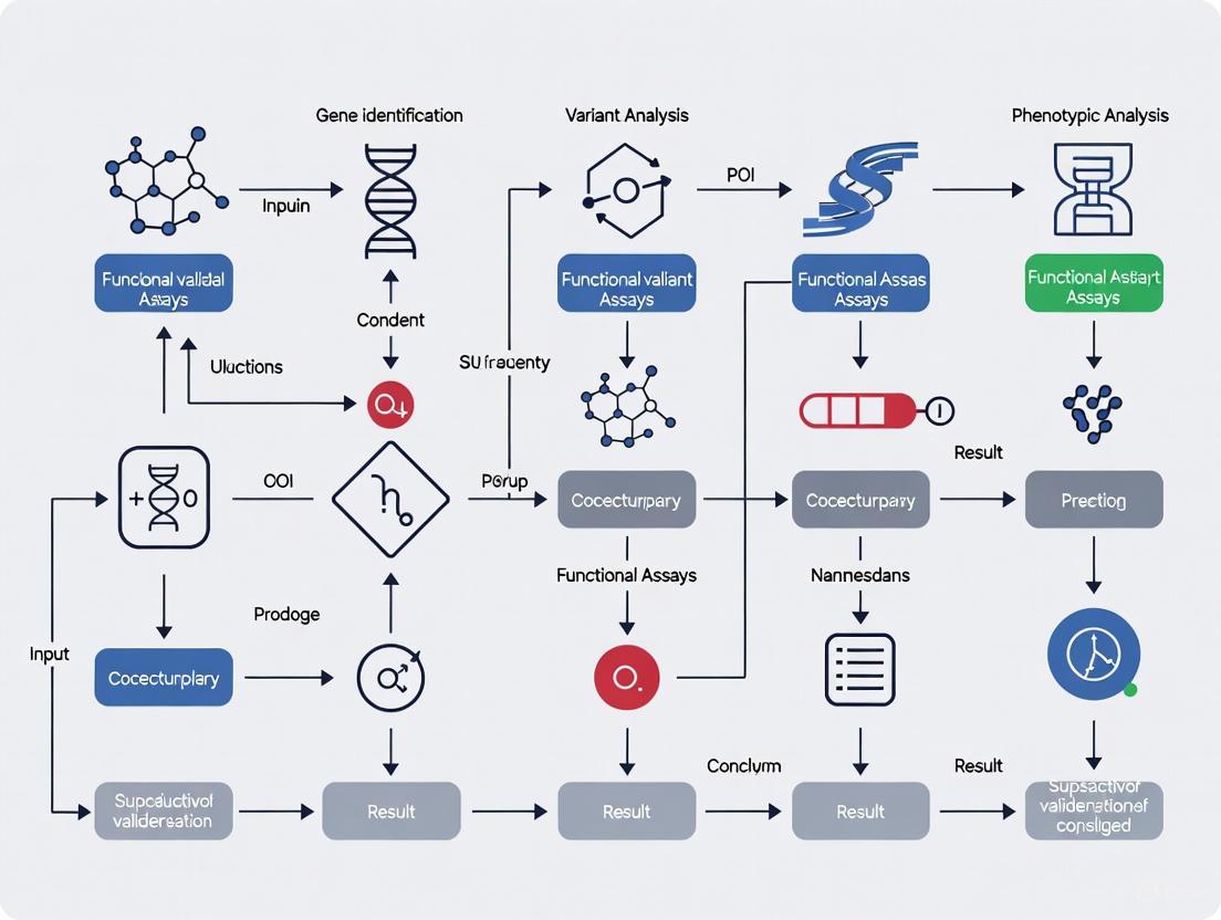
Figure 1: Experimental Workflow for Functional Validation of Amenorrhea/POI Gene Variants. This diagram outlines the key steps from patient identification through genetic screening to functional validation of candidate variants.
Technical Support Center: Troubleshooting Guides and FAQs
Frequently Asked Questions
Q1: We've identified a variant of uncertain significance (VUS) in a novel gene in our POI cohort. What is the best approach for functional validation?
A1: Prioritize functional assays based on the gene's predicted biological function:
- For genes implicated in meiosis (e.g., through homology to known meiotic genes), implement the IH-HR assay described in Section 4.2 [5].
- For genes involved in transcriptional regulation, consider reporter assays and gene expression profiling.
- For mitochondrial genes, assess oxidative phosphorylation, ATP production, and mitochondrial morphology.
- For all candidates, perform Western blotting to determine if the variant affects protein stability or expression [5].
- Utilize AlphaFold structural modeling to predict the impact of missense variants on protein structure [25].
Q2: What could explain the variable expressivity and incomplete penetrance we observe in families with POI-associated genetic variants?
A2: Several factors may contribute:
- Oligogenic/Polygenic Inheritance: Patients may carry variants in multiple genes that synergistically contribute to the phenotype [24].
- Modifier Genes: Genetic background effects can influence the expression of a primary variant.
- Environmental Factors: Factors like smoking, chemotherapy, or nutritional status may modify disease risk.
- Stochastic Events: Ovarian development and follicle pool establishment involve stochastic elements.
- Age-Dependent Penetrance: Some variants may only manifest their effects over time as the ovarian reserve declines.
Consider expanding genetic testing beyond single candidates to explore oligogenic models [24].
Q3: How should we interpret a situation where our cellular models (e.g., IH-HR assay) show a clear defect, but the variant is present in population databases at low frequency?
A3: This scenario requires careful interpretation:
- Validate Assay Specificity: Ensure your functional assay has strong validation connecting the cellular phenotype to the clinical phenotype.
- Consider Incomplete Penetrance: The variant may confer susceptibility that requires additional genetic or environmental triggers.
- Check for Common in Specific Populations: Some pathogenic variants can be enriched in certain populations due to founder effects.
- Assess Compound Heterozygosity: Check if the patient carries another variant in the same gene or pathway.
- Re-evaluate ACMG Classification: Incorporate the functional data as supporting evidence for pathogenicity according to ACMG guidelines (PS3/BS3 criteria) [24].
Troubleshooting Guide
Table 4: Common Experimental Challenges and Solutions
| Problem | Potential Causes | Solutions |
|---|---|---|
| No rare variants identified in known POI genes | True genetic heterogeneity; variants in non-coding regions; incorrect phenotype assignment [24] | Re-evaluate phenotype; consider WGS for non-coding variants; explore novel candidate genes through pathway analysis |
| Weak functional signal in cellular assays | Variant has mild effect; assay not sensitive enough; incorrect cellular model [5] | Optimize assay conditions; use more relevant cell types (e.g., oocyte-like cells); consider multiple complementary assays |
| Inconsistent results between technical replicates | Technical variability in assay execution; cell line instability; contamination [5] | Standardize protocols; increase replicate number; authenticate cell lines regularly; include robust controls |
| Difficulty interpreting missense variants | Limited structural/functional data for novel genes; conflicting in silico predictions [24] | Use multiple prediction algorithms; perform molecular modeling (AlphaFold); test multiple functional readouts |
The genetic architecture of primary and secondary amenorrhea reveals distinct profiles that reflect different underlying biological mechanisms. Primary amenorrhea is frequently associated with chromosomal abnormalities and congenital disorders of sexual development, while secondary amenorrhea, particularly POI, demonstrates complex genetic heterogeneity involving multiple biological pathways critical for ovarian function.
Future research directions should focus on:
- Elucidating Oligogenic Mechanisms: Systematic investigation of gene-gene interactions in POI pathogenesis [24].
- Functional Characterization of VUS: Developing high-throughput functional assays to classify the numerous VUS being discovered through clinical sequencing [5] [24].
- Gene-Environment Interactions: Understanding how environmental factors interact with genetic susceptibility in amenorrhea.
- Therapeutic Development: Leveraging genetic insights to develop targeted interventions, potentially including in vitro follicle maturation or gene-specific approaches.
The continued integration of genetic discovery with functional validation in model systems will be essential for translating these findings into improved diagnostics, counseling, and therapeutic options for women with amenorrhea.
Premature Ovarian Insufficiency (POI) is a clinically heterogeneous disorder characterized by the loss of ovarian function before the age of 40, affecting approximately 1–3.7% of women [26] [27]. It represents a significant cause of female infertility, with a strong genetic component underlying a substantial proportion of cases. Genetic etiology accounts for approximately 20–25% of POI cases, though recent large-scale sequencing studies have begun to expand our understanding of the genetic architecture [28] [1] [4].
The establishment of novel POI genes requires a rigorous multidisciplinary approach that moves beyond simple genetic association to demonstrate functional causality. This technical guide addresses the key methodological challenges and solutions for validating novel POI gene candidates, providing researchers with a framework for generating robust evidence that meets contemporary scientific standards.
Table 1: Current Genetic Contribution to POI Etiology
| Genetic Category | Approximate Contribution | Key Examples |
|---|---|---|
| Chromosomal Abnormalities | 10–13% | Turner syndrome (45,X), X-chromosome deletions & rearrangements [28] [1] |
| Single Gene Mutations (Known Genes) | ~11% (18.7% total minus chromosomal) | FMR1 premutation, BMP15, NR5A1, MCM9 [28] [29] |
| Novel Gene Associations | Additional ~5% (23.5% total contribution) | SWSAP1, LGR4, CPEB1, ALOX12 [5] [29] |
| Total Established Genetic Causation | ~20–25% |
Technical Support: Troubleshooting Guides and FAQs
FAQ 1: What constitutes definitive evidence for establishing a novel POI gene?
Answer: The current field recognizes a hierarchy of evidence for establishing a novel POI gene. A definitive gene-disease relationship requires: (1) identification of rare, predicted-damaging variants in patients that segregate with the phenotype in families; (2) statistical enrichment of such variants in cases versus controls; (3) functional evidence demonstrating that the variant disrupts a biological process relevant to ovarian function; and (4) replication in independent cohorts [29] [26]. The 2023 Nature Medicine study of 1,030 POI patients provides a contemporary benchmark, where association analyses comparing the POI cohort with 5,000 controls identified 20 novel POI-associated genes with a significantly higher burden of loss-of-function variants [29].
FAQ 2: How do I determine whether a variant of uncertain significance (VUS) is pathogenic?
Answer: Variant interpretation requires a multi-step functional validation pipeline. Begin with comprehensive bioinformatic prediction using tools like CADD (PHRED-scaled scores >20 suggest potential pathogenicity). However, computational predictions have limitations and can generate false positives/negatives [26]. Functional characterization is imperative. The ACMG guidelines provide a framework for variant classification, but for novel genes, experimental validation is crucial. For example, in a study of DIS3 variants, researchers first used in silico modeling, then employed a cross-species approach using mouse embryonic stem cells and Drosophila melanogaster to demonstrate the variant's deleterious impact on ovarian development [26].
FAQ 3: What are the most effective functional assays for validating POI gene candidates?
Answer: The choice of functional assays should be guided by the gene's predicted biological function. For genes involved in meiosis and DNA repair, Interhomolog Homologous Recombination (IH-HR) assays provide a relevant functional approach [5]. For example, in the validation of novel SWSAP1 variants, IH-HR assays demonstrated a partial decrease or absence of IH-HR activity in Swsap1-/- cells, indicating impaired meiotic function [5]. Other established approaches include in vitro cell culture models (e.g., granulosa cell lines), gene expression analyses, and animal models (mouse, Drosophila). A recent study functionally validated 75 VUSs from seven POI genes involved in homologous recombination repair and folliculogenesis, with 55 confirmed to be deleterious [29].
FAQ 4: How can I address the challenge of phenotypic heterogeneity in POI?
Answer: POI exhibits significant phenotypic heterogeneity, ranging from primary amenorrhea to early secondary amenorrhea. Genotype-phenotype correlation analyses indicate that genetic contribution is higher in cases with primary amenorrhea (25.8%) compared to secondary amenorrhea (17.8%) [29]. When validating novel genes, stratify your cohort by amenorrhea type and age of onset. Additionally, consider whether the POI is isolated or part of a syndromic condition, as this can provide clues to the gene's broader biological function. For instance, recent findings have revealed that POI can be the only symptom of a multi-organ genetic disease in 8.5% of cases [30].
Experimental Protocols for Functional Validation
Protocol: Interhomolog Homologous Recombination (IH-HR) Assay
Purpose: To evaluate the functional impact of putative pathogenic variants in genes involved in meiotic recombination, a key biological process frequently disrupted in POI.
Background: The SWS1 complex (SWS1-SWSAP1-SPIDR), also known as the Shu complex, plays a critical role in interhomolog homologous recombination. Knockout mouse models of this complex are infertile due to meiotic arrest, and variants in these genes have been associated with POI in patients [5].
Methodology:
- Cell Culture: Use mouse embryonic stem cells (mESCs) with knockout (KO) of the gene of interest (e.g., Sws1-/- or Swsap1-/-).
- Transfection: Introduce patient-derived variant constructs (e.g., SWS1/ZSWIM7 c.176C>T or SWSAP1 c.353del) into the respective KO cells.
- IH-HR Measurement: Utilize a well-established reporter system (e.g., DR-GFP) to quantify repair of site-specific DNA double-strand breaks through homologous recombination.
- Western Blot Analysis: Assess protein expression and stability of the truncation mutant to evaluate variant impact on protein integrity.
- Data Analysis: Compare IH-HR efficiency in variant-expressing cells versus wild-type and KO controls. A significant reduction indicates impaired homologous recombination function.
Troubleshooting Tip: If transfection efficiency is low, consider using viral transduction systems for more consistent gene delivery. Include positive and negative controls in each experiment to validate the assay performance [5].
Protocol: Cross-Species Functional Complementation Assay
Purpose: To determine the functional capacity of human gene variants to rescue phenotypes in model organisms.
Background: This approach is particularly valuable for genes where human tissue is inaccessible and mouse knockouts are lethal or exhibit subtle phenotypes. The DIS3 gene, a critical component of the RNA exosome, was recently validated using this method [26].
Methodology (Drosophila melanogaster model):
- Establish Knockdown: Create Dis3 knockdown in Drosophila female germline using RNAi or mutant lines.
- Transgenic Rescue: Generate transgenic flies expressing either wild-type human DIS3 or patient-derived variant (e.g., c.2320C>T; p.His774Tyr) under germline-specific promoters.
- Phenotypic Assessment: Compare ovarian development, egg chamber morphology, and fertility between:
- Wild-type flies
- Dis3 knockdown flies
- Dis3 knockdown flies rescued with wild-type human DIS3
- Dis3 knockdown flies rescued with variant human DIS3
- Histological Analysis: Examine ovarian sections for degenerative changes, abnormalities in egg chamber development, and evidence of meiotic arrest.
Expected Outcomes: A pathogenic variant will show reduced rescue capacity compared to wild-type human DIS3, evidenced by persistent ovarian atrophy, egg chamber degeneration, and reduced fertility [26].
Troubleshooting Tip: Confirm transgene expression levels across all rescue lines to ensure phenotypic differences are not due to expression variability. Use multiple independent transgenic lines for each construct to control for position effects.
Diagram Title: POI Gene Validation Workflow
The Scientist's Toolkit: Research Reagent Solutions
Table 2: Essential Reagents for POI Gene Validation Studies
| Reagent/Category | Specific Examples | Function/Application | Key Considerations |
|---|---|---|---|
| Sequencing Technologies | Whole Exome Sequencing (WES), Whole Genome Sequencing (WGS) | Identification of novel variants, rare variant association studies | WES sufficient for coding regions; WGS needed for non-coding & structural variants [29] |
| Cell-Based Assay Systems | Mouse Embryonic Stem Cells (mESCs), Granulosa Cell Lines | Functional characterization of variants, IH-HR assays | Ensure germline competence for mESCs; use multiple cell lines for reproducibility [5] |
| Animal Models | Drosophila melanogaster, Mouse Models | In vivo functional validation, reproductive phenotyping | Drosophila offers rapid screening; mouse models essential for mammalian reproductive biology [26] |
| Antibodies for Ovarian Tissue Analysis | Anti-MSY2, Anti-γH2AX, Anti-SCP3 | Meiotic progression analysis, follicle staging | Validate antibodies for specific species; optimize for ovarian tissue [26] |
| Specialized Assay Kits | IH-HR Reporter Assays (DR-GFP), Apoptosis Kits | Quantification of DNA repair efficiency, follicle atresia measurement | Include appropriate controls; optimize for specific cell types [5] |
Advanced Methodologies: Addressing Complex Genetic Architecture
Oligogenic and Polygenic Inheritance Models
Emerging evidence suggests that POI may not always follow a simple monogenic inheritance pattern. The identification of two or more pathogenic variants in distinct genes argues in favor of a polygenic origin for POI [4]. When validating novel genes, consider the possibility that the phenotype may result from the cumulative effect of multiple genetic variants.
Methodological Approach:
- Burden Testing: Evaluate whether cases carry a higher burden of rare variants across biologically related gene sets compared to controls.
- Interaction Studies: Test for epistatic interactions between candidate genes using statistical models and functional assays.
- Pathway Analysis: Group genes into functional pathways (e.g., homologous recombination, folliculogenesis) to identify enriched biological processes.
Recent studies have identified new pathways implicated in POI, including NF-kB signaling, post-translational regulation, and mitophagy (mitochondrial autophagy), providing future therapeutic targets [30].
Non-Coding RNAs and Mitochondrial Contributions
Beyond protein-coding genes, evidence is accumulating for roles of non-coding RNAs and mitochondrial genes in POI pathogenesis. Mitochondrial genes such as RMND1, MRPS22, and LRPPRC have been associated with POI, as have microRNAs and long non-coding RNAs [1] [31].
Validation Strategies:
- For mitochondrial genes: Assess oxidative phosphorylation capacity, mitochondrial membrane potential, and ATP production in patient-derived cells.
- For non-coding RNAs: Utilize luciferase reporter assays to validate binding sites, CRISPR-based editing to manipulate regulatory elements, and expression profiling in ovarian cell types.
Diagram Title: SWS1-Complex Disruption in POI
The establishment of novel POI genes requires a methodical, multi-layered approach that integrates human genetics with functional validation. As the field moves beyond association to causation, researchers must implement robust experimental designs that include adequate sample sizes, appropriate control populations, and biologically relevant functional assays. The tools and methodologies outlined in this technical guide provide a roadmap for generating the high-quality evidence needed to definitively establish novel gene-disease relationships in POI.
The future of POI genetics will likely involve addressing more complex inheritance models, including oligogenic and polygenic forms, and integrating multi-omics data to fully elucidate the pathogenic mechanisms. This comprehensive understanding will ultimately enable improved genetic diagnosis, personalized risk assessment, and targeted therapeutic interventions for women affected by this condition.
Advanced Functional Assays for POI Variant Characterization
Premature Ovarian Insufficiency (POI) is a complex disorder affecting approximately 3.7% of women under 40, with genetic factors contributing to 20-25% of cases [3] [32]. Functional validation of genetic variants identified through sequencing remains a critical challenge in POI research. This technical support center provides comprehensive guidance on employing mouse embryonic stem cells (mESCs) and granulosa cell (GC) cultures to validate novel POI gene variants, enabling researchers to establish causality beyond genetic association studies.
Essential Research Reagent Solutions
The table below outlines key reagents essential for experimental workflows in POI gene validation studies:
| Reagent Category | Specific Examples | Research Application | Technical Considerations |
|---|---|---|---|
| Cell Culture Materials | DMEM/F12 medium, penicillin-streptomycin, 0.4μm pore size inserts, paraformaldehyde | Ovarian culture in vitro, follicle development studies | Maintain at 37°C under 5% CO₂; replace half medium every other day [33] |
| Immunoassay Reagents | ELISA kits, Western blot antibodies, BSA blocking buffers | Protein detection, quantification, and analysis | ELISA for rapid quantification; Western blot for molecular weight confirmation [34] |
| Flow Cytometry Reagents | CD45 antibodies, cell viability dyes, fluorescence-conjugated antibodies | Hematopoietic analysis, immune cell profiling | Use systematic antibody panels with appropriate color controls [35] |
| Molecular Biology Tools | Agilent SureSelect exome capture, Illumina sequencing reagents, CRISPR/Cas9 components | Genetic variant identification, functional validation | WES achieves ~80x read depth; CRISPR enables precise genome editing [36] [26] |
Establishing Granulosa Cell Cultures for POI Validation
Primary Granulosa Cell Isolation and Culture
Granulosa cells play indispensable roles in folliculogenesis and oocyte maturation, making them crucial for modeling POI pathogenesis [33]. To establish primary cultures:
Protocol: Isolate GCs from 3-5 day postpartum mouse ovaries through micro-dissection in cold PBS. Culture on 0.4μm pore size inserts in 6-well plates containing DMEM/F12 medium supplemented with 1:100 penicillin-streptomycin. Maintain cultures at 37°C under 5% CO₂, replacing approximately half the medium every other day [33].
Troubleshooting FAQ: Q: Why do my granulosa cells show poor adhesion and viability? A: Ensure rapid processing of ovarian samples after dissection (<30 minutes). Use pre-warmed medium and coat plates with extracellular matrix components like collagen IV or laminin to improve attachment.
Q: How can I confirm the purity of my granulosa cell cultures? A: Implement immunocytochemistry using granulosa-specific markers like FOXL2 and FSHR. Flow cytometry analysis should show >90% positivity for these markers in purified cultures.
Genetic Manipulation of Granulosa Cells
CRISPR/Cas9-Mediated Gene Editing: Utilize Cre-loxP systems for cell-type specific knockout studies. For example, cross Bmi1fl/fl and Mel18fl/fl females with Foxl2-Cre males to generate GC-specific double knockout models [33]. Validate knockout efficiency via Western blot and qPCR.
Virus-Mediated Gene Transfer: Employ lentiviral or adenoviral vectors to introduce POI-associated variants into primary GC cultures. Use GFP-tagged constructs to monitor transduction efficiency (typically >70% is desirable).
Mouse Embryonic Stem Cell Models for POI
Differentiation of mESCs into Ovarian Cell Lineages
Protocol for Ovarian-like Differentiation:
- Maintain mESCs in feeder-free conditions with appropriate pluripotency factors
- Induce differentiation using BMP4, retinoic acid, and ovarian-specific factors
- Monitor differentiation efficiency using stage-specific markers (STRA8 for meiotic initiation, FOXL2 for granulosa commitment)
- Isplicate differentiated populations using FACS with cell surface markers
Troubleshooting FAQ: Q: My mESC differentiation yields low percentages of ovarian lineage cells. How can I improve efficiency? A: Optimize timing and concentration of differentiation factors. Include WNT signaling agonists during early stages, followed by TGF-β family members later. Consider co-culture with ovarian somatic cells to provide appropriate microenvironment cues.
Q: How do I validate successful differentiation? A: Use a multi-modal approach: flow cytometry for surface markers, qPCR for lineage-specific genes, and functional assays including steroid hormone production (estradiol, progesterone).
Functional Validation of POI Variants in mESC Models
Implement precise genome editing using CRISPR/Cas9 to introduce patient-specific variants into mESCs. For missense variants like DIS3 (c.2320C>T; p.His774Tyr), use homology-directed repair with donor templates containing the specific mutation [26].
Phenotypic Assessment:
- Proliferation assays: Measure growth curves and cell cycle distribution
- RNA sequencing: Analyze transcriptome changes, particularly in ovarian development pathways
- Apoptosis assays: Quantify cell death under baseline and stress conditions
- Differentiation capacity: Compare efficiency of ovarian lineage specification
Signaling Pathways in POI Pathogenesis
Figure 1: Key Signaling Pathways in POI Pathogenesis
Analytical Techniques for Functional Validation
Protein Analysis Methods: ELISA vs. Western Blot
Guidance on Method Selection:
Choose ELISA when: You need high-throughput quantification, are working with low protein concentrations, or require rapid results with minimal sample preparation [34].
Choose Western Blot when: You require confirmation of protein identity through molecular weight detection, need to identify protein modifications, or are analyzing complex protein mixtures [34].
Troubleshooting FAQ: Q: My Western blot shows high background noise. How can I improve signal clarity? A: Increase blocking time (overnight at 4°C), optimize antibody concentrations, include additional washes with Tween-20, and consider using fluorescent detection instead of chemiluminescence for better signal-to-noise ratio.
Q: My ELISA results show high variability between replicates. What could be causing this? A: Ensure consistent sample preparation and avoid repeated freeze-thaw cycles. Check pipette calibration for small volumes, pre-warm all reagents to room temperature before use, and verify that plate washing is consistent across all wells.
Flow Cytometry for Cell Population Analysis
Panel Design for Ovarian Cell Populations: Adapt principles from hematopoietic analysis by including lineage-defining markers in systematic combinations [35]. For granulosa cell analysis, include FOXL2, FSHR, and CD9 as core markers, with additional markers for differentiation status.
Troubleshooting FAQ: Q: How can I improve resolution in my flow cytometry data? A: Use spectral flow cytometry with uncompressed controls, titrate all antibodies carefully, include fluorescence-minus-one (FMO) controls, and utilize computational analysis tools like t-SNE or UMAP for population identification [37].
Q: What is the recommended approach for analyzing high-parameter flow cytometry data? A: Employ automated clustering algorithms (FlowSOM, PhenoGraph) combined with dimensionality reduction techniques (t-SNE, UMAP). Begin with manual gating to remove debris and dead cells, then apply computational methods for deep population analysis [37].
Integration of Validation Data
Statistical Considerations for Experimental Design
Power Analysis: For animal studies, include at least 5-8 mice per genotype to detect moderate effect sizes. For cell culture experiments, plan for minimum n=3 biological replicates with multiple technical replicates each.
Data Normalization: Use appropriate housekeeping genes for qPCR (e.g., Hprt, Gapdh, Actb validated for your cell type). For protein studies, normalize to total protein content or constitutive markers.
Interpretation of Functional Validation Results
Establishing Causality: A variant is considered functionally validated when:
- It produces a consistent phenotype across multiple model systems
- The phenotype mirrors the clinical POI presentation (e.g., follicular activation defects, GC proliferation defects)
- The effect follows expected molecular mechanisms (e.g., disrupted signaling pathways)
- Rescue experiments reverse the phenotype
Contextualizing with Human Data: Correlate functional findings with patient characteristics. For example, variants causing more severe molecular defects should associate with earlier age of onset or primary versus secondary amenorrhea [3].
Advanced Technical Guides
In Vitro Ovarian Culture System
Whole Ovary Culture Protocol:
- Isolate intact ovaries from 3 dpp mice under microscope
- Place on 0.4μm pore size inserts in 6-well culture plates
- Culture for 4 days in DMEM/F12 with supplements
- Treat with small molecule inhibitors/activators (e.g., AIL at 10nM) or DMSO control
- Process for histology or follicle counting [33]
Follicle Counting Methodology: Fix ovarian samples in 4% PFA overnight, embed in paraffin, section serially at 5μm (before 7 dpp) or 8μm (after 7 dpp), stain with hematoxylin, and count follicles in every fifth section with morphological classification [33].
Cross-Species Validation Approaches
Yeast Complementation Assays: For conserved genes like DIS3, introduce human variants into yeast models and assess growth phenotypes [26].
Drosophila Ovarian Models: Generate transgenic flies expressing human POI variants (e.g., DIS3 p.His774Tyr) and evaluate ovarian development, egg chamber formation, and fertility outcomes [26].
Figure 2: Experimental Validation Workflow for POI Gene Variants
The integration of mouse embryonic stem cells and granulosa cell cultures provides a powerful platform for validating POI gene variants. By following these standardized protocols and troubleshooting guides, researchers can accelerate the functional characterization of novel genetic findings, ultimately advancing our understanding of ovarian biology and developing targeted interventions for infertility.
Interhomolog Homologous Recombination (IH-HR) is a fundamental meiotic process where genetic information is exchanged between homologous parental chromosomes. This mechanism is crucial for generating genetic diversity and ensuring proper chromosome segregation during gamete formation. For researchers investigating gene variants, accurately assessing IH-HR function provides critical insights into meiotic competence and genome stability. This technical support center addresses the key methodological challenges and troubleshooting aspects of IH-HR assays within the context of functional validation for gene variant research.
Core Mechanisms and Key Proteins
Understanding the molecular machinery of IH-HR is essential for designing appropriate assays and interpreting results. The process involves a coordinated series of steps initiated by programmed double-strand breaks (DSBs) and repaired using the homologous chromosome as a template [38] [39].
The following diagram illustrates the core pathway and key regulatory proteins in meiotic IH-HR:
Key Regulatory Complexes and Their Functions
| Protein/Complex | Primary Function in IH-HR | Experimental Significance |
|---|---|---|
| Rad51/Dmc1 | Catalyze homologous pairing and strand invasion between homologous chromosomes [38] | Core recombinases; focus formation indicates active recombination |
| Rad54/Tid1 | Facilitate chromatin remodeling and homology search; specific partners for Rad51/Dmc1 respectively [38] | Assess partner choice in IH-HR vs. IS-HR |
| ZMM Proteins (Zip1, Zip2-4, Mer3, Msh4-Msh5) | Promote synapsis and class I interference-sensitive crossover formation [38] | Key markers for crossover pathway specification |
| SWS1-SWSAP1-SPIDR | Promotes stable RAD51 filament assembly; specifically required for interhomolog HDR in mitotic cells [9] | Critical for IH-HR but not intrachromosomal HDR |
| Srs2 | Disassembles Rad51-ssDNA presynaptic filaments; facilitates MMR [38] | Anti-recombination activity; balance with pro-HR factors |
| BRC-1/BRCA1 | Regulates DSB repair pathway engagement; represses error-prone repair and intersister crossovers [40] | Tumor suppressor; controls repair partner choice |
Troubleshooting Guide: Common IH-HR Assay Challenges
FAQ: How can I distinguish interhomolog from intersister recombination events in meiotic assays?
Challenge: Intersister recombination (IS-HR) produces identical genetic outcomes without heterozygosity changes, complicating differentiation from IH-HR [40].
Solutions:
- Genetic heterozygosity mapping: Utilize single-nucleotide polymorphisms (SNPs) between homologous chromosomes. IH-HR produces gene conversion tracts detectable by sequencing, while IS-HR does not alter heterozygosity patterns [38].
- Cytological differentiation: In C. elegans, the intersister/intrachromatid repair (ICR) assay exploits nonallelic recombination at a specific locus to identify homolog-independent repair events [40].
- Physical monitoring of joint molecules: Use two-dimensional gel electrophoresis to detect recombination intermediates specific to interhomolog engagement.
FAQ: What could cause persistent RAD-51 foci in my meiotic assays?
Challenge: Persistent RAD-51 foci indicate stalled recombination intermediates and defective IH-HR progression.
Potential Causes and Solutions:
- Defective mediator complexes: Mutations in SWS1-SWSAP1-SPIDR reduce RAD51 focus formation by ~3-fold [9]. Verify complex integrity through co-immunoprecipitation.
- BRC-1/BRCA1 dysfunction: In C. elegans, brc-1 mutants exhibit persistent RAD-51 foci and chromosome fragmentation [40]. Check BRC-1 localization and expression.
- SMC-5/6 complex defects: smc-5 mutants show similar RAD-51 persistence [40]. Assess genetic interactions with BRC-1.
- Anti-recombinase activity imbalance: Srs2 disassembles Rad51 filaments; its overexpression may cause focus persistence [38]. Modulate Srs2 activity or its regulator Dmc1.
FAQ: How can I enhance IH-HR efficiency in somatic cell systems?
Challenge: IH-HR occurs naturally in meiosis but is inefficient in somatic cells where sister chromatid repair is preferred.
Solutions:
- Multiple nicking approach: The NICER (Multiple Nicks Induce Interhomolog Recombination) method uses Cas9D10A nickase to introduce multiple nicks on homologous chromosomes, enhancing IH-HR efficiency by approximately 17-fold compared to single nicks [41].
- BRCA pathway modulation: Depletion of BRCA1 and BRCA2 partially impairs but does not abolish MN-induced IH-HR, suggesting alternative pathways [41].
- Cell cycle synchronization: IH-HR is favored in S and G2 phases when homologous templates are available [39]. Synchronize cells to enhance detection.
FAQ: Why do I observe different crossover outcomes in my IH-HR assays?
Challenge: Crossover outcomes vary between class I (interference-sensitive) and class II (non-interfering), controlled by distinct pathways.
Troubleshooting Guide:
- ZMM protein dependency: Class I COs require ZMM proteins (Mer3, Msh4-Msh5, Zip1, etc.). Check ZMM protein expression and localization [38].
- Alternative pathway activity: Class II COs utilize the Mms4-Mus81 endonuclease and are promoted when ZMM pathways are compromised [38].
- Anti-CO factor regulation: Sgs1 helicase dissolves dHJs to promote NCOs via SDSA. Its disruption increases CO frequency [38].
Quantitative Assay Data and Methodologies
IH-HR Detection Efficiency Across Methodologies
| Assay Method | System | Efficiency | Key Readout | Limitations |
|---|---|---|---|---|
| NICER (Multiple Nicks) [41] | Human somatic cells (TK6261) | 17-fold increase over single nick | TK1 activity recovery (98.9% WT reads) | Requires multiple sgRNAs; BRCA1/2 dependent |
| SWS1-SWSAP1-SPIDR Dependent IH-HR [9] | Mouse ES cells | Not required for DR-GFP reporter (intrachromosomal) | GFP-positive cells post I-SceI cut | Specific to interhomolog, not sister chromatid repair |
| ICR Assay [40] | C. elegans meiosis | Quantifies homolog-independent events | Non-allelic recombination products | Does not directly measure IH-HR |
| Class I CO Formation [38] | Yeast meiosis | 70-85% of total COs | Crossover interference patterns | Requires multiple mutant analysis |
Detailed Protocol: NICER Method for IH-HR Induction
The NICER method represents a significant advance for inducing IH-HR in somatic cells with reduced genomic alterations compared to DSB-based approaches [41].
Workflow:
- sgRNA design: Design mutation-specific sgRNA (primary) and additional sgRNAs (secondary) targeting homologous chromosomes within ±8.6 kb region.
- Nickase delivery: Electroporate Cas9D10A nickase mRNA with sgRNAs into target cells.
- Selection and analysis: Apply selection (e.g., CHATM for TK1+ cells) and quantify correction efficiency via colony formation or proliferation assays.
- Validation: Confirm gene correction through amplicon-based NGS (AmpNGS) and protein expression restoration.
Critical Optimization Parameters:
- sgRNA spacing: Offsets between 24–8675 bp effectively enhance IH-HR without correlation between efficiency and distance [41].
- Multiple nicks: Additional nicks strongly enhance correction efficiency compared to single nicks.
- Control experiments: Include single nick and pnDSB controls to validate IH-HR-specific effects.
The following diagram illustrates the experimental workflow for the NICER method:
Detailed Protocol: Meiotic IH-HR Assessment in Model Organisms
For meiotic systems, IH-HR analysis requires different approaches to quantify recombination between homologous chromosomes.
Yeast Hybrid System (SK1/S288c) Approach: [38]
- Strain construction: Generate hybrid yeast strains with polymorphic homologs to track recombination events.
- Meiotic time course: Induce synchronous meiosis and collect samples at intervals.
- Physical analysis: Use Southern blotting or PCR-based methods to detect recombination intermediates and products.
- Genetic outcome analysis: Score COs, NCOs, and gene conversion events through tetrad analysis or random spore analysis.
- Protein function assessment: Analyze mutants in anti-MMR activities (Rad51, Rad54, ZMM proteins) to determine their effects on IH-HR bias.
Critical Parameters:
- Hybrid heterozygosity: Ensure sufficient sequence polymorphism between homologs to track recombination events.
- Temporal resolution: Meiotic recombination occurs in distinct phases; precise timing is essential for interpreting results.
- Checkpoint status: Monitor meiotic checkpoint activation (e.g., Mek1 kinase) as persistent activation indicates repair defects.
The Scientist's Toolkit: Essential Research Reagents
| Reagent/Category | Specific Examples | Function in IH-HR Assays |
|---|---|---|
| Recombinases | Rad51, Dmc1 [38] | Catalyze strand invasion and homology search |
| Mediator Complexes | Rad55-Rad57 (yeast), RAD51 paralogs (mammalian) [42] | Facilitate Rad51 filament formation on RPA-coated ssDNA |
| ZMM Proteins | Mer3, Msh4-Msh5, Zip1, Zip2, Zip3, Zip4 [38] | Promote synapsis and class I crossover formation |
| Anti-recombinases | Srs2 (yeast), FBH1 (mammalian) [38] [42] | Regulate recombination by disassembling Rad51 filaments |
| Accessory Factors | Rad54, Tid1 [38] | Chromatin remodeling; D-loop stabilization |
| Nuclease Systems | Cas9D10A nickase [41] | Induce targeted nicks for NICER method |
| Reporter Systems | DR-GFP, TK1 mutation correction [41] [9] | Quantify recombination efficiency |
| Resolution Factors | Sgs1-BLM, Mus81-Mms4 [38] | Process recombination intermediates to NCOs or COs |
Accurate assessment of interhomolog homologous recombination is essential for understanding the functional impact of gene variants on meiotic competence and genome stability. The methodologies and troubleshooting approaches outlined here provide researchers with a comprehensive framework for designing, executing, and interpreting IH-HR assays. As the field advances, emerging techniques like the NICER method and improved understanding of complexes like SWS1-SWSAP1-SPIDR continue to enhance our ability to precisely quantify and manipulate IH-HR in both meiotic and somatic contexts.
Troubleshooting Guides
Western Blot Troubleshooting for POI Protein Detection
Q: I am getting a weak or no signal when detecting a novel POI protein (e.g., SWSAP1). What could be the cause and how can I fix it?
Weak or absent signals are common when studying novel or low-abundance proteins, such as those involved in POI. The causes and solutions are multifaceted [43] [44].
| Possible Cause | Recommended Solution | Additional POI-Specific Considerations |
|---|---|---|
| Insufficient protein loaded | - Load at least 20-30 µg of whole cell extract per lane; may require >100 µg for tissue extracts [44].- Confirm protein concentration spectrophotometrically [45]. | POI-related proteins like those in the SWS1 complex may be low-abundance; consider loading more protein. |
| Inefficient transfer | - Verify transfer efficiency by staining the gel post-transfer [43].- For high MW proteins: add 0.01-0.05% SDS to transfer buffer [43].- For low MW proteins: add 20% methanol to transfer buffer and reduce transfer time [43]. | For meiotic complex proteins (e.g., SWS1, SPIDR), ensure transfer conditions are optimized for their specific molecular weights. |
| Low antibody affinity or concentration | - Increase primary antibody concentration [43].- Perform a dot blot to confirm antibody activity [43].- Ensure antibody is validated for Western blot and has species reactivity for your model system [44]. | Antibodies against novel POI gene products (e.g., SWSAP1) may require extensive optimization; use positive controls if available. |
| Sub-optimal buffer choice | - Dilute primary antibody in the recommended buffer (BSA or milk) [44].- Avoid sodium azide in wash buffers when using HRP-conjugated antibodies [43]. | |
| Protein degradation | - Always use fresh protease and phosphatase inhibitors during lysis [44].- Sonicate samples to ensure complete lysis and shear DNA [44]. | Sample integrity is critical for detecting fragile protein complexes involved in homologous recombination. |
Q: My western blot shows high background. How can I improve the signal-to-noise ratio?
High background obscures results and can lead to misinterpretation, which is particularly problematic when validating rare patient variants [43].
| Possible Cause | Recommended Solution |
|---|---|
| Antibody concentration too high | Titrate and decrease the concentration of both primary and secondary antibodies [43]. |
| Insufficient blocking | - Block with 5% skim milk or BSA for at least 1 hour at room temperature [43] [45].- For phosphoprotein detection, use BSA in TBS instead of milk [43]. |
| Incompatible blocking buffer | - Do not use milk with avidin-biotin systems [43].- When using an Alkaline Phosphatase (AP) conjugate, use Tris-buffered saline (TBS) instead of PBS [43]. |
| Insufficient washing | - Increase wash number and volume [43].- Add Tween 20 to wash buffer to a final concentration of 0.05% [43]. |
Q: The observed molecular weight for my protein differs from the calculated one. Why?
Discrepancies between observed and calculated molecular weight are common and often biologically relevant, especially for proteins with complex functions like those in POI pathways [46].
| Possible Cause | Description | Example |
|---|---|---|
| Post-translational modifications (PTMs) | - Glycosylation: Adds significant weight, appears as a smear or higher band. Confirm with PNGase F treatment [46].- Phosphorylation: Adds ~1 kDa per group; multiple sites can cause a shift [46].- Ubiquitination: Adds ~8.6 kDa per ubiquitin, can create higher smears or ladders [46]. | PD-L1 runs at 45-70 kDa due to glycosylation, despite a calculated MW of 33 kDa [46]. |
| Protein Cleavage | Many proteins have signal peptides or pro-peptides cleaved off to form the mature, active protein, resulting in a lower MW band [46]. | PINK1 precursor is 65 kDa, but the cleaved mature form runs at 52 kDa [46]. |
| Protein Isoforms & Complexes | Alternative splicing creates isoforms of different sizes. Some proteins form stable homo- or hetero-dimers even in denaturing conditions [46]. | NQO1 forms homodimers observed at 66-70 kDa, in addition to its monomeric forms [46]. |
Protein-Protein Interaction (PPI) Study Troubleshooting
Q: How can I confirm a suspected protein-protein interaction is specific and biologically relevant in my POI research?
Confirming interactions is crucial for establishing the functional role of POI gene products in complexes like the SWS1 complex [47].
| Challenge | Solution and Control Experiments |
|---|---|
| False Positives in Co-IP | - Include a negative control with affinity support but no bait protein [47].- Use monoclonal antibodies when possible. For polyclonal antibodies, pre-adsorb them to eliminate contaminants that may bind prey directly [47].- Verify with an antibody against the co-precipitated protein in a reverse Co-IP [47]. |
| Transient or Weak Interactions | Weak interactions (KD > 10⁻⁴ M) are often biologically crucial but difficult to capture [48]. Use crosslinkers (e.g., DSS, BS3) to "freeze" transient interactions before lysis [47]. |
| Interaction occurs post-lysis | Perform co-localization studies in cells to confirm the interaction happens in vivo [47]. |
Q: My pulldown assay shows no interaction. What could be wrong?
| Possible Cause | Solutions |
|---|---|
| The tagged bait protein is degraded. | Include protease inhibitors in the lysis buffer [47]. |
| The interaction is too weak or transient. | Use crosslinking prior to lysis [47]. Consider biophysical methods like Surface Plasmon Resonance or NMR for characterizing weak complexes [48]. |
| Insufficient protein or low sensitivity. | Use more lysate and a more sensitive detection system (e.g., chemiluminescent substrates) [47]. |
FAQs
Western Blot FAQs
Q: What is the purpose of blocking in a western blot and what should I use? Blocking covers non-specific sites on the membrane to prevent antibodies from binding randomly, which reduces background noise. Common blocking agents are 5% BSA or non-fat dry milk. The choice depends on your target protein and antibody; for example, avoid milk when detecting phosphoproteins or using an avidin-biotin system, as milk contains biotin and phosphoproteins that can cause high background [43] [49].
Q: Why is normalization important and which loading control should I use? Normalization controls for experimental variations like protein concentration measurement errors and loading inconsistencies. You compare the band intensity of your target protein to that of a "housekeeping" protein that is constitutively expressed at stable levels, such as Actin, GAPDH, or Tubulin. This ensures that observed differences are real and not due to technical artifacts [49].
Protein-Protein Interaction FAQs
Q: Why are weak protein complexes important to study in the context of POI? Weak, transient protein-protein interactions are essential for cellular regulation, signaling cascades, and dynamic processes like meiosis and homologous recombination—pathways often disrupted in POI. Although challenging to study, these interactions can be highly significant in specific subcellular compartments where local protein concentrations are high [48]. Genes like SWS1, SWSAP1, and SPIDR, which form the SWS1 complex critical for meiotic homologous recombination, are prime examples where disrupted interactions can lead to POI [5].
Q: What techniques are suitable for studying weak or transient protein complexes? Many conventional techniques fail for weak complexes. The following biophysical and structural methods are more appropriate [48]:
- Nuclear Magnetic Resonance (NMR) Spectroscopy: Excellent for characterizing weak interactions at the atomic level.
- Surface Plasmon Resonance (SPR) & Isothermal Titration Calorimetry (ITC): Provide quantitative data on binding affinity and kinetics.
- Mass Spectrometry (MS): Especially when combined with methods like hydrogen-deuterium exchange or chemical cross-linking.
- Small-Angle X-Ray Scattering (SAXS): Provides low-resolution structural information of complexes in solution.
- Analytical Ultracentrifugation: Useful for studying the stoichiometry and shape of protein assemblies.
Experimental Protocols & Workflows
Detailed Western Blot Protocol for Cell Lysates
Sample Preparation [45]
- Wash & Harvest: Wash adherent cells with cold PBS. Dislodge cells using a cell scraper with PBS and transfer to a microcentrifuge tube.
- Pellet & Lyse: Centrifuge at 1500 RPM for 5 min. Discard supernatant. Resuspend pellet in 180 µL of ice-cold cell lysis buffer supplemented with 20 µL of fresh protease inhibitor cocktail.
- Incubate & Clarify: Incubate on ice for 30 min. Clarify the lysate by centrifuging at 12,000 RPM for 10 min at 4°C.
- Store & Quantify: Transfer the supernatant (protein extract) to a fresh tube. Measure protein concentration using a spectrophotometer.
Gel Electrophoresis & Transfer [45]
- Prepare Sample: Mix protein sample with loading buffer. Heat at 100°C for 5 min to denature proteins.
- Load & Run: Load equal protein mass (e.g., 20-50 µg) per lane alongside a prestained molecular weight marker. Run gel at 60-140 V until the dye front reaches the bottom.
- Prepare Transfer Stack: Create a "sandwich" in the following order (from cathode to anode): Sponge -> 3 Filter Papers -> Gel -> PVDF Membrane -> 3 Filter Papers -> Sponge. Ensure no air bubbles are trapped.
- Transfer: Perform wet transfer at 4°C for 90 minutes at 70V. For high MW proteins (>100 kDa), increase transfer time or reduce methanol in buffer [44].
Antibody Incubation & Detection [45]
- Block: Incubate membrane in 5% skim milk or BSA in TBST for 1 hour at room temperature.
- Primary Antibody: Incubate membrane with primary antibody diluted in BSA or milk overnight at 4°C on a shaker.
- Wash: Wash membrane 3 times for 5 min each with TBST.
- Secondary Antibody: Incubate with HRP-conjugated secondary antibody in 5% skim milk in TBST for 1 hour at room temperature.
- Wash: Repeat washing step 3 times.
- Detect: Incubate membrane with ECL substrate for 1-2 min. Visualize using a CCD imager or X-ray film.
Western Blot Experimental Workflow
Validating POI Protein Complexes Workflow
This workflow is essential for functionally characterizing variants in genes like SWS1/ZSWIM7 and SWSAP1, which form the SWS1 complex critical for meiotic homologous recombination [5].
Validating POI Protein Complexes
Research Reagent Solutions
Essential materials and reagents for conducting protein-level analyses in POI research.
| Reagent / Kit | Function | Example Use-Case |
|---|---|---|
| Protease Inhibitor Cocktail | Prevents proteolytic degradation of proteins during and after cell lysis [44]. | Essential for extracting intact SWSAP1 and other meiotic complex proteins from cell or ovarian tissue lysates. |
| Pierce Protein Concentrators | Concentrates dilute protein samples and allows buffer exchange into lower-salt buffers [43]. | Useful for desalting or concentrating lysates prior to electrophoresis to prevent streaking and distorted bands. |
| Slide-A-Lyzer MINI Dialysis Device | Dialyzes samples to remove excess salt or detergents that can interfere with SDS-PAGE [43]. | Critical for cleaning up protein samples for downstream Co-IP or pulldown assays. |
| Chemical Crosslinkers (e.g., DSS, BS3) | "Freeze" transient protein-protein interactions inside or outside the cell before lysis [47]. | Capturing weak interactions within the SWS1 complex that might otherwise dissociate during Co-IP. |
| SuperSignal West Femto Substrate | A high-sensitivity chemiluminescent substrate for detecting low-abundance proteins [43] [47]. | Detecting faint bands of POI-related proteins expressed at low levels in limited biological samples. |
| PVDF or Nitrocellulose Membrane | Solid support for immobilizing proteins after gel electrophoresis for antibody probing [45]. | Standard blotting membrane; 0.2 µm pore size is recommended for low MW proteins to prevent "blow-through" [44]. |
Troubleshooting Guides
Guide 1: Addressing Mismatches Between AlphaFold Predictions and Experimental Structures
Problem: A researcher models the structure of insulin using its FASTA sequence in ColabFold. The prediction has a high confidence score (pLDDT of 77.1), but the resulting 3D model is significantly different from the known experimental structure in the RCSB PDB [50].
Potential Cause 1: Incorrect Input Sequence or Format.
- Solution: For multimers or protein complexes, ensure the input FASTA file correctly specifies all constituent chains. ColabFold uses a specific format for complexes. Re-run the prediction using the dedicated multimer input options [50].
Potential Cause 2: Static vs. Dynamic Structures.
- Solution: AlphaFold may predict a single, low-energy state, whereas the experimental structure (e.g., from crystallography) captures one specific conformation. Proteins are dynamic and can adopt multiple shapes. Consider using tools like SWAXSFold, which integrates experimental data to reveal protein structures under specific conditions [51].
Potential Cause 3: Inherent Limitations of the Tool.
- Solution: AlphaFold is trained to predict static structures from sequence and may not accurately capture the conformational diversity of some proteins, especially in complexes. Verify your results with the latest versions (e.g., AlphaFold 3 for complexes) and always consult the experimental data when available [52] [53].
Guide 2: Interpreting Weak Correlation Between AlphaFold Output and Experimental Functional Data
Problem: A scientist is studying the impact of missense variants in a POI (Protein of Interest) gene. They use the change in AlphaFold's pLDDT score (ΔpLDDT) between wild-type and mutant models as a proxy for stability change (ΔΔG), but find a very weak correlation with their own functional assays [54].
Potential Cause 1: pLDDT is a Confidence Metric, Not a Stability Metric.
- Solution: The pLDDT score reflects AlphaFold's confidence in the local structure prediction, not the thermodynamic stability of the protein. A mutation in a well-defined, buried core residue might lower pLDDT and stability, while a surface mutation might lower pLDDT without significantly affecting stability. Do not use ΔpLDDT as a direct measure of ΔΔG [54].
Potential Cause 2: The Mutation May Induce Fold-Switching.
- Solution: Some proteins or regions can adopt different secondary structures. AlphaFold and similar tools typically predict only one conformation. If a variant is suspected to cause fold-switching, inaccurate secondary structure predictions can be an indicator. Analyze the region with dedicated prediction tools [55].
Recommended Workflow: Use the AlphaFold-predicted structure as a starting point for more sophisticated, physics-based ΔΔG calculation methods (e.g., FoldX, Rosetta). The accuracy of the prediction depends more on the ΔΔG predictor used than on the source of the 3D model (AlphaFold vs. experimental) [54].
Frequently Asked Questions (FAQs)
FAQ 1: Can I use AlphaFold to predict how a genetic variant affects protein stability and function?
AlphaFold itself is not validated for directly predicting the effect of mutations on stability (ΔΔG). Research shows a very weak correlation between its output metrics (like pLDDT change) and experimental stability measurements [54]. However, the predicted structures it generates can be highly valuable as input for other, more specialized tools that calculate protein stability or identify pathogenic variants, such as AlphaMissense [56].
FAQ 2: What is the difference between the structures on RCSB PDB and the AlphaFold Protein Structure Database?
The RCSB PDB archives experimental structures determined using methods like X-ray crystallography and Cryo-EM. The AlphaFold Database provides computed structure models (CSMs) predicted by the AlphaFold AI system. On RCSB.org, experimental structures are marked with a dark blue flask icon, while CSMs are marked with a cyan computer icon [53]. When available, experimental structures are generally considered the reference for accuracy.
FAQ 3: When analyzing an AlphaFold model, how should I interpret the pLDDT score?
The pLDDT score (0-100) is a per-residue estimate of confidence. Use it to gauge the local reliability of the model [53]:
- > 90: High accuracy (often comparable to experimental structures).
- 70 - 90: Good accuracy, with a generally correct backbone.
- 50 - 70: Low confidence; the topology may be correct but errors are likely.
- < 50: Very low confidence; these regions should be interpreted with extreme caution as they may be unstructured.
FAQ 4: My protein of interest has a region with very low pLDDT. What does this mean?
Low pLDDT scores often indicate intrinsically disordered regions (IDRs) that do not adopt a fixed 3D structure. However, it could also signal a region capable of fold-switching, where the sequence can adopt multiple distinct secondary structures. Inconsistent secondary structure predictions from different algorithms can be a preliminary marker for such behavior [55].
FAQ 5: What advancements does AlphaFold 3 bring for drug discovery research?
AlphaFold 3 significantly expands the ability to predict the joint 3D structure of complexes involving proteins, nucleic acids, ions, and, crucially, small molecules (like drug candidates). It demonstrates much higher accuracy in predicting protein-ligand interactions than traditional docking tools, providing a powerful, unified framework for modeling biomolecular interactions relevant to drug design [52].
Table 1: Correlation Between AlphaFold Metrics and Experimental Data
| AlphaFold Metric | Experimental Data Correlated With | Correlation Result | Key Finding |
|---|---|---|---|
| ΔpLDDT (mutant vs. wild-type) | Protein Stability Change (ΔΔG) | Very weak (PCC = -0.17) [54] | Not a reliable proxy for ΔΔG. |
| ΔpLDDT / Δ |
Impact on GFP Fluorescence | Very weak / No correlation [54] | Not a reliable proxy for functional impact. |
Table 2: Performance of AlphaFold 3 on Biomolecular Complexes
| Complex Type | Comparison to Previous Tools | Result |
|---|---|---|
| Protein-Ligand | vs. State-of-the-art docking tools (e.g., Vina) | "Far greater accuracy" [52] |
| Protein-Nucleic Acid | vs. Nucleic-acid-specific predictors | "Much higher accuracy" [52] |
| Antibody-Antigen | vs. AlphaFold-Multimer v.2.3 | "Substantially higher" accuracy [52] |
Experimental Protocols
Protocol 1: Assessing the Structural Impact of a Gene Variant using AlphaFold
Objective: To generate and compare the 3D structures of wild-type and mutant protein sequences to hypothesize about the structural consequences of a genetic variant.
- Obtain Sequences: Retrieve the wild-type amino acid sequence (e.g., from UniProt) and generate the mutant sequence by introducing the specific missense variant.
- Structure Prediction:
- Option A (Database): Check the AlphaFold Protein Structure Database for pre-computed models of the wild-type protein.
- Option B (Standalone): Use the standalone version of AlphaFold or ColabFold to generate models for both wild-type and mutant sequences. For multimers, ensure correct input formatting [50].
- Extract Confidence Metrics: From the output PDB or pickle files, extract the pLDDT scores for each residue and the global model confidence (
). - Structural Alignment & Analysis:
- Align the wild-type and mutant structures using molecular visualization software (e.g., PyMOL, ChimeraX).
- Calculate the root-mean-square deviation (RMSD) to quantify global structural changes.
- Visually inspect the mutation site for local distortions, changes in side-chain orientation, or disruption of key interactions (e.g., salt bridges, hydrogen bonds).
- Data Interpretation: Correlate structural observations with confidence scores. A localized drop in pLDDT at the mutation site suggests the model is less confident in that region, which may indicate structural destabilization. Avoid interpreting ΔpLDDT as a quantitative measure of stability change [54].
Protocol 2: Integrating Experimental Data with AI Prediction using SWAXSFold
Objective: To determine a protein's structure in its native, solution-state environment by integrating X-ray scattering data with AI.
- Sample Preparation: Purify the protein and dissolve it in an aqueous buffer that mimics physiological conditions [51].
- Data Collection: Perform Small- and Wide-Angle X-ray Scattering (SWAXS) on the sample. This yields a scattering pattern that contains information about the protein's global size, shape, and internal structure in solution [51].
- AI-Assisted Modeling: Input the protein's amino acid sequence and the experimental SWAXS data into the SWAXSFold tool. The AI integrates the experimental data directly into its prediction process to determine the most likely structure under the specific experimental conditions [51].
- Validation: The tool provides a detailed assessment of which parts of the model are well-supported by the experimental data, offering a more reliable, condition-specific structure than sequence-based prediction alone [51].
Workflow and Pathway Visualizations
The Scientist's Toolkit: Research Reagent Solutions
Table 3: Essential Resources for AlphaFold-Based Structural Analysis
| Resource Name | Type | Function / Key Utility | Access Link / Reference |
|---|---|---|---|
| AlphaFold Protein Structure Database | Database | Pre-computed AlphaFold models for a vast number of proteins, readily downloadable. | https://alphafold.ebi.ac.uk [53] |
| ColabFold | Software Suite | A user-friendly, cloud-based platform that combines FastMMseqs2 with AlphaFold2/3 for rapid protein structure and complex prediction. | https://github.com/sokrypton/ColabFold [50] |
| RCSB Protein Data Bank (RCSB.org) | Database | Integrates and displays both experimental structures and computed structure models (CSMs) side-by-side for easy comparison. | https://www.rcsb.org [53] |
| AlphaMissense | Database/Dataset | A classifier of missense variant pathogenicity, trained using AlphaFold-predicted structures. | Integrated into Ensembl VEP [56] |
| SWAXSFold | Software/Method | An AI-powered tool that integrates SWAXS experimental data to predict protein structures in solution under specific conditions. | In development [51] |
| OpenFold3 | Software Suite | An open-source, trainable implementation of AlphaFold-like models, allowing for community development and specialized training. | https://github.com/aqlaboratory/openfold [57] |
Frequently Asked Questions (FAQs) & Troubleshooting Guides
Platform Selection & Capabilities
Q: What types of genetic edits can high-throughput genotyping platforms detect? High-throughput genotyping assays, such as the genoTYPER-NEXT service, are designed to detect a wide spectrum of genome editing events. This includes point mutations, small insertions and deletions (indels) generated by techniques like CRISPR-Cas9, zinc-finger nucleases, and TALENs, as well as larger edits from homologous recombination [58].
Q: How can I validate a CRISPR-Cas9 target and editing efficiency? CRISPR gene editing is validated by confirming the selected guide RNA (gRNA) sequence and its specificity to the gene of interest. This involves analyzing other genomic loci with sequence similarity to the chosen gRNA to ensure it predominantly targets the intended site. Functional consequences, such as gene knockout, are then confirmed and quantified using methods like quantitative PCR (qPCR) or, more effectively for high-throughput studies, next-generation sequencing (NGS) [59]. NGS-based approaches are ideal for sensitive, sample-to-answer genotyping to validate CRISPR experiments [59].
Q: My project involves screening over 1,000 genetic variants. What is a robust experimental framework for this? For large-scale variant evaluation, a prime editing sensor strategy is a powerful and modern approach. This method couples prime editing guide RNAs (pegRNAs) with synthetic versions of their cognate target sites. This setup allows for the quantitative assessment of the functional impact of hundreds to thousands of endogenous genetic variants in their native genomic context, controlling for the variable efficiency of different pegRNAs. This framework has been successfully used to screen over 1,000 cancer-associated variants in the TP53 gene [60].
Experimental Design & Sample Preparation
Q: What are the sample requirements for high-throughput genotyping services? Standard requirements are typically at least 30,000 cells or 500 ng of genomic DNA per sample. However, many providers can work with lower amounts or even single cells. It is crucial to discuss your specific project needs with technical experts, as custom assays can often be developed [58].
Q: Can I screen multiple genomic loci from a single sample? Yes. Service providers can determine if multiple loci can be covered in a single amplicon or if a custom assay design is needed. High-throughput platforms are built to accommodate multi-locus screening projects [58].
Q: How is off-target analysis performed? Several options exist for assaying off-target effects:
- Unbiased approaches: High-throughput whole genome or exome sequencing to screen large genomic regions.
- Targeted approaches: Custom targeted sequencing of predicted off-target sites provided by the researcher.
- Functional assessment: RNA-Seq can be used to identify global gene expression changes, off-target edits, and alternative splicing events [58].
Data Analysis & Interpretation
Q: A high-throughput screen produced many hits. How do I prioritize them for further study? Hit confirmation and prioritization are critical steps to avoid pursuing false positives.
- Confirmatory Screening: Re-test the active compounds or edited clones using the same primary assay conditions to confirm reproducibility.
- Dose-Response Screening: Test confirmed hits over a range of concentrations to determine potency (e.g., EC50 or IC50).
- Orthogonal Screening: Use a different technology or assay to re-confirm the hits (e.g., a biophysical assay to confirm direct binding).
- Secondary Screening: Use functional cell-based assays to confirm biological relevance [61].
- Advanced Data Analytics: Utilize artificial intelligence and machine learning (AI/ML) to prioritize hits based on predicted activity, off-target effects, and drug-likeness [61].
Q: What are the key metrics for validating my high-throughput assay before a full-scale screen? Before a full screen, an assay must undergo rigorous validation to ensure it is robust and reliable. Key metrics and procedures include [62] [63]:
- Plate Uniformity Assessment: Run over multiple days to assess signal variability using "Max," "Min," and "Mid" signal controls in an interleaved plate layout.
- Z'-factor: A dimensionless parameter that measures the assay's signal separation band. A Z'-factor > 0.4 is generally considered acceptable for a robust assay.
- Signal Window: Another metric for the range of controls; a value greater than 2 is acceptable.
- Coefficient of Variation (CV): The CV for raw "High," "Medium," and "Low" signals should be less than 20% across all validation plates.
Troubleshooting Common Experimental Issues
Issue: High variability and poor reproducibility in screening results.
- Potential Cause: Manual processes are subject to inter- and intra-user variability and human error [64].
- Solution: Implement automated workflows for liquid handling and other repetitive tasks. Automation standardizes protocols, significantly reducing variability and errors. Use liquid handlers with integrated verification features (e.g., DropDetection technology) to confirm dispensed volumes [64].
Issue: A large number of false positives in a small-molecule HTS campaign.
- Potential Cause: Compounds may be interfering with the assay read-out (e.g., auto-fluorescent compounds in a fluorescence-based assay) [61].
- Solution: Implement counter-screens specifically designed to identify and filter out compounds with interfering properties or non-drug-like modes of action [61].
Issue: Signal drift or edge effects observed across assay plates.
- Potential Cause: Incubation conditions can create temperature or evaporation gradients across the plate. This is a common issue in fully automated systems with on-deck incubators [63].
- Solution: During assay validation, use an interleaved-signal plate format to help identify these patterns. Ensure incubators are properly calibrated and maintained. Data analysis tools can sometimes correct for these systematic errors if they are consistent and characterized [63].
Essential Protocols & Workflows
High-Throughput Genotyping Workflow for CRISPR Validation
The following diagram illustrates a typical sample-to-answer workflow for validating CRISPR edits at scale.
Protocol Details:
- Sample Submission: Researchers submit CRISPR-edited cell lines directly in 96-well plates, avoiding the need for manual gDNA extraction [59].
- Target Amplification: Cells are lysed, and the on-target and off-target sites are amplified via PCR using barcoded primers to allow for sample multiplexing [59].
- Sequencing: Samples are pooled and sequenced on a high-throughput platform like the Illumina MiSeq, which can handle up to 10,000 samples per run [59] [58].
- Data Analysis: Data is delivered and visualized through an interactive browser, allowing researchers to assess results such as allele frequency (with sensitivity down to <1%), full INDEL resolution, and frameshift analysis [59].
High-Throughput Prime Editing Sensor Screen Workflow
For large-scale functional evaluation of genetic variants, a prime editing sensor strategy provides a robust framework.
Protocol Details:
- Library Design: Use a computational tool like the Prime Editing Guide Generator (PEGG), a Python package designed for high-throughput design of pegRNAs and their paired synthetic sensor sites [60]. The sensor sites act as internal controls for measuring pegRNA efficiency.
- Cloning & Delivery: The library of pegRNA-sensor pairs is cloned into an appropriate delivery vector and introduced into cells expressing the prime editor protein [60].
- Screening & Analysis: After a suitable incubation period, cells are harvested. Editing efficiency at the sensor site and the fitness of the variant (e.g., via enrichment/depletion in a growth assay) are quantified simultaneously by next-generation sequencing. This allows for empirical calibration of the screening data [60].
The Scientist's Toolkit: Key Research Reagent Solutions
Table 1: Essential materials and reagents for high-throughput genetic validation experiments.
| Item | Function/Description | Example Use Case |
|---|---|---|
| genoTYPER-NEXT Assay | An NGS-based service for ultra-sensitive, high-throughput genotyping. Avoids gDNA extraction and pre-screening. [59] | Validating CRISPR edits in thousands of cell lines in a 96-well plate format. [59] |
| Prime Editing System | A "search-and-replace" genome editing technology that can install all 12 possible base-to-base conversions without double-strand breaks. [60] | Screening the functional impact of thousands of endogenous SNVs in their native genomic context. [60] |
| PEGG (Software) | A Python package for high-throughput design and ranking of prime editing guide RNAs (pegRNAs) with paired sensor sites. [60] | Designing a library of >28,000 pegRNAs to target >1,000 TP53 variants for a functional screen. [60] |
| I.DOT Liquid Handler | A non-contact dispenser that uses DropDetection technology to verify dispensed volumes, reducing human error and variability. [64] | Automating miniaturized assay setups in 384- or 1536-well plates to reduce reagent costs and increase throughput. [64] |
| Multiplex qPCR Assays | qPCR assays optimized for multiplexing, often using dual-quenched probes for reduced background and improved sensitivity. [65] | High-throughput, multiplexed detection of several pathogens or genetic features from a single sample, such as in the DeWorm3 trial. [65] |
| KingFisher Flex System | A semi-automated 96-well magnetic particle processor for nucleic acid isolation. [65] | High-throughput, reproducible DNA extraction from thousands of samples (e.g., human stool for pathogen detection). [65] |
Table 2: Key statistical metrics and their acceptable thresholds for validating a high-throughput screening assay prior to a full-scale campaign. [62] [63]
| Metric | Definition | Calculation | Acceptable Threshold | ||
|---|---|---|---|---|---|
| Z'-factor | Measure of assay robustness and signal separation between positive and negative controls. | `1 - [3*(σpositive + σnegative) / | μpositive - μnegative | ]` | > 0.4 |
| Signal Window (SW) | Ratio of the signal dynamic range to the variability of the signals. | (μ_positive - μ_negative) / (3*(σ_positive + σ_negative)) |
> 2 | ||
| Coefficient of Variation (CV) | Measure of relative variability, expressed as a percentage. | (σ / μ) * 100% |
< 20% for raw control signals |
Overcoming Challenges in POI Variant Interpretation and Functional Analysis
Frequently Asked Questions (FAQs)
1. What is a Variant of Uncertain Significance (VUS)? A VUS is a genetic variant for which the association with a specific disease is unclear. It is classified as neither pathogenic nor benign, creating uncertainty for clinical decision-making. This classification is based on insufficient evidence from population data, clinical information, functional studies, or computational predictions [66].
2. Why is resolving VUS critical in POI research? Resolving VUS is essential for improving diagnostic yields. In Premature Ovarian Insufficiency (POI), for example, discovering new disease-associated genes relies on functional validation of VUS. Identifying pathogenic variants provides a molecular diagnosis, informs recurrence risks, and can guide treatment strategies. Unresolved VUS leaves patients and clinicians without clear guidance [66] [5].
3. What are the primary challenges in VUS resolution? The main challenges include:
- Rarity of Variants: Limited evidence for pathogenicity makes it difficult for prediction algorithms to function effectively [67].
- Insufficient Phenotypic Data: A major issue is the lack of comprehensive, well-curated clinical data to link genotypes to phenotypes [67].
- Time-Consuming Functional Studies: Traditional functional validation in animal or cellular models is laborious and not easily scalable [66] [67].
4. How is functional evidence used in VUS classification? Functional data from experimental studies provides strong evidence for variant classification. According to ACMG/AMP guidelines, well-validated functional assays demonstrating a deleterious effect on gene function (PS3 criterion) support pathogenicity, while assays showing no effect (BS3 criterion) support a benign classification [66] [68].
5. What strategies can improve the use of functional evidence? Systematic and standardized approaches are needed. Recommendations include:
- Developing calibrated experimental protocols for specific genes/diseases.
- Using multiplex assays of variant effect (MAVEs) to test thousands of variants simultaneously.
- Creating resources like an "Atlas of Variant Effects" to share functional data broadly [68].
Troubleshooting Guides
Problem: Inconsistent VUS Classifications Between Laboratories
Potential Cause: Differences in the interpretation of available evidence or the use of slightly different classification protocols [66].
Solution:
- Standardize Guidelines: Adhere to joint consensus recommendations, such as those from the American College of Medical Genetics and Genomics (ACMG) and the Association for Molecular Pathology (AMP) [68].
- Utilize Data-Sharing Platforms: Share variant data through national databases and collaborative efforts like ClinGen and Shariant to consolidate evidence and achieve consensus [66] [68].
- Participate in Expert Panels: Engage with disease-specific Variant Curation Expert Panels (VCEPs) that work to specify ACMG/AMP guidelines for particular genes and diseases [68].
Problem: Lack of Scalable Functional Assays
Potential Cause: Traditional one-variant-at-a-time functional studies are too slow to address the vast number of VUS being discovered [67].
Solution:
- Implement Multiplex Assays: Deploy Multiplex Assays of Variant Effect (MAVEs), which can test the functional impact of thousands of variants in a single experiment [68].
- Apply High-Throughput Technologies: Leverage CRISPR and mRNA technologies to boost screening capacity for systematic molecular analysis of VUS [67].
- Focus on Gene-Specific Assays: Develop and use functional assays tailored to the specific biological function of the gene of interest. For meiotic genes like those in the SWS1-complex involved in POI, Interhomolog Homologous Recombination (IH-HR) assays have proven to be a relevant and effective functional approach [5].
Problem: Integrating Phenotypic Data with Genomic Findings
Potential Cause: Disconnected clinical and genomic data systems make it difficult to establish strong genotype-phenotype correlations, which are crucial for VUS interpretation [67].
Solution:
- Employ Advanced Phenotyping Tools: Utilize structured ontologies and natural language processing (NLP) models to consistently capture and analyze patient phenotype data from clinical notes [67].
- Leverage Large Biobanks: Access large, well-phenotyped datasets like the UK Biobank to find additional evidence for variant-disease associations [67].
- Use Integrated Analysis Platforms: Implement platforms that combine phenotypic and genomic data analysis to automatically suggest potential matches, thereby accelerating the resolution of VUS [67].
Quantitative Data on VUS
Table 1: VUS Prevalence and Reclassification Statistics
| Metric | Value | Context / Source |
|---|---|---|
| VUS to Pathogenic Ratio | 2.5:1 | Metanalysis of breast cancer predisposition testing [66]. |
| Patient VUS Rate | 47.4% | 80-gene panel in 2,984 unselected cancer patients [66]. |
| VUS Reclassification Rate | ~10-15% | Proportion of VUS reclassified as Likely Pathogenic/Pathogenic [66]. |
| Unique VUS Resolution | 7.7% | Over a 10-year period in a major lab's cancer-related testing [66]. |
| Benign VUS Reclassification | ~80% | Invitae gene panel study reclassified ~80% of VUS as benign/negative [67]. |
Table 2: Evidence Types for Variant Classification
| Evidence Category | Key Examples Supporting Pathogenicity | Key Examples Supporting Benign Classification |
|---|---|---|
| Population & Patient Data | Statistical increase of variant in affected individuals. | Variant prevalence higher than disease prevalence. |
| Segregation Data | Variant segregates with disease in multiple families. | Lack of segregation with disease. |
| De Novo Data | De novo variant in a relevant gene (maternity/paternity confirmed). | Not applicable. |
| Functional Data | Studies show deleterious effect on gene function (PS3). | Studies show no deleterious effect (BS3). |
| Computational Data | Multiple algorithms predict damaging effect. | Multiple algorithms predict no damaging effect. |
Experimental Protocols for Functional Validation
Protocol 1: Interhomolog Homologous Recombination (IH-HR) Assay for Meiotic Genes
This protocol is relevant for validating VUS in genes involved in meiosis, such as those associated with Premature Ovarian Insufficiency (POI) [5].
1. Objective: To assess the functional impact of a VUS on the protein's role in meiotic homologous recombination.
2. Materials:
- Mouse embryonic stem cells (mESCs) with a knockout (e.g.,
Sws1-/-orSwsap1-/-). - Plasmids containing the wild-type human gene sequence and the sequence with the VUS.
- Transfection reagent.
- Specific antibodies for western blot analysis.
- Cell culture media and reagents.
3. Methodology:
- Cell Transfection: Transfect the knockout mESCs with plasmids carrying either the wild-type gene, the VUS, or an empty vector control.
- IH-HR Activity Measurement: Use a dedicated assay system (e.g., based on a direct physical detection method) to quantify the rate of interhomolog homologous recombination in the transfected cells.
- Protein Analysis: Perform western blot analysis on cell lysates to check protein expression levels and stability of the wild-type and variant proteins.
- Data Analysis: Compare the IH-HR activity and protein expression levels in cells expressing the VUS against the wild-type and negative controls. A significant decrease in IH-HR activity indicates a deleterious effect, supporting pathogenicity.
Protocol 2: Multiplex Assay of Variant Effect (MAVE)
1. Objective: To simultaneously assess the functional consequences of thousands of variants in a gene in a single experiment.
2. Materials:
- A library of oligonucleotides representing all possible single-nucleotide variants in the target gene or a subset of VUS.
- A suitable cellular system for expressing the gene variants.
- A deep-sequencing platform (e.g., Illumina).
- Resources for data analysis, including computational pipelines.
3. Methodology:
- Library Construction: Synthesize a DNA variant library and clone it into an expression vector.
- Functional Selection: Introduce the library into cells and subject them to a selection pressure that depends on the function of the target gene. Cells expressing functional variants will survive or report a signal, while those with non-functional variants will not.
- Sequencing and Enrichment Scoring: Before and after selection, sequence the variant library to determine the abundance of each variant. Calculate an enrichment score for each variant based on its change in frequency.
- Variant Effect Mapping: Use the enrichment scores to classify each variant as functional or non-functional, generating a comprehensive functional map for the gene.
Signaling Pathways and Workflows
VUS Resolution Workflow: This diagram outlines the key stages in resolving a VUS, from initial identification through evidence collection and final classification.
Research Reagent Solutions
Table 3: Essential Research Reagents for VUS Functional Analysis
| Reagent / Solution | Function / Application | Example Use Case |
|---|---|---|
| Mouse Embryonic Stem Cells (mESCs) | A cellular model for conducting functional assays, such as homologous recombination assays. | Used to test the impact of VUS in meiotic genes like SWS1/ZSWIM7 and SWSAP1 on Interhomolog Homologous Recombination (IH-HR) [5]. |
| Multiplex Assay of Variant Effect (MAVE) Libraries | DNA libraries containing thousands of defined variants for high-throughput functional screening. | Enables simultaneous functional assessment of all possible single-nucleotide variants in a gene of interest, generating a comprehensive variant effect map [68]. |
| CRISPR/Cas9 Systems | Gene editing technology used to create isogenic cell lines with specific variants or to perform functional screens. | Can be used to introduce a specific VUS into a cell line for downstream functional analysis or to create knockout cell lines for rescue experiments [67]. |
| Validated Antibodies | For protein detection and analysis via western blot or immunofluorescence. | Essential for confirming protein expression, stability, and subcellular localization of wild-type and variant proteins in functional assays [5]. |
| Clinical Genomics Databases (e.g., ClinGen, Shariant) | Curated databases that aggregate population, clinical, and functional evidence on genetic variants. | Provides a platform for sharing and comparing variant interpretations across clinical and research laboratories, supporting more consistent VUS classification [66] [68]. |
FAQs: Core Concepts and Experimental Design
1. What is genetic heterogeneity and why is it a major challenge in functional studies? Answer: Genetic heterogeneity refers to the phenomenon where the same or similar disease phenotype can be caused by different genetic mechanisms. This is a central challenge because it can lead to missed genetic associations, incorrect inferences, and difficulties in diagnosing patients and developing targeted treatments. There are several key types [69] [70]:
- Locus Heterogeneity: Mutations in different genes cause the same disorder (e.g., mutations in RHO or PRPF31 both cause retinitis pigmentosa).
- Allelic Heterogeneity: Different mutations within the same gene lead to the same disease (e.g., over 2,000 different variants in the CFTR gene can cause cystic fibrosis).
- Phenotypic Heterogeneity: The same genetic mutation results in varying clinical symptoms or severities across different individuals (e.g., Marfan syndrome caused by FBN1 mutations) [69].
2. How should my experimental strategy differ when investigating a monoallelic versus a biallelic variant? Answer: Your functional validation strategy must account for the zygosity and suspected inheritance pattern of the variant.
- For Monoallelic Variants (often autosomal dominant): The focus is on identifying a gain-of-function, dominant-negative, or haploinsufficiency effect. A common approach is to introduce the single variant into a wild-type cell line (e.g., using CRISPR/Cas9) and assay for changes in protein function, gene expression, or cellular phenotype that mimic the disease state [71].
- For Biallelic Variants (often autosomal recessive): The focus is on a complete loss-of-function. The experimental goal is to model the compound heterozygous or homozygous state. This may require using CRISPR/Cas9 to create patient-specific mutations on both alleles or using knock-down approaches in combination to simulate the reduced gene dosage [72].
3. What are the best practices for functionally validating multi-heterozygous variants, where a patient carries multiple potentially pathogenic variants? Answer: Multi-heterozygous scenarios are complex and require a systematic, step-wise approach.
- Bioinformatic Prioritization: Use tools to filter and prioritize variants based on population frequency, predicted pathogenicity, and mode of inheritance.
- Combinatorial Modeling: Use CRISPR-based methods to introduce each variant, both individually and in combination, into a suitable cell model. This helps dissect the individual contribution of each variant and identify any synergistic effects [71].
- Multi-optic Functional Assays: Employ techniques like the novel SDR-seq method, which can simultaneously profile genomic DNA loci and transcriptomic changes in thousands of single cells. This allows you to link the specific combination of genotypes to downstream gene expression consequences within the same cell [73].
4. A patient's whole exome sequencing revealed a variant of unknown significance (VUS) in a known disease gene. What functional evidence is considered conclusive for pathogenicity? Answer: According to guidelines from the American College of Medical Genetics and Genomics (ACMG), established functional studies showing a deleterious effect are considered strong evidence for pathogenicity [72]. While computational predictions are useful, they are not definitive proof. Conclusive evidence typically comes from direct experiments demonstrating that the variant disrupts a key biological function, such as:
- Abolishing enzyme activity in an inborn error of metabolism.
- Disrupting protein folding or subcellular localization.
- Altering splicing fidelity.
- Leading to a measurable change in a relevant cellular pathway or phenotype (e.g., via transcriptomic profiling) [72] [71].
Troubleshooting Guides
Guide 1: Troubleshooting Functional Validation Experiments
| Problem | Possible Cause | Solution |
|---|---|---|
| No phenotypic effect observed after introducing a putative pathogenic variant. | The cell model lacks the necessary cellular context (e.g., neuronal genes not expressed in a standard HEK293 model). | Switch to a more disease-relevant cell type, such as patient-derived iPSCs differentiated into the affected lineage [71]. |
| The variant requires a "second hit" or specific environmental trigger to manifest. | Perform assays under stress conditions (e.g., oxidative stress) or introduce a second genetic hit to model digenic inheritance. | |
| High variability in functional readouts between technical replicates. | Underlying cellular heterogeneity in your model system is masking the signal. | Move to a single-cell resolution assay (e.g., SDR-seq, single-cell RNA-seq) to capture cell-to-cell variation and identify distinct subpopulations affected by the variant [73]. |
| Inconsistent results between different functional assays. | The variant has a subtle or highly specific effect not captured by all assay types. | Use multiple, orthogonal assays to probe different aspects of gene/protein function (e.g., enzymatic activity, protein-protein interactions, transcriptomics). |
| CRISPR editing efficiency is low for introducing the variant. | The gRNA has low efficiency or the edit is toxic to the cells. | Design and test multiple gRNAs; use high-fidelity Cas9; consider using an engineered repair template and enrich for edited cells via FACS or selection [71]. |
Guide 2: Troubleshooting Bioinformatics Analysis of Heterogeneous Variants
| Problem | Possible Cause | Solution |
|---|---|---|
| Too many candidate variants remain after initial WES/WGS filtering. | The filters used (e.g., on allele frequency or impact) are too lenient. | Apply a virtual gene panel based on the patient's precise clinical phenotype to focus on biologically relevant genes [72]. |
| Unable to find a causative variant in a patient with a strong genetic suspicion of disease. | The variant may be in a non-coding region, a complex structural variant, or masked by high background heterogeneity. | Move to whole genome sequencing (WGS). Use RNA-seq (from patient tissue or iPSCs) to identify aberrant splicing or expression outliers that may point to the affected gene [72]. |
| Uncertain how to interpret the functional impact of a non-coding variant. | The variant's gene regulatory effect is unknown. | Use tools like HaploReg or FunSeq to annotate non-coding variants with regulatory potential. For definitive evidence, use a massively parallel reporter assay (MPRA) or endogenous editing followed by SDR-seq to test its impact on gene expression [74] [73]. |
The Scientist's Toolkit: Research Reagent Solutions
Table: Essential Tools for Validating Genetically Heterogeneous Variants
| Item | Function/Benefit | Example Use Case |
|---|---|---|
| CRISPR/Cas9 Gene Editing | Enables precise introduction of patient-specific variants into controlled cellular backgrounds. | Creating an isogenic cell line pair (wild-type vs. mutant) to study the specific effect of a VUS [71]. |
| Human Induced Pluripotent Stem Cells (iPSCs) | Provides a patient-specific, disease-relevant cellular model that can be differentiated into affected cell types. | Modeling a neurological disorder by differentiating patient-derived iPSCs into neurons for functional assays. |
| Single-Cell DNA-RNA Sequencing (SDR-seq) | Simultaneously profiles hundreds of genomic DNA loci and gene expression in thousands of single cells, directly linking genotype to transcriptomic phenotype. | Determining the impact of non-coding variants on gene expression and identifying subclonal populations in a heterogeneous sample [73]. |
| High-Fidelity DNA Polymerase | Reduces errors during PCR amplification for sequencing and cloning, crucial for accurately working with specific variants. | Amplifying target genes for Sanger sequencing validation or generating fragments for cloning without introducing extra mutations [75]. |
| recA- Competent E. coli Strains | Minimizes unwanted recombination of plasmids during cloning, ensuring the stability of your DNA construct. | Propagating plasmids containing repetitive sequences or genes with high GC content that are prone to recombination in standard bacterial strains [76]. |
Experimental Workflow and Pathway Diagrams
Diagram 1: Integrated Workflow for Variant Functional Validation
Integrated Variant Analysis Workflow
Diagram 2: Single-Cell DNA-RNA Sequencing (SDR-seq) Concept
SDR-seq Links Genotype to Phenotype
Diagram 3: Categorizing Genetic Heterogeneity
Types of Genetic Heterogeneity
Within the framework of researching Premature Ovarian Insufficiency (POI) gene variants, functional assays are indispensable for validating the pathogenicity of genetic findings. Large-scale sequencing studies have identified numerous candidate POI genes, with heterozygous loss-of-function (LoF) variants in genes like MGA being significantly enriched in POI cases, accounting for approximately 1.0% to 2.6% of patients in discovery cohorts [77]. The functional validation of such variants is a critical step in confirming their causal role. However, researchers often encounter technical challenges related to assay controls, reproducibility, and quantitative metrics that can obscure results and lead to inaccurate conclusions. This guide addresses these common pitfalls through targeted troubleshooting and detailed protocols to ensure the reliability of functional data in POI research.
FAQs and Troubleshooting Guides
What are the most common causes of high background or excessive signal in immunoassays like ELISA?
High background signal is a frequent issue that can mask true positive results and compromise data interpretation. The table below outlines primary causes and their solutions [78] [79].
| Possible Cause | Recommended Solution |
|---|---|
| Insufficient Washing | Increase the number of wash steps; include a 30-second soak step between washes; ensure plates are drained completely by inverting them onto absorbent tissue and tapping forcefully [78] [79]. |
| Contaminated Buffers or Reagents | Prepare fresh buffers; ensure reagents are not contaminated with metals or residual HRP [79]. |
| Longer Incubation Times | Adhere strictly to recommended incubation times for all steps, including detection antibody and enzyme conjugate incubations [78]. |
| Plate Sealers Reused or Not Used | Always use a fresh plate sealer for each incubation step to prevent cross-contamination between wells [78]. |
| Substrate Exposure to Light | Store substrate in the dark and limit its exposure to light during the assay procedure [78]. |
How can I improve weak or absent signal in my cell-based functional assays?
A weak or absent signal can stem from multiple sources across the experimental workflow. The following table provides a systematic approach to diagnosing and resolving this problem [78] [80].
| Possible Cause | Recommended Solution |
|---|---|
| Reagents Not at Room Temperature | Allow all reagents to sit on the bench for 15-20 minutes before starting the assay to reach room temperature [78]. |
| Incorrect Antibody Titration | The antibody may be too dilute. Titrate the antibody to find the optimal concentration; consider using a brighter fluorescent dye or a two-step staining method for low-abundance targets [80]. |
| Issues with Fixation/Permeabilization | The target antigen may be inaccessible. Verify that the fixation and permeabilization methods are appropriate for your specific target protein [80]. |
| Insufficient Antigen or Low Cell Viability | Use freshly isolated cells whenever possible. If using cryopreserved cells, confirm the target antigen survives the freeze-thaw process. Use a viability dye to exclude dead cells [80]. |
| Incorrect Storage of Components | Double-check storage conditions; most kits need to be stored at 2–8°C. Confirm that reagents are not past their expiration date [78]. |
What steps can I take to ensure better reproducibility and consistency between assays?
Poor reproducibility undermines the validity of experimental conclusions. Key strategies to improve consistency are listed below [78] [81] [79].
| Possible Cause | Recommended Solution |
|---|---|
| Inconsistent Washing | Standardize the washing procedure. If using an automated plate washer, ensure all ports are clean and unobstructed. Incorporate a soak step and consider rotating the plate halfway through washing [78] [79]. |
| Variations in Incubation Temperature | Adhere to recommended incubation temperatures. Avoid incubating plates in areas with fluctuating environmental conditions, such as near air vents [78]. |
| Improper Pipetting Technique | Check pipette calibrations regularly. Use reverse pipetting techniques for more precise fluid additions, especially with viscous samples. Use fresh pipette tips for each addition [82] [81]. |
| Improper Reagent Handling | Vortex all reagents thoroughly before use. For assays involving overnight incubation, ensure a power supply is available for the orbital shaker in a cold room or refrigerator. Warm all reagents to room temperature after overnight steps [81]. |
How can I address poor duplicate and replicate data?
High variability between technical replicates suggests issues with liquid handling or plate homogeneity [78] [79].
- Check Coating Uniformity: If coating your own plates, ensure the capture antibody is diluted in PBS without additional protein and that coating and blocking volumes, times, and methods of reagent addition are consistent.
- Avoid Well Scratches: Use caution when pipetting and washing to avoid scratching the bottom of the wells, which can interfere with the optical reading. Calibrate automated plate washers so tips do not touch the well bottom.
- Use Fresh Sealers: Always use a fresh plate sealer for each incubation step and ensure the plate is properly sealed to prevent evaporation and well-to-well contamination.
Experimental Protocols for Key Functional Assays
Flow Cytometry-Based Functional Assay for Cellular Analysis
This protocol is useful for analyzing cellular processes such as apoptosis, cell cycle, and oxidative stress in primary cells or cell lines, which can be relevant for studying the cellular consequences of POI gene variants [80].
Detailed Procedure:
- Sample Preparation: Obtain a homogeneous single-cell suspension. For adherent cells, this may require trypsinization. Gently mix the suspension to ensure uniformity and count cells using an automated cell counter. Resuspend cells in staining buffer to a concentration of 1-5 x 10^6 cells/mL.
- Blocking: To prevent non-specific antibody binding, incubate cells with a blocking agent (e.g., BSA or FBS) for 15-20 minutes on ice. No washing is required prior to the next step.
- Staining (Functional Assay):
- Surface Staining: Add titrated primary antibody directly to the cell suspension and incubate for 30-60 minutes on ice, protected from light.
- Intracellular Staining: If the target is intracellular, fix and permeabilize the cells using an appropriate kit after surface staining. Then, incubate with the intracellular antibody.
- Wash cells twice with cold washing buffer after each staining step.
- Detection and Analysis: Resuspend the stained cells in an appropriate buffer and run samples on a flow cytometer. Use fluorescence-minus-one (FMO) controls to properly set gates and determine positive populations.
ELISA Protocol for Quantitative Protein Detection
ELISAs are critical for quantifying cytokine or hormone levels, which may be altered in models of POI [78] [79].
Detailed Procedure:
- Coating: Dilute the capture antibody in PBS and add to an ELISA plate. Cover the plate with a sealer and incubate overnight at room temperature or for a specified time at 4°C.
- Washing and Blocking: Aspirate the coating solution and wash the plate 3 times with Wash Buffer. Add blocking buffer (e.g., 1% BSA in PBS) and incubate for at least 1 hour at room temperature.
- Sample and Standard Incubation: Aspirate the blocking buffer. Add prepared standards and samples to the plate in duplicate. Cover with a new sealer and incubate for 2 hours at room temperature.
- Detection Antibody Incubation: Wash the plate 3 times. Add the detection antibody, diluted in blocking buffer, to each well. Cover with a new sealer and incubate for 1-2 hours at room temperature.
- Enzyme Conjugate Incubation: Wash the plate 3-5 times. Add the Streptavidin-HRP (or other enzyme conjugate) diluted in blocking buffer. Cover and incubate for 20-60 minutes at room temperature, protected from light.
- Substrate Reaction and Stop: Wash the plate 5 times. Add substrate solution (e.g., TMB) to each well and incubate for 5-30 minutes, monitoring color development. Stop the reaction by adding Stop Solution (e.g., sulfuric acid).
- Plate Reading: Read the optical density immediately using a plate reader with the correct wavelength/filter (e.g., 450 nm for TMB).
The Scientist's Toolkit: Research Reagent Solutions
| Item | Function/Benefit |
|---|---|
| ELISA Plates | Specialized plates with high protein-binding capacity to ensure efficient and uniform coating of capture antibodies. Not to be substituted with tissue culture plates [78] [79]. |
| PBS (Phosphate Buffered Saline) | A standard buffer for diluting antibodies for plate coating and for preparing washing buffers [78]. |
| Blocking Buffer (e.g., BSA) | Used to block remaining protein-binding sites on the plate after coating, thereby reducing non-specific background signal [80] [79]. |
| Plate Sealers | Used to cover plates during incubations to prevent evaporation, contamination, and well-to-well cross-talk. A fresh sealer should be used for each incubation step [78] [81]. |
| Wash Buffer (with Tween-20) | Contains a mild detergent to effectively remove unbound reagents while minimizing non-specific dislodging of the captured analyte. Essential for reducing background [78] [81]. |
| Flow Cytometry Staining Buffer | A buffered solution containing protein, which helps maintain cell viability and reduce non-specific antibody binding during flow cytometry procedures [80]. |
| Fixation/Permeabilization Kit | Allows for the intracellular staining of targets by first fixing the cells to preserve structure, then permeabilizing the membrane to allow antibodies to enter [80]. |
Visualizing Workflows and Relationships
ELISA Experimental Workflow
Functional Assay Troubleshooting Logic
Technical Support & Troubleshooting Hub
Frequently Asked Questions (FAQs)
FAQ 1: Our in vitro model shows a significant impact of a novel genetic variant on granulosa cell apoptosis. However, mouse model studies do not recapitulate the premature ovarian insufficiency (POI) phenotype. What are the potential reasons for this discrepancy?
- Potential Cause: Species-specific differences in ovarian physiology and genetic redundancy. The genetic background of inbred mouse strains can mask the effect of a single gene variant, whereas human POI is often influenced by complex genetic and environmental factors.
- Troubleshooting Guide:
- Action 1: Re-evaluate the Model System. Consider using a humanized mouse model or a xenograft model where human ovarian cells are transplanted into immunodeficient mice.
- Action 2: Conduct a Multi-Omics Profile. Compare transcriptomic and proteomic data from your in vitro model with data from human ovarian tissue banks (if available) to identify conserved versus divergent pathways.
- Action 3: Check for Genetic Modifiers. Investigate whether the gene in question has paralogs in the mouse that compensate for its loss-of-function, which is a common limitation.
FAQ 2: We have identified a promising drug candidate that rescues a cellular phenotype in a granulosa cell line model for POI. What are the critical next steps for validating its potential therapeutic efficacy?
- Potential Cause: Immortalized cell lines may not fully represent the in vivo endocrine and paracrine signaling environment, leading to false positive results.
- Troubleshooting Guide:
- Action 1: Move to a 3D Culture System. Validate findings in a more physiologically relevant model, such as ovarian organoids or primary follicle cultures, which better maintain cell-cell interactions [83].
- Action 2: Assess Functional Endpoints. Beyond cell viability, measure steroid hormone production (e.g., estradiol, AMH), and expression of key folliculogenesis genes (e.g., FSHR, CYP19A1).
- Action 3: Preclinical Animal Testing. Test the candidate in a established POI mouse model (e.g., Mga+/− mice) and assess outcomes on reproductive lifespan, follicle counts, and hormonal profiles [77].
FAQ 3: Whole-exome sequencing in our POI patient cohort has revealed a variant of uncertain significance (VUS) in a gene not previously linked to ovarian function. How can we functionally validate its potential pathogenicity?
- Potential Cause: Establishing a direct causal link between a novel genetic variant and a disease phenotype is complex and requires strong functional evidence.
- Troubleshooting Guide:
- Action 1: In Silico Pathogenicity Prediction. Use multiple bioinformatics tools (e.g., CADD, SIFT, PolyPhen-2) to assess the variant's predicted impact on protein structure and function.
- Action 2: Employ Gene Editing in Cell Models. Use CRISPR/Cas9 to introduce the specific VUS into a control cell line (e.g., a granulosa cell line) and create an isogenic control. Phenotype the edited cells for hallmarks of POI, such as increased apoptosis, disrupted cell cycle, or reduced steroidogenesis.
- Action 3: Mechanistic Studies. If a phenotype is observed, perform co-immunoprecipitation or protein stability assays to determine if the variant affects key protein-protein interactions or pathway activity, particularly in DNA damage repair or meiosis, which are common pathways in POI [25] [29].
FAQ 4: Our research suggests a role for the ovarian microenvironment in chemotherapy-induced ovarian damage. What are the best models to study this "soil and seed" interaction in POI?
- Potential Cause: Traditional 2D cultures fail to capture the complex, multi-cellular nature of the ovarian stromal environment, including extracellular matrix (ECM), vascular, and immune components.
- Troubleshooting Guide:
- Action 1: Utilize Single-Cell RNA Sequencing. Profile chemotherapy-treated ovarian tissue to map changes in all cell types (oocytes, granulosa, theca, stromal, immune cells) and identify key altered pathways in the microenvironment [83].
- Action 2: Develop a Multi-Cellular Organoid. Co-culture ovarian somatic cells with germline stem cells (if applicable) in a 3D matrix to create a more holistic model that recapitulates the "seeds" (follicles) and "soil" (microenvironment) [83].
- Action 3: Target the Microenvironment. Test interventions like senolytics (to clear senescent cells) or pro-angiogenic factors in your model to see if rescuing the microenvironment protects the follicles from damage [83].
Quantitative Data on POI Genetic Landscape
Table 1: Genetic Contribution to POI from Recent Large-Scale Studies
| Study Cohort Size | Cases with P/LP Variants in Known Genes | Cases with Variants in Novel Genes | Key Novel Genes Identified | Primary Biological Pathways Affected |
|---|---|---|---|---|
| 1,030 patients [29] | 193 (18.7%) | 49 (4.8%) via association study | LGR4, PRDM1, CPEB1, KASH5, ALOX12, BMP6, ZP3 | Meiosis, DNA repair, folliculogenesis, transcriptional regulation |
| 1,910 patients (multi-cohort) [77] | Not Specified | 38 (∼2.0%) with MGA LoF variants | MGA | Transcriptional regulation (identified as a top gene) |
| 55 patients (DOR/POI) [25] | 20 (36.4%) | 76% of variants were novel | SYCE1, C14orf39, MSH4, TWNK, TBPL2, UMODL1 | Meiosis, mitochondrial function, granulosa cell development |
Table 2: Functional Categorization of POI-Associated Genes
| Functional Category | Example Genes | Key Cellular Process | Recommended In Vitro Validation Assay |
|---|---|---|---|
| Meiosis & DNA Repair | MSH4, MSH5, HFM1, SYCE1, BRCA1 [25] [29] [84] | Homologous recombination, meiotic division | γH2AX foci formation, RAD51 assay, analysis of synaptonemal complexes |
| Mitochondrial Function | TWNK, AARS2, POLG [25] [29] | Oxidative phosphorylation, mtDNA replication | ATP production assay, mitochondrial membrane potential (JC-1 staining), ROS measurement |
| Transcriptional Regulation | NOBOX, TBPL2, MGA, FOXL2 [25] [77] [85] | Gene expression control in ovarian development | Luciferase reporter assay, ChIP-seq, RNA-seq for downstream targets |
| Granulosa Cell Function | UMODL1, FSHR, BMP6, GDF9 [25] [29] | Follicle growth, steroidogenesis, cell signaling | Estradiol/AMH ELISA, cAMP signaling assay, proliferation/apoptosis assays |
Experimental Protocols for Functional Validation
Protocol 1: Functional Validation of a DNA Repair Gene Variant
Objective: To determine if a VUS in a DNA repair gene (e.g., HFM1, MSH4) compromises the cellular response to DNA double-strand breaks.
Methodology:
- Cell Line: Use HeLa or U2OS cells engineered for doxycycline-inducible expression of AsiSI, which creates defined DNA double-strand breaks.
- Gene Editing: Create isogenic cell lines using CRISPR/Cas9: (a) Wild-type control, (b) Heterozygous for the VUS, (c) Homozygous for the VUS, (d) Complete gene knockout.
- Treatment: Induce DNA damage with 300 nM doxycycline for 4 hours.
- Immunofluorescence Staining:
- Fix cells and permeabilize.
- Incubate with primary antibodies against γH2AX (marker of DSBs) and RAD51 (marker of homologous recombination).
- Incubate with fluorescent secondary antibodies and counterstain with DAPI.
- Image Acquisition & Quantification: Use high-content microscopy to count the number of γH2AX and RAD51 foci per nucleus in at least 100 cells per condition. A significant reduction in RAD51 foci in variant lines indicates impaired homologous recombination.
Protocol 2: Assessing the Impact of a Variant on the Ovarian Microenvironment
Objective: To evaluate how a candidate gene variant in granulosa cells affects the extracellular matrix (ECM) composition and stromal cell interactions.
Methodology:
- Model System: Establish a 3D co-culture system using primary human ovarian stromal cells and a granulosa cell line (e.g., KGN) harboring the gene variant of interest versus wild-type control.
- Matrix Deposition: Culture cells in a collagen-based 3D matrix for 10-14 days.
- Histology and Staining:
- Embed constructs in paraffin and section.
- Perform Masson's Trichrome staining to visualize total collagen deposition (blue).
- Perform immunofluorescence for specific ECM components (e.g., Fibronectin, Laminin).
- Gene Expression Analysis: Use qRT-PCR to analyze expression of fibrosis-related genes (COL1A1, ACTA2) and ECM remodeling enzymes (MMPs, TIMPs) in the co-cultures. An increase in these markers suggests a pro-fibrotic microenvironment, a known feature of ovarian aging and POI [83].
Pathway and Workflow Visualizations
Variant Validation Workflow
POI Pathogenic Mechanisms
The Scientist's Toolkit: Research Reagent Solutions
Table 3: Essential Reagents for POI Functional Studies
| Reagent / Material | Function / Application | Example in POI Research |
|---|---|---|
| CRISPR/Cas9 Gene Editing System | To introduce or correct specific POI-associated variants in cell lines or animal models for functional studies. | Creating isogenic granulosa cell lines with a VUS in MGA or HFM1 to study resultant phenotypic changes [29] [77]. |
| Primary Human Granulosa Cells | A more physiologically relevant model than immortalized lines for studying steroidogenesis, apoptosis, and gene expression. | Testing the functional impact of a NOBOX or FSHR variant on estradiol production and response to FSH stimulation [25] [85]. |
| 3D Ovarian Organoid Culture Kits | To model the complex cell-cell and cell-matrix interactions of the ovarian follicle and microenvironment in vitro. | Investigating how UMODL1 variants affect granulosa cell organization and communication with oocytes [25] [83]. |
| Antibodies for Meiotic Proteins (SYCP3, γH2AX, RAD51) | To visualize and quantify key events in meiosis and DNA damage repair in oocytes or meiotic cell models. | Assessing prophase I progression in oocytes from Ms4h5-/- or Hfm1-/- mouse models [25] [29]. |
| Senescence-Associated β-Galactosidase Kit | To detect cellular senescence, a key feature of an aged or dysfunctional ovarian microenvironment. | Evaluating the effect of chemotherapy or a genetic variant on the senescence of ovarian stromal cells [83]. |
Frequently Asked Questions (FAQs) and Troubleshooting Guides
Q1: What is the role of functional evidence within the ACMG/AMP framework?
Functional data provides critical evidence for classifying a variant's pathogenicity. Within the ACMG/AMP framework, functional evidence is captured primarily in the PS3/BS3 criteria [86].
- PS3: Used for well-established functional studies supportive of a damaging effect on the gene or gene product.
- BS3: Used for functional studies showing no damaging effect on the gene or gene product.
The strength of this evidence (Strong, Supporting, etc.) is not fixed; it must be calibrated for the specific gene and disease context by Variant Curation Expert Panels (VCEPs) [86]. Proper application of this evidence is crucial for resolving Variants of Uncertain Significance (VUS).
Q2: We have generated functional assay data for a POI gene variant. What are the critical steps to ensure it meets ACMG/AMP standards for evidence?
Translating a functional assay result into validated PS3/BS3 evidence requires careful calibration. The table below outlines the key steps and common pitfalls.
Table: Troubleshooting Guide for Applying Functional Evidence (PS3/BS3)
| Step | Action Required | Common Issue & Troubleshooting |
|---|---|---|
| 1. Assay Selection | Choose an assay that accurately measures a disease-relevant molecular function. | Issue: Assay measures an irrelevant pathway or function. Solution: Base assay choice on known gene function (e.g., DNA repair for meiotic POI genes like HFM1 or MCM9) [29]. |
| 2. Assay Validation | Demonstrate the assay can reliably distinguish known pathogenic from known benign controls. | Issue: Assay lacks resolution or has poor reproducibility. Solution: Include a set of positive/negative control variants with established pathogenicity. Statistical validation of assay sensitivity/specificity is required for higher evidence levels [86]. |
| 3. Result Interpretation | Compare the variant's functional effect to positive/negative controls and wild-type. | Issue: Result is qualitative or shows an intermediate effect that is hard to classify. Solution: Use quantitative measures. A result showing complete or near-complete loss of function is typically required for stronger evidence levels [86]. |
| 4. Evidence Strength Calibration | Determine the appropriate evidence strength (e.g., Strong, Supporting) for your calibrated assay. | Issue: Assuming all "positive" results automatically qualify as PS3_Strong. Solution: Consult disease-specific ClinGen VCEP guidelines. Evidence strength is defined by the assay's predictive value for pathogenicity [86]. |
Q3: How should we handle cases where functional data seems to conflict with other evidence, like population frequency?
Conflicting evidence is a common challenge. The ACMG/AMP framework provides a combination rules system to resolve this.
- Mathematical Modeling: Bayesian modeling of the ACMG/AMP guidelines shows that combinations of pathogenic and benign evidence can result in a final classification of Likely Pathogenic, Likely Benign, or VUS, depending on the strength of the combined criteria [87].
- Systematic Re-evaluation: Weigh the strength of each piece of evidence. A strong functional (PS3) result may outweigh a single piece of benign evidence (e.g., BP1), but likely cannot override a standalone benign criterion (BA1) based on high population frequency [88].
- Disease-Specific Considerations: For POI, note that some pathogenic variants in genes related to autoimmune regulation or metabolism may have low but non-zero population frequency, so the PM2 (absent from controls) criterion should be applied with caution [29].
Q4: Are there specific considerations for applying ACMG/AMP guidelines to non-coding variants in POI genes?
Yes, interpreting non-coding variants requires additional considerations, as the standard ACMG/AMP guidelines were primarily designed for coding variants [89].
- Define the Regulatory Element: First, establish that the non-coding region is a functional regulatory element (e.g., promoter, enhancer, non-coding RNA) for a POI-associated gene. Use data from ENCODE, chromatin interaction assays (e.g., Hi-C), or conservation maps.
- Adapt Criteria: Apply adapted criteria for non-coding regions. For instance:
- PVS1 is not applicable as it is for null variants in coding sequence.
- PS3/BS3 can be used for functional assays that demonstrate the variant's impact on regulatory function (e.g., reporter assays, measuring allele-specific expression).
- Strong conservation or disruption of a transcription factor binding site might be used as supporting evidence.
- Functional Validation is Key: For non-coding variants, functional evidence often becomes the primary line of evidence for pathogenicity, making robust experimental protocols essential [89].
Q5: What are the latest technological advances for generating functional evidence for variants?
New high-throughput methods are revolutionizing functional phenotyping. Single-cell DNA–RNA sequencing (SDR-seq) is a powerful new technology that can simultaneously profile hundreds of genomic DNA loci and associated gene expression in thousands of single cells [73].
- Key Advantage: SDR-seq allows you to directly link a variant's genotype (including zygosity) to its functional impact on gene expression in the endogenous genomic context, for both coding and non-coding variants [73].
- Application to POI: This is particularly useful for studying variants in genes with complex regulation or for analyzing primary patient cells to observe the functional impact of variants in a relevant cellular background.
Experimental Protocols for Key Functional Assays in POI Research
Protocol 1: SDR-seq (Single-cell DNA–RNA sequencing) for Concurrent Genotyping and Transcriptional Phenotyping
This protocol is adapted from the SDR-seq method for linking genetic variants to gene expression changes [73].
1. Cell Preparation
- Dissociate cells into a single-cell suspension.
- Fixation: Fix cells using either paraformaldehyde (PFA) or glyoxal. Note: Glyoxal generally provides more sensitive RNA readout due to less nucleic acid cross-linking [73].
- Permeabilization: Permeabilize cells to allow reagent entry.
2. In situ Reverse Transcription (RT)
- Perform in situ RT using custom poly(dT) primers.
- These primers add a Unique Molecular Identifier (UMI), a sample barcode, and a capture sequence to each cDNA molecule.
3. Microfluidic Partitioning and Library Preparation
- Load cells onto a platform like the Tapestri instrument (Mission Bio) to generate the first droplet.
- Cell Lysis: Lyse cells within droplets and treat with Proteinase K.
- Multiplexed PCR: Mix cells with reverse primers for each gDNA/RNA target. During the generation of a second droplet, introduce forward primers, PCR reagents, and barcoding beads.
- Perform a multiplexed PCR to co-amplify gDNA and RNA targets. Cell barcoding is achieved via complementary overhangs.
4. Library Separation and Sequencing
- Break emulsions and prepare sequencing libraries.
- Use distinct primer overhangs to separate sequencing libraries for gDNA and RNA.
- Sequence gDNA libraries for full-length variant information and RNA libraries for transcript expression (cell BC, sample BC, UMI).
Table: Key Research Reagents for SDR-seq
| Reagent / Solution | Function |
|---|---|
| Custom Poly(dT) RT Primers | For in situ reverse transcription; adds UMI and sample barcode to cDNA. |
| PFA or Glyoxal Fixative | Crosslinks and preserves cellular structure and contents. |
| Tapestri Barcoding Beads | Contains cell barcode oligonucleotides for single-cell indexing during multiplexed PCR. |
| Target-Specific Primer Panels | Multiplexed primer sets for amplifying specific gDNA loci and RNA transcripts. |
The workflow for this protocol is illustrated below.
Protocol 2: A Generalized Workflow for Functional Validation of POI Gene Variants
This diagram outlines a logical pathway for integrating functional evidence into the ACMG/AMP classification process, from hypothesis to final classification.
Data Presentation: Quantitative Insights from POI Genetic Studies
Table: Genetic Findings in a Large POI Cohort (n=1,030) This table summarizes data from a large-scale WES study, illustrating diagnostic yield and genetic architecture [29].
| Genetic Characteristic | Finding in POI Cohort | Implication for Functional Studies |
|---|---|---|
| Overall Diagnostic Yield | 23.5% (242/1030 cases) | Highlights a significant portion of cases where VUS reclassification is needed. |
| Yield in Primary Amenorrhea (PA) | 25.8% | Suggests a higher prior probability of pathogenicity for variants found in PA patients. |
| Yield in Secondary Amenorrhea (SA) | 17.8% | Functional evidence can be critical for upgrading VUS in this larger patient subgroup. |
| Most Frequently Mutated Gene Class | Meiosis/HR repair genes (48.7% of solved cases) | Functional assays for DNA repair efficiency are highly relevant for many POI genes. |
| Types of P/LP Variants | 55.4% LoF, 41.5% Missense, 2.1% In-frame, 1.0% Splice | Missense variants, a major category, often require functional data for interpretation. |
Translational Validation: From Bench to Bedside and Drug Development
FAQs on Functional Validation in POI Gene Research
1. What are the first genes I should sequence when investigating a case of Premature Ovarian Insufficiency (POI)? Beyond the known genes associated with POI (e.g., FMR1, BMP15), recent research has identified novel genes involved in the SWS1-complex, which is critical for meiotic progression. You should consider screening for variants in SWS1/ZSWIM7 and its partner, SWSAP1. Pathogenic variants in these genes impair interhomolog homologous recombination (IH-HR), a key meiotic process, and lead to isolated POI, often presenting with primary or early secondary amenorrhea and signs of puberty delay [5].
2. How can I functionally validate a novel variant of uncertain significance (VUS) in a POI gene like SWSAP1? A relevant functional approach is the Interhomolog Homologous Recombination (IH-HR) assay. This method tests whether the novel variant impairs the gene's role in meiotic homologous recombination, a fundamental process for fertility. You can transfer the variant into mouse embryonic stem cells and measure IH-HR activity. The expected result for a pathogenic variant is a partial decrease or complete absence of IH-HR activity compared to the wild-type control. Additionally, western blot analysis can be performed to check if the variant causes protein destabilization or altered protein interactions within the SWS1-complex [5].
3. My qPCR validation for a POI gene shows inconsistent results. What are the common pitfalls? Common issues in quantitative real-time PCR often relate to primer and probe design. Key pitfalls to check include [90]:
- Primer Melting Temperature (Tm): Each primer should have a Tm between 58–60°C, and the probe Tm should be about 10°C higher.
- Amplicon Length: Ideally, keep amplicons between 50–150 bases for optimal PCR efficiency.
- Genomic DNA Contamination: Design primers to span an exon-exon junction to prevent amplification from genomic DNA. If this is not possible, use DNase treatment on your RNA samples.
- Template Sequence Accuracy: Verify your template sequence for inaccuracies or single nucleotide polymorphisms (SNPs) that could affect primer binding.
4. How can patient stratification improve clinical trial design for infertility treatments? Patient stratification moves beyond "one-size-fits-all" medicine by grouping patients based on underlying pathophysiology. For a complex condition like cirrhosis, a robust clustering strategy (ClustALL) identified unique patient subgroups with distinct prognoses using only admission data [91]. Similarly, in POI, stratifying patients by their molecular variants (e.g., defects in the SWS1-complex versus other pathways) can lead to more homogeneous patient cohorts in clinical trials. This helps in identifying subgroups that are more likely to respond to a specific intervention, thereby improving trial outcomes and accelerating the development of targeted therapies [91].
Troubleshooting Guides for Key Experiments
Guide 1: Troubleshooting Interhomolog Homologous Recombination (IH-HR) Assays
Objective: To functionally validate that a novel gene variant causes a meiotic defect by impairing homologous recombination.
Methodology Summary: This assay typically involves using mouse embryonic stem cells (mESCs) engineered for the assay. The cells are transfected with vectors containing the variant of interest, and IH-HR activity is measured using a reporter system that can be quantified via flow cytometry or other means [5].
| Problem | Possible Cause | Solution |
|---|---|---|
| Weak or no IH-HR signal | Poor transfection efficiency | Optimize transfection protocol for your cell line; include a positive control plasmid (e.g., GFP) to monitor efficiency. |
| Non-functional positive control | Ensure your positive control (e.g., wild-type SWS1) is working correctly. | |
| Pathogenic variant completely abolishes function | Confirm with western blot that the mutant protein is expressed. If not, it may confirm a loss-of-function variant. | |
| High background signal | Inadequate controls | Include a negative control (e.g., empty vector or a known loss-of-function variant) to set the baseline background level. |
| Assay conditions not optimized | Titrate reagents and adjust cell numbers to find the optimal signal-to-noise ratio. | |
| Inconsistent results between replicates | Variability in cell culture conditions | Maintain consistent passage number, cell density, and ensure cells are healthy and not contaminated. |
| Pipetting errors during assay setup | Use master mixes for reagents to minimize variation and ensure accurate pipetting. |
Guide 2: Troubleshooting Flow Cytometry in Functional Assays
Objective: To obtain high-quality, reproducible data from flow cytometry, which is often used in IH-HR assays and immunophenotyping.
Methodology Summary: Cells are stained with fluorescent antibodies or contain fluorescent reporters. They are passed singly in a stream of fluid through a laser beam, and detectors measure the light scattering and fluorescence properties of each cell [92] [93].
| Problem | Possible Cause | Solution |
|---|---|---|
| High background/ non-specific staining | Insufficient washing | Follow the recommended washing procedure rigorously. Increase wash steps if necessary [94]. |
| Antibody concentration too high | Titrate antibodies to find the optimal concentration that maximizes signal and minimizes background [94]. | |
| Cell autofluorescence | Use cells that are healthy and not stressed. For tissues prone to autofluorescence (e.g., brain), consider using viability dyes and check for spillover errors [92] [93]. | |
| Compensation errors (skewed data, hyper-negative populations) | Incorrectly identified positive population in single-color controls | Manually gate the positive population for controls to ensure accurate spillover calculation. Avoid using beads if cells are your sample type [93]. |
| Difference between control and sample preparation (e.g., fixation) | Treat your single-color control samples exactly the same as your experimental samples [93]. | |
| Tandem dye degradation | Protect light-sensitive tandem dyes (e.g., PE-Cy7) from light. Use fresh antibodies and consider the metabolic activity of your sample cells, which can degrade tandems [93]. | |
| Loss of population or signal during acquisition | Clogged fluidic system | Filter your cell suspension before running. Check the time parameter for gaps indicating a clog [92]. |
| Incorrect voltage settings | Adjust FSC and SSC voltages so your cell population is on scale. Use back-gating from a known positive population to confirm settings [92]. |
Essential Experimental Workflows
Workflow 1: Functional Validation of a Novel POI Gene Variant
This diagram outlines the key steps from genetic discovery to functional confirmation for a Premature Ovarian Insufficiency (POI) candidate gene.
Workflow 2: Patient Stratification via Robust Clustering
This diagram illustrates the ClustALL computational pipeline for identifying robust patient subgroups from complex clinical data.
The Scientist's Toolkit: Research Reagent Solutions
| Item | Function in POI Research |
|---|---|
| Pre-designed TaqMan Assays | Pre-optimized primer and probe sets for qPCR validation of gene expression in human, mouse, and rat models, saving time on optimization [90] [95]. |
| Validated qPCR Primers (PrimerBank) | A public database of over 306,800 primers for human and mouse genes, many designed to span exon-exon junctions for RNA-specific amplification [95]. |
| SWS1-Complex Antibodies | Antibodies for detecting protein expression and stability of SWS1, SWSAP1, and SPIDR via Western Blot, crucial for validating pathogenic variants [5]. |
| IH-HR Reporter Assay Kits | Specialized kits containing the necessary vectors and cell lines for establishing and conducting interhomolog homologous recombination assays in a lab setting [5]. |
| ClustALL Algorithm | A computational pipeline for robust patient stratification that handles mixed data types, missing values, and collinearity, identifying subgroups with prognostic value [91]. |
In the field of drug development, a significant challenge lies in bridging the "valley of death"—the gap between the identification of potential genetic targets from large-scale studies and their translation into viable clinical therapies [96]. Single-cell RNA-sequencing (scRNA-seq) and genetic association studies generate extensive lists of candidate genes, but these largely descriptive ranks require functional validation to determine which markers truly exert the putative function [96]. This technical support center provides a structured framework for researchers embarking on the functional validation of Prioritization of Interest (POI) gene variants, offering troubleshooting guidance and detailed protocols to systematically prioritize, validate, and troubleshoot potential therapeutic targets.
FAQs: Core Principles of Target Validation
What is functional validation and why is it critical in drug development?
Functional validation is the process of experimentally assessing whether a gene or genetic variant performs a hypothesized biological function relevant to disease. This involves laboratory-based methods designed to validate the biological impact of genetic variants by testing how they affect gene or protein function, providing evidence beyond computational predictions [97]. It is crucial because insufficient target validation at an early stage has been linked to costly clinical failures and low drug approval rates [96]. For genetic variants of unknown significance, functional tests often provide the only conclusive evidence for pathogenicity, bridging the gap between genetic association and causative biology [72].
How do I prioritize which genes to validate from large-scale genomic studies?
Prioritization requires a multi-faceted approach that assesses both biological and strategic considerations. The Guidelines On Target Assessment for Innovative Therapeutics (GOT-IT) framework provides a structured methodology through assessment blocks (ABs) that evaluate [96]:
- Target-Disease Linkage (AB1): Evidence supporting the gene's role in the disease pathophysiology.
- Target-Related Safety (AB2): Genetic links to other diseases or potential safety concerns.
- Strategic Considerations (AB4): Target novelty and commercial landscape.
- Technical Feasibility (AB5): Availability of perturbation tools, protein localization, and tissue specificity.
Additional criteria include high enrichment in disease-relevant cell types (e.g., log-fold change >1 versus other cell types), minimal previous characterization in the specific disease context, and strong genetic support from human genetic data [96] [98].
What are the common pitfalls in functional validation experiments?
Common pitfalls include:
- Inadequate Knockdown Verification: Proceeding with functional assays without confirming knockdown efficiency at both RNA and protein levels.
- Off-Target Effects: Using only a single siRNA or CRISPR guide RNA without appropriate controls.
- Non-Physiological Model Systems: Using cell lines that poorly represent the native tissue environment.
- Overinterpretation of Single Assays: Basing conclusions on a single functional readout instead of multiple complementary assays.
- Ignoring Genetic Guidelines: Failing to follow established variant interpretation guidelines like ACMG-AMP, leading to inconsistent pathogenicity classifications [97].
Troubleshooting Guides: Addressing Experimental Challenges
Issue: Inconsistent Phenotypes in siRNA Knockdown Experiments
Problem: Despite successful gene knockdown, expected functional phenotypes (e.g., impaired migration) are inconsistent or absent across experimental replicates.
Solution:
- Verify Knockdown Efficiency: Use qRT-PCR to confirm mRNA reduction and western blotting for protein-level validation. Always test multiple (≥2) non-overlapping siRNAs with similar phenotypic results to control for off-target effects [96].
- Optimize Transfection Timing: Ensure assay timing aligns with peak knockdown. For proteins with long half-lives, allow 72-96 hours post-transfection before functional assessment.
- Include Positive Controls: Utilize known pathway modulators (e.g., VEGFR2 inhibitors for angiogenesis assays) to confirm assay sensitivity [96].
- Monitor Cell Health: Check for compensatory activation of parallel pathways through transcriptomic analysis or phosphoprotein profiling.
Issue: Interpreting Variants of Uncertain Significance (VUS)
Problem: A POI gene variant is classified as VUS, and its functional impact remains ambiguous after initial computational predictions.
Solution:
- Multi-Tool Computational Assessment: Employ at least three different in silico prediction tools (e.g., SIFT, PolyPhen-2, CADD) that analyze evolutionary conservation, structural impact, and sequence context. Do not rely on a single tool [97].
- Functional Assay Selection: Match the assay to the variant's predicted effect:
- Splicing Variants: Mini-gene splicing assays
- Missense Variants: Protein stability and enzymatic activity assays
- Putative Loss-of-Function: Haploinsufficiency and nonsense-mediated decay assays [72]
- Segregation Analysis: When possible, examine variant segregation with disease phenotype in family members.
- Cross-Laboratory Validation: Participate in external quality assessment programs to ensure consistency and reliability in functional assay results [97].
Issue: Translating In Vitro Findings to In Vivo Models
Problem: Strong functional effects observed in cell culture models fail to replicate in animal models.
Solution:
- Confirm Target Engagement: Verify that your target is expressed and modulated in the in vivo model system through IHC or RNAscope.
- Address Compensation Mechanisms: In vivo systems often have redundant pathways; consider combinatorial inhibition approaches.
- Optimize Dosing and Timing: Ensure therapeutic agent reaches the target tissue at sufficient concentrations for adequate duration.
- Select Pathologically Relevant Models: Choose animal models that recapitulate key aspects of human disease pathophysiology rather than relying solely on healthy young animals.
Experimental Protocols & Workflows
Gene Prioritization Workflow
The following workflow visualizes the comprehensive gene prioritization process based on the GOT-IT framework, integrating genetic, functional, and strategic considerations:
In Vitro Functional Validation Protocol
Objective: Systematically validate the functional role of prioritized genes in endothelial cell migration and sprouting—key processes in angiogenesis.
Materials and Reagents:
- Primary Human Umbilical Vein Endothelial Cells (HUVECs)
- Validated siRNA pools (3 non-overlapping sequences per gene)
- Transfection reagent compatible with primary cells
- ³H-Thymidine for proliferation assays
- Collagen or fibrin matrices for sprouting assays
- qRT-PCR reagents and Western blot equipment
Procedure:
- siRNA Transfection:
- Culture HUVECs in complete endothelial growth medium.
- Transfect with three different non-overlapping siRNAs per target gene using appropriate transfection reagent.
- Include non-targeting siRNA as negative control and known pathway siRNA as positive control.
- Incubate for 48-72 hours to allow for protein turnover [96].
Knockdown Verification:
- Harvest cells for RNA and protein isolation.
- Perform qRT-PCR to quantify mRNA knockdown efficiency (≥70% recommended).
- Validate protein-level knockdown by Western blotting for at least two most effective siRNAs [96].
Functional Phenotyping:
- Proliferation Assay: Seed transfected HUVECs in 96-well plates (5,000 cells/well). Add ³H-Thymidine after 24 hours and incubate for 4-6 hours. Measure incorporated radioactivity. Compare to control cells [96].
- Migration Assay: Create confluent monolayers of transfected HUVECs. Introduce scratch wound using pipette tip. Capture images at 0, 6, 12, and 24 hours. Quantify migration distance using image analysis software.
- Sprouting Assay: Embed transfected HUVECs in 3D collagen matrices. Allow sprout formation for 24-48 hours. Fix and stain with phalloidin for visualization. Quantify sprout length and number.
Troubleshooting Notes:
- If transfection efficiency is poor in HUVECs, consider using electroporation-based systems.
- For 3D sprouting assays, optimize collagen concentration (1.5-2.5 mg/mL) and cell density.
- Include viability assays to distinguish specific functional defects from general cytotoxicity.
Functional Validation Decision Pathway
The following diagram outlines the critical decision points in designing and interpreting functional validation experiments for POI gene variants:
Gene Prioritization Criteria Applied to Tip EC Markers
Table: Application of GOT-IT Framework for Prioritizing Tip Endothelial Cell Genes
| Assessment Block | Prioritization Criteria | Application Example | Outcome |
|---|---|---|---|
| AB1: Target-Disease Linkage | Enriched in pathological vs. normal cells | 99.3% of human tip cells originated from tumor ECs vs. control [96] | High priority for pathological angiogenesis |
| AB2: Target-Related Safety | Exclude genes linked to other diseases | Excluded SPARC (linked to CNS disorders) and SEMA6B (linked to epilepsy) [96] | Reduced safety risk profile |
| AB4: Strategic Issues | <20 publications linking gene to angiogenesis | Selected ADGRL4, CCDC85B with minimal prior annotation [96] | Increased novelty and patentability |
| AB5: Technical Feasibility | Log-fold change >1 in target vs. other cells | CD93, TCF4, ADGRL4, GJA1, CCDC85B, MYH9 showed specific enrichment [96] | High confidence in cell-type specificity |
Functional Validation Success Rates
Table: Experimental Outcomes for siRNA-Mediated Knockdown of Prioritized Genes
| Gene Symbol | Known Function | Knockdown Efficiency Range | Proliferation Impact | Migration Impact | Validation Outcome |
|---|---|---|---|---|---|
| CD93 | Cell adhesion | 70-85% | Moderate decrease | Significant impairment | Confirmed tip EC function |
| TCF4 | Transcription factor | 65-80% | No significant change | Moderate impairment | Confirmed tip EC function |
| ADGRL4 | Cell adhesion | 75-90% | Mild decrease | Significant impairment | Confirmed tip EC function |
| GJA1 | Gap junctions | 70-85% | No significant change | Mild impairment | Partial validation |
| CCDC85B | Transcriptional repressor | 60-75% | No significant change | No significant change | Not validated |
| MYH9 | Cytoskeleton structure | 80-95% | Significant decrease | Significant impairment | Confirmed tip EC function |
Research Reagent Solutions
Table: Essential Reagents for Functional Validation of POI Gene Variants
| Reagent/Category | Specific Examples | Research Application | Technical Notes |
|---|---|---|---|
| Perturbation Tools | siRNA pools, CRISPR-Cas9 guides, shRNA vectors | Gene knockdown/knockout to assess functional consequences | Use ≥3 non-overlapping siRNAs; include scramble controls [96] |
| Cell Models | Primary HUVECs, iPSC-derived cells, disease-relevant cell lines | In vitro functional phenotyping | Primary cells better reflect physiology than immortalized lines [96] |
| Functional Assay Kits | ³H-Thymidine proliferation kits, migration assay plates, tubule formation matrices | Quantifying cellular phenotypes after genetic perturbation | Optimize cell density and assay timing for specific readouts |
| Detection Reagents | qPCR master mixes, Western blot antibodies, flow cytometry antibodies | Validating perturbation efficiency and downstream effects | Always confirm antibody specificity; use multiple detection methods |
| Bioinformatics Tools | SIFT, PolyPhen-2, CADD, ClinVar, gnomAD | In silico prediction of variant impact and population frequency | Use multiple tools; computational predictions require validation [97] |
FAQs & Troubleshooting Guides
Troubleshooting Common Analysis Challenges
| Problem Category | Common Failure Signs | Root Causes | Corrective Actions |
|---|---|---|---|
| Variant Interpretation | Inconsistent pathogenicity classification; high rate of Variants of Uncertain Significance (VUS) | Subjective application of guidelines; non-validated functional assays; incomplete population frequency data [72] [99] | Use ClinGen SVI recommendations; implement validated assays with ≥11 control variants; consult gnomAD for allele frequency [99] [100] |
| Phenotype Alignment | Inability to map mouse model phenotypes to human conditions | Structural differences between Human Phenotype Ontology (HPO) and Mammalian Phenotype (MP) Ontology [101] | Use logical definitions and cross-references (e.g., Uberon terms) via resources like the Mouse-Human Ontology Mapping Initiative [101] |
| Data Quality & Integration | Low library complexity; high duplication rates; adapter dimer contamination | Degraded DNA/RNA input; inaccurate quantification; suboptimal adapter ligation; over-amplification [102] | Re-purify input sample; use fluorometric quantification (Qubit); titrate adapter:insert ratios; optimize PCR cycles [102] |
FAQ: Resolving Key Research Hurdles
Q: What constitutes a "well-established" functional assay for validating a variant's pathogenicity (PS3/BS3 criterion)?
A: The ClinGen Sequence Variant Interpretation Working Group recommends a structured framework [99]:
- Define Disease Mechanism: Determine if the gene follows loss-of-function or gain-of-function.
- Evaluate Assay Applicability: Ensure the assay (e.g., splicing, cellular model) accurately reflects the biological mechanism.
- Validate Specific Assay: The assay should be tested with a set of known pathogenic and benign control variants. A minimum of 11 validated control variants is recommended to achieve moderate-level evidence in the absence of rigorous statistical analysis [99].
- Apply to Variant: Interpret the variant's result against the validated assay parameters.
Q: How can we effectively use mouse phenotype data to interpret human genetic variants?
A: Leverage the expanded Mammalian Phenotype (MP) Ontology and its alignment with the Human Phenotype Ontology (HPO) [101].
- Precise Mapping: The MP ontology contains over 14,000 defined terms. Use logical definitions that cross-reference anatomy ontologies (e.g., Uberon) to find analogous phenotypes, such as mapping human "Long foot" (HP:0001833) to mouse "increased hindlimb autopod size" (MP:0000573) [101].
- Disease-Focused Curation: Initiatives like the Gabriella Miller Kids First Pediatric Research Program have driven the creation of new, granular MP terms for diseases like idiopathic scoliosis and CHARGE syndrome, enhancing the discovery of relevant mouse models for human disease [101].
Q: Our NGS library yields are consistently low. What are the primary culprits?
A: This is often traced to initial preparation steps [102]:
- Input Quality: Degraded nucleic acids or contaminants (phenol, salts) inhibit enzymes. Check 260/230 and 260/280 ratios and re-purify if needed.
- Quantification Errors: Avoid relying solely on Nanodrop absorbance. Use fluorometric methods (Qubit, PicoGreen) for accurate template quantification.
- Ligation Inefficiency: Poor ligase performance or incorrect adapter-to-insert molar ratios can drastically reduce yield. Titrate adapter concentrations and ensure fresh reagents [102].
Experimental Protocols for Functional Validation
Protocol 1: Functional Assay Validation for PS3/BS3 Evidence
This protocol outlines the steps for validating a functional assay according to ClinGen recommendations for clinical variant interpretation [99].
1. Principle To establish a robust and "well-established" functional assay that can provide valid evidence for the ACMG/AMP PS3 (pathogenic) or BS3 (benign) criteria.
2. Reagents and Equipment
- Assay-specific reagents (e.g., cell culture materials, plasmids, enzymes)
- Source for known pathogenic and benign control variants (e.g., ClinVar, literature-curated sets)
- Equipment for functional readout (e.g., sequencer for splicing assays, fluorometer for enzyme activity)
3. Procedure Step 1: Disease Mechanism Review. Define the expected molecular consequence of pathogenic variants in your gene of interest (e.g., reduced enzyme activity, disrupted splicing, impaired protein folding) [99]. Step 2: Assay Selection and Design. Choose an assay that directly measures the defined molecular consequence. The assay should use an appropriate biological context (e.g., patient-derived cells, CRISPR-edited cell lines, in vitro biochemical assays) [99]. Step 3: Control Variant Curation. Assemble a set of at least 11 control variants with established pathogenic or benign classifications. This set should span the range of expected functional impacts [99]. Step 4: Assay Calibration and Thresholding. Run the control variants through the assay in a blinded manner. Establish clear, quantitative thresholds that distinguish between "normal" and "abnormal" function based on the control results [99]. Step 5: Validation and Statistical Analysis. Calculate the assay's sensitivity and specificity. The odds of pathogenicity should be estimated to determine the appropriate evidence strength (Supporting, Moderate, or Strong) [99] [100]. Step 6: Application to Test Variants. Once validated, the assay can be used to test VUS. Include appropriate controls in every run.
4. Data Analysis
- Compare the test variant's result to the established thresholds.
- For a pathogenic call (PS3), the result must show a definitive damaging effect.
- For a benign call (BS3), the result must show function indistinguishable from wild-type controls [99].
Protocol 2: Cross-Species Phenotype Mapping for Candidate Gene Prioritization
1. Principle To leverage phenotype data from model organisms, particularly mice, to support the implication of a candidate gene in a human disease phenotype [101].
2. Reagents and Equipment
- Mouse Model Phenotype Data (e.g., from Mouse Genome Informatics (MGI), International Mouse Phenotyping Consortium (IMPC))
- Human Patient Phenotype Data (annotated with HPO terms)
- Ontology Mapping Resources (e.g., MGI's Mouse-Human Ontology Mapping Initiative files)
3. Procedure Step 1: Human Phenotype Profiling. Annotate the patient's clinical features using standardized HPO terms [101]. Step 2: Orthologous Gene Identification. Confirm the candidate human gene has a well-conserved ortholog in the mouse. Step 3: Mouse Phenotype Query. In the MGI database, query the mouse gene for all annotated MP ontology terms [101]. Step 4: Ontology Alignment. Use available cross-mapping files (SSSOM format) or logical definitions to identify matching or highly similar terms between HPO and MP. Focus on terms that use common anatomy references (e.g., Uberon) [101]. Step 5: Data Integration and Hypothesis Building. Synthesize the findings. A strong match between human and mouse phenotypes strengthens the candidacy of the gene. The detailed, mechanism-based phenotypes available in mice (e.g., "abnormal palatal mesenchymal cell proliferation") can provide deeper biological insights beyond the clinical human phenotype [101].
Key Research Reagent Solutions
| Reagent / Resource | Function / Application | Key Examples / Databases |
|---|---|---|
| Phenotype Ontologies | Standardize phenotypic data for machine-readable cross-species comparison | Mammalian Phenotype (MP) Ontology; Human Phenotype Ontology (HPO) [101] |
| Variant Interpretation Frameworks | Provide structured criteria for classifying variant pathogenicity | ACMG/AMP Guidelines; ClinGen SVI Recommendations [99] [100] |
| Population Frequency Databases | Filter out common polymorphisms unlikely to cause rare Mendelian disease | gnomAD; 1000 Genomes Project [100] |
| Functional Assay Controls | Validate the performance and reliability of a functional test | Curated sets of known pathogenic and benign variants [99] |
| Comparative Genomics Platforms | Enable large-scale genomic comparisons and discovery | NIH Comparative Genomics Resource (CGR); NCBI's toolkit [103] |
Workflow Visualization
Functional Assay Validation Pathway
Cross-Species Phenotype Mapping
Frequently Asked Questions (FAQs)
FAQ 1: What are the most frequently mutated genes identified in large-scale POI patient cohorts, and how should I prioritize them for functional validation?
Recent large-scale sequencing studies provide crucial data for prioritizing genes for functional studies. A 2023 study of 500 Chinese Han POI patients using a 28-gene next-generation sequencing panel identified pathogenic or likely pathogenic variants in 14.4% (72/500) of patients [104]. The table below summarizes the key genes and their frequencies:
| Gene Category | Example Genes | Key Findings from Recent Studies |
|---|---|---|
| High-Frequency Genes | FOXL2, NOBOX, MSH4, MSH5 |
FOXL2 had highest occurrence (3.2%); specific variant p.R349G accounted for 2.6% of cases and impaired transcriptional repression in luciferase assays [104]. |
| Meiosis Genes | HFM1, SPIDR, SMC1B, MSH5, MSH4, CSB-PGBD3 |
Digenic heterozygous variants in MSH4/MSH5 were identified, suggesting potential oligogenic interactions [104]. |
| Transcription Factors | SOHLH1, POLR2C, FIGLA, NOBOX, NR5A1, FOXL2 |
Compound heterozygous variants in NOBOX were confirmed by pedigree haplotype analysis [104]. |
| Ligands/Receptors | AMH, AMHR2, GDF9, BMP15, FSHR, BMPR2, PGRMC1 |
Variants in pleiotropic genes (e.g., NR5A1, BMPR2) can cause isolated POI rather than syndromic presentations [104]. |
FAQ 2: My functional assay results for a POI gene variant are inconclusive. What are the potential explanations and how can I troubleshoot this?
Inconclusive results often stem from complex genetic architecture or technical limitations. Consider these aspects and troubleshooting steps:
- Oligogenic Inheritance: POI may result from combinations of variants in multiple genes. Patients with digenic/multigenic variants presented with more severe phenotypes (e.g., higher primary amenorrhea prevalence: 44.44% vs 19.05%, earlier POI onset: 20.10 ± 6.81 vs 24.97 ± 4.67 years) [104].
- Troubleshooting Step: Use sequencing to screen for variants in other known POI genes beyond your primary target, particularly genes in the same biological pathway.
- Variant Type and Location: The majority (95.1%, 58/61) of potentially causative variants identified in the 500-patient cohort were novel [104].
- Troubleshooting Step: Re-analyze your variant using multiple in silico prediction tools (e.g., MetaSVM, CADD, DANN) and check population frequency databases like gnomAD.
- Experimental Model Limitations: The transcriptional repressive effect of
FOXL2onCYP17A1was confirmed by luciferase reporter assay, but the effect onCYP19A1was not significant [104].- Troubleshooting Step: Ensure your cellular model (e.g., granulosa cell line) expresses the relevant co-factors and has the appropriate cellular context for the gene's function.
FAQ 3: How can I investigate the shared immune-molecular pathways between POI and other reproductive disorders like Recurrent Spontaneous Abortion (RSA)?
A 2025 integrative bioinformatics study identified a core set of six hub genes (CENPW, ENTPD3, FOXM1, GNAQ, LYPLA1, and PLA2G4A) common to both POI and RSA [105]. The study revealed:
- Immune Dysregulation: Significant differences in immune cell proportions, including an increase in activated NK cells and alterations in resting memory CD4 T cells [105].
- Key Pathways: These hub genes participate in oxidative phosphorylation, ribosome processes, and steroid biosynthesis pathways [105].
- Experimental Validation: The study used qRT-PCR on granulosa cells from 30 POI patients and endometrial tissue from 15 RSA patients to validate findings [105].
Shared Immune-Molecular Pathways Between POI and RSA
The Scientist's Toolkit: Research Reagent Solutions
| Category | Item/Reagent | Function/Application in POI Research |
|---|---|---|
| Genetic Analysis | Targeted NGS Panels (e.g., 28 known POI genes) | Efficient screening of known causative variants in large patient cohorts [104]. |
| Sanger Sequencing | Validation of variants identified by NGS and pedigree haplotype analysis [104]. | |
| Functional Validation | Luciferase Reporter Assay (e.g., for CYP17A1, CYP19A1 promoters) |
Testing the functional impact of gene variants (e.g., FOXL2-p.R349G) on transcriptional activity [104]. |
qRT-PCR Primers for Hub Genes (CENPW, ENTPD3, etc.) |
Validating gene expression changes in patient-derived samples (granulosa cells, endometrial tissue) [105]. | |
| Cell & Tissue Sources | Granulosa Cells from IVF patients | Primary cell model for studying gene function in ovarian physiology [105]. |
| Endometrial Tissue from RSA patients | Tissue for understanding shared molecular pathways with POI [105]. | |
| Data Analysis | In silico Prediction Tools (MetaSVM, CADD, DANN) | Filtering and prioritizing rare sequence variants for functional studies [104]. |
| Protein-Protein Interaction Networks (e.g., via Cytoscape) | Identifying hub genes and interaction networks in multi-omics data [105]. |
Experimental Protocols for Key Methodologies
Protocol 1: Functional Validation of a POI Gene Variant using Luciferase Reporter Assay
This protocol is based on the methodology used to confirm the pathogenicity of the FOXL2-p.R349G variant [104].
| Step | Parameter | Specification |
|---|---|---|
| 1. Vector Design | Reporter Plasmid | Clone promoter of target gene (e.g., CYP17A1) into luciferase reporter vector (e.g., pGL3-Basic). |
| Expression Plasmid | Clone wild-type and mutant (e.g., p.R349G) FOXL2 cDNA into mammalian expression vector (e.g., pcDNA3.1). |
|
| 2. Cell Culture & Transfection | Cell Line | Use relevant cell line (e.g., KGN, a human granulosa cell tumor-derived line). |
| Transfection | Co-transfect reporter plasmid, expression plasmid (WT or Mutant), and Renilla luciferase control plasmid using standard method (e.g., lipofection). | |
| 3. Assay & Analysis | Incubation Period | 24-48 hours post-transfection. |
| Measurement | Use Dual-Luciferase Reporter Assay System. Measure firefly and Renilla luciferase activity. | |
| Data Analysis | Normalize firefly luciferase activity to Renilla. Compare transcriptional activity of mutant vs. wild-type FOXL2. Statistically significant loss of repression indicates variant pathogenicity. |
Protocol 2: Establishing an Immune-Molecular Profile for POI/RSA Comorbidity Studies
This protocol outlines the approach used to identify shared hub genes and immune profiles [105].
| Step | Process | Details |
|---|---|---|
| 1. Sample Collection | Patient Cohorts | Collect granulosa cells from ≥30 POI patients and ≥10 controls undergoing IVF. Collect endometrial tissue from ≥15 RSA patients and ≥10 controls [105]. |
| 2. Transcriptomic Data Analysis | Data Source | Obtain RNA-seq data from public databases (e.g., GEO) or generate de novo. |
| Identification of DEGs | Identify Differentially Expressed Genes (DEGs) in POI and RSA datasets compared to respective controls. | |
| 3. Integrative Bioinformatics | PPI Network | Construct Protein-Protein Interaction network using STRING database and visualize with Cytoscape. |
| Hub Gene Identification | Use MCODE plugin in Cytoscape to identify highly connected clusters and key hub genes. | |
| Immune Infiltration Analysis | Use CIBERSORT or similar to estimate proportions of immune cell types from transcriptome data. | |
| 4. Experimental Validation | qRT-PCR | Design primers for hub genes. Validate expression changes in independent cohort of patient samples. |
Workflow for Immune-Molecular Profiling of POI and RSA
Technical Support Center
Troubleshooting Guides
Guide 1: Troubleshooting Biomarker Verification and Validation
Problem: Low Diagnostic Accuracy in Biomarker Signature
- Symptoms: Your multi-gene signature shows poor performance (low AUC) in the validation cohort when distinguishing tumor from normal samples.
- Potential Causes: Batch effects between discovery and validation cohorts; poor RNA quality from patient samples; overfitting of the initial model.
- Solutions:
- Check RNA Integrity: Re-assess RNA Integrity Numbers (RIN) for all samples. Ensure RIN > 8.0 for all samples included in the validation cohort. Repeat extraction for degraded samples.
- Combat Batch Effects: Perform principal component analysis (PCA) to visualize batch effects. Use ComBat or other batch correction algorithms to adjust the data from different cohorts before re-running the model.
- Re-evaluate Model Specificity: Ensure your signature was trained and tested against appropriate control tissues, including other inflammatory conditions or pre-cancerous lesions, not just healthy tissue, to confirm disease specificity.
Problem: Inconsistent Prognostic Risk Stratification
- Symptoms: The risk score derived from your gene set fails to significantly stratify patients into high-risk and low-risk groups for overall survival in an independent dataset.
- Potential Causes: Differences in patient population (e.g., disease stage, ethnicity, prior treatments); suboptimal risk score cut-off point; lack of clinical variable adjustment.
- Solutions:
- Validate the Cut-off: The median risk score from the training cohort may not be optimal. Use time-dependent ROC analysis in the validation cohort to determine a more appropriate cut-off value or use a continuous risk score.
- Perform Multivariable Analysis: Conduct a Cox proportional-hazard analysis including key clinical variables like age, cancer stage, and tumor grade. This confirms the biomarker's prognostic value is independent of known clinical factors [106] [107].
- Check Cohort Compatibility: Ensure the clinical characteristics (e.g., % of Stage I vs. Stage III patients) of your validation cohort are comparable to your training cohort. If not, consider developing a cohort-specific model.
Problem: Failure to Predict Therapeutic Response
- Symptoms: The biomarker signature does not predict which patients will respond to a specific therapy (e.g., 5-FU and platinum) as anticipated.
- Potential Causes: Incorrect Context of Use (COU) definition; subtle differences in drug mechanism or administration; insufficient sample size for response subgroups.
- Solutions:
- Revisit Context of Use: Clearly define if the biomarker is intended to be predictive (identifying responders to a specific drug) or prognostic (informing overall outcome regardless of therapy). The study design and statistical plan are dependent on this distinction [108].
- Leverage Public Data: Validate your findings in independent, publicly available cohorts where patients received the same or a mechanistically similar therapy [107].
- Confirm Mechanism: Use in vitro or functional models (e.g., cell lines with specific genetic backgrounds) to test the hypothesis that your gene signature is biologically linked to the drug's mechanism of action.
Guide 2: Troubleshooting Experimental Workflows for Biomarker Development
Problem: High Variability in Gene Expression Measurement
- Symptoms: High technical replicate variance in qRT-PCR or RNA-Seq data for your candidate genes.
- Potential Causes: Inefficient reverse transcription; suboptimal primer design for qPCR; poor sample quality.
- Solutions:
- Optimize Reverse Transcription: Include a genomic DNA elimination step. Use a fixed amount of RNA (e.g., 1 µg) per reaction and a master mix to ensure consistency. Test different reverse transcriptase enzymes if problems persist.
- Validate Primer Pairs: For qPCR, ensure primer efficiencies are between 90-110% and that a single, specific amplicon is produced (confirmed by melt curve analysis). Re-design primers that form primer-dimers.
- Implement Robust Normalization: Use multiple, validated reference genes (e.g., GAPDH, ACTB, HPRT1) for normalization. Do not rely on a single housekeeping gene.
Problem: Discrepancy Between mRNA and Protein Biomarker Levels
- Symptoms: A candidate gene shows strong mRNA up-regulation but no corresponding increase in serum protein levels as measured by ELISA.
- Potential Causes: Post-transcriptional regulation; issues with protein assay sensitivity or specificity; protein not being secreted.
- Solutions:
- Verify Secretion: Use bioinformatic tools to check for the presence of a signal peptide in your candidate protein. If absent, the protein may not be secreted, making it a poor serum biomarker.
- Validate ELISA Kit: Check the kit's specificity using a western blot. Run a spike-and-recovery experiment to ensure the sample matrix does not interfere with the antibody-antigen interaction.
- Correlate with IHC: Perform immunohistochemistry (IHC) on matched tissue samples to confirm protein production and cellular localization.
Frequently Asked Questions (FAQs)
Q1: What is the critical first step in developing a biomarker for clinical use? The most critical step is defining the Context of Use (COU). This is a concise description of the biomarker's purpose, including its category (e.g., diagnostic, prognostic, predictive) and its specific intended application in drug development or clinical practice. The COU dictates the entire study design, including the statistical analysis plan, choice of patient populations, and acceptable performance thresholds [108].
Q2: What is the difference between Analytical Validation and Clinical Validation? These are two distinct and necessary steps:
- Analytical Validation: Establishes that the test or assay itself is technically reliable. It evaluates performance characteristics like sensitivity, specificity, accuracy, and precision for measuring the biomarker [108].
- Clinical Validation: Establishes that the biomarker measurement acceptably identifies, measures, or predicts the concept of interest (e.g., diagnosis, prognosis, response to therapy) for its specified COU [108].
Q3: Can I develop a biomarker signature that combines different data types (e.g., genomic and digital)? Yes. The field is moving towards composite biomarkers and biomarker signatures that integrate multiple data modalities. Regulatory bodies are open to applications for all biomarker categories and modalities, including algorithms that combine data from genomic sources, digital health technologies, and more. The key is a strong justification for the utility, feasibility, and statistical approach of the combined signature [108] [109].
Q4: My biomarker is prognostic in my initial cohort. What are the next steps for validation? Robust validation requires testing in multiple, independent cohorts. After initial "proof-of-concept" clinical validation, you should progress to larger, multi-site Clinical Validation studies. These studies should evaluate the biomarker's performance in more clinically heterogeneous populations, including patients with common comorbidities, to ensure generalizability [108].
Q5: What are the advantages of digital biomarkers compared to traditional molecular biomarkers? Digital biomarkers, collected via wearables or smartphones, offer several advantages. They can provide frequent, semi-continuous monitoring of patients in their natural environment, reducing the burden of clinic visits. This can capture more objective and real-world data on functional status, potentially reducing variability and sample size requirements in clinical trials [109].
Data Presentation
Table 1: Summary of Validated Multi-Gene Signatures in Gastric Cancer
| Gene Signature | Biomarker Category | Key Genes | Reported Performance (AUC/HR) | Clinical Utility |
|---|---|---|---|---|
| 4-Serum Biomarker Signature [106] | Diagnostic / Prognostic | CHI3L1, FCGBP, VSIG2, TFF2 | ROC AUC highlighted "superior modeling accuracy" | Differentiates gastric cancer from normal; prognostic for survival |
| 32-Gene Signature [107] | Prognostic / Predictive | TP53, BRCA1, MSH6, PARP1, ACTA2 | Risk score prognostic for 5-year OS (Validated in 3 independent cohorts) | Predicts response to adjuvant 5-FU/Platinum chemotherapy and immune checkpoint inhibitors |
Table 2: Essential Research Reagent Solutions for Biomarker Development
| Reagent / Tool Category | Specific Examples | Function in Workflow |
|---|---|---|
| Bioinformatic Databases | TCGA-STAD, GEO (e.g., GSE62254, GSE13861) | Provide large-scale, annotated genomic and clinical data for discovery and validation cohorts [106] [107]. |
| Computational Algorithms | LASSO Regression, Random Forest, Support Vector Machine (SVM) | Machine learning methods for feature selection (hub gene identification) and building predictive risk models [106] [107]. |
| Wet-Lab Validation Kits | qRT-PCR Assays, ELISA Kits | Essential for experimental confirmation of mRNA and protein expression levels of candidate biomarkers in patient samples [106]. |
| Pathway Analysis Tools | clusterProfiler R package, KEGG, GO | Used for functional enrichment analysis (e.g., GO, KEGG) to interpret biological meaning of gene signatures [106]. |
Experimental Protocols
Protocol 1: Integrated Bioinformatics Pipeline for Biomarker Discovery This methodology is adapted from the workflows used to identify the 4-serum and 32-gene signatures [106] [107].
- Data Acquisition: Download RNA-Seq or microarray expression data and matched clinical data (e.g., survival, stage) from public repositories like TCGA and GEO.
- Differential Expression Analysis: Identify Differentially Expressed Genes (DEGs) between case and control groups using R/Bioconductor packages (e.g.,
limma). Apply filters (e.g., LogFC > 1, FDR < 0.05). - Functional Enrichment Analysis: Use tools like
clusterProfilerto perform Gene Ontology (GO) and Kyoto Encyclopedia of Genes and Genomes (KEGG) analysis on the DEGs to identify enriched biological pathways. - Machine Learning for Feature Selection: Apply algorithms like Least Absolute Shrinkage and Selection Operator (LASSO) regression and Random Forest (RF) to the DEGs to narrow down the list to a core set of non-redundant hub genes.
- Prognostic Model Construction: Using the hub genes, build a risk score model (e.g., via multivariate Cox regression or SVM). Stratify patients into high-risk and low-risk groups based on the median risk score.
- Model Validation: Validate the prognostic performance of the risk score using Kaplan-Meier survival analysis and time-dependent ROC analysis in one or more independent validation cohorts.
Protocol 2: Experimental Validation of Serum Biomarkers via ELISA This protocol follows the experimental confirmation steps described for the 4-serum biomarker panel [106].
- Sample Collection: Obtain serum from patients (e.g., gastric cancer) and matched healthy controls under approved IRB protocols. Process samples (clotting, centrifugation) and aliquot to avoid freeze-thaw cycles.
- ELISA Setup: For each candidate biomarker (e.g., CHI3L1), use a commercially available, validated ELISA kit. Dilute serum samples within the linear range of the standard curve as per manufacturer's instructions.
- Assay Execution: Run all samples and standards in duplicate. Include appropriate controls (blank, positive control if provided). Incubate according to kit specifications, and read the absorbance using a microplate reader.
- Data Analysis: Generate a standard curve from the standards using a 4- or 5-parameter logistic curve fit. Interpolate the concentration of unknown samples from the standard curve.
- Statistical Analysis: Compare protein concentration between patient and control groups using a non-parametric test (e.g., Mann-Whitney U test). Assess diagnostic performance by calculating the Area Under the Curve (AUC) of the Receiver Operating Characteristic (ROC) curve.
Mandatory Visualization
Biomarker Development Workflow
Biomarker Context of Use (COU) Categories
Conclusion
The functional validation of POI gene variants has transformed our understanding of ovarian biology, revealing critical pathways in meiosis, DNA repair, and folliculogenesis. The integration of large-scale genomic studies with sophisticated functional assays provides a powerful framework for translating genetic discoveries into clinical applications. Future directions must focus on expanding functional studies to newly identified genes, developing standardized validation pipelines, and leveraging these insights for targeted therapeutic interventions. As validation methodologies continue to advance, they will increasingly enable precision medicine approaches for POI, facilitating improved diagnostic accuracy, prognostic stratification, and ultimately, targeted treatments for this complex disorder. The growing genetic elucidation of POI presents unprecedented opportunities for drug development professionals to identify novel therapeutic targets and develop more effective interventions for ovarian insufficiency.
References
- 1. Genetic insights into the complexity of premature ovarian insufficiency | Reproductive Biology and Endocrinology | Full Text [https://rbej.biomedcentral.com/articles/10.1186/s12958-024-01254-2]
- 2. Evidence-based guideline: Premature Ovarian Insufficiency [https://www.asrm.org/practice-guidance/practice-committee-documents/evidence-based-guideline-premature-ovarian-insufficiency--2024/]
- 3. Landscape of pathogenic mutations in premature ovarian insufficiency - PMC [https://pmc.ncbi.nlm.nih.gov/articles/PMC9941050/]
- 4. Premature ovarian insufficiency: clinical orientations for ... [https://pmc.ncbi.nlm.nih.gov/articles/PMC7722400/]
- 5. SWS1-complex in premature ovarian insufficiency: SWSAP1 ... [https://pubmed.ncbi.nlm.nih.gov/40991243/]
- 6. Meiotic Recombination Defects and Premature Ovarian Insufficiency ... [https://ncbi.nlm.nih.gov/pmc/articles/PMC7982532]
- 7. Mitochondrial Dysfunction in Primary Ovarian Insufficiency - PMC [https://pmc.ncbi.nlm.nih.gov/articles/PMC6760336/]
- 8. Revisiting GDF9 variants in primary ovarian insufficiency [https://www.sciencedirect.com/science/article/pii/S0378111924006152]
- 9. Distinct pathways of homologous recombination controlled ... [https://www.nature.com/articles/s41467-021-24205-6]
- 10. The human Shu complex promotes RAD51 activity by ... [https://www.nature.com/articles/s41467-024-51595-0]
- 11. Exome sequencing identifies HELB as a novel ... - PubMed [https://pubmed.ncbi.nlm.nih.gov/39939714/]
- 12. Best practices for the analytical validation of clinical whole ... [https://www.nature.com/articles/s41525-020-00154-9]
- 13. Large-Scale Validation of Single Nucleotide ... [https://pmc.ncbi.nlm.nih.gov/articles/PMC509276/]
- 14. Prevalence of loss-of-function, gain-of-function and ... [https://www.nature.com/articles/s41467-025-63234-3]
- 15. Functional evaluation and clinical classification of BRCA2 ... [https://www.nature.com/articles/s41586-024-08388-8]
- 16. Identification and Functional Validation of Novel ... [https://pubmed.ncbi.nlm.nih.gov/41116289/]
- 17. PreMode predicts mode-of-action of missense variants by ... [https://pmc.ncbi.nlm.nih.gov/articles/PMC12325985/]
- 18. Augmenting precision medicine via targeted RNA-Seq ... [https://www.nature.com/articles/s41698-025-00993-8]
- 19. Amenorrhea: A Systematic Approach to Diagnosis and Management | AAFP [https://www.aafp.org/pubs/afp/issues/2019/0701/p39.html]
- 20. Primary Amenorrhea - StatPearls - NCBI Bookshelf - NIH [https://www.ncbi.nlm.nih.gov/books/NBK554469/]
- 21. Secondary Amenorrhea - StatPearls - NCBI Bookshelf [https://www.ncbi.nlm.nih.gov/books/NBK431055/]
- 22. Amenorrhea - StatPearls - NCBI Bookshelf - NIH [https://www.ncbi.nlm.nih.gov/books/NBK482168/]
- 23. Cytogenetic investigation of patients with primary amenorrhea - PMC [https://pmc.ncbi.nlm.nih.gov/articles/PMC3385165/]
- 24. Identification of new variants and candidate genes in women ... [https://pmc.ncbi.nlm.nih.gov/articles/PMC9723088/]
- 25. Genetic variants in diminished ovarian reserve and ... [https://pubmed.ncbi.nlm.nih.gov/40936058/]
- 26. Functional characterization of human recessive DIS3 variants ... [https://pmc.ncbi.nlm.nih.gov/articles/PMC11736438/]
- 27. Primary ovarian insufficiency: update on clinical and ... [https://www.frontiersin.org/journals/endocrinology/articles/10.3389/fendo.2024.1464803/full]
- 28. Genetics of primary ovarian insufficiency: new developments ... [https://pmc.ncbi.nlm.nih.gov/articles/PMC4594617/]
- 29. Landscape of pathogenic mutations in premature ovarian ... [https://www.nature.com/articles/s41591-022-02194-3]
- 30. Genetic landscape of a large cohort of Primary Ovarian ... [https://www.sciencedirect.com/science/article/pii/S2352396422004285]
- 31. Selected Genetic Factors Associated with Primary Ovarian ... [https://www.mdpi.com/1422-0067/24/5/4423]
- 32. Pathogenic TP73 variants may lead to premature ovarian ... [https://www.sciencedirect.com/science/article/abs/pii/S0378512225004566]
- 33. Polycomb repressive complex 1 modulates granulosa cell ... [https://pmc.ncbi.nlm.nih.gov/articles/PMC10879878/]
- 34. ELISA vs Western Blot: When to Use Each Immunoassay ... [https://www.thermofisher.com/blog/life-in-the-lab/western-blot-or-elisa/]
- 35. Four- and five-color flow cytometry analysis of leukocyte ... [https://pubmed.ncbi.nlm.nih.gov/19708072/]
- 36. Strategies to identify genetic variants causing infertility - PMC [https://pmc.ncbi.nlm.nih.gov/articles/PMC8263794/]
- 37. Spectral Flow Cytometry Data Analysis [https://www.thermofisher.com/us/en/home/life-science/cell-analysis/flow-cytometry/flow-cytometry-learning-center/flow-cytometry-resource-library/flow-cytometry-methods/spectral-flow-cytometry-data-analysis.html]
- 38. Rad51, Rad54, and ZMM proteins antagonize the mismatch ... [https://pmc.ncbi.nlm.nih.gov/articles/PMC12407102/]
- 39. Homologous recombination [https://en.wikipedia.org/wiki/Homologous_recombination]
- 40. BRCA1/BRC-1 and SMC-5/6 regulate DNA repair pathway ... [https://elifesciences.org/articles/80687]
- 41. Inducing multiple nicks promotes interhomolog ... [https://www.nature.com/articles/s41467-023-41048-5]
- 42. Homologous recombination in DNA repair and DNA damage ... [https://pmc.ncbi.nlm.nih.gov/articles/PMC3087377/]
- 43. Western Blot Troubleshooting | Thermo Fisher Scientific - US [https://www.thermofisher.com/us/en/home/life-science/protein-biology/protein-biology-learning-center/protein-gel-electrophoresis-information/western-blot-troubleshooting.html]
- 44. Western Blotting Troubleshooting Guide | Cell Signaling Technology [https://www.cellsignal.com/learn-and-support/troubleshooting/western-blot-troubleshooting-guide]
- 45. Western Blot: Technique, Theory, and Trouble Shooting [https://pmc.ncbi.nlm.nih.gov/articles/PMC3456489/]
- 46. Western Blot Troubleshooting: Why Does The Observed ... [https://www.ptglab.com/support/western-blot-protocol/western-blot-troubleshooting-why-does-the-observed-protein-molecular-weight-mw-differ-from-the-calculated-one/?srsltid=AfmBOoqi9iZDgkI5vlQpvvy3mNN7DjldcEnR4pHLmHvuww2XxJHl4vXH]
- 47. Protein-Protein Interactions Support—Troubleshooting [https://www.thermofisher.com/my/en/home/technical-resources/technical-reference-library/protein-assays-analysis-support-center/protein-protein-interactions-support/protein-protein-interactions-support-troubleshooting.html]
- 48. Weak protein complexes: challenging to study but essential ... [https://pmc.ncbi.nlm.nih.gov/articles/PMC5673081/]
- 49. How to Read Western blot - TotalLab [https://totallab.com/resources/how-to-read-western-blot/]
- 50. Predicted structure is way different from the RSCB PDB ... [https://github.com/sokrypton/ColabFold/issues/705]
- 51. Researcher's AI-powered drug discovery tool supported by ... [https://www.buffalo.edu/news/releases/2025/09/thomas-grant-empire-ai-drug-discovery.html]
- 52. Accurate structure prediction of biomolecular interactions ... [https://www.nature.com/articles/s41586-024-07487-w]
- 53. Computed Structure Models and RCSB.org [https://www.rcsb.org/docs/general-help/computed-structure-models-and-rcsborg]
- 54. Using AlphaFold to predict the impact of single mutations ... [https://pmc.ncbi.nlm.nih.gov/articles/PMC10019719/]
- 55. Inaccurate secondary structure predictions often indicate ... [https://pmc.ncbi.nlm.nih.gov/articles/PMC6635839/]
- 56. New predictions of genetic variant pathogenicity using ... [https://www.ebi.ac.uk/about/news/technology-and-innovation/genetic-variant-pathogenicity]
- 57. AI Structural Biology (AISB) Network [https://www.apheris.com/join-a-network/aisb]
- 58. CRISPR Validation FAQ Frequently Asked Questions [https://web.genewiz.com/faqs/crispr-validation]
- 59. High-Throughput Genotyping | CRISPR Validation [https://www.genewiz.com/public/services/next-generation-sequencing/high-throughput-genotyping]
- 60. High-throughput evaluation of genetic variants with prime ... [https://www.nature.com/articles/s41587-024-02172-9]
- 61. High Throughput Screening (HTS) Services [https://www.evotec.com/solutions/drug-discovery-preclinical-development/discovery/hit-identification-services/high-throughput-screening-hts-services]
- 62. HTS Assay Validation - NCBI [https://www.ncbi.nlm.nih.gov/books/NBK83783/]
- 63. Assay Validation in High Throughput Screening [https://www.intechopen.com/chapters/48130]
- 64. How Automation Overcomes Variability & Limitations in ... [https://dispendix.com/blog/how-automation-overcomes-variability-limitations-in-hts-troubleshooting]
- 65. Development and validation of a high-throughput qPCR ... [https://journals.plos.org/plosntds/article?id=10.1371/journal.pntd.0012760]
- 66. The challenge of genetic variants of uncertain clinical ... [https://pmc.ncbi.nlm.nih.gov/articles/PMC10555957/]
- 67. Horizon's Quest on Resolving Variant of Uncertain ... [https://genomearc.com/blog-show/MjA6OlRhbnZpclNS]
- 68. Consultation informs strategies for improving the use of ... [https://www.sciencedirect.com/science/article/pii/S0002929725001831]
- 69. Genetic heterogeneity [https://en.wikipedia.org/wiki/Genetic_heterogeneity]
- 70. Genetic heterogeneity: Challenges, impacts, and methods ... [https://pmc.ncbi.nlm.nih.gov/articles/PMC9669229/]
- 71. Functional validation of variants of unknown significance ... [https://www.sciencedirect.com/science/article/pii/S0378111922001068]
- 72. functional validation of genetic variants - PMC - PubMed Central [https://pmc.ncbi.nlm.nih.gov/articles/PMC5959958/]
- 73. Functional phenotyping of genomic variants using joint ... [https://www.nature.com/articles/s41592-025-02805-0]
- 74. Bioinformatics Tools: Variation [https://guides.library.yale.edu/bioinformatics/variation]
- 75. PCR Troubleshooting Guide | Thermo Fisher Scientific - US [https://www.thermofisher.com/us/en/home/life-science/cloning/cloning-learning-center/invitrogen-school-of-molecular-biology/pcr-education/pcr-reagents-enzymes/pcr-troubleshooting.html]
- 76. Troubleshooting Guide for Cloning [https://www.neb.com/en-us/tools-and-resources/troubleshooting-guides/troubleshooting-guide-for-cloning?srsltid=AfmBOoriVG6s15WYgl5oIUoG2R_JqjCWL9l4ke0S3Kyep8QN_2PoouRq]
- 77. MGA loss-of-function variants cause premature ovarian ... [https://pmc.ncbi.nlm.nih.gov/articles/PMC11563689/]
- 78. ELISA Troubleshooting Guide [https://www.thermofisher.com/us/en/home/life-science/protein-biology/protein-biology-learning-center/protein-biology-resource-library/pierce-protein-methods/overview-elisa/elisa-troubleshooting-guide.html]
- 79. Troubleshooting Guide: ELISA [https://www.rndsystems.com/resources/troubleshooting-guide-elisa-development]
- 80. Functional Assay Protocol & Troubleshooting - Creative Biolabs [https://www.creativebiolabs.net/functional-assay.htm]
- 81. Tips & Tricks | Immunoassay Protocols & Troubleshooting [https://www.sigmaaldrich.com/US/en/technical-documents/product-supporting/milliplex/tips-tricks-immunoassay-protocol-troubleshooting-advice?srsltid=AfmBOorzyjL6oxCv8vGF-x-VwzURIcenfSJ6GwDoI_tTTA5SDfBIjGuV]
- 82. Protein Assays and Quantitation Support—Troubleshooting [https://www.thermofisher.com/us/en/home/technical-resources/technical-reference-library/protein-assays-analysis-support-center/protein-assays-quantitation-support/protein-assays-quantitation-support-troubleshooting.html]
- 83. Ovarian microenvironment: challenges and opportunities in ... [https://pmc.ncbi.nlm.nih.gov/articles/PMC11369228/]
- 84. Studying ovarian aging and its health impacts: modern tools ... [https://genesdev.cshlp.org/content/39/15-16/975.full]
- 85. Genetic insights into the complexity of premature ovarian ... [https://pmc.ncbi.nlm.nih.gov/articles/PMC11295921/]
- 86. Consultation informs strategies for improving the use of ... [https://pubmed.ncbi.nlm.nih.gov/40480201/]
- 87. Modeling the ACMG/AMP variant classification guidelines ... [https://www.sciencedirect.com/science/article/pii/S1098360021017718?via%3Dihub]
- 88. Understanding ACMG Guidelines: Standards for Genetic ... [https://3billion.io/blog/what-are-the-acmg-standards-and-guidelines-and-how-do-they-work]
- 89. Recommendations for clinical interpretation of variants found ... [https://genomemedicine.biomedcentral.com/articles/10.1186/s13073-022-01073-3]
- 90. Top Ten Pitfalls in Quantitative Real-time PCR Primer ... [https://www.thermofisher.com/us/en/home/references/ambion-tech-support/rtpcr-analysis/general-articles/top-ten-pitfalls-in-quantitative-real-time-pcr-primer.html]
- 91. A robust clustering strategy for stratification unveils unique patient ... [https://translational-medicine.biomedcentral.com/articles/10.1186/s12967-024-05386-2]
- 92. How to Identify Bad Flow Cytometry Data – Bad Data Part 1 [https://voices.uchicago.edu/ucflow/2020/09/30/how-to-identify-bad-flow-cytometry-data-part-1/]
- 93. Identifying and fixing errors in flow data [https://www.colibri-cytometry.com/post/identifying-and-fixing-errors-in-flow-data]
- 94. Flow Cytometry Troubleshooting Guide [https://www.sigmaaldrich.com/US/en/technical-documents/technical-article/protein-biology/flow-cytometry/flow-cytometry-troubleshooting-guide?srsltid=AfmBOop7-7w6cSGW90b0s-jIbCrtTT2tzu_2JFdMdqBZTSKanlhMmpPB]
- 95. Read This Before You Design Those qPCR Primers [https://bitesizebio.com/2504/read-this-before-you-design-those-qpcr-primers/]
- 96. Prioritization and functional validation of target genes from ... [https://www.nature.com/articles/s42003-023-05006-7]
- 97. A Practical Guide to Clinical Variant Interpretation [https://www.euformatics.com/blog-post/a-practical-guide-to-clinical-variant-interpretation]
- 98. Prioritising genetic findings for drug target identification ... [https://www.sciencedirect.com/science/article/pii/S0021915024000224]
- 99. Recommendations for application of the functional evidence ... [https://genomemedicine.biomedcentral.com/articles/10.1186/s13073-019-0690-2]
- 100. Overview of specifications to the ACMG/AMP variant ... [https://pmc.ncbi.nlm.nih.gov/articles/PMC6885382/]
- 101. Expanding and refining the Mammalian Phenotype ... [https://pmc.ncbi.nlm.nih.gov/articles/PMC12590472/]
- 102. How to Troubleshoot Sequencing Preparation Errors (NGS ... [https://www.cd-genomics.com/resource-sequencing-preparation-troubleshooting.html]
- 103. The NIH Comparative Genomics Resource [https://pmc.ncbi.nlm.nih.gov/articles/PMC10523801/]
- 104. Next-generation sequencing of 500 POI patients identified novel responsible monogenic and oligogenic variants | Journal of Ovarian Research | Full Text [https://ovarianresearch.biomedcentral.com/articles/10.1186/s13048-023-01104-6]
- 105. Immune-molecular nexus in reproductive disorders [https://www.frontiersin.org/journals/genetics/articles/10.3389/fgene.2025.1652519/full]
- 106. Identification and Validation of Four Serum Biomarkers ... [https://pmc.ncbi.nlm.nih.gov/articles/PMC11907202/]
- 107. Development and validation of a prognostic and predictive ... [https://www.nature.com/articles/s41467-022-28437-y]
- 108. Biomarker Program FAQs [https://www.ninds.nih.gov/current-research/focus-tools-topics/biomarkers-research/biomarker-program-faqs]
- 109. Digital Biomarker FAQ [https://www.koneksahealth.com/digital-biomarker-faq]