Gene Editing for Reproductive Genetic Abnormalities: From CRISPR Foundations to Clinical Translation
This comprehensive review examines the rapidly evolving landscape of gene editing technologies for correcting reproductive genetic abnormalities.
Gene Editing for Reproductive Genetic Abnormalities: From CRISPR Foundations to Clinical Translation
Abstract
This comprehensive review examines the rapidly evolving landscape of gene editing technologies for correcting reproductive genetic abnormalities. Targeting researchers and drug development professionals, it explores the foundational principles of germline editing, compares emerging CRISPR platforms like base and prime editing against traditional methods, and details rigorous efficiency assessment techniques. The article critically analyzes current ethical frameworks and safety challenges, including off-target effects and mosaicism, while highlighting promising preclinical applications in conditions like male infertility and monogenic disorders. By synthesizing validation strategies and future directions, this resource provides a scientific roadmap for translating gene editing into safe, effective reproductive therapies.
The Scientific and Ethical Foundation of Germline Gene Editing
{c1::Introduction} The {c1::clustered regularly interspaced short palindromic repeats (CRISPR)} and {c1::CRISPR-associated (Cas)} system originated as an adaptive immune system in bacteria and archaea, providing resistance to invading viruses and plasmids [1] [2]. The simplicity of the type II CRISPR-Cas9 system, which relies on a single Cas protein for DNA cleavage, facilitated its adaptation into a versatile genome-editing tool [2]. This technology has revolutionized genetic research and holds transformative potential for correcting reproductive genetic abnormalities, enabling precise modifications in germline and embryonic cells to prevent the inheritance of debilitating monogenic diseases [3] [4].
{c1::From Bacterial Immunity to Genome Engineering} In its native form, the bacterial CRISPR-Cas9 immune system operates through three key stages to destroy invading nucleic acids [5] [2]:
- Adaptation: Invading viral or plasmid DNA is processed into short fragments called protospacers and integrated into the host's CRISPR locus as new "spacers" between repeat sequences, creating a genetic memory of past infections [2].
- crRNA Biogenesis: The CRISPR locus is transcribed and processed into short, mature CRISPR RNA (crRNA) molecules, each containing a sequence complementary to a previously encountered foreign DNA [1] [5].
- Interference: The mature crRNA, complexed with the Cas9 nuclease and a trans-activating crRNA (tracrRNA), guides Cas9 to complementary DNA sequences. Cas9 then introduces a double-strand break (DSB) in the target DNA, provided it is adjacent to a short protospacer adjacent motif (PAM) [2].
The system was engineered for genome editing by fusing the crRNA and tracrRNA into a single-guide RNA (sgRNA) [1] [5]. To edit a specific genomic locus, scientists simply design an sgRNA with a 20-nucleotide guide sequence that is complementary to the target site. When introduced into a cell, this sgRNA directs the Cas9 nuclease to the target DNA, where it induces a DSB [6]. The cell's own repair mechanisms then mediate the final editing outcome.
Table: Key Molecular Components of the CRISPR-Cas9 System
| Component | Type | Function in Genome Editing |
|---|---|---|
| Cas9 Nuclease | Protein | The effector enzyme that creates a double-strand break in the target DNA [5] [2]. |
| sgRNA (single-guide RNA) | RNA | A chimeric RNA molecule that combines the functions of crRNA and tracrRNA to guide Cas9 to a specific genomic location [1] [5]. |
| PAM (Protospacer Adjacent Motif) | Short DNA sequence | A short, specific sequence (e.g., 5'-NGG-3' for SpCas9) adjacent to the target site that is essential for Cas9 recognition and binding [5] [2]. |
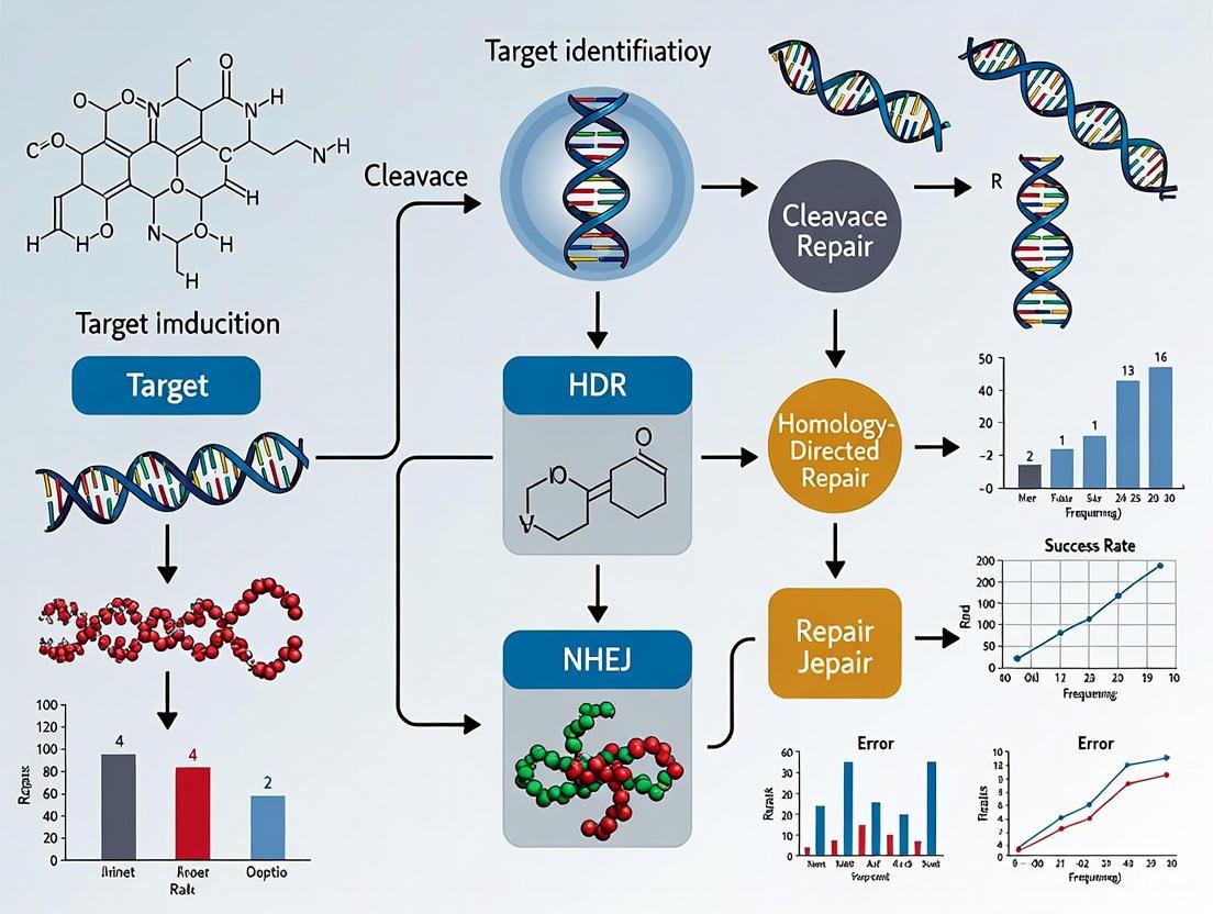
{c1::The Genome Editor's Toolkit: Mechanisms of DNA Repair} The cellular repair of Cas9-induced DSBs is the cornerstone of genome editing, primarily occurring via two pathways [5]:
- Non-Homologous End Joining (NHEJ): This is an error-prone repair pathway that often results in small insertions or deletions (indels) at the break site. When targeted to a gene's coding sequence, these indels can disrupt the reading frame, leading to a functional gene knockout [2]. This is highly applicable for disrupting dominant negative alleles in reproductive disorders.
- Homology-Directed Repair (HDR): This pathway uses a homologous DNA template to repair the break accurately. By co-delivering a donor DNA template with the desired sequence alongside CRISPR-Cas9, researchers can achieve precise gene correction or insertion, which is the ultimate goal for correcting most genetic mutations in the germline [5] [2].
Figure 1: CRISPR-Cas9 Editing Outcomes via DNA Repair Pathways. DSBs are repaired by the error-prone NHEJ pathway, leading to knockouts, or the precise HDR pathway using a donor template.
{c1::Advanced CRISPR-Cas Systems for Precision Surgery} The foundational CRISPR-Cas9 system has been extensively engineered to enhance its precision and expand its capabilities, moving beyond simple DSBs.
Table: Evolution of CRISPR-Based Genome Editing Tools
| Technology | Key Features | Application in Reproductive Genetics |
|---|---|---|
| High-Fidelity Cas9 [7] | Engineered Cas9 variants (e.g., eSpCas9, Cas9-HF1) with reduced off-target effects. | Increases safety profile for therapeutic editing of embryos and germ cells. |
| Base Editing [5] | Fuses a catalytically impaired Cas9 (nCas9) to a deaminase enzyme. Converts a single DNA base (C->T, A->G) without creating a DSB. | Corrects point mutations responsible for many genetic disorders (e.g., sickle cell disease) with minimal genotoxicity [8]. |
| Prime Editing [5] | Uses an nCas9 fused to a reverse transcriptase and a prime editing guide RNA (pegRNA). Can perform all 12 possible base-to-base conversions, plus small insertions and deletions, without a DSB or donor template. | Offers unprecedented versatility for correcting a wide array of pathogenic mutations with high precision. |
| Cas12a (Cpf1) [5] | A single RNA-guided nuclease that creates staggered DNA ends. Does not require tracrRNA. Recognizes a T-rich PAM (TTTV). | Provides an alternative PAM recognition, expanding the range of targetable genomic sites for multiplexed editing. |
{c1::Experimental Protocol: A Template for Gene Editing in Reproductive Biology} The following protocol provides a detailed methodology for achieving CRISPR-Cas9-mediated gene knockout in a model system, adaptable for research on reproductive cells or early embryos. It is based on established plant transformation and editing workflows [9] and reflects general principles applicable to preclinical research.
Table 1: Research Reagent Solutions for CRISPR-Cas9 Editing
| Research Reagent | Function/Explanation |
|---|---|
| Cas9 Protein | The core nuclease enzyme that executes the DNA cut. Using purified protein as a Ribonucleoprotein (RNP) complex is favored for reduced off-target effects and transient activity [10]. |
| sgRNA (synthetic) | A chemically synthesized single-guide RNA that directs Cas9 to the specific genomic target. Using two sgRNAs can increase knockout efficiency [9]. |
| Nuclear Localization Signal (NLS) | A peptide sequence fused to Cas9 that ensures its import into the cell nucleus. Recent advances with hairpin internal NLS (hiNLS) enhance editing efficiency in primary human cells [10]. |
| Delivery Vector | A plasmid or viral vector (e.g., lentivirus, AAV) engineered to express Cas9 and sgRNA(s) in target cells. For transgene-free editing, RNP delivery is preferred [9]. |
| Selection Antibiotic | An antibiotic (e.g., Kanamycin) used in culture media to select for cells that have successfully incorporated the editing machinery [9]. |
Title: CRISPR-Cas9 Ribonucleoprotein (RNP) Delivery for Gene Knockout
Goal: To achieve a loss-of-function mutation in a target gene via non-homologous end joining (NHEJ) following transfection with a pre-assembled Cas9-sgRNA RNP complex.
Materials & Reagents:
- Purified Cas9 protein with NLS (e.g., S. pyogenes Cas9)
- Chemically synthesized sgRNA(s) targeting the gene of interest
- Delivery method: Electroporation reagents for primary cells or lipid nanoparticles (LNPs)
- Cell culture media and reagents for the target cell type (e.g., primary lymphocytes, stem cells)
- Lysis buffer and PCR reagents for genotyping
- Gel electrophoresis equipment
Procedure:
- sgRNA Design and Validation:
- Design one or two sgRNAs targeting early exons of the target gene, preferably close to the start codon [9].
- Use computational tools (e.g., CRISPRscan) to predict on-target efficiency and minimize potential off-target sites.
- Synthesize and resuspend sgRNAs in nuclease-free buffer.
RNP Complex Assembly:
- Combine purified Cas9 protein and sgRNA at a predetermined molar ratio (e.g., 1:2) in a suitable buffer.
- Incubate the mixture at 25-37°C for 10-20 minutes to allow RNP complex formation.
Cell Transfection:
- For electroporation: Harvest and wash the target cells. Resuspend cells in an electroporation buffer, mix with the pre-assembled RNP complex, and electroporate using an optimized electrical program [10].
- For lipid-based delivery: Complex the RNP with commercial lipid nanoparticles (LNPs) according to the manufacturer's instructions and add to cells.
Culture and Expansion:
- After transfection, transfer cells to fresh pre-warmed culture medium.
- Allow cells to recover and proliferate for several days to enable repair and manifestation of edits.
Genotyping and Analysis:
- Harvest a portion of the cells 48-72 hours post-transfection.
- Extract genomic DNA and perform PCR amplification of the targeted genomic region.
- Analyze the PCR products using Sanger sequencing or next-generation sequencing (NGS) to detect the spectrum of indel mutations and calculate editing efficiency.
Figure 2: CRISPR-Cas9 RNP Knockout Workflow. Key steps from complex assembly to analysis.
{c1::Applications in Correcting Reproductive Genetic Abnormalities} CRISPR-Cas9 technology is being actively explored to correct inherited genetic mutations at various stages, from germline cells to somatic cells in adults. Its application in reproductive biology focuses on preventing the transmission of genetic diseases [3] [4].
- Germline and Embryo Editing: This approach involves correcting disease-causing mutations in sperm, eggs, or early-stage embryos. The edits would be heritable, potentially eradicating the familial disease lineage. While ethically complex and heavily regulated, this represents a definitive path for preventing monogenic disorders like cystic fibrosis, Huntington's disease, and Duchenne muscular dystrophy [4] [6].
- Therapeutic Somatic Cell Editing: Clinical success in somatic cells paves the way for related reproductive applications. The landmark approval of Casgevy for sickle cell disease and beta-thalassemia demonstrates that CRISPR can be used to edit hematopoietic stem cells ex vivo to produce therapeutic effects [8]. This proof-of-concept supports the feasibility of developing similar ex vivo editing protocols for germline or precursor cells.
{c1::Challenges and Future Directions} Despite its promise, the translation of CRISPR-Cas9 into clinical therapies for reproductive genetic abnormalities faces several hurdles that are the focus of intense research [1] [5] [8]:
- Off-Target Effects: The potential for Cas9 to cleave at unintended, partially complementary genomic sites remains a primary safety concern. Mitigation strategies include using high-fidelity Cas9 variants, optimized sgRNA design, and RNP delivery for transient activity [10] [2].
- Efficiency and Delivery: Achieving high HDR efficiency for precise correction, especially in hard-to-transfect cells like oocytes or zygotes, is challenging. Improving delivery methods, such as the use of novel LNPs and engineered viruses, is critical.
- Ethical and Regulatory Landscapes: The application of CRISPR in human germline editing raises profound ethical questions and is subject to strict legal restrictions in many countries [6]. Ongoing international dialogue is essential to establish clear guidelines for responsible research and potential clinical use.
{c1::Conclusion} The journey of CRISPR-Cas9 from a bacterial immune mechanism to a powerful tool for precision genome surgery represents a paradigm shift in biomedical science. Its core mechanism—programmable DNA recognition and cleavage—has been refined and expanded into a versatile toolkit capable of generating knockouts and, with base and prime editors, performing precise nucleotide surgery. As research in reproductive biology leverages these tools, coupled with robust protocols and a deepening understanding of the associated challenges, the potential to correct devastating genetic abnormalities at their source moves closer to reality, heralding a new era in genetic medicine.
The application of gene editing for correcting reproductive genetic abnormalities represents a frontier in reproductive medicine. While the CRISPR-Cas9 system has revolutionized genetic engineering, its reliance on double-stranded DNA breaks (DSBs) introduces significant limitations for clinical applications, particularly in precious and sensitive systems like human embryos. DSBs can lead to unintended outcomes such as indels (insertions/deletions), large deletions, and chromosomal rearrangements, raising safety concerns for therapeutic use [11] [12]. The emergence of more precise editing technologies—prime editing, base editing, and epigenetic modulation—offers promising alternatives that minimize these risks by editing DNA without creating DSBs.
These second-generation editing platforms significantly expand the scope of what is possible in correcting disease-causing mutations. Base editors enable efficient single nucleotide changes, prime editors function as "search-and-replace" tools for precise small edits, and epigenetic modulators allow for reversible changes in gene expression without altering the DNA sequence itself [11] [13] [12]. For researchers focused on reproductive genetic abnormalities, these tools provide unprecedented opportunities to study and potentially correct mutations responsible for monogenic diseases such as sickle cell anemia, cystic fibrosis, and Tay-Sachs disease at the earliest stages of development. This article provides application notes and detailed protocols for implementing these advanced technologies in embryo research, framed within the context of correcting pathogenic alleles while maintaining the highest standards of precision and safety.
Technical Comparative Analysis of Editing Platforms
The following table summarizes the key characteristics, advantages, and limitations of the three primary precision editing platforms relevant to embryo research:
Table 1: Comparative Analysis of Precision Genome Editing Platforms
| Editing Platform | Molecular Mechanism | Editing Window/Precision | Primary Applications in Embryo Research | Key Limitations |
|---|---|---|---|---|
| Base Editing | Cas9 nickase or dCas9 fused to deaminase enzymes converts C•G to T•A (CBE) or A•T to G•C (ABE) without DSBs [14] [12]. | ~5 nucleotide window near PAM site; high efficiency but limited positioning [15]. | Correcting point mutations causing monogenic diseases (e.g., β-thalassemia, sickle cell) [16]. | Cannot generate all possible base substitutions; requires specific positioning relative to PAM sequence [11]. |
| Prime Editing | Cas9 nickase-reverse transcriptase fusion uses pegRNA to directly write new genetic information into DNA [11] [17]. | Highly precise; can install all 12 possible base substitutions, small insertions (up to 44bp), and deletions (up to 80bp) [11]. | Correcting pathogenic alleles not addressable by base editors, including transversions and small indels. | Efficiency can be variable and lower than base editors; requires optimization of pegRNA and possible MMR inhibition [11] [17]. |
| Epigenetic Modulation | dCas9 fused to epigenetic effector domains (e.g., DNMT3A for methylation, TET1 for demethylation) modifies chromatin marks without changing DNA sequence [13] [18]. | Targets specific loci to alter DNA methylation or histone modifications; effects can be tunable and potentially reversible [13]. | Studying genomic imprinting, activating silenced alleles, and potentially modulating disease risk without permanent DNA alteration. | Effects may be transient; efficiency and specificity of sustained modulation require careful validation [13]. |
Prime Editing: A Versatile "Search-and-Replace" Tool
Mechanism and Workflow
Prime editing represents a significant leap in precision editing technology. The system employs a fusion protein consisting of a Cas9 nickase (H840A) connected to an engineered reverse transcriptase (RT) from the Moloney Murine Leukemia Virus (M-MLV), along with a specialized prime editing guide RNA (pegRNA) [11] [17]. The pegRNA not only specifies the target site but also contains a primer binding site (PBS) and an RT template encoding the desired edit. The mechanism involves: (1) binding of the prime editor complex to the target DNA, (2) nicking of the non-target DNA strand by the Cas9 nickase, (3) hybridization of the 3' end of the nicked DNA to the PBS on the pegRNA, (4) reverse transcription of the edited sequence from the RT template, and (5) resolution and repair of the DNA heteroduplex to permanently incorporate the edit [11]. The development of advanced prime editors (PE2-PE7) with improved RT efficiency and pegRNA stability (epegRNAs) has substantially increased editing efficiencies [17].
The following diagram illustrates the core mechanism of prime editing:
Application Notes for Embryo Research
Prime editing is particularly suited for correcting mutations in embryos where precision is paramount. Its ability to install all 12 possible base-to-base conversions, as well as small insertions and deletions, means it can theoretically correct up to 89% of known pathogenic human genetic variants [11]. For reproductive genetics, this includes mutations in genes like HEXA (Tay-Sachs disease), CFTR (cystic fibrosis), and F8 (hemophilia A), where different families may carry distinct mutations that can all be addressed with a single, versatile platform.
Key optimization strategies for embryo editing include:
- pegRNA Design: The PBS should be 10-15 nucleotides long, and the RT template should extend 8-10 nucleotides beyond the edit. The use of engineered pegRNAs (epegRNAs) with structured RNA motifs at the 3' end protects against exonuclease degradation and improves efficiency [11] [17].
- MMR Inhibition: Co-expression of dominant-negative mutants of the MLH1 protein (as in PE4 and PE5 systems) can transiently inhibit the mismatch repair pathway, which often favors the non-edited strand and reduces editing efficiency. This can increase prime editing efficiency by several-fold [11].
- Delivery Considerations: For embryo work, ribonucleoprotein (RNP) delivery of the prime editor protein complexed with in vitro-transcribed pegRNA may minimize off-target effects and reduce exposure time, potentially improving embryo viability [19].
Base Editing: Efficient Single Nucleotide Correction
Mechanism and Workflow
Base editors provide a highly efficient method for converting one DNA base pair to another without requiring DSBs. Two main classes have been developed: Cytosine Base Editors (CBEs) convert C•G to T•A, and Adenine Base Editors (ABEs) convert A•T to G•C [14] [12]. CBEs are typically fusions of a Cas9 nickase (or dCas9) to a cytidine deaminase enzyme (like APOBEC1) and a uracil glycosylase inhibitor (UGI) that prevents unwanted repair of the edited base. ABEs use an evolved tRNA adenosine deaminase (TadA) to perform the A-to-I conversion, which the cell then treats as G [14] [15] [12]. The editing occurs within a defined "editing window" of approximately 5 nucleotides near the PAM site, making target site positioning crucial.
The workflow for base editing involves:
- Target Site Selection: Identifying a target site where the pathogenic base falls within the editing window of a suitable PAM sequence.
- Editor Delivery: Introducing the base editor and sgRNA into the cells, typically as plasmids, mRNA, or RNP complexes.
- Editing Validation: Using mismatch cleavage assays, Sanger sequencing, or next-generation sequencing to quantify editing efficiency and byproducts [15] [19].
Application Notes for Embryo Research
Base editors are particularly valuable for correcting specific point mutations known to cause severe genetic disorders. For instance, the mutation responsible for Progeria (LMNA c.1824C>T) or the sickle cell disease mutation (HBB c.20A>T) are theoretically correctable with base editing technology [12]. The high efficiency and reduced indel formation compared to CRISPR-Cas9 make base editors attractive for embryo editing where maximizing correct editing while minimizing collateral damage is critical.
Key considerations for embryo base editing include:
- Editing Window: The protospacer must be positioned so that the target base falls within the ~5-nucleotide activity window of the base editor, typically at positions 4-8 within the protospacer, counting the PAM as positions 21-23 [15].
- Byproduct Management: While base editors produce fewer indels than Cas9 nuclease, they can cause unwanted, low-frequency "bystander" edits when multiple editable bases are present in the activity window. Careful design and analysis are required to select targets that minimize this risk [15] [12].
- Variant Selection: Newer base editor variants with narrowed or shifted editing windows (e.g., BE4, Target-AID) can improve product purity when multiple cytosines or adenines are present in the original window [12].
Epigenetic Modulation: Regulating Gene Expression Without Changing DNA Sequence
Mechanism and Experimental Setup
Epigenetic modulation using CRISPR/dCas9 systems allows for precise alteration of gene expression patterns without modifying the underlying DNA sequence—an approach particularly relevant for studying imprinted genes and regulatory elements during embryonic development. This technology fuses a catalytically dead Cas9 (dCas9) to epigenetic effector domains, such as DNMT3A for adding DNA methylation marks or TET1 for removing them [13] [18]. When guided to specific genomic loci by sgRNAs, these fusion proteins can induce targeted epigenetic remodeling, leading to stable changes in gene transcription that can persist through multiple cell divisions [13].
Advanced systems enable orthogonal epigenetic editing, where different dCas9 orthologs (e.g., dSpCas9 and dSaCas9) fused to opposing epigenetic modifiers (e.g., DNMT3A and TET1) can be used simultaneously within the same cell to study antagonistic epigenetic regulation [13]. Furthermore, synergistic effects have been demonstrated by combining epigenetic activators like VPR-dSpCas9 with TET1-dSaCas9, resulting in strong and persistent gene activation lasting up to 30 days post-transfection [13].
Application Notes for Embryo Research
In the context of embryo research and correcting genetic abnormalities, epigenetic modulation offers a potentially safer alternative for conditions where altering gene expression, rather than the genetic code itself, may provide therapeutic benefit. This includes potentially reactivating silenced healthy alleles of imprinted genes or modulating the expression of genes involved in metabolic storage diseases.
Key implementation strategies include:
- Modular Toolboxes: Utilizing modular cloning systems (e.g., Golden Gate assembly) allows for flexible testing of different effector domain combinations and sgRNA configurations to optimize editing outcomes [13].
- Multi-guide Systems: Employing systems capable of expressing up to six different sgRNAs simultaneously enables effective targeting of larger regulatory regions, such as promoters and enhancers, which often require multiplexed guidance for effective modulation [13].
- Stability Optimization: The persistence of epigenetic changes can be enhanced by using stronger chromatin-opening effectors like VPR and combining them with demethylating enzymes like TET1, as demonstrated in sustained activation of the HNF1A gene [13].
Essential Reagents and Validation Methods
The Scientist's Toolkit: Research Reagent Solutions
Successful implementation of these advanced editing technologies requires careful selection of molecular tools and reagents. The following table catalogs essential reagents for precision genome editing in embryonic systems:
Table 2: Essential Research Reagents for Precision Genome Editing
| Reagent Category | Specific Examples | Function & Importance | Source/Reference |
|---|---|---|---|
| Prime Editors | PE2, PEmax, PE4, PE5, PE6, PE7 | Engineered fusion proteins with improved efficiency and specificity; PE4/PE5 include MMR inhibition. | [11] [17] |
| Base Editors | BE3, BE4, Target-AID, ABE7.10 | CBEs and ABEs with varying editing windows, efficiencies, and fidelity characteristics. | [14] [15] [12] |
| Epigenetic Effectors | dCas9-DNMT3A, dCas9-TET1, dCas9-KRAB, dCas9-VPR | Fusion proteins for targeted DNA methylation, demethylation, repression, and activation. | [13] [18] |
| Specialized Guide RNAs | pegRNA, epegRNA, nicking sgRNA (for PE3/5) | pegRNAs encode the edit; epegRNAs have enhanced stability; nicking sgRNAs enhance PE efficiency. | [11] [17] |
| Delivery Tools | Lipid Nanoparticles (LNPs), Electroporation, AAV vectors | Methods for introducing editing components into embryos and cells; LNPs allow potential re-dosing. | [16] |
| Validation Enzymes | T7 Endonuclease I, ArciTect T7 Endonuclease I | Detects indels and editing efficiency via mismatch cleavage assays in heterogeneous cell populations. | [19] |
Experimental Protocol: A Workflow for Prime Editing in Embryos
The following protocol outlines key steps for implementing a prime editing experiment in a research setting, incorporating validation steps critical for assessing success.
Table 3: Protocol for Prime Editing Experiment Implementation and Validation
| Step | Procedure | Purpose & Notes |
|---|---|---|
| 1. Target Selection & pegRNA Design | Identify target sequence and design pegRNA with 10-15 nt PBS and RT template encoding the desired edit. Use computational tools (e.g., DeepPrime). | Ensures the edit is positioned correctly. For difficult targets, design multiple pegRNAs to test. |
| 2. Component Delivery | Deliver prime editor (as mRNA or protein) and pegRNA (as in vitro transcript) into zygotes via microinjection or electroporation. | RNP delivery may reduce off-target effects. Optimize concentrations to balance efficiency and viability. |
| 3. Initial Screening (48-72 hrs) | Extract genomic DNA from a subset of embryos. Amplify target region with offset primers. Perform T7 Endonuclease I assay [19]. | Rapid assessment of editing activity. The assay cleaves heteroduplex DNA, giving an estimate of editing frequency. |
| 4. Deep Sequencing Validation | Amplify target region from pooled embryos or individual clones. Submit for next-generation sequencing (NGS). Analyze with CRISPResso2 [19]. | Provides quantitative data on editing efficiency, precision, and byproducts (indels, bystander edits). |
| 5. Off-Target Assessment | Amplify potential off-target sites (predicted by in silico tools) and sequence. Alternatively, perform whole-genome sequencing for comprehensive analysis. | Critical for safety assessment. NGS provides the most thorough evaluation of off-target effects. |
| 6. Functional Validation | For established embryo models, assess phenotypic correction, protein expression restoration, and developmental progression. | Confirms that the genetic correction translates to functional and developmental improvement. |
The rapid evolution of precision genome editing tools has dramatically expanded our capabilities for researching and potentially correcting reproductive genetic abnormalities. Prime editing, base editing, and epigenetic modulation each offer distinct advantages and applications, together creating a comprehensive toolkit for addressing a wide spectrum of genetic diseases at the embryonic stage. As these technologies continue to advance—with improvements in editing efficiency, specificity, and delivery—their potential for clinical translation in reproductive medicine will grow accordingly.
Future developments will likely focus on enhancing the efficiency and specificity of these editors, optimizing delivery methods such as lipid nanoparticles that allow for re-dosing [16], and establishing robust safety profiles through comprehensive off-target characterization. Furthermore, the combination of these approaches—such as using epigenetic modulation to prime a locus for more efficient editing—may open new therapeutic avenues. For researchers in reproductive genetics, these technologies provide not only powerful tools for fundamental research into human development and disease but also hope for future interventions that could prevent the transmission of devastating genetic disorders.
The application of gene-editing technologies to correct reproductive genetic abnormalities represents a frontier in biomedical science with profound implications. This field, known as heritable human genome editing (HHGE), aims to prevent the transmission of serious genetic diseases by introducing precise modifications into the DNA of sperm, eggs, or embryos. The journey from the first controversial birth of gene-edited children to the current rise of commercial ventures illustrates a critical pivot point in reproductive medicine. This article details the key studies and emerging protocols shaping this field, providing a resource for researchers and drug development professionals engaged in this rapidly evolving discipline. The content is framed within the broader thesis that HHGE, while not yet safe or refined enough for clinical application, holds significant potential for preventing monogenic diseases, necessitating rigorous, transparent, and collaborative research to establish safety and efficacy protocols.
Landmark Historical Case: The He Jiankui Affair
In 2018, Chinese biophysicist He Jiankui announced the birth of the world's first genetically edited babies, twin girls known pseudonymously as Lulu and Nana [20]. His objective was to confer genetic resistance to HIV by mimicking a naturally occurring mutation in the CCR5 gene, which codes for a protein HIV uses to enter cells [20]. The target population was children of HIV-positive fathers and HIV-negative mothers, who faced social and regulatory barriers to assisted reproduction in China [20].
Detailed Methodology and Protocol
The following protocol reconstructs the methodology employed based on available public reports and summaries [20].
Protocol 1: Embryonic CCR5 Gene Editing for HIV Resistance
- 1. Patient Recruitment & Informed Consent: Recruit couples through AIDS advocacy groups, offering standard in vitro fertilisation (IVF) services with an experimental gene-editing component. The informed consent process was later widely criticized as being incomplete and inadequate [20].
- 2. In Vitro Fertilisation: Perform standard IVF procedures using sperm from the HIV-positive father and eggs from the HIV-negative mother to create zygotes.
- 3. Microinjection of CRISPR-Cas9 Components: Shortly after fertilisation, microinject the CRISPR-Cas9 machinery into the embryos. The editing complex was designed to introduce a frameshift mutation intended to disable the CCR5 gene, rather than recreate the natural CCR5-Δ32 variant [20].
- 4. Embryo Culture & Quality Control: Culture the edited embryos. Employ a preimplantation genetic diagnosis (PGD) process, removing 3-5 cells from each embryo for comprehensive genetic sequencing to identify potential mosaicism (where some cells are edited and others are not) and off-target editing events [20].
- 5. Embryo Transfer: Select embryos based on PGD results for transfer into the mother's uterus to establish a pregnancy.
- 6. Prenatal Monitoring: During pregnancy, sequence cell-free fetal DNA from the mother's blood to monitor for off-target effects. Offer amniocentesis for further genetic analysis, though this was declined by the parents in the reported case [20].
Key Outcomes and Data
The experiment resulted in the birth of twin girls in October 2018 [20]. He Jiankui reported that the babies were born healthy and that genetic sequencing indicated the intended edits were present, albeit with some mosaicism [20]. A third gene-edited child was born in 2019 [20]. The data was never peer-reviewed or published in a scientific journal, and the claims lack independent verification [20].
Table 1: Quantitative Data Summary of the He Jiankui Experiment
| Parameter | Reported Outcome | Limitations & Criticisms |
|---|---|---|
| Target Gene | CCR5 | The edit did not replicate the natural CCR5-Δ32 mutation; some HIV strains use other receptors (e.g., CXCR4), so protection is not guaranteed [20]. |
| Number of Embryos/Babies | 3 babies born (Twins Lulu & Nana, plus a third child, Amy) | The existence of a third child was not initially disclosed [20]. |
| Editing Efficiency | Reported edits present, but with mosaicism | Mosaicism means the edit is not present in all cells, potentially undermining the therapeutic goal and complicating risk assessment [20]. |
| Off-Target Analysis | Performed via PGD and cell-free fetal DNA sequencing | The adequacy and sensitivity of these methods for a comprehensive off-target profile are debated. No independent data verification exists [20]. |
| Clinical Outcome | Babies reported healthy at birth | The long-term health consequences, including cancer risk from potential off-target edits, are entirely unknown [20]. |
Aftermath and Global Response
The experiment was met with immediate and widespread international condemnation from scientists, bioethicists, and governments [20]. Criticisms centered on the profound ethical breaches, including the secretive nature of the work, the inadequate informed consent process, the unknown long-term risks to the children, and the use of an unproven and unnecessary procedure on otherwise healthy embryos [20]. In December 2019, a Chinese court found He Jiankui and two collaborators guilty of illegal medical practice, sentencing him to three years in prison [20]. The affair prompted global calls for a moratorium on HHGE and spurred the World Health Organization and numerous national governments to develop stricter guidelines for human genome editing [20].
Current Commercial Ventures in Embryonic Gene Editing
The controversial legacy of He Jiankui has not deterred a new wave of commercial ventures, primarily backed by Silicon Valley investors, who are pushing to advance HHGE with a stated focus on disease prevention.
Table 2: Overview of Current Commercial Ventures in HHGE
| Venture Name | Key Leadership/Backing | Stated Mission & Focus | Reported Funding & Status |
|---|---|---|---|
| Preventive [21] [22] [23] | Lucas Harrington (co-founder); Backed by OpenAI's Sam Altman and Coinbase's Brian Armstrong. | To research and rigorously test the safety of heritable genome editing for preventing serious genetic diseases [21]. | Approximately $30 million from private funders [21] [23]. Incorporated as a public-benefit corporation. Research is planned outside the US due to regulatory barriers [22] [23]. |
| Manhattan Genomics [24] | Cathy Tie (CEO), Eriona Hysolli (co-founder). | To prevent serious genetic diseases like cystic fibrosis and beta thalassemia through embryonic gene editing, with a focus on transparency and regulatory approval [24]. | Funding amount not publicly disclosed. Company is in the formation stage [24]. |
| Bootstrap Bio [24] | Chase Denecke (CEO). | Initially focused on disease prevention but has expressed interest in enhancing traits to "make peoples' lives actually better" [24]. | Reportedly seeking seed funding [21]. |
Proposed Research and Development Workflow
These companies emphasize a more measured, scientifically rigorous approach compared to the He Jiankui case. Their proposed R&D pipeline can be visualized as a multi-stage, iterative process.
Diagram 1: Proposed R&D Pipeline for HHGE
Stated Experimental Protocols for Safety Assessment
Ventures like Preventive and Manhattan Genomics have stated their commitment to extensive safety testing before any clinical application. The following protocol outlines the key methodologies they propose to employ.
Protocol 2: Comprehensive Safety and Efficacy Assessment for HHGE
1. In Vitro and In Silico Modeling:
- Objective: To perform initial gRNA design and off-target prediction.
- Method: Utilize human cell lines and advanced computational tools to design and screen guide RNAs (gRNAs) for high on-target efficiency and minimal predicted off-target activity. This includes using tools like CIRCLE-seq for in vitro off-target profiling.
2. Animal Model Studies:
- Objective: To assess the feasibility, specificity, and developmental impact of editing in a whole organism.
- Method: Conduct gene-editing experiments in mouse and non-human primate embryos. This involves:
- Microinjection of CRISPR machinery into zygotes.
- Transfer of viable embryos to surrogate females.
- Comprehensive genomic analysis of resulting offspring via whole-genome sequencing (WGS) to confirm on-target editing and detect off-target effects and mosaicism.
- Long-term phenotyping to monitor health, development, and reproductive fitness across generations.
3. Research on Non-Implantable Human Embryos:
- Objective: To validate editing efficiency and safety in the human context.
- Method: Use donated human embryos created via IVF that are not destined for implantation. Perform gene editing and culture them for up to 14 days (in accordance with international guidelines). Analyze embryos at various developmental stages using:
- Whole-Genome Sequencing (WGS): To comprehensively map on-target and off-target edits.
- Transcriptomics and Epigenomics: To assess any unintended disruptions to gene expression and cellular function.
- Single-Cell Multi-omics: To understand cell lineage and mosaicism at high resolution.
The Scientist's Toolkit: Research Reagent Solutions
The advancement of HHGE research relies on a suite of sophisticated tools and reagents. The table below details key materials and their functions.
Table 3: Essential Research Reagents and Materials for HHGE
| Research Reagent / Material | Function & Application in HHGE |
|---|---|
| CRISPR-Cas Systems (Cas9, Cas12a) | The core gene-editing enzymes that create double-strand breaks in DNA at programmed locations. Different systems (e.g., Cas9 vs. Cas12a) offer variations in specificity and the type of DNA cut made [8]. |
| Base Editors & Prime Editors | Advanced "CRISPR 2.0" systems that allow for precise chemical conversion of a single DNA base (e.g., C to T) or the insertion of small sequences without creating double-strand breaks, potentially reducing off-target effects and increasing safety [16] [24]. |
| Lipid Nanoparticles (LNPs) | A delivery vehicle for in vivo gene editing. While currently used primarily in somatic therapies, LNPs are a subject of intense research for their potential to deliver editing components to gametes or embryos more safely and efficiently than current methods [16]. |
| Guide RNA (gRNA) | A short RNA sequence that programs the Cas enzyme to bind to a specific target site in the genome. Its design is critical for minimizing off-target effects [20]. |
| Whole Genome Sequencing (WGS) Kits | Essential reagents for the comprehensive analysis of edited cells or embryos. Used to confirm on-target edits and, crucially, to detect any off-target mutations across the entire genome, a core component of safety assessment [20]. |
| Preimplantation Genetic Testing (PGT) Reagents | Used to genetically screen embryos prior to transfer. In the context of HHGE research, these reagents are critical for analyzing edit status (e.g., mosaicism) in blastocyst-stage embryos in a non-destructive manner [20]. |
The path from He Jiankui's ethically and scientifically flawed experiment to the current, more transparent commercial ventures marks a significant evolution in the field of heritable human genome editing. While the ultimate goal of preventing devastating genetic diseases remains a powerful motivator, the scientific community maintains that the technology is not yet ready for clinical application. The key challenges of off-target editing, mosaicism, and long-term health effects persist. The success of these new ventures, and the field at large, will depend on an unwavering commitment to rigorous, open, and collaborative science, robust regulatory oversight, and inclusive public dialogue. The protocols and tools outlined herein provide a framework for the meticulous research required to determine whether HHGE can ever be performed safely and responsibly, turning a controversial concept into a viable therapeutic pathway for preventing reproductive genetic abnormalities.
The global regulatory landscape for human genome editing is dynamic and multifaceted, characterized by rapid scientific progress alongside complex ethical and policy challenges. Recent advances, particularly in personalized gene-editing therapies, have prompted significant regulatory innovations, such as the U.S. Food and Drug Administration's (FDA) new pathway for accelerated approval of customized treatments [25]. Simultaneously, the international community continues to grapple with the profound implications of germline editing, evidenced by ongoing calls for moratoria and major international summits focused on establishing ethical boundaries [26] [27].
This application note provides researchers, scientists, and drug development professionals with a comprehensive analysis of the current regulatory frameworks, emphasizing practical experimental protocols and resources for navigating this evolving landscape. The information is particularly framed within the context of correcting reproductive genetic abnormalities, a field that demands careful consideration of both technical feasibility and ethical permissibility.
Global Regulatory Frameworks and Quantitative Analysis
Regulatory approaches to human genome editing vary significantly across international jurisdictions, particularly regarding heritable modifications versus somatic cell therapies. The following table summarizes the key regulatory positions and restrictions of major international bodies and countries.
Table 1: Global Regulatory Positions on Human Genome Editing
| Country/Region | Somatic Cell Editing | Germline Editing (Reproductive Use) | Key Regulations/Guidelines | Penalties for Violations |
|---|---|---|---|---|
| United States | Permitted with FDA oversight [28] [29] | Moratorium on clinical trials; FDA prohibited from reviewing applications [27] [29] | FDA & NIH Guidelines [29] | N/A (Regulatory block) |
| China | Permitted with oversight | Banned (Based on guidelines) [29] | Chinese Guideline on Human Assisted Reproductive Technologies [29] | Criminal sentence (e.g., 3 years imprisonment in He Jiankui case) [29] |
| United Kingdom | Permitted with oversight | Restricted; legal permission possible for specific medical uses [29] | Legislation on mitochondrial replacement [29] | Up to 10 years imprisonment [29] |
| France | Permitted with oversight | Banned (Based on legislation) [29] | Specific laws against germline editing [29] | Up to 20 years imprisonment [29] |
| International Bodies | N/A | Call for moratorium by leading scientific societies [27] [30] | Declaration of Helsinki [29] | No legal force, but provides global guidance |
A critical development in the U.S. is the FDA's proposal of a "plausible mechanism" pathway. This innovative regulatory approach allows for the approval of bespoke gene-editing medicines for patients with the same clinical syndrome, irrespective of the specific underlying mutation, based on a scientifically sound mechanism and consistent, robust patient-to-patient efficacy [28]. This is particularly significant for rare disease treatment, where commercial development is often not feasible.
Table 2: FDA's Proposed "Plausible Mechanism" Pathway for On-Demand Gene Editing
| Pathway Element | Description | Implication for Research & Development |
|---|---|---|
| Target Population | Patients with the same clinical syndrome (e.g., specific metabolic disorder, immune deficiency) [28] | Enables "umbrella trials" that pool patients with different mutations in the same gene or pathway. |
| Evidence Standard | Consistent, robust efficacy across a small number of patients that cannot be expected with standard care [28] | Reduces the clinical evidence burden compared to traditional drug approval pathways. |
| Manufacturing | "Platformization" of CRISPR; streamlined development for subsequent similar therapies [28] | Allows academic centers and industry to amortize development costs across multiple patient-specific therapies. |
| Current Limitations | Primarily applicable to diseases affecting tissues amenable to non-viral delivery (e.g., liver, blood stem cells) [28] | Therapies for neurological diseases await improved delivery technologies (e.g., safer AAV vectors). |
Experimental Protocols for Preclinical Development
Navigating the path to clinical trials, especially under new regulatory frameworks, requires robust and standardized preclinical protocols. The following section details a core methodology based on the pioneering case of KJ Muldoon, the first infant treated with a bespoke base-editing therapy for CPS1 deficiency [31].
Protocol: Development of a Bespoke Gene Editor for a Monogenic Disorder
This protocol outlines the key steps for designing, validating, and preparing an investigational gene-editing therapy for a single patient with a rare, life-threatening genetic condition, based on the methodologies successfully employed by the CHOP/Penn team [31].
1. Patient Identification & Genetic Diagnosis
- Objective: Confirm a definitive genetic diagnosis of a severe monogenic disease where conventional treatments are inadequate or high-risk.
- Methods:
- Perform whole-exome or whole-genome sequencing on the patient to identify the causative mutation(s).
- For recessive disorders, confirm bi-allelic presence of the pathogenic variant.
- For disorders like urea cycle defects (e.g., CPS1), monitor blood ammonia levels and other relevant biomarkers to establish disease severity and urgency [31].
2. Guide RNA (gRNA) and Editor Design
- Objective: Design a highly specific gene-editing system targeting the patient's unique mutation.
- Methods:
- Sequence Analysis: Align the wild-type and mutant gene sequences to identify the precise nucleotide change and its genomic context.
- gRNA Selection: Design a gRNA that directs the editor (e.g., a base editor) to the immediate vicinity of the target mutation with high predicted on-target efficiency and minimal off-target risk using tools like CRISPRscan.
- Editor Selection: For point mutations, select an appropriate adenine or cytosine base editor to achieve the desired nucleotide conversion without causing a double-strand break [31].
3. In Vitro Potency and Specificity Validation
- Objective: Demonstrate that the designed editor corrects the mutation efficiently and accurately in a relevant cellular model.
- Methods:
- Cell Transfection: Deliver the editor mRNA and synthetic gRNA into patient-derived fibroblasts or iPSCs (if available and time permits), or a relevant human cell line engineered to carry the patient's mutation.
- Efficiency Analysis: After 48-72 hours, extract genomic DNA and use next-generation sequencing (NGS) to quantify the percentage of alleles corrected at the target site.
- Specificity Analysis: Perform computational prediction of off-target sites based on the gRNA sequence. Use methods like GUIDE-seq or CIRCLE-seq on edited cells to empirically identify and quantify off-target editing events. NGS of these top predicted sites should be conducted to confirm specificity [28].
4. In Vivo Efficacy and Safety Studies (Animal Model)
- Objective: Provide proof-of-concept for functional correction and preliminary safety data in a live organism.
- Methods:
- Animal Model: Utilize a mouse model with the analogous disease-causing mutation. If unavailable, use a wild-type mouse for initial biodistribution and toxicity studies.
- Dose-Finding: Administer the therapy (e.g., LNP-packaged editor mRNA and gRNA) via the intended clinical route (e.g., intravenous infusion) at escalating doses.
- Efficacy Assessment: Measure the restoration of normal metabolic or physiological function (e.g., reduction in ammonia for urea cycle disorders) and detect the presence of the corrected gene sequence in the target tissue (e.g., liver) via NGS.
- Safety Assessment: Monitor animals for acute toxicity, weight loss, and signs of organ distress. Conduct histopathological analysis of major organs post-treatment [28] [31].
5. Formulation and GMP-compliant Manufacturing
- Objective: Produce a clinical-grade therapeutic for human administration.
- Methods:
- Formulation: For liver-targeted delivery, formulate the editor mRNA and gRNA in a lipid nanoparticle (LNP) system optimized for hepatocyte uptake.
- Manufacturing: Under Good Manufacturing Practice (GMP) conditions, produce a sufficient quantity of the drug product for the clinical dose regimen.
- Quality Control: Perform rigorous testing for potency, purity, sterility, and endotoxin levels. Given the single-patient nature, the FDA may permit "benefit-risk commensurate" accelerated small-scale manufacture with reduced testing compared to large-scale commercial production [28].
Signaling Pathways and Workflows in Gene Editing Regulation
The regulatory decision-making process for approving a novel gene-editing therapy, particularly under the new "plausible mechanism" pathway, involves a logical sequence of evaluations. The diagram below maps this workflow.
The scientific and ethical rationale for a global moratorium on heritable human genome editing (HHGE) is founded on a series of interconnected concerns, which are visually summarized in the following pathway.
The Scientist's Toolkit: Research Reagent Solutions
The successful development of a gene-editing therapeutic relies on a core set of reagents and tools. The following table details essential materials and their functions, drawing from the technologies used in recent landmark studies [28] [31].
Table 3: Essential Research Reagents for Developing Gene-Editing Therapies
| Reagent/Material | Function | Key Considerations for Regulatory Compliance |
|---|---|---|
| CRISPR-Cas Nucleases(e.g., SpCas9, base editors) | Enzymes that catalyze the cutting or chemical conversion of DNA at a target site. | Select high-specificity variants (e.g., HiFi Cas9). Document source and sequence. Requires purity and identity testing for GMP. |
| Guide RNA (gRNA)(synthetic or in vitro transcribed) | A short RNA sequence that directs the nuclease to the specific genomic target. | Design with thorough off-target prediction analysis. For GMP, require high purity, sequence verification, and endotoxin testing. |
| Delivery Vector(e.g., LNP, AAV, EV) | A vehicle to protect and deliver the gene-editing machinery into target cells in the body. | LNP: Ideal for liver-directed editing [28]. AAV: Used for other tissues but has manufacturing challenges [28]. Characterize size, charge, and encapsulation efficiency. |
| Patient-Derived Cells(e.g., iPSCs, fibroblasts) | A cellular model for in vitro validation of editing efficiency and specificity. | Establish with informed consent. Maintain genomic stability and identity. Crucial for demonstrating target engagement in the relevant genetic background. |
| NGS Off-Target Assay Kits(e.g., GUIDE-seq, CIRCLE-seq) | Tools to empirically identify and quantify unintended editing events across the genome. | Essential for preclinical safety package. Data from these assays are typically required by regulators to assess product risk. |
| Reference Standards(e.g., synthetic genes) | Controls for sequencing and analytical assays to ensure accuracy and reproducibility. | Critical for validating NGS-based potency and off-target assays. Should be traceable and well-characterized. |
The global regulatory landscape for gene editing is at a pivotal juncture. The emergence of faster FDA pathways for personalized therapies represents a monumental shift for treating severe rare diseases, effectively creating a new category of medicine [25] [28]. However, this progress stands in stark contrast to the firm and enduring international consensus supporting a moratorium on heritable human genome editing, a position reinforced by leading scientific societies as recently as 2025 [27].
For researchers focused on correcting reproductive genetic abnormalities, this dichotomy defines the field. The immediate future lies in refining somatic cell therapies and navigating the new "platform" and "umbrella trial" regulatory models. The successful treatment of KJ Muldoon for CPS1 deficiency provides a tangible protocol for this approach [31]. Meanwhile, any research involving germline modifications must proceed with extreme caution, adhering to the strictest ethical guidelines and current legal prohibitions. The ongoing dialogue, exemplified by the 2025 Global Observatory International Summit, underscores that responsible innovation requires continuous, inclusive deliberation to ensure these powerful technologies serve humanity and uphold the integrity of human life [26] [32].
This application note provides a comparative analysis of therapeutic target identification and experimental protocols for two distinct categories of genetic disorders: monogenic diseases and complex reproductive disorders. Within the expanding field of gene editing, these categories present unique challenges and opportunities for researchers and drug development professionals. We outline specific methodological approaches, technical considerations, and research tools essential for advancing targeted therapies in both domains, with particular emphasis on CRISPR-based technologies for monogenic conditions and integrated pathway targeting for complex reproductive endocrine disorders.
The strategic approach to identifying and validating therapeutic targets varies substantially between monogenic diseases and complex reproductive disorders. Monogenic diseases, caused by mutations in a single gene, offer well-defined, causal targets for direct genetic correction [33] [34]. In contrast, complex reproductive disorders often involve polygenic inheritance, environmental influences, and dysregulation of intricate neuroendocrine signaling pathways, necessitating multi-target intervention strategies [35] [36].
The emergence of precision gene editing tools, particularly CRISPR-Cas systems and their derivatives (base editing, prime editing), has revolutionized therapeutic development for monogenic conditions [33] [37]. Meanwhile, advances in functional neuroimaging and multi-omics profiling have enhanced our understanding of the complex pathophysiology underlying reproductive disorders, revealing novel intervention points within the hypothalamic-pituitary-ovarian (HPO) axis [35].
Table 1: Fundamental Characteristics Influencing Target Identification
| Characteristic | Monogenic Diseases | Complex Reproductive Disorders |
|---|---|---|
| Genetic Basis | Single gene mutation [38] | Polygenic + environmental factors [35] |
| Primary Target | Causal gene/variant [33] | Signaling pathways & regulatory networks [35] |
| Therapeutic Approach | Direct genetic correction [33] | Multi-target modulation [35] |
| Target Validation | Genetic linkage, functional restoration assays | Pathway analysis, neuroimaging, endocrine profiling [35] |
| Example Targets | BCL11A (hemoglobinopathies), CFTR (cystic fibrosis) [34] [39] | Kisspeptin-GPR54, PI3K/Akt/mTOR, BDNF-TrkB [35] |
Therapeutic Targeting for Monogenic Diseases
Target Identification and Validation
Monogenic disease targets are identified through genetic sequencing of affected individuals and families to establish causal relationships between gene mutations and disease phenotypes. Target validation involves demonstrating that correction of the specific genetic lesion rescues cellular and physiological function.
Key Considerations:
- Variant Pathogenicity: Establish clinical significance using databases such as ClinVar and functional studies [33].
- Therapeutic Window: Assess the editing activity window relative to the pathogenic mutation for base editing approaches [33].
- Delivery Constraints: Consider target tissue accessibility and editing tool delivery limitations [37].
Table 2: Quantitative Considerations for Monogenic Disease Target Selection
| Parameter | Optimal Characteristics | Validation Methods |
|---|---|---|
| Variant Frequency | High prevalence in patient populations [38] | Population genetics databases |
| Editing Window | Within 5-10 nucleotide activity window for base editors [33] | In vitro editing efficiency assays |
| Therapeutic Threshold | 10-24% of normal expression may be sufficient (e.g., CFTR) [34] | Gene expression analysis, functional assays |
| PAM Availability | NGG for SpCas9; T-rich for Cas12a; engineered variants for relaxed PAM [37] | PAM prediction algorithms, target sequence analysis |
Experimental Protocol: Base Editing for Point Mutation Correction
This protocol describes a methodology for correcting pathogenic point mutations using CRISPR-dependent base editing in patient-derived cells.
Materials:
- Patient-derived fibroblasts or induced pluripotent stem cells (iPSCs)
- Appropriate base editor plasmid (ABE or CBE based on conversion required)
- sgRNA expression construct or synthetic sgRNA
- Delivery system (electroporation, lipofection, or AAV)
- Culture media and supplements
- Genomic DNA extraction kit
- PCR reagents
- Sequencing primers
- Functional assay reagents (dependent on gene function)
Procedure:
Target Analysis and sgRNA Design
- Identify the pathogenic single-nucleotide variant (SNV) and sequence context
- Design sgRNA to position the target base within the activity window (typically positions 4-8 for ABE, 3-10 for CBE) [33]
- Verify minimal off-target potential using algorithms like CRISPRseek or Cas-OFFinder
- Check for required PAM sequence (NGG for SpCas9-based editors)
Editor Assembly and Delivery
- Clone sgRNA sequence into appropriate base editor backbone (e.g., pCMV_ABE8e for A•T to G•C conversion)
- Propagate plasmid in Endura electrocompetent E. coli with appropriate antibiotic selection
- Prepare high-purity plasmid DNA using endotoxin-free maxiprep kit
- Deliver base editor components to 70-80% confluent cells using optimized electroporation parameters (e.g., Neon Transfection System, 1400V, 20ms, 2 pulses)
Editing Efficiency Validation
- Harvest cells 72-96 hours post-editing
- Extract genomic DNA using silica membrane columns
- Amplify target region by PCR with high-fidelity polymerase
- Quantify editing efficiency using next-generation sequencing (minimum 10,000x coverage) or restriction fragment length polymorphism (if editing creates/disrupts a site)
- Calculate efficiency as percentage of edited alleles in total reads
Functional Validation
- Differentiate edited iPSCs into relevant cell type (if applicable)
- Assess protein expression by western blot or immunofluorescence
- Perform disease-specific functional assays (e.g., forskolin-induced swelling for CFTR in intestinal organoids [34])
- Evaluate cell viability and proliferation to exclude toxicity
Off-Target Assessment
- Perform whole-genome sequencing or target-specific amplification of predicted off-target sites
- Analyze chromosomal rearrangements at target locus by long-range PCR
- Assess p53 activation stress response via western blot for p21 and p53 phosphorylation
Therapeutic Targeting for Complex Reproductive Disorders
Target Identification and Validation
Complex reproductive disorders such as polycystic ovary syndrome (PCOS), endometriosis, and premature ovarian insufficiency involve dysregulated signaling networks within the neuroendocrine-reproductive axis [35]. Target identification requires systems-level analysis of disrupted pathways rather than single gene defects.
Key Considerations:
- Pathway Interconnectivity: Multiple signaling pathways (kisspeptin-GPR54, PI3K/Akt/mTOR, inflammation-related pathways) form interconnected networks [35].
- Neurological Components: Functional neuroimaging reveals central nervous system contributions to reproductive disorders [35].
- Hormonal Dynamics: Consider cyclical hormonal fluctuations and feedback mechanisms in the HPO axis [35].
Table 3: Key Signaling Pathways in Reproductive Disorders and Their Therapeutic Implications
| Pathway | Role in Reproductive Axis | Associated Disorders | Potential Interventions |
|---|---|---|---|
| Kisspeptin-GPR54 | Upstream regulator of GnRH pulse generator [35] | PCOS, Hypothalamic Amenorrhea [35] | Kisspeptin analogs/antagonists, flavonoid modulation [35] |
| PI3K/Akt/mTOR | Ovarian function, follicular development, energy sensing [35] | PCOS, Ovarian Aging [35] | Plant polyphenols (resveratrol, curcumin) [35] |
| BDNF-TrkB | Neuroplasticity, emotional regulation, local ovarian function [35] | PCOS, Endometriosis, Menopausal Symptoms [35] | Ginsenoside Rg1, Ginkgolide B [35] |
| NF-κB | Inflammation-immune-reproductive system bridge [35] | Endometriosis, PCOS [35] | Tanshinone, Tetramethylpyrazine [35] |
Experimental Protocol: Multi-Omics Pathway Analysis in Reproductive Disorders
This protocol describes an integrated approach to identify therapeutic targets in complex reproductive disorders using multi-omics data and functional validation.
Materials:
- Patient tissue samples (endometrial, ovarian) or blood samples
- Single-cell RNA sequencing platform
- Spatial transcriptomics reagents
- Functional MRI access (for neuroendocrine studies)
- Primary cell culture equipment
- Pathway analysis software (Ingenuity IPA, Metacore)
- qPCR reagents and equipment
- Western blot apparatus and antibodies for target proteins
Procedure:
Patient Stratification and Sample Collection
- Recruit well-phenotyped patient cohorts using standardized diagnostic criteria
- Collect tissue biopsies (endometrial, ovarian) during specific menstrual cycle phases (confirmed by ultrasound and hormonal assays)
- Process samples immediately for single-cell suspension or flash-freeze in liquid nitrogen
- Collect peripheral blood for hormone level quantification (FSH, LH, estradiol, progesterone, testosterone)
Multi-Omics Profiling
- Perform single-nucleus RNA sequencing (snRNA-seq) on 10,000-20,000 nuclei per sample using 10X Genomics platform
- Conduct spatial transcriptomics on OCT-embedded tissue sections to maintain architectural context
- Analyze DNA methylation patterns using whole-genome bisulfite sequencing in relevant cell populations
- For neuroendocrine components, perform resting-state fMRI to assess functional connectivity in brain regions regulating reproductive function
Computational Integration and Pathway Identification
- Cluster snRNA-seq data using Seurat or Scanpy to identify cell subpopulations
- Perform differential expression analysis between patient and control groups within each cell type
- Integrate spatial transcriptomics data to map dysregulated pathways to tissue microenvironments
- Conduct gene set enrichment analysis (GSEA) and pathway overrepresentation analysis using KEGG, Reactome, and WikiPathways databases
- Construct regulatory networks using weighted gene co-expression network analysis (WGCNA)
Functional Validation of Candidate Targets
- Culture primary human granulosa cells or endometrial stromal cells in phenol-red free media with charcoal-stripped FBS
- Modulate candidate targets using siRNA knockdown (10-50nM), pharmacological inhibitors, or CRISPR inhibition (dCas9-KRAB)
- Assess functional outcomes: steroid hormone production (ELISA), cell proliferation (MTS assay), apoptosis (Annexin V staining), inflammatory cytokine secretion (Luminex)
- Validate pathway modulation by western blot for phosphorylated signaling intermediates
Translational Assessment
- Examine expression conservation of validated targets in existing animal models
- Assess druggability using databases like DrugBank and CanSAR
- Evaluate potential for repurposing existing therapeutics with known safety profiles
The Scientist's Toolkit: Essential Research Reagents
Table 4: Key Research Reagent Solutions for Target Identification Studies
| Reagent/Category | Specific Examples | Application | Technical Notes |
|---|---|---|---|
| Base Editors | ABE8e, BE4max [33] | Point mutation correction without DSBs | ABE for A•T>G•C; CBE for C•G>T•A conversions [33] |
| CRISPR Nucleases | SpCas9, eSpCas9(1.1), Cas12a [37] | Gene disruption, HDR-mediated correction | High-fidelity variants reduce off-target effects [37] |
| Delivery Systems | AAV serotypes, LNPs, Electroporation [37] [39] | Editor component delivery to target cells | LNP preferred for in vivo delivery; AAV for sustained expression [39] |
| Single-Cell Platforms | 10X Genomics Chromium, Parse Biosciences | Cell-type specific profiling in heterogeneous tissues | Preserves cellular heterogeneity lost in bulk analyses [40] |
| Pathway Modulators | Kisspeptin analogs, NK3R antagonists, Resveratrol [35] [36] | Target validation in reproductive axis | Multi-target approaches often required for complex disorders [35] |
| Stem Cell Models | Patient-derived iPSCs, Organoid systems [34] [40] | Disease modeling and therapeutic testing | Enables study of human-specific biology without animal models [40] |
The strategic approach to identifying therapeutic targets differs fundamentally between monogenic diseases and complex reproductive disorders. Monogenic conditions benefit from precisely defined genetic targets and direct correction approaches using advanced gene editing tools like base editors, which can theoretically correct approximately 95% of pathogenic transition mutations [33]. In contrast, complex reproductive disorders require systems-level analyses of dysregulated pathways within the neuroendocrine-reproductive axis, often necessitating multi-target intervention strategies [35].
Successful therapeutic development in both domains will continue to leverage advancing technologies—from next-generation CRISPR systems with enhanced specificity to multi-omics integration platforms that can deconvolute complex disease pathophysiology. Researchers should select their target identification and validation strategies based on this fundamental distinction in disease etiology and the corresponding methodological requirements outlined in this application note.
The application of gene editing technologies to correct reproductive genetic abnormalities represents one of the most promising yet ethically complex frontiers in modern medicine. The distinction between therapeutic intervention and human enhancement forms the critical boundary in this discourse, though this line is often blurred and poorly defined in practice [41]. While gene editing for disease prevention aims to restore health by correcting mutations responsible for heritable disorders, enhancement seeks to improve human capabilities beyond typical functioning, raising profound ethical concerns about equity, human dignity, and the future of our species [41] [42].
The global scientific community maintains a strong consensus that clinical application of germline gene editing remains ethically impermissible at present, though careful basic research is encouraged [24] [43]. However, recent years have witnessed a resurgence of interest from private companies and investors seeking to advance this technology, intensifying the urgency for robust ethical frameworks [24] [44]. This application note examines the current ethical landscape and provides technical protocols for responsible research in reproductive genetic interventions.
Current Ethical Frameworks and Guidelines
International approaches to governing human genomic enhancement (HGE) have evolved through distinct stages, moving from typological distinctions toward more nuanced welfare-based considerations [41].
Evolution of Ethical Governance
Table 1: Chronological Development of Ethical Guidelines for Human Genomic Enhancement
| Time Period | Regulatory Approach | Key Features | Representative Policies |
|---|---|---|---|
| 2015-2017 | Typological Differentiation | Distinction between somatic/germline editing; therapy/enhancement | German scientific agencies' statement (2015); FEAM position paper (2017) |
| 2018-Present | Welfare-Based Considerations | Focus on human welfare and social consequences; precautionary principle | Nuffield Council of Bioethics reports; Chinese ethical framework proposals |
| Future Directions | Collaborative Governance | Multi-stakeholder engagement; independent ethics review | Regional ethics review centers; public deliberation processes |
Initial ethical standards centered on differentiating between somatic versus germline gene enhancement and between gene editing for enhancement versus therapy [41]. This approach implied that genetic interventions should only proceed for therapeutic, diagnostic, or preventive purposes without altering the genome of future generations. More recent frameworks have begun to challenge this dichotomous thinking, recognizing that the concept of "normal" varies across social contexts and that the field of medicine has progressively expanded to include preventive, palliative, and fertility-related procedures that defy simple categorization [41].
Proposed Ethical Framework for Reproductive Genetic Applications
Based on analysis of current literature, we propose an integrated ethical framework for gene editing in reproductive genetics with three core components:
Application of the Precautionary Principle: This serves as an overarching benchmark, emphasizing caution in the face of uncertain risks and potential irreversible consequences for future generations [41].
Multi-Stakeholder Collaborative Governance: This model promotes engagement and dialogue among scientists, ethicists, policymakers, and the public to ensure diverse perspectives inform development and regulation [41].
Regional Ethics Review Centers: Independent review processes provide oversight and maintain public trust through transparent evaluation of research proposals [41].
This framework aims to balance scientific innovation with necessary safeguards, particularly important given the rapid commercialization of gene editing technologies and concerns about unequal access potentially exacerbating social stratification [42].
Technical Protocols for Gene Editing Research
CRISPR-Cas9 Workflow for Embryonic Gene Editing
The following diagram illustrates the complete experimental workflow for CRISPR-Cas9 mediated gene editing in embryonic research, integrating both technical and ethical considerations:
Diagram 1: CRISPR-Cas9 Embryonic Gene Editing Workflow
CRISPR-Cas9 Mechanism and DNA Repair Pathways
The molecular mechanism of CRISPR-Cas9 involves precise targeting and cleavage of DNA sequences, followed by cellular repair processes that enable genetic modifications:
Diagram 2: CRISPR-Cas9 Mechanism and DNA Repair Pathways
The CRISPR-Cas9 system creates double-stranded breaks in DNA that are repaired through either Non-Homologous End Joining (NHEJ) or Homology-Directed Repair (HDR) pathways [45]. NHEJ typically results in insertions or deletions (indels) that disrupt gene function, while HDR enables precise genetic corrections when a donor template is provided [46] [45].
Optimization and Validation Protocols
Transfection Optimization
Successful CRISPR editing requires extensive optimization of transfection parameters. Research indicates that approximately 87% of CRISPR researchers incorporate optimization steps in their workflows, testing an average of seven different conditions [47]. Key recommendations include:
- Cell Line Specificity: Optimization should be performed using the target cell line rather than surrogates, as editing efficiency varies significantly across cell types [47].
- Positive Controls: Include species-specific positive controls to distinguish between guide RNA failures and parameter optimization issues [47].
- Editing Efficiency vs. Cell Viability Balance: Aim for conditions that provide sufficient editing efficiency without excessive cell death [47].
Advanced optimization approaches can test up to 200 conditions in parallel using automated platforms, significantly increasing editing efficiency compared to standard protocols [47].
Off-Target Analysis
Comprehensive off-target analysis is essential for assessing safety in reproductive genetic applications. The PRIDICT tool, developed through interdisciplinary collaboration, uses artificial intelligence to predict prime editing outcomes and optimize guide RNA design, addressing concerns about unintended genomic alterations [43].
Essential Research Reagents and Solutions
Table 2: Key Research Reagent Solutions for Reproductive Gene Editing Studies
| Reagent/Solution | Function | Application Notes | Ethical Considerations |
|---|---|---|---|
| CRISPR-Cas9 Ribonucleoprotein (RNP) Complex | Enables precise DNA cleavage at target sites | Direct delivery of preassembled RNP complex reduces off-target effects; superior to plasmid DNA transfection | Requires stringent handling protocols for embryonic applications |
| Guide RNA (gRNA) | Targets Cas9 to specific genomic loci | Design multiple gRNAs (3-4) per target; validate with PRIDICT or similar AI tools | Target selection must align with therapeutic purpose (disease prevention) |
| Base Editors | Enables direct base conversion without double-stranded breaks | Reduced indel mutations compared to standard CRISPR-Cas9; useful for precise single-nucleotide changes | Enhanced precision may raise enhancement concerns; requires ethical review |
| Prime Editors | Allows precise insertions, deletions, and substitutions | Versatile editing with minimal off-target effects; requires specialized guide RNA design | Potential for more extensive genetic modifications necessitates oversight |
| Embryo Culture Media | Supports embryonic development post-editing | Formulation affects viability and development rates; use validated media only | Limited culture periods per regulatory guidelines (typically 14 days) |
| Off-Target Assessment Tools | Detects unintended genetic modifications | Employ multiple methods (e.g., GUIDE-seq, CIRCLE-seq); required for safety evaluation | Full transparency in reporting off-target effects is ethically mandatory |
Interdisciplinary Collaboration Framework
Responsible advancement of reproductive gene editing requires structured collaboration across disciplines. Research indicates that successful interdisciplinary projects incorporate several key strategies [43]:
- Realistic Expectations: Acknowledge the challenges of integrating diverse methodologies and perspectives from ethics, law, sociology, and biology.
- Shared Goals: Establish common objectives that bridge disciplinary boundaries while respecting different value systems.
- Regular Communication: Maintain scheduled meetings (e.g., six per year) to unite all project members and facilitate knowledge exchange.
- Expert and Lay Dialogue: Engage with citizen advisory panels to incorporate public perspectives and concerns [43].
This approach fosters checks and balances within science and can prevent unethical practices while promoting socially relevant research outcomes [43].
Gene editing for correcting reproductive genetic abnormalities holds tremendous promise for preventing devastating heritable diseases, but requires careful navigation of the ethical boundaries between therapy and enhancement. The framework presented in this application note emphasizes safety, transparency, and multi-stakeholder oversight as essential components of responsible research.
Future developments in this field will likely include more precise editing technologies, improved predictive tools for off-target effects, and increasingly sophisticated ethical frameworks to address emerging challenges. By maintaining a clear focus on therapeutic applications while respecting ethical boundaries, researchers can contribute to meaningful advances in reproductive medicine while safeguarding fundamental human values and social equity.
Technical protocols must continue to evolve alongside ethical standards, with particular attention to comprehensive off-target analysis, optimization in relevant cell lines, and transparent reporting of both successful and unsuccessful outcomes. Through responsible innovation and interdisciplinary collaboration, the field can realize the significant potential of gene editing technologies while maintaining public trust and adhering to ethical principles.
Technical Applications: From Laboratory Models to Therapeutic Strategies
Within gene editing research for correcting reproductive genetic abnormalities, selecting appropriate biological models is paramount for translating in vitro findings into safe clinical applications. This document outlines detailed application notes and protocols for utilizing mouse, primate, and human embryo models in a complementary, tiered validation strategy. The hierarchical use of these models, progressing from murine to non-human primate (NHP) and finally to human embryo studies, ensures a rigorous assessment of both the efficacy and safety of novel gene-editing techniques before clinical consideration [48] [49].
Model Organism Comparative Analysis
The choice of model organism is critical, as each offers distinct advantages and limitations for evaluating gene-editing protocols. The table below provides a structured comparison of key characteristics.
Table 1: Comparative Analysis of Embryo Model Organisms in Gene Editing Research
| Characteristic | Mouse Model | Non-Human Primate (NHP) Model | Human Embryo Model (In Vitro) |
|---|---|---|---|
| Genetic & Physiological Similarity to Humans | Moderate; fundamental reproductive biology is conserved [50]. | High; closely mimics human menstrual cycles, placentation, and hormonal regulation [48]. | Direct; the intended target for clinical application. |
| Typical Species Used | Inbred (e.g., C57Bl/6, Balb/c), F1 Hybrid, Outbred (e.g., ICR) [51]. | Common Marmoset, Rhesus Macaque, Cynomolgus Macaque [48]. | Donated supernumerary embryos from IVF. |
| Key Advantages | Short gestation; large litter sizes; low cost; well-established genetic tools and protocols [48] [51]. | Bridges the gap between rodent models and humans; ideal for testing ART, infertility treatments, and gestational parameters [48]. | Provides the only direct assessment of editing efficiency, off-target effects, and embryogenesis for our species. |
| Primary Limitations | Significant anatomical and physiological differences from humans can limit translational predictability [48]. | High cost; long generational time; complex ethical and housing requirements [48]. | Severe ethical and regulatory constraints; cannot be used to establish a pregnancy; in vitro culture limitations [24] [49]. |
| Ideal Application in Validation Pipeline | Initial proof-of-concept studies; optimization of culture conditions [51]; testing editing tool functionality and early safety screening [52]. | Assessment of editing in a physiologically relevant system; critical safety and toxicology profiling prior to human embryo studies [48]. | Final-stage validation of editing precision, on-target efficiency, and the incidence of unwanted on- and off-target effects [53] [49]. |
Detailed Experimental Protocols
Protocol: Validating Gene Editing Tools in Mouse Embryos
This protocol is designed for the initial assessment of CRISPR-based editors in a murine model, focusing on the analysis of on-target efficiency and structural variations.
1. Reagents and Materials
- Mouse Strains: Zygotes from appropriate strains (e.g., C57Bl/6, ICR). Strain choice affects sensitivity to culture conditions and should be considered in experimental design [51].
- Gene Editing Reagents: CRISPR/Cas9, ABE, or CBE ribonucleoprotein (RNP) complexes targeted to the gene of interest.
- Culture Media: Pre-validated commercial media such as CSC medium (Irvine Scientific) or Sage 1-Step medium (Origio) [51].
- Microinjection System: For delivering RNP complexes into the zygote pronucleus or cytoplasm.
2. Workflow
- Day 0: Superovulate and collect zygotes.
- Day 1: Perform microinjection of gene-editing reagents. Culture injected embryos under standard conditions (37°C, 6% CO₂) [51].
- Day 1-5: Monitor and record embryo development rates to the blastocyst stage at E4.5 [51].
- Endpoint Analysis:
- Efficiency: Extract genomic DNA from individual blastocysts. Assess editing efficiency via next-generation sequencing (NGS) of the target locus.
- Safety (Structural Variations): Subject a subset of samples to Whole-Genome Sequencing (WGS). Analyze data using tools like Gridss and Manta to identify large deletions and translocations induced by the editing process [52].
Protocol: Safety and Efficacy Testing in Non-Human Primate Embryos
This protocol describes the use of NHP embryos for advanced testing, leveraging their physiological similarity to humans.
1. Reagents and Materials
- NHP Model: Rhesus or Cynomolgus macaque oocytes and sperm.
- Assisted Reproductive Technology (ART) Equipment: For performing Intracytoplasmic Sperm Injection (ICSI) and embryo culture.
- Gene Editing Reagents: High-fidelity versions of editing tools (e.g., ABE-V106W) to minimize off-target effects [52].
2. Workflow
- Oocyte Collection & Maturation: Recover oocytes via ultrasound-guided aspiration following hormonal stimulation.
- Fertilization & Editing: Perform ICSI with sperm, followed immediately by microinjection of gene-editing reagents into the resulting zygote.
- Embryo Culture: Culture embryos to the blastocyst stage using NHP-optimized media and conditions.
- Comprehensive Analysis:
- Ploidy Assessment: Use NGS on trophectoderm biopsies to check for segmental aneuploidies and chromosomal integrity [53].
- Off-Target Analysis: Apply high-sensitivity off-target detection assays (e.g., GOTI) to identify unintended edits [52].
- Efficiency Confirmation: Sequence the target locus to confirm high editing efficiency.
Protocol: Final-Stage Validation in Human Embryos
This protocol is for definitive, pre-clinical validation in human embryos and is subject to stringent ethical oversight and regulatory approvals.
1. Reagents and Materials
- Human Embryos: Donated supernumerary embryos from IVF patients with informed consent for research.
- Culture Media: G-TL medium (Vitrolife) or other clinically validated human embryo culture media [51].
- High-Fidelity Editing Tools: The most precise version of the editor validated in previous models.
2. Workflow
- Ethical and Regulatory Compliance: Secure approval from all relevant institutional review boards and ethics committees.
- Embryo Thawing and Culture: Thaw vitrified embryos and culture in pre-equilibrated media.
- Gene Editing: At the zygote stage, microinject editing reagents using protocols optimized in murine and NHP models.
- Extended In Vitro Culture: Culture embryos for up to 14 days, in compliance with international guidelines, to assess post-implantation developmental competence.
- Comprehensive Genomic Analysis (Day 5-6):
- On-Target Efficiency: NGS of the target locus from blastocyst biopsies.
- Mosaicism Rate: Sequence multiple cells from a single embryo independently to determine if the edit is present in all cells [49].
- Off-Target Effects: Use WGS to conduct a genome-wide survey for structural variations (e.g., large deletions, translocations) and single nucleotide variants [53] [52].
The Scientist's Toolkit: Key Research Reagents
The following table catalogs essential reagents and their critical functions in embryo gene editing research.
Table 2: Essential Research Reagents for Embryo Gene Editing Validation
| Reagent / Material | Function & Application in Validation |
|---|---|
| CRISPR/Cas9 System | Creates double-strand breaks in DNA for gene knockout or via homology-directed repair. Serves as a benchmark for newer technologies but is associated with higher risks of structural variations [52]. |
| Adenine Base Editors (ABE) | Catalyzes A•T to G•C base conversions without causing double-strand breaks. Preferred for many point mutation corrections, but still requires careful off-target profiling [52]. |
| High-Fidelity Base Editors (e.g., ABE-V106W) | Engineered variants of base editors with reduced off-target activity. Critical for advancing towards clinical applications due to their improved safety profile [52]. |
| Pre-Validated Embryo Culture Media | Supports in vitro development of embryos from different species. Media performance is strain- and species-dependent, necessitating empirical validation (e.g., CSC for Balb/c mice, G-TL for human embryos) [51]. |
| Lipid Nanoparticles (LNPs) | A non-viral delivery vector for gene-editing cargo in vivo or in fetal applications. Offers an alternative to viral vectors with a different safety and integration profile [54]. |
| Next-Generation Sequencing (NGS) | A suite of high-throughput sequencing technologies used for assessing on-target editing efficiency, detecting off-target single nucleotide variants, and analyzing mosaicism [53]. |
| Whole-Genome Sequencing (WGS) | Provides an unbiased, comprehensive analysis of the entire genome. Essential for identifying large, unforeseen structural variations (deletions, translocations) induced by editing [52]. |
Critical Data Interpretation and Safety Considerations
- Quantifying On-Target Efficiency: Editing efficiency in human embryos must be near 100% to minimize mosaicism. NGS data from blastocyst biopsies should be analyzed to calculate the percentage of alleles successfully corrected.
- Assessing Mosaicism: A high degree of mosaicism, where only a subset of an embryo's cells carries the intended edit, is a major failure mode. This is assessed by sequencing individual cells from a single embryo and is a key metric for protocol optimization [49].
- Evaluating Off-Target Risks: The safety profile is defined by the off-target signature of the editing tool. As demonstrated in mouse embryos and human T-cells, CRISPR/Cas9 and ABE can induce off-target structural variations (SVs), including large deletions (>1 Mb) and translocations, which must be quantified using WGS [52]. Research indicates that CBE may induce fewer SVs than ABE or Cas9, and high-fidelity editors (e.g., ABE-V106W) can significantly reduce these effects [52].
- Understanding Model Limitations: No single model is perfect. A 2023 study on human preimplantation embryos revealed that 40% of CRISPR/Cas9-induced double-strand breaks remained unrepaired, leading to segmental aneuploidy [53]. This highlights a critical risk that may be underestimated in animal models and must be rigorously assessed in human embryos. Furthermore, regulatory barriers, such as the U.S. FDA's prohibition on clinical trials involving modified human embryos, mean that the final step of the validation pipeline is currently for research and safety assessment only [24] [49].
Male infertility is a significant health concern, with genetic factors accounting for an estimated 15-30% of cases, particularly in individuals exhibiting severe oligospermia or non-obstructive azoospermia (NOA) [55] [56]. A substantial portion of these cases are idiopathic, meaning their genetic etiology remains unknown despite extensive investigation [57]. The complexity of spermatogenesis, which involves over 2,000 genes, presents a formidable challenge for pinpointing causative factors [55]. However, advances in genomic sequencing have accelerated the identification of key genetic lesions, with X-linked genes emerging as critical players due to their hemizygous status in males, which precludes compensation by a wild-type allele [58]. Among these, TEX11 has been identified as a primary target, with mutations found in approximately 1% of azoospermic men [59]. This application note provides a comprehensive framework for establishing a proof-of-concept for correcting male infertility mutations, using TEX11 as a paradigmatic model, and outlines detailed protocols for genetic screening, functional validation, and therapeutic genome editing.
Pathogenic Landscape and Key Genetic Targets
Idiopathic male infertility (IMI) is a multifactorial, heterogeneous disorder. Genomic studies have postulated associations with more than 500 genes, though functional characterization of these candidates remains a significant challenge [57]. The pathogenic landscape includes chromosomal aberrations, Y-chromosome microdeletions, and single-gene mutations. Karyotype abnormalities and Yq microdeletions are detected in >13% and >10% of azoospermic men, respectively [58]. Whole-exome sequencing (WES) has proven highly successful in identifying novel mutations in familial cases [55]. Key gene categories implicated in male infertility include:
- Meiotic Recombination Genes: Essential for chromosomal synapsis and crossover formation (e.g.,
TEX11,SYCP1,SYCP2,MLH1) [60] [59]. - Spermatogenesis Regulators: Involved in germ cell differentiation and maturation (e.g.,
DAZ,DAZL) [57]. - Reactive Oxygen Species (ROS) and Antioxidant (AO) Genes: An imbalance can lead to DNA damage in spermatozoa; 981 ROS and 70 AO genes have been cataloged, with 37 harboring SNPs linked to IMI [57].
- Sperm Motility and Structural Genes: Affecting flagellar assembly and function (e.g.,
AKAP3,AKAP4) [58].
TEX11 as a Model for Gene Correction
TEX11 (Testis-Expressed Gene 11), located on the X chromosome, encodes a meiosis-specific factor that is indispensable for meiotic recombination, chromosomal synapsis, and the repair of DNA double-strand breaks (DSBs) [58] [56]. Its deficiency in mouse models leads to meiotic arrest at the pachytene stage, apoptosis of spermatocytes, and consequent azoospermia [59]. The hemizygous nature of X-linked genes in males means that a single mutation is sufficient to cause a phenotype, making TEX11 an ideal candidate for gene correction strategies [58].
The table below summarizes documented TEX11 mutations and their functional impacts in azoospermic patients.
Table 1: Documented TEX11 Mutations in Male Infertility
| Nucleotide Change | Amino Acid Change | Mutation Type | Functional Consequence | Validation Model | Citation |
|---|---|---|---|---|---|
| 2653G→T (Exon 29) | p.W856C | Missense | Meiotic arrest, loss of post-meiotic cells | Human testicular biopsy | [60] [58] |
| c.151_154del (Exon 3) | p.D51fs | Frameshift | Loss of TEX11 protein expression, meiotic arrest | HEK293 cells, IHC | [56] |
| Complex (Exon 16) | p.? | Frameshift (insertion) | Meiotic arrest (zygotene/pachytene), no spermatids | Mouse model (V749A) | [59] |
| Not Specified | p.V748A | Missense | Severe chromosomal asynapsis | Transgenic mouse model | [59] |
Establishing a Proof-of-Concept Workflow
A robust proof-of-concept pipeline for correcting male infertility mutations involves target identification, functional validation in model systems, and the application of precise genome editing tools. The following diagram illustrates the integrated workflow for a TEX11 correction strategy, from patient screening to in vitro validation.
Patient Identification and Genetic Screening
The initial step involves recruiting azoospermic or severely oligospermic men with a suspected genetic etiology. Following standard medical examinations and semen analyses, genetic screening is performed.
- Whole-Exome Sequencing (WES): WES is conducted on genomic DNA extracted from peripheral blood. Libraries are prepared, and exons are captured using an enrichment kit. Sequencing data is processed through a bioinformatics pipeline (e.g., SAMtools, GATK) for variant calling against reference databases (dbSNP, 1000 Genomes) [56]. Variants are filtered for rarity (MAF < 0.1%) and predicted pathogenicity.
- Sanger Sequencing Validation: Potential causative mutations, such as those in
TEX11, are validated by PCR amplification of the specific exon from genomic DNA, followed by Sanger sequencing [58] [56]. For theTEX11c.151_154del mutation, exon 3 is amplified using primers F: 5’-AACAAGTGACTCCCAAAGAATGC-3’ and R: 5’-ACAGGTGAGAAAACTGAAGCCTG-3’ [56].
Functional Validation in Model Systems
Before attempting correction, the pathogenic impact of the identified mutation must be confirmed.
- In Vitro Validation (Plasmid Transfection): The wild-type (WT) and mutant
TEX11open reading frames (e.g., NM_031276) are cloned into mammalian expression vectors with tags (e.g., 6xHis). These plasmids are transfected into human embryonic kidney (HEK293) cells. Western blotting and immunofluorescence are used to assess protein expression and stability. A lack of TEX11 expression from a frameshift mutant construct confirms its pathogenicity [56]. - Animal Models (Mouse): Transgenic mouse models harboring analogous human mutations (e.g.,
Tex11V749A, equivalent to human V748A) are generated. Testicular histology (H&E staining) of these models reveals meiotic arrest, characterized by the presence of spermatocytes but absence of round spermatids [59]. Immunohistochemistry (IHC) on testicular sections from patient biopsies or models shows absent or aberrant TEX11 protein expression in spermatogonia and spermatocytes [58] [56]. IHC is performed using a primary polyclonal anti-TEX11 antibody (e.g., 1:100 dilution), followed by a labeled secondary antibody and detection with an HRP/DAB kit [56].
Genome Editing Strategies for Mutation Correction
The CRISPR/Cas9 system enables precise genome editing for correcting point mutations or small indels. The strategy involves creating a specific double-strand break (DSB) near the mutation, which is then repaired using a provided homologous donor template.
CRISPR/Cas9 Experimental Protocol
The following protocol for gene editing in human pluripotent stem cells (hPSCs) can be adapted for use in germ cell lines or animal models to correct TEX11 mutations [61].
Basic Protocol 1: Common Procedures for CRISPR/Cas9-based Gene Editing
- 1.1 sgRNA Design: Design sgRNAs to target a site as close as possible to the pathogenic mutation (within 30 bp is ideal). Use online tools (e.g., CHOPCHOP, CRISPR Design Tool) to select guides with high predicted on-target activity and minimal off-target effects. The sgRNA sequence must be followed by a 3' Protospacer Adjacent Motif (PAM), typically NGG for S. pyogenes Cas9 [61].
- 1.2 sgRNA Cloning and Production: Clone the selected sgRNA sequence into a CRISPR expression plasmid that allows co-expression of Cas9 and a selectable marker (e.g., puromycin resistance or GFP). Alternatively, for reduced off-target effects, sgRNA can be generated by in vitro transcription (IVT) and delivered as a complex with purified Cas9 protein [61].
- 1.3 HDR Donor Template Design: Design a single-stranded oligodeoxynucleotide (ssODN) or a double-stranded DNA donor vector to serve as the repair template. This template must contain the desired corrected sequence flanked by homology arms (at least 50-100 bp on each side for ssODNs; longer for plasmids) corresponding to the sequences around the target site [61].
- 1.4 Delivery into Cells: Deliver the CRISPR/Cas9 components (sgRNA + Cas9) along with the HDR donor template into the target cells (e.g., patient-derived iPSCs or germ cells) using an efficient method such as electroporation or nucleofection [61].
- 1.5 Isolation and Genotyping of Edited Clones: After delivery, culture the cells and, if applicable, select for transfected cells using antibiotics or fluorescence-activated cell sorting (FACS). Expand single-cell clones. Extract genomic DNA and screen for successful HDR by PCR amplification of the target locus, followed by Sanger sequencing or droplet digital PCR (ddPCR) to identify clones with the precise correction [61].
Table 2: Research Reagent Solutions for TEX11 Genome Editing
| Reagent / Tool | Function / Application | Example / Specification |
|---|---|---|
| CRISPR/Cas9 System | Induces targeted double-strand breaks for genome editing. | S. pyogenes Cas9 nuclease, sgRNA expression plasmid or ribonucleoprotein (RNP) complex. |
| HDR Donor Template | Provides the correct DNA sequence for homologous repair. | Single-stranded oligodeoxynucleotide (ssODN, ~200 nt) or double-stranded DNA plasmid. |
| Cell Culture Platform | Host system for in vitro editing and functional testing. | Human Pluripotent Stem Cells (hPSCs), GC-1/germ cell lines, or patient-derived primary cells. |
| Next-Gen Sequencing | Identifies mutations and assesses editing efficiency/off-targets. | Whole-Exome Sequencing (WES), amplicon sequencing, Sanger sequencing. |
| Antibodies for IHC | Validates TEX11 protein expression and localization in tissues. | Polyclonal goat-anti-human TEX11 antibody (e.g., 1:100 dilution for IHC) [56]. |
Validation of Functional Rescue
The ultimate test of a successful gene correction is the restoration of normal cellular phenotype and function.
- Molecular Validation: Confirm correction at the DNA level by sequencing. At the protein level, perform Western blot or IHC on edited cell clones to demonstrate restored TEX11 expression [56].
- Cellular Phenotype Rescue: In a rescued in vitro model, the proliferation of germ-cell-derived cells (e.g., GC-1, GC-2), which was previously suppressed by mutant TEX11 through the AKT and ERK pathways, should be restored [58]. Assess the correction of meiotic defects in more complex models, such by analyzing chromosomal synapsis in pachytene spermatocytes from edited animal models using spread nucleus immunostaining for SYCP1, SYCP2, and MLH1 foci, which should show a significant reduction in asynapsis and normalization of crossover rates [59].
The diagram below illustrates the key molecular and cellular consequences of TEX11 mutation and the intended outcomes of successful gene correction.
Discussion and Future Perspectives
This application note outlines a definitive roadmap for establishing a proof-of-concept for correcting TEX11 mutations. The integration of advanced genomic screening, precise CRISPR/Cas9 editing, and rigorous functional assays in relevant models provides a powerful framework for developing future therapies. While significant challenges remain—including optimizing delivery to human germ cells and ensuring absolute safety and fidelity of editing—the progress in this field is rapid. The principles and protocols described here for TEX11 are directly applicable to a growing list of other key infertility genes, such as AKAP4, TAF7L, and NXF2 [58]. As the pathogenic landscape of idiopathic male infertility becomes increasingly defined through WES and whole-genome sequencing (WGS) of large cohorts, the pipeline presented will be critical for transitioning from genetic diagnosis to therapeutic intervention [57] [55]. This work solidifies the foundation for a new era in reproductive medicine, where correcting the fundamental genetic causes of infertility becomes a tangible goal.
Application Notes: Preclinical and Clinical Advancements
Recent advances in gene-editing technologies are providing functional cures for monogenic diseases, moving from preclinical research to approved therapies. The application notes below summarize key successes in addressing hemoglobinopathies and metabolic diseases, highlighting the transition of these therapies from innovation to clinical implementation.
Table 1: Gene-Editing Therapies for Blood Disorders
| Therapy / Technology | Target Disease | Key Preclinical/Clinical Findings | Stage & Outcome Metrics | Citation |
|---|---|---|---|---|
| Exagamglogene autotemcel (exa-cel) | Sickle Cell Disease (SCD) | CRISPR-Cas9 used to edit BCL11A gene in patient stem cells to induce fetal hemoglobin. | Approved Therapy: 96.6% of participants achieved a "functional cure"; nearly 98% avoided hospitalization for ~3.5 years. | [62] [63] [64] |
| Exagamglogene autotemcel (exa-cel) | Transfusion-Dependent Beta Thalassemia (TDT) | CRISPR-Cas9-mediated BCL11A targeting reduces or eliminates transfusion needs. | Approved Therapy: Sustained, clinically meaningful improvements in health-related quality of life (HRQOL) in adults and adolescents for up to 48 and 24 months, respectively. | [65] [64] |
| Lentiviral Vector Gene Addition | β-Thalassemia & SCD | Addition of functional β-globin gene copies into autologous hematopoietic stem cells (HSCs) using viral vectors. | Clinical Trials: A proven gene therapy approach that has demonstrated clinical benefit, paving the way for definitive treatments for patients without matched donors. | [66] [64] |
| Base Editing | SCD & β-Thalassemia | Direct chemical conversion of a single DNA base pair to correct the underlying point mutation, without causing double-strand breaks. | Preclinical/Early Clinical: Offers a potentially safer and more precise alternative to CRISPR-Cas9 for correcting point mutations; research focuses on optimizing efficiency and delivery. | [62] [64] |
Table 2: Breakthrough in Metabolic Disease Treatment
| Therapy / Technology | Target Disease | Key Preclinical/Clinical Findings | Stage & Outcome Metrics | Citation |
|---|---|---|---|---|
| Personalized Base Editing | Carbamoyl Phosphate Synthetase 1 (CPS1) Deficiency | Patient-specific Adenine Base Editor (ABE) and guide RNA (gRNA) corrected a point mutation (A•T to G•C) in the CPS1 gene. | First-in-World N-of-1 Treatment: Infant patient showed metabolic improvement, tolerated increased dietary protein, and met key infant motor milestones post-treatment. | [67] [68] |
Experimental Protocols
Protocol 1: CRISPR-Cas9 Editing for Sickle Cell Disease and β-Thalassemia
This protocol outlines the ex vivo gene-editing process for autologous hematopoietic stem cell (HSC) therapy, as used in the development of exa-cel [62] [63] [64].
Step 1: Patient HSC Collection (Mobilization and Apheresis)
- Objective: Collect a sufficient number of CD34+ HSCs from the patient.
- Procedure: Administer granulocyte colony-stimulating factor (G-CSF) to mobilize HSCs from the bone marrow into the peripheral blood. Perform apheresis to collect the mobilized cells. Isulate and purify CD34+ cells using immunomagnetic selection (e.g., CliniMACS system).
Step 2: Ex Vivo Gene Editing
- Objective: Genetically modify the collected CD34+ HSCs to induce fetal hemoglobin production.
- Procedure:
- Electroporation: Introduce the CRISPR-Cas9 ribonucleoprotein (RNP) complex into the CD34+ cells via electroporation.
- Editing Complex: The RNP complex typically consists of:
- Cas9 Nuclease: Creates a double-strand break in the DNA.
- Guide RNA (gRNA): Specifically targets the BCL11A gene erythroid enhancer region. BCL11A is a transcriptional repressor of fetal hemoglobin.
- Culture: Maintain the electroporated cells in a cytokine-supplemented medium (e.g., containing SCF, TPO, FLT-3 ligand) for a short period to allow the editing to occur without promoting differentiation.
Step 3: Product Release and Quality Control (QC)
- Objective: Ensure the safety, purity, and potency of the final drug product (exa-cel).
- Procedure:
- Viability Testing: Assess cell viability post-editing.
- Editing Efficiency: Use next-generation sequencing (NGS) to quantify the percentage of alleles with the intended edit at the BCL11A locus.
- Sterility Tests: Perform tests to rule out bacterial, fungal, or mycoplasma contamination.
- Vector Copy Number (for lentiviral approaches): If using lentiviral vectors, ensure the copy number is within specified limits.
Step 4: Myeloablative Conditioning and Re-infusion
- Objective: Create space in the bone marrow for the engraftment of the edited HSCs.
- Procedure: Administer a myeloablative conditioning agent (e.g., Busulfan) to the patient to eliminate residual native HSCs. After conditioning, intravenously infuse the cryopreserved, edited cell product (exa-cel) into the patient.
Step 5: Engraftment and Follow-up
- Objective: Monitor patient recovery and therapy success.
- Procedure: Manage the patient during the period of neutropenia and thrombocytopenia. Monitor for successful neutrophil and platelet engraftment. Long-term follow-up includes tracking hemoglobin electrophoresis (for HbF levels), resolution of disease symptoms, and monitoring for any potential late adverse effects.
Protocol 2: Personalized Base Editing for CPS1 Deficiency
This protocol details the first-in-world personalized in vivo base editing therapy for a neonate with CPS1 deficiency [67] [68].
Step 1: Rapid Diagnosis and Target Identification
- Objective: Confirm the genetic diagnosis and identify the specific mutation for correction.
- Procedure: Perform rapid whole-genome sequencing on the infant patient immediately after birth and clinical presentation of hyperammonemia. Identify the specific point mutation in the CPS1 gene (e.g., E714X). This mutation creates a premature stop codon.
Step 2: Guide RNA (gRNA) and Base Editor Design
- Objective: Design a patient-specific gRNA and select the appropriate base editor.
- Procedure:
- gRNA Design: Design a gRNA that uniquely and efficiently targets the genomic region containing the pathogenic point mutation while minimizing potential off-target editing.
- Base Editor Selection: Select an Adenine Base Editor (ABE), which chemically converts an A•T base pair to a G•C base pair without creating a double-strand DNA break. This is suitable for correcting the E714X (TAG) stop codon to a tryptophan (TGG) codon.
Step 3: Preclinical In Vitro and In Vivo Validation
- Objective: Confirm the efficacy and safety of the designed therapy before administration.
- Procedure:
- In Vitro Testing: Transfert patient-derived fibroblast cells with the ABE and gRNA constructs. Use Sanger sequencing or NGS to confirm the correction of the mutation and restoration of full-length CPS1 protein via western blot.
- In Vivo Testing: Test the therapy in a murine model of CPS1 deficiency to demonstrate metabolic correction (e.g., reduced ammonia levels) and assess preliminary safety.
Step 4: Manufacturing and Regulatory Approval
- Objective: Manufacture the clinical-grade therapy and obtain regulatory approval for emergency use.
- Procedure: The drug product consists of lipid nanoparticles (LNPs) encapsulating the mRNA for the ABE and the patient-specific gRNA. This is manufactured under current Good Manufacturing Practice (cGMP) conditions. An emergency Investigational New Drug (IND) application is submitted to the FDA, detailing the manufacturing, preclinical data, and clinical protocol.
Step 5: In Vivo Dosing and Clinical Monitoring
- Objective: Administer the therapy and monitor the patient for efficacy and toxicity.
- Procedure: Administer the LNP-based therapy via intravenous infusion. In the reported case, three rising doses were given between 6 and 8 months of age. Monitor plasma ammonia levels frequently to assess metabolic control. Gradually liberalize dietary protein intake while reducing nitrogen-scavenger medications, monitoring for tolerance. Perform long-term follow-up for neurodevelopmental progress and screen for any potential off-target effects or immune responses.
Signaling Pathways and Experimental Workflows
Diagram 1: In Vivo Personalized Base Editing Workflow for Metabolic Disease.
Diagram 2: Ex Vivo Gene Editing Workflow for Hemoglobinopathies.
The Scientist's Toolkit: Research Reagent Solutions
Table 3: Essential Reagents and Materials for Gene-Editing Experiments
| Research Reagent / Material | Function / Application in Protocol | Specific Example / Note |
|---|---|---|
| CD34+ Hematopoietic Stem Cells | The target cell population for ex vivo editing in blood disorder therapies. Isolated from patient bone marrow or mobilized peripheral blood. | Purified using immunomagnetic selection (e.g., with CliniMACS system for clinical scale). |
| CRISPR-Cas9 Ribonucleoprotein (RNP) | The editing machinery for making precise DNA double-strand breaks. Using pre-formed RNP complexes reduces off-target effects and editing time. | Complex of purified Cas9 protein and synthetic guide RNA (sgRNA). |
| Adenine Base Editor (ABE) | A fusion protein that catalyzes the direct chemical conversion of A•T to G•C without double-strand breaks. Critical for the CPS1 deficiency case. | Typically consists of a catalytically impaired Cas9 (Cas9n) fused to a deaminase enzyme. |
| Guide RNA (gRNA) | A short RNA sequence that directs the Cas protein to the specific genomic target locus. | Must be designed for high on-target efficiency and minimal off-target activity. Patient-specific for unique mutations. |
| Lipid Nanoparticles (LNPs) | A delivery vehicle for in vivo gene editing. Encapsulates and protects the editing payload (e.g., mRNA for base editor, gRNA) and delivers it to target cells. | Used for intravenous delivery to the liver in the CPS1 protocol. |
| Electroporation System | A device that uses an electrical field to create temporary pores in cell membranes, allowing for the intracellular delivery of macromolecules like RNPs. | Used for introducing editing components into HSCs in ex vivo protocols. |
| Cytokine Media | A specialized cell culture medium supplemented with growth factors to maintain the viability and stemness of HSCs during the ex vivo editing process. | Typically contains SCF, TPO, and FLT-3 ligand. |
| Myeloablative Agent (e.g., Busulfan) | A chemotherapeutic drug used to ablate the patient's native bone marrow prior to infusion of edited HSCs, enabling engraftment of the new cells. | Critical for creating "marrow space" in ex vivo HSC therapies. |
::: {.author-information} For: Researchers, Scientists, and Drug Development Professionals Framed within a thesis on Gene Editing for Correcting Reproductive Genetic Abnormalities ::: :::
The Maternal-to-Zygotic Transition (MZT) represents a pivotal period in early embryonic development, marked by the degradation of maternally-inherited transcripts and the subsequent activation of the zygotic genome [69] [70]. Successful gene editing in oocytes and early embryos to correct reproductive genetic abnormalities must navigate this dynamic reprogramming landscape. The epigenetic state of the early embryo is not a blank slate; it is characterized by extensive, programmed remodeling of histone modifications, such as H3K4me2, which is erased in the metaphase II (MII) oocyte and progressively re-established following zygotic genome activation (ZGA) [71]. This protocol provides a detailed framework for overcoming the technical hurdles associated with gene editing during this sensitive transition, leveraging CRISPR/Cas9 microinjection and emphasizing timing, efficiency, and epigenetic considerations to ensure high-fidelity, heritable genetic corrections.
Quantitative Data on CRISPR/Cas9 Editing in Zygotes
The efficiency of CRISPR/Cas9-mediated genome editing is influenced by multiple experimental parameters. The data below, synthesized from key studies, provides a benchmark for protocol design.
Table 1: Knock-in Efficiency as a Function of Key Microinjection Parameters in Mouse Zygotes [72]
| Parameter | Condition 1 | Condition 2 | Knock-in Efficiency | Key Findings |
|---|---|---|---|---|
| ssODN Concentration | 2 ng/μl | 20 ng/μl | 5% → 15% | Efficiency peaks at an intermediate concentration (20 ng/μl). |
| 20 ng/μl | 40 ng/μl | 15% → 3% | Higher concentrations (40 ng/μl) can be detrimental. | |
| Cas9 mRNA/sgRNA Concentration | Low (5/2.5 ng/μl) | High (100/50 ng/μl) | ~15% → ~35-40% | Higher RNA concentrations significantly increase KI efficiency. |
| Injection Site (ssODN) | Pronuclear | Cytoplasmic | No significant difference | ssODN diffuses readily to the nucleus from the cytoplasm. |
| Injection Site (Plasmid dsDNA) | Pronuclear | Cytoplasmic | Superior with pronuclear | Pronuclear injection is preferable for circular plasmid templates. |
| Cas9 Variant | Wild-type | Cas9D10A Nickase | Superior with Wild-type | Nickase is less efficient and produces a higher rate of mosaicism. |
Table 2: Developmental Outcomes Following Zygote Microinjection in Multiple Species [72] [73] [74]
| Species | Target Gene | Editing Efficiency (Blastocysts) | Effect on Blastocyst Development | Effect on Sex Ratio |
|---|---|---|---|---|
| Mouse | Nle (ssODN) | Up to 40% (KI) | Not assessed in detail | Not assessed |
| Pig | TMPRSS2 | 92-100% (Indel) | No significant delay | No significant skewing |
| Syrian Hamster | ROSA26 (KI) | Successful KI reported | Not explicitly stated | Not explicitly stated |
Essential Research Reagent Solutions
A successful gene-editing experiment in zygotes relies on a carefully selected suite of reagents.
Table 3: Key Reagents for CRISPR/Cas9 Genome Editing in Zygotes [72] [73]
| Research Reagent | Function and Critical Notes |
|---|---|
| Cas9 mRNA | The effector nuclease. Critical: Use a polyadenylated, capped mRNA for stability and efficient translation. Concentration is a key determinant of efficiency and mosaicism. |
| Single Guide RNA (sgRNA) | Directs Cas9 to the specific genomic locus. Critical: Can be synthesized as a single molecule (sgRNA) or as a complex of crRNA and tracrRNA. Must be designed to avoid off-target sites and repetitive elements. |
| Single-Stranded Oligodeoxynucleotide (ssODN) | A repair template for introducing precise point mutations or short tags via HDR. Critical: Requires homology arms (typically 60+ nucleotides); optimal concentration is ~20 ng/μl. |
| Circular Plasmid DNA | A repair template for inserting larger cassettes (e.g., reporter genes) via HDR. Critical: Requires longer homology arms (e.g., 500 bp); pronuclear injection is strongly recommended. |
| gBlock Gene Fragments | Double-stranded DNA fragments used as a template for in vitro transcription of sgRNAs or as a repair donor for small knock-ins [73]. |
Detailed Experimental Protocol: CRISPR/Cas9 Microinjection in Mouse Zygotes
Zygote Collection and Preparation
- Animal Model: Use 4-5 week old C57BL/6N female mice, superovulated with pregnant mare serum gonadotropin (PMSG) followed by human chorionic gonadotrophin (hCG) [71].
- Zygote Harvest: Collect pronuclear-stage zygotes (PN) at 28-32 hours post-hCG injection. Remove the zona pellucida using glutathione (GSH) to facilitate manipulation if necessary [71].
- Culture Conditions: Maintain zygotes in KSOM or equivalent medium under saturated humidity at 37°C with 5% CO₂.
Preparation of Microinjection Mix
- For ssODN Knock-in:
- Dilute components in nuclease-free water to the following working concentrations:
- Cas9 mRNA: 100 ng/μl
- sgRNA: 50 ng/μl
- ssODN: 20 ng/μl
- Dilute components in nuclease-free water to the following working concentrations:
- For Plasmid Knock-in:
- Cas9 mRNA: 100 ng/μl
- sgRNA: 50 ng/μl
- Circular Plasmid: 40 ng/μl
- Quality Control: Analyze synthesized sgRNAs and Cas9 mRNA on an agarose gel and determine concentration and purity via spectrophotometry (260/280 ratio) [73].
Microinjection Procedure
- Setup: Use a standard micromanipulation station equipped with holding and injection pipettes.
- Pronuclear Injection: Position the zygote and inject the mixture directly into the larger pronucleus. This is critical for plasmid-based knock-in and generally recommended for all reagents to maximize nuclear delivery [72].
- Cytoplasmic Injection: An alternative for ssODN templates, which diffuse efficiently to the nucleus. However, this can lead to greater genotypic complexity, potentially from delayed editing events [72].
Post-Injection Embryo Culture and Transfer
- Culture: Culture injected zygotes in vitro to the blastocyst stage (approximately 3.5-4 days) [72] [73].
- Assessment: Monitor and record the rate of development to the blastocyst stage at days 5, 6, and 7 post-hCG. A successful protocol should not significantly delay development compared to non-injected controls [73].
- Genotype Analysis: Genotype blastocysts or subsequent offspring via PCR amplification of the target locus, followed by sequencing to confirm precise HDR and to screen for NHEJ-derived indels.
Workflow and Signaling Pathway Diagrams
Experimental Workflow for Zygote Editing
(Diagram Title: Zygote Microinjection and Genome Editing Workflow)
Navigating the MZT and Epigenetic Landscape
(Diagram Title: MZT Timeline and Editing Strategy)
Discussion and Technical Hurdles
- Timing is Critical: The optimal window for microinjection is the pronuclear stage zygote, ensuring the editing machinery is present before the first cleavage division. Editing must be completed before the onset of ZGA to ensure the corrected gene is subject to zygotic regulation and to minimize mosaicism [72] [70].
- Navigating Epigenetic Barriers: The extensive reprogramming of histone marks like H3K4me2 during the MZT presents a significant hurdle [71]. The erasure of this mark in the MII oocyte and its re-establishment post-ZGA suggests the chromatin landscape is highly dynamic. Successful editing strategies must account for this, as chromatin accessibility directly impacts the efficiency of CRISPR/Cas9 binding and cleavage.
- Minimizing Mosaicism: A primary challenge is the generation of mosaic founders, where only a subset of cells carries the intended edit. This occurs if DNA cleavage and repair happen after the first zygotic division. Using high concentrations of Cas9/sgRNA to induce rapid, efficient cleavage in the one-cell stage is the most effective strategy to reduce mosaicism [72].
- Safety and Specificity: The potential for off-target effects remains a concern. While not covered in detail by the provided protocols, strategies such as using the Cas9D10A nickase with paired sgRNAs can improve specificity, though at a cost to overall knock-in efficiency [72]. Deep sequencing of potential off-target sites in founder animals is essential.
This application note outlines a robust protocol for gene editing in oocytes and early embryos, specifically designed to overcome the technical hurdles presented by the Maternal-to-Zygotic Transition. By strategically timing the microinjection of CRISPR/Cas9 components at the pronuclear stage and optimizing reagent concentrations, researchers can achieve high-efficiency gene correction. A deep appreciation for the concurrent epigenetic reprogramming, particularly the dynamics of histone modifications like H3K4me2, is critical for interpreting experimental outcomes and advancing the goal of correcting reproductive genetic abnormalities. These protocols provide a foundational framework upon which further refinements, such as the use of base editors or prime editors, can be built to enhance both the safety and efficacy of heritable gene editing.
The integration of Preimplantation Genetic Testing (PGT) with emerging gene correction technologies represents a paradigm shift in the management of hereditary diseases within assisted reproductive technology (ART). This combination offers a potential pathway from simply selecting against genetic disorders towards actively correcting disease-causing mutations in human embryos, thereby expanding reproductive options for couples at risk of transmitting genetic conditions [54] [75]. While PGT currently serves as the standard of care for identifying chromosomally abnormal or monogenic disorder-affected embryos during in vitro fertilization (IVF), its application is fundamentally limited to selection rather than therapeutic intervention [76] [75].
The conceptual foundation for this integration rests on addressing the significant limitations of PGT. Current PGT methodologies, including PGT for aneuploidy (PGT-A), PGT for monogenic diseases (PGT-M), and PGT for structural rearrangements (PGT-SR), enable the identification and selective transfer of unaffected embryos but cannot remedy situations where all available embryos carry harmful mutations [54] [75]. This technological gap is particularly problematic for couples where both partners are homozygous for a recessive disease-causing allele or when one parent is homozygous for a dominant disease-causing allele, scenarios in which PGT provides no benefit [77]. Furthermore, PGT is impractical for polygenic conditions or multiple genetic disorders, as finding a single "disease-free" embryo would require an impractically large number of embryos [77].
Gene editing technologies, particularly CRISPR-Cas9 systems, offer a promising solution to these limitations by enabling direct correction of genetic abnormalities at the embryonic stage [54]. The rationale for intervening at the embryo or fetal stage includes the opportunity to target pathology before irreversible organ damage occurs, access to a higher concentration of stem cells for correction, and the potential for multi-generational impact by permanently removing problematic gene sequences from familial lineages [54] [78]. This approach represents a significant advancement beyond current capabilities, moving reproductive medicine from selective exclusion toward therapeutic intervention.
Current Applications and Limitations of PGT
PGT Modalities and Clinical Applications
Preimplantation Genetic Testing encompasses three distinct modalities, each with specific clinical applications in ART. PGT-A (preimplantation genetic testing for aneuploidy) serves to screen embryos for chromosomal numerical abnormalities, allowing for the transfer of euploid embryos with the highest implantation potential [79] [76]. This application is particularly relevant for women of advanced maternal age (typically ≥35 years), where aneuploidy rates increase significantly from approximately 8% in women aged 25-35 to 26-30% in women aged 40-42 [76]. PGT-M (preimplantation genetic testing for monogenic diseases) is indicated when one or both genetic parents carry a known disease-causing mutation, with application to autosomal recessive (25% risk to offspring), autosomal dominant (50% risk), and X-linked disorders [76] [80]. PGT-SR (preimplantation genetic testing for structural rearrangements) specifically addresses chromosomal structural abnormalities such as translocations, deletions, duplications, and inversions that can cause implantation failure, recurrent pregnancy loss, or affected offspring [76] [80].
Table 1: Clinical Indications and Applications of PGT Modalities
| PGT Modality | Primary Indications | Conditions Detected | Clinical Utility |
|---|---|---|---|
| PGT-A | Advanced maternal age (≥35), recurrent pregnancy loss, recurrent IVF failure, severe male factor infertility | Aneuploidies (e.g., Trisomy 21, 18, 13), monosomies | Improved embryo selection, reduced miscarriage risk, potentially higher implantation rates |
| PGT-M | Known carrier status for single-gene disorders, affected first-degree relatives | Cystic fibrosis, sickle cell anemia, Huntington's disease, Marfan syndrome | Prevention of specific monogenic diseases in offspring |
| PGT-SR | Balanced translocations in parents, structural chromosomal rearrangements | Translocations, deletions, duplications, inversions | Reduced risk of unbalanced chromosomal complement in offspring |
Technical Limitations of Current PGT Approaches
Despite technological advancements, PGT faces several significant limitations. Diagnostic accuracy concerns persist, with error rates estimated between 8-10% due to technical limitations such as amplification failure, allele dropout, and the challenge of mosaicism interpretation [80]. Embryo mosaicism, where an embryo contains a mixture of euploid and aneuploid cells, presents particular diagnostic challenges, with rates up to 55% reported at the cleavage stage [80]. The invasive nature of embryo biopsy raises safety concerns, as trophectoderm biopsy removes 5-10 cells from the developing blastocyst, potentially impacting implantation potential and subsequent placental development [81]. Recent evidence suggests that double biopsy procedures (required for re-testing) may reduce implantation rates by approximately 15%, highlighting the inherent limitations of repeated invasive procedures [81].
The fundamental limitation of selection versus correction remains PGT's most significant constraint. PGT cannot increase the number of transferable embryos for couples with limited embryo numbers or those in which all embryos carry the targeted mutation [77] [75]. This restriction becomes particularly problematic for couples with known genetic disorders who produce a small number of embryos, or when time or financial constraints limit the number of IVF cycles that can be performed. Additionally, PGT provides no benefit for dominant conditions when all embryos are affected or for couples who object to the discarding of affected embryos on ethical grounds [78] [75].
Gene Correction Technologies: Mechanisms and Workflows
CRISPR-Cas9 and Advanced Editing Platforms
The CRISPR-Cas9 system represents the foremost gene editing technology with potential application to human embryos. This system functions as a precise DNA-cutting tool, utilizing a guide RNA (gRNA) sequence to direct the Cas9 enzyme to specific genomic locations where it introduces double-strand breaks [82] [54]. The cellular repair mechanisms—either non-homologous end joining (NHEJ) or homology-directed repair (HDR)—are then harnessed to achieve the desired genetic modification [82]. Base editing and prime editing platforms constitute more recent advancements that offer potentially greater precision by enabling direct chemical conversion of one DNA base to another without creating double-strand breaks, thereby reducing the risk of unintended mutations [54].
The editing process involves several critical steps: identification of the target mutation, design of specific gRNAs with minimal off-target potential, delivery of editing components into the embryo via microinjection or electroporation, and verification of successful editing through comprehensive genetic analysis [54]. Current research focuses on enhancing the efficiency and safety of these systems through modified Cas enzymes with improved fidelity, optimized delivery methods, and refined detection methods for off-target effects [77] [54].
Integration Workflow: Combining PGT with Gene Correction
The integration of gene correction with standard PGT follows a sequential workflow that leverages established ART procedures while incorporating novel therapeutic interventions. The process begins with standard IVF protocols to generate embryos, followed by preimplantation genetic diagnosis to identify embryos carrying the targeted mutation. This diagnostic step is crucial for determining which embryos would benefit from intervention and for establishing a baseline genetic profile.
Table 2: Comparative Analysis of Delivery Systems for Embryonic Gene Editing
| Delivery System | Mechanism | Advantages | Limitations | Current Applications |
|---|---|---|---|---|
| Microinjection | Direct injection of editing components into zygote or embryo | High delivery efficiency, controlled dosage | Technically demanding, potential physical damage to embryo | Most common method in research settings |
| Electroporation | Electrical pulses to temporarily permeabilize cell membrane | Suitable for multiple embryos simultaneously, relatively simple | Potential cell toxicity, variable efficiency | Emerging application for human embryos |
| Viral Vectors (AAV, LV) | Engineered viruses deliver genetic material | High transduction efficiency, stable expression | Immune concerns, insertional mutagenesis risk, limited cargo capacity | Primarily research, limited clinical use |
| Lipid Nanoparticles (LNPs) | Lipid-encapsulated editing components | Non-viral, customizable, reduced immunogenicity | Optimizing efficiency and specificity ongoing | Promising emerging technology |
Following the editing procedure, a critical validation phase occurs, utilizing comprehensive genetic testing to assess editing efficiency, detect potential off-target effects, and identify mosaicism. This typically involves whole-genome sequencing approaches at the single-cell level to provide a detailed genetic profile of the edited embryos [77]. Only embryos demonstrating successful correction without significant unintended modifications proceed to the final stages of viability assessment and potential transfer, following the standard protocols established for PGT.
Diagram 1: PGT with Gene Correction Workflow. This diagram illustrates the integrated protocol for combining preimplantation genetic testing with gene correction technologies in human embryos.
Experimental Protocols and Research Reagents
Detailed Embryonic Gene Editing Protocol
The following protocol outlines the key methodological steps for conducting gene correction in human embryos following PGT identification of genetic abnormalities:
Oocyte Collection and Fertilization
- Perform controlled ovarian stimulation using standard IVF protocols [82]
- Transvaginal ultrasound-guided oocyte retrieval 36 hours post-hCG trigger
- Fertilize mature (MII) oocytes via intracytoplasmic sperm injection (ICSI) to minimize paternal DNA contamination [80]
- Culture resulting zygotes in sequential media under optimized conditions (37°C, 6% CO2, 5% O2)
Preimplantation Genetic Testing
- Culture embryos to blastocyst stage (day 5-7 post-fertilization)
- Perform trophectoderm biopsy using laser-assisted removal of 5-10 cells [81] [80]
- Process biopsied cells for genetic analysis using next-generation sequencing (NGS) for comprehensive aneuploidy screening and mutation detection [79] [80]
- Vitrify embryos while awaiting genetic results using standardized cryopreservation protocols
Gene Editing Intervention
- Thaw mutation-positive embryos selected for intervention
- Prepare CRISPR-Cas9 components: synthetize target-specific gRNA with minimal off-target potential, combine with high-fidelity Cas9 protein at optimized concentrations
- Deliver editing components into embryos via microinjection at the single-cell stage (zygote) or using non-viral delivery systems such as electroporation [54]
- Culture injected embryos post-intervention with continuous monitoring
Post-Editing Validation and Transfer
- Perform secondary trophectoderm biopsy 24-48 hours post-editing to assess correction efficiency
- Conduct comprehensive genetic analysis including:
- Targeted deep sequencing of the edited locus
- Whole-genome sequencing at appropriate coverage to detect off-target effects
- Copy number variation analysis to identify large deletions/insertions
- Transfer only euploid, successfully corrected embryos in a medicated frozen embryo transfer cycle
- Cryopreserve additional genetically normal embryos for future transfers
Research Reagent Solutions
Table 3: Essential Research Reagents for Embryonic Gene Editing
| Reagent Category | Specific Examples | Function | Technical Considerations |
|---|---|---|---|
| Gene Editing Enzymes | High-fidelity Cas9, Base editors, Prime editors | Catalyze precise genetic modifications | Optimize concentration to balance efficiency and off-target effects |
| Guide RNA Design | Target-specific gRNAs, Modified sgRNAs with improved stability | Direct editing machinery to specific genomic loci | Meticulous off-target prediction analysis required |
| Delivery Systems | CRISPR ribonucleoproteins (RNPs), Lipid nanoparticles (LNPs), Adeno-associated viruses (AAV) | Transport editing components into embryos | Non-viral systems preferred to minimize immunogenicity and insertional mutagenesis |
| Culture Media | Sequential embryo culture media, Additives for embryo viability | Support embryo development pre- and post-intervention | Optimization needed for edited embryo recovery |
| Analytical Tools | Next-generation sequencing platforms, Single-cell whole genome sequencing, Digital PCR | Validate editing efficiency and detect off-target effects | Comprehensive analysis required before clinical application |
Quantitative Data and Clinical Outcomes
Efficacy Metrics and Safety Data
Current research on embryonic gene editing, though limited to preclinical models and very limited human trials, provides preliminary data on efficacy and safety parameters. Editing efficiency varies significantly based on the specific technology employed, target locus, and delivery method, with reported correction rates ranging from 10-90% across different studies [77] [54]. The challenge of mosaicism remains significant, where edited and unedited cells coexist within the same embryo, with current approaches resulting in mosaicism rates of approximately 20-60% in various experimental models [54] [75].
Off-target effects represent a critical safety concern, with early CRISPR applications showing variable off-target mutation rates depending on gRNA specificity and delivery method. However, technological advancements including improved bioinformatic prediction tools and modified Cas enzymes with enhanced fidelity have demonstrated significant reductions in off-target effects in recent studies [77]. Embryo viability post-editing represents another crucial parameter, with current data suggesting that optimized editing approaches can maintain development rates comparable to control embryos in animal models, though human-specific data remains extremely limited [54].
Comparative Clinical Outcomes
The clinical rationale for integrating gene correction with PGT emerges from analyzing the limitations of current PGT approaches. While PGT-A has demonstrated mixed results in improving live birth rates across different patient populations, its value in reducing miscarriage risk is better established, particularly in specific patient subgroups [79] [76].
Table 4: Comparative Outcomes of PGT-A Versus Standard IVF
| Outcome Measure | PGT-A Group | Standard IVF Group | Population | Study/Reference |
|---|---|---|---|---|
| Ongoing Pregnancy Rate | 69.1% | 41.7% | Favorable-prognosis patients <35 years | (2012 Pilot Study) [79] |
| Ongoing Pregnancy Rate per Transfer | 50% | 46% | Women aged 25-40 years | STAR Trial (2019) [79] |
| Delivery Rate per Transfer | 66.4% | 47.9% | Women with ≥2 blastocysts | (Single-center RCT) [79] |
| Miscarriage Rate | 8.9% | 21.1% | Women with ≥2 blastocysts | (Single-center RCT) [79] |
| Aneuploidy Rate | 44.9% | N/A | Favorable-prognosis patients <35 years | (2012 Pilot Study) [79] |
The integration of gene correction technologies aims to address the fundamental limitation reflected in these data: the significant proportion of embryos identified as aneuploid or affected by monogenic disorders that are subsequently discarded. By potentially rescuing such embryos through genetic correction, the overall number of transferable embryos per IVF cycle could be increased, particularly for patients with limited embryo numbers or those at high risk for specific genetic disorders.
The integration of PGT with gene correction technologies represents a frontier in reproductive medicine with potential to address significant limitations of current approaches. While substantial technical and ethical challenges remain, the progressive refinement of gene editing platforms offers promising avenues for safe and effective clinical application. Current evidence suggests that this integration could be particularly valuable for couples with monogenic disorders where PGT provides limited options, potentially enabling them to have genetically related healthy children [77] [75].
Future research directions should prioritize the development of more precise editing tools with minimal off-target effects, improved delivery systems that ensure complete editing without mosaicism, and robust validation frameworks that comprehensively assess edited embryos before transfer. Additionally, ethical considerations regarding germline modification, regulatory frameworks, and long-term follow-up of children born from edited embryos require careful attention as this technology advances [78] [75]. The continuing evolution of this integrated approach holds promise for transforming reproductive options for couples at risk of transmitting genetic disorders, potentially moving the field from selection to therapeutic intervention.
Overcoming Technical Hurdles: Safety, Efficiency, and Accuracy
In the pursuit of correcting reproductive genetic abnormalities, CRISPR-Cas9 genome editing presents a transformative therapeutic potential. However, the clinical translation of these technologies, particularly for reproductive medicine, is critically dependent on addressing off-target effects—unintended modifications at genomic sites similar to the intended target. These inaccuracies can compromise experimental results and, more importantly, pose significant safety risks in a therapeutic context, where they could introduce heritable genetic changes. This document outlines validated strategies and detailed protocols to enhance Cas9 specificity and fidelity, providing a framework for researchers to advance gene editing applications with improved precision.
Comprehensive Strategies for Minimizing Off-Target Effects
Multiple parallel approaches have been developed to mitigate off-target activity. The most effective research programs employ a combination of these strategies, tailored to their specific experimental system.
Table 1: Overview of Strategies to Minimize CRISPR-Cas9 Off-Target Effects
| Strategy Category | Specific Approach | Key Mechanism | Reported Impact |
|---|---|---|---|
| Protein Engineering | High-Fidelity Cas9 Variants (e.g., eSpCas9, SpCas9-HF1, HiFi Cas9) | Engineered to reduce non-specific binding to DNA, especially the non-target strand [83] [84]. | Retain on-target activity comparable to wild-type SpCas9 with >85% of sgRNAs while significantly reducing off-targets [84]. |
| Cas9 Nickase (paired) | Requires two adjacent sgRNAs to create single-strand breaks on opposite strands for a double-strand break, dramatically increasing specificity [83]. | Dual sgRNA requirement makes off-target DSBs extremely unlikely. | |
| sgRNA Optimization | Truncated sgRNAs (tru-gRNAs) | Shortening the sgRNA sequence by 1-2 nucleotides reduces mismatch tolerance [85] [86]. | Increases specificity while potentially maintaining on-target efficiency. |
| GC Content & "GGX20" Design | Maintaining guide sequence GC content between 40-60% stabilizes correct binding [86] [84]. The "GGX20" design adds two guanines at the 5' end [84]. | Optimized GC content improves on-target activity; GGX20 design reduces off-target effects. | |
| Delivery & Dosage Control | Ribonucleoprotein (RNP) Complex Delivery | Direct delivery of pre-formed Cas9-gRNA complexes reduces the time the nuclease is active in the cell, limiting off-target opportunities [85] [83]. | Shorter cellular exposure compared to plasmid DNA transfection leads to fewer off-target edits. |
| Advanced Editing Systems | Base Editors | Catalytically impaired Cas9 fused to a deaminase enzyme mediates direct chemical conversion of one base into another without causing a DSB, reducing off-target indels [85] [83]. | Lower incidence of off-target indels compared to standard Cas9 nuclease. |
| Prime Editing | Uses a Cas9 nickase fused to a reverse transcriptase and a prime editing guide RNA (pegRNA) to directly write new genetic information into the target site without DSBs [85] [83]. | Offers high precision and versatility with a potentially superior off-target profile. |
High-Fidelity Cas9 Variants: A Practical Guide
Rational protein engineering has produced Cas9 variants with enhanced specificity. These "high-fidelity" mutants are designed to be more sensitive to mismatches between the sgRNA and the target DNA.
Table 2: Commercially Available High-Fidelity Cas9 Variants
| Variant Name | Underlying Mutations | Key Characteristics | Recommended Application |
|---|---|---|---|
| SpCas9-HF1 | N497A, R661A, Q695A, Q926A | A rationally designed variant that weakens Cas9's binding energy to the DNA backbone, making it less tolerant of mismatches [83]. | General purpose use where high specificity is required; effective with most sgRNAs. |
| eSpCas9(1.1) | K848A, K1003A, R1060A | Engineered to reduce non-specific interactions with the non-target DNA strand, thereby enforcing a more stringent proofreading mechanism [83] [84]. | Ideal for targets with known homologous sites in the genome. |
| HiFi Cas9 | R691A | Developed through screening in human cells, this single-point mutant offers a strong balance between high on-target activity and significantly reduced off-target effects [83]. | Often the preferred choice for therapeutic development and clinical applications. |
Protocol 1.1: Evaluating High-Fidelity Cas9 Variants in vitro
- sgRNA Design: Design sgRNAs for your target locus of interest using established tools (e.g., GuideScan). Ensure a GC content of 40-60%.
- Plasmid Preparation: Acquire expression plasmids for wild-type SpCas9 and the high-fidelity variants (e.g., SpCas9-HF1, eSpCas9, HiFi Cas9). Clone your sgRNA sequence into a compatible expression vector.
- Cell Transfection: Culture an appropriate cell line (e.g., HEK293T). Transfect cells in triplicate with each Cas9 variant + sgRNA plasmid combination. Include a negative control (Cas9 with a non-targeting sgRNA).
- Harvesting and DNA Extraction: 72 hours post-transfection, harvest cells and extract genomic DNA using a standard kit.
- On-Target Efficiency Analysis:
- Amplify the on-target genomic locus by PCR.
- Purify the PCR products and subject them to Sanger sequencing.
- Quantify the indel frequency using a tool like TIDE (Tracking of Indels by Decomposition) or ICE (Inference of CRISPR Edits).
- Off-Target Analysis:
- Select 3-5 top predicted off-target sites using a bioinformatic tool (e.g., Cas-OFFinder).
- Amplify these loci from the genomic DNA and perform deep sequencing (next-generation sequencing).
- Analyze the sequencing data to calculate the indel percentage at each off-target site.
- Data Interpretation: Compare the on-target efficiency and off-target rates of the high-fidelity variants to wild-type SpCas9. A effective variant should maintain >70% of the wild-type on-target activity while reducing or eliminating off-target edits.
Evaluating High-Fidelity Cas9 Workflow
sgRNA Design and Optimization Protocols
The design of the single-guide RNA is one of the most critical factors in determining specificity.
Protocol 1.2.1: Designing and Testing Truncated sgRNAs (tru-gRNAs)
- Target Site Selection: Identify a target site with the NGG PAM sequence.
- Guide Truncation: Design a full-length 20-nucleotide guide sequence. Systematically generate truncated versions (tru-gRNAs) of 19, 18, and 17 nucleotides in length from the 5' end of the guide sequence (the end distal to the PAM).
- In silico Prediction: Use computational tools (e.g., Cas-OFFinder) to predict off-target sites for both the full-length and truncated guides. Note the reduction in predicted off-target sites for the tru-gRNAs.
- Experimental Validation: Clone the full-length and truncated sgRNAs into your expression system.
- Co-delivery with Cas9: Deliver each sgRNA along with a Cas9 expression plasmid (or as RNP) into your cell line.
- Assessment: Measure on-target and off-target editing efficiency as described in Protocol 1.1. The optimal tru-gRNA will show a significant reduction in off-target activity with a minimal decrease in on-target efficiency.
Protocol 1.2.2: Chemical Modification of sgRNAs
Chemical modifications can enhance sgRNA stability and specificity.
- sgRNA Synthesis: Chemically synthesize the sgRNA with specific modifications. A common and effective strategy is to incorporate 2'-O-methyl-3'-phosphonoacetate (MP) at the first and last three nucleotides of the guide sequence [84].
- RNP Complex Formation: Complex the chemically modified sgRNA with purified Cas9 protein to form Ribonucleoprotein (RNP) complexes.
- Delivery: Deliver the RNP complexes into cells via electroporation or lipofection. Using RNP with modified sgRNAs combines two specificity-enhancing strategies.
- Validation: Assess the editing precision as in previous protocols. Studies have shown this combination can virtually eliminate off-target cleavage while maintaining high on-target performance [84].
Advanced Fidelity Systems: Base and Prime Editing
For the highest level of precision, especially in a therapeutic context, moving beyond standard nuclease-based editing is advisable.
Base Editing
Base editors convert a specific base pair to another without creating a DSB, which is a key source of off-target indels.
Protocol 2.1: Implementing a Cytosine Base Editor (CBE)
- Target Identification: Identify a genomic region containing a C•G to T•A conversion goal. Ensure the target cytosine is within the editing window (typically positions 4-8 within the protospacer) of the base editor.
- Base Editor Selection: Choose an appropriate CBE (e.g., BE4max) and a compatible sgRNA that places the target C within its activity window.
- Delivery: Co-transfect the base editor and sgRNA expression plasmids into cells. For higher specificity, form RNP complexes using purified base editor protein and chemically modified sgRNA, then deliver via nucleofection.
- Analysis: Harvest cells after 72 hours. Extract genomic DNA and amplify the target region by PCR. Perform Sanger sequencing and analyze the C-to-T conversion efficiency using a tool like BEATER. Use whole-genome sequencing (WGS) to rule out genome-wide, guide-independent off-target single-nucleotide variants (SNVs).
Prime Editing
Prime editing offers the most versatile and precise editing with a very low off-target profile, as it does not rely on DSBs or exogenous donor templates.
Protocol 2.2: A Workflow for Prime Editing
- pegRNA Design: Design a prime editing guide RNA (pegRNA). The pegRNA contains:
- A spacer sequence that binds the target DNA.
- A primer binding site (PBS) that hybridizes to the nicked DNA strand.
- The reverse transcriptase template (RTT) encoding the desired edit.
- System Assembly: Express the prime editor (a Cas9 nickase fused to a reverse transcriptase) and the designed pegRNA in your target cells.
- Optimization: This system may require optimization of the PBS and RTT lengths for maximum efficiency. Testing multiple pegRNA designs is common.
- Validation: Analyze editing outcomes by deep sequencing of the target locus to confirm the precise installation of the intended edit and to check for any byproduct indels. WGS can be used to confirm the superior off-target profile compared to Cas9 nuclease.
Editing Technology Specificity Spectrum
Detection and Validation of Off-Target Effects
Rigorous off-target detection is non-negotiable for validating the success of any fidelity-enhancing strategy.
Table 3: Methods for Detecting Off-Target Effects
| Method | Principle | Sensitivity | Throughput | Key Advantage |
|---|---|---|---|---|
| GUIDE-seq | Uses a short, double-stranded oligodeoxynucleotide tag that integrates into DSBs, allowing for genome-wide amplification and sequencing of off-target sites [86]. | High (can detect low-frequency events) | Genome-wide | Unbiased, comprehensive in vivo detection. |
| Digenome-seq | Cas9 nuclease digests purified genomic DNA in vitro; whole-genome sequencing reveals cleavage sites as linearized fragments [86]. | High | Genome-wide | Uses readily available WGS data; sensitive. |
| SITE-Seq | In vitro Cas9 cleavage of genomic DNA, followed by enrichment of cleaved ends and sequencing [86]. | High | Genome-wide | Highly sensitive in vitro method. |
| CIRCLE-seq | In vitro Cas9 cleavage on circularized, fragmented genomic DNA, which is then linearized, amplified, and sequenced [86]. | Very High | Genome-wide | Extremely sensitive for profiling potential off-targets in vitro. |
| Whole-Genome Sequencing (WGS) | Direct comparison of pre- and post-edited whole genomes to identify all mutations. | Limited by depth and cost | Genome-wide | Theoretically comprehensive, but can miss low-frequency events in a heterogeneous cell population [86]. |
Protocol 3.1: Off-Target Validation Using GUIDE-seq
- Tag Transduction: Co-deliver the Cas9-sgRNA RNP complex along with the GUIDE-seq dsODN tag into cells via nucleofection.
- Genomic DNA Extraction: Harvest cells 2-3 days post-delivery and extract genomic DNA.
- Library Preparation & Sequencing: Construct sequencing libraries using the GUIDE-seq protocol, which specifically amplifies tag-integrated sites. Perform high-throughput sequencing.
- Bioinformatic Analysis: Map the sequencing reads to the reference genome to identify all genomic locations where the tag was integrated, which correspond to Cas9 cleavage sites (both on-target and off-target).
- Confirmation: Use amplicon sequencing to validate the top identified off-target sites, confirming the presence of indels.
The Scientist's Toolkit: Research Reagent Solutions
Table 4: Essential Reagents for High-Fidelity CRISPR Research
| Reagent / Solution | Function | Example Use-Case |
|---|---|---|
| High-Fidelity Cas9 Expression Plasmids | Provides the coding sequence for high-specificity Cas9 variants (eSpCas9, SpCas9-HF1, HiFi Cas9). | Transient transfection for gene knockout with reduced off-target risk. |
| Chemically Modified sgRNAs | Synthetic sgRNAs with backbone modifications (e.g., 2'-O-methyl) for enhanced nuclease resistance and specificity. | Forming RNP complexes for highly specific editing, especially in sensitive primary cells. |
| Purified Cas9 Protein (WT & Hi-Fi) | Recombinant Cas9 protein for forming RNP complexes. | RNP delivery for rapid editing and reduced off-target effects due to short activity window. |
| Base Editor & Prime Editor Systems | Plasmid or protein systems for advanced, DSB-free editing. | Correcting point mutations or making precise insertions/deletions with minimal indel formation. |
| GUIDE-seq Kit | A complete reagent set for genome-wide, unbiased off-target detection. | Profiling the off-target landscape of a novel sgRNA or validating a new high-fidelity system. |
| Off-Target Prediction Software | Computational tools (e.g., Cas-OFFinder, GuideScan) to predict potential off-target sites in silico. | Initial sgRNA screening and selection prior to experimental testing. |
The safe and effective application of CRISPR-Cas9 for correcting reproductive genetic abnormalities hinges on the meticulous control of off-target effects. No single strategy is a panacea; rather, a synergistic approach yields the best results. This involves the careful selection of high-fidelity Cas9 variants, the rational design and chemical modification of sgRNAs, the use of transient delivery methods like RNP, and the adoption of next-generation editors like base and prime editors for ultra-precise edits. Finally, rigorous, unbiased off-target detection methods are essential for validating the fidelity of any gene editing experiment, providing the confidence required to progress from basic research to future clinical therapeutics.
Mosaicism presents a significant challenge in CRISPR/Cas9-mediated genome editing of embryos, where a mixture of edited and unedited cells exists within a single embryo. This phenomenon complicates the interpretation of experimental results and poses a substantial barrier to the clinical application of gene editing for correcting reproductive genetic abnormalities. The stochastic nature of editing events and the timing of CRISPR/Cas9 activity relative to embryonic cell division are primary contributors to this issue [87] [88]. This Application Note provides detailed protocols and analytical frameworks to address mosaicism, enabling researchers to achieve more consistent and complete editing outcomes.
Quantitative Assessment of Mosaicism
Key Parameters and Experimental Findings
Recent studies in non-human primates provide crucial quantitative insights into the efficiency and patterns of mosaicism. The data below summarize findings from genome editing experiments in rhesus monkey zygotes targeting the HBB (β-hemoglobin) gene [87].
Table 1: Quantitative Analysis of Editing Outcomes in Rhesus Monkey Zygotes [87]
| Treatment | Number of Zygotes | Cleavage Rate (%) | Blastocyst Formation Rate (%) | Editing Efficiency | Mosaicism Observation |
|---|---|---|---|---|---|
| #1 (Cas9 mRNA) | 17 | 70.6 | 41.7 | Variable | Considerable genetic mosaicism |
| #2 (Cas9 protein) | 11 | 90.9 | 50.0 | High | Reduced mosaicism |
| #3 (Nickase mRNA) | 9 | 55.6 | 40.0 | Moderate | Not specified |
| #4 (Nickase protein) | 12 | 91.7 | 27.3 | Moderate | Not specified |
Timing of Editing Events
The study employed a quantitative assessment approach that revealed editing events occurring at different cleavage stages, contributing to the observed mosaicism. Analysis of individual embryos showed varying fractions of cells bearing targeted alleles, with some embryos exhibiting multiple distinct editing outcomes [87]. This detailed understanding of when editing occurs during early development is critical for designing strategies to minimize mosaicism.
Experimental Protocols
CRISPR Reagent Preparation and Optimization
Guide RNA Design and Validation
- Design Phase: Utilize bioinformatic tools such as CHOPCHOP or the CRISPR Design Tool to identify guide sequences with high predicted on-target activity and minimal off-target effects [61]. Select sgRNAs with the target site located within 30 base pairs of the intended modification site.
- Validation: Perform in vitro cleavage assays using synthesized sgRNAs and Cas9 protein to verify functionality before embryonic experiments [61].
- Specificity Assessment: Analyze potential off-target sites using sequencing-based methods to ensure editing specificity.
Cas9 Delivery Format Optimization
- Protein vs. mRNA Delivery: Based on the quantitative data, utilize Cas9 protein rather than mRNA formulations where possible. Treatment #2 utilizing Cas9 protein demonstrated superior cleavage rates (90.9%) compared to mRNA-based approaches (Treatment #1: 70.6%) [87].
- Concentration Optimization: Titrate Cas9 protein and sgRNA concentrations to achieve optimal editing efficiency while minimizing toxicity. The rhesus monkey study demonstrated that proper concentration optimization can achieve high efficiency editing with no detected off-target effects at selected loci [87].
Zygote Microinjection and Culture
Microinjection Technique
- Timing: Perform microinjection as early as possible after zygote formation. The timing of editing events is crucial, with earlier interventions resulting in less mosaicism [87].
- Reagent Preparation: Prepare the ribonucleoprotein (RNP) complex by pre-incubating Cas9 protein with sgRNA for 10-15 minutes at 37°C before microinjection.
- Injection Parameters: Use precise injection parameters to minimize damage to the zygote while ensuring adequate delivery of editing components.
Embryo Culture and Analysis
- Culture Conditions: Maintain edited zygotes in optimized culture conditions specific to the model organism. For primate embryos, use sequential media systems supporting development to the blastocyst stage.
- Quality Assessment: Monitor cleavage rates and morphological development as indicators of embryo health following editing procedures (refer to Table 1 for expected rates) [87].
- Genomic Analysis: At appropriate developmental stages, lyse individual embryos and analyze editing outcomes using barcoded deep sequencing to comprehensively assess mosaicism patterns [87] [61].
Analytical Methods for Detecting Mosaicism
Barcoded Deep Sequencing
- Sample Preparation: Lyse individual embryos and amplify the target region using PCR with barcoded primers to uniquely identify each embryo's products [87] [61].
- Library Preparation and Sequencing: Prepare sequencing libraries following standard protocols, then sequence with sufficient coverage to detect minor allele populations.
- Data Analysis: Use bioinformatic tools to quantify the variety of editing outcomes and their relative frequencies within each embryo, providing a quantitative measure of mosaicism.
Analysis of Editing Timing
Implement analytical models that utilize the quantitative data from sequencing to estimate the cleavage stage at which individual editing events occurred, providing insight into the timing of CRISPR/Cas9 activity [87].
Visualization of Workflows and Relationships
Diagram 1: Factors Influencing Mosaicism
Diagram 2: Core Experimental Workflow
Research Reagent Solutions
Table 2: Essential Reagents for Minimizing Mosaicism
| Reagent | Function | Optimization Notes |
|---|---|---|
| Cas9 Protein | Catalyzes DNA double-strand breaks | Superior to mRNA; reduces timing variability and improves editing efficiency [87] |
| Synthetic sgRNA | Guides Cas9 to specific genomic loci | Validate with in vitro cleavage assays; select guides with minimal predicted off-target effects [61] |
| ssODN Donor Template | Provides template for homology-directed repair | Design with ~30-nt homology arms; consider chemical modifications to improve stability [61] |
| Embryo Culture Media | Supports development post-editing | Use sequential media systems optimized for the specific model organism [87] |
| Microinjection Buffers | Delivery vehicle for editing components | Optimize ionic composition to maintain RNP complex stability |
Discussion and Future Directions
The protocols outlined herein provide a framework for reducing mosaicism in embryonic gene editing. The quantitative data demonstrates that methodological variations, particularly the use of Cas9 protein rather than mRNA, can significantly impact editing efficiency and patterns [87]. Continued refinement of these approaches is essential for advancing toward clinical applications for correcting reproductive genetic abnormalities. Future directions include the development of small molecule inhibitors that can temporarily control the timing of CRISPR/Cas9 activity and the optimization of base editing systems that may exhibit different temporal dynamics than standard CRISPR/Cas9 editing.
The application of gene editing technologies to correct reproductive genetic abnormalities represents a frontier in medical science with profound therapeutic potential. Precise manipulation of the human germline or early embryonic genome could prevent the transmission of devastating monogenic disorders. However, this promise is tempered by significant safety concerns regarding unintended consequences of editing operations, particularly genotoxic effects that may manifest as chromosomal rearrangements and other on-target mutagenesis. This document provides a structured framework for evaluating these risks, presenting quantitative data, detailed experimental protocols for genotoxicity assessment, and essential reagent solutions to support preclinical safety evaluation in reproductive genetics research.
Quantitative Landscape of Editing-Associated Genotoxicity
Understanding the frequency and spectrum of unintended genetic outcomes is crucial for risk assessment. The tables below summarize key quantitative findings from recent studies analyzing chromosomal abnormalities and other genotoxic events following gene editing in clinically relevant cell models.
Table 1: Quantified Genotoxic Events in Gene-Edited Human Stem Cells
| Cell Type | Editing System | Target | Chromosomal Translocations | Gross Rearrangements | Other Notable Events | Citation |
|---|---|---|---|---|---|---|
| Human CD34+ HSCs [89] | CRISPR-Cas9 / TALENs | Various | 0% - 0.5% of edited cells | Up to 20% of on-target loci | Homology-mediated translocations | [89] |
| Human HSPCs [90] | 3xNLS-SpCas9 + 2 sgRNAs | BCL11A enhancer | Not specified | Significant long deletions | Micronuclei formation (culture-dependent) | [90] |
| Human HSPCs (Quiescent) [90] | 3xNLS-SpCas9 + 2 sgRNAs | BCL11A enhancer | Not specified | Bypassed long deletions | Avoided micronuclei formation | [90] |
Table 2: Spectrum of Unintended On-Target Edits
| Type of Aberration | Description | Potential Functional Consequence | Detection Method |
|---|---|---|---|
| Long Deletions [90] [91] | Deletions spanning several kilobases from the cut site. | Loss of regulatory elements or entire genes; potential for dominant-negative mutations. | Long-range PCR, WGS [92] |
| Complex On-target Rearrangements [89] | Inversions, duplications, and insertions at the target locus. | Disruption of gene structure and function; creation of novel fusion genes. | CAST-Seq [89] |
| Chromosomal Translocations [89] [91] | Exchange of genetic material between different chromosomes. | Oncogenic activation (e.g., as in pediatric leukemias [93]). | CAST-Seq [89], LAM-HTGTS [92] |
| Homology-Mediated Translocations [89] | Translocations facilitated by homologous sequences. | Genomic instability and potential for oncogenesis. | CAST-Seq [89] |
Experimental Protocols for Genotoxicity Assessment
A comprehensive safety assessment requires a multi-faceted approach. The following protocols detail methods for detecting a broad spectrum of genotoxic events.
Protocol: Chromosomal Aberrations Analysis by CAST-Seq
CAST-Seq is a sensitive, targeted method designed to identify and quantify chromosomal aberrations, including translocations and complex rearrangements, resulting from both on-target and off-target nuclease activity [89].
1. Principle: This method uses a single targeted linker-mediated PCR amplification to selectively amplify DNA junctions involving the nuclease target site and other genomic regions, enabling the detection of rearrangements with high sensitivity.
2. Reagents and Equipment:
- Restriction enzymes and buffer
- Biotinylated linker oligonucleotides
- Streptavidin-coated magnetic beads
- T4 DNA Ligase
- PCR reagents and target-specific primers
- Next-Generation Sequencing (NGS) platform (e.g., Illumina)
- Bioinformatics pipeline for CAST-Seq data analysis
3. Step-by-Step Procedure: a. DNA Extraction and Restriction Digestion: i. Extract high-molecular-weight genomic DNA from edited cells and untransfected control cells. ii. Digest DNA with a frequent-cutting restriction enzyme to generate fragments of manageable size.
b. Linker Ligation and Purification: i. Ligate biotinylated linker oligonucleotides to the ends of the restricted DNA fragments. ii. Bind the ligated DNA to streptavidin-coated magnetic beads and purify.
c. First PCR Amplification (Target-Specific): i. Perform a primary PCR using a primer specific to the linker and a primer specific to the nuclease target site. ii. This step enriches for fragments containing the target site.
d. Second PCR Amplification (Nested PCR for Library Preparation): i. Perform a nested PCR using a second set of primers to further increase specificity and add NGS-compatible adapter sequences. ii. Purify the final PCR product.
e. Sequencing and Data Analysis: i. Sequence the amplified library on an NGS platform. ii. Analyze the sequencing data using a dedicated bioinformatics pipeline to map chimeric reads, identify translocation partners, and quantify rearrangement frequencies.
4. Key Applications:
- Preclinical risk assessment for therapeutic genome editing [89].
- Comparing the genotoxic profiles of different nuclease platforms (e.g., CRISPR-Cas9 vs. TALENs) [89].
- Detecting homology-mediated translocations that are not identified by off-target prediction tools [89].
Protocol: In Vitro Off-Target Screening Using CIRCLE-Seq
CIRCLE-Seq is a highly sensitive, cell-free method for comprehensively profiling the off-target activity of CRISPR-Cas nucleases in vitro [92].
1. Principle: Genomic DNA is sheared and circularized. Cas9-sgRNA ribonucleoprotein (RNP) complexes are then added to digest the circularized DNA, which preferentially linearizes fragments containing off-target sites. These linear fragments are selectively amplified and sequenced.
2. Reagents and Equipment:
- Purified genomic DNA from the target cell type
- Covaris sonicator or nebulizer for DNA shearing
- Circligase enzyme
- Purified Cas9 nuclease and sgRNA
- NGS library preparation kit and sequencer
3. Step-by-Step Procedure: a. DNA Shearing and Circularization: i. Isolate and shear genomic DNA to ~300-500 bp fragments. ii. Circulate the sheared DNA using a Circligase enzyme. Purify the circularized DNA.
b. In Vitro Cleavage with RNP Complex: i. Pre-complex purified Cas9 protein with the sgRNA of interest to form the RNP. ii. Incubate the RNP complex with the circularized DNA library to allow for cleavage at potential off-target sites.
c. Library Preparation and Sequencing: i. Treat the reaction with an exonuclease to degrade any remaining linear DNA, enriching for fragments linearized by Cas9 cleavage. ii. Amplify the exonuclease-resistant DNA and prepare an NGS library. iii. Sequence the library and analyze the data using computational tools to map off-target sites.
4. Key Applications:
- Nominating potential off-target sites for a given sgRNA prior to cellular experiments [92].
- Comparative profiling of different Cas9 variants or sgRNA designs for specificity [92].
Workflow Visualization for Genotoxicity Assessment
The following diagram illustrates the logical relationship and application of key assays in a comprehensive genotoxicity assessment workflow.
Diagram 1: Comprehensive genotoxicity assessment workflow.
The Scientist's Toolkit: Essential Research Reagent Solutions
Successful and safe experimental design relies on a suite of specialized reagents and tools. The table below catalogs key solutions for editing and evaluating genotoxicity in reproductive genetics research.
Table 3: Research Reagent Solutions for Editing and Genotoxicity Analysis
| Reagent / Solution | Function & Application | Key Considerations |
|---|---|---|
| High-Fidelity Cas9 Variants (e.g., eSpCas9, SpCas9-HF1) [92] | Engineered to reduce off-target cleavage by enhancing specificity for perfectly matched sgRNA-DNA pairs. | Trade-off between specificity and on-target efficiency should be empirically determined [92]. |
| CAST-Seq Kit | An all-in-one solution for performing Chromosomal Aberrations Analysis by Single Targeted linker-mediated PCR sequencing [89]. | Specifically detects chromosomal translocations and complex rearrangements with high sensitivity; requires NGS and bioinformatic support [89]. |
| CIRCLE-Seq Reagents | Includes optimized enzymes and buffers for circularizing sheared genomic DNA and performing in vitro Cas9 off-target profiling [92]. | A cell-free method with high sensitivity; identified sites require validation in cellular models [92]. |
| GUIDE-seq dsODN Tag | A short, double-stranded oligodeoxynucleotide tag that integrates into DNA double-strand breaks, allowing for genome-wide mapping of off-target sites in living cells [92]. | Efficiency can be limited by transfection efficiency; may not work equally well in all cell types [92]. |
| Next-Generation Sequencing (NGS) Assays | Whole Genome Sequencing (WGS) for unbiased discovery of large deletions and complex events. Targeted sequencing for validating nominated off-target sites [92]. | WGS is comprehensive but expensive and requires deep coverage for sensitivity. Targeted sequencing is cost-effective for validation [92]. |
| In Silico Prediction Tools (e.g., Cas-OFFinder, CCTop) [92] | Computational tools to nominate potential off-target sites based on sequence similarity to the sgRNA. | Fast and inexpensive but can produce false positives and negatives; does not account for chromatin environment [92]. |
Pathway Visualization of Double-Strand Break Repair and Genotoxic Outcomes
The cellular response to the DNA double-strand breaks (DSBs) introduced by nucleases is a critical determinant of genotoxicity. The following diagram outlines the key repair pathways and their potential genotoxic outcomes.
Diagram 2: DSB repair pathways and genotoxic outcomes.
The CRISPR-Cas9 system has revolutionized biological research and therapeutic development by enabling precise genome editing. A critical aspect of this process is the repair of the CRISPR-induced double-strand breaks (DSBs), which occurs primarily via two competing pathways: error-prone non-homologous end joining (NHEJ) and precise homology-directed repair (HDR) [94]. For applications in correcting reproductive genetic abnormalities—where precise gene correction is paramount—HDR is the desired pathway as it allows for exact modifications using a donor DNA template [95]. However, HDR efficiency remains limited in many cell types because NHEJ is the dominant and more active repair mechanism in most cellular contexts, especially in non-dividing cells [94] [95]. This application note outlines current strategies and detailed protocols for enhancing HDR efficiency over NHEJ, specifically framed within research aimed at correcting genetic abnormalities.
DNA Repair Pathway Mechanics and Strategic Inhibition
Pathway Competition and Key Regulatory Targets
The choice between NHEJ and HDR is a pivotal point determining editing outcome. Non-Homologous End Joining (NHEJ) is a rapid, template-independent pathway active throughout the cell cycle. It involves the Ku70-Ku80 heterodimer recognizing and binding to broken DNA ends, recruiting factors like DNA-PKcs, Artemis, and XRCC4-DNA ligase IV to ligate the ends, often introducing small insertions or deletions (indels) [94] [95]. In contrast, Homology-Directed Repair (HDR) is a high-fidelity, template-dependent pathway largely restricted to the S and G2 phases of the cell cycle. It requires end resection by the MRN complex and CtIP to create 3' single-stranded DNA overhangs, which are then bound by RPA and subsequently RAD51 to facilitate strand invasion using a homologous donor template [94] [96].
A key regulator of this competition is 53BP1, a pro-NHEJ factor that protects DNA ends from resection, thereby favoring NHEJ and limiting HDR by inhibiting BRCA1 recruitment [97] [94]. Consequently, 53BP1 represents a prime target for strategic inhibition to shift the balance toward HDR.
The following diagram illustrates the core pathways and the strategic point of 53BP1 inhibition.
Quantitative Comparison of HDR Enhancement Strategies
The table below summarizes the performance and characteristics of key strategies for enhancing HDR efficiency, as demonstrated in recent research.
| Strategy | Reported HDR Efficiency | Key Mechanism | Cell Type Tested | Notable Advantages |
|---|---|---|---|---|
| Cas9-DN1S Fusion [97] | Up to 86% (K562); ~70% (B-lymphocytes) | Local inhibition of 53BP1 specifically at Cas9 cut sites | K562, HeLa, patient-derived B-lymphocytes | Safer; avoids global NHEJ inhibition; reduces NHEJ at target site |
| Double Cut HDR Donor [98] | 2- to 5-fold increase relative to circular donors | In vivo donor linearization synchronizes DSB with donor availability | 293T cells, iPSCs | Increases HDR efficiency; 97-100% of insertions are HDR-mediated |
| ssODN HDR Templates [99] | >10% KI efficiency in pool | Competes with pseudogene-mediated gene conversion | Human iPSCs | Effective for editing genes with high-homology pseudogenes |
| Cell Cycle Synchronization (CCND1 + Nocodazole) [98] | Up to 30% (doubled efficiency in iPSCs) | Enriches cell population in HDR-permissive (S/G2) phases | Human iPSCs | Combinatorial effect; uses small molecules |
Detailed Experimental Protocols
Protocol 1: Enhancing HDR with Cas9-DN1S Fusion Protein
This protocol uses a dominant-negative 53BP1 fragment (DN1S) fused to Cas9 to locally inhibit NHEJ and enhance HDR specifically at the target site [97].
Materials & Reagents
- Plasmid encoding Cas9-DN1S fusion protein [97]
- sgRNA targeting genomic locus of interest
- HDR donor template: dsDNA plasmid or ssODN with homology arms
- Appropriate cell culture media for K562, HeLa, or primary human lymphocytes
- Transfection reagent suitable for target cell type
- Flow cytometry antibodies or PCR reagents for HDR efficiency analysis
Procedure
- Cell Preparation: Culture and split cells to ensure ~70% confluency at time of transfection.
- Transfection Complex Formation:
- For a 6-well plate, prepare two tubes.
- Tube A: Mix 2.5 µg Cas9-DN1S plasmid, 1 µg sgRNA expression plasmid, and 1 µg HDR donor template in 250 µL serum-free medium.
- Tube B: Mix 10 µL transfection reagent in 250 µL serum-free medium.
- Incubate for 5 minutes at room temperature.
- Combine contents of Tubes A and B, mix gently, and incubate for 20 minutes.
- Transfection: Add the DNA-transfection reagent complex dropwise to cells. Gently swirl the plate.
- Post-Transfection Culture: Replace medium after 6-24 hours. Continue culture for 48-72 hours to allow for editing and reporter expression.
- HDR Efficiency Analysis:
- For reporter systems: Analyze cells via flow cytometry for fluorescent marker expression.
- For genotypic analysis: Extract genomic DNA and perform PCR/restriction digest or sequencing to detect precise edits.
Protocol 2: Using Double Cut HDR Donors in iPSCs
This protocol leverages a double cut HDR donor design to significantly improve HDR efficiency in human induced pluripotent stem cells (iPSCs), a cell type of great relevance for modeling reproductive genetic disorders [98].
Materials & Reagents
- Double Cut HDR Donor Plasmid: Contains the insert (e.g., reporter or therapeutic gene) flanked by two sgRNA target sequences and homology arms (600 bp recommended).
- Cas9 protein and in vitro-transcribed sgRNA or Cas9 expression plasmid.
- Human iPSCs maintained in mTeSR Plus on Matrigel-coated plates.
- Stem cell transfection reagent (e.g., Lipofectamine Stem).
- Small Molecules: CCND1 (cyclin D1) and Nocodazole for optional enhancement.
Procedure
- iPSC Culture: Maintain iPSCs in mTeSR Plus on Matrigel-coated plates. Passage using ReLeSR when 70-80% confluent.
- Donor and RNP Complex Preparation:
- Design a donor plasmid where the insert is flanked by the same sgRNA target sequence used for genomic cleavage.
- For RNP formation, complex 5 µg Cas9 protein with 2 µg sgRNA in Opti-MEM. Incubate 10-20 minutes at room temperature.
- Transfection:
- Combine the RNP complex with 1-2 µg of the double cut donor plasmid.
- Mix with transfection reagent according to manufacturer's instructions.
- Add the mixture to a single-cell suspension of iPSCs.
- Optional HDR Enhancement:
- Treat cells with CCND1 (final concentration 50 µM) to promote G1/S transition.
- 12 hours later, add Nocodazole (final concentration 100 ng/mL) to synchronize cells at G2/M phase.
- Analysis:
- After 72-96 hours, harvest cells for genomic DNA extraction.
- Use PCR with primers flanking the integration site and internal primers for the insert to confirm precise knock-in. Sanger sequence PCR products to validate HDR.
The Scientist's Toolkit: Essential Reagents for HDR Enhancement
The following table catalogs key reagents and their functions for implementing the described HDR enhancement strategies.
| Reagent / Tool | Function / Purpose | Example Application |
|---|---|---|
| Cas9-DN1S Fusion Plasmid [97] | Local NHEJ inhibition at cut site; boosts HDR | Precise gene correction in patient-derived lymphocytes |
| Dominant-Negative 53BP1 (DN1S) [97] | Competes with endogenous 53BP1; blocks its pro-NHEJ function | Component of the Cas9-DN1S fusion system |
| Double Cut HDR Donor [98] | Increases HDR efficiency via in vivo linearization | High-efficiency knock-in in iPSCs and 293T cells |
| ssODN with Phosphorothioate (PTO) bonds [99] | Protects donor from exonuclease degradation; enhances stability | Introducing specific point mutations or small insertions |
| CCND1 (Cyclin D1) [98] | Promotes cell cycle progression to HDR-permissive phases | Used with Nocodazole for cell cycle synchronization |
| Nocodazole [98] | Reversibly arrests cells at G2/M phase, enriching HDR-competent pool | Used with CCND1 for cell cycle synchronization |
| Lipid Nanoparticles (LNPs) [16] | Efficient in vivo delivery of CRISPR components; allows re-dosing | Systemic delivery for liver-targeted gene editing therapies |
Optimizing the balance between HDR and NHEJ is a cornerstone for advancing precise genome editing in reproductive genetic research. The strategies and detailed protocols outlined here—including the use of Cas9-DN1S for local NHEJ inhibition, double cut donors for improved template utilization, and cell cycle manipulation—provide researchers with a robust toolkit to significantly enhance the efficiency of precise gene correction. As the field progresses, combining these approaches with advanced delivery systems like LNPs will be crucial for translating in vitro research into viable therapeutic strategies for correcting genetic abnormalities.
Within the advancing field of gene editing for correcting reproductive genetic abnormalities, the immunogenicity of CRISPR-Cas components presents a significant challenge for therapeutic translation. The bacterial origin of Cas proteins can trigger pre-existing and treatment-induced immune responses in patients, potentially compromising the efficacy and safety of the intervention [100] [101]. This application note provides a structured overview of documented immune responses, detailed protocols for immunogenicity assessment, and strategies to mitigate these risks, specifically framed within the context of developing safe germline and early embryonic gene correction therapies.
A significant proportion of the human population possesses pre-existing adaptive immunity to commonly used Cas proteins, stemming from previous exposures to the source bacteria. The table below summarizes the findings from key clinical and preclinical studies. It is critical for researchers to consider these prevalence rates during patient screening and trial design, as pre-existing immunity can lead to rapid clearance of edited cells or inflammatory adverse events [100] [102] [103].
Table 1: Pre-existing Immune Responses to Cas Proteins in Healthy Human Donors
| Cas Protein | Source Bacterium | Antibody Prevalence (%) | T-cell Prevalence (%) | Reference (Selected Study) |
|---|---|---|---|---|
| SpCas9 | Streptococcus pyogenes | 2.5% - 95% | 67% - 95% | [101] [103] |
| SaCas9 | Staphylococcus aureus | 4.8% - 78% | 70% - 78% | [101] [103] |
| AsCas12a | Acidaminococcus sp. | Not Fully Quantified | ~100% (in a small cohort) | [101] |
| RfxCas13d | Ruminococcus flavefaciens | 89% | 96% (CD8+) | [101] |
The variation in reported prevalence, especially for antibody responses, can be attributed to differences in the sensitivity of detection assays (e.g., ELISA vs. immunoblot) and the donor populations sampled [100] [101]. Nonetheless, the consensus confirms that pre-existing cellular immunity is highly prevalent.
Experimental Protocols for Assessing Immunogenicity
A critical step in the development of any CRISPR-based therapeutic is the rigorous assessment of its potential immunogenicity. The following protocols outline methods for evaluating both pre-existing and treatment-induced immune responses.
Protocol 1: Mapping Immunodominant T-cell Epitopes on SaCas9
This protocol identifies SaCas9-derived peptides that are processed by antigen-presenting cells and presented by MHC Class II to stimulate CD4+ T-cells, a key driver of adaptive immunity [104].
Research Reagent Solutions:
- Overlapping Peptide Library: A set of 209 peptides (15-mers, 11-aa overlap) spanning the entire SaCas9 sequence.
- Human PBMCs: Sourced from ≥21 healthy donors, selected to match the MHC allele distribution of the target population (e.g., North American) using tools like SampPick.
- Antigen-Presenting Cells: Immature monocyte-derived dendritic cells (DCs).
- Cell Culture Media: RPMI-1640 supplemented with human serum, IL-4, and GM-CSF for DC differentiation.
- Flow Cytometry Reagents: Antibodies against CD3, CD4, CD45, and intracellular cytokines (IFN-γ, TNF-α, IL-2).
Methodology:
- Peptide Pool Stimulation: Organize the 209 peptides into 21 pools of 10 peptides each. Isolate PBMCs from donors and stimulate them ex vivo with individual peptide pools for 12-14 hours in the presence of a protein transport inhibitor.
- T-cell Response Analysis: Perform intracellular staining for IFN-γ, TNF-α, and IL-2 on CD3+CD4+ T-cells and analyze by flow cytometry. A positive response is defined as a statistically significant increase in cytokine-positive cells compared to an unstimulated control.
- MHC-Associated Peptide Proteomics (MAPPs): Differentiate DCs from donor monocytes. Pulse the DCs with purified, endotoxin-free SaCas9 protein. Lyse the cells and immunoprecipitate MHC-II complexes. Identify the naturally processed and presented SaCas9 peptides bound to MHC-II using mass spectrometry.
- Data Integration: Cross-reference the peptides identified in the MAPPs assay with those that stimulated T-cell proliferation in the peptide pool assay. This integration confirms the biologically relevant immunodominant epitopes that are both processed/presented and elicit a functional T-cell response [104].
Protocol 2: In Vivo Assessment of Cas9-Induced Immune Responses in Mouse Models
This protocol evaluates the functional consequences of Cas9 immunity in a live animal model, which is essential for preclinical safety testing.
Research Reagent Solutions:
- Animals: Immunocompetent mice (e.g., C57BL/6).
- Delivery Vector: Adeno-associated virus (AAV) encoding SaCas9 and a guide RNA.
- Immunoassay Kits: ELISA kits for detecting Cas9-specific IgG antibodies.
- Flow Cytometry Reagents: Antibodies against mouse CD45, CD3, CD8, CD4, CD11b, and Gr-1 for immunophenotyping.
- Histology Reagents: Buffers and dyes for tissue fixation (e.g., formalin), embedding, and H&E staining.
Methodology:
- Animal Immunization & Challenge:
- Group 1 (Pre-existing Immunity): Immunize mice with SaCas9 protein plus adjuvant via intramuscular injection. Several weeks later, challenge with a systemic AAV-SaCas9 injection.
- Group 2 (Naïve Control): Administer AAV-SaCas9 to immunologically naïve mice.
- Humoral Response Analysis: Collect serum from mice 14 days post-AAV challenge. Use an ELISA to detect and quantify anti-Cas9 IgG antibodies and their subtypes (e.g., IgG1, IgG2a/c) [100].
- Cellular Response and Histology:
- Immunophenotyping: Harvest tissues (e.g., liver, muscle, draining lymph nodes) at endpoint. Process into single-cell suspensions and analyze by flow cytometry to quantify infiltrating leukocytes (CD45+), T-cells (CD3+CD4+/CD8+), and myeloid cells (CD11b+Gr1-) [100].
- Histopathological Analysis: Fix target tissues in formalin, embed in paraffin, section, and perform H&E staining. Examine under a microscope for signs of immune cell infiltration and tissue damage [103].
- Functional Editing Assessment: Isolate genomic DNA from target tissues. Amplify the edited genomic locus and analyze by T7 Endonuclease I assay or next-generation sequencing to quantify editing efficiency. A significant drop in efficiency in Group 1 indicates immune-mediated clearance of edited cells [103].
Strategies to Mitigate Immunogenicity of CRISPR Therapeutics
To navigate the challenge of immunogenicity, several mitigation strategies have been developed and can be employed in the design of therapies for reproductive genetic corrections.
Table 2: Strategies to Circumvent Immune Responses to Cas Proteins
| Strategy | Approach | Rationale & Considerations |
|---|---|---|
| Use of Low-Immunogenicity Orthologs | Employ Cas proteins from non-human commensal bacteria (e.g., Geobacillus). | Reduces risk of pre-existing immunity. Requires characterization of editing efficiency and PAM requirements. |
| Protein Engineering | Rational mutagenesis of immunodominant T-cell epitopes. | Creates "deimmunized" Cas variants (e.g., SaCas9.Redi.1) that evade T-cell recognition while maintaining nuclease activity [105]. |
| Transient Expression Systems | Deliver preassembled Cas9-gRNA Ribonucleoprotein (RNP) complexes via electroporation. | Limits exposure time to the immune system, reducing the risk of inducing a potent adaptive response. Preferred for ex vivo editing [100] [106]. |
| Targeted Immunosuppression | Short-term use of corticosteroids or T-cell specific agents during and after vector administration. | Can blunt an active immune response against the vector or Cas protein. Risk-benefit ratio must be carefully evaluated [102]. |
| Selection of Delivery Route & Promoter | Use tissue-specific promoters (e.g., muscle-specific CK8) and intravascular delivery to the liver. | Localizes expression to less immunogenic or tolerogenic tissues, minimizing widespread exposure to immune surveillance [102]. |
The Scientist's Toolkit: Essential Research Reagents
The following table lists key reagents required for the experiments described in this application note.
Table 3: Essential Research Reagents for Immunogenicity Studies
| Item | Function/Application | Example & Notes |
|---|---|---|
| Purified Cas Proteins | In vitro T-cell stimulation; animal immunization. | Endotoxin-free, recombinant SaCas9 or SpCas9. |
| Synthetic Peptide Libraries | Mapping T-cell epitopes. | Overlapping 15-mer peptides covering the full Cas protein sequence. |
| HLA-Typed PBMCs | Assessing population-level T-cell responses. | Commercially sourced from donors with diverse MHC backgrounds. |
| ELISA Kits | Detecting and quantifying anti-Cas9 antibodies. | Can be custom-made using the specific Cas9 ortholog as the capture antigen. |
| Flow Cytometry Antibody Panels | Immunophenotyping of immune cells in tissues. | Antibodies for T-cells (CD3, CD4, CD8), B cells (CD19), myeloid cells (CD11b, Gr-1). |
| AAV Delivery Vectors | In vivo delivery of CRISPR components. | AAV serotypes with tropism for specific tissues (e.g., AAV9 for liver). |
| MHC-Associated Peptide Proteomics (MAPPs) Kit | Identifying naturally processed and presented peptides. | Includes reagents for MHC immunoprecipitation and sample preparation for MS. |
In the field of gene editing research, particularly for correcting reproductive genetic abnormalities, the design of highly specific guide RNAs (gRNAs) is a critical determinant of experimental success. The CRISPR-Cas system functions as a programmable gene-editing tool where a gRNA directs the Cas nuclease to a specific genomic locus, making gRNA design the foundational step for precision medicine approaches to genetic disorders. A significant challenge in this process is off-target effects, where the CRISPR system cleaves unintended genomic sites with sequence similarity to the intended target. These off-target events can lead to unintended mutations and compromise experimental validity and therapeutic safety [107] [108]. Bioinformatics tools have emerged to address this challenge by enabling the in silico prediction of potential off-target sites before conducting experiments, thereby saving time and resources while improving reliability [107]. Within reproductive genetics research, where the goal is often to correct disease-causing mutations in germlines or embryos with utmost precision, the use of these computational tools becomes indispensable for designing gRNAs with maximal on-target efficiency and minimal off-target activity.
Bioinformatics Tools for gRNA Design and Off-Target Prediction
Key Tools and Their Functionalities
Several bioinformatics tools have been developed to facilitate gRNA design and predict potential off-target effects, each with distinct features and capabilities relevant to reproductive genetic research.
- GenCRISPR gRNA Design Tool (GenScript): This web-based platform designs high-performance CRISPR guide RNAs using up-to-date algorithms. It provides researchers with top-ranked gRNAs for knocking out target genes and includes detailed information on off-target sequences, including the off-target locus, number of alignments, mismatches, and the affected gene. The tool also integrates a primer design feature, which is valuable for subsequent validation experiments [109].
- COSMID (CRISPR Off-target Sites with Mismatches, Insertions, and Deletions): This web-based tool stands out for its ability to identify potential off-target sites that contain not only base pair mismatches but also insertions or deletions (indels) when compared to the guide strand. This exhaustive search capability is crucial for comprehensive off-target profiling. COSMID searches pre-indexed genomes (initially human, mouse, C. elegans, and rhesus macaque) and provides a ranked list of potential off-target sites along with optimally designed primers for experimental validation [108].
- CRISPRMatch: This stand-alone toolkit automates the analysis of high-throughput sequencing data from CRISPR genome-editing experiments. It processes data by mapping sequencing reads, calculating mutation frequencies (deletions and insertions), and evaluating the efficiency and accuracy of the editing. CRISPRMatch supports both CRISPR-Cas9 and CRISPR-Cpf1 systems and provides comprehensive visualization of the results, including detailed mutation profiles across the target region [110].
- Benchling CRISPR Guide RNA Design Tool: This integrated platform streamlines the entire gRNA design workflow. It offers batch design capabilities for high-throughput projects, provides on-target and off-target scores to help select optimal guides and features seamless integration with plasmid assembly tools. Its ability to automatically annotate imported target gene sequences accelerates the initial design phase [111].
- ATUM gRNA Design Tool: A web-based utility focused on designing 20 bp gRNAs for cloning into Cas9 vectors. It offers specialized options for ordering tandem gRNAs in their All-in-one NickaseNinja construct, which can be valuable for experiments requiring enhanced editing efficiency through paired nicking strategies [112].
Table 1: Bioinformatics Tools for gRNA Design and Off-Target Analysis
| Tool Name | Primary Function | Key Features | Access |
|---|---|---|---|
| GenCRISPR gRNA Design Tool [109] | gRNA Design & Off-target Prediction | Top-ranked gRNA selection, Off-target sequence analysis, Integrated primer design | Web-based |
| COSMID [108] | Off-target Prediction | Identifies sites with mismatches, insertions, and deletions; Provides validation primers | Web-based |
| CRISPRMatch [110] | NGS Data Analysis | Automated pipeline for editing efficiency; Calculates mutation frequency; Supports Cas9 & Cpf1 | Stand-alone |
| Benchling [111] | gRNA Design & Management | Batch design, On/off-target scoring, Plasmid assembly workflow integration | Web-based |
| ATUM gRNA Design Tool [112] | gRNA Design | Designs 20 bp gRNAs for cloning; Tandem gRNA options for Nickase systems | Web-based |
Quantitative Comparison of Tool Capabilities
When selecting a bioinformatics tool, researchers must consider the specific parameters and genomic data supported by each platform. The following table summarizes key technical specifications.
Table 2: Technical Specifications and Supported Genomes of Key Tools
| Tool Name | Supported CRISPR Systems | Key Search Parameters | Output & Visualization |
|---|---|---|---|
| GenCRISPR [109] | CRISPR-Cas9 | On/Off-target scores | Sequence map, Off-target loci table, Primer details |
| COSMID [108] | CRISPR-Cas9 | Mismatches (≤3), Indels (≤1 with ≤2 mismatches) | Ranked list of off-target sites, Primer designs |
| CRISPRMatch [110] | CRISPR-Cas9, CRISPR-Cpf1 | Defined cleavage regions (e.g., gRNA+PAM+flanking) | Mutation frequency plots, Alignment matrices, Efficiency statistics |
| Benchling [111] | CRISPR-Cas9 | On/Off-target scores from proprietary algorithms | Annotated sequence browser, Guide lists with scores |
Integrated Experimental Protocol for gRNA Design and Validation
This protocol provides a step-by-step workflow for designing and validating gRNAs for gene editing in the context of reproductive genetic abnormality research, incorporating both in silico prediction and experimental assessment.
gRNA Design and In Silico Off-Target Assessment
Target Sequence Identification:
- Identify the specific genomic locus of the disease-causing mutation within the gene associated with the reproductive abnormality. For human genes, use a reference genome (e.g., GRCh38) from sources like NCBI or Ensembl.
- Extract a 500-2000 nucleotide sequence surrounding the target site for analysis [109].
gRNA Candidate Design:
- Input the target sequence into a gRNA design tool such as the GenCRISPR gRNA Design Tool [109] or Benchling [111].
- The tool will generate a list of potential gRNA sequences, typically 20 nucleotides in length for S. pyogenes Cas9, adjacent to a Protospacer Adjacent Motif (PAM), which is 5'-NGG-3' for standard Cas9 [108].
Selection of High-Quality gRNAs:
- Prioritize gRNAs based on high on-target efficiency scores provided by the design tool. These scores predict the likelihood of successful cleavage at the intended site.
- The design tool will also provide an initial list of potential off-target sites. Carefully review this list, noting the number of mismatches and the genomic location of these sites.
Comprehensive Off-Target Prediction:
- Take the top 2-3 gRNA candidates and subject their sequences to a specialized off-target prediction tool like COSMID [108].
- Configure the search parameters to be stringent. It is recommended to search for potential off-target sites with up to 3 mismatches and to include insertions and deletions (indels) in the search, as these can also lead to cleavage [108].
- COSMID will generate a ranked list of potential off-target sites. Carefully examine this list, paying particular attention to any off-target sites located within protein-coding exons, regulatory regions, or known oncogenes/tumor suppressors.
Final gRNA Selection:
- Select the final gRNA candidate that offers the best combination of a high on-target score and a minimal number of problematic off-target sites (i.e., those in functionally important genomic regions).
The following diagram illustrates the logical workflow and decision points in this multi-step protocol:
Experimental Validation of Editing and Off-Target Effects
Cell Line Selection and Transfection:
- Use a relevant cell model for reproductive genetics, such as induced Pluripotent Stem Cells (iPSCs). iPSCs can be derived from somatic cells and have the potential to differentiate into germ cells, making them a powerful model for studying heritable genetic corrections [113].
- Transfert the cells with the chosen gRNA(s) and Cas9 nuclease, preferably as a ribonucleoprotein (RNP) complex for higher precision and reduced off-target effects [113]. To improve cell survival post-transfection, especially for sensitive cells like iPSCs, include p53 inhibition (e.g., via shRNA) and pro-survival molecules (e.g., CloneR, ROCK inhibitor) in the culture medium [113].
Assessment of On-Target Editing Efficiency:
- After allowing time for editing and repair, harvest genomic DNA from the transfected cells.
- Amplify the target region by PCR and analyze the editing efficiency using Sanger sequencing followed by analysis with tools like ICE (Inference of CRISPR Edits) or by Next-Generation Sequencing (NGS) for a more quantitative and detailed assessment [113] [110].
Experimental Verification of Predicted Off-Target Sites:
- Design PCR primers for the top 5-10 potential off-target sites identified by COSMID. Many tools, including COSMID, can directly provide optimized primer sequences for this purpose [108].
- Amplify these loci from the edited cell DNA and analyze them using deep sequencing (NGS) to detect any low-frequency mutations.
- Use a bioinformatics analysis tool like CRISPRMatch to process the NGS data automatically. CRISPRMatch will map the sequencing reads, classify different mutation types (deletions, insertions), and calculate the mutation frequency at each potential off-target site, providing clear visualizations of the results [110].
The following workflow summarizes the key experimental steps from transfection to final validation:
The Scientist's Toolkit: Essential Reagents and Materials
Table 3: Essential Research Reagent Solutions for CRISPR Genome Editing
| Reagent/Material | Function/Application | Example Products / Notes |
|---|---|---|
| gRNA Design Tools [109] [111] [108] | In silico design of guide RNAs and prediction of off-target sites | GenCRISPR, Benchling, COSMID |
| Cas9 Nuclease | Engineered nuclease that creates double-strand breaks at DNA sites specified by the gRNA | Alt-R S.p. HiFi Cas9 Nuclease V3 [113] |
| Cell Culture Supplements | Enhance survival of edited cells, particularly sensitive types like iPSCs | CloneR, RevitaCell [113] |
| NGS Analysis Pipeline [110] | Automated calculation of mutation frequency and editing efficiency from sequencing data | CRISPRMatch, CRISPResso |
| p53 Inhibitor | Temporary inhibition of p53 pathway to improve homologous recombination efficiency in iPSCs | pCXLE-hOCT3/4-shp53-F plasmid [113] |
| Single-Stranded Oligodonor (ssODN) | Serves as a repair template for introducing precise point mutations via Homology-Directed Repair (HDR) | Custom-designed, can include silent mutations to disrupt PAM and prevent re-cleavage [113] |
Platform Comparison and Efficacy Validation in Reproductive Contexts
The advent of targeted genome editing technologies has opened transformative possibilities for correcting reproductive genetic abnormalities. Zinc Finger Nucleases (ZFNs), Transcription Activator-Like Effector Nucleases (TALENs), and the Clustered Regularly Interspaced Short Palindromic Repeats (CRISPR) system represent three generations of engineered nucleases that enable precise modifications of embryonic DNA [114] [115]. These technologies function by creating targeted double-strand breaks (DSBs) in the DNA, which stimulate the cell's endogenous repair mechanisms—either error-prone non-homologous end joining (NHEJ) or homology-directed repair (HDR) [116] [115]. The ability to directly inject these nucleases into developing embryos facilitates the one-step generation of genetically modified organisms and offers a potential pathway for correcting devastating monogenic diseases at the earliest developmental stages [117]. This application note provides a comparative analysis of these three platforms, focusing on their specificity, cost, and scalability for embryo editing applications within reproductive genetics research.
Fundamental Mechanisms of Engineered Nucleases
All three genome editing platforms operate on a common principle: the induction of a site-specific DSB in the genomic DNA. This break activates cellular repair processes. NHEJ often results in small insertions or deletions (indels) that can disrupt gene function, while HDR can be harnessed to introduce precise genetic changes using an exogenously supplied DNA template [116] [115] [118]. The fundamental difference between ZFNs, TALENs, and CRISPR lies in their mechanism for achieving DNA recognition and cleavage specificity.
Technology-Specific Architectures
Zinc Finger Nucleases (ZFNs): ZFNs are fusion proteins comprising an array of engineered zinc-finger proteins (each recognizing ~3 base pairs) fused to the FokI endonuclease cleavage domain [115] [117]. ZFNs function as pairs, with each member binding to one DNA strand. Dimerization of the FokI domains is required to create a DSB [115].
Transcription Activator-Like Effector Nucleases (TALENs): Similar to ZFNs, TALENs are fusions of a DNA-binding domain to the FokI nuclease. The DNA-binding domain consists of a series of 33-35 amino acid repeats derived from TALE proteins of plant pathogens. Each repeat recognizes a single DNA base pair through two hypervariable amino acids known as Repeat Variable Diresidues (RVDs) [116] [115].
CRISPR-Cas9 System: The CRISPR system differs fundamentally as it uses a RNA-guided DNA recognition mechanism. The Cas9 endonuclease is directed to a specific genomic locus by a guide RNA (gRNA), which forms a complex with the enzyme and base-pairs with the complementary DNA sequence. Cleavage occurs ~3-4 base pairs upstream of a short, adjacent sequence known as the Protospacer Adjacent Motif (PAM) [114] [117].
The diagram below illustrates the core mechanisms and components of each system.
Comparative Performance Analysis for Embryo Editing
Quantitative Comparison of Editing Technologies
The selection of an appropriate genome editing tool for embryo research requires a careful assessment of performance metrics, including efficiency, specificity, and practical feasibility. The table below summarizes a direct comparison of ZFNs, TALENs, and CRISPR-Cas9 across key parameters.
| Parameter | ZFNs | TALENs | CRISPR-Cas9 |
|---|---|---|---|
| DNA Recognition Mechanism | Protein-DNA (Zinc finger array) [115] | Protein-DNA (TALE repeat array) [115] | RNA-DNA (gRNA base pairing) [114] |
| Targeting Specificity | High, but context-dependent design [116] [115] | Very High [119] [120] | High, but with greater off-target potential reported in some studies [114] [121] |
| Typical Target Length | 9-18 bp (per binding site) [114] [116] | 14-20 bp (per binding site) [114] [115] | ~20 bp gRNA + PAM [114] |
| Ease of Design & Cloning | Complex, time-consuming (months) [115] [119] | Moderate, simplified by modular kits (days) [115] | Very Simple (days) [114] [117] |
| Multiplexing Capacity | Low | Low | High (multiple gRNAs simultaneously) [117] |
| Embryo Editing Efficiency | Moderate | Moderate | High [117] |
| Relative Cost | High [119] | Moderate | Low [114] [119] |
| Key Limitation | Difficult design, context effects, toxicity [115] | Large size limits viral delivery, repetitive sequences [114] | PAM requirement, off-target effects [114] [117] |
Specificity and Off-Target Considerations
Specificity is a paramount concern in embryo editing due to the potential for catastrophic consequences from off-target mutations.
- CRISPR-Cas9: While highly efficient, CRISPR's reliance on a single gRNA for targeting can lead to a higher probability of off-target effects compared to the paired nuclease systems, especially if the gRNA has partial homology to other genomic sites [114] [121]. However, a 2021 direct comparison for HPV-targeted therapy using GUIDE-seq found SpCas9 to be more efficient and specific than ZFNs and TALENs, with fewer off-target events in most targets assessed [121].
- TALENs: The requirement for dimerization of two TALEN subunits provides a built-in fail-safe mechanism, which generally results in fewer off-target effects [114] [119]. A study editing human pluripotent stem cells found low but measurable mutagenesis at potential off-target sites [115].
- ZFNs: ZFNs can exhibit significant off-target activity, which is influenced by design factors. One study noted that ZFNs targeting HPV16 generated 287 to 1,856 off-target events, with specificity reversely correlated with the count of a specific nucleotide in the target sequence [121]. The use of obligate heterodimer FokI domains has been a key strategy to reduce ZFN off-target cleavage [115].
Cost and Scalability for Research Workflows
The economic and practical scalability of these technologies directly impacts their accessibility for embryo research.
- CRISPR-Cas9 is the most cost-effective and scalable option. The simplicity of synthesizing short gRNAs versus engineering multiple protein modules for each new target drastically reduces both the time and financial cost [114] [119]. Its ability to target multiple genes simultaneously in a single experiment (multiplexing) is a unique advantage for studying polygenic diseases or genetic networks in embryos [117].
- TALENs represent a middle ground. While the design is more straightforward than for ZFNs, the cloning of highly repetitive TALE arrays is technically challenging and less amenable to high-throughput scaling [114] [119].
- ZFNs are the least scalable for a typical research lab. The extensive expertise, time (often months), and resources required for designing and validating functional ZFN pairs present a significant barrier to entry [115] [119]. They are primarily offered through commercial partners.
Experimental Protocols for Embryo Microinjection
The following protocol outlines the key steps for generating genetically modified embryos via pronuclear microinjection of CRISPR-Cas9 components, the most commonly used and efficient method [117]. Adaptations for ZFNs and TALENs are noted.
Protocol: Mouse Embryo Microinjection for Gene Knockout
Objective: To generate a novel knockout mouse model by introducing a frameshift mutation via NHEJ in single-cell embryos.
Reagents and Materials:
- Cas9 Protein: Purified S. pyogenes Cas9 nuclease for high activity and reduced off-targets compared to plasmid DNA [115].
- Guide RNA (gRNA): Chemically synthesized, capped, and polyadenylated single-guide RNA (sgRNA) targeting a critical exon of the gene of interest.
- Donor Oligo (Optional): Single-stranded DNA oligonucleotide (ssODN) for introducing specific point mutations via HDR [115].
- Microinjection Buffer: 10 mM Tris-HCl, 0.1 mM EDTA, pH 7.4.
- Embryos: B6D2F1/J (or similar) fertilized one-cell embryos with visible pronuclei.
Procedure:
gRNA Design and Validation:
- Design a 20-nt gRNA sequence specific to your target with a 5'-NGG-3' PAM using reputable online tools. Order synthesized gRNA or prepare by in vitro transcription.
- For TALENs/ZFNs: Design a pair of TALENs or ZFNs flanking the target site. The spacer length is critical (e.g., 12-20 bp for TALENs) [114] [115].
Ribonucleoprotein (RNP) Complex Formation:
- Combine the following in microinjection buffer and incubate at 37°C for 10 minutes:
- Cas9 protein (final concentration 50-100 ng/µL)
- sgRNA (final concentration 20-50 ng/µL)
- Centrifuge briefly before loading the injection needle.
- For TALENs/ZFNs: Prepare mRNA encoding each TALEN or ZFN subunit. Co-inject both mRNAs at concentrations of 10-25 ng/µL each [117].
- Combine the following in microinjection buffer and incubate at 37°C for 10 minutes:
Embryo Microinjection:
- Place a group of ~20 one-cell embryos in a drop of M2 medium under oil on an injection chamber.
- Using a micromanipulator and a pressurized injection system, load the RNP mixture into a sharp injection needle.
- Carefully insert the needle into the pronucleus of an embryo and inject until the pronucleus swells slightly. Expel the embryo from the needle.
- Repeat for all embryos in the group.
- Critical Note: The large size of TALEN mRNAs may require validation of integrity post-injection [114].
Embryo Culture and Transfer:
- Wash injected embryos in KSOM medium and culture them overnight at 37°C, 5% CO2.
- The following day, assess development to the two-cell stage.
- Transfer viable two-cell embryos into the oviducts of pseudopregnant foster female mice.
- For ZFNs/TALENs: Mosaicism is a common challenge. Breeding of founder animals (F0) is often required to obtain a full germline transmission of the mutation [117].
Genotype Analysis:
- After birth, extract genomic DNA from pup tail biopsies.
- Screen for indels at the target locus using a mismatch detection assay (e.g., T7E1 or Surveyor assay) or by sequencing the PCR-amplified target region.
The workflow for this protocol is summarized in the following diagram.
The Scientist's Toolkit: Essential Reagents for Genome Editing
A successful embryo editing experiment relies on a core set of reagents and tools. The table below details these essential components and their functions.
| Reagent / Tool | Function | Technology Applicability |
|---|---|---|
| Cas9 Nuclease | The effector enzyme that creates the DNA double-strand break. | CRISPR |
| Guide RNA (gRNA) | A synthetic RNA that directs Cas9 to the specific genomic locus. | CRISPR |
| TALEN / ZFN Expression Plasmid or mRNA | Encodes the TALEN or ZFN protein subunits. mRNA allows transient expression. | TALEN, ZFN |
| Microinjection Apparatus | Micromanipulators, injectors, and micro-pipettes for delivering reagents into embryos. | All |
| Single-Stranded Oligodeoxynucleotide (ssODN) | A short DNA template for introducing specific point mutations via HDR. | All |
| Mismatch Detection Assay (e.g., T7E1) | An enzyme-based assay to detect insertion/deletion mutations at the target site. | All |
| Next-Generation Sequencing (NGS) | A comprehensive method for assessing on-target editing efficiency and profiling off-target effects. | All |
The choice between ZFNs, TALENs, and CRISPR for embryo editing is context-dependent. CRISPR-Cas9 stands out for its unparalleled ease of use, low cost, and high efficiency, making it the preferred starting point for most research applications, including the generation of complex disease models [117]. However, TALENs maintain relevance for applications demanding the highest possible specificity where a well-validated, high-fidelity Cas9 variant is not available [119] [121]. ZFNs, while historically significant, see limited use in basic research due to their design complexity and cost but continue to be explored in clinical settings [119].
The future of therapeutic embryo editing will be shaped by ongoing efforts to enhance the safety profile of these tools. This includes the development of high-fidelity Cas9 variants, improved computational prediction of off-target sites, and refined delivery methods to minimize mosaicism. As the global market for genome editing is projected to grow significantly, reaching an estimated $23.7 billion by 2030, continued innovation in this field is assured [122]. For researchers aiming to correct reproductive genetic abnormalities, a rigorous, validated approach—beginning with a careful selection of the most appropriate editing tool for the specific genetic target—is the foundation of success.
Within the innovative field of gene editing for correcting reproductive genetic abnormalities, the accurate quantification of editing efficiency is a critical pillar of research and development. The promise of therapies aimed at correcting hereditary mutations in reproductive cells or early embryos hinges on the precise and reliable assessment of CRISPR-based editing outcomes [123] [124]. The selection of an appropriate quantification method directly influences the pace and validity of scientific progress. This application note provides a detailed comparison of five foundational techniques—T7 Endonuclease I (T7EI) assay, Tracking of Indels by Decomposition (TIDE), Inference of CRISPR Edits (ICE), droplet digital PCR (ddPCR), and live-cell reporter assays—to guide researchers in selecting and implementing the optimal protocol for their specific applications in reproductive genetics.
Comparative Analysis of Quantification Methods
The following table summarizes the key characteristics, performance metrics, and ideal use cases for each method, providing a guide for initial selection.
Table 1: Comprehensive Comparison of Gene Editing Efficiency Quantification Methods
| Method | Principle | Throughput | Sensitivity | Key Quantitative Outputs | Best-Suited Applications in Reproductive Genetics |
|---|---|---|---|---|---|
| T7E1 Assay [123] [124] | Mismatch cleavage of heteroduplex DNA; gel electrophoresis. | Medium | Low ( > 1-5%) [125] | • Indel frequency (%) from band intensity [124]. | Initial, low-cost screening of gRNA activity; projects with minimal resource availability. |
| TIDE [126] [127] | Decomposition of Sanger sequencing traces via algorithm. | High | Medium | • Total editing efficiency (%)• Spectrum and frequency of individual indels• R² value for model fit [127]. | Detailed indel profiling in bulk cell populations; rapid feedback on editing experiments [128]. |
| ICE [129] [130] | Decomposition of Sanger sequencing traces via algorithm. | High | Medium | • ICE Score (editing efficiency, %)• R² value• KO Score (frameshift likelihood, %) [129] [130]. | High-throughput analysis of knockouts and knock-ins; multiplexed editing assessment [129]. |
| ddPCR [131] [125] | Endpoint PCR with nanodroplet partitioning and fluorescent probe detection. | Medium | Very High ( < 0.1-1%) [131] [125] | • Absolute copy number concentration• Allelic frequency (%)• Distinction between homozygous and heterozygous edits [125]. | Detection of low-frequency editing events; absolute quantification without standards; sensitive screening of clinical samples [131]. |
| Live-Cell Reporter Assays [132] | Expression of fluorescent (e.g., GFP) or luminescent (e.g., Gaussia luciferase) proteins upon successful editing. | Very High | Varies with system | • Percentage of positive cells (% via flow cytometry)• Relative Luminescence Units (RLU) [132]. | Real-time, non-destructive monitoring; enrichment of edited cell populations; high-throughput drug screening on repair pathways [132]. |
A recent comparative study underscores that while methods like T7E1 offer rapid results, they are only semi-quantitative and lack the sensitivity of more advanced techniques [123] [124]. Algorithms like TIDE and ICE provide a more quantitative analysis from standard Sanger sequencing, offering a favorable balance of cost, speed, and information depth [126] [129] [123]. For applications demanding the utmost precision and sensitivity, particularly in a therapeutic context, ddPCR is unparalleled due to its ability to provide absolute quantification and detect rare editing events [131] [125]. Reporter assays, while not measuring edits at the endogenous locus, provide a unique live-cell system for studying DNA repair dynamics and enriching for edited cells [132].
Table 2: Decision Matrix for Method Selection Based on Experimental Goals
| Experimental Goal | Recommended Method(s) | Rationale |
|---|---|---|
| Rapid, low-cost gRNA validation | T7E1, TIDE, ICE | TIDE/ICE provide more detailed information than T7E1 with similar speed and cost [126] [123] [124]. |
| Detailed characterization of indel spectra | TIDE, ICE | These algorithms identify and quantify the specific sequences and abundances of insertions and deletions [126] [127] [130]. |
| Detection of very low-frequency edits | ddPCR | Superior sensitivity and absolute quantification make it ideal for detecting rare events in heterogeneous samples [131] [125]. |
| High-throughput drug screening | Live-Cell Reporter Assays (Luciferase) | Luciferase-based reporters are well-suited for 96-well or 384-well plate formats and automated readouts [132]. |
| Enrichment of edited cell populations | Live-Cell Reporter Assays (Fluorescent) | Fluorescent reporters allow for isolation of live, edited cells using fluorescence-activated cell sorting (FACS) [132]. |
| Distinguishing mono-allelic from bi-allelic edits | ddPCR | Enables precise determination of zygosity in single-cell-derived clones, which mismatch nuclease assays cannot reliably do [125]. |
Experimental Protocols
Protocol: TIDE (Tracking of Indels by Decomposition) Analysis
TIDE is a rapid, cost-effective method for quantifying genome editing efficiency and deconvoluting the spectrum of induced indels from Sanger sequencing data [126] [127].
I. Research Reagent Solutions
- Essential Materials:
- PCR Reagents: Primers flanking the target site, high-fidelity DNA polymerase (e.g., Q5 Hot Start High-Fidelity Master Mix).
- Sanger Sequencing: Appropriate sequencing primer.
- TIDE Web Tool: Access to https://tide.nki.nl or https://deskgen.com [126].
II. Step-by-Step Procedure
Sample Preparation and Sequencing:
- Isolate genomic DNA from edited and control (unmodified) cell populations.
- Design primers to amplify a region of ~500-700 bp surrounding the target site. The projected Cas9 break site should be located preferably ~200 bp downstream from the sequencing start site [127].
- Perform PCR amplification and purify the resulting amplicons.
- Submit the purified PCR products for Sanger sequencing using the same primer as for amplification. The output should be in .ab1 or .scf chromatogram file format [127].
TIDE Web Tool Analysis:
- Access the TIDE web application.
- Input Data: Upload the control sample chromatogram file and the edited sample chromatogram file.
- Enter Guide Sequence: Input the 20-nucleotide sgRNA target sequence immediately upstream of the PAM (excluding the PAM itself). TIDE assumes the double-strand break occurs between nucleotides 17 and 18 of this sequence [127].
- Set Parameters (Optional): The default parameters are typically sufficient. Advanced settings allow adjustment of:
- Alignment window: The sequence segment used to align control and test samples (default is usually appropriate).
- Decomposition window: The sequence segment used for decomposition (default is the maximum possible).
- Indel size range: The maximum size of insertions and deletions to model (default is 10) [127].
- Run Analysis: Click to execute the decomposition algorithm.
Interpretation of Results:
- The primary output includes:
- Indel Frequency: The total percentage of edited sequences.
- Indel Spectrum: A bar chart showing the sequences and frequencies of the predominant indels detected.
- R² Value: A measure of the goodness-of-fit for the decomposition model. An R² > 0.9 is indicative of a high-quality analysis [127].
- Aberrant Sequence Signal Plot: A quality control plot. A successful edit shows a low aberrant signal in the control and before the break site in the test sample, with a marked increase after the break site [127].
- The primary output includes:
Protocol: ICE (Inference of CRISPR Edits) Analysis
ICE, developed by Synthego, is another sophisticated algorithm for analyzing CRISPR edits from Sanger sequencing data, providing similar but distinct outputs to TIDE, including a Knockout (KO) Score [129] [130].
I. Research Reagent Solutions
- Essential Materials:
- PCR & Sequencing Reagents: As described for the TIDE protocol.
- ICE Web Tool: Access to ice.synthego.com.
II. Step-by-Step Procedure
Sample Preparation and Sequencing:
- Follow the same procedure as for TIDE: isolate gDNA, amplify the target locus, and perform Sanger sequencing to obtain .ab1 files for both control and edited samples [129].
ICE Web Tool Analysis:
- Access the ICE web application.
- Input Data: Upload the Sanger sequencing files. ICE supports batch analysis for hundreds of samples.
- Enter Experimental Details: Provide the gRNA target sequence (17-23 nt), and if applicable, the donor template sequence for knock-in analysis. Select the nuclease used (e.g., SpCas9, Cas12a) from the dropdown menu [129].
- Run Analysis: No parameter optimization is typically required. Submit the data for processing.
Interpretation of Results:
- The summary results table displays for each sample:
- ICE Score: The overall editing efficiency (% indels).
- R² Value: Indicates the confidence in the ICE score.
- KO Score: The proportion of cells with a frameshift or 21+ bp indel, predicting the likelihood of a functional gene knockout [129] [130].
- KI Score (for knock-ins): The proportion of sequences with the desired knock-in edit.
- Detailed views show sequence traces, indel distribution, and the contribution of specific edits to the overall pool.
- The summary results table displays for each sample:
Protocol: ddPCR for Quantifying Editing Efficiency
Droplet Digital PCR provides an ultra-sensitive and absolute quantitative method for detecting genome edits without the need for standard curves, making it ideal for detecting low-frequency events [131] [125].
I. Research Reagent Solutions
- Essential Materials:
- ddPCR Supermix: (e.g., Bio-Rad ddPCR Supermix for Probes).
- Sequence-Specific Probes: Two fluorescently labeled probes (e.g., FAM and HEX/VIC).
- Reference Probe: Binds a sequence distant from the cut site but within the amplicon.
- NHEJ Drop-off Probe: Binds directly to the nuclease target site. This probe will fail to bind if the target site is altered by an edit [125].
- Primers: Primers flanking the target site.
- Droplet Generator and Reader: (e.g., Bio-Rad QX200 system).
II. Step-by-Step Procedure
Assay Design:
- Design primers to generate an amplicon that encompasses the target site.
- Design the reference probe to bind to a stable region.
- Design the NHEJ drop-off probe to bind directly over the expected Cas9 cut site.
Reaction Setup and Droplet Generation:
- Prepare a PCR reaction mix containing ddPCR supermix, primers, the two probes, and genomic DNA (typically 10-100 ng).
- Load the reaction mix into a droplet generator, which partitions the sample into ~20,000 nanodroplets [131].
Endpoint PCR and Droplet Reading:
- Transfer the droplets to a PCR plate and run endpoint PCR.
- After cycling, load the plate into a droplet reader. The reader flows each droplet singly and measures the fluorescence in two channels (FAM and HEX/VIC).
Data Analysis:
- The data is displayed in a 2D plot. Four clusters are typically observed:
- FAM+ HEX+ (Double Positive): Droplets containing wild-type sequences.
- FAM+ HEX- (FAM Positive Only): Droplets containing sequences with edits at the target site (the "drop-off" event).
- FAM- HEX+ (HEX Positive Only): Droplets containing non-specific amplification or background.
- FAM- HEX- (Negative): Droplets without template.
- The concentration of wild-type and edited alleles is calculated using Poisson statistics. The editing efficiency is given by:
- % Editing = [FAM+] / ([FAM+] + [FAM+ HEX+]) × 100 [125].
- The data is displayed in a 2D plot. Four clusters are typically observed:
Visualization of Workflows and Pathways
To aid in experimental planning and understanding, the following diagrams outline the core workflows and decision logic for the key protocols.
Diagram 1: Comparative workflows for TIDE/ICE and ddPCR methods.
Diagram 2: Decision tree for selecting the appropriate quantification method based on experimental needs.
The field of therapeutic gene editing is advancing on two parallel, yet distinct, fronts: somatic cell editing and germline cell editing. Somatic therapies target non-reproductive cells in a patient, resulting in treatments that are non-heritable and confined to the individual. In contrast, emerging germline approaches aim to edit the DNA of sperm, eggs, or embryos, which would create heritable changes affecting all subsequent generations. The former has already transitioned from research to clinical reality, while the latter remains in early, contentious stages of exploration. This application note details the current clinical milestones, provides actionable experimental protocols for both domains, and contextualizes them within the broader scope of research on correcting reproductive genetic abnormalities. The distinct regulatory, technical, and ethical landscapes of these two pathways are a primary focus for researchers and drug development professionals navigating this transformative field.
Current Clinical and Regulatory Landscape
Approved Somatic CRISPR Therapies
The most significant milestone for somatic CRISPR therapies is the approval of Casgevy (exagamglogene autotemcel). This therapy, developed by Vertex Pharmaceuticals and CRISPR Therapeutics, received regulatory approval in the US, UK, and EU for treating sickle cell disease (SCD) and transfusion-dependent beta thalassemia (TBT) [133]. Casgevy is an ex vivo therapy where a patient's hematopoietic stem cells are harvested, edited using CRISPR-Cas9 to induce fetal hemoglobin production, and then reinfused [133]. The complexity of this process, with a price tag of around $2 million per patient, highlights both the achievement and the challenges in making such therapies widely accessible [133].
The Pipeline of Somatic Therapies in Clinical Trials
Beyond Casgevy, the pipeline of somatic CRISPR therapies is robust and diverse, with over 150 active clinical trials tracked as of February 2025 [134]. These investigations span a wide range of diseases, including genetic disorders, cancers, and infectious diseases. Key advances include the development of in vivo therapies, where editing occurs inside the patient's body.
Table 1: Key Somatic CRISPR Therapies in Clinical Development
| Therapy | Developer | Indication | Approach | Key Development Milestone |
|---|---|---|---|---|
| Casgevy | Vertex/CRISPR Tx | Sickle Cell Disease, Beta Thalassemia | Ex vivo CD34+ cell edit | Approved in multiple regions (2023-) [133] |
| NTLA-2001 | Intellia Therapeutics | Transthyretin (ATTR) Amyloidosis | In vivo LNP delivery to liver | Phase III trial ongoing (NCT06128629) [135] |
| NTLA-2002 | Intellia Therapeutics | Hereditary Angioedema (HAE) | In vivo LNP delivery to liver | Phase I/II; ~90% protein reduction [16] |
| VERVE-101/102 | Verve Therapeutics | Familial Hypercholesterolemia | In vivo base editing of PCSK9 | Phase Ib; VERVE-101 paused, VERVE-102 ongoing [135] |
| EDIT-301 | Editas Medicine | Sickle Cell Disease, Beta Thalassemia | Ex vivo edit using Cas12a | Phase I/II trials underway [133] |
| PM359 | Prime Medicine | Chronic Granulomatous Disease (CGD) | Ex vivo prime editing of CD34+ cells | IND cleared; Phase I trial expected 2025 [135] |
A landmark case in 2025 further demonstrated the potential of bespoke somatic therapies. An infant with a rare, untreatable genetic liver disease (CPS1 deficiency) received a personalized in vivo CRISPR therapy developed, FDA-approved, and delivered within six months [16]. This case, which utilized lipid nanoparticle (LNP) delivery and allowed for multiple doses, serves as a proof-of-concept for rapid development of therapies for rare genetic diseases [16].
The Status of Germline Editing
While somatic therapies advance rapidly, the editing of human germline (heritable) remains strictly off-limits for clinical application. The 2018 revelation by Chinese scientist He Jiankui of the first gene-edited babies was met with global condemnation and reinforced the consensus that heritable human gene editing is premature [24] [133]. Major scientific societies, including the Alliance for Regenerative Medicine, the International Society for Cell & Gene Therapy, and the American Society of Gene & Cell Therapy, have called for a moratorium on clinical uses of inheritable human germline editing [24].
However, basic research in this area is being encouraged. "Mainstream scientific organizations are encouraging very careful basic research to explore gene-editing and human reproduction," though they warn that creating more genetically modified children should remain "strictly off limits" [24]. The primary goal of this research is to understand human reproduction and the potential for someday preventing serious monogenic diseases, not to create pregnancies [45]. This distinction is critical for researchers to understand.
A new push from the private sector is emerging. Companies like Manhattan Project, founded by Cathy Tie, have announced plans to conduct foundational research with the ultimate goal of preventing the inheritance of serious genetic diseases like cystic fibrosis [24]. The company insists its focus is strictly on disease prevention and that it plans to move slowly with stringent ethical oversight [24]. This has raised concerns among bioethicists, with figures like Hank Greely of Stanford University warning, "When you talk about reproduction, the things you are breaking are babies. So I think that makes it even more dangerous and even more sinister" than other Silicon Valley "move fast and break things" approaches [24].
Experimental Protocols
Protocol for Ex Vivo Somatic Cell Editing (e.g., CAR-T Therapies)
This protocol outlines the generation of CRISPR-edited CAR-T cells for oncology applications, a common ex vivo somatic therapy approach.
Workflow: Ex Vivo CAR-T Cell Generation and Editing
Step-by-Step Procedure:
T-Cell Isolation and Activation:
- Isolate peripheral blood mononuclear cells (PBMCs) from a leukapheresis product using Ficoll density gradient centrifugation.
- Isolate T-cells using negative selection magnetic bead kits (e.g., Miltenyi Biotec Pan T Cell Isolation Kit).
- Activate the T-cells by culturing in a G-Rex bioreactor with CTL Anti-Biotin MACSiBeads pre-loaded with CD2, CD3, and CD28 antibodies for 48 hours in TexMACS medium supplemented with 5% human AB serum and 10 ng/mL of IL-7 and IL-15 [134].
CRISPR RNP Complex Formation and Electroporation:
- Design and synthesize a crRNA targeting the TRAC locus (e.g., target sequence: GGAGTACTGGAACAGCCAGA).
- Reconstitute the crRNA and tractRNA (or synthetic sgRNA) in nuclease-free buffer to 160 µM.
- Form the ribonucleoprotein (RNP) complex by mixing 6 µg of high-fidelity Cas9 protein with 3.5 µg of sgRNA in a total volume of 20 µL. Incubate at room temperature for 20 minutes.
- Electroporate 1x10^6 activated T-cells with the RNP complex using a Lonza 4D-Nucleofector (program EO-115) in 20 µL of P3 Primary Cell Solution.
CAR Transduction and Expansion:
- After 24 hours, transduce the edited T-cells with a lentiviral vector encoding the CAR construct at an MOI of 5 in the presence of 8 µg/mL polybrene.
- Culture the cells for 10-14 days in complete TexMACS medium with IL-7 and IL-15, maintaining a cell density between 0.5-2x10^6 cells/mL.
Quality Control and Release Testing:
- Flow Cytometry: Assess CAR expression and confirm TRAC locus knockout using anti-CAR and anti-CD3 antibodies, respectively.
- Viability: Measure using trypan blue exclusion; target >80% viability.
- Editing Efficiency: Use T7 Endonuclease I assay or next-generation sequencing (NGS) of the target site to confirm indel percentage (>90% target).
- Sterility: Perform mycoplasma and endotoxin testing.
Protocol for In Vivo Somatic Therapy (LNP-Mediated Delivery)
This protocol describes the use of lipid nanoparticles (LNPs) for in vivo delivery of CRISPR components to the liver, as used in therapies for hATTR and HAE [16].
Workflow: In Vivo LNP Delivery for Liver-Targeted Editing
Step-by-Step Procedure:
LNP Formulation:
- Prepare an aqueous phase containing sgRNA (targeting, for example, the TTR or KLKB1 gene) and Cas9 mRNA in sodium acetate buffer (pH 4.0).
- Prepare a lipid mixture in ethanol consisting of an ionizable cationic lipid (e.g., DLin-MC3-DMA), DSPC, cholesterol, and PEG-lipid at a molar ratio of 50:10:38.5:1.5.
- Use a microfluidic mixer to combine the aqueous and lipid phases at a 3:1 flow rate ratio (aqueous:organic) to form LNPs.
- Dialyze the LNP formulation against PBS (pH 7.4) for 24 hours to remove residual ethanol and buffer exchange. Sterile-filter through a 0.22 µm membrane.
In Vivo Dosing:
- Administer a single dose of 0.5-1.0 mg of mRNA per kg of animal (e.g., mouse or non-human primate) body weight via slow intravenous bolus injection into the tail vein or peripheral vein [16].
- Monitor animals for acute infusion-related reactions.
Efficacy and Safety Assessment:
- Biomarker Analysis: Collect serum at baseline and periodically post-dosing (e.g., weeks 2, 4, 8, 12). Quantify target protein (e.g., TTR or kallikrein) reduction using ELISA. A >80% reduction is considered a strong efficacy signal [16].
- Editing Confirmation: At terminal endpoint, harvest liver tissue. Extract genomic DNA and use NGS of the target locus to quantify editing efficiency and characterize the spectrum of insertions/deletions (indels).
- Off-Target Analysis: Use unbiased methods like GUIDE-seq or CIRCLE-seq on treated liver samples to identify and quantify potential off-target editing events.
Foundational Research Protocol for Embryo Editing
This protocol is for in vitro research only, to study gene function and editing feasibility in human embryos. It must be conducted under strict ethical oversight and institutional review board (IRB) approval, with no intention of establishing a pregnancy.
Workflow: In Vitro Research on Embryo Editing
Step-by-Step Procedure:
Ethical Procurement and Preparation:
- Obtain donated, fertilized zygotes from an IVF clinic with full informed consent for research, under an approved IRB protocol.
- Culture zygotes in pre-equilibrated G-TL medium (Vitrolife) under mineral oil at 37°C, 6% CO2.
CRISPR Microinjection:
- Prepare RNP complex as in Protocol 3.1, but at a lower concentration (e.g., 50 ng/µL Cas9 protein).
- Using a piezoelectric microinjector, inject the RNP complex into the cytoplasm of the zygote shortly after fertilization (pronucleus stage).
Post-Injection Culture and Analysis:
- Culture the injected embryos in a time-lapse incubator to monitor developmental progression to the blastocyst stage (Day 5-7).
- At the blastocyst stage, dissociate the embryo into individual cells (or use the whole embryo) for genotyping.
- Use whole genome amplification on single cells or the entire embryo, followed by NGS of the on-target site to assess:
- Editing Efficiency: Percentage of alleles with intended modification.
- Mosaicism: Presence of multiple different genotypes (edited and unedited) within the same embryo.
- Perform whole-genome sequencing on a subset of samples to screen for large, unintended on-target alterations and off-target effects.
Research Reagent Solutions
A successful gene-editing experiment relies on a suite of high-quality, validated reagents. The table below details essential tools for the protocols described in this note.
Table 2: Key Research Reagent Solutions for Gene Editing
| Reagent / Solution | Function | Example Products & Considerations |
|---|---|---|
| CRISPR Nucleases | Catalyze DNA cleavage. Choice depends on application. | Wild-type Cas9: General knockout. HiFi Cas9: Reduced off-targets. Cas12a (Cpf1): Different PAM, staggered cuts (e.g., EDIT-301) [133]. Base Editors (ABE/CBE): Single-base changes without DSBs [133] [135]. Prime Editors: Versatile insertions/deletions/substitutions without DSBs (e.g., PM359) [133] [135]. |
| Guide RNA (gRNA) | Directs nuclease to specific genomic locus. | Chemically synthesized sgRNA: High purity, rapid. In vitro transcribed (IVT) sgRNA: Cost-effective for screening. crRNA+tracrRNA duplex: Flexible for RNP formation. |
| Delivery Vehicles | Transport editing machinery into cells. | Electroporation: For ex vivo delivery to immune cells [134]. Lipid Nanoparticles (LNPs): For in vivo systemic delivery to liver (e.g., NTLA-2001, VERVE-102) [16] [135]. AAV Vectors: For in vivo delivery to tissues like muscle (e.g., HG-302 for DMD) [135]. |
| Cell Culture Media | Supports growth and maintenance of edited cells. | Stem Cell Media: (e.g., mTeSR, StemFlex) for pluripotent stem cells. T-Cell Media: (e.g., TexMACS with IL-7/IL-15) for CAR-T expansion [134]. Embryo Culture Media: (e.g., G-TL, Continuous Single Culture) for pre-implantation embryos. |
| Analysis Kits | Validate editing outcomes and safety. | NGS Library Prep Kits: (e.g., Illumina) for on-target and off-target sequencing. T7 Endonuclease I / Surveyor Assay: Quick check for editing efficiency. Flow Cytometry Antibodies: For assessing surface marker expression (e.g., CD3, CAR). |
The divergence between approved somatic therapies and emerging germline research reflects a deliberate and cautious consensus within the scientific community. Somatic CRISPR-based medicines are now a clinical reality, offering profound promise for treating a wide array of acquired and genetic diseases in living patients. The success of Casgevy and the robust pipeline of in vivo therapies mark the beginning of a new therapeutic era. In stark contrast, the clinical application of germline editing remains a distant and heavily guarded frontier. While foundational research is cautiously advancing, the technical hurdles of safety and efficiency, coupled with profound ethical considerations and stringent regulatory barriers, prevent any clinical application in the foreseeable future. For researchers and drug developers, the path forward involves continuing to advance the safety, efficacy, and accessibility of somatic therapies while engaging in responsible, well-regulated basic research to fully understand the potential and limits of germline gene editing.
Application Notes
The Role of Animal Models in Deciphering Reproductive Function
Animal models are indispensable for translating basic research on fertility into potential clinical applications. The domestic cat (Felis catus), a seasonally polyestrous species, serves as a valuable translational model for endangered felids and for studying embryo-maternal communication. Recent studies demonstrate that feline blastocysts actively modulate their uterine environment by secreting annexins, heat-shock proteins, and metabolic enzymes [136]. Furthermore, extracellular vesicles (EVs) from the oviduct have been shown to bind sperm and enhance its motility and fertilizing capacity, highlighting conserved signaling mechanisms crucial for reproductive success [136]. The zebrafish (Danio rerio) offers complementary advantages, including external fertilization, optical transparency of embryos, and high fecundity, making it ideal for high-throughput screening of therapeutic molecules and toxic chemicals [137].
Quantitative Validation of IVF Outcomes in Model Organisms
Rigorous quantitative assessment is fundamental for functional validation. The tables below summarize key performance metrics for IVF in two prominent animal models.
Table 1: Seasonal and Age-Related Effects on IVF in the Domestic Cat (Felis catus) [136] Data derived from 108 IVP replicates under a standardized protocol (2020–2024).
| Factor | Metric | Result / Observation |
|---|---|---|
| Season | Oocyte Recovery | Most favorable in Winter |
| Blastocyst Formation | Highest rate in Winter | |
| Post-selection Oocyte Retention | Greatest in Spring | |
| Donor Age | Oocyte Number Correlation | Negative correlation with increasing age |
| Blastocyst Conversion Rate | Higher in older queens |
Table 2: IVF Efficacy in Zebrafish (Danio rerio) Propagation [137] Data from a study using IVF to restore 12 zebrafish lines, including aged, non-productive fish.
| Parameter | Age Group: 2-3 Years | Age Group: 3-4 Years |
|---|---|---|
| Embryo Survival Rate | 67.34% | 55.48% |
| Overall Embryo Survival (Mutant & Wild-type) | 66.96% (974 embryos) | 55.67% (1438 embryos) |
Integrating Gene Editing for Functional Validation
CRISPR-Cas9 gene editing technology has revolutionized the functional validation of genes involved in reproduction. This system enables precise genome modifications through mechanisms like nonhomologous end joining (NHEJ) and homology-directed repair (HDR) [45]. Innovations such as Cas9 nickase and dCas9 systems have improved specificity and expanded applications to include gene activation, repression, and epigenetic modifications [45]. In reproductive research, CRISPR facilitates the correction of genetic mutations in animal models, providing a direct pathway from in vitro fertilization to the restoration of fertility by addressing underlying genetic abnormalities [45].
Experimental Protocols
Protocol: In Vitro Embryo Production (IVP) in the Domestic Cat
This protocol is adapted from standardized procedures that have been validated to account for seasonal and donor-age effects [136].
Oocyte Recovery and In Vitro Maturation (IVM)
- Ovarian Source: Ovaries are collected from queens during routine ovariectomy.
- Transport: Transport gonads to the laboratory within 3 hours post-surgery in 0.9% saline solution.
- Oocyte Retrieval: Aspirate follicles to recover oocytes. Critical Note: Donor selection criteria (age, season) significantly impact yield. Oocyte numbers are highest in winter and negatively correlate with donor age [136].
- Selection: Select only oocytes with uniform cytoplasm and intact surrounding cumulus cells.
- Maturation Culture: Culture selected oocytes in a specialized IVM medium for 24-48 hours under standard conditions (5% CO₂, 38.5°C). Success rates for IVM are typically 50-60% [136].
In Vitro Fertilization (IVF) and Culture (IVC)
- Sperm Preparation: Use fresh or frozen-thawed semen. Assess sperm for motility and morphology. Felid semen often has high abnormality rates; consider pre-treatment with oviductal extracellular vesicles to enhance fertilizing capacity [136].
- Fertilization: Co-incubate matured oocytes with prepared sperm for 12-18 hours.
- Embryo Culture: Culture resulting zygotes in a customized medium. Optimized protocols may use a one-step approach or multiple medium renewals. Supplementation with a combination of BSA and FBS has yielded blastocyst rates of ~20% of cultured zygotes [136].
- Assessment: Monitor cleavage rates (typically 53-74%) and blastocyst formation (target 10-20%) [136].
Protocol: IVF for Propagating Aged Zebrafish Lines
This protocol is designed to regenerate genetically valuable zebrafish lines from older, non-spawning adults [137].
Gamete Collection:
- Sperm: Sacrifice male zebrafish, remove testes, and macerate them to release sperm. Sperm can be used fresh or cryopreserved for later use.
- Eggs: Gently apply pressure to the abdomen of a euthanized female zebrafish to release eggs directly into a Petri dish.
In Vitro Fertilization:
- Immediately add the prepared sperm suspension to the harvested eggs.
- Gently mix gametes and activate fertilization by adding a small amount of system water or a specialized activation solution.
Embryo Culture and Analysis:
- Rinse embryos and incubate in standard embryo medium at 28.5°C.
- Monitor survival rates at 24 hours post-fertilization. Expected survival rates are 55-67%, depending on the age of the parents (see Table 2) [137].
- Utilize the optical transparency of embryos for phenotypic analysis.
Protocol: Gene Editing in Zygotes via CRISPR-Cas9
This protocol outlines the use of CRISPR-Cas9 for correcting genetic abnormalities in early embryos [45].
- Target Design: Design and synthesize guide RNAs (gRNAs) specific to the gene of interest.
- RNP Complex Formation: Complex the gRNA with Cas9 protein to form a ribonucleoprotein (RNP).
- Delivery into Zygotes: Microinject the RNP complex into the pronucleus or cytoplasm of freshly fertilized zygotes.
- Screening and Validation:
- Allow injected zygotes to develop to the desired stage.
- Extract genomic DNA from a portion of embryos for analysis.
- Use PCR followed by sequencing or T7 Endonuclease I assays to confirm the presence of the intended edit.
- Embryo Transfer: Transfer genetically validated embryos into synchronized surrogate females for in vivo development (in mammalian models) or continue in vitro culture.
Visualizing Workflows and Signaling
Experimental Workflow for Fertility Restoration
This diagram outlines the core pipeline from genetic diagnosis to the confirmation of restored fertility in animal models.
Experimental Pipeline for Fertility Restoration
Key Signaling in Embryo-Maternal Communication
This diagram summarizes critical molecular signals exchanged between the embryo and maternal reproductive tract, which are often targets for functional validation.
Embryo-Maternal Signaling Pathways
The Scientist's Toolkit: Research Reagent Solutions
Table 3: Essential Reagents for Reproductive and Gene Editing Research
| Reagent / Solution | Function / Application |
|---|---|
| Gonadotropins (e.g., FSH, hMG) | Stimulate ovarian follicular development and superovulation in IVF protocols [138]. |
| GnRH Agonists (e.g., Buserelin) | Achieve pituitary down-regulation to prevent premature LH surges and control ovulation timing [138]. |
| Human Chorionic Gonadotropin (hCG) | Triggers final oocyte maturation, mimicking the natural LH surge [138]. |
| CRISPR-Cas9 RNP Complex | The pre-formed complex of Cas9 protein and guide RNA for precise genome editing with reduced off-target effects [45]. |
| Extracellular Vesicles (Oviductal) | Supplementation in culture media to enhance sperm motility, fertilization rates, and early embryo development [136]. |
| BSA and FBS Combination | Key protein supplements in embryo culture media to support development and improve blastocyst yield and quality [136]. |
The application of gene editing technologies, particularly CRISPR-Cas9, for correcting reproductive genetic abnormalities represents a frontier in biomedical science with profound therapeutic potential. However, the heritable nature of these modifications necessitates rigorous long-term safety assessment across multiple generations. This application note details comprehensive monitoring protocols and assessment frameworks to evaluate on-target efficacy, off-target effects, and unintended consequences in genome-edited organisms. By establishing standardized methodologies for multigenerational tracking of genomic stability, phenotypic outcomes, and potential ecological impacts, this framework aims to support the responsible translation of germline editing technologies into clinical applications while addressing legitimate safety concerns within the scientific community and broader public.
Gene editing technologies have revolutionized potential approaches for addressing reproductive genetic abnormalities, offering possibilities for correcting disease-causing mutations in germ cells and early embryos [139]. Unlike somatic cell editing, modifications introduced into the germline have the potential to be inherited by subsequent generations, creating an ethical and scientific imperative to understand long-term consequences [140]. Recent studies confirm that CRISPR-Cas9 can induce unintended effects including off-target mutations, genetic mosaicism, and large structural variations that may not manifest immediately but could impact subsequent generations [141] [53]. A comprehensive safety assessment framework must therefore extend beyond initial modification efficiency to monitor genomic stability, phenotypic consistency, and potential ecological impacts across multiple generations.
The technical challenges in long-term safety assessment are substantial. Editing outcomes must be evaluated not only for intended modifications but also for:
- Off-target effects at genomic sites with sequence similarity to target sites
- Unintended on-target effects including large deletions and genomic rearrangements
- Genetic mosaicism where edited and unedited cells coexist in individual organisms
- Epigenetic alterations that may affect gene expression patterns
- Long-term phenotypic stability of the edited trait across generations
This protocol details standardized methodologies to address these challenges through comprehensive multigenerational monitoring.
Experimental Protocols for Multigenerational Assessment
Protocol 1: Baseline Germline Editing and Founder Generation Analysis
This protocol establishes methodology for creating founder generations of edited organisms and conducting initial genomic characterization, with an estimated timeline of 6-12 months for most model organisms [142].
Materials and Reagents
Biological Materials
- Donor gametes (oocytes and sperm cells) or zygotes from appropriate model organism
- Agrobacterium tumefaciens, strain GV3101 (for plant systems) [142]
- Escherichia coli DH5α for vector propagation [142]
Molecular Reagents
- CRISPR-Cas9 system: Cas9 nuclease and sgRNA components
- pZG23C04 vector or similar CRISPR delivery system [142]
- Restriction enzymes: BpiI (BbsI), BsaI HF [142]
- T4 ligase and buffer for vector assembly [142]
- PCR purification kit and plasmid DNA purification kit [142]
- Digital PCR reagents and platforms for precise quantification [143]
Cell Culture and Transformation -½ MS medium for plant systems [142]
- CIM I, CIM II, SIM I, SIM II, and RIM media for plant regeneration [142]
- LB medium for bacterial culture [142]
- Antibiotics for selection: ampicillin, kanamycin, gentamicin, rifampicin [142]
Step-by-Step Methodology
Step 1: Design and Assembly of Editing Constructs
- Design sgRNAs to target specific genomic regions associated with genetic abnormalities
- For improved efficiency, utilize two sgRNAs targeting adjacent regions [142]
- Preferentially design sgRNAs within the first exon downstream and closer to the start codon [142]
- Clone sgRNAs into appropriate expression cassettes using GoldenGate assembly or similar modular cloning systems [142]
- Assemble expression cassettes in final vector backbone and transform into E. coli for propagation
Step 2: Delivery of Editing Components
- For plant systems: Use Agrobacterium-mediated transformation with strain GV3101 [142]
- For mammalian systems: Utilize high-efficiency electroporation of optimized CRISPR-Cas9 ribonucleoprotein (RNP) complexes [143]
- For human germline applications: Microinject programmable nucleases into zygotes created through IVF/ICSI [140] [139]
Step 3: Regeneration and Selection
- Transfer transformed tissue to appropriate selection media
- For plants: Utilize sequential media (CIM I, CIM II, SIM I, SIM II, RIM) for callus induction, shoot formation, and root development [142]
- For mammalian cells: Employ antibiotic selection or fluorescence-activated cell sorting (FACS) for cells with successful editing
- Regenerate whole organisms from edited cells through appropriate developmental pathways
Step 4: Molecular Characterization of Founder Generation
- Extract genomic DNA from edited organisms
- Amplify target regions using PCR with specific primers (e.g., Cas96F: 5′ ACTAGCCTTGTGGCCCTACC 3′ and Cas96R: 5′ TCGATCTAGTAACATAGATGACACC 3′) [142]
- Sequence amplified products to verify intended edits
- Utilize digital PCR for quantitative assessment of edit efficiency and to detect potential off-target integrations [143]
Protocol 2: Multigenerational Monitoring Framework
This protocol establishes a comprehensive framework for tracking edited organisms across at least five generations to assess genomic stability and phenotypic consistency.
Materials and Reagents
Genomic Analysis
- Whole genome sequencing platforms and reagents
- RNA sequencing kits for transcriptome analysis
- Bisulfite conversion kits for epigenetic analysis
- Digital PCR assays for specific on-target and off-target sites [143]
Imaging and Phenotypic Analysis
- Automated imaging systems for high-throughput phenotypic characterization [143]
- Antibodies for Western blot analysis of protein expression
- Histology reagents for tissue-level analysis
Step-by-Step Methodology
Step 1: Establishing Breeding Schemes
- Outcross edited founders with wild-type partners to establish F1 generation
- Intercross heterozygous F1 individuals to generate homozygous F2 populations
- Continue sequential breeding through at least five generations (to F5)
- Maintain detailed pedigree records for all lineages
Step 2: Genomic Stability Assessment Each Generation
- Perform whole genome sequencing on representative individuals from each generation (minimum n=5 per lineage)
- Analyze sequencing data for:
- Stability of intended edits across generations
- De novo off-target mutations using tools like CIRCLE-seq or GUIDE-seq
- Large structural variations (deletions, insertions, translocations) near target sites
- Unintended on-target effects including large deletions and complex rearrangements [141]
- Utilize digital PCR for high-throughput screening of specific on-target and off-target sites across large populations [143]
Step 3: Transcriptomic and Epigenetic Monitoring
- Conduct RNA sequencing on relevant tissues from each generation
- Identify differentially expressed genes compared to wild-type controls
- Perform bisulfite sequencing to assess DNA methylation patterns
- Analyze histone modification patterns through ChIP-seq in selected generations (F1, F3, F5)
Step 4: Phenotypic Consistency Evaluation
- Implement automated bright-field and fluorescence imaging for high-throughput phenotypic characterization [143]
- Conduct comprehensive physiological profiling relevant to the edited trait
- Perform behavioral assessments in animal models where appropriate
- Document any deviant phenotypes or reduced fitness across generations
Quantitative Data and Safety Assessment Findings
Recent studies provide critical quantitative data on the frequency and types of unintended effects in genome-edited organisms, which must be monitored across generations.
Table 1: Documented Unintended Effects in Genome Editing Applications
| Editing Application | Unintended Effect | Frequency | Detection Method | Reference |
|---|---|---|---|---|
| Human preimplantation embryos | Unrepaired DNA double-strand breaks | 40% (21/53 breaks) | Whole genome amplification + NGS | [53] |
| Human preimplantation embryos | Successfully repaired DNA breaks | 60% (32/53 breaks) | Whole genome amplification + NGS | [53] |
| Human preimplantation embryos | Segmental aneuploidy from unrepaired breaks | Significant concern | NGS analysis | [53] |
| Various plant and animal systems | Off-target effects | Varies by system and target | Whole genome sequencing | [141] |
| Mammalian cell editing | Homozygous knock-in efficiency | >26% in polyploid cancer lines | Digital PCR screening | [143] |
Table 2: Multigenerational Monitoring Parameters and Assessment Timeline
| Assessment Parameter | Generations to Monitor | Recommended Methodology | Acceptance Criteria |
|---|---|---|---|
| Genomic stability of edited locus | F1-F5 (minimum) | Long-range PCR + sequencing | >95% consistency across generations |
| Off-target mutations | F1, F3, F5 | Whole genome sequencing | No de novo off-targets with functional significance |
| Genetic mosaicism | F0 (founder) only | Single-cell sequencing | <5% mosaic individuals in founders |
| Transcriptomic profiles | F1, F3, F5 | RNA sequencing | No significant differential expression vs. wild-type |
| Epigenetic patterns | F1, F3, F5 | Bisulfite sequencing | Stable methylation patterns across generations |
| Phenotypic consistency | Each generation | Automated imaging + physiological profiling | No deviant phenotypes or reduced fitness |
Visualization of Experimental Workflows
Multigenerational Monitoring Workflow
Genomic Irregularities Detection Framework
The Scientist's Toolkit: Essential Research Reagents
Table 3: Key Research Reagent Solutions for Long-Term Safety Assessment
| Reagent/Category | Specific Examples | Function in Safety Assessment | Application Notes |
|---|---|---|---|
| CRISPR Delivery Systems | pZG23C04 vector, pICH47742::2x35S-5′UTR-hCas9(STOP)-NOST | Precise delivery of editing components | Use two sgRNAs for improved efficiency [142] |
| Transformation Tools | Agrobacterium tumefaciens GV3101, Electroporation systems | Introduction of editing machinery into cells | Plant vs. mammalian systems require different approaches [142] [143] |
| Selection Agents | Kanamycin, Timentin, Ampicillin | Selection of successfully edited cells/organisms | Concentration optimization required for different species [142] |
| Genomic Analysis Tools | Digital PCR assays, Whole genome sequencing platforms | Detection of on-target edits and off-target effects | Digital PCR provides quantitative data on edit efficiency [143] |
| Cell Culture Media | ½ MS, CIM I/II, SIM I/II, RIM | Regeneration of edited cells into whole organisms | Sequential media systems support complete plant regeneration [142] |
| Imaging Systems | Automated bright-field and fluorescence imaging | High-throughput phenotypic screening | Enables efficient identification of clones with correct localization [143] |
Long-term safety assessment across multiple generations represents a critical component in the responsible development of gene editing technologies for correcting reproductive genetic abnormalities. The protocols and frameworks outlined herein provide a comprehensive approach for monitoring genomic stability, phenotypic consistency, and potential unintended effects in edited organisms and their descendants. As research advances, safety assessment protocols must evolve to address emerging challenges including the potential for epigenetic drift, delayed phenotypic manifestations, and complex ecological interactions. By establishing robust multigenerational monitoring frameworks, the scientific community can responsibly harness the tremendous potential of gene editing technologies while ensuring thorough evaluation of long-term safety implications for future generations.
The field of genetic engineering is rapidly evolving, with several pioneering companies advancing distinct technological platforms aimed at addressing fundamental challenges in genetics and reproduction. Manhattan Genomics, Colossal Biosciences, and Preventive represent three strategic approaches to harnessing gene editing technologies, each with unique commercial and scientific objectives. Their pipelines are unified by a common foundation in CRISPR-based gene editing but diverge significantly in their ultimate applications—from preventing human hereditary diseases to de-extincting lost species.
This analysis examines the technical pipelines, experimental protocols, and reagent toolkits employed by these entities, providing a comparative framework for researchers investigating gene editing applications for correcting reproductive genetic abnormalities. The workflows presented offer insights into scalable multiplex editing, embryo correction, and translational research pathways that may inform broader therapeutic development.
Company Pipeline Comparison
Table 1: Comparative Analysis of Company Pipelines and Technical Approaches
| Company | Primary Focus | Founding Year & Leadership | Key Technologies | Funding & Backing | Current Status & Milestones |
|---|---|---|---|---|---|
| Manhattan Genomics | Correcting disease-causing mutations in human embryos | Co-founded by Cathy Tie (serial biotech entrepreneur) and Eriona Hysolli, Ph.D. [144] [145] | Precision germline editing; Embryo screening [144] | Undisclosed funding amount; Not backed by Sam Altman or Brian Armstrong [144] | Preclinical research phase; Building scientific team; Focus on monogenic disorders like Huntington's, cystic fibrosis, sickle cell anemia [144] [145] |
| Preventive | Editing human embryos to prevent hereditary disease | Founded in 2025 by Lucas Harrington, Ph.D. (CRISPR scientist) [146] | Embryo gene correction; Preimplantation genetic testing [146] | $30 million in funding; Backed by Sam Altman and Coinbase CEO Brian Armstrong [146] | Preclinical research phase; Exploring regulatory jurisdictions including UAE; Focus on monogenic disorders [146] |
| Colossal Biosciences | Species de-extinction and conservation | Founded 2021 by George Church, Ph.D. (geneticist) and Ben Lamm (entrepreneur) [147] [148] | Multiplex genome editing; Stem cell reprogramming; Somatic cell nuclear transfer; Artificial wombs [147] [149] [148] | $435 million total funding; $10.2 billion valuation; World's first de-extinction company [147] [150] | Created "woolly mice" (7 genes edited simultaneously); Born dire wolf hybrid pups (20 precision edits); Developing elephant iPSCs [150] [148] |
Table 2: Target Conditions and Model Systems
| Company | Primary Target Conditions | Model Systems | Regulatory Approach |
|---|---|---|---|
| Manhattan Genomics | Monogenic disorders: Huntington's disease, cystic fibrosis, sickle cell anemia [144] [145] | Starting with mice, progressing to monkeys; Future: human embryos [144] | Working within FDA framework; Emphasizing ethical oversight and transparency [144] [145] |
| Preventive | Monogenic disorders such as cystic fibrosis and sickle cell disease [146] | Human embryo research (preclinical focus) [146] | Considering foreign jurisdictions (e.g., UAE) due to US regulatory restrictions; Committed to transparency [146] |
| Colossal Biosciences | Genetic restoration for woolly mammoth, thylacine, dodo, dire wolf [147] [150] [148] | Mice, elephants, dunnarts, marsupials, gray wolves [150] [148] | Conservation-focused; Operating in multiple countries with partner labs [147] [149] |
Experimental Protocols and Workflows
Colossal's Multiplex Genome Editing Pipeline (Woolly Mouse Model)
Colossal's most technically advanced platform involves multiplex editing of cold-adaptation traits, demonstrating a pathway for complex trait engineering that has implications for multi-gene human disorders.
Table 3: Colossal's Woolly Mouse Gene Editing Targets and Outcomes
| Gene Edited | Editing Type | Biological Function | Observed Phenotype | Editing Efficiency |
|---|---|---|---|---|
| FGF5 | Loss of function | Hair growth cycle regulation | Hair growth up to 3x longer than wild type | High efficiency (some edits up to 100%) [150] |
| FAM83G | Loss of function | Hair follicle development | Woolly hair texture | Achieved via multiplex editing [150] |
| FZD6 | Loss of function | Hair follicle patterning | Wavy coats | Simultaneous modification with 7 genes [150] |
| TGM3 | Loss of function | Hair shaft structure | Curled whiskers | Precision homology-directed repair [150] |
| TGFA | Nonfunctional version (mammoth-type) | Coat texture | Wavy coat phenotype | RNP-mediated knockout [150] |
| KRT27 | Valine substitution at position 191 | Keratin structure | Wavy coat | Mammoth-like amino acid change [150] |
| MC1R | Modified regulation | Melanin production | Golden hair (lighter coat color) | Recreated mammoth coat coloration [150] |
| FABP2 | Truncated version | Lipid metabolism and fatty acid absorption | Changes in body weight | Cold-adaptation trait [150] |
Protocol 3.1.1: Multiplex Trait Engineering Workflow
Step 1: Comparative Genomic Analysis
- Extract and sequence DNA from fossil specimens (tooth, ear bone) [148]
- Assemble high-quality reference genomes using computational biology pipelines (3.4-fold to 12.8-fold coverage) [148]
- Analyze 121 mammoth and elephant genomes to identify fixed genetic differences [150]
- Select target genes based on phenotypic impact and compatibility with mouse model system [150]
Step 2: Editing Strategy Design
- Combine three editing technologies for different modification types:
- RNP-mediated knockout for loss-of-function mutations
- Multiplex precision genome editing for multiple simultaneous changes
- Precision homology directed repair (HDR) for specific nucleotide substitutions [150]
- Design delivery systems for efficient simultaneous editing
- Streamline approach to target up to 7 genes in a single experiment [150]
Step 3: Embryo Transfer and Development
- Transfer edited embryos to surrogate mothers
- Monitor embryonic development and birth outcomes
- Conduct phenotypic analysis of coat characteristics, metabolic changes, and growth patterns [150]
- Validate cold-adaptation traits through functional studies
Figure 1: Colossal's multiplex genome editing workflow for complex trait engineering.
Human Embryo Editing Pipeline (Manhattan Genomics & Preventive)
Both Manhattan Genomics and Preventive share similar technical approaches for human therapeutic applications, focusing on monogenic disorder correction with distinct ethical and regulatory frameworks.
Protocol 3.2.1: Human Embryo Correction Workflow
Step 1: Patient Selection and Target Identification
- Identify couples at high risk for transmitting monogenic disorders
- Focus on conditions where both parents carry mutations (e.g., both with cystic fibrosis) or cases where all embryos would be affected (e.g., Huntington's disease in homozygous parent) [144]
- Select target genes with strong disease correlation and simple editing pathways (e.g., Huntington's, cystic fibrosis, sickle cell anemia) [144] [145]
Step 2: Preimplantation Genetic Diagnosis
- Perform IVF to generate multiple embryos
- Conduct genetic screening to identify affected embryos [144]
- In rare cases where no healthy embryos are available, consider editing approaches as alternative [144]
Step 3: Embryo Editing and Validation
- Apply precision editing tools to correct disease-causing mutations
- Use newer, more precise CRISPR forms to minimize off-target effects [144]
- Screen edited embryos for correct modification and absence of unintended edits
- Analyze editing efficiency and safety profiles
Step 4: Regulatory Compliance and Transparency
- Adhere to FDA restrictions (U.S. ban on modified embryos for pregnancy) [144] [146]
- Implement independent oversight and ethical review [144] [145]
- Publish findings regardless of outcome to advance field knowledge [146]
Figure 2: Decision workflow for therapeutic human embryo editing applications.
Signaling Pathways in Genetic Engineering
The successful implementation of gene editing technologies requires understanding of key biological pathways that can be targeted for therapeutic or trait modification outcomes.
Figure 3: Key biological pathways targeted in gene editing applications.
Research Reagent Solutions
Table 4: Essential Research Reagents and Platforms
| Reagent Category | Specific Examples | Function in Pipeline | Company Applications |
|---|---|---|---|
| Gene Editing Systems | CRISPR-Cas9; CRISPR-Cas9 with DNA-editing enzymes (integrases, recombinases, deaminases); RNP-mediated editing; Precision HDR systems [147] [150] | Targeted DNA modification; Multiplex editing; Specific nucleotide changes | All companies: Colossal (multiplex editing), Manhattan Genomics (embryo correction), Preventive (disease prevention) [144] [146] [150] |
| Stem Cell Technologies | Induced pluripotent stem cells (iPSCs); Embryonic stem cells; Reprogramming factors [149] [150] | Cell reprogramming; Gamete generation; Preservation of genetic diversity | Colossal (elephant and dunnart iPSCs), Manhattan Genomics (potential future application) [147] [150] |
| Reproductive Technologies | Somatic cell nuclear transfer (SCNT); In vitro fertilization (IVF); Artificial wombs; Embryo culture systems [149] [148] | Embryo creation; Gestation; Species preservation | Colossal (dire wolf cloning), Manhattan Genomics & Preventive (human embryo editing) [144] [146] [148] |
| Computational Platforms | Form Bio software; Genome assembly algorithms; Off-target effect prediction; Phenotypic modeling [147] [150] | Data analysis; Experimental design; Safety prediction | All companies: Colossal (paleogenome reconstruction), Preventive (safety modeling), Manhattan Genomics (efficiency analysis) [144] [146] [148] |
| Sequencing Technologies | Next-generation sequencing; Ancient DNA extraction; Whole-genome amplification; RNA sequencing [147] [148] | Genome characterization; Edit verification; Quality control | All companies: Colossal (fossil sequencing), Manhattan Genomics & Preventive (embryo screening) [144] [146] [148] |
The comparative analysis of Manhattan Genomics, Colossal Biosciences, and Preventive reveals a shared foundation in precision gene editing technologies while highlighting profoundly different application domains. All three entities leverage advanced CRISPR systems, computational biology, and reproductive technologies in their pipelines, yet their end goals span human therapeutic applications, species conservation, and de-extinction.
For researchers focused on correcting reproductive genetic abnormalities, these commercial pipelines offer valuable insights into scalable editing approaches, safety validation methodologies, and translational pathways. Colossal's advances in multiplex editing demonstrate the feasibility of complex trait engineering, while the human embryo editing focus of Manhattan Genomics and Preventive highlights the growing potential for addressing monogenic disorders at their origin. The continued refinement of these platforms promises to expand the toolkit available for addressing both hereditary diseases and biodiversity loss through genetic engineering.
Conclusion
Gene editing for reproductive genetic abnormalities stands at a pivotal juncture, with advanced platforms like base and prime editing offering unprecedented precision for potential clinical application. While significant technical challenges regarding safety, efficiency, and delivery persist, rigorous validation methods and comparative analyses provide clear pathways for optimization. The successful translation of these technologies will depend on continued multidisciplinary collaboration between molecular biologists, reproductive specialists, and bioethicists. Future research must prioritize establishing robust safety profiles, developing standardized regulatory frameworks, and exploring combination approaches with existing ART. For researchers and drug developers, the coming decade presents both the responsibility and opportunity to shape this powerful technology into safe, effective therapies for inherited reproductive conditions, ultimately moving from theoretical correction to clinical reality for patients with limited treatment options.
References
- 1. - CRISPR : From a Cas system to 9 - bacterial ... immune genome edited [https://pmc.ncbi.nlm.nih.gov/articles/PMC5470512/]
- 2. / CRISPR System as a Tool for Cas Engineering 9 Immune Genome [https://link.springer.com/article/10.1007/s00005-016-0427-5]
- 3. of Applications / CRISPR in Cas Biology 9 Reproductive [https://pubmed.ncbi.nlm.nih.gov/28879859/]
- 4. CRISPR-Cas9: A Preclinical and Clinical Perspective for the Treatment of Human Diseases - ScienceDirect [https://www.sciencedirect.com/science/article/pii/S1525001620304858]
- 5. Current trends of clinical trials involving CRISPR/Cas systems - PMC [https://pmc.ncbi.nlm.nih.gov/articles/PMC10666174/]
- 6. What are genome and editing - CRISPR ?: MedlinePlus Genetics Cas 9 [https://medlineplus.gov/genetics/understanding/genomicresearch/genomeediting/]
- 7. / CRISPR editing systems for enhancing secondary... Cas 9 gene [https://link.springer.com/article/10.1007/s44372-025-00334-w]
- 8. CRISPR Clinical Trials: A 2024 Update - Innovative Genomics Institute (IGI) [https://innovativegenomics.org/news/crispr-clinical-trials-2024/]
- 9. CRISPR-Cas9 Protocol for Efficient Gene Knockout and Transgene-free Plant Generation [https://en.bio-protocol.org/en/bpdetail?id=5012&type=0]
- 10. New Strategy Enhances CRISPR Editing Efficiency for Therapeutic Use [https://www.genengnews.com/topics/genome-editing/new-strategy-enhances-crispr-editing-efficiency-for-therapeutic-use/]
- 11. - Wikipedia Prime editing [https://en.wikipedia.org/wiki/Prime_editing]
- 12. : genome CRISPR without double-stranded... base editors editing [https://pmc.ncbi.nlm.nih.gov/articles/PMC5995079/]
- 13. Antagonistic and synergistic epigenetic using orthologous... modulation [https://pmc.ncbi.nlm.nih.gov/articles/PMC6765142/]
- 14. Addgene: CRISPR Plasmids - Base Edit [https://www.addgene.org/crispr/base-edit/]
- 15. Precision genome editing using cytosine and adenine base editors in mammalian cells | Nature Protocols [https://www.nature.com/articles/s41596-020-00450-9]
- 16. CRISPR Clinical Trials: A 2025 Update - Innovative Genomics Institute (IGI) [https://innovativegenomics.org/news/crispr-clinical-trials-2025/]
- 17. Addgene: CRISPR Plasmids - Prime Edit [https://www.addgene.org/crispr/prime-edit/]
- 18. with the Epigenetic /dCas9 system Induction of... modulation CRISPR [https://odr.chalmers.se/items/79b11372-b7e6-4e2b-9bf1-2d30e46dc3d6]
- 19. Evaluation of Genome Editing [https://www.stemcell.com/evaluation-of-genome-editing.html]
- 20. affair - Wikipedia He Jiankui [https://en.wikipedia.org/wiki/He_Jiankui_affair]
- 21. Here’s the latest company planning for gene-edited babies | MIT Technology Review [https://www.technologyreview.com/2025/10/31/1127461/heres-the-latest-company-planning-for-gene-edited-babies/]
- 22. Startup Secretly Working to Gene-Hack Human Baby [https://futurism.com/health-medicine/startup-altman-gene-hacking]
- 23. What is Preventive, the gene-editing startup funded by OpenAI's Sam Altman | Health News - Business Standard [https://www.business-standard.com/health/sam-altman-brian-armstrong-embryo-gene-editing-preventive-startup-125110900170_1.html]
- 24. Gene-editing human embryos to prevent disease : Shots - Health News : NPR [https://www.npr.org/sections/shots-health-news/2025/08/06/nx-s1-5493448/gene-editing-human-embryos-designer-babies]
- 25. Clears Way for Faster Personalized FDA ... - Bloomberg Gene Editing [https://www.bloomberg.com/news/articles/2025-10-31/fda-clears-way-for-faster-personalized-gene-editing-treatments]
- 26. Home - Global Observatory International Summit [https://www.summit.global-observatory.org/]
- 27. Scientists call for 10-year ban on CRISPR for germline gene editing | STAT [https://www.statnews.com/2025/05/13/scientific-societies-call-for-ten-year-moratorium-on-germline-gene-editing-crispr/]
- 28. How to get gene - editing medicines to families that need it | STAT [https://www.statnews.com/2025/11/14/fda-gene-editing-medicines-on-demand-pathway/]
- 29. Legal reflections on the case of genome - edited babies | Global Health... [https://ghrp.biomedcentral.com/articles/10.1186/s41256-020-00153-4]
- 30. Adopt a moratorium on heritable genome editing [https://www.nature.com/articles/d41586-019-00726-5]
- 31. A bespoke gene - editing treatment for a baby born with... : NPR [https://www.npr.org/sections/shots-health-news/2025/05/15/nx-s1-5389620/gene-editing-treatment-crispr-inherited]
- 32. ASU to co-host international summit on genome editing | ASU News [https://news.asu.edu/20250520-science-and-technology-asu-cohost-international-summit-genome-editing]
- 33. Frontiers | CRISPR-dependent base editing as a therapeutic strategy... [https://www.frontiersin.org/journals/genome-editing/articles/10.3389/fgeed.2025.1553590/full]
- 34. Progress in Current Gene Editing for Therapeutic ... Monogenic [https://pmc.ncbi.nlm.nih.gov/articles/PMC4786935/]
- 35. Frontiers | The neurobiology of plant-based therapeutics in... [https://www.frontiersin.org/journals/nutrition/articles/10.3389/fnut.2025.1591534/full]
- 36. Endocrine reproductive - Latest disorders and news | Nature research [https://www.nature.com/subjects/endocrine-reproductive-disorders?error=cookies_not_supported&code=91d37abf-3c31-4cdb-86a2-9c78e05e0f24]
- 37. Advances in CRISPR-Cas technology and its applications: revolutionising precision medicine - PMC [https://pmc.ncbi.nlm.nih.gov/articles/PMC11669675/]
- 38. Will Cancer Be Cured by 2025 ? (and Other Reasons Rare Disease ...) [https://www.americangene.com/blog/9-reasons-treatment-of-rare-diseases-will-be-big-in-2025/]
- 39. Highlights Strategic Priorities and... - BioSpace CRISPR Therapeutics [https://www.biospace.com/press-releases/crispr-therapeutics-highlights-strategic-priorities-and-anticipated-2025-milestones]
- 40. Stem cell treatments for female reproductive ... disorders [https://ovarianresearch.biomedcentral.com/articles/10.1186/s13048-025-01750-y]
- 41. Frontiers | An ethical framework for human genomic enhancement in... [https://www.frontiersin.org/journals/genetics/articles/10.3389/fgene.2025.1600829/full]
- 42. Beyond human limits: the ethical , social, and regulatory implications of... [https://pmc.ncbi.nlm.nih.gov/articles/PMC12283604/]
- 43. Strategies for Interdisciplinary Human Gene Editing Research: Insights from a Swiss Project - PMC [https://pmc.ncbi.nlm.nih.gov/articles/PMC7617730/]
- 44. is being sold on the promise of healthier babies — but... Gene editing [https://www.abc.net.au/news/2025-09-22/ethics-of-gene-editing-designer-babies/105496228]
- 45. using CRISPR-Cas9 technology: potential implications in... Gene editing [https://pubmed.ncbi.nlm.nih.gov/40232991/]
- 46. Frontiers | Emerging applications of gene editing technologies for the development of climate-resilient crops [https://www.frontiersin.org/journals/genome-editing/articles/10.3389/fgeed.2025.1524767/full]
- 47. Overcoming Transfection Disconnection: Optimize Your CRISPR ... [https://www.synthego.com/blog/crispr-optimization]
- 48. Frontiers | Non-human primate : the new frontier model of female... [https://www.frontiersin.org/journals/bioengineering-and-biotechnology/articles/10.3389/fbioe.2025.1536750/full]
- 49. At meeting on guardrails for gene editing of human embryos, some call for a dead end [https://www.statnews.com/2025/03/28/human-embryo-germline-gene-editing-safety-troubles-scientists/]
- 50. Roadmap to embryo implantation: clues... | Nature Reviews Genetics [https://www.nature.com/articles/nrg1808?error=cookies_not_supported&code=aabbcb01-d0df-4d5f-9cad-60a122f4380a]
- 51. Comparison of Preimplantation Mouse with Different... Embryos [https://link.springer.com/article/10.1007/s43032-024-01726-1]
- 52. Adenine base editors induce off-target structure variations in mouse ... [https://genomebiology.biomedcentral.com/articles/10.1186/s13059-024-03434-0]
- 53. Technical Problems and Ethical Concerns Regarding Gene Editing in Human Germlines and Embryos - PMC [https://pmc.ncbi.nlm.nih.gov/articles/PMC10471948/]
- 54. Embryo and fetal gene editing: Technical challenges and progress toward clinical applications - PMC [https://pmc.ncbi.nlm.nih.gov/articles/PMC10963250/]
- 55. Genetics of male infertility [https://pubmed.ncbi.nlm.nih.gov/29622783/]
- 56. A new TEX11 mutation causes azoospermia and testicular meiotic arrest - PMC [https://pmc.ncbi.nlm.nih.gov/articles/PMC8451497/]
- 57. Pathogenic landscape of idiopathic male : new insight... infertility [https://www.nature.com/articles/npjgenmed201623?error=cookies_not_supported]
- 58. A novel TEX11 mutation induces azoospermia: a case report of infertile brothers and literature review | BMC Medical Genetics | Full Text [https://bmcmedgenet.biomedcentral.com/articles/10.1186/s12881-018-0570-4]
- 59. MFGA - Male Fertility Gene Atlas [https://mfga.uni-muenster.de/publication/710920/]
- 60. A novel TEX induces azoospermia: a case report of... 11 mutation [https://pubmed.ncbi.nlm.nih.gov/29661171/]
- 61. Comprehensive protocols for CRISPR/Cas9-based gene editing in human pluripotent stem cells - PMC [https://pmc.ncbi.nlm.nih.gov/articles/PMC4988528/]
- 62. Advances in Gene Therapy for Sickle : From Cell ... Disease Preclinical [https://pubmed.ncbi.nlm.nih.gov/40356202/]
- 63. NHS England » Revolutionary gene - editing therapy for sickle ... cell [https://www.england.nhs.uk/2025/01/revolutionary-gene-editing-therapy-for-sickle-cell/]
- 64. Gene therapy for sickle cell disease and thalassemia - PubMed [https://pubmed.ncbi.nlm.nih.gov/40013946/]
- 65. Beta- Thalassemia : Gene Enhances QOL Editing [https://www.medscape.com/viewarticle/beta-thalassemia-gene-editing-enhances-qol-2025a1000oao]
- 66. therapies for transfusion dependent Gene - β : Current... thalassemia [https://pubmed.ncbi.nlm.nih.gov/32562290/]
- 67. Personalized gene corrects fatal infant editing in... metabolic disorder [https://www.news-medical.net/news/20250520/Personalized-gene-editing-corrects-fatal-infant-metabolic-disorder-in-world-first-treatment.aspx]
- 68. ASGCT : World’s First Patient Treated with Personalized CRISPR... [https://www.genengnews.com/topics/genome-editing/asgct-2025-worlds-first-patient-treated-with-personalized-crispr-therapy/]
- 69. Regulation of the oocyte -to- zygote transition [https://pubmed.ncbi.nlm.nih.gov/17446393/]
- 70. Clearance of Maternal RNAs: Not a Mummy’s Embryo... | SpringerLink [https://link.springer.com/protocol/10.1007/978-1-4939-6988-3_1]
- 71. Resetting of H3K4me2 during mammalian parental-to- zygote transition [https://elifesciences.org/reviewed-preprints/99417]
- 72. Optimization of the production of knock-in alleles by CRISPR/Cas9 microinjection into the mouse zygote | Scientific Reports [https://www.nature.com/articles/srep42661]
- 73. Zygote injection of CRISPR/Cas9 RNA successfully modifies the target gene without delaying blastocyst development or altering the sex ratio in pigs - PMC [https://pmc.ncbi.nlm.nih.gov/articles/PMC5247313/]
- 74. Generation of Knock-In Syrian Hamsters via Zygote Microinjection Using CRISPR/Cas9 Genome Editing - PubMed [https://pubmed.ncbi.nlm.nih.gov/40964963/]
- 75. Germline genome editing versus preimplantation genetic diagnosis: Is there a case in favour of germline interventions? - PMC [https://www.ncbi.nlm.nih.gov/pmc/articles/PMC6973094/]
- 76. Diagnosis: Overview, Indications and... Preimplantation Genetic [https://emedicine.medscape.com/article/273415-overview]
- 77. Germline genome editing of human IVF embryos should not be subject to overly stringent restrictions - PMC [https://pmc.ncbi.nlm.nih.gov/articles/PMC11263260/]
- 78. Gene editing and the health of future generations - PMC [https://pmc.ncbi.nlm.nih.gov/articles/PMC5524257/]
- 79. The use of preimplantation genetic testing for aneuploidy... | ASRM [https://www.asrm.org/practice-guidance/practice-committee-documents/the-use-of-preimplantation-genetic-testing-for-aneuploidy-a-committee-opinion-2024/]
- 80. PGS Testing - Statistics, Success , Results, and Risks [ Rate ] 2025 [https://www.eggdonors.asia/pgs-testing/]
- 81. Double versus single blastocyst biopsy and vitrification in... [https://systematicreviewsjournal.biomedcentral.com/articles/10.1186/s13643-025-02846-8]
- 82. PGS and PGD CRISPR = Designer babies [https://ovu.com/fertility-insights/pgs-and-pgd-crispr-designer-babies]
- 83. Recent Advances in Improving - Gene through... Editing Specificity [https://pmc.ncbi.nlm.nih.gov/articles/PMC9319960/]
- 84. Reducing the Off - target of Effect - CRISPR Genome Editing | BTT Cas 9 [https://www.dovepress.com/recent-advancements-in-reducing-the-off-target-effect-of-crispr-cas9-g-peer-reviewed-fulltext-article-BTT]
- 85. Latest Developed Strategies to Minimize the Off - Target in... Effects [https://pubmed.ncbi.nlm.nih.gov/32630835/]
- 86. - CRISPR : Mechanisms and Solutions | Danaher Life... Off Target Effects [https://lifesciences.danaher.com/us/en/library/crispr-off-target-effects.html]
- 87. assessment of timing, efficiency, specificity and Quantitative ... genetic [https://pmc.ncbi.nlm.nih.gov/articles/PMC5886216/]
- 88. sciencedirect.com/science/article/pii/S0012160618302513 [https://www.sciencedirect.com/science/article/pii/S0012160618302513]
- 89. Quantitative evaluation of chromosomal in... rearrangements [https://pubmed.ncbi.nlm.nih.gov/33626327/]
- 90. without ex vivo culture evades Gene in human... editing genotoxicity [https://pubmed.ncbi.nlm.nih.gov/39672163/]
- 91. Frontiers | Gene and Editing : Genotoxicity the Off- Targeting Targets [https://www.frontiersin.org/journals/genome-editing/articles/10.3389/fgeed.2020.613252/full]
- 92. Off-target effects in CRISPR/Cas9 gene editing - PMC [https://pmc.ncbi.nlm.nih.gov/articles/PMC10034092/]
- 93. Masters of Gene Expression: Transcription Factors in Pediatric Cancers [https://www.mdpi.com/2072-6694/17/21/3439]
- 94. Fine-Tuning Homology-Directed Repair (HDR) for Precision Genome Editing: Current Strategies and Future Directions [https://www.mdpi.com/1422-0067/26/9/4067]
- 95. CRISPR-Cas9-mediated homology-directed repair for precise gene editing - PMC [https://pmc.ncbi.nlm.nih.gov/articles/PMC11531618/]
- 96. CRISPR 101: Homology Directed Repair [https://blog.addgene.org/crispr-101-homology-directed-repair]
- 97. CRISPR-Cas9 fusion to dominant-negative 53BP1 enhances HDR and... [https://www.nature.com/articles/s41467-019-10735-7?error=cookies_not_supported&code=1772bd5a-1740-49ef-a75c-b921ff8edf0e]
- 98. precise knockin with a double cut Efficient donor after... HDR [https://genomebiology.biomedcentral.com/articles/10.1186/s13059-017-1164-8]
- 99. Frontiers | Efficient GBA1 editing via HDR with ssODNs by... [https://www.frontiersin.org/journals/genome-editing/articles/10.3389/fgeed.2025.1581743/full]
- 100. of Immunogenicity 9 Cas - PMC Protein [https://pmc.ncbi.nlm.nih.gov/articles/PMC7115921/]
- 101. of Immunogenicity therapeutics—Critical considerations for... CRISPR [https://pmc.ncbi.nlm.nih.gov/articles/PMC9978118/]
- 102. Cas9 immunity creates challenges for CRISPR gene editing therapies | Nature Communications [https://www.nature.com/articles/s41467-018-05843-9]
- 103. Frontiers | Immunogenicity of CRISPR therapeutics—Critical considerations for clinical translation [https://www.frontiersin.org/journals/bioengineering-and-biotechnology/articles/10.3389/fbioe.2023.1138596/full]
- 104. 9-derived peptides presented by MHC... | Nature Communications Cas [https://www.nature.com/articles/s41467-021-25414-9?error=cookies_not_supported&code=0fb6b9ce-74ad-4abb-9fe2-96abdd877de1]
- 105. Rational engineering of minimally immunogenic nucleases for gene therapy | Nature Communications [https://www.nature.com/articles/s41467-024-55522-1]
- 106. Genome editing of immune cells using CRISPR/Cas9 - PMC [https://pmc.ncbi.nlm.nih.gov/articles/PMC7851445/]
- 107. (PDF) online and offline tools : crispr /cas off - target detection [https://www.academia.edu/34614186/ONLINE_AND_OFFLINE_TOOLS_CRISPR_CAS_OFF_TARGET_DETECTION]
- 108. COSMID: A Web-based Tool for Identifying and Validating... [https://pmc.ncbi.nlm.nih.gov/articles/PMC4272406/]
- 109. GenCRISPR gRNA Design Tool [https://www.genscript.com/tools/gRNA-design-tool]
- 110. CRISPRMatch: An Automatic Calculation and Visualization Tool for High-throughput CRISPR Genome-editing Data Analysis [https://www.ijbs.com/v14p0858.htm]
- 111. Guide RNA CRISPR | Benchling Design Tool [https://www.benchling.com/crispr]
- 112. - ATUM CRISPR gRNA Design tool [https://www.atum.bio/eCommerce/cas9/]
- 113. A high efficiency precision genome editing method with CRISPR in iPSCs | Scientific Reports [https://www.nature.com/articles/s41598-024-60766-4]
- 114. Gene Editing Techniques: ZFNs, TALENs or CRISPR ? [https://www.news-medical.net/life-sciences/Gene-Editing-Techniques-ZFNs-TALENs-or-CRISPR.aspx]
- 115. Expanding the genetic editing tool kit: ZFNs, TALENs, and CRISPR-Cas9 - PMC [https://pmc.ncbi.nlm.nih.gov/articles/PMC4191047/]
- 116. ZFN, TALEN and CRISPR/Cas-based methods for genome engineering - PMC [https://pmc.ncbi.nlm.nih.gov/articles/PMC3694601/]
- 117. Pros and cons of ZNFs, TALENs, and CRISPR/Cas [https://www.jax.org/news-and-insights/jax-blog/2014/march/pros-and-cons-of-znfs-talens-and-crispr-cas]
- 118. Comparison of the Feasibility, Efficiency, and Safety of Genome Editing Technologies [https://www.mdpi.com/1422-0067/22/19/10355]
- 119. vs. CRISPR vs. TALEN : Which Gene Editing Tool Should You Use? ZFN [https://synapse.patsnap.com/article/crispr-vs-talen-vs-zfn-which-gene-editing-tool-should-you-use]
- 120. CRISPR-Cas9 vs TALEN vs ZFN: Precision Gene Editing Compared [https://synapse.patsnap.com/article/crispr-cas9-vs-talen-vs-zfn-precision-gene-editing-compared]
- 121. The comparison of ZFNs, TALENs, and SpCas9 by GUIDE-seq in HPV-targeted gene therapy - ScienceDirect [https://www.sciencedirect.com/science/article/pii/S216225312100202X]
- 122. Genome Editing Market Report 2025 : CRISPR and Beyond - Emerging... [https://finance.yahoo.com/news/genome-editing-market-report-2025-151500990.html]
- 123. Comparative Analysis of Methods for Assessing On-Target Gene Editing Efficiencies - PubMed [https://pubmed.ncbi.nlm.nih.gov/40126241/]
- 124. Comparative Analysis of Methods for Assessing On-Target Gene... [https://pmc.ncbi.nlm.nih.gov/articles/PMC11932317/]
- 125. A Digital PCR-Based Method for Efficient and Highly Specific Screening of Genome Edited Cells - PMC [https://www.ncbi.nlm.nih.gov/pmc/articles/PMC4835065/]
- 126. Rapid Quantitative Evaluation of CRISPR Genome Editing by TIDE ... [https://pubmed.ncbi.nlm.nih.gov/30912038/]
- 127. : Tracking of Indels by DEcomposition TIDE [https://apps.datacurators.nl/tide/]
- 128. Rapid Quantitative Evaluation of CRISPR Genome Editing by TIDE ... [https://colab.ws/articles/10.1007%2F978-1-4939-9170-9_3]
- 129. How To Use ICE : A Detailed Guide for Analyzing CRISPR Editing ... [https://www.synthego.com/guide/how-to-use-crispr/ice-analysis-guide/]
- 130. Analyzing CRISPR Editing Results with ICE from Synthego [https://blog.addgene.org/analyzing-crispr-editing-results-with-ice-from-synthego]
- 131. Modernizing Gene Editing Detection with Droplet Digital PCR | Bioradiations [https://www.bioradiations.com/modernizing-gene-editing-detection-with-droplet-digital-pcr-722/]
- 132. Comprehensive optimization of a reporter assay toolbox for three distinct CRISPR‐Cas systems - PMC [https://www.ncbi.nlm.nih.gov/pmc/articles/PMC8255852/]
- 133. CRISPR: The gene editing tool changing the world (2025 update) [https://www.labiotech.eu/in-depth/crispr-cas9-review-gene-editing-tool/]
- 134. Overview CRISPR Clinical Trials 2025 - Learn | Innovate | Access [https://crisprmedicinenews.com/clinical-trials/]
- 135. CRISPR Clinical Trials to Follow [https://www.synthego.com/blog/crispr-clinical-trials/]
- 136. Frontiers | Seasonal and age effects on in embryo production in... vitro [https://www.frontiersin.org/journals/veterinary-science/articles/10.3389/fvets.2025.1647305/full]
- 137. Propagation of old and non-productive zebrafish lines using IVF ... [https://journals.acspublisher.com/index.php/jlas/article/view/20416]
- 138. - Stimulation IVF - protocols -Worldwide IVF [https://ivf-worldwide.com/education/ivf-stimulation-protocols.html]
- 139. Genome editing and assisted reproduction: curing embryos, society or prospective parents? - PMC [https://pmc.ncbi.nlm.nih.gov/articles/PMC5956052/]
- 140. Risks and benefits of human germline genome editing: An ethical analysis - PMC [https://pmc.ncbi.nlm.nih.gov/articles/PMC7747319/]
- 141. Broadening the GMO risk assessment in the EU for genome editing ... [https://enveurope.springeropen.com/articles/10.1186/s12302-020-00361-2]
- 142. -Cas9 CRISPR for Efficient Gene Knockout and... Protocol [https://pmc.ncbi.nlm.nih.gov/articles/PMC11166535/]
- 143. Rapid generation of homozygous fluorescent... | Nature Protocols [https://www.nature.com/articles/s41596-024-01043-6?error=cookies_not_supported]
- 144. A New Startup Wants to Edit Human Embryos | WIRED [https://www.wired.com/story/startup-edit-human-embryos-manhattan-genomics-cathy-tie/]
- 145. Unveils World-Class Scientific Team to Pioneer... Manhattan Genomics [https://finance.yahoo.com/news/manhattan-genomics-unveils-world-class-160600745.html]
- 146. Tech billionaires back startup pushing illegal gene -edited 'designer... [https://nypost.com/2025/11/08/business/tech-billionaires-back-startup-pushing-illegal-gene-edited-designer-babies/]
- 147. Colossal Biosciences - Wikipedia [https://en.wikipedia.org/wiki/Colossal_Biosciences]
- 148. Dire Wolf De-Extinction: Colossal's Breakthrough Hybrid Pups | Technology Networks [https://www.technologynetworks.com/genomics/articles/inside-colossals-dire-wolf-de-extinction-project-398443]
- 149. Labs Colossal [https://colossal.com/labs/]
- 150. Press Release Service: Colossal Creates the Colossal Woolly Mouse, Showcasing Breakthroughs in Multiplex Genome Editing and Trait Engineering on the Path to a Mammoth - CRISPR Medicine [https://crisprmedicinenews.com/press-release-service/card/colossal-creates-the-colossal-woolly-mouse-showcasing-breakthroughs-in-multiplex-genome-editing-and/]