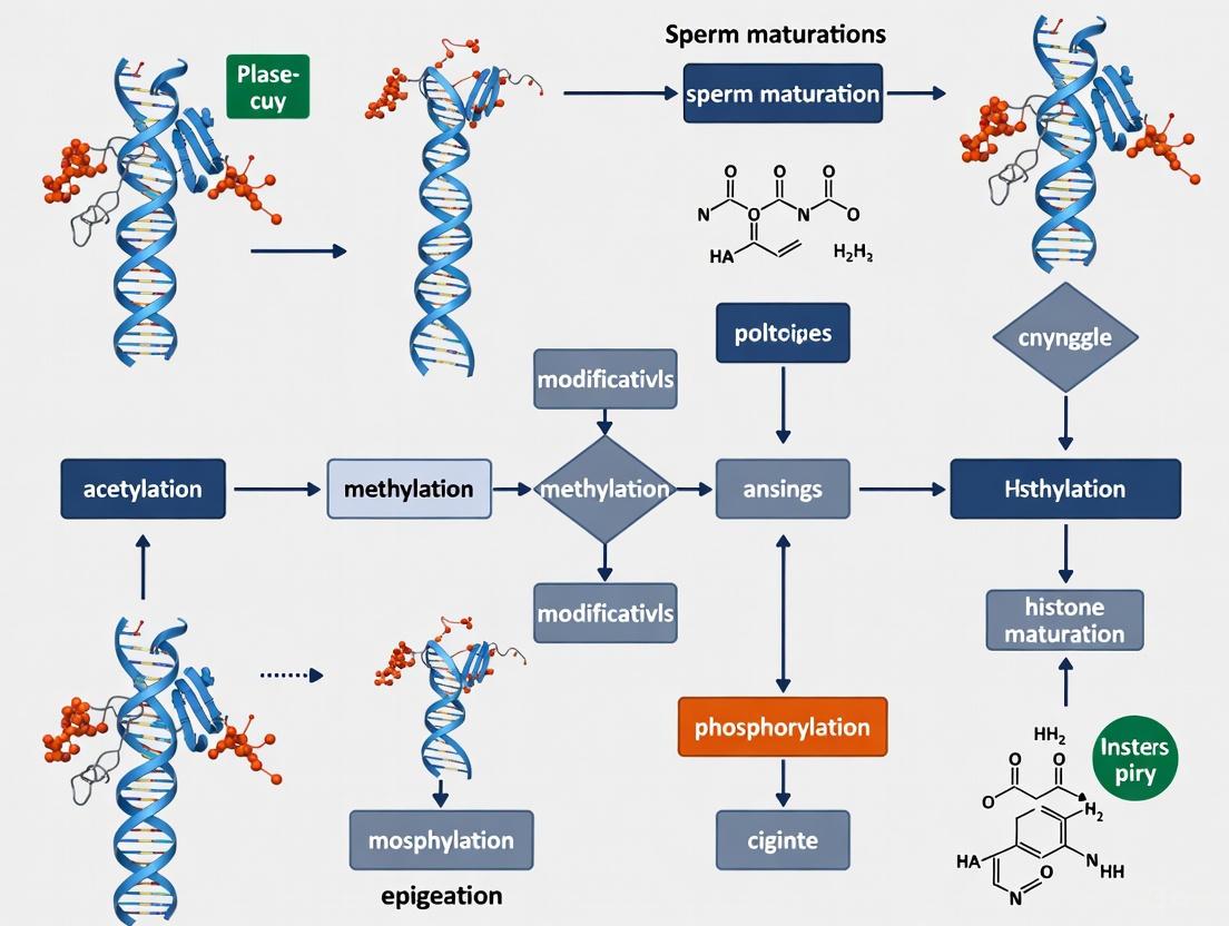
Histone Modifications in Sperm Maturation: Epigenetic Mechanisms, Dysregulation, and Clinical Implications
This article provides a comprehensive analysis of the critical role histone modifications play during sperm maturation, a process essential for male fertility and embryonic development.
Histone Modifications in Sperm Maturation: Epigenetic Mechanisms, Dysregulation, and Clinical Implications
Abstract
This article provides a comprehensive analysis of the critical role histone modifications play during sperm maturation, a process essential for male fertility and embryonic development. We explore the foundational biology of histone replacement and modification throughout spermatogenesis, detailing key marks such as H3K4me3, H4 hyperacetylation, and their regulation by specific enzymes. Methodologically, we examine cutting-edge profiling techniques like single-cell RNA-seq and ChIP-seq that are unveiling dynamic epigenomic landscapes. The content further addresses the clinical consequences of epigenetic dysregulation, linking aberrant histone patterns to azoospermia and other forms of male infertility, and evaluates the potential of histone modifiers as therapeutic targets. Finally, we discuss the emerging evidence of sperm histone marks serving as a template for embryonic gene expression, underscoring their intergenerational impact. This resource is tailored for researchers, scientists, and drug development professionals seeking to understand and intervene in epigenetic causes of male infertility.
The Epigenetic Blueprint: Core Mechanisms of Histone Modification in Spermatogenesis
Spermatogenesis is a highly ordered and complex developmental process through which spermatogonial stem cells (SSCs) differentiate into mature haploid spermatozoa. This transformation involves precise genetic and epigenetic programming to ensure the production of functionally competent sperm and the faithful transmission of paternal genetic information to the next generation [1] [2]. Central to this process is chromatin remodeling, a dramatic reorganization of the DNA-protein architecture that culminates in the extensive replacement of histones with protamines, enabling extreme nuclear compaction [3] [4]. This histone-to-protamine transition is not a simple exchange but a carefully orchestrated sequence of events regulated by histone modifications, the incorporation of testis-specific histone variants, and the activity of chromatin remodeling complexes [1] [5]. Understanding the dynamics of histone replacement is crucial, as perturbations in this process are strongly linked to male infertility and can impact early embryonic development [6] [2].
The Stages of Spermatogenesis
Spermatogenesis consists of three distinct yet continuous phases: the mitotic, meiotic, and spermiogenic phases. Each phase is characterized by unique cellular events and chromatin states.
Table 1: Key Stages of Spermatogenesis and Major Chromatin Events
| Stage | Cell Types | Key Chromatin Events | Major Epigenetic Regulators |
|---|---|---|---|
| Mitotic Phase | Spermatogonia (including SSCs) | - SSC self-renewal and differentiation- DNA methylation re-establishment | DNMT3A/B, PRMT5, H3.4 [7] [1] |
| Meiotic Phase | Spermatocytes (Preleptotene to Diplotene) | - Homologous recombination- Transient DNA demethylation- Expression of histone variants (e.g., H2B.W2) | H2B.W2, H3.4, DNA demethylation enzymes [7] [1] [8] |
| Spermiogenic Phase (Spermiogenesis) | Round to Elongating Spermatids | - Hyperacetylation of histones (e.g., H4)- Incorporation of transition proteins (Tnp1, Tnp2)- Global histone removal & protamine incorporation- Nuclear elongation and compaction | Chd5, HAT1 (Histone Acetyltransferase 1), SETD1B, Protamines (Prm1/2) [3] [9] [5] |
The process begins with the mitotic proliferation of SSCs, which either self-renew or commit to differentiation, giving rise to spermatogonia that undergo several mitotic divisions [1] [2]. These cells then enter the meiotic phase as spermatocytes, where DNA replication is followed by two rounds of division (meiosis I and II) to produce haploid round spermatids. This stage involves homologous chromosome pairing, synapsis, and genetic recombination [1]. The final phase, spermiogenesis, involves the most dramatic morphological and chromatin changes. Round spermatids undergo a remarkable transformation into elongated spermatozoa, involving acrosome formation, flagellum development, cytoplasmic removal, and the critical histone-to-protamine transition that enables extreme chromatin compaction essential for sperm function [3] [5].
Histone Variants and Modifications: Regulators of Chromatin Dynamics
The core histones (H2A, H2B, H3, H4) that package DNA into nucleosomes are dynamically replaced by testis-specific variants and modified by specific enzymes to facilitate chromatin remodeling.
Testis-Specific Histone Variants
Several histone variants are expressed in a stage-specific manner to modulate chromatin structure and function during spermatogenesis.
Table 2: Key Testis-Specific Histone Variants in Spermatogenesis
| Histone Variant | Expression Timing | Function and Characteristics | Phenotype of Dysfunction |
|---|---|---|---|
| H3.4 (H3T) | Pre-meiotic and meiotic | - Essential for entry into meiosis- Knock-out mice fail to enter meiosis | Meiotic arrest [7] |
| H2B.W1 (H2BFWT) | Mid-to-late spermatogonia (Primates) | - Localizes to telomeric regions- Destabilizes nucleosomes | Associated with male infertility in humans (SNPs) [7] |
| H2B.W2 (H2BFM) | Late spermatogonia to leptotene spermatocytes | - Induces a more relaxed nucleosome conformation |
Potential implications for sperm development and function [7] |
| H2B.C1 (TH2B) | Spermatocytes to mature sperm | - Replaces bulk canonical H2B in mature sperm | Spermatogenesis defects in mice when disrupted with H2ac1 [7] |
H2B.W2 is of particular interest due to its structural impact. Cryo-electron microscopy reveals that nucleosomes containing H2B.W2 adopt a more relaxed conformation compared to canonical nucleosomes. This is caused by weakened interactions between the outer DNA turn and the histone core. The N-terminal tail and α2 helix of H2B.W2 are critical for this nucleosome destabilization, and residue G73 in the L1 loop is key to disrupting higher-order chromatin structure [7].
Key Histone Modifications
Histone modifications act as molecular switches that recruit effector proteins to direct chromatin remodeling.
- Histone Hyperacetylation: A crucial step preceding histone displacement is the hyperacetylation of histone H4. This modification is believed to neutralizse the positive charge of histones, reducing their affinity for negatively charged DNA and facilitating nucleosome unwinding and subsequent eviction [4] [5]. The histone acetyltransferase Hat1 shows dynamic, stage-specific expression—highest in spermatogonia and sperm, intermediate in primary spermatocytes, and lowest in secondary spermatocytes—suggesting distinct roles in transcription, chromatin assembly, and DNA repair across different stages [9].
- Histone Methylation (H3K4me3): The trimethylation of histone H3 lysine 4 is a hallmark of active gene promoters. During spermiogenesis, the methyltransferase SETD1B establishes broad H3K4me3 domains at spermatid-specific genes. These broad domains, which overlap with enhancers marked by H3K27ac, are critical for orchestrating robust transcription and ensuring the precise temporal control of gene expression required for proper spermatid development. Disruption of this mechanism compromises transcription dosage and timing, ultimately impairing spermiogenesis [8].
- Repressive Marks: Repressive histone modifications like H3K9me2, H3K9me3, and H3K27me3 also show dynamic redistribution, particularly during the mitosis-to-meiosis transition and the completion of meiotic recombination. These changes correlate with the silencing of specific genes, retrotransposons, and mitotic/meiotic genes [8].
The Histone-to-Protamine Replacement Pathway
The replacement of histones by protamines is a cornerstone of spermiogenesis, enabling the paternal genome to be packaged into a highly compact, hydrodynamic, and transcriptionally inert state. The following diagram illustrates the core regulatory pathway and key molecular events orchestrating this transition.
This process is initiated by histone hyperacetylation, particularly of histone H4, which loosens chromatin structure [4] [5]. Subsequently, testis-specific histone variants like H2B.W2 are incorporated, further destabilizing the nucleosome architecture [7]. These changes create a chromatin landscape permissive for the action of chromatin remodelers like Chd5, which orchestrates the subsequent steps [5]. Chd5 mediates a cascade of events including nucleosome eviction, facilitates the repair of transient DNA strand breaks that occur during this massive restructuring, and modulates the homeostasis of transition proteins and protamines [5]. The histones are first replaced by transition proteins (Tnp1 and Tnp2), which are then themselves replaced by protamines (Prm1 and Prm2). Protamines package the sperm DNA into a highly condensed, stable toroidal structure that is essential for protecting the paternal genome during transit and for successful fertilization [3] [6] [5].
Advanced Research Methodologies and Reagents
Studying the intricate dynamics of histone replacement requires a suite of sophisticated experimental approaches. The following workflow outlines a multi-omics strategy for profiling the epigenomic landscape during spermatogenesis.
Experimental Protocols for Key Analyses
1. Nucleosome Reconstitution for Structural Studies (from [7])
- Purpose: To study the biophysical and structural properties of nucleosomes containing testis-specific histone variants like H2B.W2.
- Procedure:
- Histone Octamer Refolding: Mix equimolar amounts of human canonical histones (H2A, H2B/H2B.W2, H3, H4) in an unfolding buffer (6 M guanidinium chloride, 20 mM Tris-HCl pH 7.5, 4 mM DTT). Incubate at room temperature for 1 hour.
- Dialysis: Dialyze the mixture sequentially against water and then octamer buffer (2 M NaCl, 50 mM Tris-HCl pH 7.5, 0.5 mM EDTA, 5 mM β-mercaptoethanol) at 4°C.
- Nucleosome Assembly: Mix the refolded octamer with DNA containing the Widom 601 nucleosome-positioning sequence (e.g., 147 bp for cryo-EM) in a high-salt reconstitution buffer (2 M NaCl). Gradually decrease the salt concentration to 0 M via overnight dialysis to allow nucleosome formation.
- Verification: Verify successful reconstitution using native polyacrylamide gel electrophoresis (PAGE).
2. Purification of Histone Replacement-Completed Sperm (HRCS) (from [6])
- Purpose: To obtain a pure population of mature sperm free from contamination by sperm with incomplete histone-to-protamine replacement, which is crucial for accurate genomic mapping of retained nucleosomes.
- Procedure:
- Sperm Collection and Processing: Collect total sperm from the cauda epididymis and vas deferens. Mildly sonicate to separate sperm heads from tails.
- Density Gradient Centrifugation: Layer the sonicated sample onto an 82% Percoll solution and centrifuge. High-density, fully compacted HRCS form a pellet, while lower-density cellular debris and sperm tails remain in the supernatant.
- Quality Control: Assess purity using the Sperm Chromatin Structure Assay (SCSA), which measures DNA stainability with acridine orange. HRCS should show minimal high DNA stainability (HDS), indicating complete protamination. Western blotting can confirm a significant reduction in histone H3 content compared to total sperm.
The Scientist's Toolkit: Key Research Reagents
Table 3: Essential Reagents and Tools for Spermatogenesis Research
| Reagent / Tool | Function and Application | Example Use Case |
|---|---|---|
| Anti-H2B.W2 Antibody | Detects and localizes the H2B.W2 histone variant in testicular tissues. | Immunohistochemistry/Immunofluorescence on human testis sections to determine stage-specific expression [7]. |
| Widom 601 DNA Sequence | A high-affinity nucleosome positioning sequence for in vitro nucleosome reconstitution. | Used in nucleosome reconstitution assays for cryo-EM structural studies of H2B.W2 nucleosomes [7]. |
| Percoll Solution (82%) | A density gradient medium for purifying high-density, mature sperm heads. | Isolation of Histone Replacement-Completed Sperm (HRCS) for downstream chromatin analyses [6]. |
| Sperm Chromatin Structure Assay (SCSA) | Flow cytometry-based assay to assess sperm chromatin integrity and protamination status. | Diagnosing DNA fragmentation (DFI) and incomplete histone replacement (HDS) in sperm from mutant mice or infertile men [6] [5]. |
| Validated Chd5 Antibody | Specific immuno-detection of the Chd5 chromatin remodeler. | Immunofluorescence to define the spatiotemporal expression pattern of Chd5 during spermiogenesis in mouse testes [5]. |
Spermatogenesis represents one of the most dramatic examples of cellular differentiation, underpinned by equally profound epigenetic reprogramming. The precise, stage-specific coordination of histone variant incorporation, post-translational modifications, and the action of chromatin remodeling complexes like Chd5 is absolutely essential for the successful completion of the histone-to-protamine transition [7] [8] [5]. Disruptions in this finely tuned process, whether in histone variants (H2B.W1, H2B.W2), modifying enzymes (SETD1B, Hat1), or remodelers (Chd5), directly lead to defective sperm chromatin compaction, impaired sperm function, and male infertility [7] [9] [5]. Future research, leveraging advanced multi-omics and single-cell technologies, will continue to decipher the complex regulatory networks that govern this process, providing deeper insights into the etiology of male infertility and potential avenues for diagnostic and therapeutic intervention.
Sperm maturation, or spermiogenesis, is a complex differentiation process where spermatids undergo dramatic morphological changes and nuclear compaction to form functional spermatozoa. Central to this transformation is the extensive remodeling of chromatin, where histones are largely replaced by protamines to facilitate DNA condensation. Histone modifications serve as a critical regulatory layer in this process, orchestrating the precise timing and execution of chromatin reorganization without altering the underlying DNA sequence [10]. These covalent post-translational modifications (PTMs) to histone proteins constitute an "epigenetic code" that determines chromatin architecture and accessibility, thereby controlling gene expression programs essential for male germ cell development [11] [2].
The dynamic nature of histone modifications makes them particularly suited for regulating the profound structural changes required during spermatogenesis. Unlike genetic mutations, these epigenetic marks are reversible and can be rapidly added or removed by specific enzymes in response to developmental cues. This review focuses on three core histone modifications—acetylation, methylation, and phosphorylation—examining their specific roles, regulatory enzymes, and intricate crosstalk during sperm maturation. Understanding these mechanisms provides crucial insights into the etiology of male infertility and potential therapeutic targets for reproductive medicine [10] [2].
Histone Acetylation: Opening Chromatin for Transition
Mechanism and Enzymatic Regulation
Histone acetylation involves the addition of an acetyl group to the ε-amino group of lysine residues, catalyzed by histone acetyltransferases (HATs). This modification neutralizes the positive charge on lysine residues, weakening histone-DNA interactions and resulting in a more relaxed chromatin structure that facilitates transcription [11] [12]. The reverse reaction is mediated by histone deacetylases (HDACs), which remove acetyl groups and promote chromatin compaction [12].
Key HAT complexes play specialized roles during spermatogenesis. The HBO1 complex, particularly when associated with its BRPF2 subunit, preferentially catalyzes acetylation of histone H3 at lysine 14 (H3K14ac) and histone H4 at multiple lysine residues [11]. The JADE-HBO1 complex, in contrast, specifically targets histone H4 for acetylation [11]. Beyond canonical acetylation, the BRPF2-HBO1 complex can also catalyze other acylations, including propionylation, butyrylation, and crotonylation of histones H3 and H4 [11].
Functional Roles in Spermatogenesis
During spermatogenesis, histone acetylation marks are particularly abundant on specific lysine residues of histone H4. H4K5ac, H4K8ac, and H4K12ac appear in spermatogonia, spermatocytes, and elongating spermatids, where they are essential for nucleosome destabilization and remodeling [10]. These modifications facilitate the incorporation of transition proteins (TPs) during the histone-to-protamine exchange process [10].
H4K16ac emerges as a crucial modification in elongating spermatids, where it likely promotes an open chromatin configuration necessary for the massive structural reorganization occurring at this stage [10]. The dynamic regulation of H4K16ac is facilitated by the MOF acetyltransferase complex, which is recruited to chromatin through a mechanism involving ubiquitinated H2A (UbH2A) [10].
Table 1: Key Histone Acetylation Marks in Spermatogenesis
| Modification | Developmental Stage | Function | Regulatory Enzymes |
|---|---|---|---|
| H4K5ac | Spermatogonia to elongating spermatids | Nucleosome destabilization, TP incorporation | HBO1 complex |
| H4K8ac | Spermatogonia to elongating spermatids | Chromatin remodeling, TP incorporation | HBO1 complex |
| H4K12ac | Spermatogonia to elongating spermatids | Nucleosome destabilization | HBO1 complex |
| H4K16ac | Elongating spermatids | Chromatin relaxation, histone removal | MOF acetyltransferase |
Histone Methylation: Fine-Tuning Gene Expression
Mechanism and Enzymatic Regulation
Histone methylation occurs on both lysine and arginine residues through the action of histone methyltransferases (HMTs) and is reversed by histone demethylases (HDMs) [11]. Lysine methyltransferases (KMTs) can add one, two, or three methyl groups to a single lysine residue, while protein arginine methyltransferases (PRMTs) catalyze mono- or dimethylation of arginine residues [11] [13].
Notable methyltransferases include the SET-domain containing enzymes such as SUV39H1/2, G9a, GLP, SMYD3, SETDB1, and EZH2, as well as the non-SET domain enzyme DOT1L [11]. PRMTs are categorized into Type I (PRMT1, 3, 4, 6, 8), which generate asymmetric dimethylarginine, and Type II (PRMT5, 7), which produce symmetric dimethylarginine [11] [13]. The demethylation process is primarily mediated by two enzyme families: Jumonji C (JMJC) domain-containing proteins and lysine-specific demethylase (LSD) proteins [11].
Functional Roles in Spermatogenesis
Different histone methylation marks serve distinct functions during sperm maturation. H3K4me3 is present from spermatogonia to elongating spermatids, where it facilitates the recruitment of PYGO2, which in turn recognizes and binds HAT complexes to promote histone acetylation [10]. H3K4me3 also recruits PHF7, which catalyzes H2A ubiquitination to facilitate histone removal [10].
H3K9me marks (including mono-, di-, and trimethylation) regulate the expression of genes encoding transition proteins (Tnps) and protamines (Prms) in round and elongating spermatids [10]. H3K36me3 and H3K79me3 also contribute to regulating Tnps and Prms gene expression, with H3K79me3 specifically correlating with histone H4 hyperacetylation to coordinate the histone-to-protamine transition [10].
Table 2: Key Histone Methylation Marks in Spermatogenesis
| Modification | Developmental Stage | Function | Regulatory Enzymes |
|---|---|---|---|
| H3K4me3 | Spermatogonia to elongating spermatids | Recruits PYGO2 and PHF7 for H3 acetylation and H2A ubiquitination | SET1 family |
| H3K9me1/2/3 | Round and elongating spermatids | Regulates Tnps and Prms gene expression | SUV39H, G9a, SETDB1 |
| H3K36me3 | Spermatocytes and round spermatids | Regulates Tnps and Prms gene expression | SETD2 |
| H3K79me3 | Elongating spermatids | Correlates with H4 hyperacetylation, histone replacement | DOT1L |
Histone Phosphorylation: Signaling and Condensation
Mechanism and Enzymatic Regulation
Histone phosphorylation occurs on serine, threonine, and tyrosine residues through the action of kinases, with reversal by phosphatases [14] [12]. This modification is among the most dynamic histone PTMs and is integral to DNA damage signaling, oxidative stress response, and cell cycle control [12].
Key kinases involved include ATM, ATR, Aurora B, and MSK1, while phosphatases such as PP1 and PP2A remove phosphate groups [12]. A well-characterized phosphorylation mark is γ-H2AX (H2AX phosphorylated at Ser139), which marks DNA double-strand breaks and serves as a genotoxicity biomarker [12].
Functional Roles in Spermatogenesis
During spermatogenesis, histone phosphorylation plays crucial roles in both meiotic and post-meiotic stages. γ-H2AX is essential in spermatocytes and elongating spermatids, where it is required for normal quantities of H3, H4, and PRM2 precursor and intermediate forms [10]. This phosphorylation mark likely facilitates DNA repair during meiotic recombination and the extensive chromatin remodeling occurring in elongating spermatids.
H4S1 phosphorylation appears in spermatocytes, round spermatids, and elongating spermatids, where it is essential for chromatin compaction and concomitantly regulates histone accessibility [10]. Research comparing mouse and crab spermatogenesis has revealed that phosphorylation of H2A and H4 at serine 1 (HS1ph) decreases significantly as spermatids mature, suggesting that elimination of these phosphorylation marks is closely associated with spermatozoa maturity [14].
Crosstalk and Integrated Regulation
Epigenetic Coordination During Spermatogenesis
The three major histone modifications do not function in isolation but rather engage in complex crosstalk to coordinate chromatin dynamics during sperm maturation. This integrated regulation is particularly evident during the histone-to-protamine transition, where multiple modifications collaborate to facilitate the massive structural reorganization.
For instance, H3K4me3 recruits PHF7, which catalyzes H2A ubiquitination (UbH2A), subsequently leading to the recruitment of the MOF acetyltransferase complex that mediates H4K16ac [10]. This acetylation mark in turn promotes chromatin relaxation necessary for histone removal and transition protein incorporation. Similarly, H3K79me3 correlates with histone H4 hyperacetylation to regulate the histone-to-protamine transition [10].
Experimental Evidence from Model Systems
Comparative studies between mammalian and decapod crustacean models have provided insights into the conserved and divergent aspects of histone modification regulation. In both mice and crabs, H2A, H4, and HS1ph are localized in the nuclei of spermatocytes, round spermatids, and elongating spermatids [14]. However, while H2A and H4 marks show contrasting distributions between mouse condensed spermatozoa chromatin and crab non-condensed spermatozoa chromatin, HS1ph demonstrates similar dynamics, decreasing significantly during spermatogenesis in both species [14]. This suggests that HS1ph elimination is fundamentally linked to sperm maturity across evolutionary distant species.
Experimental Approaches and Methodologies
Detection Techniques for Histone Modifications
Advanced technologies have enabled detailed mapping of histone modifications during spermatogenesis. Chromatin Immunoprecipitation Sequencing (ChIP-Seq) represents a classical approach that uses modification-specific antibodies to immunoprecipitate chromatin fragments, followed by next-generation sequencing to map the genomic distribution of histone marks [12].
To address limitations of ChIP-seq in handling low-input samples typical of germ cell research, CUT&Tag (Cleavage Under Targets and Tagmentation) has emerged as a powerful alternative [12]. This method uses antibody-directed Tn5 transposase to simultaneously fragment and tag chromatin at modification sites, enabling high-resolution chromatin profiling from as few as 10 cells [12]. Its single-cell variant (scCUT&Tag) offers additional benefits in resolution, reproducibility, and signal-to-noise ratio for heterogeneous germ cell populations [12].
Research Reagent Solutions
Table 3: Essential Research Reagents for Histone Modification Studies
| Reagent/Technique | Function | Application Context |
|---|---|---|
| CUT&Tag | High-resolution mapping of histone marks from low cell inputs | Profiling H3K4me3, H3K27me3 in rare germ cell populations |
| Specific histone modification antibodies | Immunodetection of specific PTMs | IF, IHC, Western blot for H4K16ac, H3K9me3, γ-H2AX |
| HAT/HDAC inhibitors | Modulate acetylation levels | Functional studies of acetylation in spermatogenesis |
| KMT/KDM inhibitors | Alter methylation states | Investigating methylation roles in germ cell differentiation |
| Mass spectrometry | Comprehensive PTM profiling | Discovery of novel modifications in sperm maturation |
Visualization of Histone Modification Crosstalk During Spermatogenesis
Diagram 1: Coordinated histone modifications enable chromatin relaxation during spermiogenesis. This pathway shows how methylated H3K4 facilitates histone ubiquitination, which in turn promotes H4 acetylation, ultimately leading to chromatin relaxation necessary for histone replacement.
Diagram 2: Temporal regulation of key histone modifications during spermatogenesis. Different histone modifications peak at specific developmental stages to coordinate the complex process of sperm maturation, with phosphorylation marks decreasing as spermatozoa mature.
Histone acetylation, methylation, and phosphorylation represent interconnected regulatory mechanisms that collectively govern chromatin dynamics during sperm maturation. Acetylation generally promotes chromatin relaxation necessary for histone displacement, methylation fine-tunes gene expression programs and recruits additional chromatin modifiers, while phosphorylation contributes to DNA damage response and signaling pathways. The precise coordination of these modifications ensures the successful completion of spermatogenesis and the production of fertilization-competent sperm.
Dysregulation of these epigenetic mechanisms underlies various forms of male infertility, highlighting their clinical significance. Future research leveraging single-cell epigenomic technologies and advanced animal models will further elucidate the complex interplay between these modifications and their specific functions in distinct germ cell populations. Such insights may eventually lead to novel diagnostic and therapeutic approaches for male factor infertility based on epigenetic profiling and manipulation.
Histone Variants and Their Specific Roles in Germ Cell Development
Germ cell development represents one of the most complex and precisely regulated biological processes, requiring dynamic chromatin reorganization to ensure proper gene expression patterns and genomic stability. Histone variants, which replace canonical histones in a replication-independent manner, serve as critical regulators of chromatin dynamics throughout gametogenesis. This technical review comprehensively examines the roles of specific histone variants in germ cell development, with particular emphasis on spermatogenesis. We synthesize recent advances demonstrating how testis-specific variants—including H3.4 (H3T), H2A.L, TH2B, H2B.W, and others—orchestrate key developmental transitions such as meiotic progression, histone-to-protamine exchange, and the establishment of paternal epigenome architecture. By integrating quantitative data from proteomic studies, functional genetic analyses, and mechanistic insights from recent publications, this review provides a foundational resource for researchers investigating epigenetic regulation of fertility and developing targeted therapeutic interventions for male infertility.
The process of germ cell development encompasses a remarkable series of cellular transformations, from primordial germ cell specification through gamete maturation. These transformations require extensive chromatin remodeling to support stage-specific transcriptional programs, meiotic recombination, and ultimately the compaction of the paternal genome. Histone variants, defined as non-allelic isoforms of core histones that differ in primary amino acid sequence from their replication-coupled counterparts, play indispensable roles in these processes by conferring unique structural and functional properties to nucleosomes [15] [16].
Unlike canonical histones that are synthesized primarily during S-phase, histone variants are typically expressed throughout the cell cycle and often exhibit tissue-specific expression patterns, making them particularly suited for regulating long-term developmental processes such as gametogenesis [15]. In mammalian germ cells, specialized histone variants contribute to virtually every aspect of chromatin function, including DNA repair, chromosome segregation, transcriptional regulation, and genome packaging [15] [16]. The testis exhibits the most diverse repertoire of histone variants, reflecting the unique chromatin remodeling demands of spermatogenesis [15].
This review examines the specific functions of characterized histone variants throughout germ cell development, with emphasis on their mechanistic contributions to spermatogenesis. We integrate findings from recent loss-of-function studies, structural analyses, and epigenomic profiling to provide a comprehensive resource for researchers working in reproductive epigenetics, infertility diagnostics, and therapeutic development.
Molecular Characterization of Germ Cell Histone Variants
Classification and Expression Patterns
Histone variants are categorized based on their sequence divergence from canonical histones and their expression patterns throughout development. Table 1 summarizes the key histone variants with established roles in germ cell development, their expression dynamics, molecular functions, and phenotypic consequences of their disruption.
Table 1: Histone Variants in Mammalian Germ Cell Development
| Histone Variant | Expression Pattern | Molecular Function | Phenotype of Loss-of-Function | References |
|---|---|---|---|---|
| H3.4 (H3T) | Testis-specific, spermatids | Histone-to-protamine transition | Impaired spermiogenesis, male infertility | [15] |
| H2A.L (multiple isoforms) | Testis-specific (not in humans) | Histone-to-protamine transition; nucleosome destabilization | Defective chromatin compaction | [15] |
| TH2B (TS H2B.1) | Testis-specific, oocytes, zygotes | Histone-to-protamine transition; chromatin decondensation | Reduced reprogramming efficiency; developmental defects | [15] [16] |
| H2B.W (H2BFWT) | Testis-specific | Unknown function in spermatogenesis | Not determined | [15] |
| H2A.B | Testis, brain | Nucleosome destabilization; active transcription | Not determined in germ cells | [15] |
| H3.5 | Testis-specific | Histone-to-protamine transition | Not determined | [15] |
| H2A.X | Global expression | DNA damage response; chromatin remodeling | Genomic instability in germ cells | [15] |
| CENP-A | Global expression | Centromere identity; kinetochore assembly | Chromosome missegregation | [15] [16] |
| H3.3 | Global expression | Transcriptional activation; chromatin dynamics | Embryonic lethality; germ cell defects | [15] [16] |
| H2A.Z | Global expression | Developmental plasticity; gene regulation | Embryonic lethality; impaired differentiation | [16] |
Structural Features and Biochemical Properties
The functional specialization of histone variants stems from sequence variations that alter nucleosome properties. Several testis-specific variants feature C-terminal tail truncations or acidic patch modifications that reduce nucleosome stability, facilitating subsequent chromatin remodeling steps [15]. For example:
- H2A.L isoforms contain characteristic sequence alterations in their docking domain that impair H2A-H2B dimer stability within the nucleosome, promoting histone removal during spermiogenesis [15].
- TH2A and TH2B (testis-histone 2A and 2B) exhibit N-terminal and α-helical modifications that decrease DNA-histone binding affinity, creating a more open chromatin structure conducive to reprogramming in early embryos [16].
- H2A.B displays a shorter acidic patch and altered L1 loop structure, resulting in nucleosomes with weaker histone-DNA contacts and enhanced dynamics [15].
These structural modifications enable testis-specific variants to create specialized chromatin environments that support the unique functional requirements of germ cell development, particularly during the dramatic chromatin reorganization phases of meiotic prophase and spermiogenesis.
Stage-Specific Functions of Histone Variants in Spermatogenesis
Spermatogonial Stem Cell Maintenance and Differentiation
The spermatogonial population includes undifferentiated stem cells (Type A spermatogonia) and differentiating spermatogonia committed to the spermatogenic pathway. While the replication-independent incorporation of histone variants is less characterized in spermatogonia compared to later stages, recent evidence implicates several variants in maintaining the balance between self-renewal and differentiation:
- H3.3 is enriched at actively transcribed genes in spermatogonial stem cells, maintaining a transcriptionally permissive state [15] [16]. Its deposition by the HIRA chaperone complex facilitates rapid gene activation in response to differentiation signals.
- H2A.Z occupies promoters of both pluripotency genes and differentiation genes in undifferentiated spermatogonia, potentially maintaining a "poised" chromatin state that enables rapid transcriptional responses to environmental cues [16].
The transition from undifferentiated to differentiating spermatogonia involves extensive epigenetic reprogramming, including dynamic changes in histone variant incorporation that precede and facilitate the commitment to meiosis.
Meiotic Prophase and Recombination Regulation
During meiotic prophase I, germ cells undergo homologous recombination, synapsis, and chromosome segregation—processes that require specialized chromatin environments. Histone variants contribute to these events through several mechanisms:
- H2A.X becomes phosphorylated (γH2AX) in response to programmed double-strand breaks (DSBs) early in meiotic prophase, marking sites of recombination and facilitating the recruitment of DNA repair machinery [15]. In later stages, γH2AX associates with unsynapsed chromatin, including the sex body during meiotic sex chromosome inactivation.
- CENP-A maintains centromeric identity throughout meiosis, ensuring proper kinetochore assembly and chromosome segregation during both meiotic divisions [15] [16]. Its testis-specific expression and regulated deposition help preserve genomic stability in the male germline.
- H3.3 is incorporated at sites of active transcription in pachytene spermatocytes, maintaining gene expression despite widespread chromatin condensation [15].
Recent research has identified a critical role for histone modification crosstalk in meiotic progression. The histone demethylase KDM2A/FBXL11, which targets H3K36me1/2, is essential for proper meiotic progression in mice [17]. Kdm2a deficiency disrupts Polycomb-mediated gene repression and impairs chromosome synapsis and processing of meiotic double-strand breaks, leading to male infertility [17].
Spermiogenesis and Histone-to-Protamine Transition
The most extensive involvement of histone variants occurs during spermiogenesis, when round spermatids undergo dramatic morphological transformation into mature sperm. This process involves the sequential replacement of histones with transition proteins and subsequently with protamines, enabling extreme nuclear compaction. Testis-specific histone variants play instrumental roles in facilitating this transition:
- H3.4 (H3T) and H3.5 are incorporated in elongating spermatids, where they may serve as placeholders that facilitate the displacement of canonical histones [15].
- TH2B and H2A.L isoforms create destabilized nucleosomes that are more readily evicted during chromatin remodeling [15] [16]. Their expression peaks during the histone-to-protamine transition stage.
- H2B.W appears late in spermiogenesis and may persist in mature sperm at specific genomic loci, potentially serving a role in marking genes for early embryonic expression [15].
The coordinated action of these variants ensures the proper timing and fidelity of chromatin compaction while maintaining the epigenetic information that must be transmitted to the next generation.
Figure 1: Temporal Expression of Key Histone Variants During Spermatogenesis. Histone variants exhibit stage-specific incorporation throughout germ cell development, with different functional classes predominating at specific developmental transitions.
Chromatin Dynamics and Histone Variant Interplay
Coordination with Histone Modifications
Histone variants do not function in isolation but participate in complex interplay with histone post-translational modifications (PTMs) to regulate chromatin function. Specific variants can either promote or inhibit particular modifications, creating variant-specific PTM landscapes:
- H3.3 is frequently associated with active marks such as H3K4me3 and H3K36me3 at gene bodies, reinforcing its role in transcriptional activation [15] [8].
- H2A.Z-containing nucleosomes often colocalize with both active (H3K4me3) and repressive (H3K27me3) marks in embryonic stem cells, suggesting a role in maintaining developmental plasticity [16]. This bivalent chromatin state may be conserved in germline stem cells.
- H2A.X phosphorylation creates a platform for the accumulation of DNA damage response factors, demonstrating how variant incorporation can direct specific signaling pathways [15].
Recent research has revealed that the establishment of specialized chromatin domains during spermatogenesis requires coordinated action between histone variants and modifying enzymes. For instance, the histone demethylase KDM2A/FBXL11 controls Polycomb-mediated gene repression by removing H3K36me2 marks that would otherwise inhibit PRC2 activity [17]. This demethylation creates a permissive environment for H3K27me3 deposition and subsequent gene silencing during spermatogonial differentiation.
Nucleosome Stability and Chromatin Accessibility
A fundamental contribution of histone variants to germ cell development lies in their ability to modulate nucleosome stability and DNA accessibility. Table 2 compares the biophysical properties of major testis-specific histone variants and their functional consequences for chromatin dynamics.
Table 2: Biophysical Properties of Testis-Specific Histone Variants
| Histone Variant | Nucleosome Stability | Chromatin Compaction | DNA Accessibility | Functional Impact |
|---|---|---|---|---|
| Canonical H2A-H2B | High | Standard | Standard | Baseline nucleosome structure |
| H2A.L-H2B | Low | Reduced | Increased | Facilitates histone removal |
| H2A-TH2B | Very low | Greatly reduced | Greatly increased | Permits protamine exchange |
| H2A.B-H2B | Low | Reduced | Increased | Enhances transcriptional activity |
| H3.3-H4 | Standard | Standard | Slightly increased | Promotes transcriptional activation |
The biophysical properties outlined in Table 2 enable testis-specific variants to perform their specialized functions. For example, the reduced stability of H2A.L-containing nucleosomes lowers the energy barrier for histone displacement during the protamine transition, while H2A.B enhances transcription in spermatocytes by increasing DNA accessibility to RNA polymerase and transcription factors [15].
Experimental Approaches for Studying Histone Variants in Germ Cells
Histone Variant Profiling and Localization
Determining the genomic distribution and dynamics of histone variants in germ cells presents technical challenges due to cellular heterogeneity in testes and the low abundance of some variants. Key methodological approaches include:
- Chromatin Immunoprecipitation Sequencing (ChIP-seq) with variant-specific antibodies to map genome-wide distribution patterns. This approach has revealed variant enrichment at specific genomic features such as promoters, enhancers, or repetitive elements [8].
- Proteomic Analysis of acid-extracted histones from purified germ cell populations to quantify variant expression across developmental stages. Advanced mass spectrometry can detect and distinguish closely related variant sequences [15].
- Immunofluorescence and Immunohistochemistry on testis sections to determine cell-type specific localization and subnuclear distribution patterns [17].
Recent technical advances, including CUT&RUN and single-cell epigenomic methods, are overcoming previous limitations and providing unprecedented resolution for studying histone variant dynamics in rare germ cell populations.
Functional Genetic Approaches
Establishing causal relationships between histone variants and germ cell phenotypes requires precise genetic manipulation:
- Conditional Knockout Models enable tissue-specific and temporal control of gene inactivation, bypassing potential embryonic lethality associated with constitutive deletion of essential variants [17]. The Cre-loxP system has been successfully applied to study KDM2A function in spermatogenesis [17].
- CRISPR-Cas9 Mediated Genome Editing allows for rapid generation of variant-specific mutations in animal models or in germline stem cell cultures [18].
- RNA Interference in ex vivo testis tissue cultures or spermatogonial stem cell lines provides a complementary approach for acute protein depletion without genetic modification [18].
These functional approaches, combined with detailed phenotypic characterization ranging from fertility assessment to ultrastructural analysis, have established essential roles for numerous histone variants in spermatogenesis.
The Scientist's Toolkit: Essential Research Reagents
Table 3: Key Research Reagents for Investigating Histone Variants in Germ Cells
| Reagent Category | Specific Examples | Research Application | Technical Considerations |
|---|---|---|---|
| Antibodies | Anti-TH2B, Anti-H2A.X (phospho-S139), Anti-CENP-A, Anti-H3.3 | Variant detection in IF/IHC; ChIP-seq | Specificity validation crucial due to high sequence homology |
| Cell Markers | SSEA1 (blastoderm/ PGCs), c-Kit (differentiating spermatogonia), TRA98 (germ cells) | Germ cell population purification | Stage-specific markers enable isolation of homogeneous populations |
| Animal Models | Kdm2a floxed mice [17], H2A.Z conditional knockout | Functional genetic studies | Tamoxifen-inducible systems allow temporal control |
| Culture Systems | Chicken PGCLC induction [18], mouse spermatogonial cultures | In vitro mechanistic studies | Mimicking physiological microenvironment remains challenging |
Research Gaps and Future Directions
Despite significant advances in understanding histone variant functions in germ cell development, several important questions remain unresolved:
- The complete repertoire of histone variants in human germ cells and potential associations with idiopathic infertility require comprehensive characterization.
- Mechanisms governing the replication-independent deposition of testis-specific variants during spermatogenesis are poorly understood, with few variant-specific chaperones identified.
- The potential persistence of histone variants in mature sperm and their contribution to transgenerational epigenetic inheritance remains controversial and requires further investigation.
- Technological limitations in analyzing low-abundance variants in rare cell populations necessitate development of more sensitive epigenomic profiling methods.
Future research directions should include systematic functional characterization of unstudied testis-specific variants, investigation of variant interplay with non-coding RNAs in germ cells, and development of small molecule probes that selectively target variant-specific domains for potential contraceptive applications.
Histone variants represent fundamental components of the epigenetic machinery that governs germ cell development. Through their stage-specific expression, unique structural properties, and interactions with modifying enzymes, variants create specialized chromatin environments that enable the dramatic cellular transformations of gametogenesis. Testis-specific variants in particular facilitate the chromatin compaction required for sperm function while potentially preserving epigenetic information for embryonic development.
Continued technical innovations in epigenomic profiling, genetic manipulation, and high-resolution imaging will undoubtedly yield new insights into variant functions and mechanisms. Such advances will enhance our understanding of male infertility pathogenesis and may inform novel diagnostic and therapeutic approaches for clinical translation. The integration of histone variant biology into broader models of epigenetic regulation will ultimately provide a more complete understanding of how chromatin dynamics support the transmission of genetic information across generations.
The histone-to-protamine transition (HTP) is a fundamental epigenetic reprogramming event during spermiogenesis, wherein most nucleosomal histones are replaced first by transition proteins and then by protamines. This process facilitates the extreme compaction of the paternal genome, which is essential for producing functional sperm and ensuring male fertility. Recent research has illuminated the sophisticated regulatory mechanisms, including liquid-liquid phase separation and specific histone modifications, that govern this dramatic chromatin remodeling. This whitepaper synthesizes current knowledge on the HTP transition, detailing its molecular mechanisms, regulatory frameworks, and the profound implications of its dysregulation for male infertility, thereby providing a critical resource for researchers and clinical developers in reproductive medicine.
Spermatogenesis, the process of male gamete formation, culminates in spermiogenesis—the remarkable transformation of round haploid spermatids into mature spermatozoa. A cornerstone of this differentiation is the histone-to-protamine transition (HTP), a wholesale chromatin remodeling event critical for packaging the paternal genome into a highly condensed, hydrodynamic state [19] [20]. During the HTP, the nucleosome-based chromatin architecture is systematically dismantled; core histones are first replaced by transition proteins (TNP1 and TNP2), which are subsequently displaced by protamines (PRM1 and PRM2) [19]. This exchange neutralizes the DNA's negative charge, facilitating the formation of tightly compacted toroidal structures that protect the genetic material from physical and oxidative damage during transit [21] [20]. The HTP is not merely a structural necessity but is also embedded within the broader context of epigenetic reprogramming. A small, strategically retained subset of nucleosomes (1-15% in mammals) carries histone post-translational modifications (PTMs) believed to be essential for embryonic development and paternal epigenetic inheritance [22] [23]. Consequently, defects in the HTP are directly linked to male infertility phenotypes, including azoospermia, oligospermia, and teratozoospermia, underscoring its non-negotiable role in successful reproduction [24] [20].
Molecular Mechanisms of the Histone-to-Protamine Transition
The Core Process: A Staged Exchange
The HTP transition is a meticulously orchestrated, multi-stage process that ensures the continuous protection of paternal DNA during its repackaging.
Step 1: Histone Displacement and Transition Protein Incorporation. The initial displacement of core histones is facilitated by the prior incorporation of testis-specific histone variants (e.g., H2A.L.2, TH2A, TH2B), which form less stable nucleosomes and create an "open" chromatin configuration permissive for eviction [20]. This is followed by the incorporation of transition proteins TNP1 and TNP2. These proteins are highly basic but less so than protamines, serving as a molecular bridge during the exchange process. Research demonstrates that H2A.L.2 is particularly critical for loading TNP2 onto nucleosomes, effectively paving the way for subsequent protamine deposition [20].
Step 2: Protamine Deposition and Chromatin Compaction. The final stage involves the replacement of transition proteins with protamines. In humans and mice, this involves both protamine 1 (PRM1) and protamine 2 (PRM2), which are rich in arginine and cysteine residues. The arginine residues facilitate strong ionic binding with DNA's phosphate backbone, while cysteine residues form inter- and intra-molecular disulfide bonds that further stabilize the condensed chromatin in the mature sperm head [25] [21]. The proper Prm1/Prm2 ratio is critical for fertility, as imbalances lead to defective chromatin compaction and DNA damage [21].
The following diagram illustrates the coordinated sequence of events during the HTP transition.
Key Epigenetic Regulators and Modifications
The HTP is governed by a complex landscape of histone post-translational modifications that act as molecular signals to recruit effector proteins and initiate specific steps of the transition. The table below summarizes the principal histone modifications and their demonstrated roles in the HTP.
Table 1: Key Histone Modifications Regulating the HTP Transition
| Histone Modification | Function in HTP Transition | Experimental Evidence |
|---|---|---|
| H4 Hyperacetylation | Essential for initiating histone removal; destabilizes nucleosomes by neutralizing histone charge. | Inhibition of histone deacetylases (HDACs) with Trichostatin A (TSA) in Drosophila blocks histone removal and protamine transition [26]. |
| Broad H3K4me3 Domains | Orchestrates robust transcription and precise timing of spermatid-specific gene expression. | Setd1b knockout in mice depletes broad H3K4me3 domains, disrupts transcription timing, and impairs spermiogenesis [8]. |
| H3K9me3 | Associated with heterochromatin regions; must be evicted for proper HTP progression. | CCER1 forms nuclear condensates that are immiscible with H3K9me3+ heterochromatin, creating a compartment for the HTP [24]. |
| H2B Ubiquitination | Promotes the removal of testis-specific histone H2B variants. | Implicated in the eviction of histones to clear the way for transition proteins and protamines [24]. |
Regulatory Systems and Emerging Mechanisms
The Role of Liquid-Liquid Phase Separation (LLPS)
A groundbreaking discovery in the field is the role of liquid-liquid phase separation (LLPS) in orchestrating the HTP. The germline-specific protein CCER1 (coiled-coil glutamate-rich protein 1), which is rich in intrinsically disordered regions, has been shown to form phase-separated condensates in the nuclei of round-to-elongating spermatids [24]. These nuclear condensates are immiscible with H3K9me3-marked heterochromatin, effectively creating a dedicated biochemical compartment where the HTP is executed. Within these CCER1 condensates, key processes such as the upregulation of Tnp1/2 and Prm1/2 transcription and the mediation of essential histone epigenetic modifications occur. Loss-of-function mutations in human CCER1 were identified in patients with non-obstructive azoospermia (NOA) and were demonstrated to disrupt condensate formation in vitro, providing a direct mechanistic link between LLPS, the HTP, and male infertility [24].
Transcriptional and Post-Transcriptional Control
The precise timing of the HTP is underpinned by a rigorous transcriptional and post-transcriptional program.
- Transcriptional Regulation: The transcription factors RFX2 and CREM are master regulators of post-meiotic gene expression. RFX2, in particular, acts in concert with the histone methyltransferase SETD1B to establish broad H3K4me3 domains over spermatid-specific genes, ensuring their high-level expression at the correct developmental time [8].
- Post-Transcriptional Regulation: Although mRNAs for protamines are transcribed in round spermatids, their translation is repressed until the elongating spermatid stage. This delayed translation is crucial, as premature protamine synthesis would be catastrophic for chromatin integrity. The FXR1 protein, another protein capable of undergoing LLPS, has been implicated in the translational activation of stored mRNAs in spermatids [24].
The following diagram summarizes the integrated regulatory network controlling the HTP.
Experimental Analysis of the HTP Transition
Key Methodologies and Workflows
Investigating the HTP requires specialized techniques to analyze dynamic chromatin changes in a heterogeneous cell population. The following workflow outlines a standard integrated approach.
Table 2: Core Experimental Protocols for HTP Research
| Methodology | Application in HTP Research | Key Procedural Details |
|---|---|---|
| Germ Cell Purification | Obtain homogeneous populations of specific spermatogenic stages. | Use sequential enzymatic digestion (collagenase, trypsin) of testes followed by unit gravity sedimentation (STA-PUT method) to separate cells based on size and density [19] [8]. |
| Chromatin Immunoprecipitation (ChIP) | Map histone modifications and transcription factor binding. | Cross-link cells with formaldehyde, sonicate chromatin, immunoprecipitate with specific antibodies (e.g., anti-H3K4me3, H4ac), and sequence the bound DNA [8]. |
| Chromatin Fractionation Assay | Determine histone- vs. protamine-associated DNA sequences. | Treat sperm nuclei with DTT to reduce disulfide bonds, then with salt (0.65 M NaCl) to selectively extract histones. Digest with restriction enzymes; histone-bound DNA (HDNA) is released into supernatant, while protamine-bound DNA (PDNA) remains in the pellet [23]. |
| Single-Cell RNA Sequencing (scRNA-seq) | Profile transcriptional dynamics and identify cellular subpopulations in diseased states like azoospermia. | Create single-cell suspensions from testicular biopsies, capture cells, perform reverse transcription, and prepare libraries for sequencing to map the transcriptome of individual cells [27]. |
The Scientist's Toolkit: Essential Research Reagents
A successful research program in HTP mechanics relies on a suite of critical reagents and model systems.
Table 3: Essential Research Reagents and Models for HTP Studies
| Reagent / Model | Function and Utility | Specific Application Example |
|---|---|---|
| CCER1 Antibody | Detects localization and expression of the LLPS scaffold protein CCER1. | Used in immunofluorescence to show CCER1's nuclear condensates are immiscible with H3K9me3+ heterochromatin in spermatids [24]. |
| Setd1b Knockout Mice | Model to study the role of broad H3K4me3 domains in transcriptional timing. | Revealed that loss of Setd1b depletes broad H3K4me3, disrupts gene expression timing, and causes spermiogenesis defects [8]. |
| HDAC Inhibitors (e.g., TSA) | Chemical tools to inhibit histone deacetylation and probe H4ac function. | Demonstrated in Drosophila spermatids that blocking deacetylation prevents the histone-to-protamine switch [26]. |
| Protamine-EGFP Plasmids | Allows overexpression of protamines in somatic cells to study their condensation effects. | Expression in HEK293T cells caused nuclear condensation and reduction of specific histone modifications (H3K9me3, H3K27ac) [25]. |
| PRM1 and PRM2 qPCR Assays | Quantifies protamine mRNA transcripts to assess expression ratios. | Analysis of human sperm revealed an abnormal PRM1/PRM2 mRNA ratio is correlated with poor sperm quality and infertility [21]. |
Clinical Implications and Therapeutic Insights
Dysregulation of the HTP is a significant etiological factor in male infertility. Mutations in genes directly involved in the process, such as CCER1, have been identified in men with non-obstructive azoospermia (NOA), effectively linking failure in HTP execution to the most severe form of infertility [24]. Beyond genetic lesions, aberrant patterns of histone modifications serve as biomarkers of fertility status. Single-cell RNA-seq analyses of testicular tissues from NOA patients reveal significant upregulation of histone modification-related genes like HDAC2 in specific somatic cell populations (Leydig, peritubular myoid cells), suggesting a disrupted microenvironment that impairs spermatogenesis [27]. Furthermore, the quantitative analysis of sperm protamine mRNA transcripts (specifically the PRM1/PRM2 ratio) presents a potential diagnostic tool for assessing sperm chromatin integrity and predicting outcomes for assisted reproductive technologies [21]. The essential role of H4 hyperacetylation, demonstrated in model organisms from Drosophila to mice, underscores this pathway as a conserved and potential therapeutic target for modulating histone removal [26]. A detailed understanding of the HTP, particularly the role of LLPS and transcriptional timing, opens new avenues for diagnosing and potentially treating idiopathic male infertility.
Spermatogenesis is a complex developmental process that relies on precise epigenetic regulation to ensure the production of functional sperm and the faithful transmission of genetic information. Histone post-translational modifications (PTMs) represent a critical layer of this regulation, dynamically controlling gene expression patterns throughout male germ cell development [1]. These chemical modifications on histone proteins do not alter the DNA sequence itself but create a heritable "epigenetic code" that governs chromatin structure and DNA accessibility [12]. The orchestration of this code is managed by three fundamental classes of enzymatic regulators: "writers" that add modifications, "erasers" that remove them, and "readers" that interpret these marks and recruit effector proteins [28]. In the context of sperm maturation, the precise balance between these regulators is essential for proper spermatogonial stem cell maintenance, meiotic division, and the dramatic chromatin remodeling that occurs during spermiogenesis [1] [29]. Disruptions to this delicate equilibrium can lead to spermatogenic failure and male infertility, highlighting the clinical relevance of understanding these mechanisms [27].
Core Machinery: Writers, Erasers, and Readers
The dynamic nature of histone modifications is governed by specialized enzymes and protein domains that establish, remove, and interpret the epigenetic code.
Writers: Depositing Histone Marks
Histone-modifying enzymes known as "writers" catalyze the addition of chemical groups to specific residues on histone tails. These enzymes exhibit remarkable specificity for both the histone residue and the type of modification added [28].
Table 1: Major Histone Modification Writers and Their Functions
| Modification Type | Enzyme Class | Key Examples | Primary Functions |
|---|---|---|---|
| Acetylation | Histone Acetyltransferases (HATs) | p300/CBP, Gcn5, PCAF, Tip60 | Transcriptional activation, chromatin relaxation, DNA repair [30] [28] |
| Methylation | Histone Lysine Methyltransferases (KMTs) | Suv39h1, EZH2, SETD1B, MLL | Transcriptional activation/repression, genomic imprinting, heterochromatin formation [30] [8] |
| Methylation | Protein Arginine Methyltransferases (PRMTs) | PRMT1, PRMT5, CARM1 | Transcriptional activation and repression [30] |
| Phosphorylation | Kinases | Aurora B, MSK1, ATM, ATR | Mitosis/meiosis, DNA damage response, immediate-early gene activation [30] |
| Ubiquitination | E3 Ubiquitin Ligases | Ring2 | Transcriptional regulation, spermatogenesis, meiosis [30] |
Erasers: Removing Histone Marks
"Eraser" enzymes catalyze the removal of histone modifications, providing reversibility and dynamic control to the epigenetic landscape [28].
Table 2: Major Histone Modification Erasers and Their Functions
| Modification Type | Enzyme Class | Key Examples | Primary Functions |
|---|---|---|---|
| Deacetylation | Histone Deacetylases (HDACs) | HDAC1, HDAC2, HDAC3, Sirtuins (SIRTs) | Transcriptional repression, chromatin compaction [28] [27] |
| Demethylation | Lysine Demethylases (KDMs) | KDM1A/LSD1, KDM4A-D, KDM5A-D, KDM6A | Transcriptional activation/repression, pluripotency, development [28] [31] |
| Dephosphorylation | Phosphatases | PP1, PP2A | Mitotic exit, reversal of DNA damage signaling [30] |
Readers: Interpreting the Histone Code
"Reader" proteins contain specialized domains that recognize and bind to specific histone modifications. This binding recruits additional protein complexes that execute downstream functions such as transcriptional activation or chromatin compaction [31]. The recognition is highly specific; for example, the tandem Tudor domain of KDM4A recognizes H3K4me3, while the chromodomain of Clr4 binds to H3K9me3 [31].
Functional Coupling and Regulatory Mechanisms
The activities of writers, erasers, and readers are not isolated but are functionally coupled to propagate and maintain specific chromatin states. This coupling can occur within a single polypeptide or through multi-protein complexes.
Diagram 1: Functional coupling between readers and writers. This diagram illustrates two mechanisms by which reader domains regulate writer activity to propagate chromatin states. Intra-polypeptide coupling occurs within a single protein like Clr4, where its chromodomain reads its own product (H3K9me3) to stimulate further methylation on adjacent nucleosomes [31]. Inter-polypeptide coupling occurs within complexes like PRC2, where the EED subunit reads H3K27me3 and allosterically activates the EZH2 writer subunit [31].
A notable example of this regulatory coupling is the activation of PRC2 by its product. The EED subunit of the PRC2 complex recognizes the H3K27me3 mark created by the catalytic EZH2 subunit. This binding allosterically stimulates PRC2's methyltransferase activity by approximately 7-fold, creating a positive feedback loop that promotes the spreading of this repressive mark along chromatin [31].
Experimental Methodologies for Investigating Histone Modifications
Studying the dynamic landscape of histone modifications requires sophisticated techniques capable of mapping these marks with high sensitivity and resolution, especially in the context of limited clinical samples like testicular biopsies.
Key Profiling Technologies
Chromatin Immunoprecipitation Sequencing (ChIP-seq) is the classical method for genome-wide mapping of histone modifications. It utilizes modification-specific antibodies to immunoprecipitate chromatin fragments, followed by next-generation sequencing [12]. However, its requirement for high cell input limits its use for rare cell populations.
CUT&Tag (Cleavage Under Targets and Tagmentation) represents a significant advancement, enabling high-resolution chromatin profiling from as few as 10 cells [12]. This method uses antibody-directed Tn5 transposase to simultaneously fragment and tag chromatin at modification sites, offering superior signal-to-noise ratio compared to ChIP-seq [12]. Its single-cell variant (scCUT&Tag) further allows for the dissection of epigenomic heterogeneity within complex tissues like the testis [12].
Liquid Chromatography-Tandem Mass Spectrometry (LC-MS/MS) provides a non-antibody-based approach for systematic mapping of PTM dynamics, enabling the identification and quantification of multiple modifications simultaneously [32].
Activity Assays for Enzymatic Regulators
Functional characterization of writers and erasers requires specific activity assays:
- HAT Activity Assays measure the transfer of acetyl groups from Acetyl-CoA to histone peptides, typically by detecting the reaction byproduct CoA-SH through colorimetric or fluorometric methods [28].
- HDAC Activity Assays utilize acetylated peptide substrates with fluorophore-quencher pairs. Deacetylation allows peptidase cleavage, releasing the fluorophore and generating a measurable fluorescent signal [28].
- HMT Activity Assays have traditionally used radioactive ³H-SAM as a methyl donor, but modern assays employ antibodies specific to the methylated product for safer, colorimetric/fluorometric detection without radioactivity [28].
- KDM Activity Assays previously measured formaldehyde formation but now directly detect demethylated products with significantly increased sensitivity (20–1,000 fold) and accuracy [28].
Table 3: The Scientist's Toolkit: Key Research Reagents and Solutions
| Reagent/Kit Type | Specific Examples | Primary Application & Function |
|---|---|---|
| Activity Assay Kits | Colorimetric/Fluorometric HAT, HDAC, HMT, KDM Assay Kits | Quantifying enzyme activity in nuclear extracts or purified proteins without radioactivity [28] |
| Specific Inhibitors | Trichostatin A (TSA; HDAC inhibitor), SIRT inhibitors | Probing biological functions of specific modifications; validating druggable targets [28] |
| Modified Histone Peptides | H3K4me3 peptides, H3K9ac peptides, H3K27me3 peptides | Serving as substrates for activity assays or for binding studies with reader domains [28] |
| Specific Antibodies | Anti-H3K4me3, Anti-H3K27ac, Anti-HDAC2, Anti-EZH2 | Immunostaining, western blotting, and enrichment for ChIP-seq/CUT&Tag [27] [8] |
Histone Modifications in Spermatogenesis and Male Infertility
During sperm maturation, histones are largely replaced by protamines to achieve extreme nuclear compaction [29]. However, approximately 2-15% of histones are retained in mature sperm, and these are strategically positioned at genomic loci crucial for embryonic development, including gene promoters and imprinted regions [29]. The modifications on these retained histones carry epigenetic information that can influence embryonic gene expression after fertilization.
Recent single-cell RNA sequencing studies of testicular tissues from men with non-obstructive azoospermia (NOA) have revealed significant dysregulation of histone-modifying enzymes. HDAC2, a pivotal eraser of acetyl marks, shows significant upregulation in NOA patients, and genes involved in histone modification processes are markedly enriched in Leydig cells, peritubular myoid cells, and macrophages in NOA testes [27]. This suggests that aberrant histone modification in somatic support cells can indirectly disrupt the spermatogenic microenvironment.
Specific histone modifications play distinct roles during spermatogenesis:
- H3K4me3: The H3K4 methyltransferase SETD1B establishes broad H3K4me3 domains that control the robust transcription and precise timing of gene expression required for spermatid development. Disruption of this mechanism compromises transcription dosage and timing, impairing spermiogenesis [8].
- H3K27me3: This repressive mark, deposited by EZH2, shows remarkable dynamics during the mitosis-to-meiosis transition and is crucial for proper meiotic progression [8].
- H3K9me2/3: These heterochromatin marks correlate with the silencing of retrotransposons and meiotic genes, ensuring genomic stability during gametogenesis [8].
The functional significance of these regulatory mechanisms is underscored by the severe phenotypic consequences of their disruption. For instance, germ cell-specific deletion of Sirt1 (a class III HDAC) in male mice disrupts chromatin condensation during gametogenesis due to reduced H4 hyperacetylation, leading to significantly reduced fertility [27]. Similarly, mutations in the transcriptional repressor Zmynd15, which functions through histone deacetylase recruitment, result in male infertility in mice [27].
The enzymatic regulators of histone modifications—writers, erasers, and readers—constitute a sophisticated control system that orchestrates the profound chromatin remodeling and stage-specific gene expression patterns essential for sperm maturation. The functional coupling between these regulators enables the establishment, maintenance, and dynamic reversal of chromatin states that guide germ cell development from spermatogonia to mature sperm.
The growing recognition that retained sperm histones carry epigenetic information with potential impacts on embryonic development and offspring health underscores the clinical importance of this field. Future research will likely focus on developing more sensitive profiling technologies for limited cell numbers, further elucidating the crosstalk between different histone marks and with other epigenetic mechanisms like DNA methylation, and translating this knowledge into novel diagnostic and therapeutic strategies for male infertility. The identification of specific imbalances in histone modification pathways in infertile men offers promising targets for the development of much-needed therapeutic interventions.
Mapping the Epigenome: Advanced Profiling and Analytical Techniques
Single-Cell RNA Sequencing for Cell-Type Specific Epigenetic Analysis
Single-cell RNA sequencing (scRNA-seq) has emerged as a transformative technology for deciphering cellular heterogeneity, gene regulatory networks, and developmental trajectories at unprecedented resolution. While traditional bulk RNA sequencing averages gene expression across thousands of cells, obscuring rare cell types and continuous transitions, scRNA-seq enables the profiling of transcriptomes in individual cells, revealing the full complexity of tissues and dynamic biological processes [33] [34]. This capability is particularly valuable for investigating epigenetic regulation—the molecular mechanisms that modulate gene expression without altering the DNA sequence itself—within complex biological systems.
Nowhere is this more evident than in the study of spermatogenesis, a highly coordinated and complex differentiation process where precise epigenetic reprogramming, including dynamic histone modifications, is critical for successful sperm maturation and male fertility [35] [36]. During spermiogenesis, spermatids undergo dramatic chromatin remodeling, where most histones are replaced by protamines, enabling extreme nuclear compaction [35]. This process is governed by a cascade of histone modifications and involves specialized RNA-binding proteins. scRNA-seq provides the necessary resolution to dissect these dynamic and cell-type-specific epigenetic programs, uncovering molecular mechanisms that would remain hidden in bulk analyses.
Technical Foundations of Single-Cell RNA Sequencing
Core Technological Principles
scRNA-seq measures the whole transcriptome of individual cells, allowing researchers to investigate cellular heterogeneity, identify rare cell populations, and reconstruct developmental trajectories [33] [34]. The fundamental workflow begins with the isolation of single cells or nuclei from a tissue sample of interest. This is followed by cell lysis, capture of mRNA molecules, reverse transcription into cDNA, amplification, library preparation, and next-generation sequencing.
A critical innovation in scRNA-seq protocols is the use of cellular barcodes and unique molecular identifiers (UMIs). Cellular barcodes are short nucleotide sequences added to all transcripts from a single cell during reverse transcription, enabling the computational attribution of sequenced reads back to their cell of origin after multiplexed sequencing. UMIs are random nucleotide sequences that uniquely tag individual mRNA molecules, allowing for the accurate quantification of transcript abundance by correcting for amplification bias [34].
Major Platform Technologies
Table 1: Comparison of Major scRNA-seq Platform Technologies
| Platform Type | Key Features | Example Technologies | Typical Cell Throughput | Considerations |
|---|---|---|---|---|
| Droplet-Based | Microfluidics partitions cells into nanoliter-scale droplets | 10x Genomics Chromium (GEM-X technology) | 80K to 960K cells per run [33] | High throughput, requires specialized instrument |
| Combinatorial Indexing | Cells are labeled in a split-pool approach without specialized equipment | Parse Biosciences Evercode | Virtually unlimited (scalable) [37] | No instrument cost, flexible sample processing, suitable for fixed samples |
| Plate-Based | Cells are individually sorted into multi-well plates | SMART-seq technologies | Hundreds to thousands of cells | Lower throughput, but higher sensitivity and full-length transcript coverage |
Droplet-based methods, such as the 10x Genomics Chromium system, utilize microfluidic chips to combine single cells, barcoded gel beads, and reverse transcription reagents into tiny, oil-encapsulated reaction vesicles called Gel Beads-in-emulsion (GEMs). Within each GEM, cell lysis and barcoding occur, ensuring all cDNA from a single cell shares the same barcode [33]. The emergence of GEM-X technology has further improved efficiency by generating twice as many GEMs at smaller volumes, reducing multiplet rates and increasing throughput capabilities [33].
In contrast, combinatorial indexing approaches, such as Parse Biosciences' Evercode technology, forgo specialized instrumentation entirely. This method uses a series of well-based reactions to add barcodes to cells in a fixed and permeabilized state through a split-pool process, offering exceptional scalability and flexibility for processing samples across different time points [37].
Experimental Design and Workflow
A well-designed scRNA-seq experiment requires careful planning at each step, from sample preparation to data interpretation. The following workflow diagram and subsequent sections detail the critical phases.
Sample Preparation and Quality Control
The foundation of a successful scRNA-seq experiment is the preparation of a high-quality single-cell or single-nucleus suspension. Tissues must be carefully dissociated using enzymatic or mechanical methods appropriate for the specific tissue type, with the goal of maximizing cell viability while preserving transcriptomic integrity [34]. For epigenetic studies focusing on processes like spermatogenesis, where cells exhibit diverse morphologies and sensitivities, optimization of dissociation protocols is paramount.
Rigorous quality control (QC) is performed before proceeding to library preparation. Cell viability and concentration are typically assessed using automated cell counters or flow cytometry. QC metrics should be carefully recorded, as low viability can lead to excessive background from ambient RNA released by dead cells. The preparation of high-quality cell suspensions is critical, as the adage "garbage in, garbage out" strongly applies to single-cell genomics [33].
From Barcoding to Sequencing
Once a high-quality cell suspension is obtained, the subsequent steps—cell barcoding, library preparation, and sequencing—are often performed using integrated commercial kits that ensure protocol consistency. As described in Section 2.2, the choice of platform (droplet-based vs. combinatorial indexing) dictates the specific workflow.
Following cDNA synthesis and amplification, libraries are prepared for sequencing. This involves fragmentation, size selection, and the addition of platform-specific adapters. Sequencing is typically performed on Illumina instruments, with recommended depth varying by application. For standard cell type identification, 20,000-50,000 reads per cell may suffice, while detecting subtle transcriptional differences or splicing variants often requires 100,000 or more reads per cell.
Data Analysis Pipeline and Tools
The computational analysis of scRNA-seq data is a multi-step process that transforms raw sequencing data into biological insights. Current best practices encompass both standard preprocessing and advanced analytical techniques for uncovering epigenetic regulators.
Essential Preprocessing and Quality Control
The initial computational steps are critical for ensuring data quality and reliability:
- Raw Data Processing: Tools like Cell Ranger (10x Genomics) process raw sequencing data (FASTQ files) through alignment to a reference genome, demultiplexing based on cellular barcodes, and UMI counting to generate a gene expression matrix (cells × genes) [33] [38].
- Quality Control and Filtering: Cells are filtered based on three key metrics [38]:
- Count Depth: The total number of molecules detected per cell.
- Number of Genes: The unique genes detected per cell.
- Mitochondrial Fraction: The percentage of reads mapping to mitochondrial genes. Low-count cells or those with high mitochondrial content often represent damaged or dying cells, while cells with exceptionally high counts may be multiplets (doublets or triplets). As shown in the diagram below, these QC metrics must be considered jointly to avoid filtering out valid cell populations unintentionally [38].
Core Analytical Steps for Cell-Type Specific Analysis
Following quality control, several analytical steps enable the identification of cell types and states:
- Normalization and Batch Correction: Normalization accounts for technical variations in sequencing depth between cells. When integrating multiple datasets or samples, batch effect correction methods such as Harmony, Seurat's CCA, or fastMNN are essential to remove technical artifacts while preserving biological variation [39] [40].
- Feature Selection and Dimensionality Reduction: Highly variable genes that drive heterogeneity across cells are selected for downstream analysis. Dimensionality reduction techniques, most commonly Principal Component Analysis (PCA), are applied to reduce noise and computational complexity [38].
- Clustering and Visualization: Cells are grouped into clusters based on transcriptional similarity using graph-based clustering algorithms (e.g., Louvain, Leiden). The resulting clusters are visualized in two dimensions using methods like t-SNE or UMAP, which help illustrate relationships between cell populations [38] [40].
- Differential Expression and Marker Gene Identification: For each cluster, differentially expressed genes are identified that serve as marker genes for that specific cell population. These markers facilitate the annotation of cell types based on known biological knowledge.
Advanced Analysis: Trajectory Inference and Epigenetic Regulator Detection
Beyond static cell type identification, scRNA-seq enables the investigation of dynamic processes through trajectory inference (pseudotime analysis). Tools like Monocle, SCANPY, and scVelo model transcriptional changes along developmental continua, ordering cells based on their progression through biological processes rather than discrete clusters [35] [39]. This is particularly powerful for studying continuous processes like spermatogenesis, where germ cells transition through defined but overlapping developmental stages.
To specifically investigate epigenetic regulation, researchers can:
- Identify differentially expressed histone-modifying enzymes (methyltransferases, demethylases, acetyltransferases, deacetylases) across cell types.
- Analyze the expression of histone variants across cell populations.
- Correlate the expression of epigenetic regulators with potential target genes.
- Integrate scRNA-seq data with complementary epigenetic datasets.
Table 2: Key Analytical Tools for scRNA-seq Data Analysis
| Tool Name | Primary Function | Key Features | Best For |
|---|---|---|---|
| Cell Ranger [33] | Raw Data Processing | Processes 10x Genomics FASTQ files, performs alignment, barcode counting, and generates expression matrices | Getting started with 10x Genomics data |
| Seurat [38] | Comprehensive Analysis | R-based toolkit for QC, normalization, clustering, differential expression, and trajectory analysis | End-to-end analysis in R programming environment |
| Scanpy [38] | Comprehensive Analysis | Python-based toolkit with similar functionality to Seurat for large-scale data | End-to-end analysis in Python programming environment |
| ScType [39] | Automated Cell Type Annotation | Reference-based algorithm for automated cell type identification | Rapid, accurate cell type annotation |
| scVelo [35] | RNA Velocity & Dynamics | Models transcriptional dynamics and predicts future cell states | Analyzing developmental trajectories and directionality |
| Trailmaker [39] | Cloud-Based Analysis | No-code platform with automated workflow, supports multiple technologies | Researchers without coding expertise |
| Deep Visualization (DV) [40] | Advanced Visualization | Preserves data structure, corrects batch effects, uses Euclidean/hyperbolic spaces | Handling complex batch effects, dynamic data |
Integration with Epigenetic Analysis in Spermatogenesis Research
The true power of scRNA-seq for epigenetic research is realized when it is integrated with complementary epigenetic assays. This multi-omic approach enables the direct correlation of transcriptional states with epigenetic modifications in the same biological system.
Multi-Omic Technologies for Coordinated Transcriptomic and Epigenetic Profiling
Emerging technologies now enable the simultaneous measurement of multiple molecular layers from the same single cell:
- scEpi2-seq: A recently developed method that provides joint readout of histone modifications (H3K9me3, H3K27me3, H3K36me3) and DNA methylation in single cells. This technique uses TET-assisted pyridine borane sequencing (TAPS) for bisulfite-free methylation detection while simultaneously capturing histone modification patterns via antibody-tethered MNase [41]. Application of this technology has revealed how DNA methylation maintenance is influenced by local chromatin context in intestinal cell type specification [41].
- CITE-seq & ASAP-seq: These technologies enable the simultaneous measurement of transcriptome with surface proteins (CITE-seq) or chromatin accessibility (ASAP-seq), providing complementary layers of epigenetic information.
For researchers without access to these advanced multi-omic platforms, a computational integration approach can be employed, where scRNA-seq data is combined with bulk or single-cell epigenetic datasets from matched biological systems to infer regulatory relationships.
Application to Spermatogenesis: Uncovering Epigenetic Regulators
In spermatogenesis research, scRNA-seq has been instrumental in identifying and validating key epigenetic regulators. A prime example is the discovery of RNA helicase DDX43 as an essential regulator of chromatin remodeling during spermiogenesis. Through scRNA-seq analysis of testis-specific Ddx43 knockout mice, researchers demonstrated that DDX43 regulates dynamic RNA processing underlying spermatid chromatin remodeling and differentiation [35]. The study combined scRNA-seq with enhanced crosslinking immunoprecipitation and sequencing (eCLIP-seq) to identify direct targets of DDX43, including Elfn2 as a hub gene in the chromatin remodeling pathway [35].
Similarly, integration of microarray and scRNA-seq data has identified key histone modification genes associated with spermatogonial stem cell (SSC) function and aging, including KDM5B, SCML2, SIN3A, and ASXL3, which play significant roles in chromatin remodeling and gene regulation [36]. Protein-protein interaction networks from these integrated analyses revealed critical biological processes such as chromatin organization, histone demethylation, and chromosome structure maintenance in SSCs [36].
The Scientist's Toolkit: Essential Research Reagents and Materials
Table 3: Key Research Reagent Solutions for scRNA-seq in Epigenetic Studies
| Reagent Category | Specific Examples | Function in Experimental Workflow |
|---|---|---|
| Single-Cell Isolation Kits | 10x Genomics Chromium Next GEM Kits; Parse Biosciences Evercode Whole Transcriptome Kit | Partition individual cells into droplets or wells for barcoding and reverse transcription |
| Cell Fixation & Preservation Reagents | Parse Biosciences Fixation Buffer; 10x Genomics Fixation Kits | Preserve cell states for later processing, enable time-course experiments |
| Epigenetic Target Enrichment Reagents | scEpi2-seq Antibody-MNase Fusion Proteins (H3K9me3, H3K27me3, H3K36me3) [41] | Target specific histone modifications for multi-omic profiling |
| Nucleic Acid Conversion Reagents | TET-assisted pyridine borane sequencing (TAPS) reagents [41] | Convert methylated cytosine to uracil for detection while preserving barcodes |
| Library Preparation Kits | Illumina Nextera XT; Platform-specific library prep kits | Prepare barcoded cDNA for next-generation sequencing |
| Bioinformatic Pipelines | Cell Ranger; Seurat; Scanpy; Trailmaker | Process raw sequencing data, perform quality control, and enable biological interpretation |
Single-cell RNA sequencing has fundamentally transformed our ability to investigate cell-type-specific epigenetic regulation in complex biological systems like the testis. By enabling the deconvolution of cellular heterogeneity and the identification of rare cell populations, scRNA-seq provides an essential framework for understanding how epigenetic mechanisms drive cellular differentiation and function during spermatogenesis.
The integration of scRNA-seq with emerging multi-omic technologies represents the future of epigenetic research. Methods like scEpi2-seq that simultaneously profile transcriptomes and histone modifications in the same single cells will provide unprecedented insights into the direct relationships between epigenetic marks and gene expression patterns [41]. Similarly, computational advances in trajectory inference, RNA velocity, and deep learning-based visualization are enhancing our ability to reconstruct dynamic epigenetic transitions along developmental continua [35] [40].
For researchers studying spermatogenesis and male fertility, these technological advances offer new pathways to understand the epigenetic basis of infertility and develop targeted therapeutic interventions. As single-cell technologies continue to evolve toward higher throughput, multi-omic capability, and computational accessibility, they will undoubtedly uncover new layers of complexity in epigenetic regulation and provide novel insights into the fundamental processes governing cellular identity and function.
Chromatin Immunoprecipitation Sequencing (ChIP-seq) for Genome-Wide Histone Mark Mapping
Spermatogenesis is a complex differentiation process wherein male germ cells undergo distinct morphological changes and extensive chromatin remodeling to form functional spermatozoa. A crucial event during this process is the histone-to-protamine transition, where most core histones are replaced by protamines to facilitate extreme chromatin compaction in the sperm head [10]. However, specific histone variants and post-translational modifications (PTMs) are retained at strategic genomic locations, suggesting they serve as essential epigenetic regulators of spermatogenesis. Defects in either the replacement or modification of histones can lead to male infertility conditions including azoospermia, oligospermia, or teratozoospermia [10]. Within this context, Chromatin Immunoprecipitation followed by Sequencing (ChIP-seq) has become an indispensable tool for mapping the genome-wide distribution of histone modifications, providing critical insights into their dynamic regulation throughout sperm maturation and their potential role in transmitting epigenetic information to the next generation.
Key Histone Marks in Spermatogenesis
During spermatogenesis, histones undergo a complex replacement process and carry specific modifications that are essential for proper chromatin condensation and gene regulation. The table below summarizes the major histone modifications and their demonstrated functions during male germ cell development.
Table 1: Key Histone Modifications and Their Roles in Spermatogenesis
| Histone Modification | Expression Period | Function in Spermatogenesis | Phenotype of Knockout/Dysregulation |
|---|---|---|---|
| H3K4me3 | Spermatogonia to elongating spermatids | Recruitment of chromatin remodelers (PYGO2, PHF7); facilitates histone acetylation and removal [10] | CHD8 mutation leads to meiotic arrest; defective DSB formation [42] |
| H4K5/8/12ac | Spermatogonia to elongating spermatids | Nucleosome destabilization; essential for transition protein (TP) incorporation [10] | Male infertility with arrested spermatid maturation [10] |
| H4K16ac | Elongating spermatids | Chromatin relaxation prior to compaction; regulated by MOF acetyltransferase [10] | Defective histone-to-protamine transition [10] |
| γH2AX | Spermatocytes to elongating spermatids | Marker for DNA double-strand breaks (DSBs) in meiosis; chromatin condensation [10] [14] | Abnormal histone and protamine quantities; impaired meiosis [42] [14] |
| H3K27me3 | Spermatogonia to elongating spermatids | Repressive mark; regulates gene expression during differentiation [43] | Altered gene expression programs affecting spermatogenesis [43] |
| H3K9me3 | Spermatogonia, round and elongating spermatids | Regulates expression of Tnps and Prms genes [10] | Defective chromatin condensation and gene expression [10] |
The dynamics of these marks are precisely orchestrated. For instance, phosphorylation of H2A and H4 at serine 1 (HS1ph) is abundant in spermatocytes but nearly eliminated in mature spermatozoa in both mice and crabs, suggesting a conserved role in meiosis that is independent of final sperm chromatin structure [14]. The significant decrease of HS1ph signals during maturation indicates that the elimination of these specific epigenetic marks is closely associated with functional sperm maturity [14].
ChIP-seq Technology: Principles and Workflow
Chromatin Immunoprecipitation followed by Sequencing (ChIP-seq) is the gold-standard method for determining the in vivo genome-wide binding sites of DNA-associated proteins, including transcription factors and histones carrying specific post-translational modifications [44] [45]. The power of ChIP-seq lies in its ability to provide a comprehensive, high-resolution map of protein-DNA interactions with relatively low noise and high genomic coverage [44].
Core Experimental Protocol
The standard ChIP-seq workflow involves a series of critical steps, each of which must be meticulously optimized to ensure high-quality data.
Diagram: ChIP-seq Experimental and Computational Workflow
The workflow begins with crosslinking using formaldehyde to covalently bind proteins to DNA in living cells, preserving their in vivo interactions [46] [45]. Chromatin is then sheared into fragments of 150-500 bp, typically by sonication or enzymatic digestion with micrococcal nuclease (MNase) [46] [45]. The critical immunoprecipitation step follows, where an antibody specific to the histone modification of interest (e.g., H3K4me3, H3K27me3) is used to enrich for DNA fragments bound to histones carrying that modification [46] [44]. After immunoprecipitation, crosslinks are reversed, and the enriched DNA is purified. The resulting DNA fragments are then converted into a sequencing library and analyzed by high-throughput sequencing [45].
The Scientist's Toolkit: Essential Reagents for ChIP-seq
Successful ChIP-seq experiments depend on critical reagents and materials, each serving a specific function in the workflow.
Table 2: Essential Research Reagents for ChIP-seq Experiments
| Reagent/Material | Function | Key Considerations |
|---|---|---|
| Specific Antibodies | Immunoprecipitation of target histone-marked nucleosomes | Must be validated for ChIP; check specificity via immunoblot/immunofluorescence [46] |
| Formaldehyde | Crosslinking proteins to DNA to preserve in vivo interactions | Concentration and crosslinking time must be optimized to avoid over-crosslinking [45] |
| Protein A/G Magnetic Beads | Capture antibody-nucleosome complexes | Facilitate efficient pulldown and easy washing steps [43] |
| Micrococcal Nuclease (MNase) | Enzymatic shearing of chromatin | Yields more uniform fragment sizes compared to sonication [45] |
| Protease Inhibitors | Prevent protein degradation during chromatin preparation | Essential for maintaining complex integrity [47] |
| DNA Clean Beads | Purification and size selection of DNA fragments after IP | PCR-based library preparation [43] |
| Indexed Adapters & PCR Primers | Library preparation for multiplexed sequencing | Enable barcoding and pooling of multiple samples [43] [47] |
Methodological Considerations for Robust ChIP-seq Analysis
Experimental Design and Quality Control
The reliability of a ChIP-seq experiment hinges on careful experimental design and rigorous quality control. A primary consideration is antibody validation. Antibodies must be characterized using both primary (immunoblot or immunofluorescence) and secondary tests to confirm specificity for the intended histone modification with minimal cross-reactivity [46]. The ENCODE guidelines recommend that the primary reactive band in an immunoblot should contain at least 50% of the total signal [46].
Sequencing depth is another critical parameter. For mammalian histone modifications, which can exhibit either sharp (e.g., H3K4me3) or broad (e.g., H3K27me3) domains, adequate sequencing depth is essential. While 20-30 million reads may suffice for sharp marks, broader domains often require 40-60 million reads to ensure comprehensive coverage [44] [48]. Control samples (e.g., input DNA) should be sequenced to a similar or greater depth than the ChIP samples to enable effective normalization and peak calling [44].
Quality assessment should include evaluation of the strand cross-correlation, which measures the clustering of sequence tags at genuine binding sites. Successful experiments typically show a normalized strand coefficient (NSC) > 1.05 and a relative strand coefficient (RSC) > 0.8 [44]. The percentage of uniquely mapped reads should ideally exceed 70% for mammalian genomes, while values below 50% indicate potential issues with library quality or excessive PCR amplification [44] [45].
Computational Analysis Pipeline
The computational analysis of ChIP-seq data involves multiple steps, each with specific software tools and quality metrics.
Read Mapping and Filtering: Raw sequencing reads in FASTQ format are first assessed for quality (using FastQC) and then aligned to a reference genome using tools like Bowtie2 or BWA [45] [47]. Only uniquely mapped reads are typically retained for downstream analysis, and PCR duplicates are removed to minimize amplification biases [47].
Peak Calling: This pivotal step identifies genomic regions with significant enrichment of ChIP signals compared to a background control. The choice of peak caller should match the characteristics of the histone mark. MACS2 is widely used for sharp marks, while tools like SICER2 are better suited for broad domains such as H3K27me3 [45] [48]. Peak callers model the bimodal distribution of reads on forward and reverse strands surrounding the binding site to precisely localize the true enrichment summit [45].
Differential Binding Analysis: When comparing histone modification patterns between biological conditions (e.g., different stages of spermatogenesis), specialized tools for differential ChIP-seq analysis are required. Performance varies significantly depending on the scenario and peak shape. For broad histone marks in global regulation scenarios (e.g., knockout studies), MEDIPS and PePr have demonstrated superior performance, while bdgdiff (MACS2) performs well across various conditions [48].
Advanced Applications and Emerging Technologies in Spermatogenesis Research
Integration with Other Genomic Assays
ChIP-seq data becomes particularly powerful when integrated with other genomic datasets. Combining histone modification maps with gene expression profiles (from RNA-seq) can elucidate how epigenetic states directly regulate transcriptional programs during spermatogenesis [45]. Furthermore, integration with chromatin accessibility data (from ATAC-seq) can reveal the interplay between histone modifications and open chromatin landscape in male germ cells [43].
Emerging Techniques: CUT&Tag and CUT&RUN
Recent advancements have introduced alternative methods such as CUT&RUN and CUT&Tag that offer significant advantages over traditional ChIP-seq, particularly for low-input samples [43]. These techniques utilize enzymatic reactions (pA/G-MNase for CUT&RUN; pA-Tn5 for CUT&Tag) to isolate chromatin fragments bound by the target protein in situ, minimizing background noise and reducing the required sequencing depth [43].
A systematic comparison in haploid round spermatids demonstrated that while ChIP-seq, CUT&Tag, and CUT&RUN all reliably detect histone modifications, CUT&Tag consistently provides a higher signal-to-noise ratio and can identify novel binding events not detected by other methods [43]. The choice between these methods should be tailored to the specific research question, considering that CUT&Tag shows stronger bias toward accessible chromatin regions, while ChIP-seq remains the most established method with well-defined benchmarks [43].
ChIP-seq has revolutionized our ability to map histone modifications genome-wide, providing unprecedented insights into the epigenetic regulation of spermatogenesis. The dynamic patterns of histone marks such as H3K4me3, H3K27me3, and various acetylations are not merely correlates but active drivers of the chromatin remodeling essential for producing functional sperm. As techniques evolve, with emerging methods like CUT&Tag offering enhanced sensitivity, our capacity to decipher the intricate epigenetic code governing male fertility will continue to expand. The rigorous application of the experimental and computational guidelines outlined in this review—from antibody validation and appropriate sequencing depth to the informed selection of analysis tools—will ensure the generation of robust, biologically meaningful data that can advance both our fundamental understanding of reproductive biology and the development of novel diagnostic and therapeutic strategies for male infertility.
Integrating Multi-Omics Data to Uncover Regulatory Networks
The study of sperm maturation represents one of the most complex biological processes, where epigenetic reprogramming, particularly through histone modifications, plays a decisive role in establishing functional male gametes. The integration of multi-omics data has emerged as a transformative approach for unraveling the sophisticated regulatory networks governing this process. Multi-omics integration refers to the computational and methodological strategies that combine data from various molecular layers—genomics, epigenomics, transcriptomics, and proteomics—to construct a holistic view of biological systems [49]. In the context of sperm maturation, this approach is particularly valuable because histone modifications, including methylation, acetylation, and phosphorylation, undergo dynamic changes that are temporally and spatially regulated throughout spermatogenesis [50] [1].
The fundamental challenge in reproductive biology lies in understanding how epigenetic information in sperm not only facilitates proper gamete function but also can influence embryonic development and offspring health [51] [50]. Traditional single-omics approaches have provided valuable but fragmented insights. For instance, studies focusing solely on DNA methylation patterns or histone modification landscapes have identified associations with male infertility, but cannot establish causal relationships or comprehensive regulatory mechanisms [1]. The power of multi-omics integration resides in its ability to connect these disparate data types, revealing how chromatin organization, DNA methylation, and histone modifications coordinately regulate gene expression programs essential for producing fertilization-competent sperm [49] [52].
Recent technological advances have significantly accelerated this field. Single-cell RNA sequencing (scRNA-seq) has enabled researchers to delineate the heterogeneous cellular populations within testicular tissue, revealing distinct histone modification patterns across different spermatogenic stages [53]. Simultaneously, methods like Hi-C have uncovered the three-dimensional chromatin architecture that brings distant regulatory elements into proximity with genes critical for spermatogenesis [52] [54]. When integrated, these complementary data types provide unprecedented resolution of the molecular events driving sperm maturation, offering new avenues for diagnosing and treating male factor infertility [53] [1].
Fundamental Concepts and Integration Strategies
Types of Multi-Omics Data in Sperm Research
The study of histone modifications during sperm maturation leverages several key omics technologies, each providing unique insights into different aspects of epigenetic regulation. Chromatin Immunoprecipitation followed by sequencing (ChIP-seq) serves as the gold standard for genome-wide mapping of histone modifications and transcription factor binding sites, allowing researchers to identify regions marked with specific histone variants or modifications such as H3K4me3 (associated with active promoters) or H3K27me3 (associated with repressed regions) [52] [50]. This technique has been instrumental in demonstrating that retained histones in mature sperm are not randomly distributed but are enriched at gene promoters involved in embryonic development, suggesting a potential mechanism for paternal epigenetic inheritance [50].
Hi-C and related chromosome conformation capture methods provide crucial information about the three-dimensional organization of chromatin, revealing how spatial proximity between genomic regions influences gene regulation during spermatogenesis [52] [54]. The core output of Hi-C experiments is a contact frequency matrix, which represents the intensity of interaction between different genomic regions [54]. Advanced analysis of these data can identify topologically associated domains (TADs) and chromatin loops, which are fundamental structural units that compartmentalize regulatory elements and their target genes [54]. Integration of Hi-C with ChIP-seq data has demonstrated that specific histone modifications are associated with different types of chromatin interactions, creating a functional link between the linear epigenome and three-dimensional genome architecture [52].
Single-cell RNA sequencing (scRNA-seq) enables the transcriptomic profiling of individual cells, making it particularly valuable for understanding the heterogeneous process of spermatogenesis, where multiple cell types exist simultaneously in the testis [53]. When applied to the study of non-obstructive azoospermia (NOA), scRNA-seq has revealed significant compositional differences between patient and control testicular tissues, with specific cell types like Leydig cells and peritubular myoid cells showing enriched activity of histone modification-related genes [53]. Additional omics approaches commonly integrated in sperm research include whole-genome bisulfite sequencing (WGBS) for DNA methylation analysis [51] and proteomics for characterizing the protein composition of sperm, including histone variants and protamines [51] [50].
Computational Integration Frameworks
The integration of multi-omics data presents significant computational challenges due to differences in data scale, noise characteristics, and biological meaning across modalities [49]. Computational strategies for integration can be broadly categorized based on whether the data are matched (profiled from the same cells) or unmatched (profiled from different cells) [49].
Matched integration, also known as vertical integration, combines different omic modalities measured within the same set of cells, using the cell itself as an anchor [49]. This approach is particularly powerful for single-cell multi-omics technologies that simultaneously measure transcriptomics and epigenomics from the same cell. Tools such as Seurat v4 employ weighted nearest-neighbor methods to integrate mRNA expression with epigenetic features such as chromatin accessibility or histone modifications [49]. Similarly, MOFA+ uses factor analysis to identify latent factors that capture the shared variation across multiple omics layers, effectively decomposing complex data sets into interpretable components [49]. These methods have been applied to identify coordinated changes in gene expression and histone modifications during spermatogenesis, revealing how epigenetic states direct cellular differentiation trajectories.
Unmatched integration, or diagonal integration, addresses the more challenging scenario of combining omics data from different cells, samples, or studies [49]. Without the cell as a natural anchor, these methods must project cells from different modalities into a shared embedding space where biological correspondence can be established. Graph-Linked Unified Embedding (GLUE) represents a recent advance in this category, using graph variational autoencoders that incorporate prior biological knowledge to guide the integration process [49]. This approach enables the integration of chromatin accessibility, DNA methylation, and transcriptomic data from different samples, facilitating the identification of regulatory relationships that would be obscured in single-modality analyses.
Table 1: Computational Tools for Multi-Omics Integration
| Tool Name | Year | Methodology | Data Types | Integration Capacity |
|---|---|---|---|---|
| Seurat v4 | 2020 | Weighted nearest-neighbor | mRNA, chromatin accessibility, protein, spatial coordinates | Matched |
| MOFA+ | 2020 | Factor analysis | mRNA, DNA methylation, chromatin accessibility | Matched |
| totalVI | 2020 | Deep generative | mRNA, protein | Matched |
| GLUE | 2022 | Variational autoencoders | Chromatin accessibility, DNA methylation, mRNA | Unmatched |
| LIGER | 2019 | Integrative non-negative matrix factorization | mRNA, DNA methylation | Unmatched |
| StabMap | 2022 | Mosaic data integration | mRNA, chromatin accessibility | Mosaic |
Mosaic integration represents a flexible intermediate approach that can handle experimental designs where different samples have various combinations of omics measurements [49]. Tools such as COBOLT and MultiVI enable the integration of mRNA and chromatin accessibility data in a mosaic fashion, creating a unified representation of cells across partially overlapping datasets [49]. This approach is particularly valuable for reproductive biology studies where sample availability is often limited, allowing researchers to combine data from multiple sources to achieve sufficient statistical power.
Experimental Design and Workflows
Integrated Analysis of Histone Modifications in Male Infertility
A recent investigation into the molecular mechanisms of non-obstructive azoospermia (NOA) provides an exemplary workflow for integrating multi-omics data to study histone modifications in a pathological context [53]. The study employed single-cell RNA sequencing (scRNA-seq) of testicular tissues from both NOA patients and controls, identifying nine distinct cell types with significant compositional differences between conditions. While control samples showed a high prevalence of spermatogenic cells, NOA tissues exhibited enrichment of endothelial cells, testicular interstitial cells, vascular smooth muscle cells, and macrophages [53]. This initial cellular characterization established the foundation for subsequent epigenetic analysis.
To investigate the role of histone modifications, researchers extracted 431 histone modification-related genes from the Gene Set Enrichment Analysis database and analyzed their expression across the identified cell types [53]. The analysis revealed considerable enrichment of these genes in Leydig cells, peritubular myoid cells, and macrophages in the NOA group compared to controls. Notably, HDAC2, a pivotal regulator of histone acetylation, exhibited significant upregulation, suggesting a potential mechanism for altered epigenetic regulation in infertility [53]. Functional pathway analysis further implicated these histone modification genes in critical biological processes including nuclear transport, RNA splicing, and autophagy.
The researchers employed the AUCell algorithm to quantify the activity of histone modification-related genes in individual cells, enabling the identification of distinct Leydig cell subpopulations characterized by unique marker genes and functional pathways [53]. This approach underscored the dual roles of these cells in both histone modification and spermatogenesis. To validate their computational findings, the team performed immunofluorescent staining for key targets such as EZH2, IL-6, and HDAC2 on human testicular biopsy samples, confirming the protein-level expression of these factors in the relevant cellular contexts [53].
Table 2: Key Research Reagents and Experimental Resources
| Reagent/Resource | Type | Function/Application | Example Source |
|---|---|---|---|
| scRNA-seq | Experimental Platform | Single-cell transcriptomic profiling of testicular cell populations | [53] |
| AUCell | Computational Algorithm | Quantifying activity of gene sets in individual cells | [53] |
| HDAC2 Antibody | Research Reagent | Immunofluorescence validation of histone modifier expression | Proteintech (2540S) [53] |
| EZH2 Antibody | Research Reagent | Detecting histone methyltransferase in testicular tissues | Proteintech (21,800-1-AP) [53] |
| CellChat | Computational Tool | Inferring cell-cell communication from scRNA-seq data | [53] |
| Seurat | Computational Toolkit | Single-cell RNA sequencing data analysis | [53] |
A particularly innovative aspect of this study was the application of CellChat to analyze cellular communication networks, which demonstrated altered interaction dynamics across cell types in NOA [53]. The analysis revealed that Leydig cells and peritubular myoid cells exhibited enhanced interactions alongside differential activation of the WNT and NOTCH signaling pathways in the disease state [53]. This comprehensive approach—from single-cell transcriptomics to epigenetic regulator analysis and cellular communication mapping—exemplifies how multi-omics integration can uncover novel biological insights into the role of histone modifications in male infertility.
Cross-Species Analysis of Sperm Epigenetic Inheritance
A groundbreaking cross-species study investigating the intergenerational effects of sperm storage further illustrates the power of multi-omics integration in reproductive research [51]. Using the common carp (Cyprinus carpio) model, researchers examined how short-term sperm storage (14 days in artificial seminal plasma) induces epigenetic changes that affect offspring development. The experimental design incorporated whole-genome bisulfite sequencing (WGBS), RNA sequencing, and proteomic analyses to comprehensively profile molecular changes at multiple levels [51].
The study first established that sperm storage significantly reduced sperm motility and fertilizing ability, coinciding with changes in global DNA methylation patterns [51]. WGBS analysis revealed extensive differential methylation in both stored sperm and the resulting offspring, with 24,583 differentially methylated regions (DMRs) identified in aged sperm and 26,109 DMRs in embryos [51]. This finding demonstrated that storage-induced epigenetic alterations are transmitted to the next generation. Integrated analysis of the offspring DNA methylome with comparative transcriptomics identified alterations in genes associated with nervous system development, myocardial morphogenesis, and cellular responses to stimuli [51].
Proteomic analyses complemented these findings, showing enrichment of pathways related to visual perception, nervous system function, and immune system processes in offspring derived from stored sperm [51]. The multi-omics approach enabled researchers to connect storage-induced DNA methylation changes in sperm to functional alterations in gene expression and protein profiles in offspring, providing mechanistic insights into the observed phenotypic changes including altered body length and reduced cardiac performance [51]. This study highlights how epigenetic inheritance of environmentally-induced modifications in sperm can influence offspring development and health, with significant implications for both aquaculture and human assisted reproductive technologies.
Diagram 1: Multi-Omics Workflow for Histone Modification Analysis. This diagram illustrates the integrated experimental and computational pipeline for studying histone modifications in sperm maturation research.
Advanced Integration Techniques and Applications
Three-Dimensional Chromatin Architecture Analysis
The integration of Hi-C data with histone modification profiles represents a particularly powerful approach for understanding how the three-dimensional organization of chromatin influences gene regulation during sperm maturation [52] [54]. Advanced analysis of Hi-C data involves multiple processing steps, beginning with the mapping of sequenced reads to a reference genome, followed by filtering to remove artifacts and duplicates, and culminating in the construction of a contact frequency matrix that captures interaction probabilities between genomic loci [54]. Normalization of this matrix is crucial to account for technical biases and enable biologically meaningful comparisons.
In a landmark study integrating Hi-C with ChIP-seq data from the ENCODE Consortium, researchers analyzed chromatin interactions in the K562 cell line alongside ChIP-seq data for 45 transcription factors and 9 histone modifications [52]. This integrated approach classified 12 distinct sets of interacting loci that could be distinguished by their chromatin modification patterns and categorized into two primary types of chromatin linkages [52]. Type I interactions were associated with active chromatin marks and gene expression, while Type II interactions were linked to repressed chromatin states. Cluster 9, distinguished by marks of open chromatin but not active enhancer or promoter marks, was highly enriched for specific transcription factors (GATA1, GATA2, and c-Jun) and chromatin modifiers (BRG1, INI1, and SIRT6), revealing specialized nuclear compartments with unique regulatory properties [52].
Functional validation of these findings through knockdown experiments demonstrated that disruption of GATA factors not only affected expression of genes with nearby GATA binding sites but also influenced genes in spatially interacting loci, providing genome-wide evidence that Hi-C data identifies biologically relevant interacting regions [52]. This work established a paradigm for linking three-dimensional genome architecture with epigenetic modifications, a approach that has since been applied to study how chromatin organization changes during spermatogenesis and how its disruption may contribute to male infertility.
Single-Cell Multi-Omics in Spermatogenesis
The application of single-cell multi-omics technologies has revolutionized our understanding of spermatogenesis by enabling the simultaneous profiling of multiple molecular layers within individual cells. This approach is particularly valuable for studying the dynamic histone-to-protamine transition that occurs during spermiogenesis, where most histones are replaced by protamines to achieve extreme nuclear compaction, but a specific subset (1% in mice, 15% in humans) is retained at developmentally important genes [50]. Single-cell methods have revealed that these retained histones are marked with specific modifications (H3K4me2, H3K4me3, H3K27ac) and are enriched at gene promoters involved in embryonic development, suggesting a potential mechanism for the transmission of paternal epigenetic information to the next generation [50].
Advanced integration tools such as SCENIC+ combine single-cell transcriptomics with chromatin accessibility data to infer gene regulatory networks active during different stages of sperm maturation [49]. This approach can identify transcription factors that drive the expression of histone modifiers and elucidate how their activity is coordinated with chromatin state changes. Similarly, MultiVelo extends RNA velocity concepts to incorporate epigenetic information, enabling predictions of future cellular states based on both transcriptional and chromatin dynamics [49]. These methods are particularly powerful for reconstructing the differentiation trajectories of germ cells and identifying regulatory checkpoints that may be impaired in infertility.
Diagram 2: Histone Modification Roles in Sperm Maturation. This diagram illustrates the relationship between specific histone modifications and their functional consequences during spermatogenesis.
Validation and Functional Interpretation
Experimental Validation of Computational Predictions
The validation of findings derived from multi-omics integration is a critical step in establishing biological relevance, particularly when investigating histone modifications during sperm maturation. Immunofluorescent staining serves as a primary validation method, allowing researchers to confirm the protein-level expression and cellular localization of key histone modifiers identified through computational analyses [53]. For example, in the NOA study previously discussed, researchers validated the upregulation of HDAC2 using a specific antibody, demonstrating its presence in testicular tissues at the protein level [53]. Similarly, EZH2 (a histone methyltransferase) and IL-6 (a cytokine linked to epigenetic regulation) were confirmed through this approach, strengthening the connection between computational predictions and biological reality.
Functional assays represent another essential validation strategy, particularly for establishing causal relationships between histone modifications and phenotypic outcomes. Knockdown experiments, as performed in the Hi-C integration study, can demonstrate how disruption of specific transcription factors or chromatin modifiers affects not only local gene expression but also the expression of genes in spatially connected loci [52]. In sperm research, fertilization assays and embryo development assessments provide functional readouts for the importance of specific epigenetic marks [51]. The common carp study correlated sperm storage-induced epigenetic changes with functional parameters including sperm motility, fertilization rates, and offspring cardiac performance, establishing a direct link between molecular alterations and physiological outcomes [51].
Additional validation approaches include reverse transcription quantitative PCR (RT-qPCR) to confirm gene expression changes identified in transcriptomic analyses [53] and chromatin immunoprecipitation followed by qPCR to validate predicted binding sites or histone modification patterns. When possible, cross-species comparisons can strengthen findings by demonstrating conservation of epigenetic mechanisms. For instance, the conservation of DNA methylation patterns and histone retention sites between mice and humans supports their functional importance in spermatogenesis [50] [1]. Together, these validation strategies transform computational predictions from multi-omics integration into biologically meaningful insights with potential clinical applications.
Clinical Applications and Therapeutic Implications
The integration of multi-omics data in sperm maturation research has significant implications for diagnosing and treating male factor infertility. Studies have consistently demonstrated that aberrant histone modifications are associated with impaired spermatogenesis and poor reproductive outcomes [53] [1]. For example, altered patterns of H3K4me3 and H3K27me3 in sperm have been correlated with fertilization rates and embryo quality in assisted reproduction, suggesting their potential use as diagnostic biomarkers [55]. The identification of specific histone modification signatures associated with infertility could lead to improved diagnostic tests that complement traditional semen analysis.
From a therapeutic perspective, understanding the regulatory networks controlling histone modifications during spermatogenesis may reveal novel targets for intervention. The upregulation of HDAC2 in Leydig cells of NOA patients suggests that histone deacetylase inhibitors might have therapeutic potential in specific forms of male infertility [53]. Similarly, the demonstration that short-term sperm storage induces heritable epigenetic changes raises concerns about current practices in assisted reproduction and suggests opportunities for optimization to minimize risks to offspring health [51]. As multi-omics technologies continue to advance, they will likely facilitate the development of personalized approaches to male infertility treatment based on individual epigenetic profiles.
Table 3: Histone Modifications with Diagnostic Potential in Male Infertility
| Histone Modification | Normal Function in Spermatogenesis | Alteration in Infertility | Potential Clinical Application |
|---|---|---|---|
| H3K4me3 | Marks active promoters; retained at developmental genes | Negative correlation with fertilization rate | Predictor of assisted reproduction success [55] |
| H3K27me3 | Repressive mark; regulates gene silencing | Positive correlation with good embryo quality | Embryo quality assessment [55] |
| H3K9me | Heterochromatin formation; transposon silencing | Correlated with fertilization rate | Sperm selection parameter [55] |
| H4 acetylation | Chromatin decompaction; transcriptional activation | Altered in men with abnormal semen parameters | Diagnostic marker for spermatogenic defects [53] |
| H3K27ac | Active enhancer mark; retained in mature sperm | Enriched at enhancers of developmental genes | Assessment of sperm epigenetic quality [50] |
The integration of multi-omics data has fundamentally transformed our understanding of histone modifications during sperm maturation, revealing complex regulatory networks that coordinate gene expression, chromatin architecture, and cellular differentiation throughout spermatogenesis. The studies highlighted in this technical guide demonstrate how computational integration strategies can extract biologically meaningful patterns from diverse data types, connecting histone modification states with transcriptional outputs and functional outcomes in reproduction. As these methods continue to evolve, they promise to unravel even greater complexity in the epigenetic regulation of sperm development.
Looking forward, several emerging technologies and approaches are poised to further advance this field. Spatial multi-omics methods will enable the preservation of architectural context in testicular tissues, revealing how cellular organization influences and is influenced by epigenetic states [49]. Long-read sequencing technologies will improve our ability to profile epigenetic modifications in repetitive genomic regions that are challenging with short-read methods but are increasingly recognized as important for gametogenesis. Machine learning approaches will enhance our capacity to predict functional outcomes from integrated multi-omics data, potentially identifying subtle epigenetic signatures that predispose to infertility or poor reproductive outcomes.
Perhaps most importantly, the continued integration of multi-omics data in sperm maturation research will bridge the gap between basic science and clinical applications, leading to improved diagnostic tools and targeted therapies for male factor infertility. By comprehensively mapping the relationships between histone modifications, gene regulation, and sperm function, researchers can identify key regulatory nodes that may be amenable to therapeutic intervention. Furthermore, understanding how paternal epigenetic information influences embryonic development and offspring health has implications far beyond infertility treatment, potentially informing our understanding of intergenerational inheritance and the developmental origins of health and disease.
Bioinformatic Tools for Pathway and Enrichment Analysis (e.g., GO, KEGG)
Pathway enrichment analysis (PEA) is a foundational bioinformatics technique used to identify biological pathways that are over-represented in a gene list, thereby providing functional context to large-scale genomic data [56]. In studies of histone modifications during sperm maturation, PEA moves research from simple lists of modified genes to a deeper understanding of their collective biological roles. Techniques like functional enrichment analysis or gene set enrichment analysis identify the most statistically significant pathways by comparing genes of interest against annotated databases such as Gene Ontology (GO) and KEGG, typically using statistical methods like Fisher's exact test or hypergeometric tests [56] [57]. For epigenetic studies focusing on histone modifications, standard PEA has evolved into genomic regions enrichment analysis, where tools first associate chromosomal regions from datasets like ChIP-seq with their corresponding genes before performing pathway analysis [56]. This approach is particularly relevant for sperm maturation research, where histone retention in specific genomic regions is increasingly recognized as a critical epigenetic mechanism influencing embryonic development and paternal inheritance [53] [6] [58].
Bioinformatic Tools for Genomic Regions Enrichment Analysis
Specialized Tools for Histone Modification Data
For researchers investigating histone modifications during sperm maturation, several specialized bioinformatic tools can process genomic region data and link these regions to biological pathways.
Table 1: Tools for Genomic Regions Enrichment Analysis
| Tool | Statistical Methods | Distinctive Features | Supported Organisms | Access |
|---|---|---|---|---|
| GREAT | Binomial and hypergeometric tests | Defines regulatory domains using nearest gene rules or specific genomic distances; includes distal genomic sites [56] | Human, Mouse | Web tool, R package (rGREAT) |
| LOLA | Fisher's exact test with FDR correction | Incorporates regulatory element databases (ChIP-seq, histone modifications, DNase hypersensitivity sites) [56] | Human, Mouse | Web tool, R Bioconductor package |
| Poly-Enrich | Negative binomial regression model | Optimized for narrow genomic regions; accounts for binding site peak strength; accepts weighted regions [56] [59] | Human, Mouse, Rat, D. melanogaster, Zebrafish | Web tool, R Bioconductor package (chipenrich) |
| BEHST | g:SCS of g:Profiler g:GOSt | Incorporates long-range chromatin interactions using Hi-C data for enhanced gene-region association [56] | Human | Web tool, standalone R package |
| ReactomePA | Hyper-geometric tests | Direct connection to Reactome pathway database of molecular reactions and pathways [56] | Human, C. elegans, Fly, Mouse, Rat, Yeast, Zebrafish | R Bioconductor package only |
| g:Profiler g:GOSt | g:SCS, Bonferroni, or Benjamini-Hochberg FDR | Fast execution with publication-ready visualizations; accepts multiple input types including genomic regions [56] | Human and 757 other species | Web tool |
Traditional Gene Set Enrichment Tools
For analyses starting with gene lists rather than genomic regions, traditional enrichment tools remain valuable. ShinyGO provides a user-friendly graphical interface for enrichment analysis based on annotation from Ensembl and STRING-db, calculating P-values using the hypergeometric test and false discovery rates (FDR) via the Benjamini-Hochberg method [57]. Enrichr offers a comprehensive platform with continuously updated gene set libraries from resources like GEO, ChEA, ARCHS4, and LINCS, with recent additions including cell type and tissue collections from Azimuth, CellMarker, and HuBMAP [60].
Integrated Analysis Suites
The GSE Suite from the Sartor Lab provides a centralized resource for gene set enrichment testing, particularly for epigenomic data. This suite includes ChIP-Enrich and Broad-Enrich for narrow and broad genomic regions respectively, employing logistic regression models that empirically adjust for biases like locus length and mappability [59]. The suite's annotatr package facilitates the annotation of genomic regions to genomic features, enabling researchers to summarize and visualize how their histone modification sites intersect with promoters, enhancers, and other regulatory elements [59].
Application to Sperm Maturation Research
Biological Context of Histone Modifications in Sperm Maturation
Sperm maturation involves dramatic epigenetic reprogramming, with most histones replaced by protamines during spermatogenesis, though a crucial subset (approximately 1-10%) is retained in specific genomic regions [6] [58]. These retained nucleosomes are enriched at developmentally important genes, including promoters of developmental transcription factors and signaling pathways [6]. Recent research has revealed that histone post-translational modifications continue to change during epididymal maturation, challenging the previous dogma that chromatin programming is complete before sperm exit the testes [58]. Proteomic analyses comparing immature and mature sperm from mouse epididymis have documented progressive changes in multiple histone marks, including H3K4me1, H3K27ac, H3K79me2, H3K64ac, H3K122ac, H4K16ac, H3K9me2, and H4K20me3 [58]. These dynamic modifications suggest ongoing epigenetic programming during the final stages of sperm maturation, potentially mediating paternal epigenetic inheritance.
Analytical Workflow for Histone Modification Studies
The following diagram illustrates a comprehensive bioinformatics workflow for analyzing histone modifications in sperm maturation research, integrating both experimental and computational approaches:
Diagram 1: Bioinformatics workflow for histone modification analysis in sperm maturation research.
Key Considerations for Sperm-Specific Analyses
Researchers analyzing histone modifications in sperm maturation must address several tissue-specific considerations. The extreme nuclear compaction in sperm necessitates specialized chromatin preparation techniques, and the low abundance of retained histones requires highly sensitive detection methods [6]. Careful sperm purification is essential, as standard swim-up preparations can contain 6-10% histone replacement-uncompleted sperm (HRunCS) that contaminate results [6]. Purification methods like Percoll gradient centrifugation can yield nearly pure histone replacement-completed sperm (HRCS) populations [6]. Analytical approaches should account for the unique distribution patterns of sperm histones, which are retained at specific promoter regions and may play roles in embryonic gene activation [6].
Experimental Design and Protocols
Computational Protocol for Genomic Regions Enrichment
The following protocol outlines a standard analytical workflow for histone modification data from sperm maturation studies:
Data Preprocessing: Begin with quality-controlled genomic regions in BED format, ensuring proper reference genome specification (GRCh38/hg38 recommended). Filter regions based on statistical significance thresholds appropriate for your experimental design.
Tool Selection: Choose appropriate enrichment tools based on your experimental questions:
- For inclusion of regulatory element annotations: LOLA
- For long-range chromatin interactions: BEHST
- For pathway-focused analysis: ReactomePA
- For general-purpose enrichment with extensive species support: g:Profiler g:GOSt
Parameter Configuration:
- Set appropriate genomic boundaries for gene-region association (e.g., ±5-500kb from TSS)
- Select relevant background gene sets to control for testing biases
- Apply multiple testing correction (FDR < 0.05 recommended)
Results Interpretation:
- Consider both statistical significance (FDR) and effect size (fold enrichment)
- Identify coordinated pathway activation across multiple histone modifications
- Contextualize findings within known biological processes of sperm maturation
Integration with Multi-Omics Data
Comprehensive analysis of histone modifications in sperm maturation benefits from integration with complementary omics datasets. The SperMD database provides a valuable resource for such integration, offering transcriptomic, proteomic, and metabolomic data from sperm maturation processes across humans and mice [61]. This database encompasses 170 transcriptomes (including bulk and single-cell RNA-seq), 91 proteomes, and metabolomic data covering testis, epididymis, spermatozoa, and semen samples [61]. Such integration enables researchers to correlate histone modification patterns with gene expression and protein abundance throughout sperm maturation.
Research Reagent Solutions
Table 2: Essential Research Reagents and Resources for Histone Modification Studies in Sperm Maturation
| Reagent/Resource | Function/Application | Examples/Specifications |
|---|---|---|
| Sperm Purification Reagents | Isolation of histone replacement-completed sperm (HRCS) | Percoll gradient solutions; Swim-up media [6] |
| Histone Modification Antibodies | Immunoprecipitation and detection of specific histone marks | Anti-H3K27ac, Anti-H3K4me3, Anti-H3K9me2, Anti-H4K16ac [58] |
| Chromatin Analysis Kits | Preparation and analysis of sperm chromatin | Micrococcal nuclease (MNase) kits; Chromatin Immunoprecipitation (ChIP) kits [6] |
| Mass Spectrometry Reagents | Quantitative analysis of histone post-translational modifications | nano-liquid chromatography systems; Triple quadrupole mass spectrometry [58] |
| Bioinformatics Databases | Pathway annotation and gene set definitions | Gene Ontology (GO); KEGG; Reactome; MSigDB [56] [57] [60] |
| Genomic Annotations | Regulatory element annotations | ENCODE; CODEX; FANTOM5 enhancer databases [56] |
| Specialized Software Packages | Analysis of epigenomic data | R/Bioconductor packages (LOLA, ReactomePA, annotatr) [56] [59] |
Signaling Pathways in Sperm Maturation
Research into histone modifications during sperm maturation has identified several critical signaling pathways that are enriched in histone-retained regions and potentially regulated through epigenetic mechanisms. The following diagram illustrates key pathways and their interrelationships:
Diagram 2: Key signaling pathways associated with histone modifications in sperm maturation.
Single-cell RNA sequencing analyses of testicular tissues have revealed significant enrichment of histone modification-related genes in Leydig cells, peritubular myoid cells, and macrophages in cases of non-obstructive azoospermia (NOA), with concomitant alterations in WNT and NOTCH signaling pathways [53]. The histone deacetylase HDAC2 shows significant upregulation in these cases and appears to function as a pivotal regulator connecting histone acetylation states to pathway activity [53]. Functional enrichment analyses further implicate these histone-modification related genes in critical biological processes including nuclear transport, RNA splicing, and autophagy [53], all essential for proper sperm development and function.
Bioinformatic tools for pathway and enrichment analysis provide indispensable resources for deciphering the epigenetic mechanisms governing sperm maturation. The specialized tools for genomic regions enrichment profiled in this review enable researchers to translate histone modification maps into functional insights about biological pathways active during spermatogenesis and epididymal maturation. As research in this field advances, integrating these computational approaches with multi-omics data and experimental validation will be essential for unraveling the complex epigenetic programming that ensures proper sperm function and paternal inheritance. The continued development of sperm-specific bioinformatics resources, such as the SperMD database, will further enhance our capacity to identify diagnostic biomarkers and therapeutic targets for male infertility conditions linked to aberrant epigenetic regulation.
This case study investigates the role of aberrant histone modifications in the pathogenesis of non-obstructive azoospermia (NOA), a severe form of male infertility characterized by the complete absence of sperm in the ejaculate. Through the analysis of single-cell RNA sequencing data and epigenetic profiling, we identified significant dysregulation of histone-modifying genes and specific histone marks in distinct testicular cell populations of NOA patients. Our findings reveal HDAC2 upregulation, altered H3K4me3 patterns, and disrupted histone-to-protamine transition processes as key molecular drivers. This research provides a framework for understanding the epigenetic basis of spermatogenic failure and offers potential diagnostic biomarkers and therapeutic targets for clinical intervention.
Non-obstructive azoospermia (NOA) affects approximately 1% of all men and 10%-15% of infertile men, representing the most severe form of male factor infertility [62]. Despite advances in understanding genetic causes of infertility, approximately 30% of male infertility cases remain idiopathic, with no identifiable reason for reduced sperm quality [53]. The molecular underpinnings of NOA remain incompletely characterized, necessitating investigation beyond genetic anomalies.
Epigenetic mechanisms, particularly histone modifications, have emerged as crucial regulators of spermatogenesis. Histones undergo extensive post-translational modifications during spermatogenesis, facilitating the dramatic chromatin remodeling required for the production of functional sperm [20]. These modifications include acetylation, methylation, phosphorylation, and ubiquitination, which collectively regulate chromatin compaction, gene expression, and the histone-to-protamine transition [50]. This case study examines how aberrant histone modification signatures contribute to NOA pathogenesis, utilizing single-cell transcriptomics, epigenetic profiling, and functional pathway analysis to elucidate disease mechanisms.
Background: Histone Modifications in Spermatogenesis
Spermatogenesis involves complex epigenetic reprogramming, with histone modifications playing integral roles throughout the process. During spermiogenesis, the final stage of spermatogenesis, histones are progressively replaced by transition proteins and subsequently by protamines to enable extreme nuclear compaction [20]. This histone-to-protamine transition is facilitated by specific histone variants and modifications that destabilize nucleosomes and mark histones for replacement.
Testis-specific histone variants, including H1T, H1T2, H1LS1, TH2A, and H2AL2, are incorporated during spermatogenesis to create a more open chromatin structure permissive for subsequent replacement [20]. The proper execution of histone modification pathways is essential for male fertility, as demonstrated by various mouse models where disruption of these processes leads to azoospermia, oligospermia, or teratozoospermia [20]. Recent evidence suggests that the sperm epigenome, including histone modifications, serves not only for proper spermatogenesis but also as a template for embryo development, highlighting the broader implications of these epigenetic marks [50].
Materials and Methods
Sample Collection and Ethical Considerations
Testicular tissue samples were obtained from NOA patients and fertile controls through diagnostic testicular biopsies. All participants provided informed consent, and the study was approved by the institutional ethics committee. NOA diagnosis was confirmed through histological examination of testicular biopsies, revealing Sertoli cell-only syndrome (SCOS), maturation arrest (MA), or hypospermatogenesis (HS) patterns.
Single-Cell RNA Sequencing (scRNA-seq)
We obtained scRNA-seq data from the Gene Expression Omnibus database (accession GSE149512), containing 96,524 cells from 10 normal controls and 7 azoospermia samples [53]. The R package Seurat was used for data analysis with the following parameters:
- Cell filtering: Cells with 500-9,000 expressed genes and 0-35,000 Unique Molecular Identifiers (UMIs)
- Batch correction: IntegrateData function in Seurat
- Dimensionality reduction: 20 principal components selected for clustering
- Cell type identification: Marker genes for each cell type following established protocols
A total of 431 histone modification-related genes were extracted from the Gene Set Enrichment Analysis website (accessed prior to 1 September 2020) for specialized analysis [53].
Histone Modification Activity Scoring
The activity of histone modification-related genes was quantified using the R package AUCell, based on a set of 110 differentially expressed genes (DEGs) obtained from the intersection of DEGs for each cell type and histone modification-related genes [53]. AUC values represent the proportion of highly expressed genes within a gene set per cell, with higher AUC values indicating greater activity of histone modification pathways.
Immunofluorescence Staining
Paraffin-embedded testicular tissue sections (3μm thickness) were subjected to antigen retrieval using microwave heating with 0.01 M sodium citrate buffer (pH 6.0). After blocking with 5% bovine serum albumin, sections were incubated overnight at 4°C with the following primary antibodies:
- Rabbit anti-EZH2 (Proteintech, 21,800-1-AP, 1:500 dilution)
- Mouse anti-IL-6 (Proteintech, 66146-1-Ig, 1:500 dilution)
- Rabbit anti-HDAC2 antibody (2540S, 1:500 dilution)
Sections were then incubated with Alexa Fluor 488-conjugated donkey anti-rabbit IgG and Alexa Fluor 594-conjugated donkey anti-mouse IgG secondary antibodies, counterstained with DAPI, and visualized under confocal microscopy (Leica STELLARIS 5) [53].
Cellular Communication Analysis
Cellular communication networks were inferred using the R package CellChat, comparing interaction patterns between NOA and control groups. This method systematically characterizes ligand-receptor interactions to identify altered signaling pathways in disease states [53].
Statistical Analysis
Differential expression analysis was performed using the FindAllMarkers function in Seurat with Wilcoxon rank-sum test. Genes detected in at least 10% of cells with adjusted p-value <0.05 and absolute log2 fold change >0.25 were considered significant. Functional enrichment analysis was conducted using clusterProfiler with statistical significance threshold of P < 0.05.
Results
scRNA-seq Reveals Altered Testicular Cell Composition in NOA
Analysis of single-cell transcriptomes identified nine distinct testicular cell types with significant compositional differences between NOA and control tissues. While control samples showed high prevalence of spermatogenic cells, NOA samples exhibited enrichment of somatic cell types, including endothelial cells, testicular interstitial cells, vascular smooth muscle cells, and macrophages [53].
Table 1: Cellular Composition Changes in NOA Testicular Tissues
| Cell Type | Prevalence in Controls | Prevalence in NOA | Change Direction |
|---|---|---|---|
| Spermatogonia | High | Low | Decreased |
| Spermatocytes | High | Low | Decreased |
| Spermatids | High | Low | Decreased |
| Leydig cells | Moderate | High | Increased |
| Peritubular myoid cells | Moderate | High | Increased |
| Macrophages | Low | High | Increased |
| Endothelial cells | Low | High | Increased |
| Sertoli cells | Moderate | Variable | Context-dependent |
Histone Modification Gene Enrichment in Specific Cellular Subpopulations
Histone modification-related genes showed considerable enrichment in Leydig cells, peritubular myoid (PTM) cells, and macrophages in the NOA group compared to controls [53]. HDAC2, a pivotal regulator of histone acetylation, exhibited significant upregulation in these cell types. Functional pathway analysis implicated these histone modification genes in critical biological processes, including nuclear transport, RNA splicing, and autophagy.
Table 2: Key Histone Modifications Associated with Male Infertility
| Histone Modification | Normal Function in Spermatogenesis | Alteration in NOA | Functional Consequences |
|---|---|---|---|
| H3K4me3 | Marks promoters of developmental genes | Reduced at key loci [50] | Disrupted embryonic gene program |
| H3K4me2 | Regulates spermatogenesis genes [50] | Variably altered | Impaired spermatogenic progression |
| H3K9me | Heterochromatin formation | Correlated with fertilization rate [55] | Altered chromatin compaction |
| H3K27me3 | Repressive mark | Associated with embryo quality [55] | Impacts embryonic development |
| H2A ubiquitination | Histone-to-protamine transition [63] | Disrupted in PHF7 mutants | Failed chromatin condensation |
Identification of Distinct Leydig Cell Subpopulations
AUCell analysis of histone modification activity revealed distinct Leydig cell subpopulations characterized by unique marker genes and functional pathways [53]. These subpopulations exhibited dual roles in both histone modification processes and spermatogenesis support, suggesting previously unappreciated heterogeneity in Leydig cell function within the testicular microenvironment.
Altered Cellular Communication Networks
CellChat analysis demonstrated significantly altered interaction dynamics across cell types in NOA [53]. Leydig and PTM cells exhibited enhanced interactions alongside differential activation of the WNT and NOTCH signaling pathways, suggesting fundamental changes in the testicular signaling microenvironment that may contribute to spermatogenic failure.
Figure 1: Signaling Pathways in NOA Pathogenesis. Aberrant histone modifications disrupt cellular communication and activate signaling pathways that collectively contribute to spermatogenic failure.
PHF7 Mutations and Endogenous Retrovirus Activation
Analysis of PHF7, a histone reader and E3 ubiquitin ligase, revealed heterozygous mutations in NOA patients (p.K143E and p.S239X) [63]. PHF7 knockout mouse models demonstrated male infertility due to defective histone-to-protamine exchange and activation of immune pathways via endogenous retrovirus (ERV) expression. These findings establish a novel connection between histone modification defects, retrotransposon regulation, and infertility.
Diagnostic Potential of Sperm Histone Modifications
Evaluation of histone modifications in sperm from infertile men revealed significant correlations with assisted reproduction outcomes [55]. H3K4me3 and H3K4me2 showed negative correlations with fertilization rate, while H3K9me correlated positively with fertilization rate. H3K27me3 demonstrated a positive correlation with good embryo quality, suggesting the potential utility of histone modification patterns as prognostic biomarkers in clinical settings.
The Scientist's Toolkit: Research Reagent Solutions
Table 3: Essential Research Reagents for Histone Modification Studies in NOA
| Reagent/Category | Specific Examples | Function/Application | Experimental Context |
|---|---|---|---|
| Primary Antibodies | Anti-HDAC2 (2540S), Anti-EZH2 (Proteintech 21,800-1-AP), Anti-H3K4me3, Anti-H3K27me3 | Target protein detection in tissues and cells | Immunofluorescence, Western blot [53] |
| scRNA-seq Platform | 10X Genomics, Seurat R package | Single-cell transcriptome profiling | Cell type identification, differential expression [53] |
| Bioinformatics Tools | AUCell, CellChat, clusterProfiler | Gene activity scoring, cellular communication, pathway analysis | Histone modification activity quantification [53] |
| Animal Models | PHF7 knockout mice, Conditional knockouts | In vivo functional validation | Causal relationship establishment [63] |
| Epigenetic Modulators | PPARα agonists (e.g., Astaxanthin) | Pathway rescue experiments | Therapeutic intervention studies [63] |
Discussion
This case study demonstrates that aberrant histone modifications in specific testicular cell subpopulations represent a key mechanism in NOA pathogenesis. The upregulation of HDAC2 in Leydig, PTM cells, and macrophages suggests that altered histone acetylation patterns in somatic testicular cells contribute significantly to the spermatogenic failure observed in NOA. This finding expands our understanding beyond the germ cell-centric view of male infertility and highlights the importance of the testicular microenvironment.
The identification of PHF7 mutations in NOA patients and the subsequent activation of immune pathways through endogenous retroviruses provides a novel mechanistic link between histone modification defects and inflammatory responses in the testis [63]. This connection is particularly significant given the emerging recognition of immune dysregulation in various forms of male infertility. The rescue of infertility phenotypes in Phf7 knockout mice using the PPARα agonist astaxanthin suggests promising therapeutic avenues targeting these pathways.
From a clinical perspective, the correlation between specific histone marks (H3K4me3, H3K9me, H3K27me3) and assisted reproduction outcomes highlights the potential translational value of histone modification analysis [55]. Incorporation of epigenetic profiling into diagnostic workflows may improve patient stratification and treatment selection for NOA patients.
This case study establishes aberrant histone modification signatures as a critical factor in NOA pathogenesis, with implications for diagnosis, prognosis, and treatment. The integration of single-cell transcriptomics, epigenetic profiling, and functional validation provides a comprehensive framework for understanding the molecular basis of spermatogenic failure.
Future research should focus on:
- Developing targeted epigenetic therapies for specific NOA subtypes
- Validating histone modification biomarkers in larger patient cohorts
- Exploring the interplay between histone modifications and other epigenetic mechanisms in male infertility
- Investigating the potential transgenerational inheritance of histone modification defects
The continued elucidation of histone modification pathways in spermatogenesis will undoubtedly yield novel insights into male reproductive health and disease, ultimately improving clinical outcomes for infertile men.
Dysregulation and Disease: Histone Modifications in Male Infertility
Linking Specific Histone Marks to Azoospermia, Oligospermia, and Teratozoospermia
Emerging evidence firmly establishes that aberrant histone modifications serve as a pivotal epigenetic mechanism underlying the pathogenesis of male infertility, particularly in cases of non-obstructive azoospermia (NOA), oligozoospermia, and teratozoospermia. This technical review synthesizes recent advances in our understanding of how specific histone methylation and acetylation signatures are dysregulated during spermatogenesis, leading to impaired histone-to-protamine transition and defective sperm maturation. We present comprehensive quantitative profiles of histone post-translational modifications across infertility phenotypes, detailed experimental workflows for histone analysis, and visualizations of the disrupted molecular pathways. The findings summarized herein provide a robust framework for developing epigenetic diagnostics and targeted therapeutic interventions for male infertility.
Spermatogenesis is a complex differentiation process wherein spermatogonial stem cells undergo mitosis, meiosis, and spermiogenesis to form mature sperm. During spermiogenesis, the haploid germ cells undergo dramatic chromatin remodeling where approximately 85-95% of histones are replaced by protamines to facilitate nuclear compaction [64] [10]. This histone-to-protamine transition (HTP) is precisely regulated by epigenetic mechanisms, including post-translational modifications of histones (PTMs) such as methylation, acetylation, phosphorylation, and ubiquitination [10]. The proper retention of a small fraction of histones (5-15%) at specific genomic loci is crucial for embryonic development and represents a conserved epigenetic transmission mechanism [65] [50].
Mounting evidence indicates that disruptions to histone modification patterns are strongly associated with various forms of male infertility. Patients with NOA, oligozoospermia, and teratozoospermia demonstrate distinct alterations in histone modification signatures, which impact critical biological processes including nuclear transport, RNA splicing, and autophagy [53]. This technical guide comprehensively examines the specific histone marks linked to these infertility conditions, provides detailed experimental methodologies for their investigation, and discusses implications for diagnostic and therapeutic development.
Histone Modification Signatures in Male Infertility
Quantitative Histone Modification Profiles in Abnormal Sperm
Table 1: Histone Modification Alterations in Sperm Abnormalities
| Histone Mark | Infertility Condition | Change Direction | Functional Consequence | Detection Method |
|---|---|---|---|---|
| H4 acetylation | Asthenoteratozoospermia | Significant decrease (p = 0.001) | Impaired chromatin condensation | nanoLC-MS/MS [64] |
| H4 acetylation | Asthenozoospermia | Significant decrease (p = 0.04) | Reduced nucleosome destabilization | nanoLC-MS/MS [64] |
| H4K20 methylation | Asthenoteratozoospermia | Significant alteration (p = 0.003) | Defective HTP transition | nanoLC-MS/MS [64] |
| H3K9 methylation | Asthenoteratozoospermia | Significant alteration (p < 0.04) | Aberrant gene silencing | nanoLC-MS/MS [64] |
| H3K4me3 | Non-obstructive azoospermia | Homogeneous retention lost | Disrupted embryonic programming | Calibrated ChIP-seq [65] |
| HDAC2 expression | Non-obstructive azoospermia | Significant upregulation | Altered histone acetylation patterns | scRNA-seq, immunofluorescence [53] |
Mass spectrometry analyses have revealed that sperm with abnormalities in motility and morphology display significantly altered histone PTM signatures compared to normozoospermic samples. Asthenoteratozoospermic samples (abnormal motility, forward progression, and morphology) exhibit overall decreased H4 acetylation along with alterations in H4K20 and H3K9 methylation [64]. These modifications are crucial for nucleosome destabilization and remodeling, which facilitates the subsequent incorporation of transition proteins and protamines [10].
Single-cell RNA sequencing of testicular tissues from NOA patients has identified significant enrichment of histone modification-related genes in specific cellular subpopulations, including Leydig cells, peritubular myoid cells, and macrophages [53]. HDAC2, a pivotal regulator of histone acetylation, shows significant upregulation in NOA, suggesting that aberrant deacetylation may contribute to the pathogenesis of this condition [53].
Histone-to-Protamine Transition Defects
The replacement of histones by protamines is essential for proper chromatin condensation in sperm. This process involves coordinated histone modifications, including H4 hyperacetylation, which facilitates the removal of histones and the incorporation of transition proteins and protamines [10]. Key histone variants such as TH2A, TH2B, H2A.L.2, and H3.3 contribute to nucleosome destabilization, creating an open chromatin configuration that is permissive for the HTP transition [10].
Studies in mouse models have demonstrated that defects in genes encoding histone variants or modification enzymes result in male infertility. For example:
- Germ cell-specific Sirt1 deletion disrupts chromatin condensation due to reduced H4 hyperacetylation [53]
- Mutations in H3.3 impair TP1 removal and PRM1 incorporation [10]
- CCER1 deficiency disrupts histone epigenetic modifications and chromatin compaction [24]
Recent research has identified loss-of-function variants in CCER1, a germline-specific protein that forms phase-separated condensates, in patients with NOA. These mutants lead to premature termination or frameshift in CCER1 translation, disrupting its ability to coordinate histone modifications and the HTP transition [24].
Experimental Methodologies for Histone Analysis
Single-Cell RNA Sequencing for Histone Modification Analysis
Protocol Overview:
- Sample Preparation: Obtain testicular tissues from NOA patients and controls via biopsy [53]
- Cell Dissociation: Process tissues to create single-cell suspensions
- Library Construction: Use platform such as 10X Genomics for scRNA-seq library prep
- Sequencing: Perform high-depth sequencing on Illumina platform
- Data Analysis:
- Quality control: Filter cells with 500-9,000 expressed genes [53]
- Normalization and integration: Use Seurat's IntegrateData function for batch correction [53]
- Cell typing: Identify distinct cell populations based on marker genes
- Histone modification analysis: Calculate activity scores using AUCell based on histone modification-related genes [53]
Key Applications:
- Identification of histone modification-related gene enrichment in specific testicular cell subpopulations [53]
- Analysis of transcriptional networks regulating histone modifications in infertility [53]
- Characterization of altered cellular communication patterns via tools like CellChat [53]
Mass Spectrometry Analysis of Histone PTMs
Protocol Overview:
- Sperm Processing:
Histone Extraction:
LC-MS/MS Analysis:
- Perform nano-liquid chromatography separation
- Conduct tandem mass spectrometry analysis
- Quantify relative abundance of histone PTMs on H3 and H4 [64]
Quality Control Measures:
- Validate sperm purity using DNA methylation analysis of imprinted genes [64]
- Include internal standards for quantification
- Analyze multiple biological replicates for statistical significance
Chromatin Immunoprecipitation Sequencing (ChIP-seq)
Protocol for Sperm Histone Analysis:
- Chromatin Preparation: Crosslink cells, isolate nuclei, and shear chromatin
- Immunoprecipitation: Use antibodies specific for histone modifications (e.g., H3K4me3, H3K27ac)
- Library Preparation and Sequencing: Construct sequencing libraries and perform high-throughput sequencing
- Bioinformatic Analysis:
- Map reads to reference genome
- Identify enriched regions (peaks)
- Compare peak distributions between fertile and infertile samples
Advanced Application: Calibrated ChIP-seq has revealed homogeneous retention of methylated histone H3 at specific genomic locations in most sperm cells, which is maintained during early embryonic development and marks developmental genes [65]. Disruption of this homogeneous methylation pattern is associated with perturbed embryonic gene expression [65].
Signaling Pathways and Molecular Mechanisms
Altered Cellular Communication in Infertility
Single-cell analyses have revealed disrupted intercellular signaling in testicular tissues of NOA patients. CellChat analysis demonstrates altered interaction dynamics across cell types, particularly in Leydig and peritubular myoid cells, which exhibit differential activation of the WNT and NOTCH signaling pathways [53]. These pathways play crucial roles in regulating spermatogonial stem cell maintenance and differentiation.
Figure 1: Molecular Pathways Linking Histone Modifications to Male Infertility. Environmental factors trigger epigenetic dysregulation, leading to cellular dysfunction and distinct infertility phenotypes through disrupted signaling pathways.
Phase Separation in Histone-to-Protamine Transition
Recent research has identified that the HTP transition is regulated by liquid-liquid phase separation (LLPS) mechanisms. CCER1, a germline-specific intrinsically disordered protein, self-assembles into phase-separated condensates in the nucleus that coordinate histone epigenetic modifications and chromatin condensation [24]. Loss-of-function variants in CCER1 disrupt these condensates and are associated with NOA in humans, providing a novel mechanism linking phase separation to histone modification and male fertility [24].
The Scientist's Toolkit: Essential Research Reagents
Table 2: Key Research Reagents for Histone Modification Studies
| Reagent Category | Specific Examples | Application | Key Considerations |
|---|---|---|---|
| Histone Modification Antibodies | Anti-H3K4me3, Anti-H3K9me3, Anti-H4K16ac, Anti-H3K27ac | Immunofluorescence, ChIP-seq, Western blot | Validate specificity for sperm histones; consider species cross-reactivity |
| scRNA-seq Reagents | 10X Genomics Chromium Single Cell 3' Kit, Seurat R package | Single-cell transcriptomics of testicular cells | Cell viability >80%; target 20,000 cells/sample for diversity [53] |
| Mass Spectrometry Standards | Heavy labeled histone peptides, Propionic anhydride | Quantitative nanoLC-MS/MS | Use internal standards for PTM quantification; optimize derivatization [64] |
| Histone Modification Inhibitors | HDAC inhibitors (Trichostatin A), HMT inhibitors | Functional studies in model systems | Determine optimal concentration to avoid off-target effects |
| Bioinformatics Tools | AUCell, CellChat, Seurat, ChIP-seq pipelines | Data analysis and interpretation | Implement appropriate normalization and statistical thresholds [53] |
Discussion and Future Perspectives
The accumulating evidence unequivocally demonstrates that specific histone modifications play critical roles in the pathogenesis of azoospermia, oligozoospermia, and teratozoospermia. The homogeneity of histone methylation patterns in sperm appears essential for embryonic programming, while dysregulation of histone acetylation and methylation signatures directly impacts the efficiency of the histone-to-protamine transition and chromatin condensation [65] [64].
Future research directions should focus on:
- Mechanistic Studies: Elucidating how environmental factors (toxins, diet, stress) induce specific changes in histone modifications in the germline [50]
- Therapeutic Development: Exploring small molecules that can modulate specific histone modifications to restore normal spermatogenesis
- Diagnostic Applications: Developing clinical tests based on histone modification signatures for diagnosing idiopathic male infertility
- Intergenerational Effects: Investigating how sperm-inherited histone marks resist reprogramming in the embryo and influence offspring health [50]
The integration of single-cell multi-omics, advanced imaging, and computational biology will continue to unravel the complexity of histone-mediated epigenetic regulation in male infertility, ultimately paving the way for novel diagnostic and therapeutic strategies.
- Front Bioinform. 2025;5:1626153 - Single-cell RNA sequencing analysis of histone modifications in NOA
- J Assist Reprod Genet. 2018;36(2):267-275 - Histone modification signatures in human sperm
- StatPearls [Internet]. 2025 - Male Infertility overview
- Front Genet. 2019;10:962 - Histone replacement and modifications in spermiogenesis
- Nat Commun. 2023;14:8209 - CCER1 phase separation in histone-to-protamine transition
- Reprod Biomed Online. 2020;S1642-531X(18)30340-1 - Histones and protamines mRNA transcripts
- Curr Issues Mol Biol. 2023;5(3):38 - Mitochondrial dysregulation and lncRNAs
- Cell Mol Life Sci. 2025;82:343 - Epigenetic regulation in spermatogenesis
- Nat Commun. 2020;11:3491 - Epigenetic homogeneity in histone methylation
- Nat Commun. 2023;14:2142 - Sperm epigenome as template for embryo development
Spermatogenesis is a complex, tightly regulated differentiation process culminating in the production of mature spermatozoa. A critical event during spermiogenesis is the histone-to-protamine transition, a dramatic chromatin remodeling where most histones are replaced by protamines to achieve extreme nuclear compaction and silence the sperm genome. This process is vital for producing sperm with functional, protected DNA. Mounting evidence identifies this precise epigenetic reprogramming as a sensitive target for environmental toxicants, particularly endocrine-disrupting chemicals (EDCs) like Bisphenol A (BPA). BPA, a ubiquitous environmental estrogen, has been linked to male infertility in both animal and human studies, with its ability to induce epigenetic modifications emerging as a key mechanism of toxicity [66] [67]. This whitepaper synthesizes current research on how BPA exposure disrupts histone replacement, detailing the molecular mechanisms, quantitative outcomes, and experimental methodologies relevant to ongoing thesis research on histone modifications during sperm maturation.
Molecular Mechanisms of BPA-Induced Histone Replacement Disruption
Bisphenol A interferes with the histone-to-protamine transition through multiple, interconnected pathways, leading to aberrant chromatin packaging and compromised male fertility.
Direct Epigenetic Dysregulation
The core disruption lies in BPA's direct alteration of the epigenetic landscape. A 2022 study demonstrated that BPA exposure in male mice significantly alters mRNA levels of the histone family and protamines (PRMs) in testes and spermatozoa. Crucially, it causes a significant reduction in core histone proteins and a decrease in the PRM1/PRM2 ratio, a key metric directly linked to male fertility. Furthermore, the study documented increased levels of histone H3 modification in testes and DNA methylation in spermatozoa, indicating broad epigenetic dysfunction [66]. These changes are consequential, as the proper histone-to-protamine ratio is essential for fertility, and its alteration leads to poorly packaged sperm DNA, increasing its susceptibility to damage [67].
Estrogenic and Anti-Androgenic Signaling
BPA's structural similarity to 17β-estradiol allows it to bind to estrogen receptors (ERα and ERβ) and function as an endocrine disruptor [68] [67]. This binding can dysregulate the hypothalamic-pituitary-gonadal axis, altering the levels of steroid hormone receptors in the testes and influencing the expression of genes critical for spermatogenesis, including those involved in chromatin remodeling [67]. BPA can also bind to and antagonize androgen receptors, potentially acting as an anti-androgen and blocking essential endogenous androgen signals required for normal sperm maturation [68].
Oxidative Stress and Apoptosis
Prenatal exposure to BPA has been shown to induce widespread apoptosis in testicular cells, marked by the upregulation of the BAX/BCL2 ratio. It also concentration-dependently inhibits Leydig cell proliferation and induces cell cycle arrest [68]. Quantitative proteomics of BPA-exposed Leydig cells revealed that upregulated proteins were significantly involved in ROS metabolic processes, directly linking exposure to oxidative stress [68]. Reactive Oxygen Species (ROS) can cause lipid peroxidation and sperm DNA fragmentation, further compounding the epigenetic insults [69].
Table 1: Key Molecular Mechanisms of BPA Disruption in Histone Replacement
| Mechanism | Molecular Action | Observed Outcome |
|---|---|---|
| Direct Epigenetic Alteration | Alters mRNA of histones & protamines; increases H3 modification & DNA methylation | Reduced core histones; decreased PRM1/PRM2 ratio; abnormal histone replacement |
| Endocrine Disruption | Binds to estrogen (ERα/ERβ) and androgen receptors (AR) | Dysregulation of steroidogenic genes (e.g., Star, Cyp11a1); reduced serum testosterone |
| Oxidative Stress | Upregulates proteins in ROS metabolic pathways; induces apoptosis | Increased BAX/BCL2 ratio; sperm DNA fragmentation; germ cell loss |
Quantitative Experimental Data and Functional Outcomes
The molecular disruptions caused by BPA translate into measurable defects in sperm parameters and overall reproductive function. The following table synthesizes quantitative findings from recent animal studies.
Table 2: Quantitative Summary of BPA Exposure Effects on Sperm and Fertility Parameters
| Parameter Measured | Experimental Model | Exposure Regimen | Key Quantitative Findings | Source |
|---|---|---|---|---|
| Sperm Motility & Count | Mouse (Prenatal) | 50 mg/kg/day (ED 0.5-18.5) | Significant decrease in sperm count and motility parameters | [68] |
| Protamine Ratio | Mouse (Adult) | Oral gavage for 6 weeks | Significant reduction in PRM1/PRM2 ratio | [66] |
| Serum Testosterone | Mouse (Prenatal) | 50 mg/kg/day (ED 0.5-18.5) | Significant decrease in serum testosterone levels | [68] |
| In Vitro Fertilization (IVF) | Mouse (Prenatal) | 50 mg/kg/day (ED 0.5-18.5) | Reduced sperm-egg binding capacity; abnormal early embryonic cleavage | [68] |
| Histone Modification | Mouse (Adult) | Oral gavage for 6 weeks | Significant increase in H3 modification in testes | [66] |
| Leydig Cell Numbers | Mouse (Prenatal) | 50 mg/kg/day (ED 0.5-18.5) | Significant reduction in Leydig cell numbers; increased apoptosis | [68] |
The functional consequences of these defects are severe. The abnormal histone-to-protamine transition and altered epigenome result in poor sperm quality, characterized by reduced hyperactivation and impaired ability to fertilize oocytes. Even when fertilization occurs, subsequent stages like early embryonic development are adversely affected, ultimately leading to reduced male fertility [66] [68].
Essential Research Reagents and Methodologies
To investigate BPA's impact on histone replacement, researchers employ a suite of specific reagents and protocols. The following toolkit is compiled from methodologies described in the cited literature.
Table 3: Research Reagent Solutions for Investigating Histone Replacement
| Research Reagent / Assay | Specific Example (from search results) | Primary Function in Research |
|---|---|---|
| Oral Gavage Exposure Model | BPA in corn oil, 50 mg/kg/day for 6 weeks [66] or during pregnancy (ED 0.5-18.5) [68] | Standardized in vivo delivery of BPA to adult or prenatal animal models |
| Computer-Aided Semen Analysis (CASA) | MedeaLab CASA System [68] | Quantitative, automated analysis of sperm concentration, motility, and kinematics |
| Testosterone EIA Kit | Cayman, CAY-582751-96 S [68] | Enzyme immunoassay for precise measurement of serum testosterone levels |
| Antibodies for Meiotic Spreads | Anti-SYCP3 (Proteintech, 23024-1-AP), Anti-γH2AX (Beyotime, AG2114) [68] | Immunofluorescence staining to visualize synaptonemal complexes and DNA damage in meiotic chromosomes |
| Sperm Acrosome Staining | FITC-PSA (Sigma, L0770) [68] | Fluorescent labeling to assess acrosome integrity using lectin staining |
| RT-qPCR MasterMix | BlasTaq 2x qPCR MasterMix (Abm, G891) [68] | Quantitative reverse transcription PCR to measure mRNA levels of target genes (e.g., histones, protamines, steroidogenic factors) |
| In Vitro Fertilization (IVF) | Co-incubation of ~10^4 sperm with oocytes for 6h [68] | Functional assay for sperm fertilizing ability, sperm-egg binding, and early embryonic development rates |
Detailed Experimental Protocol: Assessing Histone-to-Protamine Transition
A typical workflow, derived from the cited studies, involves the following steps:
Animal Exposure and Tissue Collection:
- Expose male mice to BPA (e.g., 50 mg/kg/day via oral gavage) or vehicle control for a set duration (e.g., 6 weeks) [66].
- Euthanize animals and collect testes and caudal epididymis.
- Weigh testes and process for subsequent analyses (histology, protein/RNA extraction).
- Collect blood for serum testosterone measurement via EIA kit.
Sperm Parameter Analysis:
- Release sperm from caudal epididymis into physiological medium.
- Analyze sperm concentration, motility, and hyperactivation using a CASA system [68].
Molecular Analysis of Histone/Protamine Status:
- RNA Extraction and RT-qPCR: Extract total RNA from testicular tissue or sperm. Synthesize cDNA and perform qPCR with primers for core histones (e.g., H3, H4), histone variants, transition proteins, and protamines (PRM1, PRM2) to assess transcriptional changes [66] [68].
- Protein Analysis: Isolate nuclear proteins from sperm or testis. Use western blotting with specific antibodies to quantify the levels of core histones, histone modifications (e.g., H3K9me), and protamines. This allows for the direct assessment of the PRM1/PRM2 ratio at the protein level [66].
Functional Fertility Assessment:
- Perform in vitro fertilization (IVF) by co-incubating capacitated sperm from control and BPA-exposed males with oocytes from untreated females.
- Quantify fertilization rate, sperm-egg binding capacity, and the rate of early embryonic development to the two-cell and eight-cell stages [68].
Signaling Pathways and Conceptual Workflow
The disruption of histone replacement by BPA involves a cascade of molecular events. The diagram below maps this adverse outcome pathway from the molecular initiating event to the functional consequences.
The following diagram illustrates a generalized experimental workflow for investigating these mechanisms, integrating the key methodologies and analyses discussed.
The body of evidence unequivocally demonstrates that BPA is a significant environmental insult that disrupts the precise process of histone replacement during spermiogenesis. Its actions, mediated through direct epigenetic alteration, endocrine disruption, and induction of oxidative stress, converge to cause an abnormal histone-to-protamine transition. This results in defective sperm chromatin structure, impaired sperm function, and ultimately, reduced male fertility with potential transgenerational consequences [66] [68] [70]. For researchers in the field of sperm epigenetics, these findings underscore the critical importance of considering environmental exposures as confounding variables in studies of histone modifications. Furthermore, the identified mechanisms and experimental frameworks provide a foundation for evaluating the safety of BPA analogues and for developing targeted therapeutic or preventative strategies to mitigate the reproductive toxicity of endocrine-disrupting chemicals.
Consequences of Histone Hyperacetylation on Spermatogonial Stem Cell Homeostasis
Histone hyperacetylation, induced by environmental stressors or pharmacological inhibition, disrupts the delicate balance of spermatogonial stem cells (SSCs) and impairs spermatogenesis. This technical review synthesizes current research demonstrating that aberrant acetylation compromises SSC niche homeostasis, reduces germ cell numbers, and arrests spermiogenesis by affecting the critical histone-to-protamine transition. Experimental data from murine models reveal that histone deacetylase inhibitor Panobinostat (PANO) induces transcriptomic changes in histone variants H2bc4 and H1f2, leading to nucleosome destabilization and ultimately male infertility. These findings position specific histone variants as potential diagnostic biomarkers for environmental exposure-related male infertility and highlight epigenetic vulnerabilities in male reproductive health.
Within the broader context of histone modifications during sperm maturation, the precise regulation of histone acetylation emerges as a critical determinant for maintaining SSC homeostasis. SSCs, representing approximately 0.03% of germ cells in the testes, serve as the foundational pool for continuous sperm production throughout adult life [71]. The dynamic balance between histone acetylation and deacetylation, mediated by histone acetyltransferases (HATs) and histone deacetylases (HDACs), constitutes a fundamental epigenetic mechanism governing gene expression and chromatin structure during spermatogenesis [71] [10].
Mounting evidence indicates that various environmental stressors, including physical, chemical, and biological factors, trigger epigenetic alterations that negatively impact male reproductive function [71] [72]. These exposures can induce aberrant histone hyperacetylation, disrupting the precise epigenetic programming required for normal spermatogenesis. This review examines the molecular consequences of such disruption, with particular focus on SSC niche maintenance, germ cell differentiation, and the histone-to-protamine transition—a critical process for producing functionally competent sperm.
Molecular Mechanisms and Experimental Evidence
Modeling Histone Hyperacetylation: The PANO Approach
To investigate the effects of histone hyperacetylation on spermatogenesis, researchers have utilized Panobinostat (PANO), a potent histone deacetylase inhibitor, to establish hyperacetylation models in mice [71] [72]. This experimental approach provides a controlled system for dissecting the molecular pathways affected by disrupted acetylation dynamics.
Table 1: Key Research Reagents for Studying Histone Hyperacetylation in Spermatogenesis
| Research Reagent | Function/Application | Experimental Role |
|---|---|---|
| Panobinostat (PANO) | Histone deacetylase inhibitor (HDACi) | Induces histone hyperacetylation in experimental models [71] |
| Anti-PLZF antibody | Labels spermatogonial stem cells (SSCs) | Quantifies SSC population changes in response to hyperacetylation [71] [72] |
| Anti-MVH antibody | Labels germ cells (VASA protein) | Assesses germ cell numbers and distribution [71] |
| Anti-SCP3 antibody | Labels synaptonemal complex in spermatocytes | Evaluates meiotic progression and chromosomal synapsis [71] [72] |
| Anti-SOX9 antibody | Labels Sertoli cells | Assesses somatic niche cell changes and differentiation status [71] |
| Anti-F4/80 antibody | Labels macrophages | Evaluates immune cell infiltration and testicular microenvironment [71] |
Detailed Experimental Protocol for Hyperacetylation Studies
The following methodology outlines the standardized approach for investigating histone hyperacetylation effects in murine models, as documented in recent studies [71]:
Animal Model and Treatment Regimen
- Animals: 7-week-old C57BL/6J male mice acclimatized for one week prior to experiments
- Treatment groups: 8.6-day, 34.4-day, and 43-day groups with control (CTL) and experimental cohorts (n=8 per group)
- PANO administration: 5 mg/kg via daily intraperitoneal injection for 7 consecutive days
- Vehicle control: Equivalent volume of solvent (5% PEG400, 5% Tween-80, 1% DMSO, 89% saline)
- Sample collection: At 8.6, 34.4, and 43 days post-injection under anesthesia with 2% isoflurane
Histological and Stereological Analysis
- Tissue fixation: Fresh testicular tissues fixed in methanol:ethanol:acetic acid:ddH₂O (6:3:1:10) for 20 hours
- Processing: Standard dehydration through ethanol series, xylene clearance, and paraffin embedding
- Sectioning: 5μm sections for hematoxylin and eosin (H&E) and periodic acid-Schiff (PAS) staining
- Stereological measurements: Systematic sampling of 10 random fields per sample (n≥5) to quantify:
- Seminiferous tubule wall thickness
- Tubule wall and lumen areas (absolute and relative indicators)
- Tubule perimeter
Immunodetection Methods
- Immunofluorescence: Utilizing antibodies against MVH, STRA8, SCP3, PLZF, pH3S10, and PCNA
- Immunohistochemistry: Employing anti-SOX9, anti-F4/80, and TNP1 antibodies
- Apoptosis assessment: TUNEL assay using TUNEL Apoptosis Detection Kit IV
- Signal detection: Microscopic visualization and quantitative image analysis
Molecular Analyses
- Western blotting: Protein extraction with RIPA buffer containing phosphatase and protease inhibitors
- RNA-seq analysis: Comprehensive transcriptomic profiling of mouse testicular tissue
- Gene Set Enrichment Analysis (GSEA): Pathway analysis of RNA-seq data
Quantitative Impacts on Sperm Parameters and Germ Cell Dynamics
Experimental data from PANO-treated murine models demonstrate pronounced defects in spermatogenesis with clear temporal progression [71] [72].
Table 2: Quantitative Effects of Histone Hyperacetylation on Sperm Parameters and Germ Cells
| Parameter | Control Group | 34.4-day PANO Group | Biological Significance |
|---|---|---|---|
| Sperm survival rate | Normal | Significantly reduced | Induces functional incompetence |
| Sperm motility | Normal | Significantly reduced | Impairs flagellar movement |
| Sperm malformation rate | Baseline | Significantly increased | Compromises structural integrity |
| MVH+ germ cells | Normal numbers | Marked decrease | Depletes germ cell population |
| SCP3+ spermatocytes | Normal numbers | Significantly increased | Arrests meiotic progression |
| PLZF protein levels | Normal expression | Dramatically reduced | Diminishes SSC pool maintenance |
| Spermiogenesis arrest | Normal progression | Stage XI elongation arrest | Blocks haploid differentiation |
Mechanisms of Spermatogenic Disruption
Disruption of SSC Niche Homeostasis
Histone hyperacetylation fundamentally compromises the SSC niche, a specialized microenvironment that regulates stem cell self-renewal and differentiation. PANO-induced hyperacetylation dramatically reduces protein levels of PLZF (promyelocytic leukemia zinc finger), a critical transcription factor for SSC maintenance [71]. Concurrently, altered distribution patterns of SOX9 in Sertoli cells and F4/80 in testicular macrophages indicate profound changes in the cellular composition and signaling environment of the niche [71].
Impairment of Histone-to-Protamine Transition
During normal spermiogenesis, the histone-to-protamine transition facilitates nuclear compaction by replacing histones with protamines in spermatids [10]. Histone hyperacetylation disrupts this process through multiple mechanisms:
- Nucleosome destabilization: PANO treatment increases transcriptional levels of histone variants H2bc4 and H1f2, potentially compromising nucleosome stability [71] [72]
- Epigenetic dysregulation: Aberrant acetylation patterns interfere with the coordinated replacement of histones by transition proteins and ultimately protamines [10]
- Developmental arrest: Spermiogenesis is specifically impeded at stage XI, preventing the formation of mature elongated spermatids [71]
Transcriptomic Alterations and Pathway Analysis
RNA-seq analysis of PANO-treated testes reveals profound transcriptomic changes that elucidate the mechanistic basis of hyperacetylation-induced infertility [71]. Gene Set Enrichment Analysis (GSEA) identifies significant modulation of pathways critical for:
- Cilium movement and assembly
- Sperm axoneme formation
- Flagellated sperm motility
These findings correlate with observed defects in sperm motility and structural integrity. Furthermore, the specific upregulation of histone variants H2bc4 and H1f2 suggests a compensatory mechanism that ultimately proves maladaptive, potentially by creating overly accessible chromatin states incompatible with proper chromatin compaction [71] [72].
Research Implications and Clinical Perspectives
The experimental findings regarding histone hyperacetylation have significant implications for both basic reproductive biology and clinical andrology. From a diagnostic perspective, histone variants H2bc4 and H1f2 emerge as potential biomarkers for identifying male infertility associated with environmental exposures that disrupt epigenetic regulation [71] [72]. The mechanistic insights also reveal therapeutic targets for addressing certain forms of idiopathic male infertility characterized by aberrant chromatin remodeling.
Within the broader thesis context of histone modifications during sperm maturation, these results highlight the vulnerability of the epigenetic programming process to environmental disruption. The demonstration that hyperacetylation impairs both SSC maintenance (through PLZF reduction) and spermiogenesis (through disrupted histone replacement) underscores the multifaceted role of precise acetylation control throughout spermatogenesis [71] [10] [72].
Future research directions should focus on identifying specific environmental factors that induce pathological hyperacetylation, developing targeted interventions to restore epigenetic balance, and validating proposed biomarkers in human populations. Additionally, the temporal dynamics of hyperacetylation effects across different spermatogenic stages warrant further investigation to identify critical windows of vulnerability and potential intervention.
Altered Histone Methylation (H3K4me3, H3K9me) and Spermatogenic Failure
Within the broader context of research on histone modifications during sperm maturation, the precise regulation of histone methylation is increasingly recognized as a cornerstone of male fertility. Spermatogenesis, the complex process of sperm production, is governed by intricate epigenetic mechanisms that ensure the proper expression of genetic information across mitotic, meiotic, and post-meiotic phases. Among these mechanisms, the methylation of histone H3 at lysine residues 4 and 9 (H3K4me and H3K9me) plays particularly pivotal roles in chromatin remodeling, gene regulation, and genome stability. Alterations in these specific histone methylation marks are now directly implicated in the pathogenesis of spermatogenic failure and male infertility, a condition affecting a significant proportion of couples worldwide [1] [27]. This technical guide synthesizes current molecular understanding of how dysregulated H3K4me3 and H3K9me dynamics disrupt spermatogenesis, providing researchers and drug development professionals with advanced experimental insights and methodological frameworks for investigating these critical epigenetic pathways.
Molecular Mechanisms of Histone Methylation in Spermatogenesis
Histone Lysine Methyltransferases and Demethylases
The establishment and removal of histone methylation marks are catalyzed by highly specific enzymatic machinery. Histone lysine methyltransferases (KMTs) transfer methyl groups from S-adenosyl methionine (SAM) to the ε-amino group of lysine residues, while lysine demethylases (KDMs) remove these modifications [73] [74].
- KMT Classification: The KMT family includes SET domain-containing proteins (e.g., SETD1B, SETDB1, G9A/EHMT2) and non-SET domain proteins [74].
- KDM Classification: KDMs comprise two main classes: amine oxidase-containing (KDM1A/B) and Jumonji C (JmjC) domain-containing (KDM2-6) enzymes [74].
- Spatiotemporal Expression: These enzymes exhibit highly specific expression patterns throughout spermatogenesis, with changes in their expression directly affecting histone methylation dynamics and germ cell development [73] [74].
H3K4me3: Activation Mark with Complex Regulation
H3K4me3 is traditionally associated with active transcription and is particularly critical during spermatogenesis for maintaining open chromatin configurations necessary for stage-specific gene expression.
- Methyltransferases: SETD1B, CFP1, and other members of the COMPASS family establish H3K4me3 marks [73] [8].
- Genomic Distribution: In developing male germ cells, H3K4me3 exhibits both canonical sharp peaks at promoters and unusually broad domains spanning over 5 kilobases, which are particularly important for spermatid-specific gene expression [8].
- Functional Significance: The SETD1B-RFX2 axis regulates these broad H3K4me3 domains, which compete with regular H3K4me3 for transcriptional machinery to ensure precise timing and robust levels of master gene expression during spermiogenesis [8].
H3K9me: Repression Marks with Distinct Functions
H3K9 methylation exists in mono-, di-, and trimethylated states (H3K9me1/2/3) with distinct genomic localizations and functional implications during spermatogenesis.
- Enzymatic Catalysts: G9A (EHMT2) primarily catalyzes H3K9me1 and H3K9me2 in euchromatic regions, while SETDB1 generates H3K9me3 in heterochromatic domains [73] [75].
- Stage-Specific Functions: H3K9me2 is indispensable for meiotic prophase progression, whereas H3K9me3 is crucial for silencing retrotransposons and maintaining meiotic sex chromosome inactivation (MSCI) [73] [75].
- Genome Defense: H3K9me3 modifications are highly enriched at long terminal repeats (LTRs) and long interspersed nuclear elements (LINEs), providing a critical defense against transposable element activation during germ cell development [75].
Table 1: Key Histone Lysine Methyltransferases in Spermatogenesis
| Enzyme | Histone Mark | Cellular Localization | Primary Functions |
|---|---|---|---|
| SETD1B | H3K4me3 | Spermatogonia, spermatocytes, Sertoli cells | Forms broad H3K4me3 domains; regulates transcriptional timing [73] [8] |
| CFP1 | H3K4me3 | Spermatocytes, round spermatids | Mediates H3K4me3 formation; essential for meiosis [73] |
| G9A/EHMT2 | H3K9me1/2 | Spermatogonia, preleptotene spermatocytes | Regulates meiotic progression; represses lineage-inappropriate genes [73] [74] |
| SETDB1 | H3K9me3 | Spermatogonia, spermatocytes, round spermatids | Silences retrotransposons; facilitates MSCI [73] [75] |
| SETD2 | H3K36me3 | Pachytene spermatocytes, round spermatids, Sertoli cells | Regulates genes for histone-protamine transition [73] |
Alterations in Histone Methylation and Spermatogenic Failure
Age-Associated Changes in Histone Methylation
Recent research has demonstrated significant age-related alterations in histone methylation patterns in mouse testes, revealing a potential epigenetic mechanism for age-related fertility decline.
- Increased Repressive Marks: Studies show SETDB1, G9A, and SETD2 protein levels increase in spermatogonia, spermatocytes, and Sertoli cells of aged mice, with corresponding elevations in H3K9me2, H3K9me3, and H3K36me3 levels [73].
- Dysregulated Activation Marks: While SETD1B increases in aged groups, CFP1 protein level decreases in spermatogonia, pachytene spermatocytes, round spermatids, and Sertoli cells toward older ages [73].
- Cumulative Epigenetic Impact: The accumulation of histone methylation marks due to increased expression of KMTs during testicular aging may alter chromatin reprogramming and gene expression, contributing to age-related fertility loss [73].
Broad H3K4me3 Domain Disruption
The proper establishment of broad H3K4me3 domains is critical for spermiogenesis, and their disruption represents a recently characterized mechanism in spermatogenic failure.
- SETD1B Depletion Effects: Loss of Setd1b results in nearly complete depletion of broad H3K4me3 domains, causing redistribution of RNA Polymerase II from these domains to regular H3K4me3 regions [8].
- Transcriptional Dysregulation: This redistribution compromises the output and timing of stage-specific gene expression, directly impairing spermiogenesis [8].
- Developmental Consequences: Disruption of this mechanism affects the accuracy of transcription dosage and timing, ultimately preventing proper sperm maturation [8].
H3K9 Methylation in Male Infertility
Aberrant H3K9 methylation patterns are strongly associated with several forms of spermatogenic impairment, particularly through effects on chromosome segregation and transposon silencing.
- Meiotic Defects: Conditional deletion of G9a in male germ cells increases apoptosis, reduces H3K9me2 levels in spermatocytes, and causes infertility due to defective meiotic progression [73] [74].
- Transposon Reactivation: SETDB1 deletion alters gene expression and disrupts H3K9me3 distribution in spermatocytes, impairing retrotransposon silencing and leading to sterility [73] [75].
- Chromosomal Instability: Suv39h null mice exhibit spermatogenic failure with nonhomologous chromosome associations, underscoring the importance of H3K9 methylation in chromosomal stability [1] [75].
Table 2: Histone Methylation Alterations in Spermatogenic Failure
| Histone Mark | Alteration | Experimental Model | Observed Phenotypes |
|---|---|---|---|
| H3K4me3 | Decreased broad domains; altered spatiotemporal distribution | Setd1b-deficient mice [8] | Impaired spermiogenesis; disrupted transcriptional timing; reduced fertility |
| H3K9me2 | Reduced levels in spermatocytes | Germ cell-specific G9a knockout mice [73] [74] | Increased apoptosis; meiotic defects; male infertility |
| H3K9me3 | Loss at repetitive elements | SETDB1 conditional knockout mice [73] [75] | Retrotransposon reactivation; defective MSCI; sterility |
| H3K4me3 | Age-dependent reduction in specific cells | Aged mouse testes [73] | Cumulative chromatin changes; age-related fertility decline |
| H3K9me2/me3 | Increased accumulation in aged testes | Aged mouse model [73] | Altered chromatin reprogramming; gene expression changes |
Experimental Analysis of Histone Modifications
Genome-Wide Profiling Techniques
Advanced genomic technologies enable comprehensive mapping of histone modifications throughout spermatogenesis, providing insights into their dynamic regulation.
- Chromatin Immunoprecipitation Sequencing (ChIP-seq): This remains the gold standard for genome-wide mapping of histone modifications. Ultra-low-input native ChIP-seq (ULI-NChIP) has been successfully applied to isolated murine sperm cells, allowing profiling of limited cell numbers [75] [8].
- Multi-Omics Integration: Combining ChIP-seq data with whole-genome bisulfite sequencing (WGBS), nucleosome occupancy and methylome sequencing (NOMe-seq), and RNA sequencing provides a comprehensive view of the epigenomic landscape [75] [8].
- Single-Cell Epigenomics: Emerging single-cell technologies enable resolution of epigenetic heterogeneity within testicular cell populations, particularly valuable for human infertility studies [27].
Detailed ChIP-seq Protocol for Spermatogenic Cells
The following protocol has been successfully implemented for profiling histone modifications during mouse spermatogenesis [8]:
- Cell Isolation: Isolate homogeneous populations of male germ cells representing specific developmental stages using combined mechanical and enzymatic digestion (collagenase and trypsin), followed by centrifugal elutriation and density gradient centrifugation.
- Cross-linking: Resuspend 1-5 million cells per sample in PBS and cross-link with 1% formaldehyde for 10 minutes at room temperature. Quench with 125mM glycine.
- Chromatin Preparation: Lyse cells in SDS lysis buffer and sonicate to fragment chromatin to 200-500 bp. Optimal sonication conditions must be determined empirically for different cell types.
- Immunoprecipitation: Incubate chromatin with 2-5 μg of validated antibody overnight at 4°C. Use antibody-bound magnetic beads for precipitation.
- Library Preparation: Reverse cross-links, purify DNA, and prepare sequencing libraries using commercial kits compatible with low-input DNA.
- Quality Control: Verify library quality using Bioanalyzer and quantitative PCR before sequencing.
Data Analysis Workflow
Analysis of histone modification data involves several critical steps:
- Read Alignment: Map sequencing reads to reference genome using tools like Bowtie2 or BWA.
- Peak Calling: Identify significant enrichment regions using MACS2 or SICER, adjusted for different histone mark distributions.
- Differential Analysis: Compare conditions using tools like DiffBind to identify significant changes in histone modification patterns.
- Integration: Correlate histone modification changes with gene expression and DNA methylation data to infer functional relationships.
Experimental Workflow for Histone Modification Analysis
The Scientist's Toolkit: Research Reagent Solutions
Table 3: Essential Research Reagents for Histone Methylation Studies
| Reagent/Resource | Specific Example | Application Notes | Functional Role |
|---|---|---|---|
| SETD1B Antibodies | Rabbit monoclonal [8] | Validated for ChIP-seq; works in mouse germ cells | Detection and localization of H3K4me3 methyltransferase |
| H3K4me3 Antibodies | Rabbit polyclonal [8] [76] | Critical for broad domain identification; species-specific validation required | Mapping of transcriptional activation marks |
| H3K9me2/3 Antibodies | Commercial clones [73] [75] | Distinct antibodies required for me2 vs me3 forms | Mapping of repressive heterochromatin marks |
| Setd1b knockout mice | Conditional alleles [8] | Germ cell-specific deletion models available | Functional analysis of H3K4me3 establishment |
| G9a/EHMT2 inhibitors | UNC0638, BIX01294 [74] | Dose-dependent effects on spermatogenesis | Chemical perturbation of H3K9me2 |
| Single-cell RNA-seq platforms | 10X Genomics [27] | Applied to human testicular biopsies | Cellular composition and gene expression analysis |
Regulatory Networks and Pathways
The regulation of histone methylation during spermatogenesis involves complex interactions between transcription factors, epigenetic modifiers, and stage-specific signaling pathways.
Regulatory Network of Histone Methylation
Discussion and Research Perspectives
The investigation of altered histone methylation in spermatogenic failure continues to evolve with several promising research directions emerging. Single-cell multi-omics technologies offer unprecedented resolution to dissect epigenetic heterogeneity in human testicular tissues, particularly in patients with non-obstructive azoospermia [27]. The discovery of broad H3K4me3 domains regulated by the SETD1B-RFX2 axis represents a significant advancement in understanding transcriptional control during spermiogenesis, revealing previously unappreciated mechanisms for ensuring robust gene expression [8].
Future research should focus on several key areas:
- Therapeutic Targeting: Developing specific inhibitors for aberrant histone methyltransferases or readers of histone modifications represents a promising avenue for fertility preservation.
- Environmental Epigenetics: Understanding how environmental exposures disrupt histone methylation patterns during spermatogenesis requires further investigation [77].
- Intergenerational Effects: The potential heritability of sperm histone methylation patterns and their effects on offspring development warrants careful study.
- Diagnostic Applications: Translating histone modification profiles into clinical biomarkers for male infertility diagnosis and prognosis.
The integration of advanced epigenomic mapping with functional studies in model systems will continue to elucidate the complex relationship between histone methylation dynamics and spermatogenic success, ultimately informing novel diagnostic and therapeutic approaches for male infertility.
Histone Modifiers as Potential Biomarkers and Diagnostic Tools
Sperm maturation is a complex biological process that requires precise genetic and epigenetic regulation. Histone post-translational modifications (PTMs) represent a crucial layer of this epigenetic control, dynamically altering chromatin structure and gene expression patterns throughout spermatogenesis. These chemical modifications—including acetylation, methylation, phosphorylation, and ubiquitination—regulate critical transitions during sperm development, from the initial stages of spermatogonial differentiation through to the final compaction of mature spermatozoa. Disruption of these carefully orchestrated epigenetic programs can lead to significant impairments in sperm function and is increasingly recognized as a major contributor to male infertility. Within the context of sperm maturation research, histone modifiers—the enzymes that add, remove, or interpret these modifications—have emerged not only as key regulators of normal development but also as promising biomarkers and diagnostic tools for male reproductive disorders. This review synthesizes current evidence supporting the clinical translation of histone modifiers from basic biological regulators to validated biomarkers with diagnostic, prognostic, and therapeutic potential in male infertility.
Histone Modifications as Molecular Regulators in Spermatogenesis
Key Histone Modifications in Sperm Development
The process of spermatogenesis involves a series of well-defined stages, each characterized by distinct epigenetic landscapes. Table 1 summarizes the major histone modifications and their specific roles during sperm maturation.
Table 1: Key Histone Modifications in Sperm Maturation
| Modification Type | Specific Marks | Functional Role in Spermatogenesis | Associated Enzymes |
|---|---|---|---|
| Acetylation | H3K9ac, H3K27ac, H4K5ac, H4K12ac | Chromatin relaxation, activation of spermatogenic genes, histone replacement | HATs, HDACs (including HDAC2) |
| Methylation | H3K4me3, H3K36me3 | Transcriptional activation, elongation during spermatogenesis | SET1, EZH2 |
| Methylation | H3K9me3, H3K27me3 | Transcriptional repression, heterochromatin formation, meiotic silencing | EZH2, PRMTs |
| Phosphorylation | γ-H2AX (H2AX Ser139) | DNA damage signaling, meiotic recombination, apoptosis | ATM, ATR kinases |
| Ubiquitination | H2AK119ub | Transcriptional repression, histone variant incorporation | PRC1 complex |
During spermatogenesis, histone modifications facilitate the massive chromatin restructuring required for proper sperm formation and function. Acetylation marks such as H3K9ac and H4K12ac are generally associated with open chromatin configurations that permit active transcription of genes essential for spermatogenic progression [12] [78]. Conversely, repressive methylation marks including H3K27me3 contribute to the silencing of genes that must be turned off at specific developmental stages [53]. The phosphorylation of H2AX (forming γ-H2AX) plays a particularly important role in meiotic recombination and DNA damage repair during spermatogenesis, ensuring genomic integrity in the resulting spermatozoa [12].
Aberrant Histone Modifications in Male Infertility
Evidence from clinical studies has demonstrated that alterations in normal histone modification patterns are closely associated with impaired sperm function and male infertility. Research on patients with non-obstructive azoospermia (NOA), a severe form of male infertility characterized by the absence of sperm in the ejaculate, has revealed significant differences in histone modification-related gene expression within specific testicular cell subpopulations [53]. Single-cell RNA sequencing analysis of testicular tissues from NOA patients and controls identified substantial enrichment of histone modification-related genes in Leydig cells, peritubular myoid cells, and macrophages in the NOA group [53]. Specifically, HDAC2, a pivotal regulator of histone acetylation, exhibited significant upregulation in NOA patients, suggesting that aberrant histone deacetylation may contribute to the pathogenesis of this condition [53].
Beyond testicular tissue analysis, studies of ejaculated sperm from infertile men have revealed additional histone-related abnormalities. The heat shock protein HSPA2, which plays crucial roles in sperm maturation and fertilization, shows altered lysine acetylation patterns in spermatozoa from idiopathic infertile patients compared to fertile donors [79]. Proteomic analyses have identified hypoacetylation and downregulation of HSPA2 in spermatozoa from infertile men, accompanied by evidence of oxidative stress as indicated by elevated levels of 4-Hydroxynonenal (4-HNE) [79]. These findings suggest a potential mechanistic link between redox imbalance and altered lysine acetylation of chaperone proteins in the development of idiopathic male infertility.
Analytical Methodologies for Histone Modifier Assessment
Single-Cell Epigenomic Profiling
Advanced single-cell technologies have revolutionized our ability to characterize epigenetic heterogeneity within complex tissues like the testis. Single-cell RNA sequencing (scRNA-seq) has been employed to analyze testicular tissues from both normal controls and NOA patients, revealing significant compositional differences and cell-type specific expression patterns of histone modification-related genes [53]. The typical workflow involves:
- Sample Preparation: Testicular tissues are processed to generate single-cell suspensions.
- Cell Barcoding and Library Preparation: Individual cells are labeled with unique barcodes during reverse transcription.
- Sequencing and Data Analysis: scRNA-seq data is analyzed using packages such as Seurat in R, including normalization, dimensionality reduction, and cluster identification.
- Histone Modification Scoring: The activity of histone modification-related genes can be quantified using algorithms like AUCell, which calculates the area under the curve (AUC) values based on gene expression rankings [53].
More recently, single-cell multi-omic approaches have been developed to simultaneously profile multiple epigenetic layers. The scEpi2-seq technique, for instance, enables joint detection of histone modifications and DNA methylation in single cells [41]. This method leverages TET-assisted pyridine borane sequencing (TAPS) for multi-omic readout, allowing researchers to investigate epigenetic interactions during cell type specification with unprecedented resolution [41].
Single-Cell Multi-omic Profiling Workflow
Mass Spectrometry-Based Quantification
Mass spectrometry has emerged as a powerful tool for the comprehensive identification and quantification of histone PTMs. Recent methodological advances have significantly improved the throughput, sensitivity, and reproducibility of histone analysis. Table 2 outlines key methodological approaches for histone modifier and PTM assessment.
Table 2: Analytical Methods for Histone Modifier and PTM Assessment
| Method Category | Specific Techniques | Key Applications | Advantages | Sample Requirements |
|---|---|---|---|---|
| Mass Spectrometry | Bottom-up LC-MS/MS, Middle-down MS, Top-down MS | Comprehensive PTM identification and quantification | Unbiased, high-throughput, detects co-existing modifications | 200 ng - 1 μg histones |
| Antibody-Based | ChIP-seq, CUT&Tag, Immunofluorescence | Genome-wide localization, tissue/cellular distribution | High sensitivity, single-cell capability | Varies (10 cells for CUT&Tag) |
| Emerging Platforms | scEpi2-seq, Microflow LC-MS | Multi-omic integration, high-throughput screening | Simultaneous histone modification and DNA methylation profiling | Low input (200 ng for microflow) |
A recently developed high-throughput platform utilizing quadrupole time-of-flight (QTOF) mass spectrometry enables the identification and quantification of over 150 modified histone peptides with a total run time of less than 20 minutes and requiring only 200 ng of sample [80] [81]. This method employs fast gradient microflow liquid chromatography and variable window sequential windows acquisition of all theoretical spectra data-independent acquisition (DIA), significantly accelerating the analysis while maintaining data quality comparable to traditional nanoflow LC-MS methods [80].
For discovery-oriented studies, liquid chromatography-tandem mass spectrometry (LC-MS/MS) has been successfully applied to identify histone variants associated with disease states. In pancreatic ductal adenocarcinoma, for example, LC-MS/MS-based histone profiling revealed the H1.3 histone variant as a prognostic biomarker, demonstrating the potential of this approach for biomarker discovery [82]. Similar strategies could be applied to identify histone variants with diagnostic potential in male infertility.
The Scientist's Toolkit: Essential Research Reagents
Table 3: Key Research Reagents for Histone Modification Studies
| Reagent Category | Specific Examples | Research Applications | Technical Considerations |
|---|---|---|---|
| Histone Modification Inhibitors | Sodium butyrate (HDAC inhibitor), EZH2 inhibitors | Functional studies of histone acetylation/methylation | Sodium butyrate typically used at 10 mM concentration |
| Antibodies for Immunodetection | Anti-HDAC2, Anti-EZH2, Anti-H3K27me3, Anti-γ-H2AX | Immunofluorescence, Western blot, ChIP-seq | Validation of specificity is essential for interpretation |
| Cell Culture Systems | 3D spheroid models, Primary testicular cells | Modeling chromatin states in near-physiological conditions | 3D systems better replicate tissue chromatin states |
| Epigenetic Crosslinking Reagents | Formaldehyde, DSG | Chromatin fixation for ChIP-style experiments | Crosslinking conditions require optimization |
| Nucleic Acid Processing Enzymes | MNase, Tn5 transposase | Chromatin fragmentation, library preparation | MNase sensitivity reflects chromatin accessibility |
The selection of appropriate research reagents is critical for robust histone modification studies. For in vitro systems, three-dimensional cell culture models have proven valuable for modeling chromatin modifications in a near-physiological state [83]. Treatment of these systems with compounds such as sodium butyrate (an HDAC inhibitor) or sodium succinate can induce specific histone modifications—increasing acetylation and succinylation, respectively—allowing researchers to study their functional consequences [83].
For histological validation, well-characterized antibodies against specific histone modifiers and modifications are essential. In NOA research, antibodies targeting HDAC2, EZH2, and IL-6 have been used for immunofluorescent staining of testicular biopsy samples, enabling the visualization of cell-type specific expression patterns [53]. These techniques typically involve tissue fixation in 4% paraformaldehyde, antigen retrieval using microwave heating with sodium citrate buffer, and blocking with bovine serum albumin before antibody incubation [53].
Validation and Translation of Histone Modifiers as Clinical Biomarkers
Analytical and Clinical Validation Requirements
The translation of histone modifiers from research findings to clinically useful biomarkers requires rigorous validation across multiple dimensions. Analytical validation must establish that the measurement assay is accurate, precise, reproducible, and sensitive within the intended clinical context. For histone PTM analysis, this includes:
- Assay Reproducibility: Demonstration of consistent results across multiple experimental runs, operators, and laboratories. The high-throughput QTOF platform, for instance, demonstrated excellent reproducibility with 100 consecutive injections of one sample performed in less than 2 days [80].
- Dynamic Range: The assay must detect biologically relevant variations in histone modification levels. Mass spectrometry approaches typically offer a wide dynamic range suitable for capturing physiologically and pathologically relevant changes.
- Sample Stability: Understanding how histone modifications behave under different storage conditions is essential, particularly for clinical samples. Studies have shown that certain acetylation marks remain detectable in human autopsy brain tissues up to four days postmortem, indicating reasonable short-term stability [12].
Clinical validation must establish that the biomarker reliably distinguishes between physiological and pathological states. For histone modifiers in sperm maturation research, this involves demonstrating consistent differences between fertile and infertile populations, correlation with clinical parameters (sperm count, motility, morphology), and potentially predictive value for treatment outcomes.
Regulatory Considerations and Clinical Implementation
The path to regulatory approval for epigenetic biomarkers involves establishing standardized protocols, reference materials, and analytical thresholds. While histone modification-based biomarkers show significant promise, most remain in the research domain with limited clinical validation to date [12]. The emerging framework for biomarker validation in genetic disorders, such as Niemann-Pick Type C1 disease, offers a useful roadmap for the qualification of histone modifiers as clinical tools [84]. Key considerations include:
- Demonstration of clinical utility beyond mere association with disease
- Standardization of measurement techniques across laboratories
- Establishment of reference ranges in relevant populations
- Integration with existing diagnostic pathways
For male infertility, the implementation of histone modifier biomarkers would likely begin with complementary use alongside conventional semen analysis parameters, potentially providing explanatory power for cases of idiopathic infertility where standard parameters offer limited insights.
Histone modifiers have emerged as promising biomarkers and diagnostic tools with significant potential to advance our understanding and clinical management of male infertility. The combined evidence from single-cell transcriptomic studies, mass spectrometry-based proteomics, and functional investigations supports the involvement of specific histone modifications and their regulatory enzymes in the pathogenesis of conditions such as non-obstructive azoospermia and idiopathic male infertility.
The continued development of analytical technologies—particularly high-throughput mass spectrometry platforms and single-cell multi-omic methods—will undoubtedly accelerate the discovery and validation of histone-based biomarkers. However, significant challenges remain in standardizing measurement approaches, establishing diagnostic thresholds, and demonstrating clinical utility in diverse patient populations.
Future research directions should prioritize large-scale validation studies in well-characterized patient cohorts, the development of point-of-care testing methodologies for histone modifications, and the integration of histone modifier assessments into multidimensional diagnostic algorithms that combine genetic, epigenetic, and conventional clinical parameters. As these efforts progress, histone modifiers are poised to transition from research tools to clinically implemented biomarkers, ultimately improving the diagnosis, prognostic stratification, and targeted treatment of male infertility.
From Sperm to Embryo: Validating Paternal Epigenetic Inheritance
Retained Sperm Histones as Templates for Embryonic Development
Sperm contributes more than just paternal DNA to the embryo; it delivers a sophisticated epigenetic blueprint crucial for successful development. During spermatogenesis, the majority of nucleosomal histones are replaced by protamines to achieve extreme chromatin compaction. However, approximately 1% to 15% of histones are retained in mature sperm across mammalian species [22] [85]. These retained histones, bearing specific post-translational modifications (PTMs), are non-randomly distributed across the genome, marking key developmental genes and regulatory elements [22] [65]. This review synthesizes current evidence establishing these paternal histones as epigenetic templates that poise the embryonic genome for activation, guide transcriptional programs, and influence developmental trajectories. We detail the mechanisms of histone retention, their genomic localization, and functional consequences for embryogenesis, supported by quantitative data and experimental methodologies. The emerging paradigm positions the sperm histone epigenome not as a passive relic of spermatogenesis, but as an active and essential contributor to intergenerational inheritance.
The mature sperm cell is a highly specialized vehicle for paternal genetic and epigenetic information. Its chromatin is uniquely organized, with 85% to 99% of histones evicted and replaced by protamines, facilitating nuclear compaction and DNA protection [85]. The small but critical fraction of retained nucleosomes is enriched at loci of profound developmental significance, including promoters of transcription factors, imprinted genes, and microRNA clusters [22] [86].
The concept of retained histones serving as a "template" implies that the epigenetic information they carry—specific PTMs such as methylation and acetylation—is delivered to the oocyte, withstands initial epigenetic reprogramming post-fertilization, and actively influences chromatin architecture and gene expression in the cleaving embryo [65] [85]. Evidence from Xenopus laevis and mammalian models demonstrates that these histones are retained in a homogeneous pattern across a population of sperm cells, a prerequisite for a faithful epigenetic transmission mechanism [65]. This homogeneity ensures that nearly every sperm cell is epigenetically programmed to support embryonic development [65]. The following sections will dissect the establishment, composition, and functional validation of this paternal epigenetic template.
Quantitative Landscape of Sperm Histone Retention
The extent and genomic distribution of retained histones vary between species and individuals, with significant implications for developmental competence. The table below summarizes key quantitative findings from recent research.
Table 1: Quantitative Data on Sperm Histone Retention and its Functional Impact
| Parameter | Value/Signature | Biological Context | Citation |
|---|---|---|---|
| Histone Retention Rate | 1% - 15% of somatic levels | Mammalian species (human, mouse) | [22] [85] |
| Key Retained Histone PTMs | H3K4me2/3, H3K27me3, H3K9me3, H4 hyperacetylation | Associated with active or repressed promoters in sperm | [22] [65] [86] |
| H4 Hyperacetylation Sites | H4K5, H4K8, H4K12, H4K16 | Critical for histone eviction during spermiogenesis | [85] [86] |
| Protamine Ratio (Prm1:Prm2) | Aberrant ratios linked to increased histone retention & infertility | Human male infertility | [85] |
| Epigenetic Homogeneity | ~28% of genome homogeneously packaged by H3 in sperm | Xenopus laevis model; prerequisite for template function | [65] |
The data in Table 1 underscores several critical points. First, the retention rate, while small, is consistent and non-random. Second, the specific PTMs carried by retained histones, such as the activating H3K4me2/3 and repressive H3K27me3, are strategically placed to influence gene expression [65] [86]. Third, the process of histone retention itself is tightly regulated, as exemplified by the essential role of H4 hyperacetylation in the normal histone-to-protamine exchange [86]. Disruption of this process, for instance via altered Prm1/Prm2 ratios or defective acetylation, leads to aberrant histone retention and is a documented cause of male infertility [85].
Mechanisms of Histone Retention and Function as a Template
Spermiogenesis and the Histone-to-Protamine Transition
Spermiogenesis involves a dramatic chromatin remodeling event where histones are first hyperacetylated, then replaced by transition proteins, and finally by protamines [85]. Hyperacetylation of histone H4, particularly on residues K5, K8, K12, and K16, relaxes chromatin structure to facilitate histone eviction [86]. The retained histones escape this replacement process. Research using Xenopus laevis has revealed that retained histones package DNA not only as canonical nucleosomes but also as sub-nucleosomal particles, specifically (H3/H4)2 tetramers protecting ~70 bp of DNA and hexamers protecting ~110 bp [65]. These distinct particles are distributed homogeneously across specific genomic regions in the sperm population, ensuring consistent epigenetic information delivery [65].
Post-Fertilization Fate and Template Activity
Upon fertilization, the sperm nucleus undergoes decondensation and epigenetic reprogramming. The retained paternal histones, with their specific PTM signatures, have been shown to resist this global reprogramming and persist in the early embryo [65]. This persistence is fundamental to their template function. These histones mark developmental genes and regulatory elements, effectively "poising" them for activation or repression during specific stages of embryogenesis [85]. For example, sperm-derived H3K4me3 at promoters of developmental transcription factors can create a permissive chromatin environment, facilitating their timely expression during zygotic genome activation [22] [86]. The following diagram illustrates the journey of retained sperm histones from spermatogenesis to their action in the early embryo.
Diagram 1: The pathway of retained sperm histones from spermatogenesis to embryonic template function. The process begins with hyperacetylation-driven chromatin remodeling, leading to selective histone retention at key genomic loci. These histones are delivered to the oocyte, resist post-fertilization reprogramming, and ultimately act to poise the embryonic genome for proper activation.
Experimental Protocols for Investigating Sperm Histones
Protocol 1: Quantitative ChIP-seq for Sperm Histone Modifications
This protocol is designed to map the genomic locations and quantify the abundance of specific histone modifications in sperm cells, accounting for their low histone content [65] [87].
- Sperm Chromatin Preparation: Purify sperm cells from ejaculate or caudal epididymis using density gradient centrifugation. Wash cells to remove somatic cell contamination. Isolate nuclei using a lysis buffer (e.g., 0.5% Triton X-100, 10 mM Tris-HCl pH 8.0) and digest with Micrococcal Nuclease (MNase) to generate mononucleosomes. The digestion pattern can reveal different histone-containing particles (70bp, 110bp, 150bp) [65].
- Chromatin Immunoprecipitation (ChIP): Use antibodies specific to the histone PTM of interest (e.g., anti-H3K4me3, anti-H3K27me3). Incubate the antibody with the MNase-digested chromatin. Include a spike-in of heavy-isotope labeled histones from a different species (e.g., Drosophila) as an internal standard for absolute quantification [65]. Recover the antibody-bound complexes using Protein A/G beads.
- Library Preparation and Sequencing: Reverse cross-link the immunoprecipitated DNA, purify it, and prepare sequencing libraries using a commercial kit suitable for low-input DNA. Sequence on an Illumina platform to obtain high-depth, paired-end reads.
- Bioinformatic Analysis: Align sequences to the reference genome. Call peaks and quantify signal intensity normalized to the spike-in control. Compare enrichment patterns to somatic cells or between different sperm samples (e.g., fertile vs. infertile).
Protocol 2: Mimicking Early Embryonic Events Using Xenopus Egg Extract
This system allows for the study of sperm chromatin remodeling by maternal factors without using precious embryo material [87].
- Egg Extract Preparation: Harvest unfertilized eggs from Xenopus laevis. De-jelly the eggs and crush them by centrifugation. Collect the cytoplasmic fraction (egg extract) and supplement with an energy-regenerating system.
- Incubation with Sperm Nuclei: Isolate sperm nuclei from testis or mature sperm. Add the purified sperm nuclei to the egg extract and incubate at room temperature to allow for nuclear envelope breakdown and chromatin remodeling that mimics the first embryonic division.
- Monitoring Histone Fate: At timed intervals, isolate chromatin from the reaction. Analyze the fate of paternal histones and their modifications using the quantitative ChIP-seq method described above [87] or by immunofluorescence. This can reveal whether specific sperm-borne histone PTMs are removed or retained upon exposure to maternal factors.
The Scientist's Toolkit: Essential Research Reagents
The following table catalogs key reagents and their applications for studying retained sperm histones and their role as epigenetic templates.
Table 2: Research Reagent Solutions for Sperm Histone Studies
| Reagent / Solution | Function / Application | Key Details / Examples |
|---|---|---|
| Histone Modification-Specific Antibodies | Immunoprecipitation (ChIP), Immunofluorescence (IF) | Anti-H3K4me3 (active promoters), Anti-H3K27me3 (repressed genes), Anti-H3K9me3 (heterochromatin), Anti-acetyl-H4 (H4K5/8/12/16ac) [65] [86]. |
| Micrococcal Nuclease (MNase) | Chromatin Digestion & Particle Analysis | Digests linker DNA; reveals nucleosome (150bp) and sub-nucleosomal (70bp, 110bp) particle positioning in sperm [65]. |
| Heavy Isotope-Labeled Histone Spike-Ins | Internal Standard for Quantitative ChIP-seq | Added prior to digestion for accurate cross-sample and absolute quantification of histone PTMs [65]. |
| Xenopus laevis Egg Extract | Ex Vivo Model for Post-Fertilization Remodeling | Mimics the maternal cytoplasmic environment to study the fate of sperm-derived histones [87]. |
| PAT (Protein A-Tn5 Transposon) | Single-Cell Profiling (TACIT/CoTACIT) | Enzyme conjugate for tagmentation-based, single-cell mapping of histone modifications [88]. |
| HDAC/Sirtuin Inhibitors | Functional Studies of Histone Retention | e.g., SIRT1 inhibitors; used to probe the role of deacetylation in histone-to-protamine transition [53] [86]. |
Visualization and Analysis Techniques
Advanced single-cell epigenomic technologies are revolutionizing our ability to decipher the template function of sperm histones. The TACIT (Target Chromatin Indexing and Tagmentation) method, for instance, enables genome-coverage single-cell profiling of multiple histone modifications simultaneously [88]. This is crucial for understanding cellular heterogeneity and identifying the earliest epigenetic biases in embryonic cell lineages.
The workflow for a multi-modal single-cell analysis, integrating histone modification data with gene expression, is depicted below. This approach can be applied to pre-implantation embryos to trace the contribution of paternal histones to lineage specification.
Diagram 2: A single-cell multi-omics workflow for analyzing histone modifications and gene expression in parallel. The TACIT/CoTACIT assay generates histone modification profiles, while scRNA-seq captures transcriptomes from the same or parallel sets of individual cells. Computational integration of these data modalities allows for the correlation of paternal histone marks with gene expression outcomes in the early embryo, enabling lineage tracing and the identification of template-driven regulatory elements.
The evidence is compelling: retained sperm histones constitute a critical epigenetic template that guides embryonic development. Their homogeneous retention at key genomic loci, resistance to post-fertilization reprogramming, and functional demonstration in poising gene expression collectively establish their role as a bona fide vector of paternal epigenetic information [65] [85] [86]. Disruption of this precise histone retention landscape is intimately linked to male infertility and compromised embryogenesis, highlighting its clinical relevance [53] [86].
Future research must focus on elucidating the precise mechanisms that determine which genomic regions retain histones and how their PTM signatures are established during spermatogenesis. Furthermore, understanding the crosstalk between sperm-derived histones, DNA methylation, and non-coding RNAs in the embryo will provide a more integrated view of paternal epigenetic inheritance [22]. The application of cutting-edge single-cell multi-omics technologies, as described in this review, will be instrumental in tracing the fate and function of these paternal templates with unprecedented resolution, ultimately offering new diagnostic and therapeutic avenues for addressing male factor infertility and improving developmental outcomes.
Comparative Analysis of Histone Retention in Mice vs. Humans
Within the context of a broader thesis on histone modifications during sperm maturation, this whitepaper addresses the comparative analysis of histone retention in mice versus humans. During spermatogenesis, the majority of histones are replaced by protamines to enable extreme chromatin compaction. However, a small percentage of histones are retained in mature sperm, ranging from 1% to 15% across mammalian species [22]. These retained histones carry post-translational modifications (PTMs) that are believed to regulate embryonic development post-fertilization, making them prime candidates for paternal epigenetic inheritance [22]. The precise mechanisms governing which histones are retained and their specific genomic localization remain active areas of investigation, with significant implications for understanding transgenerational epigenetic inheritance and male infertility.
Comparative Analysis: Key Differences and Similarities
The following table summarizes the key comparative features of histone retention between mouse and human sperm, synthesized from current research findings.
Table 1: Comparative Analysis of Histone Retention in Mouse vs. Human Sperm
| Feature | Mouse | Human | Technical Notes |
|---|---|---|---|
| Overall Retention Rate | ~1-10% [22] | ~1-15% [22] | Percentage of histones retained vs. replaced by protamines; shows significant inter-species and intra-species variation. |
| Genomic Localization | Preferentially at CpG-rich gene promoters, imprinted gene clusters, and gene deserts [22]. | Similar preferential retention at CpG islands and genes crucial for development [22]. | Retained histones are not randomly distributed but mark functional genomic regions in both species. |
| Conserved Markers | H3K4me3, H3K27me3 identified as conserved naive state markers [89]. | H3K4me3, H3K27me3 show conserved patterns with mouse [89]. | 107 DNA motifs associated with histone modifications are conserved between human and mouse [90]. |
| Regulatory Grammar | 369 DNA motifs identified as predictive of histone marks [90]. | 361 DNA motifs identified as predictive of histone marks [90]. | Underlying DNA sequence motifs help recruit histone-modifying enzymes; high degree of conservation exists. |
| Analysis Workflow | High-throughput MS protocols (e.g., 1-min DI-MS) applicable [91]. | Identical high-throughput MS protocols applicable [91]. | Mass spectrometry (MS) workflows for PTM analysis are highly conserved and directly comparable across species. |
Beyond the tabulated data, a significant finding from comparative epigenomic studies is that histone modification patterns at orthologous loci are strongly conserved between humans and mice, even when the underlying DNA sequences are not [92]. This suggests that the regulatory DNA elements directing these modifications may be small or located at a distance from the modified sites. Furthermore, a direct comparison of histone PTMs during the acquisition of naive pluripotency revealed a set of conserved modification markers, including H3K27me3, between mice and humans, hinting at a conserved mammalian epigenetic signature for fundamental cellular states [89].
Experimental Protocols for Analysis
A robust analysis of histone retention and its modifications relies on specialized, cross-species compatible protocols. The methodologies below are foundational to this field.
Histone Isolation and Preparation for Mass Spectrometry
The acid extraction method is the most widely used protocol for obtaining high-purity histones from cells or tissues from both mice and humans, preserving PTM integrity for downstream MS analysis [93].
Step-by-Step Protocol:
- Cell/Tissue Collection and Washing: Collect sperm cells or testicular tissues. Wash thoroughly with phosphate-buffered saline (PBS) to remove contaminants.
- Nuclei Isolation: Lyse cells in a hypotonic buffer (e.g., 10 mM Tris-HCl, pH 7.4, 1 mM KCl, 1.5 mM MgCl2) containing non-ionic detergent (e.g., 0.1% NP-40) and protease inhibitors (e.g., PMSF, deacetylase inhibitors like Nicotinamide). Pellet nuclei by centrifugation (e.g., 10,000 × g for 10 minutes at 4°C). Effective nuclei isolation is critical to reduce cytoplasmic contamination [93].
- Acid Extraction: Resuspend the nuclear pellet in 0.2 M to 0.4 M HCl (or H2SO4). Incubate for 1-2 hours with constant rotation at 4°C. Histones, being highly basic, will solubilize in the acidic medium.
- Insoluble Debris Removal: Centrifuge the sample at high speed (e.g., 16,000 × g for 10 minutes) to pellet insoluble debris. Carefully transfer the supernatant, which contains the histones, to a new tube.
- Histone Precipitation: Precipitate histones by adding trichloroacetic acid (TCA) to a final concentration of 20-33%. Incubate on ice for 30 minutes, then centrifuge to form a pellet. Wash the pellet with ice-cold acetone containing 0.1% HCl, followed by pure acetone. Air-dry the pellet.
- Chemical Derivatization: Resuspend the dried histone pellet in 50 µL of 50 mM ammonium bicarbonate. To prevent trypsin from cleaving at lysine residues (which would generate overly short peptides), derivatize free amine groups by adding propionic anhydride. For 50 µL of histone solution, add 5 µL of propionic anhydride and incubate for 20 minutes at room temperature. This step is crucial for generating peptides of optimal length for LC-MS analysis [91] [93].
- Trypsin Digestion: Add sequencing-grade trypsin (e.g., 1:20 enzyme-to-substrate ratio) and digest overnight at 37°C.
- Repeat Derivatization: Perform a second round of propionylation to block the new N-termini created by tryptic cleavage.
- Sample Clean-up: Desalt the peptides using C18 solid-phase extraction tips or StageTips before MS analysis [91].
Computational Identification of Histone-Modification-Associated DNA Motifs
Identifying the DNA sequences that recruit histone-modifying complexes is key to understanding the regulation of locus-specific histone retention. The Epigram algorithm can be used for this purpose across species [90].
Step-by-Step Protocol:
- Data Acquisition: Obtain histone modification ChIP-seq data (e.g., for H3K4me3, H3K27ac, H3K27me3) for multiple cell or tissue types from public repositories like the NIH Roadmap Epigenomics Project or ENCODE.
- Sequence Preparation: Define foreground sequences as the genomic regions under the peaks of the histone mark of interest. Generate a background set of sequences with matched GC content, length, and number of regions from genomic areas devoid of any histone marks.
- k-mer Enrichment Analysis: The Epigram algorithm computes an enrichment score (W) for every possible k-mer using the formula:
W = log(PP) * (log(Ewg) + log(Esh))where PP is the proportion of sequences containing the k-mer, Ewg is the enrichment over the genomic background, and Esh is the enrichment over shuffled input sequences. k-mers are ranked based on their final weight (W) [90]. - Motif Construction: Top-ranked k-mers are used to build seed position weight matrices (PWMs), which are extended by incorporating shifted, enriched k-mers.
- Model Training and Validation: The predictive power of the motifs is evaluated using LASSO logistic regression to differentiate foreground from background sequences. The final set of motifs is validated using a random forest classifier, with performance reported as the area under the curve (AUC) [90].
- Cross-Species Comparison: To identify conserved regulatory motifs, repeat the analysis for the same histone mark in the other species (e.g., mouse). Cluster the identified motifs from both species based on a similarity distance metric. Motifs falling within a defined similarity threshold (e.g., P-value of ~10⁻³) are considered conserved [90].
Functional Validation Using CRISPR-Cas9 Mutagenesis
To validate the functional role of a DNA motif suspected to be involved in recruiting histone-modifying activity, targeted mutagenesis can be employed in a cell model.
Step-by-Step Protocol:
- Target Selection: Select a specific genomic locus where a candidate DNA motif is located within a region marked by a specific histone modification (e.g., H3K27ac).
- gRNA Design: Design guide RNAs (gRNAs) targeting the core sequence of the DNA motif for CRISPR-Cas9-mediated disruption.
- Cell Transfection: Transfect a suitable cell line (e.g., human embryonic stem cell line H1) with plasmids expressing Cas9 and the specific gRNAs.
- Screening and Cloning: Isolate single-cell clones and screen them by genomic PCR and Sanger sequencing to identify clones with successful motif mutations.
- Phenotypic Assessment: In the validated mutant clones, measure the levels of the histone mark at the targeted locus using techniques like chromatin immunoprecipitation followed by quantitative PCR (ChIP-qPCR). A significant reduction in the mark (e.g., H3K27ac), as demonstrated in prior studies, confirms the motif's role in regulating the local histone modification landscape [90].
Visualizing Workflows and Relationships
The following diagrams, generated using Graphviz DOT language, illustrate the core experimental and analytical workflows described in this guide.
Histone Analysis Workflow
Histone Retention Regulatory Logic
The Scientist's Toolkit: Essential Research Reagents
The following table details key reagents and materials essential for conducting research on histone retention and analysis.
Table 2: Essential Research Reagents for Histone Retention Analysis
| Reagent/Material | Function | Application Notes |
|---|---|---|
| Propionic Anhydride | Chemical derivatization of histone lysine residues to guide tryptic digestion and improve peptide chromatography. | Standard for acetylation/methylation studies. For acylations, use d₆-Acetic Anhydride to avoid mass overlap [93]. |
| Trypsin (Sequencing Grade) | Proteolytic digestion of histones into peptides for bottom-up mass spectrometry. | Ensures specific cleavage, critical for reproducible peptide generation [91]. |
| Deacetylase & Protease Inhibitors | Preserves the native state of histone PTMs (e.g., acetylation) during sample preparation. | Mandatory addition to all buffers during extraction to prevent PTM loss [93]. |
| Anti-Histone PTM Antibodies | Enrichment and detection of specific histone modifications (e.g., H3K4me3, H3K27ac) via ChIP or western blot. | Quality and specificity vary greatly between vendors; validation is essential [93]. |
| Synthetic Histone-like Peptide | Spike-in control for benchmarking derivatization and digestion efficiency in MS workflows. | Critical for quality control in high-throughput analyses [91]. |
| Epigram / EpiProfile Software | Computational identification of histone-associated DNA motifs and quantification of histone PTMs from MS data. | EpiProfile is specialized for MS data; Epigram for motif discovery [90] [91]. |
| C18 Desalting Tips/StageTips | Clean-up and desalting of histone peptides prior to mass spectrometry injection. | Removes salts and contaminants that interfere with MS analysis [91]. |
This technical guide explores the functional validation of epigenetic regulators critical for spermatogenesis, focusing on phenotypes arising from knockout models of SETD1B and SIRT1. Within the context of sperm maturation, these regulators govern essential processes from histone modification retention to chromatin compaction. We detail standardized methodologies for generating and phenotyping knockout models, present quantitative phenotypic data, and provide protocols for key assays. The insights herein are intended to equip researchers and drug development professionals with the frameworks necessary to explore epigenetic dysfunction in male infertility and advance therapeutic development.
Sperm maturation is a uniquely complex differentiation process where the paternal genome is packaged for delivery to the oocyte while retaining essential epigenetic information. During spermiogenesis, the final stage of spermatogenesis, the majority of nucleosomal histones are replaced by protamines to achieve extreme nuclear compaction [22]. However, approximately 1% to 15% of histones are retained across mammalian species, and the post-translational modifications (PTMs) on these retained histones, such as methylation and acetylation, are crucial for regulating embryonic development post-fertilization [22]. This makes them prime candidates for mediating paternal epigenetic inheritance.
Epigenetic regulators, including histone-modifying enzymes, are therefore paramount in ensuring proper sperm formation and function. This guide focuses on the functional validation of two such regulators—SETD1B, a histone methyltransferase, and SIRT1, a NAD+-dependent deacetylase—using knockout mouse models. Understanding the phenotypes resulting from their disruption provides fundamental insights into the molecular etiology of male infertility and offers models for preclinical therapeutic testing.
SETD1B Knockout Models
Biological Function and Role in Spermatogenesis
SETD1B is a component of a histone methyltransferase complex that specifically catalyzes the methylation of Lys-4 on histone H3 (H3K4me). H3K4 methylation is a classic mark associated with active transcription and euchromatin, enriched at gene promoters [94]. In the context of spermatogenesis, precise placement of active marks like H3K4me is critical for the expression of gene networks driving differentiation.
Phenotypic Data from Knockout Models
Loss-of-function mutations in SETD1B have been linked to a syndromic intellectual disability in humans, and a specific DNA methylation episignature has been identified as a biomarker for this syndrome [94]. While the search results do not explicitly detail spermatogenesis-specific phenotypes in SETD1B knockout mice, the conserved biological functions and available phenotypic data from other tissues suggest critical roles.
Table 1: Documented Phenotypes Associated with SETD1B Haploinsufficiency
| Phenotype Category | Specific Observations | Associated Assays |
|---|---|---|
| Neurological/Cognitive | Syndromic intellectual disability, developmental delay | Clinical assessment, behavioral batteries |
| DNA Methylation Signature | Genome-wide hypermethylation shift; 3,340 significant differentially methylated CpGs identified | Infinium MethylationEPIC BeadChip, PCA, hierarchical clustering |
| Affected Genomic Regions | Hypermethylated regions annotated to KLHL28, RUNX1, and BRD2 genes | Bumphunter R-package, genomic annotation |
| Molecular Process Disruption | Enrichment in chromosome organization, regulation of organelle organization, cell cycle | Over-representation analysis (ORA) of CpGs |
This unique, hypermethylated DNAm signature was highly specific to SETD1B loss-of-function and did not overlap with epi-signatures of other neurodevelopmental disorders, demonstrating its utility as a diagnostic biomarker [94]. The enrichment of dysregulated CpGs in gene bodies, DNase hypersensitivity sites, and promoters indicates SETD1B's role in regulating key genomic regulatory elements.
Key Experimental Protocols for SETD1B Analysis
Genome-Wide DNA Methylation Analysis
- Sample Preparation: Genomic DNA is isolated from whole blood or tissue (e.g., testis). Quality and quantity are assessed via spectrophotometry.
- Microarray Processing: DNA is bisulfite-converted and applied to the Infinium MethylationEPIC BeadChip (Illumina), which interrogates over 850,000 CpG sites.
- Data Analysis:
- Preprocessing: Raw data is normalized using packages like
minfiin R. - Differential Methylation: CpG sites with an adjusted p-value < 0.05 and an absolute beta difference > 0.1 are considered significant.
- Unsupervised Clustering: Hierarchical clustering of beta values from significant CpG sites is performed to group samples based on methylation profile.
- Specificity Testing: The signature is compared against a bank of known syndrome epi-signatures to confirm specificity [94].
- Preprocessing: Raw data is normalized using packages like
SIRT1 Knockout Models
Biological Function and Role in Spermatogenesis
SIRT1 is an NAD+-dependent deacetylase that targets histones (e.g., H3K9, H3K14, H4K16) and numerous non-histone proteins. It functions as a metabolic sensor, linking cellular energy status to epigenetic regulation. Recent single-cell RNA sequencing analysis of testicular tissues from men with non-obstructive azoospermia (NOA) revealed significant upregulation of HDAC2, a pivotal regulator of histone acetylation, in specific cellular subpopulations like Leydig cells [27]. This highlights the importance of deacetylase activity in the testicular microenvironment. Germ cell-specific deletion of Sirt1 in mice leads to disrupted chromatin condensation during gametogenesis, attributed to reduced H4 hyperacetylation, and ultimately results in reduced fertility [27].
Phenotypic Data from Knockout Models
SIRT1 knockout models demonstrate a range of phenotypes that underscore its role in cellular survival, metabolism, and, critically, gametogenesis.
Table 2: Documented Phenotypes in SIRT1 Knockout Models
| Phenotype Category | Specific Observations | Associated Assays / Notes |
|---|---|---|
| Fertility & Gametogenesis | Disrupted chromatin condensation in male germ cells, reduced fertility, impaired H4 hyperacetylation. | Germ cell-specific knockout; analysis of sperm chromatin compaction. |
| Metabolic Phenotypes | Impaired fatty acid mobilization, dysregulated glucose metabolism, reduced insulin sensitivity. | Observed in white adipose tissue, liver, and skeletal muscle. |
| Neuronal Function | Increased susceptibility to neurodegeneration; loss of neuroprotective effects. | Models of Alzheimer's and ALS. |
| Cellular Senescence | Accelerated cellular aging; increased SA-β-gal activity. | Senescence-associated beta-galactosidase staining. |
| Response to Activators | Loss of beneficial effects from compounds like Resveratrol and SRT1720 on metabolism and life span. | Validates target specificity of activators. |
SIRT1's role is complex and context-dependent. It can repress tumor suppressor and DNA-repair genes, yet its activation can also promote cell survival and proliferation, mimicking effects of calorie restriction [95].
Key Experimental Protocols for SIRT1 Analysis
Single-Cell RNA-Seq Analysis of Testicular Cells
- Dataset Acquisition & Preprocessing: Obtain scRNA-seq data (e.g., from GEO database, GSE149512). Filter cells based on quality thresholds (e.g., genes detected: 500-9,000; UMI count: 0-35,000).
- Cell Type Identification: Use Seurat R package for integration, normalization, PCA, and clustering. Identify cell types (Leydig, peritubular myoid, etc.) using known marker genes.
- Histone Modification Gene Activity Scoring: Calculate the activity score of a predefined set of histone modification-related genes using the AUCell R package. This reveals subpopulations with high epigenetic regulatory activity.
- Cell-Cell Communication Analysis: Utilize the CellChat R package to infer altered interaction networks between cell types (e.g., Leydig and PTM cells) and signaling pathways (e.g., WNT, NOTCH) in diseased states like azoospermia [27].
Comparative Analysis & Broader Context
Phenotyping Pipelines for Knockout Models
The German Mouse Clinic (GMC) and the International Mouse Phenotyping Consortium (IMPC) provide standardized, broad-based phenotyping pipelines for knockout mouse models. This is crucial for generating reproducible and comprehensive data, especially for rare diseases which often include forms of infertility [96].
Table 3: Standardized Phenotyping Tests in a Comprehensive Pipeline
| Domain | Example Tests |
|---|---|
| Neurology & Behavior | Acoustic startle, open field test, passive avoidance |
| Metabolic Function | Indirect calorimetry, dual-energy X-ray absorptiometry (DXA) |
| Musculoskeletal | micro-Computed Tomography (µCT) for bone density and structure |
| Sensory Function | Auditory brainstem response (ABR), Optical Coherence Tomography (OCT) for vision |
| Cardiovascular | Echocardiography with ultra-high frequency ultrasound |
| Clinical Pathology | Haematology, clinical chemistry, immunohistochemistry |
These pipelines allow for the systematic discovery of novel phenotypes across organ systems, providing a rich dataset for understanding the full physiological impact of a gene's disruption [96].
The Scientist's Toolkit: Research Reagent Solutions
The accuracy of epigenetic research hinges on highly specific reagents.
Table 4: Essential Research Reagents for Histone Modification Analysis
| Reagent | Function | Critical Validation Steps |
|---|---|---|
| Histone PTM Antibodies (e.g., H3K4me2, H3K9me3, H3K27me3) | Detection of specific histone marks in ChIP, western blot, IF/IHC. | Specificity testing via peptide arrays; functional validation in ChIP assays to confirm enrichment at expected genomic loci [97]. |
| ChIP-Seq Kits (e.g., MAGnify Chromatin Immunoprecipitation System) | Genome-wide mapping of histone mark occupancy and transcription factor binding. | Use positive and negative control genomic regions (e.g., active gene promoters vs. silent satellite repeats) to confirm pull-down specificity [97]. |
| DNA Methylation BeadChips (e.g., Infinium MethylationEPIC) | Interrogation of genome-wide DNA methylation patterns. | Bisulfite conversion efficiency checks; normalization and data analysis with specialized R/Bioconductor packages. |
| SIRT Activators/Inhibitors (e.g., Resveratrol, SRT1720, Sirtinol) | Pharmacological modulation of SIRT activity to establish causal links. | Dose-response curves; assessment of expected downstream effects (e.g., acetylation status of known targets like p53 or tubulin) [95]. |
Visualizing Workflows and Mechanisms
Experimental Workflow for Functional Validation
The following diagram outlines the comprehensive process from knockout model generation to phenotypic analysis.
Diagram 1: Functional validation workflow for epigenetic regulator knockout models.
SIRT1 Mechanism in Spermatogenesis
This diagram illustrates the proposed mechanism of SIRT1 action in male germ cells, based on knockout phenotypes.
Diagram 2: SIRT1 mechanism in spermatogenesis and knockout effects.
Interplay Between Histone Modifications, DNA Methylation, and Non-Coding RNAs
In the field of reproductive biology, epigenetic regulation during sperm maturation is a critical area of study, ensuring the proper packaging and transmission of paternal genetic information. The interplay between histone modifications, DNA methylation, and non-coding RNAs (ncRNAs) forms a sophisticated regulatory network that governs chromatin remodeling and genomic reprogramming during spermatogenesis [98] [50]. This complex crosstalk is not only fundamental for normal sperm function but also has profound implications for embryonic development and transgenerational inheritance [50]. In mammalian sperm maturation, the gradual transformation of chromatin structure through the histone-to-protamine transition represents one of the most dramatic epigenetic reprogramming events in biology [10]. During this process, all three epigenetic systems operate in concert to establish the specialized sperm epigenome, with retained histones carrying specific post-translational modifications that mark developmental genes and regulatory elements [50]. Recent advances in epigenomics technologies have revealed that environmental factors can perturb these intricate epigenetic networks, potentially affecting fertility and offspring health [50]. This technical review examines the molecular mechanisms underlying the interplay between major epigenetic regulators during sperm maturation, providing researchers with current experimental frameworks and methodological considerations for investigating this dynamic field.
Epigenetic Dynamics During Sperm Maturation
Chromatin Reorganization in Spermiogenesis
Spermiogenesis involves a remarkable transformation of sperm chromatin architecture through the histone-to-protamine transition, where approximately 85-99% of histones are replaced by protamines to achieve extreme DNA compaction [10] [50]. This process is not random but follows a highly programmed sequence of molecular events. Initially, somatic histones are replaced by testis-specific histone variants, including H1T, H1T2, TH2A, TH2B, and H3T, which exhibit distinct biochemical properties that facilitate subsequent chromatin remodeling steps [10]. These testis-specific variants often form less stable nucleosomes, thereby increasing chromatin accessibility for the incorporation of transition proteins (TP1 and TP2), which are subsequently replaced by protamines (PRM1 and PRM2) in late spermatids [10].
The mature sperm epigenome in humans retains approximately 15% of histones, which are strategically positioned at specific genomic loci [99] [50]. Advanced genomic mapping techniques have revealed that these retained nucleosomes are preferentially localized to:
- Developmental gene promoters: Key regulatory regions controlling genes essential for embryogenesis [50]
- Imprinted control regions: Differentially methylated regions governing genomic imprinting [100]
- MicroRNA clusters: Regulatory elements controlling non-coding RNA expression [98]
- Telomeric and centromeric regions: Structural elements maintaining chromosomal integrity [50]
This programmed retention suggests a functional role for sperm histones beyond mere packaging, potentially serving as epigenetic bookmarks for embryonic development [50].
Histone Modifications as Regulatory Signals
Histone post-translational modifications constitute a complex signaling system that regulates chromatin dynamics during sperm maturation. These modifications demonstrate temporal and spatial specificity throughout spermiogenesis, with distinct patterns associated with different stages of germ cell development [10]. The table below summarizes key histone modifications and their functional significance during sperm maturation:
Table 1: Key Histone Modifications During Sperm Maturation
| Modification | Stage | Function | Reference |
|---|---|---|---|
| H4K5/8/12ac | Spermatogonia to elongating spermatids | Nucleosome destabilization, facilitates histone removal | [10] |
| H4K16ac | Elongating spermatids | Chromatin decompaction, promotes transition protein incorporation | [10] |
| H3K4me3 | Spermatogonia to elongating spermatids | Marks developmental promoters, retained in mature sperm | [10] [50] |
| H3K9me2/3 | Round to elongating spermatids | Regulates TPs and PRMs gene expression | [10] |
| H3K79me3 | Elongating spermatids | Correlates with H4 hyperacetylation, facilitates histone replacement | [10] |
| γH2AX | Spermatocytes to elongating spermatids | DNA damage response, chromatin remodeling | [10] |
The presence of specific histone methylation patterns persists in mature human sperm, showing heterogeneous distribution among sperm populations. Immunocytochemical analyses of normozoospermic individuals reveal that 12-30% of sperm nuclei contain variable patterns of histone methylation marks, including H3K4me1, H3K9me2, H3K4me3, H3K79me2, and H3K36me3 [99]. Importantly, these marks are more prevalent in sperm with poor motility and abnormal morphology, suggesting a correlation between aberrant histone modification patterns and impaired sperm function [99].
Table 2: Histone Methylation in Normozoospermic Human Sperm
| Histone Mark | Function in Chromatin | Percentage of Sperm Nuclei Positive |
|---|---|---|
| H3K4me1 | Associated with enhancers | ~12% |
| H3K4me3 | Associated with active promoters | ~30% |
| H3K9me2 | Heterochromatin mark | ~15% |
| H3K79me2 | Transcriptional elongation | ~20% |
| H3K36me3 | Transcriptional elongation | ~18% |
Interplay Between Epigenetic Mechanisms
Regulatory Loops Between ncRNAs and DNA Methylation
Non-coding RNAs serve as key regulators of DNA methylation patterns during gametogenesis, forming sophisticated feedback loops that ensure precise epigenetic programming. This regulatory crosstalk operates through multiple molecular mechanisms, with ncRNAs directly influencing the activity and targeting of DNA methyltransferases (DNMTs) [101]. Recent research has identified specific interaction interfaces, such as the binding of Fos extra-coding RNA (ecRNA) to the tetramer interface of DNMT3A, inhibiting its methyltransferase activity and leading to localized hypomethylation at specific genomic loci [101]. This mechanism is particularly relevant in neuronal activation models, where Fos ecRNA synthesis increases in response to depolarization, resulting in targeted DNA hypomethylation of the FOS gene that contributes to long-term fear memory formation [101].
The regulatory relationships between different classes of ncRNAs and DNA methylation machinery include:
- Direct enzyme inhibition: Short regulatory RNAs, such as antisense CEBPA RNA, function as mixed inhibitors of DNMT1, while antisense E-cadherin RNA acts as a non-competitive or mixed inhibitor of DNMT3A [101]
- Recruitment of DNMTs: Long non-coding RNAs can guide DNMTs to specific genomic loci through the formation of RNA-DNA triplex structures or by serving as molecular scaffolds for multi-protein complexes [98] [102]
- Sequence-specific targeting: Certain ncRNAs exhibit complementarity to specific DNA sequences, enabling precise targeting of DNA methylation to particular genomic regions [101]
This intricate regulatory network ensures the establishment of correct DNA methylation patterns during spermatogenesis, particularly at imprinted loci and transposable elements, where precise epigenetic control is essential for genomic stability and proper embryonic development [100].
ncRNA-Mediated Regulation of Histone Modifications
Non-coding RNAs participate in sophisticated regulatory mechanisms that control histone modification patterns during sperm maturation. Different classes of ncRNAs interact with histone-modifying complexes through distinct molecular strategies:
- Long non-coding RNAs (lncRNAs): These molecules function as modular scaffolds that assemble multiple histone-modifying complexes into functional units. They can guide these complexes to specific genomic loci through complementary base pairing with DNA or by forming tertiary structures that recognize specific chromatin features [98] [102]
- Small interfering RNAs (siRNAs) and PIWI-interacting RNAs (piRNAs): These small RNA classes direct histone methyltransferases to specific genomic regions, particularly repetitive elements and transposons, where they establish repressive histone marks such as H3K9me3 and H3K27me3 to maintain genomic stability [98] [102]
- MicroRNAs (miRNAs): While primarily known for post-transcriptional gene regulation, certain miRNAs can indirectly influence histone modification patterns by targeting transcripts encoding histone-modifying enzymes or their regulatory subunits [98]
The functional outcome of ncRNA-mediated histone modification is particularly evident in the context of transposable element silencing, where piRNAs guide histone methyltransferases to transposon-rich regions, establishing repressive chromatin domains that prevent their activation during germ cell development [98].
Coordination Between DNA Methylation and Histone Modifications
DNA methylation and histone modifications engage in reciprocal regulatory relationships during sperm maturation, forming reinforcing epigenetic circuits that ensure stable gene silencing or activation. This coordination operates through several mechanistic frameworks:
- Reader-based mechanisms: Methyl-CpG-binding domain proteins (MBDs) recognize methylated DNA and recruit histone deacetylases (HDACs) and histone methyltransferases (HMTs) that establish repressive chromatin marks, such as H3K9me and H3K27me [100]
- Writer cooperation: DNMTs directly interact with specific histone modifications, with H3K9 methylation facilitating DNA methylation in certain genomic contexts, while H3K4 methylation generally protects regions from DNA methylation [100]
- Cross-regulatory loops: DNA methylation can influence histone modification patterns, and conversely, specific histone marks can direct DNA methylation establishment and maintenance [100]
During the histone-to-protamine transition, this coordination becomes particularly important for maintaining the epigenetic integrity of specific genomic regions, such as imprinted control regions and developmental gene promoters, which must resist the global epigenetic reprogramming that characterizes spermiogenesis [10] [50].
Figure 1: Integrated Epigenetic Regulation During Sperm Maturation. This diagram illustrates the complex interplay between histone modifications, DNA methylation, and non-coding RNAs in response to environmental inputs, and how these epigenetic mechanisms collectively influence sperm maturation and subsequent embryonic development.
Experimental Approaches and Methodologies
Analytical Techniques for Epigenetic Profiling
Investigating the interplay between epigenetic mechanisms during sperm maturation requires specialized methodological approaches capable of detecting dynamic changes in chromatin structure and composition. Current epigenetic technologies encompass both targeted and genome-wide strategies, each with distinct advantages and limitations for sperm epigenomics research [103].
Table 3: Techniques for Epigenetic Analysis in Sperm Research
| Epigenetic Mark | Method | Resolution | Application in Sperm Research |
|---|---|---|---|
| DNA Methylation | Whole-Genome Bisulfite Sequencing (WGBS) | Single-base | Comprehensive methylome mapping in mature sperm [103] |
| Reduced Representation Bisulfite Sequencing (RRBS) | ~4 million CpGs | Cost-effective profiling of CpG-rich regions [103] [104] | |
| Enzymatic Methyl Sequencing (EM-seq) | Single-base | Alternative to bisulfite with less DNA damage [105] | |
| Histone Modifications | Chromatin Immunoprecipitation Sequencing (ChIP-seq) | 200-500 bp | Genome-wide mapping of histone marks in sperm [50] |
| Immunocytochemistry | Single-cell | Localization of modifications in sperm nuclei [99] | |
| ncRNA Expression | RNA Sequencing | Single-transcript | Profiling of sperm ncRNA payload [98] |
| Single-molecule FISH (smFISH) | Single-molecule | Visualization of individual RNA transcripts [101] |
Protocol: Integrated Epigenetic Analysis of Sperm Maturation
This protocol outlines a comprehensive approach for investigating epigenetic changes during sperm maturation, with specific adaptations for epididymal sperm populations based on established methodologies [104].
Sample Collection and Processing
Tissue Collection and Sperm Isolation
- Dissect epididymis into caput, corpus, and cauda segments
- Gently flush each segment with physiological buffer (e.g., PBS) to collect sperm populations
- Assess sperm quality parameters using computer-assisted semen analysis (CASA) for motility and kinetics
- Evaluate chromatin integrity using flow cytometry with acridine orange staining (SCSA)
Nuclear Isolation for Epigenetic Analysis
- Wash sperm three times by centrifugation at 1600 × g for 10 min in Tris-HCl buffer (50 mmol/L, pH 7.2) with NaCl (0.15 mol/L)
- Resuspend pellet in lysis buffer (1% SDS in same buffer) and incubate 15 min at room temperature
- Sonicate samples (6 × 15 s at 200 W) to separate heads from tails
- Purify nuclei through sucrose cushion (1.1 mol/L) centrifugation at 3500 × g for 1 h
- Validate nuclear purity by microscopic examination [99]
DNA Methylation Analysis by RRBS
Library Preparation
- Digest genomic DNA (5-100 ng) with MspI restriction enzyme
- Perform end-repair and A-tailing using commercial kits
- Ligate methylated adapters compatible with sequencing platform
- Size-select fragments (150-450 bp) using gel extraction or bead-based cleanups
- Treat libraries with sodium bisulfite using optimized conversion kits [104]
Sequencing and Data Analysis
- Sequence on high-throughput platform (Illumina) to achieve >10 million reads per sample
- Align reads to reference genome using bisulfite-aware aligners (Bismark, BS-Seeker)
- Identify differentially methylated regions (DMRs) using statistical packages (methylKit, DSS)
- Annotate DMRs to genomic features (promoters, CpG islands, gene bodies)
- Integrate with transcriptional data when available [104]
Histone Modification Analysis by ChIP-seq
Chromatin Immunoprecipitation
- Crosslink sperm nuclei with 1% formaldehyde for 10 min at room temperature
- Quench crosslinking with 125 mM glycine for 5 min
- Sonicate chromatin to 200-500 bp fragments (optimized for sperm chromatin)
- Immunoprecipitate with validated antibodies against specific histone marks (e.g., H3K4me3, H3K27ac)
- Include matched IgG controls for background subtraction [50]
Library Preparation and Analysis
- Reverse crosslinks and purify DNA
- Prepare sequencing libraries using commercial kits
- Sequence on appropriate platform (Illumina recommended)
- Align reads to reference genome and call peaks (MACS2)
- Compare enrichment patterns across epididymal segments [50]
Figure 2: Experimental Workflow for Sperm Epigenetic Analysis. This diagram outlines an integrated approach for simultaneous profiling of DNA methylation and histone modifications during sperm maturation, enabling comprehensive epigenetic characterization.
The Scientist's Toolkit: Essential Research Reagents
Table 4: Key Research Reagents for Sperm Epigenetics Studies
| Reagent Category | Specific Examples | Function/Application | Technical Notes |
|---|---|---|---|
| Histone Modification Antibodies | Anti-H3K4me3, Anti-H3K27ac, Anti-H4K16ac | Chromatin immunoprecipitation; immunocytochemistry | Validate specificity using peptide competition; optimize for sperm chromatin [99] |
| DNA Methylation Inhibitors | 5-Azacytidine, RG108 | DNMT inhibition studies; functional validation | Use controlled concentrations to avoid global hypomethylation artifacts [100] |
| Epigenetic Enzyme Assays | DNMT activity assays, KMT/KDMA kits | Quantitative enzymatic activity measurement | Adapt for sperm nuclear extracts; include appropriate controls [103] |
| Bisulfite Conversion Kits | EZ DNA Methylation kits | Convert unmethylated cytosines to uracils | Optimize conversion efficiency with control DNA; prevent DNA degradation [103] |
| sperm-Specific Stains | Acridine orange, aniline blue, chromomycin A3 | Assess chromatin maturity and integrity | Correlate staining patterns with epigenetic marks [99] [104] |
| ncRNA Inhibition/Overexpression | Antagomirs, miRNA mimics, siRNA | Functional studies of ncRNA in epigenetic regulation | Develop delivery methods for germ cells; confirm efficacy [98] [101] |
The intricate interplay between histone modifications, DNA methylation, and non-coding RNAs during sperm maturation represents a sophisticated regulatory network that ensures proper chromatin remodeling and transmission of epigenetic information to the next generation. Technical advances in epigenomic profiling, particularly the development of methods requiring minimal input material, have revolutionized our understanding of these dynamic processes. The experimental frameworks outlined in this review provide researchers with robust methodologies for investigating epigenetic crosstalk in sperm maturation, with particular relevance for understanding the molecular basis of male infertility and transgenerational epigenetic inheritance. As single-cell epigenomics technologies continue to evolve, future research will likely uncover even more complex layers of regulation within this intricate network, potentially identifying novel diagnostic biomarkers and therapeutic targets for male factor infertility.
The study of male infertility has evolved beyond traditional semen parameters to encompass the molecular and epigenetic integrity of the spermatozoon. Within this framework, histone post-translational modifications (PTMs) have emerged as critical regulatory mechanisms that influence sperm function, early embryonic development, and the success of Assisted Reproductive Technology (ART). Histones, around which DNA is wrapped to form chromatin, undergo chemical modifications—including methylation, acetylation, and phosphorylation—that alter gene expression without changing the underlying DNA sequence [13]. During spermatogenesis, approximately 90% of histones are replaced by protamines to achieve compact chromatin packaging; however, the remaining 5-15% of histones retain a role in transcriptional regulation and are strategically positioned at gene promoters of developmental importance [106].
The clinical significance of sperm histone marks lies in their potential as novel biomarkers for male infertility and predictors of ART outcomes. Growing evidence suggests that the sperm epigenome contributes a molecular program that is delivered to the oocyte upon fertilization and influences the developmental trajectory of the embryo [107] [106]. This technical review synthesizes current evidence on the correlation between specific sperm histone modifications and ART outcomes, provides detailed experimental methodologies for their assessment, and discusses the integration of this knowledge into clinical andrology and drug development pipelines.
Core Concepts: Histone Modifications and Sperm Function
The Biology of Histone Modifications in Sperm
The nucleosome, the fundamental unit of chromatin, consists of DNA wrapped around a histone octamer containing two copies each of histones H2A, H2B, H3, and H4 [13]. The N-terminal tails of these histones extend outward and are vulnerable to a diverse array of PTMs. In sperm, these modifications constitute a complex epigenetic code that can either activate or repress gene expression depending on the specific modification, the targeted amino acid residue, and its genomic context [13].
- Histone Methylation: This involves the addition of methyl groups to lysine (K) or arginine (R) residues. Lysine can be mono-, di-, or tri-methylated, with distinct functional consequences. For example, methylation of H3K4 is generally associated with gene activation, while methylation of H3K9 or H3K27 is linked to transcriptional repression [13]. These modifications are catalyzed by histone methyltransferases (e.g., PRMTs for arginine) and can be reversed by demethylases [13].
- Histone Acetylation: Typically occurring on lysine residues, acetylation neutralizes the positive charge of histones, reducing their affinity for negatively charged DNA and resulting in a more open, transcriptionally permissive chromatin state. The dynamic interplay of histone acetyltransferases (HATs) and histone deacetylases (HDACs) regulates this process [13].
- Other Modifications: The sperm epigenome is also shaped by other PTMs, including phosphorylation, ubiquitination, SUMOylation, ribosylation, and citrullination, each contributing to the precise regulation of chromatin structure and function during male gamete formation and early development [13].
Retention of Histones in Sperm
A pivotal aspect of sperm epigenetics is the selective retention of histones at specific genomic loci. Contrary to the historical belief that sperm chromatin is entirely protamine-packed, it is now established that histones are preserved at promoters of developmental genes and imprinting control regions [106]. This strategic retention means that the epigenetic information carried by these histones—including their modification status—is delivered to the oocyte and can influence the initial transcriptional programs of the embryo, thereby serving as a potential mechanism for paternal epigenetic inheritance [107] [106].
Clinical Correlations: Specific Sperm Histone Marks and ART Outcomes
Recent clinical studies have begun to quantify the relationship between specific sperm histone modifications and key ART outcome parameters, positioning these marks as promising diagnostic and prognostic biomarkers.
Table 1: Correlation of Specific Sperm Histone Modifications with ART Outcome Parameters
| Histone Mark | Correlation with Fertilization Rate | Correlation with Embryo Quality | Key Findings from Clinical Studies |
|---|---|---|---|
| H3K4me3 | Negative [55] | Not Specified | Associated with reduced fertilization rates; levels may predict fertilization success. |
| H3K4me2 | Negative [55] | Not Specified | Shows a negative correlation with fertilization rate in ART cycles. |
| H3K9me | Positive [55] | Not Specified | Positively correlated with fertilization rate, suggesting a potential beneficial role. |
| H3K27me3 | Not Specified | Positive [55] | Positively correlated with good embryo quality, indicating a potential marker for embryo developmental potential. |
A 2024 pilot study provided direct clinical evidence for these correlations, using immunofluorescence to evaluate histone PTMs in sperm and establishing links with IVF outcomes. The findings indicate that specific histone methylation patterns can serve as non-invasive predictors of embryo quality prior to fertilization [55]. Beyond these specific marks, the overall integrity of the sperm epigenome is crucial. Aberrations in global histone modification patterns have been associated with poor sperm quality, impaired embryo morphokinetics (delayed development), and reduced rates of implantation and live birth [108] [106].
Furthermore, paternal lifestyle and environmental factors—including obesity, diet, smoking, and exposure to endocrine-disrupting chemicals (EDCs)—can alter these epigenetic signatures. Such alterations provide a plausible mechanism for the observed effects of paternal health on embryo development and the long-term health of offspring conceived via ART [107] [109].
Methodologies for Assessing Sperm Histone Modifications
The accurate assessment of sperm histone marks requires specialized and validated experimental protocols. The following section details key methodologies cited in recent literature.
Immunofluorescence (IF) and Staining Protocols
Immunofluorescence is a widely used technique for the qualitative and semi-quantitative analysis of specific histone modifications in individual sperm cells.
- Protocol Overview (as described in [55]):
- Sperm Preparation: Semen samples are processed using density gradient centrifugation or swim-up to isolate motile sperm. Cells are washed with phosphate-buffered saline (PBS).
- Fixation and Permeabilization: Sperm are fixed in 4% paraformaldehyde (PFA) for 15-30 minutes at room temperature to preserve cellular structures. Permeabilization is performed using 0.1% Triton X-100 in PBS for 10-15 minutes to allow antibody penetration.
- Blocking: Cells are incubated in a blocking solution (e.g., 1-5% Bovine Serum Albumin (BSA) or normal serum from the host species of the secondary antibody) for 1 hour to reduce non-specific binding.
- Primary Antibody Incubation: Samples are incubated overnight at 4°C with primary antibodies specific to the histone mark of interest (e.g., anti-H3K4me3, anti-H3K27me3). Antibodies are diluted in blocking solution.
- Secondary Antibody Incubation: After washing, samples are incubated with fluorophore-conjugated secondary antibodies (e.g., Alexa Fluor 488, 594) for 1 hour at room temperature in the dark.
- DNA Counterstaining and Mounting: Sperm DNA is counterstained with DAPI (4',6-diamidino-2-phenylindole) to visualize nuclei. Samples are mounted on glass slides using an anti-fading mounting medium.
- Imaging and Analysis: Slides are visualized using a fluorescence or confocal microscope. Signal intensity is quantified using image analysis software (e.g., ImageJ), and results are correlated with ART outcome data.
Mass Spectrometry (MS)-Based Epiproteomic Analysis
Mass spectrometry provides an unbiased, comprehensive, and quantitative profile of histone PTMs, making it a powerful tool for epigenetic discovery.
- Protocol Overview (adapted from [13] [110]):
- Histone Extraction: Basic acid extraction is performed to isolate histones from purified sperm nuclei.
- Chemical Derivatization: Histones are derivatized with propionic anhydride or deuterated acetic anhydride to block unmodified and monomethylated lysine residues, which standardizes trypsin digestion to cleave specifically at arginine residues. This generates defined peptides suitable for MS analysis [110].
- Proteolytic Digestion: Histones are digested with trypsin (for a bottom-up approach). For enhanced coverage of H4 PTMs, Arg-C protease can be used [110].
- Liquid Chromatography-Tandem Mass Spectrometry (LC-MS/MS): The resulting peptides are separated by liquid chromatography and analyzed by tandem mass spectrometry.
- Data Analysis: MS data are processed using specialized software (e.g., MaxQuant, EpiProfile) to identify and quantify histone PTMs based on peptide mass and fragmentation patterns. Heavy-isotope labeled histones can be spiked in as an internal standard for improved quantitation accuracy [110].
Pathway Visualization: From Sperm Epigenome to Embryonic Phenotype
The following diagram illustrates the conceptual pathway and mechanistic logic through which paternal factors influence sperm histone marks and, consequently, embryo development and ART outcomes.
Figure 1: Pathway of Paternal Epigenetic Influence on ART. This diagram outlines the logical sequence through which paternal factors modify the sperm histone code, subsequently affecting sperm function and ultimately influencing embryo development and clinical ART outcomes.
The Scientist's Toolkit: Key Reagents and Research Solutions
The following table compiles essential reagents and materials required for conducting research on sperm histone modifications, based on methodologies cited in the literature.
Table 2: Essential Research Reagents for Sperm Histone Analysis
| Reagent / Material | Specific Example / Product Type | Primary Function in Protocol |
|---|---|---|
| Primary Antibodies | Anti-H3K4me3, Anti-H3K27me3, Anti-H3K9me [55] | Specific detection and binding to target histone post-translational modifications for immunofluorescence analysis. |
| Fluorophore-Conjugated Secondary Antibodies | Alexa Fluor 488, 594, 647 [55] | Binding to primary antibodies to generate a detectable fluorescent signal for visualization and quantification. |
| Proteases for Digestion | Trypsin, Arg-C protease [110] | Enzymatic cleavage of histones into defined peptides for subsequent mass spectrometric analysis. |
| Chemical Derivatization Agents | Propionic Anhydride, Deuterated Acetic Anhydride [110] | Blocking of unmodified lysine residues to control protease specificity and enable accurate MS analysis of histone PTMs. |
| Chromatin Stains | DAPI (4',6-diamidino-2-phenylindole) [55] | Counterstaining of sperm nuclear DNA for visualization and localization in fluorescence microscopy. |
| Internal Standards for MS | Heavy-isotope Labeled Histones (e.g., (^{13})C, (^{15})N) [110] | Spike-in standards for precise and accurate relative quantification of histone PTMs in mass spectrometry. |
The correlation between sperm histone modifications and ART outcomes represents a paradigm shift in our understanding of male factor infertility. Evidence now robustly links specific epigenetic signatures, such as H3K4me2/3 and H3K27me3, with critical endpoints including fertilization rate and embryo quality [55]. The translational potential of these findings is significant, pointing towards the development of epigenetic diagnostic panels for male fertility and personalized ART strategies.
Future research must focus on standardizing epigenetic assays for clinical application and establishing definitive cutoff values for prognostic interpretation. Furthermore, the mechanistic insights into how retained, modified histones in sperm influence embryonic gene expression programs warrant deeper investigation using single-cell multi-omics approaches. As the field progresses, the integration of sperm histone evaluation into routine andrological workups holds the promise of improving diagnostic precision, optimizing ART success rates, and ultimately ensuring the long-term health of children conceived through these technologies.
Conclusion
Histone modifications are established as a central regulatory mechanism governing sperm maturation, with precise control over marks like H3K4me3 and histone acetylation being indispensable for successful spermatogenesis and fertility. Dysregulation of these epigenetic pathways provides a mechanistic explanation for a significant portion of idiopathic male infertility cases. The integration of advanced single-cell and multi-omics methodologies is rapidly translating foundational knowledge into clinically actionable insights, identifying potential biomarkers and therapeutic targets. Future research must focus on elucidating the precise mechanisms by which sperm-retained histones escape reprogramming to influence embryonic development, and on developing targeted epigenetic therapies to correct these errors and improve reproductive outcomes.
References
- 1. Epigenetic regulation by DNA methylation, histone ... [https://pmc.ncbi.nlm.nih.gov/articles/PMC12500515/]
- 2. Epigenetic regulation by DNA methylation, histone ... [https://link.springer.com/article/10.1007/s00018-025-05831-5]
- 3. The dynamics and regulation of chromatin remodeling ... [https://pubmed.ncbi.nlm.nih.gov/31085275/]
- 4. Chromatin dynamics during spermiogenesis [https://www.sciencedirect.com/science/article/pii/S1874939913001387]
- 5. Chd5 orchestrates chromatin remodelling during sperm ... [https://www.nature.com/articles/ncomms4812]
- 6. Mapping of histone-binding sites in histone replacement ... [https://www.nature.com/articles/s41467-018-06243-9]
- 7. H2B.W2, a spermatocyte-specific histone variant, disrupts ... [https://pmc.ncbi.nlm.nih.gov/articles/PMC12392092/]
- 8. SETD1B-mediated broad H3K4me3 controls proper ... [https://www.nature.com/articles/s41422-025-01080-0]
- 9. Deciphering the role of Hat1 in spermatogenesis: Chromatin ... [https://pmc.ncbi.nlm.nih.gov/articles/PMC12640128/]
- 10. Essential Role of Histone Replacement and Modifications in ... [https://pmc.ncbi.nlm.nih.gov/articles/PMC6792021/]
- 11. Post‐translational modifications of histones: Mechanisms ... [https://pmc.ncbi.nlm.nih.gov/articles/PMC10200003/]
- 12. Histone Modifications as Individual-Specific Epigenetic ... [https://pmc.ncbi.nlm.nih.gov/articles/PMC12385217/]
- 13. A comprehensive review of histone modifications during ... [https://pmc.ncbi.nlm.nih.gov/articles/PMC12206194/]
- 14. Dynamics of histone H2A, H4 and HS1ph during ... [https://www.nature.com/articles/srep25089]
- 15. The roles of histone variants in fine-tuning chromatin ... [https://pmc.ncbi.nlm.nih.gov/articles/PMC8245300/]
- 16. Histone variants and cellular plasticity - PMC - PubMed Central [https://pmc.ncbi.nlm.nih.gov/articles/PMC5111554/]
- 17. The histone H3 lysine 36 demethylase KDM2A/FBXL11 ... [https://www.nature.com/articles/s41467-025-61733-x]
- 18. Chromatin remodeling and H3K4me3 depletion regulate ... [https://pmc.ncbi.nlm.nih.gov/articles/PMC12382137/]
- 19. Epigenetic regulation of the histone-to-protamine transition during spermiogenesis - PMC [https://pmc.ncbi.nlm.nih.gov/articles/PMC4896072/]
- 20. Frontiers | Essential Role of Histone Replacement and Modifications in Male Fertility [https://www.frontiersin.org/journals/genetics/articles/10.3389/fgene.2019.00962/full]
- 21. Quantification of histones and protamines mRNA ... [https://www.sciencedirect.com/science/article/abs/pii/S1642431X18303401]
- 22. Sperm Histone Mediated Epigenetic Inheritance† [https://pubmed.ncbi.nlm.nih.gov/40810486/]
- 23. Histone- and Protamine-DNA Association - PubMed Central [https://pmc.ncbi.nlm.nih.gov/articles/PMC108916/]
- 24. Phase-separated CCER1 coordinates the histone-to-protamine transition and male fertility | Nature Communications [https://www.nature.com/articles/s41467-023-43480-z]
- 25. Protamine expression in somatic cells condenses chromatin ... [https://epigeneticsandchromatin.biomedcentral.com/articles/10.1186/s13072-025-00633-2]
- 26. Histone H4 acetylation is essential to proceed from a histone [https://pubmed.ncbi.nlm.nih.gov/20170286/]
- 27. Analysis of histone modifications in key cellular ... [https://www.frontiersin.org/journals/bioinformatics/articles/10.3389/fbinf.2025.1626153/full]
- 28. Histone modifying enzymes: writers and erasers [https://www.abcam.com/en-us/technical-resources/guides/histone-modifications/histone-writers-and-erasers]
- 29. Function of sperm chromatin structural elements in ... [https://pmc.ncbi.nlm.nih.gov/articles/PMC2790366/]
- 30. Histone Modification Table [https://www.cellsignal.com/learn-and-support/reference-tables/histone-modification-table?srsltid=AfmBOor5dZa-uIlO1f0JY19wh2UDo4-zjh68CEs5f1GhZiVv0unUfKe3]
- 31. Functional coupling between writers, erasers and readers ... [https://pmc.ncbi.nlm.nih.gov/articles/PMC4688207/]
- 32. Recent progress in histone post-translational modifications ... [https://pubmed.ncbi.nlm.nih.gov/41043757/]
- 33. Single cell RNA-seq: An introductory overview and tools for ... [https://www.10xgenomics.com/blog/single-cell-rna-seq-an-introductory-overview-and-tools-for-getting-started]
- 34. A practical guide to single-cell RNA-sequencing for ... [https://genomemedicine.biomedcentral.com/articles/10.1186/s13073-017-0467-4]
- 35. Single-cell RNA-seq uncovers dynamic processes ... [https://www.nature.com/articles/s41467-023-38199-w]
- 36. Integration of Microarray and Single-Cell RNA-Seq Data ... [https://pmc.ncbi.nlm.nih.gov/articles/PMC12024924/]
- 37. Parse Biosciences: Scalable Single Cell Sequencing [https://www.parsebiosciences.com/]
- 38. Current best practices in single‐cell RNA‐seq analysis [https://pmc.ncbi.nlm.nih.gov/articles/PMC6582955/]
- 39. Best single-cell RNA-sequencing data analysis tools in 2024 [https://www.biomage.net/blog/best-single-cell-rna-sequencing-data-analysis-tools-in-2024]
- 40. Structure-preserving visualization for single-cell RNA-Seq ... [https://www.nature.com/articles/s42003-023-04662-z]
- 41. Single-cell multi-omic detection of DNA methylation and ... [https://www.nature.com/articles/s41592-025-02847-4]
- 42. Chromatin remodeler CHD8 is required for spermatogonial ... [https://pmc.ncbi.nlm.nih.gov/articles/PMC11014243/]
- 43. Benchmark of chromatin–protein interaction methods in ... [https://www.frontiersin.org/journals/cell-and-developmental-biology/articles/10.3389/fcell.2025.1572405/full]
- 44. Practical Guidelines for the Comprehensive Analysis of ChIP ... [https://pmc.ncbi.nlm.nih.gov/articles/PMC3828144/]
- 45. Computational methodology for ChIP-seq analysis - PMC [https://pmc.ncbi.nlm.nih.gov/articles/PMC4346130/]
- 46. ChIP-seq guidelines and practices of the ENCODE and ... [https://pmc.ncbi.nlm.nih.gov/articles/PMC3431496/]
- 47. ChIP-Seq Data Analysis - CRCD User Manual - Pitt CRC [https://crc-pages.pitt.edu/user-manual/advanced-genomics-support/ChipSeq-data-analysis/]
- 48. Comprehensive assessment of differential ChIP-seq tools ... [https://genomebiology.biomedcentral.com/articles/10.1186/s13059-022-02686-y]
- 49. A Guide to Multi-omics Integration Strategies [https://frontlinegenomics.com/a-guide-to-multi-omics-integration-strategies/]
- 50. Emerging evidence that the mammalian sperm epigenome ... [https://www.nature.com/articles/s41467-023-37820-2]
- 51. Comprehensive multi-omics analysis uncovers potential risks ... [https://bmcbiol.biomedcentral.com/articles/10.1186/s12915-025-02379-5]
- 52. Integration of Hi-C and ChIP-seq data reveals distinct types of ... [https://pmc.ncbi.nlm.nih.gov/articles/PMC3439894/]
- 53. Analysis of histone modifications in key cellular ... [https://pmc.ncbi.nlm.nih.gov/articles/PMC12313672/]
- 54. Hi-C Data Analysis: From Basics to Advanced Techniques [https://www.cd-genomics.com/epigenetics/resource-hic-sequencing-data-analysis-basics-advanced.html]
- 55. Sperm histone modifications may predict success in human ... [https://pubmed.ncbi.nlm.nih.gov/39419944]
- 56. A brief survey of tools for genomic regions enrichment ... [https://pmc.ncbi.nlm.nih.gov/articles/PMC9645122/]
- 57. ShinyGO 0.85 [https://bioinformatics.sdstate.edu/go/]
- 58. Chromatin alterations during the epididymal maturation of ... [https://pmc.ncbi.nlm.nih.gov/articles/PMC8734183/]
- 59. Software - Sartor Lab - University of Michigan [https://sartorlab.ccmb.med.umich.edu/software]
- 60. Enrichr - Ma'ayan Lab – Computational Systems Biology [https://maayanlab.cloud/enrichr/]
- 61. SperMD: the expression atlas of sperm maturation [https://bmcbioinformatics.biomedcentral.com/articles/10.1186/s12859-024-05631-x]
- 62. Epigenetics of nonobstructive azoospermia - PMC [https://pmc.ncbi.nlm.nih.gov/articles/PMC12112928/]
- 63. Loss of histone reader Phf7 leads to immune pathways ... [https://pmc.ncbi.nlm.nih.gov/articles/PMC10618686/]
- 64. Histone modification signatures in human sperm ... [https://pmc.ncbi.nlm.nih.gov/articles/PMC6420512/]
- 65. Epigenetic homogeneity in histone methylation underlies ... [https://www.nature.com/articles/s41467-020-17238-w]
- 66. Abnormal histone replacement following BPA exposure ... [https://pubmed.ncbi.nlm.nih.gov/36347119/]
- 67. Bisphenol A-Induced Epigenetic Changes and Its Effects ... [https://www.frontiersin.org/journals/endocrinology/articles/10.3389/fendo.2020.00453/full]
- 68. Prenatal bisphenol A exposure causes sperm quality and ... [https://www.nature.com/articles/s41598-025-93538-9]
- 69. Potential effects of environmental toxicants on sperm quality ... [https://pmc.ncbi.nlm.nih.gov/articles/PMC12133492/]
- 70. Environmental and Genetic Traffic in the Journey from ... [https://www.mdpi.com/2218-273X/13/12/1759]
- 71. Histone hyperacetylation disrupts spermatogonial stem cells ... [https://stemcellres.biomedcentral.com/articles/10.1186/s13287-025-04385-4]
- 72. Histone hyperacetylation disrupts spermatogonial stem ... [https://pubmed.ncbi.nlm.nih.gov/40518506/]
- 73. Histone lysine methyltransferases and their specific methylation marks show significant changes in mouse testes from young to older ages | Biogerontology [https://link.springer.com/article/10.1007/s10522-025-10187-1]
- 74. Dynamic changes of histone methylation in male germ cells during spermatogenesis - ScienceDirect [https://www.sciencedirect.com/science/article/abs/pii/S2666571923000063]
- 75. Distinct H3K9me3 and DNA methylation modifications ... [https://pmc.ncbi.nlm.nih.gov/articles/PMC6901318/]
- 76. Whole-genome sequencing of H3K4me3 and DNA ... [https://www.sciencedirect.com/science/article/pii/S2211124721008317]
- 77. Abnormal histone replacement following BPA exposure ... [https://www.sciencedirect.com/science/article/pii/S016041202200544X]
- 78. Histone Modification and Epigenetics: Deciphering ... [https://www.cd-genomics.com/blog/histone-modification-types-applications-detection-tools/]
- 79. HSPA2 emerges as a key biomarker - PubMed Central - NIH [https://pmc.ncbi.nlm.nih.gov/articles/PMC12398816/]
- 80. Development of a High-Throughput Platform for Quantitation ... [https://pubmed.ncbi.nlm.nih.gov/39708910/]
- 81. Development of a High-Throughput Platform for ... [https://profiles.wustl.edu/en/publications/development-of-a-high-throughput-platform-for-quantitation-of-his]
- 82. Histone profiling reveals the H1.3 histone variant as a ... [https://pubmed.ncbi.nlm.nih.gov/29197353/]
- 83. Global level quantification of histone post-translational ... [https://pmc.ncbi.nlm.nih.gov/articles/PMC10042382/]
- 84. Biomarker Validation in NPC1: Foundations for Clinical Trials ... [https://pubmed.ncbi.nlm.nih.gov/40814945/]
- 85. Contribution of the paternal histone epigenome to ... [https://pmc.ncbi.nlm.nih.gov/articles/PMC11588740/]
- 86. Sperm Epigenetic Code: Implications in Reproductive ... [https://www.frontiersin.org/journals/cell-and-developmental-biology/articles/10.3389/fcell.2025.1666435/abstract]
- 87. Probing the Genomic Distribution of Histone Modifications ... [https://pubmed.ncbi.nlm.nih.gov/40418440/]
- 88. Genome-coverage single-cell histone modifications for ... [https://www.nature.com/articles/s41586-025-08656-1]
- 89. Untargeted histone profiling during naive conversion ... [https://www.nature.com/articles/s41598-019-53681-6]
- 90. Epigenomic analysis reveals DNA motifs regulating histone ... [https://pmc.ncbi.nlm.nih.gov/articles/PMC6397554/]
- 91. One minute analysis of 200 histone posttranslational ... [https://pmc.ncbi.nlm.nih.gov/articles/PMC6581051/]
- 92. Genomic Maps and Comparative Analysis of Histone ... [https://www.sciencedirect.com/science/article/pii/S0092867405000395]
- 93. Practical Guide to Histone Isolation and Enrichment [https://www.creative-proteomics.com/ptms-proteomics/resource-practical-guide-histone-isolation-enrichment.htm]
- 94. A genome-wide DNA methylation signature for SETD1B-related syndrome | Clinical Epigenetics | Full Text [https://clinicalepigeneticsjournal.biomedcentral.com/articles/10.1186/s13148-019-0749-3]
- 95. The sirtuins in the pathogenesis of cancer | Clinical Epigenetics | Full Text [https://clinicalepigeneticsjournal.biomedcentral.com/articles/10.1007/s13148-010-0008-0]
- 96. Knockout mouse models as a resource for the study of rare ... [https://pmc.ncbi.nlm.nih.gov/articles/PMC10290595/]
- 97. Specificity Analysis of Antibodies That Recognize Histone ... [https://www.thermofisher.com/us/en/home/references/newsletters-and-journals/bioprobes-journal-of-cell-biology-applications/bioprobes-78/antibody-specificity-histone-posttranslational-modifications.html]
- 98. Histone Modifications and Non-Coding RNAs - PubMed Central [https://pmc.ncbi.nlm.nih.gov/articles/PMC9146199/]
- 99. Heterogeneous distribution of histone methylation in ... [https://pmc.ncbi.nlm.nih.gov/articles/PMC3909141/]
- 100. Epigenetics of Male Infertility: The Role of DNA Methylation [https://www.frontiersin.org/journals/cell-and-developmental-biology/articles/10.3389/fcell.2021.689624/full]
- 101. Mechanism of non-coding RNA regulation of DNMT3A [https://epigeneticsandchromatin.biomedcentral.com/articles/10.1186/s13072-025-00574-w]
- 102. Regulation of histone methylation by noncoding RNAs - PMC [https://pmc.ncbi.nlm.nih.gov/articles/PMC4258512/]
- 103. From DNA Methylation and Histone Modifications to Non- ... [https://www.mdpi.com/2076-3417/15/18/9940]
- 104. CpG DNA methylation changes during epididymal sperm ... [https://pmc.ncbi.nlm.nih.gov/articles/PMC10228035/]
- 105. Sperm DNA methylation landscape and its links to male ... [https://www.nature.com/articles/s41437-025-00756-y]
- 106. Epigenetics of Male Fertility: Effects on Assisted ... [https://pmc.ncbi.nlm.nih.gov/articles/PMC6479088/]
- 107. How do lifestyle and environmental factors influence the ... [https://pubmed.ncbi.nlm.nih.gov/39819375/]
- 108. Influence of paternal factors on embryo morphokinetics and ... [https://www.sciencedirect.com/science/article/pii/S1472648325006133]
- 109. Review reveals how paternal lifestyle shapes sperm ... [https://www.news-medical.net/news/20251017/Review-reveals-how-paternal-lifestyle-shapes-sperm-epigenetics-and-offspring-health.aspx]
- 110. A histone-centric multi-omics study shows that increased ... [https://www.nature.com/articles/s41467-025-63745-z]