Integrating Array-CGH and NGS in the POI Diagnostic Workflow: A Comprehensive Guide for Enhanced Genetic Diagnosis
Premature Ovarian Insufficiency (POI) is a genetically heterogeneous disorder, with over 70% of cases historically remaining idiopathic.
Integrating Array-CGH and NGS in the POI Diagnostic Workflow: A Comprehensive Guide for Enhanced Genetic Diagnosis
Abstract
Premature Ovarian Insufficiency (POI) is a genetically heterogeneous disorder, with over 70% of cases historically remaining idiopathic. This article explores the integrated application of array Comparative Genomic Hybridization (array-CGH) and Next-Generation Sequencing (NGS) to significantly improve the diagnostic yield for POI. Aimed at researchers, scientists, and drug development professionals, we provide a foundational understanding of POI's genetic landscape, detail practical methodologies for combining these genomic techniques, address common troubleshooting and optimization challenges, and present validating comparative data. The synthesis of these approaches offers a powerful strategy to unravel the genetic complexity of POI, facilitating precise diagnosis, improved genetic counseling, and paving the way for targeted therapeutic development.
Unraveling POI: Genetic Complexity and the Need for Integrated Diagnostics
Frequently Asked Questions (FAQs) on POI Genetic Analysis
Q1: Why is a standard karyotype insufficient for a comprehensive genetic diagnosis of POI? A standard karyotype has a resolution limit of approximately 5-10 Mb, meaning it can detect large chromosomal abnormalities, such as those found in Turner syndrome (45,X), which is a common cause of POI [1]. However, it cannot identify the majority of smaller copy number variations (CNVs) and single nucleotide variants (SNVs) that are now known to contribute significantly to POI etiology [2]. Many genetic anomalies in POI involve microdeletions, duplications, or point mutations in genes critical for ovarian function, which are below the detection threshold of conventional karyotyping [1] [3].
Q2: What is the typical diagnostic yield when combining array-CGH and NGS for idiopathic POI? Recent studies demonstrate that an integrated approach using both array-CGH and NGS panels significantly increases the diagnostic yield. One 2025 study of 28 idiopathic POI patients found genetic anomalies in 57.1% (16/28) of cases [2]. The breakdown of these findings is detailed in the table below.
Table 1: Genetic Findings from a Combined Array-CGH and NGS Approach in Idiopathic POI
| Genetic Analysis Method | Type of Anomaly Detected | Detection Rate in Study | Example Findings |
|---|---|---|---|
| Array-CGH | Copy Number Variations (CNVs) | 1/28 patients (3.6%) causal CNV [2] | 15q25.2 deletion [2] |
| Next-Generation Sequencing (NGS) | Single Nucleotide Variations (SNVs)/Indels | 8/28 patients (28.6%) causal SNV/Indel [2] | Pathogenic variants in FIGLA, TWNK [2] |
| Combined Approach | All Classes (Causal + VUS) | 16/28 patients (57.1%) [2] | CNVs, SNVs, and Variants of Uncertain Significance (VUS) |
Q3: What does "oligogenic involvement" mean in the context of POI? Oligogenic involvement suggests that the POI phenotype in a single individual can be caused by the combined effect of pathogenic variants in two or more different genes [4]. This is a departure from traditional monogenic (single-gene) disease models. Evidence indicates this is a frequent occurrence; one study found that 75% of analyzed patients had at least one genetic variant, and over 30% had three or more variants in different POI-associated genes [4]. This complexity explains why single-gene testing often fails to identify a cause.
Q4: Which biological pathways are most commonly affected by genetic variants in POI? Gene ontology analyses from NGS studies implicate several key biological pathways in POI pathogenesis [4]. Understanding these helps in curating effective NGS panels.
- Meiosis and DNA Repair: Genes like MCM8, MCM9, MSH4, and SYCE1 are critical for chromosomal stability and homologous recombination during meiosis [1] [3].
- Folliculogenesis: Genes such as NOBOX, GDF9, BMP15, and FIGLA regulate the development, formation, and maturation of ovarian follicles [5] [3].
- Ovary Formation and Germ Cell Development: This includes transcription factors like FOXL2 and genes like NANOS3 [3].
Troubleshooting Genetic Workflows in POI Research
Problem 1: Low Diagnostic Yield Despite Using an NGS Panel Potential Cause: The NGS panel may not cover the full spectrum of genes, or the analysis may not account for complex inheritance models. Solution:
- Panel Expansion and Curation: Ensure your custom NGS panel includes genes from all major pathways implicated in POI (see FAQ Q4). The number of associated genes continues to grow, with some panels now investigating hundreds of candidates [4].
- Incorporate CNV Analysis: Do not rely on NGS for SNVs alone. Use array-CGH or bioinformatic tools to call CNVs from NGS data to detect exon-level deletions/duplications that SNV analysis would miss [2].
- Investigate Oligogenicity: Re-analyze sequencing data looking for potential damaging variants in multiple genes within the same patient, as the cumulative effect may be causative [4].
Problem 2: Interpretation of Variants of Uncertain Significance (VUS) Potential Cause: A VUS is a genetic variant for which the association with disease risk is unknown, a common challenge in NGS. Solution:
- Segregation Analysis: Test first-degree relatives (especially affected ones, if available) to see if the VUS co-segregates with the POI phenotype in the family.
- Utilize Population and Prediction Databases: Cross-reference variants with population frequency databases (e.g., gnomAD), disease-specific databases (e.g., ClinVar), and use in silico prediction tools to assess pathogenicity [2].
- Functional Studies: For recurrent or compelling VUS, consider initiating functional studies in model systems to determine the biological impact of the variant on protein function.
Key Experimental Protocols for an Integrated POI Workflow
Protocol: Integrated Array-CGH and NGS Analysis for Idiopathic POI
This protocol is adapted from recent studies that successfully identified genetic anomalies in over 50% of idiopathic POI cases [2].
1. Patient Selection and Pre-Screening:
- Inclusion Criteria: Select patients meeting the clinical definition of POI: amenorrhea for ≥4 months before age 40, with elevated FSH >25 IU/L on two occasions [2] [5].
- Exclusion Criteria: Systematically rule out:
2. DNA Extraction:
- Extract high-quality genomic DNA from peripheral blood samples using standardized commercial kits (e.g., QIAsymphony DNA kits) [2].
3. Array-CGH for CNV Detection:
- Technology: Use high-resolution oligonucleotide array-CGH (e.g., Agilent SurePrint G3 4x180K) [2].
- Bioinformatics: Analyze data with software such as CytoGenomics or Cartagenia Bench Lab CNV. Set a detection threshold for CNVs (e.g., >60 kb) [2].
- Interpretation: Classify identified CNVs using public databases (DGV, DECIPHER) and ACMG guidelines [2].
4. Next-Generation Sequencing:
- Library Preparation: Use a custom target capture design (e.g., Agilent SureSelect) encompassing a panel of known and candidate POI genes. Studies have used panels ranging from 31 to over 150 genes [2] [5] [4].
- Sequencing: Perform sequencing on a platform such as Illumina NextSeq 550 to achieve high coverage (e.g., >90% of targets at 50x read depth) [2] [4].
- Variant Calling & Annotation: Use a bioinformatics pipeline (e.g., Torrent Suite, Ion Reporter) for base calling, alignment, and variant annotation. Classify variants according to ACMG guidelines (Pathogenic, Likely Pathogenic, VUS, etc.) [2] [5].
5. Data Integration and Validation:
- Correlate findings from array-CGH and NGS. A patient may have a causal CNV, a causal SNV, or a combination of variants contributing to their phenotype.
- Confirm pathogenic variants, especially novel findings, using an independent method like Sanger sequencing.
The Scientist's Toolkit: Research Reagent Solutions
Table 2: Essential Materials for a POI Genetic Research Workflow
| Reagent / Kit | Function in Workflow | Example Product / Assay |
|---|---|---|
| DNA Extraction Kit | Isolation of high-molecular-weight genomic DNA from blood or cells. | QIAsymphony DNA Mid Kits [2] |
| Array-CGH Platform | Genome-wide detection of copy number variations (CNVs). | Agilent SurePrint G3 CGH Microarray [2] |
| NGS Target Capture Panel | Enrichment of a custom set of POI-associated genes prior to sequencing. | Agilent SureSelect Custom Capture (e.g., for 163 genes) [2] |
| NGS Library Prep Kit | Preparation of sequencing-ready libraries from fragmented DNA. | Ion AmpliSeq Library Kit Plus [5] |
| NGS Sequencing Kit | Performing the massively parallel sequencing reaction. | Ion S5 Sequencing Kit [5]; Illumina Nextera Rapid Capture [4] |
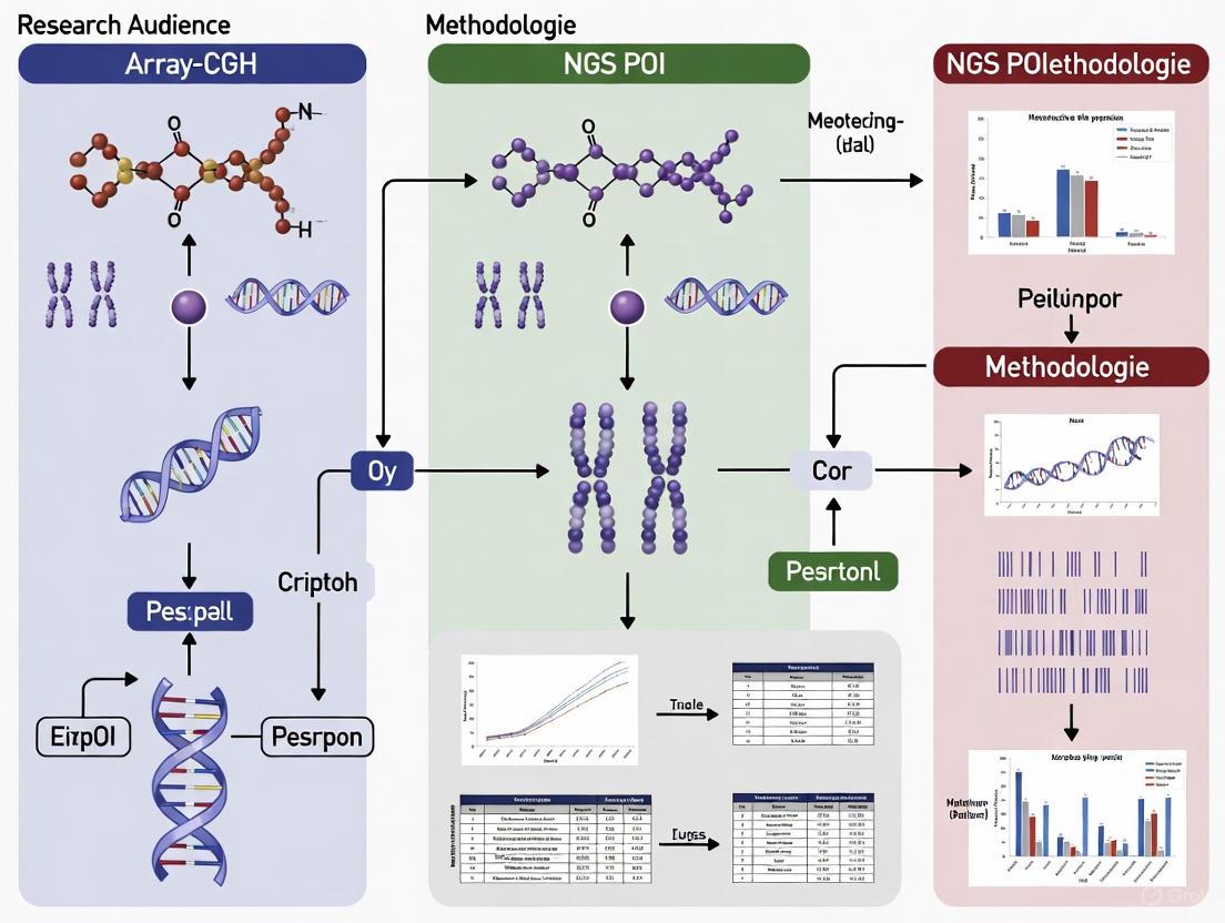
Workflow Visualization: Integrated Array-CGH & NGS for POI
The following diagram illustrates the integrated diagnostic and research pathway for the genetic analysis of Premature Ovarian Insufficiency, moving beyond conventional karyotyping.
In the field of genomics, Array-based Comparative Genomic Hybridization (Array-CGH) and Next-Generation Sequencing (NGS) are foundational technologies for analyzing genetic variation. Array-CGH is a specialized technique designed to detect copy number variations (CNVs)—submicroscopic chromosomal deletions or duplications—across the entire genome in a single assay [6]. In contrast, NGS is a high-throughput technology that enables the parallel sequencing of millions of DNA fragments, allowing for the comprehensive identification of a wider range of variants, including single nucleotide variants (SNVs), small insertions/deletions (indels), and with specific bioinformatic approaches, CNVs as well [7] [8]. The integration of these two methods is particularly powerful in the research of genetically heterogeneous conditions like Primary Ovarian Insufficiency (POI), where they can uncover both structural and sequence-level variations contributing to the disease [4] [6].
Principle and Workflow of Array-CGH
Array-CGH functions by comparing a patient's genome against a reference genome to identify regions of unequal copy number.
Core Principle: The fundamental concept involves the competitive hybridization of fluorescently labeled DNA from test and reference samples to genomic probes arrayed on a slide.
- The test DNA is labeled with one fluorophore (e.g., Cy3, green) and the reference DNA with another (e.g., Cy5, red) [9] [10].
- The two samples are mixed in equal amounts and hybridized to the array. After hybridization, the fluorescence intensity ratio between the two colors is measured for each probe.
- A green signal indicates a region of duplication (more test DNA bound), a red signal indicates a deletion (less test DNA bound), and a yellow signal indicates a normal, diploid region (equal amounts bound) [9].
- The resulting fluorescence ratio data is transformed into a log2 ratio, which is plotted across the genome to visually identify gains (positive values) and losses (negative values) [9].
Workflow: Array-CGH
The following diagram outlines the key steps in a typical Array-CGH experiment:
Principle and Workflow of Next-Generation Sequencing (NGS)
NGS is a massively parallel sequencing technology that allows for the simultaneous determination of the nucleotide sequence of millions to billions of DNA fragments.
Core Principle: Unlike Sanger sequencing, which processes one DNA fragment at a time, NGS fragments the genome, sequences all fragments in parallel, and then reassembles them computationally [8]. The most common method is Sequencing by Synthesis (SBS), where fluorescently tagged nucleotides are incorporated by DNA polymerase and imaged as they are added to the growing DNA strand [11] [8]. The massive redundancy, known as coverage or depth, ensures high accuracy by having each base position sequenced multiple times [7] [8].
Workflow: Next-Generation Sequencing
The following diagram illustrates the core steps in a standard NGS workflow:
Technical Comparison: Array-CGH vs. NGS
The table below summarizes the key technical characteristics and applications of Array-CGH and NGS.
| Feature | Array-CGH | NGS (Targeted Panel/WES) |
|---|---|---|
| Primary Detectable Variants | Copy Number Variations (CNVs) [10] [6] | SNVs, Indels, CNVs (via read-depth) [7] [10] |
| Analyzed Genomic Region | Predefined probes across the genome [10] | Targeted panels: 50-500 selected genes; WES: All exons (~1-2% of genome) [7] |
| Resolution | Limited to probe density and spacing [10] | Single-base resolution for SNVs/Indels; higher for CNVs than array in targeted regions [7] |
| Best For | Detection of large gains/losses, standard cytogenetic analysis [10] [6] | Conditions with high genetic heterogeneity, novel gene discovery, comprehensive variant screening [7] [4] |
| Limitations | Cannot detect balanced rearrangements or sequence-level changes [6] | Complex data analysis, risk of incidental findings, may miss CNVs in non-coding regions (WES) [7] [10] |
The Scientist's Toolkit: Essential Research Reagents
Successful genomic analysis relies on a suite of specialized reagents and tools. The following table lists key solutions used in these workflows.
| Research Reagent / Solution | Function in the Experiment |
|---|---|
| CYTAG CGH Labeling Kits [12] | Optimized fluorescent labeling of DNA for microarray hybridization, generating high-quality data with low background noise. |
| NGS Library Prep Kits (e.g., Illumina Nextera) [4] | Fragment genomic DNA and attach adapter sequences essential for cluster generation and sequencing. |
| Custom Target Enrichment Panels (e.g., Haloplex, SureSelect) [7] [4] | Capture and amplify a predefined set of genes of interest (e.g., a 295-gene panel for POI) from a complex genomic background prior to sequencing. |
| DNA Polymerases for SMRT/HiFi Sequencing [11] | Enable long-read, real-time sequencing in PacBio's Zero-Mode Waveguides (ZMWs) for high-fidelity (HiFi) reads. |
| Bioinformatic Pipelines (e.g., GATK, BWA) [7] [4] | Critical software tools for aligning raw sequencing reads to a reference genome and performing variant calling. |
Frequently Asked Questions (FAQs) and Troubleshooting
1. Our Array-CGH results show a high background noise and poor DLR scores. What could be the cause?
- Potential Cause: Inefficient fluorescent dye incorporation or degradation of the labeled DNA sample can lead to poor signal-to-noise ratios [12].
- Solution: Ensure you are using a high-quality labeling kit validated for low input samples. Precisely quantify DNA after labeling and use the recommended amount of starting material (e.g., 50-500 ng) to ensure efficient dye incorporation and low background [12].
2. When should I choose a targeted NGS panel over Whole Exome Sequencing (WES) for my POI research?
- Guidance: The choice depends on your research question. Use a targeted panel when the patient's phenotype points to a well-characterized group of conditions with known genetic heterogeneity, as it offers deep coverage, streamlined interpretation, and a higher diagnostic yield for those specific genes [7] [4]. Opt for WES when the genetic basis is unclear or when you are interested in discovering novel candidate genes, as it provides a broader, hypothesis-free screen of all protein-coding regions [7].
3. We identified a Variant of Uncertain Significance (VUS) in a known POI gene using our NGS panel. How should we proceed?
- Protocol: First, meticulously curate the variant using population frequency databases, computational prediction tools, and literature evidence per ACMG guidelines [7]. For oligogenic conditions like POI, also check for additional variants in interacting genes that may contribute to the phenotype [4]. VUS findings should be reported in the context of the patient's clinical phenotype and may require segregation analysis within the family for further clarification.
4. Can NGS data from a clinical exome be used reliably for CNV detection?
- Answer: Yes, CNVs can be detected from exome sequencing data using read-depth analysis methods, which compare the relative depth of sequencing coverage between the patient and a control set across genomic regions [10]. While this method is powerful and simplifies the diagnostic process by using a single test, it has limitations. CNVs that extend into non-coding regions or very small exon-level CNVs may be missed, and the analysis requires sophisticated algorithms and manual verification of coverage plots [10].
5. What is the key advantage of long-read sequencing (e.g., PacBio, Oxford Nanopore) in complex disease research?
- Advantage: Long-read technologies produce reads that are thousands to tens of thousands of bases long. This allows them to span complex genomic regions, such as repetitive sequences or large structural variations, that are difficult or impossible to resolve with short-read NGS [11]. This makes them invaluable for de novo genome assembly, resolving complex rearrangements, and detecting epigenetic modifications directly.
FAQs: Integrating Array-CGH and NGS in POI Research
1. Why is a multi-technique approach combining array-CGH and NGS necessary for POI research?
POI has a highly heterogeneous genetic background. Relying on a single technology can miss a significant number of causal variants. Array-CGH effectively identifies large copy number variations (CNVs), such as chromosomal deletions or duplications, while NGS is optimal for detecting single nucleotide variants (SNVs) and small insertions/deletions (indels) in individual genes [2] [6]. Using both methods in tandem provides a more comprehensive genetic screening, which is crucial as nearly 70% of POI cases were historically unexplained [2]. One study demonstrated that by combining both techniques, a genetic anomaly was identified in 57.1% (16 of 28) of idiopathic POI patients, a diagnostic yield that would not have been achieved with either method alone [2].
2. What are the specific limitations of using only array-CGH or only NGS?
- Limitation of Array-CGH Alone: Standard array-CGH can detect CNVs but is ineffective at identifying point mutations or small indels within genes known to cause POI [10] [6]. For example, it would miss pathogenic sequence variants in genes like FIGLA or NOBOX [2] [5].
- Limitation of NGS Alone: While excellent for SNVs, traditional NGS analysis pipelines can miss large CNVs, especially those involving single exons or non-coding regions, unless a specific CNV-calling algorithm is applied to the sequencing data [10]. For instance, a large deletion on chromosome 15q25.2 was identified by array-CGH in one study, which might be missed by a standard NGS SNV-calling workflow [2].
3. We have a limited budget. Which test should we run first?
The choice can depend on your patient population. However, given the high rate of point mutations, starting with an NGS gene panel is often more efficient for finding a monogenic cause. If the NGS panel is uninformative, a subsequent array-CGH should be performed to investigate structural variants [2] [5]. For a truly comprehensive and cost-effective approach in the long run, employing both methods concurrently on the same patient cohort, or using an NGS platform with validated CNV-calling capabilities, provides the highest diagnostic yield [2] [10].
4. What is a common technical challenge when preparing libraries for both techniques from a single patient sample, and how can it be mitigated?
A frequent issue is insufficient DNA quantity or quality for both assays, especially when working with precious clinical samples.
- Troubleshooting Guide:
- Problem: Low DNA yield from patient blood or tissue samples.
- Solution: Optimize DNA extraction protocols to maximize yield and purity. Use fluorescence-based assays (e.g., Qubit) for accurate DNA quantification, which is critical for both array-CGH and NGS library preparation [2].
- Prevention: Plan the DNA allocation at the start of the project. For array-CGH, ensure you have the required amount for the specific platform (e.g., 4x180K array). For NGS, ensure sufficient DNA for the library kit (e.g., 10 ng for an AmpliSeq panel) [2] [5]. Consider whole-genome amplification methods as a last resort, being aware of potential amplification biases.
Experimental Protocols for an Integrated Workflow
The following protocol is synthesized from recent studies that successfully integrated array-CGH and NGS for POI analysis [2] [5].
Protocol 1: Combined Genetic Screening for Idiopathic POI
1. Patient Selection & Phenotyping:
- Inclusion Criteria: Recruit patients meeting the clinical definition of POI: primary or secondary amenorrhea for >4 months before age 40, with elevated FSH levels >25 IU/L on two consecutive tests [2].
- Exclusion Criteria: Exclude patients with known karyotype abnormalities, FMR1 premutation, or clear iatrogenic/autoimmune causes [2].
- Data Collection: Record detailed clinical data, including age at diagnosis, type of amenorrhea, family history, and hormone levels (FSH, Estradiol, AMH) [2].
2. DNA Extraction:
- Extract genomic DNA from peripheral blood samples using a standardized kit (e.g., QIAsymphony DNA midi kits) [2].
- Accurately quantify DNA using a fluorometric method.
3. Array-CGH Analysis:
- Platform: Use a high-resolution oligonucleotide array (e.g., Agilent SurePrint G3 Human CGH Microarray 4x180K) [2].
- Procedure:
- Label patient and control DNA with different fluorescent dyes (e.g., Cy3 and Cy5).
- Hybridize the mixed samples to the microarray.
- Scan the array and analyze fluorescence intensity ratios using dedicated software (e.g., Agilent CytoGenomics) [2].
- Data Interpretation: Identify CNVs (deletions/duplications) with a minimum size of 60 kb. Classify CNVs as pathogenic, benign, or VUS using population (e.g., DGV) and clinical (e.g., DECIPHER) databases [2].
4. Next-Generation Sequencing:
- Method: Use a targeted gene panel covering known and candidate POI genes.
- Library Preparation: Prepare amplicon libraries (e.g., with Ion AmpliSeq Library Kit Plus) using a custom panel (e.g., 163 genes) [2].
- Sequencing: Perform sequencing on a platform such as an Illumina NextSeq 550 or Ion S5 system [2] [5].
- Bioinformatics Analysis:
5. Data Integration:
- Correlate findings from array-CGH and NGS. A patient might have a causal CNV from array-CGH and a VUS from NGS, or vice-versa. The combined result provides a more complete genetic picture [2].
Quantitative Data from Key Studies
Table 1: Diagnostic Yield of Integrated Genetic Analysis in POI
| Study Cohort | Patient Population | Array-CGH Findings (Causal CNV) | NGS Findings (Causal SNV/Indel) | Combined Diagnostic Yield | Key Genes Identified |
|---|---|---|---|---|---|
| Amiens University (2025) [2] | 28 idiopathic POI patients | 1/28 (3.6%) | 8/28 (28.6%) | 16/28 (57.1%) | FIGLA, TWNK |
| Hungarian Cohort (2024) [5] | 48 POI patients | Not separately specified | 8/48 (16.7%) with monogenic defects | ~29.2% with potential risk factors | EIF2B, GALT, NOBOX |
Table 2: Essential Research Reagent Solutions for Integrated POI Workflow
| Reagent / Kit | Function in the Workflow | Example Product (from search results) |
|---|---|---|
| Genomic DNA Extraction Kit | Isolation of high-quality, high-molecular-weight DNA from patient blood. | QIAsymphony DNA Midi Kits [2] |
| Array-CGH Platform | Genome-wide screening for copy number variations (CNVs). | Agilent SurePrint G3 Human CGH Microarray [2] |
| Targeted NGS Panel | Simultaneous sequencing of a custom set of genes associated with POI. | Custom capture design of 163 genes [2] or panel of 31 genes [5] |
| NGS Library Prep Kit | Preparation of sequencing-ready libraries from genomic DNA. | Ion AmpliSeq Library Kit Plus [5] or SureSelect XT-HS [2] |
| Sequence Analysis Software | Bioinformatic pipeline for alignment, variant calling, and annotation. | Ion Reporter, Varsome [5]; Alissa Align&Call, Alissa Interpret [2] |
Workflow and Pathway Visualizations
The following diagrams, generated with Graphviz, illustrate the integrated experimental workflow and the biological processes involved in POI.
Integrated POI Genetic Analysis Workflow
Biological Pathways to POI Disruption
Primary Ovarian Insufficiency (POI) is a clinically heterogeneous disorder characterized by the loss of ovarian function before the age of 40, affecting approximately 1-3.7% of women [13] [14]. It is diagnosed by oligomenorrhea or amenorrhea for at least four months, along with elevated follicle-stimulating hormone (FSH) levels exceeding 25 IU/L on two occasions spaced at least four weeks apart [15]. POI represents a significant cause of female infertility and is associated with long-term health risks including osteoporosis, cardiovascular disease, and cognitive decline [14]. The etiopathogenesis of POI is multifactorial, with genetic factors contributing to approximately 20-25% of cases [16] [15]. The genetic basis is highly heterogeneous, involving chromosomal abnormalities, copy number variations (CNVs), and single-gene mutations affecting various biological processes essential for ovarian function.
Key Chromosomal Regions and CNVs in POI
Chromosomal abnormalities, particularly those involving the X chromosome, are well-established causes of POI, accounting for 10-13% of cases [17]. Early studies identified critical regions on the X chromosome essential for ovarian function, with rearrangements in these regions frequently associated with POI.
X Chromosome Critical Regions
Three critical regions for ovarian function and reproductive lifespan have been identified on the X chromosome:
- POF1 (Xq26qter): This region contains genes crucial for ovarian maintenance. Disruptions in this region are frequently associated with POI.
- POF2 (Xq13.3q21.1): Deletions and translocations in this region often lead to POI. The gene DIAPH2 (Diaphanous Related Formin 2) has been implicated through X-autosomal translocations with breakpoints in this region [13] [16].
- POF3 (Xp11p11.2): Structural variations in this region have been linked to impaired ovarian function [13].
Specific Chromosomal Disorders
- Turner Syndrome (45,X or mosaicism): This represents the most extreme example of X-linked POI. Complete monosomy X (45,X) typically presents with primary amenorrhea and streak ovaries, while mosaic individuals (e.g., 45,X/46,XX) may experience secondary amenorrhea and a milder POI phenotype [13] [17]. The haploinsufficiency of X-linked genes, such as SHOX (Short-stature homeobox), is believed to drive the accelerated follicular atresia [16].
- Trisomy X Syndrome (47,XXX): Women with this karyotype have a heightened risk of POI, evidenced by diminished Anti-Müllerian Hormone (AMH) levels and elevated FSH and LH, which can lead to menstrual cycle disorders and secondary amenorrhea [16].
- X-Autosomal Translocations: These rare rearrangements (incidence ~1:30,000) are strongly associated with POI. Approximately 80% of breakpoints occur within the Xq21 cytoband of the POF2 region. The pathogenic mechanisms may include direct gene disruption, meiotic errors, or position effects that alter the expression of nearby genes critical for ovarian function [16].
CNV Detection Methods: Array-CGH vs. NGS
The detection of CNVs has evolved significantly, moving beyond traditional karyotyping.
- Array Comparative Genomic Hybridization (array-CGH): This technique has been the standard for genome-wide CNV analysis. It involves competitive hybridization of patient and reference DNA to a chip containing thousands of DNA probes. The resolution (from 100 kb to <10 kb) is determined by the number and density of these probes, far exceeding the ~5 Mb resolution of conventional karyotyping [18]. A key limitation is its inability to detect balanced chromosomal rearrangements (e.g., translocations) or low-level mosaicism [18].
- Next-Generation Sequencing (NGS): CNV analysis using Whole Exome Sequencing (WES) typically relies on read-depth analysis. Deviations in the normalized read depth across genomic regions can indicate deletions (decreased depth) or duplications (increased depth) [10]. While WES excels at detecting single nucleotide variants (SNVs) and small indels, its effectiveness for CNV calling is generally limited to the exonic regions captured by the specific platform used.
Table 1: Comparison of CNV Analysis Methods
| Feature | Array-CGH | NGS-based CNV (via WES) |
|---|---|---|
| Primary Principle | Competitive hybridization and fluorescence ratio measurement | Read depth analysis and normalization |
| Resolution | High (100 kb to <10 kb), customizable with specific arrays | Varies; focuses on exonic regions covered by the platform |
| Coding Region Focus | Genome-wide, but can be designed for specific regions | Excellent for targeted exonic CNVs |
| Ability to Detect Balanced Rearrangements | No | No (from standard WES analysis) |
| Ability to Detect SNVs/Indels | No | Yes, simultaneously |
| Key Advantage | Established, robust genome-wide CNV profiling | Simplified workflow with combined SNV/Indel/CNV data |
| Key Limitation | Cannot detect SNVs/Indels or balanced changes | CNV detection limited to designed target regions; may miss non-coding or intergenic variants |
Evidence suggests that WES can be a highly effective single-tier test. One study performing clinical exome sequencing on 245 patients undiagnosed by array-CGH achieved a 20% diagnostic rate, suggesting that an integrated NGS approach may offer a higher overall diagnostic yield for heterogeneous conditions like POI [10].
Key Single-Gene Mutations in POI
Advances in high-throughput sequencing have identified mutations in over 90 genes associated with both syndromic and non-syndromic POI. The genetic architecture includes autosomal dominant, autosomal recessive, and X-linked patterns.
Genes Categorized by Biological Function
The genes implicated in POI can be functionally categorized based on their role in ovarian development and function.
Table 2: Key POI-Associated Genes and Their Functional Roles
| Gene | Inheritance | Primary Functional Role | Phenotypic Association |
|---|---|---|---|
| FMRI | XLD | RNA metabolism; premutation (55-200 CGG repeats) causes toxic RNA gain-of-function | Isolated POI (most common single-gene cause) |
| BMP15 | XLD | Oocyte-secreted factor, folliculogenesis | Isolated POI |
| NR5A1 | AD | Transcriptional regulator of gonadal development | Isolated POI or with adrenal insufficiency |
| FIGLA | AD | Transcription factor for primordial follicle formation | Isolated POI |
| NOBOX | AD | Oocyte-specific transcription factor, folliculogenesis | Isolated POI |
| GDF9 | AD | Oocyte-secreted factor, folliculogenesis | Isolated POI |
| STAG3 | AR | Meiotic cohesin complex, chromosome segregation | Isolated POI (primary amenorrhea) |
| HFM1 | AR | DNA helicase, meiotic recombination | Isolated POI |
| MCM8/9 | AR | DNA repair, meiotic homologous recombination | Isolated POI |
| SYCE1 | AR | Synaptonemal complex assembly, meiosis I | Isolated POI |
| AIRE | AR | Transcription factor, immune tolerance | Autoimmune Polyglandular Syndrome Type 1 (APS-1) |
| GALT | AR | Galactose metabolism | Galactosemia |
| ATM | AR | DNA damage repair, cell cycle control | Ataxia-Telangiectasia (A-T) |
Notes: AD: Autosomal Dominant; AR: Autosomal Recessive; XLD: X-Linked Dominant.
Ovarian Development and Folliculogenesis
Genes like NR5A1, NOBOX, FIGLA, BMP15, and GDF9 are critical for early ovarian development, formation of primordial follicles, and their subsequent growth and maturation. Mutations in these genes often lead to non-syndromic POI by disrupting the initial pool or the developmental trajectory of ovarian follicles [17] [14].
Meiosis and DNA Repair
A substantial proportion of POI cases, particularly those with primary amenorrhea, are linked to defects in meiotic genes. These include STAG3, SYCE1, HFM1, MCM8, and MCM9 [15] [17]. These genes are essential for processes like homologous recombination, synaptonemal complex formation, and DNA double-strand break repair during meiotic prophase I. Their failure leads to meiotic arrest and massive oocyte attrition before birth or in early adulthood.
Metabolic and Autoimmune Disorders
- Galactosemia: Caused by biallelic mutations in GALT, this disorder leads to POI in 80-90% of affected females, often presenting as primary amenorrhea. The toxic accumulation of galactose metabolites is thought to cause damage to the ovarian stroma and follicles [16].
- Autoimmune Polyglandular Syndrome Type 1 (APS-1): Resulting from mutations in the AIRE gene, this syndrome is characterized by autoimmune destruction of multiple endocrine organs, including the ovaries, with approximately 41% of patients developing POI due to lymphocytic oophoritis [16].
Genetic Architecture and Genotype-Phenotype Correlations
The genetic architecture of POI is complex. A large whole-exome sequencing study of 1,030 patients revealed that 18.7% had pathogenic/likely pathogenic (P/LP) variants in 59 known POI genes [15]. Of these:
- 80.3% had monoallelic (heterozygous) mutations.
- 12.4% had biallelic mutations.
- 7.3% had multiple heterozygous P/LP variants in different genes (digenic/oligogenic inheritance) [15].
A clear genotype-phenotype correlation exists regarding the type of amenorrhea:
- Primary Amenorrhea (PA): Has a higher genetic contribution (25.8% in one study) and a greater frequency of biallelic and multi-het variants, suggesting more severe genetic disruptions [15].
- Secondary Amenorrhea (SA): Has a lower overall genetic contribution (17.8%) and is more frequently associated with monoallelic variants [15].
Integrated Array-CGH and NGS Workflow for POI Genetic Testing
Experimental Protocol for Genetic Diagnosis of POI
Sample Requirement: Genomic DNA (e.g., from peripheral blood) with high quality and purity (260/280 ratio ~1.8).
Workflow Steps:
DNA Quality Control (QC):
- Method: Use fluorometric methods (e.g., Qubit) for accurate DNA quantification. Assess purity via spectrophotometry (NanoDrop) with acceptable 260/230 (>1.8) and 260/280 (~1.8) ratios. Verify integrity by agarose gel electrophoresis or Bioanalyzer.
- Troubleshooting: If yield is low or degradation is detected, re-extract DNA. If contaminants are present, perform additional clean-up steps using column- or bead-based purification [19].
Array-CGH Analysis:
- Platform: Use a commercial high-resolution array (e.g., 60K-400K or higher), preferably one that includes SNP probes to detect regions of homozygosity and some uniparental disomies.
- Protocol: Label patient and reference DNA with different fluorochromes (e.g., Cy3 and Cy5). Hybridize equal amounts of labeled DNA to the array chip according to manufacturer's instructions. Scan the array and extract fluorescence intensity data [18].
- Data Analysis: Use specialized software (e.g., in R/Bioconductor environment with packages like
DNAcopyfor segmentation) to identify genomic regions with significant log2 ratio deviations, indicating CNVs [20]. Classify CNVs following ACMG guidelines, annotating them with population frequency (e.g., from DGV, gnomAD) and clinical databases (e.g., DECIPHER, ClinGen) [21].
Next-Generation Sequencing:
- Platform: Whole Exome Sequencing (WES) is recommended for a comprehensive assessment. If resources allow, Whole Genome Sequencing (WGS) provides uniform coverage and can detect variants in non-coding regions.
- Library Preparation & Sequencing: Use a clinical-grade exome capture kit. Prepare the sequencing library following best practices to avoid artifacts (e.g., adapter dimers) and biases (e.g., from over-amplification). Sequence on an Illumina or similar platform to achieve >100x mean coverage with >95% of target bases covered at 20x [19] [15].
- Variant Calling & Annotation: Align sequences to a reference genome (e.g., GRCh38). Call SNVs, indels, and CNVs using a robust bioinformatic pipeline. Annotate variants using databases like gnomAD, ClinVar, and HGMD.
Integrated Data Interpretation:
- Triangulation: Correlate findings from array-CGH and WES. Confirm that CNVs detected by array-CGH are also supported by read-depth analysis from WES data.
- Variant Prioritization: Filter variants based on population frequency (<0.01), predicted pathogenicity (in silico tools, ACMG/AMP guidelines), and functional relevance to POI (known POI genes, meiotic pathways, ovarian development) [15] [21].
- Segregation Analysis: Where possible, perform familial segregation studies to confirm de novo inheritance or co-segregation of the variant with the disease phenotype in the family.
- Reporting: Report clinically significant findings, including P/LP variants in known POI genes and CNVs affecting dosage-sensitive genomic regions.
Integrated Diagnostic Workflow for POI
Troubleshooting Common Issues in Genetic Analysis
Q1: Our NGS library yields are consistently low. What are the primary causes and solutions? A: Low library yield is a common issue often stemming from:
- Poor Input DNA Quality: Degraded DNA or contaminants (phenol, salts) inhibit enzymatic reactions. Solution: Re-purify input DNA; use fluorometric quantification (Qubit) instead of UV absorbance (NanoDrop) for accurate measurement [19].
- Fragmentation Issues: Over- or under-shearing creates fragments outside the optimal size range. Solution: Optimize fragmentation parameters (time, energy) and verify the fragment size distribution on a BioAnalyzer post-shearing [19].
- Inefficient Adapter Ligation: This can be caused by suboptimal ligase activity or incorrect adapter-to-insert molar ratios. Solution: Titrate the adapter concentration, ensure fresh reagents, and maintain optimal reaction temperature [19].
Q2: Our sequencing data shows high duplication rates and poor library complexity. How can this be resolved? A: High duplication rates often indicate insufficient starting material or amplification bias.
- Root Cause: Over-amplification during PCR to compensate for low input. Solution: Increase the amount of input DNA if possible. If starting material is limited, use a library prep kit designed for low inputs and minimize the number of PCR cycles [19].
- Root Cause: Pippeting errors or sample loss during purification. Solution: Use calibrated pipettes and master mixes to reduce volumetric errors. Be careful during bead-based cleanups to avoid discarding the sample [19].
Q3: Our array-CGH data is noisy, making it difficult to call CNVs confidently. What steps can we take? A: Noisy data can arise from several sources in the array workflow.
- Preprocessing: Ensure rigorous quality control and normalization of the raw fluorescence data. Use Bioconductor packages like
MANORto correct for spatial biases on the array [20]. - Segmentation Algorithm: Choose an appropriate segmentation algorithm. The
DNAcopypackage in R, which uses Circular Binary Segmentation, is widely regarded as a robust method for breakpoint detection and is less sensitive to noise [20]. - Replicate Spots: If your array platform uses replicate spots, assess the consistency between them. High variability between replicates indicates poor array quality [20].
Q4: We have identified a variant of uncertain significance (VUS) in a candidate POI gene. What is the recommended course of action? A: VUSs are a major challenge in clinical diagnostics.
- Re-evaluation: Implement a process for periodic re-analysis of unsolved cases and VUSs. Automated tools like SeqOne's "GenomeAlert!" can facilitate this by tracking changes in variant classifications in public databases like ClinVar [21].
- Functional Studies: Generate functional evidence (PS3 criterion per ACMG guidelines) to upgrade or downgrade the VUS. For example, the large POI study by [15] functionally validated 75 VUSs, reclassifying 38 as likely pathogenic.
- Segregation Analysis: Test the variant in affected and unaffected family members. Co-segregation of the variant with the disease phenotype in the family provides supporting evidence for pathogenicity.
Table 3: Key Research Reagent Solutions for POI Genetic Analysis
| Reagent/Resource | Function | Example/Note |
|---|---|---|
| High-Resolution Array-CGH Kit | Genome-wide detection of CNVs | Agilent, Illumina, or Affymetrix platforms with 180K-400K probes for optimal resolution. |
| Clinical Exome Capture Kit | Target enrichment for WES | Kits from Twist Bioscience, Agilent, or IDT that comprehensively cover known POI genes. |
| NGS Library Prep Kit | Preparation of sequencing libraries | Kits with low input requirements and low duplication rates (e.g., Illumina DNA Prep). |
| Bioinformatic Pipelines | Variant calling, annotation, and filtering | Commercial platforms (e.g., SeqOne) or open-source workflows (e.g., BWA-GATK). |
| ACMG Classification Framework | Standardized variant pathogenicity assessment | Essential for consistent interpretation of SNVs/Indels and CNVs [21]. |
| Population Genomics Databases | Filtering common polymorphisms | gnomAD, 1000 Genomes Project. |
| Variant & Phenotype Databases | Curated clinical and functional evidence | ClinVar, DECIPHER, HGMD, LOVD. |
| POI Gene Panels | Curated list of genes for focused analysis | Can be used for targeted NGS or to filter WES data; should include both established and novel candidate genes [15] [14]. |
The integration of array-CGH and NGS technologies has significantly advanced our understanding of the genetic architecture of POI, increasing the diagnostic yield to approximately 20-25% [15] [17]. Current genetic testing that focuses only on the FMR1 premutation is inadequate, as it misses the vast majority of genetic cases [13]. An expanded genetic testing approach, as outlined in this guide, is crucial for providing patients with an accurate diagnosis, enabling personalized risk assessment, and informing reproductive planning.
Future directions in POI genetic research will involve the systematic exploration of oligogenic inheritance, the functional validation of novel candidate genes from large-scale sequencing studies, and the investigation of non-coding variants and epigenetic modifications. The ongoing shift towards Whole Genome Sequencing (WGS) as a first-line test promises a more comprehensive detection of all variant types in a single assay, potentially further simplifying the diagnostic odyssey for women and families affected by POI.
Building the Integrated POI Workflow: From Sample to Analysis
FAQs: Understanding Sequential and Parallel Testing
What is the core difference between sequential and parallel testing workflows?
In a sequential workflow, genetic tests are executed one after the other. For example, a sample might first be analyzed using array-CGH, and only if the results are inconclusive would it proceed to Next-Generation Sequencing (NGS). This linear approach is straightforward but can be time-consuming [22] [23].
In a parallel workflow, array-CGH and NGS are initiated simultaneously on the same sample. This high-throughput strategy leverages multiple testing platforms at once, significantly accelerating the diagnostic process and providing complementary datasets from a single run [22] [23].
When should I choose a sequential testing strategy?
A sequential strategy is often better suited for:
- Budget-conscious projects: It helps control costs by only performing the more expensive test (often NGS) if the first-tier test is inconclusive [10] [24].
- Cases with strong preliminary indications: When a specific, large-scale chromosomal abnormality is strongly suspected, array-CGH as a first test can be sufficient [10].
- Resource-constrained environments: It requires less computational and laboratory infrastructure to manage a single workflow at a time [22].
What are the advantages of a parallel testing strategy?
Parallel testing offers several key benefits:
- Faster Time-to-Diagnosis: By eliminating the wait time between sequential tests, parallel workflows can provide a comprehensive genetic result much more quickly [22] [23].
- Higher Diagnostic Yield: Parallel testing can capture a broader range of genetic variants from the outset. For instance, array-CGH detects large copy number variations (CNVs), while NGS can simultaneously identify single nucleotide variants (SNVs), small indels, and CNVs via read-depth analysis [10] [25] [24].
- Simplified Workflow Management: Running both tests simultaneously can simplify project coordination and sample tracking compared to a multi-stage sequential process [23].
What is the comparative diagnostic yield of array-CGH versus NGS?
The diagnostic yield varies significantly based on the clinical context. The table below summarizes findings from key studies on patients with neurodevelopmental disorders (NDDs) [24]:
| Phenotype Category | Diagnostic Yield (aCGH) | Diagnostic Yield (Clinical Exome Sequencing) |
|---|---|---|
| Global Developmental Delay / Intellectual Disability | ~5.7% (as part of a broader cohort) | Significantly higher than aCGH; specific yield varies by subcategory |
| Autism Spectrum Disorder (Isolated) | ~3% | ~6.1% |
| Other NDDs | ~1.4% | ~7.1% |
| Overall (across all NDDs) | 5.7% | 20% |
Another randomized study in an IVF context found that NGS performed with high accuracy comparable to array-CGH, resulting in ongoing pregnancy rates of 74.7% for NGS vs. 69.2% for aCGH [25].
How does the underlying technology differ between array-CGH and NGS for CNV detection?
The fundamental principles of CNV detection differ between the two platforms, which is why they can be complementary [10] [9]:
| Feature | Array-CGH (aCGH) | NGS (Read-Depth Based) |
|---|---|---|
| Basic Principle | Compares patient and control DNA hybridized to probes on a microarray. Measures fluorescence intensity ratios to detect copy number changes [10] [9]. | Sequences millions of short DNA fragments. Normalized read counts (depth of coverage) across genomic regions are compared to detect copy number changes [10]. |
| Primary Data Output | Log2 ratio of fluorescence intensities (Cy3/Cy5) [9]. | Number of aligned reads per genomic bin or target [10]. |
| Key Strength | Established, robust technology for detecting large CNVs and aneuploidies [10]. | Can detect a wider variety of variant types (SNVs, Indels, CNVs) simultaneously. Can identify smaller CNVs than some array platforms [10] [24]. |
| Key Limitation | Cannot detect balanced rearrangements or sequence-level variants. Resolution is limited by probe density and distribution [10]. | CNV detection in non-coding regions or areas with poor coverage is challenging. Requires sophisticated bioinformatics analysis [10]. |
Troubleshooting Guides
Issue: Low Diagnostic Yield Despite Using NGS
Potential Causes and Solutions:
- Cause 1: Inadequate Analysis of Copy Number Variants.
- Solution: Not all NGS analysis pipelines are equally adept at CNV calling. Ensure your bioinformatics workflow includes a robust, validated read-depth-based algorithm for CNV detection from NGS data, similar to how array-CGH analyzes intensity ratios [10] [9]. Consider using specialized software for integrated SNV and CNV analysis.
- Cause 2: Suboptimal Target Enrichment.
- Solution: For exome or panel sequencing, verify the efficiency and uniformity of the capture process. Poor or uneven coverage in key genes can lead to missed variants. Check metrics like mean coverage, uniformity, and on-target rate.
- Cause 3: Incomplete Gene Coverage.
- Solution: Compare the gene list covered by your clinical exome or panel with the latest databases of disease-associated genes (e.g., from the Deciphering Developmental Disorders Consortium). There may be clinically relevant genes missing from your target region [24].
Issue: Inconsistent or Noisy CNV Data
Applicable to both array-CGH and NGS-based methods.
- Cause 1: Poor DNA Quality.
- Solution: Always use high-quality, high-molecular-weight DNA. Check integrity via gel electrophoresis or fragment analyzers. Degraded or sheared DNA leads to poor hybridization in array-CGH and uneven coverage in NGS.
- Cause 2: Technical Variation in array-CGH.
- Solution: Ensure patient (test) and reference DNA are labeled with different fluorophores (e.g., Cy3 and Cy5) and hybridized correctly. Optimize hybridization conditions and washing stringency to improve the signal-to-noise ratio [9].
- Cause 3: GC Bias in NGS.
Issue: Interpreting a Variant of Uncertain Significance (VUS)
A common challenge in both platforms.
- Action 1: Correlate with Clinical Phenotype.
- Review the patient's symptoms in detail against the known association of the gene(s) within the CNV or the specific gene with the SNV/Indel.
- Action 2: Determine Inheritance.
- If possible, test both parents. A de novo (new) variant is more likely to be pathogenic than one inherited from an unaffected parent.
- Action 3: Utilize Public and Commercial Databases.
- Consult databases like ClinVar, DECIPHER, and gnomAD to assess the variant's frequency and previously reported pathogenicity.
- Action 4: Multi-Method Confirmation.
- Use an orthogonal method to confirm the finding. For example, confirm a CNV detected by NGS with array-CGH or digital PCR, and vice-versa [24]. This was crucial in a case where a female with an X-linked CNV inherited from an unaffected mother was ultimately diagnosed via clinical exome sequencing, which found a causative de novo SNV in a different gene [24].
Experimental Workflow and Decision Diagram
The following diagram illustrates the logical decision process for choosing between sequential and parallel testing strategies, integrating the key questions and considerations from the FAQs.
Research Reagent Solutions
This table details essential materials and their functions for implementing array-CGH and NGS workflows.
| Item | Function | Application Notes |
|---|---|---|
| Microarray Platform | Solid support with immobilized DNA probes for competitive hybridization of test and reference genomes [9]. | Resolution (e.g., 60K to 1M probes) impacts detection capability. Choose based on required resolution [10] [24]. |
| Fluorophore-Labeled dUTPs (Cy3, Cy5) | Fluorescent dyes for enzymatic labeling of test and reference DNA samples for visualization on arrays [9]. | Ensures distinct fluorescent signals can be measured and compared for ratio analysis. |
| NGS Library Prep Kit | Reagents for fragmenting DNA, attaching platform-specific adapters, and PCR amplification to create sequencer-ready libraries. | Select kits optimized for your sample type (e.g., whole genome, exome) and desired insert size. |
| Bioinformatic Analysis Suite | Software for processing raw data, aligning sequences, and calling variants (SNVs, Indels, CNVs). | Critical for NGS. Must include a robust read-depth algorithm for CNV detection [10]. Examples include tools like CoNIFER, XHMM, or commercial suites [9]. |
| Whole Genome Amplification (WGA) Kit | For amplifying minute quantities of DNA from limited samples (e.g., blastocyst biopsies) to quantities sufficient for analysis [25]. | Essential for preimplantation genetic testing (PGT) and other low-input applications. |
Sample Preparation and Quality Control for Combined Genomic Analyses
Technical Support Center
Troubleshooting Guides
Table 1: Common NGS Library Preparation Issues and Solutions
| Problem | Possible Causes | Recommended Solutions |
|---|---|---|
| Low library yield [26] | - Degraded starting material- Inefficient adapter ligation- Inadequate PCR amplification | - Verify nucleic acid integrity (RIN > 8 for RNA, A260/A280 ≈ 1.8) [27]- Optimize adapter concentration and ligation time [26]- Increase PCR cycle number cautiously [26] |
| High adapter dimer rate [26] [27] | - Excess unused adapters- Inefficient purification post-ligation | - Use bead-based size selection or gel purification [27]- Optimize adapter-to-insert ratio [26] |
| Uneven sequencing coverage [26] | - PCR amplification bias- Incomplete fragmentation | - Use high-fidelity PCR enzymes designed to minimize bias [26]- Optimize fragmentation conditions (enzymatic or physical) [26] |
| Chimeric reads [26] | - Inefficient library construction | - Implement efficient A-tailing of PCR products [26]- Use chimera detection software for filtering [26] |
| Checkpoint | Parameter(s) to Measure | Target Value / Ideal Outcome |
|---|---|---|
| Starting Material | Quantity, Purity (A260/A280, A260/A230), Integrity (RIN/RQN) | A260/A280 ≈ 1.8, A260/A230 ≈ 2.0, RIN > 8 for RNA [27] |
| Fragmentation | Fragment size distribution | Single, tight peak at desired size (e.g., 200-500bp) [27] |
| Final Library | Concentration, molarity, adapter dimer presence | High concentration, minimal adapter dimer peak on electrophoretogram [27] |
| Library Pooling | Normalized concentration across samples | Equal molar concentration for uniform sample representation [27] |
Frequently Asked Questions (FAQs)
Q1: What is the most critical step in preparing samples for a combined array-CGH and NGS workflow for POI research?
The initial nucleic acid extraction and quality control is paramount [27]. The quality of the starting material directly impacts all downstream analyses. For POI research involving the detection of copy number variations (CNVs) or single-gene mutations (e.g., in STAG3), high-quality, high-molecular-weight DNA is essential for both array-CGH and NGS to ensure accurate results and prevent false positives/negatives [28] [29].
Q2: How can I minimize bias in my NGS library, especially when working with limited patient samples?
To minimize bias:
- Use PCR enzymes specifically designed to reduce amplification bias [26].
- Avoid over-amplification by determining the minimum number of PCR cycles needed for sufficient library yield [27].
- Employ bioinformatic tools like Picard MarkDuplicates or SAMTools to identify and remove PCR duplicates from the sequencing data post-run [26].
Q3: Our lab is transitioning from MLPA to NGS for CNV detection in our POI diagnostic panel. What are the key advantages?
NGS offers several key advantages over MLPA [30]:
- Multiplexing: You can test all genes in your panel simultaneously for CNVs, rather than performing separate MLPA tests for each gene.
- Resolution: NGS can detect smaller CNVs, including single-exon or partial-exon deletions/duplications, which might be missed by MLPA due to its limited probe density [30].
- Throughput and Cost: NGS provides higher throughput and can be more cost-effective than ordering multiple individual MLPA kits [30].
Q4: What specific QC is needed for the final NGS library before pooling and sequencing?
The final library should be assessed for [27]:
- Concentration and Molarity: Using fluorometric methods (e.g., Qubit) or qPCR for accurate quantification.
- Size Distribution: Using an automated electrophoresis system (e.g., Bioanalyzer, TapeStation) to confirm the correct library size and check for adapter dimers or other contaminants.
Experimental Protocols
This protocol is used for detecting copy number variations in diagnostic POI gene panels.
- Data Input: Use aligned sequencing files (BAM) from targeted gene panel sequencing.
- Calculate Coverage Depth: Determine the coverage depth for each target region (exon) in the panel.
- Sliding Window Analysis (Optional for high resolution): To detect CNVs in small or partial exons, divide each target region into overlapping sliding windows. A typical setup uses a window size of 75 bp (half the read length) and a sliding length of 10 bp [30].
- Normalization and Ratio Calculation: For each region (or window) in a query sample, calculate a copy number ratio by comparing its mean coverage to the average coverage of the same region in a pool of normal control samples with similar overall coverage depth [30].
- Formula for copy number state interpretation: A ratio of ~0.5 suggests a deletion, ~1.0 is normal, and ~1.5 suggests a duplication [30].
- CNV Calling: Identify regions where the copy number ratio significantly deviates from the expected value of 1.0, indicating a potential CNV.
The Scientist's Toolkit
Table 3: Essential Research Reagent Solutions
| Item | Function / Application |
|---|---|
| Nucleic Acid Extraction Kits | Isolate high-quality DNA from patient samples (e.g., blood, tissue) for both array-CGH and NGS [27]. |
| NGS Library Prep Kits | Convert the extracted DNA into a sequence-ready library through fragmentation, adapter ligation, and amplification. Selection depends on sequencing platform (e.g., Illumina) [26]. |
| Target Enrichment Panels | Designed to capture and sequence genes associated with POI (e.g., panels including STAG3, FMR1, etc.) [28]. |
| Cytogenomic Microarrays | Used for genome-wide detection of CNVs and regions of homozygosity, which can be correlated with NGS findings [28] [29]. |
| Quality Control Assays | Including instruments for electrophoresis (Bioanalyzer, TapeStation) and fluorometric quantification (Qubit) to assess nucleic acid quality at multiple steps [27]. |
Workflow Diagrams
CNV Detection via Read-Depth Analysis
Technical FAQs: Optimizing Your Array-CGH Experiment
FAQ 1: What are the key factors in microarray probe design that affect the detection of copy number variants (CNVs)?
The ability of an array-CGH platform to reliably detect CNVs, especially small, exon-level variants, depends heavily on probe design. Several factors critically influence probe performance [31]:
- Probe Specificity: Probes must hybridize specifically to their intended target. Performance can be adversely affected by cross-hybridization to homologous regions (e.g., segmental duplications or pseudogenes), non-specific binding to repetitive sequences, or high GC content, which can cause "sticky" probes and non-informative signals [31].
- Probe Sensitivity: Probes must bind effectively to their target. Sensitivity can be reduced by secondary structures in either the probe or the target DNA, or by very low GC content, which reduces binding efficiency [31].
- Hybridization Uniformity: Probes should function under similar isothermal conditions. Consistent probe behavior across the array reduces noise in the final dataset, leading to more accurate CNV calls [31].
Sophisticated design workflows address these factors through in silico steps that analyze sequence metadata, identify repetitive regions, generate candidate probes, rank them based on physicochemical properties, and select the optimal probes. Empirical optimization using thousands of tests further filters out non-performing probes to ensure robust performance [31].
FAQ 2: How do I select the appropriate array-CGH platform and resolution for a POI study?
Platform selection involves balancing resolution, content, and throughput. The table below summarizes key specifications for Agilent's array platforms, which utilize SurePrint technology with long, high-quality oligonucleotides [32].
Table 1: Comparison of Array-CGH Platform Specifications
| Specification | Postnatal CNV Array | High-Resolution Exon-Focused Array | Preimplantation Embryo Screening Array |
|---|---|---|---|
| Area of Research | Clinical Cytogenetics, Postnatal | CNV, Cancer | Preimplantation Embryo Screening |
| Array Type | CGH or CGH+SNP | CGH or CGH+SNP | CGH |
| Arrays per Slide | 1, 4, or 8 | 1, 4, or 8 | 1 |
| Exon Coverage | Variable | Yes, down to exon-level | Not Primary Focus |
| Minimum Probes per Exon | Information Varies | 5 or more | Not Applicable |
For POI research, where identifying small, exon-level CNVs in known genes is a priority, a high-resolution array with dense probe clustering across exons is recommended [31]. A study on idiopathic POI that used a 4x180K array successfully identified pathogenic CNVs, demonstrating the utility of this resolution [2].
FAQ 3: What are common issues that lead to suboptimal array-CGH data, and how can I troubleshoot them?
Suboptimal data often manifests as low signal-to-noise ratios, high channel bias, or excessive variation, which compromises call accuracy. Key troubleshooting steps include [33]:
- Problem: Poor Labeling Efficiency
- Solution: Use optimized genomic DNA labeling kits. For example, BioPrime Total Array CGH kits are formulated with optimized Alexa Fluor dyes and buffer chemistry to improve dye incorporation, increase DNA yields, and enhance signal-to-background ratios [33].
- Problem: Inconsistent or Noisy Data
- Solution: Ensure complete removal of unincorporated dyes and nucleotides after the labeling reaction, as these are a major source of background noise and variation. Integrated purification steps in modern kits simplify this process [33].
- Problem: Unrepresentative Amplification
- Solution: Use random primed linear amplification methods (e.g., Random Prime Amplification - RPA), which maintain accurate representation of copy number. Other non-linear amplification methods can skew results [33].
FAQ 4: How does array-CGH compare to NGS for CNV detection in a diagnostic workflow for POI?
Array-CGH and NGS are complementary technologies. A combined approach maximizes diagnostic yield for complex conditions like POI. The table below compares the two methods for CNV detection.
Table 2: Array-CGH vs. NGS-based CNV Analysis
| Feature | Array-CGH (aCGH) | NGS (Exome/Genome) |
|---|---|---|
| Primary Principle | Comparative fluorescence hybridization to designed probes [10] | Read depth comparison and paired/split-read analysis [10] |
| Best For | Detecting large gains/losses; established gold standard for genome-wide CNV detection [31] [10] | Simultaneous SNV/Indel/CNV analysis; heterogeneous disorders; various CNV sizes [10] |
| Resolution | Determined by probe density and distribution [31] | Limited to targeted exons in WES; comprehensive in WGS [10] |
| Key Limitations | Cannot detect exon-level CNVs if probes are not present; cannot detect balanced rearrangements [10] | Read depth-based CNV calling can miss variants in non-coding regions (WES); lack of standardized algorithms [10] |
| Diagnostic Yield in POI | Can identify causative CNVs in patients with otherwise negative tests [2] | Can identify SNVs/Indels and CNVs, increasing overall diagnostic yield [2] [10] |
A 2025 study on POI that integrated both array-CGH and an NGS gene panel achieved an overall genetic anomaly identification rate of 57.1%, underscoring the power of a combined approach [2].
Experimental Protocols for an Integrated POI Workflow
Protocol: Integrated Genetic Analysis for Idiopathic POI
This protocol is adapted from a clinical study that successfully identified genetic anomalies in patients with idiopathic Premature Ovarian Insufficiency [2].
1. Patient Selection and Phenotyping:
- Inclusion Criteria: Recruit patients with idiopathic POI, defined as primary or secondary amenorrhea for >4 months before age 40, with elevated FSH (>25 IU/L). Exclude patients with abnormal karyotypes, FMR1 premutation, or known autoimmune/iatrogenic causes [2].
- Data Collection: Record detailed clinical data including type of amenorrhea, age at diagnosis, hormone levels (FSH, Estradiol, AMH), antral follicle count via ultrasound, and family history [2].
2. DNA Extraction:
- Extract high-quality genomic DNA from peripheral blood samples using a system such as the QIAsymphony with associated midi kits (e.g., from Qiagen). Quantify DNA using spectrophotometry (e.g., Nanodrop) or fluorometry (e.g., Qubit) [2].
3. Array-CGH for CNV Detection:
- Platform: Use an oligonucleotide array such as the SurePrint G3 Human CGH Microarray 4x180K [2].
- Labeling and Hybridization: Follow the manufacturer's recommended protocol. Use a validated genomic labeling system (e.g., BioPrime Total Array CGH kit) to label patient and control DNA with different fluorophores (e.g., Cy3 and Cy5). Hybridize the labeled DNA to the microarray [33] [2].
- Data Analysis:
- Image and Data Extraction: Use software like Feature Extraction (Agilent) [2].
- CNV Calling: Analyze data with genomic analysis software (e.g., CytoGenomics). Call CNVs with a size threshold, for example, a minimum of 60 kb [2].
- Annotation and Interpretation: Annotate called CNVs using a platform such as Cartagenia Bench Lab CNV. Classify CNVs based on population frequency (e.g., DGV, gnomAD), disease databases (e.g., DECIPHER, ClinVar), and scientific literature. Classify variants according to ACMG guidelines (Pathogenic, VUS, etc.) [2].
4. Next-Generation Sequencing for SNV/Indel Detection:
- Library Preparation and Target Enrichment: Use a system such as SureSelect XT-HS to prepare sequencing libraries. For POI, use a custom-designed panel that captures the exons of 163 genes known or suspected to be involved in ovarian function [2].
- Sequencing: Perform high-throughput sequencing on a platform such as Illumina's NextSeq 550 to achieve sufficient coverage (e.g., >80x) [2].
- Bioinformatic Analysis:
- Alignment and Variant Calling: Map sequencing reads to a reference genome (e.g., GRCh37) using a tool like BWA. Call SNVs and small indels using a variant caller such as GATK [7] [2].
- Variant Filtering and Annotation: Filter variants against population databases. Annotate for functional impact and presence in disease databases using software like Alissa Interpret or ANNOVAR [2].
- Variant Classification: Classify filtered variants according to ACMG/AMP guidelines [2].
5. Integrated Data Interpretation:
- Correlate findings from both array-CGH and NGS. A pathogenic CNV detected by array-CGH may explain the phenotype, or a candidate VUS from NGS may be supported by a CNV in the same gene or pathway. This combined evidence can lead to a conclusive diagnosis [2].
Workflow and Pathway Visualization
Research Reagent Solutions
Table 3: Essential Reagents and Kits for Array-CGH
| Product Name | Function / Application | Key Features |
|---|---|---|
| BioPrime Total Array CGH Kit [33] | Genomic DNA labeling for array-CGH | Optimized Alexa Fluor dye formulation, reduces channel bias, improves signal-to-background ratios, includes purification. |
| BioPrime Total FFPE Genomic Labeling System [33] | Genomic DNA labeling for FFPE samples | Enzymatic RPA method for representative results from challenging FFPE tissue samples. |
| SurePrint G3 Human CGH Microarray [2] [32] | Oligonucleotide microarray for hybridization | High-resolution designs (e.g., 4x180K), capable of exon-level resolution; content can be customized. |
| CytoSure Interpret Software [31] | Analysis of microarray data | Robust, feature-rich platform for CNV calling and interpretation, works with optimized arrays for low noise. |
| PureLink Purification Module [33] | Post-labeling cleanup | Removes unincorporated dyes and nucleotides, critical for reducing noise and improving data quality. |
Strategic Technology Comparison
FAQ: What are the core technical differences between targeted panels, WES, and WGS?
The choice between targeted gene panels, whole exome sequencing (WES), and whole genome sequencing (WGS) represents a fundamental strategic decision in next-generation sequencing (NGS) experimental design. These approaches differ significantly in the genomic regions they cover, the data they generate, and their associated costs and analytical requirements [34] [35].
Table 1: Core Technical Specifications of NGS Approaches
| Parameter | Targeted Panels | Whole Exome Sequencing (WES) | Whole Genome Sequencing (WGS) |
|---|---|---|---|
| Sequencing Region | Selected genes/regions (dozens to thousands) [34] | Whole exome (~30 Mb; 1-2% of genome) [34] [35] | Entire genome (~3 Gb) [34] [35] |
| Typical Sequencing Depth | > 500X [34] | 50-150X [34] | > 30X [34] |
| Approximate Data Output | Varies with panel size | 5-10 GB [34] | > 90 GB [34] |
| Detectable Variants | SNPs, InDels, CNV, Fusion [34] | SNPs, InDels, CNV, Fusion [34] | SNPs, InDels, CNV, Fusion, Structural Variants [34] [35] |
| Primary Strengths | High depth for rare variants, cost-effective for focused questions, simplified analysis [36] [35] | Balance of comprehensive gene coverage and cost, effective for known disease-associated coding variants [36] [35] | Most comprehensive view, detects coding & non-coding variants, enables discovery of structural variants [36] [35] |
| Key Limitations | Limited to pre-defined regions, cannot discover novel genes [36] [35] | Misses non-coding regulatory variants, prone to coverage bias in GC-rich regions [36] [35] | Higher cost per sample, massive data storage/analysis needs, lower depth for rare variants [34] [35] |
FAQ: How do I choose the right NGS method for my research question?
The decision flowchart below outlines a strategic path for selecting the most appropriate NGS method based on your research goals, which is particularly critical when integrating with existing data from techniques like array-CGH.
Troubleshooting Common Experimental Issues
FAQ: My NGS library yield is low. What are the potential causes and solutions?
Low library yield is a common failure point that can occur at multiple stages of preparation. Systematic troubleshooting is required to identify the root cause [19].
Table 2: Troubleshooting Low Library Yield
| Problem Category | Common Root Causes | Corrective Actions |
|---|---|---|
| Sample Input/Quality | Degraded DNA/RNA; sample contaminants (phenol, salts); inaccurate quantification [19] | Re-purify input sample; use fluorometric quantification (Qubit) instead of UV absorbance; check purity ratios (260/280 ~1.8) [19] |
| Fragmentation & Ligation | Over- or under-fragmentation; poor ligase performance; suboptimal adapter-to-insert ratio [19] | Optimize fragmentation parameters; titrate adapter:insert ratio; ensure fresh ligase and optimal reaction conditions [19] |
| Amplification/PCR | Too many PCR cycles; polymerase inhibitors; primer exhaustion [19] | Reduce the number of amplification cycles; re-purify sample to remove inhibitors; check primer quality and concentration [19] |
| Purification & Cleanup | Incorrect bead:sample ratio; over-drying beads; inefficient washing [19] | Precisely follow bead cleanup ratios; avoid over-drying bead pellets; ensure wash buffers are fresh and correctly applied [19] |
FAQ: My sequencing data shows high duplication rates or adapter contamination. How can I fix this?
These issues typically originate from library preparation artifacts and can be mitigated through protocol optimization [19].
- High Duplication Rate: Often results from over-amplification during PCR or insufficient starting material. Solution: Reduce the number of PCR cycles and ensure accurate input DNA quantification using fluorometric methods. Case Study: A microbiome lab resolved this by switching from one-step to two-step PCR indexing and optimizing bead cleanup parameters [19].
- Adapter Contamination: Manifests as a sharp peak at ~70-90 bp in electropherograms. Caused by inefficient adapter ligation or inadequate size selection to remove adapter dimers. Solution: Titrate adapter-to-insert molar ratios and optimize bead-based size selection ratios. Ensure proper purification after ligation to remove excess adapters [19].
Essential Research Reagents and Materials
Successful NGS experimentation relies on a suite of high-quality reagents and materials. The following table details key solutions for your research toolkit.
Table 3: Research Reagent Solutions for NGS Workflows
| Reagent/Material | Function | Key Considerations |
|---|---|---|
| Hybridization Capture Probes | Enrich target genomic regions by hybridization with biotinylated probes [34] [35] | Evaluate specificity, sensitivity, uniformity, and reproducibility. Custom panels can include regulatory regions [34]. |
| Library Preparation Kit | Fragment DNA, add adapters, and amplify the library for sequencing [19] | Select kits based on input DNA quality/quantity and application. Automation-friendly kits reduce manual errors [19]. |
| Sequenceing Platforms | Execute the sequencing reaction (e.g., Illumina, PacBio, Oxford Nanopore) [37] | Choose based on read length, accuracy, throughput, and cost requirements. Emerging platforms offer improved accuracy and lower costs [37] [38]. |
| Bioinformatics Pipelines | Process raw data: alignment, variant calling, annotation [39] | Use standardized pipelines (e.g., GATK, BWA) to reduce variability. Ensure sufficient computational resources for large datasets [39]. |
Detailed Experimental Protocols
FAQ: What is the standard workflow for Whole Exome Sequencing?
The WES protocol provides a robust framework for targeting protein-coding regions. The detailed workflow involves both laboratory and computational phases [34].
FAQ: How do I evaluate the performance of target enrichment probes?
Probe performance is critical for targeted NGS and WES. Key metrics must be assessed during experimental design and quality control [34].
- On-Target Rate: The percentage of sequencing reads aligning to the target region. Acceptable Range: >80%. Higher rates indicate less wasted sequencing on off-target regions [34].
- Coverage Uniformity: Measures the evenness of read depth across target regions. Metric: Fold-80 penalty (lower is better). Excellent homogeneity ensures all regions are sequenced adequately without wasteful over-sequencing [34].
- Coverage Depth: The average number of times a base is sequenced. Recommendation: >50X for WES; >500X for targeted panels. Ensures reliable variant detection, especially for heterogeneous samples [34].
- Duplication Rate: Percentage of PCR duplicate reads. Target: <20%. High rates indicate low library complexity or over-amplification, reducing effective sequencing depth [19] [34].
Navigating Data Analysis Challenges
FAQ: What are the common bottlenecks in NGS data analysis and how can they be overcome?
NGS data interpretation presents significant computational and analytical challenges that vary by sequencing approach [39].
- Sequencing Errors and Quality Control: Inaccuracies during library prep or sequencing can introduce false variants. Solution: Implement rigorous QC at every stage (raw read quality, alignment metrics, post-variant calling) using tools like FastQC. This is especially crucial when correlating NGS findings with array-CGH data [39].
- Tool Variability and Standardization: Different bioinformatics algorithms can produce conflicting results. Solution: Use standardized, well-documented pipelines (e.g., GATK best practices) to ensure consistency and reproducibility across experiments [39] [38].
- Computational Demands: WGS generates >90 GB of data per sample, requiring significant storage and processing power. Solution: Plan for adequate computational resources (high-performance computing clusters) and optimize workflows for efficiency. Consider cloud-based solutions for scalable analysis [39] [34].
- Variant Interpretation Challenges: Distinguishing pathogenic variants from benign polymorphisms remains difficult. Solution: Utilize multiple annotation databases (ClinVar, Ensembl) and functional prediction algorithms. For integrated array-CGH and NGS workflows, develop standardized frameworks for reconciling copy number and sequence variant data [36] [38].
The NGS Quality Initiative provides valuable resources for establishing robust quality management systems, including SOPs for personnel training, method validation, and bioinformatics competency assessment to address these analytical challenges [38].
Premature Ovarian Insufficiency (POI) is a clinically heterogeneous disorder characterized by the loss of ovarian function before the age of 40, affecting approximately 1-3.5% of the female population [40] [41] [42]. The condition presents with amenorrhea (primary or secondary), elevated gonadotropin levels, and estrogen deficiency, carrying significant implications for fertility, bone health, cardiovascular function, and overall quality of life [40] [41]. Despite advancing diagnostic capabilities, a substantial proportion of POI cases—estimated at up to 70%—remain classified as idiopathic, meaning their underlying etiology cannot be identified through routine diagnostic workups [2] [1]. This diagnostic gap presents a significant challenge for clinicians and researchers alike, necessitating more sophisticated genetic investigation strategies.
The integration of advanced genomic technologies has begun to illuminate the complex genetic architecture of idiopathic POI. Chromosomal abnormalities, including X-chromosome structural variations and monosomy, represent the most frequently identified genetic causes, followed by premutations in the FMR1 gene [2] [1]. Beyond these established causes, pathogenic variants in numerous genes involved in ovarian development, folliculogenesis, meiosis, and DNA repair contribute to the POI phenotype, often demonstrating autosomal inheritance patterns [1]. The emerging understanding of POI as a polygenic disorder underscores the limitation of single-gene testing approaches and highlights the necessity for comprehensive genetic assessment methods capable of detecting diverse variant types across the genome [1].
Experimental Protocol & Workflow
Patient Selection and Clinical Characterization
The foundational step in the genetic investigation of idiopathic POI involves careful patient selection and thorough clinical characterization. In the referenced study, researchers enrolled 28 women with idiopathic POI, comprising 4 patients (14.3%) with primary amenorrhea and 24 patients (85.7%) with secondary amenorrhea, with an average age at diagnosis of 27.7 years [2]. A significant finding was that 11 patients (39.3%) reported a family history of POI, suggesting a heritable component in these cases [2]. All participants met standardized diagnostic criteria for POI, specifically the presence of primary or secondary amenorrhea for more than 4 months before age 40, combined with elevated follicle-stimulating hormone (FSH) levels greater than 25 IU/L on two consecutive measurements [2] [41]. Critical to the study design was the exclusion of patients with known karyotype abnormalities, FMR1 premutations, or identifiable iatrogenic and autoimmune causes, thus ensuring a truly idiopathic cohort for investigation [2].
Integrated Genetic Analysis Workflow
The molecular diagnostic workflow implemented a complementary approach utilizing two high-resolution genetic techniques: array comparative genomic hybridization (array-CGH) and next-generation sequencing (NGS). The experimental protocol proceeded through several critical stages:
- DNA Extraction: High-quality genomic DNA was isolated from peripheral blood samples using QIAsymphony DNA midi kits on a QIAsymphony system (Qiagen) [2].
- Array-CGH Analysis: Oligonucleotide array-CGH was performed using SurePrint G3 Human CGH Microarray 4 × 180 K technology (Agilent Technologies). This technique enables genome-wide detection of copy number variations (CNVs) with a resolution of approximately 60 kb. Bioinformatic analysis was conducted using Feature Extraction and CytoGenomics software (Agilent Technologies), with CNV interpretation enhanced by Cartagenia Bench Lab CNV software [2].
- NGS Analysis: A custom capture design targeting 163 genes known or suspected to be involved in ovarian function was utilized for targeted sequencing. The process employed SureSelect XT-HS reagents (Agilent Technologies) on a Magnis system, with sequencing performed on a NextSeq 550 system (Illumina). Bioinformatic processing of NGS data incorporated Alissa Align&Call and Alissa Interpret softwares (Agilent Technologies) [2].
- Variant Interpretation: All identified variants were analyzed using population databases (gnomAD, DGV), clinical databases (DECIPHER, ClinGen, ClinVar, HGMD), and the scientific literature. Final classification followed American College of Medical Genetics (ACMG) guidelines, categorizing variants as benign, likely benign, variant of uncertain significance (VUS), likely pathogenic, or pathogenic [2].
The complementary nature of this approach allows for comprehensive variant detection: array-CGH effectively identifies larger chromosomal rearrangements and CNVs, while NGS detects single nucleotide variants (SNVs) and small insertions/deletions (indels) within coding regions of targeted genes.
Figure 1: Integrated Genetic Analysis Workflow for Idiopathic POI
Key Results and Genetic Findings
Diagnostic Yield of Combined Approach
The integrated genetic analysis demonstrated exceptional diagnostic efficacy, identifying genetic anomalies in 16 of 28 patients (57.1%) with previously unexplained POI [2]. The breakdown of pathogenic findings revealed a spectrum of variant types contributing to the POI etiology. As detailed in Table 1, the analysis identified one patient with a causal copy number variation (CNV) detected by array-CGH (3.6% of cohort), eight patients with causal single nucleotide variations (SNVs) or indel variations identified by NGS (28.6% of cohort), and seven patients with variants of uncertain significance (VUS) that may contribute to the phenotype but require further validation [2]. This distribution highlights the complementary value of both technologies, with NGS providing a higher diagnostic yield for single-gene disorders while array-CGH captures chromosomal abnormalities that would be missed by sequencing approaches alone.
Table 1: Diagnostic Yield of Combined Array-CGH and NGS Analysis in Idiopathic POI
| Variant Category | Number of Patients | Percentage of Cohort | Detection Method |
|---|---|---|---|
| Causal CNV | 1 | 3.6% | Array-CGH |
| Causal SNV/Indel | 8 | 28.6% | NGS |
| Variants of Uncertain Significance (VUS) | 7 | 25.0% | Both Methods |
| Total with Genetic Anomalies | 16 | 57.1% | Combined Approach |
| No Genetic Anomaly Identified | 12 | 42.9% | - |
Specific Genetic Findings and Functional Correlations
The genetic landscape uncovered in the study reflects the biological complexity of ovarian function, with implicated genes participating in diverse molecular pathways essential for follicular development, meiosis, and DNA repair. Table 2 summarizes key pathogenic variants identified and their presumed biological mechanisms in ovarian function. Notably, the study identified a homozygous pathogenic frameshift variation in the FIGLA gene (c.239dup, p.Asn80Lysfs*26) in a patient with primary amenorrhea [2]. FIGLA encodes a transcription factor critical for primordial follicle formation, and loss-of-function variants are established causes of POI through disrupted folliculogenesis [2]. In another case, array-CGH revealed a pathogenic 15q25.2 deletion, representing a larger genomic rearrangement that likely encompasses multiple genes important for ovarian function [2].
Additional findings included a heterozygous likely pathogenic variation in the TWNK gene (c.1210G>C, p.Gly404Arg), which encodes a mitochondrial helicase essential for mitochondrial DNA replication [2]. This finding underscores the importance of mitochondrial function and energy metabolism in ovarian maintenance. Other patients carried heterozygous variations in genes such as PMM2, DMC1, MACF1, and NBN, which were classified as VUS but represent plausible candidates given their roles in glycosylation, meiotic recombination, cytoskeletal organization, and DNA damage repair, respectively [2]. The co-occurrence of multiple VUS in some patients (e.g., Patient 5 with both PMM2 and DMC1 VUS) further supports the emerging concept of polygenic inheritance or oligogenic contributions to POI in some cases [2] [1].
Table 2: Pathogenic Variants Identified and Their Biological Mechanisms
| Gene | Variant | ACMG Classification | Presumed Biological Mechanism in Ovary |
|---|---|---|---|
| FIGLA | Chr2:g.71014926dupc.239dup, p.Asn80Lysfs*26 | Pathogenic (Class 5) | Transcription factor essential for primordial follicle formation [2] |
| 15q25.2 deletion | arr[GRCh37] 15q25.2(83240239_85090038)x1 | Pathogenic (Class 5) | Multi-gene deletion disrupting ovarian function [2] |
| TWNK | Chr10:g.102749177G>Cc.1210G>C, p.Gly404Arg | Likely Pathogenic (Class 4) | Mitochondrial DNA replication and energy metabolism [2] |
| DMC1 | Chr22:g.38945934T>Cc.490A>G, p.Thr164Ala | VUS (Class 3) | Meiotic homologous recombination [2] |
| NBN | Chr8:g.90990521T>Cc.265A>G, p.Ile89Val | VUS (Class 3) | DNA damage repair and meiotic integrity [2] |
The Scientist's Toolkit: Essential Research Reagents
Implementation of the combined array-CGH and NGS workflow requires specific laboratory reagents and platforms optimized for high-resolution genetic analysis. The following reagents and systems represent the core components employed in the referenced study, providing researchers with a practical resource for establishing similar diagnostic protocols in their laboratories.
Table 3: Essential Research Reagents and Platforms for POI Genetic Analysis
| Reagent/Platform | Specific Product | Application in Workflow |
|---|---|---|
| DNA Extraction System | QIAsymphony DNA midi kits (Qiagen) | High-quality genomic DNA extraction from peripheral blood [2] |
| Array-CGH Platform | SurePrint G3 Human CGH Microarray 4×180K (Agilent Technologies) | Genome-wide CNV detection with ~60kb resolution [2] |
| Array-CGH Analysis Software | CytoGenomics v5.0 + Cartagenia Bench Lab CNV (Agilent Technologies) | CNV identification, visualization, and interpretation [2] |
| Targeted Sequencing Capture | SureSelect XT-HS custom capture (Agilent Technologies) | Target enrichment for 163 POI-associated genes [2] |
| Sequencing System | NextSeq 550 (Illumina) | High-throughput sequencing of targeted regions [2] |
| NGS Data Analysis | Alissa Align&Call v1.1 + Alissa Interpret v5.3 (Agilent Technologies) | Variant calling, annotation, and interpretation [2] |
| Variant Interpretation Databases | gnomAD, DECIPHER, ClinGen, ClinVar, HGMD | Variant filtration and pathogenicity assessment [2] |
Technical Support Center: Troubleshooting Guides and FAQs
Frequently Asked Questions
Q1: What is the recommended first-tier genetic testing for patients with idiopathic POI? According to current clinical guidelines, high-resolution karyotype analysis and FMR1 premutation testing should be performed as first-tier genetic investigations in the assessment of POI [1]. The recent evidence-based guideline from ESHRE/ASRM recommends genetic testing to identify potential causes of POI, particularly in cases with early onset or family history [40] [41]. Array-CGH and targeted NGS panels should be considered as second-tier tests when initial investigations are negative, especially for patients with first- or second-degree relatives affected with POI [1].
Q2: Why combine array-CGH and NGS rather than using one technology alone? Array-CGH and NGS provide complementary genetic information. Array-CGH effectively detects copy number variations (deletions/duplications) across the entire genome but cannot detect balanced chromosomal rearrangements or single nucleotide variants [43]. NGS excels at identifying single nucleotide variants and small insertions/deletions within specific genes but may miss larger structural variations, particularly in non-coding regions [2]. The combined approach increases the overall diagnostic yield to 57.1%, compared to what either method would achieve independently [2].
Q3: How should we handle variants of uncertain significance (VUS) in clinical reporting? VUS should be reported with clear statements about their uncertain clinical significance and without definitive clinical decision-making based solely on their presence [2]. Recommendations include: (1) segregation analysis in family members when possible; (2) periodic re-evaluation as new evidence emerges in databases; (3) consideration of the gene's biological plausibility in ovarian function; and (4) caution against using VUS for reproductive decision-making without additional evidence [2]. The 2025 study identified VUS in 25% of patients, highlighting the need for careful counseling [2].
Q4: What are the common technical challenges in array-CGH analysis for POI? Key technical challenges include: (1) distinguishing pathogenic CNVs from benign polymorphisms using population frequency databases; (2) detection limitations for balanced translocations and low-level mosaicism (<20-30%); (3) interpretation difficulties when CNVs contain multiple genes or non-coding regulatory elements; and (4) analytical challenges with regions of high genomic complexity [44] [43]. Implementation of appropriate statistical segmentation methods and adaptive model selection criteria can improve breakpoint detection accuracy [44].
Q5: How does the diagnostic workflow differ for primary versus secondary amenorrhea? While the core genetic analysis is similar, the interpretation and additional investigations may differ. Patients with primary amenorrhea (especially with high FSH) more frequently have chromosomal abnormalities such as Turner syndrome (45,X) or pure gonadal dysgenesis [2] [42]. The 2025 study found that patients with primary amenorrhea often had more severe ovarian phenotypes, with lower AMH levels and higher FSH values compared to those with secondary amenorrhea [2]. Syndromic features should prompt evaluation for associated genetic conditions beyond the standard POI gene panel.
Troubleshooting Common Experimental Issues
Issue 1: Low DNA Quality or Quantity for Array-CGH
- Potential Cause: Degraded DNA from improper blood collection, storage, or extraction; insufficient starting material.
- Solution: Verify DNA integrity using gel electrophoresis or fragment analyzers; ensure DNA concentration >50ng/μL; use fresh blood samples with proper anticoagulants; optimize extraction protocols with inclusion of RNase treatment; consider whole genome amplification if sample is limited but with recognition of potential amplification bias.
Issue 2: High Background Noise in Array-CGH Hybridization
- Potential Cause: Suboptimal DNA labeling, impurities in the sample, inadequate blocking of repetitive sequences, or slide drying during processing.
- Solution: Ensure complete purification of labeled DNA; verify fluorophore integrity; include Cot-1 DNA for repetitive sequence suppression; maintain consistent hybridization temperature; implement proper post-hybridization washes; use fresh hybridization buffers.
Issue 3: Poor Coverage Uniformity in NGS
- Potential Cause: Unefficient target capture, PCR duplicates, GC-rich regions, or inadequate library preparation.
- Solution: Optimize probe design for GC-extreme regions; ensure balanced multiplexing; verify library quality and quantity; incorporate molecular barcodes to address PCR duplicates; use specialized library preparation kits for difficult regions.
Issue 4: Discrepant Results Between Array-CGH and NGS
- Potential Cause: Technical artifacts, mosaic variations detectable by one method but not the other, or differences in genomic coverage.
- Solution: Confirm findings with orthogonal method (e.g., MLPA or qPCR); examine raw data quality metrics; consider low-level mosaicism; evaluate region-specific coverage gaps; use integrative visualization tools to reconcile results.
Issue 5: Challenges in CNV Interpretation from NGS Data
- Potential Cause: Inadequate read depth, mapping errors in complex genomic regions, or limitations of CNV calling algorithms.
- Solution: Implement multiple CNV calling algorithms with consensus approach; establish laboratory-specific baseline for coverage variability; manually inspect read depth in problematic regions; supplement with targeted MLPA for validation; utilize public CNV databases for frequency filtering.
The integrated application of array-CGH and NGS technologies represents a transformative approach to elucidating the genetic etiology of idiopathic premature ovarian insufficiency. The demonstrated diagnostic yield of 57.1% in previously unexplained cases highlights the limitations of conventional genetic testing and underscores the molecular heterogeneity underlying this condition [2]. The identification of both chromosomal rearrangements and single-gene defects through this complementary approach provides a more comprehensive genetic assessment, enabling improved counseling, personalized management, and targeted screening for associated health complications.
From a clinical perspective, the implementation of this combined diagnostic workflow has profound implications for patient care. A precise genetic diagnosis facilitates appropriate surveillance for associated features, particularly in cases of syndromic POI, and informs reproductive counseling regarding inheritance risks and family planning options [2] [1]. Furthermore, the recognition of a genetic etiology can alleviate patient uncertainty and guide targeted therapeutic development. As our understanding of the genetic architecture of POI continues to evolve, the integration of multi-omics approaches and functional validation of novel variants will further enhance diagnostic capabilities and ultimately improve clinical outcomes for women affected by this challenging condition.
Navigating Challenges and Enhancing Performance in the Integrated Workflow
Frequently Asked Questions (FAQs)
FAQ 1: What are the most critical factors affecting DNA quality for array-CGH and NGS, and how can I assess them? The most critical factors are degradation and the presence of contaminants such as salts, phenol, or guanidine, which can inhibit enzymatic reactions during library preparation [19] [26]. Accurate assessment requires more than just a spectrophotometer. For DNA purity, check absorbance ratios (260/280 and 260/230) using a tool like NanoDrop, with ideal 260/280 ratios around 1.8 [19]. For accurate concentration of usable DNA, fluorometric methods (e.g., Qubit) are superior to UV absorbance, as they are not fooled by common contaminants [19]. Finally, an electropherogram (e.g., from a BioAnalyzer) should be used to confirm that the DNA is high molecular weight and not degraded [19].
FAQ 2: I am seeing numerous low-frequency variants in my NGS data. What are common sources of these hybridization artifacts? Low-frequency variants are often sequencing artifacts introduced during library preparation, particularly from the DNA fragmentation step [45]. Research has identified two primary mechanisms:
- Sonication Fragmentation: Can generate chimeric reads containing cis- and trans-inverted repeat sequences, leading to misalignments and false variant calls [45].
- Enzymatic Fragmentation: Tends to produce even more artifacts, often characterized by chimeric reads containing palindromic sequences with mismatched bases [45]. A bioinformatic tool called ArtifactsFinder has been developed to create a custom "blacklist" of such errors by identifying these characteristic patterns in the BED region, helping to filter them out from downstream analysis [45].
FAQ 3: My NGS coverage is uneven, with poor coverage in GC-rich regions. How can I improve this? Uneven coverage, especially in GC-rich regions, is a common limitation of amplicon-based enrichment assays due to primer competition and variable amplification efficiency [46]. Switching to a hybridization-based enrichment approach can significantly improve uniformity. Hybrization assays use long oligonucleotide baits that can be expertly designed and positioned to overcome challenges posed by GC-rich content, internal tandem repeats, and other difficult genomic contexts, leading to much more uniform coverage [46]. Ensuring your library preparation protocol includes an optimized number of PCR cycles and uses high-fidelity polymerases can also help reduce bias [19] [26].
FAQ 4: How do I choose between a hybridization-capture and an amplicon-based targeted NGS assay? The choice depends on your application's specific requirements for performance, target size, and turnaround time. The table below summarizes the key differences.
Table: Comparison of Targeted NGS Enrichment Assays
| Feature | Hybridization-Based Capture | Amplicon-Based |
|---|---|---|
| Best For | Larger target sizes (e.g., large gene panels, whole exome) [46] | Small, well-defined targets [46] |
| Uniformity of Coverage | High; better for GC-rich regions and repeats [46] | Lower; prone to bias from primer competition [46] |
| Variant Discovery | Broader; less affected by novel variants in primer sites [46] | Limited; variants in primer binding sites can cause allelic dropout [46] |
| PCR Duplicates | Can be removed bioinformatically [46] [26] | Cannot be distinguished from unique fragments [46] |
| Typical Turnaround Time | Longer protocol, but can be streamlined to one day [46] | Faster, fewer steps [46] |
| False Positives/Negatives | Lower due to fewer PCR cycles [46] | Higher risk from polymerase errors and drop-outs [46] |
Troubleshooting Guides
Troubleshooting DNA Quality and Library Preparation
Table: Common Library Preparation Failures and Solutions
| Problem & Symptoms | Root Cause | Corrective Action |
|---|---|---|
| Low Library Yield [19] | • Poor input DNA quality/contaminants• Inaccurate quantification• Inefficient fragmentation or ligation• Overly aggressive purification | • Re-purify input DNA; use fluorometric quantification• Titrate adapter:insert ratios; optimize fragmentation• Verify bead cleanup ratios and avoid over-drying [19] |
| High Duplicate Rates [26] | • Low input DNA leading to over-amplification of few fragments• Too many PCR cycles | • Increase input DNA if possible• Reduce PCR cycles• Use hybridization capture to allow duplicate removal [46] [26] |
| Adapter Dimer Contamination (Sharp peak ~70-90 bp in electropherogram) [19] | • Inefficient ligation• Imbalanced adapter-to-insert molar ratio• Incomplete cleanup | • Titrate adapter concentration• Ensure fresh ligase and optimal reaction conditions• Optimize bead-based size selection [19] |
| Chimeric Reads [45] [26] | • Artifacts from sonication or enzymatic fragmentation during library prep• Inefficient A-tailing | • Use bioinformatic tools (e.g., ArtifactsFinder) to identify and filter [45]• Ensure efficient A-tailing of PCR products during library construction [26] |
Troubleshooting Hybridization and Sequencing Artifacts
Problem: Inconsistent results in array-based hybridization, such as no blue pellet formation during the Infinium assay [47].
- Probable Cause & Solution:
Problem: Sanger sequencing shows good quality data that suddenly terminates [48].
- Probable Cause: Secondary structure (e.g., hairpins) in the template DNA that the sequencing polymerase cannot pass through [48].
- Solution: Use an alternate sequencing chemistry (e.g., "difficult template" protocols). Alternatively, design a sequencing primer that sits directly on or just after the problematic region to sequence through it [48].
Workflow Integration and Visualization
Diagram: Integrated Array-CGH & NGS POI Workflow
This workflow outlines the key steps for integrating array-CGH and NGS for the analysis of Points of Interest (POI), highlighting critical quality control checkpoints.
Diagram: Mechanism of NGS Artifact Formation (PDSM Model)
This diagram illustrates the Pairing of Partial Single Strands from a Similar Molecule (PDSM) model, which explains how chimeric reads are formed during library fragmentation [45].
The Scientist's Toolkit: Essential Research Reagents
Table: Key Reagents for Managing Technical Hurdles in Genomic Workflows
| Reagent / Tool | Function | Application Context |
|---|---|---|
| Fluorometric Quantitation Kits (e.g., Qubit) [19] | Accurately measures concentration of double-stranded DNA, unaffected by common contaminants. | Critical QC step after DNA extraction before array-CGH or NGS library prep. |
| FFPE DNA Repair Mix [46] | Enzymatic cocktail that reverses damage typical of formalin-fixed samples (e.g., nicks, cytosine deamination). | Restoring sequencing-quality DNA from archived clinical FFPE tissue samples. |
| High-Fidelity PCR Polymerase [26] | DNA polymerase with high replication accuracy to minimize introduction of errors during amplification. | Essential for the PCR amplification step in NGS library preparation to reduce false positives. |
| Magnetic Beads (SPRI) [19] | Paramagnetic particles used for DNA purification, size selection, and cleanup of enzymatic reactions. | Used in multiple NGS library prep steps: post-fragmentation cleanup, adapter dimer removal, and PCR product purification. |
| ArtifactsFinder [45] | A bioinformatic algorithm that generates a custom mutation "blacklist" from the reference sequence. | Filtering out false positive SNVs and indels caused by NGS library preparation artifacts in targeted sequencing data. |
FAQs on VUS in POI Genetic Analysis
What is a Variant of Uncertain Significance (VUS)? A VUS is a genetic alteration for which the clinical impact is unknown. It is classified as neither clearly pathogenic (disease-causing) nor benign. This classification is used when the available evidence is insufficient or conflicting, making it impossible to determine the variant's role in disease [49] [50].
Why are VUS a significant challenge in clinical diagnostics for POI? VUS are a major challenge because they do not provide clear guidance for clinical decision-making. Their interpretation is time-consuming, and they can lead to patient anxiety, unnecessary surveillance, or even unneeded medical procedures. Furthermore, resolving the uncertainty is rarely timely; one study noted that only 7.7% of unique VUS in cancer-related testing were resolved over a 10-year period [49]. In POI, this uncertainty can complicate genetic counseling and family planning.
What strategies can be used to resolve a VUS? Several evidence-gathering strategies can aid in VUS reclassification:
- Segregation Analysis: Tracking whether the variant co-occurs with the disease across multiple generations in a family.
- De Novo Analysis: Confirming if the variant is new (de novo) in the affected individual and not present in either parent.
- Computational Prediction: Using bioinformatics tools to predict the potential functional impact of the variant on the protein.
- Functional Assays: Performing laboratory experiments to test the biological consequences of the variant, such as its effect on protein function or splicing [49] [50].
How can we minimize the identification of VUS in a POI diagnostic workflow? A key strategy is to use rigorously curated targeted gene panels. Limiting analysis to genes with definitive or strong evidence of association with POI reduces the chance of encountering VUS in genes with disputed or weak links to the condition. Expanding population genomic databases to include more diverse ancestries also improves the accurate classification of rare variants [49].
What is the role of clinical correlation in VUS interpretation? Clinical correlation is paramount. A VUS found in a gene with a strong association to POI is more likely to be significant if the patient's phenotype (e.g., age at onset, associated symptoms) closely matches the established disease spectrum. This genotype-phenotype correlation provides critical evidence for variant interpretation [50].
Experimental Protocols for VUS Resolution
1. Protocol: Familial Segregation Analysis
Objective: To determine if a VUS co-segregates with the Primary Ovarian Insufficiency (POI) phenotype within a family.
Methodology:
- Sample Collection: Obtain informed consent and collect DNA samples (e.g., via saliva or blood) from the proband and available first- and second-degree relatives.
- Targeted Sequencing: Perform NGS using a custom POI gene panel or whole exome sequencing (WES) to genotype all family members for the specific VUS.
- Phenotype Correlation: Document the clinical status (affected, unaffected, unknown) of each family member regarding POI.
- Linkage Analysis: Statistically assess the likelihood of the VUS and the disease trait being inherited together. For a dominant condition, perfect segregation would mean all affected individuals carry the VUS, and no unaffected individuals do.
Expected Outcome: Evidence supporting pathogenicity is strengthened if the variant tracks perfectly with the disease. Lack of segregation is strong evidence for a benign classification [49].
2. Protocol: In Silico Computational Prediction
Objective: To bioinformatically assess the potential deleteriousness of a missense VUS.
Methodology:
- Variant Annotation: Use a clinical genomics platform (e.g., omnomicsNGS) or standalone tools to annotate the VUS.
- Tool Suite Application: Run the variant through multiple computational prediction algorithms. These may include:
- SIFT: Predicts whether an amino acid substitution affects protein function.
- PolyPhen-2: Classifies variants as probably damaging, possibly damaging, or benign.
- CADD: Integrates multiple annotations into a single C-score to rank variant deleteriousness.
- Consensus Evaluation: Interpret the results conservatively. A VUS predicted to be damaging by the majority of tools is a higher priority for further study, but these predictions are not conclusive on their own [50].
Expected Outcome: A prioritized list of VUS for functional validation based on aggregated computational evidence.
3. Protocol: Functional Splicing Assay
Objective: To experimentally determine if a VUS disrupts normal mRNA splicing.
Methodology:
- Vector Construction: Clone a genomic DNA fragment containing the exon with the VUS and its flanking intronic sequences into a splicing reporter vector (e.g., minigene assay).
- Cell Transfection: Introduce the constructed vector (with VUS) and a wild-type control vector into a relevant cell line.
- RNA Isolation and RT-PCR: Extract total RNA 48 hours post-transfection, reverse transcribe it to cDNA, and perform PCR using primers specific to the vector's exons.
- Product Analysis: Resolve the PCR products by gel electrophoresis. Sanger sequence any aberrantly sized bands to confirm the splicing pattern.
Expected Outcome: Identification of abnormal splicing events (e.g., exon skipping, intron retention) caused by the VUS, providing strong evidence of pathogenicity [50].
Table 1: Evidence Categories for VUS Interpretation and Reclassification
| Evidence Category | Description | Impact on Classification |
|---|---|---|
| Population Data | Variant frequency in general populations (e.g., gnomAD) is higher than disease prevalence. | Supports Benign [49] |
| Segregation Data | The variant co-occurs with the disease in multiple affected family members. | Supports Pathogenic [49] |
| De Novo Data | The variant is absent in both parents of the affected proband. | Supports Pathogenic [49] |
| Functional Data | Laboratory assays show a deleterious effect on protein or gene function. | Supports Pathogenic [49] [50] |
| Computational Data | Multiple in silico tools predict a damaging impact on the protein. | Supporting Evidence [50] |
| Reclassification Rate | ~10-15% of reclassified VUS are upgraded to Pathogenic/Likely Pathogenic [49] |
Table 2: Comparison of NGS Approaches in POI Diagnostics
| Feature | Targeted Gene Panels | Whole Exome Sequencing (WES) | Whole Genome Sequencing (WGS) |
|---|---|---|---|
| Analyzed Region | 50-500 selected POI-associated genes | All protein-coding exons (~1-2% of genome) | Entire genome (coding + non-coding) |
| Average Coverage | 500–1000x | 80–150x | 30–50x |
| Risk of VUS | Lower (focused on known genes) | Moderate | Higher |
| Primary Use in POI | Ideal for phenotypes pointing to known heterogeneous POI genes | For atypical presentations or when panel testing is negative | Unresolved cases, research for novel non-coding variants |
| Advantage | High sensitivity, fast turnaround, lower data burden | Unbiased approach, potential for novel gene discovery | Most comprehensive, detects structural variants [7] |
The Scientist's Toolkit: Research Reagent Solutions
Table 3: Essential Materials for VUS Resolution Workflows
| Item | Function/Benefit |
|---|---|
| NGS Platform (e.g., Illumina) | Provides the high-throughput sequencing data required to identify variants. |
| Clinical Genomics Platform (e.g., omnomicsNGS) | Integrates and automates variant calling, annotation, and filtering to prioritize VUS. |
| Population Databases (e.g., gnomAD) | Determines the frequency of a variant in healthy populations to assess rarity. |
| Variant Databases (e.g., ClinVar) | Public archive of reports on variant relationships to human health. |
| In Silico Prediction Tools (e.g., SIFT, PolyPhen-2) | Computational assessment of a variant's potential impact on gene function. |
| Splicing Reporter Vectors | Essential for conducting minigene assays to test for splicing defects. |
| Cell Culture Lines | Used for in vitro functional assays to validate variant pathogenicity. |
Workflow Diagrams for VUS Resolution
VUS Resolution Workflow
POI Diagnostic Pathway with aVUS
Technical Support Center
Frequently Asked Questions (FAQs)
Q1: What are the main differences between targeted NGS panels, whole exome sequencing (WES), and whole genome sequencing (WGS), and how do I choose for a POI study?
The choice depends on your research goals, budget, and the current state of gene discovery for POI [7] [51].
- Targeted Gene Panels are ideal when the patient's phenotype points to a well-characterized group of conditions with known genetic heterogeneity. They offer high depth of coverage, fast turnaround, and lower data management burden by focusing on a predefined set of genes [7].
- Whole Exome Sequencing (WES) casts a wider net and is valuable when the genetic basis is unclear or highly heterogeneous. It allows for the discovery of novel candidate genes but comes with a higher analytical burden and risk of incidental findings [7].
- Whole Genome Sequencing (WGS) is the most comprehensive approach, detecting a broad range of variants in both coding and non-coding regions. While currently the most expensive and data-intensive, it is particularly useful for unresolved cases where other methods have failed [7].
For POI research, one study found that combining array-CGH with a targeted NGS panel of 163 genes achieved a 57.1% diagnostic yield, identifying causal CNVs and SNVs in patients with idiopathic POI [51].
Q2: Which factors most significantly impact the efficiency of DNA hybridization in biosensor development or array-based applications?
Several parameters require optimization for efficient DNA hybridization. In the development of an electrochemical biosensor, key factors were simultaneously optimized using Response Surface Methodology (RSM) [52].
The following parameters were found to be critical [52]:
- NaCl Concentration: This was identified as the parameter with the most significant impact on the DNA hybridization event, influencing the electrostatic interactions between DNA strands.
- pH Buffer: Affects the charge and stability of the DNA molecules.
- Temperature: Influences the stringency of the hybridization; too high can prevent binding, too low can permit non-specific binding.
- Hybridization Time: Must be sufficient to allow for the binding reaction to reach equilibrium.
Q3: Our NGS data analysis is becoming a bottleneck. How can we improve throughput without compromising accuracy?
Improving analysis throughput involves a multi-faceted approach focusing on bioinformatics and workflow design [7].
- Utilize Optimized Bioinformatics Pipelines: Employ standardized, validated pipelines using tools like BWA for alignment and GATK for variant calling. This reduces manual intervention and ensures consistent, accurate results [7].
- Leverage Targeted Analyses: When the research question allows, using targeted gene panels generates significantly less data than WES or WGS. This leads to faster alignment, variant calling, and interpretation due to the lower data volume and complexity [7].
- Implement Efficient Variant Filtering Strategies: Develop robust filtering strategies based on population frequency, predicted pathogenicity, and inheritance models. In a POI study, this allows researchers to quickly focus on the most likely causative variants from millions of sequenced positions [51].
Troubleshooting Guides
Issue: Low Diagnostic Yield in POI NGS Studies
| Potential Cause | Investigation | Solution |
|---|---|---|
| Incomplete gene coverage | Check depth and uniformity of coverage across all genes in the panel. | Re-sequence with increased depth or switch to a more comprehensive panel that includes newly discovered POI genes [7]. |
| Overlooked Copy Number Variations (CNVs) | NGS panels may have limited sensitivity for CNVs. | Integrate array-CGH analysis into the workflow. One study found array-CGH identified causal CNVs in 14.3% of POI patients where NGS alone might have missed them [51]. |
| High number of Variants of Uncertain Significance (VUS) | Review variant classification. | Re-classify VUS using the latest ACMG guidelines, functional studies, and segregation analysis in family members [7] [51]. |
Issue: Poor Specificity or Signal-to-Noise Ratio in DNA Hybridization Assays
| Potential Cause | Investigation | Solution |
|---|---|---|
| Suboptimal stringency conditions | Check salt concentration, temperature, and pH. | Systematically optimize parameters using statistical models like Response Surface Methodology (RSM). One study used RSM to find the ideal NaCl concentration, which had the largest impact on performance [52]. |
| Non-specific binding on sensor surface | Test with mismatch and non-complementary DNA sequences. | Improve washing protocols post-hybridization and ensure the sensing layer (e.g., SiNWs/AuNPs) is properly fabricated to reduce background noise [52]. |
| Inefficient probe immobilization | Validate probe attachment to the substrate. | Optimize the probe concentration and immobilization time (e.g., 10 hours for a thiolated probe on a gold surface) to ensure a dense, functional probe layer [52]. |
Detailed Protocol: Optimization of DNA Hybridization using Response Surface Methodology (RSM) [52]
- Define Parameters and Ranges: Identify key variables (e.g., NaCl: 0-500 mM, Temperature: 25-45°C, Time: 30-150 min, pH: 6-8).
- Experimental Design: Use a Central Composite Design (CCD) or Box-Behnken Design to create a set of experimental conditions.
- Execute Hybridization: Perform the DNA hybridization assay under each condition. For a biosensor, this involves:
- Immobilizing a thiolated ssDNA probe on a SiNWs/AuNPs-modified electrode for 10 hours.
- Hybridizing with the target DNA at the specified conditions.
- Washing with TE buffer to remove unbound DNA.
- Signal Measurement: Monitor hybridization success. Electrochemically, this can be done by measuring the reduction signal of Methylene Blue (a redox indicator) using Differential Pulse Voltammetry (DPV).
- Statistical Analysis & Modeling: Input the response data (DPV current) into RSM software to generate a mathematical model and identify the optimal parameter combination and their interactions.
Summary of NGS Approaches for Clinical Diagnostics [7]
| Feature | Targeted Gene Panels | Whole Exome Sequencing (WES) | Whole Genome Sequencing (WGS) |
|---|---|---|---|
| Analyzed Region | 50–500 selected genes | All coding exons (~1-2% of genome) | Entire genome |
| Average Coverage | 500–1000x | 80–150x | 30–50x |
| Cost | Low | Moderate | High |
| Data Management | Low | Moderate | High |
| Best For | Phenotypes with known genes; high heterogeneity | Unclear etiology; novel gene discovery | Unresolved cases; detecting non-coding and structural variants |
The Scientist's Toolkit: Research Reagent Solutions
Key Materials for an Integrated Array-CGH and NGS Workflow in POI Research
| Reagent / Material | Function in the Workflow |
|---|---|
| SurePrint G3 CGH Microarray | Platform for genome-wide copy number variation (CNV) detection via array-CGH [51]. |
| Custom Target Enrichment Panel | A predefined set of probes to capture and sequence genes known or suspected in POI (e.g., 163-gene panel) [51]. |
| QIAsymphony DNA Kit | Automated extraction of high-quality, high-molecular-weight DNA from patient blood samples [51]. |
| Alissa Interpret Software | Bioinformatics platform for the annotation, filtering, and clinical classification of sequence variants according to ACMG guidelines [51]. |
| CytoGenomics / Bench Lab CNV | Software for the analysis, visualization, and interpretation of CNV data from array-CGH experiments [51]. |
Workflow and Relationship Diagrams
Integrated POI Diagnostic Workflow
DNA Hybridization Parameter Optimization
Technical Support Center
Frequently Asked Questions (FAQs)
Q1: Our diagnostic yield is lower than expected after integrating NGS and array-CGH data. What could be causing this? A low diagnostic yield often stems from unresolved variant conflicts or technical limitations between platforms. Key factors to investigate:
- Variant Filtering Stringency: Overly conservative population frequency filters may discard real pathogenic variants. Review your gnomAD frequency thresholds for rare diseases (typically <1% or <0.1%) [53].
- Platform Resolution Gaps: Array CGH cannot detect balanced chromosomal changes or low-level mosaicism (<10-20%) [18]. Similarly, NGS may miss complex structural variations better detected by array CGH.
- Data Interpretation Challenges: A significant proportion of variants (40-60%) may be classified as Variants of Uncertain Significance (VUS), which cannot be used for clinical decision-making without further validation [53].
Q2: What strategies can improve detection of copy number variations in our NGS workflow?
- Utilize Complementary Tools: Incorporate specialized algorithms for CNV detection from NGS data, such as CNVkit, which was used in the SNUBH Pan-Cancer study to identify amplifications (average CN ≥ 5) [54].
- Multi-Algorithm Approach: Combine different analytical approaches (hidden Markov models, segmentation algorithms) to improve sensitivity and reduce false positives [55].
- Validation Bridge: Use array CGH to validate NGS-detected CNVs, particularly for clinically significant findings. Array CGH provides a robust, established method for genome-wide CNV detection with resolution determined by probe density and spacing [56].
Q3: How can we manage the high number of variants of uncertain significance (VUS) in our clinical reports?
- Functional Validation: Implement laboratory functional studies to assess variant impact, such as enzyme activity assays or protein localization studies [53].
- Segregation Analysis: Perform family studies to observe whether variants co-segregate with disease phenotypes across multiple generations [53].
- Data Sharing: Participate in international data sharing initiatives to accumulate additional evidence for variant reclassification [53].
- Periodic Reassessment: Establish clear protocols for periodic reassessment of VUS and notification of healthcare providers when reclassifications occur [53].
Q4: What are the key quality control metrics we should monitor for both array CGH and NGS platforms?
- Array CGH QC: Monitor signal-to-noise ratios, spatial biases, background fluorescence, and replicate consistency. Image normalization is critical as single-copy changes produce subtle ratio shifts (1:2 for loss, 3:2 for gain) [55].
- NGS QC: For tissue samples, ensure tumor cellularity >20%, DNA quality (A260/A280 ratio 1.7-2.2), library size (250-400 bp), and sequencing depth (mean >500×) with >80% of bases at 100× coverage [54].
- Variant Calling: For NGS, set appropriate thresholds such as variant allele frequency (VAF) ≥2% for SNVs/indels and read counts ≥3 for fusion detection [54].
Q5: How can we optimize data visualization for integrated genomic data?
- Accessibility Standards: Ensure color contrast ratios of at least 4.5:1 for text and 3:1 for adjacent data elements. Use additional visual indicators like patterns or shapes beyond color alone [57].
- Genomic Context: Display ratio data in parallel to chromosome ideograms with gene annotation tracks and immediate linkage to public databases [55].
- Supplemental Formats: Provide data tables alongside visualizations to accommodate different learning preferences and ensure accessibility [57].
Performance Metrics for Integrated Genomic Approaches
Table 1: Diagnostic Performance of Genomic Technologies
| Technology | Resolution | Detection Capabilities | Limitations | Typical Diagnostic Yield |
|---|---|---|---|---|
| Array CGH | 100 kb to <10 kb [18] | Unbalanced chromosomal abnormalities, deletions, duplications, amplifications [18] [56] | Cannot detect balanced changes, low-level mosaicism (<10-20%) [18] | 10-20% in idiopathic mental retardation/birth defects [56] |
| Next-Generation Sequencing | Single nucleotide | SNVs, indels, CNVs, fusions, TMB, MSI [54] | May miss complex structural variations; requires high DNA quality | 26.0% tier I variants in solid tumors [54] |
| Integrated Approach | Comprehensive | Combined SNV, CNV, structural variant detection | Data interpretation challenges, VUS classification | 30.6% diagnostic yield for rare diseases/cancer predisposition [58] |
Table 2: Turnaround Time and Throughput Comparison
| Metric | Array CGH | NGS (Solid Tumors) | NGS (Rare Diseases) |
|---|---|---|---|
| Sample Preparation | 3-5 days [18] | 2-3 days [54] | 2-3 days [58] |
| Data Generation | 1-2 days [55] | 2-3 days [54] | 3-5 days [58] |
| Analysis & Interpretation | 2-3 days [55] | 5-7 days [54] | ~180 days [58] |
| Total Reporting Time | 7-10 days | 10-14 days | ~202 days [58] |
Experimental Protocols for Integrated Analysis
Protocol 1: Array CGH for Genome-Wide CNV Detection
Sample Preparation
- Extract DNA from patient samples (blood, skin, fetal cells) using standard phenol-chloroform or column-based methods [56].
- Assess DNA quality and quantity using spectrophotometry (A260/A280 ratio 1.7-2.0) and fluorometry [18].
- Label test DNA with one fluorescent dye (e.g., Cy5) and reference DNA with another dye (e.g., Cy3) [56].
Hybridization and Processing
- Mix equal amounts (200-500 ng) of labeled test and reference DNA [18].
- Denature DNA at 95°C for 3 minutes, then incubate at 37°C for 30-60 minutes for Cot-1 DNA blocking [55].
- Apply the mixture to a microarray containing oligonucleotide or BAC probes [55] [18].
- Hybridize for 24-48 hours at 45°C with rotation [56].
- Wash slides to remove non-specifically bound DNA [55].
Data Acquisition and Analysis
- Scan slides using a dual-laser scanner to detect both fluorescence signals [55].
- Quantify fluorescence intensities for each probe using image analysis software [55].
- Calculate log2 ratios of test to reference signals for each genomic position [55].
- Identify copy number variations using segmentation algorithms (e.g., circular binary segmentation) or hidden Markov models [55].
- Validate findings using FISH or PCR-based methods for clinically significant results [56].
Protocol 2: NGS Data Analysis for Integrated Variant Detection
Variant Calling Pipeline
- Quality Control: Assess raw sequence data using FastQC for base quality, GC content, and adapter contamination [54].
- Alignment: Map reads to reference genome (hg19/GRCh38) using BWA-MEM or similar aligners [53] [54].
- Variant Calling:
- Annotation: Annotate variants using SnpEff with population databases (gnomAD), functional predictors, and clinical databases [53] [54].
Variant Prioritization Strategy
- Frequency Filtering: Exclude variants with population frequency >1% for rare diseases [53].
- Functional Impact Assessment:
- Clinical Interpretation:
- Integration with Array CGH Findings:
- Resolve discordant calls through manual review and additional testing.
- Correlate CNV boundaries with gene structures and regulatory elements.
Data Integration Workflow
Integrated Genomic Analysis Workflow: This diagram illustrates the parallel processing of samples through array CGH and NGS platforms, followed by data integration, conflict resolution, and clinical interpretation to generate a unified diagnostic report.
The Scientist's Toolkit: Essential Research Reagents and Materials
Table 3: Key Research Reagents for Integrated Genomic Analysis
| Reagent/Material | Function | Application Notes |
|---|---|---|
| QIAamp DNA FFPE Tissue Kit (Qiagen) | DNA extraction from formalin-fixed paraffin-embedded tissue [54] | Critical for NGS of archival clinical specimens; ensure A260/A280 ratio 1.7-2.2 [54] |
| SureSelectXT Target Enrichment (Agilent) | Library preparation and target enrichment for NGS [54] | Hybrid capture method for focused genomic regions; compatible with Illumina platforms [54] |
| Array CGH Microarray Slides | Genome-wide copy number analysis [55] [18] | Available in various formats: oligonucleotide (25-85 bp) to BAC clones (80-200 kb); resolution depends on probe density [55] [56] |
| Qubit dsDNA HS Assay Kit (Invitrogen) | Accurate DNA quantification [54] | Fluorometric measurement superior for NGS library prep; requires at least 20 ng DNA input [54] |
| Differential Fluorescent Dyes (Cy3/Cy5) | Labeling of test and reference DNA for array CGH [56] | Competitive hybridization enables copy number ratio calculation; avoid photo-bleaching during processing [55] |
| Bioanalyzer DNA Kits (Agilent) | Quality control of libraries and DNA [54] | Critical for assessing fragment size distribution (250-400 bp ideal for NGS libraries) [54] |
Within genetic research on Premature Ovarian Insufficiency (POI), the integration of array-CGH and Next-Generation Sequencing (NGS) has proven to be a powerful diagnostic strategy. A combined approach identified genetic anomalies in 57.1% of patients with idiopathic POI, with array-CGH detecting pathogenic copy number variations (CNVs) and NGS pinpointing causal single nucleotide variations (SNVs) and indels [2]. However, the accuracy of this integrated workflow is highly dependent on meticulous optimization to prevent false positives and negatives, which can misdirect research and clinical conclusions. This guide provides targeted troubleshooting to safeguard the analytical sensitivity and specificity of your experiments.
Performance Benchmarking of Integrated Technologies
The table below summarizes the confirmation rates and diagnostic performance of key technologies in the POI workflow.
Table 1: Performance Metrics of Genetic Analysis Technologies
| Technology | Application / Tool | Performance Metric | Value | Context |
|---|---|---|---|---|
| Array-CGH | Day-3 Embryo Biopsy (Blastomere) PGS [59] | Confirmation Rate | 98% (49/50) | Re-analysis of whole blastocysts |
| Array-CGH | Trophectoderm Biopsy PGS [59] | Confirmation Rate | 96.6% (57/59) | Re-analysis of whole blastocysts |
| NGS CNV Detection | CANOES workflow on Gene Panel data [60] | Positive Predictive Value (PPV) | 87.8% | Across 3776 samples |
| NGS CNV Detection | CANOES workflow on WES data [60] | Sensitivity | 87.25% | Comparison with aCGH on 137 samples |
| Combined Array-CGH & NGS | Idiopathic POI Diagnosis [2] | Total Diagnostic Yield | 57.1% (16/28) | One causal CNV, eight causal SNVs/indels |
Research Reagent Solutions
The following reagents and materials are essential for executing a robust array-CGH and NGS POI workflow.
Table 2: Essential Research Reagents and Materials
| Item | Function | Example Use Case |
|---|---|---|
| SurePrint G3 Human CGH Microarray (Agilent) | Genome-wide identification of CNVs [2] | Detection of pathogenic deletions/gains in POI patients. |
| Custom Gene Capture Panel | Targeted sequencing of genes of interest [2] | NGS analysis of a 163-gene panel implicated in ovarian function. |
| Ca2+/Mg2+-free Medium (e.g., G-PGD) | Facilitates blastomere biopsy on day-3 embryos [59] | Used in embryo biopsy for preimplantation genetic screening. |
| Polyvinylpyrrolidone (PVP) | Reduces stickiness during cell handling and washing [59] | Used during embryo biopsy to manipulate cells efficiently. |
| QIAsymphony DNA Midi Kits (Qiagen) | Automated, high-quality DNA extraction from blood [2] | Standardized DNA preparation for downstream genetic analyses. |
FAQs and Troubleshooting Guides
FAQ: How can I minimize false negatives in CNV detection with NGS?
False negatives often arise from low sequencing coverage or inadequate bioinformatics. To maximize sensitivity:
- Optimize Sequencing Depth: For WES, a minimum depth of 100x is often recommended, though the required depth can vary by application [61].
- Select and Validate Bioinformatics Tools: No single CNV detection tool is perfect. Use a complementary approach. For instance, the CANOES tool, which uses read depth, demonstrated an 87.25% sensitivity against aCGH [60]. Consider combining multiple algorithms or using well-benchmarked tools like CNVkit or Control-FREEC to improve detection [61] [62].
- Ensure High Sample Quality: Degraded DNA or samples with contaminants can inhibit enzymatic reactions and lower coverage, leading to false negatives. Always use fluorometric methods (e.g., Qubit) for accurate DNA quantification and check purity via absorbance ratios (260/280 ~1.8) [19].
FAQ: What are the primary causes of false positives in my array-CGH results?
False positives can stem from technical artifacts or biological factors.
- Technical Noise: Ensure all reagents are fresh and properly mixed. For instance, a blue pellet not forming during DNA precipitation can indicate degraded DNA or improper mixing, which can compromise results [63].
- Mosaicism in Embryonic Samples: A detected aneuploidy in a single blastomere may not represent the entire embryo due to mosaicism. While one study showed a 98% confirmation rate for abnormal day-3 embryos, the one discordant case was attributed to a mosaic pattern in the whole blastocyst [59].
- Data Analysis Thresholds: Use appropriate statistical thresholds when calling CNVs. Overly lenient thresholds can increase false positive calls. Validate any rare or novel findings with an orthogonal method, such as quantitative PCR (qPCR) or multiplex ligation-dependent probe amplification (MLPA) [60].
FAQ: My NGS library yield is low. What should I check?
Low library yield is a common failure point that can reduce the complexity of your sequencing data and introduce bias.
- Diagnose the Cause: Use the following flowchart to systematically identify the root cause.
- Implement Corrective Actions:
- For Sample Input Issues: Re-purify the input DNA using clean columns or beads to remove inhibitors [19].
- For Fragmentation Issues: Optimize fragmentation time and energy settings for your specific sample type (e.g., FFPE vs. high-quality DNA) [19].
- For Amplification Issues: Reduce the number of PCR cycles and ensure fresh, high-fidelity polymerase is used [19].
- For Purification Issues: Precisely follow the manufacturer's recommended bead-to-sample ratio to prevent loss of desired fragments [19].
Integrated Experimental Workflow for POI Research
A validated, multi-technology workflow is key to maximizing diagnostic yield while controlling for false results. The following diagram outlines the core process for genetic analysis of POI, integrating array-CGH and NGS.
Key Steps in the Workflow Protocol:
- Patient Selection and Exclusion: Enroll patients meeting the clinical criteria for POI (amenorrhea for >4 months and elevated FSH before age 40). Critically, exclude patients with known karyotype abnormalities, FMR1 premutations, or autoimmune/iatrogenic causes to ensure a truly idiopathic cohort [2].
- DNA Extraction: Perform high-quality DNA extraction from peripheral blood using a standardized automated system like the QIAsymphony. Consistent DNA quality and accurate quantification are foundational for both subsequent techniques [2].
- Parallel Genetic Analysis:
- Array-CGH: Perform oligonucleotide array-CGH (e.g., Agilent 4x180k) following manufacturer protocols. Use software like CytoGenomics and CNV interpretation tools (e.g., Cartagenia Bench Lab) to identify CNVs with a minimum resolution of 60 kb [2].
- NGS: Prepare libraries using a custom gene capture design (e.g., 163 genes involved in ovarian function) and sequence on a platform such as Illumina NextSeq 550. Ensure a minimum average coverage of 100x across the target regions to confidently call variants [2].
- Bioinformatics and Integrated Interpretation: Analyze NGS data to identify SNVs and indels, annotating them against population and clinical databases (gnomAD, ClinVar). Classify all variants (CNVs, SNVs, indels) according to ACMG guidelines. The final diagnosis integrates findings from both technologies [2].
Assessing Efficacy: Diagnostic Yield and Comparative Analysis of the Integrated Approach
For researchers investigating genetically heterogeneous conditions like Premature Ovarian Insufficiency (POI), choosing the right genetic diagnostic strategy is paramount. The debate often centers on whether to use single-method testing or an integrated approach combining Chromosomal Microarray Analysis (array-CGH or CMA) and Next-Generation Sequencing (NGS). This guide provides a technical deep-dive into the quantitative evidence supporting an integrated array-CGH and NGS workflow, with a specific focus on POI research. We present the diagnostic yields, detailed experimental protocols, and troubleshooting advice to help you design robust and successful studies.
Diagnostic Yield: A Quantitative Comparison
The core justification for an integrated workflow lies in its superior diagnostic yield. The tables below summarize key performance metrics from recent studies.
Table 1: Diagnostic Yield in POI-Specific Research
| Study Population | Single-Method Testing Yield | Integrated Array-CGH + NGS Yield | Key Findings |
|---|---|---|---|
| 28 Idiopathic POI Patients [2] | Not separately quantified | 57.1% (16/28 patients) | • Causal CNVs identified in 1 patient (3.6%)• Causal SNVs/Indels identified in 8 patients (28.6%)• Variants of Uncertain Significance in 7 patients (25%) |
| Breakdown of 16 Positive Cases [2] | Array-CGH alone: 1 causal CNVNGS alone: 8 causal SNVs/Indels | Combined yield surpasses any single method | 7 cases had VUS, highlighting the need for combined interpretation and functional follow-up. |
Table 2: General Diagnostic Yield Across Disease Contexts
| Testing Strategy | Reported Diagnostic Yield | Context and Notes |
|---|---|---|
| Array-CGH (CMA) Alone | 5.10% - 11.22% [64] | Prenatal diagnosis of fetal cardiac abnormalities. CMA showed a consistently higher yield than karyotyping. |
| Clinical Exome Sequencing (CES) Alone | ~20% (in patients undiagnosed by aCGH) [10] | Patients with neurodevelopmental disorders; suggests complementary value of methods. |
| Singleton Genome Sequencing (sGS) | 28.8% - 39.1% [65] | Prospective vs. retrospective analysis in rare diseases, showing experience and re-analysis impact yield. |
| Trio Genome Sequencing (tGS) | 36.1% - 40.0% [65] | Outperformed standard of care (often ES) in rare disease diagnosis, detecting non-coding and complex variants. |
Experimental Protocols for an Integrated POI Workflow
The following protocol is adapted from a 2025 study that successfully integrated array-CGH and NGS for POI [2].
Sample Preparation and DNA Extraction
- Sample Type: Collect peripheral blood samples from enrolled POI patients (diagnosed based on primary/secondary amenorrhea before age 40 and elevated FSH >25 IU/L).
- DNA Extraction: Use automated systems such as the QIAsymphony with corresponding DNA midi kits (e.g., Qiagen) to obtain high-quality, high-molecular-weight DNA.
- Quality Control: Verify DNA purity and concentration using spectrophotometry (A260/A280 ratio ~1.8) and fluorometry. Ensure DNA is non-degraded for optimal array and library preparation performance.
Array-CGH Protocol for CNV Detection
- Platform: Utilize a high-resolution oligonucleotide array, such as the SurePrint G3 Human CGH Microarray 4x180K (Agilent Technologies).
- Procedure:
- Digestion and Labeling: Digest patient and reference control DNA and label with different fluorescent dyes (e.g., Cy5 for patient, Cy3 for control).
- Hybridization: Co-hybridize the labeled DNA samples to the microarray slide for approximately 24 hours.
- Washing and Scanning: Wash the array to remove non-specific binding and scan it using a microarray scanner (e.g., Agilent SureScan).
- Data Analysis: Use dedicated software (e.g., Agilent CytoGenomics) to identify Copy Number Variations (CNVs). Apply a minimum size threshold (e.g., 60 kb) and filter against population databases (e.g., DGV) to exclude common benign variants. Interpret CNVs using clinical databases like DECIPHER and ClinGen.
Targeted NGS Panel for SNV and Indel Detection
- Target Enrichment: Use a custom SureSelect capture design (Agilent Technologies) targeting a predefined set of genes (e.g., 163 genes known or suspected in ovarian function).
- Library Preparation & Sequencing:
- Library Prep: Use the SureSelect XT-HS system for library preparation.
- Sequencing: Sequence the libraries on a platform such as the Illumina NextSeq 550, aiming for high coverage (e.g., >100x) to confidently call variants.
- Bioinformatic Analysis:
- Alignment & Variant Calling: Align reads to a reference genome (GRCh37/hg19) using tools like BWA, and call variants with a pipeline such as Alissa Align&Call.
- Annotation & Filtering: Annotate variants and filter against population frequency databases (gnomAD), disease databases (HGMD, ClinVar), and use in-silico prediction tools.
- Variant Classification: Classify variants according to ACMG/AMP guidelines (Pathogenic, Likely Pathogenic, VUS, Likely Benign, Benign).
Data Integration and Interpretation
- Correlate Findings: Integrate CNV data from array-CGH with SNV/Indel data from NGS. A single patient may harbor both types of variants.
- Phenotype-Genotype Correlation: Correlate all genetic findings with the patient's detailed clinical phenotype (type of amenorrhea, hormone levels, ultrasound data, family history).
- Family Studies: Where possible, perform segregation analysis in family members to confirm the pathogenicity of identified variants.
The Scientist's Toolkit: Essential Research Reagents
Table 3: Key Reagents and Kits for the Integrated Workflow
| Reagent / Kit | Function | Example Product / Provider |
|---|---|---|
| DNA Extraction Kit | High-quality DNA extraction from whole blood. | QIAsymphony DNA Midi Kits (Qiagen) [2] |
| Oligonucleotide Array | Genome-wide screening for copy number variations. | SurePrint G3 Human CGH Microarray 4x180K (Agilent Technologies) [2] |
| Target Capture System | Enrichment of a custom gene panel for NGS. | SureSelect XT-HS Custom Capture (Agilent Technologies) [2] |
| NGS Library Prep Kit | Preparation of sequencing-ready libraries from extracted DNA. | SureSelect XT-HS Reagents (Agilent Technologies) [2] |
| Sequencing Platform | High-throughput sequencing of the prepared libraries. | Illumina NextSeq 550 System [2] |
Frequently Asked Questions (FAQs) and Troubleshooting
Q1: Our research budget is limited. Why shouldn't we just start with an NGS panel, which can also detect some CNVs?
A1: While some NGS bioinformatic tools can infer large exonic CNVs from read depth, this method has limitations [10]. Array-CGH is a mature, optimized technology specifically designed for genome-wide CNV detection with high sensitivity and specificity. It can reliably detect CNVs in non-coding regulatory regions that are missed by exome or panel-based NGS [65]. Relying solely on NGS-based CNV calling may lead to false negatives, particularly for smaller or complex CNVs. The integrated approach ensures comprehensive coverage of both variant types.
Q2: We identified a Variant of Uncertain Significance (VUS) in a novel gene using this workflow. What are the next steps?
A2: Finding a VUS is common, especially in research on disorders like POI.
- Segregation Analysis: Test the variant in affected and unaffected family members. Co-segregation of the variant with the disease phenotype in the family strengthens its potential pathogenicity.
- Functional Studies: Design experiments to assess the biological impact of the variant (e.g., gene expression assays, protein modeling, or in-vitro/cell-based functional assays).
- Data Sharing: Use databases like GeneMatcher to connect with other researchers who have found variants in the same gene [65]. This collaboration is crucial for building evidence to reclassify a VUS.
Q3: Our array-CGH and NGS results for a sample appear contradictory. How should we resolve this?
A3: Apparent conflicts require careful investigation.
- Wet-lab Confirmation: Use an orthogonal method to validate both findings. For a suspected CNV, use MLPA or qPCR. For a point mutation, use Sanger sequencing.
- Bioinformatic Review: Re-examine the raw data. For the NGS data, check the alignment (BAM files) around the variant. For array-CGH, review the log2 ratios and probe-level data. A poorly performing sample or technical artifact can cause false calls.
- Sample Identity Check: Verify that the same patient DNA was used for both assays and that there was no sample mix-up.
Q4: What is the most critical factor for achieving a high diagnostic yield in a POI cohort?
A4: Beyond the technical workflow, patient phenotyping and cohort selection are critical. The cited study with a 57% yield explicitly enrolled patients with idiopathic POI, meaning they excluded those with known autoimmune, iatrogenic, or common genetic causes (like FMR1 premutations and karyotype abnormalities) [2]. Ensuring a well-phenotyped, "idiopathic" cohort enriches for patients whose condition is likely due to rare genetic causes detectable by your integrated NGS and array-CGH workflow.
Technical Comparison: Array-CGH vs. NGS for CNV Analysis
Table 1: Core Technical Characteristics and Diagnostic Performance of Array-CGH and NGS in CNV Detection
| Feature | Array-CGH (Oligonucleotide) | NGS-based CNV Analysis (WES/WGS) |
|---|---|---|
| Primary Detection Principle | Fluorescence intensity comparison between patient and control DNA hybridized to array probes [10] | Read depth (coverage) analysis of sequenced regions; paired-end, split-read, and assembly methods also applicable [10] |
| Typical Resolution | 60 kb to 200 kb, depending on probe density (e.g., 60K, 180K, 1M arrays) [2] [66] | Varies; can detect single-exon CNVs (WES) or provide base-pair resolution (WGS) [67] |
| Detection Scope | Genome-wide gains/losses; targeted or backbone probe coverage [66] | Targeted exonic regions (WES) or whole genome, including non-coding regions (WGS) [10] [68] |
| Key Advantage | Established, standardized gold standard for genome-wide CNV detection; high sensitivity for large CNVs [66] | Simultaneous detection of SNVs, Indels, and CNVs; simplifies diagnostic odyssey [10] [2] |
| Key Limitation | Cannot detect true balanced rearrangements or low-level mosaicism; resolution limited by probe design [10] | Read depth-based CNV may miss complex rearrangements or changes in non-coding/poorly covered regions [10] |
| Diagnostic Yield in ID/DD | 15-20% pathogenic/likely pathogenic CNVs in large cohorts [66] | ~20% additional diagnosis in aCGH-negative neurodevelopmental disorder (NDD) patients [10] |
| Data on POI Cohorts | In a 28-patient POI study, 1/28 (3.6%) had a causal CNV (15q25.2 deletion) [2] | In the same POI study, 8/28 (28.6%) had causal SNV/Indel; combined aCGH+NGS yield was 57.1% [2] |
The Scientist's Toolkit: Essential Research Reagents and Platforms
Table 2: Key Reagent Solutions for CNV Analysis Workflows
| Item | Function in Experiment | Example Products/Brands |
|---|---|---|
| DNA Extraction Kit | Obtain high-quality, high-molecular-weight DNA from patient samples (blood, tissue). | QIAamp DNA Blood Midi Kit (Qiagen), MagNaPure System (Roche) [66] |
| Array Platform | Solid support with immobilized probes for competitive hybridization in aCGH. | Agilent SurePrint G3 CGH+SNP Microarray (4x180K), Oxford Gene Technology (OGT) CytoSure ISCA arrays [69] [2] [66] |
| NGS Library Prep Kit | Fragment DNA and attach adapters for sequencing. | Agilent SureSelect XT-HS (for targeted NGS), Illumina TruSight RNA Pan-Cancer Panel [70] [2] |
| NGS Target Enrichment | Capture coding exons (for WES) or specific gene panels from the genomic DNA library. | Agilent SureSelect Human All Exon V4 (50 Mb) [67] |
| Whole Genome Amplification Kit | Amplify minute quantities of DNA from limited samples (e.g., biopsy). | Used in PGS studies for blastocyst biopsy samples [71] |
| Analysis Software | Visualize log ratios, call CNVs, and perform statistical analysis. | Agilent CytoGenomics, DNA Analytics/Genomic Workbench, PennCNV, QuantiSNP [72] [66] [67] |
Experimental Protocols for Key Workflows
Array-CGH Protocol for POI DNA Analysis
This optimized protocol is adapted for processing prenatal and clinical samples, including those from POI studies, with minimal starting material [69].
DNA Extraction and Quantification
- Input: Use 2–4 ml of amniotic fluid, 2–5 mg of chorionic villi, or less than 150,000 cultured cells. For POI studies, peripheral blood is a common source [2].
- Extraction: Perform DNA extraction using a kit such as the iGENatal kit or on a QIAcube robot/QIAsymphony system (Qiagen) [69] [66] [67].
- Quantification: Accurately quantify DNA using a fluorometer-based method like the Qubit dsDNA BR Assay kit. Avoid spectrophotometers for this critical step [69].
Array Hybridization
- Platform: Use an oligonucleotide array such as the Agilent SurePrint G3 4x180K [2] [66].
- Labeling: Label 125-500 ng of patient DNA and a sex-matched control DNA with different fluorescent dyes (e.g., Cy3 and Cy5) [69] [66].
- Hybridization: Combine labeled patient and control DNA, purify, and hybridize to the microarray slide according to the manufacturer's protocol (e.g., Agilent). Wash and dry the slides [66].
Scanning and Data Extraction
- Scanning: Scan the microarray slide using a dedicated scanner (e.g., Agilent DNA Microarray Scanner).
- Data Extraction: Use feature extraction software (e.g., Agilent Feature Extraction) to obtain the normalized log2 ratio of fluorescence intensity (patient/control) for each probe on the array [66].
NGS-Based CNV Calling from Exome Sequencing Data
This protocol outlines a method for identifying CNVs from exome sequencing data, complementing SNV/Indel detection [67].
Library Preparation and Sequencing
- Enrichment: Capture the exonic regions from 1 μg of sheared genomic DNA using a solution-based hybrid capture kit (e.g., Agilent SureSelect Human All Exon) [67].
- Sequencing: Sequence the captured library on a high-throughput platform (e.g., Illumina HiSeq 2000) to generate 100-bp paired-end reads. Aim for a minimum of 10x coverage across targeted exons [67].
Bioinformatic Processing and CNV Calling
- Alignment: Map sequence reads to the human reference genome (e.g., GRCh37/hg19).
- Coverage Calculation: Calculate the depth of coverage for each targeted exon.
- Normalization: Normalize the per-target coverage for a given sample to the sample's overall exome coverage depth.
- Deviance Calculation: Compare the normalized coverage of each exon against a baseline distribution of coverages from a set of control samples. Calculate a "deviance" value (e.g., -0.5 for a heterozygous deletion, +0.5 for a heterozygous duplication) [67].
- Statistical Significance: Perform a Z-test to assign a P-value to the observed deviance, indicating the likelihood that it is a true CNV versus a random artifact. A P-value ≤ 0.01 is a typical threshold [67].
- Calling: Chain adjacent exons with significant deviance into larger CNV calls, allowing for gaps in low-coverage regions [67].
Troubleshooting Guides and FAQs
FAQ 1: Our array-CGH data shows wave-like patterns (genomic waves) that interfere with CNV calling. What is the cause and how can we mitigate this?
Answer: Genomic waves are spatial autocorrelation patterns observed across chromosomes and are known to negatively impact CNV detection accuracy [72]. They are often caused by variations in DNA quantity and quality.
- Solution: Employ a machine learning approach to mitigate this effect. The process involves:
- Clustering: Use k-means clustering on LRR (Log R Ratio) means from 1 Mb binned regions across many clinical samples (e.g., >5000) to define common wave patterns [72].
- Matching: For a new analytical sample, use a k-Nearest Neighbor (k-NN) algorithm to match its LRR pattern to the pre-defined clusters [72].
- Correction: Normalize the sample's LRR data using the Z-score of the matched cluster's wave pattern to generate a modified LRR (mLRR), which significantly improves CNV calling performance [72].
FAQ 2: We are getting a high number of false positive CNV calls from our NGS data. What are the key filtering steps to improve specificity?
Answer: Stringent filtering is required to distinguish real pathogenic CNVs from artifacts and benign population variants.
- Solution:
- Size and Probe/Read Support: Filter calls based on a minimum number of supporting markers or reads and a minimum CNV size (e.g., >50 kb and >10 SNPs on an array) [72].
- Confidence Score: Use internal confidence scores generated by calling algorithms (e.g., PennCNV, QuantiSNP) and set a threshold (e.g., >50) [72].
- Database Filtering: Annotate all CNVs against public databases of benign variants (e.g., Database of Genomic Variants - DGV) and known pathogenic loci (e.g., DECIPHER, ClinGen) [2] [66].
- Visual Inspection: Do not rely solely on automated calls. Manually inspect the coverage plot and normalized depth of any suspected region in an interactive genome browser [10].
FAQ 3: For our POI research, should we prioritize array-CGH or NGS for the highest diagnostic yield?
Answer: The highest yield comes from a combined approach. A 2025 study on 28 idiopathic POI patients performed both array-CGH and targeted NGS on the same individuals [2].
- Finding: The study identified a genetic cause in 16/28 patients (57.1%). Array-CGH alone found a causal CNV in 1 patient (3.6%), while NGS found causal SNVs/Indels in 8 others (28.6%). Seven additional patients had Variants of Uncertain Significance (VUS) [2].
- Recommendation: For maximum yield in a research setting, implement both array-CGH (or CNV-aware WES) and NGS sequencing. This captures the full spectrum of pathogenic variation, as the technologies are complementary rather than mutually exclusive [2].
FAQ 4: How can we validate a potentially pathogenic CNV identified by either array-CGH or NGS?
Answer: Orthogonal validation is a critical step before reporting a novel or potentially pathogenic CNV.
- Methods:
- Quantitative PCR (qPCR): Design primers within the putative CNV and a control region. Compare the patient's CT values to a control sample [66].
- Multiplex Ligation-dependent Probe Amplification (MLPA): An excellent method for validating exon-level deletions/duplications in specific genes. It is particularly suitable for POI genes like BMP15 [10].
- Fluorescence In Situ Hybridization (FISH): Useful for confirming large rearrangements and determining their chromosomal context (e.g., cryptic translocations) [66].
Integrated Workflow Diagram
The following diagram illustrates a recommended integrated workflow for combining array-CGH and NGS in a POI research study.
Premature Ovarian Insufficiency (POI) is a clinically heterogeneous disorder characterized by the loss of ovarian activity before age 40, affecting approximately 1% of women [2]. The etiology remains unexplained in nearly 70% of cases, though genetic factors play a significant role, with familial forms identified in 12-31% of patients [2]. Clinical validation of genetic findings through correlation with patient phenotypes and family history is essential for definitive diagnosis, improved patient management, and accurate genetic counseling.
An integrated diagnostic approach combining array-CGH and Next-Generation Sequencing (NGS) provides the most comprehensive genetic assessment for idiopathic POI. This workflow enables detection of both copy number variations (CNVs) and single nucleotide variants (SNVs)/indels across a broad panel of genes implicated in ovarian function [2].
Integrated POI Diagnostic Workflow
Technical Support & Troubleshooting Guides
NGS Sequencing Troubleshooting
Problem: Chip Initialization Failure on Ion PGM System
- Possible Cause: pH of nucleotides out of range, insufficient W1 volume, or measurement error [73]
- Solution: Press "Start" to restart measurement. If error persists, check W1 volume (minimum 200 mL) and note pH values of all reagents. Contact technical support with error message details [73]
Problem: No Connectivity Between Ion PGM System and Torrent Server
- Possible Cause: System and server connection loss [73]
- Solution: Shut down both system and server, then reboot. To avoid 3-4 hour system check, press "c" during reboot. Systems can run disconnected with run storage capacity: 314 chip (40 runs), 316 chip (6 runs), 318 chip (5 runs) [73]
Problem: Low Throughput or Poor Quality Sequences
- Possible Cause: Library or template preparation issues [73]
- Solution: Verify quantity and quality of library and template preparations. Ensure Control Ion Sphere particles were added during chip loading [73]
Problem: Repeated Alarms/Events on Ion S5 Systems
- Possible Cause: Software updates, connectivity issues, or hardware detection failures [73]
- Solution: Check for newer software in Main Menu > Options > Updates. For connectivity issues, disconnect/reconnect ethernet cable. Power cycle instrument if problems persist [73]
Array-CGH Analysis Troubleshooting
Problem: Inconsistent CNV Calls Across Samples
- Possible Cause: DNA quality issues or hybridization artifacts
- Solution: Verify DNA integrity using QIAsymphony DNA midi kits (Qiagen). Ensure consistent sample processing using SurePrint G3 Human CGH Microarray 4 × 180 K technology with standardized hybridization protocols [2]
Problem: VUS Classification Challenges
- Possible Cause: Insufficient population frequency data or conflicting database annotations
- Solution: Utilize comprehensive annotation pipeline integrating gnomAD, DGV, DECIPHER, ClinGen, and HGMD. Cross-reference with literature and OMIM for phenotype correlation [2]
Research Reagent Solutions for POI Genetic Analysis
Table 1: Essential Research Reagents for POI Genetic Workflow
| Reagent/Kit | Manufacturer | Function | Application in POI Research |
|---|---|---|---|
| QIAsymphony DNA Midi Kits | Qiagen | High-quality DNA extraction from peripheral blood | Standardized nucleic acid isolation for array-CGH and NGS [2] |
| SurePrint G3 Human CGH Microarray 4 × 180 K | Agilent Technologies | Genome-wide CNV detection (60kb resolution) | Identification of pathogenic deletions/duplications in POI patients [2] |
| SureSelect XT-HS Custom Capture | Agilent Technologies | Target enrichment for NGS | Custom capture of 163 ovarian function genes [2] |
| Ion S5 Installation Kit | Thermo Fisher Scientific | Control particles for NGS run validation | Quality control for sequencing chip performance [73] |
| NextSeq 550 System Reagents | Illumina | High-throughput sequencing | NGS of POI gene panels [2] |
Diagnostic Performance & Validation Metrics
Table 2: Diagnostic Yield of Integrated Genetic Analysis in POI (n=28 Patients)
| Analysis Method | Pathogenic Findings | VUS Findings | Total Diagnostic Yield | Key Genetic Findings |
|---|---|---|---|---|
| Array-CGH Only | 1 patient (3.6%) with 15q25.2 deletion | 2 patients (7.1%) with gains | 10.7% | Pathogenic CNVs in patients with primary amenorrhea [2] |
| NGS Panel Only | 8 patients (28.6%) with causal SNVs/indels | 7 patients (25%) with VUS | 53.6% | FIGLA, TWNK pathogenic variants; PMM2, DMC1 VUS [2] |
| Combined Approach | 9 patients (32.1%) | 9 patients (32.1%) | 57.1% | Highest diagnostic yield; comprehensive variant detection [2] |
Key Performance Metrics:
- Overall diagnostic rate: 57.1% (16/28 patients) with identified genetic anomalies [2]
- Family history correlation: 39.3% (11/28) had familial POI history [2]
- Phenotype distribution: 14.3% primary amenorrhea, 85.7% secondary amenorrhea [2]
Clinical Validation Framework
Clinical Validation Pathway for POI Genetic Findings
Validation Criteria for Pathogenicity:
- Population Frequency: Exclusion of variants with >1% frequency in gnomAD [2]
- Computational Prediction: Multiple in silico tools supporting deleterious effect [2]
- Phenotype Consistency: Match between gene-associated disorders and patient clinical presentation [2]
- Segregation Analysis: Co-segregation with disease in familial cases when available [2]
- Functional Evidence: Literature support for gene function in ovarian biology [2]
Frequently Asked Questions (FAQs)
Q: What is the recommended first-line genetic testing strategy for idiopathic POI? A: The combined approach of array-CGH followed by targeted NGS gene panel analysis provides the highest diagnostic yield (57.1% in recent studies). This detects both CNVs and sequence variants across 163 ovarian function genes [2].
Q: How should variants of uncertain significance (VUS) be handled in clinical reporting? A: VUS should be reported with clear explanation of limitations. Periodic reclassification is recommended as new evidence emerges. Correlation with patient phenotype and family history is crucial for clinical interpretation [74].
Q: What are the essential components of pre-test genetic counseling for POI patients? A: Counseling should include: interpretation of family/medical histories, education about inheritance patterns and testing limitations, discussion of psychological aspects, and informed consent regarding possible findings (including VUS and incidental findings) [74].
Q: What quality control measures are critical for successful NGS in POI diagnostics? A: Key QC steps include: DNA quality assessment, library quantification, proper template preparation, control particle addition, chip loading verification, and sequencing coverage analysis (minimum 30x recommended) [73] [2].
Q: How does family history influence the genetic testing approach for POI? A: Strong family history (39.3% of POI cases) increases pretest probability and may warrant broader testing. Segregation studies in affected relatives can help validate candidate variants and refine classification [2].
Q: What clinical management changes based on genetic findings in POI? A: Positive findings enable: personalized complication screening (osteoporosis, cardiovascular), fertility counseling, family member testing, and in some cases, targeted therapies. Early diagnosis facilitates timely intervention for associated health risks [2].
This technical support center provides a structured troubleshooting guide for researchers integrating array-CGH and Next-Generation Sequencing (NGS) Panels of Interest (POI) workflows. This integrated approach is critical for identifying novel pathogenic Copy Number Variations (CNVs) and sequence variants in consanguineous families, where recessive disorders and unique structural variants are prevalent [75] [76]. Our focus is on resolving specific, common experimental challenges to improve diagnostic yield and research accuracy.
Frequently Asked Questions (FAQs) & Troubleshooting Guides
FAQ 1: What is the optimal integrated workflow for detecting both CNVs and SNVs in a single experiment?
Answer: While array-CGH excels at CNV detection, a combined NGS and CNV analysis workflow from a single sequencing run can streamline the process for known genetic disorders.
Integrated NGS & CNV Analysis Workflow:
Methodology Details:
- Targeted NGS Gene Panels are ideal when the phenotype suggests a specific group of conditions with known genetic heterogeneity. They focus on a pre-defined set of genes, allowing for deep sequencing coverage (500–1000x) and streamlined data interpretation [7].
- CNV Calling from NGS Data: Specialized bioinformatic tools (e.g., the CNV caller in VarSeq) can identify genomic duplications and deletions directly from NGS panel data, as demonstrated in studies of oculocutaneous albinism [76] and neurodevelopmental disorders (NDDs) [75].
- Validation: For research-grade confirmation, suspected CNVs, especially novel ones, should be validated using an orthogonal technique like array-CGH.
FAQ 2: How do I troubleshoot low library yield in my NGS preparation for CNV analysis?
Answer: Low library yield is a common issue that can compromise downstream CNV detection. Use the following table to diagnose and correct the problem.
Troubleshooting Guide: Low NGS Library Yield
| Problem Category | Specific Failure Signals | Root Causes | Corrective Actions |
|---|---|---|---|
| Sample Input & Quality | Low starting yield; smear in electropherogram [19] | Degraded DNA; contaminants (phenol, salts); inaccurate quantification [19] | Re-purify input DNA; use fluorometric quantification (Qubit) over UV; check 260/230 and 260/280 ratios [19]. |
| Fragmentation & Ligation | Unexpected fragment size; sharp ~70-90 bp peak (adapter dimers) [19] | Over-/under-shearing; improper adapter-to-insert molar ratio; poor ligase performance [19] | Optimize fragmentation parameters; titrate adapter concentration; ensure fresh ligase and buffer [19]. |
| Amplification (PCR) | Overamplification artifacts; high duplicate rate [19] | Too many PCR cycles; enzyme inhibitors; primer exhaustion [19] | Reduce the number of PCR cycles; re-amplify from leftover ligation product; use high-fidelity polymerase [19]. |
| Purification & Size Selection | Incomplete removal of adapter dimers; significant sample loss [19] | Incorrect bead-to-sample ratio; over-drying beads; pipetting errors [19] | Precisely follow cleanup protocols; avoid bead over-drying; implement pipette calibration and technician checklists [19]. |
FAQ 3: How can I confirm the pathogenicity of a novel CNV or deep intronic variant identified in my cohort?
Answer: Pathogenicity confirmation requires a multi-step approach, combining segregation analysis, literature review, and functional studies.
Pathogenicity Confirmation Workflow:
Experimental Protocols:
- Segregation Analysis: Test available family members to confirm the variant co-segregates with the disease, following a recessive inheritance pattern. This provides evidence for classification (e.g., PS2/PM3 under ACMG guidelines) [75] [76].
- ACMG/AMP Classification: Systematically classify the variant using established guidelines. In consanguineous families, finding the same variant in trans configuration in affected siblings is a key piece of evidence [76].
- Functional Validation (e.g., Minigene Assay): For non-coding or deep intronic variants, a minigene splicing assay can confirm pathogenicity. This method involves cloning the wild-type and mutant genomic DNA fragment spanning the variant into an exon-trapping vector. Transfection into cultured cells and subsequent RNA analysis reveals if the variant causes aberrant splicing, such as the inclusion of a pseudoexon, leading to a non-functional transcript [76].
FAQ 4: What are the key differences between targeted panels, WES, and WGS for detecting pathogenic variants in a research setting?
Answer: The choice of NGS approach involves a trade-off between breadth, depth, cost, and analytical burden. The following table provides a direct comparison to guide experimental design.
NGS Approach Comparison for Pathogenic Variant Detection
| Feature | Targeted Gene Panels | Whole Exome Sequencing (WES) | Whole Genome Sequencing (WGS) |
|---|---|---|---|
| Analyzed Region | 50–500 selected genes [7] | All coding exons (~1–2% of genome) [7] | Entire genome (coding + non-coding) [7] |
| Average Coverage | 500–1000x [7] | 80–150x [7] | 30–50x [7] |
| Detection of CNVs | Limited [7] | Partial (depends on pipeline and coverage) [7] | Excellent [7] |
| Detection of Deep Intronic Variants | No (unless specifically targeted) | No | Yes |
| Risk of Incidental Findings | Low [7] | Moderate [7] | High [7] |
| Best Clinical/Research Indication | Phenotype points to a well-characterized group of genes [7] | Heterogeneous phenotypes (e.g., NDDs); hypothesis-free gene discovery [7] | Unresolved cases after WES; comprehensive SV analysis [7] |
FAQ 5: Our manual NGS preps are suffering from sporadic, operator-dependent failures. How can we improve consistency?
Answer: Sporadic failures often stem from human error and protocol deviations. Implementing a quality management system is crucial.
Case Study: Core Facility Manual Prep Pitfalls
- Observed Problems: Samples produced no library or showed strong adapter peaks. Failures correlated with the operator, not a specific kit batch [19].
- Root Causes: Deviations in mixing methods (vortex vs. pipetting); evaporation of ethanol wash solutions; accidental discarding of beads [19].
- Corrective Steps & Impact:
- SOP Enhancement: Highlighted critical steps in the protocol with bold text or color [19].
- Process Controls: Introduced "waste plates" to allow retrieval in case of pipetting mistakes [19].
- Standardization: Switched to master mixes to reduce pipetting steps and errors [19].
- Training & Checklists: Enforced cross-checking and operator checklists for repetitive steps [19].
- Outcome: These measures reduced failure frequency and improved inter-operator consistency [19].
The Scientist's Toolkit: Essential Research Reagents & Materials
This table details key reagents and their functions for successful NGS and CNV analysis, based on protocols cited in the case studies.
| Research Reagent / Material | Function in the Workflow | Example from Case Studies |
|---|---|---|
| SureSelect Custom Library (Agilent) | Target enrichment for NGS gene panels; captures a predefined set of genes associated with a disease [76]. | Used for a 20-gene panel in the analysis of 28 consanguineous OCA families [76]. |
| Cytoscan HD Microarray Suite | Genome-wide CNV detection using array-CGH technology; identifies microdeletions/duplications [75]. | Used to identify novel CNVs (e.g., 1q21.1 microduplication) in families with neurodevelopmental disorders [75]. |
| VarSeq Software (Golden Helix) | Bioinformatic tool for CNV calling, variant annotation, and filtration from NGS data [76]. | Used for CNV analysis and annotation/filtering of variants in the OCA family study [76]. |
| Chemagic 360 Machine (Perkin Elmer) | Automated, high-throughput nucleic acid extraction system; ensures high molecular weight DNA quality [76]. | Used for DNA extraction in families undergoing whole-genome sequencing [76]. |
| Minigene Exon-Trapping Vector | Functional assay tool to validate the impact of non-coding variants on mRNA splicing [76]. | Used to demonstrate that a deep intronic TYR variant causes inclusion of a pseudoexon [76]. |
The diagnostic odyssey for patients with complex genetic disorders like Premature Ovarian Insufficiency (POI) often involves sequential, inconclusive genetic tests, leading to prolonged uncertainty, emotional distress, and escalating costs. Integrating multiple genomic technologies into a cohesive diagnostic pathway represents a paradigm shift in personalized medicine. By combining the broad, genome-wide screening capability of array Comparative Genomic Hybridization (array-CGH) with the precise, base-pair resolution of Next-Generation Sequencing (NGS), clinicians and researchers can achieve a higher diagnostic yield in a more efficient and cost-effective manner.
The economic and clinical impact is twofold. First, it significantly shortens the time to diagnosis, allowing for timely medical management and genetic counseling. Second, it enables a more precise understanding of the molecular etiology of the disease, which is fundamental for developing personalized management strategies and targeted therapies. This technical support center provides troubleshooting guides and FAQs to help researchers and drug development professionals successfully implement and optimize this integrated workflow.
Technical FAQs & Troubleshooting
Q1: What is the primary clinical rationale for combining array-CGH and NGS in a POI diagnostic workflow?
A1: Array-CGH and NGS are complementary technologies that detect different types of genetic variations. POI is genetically heterogeneous, meaning it can be caused by a wide range of mutations, including large copy number variations (CNVs) detectable by array-CGH and small single nucleotide variations (SNVs) or indels detectable by NGS [51]. Relying on only one method can miss a significant proportion of causal variants.
A study on 28 idiopathic POI patients demonstrated this synergy. Using both methods, an overall diagnostic yield of 57.1% was achieved [51]. Specifically:
- Array-CGH identified causal CNVs in 14.3% of patients.
- NGS identified causal SNVs/indels in 28.6% of patients.
This proves that a sequential or parallel approach using both techniques is more powerful than either one alone for a comprehensive genetic investigation.
Q2: Our NGS data for FFPE-derived DNA is of poor quality, with low coverage and high duplication rates. What are the potential causes and solutions?
A2: DNA extracted from Formalin-Fixed Paraffin-Embedded (FFPE) tissues is often fragmented and chemically degraded, which poses a significant challenge for NGS library preparation [77].
Troubleshooting Guide:
| Symptom | Potential Cause | Recommended Solution |
|---|---|---|
| Low library yield / poor amplification | High fragmentation; low fraction of amplifiable DNA | Pre-library QC: Implement a DNA quality assay (e.g., ddPCR with multi-size amplicons). Samples failing to amplify >200 bp fragments are at high risk for NGS failure [77]. |
| Uneven coverage / low mapping rates | DNA cross-linking and base modifications from formalin | Optimized extraction: Use FFPE-specific DNA extraction kits designed to reverse cross-links. |
| High duplicate reads / low library complexity | Very low input of amplifiable DNA leading to over-amplification | Input DNA Increase: If possible, use more input DNA to reduce the number of PCR cycles needed during library prep, preserving library complexity [78]. |
Q3: When analyzing CNV data from NGS, how can we distinguish true low-level CNVs from artifacts caused by low tumor purity or sample quality?
A3: Low tumor purity or sample quality can obscure the tumor-specific copy number signal, leading to both false-negative and false-positive CNA calls [79]. This is a critical issue in cancer genomics and can be extrapolated to other fields.
Solutions:
- Tumor Purity Estimation: Use dedicated bioinformatics tools (e.g., ACE, ABSOLUTE) to estimate the purity and ploidy of your sample before CNA calling [79]. This corrects for stromal contamination.
- Use Combined Metrics: Do not rely solely on the Fraction Genome Altered (FGA). Combine FGA with the number of called segments to filter out samples where low purity artificially suppresses the CNA signal [79].
- Validation: Consider using an orthogonal method like Multiplex Ligation-dependent Probe Amplification (MLPA) for targeted validation of key CNV calls, especially in low-purity or low-quality samples [79].
Q4: What are the key steps in NGS library preparation that most commonly lead to failure, and how can they be optimized?
A4: Over 50% of NGS failures are attributed to issues during library preparation [78]. The most critical steps are fragmentation and adapter ligation.
Optimization Guide:
Fragmentation:
- Problem: Skewed insert size distribution.
- Solution: Choose the fragmentation method wisely. Mechanical shearing (e.g., acoustic) is less biased, while enzymatic (tagmentation) is better for low-input samples. Optimize time and enzyme concentration to avoid over- or under-fragmentation [78].
Adapter Ligation:
- Problem: Low ligation efficiency and high rates of adapter-dimer formation.
- Solution: Ensure proper end-repair and A-tailing to create compatible ends for T-overhang adapter ligation. Perform rigorous post-ligation cleanup and size selection (e.g., with magnetic beads) to remove adapter dimers and unligated adapters [78].
Library Amplification:
- Problem: Amplification bias and low library diversity due to excessive PCR cycles.
- Solution: Use high-fidelity polymerases and minimize the number of PCR cycles. Accurate library quantification by qPCR is essential to determine the optimal number of cycles [78].
Integrated Array-CGH and NGS Workflow for POI
The following diagram illustrates the recommended diagnostic and research workflow for Premature Ovarian Insufficiency, integrating array-CGH and NGS to maximize diagnostic yield.
Essential Research Reagent Solutions
The following table details key reagents and materials required for establishing the integrated array-CGH and NGS workflow, based on cited experimental protocols.
Table: Research Reagent Solutions for Integrated Genomic Workflow
| Item | Function in Workflow | Example from Literature |
|---|---|---|
| DNA Extraction Kit (Blood/FFPE) | Obtains high-quality, high-molecular-weight DNA for downstream analyses. Integrity is critical for FFPE samples. | QIAsymphony DNA midi kits (Qiagen) were used for blood DNA extraction in the POI study [51]. |
| Array-CGH Platform | Genome-wide screening for copy number variations (CNVs) with defined resolution. | SurePrint G3 Human CGH Microarray 4x180K (Agilent Technologies) was used for CNV detection in POI research [51]. |
| Custom NGS Gene Panel | Targeted sequencing of genes known or suspected to be involved in the disease pathology. | A custom capture design of 163 genes involved in ovarian function was used for POI [51]. |
| NGS Library Prep Kit | Prepares DNA fragments for sequencing through fragmentation, end-repair, adapter ligation, and amplification. | SureSelect XT-HS reagents (Agilent Technologies) were used for target enrichment [51]. |
| NGS Sequencing Platform | Performs high-throughput parallel sequencing of the prepared libraries. | NextSeq 550 system (Illumina) was used in the POI study [51]. |
| CNV Analysis Software | Bioinformatics tool for calling, visualizing, and interpreting copy number changes from array-CGH or NGS data. | CytoGenomics (Agilent) and Cartagenia Bench Lab CNV were used for array-CGH analysis [51]. |
| Variant Interpretation Tools | Databases and software for annotating sequence variants and classifying them according to guidelines. | Alissa Interpret (Agilent), gnomAD, ClinVar, and ACMG guidelines were used for NGS variant classification [51]. |
Conclusion
The integration of array-CGH and NGS represents a paradigm shift in the genetic diagnosis of Premature Ovarian Insufficiency, effectively addressing its profound heterogeneity. This combined workflow delivers a substantially higher diagnostic yield—reaching over 57% in recent studies—compared to traditional, sequential testing. By concurrently evaluating the genome for both large-scale copy number variations and subtle single-nucleotide variants, this approach provides a more comprehensive genetic portrait. For researchers, this opens new avenues for discovering novel candidate genes and understanding POI pathogenesis. For clinicians, it enables precise diagnoses, improves genetic counseling, and informs personalized patient management, including proactive health surveillance for associated co-morbidities. Future directions will involve the broader adoption of Whole Genome Sequencing, the functional validation of VUS through multi-omics approaches, and the translation of these genetic insights into targeted therapeutic strategies, ultimately improving outcomes for women with POI.
References
- 1. Premature ovarian insufficiency: clinical orientations for ... [https://pmc.ncbi.nlm.nih.gov/articles/PMC7722400/]
- 2. Genetics Investigation of Idiopathic Premature Ovarian ... [https://pmc.ncbi.nlm.nih.gov/articles/PMC11942377/]
- 3. Premature ovarian insufficiency — Knowledge Hub [https://www.genomicseducation.hee.nhs.uk/genotes/knowledge-hub/premature-ovarian-insufficiency/]
- 4. Targeted Next-Generation Sequencing Indicates a ... [https://www.frontiersin.org/journals/endocrinology/articles/10.3389/fendo.2021.664645/full]
- 5. Screening of premature ovarian insufficiency associated ... [https://bmcmedgenomics.biomedcentral.com/articles/10.1186/s12920-024-01873-z]
- 6. Next generation sequencing and array-based comparative ... [https://pmc.ncbi.nlm.nih.gov/articles/PMC5495984/]
- 7. NGS Approaches in Clinical Diagnostics: From Workflow to ... [https://pmc.ncbi.nlm.nih.gov/articles/PMC12524640/]
- 8. Next-generation sequencing: 2025 Revolution [https://lifebit.ai/blog/next-generation-sequencing/]
- 9. Basic Principles of Array CGH - bionanogenomics [https://bionano.com/videos/basic-principles-of-array-cgh/]
- 10. Comparison of CNV analysis methods: Array CGH vs NGS [https://3billion.io/blog/comparison-of-cnv-analysis-methods-array-cgh-vs-ngs]
- 11. A Primer on NGS Technologies and Their Specs (2025) [https://blog.latch.bio/p/a-primer-on-ngs-technologies-and]
- 12. Array CGH [https://www.enzo.com/basic-research/technology-platforms/genomic-analysis/array-cgh/]
- 13. investigating the genetics of primary ovarian insufficiency [https://pmc.ncbi.nlm.nih.gov/articles/PMC11603650/]
- 14. Primary ovarian insufficiency: update on clinical and ... [https://www.frontiersin.org/journals/endocrinology/articles/10.3389/fendo.2024.1464803/full]
- 15. Landscape of pathogenic mutations in premature ovarian ... [https://www.nature.com/articles/s41591-022-02194-3]
- 16. Genetic insights into the complexity of premature ovarian ... [https://pmc.ncbi.nlm.nih.gov/articles/PMC11295921/]
- 17. Genetics of primary ovarian insufficiency: new developments ... [https://pmc.ncbi.nlm.nih.gov/articles/PMC4594617/]
- 18. Methods and Technologies - Array-CGH [https://medicover-genetics.com/resources/methods-and-technologies/array-cgh/]
- 19. How to Troubleshoot Sequencing Preparation Errors (NGS ... [https://www.cd-genomics.com/resource-sequencing-preparation-troubleshooting.html]
- 20. Analysis of Array-CGH Data Using the R and Bioconductor ... [https://pmc.ncbi.nlm.nih.gov/articles/PMC2728899/]
- 21. Introducing SeqOne CGH Array Interpretation [https://www.seqone.com/news-insights/introducing-seqone-cgh-array-interpretation]
- 22. Sequential vs. Parallel Testing in Cypress: Key Differences [https://www.testrigtechnologies.com/sequential-vs-parallel-testing-in-cypress-which-is-right-for-you/]
- 23. Parallel Testing vs. Traditional Testing | All You Need To ... [https://katalon.com/resources-center/blog/parallel-vs-traditional-testing-devops]
- 24. Comparison of the diagnostic yield of aCGH and genome- ... [https://www.nature.com/articles/s41525-021-00188-7]
- 25. Randomized comparison of next-generation sequencing ... [https://pmc.ncbi.nlm.nih.gov/articles/PMC4477308/]
- 26. Sample Preparation for NGS – A Comprehensive Guide [https://frontlinegenomics.com/sample-preparation-for-ngs-a-comprehensive-guide/]
- 27. Quality Control in NGS Library Preparation Workflow [https://www.cd-genomics.com/resource-ngs-library-preparation-quality-control.html]
- 28. A Case Report and Review of the Literature - PMC [https://pmc.ncbi.nlm.nih.gov/articles/PMC8622734/]
- 29. Copy Number Variation (CNV) Analysis | NGS & array ... [https://www.illumina.com/techniques/popular-applications/genotyping/copy-number-variation-analysis.html]
- 30. Detecting copy number variation in next generation ... [https://bmcmedgenomics.biomedcentral.com/articles/10.1186/s12920-021-01059-x]
- 31. The impact of microarray probe design on detecting CNV [https://www.ogt.com/us/resources/array-resources-and-support/array-resources/the-impact-of-microarray-probe-design-on-detecting-copy-number-variants-at-exon-resolution/]
- 32. Human Microarrays Comparison Tool [https://www.agilent.com/en/product/cgh-cgh-snp-microarray-platform/cgh-cgh-snp-microarrays/human-microarrays-comparison?srsltid=AfmBOoq-XpLK--4nlj8d7SNO1EUF3QO5hSK1H_v20OoJTclLI_XIy_XN]
- 33. Array Comparative Genomic Hybridization (Array CGH) [https://www.thermofisher.com/us/en/home/life-science/gene-expression-analysis-genotyping/genotyping-genomic-profiling/genomic-profilling-acgh.html]
- 34. WGS vs. WES vs. Targeted Sequencing Panels [https://www.cd-genomics.com/resource-wgs-vs-wes-vs-targeted-panels.html]
- 35. WES vs WGS vs Custom Panels [https://sequencing.roche.com/us/en/article-listing/wes-wgs-custom.html]
- 36. NGS Approaches in Clinical Diagnostics: From Workflow to ... [https://pubmed.ncbi.nlm.nih.gov/41096862/]
- 37. Next-Generation Sequencing Technology: Current Trends ... [https://pmc.ncbi.nlm.nih.gov/articles/PMC10376292/]
- 38. The Next-Generation Sequencing Quality Initiative and ... [https://wwwnc.cdc.gov/eid/article/31/13/24-1175_article]
- 39. NGS Data Analysis Bottlenecks: Common Pitfalls & Proven ... [https://www.genomebeans.com/blog/ngs-data-analysis-bottlenecks-common-pitfalls-and-how-to-overcome-them]
- 40. Evidence-based guideline: Premature Ovarian Insufficiency [https://www.asrm.org/practice-guidance/practice-committee-documents/evidence-based-guideline-premature-ovarian-insufficiency--2024/]
- 41. Evidence-based guideline: premature ovarian insufficiency [https://pmc.ncbi.nlm.nih.gov/articles/PMC11631070/]
- 42. Premature ovarian insufficiency in general practice [https://www.racgp.org.au/afp/2017/june/premature-ovarian-insufficiency-in-general-practic]
- 43. Unlocking Genetic Secrets with Array CGH [https://www.numberanalytics.com/blog/ultimate-guide-array-cgh-medical-genetics]
- 44. A statistical approach for array CGH data analysis - PMC [https://pmc.ncbi.nlm.nih.gov/articles/PMC549559/]
- 45. Characterization and mitigation of artifacts derived from NGS ... [https://pmc.ncbi.nlm.nih.gov/articles/PMC10908179/]
- 46. Selecting the best NGS enrichment assay for your needs [https://www.ogt.com/us/resources/ngs-resources-and-support/ngs-resources/selecting-the-best-ngs-enrichment-assay-for-your-needs/]
- 47. Precipitation and Resuspension Troubleshooting Guide in ... [https://knowledge.illumina.com/microarray/general/microarray-general-troubleshooting-list/000003842]
- 48. Sanger DNA Sequencing: Troubleshooting [https://dnacore.mgh.harvard.edu/new-cgi-bin/site/pages/sequencing_pages/seq_troubleshooting.jsp]
- 49. The challenge of genetic variants of uncertain clinical ... [https://pmc.ncbi.nlm.nih.gov/articles/PMC10555957/]
- 50. A Practical Guide to Clinical Variant Interpretation [https://www.euformatics.com/blog-post/a-practical-guide-to-clinical-variant-interpretation]
- 51. Contribution of Array-CGH and Next-Generation Sequencing [https://www.mdpi.com/2073-4425/16/3/251]
- 52. Strategies in the optimization of DNA hybridization conditions ... [https://pmc.ncbi.nlm.nih.gov/articles/PMC10286818/]
- 53. Variant Analysis in Genomic Medicine Workflow [https://www.cd-genomics.com/blog/variant-analysis-genomic-medicine-workflow.html]
- 54. Clinical implementation of next-generation sequencing ... [https://www.nature.com/articles/s41598-024-84909-9]
- 55. Computational Methods for the Analysis of Array ... [https://pmc.ncbi.nlm.nih.gov/articles/PMC2067254/]
- 56. Microarray-based Comparative Genomic Hybridization ... [http://www.nature.com/scitable/topicpage/microarray-based-comparative-genomic-hybridization-acgh-45432]
- 57. Data Visualizations, Charts, and Graphs - Digital Accessibility [https://accessibility.huit.harvard.edu/data-viz-charts-graphs]
- 58. PFMG2025–integrating genomic medicine into the national ... [https://pmc.ncbi.nlm.nih.gov/articles/PMC11910791/]
- 59. Confirmation rates of array-CGH in day-3 embryo and ... [https://pmc.ncbi.nlm.nih.gov/articles/PMC4717135/]
- 60. Detection of copy-number variations from NGS data using ... [https://pmc.ncbi.nlm.nih.gov/articles/PMC7852510/]
- 61. Comparative study of tools for copy number variation ... [https://www.nature.com/articles/s41598-025-06527-3]
- 62. Copy Number Variation (CNV) Calling [https://www.cancer.gov/about-nci/organization/cbiit/training/library/cnv]
- 63. Troubleshooting [https://support-docs.illumina.com/ARR/InfLabSetup/Content/ARR/SetupandProcedures/fINF_Troubleshooting.htm]
- 64. Application of chromosome microarray analysis and ... [https://www.frontiersin.org/journals/genetics/articles/10.3389/fgene.2025.1611388/full]
- 65. Evaluating genome sequencing strategies: trio, singleton ... [https://pmc.ncbi.nlm.nih.gov/articles/PMC12445032/]
- 66. The clinical benefit of array-based comparative genomic ... [https://bmcmedgenomics.biomedcentral.com/articles/10.1186/s12920-019-0559-7]
- 67. Assessing copy number from exome sequencing and ... [https://www.nature.com/articles/gim2014160]
- 68. NGS Approaches in Clinical Diagnostics: From Workflow to ... [https://www.mdpi.com/1422-0067/26/19/9597]
- 69. Perspective on the technical challenges involved in ... [https://pubmed.ncbi.nlm.nih.gov/24146428/]
- 70. Implementation of RNA sequencing and array CGH in the ... [https://pmc.ncbi.nlm.nih.gov/articles/PMC7069912/]
- 71. Randomized comparison of next-generation sequencing and ... [https://bmcmedgenomics.biomedcentral.com/articles/10.1186/s12920-015-0110-4]
- 72. Improving CNV Detection Performance in Microarray Data ... [https://pmc.ncbi.nlm.nih.gov/articles/PMC10871075/]
- 73. NGS Sequencing Support-Troubleshooting [https://www.thermofisher.com/us/en/home/technical-resources/technical-reference-library/next-generation-sequencing-support-center/ngs-support/ngs-support-troubleshooting.html]
- 74. Genetic Testing for Inherited Conditions 2025-09-20 updated ... [https://guidelines.carelonmedicalbenefitsmanagement.com/genetic-testing-for-inherited-conditions-2025-09-20-updated-2025-10-01/]
- 75. Genome-wide CNV analysis uncovers novel pathogenic ... [https://pmc.ncbi.nlm.nih.gov/articles/PMC10558996/]
- 76. Oculocutaneous albinism variants in 28 consanguineous ... [https://www.medrxiv.org/content/10.1101/2025.02.20.25322192v1.full-text]
- 77. Array Comparative Genomic Hybridization - an overview [https://www.sciencedirect.com/topics/engineering/array-comparative-genomic-hybridization]
- 78. NGS Library Preparation [https://www.cd-genomics.com/resource-library-preparation-for-ngs.html]
- 79. Hallmarks of a genomically distinct subclass of head and ... [https://www.nature.com/articles/s41467-024-53390-3]