Inter-Algorithm Agreement in Sperm Morphology Assessment: Resolving Variability with AI and Standardized Methods
Sperm morphology assessment is a critical yet highly variable component of male fertility evaluation, with significant implications for clinical decision-making in assisted reproductive technologies.
Inter-Algorithm Agreement in Sperm Morphology Assessment: Resolving Variability with AI and Standardized Methods
Abstract
Sperm morphology assessment is a critical yet highly variable component of male fertility evaluation, with significant implications for clinical decision-making in assisted reproductive technologies. This article explores the current landscape of inter-algorithm agreement across conventional semen analysis, computer-assisted systems, and emerging artificial intelligence models. Through examination of methodological approaches, troubleshooting strategies, and validation frameworks, we synthesize evidence from recent studies demonstrating how deep learning algorithms achieve superior correlation with expert assessment (r=0.88) compared to conventional methods. For researchers and drug development professionals, this review provides a comprehensive analysis of technological advancements that enhance reproducibility while addressing persistent challenges in dataset standardization, algorithm validation, and clinical implementation.
The Fundamental Challenge: Understanding Variability in Sperm Morphology Assessment
The Clinical Significance of Sperm Morphology in Male Infertility Diagnostics
Sperm morphology, which describes the size, shape, and structural integrity of spermatozoa, represents one of the fundamental parameters assessed during male infertility diagnostics. Its clinical significance, however, has been subject to considerable debate and evolution in interpretation over recent decades. Historically, the percentage of normally formed spermatozoa served as a key prognostic indicator for natural conception and success rates in assisted reproductive technologies (ART). Contemporary evidence, particularly from the 2025 French BLEFCO Group guidelines, now challenges this practice, indicating that the prognostic value of the percentage of normal forms for selecting ART procedures (IUI, IVF, or ICSI) is limited [1]. This paradigm shift underscores a critical transition in andrology: from utilizing morphology as a simple quantitative metric to understanding its role within a more nuanced diagnostic framework that emphasizes the detection of specific, severe morphological syndromes.
This comparison guide objectively evaluates the primary methodologies employed in sperm morphology assessment, with a specific focus on inter-algorithm agreement between conventional manual techniques and emerging computational approaches. The consistent variability observed across all assessment modalities highlights the complex challenge of standardizing morphological evaluation in both clinical and research settings. Understanding the capabilities, limitations, and concordance of these diverse assessment strategies is paramount for researchers, scientists, and drug development professionals working to advance male infertility diagnostics and develop novel therapeutic interventions.
Comparative Analysis of Sperm Morphology Assessment Methodologies
The evaluation of sperm morphology rests on a continuum of methodologies, ranging from subjective visual analysis to fully automated artificial intelligence (AI) systems. The following section provides a structured comparison of these approaches, detailing their core principles, performance characteristics, and experimental protocols.
Table 1: Comparison of Sperm Morphology Assessment Methodologies
| Methodology | Core Principle | Reported Accuracy/ Variability | Key Advantages | Inherent Limitations |
|---|---|---|---|---|
| Manual Microscopy (WHO Standard) | Visual assessment by trained morphologists using strict criteria [2]. | High inter-observer variability; Untrained novices: 53-81% accuracy (vs. expert consensus); Trained novices: 90-98% accuracy [3]. | Low initial cost; Direct implementation per WHO guidelines. | Subjectivity; High workload (>200 sperm/analysis); Classification drift over time [4]. |
| Conventional Machine Learning (ML) | Automated classification using handcrafted features (e.g., shape, texture) with classifiers like SVM [5]. | SVM classification accuracy: 49-90% [5]; Highly dependent on feature engineering and dataset quality. | Reduces subjective bias; Faster than manual analysis. | Limited to pre-defined features; Struggles with complex or overlapping sperm structures; Poor generalizability. |
| Deep Learning (DL) | End-to-end automated classification using complex neural networks to learn features directly from images [6] [7]. | Outperforms conventional ML; High accuracy in segmenting head, midpiece, and tail [5]. | Superior accuracy and objectivity; High-throughput analysis; Detects subtle, predictive patterns. | "Black-box" nature; Requires very large, high-quality annotated datasets for training [7] [5]. |
| Expert Consensus Training Tool | Trains morphologists using "ground-truth" datasets validated by multiple experts [3]. | Final test accuracy: 90% (25-category) to 98% (2-category) [3]. | Standardizes human assessment; Significantly reduces inter-observer variation. | Does not fully eliminate subjectivity; Requires access to validated datasets and training protocols. |
Experimental Protocols for Key Methodologies
Protocol for Manual Morphology Assessment (WHO Guidelines)
The manual assessment protocol remains the foundational method against which new technologies are benchmarked.
- Sample Preparation: Semen samples are collected after 2-7 days of abstinence. Smears are prepared on glass slides, air-dried, and stained (e.g., Diff-Quik, Papanicolaou) for clear visualization of sperm structures [2].
- Assessment Procedure: A systematic examination of at least 200 individual spermatozoa is performed under 1000x oil immersion magnification. Each sperm is categorized as "normal" or "abnormal" based on strict Tygerberg criteria, which define precise dimensions and shapes for a normal head, acrosome, midpiece, and tail [2] [8].
- Data Interpretation: The result is expressed as the percentage of spermatozoa with normal morphology. Abnormal forms may be sub-classified by defect location (head, midpiece, tail) [2]. Current guidelines, however, recommend against relying solely on this percentage for ART prognosis [1].
Protocol for AI-Based Assessment (Deep Learning)
AI-based assessment represents the cutting edge of automated, objective analysis.
- Data Acquisition and Curation: A large dataset of sperm images is compiled. High-quality, expert-annotated datasets are crucial, such as the SVIA dataset, which contains over 125,000 annotated instances for object detection and 26,000 segmentation masks [5].
- Model Training: A deep learning model, typically a convolutional neural network (CNN), is trained on the annotated dataset. The model learns to identify and segment sperm components (head, neck, tail) and classify morphological abnormalities directly from the pixel data [6] [5].
- Validation and Testing: The trained model's performance is validated against a separate set of images not used in training. Metrics such as accuracy, precision, recall, and the Dice coefficient (for segmentation overlap) are used to quantify performance against expert consensus or manual results [7] [5].
Inter-Algorithm Agreement and Clinical Implications
A central thesis in modern andrology research is the investigation of inter-algorithm agreement—the consistency of results between different methods and observers. The data reveals significant variability not only between manual and AI assessments but also among human morphologists themselves. This lack of standardization has direct clinical consequences.
- Classification Drift: A historical study comparing IUI outcomes between two eras found that the average sperm morphology scores decreased significantly over a decade (from 37% to 23% using WHO 3rd criteria), while pregnancy rates for couples with "poor" morphology improved. This suggests a drift in classification standards over time, leading to an over-diagnosis of teratozoospermia and a loss of the parameter's predictive value [4].
- The "Black Box" Challenge: While AI systems offer superior consistency, the "black-box" nature of complex DL models can be a barrier to clinical adoption. Understanding why a model classifies a sperm as abnormal is as important as the classification itself for gaining clinician trust and providing diagnostic insights [7].
- Shifting Clinical Utility: The 2025 French BLEFCO guidelines reflect an evolution in clinical thinking. The primary value of morphology assessment is no longer the percentage of normal forms but the detection of specific monomorphic syndromes, such as globozoospermia (round-headed sperm without acrosomes) or macrocephalic spermatozoa syndrome. These severe and consistent abnormalities have clear implications for fertilization failure and dictate specific ART treatment pathways (e.g., requiring ICSI) [1].
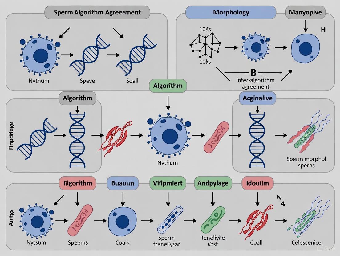
The diagram below illustrates the logical relationships and workflow between the different assessment methodologies and the overarching clinical goal of standardizing diagnosis.
The Scientist's Toolkit: Essential Research Reagents and Materials
Successful research in sperm morphology assessment depends on a suite of reliable reagents and technologies. The following table details key solutions and their functions in experimental workflows.
Table 2: Key Research Reagent Solutions for Sperm Morphology Assessment
| Item Name | Function/Application | Key Characteristics |
|---|---|---|
| Staining Kits (e.g., Diff-Quik, Papanicolaou) | Cytological staining of sperm smears for manual microscopy. | Provides contrast for visualizing sperm head acrosome, nucleus, midpiece, and tail defects [2]. |
| Validated "Ground-Truth" Image Datasets (e.g., SVIA, MHSMA) | Training and validation of AI/ML models for automated sperm analysis. | Contain thousands of sperm images with expert annotations for classification and segmentation tasks [5]. |
| Standardized Morphology Training Tools | Training and re-training of human morphologists to reduce inter-observer variability. | Utilizes expert-consensus-labeled images to train novices via supervised learning principles, improving accuracy to >90% [3]. |
| Computer-Aided Sperm Analysis (CASA) Systems | Automated, high-throughput analysis of sperm concentration, motility, and with advanced modules, morphology. | Integrates with AI algorithms for objective assessment; requires qualification and validation within each laboratory [1] [7]. |
| Sperm DNA Fragmentation (SDF) Assays (e.g., TUNEL, SCD) | Extended examination of sperm nuclear integrity, a key parameter beyond basic morphology. | Quantifies DNA damage, which is increasingly recognized as a critical factor in male infertility and ART outcomes [2]. |
The clinical significance of sperm morphology is undergoing a critical redefinition. The field is moving away from reliance on the percentage of normal forms as a standalone prognostic tool and toward a more sophisticated diagnostic approach. This new paradigm prioritizes the identification of severe monomorphic abnormalities and leverages advanced AI technologies to overcome the long-standing challenges of subjectivity and variability inherent in manual assessment.
Future research must focus on several key areas to solidify this transition. There is a pressing need for large-scale, multi-center studies to clinically validate AI models and establish standardized performance benchmarks. Furthermore, developing explainable AI that can provide diagnostically meaningful insights, not just classifications, will be crucial for bridging the gap between computational output and clinical decision-making. Finally, integrating morphology data with other advanced parameters, such as DNA fragmentation and genetic and epigenetic markers, will pave the way for a truly comprehensive and personalized diagnostic framework for male infertility. For researchers and drug developers, this evolving landscape presents significant opportunities to create novel, standardized tools and therapies that directly address the identified limitations and harness the power of computational biology.
Sperm morphology assessment is a cornerstone of male fertility evaluation, providing critical diagnostic and prognostic information for researchers and clinicians. However, its utility is fundamentally challenged by multiple sources of variability that can compromise result reliability and inter-laboratory comparability. The World Health Organization (WHO) has continually refined its laboratory manual to standardize semen analysis procedures, with the 6th edition emphasizing the importance of robust methodology for fertility diagnosis, assessment of male reproductive health, and guiding assisted reproductive technology choices [9]. Despite these efforts, significant variability persists across three primary domains: staining techniques, classification criteria, and technician subjectivity. This methodological comparison guide examines how these variables influence sperm morphology assessment outcomes, synthesizing experimental data to illuminate their individual and collective impacts on diagnostic accuracy. Understanding these sources of variability is particularly crucial within the emerging research context of inter-algorithm agreement, where consistent input data is essential for validating computational approaches to sperm morphology analysis.
Comparative Analysis of Staining Techniques
Methodological Principles and Protocol Variations
The choice of staining technique directly influences cellular visualization, which subsequently affects morphological classification. Different stains provide varying levels of contrast and definition for specific sperm components, leading to systematic differences in abnormality detection rates.
Diff-Quick Protocol: This rapid, three-step method involves air-drying slides followed by sequential immersion in fixative (0.1% triarylmethane solution for 5 seconds), solution I (0.1% xanthenes solution for 5 seconds), and solution II (0.1% thiazines solution for 5 seconds), concluding with a distilled water rinse and air-drying [10]. Originally developed for hematological examinations, it has been adapted for sperm morphology assessment due to its simplicity and speed.
Spermac Protocol: This specialized spermatological stain provides enhanced structural differentiation through a more complex procedure. Slides are fixed in formaldehyde solution for 5 minutes, then sequentially stained in three solutions: Solution A (containing rose Bengal and neutral red), Solution B (containing pyronin Y, orange G, and phosphomolybdic acid), and Solution C (containing janus green and fast green FCF), with each step lasting 1 minute and interspersed with distilled water washes [10]. The multi-color approach offers superior compartmental differentiation.
Experimental Data: Quantitative Comparison of Staining Efficacy
A 2023 study directly compared these staining methods using semen samples from fifty men, with morphological parameters classified based on Tygerberg criteria and statistical analysis performed using paired t-tests or Wilcoxon rank-sum tests [10]. The findings demonstrate significant staining-dependent variations:
Table 1: Comparison of Sperm Morphology Assessment Between Diff-Quick and Spermac Staining Methods
| Parameter | Diff-Quick Stain (%) | Spermac Stain (%) | p-value |
|---|---|---|---|
| Normal Morphology | 3.98 ± 0.41 | 2.8 ± 0.33 | 0.0385 |
| Head Defects | 93.42 ± 0.66 | 94.24 ± 0.61 | 0.3665 |
| Midpiece Defects | 24.82 ± 2.05 | 55.74 ± 2.06 | <0.0001 |
| Tail Defects | 16.6 ± 1.34 | 14.84 ± 1.39 | 0.3032 |
Data presented as mean ± SEM (Standard Error of the Mean) [10].
The experimental data reveals that Spermac staining detected significantly fewer normal spermatozoa (2.8% vs. 3.98%, p=0.0385) and more than double the rate of midpiece abnormalities (55.74% vs. 24.82%, p<0.0001) compared to Diff-Quick [10]. This discrepancy stems from Spermac's superior visualization of the midpiece, providing clearer demarcation of its boundaries and enabling more accurate identification of structural abnormalities in this region. Conversely, Diff-Quick's limited capacity to delineate midpiece thickness likely resulted in underestimation of defects, contributing to its higher normal morphology percentage [10].
Classification Systems: Evolving Criteria and Diagnostic Implications
WHO Criteria Versus Strict (Tygerberg) Criteria
Sperm morphology classification has evolved significantly, with two primary systems employed in clinical and research settings:
WHO Criteria: The traditional WHO classification system defines a morphologically normal spermatozoon as having an oval head with a well-defined acrosome covering 40-70% of the head area, no neck/midpiece or tail abnormalities, and no cytoplasmic droplets larger than 50% of the sperm head [11]. This system historically used a threshold of ≥30% normal forms for normozoospermia diagnosis.
Strict (Tygerberg) Criteria: Developed to enhance objectivity and reduce variability, the strict criteria impose more stringent parameters: head length of 4.0-5.0 µm, width of 2.5-3.5 µm, length-to-width ratio of 1.50-1.75, well-defined acrosome covering 40-70% of the head, thin midpiece (<1 µm wide and approximately 1.5 times head length), and a thin, uniform, uncoiled tail approximately 45 µm long [11]. All borderline forms are classified as abnormal, with the reference threshold reduced to <4% normal forms for teratozoospermia diagnosis in later WHO editions [10].
Diagnostic Concordance and Clinical Impact
A Croatian study comparing these classification systems in 49 patients found maximal concordance in diagnosing teratozoospermia, with both criteria agreeing in 45 of 49 cases (92% concordance rate) [11]. However, systematic differences emerged in defect detection rates:
Table 2: Sperm Defect Detection Rates by Classification System
| Defect Category | WHO Criteria (%) | Strict Criteria (%) | Statistical Significance |
|---|---|---|---|
| Head Defects | Higher in normozoospermia, asthenozoospermia, and oligoasthenozoospermia groups | Consistently lower | p = 0.001-0.031 |
| Neck/Midpiece Defects | Lower in oligoasthenozoospermia group | Significantly higher | p = 0.005 |
| Tail Defects | Lower in normozoospermia and asthenozoospermia groups | Significantly higher | p = 0.002-0.005 |
Adapted from Čipak et al. [11]
The stringency of classification systems has clinical consequences. Research on intrauterine insemination (IUI) outcomes revealed that between 1996-1997 and 2005-2006, average sperm morphology decreased from 37% to 23% by WHO 3rd criteria and from 8.0% to 4.0% by strict criteria [4]. This "classification drift" increased teratozoospermia diagnoses and diminished the predictive value of morphology for IUI success, as the strong relationship between morphology and pregnancy rates present in the earlier era was no longer evident in the later period [4].
Technician Subjectivity: The Human Factor in Morphology Assessment
Magnitude of Inter-Observer Variability
Despite standardized criteria, technician subjectivity remains a substantial source of variability in sperm morphology assessment. A study examining inter-observer agreement found similar kappa values for both WHO and strict criteria (0.700 vs. 0.715), indicating only a "good" rather than "excellent" level of agreement between technicians [11]. This variability stems from individual differences in interpreting borderline cases and applying classification criteria consistently.
Training as a Standardization Tool
Recent research demonstrates that standardized training can significantly reduce technician variability. A 2025 study utilizing a "Sperm Morphology Assessment Standardisation Training Tool" based on machine learning principles showed remarkable improvements in accuracy and consistency [3].
Table 3: Impact of Standardized Training on Technician Accuracy
| Classification System Complexity | Untrained Accuracy (%) | Trained Accuracy (%) | Improvement |
|---|---|---|---|
| 2-category (normal/abnormal) | 81.0 ± 2.5 | 98 ± 0.43 | +17.0% |
| 5-category (by defect location) | 68 ± 3.59 | 97 ± 0.58 | +29.0% |
| 8-category (specific abnormality types) | 64 ± 3.5 | 96 ± 0.81 | +32.0% |
| 25-category (individual defects) | 53 ± 3.69 | 90 ± 1.38 | +37.0% |
Data from Seymour et al. [3]
The training not only improved accuracy but also reduced assessment time (from 7.0±0.4s to 4.9±0.3s per image) and decreased inter-technician variation [3]. This demonstrates that standardized training incorporating expert consensus labels ("ground truth") and machine learning principles can effectively mitigate human subjectivity in sperm morphology assessment.
Integrated Workflow and Interrelationships
The relationship between staining methods, classification systems, and technician factors follows a sequential workflow pattern where outputs from earlier stages become inputs for subsequent stages, creating cumulative variability.
This workflow visualization illustrates how technical, methodological, and human factors interact sequentially to produce the final morphology assessment. Staining quality directly impacts the application of classification systems, while technician expertise mediates the entire interpretive process. This cascade effect means that variability at any stage propagates through the entire assessment pathway.
Essential Research Reagents and Materials
Standardization across research and clinical settings requires consistent use of high-quality reagents and materials. The following toolkit represents essential components for sperm morphology assessment:
Table 4: Essential Research Reagent Solutions for Sperm Morphology Assessment
| Reagent/Material | Primary Function | Application Notes |
|---|---|---|
| Diff-Quick Stain | Rapid sperm staining | Provides quick differentiation of basic structures; less effective for midpiece visualization [10] |
| Spermac Stain | Multi-color sperm staining | Superior compartmental differentiation; especially effective for midpiece assessment [10] |
| Giemsa Stain | General sperm staining | Traditional method for WHO criteria assessment; requires temperature control [11] |
| Eosin-Nigrosin | Vitality staining and morphology | Differentiates live/dead sperm; suitable for field conditions [12] |
| Formaldehyde Fixative | Cellular structure preservation | Used in Spermac protocol (5 minutes fixation) [10] |
| Triarylmethane Fixative | Rapid cellular fixation | Used in Diff-Quick protocol (5 seconds immersion) [10] |
| Sperm Washing Medium | Semen preparation | Removes seminal plasma; improves staining quality [11] |
| Standardized Classification Atlas | Reference for morphology assessment | Reduces technician subjectivity; improves inter-observer agreement [3] |
The experimental data comprehensively demonstrates that staining methods, classification systems, and technician subjectivity collectively introduce significant variability in sperm morphology assessment. Diff-Quick staining yields higher normal sperm percentages primarily due to its limited midpiece visualization, while Spermac provides more comprehensive structural assessment but yields lower normal ranges [10]. Classification stringency directly impacts teratozoospermia diagnosis rates, with strict criteria identifying more abnormalities but potentially overdiagnosing fertility impairment in some populations [4] [11]. Finally, technician subjectivity remains a substantial challenge, though standardized training utilizing expert consensus and machine learning principles shows promise for remarkable improvement in both accuracy and consistency [3].
For the research community pursuing inter-algorithm agreement studies, these findings underscore the critical importance of standardizing pre-analytical conditions when developing and validating computational morphology assessment tools. Consistent staining protocols, classification criteria, and comprehensive technician training are foundational prerequisites for generating reliable data sets capable of supporting robust algorithm development. Future standardization efforts should focus on establishing universally accepted staining protocols for specific morphological questions, refining classification systems to better correlate with clinical outcomes, and implementing continuous training and quality control programs to minimize human variability. Only through such comprehensive standardization can sperm morphology assessment fully realize its potential as an objective, reproducible diagnostic tool in both clinical and research settings.
Sperm morphology, the study of the size and shape of spermatozoa, is a cornerstone of male fertility evaluation. It is widely recognized as one of the most significant predictors of fertilization potential, both in natural conception and in assisted reproductive technology (ART) procedures [1] [13]. Despite its clinical importance, sperm morphology assessment remains one of the most challenging and subjective analyses to standardize in the andrology laboratory [3]. The inherent variability in human sperm shapes, combined with differences in staining techniques, microscopy, and operator expertise, has led to the development of multiple classification systems in an effort to improve consistency and prognostic value.
The three predominant systems in use today are the World Health Organization (WHO) guidelines, Kruger's strict criteria, and David's classification (also known as the modified David classification). Each system provides a framework for categorizing sperm as "normal" or "abnormal" based on specific morphological characteristics, but they differ significantly in their stringency, categorization of defects, and clinical application. This guide provides a detailed, objective comparison of these three systems, focusing on their methodological approaches, reliability, and clinical correlations, framed within the context of inter-algorithm agreement in sperm morphology research.
Comparative Analysis of Classification Systems
The following section provides a systematic comparison of the three main sperm morphology classification systems, detailing their fundamental principles, technical requirements, and performance characteristics.
Table 1: Key Characteristics of Sperm Morphology Classification Systems
| Feature | WHO Guidelines | Kruger Strict Criteria | David Classification |
|---|---|---|---|
| Primary Philosophy | Tolerant, descriptive | Highly stringent, prognostic | Detailed, descriptive |
| Classification Basis | Broad descriptive categories | Strict quantitative thresholds | Specific defect-based (12 classes) |
| Key Defects Categorized | Head, midpiece, tail, cytoplasmic droplets | Head (focus), midpiece, tail | 7 head defects, 2 midpiece defects, 3 tail defects |
| Staining Preference | Diff-Quik, Papanicolaou | Papanicolaou | RAL Diagnostics kit |
| Clinical Correlation | Moderate correlation with fertility | Better predictor of IVF success [14] | Used widely; debate on switching to strict criteria [14] |
| Inter-Laboratory Variability | High | Lower due to strict thresholds | High due to complexity |
Table 2: Analytical Performance and Research Findings
| Performance Metric | WHO Guidelines | Kruger Strict Criteria | David Classification |
|---|---|---|---|
| Correlation with Manual Assessment | - | - | Moderate correlation with strict criteria (r=0.386 for Diff-Quik) [15] |
| Inter-Expert Agreement | High variability [3] | Improved agreement with training | High complexity leads to variability [13] |
| Automation Potential | Challenging due to broad categories | More suitable for AI/automation | Complex for automation due to many classes |
| Key Research Findings | Overestimation of normal forms compared to strict criteria [15] | Abnormal results not reliably predicted by other SA parameters [16] | A 2011 study argued for its replacement by strict criteria for standardization [14] |
Experimental Protocols for Morphology Assessment
The reliability of sperm morphology assessment is heavily dependent on strict adherence to standardized laboratory protocols, from sample preparation to staining and analysis.
Sample Preparation and Staining Protocols
Consistent sample preparation is critical for minimizing analytical variability. According to WHO guidelines, semen smears should be prepared from a well-mixed, liquefied sample. For the David classification, as used in developing the SMD/MSS dataset, samples with a sperm concentration of at least 5 million/mL are selected, while samples with very high concentrations (>200 million/mL) are excluded to prevent image overlap and facilitate the capture of whole sperm [13]. The smears are then air-dried and fixed.
Staining methods vary by classification system:
- Diff-Quik Staining: This rapid, three-step Romanowsky stain is commonly used for WHO assessments and has been shown to produce a higher correlation (r=0.386) between WHO and strict criteria outcomes compared to Papanicolaou staining (r=0.110) in CASA systems [15].
- Papanicolaou Staining: This more complex, multi-step staining method is preferred for Kruger strict criteria evaluations as it provides superior cellular detail, particularly of the sperm head.
- RAL Diagnostics Staining: This kit was specifically used in the SMD/MSS dataset creation for David's classification, demonstrating the variety of staining solutions employed in different laboratories [13].
Microscopy and Image Acquisition
For manual assessment, a bright-field microscope with 100x oil immersion objective is standard. The use of phase-contrast optics is also common, especially for unstained samples. For CASA and AI-based systems, the process involves:
- Using an optical microscope equipped with a digital camera for image acquisition [13].
- Capturing a sufficient number of images (e.g., 37 ± 5 images per sample in the SMD/MSS study) to ensure a representative sample of spermatozoa [13].
- Ensuring each image contains a single spermatozoon for precise morphological classification, which is crucial for building AI training datasets [13].
Classification and Quality Control
A minimum of 200 spermatozoa should be evaluated and classified according to the chosen system's criteria. To establish ground truth data for research and training, the consensus of multiple experts is required. In the SMD/MSS study, each spermatozoon was independently classified by three experts, and the level of agreement (No Agreement, Partial Agreement, or Total Agreement) was analyzed to gauge the inherent complexity of the task [13]. Furthermore, studies have shown that standardized training tools, which use principles of supervised learning from expert-validated image datasets, can significantly improve the accuracy and reduce variation among novice morphologists [3].
Analytical Workflow for Sperm Morphology Assessment
The following diagram illustrates the general experimental workflow for sperm morphology analysis, from sample collection to final classification, highlighting steps where different standards may diverge.
Diagram 1: Sperm morphology assessment workflow.
The Scientist's Toolkit: Essential Reagents and Materials
Successful sperm morphology assessment relies on a set of specific laboratory reagents and instruments. The following table details key solutions and their functions in the analytical process.
Table 3: Essential Research Reagent Solutions for Sperm Morphology Analysis
| Reagent/Material | Primary Function | Application Notes |
|---|---|---|
| Diff-Quik Staining Kit | A rapid Romanowsky-type stain for cytological preparation. | Preferred for WHO assessments; shown to yield better inter-system correlation in CASA [15]. |
| Papanicolaou Stain | A multi-step stain providing detailed cellular morphology. | The preferred method for Kruger strict criteria due to superior nuclear and acrosomal detail. |
| RAL Diagnostics Stain | A staining kit for sperm morphology. | Used in studies employing David's classification for sample preparation [13]. |
| Phase-Contrast Microscope | Enables observation of unstained, live sperm. | Useful for initial motility and basic morphology checks; often used in simple "normal/abnormal" classifications. |
| Bright-Field Microscope | The standard microscope for observing stained cells. | Equipped with a 100x oil immersion objective for detailed morphology assessment of stained smears. |
| Computer-Assisted Semen Analysis (CASA) | Automated system for image acquisition and analysis. | Systems like SCA, IVOS, or CEROS capture images; performance varies, especially with morphology [17]. |
| Quality Control Beads (e.g., Accu-Beads) | Validated quality control beads for personnel training and proficiency testing. | Used to train technicians and standardize analysis across operators and laboratories [17]. |
The comparative analysis of WHO guidelines, Kruger strict criteria, and David classification reveals a fundamental trade-off in sperm morphology assessment: the balance between descriptive detail and analytical consistency. While the WHO system offers a broad overview and David's classification provides detailed defect categorization, the Kruger strict criteria demonstrate superior prognostic value for ART outcomes and better potential for standardization due to its stringent, quantitative thresholds.
The future of sperm morphology assessment lies in technological innovation to overcome human subjectivity. Artificial Intelligence (AI) and deep learning models are showing significant promise in automating the analysis, offering standardization, and accelerating the process [5] [13]. The development of large, high-quality, and expertly annotated datasets is the critical foundation for these technologies. As these AI-based systems evolve and are validated against robust, expert-derived ground truth data, they hold the potential to seamlessly integrate the diagnostic strengths of all three classification systems, ultimately providing andrology laboratories with a tool that is not only consistent and efficient but also deeply insightful for clinical decision-making in male infertility.
Impact of Assessment Variability on Clinical Decision-Making and ART Outcomes
Sperm morphology assessment, the evaluation of the size and shape of spermatozoa, serves as a cornerstone in the diagnostic evaluation of male infertility [18]. This parameter is established as the most prominent component in semen analysis, as it defines fertility status and potential, as well as the course of natural or assisted reproduction [18]. The clinical significance of morphology is profound; it directly influences critical treatment decisions in assisted reproductive technology (ART), particularly the choice between intrauterine insemination (IUI), in vitro fertilization (IVF), and intracytoplasmic sperm injection (ICSI) [18]. When the percentage of sperm with normal morphology falls below 4%, fertilization with IUI and IVF is typically poor, making ICSI the preferred treatment option [18].
Despite its clinical importance, sperm morphology assessment is widely recognized as one of the most challenging semen parameters to standardize due to its highly subjective nature [18] [13]. This variability presents a significant challenge in reproductive medicine, as inconsistent morphological evaluation can lead to misdiagnoses and inadequate treatment of infertile patients [18]. The fundamental issue lies in the inherent subjectivity of the test, which relies heavily on the technician's expertise and visual interpretation [3]. This paper examines the impact of assessment variability on clinical decision-making and ART outcomes, framed within the broader context of inter-algorithm agreement in sperm morphology assessment research, exploring both traditional manual methods and emerging artificial intelligence solutions.
Classification Systems and Reference Value Evolution
The landscape of sperm morphology classification has undergone significant evolution, contributing substantially to inter-laboratory variability. Normal sperm morphology reference values have been dramatically revised over successive World Health Organization (WHO) manuals, descending from ≥80.5% in the 1st edition to ≥14% in the 4th edition, and further reduced to ≥4% in the most recent 5th edition [18]. This progression reflects the ongoing refinement of what constitutes "normal" sperm morphology, but has simultaneously created inconsistency across laboratories using different classification standards.
The complexity of classification systems themselves directly impacts assessment accuracy and variability. Research has demonstrated that more complex classification systems result in lower overall accuracy and higher variability among morphologists [3]. Studies evaluating different category systems revealed significant differences in accuracy, with untrained users achieving 81.0% accuracy for simple 2-category (normal/abnormal) classification, compared to only 53% accuracy for a detailed 25-category system that defines all defects individually [3]. This demonstrates a fundamental trade-off between diagnostic detail and reliability in morphological assessment.
Technical and Human Factors in Assessment Variability
Multiple technical and human factors contribute to the substantial variability observed in sperm morphology assessment:
- Staining Methods and Preparation Techniques: Variations in staining methods (Papanicolaou, Shorr, Diff-Quik) and sample preparation techniques affect morphological interpretation [18]. Centrifugation may alter sperm morphology and must be carefully standardized [18].
- Microscopy and Measurement Inconsistencies: Without an ocular micrometer to accurately measure sperm dimensions, precise morphological evaluation is compromised [18]. The refractive index of immersion oil and microscope calibration further contribute to variability.
- Subjective Interpretation and Expertise Dependence: Manual assessment remains highly subjective and strongly dependent on the technician's experience [13] [3]. Inter-expert agreement analysis reveals significant discrepancies, with experts agreeing on normal/abnormal classification for only 73% of sperm images in some studies [3].
Table 1: Factors Contributing to Variability in Sperm Morphology Assessment
| Factor Category | Specific Source of Variability | Impact on Assessment |
|---|---|---|
| Classification Systems | Evolution of WHO reference values (80.5% → 4%) | Creates historical inconsistencies between laboratories |
| Complexity of classification categories | Higher complexity reduces accuracy (81% vs 53% for 2 vs 25 categories) | |
| Technical Methods | Staining techniques (Papanicolaou, Diff-Quik, Shorr) | Affects cellular detail visualization and interpretation |
| Sample preparation and centrifugation | May artificially alter sperm morphology | |
| Human Factors | Technician expertise and training | Untrained users show high variation (CV=0.28) and lower accuracy |
| Subjective interpretation of criteria | Experts disagree on 27% of normal/abnormal classifications |
Experimental Protocols and Methodological Approaches
Standardized Manual Assessment Protocol
The WHO-endorsed methodology for sperm morphology assessment involves a detailed, multi-step protocol designed to maximize consistency [18]. The process begins with semen sample collection in a sterile container followed by incubation at 37°C for 30 minutes to allow liquefaction. For viscous samples, proteolytic enzymes such as α-chymotrypsin or bromelain may be added. Smear preparation requires placing 10 µL of well-mixed semen on a clean frosted slide and using a second slide at a 45° angle to create a smooth, even smear, which is then air-dried before staining [18].
Staining follows standardized protocols, such as the Diff-Quik method, which consists of sequential immersion in fixative (triarylmethane dye, methanol), solution I (xanthene dye, sodium azide, pH buffer), and solution II (thiazine dye, pH buffer). The stained smear is examined under a bright field microscope with 100× objective and 10× eyepiece, with immersion oil having a refractive index of 1.52. Critical to this process is the use of an ocular micrometer to accurately measure sperm dimensions, as the sperm head should be 5 to 6 µm long and 2.5 to 3.5 µm wide, with specific acrosome (40-70% of head area) and midpiece (same length as head) proportions [18]. At least 200 spermatozoa must be evaluated across replicates, with all borderline forms considered abnormal.
Deep Learning and Artificial Intelligence Protocols
Recent research has developed automated assessment approaches using convolutional neural networks (CNNs) to address human subjectivity. One representative study created the Sperm Morphology Dataset/Medical School of Sfax (SMD/MSS) containing 1,000 images of individual spermatozoa acquired using an MMC computer-assisted semen analysis (CASA) system [13]. Each spermatozoon was manually classified by three experts according to the modified David classification, which includes 12 classes of morphological defects: 7 head defects (tapered, thin, microcephalous, etc.), 2 midpiece defects (cytoplasmic droplet, bent), and 3 tail defects (coiled, short, multiple) [13].
The experimental workflow involved several stages: image acquisition, expert classification and consensus labeling, data augmentation to expand the dataset to 6,035 images, and algorithm development using Python 3.8. The CNN architecture included image pre-processing (denoising, normalization, standardization), database partitioning (80% training, 20% testing), data augmentation, model training, and evaluation. This approach achieved accuracy rates ranging from 55% to 92%, demonstrating the potential for AI to standardize morphological assessment [13].
Standardization Training Protocols
To address variability through improved training, researchers have developed specialized Sperm Morphology Assessment Standardisation Training Tools based on machine learning principles [3]. These tools utilize expert consensus labels ("ground truth") and supervised learning methodologies to train morphologists. The protocol involves iterative testing across different classification systems (2-category, 5-category, 8-category, and 25-category) with immediate feedback on accuracy [3].
In validation studies, novice morphologists underwent repeated training over four weeks, resulting in significant improvement in accuracy (from 82% to 90%) and diagnostic speed (from 7.0 to 4.9 seconds per image). The most substantial improvements occurred after the first intensive day of training, with final accuracy rates reaching 98%, 97%, 96%, and 90% across the 2-, 5-, 8- and 25-category systems respectively [3]. This demonstrates the critical importance of standardized, iterative training in reducing assessment variability.
Diagram 1: Sperm Morphology Assessment Workflow showing parallel manual and AI-assisted methodologies that inform clinical ART decisions. The workflow begins with sample collection and progresses through preparation, imaging, and alternative assessment pathways.
Quantitative Impact on Assessment Accuracy and Consistency
Inter-Expert Agreement and Training Efficacy
The fundamental challenge in sperm morphology assessment is reflected in inter-expert agreement studies, which reveal significant disparities in morphological classification. Analysis of agreement distribution among three experts shows three distinct scenarios: no agreement (NA) among experts, partial agreement (PA) where 2/3 experts concur, and total agreement (TA) where all three experts agree on the same label for all categories [13]. Without standardized training, users demonstrate high variation (coefficient of variation = 0.28) with accuracy scores ranging dramatically from 19% to 77% [3].
Structured training programs produce measurable improvements in assessment quality. Research demonstrates that repeated training over four weeks significantly enhances accuracy (from 82% to 90%) and reduces interpretation time (from 7.0 to 4.9 seconds per image) [3]. The most substantial improvements occur during initial training, with accuracy rates plateauing after the first intensive day. This training effect is consistent across classification system complexities, though absolute accuracy remains inversely related to system complexity.
Table 2: Impact of Training and Classification System Complexity on Assessment Accuracy
| Training Status | 2-Category System (Normal/Abnormal) | 5-Category System (By Location) | 8-Category System (Cattle Veterinary) | 25-Category System (All Defects) |
|---|---|---|---|---|
| Untrained Novices | 81.0% ± 2.5% | 68.0% ± 3.6% | 64.0% ± 3.5% | 53.0% ± 3.7% |
| After 1st Training Day | 94.9% ± 0.7% | 92.9% ± 0.8% | 90.0% ± 0.9% | 82.7% ± 1.1% |
| Final Accuracy (4 Weeks) | 98.0% ± 0.4% | 97.0% ± 0.6% | 96.0% ± 0.8% | 90.0% ± 1.4% |
| Coefficient of Variation | 0.027-0.137 | 0.027-0.137 | 0.027-0.137 | 0.027-0.137 |
Algorithm Performance and Comparative Effectiveness
Emerging artificial intelligence approaches show promising but variable performance in sperm morphology classification. Deep learning models using convolutional neural networks trained on the SMD/MSS dataset demonstrate accuracy ranging from 55% to 92% compared to expert classifications [13]. This performance variability reflects both the challenges of algorithm training and the inherent subjectivity in the "ground truth" expert classifications used for training.
The comparative effectiveness of different assessment methodologies reveals important trade-offs. Manual assessment by trained experts remains the reference standard but suffers from throughput limitations and residual subjectivity. Computer-assisted semen analysis (CASA) systems automate the image acquisition process but have limited ability to accurately distinguish between spermatozoa and cellular debris, and struggle to classify midpiece and tail abnormalities [13]. AI-based approaches offer potential for standardization and increased throughput but require extensive validation and may be limited by training dataset quality and diversity.
Consequences for Clinical Decision-Making and ART Outcomes
Direct Impact on Treatment Pathway Selection
Sperm morphology assessment directly determines clinical treatment pathways in assisted reproduction. The critical threshold of 4% normal forms established in the WHO 5th edition manual serves as a key decision point [18]. When the percentage of morphologically normal sperm is ≥4%, the probability of fertilization with conventional IVF or intrauterine insemination (IUI) is sufficient to justify these less invasive approaches. Conversely, when normal morphology falls below 4%, fertilization rates with IUI and conventional IVF decline significantly, making intracytoplasmic sperm injection (ICSI) the preferred treatment option [18].
Inaccurate morphology assessment therefore directly leads to suboptimal treatment selection. Overestimation of normal forms may result in failed fertilization cycles with conventional IVF, while underestimation may lead to unnecessary use of ICSI with its increased costs, technical demands, and theoretical genetic concerns [18] [19]. This is particularly significant given that ART-conceived pregnancies already demonstrate increased risks of adverse outcomes, including preterm delivery and small for gestational age infants, even after controlling for multiple gestations [20].
Broader Implications for ART Safety and Efficacy
Assessment variability contributes to broader challenges in evaluating ART safety and efficacy. Research has shown associations between ART and adverse perinatal outcomes, including cerebral palsy, autism, neurodevelopmental imprinting disorders, and cancer [19]. However, uncertainty persists regarding whether these outcomes relate to the ART procedures themselves, underlying infertility factors, or other medical and environmental influences. Inconsistent morphology assessment and treatment selection based on variable criteria complicate this determination.
The significant state-level variations in ART outcomes further highlight the impact of assessment and treatment variability. Massachusetts, which has comprehensive insurance coverage for ART services, demonstrates significantly lower rates of twins, triplets, and higher-order births compared to Florida and Michigan, where coverage is more limited [20]. This suggests that financial factors influencing treatment decisions, including those based on morphology assessment, significantly impact multiple gestation rates and associated complications.
Diagram 2: Clinical Decision Impact Pathway illustrating how morphology assessment results direct treatment choices and how assessment errors lead to clinical consequences. The pathway shows how variability contributes to broader ART outcome variations.
The Scientist's Toolkit: Essential Research Reagents and Materials
Table 3: Essential Research Reagents and Materials for Sperm Morphology Assessment
| Item | Specification/Function | Application Context |
|---|---|---|
| Diff-Quik Stain | Triarylmethane dye fixative, xanthene dye (Solution I), thiazine dye (Solution II) | Rapid staining for manual morphology assessment [18] |
| Papanicolaou Stain | Gold standard staining method per WHO guidelines | Reference standard morphology assessment [18] |
| RAL Diagnostics Stain | Commercial staining kit for semen smears | Research use in standardized studies [13] |
| α-Chymotrypsin/Bromelain | Proteolytic enzymes for viscous sample preparation | Liquefaction of viscous semen samples [18] |
| Ocular Micrometer | Microscope calibration for sperm dimension measurement | Essential for accurate head size measurement (5-6 µm × 2.5-3.5 µm) [18] |
| MMC CASA System | Computer-assisted semen analysis with digital camera | Automated image acquisition for AI studies [13] |
| SMD/MSS Dataset | 1,000+ sperm images with expert classifications | Training and validation of AI algorithms [13] |
| Standardized Training Tool | Machine learning-based training with expert consensus | Morphologist training and standardization [3] |
The impact of assessment variability on clinical decision-making and ART outcomes represents a significant challenge in reproductive medicine. Current evidence demonstrates that inconsistency in sperm morphology assessment stems from multiple sources, including evolving classification standards, technical methodological differences, and inherent human subjectivity in interpretation. This variability directly influences critical treatment decisions, particularly the selection between conventional IVF and ICSI, with profound implications for treatment success, economic costs, and patient outcomes.
Promising approaches to address these challenges include standardized training tools based on machine learning principles, which demonstrate significant improvements in assessment accuracy and consistency, and artificial intelligence-based classification systems that offer potential for automation and standardization. Future research directions should focus on validating and refining these approaches across diverse laboratory settings, establishing robust quality control and quality assurance programs, and developing consensus standards that balance diagnostic detail with practical reliability. Through such efforts, the field can progress toward more consistent, accurate morphological assessment that optimizes clinical decision-making and ART outcomes for the millions affected by infertility worldwide.
The Emergence of Algorithm-Based Approaches to Reduce Subjectivity
Sperm morphology analysis, a cornerstone of male fertility evaluation, has long been constrained by its inherent subjectivity. Conventional semen analysis (CSA) relies on visual assessment by laboratory technicians, introducing substantial inter-observer variability that compromises result reproducibility and clinical reliability [3] [5]. This variability stems from multiple factors: the complexity of classification systems encompassing 26 distinct abnormality types, the challenge of evaluating over 200 sperm per sample, and inevitable human fatigue [5]. According to recent validation studies, even expert morphologists demonstrate only 73% agreement on basic normal/abnormal classifications for sperm images, highlighting the profound impact of subjective interpretation [3].
The emergence of artificial intelligence (AI) and machine learning (ML) technologies promises to address these limitations through algorithmic standardization. By applying computational methods to sperm assessment, researchers aim to establish objective, reproducible morphological analysis while minimizing human bias [21] [5]. This comparison guide examines the performance of various algorithm-based approaches against conventional methods, with particular focus on their agreement levels, technical capabilities, and potential to transform clinical andrology practices. The shift toward computational assessment represents not merely technical advancement but a fundamental reorientation toward evidence-based, standardized male fertility evaluation.
Comparative Performance Analysis of Assessment Methodologies
Quantitative Performance Metrics Across Assessment Platforms
Table 1: Correlation and Agreement Between Assessment Methods for Sperm Morphology
| Assessment Method | Comparison Reference | Correlation Coefficient (r) | Key Performance Metrics | Limitations |
|---|---|---|---|---|
| In-house AI Model (Unstained live sperm) | Computer-Aided Semen Analysis | 0.88 [21] | Precision: 0.95 (abnormal), 0.91 (normal); Recall: 0.91 (abnormal), 0.95 (normal) [21] | Requires specialized imaging equipment [21] |
| In-house AI Model (Unstained live sperm) | Conventional Semen Analysis | 0.76 [21] | Test accuracy: 0.93; Processing speed: 0.0056 seconds per image [21] | Limited clinical validation studies [21] |
| Computer-Aided Semen Analysis (CASA) | Conventional Semen Analysis | 0.57 [21] | Correctly classified sperm morphology compared to manual analysis [22] | Higher results for morphology vs. manual method [22] |
| Electro-Optical System (SQA-Vision) | Conventional Semen Analysis | Moderate to high correlation [22] | Acceptable sensitivity and specificity for classification [22] | Performance slightly poorer than CASA for morphology [22] |
| Conventional ML Algorithms (SVM, Bayesian) | Expert Morphologist Consensus | Accuracy: 88.59%-90% [5] | AUC-ROC: 88.59%; Precision rates >90% for sperm head classification [5] | Limited to sperm head analysis only [5] |
Diagnostic Accuracy Across Classification System Complexities
Table 2: Impact of Classification Complexity on Assessment Accuracy
| Classification System | Untrained Morphologist Accuracy | Trained Morphologist Accuracy | Expert Consensus Accuracy | AI Model Performance |
|---|---|---|---|---|
| 2-Category (Normal/Abnormal) | 81.0% ± 2.5% [3] | 98.0% ± 0.43% [3] | 73% agreement [3] | Precision: 0.91-0.95 [21] |
| 5-Category (Head, Midpiece, Tail, Cytoplasmic Droplet, Normal) | 68.0% ± 3.59% [3] | 97.0% ± 0.58% [3] | Not Reported | Not Specifically Reported |
| 8-Category (Pyriform, Knobbed, Vacuoles, etc.) | 64.0% ± 3.5% [3] | 96.0% ± 0.81% [3] | Not Reported | Not Specifically Reported |
| 25-Category (Individual Defects) | 53.0% ± 3.69% [3] | 90.0% ± 1.38% [3] | Not Reported | Not Specifically Reported |
Experimental Protocols and Methodologies
Deep Learning Model Development Protocol
The development of AI models for sperm morphology assessment follows a structured protocol centered on dataset creation, model training, and validation. Recent approaches utilize confocal laser scanning microscopy at 40× magnification to capture high-resolution Z-stack images (0.5 μm interval) of unstained live sperm [21]. This imaging protocol generates approximately 200 sperm images per sample, with each capture containing 2-3 sperm within a 159.7×159.7 μm field at 512×512 pixel resolution [21].
The annotation phase involves manual labeling by embryologists and researchers using specialized programs like LabelImg, with established inter-observer correlation coefficients of 0.95 for normal morphology detection and 1.0 for abnormal morphology detection [21]. Classification follows WHO sixth edition criteria, categorizing sperm into nine distinct datasets based on head shape (smooth oval, length-to-width ratio 1.5-2), vacuole presence, neck characteristics, tail uniformity, and cytoplasmic droplet size [21].
For model architecture, ResNet50 transfer learning models are implemented for deep learning-based classification. Training typically utilizes datasets of 21,600 images with 12,683 annotated sperm, with a standard split of 4,500 images each for normal and abnormal morphology [21]. Performance validation achieves test accuracy of 0.93 after 150 epochs, with precision and recall metrics exceeding 0.91 for both normal and abnormal classification [21].
Conventional Machine Learning Implementation
Traditional machine learning approaches employ distinct methodologies for feature extraction and classification. Conventional ML pipelines begin with shape-based descriptors including Hu moments, Zernike moments, and Fourier descriptors for manual feature extraction [5]. The k-means clustering algorithm serves for initial sperm head localization, complemented by histogram statistical methods for segmentation [5].
For classification, support vector machines (SVM) represent the most frequently employed algorithm, trained on datasets of 1,400-1,540 human sperm cells from multiple donors [5]. Performance evaluation typically incorporates area under the receiver operating characteristic curve (AUC-ROC) and area under the precision-recall curve (AUC-PR), with reported values of 88.59% and 88.67% respectively [5]. Bayesian density estimation models achieve approximately 90% accuracy for sperm head classification into four morphological categories (normal, tapered, pyriform, small/amorphous) [5].
Morphologist Training Validation Protocol
Recent research demonstrates the efficacy of standardized training tools based on machine learning principles. The "Sperm Morphology Assessment Standardisation Training Tool" employs expert consensus labels as "ground truth" for training novice morphologists [3]. Validation studies involve training cohorts of 16-22 participants across multiple classification systems (2-category to 25-category) [3].
The protocol incorporates repeated training over four weeks with initial tests establishing baseline accuracy (81.0% for 2-category), followed by intensive training with visual aids and instructional videos [3]. Post-training assessment reveals significant improvement in accuracy (94.9% for 2-category) and diagnostic speed (reduction from 7.0±0.4s to 4.9±0.3s per image) [3]. This approach demonstrates that standardized training can achieve final accuracy rates of 98.0% for 2-category systems, though more complex 25-category systems plateau at 90.0% accuracy [3].
Visualization of Algorithm-Based Sperm Assessment Workflow
AI Sperm Assessment Workflow
The Scientist's Toolkit: Essential Research Reagents and Materials
Table 3: Essential Research Materials for Algorithm-Based Sperm Morphology Assessment
| Item | Specification | Research Function |
|---|---|---|
| Confocal Laser Scanning Microscope | LSM 800, 40× magnification, Z-stack capability [21] | High-resolution imaging of unstained live sperm without fixation [21] |
| Standardized Slides | Two-chamber slides, 20μm depth (Leja) [21] | Consistent sample preparation for reliable imaging [21] |
| Annotation Software | LabelImg program [21] | Manual annotation by embryologists for ground truth establishment [21] |
| Staining Solutions | Diff-Quik stain (Romanowsky variant) [21] | Conventional staining for CASA and manual assessment comparison [21] |
| CASA Systems | IVOS II (Hamilton Thorne) or SCA (Microptic) [21] [22] | Automated semen analysis for method comparison [21] [22] |
| AI Training Datasets | HSMA-DS (1,475 images), MHSMA (1,540 images), SVIA (125,000 annotations) [5] | Model training and validation with diverse sperm morphology examples [5] |
| Computational Resources | ResNet50 architecture, Python/NumPy, GPU acceleration [21] [5] | Deep learning model implementation and training [21] [5] |
Inter-Algorithm Agreement and Clinical Implications
The correlation between algorithm-based approaches and conventional methods reveals substantial variation across platforms. In-house AI models demonstrate strong correlation with CASA (r=0.88) and moderate correlation with conventional semen analysis (r=0.76), outperforming the correlation between CASA and conventional analysis (r=0.57) [21]. This pattern suggests that AI systems may capture morphological features more consistently than human observers, potentially bridging the gap between established automated systems and conventional manual assessment.
Recent clinical guidelines from the French BLEFCO Group question the prognostic value of sperm morphology assessment before assisted reproductive technologies, highlighting the need for more objective assessment methods [1]. Algorithm-based approaches address this limitation by detecting subtle morphological patterns potentially imperceptible to human observers, while enabling analysis of unstained live sperm – a significant advantage for clinical applications where sperm viability must be preserved [21]. Furthermore, standardized training tools leveraging machine learning principles demonstrate that morphologist accuracy can be significantly improved (from 81.0% to 98.0% for 2-category systems) through structured training protocols [3], suggesting a hybrid approach combining human expertise with algorithmic standardization may offer optimal outcomes.
The philosophical dimensions of algorithmic subjectivity warrant consideration alongside technical performance. Research in personalized image aesthetic assessment demonstrates that algorithms often reflect the biases of their training datasets, with ground truth labels predicting user choices with highly variable accuracy (55% for some participants, <30% for others) [23]. This underscores the importance of diverse, representative training datasets in sperm morphology algorithms to ensure equitable performance across varied patient populations and clinical contexts. As algorithmic approaches continue to evolve, their integration into clinical practice must balance technical precision with acknowledgment of inherent limitations in capturing the full spectrum of biological variation relevant to male fertility assessment.
Algorithmic Approaches: From Conventional ML to Advanced Deep Learning Architectures
The assessment of sperm morphology is a critical, yet notoriously subjective, component of male fertility diagnostics. Traditional manual analysis, performed by trained embryologists, is plagued by significant inter-observer variability, with studies reporting diagnostic disagreement as high as 40% between expert evaluators [24] [25]. This lack of standardization presents a major challenge for research and clinical practice, directly motivating the investigation into inter-algorithm agreement among automated methods. In this landscape, conventional machine learning (ML) pipelines that leverage robust feature engineering offer a pathway toward objective, reproducible analysis. This guide provides a comparative analysis of two cornerstone algorithms in this domain: K-means clustering for unsupervised pattern discovery and Support Vector Machines (SVM) for supervised classification, evaluating their performance and roles within the specific context of sperm morphology assessment.
Experimental Protocols in Sperm Morphology Analysis
To objectively compare algorithm performance, it is essential to understand the standard experimental workflows and metrics used for validation in this field.
Standardized Morphological Classification and Evaluation Metrics
Researchers typically develop predictive models using datasets of sperm images where the "ground truth" is established by expert andrologists based on standardized classification systems, such as the modified David classification or WHO guidelines [13] [5]. These systems categorize sperm into multiple classes based on defects in the head, midpiece, and tail.
The performance of ML models is then quantified using standard classification metrics and clustering validity indices, which are crucial for inter-algorithm comparison [26] [27].
- Classification Metrics:
- Accuracy: The proportion of total sperm images correctly classified.
- Precision: The ability of the classifier not to label a normal sperm as abnormal.
- Recall (Sensitivity): The ability of the classifier to find all the abnormal sperm.
- F1-Score: The harmonic mean of precision and recall.
- Clustering Metrics:
- Silhouette Score: Measures how similar an object is to its own cluster compared to other clusters. Ranges from -1 to 1, where higher values are better [26].
- Davies-Bouldin Index (DBI): Measures the average similarity between each cluster and its most similar one. Lower values indicate better cluster separation [26].
- Calinski-Harabasz Index (CHI): Measures the ratio of between-cluster dispersion to within-cluster dispersion. Higher values indicate better clustering [27].
Workflow for Conventional Machine Learning Analysis
The following diagram illustrates the generalized workflow for applying conventional machine learning to sperm morphology analysis, highlighting the roles of both K-means and SVM.
Comparative Performance Analysis
The table below summarizes the performance of conventional machine learning models, particularly SVM, as reported in studies on sperm morphology analysis, and contrasts them with deep learning alternatives.
Table 1: Performance Comparison of Machine Learning Models in Sperm Morphology Analysis
| Algorithm | Reported Performance | Key Strengths | Key Limitations / Challenges |
|---|---|---|---|
| SVM with Feature Engineering | ~88-91% Accuracy [5] [24] | High precision (reports of >90%); effective in high-dimensional spaces; robust with clear margin of separation. | Performance heavily dependent on quality of manual feature extraction; may struggle with complex, non-linear morphological patterns. |
| K-means Clustering | Evaluated via validity indices (Silhouette Score, DBI) [26] | Useful for exploratory data analysis; identifies inherent structures/groupings without pre-labeled data. | Purely descriptive; requires post-hoc analysis for clinical relevance; predefined K can be a limitation. |
| Deep Learning (CNN-based) | Up to 96% Accuracy [24] [25] | Automatic feature extraction from raw pixels; handles complex and subtle patterns; state-of-the-art accuracy. | Requires very large datasets (<1000 images often insufficient [13]); computationally intensive; "black box" nature. |
Analysis of Inter-Algorithm Agreement and Disagreement
The performance data reveals a central thesis in automated sperm analysis: the "agreement" between a model's output and expert consensus is a function of its feature representation capability.
SVM Performance: The high precision of SVMs, as demonstrated by Mirsky et al. with rates consistently above 90% [5], shows strong agreement with experts on morphologically distinct classes. However, this agreement can degrade with more subtle or complex anomalies that are difficult to capture with handcrafted features. Chang et al. reported classification accuracy as low as 49% for non-normal sperm heads using Fourier descriptors and SVM, highlighting a significant point of disagreement with expert judgment when feature engineering is inadequate [5].
K-means vs. Supervised Models: As an unsupervised technique, K-means does not directly agree or disagree with expert labels. Instead, its value lies in uncovering inherent structures in the data. A high Silhouette Score indicates that the algorithm agrees with itself on coherent cluster formation. Researchers must then interpret whether these clusters align with biological morphology, creating a different kind of agreement—between data-driven structure and clinical knowledge.
The Performance Gap with Deep Learning: The superior accuracy of deep learning models (e.g., 96.08% [24]) underscores a fundamental limitation of conventional ML. The automated feature extraction in deep learning leads to higher agreement with expert classification because it can learn a more nuanced and comprehensive representation of sperm morphology compared to manually engineered features.
Table 2: Key Research Reagents and Computational Tools for Sperm Morphology ML
| Item / Resource | Function in Research | Example / Note |
|---|---|---|
| Stained Sperm Smears | Creates contrast for microscopic imaging, enabling visualization of morphological details. | RAL Diagnostics staining kit is used following WHO manual guidelines [13]. |
| CASA System | Automated platform for image acquisition and basic morphometric analysis. | MMC CASA system used for image capture with a x100 oil immersion objective [13]. |
| Public Datasets | Benchmark for training and validating new algorithms. | HuSHeM (216 images), SMIDS (3000 images), SMD/MSS (1000+ images) [13] [5] [24]. |
| Feature Extraction Libraries | Software tools to compute handcrafted features from images. | Scikit-image (Python); used for shape descriptors (Hu moments, Zernike moments), texture. |
| Machine Learning Frameworks | Environment for building, training, and evaluating SVM, K-means, and other models. | Scikit-learn (Python) provides implementations of SVM, K-means, and clustering metrics [26]. |
The comparative analysis confirms that while SVM paired with careful feature engineering can achieve solid performance and high precision in classifying sperm morphology, its dependency on manual feature extraction limits its ultimate accuracy and generalizability. K-means clustering serves as a valuable exploratory tool for uncovering hidden patterns in unlabeled data but does not directly produce a diagnostic classification.
The prevailing trend in the field points toward hybrid models and deep learning. Future research for inter-algorithm agreement will likely focus on how conventional ML can augment deep learning, for instance, by using K-means for initial data stratification or SVM for classifying deep features extracted by a CNN—a method that has already pushed accuracies to 96% [24]. As datasets continue to grow in size and quality [13] [5], the role of conventional ML may evolve, but its principles of feature space optimization and model evaluation will remain foundational to developing robust, standardized tools for male fertility assessment.
The assessment of sperm morphology is a cornerstone of male fertility evaluation, yet it remains one of the most challenging and subjective tests in reproductive medicine. Traditional manual morphology assessment suffers from significant inter-laboratory and inter-technician variability, undermining its clinical reliability. A striking demonstration of this limitation comes from a 2022 multisite trial which found poor reproducibility of sperm morphology assessment using World Health Organization Fifth Edition (WHO5) strict grading criteria, with no correlation between fertility centers and a core laboratory for the same semen samples [28]. This variability poses a substantial challenge for both clinical diagnosis and reproductive research.
In response to these challenges, deep learning approaches, particularly Convolutional Neural Networks (CNNs), have emerged as powerful tools for automating sperm classification. These technologies offer the potential to standardize morphology assessment, reduce human subjectivity, and provide rapid, quantitative analyses. This review comprehensively compares the performance of various CNN architectures and traditional machine learning approaches for automated sperm classification, focusing on their experimental validation, technical capabilities, and agreement with expert andrology assessment.
Experimental Protocols and Methodologies in Sperm Imaging AI
Data Acquisition and Preparation Protocols
The foundation of any robust deep learning model is a high-quality, well-annotated dataset. Research in automated sperm classification utilizes diverse acquisition methodologies:
- Brightfield Microscopy with Stained Samples: Conventional approaches using RAL Diagnostics staining kits and optical microscopes with 100x oil immersion objectives provide standard brightfield images for morphological analysis [13]. These typically yield 2D images focusing on head, midpiece, and tail structures.
- Quantitative Phase Imaging (QPI): Advanced imaging using Partially Spatially Coherent Digital Holographic Microscopy (PSC-DHM) provides label-free, quantitative phase maps of sperm cells. This technique offers nanometric sensitivity to subcellular structures and has been used to distinguish normal sperm from those under stress conditions (cryopreservation, oxidative stress, alcohol exposure) [29].
- Video Analysis for Motility Assessment: The VISEM dataset, featuring videos captured at 400-fold magnification with an integrated heated table (37°C), enables both motility tracking and morphological analysis [30]. This multimodal approach captures dynamic movement patterns alongside static morphology.
Dataset Annotation and Ground Truth Establishment
Establishing reliable ground truth labels is crucial for model training and validation. The most rigorous studies employ multi-expert consensus approaches:
- The SMD/MSS Dataset: Three independent experts with extensive experience in semen analysis classified each spermatozoon according to modified David classification, which includes 12 classes of morphological defects (7 head defects, 2 midpiece defects, and 3 tail defects) [13].
- Analysis of Inter-Expert Agreement: Studies typically categorize agreement as: No Agreement (NA), Partial Agreement (PA: 2/3 experts agree), or Total Agreement (TA: 3/3 experts agree). Statistical analysis using Fisher's exact test ensures significant agreement between experts [13].
- Data Augmentation Techniques: To address limited dataset sizes and class imbalance, researchers employ augmentation strategies including rotation, scaling, and contrast adjustments. One study expanded an initial set of 1,000 images to 6,035 images after augmentation [13].
Deep Learning Model Architectures and Training Protocols
Various neural network architectures have been adapted for sperm classification tasks:
- Basic CNN Architectures: Custom CNNs with multiple convolutional layers followed by fully connected layers represent the fundamental approach. These typically operate on pre-processed (denoised, normalized) grayscale images resized to standard dimensions (e.g., 80×80 pixels) [13].
- Transfer Learning Approaches: Pre-trained architectures including VGG16, ResNet-18, ResNet-34, and ResNet-50, initially trained on ImageNet, are fine-tuned for sperm classification tasks. This approach leverages feature hierarchies learned from natural images [30].
- Region-Based CNNs (R-CNN): Faster R-CNN architectures have been implemented for simultaneous sperm head detection and segmentation, particularly useful for motility analysis in video data [30].
- Experimental Validation: Standard practice involves dataset partitioning (typically 80% training, 20% testing), k-fold cross-validation, and performance evaluation using metrics including accuracy, mean absolute error (MAE), sensitivity, specificity, and area under the curve (AUC) [13] [31].
Table 1: Summary of Key Datasets for Sperm Morphology Analysis
| Dataset Name | Sample Size | Image Type | Annotation Classes | Key Features |
|---|---|---|---|---|
| SMD/MSS [13] | 1,000 (extended to 6,035 with augmentation) | Brightfield, stained | 12 classes (modified David classification) | Multi-expert annotation, comprehensive defect classification |
| VISEM-Tracking [32] | 656,334 annotated objects | Video, low-resolution unstained | Detection, tracking, and regression | Multimodal with videos and biological data from 85 participants |
| SVIA [32] | 125,000 annotated instances | Images and videos | Detection, segmentation, classification | Comprehensive annotations for multiple computer vision tasks |
| MHSMA [32] | 1,540 images | Grayscale, non-stained | Acrosome, head shape, vacuoles | Focus on sperm head morphology features |
| QPI Dataset [29] | 10,163 phase maps | Quantitative phase imaging | Normal, cryopreserved, oxidative stress, alcohol affected | Label-free, nanometric sensitivity to subcellular changes |
Comparative Performance Analysis of Algorithm Architectures
Convolutional Neural Networks for Morphology Classification
CNN architectures have demonstrated remarkable capabilities in classifying sperm morphological abnormalities:
- SMD/MSS Dataset Performance: A custom CNN implementation achieved accuracy ranging from 55% to 92% across different morphological classes when trained on the augmented SMD/MSS dataset. This range reflects the varying complexity of distinguishing specific defect types, with some abnormalities being more challenging to classify than others [13].
- ResNet Architecture Applications: Pre-trained ResNet models applied to sperm classification have shown strong performance. One study utilizing ResNet-34 reported high precision in predicting motility and morphology parameters, though specific accuracy figures were not provided in the available excerpt [30].
- Comprehensive Defect Identification: Advanced CNN models can simultaneously identify defects across multiple sperm components (head, midpiece, tail), with one study reporting successful classification of 12 distinct morphological defect categories based on the modified David classification [13].
Deep Neural Networks for Phase Imaging Classification
The integration of QPI with deep learning represents a cutting-edge approach:
- Stress Condition Classification: A feedforward Deep Neural Network (DNN) applied to quantitative phase images of sperm cells achieved 85.6% accuracy in distinguishing normal sperm from those under various stress conditions (cryopreservation, oxidative stress, alcohol exposure). The model demonstrated 85.5% sensitivity and 94.7% specificity on a test dataset of 10,163 phase maps [29].
- Subcellular Sensitivity: The PSC-DHM system provided spatial phase sensitivity of approximately ±20 mrad, enabling visualization of subtle subcellular changes in head, midpiece, and tail structures that are not visible with conventional brightfield microscopy [29].
Region-Based CNNs for Motility and Morphology Analysis
R-CNN architectures enable simultaneous analysis of motility and morphology:
- Sperm Head Detection and Tracking: A Faster R-CNN implementation achieved 91.77% accuracy (95% CI: 91.11-92.43%) for sperm head detection in video sequences from the VISEM dataset. This approach combined detection with a heuristic tracking algorithm to calculate movement speed [30].
- MotionFlow Analysis: A novel motion representation technique called MotionFlow, combined with deep neural networks, achieved a Mean Absolute Error (MAE) of 6.842% for motility and 4.148% for morphology estimation, outperforming existing methods on the VISEM dataset [31].
Ensemble and Traditional Machine Learning Approaches
While deep learning dominates recent research, traditional machine learning methods provide important benchmarks:
- Random Forest Performance: An ensemble Random Forest model applied to clinical pregnancy prediction based on sperm parameters achieved 0.72 accuracy and 0.80 AUC in predicting clinical pregnancy success from IVF/ICSI procedures [33].
- Support Vector Machines (SVM): Traditional SVM approaches have demonstrated strong performance in specific contexts, with one study reporting 88.59% AUC-ROC and precision rates above 90% for classifying sperm heads as normal or abnormal [5].
- Bayesian Density Estimation: One study utilizing Bayesian Density Estimation achieved 90% accuracy in classifying sperm heads into four morphological categories (normal, tapered, pyriform, small/amorphous) [5].
Table 2: Performance Comparison of Algorithm Architectures for Sperm Classification
| Algorithm Type | Reported Performance | Strengths | Limitations |
|---|---|---|---|
| Custom CNN [13] | 55-92% accuracy (varies by morphological class) | Comprehensive defect classification across head, midpiece, tail | Performance varies significantly by defect type |
| DNN with QPI [29] | 85.6% accuracy, 85.5% sensitivity, 94.7% specificity | Label-free imaging, sensitive to subcellular changes | Requires specialized imaging equipment |
| Faster R-CNN [30] | 91.77% detection accuracy | Combined detection and tracking capabilities | Primarily focused on sperm head analysis |
| MotionFlow + DNN [31] | MAE: 6.842% (motility), 4.148% (morphology) | Integrated motion and morphology analysis | Novel technique requiring further validation |
| Random Forest [33] | 0.72 accuracy, 0.80 AUC | Effective for clinical outcome prediction | Limited detailed morphological classification |
| SVM [5] | 88.59% AUC-ROC, >90% precision | Strong performance for binary classification | Requires manual feature engineering |
Table 3: Key Research Reagents and Resources for Automated Sperm Classification
| Resource Category | Specific Examples | Function/Application |
|---|---|---|
| Imaging Systems | MMC CASA System [13] | Standardized image acquisition for brightfield microscopy |
| Partially Spatially Coherent Digital Holographic Microscope (PSC-DHM) [29] | Quantitative phase imaging with nanometric sensitivity | |
| Staining Kits | RAL Diagnostics staining kit [13] | Sperm staining for conventional morphology assessment |
| Public Datasets | VISEM-Tracking [32] | Multimodal video dataset with 656,334 annotated objects |
| SMD/MSS Dataset [13] | 1,000 expert-annotated images with 12-class morphology classification | |
| SVIA Dataset [32] | 125,000 annotated instances for detection, segmentation, classification | |
| Software Frameworks | Python 3.8 with TensorFlow/PyTorch [13] | Deep learning model development and training |
| Scikit-learn, Pandas, NumPy [33] | Traditional machine learning and data analysis | |
| Evaluation Metrics | Accuracy, Sensitivity, Specificity [29] | Standard classification performance measures |
| Mean Absolute Error (MAE) [31] | Regression performance for continuous parameters |
Technological Workflows: From Image Acquisition to Classification
The following diagram illustrates a generalized experimental workflow for deep learning-based sperm classification, integrating multiple approaches from the cited research:
Inter-Algorithm Agreement and Standardization Challenges
While deep learning approaches show considerable promise, several challenges remain in achieving standardized, reproducible sperm classification:
- Dataset Quality and Variability: The performance of deep learning models is heavily dependent on training data quality. Studies consistently note limitations including low resolution, limited sample sizes, insufficient morphological class representation, and inconsistent annotation standards across available datasets [32].
- Algorithm-Generalization Gap: Many algorithms demonstrate strong performance on their development datasets but face challenges in generalizing across different imaging protocols, staining methods, and classification criteria. This mirrors the inter-laboratory variability observed in manual assessment [28].
- Complexity-Accuracy Tradeoff: Research demonstrates that classification accuracy inversely correlates with system complexity. One training study showed final accuracy rates of 98% for 2-category systems (normal/abnormal) declining to 90% for 25-category systems (individual defects) [3]. This reflects the fundamental challenge of detailed morphological classification.
- Clinical Validation Deficit: While technical performance metrics are promising, there remains limited evidence connecting algorithm classifications to clinical outcomes such as fertilization success or live birth rates. The Random Forest model predicting clinical pregnancy represents an important step in addressing this gap [33].
Deep learning approaches, particularly CNN architectures, have demonstrated substantial potential to address the critical standardization challenges in sperm morphology assessment. Current research shows that these algorithms can achieve performance comparable to expert andrologists for specific classification tasks, with accuracies typically ranging from 72% to 92% depending on the complexity of the morphological classification system.
The integration of advanced imaging modalities like quantitative phase imaging with deep neural networks offers particularly promising avenues for future research, enabling label-free analysis with sensitivity to subcellular structures. Furthermore, the development of large, well-annotated public datasets and standardized benchmarking protocols will be essential for validating algorithm performance and promoting clinical adoption.
As these technologies continue to mature, focusing on inter-algorithm agreement, clinical outcome correlation, and operational efficiency will be crucial for translating technical capabilities into improved diagnostic tools that can standardize sperm morphology assessment and enhance male infertility management.
This guide provides an objective comparison of three public datasets—HSMA-DS, SVIA, and SMD/MSS—used for developing deep learning models in sperm morphology analysis. The comparison is framed within the research context of inter-algorithm agreement, examining how dataset characteristics influence the consistency and performance of different computational models.
The following table summarizes the core attributes of the three datasets, which are foundational for training and validating machine learning and deep learning algorithms.
Table 1: Core Dataset Characteristics and Specifications
| Feature | HSMA-DS | SVIA | SMD/MSS |
|---|---|---|---|
| Full Name | Human Sperm Morphology Analysis DataSet | Sperm Videos and Images Analysis dataset | Sperm Morphology Dataset/Medical School of Sfax |
| Primary Modality | Static images [32] | Videos and static images [32] | Static images [13] |
| Sample Size | 1,457 sperm images from 235 patients [32] | 4,041 low-resolution images/videos; 125,000 annotated instances [32] | 1,000 original images, expanded to 6,035 after augmentation [13] |
| Staining | Non-stained [32] | Non-stained [32] | Stained (RAL Diagnostics kit) [13] |
| Key Annotations | Vacuole, tail, midpiece, and head abnormality (binary notation) [32] | 125,000 instances for detection; 26,000 segmentation masks; 125,880 images for classification [32] | 12 morphological defect classes based on modified David classification [13] |
| Primary ML Task | Classification [32] | Detection, Segmentation, and Classification [32] | Classification [13] |
Experimental Protocols and Annotation Methodologies
The reliability of a dataset is directly tied to the rigor of its creation process. The annotation methodology is a critical factor for inter-algorithm agreement, as inconsistencies in the "ground truth" data will be learned and amplified by models.
SVIA Dataset Curation
The SVIA dataset was constructed to support multiple complex computer vision tasks. Its annotations are provided for several tasks: object detection (125,000 annotated instances), semantic segmentation (26,000 segmentation masks), and image classification (125,880 cropped image objects) [32]. This multi-layered annotation approach allows researchers to train models not just to classify sperm, but also to locate them within a larger image and precisely segment their morphological components. This is crucial for developing robust automated sperm analysis systems [32].
SMD/MSS Annotation and Expert Agreement
The SMD/MSS dataset highlights the challenge of subjectivity in creating ground truth. Each of the 1,000 acquired sperm images was independently classified by three expert morphologists according to the modified David classification, which includes 12 classes of defects (e.g., tapered head, microcephalous, bent midpiece, coiled tail) [13].
The study explicitly measured inter-expert agreement as a core part of its protocol. The authors reported three scenarios [13]:
- Total Agreement (TA): All three experts assigned the same label (3/3 agreement).
- Partial Agreement (PA): Two of the three experts agreed on the same label (2/3 agreement).
- No Agreement (NA): There was no consensus among the experts.
This analysis provides a transparent measure of the labeling complexity and reliability for each image, which is vital for understanding potential variations in model performance [13].
HSMA-DS and MHSMA Preprocessing
The HSMA-DS dataset consists of images labeled by experts for specific morphological features using a binary notation (normal or abnormal) [32]. A derived dataset, the Modified Human Sperm Morphology Analysis Dataset (MHSMA), was created by cropping images from HSMA-DS to focus on the sperm heads, resulting in 1,540 grayscale images of size 128x128 or 64x64 pixels [32] [34]. In these cropped images, the sperm tail is not entirely visible, focusing the learning task on head morphology [34].
Experimental Data and Performance Implications
Differences in dataset design directly impact model performance and generalizability, which is a key concern in inter-algorithm agreement studies.
Table 2: Documented Model Performance and Technical Limitations
| Dataset | Reported Model Performance | Noted Limitations |
|---|---|---|
| HSMA-DS/MHSMA | A model trained on MHSMA achieved 90% accuracy in classifying sperm heads into categories like normal and amorphous [32]. | Non-stained, noisy, and low-resolution images; limited sample size and insufficient categories; tail often not visible [32]. |
| SVIA | The dataset is designed for complex tasks like detection and segmentation, but specific accuracy metrics for a baseline model were not provided in the search results [32]. | Comprises low-resolution, unstained grayscale images and videos, which can affect feature clarity [32]. |
| SMD/MSS | A deep learning model (CNN) achieved a wide accuracy range of 55% to 92% [13]. This variability underscores the impact of dataset characteristics and expert disagreement on model outcomes. | Limited number of original images; class imbalance required data augmentation to address; performance variability linked to inter-expert labeling disagreement [13]. |
Experimental Workflow for Dataset Curation
The process of creating a high-quality, annotated dataset for sperm morphology analysis follows a systematic pipeline. The diagram below illustrates the key stages, integrating common steps from the reviewed datasets.
Diagram 1: Sperm Morphology Dataset Curation Workflow. This workflow integrates critical steps like inter-expert agreement analysis and data augmentation, which are essential for enhancing dataset quality and reliability for algorithm development.
The Scientist's Toolkit: Essential Research Reagents and Materials
Table 3: Key Laboratory Materials and Computational Tools for Sperm Morphology Analysis
| Item Name | Function/Application | Relevance to Dataset Curation |
|---|---|---|
| Optical Microscope | Visualization and image acquisition of sperm samples. | Used with 100x oil immersion objective for SMD/MSS [13]; 400x magnification for VISEM-Tracking [35]. Foundational for all image-based datasets. |
| RAL Diagnostics Staining Kit | Chemical staining of semen smears to enhance contrast and morphological detail. | Specifically used for the SMD/MSS dataset to prepare slides [13]. |
| MMC CASA System | Computer-Assisted Semen Analysis system for automated image acquisition and morphometry. | Used for data acquisition in the SMD/MSS study [13]. |
| Phase-Contrast Optics | Microscope optics that enhance contrast in transparent specimens without staining. | Essential for examining unstained, fresh semen preparations per WHO guidelines [35]. Used for datasets like VISEM-Tracking and SVIA. |
| Python with Deep Learning Libraries | Programming environment for developing Convolutional Neural Networks (CNNs) and other models. | Used to build and train the classification algorithm for the SMD/MSS dataset [13]. |
| Data Augmentation Techniques | Computational methods to artificially expand dataset size and diversity. | Applied to the SMD/MSS dataset, increasing the number of images from 1,000 to 6,035 to balance classes and improve model generalization [13]. |
The choice of dataset fundamentally shapes research outcomes in computational sperm morphology. HSMA-DS and its derivative MHSMA offer a starting point for head-specific classification but are limited by image quality and scope. The SVIA dataset, with its extensive annotations for detection and segmentation, enables the development of more complex, end-to-end analysis systems. The SMD/MSS dataset demonstrates the critical importance of addressing inter-expert disagreement and class imbalance through rigorous annotation protocols and data augmentation.
For researchers focusing on inter-algorithm agreement, these datasets present a trade-off. While larger and more complex datasets like SVIA allow for training on diverse tasks, the higher annotation complexity can introduce new sources of variability. The SMD/MSS dataset, with its published analysis of expert consensus, provides a more transparent foundation for studying and improving algorithmic consistency. The ongoing challenge for the field remains the creation of larger, high-quality, and meticulously annotated datasets to build more reliable and universally applicable models [32].
The morphological classification of sperm represents a critical yet profoundly subjective component of male fertility assessment. Despite its clinical importance, this analysis suffers from significant inter-observer and inter-algorithm variability, challenging the reliability of diagnostic and research outcomes. The fundamental obstacle lies in translating complex, continuous morphological features into discrete, categorical classifications—a process inherently prone to inconsistency. Within this context, performance metrics such as accuracy, precision, and recall transition from abstract statistical concepts to essential tools for quantifying agreement and disagreement between different analytical methods. These metrics provide the rigorous, quantitative framework necessary to evaluate the performance of emerging artificial intelligence (AI) algorithms against conventional manual assessments and to understand the sources of discordance in morphology classification.
The application of these metrics reveals a critical trade-off. While accuracy offers an intuitive measure of overall correctness, its utility diminishes with class imbalance—a hallmark of sperm morphology datasets where normal sperm are often outnumbered by various abnormal types [36] [37]. Precision, measuring the reliability of a positive identification (e.g., a specific defect), and recall, measuring the ability to find all instances of that defect, often exist in tension. Optimizing one typically compromises the other [36] [38]. This precision-recall trade-off is not merely statistical but reflects a fundamental clinical dilemma: is it more costly to miss a defect (false negative) or to misidentify a normal sperm as abnormal (false positive)? The resolution of this question dictates which metric should be prioritized when training and evaluating classification models, directly impacting their clinical applicability and the broader goal of achieving inter-algorithm agreement.
Metric Definitions and Their Computational Foundations
The evaluation of classification models, whether human or algorithmic, relies on a foundation built from four fundamental outcomes derived from a confusion matrix: True Positives (TP), True Negatives (TN), False Positives (FP), and False Negatives (FN) [36] [37]. These elements form the basis for calculating the core performance metrics.
- Accuracy answers the question, "How often is the classifier correct overall?" It is the proportion of all classifications that are correct, calculated as (TP + TN) / (TP + TN + FP + FN) [38]. While simple and intuitive, its weakness is that it can be misleadingly high in imbalanced datasets. A model that always predicts "abnormal" in a dataset where 95% of sperm are abnormal would still achieve 95% accuracy, while failing completely to identify the rare "normal" sperm [36] [37].
- Precision answers, "When the classifier predicts a positive, how often is it correct?" It measures the reliability of positive predictions, calculated as TP / (TP + FP) [36] [38]. A high precision means that when an algorithm flags a sperm as having a "head defect," you can be highly confident it is indeed a head defect. This is crucial when the cost of false alarms (FP) is high.
- Recall (also called Sensitivity or True Positive Rate) answers, "Of all the actual positives, how many did the classifier successfully find?" It measures the model's ability to detect all relevant cases, calculated as TP / (TP + FN) [36] [38]. A high recall means the model misses very few actual defective sperm. This is paramount in scenarios like disease screening or detecting critical defects where missing a positive (FN) is dangerous.
The relationship between these metrics is often characterized by a trade-off, visualized in the diagram below.
To balance precision and recall, the F1-Score is used. It is the harmonic mean of the two, providing a single metric that penalizes extreme values in either direction [36] [39]. The formula is F1 = 2 * (Precision * Recall) / (Precision + Recall). A high F1-score indicates that both false positives and false negatives are reasonably controlled.
Comparative Performance of Classification Algorithms
The performance of sperm morphology classification varies significantly based on the algorithm used and the complexity of the classification task. The following tables synthesize quantitative data from recent research, highlighting the role of different metrics in evaluating inter-algorithm agreement.
Table 1: Performance of Conventional Machine Learning vs. Deep Learning in Sperm Morphology Analysis
| Algorithm Type | Key Features | Reported Accuracy | Strengths | Limitations |
|---|---|---|---|---|
| Conventional ML (e.g., SVM, K-means, Bayesian Density) [5] | Relies on handcrafted features (shape, texture, Hu moments). | 49% - 90% [5] | Interpretable; requires less computational power. | Performance highly dependent on feature engineering; struggles with complex morphological classes beyond the head [5]. |
| Deep Learning (CNN) [13] | Automatic feature extraction from images; hierarchical learning. | 55% - 92% [13] | Superior handling of complex features and large datasets; can classify head, midpiece, and tail defects [13] [5]. | Requires very large, high-quality annotated datasets; "black box" nature [13] [5]. |
Table 2: Impact of Classification System Complexity on Human Expert Agreement and Accuracy
| Classification System Complexity | Number of Categories | Reported Expert Agreement / Accuracy | Context |
|---|---|---|---|
| Simple (Normal/Abnormal) [3] | 2 | 81% (Untrained) to 98% (Trained) [3] | A 2-category system is simpler but provides limited diagnostic information. |
| Moderately Complex [3] | 5 | 68% (Untrained) to 97% (Trained) [3] | Categorizes defects by location (head, midpiece, tail). |
| Highly Complex [3] | 25 | 53% (Untrained) to 90% (Trained) [3] | Provides detailed defect identification but is challenging and has high inter-observer variation. |
The data in Table 2 underscores a critical point: as the granularity of the classification system increases, the agreement between experts and the achievable accuracy decrease. This highlights a fundamental challenge for inter-algorithm agreement—the more complex the taxonomy, the harder it is to achieve consensus, whether among humans or algorithms. This variability is a key reason why some expert groups, such as the French BLEFCO, recommend against using detailed abnormality analysis for prognosticating assisted reproductive technology (ART) outcomes [1].
Experimental Protocols and Methodologies
Deep Learning for Sperm Morphology Classification
A 2025 study by PMC provided a robust protocol for developing a deep-learning model for sperm morphology classification, offering a benchmark for performance metrics [13].
- Objective: To develop a predictive model for sperm morphological evaluation using Convolutional Neural Networks (CNNs) trained on an augmented dataset to automate and standardize analysis [13].
- Dataset: The Sperm Morphology Dataset/Medical School of Sfax (SMD/MSS) was used, starting with 1,000 individual spermatozoa images acquired via a CASA system. The dataset was expanded to 6,035 images after applying data augmentation techniques to balance morphological classes [13].
- Labeling and Ground Truth: Each sperm image was manually classified by three human experts based on the modified David classification (12 classes of defects). A ground truth file was compiled for each image, including the expert classifications and morphometric data. This multi-expert consensus approach is critical for establishing reliable labels for model training and evaluation [13].
- Pre-processing and Model Training: Images underwent pre-processing, including normalization and resizing to 80x80 pixels in grayscale. The dataset was partitioned, with 80% used for training and 20% for testing. A CNN algorithm was then implemented in Python 3.8 for the classification task [13].
- Performance Evaluation: The model's performance was evaluated, yielding an accuracy range of 55% to 92%, demonstrating the potential of deep learning to approach expert-level performance while highlighting the variability depending on the specific defect class [13].
Standardization of Human Morphologists
A 2025 study in Scientific Reports addressed the human side of the variability problem, using metrics to quantify the effectiveness of a standardized training tool [3].
- Objective: To validate a Sperm Morphology Assessment Standardisation Training Tool in improving the accuracy and reducing the variation among novice morphologists across classification systems of varying complexity [3].
- Experimental Design: The study consisted of two experiments. Experiment 1 assessed the baseline accuracy of untrained novices (n=22) across 2-, 5-, 8-, and 25-category classification systems. A second cohort (n=16) was then exposed to visual aids and video training before testing. Experiment 2 evaluated the effect of repeated training over four weeks on accuracy and diagnostic speed [3].
- Outcome Measures: The primary outcome was classification accuracy against expert-derived "ground truth." The time taken to classify each image was also recorded. The study used machine learning principles, treating the trained novices as models being optimized [3].
- Results: Untrained users showed high variation (CV=0.28) and low accuracy in complex categories (53% for 25-category system). Training significantly improved accuracy (to 90% for the 25-category system) and speed (from 7.0s to 4.9s per image), while reducing variation. This protocol demonstrates that standardized training can significantly improve inter-observer agreement, a prerequisite for meaningful algorithm benchmarking [3].
The Scientist's Toolkit: Essential Research Reagents and Materials
Table 3: Key Materials and Reagents for Sperm Morphology Analysis Research
| Item | Function in Research |
|---|---|
| Computer-Assisted Semen Analysis (CASA) System [13] | An automated system comprising a microscope with a digital camera for acquiring and storing sperm images. It provides consistent, high-quality image data essential for both manual analysis and training AI models. |
| Annotated Sperm Morphology Datasets (e.g., SMD/MSS, SVIA) [13] [5] | Public or proprietary datasets of sperm images classified by experts. These are the fundamental "reagents" for training and validating machine learning models. Their size and quality directly limit model performance. |
| Staining Kits (e.g., RAL Diagnostics) [13] | Used to prepare semen smears, enhancing the contrast and visibility of sperm structures (head, midpiece, tail) for more consistent manual and automated analysis. |
| Data Augmentation Techniques [13] | Computational methods used to artificially expand the size and diversity of training datasets by creating modified versions of existing images (e.g., rotations, flips). This helps improve model robustness and generalizability. |
| Standardized Training Tool [3] | Software-based tools that use expert-consensus "ground truth" images to train and standardize human morphologists. This reduces human subjectivity, a major source of noise in dataset creation and model evaluation. |
The pursuit of inter-algorithm agreement in sperm morphology assessment research is fundamentally guided by the disciplined application of performance metrics. Relying solely on accuracy provides an incomplete and potentially misleading picture, especially given the class imbalances inherent in the data. A multi-metric approach, leveraging precision, recall, and the unifying F1-score, is essential to properly evaluate and compare the performance of different classification models.
The evidence indicates that while deep learning models show significant promise in automating classification and reducing subjectivity, their performance is intrinsically linked to the quality of the annotated data and the complexity of the chosen classification system. The high variability observed even among human experts underscores the profound challenge of this task. Therefore, future progress hinges on two parallel efforts: the continued development of robust, transparent AI algorithms and the establishment of standardized, high-quality datasets and training protocols. By rigorously applying the correct performance metrics, the field can move closer to the goal of reliable, reproducible, and clinically valuable sperm morphology assessment.
The assessment of sperm morphology is a cornerstone of male fertility evaluation, yet it remains one of the most challenging and subjective tests in diagnostic andrology. This variability stems from the inherent complexity of sperm morphological classification, which encompasses numerous defect types across the head, midpiece, and tail regions according to WHO standards and other classification systems [5]. While manual assessment has been the traditional approach, its reliance on technician expertise and subjective interpretation has led to significant inter-laboratory variability, potentially impacting clinical decision-making and patient care [17] [3].
Computer-Aided Sperm Analysis (CASA) systems emerged to address these standardization challenges through automated, objective assessment. Initially developed in the 1980s, CASA technology has evolved substantially, with current systems utilizing advanced imaging and machine learning algorithms to analyze sperm concentration, motility, and morphology [17]. The clinical implementation of these systems, however, requires careful validation against established manual methods and consideration of their integration into existing laboratory workflows. This comparison guide examines the real-world performance of CASA systems against manual assessment, with particular focus on inter-algorithm agreement in sperm morphology assessment research.
CASA vs. Manual Assessment: Performance Comparison
Analytical Performance Across Semen Parameters
Comprehensive studies comparing CASA systems with manual assessment reveal a complex performance profile with significant variation across different semen parameters. The table below summarizes key performance metrics based on clinical validation studies:
Table 1: Performance Comparison of CASA Systems Versus Manual Semen Analysis
| Parameter | Correlation Level | Limitations & Challenges | Clinical Implications |
|---|---|---|---|
| Sperm Concentration | High correlation with manual methods [17] | Increased variability in extreme concentrations (<15 million/mL and >60 million/mL) [17] | Reliable for routine clinical use except in severe oligospermia or very high concentrations |
| Sperm Motility | High correlation for total and progressive motility [17] [22] | Inaccurate in samples with high concentration or significant debris [17] | Suitable for most clinical scenarios with appropriate sample quality |
| Sperm Morphology | Highest level of discrepancy [17] | Challenge in distinguishing subtle defects; affected by staining quality and debris [17] [5] | Remains the most significant limitation for full automation |
Recent double-blind prospective studies have demonstrated that automated systems and manual methods show good agreement for sperm concentration and motility, with both CASA and electro-optical systems correctly classifying abnormal samples compared to manual analysis [22]. However, morphology assessment continues to present challenges, with one study noting that "the electro-optical system gave higher results and performed slightly poorer than CASA" for morphology evaluation [22].
Technology-Specific Performance Profiles
Different CASA systems utilize varying technological approaches, which impacts their performance characteristics in clinical settings:
Table 2: Comparison of CASA System Technologies and Their Performance Characteristics
| System Type | Technology | Strengths | Morphology Assessment Limitations |
|---|---|---|---|
| Image-Based Systems (SCA, IVOS, CEROS) | Camera and software for image processing [17] | Direct visualization, trajectory tracking for motility | Difficulty with overlapping sperm, debris misclassification [17] [5] |
| Electro-Optical Systems (SQA-Vision) | Electro-optical signals from moving sperm [22] | Rapid analysis, less affected by debris | Limited morphological detail, algorithm-dependent accuracy [22] |
| AI-Enhanced Systems | Deep learning algorithms [5] [13] | Continuous improvement, pattern recognition | Training data dependency, computational requirements [5] |
A 2021 systematic review concluded that CASA systems represent a valid alternative for evaluating semen parameters in clinical practice, particularly for concentration and motility, but noted that "further technological improvements are required before these devices can one day completely replace the human operator" [17].
Inter-Algorithm Agreement: Statistical Frameworks and Metrics
Fundamentals of Inter-Algorithm Agreement
In the context of sperm morphology assessment, inter-algorithm agreement refers to the consistency between different computational methods or between automated and manual approaches when evaluating the same samples. This concept extends from the established statistical framework of inter-annotator agreement (IAA), which measures how well multiple annotators make the same annotation decisions [40] [41]. For algorithmic validation, this translates to assessing whether different analysis methods produce clinically equivalent results.
The statistical measurement of agreement is particularly important for validating automated systems against manual gold standards. As noted in recent guidelines, "There is insufficient evidence to demonstrate the clinical value of indexes of multiple sperm defects (TZI, SDI, MAI) in investigation of infertility and before ART" [1], highlighting the need for robust agreement metrics rather than simple correlation coefficients.
Key Metrics for Agreement Assessment
Several statistical measures are employed to quantify agreement between sperm assessment methods:
Table 3: Key Statistical Metrics for Assessing Inter-Algorithm Agreement
| Metric | Application | Interpretation | Advantages |
|---|---|---|---|
| Cohen's Kappa | Agreement between two classification methods [40] [41] | -1 (disagreement) to 1 (perfect agreement); >0.8 considered strong agreement [40] | Accounts for chance agreement; suitable for categorical data |
| Intraclass Correlation Coefficient (ICC) | Agreement for continuous measures [41] | 0-1 scale; >0.9 excellent agreement [42] | Handles multiple raters; appropriate for concentration and count metrics |
| Krippendorff's Alpha | Agreement with multiple algorithms or categories [40] [41] | 0-1 scale; >0.8 reliable agreement [40] | Works with multiple annotators, missing data, and various variable types |
In clinical validation studies, these metrics help establish whether automated systems can reliably replace manual methods. For instance, a study on retinal layer segmentation demonstrated "excellent agreement (range 0.980-0.999)" using ICC values [42], providing a benchmark for what constitutes acceptable agreement in medical image analysis.
Experimental Protocols for Validation Studies
Sample Preparation and Method Comparison Protocols
Robust experimental design is essential for validating CASA system performance against manual methods. The following workflow illustrates a standardized protocol for comparative studies:
Diagram 1: Experimental workflow for CASA validation studies
This methodology aligns with approaches used in recent validation studies. For example, one prospective double-blind study compared two automated systems (CASA and electro-optical) with manual assessment across 102 patients, with all operators blinded to each other's results [22]. Such designs minimize bias and provide reliable comparative data.
Expert Consensus and Ground Truth Establishment
For morphology assessment specifically, establishing reliable ground truth is particularly challenging. The protocol below demonstrates how expert consensus is built for algorithm training:
Diagram 2: Ground truth establishment for morphology algorithm development
This approach mirrors methodologies used in recent research, where "each spermatozoon underwent manual classification by three experts" and agreement levels were systematically analyzed [13]. Studies implementing such protocols have demonstrated that with comprehensive training, accuracy rates for morphological classification can reach "98% for 2-category systems and 90% for 25-category systems" [3].
The Scientist's Toolkit: Essential Research Reagents and Materials
Successful implementation of CASA systems requires specific laboratory materials and protocols. The following table details essential components for comparative studies:
Table 4: Essential Research Reagents and Materials for CASA Validation Studies
| Category | Specific Items | Function & Importance | Implementation Notes |
|---|---|---|---|
| Sample Preparation | RAL Diagnostics staining kit [13] | Standardized sperm staining for morphology | Ensures consistent staining across samples |
| Phase-contrast optics [17] | Live sperm analysis without staining | Essential for motility assessment | |
| Quality Control | Latex Accu-Beads [17] | Validation and training for personnel | Critical for standardized quality control |
| Standardized annotation guidelines [40] [3] | Consistent classification across operators | Reduces subjective interpretation | |
| Image Acquisition | MMC CASA system [13] | Image capture and initial analysis | Provides standardized imaging platform |
| Oil immersion 100x objective [13] | High-resolution morphology imaging | Essential for detailed defect identification | |
| Data Management | SMD/MSS dataset [13] | Algorithm training and validation | Contains 1000+ expert-classified images |
| Data augmentation techniques [13] | Expanding limited training datasets | Improves algorithm robustness |
The critical importance of standardized reagents and protocols is highlighted in studies showing that comprehensive training tools can improve novice morphologists' accuracy from 53% to 90% even for complex 25-category classification systems [3].
Clinical Workflow Integration Strategies
Implementation Models for Diagnostic Laboratories
Successfully integrating CASA technology into clinical andrology laboratories requires thoughtful workflow design. The most effective implementation strategies include:
Hybrid Approach: Utilizing CASA for initial high-throughput screening while reserving manual assessment for complex cases, quality control, and verification of abnormal results [17] [22].
Quality Control Integration: Implementing regular internal and external quality control procedures using standardized beads and repeat sample analysis to maintain system accuracy [3] [13].
Staff Training Protocols: Developing comprehensive training programs that combine traditional morphology education with CASA system operation, focusing on image interpretation and result verification [3].
Validation Frameworks: Establishing laboratory-specific validation protocols to verify CASA performance against manual methods using appropriate statistical measures of agreement before full clinical implementation [42] [22].
Recent expert guidelines emphasize that laboratories should "use a qualitative or quantitative method for detection of a monomorphic abnormality" while noting that "there is insufficient evidence to demonstrate the clinical value of indexes of multiple sperm defects" [1], suggesting focused rather than comprehensive automated morphology assessment.
Addressing Implementation Challenges
Several practical challenges emerge when integrating CASA systems into clinical workflows:
Cost-Benefit Considerations: While CASA systems require significant initial investment, they can reduce technician time and improve throughput, potentially offering long-term efficiency gains [22].
Sample Quality Requirements: CASA systems generally require higher sample quality with proper staining and minimal debris to function optimally, necessitating strict adherence to preparation protocols [17] [5].
Result Verification Procedures: Laboratories must establish clear protocols for manual verification of abnormal or borderline results to ensure diagnostic accuracy [1] [22].
The integration of artificial intelligence approaches shows particular promise for addressing current limitations, with recent studies demonstrating that "deep learning model produced satisfactory results, with an accuracy ranging from 55% to 92%" across different morphological classes [13].
The field of automated sperm analysis continues to evolve rapidly, with several promising developments on the horizon:
Artificial Intelligence Enhancement: Deep learning algorithms are increasingly being applied to sperm morphology assessment, with recent studies achieving classification accuracy up to 92% for specific defect categories [5] [13]. These systems have the potential to continuously improve through additional training data.
Expanded Dataset Development: Researchers are addressing current limitations in algorithm performance by creating larger, more diverse datasets with expert-validated classifications. The emerging SVIA dataset, for example, contains "125,000 annotated instances for object detection" and "26,000 segmentation masks" [5].
Standardized Agreement Metrics: The field is moving toward consensus on appropriate statistical measures for inter-algorithm agreement, with increased use of metrics like Krippendorff's Alpha that can handle multiple annotators and complex categorical data [40] [41].
In conclusion, while current CASA systems demonstrate strong agreement with manual methods for sperm concentration and motility assessment, morphology evaluation remains challenging. Successful clinical implementation requires careful validation using appropriate statistical agreement metrics, standardized protocols, and thoughtful workflow integration. As artificial intelligence technologies continue to advance and datasets expand, the reliability and scope of automated sperm analysis are likely to improve, potentially transforming the standard of care in diagnostic andrology.
Addressing Limitations: Dataset Biases, Annotation Consistency, and Algorithm Training
In computational domains such as sperm morphology assessment, the reliability of research findings is not solely a function of algorithmic sophistication. The quality of the underlying datasets—specifically their resolution, sample size, and class representation—fundamentally shapes model performance and the degree to which different algorithms concur in their predictions. This inter-algorithm agreement is a critical indicator of result robustness, especially in clinical and research settings. Variations in these data characteristics can introduce significant uncertainty, limiting the generalizability and clinical applicability of automated assessment tools. This guide objectively compares the influence of these dataset limitations across different machine learning approaches, providing a framework for researchers to evaluate and mitigate these pervasive challenges.
Quantitative Comparison of Dataset Limitations on Model Performance
The following tables synthesize experimental data from numerous studies, illustrating how resolution, sample size, and class representation impact key performance metrics across diverse classification tasks.
Table 1: Impact of Sample Size on Classification Performance and Uncertainty
| Sample Size Range | Overall Accuracy (OA) Range | Observed Uncertainty / IQR of OA | Key Trends & Plateaus | Primary Research Context |
|---|---|---|---|---|
| Very Small (16 - 64) | 67% - 98% [43] | High Variance (e.g., 42% relative change) [43] | Significant accuracy variance; largest relative changes between sizes [43] | Arrhythmia & Heart Attack Data [43] |
| Small (1000 - 2000) | Not Reported | Wider Interquartile Range (IQR) [44] | Lower accuracy with high uncertainty [44] | LULC Mapping with RF [44] |
| Moderate (9000 - 12,000) | >96% [44] | Narrower IQR [44] | Effective accuracy achieved; uncertainty minimized [44] | LULC Mapping with RF [44] |
| Increasing (120 - 2500) | 85% - 99% [43] | Variance reduced to 0.04%-2.2% change [43] | Accuracy plateaus; further sample increases yield diminishing returns [43] | Arrhythmia Data [43] |
Table 2: Effect of Image Resolution on Model Performance and Computational Efficiency
| Input Resolution | Average Performance (ACC/AUC) | Performance Saturation | Computational Cost & Prototyping Recommendation | Primary Research Context |
|---|---|---|---|---|
| 28x28 pixels | Baseline | Lower baseline performance [45] | Lowest cost; suitable for initial prototyping [45] | MedMNIST+ Collection [45] |
| 64x64 pixels | Improved | Progressive improvement from lower resolutions [45] | Moderate cost [45] | MedMNIST+ Collection [45] |
| 128x128 pixels | High | Performance nears saturation [45] | Higher cost; often the best cost-to-performance ratio [45] | MedMNIST+ Collection [45] |
| 224x224 pixels | Highest | Marginal or no gain over 128x128 [45] | Highest cost; not always justified for final model [45] | MedMNIST+ Collection [45] |
Table 3: Influence of Class Representation and Data Quality on Model Performance
| Data Characteristic | Impact on Model Performance | Recommended Mitigation Strategies | Primary Research Context |
|---|---|---|---|
| Imbalanced Class Distribution | Bias towards majority classes; under-representation of rare classes [46] | Oversampling minority classes; targeted sampling for rare classes [46] | Peatland Ecosystem Mapping [46] |
| Low Data Quality / Poor Discriminative Power | Low effect size (~0.2) and accuracy (<70%) [43] | Improve feature selection; augment data quality [43] | Simulated & Real Datasets [43] |
| High Data Dimensionality with Small Samples | Compromised learning; high model uncertainty [46] | Dimensionality reduction; use only uncorrelated, important variables [46] | Peatland Ecosystem Mapping [46] |
| Non-Representative Reference Dataset | Low classification confidence in under-represented image portions [47] | Assess representativeness by comparing reference data to full dataset in feature space [47] | Remote Sensing Classification [47] |
Experimental Protocols for Evaluating Data Limitations
Protocol for Determining Sample Size Adequacy
Objective: To determine the minimum sample size required for a robust and generalizable model without overfitting, using effect size and classifier accuracy [43].
- Data Preparation: Begin with a dataset that has a good discriminative power between classes. If using a real dataset, ensure it is large enough to allow for progressive sub-sampling.
- Iterative Sub-Sampling and Modeling: Systematically create subsets of the data with increasing sample sizes, starting from a very small number (e.g., 16). At each sample size level, perform the following:
- Performance Analysis: For each sample size, record the average classification accuracy and the variance in accuracy across iterations.
- Determine Adequacy: A sample size is considered adequate when it meets two criteria simultaneously:
Protocol for Assessing Image Resolution Impact
Objective: To identify the optimal image resolution that balances performance with computational efficiency, avoiding unnecessary cost for marginal gains [45].
- Dataset Curation: Utilize a dataset collection available in multiple resolutions (e.g., MedMNIST+ at 28x28, 64x64, 128x128, and 224x224 pixels) [45].
- Model Training: Train a diverse set of standard models (e.g., CNNs, Vision Transformers) on each resolution tier. Employ consistent training schemes (e.g., end-to-end, linear probing) across all tests.
- Performance Evaluation: Evaluate models on a held-out test set for each resolution. Record standard performance metrics such as Accuracy (ACC) and Area Under the Curve (AUC).
- Saturation Point Identification: Plot performance metrics against resolution. The point where performance gains from increased resolution become marginal (e.g., from 128x128 to 224x224) is identified as the saturation threshold [45]. For prototyping and potentially deployment, a resolution at or just below this threshold is recommended.
Protocol for Evaluating Class Representation and Feature Quality
Objective: To ensure the training dataset is representative of the feature space of the entire image and to mitigate bias from class imbalance [47] [46].
- Representativeness Assessment:
- Contrast the reference dataset with the full dataset (e.g., the entire image) in the multidimensional feature space [47].
- Use spatial statistics to compute an information density-based confidence metric for the reference dataset [47].
- Generate confidence maps to identify over- and under-represented portions of the image [47].
- Handling Class Imbalance:
- Analyze the distribution of classes in the training data. If a severe imbalance exists, employ resampling techniques.
- For rare classes, use targeted oversampling or design a sampling strategy to increase their proportion in the training data [46].
- For majority classes, undersampling can be applied to reduce bias [46].
- Feature Space Optimization:
Visualizing the Interplay of Dataset Limitations
The following diagram illustrates the logical workflow for diagnosing and mitigating common dataset limitations, connecting the experimental protocols to their intended outcomes.
Diagram 1: A diagnostic workflow for addressing common dataset limitations, linking symptoms and diagnostic protocols to recommended mitigation strategies.
The Scientist's Toolkit: Essential Reagents and Materials
Table 4: Key Research Reagent Solutions for Data-Centric Machine Learning
| Tool / Material | Function in Research | Application Context |
|---|---|---|
| Nested k-Fold Cross-Validation | Provides unbiased accuracy estimates and reduces overfitting, especially critical with small samples [48]. | Model evaluation and selection. |
| Effect Size Calculators (Average & Grand) | Quantifies the discriminative power between classes in a dataset; used to evaluate sample size adequacy [43]. | Data quality assessment and power analysis. |
| Multi-Resolution Benchmark Datasets (e.g., MedMNIST+) | Enables controlled experimentation on the impact of image resolution on model performance [45]. | Medical image model prototyping. |
| Resampling Algorithms (Oversampling/Undersampling) | Adjusts class distribution in training data to mitigate model bias caused by imbalanced datasets [49] [46]. | Preprocessing for classification tasks. |
| Random Forest with Variable Importance | A robust classifier that also provides metrics on feature relevance, aiding in dimensionality reduction [46]. | High-dimensional data classification and feature selection. |
| Information Density & Confidence Metrics | Assesses the representativeness of a reference dataset compared to the full dataset in the feature space [47]. | Quality control for training data selection. |
Data Augmentation Techniques to Overcome Limited Training Data
In data-driven fields like medical research and drug discovery, the performance of machine learning models is often constrained by the availability of high-quality, annotated training data. This challenge is particularly acute in specialized domains such as sperm morphology assessment, where data collection is expensive, time-consuming, and requires expert annotation. Data augmentation has emerged as a powerful strategy to overcome these limitations by artificially expanding training datasets through the creation of modified versions of existing samples. This guide provides a comprehensive comparison of data augmentation techniques, with a specific focus on their application in sperm morphology assessment research, where inter-algorithm agreement and model robustness are critical for clinical adoption.
Fundamental Data Augmentation Techniques
Data augmentation techniques can be broadly categorized into basic and advanced methods. Understanding this taxonomy is essential for selecting the appropriate approach for a given research context.
Basic Image Transformations
Basic data augmentation techniques involve simple transformations that preserve the essential characteristics of the original data while introducing variability [50]:
Geometric Transformations: These alter the spatial properties of images and include flipping (horizontal or vertical), rotation (typically between -30° to 30°), scaling, cropping, and translation (shifting images in different directions) [50] [51]. These help models recognize objects from various viewpoints and positions.
Photometric Transformations: These modify color and lighting properties through brightness and contrast adjustments, color jittering (randomly changing hue, saturation, and color balance), and grayscale conversion [50] [51]. Such transformations make models more adaptable to different cameras and lighting conditions.
Advanced Augmentation Strategies
As the field has evolved, more sophisticated augmentation techniques have emerged [50] [52]:
Generative Methods: Generative AI models, including Generative Adversarial Networks (GANs) and diffusion models, can create realistic variations by changing facial expressions, clothing styles, or even simulating different weather conditions [50]. These models can also fill in missing details or create high-quality synthetic images.
Feature Space Augmentation: Techniques like MixUp (blending two images), CutMix (replacing a section of one image with a part of another), and CutOut (removing random parts of an image) help models learn from multiple contexts and recognize objects even when partially hidden [50].
Comparative Analysis of Augmentation Techniques
Performance Metrics Across Applications
The effectiveness of data augmentation techniques varies significantly across different domains and applications. The table below summarizes experimental results from multiple studies:
Table 1: Performance Comparison of Data Augmentation Techniques Across Domains
| Application Domain | Augmentation Technique | Model Architecture | Key Performance Metrics | Reference |
|---|---|---|---|---|
| Sperm Morphology Classification | Multiple Techniques (Flipping, Rotation, etc.) | Convolutional Neural Network (CNN) | Accuracy: 55% to 92% | [13] |
| Rib Fracture Detection | Traditional (Albumentations) | YOLOv8s | mAP@50: 0.9194, Recall: 0.8196 | [51] |
| Rib Fracture Detection | Focused Augmentation | YOLOv8s | mAP@50: 0.9412, Recall: 0.8766 | [51] |
| Rib Fracture Detection | Traditional (Albumentations) | YOLOv8m | mAP@50: 0.9448 | [51] |
| Rib Fracture Detection | Focused Augmentation | YOLOv8m | mAP@50: 0.9442 | [51] |
| Text Classification | Established Methods | Various Classifiers | Variable performance; cost-effective | [53] |
| Text Classification | LLM-based Augmentation | Various Classifiers | Best with very small seed samples | [53] |
Focused vs. Traditional Augmentation in Medical Imaging
A comparative study on rib fracture detection demonstrated the contextual superiority of different augmentation approaches [51]. Focused data augmentation, which applies specific transformations only to fracture regions rather than the entire image, achieved superior performance for certain metrics and model architectures. Specifically, with the YOLOv8s model, focused augmentation increased the mAP@50 value by 2.18% (reaching 0.9412) and improved recall for fracture detection by 5.70% (reaching 0.8766) compared to traditional augmentation [51]. However, traditional augmentation maintained a slight advantage in overall precision metrics with the YOLOv8m model, highlighting how the optimal technique depends on both the application and model architecture.
Data Augmentation in Sperm Morphology Assessment
Experimental Protocol and Methodology
The application of data augmentation in sperm morphology assessment follows a structured experimental workflow, as demonstrated in a study that developed a predictive model for sperm morphological evaluation [13]:
Data Collection and Preparation: The initial dataset comprised 1,000 images of individual spermatozoa acquired using the MMC CASA system [13]. Samples were prepared from semen obtained from 37 patients, with inclusion criteria requiring a sperm concentration of at least 5 million/mL and varying morphological profiles to maximize examples of different morphological classes.
Expert Annotation and Ground Truth Establishment: Each spermatozoon was manually classified by three experts following the modified David classification, which includes 12 classes of morphological defects (7 head defects, 2 midpiece defects, and 3 tail defects) [13]. This multi-expert approach enabled the assessment of inter-expert agreement, with statistical analysis using Fisher's exact test to evaluate differences between experts.
Data Augmentation Implementation: To address the limited dataset size and class imbalance, multiple augmentation techniques were applied, expanding the dataset from 1,000 to 6,035 images [13]. The specific techniques employed were not detailed in the available literature, but standard approaches for medical imaging include rotation, flipping, brightness adjustment, and contrast modification.
Model Development and Training: A Convolutional Neural Network (CNN) architecture was implemented in Python 3.8, with preprocessing steps including image denoising, normalization, and resizing to 80×80×1 grayscale [13]. The dataset was partitioned with 80% for training and 20% for testing.
Research Reagent Solutions for Sperm Morphology Analysis
Table 2: Essential Research Materials for Sperm Morphology Assessment Studies
| Research Reagent | Specification/Function | Application Context |
|---|---|---|
| MMC CASA System | Microscope with digital camera for sperm image acquisition | Capturing individual spermatozoa images for dataset creation [13] |
| RAL Diagnostics Staining Kit | Standardized staining for sperm morphology visualization | Preparing semen smears for clear morphological assessment [13] |
| Python 3.8 with Deep Learning Libraries | (e.g., TensorFlow, PyTorch, Keras) | Implementing CNN architecture and augmentation pipeline [13] |
| NSG Mice | NOD.Cg-Prkdcscid Il2rgtm1Wjl/SzJ immunodeficient host | Maintaining PDX models for drug response studies [54] |
| Data Augmentation Libraries | (e.g., Albumentations, torchvision transforms) | Implementing geometric and photometric transformations [51] |
Domain-Specific Augmentation Approaches
Text Data Augmentation
In natural language processing, the emergence of Large Language Models (LLMs) has created new opportunities for data augmentation. Recent research comparing LLM-based augmentation with established techniques reveals that LLM-based methods are primarily advantageous when very small numbers of seed samples are available [53]. In many cases, established methods lead to similar or better model accuracies, raising questions about the cost-benefit ratio of LLM-based approaches [53].
Another study challenged conventional wisdom about textual data augmentation, suggesting that classical methods primarily facilitate network training and that their effects diminish with more extensive fine-tuning [55]. The research also indicated that zero- and few-shot DA via conversational agents like ChatGPT can increase performance, suggesting this form of data augmentation may be preferable to classical methods [55].
Drug Discovery Applications
In drug discovery, where data scarcity is particularly pronounced, specialized augmentation approaches have been developed. One study addressed the prediction of drug response in Patient-Derived Xenografts (PDXs) by combining single-drug and drug-pair treatments through homogenized drug representations [54]. This approach allowed training multimodal neural networks without architectural changes, with the augmented model outperforming those trained on non-augmented data.
Another innovative approach for anticancer drug synergy prediction employed a novel drug similarity metric (DACS score) that incorporates both chemical characteristics and molecular targets [56]. This method enabled the substantial expansion of a drug combination dataset from 8,798 to over 6 million combinations, with Random Forest and Gradient Boosting Trees models trained on the augmented data achieving higher accuracy than those trained solely on the original dataset [56].
Limitations and Practical Considerations
While data augmentation offers significant benefits, researchers must consider several limitations [50]:
Limited Data Diversity: Augmented images originate from existing data and cannot introduce completely new patterns or rare perspectives absent from the original dataset.
Potential Data Distortion: Excessive transformations can create unrealistic images that may reduce model accuracy in real-world scenarios.
Increased Computational Requirements: Real-time augmentation during model training demands substantial processing power, potentially slowing training and increasing memory usage.
Persistent Class Imbalance: Augmentation does not create entirely new samples, so underrepresented categories may still lead to biased learning if not properly addressed.
Data augmentation represents a powerful methodology for overcoming the limited training data challenges prevalent in specialized research domains like sperm morphology assessment. The comparative analysis presented in this guide demonstrates that the optimal augmentation strategy depends on multiple factors, including the specific application domain, model architecture, and data characteristics. In medical imaging applications such as sperm morphology classification and rib fracture detection, appropriate augmentation techniques can significantly enhance model performance, with focused approaches sometimes outperforming traditional methods for specific metrics.
For sperm morphology assessment research, where inter-algorithm agreement and clinical reliability are paramount, data augmentation enables the development of more robust and accurate models while mitigating the challenges of limited dataset size and expert annotation variability. As the field advances, techniques incorporating domain-specific knowledge—such as the focused augmentation for medical images or drug similarity metrics for pharmaceutical applications—show particular promise for generating meaningful synthetic data that enhances model generalization and real-world performance.
In sperm morphology assessment research, establishing reliable ground truth is fundamental for developing accurate diagnostic tools, training laboratory personnel, and validating automated systems. Ground truth refers to reference data derived from expert consensus that serves as the benchmark for evaluating other assessments or algorithms [3]. In medical fields relying on subjective interpretation—including sperm morphology analysis—this consensus is typically established through the diagnostic agreement of multiple experts for each image or sample [3]. The profound clinical implications of morphological assessment in male fertility evaluation make robust ground-truth protocols particularly essential [1] [57].
The inter-algorithm agreement in computational sperm analysis depends entirely on the quality of the ground truth used for training and validation. Inconsistencies in reference data propagate through research and development cycles, compromising clinical reliability. Studies consistently reveal significant variability in sperm morphology assessment due to its subjective nature, highlighting why standardized annotation protocols and expert consensus methodologies are critical research components [3] [1]. This guide examines current approaches for establishing ground truth, compares their methodological frameworks, and provides experimental data supporting best practices for the scientific community.
Annotation Protocols: Comparative Methodologies for Ground Truth Establishment
Expert Consensus Approaches
Structured Consensus Meetings: Regular, organized meetings where experts review discrepancies in their independent assessments and reach agreement through discussion. In diabetic retinopathy research, this approach achieved excellent intergrader agreement (kappa = 0.89-0.91) after eight consensus meetings [58]. The process involves identifying discordant classifications, discussing interpretive criteria, and establishing unified guidelines for borderline cases.
Blinded Independent Review with Adjudication: Multiple experts initially classify images independently and blindly. A senior specialist then adjudicates cases where disagreements exist, providing a definitive classification [58]. This method preserves independent assessment while leveraging senior expertise for resolution, making it particularly valuable when complete consensus among all experts is impractical.
Ground Truth by Majority Voting: For each sperm image, the classification assigned by the majority of experts establishes the reference standard. This approach is efficient for large datasets but requires an odd number of reviewers to avoid ties. Its reliability increases with the number of participating experts, though practical constraints often limit this number.
Systematic Training Protocols
Machine Learning-Inspired Training: Applying supervised learning principles to train human morphologists using expert-validated datasets [3]. This approach treats trainees similarly to machine learning models, providing consistent, high-quality labeled data to improve classification accuracy. Research demonstrates this method significantly improves novice accuracy from 53% to 90% in complex classification systems [3].
Progressive Learning Frameworks: Training begins with simpler classification systems (e.g., normal/abnormal) and progressively advances to more complex categorization (e.g., 25 specific defect types) [3]. This graduated approach builds morphological recognition skills systematically, allowing morphologists to develop foundational patterns before addressing subtle distinctions.
Table 1: Impact of Classification System Complexity on Assessment Accuracy
| Classification System | Untrained Accuracy (%) | Trained Accuracy (%) | Training Improvement |
|---|---|---|---|
| 2-category (normal/abnormal) | 81.0 ± 2.5 | 98.0 ± 0.43 | +17.0% |
| 5-category (by defect location) | 68.0 ± 3.59 | 97.0 ± 0.58 | +29.0% |
| 8-category (specific defect types) | 64.0 ± 3.5 | 96.0 ± 0.81 | +32.0% |
| 25-category (individual defects) | 53.0 ± 3.69 | 90.0 ± 1.38 | +37.0% |
Data adapted from sperm morphology training validation study [3]
Quantitative Comparison of Annotation Protocols
Table 2: Performance Metrics of Different Ground Truth Establishment Methods
| Methodological Approach | Inter-Rater Reliability (Kappa/ICC) | Time Investment | Scalability | Best Application Context |
|---|---|---|---|---|
| Structured Consensus Meetings | 0.89-0.91 [58] | High | Moderate | Protocol development, criteria refinement |
| Independent Review with Adjudication | 0.83-0.89 [58] | Moderate-High | Moderate | Research studies, validation datasets |
| Majority Voting | 0.76-0.85 (estimated) | Moderate | High | Large dataset annotation |
| Machine Learning-Inspired Training | 0.49-0.93 (varies by system) [3] [59] | High initially, lower long-term | High | Laboratory standardization, training programs |
Experimental Data: Validating Ground Truth Protocols
Training Intervention Studies
A comprehensive validation study utilizing a Sperm Morphology Assessment Standardisation Training Tool demonstrated significant improvement in novice morphologist accuracy across multiple classification systems [3]. The research involved two experiments: the first assessed untrained accuracy across different classification complexities, while the second evaluated repeated training over four weeks.
Experimental Protocol 1: Baseline Assessment
- Participants: 22 novice morphologists with minimal prior experience
- Classification Systems: 2-category (normal/abnormal), 5-category (by location), 8-category (specific defects), 25-category (individual defects)
- Procedure: Single assessment without training or feedback
- Outcomes: Untrained users achieved 81.0 ± 2.5%, 68 ± 3.59%, 64 ± 3.5%, and 53 ± 3.69% accuracy respectively across increasing complex systems [3]
Experimental Protocol 2: Longitudinal Training
- Participants: 16 novice morphologists
- Intervention: Visual aids, instructional videos, and repeated testing over four weeks (14 tests total)
- Procedure: Progressive training with immediate feedback on accuracy
- Outcomes: Final accuracy rates reached 98 ± 0.43%, 97 ± 0.58%, 96 ± 0.81%, and 90 ± 1.38% across classification systems [3]
The study also recorded significant improvement in diagnostic speed, decreasing from 7.0 ± 0.4 seconds to 4.9 ± 0.3 seconds per image classification, demonstrating increased efficiency alongside accuracy [3].
Consensus Building in Medical Imaging
The INSPIRED study on diabetic retinopathy assessment provides transferable insights for sperm morphology consensus protocols [58]. Their methodology involved:
- Two paired medical retina fellows independently grading images
- Weekly adjudication by a senior specialist to resolve discrepancies
- Structured consensus meetings to discuss challenging cases
- Implementation of a standardized grading grid (INSPIRED grid) for quantitative assessments
This approach achieved excellent intergrader agreement: kappa = 91% for DR severity, 89% for DRSS, and 89% for predominantly peripheral lesions [58]. The successful protocol emphasizes regular consensus meetings and standardized quantification tools.
Figure 1: Expert consensus methodology for establishing reliable ground truth in morphological assessment
Inter-Algorithm Agreement: The Impact of Ground Truth Quality
Ground Truth Consistency and Computational Performance
The relationship between ground truth reliability and algorithmic consistency is demonstrated in deep learning applications for sperm morphology. One study developed a multidimensional morphological analysis system for live sperm using improved FairMOT tracking and BlendMask segmentation algorithms [59]. When validated against experienced andrologists, the system achieved 90.82% morphological accuracy across 1272 samples from multiple tertiary hospitals [59].
Experimental Protocol: Algorithm Validation
- Sample Size: 1272 samples from multiple tertiary hospitals
- Reference Standard: Classification by experienced sperm physicians
- Algorithm Framework: Improved FairMOT tracking with BlendMask segmentation
- Validation Method: Comparison against manual microscopy with statistical agreement measures
- Results: High consistency between system outputs and manual microscopy [59]
This correlation between expert consensus and algorithmic performance underscores how ground truth quality directly impacts inter-algorithm agreement. Variations in reference standards propagate through development cycles, affecting all subsequent analytical tools trained on these benchmarks.
Statistical Measures for Agreement Assessment
Various statistical measures quantify agreement between algorithms and ground truth or between different algorithms:
For Categorical Data (Normal/Abnormal Classification):
- Cohen's Kappa: Measures agreement between two raters correcting for chance [60] [41]
- Fleiss' Kappa: Generalizes Cohen's kappa for multiple raters [60]
- Krippendorff's Alpha: Applicable to multiple raters, various measurement levels [60] [41]
For Ordinal Data (Severity Grading):
- Weighted Kappa: Incorporates magnitude of disagreements [61] [60]
- Intraclass Correlation Coefficient (ICC): Assesses agreement for continuous or ordinal data [60] [58]
For Continuous Data (Morphometric Parameters):
- Concordance Correlation Coefficient (CCC): Measures agreement between continuous measurements [60]
- Bland-Altman Analysis: Visualizes agreement with limits of agreement [58]
Figure 2: Relationship between ground truth quality and inter-algorithm agreement in computational analysis
The Scientist's Toolkit: Essential Research Reagents and Materials
Table 3: Essential Research Materials for Sperm Morphology Ground Truth Studies
| Item/Category | Function/Purpose | Implementation Example |
|---|---|---|
| Staining Solutions (Diff-Quik, Papanicolaou) | Cellular structure visualization for morphological assessment | Quick staining for strict criteria evaluation [57] |
| Standardized Classification Grids | Consistent zoning for quantitative assessment | INSPIRED grid with concentric circles and radial divisions [58] |
| Digital Image Databases | Reference standards for training and validation | Expert-validated sperm images with consensus labels [3] |
| Quality Control Samples | Proficiency testing and longitudinal performance monitoring | External quality control programs (QuaDeGA, UK NEQAS) [3] |
| Annotation Software Platforms | Efficient data labeling and agreement quantification | Custom tools for sperm morphology classification [3] |
| Statistical Analysis Packages (R, Python with irrCAC) | Calculate agreement coefficients and confidence intervals | R package 'irrCAC' for generalized kappa coefficients [60] |
Establishing reliable ground truth through robust expert consensus and annotation protocols remains fundamental for advancing sperm morphology assessment research. The experimental data presented demonstrates that structured training interventions significantly improve assessment accuracy, while standardized statistical measures enable quantitative evaluation of inter-algorithm agreement. As computational methods increasingly augment morphological analysis, the principles of rigorous ground truth establishment become even more critical. Future research directions should prioritize developing international standards for annotation protocols, creating large-scale shared datasets with validated ground truth, and establishing benchmarks for inter-algorithm agreement in clinical applications. These advancements will ultimately enhance diagnostic reliability in male fertility assessment and strengthen the evidence base for treatment decisions in assisted reproductive technologies.
Transfer Learning Approaches for Improved Generalization
The assessment of sperm morphology is a cornerstone of male fertility diagnosis, yet it remains plagued by significant subjectivity and inter-observer variability. Traditional manual analysis, reliant on technician expertise, can result in diagnostic disagreements as high as 40% between expert evaluators [24]. This lack of standardization directly impacts the reliability of infertility diagnoses and treatment pathways. Within this challenging context, deep learning has emerged as a powerful tool for automating and standardizing sperm morphology analysis. However, the development of robust, generalizable deep learning models is often hampered by a common obstacle in medical AI: the scarcity of large, meticulously annotated datasets [5].
Transfer learning has become a critical strategy to overcome data scarcity. It leverages knowledge a model has acquired from a large, general dataset (like ImageNet) and applies it to a specific, data-limited task, such as classifying sperm defects [62]. This approach mitigates overfitting and enhances the model's ability to generalize to new, unseen data. This guide provides a comparative analysis of prominent transfer learning methodologies, evaluating their performance, experimental protocols, and applicability within the critical field of sperm morphology assessment. The focus is on inter-algorithm agreement—the consistency with which different AI models arrive at the same morphological classification—a key metric for building trust in automated diagnostic systems.
Comparative Performance of Transfer Learning Approaches
The table below summarizes the performance of various transfer learning and deep learning strategies as applied to sperm morphology analysis and a analogous complex classification task (birdsong).
Table 1: Performance Comparison of Different Learning Approaches
| Learning Approach | Specific Model/Strategy | Dataset(s) Used | Key Performance Metric(s) | Reported Result |
|---|---|---|---|---|
| Transfer Learning with Fine-tuning [63] | U-Net with Transfer Learning | SCIAN-SpermSegGS | Dice Coefficient (Head/Acrosome/Nucleus) | 0.96 / 0.94 / 0.95 |
| Deep Feature Engineering [24] | CBAM-enhanced ResNet50 + SVM | SMIDS, HuSHeM | Classification Accuracy | 96.08% (SMIDS), 96.77% (HuSHeM) |
| Deep Fine-tuning [64] | Pre-trained Audio Models (on Xeno-canto) | Xeno-canto Bird Songs | In-domain Classification Accuracy | Strong Performance |
| Shallow Fine-tuning [64] | Pre-trained Audio Models (on Soundscapes) | Environmental Soundscapes | Generalization to Soundscapes | Superior Generalization vs. Deep Fine-tuning |
| From-Scratch Training [13] | Custom CNN | SMD/MSS (Sperm Morphology) | Classification Accuracy | 55% to 92% |
| Knowledge Distillation [64] | Student Model from Teacher | Xeno-canto Bird Songs | In-domain Classification Accuracy | Strong Performance (but weaker generalization) |
Detailed Experimental Protocols
To ensure reproducibility and provide a clear understanding of the methodological underpinnings, this section details the experimental protocols for the key approaches cited.
U-Net with Transfer Learning for Sperm Segmentation
Objective: To accurately segment human sperm heads, acrosomes, and nuclei as a precursor to morphological classification [63].
- Dataset and Preprocessing: The model was trained and evaluated on the public SCIAN-SpermSegGS dataset, which contains over two hundred manually segmented sperm cells. Data augmentation techniques were applied to increase the effective dataset size and variability.
- Model Architecture and Training: A U-Net architecture, a convolutional network designed for biomedical image segmentation, was employed. The core of the methodology was the use of transfer learning, where a model pre-trained on a large, general dataset was adapted to the specific task of sperm segmentation.
- Evaluation: Performance was quantified using the Dice coefficient, a metric measuring the overlap between the model's predicted segmentation and the hand-annotated ground truth masks. A Dice score of 1 indicates perfect overlap.
CBAM-enhanced ResNet50 with Deep Feature Engineering
Objective: To achieve state-of-the-art accuracy in sperm morphology classification by integrating attention mechanisms and classical machine learning [24].
- Dataset: The model was rigorously evaluated on two benchmark datasets: SMIDS (3,000 images, 3 classes) and HuSHeM (216 images, 4 classes), using 5-fold cross-validation.
- Model Architecture: A ResNet50 architecture was enhanced with a Convolutional Block Attention Module (CBAM). This module allows the model to sequentially focus on important channels and spatial regions in an image, helping it prioritize relevant morphological features like head shape or tail defects.
- Deep Feature Engineering (DFE) Pipeline:
- Feature Extraction: High-dimensional feature maps were extracted from multiple layers of the CBAM-enhanced ResNet50, including the CBAM, Global Average Pooling (GAP), and Global Max Pooling (GMP) layers.
- Feature Selection: Ten distinct feature selection methods, including Principal Component Analysis (PCA) and Chi-square tests, were applied to reduce noise and dimensionality.
- Classification: The refined feature set was fed into a Support Vector Machine (SVM) with RBF/Linear kernels for the final classification, rather than using the network's native output layer.
Comparative Transfer Learning Strategies in Bioacoustics
Objective: To explore the effectiveness of finetuning versus knowledge distillation for bird sound classification, with a focus on model generalization [64].
- Dataset: Models were pre-trained on the Xeno-canto birdsong collection (in-domain data) and tested for their ability to generalize to complex environmental soundscapes (out-of-domain data).
- Strategies:
- Deep Fine-tuning: All layers of a pre-trained model were adjusted on the target birdsong dataset.
- Shallow Fine-tuning: Only the final layers of a pre-trained model were adjusted, preserving the general features learned in the earlier layers.
- Knowledge Distillation: A new, smaller "student" model was trained to replicate the output of a larger, pre-trained "teacher" model, effectively compressing the knowledge.
- Finding: While both fine-tuning and distillation yielded strong in-domain performance, shallow fine-tuning demonstrated superior generalization to the complex soundscapes, highlighting its robustness when domain shift is a concern.
The following workflow diagram illustrates the core experimental protocol for developing and evaluating a deep learning model for sperm morphology analysis, highlighting where key strategies like transfer learning and data augmentation are integrated.
Building a reliable AI system for sperm morphology analysis requires more than just an algorithm; it depends on a foundation of high-quality data and computational tools. The table below lists key resources mentioned in the cited research.
Table 2: Key Research Reagents and Computational Resources
| Resource Name | Type | Primary Function in Research | Relevant Study |
|---|---|---|---|
| SMD/MSS Dataset | Image Dataset | A dataset of 1,000+ sperm images, augmented to 6,035, classified by experts using modified David classification for training and testing models. | [13] |
| SCIAN-SpermSegGS | Image Dataset | A public dataset with over 200 manually segmented sperm cells, used for validating segmentation methods of sperm parts. | [63] |
| SMIDS & HuSHeM | Image Dataset | Public benchmark datasets (SMIDS: 3,000 images; HuSHeM: 216 images) used for standardized evaluation of classification models. | [24] |
| Convolutional Block Attention Module (CBAM) | Algorithm/Software | A lightweight attention module that enhances CNN performance by forcing the model to focus on semantically relevant image regions. | [24] |
| RAL Diagnostics Staining Kit | Wet Lab Reagent | Used for staining sperm smears to enhance contrast and morphological detail for microscopic imaging. | [13] |
| MMC CASA System | Laboratory Instrument | A Computer-Assisted Semen Analysis system used for acquiring and storing high-quality digital images from sperm smears. | [13] |
The comparative data and methodologies presented in this guide reveal a clear trend: transfer learning and its advanced derivatives, such as deep feature engineering, are pivotal for achieving high-performance, generalizable models in sperm morphology analysis. The superior results of the CBAM-enhanced ResNet50 with DFE [24] and U-Net with transfer learning [63] underscore a critical lesson. Simply applying a standard CNN model yields variable and often suboptimal results (55%-92% accuracy [13]), while leveraging pre-trained knowledge and focusing model attention leads to performance exceeding 96% accuracy, approaching or surpassing expert-level consensus.
From the perspective of inter-algorithm agreement, the choice of transfer learning strategy is paramount. Methods that enhance generalization, such as shallow fine-tuning [64] or DFE with robust feature selection [24], are more likely to produce models that agree with each other and with human experts on challenging, ambiguous cases. These strategies reduce overfitting to spurious patterns in the training data, a common cause of disagreement between models. The use of standardized, public datasets like SMIDS and SCIAN-SpermSegGS is equally crucial, as it provides a common benchmark for evaluating and comparing the agreement of different algorithms.
In conclusion, for researchers and clinicians aiming to deploy AI for sperm morphology assessment, the path toward reliable and standardized diagnosis is best paved with sophisticated transfer learning approaches. Future work should continue to explore the intersection of attention mechanisms, feature engineering, and efficient fine-tuning strategies to further enhance model agreement, interpretability, and ultimately, their clinical utility in reproductive medicine.
Standardization Protocols for Sample Preparation and Image Acquisition
The assessment of sperm morphology is a cornerstone of male fertility evaluation, playing a critical role in both clinical diagnostics and reproductive research. However, this assessment has historically been plagued by significant subjectivity, resulting in substantial inter-laboratory and inter-operator variability [13] [65]. This variability poses a fundamental challenge to research reproducibility and clinical reliability, particularly in studies investigating inter-algorithm agreement for automated sperm analysis systems. Standardization of sample preparation and image acquisition protocols emerges as an essential prerequisite for generating comparable, high-quality data across different research environments and technological platforms. Without such standardization, evaluating the true performance and agreement between different analytical algorithms becomes fundamentally compromised by underlying methodological inconsistencies.
The imperative for standardization extends across the entire analytical workflow, from initial sample collection through final image capture. Variations in staining techniques, microscopy methods, and classification criteria collectively contribute to the observed disparities in morphological assessment outcomes [12] [66]. Within the specific context of inter-algorithm agreement research, these methodological inconsistencies introduce confounding variables that obscure meaningful comparison between computational approaches. This article systematically compares prevailing standardization protocols, examining their efficacy in mitigating variability and facilitating robust comparative analysis of sperm morphology assessment methodologies.
Comparative Analysis of Standardization Approaches
Manual Microscopy Assessment Protocols
Table 1: Standardized Manual Assessment Protocols for Sperm Morphology
| Protocol Component | WHO Laboratory Manual (5th Edition) | University of Queensland Sperm Morphology Standardization Program (UQSMSP) | David's Modified Classification |
|---|---|---|---|
| Staining Method | Papanicolaou stain recommended | Buffered formal saline wet preparations; no staining for DIC | RAL Diagnostics staining kit |
| Microscopy Type | Brightfield or Phase Contrast | Differential Interference Contrast (DIC) at 1000x magnification | Bright field mode with oil immersion x100 objective |
| Sperm Counted | Minimum 200 spermatozoa | Minimum 100 sperm (increased to 200 for borderline cases) | Individual spermatozoa images (1000 initial dataset) |
| Classification System | Binary (Normal/Abnormal) with strict criteria | 8 main categories with subcategories based on functional impact | 12 classes of morphological defects based on head, midpiece, and tail anomalies |
| Quality Control | Internal and external quality control programs | Annual morphologist workshops; competency checks with 5 samples annually | Three-expert classification with inter-expert agreement analysis |
Traditional manual assessment methods rely heavily on technician expertise and standardized staining procedures. The World Health Organization (WHO) provides comprehensive guidelines covering sample collection, liquefaction, preparation, and staining protocols to minimize pre-analytical variability [65]. These protocols emphasize strict adherence to methodological consistency, including controlled abstinence periods (2-7 days), standardized liquefaction timing (30-60 minutes at 37°C), and specific staining techniques such as Papanicolaou stain for morphological evaluation.
Specialized standardization programs like the Australian UQSMSP have implemented even more rigorous protocols, advocating for buffered formal saline wet preparations examined under Differential Interference Contrast (DIC) microscopy at 1000x magnification [66]. This approach is considered the professional gold standard for morphological assessment as it eliminates potential artifacts introduced by staining procedures and provides superior optical clarity for detecting subtle abnormalities. The program mandates specific counting methodologies, including randomization of fields of view and examination of a minimum of 100 sperm cells, with increased counts for borderline cases.
Classification systems represent another critical dimension of standardization, with various frameworks employed including the binary WHO system, David's modified classification (12 defect classes) [13], and the 8-category system with functional thresholds used in Australian veterinary standards [66]. Each system carries distinct implications for inter-rater reliability, with evidence suggesting that more complex classification systems typically result in lower agreement rates among morphologists [3].
Automated and AI-Based Assessment Systems
Table 2: Automated Sperm Analysis Systems and Standardization Approaches
| System Type | Detection Method | Standardization Approach | Reported Correlation with Manual Methods |
|---|---|---|---|
| Computer-Assisted Semen Analysis (CASA) | Sequential image acquisition and algorithmic analysis | Standardized cell identification parameters; calibration with quality control beads | Moderate to high correlation for concentration and motility; variable for morphology |
| Electro-Optical Systems | Electro-optical signals generated by moving spermatozoa | Proprietary algorithms with standardized signal interpretation | Good agreement for concentration and motility; higher variability in morphology |
| Deep Learning-Based Classification | Convolutional Neural Networks (CNNs) trained on expert-validated images | Data augmentation; transfer learning; ground truth establishment via multi-expert consensus | Accuracy ranging from 55% to 92% approaching expert-level performance |
Automated semen analysis systems have emerged to address limitations in manual assessment, primarily through computer-assisted semen analysis (CASA) and electro-optical platforms. These systems theoretically offer enhanced standardization by applying consistent analytical criteria across all samples. Modern CASA systems utilize sophisticated image acquisition protocols, capturing sequential images under standardized lighting and magnification conditions [13] [22]. The MMC CASA system employed in deep learning research, for instance, uses bright field mode with an oil immersion 100x objective for image acquisition, with precise morphometric tools to determine head dimensions and tail length for each spermatozoon [13].
Comparative studies evaluating automated systems reveal important standardization considerations. A double-blind prospective study comparing two automated systems with manual assessment found no significant differences for sperm concentration and motility parameters, but noted greater variability in morphology assessment, particularly with electro-optical systems [22]. This highlights how different detection methodologies can influence results despite standardized sample preparation.
Deep learning approaches represent the most recent innovation in standardization, addressing variability through computational means. These systems require extensive, expertly labeled datasets for training, with protocols such as those used for the SMD/MSS dataset employing data augmentation techniques to expand limited image libraries from 1,000 to over 6,000 images [13]. The establishment of reliable "ground truth" through multi-expert consensus is a critical standardization component in AI-based systems, directly addressing the historical challenge of subjective classification in traditional morphology assessment [3] [67].
Experimental Protocols for Inter-Algorithm Agreement Research
Protocol 1: Multi-Expert Consensus for Ground Truth Establishment
The establishment of reliable ground truth labels represents a foundational requirement for inter-algorithm agreement studies. The protocol implemented in the Sperm Morphology Assessment Standardisation Training Tool development exemplifies this approach [3] [67]:
Image Acquisition: Collect high-resolution images using microscopy systems with standardized specifications. The training tool development utilized an Olympus BX53 microscope with DIC and phase contrast objectives at 40× magnification, with objectives having high numerical apertures (0.75 for phase contrast, 0.95 for DIC) to maximize resolution [67].
Multi-Expert Classification: Engage multiple experienced morphologists (typically three or more) to independently classify each sperm image according to predefined morphological criteria.
Consensus Establishment: Define agreement thresholds (e.g., 100% consensus among all experts or majority agreement) for including images in the ground truth dataset. The training tool development utilized only images with 100% consensus across all three experts (4,821 out of 9,365 images) [67].
Data Augmentation: Apply techniques such as rotation, scaling, and contrast adjustment to expand dataset size and improve algorithm robustness, as demonstrated in research that expanded a dataset from 1,000 to 6,035 images [13].
This protocol directly addresses the subjectivity inherent in sperm morphology assessment by creating a validated reference standard, enabling meaningful comparison between different analytical algorithms.
Protocol 2: Variance Component Analysis for Method Comparison
Variance Component Analysis (VCA) provides a statistical framework for quantifying different sources of variability in method comparison studies:
Experimental Design: Collect semen samples from multiple donors (typically 30+) to ensure biological representation of morphological diversity.
Parallel Processing: Split each sample for analysis by different methods (manual and automated systems) with operators blinded to results from other methods.
Data Collection: Record all morphological classifications with appropriate metadata including operator identity, method used, and time of analysis.
Statistical Analysis: Employ VCA to partition total variance into components attributable to biological variation, methodological differences, operator effects, and random error [68].
This approach enables researchers to determine whether observed differences between algorithms exceed variability introduced by other factors, providing a robust basis for evaluating true inter-algorithm agreement.
Visualization of Standardization Workflows
Sperm Morphology Assessment Standardization Pathway
This diagram illustrates the sequential standardization requirements across the sperm morphology assessment workflow, highlighting critical control points where protocol deviations can introduce variability affecting inter-algorithm agreement studies.
Essential Research Reagent Solutions
Table 3: Key Reagents and Materials for Standardized Sperm Morphology Assessment
| Reagent/Material | Function in Standardization | Application Context |
|---|---|---|
| RAL Diagnostics Staining Kit | Consistent staining for head and acrosomal structure visualization | Modified David classification protocols [13] |
| Buffered Formalin Saline | Sperm preservation without staining artifacts for DIC microscopy | Wet mount preparations in veterinary morphology programs [66] |
| Papanicolaou Stain | Standardized nuclear and acrosomal staining according to WHO guidelines | Clinical andrology laboratories [65] |
| Eosin-Nigrosin Stain | Vital staining for membrane integrity assessment | Field-based bull breeding soundness evaluations [12] |
| α-Chymotrypsin or Bromelain | Viscosity reduction without mechanical distortion | Processing of highly viscous samples per WHO protocols [65] |
| Quality Control Beads | Instrument calibration and performance verification | Regular quality assurance in CASA systems [65] |
| Formalin Buffered Saline | Sample preservation for centralized morphology analysis | Reference laboratory programs with sample transportation [66] |
Implications for Inter-Algorithm Agreement Research
The standardization protocols examined have profound implications for research on inter-algorithm agreement in sperm morphology assessment. Inconsistent sample preparation and image acquisition methodologies introduce significant confounding variables that can obscure true algorithm performance differences. Research indicates that the level of classification complexity directly impacts agreement metrics, with studies showing accuracy rates declining from 94.9% in 2-category systems to 82.7% in 25-category systems even with standardized training [3].
The establishment of reliable ground truth through multi-expert consensus emerges as particularly critical for algorithm validation. Studies implementing this approach demonstrate its effectiveness, with training tools utilizing consensus-validated images significantly improving novice morphologist accuracy from 53% to 90% in complex classification systems [3]. This validation methodology provides the reference standard needed for meaningful algorithm comparison.
Future research directions should prioritize the development of universally accepted reference materials and calibration standards that enable cross-platform comparability. Additionally, standardized reporting frameworks for inter-algorithm studies would enhance meta-analytical capabilities, facilitating broader advancements in automated sperm morphology assessment technologies.
Benchmarking Performance: Correlation Studies and Clinical Validation Frameworks
The assessment of sperm morphology is a cornerstone of male fertility diagnosis, providing critical insights into spermatogenic function and the potential for successful fertilization. For decades, this assessment has relied on two primary methodologies: Conventional Semen Analysis (CSA), a manual microscopic evaluation by a trained professional, and Computer-Aided Semen Analysis (CASA), which automates the analysis of sperm concentration, motility, and sometimes morphology. Despite standardization efforts by the World Health Organization (WHO), these methods are hampered by significant subjectivity, inter-observer variability, and a dependency on staining processes that render sperm unusable for subsequent assisted reproductive technologies (ART) [69] [70]. The emerging application of Artificial Intelligence (AI) models, particularly deep learning, promises to overcome these limitations by offering a fully automated, objective, and highly accurate analysis of sperm morphology. This comparative analysis is framed within a broader thesis on inter-algorithm agreement in sperm morphology assessment research. It objectively evaluates the performance of AI models, CSA, and CASA systems by synthesizing recent experimental data, with the goal of illuminating the path toward a new standard of reliability and clinical utility in male fertility diagnostics.
The following tables consolidate key performance metrics from recent studies, providing a direct comparison of the three methodologies across critical parameters.
Table 1: Overall Performance and Correlation Metrics
| Method | Correlation with CSA (r-value) | Correlation with CASA (r-value) | Key Performance Highlights |
|---|---|---|---|
| AI Model (In-house) | 0.76 [21] | 0.88 [21] | Test Accuracy: 0.93; Precision/Normal: 0.91; Recall/Normal: 0.95 [21] |
| Conventional Semen Analysis (CSA) | - | 0.57 [21] | Considered the historical gold standard but suffers from subjectivity [69] |
| Computer-Aided Semen Analysis (CASA) | 0.57 [21] | - | ICC for Morphology vs. Manual: LensHooke (0.160), SQA-V (0.261) [70] |
Table 2: Diagnostic Agreement and Clinical Impact
| Method | Agreement with Manual (Cohen's κ) | Clinical Workflow Impact | Notable Limitations |
|---|---|---|---|
| AI Model | N/A (New reference) | Assesses unstained, live sperm; maintains sperm viability for ART [21] | Requires large, high-quality annotated datasets for training [7] |
| Conventional Semen Analysis | Gold Standard | Staining required; renders sperm unusable; labor-intensive and time-consuming [21] [71] | High inter- and intra-observer variability [69] |
| CASA | Morphology: Poor (e.g., κ=0.177 for teratozoospermia) [70] | Automated but often requires stained, fixed sperm; can skew IVF/ICSI treatment allocation [70] | Inconsistent morphology results; poor performance in oligozoospermic samples [70] [71] |
Detailed Experimental Protocols
To ensure the reproducibility of cited findings, this section details the core methodologies from two pivotal studies.
Protocol: In-House AI Model for Unstained Sperm Morphology
Objective: To develop and validate a deep learning model for assessing normal sperm morphology in unstained, live sperm using confocal laser scanning microscopy [21].
Materials & Methods:
- Sample Preparation: Semen samples (n=30) were dispensed as a 6 µL droplet onto two-chamber Leja slides with a 20 µm depth.
- Image Acquisition: Sperm images were captured using a confocal laser scanning microscope (LSM 800) at 40x magnification in LSM Z-stack mode. A Z-stack interval of 0.5 µm over a 2 µm range was used to generate high-resolution images.
- Dataset Creation & Annotation: From 21,600 captured images, 12,683 contained annotated sperm. Embryologists and researchers manually annotated sperm using the LabelImg program, achieving a high inter-observer correlation (0.95 for normal, 1.0 for abnormal sperm). Sperm were classified into normal or abnormal categories based on strict WHO sixth edition criteria.
- AI Model Training: A ResNet50 transfer learning model was trained on a subset of 9,000 images (4,500 normal, 4,500 abnormal). The model was trained for 150 epochs and its performance was evaluated on a separate test set.
Outcome: The model achieved a test accuracy of 93%, with 139.7 seconds required to process 25,000 images (~0.0056 s/image) [21].
Protocol: Standardized Training Tool for Morphologist Accuracy
Objective: To validate a "Sperm Morphology Assessment Standardisation Training Tool" developed using machine learning principles for training novice morphologists [3].
Materials & Methods:
- Image Dataset: The tool utilized a robust dataset of 4,821 ram sperm images classified with 100% consensus by three expert morphologists, establishing a "ground truth."
- Experimental Design: The study involved two experiments. Experiment 1 assessed untrained novice morphologists (n=22) across classification systems of varying complexity (2, 5, 8, and 25 categories). A second cohort (n=16) was given a visual aid and video before testing. Experiment 2 evaluated the effect of repeated training over four weeks.
- Testing and Metrics: Users were tested on the tool, which provided instant feedback. Accuracy and the time taken to classify each image were recorded.
Outcome: Untrained users showed high variability and low accuracy (e.g., 53% for the 25-category system). Training significantly improved accuracy (up to 90% for the 25-category system) and diagnostic speed (from 7.0 s to 4.9 s per image) [3].
Visualized Workflows and Relationships
Sperm Morphology Assessment Workflow
Inter-Algorithm Agreement and Performance
The Scientist's Toolkit: Research Reagent Solutions
For researchers aiming to implement or validate advanced sperm morphology assessment techniques, the following core materials and tools are essential.
Table 3: Essential Research Reagents and Materials
| Item Name | Function/Application | Specific Example/Note |
|---|---|---|
| Confocal Laser Scanning Microscope | High-resolution imaging of unstained, live sperm for AI model training. | e.g., LSM 800; enables Z-stack imaging at low magnification (40x) [21]. |
| Standardized Chamber Slides | Ensures consistent sample depth for accurate concentration and morphology analysis. | e.g., Leja 20 µm depth two-chamber slides [21]. |
| Differential Interference Contrast (DIC) Optics | Provides high-contrast, detailed images of unstained sperm without the need for staining. | Critical for creating high-quality training datasets [67]. |
| Sperm Morphology Staining Kits | For fixed-smear preparation required for CSA and some CASA systems. | e.g., Diff-Quik stain (Romanowsky stain variant) [21] [70]. |
| Validated Image Annotation Software | Allows experts to manually label sperm images to create "ground truth" datasets for AI training. | e.g., LabelImg program [21]. |
| Standardized Morphology Training Tool | Trains and assesses morphologist proficiency using expert-consensus "ground truth" images. | Web-based tool with instant feedback; adaptable to multiple species and classification systems [3]. |
| Deep Learning Framework | Platform for developing and training custom AI models for sperm classification. | e.g., ResNet50 transfer learning model for image classification tasks [21]. |
In the field of sperm morphology assessment, a critical predictor of fertility and reproductive health, the lack of standardized methods introduces significant variability into analytical results. This variability poses a substantial challenge for researchers and clinicians who rely on consistent, reproducible data for drug development and diagnostic applications. The central thesis of this guide is that understanding inter-algorithm and inter-laboratory agreement is fundamental to advancing reproducible research in this domain. Despite the importance of sperm morphology assessment as a key predictor of fertility, it remains a subjective test susceptible to human bias without recognized standardization training methods [67]. This comparison guide objectively evaluates the performance of various assessment methodologies, supported by experimental data quantifying their agreement levels, to provide researchers with evidence-based protocol selection criteria.
Methodological Comparison: Experimental Protocols and Performance Data
Prospective, comparative studies have been designed to quantify the intra- and inter-laboratory variability in sperm morphology assessment using strict criteria. These studies investigate the impact of critical methodological variables, including semen preparation, staining techniques, and manual versus computerized analysis systems [72]. The following sections detail the core experimental protocols and present quantitative comparisons of their agreement.
Detailed Experimental Protocols
1. Sample Preparation and Staining Protocol: A total of 54 semen samples are typically studied in a standard experimental design. For each subject, slides are prepared from both liquefied semen and after washing procedures. The staining process involves two principal techniques: Diff-Quik (a rapid Romanowsky stain) and the Papanicolaou method. Stained slides are then subjected to analysis under predefined, strict morphological criteria [72].
2. Intra-Laboratory Assessment Protocol: A blind assessment is performed within a single laboratory. This involves manual analysis by two independent, trained observers and analysis using a computerized sperm morphology system, typically with two separate readings to assess instrument repeatability. The comparisons focus on different sample preparations (liquefied versus washed) and different staining techniques [72].
3. Inter-Laboratory Assessment Protocol: To assess variability across different research centers, an inter-laboratory comparison is conducted. This involves computer readings of prepared slides at two separate centers, alongside comparisons between manual analyses and between manual versus computer analyses. The goal is to quantify the consistency of results across different operational environments and analysts [72].
Quantitative Comparison of Method Agreement
The following table summarizes the key correlation coefficients (Intraclass Correlation Coefficients - ICC) observed from the comparative studies, providing a quantitative measure of agreement across different methodological variables [72].
Table 1: Inter-Method Agreement in Sperm Morphology Assessment
| Comparison Factor | Specific Comparison | Correlation Coefficient (ICC) | Level of Agreement |
|---|---|---|---|
| Sample Preparation | Manual Analysis: Liquefied vs. Washed Samples | ICC = 0.93 | Very Good |
| Staining Technique | Computerized Analysis: Diff-Quik Staining (Washed Samples) | ICC = 0.93 | Very Good |
| Staining Technique | Computerized Analysis: Papanicolaou Staining (Washed Samples) | ICC = 0.66 | Moderate |
| Analysis Mode | Intra-Laboratory: Within-Computer Readings | ICC = 0.93 | Excellent |
| Analysis Mode | Inter-Laboratory: Computer vs. Computer Readings | ICC = 0.72 | Moderate |
| Overall | All Manual vs. All Computer Analyses | ICC = 0.73 | Good |
Visual Workflow of Comparative Experimental Protocol
The DOT script below generates a flowchart illustrating the experimental workflow used to generate the comparability data.
Figure 1: Experimental workflow for method comparison.
The Scientist's Toolkit: Essential Research Reagent Solutions
The consistent execution of sperm morphology assessments relies on a set of core reagents and materials. The following table details key research reagent solutions, their functions, and their role in ensuring analytical validity.
Table 2: Essential Reagents and Materials for Sperm Morphology Assessment
| Reagent/Material | Primary Function in Protocol | Research Application Context |
|---|---|---|
| Diff-Quik Stain | Rapid, standardized Romanowsky-type stain for sperm head and tail structures. | Provides reliable staining for both manual and computerized analysis (ICC=0.93 with computer) [72]. |
| Papanicolaou Stain | Detailed, multi-step cytological stain for nuclear and cytoplasmic details. | Used in traditional morphology assessment; shows higher variability with computerized systems (ICC=0.66) [72]. |
| Computerized Sperm Analyzer | Automated digital imaging system for objective, strict criteria morphology assessment. | Reduces intra-laboratory variability (ICC=0.93); used for high-throughput screening in drug development [72]. |
| Strict Criteria Classification System | Standardized, defined benchmarks for classifying sperm as "normal" or with specific defects. | The foundation for all comparative analyses, minimizing subjective bias between observers and labs [72] [67]. |
| Standardized Buffer Solutions | For sample washing and dilution to maintain sperm viability and morphology during preparation. | Critical for pre-analytical processing, influencing the consistency of results from liquefied vs. washed samples [72]. |
Visualizing the Inter-Algorithm Agreement Landscape
The following diagram synthesizes the relationships and agreement levels between the different methodological components discussed in this guide, based on the experimental correlation data.
Figure 2: Inter-algorithm agreement relationships.
The quantitative data presented demonstrates a spectrum of inter-method agreement, with correlation coefficients ranging from 0.66 to 0.93 depending on specific methodological choices. The evidence indicates that protocols utilizing Diff-Quik staining consistently yield higher agreement levels, especially when paired with computerized analysis. Furthermore, intra-laboratory consistency is markedly higher than inter-laboratory agreement, underscoring the pressing need for standardized training and operational protocols across research centers. For researchers and drug development professionals, these findings are critical for designing robust, reproducible studies. Selecting a protocol with inherently higher agreement, such as computerized analysis of Diff-Quik stained slides, reduces noise and enhances the reliability of data used to evaluate the effects of pharmaceutical compounds or diagnostic techniques. Future efforts must focus on developing standardized training tools, akin to the "ground truth" datasets used in machine learning, to further align human assessors and minimize the documented variation in this vital field of research [67].
In statistics, inter-rater reliability (IRR), also known as inter-rater agreement, inter-observer reliability, or inter-expert concordance, refers to the degree of agreement among independent observers who rate, code, or assess the same phenomenon [73]. In the specific context of sperm morphology assessment research, this translates to the consistency with which different experts or algorithms classify sperm cells into morphological categories (e.g., normal, head defect, tail defect). Assessment tools that rely on ratings must exhibit good inter-rater reliability; otherwise, they cannot be considered valid tests [73]. The establishment of robust validation benchmarks for inter-algorithm agreement is therefore not merely a statistical exercise but a foundational requirement for ensuring that automated and AI-based sperm morphology analysis systems produce clinically reliable and reproducible results.
The core challenge in this field is the inherent subjectivity of morphological assessment. Studies have documented significant variability in the performance and interpretation of the sperm morphology test, raising questions about its analytical reliability and clinical relevance [1]. This variability directly impacts the diagnosis of male infertility and the decisions made regarding assisted reproductive techniques (ART). Consequently, inter-expert agreement analysis serves as the critical bridge between subjective human judgment and the objective, standardized benchmarks needed to validate emerging computational methods.
Foundational Methods for Measuring Agreement
The choice of an appropriate statistical method to quantify agreement is paramount and depends on the type of data being analyzed and the study design [74]. The following table summarizes the most common coefficients used in inter-rater agreement analysis.
Table 1: Key Statistical Measures for Inter-Rater Reliability
| Statistic | Data Type | Number of Raters | Key Characteristics | Interpretation Guide |
|---|---|---|---|---|
| Cohen's Kappa [73] [75] | Categorical (Nominal/Binary) | 2 | Corrects for chance agreement. | Ranges from -1 (complete disagreement) to +1 (perfect agreement). 0 indicates agreement equal to chance. |
| Fleiss' Kappa [73] | Categorical (Nominal/Binary) | >2 | Extends Cohen's Kappa to multiple raters. | Same as Cohen's Kappa. |
| Intraclass Correlation Coefficient (ICC) [73] [74] | Quantitative (Continuous/Ordinal) | ≥2 | Assesses reliability based on variance partitioning; very flexible for different study designs. | Ranges from 0 to 1. Values closer to 1 indicate higher reliability. |
| Krippendorff's Alpha [73] [74] | Nominal, Ordinal, Interval, Ratio | ≥2 | A versatile statistic that can handle multiple raters, any level of measurement, and missing data. | A value of 1 indicates perfect agreement. A value of 0 indicates the absence of reliability. |
| Percentage Agreement [76] [75] | Any | ≥2 | The simplest measure; the proportion of times raters agree. | Does not account for agreement by chance, which can inflate the perceived reliability. |
It is crucial to distinguish between agreement and reliability, as these concepts are often confused [61]. Agreement refers to the exact closeness of the observations or scores assigned by different raters. In contrast, reliability is the ability of a measurement instrument to differentiate between subjects in a population, and it depends on the heterogeneity of that population [61]. For validation benchmarking, where the goal is to ensure different algorithms produce the same exact result, measures of agreement are often the primary focus.
Inter-Expert Agreement in Sperm Morphology Assessment
The Inherent Challenge of Variability
The assessment of sperm morphology is a cornerstone of male fertility evaluation, yet it is plagued by substantial inter- and intra-laboratory variability [1] [3]. This variability stems from the test's subjective nature, the complexity of sperm morphological defects, and a historical lack of standardized training protocols for morphologists [3]. Expert morphologists, for instance, have been shown to agree on a simple normal/abnormal classification for only about 73% of sperm images, highlighting the profound challenge in achieving consensus [3]. This high level of disagreement among human experts sets the stage for the critical need to establish clear benchmarks against which automated systems can be validated.
The Impact of Classification System Complexity
The complexity of the morphological classification system itself is a major factor influencing the level of agreement achievable. Research has demonstrated that as the number of categories increases, the accuracy and agreement between raters (human or algorithmic) decrease significantly.
Table 2: Impact of Classification System Complexity on Assessment Accuracy
| Classification System | Description | Reported Untrained User Accuracy [3] | Reported Accuracy After Training [3] |
|---|---|---|---|
| 2-Category | Normal vs. Abnormal | 81.0% ± 2.5% | 98% ± 0.43% |
| 5-Category | Defects grouped by location (head, midpiece, tail, etc.) | 68% ± 3.59% | 97% ± 0.58% |
| 8-Category | Specific common defects (pyriform, vacuoles, etc.) | 64% ± 3.5% | 96% ± 0.81% |
| 25-Category | All defects defined individually | 53% ± 3.69% | 90% ± 1.38% |
This data clearly indicates that validation benchmarks must be context-specific and defined with reference to the particular classification schema being used. A benchmark of 90% agreement is exceptional for a 25-category system but would be considered poor for a 2-category system.
Establishing Benchmarks for Inter-Algorithm Agreement
The Role of "Ground Truth" and Expert Consensus
The foundation of any validation benchmark is a reliable "ground truth." In machine learning, supervised learning relies on models learning from accurately labeled datasets [3]. Similarly, for validating sperm morphology algorithms, the benchmark must be based on high-quality reference data. This ground truth is established through the consensus of multiple expert morphologists for each sperm image, a method that minimizes the bias of any single expert [3]. The application of this methodology to create a "Sperm Morphology Assessment Standardisation Training Tool" has been shown to significantly improve the accuracy and reduce the variation of novice morphologists, effectively creating a standardized reference for comparison [3]. Therefore, the primary benchmark for any algorithm should be its agreement with this expert-derived ground truth, measured using appropriate statistics like Krippendorff's Alpha or ICC.
Comparative Performance: Conventional ML vs. Deep Learning
The field of automated sperm morphology analysis (SMA) has evolved from conventional machine learning (ML) to deep learning (DL) approaches, and benchmarks must reflect this progression. Conventional ML models, such as Support Vector Machines (SVM) and K-means clustering, often relied on manual feature extraction (e.g., shape, texture) and achieved reported classification accuracies for sperm heads ranging from 49% to 90% [5]. However, these models typically focused only on the sperm head and struggled with generalization across different datasets.
Deep learning algorithms, particularly those based on convolutional neural networks (CNNs), represent a significant advancement. They automate feature extraction and can perform end-to-end segmentation and classification of the complete sperm structure (head, neck, and tail). The primary benchmark for these DL systems is their performance on standardized, high-quality annotated datasets. The move towards larger and more complex datasets, such as the SVIA dataset which contains over 125,000 annotated instances, provides a more rigorous foundation for establishing robust performance benchmarks for modern AI systems [5].
Experimental Protocols for Validation Studies
Protocol for a Ground Truth Consensus Study
Objective: To establish a validated dataset of sperm images for use as a benchmark in inter-algorithm agreement studies.
- Image Selection: Curate a diverse set of hundreds to thousands of sperm images from well-prepared semen slides, representing the full spectrum of morphological normality and abnormality.
- Expert Panel Assembly: Engage a panel of multiple (e.g., 3-5) highly experienced morphologists who are blinded to each other's assessments.
- Classification: Each expert independently classifies every sperm image according to the predefined classification system (e.g., 2, 5, 8, or 25-category).
- Consensus Meeting: For images where there is initial disagreement, experts review the images together to discuss and reach a consensus classification. This final consensus label becomes the "ground truth" for that image.
- Data Archiving: The ground-truthed dataset is archived and made available for training and validating automated systems.
Protocol for an Inter-Algorithm Agreement Analysis
Objective: To compare the performance of multiple algorithms against the ground truth and against each other.
- Algorithm Selection: Select the algorithms for comparison (e.g., a conventional SVM model, a custom CNN, and a pre-trained ResNet model).
- Test Set: Run all algorithms on a standardized test set of images from the ground-truthed dataset.
- Output Collection: Collect the morphological classification output for each image from each algorithm.
- Statistical Analysis: Calculate agreement statistics.
- Algorithm vs. Ground Truth: For each algorithm, calculate its agreement with the ground truth using Percent Agreement and a chance-corrected metric like Krippendorff's Alpha.
- Algorithm vs. Algorithm: Calculate pairwise agreement between all algorithms using the same statistics to understand their concordance.
- Benchmarking: Compare the calculated agreement coefficients against pre-defined benchmark targets (e.g., Alpha > 0.8 for substantial agreement) to determine validation success.
Diagram 1: Benchmark establishment and validation workflow.
The Scientist's Toolkit: Essential Reagents and Materials
Table 3: Key Research Reagent Solutions for Sperm Morphology Analysis Studies
| Item / Solution | Function in Experiment |
|---|---|
| Standardized Staining Kits (e.g., Diff-Quik, Papanicolaou) | Provides consistent and reproducible staining of sperm cell structures (head, acrosome, midpiece, tail), which is critical for accurate morphological assessment by both experts and algorithms. |
| Phase-Contrast Microscope | Enables the visualization of live or unstained sperm for initial assessment, though stained smears are typically used for detailed morphology. |
| Computer-Assisted Semen Analysis (CASA) System | An existing technology that can be used for comparative analysis. While often focused on motility and concentration, modern systems may include morphology modules. |
| High-Quality Annotated Datasets (e.g., SVIA, HSMA-DS) | Serves as the fundamental benchmark for training and validating machine learning models. The quality and size of these datasets directly determine the robustness of the resulting algorithm. |
| Sperm Morphology Training Tool [3] | A software tool based on expert-consensus ground truth used to train and standardize human morphologists, which in turn provides a reliable standard for algorithm validation. |
| Digital Slide Scanner | Creates high-resolution digital images of entire semen smears, facilitating the creation of large datasets and enabling automated, high-throughput analysis. |
The establishment of rigorous validation benchmarks through inter-expert agreement analysis is a critical prerequisite for the clinical adoption of AI-driven sperm morphology assessment. The current state of the art points toward benchmarks grounded in expert consensus and measured with robust statistics like Krippendorff's Alpha or ICC, with target values explicitly acknowledging the complexity of the classification system used. The field is moving beyond simple normal/abnormal classification toward more nuanced, multi-category systems that provide richer diagnostic information, and the validation frameworks must evolve in parallel.
Future advancements will likely be driven by even larger and more diverse collaborative datasets, the development of standardized evaluation platforms that allow for fair comparison of different algorithms, and a tighter integration of morphological data with other semen parameters and clinical outcomes. The ultimate benchmark is not merely statistical agreement with human experts, but the demonstrable ability to predict clinical endpoints such as fertilization success and live birth rates with greater accuracy and consistency than current subjective methods.
The assessment of sperm morphology has long been a cornerstone of male fertility evaluation, traditionally serving as a key parameter for predicting success in assisted reproductive technologies (ART). However, contemporary research has dramatically reshaped our understanding of its clinical utility and limitations. Numerous recent publications have questioned the analytical reliability and clinical relevance of conventional sperm morphology assessment for infertility workups, revealing significant variability in its performance and interpretation across laboratories [1] [77]. This variability has necessitated a critical re-evaluation of the true medical value this test provides to patients undergoing fertility treatment.
Within this context of evolving clinical standards, a fundamental thesis is emerging regarding inter-algorithm agreement in sperm morphology research. This concept examines the convergence or divergence between different analytical approaches—from traditional manual assessment to advanced artificial intelligence systems. The clinical validation of morphology-based prediction models depends not only on their individual performance metrics but also on their consensus with complementary assessment methodologies. This article provides a comprehensive comparison of current sperm morphology assessment technologies, examining their respective capabilities and limitations in predicting ART outcomes through the critical lens of inter-algorithm validation.
Comparative Analysis of Assessment Methodologies
Performance Comparison of Sperm Morphology Assessment Technologies
Table 1: Comparative analysis of sperm morphology assessment methodologies
| Methodology | Key Features | Predictive Value for ART | Limitations |
|---|---|---|---|
| Traditional Manual Assessment | Subjective visual analysis; Strict Tygerberg criteria [78] | Limited prognostic value for selecting ART procedure (IUI, IVF, or ICSI) [1] | High inter-laboratory variability; Subjectivity; Time-consuming [32] [3] |
| Computer-Assisted Sperm Analysis (CASA) | Automated morphology analysis; Reduced subjectivity [32] | Not recommended for ART procedure selection [1] | Requires rigorous validation; Limited by staining quality [1] |
| Conventional Machine Learning | Feature extraction (shape, texture); Algorithms: SVM, K-means [32] | ~90% accuracy in classifying sperm head morphology [32] | Relies on manual feature engineering; Limited performance [32] |
| Deep Learning (DL) | Automated feature extraction; Complex pattern recognition [32] | Potential for substantial improvements in efficiency and accuracy [32] | Requires large, high-quality annotated datasets [32] |
| Specialized Monomorphic Detection | Qualitative/quantitative method for specific abnormalities [1] | Clinically valuable for detecting globozoospermia, macrocephalic spermatozoa syndrome [1] | Limited to specific pathological conditions |
Clinical Guideline Recommendations for Morphology Assessment
Table 2: Key recommendations from the French BLEFCO Group 2025 guidelines
| Recommendation | Clinical Rationale | Impact on ART Prediction |
|---|---|---|
| Against systematic detailed abnormality analysis [1] | Lack of proven clinical utility for infertility investigation | Shifts focus from comprehensive classification to specific pathologies |
| Recommended detection of monomorphic abnormalities [1] | Specific syndromes have clear clinical implications | Identifies conditions requiring specific ART interventions (e.g., ICSI for globozoospermia) |
| Against use of sperm abnormality indexes (TZI, SDI, MAI) [1] | Insufficient evidence of clinical value | Directs attention away from composite scoring systems |
| Positive opinion on qualified automated systems [1] | Potential for improved standardization | Supports technological advancement with appropriate validation |
| Against using normal morphology percentage for ART selection [1] | Poor predictive value for IUI, IVF, or ICSI outcomes | Challenges traditional ART decision-making paradigms |
Experimental Protocols and Validation Data
Standardization Training Protocol for Morphology Assessment
Recent research has addressed the critical issue of standardization in sperm morphology assessment through the development and validation of specialized training tools. The "Sperm Morphology Assessment Standardisation Training Tool" applies machine learning principles of supervised learning and expert consensus labels ("ground truth") to train novice morphologists [3]. The experimental protocol involved:
- Participant Cohorts: Two experiments with novice morphologists (n=22 in Experiment 1; n=16 in Experiment 2) [3]
- Classification Systems: Evaluation across 2-category (normal/abnormal), 5-category (by defect location), 8-category (specific abnormalities), and 25-category (individual defects) systems [3]
- Training Duration: Repeated training sessions over four weeks with 14 total tests [3]
- Accuracy Measurement: Comparison of pre-training and post-training classification accuracy against expert consensus [3]
The results demonstrated significant improvement in assessment accuracy following standardized training. Untrained users showed high variability with accuracy scores ranging from 19% to 77% across different classification systems. After training, accuracy rates dramatically improved to 98% (2-category), 97% (5-category), 96% (8-category), and 90% (25-category) systems [3]. This protocol highlights the critical importance of standardized training for reliable morphology assessment, directly addressing the inter-algorithm agreement challenge by establishing consistent classification benchmarks across observers.
Deep Learning Implementation Framework
The application of artificial intelligence to sperm morphology analysis represents a paradigm shift in assessment methodology. The experimental framework for deep learning-based morphology analysis involves:
- Data Acquisition: Utilization of publicly available datasets (HSMA-DS, MHSMA, VISEM-Tracking, SVIA) containing thousands of annotated sperm images [32]
- Image Preprocessing: Standardization of staining, image acquisition, and annotation protocols to minimize technical variability [32]
- Model Architecture: Implementation of convolutional neural networks capable of automated feature extraction from sperm images [32]
- Validation Metrics: Assessment of both segmentation accuracy (for head, neck, and tail structures) and classification performance for abnormal morphology [32]
The fundamental challenge in deep learning implementation is the establishment of standardized, high-quality annotated datasets. Current limitations include low-resolution images, limited sample sizes, insufficient abnormality categories, and substantial annotation difficulties due to sperm entanglement or partial structures in microscopic images [32]. These limitations directly impact inter-algorithm agreement between different AI systems and between AI and human evaluators.
Visualizing Morphology Assessment Workflows
Sperm Morphology Analysis Pathway
Inter-Algorithm Agreement Validation Framework
Table 3: Key research reagents and solutions for sperm morphology assessment
| Research Tool | Function/Application | Implementation Considerations |
|---|---|---|
| Standardized Staining Kits (e.g., Diff-Quik, Papanicolaou) | Sperm head and cytoplasmic visualization | Critical for consistent morphology evaluation across laboratories [32] |
| Quality Control Materials | Proficiency testing and internal quality assurance | Required for maintaining assessment standardization [3] |
| Computer-Assisted Semen Analysis (CASA) Systems | Automated motility and morphology assessment | Must undergo rigorous validation of analytical performance [1] |
| Annotated Image Datasets (HSMA-DS, VISEM-Tracking, SVIA) | Training and validation of AI algorithms | Quality limited by resolution, sample size, and annotation accuracy [32] |
| Morphology Classification Guides | Standardized abnormality categorization | Simpler systems (2-category) yield higher accuracy than complex systems (25-category) [3] |
| Sperm Morphology Training Tools | Standardization of morphologist training | Applying machine learning principles improves accuracy and reduces variation [3] |
The clinical validation of sperm morphology assessment for predicting ART outcomes reveals a complex landscape where traditional assumptions are being systematically challenged by emerging evidence. The 2025 guidelines from the French BLEFCO Group represent a paradigm shift, explicitly recommending against using the percentage of normal sperm morphology as a prognostic criterion for IUI, IVF, or ICSI outcomes [1]. This fundamental reorientation reflects the growing recognition that conventional morphology parameters possess limited predictive value for ART success.
Within the framework of inter-algorithm agreement, the convergence of evidence points toward a more nuanced application of morphology assessment. While comprehensive abnormality scoring systems demonstrate poor clinical utility, focused detection of specific monomorphic abnormalities retains significant diagnostic value [1]. The emergence of standardized training tools and artificial intelligence approaches offers promising pathways toward reduced variability and improved reproducibility [32] [3]. However, these technological solutions face their own validation challenges, particularly regarding dataset quality and algorithmic transparency.
Future research directions should prioritize the development of standardized, high-quality annotated datasets that enable robust inter-algorithm comparison. The validation of morphology assessment technologies must extend beyond technical performance metrics to demonstrate concrete correlations with ART outcomes. As the field evolves toward increasingly sophisticated analytical methodologies, maintaining focus on clinical utility rather than analytical complexity will be essential for advancing both basic science and patient care in reproductive medicine.
Conclusion
The assessment of inter-algorithm agreement in sperm morphology reveals a rapidly evolving landscape where artificial intelligence, particularly deep learning models, demonstrates superior correlation with expert evaluation and traditional methods. The synthesis of evidence indicates that while algorithmic approaches significantly reduce subjectivity and variability, challenges persist in dataset standardization, annotation consistency, and clinical validation. Future research directions should prioritize the development of larger, more diverse datasets with expert consensus ground truth, multi-center validation studies to establish clinical reliability, and exploration of explainable AI to enhance trust in automated classification systems. For biomedical researchers and drug development professionals, these advancements represent a paradigm shift toward more reproducible, accurate male fertility assessment with profound implications for personalized treatment strategies and pharmaceutical development targeting male factor infertility.
References
- 1. An Expert Review From the French BLEFCO Group [https://pubmed.ncbi.nlm.nih.gov/41185394/]
- 2. Sixth edition of the World Health Organization laboratory ... [https://pmc.ncbi.nlm.nih.gov/articles/PMC10929669/]
- 3. Use of a sperm morphology assessment standardisation ... [https://www.nature.com/articles/s41598-025-07515-3]
- 4. Sperm morphology: classification drift over time and clinical ... [https://www.sciencedirect.com/science/article/pii/S0015028211024873]
- 5. Deep learning-based approach for sperm morphology analysis [https://bmcurol.biomedcentral.com/articles/10.1186/s12894-025-01946-w]
- 6. Artificial Intelligence in Andrology: From Semen Analysis to ... [https://pmc.ncbi.nlm.nih.gov/articles/PMC10782130/]
- 7. Revolutionizing Sperm Analysis with AI: A Review of ... [https://www.mdpi.com/2079-3197/13/6/132]
- 8. Comparison of WHO 4th and 5th Edition Morphology Criteria [https://mes-global.com/blog/service-support/comparison-of-who-4th-and-5th-edition-morphology-criteria/]
- 9. The Sixth Edition of the WHO Manual for Human Semen ... [https://pmc.ncbi.nlm.nih.gov/articles/PMC8706130/]
- 10. Comparison of Diff-Quick and Spermac Staining Methods for ... [https://pmc.ncbi.nlm.nih.gov/articles/PMC10471943/]
- 11. Sperm morphology assessment according to WHO and ... [https://www.biochemia-medica.com/en/journal/19/1/10.11613/BM.2009.009]
- 12. Comparison of bull sperm morphology evaluation methods ... [https://clinicaltheriogenology.net/index.php/CT/article/view/9425]
- 13. Deep-learning based model for sperm morphology ... [https://pmc.ncbi.nlm.nih.gov/articles/PMC12599357/]
- 14. Sperm morphology assessment using David's classification [https://www.semanticscholar.org/paper/Sperm-morphology-assessment-using-David%27s-time-to-a-Blanchard-Haguenoer/56dbc7ab6a8fd316eb1233507cdee3a0c6551661]
- 15. Comparison of normal sperm morphology outcomes from ... [https://pubmed.ncbi.nlm.nih.gov/11380331/]
- 16. Abnormal sperm count and motility on semen analysis are ... [https://www.sciencedirect.com/science/article/pii/S0015028210010149]
- 17. The validity and reliability of computer-aided semen ... [https://pmc.ncbi.nlm.nih.gov/articles/PMC8350227/]
- 18. Sperm Morphology Assessment in the Era of ... [https://pmc.ncbi.nlm.nih.gov/articles/PMC9253798/]
- 19. Assisted reproductive technology: Short‐ and long‐term ... [https://pmc.ncbi.nlm.nih.gov/articles/PMC9809323/]
- 20. The Impact of ART on Live Birth Outcomes: Differing ... [https://pmc.ncbi.nlm.nih.gov/articles/PMC10976649/]
- 21. Artificial intelligence model for the assessment of unstained ... [https://pmc.ncbi.nlm.nih.gov/articles/PMC12060770/]
- 22. Comparison of two automated sperm analyzers using 2 ... [https://www.sciencedirect.com/science/article/abs/pii/S246878472100026X]
- 23. When Algorithms Approach Subjective Problems - Sam Goree [https://samgoree.github.io/2022/12/02/correct_for_whom.html]
- 24. Deep feature engineering for accurate sperm morphology ... [https://journals.plos.org/plosone/article?id=10.1371/journal.pone.0330914]
- 25. Deep feature engineering for accurate sperm morphology ... [https://ui.adsabs.harvard.edu/abs/2025PLoSO..2030914K/abstract]
- 26. Clustering Metrics in Machine Learning [https://www.geeksforgeeks.org/machine-learning/clustering-metrics/]
- 27. Benchmarking validity indices for evolutionary K-means ... [https://pmc.ncbi.nlm.nih.gov/articles/PMC12218181/]
- 28. Poor reproducibility of percentage of normally shaped ... [https://www.sciencedirect.com/science/article/pii/S2666334122000290]
- 29. High spatially sensitive quantitative phase imaging ... [https://www.nature.com/articles/s41598-020-69857-4]
- 30. Deep Learning Based Evaluation of Spermatozoid Motility for ... [https://pmc.ncbi.nlm.nih.gov/articles/PMC7795243/]
- 31. Deep learning-based sperm motility and morphology ... [https://www.sciencedirect.com/science/article/pii/S2352914824000157]
- 32. Deep learning-based approach for sperm morphology analysis [https://pmc.ncbi.nlm.nih.gov/articles/PMC12538767/]
- 33. Ensemble machine learning models for sperm quality ... [https://www.nature.com/articles/s41598-024-73326-7]
- 34. Dataset Comparison - Mendeley Data [https://data.mendeley.com/datasets/compare/hjygss6sw2]
- 35. VISEM-Tracking, a human spermatozoa tracking dataset [https://www.nature.com/articles/s41597-023-02173-4]
- 36. What is Accuracy vs. Precision vs. Recall in Machine ... [https://www.ultralytics.com/blog/accuracy-precision-recall]
- 37. Accuracy vs. precision vs. recall in machine learning [https://www.evidentlyai.com/classification-metrics/accuracy-precision-recall]
- 38. Classification: Accuracy, recall, precision, and related metrics [https://developers.google.com/machine-learning/crash-course/classification/accuracy-precision-recall]
- 39. 9 Accuracy Metrics for ML Engineers to Evaluate AI Model [https://galileo.ai/blog/accuracy-metrics-ai-evaluation]
- 40. Inter-Annotator Agreement: Building Datasets [https://keymakr.com/blog/measuring-inter-annotator-agreement-building-trustworthy-datasets/]
- 41. Inter-Annotator Agreement: a key metric in Labeling [https://www.innovatiana.com/en/post/inter-annotator-agreement]
- 42. Validation of Inter-Reader Agreement/Consistency for ... [https://pmc.ncbi.nlm.nih.gov/articles/PMC11545794/]
- 43. Evaluation of a decided sample size in machine learning ... [https://bmcbioinformatics.biomedcentral.com/articles/10.1186/s12859-023-05156-9]
- 44. Understanding the role of training sample size in ... [https://link.springer.com/article/10.1007/s12145-023-01117-1]
- 45. Rethinking model prototyping through the MedMNIST+ ... [https://www.nature.com/articles/s41598-025-92156-9]
- 46. On the Importance of Training Data Sample Selection in ... [https://www.mdpi.com/2072-4292/7/7/8489]
- 47. Assessing reference dataset representativeness through ... [https://www.sciencedirect.com/science/article/abs/pii/S0924271613000312]
- 48. Estimating Sample Size and Reducing Overfitting [https://pmc.ncbi.nlm.nih.gov/articles/PMC11005022/]
- 49. A Survey on Small Sample Imbalance Problem: Metrics, ... [https://arxiv.org/html/2504.14800v1]
- 50. Data Augmentation: The Ultimate Guide for 2025 [https://www.ultralytics.com/blog/the-ultimate-guide-to-data-augmentation-in-2025]
- 51. Comparative Analysis of Conventional and Focused Data ... [https://pmc.ncbi.nlm.nih.gov/articles/PMC12345841/]
- 52. Data augmentation: A comprehensive survey of modern ... [https://www.sciencedirect.com/science/article/pii/S2590005622000911]
- 53. LLMs vs Established Text Augmentation Techniques for ... [https://aclanthology.org/2025.naacl-long.526/]
- 54. Data augmentation and multimodal learning for predicting ... [https://www.frontiersin.org/journals/medicine/articles/10.3389/fmed.2023.1058919/full]
- 55. On Evaluation Protocols for Data Augmentation in a ... [https://aclanthology.org/2025.coling-main.231/]
- 56. Augmented drug combination dataset to improve the ... [https://pmc.ncbi.nlm.nih.gov/articles/PMC10635365/]
- 57. Sperm Morphology Assessment Using Strict Criteria and ... [https://pubmed.ncbi.nlm.nih.gov/8671176/]
- 58. Intergrader Agreement on Qualitative and Quantitative ... [https://www.mdpi.com/2075-4418/15/14/1831]
- 59. Multidimensional morphological analysis of live sperm ... [https://www.sciencedirect.com/science/article/pii/S2001037024000497]
- 60. Statistical Methods for the Analysis of Inter-Reader ... [https://pmc.ncbi.nlm.nih.gov/articles/PMC10973739/]
- 61. A comprehensive guide to study the agreement and reliability ... [https://bmcmedresmethodol.biomedcentral.com/articles/10.1186/s12874-024-02431-y]
- 62. A comparison review of transfer learning and self ... [https://www.sciencedirect.com/science/article/pii/S0957417423033092]
- 63. Impact of transfer for human learning segmentation using... sperm [https://pubmed.ncbi.nlm.nih.gov/34364259/]
- 64. Impact of transfer learning methods and dataset ... [https://www.nature.com/articles/s41598-025-00996-2]
- 65. Standardized laboratory procedures, quality control and ... [https://wjmh.org/DOIx.php?id=10.5534/wjmh.210022]
- 66. The Role of Sperm Morphology Standards in ... [https://www.frontiersin.org/journals/veterinary-science/articles/10.3389/fvets.2021.672058/full]
- 67. Development of a sperm morphology assessment ... [https://pmc.ncbi.nlm.nih.gov/articles/PMC12036963/]
- 68. How to assess intra- and inter-observer agreement with ... [https://bmcmedimaging.biomedcentral.com/articles/10.1186/s12880-016-0159-3]
- 69. Current Updates on Involvement of Artificial Intelligence ... [https://pmc.ncbi.nlm.nih.gov/articles/PMC10890589/]
- 70. ICSI/IVF treatments allocation using CASAs compared to ... [https://pmc.ncbi.nlm.nih.gov/articles/PMC11813069/]
- 71. Semen Analysis Accuracy and Consequences on ... [https://www.medrxiv.org/content/10.1101/2025.05.08.25327189v1.full-text]
- 72. and inter-laboratory variability in the assessment of sperm ... [https://pubmed.ncbi.nlm.nih.gov/10438423/]
- 73. Inter-rater reliability [https://en.wikipedia.org/wiki/Inter-rater_reliability]
- 74. Some common statistical methods for assessing rater ... [https://pmc.ncbi.nlm.nih.gov/articles/PMC12163189/]
- 75. Inter-rater Reliability: Definition, Examples, Calculation [https://encord.com/blog/inter-rater-reliability/]
- 76. An Overview of Interrater Agreement on Likert Scales for ... [https://pmc.ncbi.nlm.nih.gov/articles/PMC5427087/]
- 77. An Expert Review From the French BLEFCO Group [https://uca.hal.science/hal-05374415v1]
- 78. Sperm morphology: assessment, pathophysiology, clinical ... [https://www.semanticscholar.org/paper/84016f6e9589315ed66578ffeea9d8da5649acaa]