Metagenomic Sequencing for Bacterial Vaginosis: A New Paradigm in Diagnosis, Microbial Profiling, and Therapeutic Development
This article provides a comprehensive analysis of the transformative role of metagenomic sequencing in redefining the diagnosis and management of bacterial vaginosis (BV) for the research and drug development community.
Metagenomic Sequencing for Bacterial Vaginosis: A New Paradigm in Diagnosis, Microbial Profiling, and Therapeutic Development
Abstract
This article provides a comprehensive analysis of the transformative role of metagenomic sequencing in redefining the diagnosis and management of bacterial vaginosis (BV) for the research and drug development community. It explores the foundational science of vaginal microbiome dysbiosis, evaluates the application and performance of various sequencing methodologies (including shallow shotgun and Nanopore sequencing), and addresses critical challenges in standardization and data interpretation. Furthermore, it synthesizes evidence from clinical validation studies and emerging machine learning applications, highlighting how this technology enables a shift from syndromic diagnosis to a molecular, microbiome-informed understanding of BV. This granular view is crucial for developing novel, targeted therapeutics that address the high recurrence rates associated with current standard of care.
The Vaginal Microbiome in Health and Dysbiosis: Foundations for Metagenomic Diagnosis
The vaginal microbiome plays a critical role in maintaining urogenital health, with Lactobacillus species serving as primary guardians against pathogenic colonization and dysbiosis. Advances in metagenomic sequencing have revolutionized our understanding of bacterial vaginosis (BV) diagnosis and pathogenesis, revealing a complex ecosystem where specific community state types (CSTs) correlate strongly with health and disease states [1] [2]. A healthy vaginal environment is typically dominated by Lactobacillus species, particularly L. crispatus, L. gasseri, L. iners, and L. jensenii, which maintain a low vaginal pH through lactic acid production and provide multiple protective mechanisms against pathogens [3] [4]. In contrast, bacterial vaginosis is characterized by depletion of lactobacilli and increased microbial diversity, elevating risks for numerous adverse health outcomes including sexually transmitted infections, pelvic inflammatory disease, and preterm birth [3] [5].
Molecular diagnostics have superseded traditional clinical criteria (Amsel's criteria, Nugent score) by providing unprecedented resolution of vaginal microbiome composition and function [2] [4]. This application note synthesizes current metagenomic research to define the optimal vaginal microbiome profile, detail the protective mechanisms of Lactobacillus species, and provide standardized protocols for researchers investigating microbiome-based diagnostics and therapeutics for bacterial vaginosis.
Vaginal Community State Types and Clinical Correlations
Molecular characterization of the vaginal microbiome has revealed five major community state types (CSTs), four of which are dominated by different Lactobacillus species, while the fifth represents a diverse anaerobic community associated with bacterial vaginosis [2]. The distribution and stability of these CSTs vary significantly between healthy women and those with chronic vulvovaginal discomfort (CVD) [1].
Table 1: Vaginal Community State Types and Their Clinical Significance
| Community State Type (CST) | Dominant Microorganism(s) | Associated Health Status | Clinical and Functional Characteristics |
|---|---|---|---|
| CST I | Lactobacillus crispatus | Healthy | Lowest vaginal pH, strongest association with health, highest stability [3] [6] |
| CST II | Lactobacillus gasseri | Variable | Higher prevalence in CVD patients with non-specific etiology [1] |
| CST III | Lactobacillus iners | Transitional/Health | Highly adaptable, associated with both health and dysbiosis, most prevalent CST [1] [7] |
| CST V | Lactobacillus jensenii | Healthy | Protective role similar to CST I [2] |
| CST IV | Diverse anaerobic bacteria | Bacterial Vaginosis | High diversity, elevated pH (>4.5), associated with adverse outcomes [2] [4] |
Research demonstrates that CST distribution patterns differ markedly between healthy women and those with chronic vulvovaginal conditions. In a study of 91 CVD patients and 35 healthy controls, CST-III (L. iners-dominated) predominated across all study groups, while CST-II was significantly more prevalent in the non-specific CVD group (29.2%) compared to controls [1]. Notably, CST stability patterns opposed between groups: CST-III represented the most stable community in CVD patients but the most labile in healthy controls, where it frequently transitioned to CST-IV [1].
Table 2: Quantitative Lactobacillus Species Distribution Across Clinical Status
| Lactobacillus Species | Healthy Controls (%) | CVD Patients (%) | Functional Metabolic Characteristics |
|---|---|---|---|
| L. crispatus | Higher abundance | Lower abundance | Produces H₂O₂, d-lactic acid isomer; strongly anti-inflammatory [3] [6] |
| L. iners | 50.0% (in pregnant cohorts) [7] | Variable, context-dependent | Unique adaptability; produces inecin L (anti-Gardnerella); lacks fatty acid metabolism genes [7] |
| L. gasseri | Lower abundance | Significantly higher in non-specific CVD [1] | Context-dependent protective role |
| L. jensenii | Protective association | Associated with inflammation in preterm birth [5] | Strain-dependent effects; metabolic output varies |
Protective Mechanisms of Lactobacillus Species
Lactobacillus species employ multiple synergistic mechanisms to maintain vaginal health and exclude pathogens, creating a comprehensive defense system for the vaginal ecosystem.
Direct Antimicrobial Activity
Lactobacilli, particularly L. crispatus, produce lactic acid (both D and L isomers) that maintains vaginal pH between 3.5-4.5, suppressing pathogen growth [3]. Certain strains, including L. crispatus CTV-05, generate hydrogen peroxide (H₂O₂) that exhibits bactericidal effects against urogenital pathogens like Gardnerella vaginalis and Prevotella bivia [3]. Additionally, specialized antimicrobial compounds such as bacteriocins and the novel lanthipeptide inecin L produced by L. iners provide targeted inhibition against specific pathogens including G. vaginalis [7] [4].
Immunomodulation and Barrier Enhancement
Lactobacillus-dominated communities, especially CST-I (L. crispatus), maintain a non-inflammatory genital immune environment with lower levels of proinflammatory cytokines (IL-1α, IL-1β, IL-6) compared to dysbiotic states [6]. Lactobacilli strengthen epithelial barrier integrity by promoting mucin production and enhancing tight junction function, thereby reducing epithelial disruption markers like soluble E-cadherin and MMP-9 [6]. Through competitive exclusion, lactobacilli limit resources and adhesion sites for potential pathogens, a mechanism particularly robust in L. crispatus which effectively excludes other bacteria [7].
Metagenomic Sequencing Protocols for Vaginal Microbiome Analysis
Standardized protocols for vaginal microbiome analysis are essential for reproducible research in bacterial vaginosis diagnostics and therapeutic development.
Sample Collection and DNA Extraction
Protocol: Vaginal Swab Collection for Metagenomic Analysis
- Sample Collection: Using Dacron polyester swabs, place into the posterior fornix of the vagina for 20 seconds to achieve sufficient saturation [1]. Place one sample into a polypropylene tube containing 1.5mL of phosphate-buffered saline (PBS) and use the second for microbiological cultivation.
- Transport and Storage: Centrifuge the aliquoted sample at 300× g for 15 minutes at room temperature. Isolate supernatant and pellets, storing aliquots at -80°C until analysis [1].
- DNA Extraction: Isolate DNA from 250 μL of cervicovaginal secretion pellet using DNEasy PowerSoil Pro Kit (Qiagen) according to manufacturer's instructions [6]. Elute DNA in 50 μL of elution buffer.
- Quality Control: Assess DNA concentration and purity using spectrophotometry (A260/A280 ratio of 1.8-2.0). Confirm DNA integrity by agarose gel electrophoresis.
16S rRNA Gene Amplification and Sequencing
Protocol: 16S rRNA Metataxonomic Analysis
- Target Region Selection: Amplify the V4 hypervariable region of the 16S rRNA gene using 515F (forward) and 806R (reverse) primers, or the V4/V5 region using F519/R926 primers [1] [6].
- PCR Amplification: Perform amplification reactions using 12.5 μL of KAPA2G Robust HotStart ReadyMix, 1.5 μL of 10 μM forward and reverse primers, 7.5 μL of sterile water and 2 μL of DNA [6].
- Thermal Cycling Conditions: Initial denaturation at 95°C for 3 min, followed by 18-25 cycles of: 95°C for 15 s, 50-55°C for 15 s, and 72°C for 15 s, with final extension at 72°C for 2 min [1] [6].
- Library Preparation and Sequencing: Purify PCR products using Ampure XP beads, quantify using PicoGreen, and pool equimolar amounts. Sequence on Illumina MiSeq or HiSeq platforms using V2 (150 bp × 2) or V3 (250 bp × 2) chemistry [6] [8].
Metagenomic and Metatranscriptomic Sequencing
Protocol: Shotgun Metagenomic and Metatranscriptomic Analysis
- Library Preparation: For metagenomics, fragment extracted DNA and prepare sequencing libraries using Illumina-compatible kits. For metatranscriptomics, extract total RNA, remove ribosomal RNA, and prepare cDNA libraries [7] [2].
- Sequencing: Perform shotgun sequencing on Illumina platforms (HiSeq 4000 or NovaSeq) to generate 100-150 bp paired-end reads with minimum depth of 10 million reads per sample for adequate coverage [7] [8].
- Bioinformatic Analysis:
- Quality Control: Use FastQC and MultiQC to assess read quality [6].
- Read Processing: Remove adapters and low-quality sequences using Cutadapt or Trimmomatic [6].
- Taxonomic Profiling: Utilize MetaPhlAn for species-level identification or Qiime2 with SILVA database for 16S data [7] [6].
- Functional Annotation: Apply HUMAnN2 for pathway analysis or custom pipelines for gene annotation [8].
The Scientist's Toolkit: Essential Research Reagents and Materials
Table 3: Essential Research Reagents for Vaginal Microbiome Studies
| Reagent/Material | Specific Product Examples | Application & Function | Technical Notes |
|---|---|---|---|
| Sample Collection Swabs | Dacron polyester swabs | Vaginal sample collection | Avoid cotton swabs which may inhibit PCR [1] |
| DNA Extraction Kits | QIAamp DNA Mini Kit, DNEasy PowerSoil Pro Kit | Microbial DNA isolation | PowerSoil Kit effective for low-biomass samples [1] [6] |
| 16S rRNA Primers | 341F/806R (V3-V4), 515F/806R (V4), F519/R926 (V4/V5) | Target amplification for metataxonomics | Primer selection affects taxonomic resolution [1] [5] |
| PCR Master Mix | Q5 High-Fidelity polymerase, KAPA2G Robust HotStart ReadyMix | Amplification for sequencing | High-fidelity enzymes reduce amplification errors [1] [6] |
| Sequencing Platforms | Illumina MiSeq, HiSeq, NovaSeq | High-throughput sequencing | MiSeq suitable for 16S, NovaSeq for shotgun metagenomics [6] [8] |
| Bioinformatic Tools | Qiime2, METAXA2, MetaPhlAn, HUMAnN2 | Data analysis and interpretation | Qiime2 standard for 16S; MetaPhlAn for shotgun data [7] [6] |
| Reference Databases | SILVA, NCBI 16S RefSeq, VALENCIA | Taxonomic classification and CST assignment | VALENCIA specifically designed for vaginal CST classification [6] |
Lactobacillus Protective Mechanisms Visualization
Defining the optimal vaginal microbiome through metagenomic approaches provides critical insights for developing novel diagnostic and therapeutic strategies for bacterial vaginosis. The protective role of Lactobacillus species, particularly L. crispatus, extends beyond simple acidification to include a multi-faceted defense system that maintains vaginal health and prevents dysbiosis. Standardized protocols for metagenomic analysis enable reproducible characterization of vaginal community state types, while emerging live biotherapeutic products like LACTIN-V (L. crispatus CTV-05) represent promising approaches for restoring optimal microbiome composition after dysbiosis [3].
The integration of metagenomic sequencing into bacterial vaginosis research facilitates precise molecular diagnosis beyond the limitations of traditional clinical criteria, enabling stratification of BV into distinct subtypes based on microbial composition [2] [4]. This precision microbiome medicine approach holds significant potential for developing targeted interventions that address the high recurrence rates of BV by restoring and maintaining protective Lactobacillus-dominated communities rather than merely eliminating pathogenic organisms. Future research directions should focus on strain-level functional differences within Lactobacillus species, host-microbe interactions in different ethnic populations, and longitudinal studies of microbiome dynamics during therapeutic interventions.
Bacterial vaginosis (BV) represents the most prevalent vaginal infection among reproductive-age women worldwide, with a prevalence of 23–29% [9]. Traditionally defined as a dysbiosis—a disruption of the normal balance of the vaginal microbiota—this characterization explains the change in microbial composition but fails to fully elucidate the underlying pathophysiology [9]. Contemporary research has revealed BV as a complex polymicrobial syndrome characterized by a massive increase of facultative and obligate anaerobic bacteria and a loss of protective lactobacilli [9]. The condition is clinically significant not only for its local symptoms but also for its association with serious health consequences, including increased risk of preterm birth, pelvic inflammatory disease, infertility, and enhanced susceptibility to sexually transmitted infections including HIV [9] [10].
Advances in molecular technologies, particularly metagenomic sequencing, have transformed our understanding of BV pathogenesis, revealing the critical role of polymicrobial biofilms and enabling classification systems that correlate specific microbial communities with clinical outcomes [9] [11]. This application note examines BV through the lens of metagenomic sequencing, focusing on key pathogenic taxa and Community State Types (CSTs) to provide researchers and drug development professionals with a comprehensive framework for investigating and diagnosing this complex condition.
Key Pathogenic Taxa in Bacterial Vaginosis
The shift from a lactobacilli-dominated vaginal microbiome to a diverse anaerobic community defines the transition to BV. Fluorescent in situ hybridization (FISH) studies have identified Gardnerella spp.-dominated polymicrobial vaginal biofilms as a fundamental pathological feature, lying directly on epithelial cells and explaining the pronounced impairments to epithelial homeostasis in BV patients [9].
Primary Pathogens and Their Roles
Table 1: Key Pathogenic Taxa in Bacterial Vaginosis
| Taxon | Current Nomenclature | Role in BV Pathogenesis | Clinical Significance |
|---|---|---|---|
| Gardnerella spp. | G. vaginalis, G. piotii, G. leopoldii, G. swidsinskii [9] | Forms scaffold of polymicrobial biofilm; produces cytotoxins and sialidases [9] [4] | Detected in >95% of BV cases; primary biofilm architect [9] |
| Fannyhessea vaginae | Formerly Atopobium vaginae [9] [10] | Co-forms biofilm with Gardnerella; enhances biofilm stability [9] | Associated with BV recurrence and treatment failure [9] |
| Prevotella spp. | P. bivia, P. timonensis [4] | Produces amino acids and proteolytic enzymes; elevates vaginal pH [4] | Linked to preterm birth risk; produces malodorous compounds [4] |
| BVAB1-3 | Candidatus Lachnocurva vaginae (BVAB1) [12] | Not fully characterized; consistently detected in BV | BVAB1 associated with recurrent BV [12] |
| Sneathia spp. | S. amnii, S. sanguinegens [4] | Associated with ascending infections | Linked to pregnancy complications and STI acquisition [4] |
The polymicrobial biofilm represents a central pathogenic mechanism in BV. This cohesive microbial structure, primarily composed of Gardnerella species with incorporated diverse anaerobic taxa, creates a physical and functional barrier on vaginal epithelial cells [9]. The biofilm contributes directly to treatment failure and recurrence—issues affecting over 50% of patients within one year—by protecting embedded bacteria from antibiotics and potentially facilitating sexual transmission [9].
Beyond biofilm formation, BV-associated bacteria produce various virulence factors including sialidases and proteolytic enzymes that degrade host proteins and disrupt epithelial barrier function [4]. The metabolic activities of these communities result in production of biogenic amines (cadaverine, putrescine, trimethylamine) and short-chain fatty acids that elevate vaginal pH, create the characteristic fishy odor, and induce pro-inflammatory responses [10] [4].
Vaginal Community State Types (CSTs) and BV Diagnosis
The Community State Type framework classifies vaginal microbiomes into distinct categories based on dominant bacterial species, providing a molecular foundation for understanding BV and other vaginal dysbioses [13] [12].
CST Classification System
Table 2: Vaginal Community State Types (CSTs) and Clinical Correlations
| CST | Dominant Taxa | pH Range | Prevalence | Clinical Associations | Stability & Notes |
|---|---|---|---|---|---|
| CST I | Lactobacillus crispatus [13] [12] | ≤4.5 [12] | ~20% [14] | Lowest BV/STI risk; protective against preterm birth [13] [12] | Most stable; produces both D- and L-lactic acid [13] |
| CST II | Lactobacillus gasseri [13] [12] | 4.5-5.5 [12] | Less common [14] | Protective, but slightly less than CST I [13] | Stable; produces D-lactic acid [13] |
| CST III | Lactobacillus iners [13] [12] | Variable (typically ≤4.5) [12] | Common [14] | Transitional state; associated with BV recurrence [13] | Less stable; produces only L-lactic acid [13] |
| CST IV | Diverse anaerobic bacteria [13] [12] | >4.5-5.5 [12] | 30-40%+ [14] | High BV risk; associated with inflammation and STIs [13] [12] | Polymicrobial; multiple subtypes with varying risks [13] |
| CST V | Lactobacillus jensenii [13] [12] | ≤4.5 [12] | <10% [14] | Highly protective; similar to CST I [13] | Rare but stable; produces D-lactic acid [13] |
CST-IV Subtypes and Clinical Implications
CST-IV requires particular attention in BV research due to its heterogeneity and strong association with clinical disease states. This diverse category encompasses several distinct subtypes with varying clinical implications:
Table 3: CST-IV Subtypes and Characteristics
| Subtype | Dominant Taxa | Clinical Associations |
|---|---|---|
| IV-A | High Gardnerella vaginalis and BVAB1 [13] [12] | Classical BV presentation; high recurrence risk [13] |
| IV-B | High Gardnerella vaginalis and Fannyhessea vaginae [13] [12] | Strong biofilm formation; treatment refractory BV [13] |
| IV-C0 | Diverse community with Prevotella [13] [12] | BV-associated inflammation [13] |
| IV-C1 | Streptococcus species [13] [12] | Potential aerobic vaginitis; pregnancy concerns [13] |
| IV-C2 | Enterococcus species [13] [12] | Aerobic vaginitis; UTI risk [13] |
| IV-C3 | Bifidobacterium species [13] [12] | More protective; produces lactic acid [13] |
| IV-C4 | Staphylococcus species [13] [12] | Aerobic vaginitis; inflammatory response [13] |
It is important to note that not all CST-IV communities are inherently pathological, and racial and ethnic differences in CST distribution exist [13]. Black and Hispanic women are more likely to have CST-IV microbiomes, which should not be automatically equated with disease in the absence of symptoms [13].
BV Diagnosis and CST Relationships: This diagram illustrates the relationship between clinical diagnosis and Community State Type classification in bacterial vaginosis.
Metagenomic Sequencing Approaches for BV Research
Metagenomic sequencing has revolutionized BV research by enabling comprehensive, culture-free characterization of vaginal microbial communities. Different sequencing methodologies offer distinct advantages and limitations for specific research applications.
Comparative Sequencing Methodologies
Table 4: Sequencing Methodologies for Vaginal Microbiome Analysis
| Method | Target | Resolution | Advantages | Limitations |
|---|---|---|---|---|
| 16S rRNA Metataxonomics | V1-V2 or V3-V4 hypervariable regions [15] | Genus to species level | Cost-effective; established protocols; suitable for large cohorts [15] | Limited species/strain resolution; PCR biases [2] [15] |
| Shotgun Metagenomics | Total genomic DNA [11] [15] | Species to strain level | Comprehensive taxonomic and functional profiling; detects non-bacterial organisms [11] [15] | Higher cost; host DNA contamination; computational complexity [15] |
| Metatranscriptomics | Total RNA [2] | Species to strain level with functional activity | Assesses microbial gene expression; identifies active community members [2] | RNA stability challenges; high computational complexity [2] |
Recent studies demonstrate significant discordance in CST assignments between different sequencing methods, with concordance as low as 59% between metatranscriptomic and metataxonomic approaches [2]. This highlights the importance of methodological consistency in longitudinal studies and the consideration of technique-specific biases in data interpretation.
Shallow shotgun metagenomic sequencing has emerged as a balanced approach, providing species-level resolution at reduced cost while maintaining the advantages of shotgun metagenomics over 16S sequencing [15]. Nanopore-based shallow SMS shows promise for clinical applications due to rapid turnaround time and flexible multiplexing options [15].
Molecular vs. Clinical Diagnostic Concordance
Molecular characterization of vaginal microbiomes reveals significant complexity in the relationship between CST profiles and clinical BV diagnoses:
- Women characterized as BV-negative by Amsel's criteria or Nugent scoring may be assigned to BV-associated CSTs (III-B and IV), suggesting subclinical dysbiotic states [2]
- Within metagenomic-defined CST III-B, Amsel BV-positive participants show significantly higher Shannon diversity than Amsel BV-negative participants (padj = 0.0238) [2]
- PERMANOVA analysis indicates Amsel's criteria groupings explain approximately 19% of total ordinal variation in microbial communities (p = 0.001) [2]
These findings support the concept of BV as a "bacterial vaginosis syndrome" with multiple manifestations rather than a single discrete condition [9].
Experimental Protocols for BV Metagenomic Research
Sample Collection and Processing Protocol
Protocol Title: Vaginal Swab Collection for Metagenomic Sequencing
Principle: Proper specimen collection is critical for accurate metagenomic characterization of the vaginal microbiome. Self-collected or clinician-collected vaginal swabs provide sufficient microbial biomass for DNA extraction and subsequent sequencing.
Materials:
- Sterile polyester-flocked or cotton swabs
- DNA/RNA Shield collection tubes (e.g., ZymoBIOMICS)
- Freezer (-80°C) for sample storage
- Personal protective equipment
Procedure:
- Participant Preparation: Instruct participants to avoid douching, sexual intercourse, and vaginal product use for 48 hours prior to sampling [11]
- Swab Collection: Insert swab approximately 5cm into vaginal canal and rotate against vaginal wall for 10-15 seconds to ensure adequate cellular collection
- Sample Preservation: Immediately place swab into DNA/RNA Shield solution and vortex thoroughly [15]
- Storage: Store samples at -80°C until DNA extraction
- DNA Extraction: Use mechanical and chemical lysis methods with automated extraction systems; include host DNA depletion step for improved microbial sequencing depth [11] [15]
Quality Control:
- Measure DNA concentration using fluorometric methods (e.g., Qubit)
- Verify DNA integrity through electrophoresis or bioanalyzer
- Include extraction negative controls to monitor contamination
Shotgun Metagenomic Sequencing Workflow
Protocol Title: Shallow Shotgun Metagenomic Sequencing for Vaginal Microbiome Characterization
Principle: Shallow shotgun metagenomic sequencing provides comprehensive taxonomic profiling with species-level resolution while maintaining cost-effectiveness suitable for large cohort studies.
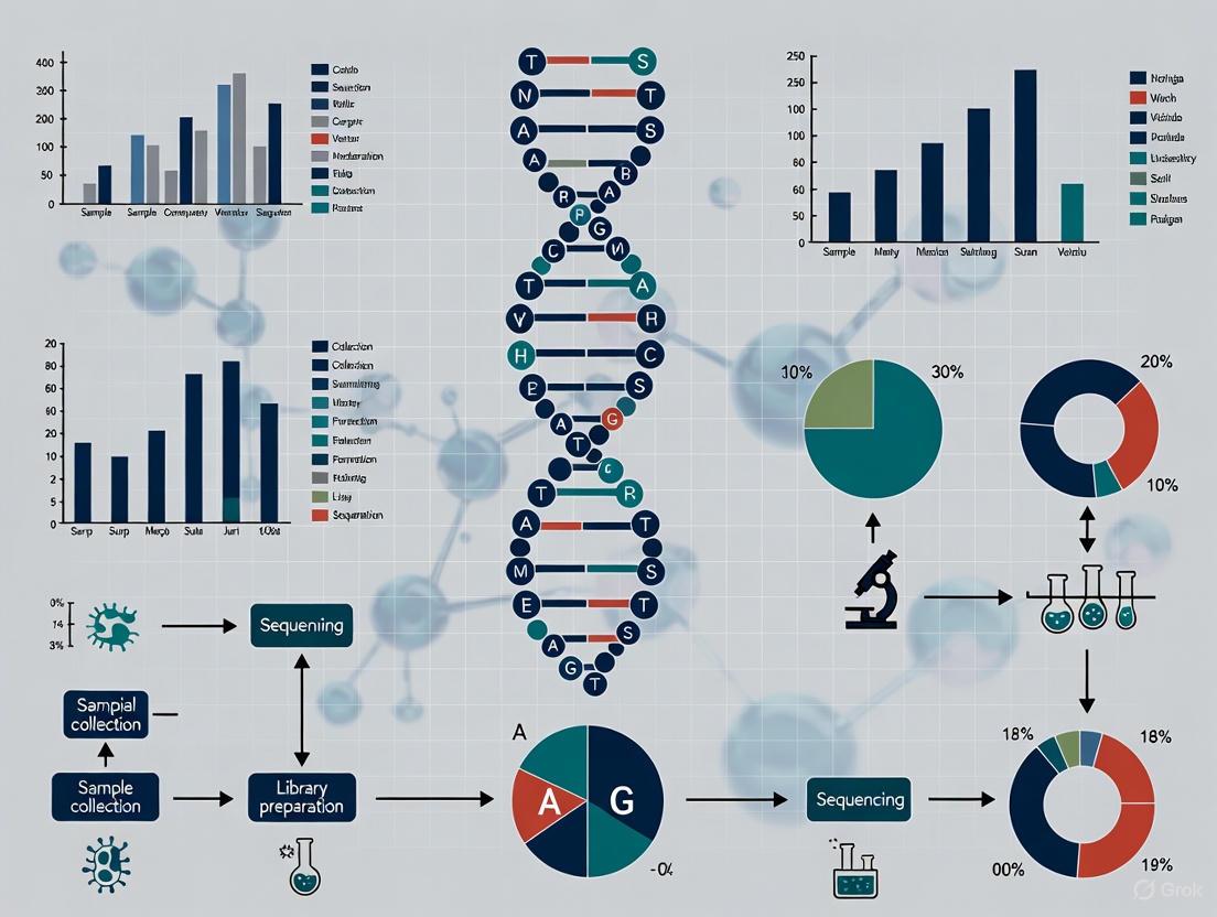
Metagenomic Sequencing Workflow: This diagram outlines the key steps in shotgun metagenomic sequencing for vaginal microbiome analysis.
Materials:
- DNA library preparation kit (e.g., Illumina DNA Prep or Nanopore Ligation Sequencing Kit)
- Size selection beads
- Sequencing platform (Illumina NovaSeq 6000 or Oxford Nanopore GridION)
- Bioinformatic analysis workstation
Procedure:
- Library Preparation:
Sequencing:
Bioinformatic Analysis:
Quality Control:
- Include positive control (mock community) in each sequencing run
- Monitor sequencing quality metrics (Q-score, read length distribution)
- Verify expected taxa in positive controls
- Assess potential contamination in negative controls
The Scientist's Toolkit: Research Reagent Solutions
Table 5: Essential Research Reagents for BV Metagenomic Studies
| Reagent Category | Specific Products | Application | Key Features |
|---|---|---|---|
| Sample Preservation | ZymoBIOMICS DNA/RNA Shield [15] | Nucleic acid stabilization at room temperature | Preserves DNA and RNA integrity; inactivates pathogens |
| DNA Extraction | ZymoBIOMICS DNA/RNA Miniprep Kit [15] | Simultaneous DNA/RNA extraction from vaginal swabs | Includes bead beating for mechanical lysis; inhibitor removal |
| Host DNA Depletion | NEBNext Microbiome DNA Enrichment Kit | Selective removal of human DNA | Improves microbial sequencing depth; methylation-based |
| Library Preparation | Illumina DNA Prep Kit [11] | Library construction for Illumina sequencing | Transposase-based; fast workflow; low input compatible |
| Long-read Sequencing | Oxford Nanopore Ligation Sequencing Kit (SQK-LSK109) [15] | Library preparation for Nanopore sequencing | Enables real-time sequencing; long reads for strain resolution |
| 16S Amplification | QIAseq 16S/ITS Panel with V1-V2 primers [15] | Target amplification for metataxonomics | Covers appropriate variable regions for vaginal microbiota |
| Positive Controls | ZymoBIOMICS Microbial Community Standard | Quality control for entire workflow | Defined composition and abundance; validates sensitivity |
Bacterial vaginosis represents a complex polymicrobial dysbiosis characterized by distinct shifts in vaginal microbial community structure. The integration of metagenomic sequencing technologies with the CST framework provides researchers and clinicians with powerful tools for understanding BV pathogenesis, classifying disease subtypes, and developing targeted interventions. The persistent challenges of BV—including high recurrence rates following conventional antibiotic therapy—highlight the need for biofilm-targeted approaches and personalized treatment strategies based on precise molecular characterization of individual vaginal microbiomes [9] [10].
Future directions in BV research should focus on leveraging multi-omics approaches to connect microbial community structure with functional activity, developing point-of-care diagnostic technologies that incorporate molecular classification, and advancing therapeutic strategies that effectively target polymicrobial biofilms while restoring protective lactobacilli populations.
Bacterial vaginosis (BV) represents the most common cause of vaginal discharge in reproductive-aged women worldwide, characterized by a disruption of the healthy vaginal microbiome in which protective Lactobacillus species are replaced by anaerobic bacteria including Gardnerella vaginalis, Prevotella species, and Mobiluncus species [16] [17]. Accurate diagnosis is crucial not only for symptom management but also for preventing serious complications associated with BV, including increased susceptibility to sexually transmitted infections (including HIV), inflammatory pelvic disease, and adverse pregnancy outcomes such as prematurity and low birth weight [16] [18].
For decades, diagnosis has relied on two principal traditional methods: the clinical Amsel criteria and the laboratory-based Nugent scoring system. While these methods have formed the diagnostic cornerstone for BV, they present significant limitations in sensitivity, specificity, interpretation, and practical implementation [16] [19] [20]. This application note critically examines these limitations within the context of emerging metagenomic sequencing technologies, which promise to overcome these constraints through comprehensive, molecular characterization of the vaginal microbiome.
Principle and Methodology of Traditional Diagnostic Systems
Amsel's Criteria: Clinical Diagnostic Framework
The Amsel criteria, established in 1983, provide a clinical diagnostic framework based on four parameters, of which at least three must be present for a confirmed BV diagnosis [16] [17]:
- Homogeneous, thin, grayish-white vaginal discharge that smoothly coats the vaginal walls.
- Vaginal fluid pH > 4.5 measured using litmus paper.
- Positive "whiff test" characterized by a fishy odor after adding 10% potassium hydroxide (KOH) to the vaginal discharge.
- Presence of clue cells (vaginal epithelial cells studded with adherent bacteria) on microscopic examination of a wet mount, constituting more than 20% of epithelial cells.
Table 1: Components and Interpretation of Amsel's Criteria
| Diagnostic Component | Procedure | Positive Finding |
|---|---|---|
| Vaginal Discharge | Visual inspection during speculum examination | Thin, homogeneous, grayish-white, uniformly coating vaginal walls |
| Vaginal pH | Application of vaginal fluid to litmus paper | pH value greater than 4.5 |
| Whiff Test | Addition of 10% KOH to vaginal discharge sample | Release of a distinct, fishy (amine) odor |
| Clue Cells | Microscopic examination of wet mount preparation | Vaginal epithelial cells with obscured borders due to adherent bacteria |
Nugent Scoring System: Laboratory Gold Standard
The Nugent score, introduced in 1991, is a standardized Gram stain scoring system considered the historical gold standard for BV diagnosis [20] [21]. It involves microscopic evaluation of a vaginal smear to quantify the presence of three bacterial morphotypes, with a composite score ranging from 0 to 10:
- Large Gram-positive rods (Lactobacillus morphotypes)
- Small Gram-variable rods (Gardnerella vaginalis and Bacteroides morphotypes)
- Curved Gram-variable rods (Mobiluncus morphotypes)
Table 2: Nugent Scoring System for Bacterial Vaginosis Diagnosis
| Score | Lactobacillus Morphotypes | Gardnerella Morphotypes | Mobiluncus Morphotypes | Interpretation |
|---|---|---|---|---|
| 0 | 4+ | 0 | 0 | |
| 1 | 3+ | 1+ | 1+ or 2+ | |
| 3 | 1+ | 3+ | - | |
| 4 | 0 | 4+ | - | |
| Total Score | Clinical Interpretation | |||
| 0-3 | Normal vaginal flora | |||
| 4-6 | Intermediate flora | |||
| 7-10 | Bacterial Vaginosis |
Experimental Protocols for Traditional Methods
Protocol for Amsel's Criteria Diagnosis
Specimen Collection:
- Insert a non-lubricated speculum into the vagina.
- Using a sterile swab, collect vaginal fluid from the posterior fornix.
Procedure:
- Vaginal Discharge Assessment: Visually assess the character of the discharge on the swab and during speculum examination for homogeneity and color.
- Vaginal pH Measurement:
- Roll the swab onto a pH test strip (range 4.0-7.0).
- Compare the color change to the manufacturer's reference chart immediately.
- Whiff Test:
- Roll a portion of the sample onto a clean glass slide.
- Add 1-2 drops of 10% KOH solution to the sample.
- Immediately smell for the presence of a fishy, amine odor.
- Wet Mount Preparation and Microscopy:
- Add a drop of normal saline to a separate glass slide.
- Emulsify a portion of the vaginal swab in the saline.
- Apply a coverslip and examine under microscope at 400x magnification.
- Scan multiple fields to identify clue cells (epithelial cells with obscured borders and granular appearance due to adherent bacteria).
Interpretation: The diagnosis of BV is confirmed when at least three of the four criteria are positive [16].
Protocol for Nugent Scoring
Specimen Collection and Smear Preparation:
- Collect vaginal fluid sample with a sterile swab from the posterior fornix.
- Roll the swab evenly across a clean glass slide to create a thin smear.
- Allow the smear to air dry completely.
Gram Staining:
- Flood the slide with crystal violet solution for 60 seconds.
- Rinse gently with distilled water.
- Flood with Gram's iodine solution for 60 seconds.
- Rinse gently with distilled water.
- Decolorize with acetone/alcohol solution for 5-10 seconds until runoff is clear.
- Rinse immediately with distilled water.
- Flood with safranin counterstain for 60 seconds.
- Rinse gently with distilled water and air dry.
Microscopic Evaluation and Scoring:
- Examine the stained smear under oil immersion (1000x magnification).
- Evaluate a minimum of 10-20 fields to account for sample heterogeneity.
- Quantify the three bacterial morphotypes per field:
- Lactobacillus morphotypes: Large, Gram-positive rods
- Gardnerella morphotypes: Small, Gram-variable rods
- Mobiluncus morphotypes: Curved, Gram-variable rods
- Assign scores for each morphotype based on the average number per field:
- 0: No organisms observed
- 1+: <1 organism per field
- 2+: 1-4 organisms per field
- 3+: 5-30 organisms per field
- 4+: >30 organisms per field
- Calculate the total Nugent score by summing the three individual scores.
Interpretation: A score of 7-10 is diagnostic for BV, 4-6 represents intermediate flora, and 0-3 indicates normal flora [20] [21].
Critical Analysis of Limitations
Diagnostic Performance Limitations
Comparative studies consistently reveal significant variability in the diagnostic performance of both Amsel criteria and Nugent scoring, with sensitivities and specificities varying substantially across different populations and settings.
Table 3: Comparative Diagnostic Performance of Amsel's Criteria Versus Nugent Scoring
| Study Reference | Sensitivity of Amsel's Criteria | Specificity of Amsel's Criteria | PPV of Amsel's Criteria | NPV of Amsel's Criteria | Study Population |
|---|---|---|---|---|---|
| PMC8369704 [19] | 50.0% | 98.2% | 87.5% | 88.8% | 141 women, Nepal |
| PMC9292691 [21] | 85.3% | 95.6% | 87.9% | 94.6% | 125 women, India |
| IJRCOG 2019 [22] | 75.0% | 95.0% | 90.0% | 86.0% | 260 women, India |
| StatPearls/NIH [16] | 37-70% | 94-99% | Not reported | Not reported | Literature review |
The Amsel criteria demonstrate particularly wide sensitivity ranges (37%-85.3%), while maintaining consistently high specificity (94%-98.2%) across studies [16] [19] [21]. This variability suggests that clinical context and practitioner experience significantly impact diagnostic accuracy. Furthermore, the individual components of the Amsel criteria show markedly different performance characteristics, with clue cell demonstration providing the highest specificity (100% in some studies) and pH >4.5 showing the highest sensitivity (89.3%) but lowest specificity (21.2%) [19] [23].
Technical and Operational Limitations
Amsel Criteria Challenges:
- Subjectivity in Interpretation: Visual assessment of discharge character and microscopic identification of clue cells introduce substantial inter-observer variability [20].
- Inadequate Sample Collection: Improper technique can compromise all subsequent testing, particularly pH measurement and microscopy.
- Equipment Dependency: Despite being considered a "clinical" method, Amsel criteria still require access to a microscope and potassium hydroxide, limiting point-of-care application in resource-limited settings [16].
- Time Sensitivity: pH measurement and wet mount examination must be performed promptly after sample collection to avoid artifactual changes.
Nugent Scoring Challenges:
- Technical Expertise Requirement: Accurate morphotype identification demands substantial training and experience in microbiology [19] [20].
- Time-Consuming Process: The staining and detailed microscopic examination render the method impractical for rapid clinical decision-making.
- Intermediate Flora Dilemma: The clinical significance of Nugent scores of 4-6 (intermediate flora) remains ambiguous, representing a "garbage can" category with uncertain therapeutic implications [20].
- Equipment and Resource Intensive: Requires Gram staining capabilities and high-quality microscopy, limiting implementation in basic healthcare settings [19] [23].
Clinical and Diagnostic Limitations
Both traditional systems suffer from fundamental diagnostic shortcomings that impact patient management and research applications:
- Binary Diagnostic Paradigm: Both methods force a dichotomous (positive/negative) classification on what is fundamentally a continuous spectrum of microbial communities [20].
- Limited Microbial Resolution: They fail to identify specific pathogenic species or microbial consortia beyond basic morphotypes, preventing targeted therapy [11] [15].
- Inability to Predict Recurrence: Neither method provides insights into why approximately 50% of patients experience recurrence within 6-12 months after treatment [18] [11].
- Inadequate for Asymptomatic Cases: Both systems were developed for symptomatic women, with limited utility for detecting subclinical BV that may still confer increased STI risk [17].
The Scientist's Toolkit: Research Reagent Solutions
Table 4: Essential Research Reagents for Traditional BV Diagnostic Methods
| Reagent/Equipment | Application | Technical Specification | Research Considerations |
|---|---|---|---|
| pH Indicator Strips | Vaginal pH measurement | Range 4.0-7.0 with 0.2-0.3 pH unit increments | Colorimetric interpretation requires standardization; affected by blood, semen, or lubricants |
| 10% Potassium Hydroxide (KOH) | Whiff test | 10% solution in distilled water | Fresh preparation recommended; amine volatilization time-sensitive |
| Normal Saline | Wet mount preparation | 0.9% sodium chloride, sterile | Must be preservative-free to maintain microbial viability and morphology |
| Gram Stain Kit | Nugent scoring | Crystal violet, iodine, decolorizer, safranin | Batch-to-batch consistency critical for reproducible morphotype identification |
| Microscope | Clue cell identification & Nugent scoring | Phase-contrast capability preferred; 100x oil immersion objective | Regular calibration and quality control essential for consistent reading |
| Transport Media | Sample preservation for delayed processing | Amies, Stuart's, or DNA/RNA shield media | Choice affects microbial viability and molecular integrity for downstream applications |
Transition to Metagenomic Approaches
The limitations of traditional diagnostic methods have accelerated the development of molecular approaches that provide comprehensive, precise characterization of the vaginal microbiome. Metagenomic sequencing, particularly shallow shotgun metagenomic sequencing (SMS), enables culture-free identification and quantification of bacterial species, detection of viruses, fungi, and antimicrobial resistance genes, offering unprecedented resolution for BV research and diagnostics [11] [15].
This technological transition addresses core limitations of traditional methods by:
- Providing objective, quantitative data reducing operator-dependent variability
- Identifying specific microbial signatures associated with treatment failure and recurrence
- Enabling development of personalized therapeutic approaches based on individual microbiome profiles
- Facilitating remote diagnosis through self-collected samples processed in centralized laboratories [11]
Diagram 1: Diagnostic workflow evolution from traditional to metagenomic methods for bacterial vaginosis, highlighting key limitations and resolution of progressive information.
The Amsel criteria and Nugent scoring system have provided fundamental diagnostic frameworks for bacterial vaginosis for decades. However, their limitations in sensitivity, specificity, technical complexity, and microbial resolution increasingly constrain both clinical management and research advancement. The documented variability in performance, subjectivity in interpretation, and inability to characterize the full complexity of vaginal microbiome dysbiosis underscore the necessity for more sophisticated diagnostic approaches.
Metagenomic sequencing technologies represent a paradigm shift in BV diagnosis, offering the comprehensive, objective characterization of vaginal microbiota necessary to address the persistent challenges of recurrence, asymptomatic infection, and personalized therapeutic management. As research continues to validate these molecular approaches against clinical outcomes, they are poised to supplant traditional methods as the new standard for both research and clinical diagnostics, ultimately enabling more effective, personalized management of this prevalent condition.
Bacterial vaginosis (BV) represents a profound shift in the vaginal microbiome from a Lactobacillus-dominated community to a polymicrobial anaerobic consortium [24]. Historically classified as "Gardnerella vaginitis," the condition was initially attributed to a single etiological agent, Gardnerella vaginalis [24]. However, decades of research using advanced molecular methodologies have fundamentally challenged this simplistic model, revealing BV as a complex dysbiotic state without a primary infectious agent [11] [25]. This paradigm shift carries critical implications for diagnostic accuracy, therapeutic efficacy, and public health outcomes, particularly in an era of growing antimicrobial resistance.
The limitations of the single-pathogen model become evident when examining basic microbiological facts: G. vaginalis can be detected in approximately 50% of asymptomatic women, establishing it as a common component of normal vaginal flora rather than an exclusive pathogen [24]. Furthermore, BV cases consistently demonstrate co-occurrence of multiple anaerobic taxa, including Prevotella, Fannyhessea (formerly Atopobium), Mobiluncus, and Megasphaera species [11] [26]. This polymicrobial nature, combined with high recurrence rates following antibiotic therapy that targets specific bacteria, provides compelling evidence against singular pathogenicity [11] [27].
The Polymicrobial Nature of BV: Ecological Perspectives
Community State Transitions and Dysbiosis
The vaginal microbiome is typically categorized into Community State Types (CSTs), with CSTs I, II, III, and V defined by dominance of L. crispatus, L. gasseri, L. iners, and L. jensenii, respectively [15] [28]. BV is characterized by CST IV, which exhibits high microbial diversity without a single dominant species [15] [2]. This transition represents an ecological disturbance where protective lactobacilli decline while facultative and obligate anaerobes proliferate [11] [24].
The complexity of BV becomes apparent when examining the variable presentations within CST IV. Recent research has revealed significant discordance between molecular and clinical assessments, with women characterized as BV-negative by Amsel's criteria and Nugent scoring often being assigned to BV-positive CSTs (CST III, and IV) based on metagenomic analysis [2]. This indicates that microbiome composition and symptomatic presentation do not always align, suggesting multiple distinct subtypes within the BV spectrum [2].
Current Diagnostic Limitations and Methodological Discordance
Traditional diagnostic approaches for BV include Amsel's clinical criteria and Nugent scoring of Gram-stained vaginal smears [24]. While these methods have been widely used for decades, they suffer from significant limitations in sensitivity, specificity, and inter-observer variability [25]. Microscopy with Nugent scoring has approximately 76% overall agreement with molecular methods, leaving substantial room for misdiagnosis [26].
The diagnostic challenge is further complicated by significant discordance between different sequencing methodologies. A 2025 study revealed that CST assignments were substantially influenced by sequencing methodology, with concordance between metatranscriptomic and metataxonomic-based CST assignment as low as 59% [2]. This methodological variability underscores the complexity of capturing an accurate snapshot of the vaginal microbiome and its functional state.
Table 1: Comparison of BV Diagnostic Methodologies
| Method | Principle | Advantages | Limitations | Agreement with Reference |
|---|---|---|---|---|
| Amsel's Criteria | Clinical assessment (pH, whiff test, clue cells, discharge) | Rapid, low-cost, point-of-care | Subjective, requires symptoms, inter-observer variability | ~70% sensitivity vs. Nugent [24] |
| Nugent Score | Gram stain microscopy scoring (0-10) | Low cost, standardized | Labor-intensive, subjective, intermediate scores unclear | Considered historical gold standard [26] |
| qPCR Panels | Targeted detection of BV-associated taxa | Objective, species-specific | Limited to pre-selected targets, may miss novel organisms | 76% overall agreement with Nugent [26] |
| 16S Metataxonomics | 16S rRNA gene sequencing | Comprehensive genus-level profile | Limited species resolution, PCR biases | High discordance with other methods [2] |
| Shotgun Metagenomics | Whole-genome sequencing | Strain-level resolution, functional potential | Higher cost, host DNA contamination | 59% concordance with metatranscriptomics [2] |
| Metatranscriptomics | RNA sequencing | Measures microbial activity and viability | Technically challenging, RNA instability | Reveals active community composition [2] |
Quantitative Evidence Supporting BV Complexity
Microbial Diversity Patterns in BV
Large-scale metagenomic studies have consistently demonstrated the remarkable taxonomic diversity associated with BV. A 2025 real-world study of 1,159 participants using shotgun metagenomic sequencing revealed significant shifts in microbial architecture following treatment, with Lactobacillus abundance increasing from 32.9% to 48.4% (p < 0.0001) and corresponding decreases in BV-associated taxa including Gardnerella, Prevotella, and Fannyhessea [11]. PERMANOVA analysis of pairwise Bray-Curtis distances showed significant separation between pre- and post-treatment samples (pseudo-F = 37.6, p < 0.0001), driven predominantly by an increase in Lactobacillus-dominated communities [11].
The complexity of BV extends beyond simple taxonomic shifts to encompass functional and structural adaptations. BV-associated bacteria frequently form polymicrobial biofilms on vaginal epithelial cells, with Gardnerella species potentially acting as initial architects that facilitate subsequent colonization by other anaerobic taxa [25] [24]. These biofilms provide structural stability to the dysbiotic state and confer protection against antimicrobial agents and host immune responses, contributing significantly to the high recurrence rates observed following antibiotic therapy [25].
Therapeutic Challenges and Recurrence Patterns
The polymicrobial nature of BV directly impacts treatment efficacy and recurrence rates. Standard antibiotic therapies with metronidazole or clindamycin result in initial response rates of 70-85% within one month, but recurrence rates remain unacceptably high, with 45% of patients recurring within 3 months and over 50% within 6 months [11]. A 2025 remote care study demonstrated that algorithm-guided treatment protocols could achieve 75.5% symptom resolution at four weeks, with 30.0% experiencing recurrence at a median follow-up of 4.4 months—lower than historical in-person cohorts but still substantial [11].
Quantitative modeling approaches have provided mechanistic insights into these recurrence patterns. An ordinary differential equation model predicting bacterial growth as a function of metronidazole uptake, sensitivity, and metabolism revealed that a critical factor in efficacy is Lactobacillus sequestration of metronidazole, with efficacy decreasing when the relative abundance of Lactobacillus is higher pre-treatment [29]. This counterintuitive finding was validated in both co-culture experiments and clinical cohorts, where women with recurrence had significantly higher pre-treatment levels of Lactobacillus relative to BV-associated bacteria [29].
Table 2: Quantitative Metrics of BV Complexity and Treatment Outcomes
| Parameter | Value | Significance | Source |
|---|---|---|---|
| BV Prevalence | 30% of women annually | Major public health burden | [11] |
| Standard Treatment Response | 70-85% within 1 month | Most cases initially responsive | [11] |
| Recurrence Rate (3 months) | 45% | High recurrence indicates incomplete resolution | [11] |
| Recurrence Rate (6 months) | >50% | Persistent cyclical nature | [11] |
| Algorithm-Guided Resolution | 75.5% at 4 weeks | Personalized approaches may improve outcomes | [11] |
| Reduced Recurrence with Guidance | 30% at 4.4 months | Better than historical cohorts | [11] |
| Lactobacillus Increase Post-Treatment | 32.9% to 48.4% (p<0.0001) | Microbiome restoration possible | [11] |
| Methodological Discordance | 59% CST concordance between methods | Diagnostic challenges | [2] |
Experimental Protocols for BV Metagenomic Analysis
Sample Collection and DNA Extraction
Protocol: Self-Collection of Vaginal Specimens for Metagenomic Sequencing
- Collection Kit Preparation: Utilize standardized collection kits with nylon-flocked Copan ESwabs in Amie's transport medium [11] [26].
- Sample Collection: Instruct participants to collect mid-vaginal specimens using a circular motion, ensuring adequate epithelial cell collection.
- Storage: Immediately transfer samples to -80°C storage to preserve DNA integrity until processing.
- DNA Extraction:
- Employ chemical and mechanical lysis with an automated extraction system
- Implement host DNA depletion steps to increase microbial sequencing depth
- Use validated DNA extraction kits (e.g., ZymoBIOMICS DNA/RNA Miniprep Kit) with modifications including extended bead-beating (40 minutes) for rigorous cell disruption [15]
- Elute DNA in nuclease-free water and quantify using fluorometric methods (e.g., Qubit dsDNA HS Assay)
Library Preparation and Sequencing
Protocol: Shallow Shotgun Metagenomic Sequencing using Oxford Nanopore Technology
Library Preparation:
- Utilize ligation sequencing kit (SQK-LSK109) with barcoding (EXP-NBD196 expansion kit)
- Include short fragment buffer in adapter ligation to ensure equal purification of short and long DNA fragments
- Normalize libraries to 4nM concentration [15]
Sequencing:
- Load library onto Nanopore GridION with R9.4.1 flow cells (FLO-MIN106)
- Perform basecalling and demultiplexing using MinKNOW (v. 21.11.6) with Guppy (v. 5.1.12)
- Target 1-5 million reads per sample for shallow shotgun profiling [15]
Alternative Illumina Protocol:
- For comparative 16S sequencing, target V1-V2 or V2-V3 regions using QIAseq 16S/ITS Panel
- Sequence on MiSeq system with 2×301 bp read setup and 20% PhiX addition [15]
Bioinformatic Analysis Pipeline
Protocol: Taxonomic Profiling and Community State Typing
Quality Control and Preprocessing:
- Perform adapter trimming and quality filtering using FastP or Trimmomatic
- Remove host reads by alignment to human reference genome (hg38)
Taxonomic Profiling:
- Utilize custom bioinformatic pipelines for taxonomic assignment [11]
- Employ k-mer based classifiers for species-level resolution
- Generate relative abundance profiles for all detected taxa
Community State Type Assignment:
Conceptual Framework: Polymicrobial Synergy in BV
The diagram below illustrates the conceptual framework of polymicrobial interactions in BV pathogenesis, highlighting the transition from a healthy vaginal microbiome to a dysbiotic state characterized by synergistic relationships between multiple bacterial taxa.
Diagram 1: Polymicrobial Synergy in BV Pathogenesis. This diagram illustrates the transition from a healthy vaginal microbiome to a dysbiotic BV state through sequential colonization and synergistic interactions between multiple bacterial taxa, culminating in treatment resistance and recurrence.
The Scientist's Toolkit: Essential Research Reagents and Platforms
Table 3: Essential Research Reagents and Platforms for BV Metagenomic Studies
| Category | Specific Product/Platform | Application in BV Research | Key Features | |
|---|---|---|---|---|
| Sample Collection | Copan ESwabs in Amie's transport medium | Vaginal specimen collection | Maintains microbial viability, compatible with molecular assays | [11] [26] |
| DNA Extraction | ZymoBIOMICS DNA/RNA Miniprep Kit | Microbial DNA extraction from vaginal samples | Effective host DNA depletion, bead-beating for cell lysis | [15] |
| Sequencing Platform | Oxford Nanopore GridION | Shallow shotgun metagenomic sequencing | Real-time data generation, flexible multiplexing, long reads | [15] |
| Sequencing Platform | Illumina NovaSeq 6000 | High-depth metagenomic sequencing | High accuracy, high throughput, established pipelines | [11] |
| Bioinformatic Tools | Custom taxonomic profiling pipeline | Microbiome analysis | Species-level resolution, relative abundance quantification | [11] |
| Culture Media | Human blood bilayer agar with Tween 80 (HBT) | Gardnerella cultivation | Selective growth of fastidious BV-associated organisms | [24] |
| Live Biotherapeutic | Multi-strain L. crispatus products | Microbiome restoration | Probiotic intervention for BV recurrence prevention | [27] |
The etiological complexity of bacterial vaginosis demands a fundamental shift from the single-pathogen model to an ecological perspective that acknowledges the polymicrobial nature of this condition. The evidence from metagenomic studies reveals BV as a diverse dysbiotic state with variable compositional patterns across individuals and populations [11] [28] [2]. This understanding directly impacts diagnostic and therapeutic approaches, suggesting that personalized management strategies based on comprehensive microbiome profiling may yield better outcomes than one-size-fits-all antibiotic regimens.
Future directions in BV research should focus on developing culturomics approaches to isolate novel BV-associated organisms, multi-omics integration to connect taxonomic composition with functional potential, and targeted therapeutic strategies that address the polymicrobial consortia and biofilm structures characteristic of established BV [25]. The promising results from Phase I trials of multi-strain L. crispatus live biotherapeutic products demonstrate the potential for microbiome-based interventions to achieve durable colonization and potentially reduce recurrence rates [27]. As our understanding of BV complexity deepens, so too will our ability to provide effective, personalized care for this pervasive condition.
Sequencing Technologies and Analytical Pipelines: From Sample to Data
Metagenomic sequencing has revolutionized the study of complex microbial communities, offering powerful tools for diagnosing and understanding conditions like bacterial vaginosis (BV). The vaginal microbiome plays a critical role in female health, with its dysbiosis being intimately linked to BV, a prevalent condition associated with adverse obstetric and gynecological outcomes [30]. Two principal sequencing methodologies—16S rRNA gene sequencing and shotgun metagenomic sequencing—have emerged as the dominant techniques for microbiome characterization. For researchers and drug development professionals working on BV diagnostics, the choice between these methods carries significant implications for taxonomic resolution, functional insight, cost, and analytical complexity. This application note provides a detailed comparative analysis of these sequencing approaches within the context of BV research, offering structured experimental protocols and practical guidance for implementation.
Technical Comparison of Sequencing Methodologies
Fundamental Principles and Workflows
16S rRNA Gene Sequencing (metataxonomics) employs a targeted approach, using PCR to amplify specific hypervariable regions (V1-V9) of the bacterial 16S ribosomal RNA gene. This conserved gene contains regions that enable differentiation between bacterial taxa, allowing for identification and relative quantification of community members [31]. After amplification, libraries are prepared and sequenced, with subsequent analysis performed against established 16S reference databases like SILVA. This method primarily profiles bacterial and archaeal communities, with full-length 16S sequencing typically limited to bacteria due to PCR primer specificity [31].
Shotgun Metagenomic Sequencing takes a comprehensive approach by randomly fragmenting the entire genomic DNA content of a sample through mechanical shearing. These fragments are then sequenced without target-specific amplification, theoretically capturing all genetic material present—including bacterial, fungal, viral, and other microbial DNA [31]. The resulting sequences are analyzed through either reference-based alignment to genomic databases or de novo assembly, the latter enabling identification of previously uncharacterized microorganisms.
Comparative Performance in Microbial Profiling
Table 1: Methodological Comparison of 16S rRNA vs. Shotgun Metagenomic Sequencing
| Parameter | 16S rRNA Sequencing | Shotgun Metagenomics |
|---|---|---|
| Taxonomic Scope | Limited to bacteria and archaea [31] | Comprehensive: bacteria, archaea, fungi, viruses, eukaryotes [31] [32] |
| Taxonomic Resolution | Genus-level, potentially species-level [31] | Species-level and strain-level discrimination [32] |
| Functional Insights | Limited to inferred function | Direct assessment of functional genetic potential [30] |
| Amplification Bias | Present due to PCR amplification | Minimal, no target amplification required [15] |
| Host DNA Contamination | Minimal impact | Significant challenge, requires filtering [32] |
| Cost Considerations | Lower per sample [33] | Higher per sample, but decreasing [32] |
| Computational Demand | Moderate | High, complex bioinformatics [31] [32] |
| Reference Databases | Well-established (SILVA, Greengenes) [31] | Less complete for some ecosystems [31] |
| Detection Sensitivity | May miss low-abundance taxa [34] | Enhanced detection of rare taxa [34] |
| Quantitative Accuracy | Affected by 16S copy number variation | More directly quantitative [34] |
Recent advancements include shallow shotgun metagenomic sequencing, which reduces per-sample sequencing costs while maintaining many advantages of shotgun approaches [35] [15]. When implemented on platforms like Oxford Nanopore Technologies, this approach offers cost-effectiveness, rapid data generation, and flexible multiplexing schemes suitable for larger-scale BV research [35].
Application to Bacterial Vaginosis Research
Vaginal Microbiome Community State Types (CSTs)
In BV research, vaginal microbiomes are typically categorized into Community State Types (CSTs), which provide a framework for understanding microbial composition and its relationship to health and disease:
- CST I: Dominated by Lactobacillus crispatus
- CST II: Dominated by Lactobacillus gasseri
- CST III: Dominated by Lactobacillus iners
- CST V: Dominated by Lactobacillus jensenii
- CST IV: Characterized by diverse anaerobic bacteria with reduced Lactobacillus abundance [15] [30]
CST IV is particularly associated with bacterial vaginosis and represents a polymicrobial condition often including Gardnerella vaginalis, Prevotella, Fannyhessea vaginae (formerly Atopobium vaginae), and Dialister species [30] [2]. This dysbiotic state is linked to increased risk of preterm birth, sexually transmitted infections, and other gynecological complications [30].
Diagnostic Concordance and Method-Specific Insights
Studies comparing sequencing methods for BV diagnosis reveal both concordance and important distinctions. A 2025 study demonstrated 92% concordance in CST classification between Illumina 16S-based sequencing and Nanopore-based shallow shotgun metagenomic sequencing [35]. Both methods showed perfect agreement in detecting samples dominated by Lactobacilli versus vaginosis-associated taxa [35].
However, significant differences emerge in finer-scale characterization. The same study identified significant abundance variations for 12 of the 20 most abundant vaginal species between 16S and shotgun approaches [35]. Shotgun methods demonstrated potentially increased sensitivity for dysbiotic states, showing higher overall abundance of Gardnerella vaginalis and corresponding increases in CST IV detection [35].
Methodological choices can substantially impact CST assignments, with one study reporting concordance as low as 59% between metatranscriptomic and metataxonomic-based CST assignment [2]. This highlights the substantial influence of sequencing methodology on molecular BV profiling and subsequent diagnosis.
Experimental Protocols
Sample Collection and DNA Extraction
Table 2: Essential Research Reagent Solutions for Vaginal Microbiome Sequencing
| Reagent/Category | Specific Examples | Function & Application Notes |
|---|---|---|
| Sample Collection & Transport | ZymoBIOMICS DNA/RNA Shield Collection Tubes [15] | Preserves nucleic acid integrity during storage and transport; critical for accurate representation. |
| DNA Extraction Kits | ZymoBIOMICS DNA/RNA Miniprep Kit [15]; NucleoSpin Soil Kit [32]; Dneasy PowerLyzer Powersoil kit [32] | Cell lysis and nucleic acid purification; choice significantly influences microbial diversity observed [36]. |
| 16S Library Preparation | QIAseq 16S/ITS Panel (V1-V2, V2-V3 primers) [15]; Ion 16S Metagenomics Kit [36] | Target amplification and library construction; primer choice (variable region) introduces bias [36]. |
| Shotgun Library Preparation | Ligation Sequencing Kit (e.g., SQK-LSK109) [15] | Fragmentation, adapter ligation, and library prep for whole-genome sequencing. |
| Barcoding/Multiplexing | EXP-NBD196 expansion kit [15] | Allows sample pooling for cost-effective sequencing. |
| Bioinformatics Tools | DADA2 [32], Kraken2/Bracken2 [32], QIIME [36], MOTHUR [36] | Data processing, taxonomy assignment, and diversity analysis; pipeline choice affects results [36]. |
Sample Collection Protocol:
- Collect vaginal samples using sterile swabs or collection brushes [36]
- Immediately transfer samples to DNA/RNA Shield solution in collection tubes [15]
- Store at -80°C prior to DNA extraction [36]
- For longitudinal studies, maintain consistent collection methods across all timepoints
DNA Extraction Protocol (Modified from ZymoBIOMICS DNA/RNA Miniprep Kit):
- Transfer 200μL of sample suspension to a bead beating tube
- Add 350μL of DNA/RNA Shield buffer to enable harvesting of 200μL of bead-free liquid [15]
- Perform bead beating using vortex genie with multi-tube attachment on maximal speed for 40 minutes [15]
- Complete extraction according to manufacturer's instructions with elution in 100μL nuclease-free water
- Quantify DNA using fluorometric methods (e.g., Qubit with 1× dsDNA HS Assay Kit) [15]
16S rRNA Gene Sequencing Protocol
Library Preparation (Illumina Platform):
- Utilize QIAseq 16S/ITS Panel with V1-V2 or V2-V3 16S primers [15]
- Use 1μL total input per sample as recommended for low-input samples [15]
- Perform PCR amplification with cycle optimization for low-biomass samples
- Clean amplified products and normalize libraries to 4nM [15]
- Pool libraries and sequence on MiSeq system with 2×301bp read setup [15]
- Include 20% PhiX control to improve low-diversity sequence quality [15]
Bioinformatic Analysis (DADA2 Pipeline):
- Filter and trim reads based on quality profiles (truncate forward/reverse reads below 290/230 respectively) [32]
- Remove first 10 nucleotides of each read to eliminate primer sequences [32]
- Perform sample inference with pool=True parameter to enhance rare variant detection [32]
- Merge paired reads and remove chimeric sequences using removeBimeraDenovo function [32]
- Assign taxonomy using SILVA 16S rRNA database (v138.1) [32]
- For enhanced species-level classification, perform additional taxonomic classification using custom BLASTN database and k-mer based classification (Kraken2/Bracken2) with NCBI RefSeq Targeted Loci Project database [32]
Shotgun Metagenomic Sequencing Protocol
Library Preparation (Oxford Nanopore Platform):
- Prepare library with ligation sequencing kit (e.g., SQK-LSK109) [15]
- Incorporate barcoding using expansion kit (e.g., EXP-NBD196) for multiplexing 12-16 samples per flow cell [15]
- Use Short Fragment Buffer (SFB) in adapter ligation to ensure equal purification of short and long DNA fragments [15]
- Sequence on Nanopore GridION with R9.4.1 flow cells [15]
- Perform basecalling and demultiplexing using MinKNOW with Guppy (v5.1.12) [15]
Bioinformatic Analysis:
- Filter human sequence reads using Bowtie2 with human genome GRCh38 [32]
- Perform quality control and adapter trimming
- For taxonomic profiling:
- Align reads to comprehensive genomic databases (e.g., NCBI RefSeq, GTDB, UHGG)
- Alternatively, perform de novo assembly for novel organism identification [31]
- For functional analysis:
- Annotate genes and metabolic pathways
- Conduct comparative functional analysis between sample groups [30]
Method Selection Guidelines for BV Research
Project-Specific Recommendations
The choice between 16S rRNA and shotgun metagenomic sequencing should be guided by specific research objectives, sample types, and resource constraints:
Choose 16S rRNA sequencing when:
- Research focus is exclusively on bacterial composition
- Genus-level taxonomic resolution is sufficient
- Studying large sample cohorts with limited budget
- Infrastructure for complex bioinformatic analysis is limited
- Rapid turnaround time is prioritized [31] [33]
Choose shotgun metagenomics when:
- Species-level or strain-level discrimination is required
- Comprehensive profiling of all microbial domains (bacteria, fungi, viruses) is needed
- Functional metabolic potential of the community is of interest
- Investigating strain-specific effects, such as different Gardnerella vaginalis strains in BV [2]
- Sufficient computational resources and bioinformatics expertise are available [31] [32]
Emerging Approaches and Future Directions
Shallow shotgun metagenomic sequencing represents a promising intermediate approach, offering many advantages of shotgun sequencing at reduced cost [35] [15]. This method is particularly suitable for large-scale BV studies where both taxonomic precision and functional insights are valuable.
Recent studies also highlight the value of multi-omic approaches, combining metagenomics with metatranscriptomics to assess not only microbial composition but also functional activity [2]. This is particularly relevant for understanding the dynamic changes in BV and treatment response.
For clinical BV diagnostics, machine learning models applied to sequencing data show promise but require careful validation across diverse populations. Recent research indicates that model performance varies by ethnicity, with lower accuracy observed for Black women [28]. This underscores the importance of diverse, representative cohorts in method development.
Both 16S rRNA and shotgun metagenomic sequencing offer powerful approaches for studying the vaginal microbiome in bacterial vaginosis research. 16S sequencing provides a cost-effective method for comprehensive bacterial profiling, while shotgun metagenomics enables higher taxonomic resolution and functional insights. The choice between these methods should be guided by specific research questions, resources, and desired level of taxonomic and functional resolution. As sequencing technologies continue to advance and costs decrease, shotgun methods are becoming increasingly accessible for routine BV research, potentially offering more comprehensive insights into the complex microbial dynamics underlying this prevalent condition.
The Rise of Shallow Shotgun Metagenomic Sequencing (SMS) for Cost-Effective Profiling
The characterization of the vaginal microbiome is crucial for understanding female reproductive health, susceptibility to sexually transmitted infections, and the pathophysiology of bacterial vaginosis (BV)—the most prevalent vaginal condition in reproduction-age women [15]. For decades, 16S rRNA gene sequencing has been the methodological cornerstone for vaginal microbiome profiling, providing insights into community state types (CSTs) and broad compositional shifts [11] [37]. However, this approach has inherent limitations, including primer bias, inability to achieve reliable species-level resolution, and blindness to non-prokaryotic community members [15] [2].
Shallow shotgun metagenomic sequencing (SMS) has emerged as a powerful alternative that addresses these limitations while offering a cost-effective solution for large-scale studies [15] [38]. By sequencing the entire microbial DNA content in a sample at reduced coverage, SMS provides uncompromised taxonomic resolution without the amplification biases of 16S methods [15]. When implemented with the Oxford Nanopore Technology, SMS gains additional advantages including rapid real-time data generation, flexible multiplexing schemes, and access to epigenetic markers such as DNA methylation [15] [38]. This application note details the experimental validation, implementation protocols, and research applications of shallow SMS for advancing BV diagnostics and therapeutic development.
Performance Validation: SMS Versus Established Methods
Comparative Analytical Performance
A recent benchmark study evaluated Nanopore-based shallow SMS against the established standard of Illumina 16S sequencing using a cohort of 52 women, including 23 diagnosed with BV [15]. The results demonstrated strong concordance between the methods while highlighting distinctive advantages of SMS.
Table 1: Method Comparison Between Shallow SMS and 16S Sequencing for Vaginal Microbiome Profiling
| Performance Metric | Illumina 16S Sequencing | Nanopore Shallow SMS | Concordance/Advantage |
|---|---|---|---|
| CST Classification | Reference standard | 92% concordance | Near-perfect agreement for broad community structures [15] |
| Dominant Taxa Detection | Lactobacilli, BV-associated taxa | Perfect agreement (100%) | Identical detection of sample dominance patterns [15] |
| Gardnerella vaginalis Detection | Standard sensitivity | Higher abundance measurements | Potentially increased sensitivity to dysbiotic states [15] |
| Non-Prokaryotic Detection | Not available | Candida albicans, Lactobacillus phage | Unique SMS capability for full community profiling [15] [38] |
| Additional Data Types | Microbiome composition only | Host DNA methylation, cell type quantification | Epigenetic host profiling alongside microbiome analysis [15] |
| Technical Variation | Established consistency | Marked variation in sequencing yields | SMS requires optimization of library preparation [15] |
Quantitative Microbial Abundance Differences
When comparing inferred abundances of individual species across samples, significant differences (Wilcoxon signed-rank test p < 0.05) were observed for 12 of the 20 most abundant species in the cohort [15]. The shallow SMS approach consistently demonstrated higher overall abundance measurements for Gardnerella vaginalis, which corresponded to an increased number of CST IV detections [15]. This suggests that the amplification-free nature of SMS may provide increased sensitivity for detecting dysbiotic states characteristic of BV.
Table 2: Key Advantages of Shallow SMS for BV Research Applications
| Research Application | SMS Benefit | Research Implication |
|---|---|---|
| BV Therapeutic Development | Species-level resolution | Enables tracking of specific BVAB (e.g., Gardnerella spp., Prevotella spp., Fannyhessea vaginae) [11] |
| Treatment Efficacy Monitoring | Detection of non-prokaryotes | Allows concurrent monitoring of fungal pathogens (e.g., Candida albicans) during antibiotic therapy [15] [11] |
| Microbiome Dynamics Studies | Real-time sequencing capability | Rapid assessment of microbiome changes during treatment interventions [38] |
| Host-Microbe Interactions | Methylation-based host cell quantification | Correlates microbial shifts with host epithelial and immune cell patterns [15] |
| Recurrence Investigation | Strain-level resolution | Enables tracking of specific bacterial strains contributing to BV recurrence [2] |
Experimental Protocols for Vaginal Microbiome SMS
Sample Collection and DNA Extraction
Sample Collection Protocol:
- Collect vaginal swabs using standardized collection kits (e.g., Copan, Murrieta, CA, USA) [11]
- Immediately place swabs in DNA/RNA Shield solution (e.g., ZymoBIOMICS DNA/RNA Shield Collection Tubes) to preserve nucleic acid integrity [15]
- Store samples at -80°C until processing to prevent microbial community shifts
DNA Extraction Protocol:
- Process 200μL of vaginal suspension using the ZymoBIOMICS DNA/RNA Miniprep Kit [15]
- Implement mechanical lysis via bead beating (40 minutes on maximal speed using Vortex genie with 24 multi-tube attachment) [15]
- Include chemical lysis steps according to manufacturer protocols
- Perform optional host DNA depletion to increase microbial sequencing depth (critical for low-biomass samples) [11]
- Elute DNA in 100μL nuclease-free water
- Quantify DNA using fluorometric methods (e.g., Qubit 3 with 1× dsDNA HS Assay Kit) [15]
- Assess DNA quality via spectrophotometric ratios (A260/A280 ≈ 1.8-2.0)
Library Preparation and Sequencing
Nanopore-Specific Library Preparation:
- Utilize the ligation sequencing kit SQK-LSK109 with barcoding expansion kit EXP-NBD196 [15]
- Include Short Fragment Buffer (SFB) during adapter ligation to ensure equal purification of short and long DNA fragments [15]
- Pool 12-16 barcoded libraries per flow cell for optimal sequencing yield
- Sequence on Nanopore GridION with R9.4.1 flow cells (FLO-MIN106) [15]
Illumina-Based SMS Alternative:
- Prepare libraries using Illumina DNA prep kits with IDT unique dual indexes
- Sequence on Illumina NovaSeq 6000 platform with 2×150bp reads [11]
- Target 1-5 million reads per sample for "shallow" coverage [11]
Bioinformatic Analysis Workflow
The bioinformatic workflow begins with raw sequencing read quality control using tools such as FastQC, including adapter trimming and quality filtering [15] [11]. For vaginal samples, a crucial step involves the removal of host DNA reads through alignment to the human genome (e.g., using Bowtie2 against hg38) [11]. Taxonomic profiling is performed using reference-based classifiers (Kraken2, Bracken) or assembly-based approaches (MetaPhlAn) with specialized databases for vaginal microbes [15]. Community State Type assignment implements the VALENCIA framework, a nearest centroid classification method validated for vaginal microbial communities [35]. Functional profiling can be achieved through alignment to comprehensive databases (KEGG, eggNOG) following metagenomic assembly [11].
The Scientist's Toolkit: Essential Research Reagents
Table 3: Essential Research Reagents for Vaginal Microbiome SMS
| Reagent Category | Specific Products | Research Function |
|---|---|---|
| Sample Collection | ZymoBIOMICS DNA/RNA Shield Collection Tubes; Copan collection kits [15] [11] | Nucleic acid preservation at point of collection; standardized specimen collection |
| DNA Extraction | ZymoBIOMICS DNA/RNA Miniprep Kit [15] | Simultaneous extraction of DNA and RNA; effective lysis of difficult-to-break bacterial cells |
| Library Preparation | Oxford Nanopore Ligation Sequencing Kit SQK-LSK109; EXP-NBD196 barcoding kit [15] | Preparation of sequencing libraries for multiplexed runs; maintains fragment diversity |
| Sequencing | Nanopore R9.4.1 flow cells (FLO-MIN106); Illumina NovaSeq 6000 reagents [15] [11] | Platform-specific sequencing; determines read length, output, and real-time capability |
| Bioinformatic Tools | Kraken2/Bracken; MetaPhlAn; VALENCIA; FastQC; Bowtie2 [15] [35] | Taxonomic classification; CST assignment; quality control; host DNA removal |
Research Applications in Bacterial Vaginosis
Enhanced BV Diagnostics and Monitoring
Shallow SMS enables comprehensive BV diagnostics beyond traditional methods by providing:
- Species-level resolution of BV-associated bacteria (BVAB), including differentiation of Gardnerella species (G. vaginalis, G. leopoldii, G. piotii, G. swidsinskii) with potential differential virulence [37]
- Detection of non-bacterial members including Candida albicans and bacteriophages (e.g., Lactobacillus phage) that may influence treatment outcomes [15] [38]
- Quantification of microbial load through host DNA ratio calculations, enabling distinction between colonization and true infection [15]
In remote clinical validation studies, SMS-based BV management demonstrated 75.5% symptom resolution at four weeks with personalized treatment protocols, and significantly lower recurrence rates (30.0% at 4.4 months median follow-up) compared to historical in-person cohorts [11]. Metagenomic analysis confirmed significant microbial shifts post-treatment, with increased Lactobacillus abundance (32.9% to 48.4%, p < 0.0001) and decreased BV-associated taxa [11].
Therapeutic Development Applications
For drug development professionals, shallow SMS provides critical tools for:
- Identifying novel drug targets through subtractive proteomics approaches that prioritize proteins with essential bacterial functions absent in human and beneficial flora [39]
- Monitoring treatment efficacy at species and strain levels, enabling understanding of differential responses among BVAB [2]
- Evaluating microbiome restoration following therapeutic interventions, including the return of protective Lactobacillus species [11]
Computational drug discovery approaches leveraging SMS data have identified potential targets like 3-deoxy-7-phosphoheptulonate synthase (aroG gene product) in Gardnerella vaginalis, with molecular docking and dynamics simulations guiding compound selection [39].
Shallow shotgun metagenomic sequencing represents a transformative methodology for vaginal microbiome research and BV therapeutic development. Its cost-effectiveness, combined with superior taxonomic resolution and ability to detect the full spectrum of vaginal microbes, positions SMS as an essential tool for advancing women's health research. The experimental protocols outlined herein provide researchers with a comprehensive framework for implementing this powerful technology in both basic science and clinical translation contexts. As validation studies continue to demonstrate its robust performance relative to established methods [15] [38] [2], shallow SMS is poised to become the new gold standard for high-resolution vaginal microbiome profiling in large-scale studies.
The accurate characterization of the vaginal microbiome is a critical component in the study of female reproductive health, particularly for the diagnosis and understanding of bacterial vaginosis (BV). BV is a prevalent condition associated with serious health risks, including increased susceptibility to sexually transmitted infections and preterm birth [35] [2]. For years, Illumina-based 16S rRNA gene sequencing has been the established standard for microbial community profiling in research settings. However, the emergence of Oxford Nanopore Technologies (ONT) presents a promising alternative, offering long-read capabilities and real-time sequencing. This application note provides a detailed, evidence-based comparison of these two platforms within the specific context of vaginal microbiome analysis for BV research, presenting structured data, standardized protocols, and analytical workflows to guide scientific and drug development professionals.
A direct comparative study of Nanopore shallow shotgun metagenomic sequencing (SMS) and Illumina 16S sequencing on a cohort of 52 women (23 with BV) revealed both concordance and key differences in performance [35] [15].
Table 1: Key Performance Metrics for Vaginal Microbiome Characterization
| Performance Metric | Illumina 16S | Oxford Nanopore Shallow SMS |
|---|---|---|
| Community State Type (CST) Concordance | Reference Standard | 92% vs. Illumina [35] |
| Dominant Taxa Detection | Reference Standard | Perfect Agreement [35] |
| Sensitivity to Gardnerella vaginalis | Standard | Potentially Higher [35] |
| Species-Level Abundance Quantification | Standard | Significant differences for 12/20 top species [35] |
| Sequencing Yield | Consistent | Marked variation observed [35] |
| Non-Prokaryote Detection | Limited to prokaryotes | Enables detection of phage & fungi [40] |
| Additional Data Layers | Microbiome composition only | Methylation-based host cell quantification [35] |
The high concordance in CST assignment and detection of dominant taxa demonstrates that Nanopore SMS reliably recapitulates the broad community structures identified by Illumina 16S [35]. However, the finding that 12 of the 20 most abundant species showed significantly different inferred abundances between the platforms indicates notable differences in fine-scale characterization [35]. Furthermore, ONT's higher reported abundance of Gardnerella vaginalis, a key BV-associated organism, suggests it may have an increased sensitivity to dysbiotic states [35].
Beyond prokaryote profiling, a significant advantage of the amplification-free, shallow SMS approach is its capacity to detect other biologically relevant entities, such as the fungal pathogen Candida albicans and Lactobacillus-infecting phages, which are inaccessible to 16S sequencing [40] [38].
Detailed Experimental Protocols
To ensure reproducibility and facilitate the adoption of these methods, below are detailed protocols for vaginal microbiome characterization based on the cited studies.
Sample Collection and DNA Extraction
Sample Collection
- Tool: Vaginal swabs.
- Procedure: Collect swabs and immediately store them in DNA/RNA stabilization buffers, such as ZymoBIOMICS DNA/RNA Shield Collection Tubes [15]. Store samples at -80°C prior to nucleic acid extraction.
DNA Extraction
- Recommended Kit: ZymoBIOMICS DNA/RNA Miniprep Kit [15].
- Input: 200 μL of suspension from the collection tube.
- Critical Step (Bead Beating): Perform mechanical lysis via bead beating for 40 minutes on maximal speed using a vortex with a multi-tube attachment to ensure robust cell disruption [15].
- Elution: Elute DNA in 100 μL of nuclease-free water.
- Quality Control: Quantify DNA using a fluorometric method (e.g., Qubit with dsDNA HS Assay Kit). For samples with yields below 1 ng/μL, a repeat extraction is recommended [15].
Library Preparation and Sequencing
Table 2: Library Preparation and Sequencing Protocols
| Step | Illumina 16S Protocol | Oxford Nanopore SMS Protocol |
|---|---|---|
| Target Region / Method | V1-V2 or V3-V4 of 16S rRNA gene [15] | Shallow Shotgun Metagenomic Sequencing (SMS) [15] |
| Library Prep Kit | QIAseq 16S/ITS Region Panel (Qiagen) [15] | Ligation Sequencing Kit (SQK-LSK109) [15] |
| Barcoding / Multiplexing | Yes, as per kit (e.g., QIAseq 16S/ITS Index) [15] | Yes, using expansion kit (e.g., EXP-NBD196); 12-16 samples per flow cell [15] |
| Critical Step | PCR amplification of target region. | Use of Short Fragment Buffer (SFB) during adapter ligation to ensure equal purification of short DNA fragments [15]. |
| Sequencing Device | Illumina MiSeq [15] | GridION with R9.4.1 flow cells [15] |
| Run Parameters | 2 × 301 bp; 20% PhiX spike-in [15] | Real-time sequencing until flow cell end-of-life; basecalling with Guppy. |
Data Analysis Workflows
The analytical pipelines for the two platforms differ significantly due to the nature of the sequence data.
Illumina 16S Data Analysis
- Processing: Use pipelines like nf-core/ampliseq or QIIME2 [41] [42].
- Key Steps: Quality filtering (FastQC, MultiQC), primer trimming (Cutadapt), denoising and Amplicon Sequence Variant (ASV) generation (DADA2), and taxonomic classification against a reference database (e.g., SILVA) [41] [42].
- Output: An ASV table for downstream ecological analysis (alpha/beta diversity, CST assignment with tools like VALENCIA) [15].
Nanopore Shallow SMS Data Analysis
- Basecalling & Demultiplexing: Perform in real-time using MinKNOW software and Guppy [15].
- Taxonomic Profiling: Classify reads using tools tailored for long-read metagenomic data. The analysis in the primary study employed a custom pipeline for comprehensive taxonomic assignment [35] [15].
- Additional Analyses: The same data can be leveraged for methylation calling to quantify host cell types, providing an additional layer of host-microbe interaction data [35].
The Scientist's Toolkit: Essential Research Reagents and Materials
Table 3: Key Research Reagent Solutions for Vaginal Microbiome Sequencing
| Item | Function/Application | Example Product/Catalog Number |
|---|---|---|
| Sample Collection & Stabilization | Preserves nucleic acid integrity immediately post-collection. | ZymoBIOMICS DNA/RNA Shield Collection Tubes [15] |
| Nucleic Acid Extraction Kit | Simultaneous co-extraction of DNA and RNA from complex samples. | ZymoBIOMICS DNA/RNA Miniprep Kit [15] [42] |
| Illumina 16S Library Prep | Targeted amplification and preparation of 16S rRNA gene libraries. | QIAseq 16S/ITS Region Panel (Qiagen) [15] [41] |
| Nanopore SMS Library Prep | Preparation of sequencing libraries for whole-metagenome sequencing. | Ligation Sequencing Kit (SQK-LSK109) [15] |
| Nanopore Barcoding Kit | Multiplexing samples to enable cost-effective shallow SMS. | Native Barcoding Expansion Kit (EXP-NBD196) [15] |
| ONT Flow Cell | The consumable containing nanopores for sequencing. | MinION / GridION Flow Cell (R9.4.1 or newer) [15] |
Discussion and Research Implications for BV Diagnosis
The choice between Illumina and ONT for vaginal microbiome studies is not a simple determination of superiority but depends heavily on the specific research objectives.
- For Large-Scale Population Studies: Where cost-effectiveness and high-throughput profiling are paramount, Illumina 16S remains a robust and reliable choice. Its established protocols and high data consistency are advantageous [41].
- For Mechanistic and Pathogenesis Studies: Where a more comprehensive view of the microbial community and host-environment is needed, ONT shallow SMS offers distinct advantages. Its ability to provide species-level resolution without amplification bias, detect non-prokaryotic members, and simultaneously assess host methylome provides a more holistic picture of the vaginal microenvironment [35] [38]. The finding that different sequencing methods (metataxonomics, metagenomics, metatranscriptomics) can lead to discordant molecular diagnoses of BV further underscores the value of a method that captures a broader range of biological molecules [2].
A critical consideration in BV research is the move beyond relative abundance. A 2025 study highlighted that total vaginal bacterial load, an absolute quantitative measure, can be a stronger predictor of the genital immune milieu than Nugent score [42]. This finding has profound implications for understanding host-microbe interactions and suggests that future research, regardless of sequencing platform, should incorporate absolute quantification methods to complement relative abundance data.
In conclusion, ONT shallow SMS has emerged as a powerful, viable alternative to Illumina 16S, particularly when its additional data layers can be leveraged to unravel the complex etiology of BV. The decision matrix for platform selection should be guided by the trade-offs between throughput, cost, resolution, and the depth of biological insight required.
Metagenomic sequencing has revolutionized the study of complex microbial communities, offering unparalleled insights into their composition, functional potential, and strain-level diversity. In the context of bacterial vaginosis (BV) diagnosis and research, these bioinformatic approaches provide powerful tools to move beyond traditional diagnostic limitations. BV, a condition characterized by a shift from a Lactobacillus-dominant vaginal microbiome to a polymicrobial anaerobic community, affects approximately 30% of reproductive-age women annually and poses significant diagnostic challenges due to its polymicrobial nature and high recurrence rates [11] [43].
This application note details standardized protocols for comprehensive bioinformatic analysis of vaginal metagenomic data, encompassing taxonomic profiling, functional pathway inference, and strain-level resolution. The methodologies outlined herein support the broader thesis that advanced metagenomic sequencing, coupled with sophisticated bioinformatic analysis, enables more precise molecular diagnosis of BV and provides insights into its complex etiology, potentially leading to improved therapeutic strategies.
Taxonomic Profiling
Taxonomic profiling serves as the foundational step in metagenomic analysis, identifying which microorganisms are present and in what relative abundances in a given sample.
Experimental Protocols
Sample Collection and DNA Extraction:
- Sample Collection: Vaginal swabs are collected using standardized collection kits (e.g., Zymo DNA-RNA shield Collection Tube w-Swabs) and stored at -80°C immediately after collection [44].
- DNA Isolation: Extract microbial DNA using the DNeasy 96 Powersoil Pro QIAcube HT Kit (Qiagen) with the following modifications to the manufacturer's protocol [44]:
- Supplement each well of a 96-deep-well plate containing 50 μL microbiome sample with 500 μL washed zirconium beads (0.1 mm) and 800 μL CD1 solution.
- Seal the plate and homogenize for 4 minutes.
- Centrifuge at 3000 × g for 6 minutes.
- Transfer 600 μL of supernatant to a fresh plate containing 300 μL CD2 solution.
- After mixing and centrifugation, transfer 550 μL of supernatant to an S-block (Qiagen).
- Complete extraction using the QIAcube Connect instrument (Qiagen) according to manufacturer's instructions.
- Elute purified DNA in EB buffer (Qiagen) and store at -20°C.
Sequencing Approaches:
- Shotgun Metagenomic Sequencing: Prepare libraries according to Illumina protocols and sequence on platforms such as Illumina NovaSeq 6000, aiming for a minimum of 18 million reads per sample for adequate coverage [11] [45].
- 16S rRNA Gene Sequencing: Amplify the V3-V4 hypervariable region of the 16S rRNA gene using primers 341F and 805R. Normalize libraries to 4 nM, pool, and sequence on an Illumina MiSeq system with a 2 × 301 bp read setup with 20% PhiX addition [15].
Bioinformatic Analysis
Quality Control:
- Process raw sequencing data through the bioBakery 3 platform [44].
- Perform quality control with KneadData (v0.10.0) to filter host DNA and low-quality reads using default parameters [44].
- For 16S data, use DADA2 to model and correct Illumina-sequencing errors, producing amplicon sequence variants (ASVs) [46].
Taxonomic Profiling:
- For shotgun metagenomic data, perform taxonomic profiling using MetaPhlAn (version 3.0) with the bacterial ChocoPhlAn database (version mpav31CHOCOPhlAn_2010901) for species-level classification [44].
- For 16S data, assign taxonomy to ASVs using the SILVA database or classify sequences through phylogenetic placement [46].
Table 1: Key Taxonomic Differences in Bacterial Vaginosis
| Taxon | Abundance in Health | Abundance in BV | Research Significance |
|---|---|---|---|
| Lactobacillus crispatus | High (42.4% in healthy controls) [47] | Low (24.3% in dysplasia) [47] | Protective role; associated with favorable outcomes |
| Lactobacillus iners | Variable | Often increased in transitional states | Potential pathobiont; debated role in BV development [11] |
| Gardnerella vaginalis | Low | High | BV-associated; multiple subspecies with different virulence [43] |
| Fannyhessea vaginae | Low | High | BV-associated; co-occurs with Gardnerella [43] |
| Prevotella species | Low | High | BV-associated; multiple species present [43] |
Community State Type Classification
Categorize vaginal microbiomes into Community State Types (CSTs) based on the dominant species present [46]:
- CST-I: Dominated by L. crispatus
- CST-II: Dominated by L. gasseri
- CST-III: Dominated by L. iners
- CST-V: Dominated by L. jensenii
- CST-IV: Diverse community without Lactobacillus dominance
The VALENCIA tool provides a standardized nearest-centroid based approach for CST classification, which is particularly valuable for cross-study comparisons [48].
Functional Pathway Inference
Functional analysis reveals the metabolic capabilities of microbial communities, providing insights into how microbial communities impact host health and disease states.
Experimental Protocols
Functional Profiling:
- Perform functional profiling using HUMAnN (v3.0.1) with the MetaCyc database (version v24) for pathway classification [44].
- For metatranscriptomic data, extract RNA and prepare sequencing libraries using ribosomal RNA depletion methods to enrich for mRNA [49].
Bioinformatic Analysis
Pathway Abundance Analysis:
- Identify microbial metabolic pathways and compute their abundances from metagenomic or metatranscriptomic data using HUMAnN3 [47].
- Normalize pathway abundances using the updated ALDEx2 R package with scale reliant inference (SRI) to account for asymmetry between healthy and BV datasets, applying a 15% offset to center housekeeping functions [49].
Table 2: Functional Pathways Differentially Abundant in Bacterial Vaginosis
| Functional Pathway | Abundance in BV | Key Contributing Taxa | Potential Functional Significance |
|---|---|---|---|
| Peptidoglycan biosynthesis | Increased [47] | G. vaginalis, F. vaginae [47] | Cell wall maintenance in diverse community |
| Nucleotide biosynthesis | Increased [47] | G. vaginalis, F. vaginae [47] | Support rapid growth of diverse taxa |
| L-lysine biosynthesis | Decreased [47] | L. crispatus [47] | Reduced amino acid production in dysbiosis |
| Sugar degradation | Decreased [47] | L. crispatus, L. jensenii [47] | Altered carbon metabolism in dysbiosis |
| Cationic antimicrobial peptide (CAMP) resistance | Increased [49] | Multiple BV-associated taxa [49] | Host defense evasion mechanism |
Genome-Scale Metabolic Modeling:
- Generate genome-scale metabolic network reconstructions (GENREs) for in silico prediction of metabolic interactions between BV-associated bacteria [43].
- Simulate pairwise interactions to identify potential competitive and mutualistic relationships within the community.
Strain-Level Resolution
Strain-level analysis reveals intra-species genetic diversity that can significantly influence microbial function and host interactions but is masked in species-level profiling.
Experimental Protocols
Deep Sequencing:
- For strain-level resolution, increase sequencing depth to a minimum of 40 million reads per sample for shotgun metagenomics to achieve sufficient coverage for strain discrimination [48].
- For Oxford Nanopore Technologies (ONT) sequencing, prepare libraries using the ligation sequencing kit SQK-LSK109 with barcoding based on the EXP-NBD196 expansion kit, and sequence on GridION with R9.4.1 flow cells [15].
Bioinformatic Analysis
Metagenomic Community State Typing (mgCST):
- Apply mgCST classification using the VIRGO database to identify metagenomic subspecies (mgSs) based on co-occurring genetic variation within species [48].
- For L. crispatus, distinguish between multiple mgCSTs (e.g., mgCST 1, 3-6) that represent different subspecies with varying functional potential [48].
Metagenome-Assembled Genomes (MAGs):
- Reconstruct MAGs from metagenomic data using assembly and binning tools such as MetaBAT2, MaxBin2, or CONCOCT [48].
- Assess MAG quality using CheckM to ensure completeness >80% and contamination <5% [48].
- Annotate MAGs with PROKKA or similar tools to identify strain-specific genes, particularly those involved in cell surface interactions, carbohydrate metabolism, and antimicrobial peptide resistance [48].
Strain Tracking:
- Use the Metagenomic Intra-Species Typing (MIST) software to resolve co-occurring strains at an average nucleotide identity (ANI) resolution of 99.9% with coverage as low as 0.001× per strain [45].
- Identify strain-specific single nucleotide polymorphisms (SNPs) and gene content variations that differentiate subtypes.
Table 3: Strain-Level Variations in Key Vaginal Microbiota
| Species | Strain-Level Features | Functional Implications |
|---|---|---|
| Lactobacillus crispatus | Variation in mucin-binding genes (mucBP) and cell surface glycan gene cluster [48] | Differences in host adhesion and colonization potential |
| Lactobacillus iners | Presence/absence of D-lactate dehydrogenase gene [48] | Affects lactic acid production and vaginal pH modulation |
| Gardnerella vaginalis | 13 genetically distinct species with 95% ANI; clades within G. vaginalis and G. piotii [43] | Varied virulence potential and antimicrobial resistance |
| Lactobacillus gasseri | Strain-specific differences in mucin binding and vaginal cell adhesion genes [48] | Impacts persistence in the vaginal environment |
Research Reagent Solutions
Table 4: Essential Research Reagents and Materials for Metagenomic Analysis of Bacterial Vaginosis
| Reagent/Material | Function | Example Product |
|---|---|---|
| DNA/RNA Shield Collection Tubes | Sample preservation and stabilization | ZymoBIOMICS DNA/RNA Shield Collection Tubes [44] [15] |
| DNA Extraction Kit | Microbial DNA isolation | DNeasy 96 Powersoil Pro QIAcube HT Kit [44] |
| Host DNA Depletion Kit | Enrichment of microbial DNA | GensKey Host DNA Depletion kit [45] |
| Library Preparation Kit | Sequencing library construction | Illumina DNA Prep kits; ONT Ligation Sequencing Kits [15] |
| Synthetic Spike-in Control | Quantification of bacterial load | Custom synthetic DNA sequences [45] |
| 16S rRNA Primers | Amplification of target regions | V1-V2 (27F-338R) or V3-V4 (341F-805R) primers [46] [15] |
| Reference Databases | Taxonomic and functional annotation | ChocoPhlAn, VIRGO, MetaCyc, CARD [44] [49] |
Workflow and Pathway Diagrams
Metagenomic Analysis Workflow
BV-Associated Metabolic Interactions
The integration of taxonomic profiling, functional pathway inference, and strain-level resolution provides a comprehensive framework for advancing bacterial vaginosis research. These bioinformatic approaches enable researchers to move beyond compositional descriptions to functional understanding of BV pathogenesis, revealing the complex metabolic interactions and strain-specific adaptations that underlie this common but poorly understood condition. The standardized protocols and analytical workflows presented in this application note offer researchers a validated foundation for implementing these powerful approaches in both research and potential future diagnostic applications.
Addressing Technical and Clinical Challenges in Metagenomic BV Diagnosis
Within the framework of broader research on metagenomic sequencing for bacterial vaginosis (BV) diagnosis, a significant challenge emerges: discordant molecular diagnoses arising from the use of different sequencing methodologies. BV, a common vaginal syndrome associated with increased risk of sexually transmitted infections and preterm birth, has long eluded a single etiological agent, complicating its diagnosis and treatment [2]. Traditional diagnostic methods, including Amsel's clinical criteria and Nugent microscopic scoring, provide a foundation but lack the granularity needed to fully characterize the condition's polymicrobial nature [26].
The advent of molecular sequencing techniques has revolutionized our capacity to profile the vaginal microbiome. However, our research reveals that these advanced methods themselves introduce variability. Metataxonomics (16S rRNA gene sequencing), metagenomics (whole-genome shotgun sequencing), and metatranscriptomics (RNA-based sequencing) each reflect different aspects of the microbial community, leading to substantially different interpretations of the same condition [2]. This application note systematically examines the sources and implications of this methodological variability and provides standardized protocols to enhance reproducibility and cross-study comparisons in BV research.
Comparative Performance of Sequencing Methodologies
Quantitative Discordance Across Platforms
Direct comparison of common cervicovaginal sequencing methods reveals significant differences in their outputs and subsequent clinical interpretations. Analysis shows that the concordance in Community State Type (CST) assignment between metatranscriptomic and metataxonomic methods can be as low as 59%, highlighting the substantial impact of methodological selection [2]. Furthermore, the taxonomic profiles generated vary considerably—only four bacterial species were shared among the top 20 most abundant species identified across all three major sequencing approaches (metataxonomic, metagenomic, and metatranscriptomic) [2].
Table 1: Methodological Comparison for BV Microbiome Profiling
| Sequencing Method | Target Molecule | Key Strengths | Key Limitations | Concordance with Other Methods |
|---|---|---|---|---|
| Metataxonomics (16S rRNA) | DNA | Cost-effective; Well-established protocols; High sensitivity for bacterial presence | Limited species-resolution; Insensitive to bacterial activity/viability | 59-76% with metatranscriptomics for CST assignment [2] |
| Metagenomics (Shotgun) | DNA | Improved species and strain-level resolution; Functional potential inference | Cannot distinguish live/dead bacteria; Higher cost and computational needs | Shares only 6/20 top species with metataxonomics [2] |
| Metatranscriptomics | RNA | Identifies metabolically active microbes; Functional activity insight | RNA instability; Technically challenging; Higher cost | Shares 13/20 top species with metagenomics [2] |
| qPCR | DNA | High sensitivity for specific targets; Quantitative; Fast turnaround | Limited to pre-selected targets; Hypothesis-driven | 76% overall agreement with Nugent scoring [26] |
Impact on Clinical Diagnosis and Research Interpretation
The choice of sequencing methodology directly influences molecular BV diagnosis, particularly in defining Community State Types (CSTs) which categorize vaginal microbiomes into groups dominated by specific Lactobacillus species (CST I, II, III, V) or characterized by polymicrobial communities (CST IV) [2]. This methodological variability has profound implications for both clinical practice and research validity:
- Diagnostic Inconsistency: Individuals may receive different molecular diagnoses depending on the sequencing method employed, affecting both treatment decisions and research cohort composition [2].
- Diversity Assessment: Significant differences in alpha-diversity measurements (Shannon and Simpson indices) within CSTs have been observed based on the sequencing method, particularly in CST IIIB where Amsel BV-positive participants showed significantly higher Shannon diversity in metagenomic data but not in metatranscriptomic data [2].
- Biomarker Identification: Different methods recover different sets of significantly associated taxa. For instance, strain-level effects of Gardnerella vaginalis and associations between Dialister micraerophilus and Parvimonas micra with Amsel's criteria may be more readily detected by certain methodologies [2].
Standardized Experimental Protocols
Sample Collection and Storage Protocol
Principle: Standardized sample collection and processing are critical for minimizing pre-analytical variability, especially when comparing different sequencing methods.
Reagents and Equipment:
- Nylon-flocked Copan ESwabs
- Amies transport medium
- Sterile cryovials for storage
- -80°C freezer
Procedure:
- Collect mid-vaginal samples using nylon-flocked ESwabs.
- Immediately place swabs into Amies transport medium.
- For DNA-based methods (metataxonomics, metagenomics):
- Store samples at -80°C until processing
- Avoid freeze-thaw cycles
- For RNA-based methods (metatranscriptomics):
- Add RNA stabilization reagent immediately after collection
- Flash-freeze in liquid nitrogen within 30 minutes of collection
- Store at -80°C
- Record time-from-collection-to-freezing for all samples
Validation Metrics:
- Sample storage time should not exceed 6 months for metatranscriptomics
- Document transportation conditions and time
DNA Extraction for Metataxonomic and Metagenomic Sequencing
Principle: Efficient and reproducible DNA extraction is essential for representative microbiome profiling.
Reagents and Equipment:
- QIAamp DNA Mini Kit (Qiagen)
- Lysozyme (20 mg/ml; Sigma-Aldrich)
- Proteinase K
- Thermal shaker
- Centrifuge
Procedure:
- Centrifuge samples at 13,000 × g for 5 min to pellet microbial cells
- Resuspend pellet in 200 μL residual supernatant
- Add enzymatic lysis buffer containing lysozyme (20 mg/ml)
- Incubate at 37°C for 30 min
- Add proteinase K and buffer AL
- Incubate at 56°C for 30 min
- Follow QIAamp spin column protocol according to manufacturer's instructions
- Elute DNA in 100 μL molecular-grade water
- Quantify DNA using fluorometric methods (e.g., Qubit dsDNA HS Assay)
Quality Control:
- Include extraction negative controls with each batch
- Minimum DNA yield: 0.5 ng/μL for metagenomics
- DNA integrity number (DIN) >7 for metagenomics
Metataxonomic (16S rRNA) Library Preparation and Sequencing
Principle: Amplify and sequence hypervariable regions of the 16S rRNA gene for taxonomic profiling.
Reagents and Equipment:
- 16S Amplification primers (e.g., V3-V4 region)
- KAPA HiFi HotStart ReadyMix
- AMPure XP beads
- Illumina sequencing platform (e.g., MiSeq)
Procedure:
- Amplify V3-V4 region using primers 341F (5'-CCTACGGGNGGCWGCAG-3') and 785R (5'-GACTACHVGGGTATCTAATCC-3')
- Perform PCR with the following conditions:
- 95°C for 3 min
- 25 cycles of: 95°C for 30s, 55°C for 30s, 72°C for 30s
- 72°C for 5 min
- Clean amplicons with AMPure XP beads (0.8X ratio)
- Quantify libraries with fluorometry
- Pool libraries at equimolar concentrations
- Sequence on Illumina MiSeq using 2×300 bp paired-end chemistry
Bioinformatic Analysis:
- Process raw reads through DADA2 for denoising and ASV formation
- Classify ASVs against SILVA or Greengenes database
- Normalize using rarefaction or DESeq2
Metagenomic (Shotgun) Library Preparation and Sequencing
Principle: Sequence all genomic DNA in a sample for comprehensive taxonomic and functional profiling.
Reagents and Equipment:
- Illumina DNA Prep Kit
- IDT for Illumina DNA/RNA UD Indexes
- Illumina sequencing platform (e.g., NextSeq500)
Procedure:
- Fragment DNA to target size of 350-400 bp
- Perform end repair and A-tailing
- Ligate adapters with dual index barcodes
- Clean up libraries with SPRIselect beads (0.8X ratio)
- Amplify libraries with 8-10 PCR cycles
- Validate library quality with Bioanalyzer or Tapestation
- Quantify libraries by qPCR
- Pool libraries and sequence on Illumina NextSeq500 (2×150 bp) to depth of 10-20 million reads per sample
Bioinformatic Analysis:
- Remove host reads by alignment to human reference genome (hg19)
- Classify non-human reads using Kraken2 with custom database
- Validate candidate reads with BLAST against NCBI nt database
- Perform functional annotation with HUMAnN2
Quality Control and Validation Procedures
Principle: Implement rigorous QC measures across all methodological workflows to ensure data comparability.
Negative Controls:
- Include extraction controls with molecular-grade water
- Include library preparation controls
- Sequence negative controls alongside samples
- Establish threshold for background contamination removal
Positive Controls:
- Use mock microbial communities with known composition
- Include in each sequencing run to assess technical variability
- Monitor batch effects across different sequencing runs
Validation Metrics:
- For metataxonomics: mock community recovery >80%
- For metagenomics: limit of detection <100 CFU/ml
- For metatranscriptomics: RNA integrity number (RIN) >7
Visualization of Methodological Workflows and Decision Pathways
Experimental Workflow for BV Sequencing Studies
Diagnostic Decision Pathway for Discordant Results
Research Reagent Solutions for BV Sequencing Studies
Table 2: Essential Research Reagents for BV Sequencing Methodologies
| Reagent Category | Specific Product Examples | Application & Function | Method Compatibility |
|---|---|---|---|
| Sample Collection & Storage | Copan ESwabs; Amies Transport Medium; RNA stabilization reagents | Maintains microbial viability and nucleic acid integrity during transport and storage | All methods |
| Nucleic Acid Extraction | QIAamp DNA Mini Kit; Lysozyme; Proteinase K; TIANamp Micro DNA Kit | Efficient lysis of diverse microbial species and recovery of high-quality nucleic acids | Metataxonomics, Metagenomics |
| RNA Extraction | Qiagen RNeasy Kit; MetaPolyzyme | Preserves RNA integrity and enables transcriptomic analysis | Metatranscriptomics |
| Library Preparation | 16S Amplification Primers; KAPA HiFi HotStart ReadyMix; Illumina DNA Prep Kit; Oxford Nanopore Ligation Kit | Prepares nucleic acids for sequencing with minimal bias | Platform-dependent |
| Sequencing Platforms | Illumina MiSeq/NextSeq; Oxford Nanopore MinION; BGISEQ-500 | Generates sequence data with appropriate read length and depth for analysis | Method-dependent |
| Bioinformatic Tools | DADA2; Kraken2; Bowtie2; HUMAnN2; IDSeq | Processes raw data into biologically meaningful information | All methods |
The variability between sequencing methodologies presents both a challenge and an opportunity in BV research. While discordant molecular diagnoses complicate simple interpretations, they also reflect the biological complexity of BV and the complementary strengths of different technical approaches. By implementing standardized protocols, understanding method-specific limitations, and adopting integrated analytical frameworks, researchers can navigate this variability to develop more nuanced diagnostic and therapeutic strategies. The future of BV research lies not in seeking a single perfect method, but in leveraging methodological diversity to create a multidimensional understanding of this complex condition.
Lactobacillus iners is the most prevalent bacterial species in the vaginal microbiome of reproductive-aged women worldwide, yet its functional role in vaginal health remains enigmatic and subject to considerable debate [50] [51]. Unlike other major vaginal Lactobacillus species, L. iners exhibits unique characteristics that complicate its classification as purely beneficial or detrimental. This application note examines the dual nature of L. iners within the context of metagenomic sequencing for bacterial vaginosis (BV) diagnosis research, providing researchers with consolidated quantitative data, experimental protocols, and analytical frameworks for investigating this transitional species.
The diagram below illustrates the paradoxical role of L. iners in vaginal health and disease, highlighting its unique characteristics and transitional nature.
The Dual Nature ofL. iners: Comparative Analysis
Genomic and Metabolic Characteristics
L. iners possesses several unique traits that distinguish it from other vaginal lactobacilli and contribute to its ambiguous role in vaginal health. Table 1 summarizes the key comparative characteristics between L. iners and other major vaginal Lactobacillus species.
Table 1: Comparative Characteristics of Major Vaginal Lactobacillus Species
| Characteristic | L. iners | L. crispatus | L. gasseri | L. jensenii |
|---|---|---|---|---|
| Genome Size | ~1.3 Mbp (smallest known lactobacilli) [50] | ~2.1 Mbp [52] | ~1.9 Mbp [52] | ~1.7 Mbp [52] |
| Lactic Acid Isomers | L-lactic acid only [50] [53] | D- and L-lactic acid [50] | D- and L-lactic acid [50] | D- and L-lactic acid [50] |
| Hydrogen Peroxide Production | Limited/absent [53] | Present [52] | Present [52] | Present [52] |
| Growth on MRS Agar | Poor (requires blood supplementation) [50] | Robust [50] | Robust [50] | Robust [50] |
| Unique Toxins | Inerolysin (pore-forming cytolysin) [50] [52] | Not reported | Not reported | Not reported |
| Gram Staining | Variable (often appears Gram-negative) [50] | Consistently Gram-positive [50] | Consistently Gram-positive [50] | Consistently Gram-positive [50] |
| Ecological Role | Transitional species [50] | Stable protective species [54] | Protective species [54] | Protective species [54] |
The small genome size of approximately 1.3 Mbp suggests a symbiotic or parasitic lifestyle with extensive gene loss and specialization for the vaginal niche [50] [52]. L. iners lacks the gene encoding D-lactate dehydrogenase, resulting in exclusive production of L-lactic acid rather than both isomers produced by other vaginal lactobacilli [50]. This metabolic difference has functional implications, as D-lactic acid demonstrates greater inhibitory effects against exogenous bacteria and differentially modulates host immune responses [50].
Clinical Associations and Health Outcomes
The clinical associations of L. iners present a complex picture that varies across populations and health states. Table 2 summarizes key clinical associations of L. iners based on recent metagenomic studies.
Table 2: Clinical Associations of L. iners in Vaginal Health and Disease
| Clinical Context | Association with L. iners | Study Details | Reference |
|---|---|---|---|
| Healthy Pregnancy | Higher abundance in healthy vs. diseased pregnant women [54] | 95 Chinese pregnant women; third trimester | [54] |
| Bacterial Vaginosis (BV) | Prevalent in both healthy and BV states; associated with BV recurrence [50] [51] | Often dominates post-antibiotic treatment; may facilitate recurrence | [50] [51] |
| BV Treatment Outcome | Higher abundance in cured patients; inhibits G. vaginalis and F. vaginae [55] | 130 BV patients; abundance predictive of positive outcome | [55] |
| Sexually Transmitted Infections | Reduced protection against Chlamydia trachomatis and HIV compared to L. crispatus [51] | 3.4-fold higher odds of CT detection vs. L. crispatus dominance | [51] |
| Adverse Pregnancy Outcomes | Associated with MAPO; lower abundance in healthy pregnancies [54] | Negative correlation with maternal associated adverse pregnancy outcomes | [54] |
A 2025 metagenomic study of Chinese pregnant women revealed that healthy participants exhibited higher levels of L. iners, with its abundance negatively correlated with maternal adverse pregnancy outcomes [54]. Conversely, another study found L. iners abundance was higher in patients who were cured of BV following antibiotic treatment compared to those with intermediate or failed outcomes [55]. This suggests that under specific conditions, L. iners may contribute to vaginal homeostasis restoration.
Metagenomic Analysis Protocols
Sample Collection and DNA Extraction
Protocol: Vaginal Sample Collection for Metagenomic Sequencing
Principle: Optimal sample collection preserves microbial community structure and enables high-quality DNA extraction for metagenomic sequencing.
Materials:
- Sterile polyester-tipped swabs (e.g., Copan flocked swabs)
- DNA/RNA-free collection tubes
- -80°C freezer for storage
- Commercial DNA extraction kit (e.g., QIAamp DNA Microbiome Kit)
- Laboratory equipment: vortex, centrifuge, thermal shaker
Procedure:
- Collect vaginal samples from the upper third of the anterior vaginal wall using sterile swabs [55].
- Place swabs immediately into sterile collection tubes and freeze at -80°C within 2 hours of collection.
- For DNA extraction, thaw samples and vortex swabs vigorously in buffer solution.
- Centrifuge for 10 minutes at 10,000 × g to pellet bacterial cells [55].
- Extract DNA using commercial kits following manufacturer's protocols with additional enzymatic lysis steps (lysozyme and mutanolysin) to ensure efficient Gram-positive bacterial lysis [11].
- Assess DNA quality and quantity using spectrophotometry (e.g., Nanodrop) and fluorometry (e.g., Qubit).
- Store extracted DNA at -20°C or -80°C until library preparation.
Technical Notes:
- Collect multiple swabs for parallel analyses (microscopy, culture, DNA extraction) [55].
- Include negative controls to monitor contamination.
- For longitudinal studies, standardize collection timing relative to menstrual cycle.
Shotgun Metagenomic Sequencing and Bioinformatics
Protocol: Metagenomic Sequencing and Analysis for L. iners Characterization
Principle: Shotgun metagenomic sequencing enables strain-level resolution of L. iners and functional profiling of the vaginal microbiome.
Materials:
- Illumina DNA library preparation kit
- Illumina sequencing platform (e.g., NovaSeq 6000)
- High-performance computing cluster
- Bioinformatic tools: MetaPhlAn, HUMAnN3, ChocoPhlAn
Procedure: Library Preparation and Sequencing:
- Fragment DNA to target size of 300-500 bp using acoustic shearing.
- Perform end repair, A-tailing, and adapter ligation following manufacturer protocols.
- Amplify libraries with limited PCR cycles (4-8) to minimize bias.
- Pool multiplexed libraries and sequence on Illumina platform (2×150 bp recommended).
Bioinformatic Analysis:
- Quality Control: Remove adapters and low-quality reads using Trimmomatic or FastP.
- Host DNA Depletion: Map reads to human reference genome (hg38) and remove aligning reads.
- Taxonomic Profiling:
- Analyze with MetaPhlAn for species-level identification [54].
- Alternatively, use k-mer based methods for strain-level resolution.
- Functional Profiling:
- Align reads to pan-genome databases using HUMAnN3 [54].
- Annotate metabolic pathways via MetaCyc database.
- Strain-Level Analysis:
- Map reads to L. iners reference genomes.
- Identify single nucleotide variants (SNVs) and gene content variations.
Technical Notes:
- Sequence to minimum depth of 10 million reads per sample for adequate coverage.
- For strain-level analysis, include custom L. iners genome databases.
- Functional annotation should specifically target vaginal microbiome-relevant pathways (e.g., lactic acid production, folate biosynthesis).
Experimental Models for Functional Characterization
1In VitroCultivation ofL. iners
Protocol: Isolation and Cultivation of L. iners from Clinical Specimens
Principle: L. iners has fastidious growth requirements distinct from other lactobacilli, necessitating specialized culture conditions.
Materials:
- Blood agar plates (Columbia agar with 5% sheep blood)
- MRS agar supplemented with 1-5% sheep or human blood [50]
- Anaerobic chamber or gas pak system
- Reduced transport fluid for specimen preservation
- L-cysteine supplemented broth [54]
Procedure:
- Inoculate clinical specimens onto blood agar and supplemented MRS agar.
- Incubate anaerobically at 37°C for 24-48 hours [50].
- Extend incubation time to 7 days for slow-growing isolates [50].
- Identify small, smooth, circular, translucent colonies characteristic of L. iners.
- Subculture isolated colonies in MRS broth with 0.5% cysteine as reducing agent [50].
- Confirm species identity via 16S rRNA sequencing or species-specific PCR.
- Preserve strains in glycerol stocks at -80°C for long-term storage.
Technical Notes:
- L. iners grows to lower maximum density (~10^7 CFU/mL) than other lactobacilli [50].
- Growth is optimal at pH 5.5-6.5; the species shows poor survival at pH 3.0 [50].
- Some strains require exogenous L-cysteine due to absent canonical biosynthesis pathways [54].
Functional Assays for Pathobiont Characterization
Protocol: Bacterial Inhibition Assays for L. iners Antimicrobial Activity
Principle: Co-culture experiments evaluate L. iners ability to inhibit BV-associated pathogens.
Materials:
- L. iners isolates
- BV-associated pathogens (G. vaginalis, F. vaginae, Prevotella spp.)
- Appropriate culture media for each species
- Anaerobic workstation
- Multi-well plates for co-culture
Procedure:
- Grow L. iners and pathogen isolates to mid-log phase in appropriate media.
- Standardize bacterial suspensions to equivalent density (e.g., 10^6 CFU/mL).
- Set up co-culture systems:
- Direct competition: Mix L. iners and pathogen in equal volumes.
- Agar diffusion: Seed pathogen in agar and place L. iners culture filtrate in wells.
- Transwell system: Separate cultures with permeable membrane to test diffusible factors.
- Incubate anaerobically at 37°C for 24-48 hours.
- Quantify pathogen viability by plating dilutions on selective media.
- Measure pH and lactic acid production throughout experiment.
Technical Notes:
- Four out of seven L. iners strains demonstrated inhibition against G. vaginalis in recent studies [54].
- Include controls for pH-mediated inhibition using sterile acidified media.
- Test both cell-free supernatants and live bacteria for inhibitory effects.
The experimental workflow below outlines the key steps for functional characterization of L. iners in the research laboratory setting.
The Scientist's Toolkit: Research Reagent Solutions
Table 3: Essential Research Reagents for L. iners Investigation
| Reagent/Category | Specific Examples | Research Application | Technical Notes |
|---|---|---|---|
| Culture Media | Blood agar, MRS with blood supplementation [50] | Isolation and cultivation | Essential for primary isolation; L. iners does not grow on standard MRS |
| DNA Extraction Kits | QIAamp DNA Microbiome Kit | Metagenomic studies | Includes enzymes for Gram-positive cell wall lysis |
| Sequencing Kits | Illumina DNA Prep | Library preparation | Enable strain-level resolution |
| Bioinformatic Tools | MetaPhlAn, HUMAnN3 [54] | Taxonomic/functional profiling | Standardized pipelines for vaginal microbiome |
| Reference Genomes | L. iners AB-1, L. iners 1303 | Read mapping, variant calling | Essential for strain-level analyses |
| Antibodies | Anti-inerolysin | Protein detection, localization | Detect toxin production in clinical samples |
| qPCR Assays | L. iners-specific 16S rRNA targets | Quantitative assessment | Species-specific quantification |
| Metabolite Standards | L-lactic acid, D-lactic acid | Metabolic profiling | HPLC/MS quantification of acid isomers |
Discussion and Research Implications
The dual nature of L. iners as both a commensal and pathobiont presents significant challenges for its interpretation in metagenomic-based BV diagnostics. Rather than a definitive protective or pathogenic species, evidence suggests L. iners is a transitional species that colonizes after vaginal environment disturbance and offers less protection against dysbiosis than other lactobacilli [50]. Its unique genomic and metabolic features, including a small genome and exclusive production of L-lactic acid, reflect specialized adaptation to the vaginal niche but may come at the cost of reduced protective capacity [50] [53].
For diagnostic applications, simply reporting L. iners presence or abundance may be insufficient. Future metagenomic approaches should incorporate strain-level resolution and functional profiling to distinguish between potentially protective and detrimental variants [51]. The development of strain-specific biomarkers would enhance predictive value for BV susceptibility, treatment response, and adverse outcome risk.
From a therapeutic perspective, L. iners represents both a challenge and opportunity. Its frequent dominance following antibiotic treatment [51] [11] and association with BV recurrence suggests it may create a permissive environment for dysbiosis. However, its demonstrated inhibition of G. vaginalis and F. vaginae [55] indicates potential beneficial functions under specific conditions. Therapeutic strategies might aim to promote transition from L. iners to more protective lactobacilli like L. crispatus or selectively enhance beneficial functions of specific L. iners strains.
In conclusion, L. iners exemplifies the complexity of microbial ecology in human health and the limitations of categorical classifications as solely beneficial or pathogenic. Advanced metagenomic approaches that resolve strain-level variation and functional potential are essential to unravel the contextual role of this enigmatic species in vaginal health and disease.
Optimizing Sample Collection, Host DNA Depletion, and Library Preparation
Within the context of metagenomic sequencing for bacterial vaginosis (BV) diagnosis research, the accuracy of microbial community profiling is highly dependent on the initial steps of sample collection, processing, and library preparation. BV is a common polymicrobial condition characterized by a shift in the vaginal microbiome from Lactobacillus dominance to a diverse community of anaerobic bacteria [25]. The lack of a single etiological agent and the polymicrobial nature of BV make its diagnosis challenging [56] [25]. Traditional diagnostic methods, such as Amsel's criteria and Nugent scoring, suffer from interobserver variability and may not capture the full complexity of the condition [25]. Metagenomic next-generation sequencing (mNGS) offers a powerful, culture-independent approach to comprehensively characterize the vaginal microbiome, identify biomarkers, and understand the metabolic interactions associated with BV [56] [57] [58]. However, the effectiveness of mNGS is heavily influenced by pre-analytical and analytical factors. This application note provides detailed protocols for optimizing sample collection, host DNA depletion, and library preparation specifically for vaginal microbiome studies focused on BV research.
Sample Collection and Preservation
Proper sample collection and preservation are critical for obtaining high-quality, non-degraded nucleic acids that accurately represent the in vivo microbial community.
Vaginal Swab Collection Protocol
- Patient Preparation: Instruct patients to avoid douching, vaginal medications, and intercourse for at least 48 hours prior to sampling. Document relevant metadata, including menstrual cycle phase, contraceptive use, and recent antibiotic exposure [11].
- Collection Procedure:
- Insert a sterile speculum into the vaginal canal to visualize the cervix.
- Using a standardized collection swab (e.g., Copan FLOQSwab), sample the posterior vaginal fornix by rotating the swab for 10-15 seconds to ensure adequate absorption of cervicovaginal secretions [11] [6].
- Withdraw the swab carefully, avoiding contact with non-target surfaces.
- Preservation: Immediately place the swab tip into a DNA/RNA Shield solution, such as ZymoBIOMICS DNA/RNA Shield Collection Tubes. This solution stabilizes nucleic acids at room temperature and inactivates infectious agents. Ensure the swab is fully submerged [15] [11].
- Storage: Store stabilized samples at -80°C for long-term preservation. Avoid repeated freeze-thaw cycles.
Key Considerations for BV Research
The choice of collection method can influence the microbial profile obtained. Self-collection of vaginal swabs has been validated for metagenomic sequencing and enables large-scale studies and remote patient participation, as demonstrated in telemedicine-based BV management programs [11]. Consistency in the anatomical site of collection is paramount for longitudinal studies and inter-cohort comparisons.
Host DNA Depletion Strategies
Vaginal samples typically contain a high proportion of human DNA, which can dominate sequencing libraries and reduce the sensitivity for detecting microbial taxa, particularly low-abundance pathogens. Host depletion is therefore a crucial step for efficient BV metagenomics.
Filtration-Based Depletion
This method exploits size differences between human cells and bacteria.
- Workflow:
- Vortex and Centrifuge: Vigorously vortex the vaginal swab in DPBS (e.g., 0.5 mL) for 5 minutes. Centrifuge the supernatant at a low speed (e.g., 500 × g for 10 min) to pellet large eukaryotic cells and debris.
- Filtration: Pass the supernatant through a 0.45-μm pore-size filter. This retains human cells and large particles while allowing most bacteria and viruses to pass through into the filtrate [59].
- Concentration: The microbial fraction in the filtrate can be concentrated using centrifugal filters (e.g., Amicon Ultra) if necessary.
Nuclease-Based Depletion
This approach selectively digests unprotected DNA outside of intact cells.
- Workflow:
- Sample Processing: Subject the vaginal swab sample to gentle lysis conditions that permeabilize human cells but leave bacterial cells intact. This can be achieved with mild detergents or saponin-based reagents.
- Nuclease Treatment: Incubate the sample with a benzonase-based enzyme mix (e.g., Turbo DNase, RNase Cocktail) at 37°C for 60 minutes. These enzymes will degrade the DNA and RNA released from permeabilized host cells.
- Enzyme Inactivation: Stop the reaction with a chelating agent like EDTA and heat inactivation.
- Microbial Lysis and DNA Extraction: Proceed with rigorous mechanical and chemical lysis of the intact bacterial cells to extract microbial DNA [59].
Comparative Analysis of Depletion Methods
Table 1: Comparison of Host DNA Depletion Methods for Vaginal Samples
| Method | Principle | Advantages | Limitations | Recommended Use |
|---|---|---|---|---|
| Filtration | Size exclusion of host cells | Simple, cost-effective, preserves viability | May lose larger microbes (e.g., fungi), can clog with mucinous samples | Initial enrichment step for bacterial-focused studies |
| Nuclease Treatment | Enzymatic digestion of free DNA | Highly effective for extracellular host DNA, suitable for liquid samples | Risk of damaging fragile microbes, optimization required for different sample types | Ideal for liquid-rich cervicovaginal secretions (e.g., SoftCup collections) [6] |
| Commercial Kits | Combination of lysis & binding | Standardized, often optimized for specific sample types | Higher cost, proprietary reagents | High-throughput labs requiring consistency (e.g., QIAamp MinElute Virus Spin Kit) [59] |
The following workflow diagram illustrates a recommended integrated approach for sample processing and host depletion:
Diagram 1: Integrated host depletion workflow for vaginal samples.
DNA Extraction and Library Preparation
Microbial DNA Extraction
The goal is to achieve comprehensive lysis of diverse bacterial species, including tough Gram-positive cells, while maintaining high DNA quality.
- Recommended Protocol:
- Mechanical Lysis: Use bead beating with a mixture of zirconia/silica beads (e.g., 0.1 mm and 0.5 mm) for optimal disruption. Process samples for a minimum of 40 minutes on a vortex genie with a multi-tube attachment [15].
- Chemical Lysis: Employ a kit designed for difficult-to-lyse bacteria, such as the DNEasy PowerSoil Pro Kit (Qiagen) or ZymoBIOMICS DNA/RNA Miniprep Kit [15] [6].
- Inhibition Removal: Include wash steps to remove PCR inhibitors commonly found in vaginal secretions.
- Elution: Elute DNA in a low-EDTA TE buffer or nuclease-free water. Quantify DNA using fluorescence-based methods (e.g., Qubit dsDNA HS Assay).
Library Preparation for Shotgun Metagenomics
The choice of sequencing technology influences library preparation. The following table summarizes key reagent solutions for library construction.
Table 2: Research Reagent Solutions for Metagenomic Library Preparation
| Reagent / Kit | Function | Application Notes |
|---|---|---|
| ZymoBIOMICS DNA/RNA Shield | Nucleic acid stabilization at collection | Preserves microbial community structure; enables room-temperature transport [15] |
| ZymoBIOMICS DNA/RNA Miniprep Kit | Co-extraction of DNA and RNA | Includes bead beating for robust mechanical lysis; suitable for low-input samples [15] |
| SQK-LSK109 Ligation Sequencing Kit (Oxford Nanopore) | Library prep for long-read sequencing | Enables real-time analysis; use Short Fragment Buffer (SFB) to retain short fragments [15] |
| EXP-NBD196 Barcoding Kit (Oxford Nanopore) | Sample multiplexing | Allows for flexible pooling of 12-16 samples per flow cell for cost-effective shallow sequencing [15] |
| Illumina DNA Prep Kit | Library prep for short-read sequencing | Standardized workflow for high-throughput applications on platforms like NovaSeq 6000 [11] |
Protocol for Nanopore-Based Shallow Shotgun Sequencing
Shallow shotgun metagenomic sequencing (SMS) with Oxford Nanopore Technologies (ONT) is emerging as a cost-effective and rapid alternative for BV research, offering advantages in flexibility and real-time data analysis [15].
- DNA Input: Use 100-500 ng of input DNA. For low-biomass samples, a minimum of 1 ng/µL is recommended [15].
- End-Prep and Repair: Perform DNA end-repair and dA-tailing according to the Ligation Sequencing Kit (SQK-LSK109) protocol.
- Adapter Ligation: Use the Short Fragment Buffer (SFB) during adapter ligation to ensure equal representation of short and long DNA fragments, which is critical for an unbiased community profile [15].
- Barcoding and Pooling: For multiplexed sequencing, use the Native Barcoding Expansion kit (EXP-NBD196). Normalize and pool barcoded libraries equimolarly.
- Sequencing: Load the library onto a Nanopore Flongle or MinION flow cell (R9.4.1). Perform sequencing on a GridION or MinION device. Basecalling and demultiplexing can be performed in real-time using MinKNOW software (e.g., with Guppy basecaller).
Protocol for Illumina-Based Shotgun Sequencing
- DNA Fragmentation and Library Construction: Follow the manufacturer's protocol for the Illumina DNA Prep Kit. This typically involves tagmentation, clean-up, indexing PCR, and a final library clean-up.
- Quality Control: Assess library quality and quantity using a Fragment Analyzer or Bioanalyzer and qPCR.
- Sequencing: Pool libraries and sequence on an Illumina platform (e.g., NovaSeq 6000) with a minimum of 2-5 million reads per sample for shallow SMS [11].
Optimizing the pre-analytical pipeline is foundational to successful metagenomic research into bacterial vaginosis. The protocols detailed herein for sample collection, host DNA depletion, and library preparation are designed to maximize the fidelity of microbial community representation. The adoption of standardized, optimized workflows will enhance the reproducibility and comparability of findings across different studies, accelerating the development of novel, metagenomics-based diagnostic and therapeutic strategies for BV.
Integrating Metagenomic Data with Clinical Metadata for Enhanced Diagnostic Specificity
Bacterial vaginosis (BV) diagnosis is transitioning from microscopy-based techniques to molecular methods that reveal complex microbial communities. However, microbial composition data alone often lacks sufficient specificity for accurate diagnosis and outcome prediction. This application note details protocols for integrating metagenomic sequencing data with clinical metadata to significantly enhance diagnostic specificity and predictive accuracy for bacterial vaginosis. We provide comprehensive methodologies for sample processing, data integration, computational analysis, and machine learning implementation that enable researchers to account for ethnic variations, clinical symptoms, and metabolic interactions that profoundly influence diagnostic outcomes.
Bacterial vaginosis represents a profound shift from a Lactobacillus-dominant vaginal microbiome to a diverse community of anaerobic bacteria, affecting millions of women annually and constituting a significant healthcare burden exceeding $14 billion USD in the United States alone [11] [56]. While metagenomic sequencing has revolutionized our understanding of vaginal microbial communities by identifying community state types (CSTs) and specific BV-associated pathogens, diagnostic challenges persist due to the complex nature of microbial interactions and host factors [58] [28].
The limitations of standalone metagenomic data have become increasingly apparent. Recent research demonstrates that machine learning models trained exclusively on 16S rRNA sequencing data exhibit significant performance disparities across ethnic groups, with notably lower accuracy for Black women [28]. This underscores the critical need for integrating clinical metadata with microbial data to develop equitable diagnostic tools. Furthermore, therapeutic outcomes are influenced by factors beyond microbial composition, including treatment adherence, symptom severity, and recurrent infection patterns [11].
This protocol outlines comprehensive methodologies for integrating multidimensional data sources to address these challenges, enabling researchers to develop more specific diagnostics that account for the complex interplay between microbial ecology, metabolic interactions, and clinical presentation.
Background
Vaginal Microbiome Fundamentals
A healthy vaginal microbiome is typically dominated by Lactobacillus species (L. crispatus, L. gasseri, L. iners, and L. jensenii) that acidify the vaginal environment to pH 3.5 ± 0.2, creating a protective barrier against pathogens [58] [60]. In contrast, bacterial vaginosis is characterized by a loss of Lactobacillus dominance and an increase in diverse anaerobic bacteria including Gardnerella species, Prevotella, Fannyhessea vaginae, Hoylesella timonensis, and various others [56]. This dysbiotic state creates elevated vaginal pH (>4.5) and produces characteristic clinical symptoms.
The five community state types (CSTs) provide a framework for classifying vaginal microbiomes:
- CST I: Dominated by L. crispatus
- CST II: Dominated by L. gasseri
- CST III: Dominated by L. iners
- CST V: Dominated by L. jensenii
- CST IV: Characterized by high diversity and obligate anaerobic bacteria [58] [60]
Table 1: Community State Types and Clinical Associations
| CST | Dominant Taxa | Clinical Status | Pregnancy Outcomes |
|---|---|---|---|
| I | L. crispatus | Healthy | Higher implantation and pregnancy rates |
| II | L. gasseri | Healthy | Favorable |
| III | L. iners | Intermediate | Variable outcomes |
| V | L. jensenii | Healthy | Favorable |
| IV | Diverse anaerobes | BV/Dysbiotic | Reduced pregnancy rates, preterm birth risk |
Current Diagnostic Limitations
Traditional BV diagnostic methods include Amsel's clinical criteria (requiring at least three of: malodor, pH>4.5, clue cells, or vaginal discharge) and Nugent scoring (Gram stain morphology assessment) [28]. While these methods remain widely used, they lack sensitivity and specificity compared to molecular approaches. Microscopic and culture-based methods fail to capture the full diversity of BV-associated organisms and provide limited insight into metabolic interactions that drive disease progression and recurrence [58].
Metagenomic sequencing alone, whether using 16S rRNA or shotgun approaches, cannot fully predict treatment response or recurrence risk, highlighting the need for integrated approaches that incorporate clinical metadata [11].
Protocols
Sample Collection and Metagenomic Sequencing Protocol
Materials Required:
- Copan vaginal swab collection kits [11]
- DNA extraction kits (automated extraction systems)
- Illumina NovaSeq 6000 sequencing platform [11]
- Host depletion reagents
- Library preparation reagents
Procedure:
Sample Collection:
- Collect vaginal specimens using standardized collection kits following established protocols [11]
- Ensure proper labeling and immediate freezing at -80°C until processing
- Document collection date, time, and patient symptoms
DNA Extraction and Processing:
- Perform chemical and mechanical lysis of samples
- Conduct host depletion to enrich microbial DNA
- Extract DNA using automated extraction handling instruments
- Quality check DNA using spectrophotometry (A260/A280 ratio >1.8)
Library Preparation and Sequencing:
- Prepare NGS libraries using Illumina-compatible protocols
- Multiplex samples appropriately based on expected microbial load
- Perform quality checks on libraries using bioanalyzer
- Sequence on Illumina NovaSeq 6000 platform [11]
- Generate minimum of 10 million reads per sample for adequate coverage
Clinical Metadata Collection Protocol
Clinical metadata significantly enhances the diagnostic specificity of metagenomic data. Standardized collection is essential for meaningful analysis.
Essential Metadata Categories:
Demographic Information:
- Self-reported ethnicity and age
- Geographic location
- Socioeconomic status indicators
Symptom Assessment (14-parameter questionnaire) [11]:
- Rate each symptom on 0-3 scale (0=absent, 1=mild, 2=moderate, 3=severe)
- Symptoms include: excessive discharge, odorous discharge, vaginal pain, vulvar pain, vulvar erythema, vaginal edema, external and internal itchiness, vaginal dryness, vaginal burning, vulvar burning, dyspareunia, and dysuria
- Calculate comprehensive symptom score by aggregating ratings
Treatment History:
- Previous BV episodes and treatments
- Antibiotic exposure history
- Current medications including probiotics
Behavioral and Medical History:
- Sexual activity patterns
- Contraceptive use
- Douching practices
- Pregnancy history and outcomes
- Comorbid conditions
Data Integration and Machine Learning Analysis Protocol
This protocol enables the development of predictive models that outperform either data type alone.
Diagram 1: Data Integration Workflow (76 characters)
Machine Learning Implementation:
Feature Engineering:
- Calculate relative abundance of key taxa (Lactobacillus species, Gardnerella, Prevotella)
- Compute diversity indices (Shannon, Simpson)
- Create interaction terms between microbial features and clinical symptoms
- Normalize all features using z-score transformation
Model Training:
- Implement multiple algorithms: Random Forest, Logistic Regression, Support Vector Machine, Multi-layer Perceptron [28]
- Use stratified k-fold cross-validation to account for class imbalance
- Employ feature importance analysis to identify key predictors
- Optimize hyperparameters using grid search
Ethnicity-Stratified Analysis:
- Train separate models for different ethnic groups when sample size permits
- Use paired-ethnicity training (training and testing on same ethnic group) to reduce disparities [28]
- Compare feature importance across ethnic groups to identify population-specific biomarkers
Table 2: Machine Learning Performance Metrics by Ethnicity
| Ethnicity | Balanced Accuracy | AUPRC | False Positive Rate | Significant Taxa |
|---|---|---|---|---|
| White | 0.90-0.92 | 0.93-0.96 | 0.07-0.10 | L. crispatus, L. iners |
| Black | 0.84-0.87 | 0.88-0.91 | 0.13-0.16 | L. iners, Gardnerella |
| Other | 0.89-0.91 | 0.92-0.95 | 0.08-0.11 | L. crispatus, Prevotella |
Metabolic Interaction Analysis Protocol
Genome-scale metabolic network reconstructions (GENREs) provide critical insights into BV-associated bacterial interactions that enhance diagnostic specificity.
Materials:
- Bacterial isolates (G. vaginalis, P. amnii, P. buccalis, H. timonensis, L. iners, F. vaginae, A. christenssii) [56]
- Spent media from Gardnerella cultures
- LC-MS/MS instrumentation for metabolomics
- Genome-scale metabolic modeling software
Procedure:
In Silico Metabolic Modeling:
- Reconstruct genome-scale metabolic networks for BV-associated bacteria
- Simulate pairwise interactions to predict mutualistic and competitive relationships
- Calculate biomass flux changes to quantify interaction strength
- Identify key metabolites mediating bacterial interactions
In Vitro Validation:
- Grow common co-occurring bacteria on spent media from Gardnerella species
- Perform metabolomics to identify potential mechanisms of metabolic interaction
- Measure growth rates and metabolic byproducts
- Validate caffeate production as estrogen receptor-binding compound [56]
Diagram 2: Metabolic Interactions Network (65 characters)
The Scientist's Toolkit: Research Reagent Solutions
Table 3: Essential Research Reagents and Platforms
| Category | Specific Product/Platform | Application | Key Features |
|---|---|---|---|
| Sequencing Platforms | Illumina NovaSeq 6000 | Shotgun metagenomic sequencing | High-throughput, CLIA/CAP/CLEP-certified pipelines [11] |
| Long-read Technologies | Oxford Nanopore MinION | Real-time sequencing | Portability, long reads (>10kb), field deployment [61] |
| Long-read Technologies | PacBio Revio | HiFi metagenome assembly | Q30+ accuracy, 24h sequencing time [61] |
| Bioinformatics Tools | metaFlye | Long-read metagenomic assembly | Excellent performance with ONT and PacBio data [61] |
| Bioinformatics Tools | HiFiasm-meta | HiFi metagenome assembly | Optimized for PacBio HiFi reads [61] |
| Metabolic Modeling | Genome-scale metabolic reconstructions | Predicting bacterial interactions | Identifies competitive/mutualistic relationships [56] |
| Machine Learning | Scikit-learn, TensorFlow | Predictive model development | Random Forest, Logistic Regression, MLP implementations [28] |
| Data Visualization | Tableau, Power BI | Clinical metadata visualization | Interactive dashboards, statistical analysis |
Application Notes
Remote BV Management Implementation
Telemedicine platforms integrated with at-home metagenomic testing demonstrate the practical application of integrated data approaches:
- Platform Structure: Self-collected vaginal microbiome testing paired with telemedicine visits and algorithm-guided treatment protocols [11]
- Clinical Outcomes: 75.5% symptom resolution at four weeks, 30.0% recurrence rate at 4.4 months median follow-up (lower than historical in-person cohorts) [11]
- Microbial Shifts: Significant increase in Lactobacillus abundance (32.9% to 48.4%, p<0.0001) with corresponding decrease in BV-associated taxa
- Adherence Monitoring: 78% perfect or near-perfect treatment adherence through structured follow-up
Metabolic Interaction Integration
Incorporate metabolic interaction data into diagnostic models:
- Mutualism Identification: L. iners and A. christensenii show significant mutualistic benefits in pairwise simulations [56]
- Competition Analysis: Specific Gardnerella strains (G.8, unknown species, G.9) function as top competitors in community dynamics
- Metabolite Detection: Caffeate production identified as key metabolite in BV-associated bacterial interactions with potential estrogen receptor binding implications [56]
The integration of metagenomic data with clinical metadata represents a paradigm shift in bacterial vaginosis diagnostics, moving beyond simple microbial classification to multidimensional assessment that accounts for host factors, metabolic interactions, and population-specific variations. The protocols outlined in this application note provide researchers with comprehensive methodologies to develop enhanced diagnostic tools with improved specificity and equitable performance across diverse populations.
By implementing these integrated approaches, researchers and clinicians can advance toward personalized BV management strategies that predict treatment response, identify recurrence risk, and ultimately improve patient outcomes through data-driven precision medicine.
Clinical Validation, Novel Applications, and Performance Benchmarking
Bacterial vaginosis (BV) represents a significant challenge in gynecologic health, being the most prevalent vaginal infection affecting approximately 30% of reproductive-aged women annually [11]. Traditional diagnostic methods, including Amsel criteria and Nugent scoring, have limitations in characterizing the complex polymicrobial nature of BV, potentially contributing to high recurrence rates of 45-80% within 3-12 months post-treatment [11] [62]. The emergence of metagenomic next-generation sequencing (mNGS) enables comprehensive, unbiased detection of vaginal microbiota, providing a powerful tool for guiding precision treatment strategies that address the underlying dysbiosis [11] [15].
This application note synthesizes recent real-world evidence validating metagenomic-guided BV management protocols, demonstrating significantly improved clinical outcomes through personalized therapeutic approaches.
Key Findings: Clinical and Microbial Outcomes
Recent large-scale observational studies provide compelling evidence for the efficacy of metagenomic-guided BV management protocols. An evaluation of 1,159 participants using a remote care platform integrating at-home vaginal microbiome testing with telemedicine demonstrated substantial improvements in both symptomatic and microbiological outcomes [11].
Table 1: Summary of Real-World Clinical Outcomes from Metagenomic-Guided BV Management
| Outcome Measure | Baseline/Control | Post-Treatment | Statistical Significance |
|---|---|---|---|
| Symptom Resolution (4 weeks) | N/A | 75.5% (875/1159) | N/A |
| Recurrence Rate (Median 4.4 months) | 50-60% (Historical cohorts) | 30.0% | p < 0.0001 |
| Lactobacillus Abundance | 32.9% (Mean) | 48.4% (Mean) | p < 0.0001 |
| BV-Associated Taxa Abundance | Elevated | Significantly Decreased | p < 0.0001 |
| Treatment Adherence | N/A | 78% (High Adherence) | N/A |
| Microbial Community Shift | Pre-treatment composition | Significant separation (PERMANOVA pseudo-F=37.6) | p < 0.0001 |
The integration of metagenomic data enabled personalized treatment selection, where antibiotic choice (metronidazole or clindamycin) was tailored based on individual microbiome profiles, patient history, and preferences [11]. This precision approach resulted in a significant shift from dysbiotic to lactobacilli-dominated communities, with the proportion of Lactobacillus-dominant samples increasing substantially post-treatment [11].
Comparative effectiveness research confirms that combination therapies integrating antibiotics with probiotics demonstrate superior clinical cure rates versus antibiotic monotherapy [63]. Network meta-analysis of randomized controlled trials reveals the highest P-scores for combined regimens: local probiotics with oral clindamycin and local 5-nitroimidazole (P-score=0.92), oral 5-nitroimidazole with probiotics (P-score=0.82), and local 5-nitroimidazole with oral probiotics (P-score=0.68) [63].
Table 2: Comparative Effectiveness of BV Treatment Approaches
| Treatment Category | Clinical Cure Rate Range | Pooled Clinical Cure Rate | Heterogeneity Indices |
|---|---|---|---|
| All Therapies | 46.75% - 96.20% | 75.5% (CI: 69.4-80.8) | Q=418.91, I²=94.27% |
| Antibiotics Only | Variable across studies | ~70-85% (Initial response) | High between studies |
| Combination Therapies | Superior outcomes | Highest P-scores (0.68-0.92) | Reduced long-term recurrence |
Methodological Protocols
Sample Collection and Metagenomic Sequencing
Sample Collection Protocol:
- Utilize sterile flocked swabs (e.g., FLOQSwabs, Copan) for vaginal specimen collection from the fornix [64] [15]
- Store samples in DNA/RNA Shield solution (ZymoBIOMICS) at -20°C until processing [15]
- For self-collection, provide standardized kits with detailed instructions for proper sampling technique [11]
DNA Extraction and Library Preparation:
- Extract microbial DNA using bead-beating protocols (e.g., ZymoBIOMICS DNA/RNA Miniprep Kit) to ensure comprehensive lysis of gram-positive bacteria [15]
- Employ mechanical and chemical lysis followed by host DNA depletion to enhance microbial signal [11]
- Prepare sequencing libraries using ligation-based methods (e.g., SQK-LSK109 for Nanopore) with unique molecular identifiers to monitor cross-contamination [65] [15]
Sequencing Approaches:
- Shotgun metagenomic sequencing: Implement shallow sequencing (0.5-5 million reads/sample) on Illumina (NovaSeq 6000) or Nanopore (GridION) platforms [11] [15]
- 16S/ITS amplicon sequencing: Target V1-V2 or V3-V4 regions of 16S rRNA gene for bacteria and ITS1 region for fungi using validated primers [64]
- Multiplexing: Pool libraries (12-96 samples/run) using barcoding systems (e.g., EXP-NBD196) for cost-efficient processing [15]
Bioinformatic Analysis Pipeline
Taxonomic Profiling:
- Process raw sequencing reads through quality filtering and adapter trimming using tools such as Trimmomatic or Cutadapt [11] [15]
- Align sequences to comprehensive microbial databases (VIRGO, MetaPhlAn, Kraken2) using BLAST-based algorithms [64]
- Implement iterative text-extraction-based filtration to select the most probable taxonomic hits using ecosystem-specific literature records [64]
- Generate relative abundance profiles for all detected bacteria, fungi, and archaea
Community State Type Classification:
- Categorize vaginal microbiome profiles according to established Community State Types (CSTs) [15]:
- CST I: L. crispatus-dominant
- CST II: L. gasseri-dominant
- CST III: L. iners-dominant
- CST V: L. jensenii-dominant
- CST IV: Diverse anaerobic community
- Calculate Bray-Curtis distances and perform PERMANOVA to assess community-level shifts post-treatment [11]
Clinical Correlation:
- Integrate microbial abundance data with patient-reported symptom scores using validated questionnaires [11]
- Correlate specific taxonomic patterns with treatment response and recurrence risk
- Develop algorithmic treatment recommendations based on multidimensional data integration
The Scientist's Toolkit: Essential Research Reagents
Table 3: Key Research Reagents for Vaginal Metagenomic Studies
| Reagent Category | Specific Products | Research Application |
|---|---|---|
| Sample Collection | FLOQSwabs (Copan), ZymoBIOMICS DNA/RNA Shield Collection Tubes | Standardized specimen collection and stabilization |
| DNA Extraction | ZymoBIOMICS DNA/RNA Miniprep Kit, QIAseq 16S/ITS Panel | High-yield microbial nucleic acid isolation |
| Library Preparation | Illumina DNA Prep, SQK-LSK109 Ligation Sequencing Kit | Sequencing library construction for various platforms |
| 16S Amplification | 341F (CCTACGGGNGGCWGCAG), 785R (GACTACHVGGGTATCTAATCC) | V3-V4 hypervariable region amplification |
| ITS Amplification | ITS1F (GGTCATTTAGAGGAAGTAA), ITS2 (GCTGCGTTCTTCATCGATGC) | Fungal internal transcribed spacer amplification |
| Bioinformatic Tools | VIRGO, MetaPhlAn, Kraken2, UNITE, SILVA | Taxonomic classification and database resources |
| Validation Assays | Amsel Criteria, Nugent Scoring, FDA-cleared BV NAATs | Clinical correlation and method validation |
Emerging Applications and Future Directions
Live Biotherapeutic Products
Phase I clinical trials demonstrate the safety and colonization potential of novel Lactobacillus crispatus live biotherapeutic products (LBPs) for BV management [27]. Multi-strain LBPs administered following antibiotic therapy achieved durable colonization in 66.1% of participants, with nearly half maintaining colonization at 12 weeks post-treatment [27]. This approach addresses the critical limitation of conventional antibiotics, which frequently fail to facilitate recolonization by protective lactobacilli [62].
Advanced Sequencing Technologies
Nanopore-based shallow shotgun metagenomic sequencing enables cost-effective, real-time characterization of vaginal microbiomes with 92% concordance in CST classification compared to Illumina 16S sequencing [15]. This platform additionally facilitates detection of non-prokaryotic species (e.g., Lactobacillus phage, Candida albicans) and methylation-based quantification of human cell types within clinical samples [15].
Integrated Care Models
Remote care platforms combining at-home metagenomic testing with telemedicine and precision treatment algorithms demonstrate exceptional adherence (78%) and patient engagement [11] [66]. These integrated approaches address critical barriers in women's healthcare access, particularly in regions with limited specialist availability [11].
Metagenomic-guided treatment protocols for bacterial vaginosis represent a transformative approach in women's healthcare, demonstrating significant improvements in clinical outcomes and recurrence prevention. Real-world evidence confirms that comprehensive microbiome characterization enables personalized therapeutic strategies that successfully restore lactobacilli-dominated communities and sustain clinical improvement. As sequencing technologies advance and live biotherapeutic products mature, integrated diagnostic-and-treatment platforms offer promising solutions for this pervasive gynecologic condition.
Application Note
This application note synthesizes recent evidence on the application of machine learning (ML) for the predictive diagnosis of bacterial vaginosis (BV), with a specific focus on performance variations across diverse populations. Within the broader thesis of metagenomic sequencing for BV diagnosis research, this analysis reveals that while ML models show strong overall performance, they exhibit significant diagnostic disparities across ethnic groups, necessitating careful consideration of data composition, algorithmic fairness, and validation strategies.
Quantitative Performance of ML Models in BV Diagnosis
Multiple studies have demonstrated the strong predictive capability of ML models in diagnosing bacterial vaginosis, though performance varies by model architecture, data type, and population.
Table 1: Performance of Different ML Models in BV Diagnosis
| Model Type | Application/Data | Overall Accuracy | Key Performance Metrics | Reference |
|---|---|---|---|---|
| Multiple ML Models (RF, LR, SVM, MLP) | 16S rRNA data from 220 women for symptomatic BV prediction | 90-92% (Balanced Accuracy) | AUPRC: 0.93-0.96; FPR: 0.07-0.10; FNR: ~0.10 | [28] |
| Deep Learning (CNN) | 1,510 vaginal smear images for Nugent score prediction | 89% (1000x mag), 94% (Advanced Model) | Agreement with technicians: 92-100% across Nugent categories | [67] |
| Artificial Neural Network (ANN) | 420 vaginal specimens for incident BV prediction | >97% | Sensitivity >96%, Specificity >98% | [68] |
Performance Disparities Across Ethnic Groups
A critical finding across studies is the inconsistent performance of ML models when applied to different ethnic populations, highlighting a significant pitfall in equitable diagnostic application.
Table 2: Performance Variation of ML Models by Ethnicity
| Ethnic Group | Model Performance Characteristics | Notable Microbial Variations | Reference |
|---|---|---|---|
| Black Women | Lowest balanced accuracy; Highest false positive rates across most models | Higher prevalence of CST IV (56%); Lower prevalence of CST I (8%) | [28] [69] |
| White Women | Lower false negative rates; Generally higher model accuracy | CST III most common (39.2%); Higher prevalence of CST I (26.8%) | [28] |
| Other Ethnicities | Intermediate performance metrics | CST IV predominant (50%); CST III second most common (25%) | [28] |
The performance disparities are linked to underlying variations in vaginal microbiome composition. Black women and women of other ethnicities showed a higher prevalence of BV and a greater predominance of diverse microbial community state types (CST IV), whereas White women more frequently exhibited Lactobacillus iners-dominated (CST III) and L. crispatus-dominated (CST I) microbiomes [28]. Furthermore, the significant bacterial taxa for predicting BV differed by ethnicity [28] [68]. For instance, in ANN models, L. gasseri and L. jensenii contributed differently to predictions when models were trained on data stratified by race [68].
Mitigation Strategies for Equitable Model Performance
Research indicates that specific methodological adjustments can help reduce observed disparities:
- Paired-Ethnicity Training: Training and testing models using data from the same ethnic group can improve or yield comparable performance for that group, though improvements may not be statistically significant in all cases [28].
- Race-Conscious Model Development: Moving from a race-blind to a race-conscious approach involves critically evaluating the purpose race serves in a model, understanding what racial categories represent, and engaging with diverse communities throughout the AI/ML life cycle [70].
- Feature Analysis and Stratification: Identifying unique bacterial communities important for accurate prediction within specific ethnic groups and stratifying models by race can enhance accuracy and equity [28] [68].
Protocols
Protocol: ML Workflow for BV Prediction from 16S rRNA Metagenomic Data
This protocol outlines the procedure for developing machine learning models to predict bacterial vaginosis from 16S rRNA sequencing data, as implemented in recent studies [28] [71] [68].
Procedure
Sample Collection and Metagenomic Sequencing
- Collect vaginal specimens using standardized methods (e.g., nylon-flocked swabs) [68].
- Preserve samples appropriately (e.g., AssayAssure Sample Preservative in PBS) and store at -80°C until processing.
- Perform DNA isolation using commercial kits (e.g., QIAamp DNA Mini Kit).
- Generate 16S rRNA amplicon libraries targeting hypervariable regions (e.g., V4 region). Sequence on platforms such as Illumina MiSeq [68].
Bioinformatic Processing and Feature Engineering
- Process raw sequencing data using pipelines like DADA2 in R for quality control, filtering, denoising, and merging paired-end reads [68].
- Assign taxonomy to Amplicon Sequence Variants (ASVs) using reference databases (e.g., SILVA v138) [68].
- For enhanced analysis, calculate the inferred absolute abundance (IAA). This is done by multiplying the relative abundance of a taxon (from sequencing) by the total bacterial load (determined via broad-range 16S rRNA qPCR) [68].
- Construct a feature matrix where rows represent samples and columns represent the IAA or relative abundance of bacterial taxa.
Model Training with Equity Considerations
- Split the dataset into training and validation sets (e.g., 80/20 split). Crucially, stratify this split by ethnicity to ensure representation [28] [68].
- Train multiple ML models (e.g., Random Forest, Logistic Regression, Support Vector Machine, Multi-layer Perceptron) using the training set.
- Optimize hyperparameters for each classifier via a grid search approach. Key parameters can include the number of trees and maximum depth for Random Forest, the regularization strength (
C) for Logistic Regression, and the kernel type for SVM [28]. - For ANN-specific training:
Model Validation and Disparity Assessment
- Evaluate model performance on the hold-out validation set using metrics such as Balanced Accuracy (BACC), Area Under the Precision-Recall Curve (AUPRC), False Positive Rate (FPR), and False Negative Rate (FNR) [28].
- Assess performance metrics separately for each ethnic group (e.g., Black, White, Other) to identify any performance disparities [28].
- Use explainable AI (XAI) techniques, such as SHAP (SHapley Additive exPlanations), to interpret model predictions and identify taxa with the largest influence on predictions for different groups [71].
Protocol: Deep Learning for Nugent Score Prediction from Gram Stain Images
This protocol details the development of a deep learning model to automate Nugent scoring from vaginal smear Gram stain images, improving diagnostic consistency [67].
Procedure
Image Acquisition and Annotation
- Obtain vaginal smear Gram stain images from clinical specimens. Capture each image at multiple magnifications (e.g., 400x and 1000x) [67].
- Have experienced laboratory technicians annotate each image into one of four standardized categories based on the Nugent score: Normal vaginal flora, No vaginal flora, Altered vaginal flora, and Bacterial vaginosis [67]. This serves as the ground truth.
Model Development and Training
- Partition the annotated image dataset into training, validation, and independent test sets.
- Develop a Convolutional Neural Network (CNN) model. This can involve using pre-trained architectures and adapting them for this specific task.
- Train the model using the training set. The model learns to map image features to the four Nugent categories.
Model Evaluation and Comparison
- Evaluate the model's performance on the independent test set. Calculate the accuracy and agreement rates for each Nugent category by comparing the model's predictions to the technicians' annotations [67].
- Compare the advanced model's performance against the accuracy of laboratory technicians to benchmark its clinical utility. For example, a reported advanced model achieved 94% accuracy, outperforming the average technician accuracy of 92% [67].
The Scientist's Toolkit: Research Reagent Solutions
Table 3: Essential Materials and Computational Tools for ML-based BV Diagnosis Research
| Item Name | Function/Application | Specification/Notes | Reference |
|---|---|---|---|
| Nylon-Flocked Swabs | Standardized vaginal specimen collection | Minimizes specimen retention; used with preservative | [68] |
| Sample Preservative | Stabilizes microbial DNA pre-processing | e.g., AssayAssure Sample Preservative in PBS | [68] |
| DNA Extraction Kit | Isolation of high-quality microbial DNA | e.g., QIAamp DNA Mini Kit | [68] |
| 16S rRNA Primers | Amplification of target gene for sequencing | Target V4 region; for Illumina MiSeq platform | [68] |
| SILVA Database | Taxonomic classification of 16S sequences | Provides reference sequences for annotation | [68] |
| TensorFlow & Keras | Framework for building & training ANN/CNN models | Enables implementation of deep learning models | [67] [68] |
| SHAP (SHapley Additive exPlanations) | Model interpretability and feature importance | Explains output of any ML model | [71] |
| DADA2 | Processing and denoising of 16S rRNA seq data | R package for high-resolution amplicon variant inference | [68] |
Bacterial vaginosis (BV) represents a profound public health challenge, affecting approximately 30% of individuals with vaginas annually and exhibiting recurrence rates exceeding 50% within six months of standard antibiotic treatment [11]. The conventional diagnostic paradigm, relying on microscopy-based Nugent scoring or clinical Amsel's criteria, fails to capture the complex polymicrobial nature of this condition [2]. Similarly, traditional 16S rRNA gene sequencing, while an improvement over culture-based methods, provides limited resolution at the species level and cannot discriminate between functionally distinct bacterial strains [72] [73].
The emergence of shotgun metagenomic sequencing has revolutionized vaginal microbiome research by enabling strain-level characterization and functional profiling. This advanced approach reveals that what was previously classified as a single species (e.g., Lactobacillus crispatus) actually comprises multiple metagenomic subspecies (mgSs) with distinct genetic capabilities that influence vaginal health [72] [73]. This application note details protocols for leveraging metagenomic data to move beyond taxonomic classification toward functional and strain-level analysis, with direct implications for improving BV diagnosis, treatment selection, and therapeutic development.
Key Analytical Approaches
From Community State Types to Metagenomic Subspecies
Traditional vaginal microbiome classification systems recognize five primary Community State Types (CSTs), with four dominated by different Lactobacillus species (CST-I: L. crispatus; CST-II: L. gasseri; CST-III: L. iners; CST-V: L. jensenii) and one polymicrobial state (CST-IV) associated with BV [2]. Metagenomic community state typing (mgCST) provides substantially enhanced resolution by integrating both taxonomic and functional composition data [72] [73].
Table 1: Comparison of CST Classification Approaches
| Classification Method | Resolution Level | Number of Types | Key Differentiating Factors |
|---|---|---|---|
| Traditional CST | Species-level | 5 | Dominant Lactobacillus species or mixed community |
| Metagenomic CST (mgCST) | Subspecies-level | 27+ | Co-occurring genetic variants and functional potential |
Research analyzing 354 vaginal metagenomes from pregnant women revealed 18 distinct mgCSTs, including five L. crispatus mgSs, three L. iners mgSs, and three Gardnerella vaginalis mgSs [72] [73]. This granular classification reveals significant intra-species dynamics, with 27% of longitudinal samples showing mgCST shifts within the same dominant species during pregnancy [73].
Strain-Level Functional Gene Analysis
Strain-level genetic variation confers distinct functional capabilities that influence colonization persistence, host-microbe interactions, and potentially treatment response. Comparative pan-metagenomics of L. crispatus and L. iners reveals fundamental functional differences:
Table 2: Key Functional Gene Differences Between Vaginal Lactobacillus Species
| Functional Gene | L. crispatus (5/5 mgCSTs) | L. iners (2/2 mgCSTs) | Functional Significance |
|---|---|---|---|
| D-lactate dehydrogenase | Present (173/173 samples) | Absent (0/47 samples) | Contributes to both D- and L-lactic acid production |
| L-lactate dehydrogenase | Present (173/173 samples) | Present (47/47 samples) | L-lactic acid production only |
| Mucin-binding genes (mucBP) | Present (172/173 samples) | Absent | Enhanced host interface and colonization capabilities |
| Glycogen debranching gene (pulA) | Present (168/173 samples) | Present (47/47 samples) | Carbohydrate metabolism |
L. crispatus exhibits a more extensive accessory genome with 405 mgCST-specific genes, many involved in cell wall biogenesis, carbohydrate metabolism, and transcription [73]. A cell surface glycan gene cluster predominantly found in L. crispatus but absent in both L. iners and G. vaginalis may facilitate host adhesion and niche specialization [73]. These functional differences may explain the superior colonization and BV-protective properties associated with L. crispatus dominance.
Methodological Protocols
Metagenomic Sequencing and Analysis Workflow
The following workflow outlines the comprehensive protocol for strain-resolved vaginal metagenomic analysis, from sample collection through bioinformatic processing and functional interpretation:
Sample Collection and Sequencing Protocol
Sample Collection and Preservation
- Self-collection: Participants self-collect vaginal specimens using standardized collection kits (e.g., Copan kits) [11]. Detailed instructions ensure proper technique for mid-vaginal sampling.
- Preservation: Immediately place swabs in appropriate stabilization buffer to maintain nucleic acid integrity during transport. Store at -80°C until processing.
- Exclusion criteria: Exclude participants with diabetes, current cancer diagnosis, HIV, immunocompromised status, pregnancy, or untreated STIs to minimize confounding variables [11].
DNA Extraction and Library Preparation
- Mechanical and chemical lysis: Implement rigorous lysis protocols to ensure efficient DNA recovery from Gram-positive bacteria, including lactobacilli.
- Host DNA depletion: Apply host depletion methods (e.g., enzymatic degradation or probe-based capture) to increase microbial sequencing depth. This step is critical for obtaining sufficient coverage for strain-level analysis.
- Library preparation: Use Illumina-compatible library preparation kits with unique dual indexing to enable sample multiplexing. Quality check libraries using fluorometric methods (e.g., Qubit) and fragment analyzers.
- Sequencing: Perform shotgun metagenomic sequencing on Illumina NovaSeq 6000 platform, targeting a minimum of 336,878 quality-trimmed microbial read pairs per sample to achieve sufficient depth for metagenome-assembled genome (MAG) reconstruction [72].
Bioinformatic Analysis Pipeline
Quality Control and Preprocessing
- Adapter trimming: Use Trimmomatic or Cutadapt to remove adapter sequences.
- Quality filtering: Apply quality-based trimming (e.g., minimum Phred score of 20).
- Host read removal: Align reads to human reference genome (hg38) using BWA or Bowtie2 and remove aligning reads.
Strain-Level Analysis
- Metagenomic assembly: Perform co-assembly or individual sample assembly using MEGAHIT or metaSPAdes.
- Metagenome-Assembled Genome (MAG) reconstruction: Use CONCOCT, MetaBAT2, or MaxBin2 for binning. Apply DAS Tool to generate consensus bins.
- MAG quality assessment: Evaluate completeness and contamination using CheckM. Retain medium-quality (≥50% complete, <10% contaminated) and high-quality (≥90% complete, <5% contaminated) MAGs.
- Metagenomic subspecies (mgSs) assignment: Utilize VIRGO database and mgCST classifier to assign samples to specific mgCSTs based on nearest centroid approach [72] [73].
Functional Profiling
- Gene prediction and annotation: Predict open reading frames using Prodigal. Annotate against KEGG, COG, and custom vaginal microbiome databases (e.g., VOGs - Vaginal Orthologous Groups).
- Variant analysis: Identify single-nucleotide variants (SNVs) using metaSNV or StrainPhlAn to track strain dynamics.
- Functional gene mapping: Specifically screen for genes of interest: mucin-binding genes (mucBP), lactate dehydrogenase genes (D- and L-), glycogen debranching gene (pulA), and cell surface glycan clusters [73].
Research Reagent Solutions
Table 3: Essential Research Reagents and Computational Tools for Vaginal Metagenomics
| Category | Specific Product/Tool | Application | Key Features |
|---|---|---|---|
| Sampling & DNA Extraction | Copan collection kits | Vaginal specimen self-collection | Standardized collection, stabilization buffer |
| Sequencing Platform | Illumina NovaSeq 6000 | Shotgun metagenomic sequencing | High-throughput, 2×150 bp reads recommended |
| Reference Databases | VIRGO | mgCST classification | Non-redundant gene database for vaginal microbiome |
| Bioinformatic Tools | metaSPAdes, MEGAHIT | Metagenomic assembly | Optimized for complex microbial communities |
| Binning Tools | CONCOCT, MetaBAT2 | MAG reconstruction | Contig clustering based on sequence composition |
| Taxonomic Profilers | MetaPhlAn4 | Species-level profiling | Marker gene-based taxonomic assignment |
| Functional Annotators | VOG database | Functional classification | Vaginal Orthologous Groups for vaginal niche |
| Quality Assessment | CheckM | MAG quality evaluation | Estimates completeness and contamination |
Clinical Applications and Validation
Enhanced BV Diagnostics and Monitoring
Implementation of metagenomic diagnostics in a real-world telemedicine cohort (n=1,159) demonstrated significant improvements in BV management, achieving 75.5% symptom resolution at four weeks using an algorithm-guided treatment protocol [11]. At a median follow-up of 4.4 months, recurrence rates were reduced to 30.0%, substantially lower than historical in-person cohorts [11].
Metagenomic monitoring revealed significant microbial shifts following treatment, with Lactobacillus abundance increasing from a mean of 32.9% to 48.4% (p<0.0001), accompanied by corresponding decreases in BV-associated taxa including Gardnerella, Prevotella, and Fannyhessea [11]. Multivariate analysis (PERMANOVA) of pairwise Bray-Curtis distances confirmed significant separation between pre- and post-treatment samples (pseudo-F=37.6, p<0.0001), indicating substantial microbiome restructuring [11].
Methodological Considerations and Discordance
Different sequencing methodologies (metataxonomics, metagenomics, and metatranscriptomics) yield substantially different taxonomic profiles and CST assignments, with concordance between methods as low as 59% [2]. This discordance highlights the importance of consistent methodological application and demonstrates that DNA-based and RNA-based approaches capture distinct aspects of microbial communities (presence versus activity).
Table 4: Comparative Analysis of Sequencing Methodologies for Vaginal Microbiome Profiling
| Methodology | Resolution | Discordance Rate | Advantages | Limitations |
|---|---|---|---|---|
| 16S rRNA Metataxonomics | Species-level | ~41% (vs. metagenomics) | Cost-effective, established benchmarks | Limited strain resolution, functional inference |
| Shotgun Metagenomics | Strain-level | Reference standard | Full functional potential, strain variation | Higher cost, computational demands |
| Metatranscriptomics | Active strains | ~41% (vs. metagenomics) | Identifies metabolically active taxa | RNA stability challenges, higher complexity |
Strain-level metagenomic analysis represents a paradigm shift in bacterial vaginosis research and clinical management. By moving beyond taxonomic classification to characterize functional potential and genetic variation at the subspecies level, researchers and clinicians can develop more precise diagnostic algorithms and targeted therapeutic interventions. The protocols outlined in this application note provide a framework for implementing these advanced methodologies, with demonstrated improvements in treatment outcomes and recurrence rates. As validation of strain-specific functional differences continues to accumulate, the integration of metagenomic approaches into standard BV research and clinical practice will be essential for addressing the significant limitations of current diagnostic and therapeutic paradigms.
Comparative Analysis of Remote vs. In-Person Care Models Enabled by Metagenomic Platforms
Bacterial vaginosis (BV) is a common gynecological condition, affecting approximately 30% of individuals with vaginas annually and characterized by complex microbiological dysbiosis [74] [75]. Traditional clinical management relies on in-person assessments using diagnostic criteria such as Amsel's criteria or Nugent scoring, followed by standard antibiotic regimens with metronidazole or clindamycin [2]. While these treatments achieve initial symptom resolution in 70-85% of cases within one month, recurrence rates remain notably high, with 45% of patients experiencing recurrence within 3 months and over 50% within 6 months [74] [75]. The limitations of conventional diagnostic methods and the polymicrobial nature of BV have created an opportunity for metagenomic sequencing technologies to enable more precise, personalized management approaches [2].
This application note provides a comparative analysis of traditional in-person care versus emerging remote care models facilitated by metagenomic vaginal microbiome testing platforms. We examine quantitative clinical outcomes, detailed experimental methodologies, and implementation frameworks to guide researchers, scientists, and drug development professionals in optimizing BV management strategies. The integration of shotgun metagenomic sequencing with telemedicine platforms represents a paradigm shift in women's healthcare, offering the potential for improved diagnostic accuracy, personalized treatment selection, and reduced recurrence rates through comprehensive microbiome analysis [74].
Quantitative Outcomes Comparison
The table below summarizes key clinical and microbiological outcomes observed in a real-world study of a remote care model utilizing metagenomic testing compared to historical data from traditional in-person care.
Table 1: Comparative outcomes between remote metagenomic-enabled care and traditional in-person care for bacterial vaginosis
| Outcome Measure | Remote Care with Metagenomics | Traditional In-Person Care | Significance/Notes |
|---|---|---|---|
| Study Population | 1,159 participants [74] | N/A (Historical data) | Remote care study excluded pregnant, immunocompromised patients [74] |
| Symptom Resolution Rate (4 weeks) | 75.5% [74] | 70-85% [75] | Comparable short-term efficacy |
| Recurrence Rate (Median 4.4 months) | 30.0% [74] | 45% (within 3 months) [75] | Remote model shows significantly lower recurrence |
| Lactobacillus Abundance Increase | 32.9% to 48.4% (p<0.0001) [74] | Not routinely measured | Metagenomics enables microbial shift tracking |
| Treatment Adherence | 78% (perfect/near-perfect) [74] | Variable, often lower | Remote support may improve adherence |
| Reported Adverse Events | 22% (vaginal irritation), 13% (abnormal discharge) [74] | Similar profile expected | Mild events typical of standard antibiotics |
The remote care model demonstrated particular strength in reducing recurrence rates, a significant challenge in BV management. The observed 30.0% recurrence rate at a median follow-up of 4.4 months compares favorably to the 45% recurrence typically seen within just 3 months in traditional care settings [74] [75]. This improvement may be attributed to the personalized treatment protocols and comprehensive microbiome restoration enabled by metagenomic insights.
Microbiome analysis revealed significant restructuring of the vaginal microbial community following treatment in the remote care cohort. A PERMANOVA of pairwise Bray-Curtis distances showed significant separation between pre-and post-treatment samples (pseudo-F = 37.6, p < 0.0001), driven predominantly by an increase in Lactobacillus-dominated community state types [74]. This microbial shift toward a protective lactobacilli-dominated environment may underlie the reduced recurrence rates observed in the metagenomically-guided care model.
Methodological Protocols
Remote Care Model Workflow
The following diagram illustrates the integrated clinical-metagenomic workflow implemented in the remote care model:
Sample Collection and Metagenomic Analysis Protocol
Sample Collection and Processing:
- Vaginal specimens are self-collected by patients using standardized collection kits (Copan, Murrieta, CA, USA) [74]
- Samples are processed using chemical and mechanical lysis, followed by host depletion and DNA extraction using an automated extraction handling instrument [74]
- Extracted DNA undergoes library preparation, multiplexing, quality checks, and sequencing on the Illumina NovaSeq 600 platform [74]
Sequencing and Bioinformatics:
- Shotgun metagenomic sequencing is performed using a CLIA/CAP/CLEP-certified analytical pipeline (Microgen DX, Lubbock, TX, USA) [74]
- Evvy's proprietary bioinformatics pipeline incorporates rigorous quality control measures, host sequence depletion, and high-resolution taxonomic profiling [75]
- Species-level classification is achieved through alignment with a curated vaginal-specific genomic signature database [75]
- Only taxonomic classifications demonstrating relative abundance exceeding 0.75% are included in final analyses to ensure analytical robustness [75]
Comparative Sequencing Method Considerations: Recent studies have evaluated alternative sequencing approaches for vaginal microbiome characterization. Shallow shotgun metagenomic sequencing (SMS) with Oxford Nanopore Technology demonstrates perfect agreement with Illumina 16S-based sequencing for detecting Lactobacillus dominance and 92% concordance for community state type (CST) classification [15]. Nanopore-based SMS may offer increased sensitivity for detecting Gardnerella vaginalis and dysbiotic states, while also enabling detection of non-prokaryotic species like Lactobacillus phage and Candida albicans [15].
Diagnostic Integration Challenges
Different sequencing methodologies can yield discordant molecular diagnoses of bacterial vaginosis. Concordance between metatranscriptomic and metataxonomic-based CST assignment can be as low as 59% [2]. This highlights the challenge of characterizing a condition without a single etiological agent and reinforces the need for diagnostic approaches sensitive to BV variability at the strain level [2].
The Scientist's Toolkit: Essential Research Reagents and Platforms
Table 2: Key research reagent solutions for metagenomic analysis of vaginal microbiomes
| Category | Specific Product/Platform | Application and Research Utility |
|---|---|---|
| Sample Collection | Standardized collection kits (Copan) [74] | Standardized self-collection for remote studies; maintains sample integrity during transport |
| DNA Extraction | Automated extraction handling instruments [74] | High-throughput, reproducible nucleic acid isolation with host depletion capabilities |
| Sequencing Platforms | Illumina NovaSeq 600 [74] | High-accuracy shotgun metagenomic sequencing; gold standard for vaginal microbiome studies |
| Oxford Nanopore GridION [15] | Real-time sequencing; enables shallow shotgun metagenomics with flexible multiplexing | |
| Bioinformatics | Curated vaginal-specific genomic database [75] | Species-level taxonomic classification optimized for vaginal microbiome constituents |
| CLIA/CAP/CLEP-certified pipeline (Microgen DX) [74] | Clinically validated analytical pipeline for diagnostic-grade metagenomic analysis | |
| Adjunctive Therapies | Medical-grade compounded formulations [74] | Personalized adjunctive treatments (boric acid, vaginal estrogen, probiotics) |
The integration of metagenomic platforms with remote care delivery models represents a significant advancement in the management of bacterial vaginosis. The comparative analysis presented in this application note demonstrates that metagenomically-enabled remote care can achieve comparable initial symptom resolution to traditional in-person care while significantly reducing recurrence rates through personalized treatment protocols and comprehensive microbiome restoration.
For researchers and drug development professionals, these findings highlight the importance of moving beyond symptom-based diagnosis and treatment toward precision medicine approaches that address the underlying microbial dysbiosis. The protocols and methodologies detailed herein provide a framework for implementing metagenomic solutions in both clinical research and care delivery settings. Future developments in sequencing technologies, bioinformatics pipelines, and remote patient monitoring will further enhance the capability to provide personalized, effective BV management strategies that address the persistent challenge of recurrence.
Conclusion
Metagenomic sequencing is fundamentally reshaping the landscape of BV research and diagnostics by moving beyond syndromic definitions to a precise, molecular understanding of the condition. The integration of diverse sequencing platforms, advanced bioinformatics, and machine learning provides unprecedented resolution of the polymicrobial and strain-specific nature of BV dysbiosis. However, challenges remain in standardizing methodologies, equitably applying algorithms across ethnic groups, and translating microbial insights into targeted therapies. The future of BV management lies in leveraging this granular data to develop personalized treatment regimens, including novel live biotherapeutic products and microbiome-based diagnostics, ultimately aiming to break the cycle of high recurrence and improve long-term patient outcomes.
References
- 1. Vaginal microbiota: different roles of lactobacilli and ... [https://pmc.ncbi.nlm.nih.gov/articles/PMC12399642/]
- 2. Common cervicovaginal sequencing methods result in ... [https://www.nature.com/articles/s44294-025-00089-5]
- 3. Connecting the Dots: Translating the Vaginal Microbiome ... [https://pmc.ncbi.nlm.nih.gov/articles/PMC8502429/]
- 4. Microbial Pathogens Linked to Vaginal Microbiome ... [https://www.mdpi.com/2813-9054/70/2/19]
- 5. Comprehensive analysis of vaginal microbiota, metabolites ... [https://www.frontiersin.org/journals/microbiology/articles/10.3389/fmicb.2025.1689494/full]
- 6. Quantitative profiling of the vaginal microbiota improves ... [https://microbiomejournal.biomedcentral.com/articles/10.1186/s40168-025-02039-4]
- 7. Metagenomic analysis reveals the novel role of vaginal ... [https://pmc.ncbi.nlm.nih.gov/articles/PMC12125259/]
- 8. Longitudinal Shotgun Metagenomic Analysis of the Vaginal ... [https://digitalscholar.lsuhsc.edu/sommrd/2021MRD/Posters/63/]
- 9. Bacterial Vaginosis—Vaginal Polymicrobial Biofilms and ... [https://pmc.ncbi.nlm.nih.gov/articles/PMC10412922/]
- 10. Vaginal dysbiosis and the potential of vaginal microbiome- ... [https://www.frontiersin.org/journals/microbiomes/articles/10.3389/frmbi.2024.1363089/full]
- 11. Remote BV Management via Metagenomic Vaginal ... [https://pmc.ncbi.nlm.nih.gov/articles/PMC12298078/]
- 12. Vaginal Community State Types (CSTs): Understanding ... [https://www.tinyhealth.com/blog/cst]
- 13. What Are Community State Types? [https://www.evvy.com/blog/community-state-types]
- 14. What Are Vaginal Community State Types (CSTs)? [https://happyv.com/blogs/womens-health/what-are-vaginal-community-state-types-csts?srsltid=AfmBOoo-7JZG52Kb-QaMnr_o8Up6TcdLcXWnMlWUiPpxkriAgKWZib6u]
- 15. Shallow shotgun metagenomic sequencing of vaginal ... [https://bmcmicrobiol.biomedcentral.com/articles/10.1186/s12866-025-04236-5]
- 16. Amsel Criteria - StatPearls - NCBI Bookshelf - NIH [https://www.ncbi.nlm.nih.gov/books/NBK542319/]
- 17. Bacterial Vaginosis - STI Treatment Guidelines [https://www.cdc.gov/std/treatment-guidelines/bv.htm]
- 18. A Narrative Review of Current Challenges in the Diagnosis ... [https://pmc.ncbi.nlm.nih.gov/articles/PMC7294746/]
- 19. Comparative study of Amsel's criteria and Nugent scoring ... [https://pmc.ncbi.nlm.nih.gov/articles/PMC8369704/]
- 20. Nugent Score - an overview | ScienceDirect Topics [https://www.sciencedirect.com/topics/medicine-and-dentistry/nugent-score]
- 21. Comparison of Amsel's criteria with low and high Nugent's ... [https://pmc.ncbi.nlm.nih.gov/articles/PMC9282691/]
- 22. Comparison of Amsel's criteria and Nugent's criteria for ... [https://www.ijrcog.org/index.php/ijrcog/article/view/5967]
- 23. Comparative study of Amsel's criteria and Nugent scoring for ... [https://bmcinfectdis.biomedcentral.com/articles/10.1186/s12879-021-06562-1]
- 24. Bacterial Vaginosis - StatPearls - NCBI Bookshelf - NIH [https://www.ncbi.nlm.nih.gov/books/NBK459216/]
- 25. Bacterial Vaginosis: Current Diagnostic Avenues and ... [https://www.frontiersin.org/journals/cellular-and-infection-microbiology/articles/10.3389/fcimb.2020.00354/full]
- 26. Molecular Diagnosis of Vaginitis - PubMed Central - NIH [https://pmc.ncbi.nlm.nih.gov/articles/PMC6711928/]
- 27. A Phase 1 randomized trial of multi-strain vaginal L. ... [https://www.medrxiv.org/content/10.1101/2025.09.18.25336053v3.full-text]
- 28. Population-level predictive variation in machine learning ... [https://www.nature.com/articles/s44294-025-00092-w]
- 29. Quantitative modeling predicts mechanistic links between ... [https://www.nature.com/articles/s41467-020-19880-w]
- 30. Shotgun metagenomics of the vaginal microbiome in ... [https://www.nature.com/articles/s41598-025-23737-x]
- 31. Unravel Your Metagenomic Sample - Shotgun Sequencing ... [https://cegat.com/unravel-your-metagenomic-sample-shotgun-sequencing-vs-16s-sequencing/]
- 32. Comparison between 16S rRNA and shotgun sequencing in ... [https://bmcgenomics.biomedcentral.com/articles/10.1186/s12864-024-10621-7]
- 33. The profiling of microbiota in vaginal swab samples using ... [https://pmc.ncbi.nlm.nih.gov/articles/PMC8015044/]
- 34. Comparison between 16S rRNA and shotgun sequencing ... [https://www.nature.com/articles/s41598-021-82726-y]
- 35. Shallow shotgun metagenomic sequencing of vaginal ... [https://pubmed.ncbi.nlm.nih.gov/40855409/]
- 36. Methodological approaches in 16S sequencing of female ... [https://pmc.ncbi.nlm.nih.gov/articles/PMC11805751/]
- 37. State of the Art for Diagnosis of Bacterial Vaginosis - PMC [https://pmc.ncbi.nlm.nih.gov/articles/PMC10446871/]
- 38. Science unlocked: publication picks from August 2025 [https://nanoporetech.com/blog/science-unlocked-publication-picks-from-august-2025]
- 39. Computational approach for drug discovery against ... [https://www.nature.com/articles/s41598-024-68443-2]
- 40. Shallow shotgun metagenomic sequencing of vaginal ... [https://nanoporetech.com/resource-centre/shallow-shotgun-metagenomic-sequencing-of-vaginal-microbiomes-with-the-oxford-nanopore-technology-enables-the-reliable-determination-of-vaginal-community-state-types-and-broad-community-structures]
- 41. Comparative analysis of illumina and oxford nanopore ... [https://www.nature.com/articles/s41598-025-18768-3]
- 42. Quantitative profiling of the vaginal microbiota improves ... [https://pmc.ncbi.nlm.nih.gov/articles/pmid/39905573/]
- 43. Genome-scale metabolic network reconstruction analysis ... [https://pmc.ncbi.nlm.nih.gov/articles/PMC12098912/]
- 44. Taxonomic and functional profiling of the vulvar ... [https://www.frontiersin.org/journals/microbiology/articles/10.3389/fmicb.2025.1633147/full]
- 45. Strain-level characterization of bacterial pathogens using ... [https://translational-medicine.biomedcentral.com/articles/10.1186/s12967-025-07134-6]
- 46. Characterization of vaginal microbiomes in clinician ... [https://pmc.ncbi.nlm.nih.gov/articles/PMC11960135/]
- 47. Compositional and functional differences of the vaginal ... [https://www.nature.com/articles/s41598-024-61942-2]
- 48. Strain-level variation among vaginal Lactobacillus ... [https://www.nature.com/articles/s41522-025-00682-1]
- 49. Vaginal metatranscriptome meta-analysis reveals functional ... [https://microbiomejournal.biomedcentral.com/articles/10.1186/s40168-024-01992-w]
- 50. Contribution of Lactobacillus to Vaginal Health and Diseases... iners [https://pmc.ncbi.nlm.nih.gov/articles/PMC8645935/]
- 51. Lactobacillus iners and genital health: molecular clues to ... [https://pmc.ncbi.nlm.nih.gov/articles/PMC10209668/]
- 52. sciencedirect.com/topics/medicine-and-dentistry/ lactobacillus - iners [https://www.sciencedirect.com/topics/medicine-and-dentistry/lactobacillus-iners]
- 53. Lactobacillus iners, the unusual suspect [https://www.sciencedirect.com/science/article/pii/S0923250817301511]
- 54. Metagenomic analysis reveals the novel role of vaginal ... [https://www.nature.com/articles/s41522-025-00731-9]
- 55. Vaginal Lactobacillus iners abundance is associated with ... [https://www.frontiersin.org/journals/cellular-and-infection-microbiology/articles/10.3389/fcimb.2022.1033431/full]
- 56. Genome-scale metabolic network reconstruction analysis ... [https://www.nature.com/articles/s41467-025-59965-y]
- 57. Interpretation of vaginal metagenomic characteristics in ... [https://pmc.ncbi.nlm.nih.gov/articles/PMC10949516/]
- 58. Exploring vaginal microbiome: from traditional methods to ... [https://pmc.ncbi.nlm.nih.gov/articles/PMC12391137/]
- 59. Metagenomic analysis reveals the diversity of the vaginal ... [https://www.frontiersin.org/journals/cellular-and-infection-microbiology/articles/10.3389/fcimb.2025.1582553/full]
- 60. Frontiers | Exploring vaginal microbiome: from traditional methods to metagenomic next-generation sequencing—a systematic review [https://www.frontiersin.org/journals/microbiology/articles/10.3389/fmicb.2025.1578681/full]
- 61. Computational Tools and Resources for Long-read ... [https://pmc.ncbi.nlm.nih.gov/articles/PMC12631790/]
- 62. Bacterial vaginosis: a review of approaches to treatment ... [https://pmc.ncbi.nlm.nih.gov/articles/PMC10264601/]
- 63. Comparative Effectiveness of Treatments for Bacterial ... [https://pmc.ncbi.nlm.nih.gov/articles/PMC8388924/]
- 64. vaginal microbiota and mycobiota in healthy women - PMC [https://pmc.ncbi.nlm.nih.gov/articles/PMC11681650/]
- 65. The Real-World Clinical Impact of Plasma mNGS Testing [https://pmc.ncbi.nlm.nih.gov/articles/PMC10101021/]
- 66. Peer-Reviewed Study on Evvy's Real World Outcomes ... [https://finance.yahoo.com/news/peer-reviewed-study-evvy-real-130000722.html]
- 67. Performance of deep learning models in predicting the nugent score to diagnose bacterial vaginosis - PMC [https://pmc.ncbi.nlm.nih.gov/articles/PMC11705891/]
- 68. Predicting Bacterial Vaginosis Development using Artificial Neural Networks - PMC [https://pmc.ncbi.nlm.nih.gov/articles/PMC12083586/]
- 69. AI Bias in Bacterial Vaginosis Diagnosis Exposed [https://www.emjreviews.com/reproductive-health/news/ai-bias-in-bacterial-vaginosis-diagnosis-exposed/]
- 70. Role and Use of Race in Artificial Intelligence and Machine ... [https://www.jmir.org/2025/1/e73996]
- 71. Harnessing machine learning for metagenomic data analysis [https://pmc.ncbi.nlm.nih.gov/articles/PMC12625703/]
- 72. - Strain variation among vaginal Lactobacillus crispatus and... level [https://pmc.ncbi.nlm.nih.gov/articles/PMC11930926/]
- 73. - Strain variation among vaginal Lactobacillus crispatus and... level [https://www.nature.com/articles/s41522-025-00682-1?error=cookies_not_supported&code=e4a06729-8c67-4298-8de8-34269e0cb81a]
- 74. Remote BV Management via Metagenomic Vaginal ... [https://www.mdpi.com/2076-2607/13/7/1623]
- 75. Vaginal Microbiome Testing and Personalized Care for ... [https://www.preprints.org/manuscript/202505.0623]