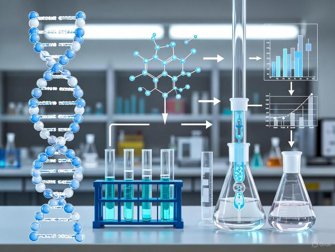
Optimized Protocols for High-Qality Genomic DNA Extraction from Mammalian Sperm Cells
This article provides a comprehensive guide for researchers on extracting high-quality genomic DNA from sperm cells, a process complicated by unique biological challenges.
Optimized Protocols for High-Qality Genomic DNA Extraction from Mammalian Sperm Cells
Abstract
This article provides a comprehensive guide for researchers on extracting high-quality genomic DNA from sperm cells, a process complicated by unique biological challenges. It covers the foundational principles of sperm chromatin compaction, evaluates traditional and novel extraction methodologies, and offers targeted troubleshooting for common issues like low yield and somatic contamination. A comparative analysis of protocol efficiency is presented, with a focus on validation techniques and downstream applications in advanced genetic and clinical studies, providing a complete framework for reliable sperm DNA isolation in biomedical research.
Understanding the Unique Challenges of Sperm Chromatin and DNA Integrity
The extraction of high-quality genomic DNA (gDNA) from spermatozoa presents a unique set of biological challenges that distinguish it from DNA isolation from somatic cells. The core issue lies in the extraordinary compaction of sperm chromatin, achieved through two primary biological mechanisms: the replacement of histones with protamines and the formation of extensive inter- and intra-protamine disulfide bridges [1]. During spermatogenesis, histones are largely replaced by protamines, which are smaller, arginine-rich proteins that facilitate extreme DNA condensation [2]. This protamine-DNA complex is further stabilized by the formation of disulfide bonds between cysteine residues of adjacent protamines [1]. This specialized chromatin organization results in nuclear compaction at least six times greater than that found in somatic cells [1], creating a physical barrier that conventional DNA extraction methods cannot efficiently overcome. The resilience of this structure is essential for protecting the paternal genome during transit through the female reproductive tract but represents a significant hurdle for researchers requiring intact gDNA for downstream applications such as genome sequencing, genetic marker analysis, and long-term DNA banking [1].
Comparative Analysis of DNA Extraction Method Efficacy
The performance of different DNA extraction methods varies significantly when applied to both fresh and cryopreserved sperm samples. The following table summarizes quantitative data comparing yield, purity, and integrity across six methodological approaches.
Table 1: Functional Assessment of Genomic DNA Extraction Methods from Caprine Sperm [1]
| Extraction Method | Key Reagents | Average DNA Yield (Fresh Sperm) | Average DNA Yield (Cryopreserved Sperm) | A260/A280 Ratio | DNA Integrity |
|---|---|---|---|---|---|
| Method 1 | β-Mercaptoethanol (β-ME) | 312.5 ± 12.5 µg | 187.5 ± 12.5 µg | ~1.8 | Degradation-free |
| Method 2 | Dithiothreitol (DTT) | 375.0 ± 25.0 µg | 250.0 ± 25.0 µg | ~1.8 | Degradation-free |
| Method 3 | β-ME + DTT (Combination) | 512.5 ± 37.5 µg | 375.0 ± 25.0 µg | ~1.8-2.0 | Degradation-free |
| Method 4 | Commercial Kit A | 62.5 ± 12.5 µg | 50.0 ± 10.0 µg | ~1.7 | Mostly intact |
| Method 5 | Commercial Kit B | 125.0 ± 25.0 µg | 75.0 ± 15.0 µg | ~1.7 | Mostly intact |
| Method 6 | Commercial Kit C | 250.0 ± 25.0 µg | 187.5 ± 12.5 µg | ~1.8 | Degradation-free |
The data demonstrates that the in-house method employing a combination of β-ME and DTT (Method 3) outperforms all other tested protocols, yielding the highest concentration of pure, degradation-free gDNA from both fresh and cryopreserved sperm [1]. This method proved to be the most efficient and economical for producing gDNA suitable for sensitive downstream applications, including qRT-PCR and DNA sequencing, even after six months of storage at -80°C [1].
Detailed Experimental Protocol for Optimal gDNA Extraction
Reagents and Solution Composition
- Lysis Buffer: 100 mM Tris-HCl (pH 8.0), 500 mM NaCl, 10 mM EDTA, and 1% SDS [1].
- Reducing Agents: Dithiothreitol (DTT; prepared fresh) and β-Mercaptoethanol (β-ME) [1].
- Proteinase K: For enzymatic digestion of nuclear proteins [1].
- RNase A: For elimination of RNA contamination [1].
- Precipitation & Wash Solutions: Absolute ethanol, 70% ethanol, and 3M sodium acetate (pH 5.2) [1].
- TE Buffer: 10 mM Tris-HCl, 1 mM EDTA (pH 8.0) for final DNA resuspension [1].
Step-by-Step Procedure
Sperm Lysis and Reduction:
Protein Digestion:
- Add 10 µL of Proteinase K (20 mg/mL) and incubate at 56°C for 2 hours [1].
RNA Removal:
- Add 5 µL of RNase A (10 mg/mL), mix by inversion, and incubate at 37°C for 30 minutes [1].
DNA Precipitation:
- Add 500 µL of 3M sodium acetate and 500 µL of absolute ethanol.
- Mix thoroughly by inversion until a white DNA precipitate is visible.
- Incubate at -20°C for 1 hour [1].
DNA Washing:
- Centrifuge at 12,000 × g for 10 minutes at 4°C and carefully discard the supernatant.
- Wash the pellet with 500 µL of 70% ethanol and centrifuge again at 12,000 × g for 5 minutes.
- Completely remove the ethanol and air-dry the pellet for 10-15 minutes [1].
DNA Hydration:
Diagram Title: Sperm gDNA Extraction Workflow
The Scientist's Toolkit: Essential Reagents and Their Functions
Table 2: Key Research Reagent Solutions for Sperm DNA Extraction
| Reagent | Chemical Nature | Primary Function | Mechanism of Action |
|---|---|---|---|
| Dithiothreitol (DTT) | Small-molecule reducing agent | Reduction of disulfide bonds | Cleaves S-S bonds between protamine cysteine residues, decondensing chromatin [1]. |
| β-Mercaptoethanol (β-ME) | Thiol-based reducing agent | Augmentation of disulfide reduction | Complements DTT action, ensuring complete reduction of resilient bonds [1]. |
| Sodium Dodecyl Sulfate (SDS) | Anionic detergent | Membrane lysis & protein denaturation | Solubilizes lipid membranes and denatures nuclear proteins, facilitating access to DNA [1]. |
| Proteinase K | Broad-spectrum serine protease | Enzymatic protein digestion | Degrades protamines and other proteins, liberating gDNA from the nucleoprotein complex [1]. |
| RNase A | Ribonuclease enzyme | RNA contamination removal | Selectively catalyzes the hydrolysis of RNA, preventing RNA contamination in the final gDNA sample [1]. |
Analytical Techniques for Disulfide Bridge Characterization
Mass spectrometry (MS) has emerged as a powerful tool for characterizing disulfide bridges in proteins, with methodologies that can be conceptually applied to understanding protamine disulfide networks. The following workflow illustrates a modern MS approach for disulfide bond analysis.
Diagram Title: Disulfide Bond Analysis by Mass Spectrometry
Key analytical advancements include:
- Microwave-Assisted Acid Hydrolysis (MAAH): Enables rapid, artifact-free digestion of proteins while keeping disulfide bonds intact, providing extensive sequence coverage with overlapping peptides [3].
- Electron Transfer/Higher Energy Collisional Dissociation (EThcD): Generates highly informative fragmentation spectra of disulfide-bridged peptides, producing both S-S cleavage and backbone ions (b/y and c/z) for confident linkage confirmation [3] [4].
- Field Asymmetric Ion Mobility Spectrometry (FAIMS): Efficiently removes chemical noise from hydrolysis, enhancing the detection of disulfide-bridged peptide pairs [3].
- Advanced Bioinformatics: Disulfide-aware search engines and algorithms (e.g., XlinkX, PEAKS, UNIFI) enable accurate annotation of complex, intertwined disulfide bridges [3] [4].
The synergistic application of combined reducing agents (DTT and β-ME) with optimized lysis conditions represents the most effective strategy for overcoming the biological hurdles presented by protamine-rich chromatin and its extensive disulfide bridge network. The standardized protocol detailed herein reliably produces high yields of degradation-free, pure genomic DNA from both fresh and cryopreserved sperm samples, making it suitable for the most demanding downstream applications, including genome-wide sequencing studies and long-term genetic resource conservation [1]. For researchers requiring ultimate analytical precision in disulfide bond characterization, modern mass spectrometry workflows incorporating EThcD fragmentation and FAIMS separation offer unprecedented capability to map complex disulfide topologies, providing insights that extend from basic reproductive biology to therapeutic protein engineering [3] [4].
Sperm DNA integrity is a critical parameter for successful fertilization, embryo development, and pregnancy outcomes. Within the broader context of research on DNA extraction protocols from sperm cells, understanding the sources and mechanisms of DNA damage is fundamental. Oxidative stress induced by reactive oxygen species (ROS) represents the primary cause of sperm DNA damage, a process often exacerbated by cryopreservation procedures essential to assisted reproductive technologies (ART) [5] [6]. Spermatozoa are particularly vulnerable to oxidative insult due to their limited cytoplasm, which contains minimal antioxidant enzymes, and the high concentration of polyunsaturated fatty acids (PUFAs) in their plasma membranes [5] [6]. This application note details the quantitative impact of cryopreservation, the molecular pathways of oxidative damage, and standardized protocols for assessing DNA integrity in sperm cells, providing a framework for researchers developing optimized DNA extraction and preservation methodologies.
Quantitative Impact of Cryopreservation and Oxidative Stress
Extensive clinical studies have quantified the detrimental effects of cryopreservation on sperm DNA integrity and the associated rise in oxidative stress. The data, summarized in the table below, reveal consistent patterns of damage across diverse populations.
Table 1: Quantitative Impact of Cryopreservation on Sperm DNA Integrity and Oxidative Stress
| Study Population | Sample Size | Assessment Method | Key Finding Related to DNA Fragmentation | Key Finding Related to Oxidative Stress |
|---|---|---|---|---|
| Normozoospermic Libyan Males [7] | 104 | Sperm Chromatin Dispersion (SCD) | DNA Fragmentation Index (DFI) increased from 46.3% to 60.0% post-thaw. | ROS levels increased from 3.2 × 10³ RLU/s to 14.7 × 10³ RLU/s post-thaw. A positive correlation between ROS and DFI was observed (r=0.68). |
| Mixed Fertility Status [8] | 30 (15 fertile, 15 infertile) | Sperm Chromatin Structure Assay (SCSA) | A post-thaw increase in DNA fragmentation was recorded across all groups; infertile samples were more adversely affected. | An increase in the apoptotic marker Caspase-3 was detected after freezing. |
| Fertile Donors vs. Infertile Patients [9] | 60 (20 fertile, 40 infertile) | TUNEL Assay | Infertile patients had significantly higher SDF (32.77%) than fertile donors (22.19%). SDF negatively correlated with count, motility, and morphology. | Not Specifically Measured |
Furthermore, analysis of the relationship between conventional semen parameters and DNA fragmentation reveals significant correlations, underscoring the pervasive nature of oxidative damage.
Table 2: Correlations Between Sperm DNA Fragmentation and Conventional Semen Parameters [9]
| Semen Parameter | Correlation Coefficient (r) with SDF | P-value |
|---|---|---|
| Sperm Count | -0.4036 | 0.0014 |
| Sperm Motility | -0.6377 | < 0.0001 |
| Sperm Morphology | -0.2783 | 0.0378 |
| Age | 0.2167 | 0.0993 (Not Significant) |
Molecular Mechanisms of Oxidative Stress-Induced DNA Damage
The vulnerability of sperm to oxidative stress is an inherent consequence of their specialized structure and function. The molecular mechanisms leading to DNA damage involve a cascade of events:
- ROS Generation and Membrane Damage: The freeze-thaw process induces physical and chemical stress, leading to increased ROS production from sources like sperm mitochondria and leukocytes in semen [5] [6]. Due to the high PUFA content in the sperm plasmalemma, ROS readily initiate lipid peroxidation [5]. This chain reaction damages the sperm membrane's integrity, reducing its fluidity and compromising motility and viability.
- DNA Fragmentation: The lipid peroxidation process generates toxic byproducts like malondialdehyde (MDA) and 4-hydroxynonenal (4-HNE) [6]. These aldehydes can bind to proteins in the mitochondrial electron transport chain, creating a vicious cycle of further ROS production [5]. Ultimately, ROS directly attack the sperm DNA, causing single-strand and double-strand breaks, as well as oxidative base lesions such as 8-hydroxy-2'-deoxyguanosine (8-OHdG) [6]. Single-stranded breaks are more associated with oxidative stress and can lead to implantation failure, while double-stranded breaks are linked to poor embryo quality and miscarriage [5].
The following diagram illustrates this self-perpetuating cycle of damage.
Experimental Protocols for Assessing Sperm DNA Damage
For research focused on DNA extraction and integrity, accurate assessment of sperm DNA fragmentation (SDF) is crucial. Below are detailed protocols for two commonly used and complementary techniques.
Protocol: Sperm Chromatin Dispersion (SCD) Test
The SCD test is a widely used method for quantifying DNA fragmentation based on the differential decondensation of chromatin between damaged and intact sperm nuclei [7].
- Principle: Sperm with fragmented DNA produce a characteristic "halo" of dispersed DNA loops when subjected to acid denaturation and subsequent neutralization, while sperm with intact DNA produce a minimal or no halo due to highly compacted chromatin.
- Materials:
- Sperm-Halomax kit (or equivalent agarose solutions, lysis solutions, and staining dyes).
- Pre-coated slides or standard microscope slides.
- Water bath (set to 22-37°C).
- Bright-field or fluorescence microscope.
- Procedure:
- Sample Preparation: Mix fresh or thawed semen sample with low-melting-point agarose to achieve a final concentration of 0.7% agarose.
- Slide Preparation: Pipette the mixture onto a pre-coated slide and cover with a coverslip. Place the slide on a cold surface (4°C) for 5 minutes to allow the agarose to solidify.
- Denaturation and Lysis: Gently remove the coverslip and immerse the slide in a freshly prepared acid denaturation solution (0.08 N HCl) for 7 minutes at room temperature. Then, transfer the slide to a lysis solution (0.4 M Tris-HCl, 0.8 M DTT, 1% SDS) for 25 minutes at room temperature.
- Washing and Dehydration: Wash the slide sequentially in distilled water for 5 minutes, 70% ethanol for 2 minutes, and 100% ethanol for 2 minutes. Air-dry the slide completely.
- Staining and Analysis: Stain the sperm nuclei using a DNA-specific fluorescent stain (e.g., DAPI, Propidium Iodide) or a Diff-Quick set. Score at least 500 spermatozoa under a microscope. Sperm are classified as having fragmented DNA if they show a small or absent halo, and intact DNA if they show a large halo.
Protocol: TUNEL Assay with Flow Cytometry
The TUNEL (Terminal deoxynucleotidyl transferase dUTP Nick End Labeling) assay is a direct method for detecting DNA strand breaks by enzymatically labeling the 3'-OH ends of fragmented DNA [9].
- Principle: The enzyme Terminal deoxynucleotidyl Transferase (TdT) catalyzes the addition of fluorescently labeled dUTP nucleotides to the 3'-OH ends of DNA fragments, allowing for quantification via flow cytometry.
- Materials:
- Commercial TUNEL assay kit (e.g., In Situ Cell Death Detection Kit).
- Flow cytometer.
- Phosphate-Buffered Saline (PBS).
- 4% Paraformaldehyde (PFA) in PBS.
- 0.1% Triton X-100 in 0.1% sodium citrate.
- Procedure:
- Sperm Washing and Fixation: Wash sperm samples twice with PBS by centrifugation (500 x g for 5 minutes). Resuspend the pellet in 4% PFA and incubate for 1 hour at room temperature for fixation.
- Permeabilization: Centrifuge the fixed cells, wash with PBS, and then permeabilize the cells by resuspending them in 0.1% Triton X-100 for 2 minutes on ice.
- Labeling Reaction: Centrifuge the cells and resuspend the pellet in the TUNEL reaction mixture (containing TdT and fluorescent-dUTP). Incubate for 1 hour at 37°C in the dark. Prepare a negative control by incubating a separate aliquot in a reaction mixture without the TdT enzyme.
- Flow Cytometry Analysis: Wash the cells twice with PBS and resuspend in 0.5 mL of PBS. Analyze the samples using a flow cytometer with an excitation wavelength of 488 nm. The percentage of TUNEL-positive cells (indicating high DNA fragmentation) is calculated from the fluorescence histogram.
The workflow for these assessments is summarized below.
The Scientist's Toolkit: Essential Research Reagents and Materials
Successful experimentation in this field relies on a suite of specialized reagents and equipment. The following table catalogs key solutions for conducting the described protocols and investigating oxidative stress.
Table 3: Research Reagent Solutions for Sperm DNA Damage Studies
| Reagent / Material | Function / Application | Example Kits & Specifications |
|---|---|---|
| Cryoprotectant Agents | Protect sperm from ice crystal formation and osmotic shock during freeze-thaw. | Egg-yolk + glycerol; Sucrose + glycerol; Glycerol alone [8]. |
| Sperm DNA Fragmentation Kits | Quantify the level of DNA strand breaks in spermatozoa. | Sperm-Halomax (SCD); In Situ Cell Death Detection Kit (TUNEL) [7] [9]. |
| ROS Detection Kits | Quantify levels of reactive oxygen species in semen samples. | Luminol-enhanced chemiluminescence assays [7]. |
| Antioxidant Supplements | Investigate protective strategies against oxidative damage during cryopreservation. | Melatonin (2 mM) [7]. |
| Nucleic Acid Extraction Kits | Co-isolate high-quality DNA and RNA from complex seminal plasma for downstream analysis. | QIAamp DNA Mini Kit; miRNeasy Tissue/Cells Advanced Micro Kit [10] [11]. |
| Computer-Assisted Sperm Analysis (CASA) | Objectively assess standard semen parameters (concentration, motility, kinematics). | Integrated systems with chamber slides and analysis software [8] [12]. |
Oxidative stress, significantly amplified by the cryopreservation process, is a primary driver of sperm DNA damage. This compromises genetic integrity and has direct implications for the efficacy of DNA extraction protocols and subsequent molecular analyses in research settings. The quantitative data and standardized methodologies presented herein—ranging from SCD and TUNEL assays for DNA damage assessment to the evaluation of protective cryoprotectant formulations—provide a critical resource for scientists. Integrating these assessments into the research workflow is essential for developing robust DNA extraction methods, optimizing cryopreservation strategies, and ultimately improving diagnostic and therapeutic outcomes in male fertility.
The integrity of genomic DNA (gDNA) extracted from spermatozoa is a foundational requirement for advanced molecular analyses, including genome sequencing, genotyping, and the establishment of long-term DNA banks. The choice of starting material—fresh ejaculated or cryopreserved sperm—profoundly impacts the yield, purity, and functional utility of the isolated nucleic acids. The highly compact, protamine-rich nature of sperm chromatin presents unique challenges for DNA extraction, which are further compounded by the cryopreservation process. Cryopreservation, while essential for the flexibility of assisted reproductive technologies (ART) and biobanking, can induce physical and oxidative stress, leading to compromised DNA integrity. This application note delineates the comparative impact of fresh versus cryopreserved sperm samples on downstream DNA analyses. It provides detailed, actionable protocols to guide researchers in selecting and optimizing their methodologies to ensure the recovery of high-quality genetic material, thereby supporting robust and reproducible scientific outcomes in reproductive genetics and drug development [1] [13] [14].
Comparative Analysis: Fresh vs. Cryopreserved Sperm
DNA Yield, Purity, and Integrity
The physical and biochemical alterations inflicted by the freeze-thaw cycle directly influence the metrics of extracted DNA. A functional assessment of different gDNA extraction methods from caprine sperm established a quantitative basis for this comparison.
Table 1: Comparative DNA Yield and Purity from Fresh vs. Cryopreserved Sperm
| Parameter | Fresh Ejaculated Sperm | Cryopreserved Sperm | Notes |
|---|---|---|---|
| DNA Yield | Significantly higher | Reduced | The combination of β-Mercaptoethanol (β-ME) and Dithiothreitol (DTT) yielded the highest DNA amounts from both types [1]. |
| Protein Contamination | Minimal or absent | Minimal or absent | Optimized protocols can achieve high purity regardless of preservation status [1]. |
| DNA Fragmentation | Lower baseline | Markedly increased | Cryopreservation induces single and double-strand DNA breaks due to oxidative stress and ice crystal formation [15] [14]. |
| Suitability for Long-Term Banking | High | High | Provided extraction methods yield pure, degradation-free gDNA [1]. |
Functional Suitability for Downstream Applications
The ultimate value of extracted DNA is determined by its performance in subsequent analyses.
- Genome Sequencing and qRT-PCR: High-quality, degradation-free gDNA extracted via a modified β-ME/DTT protocol from both fresh and cryopreserved caprine sperm has been demonstrated to be fully suitable for genome sequencing and quantitative expression analyses post long-term storage [1].
- Epigenetic Studies: Sperm epigenetic profiles are highly vulnerable to contamination by somatic DNA present in semen samples. This is a critical consideration for both fresh and cryopreserved specimens. A comprehensive strategy involving microscopic examination, treatment with a somatic cell lysis buffer (SCLB), and bioinformatic filtering using defined CpG biomarkers is essential to eliminate confounding signals from somatic cell contamination [16].
Experimental Protocols for DNA Extraction and Assessment
Optimized gDNA Extraction Protocol for Sperm Cells
The following in-house protocol, modified with a combination of reducing agents, has been validated for efficient recovery of high-quality gDNA from both fresh and cryopreserved sperm, outperforming several commercial kits in terms of yield and cost-efficiency [1].
Reagent Solutions:
- Lysis Buffer: 100 mM Tris-HCl (pH 8.0), 500 mM NaCl, 10 mM EDTA, 1% SDS.
- Reducing Agents: β-Mercaptoethanol (β-ME) and Dithiothreitol (DTT). DTT solution must be prepared fresh.
- Other Reagents: Proteinase K, RNase A, absolute and 70% ethanol, 3M sodium acetate (pH 5.2), and Triton X-100.
Step-by-Step Procedure:
- Sperm Washing: Wash approximately 1x10⁶ sperm cells with 1X PBS by centrifugation to remove seminal plasma and cryoprotectant residues.
- Cell Lysis: Resuspend the sperm pellet in 500 µL of lysis buffer.
- Reduction of Disulfide Bridges: Add 20 µL of β-ME and 25 µL of DTT to the lysate. Mix thoroughly. This step is critical for breaking the highly compact, protamine-stabilized sperm chromatin.
- Digestion: Add 25 µL of Proteinase K (20 mg/mL) and 5 µL of RNase A (4 mg/mL). Incubate at 56°C for 2–3 hours, with intermittent mixing, until the solution becomes clear.
- DNA Precipitation: Add 500 µL of absolute ethanol and 50 µL of 3M sodium acetate. Invert the tube gently until DNA threads become visible.
- DNA Washing: Pellet the DNA by centrifugation, carefully discard the supernatant, and wash the pellet with 1 mL of 70% ethanol.
- Dissolution: Air-dry the DNA pellet and dissolve it in 50 µL of nuclease-free water or TE buffer.
- Storage: Store the extracted gDNA at -80°C for long-term banking.
Advanced Protocol: Detection of Sperm DNA Fragmentation Using a TdT/Cas12a Biosensor
Accurate assessment of DNA fragmentation is crucial. This novel, highly sensitive biosensor protocol outperforms traditional methods like SCSA and TUNEL in detecting cryopreservation-induced DNA breakpoints [14].
Reagent Solutions:
- TdT Reaction Buffer: Includes TdT enzyme, CoCl₂, and dATP.
- CRISPR-Cas12a Detection System: Contains LbCas12a, specific crRNA, and a fluorescent reporter probe (e.g., FAM-BHQ1).
- Antioxidant Supplements (Optional): Lycium barbarum polysaccharides (LBP, 1 mg/mL) or Resveratrol (RES, 30 µmol/L) can be added to cryoprotectant to mitigate DNA damage.
Step-by-Step Procedure:
- DNA Extraction: Extract genomic DNA from fresh and frozen-thawed sperm samples using a standardized commercial kit.
- TdT-Mediated Tailing: Prepare a 15 µL reaction mixture containing 1X TdT buffer, CoCl₂, dATP, TdT enzyme, and the extracted sperm DNA (e.g., 5 ng/µL). Incubate at 37°C for 30 minutes to add a poly-A tail to the 3'-OH ends of DNA breakpoints. Heat-inactivate at 85°C for 10 minutes.
- Cas12a Detection: Immediately add the CRISPR-Cas12a detection system to the inactivated TdT reaction. The final mixture should contain Cas12a, crRNA, MgCl₂, and the fluorescent probe.
- Fluorescence Measurement: Conduct isothermal fluorescence measurement at 37°C for 60 cycles (30 seconds per cycle) using a real-time PCR instrument. The Cas12a, upon recognition of the poly-A tail by its crRNA, will exhibit trans-cleavage activity, cleaving the fluorescent probe and generating a signal proportional to the number of DNA breakpoints.
- Data Analysis: Quantify DNA breakpoints by comparing the fluorescence signals to a standard curve generated with known concentrations of a standard DNA breakpoint control.
Diagram 1: TdT/Cas12a Biosensor Workflow for DNA Breakpoint Detection.
The Scientist's Toolkit: Essential Research Reagents
Table 2: Key Reagent Solutions for Sperm DNA Extraction and Analysis
| Reagent / Solution | Function / Application | Key Considerations |
|---|---|---|
| Dithiothreitol (DTT) & β-Mercaptoethanol (β-ME) | Reduction of disulfide bridges in protamine-bound sperm chromatin. | Combining both agents yields superior DNA results. DTT must be prepared fresh [1]. |
| Somatic Cell Lysis Buffer (SCLB) | Selective lysis of contaminating somatic cells in semen samples. | Critical for pure sperm epigenetic studies. Composition: 0.1% SDS, 0.5% Triton X-100 [16]. |
| Silane-Coated Colloidal Silica Gradients | Separation of morphologically normal sperm via Density Gradient Centrifugation (DGC). | Selects sperm with denser, more homogeneous nuclei, enriching for DNA-intact cells [17]. |
| Natural Antioxidants (LBP, Resveratrol) | Additives to cryoprotectant to mitigate oxidative DNA damage during freeze-thaw. | LBP at 1 mg/mL and Resveratrol at 30 µmol/L have shown protective effects [14]. |
| TdT/Cas12a Biosensor System | Highly sensitive detection and quantification of DNA breakpoints. | Offers superior sensitivity and specificity over SCSA, TUNEL, etc. Requires real-time PCR equipment [14]. |
The choice between fresh and cryopreserved sperm as a starting material necessitates a strategic balance between logistical convenience and analytical demand. While cryopreservation introduces challenges for DNA integrity, optimized protocols can effectively mitigate these issues. For researchers, the following evidence-based recommendations are proposed:
- For Maximum DNA Yield and Integrity: Whenever possible, use fresh ejaculated sperm and employ a DNA extraction protocol incorporating a combination of reducing agents (DTT and β-ME) to ensure efficient chromatin decondensation [1].
- When Using Cryopreserved Sperm: Supplement cryoprotectants with natural antioxidants like LBP or resveratrol to shield sperm DNA from oxidative stress during the freeze-thaw cycle [14].
- For Sensitive Epigenetic Analyses: Implement a rigorous somatic cell removal protocol, including SCLB treatment and bioinformatic filtering, to guarantee that the resulting DNA methylation profile is exclusively representative of sperm cells [16].
- For Accurate DNA Fragmentation Assessment: Consider adopting next-generation detection methods like the TdT/Cas12a biosensor for unparalleled sensitivity in quantifying DNA damage, which is particularly relevant for assessing cryopreservation-induced stress [14].
By aligning the choice of starting material with these tailored experimental protocols, researchers can ensure the procurement of high-quality genomic DNA, thereby fortifying the reliability of their data in genomics, reproductive medicine, and drug development.
The isolation of high-quality DNA from sperm cells is a critical procedure in diverse fields, including reproductive medicine, forensic science, and genetic research. The unique structural composition of spermatozoa presents distinct challenges not encountered with somatic cells. During spermatogenesis, approximately 90% of histones are replaced by protamines in humans, leading to a chromatin structure that is at least six times more compact than that of somatic cells [18] [1]. This compaction is stabilized by disulfide bridges between cysteinerich protamines, rendering the sperm nucleus highly resistant to conventional lysis methods [18] [1]. Consequently, DNA extraction from sperm requires specialized protocols that effectively dismantle this robust structure without compromising DNA integrity. This application note details the core principles—lysis, binding, washing, and elution—within the specific context of sperm cell processing, providing researchers with optimized protocols to overcome these challenges.
Core Principles and Their Application in Sperm DNA Extraction
Lysis: Disrupting the Unique Sperm Architecture
The lysis step must achieve dual objectives: breaking down the resilient sperm membrane and decondensing the protamine-packed chromatin. Effective lysis is accomplished through a combination of chemical, enzymatic, and sometimes mechanical strategies.
Chemical Lysis: Chaotropic salts, such as guanidine thiocyanate (GTC) or guanidinium hydrochloride, are essential components. They disrupt cellular membranes, inactivate nucleases, and facilitate the dissociation of proteins from nucleic acids [18]. Detergents like Sodium Dodecyl Sulfate (SDS) further solubilize membrane lipids and proteins [1].
Reducing Agents: The reduction of disulfide bonds cross-linking protamines is paramount. Traditional agents include Dithiothreitol (DTT) or β-mercaptoethanol (β-ME). A significant advancement is the use of tris(2-carboxyethyl)phosphine (TCEP), which is odorless, stable at room temperature, and highly effective, often yielding >90% high-quality DNA [18].
Enzymatic Digestion: Proteinase K is widely used to digest nuclear proteins and degrade contaminating enzymes. However, recent protocols have demonstrated that efficient mechanical homogenization can reduce or eliminate the need for lengthy (e.g., overnight) Proteinase K incubations [18].
Mechanical Homogenization: The use of 0.2 mm stainless steel beads in a homogenizer for 5-10 minutes at room temperature can dramatically accelerate cell lysis, making the process faster and more efficient [18].
Table 1: Key Components of an Optimized Sperm Lysis Buffer
| Component | Concentration/Final Amount | Primary Function | Considerations for Sperm |
|---|---|---|---|
| Guanidine Thiocyanate | 4-6 M | Chaotropic salt; lyses cells, inactivates nucleases | Core component for accessing compact DNA [18] |
| TCEP | 50 mM | Reduces disulfide bonds between protamines | Odorless, stable alternative to DTT/β-ME [18] |
| Proteinase K | 200 µg/mL | Digests nuclear proteins | Incubation time can be reduced with mechanical homogenization [18] |
| Tris-HCl | 100 mM (pH 8.0) | Maintains stable pH | Common buffer for molecular biology [1] |
| EDTA | 10 mM | Chelates Mg²⁺, inhibits DNases | Prevents DNA degradation [1] |
| SDS | 1% (w/v) | Ionic detergent; solubilizes membranes | Effective in combination with other agents [1] |
Binding, Washing, and Elution: Purifying DNA
Following lysis, the released DNA is purified using silica-based membrane technology, which leverages the affinity of DNA for silica in the presence of high concentrations of chaotropic salts.
Binding: The lysate is mixed with a binding buffer, frequently containing high concentrations of GTC or guanidine hydrochloride, and loaded onto a silica membrane column. Under these conditions, DNA adsorbs to the silica matrix, while proteins and other contaminants are repelled [18].
Washing: Impurities are removed through a series of wash steps. Initial washes often contain chaotropic salts to reinforce DNA binding and remove residual proteins. A final wash with an ethanol-based buffer removes salts and other small molecules without eluting the DNA. It is crucial to centrifuge the columns thoroughly to remove all ethanol, as carryover can inhibit downstream enzymatic reactions [18].
Elution: Pure DNA is recovered by applying a low-ionic-strength buffer, such as TE buffer or nuclease-free water. Preheating the elution buffer to 70°C and allowing a 3-5 minute incubation on the membrane before centrifugation can significantly increase DNA yield [18]. Some protocols recommend a second elution with a fresh buffer volume to maximize recovery [18].
Optimized Experimental Protocol for Sperm DNA Isolation
This protocol, adapted from a rapid isolation method, yields high-quality DNA suitable for genetic and epigenetic analyses [18].
Materials Required (The Scientist's Toolkit):
- Somatic Cell Lysis Buffer (SCLB) [16]: For initial purification of sperm from semen.
- Buffer RLT (Qiagen) or equivalent GTC-based lysis buffer [18].
- TCEP (0.5 M stock solution) [18].
- 0.2 mm stainless steel beads [18].
- Silica-based spin columns (e.g., from Qiagen or Zymo Research kits) [18].
- Proteinase K (optional).
- Wash buffers (typically provided with kit).
- Nuclease-free water or TE buffer (for elution).
Procedure:
- Sperm Cell Purification: To mitigate somatic cell contamination, which is a major concern in epigenetic studies, wash fresh semen samples twice with 1X PBS by centrifugation at 200-500 × g for 15 minutes. Inspect the pellet under a microscope. If somatic cells are present, incubate the sample with SCLB (0.1% SDS, 0.5% Triton X-100) for 30 minutes at 4°C, then re-pellet the sperm [16].
- Cell Lysis: Resuspend the purified sperm pellet (containing 20-100 million cells) in 500 µL of lysis buffer (e.g., Buffer RLT) supplemented with 50 mM TCEP. Add 0.1 g of 0.2 mm steel beads and homogenize for 5 minutes on a mechanical disruptor (e.g., Disruptor Genie) at room temperature [18].
- DNA Binding: Transfer the homogenized lysate directly to a silica spin column. Centrifuge at ≥10,000 × g for 30-60 seconds to bind the DNA. Discard the flow-through. Note: If somatic contamination is still a concern, the lysate can be stored at room temperature for at least two weeks without degrading DNA yield or quality [18].
- Washing: Perform two washes with the provided wash buffers according to the manufacturer's instructions. Ensure a final centrifugation with an empty column for 1-2 minutes to dry the membrane completely.
- Elution: Apply 50 µL of preheated (70°C) nuclease-free water to the center of the membrane. Incubate at room temperature for 3-5 minutes. Centrifuge at maximum speed for 1 minute. For maximum yield, repeat the elution with a second 50 µL volume [18].
Advanced Considerations for Research Applications
Addressing Somatic Cell Contamination
For sensitive applications like epigenetic profiling, even minimal somatic cell contamination can skew results. A multi-pronged approach is recommended [16]:
- Microscopic Examination: Visually inspect samples before and after SCLB treatment.
- Biomarker Screening: Analyze known CpG sites that are hypermethylated in blood (>80%) and hypomethylated in sperm (<20%). A panel of 9,564 such CpG sites has been identified for this purpose [16].
- Data Analysis Cut-off: Apply a conservative threshold (e.g., 15% methylation difference) during data analysis to filter out potential contamination artifacts [16].
Comparison of Methods and Reagents
Table 2: Comparison of Reducing Agents and Separation Methods for Sperm DNA Extraction
| Method / Reagent | Key Advantage | Key Disadvantage | Typical DNA Yield/Quality |
|---|---|---|---|
| TCEP [18] | Odorless, room-temperature stable, highly effective | Higher cost than traditional agents | >90% yield, high-quality |
| DTT + β-ME Combination [1] | High yield reported for caprine sperm | Odor, requires fresh preparation, less stable | High yield, pure gDNA |
| Microfluidic Sperm Sorting (MFSS) [19] | Reduces DNA fragmentation, no centrifugation | Lower sperm recovery, may not suit oligozoospermic samples | Lower fragmentation vs. Swim-Up |
| Density Gradient Centrifugation [17] | Effective removal of somatic cells & debris | Centrifugation may increase ROS and DNA damage | Good motility & morphology selection |
The successful extraction of DNA from sperm cells hinges on protocols specifically designed to overcome the biological challenges posed by protamine condensation and disulfide cross-linking. The integration of robust reducing agents like TCEP, efficient mechanical homogenization, and stringent measures to eliminate somatic cell contamination ensures the recovery of DNA of the highest quality and purity. The protocols and principles outlined here provide a reliable foundation for research and clinical applications requiring accurate genetic and epigenetic analysis of sperm.
Step-by-Step Guide to Established and Novel Extraction Protocols
The efficiency of genomic DNA (gDNA) extraction from sperm cells is a critical determinant for the success of downstream molecular applications, including genome sequencing, genetic disease research, and long-term DNA banking. Spermatozoa present unique challenges not encountered with somatic cells due to their highly compacted nuclear chromatin. This compaction is achieved through the replacement of histones with protamines during spermatogenesis, forming a dense structure stabilized by numerous disulfide bonds [1]. This robust architecture functions as a formidable barrier to standard lysis methods, necessitating the strategic optimization of lysis buffers with specific reducing agents to break these cross-links and liber high-quality, high-molecular-weight DNA for research and clinical diagnostics [1].
The core challenge lies in the biochemical composition of the sperm nucleus. The disulfide bridges between protamines create a nuclear matrix that is exceptionally resistant to conventional chemical and physical lysis methods developed for somatic cells. Consequently, lysis protocols require the incorporation of reducing agents to cleave these covalent bonds. The choice of reducing agent, its concentration, and its combination with other buffer components directly impact the yield, purity, and integrity of the extracted gDNA. This application note details the optimization of in-house manual lysis methods using dithiothreitol (DTT) and β-mercaptoethanol (β-ME), providing a validated protocol for the research community.
Lysis Buffer Composition and Mechanism of Action
An effective lysis buffer for sperm cells must perform three primary functions: disrupt the plasma and nuclear membranes, reduce disulfide bonds to decondense chromatin, and protect the released DNA from nucleases. The table below outlines the core components of a standard sperm lysis buffer and their specific functions.
Table 1: Core Components of an Optimized Sperm Lysis Buffer
| Component | Typical Concentration | Primary Function |
|---|---|---|
| Tris-HCl | 100 mM (pH 8.0) | Maintains a stable pH for optimal enzyme activity and DNA stability [1]. |
| Sodium Chloride (NaCl) | 500 mM | Provides ionic strength to shield negative charges on the DNA backbone, preventing aggregation [1]. |
| EDTA | 10 mM | Chelates divalent cations (Mg²⁺, Ca²⁺), inactivating metalloproteases and DNases [1]. |
| Sodium Dodecyl Sulfate (SDS) | 1% | Ionic detergent that solubilizes lipid membranes and denatures proteins [1]. |
| Reducing Agent (DTT/β-ME) | Variable | Cleaves disulfide bonds between protamines, enabling chromatin decondensation and DNA release [1]. |
The key to successful sperm lysis is the reduction of disulfide bridges. DTT and β-ME are thiol-based reducing agents that break these bonds by undergoing oxidation themselves. DTT is often preferred for its stronger reducing power and greater stability in buffer solutions; it is approximately 7-fold stronger than β-ME and lacks the volatile, unpleasant odor associated with the latter [20]. β-ME, while less expensive, is less stable and can evaporate from solution, leading to a gradual loss of reducing capacity over time [20]. This can result in incomplete lysis and reduced DNA yield.
Optimized Protocol for Genomic DNA Extraction from Sperm
Reagents and Equipment
- Lysis Buffer: 100 mM Tris-HCl (pH 8.0), 500 mM NaCl, 10 mM EDTA, 1% SDS [1].
- Reducing Agents: 1M DTT stock and/or β-Mercaptoethanol (β-ME) [1].
- Proteinase K: ≥20 mg/mL solution.
- RNase A: 10 mg/mL solution.
- Phenol:Chloroform:Isoamyl Alcohol (25:24:1)
- Absolute Ethanol and 70% Ethanol
- Microcentrifuge Tubes, water bath, centrifuge, and vortexer.
Step-by-Step Workflow
Diagram 1: Schematic workflow of the optimized gDNA extraction protocol from sperm cells.
Cell Lysis:
- Resuspend the purified sperm pellet in 500 µL of lysis buffer.
- Add reducing agents to the specified final concentration. For a combined approach, use 50 mM DTT and 0.2% β-ME [1].
- Add Proteinase K to a final concentration of 200 µg/mL.
- Incubate at 56°C overnight (12-16 hours) with gentle agitation to ensure complete lysis and digestion [1].
RNA and Protein Digestion:
- After overnight lysis, add RNase A to a final concentration of 100 µg/mL.
- Vortex briefly and incubate at 37°C for 30 minutes [1].
DNA Purification:
- Perform organic extraction with an equal volume of Phenol:Chloroform:Isoamyl Alcohol. Mix thoroughly and centrifuge at >12,000 × g for 5 minutes.
- Carefully transfer the upper aqueous phase to a new tube.
- Add 2 volumes of ice-cold absolute ethanol to precipitate the DNA. Invert the tube gently until the DNA is visible as a stringy precipitate.
- Centrifuge at >12,000 × g for 10 minutes to pellet the DNA.
- Wash the pellet with 1 mL of 70% ethanol to remove residual salts. Centrifuge again and carefully decant the supernatant.
- Air-dry the pellet for 5-10 minutes and resuspend in TE buffer or nuclease-free water.
Comparative Data and Analysis
A systematic comparison of different reducing agent strategies was conducted on both fresh and cryopreserved caprine sperm. The results demonstrate the clear advantage of using a combination of DTT and β-ME.
Table 2: Functional Comparison of Reducing Agent Efficacy in Sperm gDNA Extraction
| Extraction Method | Reducing Agent(s) | Average DNA Yield (Fresh Sperm) | Average DNA Yield (Cryopreserved Sperm) | Purity (A260/A280) | Key Findings |
|---|---|---|---|---|---|
| In-House Modified | DTT + β-ME | Highest | Highest | ~1.8 | Superior yield; pure, degradation-free DNA suitable for sequencing & banking [1] |
| Kit-based (DTT) | DTT | Moderate | Moderate | ~1.8 | Reliable but less efficient for highly compacted sperm [1] |
| Kit-based (β-ME) | β-ME | Lower | Lower | ~1.8 | Less effective due to lower reducing power and stability [1] |
| Organic (β-ME) | β-ME | Low | Low | Variable | Inefficient; fails to fully disrupt protamine disulfide bonds [1] |
The Scientist's Toolkit: Research Reagent Solutions
Table 3: Essential Reagents for Sperm Lysis Buffer Optimization
| Reagent | Function | Key Considerations |
|---|---|---|
| Dithiothreitol (DTT) | Strong reducing agent for cleaving disulfide bonds in protamines [1]. | Preferred for its strength, stability, and lack of odor. Prepare fresh stock solutions for optimal activity [20]. |
| β-Mercaptoethanol (β-ME) | Alternative reducing agent for disulfide bond reduction [1]. | Less expensive but weaker and volatile. Requires careful handling due to toxicity and smell [20]. |
| Proteinase K | Broad-spectrum serine protease degrades cellular proteins and nucleases [1]. | Essential for digesting proteins after the chromatin is decondensed by reducing agents. |
| Sodium Dodecyl Sulfate (SDS) | Ionic detergent that solubilizes cell membranes and denatures proteins [1]. | Works synergistically with reducing agents and Proteinase K to disrupt cellular structures. |
| EDTA (Ethylenediaminetetraacetic acid) | Chelating agent that binds divalent metal ions [1]. | Inactivates metalloproteases and inhibits DNases by removing required co-factors (Mg²⁺). |
Troubleshooting and Technical Notes
- Low DNA Yield: This is often due to incomplete lysis. Ensure the lysis buffer contains a sufficiently high concentration of a potent reducing agent. The combination of DTT and β-ME is highly recommended. Extending the incubation time at 56°C may also improve yields.
- Suboptimal Lysis Buffer: For sperm cells, standard lysis buffers like RIPA may be insufficient. A buffer with higher salt concentration (e.g., 500 mM NaCl) and strong detergents like SDS is crucial for efficient lysis and protein solubilization [21] [1].
- Handling Reducing Agents: DTT solutions are susceptible to oxidation. Always prepare fresh stock solutions or aliquot and store them at -20°C. β-ME should be used in a fume hood due to its volatility and odor.
- Downstream Application Failures: If the extracted DNA performs poorly in PCR or sequencing, check the A260/A280 ratio. A ratio below 1.8 suggests protein contamination, indicating a need for more thorough Proteinase K digestion or an additional phenol-chloroform extraction step.
Within the broader context of DNA extraction protocol research, the isolation of high-quality genetic material from spermatozoa presents a unique challenge. The compact nature of sperm chromatin, resulting from the protamine-based packaging of DNA and the formation of inter-protamine disulfide bridges, renders sperm cells exceptionally resistant to standard lysis techniques used for somatic cells [18]. This application note details a optimized protocol for silica-based column kits that addresses these challenges through specific modifications, enabling efficient and reliable DNA recovery from sperm cells for downstream genetic and epigenetic analyses.
The Challenge of Sperm Chromatin
Sperm DNA is packaged with protamines in a highly condensed state. During spermatogenesis, up to 90% of histones are replaced by protamines in humans, facilitating extreme nuclear compaction [18]. This is stabilized by the oxidation of cysteine residues on protamines, forming extensive disulfide bridges that cross-link the chromatin structure [18]. This unique architecture, essential for protecting the genetic material during transit, presents a significant biochemical barrier to DNA extraction, requiring specialized lysis conditions that conventional protocols do not provide.
Optimized Protocol for Enhanced Sperm Lysis
The following protocol modifies commercial silica-column kits to incorporate a mechanical homogenization step and a potent, stable reducing agent to disrupt the resilient sperm nuclear matrix.
Materials and Reagents
- Sperm Samples: Purified sperm cells isolated using a discontinuous density gradient (e.g., PureSperm 40/80) to remove somatic cell contamination [22].
- Lysis Buffer: Buffer RLT (Qiagen) or similar guanidine thiocyanate (GTC)-based lysis buffer.
- Reducing Agent: Tris(2-carboxyethyl)phosphine (TCEP), 50 mM final concentration. Alternatively, dithiothreitol (DTT) at 150 mM can be used, though TCEP is preferred for its odorlessness and stability at room temperature [18].
- Homogenization Aids: 0.2 mm stainless steel beads and a mechanical homogenizer (e.g., Disruptor Genie) [18].
- Silica-Based Spin Columns: Compatible columns from various manufacturers (e.g., Qiagen AllPrep or QIAamp DNA Mini Kits, Zymo Quick-gDNA MiniPrep) [18].
- Standard Kit Buffers: Binding, wash, and elution buffers as supplied with the chosen kit.
Detailed Step-by-Step Procedure
Cell Lysis and Reduction:
- Resuspend isolated sperm cells (e.g., 20-100 million cells) in 500 µL of lysis buffer.
- Add TCEP to a final concentration of 50 mM. Vortex to mix.
- Add approximately 0.1 g of 0.2 mm stainless steel beads to the tube.
Mechanical Homogenization:
- Homogenize the sample for 5 minutes at room temperature using a mechanical homogenizer [18]. This step physically disrupts the cell structures and aids in chromatin decompaction.
DNA Binding and Purification:
Experimental Workflow
The following diagram illustrates the optimized workflow for sperm DNA extraction, highlighting the critical modifications.
Key Data and Comparative Analysis
Quantitative Comparison of Sperm DNA Isolation Methods
The table below summarizes the performance of the optimized protocol against a traditional method.
Table 1: Comparison of sperm DNA isolation protocols.
| Parameter | Traditional Method (SDS/ProK/DTT) | Optimized Protocol (GTC/TCEP/Mechanical) |
|---|---|---|
| Incubation Time | 2 hours to overnight [18] | 5 minutes [18] |
| Incubation Temperature | 55°C [18] | Room Temperature [18] |
| DNA Yield | ~80% [18] | >90% [18] |
| Reducing Agent Stability | Low (DTT, β-mercaptoethanol) [18] | High (TCEP, odorless) [18] |
| Compatibility with Storage | Not recommended | Lysate stable at room temperature for ≥2 weeks [18] |
Reagent Compatibility and Principles
The efficacy of silica-based DNA purification relies on the presence of chaotropic salts. The optimized protocol ensures that the final composition of the lysate meets the requirements for efficient DNA binding.
Table 2: Research reagent solutions for silica-based DNA binding.
| Reagent / Solution | Function in the Protocol |
|---|---|
| Guanidine Thiocyanate (GTC) | Chaotropic salt that inactivates nucleases, denatures proteins, and, at high concentrations (>4 M), enables DNA binding to silica [23] [18]. |
| Tris(2-carboxyethyl)phosphine (TCEP) | Reduces disulfide bonds between protamines, decondensing sperm chromatin. More stable and odorless than DTT [18]. |
| Silica Spin Column | The solid phase to which DNA binds in the presence of chaotropic salts. Different brands have comparable performance with homemade buffers [23]. |
| Ethanol Wash Buffer (80%) | Removes salts and impurities while keeping DNA bound to the silica membrane [23]. |
| Low-Salt Elution Buffer (e.g., Tris pH 8.5) | A high-pH, low-ionic-strength solution disrupts the interaction between DNA and the silica surface, releasing the purified DNA [23] [24]. |
Discussion
The protocol modifications described herein are grounded in the fundamental biochemistry of sperm chromatin. The integration of a potent reducing agent (TCEP) with mechanical homogenization in a chaotropic lysis environment directly targets the two main barriers to sperm DNA extraction: the disulfide-crosslinked protamine matrix and the physical toughness of the sperm head [18].
This optimized method offers significant practical advantages. The dramatic reduction in processing time from hours to minutes, coupled with the ability to store lysates at room temperature, streamlines laboratory workflow and facilitates the processing of large sample batches [18]. Furthermore, the protocol's compatibility with multiple commercial silica columns and the option to use inexpensive, homemade buffers provide flexibility and cost-effectiveness, making it suitable for a wide range of research settings [23].
In conclusion, this application note provides a robust, efficient, and reliable method for extracting DNA from spermatozoa, addressing a key methodological challenge in reproductive research. By modifying standard silica-based kits with targeted biochemical and mechanical interventions, researchers can achieve high yields of quality DNA, thereby supporting advanced genetic and epigenetic studies in the context of male fertility and transgenerational inheritance.
Magnetic bead technology has revolutionized molecular biology workflows, offering significant advantages for automating and scaling up DNA extraction processes. This is particularly valuable in challenging applications such as extracting DNA from sperm cells, which possess a highly compact, protamine-rich nuclear structure that makes lysis and DNA recovery difficult [1]. The global magnetic beads market, projected to grow from USD 3.24 billion in 2025 to USD 7.44 billion by 2032 at a CAGR of 12.6%, reflects the increasing adoption of this technology across biotechnology, diagnostics, and pharmaceutical research [25].
The fundamental principle behind magnetic bead technology involves using superparamagnetic beads that become magnetized only in the presence of an external magnetic field. This property prevents unwanted clumping and enables smooth dispersion in solution [25] [26]. For DNA extraction, these beads, typically coated with silica, bind nucleic acids in the presence of chaotropic salts, allowing for magnetic separation, washing, and elution in a purified form [26]. This simple yet powerful mechanism eliminates the need for centrifugation or vacuum filtration, reducing processing time and hands-on effort while minimizing the risk of sample cross-contamination [26] [27].
Key Advantages for Automated and High-Throughput Workflows
The physical properties and binding chemistry of magnetic beads make them exceptionally suitable for automation and high-throughput processing. Their superparamagnetic nature enables precise liquid handling without bead loss, a critical feature for robotic platforms [25] [27]. The technology is automation-friendly and compatible with standard 24, 96, and 384-well plate formats, allowing simultaneous processing of dozens to hundreds of samples [26] [27]. This compatibility has established magnetic beads as a core technology in modern laboratories, from large-scale biobanking to clinical diagnostics and drug development [27].
Table 1: Key Advantages of Magnetic Bead Technology for Automation
| Advantage | Impact on Workflow Efficiency |
|---|---|
| No Centrifugation or Vacuum Required | Enables processing in standard microplates; simplifies robotic liquid handling [26]. |
| Superparamagnetic Properties | Beads remain suspended without clumping; ensure consistent recovery [25] [26]. |
| High Surface Area-to-Volume Ratio | Increases binding capacity; allows for miniaturization of reaction volumes [25]. |
| Batch Processing Capability | Simultaneous extraction of 16+ samples improves throughput and reduces hands-on time [28]. |
| Standardized Protocols | Ensures consistency and reproducibility across runs and operators [27] [28]. |
For sperm DNA extraction specifically, magnetic bead technology addresses unique challenges. The robust sperm membrane and compacted DNA require rigorous lysis conditions. Magnetic bead-based protocols can be effectively integrated with pre-treatment steps using reducing agents like β-mercaptoethanol (β-ME) and dithiothreitol (DTT) to break down disulfide bonds in protamine, thereby enhancing DNA yield and quality [1]. This integration is crucial for successful downstream applications such as genotyping, sequencing, and long-term genetic resource conservation [1].
Quantitative Performance Data
The efficiency of magnetic bead-based systems is demonstrated through their performance in nucleic acid extraction, characterized by high yield, purity, and sensitivity.
Table 2: Performance Metrics of Magnetic Bead-Based Extraction Systems
| Application / System | Key Performance Metric | Result |
|---|---|---|
| General DNA Extraction (SHIFT-SP Method) | Extraction Time | 6-7 minutes [29] |
| DNA Binding Efficiency | >96% [29] | |
| Pathogen Detection (Salmonella) | Lower Detection Limit | 10² CFU/mL [30] |
| Viral Nucleic Acid Extraction (Insta NX Mag 16Plus) | HBV Detection Sensitivity | 2.39 IU/μL [28] |
| HCV Detection Sensitivity | 10² IU/ml [28] |
Recent advancements have further optimized magnetic bead protocols. For instance, the SHIFT-SP method achieves high-yield nucleic acid extraction in just 6-7 minutes by optimizing parameters such as binding buffer pH and implementing efficient "tip-based" bead mixing, which rapidly exposes beads to the entire sample [29]. In diagnostic applications, automated systems like the Insta NX Mag 16Plus demonstrate high sensitivity, detecting Hepatitis B Virus (HBV) at concentrations as low as 2.39 IU/μL, which is critical for accurate viral load monitoring and early disease detection [28].
Application Note: DNA Extraction from Sperm Cells
Background and Challenges
Sperm cells present unique challenges for DNA extraction due to their highly compacted chromatin structure. Sperm DNA is tightly packed with protamines, forming disulfide bridges that create exceptional nuclear compaction [1]. This biological structure, essential for normal sperm function, necessitates specialized lysis conditions not required for somatic cells. Furthermore, the presence of hyaluronidase in the acrosome and minerals in the seminal plasma can interfere with downstream molecular applications if not adequately removed during extraction [1]. Magnetic bead technology effectively addresses these challenges by enabling efficient DNA purification after rigorous chemical lysis.
Detailed Experimental Protocol
Protocol: High-Quality Genomic DNA Extraction from Sperm Cells Using Magnetic Beads
Principle: This protocol utilizes a combination of reducing agents to break down the disulfide bonds in protamine, followed by proteinase K digestion to lyse cells and release DNA. Silica-coated magnetic beads then selectively bind the genomic DNA in the presence of chaotropic salts, enabling magnetic separation and washing to remove contaminants, resulting in high-purity DNA suitable for sequencing and other sensitive downstream applications [1].
Materials and Reagents:
- Sperm samples (fresh ejaculated or cryopreserved)
- Lysis Buffer (100 mM Tris-HCl pH 8.0, 500 mM NaCl, 10 mM EDTA, 1% SDS)
- Reducing Agents: β-Mercaptoethanol (β-ME) and Dithiothreitol (DTT)
- Proteinase K
- Silica-coated magnetic beads (≈1 μm diameter)
- Binding Buffer (high chaotrope, e.g., guanidine hydrochloride, pH 4.1)
- Wash Buffer (70-80% ethanol)
- Elution Buffer (TE buffer or nuclease-free water)
- Absolute ethanol
- Thermal shaker
- Magnetic separation rack
- Microcentrifuge tubes
Procedure:
Sample Preparation
- Transfer 100-500 μL of sperm sample to a 1.5-2.0 mL microcentrifuge tube.
- Centrifuge at 12,000 × g for 5 minutes to pellet sperm cells. Carefully discard supernatant.
Chemical Lysis and Reduction
- Add 500 μL of Lysis Buffer to the pellet.
- Add 10 μL of β-ME and 5 μL of freshly prepared 1M DTT.
- Add 25 μL of Proteinase K (20 mg/mL).
- Mix thoroughly by vortexing and incubate at 56°C for 1-2 hours in a thermal shaker with occasional mixing. Ensure the sample is fully digested until clear.
DNA Binding to Magnetic Beads
- Add 500 μL of Binding Buffer (pH 4.1) to the lysate and mix thoroughly.
- Add 50 μL of silica-coated magnetic bead suspension.
- Mix continuously for 10-15 minutes using a tip-based mixing method or orbital shaker to ensure efficient DNA binding.
Magnetic Separation and Washing
- Place the tube in a magnetic separation rack for 2 minutes until the solution clears.
- Carefully aspirate and discard the supernatant without disturbing the bead pellet.
- With the tube in the rack, add 500 μL of Wash Buffer (70% ethanol). Incubate for 30 seconds, then aspirate and discard the supernatant.
- Repeat the wash step once.
- Air dry the bead pellet for 5-10 minutes to remove residual ethanol. Do not over-dry.
DNA Elution
- Remove the tube from the magnetic rack.
- Add 50-100 μL of Elution Buffer (TE buffer or nuclease-free water).
- Resuspend the beads by pipetting and incubate at 65°C for 5 minutes to facilitate DNA elution.
- Place the tube back in the magnetic rack for 2 minutes until the solution clears.
- Transfer the eluted DNA (supernatant) to a new tube.
- Quantify DNA concentration and assess purity using spectrophotometry.
Troubleshooting Notes:
- For cryopreserved samples, extend the lysis incubation time by 30 minutes to ensure complete disruption.
- If DNA yield is low, increase the volume of magnetic beads or extend the binding time.
- If downstream PCR fails, perform an additional wash step to remove potential inhibitors.
The Scientist's Toolkit: Essential Reagents and Materials
Table 3: Essential Research Reagent Solutions for Sperm DNA Extraction
| Reagent/Material | Function | Specification Notes |
|---|---|---|
| Silica-coated Magnetic Beads | Nucleic acid binding via electrostatic interactions under chaotropic conditions [30]. | Superparamagnetic, ≈1 μm diameter, silica surface [30]. |
| Chaotropic Binding Buffer | Denatures proteins and enables DNA binding to silica beads [29]. | Contains guanidine salts; optimal binding at pH ~4.1 [29]. |
| Reducing Agents (β-ME + DTT) | Breaks disulfide bonds in sperm protamine for chromatin decompaction [1]. | Use fresh DTT solution; combination shown to increase yield [1]. |
| Proteinase K | Digests proteins and cellular structures to release nucleic acids [1]. | Incubate at 56°C for 1-2 hours for complete sperm lysis [1]. |
| Wash Buffer | Removes salts, metabolites, and other impurities from bound DNA [30]. | Typically 70-80% ethanol; removes PCR inhibitors effectively [30]. |
| Elution Buffer | Releases purified DNA from magnetic beads into aqueous solution [29]. | Low-salt buffer (e.g., TE or nuclease-free water); pre-warm to 65°C for higher yield [29]. |
Technological Innovations and Future Outlook
Recent innovations in magnetic bead technology continue to enhance its capabilities for automated processing. The trend toward miniaturization and portability is expanding applications in field testing and point-of-care diagnostics [27]. The development of ultrafine magnetic beads (<1 μm) provides a higher surface area-to-volume ratio, resulting in increased binding capacity and faster kinetics during binding and elution steps [25]. This is particularly valuable for capturing scarce analytes in applications like rare cell isolation or pathogen detection in clinical diagnostics [25].
The integration of magnetic bead technology with microfluidic platforms represents another significant advancement. For instance, researchers have developed systems using magnetic bead chains formed in serpentine microfluidic channels under a homogeneous magnetic field, enabling continuous-flow DNA extraction from large sample volumes with efficiencies exceeding 90% [30]. This approach is promising for the rapid, sensitive, and simultaneous detection of multiple pathogens [30].
Future trends indicate that magnetic bead purification will become increasingly integrated with automation and digital workflows [27]. Innovations in bead chemistry and kit design will focus on developing more versatile solutions that can handle multiple biomolecules simultaneously. These advancements will further expand applications in emerging fields like single-cell analysis and personalized medicine [27], solidifying the role of magnetic bead technology as a cornerstone of modern molecular biology and diagnostic science.
The isolation of high-quality genomic DNA (gDNA) is a foundational step in molecular biology research, with the integrity and purity of the extracted nucleic acids being critical for downstream applications such as long-read sequencing, genotyping, and genetic resource banking. While numerous DNA extraction methods exist, organic extraction using phenol-chloroform remains a benchmark technique, particularly for challenging biological samples. This method is especially relevant for sperm cells, which present unique lysis difficulties due to their highly compact, protamine-rich chromatin structure and resilient membrane saturated with disulfide bridges [1].
Despite the development of commercial kit-based extraction methods, phenol-chloroform extraction continues to be preferred in many laboratories due to its proven reliability for diverse biological materials and consistent yields of high molecular weight DNA [31]. This application note details the traditional phenol-chloroform protocol, with specific modifications for extracting DNA from challenging samples such as mammalian sperm cells, and frames this methodology within the broader context of DNA extraction protocols for sperm cell research.
The Scientific Basis of Phenol-Chloroform Extraction
Fundamental Principles
The phenol-chloroform extraction method operates on the principle of liquid-liquid phase separation to partition DNA away from other cellular components. In this system, phenol efficiently denatures proteins and lipids, while chloroform serves as a stabilizing agent that prevents the partitioning of mRNA and DNA into the organic phase and enhances the separation of the aqueous and organic layers [32]. Isoamyl alcohol is often included in the mixture (typically at a phenol:chloroform:isoamyl alcohol ratio of 25:24:1) as an anti-foaming agent [32].
When an emulsified lysate is centrifuged, the mixture separates into distinct phases: the lower organic phase containing denatured proteins and lipids, an interphase (often visible as a white layer), and the upper aqueous phase containing the nucleic acids [32]. The DNA in the aqueous phase can then be recovered through precipitation with alcohols such as ethanol or isopropanol [33].
Unique Challenges of Sperm Cell DNA Extraction
Spermatozoa present distinctive challenges for DNA extraction that necessitate protocol modifications:
- Nuclear Compaction: Sperm chromatin is at least six times more compact than in somatic cells due to the replacement of histones with protamines, creating a physically resistant structure [1].
- Disulfide Bridges: Protamines are cross-linked by disulfide bonds, creating a tight compaction that is highly resistant to conventional lysis methods used for somatic cells [1].
- Cryopreservation Effects: Cryoprotective agents used in sperm preservation can alter membrane permeability and DNA integrity, requiring additional optimization of extraction parameters [1].
- Enzymatic Interference: The acrosome contains hyaluronidase, which can be released during lysis and potentially degrade gDNA if not properly inactivated [1].
These challenges often render standard DNA extraction protocols ineffective, necessitating specialized approaches that incorporate robust reducing agents and extended digestion steps.
Research Reagent Solutions
The following table details essential reagents required for effective phenol-chloroform DNA extraction, particularly from challenging sperm samples.
Table 1: Essential Reagents for Phenol-Chloroform DNA Extraction from Sperm Cells
| Reagent | Function | Specific Application Notes |
|---|---|---|
| Lysis Buffer (Tris-HCl, NaCl, EDTA, SDS) | Provides optimal pH and ionic conditions; EDTA chelates divalent cations; SDS disrupts membranes and denatures proteins [1]. | For sperm, use higher salt concentrations (e.g., 1.4 M NaCl) and ensure ≥1% SDS concentration [1]. |
| Proteinase K | Serine protease that digests histones and nucleases, protecting DNA from degradation [32]. | Essential for degrading protamines in sperm; requires extended incubation times (overnight recommended) [31] [1]. |
| Reducing Agents (β-Mercaptoethanol, DTT) | Breaks disulfide bonds between protamine cysteine residues, decondensing sperm chromatin [1]. | A combination of β-ME (1%) and DTT (freshly prepared) is highly effective for sperm nuclei [1]. |
| Phenol:Chloroform:Isoamyl Alcohol (25:24:1) | Phenol denatures proteins; chloroform removes phenol traces and stabilizes separation; isoamyl alcohol reduces foaming [31] [32]. | Must be equilibrated to pH 8.0 to prevent DNA partitioning into the organic phase [32]. |
| Glycogen | Carrier molecule that co-precipitates with nucleic acids, visible pelleting and significantly improving recovery of low-concentration DNA [31]. | Particularly useful for samples with low cell numbers. |
| Precipitation Alcohols (Ethanol, Isopropanol) | Reduces dielectric constant of solution, neutralizing DNA charge and enabling aggregation and precipitation [34] [33]. |
Comparative Analysis of DNA Extraction Methods
The selection of a DNA extraction method involves balancing factors such as DNA yield, purity, fragment size, and technical requirements. The following table provides a comparative overview of common techniques relevant to sperm DNA isolation.
Table 2: Comparison of DNA Extraction Methods for Sperm Cells
| Method | Principle | Best For | Advantages | Disadvantages |
|---|---|---|---|---|
| Phenol-Chloroform (Organic) | Liquid-phase separation based on solubility [32]. | High molecular weight DNA; challenging samples (e.g., sperm); long-read sequencing [31] [1]. | High yield and purity; high molecular weight DNA; cost-effective for large volumes [31] [32]. | Use of toxic reagents; time-consuming; requires multiple tube transfers; technical skill needed [31] [32] [35]. |
| Silica Spin Columns | Solid-phase extraction using silica membrane binding under high salt conditions [34] [36]. | High-throughput processing; routine samples; PCR-based applications. | Fast; easy to use; avoids hazardous organic solvents; automatable [34] [36]. | Lower DNA yield and smaller fragment size; membrane clogging possible; higher cost per sample [1] [36]. |
| Magnetic Beads | Paramagnetic particle binding under high salt conditions with magnetic separation [34]. | Automated high-throughput systems; post-PCR clean-up. | Amenable to automation; no centrifugation; flexible elution volumes [34]. | Specialized equipment required; bead aspiration risk with manual handling; optimization needed for different sample types [34]. |
| Salting-Out | Selective protein precipitation using high salt concentration (e.g., NaCl, KCl) [32]. | Non-toxic, quick preparations where lower purity is acceptable. | Avoids toxic phenol and chloroform; simple and quick protocol; low cost [32]. | Lower DNA purity compared to organic extraction; may contain more residual contaminants [32]. |
Detailed Experimental Protocol for Sperm Cell DNA Extraction
Special Considerations for Sperm Cells
Recent research indicates that a combination of reducing agents significantly enhances DNA yield from sperm cells. A modified β-ME-based extraction method incorporating DTT has demonstrated superior performance for both fresh and cryopreserved caprine sperm, yielding degradation-free gDNA with minimal protein contamination, making it suitable for genome sequencing and long-term DNA banking [1].
Modified Phenol-Chloroform Protocol for Sperm Cells
Sample Preparation:
- Pellet approximately 1-5 × 10⁶ sperm cells by centrifugation.
- Wash pellet with phosphate-buffered saline (PBS) to remove seminal plasma and cryoprotectants.
- Resuspend the washed pellet thoroughly in 200 µL of extraction buffer.
Lysis Buffer Composition:
- 100 mM Tris-HCl (pH 8.0)
- 1.4 M NaCl
- 20 mM EDTA
- 1% SDS (w/v)
- 0.2 mg/mL Proteinase K
- 1% β-Mercaptoethanol (v/v)
- 1-2 mM DTT (freshly added) [1]
Step-by-Step Procedure:
- Digestion: Incubate the sperm suspension in lysis buffer at 55°C for a minimum of 2 hours, preferably overnight, with gentle agitation to ensure complete decondensation [31] [1].
- Clearing Lysate: Centrifuge the digested lysate at 11,000–15,000 × g for 10 minutes at 4°C. Transfer the supernatant to a new polypropylene tube (note: chloroform can degrade polystyrene) [31] [32].
- First Organic Extraction:
- Add an equal volume (200 µL) of phenol:chloroform:isoamyl alcohol (25:24:1, pH 8.0).
- Mix thoroughly by vigorous inversion or vortexing for 5 minutes.
- Centrifuge at 15,000 × g for 1 minute at 4°C to achieve clear phase separation.
- Carefully transfer the upper aqueous phase to a new tube, avoiding the interphase [31] [32].
- Back-Extraction (Optional for Maximum Yield): Add fresh extraction buffer to the remaining organic phase and interphase, mix, and centrifuge. Pool this second aqueous phase with the first [31].
- Chloroform Cleaning:
- Add an equal volume of chloroform (saturated with 10 mM Tris-HCl, pH 8.5, and 1 mM EDTA) to the pooled aqueous phases.
- Mix by inversion and centrifuge as before.
- Transfer the aqueous phase to a new 1.5 mL tube [31].
- DNA Precipitation:
- Add 0.1 volumes of 3 M sodium acetate (pH 5.2) and 1 volume of room-temperature isopropanol (or 2 volumes of ice-cold 100% ethanol). For low DNA concentrations, include 1-2 µL of glycogen (20 mg/mL) as a carrier [31] [33].
- Mix by gentle inversion for 5 minutes. If using ethanol, incubate at -20°C for ≥1 hour. Isopropanol precipitation can often proceed at room temperature to avoid co-precipitating salt [33].
- Centrifuge at 15,000 × g for 30 minutes at 4°C to pellet the DNA.
- DNA Washing:
- Carefully decant the supernatant without disturbing the pellet.
- Wash the pellet with 500 µL of ice-cold 70% ethanol to remove residual salts.
- Centrifuge again at 15,000 × g for 10-15 minutes.
- Discard the ethanol wash completely.
- DNA Resuspension:
- Air-dry the pellet briefly (5-10 minutes) in a centrifugal vacuum concentrator or at room temperature until no visible ethanol remains. Avoid over-drying, as this will make resuspension difficult.
- Resuspend the DNA pellet in 20-50 µL of TE buffer (10 mM Tris-HCl, pH 8.0, 1 mM EDTA) or nuclease-free water.
- Gently tap the tube to dissolve, or incubate at 50-60°C for 10-15 minutes to aid dissolution [31] [33].
The following workflow diagram summarizes the key steps of the protocol:
Safety and Procedural Considerations
Handling Hazardous Reagents
Phenol-chloroform extraction involves significant chemical hazards that require strict safety protocols:
- Personal Protective Equipment (PPE): Wear a lab coat, safety glasses (with chemical splash goggles when pouring reagents), and disposable nitrile gloves. Note that chloroform can penetrate nitrile gloves relatively quickly (breakthrough time ~3 minutes) [35].
- Engineering Controls: Perform all procedures involving phenol, chloroform, and ether in a properly functioning chemical fume hood to prevent inhalation exposure [35].
- Centrifugation Safety: Use sealed safety cups when centrifuging phenol-chloroform mixtures. Wait at least 10 minutes after centrifugation before opening the centrifuge to allow aerosols to settle [35].
- Waste Disposal: Discard all organic waste, contaminated tips, and tubes in appropriately labeled hazardous waste containers. Sharps used with these chemicals should be disposed of in a dedicated "Mutagen/Carcinogen Sharps" container [35].
Troubleshooting Common Issues
- Low DNA Yield: Ensure fresh reducing agents (DTT) are used and proteinase K digestion is extended. Include a glycogen carrier during precipitation for low-concentration samples [1] [33].
- DNA Shearing/Fragmentation: Avoid vigorous pipetting or vortexing of the aqueous phase after the first organic extraction. Use wide-bore pipette tips for resuspending high molecular weight DNA [31].
- Protein Contamination: Repeat the chloroform extraction step until the interphase is no longer visible. Ensure the phenol is saturated to the correct pH (8.0) [32].
- Salt Contamination in Pellet: If using isopropanol, precipitate at room temperature. Always perform the 70% ethanol wash thoroughly, and consider a second wash if necessary [33].
The traditional phenol-chloroform extraction method, when appropriately modified with robust reducing agents and extended digestion, remains a powerful technique for obtaining high-quality, high molecular weight DNA from challenging samples such as sperm cells. Despite the requirement for careful handling of hazardous reagents and technical expertise, its superior performance for demanding applications like long-read sequencing and genetic banking ensures its continued relevance in molecular biology research. The protocol detailed in this application note provides a reliable framework for researchers requiring high-integrity DNA from spermatozoa and other recalcitrant biological materials.
The integrity of paternal DNA is a critical determinant of success in assisted reproductive technologies (ART) and genetic research. Sperm cells present unique challenges for DNA extraction and analysis due to their highly compact, protamine-rich nuclear structure, which is essential for genome protection but resistant to conventional lysis methods [1]. Selecting an appropriate sperm processing or DNA extraction protocol is not a one-size-fits-all endeavor; it must be precisely matched to the specific downstream application, whether for clinical ART procedures aiming to select the most competent spermatozoa, or for research applications requiring high-quality genomic DNA (gDNA) for advanced molecular analyses [37] [1]. This guide provides a structured framework for researchers and clinicians to navigate the selection of sperm preparation and DNA extraction methods based on intended application outcomes, supported by comparative data and detailed protocols.
Sperm Preparation for Assisted Reproductive Technologies
In clinical ART, the objective shifts from bulk DNA extraction to the selection of individual spermatozoa with optimal DNA integrity for oocyte fertilization. Several advanced sperm selection techniques have been developed to improve ART outcomes by mitigating sperm DNA damage.
Comparative Efficacy of Advanced Sperm Selection Techniques
A 2025 study directly compared the efficacy of Magnetic Activated Cell Sorting (MACS), zeta potential, and short abstinence duration in obtaining sperm with improved DNA integrity in patients with increased sperm DNA fragmentation [38].
Table 1: Comparison of Sperm Selection Techniques for DNA Integrity
| Technique | Principle | Key Findings (vs. Neat Sample) | Relative Advantages | Considerations |
|---|---|---|---|---|
| MACS | Annexin-V binding to phosphatidylserine on apoptotic sperm; separation via magnetic field [38]. | - Significant increase in progressive motility (p < .001) [38].- Fewer sperm with abnormal DNA fragmentation (SCD) and protamine deficiency (CMA3) (p < .001) [38].- Superior to zeta potential in preserving sperm count and improving motility [38]. | Superior selection efficacy for DNA integrity [38]. | Advanced, expensive equipment; not universally available; requires substantial sperm concentration [38]. |
| Zeta Potential | Selection based on higher negative surface charge (–16 to –20 mV) on mature sperm [38]. | - Significant increase in progressive motility (p < .001) [38].- Fewer sperm with abnormal SCD/CMA3 tests (p < .001) [38]. | Less expensive than MACS [38]. | Operator-dependent; requires experienced technicians [38]. |
| Short Abstinence (≤24 h) | Reduces exposure to reactive oxygen species and stressor effects in the epididymis [38]. | - Provided sperm with better DNA integrity [38]. | Simple, cost-free strategy [38]. | May result in lower overall sperm count [38]. |
Established Sperm Separation Methods
Beyond the techniques compared above, laboratories routinely use other methods for separating sperm from seminal plasma.
- Density Gradient Centrifugation (DGC): Considered a 'gold standard' for decades, DGC separates sperm based on density, selecting for morphologically normal, viable spermatozoa with homogeneous nuclei. It is particularly efficient for handling semen samples with infectious diseases or for Intrauterine Insemination (IUI) and IVF, with recovery rates of approximately 50% from normal samples [37].
- Swim-Up: This technique selects for sperm with good progressive motility by allowing them to migrate into an overlaid medium. It is best suited for normozoospermic or mild asthenozoospermic samples but has low recovery rates, making it less ideal for severe oligoasthenozoospermia [37].
Experimental Workflow for Sperm Selection
The following diagram illustrates a comparative experimental workflow for evaluating different sperm selection strategies, as described in the 2025 study [38].
Genomic DNA Extraction for Research Applications
For downstream molecular applications such as genome sequencing, genotyping, and long-term DNA banking, the objective is to efficiently extract high-quality, unfragmented gDNA from sperm cells' compact chromatin.
Comparison of gDNA Extraction Methods from Caprine Sperm
A 2025 study systematically evaluated six methods for extracting gDNA from both fresh and cryopreserved caprine sperm, highlighting the critical importance of reducing agents in breaking down the protamine-rich, disulfide-linked nuclear structure [1].
Table 2: Comparison of Genomic DNA Extraction Methods from Sperm Cells
| Extraction Method | Key Principle | Performance (Fresh Sperm) | Performance (Cryopreserved Sperm) | Cost & Efficiency |
|---|---|---|---|---|
| β-ME + DTT Combination | Combined reducing agents break down disulfide bridges in protamine-DNA complexes [1]. | Highest gDNA yield; pure, degradation-free gDNA [1]. | Highest gDNA yield; minimal protein content [1]. | Most efficient and economical per extraction [1]. |
| Dithiothreitol (DTT) Based | DTT reduces disulfide bonds to facilitate chromatin decondensation [1]. | Lower yield compared to β-ME + DTT combination [1]. | Lower yield compared to β-ME + DTT combination [1]. | Moderate efficiency. |
| β-Mercaptoethanol (β-ME) Based | β-ME acts as a reducing agent for cell lysis [1]. | Lower yield compared to β-ME + DTT combination [1]. | Lower yield compared to β-ME + DTT combination [1]. | Moderate efficiency. |
| Commercial Kits | Often optimized for somatic cells; may not effectively lyse sperm nuclei [1]. | Insufficient results for sperm chromatin [1]. | Insufficient results for sperm chromatin [1]. | Higher cost, lower efficacy for sperm. |
Optimized Protocol for gDNA Extraction Using Combined Reducing Agents
Based on the findings above, the following modified in-house protocol is recommended for high-quality gDNA extraction from mammalian sperm [1].
Method: Combined β-Mercaptoethanol and Dithiothreitol (β-ME + DTT) Extraction
Reagents:
- Lysis Buffer: 100 mM Tris-HCl (pH 8.0), 500 mM NaCl, 10 mM EDTA, 1% SDS [1].
- Proteinase K
- Reducing Agents: β-Mercaptoethanol (β-ME) and Dithiothreitol (DTT) [1].
- RNase A
- Absolute and 70% Ethanol
- Other standard molecular biology reagents (Phenol-Chloroform, etc.) [1].
Procedure:
- Cell Lysis: Incubate sperm sample with lysis buffer and Proteinase K at 65°C for 2 hours [1].
- Reduction: Add a combination of β-ME and DTT to the lysate and incubate to break disulfide bridges in the protamine-bound chromatin [1].
- RNA Digestion: Treat the sample with RNase A to remove RNA contamination [1].
- DNA Precipitation: Precipitate the gDNA using absolute ethanol [1].
- Washing and Elution: Wash the DNA pellet with 70% ethanol, air-dry, and resuspend in an appropriate elution buffer [1].
- Quality Assessment: Assess gDNA concentration, purity (A260/A280 ratio), and integrity (e.g., via gel electrophoresis) [1].
Decision Pathway for Sperm DNA Protocol Selection
The flowchart below provides a strategic framework for selecting the most appropriate protocol based on the primary downstream application goal.
The Scientist's Toolkit: Essential Research Reagent Solutions
The table below lists key reagents used in the sperm processing and DNA extraction protocols discussed, with explanations of their critical functions.
Table 3: Essential Reagents for Sperm Processing and DNA Extraction
| Reagent / Solution | Function / Purpose |
|---|---|
| Annexin-V Conjugated Microbeads | Binds to externalized phosphatidylserine on the membrane of apoptotic sperm for negative selection in MACS [38]. |
| Colloidal Silica Gradients | Forms a density gradient (e.g., 45%/90%) for DGC, allowing selection of sperm with denser, morphologically normal nuclei based on migration [37]. |
| Dithiothreitol (DTT) | A reducing agent that breaks down disulfide bridges in protamine molecules, crucial for decondensing sperm chromatin during DNA extraction [1]. |
| β-Mercaptoethanol (β-ME) | A reducing agent used in lysis buffers to help break down the resistant sperm membrane and nuclear structure [1]. |
| Chromomycin A3 (CMA3) | A fluorescent dye that competes with protamines for DNA binding sites; used to assess protamine deficiency, indicative of DNA damage [38]. |
| Proteinase K | A broad-spectrum serine protease that digests proteins and nucleases, essential for efficient cell lysis and isolation of intact nucleic acids [1]. |
| Hyaluronan | A component of the extracellular matrix; used in PICSI dishes to select mature sperm that possess surface hyaluronan receptors [37]. |
Matching the sperm processing or DNA extraction method to the downstream application is fundamental to success in both clinical and research settings. For ART, advanced selection techniques like MACS can significantly improve embryo quality by selecting sperm with superior DNA integrity [38]. For genomic research, overcoming the unique challenge of sperm chromatin compaction requires optimized protocols employing combined reducing agents like β-ME and DTT to yield high-quality, degradation-free DNA suitable for sequencing and banking [1]. This guide provides a foundational framework for this critical decision-making process, enabling researchers and clinicians to maximize outcomes through strategic protocol selection.
Solving Common Problems: From Low Yield to Somatic Contamination
The comprehensive analysis of sperm DNA is a cornerstone of research in andrology, reproductive biology, and transgenerational inheritance. However, the unique cellular physiology of spermatozoa presents significant challenges for molecular protocols. The mature sperm cell possesses a nucleus characterized by extreme chromatin compaction, achieved through the replacement of histones with protamines and the formation of inter- and intra-protamine disulfide bridges [39] [1]. This specialized architecture, while essential for genetic protection, acts as a formidable barrier to efficient DNA extraction, invariably leading to the common problems of low DNA yield and compromised purity that plague this field of research. These challenges are frequently exacerbated in samples that have undergone cryopreservation, where additional factors can further impact DNA integrity [1].
This application note, framed within a broader thesis on optimizing nucleic acid extraction from sperm cells, addresses these impediments directly. We provide a detailed, evidence-based protocol focusing on the critical optimization of lysis conditions and incubation parameters to maximize the recovery of high-quality genomic DNA suitable for downstream applications such as next-generation sequencing, genotyping, and long-term biobanking.
Background and Challenges
The primary obstacle in sperm DNA extraction is the resilient nuclear structure. Standard lysis buffers developed for somatic cells are ineffective against the protamine-rich, disulfide-crosslinked sperm chromatin [1]. Consequently, protocols that do not account for this specificity result in insufficient lysis, low DNA yields, and co-purification of contaminants. Furthermore, semen is a heterogeneous fluid often contaminated with somatic cells (e.g., leukocytes, epithelial cells), whose DNA can constitute a significant portion of the total extracted nucleic acid pool, leading to misleading results in epigenetic or genetic analyses [39] [16]. A rigorous purification step is, therefore, a prerequisite for any reliable sperm DNA extraction protocol [39].
Optimized DNA Extraction Protocol for Sperm Cells
The following protocol has been optimized for both fresh ejaculated and cryopreserved sperm samples from various mammalian species, incorporating steps to enhance lysis efficiency and ensure sample purity.
Reagents and Equipment
- Somatic Cell Lysis Buffer (SCLB): 0.1% SDS, 0.5% Triton X-100 in nuclease-free water [16].
- Primary Lysis Buffer: 100 mM Tris-HCl (pH 8.0), 500 mM NaCl, 10 mM EDTA, 1% SDS [1].
- Reducing Agents:
- Proteinase K: Molecular biology grade.
- RNase A: (Optional, for DNA-only extraction).
- Phenol:Chloroform:Isoamyl Alcohol (25:24:1).
- Absolute Ethanol and 70% Ethanol.
- Nuclease-free water.
- Microcentrifuge, water bath or heating block, vortex mixer, and spectrophotometer (e.g., NanoDrop) or fluorometer (e.g., Qubit).
Pre-Extraction: Sperm Purification and Washing
- Density Gradient Centrifugation: Purify raw semen using a density gradient (e.g., Percoll or SpermGrade) to isolate motile spermatozoa and remove somatic cells, debris, and seminal plasma [39].
- Somatic Cell Lysis (Optional but Recommended): To further minimize somatic DNA contamination, resuspend the purified sperm pellet in 1 mL of Somatic Cell Lysis Buffer (SCLB). Incubate for 30 minutes at 4°C with gentle agitation [16].
- Washing: Pellet the sperm cells by centrifugation at 200 × g for 15 minutes at 4°C. Carefully discard the supernatant. Wash the pellet once with 1X phosphate-buffered saline (PBS) and centrifuge again [16].
Critical Step: Optimized Lysis and Digestion
This is the core section addressing the user's focus on lysis optimization.
- Resuspension: Resuspend the final washed sperm pellet in 500 µL of Primary Lysis Buffer.
- Reducing Agent Addition: Add a reducing agent to break disulfide bonds. The choice and concentration are critical:
- Proteinase K Digestion: Add Proteinase K to a final concentration of 200 µg/mL. Mix thoroughly by vortexing.
- Optimized Incubation: Incubate the mixture at 56°C for a minimum of 3 hours with constant agitation (e.g., in a shaking water bath or on a thermomixer). For highly resistant samples or samples with high chromatin compaction, extending the incubation time overnight (12-16 hours) is strongly recommended to maximize yield and purity [1].
DNA Purification and Recovery
- Organic Extraction: Add an equal volume of Phenol:Chloroform:Isoamyl Alcohol to the lysate. Mix thoroughly by inversion for 2 minutes and centrifuge at 12,000 × g for 10 minutes at room temperature.
- Aqueous Phase Recovery: Carefully transfer the upper aqueous phase to a new microcentrifuge tube.
- Ethanol Precipitation: Add 2 volumes of ice-cold absolute ethanol to the aqueous phase. Mix by inversion until DNA precipitates as a visible thread-like mass.
- Washing: Pellet the DNA by centrifugation at 12,000 × g for 15 minutes. Carefully decant the supernatant without disturbing the pellet. Wash the pellet with 1 mL of 70% ethanol and centrifuge again for 5 minutes.
- Resuspension: Air-dry the pellet for 5-10 minutes (do not over-dry) and resuspend in 50-100 µL of nuclease-free water or TE buffer (10 mM Tris-HCl, 1 mM EDTA, pH 8.0).
Data and Optimization Strategies
Impact of Reducing Agents and Incubation Time
The following table summarizes key quantitative findings from recent studies on optimizing sperm DNA extraction:
Table 1: Comparison of Optimized Lysis Strategies for Sperm DNA Extraction
| Optimization Factor | Protocol Details | Reported Outcome | Source |
|---|---|---|---|
| Reducing Agent | Combination of DTT + β-Mercaptoethanol | "Significantly higher gDNA yields from both fresh and cryopreserved sperm" compared to single agents. | [1] |
| Incubation Time | Extended digestion (≥3 hours to overnight) | Essential for "extraction of significantly higher amounts of pure, degradation-free gDNA with minimal or no protein content". | [1] |
| Purity Control | Pre-purification via density gradient + SCLB treatment | Confirmed by "absence of the 18S and 28S ribosomal RNA peaks" and microscopy, indicating removal of somatic cell contamination. | [39] [16] |
Research Reagent Solutions
Table 2: Essential Reagents for Optimized Sperm DNA Lysis
| Reagent | Function in Protocol |
|---|---|
| Dithiothreitol (DTT) | Reducing agent that cleaves disulfide bonds between protamine cysteine residues, decondensing chromatin and enabling lysis [39] [1]. |
| β-Mercaptoethanol (β-ME) | Alternative reducing agent; a combination with DTT can provide a synergistic effect for improved yield [1]. |
| Sodium Dodecyl Sulfate (SDS) | Ionic detergent that disrupts lipid membranes and solubilizes proteins, working synergistically with reducing agents. |
| Proteinase K | Broad-spectrum serine protease that digests nucleases and structural proteins, protecting DNA and further aiding lysis. |
| Somatic Cell Lysis Buffer (SCLB) | A mild, non-ionic detergent buffer (Triton X-100) that selectively lyses somatic cells while leaving resilient sperm cells intact for purification [16]. |
Workflow and Data Analysis
The following diagram illustrates the critical decision points and workflow for the optimized sperm DNA extraction protocol.
Quality Control and Contamination Assessment
After extraction, DNA quality and purity must be rigorously assessed.
- Quantification and Purity: Use spectrophotometry (e.g., NanoDrop) to measure DNA concentration and assess purity via A260/A280 (ideal range: ~1.8) and A260/A230 ratios (ideal range: >2.0). Fluorometric methods (e.g., Qubit) are more accurate for concentration.
- Integrity: Check DNA integrity by agarose gel electrophoresis. High-molecular-weight DNA should appear as a tight, high-mass band with minimal smearing.
- Somatic Contamination Check: For epigenetic studies, it is critical to confirm the absence of somatic cell DNA contamination. This can be done post-extraction by targeting specific CpG methylation markers that are hypermethylated in somatic cells but hypomethylated in sperm [16]. Applying a cutoff during data analysis (e.g., excluding samples with >15% contamination signal) is recommended to ensure data integrity [16].
The challenges of low DNA yield and purity from spermatozoa can be systematically addressed by optimizing lysis conditions to target the unique, highly condensed nature of sperm chromatin. The strategic use of potent reducing agents like DTT, combined with extended incubation times for proteinase K digestion, is a decisive factor for success. By implementing this detailed protocol, researchers can reliably obtain high-quality genomic DNA from sperm cells, thereby enhancing the robustness and reproducibility of their findings in reproductive genetics and epigenetics.
Epigenetic and molecular analyses of sperm DNA are pivotal for understanding sperm quality, male infertility, environmental toxicity, and transgenerational inheritance [16]. However, semen samples are frequently contaminated with somatic cells, such as leukocytes and epithelial cells, a problem that becomes particularly acute in oligozoospermic individuals [16]. The presence of these diploid cells poses a significant threat to data integrity because somatic and germ cell methylomes are fundamentally different. Sperm cells possess a unique, highly compact chromatin structure and a distinct DNA methylation landscape, with many promoter regions being characteristically hypomethylated compared to somatic cells [16]. Even low-level somatic cell contamination can introduce a confounding "proxy methylation" signal, potentially leading researchers to draw misleading conclusions about differential DNA methylation in sperm [16]. Therefore, ensuring the purity of sperm samples is not merely a procedural step but a fundamental prerequisite for obtaining accurate and meaningful scientific data. This Application Note details a comprehensive strategy, centered on microscopic examination and Somatic Cell Lysis Buffer (SCLB), to effectively combat this contamination.
Detection and Analysis: Assessing Contamination Levels
Microscopic Examination: The First Line of Defense
Initial quality control of any semen sample should include a thorough microscopic examination. This simple yet crucial step provides a direct assessment of the level of somatic cell contamination. Samples should be inspected under a microscope, such as a Nikon Eclipse Ti-S inverted microscope with a 20X objective lens, both before and after any purification procedure [16]. While microscopic examination can readily identify contamination when somatic cells are present in significant numbers, its sensitivity is limited. It is generally unable to reliably detect somatic cell contamination that is below 5% of the sperm number, leaving a substantial risk of undetected confounding factors in the sample [16].
Molecular Biomarkers for Hidden Contamination
To address the limitations of microscopy, a robust molecular checkpoint is essential. Research has identified specific genomic regions that are highly methylated in somatic cells but minimally methylated in sperm, serving as ideal biomarkers for contamination. By comparing genome-wide methylation data from sperm and blood, 9,564 unique CpG sites have been identified that fit this profile (hypermethylated in blood, >80%, and hypomethylated in sperm, <20%) and are unrelated to infertility [16]. Analyzing the methylation status of a panel of these CpG sites in a sperm sample preparation provides a sensitive and quantitative measure of residual somatic DNA contamination, enabling researchers to qualify their samples before proceeding with costly downstream applications [16].
Table 1: Key CpG Biomarkers for Somatic DNA Contamination Assessment
| Feature | Description | Utility |
|---|---|---|
| Source | Identified via Infinium Human Methylation 450K BeadChip comparison of sperm vs. blood [16] | Provides a list of pre-validated genomic targets |
| Methylation Status | >80% methylated in blood; <20% methylated in sperm [16] | Large differential ensures high sensitivity for detection |
| Number of Sites | 9,564 unique CpG sites [16] | Allows for flexible panel design and robust statistical analysis |
| Key Consideration | Selected from CpG sites not differentially methylated in infertility [16] | Ensures signal is specific to contamination, not the condition under study |
Experimental Protocols: A Comprehensive Purification Strategy
The following protocols outline a multi-faceted approach to somatic cell contamination control, from basic lysis to advanced molecular verification.
Protocol 1: Somatic Cell Lysis Buffer (SCLB) Treatment
This protocol describes the standard procedure for using SCLB to purify sperm samples [16].
Principle: Detergents in the buffer selectively lyse somatic cells, which have less robust membranes, while the compact, protamine-rich structure of spermatozoa makes them more resistant.
Reagents and Solutions:
- Somatic Cell Lysis Buffer (SCLB): 0.1% SDS, 0.5% Triton X-100 in ddH₂O [16]. Note: Other studies have used variations, such as 0.05% SDS/0.25% Triton X-100 for human sperm [40].
- 1X Phosphate-Buffered Saline (PBS), sterile and cold.
Procedure:
- Initial Wash: Wash fresh semen samples twice with 1X PBS by centrifugation at 200 × g for 15 minutes at 4°C [16].
- Initial Microscopy: After the second wash, inspect the pellet under a microscope to identify the initial level of somatic cell contamination and perform a sperm count [16].
- SCLB Incubation: Resuspend the pellet in freshly prepared SCLB and incubate for 30 minutes on ice or at 4°C [16].
- Post-Lysis Microscopy: Centrifuge the sample to pellet the cells. Inspect the pellet again under a microscope to check for the presence of somatic cells and repeat the sperm count [16].
- Repeat Lysis (Optional): If somatic cells are still detected, repeat the SCLB treatment step [16].
- Final Wash: If no somatic cells are detected, wash the sperm pellet with PBS to obtain a highly pure sperm population [16].
Protocol 2: Molecular Validation of Purity via Contamination Biomarkers
This protocol should be performed on the purified sperm DNA prior to epigenetic analysis.
Principle: Quantify DNA methylation at pre-identified somatic-specific hypermethylated CpG sites to detect residual contamination that is invisible to microscopy.
Procedure:
- DNA Extraction: Extract genomic DNA from the SCLB-treated and washed sperm pellet using a robust method suitable for spermatozoa's compact chromatin [1].
- Methylation Analysis: Analyze the extracted DNA using a targeted methylation sequencing method or a methylation array (e.g., Infinium MethylationEPIC array) that covers the panel of 9,564 somatic-specific CpG sites or a selected subset [16].
- Data Analysis and Cut-off Application: Calculate the methylation levels at the biomarker CpG sites.
- Sample Qualification: Apply a strict cut-off. Samples showing a methylation level above 15% at these somatic-specific markers should be considered contaminated and excluded from final sperm epigenetic analysis [16].
The Scientist's Toolkit: Essential Reagents and Materials
Table 2: Key Research Reagent Solutions for Sperm Purification
| Item | Function/Description | Example Formulation/Note |
|---|---|---|
| Somatic Cell Lysis Buffer (SCLB) | Selective lysis of contaminating leukocytes and epithelial cells in semen samples [16] | 0.1% SDS, 0.5% Triton X-100 in ddH₂O [16]. Concentration may be adjusted [40]. |
| Dithiothreitol (DTT) | Reducing agent that breaks disulfide bridges in protamine-compacted sperm chromatin, aiding in DNA recovery [1] | Often used in combination with other agents (e.g., β-Mercaptoethanol) in DNA extraction protocols [1]. |
| β-Mercaptoethanol (β-ME) | Reducing agent used to improve lysis of tough sperm cells during genomic DNA extraction [1] | Can be used in combination with DTT for higher yields [1]. |
| SYBR-14 & Propidium Iodide (PI) | Fluorescent stains for simultaneous assessment of sperm concentration and membrane integrity via flow cytometry [41] | Part of commercial LIVE/DEAD sperm viability kits [41]. |
| CpG Biomarker Panel | A set of genomic loci used to detect somatic DNA contamination quantitatively via DNA methylation analysis [16] | 9,564 sites identified; a subset can be used for a cost-effective QC assay [16]. |
Workflow Visualization and Data Analysis
Comprehensive Sperm Purification and QC Workflow
The entire process, from sample receipt to data analysis, is summarized in the following workflow. Adhering to this integrated pathway is critical for generating reliable sperm epigenetic data.
Quantifying the Impact of Contamination
To justify the stringent purity requirements, it is helpful to model how undetectably low levels of somatic contamination can significantly bias results. The following table calculates the perceived "hypermethylation" in a sperm sample that is, in fact, purely due to 5% somatic cell contamination at a genomic locus where somatic cells are 100% methylated and sperm are 0% methylated.
Table 3: Impact of 5% Somatic Contamination on Perceived Methylation
| Scenario | Case Sample\n(Contaminated) | Control Sample\n(Pure Sperm) | Apparent Differential\nMethylation |
|---|---|---|---|
| Sperm Methylation = 0% | 5% (from 5% somatic cells) | 0% | +5% (False Positive) |
| Sperm Methylation = 10% | 14.5% | 10% | +4.5% (Exaggerated Effect) |
| Sperm Methylation = 20% | 24% | 20% | +4% (Exaggerated Effect) |
Assumption: Somatic cell methylation at the locus is 100%. This demonstrates why a 15% cut-off at somatic-specific biomarkers is necessary to prevent such artifacts [16].
Discussion: Efficacy, Limitations, and Integrated Strategies
The efficacy of SCLB treatment in significantly reducing somatic cell contamination has been visually demonstrated through microscopy [16]. However, a critical study on laboratory mouse sperm sounded a note of caution, reporting that SCLB treatment, along with the requisite pelleting and resuspension steps, led to substantial cell loss without conclusive evidence of enhanced sperm purification compared to minimally manipulated controls [42]. This finding highlights that the efficacy of SCLB may vary by species or specific protocol and underscores the necessity of the molecular validation steps described in this note.
Given the potential limitations of any single method, a layered approach is paramount. For the highest purity requirements, especially in sensitive molecular assays, SCLB treatment should be considered a component of a broader strategy. This strategy can include initial sperm separation techniques like Density Gradient Centrifugation (DGC) or swim-up, which efficiently separate motile, morphologically normal spermatozoa from other semen constituents [17]. Furthermore, flow cytometry has emerged as a powerful tool for the rapid, multi-parametric analysis of sperm populations, allowing for the assessment of membrane integrity, mitochondrial potential, and other functional characteristics simultaneously [43] [41]. In cases where absolute purity is critical, such as for epigenetic studies where RNA from somatic cells can be highly confounding, fluorescence-activated cell sorting (FACS) represents a gold-standard method for isolating a pure sperm population based on light scattering and DNA content [42].
In conclusion, combating somatic cell contamination requires vigilance and a multi-pronged methodology. The combined use of simple microscopic checks, SCLB treatment, sensitive molecular biomarkers for quality control, and potentially other separation technologies provides a robust defense. By implementing this comprehensive plan, researchers can confidently eliminate the influence of somatic DNA contamination, thereby ensuring the accuracy and reliability of their conclusions in sperm epigenetic studies.
The integrity of genomic DNA (gDNA) extracted from sperm cells is a foundational requirement for reliable downstream applications in genetic research, diagnostics, and drug development. Unlike somatic cells, spermatozoa present unique challenges due to their highly compact, protamine-rich nuclear structure, which is stabilized by disulfide bridges [1]. This compact nature makes the sperm nucleus remarkably resilient but also complete DNA extraction, as standard protocols optimized for somatic cells often fail to efficiently lyse sperm cells, leading to insufficient yields or exacerbated DNA fragmentation [1] [44].
Within the context of a broader thesis on DNA extraction methodologies, this application note addresses the specific obstacles and solutions associated with sperm cell lysis and DNA purification. The compactness of sperm chromatin is essential for protecting the paternal genome during transit in the female reproductive tract but necessitates specialized lysis conditions for effective DNA release in vitro [1]. Furthermore, challenges such as enzymatic degradation from acrosomal hyaluronidase and contamination from seminal plasma additives or cryoprotective agents can further compromise DNA quality [1]. This document provides detailed, experimentally validated protocols and strategic insights to help researchers minimize DNA fragmentation, ensuring the recovery of high-quality, high-molecular-weight gDNA suitable for advanced genomic analyses.
Critical Challenges in Sperm DNA Extraction
The pursuit of intact genomic DNA from spermatozoa requires overcoming several biologically inherent barriers. A primary challenge is the extreme nuclear compaction. During spermatogenesis, histones are largely replaced by protamines, leading to a chromatin architecture at least six times more compact than that of somatic cells [1]. The stability of this structure is reinforced by numerous disulfide bonds between protamines, creating a physical barrier that is impervious to standard lysis buffers [1] [44].
Another significant challenge is the presence of contaminants that can interfere with both the extraction process and downstream applications. The non-cellular fraction of ejaculate contains substances like copper and zinc from prostatic fluid, which can act as inhibitors in enzymatic reactions like PCR [1]. Similarly, diluents and cryoprotective agents (e.g., glycerol, dimethyl sulfoxide) used in semen cryopreservation can alter membrane permeability and introduce contaminants that must be removed prior to DNA extraction [1].
Finally, somatic cell contamination in semen samples poses a major risk for epigenetic and genetic studies. Somatic cells, such as leukocytes, have vastly different DNA methylation profiles compared to sperm cells [16]. Even low-level contamination (e.g., below 5%) can significantly skew results, leading to erroneous conclusions about sperm-specific epigenetic markers [16]. Therefore, a comprehensive strategy involving both physical removal and subsequent bioinformatic verification is essential for pure sperm DNA analysis.
Optimized DNA Extraction Protocols
Modified In-House Protocol for High-Quality gDNA
This protocol, optimized for both fresh and cryopreserved caprine sperm, has been demonstrated to yield substantially higher amounts of pure, degradation-free gDNA compared to several commercial kits and other in-house methods [1]. Its efficacy stems from the combined use of reducing agents to dismantle the protamine network.
Materials:
- Sperm sample (fresh or cryopreserved)
- Lysis Buffer: 100 mM Tris-HCl (pH 8.0), 500 mM NaCl, 10 mM EDTA, 1% SDS
- Proteinase K
- Reducing Agents: Dithiothreitol (DTT) and β-Mercaptoethanol (β-ME)
- RNase A
- Absolute and 70% ethanol
- Nuclease-free water
Method:
- Cell Lysis and Decondensation:
- Transfer a pellet containing ~5-10 million sperm cells to a 1.5 mL microcentrifuge tube.
- Resuspend the pellet in 500 μL of Lysis Buffer.
- Add DTT to a final concentration of 5 mM and β-ME to a final concentration of 2% (v/v).
- Add Proteinase K to a final concentration of 200 μg/mL.
- Incubate the mixture at 56°C for 3 hours with constant gentle agitation. This extended incubation in the presence of reducing agents is critical for breaking disulfide bonds and digesting proteins.
RNA Removal:
- After lysis, allow the tube to cool to room temperature.
- Add RNase A to a final concentration of 20 μg/mL and incubate at 37°C for 30 minutes.
DNA Precipitation:
- Add an equal volume of absolute ethanol to the lysate and mix by gentle inversion. The DNA should form a visible precipitate.
- Spool the DNA precipitate using a sterile glass rod or pipette tip and transfer it to a new tube.
Washing and Hydration:
- Wash the DNA pellet twice with 1 mL of 70% ethanol to remove salts and residual contaminants.
- Air-dry the pellet for 10-15 minutes until all traces of ethanol have evaporated. Do not over-dry.
- Rehydrate the purified DNA in 50-100 μL of nuclease-free water or TE buffer overnight at 4°C to ensure complete dissolution.
Somatic Cell Contamination Workflow
For epigenetic studies, pure sperm DNA is paramount. The following workflow ensures the elimination of somatic cell contamination [16].
Materials:
- Somatic Cell Lysis Buffer (SCLB): 0.1% SDS, 0.5% Triton X-100 in ddH₂O
- 1X Phosphate-Buffered Saline (PBS)
- Microscope and slides
Method:
- Initial Wash and Inspection:
- Wash fresh semen samples twice with 1X PBS by centrifugation at 200 × g for 15 min at 4°C.
- Resuspend the pellet and inspect an aliquot under a microscope (e.g., 20X objective) to assess the level of somatic cell contamination.
Somatic Cell Lysis:
- Incubate the sample with freshly prepared SCLB for 30 minutes at 4°C.
- Centrifuge to pellet the sperm cells and discard the supernatant containing lysed somatic debris.
Verification:
- Re-examine the processed sample under a microscope. If somatic cells are still detected, repeat the SCLB treatment.
- Proceed with DNA extraction only after confirming the absence of somatic cells.
Bioinformatic Filtering (for methylation studies):
- Following genome-wide methylation analysis (e.g., using Infinium Methylation BeadChip), apply a final quality check.
- Filter out any sample showing >15% methylation at a predefined set of CpG sites known to be highly methylated in blood cells (>80%) but unmethylated in sperm (<20%). A set of 9,564 such CpG sites has been identified for this purpose [16].
The following workflow diagram summarizes the dual paths for general DNA extraction and contamination control.
Comparative Performance Data of Extraction Methods
A systematic comparison of six gDNA extraction methods from fresh and cryopreserved caprine sperm revealed significant differences in performance. The methods were evaluated based on DNA concentration (ng/µL), purity (A260/A280 ratio), and integrity following six months of storage at -80°C [1].
Table 1: Comparative Analysis of DNA Extraction Methods for Fresh Ejaculated Sperm
| Method Description | Key Feature(s) | Average DNA Yield (ng/µL) | A260/A280 Ratio | Suitability for Long-Term Banking |
|---|---|---|---|---|
| Modified In-House (β-ME + DTT) | Combination of reducing agents | 312.5 | 1.82 | Excellent |
| Commercial Kit A (with DTT) | Kit-based, includes DTT | 245.7 | 1.79 | Good |
| In-House Protocol (β-ME only) | Single reducing agent | 198.4 | 1.75 | Moderate |
| Organic Thiol Method (β-ME-based) | Traditional organic method | 185.2 | 1.71 | Moderate |
| Commercial Kit B (standard) | Optimized for somatic cells | 89.6 | 1.68 | Poor |
| Phenol-Chloroform Standard Protocol | No specialized reducing agents | 45.3 | 1.65 | Poor |
Table 2: Performance with Cryopreserved Sperm Samples
| Method Description | Key Feature(s) | Average DNA Yield (ng/µL) | A260/A280 Ratio | Integrity Post-Thaw |
|---|---|---|---|---|
| Modified In-House (β-ME + DTT) | Effective on cryoprotectant-exposed cells | 298.1 | 1.81 | High |
| Commercial Kit A (with DTT) | Partial adaptation for sperm | 230.5 | 1.78 | High |
| In-House Protocol (β-ME only) | Moderate efficacy | 172.8 | 1.74 | Moderate |
| Organic Thiol Method (β-ME-based) | Moderate efficacy | 165.5 | 1.70 | Moderate |
| Commercial Kit B (standard) | Inefficient lysis | 75.2 | 1.66 | Low |
| Phenol-Chloroform Standard Protocol | Highly fragmented output | 40.1 | 1.62 | Low |
The data underscores the clear superiority of the modified in-house method using a combination of β-ME and DTT. This protocol consistently produced the highest yields of pure, degradation-free gDNA from both fresh and cryopreserved samples, making it the most efficient and economical choice for applications requiring high-quality DNA, such as genome sequencing and long-term banking [1].
The Scientist's Toolkit: Essential Reagents and Materials
The following table details key reagents and their critical functions in sperm DNA extraction protocols.
Table 3: Essential Research Reagent Solutions for Sperm DNA Extraction
| Reagent / Material | Function & Mechanism | Application Note |
|---|---|---|
| Dithiothreitol (DTT) | Reducing Agent: Cleaves disulfide (S-S) bonds between protamine cysteine residues, decondensing chromatin. | Use freshly prepared solutions. A concentration of 5 mM in lysis buffer is effective [1] [44]. |
| β-Mercaptoethanol (β-ME) | Reducing Agent: Complements DTT action by breaking down the highly compact sperm nuclear structure. | Typically used at 2% (v/v). The combination with DTT yields synergistic effects [1]. |
| Proteinase K | Broad-Spectrum Protease: Digests nucleoproteins and other cellular proteins, facilitating DNA release. | Essential for complete digestion. Use at 200 μg/mL with a 3-hour incubation at 56°C for optimal results [1]. |
| Sodium Dodecyl Sulfate (SDS) | Ionic Detergent: Disrupts lipid membranes and denatures proteins, working synergistically with lysis buffers. | A standard 1% concentration in lysis buffer effectively aids in cell disruption [1]. |
| Somatic Cell Lysis Buffer (SCLB) | Selective Lysis: Lyses somatic cells (e.g., leukocytes) while leaving resilient sperm cells intact. | Critical for epigenetic studies. Composition: 0.1% SDS, 0.5% Triton X-100. Post-treatment inspection is vital [16]. |
The successful extraction of high-integrity DNA from sperm cells is a critical step that underpins the validity of subsequent genomic and epigenetic analyses. This application note has detailed the primary challenges—including extreme nuclear compaction and somatic cell contamination—and provided robust, experimentally validated protocols to overcome them. The data confirms that a method incorporating a combination of reducing agents (DTT and β-ME) is supremely effective for breaking down the protamine-based sperm chromatin to yield high-quality, high-molecular-weight DNA.
For researchers, adherence to these optimized protocols ensures that extracted sperm DNA is of sufficient purity and integrity for the most demanding downstream applications, from next-generation sequencing and genotyping to the establishment of long-term DNA biobanks. As the field of reproductive genetics continues to evolve, these refined DNA extraction strategies will serve as a cornerstone for reliable and reproducible research outcomes.
Within the broader thesis on DNA extraction protocols from sperm cells, this application note addresses the critical challenges and modifications required for two particularly difficult sample types: oligozoospermic (low sperm count) and cryopreserved specimens. Standard DNA extraction protocols, optimized for normozoospermic samples, often yield suboptimal results when applied to these challenging cases due to unique cellular and molecular characteristics. Oligozoospermic samples contain fewer spermatozoa amid relatively higher somatic cell contamination [16], while cryopreservation procedures induce physical and chemical stresses that compromise sperm membrane integrity and DNA quality [1] [45]. This technical guide provides evidence-based, detailed protocol modifications to overcome these specific challenges, ensuring reliable recovery of high-quality DNA suitable for downstream applications including genome sequencing, genetic screening, and epigenetic studies.
Sample Preparation and Sperm Selection Strategies
Specialized Separation Techniques for Oligozoospermic Samples
Effective processing of oligozoospermic samples begins with optimized sperm separation protocols that maximize the recovery of viable sperm while minimizing contamination. For samples with severely reduced sperm counts, mini-density gradient centrifugation using lower volumes than standard protocols has proven effective [17]. This approach maintains the selection efficiency of discontinuous density gradient centrifugation (DGC) while improving recovery rates in severe oligoasthenozoospermia.
Advanced sperm selection techniques can further enhance DNA quality in extracted samples. Magnetic-activated cell sorting (MACS) utilizes Annexin-V conjugated magnetic microbeads to bind phosphatidylserine externalized on the membrane of apoptotic sperm, effectively separating them from viable, DNA-intact cells [38]. Studies demonstrate that MACS provides sperm with significantly better DNA integrity, reflected by improved sperm chromatin dispersion (SCD) tests and reduced protamine deficiency (CMA3 staining) [38]. Zeta potential selection offers an alternative approach, exploiting the negative surface charge of mature sperm to separate them from immotile sperm, debris, and somatic cells through electrostatic interactions with positively charged tubes [38].
For cases of cryptozoospermia (extremely low sperm concentration), a short-abstinence collection protocol has shown remarkable efficacy. Research demonstrates that a second semen sample collected after only 2 hours of abstinence significantly increases motile sperm concentration compared to samples collected after 2-7 days of abstinence [46]. This approach improves cryopreservation feasibility without compromising post-thaw viability, potentially reducing reliance on invasive surgical sperm retrieval [46].
Table 1: Comparison of Sperm Selection Techniques for Challenging Samples
| Technique | Mechanism | Advantages | Sample Compatibility |
|---|---|---|---|
| Mini-Density Gradient Centrifugation | Separation based on sperm density using reduced volumes | Higher recovery of morphologically normal sperm; suitable for low-count samples | Oligozoospermic, cryptozoospermic samples [17] |
| Magnetic-Activated Cell Sorting (MACS) | Annexin-V binding to phosphatidylserine on apoptotic sperm | Selects sperm with better DNA integrity; reduces DNA fragmentation | Samples with high DNA fragmentation; recurrent ART failure [38] |
| Zeta Potential | Selection based on negative surface charge of mature sperm | Lower cost than MACS; improved DNA integrity | Requires experienced technician; operator-dependent [38] |
| Short-Abstinence Collection | Reduces epididymal stress exposure | Higher motile sperm concentration; improved cryopreservation feasibility | Cryptozoospermic, severe oligozoospermic samples [46] |
Addressing Somatic Cell Contamination
Oligozoospermic samples present heightened vulnerability to somatic cell contamination, which significantly compromises epigenetic analyses by introducing non-germline DNA signatures [16]. A comprehensive contamination control strategy includes multiple verification steps:
- Microscopic examination of semen samples both before and after processing
- Somatic cell lysis buffer (SCLB) treatment (0.1% SDS, 0.5% Triton X-100 in ddH₂O) with incubation for 30 minutes at 4°C [16]
- Epigenetic verification using CpG biomarkers that show distinct methylation patterns between somatic and germ cells
Research identifies 9,564 CpG sites with high methylation in blood (>80%) and low methylation in sperm (<20%) that serve as effective markers for detecting somatic contamination [16]. Applying a 15% cutoff during data analysis for these markers effectively eliminates the influence of residual somatic contamination [16].
Somatic Cell Contamination Control Workflow
DNA Extraction Protocol Modifications
Enhanced Lysis Conditions for Compacted Sperm Chromatin
The highly compacted, protamine-rich nature of sperm chromatin necessitates specialized lysis conditions distinct from somatic cell protocols. Standard methods require significant modification to efficiently disrupt disulfide bonds in sperm nuclei [1]. An enhanced lysis buffer formulation combined with strategic reducing agent application achieves optimal results:
- Lysis Buffer Composition: 100 mM Tris-HCl (pH 8.0), 500 mM NaCl, 10 mM EDTA, and 1% SDS [1]
- Dual Reducing Agent System: Combination of β-mercaptoethanol (β-ME) and dithiothreitol (DTT) for sequential disruption of disulfide bonds [1]
- Extended Proteinase K Digestion: 2-hour incubation at 60°C with periodic inversion [47]
This modified approach yields significantly higher quantities of pure, degradation-free genomic DNA from both fresh and cryopreserved sperm compared to commercial kits [1]. The combination of β-ME and DTT proves particularly effective for cryopreserved samples where additional disulfide cross-linking occurs during freeze-thaw cycles [45].
Special Considerations for Cryopreserved Specimens
Cryopreservation introduces unique challenges for DNA extraction, including increased membrane fragility, oxidative DNA damage, and compromised chromatin integrity [45]. Cryoprotectant agents themselves can interfere with downstream applications if not adequately removed. Protocol modifications must address these specific issues:
- Cryoprotectant Removal: Additional washing steps with isotonic buffers prior to lysis to eliminate glycerol residues
- Antioxidant Supplementation: Addition of antioxidants to lysis buffers to minimize oxidative DNA damage during extraction
- Modified Shearing Approaches: Optimized mechanical shearing parameters (e.g., Covaris g-TUBE at 6000×g or Diagenode Megaruptor 3) to accommodate increased DNA fragility [47]
Comparative studies show that cryopreservation and thawing processes cause quantifiable sperm cell damage, with infertile samples being more adversely affected than fertile ones [45]. Post-thaw assessments reveal declines in sperm motility, increased morphological abnormalities, elevated DNA fragmentation, and higher apoptotic marker (Caspase-3) levels [45].
Table 2: Impact of Cryopreservation on Sperm DNA Integrity
| Parameter | Fresh Samples | Post-Thaw Samples | Significance |
|---|---|---|---|
| Progressive Motility | Higher | Declined across all cryoprotectants | p < 0.05 [45] |
| Morphological Abnormalities | Lower | Significantly increased | p < 0.05 [45] |
| DNA Fragmentation | Lower | Increased by 15-30% | p < 0.05 [45] |
| Apoptotic Markers (Caspase-3) | Lower | Significantly elevated | p < 0.05 [45] |
| Post-Thaw Viability | Not applicable | Highest with egg-yolk + glycerol cryoprotectant | p < 0.05 [45] |
The Scientist's Toolkit: Essential Research Reagents
Table 3: Key Research Reagent Solutions for Challenging Sperm Samples
| Reagent/Kit | Specific Application | Function | Considerations |
|---|---|---|---|
| β-mercaptoethanol (β-ME) + DTT Combination | Sperm cell lysis | Reducing agents that break disulfide bonds in protamine-rich sperm chromatin | Highest DNA yield and purity for both fresh and cryopreserved samples [1] |
| Silane-coated Colloidal Silica Gradients | Sperm separation via DGC | Selects morphologically normal, viable sperm based on density | Particularly suitable for oligozoospermic samples; "gold standard" [17] |
| Annexin-V Conjugated Magnetic Microbeads | MACS apoptosis-based selection | Binds phosphatidylserine externalized on apoptotic sperm | Effective for reducing DNA fragmentation; requires specific equipment [38] |
| Somatic Cell Lysis Buffer (SCLB) | Sperm purification | Lyses contaminating somatic cells while preserving sperm integrity | Critical for oligozoospermic samples and epigenetic studies [16] |
| Infinium Human Methylation BeadChip | Epigenetic quality control | Identifies somatic contamination through 9,564 specific CpG markers | Essential for verifying sperm purity in epigenetic studies [16] |
Experimental Workflow and Quality Assessment
The comprehensive workflow for processing challenging sperm samples integrates the specialized techniques and reagents previously described, providing researchers with a systematic approach to maximize DNA quality and experimental reliability.
Integrated DNA Extraction Workflow
Quality Control and Validation Metrics
Rigorous quality assessment is imperative when working with challenging sperm samples. The following metrics provide comprehensive evaluation:
- DNA Yield Quantification: Spectrophotometric measurement (OD 260/280 ~1.8-2.0; OD 260/230 ~1.6-2.0) [47]
- DNA Fragmentation Analysis: Sperm Chromatin Structure Assay (SCSA) or sperm chromatin dispersion (SCD) test [38] [45]
- Protamine Deficiency Assessment: Chromomycin A3 (CMA3) staining [38]
- Somatic Contamination Verification: CpG methylation profiling at specific biomarker sites [16]
- DNA Integrity Evaluation: Gel electrophoresis for high molecular weight DNA assessment [1]
For cryopreserved samples, additional assessment of cryodamage markers including Caspase-3 activity provides valuable quality indicators [45]. Samples with DNA fragmentation exceeding 30% demonstrate significantly reduced fertility potential and may require additional processing or alternative selection strategies [45].
These specialized protocol modifications for oligozoospermic and cryopreserved sperm specimens provide researchers with evidence-based methodologies to overcome the unique challenges posed by these sample types. Implementation of these approaches ensures the recovery of high-quality DNA suitable for advanced genomic applications, thereby supporting robust and reproducible research outcomes in the broader context of sperm DNA extraction protocol optimization.
Ensuring Success: Quality Control and Protocol Efficacy Analysis
The integrity and purity of extracted DNA are foundational to the success of downstream genetic and epigenetic analyses. This is particularly true for sperm DNA, which possesses a unique and highly compact structure due to the replacement of histones with protamines and the formation of inter- and intra-protamine disulfide bridges [48] [18]. This compact nature renders sperm cells resistant to DNA isolation techniques that are effective for somatic cells, making rigorous quality assessment a critical step in any sperm-based research protocol [18]. Within the broader context of optimizing DNA extraction protocols from sperm cells, this application note details three cornerstone techniques for DNA quality assessment: spectrophotometry, gel electrophoresis, and fluorometry. These methods provide researchers with complementary data on nucleic acid concentration, purity, and structural integrity, ensuring that DNA is of sufficient quality for advanced applications such as next-generation sequencing, methylation-specific PCR, and other genetic and epigenetic studies [48] [49].
Spectrophotometric Analysis
Principle and Application
Spectrophotometry is a widely used technique for the rapid quantification of nucleic acids and assessment of sample purity by measuring the absorbance of ultraviolet light. The nitrogen-rich bases in DNA absorb light maximally at 260 nm. Using the Beer-Lambert law, the absorbance at this wavelength can be used to determine the concentration of the nucleic acid in solution [50]. For double-stranded DNA (dsDNA), an absorbance of 1.0 (measured in a 1-cm pathlength cuvette) corresponds to approximately 50 µg/mL [50]. Purity is evaluated by calculating the ratios of absorbance at different wavelengths. The A260/A280 ratio indicates protein contamination, with a value between 1.8 and 2.0 generally accepted as pure for DNA [51]. The A260/A230 ratio assesses the presence of contaminants such as chaotropic salts or phenol, where a value greater than 1.5 is indicative of a clean preparation [48] [51].
Protocol for Spectrophotometric Measurement
This protocol is adaptable to cuvette, microplate, or low-volume systems.
- Blank Preparation: Use the same buffer or elution solution in which the DNA is dissolved.
- Sample Measurement:
- Cuvette-Based: Pipette the recommended volume (e.g., 50-100 µL) into a UV-transparent cuvette. The pathlength is fixed at 1 cm, so concentrations can be directly calculated.
- Microplate-Based: Pipette sample into a UV-compatible microplate well. For standard plates, use the highest practical volume (e.g., 100-300 µL for a 96-well plate) to maximize pathlength and sensitivity. The instrument software must be used to correct for the variable pathlength.
- Low-Volume (LVis Plate): Pipette 2 µL of sample onto the designated site of an LVis Plate. The closed lid creates a fixed 0.5 mm pathlength [50].
- Data Collection: Measure absorbance at 230 nm, 260 nm, 280 nm, and 340 nm. The 340 nm measurement is for light-scattering correction [50].
- Analysis:
- Concentration: Calculate dsDNA concentration using the formula: Concentration (µg/mL) = A260 × Dilution Factor × 50 µg/mL (for 1-cm pathlength). For LVis Plate measurements, apply a standard multiplication factor to normalize to 1 cm [50].
- Purity: Calculate A260/A280 and A260/A230 ratios. Subtract the A340 value from A260 and A280 before ratio calculation for improved accuracy [50].
Technical Considerations
Spectrophotometry is a simple and fast method but has limitations. It cannot distinguish between dsDNA, single-stranded DNA (ssDNA), and RNA, as all contribute to the A260 signal [51]. This can lead to overestimation of dsDNA concentration, a phenomenon noted in comparative studies where spectrophotometric values were consistently higher than fluorometric ones [51]. It is also less sensitive than fluorometry, with limits of detection (LOD) in the range of 0.2-2.8 µg/mL depending on the platform and volume used (see Table 1) [50]. Therefore, while ideal for initial purity checks, it is often used in conjunction with more specific methods.
Diagram 1: A sequential workflow for comprehensive sperm DNA quality assessment, integrating spectrophotometry, gel electrophoresis, and fluorometry.
Gel Electrophoresis
Principle and Application
Gel electrophoresis is a fundamental technique for visualizing the integrity and size of extracted DNA. DNA molecules, which are negatively charged, migrate through an agarose gel matrix when an electric field is applied. Separation occurs based on fragment size, with smaller fragments moving faster and farther than larger ones. For quality assessment of genomic DNA, this method is used to confirm the presence of a single, high-molecular-weight band with minimal smearing, which indicates intact, non-degraded DNA [48]. The presence of a smeared profile suggests DNA degradation, while the appearance of a low-molecular-weight band may indicate RNA contamination.
Protocol for Agarose Gel Electrophoresis
- Gel Preparation: Prepare a 0.7% - 1.0% agarose gel by dissolving agarose in 1X TAE or TBE buffer. Add a DNA intercalating dye, such as ethidium bromide or a safer alternative, to the molten gel before pouring. Allow the gel to solidify completely [18].
- Sample Loading: Mix 2-5 µL of DNA sample with 6X loading dye. Load the mixture into the wells of the agarose gel. Include a DNA molecular weight ladder in one well to serve as a size reference.
- Electrophoresis: Submerge the gel in the electrophoresis tank filled with 1X TAE or TBE buffer. Run the gel at 80-100 V for 30-60 minutes, or until the dye front has migrated an appropriate distance.
- Visualization: Image the gel using a UV transilluminator or a gel documentation system. A high-quality sperm DNA preparation should show a tight, high-molecular-weight band with little to no smearing below it, as demonstrated in successful sperm DNA extractions [48].
Fluorometric Analysis
Principle and Application
Fluorometric methods provide highly specific and sensitive quantification of dsDNA. These assays utilize fluorescent dyes that selectively bind to dsDNA by intercalating between base pairs. Upon binding, the dye's fluorescence intensity increases dramatically and is directly proportional to the amount of dsDNA present [51]. This specificity is a key advantage over spectrophotometry, as the signal is not affected by the presence of RNA, ssDNA, or free nucleotides. This makes fluorometry the gold standard for accurate dsDNA quantification, especially for precious, low-concentration samples such as those derived from sperm or clinical biopsies [51]. Common commercial kits include the Qubit dsDNA HS Assay, AccuGreen High Sensitivity kit, and AccuClear Ultra High Sensitivity kit [51].
Protocol for Fluorometric Quantification (using Qubit as an example)
- Working Solution Preparation: Prepare the working solution by diluting the fluorometric dsDNA HS dye 1:200 in the provided assay buffer.
- Standard Curve Setup: Pipette 190 µL of working solution into each of two assay tubes. Add 10 µL of the provided standard #1 to one tube and standard #2 to the other. Vortex briefly.
- Sample Preparation: For each unknown sample, pipette 190-199 µL of working solution into an assay tube and add 1-10 µL of the DNA sample. The optimal volume depends on the expected concentration.
- Incubation and Measurement: Incubate all tubes at room temperature for 2-5 minutes. Read the samples in the fluorometer, which is pre-programmed with the assay type. The instrument will use the standards to generate a calibration curve and report the sample concentrations directly.
- Analysis: Record the dsDNA concentration. Note that fluorometric values are often lower than spectrophotometric readings for the same sample due to the elimination of non-dsDNA signals [51].
Comparative Data and Technical Specifications
Table 1: Comparison of DNA Quantification and Quality Assessment Methods.
| Method | Principle | Measures | Sample Volume | Sensitivity (LOD) | Key Advantages | Key Limitations |
|---|---|---|---|---|---|---|
| Spectrophotometry | UV Absorbance | All nucleic acids (ds/ssDNA, RNA) & contaminants | 2-350 µL [50] | ~0.2-2.8 µg/mL [50] | Fast; indicates sample purity | Overestimates dsDNA concentration if RNA/ssDNA present; less sensitive [51] |
| Gel Electrophoresis | Size-based separation in an electric field | DNA integrity & size | 2-5 µL | Visual assessment | Confirms high molecular weight and integrity; low cost | Semi-quantitative at best; requires more DNA |
| Fluorometry | Fluorescence of dsDNA-binding dyes | dsDNA-specific concentration | 1-10 µL | 10 pg/µL - 0.1 ng/µL [51] | Highly specific and sensitive; ideal for low-concentration samples | Does not provide purity ratios (A260/280, A260/230) [51] |
Table 2: Expected Quality Metrics for High-Quality Sperm DNA.
| Parameter | Optimal Value/Range | Interpretation |
|---|---|---|
| A260/A280 Ratio | 1.8 - 2.0 [48] [51] | Indicates minimal protein contamination. |
| A260/A230 Ratio | >1.5 (often ~1.95) [48] [51] | Indicates minimal contamination from salts or organic compounds. |
| Gel Electrophoresis | Single, tight high-molecular-weight band [48] | Confirms high integrity and absence of degradation. |
| Fluorometric Concentration | Variable, but should be consistent with expected yield (~2.8 - 3.0 pg per haploid sperm cell) [18] | Provides accurate, dsDNA-specific concentration for downstream assay normalization. |
The Scientist's Toolkit: Essential Reagents and Materials
Table 3: Key Research Reagent Solutions for Sperm DNA Quality Assessment.
| Item | Function/Application | Example Notes |
|---|---|---|
| Tris(2-carboxyethyl)phosphine (TCEP) | Odorless, stable reducing agent to break protamine disulfide bonds in sperm DNA extraction [18]. | More stable than DTT; effective in lysis buffers for rapid room-temperature protocols [18]. |
| Proteinase K (ProK) | Digests nuclear proteins during sperm cell lysis [48] [18]. | Often used in traditional, longer incubation protocols but can be omitted in rapid mechanical homogenization methods [18]. |
| Guanidine Thiocyanate (GTC) | Chaotropic salt for cell lysis, protein denaturation, and nuclease inactivation [18]. | A key component of many commercial lysis buffers (e.g., Qiagen's Buffer RLT) [18]. |
| Somatic Cell Lysis Buffer (SCLB) | Selective lysis of contaminating somatic cells in semen samples [16]. | Critical for pure sperm DNA isolation in epigenetic studies; contains SDS and Triton X-100 [16]. |
| Silica-based Spin Columns | Bind DNA for purification and concentration from lysates [18]. | Enable efficient DNA recovery while removing salts, proteins, and other contaminants. |
| Fluorometric dsDNA Kits | Specific quantification of double-stranded DNA. | Qubit dsDNA HS Assay: Widely used, high sensitivity [51]. AccuGreen/AccuClear Kits: Alternative high-sensitivity assays [51]. |
The rigorous quality assessment of sperm DNA is a non-negotiable step preceding any genetic or epigenetic investigation. As detailed in this note, a multi-faceted approach is most effective. Spectrophotometry provides an initial, rapid check of concentration and purity, with high-quality sperm DNA typically yielding A260/A280 ratios of 1.82 ± 0.03 and A260/230 ratios of 1.95 ± 0.16 [48]. Gel electrophoresis visually confirms the integrity of the DNA, revealing a non-degraded, high-molecular-weight band suitable for analysis [48]. Finally, fluorometry delivers the most accurate, dsDNA-specific concentration, which is critical for normalizing downstream assays and avoiding wasted reagents and sample [51]. Employing these three techniques in concert provides researchers with a comprehensive picture of their sperm DNA preparation, ensuring the reliability and reproducibility of their scientific findings in the context of DNA extraction protocol optimization.
Within the broader context of developing robust DNA extraction protocols from sperm cells for genomic applications, functional validation is a critical step to ensure data reliability. Spermatozoa present unique challenges for molecular biology techniques due to their highly compact, protamine-rich chromatin structure and resilient plasma membrane, which can hinder nucleic acid isolation [1] [52]. These inherent characteristics directly impact the performance and outcome of downstream applications such as qPCR, Whole-Genome Sequencing (WGS), and long-term DNA banking. This document provides a structured set of application notes and protocols to standardize the validation of sperm-derived DNA across these key technological platforms, ensuring data integrity for research and diagnostic purposes.
Performance Metrics for Downstream Applications
The quality of DNA extracted from sperm cells directly influences the success and accuracy of downstream analyses. The following tables summarize key performance metrics from validation studies for qPCR, WGS, and DNA banking, providing a reference for expected outcomes with high-quality sperm DNA.
Table 1: qPCR Validation Metrics for Sperm-Derived DNA
| Parameter | Validated Performance Metric | Experimental Notes |
|---|---|---|
| Amplicon Length | 70 - 150 bp (ideal) [53] | Shorter amplicons are preferred for fragmented DNA from FFPE or compromised samples [54]. |
| Primer Tm | 60 - 64°C; forward/reverse primers within 2°C [53] | Tm can be calculated using nearest-neighbor thermodynamics [53]. |
| Primer GC Content | 35% - 65% (ideal 50%) [53] [54] | Avoid stretches of >4 consecutive G or C residues, especially at the 3' end [53] [54]. |
| Probe Tm | 5 - 10°C higher than primers [53] | For TaqMan assays; ensures probe binds before primer extension [54]. |
| Specificity Control | Primers must span an exon-exon junction [55] [54] | Critical for RT-qPCR to avoid genomic DNA amplification [54]. |
Table 2: Whole-Genome Sequencing Performance of Sperm DNA
| Parameter | Validated Performance Metric | Reference/Location |
|---|---|---|
| Input DNA | 300 - 500 ng gDNA [56] | Extracted via a validated method (e.g., Qiagen QIAsymphony) [56]. |
| Coverage Depth | 30X (minimum for germline variants) [56] | Sequencing performed on Illumina NovaSeq 6000 [56]. |
| Variant Types Detected | SNVs, MNVs, INDELs, CNVs [56] | A PCR-free WGS library prep reduces bias and improves variant calling in complex regions [56]. |
| QC Metric (Alignment) | >90% Q30 score, >80% PF [57] | Primary sequencing run metric from Illumina NovaSeq [57]. |
| Orthogonal Concordance | Excellent sensitivity, specificity, and accuracy [56] | Validation against orthogonal panel testing at commercial laboratories [56]. |
Table 3: Long-Term DNA Banking Stability for Sperm DNA
| Storage Condition | Storage Duration | DNA Quantity & Quality Post-Storage |
|---|---|---|
| -80 °C / -20 °C (Standard) | Long-term (years) | High integrity, but risk of degradation from freeze-thaw cycles and equipment failure [58]. |
| Room Temperature (with Anhydrobiosis Matrix) | 1 month (real-time) and 1 year (accelerated aging) [58] | Effective recovery of DNA quantities as low as 0.2 ng; STR profiles maintained [58]. |
| Room Temperature (Modified In-House Method) | 6 months at -80°C post-extraction [1] | gDNA was free from degradation and protein contamination, suitable for sequencing and banking [1]. |
Experimental Protocols
Protocol: DNA Extraction from Fresh and Cryopreserved Sperm
This standardized protocol for extracting high-quality genomic DNA from sperm cells is optimized for downstream WGS and qPCR, based on a modified in-house method [1].
- Step 1: Sample Preparation. Purify fresh ejaculated or cryopreserved buck semen using a density gradient to remove somatic cell contaminants [1] [52].
- Step 2: Cell Lysis. Resuspend the purified sperm pellet in a lysis buffer. The optimized buffer consists of 100 mM Tris-HCl (pH 8.0), 500 mM NaCl, 10 mM EDTA, and 1% SDS [1]. To disrupt the dense, disulfide-linked protamine network, add the reducing agents β-Mercaptoethanol (β-ME) and Dithiothreitol (DTT). Using a combination of both agents has been shown to yield significantly higher amounts of pure gDNA compared to either agent alone [1].
- Step 3: Digestion. Incubate the lysate with Proteinase K (to digest proteins) and RNase A (to remove RNA contamination) [1].
- Step 4: DNA Precipitation and Washing. Precipitate the genomic DNA using absolute ethanol. Wash the resulting pellet thoroughly with 70% ethanol to remove salts and other contaminants [1].
- Step 5: Elution. Rehydrate the final DNA pellet in nuclease-free water or a low-EDTA TE buffer [1].
- Step 6: Quality Control. Quantify the DNA using a fluorescence-based method (e.g., Qubit). Assess purity via spectrophotometry (Nanodrop), targeting A260/A280 and A260/A230 ratios of ~1.8-2.0. Evaluate integrity by agarose gel electrophoresis or automated systems (e.g., TapeStation) [1].
Protocol: Primer and Probe Design for qPCR of Sperm DNA
This protocol ensures specific and efficient amplification for gene expression or quantification studies using sperm-derived DNA or cDNA.
- Step 1: Define Target and Parameters. Select a unique target sequence. Design primers to be 18-30 bases long, with a melting temperature (Tm) of 60-64°C and GC content of 35-65%. The Tm difference between the forward and reverse primers should be ≤ 2°C [53].
- Step 2: Ensure Target Specificity. Use the NCBI Primer-BLAST tool to design primers and check for specificity across the entire genome [55]. For mRNA targets, select the "Primer must span an exon-exon junction" option to prevent amplification of genomic DNA [55] [54].
- Step 3: Check for Secondary Structures. Analyze candidate primers and probes using tools like the IDT OligoAnalyzer Tool. Avoid self-dimers, hairpins, or heterodimers with a ΔG less than -9.0 kcal/mol [53].
- Step 4: Design Probes (for TaqMan qPCR). Design hydrolysis probes to be 20-30 bases long with a Tm 5-10°C higher than the primers. Avoid a 'G' at the 5' end, and do not overlap the probe binding site with the primer sites [53] [54].
- Step 5: Empirical Validation. Test the primer and probe sets experimentally using a standard qPCR cycle protocol with a gradient annealing temperature to confirm specificity and efficiency.
The following workflow diagram illustrates the key steps and decision points in the primer and probe design process.
Protocol: DNA Banking and Room Temperature Storage
For the long-term preservation of sperm DNA extracts, especially relevant for forensic casework and biobanking, anhydrobiosis technology provides a stable and efficient alternative to ultra-cold freezing [58].
- Step 1: Prepare Storage Matrix. Rehydrate the GenTegra DNA matrix with 1.65 mL of ultra-pure water. Aliquot 15 µL of the rehydrated matrix into each well of a 96-well plate [58].
- Step 2: Dry the Matrix. Dry the aliquoted matrix for 24 hours under a laminar flow hood at room temperature (e.g., 20°C) and constant air humidity (e.g., 35%) [58].
- Step 3: Apply DNA Sample. Pipette 30 µL of the quantified DNA extract directly onto the dried matrix in each well. Allow the sample to dry for another 24 hours under the same laminar flow conditions [58].
- Step 4: Seal and Store. Seal the plate with a self-adhesive film and store it in the dark at room temperature. Studies have validated storage for periods equivalent to one year using accelerated aging models [58].
- Step 5: Recover DNA. To recover the DNA, simply add 30 µL of ultra-pure water to the well, mix thoroughly, and incubate to fully resuspend the DNA [58].
The Scientist's Toolkit: Research Reagent Solutions
The following table details key reagents and kits referenced in the validation protocols, along with their specific functions in the context of sperm nucleic acid research.
Table 4: Essential Reagents for Sperm Nucleic Acid Research
| Reagent/Kit | Primary Function | Application Note |
|---|---|---|
| β-Mercaptoethanol (β-ME) & Dithiothreitol (DTT) [1] | Reducing agents that break disulfide bonds in protamine complexes. | Critical for efficient lysis of sperm cell nuclei. A combination yields superior gDNA quantity and purity [1]. |
| NucleoSpin RNA II Kit (with DTT/TRIzol mod.) [52] | RNA extraction from mammalian spermatozoa. | An optimized protocol combining this kit with DTT and TRIzol pretreatment significantly improves RNA yield and purity from sperm [52]. |
| GenTegra DNA Matrix [58] | Room-temperature DNA stabilization via anhydrobiosis. | Enables long-term storage of DNA extracts (from 0.2 ng) without freezers, with full recovery of STR profiles [58]. |
| Illumina DNA PCR-Free Prep, Tagmentation Kit [56] | PCR-free library preparation for Whole Genome Sequencing. | Reduces GC bias and improves coverage uniformity, leading to more accurate detection of variants, including in complex genomic regions [56]. |
| SureSelect XTHS2 (Agilent) [57] | Exome capture for Whole Exome Sequencing (WES). | Used in combined DNA and RNA exome capture for comprehensive tumor portrait analysis, applicable to FFPE samples [57]. |
| IDT SciTools (OligoAnalyzer, PrimerQuest) [53] | In-silico oligonucleotide design and analysis. | Essential for calculating Tm, checking for secondary structures, and ensuring primer specificity for qPCR assay design [53]. |
Workflow for Validated Sperm DNA Analysis
The entire process, from sperm cell to analyzable data, can be summarized in the following integrated workflow, which incorporates the critical validation steps outlined in this document.
Within the broader scope of thesis research on deoxyribonucleic acid (DNA) extraction protocols from sperm cells, the selection of an appropriate method is a critical determinant of success in downstream genetic and genomic applications. Spermatozoa present a unique challenge due to their highly compact, protamine-rich chromatin structure and the resilient disulfide bond-crosslinked membrane, which necessitates more rigorous lysis conditions than somatic cells [1]. These challenges are often compounded in cryopreserved samples, where extenders and cryoprotective agents can further interfere with DNA recovery [1]. This application note provides a structured comparative analysis of various genomic DNA (gDNA) extraction methods, evaluating their performance in terms of DNA yield, purity, and cost-effectiveness. The data and protocols herein are designed to guide researchers, scientists, and drug development professionals in selecting the optimal strategy for their specific experimental and biobanking requirements.
Comparative Performance Data
A systematic evaluation of six different gDNA extraction methods was conducted on both fresh ejaculated and cryopreserved caprine sperm. The performance was quantified based on DNA concentration, purity (A260/A280 ratio), and cost per extraction. The results are summarized in the table below.
Table 1: Functional Assessment of gDNA Extraction Methods from Caprine Sperm
| Extraction Method | Key Components | Average Yield (Fresh Sperm) | Average Yield (Frozen Sperm) | Purity (A260/A280) | Relative Cost per Extraction |
|---|---|---|---|---|---|
| Modified In-House (β-ME + DTT) | β-Mercaptoethanol, Dithiothreitol, Lysis Buffer, Proteinase K [1] | High | High | 1.8 - 2.0 | Low (Most Economical) |
| Commercial Kit (DTT-based) | Dithiothreitol, Proprietary Silica-Membrane Columns [1] | Moderate | Moderate | ~1.8 | High |
| Commercial Kit (β-ME-based) | β-Mercaptoethanol, Proprietary Reagents [1] | Low | Low | ~1.7 | High |
| In-House (DTT only) | Dithiothreitol, Lysis Buffer [1] | Moderate | Low | ~1.7 | Low |
| In-House (β-ME only) | β-Mercaptoethanol, Lysis Buffer [1] | Low | Low | ~1.7 | Low |
| Organic (Phenol-Chloroform) | Phenol, Chloroform, Isoamyl Alcohol [1] | Moderate | Moderate | Variable, often protein contamination | Moderate |
The data conclusively demonstrates that the modified in-house method utilizing a combination of β-Mercaptoethanol (β-ME) and Dithiothreitol (DTT) achieved significantly higher gDNA yields from both fresh and cryopreserved sperm compared to other methods. Furthermore, the DNA extracted was free from degradation and protein contamination, with optimal purity ratios suitable for sensitive downstream applications like genome sequencing and long-term DNA banking [1]. When considering cost-effectiveness, this combined reducing agent protocol was the most efficient for both sperm types, offering a high-performance, economical alternative to commercial kits [1].
Detailed Experimental Protocols
Modified In-House gDNA Extraction Protocol (β-ME + DTT)
This protocol is optimized for the extraction of high-quality, high-molecular-weight genomic DNA from both fresh and cryopreserved mammalian sperm samples [1].
Research Reagent Solutions
Table 2: Essential Reagents for the Modified In-House Protocol
| Reagent/Material | Function |
|---|---|
| Tris-HCl Buffer (pH 8.0) | Maintains a stable pH for enzymatic reactions and cellular lysis. |
| Sodium Chloride (NaCl) | Neutralizes negative charges on the sugar-phosphate DNA backbone, aiding in precipitation. |
| Ethylenediaminetetraacetic Acid (EDTA) | Chelates Mg²⁺ ions, inhibiting DNase activity and protecting DNA from degradation. |
| Sodium Dodecyl Sulfate (SDS) | Ionic detergent that disrupts lipid membranes and solubilizes cellular proteins. |
| β-Mercaptoethanol (β-ME) | Reducing agent that breaks disulfide bonds in the sperm nuclear protamine matrix. |
| Dithiothreitol (DTT) | Strong reducing agent used in combination with β-ME for enhanced nuclear decondensation. |
| Proteinase K | Broad-spectrum serine protease that digests proteins and nucleases. |
| RNase A | Degrades RNA to prevent its co-purification with gDNA. |
| Phenol:Chloroform:Isoamyl Alcohol | Organic mixture used to denature and separate proteins from nucleic acids. |
| Absolute Ethanol | Precipitates nucleic acids from an aqueous solution. |
Step-by-Step Procedure
- Lysis Buffer Preparation: Prepare a lysis buffer containing 100 mM Tris-HCl (pH 8.0), 500 mM NaCl, 10 mM EDTA, and 1% SDS. Supplement this buffer with 2% β-Mercaptoethanol (β-ME) and 40 mM Dithiothreitol (DTT) immediately before use [1].
- Cell Lysis: Transfer approximately 1-2 x 10^6 sperm cells to a nuclease-free microcentrifuge tube. Add 500 µL of the prepared lysis buffer and mix by gentle inversion. Incubate the mixture at 65°C for 60 minutes, with occasional gentle mixing.
- RNA Digestion: Add 5 µL of RNase A (10 mg/mL) to the lysate. Mix thoroughly and incubate at 37°C for 30 minutes.
- Protein Digestion: Add 25 µL of Proteinase K (20 mg/mL) to the mixture. Vortex briefly and incubate at 56°C for a further 60 minutes, or until the solution appears clear.
- Organic Extraction: Cool the sample to room temperature. Add an equal volume of Phenol:Chloroform:Isoamyl Alcohol (25:24:1). Mix thoroughly by inversion for 2 minutes and centrifuge at 12,000 x g for 10 minutes at 4°C.
- Aqueous Phase Recovery: Carefully transfer the upper aqueous phase to a new microcentrifuge tube, avoiding the protein interphase.
- DNA Precipitation: Add 0.7 volumes of isopropanol or 2 volumes of absolute ethanol to the aqueous phase. Mix gently by inversion until the DNA precipitates as a visible thread-like mass.
- DNA Washing: Pellet the DNA by centrifugation at 12,000 x g for 10 minutes. Carefully decant the supernatant and wash the pellet with 1 mL of 70% ethanol. Centrifuge again for 5 minutes and carefully aspirate the ethanol.
- DNA Resuspension: Air-dry the pellet for 5-10 minutes until no ethanol remains, but the pellet is not completely desiccated. Resuspend the DNA in 50-100 µL of nuclease-free TE buffer or water. Gently tap the tube to dissolve and store at -20°C or -80°C for long-term banking.
Protocol for Sperm DNA Integrity Assessment (SCSA)
The Sperm Chromatin Structure Assay (SCSA) is a flow cytometry-based method used to assess sperm DNA fragmentation, a critical parameter in fertility and genetic integrity studies [59] [60] [61].
Step-by-Step Procedure
- Sample Preparation: Dilute a portion of the liquefied semen sample to a concentration of 1–2 × 10^6 spermatozoa/mL using TNE buffer (0.15 M NaCl, 0.01 M Tris HCl, 1 mM disodium EDTA, pH 7.4) [60] [61].
- Acidic Denaturation: Add 400 µL of a chilled acid detergent solution (0.08 N HCl, 0.15 M NaCl, 0.1% Triton X-100, pH 1.2) to 200 µL of the diluted sperm suspension. Incubate exactly for 30 seconds [60].
- Staining: Add 1.2 mL of Acridine Orange (AO) staining solution (6 µg/mL) to the sample [60]. Incubate for 3-5 minutes, protected from light.
- Flow Cytometry Analysis: Analyze the stained sample using a flow cytometer calibrated with reference samples. Acridine Orange bound to double-stranded DNA fluoresces green (emission detected at 530 nm, FL1), while that bound to single-stranded DNA fluoresces red (>630 nm, FL3) [59] [61]. Record a minimum of 5,000 events per sample.
- Data Interpretation: The DNA Fragmentation Index (DFI) is calculated as the ratio of red to total (red plus green) fluorescence, representing the percentage of sperm with fragmented DNA [59] [61].
Workflow and Pathway Diagrams
The following diagram illustrates the logical workflow for selecting a DNA extraction method based on research priorities, as derived from the comparative analysis.
The following diagram outlines the core experimental workflow for the modified in-house DNA extraction protocol, highlighting the function of key reagents at each stage.
Within the broader scope of research on DNA extraction protocols from sperm cells, the critical importance of high-quality genomic DNA (gDNA) for advanced genetic and epigenetic analyses in male infertility is paramount. The highly compact, protamine-rich nature of sperm chromatin presents unique challenges for DNA extraction, demanding specialized, robust, and reproducible methods [1]. Efficient protocols are a prerequisite for reliable downstream biotechnological applications, including genome sequencing, mutation detection, linkage analysis, and epigenetic studies [1]. This application note details a standardized, cost-effective protocol for extracting high-quality gDNA from both fresh and cryopreserved sperm, demonstrates its application in a clinical environmental epigenetics case study, and outlines its critical role in the evolving landscape of assisted reproductive technologies (ART).
Protocol: Modified Genomic DNA Extraction from Caprine Sperm
This protocol, modified from Singh et al. (2025), outlines an efficient and economical in-house method for extracting high-quality, degradation-free gDNA from fresh ejaculated and cryopreserved buck semen, suitable for long-term DNA banking and sensitive downstream applications like qRT-PCR and DNA sequencing [1].
Research Reagent Solutions
Table 1: Essential reagents and their functions in the gDNA extraction protocol.
| Reagent | Function/Brief Explanation |
|---|---|
| Lysis Buffer (100 mM Tris-HCl, 500 mM NaCl, 10 mM EDTA, 1% SDS) | Breaks down cell membranes and nuclear envelope; chelates metal ions to inhibit DNases [1]. |
| β-Mercaptoethanol (β-ME) | A reducing agent that breaks down disulfide bridges in protamines, decondensing the highly compact sperm chromatin [1]. |
| Dithiothreitol (DTT) | A stronger reducing agent used in combination with β-ME to enhance the breakdown of disulfide cross-links for more efficient DNA release [1]. |
| Proteinase K | A broad-spectrum serine protease that digests histones and protamines, degrading nucleoproteins and contaminating enzymes [1]. |
| RNase A | Degrades RNA to ensure the final extract is pure genomic DNA, free from RNA contamination [1]. |
| Phenol:Chloroform:Isoamyl Alcohol | Organic mixture used for liquid-phase separation to remove proteins and lipids from the nucleic acid solution [1]. |
| Absolute Ethanol | Precipitates the high-quality gDNA from the aqueous solution [1]. |
Detailed Methodology
Step 1: Lysis and Decondensation
- Aliquot 100-200 µL of fresh or thawed cryopreserved semen into a 1.5 mL microcentrifuge tube.
- Wash twice with 1 mL of phosphate-buffered saline (PBS) by centrifugation at 5000× g for 5 minutes to remove seminal plasma and cryoprotectants.
- Resuspend the final sperm pellet in 500 µL of Lysis Buffer.
- Add 5 µL of β-Mercaptoethanol (β-ME) and 10 µL of freshly prepared Dithiothreitol (DTT) to the lysate. Vortex thoroughly.
- Add 25 µL of Proteinase K (20 mg/mL) and 5 µL of RNase A (10 mg/mL). Mix by inverting the tube.
- Incubate the mixture at 56°C for 2-3 hours in a water bath with intermittent gentle mixing every 30 minutes until the solution appears clear.
Step 2: Organic Extraction and Purification
- Cool the lysate to room temperature. Add an equal volume of Phenol:Chloroform:Isoamyl Alcohol (25:24:1). Cap the tube tightly and mix by vigorous inversion for 2 minutes to form an emulsion.
- Centrifuge at 12,000× g for 15 minutes at 4°C. Following centrifugation, two distinct phases will be visible.
- Carefully transfer the upper aqueous phase (containing the gDNA) to a new sterile 1.5 mL microcentrifuge tube, taking extreme care not to disturb the interphase.
- Repeat the organic extraction step (Steps 2.1-2.3) once to ensure complete protein removal.
Step 3: DNA Precipitation and Washing
- To the aqueous phase, add 0.1 volumes of 3 M Sodium Acetate (pH 5.2) and 2.5 volumes of ice-cold absolute ethanol.
- Mix gently by inversion until the gDNA precipitates as a white, thread-like mass.
- Spool the DNA precipitate using a sealed Pasteur pipette or glass rod and transfer it to a new tube containing 1 mL of 70% ethanol.
- Wash the DNA by inverting the tube several times to remove residual salts.
- Centrifuge at 12,000× g for 5 minutes. Carefully decant the ethanol.
Step 4: Dissolution and Storage
- Air-dry the gDNA pellet for 10-15 minutes until no visible ethanol remains. Do not over-dry, as this will make the pellet difficult to resuspend.
- Dissolve the pure gDNA pellet in 50-100 µL of TE Buffer (10 mM Tris-HCl, 1 mM EDTA, pH 8.0) or nuclease-free water by gentle tapping and incubation at 4°C overnight or at 55°C for 2 hours.
- Assess the concentration and purity using a spectrophotometer (A260/A280 ratio of ~1.8 is ideal). Confirm integrity via agarose gel electrophoresis, which should show a single, high-molecular-weight band with no smearing [1].
- Store the gDNA at -80°C for long-term banking.
Diagram 1: Workflow for high-quality sperm genomic DNA extraction. The critical decondensation step using reducing agents is highlighted [1].
Performance Data and Comparative Analysis
The modified β-ME + DTT protocol was systematically compared against other established methods. The following table summarizes the quantitative outcomes for both fresh and cryopreserved sperm after six months of storage at -80°C, demonstrating the superior performance of the modified in-house method [1].
Table 2: Comparative functional assessment of different gDNA extraction methods from caprine sperm [1].
| Extraction Method | Sperm Type | Average DNA Yield (ng/µL) | A260/A280 Purity Ratio | Protein Contamination | Suitability for Long-Term Banking & Sequencing |
|---|---|---|---|---|---|
| Modified In-House (β-ME + DTT) | Fresh | High | ~1.8 | Minimal/None | Excellent |
| Modified In-House (β-ME + DTT) | Cryopreserved | High | ~1.8 | Minimal/None | Excellent |
| Commercial Kit (DTT-based) | Fresh | Moderate | ~1.7 | Low | Good |
| Commercial Kit (DTT-based) | Cryopreserved | Moderate | ~1.7 | Low | Good |
| β-ME-based (no DTT) | Fresh | Low | ~1.6 | Moderate | Limited |
| β-ME-based (no DTT) | Cryopreserved | Low | ~1.5 | Significant | Not Recommended |
Case Study: Environmental Epigenetics and Sperm DNA Fragmentation
Study Question: Does exposure to fine particulate matter (PM(_{2.5})) impact sperm DNA fragmentation (SDF), a key epigenetic and genetic integrity marker? [62]
Experimental Protocol for Epidemiological Study
Sample Collection and DFI Measurement:
- Cohort: 21,851 semen samples from men undergoing fertility evaluation in the US (2005-2022) [62].
- SDF Analysis: Sperm DNA Fragmentation Index (DFI) was uniformly measured using the Sperm Chromatin Structure Assay (SCSA). All assays were performed in accordance with the WHO Laboratory Manual (6th edition) [62].
Exposure Assessment and Statistical Analysis:
- PM(_{2.5}) Exposure: Estimated using validated satellite-derived models. Exposure was aligned with the 70–80 day spermatogenic window prior to each sample's collection date [62].
- Data Integration: Individual sample data were linked to ZIP-code level socioeconomic status (SES) data from the American Community Survey (ACS) [62].
- Modeling: Spatial linear mixed-effects models with natural splines and geographic correlation structures were used to assess nonlinear PM(_{2.5})-DFI associations, adjusting for age, SES, population density, and racial composition [62].
Diagram 2: Research workflow for analyzing environmental impact on sperm DNA quality, integrating clinical, environmental, and socioeconomic data [62].
Key Quantitative Findings
Table 3: Key results from the cross-sectional study on PM2.5 exposure and sperm DNA fragmentation [62].
| Variable | Key Finding | Statistical Significance (P-value) |
|---|---|---|
| PM(_{2.5}) Exposure | Associated with increased sperm DNA fragmentation (DFI). | 0.0025 |
| Dose-Response | Nonlinear relationship, peaking at ~11 µg/m³. | - |
| Socioeconomic Status (SES) | Significant interaction with PM(_{2.5}); men from lower SES areas experienced stronger DNA damage. | 0.0148 |
| Age (≥50 years) | Strong independent predictor of markedly elevated DFI. | < 0.0001 |
Application in Advanced Reproductive Genomics
The reliability of genetic data from sperm is fundamental for advanced ART and preimplantation genetic testing (PGT). High-quality gDNA extraction is the critical first step for:
- Preimplantation Genetic Testing for Aneuploidy (PGT-A): Screens embryos for chromosomal abnormalities [63].
- Whole-Genome Sequencing (PGT-WGS): Offers higher resolution to identify severe pathogenic mutations, copy number variations (CNVs), and structural aberrations, potentially improving pregnancy outcomes per transfer [63].
- Polygenic Risk Screening (PGT-PRS): Assesses the genetic predisposition of embryos to complex diseases, though its clinical utility is still evolving and requires careful ethical consideration [63].
Furthermore, effective sperm separation techniques like Density Gradient Centrifugation (DGC) and swim-up are crucial for mitigating the impact of DNA fragmentation prior to IVF/ICSI by selecting sperm with better DNA integrity [17].
Conclusion
Successful genomic DNA extraction from sperm cells hinges on understanding its unique biology and meticulously selecting and optimizing the isolation protocol. The compact, protamine-rich nature of sperm chromatin necessitates specialized lysis conditions, often involving reducing agents, to ensure high yield and integrity. As research increasingly relies on high-fidelity genetic data from sperm for studies on infertility, epigenetic inheritance, and transgenerational effects, the standardization of robust, reproducible extraction methods becomes paramount. Future directions should focus on developing even more efficient, automated protocols that minimize hands-on time while maximizing DNA quality, further empowering genomic and clinical discoveries.
References
- 1. Functional assessment and comparison of different ... [https://www.sciencedirect.com/science/article/abs/pii/S0921448825001385]
- 2. Molecular and Computational Studies Reveal That Per [https://pmc.ncbi.nlm.nih.gov/articles/PMC12467272/]
- 3. Fast and Accurate Disulfide Bridge Detection - PMC [https://pmc.ncbi.nlm.nih.gov/articles/PMC11067345/]
- 4. Disulfide Bond Analysis by Mass Spectrometry [https://www.creative-proteomics.com/services/di-sulfide-bond-localization-2.htm]
- 5. The Oxidative Stress of Human Sperm Cryopreservation [https://pmc.ncbi.nlm.nih.gov/articles/PMC12024095/]
- 6. Mechanisms of oxidative stress-induced sperm dysfunction [https://www.frontiersin.org/journals/endocrinology/articles/10.3389/fendo.2025.1520835/full]
- 7. Cryopreservation increases sperm DNA fragmentation in ... [https://pmc.ncbi.nlm.nih.gov/articles/PMC12523456/]
- 8. Impact of cryopreservation agents on sperm quality, DNA ... [https://www.nature.com/articles/s41598-025-15456-0]
- 9. The Utility of Sperm DNA Fragmentation as a Diagnostic ... [https://www.mdpi.com/1422-0067/26/13/6314]
- 10. State-of-the-art approaches in the investigation of human ... [https://pmc.ncbi.nlm.nih.gov/articles/PMC12176750/]
- 11. Evaluation of extraction methods for co-isolation of nucleic ... [https://link.springer.com/article/10.1007/s00414-025-03641-9]
- 12. Sperm quantification using ultrasound and wavelength ... [https://www.sciencedirect.com/science/article/abs/pii/S0924424725005722]
- 13. Fresh versus frozen micro-TESE sperm and outcomes - PMC [https://pmc.ncbi.nlm.nih.gov/articles/PMC12112934/]
- 14. TdT/Cas12a-based biosensor for sensitive detection of ... [https://www.nature.com/articles/s41598-025-09768-4]
- 15. Comparison of TUNEL, SCSA, SCD Test and COMET Assay [https://www.mdpi.com/1422-0067/26/18/8978]
- 16. Tackling somatic DNA contamination in sperm epigenetic ... [https://www.frontiersin.org/journals/reproductive-health/articles/10.3389/frph.2025.1506117/full]
- 17. Sperm Separation and Selection Techniques to Mitigate ... [https://pmc.ncbi.nlm.nih.gov/articles/PMC11856810/]
- 18. Rapid method for the isolation of mammalian sperm DNA [https://pmc.ncbi.nlm.nih.gov/articles/PMC4486329/]
- 19. Comparison of Swim-Up and Microfluidic Sperm Sorting ... [https://pmc.ncbi.nlm.nih.gov/articles/PMC12155474/]
- 20. This vs. That – Your Guide to Choosing Between Similar ... [https://www.goldbio.com/blogs/articles/this-vs-that-your-guide-to-choosing-between-similar-reagents?srsltid=AfmBOor61u0ATZSfb7MOiD8AJigMfB4rP0ctTEgiLMLK_oH3mWXaCMZ4]
- 21. Western Blot Sample Preparation Protocol [https://www.novusbio.com/support/support-by-application/western-blot-sample-preparation?srsltid=AfmBOor_NdcVbqeYH_F5W56iJ80P0RhrY7AbYQWDJL6iq6NnlWzt6hLB]
- 22. Comparison of the DNA Fragmentation and the Sperm ... [https://pmc.ncbi.nlm.nih.gov/articles/PMC3527767/]
- 23. Don’t throw leftover silica DNA purification columns /buffers away! [https://pipettejockey.com/2017/06/16/dont-throw-those-silica-dna-purification-columnsbuffers-away-use-them-up/]
- 24. Low concentration DNA and recovery using a extraction solid... silica [https://journals.plos.org/plosone/article?id=10.1371/journal.pone.0176848]
- 25. Magnetic Beads Market Size and YoY Growth Rate, 2025- ... [https://www.coherentmarketinsights.com/industry-reports/magnetic-beads-market]
- 26. What are Magnetic Beads and How Do They Work for ... [https://www.magbiogenomics.com/latest/post/what-are-magnetic-beads-and-how-do-they-work-for-isolation-of-biomolecules?srsltid=AfmBOoqDtsgno_fgFGHnETCvhkGayMrul9b6dcs_859I0S99DwNS-0S0]
- 27. Magnetic Bead Purification Kits in the Real World: 5 Uses ... [https://www.linkedin.com/pulse/magnetic-bead-purification-kits-real-world-5-uses-youll-actually-my4ff/]
- 28. Automated magnetic bead technology [https://www.news-medical.net/whitepaper/20250424/Automated-magnetic-bead-technology-for-high-throughput-viral-nucleic-acid-isolation.aspx]
- 29. A rapid and high-yield method for nucleic acid extraction [https://www.nature.com/articles/s41598-025-95226-0]
- 30. Magnetic Bead Chain-Based Continuous-Flow DNA ... [https://pmc.ncbi.nlm.nih.gov/articles/PMC8066728/]
- 31. Automated phenol-chloroform extraction of high molecular weight genomic DNA for use in long-read single-molecule sequencing - PMC [https://pmc.ncbi.nlm.nih.gov/articles/PMC8931447/]
- 32. Phenol-Chloroform Extraction - an overview | ScienceDirect Topics [https://www.sciencedirect.com/topics/biochemistry-genetics-and-molecular-biology/phenol-chloroform-extraction]
- 33. DNA Precipitation Protocol: Ethanol vs. Isopropanol [https://bitesizebio.com/2839/dna-precipitation-ethanol-vs-isopropanol/]
- 34. DNA purification: comparing different methods and techniques [https://www.integra-biosciences.com/united-states/en/blog/article/dna-purification-comparing-different-methods-and-techniques]
- 35. Fact Sheet: Phenol-Chloroform Extraction | PennEHRS [https://ehrs.upenn.edu/health-safety/lab-safety/chemical-hygiene-plan/fact-sheets/fact-sheet-phenol-chloroform-extraction]
- 36. CTAB Protocol for the Isolation of DNA from Plant Tissues [https://opsdiagnostics.com/notes/protocols/ctab_protocol_for_plants.htm]
- 37. Sperm Separation and Selection Techniques to Mitigate ... [https://www.mdpi.com/2075-1729/15/2/302]
- 38. Efficacy of Various Sperm Selection Strategies in Obtaining ... [https://pmc.ncbi.nlm.nih.gov/articles/PMC12541169/]
- 39. Development and Optimization of an RNA-Isolating ... [https://www.mdpi.com/1422-0067/26/22/11171]
- 40. High-quality human and rat spermatozoal RNA isolation for ... [https://pmc.ncbi.nlm.nih.gov/articles/PMC6309170/]
- 41. Determination of Sperm Concentration Using Flow ... [https://pmc.ncbi.nlm.nih.gov/articles/PMC5603167/]
- 42. Limited efficacy of somatic cell lysis buffer to purify ... [https://pubmed.ncbi.nlm.nih.gov/29979107/]
- 43. Multiple flow cytometry analysis for assessing human ... [https://www.sciencedirect.com/science/article/abs/pii/S0890623823000266]
- 44. Review History for Comprehensive analysis of DNA ... [https://peerj.com/articles/20125/reviews/]
- 45. Impact of cryopreservation agents on sperm quality, DNA ... [https://pmc.ncbi.nlm.nih.gov/articles/PMC12358582/]
- 46. Re-sperm collection with a short abstinence time [https://www.jomh.org/articles/10.22514/jomh.2025.078]
- 47. Bull sperm DNA [https://nanoporetech.com/document/extraction-method/bull-sperm-dna]
- 48. A simple, rapid and economic manual method for human ... [https://www.sciencedirect.com/science/article/pii/S1110569017302856]
- 49. Sperm sequencing reveals extensive positive selection in ... [https://www.nature.com/articles/s41586-025-09448-3]
- 50. DNA measurements for low volume samples [https://www.bmglabtech.com/en/application-notes/dna-measurements-in-low-volume-samples-microplates-and-cuvettes-on-the-spectrostar-nano/]
- 51. Performance of Spectrophotometric and Fluorometric DNA ... [https://www.mdpi.com/2673-4532/3/3/25]
- 52. Development and Optimization of an RNA-Isolating ... [https://pmc.ncbi.nlm.nih.gov/articles/PMC12652690/]
- 53. Rules and Tips for PCR & qPCR Primer Design | IDT [https://www.idtdna.com/page/support-and-education/decoded-plus/how-to-design-primers-and-probes-for-pcr-and-qpcr/]
- 54. How to Design Primers for PCR Experiments [https://www.zymoresearch.com/blogs/blog/how-to-design-primers-for-pcr-experiments?srsltid=AfmBOopU55gHIG6ijsnr83_XxvhL7RRCtENCd3nWx0fobZqRp0MeG3jb]
- 55. Primer designing tool - NCBI - NIH [https://www.ncbi.nlm.nih.gov/tools/primer-blast/]
- 56. Design and validation of a clinical whole genome ... [https://pmc.ncbi.nlm.nih.gov/articles/PMC12554564/]
- 57. Clinical and analytical validation of a combined RNA ... [https://www.nature.com/articles/s43856-025-00934-3]
- 58. Long-Term DNA Storage of Challenging Forensic ... [https://www.mdpi.com/2673-8856/5/2/29]
- 59. High sperm DNA stainability might not be an accurate ... [https://www.frontiersin.org/journals/endocrinology/articles/10.3389/fendo.2025.1510114/full]
- 60. Sperm DNA integrity [https://bio-protocol.org/exchange/minidetail?id=8380734&type=30]
- 61. DNA breakpoints and free DNA fragments as potential ... [https://www.nature.com/articles/s41598-024-81252-x]
- 62. Fine particulate matter exposure and sperm DNA ... [https://pmc.ncbi.nlm.nih.gov/articles/PMC12491671/]
- 63. Unlocking the Future of IVF: Navigating the Complex World ... [https://www.asrm.org/news-and-events/asrm-news/tech-talk-blogs/27-june-2025-unlocking-the-future-of-ivf-navigating-the-complex-world-of-ivf-genomics/]