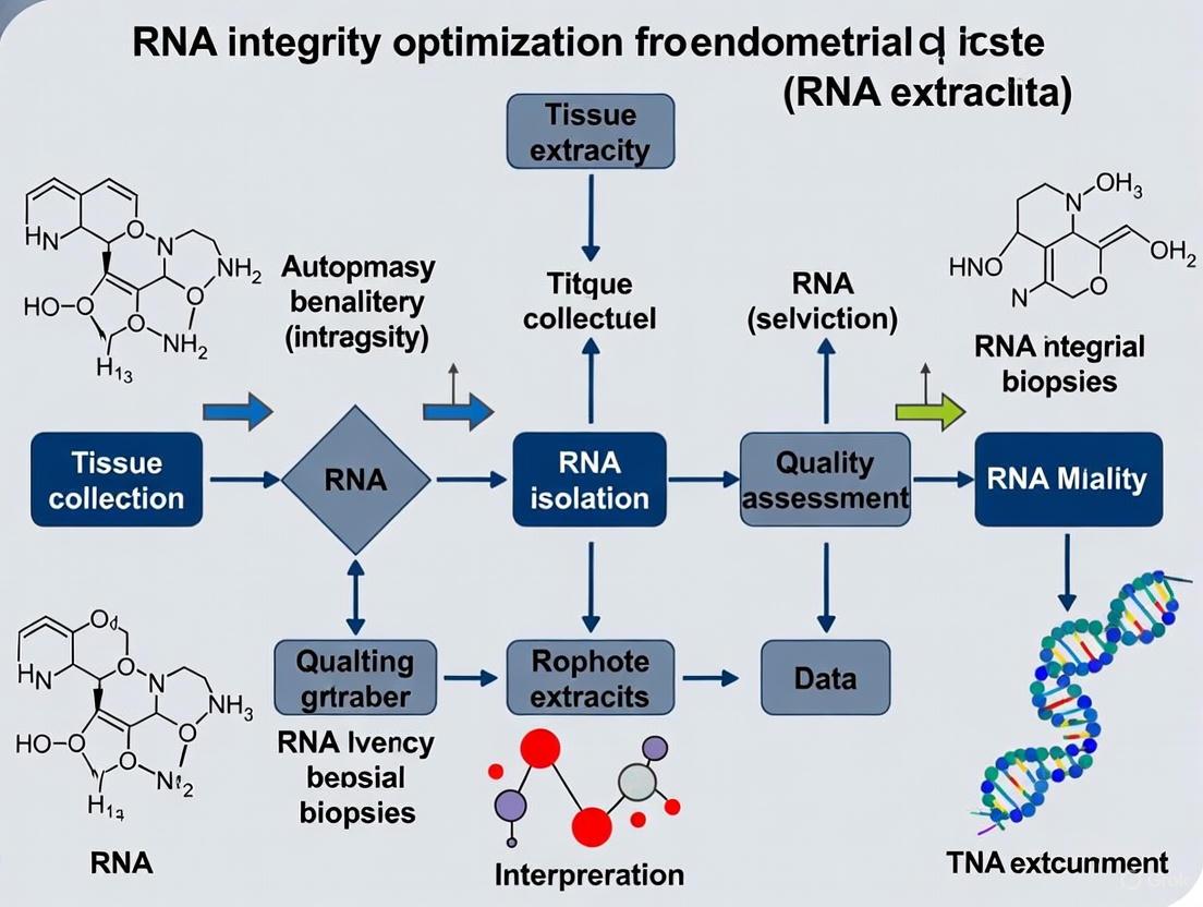
Optimizing RNA Integrity from Endometrial Biopsies: A Complete Guide for Reproductive Research and Biomarker Discovery
High-quality RNA from endometrial biopsies is fundamental for advancing research in endometrial receptivity, disease pathogenesis, and drug development.
Optimizing RNA Integrity from Endometrial Biopsies: A Complete Guide for Reproductive Research and Biomarker Discovery
Abstract
High-quality RNA from endometrial biopsies is fundamental for advancing research in endometrial receptivity, disease pathogenesis, and drug development. This comprehensive guide synthesizes current best practices for preserving RNA integrity from collection through analysis. It covers the critical role of RNA quality in transcriptomic studies, details optimized protocols for tissue handling and storage, provides troubleshooting strategies for common pre-analytical challenges, and outlines robust validation methods for downstream applications. Aimed at researchers and scientists, this article serves as a methodological resource to ensure the reliability of RNA-based data in reproductive biology.
Why RNA Integrity is the Cornerstone of Endometrial Research
The Critical Link Between RNA Quality and Transcriptomic Data Reliability
For researchers working with endometrial biopsies, the journey from sample collection to transcriptomic data is fraught with challenges. The quality of extracted RNA directly determines the reliability of your gene expression results, especially when studying subtle molecular differences in conditions like endometrial cancer, hyperplasia, and normal endometrial tissues. Compromised RNA integrity can lead to technical artifacts that obscure true biological signals, potentially resulting in false positives or negatives that undermine research validity [1] [2]. This technical support center provides essential guidance for maintaining RNA quality throughout your experimental workflow, with specific consideration for the unique challenges of endometrial research.
FAQs: RNA Quality in Endometrial Research
Why is RNA quality particularly important for endometrial cancer studies?
Endometrial cancer exhibits significant molecular heterogeneity, with distinct subtypes showing different expression profiles [3]. High-quality RNA is essential to accurately detect these subtle molecular differences, especially when comparing premalignant lesions (like atypical endometrial hyperplasia) from concurrent endometrioid adenocarcinoma [4]. Poor RNA quality can obscure critical expression signatures needed for accurate molecular classification.
How does RNA quality affect detection of long non-coding RNAs in endometrial tissues?
Long non-coding RNAs (lncRNAs) like UCA1, XIST, MALAT1, and ANRIL show promise as diagnostic biomarkers in endometrial pathologies [5]. However, degradation patterns can affect transcript detection differently depending on their length and stability. Maintaining RNA integrity ensures accurate quantification of these regulatory molecules, which is particularly important when working with formalin-fixed paraffin-embedded (FFPE) endometrial samples where RNA is more vulnerable to degradation [5].
What are the consequences of hidden quality imbalances in transcriptomic data?
Quality imbalances between sample groups can significantly skew analysis results, potentially causing a fourfold increase in false positives [2]. When one group (e.g., cancerous endometrium) systematically has lower RNA quality than another (e.g., normal controls), the observed differential expression may reflect technical artifacts rather than biological truth. This is especially problematic in endometrial research where sample collection and processing methods may vary between clinical groups.
RNA Quality Assessment Methods
Spectrophotometric Measurements
The table below outlines key spectrophotometric parameters for assessing RNA purity and their optimal values:
| Parameter | Target Value | Indication of Problem | Potential Cause |
|---|---|---|---|
| A260/A280 Ratio | 1.8-2.0 [5] | Protein contamination | Incomplete protein removal during extraction [6] |
| A260/A230 Ratio | >2.0 [5] | Chemical carryover | Residual guanidine salts or organic compounds [6] |
Integrity Number Assessment
The RNA Integrity Number (RIN) provides a numerical value from 1 (degraded) to 10 (intact). For standard RNA-seq, aim for RIN >7, though specialized methods like BRB-seq can tolerate values as low as 2.2 [7]. For endometrial FFPE samples, which typically have lower RIN values, employ specific QC metrics validated for degraded samples.
mRNA Integrity Assays
The 5'/3' assay measures degradation bias by comparing Cq values from the 5' and 3' ends of reference genes like HPRT1 [1]. Increased 5'-3' dCq values indicate preferential 5' degradation, which is particularly relevant for endometrial samples that may experience variable ischemia times before preservation.
Troubleshooting Guide: Common RNA Extraction Problems
Problem: Low RNA Yield from Endometrial Biopsies
| Cause | Solution |
|---|---|
| Incomplete homogenization | Increase homogenization time; use rotor-stator homogenizers for fibrous endometrial tissue [6] |
| Overwhelmed binding capacity | Reduce starting material to match kit specifications [8] |
| Incomplete elution | Perform second elution; incubate elution buffer 5-10 minutes at room temperature before centrifugation [8] |
Problem: RNA Degradation
| Cause | Solution |
|---|---|
| Delayed preservation | Preserve samples immediately upon collection using RNAlater or flash freezing [7] |
| RNase contamination during extraction | Add beta-mercaptoethanol (BME) to lysis buffer (10μl of 14.3M BME per 1ml buffer) [6] |
| Incomplete tissue disruption | Homogenize in bursts of 30-45 seconds with 30-second rest periods to prevent heating [6] |
Problem: Genomic DNA Contamination
| Cause | Solution |
|---|---|
| Insufficient DNA shearing | Use methods that sufficiently break genomic DNA (bead beater or polytron rotor stator) [6] |
| Inefficient DNA removal | Perform on-column DNase I treatment; for samples rich in gDNA, use high-activity DNase kits [6] |
Problem: Inhibitors in RNA Sample
| Cause | Solution |
|---|---|
| Guanidine salt carryover | Add extra washes with 70-80% ethanol during silica-based purification [6] |
| Protein contamination | Clean up sample with another purification round; use less starting material [6] |
RNA Preservation Methods for Endometrial Biopsies
The diagram below illustrates the decision pathway for selecting appropriate RNA preservation methods:
Impact of RNA Quality on Transcriptomic Data
The relationship between RNA quality and reliable transcriptomic data follows a logical progression as shown below:
The Researcher's Toolkit: Essential Reagents and Materials
| Reagent/Material | Function | Application Notes for Endometrial Research |
|---|---|---|
| RNAlater Stabilization Solution | Preserves RNA integrity at collection | Ideal for endometrial biopsies when immediate freezing isn't possible [7] |
| PAXgene Blood RNA Tubes | Stabilizes RNA in blood samples | Useful for liquid biopsy approaches in endometrial cancer [7] |
| TRIzol Reagent | Monophasic RNA isolation | Effective but requires toxic handling; good yield from fibrous tissue [7] |
| Silica Spin Columns | RNA purification | Enable DNase treatment; follow manufacturer's capacity limits [8] |
| DNase I Kit | Removes genomic DNA contamination | Essential for samples rich in gDNA; use on-column or in-solution [6] |
| Beta-mercaptoethanol (BME) | RNase inhibitor | Add to lysis buffer (10μl/ml) to stabilize RNA during extraction [6] |
Experimental Protocol: RNA Quality Control for Endometrial FFPE Samples
Based on methodologies from recent endometrial research [5], this protocol ensures reliable RNA quality assessment:
Sample Preparation
- Cut five 10μm sections from FFPE blocks of endometrial tissues (cancer, polyps, normal)
- Deparaffinize using xylene substitute and ethanol washes
- Use FFPE-optimized RNA isolation kit (e.g., EcoSpin FFPE Total RNA Isolation Kit)
RNA Isolation
- Perform proteinase K digestion at 55°C for extended time (overnight if needed)
- Bind RNA to silica membrane with high-salt binding buffer
- Wash with ethanol-based wash buffers
- Elute in 30-50μl nuclease-free water
Quality Assessment
- Measure A260/A280 and A260/A230 ratios via spectrophotometry
- Confirm RNA integrity via agarose gel electrophoresis
- For qRT-PCR analysis, use reference genes (U6 snRNA) and perform reactions in triplicate
Special Considerations for Endometrial Samples
- Account for variable cellularity in endometrial biopsies
- Normalize based on tissue area when cellularity is low
- Use inhibitor removal protocols for bloody samples
Advanced Approaches for Challenging Samples
When working with precious endometrial biopsies that yield poor-quality RNA, consider these advanced approaches:
Low-Input RNA Sequencing Methods
Bulk RNA Barcoding and Sequencing (BRB-seq) enables transcriptomic analysis from degraded samples (RIN as low as 2.2) and minimal input (as little as 100pg RNA) [7]. This is particularly valuable for archival FFPE samples with limited material.
Quality Imbalance Detection
Use tools like seqQscorer to automatically detect quality imbalances between sample groups that might compromise differential expression analysis [2]. This is crucial when comparing endometrial cancer subtypes with different processing histories.
Signal-to-Noise Ratio Assessment
Implement PCA-based signal-to-noise ratio metrics to evaluate your platform's ability to distinguish subtle biological differences among samples [9]. This approach is particularly relevant for detecting small expression differences between premalignant and malignant endometrial lesions.
Endometrial biopsy is a fundamental procedure in gynecologic research and clinical practice, essential for investigating endometrial receptivity and discovering biomarkers for conditions like endometrial cancer (EC) [10]. EC is the most prevalent gynecologic cancer in the United States, with the American Cancer Society estimating approximately 69,120 new cases and 13,860 deaths in 2025 [11]. The integrity of RNA extracted from these biopsies is paramount for downstream molecular analyses, including various RNA-sequencing (RNA-Seq) applications, which are powerful tools for transcriptome profiling [12]. This guide addresses common experimental challenges and provides troubleshooting recommendations to ensure high-quality results.
Frequently Asked Questions (FAQs) and Troubleshooting Guides
FAQ 1: What are the primary research applications for endometrial biopsies?
Endometrial biopsies are used in two key research areas:
- Endometrial Receptivity Testing: The endometrium has a specific period called the window of implantation (WOI) when it is receptive to embryo implantation. Research focuses on using gene expression profiling to accurately time the WOI, which is crucial for improving success rates in in vitro fertilization (IVF), especially for patients with recurrent implantation failure (RIF) [13].
- Cancer Biomarker Discovery: Biomarkers are critical for the early diagnosis, prognosis, and personalized treatment of endometrial cancer. Multi-omics technologies (genomic, transcriptomic, proteomic) are extensively used to analyze tissue and liquid biopsy samples to identify specific molecular signatures of EC [14] [11].
FAQ 2: My RNA yields from endometrial biopsies are low. What could be the cause?
Low RNA yield is a common issue. Potential causes and solutions include:
- Cause: Inadequate Tissue Sampling. An inadequate tissue sample is a primary cause of low yield [10].
- Solution: Ensure an adequate tissue sampling method is used. Blind biopsy methods may be less reliable. Hysteroscopy-guided biopsy is recommended for its high diagnostic accuracy and adequate tissue yield [10].
- Cause: Suboptimal RNA Extraction from Limited Material.
- Solution: Use RNA purification kits specifically designed for very small starting amounts, such as the NucleoSpin RNA XS kit, and avoid using poly(A) carriers which can interfere with downstream oligo(dT)-primed cDNA synthesis [15].
FAQ 3: My RNA Integrity Number (RIN) is poor. How can I improve it?
Poor RNA integrity severely impacts sequencing results.
- Cause: Sample Handling and Processing Delays. RNA degradation can begin quickly after tissue collection.
- Solution: Minimize the time between biopsy collection and preservation. Immediately freeze the tissue in liquid nitrogen or preserve it in a specialized RNA stabilization reagent.
- Cause: Use of Degraded Starting Material.
FAQ 4: Which RNA-Seq method should I choose for my project?
The choice of RNA-Seq method depends on your sample quality and research goal. The table below summarizes the key options.
Table 1: Guide to Selecting RNA-Seq Methods for Endometrial Research
| Method / Kit | Recommended Application & Input | Priming Method | Key Considerations |
|---|---|---|---|
| SMART-Seq v4 Ultra Low Input RNA Kit [15] | Full-length mRNA-seq from 1-1,000 intact cells or 10 pg–10 ng total RNA. Ideal for single cells or low-input samples with high-quality RNA (RIN ≥8). | Oligo(dT) | Provides full-length transcript coverage. Requires high-quality RNA input. |
| SMARTer Stranded RNA-Seq Kit [15] | 100 pg–100 ng of full-length or degraded RNA. Maintains strand-of-origin information. | Random | Requires prior ribosomal RNA (rRNA) depletion or poly(A) enrichment. Suitable for FFPE samples. |
| SMARTer Universal Low Input RNA Kit [15] | 200 pg–10 ng of degraded or nonpolyadenylated RNA (e.g., RIN 2-3). Compatible with FFPE or LCM samples. | Random | Requires prior rRNA depletion. Ideal for low-quality or partially degraded samples. |
| Poly-A Selection [12] | Standard mRNA sequencing from high-quality total RNA. | Oligo(dT) | Enriches for polyadenylated mRNA. Not suitable for degraded samples or non-coding RNA analysis. |
| rRNA Depletion [12] | Sequencing of total RNA, including non-coding RNAs. Ideal for degraded samples (e.g., FFPE) or bacterial RNA. | N/A | Removes ribosomal RNA. Necessary for studying long non-coding RNA (lncRNA) or when using random-primed kits. |
FAQ 5: How many sequencing reads are sufficient for my RNA-Seq experiment?
The required read depth depends on the organism and experimental goal. General recommendations are [12]:
- Large genomes (Human/Mouse): 20-30 million reads per sample.
- Medium genomes: 15-20 million reads per sample.
- De novo transcriptome assembly: ≥100 million reads per sample.
Research utilizes both tissue and liquid biopsies, each with advantages [14]:
- Tissue Biopsy: The traditional standard, allowing direct observation of cell morphology. Limitations include tumor heterogeneity and invasive collection.
- Liquid Biopsy: A minimally invasive alternative that allows continuous monitoring. Valuable biofluids include:
Experimental Workflows
The following diagrams outline core experimental workflows for endometrial receptivity and cancer biomarker research.
Workflow 1: Endometrial Receptivity Testing via Targeted RNA-Seq
Workflow 2: Multi-Omics Biomarker Discovery from Liquid Biopsies
The Scientist's Toolkit: Key Research Reagent Solutions
Table 2: Essential Reagents and Kits for Endometrial RNA Research
| Item | Function / Application | Key Notes |
|---|---|---|
| NucleoSpin RNA XS Kit [15] | RNA purification from a very low number of cells (up to 1x10^5). | Recommended for small biopsy samples. Avoids the use of a carrier. |
| Agilent RNA 6000 Pico Kit [15] | Assessment of RNA quality (RIN) and quantity from low-concentration samples. | Critical for evaluating input material prior to RNA-Seq. |
| SMART-Seq v4 Ultra Low Input RNA Kit [15] | cDNA synthesis and amplification for full-length mRNA-seq from low-input samples (1-1,000 cells). | Uses oligo(dT) priming. Requires high-quality RNA (RIN ≥8). |
| SMARTer Universal Low Input RNA Kit [15] | cDNA synthesis and library prep from degraded or low-quality RNA (RIN 2-3). | Uses random priming. Requires prior rRNA depletion. Ideal for FFPE-like samples. |
| RiboGone - Mammalian Kit [15] | Depletion of ribosomal RNA from total RNA samples. | Essential for random-primed RNA-Seq protocols or when analyzing non-polyadenylated RNAs. |
| ERCC RNA Spike-In Mix [12] | External RNA controls to standardize RNA quantification and assess technical variation in RNA-Seq experiments. | Not recommended for very low-concentration samples. |
In molecular biology research, the quality of extracted RNA is a critical determinant for the success of downstream applications like RNA sequencing (RNA-seq), quantitative real-time PCR (qPCR), and microarray analysis [16] [17]. Ribosomal RNA (rRNA) constitutes the majority (~85%) of the total RNA in a cell, making it a primary indicator for assessing overall RNA sample quality [16]. RNA is inherently susceptible to degradation by ribonucleases (RNases), which are ubiquitous in the environment [17]. Ensuring RNA integrity is especially crucial when working with clinically derived samples, such as endometrial biopsies, where the starting material may be limited and the biological context sensitive.
Several methods have been developed to evaluate RNA quality. Historically, the ratio of the 28S to 18S ribosomal RNA bands was used, but this method has been shown to be subjective and inconsistent [16]. The RNA Integrity Number (RIN) was subsequently developed as a standardized, automated algorithm to assign integrity values, providing a more reliable and reproducible metric [16]. This guide will explore the RIN metric, its alternatives, and their practical application in a research setting, with a specific focus on optimizing work with endometrial biopsies.
Key RNA Integrity Metrics and Their Interpretation
RNA Integrity Number (RIN)
The RIN is an algorithm developed by Agilent Technologies that uses microfluidics-based capillary gel electrophoresis to analyze an RNA sample [16]. It assigns an integrity value on a scale from 1 (completely degraded) to 10 (perfectly intact).
- Calculation: The RIN algorithm is proprietary, but it incorporates several features from the electropherogram trace [16]:
- The total RNA ratio: the area under the 18S and 28S rRNA peaks relative to the total area.
- The height of the 28S peak: as this rRNA species often degrades first.
- The fast region ratio: the area between the 18S and 5S rRNA peaks, indicating intermediate-sized degradation products.
- The marker height: indicating the amount of RNA degraded to very small fragments.
- Ideal Values: For most gene expression studies, a RIN of ≥ 8 is considered high-quality. However, the required threshold can be application-dependent; for instance, one spatial transcriptomics study on endometrial tissue set a minimum RIN of 7 [18].
The following diagram illustrates the logic behind RIN assessment and its role in the experimental workflow:
Beyond RIN: Alternative Integrity Metrics
While RIN is a industry standard, it has limitations. It primarily reflects the integrity of ribosomal RNAs, which may not always correlate perfectly with the integrity of messenger RNAs (mRNAs), the primary targets for many gene expression studies [16] [19]. This has led to the development of complementary metrics.
- Transcript Integrity Number (TIN): Unlike RIN, which is based on rRNA, the TIN metric assesses the integrity of mRNA transcripts directly from RNA-seq data by evaluating the evenness of sequence coverage across a transcript [19]. Studies have shown that TIN can be a more accurate reflection of mRNA integrity, particularly in challenging clinical samples where rRNA and mRNA degradation may not be synchronized [19].
- RNA Quality Number (RQN): RQN is a similar metric to RIN but is used with the Fragment Analyzer systems. Like RIN, it can be influenced by factors beyond pure degradation, such as the total quantity of RNA extracted from a sample [19].
The table below provides a comparative summary of these key integrity metrics.
Table 1: Comparison of Key RNA Integrity Metrics
| Metric | Full Name | What It Measures | Typical Scale | Key Advantage | Key Limitation |
|---|---|---|---|---|---|
| RIN [16] | RNA Integrity Number | Integrity of ribosomal RNA (rRNA) | 1 (degraded) to 10 (intact) | Standardized, reproducible, pre-sequencing assessment | May not reflect mRNA integrity; less reliable for plant or host-microbe samples |
| TIN [19] | Transcript Integrity Number | Integrity of mRNA transcripts from RNA-seq data | 0 (degraded) to 100 (intact) | Directly measures mRNA quality; post-sequencing assessment | Requires RNA-seq data; not available for quality control prior to sequencing |
| RQN [19] | RNA Quality Number | Integrity of RNA (similar to RIN) | 1 (degraded) to 10 (intact) | Provides an alternative to RIN for capillary electrophoresis | Can be influenced by RNA quantity extracted from the sample |
FAQs and Troubleshooting Guide
Pre-Analytical Phase: Sample Collection and Storage
Q1: My endometrial biopsy yields low RNA quantity and poor RIN scores. How can I improve this?
- A: Pre-analytical handling is the most critical phase.
- Rapid Processing: Flash-freeze tissue samples immediately in liquid nitrogen or stabilize them in RNase-inhibiting reagents (e.g., RNAlater) after collection to halt degradation.
- Standardized Collection: For menstrual fluid collection, a standardized tampon-based system that uses a preservation buffer has been validated to maintain RNA stability for up to 14 days at ambient temperature, ensuring high-quality RNA for sequencing [20].
- Minimize RNase Contamination: Use RNase-free reagents, tubes, and workspace. Wear gloves at all times.
Q2: My sample has a low RIN but a decent TIN score. Should I discard my sample?
- A: Not necessarily. Research indicates that samples should not be automatically discarded based on a single integrity metric like RIN [19]. In studies of necrotising soft tissue infection, the TIN score, which measures mRNA integrity, was found to be a better reflection of mRNA content than RIN [19]. It is advisable to:
- Use Multiple Metrics: Evaluate both RIN and TIN if possible.
- Use as a Covariate: In downstream data analysis, include the RIN or TIN score as a covariate to statistically account for the impact of integrity on gene expression measurements, rather than excluding the sample outright [19].
- Choose the Right Assay: For low-RIN RNA, target smaller amplicons in qPCR, as they are less affected by degradation.
Analytical Phase: Quality Control and Interpretation
Q3: Why is my RIN score low even though my RNA concentration looks good?
- A: Concentration and integrity are independent measures.
- Spectrophotometer Limitation: Standard absorbance measurements (e.g., Nanodrop) quantify nucleic acids but cannot distinguish between intact RNA and degraded fragments or free nucleotides, all of which contribute to the 260nm reading [17]. A good concentration can mask severe degradation.
- Always Use Electrophoresis: Always complement concentration measurements with an integrity assessment method like the Bioanalyzer or Fragment Analyzer, which separates RNA by size [17].
Q4: Are there specific considerations for using RIN with endometrial or menstrual fluid samples?
- A: Yes. The cellular composition of endometrial and menstrual effluence is complex, containing a mix of endometrial tissue, immune cells, and microbial communities [20].
- Microbial Contamination: The RIN algorithm was designed for mammalian rRNA (18S and 28S). If your sample has a significant microbial load, the bacterial or archaeal rRNA (23S and 16S) can interfere with the electropherogram and lead to an underestimation of the true quality of the human RNA [16]. Consider methods to enrich for human cells or use a metric like TIN.
- Biological Variation: RNA integrity can be influenced by the patient's biological condition, such as age or disease severity, which may affect cellular health and necrosis [19]. Always document patient metadata.
Essential Research Reagent Solutions
The following table lists key reagents and kits used in the field for RNA quality control, as referenced in the studies analyzed.
Table 2: Research Reagent Solutions for RNA Quality Control and Analysis
| Reagent / Kit / Instrument | Primary Function | Key Features and Considerations |
|---|---|---|
| Agilent 2100 Bioanalyzer [16] [17] | RNA integrity analysis via microfluidics and capillary electrophoresis | - Provides the RIN metric.- Requires very small sample volumes.- Considered a gold-standard method for pre-seq QC. |
| Norgen Biotek Preservation Buffer & Kits [20] | Nucleic acid preservation and extraction | - Used in a validated tampon-based collection system for menstrual effluence.- Preserves RNA at ambient temperature for shipping. |
| Zymo-Seq RiboFree Total RNA Library Kit [20] | RNA library preparation for sequencing | - Used for preparing RNA-seq libraries from menstrual fluid samples.- Can handle complex samples. |
| QuantiFluor RNA System [17] | Sensitive RNA quantification using fluorescent dyes | - More sensitive than absorbance methods.- Does not provide integrity information.- Can co-quantify DNA unless DNase treatment is used. |
| DNase Treatment [17] | Removal of genomic DNA contamination | - Critical step before RNA quantification with non-specific dyes or before qPCR.- Prevents overestimation of RNA concentration and false-positive signals in qPCR. |
Experimental Protocol: A Workflow for Assessing RNA Integrity from Endometrial Biopsies
Below is a detailed workflow for handling endometrial samples, from collection to quality assessment, incorporating best practices from the search results.
Protocol Steps:
Sample Collection:
- Endometrial Biopsy: Obtain tissue using a Pipelle biopsy device during the desired phase of the menstrual cycle (e.g., mid-luteal phase for receptivity studies) [18]. Immediately snap-freeze in liquid nitrogen and store at -80°C.
- Menstrual Effluence: Utilize a standardized at-home collection system, such as the one described by [20], which involves a tampon sealed in a jar with a preservation buffer (e.g., from Norgen Biotek). This preserves nucleic acids for ambient temperature shipping to the lab.
Nucleic Acid Extraction:
- Use a robust, spin-column-based RNA extraction kit designed for complex tissues.
- Critical Step: Perform an on-column DNase I digestion step to remove contaminating genomic DNA, which can interfere with accurate RNA quantification and downstream applications like RNA-seq [17].
Concentration and Purity Measurement:
- Use a UV-Vis spectrophotometer (e.g., NanoDrop) to determine RNA concentration.
- Assess purity by checking absorbance ratios. Aim for:
- A260/A280 ≈ 1.8–2.2 (indicates low protein contamination).
- A260/A230 ≈ >1.7 (indicates low contamination from salts or organics) [17].
- Note: These ratios are purity indicators and do not confirm integrity.
Integrity Assessment:
- Use an instrument like the Agilent 2100 Bioanalyzer with the RNA Nano Kit [16] [17].
- Follow the manufacturer's protocol to load a small aliquot of the RNA sample. The software will generate an electropherogram and calculate the RIN.
- For RNA-seq samples, later calculate the TIN score from the sequencing data using appropriate software to get a second opinion on mRNA integrity [19].
Decision Point:
- Based on the RIN score and your application's requirements, decide whether to proceed with costly downstream assays (e.g., RNA-seq, microarrays) or to use more degradation-tolerant methods (e.g., qPCR with short amplicons).
FAQs on Endometrial Biology and Molecular Research
1. How does the cellular heterogeneity of the human endometrium impact molecular analysis?
The human endometrium is composed of a highly complex and dynamic cellular ecosystem. Integrated single-cell RNA sequencing (scRNA-seq) analyses have identified 39 distinct cell subtypes across four major compartments: epithelial, stromal, endothelial, and immune cells [21]. This diversity means that bulk analysis methods, like standard RNA extraction from a tissue fragment, yield an average signal from all these cell types. Consequently, crucial cell-type-specific molecular changes, such as those in rare progenitor populations or specific epithelial subtypes, can be masked or diluted. For example, a specific population of SOX9+ basalis epithelial cells with progenitor characteristics has been identified, which interacts with surrounding fibroblasts via specific signaling pathways (e.g., CXCL12-CXCR4) [22]. Relying on scRNA-seq or carefully separating tissue layers is often necessary to study such specific populations.
2. What is the molecular evidence that endometrial cancer originates from specific epithelial cells?
Single-cell transcriptomic studies comparing normal endometrium, atypical hyperplasia (a precancerous condition), and endometrioid endometrial cancer (EEC) provide strong evidence that EEC originates from endometrial epithelial cells, not stromal cells. Key findings include [23]:
- A significant increase in the proportion of epithelial cells and a decrease in stromal fibroblasts from normal to EEC tissues.
- Inferred copy number variations (CNVs) are prominent in the epithelial cells of AEH and EEC but are absent in stromal fibroblasts from the same samples.
- RNA velocity analysis shows independent trajectories for epithelial and stromal cells, ruling out a mesenchymal-epithelial transition as the origin.
- The unciliated glandular epithelium is identified as the likely cellular source of EEC.
3. Are there specific long non-coding RNAs (lncRNAs) that can distinguish benign from malignant endometrial lesions?
Yes, recent research has identified specific lncRNAs with diagnostic potential. A 2024 study compared the expression of four lncRNAs in endometrial polyps (EP), endometrial cancer (EC), and normal endometrium [5]. The expression level of UCA1 was found to be a particularly strong independent discriminator. The table below summarizes the expression patterns and diagnostic performance.
Table 1: LncRNA Expression Profiles and Diagnostic Power in Endometrial Lesions
| LncRNA | Expression in EP vs. Control | Expression in EC vs. Control | Key Findings |
|---|---|---|---|
| UCA1 | Upregulated | Markedly Downregulated | Strongest independent predictor; high in EP, low in EC [5]. |
| XIST | Not Specified | Upward Trend | Lacked independent predictive value [5]. |
| MALAT1 | Not Specified | Upward Trend | Lacked independent predictive value [5]. |
| ANRIL | Not Specified | Upward Trend | Lacked independent predictive value [5]. |
Table 2: Diagnostic Accuracy (AUC) of a Model Combining Age and UCA1 [5]
| Comparison | Area Under Curve (AUC) |
|---|---|
| Endometrial Cancer vs. Control | 0.98 |
| Endometrial Cancer vs. Endometrial Polyp | 0.87 |
| Endometrial Polyp vs. Control | 0.86 |
4. What are the critical steps in preserving RNA integrity from endometrial biopsies?
RNA integrity is paramount for reliable transcriptomic data. Endometrial tissue is particularly challenging due to high levels of RNase activity. Key steps based on methodological optimizations include [24] [25] [26]:
- Immediate Stabilization: Snap-freezing tissue in liquid nitrogen immediately after collection is superior to immersion in RNA-stabilizing solution alone for preserving RNA integrity in tough tissues [24].
- Effective Homogenization: For snap-frozen tissue, cryosectioning is recommended to allow effective penetration of lysis reagents. Bead milling can be used but may require optimization to avoid excessive heat and degradation [24] [25].
- Lysis Buffer Selection: Buffers like QIAzol (phenol/guanidine thiocyanate-based) are effective for a range of tissues, including those with high lipid or fibrous content [25].
- Quality Control: Always assess RNA quality using methods like the RNA Integrity Number (RIN) to ensure samples are suitable for downstream applications like qRT-PCR or RNA-seq [25] [26].
Troubleshooting Guides
Issue 1: Low RNA Yield and Quality from Endometrial Biopsies
Problem Description: After RNA extraction from an endometrial biopsy, the yield is low and the RNA Integrity Number (RIN) is poor, making the samples unsuitable for quantitative gene expression analysis.
Root Cause Analysis: The primary causes are typically rapid RNA degradation by endogenous RNases and inefficient tissue homogenization due to the dense, fibrous nature of the endometrium. The period between tissue resection and stabilization (warm ischemia time) is critical.
Step-by-Step Resolution:
Rapid Collection and Stabilization:
Optimized Homogenization:
- For snap-frozen endometrial tissue, use the "Snap-freeze + Cryosection" protocol.
- Procedure: Embed the frozen tissue in OCT compound and section it at 10-20 µm thickness in a cryostat. The thin sections can then be directly transferred to QIAzol or a similar lysis reagent, ensuring immediate and complete contact for effective lysis and RNase inhibition [24].
Verification:
- Check RNA concentration and purity (A260/280 ratio ~2.0).
- Run the sample on an instrument like a Bioanalyzer to obtain a RIN. A RIN ≥ 7 is generally considered acceptable for most downstream applications [25].
Issue 2: High Background Noise in Gene Expression Data from Heterogeneous Endometrial Samples
Problem Description: qRT-PCR or RNA-seq data from whole endometrial tissue biopsies shows high variability and inconsistent results, likely due to the mixing of different cell types whose proportions vary between samples and across the menstrual cycle.
Root Cause Analysis: The cellular heterogeneity of the endometrium means that a molecular signal from a specific cell type (e.g., epithelial cells) can be obscured by signals from other cell types (e.g., stromal or immune cells). This is a biological, not technical, source of noise.
Step-by-Step Resolution:
Single-Cell Resolution:
Alternative: Tissue Microdissection:
- If scRNA-seq is not feasible, laser capture microdissection (LCM) can be used to isolate specific regions of interest (e.g., endometrial glands vs. stromal areas) from tissue sections before RNA extraction. This reduces, but does not eliminate, cellular heterogeneity [22].
In Silico Deconvolution:
- For existing bulk RNA-seq data, computational methods can be used to infer the proportions of major cell types present in the sample. This requires a reference signature matrix, which is now available from published endometrial single-cell atlases like the Human Endometrial Cell Atlas (HECA) [22].
Experimental Protocols from Key Studies
Protocol 1: RNA Extraction and qRT-PCR Analysis of lncRNAs from FFPE Endometrial Tissues
This protocol is adapted from a 2024 study investigating lncRNAs in endometrial polyps and cancer [5].
Key Reagent Solutions:
- Tissue Source: Formalin-Fixed Paraffin-Embedded (FFPE) endometrial tissue blocks.
- RNA Extraction Kit: EcoSpin FFPE Total RNA Isolation Kit (Ecotech Biotechnology).
- cDNA Synthesis Kit: OneScript Plus cDNA Synthesis Kit (Applied Biological Materials).
- qRT-PCR MasterMix: SYBR Green BlasTaq 2X qPCR MasterMix (Applied Biological Materials).
- Reference Gene: U6 snRNA.
Detailed Methodology:
- Sectioning: Cut five sections of 10 µm thickness from each FFPE block.
- RNA Isolation: Use the EcoSpin kit following the manufacturer's instructions, including a DNase digestion step to remove genomic DNA. Elute RNA in nuclease-free water.
- Quality Control: Assess RNA purity by spectrophotometry (accept A260/A280 ≥ 2.0) and integrity by agarose gel electrophoresis.
- cDNA Synthesis: Convert 500 ng - 1 µg of total RNA to cDNA using the OneScript Plus kit.
- Quantitative RT-PCR:
- Prepare reactions in technical triplicates using the SYBR Green MasterMix.
- Use primers specific for the target lncRNAs (XIST, UCA1, MALAT1, ANRIL) and the reference gene U6.
- Cycling Conditions:
- Enzyme activation: 95°C for 3 min (1 cycle)
- Amplification: 95°C for 15 s, then 60°C for 1 min (40 cycles)
- Data Analysis: Calculate relative expression using the 2−ΔΔCt method, normalizing to U6 snRNA.
Protocol 2: Key Steps for Generating a Single-Cell RNA Sequencing Atlas from Endometrial Tissue
This protocol summarizes the core workflow used in recent studies to build a consensus atlas of the human endometrium [21] [22].
Key Reagent Solutions:
- Tissue Digestion: A cocktail of collagenases (e.g., Collagenase IV) and DNase to dissociate fresh endometrial tissue.
- Cell Viability Stain: Propidium Iodide (PI) or DAPI for dead cell exclusion.
- Single-Cell Platform: 10X Genomics Chromium Controller.
- Reagent Kits: Chromium Single Cell 3' Reagent Kits (10X Genomics).
Detailed Methodology:
- Sample Collection & Processing: Obtain endometrial biopsies under informed consent. Process samples immediately to preserve cell viability.
- Single-Cell Suspension: Mechanically mince the tissue and digest it in the enzyme cocktail at 37°C with gentle agitation. Filter the resulting suspension through a cell strainer (e.g., 40 µm) to remove clumps.
- Cell Quality Control: Count cells and assess viability (aim for >90% via trypan blue or PI exclusion). Adjust cell concentration to the target for the single-cell platform (e.g., ~1,000 cells/µl for 10X Genomics).
- Library Preparation & Sequencing: Load cells onto the 10X Chromium Controller to partition single cells into droplets with barcoded beads. Perform reverse transcription, cDNA amplification, and library construction as per the manufacturer's protocol. Sequence libraries on an Illumina platform to a sufficient depth (e.g., 50,000 reads per cell).
- Computational Analysis:
- Quality Control & Filtering: Use tools like Seurat or Scanpy to filter out low-quality cells, doublets, and dead cells (high mitochondrial gene percentage).
- Integration & Clustering: Integrate data from multiple donors using harmony or similar methods. Perform principal component analysis (PCA) and graph-based clustering to identify cell populations.
- Cell Type Annotation: Manually annotate clusters using known marker genes from resources like the Human Endometrial Cell Atlas (HECA) [22].
Signaling Pathways and Cellular Workflows
Endometrial Epithelial-Stromal Communication in the Basalis Niche
This diagram illustrates the molecular crosstalk between a putative epithelial progenitor population and stromal fibroblasts in the endometrial basalis layer, a niche critical for regeneration [22].
Experimental Workflow for Robust Endometrial RNA Analysis
This flowchart outlines a optimized workflow for obtaining high-integrity RNA from endometrial biopsies, crucial for reliable data [24] [25].
The Scientist's Toolkit: Essential Reagents and Materials
Table 3: Key Research Reagent Solutions for Endometrial Molecular Studies
| Item | Function / Application | Example Product / Citation |
|---|---|---|
| QIAzol Lysis Reagent | A phenol and guanidine thiocyanate-based solution for effective lysis and RNase inhibition during homogenization of complex tissues like endometrium. | QIAzol (Qiagen) [25]. |
| EcoSpin FFPE RNA Kit | Optimized kit for extracting RNA from formalin-fixed paraffin-embedded (FFPE) endometrial tissue blocks, which are common in clinical archives. | EcoSpin FFPE Total RNA Isolation Kit [5]. |
| Collagenase IV / DNase Mix | Enzyme cocktail for digesting fresh endometrial tissue to create high-viability single-cell suspensions for scRNA-seq. | Used in multiple scRNA-seq studies [21] [22]. |
| SYBR Green qRT-PCR MasterMix | For sensitive and quantitative gene expression analysis of candidate genes (e.g., lncRNAs, markers) from endometrial cDNA. | SYBR Green BlasTaq 2X qPCR MasterMix [5]. |
| U6 snRNA Primers | A commonly used small non-coding RNA for normalizing qRT-PCR data in lncRNA studies from endometrial tissue. | [5] |
| SOX9 / CDH2 Antibodies | Markers for identifying putative epithelial progenitor cells in the basalis layer via immunofluorescence or flow cytometry. | [22] |
Proven Protocols: From Biopsy to Stable RNA
Frequently Asked Questions
Q1: Why is the timing of an endometrial biopsy within the menstrual cycle so critical for gene expression studies? The endometrium undergoes dramatic, rapid changes in gene expression driven by hormonal fluctuations. If samples collected at different cycle stages are compared without precise timing, the natural variation can obscure disease-related findings and make studies irreproducible. Research shows that over 3,400 endometrial genes change expression significantly throughout the cycle, with the most rapid shifts occurring in the secretory phase [27]. Accurately defining the cycle stage is a prerequisite for reliable transcriptomic analysis.
Q2: What are the primary methods for determining menstrual cycle stage, and what are their limitations? Each standard method has significant drawbacks for precise research:
- Last Menstrual Period (LMP) Alone: This is highly inaccurate due to normal variability in cycle length. Only about 12.4% of women have a 28-day cycle, and ovulation day can vary by up to 10 days, making forward or backward counting from LMP unreliable [28] [27].
- Serum Hormone Measurement: While direct, this provides an indirect measure of the endometrial tissue response and requires multiple blood draws [27].
- Histological Dating (Noyes Criteria): This traditional method is subjective and suffers from significant inter-observer variability, even among expert pathologists [27].
- Ultrasound Follicle Tracking: This does not directly correlate with the molecular state of the endometrium [27].
Q3: My sample integrity is compromised. What are the most common pre-analytical errors? The most frequent errors occur before RNA extraction:
- Delayed or Improper Stabilization: RNases begin degrading RNA immediately upon tissue collection. Without immediate stabilization in a reagent like RNAlater or immediate freezing, RNA integrity is lost [29] [30].
- Inaccurate Cycle Stage Assignment: Using an imprecise method like LMP alone leads to grouping molecularly different samples together, confounding results [28] [27].
- Inconsistent Biopsy Technique: The method (e.g., suction curette vs. resectoscope loop) can affect sample weight and composition, though studies show both can yield high-quality RNA if processed correctly [31].
Q4: Are there any emerging, less invasive methods for endometrial sampling? Yes, recent technological advances show great promise. Menstrual effluence (collected with a specialized tampon system) is now a validated, less invasive biospecimen. When collected with a standardized kit and preservation buffer, it provides high-quality RNA stable at ambient temperature for up to 14 days and is suitable for RNA sequencing, metatranscriptomic profiling, and even exome sequencing with 100% concordance to matched blood samples [20]. This allows for at-home, longitudinal sampling.
Troubleshooting Guides
Problem: Inconsistent Gene Expression Data
Potential Cause: Inaccurate alignment of sample collection with the true molecular phase of the menstrual cycle.
Solution: Implement a molecular staging model.
- Collect Metadata: Record LMP and, if possible, use LH surge kits to estimate ovulation.
- Utilize Public Tools: Use existing molecular models, like the one described by [27], which assigns a "model time" based on the expression of thousands of genes. This allows you to normalize your gene expression data for cycle stage.
- Re-normalize Data: If you have existing RNA-seq data, you can reanalyze it by applying this model to accurately compare samples by their molecular stage rather than their historical or pathological stage [27].
Workflow Diagram: Traditional vs. Molecular Staging
Problem: Low RNA Yield or Purity
Potential Cause: Degradation during collection, storage, or extraction.
Solution: Optimize the collection-to-storage pipeline.
- Choose the Right Stabilizer: Immediately post-collection, immerse the biopsy in RNAlater. This aqueous, non-toxic reagent rapidly permeates tissue to stabilize and protect RNA, eliminating the need for immediate snap-freezing [29] [30].
- Ensure Proper Storage: After 24-hour stabilization at 4°C, transfer samples to -80°C for long-term storage. RNAlater-treated samples can withstand multiple freeze-thaw cycles without significant RNA degradation [29].
- Verify RNA Quality: Use a spectrophotometer (NanoDrop) to check the A260/A280 ratio. A value between 1.9 and 2.1 indicates pure RNA. Use a Bioanalyzer to determine the RNA Integrity Number (RIN); a RIN >8 is considered optimal for downstream applications like qRT-PCR and RNA-seq [31] [30].
Workflow Diagram: Optimal RNA Stabilization
Experimental Protocols & Data
Detailed Methodology: Comparing Biopsy Methods for RNA Analysis
Objective: To validate that unguided Pipelle biopsies provide RNA of similar purity and quantity to guided hysteroscopic biopsies for gene expression studies, even in uteri with structural irregularities like submucosal leiomyomas [31].
Procedure:
- Patient Selection: Include premenopausal women with regular cycles (22-35 days) and no hormonal treatment for ≥3 months.
- Sample Collection:
- Perform two unguided biopsies using a low-pressure suction device (Pipelle) prior to hysteroscopy.
- During hysteroscopy, obtain two guided biopsies from the endometrium overlying a leiomyoma and from an area remote from the leiomyoma using a resectoscope loop.
- Sample Processing:
- Rinse biopsies in PBS.
- Immediately transfer to RNase-free tubes containing RNAlater. Keep overnight at 4°C.
- Remove from RNAlater, aliquot, and store at -80°C.
- RNA Isolation & QC:
- Use a commercial kit (e.g., RNeasy Mini Kit).
- Quantify RNA yield (ng/mg tissue).
- Assess purity via A260/A280 ratio (target: 1.9-2.2).
- Gene Expression Validation:
- Perform cDNA synthesis.
- Analyze expression of a housekeeping gene (e.g., HOXA10) via qRT-PCR in different sample types to confirm homogeneity.
Summary of Results: Table: Comparison of Biopsy Methods for RNA Yield and Purity [31]
| Biopsy Method | Median Sample Weight | RNA Yield (ng/mg tissue) | Samples with Satisfactory A260/A280 (1.9-2.2) |
|---|---|---|---|
| Low-Pressure Suction (Pipelle) | 153 mg | 1,625 ng/mg | 94.7% |
| Resectoscope Loop | 20 mg | 1,779 ng/mg | 94.7% |
Conclusion: Pipelle biopsies provide substantially larger tissue samples with comparable RNA purity and yield to guided biopsies, validating their use for endometrial gene expression studies [31].
The Scientist's Toolkit: Essential Research Reagents
Table: Key Reagents for Optimal Endometrial RNA Collection and Analysis
| Item | Function/Application | Key Considerations |
|---|---|---|
| RNAlater Stabilization Solution | Stabilizes RNA in fresh tissue at point of collection; inhibits RNases. | Eliminates need for immediate freezing. Tissue can be stored at 4°C for days or at -80°C for long term [29] [30]. |
| Pipelle Endometrial Suction Curette | Minimally invasive, unguided biopsy device for endometrial sampling. | Provides adequate tissue with high RNA purity and yield, suitable for transcriptomic studies [31]. |
| RNeasy Mini Kit | Silica-membrane based total RNA isolation from small tissue samples. | Provides high-purity RNA with an efficient DNase digestion step; compatible with FFPE tissue [31]. |
| Tempus Blood RNA System | Stabilizes RNA in whole blood at collection point. | Inactivates RNases and stabilizes gene expression profile; useful for concurrent systemic immune analysis [29]. |
| NextGen Jane Tampon System | Standardized at-home collection of menstrual effluence. | Enables less-invasive longitudinal sampling; preserves nucleic acids at ambient temperature for days [20]. |
| SuperScript IV VILO Master Mix | cDNA synthesis from total RNA, including challenging samples. | Includes ezDNase for genomic DNA removal; high efficiency and robustness for qRT-PCR [31]. |
In endometrial biopsy research, the choice of sample preservative is a critical pre-analytical step that directly determines the success of downstream molecular applications. Proper preservation is essential for maintaining RNA integrity, which is the cornerstone of accurate gene expression analysis, RNA sequencing, and the identification of biomarkers for conditions like endometrial receptivity and recurrent implantation failure. This guide provides a comparative overview of three common preservation and lysis solutions—RNALater, TRIzol, and specialized lysis buffers—to help you optimize your experimental workflow and ensure the reliability of your RNA data.
Preservative Comparison at a Glance
The table below summarizes the key characteristics, advantages, and limitations of RNALater, TRIzol, and Lysis Buffers to guide your selection.
| Preservative | Primary Mechanism | Best For | Sample Storage Format | Key Advantages | Key Limitations |
|---|---|---|---|---|---|
| RNALater [32] | Aqueous solution that permeates tissue to inactivate RNases | Stabilizing intact tissues/cells for later processing; fieldwork | Intact tissue submerged in solution [32] | • Protects RNA in intact samples at various temperatures• Easier handling than frozen samples; no grinding required [32] | • Can make tissue harder to homogenize [6]• Can reduce final RNA yield if not removed properly [33] |
| TRIzol [34] [35] | Monophasic solution of phenol and guanidine isothiocyanate that immediately lyses cells and denatures proteins | Simultaneous isolation of RNA, DNA, and protein from the same sample; challenging samples | Homogenized lysate [35] | • Powerful, immediate inactivation of RNases [35]• Enables multi-omics from a single sample [35] | • Highly toxic (phenol)• Requires careful phase separation to avoid contamination [34] |
| Lysis Buffer (e.g., from kits) [33] | Typically contains guanidine salts to denature proteins and inactivate RNases | Immediate RNA extraction; precise, kit-based workflows | Homogenized lysate | • Integrated with specific silica-column purification kits• Can be optimized for specific sample types (e.g., blood) [33] | • Commits sample to RNA extraction only• Limited stabilization time; extraction should follow quickly |
Detailed Protocols for Endometrial Biopsies
Sample Preservation with RNALater
Methodology:
- Immediate Immersion: Following collection, the endometrial biopsy should be placed in 5-10 volumes of RNALater to ensure complete immersion [32].
- Storage Conditions: Samples can be stored in RNALater for extended periods: up to 1 day at 37°C, 1 week at 25°C, 1 month at 4°C, or long-term at -20°C to -80°C [32].
- Downstream Processing: Before RNA extraction, the RNALater solution must be removed. This often involves centrifugation to pellet the tissue, which is then resuspended in the appropriate lysis buffer (e.g., from an RNA kit or TRIzol) [33]. Note that RNA may be lost in the discarded supernatant, potentially reducing yield [33].
RNA Extraction Using TRIzol Reagent
Methodology:
- Homogenization: This is the most critical step. For frozen endometrial tissue, the liquid nitrogen mortar and pestle method is the gold standard. The tissue is snap-frozen and ground to a fine powder, which is then transferred directly into TRIzol reagent [35]. Mechanical homogenizers can also be used.
- Phase Separation: Incubate the homogenate for 5 minutes at room temperature. Add 0.2 mL of chloroform per 1 mL of TRIzol, shake vigorously, and centrifuge. The RNA partitions into the upper, clear aqueous phase [34] [35].
- RNA Precipitation: Transfer the aqueous phase to a new tube. Add 0.5 mL of isopropanol per 1 mL of initial TRIzol, incubate, and centrifuge to pellet the RNA [35].
- Wash and Solubilization: Wash the pellet with 75% ethanol, air-dry briefly (do not over-dry), and resuspend the RNA in RNase-free water [35].
- Optional DNase Treatment: A DNase treatment is highly recommended for downstream applications like qPCR to remove any contaminating genomic DNA [6] [35].
Troubleshooting Guides & FAQs
Low RNA Yield
- Possible Cause: Incomplete tissue homogenization.
- Solution: Ensure the endometrial tissue is completely pulverized. For tough tissue, use a mechanical homogenizer or the liquid nitrogen grinding method. Visually inspect the lysate to ensure no tissue fragments remain [6] [35].
- Possible Cause: RNA loss during precipitation (especially with low-yield samples).
- Solution: Add an inert carrier like glycogen (5-10 µg) during the isopropanol precipitation step to visualize the pellet and improve recovery [34] [36].
Genomic DNA Contamination
- Problem: High molecular weight smearing on a gel or amplification in no-RT PCR controls.
- Solution: Perform an on-column or solution-based DNase treatment during the RNA purification process [6] [35].
Poor RNA Integrity (Low RIN)
- Possible Cause: Degradation during sample collection or processing before preservation.
- Solution: Minimize the time between biopsy collection and immersion in preservative. Snap-freeze samples in liquid nitrogen if not using RNALater [6] [36].
- Possible Cause: Incomplete RNase inactivation.
- Solution: When using TRIzol, ensure thorough and immediate homogenization. For protocols using lysis buffers, adding beta-mercaptoethanol (BME) can help inactivate RNases [6].
Abnormal A260/230 or A260/280 Ratios
- Problem: Low A260/230 ratio (often below 1.0).
- Cause & Solution: Indicates carryover of guanidine salts or other organic compounds. Perform additional ethanol washes during the purification step [6] [36].
- Problem: Low A260/280 ratio (below 1.8).
- Cause & Solution: Suggests protein contamination. This can occur if the aqueous phase was contaminated with the interphase/organic phase during TRIzol extraction. Re-purify the RNA with an additional round of phenol-chloroform extraction or a silica-column cleanup [34] [6].
Frequently Asked Questions (FAQs)
Q1: Can I use RNALater for samples that are already frozen? No, the standard RNALater reagent is not designed for already frozen tissues. For frozen samples, a different product called RNAlater-ICE exists, which safely transitions tissue from frozen to a non-frozen state at -20°C [32].
Q2: My aqueous phase turned an abnormal color (yellow, pink) after phase separation with TRIzol. What does this mean? Abnormal coloring can be sample-specific. Endometrial biopsies rich in blood can cause yellowing due to hemoglobin. This may require a pre-wash with PBS to reduce blood content or an increase in the volume of TRIzol used [34].
Q3: How should I store my RNA sample after isolation? After the final RNA pellet is washed with ethanol and resuspended in RNase-free water, it should be stored at -70°C to -80°C for long-term stability. Avoid storing RNA at -20°C for extended periods, as this can lead to degradation [36].
The Scientist's Toolkit: Essential Research Reagents
| Item Name | Function/Application |
|---|---|
| RNALater Stabilization Solution [32] | Stabilizes and protects RNA in fresh endometrial tissue samples prior to homogenization and extraction. |
| TRIzol Reagent [34] [35] | A monophasic phenol-guanidine isothiocyanate solution for simultaneous disruption of cells and inactivation of RNases during total RNA isolation. |
| DNase I, RNase-free [6] | Enzymatically degrades contaminating genomic DNA from RNA preparations to prevent false positives in qPCR. |
| Glycogen, RNase-free [34] [36] | Acts as a carrier to co-precipitate with nanogram amounts of RNA, improving yield and pellet visibility. |
| Chloroform [35] | Used in conjunction with TRIzol for phase separation, partitioning RNA into the aqueous phase. |
| β-Mercaptoethanol (BME) [6] | A reducing agent added to lysis buffers to inactivate RNases by breaking disulfide bonds. |
| PAXgene Blood RNA System [37] | An integrated system of blood collection tubes and purification kits for stabilizing and purifying RNA from whole blood. |
RNA Extraction Workflow
Key Concepts at a Glance
The choice between snap-freezing and standard freezing, along with proper aliquot sizing, is fundamental to preserving RNA integrity in endometrial research. The table below summarizes the core differences between the two main tissue preservation methods.
Table 1: Comparison of Tissue Preservation Methods for Molecular Research
| Feature | Snap-Freezing (Cryopreservation) | Standard Pathology (FFPE) |
|---|---|---|
| Preservation Method | Rapid freezing in liquid nitrogen or -80°C; halts cellular metabolism [38]. | Chemical fixation in formalin followed by paraffin embedding; preserves structure [38]. |
| Biomolecule Quality | High: Intact, native DNA, RNA, and proteins. Considered the "gold standard" for molecular analysis [38]. | Lower: Fragmented and chemically modified DNA/RNA/proteins due to cross-linking [38]. |
| RNA Integrity | Ideal for sensitive techniques like RNA-Seq and qRT-PCR [38]. | RNA is often degraded, complicating analysis [38]. |
| Tissue Morphology | Good, but freezing artifacts can occur, making detailed histology challenging [38] [39]. | Excellent preservation of cellular and tissue architecture [38]. |
| Primary Applications | Advanced molecular profiling (genomics, transcriptomics, proteomics), biobanking [38]. | Routine histopathological diagnosis, immunohistochemistry (IHC) [38]. |
Optimizing Your Cryopreservation Protocol for RNA Integrity
Preserving RNA in endometrial tissues requires a meticulously controlled workflow from collection to storage. The following protocol is optimized for this purpose.
Workflow: Optimal Cryopreservation of Endometrial Biopsies for RNA Analysis
Detailed Experimental Protocol
Tissue Collection and Immediate Handling:
- Following biopsy, place the endometrial tissue specimen immediately into a sterile, pre-chilled cryovial.
- Minimize the time between collection and freezing (ideally under 5 minutes) to prevent RNA degradation by endogenous RNases [40].
Snap-Freezing:
- Submerge the sealed cryovial directly into liquid nitrogen. This process, known as flash-freezing, instantly halts all biological activity and preserves biomolecules in their native state [38] [40].
- Alternative for histology: For tissues intended for frozen sectioning that require better morphological preservation, freeze the tissue in a cryomold with OCT compound by immersing it in a cold isopentane bath chilled by dry ice. This method reduces ice crystal artifacts [40].
Aliquot Sizing and Storage:
- For optimal RNA yield and to minimize freeze-thaw cycles, aliquot tissues into small pieces weighing between 10-30 mg before freezing [41]. This size is compatible with most commercial RNA extraction kits.
- Store aliquots long-term in the vapor phase of liquid nitrogen (between -140°C and -180°C) or in a stable -80°C freezer. Vapor phase storage is preferred for long-term preservation and avoids the risk of vial explosion that can occur with liquid phase storage [42].
Troubleshooting Guide & FAQs
Table 2: Troubleshooting Common Cryopreservation Issues
| Problem | Potential Cause | Solution |
|---|---|---|
| Low RNA Yield/Quality Post-Thaw | 1. Slow freezing allowing ice crystal formation.2. Improper thawing temperature.3. Multiple freeze-thaw cycles. | 1. Ensure snap-freezing in liquid nitrogen [40].2. Thaw small aliquots (≤100 mg) on ice; consider -20°C overnight for larger pieces [41].3. Aliquot into single-use masses to avoid repeated thawing [41]. |
| Poor Cell Viability or Recovery | 1. Absence or incorrect use of cryoprotectant.2. Uncontrolled freezing rate. | 1. Use 10% DMSO in freezing media. For cell therapies, explore alternatives like PVP [42].2. Use a controlled-rate freezer or a passive cooling device (e.g., CoolCell) to achieve a cooling rate of -1°C/minute [42]. |
| Ice Crystal Artifacts in Sections | Slow freezing forming large, disruptive ice crystals [39]. | Snap-freeze in cold isopentane for superior histology quality, especially for water-rich tissues like the endometrium [40]. |
| Inconsistent Molecular Results | Variable aliquot sizes leading to uneven freezing/thawing and RNA degradation [41]. | Standardize aliquot sizes to the 10-30 mg range for uniform processing [41]. |
Frequently Asked Questions (FAQs)
Q1: Can I re-freeze tissue after it has been thawed for use? It is strongly discouraged. Each freeze-thaw cycle degrades RNA and compromises cell integrity [41]. Always subdivide your original tissue into small, single-use aliquots prior to the initial freezing.
Q2: My frozen tissue is large, and I only need a small piece. How can I subdivide it without thawing? For tissues already stored as a large block, use a cryogenically pre-cooled mortar and pestle to smash the frozen tissue into smaller fragments under liquid nitrogen. This "pulverization" method allows you to obtain material without subjecting the entire sample to a thaw cycle [41] [40].
Q3: Is DMSO always necessary for freezing tissues, and are there alternatives? DMSO is a common intracellular cryoprotectant, but it can be toxic to cells. For specific applications like cell therapies, alternatives exist. Extracellular cryoprotectants like sucrose or polymers like Polyvinylpyrrolidone (PVP) and methylcellulose can be used, sometimes in combination with reduced concentrations of DMSO [42].
Q4: How does the preservation method impact the analysis of specific biomarkers like lncRNAs in endometrial studies? Snap-frozen tissue is superior for quantifying sensitive molecular biomarkers like long non-coding RNAs (lncRNAs). While studies can extract RNA from FFPE tissue, the formalin-induced fragmentation and modification can reduce the accuracy and sensitivity of assays like qRT-PCR [5]. For cutting-edge transcriptomic studies, snap-freezing is the definitive choice [38].
The Scientist's Toolkit: Essential Research Reagents
Table 3: Key Reagents for Cryopreservation and RNA Analysis
| Reagent / Material | Function | Application Note |
|---|---|---|
| Liquid Nitrogen | Enables snap-freezing through ultra-rapid cooling to -196°C [38] [40]. | Critical for instantly arresting RNase activity and preserving high-quality RNA. |
| DMSO (Dimethyl Sulfoxide) | Penetrating cryoprotective agent (CPA). Reduces ice crystal formation by penetrating cells [42]. | Typically used at a 10% concentration in culture media or specialized freezing media. |
| RNALater Stabilization Solution | An RNA-stabilizing solution that permeates tissue to inhibit RNases [41]. | Can be added to frozen tissues during the thawing step to rescue RNA quality, even in archival samples [41]. |
| OCT Compound | Water-soluble embedding medium for frozen tissue sectioning [40]. | Provides structural support for cutting thin sections on a cryostat. |
| TRIzol / RL Lysis Buffer | Chaotropic, monophasic lysis reagents for the effective disruption of tissues and inactivation of RNases during RNA extraction [41]. | Tissue should be homogenized immediately upon thawing in these buffers for optimal RNA recovery. |
Decision Pathway for Method Selection
The choice between cryopreservation methods depends on your primary research goal. The following diagram outlines a decision-making pathway.
The molecular analysis of the endometrium provides vital insights into reproductive health, fertility, and various gynecological pathologies. The foundation of this research relies on obtaining high-quality RNA that accurately represents the endometrial transcriptome. However, RNA is an inherently unstable molecule, highly susceptible to degradation by ribonucleases (RNases), making its preservation and extraction particularly challenging. These challenges are compounded when working with different tissue preservation methods, primarily formalin-fixed paraffin-embedded (FFPE) and fresh-frozen tissues, each presenting unique obstacles. For endometrial research specifically, the molecular heterogeneity of the tissue and the impact of menstrual cycle stage add further complexity to RNA extraction protocols.
This technical support center addresses these critical challenges by providing optimized, evidence-based protocols for RNA extraction from endometrial tissues. The guidance synthesizes current methodologies, troubleshooting approaches, and quality control measures specifically tailored for researchers working with endometrial specimens. By implementing these standardized procedures, scientists can significantly enhance the reliability of their downstream applications, including RNA sequencing, quantitative PCR, and transcriptome analysis, thereby advancing our understanding of endometrial biology and pathology.
Quantitative Comparison of RNA Yield and Quality Across Methods
The choice of tissue preservation and RNA extraction method significantly impacts the quantity and quality of recovered RNA. The following tables summarize key performance metrics from optimized protocols for fresh-frozen and FFPE endometrial tissues, providing researchers with realistic expectations for their experiments.
Table 1: RNA Yield and Purity from Different Endometrial Sampling Methods
| Sampling Method | Median Tissue Weight (mg) | RNA Yield (ng/mg tissue) | A260/A280 Ratio (Purity) | Suitability for Downstream Applications |
|---|---|---|---|---|
| Low-pressure suction device (Pipelle) | 153 | 1,625 | 1.9-2.2 (94.7% of samples) | RNA-seq, RT-qPCR, microarray analysis |
| Resectoscope loop | 20 | 1,779 | 1.9-2.2 (94.7% of samples) | RNA-seq, RT-qPCR, microarray analysis |
| FFPE tissues (optimized protocol) | Variable | Dependent on storage duration and fixation | Typically lower than fresh | RT-qPCR (shorter amplicons), targeted sequencing |
Table 2: Impact of FFPE Storage Duration and Protocol Optimization on RNA Quality
| Storage Duration | Protocol Variation | RNA Yield | Amplifiable Product Size | Key Success Factors |
|---|---|---|---|---|
| Up to 11 years | Standard protocol | Lower yield | Limited amplification | Proteinase K incubation time and volume |
| Up to 11 years | Extended proteinase K incubation | Significantly higher (P<0.01) | Up to 298 bp | Increased duration and volume of proteinase K |
| Variable fixation times | Organ-specific optimization | Variable | Organ-dependent | Tissue-specific treatment protocols |
Optimized Experimental Protocols
Protocol 1: RNA Extraction from Fresh-Frozen Endometrial Biopsies
Principle: Rapid stabilization of RNA immediately upon tissue collection prevents degradation and maintains transcriptome integrity, enabling accurate gene expression analysis [43].
Materials and Reagents:
- RNAlater RNA Stabilization Solution
- RNeasy Mini Kit (Qiagen) or equivalent silica-membrane column system
- RNase-free reagents and consumables
- Liquid nitrogen or dry ice for flash-freezing
- -80°C freezer for storage
Step-by-Step Procedure:
- Sample Collection: Obtain endometrial tissue using a low-pressure suction device (e.g., Pipelle) or during hysterectomy procedures.
- Immediate Stabilization:
- For biopsy samples: Immediately place tissue in 5-10 volumes of RNAlater solution.
- For hysterectomy samples: Within 15 minutes of devascularization, dissect relevant endometrial areas and place in RNAlater [43].
- Storage: Store stabilized samples at 4°C for up to one month, or at -20°C to -80°C for longer-term storage.
- Homogenization:
- Remove tissue from RNAlater and homogenize in RLT buffer (provided in RNeasy kit) using a mechanical homogenizer.
- Ensure complete tissue disruption within a few minutes.
- RNA Extraction:
- Follow manufacturer's instructions for the RNeasy Mini Kit.
- Include the optional on-column DNase digestion step using the RNase-Free DNase Set (Qiagen) to remove genomic DNA contamination.
- Elution: Elute RNA in 30-50 µL RNase-free water.
- Quality Control:
- Assess RNA concentration and purity using spectrophotometry (A260/A280 ratio of 1.8-2.1 indicates pure RNA).
- Verify RNA integrity using microfluidic analysis (e.g., Bioanalyzer), with RNA Integrity Number (RIN) >7.0 considered suitable for most downstream applications.
Protocol 2: RNA Extraction from FFPE Endometrial Tissues
Principle: Extended proteinase K digestion reverses formaldehyde-induced crosslinks and releases RNA fragments from long-term archived FFPE tissues, making them accessible for molecular analysis [44].
Materials and Reagents:
- Xylene or alternative deparaffinization agent
- Ethanol series (100%, 95%, 70%)
- Proteinase K solution
- Phenol-chloroform or commercial FFPE RNA extraction kit
- DNase I, RNase-free
Step-by-Step Procedure:
- Sectioning: Cut 5-10 sections of 5-10 µm thickness from the FFPE block using a microtome.
- Deparaffinization:
- Add 1 mL xylene to sections, vortex, and incubate at room temperature for 5 minutes.
- Centrifuge at full speed for 5 minutes and carefully remove supernatant.
- Repeat xylene treatment once.
- Rehydration:
- Wash with 100% ethanol (twice), 95% ethanol, and 70% ethanol (2 minutes each).
- Briefly air-dry pellet to remove residual ethanol.
- Proteinase K Digestion:
- Resuspend tissue pellet in proteinase K digestion buffer.
- Incubate at 56°C for extended period (optimized time: 3-16 hours depending on tissue age and fixation) with occasional mixing [44].
- RNA Extraction:
- Extract using acid phenol-chloroform or commercial FFPE RNA kit following manufacturer's instructions.
- Precipitate RNA with isopropanol or ethanol.
- DNase Treatment:
- Treat RNA with DNase I to remove contaminating genomic DNA.
- Purify using RNA clean-up columns or precipitation.
- Quality Control:
- Assess concentration and A260/A280 ratio.
- For FFPE tissues, A260/A280 ratio >1.8 indicates acceptable purity.
- Verify amplifiability by RT-qPCR using short amplicons (<150 bp).
Protocol 3: Single-Cell/Nucleus RNA Sequencing from Endometrial Tissues
Principle: Isolation of individual cells or nuclei enables resolution of cellular heterogeneity within the endometrium, providing insights into cell-type-specific gene expression patterns [45] [46].
Materials and Reagents:
- EDTA-, Mg2+- and Ca2+-free 1X PBS
- Appropriate collection buffer (e.g., Mg2+- and Ca2+-free 1X PBS for SMART-Seq Stranded kit)
- RNase inhibitor
- Single-cell RNA-seq kit (e.g., SMART-Seq, 10x Genomics)
- Cell strainer (40 µm)
Step-by-Step Procedure:
- Tissue Processing:
- For fresh tissues: Mechanically dissociate endometrial tissue to create single-cell suspension using enzymatic digestion if necessary.
- For FFPE tissues: Use snPATHO-seq protocol involving rehydration, enzyme-based dissociation, and nuclei isolation [46].
- Cell/Nuclei Suspension:
- Filter suspension through 40 µm cell strainer.
- Centrifuge and resuspend in appropriate buffer free of divalent cations.
- Quality Assessment:
- Assess cell viability and count using trypan blue exclusion.
- For nuclei, verify integrity by microscopy.
- Single-Cell Partitioning:
- Use appropriate platform (FACS, microfluidics) to isolate single cells/nuclei.
- Library Preparation:
- Follow manufacturer's protocol for specific single-cell RNA-seq kit.
- Include positive and negative controls [45].
- Amplification and Sequencing:
- Perform cDNA amplification with optimized cycle number.
- Prepare libraries for sequencing on appropriate platform.
Troubleshooting Guides and FAQs
Common RNA Extraction Problems and Solutions
Table 3: Troubleshooting Guide for RNA Extraction Issues
| Problem | Possible Causes | Solutions |
|---|---|---|
| RNA Degradation | RNase contamination, improper sample storage, repeated freeze-thaw cycles | Use RNase-free reagents and consumables, wear gloves, use RNase inhibitors, flash-freeze samples immediately after collection, avoid repeated freeze-thaw cycles [47] [48] |
| Low RNA Yield | Incomplete tissue homogenization, insufficient proteinase K digestion (FFPE), too much starting material | Optimize homogenization conditions, increase proteinase K incubation time and volume for FFPE samples, ensure sample input is within kit specifications [49] [44] |
| Genomic DNA Contamination | Incomplete DNase digestion, high sample input | Perform on-column or in-solution DNase treatment, reduce starting material, use reverse transcription reagents with genome removal capability [47] [50] |
| Low A260/A280 Ratio (Protein Contamination) | Incomplete protein removal, organic phase carryover | Increase proteinase K digestion time, ensure proper phase separation during phenol-chloroform extraction, add extra wash steps in column-based protocols [48] [49] |
| Low A260/230 Ratio (Salt/Solvent Contamination) | Residual guanidine salts, ethanol carryover | Increase wash steps, ensure complete removal of wash buffers, briefly dry column before elution, repeat precipitation with 70% ethanol [49] [50] |
| Inhibited Downstream Applications | Carryover of salts, solvents, or other contaminants | Perform additional clean-up steps, ensure proper wash steps, use silica column clean-up, check RNA purity spectrophotometrically before downstream applications [49] [50] |
Frequently Asked Questions
Q1: What is the maximum ischemia time acceptable for endometrial tissue before RNA degradation becomes significant?
A: The time between surgical devascularization and tissue stabilization is critical. Based on biobanking best practices, endometrial tissue should be placed in stabilization solution within 15 minutes of devascularization to minimize RNA degradation and ischemia-induced gene expression changes [43].
Q2: Can I use endometrial samples collected with a Pipelle device for gene expression studies even in patients with uterine leiomyomas?
A: Yes. Research has demonstrated that endometrial expression of the key receptivity marker HOXA10 did not differ between sampling sites in patients with submucosal leiomyomas. Low-pressure suction devices like Pipelle provide tissue samples with acceptable RNA purity and quantity for gene expression studies even in the presence of leiomyomas [31].
Q3: What quality control metrics should I use to assess RNA suitability for different downstream applications?
A:
- Spectrophotometry: A260/A280 ratio of 1.8-2.1 indicates pure RNA; A260/A230 ratio should be >1.5.
- Fluorometric quantification (e.g., Qubit): More accurate for concentration than Nanodrop.
- Microfluidic analysis (e.g., Bioanalyzer, TapeStation): RNA Integrity Number (RIN) >7.0 for bulk RNA-seq; RIN >8.0 for single-cell RNA-seq.
- Functional QC: RT-qPCR amplification of housekeeping genes with long (>500 bp) and short (<150 bp) amplicons to assess integrity [50].
Q4: How does the fixation time affect RNA quality from FFPE endometrial samples?
A: Fixation time significantly impacts RNA quality. Studies show that the optimal fixation period is organ-related, with longer formalin fixation times leading to increased RNA fragmentation and reduced yield. For endometrial tissues, fixation should be limited to 24-48 hours in 10% neutral buffered formalin before processing and embedding [44].
Q5: What specific considerations are needed for single-cell RNA-seq of endometrial tissues?
A:
- Process samples immediately or snap-freeze after collection to minimize transcriptome changes.
- Use appropriate buffers free of Mg2+, Ca2+, and EDTA for cell suspension.
- Include positive and negative controls in every experiment.
- For FFPE samples, use specialized protocols like snPATHO-seq or 10x Flex designed for fragmented RNA.
- Be aware that different cell types have varying RNA content, which may require adjustment of PCR cycles [45] [46].
Visualization of Experimental Workflows
FFPE Tissue RNA Extraction Workflow
Fresh-Frozen Endometrial Tissue Processing
The Scientist's Toolkit: Essential Research Reagents
Table 4: Essential Reagents and Kits for Endometrial RNA Research
| Reagent/Kit | Function | Application Notes |
|---|---|---|
| RNAlater Stabilization Solution | Stabilizes RNA at room temperature for up to 1 week | Critical for clinical samples where immediate freezing isn't possible; compatible with various RNA extraction methods [48] |
| RNeasy Mini Kit (Qiagen) | Silica-membrane based RNA purification | Suitable for most fresh-frozen endometrial tissues; includes DNase treatment option; yields high-quality RNA for sensitive applications [31] [50] |
| Proteinase K | Digests proteins and reverses formaldehyde crosslinks | Essential for FFPE RNA extraction; extended incubation times (3-16 hours) significantly improve yield from archived samples [44] |
| DNase I, RNase-free | Removes contaminating genomic DNA | Critical for applications sensitive to DNA contamination; can be used on-column or in-solution [50] |
| SMART-Seq Single-Cell Kits | Amplification of full-length cDNA from single cells | Ideal for resolving endometrial cellular heterogeneity; requires careful handling to minimize background [45] |
| 10x Genomics Flex Assay | Single-nucleus RNA-seq for FFPE samples | Enables single-cell resolution from archived FFPE tissues; uses probe-based chemistry tolerant to RNA fragmentation [46] |
| RNase Inhibitor | Prevents RNA degradation during processing | Essential for single-cell and low-input RNA applications; should be added to lysis and reaction buffers [45] |
Solving Pre-Analytical Pitfalls for Maximum RNA Yield and Quality
Frequently Asked Questions (FAQs) on RNA Thawing
FAQ 1: Why is the thawing method so critical for RNA integrity? RNA is chemically unstable and highly susceptible to degradation by ribonucleases (RNases), which are ubiquitous and resilient enzymes. The thawing process can influence the rate and extent of RNase activity. Slow or improper thawing provides a window for these enzymes to become active and degrade the RNA, leading to fragmented, low-quality samples that are unsuitable for downstream gene expression analysis [51] [30].
FAQ 2: I used RNAlater for tissue stabilization. Do I still need to be careful during thawing? Yes. While reagents like RNAlater are highly effective at stabilizing RNA in tissues by inactivating RNases, they are not a substitute for proper handling during the thawing of your stored samples. The stabilization is best maintained by following a controlled thawing protocol to ensure the preservative's protection isn't compromised before you proceed to RNA extraction [31] [30].
FAQ 3: What are the consequences of using degraded RNA in my experiments? Using degraded RNA can severely compromise your data. In gene expression studies, such as quantitative PCR (qPCR) or RNA sequencing, degraded RNA can lead to:
FAQ 4: How can I verify the quality of my RNA after thawing? RNA quality should be assessed after extraction using instrumentation such as a spectrophotometer, fluorometer, or bioanalyzer. Key metrics include:
- A260/A280 Ratio: A ratio between 1.8 and 2.0 is generally considered pure for RNA, indicating low protein contamination [31] [52].
- RNA Integrity Number (RIN): This score, generated by instruments like Bioanalyzers, assesses the overall integrity of the RNA. A RIN value >7 is often the minimum requirement for many downstream applications, with higher values (e.g., >8) being optimal [30].
Troubleshooting Guide: Common RNA Thawing Problems
| Problem | Potential Cause | Solution |
|---|---|---|
| Low RNA yield after extraction | Degradation during thawing. RNases became active as the sample slowly warmed. | Adhere strictly to the "ice-thaw" protocol. Never thaw at room temperature. |
| Poor A260/A280 ratio (e.g., <1.8) | Protein contamination. Inadequate inhibition of RNases or contamination introduced during handling. | Ensure samples are fully submerged in a sufficient volume of RNAlater. Always wear gloves and use RNase-free tubes [53] [51]. |
| Low RIN value; degraded RNA profile | Multiple freeze-thaw cycles or prolonged thawing time. | Aliquot purified RNA into single-use volumes to avoid repeated freezing and thawing. For tissue in RNAlater, thaw only the aliquot needed [52] [51]. |
| Inconsistent gene expression results | Variable RNA integrity across samples due to inconsistent thawing practices. | Implement a Standard Operating Procedure (SOP) for thawing for all lab members to ensure uniformity and reproducibility [30]. |
Experimental Protocol: Thawing Stabilized Endometrial Biopsies
This protocol is designed for endometrial tissue samples preserved in RNAlater solution, based on methodologies from published studies [31].
Materials and Equipment
- Endometrial biopsy samples stored in RNAlater at -80°C
- Personal Protective Equipment (PPE): Lab coat, gloves, safety glasses
- RNase-free microcentrifuge tubes and pipette tips
- Crushed ice or ice bucket
- Refrigerated centrifuge
- RNaseZap or similar surface decontaminant [52]
- RNase-free water [53]
Step-by-Step Thawing Procedure
- Prepare the Workstation: Clean the entire work surface with RNaseZap to inactivate any residual RNases. Ensure all equipment (pipettes, racks) are dedicated to RNA work or have been decontaminated [51].
- Pre-Chill Equipment: Place a microcentrifuge tube rack on ice.
- Transfer Sample from -80°C: Quickly retrieve the frozen sample tube from the -80°C freezer. Immediately place the tube into the pre-chilled rack on ice.
- Thaw on Ice: Allow the sample to thaw completely while on ice. This typically takes 10-20 minutes, depending on the sample volume. Do not use heat blocks, warm water, or leave at room temperature to accelerate thawing.
- Proceed to Homogenization: Once fully thawed, briefly centrifuge the tube in a refrigerated centrifuge (4°C) to collect the contents at the bottom. The tissue is now ready for RNA extraction according to your chosen kit's protocol (e.g., RNeasy Mini Kit) [31].
The table below summarizes quantitative data from relevant studies, highlighting how tissue handling and stabilization methods impact RNA yield and purity, which are directly influenced by the thawing strategy.
Table 1: RNA Yield and Purity from Different Endometrial Biopsy Methods
| Biopsy Method | Sample Weight (Median) | RNA Yield (ng/mg tissue) | RNA Purity (A260/A280) | Key Findings |
|---|---|---|---|---|
| Low-Pressure Suction (Pipelle) [31] | 153 mg | 1,625 ng/mg | Satisfactory (94.7% of samples) | Effective for gene expression studies. Provides ample tissue. |
| Resectoscope Loop [31] | 20 mg | 1,779 ng/mg | Satisfactory (94.7% of samples) | Yields smaller tissue fragments but high RNA quality. |
Table 2: Comparison of RNA Stabilization and Storage Methods
| Method | Principle | Optimal Storage | Key Advantage | Consideration for Thawing |
|---|---|---|---|---|
| Flash Freezing [52] | Rapid inactivation of RNases with liquid nitrogen. | -80°C | Gold standard for immediate stabilization. | Thawing must be done on ice to prevent RNase activity from resuming. |
| RNAlater [31] [30] | Aqueous, non-toxic solution that permeates tissue to stabilize RNA. | Can be stored at 4°C short-term or -80°C long-term. | Allows for storage and shipping without immediate freezing. Protects RNA during thawing. | Tissue must be thawed in RNAlater on ice before extraction. |
The Scientist's Toolkit: Essential Reagents for RNA Preservation
| Item | Function | Application Note |
|---|---|---|
| RNAlater Tissue Stabilization Solution | Stabilizes and protects cellular RNA in intact tissue samples by inactivating RNases. | Ensure tissue pieces are <0.5 cm in one dimension for rapid penetration of the solution [52]. |
| RNaseZap RNase Decontamination Solution | Used to effectively remove RNase contamination from pipettors, workbenches, and other surfaces. | A critical step in maintaining an RNase-free environment before starting the thawing procedure [52]. |
| RNeasy Mini Kit | Silica-membrane column-based method for total RNA isolation. | Includes chaotropic salts in the lysis buffer that continue to inactivate RNases during homogenization of the thawed tissue [31] [52]. |
| Protector RNase Inhibitor | Added directly to reactions to protect RNA from degradation during downstream applications like cDNA synthesis. | Useful for safeguarding your RNA after it has been purified [51]. |
Workflow Diagrams
Optimal RNA Thawing Workflow
Endometrial Biopsy to RNA Analysis Pipeline
Frequently Asked Questions
What is the most critical step for preserving RNA in endometrial samples? The most critical step is the initial stabilization of the tissue immediately after collection. RNA begins to degrade within minutes due to the release of endogenous RNases. Prompt stabilization using an RNA-stabilizing reagent or flash-freezing is essential to preserve an accurate gene expression profile [53] [54].
My RNA has a low A260/A280 ratio. What does this mean? A low A260/A280 ratio (typically below 1.9) indicates protein contamination in your purified RNA sample. This can be caused by incomplete lysis or insufficient purification. Ensuring thorough homogenization and careful adherence to the protocol during wash steps can help resolve this issue [31] [55].
How can I check if my RNA is degraded? RNA integrity can be assessed using denaturing agarose gel electrophoresis or an Agilent 2100 Bioanalyzer. For intact eukaryotic total RNA, you should see sharp 28S and 18S ribosomal RNA bands, with the 28S band approximately twice as intense as the 18S band. A smear or a deviation from the 2:1 ratio indicates degradation [56].
My RNA yield is low. What could be the cause? Low yield can result from several factors, including incomplete tissue homogenization, overloading the purification column causing clogs, or using too much starting material beyond the kit's specifications. Ensuring complete lysis and using the recommended amount of tissue can improve yields [55] [57].
How long can I store stabilized tissue samples? Storage duration depends on the method and temperature. Samples submerged in a stabilization reagent like RNAlater or Monarch StabiLyse Buffer can typically be stored at 4°C for several weeks. For long-term storage, -20°C or -80°C is recommended [54] [58].
Troubleshooting Guide
| Problem | Possible Cause | Solution |
|---|---|---|
| Low RNA Yield | Incomplete tissue disruption or homogenization [55] [57]. | Increase homogenization time; centrifuge to pellet debris before purification; use a larger volume of lysis buffer. |
| Too much starting material, overloading the system [55]. | Reduce the amount of tissue to match the kit's specifications. | |
| RNA Degradation | Delay in stabilization after biopsy; improper handling [53] [54]. | Transfer tissue to stabilizing reagent immediately upon collection; keep samples on ice during processing. |
| RNase contamination from tubes, solutions, or work surfaces [53]. | Use RNase-free consumables; clean work surfaces with RNase-deactivating reagents; wear gloves. | |
| DNA Contamination | Genomic DNA not effectively removed during extraction [55] [57]. | Perform an on-column or in-solution DNase I digestion step during the RNA purification process. |
| Poor Purity (Low A260/A280) | Residual protein contamination [55]. | Ensure complete sample lysis and that all wash steps are performed thoroughly. |
| Poor Purity (Low A260/A230) | Carryover of guanidine salts or other contaminants from purification buffers [55]. | Ensure wash buffers have been completely removed during the final steps; do not let the column touch flow-through. |
| Clogged Purification Column | Insufficient sample disruption or too much tissue [55]. | Centrifuge lysate to pellet debris before loading; use less starting material. |
Quantitative Data on Time and RNA Integrity
The following data, synthesized from research, highlights the tangible impact of processing delays on RNA quality.
Table 1: Impact of Processing Delay and Method on RNA Integrity (RIN) in Placental Tissue [54]
| Delay Time Post-Delivery | Direct Transfer to RNAlater (Protocol A) | Dissection Before Transfer (Protocol B) |
|---|---|---|
| T0 (Fresh) | 6.44 - 7.22 | 4.50 - 6.05 |
| 48 hours | 6.44 - 7.22 | 4.50 - 6.05 |
| 72 hours | 6.44 - 7.22 | < 6.05 (Significant decrease) |
| 96 hours | 6.44 - 7.22 | < 4.50 - 6.05 (Significant decrease) |
Table 2: Expression of Stress-Induced Genes Relative to Delay Time in Placental Tissue [54]
| Delay Time Post-Delivery | TNF-α Expression | COX-2 Expression |
|---|---|---|
| Up to 48 hours | Stable (Baseline) | Stable (Baseline) |
| 72 hours | ~4x Increase | ~10x Increase |
| 96 hours | ~4x Increase | ~10x Increase |
Table 3: Recommended Storage Conditions for Stabilized Samples [58]
| Temperature | Maximum Recommended Storage Duration |
|---|---|
| 25°C (Room Temperature) | Up to 1 week |
| 4°C (Refrigerator) | Up to 4 weeks |
| -20°C or -80°C (Freezer) | 4 weeks or longer |
Experimental Protocol: Endometrial Biopsy Processing for RNA Analysis
This protocol is adapted from a study comparing endometrial sampling methods and is designed to minimize RNA degradation [31].
Materials:
- Pipelle endometrial suction curette or similar device.
- RNase-free forceps and tubes.
- RNAlater or similar RNA-stabilizing solution.
- RNase-free phosphate-buffered saline (PBS).
- RNeasy Mini Kit or equivalent RNA purification kit.
- Liquid nitrogen or -80°C freezer.
Procedure:
- Collection: Obtain the endometrial biopsy using a low-pressure suction device (e.g., Pipelle).
- Rinsing: Immediately rinse the biopsy in a sterile tube containing ~40 mL of ice-cold, RNase-free PBS to remove blood and contaminants [31].
- Stabilization: Quickly transfer the tissue to an RNase-free tube containing 2.5 mL of RNAlater stabilization reagent. Ensure the tissue is fully submerged [31] [54].
- Initial Incubation: Keep the tube at 4°C overnight to allow the reagent to fully penetrate the tissue.
- Long-term Storage: After the overnight incubation, remove the tissue from the RNAlater, place it in a new, labeled RNase-free tube, and store it at -80°C until RNA extraction [31].
- RNA Extraction:
- Thaw samples on ice.
- Homogenize up to 30 mg of tissue using an appropriate method (e.g., bead beater or rotor-stator homogenizer) in the provided lysis buffer.
- Follow the manufacturer's instructions for the RNA purification kit, including the optional on-column DNase digestion step to remove genomic DNA [55] [57].
- Elute RNA in nuclease-free water.
- Quality Control: Assess RNA concentration and purity using a spectrophotometer (e.g., NanoDrop). Verify RNA integrity using an Agilent Bioanalyzer or by running a denaturing agarose gel [56].
The Scientist's Toolkit: Key Research Reagent Solutions
Table 4: Essential Reagents and Kits for RNA Stabilization and Extraction
| Item | Function | Example Use Case |
|---|---|---|
| RNA-stabilizing Reagents | Inactivate RNases and preserve RNA integrity at room temperature for limited periods immediately after sample collection. | RNAlater for tissue biopsies; DNA/RNA Shield for field work [53] [54] [57]. |
| Monarch StabiLyse Buffer | A dual-function solution that both preserves and lyses cells, simplifying the workflow from storage to purification [58]. | Stabilization of cell pellets or small tissue fragments. |
| RNeasy Mini Kit | A widely used silica-membrane column kit for purifying high-quality RNA from various sample types, including tissues and cells [31]. | Total RNA extraction from endometrial biopsies. |
| DNA Digestion Kit | Contains DNase I enzyme to digest and remove contaminating genomic DNA during the RNA purification process. | On-column DNase treatment to prevent false positives in RT-qPCR [55] [57]. |
| RNA Quality Assessment Kits | Used with instruments like the Agilent Bioanalyzer to generate an RNA Integrity Number (RIN) for precise quality control [56]. | Determining the suitability of RNA samples for sensitive assays like RNA-seq. |
Workflow for Maintaining RNA Integrity
The diagram below outlines the critical steps and decision points for preserving RNA integrity from the moment of biopsy collection to final analysis.
Troubleshooting Guide: Common Scenarios and Solutions
Scenario 1: Degraded RNA from repeatedly thawed bulk tissue.
- Problem: A large piece of frozen endometrial tissue is thawed multiple times to take small pieces for RNA extraction, leading to progressively lower RNA Integrity Numbers (RIN).
- Solution: Subdivide the tissue into small, single-use aliquots (≤ 30 mg) during the initial processing, before the first freeze. Thaw only the aliquot needed for a single experiment [41].
Scenario 2: Poor RNA yield and quality from a large, thawed tissue piece.
- Problem: Thawing a large tissue aliquot (e.g., 250-300 mg) results in a low RIN, as the inner parts thaw slowly, exposing the outer parts to prolonged degradation.
- Solution: For larger tissue pieces, thaw at -20°C overnight instead of on ice. This slower, more uniform thawing method has been shown to yield significantly higher RIN values (7.13 ± 0.69) compared to ice-thawing (5.25 ± 0.24) for large aliquots [41] [59].
Scenario 3: Inconsistent RNA-Seq results after multiple freeze-thaws.
- Problem: Poly(A)-enriched RNA sequencing from samples that have undergone several freeze-thaw cycles shows high technical noise and unreliable differential expression.
- Solution: Minimize freeze-thaw cycles. Studies show that more than three freeze-thaw cycles can extinguish the reproducibility of differential expression analyses. If possible, use ribosomal RNA depletion instead of poly(A)-enrichment for library preparation from frozen samples, as it is less susceptible to freeze-thaw-induced 3' bias [60].
Frequently Asked Questions (FAQs)
Q1: What is the ideal aliquot size for frozen endometrial tissue intended for RNA extraction?
- A: For optimal RNA quality, aliquot tissue into small pieces of 10-30 mg [41]. This size is compatible with most commercial RNA extraction kits and allows for rapid penetration of preservatives or lysis buffers, thereby minimizing post-thaw degradation.
Q2: Should I thaw my frozen tissue on ice or at room temperature?
- A: Always thaw on ice for small aliquots (≤ 100 mg). For larger tissue pieces (250-300 mg), thawing at -20°C overnight is recommended to maintain a higher RIN [41] [59]. Thawing at room temperature should be avoided, as it leads to significant RNA degradation [61].
Q3: Can I re-freeze and re-use tissue after it has been thawed?
- A: It is strongly discouraged. Each freeze-thaw cycle introduces significant variability and degradation. RNA integrity declines with each cycle, and the impact is more pronounced in larger tissue aliquots [41] [62] [60]. Always plan experiments to use the entire aliquot once thawed.
Q4: My tissue was frozen without any preservative. Can I still recover high-quality RNA?
- A: Yes. Adding an RNA preservative like RNALater during the thawing process can significantly rescue RNA quality. Research shows that RNALater-treated tissues thawed on ice maintained high-quality RNA (RIN ≥ 8) [41] [59].
Q5: How does the number of freeze-thaw cycles impact downstream RNA-Seq analysis?
- A: Multiple freeze-thaw cycles increase technical noise in RNA-Seq data. One study estimated that each additional freeze-thaw cycle increases random read counts by approximately 4%, and reproducibility of differential expression analysis approaches zero after three cycles. This effect is not fully captured by RIN alone [60].
Table 1: Impact of Thawing Conditions on RNA Integrity
| Tissue Aliquot Size | Thawing Condition | Average RNA Integrity Number (RIN) | Key Findings |
|---|---|---|---|
| Small (10-30 mg) | Ice | ≥ 8 [41] | Optimal for kit compatibility and preserving quality. |
| Small (≤ 100 mg) | Ice or -20°C | ≥ 7 [41] [59] | Marginally high RIN maintained with either method. |
| Large (250-300 mg) | -20°C | 7.13 ± 0.69 [41] | Recommended method for larger pieces. |
| Large (250-300 mg) | Ice | 5.25 ± 0.24 [41] | Significantly lower RIN; not recommended. |
Table 2: Effect of Preservation Strategy on Multi-Use Tissues
| Preservation Method | After 1 Freeze-Thaw Cycle | After 3 Freeze-Thaw Cycles |
|---|---|---|
| Snap-Frozen (no preservative) | RIN < 7 [62] | RIN < 6 [62] |
| RNALater | RIN > 8 [62] | RIN > 8 [62] |
| Saline or OCT | RIN > 7 [62] | RIN > 6 after two cycles [62] |
Experimental Protocols
Protocol 1: Optimal Aliquotting of Fresh Endometrial Biopsies
Objective: To preserve maximum RNA integrity by processing fresh tissue into single-use aliquots prior to freezing.
Materials:
- Fresh endometrial biopsy
- RNase-free petri dish, forceps, and scalpel
- Liquid nitrogen
- Pre-chilled mortar and pestle
- RNase-free cryovials
- (Optional) RNA stabilization solution (e.g., RNALater)
Method:
- Place the fresh biopsy on a RNase-free petri dish kept on ice.
- Using sterile instruments, rapidly dissect the tissue into fragments weighing 10-30 mg [41].
- For snap-freezing: Place each aliquot into a pre-labeled cryovial and immediately submerge in liquid nitrogen. Store at -80°C or in liquid nitrogen vapor [52] [53].
- Alternative (Recommended): For added protection, add a volume of RNALater to the cryovial before adding the tissue aliquot. Incubate at 4°C overnight, then transfer to -80°C for long-term storage [41] [53].
Protocol 2: Thawing and Processing Archival Frozen Tissue
Objective: To minimize RNA degradation when using a portion of a previously frozen tissue block stored without preservative.
Materials:
- Frozen tissue block on dry ice
- RNase-free petri dish, forceps, and scalpel
- RNALater or lysis buffer
- Ice bucket or -20°C freezer
Method:
- Based on the size of the frozen block, choose the thawing method:
- For pieces ≤ 100 mg, add ~750 µL of RNALater to a tube and keep it on ice. Transfer the frozen tissue into the solution and let it thaw on ice for 15 minutes [41] [59].
- For pieces > 100 mg, add ~1.5 mL of RNALater to a tube and let the tissue thaw at -20°C overnight, followed by a 30-minute incubation on ice [41].
- Once thawed and softened, aseptically excise a 10-30 mg piece for RNA extraction.
- Proceed immediately with homogenization in your chosen lysis buffer. Do not re-freeze the remaining tissue in RNALater for later use, as this does not prevent degradation from additional freeze-thaw cycles [41].
Workflow and Process Diagrams
Sample Processing Workflow
Thawing Protocol Decision Map
The Scientist's Toolkit
Table 3: Essential Reagents and Materials for RNA Preservation
| Item | Function/Application in Protocol |
|---|---|
| RNALater Stabilization Solution | An aqueous stabilization reagent that permeates tissue to protect RNA from degradation. Critical for rescuing RNA quality in tissues frozen without preservatives when added during thawing [41] [52]. |
| TRIzol Reagent | A mono-phasic solution of phenol and guanidine isothiocyanate that effectively denatures RNases during tissue homogenization. Ideal for difficult tissues (high in fat or nucleases) [52]. |
| Liquid Nitrogen | Used for instantaneous snap-freezing of fresh tissues, which vitrifies the sample and halts all enzymatic activity, including RNase action [41] [53]. |
| RNaseZap Decontamination Solution | Used to decontaminate work surfaces, pipettors, and other equipment to eliminate ubiquitous RNases from the environment [52] [53]. |
| Optimum Cutting Temperature (OCT) Compound | A water-soluble embedding medium shown to preserve RNA, DNA, and protein quality reasonably well across multiple freeze-thaw cycles, offering a budget-friendly option [62]. |
| Tempus Blood RNA Tubes | Specialized collection tubes containing RNA stabilizing reagents for blood samples. Validated for long-term storage of blood RNA at -80°C for up to six years [63]. |
RNA Quality Control: Assessing Sample Integrity
Why is RNA quality control critical for endometrial research?
High-quality RNA is a prerequisite for meaningful gene expression data. RNA is highly susceptible to degradation by RNases, which are ubiquitous in the environment. For endometrial biopsies, which are valuable and often limited, ensuring RNA integrity before proceeding to downstream applications like RNA-seq or qRT-PCR is essential to avoid costly experimental failures and to generate reliable, reproducible results. [17] [64]
How can I check the quality of my RNA sample?
A comprehensive RNA quality check involves assessing three key parameters: quantity, purity, and integrity. [64]
- Quantity and Purity: These are typically measured using UV absorbance with a spectrophotometer (e.g., NanoDrop).
- Integrity: This is the most critical measure for downstream applications. It can be checked in two ways:
- Agarose Gel Electrophoresis: Intact eukaryotic RNA should show two sharp ribosomal RNA bands (28S and 18S), with the 28S band approximately twice the intensity of the 18S band. Smearing or a change in this ratio indicates degradation. [17] [64]
- Bioanalyzer: This instrument provides an RNA Integrity Number (RIN), which is a standardized score from 1 (degraded) to 10 (intact). A RIN ≥ 8 is generally recommended for demanding applications like RNA sequencing. [41] [17] [64]
Table 1: RNA Quality Metrics and Their Interpretations
| Metric | Method | Ideal Value | What It Measures |
|---|---|---|---|
| Concentration | Spectrophotometry (A260) | Application-dependent | RNA quantity [17] [64] |
| Purity (A260/A280) | Spectrophotometry | 1.8 - 2.2 | Protein contamination [17] [64] |
| Purity (A260/A230) | Spectrophotometry | > 1.7 | Salt/organic solvent contamination [17] [64] |
| Integrity (28S:18S ratio) | Agarose Gel | ~2:1 | RNA degradation [17] [64] |
| Integrity (RIN) | Bioanalyzer | ≥ 8 | RNA degradation (standardized score) [41] [17] |
Troubleshooting Guide: Pre-Analytical Handling of Archival Tissues
How can I improve RNA quality from archival frozen endometrial biopsies stored without preservatives?
Archival tissues frozen without preservatives like RNALater are highly challenging. However, an optimized thawing and processing protocol can significantly rescue RNA integrity. A 2025 study systematically evaluated key variables and provides the following evidence-based recommendations: [41]
- Add Preservatives During Thawing: Introduce RNALater or TRIzol to the frozen tissue as it thaws. This immediately inactivates RNases released during the thawing process. [41]
- Optimize Thawing Temperature:
- For small tissue aliquots (≤ 100 mg), thaw on ice.
- For larger samples (250–300 mg), thaw at -20°C overnight. [41]
- Minimize Freeze-Thaw Cycles: Each cycle causes significant RNA degradation. Aliquot tissues to avoid repeated freezing and thawing. [41]
- Reduce Processing Delay: Begin RNA extraction as soon as possible after thawing. A processing delay of 120 minutes maintained a high RIN, while a 7-day delay led to a notable reduction. [41]
What is the best RNA extraction method for fibrous tissues like heart or endometrium?
For fibrous tissues, a solid-phase, silica-membrane-based method (one-step) is often superior to traditional organic (multi-step) extraction.
A study comparing methods for human myocardial biopsies found the one-step method yielded RNA with higher RIN and better performance in downstream qRT-PCR. The organic method risked phenol carryover, which can inhibit enzymatic reactions. [26] This principle translates well to endometrial research, where efficient lysis of fibrous tissue is critical.
- Recommended: Silica-membrane kits (e.g., Qiagen RNeasy Fibrous Tissue kits) including a proteinase K digestion step and on-column DNase treatment. [26]
Advanced Recovery Protocols
Protocol: Recovering RNA from Archival Frozen Tissues
This protocol is adapted from a 2025 study optimizing RNA from frozen rabbit, human, and murine kidney tissues. [41]
Application: Rescuing RNA integrity from tissues originally frozen without preservatives. Key Materials: RNALater stabilization solution, LN₂-precooled mortar and pestle, RNase-free reagents and consumables.
- Cryogenic Smashing: Place the frozen tissue block on a mortar pre-cooled with liquid nitrogen (LN₂). Gently smash into small pieces using a pestle.
- Weigh and Aliquot: Weigh the smashed tissue fragments into recommended aliquot sizes (10-30 mg is optimal for most extraction kits).
- Add Preservative: Transfer each aliquot to a microcentrifuge tube containing 750 µL of RNALater (or TRIzol).
- Controlled Thawing:
- For aliquots ≤ 100 mg, incubate the tube on ice for 15 minutes until the tissue softens.
- For larger aliquots, incubate at -20°C overnight.
- Immediate Processing: Proceed directly with your chosen RNA extraction protocol. Do not delay.
Protocol: High-Quality RNA from Compromised Frozen Blood (EmN Protocol)
This novel protocol allows for the extraction of high-quality, high-yield RNA from frozen EDTA blood, which was previously considered suboptimal. [65]
Application: Transcriptional profiling from frozen whole blood collected in conventional EDTA tubes. Key Materials: Nucleospin Blood RNA Kit (Macherey-Nagel), frozen EDTA blood, microcentrifuge.
- Add Lysis Buffer: Aliquot 1.3 mL of frozen EDTA blood into a tube. Add 1.3 mL of the Nucleospin Lysis Buffer (containing β-mercaptoethanol) directly to the frozen blood.
- Thaw in Buffer: Allow the blood to thaw completely at room temperature in the presence of the lysis buffer. This lyses cells and stabilizes RNA simultaneously.
- Extract RNA: Follow the manufacturer's instructions for the Nucleospin Blood RNA Kit.
- Quality Control: Assess RNA concentration, purity, and RIN (can achieve RIN > 7.3). [65]
Table 2: Research Reagent Solutions for RNA Preservation and Extraction
| Reagent/Kits | Function | Application Context |
|---|---|---|
| RNALater | Aqueous, non-toxic reagent that permeates tissue to stabilize and protect RNA; enables storage at ambient temps. [41] [66] | Preserving fresh endometrial biopsies during transport or short-term storage. [41] |
| TRIzol | Monophasic solution of phenol and guanidine isothiocyanate that denatures proteins and inhibits RNases during homogenization. [41] | Effective lysis of fibrous tissues; suitable for fresh or frozen tissues. [41] |
| Nucleospin Lysis Buffer | Component of blood RNA kit; lyses cells and stabilizes RNA upon contact. [65] | Critical for the EmN protocol to recover RNA from frozen EDTA blood. [65] |
| Silica-Membrane Columns | (e.g., Qiagen RNeasy, Nucleospin). Bind RNA for washing and elution; often include DNase treatment steps. [26] | Preferred one-step method for efficient RNA extraction from fibrous tissues like endometrium with high purity. [26] |
| Paxgene Blood RNA System | Integrated collection tube and RNA purification kit designed for RNA stabilization at the point of blood draw. [65] | Prospective collection of whole blood for transcriptomic studies. [65] |
Innovative Collection Methods & FAQs
Can menstrual effluence be a valid source for endometrial RNA?
Yes. Recent research has validated a standardized, at-home tampon-based collection system for menstrual effluence. This method preserves nucleic acids at ambient temperature for up to 14 days without refrigeration.
- RNA Stability: RNA from menstrual effluence achieved sufficient yield and quality for sequencing in >97% of samples. [20]
- Clinical Concordance: Exome sequencing demonstrated 100% concordance for single nucleotide variants between menstrual fluid and matched venous blood. [20]
- Application: This offers a scalable, non-invasive solution for carrier screening, reproductive health assessment, and studying endometrial biology outside clinical settings. [20]
What are the alternatives to ultra-low temperature storage for nucleic acids?
While -80°C is the gold standard, several effective alternatives exist for stabilizing DNA and RNA, which are crucial for field work or resource-limited settings. [66]
- For DNA: FTA Cards are chemically coated cards that lyse cells, denature proteins, and immobilize DNA for room-temperature storage. [66]
- For RNA: Desiccation technologies use carbohydrates (e.g., trehalose) to form a protective "glass-like shell" around the RNA sample at ambient temperature. Aqueous Chemical Preservants (e.g., RNAlater) allow tissue and RNA storage at room temperature for short periods or at -20°C long-term. [66]
My RNA is degraded. What should I do?
If quality control confirms degradation (e.g., low RIN, smeared gel), the most reliable course of action is to prepare a new RNA sample. It is worth investing the time to re-extract, taking extreme measures to prevent RNase contamination, rather than proceeding with compromised data. Investigate the source of degradation, which commonly occurs during collection, freezing/thawing, or the extraction process itself. [64]
Ensuring Success: Quality Control and Technology Selection for Downstream Analysis
FAQ: Understanding RNA Quality Control in Endometrial Research
This technical support center addresses common questions researchers encounter when ensuring RNA quality for sensitive downstream applications like gene expression studies on endometrial biopsies.
1. What is the RNA Integrity Number (RIN) and how is it interpreted?
The RNA Integrity Number (RIN) is an algorithm that assigns an integrity value from 1 (completely degraded) to 10 (perfectly intact) to an RNA sample [16] [67]. It is a standardized, software-generated metric that overcomes the subjectivity of traditional methods like the 28S:18S ribosomal ratio [16] [68].
For most gene expression studies, the following general guidelines apply [67]:
| RIN Score | Integrity Level | Suitability for Downstream Applications |
|---|---|---|
| 8 - 10 | High Integrity | Ideal for RNA-Seq, Microarrays, qPCR |
| 6 - 8 | Moderate Integrity | May be acceptable for gene arrays and RT-qPCR |
| 1 - 5 | Low Integrity/Decomposed | Generally unsuitable for most applications |
Note: A RIN >8 is often considered ideal for high-throughput sequencing [67]. Context is critical; always validate suitability for your specific experiment.
2. My endometrial biopsy RNA has a low A260/A280 ratio. What does this mean?
The A260/A280 ratio assesses RNA purity from protein contamination [69]. An ideal ratio for pure RNA is approximately 2.0 [31] [69]. A ratio significantly lower than 1.8 typically indicates protein contamination [69]. The A260/A230 ratio assesses purity from salt or organic solvent carryover, with a value >1.8 generally considered acceptable [69].
3. What are the limitations of RIN for endometrial research?
While RIN is a powerful tool, it has key limitations:
- It primarily reflects the integrity of ribosomal RNA (rRNA), which may have different stability than your target messenger RNA (mRNA) [16].
- The algorithm was developed using mammalian RNA and may not be optimal for plants or samples with mixed eukaryotic-prokaryotic RNA [16].
- A "good" RIN does not guarantee success in a specific downstream application; it must be validated for your experimental context [67].
4. How do I read an RNA electropherogram?
An electropherogram is a graphical output from instruments like the Agilent Bioanalyzer, generated by capillary electrophoresis [16] [70]. It plots fluorescence intensity (Y-axis, indicating RNA quantity) against migration time (X-axis, inversely related to RNA fragment size) [71] [70].
Key features of a high-quality mammalian total RNA electropherogram are demonstrated in the following workflow:
A degraded RNA sample will show a flattened 28S peak, an increased baseline between the 18S and 5S regions (the "fast region"), and a large peak in the lower marker area [16] [68].
Troubleshooting Guide: Common RNA Quality Issues
This section addresses specific problems, their causes, and solutions, with a focus on the endometrial biopsy context.
Problem: Low RNA Yield from Endometrial Biopsies
This is a common issue when working with small tissue samples [31].
| Possible Cause | Solution |
|---|---|
| Insufficient starting material | Ensure the biopsy is performed correctly. One study found a median sample weight of 153 mg with a low-pressure suction device (e.g., Pipelle) versus 20 mg with a resectoscope loop [31]. |
| Inefficient homogenization or lysis | Thoroughly homogenize the tissue. Ensure lysis buffers are fresh and used in the correct volume for the tissue amount. |
| Incomplete elution from the purification column | Ensure elution buffer is applied directly to the center of the column membrane. Using a larger elution volume or a second elution step can increase yield (but will dilute the sample) [72]. |
Problem: Degraded RNA (Low RIN)
RNA degradation is a critical failure point.
| Possible Cause | Solution |
|---|---|
| RNase contamination | Work in a clean, RNase-free environment. Wear gloves, use RNase-free tips and tubes, and keep kit components tightly sealed [72]. |
| Improper tissue handling post-biopsy | Stabilize tissue immediately after collection. Flash-freeze in liquid nitrogen or immerse in RNAlater solution. One endometrial study placed samples in RNAlater immediately after rinsing, storing them at -80°C [31]. |
| Prolonged or incorrect storage | Store purified RNA at -70°C or in stabilized form. Avoid repeated freeze-thaw cycles [72]. |
Problem: Poor RNA Purity (Abnormal Spectrophotometry Ratios)
| Possible Cause | Solution |
|---|---|
| Low A260/A280 (Protein contamination) | Repeat the extraction, ensuring complete removal of the protein fraction during the phase separation. Use high-quality, fresh reagents [69]. |
| Low A260/A230 (Salt or solvent carryover) | Ensure wash buffers contain ethanol and are not omitted. During column-based cleanup, be careful not to let the column tip touch the flow-through. Re-centrifuge the column if needed to remove residual wash buffer [72] [69]. |
Experimental Protocol: RNA Quality Control Workflow for Endometrial Biopsies
This detailed methodology is adapted from a published study on endometrial biopsies [31].
1. Sample Collection and Stabilization
- Materials: Low-pressure suction device (e.g., Pipelle), RNase-free forceps, sterile tubes, RNAlater solution, -80°C freezer.
- Procedure:
- Collect endometrial tissue using a Pipelle or guided biopsy tool.
- Rinse the biopsy briefly in phosphate-buffered saline (PBS) to remove blood.
- Immediately transfer the tissue to a pre-labeled cryotube containing a sufficient volume of RNAlater to cover the sample.
- Store the tube overnight at 4°C to allow the RNAlater to penetrate the tissue.
- The next day, remove the tissue from RNAlater, place it in a new, dry RNase-free tube, and store it at -80°C until RNA extraction.
2. RNA Extraction and Quantification
- Materials: Commercial RNA extraction kit (e.g., RNeasy Mini Kit, Qiagen), NanoDrop or similar spectrophotometer, RNase-free water.
- Procedure:
- Weigh the frozen tissue. Use up to 30 mg for extraction.
- Follow the manufacturer's instructions for the RNA extraction kit, including on-column DNase digestion to remove genomic DNA contamination [72].
- Elute the RNA in 30-80 µL of RNase-free water.
- Quantify the RNA using a spectrophotometer. Record the concentration (ng/µL) and purity ratios (A260/A280 and A260/A230). Acceptable purity is A260/A280 between 1.9–2.2 [31] [69].
3. RNA Integrity Assessment (RIN Calculation)
- Materials: Agilent 2100 Bioanalyzer system with the appropriate RNA LabChip kit (e.g., RNA 6000 Nano).
- Procedure:
- Follow the manufacturer's protocol to prepare the chip, ladder, and samples.
- Run the chip in the Bioanalyzer. The instrument's software will automatically generate an electropherogram and calculate the RIN value.
- Use the RIN and the electropherogram profile together to make a final decision on the sample's suitability for your downstream application (e.g., RNA-Seq requires RIN >8).
The complete workflow from biopsy to quality assessment is summarized below:
The Scientist's Toolkit: Essential Research Reagents and Materials
| Item | Function | Example/Note |
|---|---|---|
| Low-Pressure Suction Device | Collects unguided endometrial biopsy samples. Provides sufficient tissue for RNA analysis. | Pipelle [31] |
| RNAlater Stabilization Solution | Stabilizes and protects RNA in fresh tissues immediately after collection, inhibiting RNases. | [31] |
| RNA Extraction Kit | Purifies total RNA from tissue, including steps for DNase digestion to remove genomic DNA. | RNeasy Mini Kit [31] |
| Agilent 2100 Bioanalyzer | An automated microfluidics platform for capillary electrophoresis, generating RIN and electropherograms. | Uses proprietary chips and software [16] [68] |
| NanoDrop Spectrophotometer | Provides rapid, micro-volume measurement of RNA concentration and purity (A260/A280 and A260/A230). | [31] [69] |
| Positive Control RNA | A sample with known high integrity and concentration, used to validate the entire QC workflow. | Run alongside experimental samples [73] |
FAQ: Core Technology Comparison
What are the fundamental differences between Microarray and RNA-Seq?
Microarray technology relies on hybridization, where fluorescently-labeled cDNA samples bind to complementary DNA probes fixed on a chip. The signal intensity is measured to determine gene expression levels. In contrast, RNA-Seq is a sequencing-based method that converts RNA into a library of cDNA fragments which are then sequenced in a high-throughput manner to determine the nucleotide sequence and quantify expression by counting the number of reads that map to each gene or transcript [74] [75].
When should I choose Microarray for my endometrial study?
Microarray is a suitable choice when your study is targeted, focusing on a predefined set of known genes, and when budget is a primary constraint. It is also a well-established technology with simpler data analysis workflows, which can be advantageous for studies with limited bioinformatics support [74].
What are the key advantages of RNA-Seq that justify its higher cost?
RNA-Seq provides several significant advantages over microarrays [74] [75]:
- Discovery Power: It can identify novel transcripts, gene fusions, and non-coding RNAs not represented on microarray chips.
- Superior Dynamic Range: It offers a wider quantitative range, allowing for more accurate measurement of both very lowly and very highly expressed genes.
- Resolution: It enables the investigation of alternative splicing, allele-specific expression, and transcript isoform-level changes, providing a deeper layer of biological insight.
Table 1: Technical Comparison of Microarray and RNA-Seq
| Feature | Microarray | RNA-Seq |
|---|---|---|
| Technology Principle | Hybridization-based | Sequencing-based |
| Throughput | Limited to pre-designed probes | Comprehensive, whole-transcriptome |
| Dynamic Range | Limited, subject to background noise and saturation | Vast, limited only by sequencing depth |
| Ability to Detect Novel Features | No | Yes (novel genes, isoforms, fusions) |
| Background Noise | High due to cross-hybridization | Low |
| Required Input RNA | 25 ng - 1 µg [74] | 10 - 200 ng [76] |
| Data Analysis Complexity | Lower | Higher (requires specialized bioinformatics) |
| Cost | Lower | Higher |
FAQ: Application in Endometrial Research
Can RNA-Seq reveal insights in endometrium that microarrays miss?
Yes. For endometrial research, RNA-Seq has proven uniquely powerful in uncovering regulatory mechanisms that are invisible to gene-level microarray analysis. A 2025 study of endometrial transcriptomes demonstrated that RNA splicing and transcript isoform-level changes vary significantly across the menstrual cycle and in endometriosis. These transcript-level differences were most pronounced in the mid-secretory phase in endometriosis samples and were not discovered in previous gene-level analyses [77]. This includes identifying specific genes like GREB1 and WASHC1, whose genetic risk for endometriosis is linked to genetically regulated splicing events [77].
How is RNA-Seq being used to study endometrial receptivity?
RNA-Seq is instrumental in developing molecular diagnostic tools for endometrial receptivity (ER). Its high sensitivity and dynamic range allow for the identification of differentially expressed genes (DEGs) from an unrestricted range of genes, surpassing the limitations of microarrays. For instance, one study established an RNA-Seq-based endometrial receptivity test (rsERT) comprising 175 biomarker genes, which achieved high accuracy in predicting the window of implantation (WOI). This test guided personalized embryo transfer (pET) in patients with repeated implantation failure (RIF), significantly improving the intrauterine pregnancy rate compared to conventional methods [78].
What is the role of single-cell RNA-Seq in endometrial research?
Single-cell RNA sequencing (scRNA-Seq) enables the profiling of transcriptomes from individual cells within a tissue. This technology is revolutionizing our understanding of the human endometrium by revealing cellular heterogeneity and dynamics that are obscured in bulk tissue analyses. For example, a 2025 scRNA-Seq study of intrauterine adhesions (IUAs) profiled 55,308 primary human endometrial cells. The analysis identified distinct cell lineages and revealed specific changes in cell populations, such as a significant decrease in a particular fibroblast subcluster in IUA patients, providing deeper insights into the pathological endometrial microenvironment [79].
Table 2: Key Considerations for Endometrial Biopsy RNA Integrity
| Consideration | Description | Best Practice for Endometrial Tissue |
|---|---|---|
| RNA Integrity Number (RIN) | Quantitative measure of RNA quality. | A RIN greater than 7 is generally required for high-quality sequencing, but this can vary by sample type [74]. |
| Electropherogram Peaks | Visual assessment of ribosomal RNA peaks. | A healthy sample shows distinct 28S and 18S rRNA peaks in a 2:1 ratio [74]. |
| Contamination Assessment | Check for protein or DNA contamination. | Assess 260/280 and 260/230 ratios during extraction [74]. |
| Impact of Degradation | Degraded RNA leads to biased data. | For degraded samples, use rRNA depletion methods instead of poly(A) selection, which requires intact mRNA [74]. |
Experimental Protocol: RNA-Seq Workflow for Endometrial Biopsies
The following diagram outlines the core RNA-Seq workflow, from sample preparation to functional interpretation.
Detailed Methodology:
- Nucleic Acid Isolation: Isolate total RNA from endometrial tissue using a dedicated kit (e.g., AllPrep DNA/RNA Mini Kit). Assess RNA quantity and quality using systems like Qubit (for concentration), NanoDrop (for purity via 260/280 and 260/230 ratios), and TapeStation or Bioanalyzer (for integrity via RIN) [74] [76].
- Library Preparation:
- For standard mRNA sequencing, use a stranded mRNA library preparation kit (e.g., TruSeq stranded mRNA kit). This involves mRNA enrichment using oligo-dT beads, fragmentation, cDNA synthesis, and adapter ligation [76].
- Key Decision: Use a stranded library protocol. This preserves the information about which DNA strand was transcribed, which is critical for accurately determining transcript orientation, identifying overlapping genes on opposite strands, and analyzing long non-coding RNAs [74].
- For degraded RNA samples (e.g., from FFPE tissue), use ribosomal RNA (rRNA) depletion methods instead of poly-A selection, as they do not depend on an intact poly-A tail [74].
- Sequencing: Sequence the libraries on a high-throughput platform (e.g., Illumina NovaSeq 6000). Aim for a sequencing depth of 20–30 million reads per sample for standard differential gene expression analysis [75].
- Bioinformatics Analysis:
- Quality Control & Trimming: Use FastQC to assess raw read quality and tools like Trimmomatic or Cutadapt to remove adapter sequences and low-quality bases [75].
- Alignment: Map the cleaned reads to a reference genome (e.g., hg38) using aligners like STAR or HISAT2 [75].
- Quantification: Generate a count matrix summarizing the number of reads mapped to each gene using tools like featureCounts or HTSeq-count [75]. Alternatively, faster pseudoalignment tools like Kallisto or Salmon can be used to estimate transcript abundance directly [75].
The Scientist's Toolkit: Essential Research Reagents & Materials
Table 3: Key Research Reagent Solutions for Endometrial Transcriptomics
| Item | Function | Example Product/Kit |
|---|---|---|
| RNA Stabilization Reagent | Preserves RNA integrity immediately after tissue collection. Prevents degradation. | PAXgene Tissue System |
| DNA/RNA Co-isolation Kit | Simultaneously purifies genomic DNA and total RNA from a single tissue sample. Allows for multi-omics analysis. | AllPrep DNA/RNA Mini Kit (Qiagen) [76] |
| Stranded mRNA Library Prep Kit | Prepares sequencing libraries that retain strand-of-origin information. Crucial for transcript annotation and lncRNA studies. | TruSeq Stranded mRNA Kit (Illumina) [76] |
| rRNA Depletion Kit | Removes abundant ribosomal RNA, increasing sequencing coverage of mRNA and non-coding RNA. Essential for degraded samples. | RNAse H-based depletion kits [74] |
| Exome/Transcriptome Capture Probes | Target and enrich for exonic regions or the whole transcriptome for sequencing. | SureSelect Human All Exon V7 + UTR (Agilent) [76] |
Troubleshooting Guide: Common Issues and Solutions
Problem: Low RNA Integrity Number (RIN) from endometrial biopsies.
- Potential Cause & Solution: Improper handling or delay in tissue preservation after biopsy. Solution: Flash-freeze tissue in liquid nitrogen immediately after collection and store at -80°C. Alternatively, preserve tissue directly in RNA stabilization reagents [74].
Problem: High ribosomal RNA background in sequencing data.
- Potential Cause & Solution: Inefficient mRNA enrichment or rRNA depletion. Solution: Optimize the rRNA depletion protocol. Be aware that depletion efficiency can be variable; RNAse H-based methods may be more reproducible than bead-based precipitation methods, though the latter can be more effective [74].
Problem: Inability to detect novel transcripts or splicing events.
- Potential Cause & Solution: Using microarray technology, which is limited to predefined probes. Solution: Switch to RNA-Seq. Ensure the use of stranded library preparation and sufficient sequencing depth to confidently identify and quantify novel isoforms and splicing events [77] [74].
Problem: Difficulty choosing between microarray and RNA-Seq.
- Potential Cause & Solution: Unclear research objectives and constraints. Solution: Use the following decision diagram to guide platform selection based on your study's primary goals, budget, and technical expertise.
In the field of molecular research, particularly in studies involving endometrial cancer, quantitative reverse transcription PCR (qRT-PCR) has established itself as the gold standard for validating transcriptomic findings from high-throughput screening methods. The process of confirming expression profiles is crucial for establishing reliable biomarkers and therapeutic targets. This technical support center addresses the key challenges researchers face when employing qRT-PCR as a validation tool, providing troubleshooting guidance and methodological frameworks to ensure data integrity and reproducibility.
The transition from discovery-based transcriptomics to targeted validation requires meticulous experimental design. As demonstrated in endometrial cancer research, qRT-PCR serves to confirm findings from innovative profiling methods like NanoString nCounter Technology. In one comprehensive study, researchers initially identified 11 differentially expressed metabolism-related genes using NanoString panels and subsequently validated all 11 targets via qRT-PCR, demonstrating a "very high similarity" between the platforms [80]. This validation step is essential for transforming preliminary findings into biologically relevant conclusions with potential clinical applications.
Technical FAQs: Addressing Common qRT-PCR Validation Challenges
RNA Quality and Pre-Analytical Considerations
Q: How does endometrial biopsy methodology affect RNA quality for qRT-PCR validation studies?
A: Biopsy technique significantly impacts RNA integrity. A 2024 comparative study evaluated low-pressure suction devices versus resectoscope loops for endometrial sampling. While both methods yielded satisfactory RNA purity (94.7% of samples had acceptable A260/A280 ratios), the suction device provided significantly larger tissue samples (median 153 mg vs. 20 mg) with similar RNA yield per milligram of tissue [81]. This is crucial for validation studies that may require multiple analytical replicates. The study also confirmed consistent gene expression patterns across different endometrial sites, supporting the reliability of unguided biopsy methods for transcriptional validation studies.
Q: What are the optimal RNA handling procedures to ensure reliable qRT-PCR results?
A: RNA integrity is paramount for accurate validation. Key considerations include:
- Assessment: Verify RNA integrity prior to cDNA synthesis using gel electrophoresis or microfluidics [82]
- Minimize degradation: Limit freeze-thaw cycles of RNA samples and use nuclease-free water [82]
- Inhibitor removal: Ensure proper wash steps during RNA extraction to carry away impurities [82]
- Storage conditions: Store RNA in EDTA-buffered solution (0.1 mM EDTA, or 10 mM Tris + 1 mM EDTA) to minimize nonspecific cleavage by nucleases [82]
Assay Design and Optimization
Q: What strategies ensure specific amplification of target genes during validation?
A: Assay specificity is critical for validation accuracy:
- Specificity verification: Check primer sequences against databases like NCBI and Ensembl to ensure target-specific detection [83]
- Exon-spanning design: Design PCR primers that span exon-exon junctions to enable specific amplification of cDNA and avoid genomic DNA amplification [82]
- Efficiency validation: Ensure amplification efficiency between 90-110% for reliable comparative analysis [83]
- Thermostable enzymes: Use highly thermostable reverse transcriptases that withstand elevated reaction temperatures to minimize secondary structures [82]
Q: How should researchers select between one-step and two-step RT-qPCR protocols for validation studies?
A: The choice depends on experimental goals:
- Two-step RT-qPCR is preferred for validation studies as it allows for storing cDNA and analyzing multiple targets from the same sample, providing greater flexibility [83]
- One-step RT-qPCR reduces contamination risk and processing time but is less flexible for analyzing additional targets later [83]
Normalization and Data Analysis
Q: What reference gene selection strategy is recommended for validation studies?
A: Appropriate normalization is essential for accurate expression quantification:
- Multiple reference genes: Use at least two validated reference genes rather than a single housekeeping gene [84]
- Stability assessment: Evaluate candidate reference genes using algorithms like geNorm and NormFinder [84]
- Experimental validation: Confirm reference gene stability in your specific experimental system, as stability varies across tissue types and conditions [84]
- Housekeeping genes: Common candidates include TBP, GAPDH, EF-1α, and ACT, but their stability must be empirically verified [84]
Troubleshooting Guide: Addressing Common Validation Failure Points
Table 1: Troubleshooting Common qRT-PCR Validation Issues
| Problem | Possible Causes | Recommendations |
|---|---|---|
| Low or no amplification | Poor RNA integrity, low RNA purity, high GC content, low RNA quantity, suboptimal reverse transcriptase | Assess RNA integrity prior to cDNA synthesis; repurify RNA samples to remove inhibitors; denature secondary structures by heating RNA at 65°C; confirm RNA quantity; use high-performance reverse transcriptase [82] |
| Nonspecific amplification | Genomic DNA contamination, problematic primer design | Treat RNA samples with DNase prior to reverse transcription; design primers spanning exon-exon junctions; perform reverse transcription at elevated temperature with thermostable enzyme [82] |
| High variability between replicates | Inconsistent RNA quality, suboptimal cDNA synthesis, inadequate reference genes | Standardize RNA collection and processing; use reverse transcription reagents generating high linearity across RNA inputs; validate multiple reference genes for specific experimental conditions [82] [84] |
| Inconsistent results with original screening data | Platform differences, sample degradation, inadequate normalization | Verify RNA quality matches original study; use sufficient biological replicates; confirm assay specificity; validate reference gene stability [80] |
Experimental Protocols: Methodologies for Robust Validation
Sample Processing and RNA Extraction
For endometrial biopsy samples intended for qRT-PCR validation:
- Collection: Use appropriate biopsy methods (suction devices provide larger tissue samples) [81]
- Preservation: Immediately freeze samples in liquid nitrogen and store at -80°C
- RNA Extraction: Use purification protocols designed for specific tissue sources [82]
- Quality Assessment: Verify RNA integrity and purity (A260/A280 ratio) prior to cDNA synthesis [82]
- DNA Removal: Treat with DNase I to eliminate genomic DNA contamination [84]
cDNA Synthesis Protocol
For optimal reverse transcription in validation studies:
- RNA Denaturation: Heat RNA at 65°C for ~5 minutes, then chill rapidly on ice to denature secondary structures [82]
- Primer Selection:
- Use random primers for potentially degraded RNA or when analyzing multiple targets
- Use oligo(dT) primers for full-length cDNA synthesis when possible [82]
- Reaction Conditions: Use thermostable reverse transcriptases capable of withstanding elevated temperatures (e.g., 50°C) for improved specificity [82]
- Controls: Include no-reverse transcriptase controls to detect genomic DNA contamination [82]
qPCR Setup and Analysis
For the quantitative PCR phase of validation:
- Reaction Composition: Use validated primer sets with demonstrated efficiency between 90-110% [83]
- Detection Chemistry: Select based on required specificity:
- Amplification Parameters:
- Data Analysis: Use the comparative CT (ΔΔCT) method for relative quantification when comparing expression between samples [83]
Table 2: Key Research Reagent Solutions for qRT-PCR Validation
| Reagent Category | Specific Examples | Function in Validation | Considerations |
|---|---|---|---|
| RNA Stabilization | RNase inhibitors, DEPC-treated water, EDTA buffers | Preserves RNA integrity during processing and storage | Minimize freeze-thaw cycles; store in EDTA-buffered solutions [82] |
| Reverse Transcriptase | Thermostable enzymes, high-efficiency systems | Converts RNA to cDNA for subsequent amplification | Select enzymes resistant to inhibitors; use thermostable versions for high GC content [82] |
| qPCR Master Mixes | SYBR Green, TaqMan assays, ROX reference dye | Enables real-time detection and quantification | SYBR Green requires melt curve analysis; TaqMan offers higher specificity [83] [84] |
| Reference Genes | TBP, GAPDH, EF-1α, ACT | Normalizes sample-to-sample variation | Must validate stability for specific tissues and conditions [84] |
| Quality Control Tools | DNase I, RNA integrity assessment tools | Ensures input material quality | DNase treatment essential for genomic DNA removal; verify RNA integrity pre-cDNA synthesis [82] [84] |
Validation in Practice: Case Examples from Endometrial Cancer Research
Validating Chemokine Signatures in Uterine Corpus Endometrial Carcinoma
A 2023 study exemplifies the robust application of qRT-PCR for validating transcriptomic findings. Researchers initially identified four crucial hub genes (CCL25, CXCL10, CXCL12, and CXCL16) through bioinformatic analysis of The Cancer Genome Atlas (TCGA) datasets. These computational findings were subsequently validated using qRT-PCR on clinical UCEC samples and cell lines, confirming the significant up-regulation of CCL25, CXCL10, and CXCL16, and down-regulation of CXCL12 in UCEC samples compared to controls [85]. This validation step strengthened the conclusion that these chemokines could serve as potential diagnostic and prognostic biomarkers.
Confirming Metabolic Reprogramming in Endometrial Cancer
Another investigation employed qRT-PCR to validate findings from NanoString metabolic profiling. The study initially identified 11 metabolism-related genes differentially expressed in endometrial cancer using the NanoString nCounter platform. The researchers then designed qRT-PCR assays for all 11 targets and confirmed their expression patterns in 87 formalin-fixed paraffin-embedded (FFPE) samples, demonstrating "very high similarity" between the platforms [80]. This rigorous validation approach reinforced the role of 'central carbon metabolism in cancer' as a key metabolic axis in endometrial cancer.
Workflow Visualization: qRT-PCR Validation Pipeline
Diagram 1: Comprehensive qRT-PCR Validation Workflow. This diagram outlines the critical steps in validating transcriptomic findings through qRT-PCR, highlighting key quality checkpoints that ensure reliable results.
Successful validation of expression profiles via qRT-PCR requires meticulous attention to pre-analytical factors, assay design, and data normalization. Key considerations include:
- Sample Quality: Ensure RNA integrity through proper handling and storage procedures
- Assay Specificity: Validate primer efficiency and specificity for each target
- Appropriate Normalization: Use multiple validated reference genes suitable for your experimental system
- Technical Replication: Include sufficient replicates to account for experimental variability
- Quality Controls: Implement controls for genomic DNA contamination and reverse transcription efficiency
By adhering to these guidelines and implementing the troubleshooting strategies outlined in this document, researchers can enhance the reliability and reproducibility of their qRT-PCR validation studies, ultimately strengthening the conclusions drawn from transcriptomic profiling experiments in endometrial cancer and related fields.
This case study details a successful 2025 research initiative that identified the long non-coding RNA (lncRNA) UCA1 as a potent diagnostic biomarker for distinguishing between benign endometrial polyps (EP) and malignant endometrial cancer (EC) [5] [86]. The research team achieved a high diagnostic accuracy, with an Area Under the Curve (AUC) of 0.87 for differentiating EC from EP, by integrating molecular profiling with clinical parameters [5]. The study exemplifies a optimized RNA workflow from endometrial biopsy to validation, providing a reproducible model for biomarker discovery in endometrial pathology.
Background and Objectives
Endometrial cancer is a prevalent gynecologic malignancy, and its early distinction from common benign conditions like endometrial polyps is crucial for effective treatment [87]. While molecular profiling has advanced, a significant challenge remains in translating discoveries into clinically viable diagnostic tools, a process that depends heavily on pre-analytical variables like RNA integrity [31].
The primary objective of this case study was to evaluate the diagnostic potential of four specific lncRNAs—XIST, UCA1, MALAT1, and ANRIL—in a clinical cohort [5]. A core focus was implementing a robust experimental protocol that ensured high-quality RNA from Formalin-Fixed Paraffin-Embedded (FFPE) tissue, thereby maximizing the reliability of the expression data for biomarker development [5].
Experimental Workflow and Methodology
The research followed a structured workflow, from patient recruitment to data analysis, with stringent quality control checkpoints to ensure RNA integrity.
Study Population and Sample Collection
- Cohort Design: A prospective study of 150 women undergoing endometrial biopsy was conducted. The cohort included 50 patients with endometrial polyps (EP), 50 with endometrial cancer (EC), and 50 controls with normal endometrium [5].
- Sample Type: Tissue samples were collected as FFPE blocks, a common resource in clinical pathology departments [5].
RNA Isolation and Quality Control
The integrity of RNA is the most critical factor for successful downstream gene expression analysis.
- Isolation Kit: Total RNA was isolated using the EcoSpin FFPE Total RNA Isolation Kit, which is optimized for FFPE tissues and preserves all RNA types, including lncRNAs [5].
- Quality Assessment: RNA quality was rigorously confirmed through two methods [5]:
- Spectrophotometry: An absorbance ratio (A260/A280) of ≥ 2.0 was required, indicating minimal protein contamination.
- Gel Electrophoresis: Used to confirm RNA integrity and rule out significant degradation.
The choice of biopsy method can impact RNA yield and purity. While this study used FFPE blocks, concurrent research confirms that low-pressure suction devices (like Pipelle) provide endometrial samples with satisfactory RNA purity (A260/A280 ratios between 1.9 and 2.2) and quantity for gene expression studies [31].
cDNA Synthesis and Quantitative Real-Time PCR (qRT-PCR)
- Reverse Transcription: The OneScript Plus cDNA Synthesis Kit was used to convert high-quality RNA into cDNA [5].
- qRT-PCR Analysis: Expression levels of the four target lncRNAs were measured using SYBR Green-based qRT-PCR. The U6 snRNA gene served as the reference for data normalization [5].
- Technical Replication: All reactions were performed in technical triplicates to ensure statistical robustness [5].
- Data Analysis: Relative expression was calculated using the 2−ΔΔCt method. A fold change of ≥ ±2 and a p-value of < 0.05 were considered statistically significant [5].
Diagram: Experimental Workflow for lncRNA Biomarker Validation
Key Results and Data Analysis
The study yielded clear, quantitative results, identifying UCA1 as a context-dependent biomarker.
LncRNA Expression Profiles
Table 1: LncRNA Expression Profiles Across Endometrial Conditions [5]
| LncRNA | Expression in EP vs. Control | Expression in EC vs. Control | Statistical Significance (p-value) | Remarks |
|---|---|---|---|---|
| UCA1 | Upregulated | Markedly Downregulated | p = 0.008 (EP); p < 0.0005 (EC) | Strongest independent predictor |
| XIST | Upward Trend | Upward Trend | Not independently significant | Aligns with oncogenic function |
| MALAT1 | Upward Trend | Upward Trend | Not independently significant | Aligns with oncogenic function |
| ANRIL | Upward Trend | Upward Trend | Not independently significant | Aligns with oncogenic function |
Diagnostic Performance of Key Biomarkers
Logistic regression analysis identified patient age and UCA1 expression as the only independent predictors for distinguishing between the groups [5].
Table 2: Diagnostic Accuracy of the Biomarker Model (ROC Analysis) [5]
| Comparison | Area Under the Curve (AUC) | Key Discriminating Factor |
|---|---|---|
| EC vs. Control | 0.98 | Age & Low UCA1 |
| EP vs. Control | 0.86 | High UCA1 |
| EC vs. EP | 0.87 | Age & UCA1 |
The Scientist's Toolkit: Research Reagent Solutions
Table 3: Essential Reagents and Kits for lncRNA Biomarker Studies
| Item | Function / Role | Example Product / Specification |
|---|---|---|
| FFPE RNA Isolation Kit | Extracts high-purity, minimally degraded RNA from archived FFPE tissue. | EcoSpin FFPE Total RNA Isolation Kit [5] |
| cDNA Synthesis Kit | Converts purified RNA into stable cDNA for PCR amplification. | OneScript Plus cDNA Synthesis Kit [5] |
| qPCR Master Mix | Enables sensitive and specific detection of lncRNA targets via fluorescence. | SYBR Green BlasTaq 2X qPCR MasterMix [5] |
| Reference Gene | A stable non-coding RNA used to normalize lncRNA expression data. | U6 snRNA [5] |
| Quality Control Instrument | Assesses RNA concentration and purity (A260/A280 ratio). | NanoDrop Spectrophotometer [31] |
Troubleshooting Guides and FAQs
This section addresses specific, high-impact issues researchers might encounter during their experiments.
Troubleshooting Guide: RNA Integrity and Purity
| Problem | Potential Cause | Solution |
|---|---|---|
| Low RNA yield from FFPE tissue | Over-fixation in formalin; inefficient deparaffinization. | Optimize tissue processing protocol; ensure complete deparaffinization before lysis [5]. |
| Poor RNA purity (Low A260/A280 ratio) | Contamination with protein or organic solvents. | Use recommended wash buffers; ensure complete ethanol evaporation after washing steps [5] [31]. |
| Inconsistent qRT-PCR results | RNA degradation; inaccurate cDNA synthesis. | Always perform RNA quality control (e.g., gel electrophoresis) prior to cDNA synthesis. Use a high-fidelity reverse transcriptase kit [5]. |
Frequently Asked Questions (FAQs)
Q1: What is an acceptable RNA purity (A260/A280) ratio for endometrial gene expression studies? An A260/A280 ratio between 1.9 and 2.2 is generally considered satisfactory and indicates minimal protein contamination. This standard was used in the featured case study and supported by independent methodological research [5] [31].
Q2: Are unguided endometrial biopsies (e.g., with a Pipelle) sufficient for reliable lncRNA expression analysis? Yes. Studies confirm that low-pressure suction devices like the Pipelle provide tissue samples with acceptable RNA purity and quantity for gene expression studies. Furthermore, key gene expression markers (e.g., HOXA10) have been shown to be consistent throughout the uterine cavity, even in the presence of focal pathologies like submucosal leiomyomas, validating the use of unguided biopsies for transcriptomic analysis [31].
Q3: Why is UCA1 considered a context-dependent biomarker? The study found that UCA1 expression was upregulated in benign endometrial polyps but markedly downregulated in endometrial cancer. This dual role suggests it has different functions in benign proliferation versus malignant transformation, making it a powerful biomarker for distinguishing between these conditions [5] [86].
Q4: How does MALAT1 contribute to endometrial cancer progression? Although not the top diagnostic marker in this study, MALAT1 is a well-established oncogenic lncRNA. Separate research confirms that MALAT1 is significantly upregulated in EC tissues, where it promotes cell proliferation, inhibits apoptosis, and enhances angiogenesis. It often acts as a "molecular sponge" for tumor-suppressive microRNAs like miR-200c, thereby deregulating oncogenic pathways [87] [88].
Diagram: UCA1's Context-Dependent Role in Endometrial Pathology
The following diagram illustrates the dual role of the lncRNA UCA1, which was central to the case study's findings.
Conclusion
The journey from a well-timed endometrial biopsy to high-integrity RNA is a critical determinant of research success, directly impacting the discovery of robust biomarkers and the understanding of reproductive biology. This synthesis underscores that a rigorous, pre-emptive approach—incorporating immediate stabilization, optimized thawing for cryopreserved tissues, and stringent quality control—is non-negotiable. As the field moves forward, the standardization of these pre-analytical workflows will be paramount for data reproducibility. Future directions will likely involve the integration of non-invasive liquid biopsies, such as uterine fluid proteomics, with traditional tissue analysis, and the continued refinement of multi-omics approaches that depend entirely on the foundational quality of extracted RNA.
References
- 1. Measurable impact of RNA quality on gene expression results ... [https://pmc.ncbi.nlm.nih.gov/articles/PMC3089491/]
- 2. Beware of Hidden Quality Imbalances in RNA-seq Data [https://www.rna-seqblog.com/beware-of-hidden-quality-imbalances-in-rna-seq-data-a-silent-threat-to-your-analysis/]
- 3. Beyond the Microscope: Integrating Liquid Biopsies into ... [https://www.mdpi.com/1422-0067/26/16/7987]
- 4. Impact of molecular and immune signature on endometrial ... [https://www.sciencedirect.com/science/article/pii/S2352578925001845]
- 5. Comparison of Long Non-Coding RNA Expressions in ... [https://pmc.ncbi.nlm.nih.gov/articles/PMC12607446/]
- 6. Troubleshooting RNA Isolation [https://bitesizebio.com/2345/troubleshooting-rna-isolation/]
- 7. RNA Preservation Methods to Improve RNA Quality and ... [https://alitheagenomics.com/blog/rna-preservation-methods-to-improve-rna-quality-and-quantity]
- 8. Troubleshooting Guide for Total RNA Extraction & Purification [https://www.neb.com/en-us/tools-and-resources/troubleshooting-guides/troubleshooting-guide-for-total-rna-extraction-and-purification?srsltid=AfmBOoqpfmAM64H1weKpoPBt3HlQ921Natlx077DX9Is4cpFnwRwjuWJ]
- 9. Quartet RNA reference materials improve the quality of ... [https://www.nature.com/articles/s41587-023-01867-9]
- 10. : Indications, techniques and recommendations. Endometrial biopsy [https://pubmed.ncbi.nlm.nih.gov/37061093/]
- 11. Mount Sinai Scientists Discover Biomarkers That Could ... [https://www.mountsinai.org/about/newsroom/2025/mount-sinai-scientists-discover-biomarkers-that-could-improve-endometrial-cancer-treatment]
- 12. RNA-Seq Frequently Asked Questions [https://web.genewiz.com/faqs/rna-seq]
- 13. Targeted gene expression profiling for accurate endometrial receptivity... [https://www.nature.com/articles/s41598-023-40991-z?error=cookies_not_supported&code=c0ba6022-d1e6-43d6-8e5c-e8112ce9e62b]
- 14. Present progress in biomarker discovery of endometrial ... [https://pmc.ncbi.nlm.nih.gov/articles/PMC12032760/]
- 15. RNA-seq FAQs [https://www.takarabio.com/learning-centers/next-generation-sequencing/rna-seq/rna-seq-faqs?srsltid=AfmBOorXhoLoJOtwH4bBoPrEcktmQOEDSapg_h71SbyBFrskHfYZAZhd]
- 16. RNA integrity number [https://en.wikipedia.org/wiki/RNA_integrity_number]
- 17. Methods of RNA Quality Assessment [https://www.promega.com/resources/pubhub/methods-of-rna-quality-assessment/]
- 18. A spatial transcriptomics dataset of the endometrium from ... [https://www.nature.com/articles/s41597-025-05995-6]
- 19. Effects of disease severity on RNA integrity measures in ... [https://www.sciencedirect.com/science/article/abs/pii/S187493992500046X]
- 20. a validated system for DNA, RNA, and microbial analysis from ... [https://pmc.ncbi.nlm.nih.gov/articles/PMC12155041/]
- 21. Cellular heterogeneity and dynamics of the human uterus ... [https://pmc.ncbi.nlm.nih.gov/articles/PMC11551439/]
- 22. An integrated single-cell reference atlas of the human ... [https://www.nature.com/articles/s41588-024-01873-w]
- 23. Single-cell transcriptomic analysis highlights origin and ... [https://www.nature.com/articles/s41467-022-33982-7]
- 24. Reliable approaches to extract high-integrity RNA from skin ... [https://pmc.ncbi.nlm.nih.gov/articles/PMC7209822/]
- 25. Optimization of RNA extraction methods from human ... [https://www.nature.com/articles/s41598-021-00355-x]
- 26. Evaluating RNA Preparation Options for Archived ... [https://www.sciencedirect.com/science/article/pii/S1443-9506(11)00047-3]
- 27. A molecular staging model for accurately dating the ... [https://www.nature.com/articles/s41467-023-41979-z]
- 28. The Timing of Breast Cancer Surgery During the Menstrual ... [https://www.cancernetwork.com/view/timing-breast-cancer-surgery-during-menstrual-cycle]
- 29. RNA Stabilizers | Thermo Fisher Scientific - US [https://www.thermofisher.com/us/en/home/life-science/dna-rna-purification-analysis/rna-extraction/rna-stabilization.html]
- 30. Stabilizing the Code—Methods to Preserve RNA Prove Their ... [https://pmc.ncbi.nlm.nih.gov/articles/PMC2999993/]
- 31. Effect of endometrial biopsy method on ribonucleic acid ... [https://pmc.ncbi.nlm.nih.gov/articles/PMC10958711/]
- 32. RNAlater FAQs | Thermo Fisher Scientific - US [https://www.thermofisher.com/us/en/home/references/ambion-tech-support/rna-isolation/tech-notes/rnalater-faqs.html]
- 33. Comparison of procedures for RNA-extraction from ... [https://pmc.ncbi.nlm.nih.gov/articles/PMC7034890/]
- 34. Common Issues and Troubleshooting in RNA Extraction Using ... [https://www.yeasenbio.com/blogs/molecular-biology/common-issues-and-troubleshooting-in-rna-extraction-using-trizol]
- 35. From Tissue to Transcript: The Protocol for Trizol RNA Extraction [https://www.clyte.tech/post/from-tissue-to-transcript-the-protocol-for-trizol-rna-extraction?srsltid=AfmBOoqWArrGv5D5cjuT6rIcwhmjEoBiBxbfHlnF-JIghkSDE2xUOcyx]
- 36. RNA Sample Collection, Protection, and Isolation Support ... [https://www.thermofisher.com/us/en/home/technical-resources/technical-reference-library/nucleic-acid-purification-analysis-support-center/rna-sample-collection-protection-isolation-support/rna-sample-collection-protection-isolation-support-troubleshooting.html]
- 37. Blood RNA Extraction: A Comparative Look at Six Methods [https://www.thermofisher.com/blog/biobanking/blood-rna-extraction-a-comparative-look-at-six-methods/]
- 38. How is Cryopreserved Tissue Different from Standard ... [https://specicare.com/how-is-cryopreserved-tissue-different-from-standard-tumor-preservation-ffpe/]
- 39. Qualitative Comparison of Cryostat- versus Snap-Frozen ... [https://pubmed.ncbi.nlm.nih.gov/35971290/]
- 40. Snap Freezing - an overview | ScienceDirect Topics [https://www.sciencedirect.com/topics/agricultural-and-biological-sciences/snap-freezing]
- 41. Optimizing RNA quality in cryopreserved tissues without ... [https://pmc.ncbi.nlm.nih.gov/articles/PMC12584263/]
- 42. Cryopreservation of Cells: Dos and Don'ts [https://www.corning.com/worldwide/en/products/life-sciences/resources/stories/at-the-bench/the-dos-and-donts-of-cryopreservation.html]
- 43. Biobanking human endometrial tissue and blood ... [https://www.sciencedirect.com/science/article/pii/S0015028211002433]
- 44. Optimization of RNA extraction protocol for long-term ... [https://www.sciencedirect.com/science/article/abs/pii/S0014480019302308]
- 45. 5 tips for successful single-cell RNA-seq [https://www.takarabio.com/about/bioview-blog/tips-and-troubleshooting/5-tips-to-make-your-single-cell-rna-seq-experiments-a-success?srsltid=AfmBOork1gfLLwqgeRv08OGhn0U021IqoXaRolJ9BfDumNOOtU5yN1kR]
- 46. snPATHO-seq, a versatile FFPE single-nucleus RNA ... [https://www.nature.com/articles/s42003-024-07043-2]
- 47. Troubleshooting Guide: RNA Extraction for Sequencing [https://rna.cd-genomics.com/resource/troubleshooting-rna-extraction.html]
- 48. The Basics: RNA Isolation [https://www.thermofisher.com/us/en/home/references/ambion-tech-support/rna-isolation/general-articles/the-basics-rna-isolation.html]
- 49. Troubleshooting Guide for Total RNA Extraction & Purification [https://www.neb.com/en-us/tools-and-resources/troubleshooting-guides/troubleshooting-guide-for-total-rna-extraction-and-purification?srsltid=AfmBOorycsZoW3nLhly2SmHLYK6L7_8dvSWLT6mduPtcxkoPC1QmjUqo]
- 50. Which protocols or kits do you recommend for RNA isolations ... [https://dnatech.ucdavis.edu/faqs/which-protocols-or-kits-do-you-recommend-for-rna-isolations-from-human-and-animal-samples]
- 51. Precautions for Handling of RNA [https://lifescience.roche.com/global/en/article-listing/article/precautions-for-handling-of-rna.html]
- 52. Top Ten Ways to Improve Your RNA Isolation [https://www.thermofisher.com/us/en/home/references/ambion-tech-support/rna-isolation/general-articles/ten-ways-to-improve-your-rna-isolation.html]
- 53. Best practices for RNA storage and sample handling [https://www.qiagen.com/us/knowledge-and-support/knowledge-hub/bench-guide/rna/introduction/general-remarks-on-rna-handling-storage-and-stabilization]
- 54. Time course analysis of RNA stability in human placenta - PMC [https://pmc.ncbi.nlm.nih.gov/articles/PMC2664811/]
- 55. Troubleshooting Guide for Total RNA Extraction & Purification [https://www.neb.com/en-us/tools-and-resources/troubleshooting-guides/troubleshooting-guide-for-total-rna-extraction-and-purification?srsltid=AfmBOooYhckrBnal3s9TEOOg280dyXrtdVFH4t3c4Ue0f4Tb8B7xJ8qK]
- 56. Is Your RNA Intact? Methods to Check RNA Integrity [https://www.thermofisher.com/us/en/home/references/ambion-tech-support/rna-isolation/tech-notes/is-your-rna-intact.html]
- 57. Tips & Tricks For RNA Isolation [https://www.zymoresearch.com/blogs/blog/tips-tricks-for-rna-isolation?srsltid=AfmBOoo0JKTU5SdBrT2bLIxPmJ-vugVXVjaXz-zuvk2izUxfgoFPKXIL]
- 58. Guidelines for Sample Stabilization Using the Monarch ... [https://www.neb.com/en-us/tools-and-resources/usage-guidelines/sample-stabilization-using-monarch-stabilyse-dna-rna-buffer-t2111?srsltid=AfmBOoqJVWfMQwJpG_zSpwUk9jLIZw7mTihbD8gcHhRdEs7svJw19rLo]
- 59. impact of preservatives, thawing methods and tissue ... [https://pubmed.ncbi.nlm.nih.gov/41188825/?utm_source=FeedFetcher&utm_medium=rss&utm_campaign=None&utm_content=0MwD5wAf_AD9JQTQpHt2GrDZih97ATDm7nKAp15CPQo&fc=None&ff=20251105045348&v=2.18.0.post22+67771e2]
- 60. Multiple freeze-thaw cycles lead to a loss of consistency in ... [https://bmcgenomics.biomedcentral.com/articles/10.1186/s12864-021-07381-z]
- 61. Effect of Freeze-Thawing of Tissue on RNA Integrity [https://www.thermofisher.com/us/en/home/references/ambion-tech-support/rna-isolation/tech-notes/effect-of-freeze-thawing-of-tissue-on-rna-integrity.html]
- 62. The Impact of Different Preservation Conditions and ... [https://pubmed.ncbi.nlm.nih.gov/26484573/]
- 63. Long-term storage of blood RNA collected in RNA stabilizing ... [https://pmc.ncbi.nlm.nih.gov/articles/PMC4168124/]
- 64. RNA quality control: 3 Key Considerations [https://bitesizebio.com/13527/how-to-quality-control-check-your-rna-samples/]
- 65. Extraction of high quality and high yield RNA from frozen ... [https://www.nature.com/articles/s41598-024-58576-9]
- 66. How to preserve nucleic acid samples without ultra-low ... [https://www.linkedin.com/pulse/how-preserve-nucleic-acid-samples-without-ultra-low-temperatures-7fl4e]
- 67. What is the RNA Integrity Number (RIN)? [https://www.cd-genomics.com/longseq/resource-rna-integrity-number.html]
- 68. The RIN: an RNA integrity number for assigning integrity ... [https://pmc.ncbi.nlm.nih.gov/articles/PMC1413964/]
- 69. RNA quantification and quality control methods [https://www.qiagen.com/us/knowledge-and-support/knowledge-hub/bench-guide/rna/rna-analysis/quantification-and-analysis-of-rna]
- 70. What is Electropherogram? How to Read it? [https://geneticeducation.co.in/what-is-electropherogram-how-to-read-it/]
- 71. Principles of Sample Analysis on a Chip [https://cmmt.ubc.ca/facilities-services/bioanalyzer/principles-of-sample-analysis-on-a-chip/]
- 72. Troubleshooting Guide for RNA Cleanup [https://www.neb.com/en-us/tools-and-resources/troubleshooting-guides/troubleshooting-guide-for-rna-cleanup?srsltid=AfmBOorESIt3Bxcbh5U9y7Qs5jTBpaZKBRS7dH2GSMrEGNb_nU7HkTkC]
- 73. RNAscope Troubleshooting Guide and FAQ [https://acdbio.com/technical-support/solutions]
- 74. Commentary: a review of technical considerations for ... [https://pmc.ncbi.nlm.nih.gov/articles/PMC12522984/]
- 75. From bench to bytes: a practical guide to RNA sequencing ... [https://www.frontiersin.org/journals/genetics/articles/10.3389/fgene.2025.1697922/full]
- 76. Clinical and analytical validation of a combined RNA ... [https://www.nature.com/articles/s43856-025-00934-3]
- 77. Regulation of RNA splicing in endometrial tissue and its ... [https://pmc.ncbi.nlm.nih.gov/articles/PMC12359188/]
- 78. The role of transcriptomic biomarkers of endometrial ... [https://translational-medicine.biomedcentral.com/articles/10.1186/s12967-021-02837-y]
- 79. Single-cell transcriptome profiling of the human ... [https://www.nature.com/articles/s41598-025-97433-1]
- 80. Identification and subsequent validation of transcriptomic ... [https://www.nature.com/articles/s41598-023-40994-w]
- 81. Effect of endometrial biopsy method on ribonucleic acid ... [https://www.sciencedirect.com/science/article/pii/S2666334123001149]
- 82. Reverse transcription troubleshooting guide [https://www.thermofisher.com/us/en/home/life-science/cloning/cloning-learning-center/invitrogen-school-of-molecular-biology/rt-education/reverse-transcription-troubleshooting.html]
- 83. Introduction to Quantitative PCR (qPCR) Gene Expression ... [https://www.thermofisher.com/ar/en/home/life-science/pcr/real-time-pcr/real-time-pcr-learning-center/gene-expression-analysis-real-time-pcr-information/introduction-gene-expression.html]
- 84. Evaluation and validation of suitable reference genes for ... [https://www.nature.com/articles/s41598-024-61806-9]
- 85. Investigating novel biomarkers in uterine corpus endometrial ... [https://pmc.ncbi.nlm.nih.gov/articles/PMC10560950/]
- 86. Comparison of Long Non-Coding RNA Expressions in ... [https://pubmed.ncbi.nlm.nih.gov/41226033/]
- 87. Exploring lncRNA-Mediated Regulatory Networks in ... [https://pmc.ncbi.nlm.nih.gov/articles/PMC6406952/]
- 88. LncRNA MALAT1 as a prognostic biomarker in endometrial ... [https://link.springer.com/article/10.1007/s12672-025-02748-0]