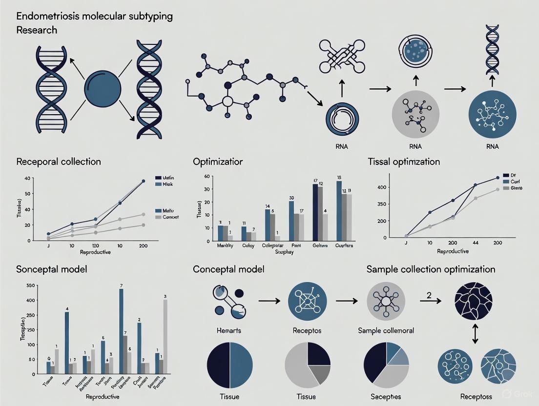
Optimizing Sample Collection for Endometriosis Molecular Subtyping: A Strategic Framework for Biomarker Discovery and Personalized Medicine
Endometriosis is a heterogeneous disease with significant diagnostic delays, creating an urgent need for precise molecular subtyping to enable personalized treatment.
Optimizing Sample Collection for Endometriosis Molecular Subtyping: A Strategic Framework for Biomarker Discovery and Personalized Medicine
Abstract
Endometriosis is a heterogeneous disease with significant diagnostic delays, creating an urgent need for precise molecular subtyping to enable personalized treatment. This article provides a comprehensive framework for researchers and drug development professionals on optimizing biospecimen collection to uncover and validate molecular subtypes. We explore the biological rationale for subtyping based on distinct immune and metabolic profiles, detail standardized protocols for collecting diverse sample types including blood, saliva, and menstrual blood, address critical pre-analytical variables, and present advanced validation methodologies integrating multi-omics data with artificial intelligence. By establishing robust sample collection standards, we aim to accelerate the development of non-invasive diagnostics and targeted therapies for specific endometriosis endotypes.
Understanding Endometriosis Heterogeneity: The Biological Imperative for Molecular Subtyping
Endometriosis is a complex and heterogeneous gynecological disorder characterized by the presence of endometrial-like tissue outside the uterine cavity, affecting approximately 10% of women of reproductive age globally [1]. The disease manifests through a spectrum of distinct phenotypes, primarily categorized as superficial peritoneal lesions, ovarian endometriomas, and deep infiltrating endometriosis [2] [3]. This phenotypic diversity is underpinned by varied molecular signatures, suggesting the existence of distinct disease subtypes [4]. A critical challenge in endometriosis research has been the over-reliance on eutopic endometrium (the normal uterine lining) as a proxy for ectopic disease tissue. Recent analyses reveal that over 36% of publicly available datasets labeled 'endometriosis' contain only eutopic endometrial samples, thereby misrepresenting the true biology of ectopic lesions [4]. Optimizing sample collection strategies is therefore foundational to advancing molecular subtyping research, enabling the development of more accurate diagnostic tools and personalized therapeutic interventions.
Frequently Asked Questions (FAQs) & Troubleshooting Guides
FAQ 1: What are the key biological distinctions between eutopic endometrium and ectopic endometriotic lesions that justify separate sampling?
Answer: Eutopic endometrium and ectopic endometriotic lesions, while sharing some histological features, are fundamentally distinct at the molecular and cellular levels. Using eutopic tissue as a universal proxy for disease is a significant methodological pitfall.
- Key Distinctions:
- Molecular and Cellular Profiles: Endometriosis lesions are not merely "ectopic endometrium." They possess unique cellular and molecular profiles, including differences in epigenetic regulation (e.g., hypermethylation of the progesterone receptor B gene) and distinct transcriptomic signatures that are not faithfully replicated by eutopic tissue [4] [1].
- Microenvironment: Ectopic lesions exist in a unique inflammatory microenvironment, with significant contributions from immune cells (e.g., altered macrophage activity, reduced natural killer cell function), endothelial cells, and fibroblasts, which differ from the uterine environment [4] [3].
- Functional Differences: Lesions frequently exhibit progesterone resistance, a phenomenon linked to epigenetic modifications that is not always mirrored in a patient's eutopic endometrium [1] [3].
Troubleshooting Guide: If your experimental model is based solely on eutopic endometrium, the results may not be translatable to true disease pathology. The table below outlines common scenarios and solutions.
| Research Goal | Common Pitfall | Recommended Solution |
|---|---|---|
| Studying lesion-specific pathogenesis | Using only eutopic endometrium from patients vs. controls. | Prioritize the collection of well-phenotyped ectopic lesions, with matched eutopic endometrium and peritoneum as the most relevant control [4]. |
| Drug screening for lesion targeting | Using eutopic endometrial organoids to represent all disease. | Develop and utilize endometriosis lesion-derived organoids to ensure drug responses are relevant to the disease tissue [5]. |
| Biomarker discovery | Relying on eutopic endometrial gene expression signatures. | Focus biomarker validation studies on lesion-derived molecules (e.g., specific miRNAs, proteins) in easily accessible biofluids [2]. |
FAQ 2: How does the disease phenotype (superficial, ovarian, deep infiltrating) influence experimental outcomes and sample handling?
Answer: The different phenotypes of endometriosis are not just surgical appearances; they can represent biologically distinct entities with implications for sample processing and data interpretation.
- Biological Variability: Molecular analyses reveal that endometriomas are highly enriched for stromal cells compared to peritoneal lesions [4]. Furthermore, transcriptional signatures related to fibrosis or immune dysfunction can vary between phenotypes and may form the basis for molecular subtyping independent of surgical classification [4].
- Impact on Research: The over-representation of certain phenotypes, particularly endometriomas, in biorepositories (comprising over 70% of some tissue datasets despite a population prevalence of ~30%) introduces a significant selection bias that can skew research findings [4].
Troubleshooting Guide: Inconsistent results across studies may stem from unaccounted phenotypic heterogeneity.
| Experimental Issue | Potential Root Cause | Corrective Action |
|---|---|---|
| Inconsistent gene expression results between studies. | Aggregating data from different phenotypes (e.g., superficial peritoneal vs. endometrioma) as a single "endometriosis" group. | Stratify samples by phenotype (SPD, DIE, endometrioma) during analysis. Record and control for phenotype in all experimental designs [3]. |
| Low yield of epithelial cells from lesions. | Endometriotic tissues, especially non-cystic lesions, can be stroma-rich, making epithelial cell isolation challenging. | Optimize digestion protocols for different phenotypes. For endometriomas, larger tissue volume may be available, but sample areas rich in glandular epithelium [5]. |
| Failure to recapitulate disease features in a model. | Using a single cell type or model for a heterogeneous disease. | Consider developing phenotype-specific models (e.g., organoids from DIE) to address specific research questions [5]. |
FAQ 3: What are the best practices for establishing and validating in vitro models like organoids for endometriosis research?
Answer: Endometrial and endometriosis organoids represent a transformative model system, but their construction and validation require meticulous attention to detail.
- Construction and Culture: Successful organoid culture from menstrual effluent, eutopic endometrium, or ectopic lesions depends on a carefully formulated medium containing core factors like WNT-3A, RSPO-1, EGF, and Noggin to support stemness and proliferation. To induce physiological function, a differentiation medium containing estradiol and progesterone is required [5].
- Critical Validation: A robust validation protocol is non-negotiable. This includes:
- Morphological analysis of 3D glandular structures with proper polarity (e.g., E-cadherin, layer adhesion protein expression).
- Functional validation of dynamic hormone response (e.g., expression of estrogen and progesterone receptors, and secretion of receptivity markers like PAEP in response to progesterone).
- Molecular validation via transcriptomics to confirm the model recapitulates key features of the source tissue [5].
Troubleshooting Guide: Common challenges in organoid culture and their solutions.
| Problem | Possible Reason | Solution |
|---|---|---|
| Low organoid formation efficiency. | Poor sample quality or incorrect digestion. | For surgical specimens, use multiple sampling points to avoid necrotic tissue. For menstrual effluent, process quickly to maintain cell viability [5]. |
| Organoids lack physiological response. | Culture conditions only support proliferation, not differentiation. | Introduce a differentiation medium phase with hormonal cues (estradiol, progesterone) to mimic the secretory phase and study functional responses [5]. |
| Model lacks complexity. | Standard matrix-based cultures lack stromal and immune cells. | Explore air-liquid interface (ALI) cultures to retain native stromal and immune cells, providing a more complete microenvironment for studying cell-cell interactions [5]. |
Research Reagent Solutions for Endometriosis Molecular Subtyping
The following table details essential reagents and their applications in cutting-edge endometriosis research.
| Reagent / Material | Function / Application in Research |
|---|---|
| Endometriosis Organoid Culture Media | Supports long-term expansion of lesion-derived epithelial cells. Core components include WNT-3A (self-renewal), RSPO-1 (WNT signaling enhancement), EGF (proliferation), and Noggin (BMP inhibition) [5]. |
| Differentiation Media (Hormonal) | Used to induce a secretory, receptive state in organoids. Typically contains estradiol and progesterone to study hormone response, gene expression (e.g., PAEP, DEFB1), and model progesterone resistance [5]. |
| Matrix Gel (e.g., Basement Membrane Extract) | Provides a 3D scaffold for organoid growth, mimicking the extracellular matrix. Its complex and variable composition is a key consideration for experimental reproducibility [5]. |
| Antibodies for Cell Characterization | Critical for validating models via Immunohistochemistry (IHC)/Immunofluorescence (IF). Key targets: E-Cadherin (epithelial cell polarity), Estrogen/Progesterone Receptors (hormone responsiveness), T-bet/GATA3 (immune cell profiling in RIF subtypes) [5] [6]. |
| Enzymes for Tissue Digestion | Collagenases and other proteases for dissociating lesion tissues to isolate primary stromal and epithelial cells. Protocols must be optimized for different lesion phenotypes (e.g., fibrotic DIE vs. cystic endometrioma) [5]. |
Detailed Experimental Protocols
Protocol 1: Building a Phenotypically Defined Endometriosis Biospecimen Collection
Objective: To establish a standardized pipeline for collecting, processing, and storing high-quality endometriosis biospecimens with comprehensive phenotypic data to support robust molecular subtyping studies [4].
Step-by-Step Workflow:
- Pre-Surgical Consent & Ethical Compliance: Obtain informed consent under institutional ethical guidelines, specifically covering the use of tissue for biobanking and molecular research. Adhere to regulations governing human genetic resources [5].
- Intraoperative Phenotyping: During laparoscopy, the surgeon must meticulously document:
- Phenotype: Classify each lesion as Superficial Peritoneal Disease (SPD), Deep Infiltrating Endometriosis (DIE, >5mm infiltration), or Ovarian Endometrioma [3].
- Location: Record the anatomical site (e.g., uterosacral ligament, ovarian surface, rectovaginal septum).
- Macroscopic Appearance: Note the color (red, white, black), texture, and vascularity.
- Sample Collection: Collect tissue from representative lesions. For large endometriomas, sample the cyst wall, avoiding necrotic or hemorrhagic areas.
- Control Tissue Collection: Collect matched eutopic endometrial tissue (via biopsy or curettage). The most relevant biological control, peritoneum or other lesion-adjacent tissue, should be collected if surgically feasible [4].
- Sample Processing:
- Fresh Tissue: Immediately place tissue in cold, sterile transport medium.
- Multiple Aliquoting: Divide each sample for:
- Cryopreservation: Snap-freeze in liquid nitrogen for RNA/DNA/protein extraction.
- Formalin-Fixation and Paraffin-Embedding (FFPE) for histology.
- Live Cell Culture/Organoid Derivation: Process in tissue culture hood.
- Cryopreservation: Store at -80°C or in liquid nitrogen vapor phase for long-term preservation.
- Data Annotation: Link each sample to a de-identified database containing full surgical, pathological, and clinical information (e.g., pain scores, infertility status, imaging data).
Protocol 2: Establishing and Validating Endometriosis Organoids from Ectopic Lesions
Objective: To generate 3D organoid cultures from ectopic endometriotic lesions that faithfully recapitulate the cellular and functional features of the original tissue [5].
Step-by-Step Workflow:
- Tissue Transportation & Washing: Transport lesion tissue in cold, plain RPMI-1640 or another holding medium. Wash thoroughly to remove blood and mucus.
- Tissue Digestion: Mince the tissue finely with scalpel and dissect and incubate with a collagenase solution (e.g., Collagenase Type XI) at 37°C with gentle agitation for 1-2 hours.
- Cell Separation: Pellet the digest and resuspend. Sequential filtration through 100μm and 40μm cell strainers can help remove undigested fragments. Centrifuge to separate glandular fragments from single stromal cells if needed.
- Embedding in Matrix Gel: Resuspend the pelleted epithelial glands/organoids in a chilled, commercial basement membrane extract (e.g., Matrigel). Plate small droplets of the cell-matrix mixture in pre-warmed culture plates and allow to polymerize.
- Organoid Culture: Overlay with complete organoid growth medium, supplemented with WNT-3A, RSPO-1, EGF, Noggin, B27, and N2. Culture at 37°C, 5% CO2, and change the medium every 2-3 days.
- Passaging: For expansion, dissociate organoids mechanically or enzymatically (e.g., with TryPLE) every 7-14 days and re-embed in fresh matrix.
- Functional Validation:
- Hormone Response: Differentiate organoids by treating with 1nM estradiol and 1μM progesterone for 5-7 days. Assess the induction of secretory markers (e.g., PAEP) via qPCR.
- Molecular Characterization: Perform RNA sequencing to compare the transcriptomic profile of organoids to their parent lesion tissue.
- Genetic Fidelity: Verify retention of key mutations (e.g., PTEN, ARID1A) found in the original lesion, if present.
Data Presentation: Quantitative Landscape of Endometriosis
Table 1: Global Prevalence and Diagnostic Delays in Endometriosis
Epidemiological data highlights the widespread nature of endometriosis and critical gaps in clinical diagnosis, underscoring the need for better diagnostic tools.
| Region/Country | Prevalence (%) | Key Study Details (Population) | Average Diagnostic Delay |
|---|---|---|---|
| Global Estimate | ~10% | Women of reproductive age [1] | 4 to 11 years, up to 13 years [1] |
| Italy | 3.2% | Women >30 yrs (surgery/ultrasound) [1] | - |
| Germany | 0.5 - 0.7% | Women >14 yrs (laparoscopy/clinical) [1] | - |
| North America | 4.5 - 8.0% | Women 18-45 yrs (self-report/laparoscopy) [1] | - |
| Jordan | 13.7% | Women 16-50 yrs (laparoscopy) [1] | - |
| Brazil | 16.3% | Women 21-44 yrs (laparoscopic sterilization) [1] | - |
Table 2: Molecular Biomarkers in Endometriosis Research and Diagnostics
A summary of promising biomarkers being investigated for non-invasive diagnosis and understanding disease mechanisms.
| Biomarker Category | Example(s) | Association & Research Utility | Current Status |
|---|---|---|---|
| Protein Biomarkers | CA-125, Urocortin | Elevated in endometriosis; useful for differentiating endometriomas from other cysts [2]. | Research and limited clinical use. |
| Epigenetic Markers | Progesterone Receptor B (PRB), HOXA10, E-Cadherin | Hypermethylation of gene promoters linked to progesterone resistance and disease pathogenesis [1] [3]. | Active research for diagnostic/therapeutic targets. |
| MicroRNAs (miRNAs) | Various circulating miRNAs | Key regulators of gene expression; potential for non-invasive diagnostic panels [2]. | Early research phase. |
| Immune/Inflammatory Cytokines | TNF-α, IL-1β, IL-6 | Overproduced in peritoneal fluid; drivers of chronic inflammation and pain [3]. | Mechanistic research and drug target exploration. |
Visualization: Signaling Pathways and Research Workflows
Diagram: PI3K/Akt Pathway in Endometriosis Survival
This diagram illustrates the PI3K/Akt pathway, a key driver of cell survival and proliferation in endometriosis lesions, representing a promising therapeutic target.
Diagram: Endometriosis Molecular Subtyping Research Workflow
This flowchart outlines a comprehensive research workflow from sample collection to molecular subtyping and clinical application, emphasizing the importance of quality-controlled biospecimens.
Troubleshooting Guides
Guide 1: Inconsistent Molecular Subtyping Results
Problem: Researchers report inconsistent classification of patient samples into immune-driven and metabolic-driven subtypes across different sequencing batches.
Solution:
- Pre-analytical Variable Control: Standardize sample collection and processing using World Endometriosis Research Foundation Endometriosis Phenome and Biobanking Harmonisation Project (EPHect) protocols [7].
- Bioinformatic Normalization: Apply batch effect correction algorithms like the Combat algorithm from the R "sva" package. Perform quantile normalization using tools like Sangerbox to correct technical biases among chips [8].
- Multi-Algorithm Validation: Cross-validate subtypes using multiple machine learning algorithms (Stepglm, Random Forest) to minimize model-specific bias [9].
Verification Steps:
- Run principal component analysis (PCA) before and after batch correction to visualize batch effect removal [8]
- Validate key gene expression patterns (CEACAM1, FOS for immune; HNRNPR, HSP90B1 for metabolic) in external datasets [8] [9]
- Confirm subtype stability through 10-fold cross-validation (target AUC >0.9) [9]
Guide 2: Poor RNA Quality from Ectopic Lesions
Problem: Degraded RNA from ectopic endometrial tissues compromises transcriptomic profiling for molecular subtyping.
Solution:
- Rapid Processing: Process tissue samples within 45 minutes of collection, centrifuge at 1,000 × g for 10 minutes at 4°C [10].
- Standardized Preservation: Aliquot supernatant into 500μL tubes and store at -80°C until RNA extraction [10].
- Quality Control Metrics: Ensure RNA Integrity Number (RIN) >7.0 before proceeding with sequencing.
Critical Control Points:
- Use RNase-free reagents and tubes during processing
- Document ischemia time (time from excision to freezing)
- Avoid multiple freeze-thaw cycles
Guide 3: Weak Signal in Metabolic Reprogramming Assays
Problem: Weak or inconsistent results when validating Warburg-effect related metabolic reprogramming in cellular models.
Solution:
- Hypoxic Conditioning: Culture ectopic endometrial cells under hypoxic conditions (1-3% O₂) to stabilize HIF-1α signaling [11].
- Metabolic Pathway Activation: Validate HIF-1α-induced expression of GLUT1, LDH, and COX-2 via RT-qPCR [8].
- Functional Assays: Measure glucose uptake and lactate production to confirm glycolytic flux.
Experimental Optimization:
- Use Z12 cell line for in vitro validation of metabolic genes [8]
- Overexpress HSP90B1 to upregulate GLUT1, LDH, and COX-2 as positive control [8]
- Include mitochondrial inhibitors (e.g., rotenone) to confirm glycolytic dependency
Frequently Asked Questions (FAQs)
What are the key differential features between immune-driven and metabolic-driven endometriosis subtypes?
The table below summarizes the core distinguishing characteristics:
| Feature | Immune-Driven Subtype | Metabolic-Driven Subtype |
|---|---|---|
| Core Biomarkers | CEACAM1, FOS, PLA2G2A, THBS1 [9] | HNRNPR, SYNCRIP, HSP90B1, HSPA4, HSPA8, CCT2, CCT5 [8] |
| Dominant Process | Neutrophil Extracellular Traps (NETs) formation, immune cell infiltration [9] | Aerobic glycolysis (Warburg effect), mitochondrial dysfunction [11] |
| Key Signaling Pathways | Rho/ROCK, NF-κB, cytokine signaling [12] | HIF-1α, PI3K/AKT/mTOR, PDK1-PDH axis [11] |
| Immune Microenvironment | Enriched CD8+ T cells, regulatory T cells, mast cells [8] [9] | Immune evasion, altered macrophage polarization [8] [11] |
| Diagnostic Performance | 4-gene model AUC: 0.962-0.976 [9] | 7-gene model AUC: >0.8 [8] |
| Therapeutic Implications | Target immune checkpoint inhibitors, NETs formation | Target glycolytic enzymes, metabolic reprogramming |
How can researchers validate molecular subtypes in patient-derived samples?
Multi-Omics Confirmation Strategy:
- Transcriptomic: Validate key subtype-specific genes (CEACAM1, FOS for immune; HSP90B1, CCT2 for metabolic) via RT-qPCR [8] [9]
- Metabolomic: Confirm Warburg effect via mass spectrometry detection of increased lactate and glycolytic intermediates [10]
- Functional Immune Profiling: Use CIBERSORT and ssGSEA to quantify immune cell infiltration patterns [8]
Recommended Validation Workflow:
What are the common pitfalls in sample collection for endometriosis subtyping studies?
Critical Pitfalls and Solutions:
| Pitfall | Impact | Solution |
|---|---|---|
| Hormonal therapy | Alters gene expression profiles, confounding subtyping | Exclude patients with hormonal therapy during last 3 months [10] |
| Phase of menstrual cycle | Introduces transcriptional variability | Document cycle phase from last menstrual period and average cycle length [10] |
| Lesion heterogeneity | Different molecular features in same patient | Collect and process multiple lesions separately with precise anatomical documentation |
| Delay in processing | RNA degradation, metabolite decay | Process within 45 minutes of collection; immediate freezing at -80°C [10] |
| Control tissue selection | Inappropriate reference for differential expression | Use matched eutopic endometrium from same patient + healthy controls [8] |
Which experimental models are most appropriate for studying each molecular subtype?
Model Selection Guide:
Model Applications:
- Homologous mouse models: Best for immune system interactions and genetic studies [7]
- Heterologous mouse models: Ideal for studying human tissue in mouse microenvironment [7]
- Organoid models: Optimal for metabolic pathway studies and high-throughput screening [7]
- Pain models: Essential for therapeutic efficacy assessment on pain symptoms [7]
The Scientist's Toolkit: Research Reagent Solutions
| Reagent/Category | Specific Examples | Function in Subtyping Research |
|---|---|---|
| Sample Collection Kits | EPHect-standardized collection kits [7] | Standardized biospecimen collection for reproducible molecular profiling |
| Metabolomic Analysis | AbsoluteIDQ p180 kit (Biocrates) [10] | Simultaneous quantification of 188 metabolites including amino acids, biogenic amines, lipids |
| RNA Stabilization | RNAlater or equivalent | Preserves RNA integrity during tissue processing and storage |
| Cell Culture Media | Specialized organoid media [7] | Supports growth of patient-derived endometriotic cells in 3D culture |
| Metabolic Inhibitors | HK2, LDHA, PDK inhibitors [11] | Targets glycolytic pathway to validate metabolic dependencies |
| Immune Profiling Panels | CIBERSORT LM22 matrix [8] [9] | Deconvolutes immune cell infiltration from transcriptomic data |
| Machine Learning Tools | Stepglm, Random Forest, XGBoost algorithms [9] | Builds predictive models for subtype classification from omics data |
| Pathway Analysis Software | clusterProfiler R package [8] | Identifies enriched biological pathways in each molecular subtype |
Key Signaling Pathways in Molecular Subtypes
Immune-Driven Subtype Signaling:
Metabolic-Driven Subtype Signaling:
Frequently Asked Questions (FAQs)
FAQ 1: What are the key molecular subtypes in endometriosis-associated ovarian cancer (EAOC), and why are they relevant for sample collection?
The Cancer Genome Atlas (TCGA) has defined four principal molecular subtypes for endometrial cancer that are now applied to EAOC, which includes endometrioid (ENOC) and clear cell (CCOC) ovarian cancers. The distribution of these subtypes differs significantly between ENOC and CCOC, which has implications for prognosis and treatment strategies. Ensuring your sample collection is phenotypically well-defined (e.g., confirmed as ENOC or CCOC) is critical for meaningful molecular subtyping results. The table below summarizes the key differences in subtype prevalence [13].
Table 1: Prevalence of TCGA Molecular Subtypes in Endometriosis-Associated Ovarian Cancer
| TCGA Molecular Subtype | Prevalence in ENOC | Prevalence in CCOC | Notes on Prognosis |
|---|---|---|---|
| POLEmut (POLE ultramutated) | Higher | Lower | Often associated with a more favourable prognosis. |
| MMRd (Mismatch Repair Deficient) | Higher | Lower | Also known as microsatellite instability (MSI) subtype. |
| NSMP (No Specific Molecular Profile) | Lower | Higher | Serves as the reference category for survival analyses. |
| p53abn (p53 abnormal) | Lower | Higher | Associated with significantly worse DFS and PFS in both ENOC and CCOC. |
FAQ 2: How does oxidative stress contribute to the pathophysiology of endometriosis and associated infertility?
Oxidative stress (OS) is a state of imbalance between reactive oxygen species (ROS) and antioxidant defenses, and it is a central player in endometriosis [14] [15]. It contributes to a pro-inflammatory peritoneal environment, promotes cell proliferation in lesions, and can cause damage to DNA, lipids, and proteins. In the context of infertility, particularly with minimal/mild endometriosis, OS is thought to be a primary underlying cause rather than a secondary effect. High OS levels in the follicular fluid and peritoneal environment can negatively impact oocyte quality, sperm motility, embryo cleavage, and implantation rates, leading to subfertility [15].
FAQ 3: What is the relationship between the eutopic endometrium and ectopic endometriotic lesions? Should I use eutopic tissue as a control?
While eutopic endometrium (from the uterine cavity) from patients with endometriosis is a valuable biospecimen, it is not equivalent to ectopic endometriotic lesions. A critical review of public datasets found that nearly half of all samples labeled "endometriosis" are actually eutopic endometrium, highlighting a significant bias in research [4]. Eutopic endometrium and lesions show "unequivocal differences at both the tissue and cellular levels." For studies focused on lesion biology or the lesion microenvironment, the most appropriate controls are often tissues adjacent to the lesions (e.g., peritoneum, ovarian stroma) rather than eutopic endometrium. Using eutopic endometrium as a sole control may lead to misleading conclusions about disease-specific mechanisms [4].
FAQ 4: Which signaling pathways are central to the process of Epithelial-Mesenchymal Transition (EMT) in endometriosis?
Epithelial-Mesenchymal Transition (EMT) is a key process that confers migratory and invasive capabilities to endometriotic cells. The major drivers and pathways involved in EMT in endometriosis are summarized below [16].
Table 2: Key Drivers of EMT in Endometriosis
| Category | Specific Factor/Pathway | Role in EMT |
|---|---|---|
| Growth Factors & Cytokines | TGF-β, PDGF, IL-1β | Potent inducers of the EMT program. |
| Hormonal Signals | Estradiol | Promotes EMT. |
| Microenvironmental Cues | Hypoxia | Activates HIFs, which drive EMT. |
| Key Transcription Factors | Snail, Slug, ZEB1/2, TWIST-1/2 | Execute the transcriptional reprogramming, downregulating epithelial markers (e.g., E-cadherin) and upregulating mesenchymal markers. |
| Signaling Pathways | Wnt/β-catenin, PI3K/Akt/mTOR, Notch, Hedgehog | Activated in most ectopic lesions and promote EMT. |
Troubleshooting Guides
Issue 1: Inconsistent Molecular Subtyping Results
Problem: Your sequencing or immunohistochemistry (IHC) results for TCGA molecular subtypes (POLEmut, MMRd, p53abn, NSMP) are inconsistent or do not align with expected clinical outcomes.
Solution:
- Verify Sample Quality and Source:
- Confirm the histopathological diagnosis of your samples (e.g., ENOC vs. CCOC) [13].
- Ensure DNA and protein integrity is high for sequencing and IHC, respectively. Use standardized quality control metrics like DNA Integrity Number (DIN) or RNA Integrity Number (RIN).
- Standardize Your Assessment Methods:
- POLEmut: Perform targeted next-generation sequencing covering the exonuclease domain of the POLE gene to identify hotspot mutations.
- MMRd: Use a combined approach. Perform IHC for the four mismatch repair proteins (MLH1, MSH2, MSH6, PMS2). For equivocal results, follow up with PCR-based testing for microsatellite instability (MSI).
- p53abn: Use IHC as a sensitive screening method. Strong diffuse nuclear staining or complete absence of staining (null pattern) is indicative of a TP53 mutation. Confirm abnormal results with TP53 sequencing.
- Utilize a Classifier: Follow the ProMisE algorithm, which uses a decision tree based on the results of the above tests to assign a final molecular subtype [13].
Issue 2: Measuring Oxidative Stress in Patient-Derived Samples
Problem: Measurements of oxidative stress markers (e.g., in serum, peritoneal fluid, or tissue) are highly variable between samples.
Solution:
- Standardize Sample Collection and Processing:
- Collect samples in a consistent, anaerobic manner to prevent ex vivo oxidation.
- Process samples immediately on ice and store at -80°C in aliquots to avoid freeze-thaw cycles.
- For blood collection, follow strict patient preparation guidelines, including fasting and avoiding strenuous exercise, to minimize variability [17].
- Use a Multi-Parameter Approach: Do not rely on a single OS marker. Implement a panel of tests to get a comprehensive picture [14] [15].
- Markers of Oxidative Damage: Measure Malondialdehyde (MDA) for lipid peroxidation or 8-hydroxy-2'-deoxyguanosine (8-OHdG) for DNA damage.
- Antioxidant Capacity: Assess the activity of key enzymes like Superoxide Dismutase (SOD), Glutathione Peroxidase (GPX), and Catalase.
- Total Antioxidant Status: Use assays like FRAP or ORAC.
- Correlate with Clinical Data: Always correlate your OS measurements with patient data such as disease stage (e.g., rASRM score), symptom severity, and fertility status to enhance biological relevance [15].
Issue 3: Differentiating True Lesional Signaling from Background Noise
Problem: Gene expression or pathway analysis from bulk tissue samples is confounded by cellular heterogeneity, making it difficult to identify signals specific to endometriotic epithelial or stromal cells.
Solution:
- Employ Single-Cell RNA Sequencing (scRNA-seq): This allows for the transcriptional profiling of individual cells, enabling the identification of cell-type-specific signaling pathways and the construction of cellular maps of the lesion microenvironment [4].
- Apply Computational Deconvolution: If scRNA-seq is not feasible, use bioinformatic tools (e.g., CIBERSORTx) to estimate the proportions of different cell types in your bulk RNA-seq data and infer cell-type-specific gene expression [18] [19].
- Utilize Laser Capture Microdissection (LCM): Precisely isolate specific cell populations (e.g., glandular epithelium vs. stroma) from tissue sections prior to molecular analysis to obtain pure samples.
- Validate with In Situ Techniques: Confirm findings from bulk analyses using techniques that preserve spatial context, such as RNA in situ hybridization or multiplex IHC/IF, to verify which cells express your target of interest [16].
Quantitative Data for Experimental Design
Table 3: Key Oxidative Stress Markers in Endometriosis Studies
| Biomarker | Sample Type | Change in Endometriosis vs. Control | Functional Significance |
|---|---|---|---|
| Malondialdehyde (MDA) | Serum, Peritoneal Fluid | Increased [14] | Marker of lipid peroxidation and cellular damage. |
| Superoxide Dismutase (SOD) | Serum, Plasma | Decreased [14] | Reduced activity indicates impaired antioxidant defense. |
| 8-F2-isoprostane | Serum | Decreased [14] | A marker of oxidative stress; its decrease is not fully explained. |
| Lipid Hydroperoxides (LOOHs) | Serum | Increased [14] | Products of unsaturated lipid oxidation, indicating oxidative damage. |
| HSP70 | Serum, Endometrium | Increased [14] | Chaperone protein induced during cellular stress. |
| Paraoxonase-1 (PON-1) | Serum | Decreased activity [14] | An antioxidant enzyme associated with HDL; decreased activity implies reduced antioxidant capacity. |
Detailed Experimental Protocols
Protocol 1: Molecular Subtyping of Endometriosis-Associated Samples Using the ProMisE Algorithm
Objective: To classify formalin-fixed, paraffin-embedded (FFPE) tissue samples from EAOC or endometriosis lesions into the four TCGA molecular subtypes: POLEmut, MMRd, p53abn, and NSMP.
Materials:
- FFPE tissue sections (4-5 µm for IHC, 10 µm for DNA extraction)
- Antibodies for IHC: MLH1, MSH2, MSH6, PMS2, p53
- DNA extraction kit (compatible with FFPE tissue)
- PCR reagents and platforms for POLE sequencing
Workflow Diagram:
Procedure:
- POLE Mutation Analysis:
- Extract genomic DNA from FFPE sections.
- Perform targeted next-generation sequencing of the exonuclease domain of the POLE gene.
- Analyze data for known pathogenic mutations (e.g., P286R, V411L, S297F, A456P). A sample with a pathogenic POLE mutation is classified as POLEmut, regardless of other findings.
- Mismatch Repair (MMR) Protein Assessment:
- Perform IHC for MLH1, MSH2, MSH6, and PMS2 on consecutive tissue sections.
- Interpret results: Loss of nuclear expression in lesion cells, with internal positive control (e.g., stromal cells, lymphocytes), indicates deficiency.
- A sample with loss of one or more MMR proteins is classified as MMRd.
- p53 Immunohistochemistry:
- Perform IHC for p53.
- Interpret results: Aberrant expression is defined as either:
- Overexpression: Strong, diffuse nuclear staining in >80% of lesion cells.
- Complete absence: Null pattern, with no nuclear staining in lesion cells (internal positive control must be present).
- A sample with aberrant p53 expression is classified as p53abn.
- Final Classification:
- Samples that are wild-type for POLE and have proficient MMR and normal p53 expression are classified as NSMP [13].
Protocol 2: Evaluating Immune Cell Infiltration in Endometriotic Lesions Using CIBERSORT
Objective: To estimate the abundance of 22 immune cell subtypes from bulk RNA-sequencing data of endometriotic lesions.
Materials:
- Bulk RNA-seq data from endometriosis and control tissues (e.g., normalized count matrix).
- R statistical software with the CIBERSORT package installed.
- The LM22 signature matrix file (available from the CIBERSORT website).
Workflow Diagram:
Procedure:
- Data Preparation:
- Obtain a normalized gene expression matrix (e.g., TPM or FPKM) from your RNA-seq data for all samples.
- Ensure your gene identifiers match those in the LM22 signature matrix (usually HGNC symbols).
- Run CIBERSORT:
- Upload your gene expression matrix and the LM22 signature file to the CIBERSORT web portal or run the algorithm locally in R.
- Use the default parameters with 100 permutations for significance analysis.
- Output Interpretation:
- CIBERSORT returns a matrix showing the proportional abundance of each of the 22 immune cell types in each sample. The sum of all fractions for a sample equals 1.
- Filter out samples with a CIBERSORT p-value < 0.05 to ensure reliable deconvolution.
- Downstream Analysis:
- Use the output to perform unsupervised clustering (e.g., k-means) to identify "immune-hot" and "immune-cold" molecular clusters within your endometriosis samples [18] [19].
- Correlate immune cell abundances with clinical variables (e.g., pain scores, infertility) or with the expression of genes of interest (e.g., ER stress markers like VWF, VCAM1) [19].
The Scientist's Toolkit: Research Reagent Solutions
Table 4: Essential Reagents for Endometriosis Molecular Pathway Analysis
| Reagent / Material | Function / Application | Example Use Case |
|---|---|---|
| Anti-p53 Antibody (IHC) | Identifies aberrant p53 protein expression via immunohistochemistry. | Central to the ProMisE algorithm for identifying the p53abn molecular subtype [13]. |
| Anti-MMR Protein Antibodies (MLH1, MSH2, MSH6, PMS2) | Detects loss of mismatch repair protein expression via IHC. | Key for classifying the MMRd molecular subtype [13]. |
| CIBERSORT Software & LM22 Matrix | Computational deconvolution of bulk RNA-seq data to estimate immune cell abundances. | Characterizing immune infiltration in endometriosis molecular clusters (e.g., immune-enriched vs. less immune-enriched) [18] [19]. |
| MDA (Malondialdehyde) Assay Kit | Colorimetric or fluorometric quantification of lipid peroxidation. | Measuring oxidative stress levels in serum, peritoneal fluid, or tissue homogenates [14]. |
| SOD Activity Assay Kit | Measures superoxide dismutase enzymatic activity. | Assessing the antioxidant capacity of a biological sample [14]. |
| ER Stress Marker Antibodies (e.g., VWF, VCAM1) | Detects expression of endoplasmic reticulum stress-related proteins via IHC or Western Blot. | Validating the role of ER stress in endometriosis pathogenesis and subtyping [19]. |
Technical Support Center
Troubleshooting Guides
Table 1: Troubleshooting Common Molecular Biology Experiments in Endometriosis Research
| Problem | Potential Cause | Solution |
|---|---|---|
| No amplification in qPCR [20] | Suboptimal annealing temperature, low-quality template, or low template concentration. | Perform a temperature gradient PCR, check DNA/RNA quality via Nanodrop, and increase template concentration. [20] |
| Non-specific amplification in PCR [20] | Annealing temperature too low, primer dimers, or non-specific primer binding. | Increase the annealing temperature, lower primer concentration, and ensure primers do not have self-complementary sequences. [20] |
| Low DNA/RNA yield [20] | Incomplete tissue homogenization or lysis, or low starting material. | Increase lysis time, ensure thorough vortexing and homogenization, and increase the initial sample volume. [20] |
| Amplification in negative control [20] | Contaminated reagents or cross-contamination of samples. | Use new, sterile reagents and tips; consider using a commercial, high-fidelity polymerase. [20] |
| High background in immunoassay | Non-specific antibody binding or inadequate blocking. | Optimize antibody concentrations, include appropriate controls, and ensure sufficient blocking time. [21] |
Table 2: Troubleshooting Biomarker Discovery and Validation
| Problem | Potential Cause | Solution |
|---|---|---|
| Low diagnostic accuracy of a single biomarker | High heterogeneity of endometriosis and complex pathophysiology. [22] | Develop a multi-marker panel (e.g., combining genetic, epigenetic, and protein biomarkers) to increase sensitivity and specificity. [22] |
| Inconsistent DNA methylation results | Cell-type heterogeneity in tissue samples, leading to confounding signals. | Perform microdissection or cell sorting to analyze pure cell populations, or use bioinformatic methods for deconvolution. [23] |
| Poor correlation between biomarker levels and disease stage | The biomarker may not be involved in disease progression or may be influenced by other factors. | Correlate biomarker levels with clinical phenotypes (e.g., pain scores, lesion location) and validate in a large, well-characterized cohort. [22] |
Frequently Asked Questions (FAQs)
Q1: What are the key considerations for sample collection in endometriosis GWAS studies? The foremost consideration is the accurate phenotypic characterization of patients and controls. This includes surgical and histological confirmation of endometriosis for cases and the absence of disease for controls. [24] Sample size is critical for achieving sufficient statistical power, as GWAS typically require large cohorts to detect variants with genome-wide significance (p < 5 × 10⁻⁸). [24] Proper collection, processing, and storage of DNA samples (e.g., from blood or tissue) are essential to prevent degradation and ensure high-quality genotyping data.
Q2: How can I functionally characterize a non-coding genetic variant associated with endometriosis? A powerful strategy is to determine if the variant acts as an expression quantitative trait locus (eQTL). This involves cross-referencing the variant with databases like GTEx to see if it is significantly associated with gene expression changes in relevant tissues, such as the uterus, ovary, or peripheral blood. [24] A significant eQTL signal (FDR < 0.05) suggests the variant has a regulatory effect on gene expression, providing a mechanistic hypothesis for its role in disease.
Q3: We are seeing high variability in our DNA methylation data from endometrium samples. How can this be mitigated? High variability often stems from differences in cellular composition (epithelial vs. stromal cells) and the menstrual cycle phase at the time of collection. [23] To mitigate this:
- Standardize collection: Document and, if possible, standardize the menstrual phase (e.g., mid-secretory) for sample procurement.
- Record cell composition: Histologically evaluate the cellular makeup of each sample.
- Use appropriate controls: Compare ectopic lesions to both eutopic endometrium from patients and endometrium from healthy controls to distinguish disease-specific changes from patient-specific background. [23]
Q4: What is the potential of machine learning in endometriosis biomarker discovery? Machine learning (ML) is highly promising for integrating complex, multi-dimensional data to improve diagnosis. For instance, one study used Support Vector Machine-Recursive Feature Elimination (SVM-RFE) and LASSO regression to identify a panel of three apoptosis-related genes (FAS, PRKAR2B, CSF2RB) as diagnostic biomarkers for endometriosis from a larger pool of candidates. [25] ML models can combine genetic, epigenetic, and clinical data to create predictive models with higher accuracy than single biomarkers. [22]
Q5: Are there non-invasive biomarkers on the horizon for endometriosis? Yes, research is actively focused on developing non-invasive biomarkers. A blood-based molecular diagnostic test, HerResolve, has shown clinical validation data demonstrating accuracy equivalent to the surgical gold standard, with reported ≥90% specificity and ≥83% sensitivity. [26] Other approaches include analyzing DNA methylation patterns [23] or specific miRNAs [27] in blood or menstrual effluent.
The Scientist's Toolkit: Research Reagent Solutions
Table 3: Essential Reagents for Genetic and Epigenetic Studies
| Reagent / Kit | Function / Application |
|---|---|
| DNA Methylation Kit | For bisulfite conversion of DNA, a critical step in analyzing methylation status at single-base resolution. [23] |
| qPCR Master Mix | Pre-mixed solutions for quantitative PCR, essential for validating gene expression from RNA or DNA samples. [20] |
| Plasmid Miniprep Kit | For rapid extraction and purification of plasmid DNA for cloning or as standards in qPCR. [20] |
| Chromatin Immunoprecipitation (ChIP) Kit | For investigating histone modifications and transcription factor binding in endometriotic tissues. [27] |
| ELISA Kits | For quantifying protein levels of potential biomarkers (e.g., cytokines, hormones) in serum or tissue lysates. [21] [22] |
| Flow Cytometry Antibodies | For characterizing immune cell populations (e.g., T cells, B cells, MDSCs) in the endometriosis microenvironment. [25] |
Experimental Workflows and Signaling Pathways
GWAS to Functional Validation Workflow
DNA Methylation Analysis in Endometriosis
Key Pathways in Endometriosis Pathogenesis
The diagnostic delay for endometriosis represents one of the most significant challenges in gynecological care, with profound implications for both clinical management and research. Current evidence consistently reports prolonged intervals between symptom onset and definitive diagnosis across multiple healthcare systems.
Table 1: Documented Diagnostic Delays for Endometriosis Across Geographical Regions
| Geographical Region | Reported Diagnostic Delay | Study Period/Publication Year | Sample Characteristics |
|---|---|---|---|
| United Kingdom | 7.5 - 10 years [28] | 2025 | Based on healthcare professional reports and national surveys |
| Global Average (Multiple Studies) | 6.6 years [29] | Scoping Review (2024) | Synthesis of 22 studies |
| Western High-Income Nations | 0.3 - 12 years [30] | Review of studies from 2018-2023 | Range across 17 observational studies |
| International Scope | 6.7 - 11 years [29] | Current global estimate | Reflects systemic issues impeding early detection |
This extended diagnostic journey typically begins with symptom onset during adolescence, with an average age of 14 years for adolescents and 20 years for adults, yet formal diagnosis often does not occur until much later in life [29]. The delay is not merely a temporal issue but a complex phenomenon driven by multiple interacting factors that have direct consequences for research sample quality and participant recruitment.
Root Causes: Deconstructing the Diagnostic Bottleneck
The prolonged diagnostic timeline can be attributed to a confluence of patient-, physician-, and healthcare system-related factors. Understanding this hierarchy is essential for developing targeted interventions both in clinical practice and research methodology.
Table 2: Contributing Factors to Diagnostic Delay and Their Research Implications
| Factor Category | Specific Barriers | Pooled Effect Size (SMD) | Impact on Research |
|---|---|---|---|
| Patient-Related Factors | Symptom normalization, delay in seeking care, cultural attitudes toward menstruation | SMD: 1.94 (95% CI: 1.62-2.27) [31] | Late-stage recruitment, advanced disease bias |
| Physician-Related Factors | Misdiagnosis, symptom dismissal, lack of specialized training | SMD: 2.00 (95% CI: 1.72-2.28) [31] | Heterogeneous pre-referral treatments |
| System-Related Factors | Referral pathway limitations, geographic disparities, access to specialized care | Insufficient data for meta-analysis [31] | Multi-center variability, sample heterogeneity |
Patient-Related Barriers
Patient-related factors demonstrate a significant contribution to diagnostic delays, with delays in seeking medical attention contributing most prominently (SMD: 2.14, 95% CI: 1.36-2.92) [31]. This often stems from the normalization of menstrual pain and cultural taboos surrounding menstrual discourse. For researchers, this translates to recruitment challenges and a population that may have adapted to chronic pain, potentially altering molecular profiles.
Physician-Related Barriers
Provider-related factors, including misdiagnosis and reliance on non-specific diagnostics, show a substantial pooled effect size (SMD: 2.00, 95% CI: 1.72-2.28) with notably low heterogeneity (I² = 3%), indicating consistent findings across studies [31]. Qualitative research reveals that healthcare professionals describe how endometriosis is often "masked or rendered invisible" in consultations, and that the presence of another person—most often a male partner—can legitimize symptom severity and influence referral decisions [28]. This diagnostic uncertainty introduces significant variability in the pre-diagnostic treatment history of research participants, potentially confounding molecular analyses.
Disease Complexity Barriers
The diagnostic process is further complicated by the diverse and non-specific manifestations of endometriosis that often mimic other conditions such as irritable bowel syndrome (IBS) and pelvic inflammatory disease (PID) [31]. The growth of endometrial-like tissue outside the uterus triggers inflammation and scarring that manifests uniquely in each person, creating a heterogeneous disease phenotype that challenges both diagnosis and research classification [29].
Diagnostic Modalities and Their Limitations in Clinical and Research Settings
Established Diagnostic Tools
Laparoscopy remains the gold standard for definitive diagnosis, allowing direct visualization and histological confirmation of endometrial implants [29]. However, its invasive nature creates a barrier to early diagnosis and is unsuitable for longitudinal research monitoring.
Imaging techniques including ultrasound and MRI can identify larger cysts and deep infiltrating lesions, but often fail to detect superficial peritoneal implants [29]. Consequently, a clear imaging study cannot definitively rule out endometriosis, limiting their negative predictive value in both clinical and research screening.
Emerging Non-Invasive Diagnostic Methods
Research into non-invasive diagnostic methods is exploring the identification of endometriosis biomarkers in blood, urine, or menstrual fluid [29]. Early studies show promise with inflammatory and angiogenic markers that correlate with the presence and severity of the disease. Although not yet standard practice, these innovations aim to reduce reliance on surgery and could revolutionize longitudinal research designs.
Direct Research Implications: From Sample Collection to Molecular Subtyping
The diagnostic delay has profound methodological consequences for endometriosis research, particularly in studies aiming to optimize sample collection for molecular subtyping.
Impact on Sample Characteristics and Recruitment
Prolonged diagnostic timelines directly impact research participant recruitment, creating a selection bias toward more severe, long-standing disease. The average delay of 6.6-11 years means that most research participants have advanced disease pathology, making it difficult to study early molecular events in endometriosis pathogenesis [29]. This late-stage recruitment inevitably influences the molecular landscape of collected samples, potentially masking early disease mechanisms.
The diagnostic journey often involves multiple therapeutic interventions (hormonal treatments, pain management) before definitive diagnosis, introducing confounding variables that can alter molecular profiles in tissue samples [31]. These exposures are frequently incompletely documented in research cohorts, creating significant noise in transcriptomic and proteomic analyses.
Molecular Heterogeneity and Subtyping Challenges
Recent research has revealed biologically distinct molecular subtypes of endometrial dysfunction in related conditions like recurrent implantation failure (RIF), with an immune-driven subtype (RIF-I) and a metabolic-driven subtype (RIF-M) [32]. The extended pre-diagnostic period in endometriosis may allow for molecular evolution and increased heterogeneity, complicating similar subtyping efforts.
The EPHect (Endometriosis Phenome and Biobanking Harmonisation Project) has developed standardized tools for the collection of study participant and surgeon-recorded data to address these challenges [7]. These protocols are essential for controlling the variability introduced by diagnostic delays across multi-center research studies.
Essential Methodologies for Robust Endometriosis Research
Standardized Phenotypic Data Collection
The World Endometriosis Research Foundation EPHect initiative provides standardized tools for collecting clinical data and biospecimens in endometriosis research [7]. Implementation of these protocols is critical for ensuring comparability across studies and controlling for confounding variables introduced by diagnostic delays.
EPHect Recommended Data Collection Domains:
- Surgical phenotype data (visual findings, revised ASRM stage)
- Patient-reported symptoms (pain, gastrointestinal, urinary)
- Quality of life impact and pain history
- Previous treatments and response
- Reproductive history and family history
Experimental Models and Their Applications
The EPHect working group has developed standardized operating procedures (SOPs) for experimental models to enhance reproducibility in endometriosis research [7].
Table 3: Research Reagent Solutions for Endometriosis Investigation
| Model System | Key Applications | Technical Considerations | Implementation Timeline |
|---|---|---|---|
| Heterologous Mouse Models | Exploring human tissue-microenvironment interactions | Requires fresh human samples, specialized infrastructure | Months (plus ethical approvals) |
| Homologous Mouse Models | Studying immune system and genetic influences | Uses syngeneic mouse endometrium | Months (plus ethical approvals) |
| Organoid Cultures | Investigating cellular mechanisms, drug screening | Matrix-based 3D in vitro approaches | Weeks to establish cultures |
| Pain Behavior Models | Evaluating novel analgesics and mechanisms | Requires specialized behavioral assessment training | Months to establish protocols |
Molecular Classification Workflows
Advanced transcriptomic approaches can identify molecular subtypes as demonstrated in recurrent implantation failure research [32]. Similar approaches are needed in endometriosis to address disease heterogeneity exacerbated by diagnostic delays.
Molecular Subtyping Workflow for Heterogeneous Samples
Troubleshooting Guide: Addressing Diagnostic Delay Challenges in Research
FAQ 1: How can researchers account for heterogeneous pre-diagnostic treatment histories in study participants?
- Challenge: Varied exposures to hormonal therapies, pain medications, and surgical interventions before diagnosis confound molecular analyses.
- Solution: Implement detailed medical history documentation using standardized instruments like the EPHect patient questionnaire [7]. Stratify participants by treatment history in analysis or include only treatment-naïve participants when feasible. Statistical methods such as propensity score matching can adjust for confounding variables.
FAQ 2: What sampling strategies can address the bias toward advanced-stage disease in research cohorts?
- Challenge: Diagnostic delays create recruitment bias toward severe, long-standing disease, limiting understanding of early pathogenesis.
- Solution: Establish prospective cohorts of individuals with symptomatic endometriosis who are awaiting diagnostic confirmation. Collaborate with primary care clinics to identify patients early in their diagnostic journey. Utilize banked tissue from incidental findings of endometriosis during other procedures.
FAQ 3: How can molecular studies overcome the heterogeneity introduced by prolonged disease duration?
- Challenge: Extended disease progression may allow for molecular evolution and increased heterogeneity.
- Solution: Implement molecular subtyping approaches similar to those used in recurrent implantation failure research, which identified immune-driven (RIF-I) and metabolic-driven (RIF-M) subtypes [32]. Increase sample sizes to power stratification analyses and employ single-cell technologies to resolve cellular heterogeneity.
FAQ 4: What validation approaches are essential for biomarkers discovered in delayed-diagnosis populations?
- Challenge: Biomarkers identified in late-stage disease may not apply to early detection.
- Solution: Validate candidate biomarkers in independent cohorts with varying disease durations and stages. Include symptomatic but undiagnosed individuals in validation studies to assess early detection capability.
The 7-11 year diagnostic delay in endometriosis represents not only a clinical failure but a critical methodological challenge for researchers. This delay introduces significant biases in participant recruitment, confounds molecular analyses through heterogeneous treatment histories, and likely amplifies disease heterogeneity. Addressing these limitations requires standardized phenotyping using EPHect guidelines, innovative recruitment strategies targeting early disease stages, and molecular subtyping approaches to deconvolute heterogeneity. By explicitly acknowledging and methodologically addressing these diagnostic challenges, researchers can generate more robust, reproducible findings that ultimately contribute to reducing the diagnostic delay itself through improved biomarker discovery and disease classification.
Strategic Biospecimen Collection: Standardized Protocols for Diverse Sample Types
This technical support center provides troubleshooting guidance for researchers implementing the Endometriosis Phenome and Biobanking Harmonisation Project (EPHect) standard operating procedures (SOPs) to optimize sample collection for endometriosis molecular subtyping research.
Frequently Asked Questions (FAQs) and Troubleshooting Guides
Sample Collection and Processing
Q: What are the critical checkpoints for ensuring RNA integrity in endometrial biopsies intended for transcriptomic subtyping?
A: RNA integrity is paramount for molecular subtyping studies. The following checklist outlines critical control points:
| Checkpoint | Objective | Common Pitfalls | Corrective Action |
|---|---|---|---|
| Pre-collection | Confirm patient fasting status & menstrual cycle phase. | Incorrect cycle timing (non-Window of Implantation). | Verify LH surge peak or cycle day; use Noyes' criteria for histology [32]. |
| Collection | Minimize ischemic time. | Delay in tissue stabilization >10 minutes. | Immediately place tissue in RNAlater or flash-freeze in liquid N₂. |
| Storage | Prevent RNA degradation. | Inconsistent freezer temperature at -80°C. | Use temperature loggers; avoid freezer frost build-up. |
| QC Assessment | Confirm RNA Quality Number (RQN) >7.0. | RQN below acceptable threshold (e.g., <7.0). | Repeat extraction; use degraded samples for DNA analysis only. |
Q: How should we handle discrepancies in sample quality metrics when applying the EPHect SOPs across multiple clinical sites?
A: Implement a centralized quality control (QC) protocol. The table below standardizes key metrics and actions:
| Quality Metric | Acceptable Range | Action if Out of Range |
|---|---|---|
| RNA Integrity Number (RIN) | ≥ 7.0 | Flag for re-extraction; exclude from transcriptomic subtyping [32]. |
| Tissue Ischemic Time | ≤ 10 minutes | Note in metadata; may impact hypoxia-sensitive genes. |
| Sample Volume | As per EPHect SOP (e.g., 5x5mm) | Process smaller samples for DNA/qPCR, not RNA-seq. |
Molecular Subtyping and Data Analysis
Q: Our transcriptomic analysis of endometriosis samples shows significant heterogeneity. How can we define molecular subtypes, and what are the key analytical pathways?
A: Endometriosis, like Recurrent Implantation Failure (RIF), exhibits distinct molecular subtypes driven by different biological processes. Research has identified reproducible subtypes, such as an immune-driven (RIF-I) and a metabolic-driven (RIF-M) subtype [32]. The workflow below outlines the process from sample to subtype identification.
Key pathways to investigate for subtype characterization:
| Molecular Subtype | Enriched Signaling Pathways [32] | Characteristic Immune Features [32] [33] |
|---|---|---|
| Immune-Driven (RIF-I) | IL-17 signaling, TNF signaling, Allograft rejection | Enriched for NK cells, elevated Th1/Th2 ratio, high T-bet/GATA3 ratio. |
| Metabolic-Driven (RIF-M) | Oxidative phosphorylation, Fatty acid metabolism, Steroid hormone biosynthesis | Dysregulated circadian clock gene PER1, lower T-bet/GATA3 ratio. |
Q: What machine learning approaches are recommended for building a robust molecular classifier for these subtypes?
A: A classifier can be developed by testing multiple algorithm combinations. One approach achieved an Area Under the Curve (AUC) of 0.94 in validation by finding the optimal F-score from 64 different combinations of machine learning algorithms [32]. It is crucial to validate the classifier in an independent cohort and benchmark it against existing models.
Reagent Solutions and Essential Materials
The following table details key reagents and their critical functions in the experimental workflow for molecular subtyping.
Research Reagent Solutions for Endometriosis Molecular Subtyping
| Item | Function / Application in Workflow |
|---|---|
| RNAlater Stabilization Solution | Preserves RNA integrity in fresh tissue samples immediately after collection, preventing degradation prior to nucleic acid extraction. |
| Qiagen RNeasy Mini Kits | For high-quality total RNA isolation from endometrial tissue samples, suitable for downstream transcriptomic applications [32]. |
| Anti-T-bet & Anti-GATA3 Antibodies | Used for Immunohistochemistry (IHC) validation of the immune-driven subtype (RIF-I) by calculating the T-bet/GATA3 expression ratio [32]. |
| Connectivity Map (CMap) Database | A bioinformatics resource used to predict candidate therapeutic compounds based on the gene expression signature of the identified molecular subtypes [32]. |
| PER1 Antibodies | For validating the metabolic-driven subtype (RIF-M) through protein-level analysis of the dysregulated circadian clock gene PER1 [32]. |
Technical Support Center
Troubleshooting Guides & FAQs
Q1: Our research group is consistently obtaining low biomarker sensitivity (<50%) from plasma samples for endometriosis subtyping. What are the primary pre-analytical variables we should investigate?
A: Low sensitivity often stems from pre-analytical degradation of labile biomarkers. Focus on these critical steps:
- Time to Processing: Cellular metabolism and protease activity continue post-collection. For mRNA and protein phosphorylation studies, process blood within 2 hours of draw.
- Centrifugation Conditions: Inconsistent g-force or time can lead to residual platelets in plasma (platelet-derived contamination). Adhere strictly to a double centrifugation protocol: 1,500-2,000 x g for 15 minutes at 4°C, followed by a second spin of the supernatant at 15,000 x g for 10 minutes at 4°C.
- Collection Tube Additive: For cell-free DNA (cfDNA) or miRNA studies, ensure you are using Streck or CellSave tubes for sample stabilization if immediate processing is not feasible. Standard EDTA tubes require processing within 2-4 hours.
Q2: We observe high background noise in our sequencing data from blood-based biomarkers, reducing assay specificity. How can we improve target-to-noise ratio?
A: High background is frequently a result of non-specific amplification or non-target cellular contamination.
- Inhibit Contaminants: Use RNase inhibitors in lysis buffers for RNA work and protease inhibitors for protein studies.
- Remove Platelet Contamination: Implement the double centrifugation protocol mentioned above. Platelets are a major source of confounding RNA species.
- Optimize Nucleic Acid Extraction Kits: Not all kits are equal for cfDNA or circulating miRNA. Use kits specifically validated for low-abundance, fragmented nucleic acids. Increase the number of wash steps to remove PCR inhibitors like heparin.
- Verify Primer/Probe Specificity: Re-validate all primer sets using BLAST and use locked nucleic acid (LNA) probes for miRNA detection to enhance binding specificity and thermal stability.
Q3: How does the choice of blood collection tube directly impact the sensitivity and specificity of downstream multi-omics assays for endometriosis?
A: The collection tube is the first and most critical determinant of data quality. The additive dictates the sample's molecular integrity.
- K2EDTA Tubes: Standard for most applications. Pros: Inexpensive, versatile. Cons: Requires rapid processing (<4 hours) to prevent RNA degradation and genomic DNA contamination from lysing white blood cells. Specificity can drop if processing is delayed.
- PAXgene Blood RNA Tubes: Pros: Immediately lyses cells and stabilizes RNA, locking the transcriptome profile at the time of draw. Maximizes sensitivity for RNA-based signatures. Cons: Incompatible with plasma protein or cfDNA analysis.
- Cell-Free DNA BCT Tubes (e.g., Streck): Pros: Preserves nucleated blood cells, preventing them from lysing and releasing genomic DNA that would dilute the cfDNA signal. This dramatically improves specificity for rare variant detection. Cons: Not optimal for intracellular transcriptomic studies.
Quantitative Data Summary: Impact of Pre-analytical Variables on Assay Performance
Table 1: The Effect of Time-to-Processing on Biomarker Stability in K2EDTA Tubes
| Biomarker Class | 2 Hours | 6 Hours | 24 Hours (4°C) | Key Degradation Effect |
|---|---|---|---|---|
| Cell-Free miRNA | 98% Recovery | 85% Recovery | 40% Recovery | RNase activity |
| Phospho-Proteins | 100% Recovery | 60% Recovery | <10% Recovery | Phosphatase activity |
| Cell-Free DNA | 100% Recovery | 95% Recovery | 80% Recovery | Increase in high-mol. weight gDNA |
Table 2: Comparative Performance of Blood Collection Tubes for Endometriosis Biomarker Detection
| Tube Type | Target Analyte | Avg. Sensitivity | Avg. Specificity | Primary Advantage |
|---|---|---|---|---|
| K2EDTA | Plasma Proteins | 85% | 90% | Broad compatibility |
| K2EDTA | cfDNA/miRNA | 75% | 82% | Cost-effective with rapid processing |
| PAXgene RNA | Blood RNA | >95% | 92% | Superb RNA integrity |
| cfDNA BCT | Cell-Free DNA | 90% | 99% | Inhibits gDNA contamination |
Experimental Protocols
Protocol: Isolation of High-Purity Platelet-Free Plasma for miRNA Sequencing
Objective: To obtain plasma devoid of platelets and cellular debris for robust and reproducible circulating miRNA analysis.
Materials:
- K2EDTA or cfDNA BCT blood collection tubes
- Refrigerated centrifuge
- Sterile polypropylene tubes
- Piperman and aerosol-resistant tips
- PBS (optional, for diluting viscous samples)
Methodology:
- Collection: Draw blood via venipuncture and invert tubes 8-10 times gently.
- Initial Spin: Centrifuge tubes at 1,800 x g for 15 minutes at 4°C. Brake: Off or Low.
- Plasma Transfer: Carefully aspirate the upper plasma layer (approx. 2/3 volume) without disturbing the buffy coat, and transfer to a new sterile tube.
- Secondary Spin: Centrifuge the transferred plasma at 15,000 x g for 10 minutes at 4°C. Brake: Off or Low.
- Aliquot: Transfer the supernatant (now platelet-poor plasma) into fresh cryovials in small, single-use aliquots.
- Storage: Flash-freeze in liquid nitrogen and store at -80°C.
Key Consideration: The use of a low or no brake during centrifugation is critical to prevent disturbing the pellet and re-suspending platelets.
Pathway & Workflow Visualizations
Title: Preamalytic Workflow for Blood Biomarker Studies
Title: Blood Biomarker Pathway in Endometriosis
The Scientist's Toolkit
Table 3: Research Reagent Solutions for Blood-Based Endometriosis Research
| Item | Function & Rationale |
|---|---|
| cfDNA BCT (Streck) | Chemical stabilizer that cross-links nucleated cells, preventing lysis and gDNA release, crucial for high-specificity cfDNA assays. |
| PAXgene Blood RNA Tube | Contains lysing agents and RNA stabilizers for immediate transcriptome preservation, maximizing sensitivity for RNA biomarkers. |
| RNase/DNase Inhibitors | Added to lysis buffers to protect fragile circulating nucleic acids from degradation during extraction. |
| miRNA-Specific SPRI Beads | Solid-phase reversible immobilization beads sized for optimal recovery of small RNA fragments (<200 nt). |
| Phosphatase/Protease Inhibitor Cocktails | Essential additives for preserving labile phospho-protein epitopes in plasma for proteomic workflows. |
| Magnetic Bead-based Extraction Kits | Enable high-throughput, automatable purification of nucleic acids with minimal carry-over of PCR inhibitors. |
The move towards non-invasive diagnostic methods is transforming endometriosis research. Salivary microRNA (miRNA) panels and urinary proteomic profiling represent promising approaches that circumvent the need for surgical intervention. This technical support center provides detailed troubleshooting guides and FAQs to help researchers optimize sample collection and analysis, ensuring high-quality data for molecular subtyping studies.
Salivary miRNA Panels: Technical Guide
Experimental Protocol for Salivary miRNA Sequencing
Sample Collection
- Collect saliva using sterile tubes containing a DNA/RNA preserving solution (e.g., DNA/RNA Shield Safe Collection Kit) [34].
- Immediately after collection, centrifuge samples at 10,000 × g for 20 minutes to remove cellular debris and contaminants [34].
- Store processed samples at -80°C until nucleic acid extraction [34].
RNA Extraction and Library Preparation
- Extract total RNA using a specialized kit such as the miRNeasy Advanced Micro Kit [34].
- Determine RNA concentration and quality using fluorimetry (e.g., Qubit RNA HS Assay) [34].
- Prepare miRNA libraries using the QIAseq microRNA Library Kit: ligate adaptors to RNA, perform reverse transcription to generate cDNA, and amplify libraries with unique dual indexes [34].
- Quantify final libraries and verify fragment size (~170 bp) using Bioanalyzer [34].
- Pool libraries and sequence using Illumina technology (74 bp paired-end recommended) [34].
Bioinformatics Analysis
- Process FASTQ files through an RNA-seq Analysis Portal for demultiplexing and mapping to reference genome (miRbase) [34].
- Perform differential expression analysis using DESeq2 algorithm with FDR-adjusted p-value <0.01 as significance threshold [34].
- Conduct functional characterization using Ingenuity Pathway Analysis (IPA) and g:Profiler, querying Gene Ontology database [34].
Salivary miRNA Troubleshooting FAQ
Q: What should I do if I obtain low miRNA yield from saliva samples? A: Ensure proper sample preservation immediately after collection using specialized preservative solutions. Increase starting sample volume and avoid repeated freeze-thaw cycles. Verify centrifugation parameters to remove contaminants while retaining miRNAs [34].
Q: How can I address poor sequencing library complexity? A: Check RNA integrity prior to library preparation. Optimize adapter ligation conditions and use appropriate input RNA quantities. Include library quality control steps using fluorometry and fragment analysis [34].
Q: What if I cannot replicate differential miRNA expression findings? A: Standardize collection time relative to menstrual cycle (particularly for endometriosis studies). Control for potential confounders like age, BMI, and medication use. Ensure consistent bioinformatic processing pipelines and normalization methods across datasets [34].
Q: How can I determine if detected miRNAs are biologically relevant to endometriosis? A: Cross-reference findings with existing literature on endometriosis pathogenesis. Utilize pathway analysis tools to identify enriched biological processes. Consider functional validation experiments in relevant cell models [34].
Salivary miRNA Analysis Workflow
Salivary miRNA Analysis Workflow
Urinary Proteomic Profiling: Technical Guide
Experimental Protocol for Urinary Proteomics
Sample Collection and Preparation
- Collect first-morning urine samples to minimize variability [35].
- Centrifuge at 14,000 × g for 30 minutes to remove insoluble particles and cells [35] [36].
- Concentrate samples using nitrogen blowdown evaporation or ultrafiltration to increase protein concentration [36].
- Store processed samples at -80°C if not analyzing immediately [36].
Protein Digestion and Cleanup
- Perform buffer exchange using size exclusion columns or precipitation methods to remove interfering compounds [36].
- Digest proteins using sequencing-grade trypsin (enzyme-to-substrate ratio 1:25-1:50) at 37°C for 12-16 hours [36].
- Desalt peptides using C18 solid-phase extraction columns [36].
- Lyophilize peptides and reconstitute in LC-MS compatible solvent (0.1% formic acid) [36].
LC-MS/MS Analysis and Data Processing
- Separate peptides using nanoflow LC with C18 reverse-phase column [37] [38].
- Acquire data using data-dependent acquisition MS/MS on a high-resolution mass spectrometer [38].
- Search MS/MS spectra against human protein databases using search engines like MaxQuant or Proteome Discoverer [38].
- Perform statistical analysis to identify differentially expressed proteins and post-translational modifications [35].
Urinary Proteomics Troubleshooting FAQ
Q: How can I prevent protein degradation in urine samples? A: Add protease inhibitor cocktails during collection, process samples immediately or flash-freeze in liquid nitrogen, and avoid repeated freeze-thaw cycles. Work at 4°C whenever possible [38] [36].
Q: What steps can reduce high background noise in LC-MS spectra? A: Implement rigorous peptide cleanup using StageTips or SPE columns. Use HPLC-grade water and solvents. Avoid polymer contamination by using filter tips and working in clean environments [37] [38].
Q: How can I improve detection of low-abundance proteins? A: Increase starting sample volume and use concentration methods like nitrogen blowdown. Implement fractionation techniques (SCX, high-pH RP) to reduce sample complexity. Consider enrichment strategies for specific protein classes [36].
Q: What if I observe inconsistent results between technical replicates? A: Standardize sample processing protocols precisely. Use internal standard peptides for quantification. Ensure consistent LC-MS system performance with quality control samples. Automate sample preparation where possible to reduce variability [37] [36].
Urinary Proteomic Profiling Workflow
Urinary Proteomic Profiling Workflow
Comparative Technical Challenges
Table 1: Troubleshooting Common Issues in Non-Invasive Sample Analysis
| Issue | Salivary miRNA | Urinary Proteomics |
|---|---|---|
| Low analyte yield | Increase sample volume; optimize preservation; verify RNA integrity [34] | Concentrate via nitrogen blowdown/ultrafiltration; pool multiple collections [36] |
| Sample degradation | Use RNA stabilizers; process immediately; store at -80°C [34] | Add protease inhibitors; freeze immediately; avoid freeze-thaw cycles [36] |
| High background interference | DNase treatment; optimize centrifugation; solid-phase cleanup [34] | SPE cleanup; buffer exchange; HPLC-grade reagents [37] [36] |
| Technical variability | Standardize collection time; control for external factors; use endogenous controls [34] | Internal standards; automate preparation; quality control samples [36] |
| Data reproducibility | Consistent bioinformatics; normalize using housekeeping miRNAs; adequate sample size [34] | Standardized protocols; instrument calibration; cross-validation [37] [38] |
Research Reagent Solutions
Table 2: Essential Materials for Non-Invasive Endometriosis Research
| Reagent/Kit | Application | Key Features | Considerations |
|---|---|---|---|
| DNA/RNA Shield Safe Collection Kit | Saliva sample preservation | Stabilizes nucleic acids; enables room temp storage [34] | Compatible with downstream RNA extraction kits |
| miRNeasy Advanced Micro Kit | Salivary RNA extraction | Optimized for low-concentration miRNA; removes contaminants [34] | Includes DNase treatment step |
| QIAseq microRNA Library Kit | miRNA library preparation | Unique molecular indexes; reduces duplicates [34] | Compatible with Illumina platforms |
| Trypsin, sequencing grade | Protein digestion | High specificity; minimal autolysis [36] | Requires optimized enzyme-to-substrate ratio |
| C18 Solid-Phase Extraction Columns | Peptide cleanup | Desalting; removes interfering compounds [36] | Various formats for different sample sizes |
| Nucleic Acid/Protein Assay Kits | Quality control | Fluorometric/colorimetric quantification [34] [36] | Essential for normalization |
| Protease Inhibitor Cocktails | Sample preservation | Broad-spectrum protease inhibition [36] | EDTA-free for MS compatibility |
Successful implementation of salivary miRNA and urinary proteomic profiling requires meticulous attention to pre-analytical variables. The protocols and troubleshooting guides provided here address the most critical challenges in non-invasive sample analysis for endometriosis research. As these technologies continue to evolve, standardization across research sites will be essential for generating comparable data and advancing our understanding of endometriosis molecular subtypes.
Frequently Asked Questions (FAQs)
Q1: What is the primary diagnostic advantage of using menstrual fluid (MF) for endometriosis research? Menstrual fluid provides a non-invasive source of endometrial tissue that cyclically sheds, reflecting the endometrial environment. This avoids the need for invasive biopsies and allows for renewable and inexpensive sample collection for investigating disorders like endometriosis [39].
Q2: How can molecular subtyping of endometriosis lesions impact treatment strategies? Research has identified distinct molecular subtypes of endometriosis lesions, such as stroma-enriched (S1) and immune-enriched (S2) subtypes. The S2 subtype is strongly associated with resistance or intolerance to hormone therapy. This subtyping highlights the importance of stromal-immune heterogeneity and provides a basis for future personalized, hormone-free treatments [40].
Q3: What are some common inflammatory serum biomarkers studied in endometriosis, and what are their challenges? IL-6 and suPar (soluble urokinase-type plasminogen activator receptor) have shown diagnostic potential in differentiating patients with endometriosis from controls [41]. However, a significant challenge is that serum levels of biomarkers like suPar can be influenced by factors such as patient age and BMI, which may confound results without careful study design and statistical correction [41].
Q4: What is the current gold standard for diagnosing endometriosis, and why is non-invasive diagnostic research critical? The only definitive way to diagnose endometriosis is through surgical laparoscopy and histological confirmation [42] [43]. Non-invasive diagnostic research is crucial to develop alternatives that can avoid surgery, reduce diagnostic delays, and allow for earlier intervention [41].
Troubleshooting Guide: Common Experimental Challenges
Table 1: Troubleshooting Common Sample Collection and Analysis Issues
| Problem | Possible Cause | Potential Solution |
|---|---|---|
| Low sample volume or cellular yield from Menstrual Fluid (MF). | Incorrect timing of collection within menstruation; improper use of menstrual cup. | Standardize collection to the first 24-48 hours of menstruation; provide participants with detailed pictorial guides for menstrual cup insertion [39]. |
| High biomarker level variability (e.g., IL-6, suPar) within study cohorts. | Confounding factors like BMI, age, or underlying inflammatory conditions influencing serum levels. | Implement strict inclusion/exclusion criteria; record and statistically adjust for covariates like BMI and age during analysis [41]. |
| Inability to replicate molecular subtyping results from endometriosis lesions. | Batch effects in transcriptomic data; suboptimal clustering parameters. | Use batch effect removal algorithms (e.g., ComBat in R) on combined datasets; employ consensus clustering with robust parameters (e.g., 10,000 repetitions) for subtype identification [40]. |
| Poor RNA quality or quantity from MF-derived cells. | Rapid RNA degradation due to inadequate sample preservation. | Immediately process MF samples upon collection; add RNA stabilization reagents to the collection medium; freeze samples at -80°C without delay [39]. |
| Hormone therapy confounds molecular analysis. | Previous hormone use alters gene expression profiles in ectopic lesions. | Document and account for all hormone therapy prior to sample collection; consider a wash-out period where ethically and clinically feasible [40]. |
Detailed Experimental Protocols
Protocol 1: Menstrual Fluid Collection and Processing for Cellular Analysis
This protocol is designed for the isolation of endometrial cells from menstrual effluent for subsequent transcriptomic studies [39].
- Participant Preparation & Consent: Obtain informed consent. Provide participants with a sterile menstrual cup (e.g., DivaCup, Mooncup) and detailed instructions for use.
- Sample Collection: Participants collect menstrual fluid into the cup during the first 24-48 hours of their menstrual cycle.
- Sample Transport: The sealed cup is placed in a provided insulated container and returned to the lab within 4-6 hours of collection.
- Sample Processing in Lab:
- Transfer the menstrual fluid to a 50mL conical tube.
- Centrifuge at 1,500 x g for 10 minutes to separate the liquid fraction from the cellular pellet.
- Carefully aspirate and aliquot the supernatant (liquid fraction) for future analysis of soluble factors.
- Resuspend the cellular pellet in 10-15mL of phosphate-buffered saline (PBS).
- Filter the cell suspension through a 40μm cell strainer to remove debris and large clumps.
- Centrifuge the filtered suspension again at 500 x g for 5 minutes.
- The resulting cell pellet can be used for immediate RNA/DNA extraction or resuspended in a cryopreservation medium (e.g., 90% FBS, 10% DMSO) for storage in liquid nitrogen.
Protocol 2: RNA Extraction and Microarray Analysis for Molecular Subtyping
This protocol is adapted from methods used to identify stroma-enriched and immune-enriched subtypes of endometriosis [40].
- RNA Extraction: Extract total RNA from homogenized endometriosis lesions or MF-derived cells using a commercial kit (e.g., RNeasy Mini Kit, Qiagen) according to the manufacturer's instructions. Include a DNase digestion step to remove genomic DNA contamination.
- RNA Quality Control: Assess RNA integrity and concentration using an instrument like the Agilent Bioanalyzer. Only samples with an RNA Integrity Number (RIN) >7.0 should proceed.
- Microarray Processing:
- 100ng of high-quality RNA is used for cDNA synthesis and labeling.
- Hybridize labeled cDNA to a whole-genome expression microarray (e.g., Affymetrix GeneChip) following the platform-specific protocol.
- Scan the arrays using a standardized scanner to generate raw intensity data (CEL files).
- Bioinformatic Analysis:
- Preprocessing & Normalization: Process raw CEL files in R using the
affypackage for background correction and normalization (e.g., RMA). - Batch Effect Removal: If combining multiple datasets, use the
ComBatfunction from thesvapackage to adjust for non-biological technical variation. - Molecular Subtyping: Perform unsupervised consensus clustering on the normalized expression data of the most variable genes using the
ConsensusClusterPluspackage. Set parameters to a high number of repetitions (e.g., 10,000) to ensure stable cluster identification.
- Preprocessing & Normalization: Process raw CEL files in R using the
Protocol 3: ELISA for Inflammatory Serum Biomarkers
This protocol outlines the quantification of serum biomarkers like IL-6 and suPar [41].
- Sample Preparation: Collect whole blood from patients and controls. Allow it to clot at room temperature for 30 minutes, then centrifuge at 2,000 x g for 15 minutes to isolate serum. Aliquot and store serum at -80°C until analysis.
- Assay Setup:
- Bring all reagents, standards, and samples to room temperature.
- Add standards and diluted patient serum to the appropriate wells of a pre-coated ELISA plate (e.g., Human IL-6 Quantikine ELISA Kit, R&D Systems).
- Incubate the plate as per the kit's protocol (typically 2 hours), then aspirate and wash each well 3-4 times with the provided wash buffer.
- Detection:
- Add the detection antibody to each well and incubate.
- After another wash step, add a substrate solution (e.g., tetramethylbenzidine) to develop color. Stop the reaction with a stop solution.
- Data Analysis:
- Measure the optical density (OD) of each well immediately using a microplate reader set to the appropriate wavelength (e.g., 450nm with a correction at 540nm or 570nm).
- Generate a standard curve from the OD values of the standards and use it to calculate the concentration of the target biomarker in the patient samples.
The Scientist's Toolkit: Research Reagent Solutions
Table 2: Essential Materials and Reagents for Endometriosis Microenvironment Research
| Reagent / Solution | Function / Application in Research |
|---|---|
| Sterile Menstrual Cup | Non-invasive collection of menstrual fluid (menstrual effluent) containing endometrial tissue and cells [39]. |
| RNA Stabilization Reagent | Preserves RNA integrity in biological samples immediately upon collection, preventing degradation prior to RNA extraction [39]. |
| Human IL-6/suPar Quantikine ELISA Kits | Quantify specific pro-inflammatory serum biomarkers (IL-6, suPar) implicated in endometriosis pathogenesis [41]. |
| RNeasy Mini Kit (Qiagen) | Silica-membrane-based spin column technology for the purification of high-quality total RNA from tissue or cell samples [40]. |
| Affymetrix GeneChip Microarray | Platform for genome-wide transcriptomic expression profiling to identify gene signatures and molecular subtypes [40]. |
| ConsensusClusterPlus R Package | A tool for determining the number of clusters and assigning membership in an unsupervised clustering analysis of molecular data [40]. |
Workflow and Pathway Diagrams
Sample Processing Workflow
Molecular Subtyping Logic
Troubleshooting Guide: Common Sample Collection Challenges
Problem: Inconsistent cycle stage classification between pathology and molecular dating.
- Potential Cause: Traditional histopathological dating (Noyes' criteria) is subjective and can be inconsistent between pathologists [44]. Natural variation in menstrual cycle length also complicates timing [44].
- Solution: Implement a molecular staging model to assign a precise cycle day based on endometrial gene expression profiling [44]. This method minimizes variability and allows for more accurate comparisons between samples.
Problem: Patient cycle length variability confounds sample grouping.
- Potential Cause: Only about 12.4% of women have a classic 28-day cycle [44]. Cycle length varies naturally between and within individuals.
- Solution: Record the cycle day as a percentage of the individual's total cycle length rather than an absolute day number. This "model time" normalizes samples from different cycles for direct comparison [44].
Problem: Unreliable participant-reported last menstrual period (LMP).
- Potential Cause: Participant recall error or irregular bleeding at cycle onset.
- Solution: Use multiple methods for cycle stage confirmation. The luteinizing hormone (LH) surge detected in urine provides a reliable marker for ovulation [45]. Serum progesterone levels can confirm post-ovulatory status [45].
Problem: Hormonal medication affects endometrial molecular profile.
- Potential Cause: Use of hormonal contraceptives or other steroid hormones alters natural endometrial gene expression.
- Solution: Establish strict participant exclusion criteria. Exclude individuals who have used hormonal treatments for at least one, and ideally three, full menstrual cycles prior to sample collection [6].
Frequently Asked Questions (FAQs)
Q1: What is the single most critical factor for accurate endometrial sample timing? The most critical factor is using a fixed, biologically-relevant reference point. For secretory phase studies, this is the LH surge. For full-cycle studies, transforming cycle day into a percentage of total cycle length ("model time") accounts for individual variability and enables precise cross-sample alignment [44].
Q2: How can I accurately time an endometrial biopsy for endometriosis research? For studies focusing on receptivity, the mid-secretory phase is critical. Collect biopsies 5-8 days after the detected LH peak [6]. Use the WERF EPHect standard operating procedures (SOPs) for consistent collection, processing, and storage of ectopic and eutopic endometrium [46].
Q3: My research involves molecular subtyping of endometriosis. Are there specific timing considerations? Yes. Endometrial gene expression changes dramatically and rapidly across the cycle [44]. Inaccurate timing can obscure true molecular subtypes. For reproducible subtyping, use a molecular staging model to normalize your samples before attempting to identify disease-associated subtypes [44].
Q4: What is the best way to document hormonal status for a sample? Documentation should be multi-faceted. The minimum required data includes:
- First day of last menstrual period (LMP)
- Cycle day at collection
- Total usual cycle length
- Method of ovulation confirmation (e.g., LH urine test kit, serum progesterone)
- Any current or recent medications [46] [6]
Q5: Can organoid models replace timed tissue sampling for initial drug screening? While organoids are powerful for functional studies, they do not fully replicate the in vivo endometrial microenvironment. For foundational molecular subtyping research, well-timed primary tissues remain the gold standard. Organoids are best used for subsequent mechanistic validation [47].
Standardization Frameworks and Data
Table 1: WERF EPHect Tissue Collection Standardization Tiers [46]
| Tier | Description | Application |
|---|---|---|
| Standard Recommended | Optimized protocols to minimize pre-analytical variability and yield highest quality samples. | Used when logistical and budgetary circumstances allow; produces results least prone to variation. |
| Minimum Required | Fundamental steps that must be adhered to as an absolute minimum for standardization. | Used when strictest standards are logistically unattainable; ensures basic comparability between centers. |
Table 2: Key Molecular Staging Model Performance [44]
| Measurement | Correlation | Details |
|---|---|---|
| Molecular vs. Pathology Date | r = 0.9297 | Correlation between molecularly estimated post-ovulatory day and average pathology estimates. |
| 3-Stage vs. 14-Day Model | r = 0.9807 | Correlation between a 3-stage (early/mid/late secretory) model and a precise 14-day post-ovulatory model. |
Experimental Protocols
Protocol 1: Endometrial Biopsy Collection for Molecular Subtyping
This protocol is adapted from the WERF EPHect standards and related clinical studies [46] [6].
- Patient Selection & Consent: Recruit participants with regular cycles (25-35 days). Exclude those with hormonal treatment in the last 3 months, intrauterine pathologies, PCOS, or active infections [6].
- Cycle Monitoring: Instruct patients to use urine luteinizing hormone (LH) test kits starting on cycle day 10. The day of the LH surge is designated as Day 0 [45] [6].
- Biopsy Timing: Schedule the biopsy for the mid-secretory phase, specifically 5-8 days after the LH surge [6].
- Sample Collection: Using a Pipelle catheter or similar device, collect endometrial tissue.
- Sample Processing (Standard Recommended): Immediately process the tissue. For RNA/DNA analysis, snap-freeze a portion in liquid nitrogen. For histology, place a portion in formalin. For organoid culture, process tissue in ice-cold collection media within one hour [47].
- Data Recording: Complete a standardized biospecimen form documenting all parameters (LMP, LH surge day, patient age, BMI, etc.) [46].
Protocol 2: Establishing Hormonally-Responsive Endometrial Organoids
This protocol is adapted from a recent proof-of-concept study [47].
- Tissue Digestion: Mince endometrial biopsy tissue into 0.5-1 mm³ pieces. Digest with an enzymatic solution for 40 minutes at 37°C with constant shaking.
- Gland Isolation: Filter the digest through a 100 µm cell strainer to separate glandular structures from stromal cells.
- Embedding in Matrix: Resuspend the glandular structures in a cold basement membrane extract (BME/Geltrex) and culture medium. Dispense as small droplets (domes) into a culture dish and solidify at 37°C for 30 minutes.
- Base Culture: Overlay the domes with a specialized human endometrial organoid medium (hEOM). Change the medium every 2-3 days.
- Hormonal Stimulation (28-day cycle simulation):
- Days 0-14 (Proliferative Phase): Treat with Estrogen (E2) only.
- Days 14-28 (Secretory Phase): Continue E2 and add Progesterone (P4). To enhance secretory function, further add cyclic AMP (cAMP) from day 14 or 21 [47].
- Analysis: Harvest organoids at different time points for gene expression analysis (qPCR for markers like PAEP, SPP1, PR) and protein validation (IHC, ELISA).
Signaling Pathways and Experimental Workflows
The Scientist's Toolkit: Research Reagent Solutions
Table 3: Essential Materials for Endometrial Sampling and Analysis
| Item | Function | Application Note |
|---|---|---|
| Pipelle Catheter | Minimally invasive device for endometrial biopsy. | Standard for obtaining eutopic endometrial tissue samples [47]. |
| LH Urine Test Kits | At-home detection of the luteinizing hormone surge. | Critical for pinpointing ovulation (Day 0) and timing secretory-phase biopsies [45] [6]. |
| Basement Membrane Extract (BME) | Extracellular matrix scaffold for 3D cell culture. | Essential for establishing and growing human endometrial organoids (hEOs) from biopsy tissue [47]. |
| Dibutyryl-cAMP | Cell-permeable cyclic AMP analog. | Used in organoid culture to enhance progesterone-driven secretory responses and simulate mid-late secretory phase [47]. |
| SNP Genotyping Array | Platform for genome-wide association studies (GWAS). | Used to identify genetic variants (pQTLs) linked to protein levels for Mendelian randomization studies in endometriosis [48] [49]. |
| SOMAscan Platform | Multiplexed immunoassay for proteomic analysis. | Enables large-scale analysis of thousands of plasma proteins to discover biomarkers and drug targets [48] [49]. |
Mitigating Pre-Analytical Variables: Quality Control and Contamination Challenges
The pursuit of reliable molecular subtyping in endometriosis research is fundamentally dependent on the quality and integrity of biological samples. Variations in pre-analytical procedures—from the initial patient recruitment to the final long-term storage of samples—can introduce significant confounding variables that compromise data integrity and reproducibility. This technical support guide outlines critical control points throughout the sample journey, providing troubleshooting guidance and standardized protocols to ensure the generation of high-quality, analytically valid data for downstream molecular applications such as single-cell RNA sequencing, mass spectrometry, and biomarker validation studies. Establishing rigor at these early stages is paramount for advancing our understanding of endometriosis heterogeneity and developing personalized diagnostic and therapeutic strategies.
FAQs & Troubleshooting Guides: Navigating Common Experimental Challenges
Patient Recruitment and Phenotypic Data Collection
Q: What minimal clinical and phenotypic data must be collected for robust endometriosis molecular subtyping? A comprehensive and standardized clinical profile is non-negotiable for meaningful molecular analysis. The table below summarizes the essential data domains. Inconsistent or missing phenotypic data is a major source of irreproducibility in molecular studies [50].
Table 1: Essential Patient Phenotypic Data for Endometriosis Studies
| Data Domain | Specific Variables | Importance for Molecular Subtyping |
|---|---|---|
| Symptom Profile | Dysmenorrhea, dyspareunia, dyschezia, chronic pelvic pain, infertility [50]. | Correlates with specific molecular drivers and lesion types [51]. |
| Lesion Characteristics | Location (peritoneal, ovarian, deep infiltrating), stage (r-ASRM or ENZIAN) [50], surgical findings. | Different lesion locations show distinct molecular profiles (e.g., superficial vs. ovarian endometrioma) [51]. |
| Menstrual Cycle | Cycle day at sampling, luteinizing hormone (LH) surge date, hormone therapy use. | Critical for interpreting transcriptomic data, as endometrial gene expression is highly cycle-dependent [52]. |
| Comorbidities | Presence of adenomyosis, other inflammatory conditions [53]. | Identifies potential confounding factors in data analysis. |
Problem: Inconsistent lesion classification between surgeons.
- Solution: Implement and standardize the use of the #ENZIAN classification system across all clinical collaborators. This system provides a more comprehensive description of deep infiltrating endometriosis, which is crucial for correlating anatomy with molecular data [50].
Non-Invasive Sample Collection: Menstrual Fluid (MF)
Q: How viable is menstrual fluid as a surrogate for endometrial biopsy in endometriosis research? Recent evidence confirms that self-collected menstrual fluid (MF) is a robust and transcriptionally faithful source of endometrial cells. Single-cell RNA-sequencing studies show that MF epithelial and stromal cells retain their transcriptional identity compared to cells from invasive endometrial biopsies [52]. This makes MF a powerful, non-invasive alternative for studying the endometrial microenvironment.
Problem: Low cell viability or yield from menstrual fluid collections.
- Potential Causes & Solutions:
- Cause: Excessive processing delay after cup removal.
- Solution: Process samples within a few hours of collection. Note that one study demonstrated reasonable viability (63%) even after a 12-hour delay, but best practice is to minimize this time [52].
- Cause: Improper handling or filtration.
- Solution: Use a standardized protocol of serial filtration through 100μm and 70μm cell strainers. Gently dissociate cellular clumps before further processing [52].
Table 2: Quantitative Metrics for Menstrual Fluid Sample Quality
| Parameter | Typical Yield from Healthy Donors | Impact on Downstream Analysis |
|---|---|---|
| Volume | Median ~5 mL per sample [52] | Impacts the number of cells available for sequencing or culture. |
| Total Live Cells | Median 2.6 million cells per mL of flow-through [52] | Low cell count may preclude certain single-cell protocols. |
| Cell Viability | >70% is desirable; can exceed 63% even with extended processing times [52] | Critical for single-cell RNA-sequencing success and primary cell culture. |
| Cell-Type Composition | ~83% CD45+ (immune), ~50% epithelial cells in single-cell mixes [52] | Informs on the cellular representation and potential need for enrichment. |
Sample Processing and Storage
Problem: Degradation of RNA/protein biomarkers during sample processing.
- Solution: Develop and adhere to a Standard Operating Procedure (SOP) that minimizes the time from collection to stabilization (e.g., flash-freezing in liquid nitrogen or immediate preservation in RNAlater). The specific protocol depends on the sample type (tissue, menstrual fluid, blood) [53].
Analytical Techniques: Immunoassays (ELISA)
Q: Our ELISA results show high background signal. What are the primary corrective actions? High background is a common issue that reduces the signal-to-noise ratio and compresses the dynamic range of detection [54].
- Primary Cause 1: Insufficient Washing.
- Solution: Increase the number of washes (typically 3-5 times). Incorporate a 30-second soak step between washes to allow for proper dissociation of non-specifically bound material. Ensure complete removal of buffer by firmly inverting and tapping the plate onto absorbent paper after each wash [55] [54].
- Primary Cause 2: Incomplete Blocking.
- Primary Cause 3: Antibody Concentration Too High.
- Solution: Titrate the primary and/or secondary antibodies to find the optimal concentration that provides a strong specific signal with minimal background [56].
Problem: Poor replicate reproducibility (CV > 20%).
- Solution: This is often a technique-driven problem.
- Standardize Pipetting: Use calibrated pipettes and ensure all operators use a consistent, vertical pipetting technique [54].
- Thorough Mixing: Gently vortex or invert all reagents and samples before use to ensure homogeneity [54].
- Control Evaporation: Always use a fresh plate sealer during incubations to prevent edge effects caused by uneven evaporation [57] [56].
- Temperature Equilibrium: Ensure all reagents are at room temperature before starting the assay to avoid condensation and well-to-well variation [57] [56].
Long-Term Sample Storage at -80°C
Problem: Loss of analyte integrity after long-term -80°C storage.
- Solution:
- Aliquot Samples: Avoid repeated freeze-thaw cycles by dividing samples into single-use aliquots.
- Use Stable, Airtight Vials: Prefer screw-cap tubes with O-rings to prevent freeze-drying (lyophilization) over time.
- Maintain Consistent Temperature: Ensure the -80°C freezer has continuous temperature monitoring and alarm systems. Minimize frost buildup and the time the door is open during access.
Experimental Protocols for Key Endometriosis Studies
Protocol: Standardized Collection and Processing of Menstrual Fluid for Single-Cell Analysis
Application: To non-invasively obtain viable endometrial cells for single-cell RNA sequencing (scRNA-seq) to study the endometrial microenvironment in endometriosis [52].
Workflow Overview:
Materials:
- Menstrual cup (medical-grade silicone)
- 100μm and 70μm cell strainers
- FACS buffer (PBS with 1-2% FBS)
- Antibodies: anti-CD45, anti-EPCAM
- Viability dye (e.g., Propidium Iodide)
Detailed Steps:
- Collection: Participants self-collect MF on day 2 of their cycle using a menstrual cup.
- Transport: Transfer the cup to the lab promptly (aim for <6 hours).
- Filtration: Gently pour MF through a 100μm strainer, followed by a 70μm strainer, into a sterile container. Retain the cellular material from both strainers.
- Dissociation: Gently dissociate the cellular clumps retained on the strainers using a pipette tip and enzymatic or mechanical methods as optimized.
- Staining: Resuspend the single-cell suspension and incubate with fluorochrome-conjugated antibodies against CD45 (pan-immune) and EPCAM (epithelial), along a viability dye.
- Sorting: Using FACS, sort the live cells into three populations: CD45+, CD45-EPCAM+, and CD45-EPCAM-.
- scRNA-seq: Combine the sorted populations in a pre-determined ratio (e.g., 14% CD45+, 22% CD45-EPCAM+, 64% CD45-EPCAM-) to ensure balanced representation for downstream single-cell RNA-sequencing library preparation and analysis [52].
Protocol: Identification of NETs-Related Biomarkers via Machine Learning
Application: To discover and validate neutrophil extracellular trap (NETs)-related diagnostic biomarkers for endometriosis from transcriptomic data [9].
Workflow Overview:
Materials:
- Gene expression dataset (e.g., from GEO database)
- R or Python statistical environment
- List of known NETs-related genes (e.g., 271 genes) [9]
Detailed Steps:
- Data Acquisition: Source publicly available or in-house transcriptomic datasets from endometriosis patients and controls (e.g., GSE141549 from GEO).
- Identify DE-NETRGs: Perform differential expression analysis to find Differentially Expressed Genes (DEGs) and intersect them with a predefined list of NETs-Related Genes (NETRGs) to identify DE-NETRGs.
- Machine Learning Modeling: Apply a suite of 13 machine learning algorithms (e.g., Lasso, Random Forest, SVM, XGBoost) to the DE-NETRGs, creating 107 distinct model combinations.
- Model Selection: Evaluate all models based on the Area Under the Curve (AUC) of the Receiver Operating Characteristic (ROC) curve. Select the model with the highest and most robust performance.
- Biomarker Identification: From the optimal model, extract the core set of biomarker genes. The study by [9] identified CEACAM1, FOS, PLA2G2A, and THBS1 using this method.
- Validation: Rigorously validate the diagnostic model's performance using external patient cohorts and 10-fold cross-validation to ensure generalizability.
The Scientist's Toolkit: Essential Research Reagent Solutions
Table 3: Key Reagents for Endometriosis Molecular Research
| Reagent / Material | Function / Application | Example / Specification |
|---|---|---|
| Menstrual Cup | Non-invasive collection of menstrual fluid and cells for transcriptomic studies [52]. | Medical-grade silicone. |
| Cell Strainers | Size-based separation of cellular clumps from single cells in MF samples [52]. | 100μm and 70μm mesh sizes. |
| FACS Antibodies | Isolation of specific cell populations from heterogeneous mixtures (e.g., MF) for scRNA-seq. | Anti-CD45, anti-EPCAM, viability dye. |
| Matched Antibody Pairs | Essential for Sandwich ELISA to detect soluble biomarkers in serum or plasma [58]. | Antibodies recognizing distinct epitopes of the target protein. |
| ELISA Plate Sealers | Prevent evaporation and well-to-well contamination during incubations; critical for reproducibility [57] [56]. | Adhesive, optically clear seals. |
| BSA or Casein | Blocking agents used in immunoassays to bind non-specific sites on microplates, reducing background [56]. | High-purity, protease-free. |
| Tween-20 | Non-ionic detergent added to wash buffers to reduce non-specific binding in immunoassays [56]. | Typically used at 0.01-0.1%. |
| NETs-Related Gene Set | A predefined list of genes involved in neutrophil extracellular trap formation for biomarker discovery [9]. | e.g., 271 genes from literature. |
FAQs: Addressing Key Experimental Design Challenges
1. Why is it critical to screen for and exclude participants with Chronic Endometritis (CE) in endometriosis molecular subtyping research?
Chronic Endometritis represents a local inflammatory condition that can significantly alter the endometrial molecular landscape. Its presence can confound transcriptomic, proteomic, and other molecular analyses aimed at identifying genuine endometriosis-specific signatures.
- Confounding Molecular Signatures: CE is characterized by immune cell infiltration (particularly plasma cells) and a distinct inflammatory microenvironment [59]. This can mimic or obscure immune-related molecular subtypes in endometriosis research, leading to inaccurate classification.
- Evidence from RIF Studies: Research on Recurrent Implantation Failure (RIF), a condition also involving endometrial dysfunction, has successfully identified distinct molecular subtypes (e.g., immune-driven RIF-I and metabolic-driven RIF-M) by carefully excluding confounding factors like CE [32]. This demonstrates the value of rigorous exclusion criteria in uncovering biologically meaningful subgroups.
- Diagnostic Protocols: To ensure sample purity, employ a dual diagnostic approach:
- Hysteroscopy: Look for classic signs of CE, such as stromal edema, micropolyps, and diffuse endometrial hyperemia [60] [59].
- Histology and Immunohistochemistry: Confirm the diagnosis by identifying CD138-positive plasma cells in the endometrial stroma. The presence of at least one CD138-positive plasma cell per high-power field (HPF, 400x) is a commonly used diagnostic threshold [59].
2. How does Polycystic Ovary Syndrome (PCOS) act as an effect measure modifier, and how should it be handled in endometriosis studies?
PCOS is a heterogeneous endocrine and metabolic disorder that can modify the relationship between endometriosis and molecular outcomes through several pathways, primarily by introducing a different underlying inflammatory and hormonal state.
- Distinct Inflammatory Basis: While both conditions involve inflammation, the chronic low-grade inflammation in PCOS is often systemic and linked to insulin resistance and obesity [61]. In contrast, inflammation in endometriosis is typically localized to the pelvis and ectopic lesions. Merging these distinct etiologies can blur specific disease mechanisms.
- Conflicting Evidence on Comorbidity: The scientific literature presents conflicting data on the association between PCOS and CE, a common confounder. One retrospective study found a significantly higher incidence of CE (41.73%) in infertile women with PCOS compared to those without (28.50%) [60]. However, a more recent propensity score-matched study found no significant difference in CE incidence [59]. This discrepancy underscores the heterogeneity within PCOS populations and the need for careful stratification or exclusion.
- Recommended Approach: Given the potential for PCOS to independently alter the endometrial molecular profile and the unresolved question of its association with CE, the most conservative strategy for a focused endometriosis study is to exclude participants with a PCOS diagnosis. This enhances the internal validity of findings specific to endometriosis. If studying comorbidities is an objective, participants with PCOS should be analyzed as a separate, pre-defined cohort.
3. What is the difference between a confounder and an effect measure modifier, and why does it matter for exclusion criteria?
In observational studies, distinguishing between these two is crucial for valid data interpretation [62].
- Confounder: A variable that distorts the apparent relationship between an exposure (e.g., endometriosis subtype) and an outcome (e.g., gene expression). It is associated with both the exposure and the outcome but is not part of the causal pathway. For example, if age is associated with both the likelihood of having a more severe endometriosis subtype and with changes in gene expression, it is a confounder. Confounders are typically addressed through statistical adjustment in the analysis (e.g., regression, stratification) [62].
- Effect Measure Modifier: A variable that modifies the effect of the exposure on the outcome. The relationship between exposure and outcome differs across levels of the modifier. For instance, the molecular signature of an endometriosis subtype might be fundamentally different in participants with PCOS compared to those without. Effect measure modification is investigated through stratified analysis [62].
Practical Implication: You generally exclude variables that are not of primary research interest but could introduce overwhelming confounding or are strong effect modifiers that you are not powered to study. You adjust for or stratify by variables that are recognized confounders or potential effect modifiers of key interest.
Experimental Protocols for Critical Assays
Protocol 1: Comprehensive Diagnosis of Chronic Endometritis
This protocol combines hysteroscopic and histopathological methods for a definitive CE diagnosis, suitable for screening potential research participants.
Sample Collection:
- Timing: Perform procedures in the proliferative phase of the menstrual cycle (3-5 days after menstruation) [60] [59].
- Procedure: Conduct a mini-hysteroscopic evaluation using a lens-derived mini-telescope (e.g., Karl Storz, OD: 2.7 mm). Distend the uterine cavity with 9% sodium chloride at an expansion pressure of 100–120 mmHg [60].
- Biopsy: During hysteroscopy, obtain endometrial tissue samples from the uterine cavity.
Hysteroscopic Diagnosis:
Histopathological Confirmation (Gold Standard):
- Process endometrial biopsy specimens for immunohistochemical (IHC) staining using anti-CD138 antibodies (e.g., clone MI15) to identify plasma cells [59].
- Staining Platform: Use an automatic immunohistochemical staining platform (e.g., Benchmark XT, Roche).
- Evaluation: An experienced pathologist should examine the slides under high-power fields (HPF, 400x magnification).
- Diagnostic Criterion: The presence of at least one CD138-positive plasma cell in the endometrial stroma per HPF confirms CE [59].
Inclusion/Exclusion Decision:
- Participants with a positive CD138 IHC result should be excluded from the primary endometriosis molecular analysis to prevent confounding.
Protocol 2: Applying the Rotterdam Criteria for PCOS Phenotyping
This protocol ensures consistent and standardized identification of PCOS cases for exclusion or sub-grouping.
Patient Assessment:
- Medical History: Document menstrual cycle patterns, history of infertility, and symptoms of hyperandrogenism (e.g., hirsutism, acne).
- Physical Examination: Perform a standardized assessment for hirsutism using the modified Ferriman-Gallwey (mFG) score. A score ≥ 8 is often used to define clinical hyperandrogenism [61] [59].
- Transvaginal Ultrasound: Assess ovarian morphology between days 2-5 of a spontaneous or progestin-induced menstrual cycle. Polycystic ovarian morphology (PCOM) is defined as the presence of ≥12 follicles (2-9 mm) in one or both ovaries and/or an ovarian volume ≥10 mL [61] [59].
Biochemical Assessment:
- Hormonal Assays: Measure serum levels of total testosterone, androstenedione, and sex hormone-binding globulin (SHBG) to calculate free androgen index (FAI) for defining biochemical hyperandrogenism. Exclude other endocrine disorders like hyperprolactinemia, thyroid dysfunction, and non-classical congenital adrenal hyperplasia [61].
- Recommended Assays: Use calculated free testosterone, FAI, or calculated bioavailable testosterone to determine biochemical hyperandrogenism [61].
PCOS Diagnosis:
Phenotype Classification (Optional for Stratification):
- If not excluding all PCOS cases, classify participants into four phenotypes for potential stratified analysis:
- Phenotype A (HA + OA + PCOM)
- Phenotype B (HA + OA)
- Phenotype C (HA + PCOM)
- Phenotype D (OA + PCOM) [59]
- If not excluding all PCOS cases, classify participants into four phenotypes for potential stratified analysis:
Structured Data for Experimental Planning
Table 1: Comparative Incidence of Chronic Endometritis in Infertile Women With and Without PCOS
This table summarizes key findings from recent studies, highlighting the importance of diagnostic methodology.
| Study Population | PCOS Cohort CE Incidence | Non-PCOS Cohort CE Incidence | P-value | Diagnostic Method | Key Findings |
|---|---|---|---|---|---|
| 3336 infertile patients [60] | 41.73% (212/508) | 28.50% (806/2828) | <0.05 | Hysteroscopy (stromal edema, micropolyps, hyperemia) | Significantly higher CE incidence in PCOS group. |
| 4226 infertile patients (Post-PSM) [59] | 29.1% (55/189) | 27.5% (192/697) | 0.697 | Hysteroscopy + CD138 IHC (≥1 plasma cell/HPF) | No significant difference in CE incidence after matching. |
PSM: Propensity Score Matching; IHC: Immunohistochemistry; HPF: High-Power Field.
Table 2: Essential Research Reagent Solutions for Exclusion Criteria Screening
This list details critical reagents for implementing the recommended diagnostic protocols.
| Research Reagent / Kit | Primary Function in Protocol | Brief Specification & Application Notes |
|---|---|---|
| Anti-CD138 (Syndecan-1) Antibody [59] | Histopathological confirmation of Chronic Endometritis | Clone: MI15; used for IHC staining to specifically identify plasma cells in endometrial stroma. |
| Automated IHC Staining Platform [59] | Standardized staining for CD138 | System: e.g., Benchmark XT (Roche); ensures consistent and reproducible staining conditions for accurate cell counting. |
| Hysteroscope with Telescopic Lens [60] | In vivo visualization of endometrial cavity | Specs: e.g., Karl Storz, OD: 2.7mm; angle vision: 105°; used for identifying macroscopic features of CE (edema, polyps, hyperemia). |
| Hormonal Assay Kits [61] [59] | Biochemical confirmation of hyperandrogenism for PCOS diagnosis | Targets: Total Testosterone, Androstenedione, SHBG. Used to calculate free androgen index (FAI) as per AES guidelines. |
Signaling Pathways and Experimental Workflows
Confounder Management Strategy
CE Diagnosis Workflow
Troubleshooting Guides
Guide 1: Addressing Hemolysis in Plasma Samples for miRNA Analysis
Problem: Inconsistent or unreliable microRNA (miRNA) expression data from plasma samples, potentially due to hemolysis (rupturing of red blood cells during blood collection or processing).
Background: Hemolysis significantly alters the miRNA content of plasma and serum because red blood cells (RBCs) contain high levels of specific miRNAs, such as miR-451 and miR-16 [63] [64]. When RBCs rupture, these intracellular miRNAs are released into the plasma, confounding the measurement of true, cell-free miRNA biomarkers and leading to inaccurate research data [63] [64] [65]. This is a critical pre-analytical consideration in endometriosis research, where the goal is to identify genuine disease-specific signatures rather than artifacts of sample processing.
Solution: Implement a multi-step quality control protocol to detect, prevent, and account for hemolysis.
Step 1: Visual and Spectrophotometric Assessment
- Visual Inspection: Examine plasma samples for a pink-to-red discoloration, which indicates the presence of free hemoglobin from lysed RBCs [64].
- Spectrophotometric Measurement: Use a spectrophotometer to measure absorbance at 414 nm (A414), the absorbance peak for free hemoglobin. An A414 reading exceeding 0.2 is a common threshold indicating significant hemolysis, and such samples should be excluded from downstream analysis [64] [66].
Step 2: miRNA-based Hemolysis Detection (qPCR or Sequencing) For a more sensitive, miRNA-specific assessment, calculate a hemolysis ratio using qPCR or sequencing data.
- Method: Determine the expression difference (ΔCq) between the RBC-enriched miR-451a and a hemolysis-stable control miRNA, miR-23a-3p [66] [65].
- Threshold: A ΔCq (miR-23a-3p - miR-451a) value > 5 suggests moderate hemolysis, and a value > 7 indicates a high risk of hemolysis [66]. For sequencing data, a 20-miRNA signature can be used for in silico detection of hemolysis [65].
Step 3: Standardize Pre-analytical Procedures
Blood Holding Time: Process whole blood for plasma isolation as quickly as possible. Holding whole blood at 4°C for more than 6 hours, and especially up to 24 hours, leads to a significant, time-dependent increase in hemolysis and a dramatic alteration of the plasma miRNA profile [66]. The table below summarizes the impact of holding time.
Centrifugation Protocol: Ensure a two-step centrifugation protocol (e.g., 800-2500 × g for 10-20 minutes at 4°C) to remove all cellular debris and platelets before aliquoting and freezing plasma [63] [65].
Application to Endometriosis Research: Given that several proposed miRNA biomarkers for endometriosis (e.g., miR-150-5p, miR-191-5p) are among those susceptible to hemolysis-induced variation, rigorous hemolysis QC is non-negotiable for generating reliable molecular data [66].
Guide 2: Ensuring Longitudinal Stability of Plasma miRNA Biomarkers
Problem: High intra- or inter-individual variability in plasma miRNA levels over time, undermining their reliability as biomarkers.
Background: For a miRNA to serve as a robust biomarker, its levels must be stable within an individual over time, with changes primarily reflecting disease state rather than pre-analytical or biological nuisance factors [67].
Solution: A rigorous pipeline for sample collection, processing, and data normalization to identify and use stable miRNAs.
Step 1: Control Pre-analytical Variables
- Fasting: Collect blood from participants after an overnight fast to minimize dietary effects [67].
- Hemolysis Control: As described in Guide 1, control for hemolysis, a major source of miRNA variance [67].
- Technical Calibration: Include a synthetic spike-in control (e.g., cel-miR-39-3p) during the RNA isolation step. This corrects for variances in RNA isolation and qPCR efficiency across different batches of samples [67].
Step 2: Data Normalization with Verified Endogenous Controls
Step 3: Identify Stable miRNAs for Your Study
- A recent longitudinal study identified 74 plasma miRNAs with high test-retest reliability and low drift over a 3-month period. Targeting such stable miRNAs for endometriosis biomarker discovery improves the signal-to-noise ratio and enhances the potential for identifying true disease-associated changes [67].
Guide 3: Preserving Tissue RNA Integrity for Endometrial Single-Cell and Bulk Analysis
Problem: Degraded RNA or altered transcriptomes from endometrial biopsies, leading to poor-quality sequencing data and failure to identify molecular subtypes.
Background: Advanced molecular techniques like single-cell RNA sequencing (scRNA-seq) require high-quality RNA to accurately resolve cellular heterogeneity and identify subtle transcriptional differences between, for example, the functionalis and basalis layers of the endometrium, or between eutopic and ectopic lesions in endometriosis [68] [69].
Solution: Implement standardized tissue collection and processing protocols.
Step 1: Rapid Processing and Stabilization
- Endometrial biopsies should be processed immediately after collection. For scRNA-seq, creating a single-cell suspension promptly is critical to preserve cell viability and RNA integrity [68].
- If immediate processing is not possible, snap-freeze tissue samples in liquid nitrogen and store at -80°C. For bulk RNA analysis, snap-freezing is the standard preservation method [6].
Step 2: Use of Preservation Media
Step 3: Rigorous Quality Control
- Assess RNA quality using an instrument like a Bioanalyzer. High-quality RNA for sequencing should have an RNA Integrity Number (RIN) > 7.
- For scRNA-seq data, apply strict QC filters during analysis to remove cells with a high percentage of mitochondrial genes (indicating stressed or dying cells) or an abnormally low number of detected genes [68] [69].
Frequently Asked Questions (FAQs)
FAQ 1: My plasma sample isn't pink, but my miRNA data still looks unusual. Could hidden hemolysis be the cause?
Yes. Visual inspection alone is not sufficient to rule out hemolysis. Low-level hemolysis that does not cause visible discoloration can still significantly elevate RBC-enriched miRNAs. Always use spectrophotometric (A414) or miRNA-based (miR-451a/miR-23a-3p ratio) methods for objective assessment [64] [66].
FAQ 2: What is the maximum time I can hold whole blood before processing for plasma miRNA studies?
There is no universally "safe" time, as hemolysis increases progressively. Studies show that holding blood for more than 6 hours at 4°C significantly alters the miRNA profile. For the most reliable results, process blood within 30 minutes to 2 hours of collection [66]. The precise timing should be standardized and reported in your methods.
FAQ 3: Why is the distinction between serum and plasma important for miRNA analysis?
The choice of matrix can influence miRNA yields and profiles. However, the critical factor is consistency. Use the same matrix (e.g., K2EDTA or K3EDTA plasma) for all samples in a study to minimize technical variation. Plasma is generally preferred as the clotting process in serum can release additional miRNAs from platelets and other cells [63] [66].
FAQ 4: How does hemolysis specifically impact endometriosis biomarker discovery?
Many miRNAs previously proposed as disease biomarkers, including some for endometriosis and pregnancy complications (e.g., miR-150-5p, miR-191-5p, miR-92a), are also highly susceptible to hemolysis [63] [66]. If hemolysis is not controlled for, you risk discovering a "hemolysis signature" rather than a genuine "endometriosis signature," leading to non-reproducible results and failed validation.
FAQ 5: For endometrial tissue research, what is more important: rapid processing or snap-freezing?
Both are valid but serve different purposes. Rapid processing is mandatory for single-cell RNA sequencing experiments to maintain cell viability. Snap-freezing is the standard for preserving RNA in bulk tissue transcriptomic studies. The key is to choose one method and apply it consistently across all samples in a cohort [68] [6].
Table 1: Impact of Whole Blood Holding Time on Hemolysis and miRNA Integrity
| Holding Time at 4°C | Hemoglobin (A414) | miR-451a/miR-23a-3p ΔCq | Impact on miRNA Profile |
|---|---|---|---|
| 30 minutes | Baseline (~0.15) | Baseline | Minimal change |
| 2 hours | Slight Increase | Slight Increase | Initial signs of alteration |
| 6 hours | Significant Increase | >5 (Moderate Risk) | 53/179 miRNAs significantly altered |
| 24 hours | Large Increase | >7 (High Risk) | Dramatic and widespread alteration |
Table 2: Stable vs. Hemolysis-Sensitive miRNAs in Plasma
| Category | Example miRNAs | Notes |
|---|---|---|
| Hemolysis-Sensitive (RBC-enriched) | miR-16-5p, miR-451a, miR-92a, miR-15b, miR-106a, miR-17, miR-21 | Avoid as biomarkers unless hemolysis is rigorously excluded. miR-16 can be a good normalizer only in non-hemolyzed samples [63] [64] [67]. |
| Longitudinally Stable | 74 miRNAs identified by Sater et al. (2024), including miR-16-5p (in non-hemolyzed samples) | Exhibit high test-retest reliability over 3 months. Preferred candidates for biomarker development [67]. |
| Hemolysis-Stable Control | miR-23a-3p | Used in the ΔCq ratio for hemolysis detection [66] [65]. |
Experimental Protocols
Protocol 1: Standardized Plasma Processing for miRNA Studies
Objective: To isolate high-quality, cell-free plasma from whole blood while minimizing the risk of hemolysis and pre-analytical variation.
Reagents & Equipment:
- K2EDTA or K3EDTA blood collection tubes
- Refrigerated centrifuge
- Piperman and sterile, nuclease-free tips
- Nuclease-free microcentrifuge tubes
- Spectrophotometer (e.g., NanoDrop)
Procedure:
- Collection: Draw blood via venipuncture using a butterfly device (21G) to reduce shear stress.
- Initial Handling: Gently invert tubes 8-10 times to mix with anticoagulant. Store tubes on ice immediately after collection.
- First Centrifugation: Within 30-60 minutes of collection, centrifuge tubes at 800-1,000 × g for 15-20 minutes at 4°C.
- Plasma Transfer: Carefully transfer the upper plasma layer to a new tube using a pipette, avoiding the buffy coat (white cell layer) and the RBC pellet.
- Second Centrifugation: Centrifuge the transferred plasma a second time at a higher speed (e.g., 2,500-3,000 × g for 10-15 minutes at 4°C) to remove any remaining platelets or cellular debris.
- Aliquoting and Storage: Aliquot the cleared plasma into nuclease-free tubes and immediately store at -80°C. Avoid repeated freeze-thaw cycles.
QC Step: Measure the A414 of the plasma using a spectrophotometer. Record and exclude samples with A414 > 0.2 [66] [65].
Protocol 2: Hemolysis Assessment via miRNA qPCR
Objective: To quantitatively assess the degree of hemolysis in a plasma sample using RT-qPCR.
Reagents & Equipment:
- RNA isolation kit (e.g., Norgen Biotek Plasma/Serum kit)
- Synthetic spike-in miRNAs (e.g., cel-miR-39-3p, UniSp6)
- RT-qPCR reagents (e.g., miRCURY LNA kit)
- Primers for hsa-miR-451a and hsa-miR-23a-3p
Procedure:
- RNA Isolation: Extract total RNA from a fixed volume of plasma (e.g., 700 µL) according to the manufacturer's instructions. Include a known quantity of synthetic cel-miR-39-3p as a spike-in control during the lysis step.
- Reverse Transcription (RT): Perform cDNA synthesis using an RT kit. Include a synthetic RNA (e.g., UniSp6) in the RT reaction for normalization.
- qPCR: Perform qPCR for hsa-miR-451a (RBC-enriched) and hsa-miR-23a-3p (hemolysis-stable control).
- Calculation: Calculate the ΔCq value using the formula: ΔCq = Cq(miR-23a-3p) - Cq(miR-451a).
- Interpretation: A ΔCq value < 5 indicates minimal hemolysis. A value between 5 and 7 suggests moderate hemolysis, and a value > 7 indicates severe hemolysis. Samples with ΔCq > 5 should be interpreted with caution or excluded [66].
Signaling Pathways and Workflows
Hemolysis Impact on Biomarker Discovery Workflow
The Scientist's Toolkit: Research Reagent Solutions
Table 3: Essential Materials for RNA Integrity Preservation in Blood and Tissue Studies
| Item | Function | Example Use Case |
|---|---|---|
| K2/K3EDTA Blood Tubes | Anticoagulant for plasma separation; preferred over serum for miRNA studies to avoid clot-related release of cellular miRNAs. | Standard blood collection for plasma-based biomarker discovery [66]. |
| Synthetic miRNA Spike-ins (cel-miR-39-3p) | Exogenous controls added during RNA isolation to normalize for technical variation in extraction efficiency and qPCR/sequencing performance. | Mandatory for accurate normalization in all plasma/serum miRNA workflows [67] [66]. |
| Hemolysis Assessment Tools | Spectrophotometer: Measures A414 for free hemoglobin.qPCR Assays: For miR-451a and miR-23a-3p to calculate ΔCq ratio. | Quality control step to exclude hemolyzed samples before costly downstream analysis [64] [66] [65]. |
| RNase-free Tubes and Tips | Prevent degradation of RNA during sample handling and storage. | Essential for all steps involving RNA, from tissue homogenization to cDNA preparation. |
| Tissue Preservation Media (e.g., RPMI-1640) | Maintains cell viability and RNA integrity during transport from clinic to laboratory for single-cell studies. | Processing endometrial biopsies for single-cell RNA sequencing [68] [6]. |
| Liquid Nitrogen / -80°C Freezer | Rapid snap-freezing and long-term storage of tissue samples to preserve RNA integrity for bulk transcriptomic analysis. | Storing endometrial biopsies for later RNA extraction and sequencing [6]. |
In the field of endometriosis research, metadata standards provide the foundational framework for consistently describing, organizing, and managing diverse data types collected across clinical, molecular, and patient-reported domains. These standardized guidelines ensure that data remains interoperable and meaningful across different systems and research institutions [70] [71]. For researchers focused on optimizing sample collection for endometriosis molecular subtyping, implementing robust metadata standards is crucial for ensuring data quality, enabling cross-study comparisons, and facilitating the discovery of novel molecular classifications.
The integration of metadata standards spans the entire research workflow—from initial patient phenotyping and surgical sample acquisition to molecular analysis and the incorporation of patient-reported outcomes (PROs). This integrated approach ensures that the complex heterogeneity of endometriosis can be adequately captured and systematically studied [13] [72] [18]. As endometriosis research increasingly leverages high-throughput molecular technologies and multidimensional data collection, standardized metadata provides the necessary structure to transform raw data into actionable biological insights.
Fundamental Metadata Standards and Typologies
Core Types of Metadata Standards
Metadata standards can be categorized into distinct types, each serving specific functions within the research data ecosystem. Understanding these categories enables researchers to select appropriate standards for different aspects of endometriosis research.
Table 1: Essential Types of Metadata Standards in Biomedical Research
| Type | Definition | Key Features | Relevant Examples |
|---|---|---|---|
| Descriptive Metadata | Provides information about a resource's content for discovery and identification. | Title, abstract, author, keywords for search & retrieval. | Dublin Core, MARC [71] |
| Structural Metadata | Reflects compound object assembly, e.g., how pages form chapters. | Displays component order; dictates component relationships. | METS, EAD [71] |
| Administrative Metadata | Manages and administers the resource. | Details resource creation, file type, ownership rights, technical specifics. | PREMIS, MIX [71] |
| Technical Metadata | Details technical resource aspects like file formats. | Information on necessary software/hardware; file specifications. | NISO, AES57 [71] |
Domain-Specific Metadata Standards
Different stages of endometriosis research require specialized metadata standards tailored to specific data types and experimental contexts.
- Clinical and Phenotypic Data: Standards such as HL7 (Health Level Seven) facilitate consistent exchange of electronic health information, including patient demographics, medical history, and clinical symptoms. HL7 implementation has been shown to reduce patient record retrieval times by up to 50% in healthcare settings, demonstrating its utility for efficient data integration [70].
- Molecular and Omics Data: For genomic, transcriptomic, and proteomic data generated in molecular subtyping studies, standards like MIAME (Minimum Information About a Microarray Experiment) and MINSEQE (Minimum Information about a High-Throughput Nucleotide Sequencing Experiment) ensure that essential experimental details are captured, enabling reproducibility and data re-use [72] [18].
- Biomaterial and Sample Data: The MIABIS (Minimum Information About Biobanked Specimen) standard describes biospecimen characteristics, collection methods, and storage conditions, which is critical for tracking endometriosis samples from surgery through molecular analysis [13].
Technical Support Center: Troubleshooting Guides and FAQs
FAQ 1: How do we maintain metadata consistency across multiple collection sites for a multicenter endometriosis study?
Challenge: Inconsistent application of metadata standards across clinical sites leads to data fragmentation and interoperability issues, complicating integrated analysis.
Solution: Implement a unified metadata protocol with the following components:
- Standardized Data Dictionaries: Develop and distribute detailed data dictionaries that explicitly define each metadata field, permissible values, and formatting rules. For example, clearly specify whether endometriosis stage should be recorded using the rASRM classification system or ENZIAN staging.
- Centralized Metadata Repository: Utilize a central data catalog that enforces metadata standards across all collection sites. Tools like Acceldata provide automated metadata tagging and validation, which can reduce metadata management time by 50% and significantly improve consistency [70].
- Cross-Training and Documentation: Provide comprehensive training for research coordinators at all sites and implement regular auditing procedures to ensure ongoing compliance with metadata standards.
FAQ 2: What is the optimal approach for integrating patient-reported outcomes (PROs) with clinical and molecular metadata?
Challenge: PRO data often exists in siloes, disconnected from clinical and molecular datasets, limiting its utility for comprehensive subtyping analyses.
Solution: Implement an integrated metadata framework that links PROs with other data types:
- Standardized PRO Instruments: Select and consistently use validated PRO instruments relevant to endometriosis, such as pain visual analog scales or endometriosis-specific quality of life measures. The Patient-Reported Outcomes Measurement Information System (PROMIS) provides a standardized framework for collecting PRO data [73].
- Temporal Alignment: Ensure PRO metadata captures the timing of assessment relative to clinical events and sample collection (e.g., menstrual cycle phase, pre/post-medication) [74].
- Interoperability Standards: Use standards like CDISC (Clinical Data Interchange Standards Consortium) ODM (Operational Data Model) to structure PRO data for seamless integration with clinical and molecular datasets. This enables researchers to correlate molecular subtypes with patient-reported symptom profiles [74].
FAQ 3: Which molecular characteristics should be prioritized in metadata for endometriosis subtyping studies?
Challenge: The molecular heterogeneity of endometriosis necessitates careful selection of molecular metadata elements to capture biologically and clinically relevant subtypes.
Solution: Focus on metadata elements that align with established and emerging molecular classification frameworks:
- TCGA-Based Molecular Subtypes: Adapt the ProMisE (Proactive Molecular Risk Classifier for Endometrial Cancer) algorithm to categorize endometriosis samples into molecular subtypes: POLEmut (DNA polymerase epsilon ultramutated), MMRd (mismatch repair deficient), NSMP (no specific molecular profile), and p53abn (p53 abnormal) [13].
- Immune Signatures: Given the recognized role of immune dysfunction in endometriosis, include metadata on immune cell infiltration patterns (e.g., M1/M2 macrophage ratio, T-cell subsets) and checkpoint marker expression [18].
- Key Mutational Signatures: Capture metadata on frequently mutated genes in endometriosis, including ARID1A, PTEN, PIK3CA, TP53, CTNNB1, and KRAS, which show similar landscape to endometrial cancer [13].
Table 2: Essential Molecular Characteristics for Endometriosis Subtyping Metadata
| Molecular Domain | Key Elements | Detection Methods | Clinical/Research Significance |
|---|---|---|---|
| Genomic | Somatic mutations in ARID1A, KRAS, PIK3CA, etc.; MSI status | Targeted NGS, whole exome/genome sequencing | Defines core molecular subtypes; therapeutic implications |
| Transcriptomic | Gene expression signatures; molecular subtypes (TCGA) | RNA sequencing, microarrays | Identifies expression-based subgroups; pathway activity |
| Epigenetic | DNA methylation patterns; histone modifications | Methylation arrays, ChIP-seq | Reveals regulatory mechanisms; potential biomarker source |
| Immunological | Immune cell infiltration; checkpoint expression | IHC, CIBERSORT, gene expression signatures | Informs immunotherapy potential; correlates with inflammation |
FAQ 4: How can we effectively manage and document pre-analytical variables in surgical sample collection?
Challenge: Pre-analytical variables during surgical sample acquisition can significantly impact molecular analysis quality but are often poorly documented.
Solution: Implement comprehensive pre-analytical metadata tracking:
- Collection Protocol Standardization: Develop and implement standardized SOPs for specimen collection, including warm ischemia time, sampling location (e.g., ovarian endometrioma vs. deep infiltrating endometriosis), and specimen dimensions.
- Documentation of Processing Parameters: Systematically record processing details such as fixation type (e.g., formalin fixation time), freezing methods (snap-freezing in liquid nitrogen), and storage conditions (temperature, duration) [75].
- Quality Assessment Metrics: Incorporate quality control metrics (e.g., RNA integrity number, protein concentration, histology quality indicators) into sample metadata to enable filtering of samples based on quality thresholds.
Experimental Protocols for Key Methodologies
Protocol: Molecular Subtyping of Endometriosis Samples Using TCGA/ProMisE Framework
This protocol outlines the procedure for classifying endometriosis samples into molecular subtypes based on the established TCGA classification system for endometrial cancer, which has shown relevance to endometriosis-associated ovarian cancers [13].
Step-by-Step Methodology:
Sample Preparation and DNA/RNA Extraction
- Obtain frozen or FFPE endometriosis tissue samples with appropriate clinical metadata.
- Extract genomic DNA and total RNA using validated kits, documenting quality control metrics (DNA concentration, RNA integrity number).
Molecular Profiling
- POLE Mutation Analysis: Perform targeted sequencing of the POLE exonuclease domain using Sanger sequencing or next-generation sequencing. Identify pathogenic mutations (e.g., P286R, V411L, S297F, A456P).
- Mismatch Repair (MMR) Status Assessment:
- Option A: Immunohistochemistry (IHC) for MMR proteins (MLH1, MSH2, MSH6, PMS2). Loss of nuclear expression in the presence of internal control indicates MMR deficiency.
- Option B: Molecular analysis for microsatellite instability (MSI) using PCR-based panels.
- p53 Status Evaluation:
- Option A: IHC for p53 protein. Aberrant expression (either complete loss or strong diffuse overexpression) indicates p53 abnormality.
- Option B: TP53 sequencing to identify pathogenic mutations.
- NSMP Classification: Assign the NSMP subtype if samples show proficient MMR, wild-type POLE, and normal p53 expression without specific molecular profile.
Data Integration and Subtype Assignment
- Integrate results from all molecular assays to assign one of four molecular subtypes: POLEmut, MMRd, NSMP, or p53abn.
- Correlate molecular subtypes with clinical and pathological metadata, including patient outcomes where available.
Molecular Subtyping Workflow for Endometriosis
Protocol: Immune Infiltration Characterization in Endometriosis
This protocol describes the comprehensive evaluation of immune cell infiltration in endometriosis tissues, which is critical for understanding the inflammatory microenvironment and identifying immune-related subtypes [18].
Step-by-Step Methodology:
Tissue Processing and Sectioning
- Obtain fresh-frozen or FFPE endometriosis tissue blocks with appropriate clinical metadata.
- Prepare serial sections for IHC/IF (4-5 μm) and RNA extraction (10-20 μm).
Experimental Immune Profiling
- Immunohistochemistry (IHC)/Immunofluorescence (IF): Perform staining for immune cell markers:
- T-cells: CD3, CD4, CD8
- B-cells: CD20
- Macrophages: CD68 (pan-macrophage), CD163 (M2-like)
- Natural Killer cells: CD56
- Checkpoint markers: PD-1, PD-L1
- RNA Extraction and Quality Control: Extract high-quality RNA and assess integrity (RIN > 7.0).
- Immunohistochemistry (IHC)/Immunofluorescence (IF): Perform staining for immune cell markers:
Computational Immune Deconvolution
- Gene Expression Profiling: Perform bulk RNA-sequencing or Nanostring nCounter analysis on endometriosis samples.
- Immune Cell Quantification:
- Utilize the CIBERSORT algorithm to estimate abundances of 22 immune cell types from gene expression data.
- Input the normalized gene expression matrix to CIBERSORT with 1000 permutations.
- Filter results using a significance threshold of p < 0.05.
- Data Integration: Combine IHC and computational results to build a comprehensive immune infiltration profile for each sample.
Immune Infiltration Analysis Workflow
The Scientist's Toolkit: Essential Research Reagents and Materials
Table 3: Essential Research Reagents for Endometriosis Molecular Studies
| Reagent/Material | Function/Application | Specification Notes |
|---|---|---|
| RNAlater Stabilization Solution | Preserves RNA integrity in tissue samples during storage/transport | Critical for transcriptomic studies; document duration of storage |
| Formalin-Fixed Paraffin-Embedded (FFPE) Tissue Blocks | Standard histopathology processing for morphological preservation | Record fixation time (ideally 6-48 hours) for optimal molecular analysis |
| CIBERSORT Computational Tool | Deconvolutes immune cell fractions from bulk gene expression data | Requires normalized gene expression matrix; provides 22 immune cell estimates [18] |
| Anti-MMR Protein Antibodies (MLH1, MSH2, MSH6, PMS2) | IHC detection of mismatch repair protein expression | Loss of nuclear staining indicates MMR deficiency [13] |
| Anti-p53 Antibody (DO-7 clone) | IHC assessment of p53 status | Aberrant expression (null or strong diffuse) suggests TP53 mutation [13] |
| NanoString nCounter PanCancer Immune Panel | Multiplexed gene expression profiling of 770 immune-related genes | Alternative to RNA-seq; works well with FFPE-derived RNA [18] |
| Patient-Reported Outcome Measures | Capture symptom burden and quality of life | Use validated instruments (e.g., EHP-30, pain VAS) with standardized administration [73] |
Integrated Data Management Framework
Successful integration of clinical, surgical, molecular, and PRO data requires a systematic approach to metadata management throughout the research lifecycle.
- Metadata Capture at Source: Implement electronic data capture systems that enforce metadata standards at the point of data generation, reducing transcription errors and ensuring compliance with predefined formats.
- Cross-Walk Development: Create metadata "cross-walks" that map data elements between different standards (e.g., clinical data in HL7 to molecular data in MIAME), facilitating interoperability between domains.
- Centralized Metadata Repository: Establish a searchable metadata repository that links all data types through common identifiers, enabling researchers to discover and access diverse datasets associated with specific sample types or patient cohorts.
The implementation of robust metadata collection standards for clinical, surgical, and patient-reported outcome data creates a foundation for meaningful endometriosis molecular subtyping research. By adhering to these standardized approaches, researchers can enhance data quality, enable cross-study collaboration, and accelerate the discovery of molecular subtypes with potential clinical utility for personalized endometriosis management.
Endometriosis is a complex and heterogeneous disease, now understood to have likely multiple pathogeneses and as-yet-undefined molecular subtypes [4]. This heterogeneity, combined with the historical lack of standardized research approaches, has significantly hampered progress in understanding the disease's molecular foundations. Research consistency is particularly crucial for molecular subtyping studies, where subtle biomarker expressions can define distinct disease classifications with important implications for diagnosis and treatment.
The World Endometriosis Research Foundation (WERF) established the Endometriosis Phenome and Biobanking Harmonisation Project (EPHect) to address precisely these challenges. This initiative provides standardized tools for collecting surgical phenotype data and detailed standard operating procedures (SOPs) for tissue collection, processing, and storage [7]. Implementing these harmonized protocols across research sites is essential for generating robust, reproducible, and comparable data in multi-center studies, ultimately accelerating discoveries in endometriosis research.
Frequently Asked Questions (FAQs)
Q1: Why is consistency across multiple research sites so critical for endometriosis molecular subtyping studies?
Endometriosis is now recognized as having likely multiple pathogeneses and as-yet-undefined molecular subtypes [4]. Consistent protocols ensure that the molecular differences observed reflect true biological variations rather than technical artifacts introduced by differing collection or processing methods. This is particularly important for identifying reliable molecular signatures that can classify endometriosis into clinically relevant subtypes.
Q2: What are the most common sources of pre-analytical variation in multi-center biospecimen collection?
The most significant variations typically occur in:
- Timing of sample collection relative to menstrual cycle phase
- Delay between tissue excision and preservation (ischemia time)
- Storage temperature conditions during processing
- Selection of preservation methods (e.g., freezing media, fixative types)
- Annotation practices for clinical and phenotypic data
Q3: How can researchers ensure they are collecting the appropriate biospecimen for molecular subtyping research?
It is essential to recognize that eutopic endometrium (uterine lining) is not equivalent to endometriotic lesion tissue [4]. While nearly half of publicly available datasets labeled 'endometriosis' contain only eutopic endometrium, research questions focused on disease mechanisms must prioritize ectopic lesion tissue. The appropriate biospecimen depends entirely on the research question being addressed.
Q4: What documented differences exist between endometriosis phenotypes that affect sample collection?
Molecular studies have revealed that endometriomas (ovarian cysts) are highly enriched for stromal cells compared to peritoneal lesions [4]. Additionally, cellular proportions are heterogeneous between patients, even within the same phenotype. This biological variation necessitates careful documentation of the specific phenotype being collected.
Q5: Where can researchers find the most current standardized protocols for endometriosis research?
The WERF EPHect tools are freely available from https://ephect.org/ [7]. This comprehensive resource includes standardized forms for surgical phenotype data collection and detailed SOPs for biospecimen collection, processing, and storage.
Troubleshooting Common Multi-Center Challenges
Table 1: Common Experimental Challenges and Standardized Solutions
| Challenge | Impact on Data Quality | Recommended EPHect Solution | Quality Control Checkpoint |
|---|---|---|---|
| Inconsistent tissue ischemia time | RNA degradation affects gene expression profiles | Standardize cold ischemia time to ≤30 minutes with immediate processing or preservation | Document actual ischemia time for each sample; use RNA integrity number (RIN) to verify quality |
| Variable menstrual cycle documentation | Hormonal influences confound molecular analyses | Use EPHect menstrual cycle standardized forms to capture precise cycle timing | Implement central verification of cycle phase documentation across sites |
| Heterogeneous lesion phenotyping | Molecular subtypes correlate with specific phenotypes | Apply standardized surgical data forms with photographic documentation | Conduct central review of lesion classification with expert validation |
| Inconsistent sample allocation | Inadequate material for planned analyses | Implement predefined aliquoting schemes with dedicated portions for specific analyses | Cross-verify sample inventory against processing logs before analyses begin |
| Divergent DNA/RNA extraction methods | Technical batch effects obscure biological signals | Adopt uniform extraction kits and protocols across all participating sites | Use reference standards to assess extraction efficiency and purity across sites |
Challenge: Inconsistent Selection of Biological Controls
Issue: Studies have shown that approximately 36% of endometriosis tissue datasets use eutopic endometrium as the sole biological control, while microenvironment-relevant controls (e.g., adjacent peritoneum) account for less than 5% of datasets [4].
Troubleshooting Protocol:
- Define control selection based on research question:
- For disease mechanism studies: Prioritize lesion-adjacent tissue (peritoneum, ovarian stroma)
- For endometrial origin studies: Include eutopic endometrium with proper cycle matching
- Document all control tissues using standardized EPHect surgical forms
- Implement central review of control selection rationale before study initiation
Challenge: Handling of Limited Lesion Tissue
Issue: Endometriosis lesions, particularly superficial peritoneal implants, often yield limited tissue, creating pressure to prioritize analytical methods.
Troubleshooting Protocol:
- Implement a predefined allocation scheme that reserves tissue for critical analyses
- Utilize RNA/DNA amplification methods validated for limited inputs
- Establish a tissue sharing agreement between sites to ensure key experiments are prioritized
- Create a central tissue repository for rare lesion types to maximize their research value
Experimental Protocols for Molecular Subtyping
Protocol: Standardized Biospecimen Collection for Molecular Subtyping
Principle: Consistent pre-analytical processing is essential for reliable molecular subtyping results, particularly for RNA-based classification systems.
Materials:
- EPHect-standardized surgical data collection forms [7]
- RNAlater or equivalent RNA stabilization solution
- Cryomolds with optimal cutting temperature (OCT) compound
- DNA/RNA shield collection tubes
- Temperature monitoring devices
Procedure:
- Pre-surgical planning:
- Complete EPHect surgical phenotype form documenting lesion characteristics
- Determine required sample types based on research objectives
- Prepare labeled collection containers with appropriate preservatives
Intraoperative collection:
- Minimize ischemia time (target ≤30 minutes)
- Photograph lesions prior to excision for phenotypic documentation
- Record exact anatomical location using standardized terminology
Sample processing:
- Divide tissue into aliquots for:
- Fresh freezing in OCT (histology)
- RNA stabilization (transcriptomics)
- DNA preservation (genomics)
- Protein fixative (proteomics)
- Document processing time and conditions
- Divide tissue into aliquots for:
Storage:
- Maintain consistent ultra-low temperature (-80°C) across all sites
- Implement temperature monitoring with alert systems
- Use central inventory management for sample tracking
Protocol: Implementing TCGA-Based Molecular Classification
Background: The Cancer Genome Atlas (TCGA) molecular subtypes of endometrial cancer have demonstrated utility in classifying endometriosis-associated ovarian cancers (EAOC), including endometrioid ovarian cancer (ENOC) and clear cell ovarian cancer (CCOC) [13].
Materials:
- RNA extracted from endometriosis lesions
- Next-generation sequencing platform
- Immunohistochemistry reagents for protein validation
- Computational analysis pipeline for subtype classification
Procedure:
- RNA sequencing:
- Isolve RNA with RIN ≥7.0
- Prepare libraries using standardized kit across all sites
- Sequence to minimum depth of 30 million reads per sample
- Include reference samples to monitor batch effects
Molecular subtyping analysis:
- Classify samples into four principal subtypes:
- POLEmut (POLE ultramutated)
- MMRd (mismatch repair deficient)
- NSMP (no specific molecular profile)
- p53abn (p53 abnormal) [13]
- Use established classifiers from endometrial cancer research, validated for endometriosis
- Classify samples into four principal subtypes:
Validation:
- Confirm transcriptomic subtypes with protein-level immunohistochemistry
- Correlate molecular subtypes with surgical phenotypes
- Validate findings across multiple sites to ensure reproducibility
Table 2: Essential Research Reagent Solutions for Endometriosis Molecular Subtyping
| Reagent/Category | Specific Examples | Research Function | Implementation Notes |
|---|---|---|---|
| RNA Stabilization | RNAlater, DNA/RNA Shield | Preserves nucleic acid integrity for gene expression studies | Critical for transcriptomic subtyping; must be standardized across sites |
| Protein Fixatives | Neutral buffered formalin, PAXgene | Preserves protein epitopes for IHC validation | Fixation time must be standardized to prevent antigen masking |
| Cell Isolation | Collagenase blends, RBC lysis buffer | Liberates specific cell populations from heterogeneous tissues | Enzyme concentration and digestion time affect cell viability and data quality |
| IHC Markers | BCL6, CD138, MMR proteins, p53 | Validates molecular subtypes at protein level | BCL6 detects inflammation associated with endometriosis [76] |
| Sequencing Kits | Poly-A selection, ribodepletion | Prepares libraries for transcriptome sequencing | Choice affects 3' bias and non-coding RNA detection |
| Cell Culture | Organoid media supplements | Enables 3D modeling of endometriosis lesions | Matrix-based approaches recommended [7] |
Workflow Visualization
Multi-Center Collaboration Workflow for Molecular Subtyping
Molecular Subtyping Analysis Pipeline
Implementing robust multi-center collaboration protocols is not merely a technical exercise but a fundamental requirement for advancing endometriosis molecular subtyping research. The EPHect standards provide a critical foundation for this work, enabling researchers to generate comparable data across institutions [7]. As research progresses, distinguishing between eutopic endometrium and true endometriotic lesions will be essential for accurate molecular classification [4].
The future of endometriosis research lies in large-scale collaborations that can adequately capture the disease's heterogeneity while maintaining analytical consistency. By adhering to standardized protocols, implementing rigorous quality control measures, and utilizing appropriate biospecimens and controls, the research community can accelerate progress toward meaningful molecular classifications that will ultimately improve patient care.
Advanced Validation Frameworks: Integrating Multi-Omics and Machine Learning Approaches
The molecular subtyping of endometriosis represents a frontier in understanding its heterogeneous nature and developing personalized therapeutic strategies. This complex, inflammatory gynecological condition, affecting approximately 10% of reproductive-aged women worldwide, manifests through varied phenotypes that complicate both diagnosis and treatment [77]. Molecular subtyping through multi-omics approaches enables researchers to categorize patients based on underlying biological mechanisms rather than solely on clinical symptoms, potentially revolutionizing management approaches for this enigmatic condition.
Cross-platform validation ensures that molecular signatures identified through one technological platform or study can be reliably reproduced across different laboratories and analytical methods. This verification is particularly crucial in endometriosis research, where studies have traditionally suffered from inconsistent findings due to single-center designs, small sample sizes, and platform heterogeneity [32]. The integration of transcriptomic, proteomic, and epigenomic data creates a more comprehensive molecular portrait of endometriosis subtypes, revealing interconnected regulatory layers that would remain hidden in single-omics analyses.
Successful multi-omics integration depends on robust correlation strategies that account for the technical and biological variances inherent in each data type. These strategies facilitate the identification of master regulators and key pathways that operate across molecular layers, offering higher-value therapeutic targets for drug development. For researchers focused on endometriosis molecular subtyping, establishing these correlation frameworks is essential for translating biomarker discoveries into clinically applicable tools.
Technical FAQs: Addressing Common Cross-Platform Validation Challenges
FAQ 1: What are the primary strategies for integrating matched versus unmatched multi-omics data?
The integration approach depends fundamentally on whether your omics data are matched (profiled from the same sample/cell) or unmatched (profiled from different samples). For matched multi-omics data, where transcriptomic, proteomic, and epigenomic measurements come from the same biological specimen, vertical integration methods are recommended. These include tools like MOFA+ (factor analysis), Seurat v4 (weighted nearest-neighbor), and totalVI (deep generative models) that use the shared cellular origin as a natural anchor for integration [78]. These methods directly model the relationships between different molecular layers within the same cellular context.
For unmatched data, where different omics measurements come from different samples, diagonal integration approaches are required. These methods project cells or samples into a shared embedding space to find commonalities without the benefit of shared cellular origin. Tools excelling in this context include GLUE (graph-linked unified embedding), which utilizes prior biological knowledge to anchor features, and Pamona (manifold alignment) [78]. When designing endometriosis subtyping studies, researchers should prioritize matched designs whenever possible, as they provide more biologically grounded integration, though robust diagonal integration methods can still extract valuable insights from unmatched datasets.
FAQ 2: How can we address the common discrepancy between mRNA and protein abundances in endometriosis studies?
The imperfect correlation between transcriptomic and proteomic measurements presents a particular challenge in endometriosis research. Several factors contribute to this discrepancy, including post-transcriptional regulation, varying protein half-lives, and technical limitations in proteomic coverage. A multi-faceted approach is recommended to address this issue:
First, implement ubiquitylome profiling to identify post-translational modifications that may affect protein stability and function without altering abundance. Research has demonstrated that ubiquitination plays a critical role in endometriosis fibrosis, with one study identifying ubiquitination in 41 pivotal proteins within fibrosis-related pathways [79]. Second, employ correlation analyses specifically focused on pathway-level concordance rather than individual gene-protein pairs. Finally, utilize multi-omics integration tools like MIRA (probabilistic topic modeling) that can identify shared patterns across omics layers without requiring perfect one-to-one correspondence [78].
FAQ 3: What quality control metrics are essential for ensuring reliable cross-platform validation?
Rigorous quality control is the foundation of successful cross-platform validation. The following metrics should be systematically evaluated for each omics platform:
Table 1: Essential Quality Control Metrics Across Omics Platforms
| Omics Platform | Pre-Integration QC Metrics | Post-Integration QC Metrics |
|---|---|---|
| Transcriptomics | RNA integrity number (RIN) >7, library size consistency, gene detection counts | Batch effect evaluation using PCA, silhouette width >0.25 for cluster integrity |
| Proteomics | Peptide spectrum match quality, protein false discovery rate <1%, intensity distribution consistency | Correlation with transcriptomic pathways, coefficient of variation <20% for technical replicates |
| Epigenomics | Sequencing depth, peak calling reproducibility, nucleosome positioning pattern | Enrichment in relevant regulatory regions, concordance with transcriptomic regulatory targets |
Additionally, for endometriosis-specific research, confirm that sample collection occurs during consistent menstrual phases (verified by Noyes' criteria) and that patients haven't received hormonal therapy for at least three months prior to collection [32] [79]. These clinical consistency measures are as critical as technical QC for ensuring reproducible results.
FAQ 4: Which computational tools provide the most robust integration for endometriosis subtyping applications?
Tool selection should be guided by the specific multi-omics data types being integrated and the biological questions being asked. For endometriosis subtyping studies that typically involve transcriptomic data integrated with either proteomic or epigenomic data, the following tools have demonstrated particular utility:
MOFA+ stands out for its ability to infer the principal sources of variation across multiple omics layers, effectively identifying latent factors that represent shared and specific patterns across data types [78]. This is particularly valuable for identifying molecular subtypes that may be driven by different combinations of transcriptional and epigenetic dysregulation. Seurat v4 offers excellent performance for integration of transcriptomic data with chromatin accessibility (ATAC-seq) or protein expression, utilizing weighted nearest neighbor methods that can handle the high dimensionality of single-cell multi-omics data [78]. For studies specifically focused on the role of ubiquitination in endometriosis fibrosis, custom correlation analyses (such as Pearson's correlation between proteome and ubiquitylome) have proven effective, with reported correlation coefficients of 0.32-0.36 for fibrosis-related proteins [79].
Troubleshooting Common Experimental Issues
Problem: Inconsistent Molecular Subtyping Results Across Batches
Issue: Researchers frequently obtain different endometriosis molecular subtype classifications when the analysis is performed on datasets processed at different times or across different sequencing batches.
Solution:
- Preemptive Batches Correction: Utilize the "sva" R package for ComBat batch correction applied to each omics dataset individually before integration [80]. This approach preserves biological variance while removing technical artifacts.
- Cross-Batch Validation: Implement the following workflow:
- Anchor-Based Integration: For single-cell multi-omics data, use Seurat's anchor-based integration which identifies mutual nearest neighbors across batches to correct technical differences while preserving biological heterogeneity [78].
The molecular subtyping of endometriosis has revealed distinct categories including immune-driven and metabolic-driven subtypes [32]. Maintaining consistency in these classifications across studies requires the standardized implementation of the above computational strategies.
Problem: Low Correlation Between Transcriptomic and Proteomic Measurements
Issue: Expected concordance between differentially expressed genes and differentially expressed proteins is not observed in endometriosis samples, complicating biological interpretation.
Solution:
- Pathway-Level Analysis: Shift focus from individual gene-protein pairs to pathway-level concordance. Use gene set enrichment analysis (GSEA) to identify pathways showing coordinated changes at both transcript and protein levels [32] [79].
- Temporal Consideration: Account for the time lag between transcriptional and translational regulation through dynamic modeling approaches like MultiVelo, which can infer causal relationships [78].
- Ubiquitylome Integration: Incorporate ubiquitination profiling to identify post-translational regulation that may explain discordance. Research has demonstrated that ubiquitination positively regulates fibrosis-related protein expression in ectopic lesions (correlation coefficients: 0.32-0.36) [79].
- Experimental Validation: Prioritize candidates showing consistent changes at both levels for functional validation, but also investigate biologically meaningful discordances that may reveal important post-transcriptional regulatory mechanisms specific to endometriosis pathogenesis.
Problem: Inadequate Cell Type Resolution in Bulk Omics Data
Issue: Bulk omics approaches mask cell-type-specific molecular signatures that are critical for accurate endometriosis subtyping.
Solution:
- Computational Deconvolution: Apply tools like MuSiC to estimate cell type proportions from bulk transcriptomes using single-cell RNA sequencing data as reference [78]. This enables researchers to determine whether molecular signatures originate from specific cellular compartments.
- Targeted Single-Cell Profiling: For key findings from bulk analyses, validate using targeted single-cell approaches. For example, a multi-omics study of endometriosis and recurrent implantation failure found that diagnostic genes PDIA4 and PGBD5 were predominantly expressed in fibroblasts, revealing a previously underappreciated role for this cell type [80].
- Spatial Validation: When possible, utilize spatial transcriptomics or multiplexed protein imaging to preserve architectural context, as the spatial distribution of immune and stromal cells significantly influences endometriosis microenvironments.
Cross-Platform Validation Workflow for Consistent Molecular Subtyping
Research Reagent Solutions for Endometriosis Multi-Omics Studies
Table 2: Essential Research Reagents for Endometriosis Multi-Omics Studies
| Reagent/Catalog Number | Application in Multi-Omics | Technical Considerations for Cross-Platform Validation |
|---|---|---|
| Qiagen RNeasy Mini Kits | Transcriptomic RNA extraction from endometrial tissues | Ensures high RNA integrity numbers (RIN >7) required for reliable sequencing [32] |
| TRIzol Reagent | Simultaneous RNA/DNA/protein extraction from limited tissue | Enables matched multi-omics from precious endometriosis biopsy samples [79] |
| ABclonal mRNA-seq Lib Prep Kit | Strand-specific RNA library preparation | Maintains consistency in transcript directionality across batches [79] |
| DIA-PASEF Mass Spectrometry | Proteomic profiling without missing data | Provides comprehensive coverage superior to data-dependent acquisition [79] |
| Magnetic beads for ATAC-seq | Epigenomic profiling of chromatin accessibility | Enables application to low-input samples from laparoscopic biopsies [78] |
| CD138 Immunohistochemistry | Exclusion of chronic endometritis cases | Critical for patient stratification in RIF studies [32] |
| Alizarin Red S | Histological assessment of tissue mineralization | Useful for evaluating fibrosis extent in endometriotic lesions [81] |
Standardized Protocols for Multi-Omics Sample Processing
Endometrial Tissue Collection and Preservation Protocol
Consistent sample collection is paramount for reproducible multi-omics profiling in endometriosis research. The following protocol has been validated across multiple studies:
Patient Selection Criteria:
Timing of Biopsy:
- Collect during mid-secretory phase (5-8 days after LH peak)
- Confirm timing by histological dating using Noyes' criteria [32]
- Record cycle day and correlate with serum progesterone levels
Tissue Processing:
- Immediately rinse with plain RPMI-1640 to remove blood and mucus
- Divide tissue aliquots for different omics analyses
- Snap-freeze in liquid nitrogen within 30 minutes of collection
- Store at -80°C until nucleic acid/protein extraction [32]
Quality Assessment:
- RNA: A260/A280 ratio 1.8-2.0, RIN >7.0
- Protein: Minimal degradation on SDS-PAGE
- Histology: Confirmation of endometrial tissue type and dating
Cross-Platform Data Generation Workflow
Multi-Omics Integration Workflow for Endometriosis Molecular Subtyping
Quantitative Validation Frameworks and Success Metrics
Establishing quantitative benchmarks for successful cross-platform validation enables researchers to objectively evaluate their integration strategies. The following metrics have emerged as community standards:
Table 3: Quantitative Metrics for Successful Multi-Omics Integration
| Validation Type | Success Metric | Benchmark Value | Calculation Method |
|---|---|---|---|
| Technical Reproducibility | Intra-class correlation coefficient (ICC) | >0.8 | Variance components across technical replicates |
| Biological Validation | Area under ROC curve (AUC) | >0.85 | Classifier performance for molecular subtypes [32] |
| Cross-Platform Concordance | Pearson correlation | 0.32-0.36 (proteome-ubiquitylome) [79] | Correlation between significantly changed features |
| Cluster Robustness | Silhouette width | >0.25 | Cluster compactness and separation [80] |
| Predictive Performance | Balanced accuracy | >75% | Cross-validated subtype prediction [80] |
For endometriosis molecular subtyping specifically, researchers should aim for classifier performance exceeding AUC 0.85, as demonstrated by the MetaRIF classifier which achieved AUC values of 0.94 and 0.85 in independent validation cohorts [32]. Additionally, pathway-level concordance between transcriptomic and proteomic data should show statistically significant overlap (Fisher's exact test p<0.05) in relevant biological processes such as extracellular matrix organization, immune response, and hormone signaling.
The field of endometriosis molecular subtyping continues to evolve rapidly, with new multi-omics technologies and integration methods regularly emerging. By implementing the standardized protocols, troubleshooting guides, and validation frameworks outlined in this technical support document, researchers can accelerate the discovery of robust molecular subtypes that translate into improved diagnostics and targeted therapeutics for this complex condition.
Technical Support Center: Troubleshooting Guides and FAQs
This technical support center is designed for researchers and scientists working on the optimization of sample collection for endometriosis molecular subtyping research. The following FAQs and troubleshooting guides address common experimental challenges and provide detailed protocols to ensure robust and reproducible results.
Frequently Asked Questions (FAQs)
FAQ 1: What are the most critical pre-analytical variables to control during endometriosis sample collection for RNA sequencing?
Pre-analytical variables significantly impact RNA integrity and subsequent molecular data quality. Key factors to control include:
- Tissue Preservation: Snap-freezing samples in liquid nitrogen within 10-15 minutes of excision is critical to prevent RNA degradation. Avoid using RNAlater if subsequent histological confirmation is required, as it can alter tissue morphology [6].
- Annotated Menstrual Phase Timing: Precisely document the menstrual cycle phase. For studies on endometrial receptivity, sample collection should occur during the window of implantation (5-8 days after the luteinizing hormone peak), confirmed by histological dating via Noyes' criteria [6].
- Anatomical Site Annotation: Clearly label the anatomical origin of each lesion (e.g., ovarian endometrioma, superficial peritoneal, deep infiltrating rectovaginal). Molecular profiles can vary significantly by location [1] [82].
- Patient Phenotype Stratification: Collect comprehensive patient symptom data and imaging findings. Machine learning clustering has identified distinct symptom-based phenotypes that may correlate with molecular subtypes [83].
FAQ 2: My ML model for subtyping is overfitting despite having a large feature set. What feature selection and dimensionality reduction strategies are recommended?
A high-dimensional feature set with a limited sample size is a common cause of overfitting. Implement the following strategies:
- Filter-Based Feature Selection: Begin with univariate statistical methods to reduce dimensionality. Mutual Information (MI) and Tuned ReliefF (TuRF) are effective for identifying genes with the strongest association to your target subtype or outcome [84].
- Embedded Methods: Use algorithms with built-in regularization, such as LASSO (Least Absolute Shrinkage and Selection Operator), which performs feature selection by penalizing the absolute size of regression coefficients [85].
- Dimensionality Reduction: After feature selection, apply techniques like Principal Component Analysis (PCA) to transform the remaining features into a set of linearly uncorrelated principal components. This can further stabilize model training [85].
- Address Class Imbalance: If your subtype classes are imbalanced, employ techniques like the Synthetic Minority Over-sampling Technique (SMOTE) to create a balanced training dataset, preventing the model from being biased toward the majority class [86] [85].
FAQ 3: How can I validate and interpret a "black box" ML model to make its predictions biologically and clinically actionable for endometriosis?
Model interpretability is crucial for clinical translation. Explainable AI (XAI) techniques are essential.
- SHAP (SHapley Additive exPlanations): This method calculates the contribution of each feature to the individual prediction for a single sample. It can show, for instance, how the high expression of a specific gene like
TERTpushes a sample's prediction toward the glioblastoma subtype in a glioma study, a principle directly applicable to endometriosis subtyping [84]. - Model-Specific Interpretability: For tree-based models like Random Forest, use built-in feature importance metrics. For deep learning models, saliency maps or attention mechanisms can highlight which parts of the input data were most influential [83].
- Biological Pathway Analysis: Input the top features identified by your ML model (e.g., genes like
VEGFA,WNT4,IL6) into gene set enrichment analysis (GSEA) tools. This connects model features to established biological pathways (e.g., angiogenesis, inflammation), providing a mechanistic understanding [1] [84] [6].
Troubleshooting Experimental Protocols
Issue: Inconsistent Molecular Subtyping Results Across Patient Cohorts
Background: A model trained on one dataset fails to generalize to a new patient cohort, often due to batch effects and uncontrolled clinical heterogeneity.
Investigation & Resolution Workflow:
Detailed Protocol:
- Verify Sample Quality: Confirm that all samples, from both original and new cohorts, have high-quality RNA (RNA Integrity Number > 7.0). Degraded RNA introduces significant noise [6].
- Apply Batch Effect Correction:
- Tool: Use the
ComBatfunction from thesvaR package. - Method: Specify the original training set as the reference batch. Apply the
ComBatalgorithm to the new dataset(s) to adjust for technical non-biological variation arising from different sequencing runs or platforms [84]. - Input: Log2-transformed normalized count data.
- Tool: Use the
- Audit Clinical Covariates: Create a table to compare the distributions of key clinical variables (age, BMI, menstrual phase, lesion type) between the training and new cohorts. Significant imbalances may indicate underlying biological differences rather than a model failure.
- Model Retraining: If batch effects and covariate imbalances are found and corrected, retrain your model on a combination of the original training set and a portion of the new, corrected data. Use a separate hold-out test set from the new cohort for final validation.
- Validation: Evaluate the retrained model's performance on the completely unseen hold-out test set from the new cohort. Metrics should include balanced accuracy, AUC-ROC, and subtype-specific recall [84].
Experimental Protocol: Integrating Imaging and Molecular Data for Subtype Prediction
Objective: To develop a multi-modal classifier for endometriosis subtypes by integrating transcriptomic data from lesion biopsies with radiomic features from pre-operative MRI.
Methodology:
- Sample Collection & Imaging:
- Molecular Data Generation:
- Perform total RNA extraction from tissue samples and prepare RNA-seq libraries.
- Sequence and preprocess data:
log2(counts+1)transform, followed by batch correction and z-score normalization [84].
- Radiomic Feature Extraction:
- Segment the endometriosis lesions from MRI scans in 3D using software like 3D Slicer.
- Extract first-order statistics (e.g., mean, median, skewness) and second-order texture features (e.g., from Grey-Level Co-occurrence Matrices - GLCM) from the segmented regions [86].
- Data Integration and Modeling:
- Concatenate the top significant molecular features (from step 2) and radiomic features (from step 3) into a single feature matrix.
- Train a supervised ML classifier, such as a Support Vector Machine (SVM) or Random Forest, on this multi-modal dataset to predict pre-defined molecular subtypes (e.g., Immune vs. Metabolic) [86] [84] [6].
Visualization of Multi-Modal Data Integration:
Table 1: Performance Metrics of Select ML Classifiers in Disease Subtyping Studies
| Study / Disease Context | Algorithm(s) | Key Features Used | Performance Metric | Result |
|---|---|---|---|---|
| Glioma Subtype Classification [84] | Support Vector Machine (SVM) | 13-gene expression signature (e.g., TERT, VEGFA) | Balanced Accuracy (Test) | 0.816 |
| AUC-ROC (Test) | 0.896 | |||
| Invasive Breast Cancer Classification [86] | Random Forest | MRI contralateral breast texture features | Accuracy (Original Data) | 0.910 |
| Accuracy (SMOTE Data) | 0.870 | |||
| Recurrent Implantation Failure (RIF) Subtyping [6] | MetaRIF Classifier | Endometrial transcriptomics | Validation AUC (Cohort 1) | 0.940 |
| Validation AUC (Cohort 2) | 0.850 |
Table 2: Diagnostic Sensitivity/Specificity of Imaging Modalities for Endometriosis Lesions
| Lesion Type | Imaging Modality | Sensitivity (%) | Specificity (%) | Citation |
|---|---|---|---|---|
| Ovarian Endometrioma | Transvaginal Ultrasound (TVUS) | 91 | 96 | [82] |
| Deep Endometriosis (Rectosigmoid) | TVUS | 91 | 97 | [82] |
| Deep Endometriosis (Ureter) | TVUS | 92 | 100 | [82] |
| Uterosacral Ligament Endometriosis | TVUS | 53 | 93 | [82] |
| Bladder Endometriosis | TVUS | 62 | 100 | [82] |
The Scientist's Toolkit: Research Reagent Solutions
Table 3: Essential Materials for Endometriosis Molecular Subtyping Workflows
| Item | Function / Application in Research | Example / Specification |
|---|---|---|
| RNA Stabilization Reagent | Preserves RNA integrity in fresh tissue samples during transport and storage prior to RNA extraction. Critical for ensuring high-quality input material for sequencing. | RNAlater Stabilization Solution (Note: May not be suitable if histology is required) [6]. |
| Total RNA Extraction Kit | Isolates high-purity, intact total RNA from heterogeneous endometriosis tissue lysates. | Qiagen RNeasy Mini Kits [6]. |
| RNA-seq Library Prep Kit | Prepares sequencing libraries from purified RNA for transcriptomic profiling on platforms like Illumina. | MARS-seq kit for bulk RNA-seq [6]. |
| Feature Selection Algorithm | Computationally identifies the most informative genes or features from high-dimensional RNA-seq data for model training, reducing overfitting. | Mutual Information (MI) and Tuned ReliefF (TuRF) algorithms [84]. |
| Batch Effect Correction Tool | Statistically adjusts for technical variation between different experimental batches or sequencing runs, enabling data integration from multiple cohorts. | ComBat function in the sva R package [84]. |
Key Biomarker Tables for Endometriosis
Established Protein Biomarkers Across Sample Types
Table 1: Key protein biomarkers identified in different biological samples for endometriosis detection
| Sample Type | Key Identified Proteins | Reported Sensitivity Range | Reported Specificity Range | Technical Notes |
|---|---|---|---|---|
| Peripheral Blood | Alpha-1-antitrypsin, Albumin, Vitamin D binding protein, Complement C3, Haptoglobin, Cathepsin G | 38-100% | 59-99% | Higher sensitivity variation due to complex matrix effects [87] |
| Urine | Alpha-1-antitrypsin, Albumin, Vitamin D binding protein, Cathepsin G | 58-91% | 76-93% | Less complex matrix; more consistent performance [87] |
| Menstrual Blood | Complement C3, S100-A8 | Data under characterization | Data under characterization | Emerging sample type with promising access to uterine environment [87] |
| Cervical Mucus | Complement C3, S100-A8 | Data under characterization | Data under characterization | Proximity to potential lesions; non-invasive collection [87] |
Analytical Performance Comparison
Table 2: Technical performance characteristics across sample types
| Parameter | Serum/Plasma | Urine | Menstrual Effluents |
|---|---|---|---|
| Sample Volume Requirements | Low (μL range) | Moderate (mL range) | Variable (collection device dependent) |
| Dynamic Range Challenge | >10 orders of magnitude | 6-8 orders of magnitude | Not fully characterized |
| Major Interfering Proteins | Albumin, Immunoglobulins | Tamm-Horsfall protein | Hemoglobin, cellular debris |
| Recommended Depletion Methods | MARS column, immunoaffinity | Ultrafiltration, precipitation | Cellular removal, hemoglobin depletion |
Experimental Protocols & Workflows
Standardized Sample Preparation Protocols
Blood Collection and Processing
Plasma Protocol:
- Collect blood in EDTA or sodium heparin tubes
- Gently invert immediately to mix anticoagulant
- Centrifuge at 3,000 rpm for 10 minutes at 4°C
- Transfer supernatant (plasma) to clean tubes
- Aliquot and store at -80°C until analysis [88]
Serum Protocol:
- Collect blood in serum-separator tubes
- Allow sample to clot at room temperature for 60 minutes
- Centrifuge at 3,000 rpm for 10 minutes at 4°C
- Aliquot supernatant (serum) into storage tubes
- Flash freeze and store at -80°C [88]
Critical Note: Plasma is generally preferred over serum for proteomics due to more consistent clotting-time independent results and higher total protein concentration (approximately 3-4% greater) [88].
Urine Sample Processing
- Collect mid-stream urine in sterile containers
- Centrifuge at 2,000 × g for 10 minutes to remove cells and debris
- Concentrate using 10kDa molecular weight cut-off filters
- Perform buffer exchange to compatible MS buffer
- Determine protein concentration via BCA assay
- Aliquot and store at -80°C [87]
Menstrual Effluent Collection
- Use menstrual collection cup or specialized collection device
- Transfer to laboratory in cold chain within 2 hours
- Centrifuge at 2,000 × g for 15 minutes to separate cellular components
- Collect supernatant for proteomic analysis
- Implement hemoglobin depletion if required
- Concentrate using appropriate molecular weight filters [87]
Mass Spectrometry Acquisition Methods
Data-Independent Acquisition (DIA) Workflow
DIA Protocol Parameters:
- LC Gradient: 120-minute linear acetonitrile gradient
- Column: C18, 75μm ID × 25cm length
- MS1 Resolution: 120,000 at m/z 200
- Isolation Windows: 20-32 m/z windows covering 400-1000 m/z
- MS2 Resolution: 30,000 at m/z 200
- Collision Energy: Stepped 25-35% [88]
TMT/iTRAQ Multiplexing Workflow
TMT Protocol:
- TMTpro 16-plex reagents for maximum multiplexing
- Labeling efficiency check via small aliquot analysis
- High pH reverse-phase fractionation (24 fractions) for deep coverage
- MS3 method for reduced ratio compression [88]
The Scientist's Toolkit: Research Reagent Solutions
Essential Materials and Reagents
Table 3: Key research reagents and their applications in endometriosis proteomics
| Reagent Category | Specific Products | Function | Application Notes |
|---|---|---|---|
| Sample Preparation | Multiple Affinity Removal System (MARS) columns | Depletion of high-abundance proteins | Human-14 MARS recommended for serum/plasma [89] |
| Protein Digestion | Trypsin/Lys-C mix | Protein digestion to peptides | Enhanced cleavage efficiency with mixed enzymes |
| Quantification | TMTpro 16-plex, iTRAQ 8-plex | Multiplexed quantification | TMTpro enables higher multiplexing [88] |
| Chromatography | C18, 75μm ID × 25cm columns | Peptide separation | 2μm particle size for optimal resolution |
| Quality Control | HeLa cell digest standard | System performance monitoring | Run every 10 samples for QC |
| Data Analysis | MaxQuant, Spectronaut, DIA-NN | Proteomic data processing | DIA-NN recommended for DIA data [88] |
Troubleshooting Guides & FAQs
Frequently Asked Questions
Q1: Why do we observe such wide sensitivity ranges (38-100%) for blood-based biomarkers in endometriosis detection?
A1: The substantial variation stems from multiple factors including sample processing variability, differences in mass spectrometry platforms, patient cohort heterogeneity, and the dynamic range challenge in blood proteomics. Blood contains proteins across >10 orders of magnitude concentration, and high-abundance proteins can mask potential biomarkers present at ng/mL levels or lower. Implementing consistent pre-analytical protocols and high-abundance protein depletion strategies can improve consistency [87] [89].
Q2: What is the advantage of using urine over blood for endometriosis biomarker discovery?
A2: Urine offers several advantages: (1) simpler protein matrix with fewer high-abundance interfering proteins, (2) non-invasive collection enabling longitudinal sampling, (3) more consistent sensitivity (58-91%) and specificity (76-93%) ranges, and (4) elimination of clotting-time variables that affect serum samples. However, urine biomarkers may reflect later disease processes compared to blood [87] [88].
Q3: How do we address the challenge of low-abundance biomarker detection in complex samples?
A3: Implement a multi-faceted approach:
- Utilize high-capacity depletion columns (e.g., Human-14 MARS) to remove abundant proteins
- Apply sample fractionation (high pH reverse-phase) prior to LC-MS
- Use data-independent acquisition (DIA) for comprehensive peptide detection
- Consider targeted proteomics (PRM) for verification of specific candidates
- Ensure adequate sample concentration without overloading the LC system [89] [88]
Q4: What validation approaches are recommended after initial biomarker discovery?
A4: A tiered validation approach is critical:
- Technical validation using parallel reaction monitoring (PRM) for precise quantification
- Biological validation in independent patient cohorts
- Orthogonal validation with immunoassays (ELISA) when high-quality antibodies are available
- Clinical validation in multi-center studies to assess diagnostic performance [88]
Q5: Why are we seeing common proteins like albumin and alpha-1-antitrypsin across different sample types?
A5: These proteins appear consistently because they are acute-phase reactants that reflect systemic inflammatory processes in endometriosis. Their presence across multiple sample types (serum and urine) strengthens their potential as biomarkers despite their abundance. The key is identifying specific proteoforms or post-translational modifications that may be disease-specific rather than simply measuring overall abundance [87].
Troubleshooting Common Experimental Issues
Poor Chromatographic Performance
Problem: Broad peaks, retention time shifting, or reduced peptide identification.
Solutions:
- Check LC system pressure and replace UHPLC filters if increased
- Condition column with 5-10 injections of quality control sample
- Prepare fresh mobile phases weekly with LC-MS grade solvents
- Increase column equilibration time between runs
- Implement post-column cleaning with high organic washes [88]
High Technical Variability
Problem: Large coefficient of variation (>20%) between technical replicates.
Solutions:
- Standardize sample processing time from collection to storage
- Implement internal standard addition (commercial or synthetic peptides)
- Use robotic liquid handling for sample preparation consistency
- Include quality control pools in each processing batch
- Normalize data using robust scaling methods [89] [88]
Low Protein Identification in Menstrual Effluents
Problem: Limited protein identifications despite adequate sample input.
Solutions:
- Optimize cellular debris removal via differential centrifugation
- Implement hemoglobin depletion using specific resins or antibodies
- Increase starting material volume with concentration optimization
- Add protease inhibitors immediately upon collection
- Consider narrow-range IEF separation prior to LC-MS [87]
Biomarker Verification Workflow
This workflow illustrates the critical pathway from initial discovery to clinical application, with PRM (Parallel Reaction Monitoring) serving as the key verification technology bridging discovery and validation [88].
Frequently Asked Questions (FAQs)
Q1: What are the key advantages of using organoid models over traditional 2D cell cultures or animal models for endometriosis research? Organoid models bridge the gap between conventional 2D cell cultures and in vivo animal models by offering a more physiologically relevant system. The table below summarizes a comparative analysis:
| Feature | 2D Cell Culture | 3D Organoid Culture | Animal Models |
|---|---|---|---|
| Complexity | Low | Medium | High [90] |
| Physiologic Recapitulation | Limited | Semiphysiologic | Physiologic [90] |
| Cost | Low | Low to medium | High [90] |
| Manipulability & Reproducibility | More uniformly controlled | Good, but may have more variability | Limited due to individual variation [90] |
| Modeling Organogenesis | Poor | Effective in modeling intra-organ cell-cell interaction | Yes, but often complicated by organismal complex [90] |
Q2: How can I ensure my tissue samples are suitable for molecular subtyping and organoid generation? Proper sample collection and processing are critical. Adhere to the following standardized protocol based on the World Endometriosis Research Foundation Endometriosis Phenome and Biobanking Harmonisation Project (WERF EPHect) [91]:
- Sample Collection: Obtain human tissue samples under sterile conditions immediately following surgical resection or biopsy, with informed consent.
- Immediate Transport: Transfer samples in a 15 mL Falcon tube containing 5–10 mL of cold Advanced DMEM/F12 medium supplemented with antibiotics (e.g., penicillin-streptomycin) to preserve tissue integrity and prevent contamination [92].
- Prompt Processing: Process samples as quickly as possible. If same-day processing is not feasible, two methods can be employed:
- Interim Cold Storage: For delays under 10 hours, perform an antibiotic wash and store the tissue at 4°C in RPMI or DMEM containing antibiotics for processing the next morning.
- Cryopreservation: For longer delays, cryopreserve the tissue after an antibiotic wash in an appropriate freezing medium for later processing. Note that cryopreservation can lead to a 20–30% variability in live-cell viability compared to fresh processing [92].
Q3: What are the recognized molecular subtypes in endometrial cancer, and what are their key characteristics? Endometrial carcinoma (EC) is classified into four molecular subtypes with distinct prognoses and therapeutic implications, as summarized below [93] [94]:
| Molecular Subtype | Prevalence in Cohort | Key Genetic Features | Prognosis |
|---|---|---|---|
| POLEmut (POLE-mutant) | 11.45% (45/393 patients) [93] | Pathogenic POLE mutations, ultra-mutated (TMB: 180-200) [94] | Excellent [94] |
| MMRd (Mismatch Repair-Deficient) | 18.58% (73/393 patients) [93] | Microsatellite instability, mutant MMR genes, hypermutated (TMB: 10-132) [94] | Intermediate, benefits from immunotherapy [94] |
| p53abn (p53 abnormal) | 29.26% (115/393 patients) [93] | TP53 pathogenic mutations or homozygous deletion, copy-number high [94] | Most unfavorable (5-year overall survival ~40%) [94] |
| NSMP (No Specific Molecular Profile) | 40.71% (160/393 patients) [93] | Lack of POLE, MMR, or p53 abnormalities, copy-number low [94] | Intermediate [94] |
Q4: My organoid culture has low yield or fails to form. What are the common pitfalls? Low efficiency in organoid generation often stems from issues during the initial tissue processing and culture setup. Key troubleshooting steps include:
- Problem: Poor Cell Viability.
- Solution: Minimize the time between tissue collection and processing. Ensure transport media is kept cold and contains antibiotics. If processing is delayed beyond 10 hours, opt for cryopreservation despite the potential viability trade-off [92].
- Problem: Inadequate Stem Cell Niche.
- Solution: Verify that the culture medium is correctly supplemented with essential growth factors. For intestinal organoids, this typically includes EGF, Noggin, and R-spondin to support stem cell maintenance and proliferation [92]. Use fresh, high-quality Matrigel for 3D embedding.
- Problem: Microbial Contamination.
- Solution: Implement rigorous antibiotic washes during tissue processing. Perform all culture work in a sterile laminar flow hood [92].
Troubleshooting Guides
Issue 1: Inconsistent Molecular Subtyping Results from Patient Samples
Potential Cause: Co-morbid conditions, such as leiomyoma (fibroids), can confound molecular and proteomic profiles [95].
Solution:
- Account for Comorbidities: When designing studies, record and account for conditions like leiomyoma as covariables. In plasma analyses, the presence of myoma can mask endometriosis-specific signals, for example, by reducing levels of perforin, TRAIL, and CXCL16 [95].
- Use Granular Classification Systems: Employ detailed annotation systems like #Enzian in addition to the rASRM classification. The #Enzian system provides better resolution of disease heterogeneity and can more effectively highlight stage-specific biomarkers, such as elevated IL-17F, PDGF-AB/BB, and VEGFA in early-stage disease [95].
Issue 2: Translating In Vitro Organoid Findings to In Vivo Animal Models
Potential Cause: The simplified, organ-level physiology of organoids lacks systemic integration, vascularization, and multi-organ crosstalk present in a whole animal [90].
Solution:
- Employ Advanced Co-culture Systems: Develop "assembloids" by co-culturing endometriotic organoids with other relevant cell types, such as immune cells or sensory neurons, to better model inflammatory and pain mechanisms [96]. A study demonstrated that human stem-cell-derived sensory neurons could be stimulated in a TRPM3-dependent manner by neurosteroids like DHEAS and PS, which are found in the peritoneal fluid of endometriosis patients [97].
- Correlate with Animal Model Data: Use organoids for high-throughput, reductionist screening of mechanisms and therapeutics. Then, validate key findings in animal models that recapitulate specific aspects of the disease, such as models investigating pain behavior or lesion development [90] [1].
Experimental Workflows & Signaling Pathways
Sample Processing and Organoid Generation Workflow
The following diagram illustrates the critical steps for processing endometriosis tissue samples and generating patient-derived organoids for downstream applications.
TRPM3-Mediated Pain Signaling Pathway in Endometriosis
This diagram outlines a proposed mechanism for endometriosis-associated pain involving neurosteroids and the TRPM3 ion channel on sensory neurons, based on recent research [97].
The Scientist's Toolkit: Research Reagent Solutions
| Reagent / Material | Function in the Context of Endometriosis Research |
|---|---|
| Advanced DMEM/F12 | A common basal medium used for transporting tissue samples and as a base for organoid culture media [92]. |
| Growth Factor Cocktail (EGF, Noggin, R-spondin) | Essential supplements for maintaining adult stem cells and promoting the long-term expansion of epithelial organoids [92]. |
| Matrigel | A proprietary extracellular matrix hydrogel used to provide a 3D scaffold that supports the self-organization and polarization of organoids [92]. |
| CRISPR-Based Tools | Enable genetic engineering of organoids to investigate the functional impact of specific mutations (e.g., in ARID1A, ARID1B) found in endometriosis or related carcinomas [94] [92]. |
| Liquid Chromatography with Tandem Mass Spectrometry (LC-MS/MS) | A highly sensitive analytical technique used to identify and quantify biomarkers (e.g., neurosteroids like PS and DHEAS) in patient samples such as peritoneal fluid [97]. |
| Cytokine/Chemokine Multiplex Panels | Pre-configured assays to simultaneously measure dozens of inflammatory biomarkers (e.g., IL-17F, VEGFA, MCP-2) in patient plasma, useful for identifying diagnostic signatures [95]. |
Frequently Asked Questions (FAQs)
Q1: What are the primary molecular subtypes identified in endometriosis and related gynecological conditions, and how are they defined? Research has revealed distinct molecular subtypes that correlate with specific biological pathways. In recurrent implantation failure (RIF), which shares pathophysiological features with endometriosis, two main endometrial subtypes have been identified: an immune-driven subtype (RIF-I) and a metabolic-driven subtype (RIF-M) [6]. The RIF-I subtype is characterized by enriched immune and inflammatory pathways, such as IL-17 and TNF signaling, and shows increased infiltration of effector immune cells. The RIF-M subtype is defined by dysregulation of oxidative phosphorylation, fatty acid metabolism, steroid hormone biosynthesis, and altered expression of the circadian clock gene PER1 [6].
Q2: How do these molecular subtypes directly influence the choice of therapy or predict treatment response? Molecular subtyping enables the stratification of patients for targeted therapies. For instance, in endometrial cancer, molecular profiling identifies patients who can safely avoid radiotherapy and those who need more intensive treatment, thereby personalizing care and reducing overtreatment [98]. In the context of the RIF subtypes, bioinformatics analysis of the molecular pathways has enabled the prediction of candidate therapeutic compounds: sirolimus (rapamycin) is suggested for the immune-driven RIF-I subtype, while prostaglandins are proposed for the metabolic-driven RIF-M subtype [6]. This indicates a direct link between subtype classification and targeted therapeutic strategy.
Q3: What is the relationship between molecular subtypes and specific pain phenotypes in chronic conditions like endometriosis? Pain is a complex experience, and its molecular drivers can be reflected in different subtypes. While specific pain phenotypes in endometriosis are an area of active research, the broader field of chronic pain biomarkers offers a framework. Pain biomarkers can be categorized as diagnostic, prognostic, predictive, monitoring, pharmacodynamic/response, and safety biomarkers [99]. The immune-inflammatory nature of the RIF-I subtype suggests a correlation with inflammatory pain phenotypes, potentially identifiable via specific cytokine profiles or immune cell infiltration. Integrating molecular subtyping with a multimodal biomarker approach that includes proteomic, genetic, and sensory testing is key to elucidating distinct pain phenotypes [99].
Q4: What are the most critical factors to consider during sample collection to ensure reliable molecular subtyping? The integrity of molecular subtyping is entirely dependent on the quality of the starting sample. Key considerations are standardized collection timing and rigorous patient phenotyping.
- Timing of Collection: For endometrial tissue, sampling must be precisely timed to the window of implantation (WOI), typically 5–8 days after the luteinizing hormone (LH) peak, with confirmation via histological dating (e.g., Noyes’ criteria) [6].
- Patient Stratification and Exclusion Criteria: Detailed clinical metadata is crucial. Studies should enforce strict inclusion/exclusion criteria to control for confounding factors. These typically include specific age and BMI ranges, absence of hormonal treatments for several months prior to biopsy, and exclusion of other pathologies like polycystic ovary syndrome (PCOS), endometriosis, adenomyosis, intrauterine abnormalities, and chronic endometritis [6].
Troubleshooting Guides
Issue: High Sample Heterogeneity Obscuring Molecular Signature
Problem: High variability in gene expression profiles within a sample cohort, making it difficult to identify reproducible molecular subtypes.
Solution:
- Apply Unsupervised Clustering: Use computational tools like
ConsensusClusterPlusto identify intrinsic molecular subtypes without prior biological assumptions, ensuring they are statistically robust and reproducible [6]. - Harmonize Multi-Platform Data: When integrating datasets from different sources (e.g., various microarray platforms), use a random-effects model to adjust for batch effects and technical variability [6].
- Implement Meta-Analysis for DEGs: Identify differentially expressed genes (DEGs) using a method like
MetaDEacross multiple independent cohorts to find a core set of robust genes associated with the condition, reducing noise from individual studies [6].
Prevention: Design studies with standardized, multi-center sample collection protocols and deeply phenotyped patient cohorts to minimize pre-analytical variability [6].
Issue: Inability to Validate Subtype Classification in Independent Cohorts
Problem: A molecular classifier developed in one patient cohort fails to accurately classify subtypes in a new, independent cohort.
Solution:
- Develop a Validated Molecular Classifier: Create a classifier using multiple machine learning algorithms. The "MetaRIF" classifier, for example, was built by testing 64 combinations of algorithms to find the optimal F-score, resulting in high accuracy (AUC: 0.94 and 0.85) in independent validation cohorts [6].
- Correlate with Protein-Level Expression: Validate transcriptomic findings at the protein level using techniques like immunohistochemistry (IHC). For example, the T-bet/GATA3 expression ratio was used to validate the distribution of the RIF-I and RIF-M subtypes [6].
Prevention: Always split your data into training and validation sets during classifier development and seek validation in external, independent cohorts from different clinical sites.
Issue: Poor Correlation Between Molecular Data and Clinical Endpoints like Pain or Treatment Response
Problem: Molecular findings are statistically significant but do not correlate with measurable clinical outcomes, such as pain scores or therapy success.
Solution:
- Integrate Multimodal Biomarkers: Move beyond a single type of biomarker. Combine molecular data with neuroimaging (MRI, PET), neurophysiological measurements (EEG), and quantitative sensory testing (QST) to build a composite biomarker profile that more accurately reflects the clinical pain phenotype [99].
- Link to Functional Pathways: Use Gene Set Enrichment Analysis (GSEA) to connect subtype-specific gene signatures to biologically relevant pathways (e.g., IL-17 signaling for inflammation-related pain or oxidative phosphorylation for metabolic dysfunction) [6]. This provides a mechanistic link between the subtype and the clinical phenotype.
- Define Clear, Pre-Specified Outcomes: In clinical trials, ensure all primary and secondary outcomes—including pain measurement variables, analysis metrics, and time points—are rigorously prespecified in the trial protocol to reduce bias and allow for clear correlation [100].
Research Reagent Solutions
The table below lists key reagents and their applications for experiments in molecular subtyping and biomarker discovery.
| Item Name | Function/Application in Research |
|---|---|
| Qiagen RNeasy Mini Kits | Total RNA isolation from endometrial tissue samples for subsequent transcriptomic analysis [6]. |
| MARS-seq Library Prep | Preparation of transcriptome libraries for high-throughput RNA sequencing, enabling comprehensive gene expression profiling [6]. |
| Anti-CD138 Antibodies | Immunohistochemical (IHC) staining to identify plasma cells and exclude samples with chronic endometritis, a common confounder [6]. |
| Anti-T-bet & Anti-GATA3 Antibodies | IHC validation of immune-driven molecular subtypes via protein-level detection of key immune transcription factors [6]. |
| ConsensusClusterPlus (R Tool) | Unsupervised clustering algorithm for identifying robust and reproducible molecular subtypes from transcriptomic data [6]. |
| Connectivity Map (CMap) Database | A bioinformatics resource for linking gene expression signatures to potential therapeutic compounds based on pattern-matching [6]. |
| MetaDE (R Package) | A tool for identifying differentially expressed genes through meta-analysis of multiple independent datasets [6]. |
Experimental Workflows & Signaling Pathways
Molecular Subtyping and Validation Workflow
Signaling Pathways in Molecular Subtypes
Conclusion
Optimizing sample collection represents the critical foundation for advancing endometriosis molecular subtyping and personalized medicine. By implementing standardized protocols across diverse biospecimens, researchers can reliably identify distinct molecular endotypes such as immune-activated and metabolic-driven subtypes, enabling targeted therapeutic development. Future directions must focus on validating subtype-specific biomarkers in large, diverse cohorts, establishing biobanks that reflect disease heterogeneity, and integrating multi-omics data with clinical outcomes through artificial intelligence. Such coordinated efforts will ultimately transform endometriosis management by replacing the current one-size-fits-all approach with precision medicine strategies tailored to individual molecular profiles, potentially reducing diagnostic delays and improving treatment efficacy for this complex condition.
References
- 1. Endometriosis: Challenges in Clinical Molecular ... [https://pmc.ncbi.nlm.nih.gov/articles/PMC12072088/]
- 2. Biomarkers of endometriosis - PubMed [https://pubmed.ncbi.nlm.nih.gov/40185537/]
- 3. The Molecular and Cellular Mechanisms of Endometriosis: From Basic Pathophysiology to Clinical Implications - PMC [https://pmc.ncbi.nlm.nih.gov/articles/PMC11941934/]
- 4. Endometriosis is not the endometrium: Reviewing the over- ... [https://elifesciences.org/articles/103825]
- 5. 2025年版子宫内膜类器官专家共识发布:开启女性生殖健康 ... [http://www.360doc.com/content/25/0612/09/83065685_1155343673.shtml]
- 6. Molecular subtype of recurrent implantation failure reveals ... [https://translational-medicine.biomedcentral.com/articles/10.1186/s12967-025-06771-1]
- 7. Enhancing collaboration in endometriosis research ... [https://pmc.ncbi.nlm.nih.gov/articles/PMC12237501/]
- 8. Identification of metabolic reprogramming-associated ... [https://pmc.ncbi.nlm.nih.gov/articles/PMC12574043/]
- 9. Extracellular Trap-Related Genes as Potential Diagnostic ... [https://pmc.ncbi.nlm.nih.gov/articles/PMC12640579/]
- 10. Metabolome analysis as a potential source of ... [https://www.nature.com/articles/s41598-025-22598-8]
- 11. Warburg-like Metabolic Reprogramming in Endometriosis [https://pmc.ncbi.nlm.nih.gov/articles/PMC12195805/]
- 12. Pharmaceuticals targeting signaling pathways of ... [https://pmc.ncbi.nlm.nih.gov/articles/PMC8252000/]
- 13. TCGA molecular subtypes in endometriosis-associated ... [https://pmc.ncbi.nlm.nih.gov/articles/PMC12613302/]
- 14. Oxidative Stress and Endometriosis: A Systematic Review ... [https://pmc.ncbi.nlm.nih.gov/articles/PMC5625949/]
- 15. Oxidative Stress as a Potential Underlying Cause of ... [https://www.mdpi.com/1422-0067/24/4/3809]
- 16. Insights into the Molecular Mechanisms and Signaling ... [https://www.mdpi.com/1422-0067/26/15/7460]
- 17. Guidelines of patient preparation for blood collection ... [https://www.en.vip-clinic.by/services/laboratornaya-diagnostika/pravila-podgotovki-patsienta-k-sdache-krovi-na-gormonalnyy-analiz/]
- 18. Identification of molecular subtypes and immune infiltration in ... [https://pmc.ncbi.nlm.nih.gov/articles/PMC10471803/]
- 19. Identified endoplasmic reticulum stress-related molecular ... [https://www.nature.com/articles/s41598-025-22400-9]
- 20. PCR Troubleshooting Guide & Solutions [https://www.bosterbio.com/protocol-and-troubleshooting/pcr-troubleshooting?srsltid=AfmBOoorfZivjeBwlAmvNPhaqddl8nBhjhOwBchWracwr-ByyCMUGvJK]
- 21. Protocols & Troubleshooting Guides [https://www.rndsystems.com/resources/protocols-troubleshooting-guides]
- 22. Potential biomarkers for early detection of endometriosis [https://link.springer.com/article/10.1007/s13353-025-01021-y]
- 23. Systematic review on the DNA methylation role in endometriosis [https://clinicalepigeneticsjournal.biomedcentral.com/articles/10.1186/s13148-025-01828-w]
- 24. Regulatory Effects of Endometriosis-Associated Genetic Variants [https://pmc.ncbi.nlm.nih.gov/articles/PMC12385710/]
- 25. Analysis of diagnostic apoptosis-related biomarkers and ... [https://www.nature.com/articles/s41598-025-19007-5]
- 26. HerAnova™ Presented at AAGL 2025, Highlighting ... [https://www.cbs42.com/business/press-releases/ein-presswire/866071239/heranova-presented-at-aagl-2025-highlighting-herresolve-blood-test-for-non-invasive-endometriosis-detection]
- 27. Leveraging epigenetic aberrations in the pathogenesis of ... [https://www.frontiersin.org/journals/genetics/articles/10.3389/fgene.2025.1597287/full]
- 28. Exploring delay to diagnosis of endometriosis, a healthcare ... [https://bmchealthservres.biomedcentral.com/articles/10.1186/s12913-025-13536-5]
- 29. Navigating Endometriosis Diagnosis Challenges Effectively [https://endoexcellencecenter.com/navigating-endometriosis-diagnosis-challenges-effectively/]
- 30. Time to Diagnose Endometriosis: Current Status ... [https://pmc.ncbi.nlm.nih.gov/articles/PMC11625652/]
- 31. Factors contributing to the delayed diagnosis of ... [https://www.frontiersin.org/journals/medicine/articles/10.3389/fmed.2025.1576490/full]
- 32. Molecular subtype of recurrent implantation failure reveals ... [https://pmc.ncbi.nlm.nih.gov/articles/PMC12257665/]
- 33. ASRI releases 2025 immune therapy guidelines for ... [https://www.remembryo.com/asri-releases-2025-immune-therapy-guidelines-for-recurrent-pregnancy-loss/]
- 34. Role of Salivary MicroRNA as a Marker of Progesterone ... [https://pmc.ncbi.nlm.nih.gov/articles/PMC12025187/]
- 35. Endometriosis specific vaginal microbiota links to urine and ... [https://www.nature.com/articles/s41598-024-76125-2]
- 36. Proteomics Sample Preparation [https://www.organomation.com/proteomics-sample-preparation]
- 37. Pitfalls in Proteomics: Avoiding Problems That Can Occur ... [https://www.chromatographyonline.com/view/pitfalls-in-proteomics-avoiding-problems-that-can-occur-before-data-acquisition-begins]
- 38. Tips and tricks for successful Mass spec experiments [https://www.ptglab.com/news/blog/tips-and-tricks-for-successful-mass-spec-experiments/?srsltid=AfmBOop1hrSekHJBgGlHg3QeoIECVu3-0hcKDvlBhDS1NQ6ZJDYSjFIs]
- 39. The composition of menstrual fluid, its applications, and ... [https://www.sciencedirect.com/science/article/pii/S266657192400032X]
- 40. Distinct subtypes of endometriosis identified based on ... [https://pmc.ncbi.nlm.nih.gov/articles/PMC10324653/]
- 41. Evaluation of inflammatory serum parameters as a ... [https://www.nature.com/articles/s41598-025-05719-1]
- 42. FAQs [https://endometriosis.org/frequently-asked-questions-faq/]
- 43. Endometriosis patient education booklet [https://www.reproductivefacts.org/news-and-publications/fact-sheets-and-infographics/endometriosis-booklet/]
- 44. A molecular staging model for accurately dating the ... [https://www.nature.com/articles/s41467-023-41979-z]
- 45. - Women's Health Issues - MSD Manual Consumer... Menstrual Cycle [https://www.msdmanuals.com/home/women-s-health-issues/biology-of-the-female-reproductive-system/menstrual-cycle]
- 46. World Endometriosis Research Foundation ... [https://pmc.ncbi.nlm.nih.gov/articles/PMC4230778/]
- 47. Proof-of-Concept for Long-Term Human Endometrial ... [https://www.mdpi.com/2073-4409/13/21/1811]
- 48. New therapeutic targets for endometriosis predicted ... [https://pmc.ncbi.nlm.nih.gov/articles/PMC12394149/]
- 49. Identification of EPHB4 as a potential causal gene and ... [https://hereditasjournal.biomedcentral.com/articles/10.1186/s41065-025-00457-w]
- 50. Endometriosis: Challenges in Clinical Molecular ... [https://www.mdpi.com/1422-0067/26/9/3979]
- 51. Endometriosis and Adenomyosis Unveiled Through Single ... [https://pmc.ncbi.nlm.nih.gov/articles/PMC12282335/]
- 52. Single-cell characterization of menstrual fluid at ... [https://elifesciences.org/reviewed-preprints/99558]
- 53. Molecular Imaging of Endometriosis Tissues using ... [https://www.nature.com/articles/s41598-019-51853-y]
- 54. ELISA Troubleshooting Guide is Here!_AntibodySystem [https://www.antibodysystem.com/archive/469.html]
- 55. Troubleshooting Guide: ELISA [https://www.rndsystems.com/resources/troubleshooting-guide-elisa-development]
- 56. ELISA Troubleshooting Guide [https://www.bio-techne.com/resources/protocols-troubleshooting/elisa-troubleshooting]
- 57. ELISA Troubleshooting Guide [https://www.thermofisher.com/hk/en/home/life-science/protein-biology/protein-biology-learning-center/protein-biology-resource-library/pierce-protein-methods/overview-elisa/elisa-troubleshooting-guide.html]
- 58. ELISA Troubleshooting Guide & Principles [https://www.bosterbio.com/protocol-and-troubleshooting/elisa-principle?srsltid=AfmBOooqcz2nWFInT7smVnASUDOoZrHPOj7aYLmpDute0UZLBXrS_MOD]
- 59. Chronic endometritis incidence in infertile women with and ... [https://bmcwomenshealth.biomedcentral.com/articles/10.1186/s12905-023-02759-5]
- 60. Higher Chronic Endometritis Incidences within Infertile ... [https://pmc.ncbi.nlm.nih.gov/articles/PMC9017445/]
- 61. Endocrine Disrupting Chemicals in Polycystic Ovary Syndrome [https://pmc.ncbi.nlm.nih.gov/articles/PMC9818374/]
- 62. Confounding and effect measure modification in ... [https://pmc.ncbi.nlm.nih.gov/articles/PMC8453425/]
- 63. The Impact of Hemolysis on Cell-Free microRNA Biomarkers [https://pmc.ncbi.nlm.nih.gov/articles/PMC3663194/]
- 64. Haemolysis during Sample Preparation Alters microRNA ... [https://journals.plos.org/plosone/article?id=10.1371/journal.pone.0024145]
- 65. Haemolysis Detection in MicroRNA-Seq from Clinical ... [https://www.mdpi.com/2073-4425/13/7/1288]
- 66. Whole Blood Holding Time Prior to Plasma Processing ... [https://www.frontiersin.org/journals/genetics/articles/10.3389/fgene.2021.818334/full]
- 67. Analysis of the longitudinal stability of human plasma ... [https://www.nature.com/articles/s41598-024-52681-5]
- 68. An integrated single-cell reference atlas of the human ... [https://www.nature.com/articles/s41588-024-01873-w]
- 69. Single‐cell transcriptomic atlas of different endometriosis ... [https://pmc.ncbi.nlm.nih.gov/articles/PMC11836620/]
- 70. Metadata Standards: Importance, Types, and Best Practices [https://www.acceldata.io/blog/metadata-standards-made-simple-essential-types-and-best-practices]
- 71. Metadata Standards: Definition, Examples, Types & More! [https://atlan.com/metadata-standards/]
- 72. Integrated bioinformatics analysis uncovers characteristic ... [https://www.frontiersin.org/journals/pharmacology/articles/10.3389/fphar.2022.932526/full]
- 73. Surgical Outcomes Through the Patient's Eyes: A Scoping ... [https://www.sciencedirect.com/science/article/pii/S0022480425003749]
- 74. Electronic patient-reported outcome-based symptom ... [https://pmc.ncbi.nlm.nih.gov/articles/PMC12433018/]
- 75. Best practices in phlebotomy - NCBI [https://www.ncbi.nlm.nih.gov/books/NBK138665/]
- 76. How To Prepare for an Endometriosis Test [https://myreceptiva.com/how-to-prepare-for-an-endometriosis-test/]
- 77. Endometriosis-associated infertility: Multi-omics insights into ... [https://pmc.ncbi.nlm.nih.gov/articles/PMC12527851/]
- 78. A Guide to Multi-omics Integration Strategies [https://frontlinegenomics.com/a-guide-to-multi-omics-integration-strategies/]
- 79. Multi-omics integration highlights the role of ubiquitination in ... [https://translational-medicine.biomedcentral.com/articles/10.1186/s12967-024-05245-0]
- 80. Unraveling shared diagnostic genes and cellular ... [https://www.nature.com/articles/s41598-025-93146-7]
- 81. Transcriptomic and proteomic strategies to reveal the ... [https://bmcgenomics.biomedcentral.com/articles/10.1186/s12864-024-10047-1]
- 82. Current Status and Future Potential of Machine Learning in ... [https://pmc.ncbi.nlm.nih.gov/articles/PMC12122278/]
- 83. Machine learning in the early detection of endometriosis [https://www.pfmjournal.org/journal/view.php?number=190&viewtype=pubreader]
- 84. Explainable Machine Learning Models for Glioma Subtype ... [https://pmc.ncbi.nlm.nih.gov/articles/PMC12384855/]
- 85. 50 Common FAQs about Machine Learning [2025] [https://digitaldefynd.com/IQ/machine-learning-faqs/]
- 86. Machine learning-based classification of histological ... [https://www.nature.com/articles/s41598-025-23498-7]
- 87. Proteomics approach to discovering non-invasive diagnostic ... [https://pmc.ncbi.nlm.nih.gov/articles/PMC11282838/]
- 88. Proteomics Biomarker Discovery: A Quantitative Workflow ... [https://www.metwarebio.com/proteomics-biomarker-discovery-workflow/]
- 89. Proteomics: Challenges, Techniques and Possibilities to ... [https://pmc.ncbi.nlm.nih.gov/articles/PMC2950283/]
- 90. Harness Organoid Models for Virological Studies in Animals [https://pmc.ncbi.nlm.nih.gov/articles/PMC8481363/]
- 91. IV. Tissue collection, processing, and storage in ... [https://www.sciencedirect.com/science/article/pii/S0015028214018366]
- 92. A Practical Guide to Developing and Troubleshooting ... [https://www.mdpi.com/2409-9279/8/5/121]
- 93. Clinical-grade AI model for molecular subtyping of ... [https://pmc.ncbi.nlm.nih.gov/articles/PMC12597869/]
- 94. Molecular subtyping of endometrial carcinoma cell lines ... [https://www.nature.com/articles/s41698-025-01053-x]
- 95. Enhanced analysis of endometriosis patients' plasma using ... [https://www.nature.com/articles/s44294-025-00099-3]
- 96. Advances in organoid technology for veterinary disease ... [https://www.frontiersin.org/journals/veterinary-science/articles/10.3389/fvets.2023.1234628/full]
- 97. The ExPAND Study: A Prospective Association Study into ... [https://pmc.ncbi.nlm.nih.gov/articles/PMC12563993/]
- 98. Molecular profiling reduces need for radiotherapy in ... [https://www.contemporaryobgyn.net/view/molecular-profiling-reduces-need-for-radiotherapy-in-endometrial-cancer]
- 99. Innovations in acute and chronic pain biomarkers [https://pmc.ncbi.nlm.nih.gov/articles/PMC11877092/]
- 100. CONSORT 2025 explanation and elaboration - PubMed Central [https://pmc.ncbi.nlm.nih.gov/articles/PMC11995452/]