Optimizing Sperm DNA Extraction: A Complete Guide to Using TCEP Reducing Agent for Superior Yield and Purity
The unique, disulfide-rich protamine packaging of sperm DNA presents a significant challenge for efficient extraction, often leading to low yields and degraded nucleic acids that compromise downstream analyses.
Optimizing Sperm DNA Extraction: A Complete Guide to Using TCEP Reducing Agent for Superior Yield and Purity
Abstract
The unique, disulfide-rich protamine packaging of sperm DNA presents a significant challenge for efficient extraction, often leading to low yields and degraded nucleic acids that compromise downstream analyses. This article provides a comprehensive resource for researchers and drug development professionals, detailing the strategic adoption of Tris(2-carboxyethyl)phosphine (TCEP) as a superior reducing agent to overcome these obstacles. We explore the foundational science behind sperm chromatin resistance, present optimized step-by-step protocols for DNA and co-extraction of RNA, and offer practical troubleshooting guidance. Furthermore, we present rigorous comparative data validating TCEP's advantages over traditional agents like DTT, including its stability, odorless nature, and effectiveness across a wide pH range, ultimately enabling robust applications in epigenetics, clinical diagnostics, and biomarker discovery.
Why Sperm is Tough: The Scientific Challenge of Sperm Chromatin and the Need for TCEP
FAQs: Sperm Chromatin Architecture and Experimental Challenges
Q1: What creates the unique, highly condensed structure of mammalian sperm chromatin? The DNA in most vertebrate sperm cells is packaged by protamines, not histones. The primary structure of mammalian protamine 1 is divided into three domains: a central DNA-binding domain that is arginine-rich, and amino- and carboxyl-terminal domains that are rich in cysteine residues. In mature sperm chromatin, intramolecular disulfide bonds hold the terminal domains folded back onto the central DNA-binding domain, while intermolecular disulfide bonds between DNA-bound protamines create a stable, cross-linked network that massively condenses the genetic material [1].
Q2: Why is sperm chromatin so resistant to standard molecular biology techniques? The extensive disulfide cross-linking between protamine molecules creates a physical barrier that limits accessibility to DNA and RNA. This cross-linking, combined with the general compaction of the nucleus, makes sperm chromatin particularly resistant to standard lysis buffers and nucleic acid extraction methods used for somatic cells [2] [3].
Q3: What is the functional role of this disulfide-bonded architecture? This specialized structure serves multiple crucial functions: it provides physical protection for the paternal genome during transit, ensures genetic stability by compacting DNA into an inert state, and is essential for proper sperm head morphology and function. The stability offered by intermolecular disulfide bonds is comparable to that observed in native bull sperm chromatin [1].
Q4: How do reducing agents like TCEP or DTT help overcome the "protamine problem"? These disulfide reducing agents break the sulfur-sulfur bonds that cross-link protamines. This critical step de-condenses the chromatin structure, loosens the packaging, and allows standard molecular biology reagents to access and isolate nucleic acids. The inclusion of a reducing agent is essential for complete sperm cell lysis and significantly increases nucleic acid yield [2] [3].
Troubleshooting Guide: Sperm Nucleic Acid Extraction
Table 1: Common Experimental Problems and Solutions
| Problem | Possible Cause | Solution |
|---|---|---|
| Low nucleic acid yield | Incomplete sperm lysis due to undisrupted disulfide bonds; insufficient starting material. | Incorporate a reducing agent (e.g., TCEP, DTT) into the lysis buffer; ensure adequate cell count [2] [3]. |
| Somatic cell contamination | Semen contains leukocytes, epithelial cells, and residual germ cells. | Implement a rigorous sperm purification step (e.g., density gradient centrifugation) prior to lysis [3]. |
| Co-purification of DNA with RNA | Incomplete DNase digestion or carryover of genomic DNA. | Use a commercial kit containing a DNase digestion step; confirm RNA purity with PCR for DNA-specific markers [2]. |
| Poor quality or degraded RNA | Action of intrinsic ribonucleases; improper sample handling. | Perform all steps on ice or at 4°C; use denaturing agents like TRIzol; include RNase inhibitors [3]. |
Optimized Protocol: RNA Isolation from Mammalian Spermatozoa
The following protocol, optimized for bovine and other mammalian sperm, has been demonstrated to yield high-quality, contaminant-free RNA [2] [3].
Materials and Reagents
- TRIzol Reagent: A monophasic solution of phenol and guanidine isothiocyanate that effectively denatures proteins and protects RNA during cell lysis.
- RNAeasy Plus Kit or NucleoSpin RNA II Kit: Silica-membrane-based columns for purifying RNA after initial lysis.
- TCEP (Tris(2-carboxyethyl)phosphine) or DTT (Dithiothreitol): Reducing agents critical for breaking disulfide bonds in the protamine network.
- Phosphate-Buffered Saline (PBS)
- DNase I Enzyme
- Sperm Washing and Purification Medium (e.g., SpermGrade)
Procedure
- Sperm Purification: Purify semen samples using a density gradient (e.g., SpermGrade) to isolate spermatozoa and remove contaminating somatic cells (leukocytes, epithelial cells). This step is critical for ensuring the purity of the extracted sperm RNA [3].
- Cell Lysis: Resuspend the purified sperm pellet in TRIzol reagent. Add a reducing agent (TCEP is recommended at an appropriate concentration) to the mixture. Vortex thoroughly and incubate for 5-10 minutes at room temperature to ensure complete dissociation of the nucleoprotein complex [2] [3].
- Phase Separation: Add chloroform to the homogenized lysate, shake vigorously, and centrifuge. The mixture will separate into three phases: a red organic phase, an interphase, and a colorless upper aqueous phase containing the RNA.
- RNA Precipitation: Transfer the aqueous phase to a new tube and precipitate the RNA by mixing with isopropyl alcohol. Centrifuge to form the RNA pellet.
- RNA Washing and DNase Digestion: Wash the RNA pellet with 75% ethanol. Subsequently, proceed with the protocol of the selected silica-membrane column kit. Perform an on-column DNase I digestion as per the kit's instructions to remove any contaminating genomic DNA [2].
- Elution: Elute the pure, high-quality RNA in nuclease-free water.
Workflow Visualization
The Scientist's Toolkit: Research Reagent Solutions
Table 2: Essential Reagents for Sperm Chromatin Research
| Reagent | Function/Benefit |
|---|---|
| TCEP (Tris(2-carboxyethyl)phosphine) | A strong, odorless, and stable reducing agent effective at breaking protamine disulfide bonds. Often preferred over DTT for its stability in a wider range of pH conditions [2]. |
| DTT (Dithiothreitol) | A standard reducing agent used to break disulfide bonds. It is a critical component for decondensing sperm chromatin in lysis buffers [3]. |
| TRIzol Reagent | A mono-phasic solution containing phenol and guanidine isothiocyanate. It simultaneously lyses cells, denatures proteins, and inactivates RNases, stabilizing the RNA [2] [3]. |
| Silica-Membrane Columns (e.g., in RNAeasy/NucleoSpin Kits) | Used for efficient binding and purification of RNA away from contaminants like salts, proteins, and organic solvents after initial lysis [2] [3]. |
| DNase I (RNase-free) | An enzyme that degrades contaminating genomic DNA during the RNA isolation process, ensuring that subsequent analyses (like RT-PCR) are not confounded by DNA [2]. |
| Density Gradient Media (e.g., Percoll, SpermGrade) | Essential for the pre-purification of spermatozoa from semen by separating them from somatic cells and debris, which is a major source of contaminating RNA [3]. |
Sperm Chromatin Architecture and Disulfide Bonding
The following diagram illustrates the multi-level structure of sperm chromatin, culminating in the disulfide-stabilized packaging that defines the "Protamine Problem."
Limitations of Traditional Reducing Agents (DTT, β-mercaptoethanol) in Sperm Lysis
The unique, protamine-based packaging of sperm DNA presents a significant challenge for genomic analysis. Traditional reducing agents like Dithiothreitol (DTT) and β-mercaptoethanol (β-ME) have been widely used to break the disulfide bonds that compact this DNA. However, their inherent chemical instability and potential to introduce experimental artifacts limit their efficacy and reliability. This guide details these limitations and provides troubleshooting support for researchers seeking more robust solutions.
Key Limitations at a Glance
The table below summarizes the primary constraints associated with DTT and β-mercaptoethanol.
Table 1: Core Limitations of Traditional Reducing Agents in Sperm Lysis
| Limitation | Impact on Experimental Workflow & Results |
|---|---|
| Chemical Instability [4] | Requires fresh preparation of lysis buffers for every use, increasing preparation time and introducing variability. |
| Odor and Toxicity [4] | β-ME has a particularly offensive odor; both agents require use in a fume hood, complicating workflow in clinical settings. |
| Temperature Sensitivity [4] | Ineffective at room temperature; standard protocols require long (2-hour to overnight) incubations at 55–56°C [4] [5]. |
| Potential for Oxidative Damage [6] | Can introduce artifactual oxidative DNA damage during sample preparation, confounding the accuracy of downstream analyses like next-generation sequencing. |
Troubleshooting Guide: Common Issues and Solutions
This section addresses specific problems encountered when using DTT and β-ME in sperm DNA extraction protocols.
Table 2: Troubleshooting Common Experimental Issues
| Problem | Potential Cause | Recommended Solution |
|---|---|---|
| Low DNA Yield | • Incomplete cell lysis due to ineffective reduction of disulfide bonds.• Degradation of reducing agent in stored aqueous buffer [4]. | • Ensure reducing agents are prepared fresh.• Increase incubation time or temperature as per protocol, acknowledging this may increase oxidative damage risk [6].• Validate agent activity. |
| DNA Degradation | • Introduction of nucleases or oxidative damage during lengthy incubation steps [6]. | • Shorten incubation times by optimizing lysis conditions.• Consider mechanical homogenization (e.g., with steel beads) to supplement chemical lysis [4]. |
| Inconsistent Results Between Replicates | • Variability in the potency of freshly prepared reducing agent solutions [4]. | • Standardize buffer preparation meticulously.• Aliquot and store stock solutions properly to minimize freeze-thaw cycles. |
| Inhibition of Downstream Applications | • Carryover of contaminants or reaction inhibitors from the lysis process [7]. | • Use silica-based spin columns for more efficient purification away from proteins and salts [4].• Ensure complete removal of wash buffers before elution [7]. |
Frequently Asked Questions (FAQs)
Q1: Why are reducing agents even necessary for sperm DNA extraction? Sperm DNA is packaged with protamines that form extensive inter- and intra-molecular disulfide bonds, creating a highly compact, nuclease-resistant structure. Reducing agents are essential to cleave these disulfide bridges, decondense the chromatin, and make the DNA accessible for extraction [4] [8] [5].
Q2: What are the specific advantages of TCEP as an alternative? Tris(2-carboxyethyl)phosphine (TCEP) offers several advantages:
- Stability: It is odorless and stable in aqueous solutions at room temperature, eliminating the need for fresh buffer preparation [4].
- Efficacy: Protocols using TCEP with mechanical homogenization can achieve high-quality DNA yields in as little as 5 minutes at room temperature, bypassing lengthy Proteinase K digestions [4].
- Safety: As a thiol-free compound, it does not have the offensive odor of β-ME, making it safer and more tolerable for clinical lab environments [4].
Q3: Can I simply combine DTT and β-ME for better results? Some studies on caprine sperm have shown that a combination of β-ME and DTT can yield higher DNA amounts compared to either agent alone [5]. However, this approach does not resolve the fundamental issues of instability, odor, or the potential for oxidative DNA damage [4] [6].
Q4: My extracted sperm DNA has low purity (A260/A230 ratio). What could be wrong? This often indicates carryover of guanidine salts from the lysis or wash buffers [9]. To avoid this, ensure you do not touch the upper column area with the pipette tip when loading lysate, avoid transferring foam, and close caps gently to prevent splashing. Performing the wash steps thoroughly is also critical [9] [7].
Optimized Protocol: Rapid Sperm DNA Isolation with TCEP
The following workflow illustrates an improved method that mitigates the limitations of traditional agents.
Workflow for Rapid Sperm DNA Isolation with TCEP
Detailed Methodology [4]:
- Cell Lysis: Resuspend isolated sperm cells in a guanidine thiocyanate-based lysis buffer (e.g., Qiagen's Buffer RLT) supplemented with TCEP at a final concentration of 50 mM.
- Mechanical Homogenization: Add ~0.1 g of 0.2 mm stainless steel beads to the lysate and homogenize for 5 minutes on a Disruptor Genie or similar homogenizer at room temperature. This step physically disrupts cells, working synergistically with TCEP.
- DNA Binding and Elution: Load the homogenized lysate directly onto a silica-based spin column (e.g., from Qiagen or Zymo Research kits). Follow the manufacturer's standard washing protocol. Elute the DNA with a pre-heated (70°C) elution buffer, incubating the column at room temperature for 3 minutes before centrifugation to maximize yield.
The Scientist's Toolkit: Essential Reagents and Kits
Table 3: Key Research Reagent Solutions for Sperm DNA Extraction
| Item | Function / Rationale |
|---|---|
| Tris(2-carboxyethyl)phosphine (TCEP) | Stable, odorless reducing agent. Effective at room temperature for cleaving sperm protamine disulfide bonds [4]. |
| Guanidine Thiocyanate (GTC) Buffer | Chaotropic salt that denatures proteins, inactivates nucleases, and facilitates cell lysis and nucleic acid binding to silica [4]. |
| Silica-Membrane Spin Columns | (e.g., Qiagen AllPrep, Zymo Quick-gDNA) Enable rapid binding and purification of DNA, removing contaminants and proteins efficiently [4]. |
| Proteinase K | Broad-spectrum serine protease. Digests nuclear proteins and contaminating enzymes; though its need is reduced in TCEP/bead homogenization protocols [4] [9]. |
| Mechanical Homogenizer & Beads | (e.g., 0.2 mm steel beads) Provides physical disruption, working in tandem with chemical lysis to rapidly access sperm DNA, reducing incubation times [4]. |
Decision Pathway for Reducing Agent Selection
The following diagram outlines a logical approach to selecting the appropriate reducing agent based on your experimental requirements.
Pathway for Selecting a Reducing Agent
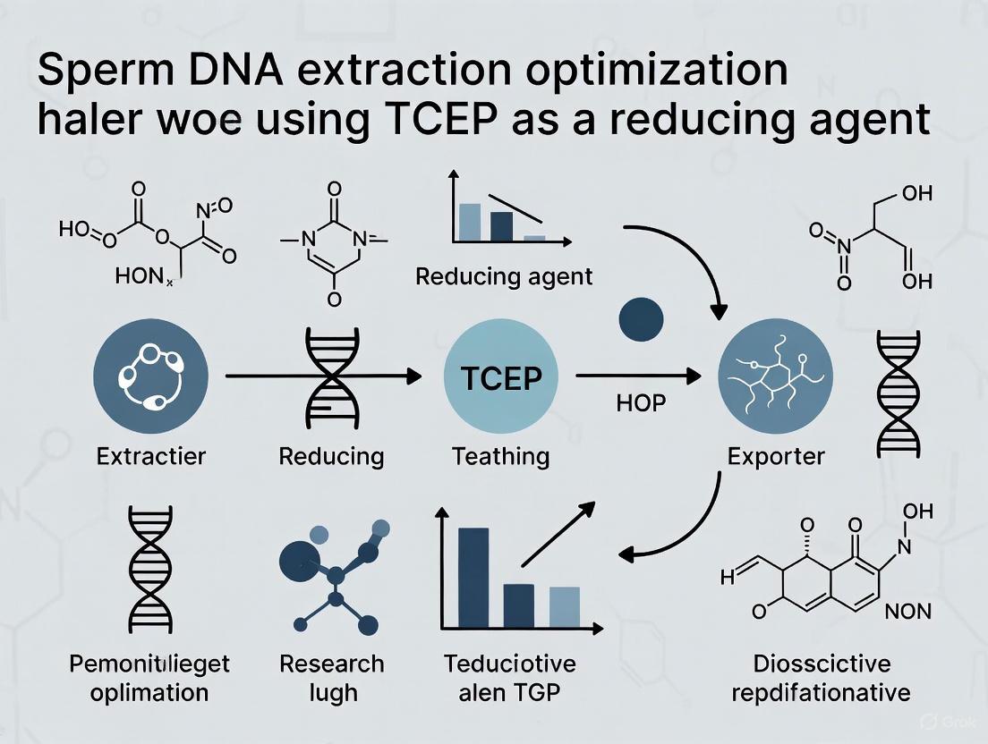
Tris(2-carboxyethyl)phosphine, commonly known as TCEP, is a powerful reducing agent extensively used in biochemical and molecular biology research. Its primary function is to break disulfide bonds (-S-S-) within and between proteins, facilitating their denaturation for analysis and purification [10] [11]. Unlike earlier reducing agents, TCEP is odorless, highly stable in aqueous solutions, and effective over a broad pH range, making it particularly valuable for sensitive applications [10].
In the context of sperm DNA and RNA extraction, the unique challenge lies in the highly compacted nature of the sperm nucleus. During spermatogenesis, histones are largely replaced by protamines, which form extensive inter- and intra-molecular disulfide bridges, creating a dense, chemically resistant structure [4]. This compaction protects the genetic material but makes it inaccessible to standard isolation techniques used for somatic cells. TCEP serves as a critical solution to this problem by efficiently reducing these disulfide bonds, thereby decondensing the chromatin and allowing for efficient nucleic acid extraction [2] [4].
Chemical Properties and Advantages
TCEP possesses several key chemical properties that make it superior to traditional reducing agents like dithiothreitol (DTT) or β-mercaptoethanol (BME) for many applications.
- Irreversibility: The reduction of disulfide bonds by TCEP is irreversible, as it proceeds to form a stable phosphine oxide [10]. This prevents reformation of disulfide bonds, a potential complication with DTT.
- Stability: TCEP is highly stable in air and aqueous solutions. It does not oxidize readily, which allows for long-term storage of stock solutions and more consistent performance in extended reactions [10] [11].
- Odorless Nature: Unlike DTT and especially BME, TCEP is odorless, improving the working environment and safety in the laboratory [10] [11].
- pH Range: It is active over a wide pH range (pH 1.5 to 8.5) and remains more stable than DTT at neutral and slightly basic pH, which is typical for many biological assays [10].
- Thiol-free: Since TCEP does not contain a thiol group, it does not need to be removed from a reaction mixture prior to downstream applications like cysteine labeling with maleimides, which can be inhibited by thiolated reducing agents [10].
Table 1: Comparison of TCEP with Common Reducing Agents
| Property | TCEP | DTT | β-Mercaptoethanol (BME) |
|---|---|---|---|
| Chemical Nature | Phosphine | Diol thiol | Thiol alcohol |
| Odor | Odorless | Faint, sulfidic | Strong, unpleasant |
| Reduction Mechanism | Irreversible | Reversible | Reversible |
| Stability in Air | High | Moderate | Low |
| Effective pH Range | Broad (1.5 - 8.5) | Best at pH > 7 | Best at pH > 7 |
| Interference with Maleimides | Low (non-thiol) | High (contains thiol) | High (contains thiol) |
Mechanism of Disulfide Bond Cleavage
The reduction of disulfide bonds by TCEP proceeds through a single-step SN2 nucleophilic substitution mechanism [12]. In this process:
- The nucleophilic phosphorus atom in TCEP attacks one of the sulfur atoms in the disulfide bond (R-S-S-R').
- This attack inverts the sulfur atom's configuration and cleaves the S-S bond.
- The reaction results in the formation of a mixed phosphonium cation intermediate and a free thiolate anion (R-S⁻).
- The phosphonium intermediate is rapidly hydrolyzed by water, releasing the second thiol (R'-SH) and the oxidized, stable form of TCEP (phosphine oxide) [10] [12].
This mechanism is highly efficient and selective for disulfide bonds, making TCEP a reliable reagent for breaking the cystine-rich protamine network in sperm cells, which is essential for accessing the tightly packed DNA [4].
TCEP in Sperm DNA Extraction: Protocols and Data
The integration of TCEP into sperm nucleic acid isolation protocols has significantly improved efficiency, yield, and purity. The following optimized protocol and supporting data highlight its critical role.
Optimized Sperm DNA Extraction Protocol with TCEP
This protocol is adapted from a rapid method for isolating high-quality sperm DNA, which eliminates the need for lengthy proteinase K digestions [4].
Materials:
- Isolated sperm cells (somatic cell contamination-free)
- Lysis Buffer: Commercially available guanidine thiocyanate-based buffer (e.g., Buffer RLT from Qiagen)
- TCEP-HCl (e.g., Pierce, #77720), prepared as a stock solution
- 0.2 mm stainless steel beads
- Silica-based spin columns (e.g., from QIAamp DNA Mini Kit or similar)
- Disruptor Genie or similar homogenizer
Method:
- Lysis Buffer Preparation: Add TCEP to the guanidine thiocyanate lysis buffer to a final concentration of 50 mM [4].
- Mechanical Homogenization: Transfer the isolated sperm cells to a tube containing the TCEP-supplemented lysis buffer and 0.2 mm steel beads. Homogenize for 5 minutes at room temperature using a Disruptor Genie [4].
- DNA Binding: Transfer the lysate to a silica-based spin column and centrifuge (e.g., 10,000 × g for 30 s) to bind the DNA.
- Washing and Elution: Perform wash steps as per the manufacturer's instructions for the spin column kit. Elute the DNA with a pre-heated (70°C) low-salt elution buffer (e.g., Buffer EB) to maximize yield [4].
Key Advantages of this Protocol:
- Speed: The combination of TCEP and mechanical homogenization reduces lysis time to minutes, replacing overnight incubations.
- Room-Temperature Processing: No need for elevated temperatures, simplifying the workflow.
- Stability: TCEP is stable at room temperature, and the lysate can be stored at room temperature for at least two weeks without degrading DNA yield or quality [4].
- High Yield and Purity: This method yields >90% high-quality DNA, free from somatic cell contamination [4].
Quantitative Data on TCEP Performance
Table 2: Impact of TCEP on Sperm Nucleic Acid Isolation Efficiency
| Parameter | Performance with TCEP | Comparison / Notes |
|---|---|---|
| DNA Yield | ~2.9 pg per cell [4] | Near-theoretical maximum (~3 pg per haploid cell). Yields stable after 2 weeks of lysate storage at room temperature [4]. |
| RNA Yield | Significantly increased [2] | TCEP enables complete lysis of sperm cells, which contain ~1000-10,000 times less RNA than somatic cells [2]. |
| Optimal Concentration | 50 mM (DNA isolation) [4] | Effective in breaking protamine disulfide bridges. A range of 10-50 mM was tested [4]. |
| Reduction Time | 5 minutes (with homogenization) [4] | Replaces traditional 2-hour to overnight incubations with DTT or BME. |
| DNA Methylation Integrity | Preserved [4] | Similar methylation levels at imprinted loci were observed in DNA isolated immediately or after 2 weeks of storage. |
The Scientist's Toolkit: Research Reagent Solutions
Table 3: Essential Reagents for TCEP-Based Sperm Nucleic Acid Isolation
| Reagent / Material | Function / Role | Example Product / Specification |
|---|---|---|
| TCEP-HCl | Breaking disulfide bonds in protamines for chromatin decondensation. | Pierce #77720; 0.5 M stock solution, pH 7.0 [4]. |
| Guanidine Thiocyanate Lysis Buffer | Chaotropic salt that lyses cells, inactivates nucleases, and denatures proteins. | Buffer RLT (Qiagen) [4]. |
| Silica-based Spin Columns | Selective binding and purification of nucleic acids from the lysate. | QIAamp DNA Mini Kit, AllPrep DNA/RNA Mini Kit [2] [4]. |
| Steel Beads (0.2 mm) | Mechanical disruption of the tough sperm cell membrane and wall. | 0.2 mm stainless steel beads, used with a homogenizer [4]. |
| Proteinase K (Optional) | Digests nuclear proteins; may not be required with efficient TCEP lysis. | For protocols not using mechanical homogenization [4]. |
Troubleshooting and FAQs
Q1: Why is my DNA yield from sperm cells still low after using TCEP?
- Incomplete Lysis: Ensure thorough mechanical homogenization. The sperm cell wall is exceptionally tough. The combination of TCEP and 0.2 mm steel beads for 5 minutes is critical for complete lysis [4].
- Somatic Cell Contamination: Sperm samples should be purified using a density gradient (e.g., Percoll) before lysis to remove somatic cells, which have different lysis properties and can contaminate the sperm DNA analysis [4].
- TCEP Concentration: Verify that the final concentration of TCEP in the lysis buffer is sufficient (50 mM is recommended). A lower concentration may not fully reduce the extensive disulfide network [4].
Q2: Can I substitute DTT for TCEP in my sperm DNA extraction protocol? While possible, it is not recommended. TCEP offers distinct advantages:
- Stability: TCEP is more stable in the lysis buffer and does not require fresh preparation for each use.
- Odor: TCEP is odorless, making the process more tolerable, especially in clinical settings [4].
- Efficiency: Protocols optimized with TCEP achieve complete lysis in minutes without the need for proteinase K or long incubations, which are often required with DTT [4].
Q3: I am isolating RNA from sperm. Will TCEP protect my RNA? Yes. TCEP is effective in RNA isolation protocols. It helps in the complete lysis of spermatozoa and stabilizes the RNA during the extraction process. One study identified a method using Triazol plus a silica-column kit supplemented with TCEP as the optimal protocol for obtaining high-quality RNA from bovine sperm [2].
Q4: How should I prepare and store a TCEP stock solution?
- Preparation: To prepare a 0.5 M stock solution, dissolve 5.73 g of TCEP-HCl in 35 ml of molecular biology grade water. The solution will be acidic (pH ~2.5). Neutralize to pH 7.0 using 10 N NaOH or KOH, then bring the final volume to 40 ml with water [10].
- Storage: Aliquot the solution and store at -20°C. Protect aliquots from light by wrapping tubes in aluminum foil. The stock solution is stable for at least 3 months under these conditions [10].
Q5: Are there any known side reactions of TCEP I should be aware of? Yes, under certain conditions, TCEP can cause side reactions:
- Backbone Cleavage: A side reaction that cleaves the protein backbone at cysteine residues under mild conditions has been reported, generating heterogeneous peptide fragments [13]. This is generally not a concern for DNA isolation but is critical for protein biochemistry.
- Reaction with Maleimides: Although TCEP does not contain a thiol, it can react with maleimide labeling reagents under certain conditions, such as acidic pH or high concentrations [10]. For precise cysteine labeling, consider removing TCEP via dialysis or desalting before adding the maleimide.
Workflow Diagram: Sperm DNA Extraction with TCEP
FAQ: Troubleshooting Common Lysis Problems
What are the immediate signs of incomplete lysis during sperm DNA extraction? Incomplete lysis is often indicated by a viscous or turbid lysate, low DNA yield after purification, and the presence of insoluble material or tissue fibers in the solution. A clogged silica membrane during column-based purification is another common symptom, as undigested cellular debris physically blocks the matrix [14].
Why does incomplete lysis lead to DNA degradation, especially in sperm samples? Incomplete lysis fails to rapidly inactivate endogenous nucleases (DNases). In tissues with high nuclease content, these enzymes remain active and degrade DNA before it can be purified. Sperm cells are particularly challenging due to their highly compacted nuclear proteins and resistant cell membranes, which can slow lysis and extend the window for nuclease activity [14].
How can incomplete lysis cause data contamination in downstream analyses? Incomplete lysis can result in cross-contamination between samples during batch processing. When cellular debris is not fully removed, it can carry over DNA from one sample to another. Furthermore, inefficient lysis of sperm cells can lead to an underrepresentation of the target DNA, making the sample more susceptible to being overwhelmed by low-level contaminants from reagents or the laboratory environment, which is a critical concern in sensitive applications like next-generation sequencing [15] [16].
What is the role of TCEP in optimizing sperm cell lysis? Tris(2-carboxyethyl) phosphine (TCEP) is a potent reducing agent that cleaves disulfide bonds. In sperm cells, the nucleus contains protamines that are heavily cross-linked by disulfide bonds, creating a highly compact structure that is resistant to digestion. The addition of TCEP to the lysis buffer effectively reduces these bonds, facilitating the decondensation of the sperm nucleus and allowing proteases (like Proteinase K) better access to digest nuclear proteins, thereby releasing high-quality, high-molecular-weight DNA [17].
What are the best practices for storing samples to prevent issues before lysis? To prevent DNA degradation and ensure successful lysis, samples should be flash-frozen in liquid nitrogen or on dry ice and stored at -80°C. For shorter periods, storage at 4°C is acceptable, but samples should not be stored long-term at -20°C without stabilizers. The use of DNA stabilizing reagents is highly recommended to inhibit nuclease activity. When using blood, EDTA is the preferred anticoagulant; heparin should be avoided as it can inhibit downstream PCR [14] [18].
Troubleshooting Guide: Low Yield, Degradation, and Contamination
The following table outlines common problems, their causes, and solutions specifically related to the lysis step in DNA extraction.
| Problem | Primary Cause | Recommended Solution |
|---|---|---|
| Low DNA Yield | Incomplete cell lysis releasing insufficient DNA [18]. | Increase incubation time with lysis buffer; use a more aggressive lysing matrix or bead beating [18]. |
| Lysis conditions insufficient for tough sperm nuclei [17]. | Incorporate TCEP (e.g., 120 g/L in PBS) into the lysis buffer to reduce protamine disulfide bonds [17]. | |
| Column clogging from cellular debris [14]. | Centrifuge lysate post-lysis to remove insoluble fibers/debris before binding to column [14]. | |
| DNA Degradation | Sample not stored properly, allowing nucleases to act [14]. | Flash-freeze samples immediately after collection; store at -80°C; use nuclease-inhibiting stabilizers [14] [18]. |
| Lysis too slow, failing to inactivate nucleases quickly [14]. | Grind or finely cut sample before lysis; ensure lysis buffer is in excess and properly formulated [19] [14]. | |
| Data Contamination | Cross-contamination between samples during processing [15]. | Implement physical segregation of pre- and post-PCR areas; use dedicated equipment and PPE; automate sample handling [15]. |
| Contaminating DNA from reagents or environment [16]. | Use certified DNA-free consumables; include negative control samples (lysis buffer only) to monitor for contaminants [16]. |
Experimental Protocol: Optimized Sperm DNA Extraction with TCEP
Methodology for Total DNA Extraction from Sperm Cells
This protocol is designed for the extraction of high-molecular-weight genomic DNA from sperm cells, with optimized lysis using TCEP to ensure high yield and purity for downstream applications such as next-generation sequencing.
Reagents and Materials:
- Lysis Buffer: (e.g., containing CTAB, SDS, or commercial lysis reagents)
- TCEP Solution: 120 g/L in Phosphate-Buffered Saline (PBS) [17]
- Proteinase K
- RNase A
- Washing Buffers (e.g., guanidine hydrochloride-based)
- Elution Buffer: TE buffer or nuclease-free water
Procedure:
- Sample Preparation: Isolate sperm cells from seminal fluid. If starting with frozen pellets, thaw slowly on ice and resuspend gently in cold PBS.
- Lysis: Transfer up to 50 µL of sperm cell suspension to a clean tube. Add 10 µL of TCEP solution (120 g/L in PBS) [17]. Mix thoroughly by vortexing.
- Enzymatic Digestion: Add Proteinase K and RNase A to the sample mixture. Mix well by pipetting to ensure uniform digestion.
- Incubation: Incubate the mixture at the optimized temperature. Critical Step: Conduct the TCEP reduction on ice for 30-60 minutes to minimize the production of interference compounds and maximize the reduction of disulfide bonds [17].
- Deproteinization: Add a volume of Trichloroacetic Acid (TCA) solution (e.g., 100 g/L) or an equivalent deproteinization agent to the lysate. Centrifuge at high speed (e.g., 15,000 ×g for 10 min at 4°C) to pellet precipitated proteins and cellular debris.
- DNA Binding and Washing: Transfer the cleared supernatant to a silica membrane column or proceed with magnetic bead-based purification. Wash the bound DNA according to the manufacturer's instructions.
- Elution: Elute the purified DNA in a low-ionic-strength buffer such as TE buffer or nuclease-free water.
Workflow Diagram: Optimized DNA Extraction Process
Diagram Title: Optimized Sperm DNA Extraction with TCEP
The Scientist's Toolkit: Essential Research Reagents
Research Reagent Solutions for DNA Extraction
| Reagent | Function in Lysis | Key Consideration |
|---|---|---|
| TCEP (Tris(2-carboxyethyl) phosphine) | Reduces disulfide bonds in sperm protamines, enabling nuclear decondensation and efficient DNA release [17]. | Perform reduction on ice to minimize generation of interfering compounds. Reaction is typically complete within 30-60 minutes [17]. |
| Proteinase K | Broad-spectrum serine protease that digests nucleases and structural proteins, inactivating them and liberating DNA. | Requires extended digestion time (30 min to 3 hours) for fibrous tissues; always add before lysis buffer to ensure proper mixing [14]. |
| CTAB (Cetyltrimethylammonium bromide) | Detergent that effectively lyses cells and complexes with polysaccharides and phenolic compounds, preventing their coprecipitation with DNA [20]. | Essential for plant extractions; helps remove contaminants that inhibit downstream enzymatic reactions [20]. |
| Guanidine Salts | Chaotropic agent that denatures proteins, inactivates nucleases, and enables DNA binding to silica matrices [21]. | Is a component of many commercial kits; can cause salt carry-over if columns are mishandled during washing [14]. |
| Silica Membranes/Magnetic Beads | Solid-phase matrix that binds DNA in the presence of high-salt chaotropic agents, allowing impurities to be washed away [21]. | Binding capacity must not be exceeded; overloading leads to low yield and contamination. Bead beating can enhance lysis efficiency [19] [18]. |
Step-by-Step: Optimized Protocols for Sperm DNA and RNA Extraction Using TCEP
Troubleshooting Guide
This guide addresses common issues encountered during the rapid silica-column based DNA purification from sperm cells using TCEP lysis. Use the tables below to diagnose and resolve problems related to yield, purity, and downstream applications.
Low DNA Yield or Inefficient Lysis
| Problem & Cause | Solution |
|---|---|
| Incomplete cell lysis: The unique, highly compacted nature of sperm DNA, cross-linked by disulfide bridges, is not fully disrupted. | Ensure the lysis buffer contains 50 mM TCEP for effective disulfide bond reduction [4]. Incorporate a 5-minute mechanical homogenization step with 0.2 mm steel beads; do not skip or shorten this step [4]. |
| Reducing agent degradation: The potency of the reducing agent is compromised. | Use tris(2-carboxyethyl)phosphine (TCEP); it is odorless and stable at room temperature, unlike DTT or β-mercaptoethanol [4]. For traditional agents, prepare fresh lysis buffer for each use. |
| DNA binding issues: Suboptimal conditions prevent DNA from binding to the silica column. | Ensure the lysate is mixed with the appropriate volume of binding buffer (e.g., Buffer AL for QIAamp kits) [4]. Avoid overloading the column; for very high cell counts, split the sample [22]. Pipette the lysate/buffer mix carefully onto the center of the membrane without touching the upper column area [22]. |
| Incomplete elution: DNA remains bound to the silica membrane. | Elute with a low-ionic-strength buffer (e.g., TE buffer or nuclease-free water) preheated to 70°C [4] [21]. Incubate the column with elution buffer at room temperature for 3-5 minutes before centrifuging [4]. |
Poor DNA Purity and Quality
| Problem & Cause | Solution |
|---|---|
| Protein contamination: Incomplete digestion or removal of nuclear proteins. | The TCEP-based protocol eliminates lengthy Proteinase K digestions [4]. If contamination persists, consider adding a short Proteinase K digestion step post-lysis [23]. Ensure all wash buffers are prepared with the correct ethanol concentration [21]. |
| Salt or reagent carryover: Chaotropic salts from the lysis or wash buffers are not fully removed. | Perform the recommended number of wash steps with the provided wash buffer [4]. During wash steps, close column caps gently to avoid splashing and do not move samples abruptly [22]. Let the column stand for 1 minute after adding the final wash buffer, then spin and discard the flow-through. Consider an additional spin with an empty column to dry the membrane [22]. |
| RNA contamination: RNA may co-purify with DNA. | Add RNase A to the elution buffer or during the lysis step to degrade contaminating RNA [23] [21]. |
| Guanidine salt contamination: This is a common cause of low A260/A230 ratios. | The most common cause is allowing the lysate/binding buffer mixture to contact the upper column area or cap. Pipet carefully directly onto the silica membrane [22]. |
Problems with Downstream Applications
| Problem & Cause | Solution |
|---|---|
| Inhibitors in eluted DNA: Carryover of contaminants inhibits enzymes in PCR or other assays. | Use the provided wash buffers and ensure the final eluate is free of ethanol [22]. If problems persist, elute in a buffered solution like TE (10 mM Tris-HCl, 0.1 mM EDTA, pH 8.0) instead of water to stabilize the DNA [21]. |
| Degraded DNA: The extracted DNA is fragmented. | Sperm cells have high DNase content. Ensure samples are processed immediately or stored correctly. Flash-freeze samples in liquid nitrogen and store at -80°C if not processed immediately [22]. The guanidine thiocyanate in the lysis buffer helps inactivate nucleases [4] [24]. |
| Poor methylation analysis results: The extraction method damages epigenetic marks. | The presented TCEP-based protocol has been validated for DNA methylation analyses at imprinted loci, showing no adverse effects compared to traditional methods [4]. |
Frequently Asked Questions (FAQs)
Q1: Why is TCEP used in this protocol instead of the more common DTT? TCEP (tris(2-carboxyethyl)phosphine) is a superior reducing agent for breaking the disulfide bonds in sperm protamines. It is odorless, more stable at room temperature than DTT or β-mercaptoethanol, and does not require fresh preparation for each use. This enhances both the user experience and the reliability of the protocol [4].
Q2: Can the homogenized lysate be stored before completing the DNA purification? Yes, a significant advantage of this protocol is that the nucleic acids are stabilized after the initial homogenization in the guanidine thiocyanate lysis buffer. Research shows that DNA yields and quality (including for methylation analyses) remain high even after storing the homogenate at room temperature for up to two weeks [4].
Q3: My DNA concentration seems low according to spectrophotometry, but my PCR works fine. Why? Spectrophotometry can sometimes underestimate concentration or purity due to minor contaminants that do not significantly inhibit PCR. For a more accurate assessment of DNA quality and quantity, use a fluorescence-based quantitation method (e.g., Qubit) and check integrity by agarose gel electrophoresis, which will show a tight, high-molecular-weight band for high-quality sperm DNA [4] [21].
Q4: Is this protocol suitable for other mammalian species? Yes, the fundamental principle of using TCEP and mechanical homogenization to disrupt the resistant sperm chromatin is applicable across mammalian species. The protocol has been designed to be adaptable, though optimization of input cell numbers may be necessary for different species [4].
Q5: What is the role of the steel beads in the protocol? The 0.2 mm stainless steel beads are used in a 5-minute mechanical homogenization step to physically disrupt the tough sperm cell membrane and highly compacted nucleus. This step is crucial for achieving efficient lysis and high DNA yields, replacing or reducing the need for prolonged enzymatic digestion [4].
Experimental Workflow and Reagents
Protocol Workflow Diagram
The following diagram illustrates the complete experimental workflow for rapid sperm DNA purification.
Research Reagent Solutions
The table below lists the key reagents and materials required for the successful execution of this protocol, along with their specific functions.
| Reagent/Material | Function in the Protocol |
|---|---|
| TCEP (tris(2-carboxyethyl)phosphine) | Odorless, stable reducing agent that breaks disulfide bonds between protamines, enabling DNA decondensation [4]. |
| Guanidine Thiocyanate Lysis Buffer (e.g., Buffer RLT) | Chaotropic salt that disrupts cells, inactivates nucleases, and facilitates subsequent binding of DNA to silica [4] [24]. |
| 0.2 mm Stainless Steel Beads | Used for mechanical homogenization to physically disrupt the tough sperm cell membrane and compacted nucleus [4]. |
| Silica-Based Spin Columns | The solid matrix to which DNA binds in the presence of chaotropic salts, allowing for purification and concentration [4] [23]. |
| Ethanol-Based Wash Buffers | Used to remove salts, proteins, and other contaminants from the silica membrane without eluting the bound DNA [4] [21]. |
| Low-Salt Elution Buffer (e.g., TE Buffer or Water) | Disrupts the interaction between DNA and the silica membrane, releasing purified DNA in a ready-to-use solution [4] [21]. |
Experimental Protocol and Workflow
Detailed Step-by-Step Methodology
This protocol describes an efficient method for extracting high-quality RNA from spermatozoa by supplementing TRIzol with the reducing agent Tris(2-carboxyethyl)phosphine (TCEP) to overcome the extreme lysis resistance of sperm cells [2] [25].
Key Reagents and Solutions:
- TRIzol reagent
- TCEP (50 mM stock solution)
- Chloroform
- Isopropanol
- 75% Ethanol (in nuclease-free water)
- Nuclease-free water
- Glycogen (optional carrier for low-yield samples)
Procedure:
Sperm Preparation and Somatic Cell Removal
- Purify sperm cells using a discontinuous density gradient (e.g., 90% gradient) to remove somatic cell contamination [4].
- Wash sperm pellet and resuspend in appropriate buffer. Visually inspect for somatic cell contamination.
Cell Lysis and Homogenization
- Add 1 mL TRIzol reagent supplemented with 50 mM TCEP per 5-10 million sperm cells [2] [25].
- Homogenize using 0.2 mm stainless steel beads for 5 minutes at room temperature using a mechanical homogenizer [4].
- Incubate the homogenate at room temperature for 5 minutes for complete dissociation of nucleoprotein complexes.
Phase Separation
- Add 0.2 mL chloroform per 1 mL of TRIzol used.
- Cap samples securely and vortex vigorously for 15 seconds.
- Incubate at room temperature for 2-3 minutes.
- Centrifuge at 12,000 × g for 15 minutes at 4°C.
RNA Precipitation
- Transfer the colorless upper aqueous phase to a fresh tube without disturbing the interphase or organic layer.
- Add 0.5 mL isopropanol per 1 mL TRIzol originally used.
- For samples with expected low RNA yield, include 1-10 μL glycogen (20 mg/mL) as a carrier [26] [27].
- Mix by inversion and incubate at room temperature for 10 minutes.
- Centrifuge at 12,000 × g for 10 minutes at 4°C.
RNA Wash and Resuspension
- Remove supernatant carefully without disturbing the RNA pellet.
- Wash pellet with 1 mL 75% ethanol by vortexing.
- Centrifuge at 7,500 × g for 5 minutes at 4°C.
- Air-dry pellet for 5-10 minutes (do not overdry).
- Resuspend RNA in 20-50 μL nuclease-free water by pipetting.
- Heat at 55-60°C for 10-15 minutes to aid dissolution, with periodic pipette mixing [28].
Experimental Workflow Diagram
Troubleshooting Guide
Common Issues and Solutions
Problem: Low or No RNA Yield
- Cause: Incomplete sperm cell lysis due to insufficient reducing agent or homogenization [25].
- Solution: Ensure TCEP concentration is 50 mM in TRIzol. Verify mechanical homogenization with steel beads for complete lysis [4].
- Cause: RNA pellet loss during precipitation, particularly with low starting material [26].
- Solution: Add glycogen (10-20 μg) as carrier during isopropanol precipitation. Avoid decanting; carefully pipette supernatant to prevent disturbing invisible pellets [26] [27].
Problem: DNA Contamination in RNA Prep
- Cause: Aqueous phase contamination with interphase during phase separation [27].
- Solution: Carefully transfer only the clear aqueous phase without disturbing interphase. Use silica-based columns with DNase treatment if contamination persists [2].
Problem: Abnormal Aqueous Phase Color (Yellow, Brown, or Pink)
- Cause: Sample-specific interference from lipids (skin), hemoglobin (blood-rich samples), or overdilution [26] [27].
- Solution: Centrifuge homogenate before chloroform addition to remove lipid layers. For blood-rich samples, include a PBS pre-wash step. Maintain sample-to-TRIzol ratio ≤ 1:10 [26].
Problem: Degraded RNA Quality
- Cause: RNase activity due to improper sample handling or insufficient lysis [28].
- Solution: Process samples immediately or flash-freeze in liquid nitrogen. Ensure complete homogenization in TRIzol + TCEP. Store isolated RNA at -80°C [28].
Problem: Poor RNA Solubility or Abnormal Pellet Appearance
- Cause: Overdrying of RNA pellet or contamination with polysaccharides [28] [27].
- Solution: Air-dry pellet until just translucent (not completely dry). For polysaccharide-rich contaminants, use high-salt precipitation (0.25 mL 0.8M sodium citrate + 1.2M NaCl per 1 mL TRIzol) during isopropanol step [27].
Quantitative Performance Data
Table 1: Performance Comparison of Reducing Agents in Sperm RNA Extraction
| Reducing Agent | Concentration | RNA Yield Improvement | pH Compatibility | Key Advantages |
|---|---|---|---|---|
| TCEP | 50 mM | 100-fold increase [25] | Effective at acidic pH (TRIzol) | Odorless, room temperature stable, maintains reducing capability in acidic conditions [25] [4] |
| β-mercaptoethanol | 0.2-2% | Limited in TRIzol [25] | pH-dependent (ineffective in TRIzol) | Standard reducing agent, but ineffective in acidic chaotropic solutions [25] |
| DTT | 150 mM | Variable | pH-dependent | Traditional sperm lysis agent, but odor and stability limitations [4] |
Table 2: Troubleshooting Guide for Common Experimental Issues
| Problem | Possible Cause | Solution | Prevention |
|---|---|---|---|
| No visible RNA pellet | Low RNA content (<1000 cells) | Use glycogen carrier (10-20 μg); precipitate at 4°C for 30 min [26] | Include carrier during precipitation |
| DNA contamination | Aqueous phase contamination | DNase treatment; silica column purification [2] | Careful phase separation; avoid interphase |
| Low A260/A280 ratio (<1.65) | Phenol contamination | Reprecipitate with ethanol; centrifuge at 4°C [27] | Maintain cold temperature during phase separation |
| High A260/A280 ratio (>2.0) | RNA degradation | Check sample integrity; ensure immediate processing [28] | Flash-freeze samples; minimize thaw cycles |
| Colored aqueous phase | Hemoglobin or pigments | Pre-wash samples; increase TRIzol volume [26] | Maintain proper sample:TRIzol ratios (1:10) |
Research Reagent Solutions
Essential Materials and Reagents
Table 3: Key Research Reagents for TCEP-Supplemented TRIzol Protocol
| Reagent/Equipment | Function/Application | Specifications | Alternative/Notes |
|---|---|---|---|
| TCEP | Reducing agent breaks disulfide bonds in sperm protamines [25] | 50 mM in TRIzol; stable at room temperature; effective at acidic pH | Superior to DTT/βME in TRIzol due to pH stability [25] |
| TRIzol | Monophasic lysis reagent: phenol + guanidine isothiocyanate [26] | Maintains RNA integrity while denaturing proteins | Acidic pH (~5.0) keeps RNA in aqueous phase [26] |
| Mechanical homogenizer | Complete sperm cell disruption | With 0.2 mm stainless steel beads, 5 min [4] | Essential for tough sperm cell walls |
| Glycogen | RNA carrier for precipitation | 10-20 μg during isopropanol step [26] | Improves yield with low starting material |
| Silica-based columns | Optional RNA purification | With DNase treatment step [2] | Removes DNA contamination; improves purity |
| Chloroform | Phase separation | 0.2 mL per 1 mL TRIzol [25] | Critical for separating RNA (aqueous) from DNA/protein |
Technical FAQs
Q1: Why is TCEP specifically recommended over traditional reducing agents like DTT or β-mercaptoethanol? A: TCEP maintains its reducing activity at the acidic pH of TRIzol (approximately pH 5), whereas the efficiency of DTT and β-mercaptoethanol is pH-dependent and significantly reduced in acidic environments. This allows TCEP to effectively break the disulfide bonds between protamines in sperm heads, enabling complete lysis and significantly improving RNA yield—by up to 100-fold compared to standard protocols [25]. Additionally, TCEP is odorless and stable at room temperature, making it more convenient for laboratory use [4].
Q2: What is the typical RNA yield expected from this protocol? A: Sperm cells contain approximately 1,000 to 10,000 times less RNA than typical mammalian somatic cells [2]. The TCEP-supplemented TRIzol method optimizes recovery from these challenging samples. Yields are cell number-dependent, but the protocol consistently recovers significantly more RNA than methods without TCEP or with traditional reducing agents [25].
Q3: How critical is the mechanical homogenization step with steel beads? A: Essential. Sperm cells are exceptionally resistant to chemical lysis alone due to their compact, disulfide-crosslinked structure. The combination of chemical reduction (TCEP) and mechanical disruption is necessary for complete and efficient lysis. Homogenization with 0.2 mm steel beads for 5 minutes ensures thorough breakdown of the sperm cell walls and nuclei [4].
Q4: My aqueous phase appears colored after phase separation. Is my RNA sample compromised? A: Not necessarily. A yellowish, brownish, or pinkish hue can occur with samples rich in lipids (e.g., from certain tissues) or hemoglobin (from blood contamination) [26] [27]. If the color is slight, proceed with the protocol. If the color is intense, you can centrifuge the initial homogenate before adding chloroform to remove some contaminants. The RNA quality should be verified by spectrophotometry and electrophoresis after isolation [27].
Q5: How should I store the isolated sperm RNA, and what quality control methods are recommended? A: Resuspend purified RNA in nuclease-free water and store at -80°C for long-term preservation. For quality control, use spectrophotometry (A260/A280 ratio ~1.8-2.1) and analyze integrity with a Bioanalyzer or similar system. Additionally, perform RT-PCR with sperm-specific markers like Protamine 1 (PRM1) to confirm the absence of somatic cell RNA contamination and the presence of authentic sperm transcripts [2].
Mechanism and Validation
Scientific Basis of the Protocol
Mechanism of TCEP Enhancement:
The unique effectiveness of TCEP in this protocol stems from its chemical properties. Sperm cell resistance arises from extensive disulfide bonding between protamine proteins that tightly package the DNA [4]. Traditional reducing agents like β-mercaptoethanol lose efficacy in acidic environments like TRIzol, while TCEP remains active across a wide pH range, enabling complete sperm head lysis and RNA liberation [25].
Experimental Validation: The optimized protocol has been validated through multiple approaches:
- Yield Assessment: Demonstrates 100-fold increase in RNA yield compared to standard TRIzol protocol [25].
- Purity Verification: PCR analysis using specific markers (Protamine 1 for sperm cells; CDH1 for epithelial cells; KIT for germ cells; PTPRC for leukocytes) confirms isolation of pure sperm RNA without somatic cell contamination [2].
- Downstream Application: Isolated RNA performs effectively in reverse transcription and PCR, confirming suitability for advanced transcriptomic analyses including RNA sequencing and microarray studies [25] [29].
This TCEP-supplemented TRIzol method represents a significant advancement for reproductive biology research, enabling reliable RNA extraction from one of the most challenging cell types for molecular analysis.
FAQs and Troubleshooting Guides
Why is sperm sample preparation so challenging?
Sperm DNA is packaged differently from somatic cells. During spermatogenesis, 90% of histones are replaced by protamines, creating a compact structure reinforced by disulfide bonds that protects DNA but makes it resistant to standard DNA isolation techniques [4].
How can I effectively remove somatic cell contamination from sperm samples?
Somatic cell contamination can significantly skew epigenetic and genetic analyses. A multi-pronged approach is recommended:
- Microscopic examination: Visually inspect samples before and after processing.
- Somatic Cell Lysis Buffer (SCLB): Treat samples with SCLB (0.1% SDS, 0.5% Triton X-100) for 30 minutes at 4°C [30].
- Density gradient centrifugation: Use media like PureSperm or Percoll to separate sperm from somatic cells based on density [31].
- Epigenetic biomarkers: Utilize known methylation markers (9,564 identified CpG sites) to detect residual contamination [30].
What are the advantages of using TCEP over traditional reducing agents?
TCEP (tris(2-carboxyethyl)phosphine) offers significant benefits for sperm DNA extraction:
| Characteristic | TCEP | DTT | β-mercaptoethanol |
|---|---|---|---|
| Odor | Odorless [32] | Strong odor [4] | Strong, unpleasant odor [4] |
| pH Stability | Wide range (pH 1.5-8.5), stable at biological pH [32] | Less stable at pH >7.5 [32] | Less stable |
| Reaction | Irreversible [32] | Reversible [32] | Reversible |
| Storage | Stable in aqueous solution at room temperature [4] | Requires fresh preparation [4] | Requires fresh preparation |
| Reduction Efficiency | Effective for breaking sperm protamine disulfide bonds [4] | Effective but requires longer incubation [4] | Effective but requires longer incubation |
How do I optimize sperm DNA extraction using TCEP?
A rapid, efficient protocol for sperm DNA extraction using TCEP includes:
- Homogenization: Use 0.2 mm steel beads with TCEP-supplemented lysis buffer for 5 minutes at room temperature [4].
- Lysis buffer: Guanidine thiocyanate-based buffer (e.g., Buffer RLT) with 50 mM TCEP [4].
- DNA binding: Use silica-based spin columns for DNA purification [4].
- Eliminate Proteinase K: The combination of mechanical disruption and TCEP can remove the need for lengthy Proteinase K digestions [4].
How much DNA yield can I expect from sperm cells?
The table below summarizes expected DNA yields from optimized protocols:
| Sample Condition | Expected Yield | Protocol Details |
|---|---|---|
| Immediate Isolation | 2.84 ± 0.04 pg/cell [4] | Homogenization with 50 mM TCEP, silica column purification |
| After 2 Weeks Storage | 2.91 ± 0.13 pg/cell [4] | Lysate stored at room temperature in TCEP-containing buffer |
| Theoretical Maximum | ~3 pg/cell [4] | Based on haploid genome content |
How does somatic cell contamination affect sperm epigenetic data?
Even low levels of somatic cell contamination can significantly distort results because sperm and somatic cells have dramatically different methylation patterns. Studies recommend applying a 15% cutoff during data analysis to completely eliminate the influence of somatic DNA contamination in sperm epigenetic studies [30].
Troubleshooting Common Problems
Low DNA Yield
| Problem | Possible Cause | Solution |
|---|---|---|
| Incomplete cell lysis | Insufficient disruption of protamine disulfide bonds | Increase TCEP concentration to 50 mM; add mechanical homogenization [4] |
| Sample degradation | Improper sample storage or handling | Flash-freeze samples in liquid nitrogen; store at -80°C; use stabilizing reagents [33] |
| Column overload | Too much starting material | Reduce input amount, particularly for DNA-rich tissues [33] |
DNA Degradation
| Problem | Possible Cause | Solution |
|---|---|---|
| Nuclease activity | High DNase content in certain tissues | Process samples quickly; keep frozen and on ice during preparation [33] |
| Large tissue pieces | Slow penetration of lysis reagents | Cut tissue to smallest possible pieces; use liquid nitrogen grinding [33] |
| Old samples | Progressive DNA degradation over time | Use fresh samples; proper storage at -80°C with stabilizers [33] |
Contamination Issues
| Problem | Possible Cause | Solution |
|---|---|---|
| Somatic DNA contamination | Incomplete removal of somatic cells | Implement SCLB treatment; use density gradient centrifugation; check biomarkers [30] |
| Protein contamination | Incomplete digestion | Ensure adequate lysis time; remove indigestible fibers by centrifugation [33] |
| Salt contamination | Guanidine salt carryover | Avoid touching upper column area with pipet tip; close caps gently to prevent splashing [33] |
Experimental Protocols
Optimized Sperm DNA Extraction with TCEP
Materials Needed:
- Buffer RLT (guanidine thiocyanate-based lysis buffer)
- TCEP solution (50 mM final concentration)
- 0.2 mm stainless steel beads
- Disruptor Genie or similar homogenizer
- Silica-based spin columns (e.g., QIAamp DNA Mini Kit)
- Ethanol (96-100%)
- Elution buffer (e.g., Buffer EB)
Procedure:
- Isolate sperm cells using density gradient centrifugation to remove somatic contamination [4].
- Aliquot 20-100 million sperm cells into a tube containing 0.1 g of 0.2 mm steel beads [4].
- Add 500 µL of Buffer RLT supplemented with 50 mM TCEP [4].
- Homogenize for 5 minutes at room temperature using a Disruptor Genie [4].
- Transfer lysate to a silica spin column and centrifuge at 10,000 × g for 30 seconds [4].
- Wash with appropriate wash buffers per manufacturer's instructions.
- Elute DNA with preheated (70°C) elution buffer, incubating for 3 minutes before centrifugation [4].
- Repeat elution for maximum yield.
Somatic Cell Removal Protocol
Materials Needed:
- Somatic Cell Lysis Buffer (SCLB): 0.1% SDS, 0.5% Triton X-100 in ddH₂O
- Phosphate-buffered saline (PBS)
- Density gradient medium (PureSperm or Percoll)
- Centrifuge
Procedure:
- Wash fresh semen samples twice with PBS by centrifugation at 200 × g for 15 minutes at 4°C [30].
- Inspect under microscope to identify somatic cell contamination level [30].
- Incubate with freshly prepared SCLB for 30 minutes at 4°C [30].
- Re-check under microscope for somatic cells.
- If somatic cells remain, repeat SCLB treatment.
- For additional purification, layer sample over 40% PureSperm or 45%/90% Percoll gradient [31].
- Centrifuge to separate mature sperm (pellet) from somatic cells (interface) [31].
- Wash sperm pellet with PBS for highly pure sperm population [30].
Research Reagent Solutions
| Reagent/Category | Specific Examples | Function in Sperm Preparation |
|---|---|---|
| Reducing Agents | TCEP (50 mM) [4], DTT (150 mM) [4], β-mercaptoethanol (2%) [4] | Break disulfide bonds between protamines for DNA accessibility |
| Lysis Buffers | Guanidine thiocyanate buffers [4], DNA/RNA Shield [4], Triazol [2] | Disrupt cell membranes, inactivate nucleases, denature proteins |
| Purification Kits | AllPrep DNA/RNA Mini Kit [4], QIAamp DNA Mini Kit [4], Quick-gDNA MiniPrep [4] | Silica-based DNA binding and purification |
| Somatic Cell Removal | Somatic Cell Lysis Buffer [30], PureSperm [31], Percoll [31] | Selective removal of contaminating non-sperm cells |
| Storage Solutions | Cryoprotectants (DMSO, glycerol) [34], DNA stabilization buffers [4] | Maintain sample integrity during storage |
Sperm Sample Preparation Workflow
Somatic Cell Contamination Identification
Determining Optimal TCEP Concentration and Incubation Conditions for Your Sample Type
Welcome to the Technical Support Center for sperm cell nucleic acid isolation. The unique challenge in working with sperm cells lies in their highly compacted nuclear structure, where disulfide bridges between protamines render the cell resistant to standard lysis techniques. This guide provides detailed, evidence-based troubleshooting advice for optimizing the use of Tris(2-carboxyethyl)phosphine (TCEP) in your protocols, enabling efficient and high-quality DNA and RNA extraction from spermatozoa.
Frequently Asked Questions (FAQs)
1. Why is TCEP preferred over DTT or β-mercaptoethanol for sperm cell lysis? TCEP offers several advantages for breaking the disulfide bonds in sperm heads. It is odorless, more stable in aqueous solutions at a wide pH range (pH 1.5-8.5), and its reduction reaction is irreversible. Critically, it remains active in acidic chaotropic solutions like Trizol, where DTT and βME lose effectiveness, ensuring complete lysis of the sperm cell [35] [36].
2. What is the typical working concentration range for TCEP? Based on optimized protocols, the effective final concentration of TCEP in the lysis buffer typically ranges from 10 mM to 100 mM [2] [4] [35]. The optimal concentration can depend on your specific sample type and starting material.
3. Is mechanical homogenization necessary when using TCEP? While TCEP is highly effective at chemical lysis, some protocols combine it with brief mechanical homogenization (e.g., 5 minutes with steel beads) to ensure complete and rapid disruption of sperm cells, leading to higher and more consistent nucleic acid yields [4].
4. Can I use TCEP for both DNA and RNA extraction from sperm? Yes. TCEP has been successfully implemented in protocols for isolating both high-quality DNA and RNA from spermatozoa, making it a versatile reducing agent for various downstream genetic and epigenetic analyses [2] [4] [35].
Troubleshooting Guides
Problem: Low Nucleic Acid Yield
Potential Causes and Solutions:
- Insufficient Lysis: Sperm heads remain intact due to inadequate reduction of disulfide bonds.
- Solution: Ensure the final TCEP concentration is at least 50 mM. For highly resistant samples, increase the concentration to 100 mM [35]. Verify that the TCEP stock solution was prepared correctly and stored properly at -20°C.
- Suboptimal Lysis Buffer:
- Inadequate Incubation:
- Solution: While TCEP works rapidly, ensure the sample is thoroughly mixed and incubated for a sufficient time. Protocols often use a short 5-minute incubation at room temperature when combined with homogenization [4].
Problem: Poor Nucleic Acid Purity
Potential Causes and Solutions:
- Carryover of Contaminants:
- Solution: If using silica columns, perform all recommended wash steps. For phenol-chloroform extraction, take care not to transfer the interphase. The use of TCEP itself does not introduce contaminants that affect purity [4].
Problem: Inconsistent Results Between Samples
Potential Causes and Solutions:
- Somatic Cell Contamination: Semen contains secretions and somatic cell types which can contaminate the sperm nucleic acid profile.
Experimental Data and Protocols
The table below summarizes key parameters from established methods to help you design your experiment.
Table 1: TCEP Usage in Sperm Nucleic Acid Isolation Protocols
| Nucleic Acid | Sample Type | Lysis Buffer | TCEP Concentration | Incubation | Key Finding | Source |
|---|---|---|---|---|---|---|
| RNA | Bovine Spermatozoa | Triazol + silica column | Optimized | Not Specified | Method with TCEP judged best for yield and purity. | [2] |
| DNA | Human Spermatozoa | Guanidine thiocyanate | 10 mM - 50 mM | 5 min homogenization (room temp) | Yielded >90% high-quality DNA; stable for 2 weeks at room temp. | [4] |
| RNA | Mouse Spermatozoa | Trizol | 100 mM | Not Specified | Enabled complete lysis, increasing RNA yield by 100-fold. | [35] |
Detailed Protocol: Rapid DNA Isolation from Human Spermatozoa
This protocol, adapted from a published study, reliably yields high-quality DNA [4].
- Sperm Cell Isolation: Isolate sperm cells from semen using a continuous 90% density gradient to remove somatic cells. Wash the sperm pellet and resuspend.
- Lysis Buffer Preparation: Supplement Buffer RLT (or similar guanidine-based lysis buffer) with TCEP to a final concentration of 50 mM.
- Cell Lysis: Transfer the sperm cell suspension to a tube containing 0.2 mm steel beads. Add the TCEP-supplemented lysis buffer.
- Homogenization: Homogenize the sample for 5 minutes at room temperature using a homogenizer (e.g., Disruptor Genie).
- DNA Binding: Load the lysate onto a silica-based spin column (e.g., from AllPrep, QIAamp, or Quick-gDNA kits) and centrifuge to bind DNA.
- Washing and Elution: Perform wash steps as per the manufacturer's instructions. Elute DNA in pre-heated (70°C) elution buffer or nuclease-free water.
Detailed Protocol: High-Efficiency RNA Extraction from Mouse Spermatozoa
This protocol demonstrates the critical role of TCEP in achieving complete lysis for RNA isolation [35].
- Lysis Buffer Preparation: Add TCEP directly to Trizol to achieve a final concentration of 100 mM.
- Cell Lysis: Resuspend the sperm pellet in the Trizol-TCEP solution.
- Homogenization: Homogenize the sample using 5 mm steel beads at 20 Hz for 2 minutes. Observation: With TCEP, sperm cells are completely lysed.
- Phase Separation: Add chloroform (200 μl per 1 ml Trizol), shake vigorously, and incubate at room temperature for 3 minutes. Centrifuge at 12,000 g for 15 minutes at 4°C.
- RNA Precipitation: Transfer the aqueous phase to a new tube. Add glycogen and isopropanol, incubate at room temperature for 10 minutes, and centrifuge to pellet RNA.
- Wash and Resuspend: Wash the RNA pellet twice with 75% ethanol. Dry the pellet briefly and resuspend in nuclease-free water.
Troubleshooting Workflow Diagram
This flowchart will guide you through the key decision points for optimizing your TCEP-based protocol.
The Scientist's Toolkit: Essential Reagents
Table 2: Key Reagents for TCEP-Based Sperm Nucleic Acid Isolation
| Reagent / Tool | Function / Description | Considerations |
|---|---|---|
| TCEP-HCl | Odorless reducing agent that breaks disulfide bonds between protamines in the sperm head. | Stable at room temperature and effective over a wide pH range (1.5-8.5). Prepare a 0.5 M stock solution at pH 7.0 for ease of use [4] [36]. |
| Chaotropic Lysis Buffers | Disrupts cell membranes, inactivates nucleases, and denatures proteins. | Buffer RLT (for DNA/columns) and Trizol (for RNA) are highly effective. TCEP retains its activity in both [4] [35]. |
| Silica-based Spin Columns | Bind nucleic acids in the presence of chaotropic salts for purification. | Ideal for DNA and longer RNA fragments. Ensure binding capacity is not exceeded. Several commercial kits are adaptable [2] [4]. |
| Steel Beads (0.2-5 mm) | Provides mechanical homogenization to complement chemical lysis. | Using beads with a homogenizer for 2-5 minutes ensures complete cell disruption [4] [35]. |
| Density Gradient Media | Purifies intact sperm cells from somatic cells and seminal fluid. | A critical pre-lysis step to ensure the purity of the extracted nucleic acids is of sperm-origin [4] [37]. |
Solving Common Problems: A Troubleshooting Guide for TCEP-Based Sperm Extraction
Troubleshooting Guides
Guide 1: Troubleshooting Incomplete Lysis in Sperm DNA Extraction
Incomplete lysis is a primary cause of low DNA yield from spermatozoa due to their highly compacted, protamine-rich nuclei. The table below outlines common problems and evidence-based solutions.
| Problem | Root Cause | Recommended Solution | Supporting Evidence |
|---|---|---|---|
| Resistant Sperm Chromatin | Inadequate breakdown of disulfide bridges between protamines, which confer tight nuclear compaction [4] [5]. | Incorporate a reducing agent like Tris(2-carboxyethyl)phosphine (TCEP) at 50 mM in the lysis buffer [4]. | TCEP is odorless, stable at room temperature, and effectively breaks disulfide bonds without lengthy Proteinase K digestions [4]. |
| Inefficient Lysis Method | Reliance on vortexing alone is insufficient for complete cell disruption [4]. | Use mechanical homogenization with 0.2 mm steel beads for 5 minutes at room temperature [4]. | Mechanical disruption with beads was a key component of a protocol that yielded >90% high-quality sperm DNA [4]. |
| Suboptimal Lysis Buffer | Lysis buffers designed for somatic cells lack the necessary potency for sperm [5]. | Use a lysis buffer containing a chaotropic salt like guanidine thiocyanate (GTC) to solubilize structures and inactivate nucleases [4]. | A protocol using Buffer RLT (a GTC-based buffer) with TCEP and mechanical homogenization successfully isolated high-quality DNA [4]. |
Guide 2: Troubleshooting Column Overloading in Nucleic Acid Purification
Column overloading occurs when the binding capacity of the silica membrane is exceeded, leading to clogging and DNA loss. The following guide addresses this issue.
| Problem | Root Cause | Recommended Solution | Supporting Evidence |
|---|---|---|---|
| Excess Starting Material | Using too many cells clogs the column and exceeds its DNA binding capacity [40]. | Do not exceed the recommended maximum cell input for the kit. If processing a large culture volume, scale up buffers accordingly [40]. | Overloading is a recognized cause of low yield in plasmid purification kits [40]. |
| Incomplete Removal of Contaminants | Undissolved agarose or excessive carbohydrates from certain bacterial strains can clog the column matrix [40]. | For gel extraction, ensure the gel slice is fully dissolved. For bacterial cultures, avoid strains with high endogenous carbohydrates (e.g., HB101) and include all wash steps [40]. | Undissolved agarose can clog columns and interfere with binding, while carbohydrate carryover can reduce DNA purity and performance [40]. |
| Inefficient Binding | The presence of inhibitors or incorrect buffer pH/salt concentration can prevent DNA from binding to the silica membrane [41]. | Ensure the binding buffer has the correct composition and pH. For some samples, adding an RNase step can reduce viscosity and improve binding [41] [42]. | Inefficient binding to the solid phase is a common mistake that leads to lower yields [41]. Viscous DNA samples may contain RNA, and adding RNase A can help clean up the extract [42]. |
Frequently Asked Questions (FAQs)
Q1: Why is a reducing agent specifically needed for sperm DNA extraction, and why is TCEP preferred over DTT or β-Mercaptoethanol?
Sperm DNA is packaged with protamines linked by strong disulfide bridges, creating a highly compact nucleus that is resistant to standard lysis buffers [4] [5]. A reducing agent is essential to break these disulfide bonds and release the DNA. TCEP is preferred because it is odorless, stable at room temperature, and highly effective, enabling a rapid 5-minute lysis at room temperature without the need for lengthy Proteinase K digestions [4]. In contrast, DTT and β-ME are less stable, have unpleasant odors, and typically require longer, high-temperature incubations [4].
Q2: How can I accurately diagnose incomplete lysis in the lab?
Diagnosing incomplete lysis can be challenging, but several indicators can help [40]. During the lysis step, if a bacterial pellet is not fully resuspended before lysis, the solution may not change to the expected dark pink color [40]. A visibly low yield after elution is a key sign. Furthermore, if the flow-through (the liquid that passes through the column during binding) appears unusually viscous, it may indicate that DNA failed to bind due to insufficient release from the cells or the presence of clogging contaminants [40].
Q3: My DNA yield is low, but I'm not sure if it's due to incomplete lysis or column overloading. How can I tell the difference?
You can differentiate between these issues by reviewing your protocol and sample [40] [42]. Incomplete Lysis is more likely if you are working with a difficult-to-lyse sample like sperm, if you observe undissolved material after lysis, or if the flow-through is not viscous. Column Overloading should be suspected if you used more than the recommended number of cells or tissue mass, if the column membrane appears clogged or discolored after loading, or if the flow-through is viscous (suggesting DNA failed to bind due to capacity being exceeded) [40]. Using a positive control sample of known quantity can help isolate the problem to the sample or the protocol [42].
Q4: Besides lysis and overloading, what are other common reasons for low DNA yield?
Other frequent issues include [41] [40]:
- Inefficient Elution: Not delivering the elution buffer to the center of the membrane, using insufficient volume, or insufficient incubation time. For large DNA fragments (>10 kb), using elution buffer preheated to 50°C and extending the incubation to 5 minutes can increase yield [40].
- Ethanol Carryover: Incomplete removal of ethanol from wash buffers can inhibit elution and degrade DNA quality. Always centrifuge the column after the final wash for an additional minute to ensure complete removal [40].
- Plasmid Loss During Culture Growth: For plasmid preps, ensure the correct antibiotic was used to maintain selection pressure during bacterial growth [40].
- Improper Storage of Samples: Using degraded or poorly stored starting material will result in low yield regardless of the extraction efficiency [41] [42].
Experimental Protocols & Data
Optimized Protocol for Sperm DNA Extraction Using TCEP
This protocol, adapted from a published method, leverages TCEP and mechanical homogenization for efficient lysis [4].
Key Research Reagent Solutions
| Item | Function in the Protocol |
|---|---|
| Tris(2-carboxyethyl)phosphine (TCEP) | Odorless, stable reducing agent that breaks protamine disulfide bridges for nuclear decondensation [4]. |
| Guanidine Thiocyanate (GTC) Lysis Buffer (e.g., Buffer RLT) | Chaotropic salt that lyses cells, inactivates nucleases, and denatures proteins [4]. |
| Silica-Based Spin Column | Solid-phase matrix that binds DNA in the presence of high salt, allowing contaminants to be washed away [4]. |
| 0.2 mm Stainless Steel Beads | Enables mechanical disruption of the tough sperm cell membrane and compacted nucleus [4]. |
Methodology:
- Sperm Preparation: Isolate sperm cells using a density gradient to remove somatic cell contamination. Wash, resuspend, and count the cells [4].
- Lysis Buffer Preparation: Supplement a GTC-based lysis buffer (e.g., 500 µL of Buffer RLT) with TCEP to a final concentration of 50 mM [4].
- Homogenization: Transfer the sperm cell aliquot to a tube containing 0.1 g of 0.2 mm steel beads. Add the TCEP-supplemented lysis buffer and homogenize for 5 minutes at room temperature using a homogenizer (e.g., Disruptor Genie) [4].
- DNA Binding: Load the homogenized lysate directly onto a silica-based spin column and centrifuge at ≥10,000 × g for 30-60 seconds to bind DNA [4].
- Washing and Elution: Perform wash steps as per the manufacturer's instructions for the column. Elute DNA with a preheated (70°C) elution buffer, using multiple incubations to maximize yield [4].
Quantitative Data on Reducing Agent Efficacy
The table below summarizes DNA yields from a study comparing different reducing agents for sperm DNA isolation, demonstrating the efficiency of the TCEP-based method [4].
| Reducing Agent & Method | Incubation Conditions | Average DNA Yield (as % of theoretical max) | Key Advantages |
|---|---|---|---|
| 50 mM TCEP + Bead Homogenization | 5 min, Room Temperature | >90% [4] | Fast, room-temperature, odorless, no Proteinase K needed [4]. |
| 150 mM DTT + Vortexing | 5 min vortexing + 2h Proteinase K at 56°C | ~80% [4] | Established method, but requires high temperature and long incubation [4]. |
| 2% β-ME + Vortexing | 5 min vortexing + 2h Proteinase K at 56°C | ~80% [4] | Similar to DTT, but has a strong, unpleasant odor [4]. |
| β-ME + DTT Combination | Overnight incubation | Higher than single agents [5] | Effective for difficult samples like caprine sperm; cost-efficient [5]. |
Workflow and Relationship Diagrams
Sperm DNA Extraction Troubleshooting Logic
TCEP-Based Sperm DNA Extraction Workflow
In the context of optimizing sperm DNA extraction protocols for epigenetic and genomic studies, achieving high nucleic acid purity is a critical prerequisite for downstream applications. The compact, protamine-rich nature of sperm chromatin, characterized by extensive disulfide cross-linking, necessitates the use of robust reducing agents like Tris(2-carboxyethyl)phosphine (TCEP) for efficient lysis. However, this process often introduces contaminants such as salts and proteins that can severely inhibit enzymatic reactions and compromise data integrity. This technical support guide provides targeted troubleshooting and methodologies to overcome these common challenges, ensuring the isolation of high-purity DNA suitable for advanced genomic analysis.
Troubleshooting Guide: Common Contamination Issues
Here are the frequent problems researchers face regarding salt and protein contamination, along with their solutions.
Problem: Low A260/A230 Purity Ratio (Salt Contamination)
- Symptoms: Low A260/A230 ratio in spectrophotometric analysis; inhibition of downstream enzymatic reactions like PCR or sequencing.
- Causes: Inadequate washing of the silica membrane or magnetic beads; carryover of chaotropic salts from the lysis or binding buffers.
- Solutions:
- Ensure complete removal of wash buffers before elution. For column-based kits, include a "dry" spin step after the final ethanol wash [21].
- Use the recommended volumes of wash buffer and verify that the wash buffer contains the correct concentration of ethanol [21].
- For manual precipitation methods, wash the DNA pellet thoroughly with 70% ethanol to remove residual salts [43] [44].
Problem: Low A260/A280 Purity Ratio (Protein Contamination)
- Symptoms: Low A260/A280 ratio; discolored or brownish pellet after precipitation; viscous DNA solution.
- Causes: Incomplete lysis or protein denaturation; insufficient proteolysis; overloading of the purification matrix; carryover of organic phases.
- Solutions:
- Optimize lysis conditions. For tough samples like sperm, ensure the lysis buffer contains sufficient detergent (e.g., SDS) and a reducing agent like TCEP or DTT to break down protamine complexes [5].
- Increase the concentration of Proteinase K and/or extend the incubation time to ensure complete protein digestion [5] [43].
- Do not exceed the binding capacity of the column or beads. If the lysate is viscous, dilute it or use a larger capacity purification system [21] [45].
Problem: Inefficient Cell Lysis Leading to Low Yield and Contamination
- Symptoms: Low DNA yield; high variability between samples; persistent protein contamination.
- Causes: The highly compacted nature of sperm DNA makes it resistant to standard lysis protocols optimized for somatic cells.
- Solutions:
- Implement a specialized lysis buffer. A modified, in-house method using a combination of reducing agents like β-Mercaptoethanol (β-ME) and Dithiothreitol (DTT) has been shown to significantly improve yield and purity from both fresh and cryopreserved sperm [5].
- For other challenging samples like plants or bacteria, incorporate mechanical disruption methods such as bead beating or grinding in liquid nitrogen alongside chemical lysis [21] [46].
Problem: Co-purification of Inhibitory Substances
- Symptoms: DNA appears intact and pure spectrophotometrically but fails in downstream PCR or sequencing.
- Causes: Co-purification of polysaccharides, polyphenols (in plants), or lipids that are not removed by standard washes.
- Solutions:
- For plant samples, add polyvinylpyrrolidone (PVP) to the lysis buffer to bind and remove polyphenols [46].
- Use wash buffers that include alcohols, which help to dissociate contaminants from the nucleic acid-matrix complex [21].
- Consider a second precipitation or cleanup step using a dedicated DNA cleanup kit to remove persistent inhibitors [21].
Problem: Somatic Cell Contamination in Sperm Samples
- Symptoms: Epigenetic analysis (e.g., methylation studies) shows aberrant hypermethylation patterns that are not sperm-specific.
- Causes: Semen samples, particularly from oligozoospermic individuals, can be contaminated with somatic cells like leukocytes, which have different epigenetic signatures.
- Solutions:
- Treat semen samples with a Somatic Cell Lysis Buffer (SCLB: 0.1% SDS, 0.5% Triton X-100) prior to sperm DNA extraction. This selectively lyses somatic cells [30].
- Post-extraction, use biomarker CpG sites (highly methylated in blood, low in sperm) to detect and quantify any residual somatic DNA contamination in your sample [30].
Frequently Asked Questions (FAQs)
Q1: What are the critical buffer components for protecting DNA during extraction from tough samples like sperm? A1: A "triple protection" lysis buffer is highly recommended. This includes:
- EDTA: A chelating agent that inactivates metal-dependent nucleases by binding magnesium ions [43].
- SDS: A detergent that disrupts cell membranes and denatures proteins [43].
- NaCl: Helps to maintain ionic strength and supports the precipitation of proteins and other contaminants [43].
- Reducing Agent (TCEP/DTT/β-ME): Essential for reducing disulfide bridges in sperm protamines, enabling chromatin decondensation [5].
Q2: How can I effectively remove RNA contamination from my genomic DNA prep? A2: The most effective method is to add RNase A to the elution buffer or to the dissolved DNA sample after the extraction is complete. This ensures the RNase acts only on RNA in the solution without being inhibited by other buffer components, yielding DNA free of RNA contamination [43] [21].
Q3: My extracted DNA is degraded. What are the most likely causes? A3: DNA degradation can be traced to several factors:
- Nuclease Activity: Inadequate inhibition of DNases during extraction. Keep samples cold, use EDTA in buffers, and ensure nuclease-free consumables [46] [45].
- Overly Aggressive Homogenization: Excessive mechanical force can shear DNA. Optimize homogenization speed and time, and use cooled systems if possible [47].
- Oxidative Damage: Exposure to reactive oxygen species. Use antioxidants in your lysis buffer and store purified DNA at -20°C or -80°C in a buffered solution like TE [47].
Q4: What is the best way to store purified DNA to maintain its quality? A4: For long-term storage, DNA should be dissolved in TE buffer (10 mM Tris-HCl, 1 mM EDTA, pH 8.0) and stored at -20°C or -80°C. The Tris buffer maintains a stable pH, and the EDTA chelates metals to inhibit nuclease activity. Avoid using nuclease-free water for long-term storage, as the slightly acidic environment can lead to autohydrolysis [43] [21] [45].
Experimental Protocols for High-Purity Sperm DNA Extraction
Detailed Methodology: Modified Reducing Agent-Based Extraction
The following protocol, adapted from research on caprine sperm, is designed for the extraction of high-purity, degradation-free genomic DNA and can be optimized with TCEP [5].
Reagents and Solutions
- Lysis Buffer: 100 mM Tris-HCl (pH 8.0), 500 mM NaCl, 10 mM EDTA, 1% SDS.
- Reducing Agent Solution: 100 mM TCEP (or a combination of β-ME and DTT, prepared fresh).
- Proteinase K: (20 mg/ml stock).
- RNase A: (10 mg/ml stock).
- Wash Buffer: 70% Ethanol.
- Elution Buffer: TE buffer (10 mM Tris-HCl, 1 mM EDTA, pH 8.0) or nuclease-free water.
Step-by-Step Protocol
- Lysis and Reduction:
- Transfer 500 μl of washed sperm pellet to a microcentrifuge tube.
- Add 1 ml of Lysis Buffer and 20 μl of Reducing Agent Solution (100 mM TCEP).
- Add 25 μl of Proteinase K (20 mg/ml). Mix thoroughly by inversion.
- Incubate at 56°C for 2-3 hours (or overnight for complete lysis) with gentle agitation.
- RNase Treatment:
- Add 5 μl of RNase A (10 mg/ml) to the lysate. Mix well.
- Incubate at 37°C for 30 minutes.
- Phase Separation (Protein Removal):
- Add 450 μl of neutral saturated NaCl solution to the lysate (final concentration ~1.5 M). Shake gently for 15 seconds and incubate on ice for 10 minutes.
- Centrifuge at 12,000 × g for 15 minutes at room temperature. The proteins will precipitate, leaving the DNA in the aqueous supernatant.
- DNA Precipitation:
- Carefully transfer the supernatant to a new tube.
- Add 2 volumes of room-temperature isopropanol. Mix by gentle inversion until the DNA precipitates as a thread-like mass.
- Pellet the DNA by centrifugation at 12,000 × g for 10 minutes.
- DNA Washing:
- Carefully decant the supernatant.
- Wash the DNA pellet with 1 ml of 70% ethanol to remove residual salts.
- Centrifuge at 12,000 × g for 5 minutes. Carefully discard the ethanol.
- Air-dry the pellet for 5-10 minutes until no ethanol remains. Do not over-dry.
- DNA Dissolving:
- Dissolve the pure DNA pellet in 50-100 μl of Elution Buffer.
- Quantify the DNA using a spectrophotometer and assess integrity by agarose gel electrophoresis.
Quantitative Data from Comparative Methods
The table below summarizes data from a functional assessment of different genomic DNA extraction methods from caprine sperm, demonstrating the efficacy of combined reducing agents [5].
| Extraction Method | DNA Yield (Fresh Sperm) | DNA Yield (Cryopreserved Sperm) | A260/A280 Ratio | Suitability for Long-Term Banking |
|---|---|---|---|---|
| Modified β-ME + DTT (In-house) | Highest | Highest | ~1.8 | Excellent |
| Commercial Kit (DTT-based) | High | Moderate | ~1.7 | Good |
| Commercial Kit (β-ME-based) | Moderate | Low | ~1.7 | Moderate |
| Organic (Phenol-Chloroform) | Variable | Variable | Often <1.7 | Poor |
Workflow Visualization
The following diagram illustrates the logical workflow for troubleshooting purity issues in sperm DNA extraction, guiding you to the appropriate solution based on the symptoms observed.
Troubleshooting Purity Issues in Sperm DNA Extraction
The Scientist's Toolkit: Key Research Reagent Solutions
The table below details essential reagents and materials for performing high-quality sperm DNA extraction, with a focus on achieving high purity.
| Item | Function/Benefit |
|---|---|
| TCEP (Tris(2-carboxyethyl)phosphine) | A potent, odorless, and stable reducing agent ideal for breaking disulfide cross-links in sperm protamine, facilitating chromatin decondensation and DNA release. |
| DTT (Dithiothreitol) / β-ME (β-Mercaptoethanol) | Traditional reducing agents. Research shows a combination of both can yield higher DNA amounts from sperm compared to either alone [5]. |
| Proteinase K | A broad-spectrum serine protease that digests histones, protamines, and other cellular proteins, preventing protein co-purification [43]. |
| RNase A | An endoribonuclease that specifically degrades RNA, allowing for its removal from the DNA preparation post-extraction [43] [21]. |
| SDS (Sodium Dodecyl Sulfate) | An ionic detergent that disrupts lipid membranes and denatures proteins, playing a key role in the initial lysis step [43]. |
| EDTA (Ethylenediaminetetraacetic acid) | A chelating agent that binds metal ions (Mg2+), inactivating DNases and protecting DNA from enzymatic degradation during extraction [43]. |
| Somatic Cell Lysis Buffer (SCLB) | A buffer containing SDS and Triton X-100 used to selectively lyse contaminating somatic cells in a semen sample prior to sperm DNA extraction, crucial for pure epigenetic analysis [30]. |
| Silica Membrane Columns/Magnetic Beads | The solid-phase matrix for selectively binding DNA under high-salt conditions, allowing for efficient washing away of salts, proteins, and other contaminants [21]. |
Core Concepts: Understanding Nucleases and DNA Integrity
What are the primary threats to DNA integrity during extraction from nuclease-rich tissues?
The primary threats are endogenous nucleases, such as DNase I and DNase II, which are enzymes that hydrolyze the phosphodiester bonds in DNA. These nucleases become activated and can freely break down DNA once cells are lysed during the extraction process. Their activity is a major cause of DNA degradation, leading to reduced yield, smaller fragment sizes, and compromised sample quality for downstream applications [48] [49].
Why are some tissues, like sperm, particularly challenging for DNA isolation?
Sperm cells present a unique challenge due to their highly specialized and resilient nuclear structure. During spermatogenesis, histones are largely replaced by protamines, leading to an exceptionally compact chromatin organization. This compaction is stabilized by disulfide bridges between protamine cysteine residues, creating a physical barrier that is resistant to standard lysis buffers used for somatic cells. Effective DNA extraction from sperm therefore requires robust disruption methods that can break these disulfide bonds [4].
Experimental Protocols & Workflows
Optimized DNA Extraction Protocol for Resistant Tissues
The following protocol is optimized for sperm cells but can be adapted for other DNase-rich or structurally robust tissues [4].
Principle: This method uses a combination of mechanical homogenization, a chaotropic salt-based lysis buffer, and a potent reducing agent to efficiently lyse cells, inactivate nucleases, and dissolve disulfide bridges, yielding high-quality DNA.
Reagents and Equipment:
- Lysis Buffer: Buffer RLT (Qiagen) or similar guanidine thiocyanate-based buffer.
- Reducing Agent: Tris(2-carboxyethyl)phosphine (TCEP), 0.5 M stock solution, pH 7.0.
- Homogenization: 0.2 mm stainless steel beads and a homogenizer (e.g., Disruptor Genie).
- Silica-based spin columns (e.g., from AllPrep, QIAamp, or Quick-gDNA kits).
- Ethanol (96-100%).
- Wash Buffer (standard for the kit used).
- Elution Buffer (e.g., 10 mM Tris-Cl, pH 8.5, or the kit's elution buffer).
- Microcentrifuge.
Step-by-Step Procedure:
Cell Lysis and Disruption:
- Isolate and count the target cells (e.g., sperm). Aliquot 1-5 million cells into a microcentrifuge tube.
- Add 500 µL of lysis buffer supplemented with TCEP at a final concentration of 50 mM.
- Add ~0.1 g of 0.2 mm stainless steel beads to the tube.
- Homogenize for 5 minutes at room temperature using a mechanical homogenizer.
DNA Binding:
- Centrifuge the lysate briefly to settle the aerosol.
- Transfer the supernatant to a silica-based spin column.
- Centrifuge at ≥10,000 × g for 30-60 seconds. Discard the flow-through.
Washing:
- Add the appropriate wash buffer 1 (e.g., AW1 for Qiagen kits) to the column. Centrifuge at high speed for 30-60 seconds. Discard the flow-through.
- Add wash buffer 2 (e.g., AW2 for Qiagen kits, which contains ethanol). Centrifuge at high speed for 30-60 seconds. Discard the flow-through.
- Perform an optional second wash with the same buffer and centrifuge again.
Elution:
- Place the column in a clean collection tube.
- Add 50-100 µL of pre-heated (70°C) Elution Buffer directly onto the center of the silica membrane.
- Incubate at room temperature for 3-5 minutes.
- Centrifuge at high speed for 1 minute to elute the pure, high-quality DNA.
Table 1: Troubleshooting Common Issues in DNA Extraction
| Problem | Potential Cause | Solution |
|---|---|---|
| Low DNA Yield | Incomplete cell lysis or protein/disulfide bond disruption | • Increase TCEP concentration to 50 mM.• Ensure fresh TCEP stock is used.• Extend homogenization time. |
| DNA Degradation | Nuclease activity during processing | • Ensure lysis is performed in the presence of chaotropic salts (guanidine thiocyanate).• Keep samples on ice when possible after lysis.• Work quickly and use nuclease-free consumables. |
| Poor DNA Purity (Protein contamination) | Incomplete washing | • Ensure wash buffers contain ethanol as recommended.• Do not overload the spin column. |
| Low A260/A280 Ratio | Residual guanidine or other buffer components | • Perform an additional wash step with the provided wash buffers.• Ensure the final wash flow-through is clear before elution. |
Workflow Diagram: Optimized DNA Extraction Strategy
The following diagram visualizes the logical workflow of the optimized DNA extraction protocol, highlighting the parallel paths for nucleic acid recovery and the critical role of TCEP.
The Scientist's Toolkit: Essential Reagents & Materials
Table 2: Key Research Reagent Solutions for Managing Nuclease Activity
| Reagent | Function & Mechanism | Key Considerations |
|---|---|---|
| TCEP (Tris(2-carboxyethyl)phosphine) | Reducing Agent: Irreversibly cleaves disulfide bonds (R-S-S-R') in protamines and other proteins, aiding in chromatin decondensation. Also helps inhibit nuclease activity [50] [4]. | • Odorless and more stable in air than DTT.• Effective over a wide pH range (1.5-8.5).• Stock solutions are stable for months at -20°C. |
| Guanidine Thiocyanate (GTC) | Chaotropic Salt / Lysis Agent: Disrupts cells, inactivates nucleases, and denatures proteins by solubilizing cellular structures [4]. | • A key component of powerful lysis buffers like RLT.• Is often used in combination with detergents. |
| DNase I | Enzyme for RNA Purification: Degrades contaminating DNA in RNA samples. Requires Mg²⁺ and Ca²⁺ for activity [51]. | • Can be inhibited by chelating agents like EDTA or EGTA.• "Sticky" enzyme; use low-binding tubes for accurate concentration. |
| Proteinase K | Protease: Digests proteins and nucleases. Some protocols use it to supplement lysis, though it can be omitted with vigorous mechanical disruption and TCEP [4]. | • Requires long incubation times at 56°C in traditional protocols.• Activity is enhanced in the presence of GTC. |
| EDTA/EGTA | Chelating Agents: Inhibit metallonucleases (like DNase I) by sequestering essential Mg²⁺ and Ca²⁺ ions [48] [49] [51]. | • A common additive in TE buffer for long-term DNA storage. |
Technical FAQ: Addressing Specific Experimental Challenges
Q1: Why is TCEP preferred over DTT for sperm DNA extraction? TCEP offers several advantages over traditional reducing agents: it is odorless, which improves the working environment; it is more stable in aqueous solutions and resistant to oxidation by air, meaning it doesn't require frequent preparation of fresh stock solutions; and its reduction reaction is irreversible, leading to more complete and stable disulfide bond breakage [50] [4].
Q2: How can I effectively remove or inactivate DNase I after treating my RNA sample? Simply heat-inactivating DNase I at 75°C can be problematic, as the required divalent cations (Mg²⁺, Ca²⁺) in the buffer can promote RNA degradation at high temperatures. A more reliable method is to use a specialized DNase Inactivation Reagent (e.g., a chelator-based solution) that sequesters the enzyme and cations without harming the RNA. Alternatively, phenol:chloroform extraction followed by ethanol precipitation is effective, though more time-consuming [51].
Q3: What are the optimal conditions for DNase I activity if I want to use it to remove DNA? DNase I requires divalent cations for activity: Mg²⁺ is crucial for the catalytic cleavage of DNA, while Ca²⁺ helps stabilize the enzyme's active conformation. A standard 10X digestion buffer consists of 100 mM Tris (pH 7.5), 25 mM MgCl₂, and 5 mM CaCl₂. The enzyme's activity is optimal under these conditions and is significantly reduced in the presence of chelators like EDTA or EGTA [51].
Q4: Our lab works with various tissues. What are the general storage guidelines to prevent DNA degradation? Proper storage is critical. For tissues, flash-freezing in liquid nitrogen and storage at -80°C is recommended for long-term preservation. Isolated genomic DNA should be dissolved in TE buffer (10 mM Tris-HCl, 1 mM EDTA, pH 8.0) and stored at -20°C. The EDTA in the TE buffer chelates metal ions, effectively inhibiting metallo-DNases. Repeated freeze-thaw cycles should be avoided, as they can cause physical shearing and degradation [48].
Frequently Asked Questions (FAQs)
FAQ 1: Why is a specific reducing agent necessary for sperm DNA extraction? Sperm DNA is highly compacted by protamines and stabilized by disulfide bridges, creating a physical barrier to lysis. Reducing agents are essential for breaking these disulfide bonds to release intact genomic DNA. Research shows that a combination of reducing agents like β-Mercaptoethanol (β-ME) and Dithiothreitol (DTT) yields significantly higher amounts of pure, degradation-free gDNA from both fresh and cryopreserved sperm compared to using either agent alone [5].
FAQ 2: My DNA yield from cryopreserved sperm is low. What could be the cause? Low yield from cryopreserved samples can often be traced to the cryoprotective agents (CPAs) and extenders used during freezing. These substances can alter the non-cellular fraction of semen and affect membrane permeability [5]. Ensure your lysis protocol is optimized for cryopreserved sperm, which may include steps to repair or remove damaged DNA fragments and a robust lysis buffer with a combination of reducing agents to counteract these effects [5].
FAQ 3: How does sample age and storage affect DNA quality? Improper storage leads to DNA degradation and loss of yield. Frozen samples should be kept at -80°C and thawed carefully on ice to prevent DNase activity. For blood, fresh (unfrozen) samples should not be older than a week [52]. For long-term storage of extracted DNA, high purity is essential as impurities can cause oxidative damage [5].
FAQ 4: My extracted DNA has protein contamination. How can I fix this? Protein contamination often indicates incomplete digestion. For fibrous tissues, this can be caused by indigestible protein fibers clogging the purification membrane [52]. Ensure you are using a sufficient concentration of Proteinase K and allow the sample to remain in the lysis buffer for an extended period (e.g., an extra 30 minutes to 3 hours) after the tissue appears dissolved to degrade any remaining protein complexes [52].
Troubleshooting Guide
| Problem | Possible Cause | Solution |
|---|---|---|
| Low DNA Yield | Inefficient cell lysis due to robust sperm membrane and compacted chromatin [5]. | Use a lysis buffer with a combination of reducing agents (e.g., DTT + β-ME). Add Proteinase K and RNase A to the sample and mix well before adding the Cell Lysis Buffer [52]. |
| DNA Degradation | Sample not stored properly; high nuclease activity; sample is too old [52]. | Flash-freeze samples in liquid nitrogen and store at -80°C. Keep samples on ice during preparation. For blood, use fresh samples or add lysis buffer directly to frozen samples [52]. |
| Protein Contamination | Incomplete digestion of the sample or clogged purification membrane with tissue fibers [52]. | Cut tissue into the smallest possible pieces. Centrifuge the lysate at maximum speed for 3 minutes to remove fibers before transferring it to the binding column [52]. |
| Salt Contamination | Carry-over of guanidine salt from the binding buffer into the final eluate [52]. | When loading the column, avoid pipetting onto the upper column area or transferring foam. Close caps gently to avoid splashing. Perform an extra wash step if needed [52]. |
The following protocol is adapted from a study comparing six gDNA extraction methods for caprine sperm [5].
Detailed Methodology for Sperm gDNA Extraction
- Semen Quality Assessment: Assess fresh ejaculated and frozen-thawed semen samples for volume, concentration, motility, viability, and morphology prior to DNA extraction [5].
- Lysis Buffer Preparation: Prepare a lysis buffer containing:
- 100 mM Tris-HCl (pH 8.0)
- 500 mM NaCl
- 10 mM EDTA
- 1% SDS Add reducing agents fresh before use. The optimized in-house method uses a combination of β-ME and DTT [5].
- Cell Lysis:
- Mix sperm sample with Proteinase K and RNase A.
- Add the prepared lysis buffer and mix thoroughly.
- Incubate at 56–60°C until complete lysis is achieved (may require several hours to overnight for sperm samples).
- DNA Purification: Perform purification using standard phenol/chloroform extraction or a silica column-based method.
- DNA Precipitation and Elution: Precipitate DNA with ethanol, wash with 70% ethanol, air-dry the pellet, and dissolve in TE buffer or nuclease-free water [5].
- Quality Control: Quantify DNA using a spectrophotometer (assessing A260/A280 and A260/A230 ratios) and check integrity by agarose gel electrophoresis [5].
The table below summarizes the key findings from a functional assessment of six genomic DNA extraction methods from fresh and cryopreserved caprine sperm [5].
| Extraction Method | Key Feature | Average DNA Yield (Fresh Sperm) | Average DNA Yield (Cryopreserved Sperm) | Suitability for Long-Term Banking |
|---|---|---|---|---|
| In-house (β-ME + DTT) | Combination of reducing agents | Highest | Highest | Excellent [5] |
| Commercial Kit A (DTT) | Kit-based, uses DTT | Moderate | Moderate | Good [5] |
| Commercial Kit B (β-ME) | Kit-based, uses β-ME | Lower | Lower | Moderate [5] |
| β-ME-based (Organic) | Organic thiol-based | Moderate | Low | Moderate [5] |
| DTT-based (In-house) | Uses DTT only | Moderate | Moderate | Good [5] |
The Scientist's Toolkit: Research Reagent Solutions
| Reagent | Function in Sperm DNA Extraction |
|---|---|
| TCEP (Tris(2-carboxyethyl)phosphine) | A potent and odorless reducing agent that breaks disulfide bridges in protamine-compacted sperm chromatin. Often used as a more stable alternative to DTT or β-ME. |
| DTT (Dithiothreitol) | A strong reducing agent that breaks disulfide bonds in the sperm nucleus, facilitating DNA decondensation and release [5]. |
| β-ME (β-Mercaptoethanol) | A reducing agent that helps in breaking disulfide bonds. When combined with DTT, it has been shown to produce higher gDNA yields [5]. |
| Proteinase K | A broad-spectrum serine protease that digests histones, protamines, and other proteins, degrading nucleases and ensuring high-purity DNA [5] [52]. |
| RNase A | An endoribonuclease that degrades RNA, preventing RNA contamination in the final DNA extract [5]. |
| SDS (Sodium Dodecyl Sulfate) | An ionic detergent that disrupts cell membranes and aids in denaturing proteins, facilitating lysis [5]. |
Experimental Workflow and Signaling Pathways
Sperm DNA Extraction Workflow
Reducing Agent Mechanism on Chromatin
Proof of Performance: Validating TCEP's Superiority in Sperm Nucleic Acid Extraction
The isolation of high-quality DNA and RNA is a foundational step in molecular biology research, drug development, and clinical diagnostics. However, certain biological samples present unique challenges that standard protocols cannot overcome. Sperm cells are a prime example, as their DNA is tightly packaged with protamines linked by extensive disulfide bridges, creating a compact, resilient structure that is resistant to conventional lysis methods [4]. This robust packaging is essential for protecting the genetic material during transit but necessitates the use of powerful reducing agents for efficient nucleic acid extraction.
The choice of reducing agent can profoundly impact the success of downstream applications, from routine genotyping to advanced long-read sequencing. This technical guide provides a head-to-head quantitative comparison of two primary reducing agents—Tris(2-carboxyethyl)phosphine (TCEP) and Dithiothreitol (DTT)—evaluating their performance in DNA and RNA yield, integrity, and suitability for various experimental contexts, with a special focus on the challenging application of sperm nucleic acid isolation.
Chemical Properties at a Glance
Understanding the fundamental chemical differences between TCEP and DTT is crucial for selecting the appropriate reagent for your experiment. The table below summarizes their key characteristics:
Table 1: Fundamental Chemical Properties of TCEP and DTT
| Property | TCEP | DTT |
|---|---|---|
| Chemical Nature | Phosphine-based | Thiol-based |
| Odor | Odorless [53] [54] | Noticeable sulfur odor [55] |
| Stability in Air | Highly stable; resistant to oxidation [54] | Less stable; oxidizes in air over time [54] |
| Effective pH Range | Wide range (pH 1.5 - 8.5) [53] | Limited to pH >7 for optimal power [53] |
| Reduction Mechanism | Irreversible [54] | Reversible [54] |
| Metal Chelation | Low; compatible with metal affinity chromatography [54] | Can chelate metal ions [54] |
These properties have direct practical implications. TCEP's odorless nature improves the user experience, especially in high-throughput or clinical settings. Its superior stability means lysis buffers can be prepared in advance without losing efficacy, enhancing experimental reproducibility. Most importantly, its effectiveness across a wide pH range, including acidic conditions, makes it uniquely suited for use with common, highly acidic, chaotropic lysis buffers like Trizol or Buffer RLT+ [25].
Quantitative Performance Comparison in Nucleic Acid Isolation
Theoretical advantages are meaningful only if they translate to superior experimental results. The following tables consolidate quantitative data from published studies on DNA and RNA isolation.
DNA Isolation Efficiency
Table 2: Quantitative Comparison of DNA Yield and Quality
| Sample Type / Metric | TCEP Performance | DTT Performance | Source & Context |
|---|---|---|---|
| Human Sperm DNA Yield | ~2.9 pg DNA per cell [4] | Information missing | Protocol with GTC lysis & silica columns [4] |
| DNA Fragment Size (Long-Read Sequencing) | Average ~49 kb (Range: 25-85 kb); suitable for PacBio SequelII [56] | Information missing | Bovine semen; salting-out protocol [56] |
| Protocol Convenience | Room temperature, 5-min homogenization; no ProK digestion required [4] | Often requires 2h to overnight incubation at 55-56°C with ProK [4] | Various sperm DNA isolation protocols [4] |
The data demonstrates that TCEP facilitates a rapid, room-temperature protocol that yields high-molecular-weight DNA, which is critical for long-read sequencing technologies. This eliminates the need for lengthy incubations and simplifies the workflow.
RNA Isolation Efficiency
Table 3: Quantitative Comparison of RNA Yield and Integrity
| Sample Type / Metric | TCEP Performance | DTT / β-ME Performance | Source & Context |
|---|---|---|---|
| Mouse Sperm RNA Yield | 100-fold increase vs. standard Trizol [25] | β-ME has no effect in standard Trizol [25] | Trizol supplemented with 100 mM reducing agent [25] |
| RNA Integrity / Stability | More than doubles the half-life of full-length RNA at 50°C and 70°C [57] [58] | Standard RNA stability (baseline for comparison) [57] | In vitro RNA stability assay [57] |
TCEP's ability to dramatically increase RNA yield from resistant cells like sperm, coupled with its intrinsic stabilizing effect on RNA, makes it an outstanding choice for transcriptomic studies where sample integrity is paramount.
Experimental Protocols: A Side-by-Side Workflow
To illustrate the practical differences, here are two adapted protocols for sperm DNA isolation, one optimized with TCEP and another using a traditional DTT approach.
Rapid TCEP-Based Sperm DNA Isolation Protocol
This protocol, adapted from a 2015 study, is designed for speed and simplicity, yielding high-quality DNA suitable for genetic and epigenetic analyses [4].
Key Reagents & Solutions:
- Lysis Buffer: Buffer RLT (Qiagen) or similar guanidine thiocyanate-based buffer.
- Reducing Agent: 50 mM Tris(2-carboxyethyl)phosphine (TCEP).
- Homogenization: 0.2 mm stainless steel beads.
- Purification: Silica-based spin columns (e.g., Qiagen AllPrep, QIAamp, or Zymo Quick-gDNA).
Step-by-Step Workflow:
- Sperm Cell Lysis: Isolate and count sperm cells. Resuspend the cell pellet in 500 µL of Buffer RLT supplemented with 50 mM TCEP.
- Mechanical Homogenization: Add ~0.1 g of 0.2 mm steel beads to the lysate. Homogenize for 5 minutes at room temperature using a benchtop homogenizer (e.g., Disruptor Genie).
- DNA Binding: Load the homogenized lysate directly onto a silica-based spin column. Centrifuge at ≥10,000 × g for 30-60 seconds to bind DNA.
- Washing: Perform wash steps as directed by the column manufacturer's protocol (typically using ethanol-based wash buffers).
- Elution: Elute the pure DNA in pre-heated (70°C) elution buffer (e.g., Buffer EB). Incubate the loaded column at room temperature for 3 minutes before centrifuging to increase yield.
The entire process can be completed in under an hour, avoids proteinase K digestion, and the homogenate can even be stored at room temperature for at least two weeks without degrading DNA yield or quality [4].
Conventional DTT-Based Sperm DNA Isolation Protocol
This traditional method relies on chemical reduction and enzymatic digestion, requiring more time and handling [4].
Key Reagents & Solutions:
- Lysis Buffer: Lysis buffer containing SDS or guanidine salts.
- Reducing Agent: 150 mM Dithiothreitol (DTT).
- Enzyme: Proteinase K (200 µg/mL).
- Purification: Silica-based columns or ethanol precipitation.
Step-by-Step Workflow:
- Lysis and Reduction: Resuspend the sperm cell pellet in a lysis buffer (e.g., containing GTC) supplemented with 150 mM DTT.
- Enzymatic Digestion: Add Proteinase K to a final concentration of 200 µg/mL. Incubate the mixture for 2 hours to overnight at 55-56°C.
- Post-Digestion Processing: The lysate may require further processing, such as passing through a shredder column or adding a binding buffer.
- DNA Binding and Washing: Load the processed lysate onto a silica column, centrifuge, and perform wash steps as per the kit instructions.
- Elution: Elute the DNA in the provided buffer.
This method is effective but is characterized by its long incubation times, the need for a heated block, and the preparation of fresh reducing agent and enzyme for optimal performance.
The following diagram summarizes the two workflows, highlighting the key differences in time and complexity.
The Scientist's Toolkit: Essential Reagent Solutions
Table 4: Key Reagents for Optimized Sperm Nucleic Acid Isolation
| Reagent / Material | Function / Application | Key Considerations |
|---|---|---|
| TCEP Hydrochloride | Primary reducing agent for breaking disulfide bonds in sperm chromatin. Odorless and stable [53] [54]. | Effective at acidic pH; ideal for Trizol/RLT buffers. Prepare stock at 0.5 M, pH 7.0, and store at -20°C [54]. |
| Guanidine Thiocyanate (GTC) | Powerful chaotropic salt. Disrupts cells, inactivates nucleases, and dissociates nucleic acid-protein complexes [4]. | Common in commercial buffers (e.g., Qiagen's RLT, Trizol). Works synergistically with reducing agents. |
| Silica-Based Spin Columns | Purify nucleic acids by binding in the presence of chaotropic salts. | Select kits designed for high molecular weight DNA if intended for long-read sequencing [56]. |
| Proteinase K | Broad-spectrum serine protease. Digests nuclear proteins and contaminating enzymes. | Required in DTT-based protocols. Can be omitted in some TCEP/mechanical lysis protocols [4]. |
| Steel Beads (0.2 mm) | Enable mechanical homogenization of resistant cells like sperm. | Used in conjunction with chemical lysis for complete disruption in rapid protocols [4]. |
Troubleshooting Guide & FAQs
FAQ 1: My RNA yield from mouse sperm is extremely low using Trizol. What is the most critical factor to improve it?
- Answer: The pH of the lysis environment is critical. Standard Trizol is acidic, which can inactivate traditional reducing agents like β-mercaptoethanol (β-ME). Supplementing Trizol with TCEP at 100 mM is highly recommended, as it remains active under acidic conditions. This single change has been shown to increase RNA yield from mouse sperm by 100-fold compared to standard Trizol, as TCEP effectively breaks the disulfide bonds that make sperm heads resistant to lysis [25].
FAQ 2: I need to isolate very long DNA fragments for PacBio sequencing from bovine semen straws. Should I use TCEP or DTT?
- Answer: For long-read sequencing applications, TCEP is the superior choice. A validated 2024 protocol for bovine semen straws uses TCEP in a salting-out method to successfully obtain DNA fragments with an average size of 49 kb (ranging from 25 to 85 kb), which was successfully sequenced on the PacBio SequelII platform. The protocol highlights TCEP's effectiveness and stability for this demanding application [56].
FAQ 3: Can I directly substitute TCEP for DTT in my existing protocol using a 1:1 molar ratio?
- Answer: No, a direct 1:1 molar substitution is not optimal. While TCEP is often more potent, you should follow specific guidelines. For sperm DNA isolation, a protocol may use 50 mM TCEP to effectively replace 150 mM DTT [4]. As a general starting point for other applications, you can use TCEP at a final concentration of 5-50 mM, often substituting for DTT at a 1:2 to 1:5 molar ratio (TCEP:DTT). Always consult literature for your specific sample type and application.
FAQ 4: I am purifying a his-tagged protein using nickel-NTA affinity chromatography. Will TCEP interfere with the purification?
- Answer: No, this is a key advantage of TCEP. Unlike DTT, which can chelate the nickel ions and strip them from the column matrix, TCEP has low metal chelating activity. Therefore, TCEP is compatible with immobilized metal affinity chromatography (IMAC), making it the preferred reducing agent for his-tagged protein purification [54].
The quantitative data and practical protocols presented in this guide consistently demonstrate that TCEP offers significant advantages over DTT for the isolation of DNA and RNA from challenging samples like sperm cells.
TCEP is the recommended reducing agent when:
- Maximizing Yield and Integrity: You require high yields of high-molecular-weight DNA or intact RNA, especially from lysis-resistant samples.
- Prioritizing Workflow Efficiency: You need a faster, room-temperature protocol that eliminates lengthy proteinase K digestions.
- Working in Specific Conditions: Your protocol uses acidic lysis buffers (e.g., Trizol) or requires compatibility with metal affinity chromatography.
- User Experience and Reproducibility: You prefer an odorless reagent with superior stability in solution for more consistent results.
DTT remains a viable and cost-effective option for standard reducing applications where long incubation times are not a constraint and the experimental conditions are maintained at a neutral to basic pH.
For researchers focused on optimizing sperm DNA extraction, integrating TCEP into a protocol combining guanidine thiocyanate lysis and mechanical homogenization represents a modern, efficient, and robust methodological standard, enabling high-quality data for advanced genomic and epigenomic analyses.
Troubleshooting Guide: DNA Methylation Analysis
Common Challenges and Solutions in DNA Methylation Workflows
| Challenge | Possible Causes | Recommended Solution | Case Study Reference |
|---|---|---|---|
| Low DNA yield post-bisulfite conversion | Severe DNA degradation during harsh bisulfite treatment [59] | Use modern bisulfite-free methods like EM-Seq or TAPS [59] | Study on GAD and MDD utilized high-throughput profiling to overcome sample limitations [60] |
| Inconsistent methylation calls | Inadequate coverage or sequencing depth; cellular heterogeneity in sample [60] | Increase sequencing coverage; account for cellular composition in statistical models [60] | Epigenetic studies in psychiatry highlight coverage and confounders as key factors [60] |
| High background noise in ChIP-Seq | Poor antibody specificity; false positives from cross-linking [59] | Use antibody-free CUT&RUN or CUT&Tag technologies [59] | CUT&Tag enabled mapping of histone modifications in mouse brains at ~20 bp resolution [59] |
| Translating blood methylome to target tissue | Tissue-specific methylation patterns [60] | Use bioinformatic tools and public data for cross-tissue inference [60] | Consortia data enables mechanistic interpretation of blood-based findings for brain disorders [60] |
Statistical & Bioinformatic Considerations
Robust statistical analysis is essential for interpreting DNA methylation data. Key parameters are summarized below.
| Parameter | Consideration | Impact on Data Interpretation |
|---|---|---|
| Confounders/Covariates | Age, sex, genetics, medication, smoking status, cellular composition [60] | Overfitting model with too many variables can obscure true biological signals [60]. |
| Multiple-Testing Adjustment | Choice of method (e.g., Bonferroni, FDR) should suit sample size [60] | Stringent correction in underpowered studies may miss biologically relevant hits [60]. |
| Methylation QTLs (meQTLs) | Genetic variants influencing methylation status [60] | ~7% of methylation heritability in blood is captured by common variants; stable across lifespan [60]. |
Troubleshooting Guide: sncRNA Sequencing
Common sncRNA-seq Challenges
| Challenge | Possible Causes | Recommended Solution |
|---|---|---|
| Biased library preparation | Adapter ligation bias against certain RNA modifications [59] | Use protocols designed for modified RNAs |
| Difficulty profiling sense/anti-sense ncRNA | Standard RNA-seq not strand-specific [61] | Employ strand-specific sequencing kits [61] |
| Low abundance of specific sncRNAs | Minute changes in expression hard to capture [61] | Use targeted amplification or increase sequencing depth [61] |
| Inability to detect allele-specific expression | Standard analysis pipelines do not call alleles [61] | Implement tools that retain and utilize SNP information [61] |
Detailed Experimental Protocols
Protocol: CUT&Tag for Histone Modification Mapping
This protocol replaces traditional ChIP-Seq, offering higher resolution and lower background [59].
- Cell Preparation: Immobilize intact nuclei from your sample (e.g., sperm cells) on lectin-coated magnetic beads.
- Antibody Binding: Permeabilize cells and incubate with a specific primary antibody against the target histone modification (e.g., H3K4me3, H3K27me3).
- Adapter Loading: Incubate with a secondary antibody that tethers Protein A-Tn5 transposase pre-loaded with sequencing adapters.
- Tagmentation: Add Mg2+ to activate the Tn5 transposase. This simultaneously cleaves the DNA and inserts adapters at sites of the antibody-bound histone mark.
- DNA Extraction & Sequencing: Purify the DNA fragments and prepare for high-throughput sequencing.
Protocol: Integrating TCEP in Sperm DNA Extraction for Epigenetic Analysis
The reducing agent TCEP (Tris(2-carboxyethyl)phosphine) is critical for breaking disulfide bonds in protamines to facilitate sperm head decondensation and DNA extraction [62] [63].
- Sample Lysis and Reduction: Lyse sperm cells in your chosen buffer. Add TCEP-HCl to a final concentration of 4-20 mM to reduce disulfide bonds [62] [63].
- Incubation: Incubate the sample at 37°C for 30-60 minutes. Incubation longer than 2 hours is not recommended as re-oxidation may occur [62].
- Metal Inactivation: Include 5-20 mM EDTA in the buffer to chelate divalent metals (e.g., copper, magnesium) that can inactivate TCEP and promote oxidation of free sulfhydryls [62].
- Proceed with Extraction: Continue with standard DNA purification steps (e.g., proteinase K digestion, phenol-chloroform extraction).
- Key Considerations:
FAQ: Epigenetics and sncRNA Sequencing
Q: What are the key advantages of TCEP over DTT for reducing disulfide bonds in my protein or DNA extraction protocol? A: TCEP is a more potent, odorless, and stable reducing agent. It is more tolerant of metals like nickel and cobalt, but is inactivated by others like copper and zinc. It operates effectively at a wider pH range and does not require preparation of stock solutions as it is stable at room temperature [62].
Q: My glycoprotein precipitates during the denaturing step. Could disulfide bonds be the cause? A: Yes. Proteins with a high number of disulfide bonds may not denature fully with detergents alone. Adding TCEP to a final concentration of 4 mM in the sample prior to heat denaturation is recommended to break these bonds and prevent precipitation [63].
Q: What is the current "gold standard" for base-resolution DNA methylation sequencing, and what are its drawbacks? A: Whole-genome bisulfite sequencing (WGBS) has been the gold standard. However, it severely damages DNA, leading to degradation and biased sequencing. Newer methods like EM-Seq (Enzymatic Methyl-seq) and TAPS are being developed to replace it with less DNA damage [59].
Q: How can I assess the efficiency of my reduction step using TCEP? A: You can use Ellman's reagent (DTNB), which reacts with free sulfhydryl groups to create a yellow color measurable at 412 nm. Comparing the signal to cysteine standards allows for colorimetric determination of free thiol concentration [62].
Q: My research requires analyzing DNA methylation from limited homogenous cell types. What technical challenges should I anticipate? A: The primary challenges are assay sensitivity and input material. Seek improved sample preparation methods and utilize sequencing platforms or protocols designed for low-input samples to achieve sufficient coverage [61].
The Scientist's Toolkit: Key Research Reagents
| Item | Function/Application in Epigenetics & sncRNA Research |
|---|---|
| TCEP-HCl | Reducing agent for breaking disulfide bonds in proteins and nucleoproteins; critical for sperm DNA extraction and protein denaturation [62] [63]. |
| EM-Seq Kit | Enzymatic conversion-based kit for DNA methylation sequencing; alternative to bisulfite that causes less DNA damage [59]. |
| CUT&Tag Assay Kit | For mapping histone modifications or transcription factor binding sites without crosslinking; offers high resolution and low background [59]. |
| Strand-Specific RNA-seq Kit | Allows profiling of both sense and anti-sense noncoding RNAs, which is crucial for understanding their regulatory roles [61]. |
| EDTA | Chelating agent used to inactivate metal ions that can interfere with TCEP activity and promote oxidation of reduced samples [62]. |
| Ellman's Reagent | Used for colorimetric quantification of free sulfhydryl groups to confirm reduction efficiency after TCEP treatment [62]. |
Workflow Visualization: From Sample to Epigenetic Insight
The following diagram illustrates the integrated experimental and computational workflow for a successful epigenetic study, incorporating the troubleshooting points and protocols detailed in this guide.
Troubleshooting Guide & FAQs for TCEP-Based Sperm DNA Extraction
This technical support resource addresses common challenges and provides detailed methodologies for researchers optimizing sperm DNA extraction protocols using Tris(2-carboxyethyl)phosphine (TCEP) as a reducing agent.
Frequently Asked Questions
Q1: Why is TCEP preferred over DTT or β-mercaptoethanol for breaking sperm disulfide bonds?
TCEP offers several advantages over traditional reducing agents for sperm DNA extraction. Unlike dithiothreitol (DTT) and β-mercaptoethanol (β-ME), TCEP is odorless, which improves laboratory working conditions, particularly in clinical settings [64] [4]. Chemically, TCEP is more stable at room temperature and across a wider pH range (pH 1.5-8.5), and its reduction of disulfide bonds is irreversible, leading to more consistent and reliable results [64]. Furthermore, TCEP is more stable in the presence of certain metal ions and does not contain thiol groups that could interfere with downstream biochemical reactions [64] [4]. Research has confirmed that TCEP is highly effective at facilitating sperm cell lysis and accessing tightly packaged DNA [65] [4].
Q2: My extracted DNA shows low 260/230 absorbance ratios. Is this a problem for long-read sequencing?
A low 260/230 ratio may not necessarily impede downstream applications. This deviation from the ideal ratio (~2.0) is often due to compounds from the cell lysis solution which absorb in the UV range [65]. In a study where 84 bovine semen DNA samples were isolated using a TCEP-based method and sequenced on the PacBio SequelII platform, low 260/230 ratios were observed but had no negative impact on the quantity or quality of the sequencing data [65]. The data showed homogeneous genome coverage and successful detection of structural variants, confirming the method's suitability for long-read sequencing despite suboptimal purity ratios [65].
Q3: What metals or chemicals interfere with TCEP activity, and how can this be mitigated?
TCEP is inactivated by specific metals, including copper, magnesium, silver, and zinc [62]. This is a critical consideration for semen samples, as the non-cellular fraction of ejaculate can contain minerals like copper and zinc from the prostate [5].
To mitigate this interference:
- Avoid metal utensils and instruments when working with TCEP [62].
- Add EDTA (5-20 mM) to your sample or lysis buffer. EDTA chelates many divalent metals, thereby protecting TCEP activity [62].
- Be cautious with cyanate compounds, which react with free sulfhydryls. Since urea can form cyanate degradation products, its use in samples for TCEP-based extraction is not recommended [62].
Q4: How do I properly prepare and store a TCEP stock solution?
A stable 0.5 M TCEP stock solution can be prepared as follows [64]:
- Weigh 5.73 g of TCEP-HCl.
- Add 35 mL of cold molecular biology-grade water to dissolve. The solution will be acidic (pH ~2.5).
- Neutralize the solution to pH 7.0 using 10 N NaOH or 10 N KOH.
- Adjust the final volume to 40 mL with molecular biology-grade water.
- Aliquot the solution into 1 mL tubes and store at -20°C.
Key Notes: Stock solutions are stable for approximately 3 months at -20°C. TCEP is light-sensitive, so tubes should be covered with aluminum foil [64].
Q5: My DNA pellet is difficult to resuspend after isopropanol precipitation. What can I do?
Difficulty in resuspending DNA after precipitation is a known challenge when aiming to preserve long fragments [65]. The following strategies can help:
- Avoid centrifuging at excessively high speeds (keep to a maximum of 5,000 x g) and never dry the pellet completely [65].
- Resuspend the DNA in EB buffer (10 mM Tris-Cl, pH 8.5) and heat it to 60°C for one hour to facilitate dissolution [65].
- Place the DNA in a buffer on a rotating wheel at 4°C for several hours or overnight [65].
- For long-term storage to preserve fragment integrity, store the DNA at 4°C instead of freezing [65].
The following tables consolidate key quantitative findings from research validating TCEP-based sperm DNA extraction methods.
Table 1: DNA Yield and Quality from Bovine Semen Straws (n=84 samples) [65]
| Parameter | Mean | Maximum | Minimum |
|---|---|---|---|
| Total DNA Yield (μg) | 23 | 79.5 | 4.3 |
| Concentration (ng/μL) | 141 | 792 | 13 |
| OD 260/280 Ratio | 1.84 | 2.17 | 1.13 |
| OD 260/230 Ratio | 0.52 | 2.13 | 0.10 |
| PacBio CLR Sequencing Output (Gb) | 143 | 228 | 80 |
Table 2: DNA Fragment Size Profile After TCEP-Based Extraction [65]
| Metric | Value |
|---|---|
| Average Fragment Size | ~49 kb |
| Size Range | 25 - 85 kb |
Table 3: Comparison of Reduction Efficiency and Lysis Time [4]
| Method | Reducing Agent | Key Lysis Step | Total Processing Time |
|---|---|---|---|
| Traditional Protocol | DTT or β-ME | Overnight ProK digestion at 55°C | >12 hours |
| TCEP-Based Method | TCEP (50 mM) | 5-min mechanical homogenization with steel beads | ~90 minutes |
Detailed Experimental Protocol for TCEP-Based Sperm DNA Extraction
This protocol, adapted from published methods, is designed to isolate high-molecular-weight DNA suitable for long-read sequencing [65] [4].
I. Materials and Reagents
- Lysis Buffer: Buffer RLT (Qiagen) or equivalent guanidine thiocyanate-based buffer [65] [4].
- Reducing Agent: 0.5 M TCEP stock solution, pH 7.0 [64].
- Protease: Proteinase K (PK) [65].
- Wash Buffer: 1X PBS (for semen cleaning) [65].
- Precipitation Reagents: Protein Precipitation Solution (e.g., from Qiagen Puregene kit), isopropanol, and 70% ethanol [65].
- Elution Buffer: EB Buffer (10 mM Tris-Cl, pH 8.5) or nuclease-free water [65].
- Equipment: Microcentrifuge, vortex mixer, rotating wheel or shaking incubator, 0.2 mm stainless steel beads (optional), and a Disruptor Genie or bead mill (optional) [4].
II. Step-by-Step Procedure
- Semen Washing: To remove diluents and preservatives that can interfere with sequencing, pellet sperm cells from a commercial semen straw via gentle centrifugation. Resuspend and wash the pellet with 1X PBS. Repeat if necessary [65].
- Initial Lysis and Reduction:
- Resuspend the final sperm pellet in a lysis buffer containing guanidine thiocyanate (e.g., Buffer RLT) supplemented with 50 mM TCEP [65] [4].
- For more efficient lysis, add 0.2 mm stainless steel beads and homogenize for 5 minutes on a Disruptor Genie at room temperature. This step mechanically disrupts the robust sperm membrane [4].
- Enhanced Lysis and Digestion:
- Transfer the lysate to a new tube. Add a commercial Cell Lysis Solution and Proteinase K to the mixture. This enhances lysis and digests nuclear proteins [65].
- Incubate the mixture at 56°C for 30-60 minutes. Unlike traditional methods, the combination of TCEP and mechanical homogenization significantly reduces or eliminates the need for lengthy overnight digestions [65] [4].
- Protein Precipitation:
- Cool the sample and add a Protein Precipitation Solution. Mix by inversion, not vortexing, to prevent DNA shearing.
- Centrifuge at high speed (e.g., 10,000 x g) for 5 minutes to pellet the proteins. Carefully transfer the supernatant, which contains the DNA, to a clean tube containing isopropanol [65].
- DNA Precipitation and Washing:
- Mix the supernatant and isopropanol by gentle inversion. Centrifuge at a maximum of 5,000 x g to pellet the DNA. High-speed centrifugation can make the pellet difficult to resuspend and may fragment long DNA molecules [65].
- Wash the DNA pellet with 70% ethanol and centrifuge again under the same gentle conditions. Aspirate the ethanol carefully without disturbing the pellet. Air-dry the pellet briefly, but do not dry it completely [65].
- DNA Hydration:
- Hydrate the DNA in EB Buffer or nuclease-free water.
- If the DNA is difficult to resuspend, heat the tube to 60°C for one hour or place it on a rotating wheel at 4°C for several hours or overnight [65].
- For long-term storage of high-molecular-weight DNA, store at 4°C instead of freezing to preserve fragment integrity [65].
The Scientist's Toolkit: Essential Research Reagents
Table 4: Key Reagent Solutions for TCEP-Based Sperm DNA Extraction
| Reagent | Function in the Protocol |
|---|---|
| TCEP (Tris(2-carboxyethyl)phosphine) | Odorless, stable reducing agent that irreversibly cleaves disulfide bonds between protamines in sperm chromatin [64] [4]. |
| Guanidine Thiocyanate (e.g., Buffer RLT) | A powerful chaotropic agent that disrupts cell membranes, inactivates nucleases, and enhances proteinase K activity [4]. |
| Proteinase K | A broad-spectrum serine protease that digests nuclear proteins and homogenizes the extract [65]. |
| EDTA (Ethylenediaminetetraacetic acid) | A chelating agent that binds to and inactivates metal ions (e.g., Zn²⁺, Cu²⁺) that can degrade TCEP and inhibit its activity [62]. |
| STEEL BEADS (0.2 mm) | Used for mechanical homogenization to physically break open the tough, disulfide-rich membrane of spermatozoa, complementing chemical lysis [4]. |
Experimental Workflow and Troubleshooting Logic
The following diagram visualizes the TCEP-based sperm DNA extraction workflow, highlighting critical steps and linking them to potential issues and solutions from the troubleshooting guide.
Sperm DNA extraction presents unique challenges due to the highly compacted nature of the sperm nucleus, where protamines form extensive disulfide bridges that render the cell resistant to conventional lysis methods. This technical support center addresses these challenges by focusing on the implementation of tris(2-carboxyethyl)phosphine (TCEP) as a superior reducing agent in sperm DNA extraction protocols. Compared to traditional reductants like dithiothreitol (DTT) and β-mercaptoethanol (βME), TCEP offers significant advantages in stability, operational safety, and reaction efficiency that are critical for reproducible results in research and drug development settings.
The Scientist's Toolkit: Essential Reagents for Sperm DNA Extraction with TCEP
The following table details key reagents and their specific functions in TCEP-optimized sperm DNA extraction protocols.
| Reagent Name | Function in Protocol | Key Features & Benefits |
|---|---|---|
| TCEP (Tris(2-carboxyethyl)phosphine) | Reduces disulfide bonds between protamines [4] [25]. | Odorless, stable at room temperature, irreversible reaction, effective at wide pH range [66] [11] [67]. |
| Guanidine Thiocyanate (e.g., Buffer RLT) | Chaotropic salt that lyses cells, inactivates nucleases, and denatures proteins [4] [25]. | Disrupts cellular structures and enhances the action of reducing agents [4]. |
| Proteinase K (PK) | Digests nuclear proteins and homogenizes the extract [56]. | Aids in the complete deproteinization of DNA, though some TCEP protocols can omit lengthy PK digestions [4]. |
| Silica-based Spin Columns | Bind DNA in the presence of chaotropic salts for purification [4]. | Enable high-quality DNA isolation; compatible with TCEP-generated lysates [4]. |
| Steel Beads (0.2-5.0 mm) | Provide mechanical homogenization to aid in cell disruption [4] [25]. | Used with a disruptor or tissuelyser to complement chemical lysis [4]. |
Core Advantages of TCEP in a Research Context
The quantitative and qualitative advantages of TCEP over traditional reducing agents are summarized in the table below.
| Advantage | Comparison with DTT/βME | Impact on Experimental Outcomes |
|---|---|---|
| Enhanced Stability | TCEP is more resistant to oxidation in air [11] [67] and stable in aqueous solution at room temperature [4]. DTT degrades rapidly after a few days when stressed [67]. | Eliminates the need for freshly prepared reducing buffer for every use, improving protocol reproducibility and reducing preparation time [4]. |
| Odorless Operation | TCEP is odorless, while DTT has a slight sulfur smell and βME has a strong, unpleasant odor that is poorly tolerated [4] [11] [67]. | Facilitates use in clinical and open-lab settings, improving user comfort and safety. |
| Irreversible Reaction | TCEP reduction of disulfides is irreversible. DTT reduction is reversible [66] [11]. | Drives the reduction reaction to completion, ensuring more consistent and thorough sperm cell lysis. |
| Effective pH Range | Effective from pH 1.5 to 8.5 [67]. DTT's reducing power is limited to pH values >7 [67]. | Remains active in acidic, chaotropic lysis buffers (e.g., Buffer RLT, Trizol) where DTT and βME lose efficacy [25]. |
| No Interference in Metal Affinity Chromatography | Does not reduce metals used in IMAC (e.g., Ni-NTA) [11]. | Can be used in purification protocols involving histidine-tagged proteins without damaging the resin. |
Experimental Workflow for Sperm DNA Extraction Using TCEP
The following diagram illustrates the optimized protocol for isolating high-quality DNA from mammalian sperm cells, incorporating TCEP and mechanical homogenization.
Detailed Methodology
The workflow above is implemented through the following detailed protocol, adapted from published research [4] [56]:
- Sperm Cell Isolation and Cleaning: Isolate sperm cells using a continuous density gradient (e.g., 90% gradient) to remove somatic cell contamination. Wash the sperm pellet and resuspend in an appropriate buffer [4]. For frozen semen, begin with a washing step using 1X PBS to remove diluents and preservatives that can interfere with sequencing [56].
- Cell Lysis with TCEP and Homogenization:
- Prepare a lysis buffer containing a chaotropic agent like guanidine thiocyanate (e.g., Qiagen's Buffer RLT) and supplement it with TCEP at a final concentration of 50 mM [4].
- Add the lysis buffer to the sperm pellet. For optimal lysis, include 0.2 mm stainless steel beads and homogenize for 5 minutes at room temperature using a mechanical disruptor (e.g., Disruptor Genie) [4]. This combination of chemical and mechanical action ensures complete disruption of the robust sperm membrane and protamine matrix.
- DNA Isolation and Purification:
- The resulting lysate can be directly applied to a silica-based spin column from various commercial kits (e.g., Qiagen AllPrep DNA Mini Kit, Zymo Quick-gDNA MiniPrep) [4].
- Centrifuge to bind DNA to the column matrix. Perform subsequent wash steps as per the manufacturer's instructions to remove contaminants [4].
- DNA Elution:
- Elute the pure, high-molecular-weight DNA in a preheated elution buffer (e.g., Buffer EB). Performing two elution steps can help maximize the final DNA yield [4].
Troubleshooting Guides & FAQs
Common Issues and Solutions
| Problem | Potential Cause | Solution |
|---|---|---|
| Incomplete Lysis and Low DNA Yield | Inefficient reduction of disulfide bonds; insufficient mechanical disruption. | Ensure the final concentration of TCEP is 50 mM. Verify the homogenization time and speed. Confirm that the lysis buffer is at room temperature [4] [25]. |
| Poor DNA Quality/Degradation | Carryover of nucleases or contaminants. | Ensure the chaotropic lysis buffer is fresh and used in the correct volume-to-sample ratio. Perform all wash steps thoroughly. Avoid introducing nucleases during handling. |
| Low A260/230 Ratio | Co-precipitation of salts or other contaminants during isolation. | For protocols using precipitation, ensure the DNA pellet is washed thoroughly with 70-75% ethanol. If using columns, repeat the wash steps [4] [56]. |
| Difficulty Dissolving DNA Pellet | Over-drying the DNA pellet after isopropanol precipitation. | Do not let the pellet dry completely. Resuspend the pellet in elution buffer and incubate at 4°C on a rotating wheel for several hours or at 60°C for one hour to dissolve [56]. |
Frequently Asked Questions (FAQs)
Q1: Why is TCEP more effective than DTT for lysing sperm cells in acidic lysis buffers like Trizol or Buffer RLT?
A1: The reducing activity of DTT and βME is highly pH-dependent and significantly diminishes in acidic environments. TCEP, in contrast, remains effective across a wide pH range (1.5 to 8.5). Since many chaotropic lysis buffers are acidic, TCEP retains its ability to break disulfide bonds in these conditions, leading to complete cell lysis, whereas DTT may fail [25] [67].
Q2: How should I prepare and store a TCEP stock solution?
A2: To prepare a 0.5 M stock solution, dissolve TCEP-HCl in molecular biology-grade water. The solution will be very acidic (pH ~2.5). Slowly titrate with 10 N NaOH or KOH to bring the pH to 7.0. Adjust to the final volume, aliquot, and store at -20°C. Stock solutions prepared this way are stable for months [66] [68]. Note that TCEP can be less stable in phosphate buffers and is light-sensitive, so protect aliquots from light [66] [11].
Q3: Can I use this TCEP-based method for other applications beyond sperm DNA isolation?
A3: Yes. The principle of using TCEP to break disulfide-rich structures is widely applicable. It has been successfully used for RNA isolation from sperm cells [25], for long-read sequencing of sperm DNA [56], and in various protein biochemistry applications such as preparing samples for gel electrophoresis and protein labeling [66] [11].
Q4: Is a Proteinase K (ProK) digestion step still necessary when using TCEP?
A4: The need for ProK can be protocol-dependent. Some advanced TCEP protocols that include vigorous mechanical homogenization have successfully eliminated lengthy (overnight) ProK digestions, reducing the total processing time to just minutes [4]. Other protocols may include a ProK step after the initial TCEP lysis to ensure complete digestion of nuclear proteins [56]. You should test which method provides the optimal yield and quality for your specific sample type.
Conclusion
The integration of TCEP into sperm nucleic acid extraction protocols represents a significant methodological advancement, effectively overcoming the fundamental challenge of sperm chromatin's compactness. By ensuring complete and efficient lysis, TCEP directly enables higher yields of high-molecular-weight DNA and intact RNA, which is critical for demanding downstream applications like whole-genome sequencing, epigenetic clock analysis, and small non-coding RNA profiling. The comparative data clearly establishes TCEP's advantages over DTT, including its superior stability, odorless operation, and reliable performance, making it the reducing agent of choice for modern reproductive research and clinical diagnostics. Future directions will involve further refining these protocols for single-sperm analysis, adapting them for automated high-throughput systems, and exploring their impact on improving the accuracy of sperm-based biomarkers for male infertility and transgenerational inheritance studies.
References
- 1. Formation of native-like mammalian sperm cell chromatin with folded... [https://pubmed.ncbi.nlm.nih.gov/14990583/]
- 2. Development and optimization of an efficient RNA isolation ... [https://www.sciencedirect.com/science/article/pii/S2405580824002267]
- 3. Development and Optimization of an RNA-Isolating ... [https://www.mdpi.com/1422-0067/26/22/11171]
- 4. Rapid method for the isolation of mammalian sperm DNA [https://pmc.ncbi.nlm.nih.gov/articles/PMC4486329/]
- 5. Functional assessment and comparison of different ... [https://www.sciencedirect.com/science/article/abs/pii/S0921448825001385]
- 6. Improved methods of DNA extraction from human ... [https://journals.plos.org/plosone/article?id=10.1371/journal.pone.0195003]
- 7. 10 Common Mistakes in Nucleic Acid Extraction and How ... [https://www.4esci.com/10-common-mistakes-in-nucleic-acid-extraction-and-how-to-avoid-them.html?srsltid=AfmBOoqnlrW4AJr0acUoSvXTr2YNcPdExKH9oeuHMwNrDUMrimNDmEeu]
- 8. Types, Causes, Detection and Repair of DNA ... [https://pmc.ncbi.nlm.nih.gov/articles/PMC3509564/]
- 9. Troubleshooting Guide for Genomic DNA Extraction & ... [https://www.neb.com/en-us/tools-and-resources/troubleshooting-guides/troubleshooting-guide-for-t3010?srsltid=AfmBOooitD1aIuRBvL_T9kviP-UM2aAvAIFNDoxGF--o3sLbibcBx1IW]
- 10. All about TCEP—the Odorless Reducing Agent [https://www.goldbio.com/blogs/articles/tcep-the-reducing-agent?srsltid=AfmBOop3-3mffEKKPd3SVQ-uU9FtX1MzPSYcaOKO05-pUSQgaQGw1HaG]
- 11. TCEP [https://en.wikipedia.org/wiki/TCEP]
- 12. The Mechanism of SN2 Disulfide Bond Cleavage by ... [https://pmc.ncbi.nlm.nih.gov/articles/PMC2613176/]
- 13. Article A Tris (2-Carboxyethyl) Phosphine (TCEP) Related ... [https://www.sciencedirect.com/science/article/pii/S1044030510000486]
- 14. Troubleshooting Guide for Genomic DNA Extraction & ... [https://www.neb.com/en-us/tools-and-resources/troubleshooting-guides/troubleshooting-guide-for-t3010?srsltid=AfmBOopSTfvDelxFV-jrxi4T_KX6bS4EvF4W05-jo-a-yz0s17QSAcMQ]
- 15. DNA contamination controls: laboratory (accessible) [https://www.gov.uk/government/publications/dna-contamination-controls-laboratory/dna-contamination-controls-laboratory-accessible]
- 16. Guidelines for preventing and reporting contamination in ... [https://www.nature.com/articles/s41564-025-02035-2]
- 17. Optimization of tris(2-carboxyethyl) phosphine reduction ... [https://pmc.ncbi.nlm.nih.gov/articles/PMC6517333/]
- 18. Troubleshooting DNA Extraction from Blood [https://www.mpbio.com/troubleshooting-dna-extraction-from-blood?srsltid=AfmBOopC8iNyZ11M_CM-q-a4wLgZRHgmpkpIaimpYY0qFPBokhlTnp9Z]
- 19. Sample disruption for extraction of genomic DNA [https://www.qiagen.com/us/knowledge-and-support/knowledge-hub/bench-guide/dna/handling-dna/sample-disruption-for-extraction-of-genomic-dna]
- 20. What is the “modified” CTAB protocol? Characterizing ... [https://pmc.ncbi.nlm.nih.gov/articles/PMC10278931/]
- 21. DNA Purification | DNA Extraction Methods [https://www.promega.com/resources/guides/nucleic-acid-analysis/dna-purification/]
- 22. Troubleshooting Guide for Genomic DNA Extraction & ... [https://www.neb.com/en-us/tools-and-resources/troubleshooting-guides/troubleshooting-guide-for-t3010?srsltid=AfmBOor3vw6kqjYcbd3ziNAuEo4lfWHkSN_vfwrBEhSzJWKHH8SNqBke]
- 23. Spin Columns for DNA and RNA Extraction [https://www.thermofisher.com/us/en/home/life-science/dna-rna-purification-analysis/spin-column-purification.html]
- 24. A rapid and high-yield method for nucleic acid extraction [https://www.nature.com/articles/s41598-025-95226-0]
- 25. High Efficiency RNA Extraction From Sperm Cells Using ... [https://pmc.ncbi.nlm.nih.gov/articles/PMC8097045/]
- 26. Common Issues and Troubleshooting in RNA Extraction Using ... [https://www.yeasenbio.com/blogs/molecular-biology/common-issues-and-troubleshooting-in-rna-extraction-using-trizol]
- 27. TRIzol™ Reagent, 200 mL - FAQs [https://www.thermofisher.com/store/v3/products/faqs/15596018]
- 28. RNA Sample Collection, Protection, and Isolation Support ... [https://www.thermofisher.com/us/en/home/technical-resources/technical-reference-library/nucleic-acid-purification-analysis-support-center/rna-sample-collection-protection-isolation-support/rna-sample-collection-protection-isolation-support-troubleshooting.html]
- 29. Sperm Transcriptome Analysis Accurately Reveals Male ... [https://www.mdpi.com/2076-2615/12/21/2955]
- 30. Tackling somatic DNA contamination in sperm epigenetic ... [https://www.frontiersin.org/journals/reproductive-health/articles/10.3389/frph.2025.1506117/full]
- 31. Development of a new method for sperm RNA purification ... [https://www.sciencedirect.com/science/article/abs/pii/S0378432014002048]
- 32. All about TCEP—the Odorless Reducing Agent [https://www.goldbio.com/blogs/articles/tcep-the-reducing-agent?srsltid=AfmBOorxlwaXiu_W63t1VG-30LR6stKPZgoATYRAZBm9bW9O0aVcPyUd]
- 33. Troubleshooting Guide for Genomic DNA Extraction & ... [https://www.neb.com/en-us/tools-and-resources/troubleshooting-guides/troubleshooting-guide-for-t3010?srsltid=AfmBOopmXvgWqShHva_BZ3k8O0IE97ySxKXiYJFQdLON6YwSncQ7HRHm]
- 34. Sperm Cryopreservation Today: Approaches, Efficiency, ... [https://pmc.ncbi.nlm.nih.gov/articles/PMC10296824/]
- 35. High Efficiency RNA Extraction From Sperm Cells Using ... [https://www.frontiersin.org/journals/cell-and-developmental-biology/articles/10.3389/fcell.2021.648274/full]
- 36. All about TCEP—the Odorless Reducing Agent [https://www.goldbio.com/blogs/articles/tcep-the-reducing-agent?srsltid=AfmBOooZhK3e57jIUQH7UYGtpxASgjZYZ6n6ECyizRspwLShg98kidwX]
- 37. Improved methods of DNA extraction from human ... [https://pmc.ncbi.nlm.nih.gov/articles/PMC5868848/]
- 38. Sperm cell isolation and sperm-associated dna purification [https://patents.google.com/patent/US20210284989A1/en]
- 39. DNA Extraction and Quantitation for Forensic Analysts [https://nij.ojp.gov/nij-hosted-online-training-courses/dna-extraction-and-quantitation-forensic-analysts/organic-extraction/differential-method]
- 40. Troubleshooting Guide for DNA Cleanup and Plasmid ... [https://www.neb.com/en-us/tools-and-resources/troubleshooting-guides/troubleshooting-guide-for-dna-cleanup-and-plasmid-purification?srsltid=AfmBOooQtYw_5vIp_WBSecUqZdpp55XKMl3x1bEHAAyeJaZxcgJx_d5V]
- 41. 10 Common Mistakes in Nucleic Acid Extraction and How ... [https://www.4esci.com/10-common-mistakes-in-nucleic-acid-extraction-and-how-to-avoid-them.html?srsltid=AfmBOophNQIwer2aZw8azFP2aHpt_ARopozUtLxdQRzuDTIVnykUZjYh]
- 42. Troubleshooting Low DNA Yield from Blood Samples [https://www.cambrianbioworks.com/blog/troubleshooting-low-dna-yield-from-blood-samples-a-guide-for-clinical-labs]
- 43. Nucleic acid protocols: Extraction and optimization - PMC [https://pmc.ncbi.nlm.nih.gov/articles/PMC5361071/]
- 44. Key Steps In Plasmid Purification Protocols [https://www.qiagen.com/us/knowledge-and-support/knowledge-hub/technology-and-research/plasmid-resource-center/key-steps-in-plasmid-purification-protocols]
- 45. 10 Common Mistakes in Nucleic Acid Extraction and How ... [https://www.4esci.com/10-common-mistakes-in-nucleic-acid-extraction-and-how-to-avoid-them.html?srsltid=AfmBOorB_YB2i9msNmL-BBL8dl0AdghV0hCNk0132Tp5s4gXLtAhHSTs]
- 46. How to Extract High-Quality DNA and RNA from Plants [https://biotech-spain.com/en/articles/how-to-extract-high-quality-dna-and-rna-from-plants-principles-challenges-and-best-practices/]
- 47. How to Work with Challenging or Limited Genomic ... [https://blog.omni-inc.com/blog/how-to-work-with-challenging-or-limited-genomic-samples-updated-for-2025]
- 48. 4 Simple Steps To Prevent Genomic DNA Samples from ... [https://info.gbiosciences.com/blog/bid/154884/4-simple-steps-to-prevent-genomic-dna-samples-from-degrading-quickly]
- 49. Deoxyribonucleases and Their Applications in Biomedicine [https://pmc.ncbi.nlm.nih.gov/articles/PMC7407206/]
- 50. All about TCEP—the Odorless Reducing Agent [https://www.goldbio.com/blogs/articles/tcep-the-reducing-agent?srsltid=AfmBOorLAVnt-hUBUWi8zp2sqKFYHRnjKR9YVugA3YxilV72AkmjdP7w]
- 51. DNase, a powerful research tool for DNA manipulations [https://www.thermofisher.com/us/en/home/references/ambion-tech-support/nuclease-enzymes/general-articles/dnase-i-demystified.html]
- 52. Troubleshooting Guide for Genomic DNA Extraction & ... [https://www.neb.com/en-us/tools-and-resources/troubleshooting-guides/troubleshooting-guide-for-t3010?srsltid=AfmBOooSJY6AuT0KtU19HWMlJPZetPiiffbiBw6nfLbIMCREUdQAw6Zb]
- 53. TCEP HCl vs DTT: Preferred Reducing Agents [https://agscientific.com/blog/side-by-side-comparison-dtt-vs-tcep-preferred-reducing-agents.html?srsltid=AfmBOoq4iQwlAOwJ6Y123kD45AS3AIcUanzfrUkbBPH0YFMv_fmRBLnY]
- 54. All about TCEP—the Odorless Reducing Agent [https://www.goldbio.com/blogs/articles/tcep-the-reducing-agent?srsltid=AfmBOorrBngqy5lUqAjCdWhrzw61hmWM4eYDegszf_qUPdGvP18YY2sv]
- 55. This vs. That – Your Guide to Choosing Between Similar ... [https://www.goldbio.com/blogs/articles/this-vs-that-your-guide-to-choosing-between-similar-reagents?srsltid=AfmBOoqO40OKUfpe1-nbE9piOpM0505mf0W248jom2MWSn0nKCxHwyzc]
- 56. Validated DNA isolation method ensuring successful long ... [https://journals.plos.org/plosone/article?id=10.1371/journal.pone.0308011]
- 57. Tris(2-carboxyethyl)phosphine stabilization of RNA [https://www.sciencedirect.com/science/article/abs/pii/S000326970300678X]
- 58. Tris(2-carboxyethyl)phosphine stabilization of RNA [https://pubmed.ncbi.nlm.nih.gov/14715294/]
- 59. Mapping epigenetic modifications by sequencing ... [https://www.nature.com/articles/s41418-023-01213-1]
- 60. Challenges in Analyzing Functional Epigenetic Data in ... [https://pmc.ncbi.nlm.nih.gov/articles/PMC9147258/]
- 61. Epigenetics & ncRNA Background & Technical Challenges [https://www.thermofisher.com/us/en/home/life-science/epigenetics-noncoding-rna-research/epigenetics-ncrna-background-technical-challenges.html]
- 62. Pierce™ Immobilized TCEP Disulfide Reducing Gel - FAQs [https://www.thermofisher.com/store/v3/products/faqs/77712]
- 63. What might cause my glycoprotein to precipitate during the ... [https://support.waters.com/KB_Chem/Sample_Preparation/WKB238648_What_might_cause_my_glycoprotein_to_precipitate_during_the_denaturing_step_in_the_GlycoWorks_RapiFluor-MS_protocol]
- 64. All about TCEP—the Odorless Reducing Agent [https://www.goldbio.com/blogs/articles/tcep-the-reducing-agent?srsltid=AfmBOoqVGdtLekQaaHsZ_tkaEebBpu6yI9MP3chVj7PsqPtP7muZJCOF]
- 65. Validated DNA isolation method ensuring successful long- ... [https://pmc.ncbi.nlm.nih.gov/articles/PMC11305549/]
- 66. All about TCEP—the Odorless Reducing Agent [https://www.goldbio.com/blogs/articles/tcep-the-reducing-agent?srsltid=AfmBOoqsatRj-8-UBdPehMug7sqJJbuM9dRXc5I7THoGW9gPwaVcQdeX]
- 67. TCEP HCl vs DTT: Preferred Reducing Agents [https://agscientific.com/blog/side-by-side-comparison-dtt-vs-tcep-preferred-reducing-agents.html?srsltid=AfmBOoqajL9F3_JE4jT64avMKa_lORhjAZw1mJ7pZUKAKaQR-W4HpisL]
- 68. TCEP [https://www.ubpbio.com/index.php/tcep.html]