Overcoming Low Microbial Diversity: A Troubleshooting Guide for Robust Sample Processing in Research and Drug Development
Accurate characterization of microbial communities is paramount for meaningful research and drug development outcomes.
Overcoming Low Microbial Diversity: A Troubleshooting Guide for Robust Sample Processing in Research and Drug Development
Abstract
Accurate characterization of microbial communities is paramount for meaningful research and drug development outcomes. However, samples with low microbial biomass are exceptionally vulnerable to contamination, technical artifacts, and biases during processing, which can severely distort diversity measurements and lead to spurious conclusions. This article provides a comprehensive, evidence-based framework for troubleshooting low diversity signals. It covers foundational concepts of low-biomass environments, methodological best practices for sample collection and processing, advanced troubleshooting and optimization protocols for DNA extraction and cultivation, and robust validation strategies through controls and multi-method integration. By synthesizing current guidelines and comparative studies, this guide empowers scientists to distinguish true biological signals from technical noise, thereby enhancing the reliability and reproducibility of their microbiome data.
Understanding the Low Biomass Challenge: Why Your Sample Processing Matters
Defining Low Microbial Biomass Environments and Their Unique Vulnerabilities
Low microbial biomass environments contain minimal levels of microorganisms, often approaching the detection limits of standard DNA-based sequencing methods. These environments pose unique challenges for researchers because even small amounts of contamination can severely distort results and lead to incorrect conclusions. This technical support guide addresses the specific vulnerabilities of these environments and provides evidence-based troubleshooting solutions to ensure data integrity in your research.
What Constitutes a Low Microbial Biomass Environment?
Low microbial biomass environments are characterized by extremely limited microbial presence, where the target DNA "signal" can be easily overwhelmed by contaminant "noise" [1]. These environments require special consideration at every stage of research, from sample collection and handling through data analysis and reporting.
Common Low Microbial Biomass Environments
- Human Tissues and Fluids: Fetal tissues, respiratory tract, breastmilk, blood, and certain pathological samples [1] [2].
- Built Environments: Cleanrooms, hospital operating rooms, and metal surfaces where strict contamination control is necessary [1] [3].
- Natural Environments with Extreme Conditions: The atmosphere, hyper-arid soils, deep subsurface, ice cores, treated drinking water, and hypersaline brines [1].
- Specific Research Materials: Plant seeds, ancient and poorly preserved samples, and certain animal guts [1].
Table 1: Major Contamination Sources in Low Microbial Biomass Research
| Contamination Source | Description | Potential Impact |
|---|---|---|
| Human Operators | Cells, skin flakes, hair, or aerosol droplets from researchers [1] [2] | False detection of human-associated microbes (e.g., Cutibacterium acnes) [3] |
| Sampling Equipment | Reusable tools, collection vessels, and gloves that are not properly decontaminated [1] | Introduction of environmental or cross-sample contaminants |
| Laboratory Reagents & Kits | Microbial DNA present in extraction kits and PCR reagents ("kitome") [1] [3] | Background contamination that dominates the true signal in ultra-low biomass samples |
| Laboratory Environment | Airborne particles and surfaces in the lab [1] | Introduction of common laboratory and environmental contaminants |
| Cross-Contamination | Transfer of DNA between samples during processing, e.g., through well-to-well leakage [1] | False positives and distorted ecological patterns |
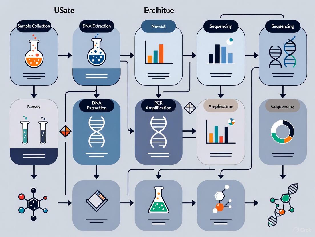
Experimental Design and Workflow
A contamination-conscious experimental design is the most critical step for successful low-biomass research. The diagram below outlines a workflow that integrates contamination control at every stage.
Troubleshooting Guide: Frequently Asked Questions
FAQ 1: How can I distinguish true microbial signals from contamination in my data?
Challenge: In low-biomass samples, contaminant DNA from reagents, kits, and the environment can be more abundant than the target DNA, making it difficult to identify the true signal [1] [3].
Solutions:
- Implement Extensive Controls: Include multiple negative controls at every stage (sample collection, DNA extraction, library preparation). Sequence these controls alongside your samples [1] [3].
- Bioinformatic Subtraction: Use specialized tools to identify and subtract contaminants by comparing your samples to the profiles obtained from your negative controls. Be aware that these tools can struggle if contamination is extensive and variable [1].
- Replicate and Validate: Consistent detection of a microbial taxon across true sample replicates, especially at higher abundances than in control samples, increases confidence.
FAQ 2: My negative controls show high microbial DNA. What went wrong?
Challenge: Detection of significant microbial DNA in negative controls indicates pervasive contamination.
Troubleshooting Steps:
- Audit Reagents: Check that all reagents, especially DNA extraction and PCR kits, are certified DNA-free or have been tested for low background contamination [1] [3].
- Review Lab Practices: Ensure that work is performed in a dedicated, clean workspace (e.g., PCR hood, if possible). Use sterile, single-use plasticware and decontaminate surfaces and equipment with solutions that destroy free DNA (e.g., 10% bleach, followed by 70% ethanol) [1].
- Verify Personnel Technique: Confirm that researchers are wearing appropriate personal protective equipment (PPE) such as gloves, lab coats, masks, and potentially cleansuits to minimize human-derived contamination [1].
FAQ 3: What are the best practices for collecting low-biomass samples to minimize contamination?
Challenge: Contamination introduced during sampling is irreversible and can invalidate a study.
Protocol for Contamination-Conscious Sampling:
- Decontaminate Equipment: Use single-use, DNA-free collection tools (swabs, vessels). If reusables are necessary, decontaminate with 80% ethanol (to kill cells) followed by a DNA-degrading solution like sodium hypochlorite (bleach) or UV-C irradiation [1].
- Use Barriers and PPE: Cover exposed skin with gloves, masks, goggles, and cleansuits to prevent contamination from researchers [1] [2].
- Collect Process Controls: During sampling, also collect controls such as an empty collection vessel, a swab of the air, or an aliquot of the preservation solution. These are essential for identifying contamination sources introduced during collection [1].
FAQ 4: How does sample biomass level affect my choice of sequencing and analysis methods?
Challenge: Standard protocols designed for high-biomass samples (e.g., human gut, soil) are often unsuitable for low-biomass applications.
Methodological Adjustments:
- Sequencing Protocol Selection: For ultra-low biomass samples, specialized library prep kits designed for low DNA input may be required. In some cases, adding nonspecific carrier DNA or increasing PCR cycles can help, but this must be done cautiously as it can also amplify contaminants [3].
- Analysis Considerations: Phylogenetic-based analysis methods can be more robust than Operational Taxonomic Unit (OTU)-based approaches when sequence coverage is low [4]. Normalize the number of sequences across all samples before comparing diversity indices, as these values are sensitive to sample size [4].
Essential Research Reagent Solutions
Table 2: Key Reagents and Materials for Low-Biomass Research
| Item Category | Specific Examples / Properties | Function & Importance |
|---|---|---|
| DNA-Free Reagents | Certified DNA-free water, extraction kits, and PCR mixes [1] [3] | Minimizes background contamination from the "kitome," which is critical for ultra-low biomass samples. |
| Surface Decontaminants | 80% Ethanol, Sodium hypochlorite (bleach), UV-C light, Hydrogen peroxide [1] | Ethanol kills cells; bleach/UV-C destroys residual DNA. A two-step process is recommended for thorough decontamination. |
| Specialized Library Prep Kits | Kits validated for low DNA input (e.g., NEBNext Ultra II DNA Library Prep Kit, Oxford Nanopore Rapid PCR Barcoding with modifications) [5] [3] | Enables library construction from minimal DNA (as low as 500 pg), improving the chances of detecting the true signal. |
| Sample Collection Devices | Single-use, pre-sterilized swabs; novel devices like the SALSA (Squeegee-Aspirator for Large Sampling Area) [3] | Maximizes recovery efficiency and minimizes sample loss or contamination during the collection process. |
| Personal Protective Equipment (PPE) | Gloves, masks, goggles, cleansuits, shoe covers [1] | Creates a barrier between the researcher and the sample, reducing contamination from human skin, hair, and aerosols. |
Successfully researching low microbial biomass environments demands rigorous contamination control throughout the entire experimental process. By implementing the guidelines, troubleshooting strategies, and reagent solutions outlined in this document, you can significantly reduce contamination, properly identify its sources, and generate more reliable and interpretable data for your research.
Frequently Asked Questions
What are the most common contamination sources in low-biomass research? The most prevalent contamination sources are reagents and kits (including extraction kits and water), laboratory equipment and surfaces (both external and internal to equipment like liquid handlers), and cross-contamination between samples during processing [6] [1] [7]. In pharmaceutical manufacturing, shared equipment is a primary cause of cross-contamination [8] [9].
My negative controls show microbial growth. What should I do? First, check your water supply and reagents [6]. Test your purified water and reagents by using them in a control culture media to see if microbial growth occurs. Secondly, review your sterile technique, including the use of personal protective equipment (PPE) and ensure all equipment has been properly sterilized. Finally, increase your process controls by including more negative controls (e.g., empty collection vessels, swabs of the air, extraction blanks) to help pinpoint the exact source of contamination [1] [7].
How can I distinguish true microbial signal from contamination in my data? Robust experimental design is key. Always include multiple types of negative controls (e.g., sample collection controls, extraction blanks, no-template PCR controls) that undergo the exact same process as your samples. During data analysis, use computational decontamination tools that leverage these controls to identify and subtract contaminant sequences. Be cautious, as these methods can struggle if controls are not representative or if well-to-well leakage has occurred [1] [7].
Our lab uses shared equipment. How can we prevent cross-contamination? Implement and validate a rigorous cleaning protocol between uses. For equipment that comes into direct contact with samples, use a two-step process: decontaminate with 80% ethanol to kill organisms, followed by a nucleic acid degrading solution (e.g., bleach, UV-C light) to remove residual DNA [1]. For larger or fixed equipment, establish and adhere to a documented cleaning schedule and use single-use materials where possible [6] [10].
What are the best practices for storing samples to prevent contamination? Immediate freezing at -80°C is the gold standard for preserving microbiome integrity. If this is not feasible, refrigeration at 4°C can be effective for some sample types, and the use of preservative buffers (e.g., AssayAssure, OMNIgene·GUT) can help maintain microbial stability at room temperature for a limited period [11].
Troubleshooting Guides
Use this flowchart to systematically identify potential contamination sources in your lab workflow.
Diagram 1: A workflow to diagnose common contamination sources.
Guide 2: Addressing Reagent and Kit Contamination
Problem: Contaminating microbial DNA is introduced from reagents, kits, or water, which is especially impactful in low-biomass studies [1] [7].
Solutions:
- Test Reagents: Use an electroconductive meter to check water purity or culture media with only the water/reagent as a sample [6].
- Use Certified Reagents: Source DNA-free, certified reagents and water when possible.
- Include Controls: Always include negative extraction controls (blanks) that use water instead of sample to identify reagent-derived contaminants [7].
- UV Irradiation: Pre-treat reagents with UV light to degrade contaminating DNA, if compatible with the reagent [1].
Guide 3: Preventing Equipment and Surface Contamination
Problem: Contaminants are introduced from laboratory surfaces, tools, or equipment interiors [6] [10].
Solutions:
- Establish a Cleaning Schedule: Create and document a strict schedule for cleaning and sterilizing all equipment. Use autoclaving and UV-C sterilization where applicable [6].
- DNA Decontamination: For surfaces and equipment, use a two-step process: clean with 80% ethanol, followed by a DNA-degrading solution like sodium hypochlorite (bleach) to remove residual nucleic acids [1].
- Automate: Use automated liquid handlers with enclosed, HEPA-filtered hoods to create a contamination-free workspace and reduce human contact with samples [6].
- Use Laminar Flow: Perform sample transfers in a laminar flow hood to keep airborne particles from settling on samples [6].
Guide 4: Minimizing Sample Cross-Contamination
Problem: DNA from one sample leaks or is transferred to another sample, a phenomenon known as "well-to-well leakage" or the "splashome" [7].
Solutions:
- Optimize Lab Workflow: Design a unidirectional workflow from "clean" to "dirty" areas to prevent processed samples from contaminating new ones. Keep samples organized and in their proper locations [6].
- Physical Barriers: Use physical seals or caps on sample plates during centrifugation and vortexing. When possible, include blank wells between samples on PCR plates to act as buffers [7].
- Reduce Touches: Map out your experimental procedure and find ways to reduce the number of sample transfers and physical touches, as each touch is a potential contamination point [6].
- Decontaminate Gloves: Change gloves frequently and decontaminate them with ethanol between handling different samples [1].
Contamination Data and Reagent Solutions
This table summarizes real-world contamination trends identified from pharmaceutical recall databases, illustrating the prevalence and impact of various contaminants [8].
| Contaminant/Impurity Type | Examples | Common Causes & Sources |
|---|---|---|
| Microbial Contaminants | Bacteria (e.g., Burkholderia cepacia), viruses, fungi | Contaminated water systems, raw materials (animal sera, human plasma), improper aseptic techniques in compounding pharmacies [8]. |
| Process-Related Impurities | Genotoxic impurities (e.g., nitrosamines), reaction byproducts | Unexpected reactions from changing reactants, failure to characterize impurities during process changes, poor cleaning leading to residue buildup [8]. |
| Metal Contaminants | Stainless steel (e.g., 316L), chromium, aluminum | Wear and tear or friction from manufacturing equipment; human error in equipment assembly [8]. |
| Packaging-Related Contaminants | Glass flakes, rubber particles, plasticizers (e.g., phthalates) | Incompatibility between packaging and product, degradation from poor storage conditions (prolonged time, high temperature) [8]. |
| Drug Cross-Contamination | Potent drugs (e.g., antihypertensives, cytotoxics) | Use of shared manufacturing equipment with inadequate cleaning validation, human error leading to product mix-ups [8] [9]. |
Table 2: Essential Research Reagent Solutions for Contamination Control
This toolkit lists key materials and reagents used to prevent, identify, and control contamination in microbiome research.
| Item | Function & Role in Contamination Control |
|---|---|
| HEPA-Filtered Laminar Flow Hood | Provides a sterile workspace with unidirectional, particulate-free air to protect samples from airborne contaminants during open-bench procedures [6]. |
| DNA-Free Water | Used as a solvent for reagents and PCR mixes to prevent the introduction of microbial DNA that can skew results, especially in low-biomass studies [6] [1]. |
| UV-C Light Source | Used to sterilize surfaces, equipment, and some reagents by degrading contaminating DNA, helping to eliminate nucleic acids that survive standard cleaning [1]. |
| Sterile Swab Kits | Allow for aseptic collection of surface and tissue samples. Using single-use, pre-sterilized kits prevents introducing contaminants during the sampling process itself [12]. |
| DNA Degrading Solution (e.g., Bleach) | Used to wipe down surfaces and equipment to destroy residual contaminating DNA that is not removed by ethanol cleaning alone [1]. |
| Preservative Buffers (e.g., AssayAssure) | Maintain microbial stability and composition in samples that cannot be immediately frozen, preventing overgrowth of contaminants or degradation of the native microbiome [11]. |
| Validated DNA Extraction Kits | Kits designed for low-biomass samples often include protocols to minimize reagent contamination. Consistent use allows for better identification of background "kitome" contaminants [11] [13]. |
| Single-Use, Filter Pipette Tips | Prevent aerosol carryover and cross-contamination between samples during liquid handling, a common source of well-to-well leakage [7] [10]. |
Experimental Protocols for Contamination Control
Protocol 1: Establishing a Comprehensive Contamination Control Plan
A proactive plan is essential for reliable results [10].
- Risk Assessment: Identify all potential contamination sources for your specific samples and processes (personnel, environment, reagents, equipment).
- Develop SOPs: Create detailed Standard Operating Procedures (SOPs) for:
- Cleaning & Sterilization: Define methods, frequencies, and responsible personnel for all equipment and surfaces [6].
- Personal Protective Equipment (PPE): Mandate lab coats, gloves, hairnets, and masks. Specify changing gloves between samples [6] [1].
- Waste Disposal: Ensure safe disposal of contaminated materials.
- Implement Physical Controls:
- Training & Documentation: Train all personnel rigorously on contamination control SOPs. Maintain detailed records of cleaning, maintenance, and any contamination incidents [10].
Protocol 2: Collecting Process Controls for Low-Biomass Studies
In low-biomass research, contamination is inevitable; the goal is to identify it. Process controls are non-sample specimens that undergo the exact same processing as your real samples to capture the "background noise" of contamination [1] [7].
- Sample Collection Controls:
- Empty Collection Vessel: Place an empty, sterile collection tube in the sampling environment.
- Field Swabs: Swab the air in the sampling environment or swab the PPE of the sampling personnel.
- Preservation Solution Aliquot: Take an aliquot of the solution used to preserve samples.
- DNA Extraction Controls:
- Amplification & Sequencing Controls:
- No-Template Control (NTC): In PCR, use a reaction mix containing all reagents except the DNA template. This identifies contamination in your PCR master mix or primers [7].
- Implementation:
- Include these controls in every batch of samples processed.
- We recommend including at least two controls per type to account for variability and ensure reliability [7].
The Critical Impact of Contamination on Diversity Metrics and Data Interpretation
Frequently Asked Questions (FAQs)
FAQ 1: Why are low-biomass samples particularly vulnerable to contamination, and how does this affect diversity metrics? In low-microbial biomass samples, the genetic material from contaminants can outnumber the DNA from the actual sample. This leads to severe biases where contamination constitutes the majority (e.g., >75%) of generated sequence data. This inflates perceived microbial diversity (alpha diversity) and distorts the true biological differences between sample groups (beta diversity), potentially leading to spurious conclusions about the presence of a native microbiome [1] [14].
FAQ 2: What are the primary sources of contamination in microbiome studies? Contamination can be introduced at virtually every stage of the workflow:
- Sample Collection: Human operators, sampling equipment, and the local environment [1] [14].
- Reagents and Kits: DNA extraction kits and library preparation reagents contain microbial DNA, often called the "kitome" [12] [14].
- Cross-contamination: During laboratory processing, DNA can transfer between samples, for instance, through well-to-well leakage during PCR setup [1].
FAQ 3: What types of negative controls are essential for identifying contamination? A robust experimental design includes multiple types of controls to trace contamination sources:
- Sampling Controls: Swabs of the air, gloves, or sampling equipment to account for environmental exposure during collection [1].
- Extraction Blanks: Reagents taken through the DNA extraction process without any sample to identify the "kitome" [12] [14].
- Library Preparation Blanks: Water or buffer used in the PCR and library preparation steps to detect contaminants from these reagents [14].
FAQ 4: How can I differentiate between a true low-diversity signal and a signal caused by contamination?
Rigorous use of negative controls is mandatory. By sequencing these controls alongside your samples, you can use statistical algorithms (e.g., decontam in R) to identify and remove sequences also found in the controls. Furthermore, correlating DNA-based findings with other methods, such as bacterial culture, can confirm the presence of viable endogenous microbes [14].
Troubleshooting Guides
Problem: Inflated or Skewed Alpha Diversity in Low-Biomass Samples
Potential Cause: Contaminating DNA from reagents or the sampling environment is being sequenced, creating a false signal of high diversity.
Solution:
- Implement Stringent Laboratory Practices:
- Use DNA-free reagents and single-use, sterilized plasticware where possible [1].
- Decontaminate workspaces and equipment with solutions that degrade nucleic acids (e.g., 10% bleach, followed by 70% ethanol to remove bleach residue) [1].
- Use separate, dedicated rooms or cabinets for pre- and post-PCR work to prevent amplicon contamination.
- Integrate Comprehensive Controls: Include extraction blanks and library preparation blanks in every batch of samples processed [14].
- Apply Bioinformatics Decontamination: Use the data from your negative controls in a contamination-identification algorithm. The table below summarizes the impact of contamination and the effect of decontamination as demonstrated in a bovine milk study [14].
Table 1: Quantitative Impact of Contamination and Decontamination in a Bovine Milk Microbiome Study
| Metric | Before Decontamination | After Decontamination |
|---|---|---|
| Proportion of sequences identified as contaminant | >75% | Not Applicable |
| Predominant genera in milk samples | Mixed community of contaminants and endogenous bacteria | Staphylococcus and Acinetobacter |
| Bacterial culture results | Not performed | Growth of Staphylococcus and Corynebacterium in 50% of samples |
| Conclusion on milk microbiome | Artificially inflated diversity, unreliable composition | More dispersed, less diverse, compositionally distinct true community |
Problem: Unreliable Beta Diversity and Sample Groupings (PCoA/PERMANOVA)
Potential Cause: Variable levels of contamination across samples or cross-contamination is distorting the true ecological distances between samples.
Solution:
- Standardize Sample Handling: Ensure all samples are collected, stored, and processed in an identical manner to minimize technical variation [1].
- Prevent Cross-Contamination:
- Use fresh gloves between handling different samples.
- Use aerosol-resistant pipette tips.
- Include a dye tracer in drilling or cutting fluids to monitor for leakage between samples [1].
- Leverage Experimental Design: For sample types where the true microbiome is expected to be minimal or non-existent (e.g., sterile buffers, negative controls), include them in your beta diversity analysis. Their position in the ordination plot will reveal the "cloud of contamination," allowing you to interpret which experimental samples are indistinguishable from it [1] [12].
Table 2: Essential Research Reagent Solutions for Contamination Control
| Item | Function in Contamination Control |
|---|---|
| DNA Degrading Solution (e.g., Bleach) | Destroys contaminating free DNA on surfaces and equipment that can persist after standard sterilization [1]. |
| DNeasy PowerSoil Kit (Qiagen) | Commonly used for soil and environmental samples; its bead-beating step is effective for lysing tough microbial cells. The associated "kitome" must be characterized with extraction blanks [12]. |
| Sterile FloqSwabs | Single-use swabs for consistent surface sampling, preventing cross-contamination between sampling locations [12]. |
| Propidium Monoazide (PMA) | A dye that penetrates cells with compromised membranes (dead cells) and covalently binds to their DNA upon light exposure, preventing its amplification. This helps differentiate DNA from intact/viable cells versus free DNA or dead cells [14]. |
| Synthetic DNA (Spike-in) | An artificial, known DNA sequence added to samples in a controlled amount. It serves as an internal standard for quantifying absolute microbial abundance and identifying PCR inhibition [14]. |
Experimental Protocol: Sampling and Control Workflow for Low-Biomass Surfaces
This protocol outlines a method for sampling low-biomass surfaces (e.g., equipment handles, cleanroom surfaces) to monitor microbial contamination while accounting for potential contaminants.
Methodology:
- Preparation:
- Decontaminate sampling surfaces and gloved hands with 80% ethanol followed by a DNA-degrading solution (e.g., 0.5% sodium hypochlorite) if compatible with the surface material [1].
- Prepare and label sterile swab kits and sample containers.
- Sample Collection:
- Moisten a sterile swab (e.g., FloqSwab) in sterile Phosphate-Buffered Saline (PBS).
- Swab the target surface area (e.g., 10 cm x 10 cm) thoroughly, using horizontal, vertical, and diagonal strokes while rotating the swab [12].
- Return the swab to its container and seal.
- Control Collection:
- Storage and Processing:
- Refrigerate samples at 4°C immediately after collection for short-term storage.
- Transfer to -80°C for long-term storage until DNA extraction.
- Process all samples and controls through DNA extraction and sequencing simultaneously using the same kit and reagent batches [12].
The following workflow diagram illustrates the full experimental and bioinformatics pipeline for a robust low-biomass study.
Technical FAQs: Resolving the Placental Microbiome Controversy
FAQ 1: What is the core methodological problem underlying the placental microbiome debate? The central issue is the challenge of low microbial biomass. In environments like the placenta, where microorganisms are presumed to be absent or extremely rare, the minimal microbial DNA signal detected by sensitive sequencing techniques can be easily overwhelmed or mistaken for background DNA contamination originating from laboratory reagents, sampling equipment, or the laboratory environment itself [15] [1] [16]. Distinguishing a true signal from this contaminant "noise" is the primary methodological hurdle.
FAQ 2: What are the key pieces of evidence cited by both sides of the debate? The scientific community remains divided, with interpretations of the same data leading to opposing conclusions, as summarized in the table below.
Table 1: Key Evidence in the Placental Microbiome Debate
| Evidence for a Placental Microbiome | Evidence for a Sterile Placenta |
|---|---|
| Detection of bacterial DNA in placental tissue via high-throughput sequencing (e.g., 16S rRNA gene sequencing) [17] [18]. | Re-analysis of sequencing data showing placental bacterial profiles cluster by study and mode of delivery, not biological signal [15]. |
| Identification of specific bacterial phyla (e.g., Firmicutes, Proteobacteria) with compositions differing from other body sites [17] [18]. | Bacterial signals in term cesarean-delivered placentas become indistinguishable from technical controls after stringent decontamination analysis [15]. |
| Studies reporting bacterial metabolites (e.g., SCFAs) in meconium and inflammatory cytokines linked to bacterial profiles in amniotic fluid [19]. | Existence of germ-free mammals, which are derived via Cesarean-section and raised in sterile conditions, contradicting in utero colonization [20] [16]. |
| Potential microbial origins from maternal oral, gut, and vaginal microbiota through hematogenous spread or ascending migration [17] [21]. | Widespread contamination from DNA extraction kits and reagents ("kit-ome") can explain most, if not all, detected bacterial DNA [22] [16]. |
FAQ 3: What is the "kit-ome" and how does it mislead research? The "kit-ome" refers to the collective microbial DNA contamination present in laboratory reagents, including DNA extraction kits and PCR master mixes [22]. This contaminating DNA is co-purified and sequenced alongside the target DNA from the sample. In high-biomass samples like stool, the authentic signal dwarfs the contamination. However, in low-biomass samples like the placenta, the "kit-ome" can constitute most or even all of the apparent microbial community, leading to reports of environmentally-derived bacteria (e.g., Bradyrhizobium, which lives on plant roots) in human tissues [22].
Troubleshooting Guide: Pitfalls in Low-Biomass Microbiome Research
The following workflow diagram outlines a systematic approach for identifying and addressing contamination throughout a low-biomass microbiome study.
Troubleshooting Steps
Step 1: Pre-Sampling & Experimental Design
- Problem: Inadequate control planning leads to inability to distinguish signal from noise.
- Solution: Plan your statistical analysis and control strategy at the start [23]. This includes determining the number and type of negative controls (e.g., extraction blanks, PCR blanks, sampling blanks) needed to robustly identify contaminants. A power analysis is recommended for hypothesis-testing studies [23].
Step 2: Sample Collection & Handling
- Problem: Contamination is introduced during the sampling procedure itself.
- Solution: Implement a contamination-informed sampling design [1]. Use single-use, DNA-free collection vessels. Decontaminate reusable equipment with ethanol followed by a DNA-degrading solution (e.g., bleach). Wear appropriate personal protective equipment (PPE) like gloves, masks, and cleansuits to limit human-derived contamination [1]. Crucially, collect procedural controls, such as an empty collection vessel or a swab exposed to the air in the sampling environment [1].
Step 3: Laboratory Processing
- Problem: Reagent and cross-sample contamination during DNA extraction and library preparation.
- Solution: Include multiple negative controls from DNA extraction and subsequent steps in every batch of samples processed [23] [1]. Where feasible, use multiple DNA extraction kits to check for kit-specific contaminants [22]. The use of synthetic DNA spike-in controls can help quantify the limit of detection and account for amplification biases [20].
Step 4: Data Analysis & Interpretation
- Problem: Failure to bioinformatically identify and remove contaminant sequences.
- Solution: Aggressively profile and subtract contaminants using dedicated tools (e.g., the
decontamR package) that identify sequences prevalent in negative controls [15] [16]. Always compare the microbial profile of your samples directly to your negative controls. Be skeptical of results that show a high degree of overlap with common environmental bacteria or known reagent contaminants [22].
The Scientist's Toolkit: Essential Reagents & Materials
The following table details key materials and solutions for conducting robust low-biomass microbiome research.
Table 2: Research Reagent Solutions for Low-Biomass Studies
| Item | Function & Importance | Considerations & Pitfalls |
|---|---|---|
| DNA Removal Solutions (e.g., bleach, UV-C light) [1] | Decontaminates sampling equipment and work surfaces by degrading trace DNA that remains even after sterility. | Critical for reducing background "signal." Autoclaving and ethanol alone do not remove persistent DNA. |
| Process Validation Controls (Extraction & PCR Blanks) [15] [23] | Reveals the "kit-ome" and environmental contaminants introduced during wet-lab workflows. The cornerstone of credible data. | Must be processed in the exact same batch and alongside experimental samples. Ignoring them invalidates results. |
| Sampling Controls (Blank swabs, air samples) [1] | Identifies contamination introduced specifically during the sample collection procedure. | Examples include swabbing the glove of the collector or exposing a swab to the operating theatre air [1]. |
| Spike-in Controls (e.g., Salmonella bongori, synthetic sequences) [22] [19] | Acts as an internal standard to verify that the entire workflow can detect low levels of microbes and to monitor PCR inhibition. | Should be added in low, known quantities to avoid overwhelming any potential authentic signal. |
| Contamination Identification Software (e.g., Decontam) [15] | Bioinformatic tool that uses prevalence or frequency in negative controls to identify and remove contaminant sequences from data. | Essential final step. However, its efficacy depends on the quality and number of negative controls provided. |
| Personal Protective Equipment (PPE) (gloves, masks, cleansuits) [1] | Creates a barrier between the operator (a major source of contamination) and the sample. | Reduces contamination from human aerosol droplets, skin, and hair [1]. More extensive PPE is needed for ultra-clean protocols. |
Building a Robust Workflow: Best Practices from Sample to Sequence
Decontamination Protocols for Sampling Equipment and Surfaces
Frequently Asked Questions (FAQs)
1. Why is decontamination especially critical for low microbial biomass studies? Samples with low microbial biomass (e.g., certain human tissues, blood, or clean water) contain minimal target DNA. Contaminants introduced from sampling equipment or surfaces can proportionally constitute a much larger, misleading fraction of the final dataset, ultimately distorting results and leading to false conclusions about the sample's true microbial community [1] [2].
2. What is the fundamental difference between sterilization and disinfection?
- Sterilization is a process that destroys all microbial life, including highly resistant bacterial endospores.
- Disinfection uses liquid chemicals to eliminate virtually all pathogenic microorganisms, but may not destroy bacterial spores [24]. Sterilization (e.g., autoclaving) is the preferred method for decontaminating equipment used in sensitive microbiological work [24].
3. How can I tell if my low-diversity sequencing results are due to contamination? A key indicator is finding microbial taxa in your samples that are also present in your negative controls. These controls—which can include unused swabs, sample preservation fluid, or rinsates from sampling equipment—are essential for identifying contaminants introduced during the sampling and processing workflow [1] [2].
4. What are the most common sources of contamination during sampling? Primary contamination sources include human operators (skin, breath, clothing), the outdoor environment, sampling equipment itself, and reagents. Contamination can occur at any stage, from sample collection and storage to DNA extraction and sequencing [1] [25].
5. Can I rely on ethanol alone to decontaminate my sampling equipment? While 80% ethanol is effective for killing contaminating organisms, it does not reliably remove traces of environmental DNA. For critical low-biomass work, a two-step decontamination is recommended: ethanol to kill organisms, followed by a nucleic acid degrading solution (e.g., diluted bleach, hydrogen peroxide) to remove residual DNA [1].
Troubleshooting Guide: Low Microbial Diversity
Problem: High Abundance of Human Commensal Bacteria in Samples
- Potential Cause: Contamination from the researcher during sample collection or processing.
- Solution: Implement stricter personal protective equipment (PPE) protocols. Wear gloves, masks, and lab coats. For extremely sensitive samples, consider more extensive PPE like coveralls and shoe covers to limit skin and hair cell shedding [1] [25].
- Preventative Measure: Always include a "sampling control," such as a swab exposed to the air in the sampling environment or an empty collection vessel, to identify human-associated contaminants [1].
Problem: Consistent Detection of the Same Contaminants Across Different Samples
- Potential Cause: Reagent contamination or cross-contamination between samples during processing.
- Solution:
- For reagent contamination: Use dedicated, DNA-free reagents. Include a "negative extraction control" (a blank with no sample) during DNA extraction to identify contaminants from kits and water [1] [12].
- For cross-contamination: Use physical barriers like single-use equipment. If reusing equipment, employ thorough decontamination between samples. Ensure proper layout of the lab workspace to separate "clean" and "dirty" areas [1] [26].
Problem: Unexpected Fungal Signals or High Microbial Diversity in Sterile Samples
- Potential Cause: Environmental contamination from dust, airflow, or improperly maintained equipment.
- Solution: Regularly clean workspaces and equipment. Use HEPA filters in sampling and processing areas. Ensure that heating, ventilation, and air-conditioning (HVAC) systems are properly maintained to prevent them from becoming a source of microbial contamination [25].
Decontamination Method Comparison
Table 1: Common Decontamination Methods for Laboratory Equipment and Surfaces
| Method | Mechanism | Typical Uses | Key Considerations |
|---|---|---|---|
| Autoclaving (Wet Heat) [24] | Steam sterilization under high pressure and temperature (e.g., 121°C). | Laboratory glassware, metal tools, biohazardous waste. | Most dependable method for sterilization. Not suitable for heat-sensitive materials. |
| Chemical Disinfection (Liquid) [24] | Halogens (e.g., bleach), alcohols, peroxides disrupt cellular structures. | Benchtop surfaces, non-autoclavable equipment. | Effectiveness depends on concentration, contact time, and target organism. Bleach can be corrosive. |
| UV Radiation [24] | Non-ionizing UV-C light damages microbial DNA. | Reducing airborne microbes in airlocks, biological safety cabinets (with caution). | Organisms must be directly exposed; dust and shadows shield microbes. Requires regular maintenance. |
| Ethanol + DNA Degradation [1] | Ethanol kills cells; DNA degradation solutions (e.g., bleach) remove residual DNA. | Sampling equipment for low-biomass studies. | Two-step process is critical for removing both viable cells and environmental DNA. |
Experimental Protocol: Validating Surface Decontamination
This protocol is designed to test the effectiveness of your decontamination procedure on sampling equipment or work surfaces.
1. Objective: To confirm that a decontamination protocol renders a surface free of contaminating microbial DNA.
2. Materials Needed:
- Sterile swabs or wipes
- DNA-free phosphate-buffered saline (PBS) or water
- Reagents for DNA extraction and PCR/sequencing
- Growth media (for viability testing, optional)
3. Procedure:
- Step 1 (Pre-decontamination swab): Swab a defined area (e.g., 10 cm x 10 cm) of the surface to be tested. Use a sterile swab moistened with DNA-free PBS [12].
- Step 2 (Decontaminate): Perform your standard decontamination procedure on the sampled surface (e.g., wipe with 70% ethanol, followed by a DNA removal solution).
- Step 3 (Post-decontamination swab): After the surface has dried, swab the same area with a new sterile swab.
- Step 4 (Process controls): Include a negative control swab that was moistened but not exposed to any surface.
- Step 5 (Analysis): Extract DNA from both swabs and the control. Analyze using high-sensitivity methods like qPCR or 16S rRNA gene sequencing [1] [2].
4. Interpretation:
- Success: The post-decontamination swab shows a microbial profile and DNA concentration similar to or lower than the negative control.
- Failure: The post-decontamination swab shows high DNA yield or distinct microbial taxa (e.g., human skin bacteria) not found in the control, indicating ineffective decontamination.
Research Reagent Solutions
Table 2: Essential Materials for Effective Decontamination
| Item | Function | Application Notes |
|---|---|---|
| DNA-free Swabs [1] | Sample collection from surfaces. | Pre-sterilized and certified DNA-free to prevent introduction of contaminants during sampling. |
| DNeasy PowerSoil Kit (Qiagen) [12] | DNA extraction from swabs or filters. | Designed to inhibit humic substances and other PCR inhibitors; commonly used in microbiome studies. |
| Sodium Hypochlorite (Bleach) [1] [24] | Chemical disinfection and DNA degradation. | Effective for destroying residual DNA on surfaces. Must be used at an appropriate dilution and may require a rinse with DNA-free water. |
| Ethanol (70-80%) [1] | Chemical disinfection to kill viable cells. | Used as a first step in a two-step decontamination process. Does not remove environmental DNA. |
| Autoclave [24] | Steam sterilization of equipment and waste. | The gold standard for destroying all viable microorganisms, including spores. |
| Personal Protective Equipment (PPE) [1] [26] | Gloves, masks, lab coats, coveralls. | Creates a barrier between the researcher and the sample to prevent human-derived contamination. |
Workflow: Equipment Decontamination for Low-Biomass Sampling
Equipment Decontamination Workflow
The Essential Role of Personal Protective Equipment (PPE) as a Physical Barrier
In low microbial biomass research, such as studies of human tissues, drinking water, or hyper-arid soils, the microbial DNA from a sample can be so minimal that it approaches the limits of detection [1]. In these sensitive studies, contamination—the introduction of microbial DNA from external sources—can severely distort results, leading to false conclusions about the sample's true microbial diversity [1]. Personal Protective Equipment (PPE) serves as a critical physical barrier, not just for protecting the researcher from hazards, but for protecting the sample from contamination by the researcher and the environment [1]. Failure to use PPE correctly is a significant factor that can lead to spurious results, including low microbial diversity, by introducing contaminating DNA.
Troubleshooting FAQs: PPE and Low Microbial Diversity
FAQ 1: Our negative controls consistently show high microbial diversity. Could our PPE be a source of this contamination?
Yes, PPE can be a significant source of contamination. Human skin and hair shed cells and microbial DNA [1]. If PPE is not worn or donned correctly, or if non-sterile PPE is used, these contaminants can enter your samples. To address this:
- Verify Sterility: Use only certified DNA-free, pre-sterilized PPE [1].
- Review Donning Procedures: Ensure staff are trained to don PPE in a clean area without touching the outer surfaces that will face the sample [27].
- Implement Controls: Include PPE controls, such as swabbing the outside of gloves or masks, to identify if they are a contamination source [1].
FAQ 2: We use standard lab coats and gloves, but are still seeing contamination in our low-biomass samples. What are we missing?
Standard lab PPE may be insufficient for low-biomass work. Furthermore, the way PPE is removed can cause contamination. During doffing, the outside surfaces of PPE, which are considered contaminated, can transfer microbes to your hands, clothes, and ultimately to your samples [27].
- Upgrade PPE: Consider more extensive coverage, such as disposable coveralls, shoe covers, and face masks, to minimize skin and clothing exposure [1].
- Focus on Doffing Protocol: Implement and practice a enhanced, step-by-step doffing procedure, potentially under supervision, to minimize self-contamination [27].
- Use a Mirror: Place a full-length mirror in the doffing area to help personnel observe their technique [27].
FAQ 3: Does the type of face mask matter for preventing contamination of samples via aerosols?
Yes, the type of mask significantly impacts contamination control. Surgical masks primarily provide external protection by containing the wearer's droplets, but their loose fit can allow aerosols to escape or enter [28]. In contrast, a well-fitted FFP2/N95 respirator provides superior protection for both the wearer and the sample by filtering a higher percentage of particles [28]. For processes generating aerosols, a respirator is recommended.
FAQ 4: How can we visually train our team on the risks of PPE-related contamination?
Fluorescent powder simulations are an excellent tool for this. By coating a training manikin or surface with fluorescent powder (simulating contaminants) and having personnel don and doff PPE, you can use a UV lamp to visually identify contamination transfer to skin, clothing, or the lab environment [27]. This provides immediate, powerful feedback on protocol breaches.
Experimental Protocol: Evaluating PPE Doffing Contamination
This protocol, adapted from research, uses fluorescent tracing to visualize and minimize contamination during PPE removal [27].
Objective: To assess the effectiveness of a PPE doffing protocol in preventing contamination of the wearer.
Materials:
- Full-body PPE kit (e.g., Coveralls, N95 respirator, goggles, inner and outer gloves, shoe covers)
- Fluorescent powder (e.g., Glo Germ powder)
- Ultraviolet (UV) LED lamp
- Camera capable of high-resolution photography in low light
- A full-length mirror
- Touch-free hand sanitizer dispenser
- Hazardous waste container
Method:
- Preparation: In a preparation room, don the full PPE kit according to your standard protocol.
- Contamination Simulation: Enter the simulation room. Perform a one-minute simulated patient care or sample handling task on a manikin or surface generously coated with fluorescent powder.
- Initial Contamination Check: In a darkened room, use the UV lamp to examine the surfaces of your PPE. Photograph the initial contamination for documentation.
- Doffing with Tracking: Begin the doffing process. After the removal of each PPE item (e.g., outer gloves, gown, goggles), pause and use the UV lamp to check your hands, clothing, and the next item to be removed for the presence of fluorescent powder. Document all findings with photographs.
- Analysis: Categorize any contamination found on the body or clothing by level (e.g., "Negligible," "Noticeable," "Apparent," "Severe") and location (e.g., "hands-fingers," "shirt," "forearms") [27].
Table 1: Categorization of Contamination Levels
| Contamination Level | Description |
|---|---|
| Negligible | Very few fluorescent particles, hard to find. |
| Noticeable | More contaminated than "negligible," easy to find. |
| Apparent | Easily found with enough powder; evident contamination. |
| Severe | Massive contamination with a significant amount of powder. |
Expected Outcome: Studies using this method have found that enhanced, supervised doffing protocols can significantly reduce contamination rates, from over 70% with self-adapted practices down to below 30% [27].
The Scientist's Toolkit: Essential Reagents for Contamination Control
Table 2: Key Research Reagent Solutions for PPE and Contamination Studies
| Item | Function in Contamination Control |
|---|---|
| Fluorescent Powder (e.g., Glo Germ) | Visually simulates microbial contaminants under UV light, allowing for qualitative assessment of contamination spread during PPE donning and doffing training [27]. |
| DNA Decontamination Solution (e.g., Bleach) | Used to destroy contaminating DNA on surfaces and non-disposable equipment before sampling. Critical for ensuring sterility as autoclaving alone may not remove cell-free DNA [1]. |
| Pre-sterilized, DNA-free Swabs | For collecting environmental and PPE surface samples as negative controls to monitor background contamination levels throughout an experiment [1]. |
| Touch-free Hand Sanitizer Dispenser | Prevents cross-contamination that can occur from touching the pump of a manual sanitizer dispenser, especially important after removing gloves during the doffing process [27]. |
| Ultraviolet (UV-C) Light Source | Used to sterilize surfaces, equipment, and plasticware by degrading nucleic acids, helping to create a DNA-free work area for processing low-biomass samples [1]. |
PPE Contamination Pathways and Testing
The following diagrams illustrate the critical pathways of contamination related to PPE and a method for testing protocol efficacy.
PPE as a Physical Barrier Against Airborne Transmission
The effectiveness of PPE as a physical barrier extends to blocking airborne transmission pathways, which is crucial for containing aerosols generated during sample processing.
Optimized DNA Extraction Methods for Maximum Yield and Minimal Bias
FAQ 1: My DNA yields from dried blood spots are consistently low. How can I improve recovery?
Low DNA yield from dried blood spots (DBS) is a common issue, often due to the irreversible binding of nucleic acids to the membrane material [29]. The following optimized protocol has been demonstrated to significantly enhance DNA recovery.
- Experimental Protocol: Optimized Spin-Column Extraction for Dried Blood Spots [29]
- Punch: Remove a disc of the dried blood spot from the membrane using a sterile disposable punch.
- Solubilize: Transfer the disc to a microcentrifuge tube. Add an aqueous solubilization buffer and incubate for 20 minutes at room temperature with gentle agitation. This step is critical for releasing the DNA from the membrane matrix.
- Lysate Preparation: Proceed with a standard spin-column extraction protocol (e.g., proteinase K digestion, buffer/alcohol binding) using the entire mixture, including the membrane material.
- Elute: Elute the DNA in a low-salt buffer or nuclease-free water.
Table 1: DNA Yield Comparison from Different Blood Sample Formats
| Sample Format | Relative DNA Yield | Key Factors |
|---|---|---|
| Native Liquid Blood | 100% (Baseline) | N/A |
| Dried on Glass Fibre Membrane | 50-60% | Membrane type, optimized solubilization |
| Dried on Cellulose Membrane | 20-30% | Higher nucleic acid retention |
FAQ 2: My metagenomic data shows a strong bias against Gram-positive bacteria. How can I achieve a more balanced lysis?
This bias is typically introduced during the DNA extraction step, as the thick peptidoglycan layer in Gram-positive cell walls is resistant to many lysis methods [30] [31]. Kits lacking mechanical disruption can under-represent Gram-positive taxa by 40-60% compared to Gram-negative bacteria [30].
- Experimental Protocol: Balanced Lysis for Diverse Microbial Communities [30] [31]
- Mechanical Disruption: Use a bead-beating homogenizer. For maximum efficiency, employ a mix of small, dense beads (e.g., 0.1 mm zirconia/silica beads) to disrupt tough cells, alongside larger beads (e.g., 2.8 mm) for macro-scale tissue or biofilm breakdown.
- Enzymatic Lysis Supplement: Combine bead-beating with a multi-enzyme cocktail. A short incubation (15-30 minutes) with a mixture such as lysozyme, mutanolysin, and lysostaphin significantly improves the lysis of Gram-positive bacteria without excessive DNA shearing.
- Chemical Lysis: Use a standard lysis buffer containing SDS.
- Optimized Homogenization: Process samples at a moderate speed (e.g., 5600 RPM for 3 min) to balance complete lysis with DNA integrity. Higher speeds (e.g., 9000 RPM) may be used for exceptionally tough samples but can increase shearing [31].
Table 2: Impact of Lysis Method on Bacterial DNA Recovery
| Lysis Method | Gram-Negative Recovery | Gram-Positive Recovery | Community Representation |
|---|---|---|---|
| Chemical Lysis Only | High | Low (≤60%) | Skewed, inflates Gram-negatives |
| Bead-Beating Only | High | Moderate | Improved, but may under-lyse some Firmicutes |
| Bead-Beating + Enzymatic Cocktail | High | High (Up to 97% lysis efficiency) | Most accurate and balanced |
FAQ 3: I am working with low-biomass, high-host-content samples. How can I reduce host DNA to better sequence the microbiome?
Samples like nasopharyngeal aspirates, skin swabs, or tissue biopsies are challenging due to low microbial DNA and overwhelming host DNA, which consumes sequencing depth [32] [33]. A combination of host DNA depletion and a robust microbial DNA extraction protocol is required.
- Experimental Protocol for Nasopharynx-like Samples [32]
- Host Cell Depletion: Use a commercial host DNA depletion kit, such as MolYsis. These kits selectively lyse mammalian cells and degrade the released DNA with a DNase, while leaving microbial cells intact.
- Microbial DNA Extraction: After depletion, pellet the intact microbial cells. Perform DNA extraction using a kit validated for Gram-positive bacteria, such as the MasterPure Gram Positive DNA Purification Kit. This ensures lysis of all remaining microbial types.
- Include Controls: Always process negative controls (e.g., empty swabs, blank buffers) to identify contaminating DNA from reagents or the environment, which is a major concern in low-biomass studies [1] [32].
This combined "Mol_MasterPure" protocol has been shown to reduce host DNA content from >99% to as low as 15% in some samples, increasing usable bacterial reads by over 1,700-fold [32].
FAQ 4: My soil/sediment DNA extracts are contaminated with PCR inhibitors. What is the best purification method?
Soil and sediment samples are notorious for co-extracting humic acids and other substances that inhibit downstream enzymatic reactions [34]. The purification step is as critical as the extraction itself.
- Experimental Protocol: Inhibitor Removal for Soil DNA [34]
- Optimized Extraction: Perform a brief, low-speed bead mill homogenization in a phosphate-buffered SDS-chloroform mixture. This maximizes cell lysis while minimizing DNA shearing and the release of inhibitors.
- Purification: Apply the crude DNA extract to a Sephadex G-200 spin column. This gel filtration method was found to be superior for removing PCR-inhibiting substances while minimizing DNA loss compared to silica binding or precipitation methods [34].
- Quality Check: Assess DNA purity spectrophotometrically (A260/A280 and A260/A230 ratios) and confirm the absence of inhibitors via a spike-in PCR assay.
The Scientist's Toolkit: Key Research Reagent Solutions
Table 3: Essential Reagents for Unbiased DNA Extraction
| Reagent / Kit | Function | Application Note |
|---|---|---|
| MolYsis Basic5 | Selective depletion of host DNA from samples | Crucial for low-biomass, high-host-content samples like nasopharyngeal aspirates [32]. |
| MasterPure Gram Positive DNA Purification Kit | Robust lysis and purification of DNA from difficult-to-lyse bacteria | Effective for achieving balanced lysis in diverse communities; works well post-host-depletion [32]. |
| Sephadex G-200 Resin | Gel filtration medium for purifying DNA from PCR inhibitors like humic acids | Ideal for purification of DNA from complex environmental samples like soil and sediment [34]. |
| Zirconia/Silica Beads (0.1 mm & 2.8 mm mix) | Mechanical disruption of microbial cell walls | A mix of small and large beads ensures efficient lysis across diverse bacterial morphologies [30]. |
| MetaPolyzyme (Lysozyme, Mutanolysin, etc.) | Enzymatic cocktail for digesting peptidoglycan in bacterial cell walls | Supplement to bead-beating to ensure complete lysis of Gram-positive bacteria [30]. |
Designing a Multi-Modal Cultivation Strategy to Capture Diversity
FAQs: Troubleshooting Low Microbial Diversity
Why is my cultivated microbial diversity much lower than my culture-independent sequencing indicates? This is a common challenge, often referred to as the "great plate count anomaly." Even with extensive cultivation efforts, traditional methods may recover less than 40% of the species detected by direct metagenomic sequencing [35]. This occurs because a significant portion of environmental microbes have fastidious and unknown growth requirements that are not met by standard laboratory media. To address this, implement a strategy using multiple, diverse culture media and conditions to target a broader spectrum of microorganisms [36] [35].
What are the key experimental factors I can adjust to improve diversity recovery? The most critical factors are the variety of growth media, incubation atmospheres, and sample processing techniques. Using 12 different media types, for example, has been shown to capture distinct subsets of the microbial community, significantly increasing the total recovered diversity [35]. Furthermore, always include both aerobic and anaerobic incubation, as obligate anaerobes constitute a major portion of uncultured diversity.
My cultivation fails due to contamination. How can I prevent this? Maintaining sterile conditions is paramount. Common sources of error include improper use of laminar flow hoods and biosafety cabinets. Ensure all equipment, like autoclaves, is functioning correctly. For training, utilize resources from the American Biological Safety Association (ABSA) and practice sterile techniques with relevant virtual lab simulations [37].
How do I know if my diversity estimates are accurate? Ensure you are using appropriate alpha diversity metrics and interpreting them correctly. Standardizing your approach is key. A comprehensive analysis recommends using a core set of metrics that collectively assess different aspects: richness (e.g., Chao1), phylogenetic diversity (Faith PD), entropy (Shannon), and dominance (e.g., Simpson) [38]. Relying on a single metric can provide a biased view of the true diversity in your samples.
Troubleshooting Guides
Problem: Inconsistent Diversity Metrics
Issue: Difficulty interpreting or comparing alpha diversity values from different experiments.
Solution: Standardize your alpha diversity analysis pipeline.
Categorize Your Metrics: Group alpha diversity metrics into four complementary categories as proposed by recent guidelines [38]:
- Richness: Estimates the number of distinct species (e.g., Chao1, ACE).
- Phylogenetic Diversity: Incorporates evolutionary relationships (e.g., Faith PD).
- Information Metrics: Describe uncertainty in predicting identity (e.g., Shannon, Brillouin).
- Dominance/Diversity Metrics: Describe the distribution of abundances (e.g., Simpson, Berger-Parker).
Report a Comprehensive Set: Do not rely on a single metric. Report at least one metric from each category to gain a holistic understanding of your samples' diversity [38].
Understand Key Influences: Be aware that some metrics are heavily influenced by specific factors. For instance, the Robbins metric is highly dependent on the number of singletons (ASVs with only one read) in your data, which can be affected by your bioinformatics pipeline [38].
Problem: Failure to Cultivate "Microbial Dark Matter"
Issue: Standard media and conditions fail to grow the majority of target microbes, particularly from complex samples like soil or gut.
Solution: Employ advanced cultivation strategies that mimic the natural environment.
Use Co-cultivation: Many uncultured microbes depend on metabolic byproducts or signaling molecules from other species. Cultivating them in pairs or consortia can stimulate growth [36].
Leverage Environmental Simulators: Utilize devices like diffusion chambers or microfluidic chips that allow nutrients and signals from the natural environment to reach the cells while containing them for growth [36].
Modify Media Composition:
- Add Growth Factors: Incorporate specific compounds like zincmethylphyrins, coproporphyrins, or short-chain fatty acids to meet unknown metabolic needs [36].
- Craft Selective Media: Use inhibitors to suppress fast-growing competitors and allow slow-growers to emerge. For example, diuron has been used to inhibit oxygenic phototrophs to isolate novel Chloroflexota [36].
- Mimic Physicochemical Conditions: Adjust pH, temperature, and pressure to match the source environment, which is crucial for isolating novel archaea and bacteria from extreme environments [36].
Problem: Low-Abundance Taxa Are Missing from Cultures
Issue: Cultivation efforts are dominated by a few fast-growing species, missing the "microbial underdogs" that may be ecologically or functionally important.
Solution: Implement strategies specifically designed to capture low-abundance taxa.
Avoid Filtering Low-Abundance Data: In sequencing analysis, do not automatically filter out low-abundance taxa during bioinformatics processing, as this can obscure keystone species [39].
Apply Dilution-to-Extinction: This method dilutes the dominant members of a community, reducing competition and allowing rare species to grow. Studies have shown that losing these low-abundance bacteria through dilution can significantly alter functional outcomes in model hosts [39].
Utilize High-Throughput Techniques: Combine cultivation in multiple micro-environments (e.g., using 96-well plates) with culture-enriched metagenomic sequencing (CEMS). CEMS sequences all colonies from a plate without manual picking, efficiently capturing rare, culturable organisms that are often missed by experienced colony picking (ECP) [35].
Experimental Protocols & Workflows
Protocol 1: Culture-Enriched Metagenomic Sequencing (CEMS)
This protocol leverages high-throughput metagenomic sequencing of entire culture plates to maximize the detection of culturable microbes, including those an researcher might overlook [35].
Workflow Overview:
Detailed Methodology:
Sample Preparation:
- Suspend 0.5 g of sample (e.g., stool, soil) in 4.5 mL of 0.85% NaCl solution.
- Prepare a dilution series from 10⁻³ to 10⁻⁷ to isolate individual colonies and reduce competition.
Multi-Modal Cultivation:
- Plate 200 µL of each dilution onto 12 different types of solid media. These should include [35]:
- Nutrient-Rich Media (e.g., LGAM, PYG, Gifu Anaerobic Medium (GAM)).
- Oligotrophic Media (e.g., 1/10 GAM, low-nutrient agar).
- Selective Media (e.g., with bile salts, high salt, or antibiotics).
- For each medium, incubate one set of plates anaerobically (in a chamber with 95% N₂, 5% H₂) and another set aerobically at 37°C for 5-7 days [35].
- Plate 200 µL of each dilution onto 12 different types of solid media. These should include [35]:
Culture Harvesting and DNA Extraction:
- After incubation, add 1 mL of 0.85% NaCl solution to each plate.
- Use a sterile cell scraper to harvest all biomass from the plate's surface, combining colonies from all dilutions of the same medium and atmosphere.
- Centrifuge the harvested suspension to pellet the cells.
- Extract metagenomic DNA from the pellet using a standardized kit (e.g., QIAamp Fast DNA Stool Mini Kit), following the manufacturer's instructions [35].
Sequencing and Analysis:
- Perform shotgun metagenomic sequencing on an Illumina platform.
- Analyze the data to determine the taxonomic composition and functional potential of the cultured community. Calculate Growth Rate Index (GRiD) values to identify the optimal medium for specific bacterial taxa [35].
Protocol 2: Advanced Environmental Cultivation
This protocol outlines strategies for cultivating microbes from extreme or specialized environments, focusing on mimicking natural conditions [36].
Workflow Overview:
Detailed Methodology:
Enrichment Strategies:
- Selective Nutrients: Add specific substrates from the target environment. For example, use manganese carbonate to enrich for manganese-oxidizing bacteria like Candidatus Manganitrophus noduliformans [36].
- Physicochemical Conditions: Tailor pH, temperature, and salinity to match the source habitat. This is critical for isolating novel archaea from hot springs or extreme environments [36].
- Selective Suppression: Use inhibitors to suppress dominant groups. For instance, diuron can be used to inhibit oxygenic phototrophs, allowing novel non-oxygenic photosynthetic bacteria like certain Chloroflexota to grow [36].
In Situ Cultivation and Devices:
- Diffusion Chambers: Place inoculated chambers back into the natural environment, allowing chemical exchange while containing cells.
- Bio-Devices: Use continuous-flow cell systems or biofilm reactors to provide a stable, nutrient-controlled environment that supports slow-growing, syntrophic microbes, as demonstrated by the cultivation of Candidatus Prometheoarchaeum syntrophicum [36].
Isolation and Purification:
Data Presentation
Table 1: Comparison of Microbial Diversity Assessment Methods
| Method | Key Principle | Pros | Cons | Typical Species Recovery (vs. CIMS) |
|---|---|---|---|---|
| Culture-Independent Metagenomic Sequencing (CIMS) [35] | Direct DNA sequencing from sample | Captures full genetic potential; detects unculturable taxa | Cannot distinguish live/dead; functional role uncertain | Reference (100%) |
| Experienced Colony Picking (ECP) [35] | Manual selection and isolation of colonies | Yields pure strains for functional studies | Labor-intensive; misses inconspicuous/rare colonies | Low (Substantial missed detection) |
| Culture-Enriched Metagenomic Sequencing (CEMS) [35] | Metagenomic sequencing of all grown biomass from a plate | High-throughput; captures rare culturable taxa; provides GRiD data | Does not provide pure isolates | High (~36.5% of species, with low overlap to CIMS) |
Table 2: Key Research Reagent Solutions for Multi-Modal Cultivation
| Reagent / Material | Type | Function in Experiment |
|---|---|---|
| Gifu Anaerobic Medium (GAM) [35] | Nutrient-rich media | General growth of fastidious anaerobic bacteria from gut and environmental samples. |
| PYG Broth / Agar [35] | Nutrient-rich media | Cultivation of a wide range of anaerobic bacteria, particularly from the gut. |
| 1/10 GAM [35] | Oligotrophic media | Promotes growth of slow-growing or low-abundance bacteria outcompeted in rich media. |
| Diffusion Chambers [36] | In situ cultivation device | Allows chemical exchange with the natural environment to provide unknown growth factors. |
| DNeasy PowerSoil Kit [12] | DNA extraction kit | Efficiently lyses microbial cells and purifies DNA from complex, difficult-to-lyse samples like soil. |
| Phosphate-Buffered Saline (PBS) [12] | Buffer | Used for moistening swabs and suspending samples during collection and processing. |
| FloqSwabs [12] | Sample collection | Sterile swabs for effective microbial collection from surfaces. |
Diagnosing and Solving Common Pitfalls in Diversity Analysis
A practical FAQ for researchers troubleshooting low microbial diversity in their samples.
Troubleshooting Guides and FAQs
My microbial diversity is unexpectedly low. What are the first steps I should take?
Low microbial diversity can often be a symptom of contamination or technical artifacts. Your initial investigation should focus on the two most common culprits: sample processing contaminants and sequencing errors.
First, review your experimental controls. Reagent-only negative controls are essential for identifying contaminants introduced from DNA extraction kits or PCR reagents [40]. Analyze these controls alongside your samples—any taxa present in both are likely contaminants. Second, consider sequencing errors, which can artificially reduce diversity by distorting the true abundance of species, particularly rare taxa [41].
We recommend a step-by-step approach, visualized in the workflow below, to systematically rule out these issues.
How can I tell if my samples are cross-contaminated, and what tools can I use to fix it?
Sample cross-contamination occurs when DNA from one sample inadvertently leaks into another. This can happen during nucleic acid extraction or library preparation. Detection relies on analyzing patterns that deviate from expectations.
For studies involving paired samples (e.g., tumor-normal), you can screen for anomalies by examining mutation abundances. A significant shift in the correlation of mutation profiles or an abnormal distribution of heterozygous alleles between paired samples can indicate contamination or even sample mislabeling [42].
In microbiome studies, the focus shifts to identifying unexpected taxa. The following tools are commonly used for contamination detection and removal:
| Tool Name | Primary Function | Key Features | Best for |
|---|---|---|---|
| FastQ Screen [43] | Contamination Screening | Aligns reads to multiple genomes; provides visual reports | Initial, user-friendly screening of contamination sources |
| DeconSeq [43] | Automated Removal | Identifies and removes contaminating reads automatically | Projects where the contaminant genome is known |
| Kraken [43] | Taxonomic Classification | Ultra-fast k-mer based classification against a database | Metagenomic studies to identify all contaminant species |
| BBSplit [43] | Read Sorting | Splits sequencing reads by alignment to multiple genomes | Complex samples with multiple potential contaminant sources |
My negative controls are clean, but I still suspect technical issues. What's next?
If your controls are clear, the next step is to investigate PCR amplification errors during library preparation. These errors are a major technical artifact that can severely skew diversity estimates.
During PCR, artificial sequences are created and later clustered into Operational Taxonomic Units (OTUs). These can be mistaken for rare species, leading to an overestimation of rare taxa and a distorted view of true community structure [41]. The table below summarizes the impact and solutions for this issue.
| Aspect of Impact | Consequence | Recommended Solution |
|---|---|---|
| Singletons (Unique Sequences) | Artificial inflation of rare species; single sequences can be misclassified as new species [41]. | Use denoising algorithms like DADA2 or UNOISE3 to distinguish true biological variants from errors [41]. |
| Diversity Indices | Systemic bias in core metrics like the Shannon Diversity Index [41]. | Apply a combination of bioinformatic filtering and statistical modeling for correction [41]. |
| Species Richness | Total species count can be overestimated by up to 300% [41]. | Employ the "Missing Link" model for full-abundance error correction, which can reduce estimation error to ±12% [41]. |
What are the best practices for visualizing my data to avoid misinterpretation?
Proper data visualization is critical for accurate interpretation and communication of results. Adhering to community standards helps prevent misunderstandings.
- Avoid Stacked Bar Charts: While popular for showing community composition, stacked bar charts are not recommended. They fail to show data distribution and standard deviation, and low-abundance taxa are often not visible [40].
- Recommended Plots: Use box plots or violin plots to represent alpha diversity metrics (e.g., Shannon Index) across sample groups. These plots effectively display the data distribution, making comparisons more robust [40].
- Color Palette: Always use a colorblind-friendly (CVD-friendly) palette to ensure your findings are accessible to all audiences [40].
The Scientist's Toolkit
Research Reagent Solutions
| Item | Function in Investigation |
|---|---|
| Negative Control Samples | Contains only the reagents (e.g., from DNA extraction kits) used in your workflow. Serves as a baseline to identify contaminating DNA present in your reagents [40]. |
| Mock Community Standards | A synthetic sample comprising DNA from known microorganisms. Used as a positive control to validate your entire workflow, from DNA extraction to bioinformatic analysis, and to calibrate error rates [40]. |
| DADA2 / UNOISE3 | Bioinformatic tools (algorithms) used in the data preprocessing stage. They correct amplicon sequencing errors and remove chimeric sequences, leading to a more accurate table of biological sequences [41]. |
| Statistical Error Models (e.g., Missing Link Model) | Advanced statistical models, such as the 2024 "Missing Link" model, which uses a Bayesian network to correct for sequencing errors across the full abundance range of species, significantly improving richness estimates [41]. |
Experimental Protocol: Contamination Screening with Negative Controls
Purpose: To identify and filter out contaminating DNA sequences introduced during wet-lab procedures.
Methodology:
- Sample Processing: In parallel with your experimental samples, process a reagent-only negative control. This sample should contain all the reagents from your DNA extraction kit but no biological material [40].
- Sequencing: Sequence the negative control alongside your experimental samples on the same sequencing run to ensure identical technical conditions.
- Bioinformatic Analysis:
- Process the sequencing data through your standard pipeline (e.g., QIIME 2, mothur) to assign taxonomy to all sequences.
- Generate a feature table that includes the counts of all Amplicon Sequence Variants (ASVs) or OTUs in each sample and the negative control.
- Contaminant Identification: Use a contamination screening tool or a simple prevalence-based method. The core principle is that sequences found in the negative control are very likely to be contaminants. A common practice is to remove any ASV/OTU that has a higher relative abundance in the control than in the experimental samples, or that appears in a majority of controls.
- Data Filtering: Create a "filtered feature table" by subtracting the identified contaminant sequences from your experimental samples. All subsequent diversity and statistical analyses should be performed on this filtered table.
This systematic approach to contamination investigation will help you distinguish true biological signals from technical noise, leading to more robust and reliable conclusions in your microbial ecology research.
Troubleshooting Guide: Overcoming Low Microbial Diversity
Why is my microbial diversity in culture so low compared to my sequencing data?
This is a common challenge known as the "great plate count anomaly," where the vast majority of environmental microorganisms resist cultivation on standard laboratory media [44]. The primary reasons include:
- Non-representative media: Standard laboratory media often do not replicate the natural nutritional environment, failing to support the growth of fastidious organisms [44] [45].
- Lack of environmental cues: Microbes in their habitat rely on complex signals, growth factors, and interactions with other organisms that are absent in a standard Petri dish [46].
- Oxygen sensitivity: Many environmental microbes, especially gut microbiota, are strict anaerobes and require specialized equipment and protocols to be cultivated successfully [44].
- Overgrowth by fast-growing species: A few fast-growing generalists (e.g., Bacillus spp. on R2A medium) can quickly dominate plates, obscuring slow-growing, rare species [45] [47].
How can I improve the diversity of my isolates?
The key is to move beyond a single, standard cultivation condition. A multi-pronged strategy that incorporates environmental simulation and high-throughput techniques is vastly more effective.
1. Employ "in situ similis" Cultivation: This approach uses materials from the sample's native environment to create culture conditions that simulate the natural habitat.
- Protocol: Plant Leaf-Based "Nutritional Pads" [45]
- Collect leaves from the study plant (e.g., sunflower) and wash carefully.
- Create leaf discs (7-9 cm diameter) to fit Petri dishes.
- Pre-treat discs by punching, pressing with a meat hammer, or surface-scratching to facilitate nutrient release.
- Freeze discs overnight at -20°C, then allow them to thaw gradually.
- Autoclave the treated discs at 121°C for 20 minutes.
- Place the disc in a Petri dish and pour a thin layer of soft water agar (1%) over it to create a base and allow nutrient diffusion.
- Inoculate with your sample and incubate under appropriate conditions. This method has been shown to cultivate a wider diversity of genera, including Rhizobium, Aureimonas, and Sphingomonas, compared to standard R2A medium [45].
2. Utilize a Suite of In-Situ Devices: These devices allow microbes to grow in their natural environment while being physically contained.
Table 1: Comparison of In-Situ Cultivation Devices
| Device Name | Principle | Key Application | Protocol Summary |
|---|---|---|---|
| Diffusion Chamber [46] | A chamber bounded by membranes allows environmental nutrients and signals to diffuse in, but traps cells inside. | General enrichment of environmental microbiota. | Create a chamber with 0.03 µm membranes on both sides, fill with a sample-agar mix, seal, and incubate in the native habitat (e.g., buried in sediment). |
| Microbial Trap [46] | Designed to enrich for filamentous, chain-forming, or motile organisms. | Targeting microbes with specific growth morphologies. | Similar to a diffusion chamber, but uses different pore-sized membranes (e.g., 0.3 µm and 0.4 µm) and is filled with plain agar. Placed on the sediment surface for colonization. |
| Filter Plate Microbial Trap (FPMT) [46] | A high-throughput version of the trap with 96 individual wells, preventing cross-contamination by fast-growing species. | High-throughput isolation from complex samples. | A 96-well plate where each well's bottom is a hydrophilic membrane. Wells are filled with agar and the device is placed on the sample surface. |
| iTip [46] | A pipette tip containing glass beads and media; microbes enter through a narrow opening. | Isolation from various environmental matrices. | Fill the tip of a sterile pipette tip with glass beads, add media with agar above the beads, and place the narrow opening just under the sediment surface. |
3. Adopt Automated Culturomics and Machine Learning: Advanced platforms can systematically capture diversity by leveraging colony morphology.
- Protocol: Machine Learning-Guided Colony Picking [47]
- Image Colonies: Use a high-resolution imaging system to capture detailed morphological features (size, color, circularity, texture) of every colony on a plate.
- Quantitative Analysis: Software segments and quantifies these features for each colony.
- Smart Picking Algorithm: An algorithm selects the most morphologically distinct colonies for picking, maximizing phylogenetic diversity. This method can reduce the number of colonies that need to be picked to find 30 unique species from ~410 (random picking) to just ~85 [47].
The following diagram illustrates the core decision-making workflow for selecting the optimal cultivation strategy to maximize microbial diversity.
What is the evidence that combining methods works?
Research consistently shows that no single cultivation method is sufficient. A study on High Arctic lake sediment used six different methods (standard, diffusion chamber, trap, filter plate, iTip, and microfluidic devices) and found that each method recovered a significant number of unique operational taxonomic units (OTUs) that other methods missed [46]. The data clearly demonstrates the power of a combined approach.
Table 2: Effectiveness of Combined Cultivation Methods in Arctic Lake Sediment
| Cultivation Method | Total Isolates Recovered | Total OTUs Captured | Key Phyla Recovered |
|---|---|---|---|
| All Methods Combined | 1,109 | 155 | Proteobacteria, Actinobacteria, Bacteroidota, Firmicutes |
| Standard Plates | Part of the above | 34 Unique OTUs | Part of the above diversity |
| Diffusion Chambers | Part of the above | 35 Unique OTUs | Part of the above diversity |
| Microbial Traps | Part of the above | 16 Unique OTUs | Part of the above diversity |
| Conclusion | No single method was sufficient. A combination of approaches was required to access the bulk of microbial taxa. |
The Scientist's Toolkit: Key Research Reagent Solutions
Table 3: Essential Materials and Reagents for Advanced Cultivation
| Item | Function / Application | Specific Examples / Notes |
|---|---|---|
| Polycarbonate Membranes | Creates the permeable barrier for in-situ devices, allowing diffusion of nutrients and chemicals. | 0.03 µm - 0.45 µm pore sizes used in diffusion chambers and microbial traps [46]. |
| Anaerobic Chamber | Provides an oxygen-free environment for cultivating anaerobic microbes (e.g., gut microbiota). | Essential for handling strict anaerobes; systems can be integrated with automated colony pickers [44] [47]. |
| Oligotrophic Media / R2A | Low-nutrient media that prevents the overgrowth of fastidious oligotrophic (slow-growing) microbes. | Superior to rich media for isolating environmental bacteria [45]. |
| Blood Culture Bottles & Supplements | Rich nutrient sources used in culturomics to support the growth of a wide array of fastidious bacteria. | Critical components in culturomics protocols; e.g., rumen fluid and sheep blood [44]. |
| MALDI-TOF MS | Rapid, high-throughput identification of bacterial isolates based on protein mass fingerprints. | Used in conjunction with 16S rRNA sequencing for rapid isolate identification in culturomics [44] [45]. |
| Antibiotic Supplements | Selective pressure to inhibit common, fast-growing bacteria and enrich for rare or resistant species. | Ciprofloxacin, trimethoprim, vancomycin used to elicit distinct microbial enrichments [47]. |
Strategic Use of Pre-treatment Methods (e.g., Filtration, Alcohol Shock)
Frequently Asked Questions (FAQs)
1. What is the primary purpose of using an alcohol pre-treatment in microbial sample processing? Alcohol pre-treatment, often referred to as "alcohol shock," is used to selectively isolate specific groups of microbes from a complex sample. By applying ethanol disinfection, vegetative bacterial cells are eliminated, which enriches for spore-forming bacteria and other resilient microorganisms. This is particularly valuable in bacteriotherapy research for isolating potential therapeutic strains, such as those used for treating Clostridium difficile infections [48].
2. I am obtaining low microbial diversity in my cultures after ethanol disinfection. Is this expected? Yes, to some extent. Ethanol disinfection is a selective process that will reduce the overall number of cultivable species. One study found that while 98 species were gained using this method, 329 different species were lost [48]. The goal is not to preserve full diversity but to enrich for a specific, often hard-to-culture, subset of the microbiota. If your aim is comprehensive diversity, you should parallel process samples with and without pre-treatment.
3. Does alcohol pre-treatment exclusively select for spore-forming bacteria? While it significantly enriches for spore-formers, it does not select for them exclusively. Research shows that non-spore-forming species are more affected by ethanol, but a substantial number of species that survive are not spore-formers. One analysis found that only about 27% of the bacterial species gained through ethanol disinfection were spore-forming [48].
4. How does sample pre-treatment help with PCR-related errors in diversity studies? Pre-treatment methods themselves don't fix PCR errors, but understanding errors is crucial for interpreting diversity data. During PCR amplification for sequencing, errors can create artificial sequences that are mis-clustered as new species, leading to significant overestimation of diversity—in some cases by up to 300% [49]. Using bioinformatics tools like DADA2 or UNOISE3 for post-sequencing data filtering is essential to correct these errors [49].
Troubleshooting Guide: Addressing Low Recovered Diversity
Problem: Unexpectedly Low Diversity After Alcohol Pre-treatment
This guide helps diagnose and address issues when the number of microbial species recovered after alcohol pre-treatment is lower than anticipated for your research goals.
| Symptom | Possible Cause | Recommended Action |
|---|---|---|
| Very low species richness across all samples. | Ethanol concentration or exposure time is too high, causing excessive cell death. | Standardize protocol: Use 50% ethanol for 30-60 minutes as a starting point and optimize exposure time empirically [48]. |
| Loss of specific, non-spore-forming target bacteria. | Inherent limitation of the method; alcohol shock selectively eliminates vegetative cells. | Parallel processing: Culture an aliquot of the untreated sample alongside the pre-treated one to recover a wider diversity [48]. |
| High contamination or overgrowth by a few species. | Ineffective disinfection or unsuitable culture conditions for the target microbes. | Combine with filtration: Use pre-filtration (e.g., 0.45-5µm filters) to remove large contaminants or eukaryotic cells before culture [50]. |
| Inconsistent results between replicate samples. | Inconsistent sample preparation or uneven ethanol mixing. | Improve technical consistency: Use a vortex mixer during ethanol addition, ensure consistent sample homogenization, and maintain precise timing [48]. |
Quantitative Impact of Alcohol Pre-treatment
The following table summarizes the quantitative effects of ethanol disinfection on microbial culturing, based on a culturomics study [48].
| Metric | Before Ethanol Disinfection | After Ethanol Disinfection | Notes |
|---|---|---|---|
| Total Species Identified | 196 (from fresh stools) | 254 (combined from all samples) | 68 species were isolated only after disinfection [48]. |
| Proportion of Spore-forming Species | Not explicitly stated | 26.5% of gained species were spore-forming | Confirms enrichment, but shows most gained species are not spore-formers [48]. |
| Impact on Major Bacterial Groups | - | Enriched: Bacillus, Clostridium, Blautia, Lactobacillus, PrevotellaReduced: Alistipes, Bacteroides, Dialister, Bifidobacterium | Shows a shift in community structure [48]. |
| Impact on Key Families | Pre-disinfection proportion:Ruminococcaceae: 2.70%Lachnospiraceae: 4.18% | Post-disinfection proportion:Ruminococcaceae: 6.69%Lachnospiraceae: 5.12% | Shows a relative enrichment of these families, which include many beneficial and spore-forming organisms [48]. |
Detailed Experimental Protocol: Alcohol Disinfection for Culturomics
This protocol is adapted from a published study that used ethanol disinfection to isolate bacteria from human stool samples for bacteriotherapy research [48].
Objective: To selectively isolate spore-forming and ethanol-resistant bacteria from a complex microbial sample.
Materials Needed:
- Fresh stool sample or microbial biomass
- 50% (v/v) Ethanol solution
- Phosphate Buffered Saline (PBS) or other suitable buffer
- Anaerobic workstation or chamber
- Culture media (e.g., Columbia blood agar, Schaedler agar, Brain Heart Infusion agar)
- MALDI-TOF MS or 16S rRNA sequencing for species identification
Procedure:
- Sample Homogenization: Suspend 1-2 grams of fresh stool in 10-20 mL of PBS. Mix thoroughly using a vortex to create a homogeneous suspension.
- Ethanol Disinfection: Mix the homogenized sample with an equal volume of 50% ethanol. For example, combine 1 mL of sample with 1 mL of 50% ethanol.
- Incubation: Allow the mixture to incubate at room temperature for 30 to 60 minutes. The exact duration may require optimization for your specific sample type.
- Washing: Centrifuge the ethanol-treated sample to pellet the microbial cells. Carefully decant the supernatant and wash the pellet twice with PBS to remove any residual ethanol.
- Inoculation: Resuspend the final pellet in PBS. Inoculate the suspension onto a variety of solid and liquid culture media suitable for your target microorganisms.
- Culture and Identification: Incubate the media under appropriate atmospheric conditions (aerobic, anaerobic, microaerophilic). After growth appears, pick individual colonies and identify them using MALDI-TOF MS or 16S rRNA gene sequencing.
Workflow: Decision Process for Pre-treatment Methods
The following diagram illustrates the logical decision-making process for selecting and troubleshooting sample pre-treatment methods to address low microbial diversity.
Research Reagent Solutions
This table lists key reagents and materials used in the featured alcohol pre-treatment protocol and their specific functions in the experimental workflow [48].
| Reagent / Material | Function in the Protocol | Notes |
|---|---|---|
| 50% (v/v) Ethanol | Selective disinfection agent that eliminates vegetative bacterial cells, enriching for spores and resistant microbes. | Concentration is critical; higher concentrations or longer exposure times may kill off desired resistant cells. |
| Phosphate Buffered Saline (PBS) | Diluent and washing buffer for homogenizing samples and removing ethanol after treatment. | Maintains osmotic balance to prevent cell lysis during washing steps. |
| Anaerobic Chamber | Provides an oxygen-free environment for culturing obligate anaerobic bacteria. | Essential for recovering a major portion of gut microbiota, including many Clostridia. |
| Diverse Culture Media | Supports the growth of a wide range of bacteria with different nutritional requirements. | Using multiple media types (e.g., blood agar, Schaedler) maximizes the number of species recovered. |
| MALDI-TOF MS | High-throughput method for rapid identification of bacterial isolates to the species level. | Faster and lower cost than sequencing for large numbers of colonies [48]. |
Prioritizing High-Yield Culture Conditions to Maximize Isolate Diversity
A common challenge in microbial ecology is the phenomenon of low diversity in cultured isolates, which fails to represent the true taxonomic richness of the original sample. This technical guide addresses the core issues leading to this discrepancy and provides evidence-based troubleshooting protocols to maximize the diversity of recovered isolates for robust downstream analysis.
Troubleshooting Guides
Guide 1: Addressing Low Isolate Diversity from Complex Samples
Problem: Cultured isolates show low species richness and do not represent the microbial community detected by molecular methods like 16S rRNA sequencing.
Solutions:
- Implement Multi-Medium Cultivation: Use a suite of 12 or more media with varying nutrient compositions (rich, selective, and oligotrophic) to target different physiological groups [35]. No single medium can support the growth of all microorganisms present in a complex sample like gut microbiota.
- Apply Automated Imaging and Machine Learning: Use systems like the Culturomics by Automated Microbiome Imaging and Isolation (CAMII) platform. This technology uses colony morphology analysis to guide the selection of morphologically distinct colonies, which has been shown to significantly increase the taxonomic diversity of isolates compared to random picking. One study isolated 26,997 isolates from 20 human gut samples, representing over 80% of all abundant taxa [47].
- Analyze Viable Cell Populations: Use propidium monoazide (PMA) dye to differentiate between intact/viable and compromised cells in your sample inoculum. Cultivation is shaped by the viability of cells in the inoculum; the most abundant viable bacteria are often the most predominant in culture, which may differ from the overall community structure revealed by direct DNA sequencing [51].
Guide 2: Preventing and Identifying Contamination in Low-Biomass Samples
Problem: Contaminant DNA from reagents, the environment, or operators is disproportionately amplified in low-biomass samples, distorting true microbial signals and leading to spurious diversity metrics [1].
Solutions:
- Rigorous Decontamination: Decontaminate reusable equipment and tools with 80% ethanol followed by a nucleic acid-degrading solution (e.g., bleach, UV-C light). Use single-use, DNA-free collection vessels where possible [1].
- Use Comprehensive Controls: Include multiple negative controls throughout your workflow. These should consist of empty collection vessels, swabs of the air in the sampling environment, and aliquots of preservation solutions or sterile sampling fluids. Process these controls in parallel with your actual samples through DNA extraction and sequencing [1].
- Wear Appropriate Personal Protective Equipment (PPE): Wear gloves, masks, and clean suits to act as a barrier, limiting the introduction of human-associated microbes and aerosol droplets into the sample [1].
Frequently Asked Questions (FAQs)
FAQ 1: Why is there often little overlap between the microbial species identified by culture-independent metagenomic sequencing (CIMS) and those we can actually culture?
It is common for culture-dependent and culture-independent methods to show limited overlap. One study found that species identified by both culture-enriched metagenomic sequencing (CEMS) and direct CIMS accounted for only 18% of the total, while methods alone identified 36.5% and 45.5% of species, respectively [35]. This occurs because:
- Cultivation Bias: Standard lab conditions cannot replicate the complex physiological and metabolic needs of many environmental microbes.
- Viability: Not all DNA detected by sequencing comes from live cells capable of growth in culture [35].
- Abundance: Some taxa may be present in low abundance or in a dormant state, making them difficult to culture. Therefore, a combination of both culture-dependent and culture-independent approaches is essential for a comprehensive view of microbial diversity [35].
FAQ 2: What are the most critical factors to optimize in culture conditions to improve diversity?
While the optimal conditions are organism-specific, several key parameters universally impact growth and diversity:
- Media Composition: This is the most critical factor. Different media compositions yield distinct microbial isolates [51]. Use a variety of media types, including nutrient-rich, selective, and low-nutrient (oligotrophic) media.
- Physical Conditions: Temperature, pH, and agitation rate are highly influential. For instance, optimization of these parameters for Streptomyces sp. showed that optimal conditions for growth (33°C, pH 7.3, 110 rpm) could differ from those for secondary metabolite production (31-32°C, pH 7.5-7.6, 112-120 rpm) [52].
- Atmosphere: Incubate plates under both aerobic and anaerobic conditions, as a significant portion of environmental and gut microbes are strict anaerobes [35].
FAQ 3: How can we efficiently pick colonies to maximize taxonomic diversity without automated systems?
Even without full automation, you can adopt a strategy inspired by smart-picking algorithms:
- Morphology-Driven Selection: Visually inspect plates and deliberately pick colonies that differ in key morphological features such as size, color, edge shape, elevation, and texture. Data shows that morphologically distinct colonies are more likely to be phylogenetically diverse [47].
- Prioritize Rare Morphotypes: Make a conscious effort to pick the small, slow-growing, or oddly shaped colonies that might be overlooked in favor of large, fast-growing colonies, which often represent a few dominant species.
Data Presentation
Table 1: Comparison of Microbial Diversity Analysis Methods
| Method | Key Principle | Advantages | Limitations | Best Use Case |
|---|---|---|---|---|
| Experienced Colony Picking (ECP) | Manual selection of colonies based on visual characteristics [35]. | Low-tech; no specialized equipment needed. | Labor-intensive; misses a large proportion of culturable strains; prone to human bias [35]. | Preliminary studies; targeted isolation of specific morphotypes. |
| Culture-Enriched Metagenomic Sequencing (CEMS) | Metagenomic sequencing of all biomass grown on culture plates [35]. | Captures a broader spectrum of culturable organisms than ECP; provides genomic data. | Does not yield pure isolates for functional studies. | Comprehensive cataloging of all culturable taxa in a sample. |
| Culture-Independent Metagenomic Sequencing (CIMS) | Direct sequencing of DNA from an environmental sample [35]. | Unbiased view of total microbial community (culturable and unculturable). | Cannot distinguish viable/dead cells; does not provide isolates [35]. | Profiling the total taxonomic and functional potential of a microbiome. |
| High-Throughput Robotic Culturomics (e.g., CAMII) | Automated imaging, machine learning-guided picking, and isolation [47]. | High throughput (>2,000 colonies/hour); greatly increases diversity of isolates; integrates phenotype & genotype. | High initial equipment cost. | Large-scale isolate biobanking and in-depth diversity studies. |
| Parameter | Optimal for Mycelial Growth | Optimal for Extra-Mycelial Metabolites | Optimal for Intra-Mycelial Metabolites |
|---|---|---|---|
| Temperature | 33 °C | 31 °C | 32 °C |
| pH | 7.3 | 7.5 | 7.6 |
| Agitation Rate | 110 rpm | 120 rpm | 112 rpm |
Experimental Protocols
Protocol 1: Multi-Medium Cultivation for Diversity Maximization
This protocol is adapted from studies on human gut microbiota [35].
- Medium Selection: Prepare a panel of at least 8-12 different solid media types. This should include:
- Nutrient-rich media (e.g., LGAM, PYG, mGAM).
- Selective media for specific groups (e.g., MRS for Lactobacillus, RG for Bifidobacterium).
- Media with inhibitors (e.g., high acid, bile salts, or antibiotics like ciprofloxacin, trimethoprim).
- Oligotrophic media (e.g., 1/10GAM).
- Sample Inoculation: Serially dilute the sample (e.g., fecal suspension in 0.85% NaCl) across gradients (e.g., 10⁻³ to 10⁻⁷). Plate 200 µL of each dilution onto the different media.
- Incubation: Incubate sets of plates under both aerobic and anaerobic atmospheres at 37°C for 5-7 days (extend time for oligotrophic media).
- Colony Harvesting: After incubation, harvest all biomass from each plate type by adding saline solution and scraping the agar surface with a cell scraper. Combine biomass from replicates for DNA extraction.
- Analysis: Perform metagenomic DNA extraction and shotgun sequencing on the harvested biomass (CEMS) to identify the total diversity of culturable organisms.
Protocol 2: Assessing the Viable Bacterial Community for Cultivation
This protocol uses PMA to differentiate viable cells [51].
- Sample Processing: Enrich microbial cells from the sample matrix (e.g., sponge tissue, soil) through homogenization and differential centrifugation or filtration.
- PMA Treatment: Resuspend the microbial pellet in CMFSW. Treat an aliquot with PMA dye (final concentration as per manufacturer's instructions) to penetrate and cross-link DNA in membrane-compromised (dead) cells.
- Include a non-PMA-treated control from the same suspension.
- Photo-Activation: Expose the PMA-treated sample to bright light (e.g., a 500-W halogen lamp for 5-10 minutes) to activate the dye and cross-link the DNA in dead cells.
- DNA Extraction and Sequencing: Extract DNA from both PMA-treated and non-treated samples. Perform 16S rRNA gene sequencing on both.
- Data Interpretation: Compare the results. The PMA-treated sample profile represents the viable bacterial community, which is the pool from which your cultivation inoculum is drawn. This helps interpret why certain taxa are recovered in culture while others are not [51].
Workflow and Pathway Diagrams
Troubleshooting Pathway for Low Diversity
The Scientist's Toolkit
Research Reagent Solutions
| Item | Function | Example/Note |
|---|---|---|
| mGAM Medium | A nutrient-rich, non-selective medium commonly used for cultivating a wide range of gut bacteria [47]. | Serves as a base medium; often used in combination with selective media. |
| Propidium Monoazide (PMA) | A dye that selectively penetrates cells with compromised membranes, binding their DNA and preventing its amplification during PCR. Used to differentiate viable vs. non-viable cells in a sample [51]. | Critical for understanding which community members in a sample are potentially culturable. |
| DNeasy PowerSoil Kit | A standardized, widely used kit for efficient lysis and extraction of microbial DNA from complex environmental samples, including soil and stool [12]. | Helps overcome PCR inhibition from humic acids and other contaminants. |
| Antibiotic Supplements | Selective agents used in media to inhibit fast-growing, common bacteria, thereby enriching for rare or resistant taxa [47]. | E.g., Ciprofloxacin, Trimethoprim, Vancomycin. |
| Oligotrophic Media (e.g., 1/10GAM) | Low-nutrient media designed to mimic nutrient-scarce environments, supporting the growth of slow-growing bacteria that are inhibited by rich media [35]. | Requires extended incubation times (e.g., 7 days). |
Ensuring Data Fidelity: Validation Through Controls and Method Comparison
Implementing a Rigorous System of Negative and Positive Controls
FAQs on Controls and Microbial Diversity
Q1: Why is my negative control showing amplification or growth, and what does this mean for my experiment? A band in your negative control (No-Template Control for PCR) invalidates your experiment. It primarily indicates either contamination or the formation of primer-dimers [53].
- If the band is the same size as your target: This is definitive evidence of DNA contamination. The exquisite sensitivity of amplification methods means even minuscule amounts of contaminating DNA from a positive sample, previous experiments, or contaminated reagents (like water, polymerase, or primers) can be amplified [53].
- If the band is small and faint (e.g., <100 bp): This is likely a primer-dimer, where primers anneal to each other instead of the template DNA. This is an issue of PCR optimization rather than contamination [53].
Q2: How can the choice of controls help me troubleshoot unexpectedly low microbial diversity in my samples? Low observed diversity can be a true biological signal or a technical artifact. Proper controls help you distinguish between the two.
- Positive Control: A known, diverse mock community sample. If this control also shows low diversity, it indicates a problem with your wet-lab or bioinformatics protocol, such as overly stringent sequencing conditions or biased DNA extraction [54].
- Negative Control (Reagent Blank): Contains only the reagents without a sample. If this control shows a significant number of sequences, it indicates reagent or environmental contamination. This contamination can skew your results by introducing non-biological sequences and artificially reducing the relative abundance of rare species in your real samples, leading to an underestimation of true diversity [54].
Q3: What are the essential positive and negative controls for a robust 16S rRNA sequencing experiment? A well-controlled experiment includes both types of controls to validate the entire process from wet lab to analysis [54].
| Control Type | Purpose | Example in 16S rRNA Sequencing |
|---|---|---|
| Positive Control | Verifies the entire experimental and computational pipeline is functioning correctly. | A mock microbial community with a known composition of strains. |
| Negative Control | Identifies contamination from reagents or the environment. | A reagent blank where no biological sample is added during DNA extraction. |
| Technical Replicate | Assesses the technical variability and reproducibility of the protocol. | The same sample processed through DNA extraction and sequencing multiple times. |
Troubleshooting Guide: Low Microbial Diversity
Low microbial diversity can stem from various points in the experimental workflow. Follow this systematic guide to identify the source.
Step 1: Investigate Your Controls This is your first and most critical step.
- Action: Check the data from your positive and negative controls.
- Interpretation:
- If the positive control (mock community) shows low diversity, the issue is with your experimental or computational protocol.
- If the negative control has a high number of sequences, contamination is a problem, and you must decontaminate your workspace and reagents before re-processing samples [54].
Step 2: Review Experimental Design and Sample Collection Biases can be introduced very early.
- Sample Size: Ensure you have a sufficient number of biological replicates. Small sample sizes may not capture the true diversity and can lead to spurious interpretations [54].
- Consistency: Document and standardize all collection and handling procedures, as factors like storage time and temperature can affect microbial composition [54].
Step 3: Optimize Wet-Lab Procedures Wet-lab protocols are a common source of bias.
- DNA Extraction Method: Different extraction kits and methods (e.g., bead-beating vs. enzymatic lysis) have varying efficiencies for lysing different types of microbial cell walls. An insufficiently rigorous lysis step will fail to recover taxa with tough cell walls, artificially lowering diversity [54].
- PCR Amplification: This step is prone to biases.
- Primer Specificity: Use well-validated primers targeting the appropriate hypervariable region (e.g., V3-V4 for bacteria) [54].
- Cycle Number: Excessive PCR cycles can lead to over-amplification of dominant sequences and loss of rare ones. Use the minimum number of cycles necessary for sufficient yield [53].
- Polymerase: Use a high-fidelity, "hot-start" polymerase to minimize non-specific amplification and primer-dimer formation [53].
Step 4: Verify Bioinformatics Parameters Overly stringent computational filtering can remove good data.
- Action: Check your quality filtering parameters (e.g., Phred score, read length, ambiguous bases). If they are too strict, they may discard a large proportion of your reads, reducing the depth available for diversity estimation [55] [54].
- Pipeline Updates: Be aware that updates to analysis pipelines (e.g., changing error models or sequence clustering algorithms) can affect diversity metrics. Ensure you are using a standardized, best-practice workflow [55].
Experimental Protocol: Establishing Effective Controls
1. Protocol for a Contamination-Free Nucleic Acid Amplification (PCR) The goal is to keep amplification templates separate from your master mix reagents [53].
- Physical Separation: Maintain physically separate Pre-PCR and Post-PCR areas. Never bring amplified DNA products into the Pre-PCR area [53].
- Dedicated Equipment: Use dedicated pipettes, lab coats, and supplies for Pre-PCR work. Always use aerosol-resistant filter tips [53].
- Reagent Management: Aliquot all reagents (water, polymerase, dNTPs, primers) into single-use volumes to prevent contamination of stock solutions [53].
- Workspace Decontamination: Before use, clean surfaces and equipment with a 10% bleach solution or commercial DNA decontaminant (e.g., DNA-Away). Use a PCR workstation with UV light to irradiate the area for 15-30 minutes [53].
- Control Setup:
- Negative Control (NTC): Include a reaction where nuclease-free water is substituted for the template DNA.
- Positive Control: Include a reaction with a known, validated template that reliably amplifies.
2. Protocol for Control Selection in Immunohistochemistry (IHC) and Western Blot (WB) Controls are essential for confirming the specificity of antibody-based detection [56] [57].
- IHC / Western Blot Positive Control: Use a cell line or tissue lysate known to express the target protein at high levels. This validates that your antibody and protocol are working correctly [56].
- IHC / Western Blot Negative Control: Use a cell line or tissue lysate known not to express the target protein (e.g., a knockout line). This checks for non-specific antibody binding and false-positive signals [56].
- No-Primary Antibody Control (IHC/WB): Omit the primary antibody and incubate only with the secondary antibody and detection reagents. This controls for non-specific signal from the detection system itself [56] [57].
- Isotype Control (IHC): Incubate with a non-immune antibody of the same isotype and concentration as your primary antibody. This controls for non-specific binding of antibodies to the tissue [56].
- Loading Control (WB): Probe for a constitutively expressed "housekeeping" protein (e.g., GAPDH, Actin, Tubulin) to confirm equal protein loading across all lanes [56].
Experimental Workflow and Control Interpretation
The following diagram illustrates the logical workflow for implementing and interpreting controls in an experiment.
Research Reagent Solutions
The table below details key reagents and materials essential for implementing rigorous controls.
| Item | Function & Importance in Controls |
|---|---|
| Mock Microbial Community | A defined mix of microbial strains used as a positive control to validate that the entire sequencing workflow (DNA extraction through bioinformatics) is accurately capturing taxonomic composition and diversity [54]. |
| Nuclease-Free Water | Used to prepare master mixes and as the template for negative controls. Must be certified DNA/RNA-free to prevent false positives. Always aliquot from a new bottle [53]. |
| Hot-Start DNA Polymerase | An enzyme inactive at room temperature, preventing non-specific amplification and primer-dimer formation during reaction setup. Critical for obtaining clean negative controls [53]. |
| Validated Positive Control Lysate | A protein lysate from a cell line or tissue known to express your target. Essential for confirming antibody specificity and protocol performance in Western Blot and IHC [56]. |
| Knockout/Negative Control Lysate | A protein lysate from a knockout cell line or tissue known not to express your target. Crucial for demonstrating antibody specificity and identifying non-specific binding [56]. |
| Loading Control Antibodies | Antibodies against housekeeping proteins (e.g., GAPDH, Actin) used in Western Blot to confirm equal protein loading across all lanes, ensuring valid comparisons [56]. |
| Isotype Control Antibody | A non-immune antibody matched to the host species and isotype of your primary antibody. Used in IHC and flow cytometry to control for background staining from Fc receptor binding [56]. |
This technical support guide addresses the critical challenges of troubleshooting low microbial diversity in sample processing research. For researchers in drug development and scientific studies, selecting the appropriate microbial community analysis method is paramount. The choice between culture-dependent and culture-independent techniques significantly impacts results, with each approach offering distinct advantages and limitations. This resource provides detailed methodologies, comparative data, and practical solutions to common experimental issues.
Core Concepts and Key Differences
What are the fundamental differences between these techniques?
Culture-Dependent Techniques rely on growing microorganisms in a laboratory setting using various nutrient media [58]. These methods target organisms that can proliferate under specific artificial conditions and have been the standard in microbiology for decades [59]. Examples include plating on selective media like MacConkey agar for Gram-negative bacteria [59], using Biological Activity Reaction Tests (BARTs) for field analysis [59], and employing metabolic profiling with Biolog microplates [58].
Culture-Independent Techniques enable direct analysis of microbial DNA without requiring cultivation [59]. These methods access the vast portion of microorganisms that cannot be grown in the lab [60]. Common approaches include 16S rRNA gene sequencing [61], shotgun metagenomics [2], and metatranscriptomics [2].
Quantitative Comparison Table
The following table summarizes key performance differences between the two methodological approaches, illustrating why they often yield different results.
Table 1: Technical Comparison of Culture-Dependent and Culture-Independent Methods
| Parameter | Culture-Dependent Methods | Culture-Independent Methods |
|---|---|---|
| Detection Principle | Growth on artificial media [58] | Direct DNA/RNA analysis [59] |
| Estimated Detectable Microbes | <1% of environmental microbes [58] | Significantly higher; 95.7% vs 80.4% in BAL fluid samples [61] |
| Process Duration | 24 hours to several days [62] | Several hours to days (including sequencing) |
| Key Limitations | Strong selectivity; misses unculturable organisms [58] | Contamination sensitivity; DNA from dead cells [1] |
| Primary Advantages | Provides live isolates for further study [60] | Comprehensive community profile [61] |
| Best Applications | Pathogen identification; antimicrobial susceptibility testing [2] | Community diversity studies; unknown pathogen discovery [2] |
Experimental Protocols for Method Comparison
Protocol: Parallel Analysis of Industrial Water Samples
This protocol, adapted from a 2024 study, directly compares both methods on the same sample [59].
1. Sample Collection:
- Collect water samples in sterile containers.
- For culture-independent analysis, preserve an aliquot with a DNA stabilization buffer or freeze immediately at -80°C.
2. Culture-Dependent Analysis (BART Test):
- Inoculate 15 mL of sample into the appropriate BART tube (e.g., for iron-related bacteria, sulfate-reducing bacteria) [59].
- Incubate at room temperature, out of direct sunlight.
- Observe daily for 7 days for cloudiness and color changes, which indicate growth and specific reactions.
- Record the reaction type and time to positive result, which correlates to population density (cfu/mL).
3. Culture-Independent Analysis (Next-Generation Sequencing):
- Filter 15-50 mL of the positive BART sample or the original water sample through a 0.2-micron sterile membrane filter.
- Extract genomic DNA from the filter using a commercial kit (e.g., DNeasy PowerWater Kit).
- Perform PCR amplification of the 16S rRNA gene V4 region using primers 515F (GTGYCAGCMGCCGCGGTAA) and 806R (GGACTACNVGGGTWTCTAAT) [59].
- Purify the amplicons and prepare a sequencing library.
- Sequence the library on an Illumina MiSeq platform with a v2 500-cycle kit.
- Analyze sequences using bioinformatics pipelines like QIIME 2 or Mothur to determine taxonomic composition.
4. Data Comparison:
- Compare the dominant taxa identified by NGS in the BART tube versus the original water sample.
- Correlate the BART reaction pattern with the taxa identified via NGS.
Protocol: Assessing Bacterial Communities in Food Decomposition
This FDA Science Forum 2023 protocol highlights methodological differences in a food safety context [63].
1. Sample Preparation and Storage:
- Incinate shrimp samples at different temperatures (e.g., 0°C, 12°C, 24°C, 36°C) for varying durations to simulate decomposition.
2. Culture-Dependent Analysis:
- At each sampling point, homogenize the sample (1:10 dilution).
- Spread plate the homogenate on a non-selective medium like Tryptic Soy Agar (TSA).
- Incubate plates under the same conditions as the stored samples.
- Randomly pick 48 colonies from each sampling point for Sanger sequencing of the 16S rRNA gene.
3. Culture-Independent Analysis:
- At the same sampling points, collect triplicate samples for metagenomic analysis.
- Extract total community DNA directly from the sample.
- Perform 16S rRNA gene amplicon sequencing (identical to Section 3.1, Step 3).
4. Data Comparison:
- Compare the species diversity and dominant taxa identified by both methods at each temperature and time point.
- Note the differences in the perceived timing of dominance by specific spoilage organisms (e.g., Shewanella spp.).
Workflow Visualization
The following diagram illustrates the logical decision process for selecting and applying these techniques, which is critical for troubleshooting low diversity issues.
Decision Workflow for Method Selection
Troubleshooting Low Microbial Diversity
Problem: Culture methods yield no growth or very low diversity, while molecular methods detect high diversity.
- Potential Cause 1: The "Great Plate Count Anomaly." The vast majority of environmental microorganisms are unculturable under standard laboratory conditions [60] [58].
- Solution: Employ culture-independent methods like 16S rRNA gene sequencing to assess true diversity [61]. For cultivation, mimic the natural environment by using oligotrophic (nutrient-poor) media, extended incubation times, and supplementation with specific growth factors or signaling molecules [60].
Problem: Culture-independent methods (e.g., 16S sequencing) show low diversity in a sample expected to be diverse.
- Potential Cause 1: Contamination in low-biomass samples. Low microbial biomass samples are exceptionally vulnerable to contamination from reagents, kits, and the laboratory environment, which can obscure the true signal [1] [2].
- Solution: Implement rigorous controls. Include extraction blanks (reagents only), sample collection controls (e.g., empty collection tube swab), and negative PCR controls. Decontaminate surfaces and equipment with 80% ethanol followed by a DNA-degrading solution (e.g., bleach). Use personal protective equipment (PPE) to minimize human-derived contamination [1].
- Potential Cause 2: Dominance of a few fast-growing species. In culture-dependent methods, copiotrophs (fast-growing bacteria in nutrient-rich conditions) can outcompete slow-growing oligotrophs, skewing the perceived diversity [60].
- Solution: Use multiple culture conditions with different nutrient concentrations and incubation temperatures. Consider high-throughput dilution-to-extinction cultivation in natural sample water [60].
Problem: Discrepancies in dominant taxa identified by culture-dependent vs. culture-independent methods.
- Potential Cause: Selective growth bias. Culture conditions favor a non-representative subset of the community [59] [63]. For example, a study on decomposing shrimp found that while culture-independent tracking showed Shewanella spp. increasing from the start, culture methods only detected its dominance at a later stage [63].
- Solution: Do not rely on a single method. Use culture-independent data as the "ground truth" for total community composition and use refined culture conditions to target specific members of interest [59]. A combination often yields the most comprehensive picture [59].
Essential Research Reagent Solutions
Table 2: Key Reagents and Their Functions in Microbial Diversity Analysis
| Reagent / Material | Function | Application Notes |
|---|---|---|
| Selective Media (e.g., MacConkey Agar) | Selects for specific microbial groups (e.g., Gram-negatives) while inhibiting others [59]. | Essential for pathogen isolation. Remember that it introduces a strong bias and misses most community members [58]. |
| R2A Agar | A low-nutrient agar used for cultivating environmental bacteria, including slow-growing oligotrophs [59]. | Better for assessing environmental water and soil samples than nutrient-rich media like TSA. |
| BART Test Kits | Field-deployable, culture-dependent test for specific functional groups (e.g., iron-related, sulfate-reducing bacteria) [59]. | Reaction pattern and time give a population estimate. NGS can identify the specific taxa growing in the tube. |
| DNA Stabilization Buffer | Preserves microbial DNA/RNA at the point of collection, preventing shifts in community profile [59]. | Critical for accurate culture-independent results, especially when immediate freezing is not possible. |
| 16S rRNA Gene Primers (e.g., 515F/806R) | Amplify a conserved, variable region of the bacterial 16S gene for sequencing and taxonomic identification [59]. | The choice of variable region (V4, V3-V4, etc.) can influence the taxonomic resolution and results. |
| Agencourt AMPure XP Beads | Magnetic beads for purifying PCR amplicons and sequencing libraries, removing primers, enzymes, and salts [59]. | A key step in preparing high-quality libraries for NGS to ensure high-quality sequence data. |
Frequently Asked Questions (FAQs)
Q1: Why should I use culture-dependent methods if they only detect <1% of microbes? Culture-dependent methods are irreplaceable for obtaining live isolates, which are required for studying phenotypes, testing antibiotic susceptibility, validating pathogenicity, and for use in industrial applications [60] [2]. They answer different questions than culture-independent methods.
Q2: My NGS results show high diversity, but my cultures are sterile. What is wrong? Nothing is necessarily wrong with your cultures. This result highlights the "great plate count anomaly" [60]. The microbes present may be in a Viable But Non-Culturable (VBNC) state, a dormant state where they are metabolically active but do not divide on standard media [60]. Others may have strict growth requirements not met by your culture conditions.
Q3: How can I improve my cultivation success for uncultured microbes? Strategies include:
- Mimicking the natural environment: Use diluted nutrients, correct pH, and temperature [60].
- Providing signaling molecules: Some microbes require quorum-sensing molecules to grow [60].
- Co-cultivation: Some microbes grow only in the presence of other species that provide essential metabolites [60].
- Reducing oxidative stress: Adding antioxidants like pyruvate or catalase to the media can help recover sensitive organisms [60].
Q4: For a low-biomass sample (like blood or tissue), which method is better? Culture-independent methods are highly sensitive but are exceptionally prone to contamination in low-biomass contexts [1] [2]. Culture-dependent methods can confirm viability but are often insensitive. The best practice is to use both in tandem with extensive negative controls to identify and subtract contaminating sequences derived from reagents and the processing environment [1] [2].
Assessing the Impact of Different Specimen Types and Processing Methods
Frequently Asked Questions (FAQs)
1. What are the most common causes of low microbial diversity in my study? Low microbial diversity often stems from two main areas: contamination and suboptimal processing. In low-biomass samples, even minimal contamination from reagents, sampling equipment, or the laboratory environment can overwhelm the true microbial signal [64]. Furthermore, using processing methods or reagents (like fixatives or clearing agents) that are too harsh for certain specimen types can lyse delicate microbial cells, leading to their loss and an underestimation of diversity [65].
2. How can I determine if my low-diversity results are real or due to contamination? The most reliable method is to implement a rigorous system of controls throughout your entire workflow. This includes processing blank samples (e.g., empty collection tubes, sterile reagents) alongside your experimental specimens [64]. By sequencing these controls, you create a "contamination profile." If the species appearing in your low-diversity samples are also dominant in the blank controls, it strongly indicates contamination rather than a true biological signal.
3. My tissue samples show inconsistent microbial results. Could the fixation process be a factor? Yes, the fixation and subsequent tissue processing steps are critical factors. Inconsistent fixation times or the use of inappropriate processing schedules can significantly impact microbial recovery. For example, under-fixed tissues may undergo lysis during dehydration steps, while over-fixed tissues might yield low-quality DNA [65]. Ensuring uniform fixation and validating the processing protocol for your specific tissue type are essential.
4. Are there specific reagents known to introduce contaminating DNA? Yes, many standard laboratory reagents, including DNA extraction kits, PCR master mixes, and even water, have been found to contain trace amounts of bacterial DNA [64]. For low-biomass work, it is crucial to use reagents that are certified "DNA-free," to aliquot reagents to minimize freeze-thaw cycles and exposure, and to routinely test batches for contamination via negative controls.
Troubleshooting Guide: Low Microbial Diversity
Problem: Suspected Contamination in Low-Biomass Samples
Potential Causes and Solutions
| # | Potential Cause | Recommended Action | Verification Method |
|---|---|---|---|
| 1 | Contamination from sampling equipment or reagents | Implement stringent decontamination of all equipment using 80% ethanol and DNA-degrading solutions. Use sterile, single-use consumables where possible [64]. | Process and sequence "field blank" controls (e.g., sterile swabs, empty tubes). |
| 2 | Laboratory and cross-sample contamination | Establish a unidirectional workflow from sample preparation to PCR, use dedicated PPE and equipment, and work within a biological safety cabinet. Employ UV irradiation to decontaminate surfaces and plasticware [64]. | Include extraction blanks and no-template PCR controls. Monitor for cross-contamination by tracking sample sequence tags. |
| 3 | Inadequate negative controls | Incorporate multiple types of controls throughout the pipeline (sampling, extraction, PCR) in a ratio of at least 1 control for every 4 samples [64]. | Compare the taxonomic profile of your samples to the profile of the negative controls using bioinformatic tools to identify and subtract contaminants. |
Problem: Low Yield or Diversity from Specific Specimen Types
Potential Causes and Solutions
| # | Specimen Type | Potential Issue | Recommended Action |
|---|---|---|---|
| 1 | Tissue Specimens | Suboptimal Fixation/Processing: Fixation time is too long or too short; dehydration or clearing steps are too harsh for associated microbes [65]. | Standardize fixation in buffered formalin for 6-24 hours. Validate processing schedules for your tissue type and size. |
| 2 | Liquid Specimens (e.g., milk, water) | Overgrowth by competitive species in general growth media, masking rare species. | Utilize a culturomics approach: inoculate a diverse set of specialized media (e.g., MRS, M17, CBL agars) and incubate under various conditions to recover a wider diversity of organisms [66]. |
| 3 | All Specimens | Incomplete cell lysis during DNA extraction from tough microbial cells (e.g., spores). | Incorporate a mechanical lysis step (e.g., bead beating) into the DNA extraction protocol and validate the protocol using a mock microbial community. |
Experimental Protocols for Validation
Protocol 1: Implementing a Contamination Control and Monitoring Strategy
This protocol is designed to identify, quantify, and correct for contamination in low-biomass microbiome studies [64].
Key Materials:
- Sterile, DNA-free swabs and collection tubes
- DNA-degrading solution (e.g., 10% bleach, commercial DNA-away products)
- Certified DNA-free water and reagents
- UV cross-linker or cabinet for UV irradiation of plastics
Detailed Methodology:
- Field Control Collection:
- For every sampling event, collect "field blank" controls. This involves opening a sterile swab or collection tube at the sampling site and exposing it to the air for the duration of sampling, then placing it back in its container.
- Collect samples of the preservation or transport reagents used.
- Laboratory Control Setup:
- Extraction Blank: Include a tube containing only the lysis buffer and other extraction reagents, with no sample added.
- PCR Blank: Include a well in the PCR plate containing only the PCR master mix and no DNA template.
- Sample Processing:
- Process all experimental samples and controls simultaneously and through an identical workflow.
- Perform all pre-PCR steps in a dedicated, UV-irradiated biosafety cabinet.
- Clean surfaces with DNA-degrading solution before and after work.
- Data Analysis:
- Sequence all controls and experimental samples.
- Use bioinformatic tools (such as the
decontampackage in R) to compare the frequency and prevalence of sequence variants (ASVs or OTUs) in controls versus true samples to identify and remove contaminants.
Protocol 2: Culturomics Approach to Expand Recovered Diversity
This protocol uses diverse culture conditions to increase the yield of microorganisms from a sample, which can be combined with molecular identification methods like MALDI-TOF MS or 16S rRNA sequencing [66].
Key Materials:
- A variety of culture media (see table below)
- Anaerobic chamber or jar systems
- Incubators set to different temperatures (e.g., 30°C, 37°C)
- Matrix-Assisted Laser Desorption/Ionization Time-of-Flight Mass Spectrometry (MALDI-TOF MS) system for rapid identification
Detailed Methodology:
- Sample Homogenization:
- Aseptically transfer 1 mL or 1 g of sample into 9 mL of sterile peptone water or 0.85% NaCl solution. Vortex vigorously for 30-60 seconds to create a homogeneous suspension [66].
- Serial Dilution and Plating:
- Prepare a series of ten-fold dilutions of the homogenate.
- Spread plate 100 µL from appropriate dilutions onto a panel of different solid media (see table below).
- Incubation:
- Incubate plates under various atmospheric conditions (aerobic, anaerobic, microaerophilic) and temperatures for 24 hours to several days to accommodate fastidious and slow-growing organisms.
- Colony Picking and Identification:
- After incubation, visually inspect plates for colonies of differing morphology, color, and size.
- Pick individual colonies and sub-culture to obtain pure isolates.
- Identify isolates using a rapid method like MALDI-TOF MS, which creates a protein fingerprint for comparison against a database, or by 16S rRNA gene sequencing [66].
Research Reagent Solutions
The following table details key reagents and their functions in specimen processing and microbial analysis.
| Reagent/Material | Function in Research | Key Considerations |
|---|---|---|
| Buffered Formalin | Preserves tissue architecture by cross-linking proteins; primary fixative for histology [65]. | Standardize fixation time; over-fixation can fragment DNA, impacting downstream microbial analysis. |
| Ethanol Series | Dehydrates tissue by displacing water in a graded manner (e.g., 70%, 90%, 100%), preparing it for wax infiltration [65]. | Incomplete dehydration can prevent proper wax infiltration, leading to poor sectioning and potential microbial loss. |
| Histopaque / Density Gradient Media | Separates different cell types (e.g., peripheral blood mononuclear cells from granulocytes) based on density [67]. | Improper use can lead to low cell recovery or contamination with other cell types, affecting subsequent microbial analysis. |
| Matrix for MALDI-TOF MS (e.g., CHCA) | Crystallizes with the microbial sample, allowing for laser desorption/ionization and generation of a protein "fingerprint" for identification [66]. | Sample preparation (intact cells vs. tube extraction) can affect spectral quality and database matching accuracy. |
| Specialized Culture Media (e.g., MRS, CBL Agar) | Supports the growth of specific microbial groups (e.g., MRS for lactic acid bacteria, CBL for Gram-negatives) in a culturomics approach [66]. | Using a diverse panel of media is crucial for capturing a broader spectrum of the microbial community. |
Specimen Processing and Microbial Diversity Workflow
The following diagram illustrates the key decision points and potential pitfalls in a specimen processing workflow that can impact observed microbial diversity.
Utilizing Bioinformatic Tools for Post-Hoc Contaminant Identification and Removal
Frequently Asked Questions (FAQs)
General Contamination Concepts
What is the difference between external contamination and cross-contamination in microbiome studies?
External contamination originates from sources outside your samples, such as laboratory reagents, sampling equipment, or human operators. Cross-contamination occurs when samples mix with each other during processing or sequencing. These require different identification strategies, with statistical tools like decontam specifically targeting external contaminants rather than cross-contamination [68].
Why is contaminant identification particularly critical for low-biomass samples?
In low-biomass samples, the microbial DNA "signal" is minimal, making even small amounts of contaminant DNA create disproportionately large "noise" that can completely distort results and lead to false conclusions. Proper controls and contamination removal are therefore essential for accurate interpretation of low-biomass studies [1].
Tool Selection and Performance
Which host DNA removal tool offers the best balance of speed and accuracy for short-read human microbiome data?
For human gut microbiome data with short reads, HoCoRT using BioBloom, Bowtie2 in end-to-end mode, or HISAT2 provides an optimal balance. Kraken2 offers the highest speed but with reduced accuracy, while Bowtie2 performs notably slower on oral microbiomes with high host content [69].
What are the limitations of relative abundance thresholds for contaminant removal?
Relative abundance thresholds indiscriminately remove rare features, eliminating both true contaminants and legitimate low-abundance community members. They also fail to remove abundant contaminants that most significantly interfere with analysis, making them a suboptimal strategy compared to statistical or reference-based approaches [68].
How do alignment-based and k-mer-based host DNA removal approaches differ?
Alignment-based tools like Bowtie2 and BWA align sequencing reads directly to reference genomes, while k-mer-based tools like Kraken2 and KMCP identify exact matches between small substrings from reads and reference databases. Each approach has distinct performance characteristics in terms of speed, accuracy, and computational demands [70].
Implementation and Troubleshooting
What minimal information should be reported about contamination removal in publications?
Researchers should document the specific tools and versions used, parameters and databases applied, the proportion of reads removed as host contamination, and how negative controls informed contaminant identification. This ensures reproducibility and transparency, especially crucial for low-biomass studies [1].
Why might host DNA removal improve metagenome-assembled genome (MAG) quality?
Host contamination increases computational processing time substantially (5-20× longer for assembly and binning) and can lead to fewer MAGs recovered. Removing host reads before assembly reduces this burden and prevents host sequences from being incorrectly binned into MAGs [70].
What are the consequences of using an incomplete or inaccurate host reference genome?
An incomplete host reference genome significantly reduces decontamination performance across all tools, as contaminating host sequences without reference matches will remain in your dataset, compromising downstream analyses [70].
Troubleshooting Guides
Problem: Suspected Contamination in Low-Biomass Samples
Symptoms: Rare taxa appearing inconsistently across samples; Taxonomic profiles showing unexpected organisms; Results varying between sequencing runs.
Diagnostic Steps:
- Analyze Control Samples: Process your negative controls through the same bioinformatic pipeline. Sequences prevalent in these controls are likely contaminants [68].
- Apply Statistical Identification: Use the
decontampackage in R with the "prevalence" method, which identifies contaminants as sequences significantly more abundant in negative controls than true samples [68]. - Cross-Reference with Known Contaminants: Compare your suspected contaminant taxa with databases of common laboratory contaminants.
Solutions:
Implement Prevalence-Based Filtering:
Adjust Experimental Design: For future studies, increase the number of negative controls and include controls at multiple processing stages [1].
Problem: Poor Host DNA Removal Efficiency
Symptoms: High percentage of reads still aligning to host after decontamination; Downstream analyses remain computationally intensive; Microbial profiles show distortion.
Diagnostic Steps:
- Verify Reference Genome Quality: Ensure you're using a comprehensive, well-assembled host reference genome.
- Evaluate Multiple Tools: Compare host read removal across different tools using a small subset of your data.
- Check Parameter Settings: Review whether alignment stringency or classification thresholds are appropriately set.
Solutions:
Tool Selection Guide:
Scenario Recommended Tools Rationale Human gut microbiome (short reads) HoCoRTwithBioBloom,Bowtie2(end-to-end),HISAT2Optimal speed/accuracy balance [69] Samples with high host content (>50%) Bowtie2in end-to-end modeHigh accuracy despite slower processing [69] Maximum speed priority Kraken2Fastest processing with moderate accuracy [69] Long-read data (Nanopore) Kraken2+Minimap2Best accuracy for long reads (detects ~59% human reads) [69] Implementation Example:
Problem: Contamination in Genome Assemblies
Symptoms: Multiple single-copy marker genes detected; Unexpected taxonomic assignments in assembly; Chimeric phylogenetic patterns.
Diagnostic Steps:
- Run Multi-Tool Contamination Screening: Use complementary tools to identify potential contaminants.
- Visualize Assembly Characteristics: Use
BlobToolsorAnvi'oto plot contigs by GC-content and coverage. - Check Single-Copy Gene Completeness: Use
CheckM(prokaryotes) orBUSCO(eukaryotes) to identify redundant markers.
Solutions:
Contamination Detection Tool Comparison:
Tool Target Method Strengths ContScoutEukaryotic/Prokaryotic Protein-based + gene position High specificity, distinguishes HGT from contamination [71] CheckMProkaryotic Marker gene sets Estimates contamination and completeness [72] BUSCOEukaryotic Universal single-copy orthologs Identifies redundant gene content [72] BlobToolsProkaryotic/Eukaryotic GC-content + coverage + taxonomy Visual identification of anomalous contigs [72] decontamMetagenomes Statistical (prevalence/frequency) Identifies contaminants in community data [68] ContScout Implementation:
Workflow Diagrams
Host DNA Removal Decision Framework
Comprehensive Contamination Identification Workflow
The Researcher's Toolkit
Essential Software Tools
| Tool | Primary Function | Application Context |
|---|---|---|
decontam (R package) |
Statistical contaminant identification | Marker-gene and metagenomic data from low-biomass samples [68] |
HoCoRT |
Host sequence removal | Human and other host-associated metagenomes [69] |
KneadData |
Integrated host removal | General metagenomic preprocessing pipeline [70] |
Bowtie2 |
Read alignment | Alignment-based host read removal [70] [69] |
Kraken2 |
Taxonomic classification | k-mer-based host and contaminant identification [70] [69] |
ContScout |
Contamination detection in assemblies | Annotated eukaryotic and prokaryotic genomes [71] |
CheckM |
Quality assessment | Prokaryotic genome contamination estimates [72] |
BUSCO |
Genome completeness | Eukaryotic genome quality assessment [72] |
BlobTools |
Contamination visualization | GC-coverage plots for identifying anomalous contigs [72] |
Critical Experimental Components
| Component | Purpose | Implementation Notes |
|---|---|---|
| Negative Controls | Identify contamination sources | Include extraction blanks, PCR blanks, and sampling controls [1] |
| DNA-free Reagents | Minimize background contamination | Use UV-irradiated or DNA-degraded reagents [1] |
| Personal Protective Equipment | Reduce human contamination | Wear gloves, masks, and clean suits during sample processing [1] |
| Sample Tracking | Monitor cross-contamination | Implement unique identifiers and track potential sample interactions [1] |
| Reference Genomes | Accurate host sequence identification | Use comprehensive, well-annotated host references [70] |
Conclusion
Troubleshooting low microbial diversity requires a paradigm shift from simply analyzing data to proactively safeguarding the entire sample processing workflow. The key takeaway is that no single method is sufficient; confidence in results is built through a multi-pronged strategy. This includes stringent decontamination, the systematic use of controls to define the contamination background, and the integration of complementary methods like culturomics and metagenomics to capture a more complete biological picture. For the future of biomedical and clinical research, adopting these rigorous, standardized practices is not optional but essential. It is the foundation for discovering genuine, actionable microbial biomarkers, developing reliable diagnostic tools, and creating effective microbiome-based therapeutics, ultimately ensuring that findings reflect true biology and not procedural artifacts.
References
- 1. Guidelines for preventing and reporting contamination in ... [https://www.nature.com/articles/s41564-025-02035-2]
- 2. Microbiome applications for pathology: challenges of low ... [https://pmc.ncbi.nlm.nih.gov/articles/PMC7164373/]
- 3. Analysis of Microbiomes from Ultra-Low Biomass Surfaces ... [https://pmc.ncbi.nlm.nih.gov/articles/PMC10644977/]
- 4. Rethinking microbial diversity analysis in the high ... [https://www.sciencedirect.com/science/article/pii/S0167701211001138]
- 5. NEBNext® Ultra™ II DNA 文库制备试剂盒 [https://www.neb.cn/zh-cn/products/e7645-nebnext-ultra-ii-dna-library-prep-kit-for-illumina]
- 6. How to Avoid Contamination in Lab Samples [https://www.aurorabiomed.com/how-to-avoid-contamination-in-lab-samples/]
- 7. Planning and analyzing a low-biomass microbiome study [https://pmc.ncbi.nlm.nih.gov/articles/PMC12445924/]
- 8. Contamination Trends & Proposed Solutions | Pharmaceutical Engineering [https://ispe.org/pharmaceutical-engineering/march-april-2023/contamination-trends-proposed-solutions]
- 9. Contamination, Cross-Contamination, And Mix-Ups In GMP | GMP Insiders [https://gmpinsiders.com/contamination-cross-contamination-and-mix-ups-in-pharmaceutical-manufacturing/]
- 10. Laboratory Contamination: Identifying and Mitigating Sources [https://www.kewaunee.in/blog/laboratory-contamination-identifying-and-mitigating-sources/]
- 11. Best Practice Guidelines for Collecting Microbiome ... [https://www.sciencedirect.com/science/article/abs/pii/S2405456924002657]
- 12. Analysis of Microbial Diversity and Evidence of Contamination ... [https://pmc.ncbi.nlm.nih.gov/articles/PMC12229742/]
- 13. Microbial diversity and composition in the gut microbiome ... [https://www.frontiersin.org/journals/cellular-and-infection-microbiology/articles/10.3389/fcimb.2025.1622866/full]
- 14. The impact of kit, environment, and sampling contamination ... [https://pmc.ncbi.nlm.nih.gov/articles/PMC11237780/]
- 15. Is there a placental microbiota? A critical review and re ... [https://pmc.ncbi.nlm.nih.gov/articles/PMC10024458/]
- 16. Questioning the fetal microbiome illustrates pitfalls of low- ... [https://pmc.ncbi.nlm.nih.gov/articles/PMC11333990/]
- 17. Unveiling the placental bacterial microbiota - PubMed Central [https://pmc.ncbi.nlm.nih.gov/articles/PMC11949977/]
- 18. Unveiling the placental bacterial microbiota: implications ... [https://www.frontiersin.org/journals/physiology/articles/10.3389/fphys.2025.1544216/full]
- 19. The Not-so-Sterile Womb: Evidence That the Human Fetus ... [https://www.frontiersin.org/journals/microbiology/articles/10.3389/fmicb.2019.01124/full]
- 20. Lessons learned from the prenatal microbiome controversy [https://microbiomejournal.biomedcentral.com/articles/10.1186/s40168-020-00946-2]
- 21. The evidence for placental microbiome and its composition ... [https://www.sciencedirect.com/science/article/pii/S0165037821001856]
- 22. The Placental Microbiome Should Be a Cautionary Tale [https://www.theatlantic.com/science/archive/2019/07/placental-microbiome-should-be-cautionary-tale/595114/]
- 23. Optimizing methods and dodging pitfalls in microbiome research [https://microbiomejournal.biomedcentral.com/articles/10.1186/s40168-017-0267-5]
- 24. Biosafety: Decontamination Methods for Laboratory Use [https://blink.ucsd.edu/safety/research-lab/biosafety/decontamination/index.html]
- 25. A comprehensive review of microbial contamination in the ... [https://www.frontiersin.org/journals/public-health/articles/10.3389/fpubh.2023.1285393/full]
- 26. Hazardous Waste - Decontamination | Occupational Safety ... [http://www.osha.gov/hazardous-waste/decontamination]
- 27. Minimizing contamination in the use of personal protective ... [https://pmc.ncbi.nlm.nih.gov/articles/PMC7832077/]
- 28. Bacterial contamination potential of personal protective ... [https://pmc.ncbi.nlm.nih.gov/articles/PMC10925564/]
- 29. Optimization of Total DNA Extraction from Dried Blood Samples | Biochemical Genetics [https://link.springer.com/article/10.1007/s10528-024-10882-7]
- 30. Addressing Bias in Metagenomic Studies [https://blog.omni-inc.com/blog/addressing-bias-in-metagenomic-studies]
- 31. De-biasing microbiome sequencing data: bacterial ... [https://microbiomejournal.biomedcentral.com/articles/10.1186/s40168-024-01998-4]
- 32. Microbial DNA extraction of high-host content and low biomass samples: Optimized protocol for nasopharynx metagenomic studies - PMC [https://pmc.ncbi.nlm.nih.gov/articles/PMC9811202/]
- 33. Protocol for microbial profiling of low-biomass upper ... [https://www.sciencedirect.com/science/article/pii/S2666166725001467]
- 34. Evaluation and Optimization of DNA Extraction and Purification Procedures for Soil and Sediment Samples - PMC [https://www.ncbi.nlm.nih.gov/pmc/articles/PMC91634/]
- 35. Comparative analysis of three experimental methods for ... [https://pmc.ncbi.nlm.nih.gov/articles/PMC12039119/]
- 36. Mining Microbial Dark Matter: Advanced Cultivation ... [https://pmc.ncbi.nlm.nih.gov/articles/PMC12566907/]
- 37. Tips for Avoiding Common Sources of Errors [https://www.labster.com/blog/microbiology-lab-experiment-tips-avoid-errors]
- 38. Key features and guidelines for the application of microbial ... [https://www.nature.com/articles/s41598-024-77864-y]
- 39. Microbial underdogs: exploring the significance of low- ... [https://www.nature.com/articles/s12276-023-01120-y]
- 40. 微生物组当代研究蓝图:破解术语迷障,推动机制探索 - 生物通 [https://www.ebiotrade.com/newsf/2025-4/20250409050747324.htm]
- 41. 微生物测序误差对多样性分析的影响及解决方案-生物外包-科学指南针 [https://www.shiyanjia.com/knowledge/articleinfo-8337.html]
- 42. CN116312779A - 检测样本污染和识别样本错配的方法和装置 [https://patents.google.com/patent/CN116312779A/zh]
- 43. 如何去除测序数据中的污染序列? 原创 [https://blog.csdn.net/weshengxin/article/details/142759248]
- 44. Advances in the isolation, cultivation, and identification of ... [https://pmc.ncbi.nlm.nih.gov/articles/PMC11145792/]
- 45. “In situ similis” Culturing of Plant Microbiota [https://www.frontiersin.org/journals/microbiology/articles/10.3389/fmicb.2020.00454/full]
- 46. Diverse cultivation strategies are necessary to capture ... [https://www.frontiersin.org/journals/microbiomes/articles/10.3389/frmbi.2025.1619859/full]
- 47. High-throughput microbial culturomics using automation ... [https://www.nature.com/articles/s41587-023-01674-2]
- 48. Alcohol pretreatment of stools effect on culturomics [https://www.nature.com/articles/s41598-020-62068-x]
- 49. 微生物测序误差对多样性分析的影响及解决方案-生物外包 [https://www.shiyanjia.com/knowledge-articleinfo-8337.html]
- 50. 微生物组不用单细胞技术研究说不过去吧原创 [https://blog.csdn.net/weixin_47195452/article/details/151135416]
- 51. Sample Processing Impacts the Viability and Cultivability of ... [https://www.frontiersin.org/journals/microbiology/articles/10.3389/fmicb.2016.00499/full]
- 52. Optimization of culture conditions for growth and secondary ... [https://www.sciencedirect.com/science/article/pii/S2950194625001918]
- 53. Why Your Negative Control Has a Band (And How to Fix It) [https://www.clyte.tech/post/troubleshooting-pcr-why-your-negative-control-has-a-band-and-how-to-fix-it?srsltid=AfmBOoqopXM618LeiIsuIdxow-bmJ3LxUz96Y7lpm06nWyhCQ7sYtMfL]
- 54. Current challenges and best-practice protocols for ... [https://pmc.ncbi.nlm.nih.gov/articles/PMC7820839/]
- 55. Microbial Diversity Profiling 2.1 FAQs [https://www.agrf.org.au/divpro-faqs]
- 56. How to Design Positive and Negative Controls [https://www.bosterbio.com/how-to-design-positive-negative-controls-ihc-western-blot-elisa?srsltid=AfmBOorOiM6-IMppfLaVVOeK9-RZyyGs8C4sahxiaTqQTP6ld8Qhd8DJ]
- 57. Immunohistochemistry (IHC) Troubleshooting Guide & the ... [https://www.cellsignal.com/learn-and-support/troubleshooting/ihc-troubleshooting-guide?srsltid=AfmBOooAko1ecHiu7aQeH8uxyy4WIa1Vk9hyBDKwbNkjss7YUYkxdCxv]
- 58. Microbial Diversity Analysis Methods [https://www.cd-genomics.com/microbioseq/microbial-diversity-analysis-methods.html]
- 59. A Comparison of the Microbial Populations in a Culture-Dependent and a Culture-Independent Analysis of Industrial Water Samples | MDPI [https://www.mdpi.com/2673-8007/4/3/73]
- 60. Obstacles and successes of microbial cultivation and their ... [https://pmc.ncbi.nlm.nih.gov/articles/PMC10027941/]
- 61. Analysis of Culture-Dependent versus Culture-Independent Techniques for Identification of Bacteria in Clinically Obtained Bronchoalveolar Lavage Fluid - PMC [https://pmc.ncbi.nlm.nih.gov/articles/PMC4187760/]
- 62. 5 Experiments to Grow & Test Bacteria [https://learning-center.homesciencetools.com/article/bacteria-experiment-guide/]
- 63. Culture Dependent vs. Culture Independent 16S Sequencing for Bacterial Communities during Decomposition of Shrimp | FDA [https://www.fda.gov/science-research/fda-science-forum/culture-dependent-vs-culture-independent-16s-sequencing-bacterial-communities-during-decomposition]
- 64. 在低生物量微生物组研究中预防和报告污染 [https://blog.csdn.net/Asa12138/article/details/148936938]
- 65. Steps to Tissue Processing for Histopathology [https://www.leicabiosystems.com/zh/knowledge-pathway/an-introduction-to-specimen-processing/]
- 66. 通过MALDI 方法使用细菌蛋白质组学分析评估乳品的微生物 ... [https://app.jove.com/cn/t/68993/evaluation-microbial-safety-dairies-using-bacterial-proteomic]
- 67. Histopaque® 密度梯度介质故障排除指南 [https://www.sigmaaldrich.com/HK/zh/technical-documents/protocol/clinical-testing-and-diagnostics-manufacturing/histology/histopaque-troubleshooting-guide?srsltid=AfmBOophyB-9l60f1vdJkxC2HiAByVzyanxCit7voHMyQUGpB2XVsABf]
- 68. Simple statistical identification and removal of contaminant ... [https://pmc.ncbi.nlm.nih.gov/articles/PMC6298009/]
- 69. HoCoRT: host contamination removal tool | BMC Bioinformatics [https://bmcbioinformatics.biomedcentral.com/articles/10.1186/s12859-023-05492-w]
- 70. Benchmarking short-read metagenomics tools for removing ... [https://pmc.ncbi.nlm.nih.gov/articles/PMC11878760/]
- 71. ContScout: sensitive detection and removal of ... [https://www.nature.com/articles/s41467-024-45024-5]
- 72. Contamination detection in genomic data: more is not enough [https://genomebiology.biomedcentral.com/articles/10.1186/s13059-022-02619-9]