Overcoming Subjectivity in Semen Analysis: How AI is Revolutionizing Male Fertility Assessment for Research and Drug Development
Traditional semen analysis, the cornerstone of male fertility evaluation, is plagued by significant subjectivity, inter-observer variability, and poor reproducibility, leading to unreliable diagnostic data.
Overcoming Subjectivity in Semen Analysis: How AI is Revolutionizing Male Fertility Assessment for Research and Drug Development
Abstract
Traditional semen analysis, the cornerstone of male fertility evaluation, is plagued by significant subjectivity, inter-observer variability, and poor reproducibility, leading to unreliable diagnostic data. This article explores the transformative integration of Artificial Intelligence (AI) and Computer-Aided Semen Analysis (CASA) systems to overcome these limitations. We detail the evolution from manual assessments to AI-driven methodologies that provide automated, objective, and high-throughput evaluation of sperm concentration, motility, morphology, and DNA integrity. For researchers and drug development professionals, this review covers foundational concepts, current AI applications and algorithms, troubleshooting for implementation, and rigorous validation data. The synthesis concludes that AI standardization is critical for advancing personalized fertility treatments, improving clinical trial endpoints, and shaping the future of andrology research.
The Subjectivity Problem: Foundational Limitations of Traditional Semen Analysis
Inherent Variability and Human Error in Manual Semen Analysis
Troubleshooting Guide: Common Technical Challenges in Manual Semen Analysis
Problem 1: High Inter-Laboratory and Intra-Laboratory Variability
Issue Description: Significant inconsistencies in semen analysis results occur between different laboratories and even between different technicians within the same facility.
Root Causes:
- Lack of adherence to standardized protocols for measuring semen parameters [1]
- Differences in technician training and experience [1] [2]
- Variable application of morphological criteria (WHO 5th vs. 6th editions, Kruger strict criteria) [3] [2]
- Use of different staining techniques and counting chambers [1] [2]
Solutions:
- Implement rigorous external quality assessment and control (EQA/C) programs [1]
- Standardize protocols according to most recent WHO laboratory manual guidelines [1]
- Regular proficiency testing and continuous training of laboratory personnel [1] [2]
- Utilize standardized counting chambers (haemocytometer vs. Makler chamber) [1]
Problem 2: Subjectivity in Sperm Morphology Assessment
Issue Description: Evaluation of sperm size, shape, and structure varies considerably based on technician subjectivity and assessment criteria.
Root Causes:
- Subjective interpretation of what constitutes "normal" morphology [3] [2]
- Application of different morphological criteria (Kruger strict criteria vs. other systems) [3]
- Variation in staining techniques affecting sperm visualization [2]
- Experience level of technologists in identifying morphological defects [2]
Solutions:
- Adopt and consistently apply a single morphological criteria system laboratory-wide [2]
- Implement regular calibration sessions among technologists [2]
- Utilize Computer-Assisted Sperm Analysis (CASA) systems with manual verification [2]
- Establish clear internal standards for normal and abnormal morphology classification [2]
Problem 3: Biological and Lifestyle Variability Affecting Results
Issue Description: Semen parameters from the same individual can show significant variation due to biological factors and lifestyle influences.
Root Causes:
- Natural biological variation in semen parameters over time [1] [3]
- Lifestyle factors (smoking, alcohol, heated car seats, hot tubs) affecting sperm production [3]
- Variation in abstinence intervals before sample collection [1]
- Fluctuations due to illness, stress, or medication [3]
Solutions:
- Perform multiple semen analyses over time to establish baseline parameters [1] [3]
- Standardize abstinence intervals (2-7 days) before sample collection [1]
- Implement thorough patient history taking to account for lifestyle factors [3]
- Consider environmental and occupational exposures in result interpretation [3]
Frequently Asked Questions (FAQs)
Q: What are the primary sources of human error in manual semen analysis?
A: The main sources of error occur throughout the analytical process: specimen collection (incomplete collection, improper abstinence intervals, delayed delivery to lab), technical analysis (subjectivity in motility assessment, small counting chamber fields leading to sampling error), and interpretation (varying application of morphological criteria). Even with experienced technicians, poor technique and inherent subjectivity lead to variable results when evaluating different aliquots from the same patient [1].
Q: How does technician experience affect semen analysis results?
A: Technician experience significantly impacts result accuracy. A 15-year study revealed that only 40% of laboratory staff had completed proper training courses, and just 16.5% of laboratories had technicians trained exclusively in manual semen analysis. Experienced technicians are particularly crucial for accurate sperm morphology assessment, as this requires specialized training to identify and classify morphological abnormalities consistently [1] [2].
Q: What is the clinical impact of variability in semen analysis results?
A: Variability directly affects clinical decision-making and treatment pathways. For example, treatment decisions between intrauterine insemination (IUI, costing $1,275–$3,825) and in vitro fertilization with intracytoplasmic sperm injection (IVF/ICSI, costing $8,825–$26,476) are often based on total motile sperm count thresholds. A small inaccuracy (e.g., reporting 9×10⁶ sperm/mL vs. 11×10⁶ sperm/mL) could lead to recommendation of more costly and invasive procedures than necessary [1].
Q: How can laboratories reduce subjectivity and variability in semen analysis?
A: Key strategies include: implementing rigorous external quality control programs, adhering strictly to WHO standardized protocols, providing comprehensive and ongoing technician training, utilizing standardized counting chambers like haemocytometers, establishing internal quality assurance measures, and considering adjunct tests like oxidation-reduction potential (ORP) measurement to validate manual results [1] [2].
Q: What technological solutions can help minimize human error in semen analysis?
A: Computer-Assisted Sperm Analysis (CASA) systems can provide more objective assessments, though they have limitations including variable results, need for frequent recalibration, and high equipment costs. Emerging artificial intelligence (AI) technologies show promise for automated sperm classification and selection with higher objectivity. The Male Infertility Oxidative Stress System (MiOXSYS) provides an adjunct test measuring oxidation-reduction potential to validate manual SA results [1] [4].
Table 1: Sources and Impact of Technical Variability in Manual Semen Analysis
| Variability Factor | Impact on Results | Quantitative Measure |
|---|---|---|
| Inter-laboratory Variation | Differences in sperm count assessment across facilities | Median coefficient of variation (CV) of 19.2% across 151 labs; improved to 14.4% with quality controls [1] |
| Technician Training | Accuracy of morphology and motility assessment | Only 40% of lab staff had formal training; 16.5% of labs had technicians dedicated solely to SA [1] |
| Counting Chamber Type | Sampling error in concentration measurement | Makler chamber: ~10 sperm/viewing field; Haemocytometer: ~400 sperm/viewing field [1] |
| Morphology Criteria | Classification of normal vs. abnormal sperm | Kruger strict: ≥4% normal considered standard; Other criteria: ≥40% normal [3] |
| Economic Constraints | Comprehensive analysis time investment | Reimbursement: $20-50/test; Actual cost: >$150/test; Analysis time: 60-90 minutes [2] |
Table 2: AI and Advanced Solutions for Traditional Limitations
| Technology | Application | Advantages Over Manual Methods |
|---|---|---|
| Machine Learning (ElNet-SQI) | Pregnancy prediction using multiple parameters | AUC 0.73 for pregnancy prediction at 12 cycles vs. 0.68 for single parameters [5] |
| Computer-Assisted Sperm Analysis | Automated motility and morphology assessment | Reduces subjectivity but requires manual verification and standardization [2] |
| Oxidation-Reduction Potential | Oxidative stress measurement via MiOXSYS | Easy, reproducible adjunct test; predictive of poor semen quality [1] |
| Artificial Intelligence Algorithms | Sperm selection for ART procedures | Processes large datasets with high objectivity; improves over time with more data [6] |
| Radiomics | Quantitative image analysis from medical imaging | Extracts large feature sets from images; can guide targeted interventions [4] |
Experimental Protocols for Methodology Validation
Protocol 1: Quality Assurance Assessment for Manual Semen Analysis
Purpose: To establish and maintain consistency in semen analysis results within and between laboratories.
Materials:
- Standardized counting chambers (preferably haemocytometer)
- WHO laboratory manual for reference values
- Standardized staining kits for morphology assessment
- Temperature monitoring equipment for sample transport
- Timer for motility assessment
Methodology:
- Implement regular external quality assessment (EQA) programs with sample exchange between laboratories
- Establish internal quality control measures including double-blinded rescoring of samples
- Conduct monthly technician proficiency testing with standardized samples
- Standardize sample collection protocols including abstinence intervals (2-7 days) and transport conditions (20°C-37°C, within 1 hour)
- Utilize standardized counting chambers allowing adequate sperm numbers per viewing field (>400 sperm)
- Implement regular calibration of all equipment and microscopes
Validation: Monitor coefficients of variation (CV) for key parameters; target <10% intra-technician CV and <15% inter-laboratory CV for major parameters [1] [2].
Protocol 2: Machine Learning Model Development for Sperm Quality Assessment
Purpose: To develop a predictive model for reproductive success using multiple semen parameters.
Materials:
- Dataset of semen parameters from cohort study (minimum n=281)
- Sperm mitochondrial DNA copy number measurement tools
- Elastic net machine learning framework
- Statistical software for discrete-time proportional hazard models
- Receiver operating characteristic (ROC) analysis tools
Methodology:
- Collect 34 conventional and detailed semen parameters plus sperm mtDNAcn
- Develop two composite semen quality indices (SQIs):
- Unweighted ranked-SQI from semen parameters only
- Weighted SQI using machine learning via elastic net (ElNet-SQI)
- Apply discrete-time proportional hazard models to evaluate predictive ability
- Use logistic regression for pregnancy likelihood at 3, 6, and 12 months
- Perform ROC analyses to assess predictive power for achieving pregnancy
- Validate model with holdout dataset or cross-validation
Validation: Compare area under curve (AUC) values for pregnancy prediction; ElNet-SQI demonstrating AUC of 0.73 (95% CI: 0.61-0.84) indicates superior predictive ability [5].
Research Reagent Solutions for Semen Analysis
Table 3: Essential Materials for Advanced Semen Analysis Research
| Research Tool | Function | Application Context |
|---|---|---|
| MiOXSYS System | Measures oxidation-reduction potential (ORP) | Adjunct test to validate manual SA results; identifies oxidative stress infertility [1] |
| Computer-Assisted Sperm Analysis | Automated assessment of motility and morphology | Reduces subjectivity but requires manual verification; shows variability at low/high concentrations [2] |
| Elastic Net Machine Learning | Develops weighted sperm quality indices | Creates multiparameter biomarkers predictive of time to pregnancy [5] |
| Sperm mtDNAcn Assay | Measures mitochondrial DNA copy number | Biomarker of overall sperm fitness and reproductive success prediction [5] |
| Standardized Staining Kits | Consistent sperm morphology visualization | Reduces variability in morphology assessment between technicians and labs [2] |
| Haemocytometer Chamber | Accurate sperm concentration measurement | Allows ~400 sperm/viewing field vs. ~10 in Makler chamber; reduces sampling error [1] |
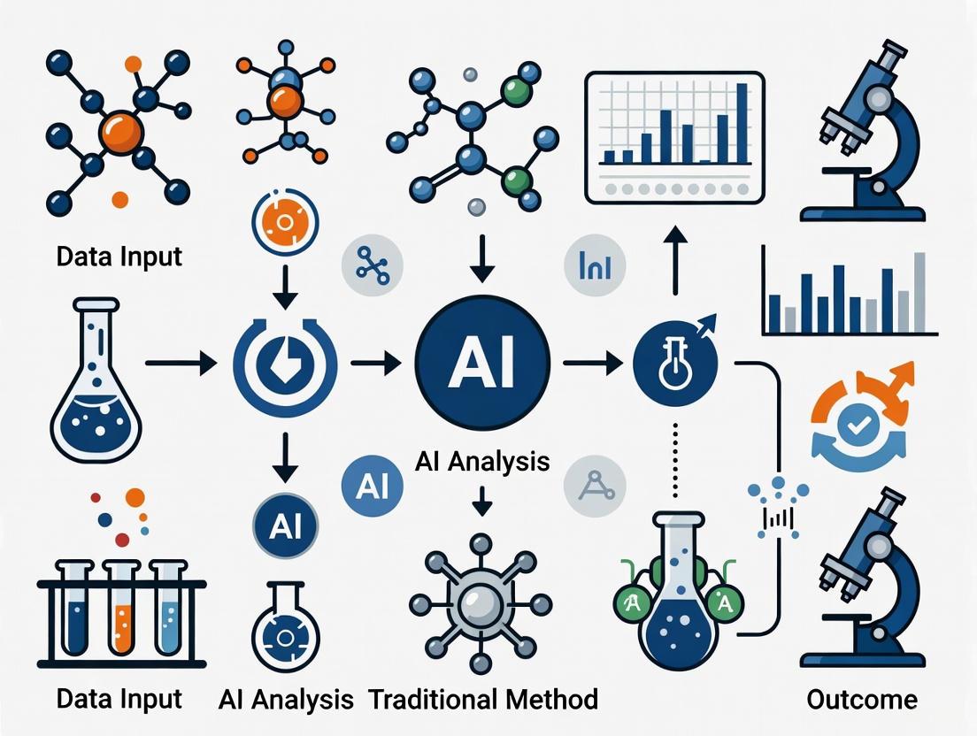
Methodological Workflow Visualization
Semen Analysis Variability and AI Solutions
Machine Learning Model for Sperm Quality Assessment
Challenges in Standardization and Adherence to WHO Guidelines
Frequently Asked Questions (FAQs)
Q1: What is the core challenge with semen analysis standardization across different laboratories? A1: The primary challenge is a significant lack of standardization in how semen analysis is performed and reported. A survey of hundreds of laboratories revealed considerable variation in the parameters reported, the lower limits of normality used, and the performance of quality control. For instance, while most labs report sperm count (94%) and motility (95%), far fewer routinely report the abstinence period (64%) or the morphology criteria used (60%). Crucially, quality control for key parameters like sperm counts, motility, and morphology was performed by only 29%, 41%, and 41% of laboratories, respectively [7].
Q2: How does the 6th edition of the WHO manual address the criticism faced by the previous edition? A2: The 5th edition of the WHO manual faced criticism for its reference ranges, which some experts argued were inadequate to represent the general population due to issues like geographic over- and under-representation and technical variations between labs [8]. The 6th edition, released in 2021, addressed this by expanding its data set to include 3589 fertile men from regions previously under-represented, such as Southern Europe, Asia, and Africa. Furthermore, it places a stronger emphasis on quality control, improved standardization, technician training, and equipment calibration. A key change is the clarification that the fifth centile reference values are just one method for interpreting results and are not sufficient alone to diagnose male infertility [8].
Q3: Can Artificial Intelligence (AI) truly reduce the subjectivity in traditional semen analysis? A3: Yes, evidence shows that AI can significantly address subjectivity. Traditional manual assessment is prone to inter-observer variability [9]. AI algorithms, particularly deep learning models, can automate the evaluation of sperm concentration, motility, and morphology with high consistency. For example, one study using an AI image recognition algorithm found a strong correlation with manual analysis for motile sperm concentration (r=0.84, p<0.001) [10]. AI models have demonstrated high accuracy in tasks like predicting sperm concentration (93% accuracy with an FSNN model) and categorizing sperm motility (89% accuracy with a Support Vector Machine) [10].
Q4: What are the performance metrics of common AI models used for semen analysis? A4: Different AI models excel at evaluating specific semen parameters. The table below summarizes the performance of various algorithms as reported in recent research.
Table 1: Performance Metrics of AI Models in Semen Analysis
| Parameter Analyzed | AI Model/Algorithm | Reported Performance | Sample Context |
|---|---|---|---|
| Sperm Concentration | Full-Spectrum Neural Network (FSNN) | 93% Accuracy [10] | Semen |
| Sperm Concentration | Artificial Neural Network (ANN) | 90% Accuracy, 95.45% Sensitivity [10] | Semen |
| Sperm Motility | Support Vector Machine (SVM) | 89% Accuracy [10] | 2817 sperm [9] |
| Sperm Morphology | Support Vector Machine (SVM) | AUC of 88.59% [9] | 1400 sperm [9] |
| Non-Ostructive Azoospermia (Sperm Retrieval) | Gradient Boosting Trees (GBT) | AUC 0.807, 91% Sensitivity [9] | 119 patients [9] |
| IVF Success Prediction | Random Forests | AUC 84.23% [9] | 486 patients [9] |
Troubleshooting Guides
Issue: High Inter-Observer Variability in Sperm Morphology Assessment
Problem: Different technicians in the same lab classify the same sperm sample differently, leading to inconsistent morphology reports (e.g., Teratozoospermia diagnosis).
Solution:
- Re-train to WHO 6th Edition Standards: Ensure all technicians are jointly re-trained using the updated, evidence-based procedures and illustrations in the 6th edition manual [11]. Conduct regular internal quality control sessions with a set of reference slides.
- Implement AI-Assisted Morphology Analysis:
- Protocol: Utilize a deep learning-based system for sperm morphology classification.
- Step 1: Prepare and stain semen smears according to WHO-recommended protocols (e.g., Diff-Quik) [8].
- Step 2: Capture high-resolution digital images of multiple, random microscopic fields at 100x oil immersion.
- Step 3: Process these images through a pre-trained Convolutional Neural Network (CNN) model. The model should be trained on a large, validated dataset to identify and classify sperm into "normal" and "abnormal" categories based on head, midpiece, and tail defects.
- Step 4: The AI system provides a standardized, quantitative output of the percentage of normal forms, eliminating technician subjectivity. The technician's role shifts to quality control of the AI's output.
Underlying Principle: Manual morphology assessment is inherently subjective. AI models like CNNs provide a consistent, objective, and quantitative assessment by applying the same classification rules to every sperm cell [10] [9]. The following workflow contrasts the traditional and AI-enhanced methods for morphology assessment, highlighting the points where subjectivity is introduced and where AI provides standardization.
Issue: Inconsistent Sperm Motility Grading and Reporting
Problem: There is poor correlation for motility parameters between labs, and even within the same lab over time, due to subjective grading of progressive vs. non-progressive motility.
Solution:
- Calibrate with Reference Videos: Use the standardized videos and detailed procedures for motility assessment provided in the WHO 6th edition to calibrate all technicians [11] [8].
- Adopt a CASA System with AI Kinematics Tracking:
- Protocol: Implement a Computer-Assisted Sperm Analysis (CASA) system enhanced with AI for kinematic analysis.
- Step 1: Load a fixed volume of well-mixed semen into a pre-warmed counting chamber.
- Step 2: Set the system to capture multiple video sequences from different fields at 37°C.
- Step 3: The AI algorithm identifies and tracks the movement path of individual spermatozoa across frames. It calculates kinematic parameters like curvilinear velocity (VCL), straight-line velocity (VSL), and amplitude of lateral head displacement (ALH).
- Step 4: Based on these objective measurements, the AI automatically classifies each sperm into progressive, non-progressive, or immotile categories according to predefined, consistent thresholds.
- Step 5: The system generates a comprehensive report including total motility, progressive motility, and detailed kinematic data.
Underlying Principle: While traditional CASA systems automate tracking, they can struggle with accurate identification. AI-enhanced systems use sophisticated models like Recurrent Neural Networks (RNNs) to more accurately track sperm paths and classify motility based on learned patterns from vast datasets, reducing operational difficulties and improving reliability [10].
The Scientist's Toolkit: Research Reagent Solutions
Table 2: Essential Reagents and Materials for Standardized Semen Analysis
| Item | Function/Brief Explanation |
|---|---|
| Diff-Quik Staining Kit | A standardized Romanowsky-type stain used for sperm morphology assessment. It provides consistent staining of sperm heads (various shades), midpieces, and tails, allowing for clear identification of structural abnormalities as per WHO guidelines [8]. |
| Eosin-Nigrosin Stain | Used for the sperm vitality test (supravital staining). Live sperm with intact membranes exclude the eosin stain and appear white, while dead sperm with damaged membranes take up the stain and appear pink/red, providing an objective measure of non-motile sperm viability [8]. |
| Pre-Warmed Counting Chambers (e.g., Makler, Leja) | Specialized slides with a fixed depth for microscopic analysis. Using standardized, pre-warmed chambers is critical for accurate and consistent assessment of sperm concentration and motility, as it eliminates volume errors and maintains sperm viability during analysis. |
| Hyaluronate Binding Assay Kit | An optional test for assessing sperm maturity and functional integrity. Mature sperm with intact membranes bind to hyaluronic acid. This kit provides standardized reagents to perform this test, which can complement basic semen parameters. |
| Sperm DNA Fragmentation (SDF) Assay Kits (e.g., SCD, TUNEL) | The WHO 6th edition introduces tests for SDF. These kits provide reagents to detect DNA damage in sperm, which is a parameter not revealed by routine analysis but crucial for understanding male fertility potential and predicting ART outcomes [8]. |
| Quality Control Sperm Slides | Commercially available fixed sperm slides with known reference values for concentration and morphology. These are essential for regular internal quality control and proficiency testing to ensure technician skills and procedures remain within standardized limits. |
Impact of Technician Subjectivity on Sperm Motility and Morphology Assessment
Frequently Asked Questions (FAQs) on Technician Subjectivity and Analysis Challenges
Q1: What are the primary sources of technician-induced variability in manual sperm motility assessment?
Manual sperm motility assessment is highly prone to subjectivity due to several factors. The "attraction of the eye to movement" often leads to overestimation of motility, particularly in samples with high sperm concentration [12]. The choice of counting chamber also introduces variability; while the World Health Organization (WHO) recommends the improved Neubauer haemocytometer, many laboratories persist in using Makler chambers due to "practical ease," despite known issues with artificial concentration increases and motility distribution errors over time [12]. Furthermore, distinguishing between rapid (A) and slow (B) progressive motility relies heavily on individual technician judgment, creating inter-operator variability [12].
Q2: Why is sperm morphology considered the most subjective parameter in semen analysis?
Sperm morphology assessment faces significant technical challenges that amplify subjectivity. The preparation of samples (smear and staining) introduces technical artifacts that can be interpreted differently [12]. According to the WHO standards, sperm morphology is divided into head, neck, and tail, with 26 types of abnormal morphology, requiring the analysis of more than 200 sperms—a process that involves a "substantial workload" and is "always influenced by the subjectivity of observers" [13]. The evaluation requires simultaneous assessment of multiple compartments (head, vacuoles, midpiece, and tail), and the lack of clear, objective boundaries for "normal" versus "abnormal" features leads to low reproducibility between technicians and laboratories [12] [13].
Q3: How does AI address the subjectivity problem in traditional semen analysis?
Artificial Intelligence (AI) algorithms, particularly deep learning models, provide objective, automated analysis by learning from large, annotated datasets of sperm images and videos [10] [13]. These models standardize the assessment by applying consistent, pre-defined criteria to every sperm cell, thereby eliminating human visual bias and fatigue [6] [9]. For instance, AI models can be trained to classify sperm morphology based on precise, measurable features (e.g., head length-to-width ratio, presence of vacuoles) and categorize motility based on quantitative kinematic trajectories, ensuring high intra- and inter-system reliability [14] [13].
Q4: What is the clinical impact of subjectivity in semen analysis?
Subjectivity in semen analysis can lead to misdiagnosis and consequently, over- or under-treatment of male infertility [12]. Inaccurate assessment may result in inappropriate selection of assisted reproductive technologies (ART). For example, flawed analysis could lead to the selection of suboptimal sperm for procedures like Intracytoplasmic Sperm Injection (ICSI), potentially compromising fertilization rates and embryo quality [9]. Furthermore, this variability complicates the comparison of results across different clinics and longitudinal monitoring of a patient's condition [12] [15].
Quantitative Data: AI vs. Traditional Analysis Performance
The tables below summarize performance data from recent studies comparing AI-based assessment with traditional manual methods.
Table 1: Performance Comparison of Sperm Morphology Assessment Methods
| Assessment Method | Correlation with Reference Method | Key Findings | Source |
|---|---|---|---|
| In-house AI Model (for unstained live sperm) | r = 0.88 with CASAr = 0.76 with Conventional Semen Analysis (CSA) | Strongest correlation with CASA; allows assessment of live sperm without staining. | [14] |
| Conventional Semen Analysis (CSA) | r = 0.57 with CASA | Weaker correlation, highlighting significant inter-method variability. | [14] |
| Support Vector Machine (SVM) Classifier | AUC-ROC: 88.59%Precision: >90% | High diagnostic efficacy in classifying sperm heads as "good" or "bad". | [13] |
Table 2: Performance of AI Models in Assessing Sperm Concentration and Motility
| Parameter | AI Model/Algorithm | Performance/Outcome | Source |
|---|---|---|---|
| Sperm Concentration | Full-Spectrum Neural Network (FSNN) | 93% prediction accuracy, significant correlation with clinical data (R² = 0.98). | [10] [16] |
| Sperm Concentration | Bemaner AI Algorithm | Moderate correlation with manual analysis (r = 0.65, p < 0.001). | [10] [16] |
| Sperm Motility | Bemaner AI Algorithm | High correlation with manual analysis (r = 0.90, p < 0.001). | [10] [16] |
| Motile Sperm Concentration | Bemaner AI Algorithm | High correlation with manual analysis (r = 0.84, p < 0.001). | [10] [16] |
Experimental Protocols for Validating AI in Sperm Analysis
Protocol 1: Developing an AI Model for Unstained Sperm Morphology Analysis
This protocol is based on a 2025 study that developed an in-house AI model to assess live sperm without staining [14].
1. Sample Preparation:
- Collect semen samples via masturbation after 2-7 days of sexual abstinence.
- Check for liquefaction within 30 minutes of ejaculation.
- Dispense a 6 µL droplet onto a standard two-chamber slide with a depth of 20 µm (e.g., Leja).
2. Image Acquisition and Dataset Creation:
- Use a confocal laser scanning microscope (e.g., LSM 800) at 40x magnification in confocal mode (Z-stack).
- Set the Z-stack interval to 0.5 µm, covering a total range of 2 µm to capture high-resolution, well-focused images.
- Manually annotate sperm images using a program like LabelImg. Embryologists and researchers should draw bounding boxes around each sperm and categorize them based on strict criteria (e.g., smooth oval head, length-to-width ratio of 1.5–2, no vacuoles, normal tail).
- Establish a high correlation coefficient (e.g., >0.95) between annotators to ensure dataset quality.
3. AI Model Training and Validation:
- Select a deep learning model such as ResNet50 (a transfer learning model) for image classification.
- Divide the dataset (e.g., 21,600 images with 12,683 annotated sperm) into training and testing sets.
- Train the model to minimize the difference between predicted and actual labels. A reported test accuracy of 93% after 150 epochs is achievable [14].
- Validate the model's performance by comparing its results for the percentage of normal sperm morphology against Computer-Aided Semen Analysis (CASA) and Conventional Semen Analysis (CSA) on the same samples.
Protocol 2: AI-Based Workflow for Integrated Sperm Motility and Morphology Assessment
Diagram 1: AI-powered semen analysis workflow.
1. Sample Loading:
- After liquefaction, load a small, well-mixed aliquot of semen into a specialized chamber (e.g., Makler or Neubauer) ensuring consistent depth and distribution of sperm. Note that chamber choice can be a source of error in manual analysis [12].
2. Data Capture:
- Place the chamber on a phase-contrast microscope with a stage warmer maintained at 37°C.
- Capture multiple video sequences (e.g., 30 frames per second) from different microscopic fields for motility analysis.
- Capture high-resolution still images from the same or a parallel preparation for morphology analysis. Smartphone-based devices or CASA systems can be used for this step [12] [10].
3. AI Analysis:
- For Motility: The AI system, often based on Convolutional Neural Networks (CNN) or region-based CNNs (R-CNN), tracks the movement of individual spermatozoa across video frames [10] [16]. It classifies each sperm into categories (e.g., progressive, non-progressive, immotile) based on calculated kinematic parameters (e.g., curvilinear velocity, straight-line velocity).
- For Morphology: A separate or integrated AI model analyzes the still images. It segments each sperm into head, midpiece, and tail, and extracts morphometric features (e.g., head area, elongation, tail length) [13]. It then classifies sperm as "normal" or "abnormal" based on trained criteria, and can even specify the type of defect.
4. Data Integration and Reporting:
- The system compiles the results, providing a comprehensive report including concentration, total and progressive motility percentages, and the percentage of morphologically normal and abnormal forms, all with minimal subjective intervention.
The Scientist's Toolkit: Essential Research Reagents and Materials
Table 3: Key Reagents and Materials for AI-Enhanced Sperm Analysis Research
| Item | Function/Application | Considerations for AI Research |
|---|---|---|
| Standardized Counting Chambers (e.g., Makler, Neubauer, Leja) | Provides a consistent depth for reliable and repeatable imaging. | Critical for creating uniform image datasets for AI training and validation. The Neubauer chamber is recommended by WHO for improved accuracy [12] [14]. |
| Confocal Laser Scanning Microscope | Captures high-resolution, z-stack images of unstained, live sperm. | Essential for creating high-quality datasets for morphology AI models, as it allows for clear visualization of subcellular structures without staining [14]. |
| Phase-Contrast Microscope with Video | Enables capture of high-frame-rate videos for motility analysis. | The quality of the input video directly impacts the accuracy of AI-based motility tracking algorithms [10] [15]. |
| Staining Kits (e.g., Diff-Quik) | Used for traditional morphology assessment on fixed sperm. | Provides a benchmark for validating the performance of AI models trained on unstained sperm images [14]. |
| Public & Custom Datasets (e.g., VISEM, HSMA-DS, SVIA) | Serve as training and validation data for developing AI models. | The lack of large, standardized, high-quality annotated datasets is a major challenge. Dataset quality directly dictates model performance and generalizability [10] [14] [13]. |
| Cloud Computing or GPU Resources | Trains and runs complex deep learning models (e.g., CNN, ResNet50). | Necessary for handling the computational load of processing thousands of sperm images and videos [14] [16]. |
Clinical and Financial Consequences of Inaccurate Diagnostic Results
In clinical medicine and research, a diagnostic error is defined as the failure to either establish an accurate and timely explanation of a patient's health problem or to communicate that explanation to the patient [17]. Within the specific context of male infertility, these errors manifest as the misclassification, delayed reporting, or complete oversight of critical semen parameters such as sperm motility, morphology, and concentration. The consequences of these inaccuracies are twofold: they directly compromise patient care and introduce significant volatility into research data, thereby undermining the development of reliable therapeutic interventions.
Traditional semen analysis, reliant on manual microscopy and subjective assessment, is inherently prone to these errors. The subjectivity of human evaluation leads to substantial inter- and intra-laboratory variability [18]. This lack of standardization is a fundamental systems-based weakness in the diagnostic process, which can lead to missed or delayed diagnosis of male factor infertility. The financial impact is staggering; diagnostic errors are the most common and costly category of medical mistakes, leading to malpractice claims with average settlements exceeding $240,000 and totaling billions of dollars paid to claimants over a decade [19]. For research and drug development, these inaccuracies translate into corrupted datasets, failed experiments, and costly delays in bringing new treatments to market.
Technical Support: Troubleshooting Guide & FAQs
This section addresses common experimental challenges encountered during semen analysis and provides evidence-based solutions to enhance the reliability of your data.
Frequently Asked Questions (FAQs)
Q1: Our manual sperm motility assessments show high variance between technicians. What is the root cause and how can we mitigate it? A: The root cause is the inherent subjectivity of visual motility estimation. Manual classification into progressive, non-progressive, and immotile categories is susceptible to individual judgment and fatigue [18].
- Solution: Implement automated Computer-Aided Sperm Analysis (CASA) systems. These systems use video capture and object-tracking algorithms to provide objective, quantitative motility parameters (e.g., curvilinear velocity, straight-line velocity) [18]. For example, one deep convolutional neural network (DCNN) model demonstrated a strong correlation (Pearson’s r = 0.88) with manual assessments for progressively motile spermatozoa, while providing greater consistency [20].
Q2: How can we improve the accuracy and throughput of sperm morphology analysis? A: Traditional morphology assessment is a labor-intensive process requiring expert training. Deep learning (DL) models, particularly Convolutional Neural Networks (CNNs), can automate this classification with high accuracy.
- Solution: Employ a DL-based image analysis pipeline. Studies have shown that CNNs can classify sperm morphology (normal vs. abnormal) with accuracy exceeding 90% [20]. For instance, a Faster Region Convolutional Neural Network with an Elliptic Scanning Algorithm achieved an accuracy of 97.37% in human sperm classification [20]. These models can be trained to identify specific head, acrosome, and vacuole abnormalities in real-time, significantly increasing throughput.
Q3: Our laboratory is experiencing inconsistencies in DNA fragmentation index (DFI) results. How can we standardize this assay? A: Manual interpretation of sperm DNA fragmentation assays (e.g., SCD, TUNEL) can be variable. AI-powered analytical platforms can reduce this technical noise.
- Solution: Integrate an AI microscopy system for DFI calculation. Research indicates that AI-based assays for chromatin dispersion are not only faster (saving approximately 32 minutes per assay) but also show a strong correlation with manual methods (Spearman's rho = 0.85) and exhibit a 21% lower coefficient of variation, ensuring greater precision and reproducibility [20].
Q4: What are the common system-level failures in the diagnostic process that lead to inaccurate results in a clinical study? A: Many errors are not technical but process-related. Common failures include [21] [19]:
- Communication Gaps: Inadequate follow-up on test results or referrals.
- Sample Mix-ups: Mislabeled specimens or tests performed on the wrong patient.
- Lack of Clear Protocols: No standardized procedure for communicating critical findings.
- Solution: Implement a diagnostic tracking dashboard for real-time monitoring of samples and results. Establish and enforce standard operating procedures (SOPs) for every step, from sample collection and labeling to result reporting and data entry, creating redundant safety checks.
Troubleshooting Common Experimental Scenarios
Table 1: Troubleshooting Common Semen Analysis Experimental Challenges
| Problem | Potential Cause | Solution & Recommended Action |
|---|---|---|
| High variance in concentration counts | Improsample dilution; subjective counting; poor cell dispersion. | Action: Automate counting with an AI-powered CASA system. One study showed a high correlation (r = 0.65) for total sperm concentration compared to expert analysis [20]. |
| Inability to identify subtle morphological patterns | Limitations of human visual inspection to complex, non-linear patterns. | Action: Utilize a deep learning fusion architecture (e.g., combining Shifted Windows Vision Transformer with MobileNetV3) which has been shown to accurately classify sperm with 95.4% accuracy, outperforming benchmark models [20]. |
| Long processing times for complex assays (e.g., DFI) | Manual scoring of hundreds of sperm cells per sample. | Action: Adopt an AI-powered analytical platform. One validated method reduced the assay time by 32 minutes and automated the calculation, improving consistency [20]. |
| Poor generalizability of predictive models | Small, non-diverse training datasets; model overfitting. | Action: Leverage large, open-access datasets for model training and validation. Employ techniques like transfer learning to adapt models to new populations and ensure rigorous external validation [18]. |
AI Solutions for Objective Analysis and Error Reduction
Artificial Intelligence (AI), particularly machine learning (ML) and deep learning (DL), is revolutionizing semen analysis by overcoming the limitations of manual methods. AI-driven CASA systems provide objective, automated, and high-throughput evaluations of sperm quality, directly addressing the major sources of diagnostic error [18].
Performance Comparison: Manual vs. AI-Based Analysis
Table 2: Quantitative Performance of AI Models in Semen Analysis (Based on Published Studies)
| Analysis Type | AI Methodology | Reported Performance Metric | Comparative Manual Limitation |
|---|---|---|---|
| Motility Analysis | Deep Convolutional Neural Network (DCNN) | Pearson’s r = 0.88 for progressively motile sperm [20] | High inter-technician variability; subjective classification. |
| Morphology Classification | Faster Region-CNN with Elliptic Scan | 97.37% accuracy (normal vs. abnormal) [20] | Labor-intensive; requires high expertise; lower consistency. |
| Morphology Classification | Convolutional Neural Network (CNN) | Up to 90.73% classification accuracy [20] | As above. |
| Sperm Head Detection | Region-Based CNN | 91.77% detection accuracy [20] | Inconsistent identification of sperm heads in dense fields. |
| DNA Fragmentation | AI Microscopy & Auto-Calculation | Spearman's rho = 0.85 vs. manual; 21% lower coefficient of variation [20] | Subjective interpretation; high result variability. |
The "black-box" nature of some complex AI algorithms remains a challenge, necessitating rigorous clinical validation and model interpretability efforts to ensure their reliability and adoption in clinical practice [18].
Experimental Protocols for AI-Assisted Semen Analysis
Protocol: Automated Sperm Morphology Classification Using Deep Learning
Objective: To automatically and accurately classify human spermatozoa into "normal" and "abnormal" morphological categories using a Deep Convolutional Neural Network.
Materials:
- Stained semen smears (e.g., Diff-Quik, Papanicolaou)
- Microscope with digital camera
- High-performance computing workstation (GPU recommended)
- Software: Python with deep learning libraries (e.g., TensorFlow, PyTorch)
Methodology:
- Image Acquisition: Capture high-resolution digital images (at least 100x oil immersion) of multiple, random microscopic fields from the stained semen smear.
- Data Curation & Annotation:
- A minimum of 1,000-10,000 sperm images should be used for robust model development.
- Each sperm image must be meticulously annotated by multiple trained embryologists according to WHO criteria [18]. The annotations will serve as the "ground truth" for training.
- Split the dataset into training (70%), validation (15%), and test (15%) sets.
- Model Training:
- Utilize a pre-trained CNN architecture (e.g., ResNet, MobileNet) and apply transfer learning.
- The model will learn to extract features (edges, shapes, textures) from the images and associate them with the expert annotations.
- Train the model to minimize the difference between its predictions and the human-provided labels.
- Validation & Testing:
- Validate the model's performance on the held-out test set.
- Evaluate using metrics such as accuracy, sensitivity, specificity, and F1-score [20].
- The model's classification should be compared against the performance of a new human evaluator to benchmark its efficacy.
Protocol: Integrating AI-Generated Results into Clinical Workflows
Objective: To establish a reliable system for incorporating AI-CASA outputs into the patient diagnostic pathway, minimizing communication errors.
Materials:
- AI-CASA system
- Laboratory Information System (LIS) or Electronic Health Record (EHR)
- Standardized reporting template
Methodology:
- Result Verification: For initial deployment, implement a dual-reader system where AI-generated results are verified by a human technologist. Flag results that fall outside pre-defined confidence thresholds for expert review.
- Structured Reporting: Automatically populate a standardized diagnostic report template with the AI-derived parameters (concentration, motility, morphology, DFI).
- System Integration: Feed the finalized report directly into the LIS/EHR to ensure timely and accurate data transfer, eliminating manual transcription errors.
- Follow-up Protocol: Establish clear, multi-modal communication protocols (e.g., EHR alert, email, SMS) to ensure clinicians and patients are notified of critical results, with documentation of each contact attempt [19].
The Scientist's Toolkit: Research Reagent Solutions
Table 3: Essential Materials for AI-Enhanced Semen Analysis Research
| Item | Function in Research |
|---|---|
| AI-CASA System | Core hardware/software for automated, high-throughput sperm tracking and parameter quantification (e.g., motility, concentration). Replaces subjective manual microscopy [18]. |
| Staining Kits (Diff-Quik, Papanicolaou) | Prepare sperm smears for morphology imaging. Provides the consistent, high-contrast images required for training and deploying deep learning models for morphology classification [20]. |
| DNA Fragmentation Assay Kits (SCD, TUNEL) | Quantify sperm DNA damage. When combined with AI-powered image analysis, these assays become faster and more reproducible, reducing manual scoring variability [20]. |
| Curated, Public Sperm Image Datasets | Serve as a foundational resource for training and benchmarking new AI models. Mitigates the challenge of assembling large, annotated datasets from scratch and promotes research reproducibility [18]. |
| High-Performance Computer (GPU) | Provides the necessary computational power to efficiently train complex deep learning models, which is essential for processing large volumes of high-resolution image and video data [20]. |
Workflow Visualization: From Sample to AI-Augmented Diagnosis
The following diagram contrasts the traditional diagnostic pathway, which is vulnerable to human error, with an AI-augmented workflow that enhances objectivity and reliability.
The Evolving Role of Semen Analysis in Male Fertility Biomarker Discovery
Traditional semen analysis, while a cornerstone of male fertility evaluation, faces significant limitations due to its inherent subjectivity and inter-observer variability [22]. The manual assessment of parameters like sperm concentration, motility, and morphology can lead to inconsistent results, complicating both diagnosis and research [22]. Artificial Intelligence (AI) is now revolutionizing this field by introducing unprecedented levels of objectivity, accuracy, and efficiency. AI-powered systems, particularly advanced Computer-Aided Sperm Analysis (CASA) platforms, are transforming semen analysis from a basic diagnostic tool into a powerful engine for discovering novel, complex biomarkers [18] [22]. This technical support guide explores common experimental challenges and details how integrating AI methodologies can overcome these hurdles, paving the way for more precise and predictive male fertility assessment.
Troubleshooting Common Semen Analysis Experimental Challenges
FAQ 1: How can I minimize variability and subjectivity in sperm morphology assessment?
- Challenge: Manual morphology classification is highly subjective, leading to poor inter-laboratory reproducibility and inconsistent research data [22].
- Solution: Implement deep learning (DL) models for automated morphology analysis. These systems use convolutional neural networks (CNNs) trained on thousands of pre-annotated sperm images to classify sperm heads, vacuoles, and tail defects based on learned features, not predetermined rules [18] [22].
- Protocol:
- Image Acquisition: Capture high-resolution phase-contrast or stained digital images of sperm smears using a standardized microscope and camera setup.
- Model Application: Process images through a pre-trained DL architecture (e.g., a CNN). The model will segment individual sperm and extract morphological features.
- Classification: The AI assigns each sperm to a category (e.g., normal, head defect, tail defect) with a calculated probability, providing a quantitative and objective morphology score [22].
FAQ 2: What is the most effective method for identifying viable sperm in samples with severe oligospermia or azoospermia?
- Challenge: Manually finding rare viable sperm in samples from patients with severe male factor infertility is time-consuming, prone to error, and can lead to sperm damage from prolonged search times [23] [24].
- Solution: Utilize high-throughput AI imaging systems like the Sperm Tracking and Recovery (STAR) method. This technology rapidly scans millions of image fields to identify and isolate even single sperm cells [23] [24].
- Protocol:
- Sample Preparation: Place the raw semen or processed sample on a specialized chip under a microscope integrated with a high-speed camera.
- AI Scanning: The system captures over 8 million images in under an hour. An AI algorithm, trained to recognize sperm cell characteristics, scans these images in real-time [24].
- Recovery: Upon identification, the system uses gentle robotics to isolate the viable sperm into a microdroplet for subsequent use in Intracytoplasmic Sperm Injection (ICSI) [23] [24].
FAQ 3: How can we predict IVF success using semen analysis parameters more accurately?
- Challenge: Traditional parameters alone are often insufficient for predicting ART outcomes due to the complex, multifactorial nature of fertility [22].
- Solution: Employ machine learning (ML) models that integrate classical semen analysis data with advanced biomarkers and clinical patient data [22].
- Protocol:
- Data Aggregation: Compile a dataset including standard semen parameters (count, motility, morphology), advanced metrics (sperm DNA fragmentation index, vacuolation), and female partner factors (age, hormone levels).
- Model Training: Train an ensemble ML model (e.g., Random Forest, Support Vector Machine) on this dataset with known IVF outcomes (fertilization rate, pregnancy).
- Prediction: The trained model can analyze new patient data to generate a personalized prediction of IVF success probability, aiding in clinical decision-making [22].
Quantitative Performance of AI Models in Semen Analysis
The table below summarizes the performance of various AI models as reported in recent research, providing a benchmark for experimental planning and validation.
Table 1: Performance Metrics of AI Models in Key Sperm Analysis Applications
| Analysis Focus | AI Model Used | Reported Performance | Dataset Size | Citation |
|---|---|---|---|---|
| Sperm Morphology | Support Vector Machine (SVM) | AUC of 88.59% | 1,400 sperm | [22] |
| Sperm Motility | Support Vector Machine (SVM) | Accuracy of 89.9% | 2,817 sperm | [22] |
| Non-Obstructive Azoospermia (Sperm Retrieval Prediction) | Gradient Boosting Trees (GBT) | AUC 0.807, 91% Sensitivity | 119 patients | [22] |
| IVF Success Prediction | Random Forests | AUC 84.23% | 486 patients | [22] |
| Male Infertility Risk from Blood Test | Proprietary Model | ~74% Accuracy | 3,662 patients | [25] |
Experimental Workflow: Integrating AI into Semen Analysis
The following diagram illustrates the conceptual shift from a traditional, subjective workflow to an integrated, AI-enhanced pipeline for biomarker discovery and analysis.
The Scientist's Toolkit: Essential Research Reagent Solutions
For researchers developing or validating AI-based semen analysis systems, the following tools and reagents are fundamental.
Table 2: Essential Reagents and Materials for AI-Driven Sperm Analysis Research
| Item | Function in Experiment | Key Consideration |
|---|---|---|
| CASA System with AI module | Core platform for automated, high-throughput sperm tracking and morphological analysis. | Ensure software is capable of exporting raw image data and features for custom AI model training [18] [26]. |
| High-Resolution CMOS Camera | Captures the high-speed video and images required for detailed AI analysis of motility and morphology. | Frame rate and resolution are critical for capturing rapid sperm movement and fine structural details [18]. |
| Sperm Staining Kits (e.g., Papanicolaou, Hoechst) | Used for preparing slides for morphology analysis and for assessing sperm chromatin integrity and viability. | Select stains compatible with your imaging modality (brightfield/fluorescence) and that do not compromise sperm DNA for ART use [22]. |
| Sperm-Freeze Cryopreservation Media | Preserves patient samples for longitudinal studies and allows for batch testing of algorithms. | Use media that maximizes post-thaw motility and viability to ensure data quality [27]. |
| Microfluidic Sperm Sorting Chips | Prepares samples by isolating motile sperm and reducing debris, which improves subsequent AI analysis accuracy. | Ideal for processing severe oligospermic samples before introducing them to an AI search system like STAR [24]. |
| Annotated Sperm Image Datasets | Serves as the "ground truth" for training, validating, and benchmarking new machine learning models. | Seek large, diverse, and publicly available datasets to ensure model robustness and generalizability [18] [22]. |
The integration of AI into semen analysis is fundamentally reshaping the landscape of male fertility research. By overcoming the critical limitations of subjectivity and variability, AI-powered CASA systems are enabling the discovery of subtle, complex biomarkers beyond the reach of conventional microscopy. This technical guide outlines the practical pathways for researchers to troubleshoot common experimental issues, leverage quantitative AI models, and adopt the necessary tools to advance the field. As these technologies continue to evolve and undergo rigorous clinical validation, they promise to deliver a new era of personalized, predictive, and precise male fertility diagnostics.
AI in Action: Methodological Advances and Research Applications in Sperm Analysis
Troubleshooting Guide: Model Performance and Implementation
Q1: My conventional machine learning model for sperm morphology classification is underperforming. What could be the cause?
A: Underperformance in conventional ML models often stems from their fundamental reliance on handcrafted feature extraction [28]. These models typically use algorithms like Support Vector Machines (SVM) and k-means clustering but depend on manually designed image features such as grayscale intensity, edge detection, and contour analysis [28]. This approach limits their ability to capture the complex, hierarchical features of sperm cells, such as subtle head vacuoles or tail defects [28] [29]. For instance, while a Bayesian Density Estimation model can achieve 90% accuracy in classifying sperm heads into broad categories, its performance is limited by focusing predominantly on shape-based features [28].
- Solution: Transition to a deep learning (DL) architecture. DL models, particularly Convolutional Neural Networks (CNNs) enhanced with attention mechanisms like the Convolutional Block Attention Module (CBAM), automatically learn relevant features from raw image data [29]. One study reported that integrating CBAM with ResNet50 and employing deep feature engineering boosted performance by over 8% compared to baseline models, achieving test accuracies of up to 96.08% [29].
Q2: I am struggling with a lack of high-quality, annotated sperm image data for training. What are my options?
A: The lack of standardized, high-quality datasets is a major challenge in this field [28]. Existing public datasets (e.g., SMIDS, HuSHeM, VISEM-Tracking) often suffer from limitations like low resolution, small sample sizes, and insufficient categorical coverage [28].
- Solution 1: Utilize Synthetic Data Generation. Tools like AndroGen provide an open-source solution for generating realistic, customizable synthetic sperm images [30]. This approach reduces the dependency on large, manually annotated real-world datasets and minimizes privacy concerns. The synthetic images produced have been validated using metrics like Fréchet Inception Distance (FID) to confirm their similarity to real images [30].
- Solution 2: Leverage Deep Feature Engineering. When data is scarce, a hybrid approach can be highly effective. This involves using a pre-trained CNN (like ResNet50) as a feature extractor and then applying classical feature selection methods (e.g., Principal Component Analysis (PCA), Chi-square test) before classification with an SVM [29]. This method has been shown to achieve state-of-the-art results even with limited data [29].
Q3: How can I reduce the subjectivity and time required for sperm morphology analysis in a clinical setting?
A: Traditional manual analysis is highly subjective, with reported inter-observer variability as high as 40%, and can take 30-45 minutes per sample [29].
- Solution: Implement an automated DL-based analysis system. A framework combining CNNs with attention mechanisms not only standardizes the assessment but also drastically reduces processing time. Research demonstrates that such systems can evaluate a sample in less than one minute, down from 30-45 minutes, while providing objective, reproducible results [29]. This is achieved by the model's ability to accurately segment sperm structures (head, neck, tail) and classify morphology without human intervention [28].
Frequently Asked Questions (FAQs)
Q1: What is the key architectural difference between conventional ML and DL for sperm analysis?
A: The core difference lies in feature learning. Conventional ML requires domain experts to manually define and extract relevant features (e.g., head shape descriptors) from sperm images, which are then fed into a classifier [28]. In contrast, Deep Learning uses multi-layered neural networks to automatically discover and learn the most discriminative features directly from the raw pixel data, capturing more complex and abstract patterns [28] [29].
Q2: My DL model for sperm classification is overfitting. What strategies can I use?
A: Overfitting is common when training complex models on limited medical data. You can employ several strategies:
- Data Augmentation: Artificially expand your dataset using transformations like rotation, scaling, and flipping.
- Synthetic Data: Incorporate tools like AndroGen to generate more training samples [30].
- Deep Feature Engineering: Instead of training an end-to-end CNN from scratch, use a pre-trained network for feature extraction and apply dimensionality reduction (like PCA) before classification. This hybrid approach has been shown to improve accuracy and generalizability [29].
- Attention Mechanisms: Integrate modules like CBAM, which help the model focus on morphologically relevant parts of the sperm (e.g., head shape, acrosome), making learning more efficient and robust [29].
Q3: Can I use a pre-trained model for sperm morphology analysis, or do I need to train one from scratch?
A: Using a pre-trained model (transfer learning) is a highly effective and common practice. You can take a network pre-trained on a large dataset (e.g., ImageNet) and fine-tune it on your specific sperm image dataset. For even better performance, research shows that adding attention modules to a pre-trained backbone (like ResNet50) and applying deep feature engineering (e.g., extracting features from GAP/GMP layers and using SVM for classification) can yield superior results compared to training from scratch or using standard transfer learning [29].
Quantitative Data Comparison
The table below summarizes key performance metrics and dataset characteristics from cited studies to facilitate easy comparison.
Table 1: Performance Metrics of Sperm Analysis Algorithms
| Study / Model | Dataset | Key Architecture/Technique | Reported Performance |
|---|---|---|---|
| Bijar A et al. [28] | Not Specified | Conventional ML (Bayesian Density Estimation) | 90% accuracy (4-class head morphology) |
| Spencer et al. [29] | HuSHeM | Stacked Ensemble of CNNs (VGG16, ResNet-34, DenseNet) | 95.2% accuracy |
| Kılıç Ş (2025) [29] | SMIDS | CBAM-enhanced ResNet50 + Deep Feature Engineering | 96.08% accuracy |
| Kılıç Ş (2025) [29] | HuSHeM | CBAM-enhanced ResNet50 + Deep Feature Engineering | 96.77% accuracy |
Table 2: Publicly Available Sperm Image Datasets
| Dataset Name | Key Characteristics | Ground Truth | Image Count |
|---|---|---|---|
| HSMA-DS [28] | Non-stained, noisy, low resolution | Classification | 1,457 images |
| HuSHeM [28] | Stained, higher resolution | Classification | 216 publicly available sperm head images |
| SMIDS [28] [29] | Stained sperm images | Classification | 3,000 images (3 classes) |
| VISEM-Tracking [28] | Low-resolution, unstained, includes videos | Detection, Tracking, Regression | 656,334 annotated objects |
| SVIA [28] | Low-resolution, unstained, videos & images | Detection, Segmentation, Classification | 125,000 annotated instances |
Experimental Protocol: Implementing a Deep Feature Engineering Workflow
This protocol outlines the methodology for achieving state-of-the-art sperm morphology classification, as detailed in [29].
Objective: To classify sperm morphology with high accuracy using a hybrid deep feature engineering pipeline.
Materials:
- Datasets: SMIDS or HuSHeM dataset.
- Software: Python with deep learning libraries (e.g., TensorFlow, PyTorch), scikit-learn.
- Hardware: GPU-enabled computer for efficient model training.
Methodology:
- Backbone Feature Extraction:
- Use a pre-trained CNN (e.g., ResNet50) as a feature extractor.
- Enhance the CNN by integrating a Convolutional Block Attention Module (CBAM) to help the model focus on salient sperm structures.
- Pass each sperm image through the network to generate high-dimensional feature maps.
Feature Pooling:
- Extract deep features from multiple layers of the network. Key layers include:
- Convolutional Block Attention Module (CBAM) output.
- Global Average Pooling (GAP) layer.
- Global Max Pooling (GMP) layer.
- Pre-final fully connected layer.
- Extract deep features from multiple layers of the network. Key layers include:
Feature Selection & Dimensionality Reduction:
- Apply feature selection algorithms to the pooled features to reduce noise and overfitting. Methods include:
- Principal Component Analysis (PCA)
- Chi-square test
- Random Forest feature importance
- Variance thresholding
- This step creates a compact, discriminative feature set.
- Apply feature selection algorithms to the pooled features to reduce noise and overfitting. Methods include:
Classification:
- Feed the optimized feature set into a shallow classifier. Research indicates that a Support Vector Machine (SVM) with a Radial Basis Function (RBF) kernel often yields the best performance [29].
- Perform evaluation using rigorous methods like 5-fold cross-validation and report metrics such as accuracy, precision, and recall.
Workflow and Architecture Visualization
The Scientist's Toolkit: Essential Research Reagents & Materials
The table below lists key resources for developing AI-based sperm morphology analysis systems.
Table 3: Research Reagent Solutions
| Item / Resource | Type | Function / Application |
|---|---|---|
| SMIDS Dataset [28] [29] | Dataset | A stained sperm image dataset with 3,000 images across 3 classes, used for training and benchmarking classification models. |
| HuSHeM Dataset [28] [29] | Dataset | A higher-resolution dataset of stained sperm heads, useful for focused head morphology analysis. |
| AndroGen Software [30] | Software Tool | Open-source software for generating customizable synthetic sperm images, mitigating data scarcity and annotation effort. |
| ResNet50 Architecture [29] | Algorithm | A robust, pre-trained Convolutional Neural Network often used as a backbone for feature extraction in deep learning pipelines. |
| Convolutional Block Attention Module (CBAM) [29] | Algorithm | An attention mechanism that enhances CNNs by sequentially focusing on important channels and spatial regions in feature maps. |
| Support Vector Machine (SVM) [28] [29] | Algorithm | A powerful classifier often used in conventional ML and, with RBF kernel, in deep feature engineering pipelines for final classification. |
Automated Sperm Motility Tracking and Kinematic Profiling with AI-CASA
Technical Support Center
Artificial Intelligence in Computer-Assisted Semen Analysis (AI-CASA) represents a paradigm shift in andrology, moving from subjective manual assessments to objective, data-rich evaluations. These systems are engineered to process large numbers of images with high consistency, accuracy, and repeatability, addressing the significant inter- and intra-laboratory variability of manual methods [31] [32]. By leveraging machine learning (ML), artificial neural networks (ANN), and deep learning (DL), AI-CASA provides automated, quantitative data on key sperm kinematics and motility parameters, transforming the diagnosis of male infertility and research in drug development [4].
Troubleshooting Common Experimental Issues
Issue 1: Inaccurate Sperm Detection and Segmentation
- Problem: The system fails to correctly identify and separate individual sperm cells in the video footage. This can be due to high cell density, debris, or non-sperm cells in the sample.
- Solution:
- Sample Preparation: Dilute the semen sample using a recommended buffer or medium to reduce cell density and minimize collisions and overlapping trajectories [32]. Ensure the use of prepared chambers to control sample depth.
- Algorithm Validation: Use publicly available simulation software to test your segmentation algorithm's performance against a known ground truth under varying levels of image noise and cell concentration [31]. Compare algorithms using precision, recall, and the Optimal Subpattern Assignment (OSPA) metric [31].
Issue 2: Erroneous Sperm Tracking and Trajectory Analysis
- Problem: Generated sperm paths are fragmented or incorrect, especially when sperm cells are in close proximity or collide.
- Solution:
- Tracking Algorithm Selection: Implement and test more robust multi-object tracking algorithms. Research indicates that algorithms like the Joint Probabilistic Data Association Filter (JPDAF) are better at tracking individual spermatozoa during collisions and close encounters than simpler methods like Nearest Neighbor (NN) [31].
- Performance Metrics: Quantify tracking performance using Multi-Object Tracking Accuracy (MOTA) and Multi-Object Tracking Precision (MOTP) to objectively compare different algorithms [31].
Issue 3: Discrepancies Between CASA Motility Results and Manual Assessments
- Problem: The motility percentages (progressive, non-progressive, immotile) reported by the AI-CASA system do not align with manual microscopy estimates.
- Solution:
- Standardize Motility Classification: Ensure the kinematic thresholds for classifying sperm movement (e.g., Curvilinear Velocity - VCL) are set according to recognized laboratory manuals and are consistent across analyses [33].
- System-Specific Calibration: Be aware that different CASA systems and even different settings (e.g., frame rate, counting chamber) can influence kinematic results [33]. Validate your specific system against video recordings of known motility, if possible [34]. Do not expect perfect parity with manual estimates, which are inherently subjective.
Issue 4: Poor Performance of a Custom Machine Learning Model
- Problem: A self-developed ML model for predicting sperm motility or fertility performs poorly on new, unseen data.
- Solution:
- Data Augmentation: Increase the size and diversity of your training dataset. Use simulation tools to generate life-like semen images with controllable parameters (e.g., noise levels, sperm concentration, swimming patterns) to train and validate your models more effectively [31].
- Multimodal Data: Explore incorporating participant data (e.g., age, BMI) alongside video analysis, though one study found that adding this data did not significantly improve deep learning-based motility prediction [32]. The primary focus should remain on high-quality video data.
Frequently Asked Questions (FAQs)
Q1: What are the core AI techniques used in modern CASA systems? A1: Modern AI-CASA primarily utilizes:
- Machine Learning (ML): Develops automated algorithms from large datasets to find patterns and associations for prediction and classification [4].
- Deep Learning (DL): A subset of ML that uses extensive neural networks to automate feature extraction, ideal for complex image recognition tasks like identifying sperm cells and their movements [4] [32].
- Convolutional Neural Networks (CNNs): A class of DL particularly effective for analyzing visual imagery, used directly to predict sperm motility from video sequences [32].
Q2: What are the standard sperm swimming modes that AI-CASA should identify? A2: Advanced tracking and simulation models are designed to recognize four primary swimming modes observed in 2D images [31]:
- Linear Mean: Progressive movement in a relatively straight line.
- Circular: Movement along a circular path.
- Hyperactive: High-amplitude, asymmetrical beating patterns.
- Immotile: Non-motile or dead spermatozoa.
Q3: Why is my CASA system's concentration measurement inaccurate? A3: Inaccurate concentration counts often stem from:
- Sample Purity: The presence of debris, agglutinations, or other cells can be mistakenly identified as sperm [32].
- Calibration: The system must be calibrated according to the manufacturer's protocol. Some systems, like SAMi, are validated against the haemocytometer method, which is the WHO gold standard [34].
- Settings: Incorrect settings for cell size or intensity can lead to misidentification.
Q4: How can I validate the kinematic and motility output of my AI-CASA system? A4: Validation should be a multi-step process:
- Internal Checks: Use system-provided control videos or samples.
- Cross-Referencing: Compare results with manual assessments performed by experienced technicians, acknowledging the inherent subjectivity of the manual method [34].
- Simulation Tools: Employ computational simulators that generate videos with known kinematic parameters to serve as a ground truth for objective algorithm validation [31].
Experimental Protocols & Workflows
Protocol: Automated Motility Analysis Using Deep Learning
Objective: To automatically predict the percentage of progressive, non-progressive, and immotile spermatozoa from raw video footage using a convolutional neural network (CNN).
- Sample Collection & Preparation: Collect semen samples and prepare them according to WHO guidelines. Pipette 10μl onto a glass slide, cover with a 22x22 mm cover slip, and place on a microscope with a heated stage (37°C) [32].
- Video Acquisition: Record videos using a microscope with phase contrast optics at 400x magnification. Capture AVI files at a high frame rate (e.g., 50 frames-per-second) for 2-7 minutes [32].
- Data Preprocessing: Extract sequences of frames from the videos. Normalize pixel intensity and resize frames to the required input dimensions of the chosen CNN model.
- Model Training: Train a CNN (e.g., ResNet, VGG) using a labeled dataset where the motility values (progressive, non-progressive, immotile) are the dependent variables. Use a three-fold cross-validation approach to ensure robustness [32].
- Prediction & Evaluation: Use the trained model to predict motility values on new test videos. Evaluate performance by calculating the Mean Absolute Error (MAE) against manual assessments and ensure results are statistically significant [32].
Workflow Diagram: AI-CASA Analysis Pipeline
The following diagram illustrates the logical workflow for automated sperm analysis, from sample preparation to result generation.
The Scientist's Toolkit: Essential Research Reagents & Materials
The following table details key materials and solutions required for conducting robust AI-CASA experiments.
Table 1: Essential Research Reagents and Materials for AI-CASA
| Item | Function & Application in AI-CASA |
|---|---|
| Phase Contrast Microscope | Essential for high-quality, high-contrast video recording of unstained sperm cells, enabling clear visualization of sperm heads and flagella for accurate tracking [32]. |
| Heated Stage (37°C) | Maintains physiological temperature during analysis, which is critical for preserving native sperm motility and obtaining biologically relevant kinematic data [32]. |
| Standardized Counting Chambers | (e.g., Makler, Leja) Controls sample depth, ensuring consistent imaging conditions and reliable concentration and motility measurements across different experiments [33]. |
| Sperm Wash Media / Buffer | Used to dilute raw semen samples, reducing cell density and debris. This minimizes sperm collisions and particle interference, significantly improving tracking algorithm accuracy [32]. |
| Public Simulation Software | Provides a ground truth for validating CASA algorithms. Allows performance testing of segmentation and tracking methods against simulated semen images with known, controllable parameters [31]. |
Quantitative Data & Kinematic Parameters
AI-CASA systems extract a wide array of quantitative kinematic measurements. The table below summarizes the core parameters used to characterize sperm movement.
Table 2: Key Sperm Kinematic Parameters Measured by AI-CASA
| Parameter | Abbreviation | Description | Clinical/Research Relevance |
|---|---|---|---|
| Curvilinear Velocity | VCL | The time-average velocity of the sperm head along its actual curvilinear path. | Identifies hyperactivated motility; high VCL is often linked to high energy and fertilization potential. |
| Straight-Line Velocity | VSL | The velocity of the sperm head along the straight line from its start to end position. | Key for assessing progressive motility; lower VSL indicates less efficient forward progression. |
| Average Path Velocity | VAP | The velocity of the sperm head along its spatially averaged path. | Used in conjunction with VSL and VCL to calculate other indices of movement quality. |
| Linearity | LIN | The linearity of the curvilinear path (LIN = VSL/VCL). | Measures the straightness of the track; high LIN indicates very direct movement. |
| Wobble | WOB | The oscillation of the sperm head along its path (WOB = VAP/VCL). | Describes the tightness of the head's movement pattern around the average path. |
| Amplitude of Lateral Head Displacement | ALH | The mean width of the head oscillations as the sperm moves. | A higher ALH is characteristic of hyperactive swimming, which is crucial for fertilization. |
AI for Unstained, Live Sperm Morphology Assessment Using Confocal Microscopy
Experimental Protocols
Core Methodology for AI-Based Unstained Sperm Assessment
This protocol details the key experimental procedure for developing and validating an AI model to assess unstained, live sperm morphology using confocal laser scanning microscopy, as established in recent studies [14].
Sample Preparation:
- Collect semen samples through masturbation into sterile containers after 2-7 days of sexual abstinence [14].
- Check for liquefaction within 30 minutes of ejaculation [14].
- Dispense a 6 μL droplet of the sample onto a standard two-chamber slide with a depth of 20 μm (e.g., Leja) [14].
Image Acquisition with Confocal Microscopy:
- Use a confocal laser scanning microscope (e.g., LSM 800) operating at 40× magnification in confocal mode (LSM, Z-stack) [14].
- Set the Z-stack interval to 0.5 μm, covering a total range of 2 μm [14].
- Configure frame time to 633.03 ms with an image size of 512 × 512 pixels [14].
- Capture at least 200 sperm images per sample, with each capture containing 2-3 sperm [14].
Dataset Creation and Annotation:
- Manually annotate well-focused sperm images using bounding boxes via annotation programs (e.g., LabelImg) [14].
- Categorize each sperm image into one of nine datasets based on strict morphological criteria derived from WHO Laboratory Manual for the Examination and Processing of Human Semen (sixth edition) guidelines [14].
- For normal sperm morphology classification, ensure the sperm meets all criteria across all five captured frames: smooth oval head, length-to-width ratio of 1.5-2, no vacuoles, slender and regular neck, uniform tail calibre, and cytoplasmic droplets less than one-third of the sperm head [14].
AI Model Training:
- Select a ResNet50 transfer learning model for sperm classification tasks [14].
- Train the model on the annotated dataset, using a subset of 9,000 images (4,500 normal and 4,500 abnormal morphology) derived from 32 pattern samples [14].
- Validate model performance using a separate test dataset not used during training [14].
- Achieve model convergence over 150 epochs, with a batch size of 900 [14].
Comparison with Traditional Methods
Computer-Aided Semen Analysis (CASA) Protocol:
- Allow samples to air-dry on glass slides [14].
- Stain with Diff-Quik stain (a Romanowsky stain variant) [14].
- Assess at least 200 sperm under 100× magnification using a CASA system (e.g., IVOS II, Hamilton Thorne) [14].
- Evaluate normal sperm morphology according to Tygerberg strict criteria implemented in the DIMENSIONS II Sperm Morphology Analysis software [14].
Conventional Semen Analysis Protocol:
- Perform assessments according to WHO sixth edition guidelines and Björndahl methods [14].
- Assess sperm parameters including concentration and motility using standardized protocols [14].
- Use wet preparations with 6 μL semen drops on specialized slides (e.g., LEJA slides) under coverslips creating 20 μm preparation depth [14].
- Evaluate at least 200 spermatozoa across five microscopic fields per replicate [14].
Performance Data
Table 1: Comparison of Sperm Morphology Assessment Methods
| Assessment Method | Correlation with CASA (r-value) | Correlation with Conventional Analysis (r-value) | Normal Morphology Detection Rate | Key Advantages |
|---|---|---|---|---|
| In-house AI Model (Unstained) | 0.88 [14] | 0.76 [14] | Significantly higher than CASA [14] | Preserves sperm viability; no staining required [14] |
| Computer-Aided Semen Analysis (CASA) | - | 0.57 [14] | Lower than AI and conventional methods [14] | Standardized automated assessment [14] |
| Conventional Semen Analysis | 0.57 [14] | - | Significantly higher than CASA [14] | Established reference method [14] |
Table 2: AI Model Performance Metrics
| Performance Parameter | Value | Details |
|---|---|---|
| Test Accuracy | 0.93 [14] | After 150 epochs [14] |
| Precision (Abnormal Sperm) | 0.95 [14] | - |
| Recall (Abnormal Sperm) | 0.91 [14] | - |
| Precision (Normal Sperm) | 0.91 [14] | - |
| Recall (Normal Sperm) | 0.95 [14] | - |
| Processing Speed | 0.0056 seconds/image [14] | ~139.7 seconds for 25,000 images [14] |
| Dataset Size | 12,683 annotated images [14] | From 21,600 total images [14] |
Workflow Diagrams
Experimental Workflow for AI-Based Sperm Morphology Assessment
AI Model Development and Validation Process
The Scientist's Toolkit: Research Reagent Solutions
Table 3: Essential Materials for AI-Based Unstained Sperm Analysis
| Research Reagent/Material | Function/Purpose | Specifications/Examples |
|---|---|---|
| Confocal Laser Scanning Microscope | High-resolution imaging of unstained live sperm | LSM 800; 40× magnification; Z-stack interval 0.5 μm [14] |
| Standardized Slides | Sample preparation and analysis | Two-chamber slides, 20 μm depth (e.g., Leja) [14] |
| Annotation Software | Manual labeling of sperm images for training data | LabelImg program [14] |
| AI Training Framework | Deep learning model development | ResNet50 transfer learning model [14] |
| Staining Solutions (for comparison methods) | Traditional sperm morphology assessment | Diff-Quik stain (Romanowsky stain variant) [14] |
| Computer-Aided Semen Analysis System | Automated analysis of stained sperm | IVOS II with DIMENSIONS II Software (Hamilton Thorne) [14] |
Troubleshooting Guides & FAQs
Frequently Asked Questions
Q1: What are the main advantages of using AI for unstained sperm morphology assessment compared to traditional methods?
The AI approach offers several critical advantages: (1) It preserves sperm viability since no staining is required, making selected sperm suitable for immediate use in assisted reproductive technology [14]; (2) It demonstrates stronger correlation with CASA (r=0.88) than the correlation between CASA and conventional analysis (r=0.57) [14]; (3) It detects normal sperm morphology at significantly higher rates than CASA systems [14]; (4) It minimizes subjectivity inherent in conventional semen evaluation methods [18].
Q2: What specific confocal microscopy parameters are optimal for capturing sperm images for AI analysis?
Optimal parameters include: 40× magnification in confocal mode (LSM, Z-stack), Z-stack interval of 0.5 μm covering a total range of 2 μm, frame time of 633.03 ms, and image size of 512 × 512 pixels. Each slide should capture an area of 159.7 × 159.7 μm, with at least 200 sperm images collected per sample [14].
Q3: How is normal sperm morphology defined for the AI training dataset?
Normal sperm morphology is strictly defined according to WHO sixth edition guidelines: smooth oval head, length-to-width ratio of 1.5-2, no vacuoles, slender and regular neck, uniform calibre along the tail length, and cytoplasmic droplets less than one-third of the sperm head. Normal morphology is confirmed only when sperm meet all criteria across all five captured frames [14].
Q4: What performance metrics should a properly trained AI model achieve?
A robust model should achieve: test accuracy of at least 0.93 after 150 epochs, precision of 0.95 and recall of 0.91 for detecting abnormal sperm morphology, and precision of 0.91 and recall of 0.95 for normal sperm morphology. Processing speed should approximate 0.0056 seconds per image [14].
Q5: How does this AI methodology address subjectivity in traditional semen analysis?
Traditional sperm assessment involves substantial human interpretation, leading to inter-observer variability. AI models provide objective, automated analysis that minimizes this subjectivity [18]. The annotation process for training data maintains high inter-rater reliability, with correlation coefficients of 0.95 for normal sperm detection and 1.0 for abnormal sperm detection between embryologists [14].
Troubleshooting Common Experimental Issues
Problem: Poor Image Quality from Confocal Microscopy
- Potential Cause: Improper Z-stack configuration or insufficient resolution settings.
- Solution: Ensure Z-stack interval is set to 0.5 μm with total range of 2 μm. Verify 40× magnification in confocal mode and image size of 512 × 512 pixels [14].
Problem: Low AI Model Accuracy
- Potential Cause: Insufficient training data or improper annotation.
- Solution: Expand training dataset to include at least 12,683 annotated images with equal representation of normal and abnormal morphology. Ensure annotation consistency between multiple embryologists [14].
Problem: Discrepancies Between AI and Traditional Methods
- Potential Cause: Inherent differences in assessing stained vs. unstained sperm.
- Solution: Recognize that AI and CASA may detect normal morphology at different rates. The AI model has shown stronger correlation with CASA (r=0.88) than between CASA and conventional analysis (r=0.57) [14].
Problem: Slow Processing Speed
- Potential Cause: Suboptimal batch size or hardware limitations.
- Solution: Implement batch processing of 900 images and utilize the ResNet50 transfer learning model, which processes approximately 25,000 images in 139.7 seconds [14].
High-Throughput Sperm DNA Fragmentation and Viability Analysis
Traditional semen analysis, the cornerstone of male fertility assessment, faces significant challenges due to its reliance on manual, subjective evaluation. This leads to substantial inter- and intra-observer variability, complicating the accurate diagnosis and treatment of male factor infertility, which contributes to approximately 50% of infertility cases worldwide [20] [9]. While critical, conventional parameters like sperm concentration, motility, and morphology fail to evaluate sperm DNA fragmentation (SDF), a key factor associated with reduced fertilization rates, impaired embryo development, and increased miscarriage rates [35] [36].
The integration of Artificial Intelligence (AI) and high-throughput automated systems is poised to revolutionize this field. These technologies offer objective, rapid, and quantitative assessments of sperm DNA integrity and viability, overcoming the limitations of traditional methods. This technical support center provides guidelines and troubleshooting for researchers implementing these advanced, AI-driven diagnostic approaches to standardize and enhance the accuracy of semen analysis [20] [37].
Core High-Throughput Methodologies
This section details the primary automated assays for assessing sperm DNA integrity, which form the basis of modern, objective male fertility evaluation.
Automated Sperm Chromatin Dispersion (SCD) Assay
The SCD assay is a common method for assessing sperm DNA fragmentation. The traditional manual method is low-throughput and suffers from inter-observer variations. The automated, high-throughput version leverages automated optical microscopy and chromatin diffusion-based analysis [38] [39].
- Principle: Sperm with fragmented DNA display minimal or no halo of dispersed chromatin after acid denaturation and removal of nuclear proteins, while sperm with intact DNA show large, clear halos.
- High-Throughput Advantage: This automated method can analyze a sample of thousands of sperm in under 10 minutes, a task that would otherwise require about 5 hours manually, making it a clinically viable workflow [38].
- Validation: The automated measurement of population-level sperm DNA fragmentation (%sDF) shows excellent agreement with the flow cytometry-based Sperm Chromatin Structure Assay (SCSA), a gold standard, with a correlation of R² = 0.98 [39]. It also provides a quantitative single-cell metric, the sperm DNA diffusion coefficient (DDNA), which correlates with SCSA's DNA Fragmentation Index (DFI) at R² = 0.96 [38].
AI-Based Digital TUNEL Assay Analysis
The terminal deoxynucleotidyl transferase dUTP nick end labeling (TUNEL) assay is a reliable method for detecting SDF by labeling DNA strand breaks. AI models are now being developed to digitally replicate this assay using phase-contrast microscopy images alone, eliminating the need for destructive staining procedures [35].
- Principle: The assay identifies single and double-strand DNA breaks by enzymatically labeling the free 3’-OH termini with modified nucleotides. Sperm with fragmented DNA exhibit bright fluorescence (TUNEL-positive), while those with intact DNA show minimal background staining (TUNEL-negative) [35].
- AI Approach: One study validates a novel morphology-assisted ensemble AI model that combines image processing with state-of-the-art transformer-based machine learning models (GC-ViT) to predict DNA fragmentation from phase-contrast images [35].
- Key Benefit: This is a non-destructive methodology, allowing for real-time sperm selection based on DNA integrity for use in assisted reproductive technologies (ART) like IVF and ICSI, unlike the traditional TUNEL assay which renders sperm non-viable [35].
- Performance: The proposed AI framework has demonstrated a sensitivity of 60% and specificity of 75% in detecting SDF from phase-contrast images, benchmarked against the TUNEL gold standard [35].
Smartphone-Based Automated Semen Analysis System
Emerging point-of-care technologies leverage smartphones for automated, cost-effective semen analysis.
- Platform: A smartphone-based system uses an optical attachment and custom applications to measure sperm DNA fragmentation, viability, and hyaluronic binding assay (HBA) scores [40].
- DNA Fragmentation Workflow: For DNA fragmentation assessment (compatible with kits like Halosperm), stained slides are imaged. An adaptive thresholding algorithm isolates sperm cell images to calculate the area of the sperm heads. Sperm with significantly larger heads are classified as having haloing (non-fragmented), while those with smaller heads are classified as having no haloing (fragmented) [40].
- Performance: This system has been tested on 47 human semen samples for DNA fragmentation, showing compatibility with the smartphone-based approach [40].
Table 1: Comparison of High-Throughput Sperm DNA Fragmentation Assays
| Assay Method | Throughput | Key Metric | Correlation with Gold Standard | Primary Advantage |
|---|---|---|---|---|
| Automated SCD [38] [39] | High (1000s of sperm in <10 min) | %sDF, DDNA | R² = 0.98 with SCSA %DFI | Standardization and speed; prevents inter-observer variation. |
| AI-Digital TUNEL [35] | Medium (100s of sperm) | Binary (Fragmented/Intact) | Sensitivity: 60%, Specificity: 75% | Non-destructive; allows subsequent use of sperm in ART. |
| Smartphone-Based SCD [40] | Medium | Binary (Fragmented/Intact) | Compatible with clinical kit results | Low-cost, point-of-care potential; automated classification. |
Artificial Intelligence Approaches and Performance
AI, particularly machine learning and deep learning, is at the forefront of automating and enhancing the accuracy of sperm analysis.
AI Model Architectures for SDF Detection
Various AI architectures have been applied, demonstrating high performance in classifying sperm based on DNA integrity and other parameters:
- Ensemble Models (GC-ViT): Combine traditional image processing for morphological feature extraction with state-of-the-art transformer models for classification, achieving robust performance even with limited datasets [35].
- Deep Convolutional Neural Networks (CNNs): Used for sperm head detection, vitality prediction, and morphological classification. One study using Region-Based CNNs achieved 91.77% accuracy in sperm head detection and a Pearson correlation of 0.969 for predicting sperm head vitality [20]. Another using a Faster R-CNN with an Elliptic Scanning Algorithm achieved 97.37% accuracy in human sperm classification (normal vs. abnormal) [20].
- Fusion Architectures: Models like a Shifted Windows Vision Transformer combined with MobileNetV3 have been used to accurately classify sperm as normal, abnormal, and non-sperm, with accuracy of the best performing model ranging from 91.7% to 95.4%, outperforming benchmark models [20].
- Support Vector Machines (SVM) and Random Forests: Applied to predict sperm morphology and DNA fragmentation results from other assays, such as the COMET assay, with high precision [35] [20].
Quantitative Performance of AI Models in Semen Analysis
Table 2: Performance Metrics of AI Models in Key Sperm Analysis Tasks
| Analysis Task | AI Method | Reported Performance | Sample Size | Citation |
|---|---|---|---|---|
| Sperm Morphology Classification | Faster R-CNN with Elliptic Scan | Accuracy: 97.37% | Not Specified | [20] |
| Sperm Head Detection & Vitality | Region-Based CNN | Accuracy: 91.77%, Pearson Correlation: 0.969 | Not Specified | [20] |
| DNA Integrity Identification | Deep CNN | Moderate correlation (r=0.43) in identifying higher DNA integrity | Not Specified | [20] |
| Sperm Motility Classification | Support Vector Machine (SVM) | Accuracy: 89.9% | 2817 sperm | [9] |
| Sperm Morphology Analysis | Support Vector Machine (SVM) | AUC: 88.59% | 1400 sperm | [9] |
| Non-Obstructive Azoospermia Prediction | Gradient Boosting Trees (GBT) | AUC: 0.807, Sensitivity: 91% | 119 patients | [9] |
The Scientist's Toolkit: Research Reagent Solutions
Table 3: Essential Reagents and Materials for High-Throughput Sperm DNA and Viability Analysis
| Reagent / Material | Function / Assay | Key Details |
|---|---|---|
| Hyaluronic Acid-Coated Slides [40] | Hyaluronan Binding Assay (HBA) | Assesses sperm maturity and fertilization potential; custom-coated by specialized vendors (e.g., Biocoat). |
| Eosin-Nigrosin Stain [41] [40] | Sperm Viability Testing | Differentiates live (unstained) from dead (stained) sperm based on membrane integrity. |
| Halosperm Kit / Equivalent [40] | Sperm Chromatin Dispersion (SCD) Test | Differentiates sperm with fragmented DNA (small/no halo) from those with intact DNA (large halo). |
| ApopTag Plus Peroxidase Kit [35] | TUNEL Assay | Gold-standard for detecting DNA strand breaks via enzymatic labeling of 3'-OH termini. |
| Acridine Orange [41] | Sperm Chromatin Structure Assay (SCSA) | Flow cytometry-based assay; fluoresces green with double-stranded DNA, red with single-stranded DNA. |
| Coenzyme Q10 & L-Carnitine [36] | Antioxidant Supplementation | Used in studies to reduce oxidative sperm DNA damage and improve semen quality parameters. |
Troubleshooting Guides and FAQs
Frequently Asked Questions
Q1: Our automated SCD assay results show high variability between replicates. What could be the cause? A: High variability can often be traced to sample preparation. Ensure consistent:
- Denaturation Time and Temperature: Strictly adhere to the recommended time and temperature for the acid denaturation step. Even minor deviations can significantly impact halo size and consistency.
- Staining Incubation: Maintain precise timing for staining steps.
- Sample Cleanliness: Avoid debris and leukocyte contamination, which can interfere with automated image analysis algorithms.
Q2: The AI model we trained on phase-contrast images has poor accuracy in predicting DNA fragmentation compared to the TUNEL assay validation. How can we improve it? A: Poor model performance can stem from several issues:
- Data Quality and Quantity: Ensure your training dataset is large enough and that expert annotations (the "ground truth") are consistent. Be aware of intra-expert variance, which can be as high as 19.5% in reported SDF % [35]. Consider multiple annotation rounds.
- Data Leakage: During data partitioning for training and validation, ensure all images from a single patient are in the same set (training OR validation) to prevent the model from memorizing patient-specific artifacts [35].
- Model Architecture: Consider using a hybrid ensemble model that incorporates both morphological features from image processing and deep learning classification, which has shown promise in improving prediction accuracy [35].
Q3: How can we validate the results from our new high-throughput automated system against traditional methods? A: A robust validation protocol is essential.
- Correlation with Gold Standards: Run a subset of samples in parallel using your automated system and an established gold standard like the SCSA (for SCD) or TUNEL assay. Look for a high correlation coefficient (e.g., R² > 0.95 as demonstrated in automated SCD) [38] [39].
- Bland-Altman Analysis: Perform this analysis to assess the agreement between the two methods and identify any potential biases.
- Inter-/Intra-Observer Variance: Compare the coefficient of variation (CV) of your automated system with that of manual assessments. Automated systems can have a 21% lower CV than manual interpretation [20].
Q4: We are using a smartphone-based system for viability and DNA fragmentation tests. How can we ensure the image quality is sufficient for analysis? A: Image quality is critical for accurate automated analysis.
- Calibration: Regularly calibrate the optical attachment using known concentrations of polystyrene beads or live sperm [40].
- Focus and Illumination: Utilize the smartphone's autofocus capability and ensure consistent, even illumination across the sample. The system should capture videos at a sufficient frame rate (e.g., 30 fps) for motility-related assessments [40].
- Thresholding Algorithms: Ensure that the application's adaptive thresholding algorithms are correctly separating sperm cells from the background. You may need to adjust parameters based on your specific staining protocol.
Predictive Modeling for Surgical Outcomes and Treatment Response
Frequently Asked Questions: Technical Troubleshooting
Q1: My predictive model for sperm retrieval in non-obstructive azoospermia (NOA) is performing poorly. What are the most effective algorithms reported in recent literature?
A1: Research indicates that gradient boosting trees (GBT) have shown excellent performance for predicting sperm retrieval success in NOA patients. One study achieved an AUC of 0.807 with 91% sensitivity using GBT on a cohort of 119 patients [9]. The Random Forest algorithm has also demonstrated strong performance across multiple reproductive medicine applications, with one study reporting AUC values up to 0.80 for predicting clinical pregnancy success [42].
Q2: What are the optimal cut-off values for sperm parameters when predicting clinical pregnancy success in IVF/ICSI procedures?
A2: Recent ensemble machine learning studies have identified specific cut-off values for sperm parameters. The table below summarizes evidence-based decision rules derived from predictive modeling:
Table: Sperm Parameter Cut-off Values for Clinical Pregnancy Prediction
| Parameter | IVF/ICSI Cut-off | IUI Cut-off | Statistical Significance |
|---|---|---|---|
| Sperm Count | 54 million/mL | 35 million/mL | p-value: 0.02 (IVF/ICSI), 0.03 (IUI) |
| Sperm Morphology | 30 million/mL | 30 million/mL | p-value: 0.05 for both procedures |
| Sperm Motility | No significant cut-off identified | No significant cut-off identified | Not statistically significant |
Source: Scientific Reports volume 14, Article number: 24283 (2024) [42]
Q3: How can I validate whether my predictive model will actually improve clinical outcomes, not just show statistical accuracy?
A3: Implementation success requires meeting a six-condition framework validated in clinical settings. Even statistically accurate models can fail if these conditions aren't met:
- Model access: End users must know the model exists and how to access it
- Novel insight: The model must provide information not already known to clinicians
- Interpretability: Users must understand how to interpret the statistical output
- Action mapping: There must be agreed-upon clinical responses to model predictions
- Resources: Clinicians need time, skills, and resources to act on predictions
- Action: Providers must actually implement the recommended responses [43]
Q4: What evaluation metrics are most appropriate for assessing predictive model performance in clinical andrology applications?
A4: The most comprehensive approach combines multiple validation methods:
- ROC-AUC: Area Under the Receiver Operating Characteristic Curve (AUC >0.7 indicates reasonable model, >0.8 indicates robust model) [42]
- C-index: For validation accuracy (values >70% indicate good predictive performance) [44]
- Calibration plots: Assess agreement between predicted probabilities and observed outcomes [44]
- Decision Curve Analysis (DCA): Evaluates clinical utility and net patient benefit [44]
- SHAP values: Explain feature impact on predictions, enhancing interpretability [42]
Q5: My deep learning model for sperm morphology classification requires extensive training data. What are the current best practices for dataset development?
A5: Successful implementation requires addressing several data challenges:
- Dataset size: Deep learning models for sperm morphology classification have achieved 88.59% AUC using datasets of 1,400 sperm images [9]
- Data standardization: Follow WHO guidelines for baseline parameter values while recognizing that AI may identify more nuanced thresholds [42]
- Multi-center collaboration: Emerging research shows multicenter validation trials are essential for clinical adoption [9]
- Class imbalance techniques: Employ stratification and sampling methods to handle unequal outcome distributions common in medical data [44]
Experimental Protocols & Methodologies
Protocol 1: Developing Ensemble Models for Clinical Pregnancy Prediction
Objective: Predict clinical pregnancy success based on sperm parameters using ensemble machine learning models.
Materials:
- Python with Scikit-learn, Pandas, and NumPy frameworks
- Clinical dataset of 734 couples undergoing IVF/ICSI and 1,197 couples undergoing IUI
- Sperm parameters: morphology, motility, count
- Outcome measures: clinical pregnancy (5th week) and fetal heartbeat detection (11th week)
Methodology:
- Data Preprocessing:
- Clean dataset, removing cases with donated gametes or combined male-female infertility factors
- Normalize sperm parameters according to WHO standards
- Handle missing data using appropriate imputation methods
Model Development:
- Implement five ensemble models: Random Forest, Bagging, AdaBoost, Gradient Boosting, and Voting Classifier
- Train separate models for IVF/ICSI and IUI procedures
- Utilize k-fold cross-validation to prevent overfitting
Model Evaluation:
- Assess accuracy and AUC metrics
- Perform SHAP (Shapley Additive Explanations) analysis to interpret feature importance
- Determine clinical cut-off values using contingency tables with 95% confidence intervals
Validation:
- External validation using hold-out datasets
- Compare model performance against traditional statistical approaches
- Calculate net benefit using Decision Curve Analysis [42]
Protocol 2: Building a Surgical Outcome Prediction Nomogram
Objective: Develop a predictive scoring system for cleft lip and palate surgical outcomes using nomogram analysis.
Materials:
- Medical records of 997 cleft lip and palate patients
- Surgical outcome evaluation criteria (Asher-McDade scale and Mortier PB scale)
- Statistical software (SPSS 17.0 and R)
- Multidisciplinary evaluation team including senior physicians and nursing staff
Methodology:
- Data Collection:
- Retrospectively collect patient demographics, surgical history, and intraoperative variables
- Include factors such as breastfeeding history, obstetric examinations, pregnancy nutrition, and labor intensity during pregnancy
Outcome Assessment:
- Establish evaluation team with physicians of varying experience levels
- Conduct one-year follow-up through outpatient visits, remote video, or photo assessment
- Score outcomes using 5-point scale (1=very good to 5=very bad)
Statistical Analysis:
- Perform univariate analysis to identify significant factors (p<0.05)
- Conduct multivariate analysis using binary logistic regression
- Develop nomogram scoring system based on regression coefficients
- Establish cut-point of 273 for predicting poor surgical outcomes
Validation:
- Calculate c-index (73.36% reported in original study)
- Generate calibration plots to assess prediction accuracy
- Perform decision curve analysis to evaluate clinical utility [44]
Experimental Workflow: Predictive Model Development
Predictive Model Development Workflow
Research Reagent Solutions
Table: Essential Resources for AI-Driven Predictive Modeling in Andrology
| Resource Category | Specific Tools/Platforms | Application in Research |
|---|---|---|
| Programming Frameworks | Python, Scikit-learn, Pandas, NumPy | Model development, data preprocessing, and analysis [42] |
| Deep Learning Architectures | Multi-layer Perceptrons (MLP), Deep Neural Networks, Support Vector Machines (SVM) | Sperm morphology classification, motility analysis, and IVF outcome prediction [9] |
| Model Interpretation Tools | SHAP (Shapley Additive Explanations) | Feature importance analysis and model explainability [42] |
| Statistical Analysis Platforms | SPSS, R Software | Statistical analysis, nomogram development, and validation [44] |
| Validation Methodologies | Decision Curve Analysis (DCA), Calibration Plots, ROC-AUC | Assessment of clinical utility and model performance [44] [42] |
| Clinical Integration Frameworks | Six-Condition Implementation Pathway | Translation of predictive models to clinical practice [43] |
Integration of Multi-Omics Data with AI for Comprehensive Fertility Profiling
The diagnosis and treatment of infertility are undergoing a revolutionary shift, moving from subjective manual assessments to data-driven, objective approaches powered by artificial intelligence (AI). Traditional semen analysis, a cornerstone of male fertility evaluation, has long been hampered by significant inter-observer variability, subjectivity, and poor reproducibility [22]. This subjectivity complicates accurate evaluation of critical sperm parameters such as morphology, motility, and concentration, ultimately affecting treatment planning and success rates [22].
The integration of multi-omics data—genomics, transcriptomics, proteomics, and metabolomics—with sophisticated AI algorithms represents a transformative approach for comprehensive fertility profiling. This paradigm seeks to overcome the limitations of traditional methods by providing a holistic, molecular-level understanding of reproductive health [45]. By combining diverse biological datasets, researchers can create complete pictures of patients' reproductive status, revealing interactions across biological layers that are invisible to single-omics approaches [46]. This technical support guide provides researchers with the practical frameworks and troubleshooting knowledge needed to implement these advanced methodologies successfully.
FAQs: Multi-Omics and AI Integration
Q1: What are the primary technical challenges when integrating multi-omics data for fertility research?
The key challenges include:
- Data Heterogeneity and Complexity: Omics technologies have different precision levels and signal-to-noise ratios, creating mismatches between data types (e.g., ChIP-seq is less sensitive than RNA-seq) [46]. Differences in experimental protocols, sample types, and analytical platforms further complicate integration.
- Complex Preprocessing Requirements: Data requires extensive normalization, handling of missing values, batch effect correction, and addressing outliers, sparse features, multicollinearity, and artifacts [46].
- Scalability and Storage Issues: Multi-omics datasets generate massive volumes of data with distinct formats, creating high storage and processing demands that often exceed the capabilities of traditional analysis pipelines [46].
- Statistical Power Imbalance: Collecting equal numbers of samples across omics layers doesn't guarantee equal statistical power, and incomplete data at some omics levels further reduces relevant sample counts after quality control filtering [46].
Q2: Which AI integration strategies are most effective for multi-omics data in reproductive medicine?
Researchers typically employ three main strategies, each with distinct advantages:
Table: AI Integration Strategies for Multi-Omics Data in Fertility Research
| Strategy | Timing | Advantages | Challenges |
|---|---|---|---|
| Early Integration | Before analysis | Captures all cross-omics interactions; preserves raw information | Extremely high dimensionality; computationally intensive |
| Intermediate Integration | During analysis | Reduces complexity; incorporates biological context through networks | Requires domain knowledge; may lose some raw information |
| Late Integration | After individual analysis | Handles missing data well; computationally efficient | May miss subtle cross-omics interactions [45] |
Q3: How can we address the "black box" problem of complex AI algorithms in clinical fertility applications?
Mitigation strategies include:
- Implementing GraphRAG (Retrieval-Augmented Generation) systems that link AI outputs directly to supporting subgraphs and evidence tables, enabling transparent reasoning chains [46].
- Using knowledge graphs that make relationships between entities explicit and easier to retrieve and validate [46].
- Prioritizing interpretable models like random forests when possible, which offer better insight into feature importance compared to deep neural networks [22].
- Conducting rigorous clinical validation through multicenter trials to establish clinical reliability and build trust among practitioners [22] [47].
Q4: What ethical considerations are unique to AI applications in fertility and reproductive medicine?
Key ethical concerns include:
- Data Privacy and Governance: Special precautions are needed for sensitive reproductive information, particularly considering cases of third-party data sharing from fertility monitoring applications [48]. Existing consent processes may be inadequate for AI-based research, especially for marginalized populations [48].
- Avoiding Harm: Research must consider potential long-term impacts on individuals, avoiding applications that could endanger patients (e.g., predicting sensitive attributes that could be misused) [48].
- Demonstrating Social Value: Researchers must justify the social benefit of AI interventions in reproductive health, avoiding applications with questionable societal benefits while resource needs remain unmet [48].
- Over-reliance on Technology: 59.06% of fertility specialists cite over-reliance on AI as a significant risk, highlighting the need for systems that augment rather than replace clinical expertise [49].
Troubleshooting Common Experimental Issues
Data Quality and Preprocessing Problems
Problem: Batch effects obscure biological signals in multi-omics data.
- Solution: Implement statistical correction methods like ComBat during data harmonization. Design experiments carefully to minimize technical variations from different technicians, reagents, or sequencing machines [45]. Use positive control samples across batches to monitor technical variation.
Problem: Missing data across omics layers creates integration challenges.
- Solution: Apply robust imputation methods such as k-nearest neighbors (k-NN) or matrix factorization to estimate missing values based on existing data patterns [45]. However, be cautious as imputing missing samples can violate independence assumptions and bias downstream analyses [46].
Problem: High-dimensional data with far more features than samples.
- Solution: Employ dimensionality reduction techniques like autoencoders (AEs) and variational autoencoders (VAEs) that compress high-dimensional omics data into dense, lower-dimensional "latent spaces" while preserving key biological patterns [45].
Model Performance and Validation Issues
Problem: AI models perform well on training data but poorly on new clinical samples.
- Solution: Address overfitting by implementing rigorous train-validation-test splits, using cross-validation techniques, and expanding dataset diversity. Multicenter validation trials are essential to ensure generalizability across diverse clinical settings and populations [22] [47].
Problem: Limited annotated datasets for training deep learning models.
- Solution: Leverage transfer learning from related domains, data augmentation techniques, and explore semi-supervised or self-supervised learning approaches that can learn from limited labeled data [18]. Participate in collaborative data-sharing frameworks to access larger datasets while maintaining privacy through federated learning approaches [50].
Problem: Difficulty interpreting model predictions for clinical decision-making.
- Solution: Incorporate explainable AI (XAI) techniques such as SHAP (SHapley Additive exPlanations) and LIME (Local Interpretable Model-agnostic Explanations). Use knowledge graphs that enable transparent reasoning chains by linking predictions to supporting biological evidence [46].
Experimental Protocols for Multi-Omics Integration in Fertility Research
Protocol: AI-Driven Sperm Quality Assessment Using Multi-Omics Data
Objective: To integrate genomic, transcriptomic, and proteomic data for comprehensive sperm quality evaluation and prediction of IVF success.
Materials and Reagents:
- Semen samples collected following WHO guidelines [18]
- DNA/RNA extraction kits for genomic and transcriptomic analysis
- Mass spectrometry equipment for proteomic profiling
- Computer-Aided Sperm Analysis (CASA) system for baseline motility and morphology parameters [18] [22]
- AI platform with support for multi-omics integration (e.g., Lifebit, Blackthorn.ai) [45] [46]
Methodology:
- Sample Collection and Processing:
- Collect semen samples following standardized protocols [18].
- Split each sample for multi-omics analysis: aliquot for DNA sequencing, RNA sequencing, protein mass spectrometry, and traditional CASA analysis.
Multi-Omics Data Generation:
- Perform whole genome sequencing to identify genetic variations associated with sperm quality.
- Conduct RNA sequencing to profile gene expression patterns in sperm cells.
- Utilize mass spectrometry for proteomic analysis to quantify protein abundance.
- Perform automated CASA analysis for motility, morphology, and concentration parameters [22].
Data Preprocessing and Harmonization:
- Normalize each omics dataset using appropriate methods (e.g., TPM for RNA-seq, intensity normalization for proteomics) [45].
- Apply batch effect correction using methods like ComBat.
- Handle missing data using k-NN imputation.
AI Model Development and Integration:
- Implement intermediate integration using network-based methods to construct biological networks from each omics layer.
- Train ensemble models (e.g., random forests, gradient boosting trees) on integrated features.
- Validate models using nested cross-validation to prevent overfitting.
Validation and Clinical Application:
- Correlate model predictions with clinical outcomes (fertilization rates, embryo quality, pregnancy success).
- Perform multicenter validation to ensure generalizability.
Table: Performance Metrics of AI Models in Male Infertility Applications
| Application Area | AI Technique | Performance | Sample Size | Clinical Utility |
|---|---|---|---|---|
| Sperm Morphology | Support Vector Machines (SVM) | AUC 88.59% | 1,400 sperm | Enhanced diagnostic accuracy over manual assessment |
| Sperm Motility | Support Vector Machines (SVM) | 89.9% accuracy | 2,817 sperm | Objective, high-throughput evaluation |
| Non-Obstructive Azoospermia | Gradient Boosting Trees (GBT) | AUC 0.807, 91% sensitivity | 119 patients | Predicts successful sperm retrieval |
| IVF Success Prediction | Random Forests | AUC 84.23% | 486 patients | Informs treatment planning and patient counseling [22] |
Protocol: Knowledge Graph Construction for Multi-Omics Data in Fertility
Objective: To structure heterogeneous multi-omics data into a knowledge graph enabling sophisticated querying and relationship discovery.
Materials and Reagents:
- Multi-omics datasets (genomic, transcriptomic, proteomic, metabolomic)
- Clinical data from electronic health records (EHRs)
- Graph database platform (e.g., Neo4j, Amazon Neptune)
- Natural Language Processing (NLP) tools for processing unstructured clinical notes [51]
Methodology:
- Entity Identification and Node Creation:
- Define node types: genes, proteins, metabolites, diseases, drugs, clinical parameters.
- Extract entities from structured omics data and unstructured clinical notes using NLP.
- Create unique identifiers for each entity, resolving synonyms and variations.
Relationship Definition and Edge Creation:
- Define relationship types: protein-protein interactions, gene-disease associations, metabolic pathways.
- Extract relationships from curated databases (e.g., KEGG, Reactome) and literature mining.
- Incorporate quantitative attributes (z-scores, expression levels) as edge properties.
Graph Population and Community Detection:
- Populate the knowledge graph with nodes and edges.
- Implement community detection algorithms to identify biologically relevant subgraphs (grouped by tissue, disease type, or biological function).
- Create community summaries to enable efficient querying.
GraphRAG Implementation:
- Integrate the knowledge graph with retrieval-augmented generation systems.
- Enable semantic search capabilities that combine entity-aware graph traversal with semantic embeddings.
- Implement evidence retrieval that returns supporting subgraphs for AI predictions [46].
Visualization of Multi-Omics Data Integration Workflows
Multi-Omics Data Integration Workflow
Knowledge Graph Structure for Fertility Multi-Omics
Research Reagent Solutions for Multi-Omics Fertility Studies
Table: Essential Research Reagents for Multi-Omics Fertility Profiling
| Reagent/Technology | Function | Application in Fertility Research |
|---|---|---|
| Computer-Aided Sperm Analysis (CASA) | Automated assessment of sperm motility, morphology, and concentration | Provides baseline sperm parameters; integrates with AI for enhanced prediction of fertilization potential [18] [22] |
| Whole Genome Sequencing Kits | Comprehensive analysis of DNA variations and mutations | Identifies genetic markers associated with male and female infertility; reveals structural variations impacting reproductive function [45] |
| RNA Sequencing Reagents | Profiling of gene expression patterns in gametes and reproductive tissues | Reveals transcriptional signatures correlated with embryo viability and treatment outcomes; identifies novel biomarkers [45] |
| Mass Spectrometry Equipment | Quantitative and qualitative analysis of proteins and metabolites | Discovers protein biomarkers of sperm and egg quality; identifies metabolic signatures predictive of IVF success [45] |
| AI Platforms with Multi-Omics Support | Integration and analysis of heterogeneous biological datasets | Lifebit, Blackthorn.ai; enable federated learning across institutions while maintaining data privacy [45] [46] |
| Knowledge Graph Databases | Structuring interconnected biological entities and relationships | Neo4j, Amazon Neptune; represent complex biological relationships for sophisticated querying and pattern discovery [46] |
| Time-Lapse Imaging Systems | Continuous monitoring of embryo development without disruption | Generates rich morphological and morphokinetic data for AI-based embryo selection algorithms [50] [47] |
Bridging the Gap: Troubleshooting and Optimizing AI Implementation in Research Settings
Addressing Data Quality and Annotation Challenges for Model Training
Troubleshooting Guide: Data Quality and Annotation
Common Problem: Poor Model Performance on Sperm Images
Q: My AI model for sperm morphology classification is producing inconsistent and inaccurate results, even though it performed well during initial validation. What could be causing this?
A: This typically stems from data quality issues at various stages of your pipeline. The probabilistic nature of AI systems means they're highly sensitive to inconsistencies in training data [52].
Quick Diagnosis Checklist:
- Verify image acquisition consistency (magnification, staining, lighting)
- Check for inter-annotator variability in your training labels
- Assess whether your test data represents real-world clinical variability
- Confirm preprocessing steps are consistent across datasets
Solution Protocol:
- Implement Data Quality Metrics: Establish quantitative measures for accuracy, consistency, completeness, timeliness, and relevance of your image data [53]
- Conduct Inter-annotator Agreement Studies: Use Cohen's Kappa or intra-class correlation coefficients to quantify annotation consistency
- Create Validation Benchmarks: Maintain a golden set of expert-annotated images for periodic model assessment
- Establish Continuous Monitoring: Implement data quality dashboards tracking key parameters over time
Common Problem: Annotation Inconsistency Across Multiple Annotators
Q: Our sperm morphology annotations show significant variability between different clinical experts, leading to confused model training. How can we standardize this process?
A: This reflects the fundamental subjectivity challenge in traditional semen analysis that AI aims to overcome [10] [18].
Standardization Protocol:
- Develop Comprehensive Annotation Guidelines
- Create visual reference libraries with clear examples for each morphology class
- Establish decision rules for borderline cases
- Include quantitative measurements (head size, tail length ratios)
Implement Tiered Annotation System
- Level 1: Basic binary classification (normal/abnormal)
- Level 2: Detailed defect categorization (head, neck, tail abnormalities)
- Level 3: Clinical severity scoring
Conduct Regular Calibration Sessions
- Weekly annotation review meetings with all experts
- Blind re-scoring of previously annotated images
- Continuous feedback and guideline refinement
Common Problem: Handling Class Imbalance in Rare Morphology Classes
Q: Certain sperm abnormality types occur very infrequently in our datasets, causing poor model performance on these important minority classes.
A: Class imbalance is particularly challenging in clinical andrology where some morphological defects have low prevalence but high clinical significance [18].
Balancing Strategies:
Table: Class Imbalance Solutions for Sperm Morphology Analysis
| Strategy | Implementation | Best For | Limitations |
|---|---|---|---|
| Strategic Oversampling | Duplicate rare class samples with transformations | Small datasets (<10,000 images) | Risk of overfitting to duplicated samples |
| Synthetic Data Generation | Use GANs or diffusion models to create artificial sperm images | Rare abnormalities (<1% prevalence) | Requires validation of synthetic image fidelity [18] |
| Cost-sensitive Learning | Adjust loss function to weight rare classes higher | Moderate imbalances (1-5% prevalence) | May reduce overall accuracy |
| Ensemble Methods | Combine multiple models trained on balanced subsets | All imbalance scenarios | Increased computational complexity |
Recommended Workflow:
- Start with cost-sensitive learning as baseline
- Implement synthetic generation for classes below 1% prevalence
- Use ensemble methods for production deployment
- Continuously monitor performance per class
Experimental Protocols for Data Quality Assurance
Protocol 1: Inter-laboratory Data Consistency Validation
Objective: Ensure sperm image data collected across multiple clinical sites maintains consistent quality for model training.
Materials:
- Standardized image acquisition protocol document
- Reference sperm sample aliquots
- Calibration slides with micrometer scales
- Color calibration cards
Methodology:
- Pre-study Site Qualification
- Distribute reference samples to all participating sites
- Require each site to capture 100 images using their standard protocols
- Centralized analysis of image quality metrics (focus, contrast, scale consistency)
Ongoing Quality Monitoring
- Monthly submission of control sample images from each site
- Statistical process control charts for key image parameters
- Quarterly in-person audit with equipment calibration
Data Normalization Pipeline
- Implement computational correction for inter-site variability
- Standardize brightness, contrast, and color profiles
- Verify scale consistency across all images
Table: Key Quality Metrics for Cross-site Data Validation
| Metric Category | Specific Measurements | Acceptance Criteria | Corrective Actions |
|---|---|---|---|
| Technical Quality | Focus sharpness, Signal-to-noise ratio, Illumination uniformity | CV < 15% across sites | Equipment maintenance, Protocol retraining |
| Biological Consistency | Sperm concentration, Motility patterns, Morphology distribution | Within 2 SD of reference mean | Sample handling review, Staining protocol adjustment |
| Annotation Reliability | Inter-annotator agreement, Intra-annotator consistency | Cohen's Kappa > 0.8 | Annotation guideline refinement, Expert retraining |
Protocol 2: Continuous Model Performance Monitoring in Production
Objective: Detect and address model degradation when deploying AI sperm analysis systems in clinical environments.
Implementation Framework:
- Performance Baseline Establishment
- Collect comprehensive test set representing clinical population diversity
- Establish accuracy benchmarks for each sperm parameter
- Define acceptable performance bounds for clinical use
Drift Detection System
- Concept drift: Monitor distribution shifts in input features
- Data drift: Track changes in image quality and acquisition parameters
- Model decay: Periodic performance assessment on recent clinical data
Automated Retraining Triggers
- Performance threshold breaches (e.g., accuracy drop >5%)
- Significant distribution shifts detected
- Scheduled quarterly retraining with newly annotated data
The Scientist's Toolkit: Research Reagent Solutions
Table: Essential Materials for AI-Enhanced Sperm Analysis Research
| Reagent/Equipment | Function | Quality Considerations | AI Integration Role |
|---|---|---|---|
| Computer-Assisted Sperm Analysis (CASA) Systems | Automated sperm motility and concentration analysis | System-to-system variation calibration [10] | Provides standardized input for ML models; requires validation against manual methods |
| Standardized Staining Kits | Sperm morphology visualization | Batch-to-batch consistency verification | Ensures consistent image input for morphology classification models |
| Reference Control Samples | Inter-laboratory standardization | Stability monitoring, Aliquot consistency | Critical for data quality assessment and model validation across sites |
| Quality Control Slides | Equipment performance verification | Traceable to reference standards | Enables detection of image acquisition drift in continuous monitoring |
| Annotation Management Software | Multi-expert labeling coordination | Version control, Conflict resolution | Facilitates creation of high-quality training datasets with measurable consistency |
| Vector Databases | Managing high-dimensional sperm image embeddings [52] | Query performance, Scalability | Supports efficient similarity search and retrieval for continuous learning systems |
Data Quality Visualization Workflows
Frequently Asked Questions
Q: How much training data do we realistically need for a clinically viable sperm morphology AI model?
A: Current research indicates that 3,000-5,000 well-annotated sperm images from at least 200 different patients provides a reasonable starting point for basic morphology classification. However, for robust clinical deployment, studies suggest aiming for 15,000-20,000 images across diverse patient populations and abnormality types. The key is quality over quantity - 1,000 perfectly annotated images with high inter-annotator agreement are more valuable than 10,000 inconsistently labeled samples [10] [18].
Q: What specific performance metrics should we track beyond basic accuracy?
A: For clinical AI applications, comprehensive metrics should include:
- Class-wise precision and recall (especially for rare abnormalities)
- Area Under Curve (AUC) for overall discriminative ability
- Cohen's Kappa for agreement with expert consensus
- F1-score for balance between precision and recall
- Clinical concordance with treatment outcomes when available
Recent studies achieving 93% accuracy in sperm concentration prediction and 89% accuracy in motility classification used comprehensive metric suites including AUC values of 0.72-0.90 [10].
Q: How do we handle the "black box" problem when clinicians distrust AI recommendations?
A: Implement explainable AI (XAI) techniques specifically tailored for sperm analysis:
- Visual attention maps highlighting which sperm parts influenced classification
- Confidence scores with uncertainty quantification for each prediction
- Case-based reasoning showing similar historical cases
- Feature importance analysis explaining which morphological factors drove the decision
Studies show that models achieving 97.37% accuracy with minimal execution time (1.12 seconds) gain greater clinical trust when accompanied by interpretable explanations [10].
Q: What's the most effective strategy for continuous learning without model degradation?
A: Implement a human-in-the-loop active learning system:
- Uncertainty sampling: Flag cases where model confidence is low for expert review
- Diversity sampling: Ensure new training samples represent underrepresented populations
- Drift detection: Automatically identify distribution shifts in incoming data
- Staged deployment: Roll out model updates gradually with careful monitoring
This approach prevents catastrophic forgetting while allowing the model to adapt to new patterns in clinical data [18] [52].
Overcoming Cost Barriers and Infrastructure Requirements for CASA Systems
Technical Support Center: Troubleshooting Guides and FAQs
This support center is designed for researchers and scientists integrating Computer-Assisted Semen Analysis (CASA) systems into their workflows. The following guides address common technical and experimental challenges, helping to ensure the standardized, objective data collection required for robust AI research in male fertility.
Frequently Asked Questions (FAQs)
Q1: What are the first steps to validate a new AI-based CASA system in my lab? A1: Begin with a standardized validation protocol. A 2025 study detailed that operators (urology residents) first completed an 8-hour didactic module on semen analysis principles, followed by 10 hours of supervised, hands-on sessions with the AI-CASA device. Competency was verified through two observed assessments requiring an intra-class correlation coefficient (ICC) greater than 0.85. This training achieved excellent inter-operator variability (ICC = 0.89) and intra-operator repeatability (ICC = 0.92), which is crucial for generating consistent data for AI models [54].
Q2: Our CASA system's results show high variability. How can we improve consistency? A2: High variability often stems from non-standardized sample handling or device operation. Ensure that all lab members adhere to a strict protocol for sample collection and liquefaction. The AI-based LensHooke X1 PRO system, for instance, requires that analysis be performed 1 minute after complete semen liquefaction, which occurs about 30 minutes after sample collection [54]. Furthermore, implement a regular calibration schedule; some systems require calibration every 50 samples [54].
Q3: What are the common limitations of conventional machine learning in sperm morphology analysis, and how does deep learning address them? A3: Conventional machine learning algorithms (e.g., Support Vector Machines, K-means) have limited performance because they rely on manually designed image features (e.g., grayscale intensity, contour analysis). This process is cumbersome, time-consuming, and can lead to over-segmentation or under-segmentation. Deep learning algorithms, by contrast, automatically extract features from large datasets, significantly improving the accuracy and efficiency of segmenting complex sperm structures like the head, neck, and tail [13].
Q4: Our deep learning models for sperm classification are underperforming. What could be the issue? A4: The performance of deep learning models is highly dependent on data quality. A common challenge is the lack of standardized, high-quality annotated datasets. Many publicly available datasets have limitations such as low resolution, small sample sizes, and insufficient categories of sperm morphology. To overcome this, focus on building a large, high-quality internal dataset with precise annotations of the head, vacuoles, midpiece, and tail abnormalities. The SVIA dataset is an example of a newer, more comprehensive resource, containing 125,000 annotated instances for object detection and 26,000 segmentation masks [13].
Troubleshooting Guide for Common CASA Issues
| Issue & Symptom | Potential Cause | Solution / Diagnostic Steps |
|---|---|---|
| High inter-operator variability | Insufficient or inconsistent training among lab personnel. | Implement a structured, competency-based training program with objective metrics (e.g., ICC > 0.85) for certification [54]. |
| Inconsistent results between runs | Failure to calibrate the device regularly; variations in sample preparation timing. | Follow the manufacturer's calibration schedule (e.g., every 50 samples). Standardize the time between sample collection, liquefaction, and analysis [54]. |
| Poor segmentation of sperm cells | Using conventional ML algorithms that rely on manual feature extraction. | Transition to deep learning-based models that automate feature extraction for more accurate segmentation of head, neck, and tail structures [13]. |
| AI/ML model fails to generalize | Training on a small, low-quality, or non-diverse dataset. | Curate or acquire a larger, high-quality annotated dataset that covers a wide range of sperm morphological abnormalities and staining variations [13]. |
Experimental Protocols for System Validation
The following methodology provides a template for validating the performance of a CASA system in a clinical or research setting, as demonstrated in recent literature.
Protocol: Clinical Validation of an AI-Based CASA System for Assessing Surgical Outcomes
- Objective: To validate the use of an AI-CASA system operated by trained researchers for the perioperative assessment of patients undergoing varicocelectomy [54].
- Materials:
- AI-based CASA system (e.g., LensHooke X1 PRO with autofocus optical technology and integrated AI algorithms).
- Standard materials for semen sample collection.
- Patient Cohort:
- Enroll a sufficient number of participants (e.g., 42 patients) as determined by a power analysis. For the referenced study, a sample size of 32 was required to detect a mean increase of +6 percentage points in progressive motility, allowing for 20% attrition [54].
- Procedure:
- Sample Collection & Analysis: Collect semen samples from participants the day before and 3 months after surgical intervention.
- Device Operation: Analyze liquefied samples using the AI-CASA system according to the manufacturer's instructions. The system should capture both conventional and kinematic parameters as per WHO guidelines.
- Data Collection: Record parameters such as sperm concentration, total motility, progressive motility, and morphology. Advanced systems will also capture kinematic metrics like VCL (curvilinear velocity), VSL (straight-line velocity), and ALH (amplitude of lateral head displacement).
- Statistical Analysis:
- Use a paired, within-subject design (e.g., Student's t-test or Mann-Whitney U test) to compare pre- and post-operative parameters.
- Control the false discovery rate (FDR) for multiple comparisons using a method like Benjamini-Hochberg.
- A successful validation is indicated by the CASA system detecting statistically significant (
p < 0.05) postoperative improvements across multiple semen parameters, demonstrating its sensitivity to clinical changes [54].
Performance Data of CASA and AI Technologies
The tables below summarize quantitative data on the performance of CASA systems and AI models from recent studies, providing benchmarks for comparison.
Table 1: Performance of Conventional ML vs. Deep Learning in Sperm Morphology Analysis [13]
| Algorithm Category | Example Algorithms | Key Limitation / Challenge | Reported Performance / Outcome |
|---|---|---|---|
| Conventional Machine Learning | Support Vector Machine (SVM), K-means, Bayesian Density | Relies on manual feature extraction (e.g., thresholds, textures); poor generalization. | Accuracy ranged from 49% (for multi-class head classification) to 90% (for binary head classification). |
| Deep Learning | Convolutional Neural Networks (CNN) | Requires large, high-quality annotated datasets for training. | Outperforms conventional ML; enables accurate segmentation of complete sperm structures (head, neck, tail). |
Table 2: Operational Performance of an AI-Based CASA System in a Clinical Setting [54]
| Metric | Outcome / Specification |
|---|---|
| Training Requirement for Operators | 8 hours didactic + 10 hours supervised hands-on session. |
| Competency Threshold (ICC) | > 0.85 required. |
| Inter-Operator Variability (ICC) | 0.89 for progressive motility. |
| Intra-Operator Repeatability (ICC) | 0.92 for progressive motility. |
| Time to Result | ~1 minute after complete semen liquefaction. |
| Key Clinical Finding | Detected statistically significant (p < 0.05) improvements in sperm parameters 3 months post-varicocelectomy. |
The Scientist's Toolkit: Research Reagent Solutions
Table 3: Essential Materials for AI-Based Semen Analysis Research
| Item | Function in Research | Brief Explanation |
|---|---|---|
| AI-CASA System | Core analysis hardware & software | Integrates AI with microscopy for automated, standardized analysis of concentration, motility, and kinematics. Reduces subjectivity [54]. |
| Standardized Annotated Datasets | Training and validating AI models | High-quality public (e.g., SVIA, MHSMA) or internal datasets with precise labels are essential for developing robust deep learning models [13]. |
| Deep Learning Framework (e.g., TensorFlow) | Building custom AI models | Software that accelerates the design and training of deep neural networks, often with supporting tools for visualizing model training progress [55]. |
| Automated Semen Analyzer | High-throughput image/data capture | Device (e.g., LensHooke X1 PRO, IVOS II) that captures real-time microscopic videos for AI algorithms to track and analyze sperm cells [54]. |
Workflow Visualization
The following diagram illustrates the integrated workflow of an AI-based CASA system, from sample processing to clinical insight.
AI-Based Semen Analysis Workflow
Ensuring Model Generalizability Across Diverse Populations and Equipment
Artificial Intelligence (AI) is poised to overcome the significant subjectivity and inter-observer variability that have long plagued traditional semen analysis [10] [9]. However, the transition from research prototypes to clinically reliable tools hinges on solving a critical problem: model generalizability. An AI model that performs excellently in one laboratory, with a specific population and equipment, may fail dramatically in another setting. This technical support guide provides researchers and scientists with practical methodologies and troubleshooting approaches to ensure your AI models for semen analysis are robust, reliable, and generalizable.
Frequently Asked Questions (FAQs) on AI Generalizability
Q1: Why does my AI model, which achieved 99% accuracy during development, perform poorly on data from a different clinic?
This is a classic sign of overfitting and a lack of generalizability. The most common causes are:
- Dataset Shift: The new clinic's data has a different distribution. This can be due to variations in patient demographics, sample preparation protocols, or imaging equipment [56].
- Insufficient Training Diversity: The original training dataset did not encompass the full spectrum of biological variability, equipment types, or staining techniques [6].
- Unaccounted Confounders: The model may have learned to rely on subtle, non-biological artifacts in the images (e.g., background contrast, lighting, or debris) that are specific to your original lab's setup [9].
Q2: What are the minimum dataset requirements to start building a generalizable model?
While there is no universal number, the key is diversity over mere volume. A smaller dataset that is highly heterogeneous is more valuable than a large dataset from a single source.
- Population Diversity: Include samples from men of different ages, ethnicities, and infertility etiologies [57].
- Equipment Diversity: Collect images from multiple microscope models, cameras, and CASA systems [10] [37].
- Protocol Diversity: Incorporate data from different staining methods, sample preparation techniques, and technicians, if possible.
- Class Balance: Ensure that rare conditions (like certain morphological defects or azoospermia) are sufficiently represented to avoid biased predictions [58] [57].
Q3: What technical strategies can I use to improve generalizability if I cannot access large, multi-center datasets?
Advanced techniques can help simulate diversity and improve robustness:
- Data Augmentation: Systematically apply transformations (rotation, scaling, brightness/contrast adjustment, adding noise) to your existing images to simulate variability.
- Transfer Learning: Start with a pre-trained model (e.g., on ImageNet) and fine-tune it on your specialized semen analysis dataset. This leverages general feature extraction knowledge.
- Federated Learning: This emerging technique allows you to train models across multiple institutions without sharing raw data, thus preserving privacy while accessing diverse data sources [56].
Troubleshooting Guides for Common Experimental Issues
Problem: High Performance on Internal Test Data, but Poor Performance on External Validation Data
This indicates your model has not learned the true underlying biological features but is instead relying on artifacts specific to your development environment.
Investigation and Resolution Protocol:
- Perform Error Analysis: Manually review the cases where the model failed on the external data. Look for patterns. Are the images darker? Is the sperm density different? Is there new types of debris?
- Analyze Feature Importance: Use explainable AI (XAI) techniques like SHAP or LIME to understand what image features your model is using for its predictions. If it highlights background areas or consistent artifacts, you have found the problem [58].
- Implement Domain Adaptation: Use technical strategies like style transfer to artificially make images from your source domain look like they came from the target (external) domain, or employ algorithms specifically designed to learn domain-invariant features.
- Re-calibrate the Model: If the underlying problem is a simple shift in probability distribution, you can apply Platt scaling or isotonic regression to re-calibrate your model's output probabilities on the new data.
Problem: Model Performance Degrades Over Time (Model Drift)
The data your model receives in the clinic is gradually changing compared to the data it was trained on.
Investigation and Resolution Protocol:
- Establish Monitoring: Continuously monitor the model's input data distributions (data drift) and its performance metrics (concept drift) on a held-out validation set. Set up alerts for significant deviations.
- Identify the Drift Source:
- Equipment Change: Was a microscope bulb replaced or a camera upgraded?
- Reagent Lot: Has a new batch of staining dye been introduced?
- Protocol Change: Has a technician introduced a subtle change in sample preparation?
- Mitigation Strategy: Implement a continuous learning pipeline where the model is periodically re-trained on a mix of old and newly acquired, carefully labeled data. This allows the model to adapt without catastrophically forgetting previously learned patterns.
Experimental Protocols for Validating Generalizability
Protocol: Standardized External Validation
Objective: To objectively assess the performance of an AI model for semen analysis on an independent, external dataset.
Materials:
- Trained AI model (e.g., CNN for morphology classification).
- External Validation Dataset: Comprising at least 100 samples from a minimum of 2 different clinical centers not involved in model training. The dataset should include varied equipment and patient populations [9] [57].
- Computing environment with necessary inference software.
Methodology:
- Blinded Prediction: Run the external validation dataset through the model without exposing the ground truth labels to the research team.
- Performance Calculation: Calculate standard performance metrics (Accuracy, Sensitivity, Specificity, AUC-ROC) on the external set.
- Statistical Comparison: Compare the metrics from the external validation to those obtained from the internal validation set. A drop of less than 5-10% in major metrics (like AUC) is a good indicator of robustness [58].
- Subgroup Analysis: Stratify the results by clinical center, equipment type, and patient characteristics (e.g., azoospermia vs. normozoospermia) to identify specific weaknesses [57].
Protocol: Cross-Validation with Strategic Data Splitting
Objective: To estimate model performance in a way that better reflects real-world generalizability during the development phase.
Materials: A multi-source dataset.
Methodology: Instead of using a simple random split, use a "leave-one-center-out" cross-validation approach.
- Group Data: Group all data by their source institution.
- Iterative Training/Testing: For each iteration, designate the data from one institution as the test set, and combine the data from all remaining institutions for training.
- Aggregate Results: The final performance is the average of the performance across all iterations. This method provides a much more realistic and pessimistic estimate of how the model will perform on a never-before-seen clinic.
The following workflow visualizes this rigorous validation process, from data collection to final model assessment, highlighting the key steps that ensure generalizability.
Quantitative Performance Data for Model Benchmarking
The following tables summarize performance metrics reported in recent studies for various AI tasks in semen analysis. Use these as a benchmark for your own models, with the understanding that performance on external validation is the most critical metric.
Table 1: AI Model Performance on Core Semen Parameters
| Parameter | AI Model Used | Reported Performance | Validation Context | Citation |
|---|---|---|---|---|
| Sperm Concentration | Full-Spectrum Neural Network (FSNN) | Accuracy: 93% (R²=0.98) | Clinical Data Correlation | [10] |
| Sperm Motility | Convolutional Neural Network (CNN) | Mean Absolute Error: 2.92 | VISEM Dataset | [10] |
| Sperm Motility | Support Vector Machine (SVM) | Accuracy: 89% | Sample Analysis | [10] |
| Sperm Morphology | Support Vector Machine (SVM) | AUC: 88.59% | 1,400 Sperm Images | [9] |
| Azoospermia Prediction | XGBoost | AUC: 0.987 | Multi-clinic Dataset | [57] |
Table 2: AI Performance in Clinical Prediction and Selection Tasks
| Task | AI Model Used | Reported Performance | Sample Size | Citation |
|---|---|---|---|---|
| Sperm Retrieval in NOA | Gradient Boosting Trees (GBT) | AUC: 0.807, Sensitivity: 91% | 119 Patients | [9] |
| Male Fertility Classification | Hybrid MLFFN–ACO | Accuracy: 99%, Sensitivity: 100% | 100 Clinical Profiles | [58] |
| IVF Success Prediction | Random Forest | AUC: 84.23% | 486 Patients | [9] |
The Scientist's Toolkit: Key Research Reagents & Solutions
Table 3: Essential Components for Building Generalizable AI Models in Semen Analysis
| Tool / Resource | Function / Purpose | Key Considerations for Generalizability |
|---|---|---|
| Multi-Center Datasets | Provides foundational data diversity for training. | Prioritize datasets with explicit metadata on equipment, patient demographics, and protocols (e.g., VISEM [10]). |
| Explainable AI (XAI) Libraries (e.g., SHAP, LIME) | Interprets model decisions, identifies learned biases, and validates that features are biologically relevant. | Critical for troubleshooting failure modes and proving model credibility to clinicians [58]. |
| Federated Learning Platforms | Enables model training across institutions without centralizing data, preserving privacy while accessing diverse data. | Key for future multi-center validation and continuous learning in real-world settings [56]. |
| Data Augmentation Pipelines | Artificially expands training data variety by applying transformations, improving robustness to visual changes. | Should simulate realistic variations (e.g., focus, stain intensity, lighting) not just geometric changes. |
| Standardized Performance Metrics (AUC, Sensitivity, Specificity) | Quantifies model performance consistently across different experiments and datasets. | Always report performance on a held-out external test set in addition to internal validation [9] [57]. |
Strategies for Integrating AI Tools into Existing Laboratory Workflows
Traditional semen analysis, as guided by the World Health Organization (WHO) manuals, is a cornerstone of male fertility assessment but is widely acknowledged to lack predictive value and is prone to subjectivity and inter-observer variability [59] [60]. This subjectivity can lead to inconsistent diagnoses and treatment planning for individuals and couples facing infertility. Artificial Intelligence (AI) is poised to revolutionize this field by introducing objectivity, standardizing analyses, and uncovering subtle patterns beyond human perception [54] [60]. Integrating these powerful AI tools into established laboratory workflows, however, presents unique technical and operational challenges. This guide provides a strategic framework for seamless AI integration, complete with troubleshooting and experimental protocols, to help laboratories harness AI's potential for enhancing the accuracy and efficiency of semen analysis.
AI Integration Strategy: A Step-by-Step Guide
Successfully incorporating AI into your lab requires a methodical approach that addresses both technical and human factors. The following workflow outlines the key stages, from initial setup to full operational use.
Step 1: Pre-Integration Planning
- Define Objectives and KPIs: Begin by identifying specific problems you want AI to solve, such as reducing variability in sperm motility assessment or decreasing analysis time. Establish Key Performance Indicators (KPIs) to measure success, including error rates, time saved, and diagnostic accuracy [61].
- Address Barriers Proactively: Recognize common adoption barriers. Cost and a lack of training are the two most dominant concerns, cited by 38.01% and 33.92% of professionals respectively [62]. Secure budget and plan for comprehensive training early.
Step 2: Infrastructure and Data Setup
- Ensure Interoperability: Modern AI analyzers rely on standardized data formats like HL7 and DICOM to connect with Laboratory Information Systems (LIS) and Electronic Health Records (EHR) [63]. Verify that your chosen AI solution offers compatible data export capabilities.
- Implement Data Security: As cloud-based systems become more common for data sharing and remote analysis, compliance with data privacy regulations (e.g., HIPAA) is non-negotiable to protect sensitive patient information [63].
Step 3: Validation and Staff Training
- Conduct Rigorous In-House Validation: Before clinical use, validate the AI system's performance against your laboratory's existing manual methods. This involves running parallel analyses to ensure concordance and establish baseline performance metrics [54] [64].
- Invest in Hands-On Training: Develop structured training modules for staff. A successful model includes an 8-hour didactic module on semen analysis principles followed by 10 hours of supervised, hands-on sessions with the AI device, culminating in a competency verification assessment [54].
Step 4: Phased Deployment and Continuous Improvement
- Pilot the Technology: Start with a limited deployment, using the AI tool for a specific subset of samples or analyses. This controlled environment allows for fine-tuning and builds user confidence without disrupting all workflows [61].
- Establish a Feedback Loop: Use monitoring and reporting systems to track the AI's performance continuously. Set up regular audits to identify model drift or data distribution shifts, and incorporate user feedback into retraining cycles to ensure the system improves over time [61].
Troubleshooting Common AI Integration Issues
Despite careful planning, laboratories may encounter technical hurdles. This section addresses specific problems and offers solutions in a question-and-answer format.
Q1: Our new AI semen analyzer flags a high number of samples as potential false positives for anomalies. How can we improve specificity? A: High false-positive rates often indicate a need for better human-AI collaboration.
- Verify Sample Handling: First, confirm that samples are collected and handled according to strict protocols, as improper handling can cause artifacts the AI may misinterpret [63].
- Combine Human and Machine Judgment: A study on AI analysis of mycobacteria slides found that while AI alone had high sensitivity (97%) but low specificity (13%), combining AI with human expertise raised specificity to 89% [64]. Use the AI's output as a first-pass screening tool, with a trained technician reviewing all flagged samples.
- Calibrate the System: Ensure the device is calibrated according to the manufacturer's specifications. For instance, some analyzers require calibration every 50 samples [54].
Q2: We are experiencing data synchronization errors between our AI analyzer and the Laboratory Information System (LIS). A: This is typically an interoperability issue.
- Check Data Formatting: Confirm that the data output from the AI device (e.g., sperm concentration, motility values) matches the expected format and standards (like HL7) required by your LIS [63].
- Review API Connections: If using a cloud-based analyzer, ensure that the Application Programming Interface (API) connections to your LIS are stable and correctly configured. Contact the vendor's technical support for API documentation and assistance [63].
Q3: How can we manage the high cost of acquiring and maintaining an AI system? A: Cost is a significant barrier, but its impact can be mitigated.
- Quantify Efficiency Gains: Build a business case that factors in the AI system's potential to improve staff efficiency. Some clinical laboratories have reported up to a 30% improvement in staff efficiency after implementing AI-driven predictive tools [64]. The time saved on manual analysis can be reallocated to other high-value tasks.
- Explore Phased Financing: Investigate whether the vendor offers subscription-based models or phased payment plans instead of a large upfront capital expenditure.
Experimental Protocol: Validating an AI-Based Semen Analyzer
Before deploying an AI tool for clinical diagnostics, it is essential to conduct an internal validation study. The following protocol, adapted from a recent clinical study, provides a detailed methodology for comparing an AI-based Computer-Assisted Semen Analyzer (CASA) against manual methods [54].
Objective
To validate the performance and concordance of an AI-based CASA system (e.g., LensHooke X1 PRO) against traditional Manual Semen Analysis (MSA) for assessing key sperm parameters.
Materials and Reagents
Table: Essential Research Reagents and Materials
| Item | Function in Experiment |
|---|---|
| AI-based CASA System (e.g., LensHooke X1 PRO) | Automated platform for sperm concentration, motility, and morphology analysis. |
| Phase-Contrast Microscope | Essential optical instrument for manual semen analysis. |
| Hemocytometer (or Makler Chamber) | Standardized chamber for manual sperm counting and concentration calculation. |
| WHO Laboratory Manual (6th Edition) | Reference guide for standardized manual analysis protocols and criteria [54]. |
| Pre-warmed Slides & Coverslips | For preparing semen samples for both manual and AI-based motility analysis. |
Methodology
- Sample Collection and Preparation: Obtain fresh semen samples after a recommended abstinence period of 2-4 days. Allow samples to liquefy completely at 37°C for 30 minutes before analysis [54].
- Parallel Analysis:
- AI-CASA Arm: Load a fixed volume of the liquefied sample into the AI-CASA system according to the manufacturer's instructions. The system will automatically capture and analyze parameters such as sperm concentration, total motility, progressive motility, and morphology using built-in AI algorithms.
- Manual Analysis Arm: An experienced embryologist or technician, blinded to the AI-CASA results, performs a manual analysis on the same sample. Sperm concentration is determined using a hemocytometer, and motility is assessed by classifying a minimum of 200 spermatozoa as progressively motile, non-progressively motile, or immotile [54].
- Data Collection and Statistical Analysis:
- Collect raw data from both methods for all measured parameters.
- Use statistical software (e.g., Stata, SPSS) to perform the following analyses [54]:
- Concordance Correlation Coefficient (CCC) or Intra-class Correlation Coefficient (ICC): To assess the agreement between the two methods for continuous measures like concentration. An ICC > 0.85 is often considered an excellent agreement [54].
- Bland-Altman Plots: To visualize the bias and limits of agreement between the two methods.
- Chi-square or Fisher's Exact Test: To compare categorical outcomes, such as the classification of samples into normozoospermia vs. oligozoospermia based on concentration.
The experimental workflow for this validation is summarized in the following diagram:
Performance Metrics and Data
When selecting and validating an AI tool, reviewing and generating quantitative performance data is crucial. The table below summarizes key findings from recent studies and surveys.
Table: Performance and Adoption Metrics of AI in Reproductive Medicine
| Metric | Data Point | Source / Context |
|---|---|---|
| AI Adoption in IVF | Increased from 24.8% (2022) to 53.2% (2025) | Global survey of fertility specialists [62] |
| Key Application | Embryo selection (86.3% of AI users in 2022) | Primary use case for AI in reproductive medicine [62] |
| Operational Efficiency | Reduced manual interpretation time by 90% | Study on AI analysis of mycobacteria slides [64] |
| Diagnostic Accuracy | 94% accuracy in detecting breast cancer from histology slides | Example of AI's potential in diagnostic imaging [64] |
| Analysis Speed | Results available ~1 min after liquefaction | Performance of LensHooke X1 PRO AI analyzer [54] |
| Inter-Operator Reliability | ICC = 0.89 for progressive motility | Between trainee urologists using an AI-CASA system [54] |
Frequently Asked Questions (FAQ)
Q: What are the most significant ethical risks of using AI in semen analysis? A: The primary ethical concerns include over-reliance on technology (cited by 59.06% of professionals), potential algorithmic bias (68% of AI tools in healthcare show some level of bias), and data privacy issues [62] [64]. Mitigation requires human oversight, transparent algorithms, and robust data security measures.
Q: Can AI tools analyze non-conventional sperm parameters? A: Yes, advanced AI-CASA systems can extract detailed kinematic parameters beyond standard WHO criteria. These include metrics like Average Path Velocity (VAP), Amplitude of Lateral Head (ALH) displacement, and Beat Cross Frequency (BCF), which can provide a more comprehensive profile of sperm function [54].
Q: How long does it take to train staff to use an AI semen analyzer competently? A: A structured training program can achieve competency relatively quickly. One study reported that urology residents completed an 8-hour didactic module and 10 hours of supervised hands-on sessions, resulting in excellent inter-operator reliability (ICC > 0.85) [54].
Q: Will AI eventually replace embryologists and lab technicians? A: No. The current consensus is that AI acts as a supportive tool that augments human expertise rather than replacing it. AI excels at automating routine tasks and processing large datasets, freeing skilled personnel to focus on complex decision-making, patient communication, and quality control [60] [64].
Training and Competency Development for Research Staff in AI Tools
Competency Framework and Training Pathway
A structured training program is essential for research staff to achieve proficiency in AI-based semen analysis tools, ensuring standardized and reliable results.
How should a training program for research staff on AI semen analysis tools be structured?
A validated training pathway for urology residents on an AI-enabled computer-assisted semen analyzer (CASA) involved a structured program combining theoretical and practical components. Researchers demonstrated high inter-operator reliability after completing this program [65].
Table: Structured Competency Development Program
| Training Component | Duration | Content Description | Competency Verification |
|---|---|---|---|
| Didactic Module [65] | 8 hours | Principles of semen analysis, WHO guidelines (6th edition), AI system fundamentals, and operational theory. | Written or oral assessment. |
| Supervised Hands-on Sessions [65] | 10 hours | Practical device operation, sample preparation, software navigation, and initial data interpretation. | Two observed practical assessments. |
| Proficiency Verification [65] | N/A | Direct observation of technique and analysis of results for consistency. | Intra-class correlation coefficient (ICC) > 0.85 required. |
This program resulted in excellent inter-operator variability (ICC = 0.89) and intra-operator repeatability (ICC = 0.92), confirming that standardized training enables research staff to produce highly consistent results [65].
Frequently Asked Questions (FAQs) and Troubleshooting
Q1: What are the most common data quality issues and how can we troubleshoot them?
A: Data quality is often compromised by sample-related problems. The core principle is "garbage in, garbage out"; an improperly handled sample will lead to unreliable AI analysis.
- Problem: Poor Sperm Motility Assessment.
- Cause: Use of inappropriate lubricants during sample collection (e.g., Vaseline, personal lubricants, saliva) can destroy sperm motility [66].
- Solution: Instruct donors to use dry masturbation for collection. If necessary, only use special non-spermicidal condoms, acknowledging a potential impact on accuracy [67].
- Problem: Low Sperm Concentration or Inaccurate Counts.
- Cause: Incomplete sample collection, significant spills, or improper mixing of the sample after liquefaction [66].
- Solution: Ensure the entire ejaculate is collected into a sterile container. Follow protocols to mix the sample gently and thoroughly by swirling the container before loading it onto the device [67].
- Problem: General Inaccurate Results.
- Cause: Not allowing the full liquefaction time (typically 30 minutes) or improper sample loading on the slide or chamber [67].
- Solution: Adhere strictly to the recommended liquefaction time. Ensure the slide or chamber is filled correctly, without bubbles or overflow, as per the manufacturer's video instructions [67].
Q2: Our AI model's performance is degrading or inconsistent. What steps should we take?
A: Model degradation can stem from data drift or technical failures. A systematic approach is required.
- Step 1: Review Input Data Quality. Check for recent changes in sample collection procedures, donor population, or reagents. Inconsistencies here can cause the model to receive data that differs from what it was trained on.
- Step 2: Perform Calibration and Quality Control. Follow the manufacturer's calibration schedule. One cited protocol requires calibration for every 50 samples analyzed [65]. Use control samples if available to verify device performance.
- Step 3: Check for Technical Flags. Modern AI-CASA systems automatically raise flags for focus, illumination, and debris density. Investigate any flagged analyses and clean optical components as needed [65].
- Step 4: Retraining and Validation. If the above steps do not resolve the issue, the model may need retraining on a new, curated dataset. This requires collaboration with the vendor or a dedicated AI team and must be followed by rigorous re-validation.
Q3: What are the key barriers to adopting AI tools in this field and how can we overcome them?
A: Understanding these barriers allows research teams to proactively address them.
Table: Key Barriers and Mitigation Strategies for AI Tool Adoption
| Barrier | Reported Prevalence | Proposed Mitigation Strategy |
|---|---|---|
| High Implementation Cost | 38.01% of fertility specialists [62] | Develop a clear business case highlighting long-term efficiency gains. Explore collaborative funding or phased implementation. |
| Lack of Staff Training | 33.92% of fertility specialists [62] | Implement the structured competency framework outlined in Section 1. Allocate dedicated time and resources for training. |
| Ethical Concerns & Over-reliance on AI | 59.06% cited over-reliance as a risk [62] | Frame AI as a decision-support tool, not a replacement for expert judgment. Maintain human oversight for critical decisions. |
Experimental Protocols and Validation
For research on AI tools, validating performance against a reference standard is a critical experiment.
Detailed Methodology for Validating an AI-Based Sperm Analyzer
This protocol is adapted from a prospective study validating an AI-CASA system for clinical use [65].
1. Objective: To validate the concordance and reliability of an AI-based semen analyzer compared to manual semen analysis (MSA) or an established reference method.
2. Materials and Reagents:
- AI-based CASA System: (e.g., LensHooke X1 PRO, IVOS II, Sperm Class Analyzer (SCA)) [65].
- Reference Method: Equipment for Manual Semen Analysis (phase-contrast microscope, Makler chamber/hemocytometer).
- Sample Collection Kit: Sterile, wide-mouth containers [67].
- Timer: For tracking liquefaction and analysis timing.
3. Experimental Workflow:
The following diagram outlines the core steps for a method comparison study.
4. Key Parameters & Data Analysis:
- Primary Parameters: Sperm concentration (million/mL), total motility (%), progressive motility (%), and normal morphology (%).
- Statistical Analysis:
- Concordance: Use Pearson's or Spearman's correlation coefficient to assess the relationship between AI and manual results. One study reported strong correlations for motile sperm concentration (r = 0.84, p < 0.001) [10].
- Reliability: Calculate Intra-class Correlation Coefficient (ICC) for both inter-operator and intra-operator variability. Target ICC > 0.8 for excellent reliability [65].
- Agreement: Employ Bland-Altman plots to visualize the bias and limits of agreement between the two methods.
The Scientist's Toolkit: Essential Research Reagents and Materials
Table: Key Research Reagent Solutions for AI-Based Semen Analysis
| Item | Function / Application | Technical Notes |
|---|---|---|
| AI-CASA System | Automated analysis of sperm concentration, motility, and kinematics. | Systems like the LensHooke X1 PRO use AI algorithms with autofocus optical technology [65]. |
| Reference Analysis Materials (Microscope, Hemocytometer) | Provides the gold-standard data for validating AI system performance. | Crucial for method comparison studies to establish concordance [10]. |
| Sterile Sample Containers | Collection of semen sample without contamination or spermicidal exposure. | Must be non-toxic. Lubricants should be avoided as they can damage sperm [66] [67]. |
| Control Samples (if available) | Quality control and periodic calibration of the AI system. | Used to monitor instrument drift and ensure analytical consistency over time [65]. |
Mitigating Ethical and Data Privacy Concerns in Sensitive Reproductive Data
Technical Support Center
Frequently Asked Questions (FAQs)
Q1: What constitutes "sensitive data" in reproductive AI research? Sensitive data in this field includes any information that could identify research participants or contains confidential health details. This encompasses direct identifiers like names and addresses, and indirect identifiers such as zip code, medical diagnosis, or other variables that could be combined to re-identify an individual [68]. Specific examples in reproductive research include semen analysis parameters, patient fertility histories, genetic information, and embryo imaging data [10] [69].
Q2: What are the primary ethical concerns when using AI for embryo or sperm analysis? Key ethical concerns include algorithmic bias (where AI performs differently across demographic groups), dehumanization (reducing human reproduction to algorithmic decisions), responsibility gaps (uncertainty over who is accountable for AI decisions), and transparency issues in how AI reaches conclusions [69]. There are also concerns about AI systems potentially tracking irrelevant embryo features or features patients would not want to influence embryo selection [69].
Q3: What technical safeguards should I implement for sensitive reproductive data? You should implement a combination of:
- Anonymization: Irreversibly removing all identifying information [70]
- Pseudonymization: Replacing identifiers with codes, keeping mapping separate [70]
- Encryption: Using strong encryption for data at rest and in transit [70]
- Secure storage: Using certified repositories with restricted access [70]
Q4: How can I address bias in AI models for semen analysis? Address bias by ensuring diverse training datasets that represent various patient demographics. If performance gaps exist between groups, consider retraining with more representative data rather than implementing "fairness algorithms" that may worsen performance for all groups [69]. Regularly audit model performance across different patient subgroups.
Q5: What consent considerations are unique to AI reproductive research? Consent forms must explicitly address how data will be used in AI development, including potential future uses and data sharing practices [68] [71]. Participants should understand that their data may train algorithms that make reproductive decisions. The consent form acts as a contract between researcher and participant and must be approved by an ethics review board [68].
Troubleshooting Guides
Problem: High variability in semen analysis parameters affecting AI model training Solution: Implement multiple sampling and understand expected variability coefficients.
Table 1: Expected Within-Subject Variability in Semen Parameters
| Parameter | Within-Subject Coefficient of Variation (CVw) | Reliability (ICC) |
|---|---|---|
| Volume | 28-36% | 0.70-0.88 |
| Concentration | 28-34% | 0.89 |
| Motility | 36-58% | 0.58 |
| Morphology | 34% | 0.60 |
| Total Motile Count | 82% | 0.73-0.78 |
Experimental Protocol for Handling Variability:
- Collect multiple samples per participant (typically 2 samples as recommended by guidelines) [72] [73]
- Ensure consistent abstinence periods (at least 3 days) between samples [72]
- Maintain consistent laboratory conditions and analysis methodologies
- Use the average of multiple measurements for model training rather than single measurements
- Consider total motile count as the most reliable single parameter for fertility classification (ICC=0.78) [72]
Problem: Ensuring proper data governance in multi-institutional AI research Solution: Implement a comprehensive data governance framework.
Data Governance Workflow for Sensitive Reproductive Research
Step-by-Step Implementation:
- Pre-collection phase: Obtain IRB/ethics approval with explicit AI research components [68]
- Consent design: Develop comprehensive consent forms explaining AI-specific data uses [71]
- Data handling: Implement role-based access controls and data encryption [70]
- Sharing framework: Use data access committees for external data requests [70]
- Documentation: Maintain detailed metadata and data provenance records [70]
Problem: Demonstrating social value and beneficence in AI reproductive research Solution: Ensure research addresses genuine clinical needs with proper methodology.
Table 2: AI Performance in Semen Analysis Prediction Tasks
| Prediction Task | AI Approach | Performance | Reference |
|---|---|---|---|
| Sperm Concentration | Artificial Neural Networks | 90-93% accuracy | [10] |
| Pregnancy at 12 months | Elastic Net SQI (with mtDNAcn) | AUC: 0.73 | [5] |
| Sperm Motility | Convolutional Neural Networks | Mean Absolute Error: 2.92-9.86 | [10] |
| Varicocelectomy outcome | Random Forest | AUC: 0.72 | [10] |
Validation Protocol:
- Define clear clinical utility and how AI improves current standard care [69] [71]
- Compare AI performance against human expert benchmarks [10]
- Conduct external validation on independent datasets
- Perform subgroup analysis to identify performance variations across populations [69]
- Implement model interpretability techniques to build trust in AI decisions [69]
The Scientist's Toolkit: Research Reagent Solutions
Table 3: Essential Resources for Ethical AI Reproductive Research
| Resource Type | Specific Examples | Function | Key Considerations |
|---|---|---|---|
| Data Anonymization Tools | Amnesia (OpenAIRE) | Irreversibly removes identifiers from datasets | Ensure true anonymization is reversible; different from pseudonymization [70] |
| Secure Storage Solutions | Certified repositories, Institutional data vaults | Safe, private storage with access controls | Look for repositories with persistent identifiers and clear data policies [70] |
| Consent Form Templates | IRB-approved templates with AI-specific language | Ensure proper participant informed consent | Must explicitly address AI uses, data sharing, and future research applications [68] [71] |
| Bias Assessment Frameworks | Subgroup performance analysis, Fairness algorithms | Identify and mitigate algorithmic bias | Balance performance equality across groups without degrading overall accuracy [69] |
| Metadata Standards | Domain-specific metadata schemas | Make data findable and reusable while protected | Support FAIR principles even for restricted data [70] |
Problem: Managing the reproducibility crisis in AI-based semen analysis Solution: Standardize experimental protocols and validation methods.
Experimental Protocol for AI Model Validation:
- Data partitioning: Use strict train-validation-test splits with no data leakage
- External testing: Validate on completely independent datasets from different clinics
- Clinical benchmarking: Compare AI performance against both manual analysis and CASA systems [10]
- Uncertainty quantification: Implement methods that provide confidence intervals for predictions
- Failure analysis: Document and analyze cases where AI predictions diverge from clinical outcomes
AI Model Validation Workflow for Reproductive Data
Problem: Navigating regulatory requirements for international collaborative research Solution: Implement GDPR-compliant data processing frameworks.
Compliance Protocol:
- Data mapping: Document all data flows across international boundaries
- Legal basis: Establish appropriate legal basis for processing (consent, research necessity)
- Data protection: Implement GDPR-required safeguards including data protection impact assessments [70]
- Documentation: Maintain detailed records of processing activities
- Breach response: Develop and test incident response plans for data breaches
By addressing these specific technical challenges with the outlined protocols and solutions, researchers can advance AI applications in reproductive medicine while maintaining rigorous ethical and privacy standards. The frameworks provided enable the development of AI systems that not only improve upon traditional semen analysis methods but do so in a manner that respects participant autonomy, ensures data privacy, and promotes equitable outcomes across diverse patient populations.
Evidence and Efficacy: Validating AI Performance Against Gold Standards
The following tables summarize key quantitative findings from concordance studies comparing AI-CASA systems with Manual Semen Analysis (MSA).
Table 1: Correlation and Agreement of Sperm Parameters between AI-CASA and MSA
| Sperm Parameter | Correlation Coefficient (Spearman's rho) | Positive Predictive Value (PPV) for Identifying Abnormal Samples | Key Findings |
|---|---|---|---|
| Sperm Concentration | ≥ 0.92 (p<0.0001) [74] | 100% for oligozoospermia (concentration <15 million/mL) [74] | Strong correlation and perfect ability to identify abnormal concentration [74]. |
| Total Motility | ≥ 0.92 (p<0.0001) [74] | 86.5% (LensHooke X1 PRO) [74] | Strong correlation; AI-CASA shows high predictive value for abnormal motility (total motility <40%) [74]. |
| Progressive Motility | Not explicitly stated | Not explicitly stated | LensHooke X1 PRO reported lower average values than MSA, though correlation was strong for motility overall [74]. |
| Normal Morphology | Not explicitly stated | 97.7% (LensHooke X1 PRO) [74] | The AI-CASA system showed a very high agreement with MSA in identifying normal sperm forms [74]. |
Table 2: Inter-Rater and Intra-Rater Reliability of AI-CASA vs. MSA
| Reliability Metric | AI-CASA (LensHooke X1 PRO) Performance | Context and Implications |
|---|---|---|
| Inter-Rater Reliability | Kappa > 0.91 [74] | Excellent agreement between different operators using the same AI-CASA device, minimizing subjective bias [74]. |
| Intra-Rater Reliability | Kappa > 0.92 [74] | Excellent consistency when the same operator repeats the analysis with the AI-CASA device [74]. |
| Inter-Operator Variability (Progressive Motility) | ICC = 0.89 [65] | High reliability across different trained users (urologists in training), supporting standardized use in clinical settings [65]. |
Detailed Experimental Protocols
Protocol: Validation of an AI-CASA System for Clinical Use
This protocol is adapted from a study validating the LensHooke X1 PRO system [74].
1. Sample Collection and Preparation
- Participants: Recruit a cohort including both healthy volunteers and patients presenting with infertility to ensure a range of semen parameters [74].
- Abstinence: Enforce a standardized abstinence period of 2-3 days before sample collection [74].
- Liquefaction: Allow semen samples to liquefy completely in an incubator at 37°C for 30 minutes [74].
- Aliquot Preparation: Split native semen samples into multiple aliquots via dilution or concentration using the donor's own seminal plasma to create a wide spectrum of sample qualities for testing [74].
2. Manual Semen Analysis (Reference Method)
- Procedure: Perform MSA strictly according to the WHO laboratory manual (e.g., 5th or 6th edition) [11] [74].
- Counting Chamber: Use a disposable Leja counting chamber [74].
- Parameters Assessed:
- Sperm Concentration: Assess using standardized hemocytometer methods.
- Motility: Differentiate between progressive, non-progressive, and immotile sperm.
- Morphology: Evaluate stained smears using strict WHO criteria (e.g., Diff-Quik staining) [74].
3. AI-CASA Analysis
- Device Setup: Use the AI-CASA device (e.g., LensHooke X1 PRO) according to the manufacturer's protocol. The device typically uses AI algorithms combined with autofocus optical technology [65] [74].
- Sample Loading: Load a specific volume (e.g., 40 µL) into a disposable, pre-designed test cassette [74].
- Analysis: Insert the cassette into the device. The built-in system automatically captures and analyzes images, providing results for concentration, motility categories, and morphology within minutes [74].
4. Reliability Assessment
- Intra-Observer Variation: Have a single trained operator analyze the same sample (n=10) in triplicate using both MSA and the AI-CASA system [74].
- Inter-Observer Variation: Have three different trained operators independently analyze three aliquots of the same sample (n=10) using both MSA and the AI-CASA system [74].
5. Statistical Analysis
- Correlation: Use Spearman's rank correlation to assess the relationship between MSA and AI-CASA parameters [74].
- Agreement: Employ Bland-Altman plots and Passing-Bablok regression analysis to evaluate the concordance between the two methods [74].
- Predictive Capacity: Calculate sensitivity, specificity, Positive Predictive Value (PPV), and Negative Predictive Value (NPV) using MSA as the reference standard for identifying abnormal samples [74].
- Reliability: Calculate Kappa coefficients or Intra-class Correlation Coefficients (ICC) to quantify inter- and intra-rater agreement [65] [74].
Protocol: Integrating AI-CASA into Clinical Training and Surgical Outcome Assessment
This protocol outlines the use of AI-CASA for assessing surgical outcomes, as demonstrated in a study on varicocelectomy [65].
1. Pre-Operative Baseline Assessment
2. Operator Training and Standardization
- Didactic Training: Conduct a structured training module (e.g., 8 hours) on semen analysis principles and WHO guidelines for all operators (e.g., urology residents) [65].
- Hands-On Supervision: Provide supervised, hands-on sessions with the AI-CASA device (e.g., 10 hours) [65].
- Competency Verification: Verify competency through observed assessments, requiring a high intra-class correlation coefficient (e.g., ICC >0.85) against a gold standard before independent operation [65].
3. Post-Operative Follow-Up Assessment
4. Data Analysis
- Statistical Testing: Use paired statistical tests (e.g., Student's t-test or Mann-Whitney U test, depending on data distribution) to compare pre- and post-operative parameters [65].
- Power Analysis: Ensure the study is adequately powered for the primary endpoint (e.g., change in progressive motility) based on pre-defined assumptions of expected improvement and variability [65].
- Multiple Comparison Control: Apply false discovery rate (FDR) control methods (e.g., Benjamini-Hochberg) when analyzing multiple secondary kinematic endpoints to reduce the risk of Type I errors [65].
Troubleshooting Guides & FAQs
Frequently Asked Questions
Q1: Our AI-CASA system consistently reports lower values for progressive motility compared to our manual assessments. Is this a calibration issue? A: Not necessarily. This observed discrepancy is a known finding in validation studies [74]. AI systems often use stricter, algorithm-driven kinematic thresholds (e.g., Velocity Average Path ≥25 µm/s and Straightness ≥0.80) to define progressive motility [65]. This can be more objective and reproducible than the visual estimation used in MSA, which is prone to overestimation due to the human eye's attraction to movement [12]. It is recommended to validate your device's reference ranges and ensure all operators are trained on the specific definitions used by the AI system.
Q2: How can we ensure different lab technicians generate consistent results with the same AI-CASA instrument? A: High inter-operator reliability is a key advantage of AI-CASA, but it requires standardized training. Implement a formal certification process for all operators, including:
- Structured Training: A combination of theoretical (e.g., WHO guidelines) and hands-on practical sessions with the device [65].
- Competency Assessment: Verify skills through tests requiring a high Intra-class Correlation Coefficient (ICC >0.85) against a senior technician or known standard before granting independent access [65].
- E-Learning Modules: Utilize interactive computer-based training to standardize the measurement process across all personnel, which has been shown to significantly reduce inter-individual variation [75].
Q3: Can compact, portable AI-CASA devices truly provide laboratory-grade accuracy? A: Yes, validation studies confirm that several modern, portable AI-CASA devices demonstrate a high level of concordance with laboratory-based MSA. For example, the LensHooke X1 PRO showed strong correlation (≥0.92) and high positive predictive value for key parameters like concentration and motility when compared to MSA [74]. These systems leverage advanced AI algorithms for sperm identification and tracking, offering a reliable, standardized, and efficient alternative to traditional methods, especially in settings where access to large, expensive laboratory systems is limited [65] [12].
Q4: What is the most significant advantage of using AI-CASA in a clinical research setting? A: The primary advantage is the overcoming of subjectivity and the introduction of high-throughput, quantitative objectivity. AI-CASA eliminates inter-observer variability, providing consistent, reproducible data on not just basic parameters but also on sophisticated kinematic metrics (like VCL, ALH, STR) that are difficult or impossible to assess manually [65] [4] [9]. This is crucial for longitudinal studies, multi-center trials, and assessing subtle changes in sperm function in response to interventions [65].
Troubleshooting Common Issues
| Problem | Potential Cause | Solution |
|---|---|---|
| High variation in concentration readings. | Improper sample mixing or loading leading to uneven distribution in the chamber. | Ensure thorough mixing of the semen sample prior to loading. Follow manufacturer's instructions precisely for loading the cassette or chamber to avoid bubbles or uneven filling [12]. |
| Device flags for "focus" or "debris" errors. | Sample contains high levels of cellular debris or particulate matter. Poor optical clarity. | Use a standardized sample preparation method. If problems persist, consider gentle washing of the sperm sample to reduce background debris. Ensure the disposable cassette is not defective [65]. |
| Results from AI-CASA and MSA show poor agreement for morphology. | Staining inconsistencies for MSA or the AI algorithm being trained on different morphological criteria. | Standardize the staining protocol for MSA according to WHO guidelines. Verify that the AI system's classification criteria are aligned with the reference method (e.g., WHO strict criteria) used in your lab [74]. |
| Intra-rater reliability is low even with the AI system. | Inconsistent operational protocol (e.g., variable liquefaction time, incubation time, or sample loading technique). | Implement and adhere to a strict Standard Operating Procedure (SOP) for every step, from sample collection to device operation. Re-train the operator on the SOP [75]. |
Research Reagent Solutions
Table 3: Essential Materials for AI-CASA Concordance Studies
| Item | Function / Application | Example Product / Note |
|---|---|---|
| AI-CASA System | The core technology for automated, high-throughput semen analysis. Uses AI and computer vision for objective parameter assessment. | LensHooke X1 PRO [65] [74], IVOS II [65] [74], Sperm Class Analyzer (SCA) [65]. |
| Disposable Counting Chambers/Cassettes | Standardized chambers for holding semen samples for analysis under the microscope. Ensure consistent depth and volume. | Leja counting chamber (for some CASA) [74], CS1 semen test cassette (for LensHooke X1 PRO) [74]. |
| WHO Laboratory Manual | The international standard for procedures and reference values for semen examination. Provides the benchmark for manual analysis. | WHO Laboratory Manual for the Examination and Processing of Human Semen (6th Edition) [11]. |
| Staining Kit for Morphology | For preparing sperm smears to assess sperm morphology as part of the reference MSA. | Diff-Quik staining kit [74]. |
| Quality Control (QC) Material | Used to monitor the precision and accuracy of the AI-CASA system over time. | Commercially available stabilized semen analogs or video recordings of sperm tracks for CASA systems [75]. |
Experimental Workflow Visualization
AI-CASA vs. MSA Concordance Study Workflow
AI-CASA for Surgical Outcome Assessment
Frequently Asked Questions (FAQs)
Q1: What are the key AI parameters for predicting blastocyst formation, and how validated are they? The most important AI parameters for predicting blastocyst yield in IVF cycles have been identified through machine learning models like LightGBM. The top features, in order of importance, are [76]:
- Number of embryos placed in extended culture
- Mean cell number in Day 3 embryos
- Proportion of 8-cell embryos on Day 3
- Proportion of symmetric embryos on Day 3
- Proportion of 4-cell embryos on Day 2
- Mean fragmentation in Day 3 embryos These parameters were validated on a large dataset of over 9,000 cycles, with the model explaining 67-68% of the variance in blastocyst yield (R²: 0.673–0.676) and demonstrating robust multi-class classification accuracy of 0.675–0.71 [76].
Q2: My AI model for embryo selection shows high training accuracy but poor clinical performance. What could be wrong? This common issue often stems from overfitting or a lack of generalizability. A 2025 systematic review highlighted that while AI models for embryo selection show promise, their performance can vary significantly when applied to new datasets [77]. Ensure your model is validated on large, diverse, and external datasets that are separate from the training data. The review reported a pooled sensitivity of 0.69 and specificity of 0.62 for AI in predicting implantation, indicating that even validated models have limitations and are not infallible [77].
Q3: Can AI reliably assess sperm DNA fragmentation without invasive assays? Emerging research indicates this is becoming feasible. A 2025 study validated an AI tool that uses phase-contrast microscopy images to predict sperm DNA fragmentation, which is traditionally measured using the TUNEL assay (a gold standard but invasive test) [78]. The AI model, which combines image processing with a transformer-based machine learning model, achieved a sensitivity of 60% and a specificity of 75% [78]. This provides a non-destructive method for real-time sperm selection based on DNA integrity, a significant advancement for clinical applications.
Q4: Which machine learning model is best for predicting IVF success rates? The "best" model can depend on the specific outcome you are predicting (e.g., blastocyst formation, implantation, live birth). However, ensemble learning methods consistently show high performance. One study comparing multiple models found that Logit Boost, an ensemble method, achieved the highest accuracy of 96.35% for predicting live birth occurrences [79]. For predicting quantitative blastocyst yield, LightGBM has been identified as a top performer, balancing high accuracy (R² ~0.676) with the use of fewer features, which reduces overfitting risk and improves model interpretability [76].
Q5: How can I improve the predictive power of my AI model for pregnancy outcomes? Integrating multimodal data significantly boosts predictive power. Relying solely on embryo images may be insufficient. For instance, the FiTTE system, which integrates blastocyst images with clinical patient data, improved prediction accuracy for clinical pregnancy to 65.2%, outperforming models that use images alone [77]. Furthermore, for male factor infertility, creating composite indices using machine learning that combine multiple semen parameters (e.g., via an Elastic Net algorithm) has shown higher predictive ability for time-to-pregnancy than any single parameter alone [5].
Troubleshooting Guides
Problem: Inconsistent or Poor Performance of an AI Sperm Motility Analysis Tool
| Potential Cause | Diagnostic Steps | Solution |
|---|---|---|
| Sample Preparation Variability | Review protocols for slide preparation, cover-slipping, and temperature control (must be 37°C) [32]. | Standardize the sample preparation protocol strictly. Use a heated stage consistently and ensure uniform sample volume and depth. |
| High Background Noise in Images | Inspect raw video frames for debris, overlapping cells, or poor contrast [10] [32]. | Implement preprocessing filters to remove non-sperm particles and debris. Ensure samples are well-prepared to minimize contamination. |
| Incorrect Model Calibration | Compare AI results with manual assessments from a trained embryologist for a subset of samples [10]. | Re-calibrate or fine-tune the AI model using a labeled dataset from your specific laboratory environment and microscope setup. |
Experimental Protocol: Validating an AI Model for Blastocyst Yield Prediction This protocol is based on the methodology from a large-scale study developing machine learning models for this purpose [76].
Data Collection:
- Cohort: Include a minimum of several hundred complete IVF/ICSI cycles. A typical study might use over 9,000 cycles [76].
- Predictors (Features): Collect the following data for each cycle:
- Female age
- Number of oocytes retrieved
- Number of 2PN embryos
- Number of embryos placed in extended culture
- Day 2 Embryo Morphology: Proportion of 4-cell embryos.
- Day 3 Embryo Morphology: Mean cell number, proportion of 8-cell embryos, proportion of symmetric embryos, and mean fragmentation.
- Outcome: The number of usable blastocysts formed per cycle.
Data Preprocessing:
- Randomly split the dataset into a training set (e.g., 70-80%) and a hold-out test set (e.g., 20-30%).
- Handle missing values appropriately (e.g., imputation or exclusion).
- Normalize or standardize numerical features if required by the chosen algorithm.
Model Training and Feature Selection:
- Train multiple machine learning models (e.g., LightGBM, XGBoost, SVM) on the training set.
- Employ a feature selection method like Recursive Feature Elimination (RFE) to identify the most predictive parameters with the least performance loss. The goal may be to reduce the feature set to 8-11 key predictors [76].
Model Validation:
- Performance Metrics: Use the hold-out test set to calculate key metrics:
- R-squared (R²): The proportion of variance in blastocyst yield explained by the model. Target >0.65 [76].
- Mean Absolute Error (MAE): The average absolute difference between predicted and actual blastocyst counts. Target <0.81 [76].
- Multi-class Accuracy: If categorizing yields (e.g., 0, 1-2, ≥3 blastocysts). Target >0.67 [76].
- Subgroup Analysis: Validate model performance in key patient subgroups, such as those of advanced maternal age or with poor embryo morphology [76].
- Performance Metrics: Use the hold-out test set to calculate key metrics:
Problem: AI Model for Embryo Selection Fails to Generalize to a New Patient Population
| Potential Cause | Diagnostic Steps | Solution |
|---|---|---|
| Dataset Shift | Compare the distribution of key features (e.g., female age, infertility diagnosis, ovarian reserve) between your original training data and the new population. | Retrain or fine-tune the model on a dataset that is representative of the new population. Use transfer learning techniques if labeled data is limited. |
| Overfitting to Training Data | Check for a large performance gap between training set metrics and test set metrics. | Simplify the model, increase the training dataset size, or incorporate more aggressive regularization during training. |
| Lack of Clinical Feature Integration | Audit if the model relies solely on embryo images, missing crucial clinical context [77] [79]. | Develop a multimodal AI system that incorporates both image data and relevant clinical features such as female age, BMI, and infertility diagnosis [77] [79]. |
Quantitative Data on AI Performance in Reproductive Medicine
Table 1: Performance Metrics of AI Models in Predicting Key IVF Outcomes
| Prediction Task | AI Model / System | Key Performance Metrics | Reference |
|---|---|---|---|
| Blastocyst Yield | LightGBM | R²: 0.676, MAE: 0.793, Multiclass Accuracy: 0.678 | [76] |
| Clinical Pregnancy | FiTTE (with clinical data) | Accuracy: 65.2%, AUC: 0.7 | [77] |
| Clinical Pregnancy | Life Whisperer | Accuracy: 64.3% | [77] |
| Sperm DNA Fragmentation | GC-ViT Ensemble | Sensitivity: 60%, Specificity: 75% | [78] |
| Live Birth | Logit Boost | Accuracy: 96.35% | [79] |
Table 2: Key Predictors of IVF/ICSI Success Identified by AI and Traditional Studies
| Predictor Category | Specific Parameters | Relevance |
|---|---|---|
| Embryo Development | Number of extended culture embryos, Mean cell number (Day 3), Proportion of 8-cell embryos (Day 3) | Identified as top features for blastocyst yield prediction by a LightGBM model [76]. |
| Patient Clinical Profile | Female Age, Duration of Infertility, BMI, Antral Follicle Count, Previous Pregnancy History | Consistently feature in traditional and AI-powered prediction models for live birth [80] [79]. |
| Sperm Quality | Sperm mtDNA copy number, Composite Semen Quality Index (ElNet-SQI) | A machine-learning weighted index including mtDNAcn was most predictive of time-to-pregnancy [5]. |
| Treatment Protocol | Number of oocytes retrieved, Sperm parameters, Day of embryo transfer | Key laboratory and treatment parameters influencing success rates [80] [79]. |
Experimental Workflow and Signaling Pathways
AI Validation Workflow
The Scientist's Toolkit: Research Reagent Solutions
Table 3: Essential Materials for AI-Assisted Reproductive Research
| Item / Solution | Function in Experimentation |
|---|---|
| Time-Lapse Imaging System (e.g., EmbryoScope) | Provides continuous, real-time morphokinetic data of embryo development, which is a critical data source for training deep learning models [77]. |
| Terminal Deoxynucleotidyl Transferase (TdT) | Enzyme used in the TUNEL assay, the gold-standard method for validating AI-based predictions of sperm DNA fragmentation [78]. |
| Phase-Contrast Microscope with Heated Stage | Essential for acquiring high-quality, consistent video recordings of sperm motility and morphology for computer vision analysis [32]. |
| Stains for Sperm Morphology (e.g., Papanicolaou) | Used to prepare sperm slides for detailed morphological analysis, creating labeled datasets to train AI classifiers for identifying abnormal sperm [10]. |
| Open Multimodal Datasets (e.g., VISEM) | Publicly available datasets containing videos and related clinical data, enabling reproducibility and benchmarking of new AI algorithms for semen analysis [32]. |
FAQs on AI Performance Metrics and Validation
Q1: What do the key performance metrics—Accuracy, Precision, and AUC—tell me about my AI model's performance in semen analysis?
- Accuracy indicates the overall proportion of correct predictions (both normal and abnormal sperm) made by your model. In male infertility contexts, studies report AI models achieving accuracy up to 96% in classifying sperm and embryos [81].
- Precision reveals the proportion of sperm identified as abnormal that are truly abnormal. High precision (76.19% to 83.0% in hormone-based prediction models) is critical to avoid false alarms and ensure reliable abnormal case identification [82].
- AUC-ROC (Area Under the Receiver Operating Characteristic Curve) measures the model's ability to distinguish between classes (e.g., normal vs. abnormal sperm). An AUC of 1.0 represents perfect classification. In practice, studies report strong performance, such as an average AUC of 0.91 for machine learning models and specific AUCs of 74.2% to 88.59% for models assessing sperm morphology and infertility risk [81] [9] [82].
Q2: My AI model for sperm motility analysis shows high accuracy on training data but poor performance on new samples. What could be wrong?
This indicates overfitting, where the model learns training data noise instead of generalizable patterns. Key troubleshooting steps include:
- Data Quality and Diversity: Ensure your training dataset is large and diverse enough to represent real-world variability. Models in studies often use thousands of sperm images or patient records for robust training [65] [9].
- Cross-Validation: Use techniques like k-fold cross-validation during training to evaluate performance on multiple data subsets and ensure generalizability [83].
- Feature Importance Analysis: Check if the model relies on clinically relevant features. Studies rank features by importance (e.g., FSH level is the top predictor for infertility risk) to validate model logic [82].
Q3: How do I validate that my AI-based semen analyzer is performing as well as manual methods?
Implement a validation protocol comparing AI against manual sperm analysis (MSA) and current standards:
- Concordance Assessment: Establish high concordance with manual methods for parameters like concentration and motility. A strong correlation with manual analysis and excellent inter-/intra-rater reliability (ICC > 0.85) are key benchmarks [65].
- Standardized Operating Procedures: Follow WHO 6th edition guidelines for semen analysis. Define strict calibration schedules (e.g., every 50 samples), optical settings (40× objective, 60 fps), and motility tracking parameters (≥30 consecutive frames) [65].
- Clinical Outcome Correlation: Verify that AI parameter improvements (e.g., post-varicocelectomy motility gains) translate to meaningful fertility outcomes like conception rates [65].
Performance Metrics for AI in Male Infertility
Table 1: Reported Performance Metrics of AI Algorithms in Male Infertility Applications
| AI Application | AI Model(s) Used | Reported Accuracy | Reported Precision | Reported AUC | Data/Sample Size |
|---|---|---|---|---|---|
| General Sperm & Embryo Evaluation | Multiple (NB, SVM, RF, CNN) | 90% - 96% [81] | Not Specified | Average 0.91 [81] | 27 reviewed studies [81] |
| Sperm Morphology Assessment | Support Vector Machine (SVM) | Not Specified | Not Specified | 88.59% [9] | 1,400 sperm [9] |
| Sperm Motility Assessment | Support Vector Machine (SVM) | 89.9% [9] | Not Specified | Not Specified | 2,817 sperm [9] |
| Predicting IVF Success | Random Forest (RF) | Not Specified | Not Specified | 84.23% [9] | 486 patients [9] |
| Male Infertility Risk Screening | Prediction One / AutoML | 69.67% - 71.2% [82] | 76.19% - 83.0% [82] | 74.2% - 74.42% [82] | 3,662 patients [82] |
| Predicting Natural Conception | XGB Classifier | 62.5% [83] | Not Specified | 0.580 [83] | 197 couples [83] |
Table 2: Essential Research Reagent Solutions for AI-Assisted Semen Analysis
| Reagent / Material | Function in the Experimental Protocol |
|---|---|
| LensHooke X1 PRO | An AI-enabled, computer-assisted semen analyzer (CASA) that uses autofocus optical technology and AI algorithms to assess conventional and kinematic semen parameters [65]. |
| Standard Calibration Materials | Used for regular calibration of CASA systems (e.g., every 50 samples) to ensure measurement accuracy and consistency across experiments [65]. |
| WHO 6th Edition Manual | Provides the standard reference for semen parameter definitions (e.g., progressive motility) and laboratory procedures, ensuring methodological validity [65]. |
| Phase-Contrast Microscopy Setup | Enables high-quality image and video capture for sperm analysis; typically configured with a 40× objective and 60 fps frame rate for tracking sperm movement [65]. |
| Sperm Class Analyzer (SCA) | An alternative CASA system that uses image processing based on phase-contrast microscopy to assess concentration and motility [65]. |
Experimental Protocol: Validating an AI-Based Semen Analyzer
This protocol outlines the key steps for validating a CASA system, like the LensHooke X1 PRO, against manual standards [65].
Workflow Diagram: AI-Based Semen Analyzer Validation
Step-by-Step Methodology
Personnel Training and Competency Verification
- Structured Didactic Module: Provide at least 8 hours of formal training on semen analysis principles and WHO guidelines [65].
- Supervised Hands-on Sessions: Conduct a minimum of 10 hours of practical, supervised training with the AI-CASA device [65].
- Competency Assessment: Verify operator proficiency through observed tests. Require an intra-class correlation coefficient (ICC) > 0.85 for key parameters like progressive motility before independent use [65].
Device Calibration and Setup
- Regular Calibration: Calibrate the AI-CASA system after every 50 samples to maintain measurement accuracy [65].
- Optical Configuration: Standardize microscope settings (e.g., 40× objective, numerical aperture of 0.65, frame rate of 60 fps) [65].
- Algorithm Tracking Settings: Ensure the AI tracks sperm trajectories over ≥30 consecutive frames and is configured to discard non-sperm objects [65].
Sample Analysis and Data Processing
- Sample Collection: Collect semen samples following standard clinical protocols, including a defined abstinence interval [65].
- Automated Readout: Load the liquefied sample into the analyzer. Results for concentration and motility are typically available within ~1 minute after liquefaction [65].
- Quality Control: Monitor automated flags for focus, illumination, and debris density. Manually inspect samples triggering flags [65].
Validation and Statistical Analysis
- Statistical Comparison: Perform paired statistical tests (e.g., Student's t-test) to compare pre- and post-intervention parameters and assess concordance with manual analysis [65].
- Performance Metrics: Calculate accuracy, precision, and AUC to benchmark the AI system's performance against manual standards and clinical outcomes [65].
Inter-Operator and Intra-Operator Reliability of AI Systems
Troubleshooting Guides & FAQs
Q1: Our AI model for sperm morphology classification is showing high inconsistency between different embryologists' assessments. What could be the cause and how can we resolve it?
A1: This issue typically stems from low Inter-Rater Reliability (IRR) in your training data labels. In male infertility research, traditional semen analysis is prone to subjectivity, where different experts may interpret the same sperm morphology differently [9] [84]. To resolve this:
- Implement Clear Labeling Guidelines: Develop and provide detailed, standardized protocols for classifying sperm morphology (e.g., based on WHO criteria), including explicit examples and counter-examples.
- Conduct Annotator Training: Hold training sessions for all embryologists and data annotators to ensure uniform understanding and application of the labeling guidelines [84].
- Measure IRR Statistically: Use statistical metrics like Fleiss' Kappa (for multiple raters) or Cohen's Kappa (for two raters) to quantify the level of agreement in your labeled dataset. A low Kappa score indicates that the labeling is not consistent and needs improvement before model training [84].
Q2: The output of our AI-based motility analysis system seems to drift over time, giving different results for the same sample when analyzed weeks apart. How should we troubleshoot this?
A2: This suggests a problem with Intra-Rater Reliability, where the same system (or operator) produces different results over time [84].
- Check for Software Updates or Drift: Ensure that no unauthorized updates have been made to the AI software or its underlying algorithms. AI models can experience "concept drift" where performance degrades over time.
- Re-calibrate Equipment: If the AI system relies on specific hardware (e.g., microscopes, cameras), follow the manufacturer's guidelines for regular calibration. Variations in imaging conditions can significantly affect analysis.
- Establish a Quality Control Protocol: Implement a routine where a set of standardized control samples (e.g., video recordings of sperm motility) is analyzed daily or weekly. Tracking the results for these controls will help you identify and correct for performance drift early [85].
Q3: We are validating a new AI tool for semen analysis. What is an appropriate experimental protocol to rigorously test its reliability against manual methods?
A3: A robust validation protocol should assess both accuracy and reliability, mirroring methodologies used in recent studies [86] [87] [85].
- Sample Collection: Recruit a sufficient number of semen samples (e.g., 60+), ensuring a mix of normal and abnormal parameters to test the AI across a range of clinical scenarios [85].
- Method Comparison: Analyze each sample using both the new AI tool and the standard manual method performed by experienced embryologists. The manual method serves as the reference standard.
- Reliability Assessment:
- Inter-Operator: Have multiple trained operators analyze the same set of samples using the AI system and compare the results.
- Intra-Operator: Have a single operator re-analyze a subset of samples at different time points to check for consistency.
- Statistical Analysis: Use Bland-Altman plots to assess agreement between methods and Intraclass Correlation Coefficient (ICC) to measure reliability. An ICC value above 0.9 is generally considered excellent reliability [87] [88].
Table 1: Comparison of Reliability in Medical AI Systems Across Specialties
| Field of Application | AI System / Task | Reliability Metric | Performance Result | Key Finding |
|---|---|---|---|---|
| Orthodontics [86] | Cephalometric Landmark Identification | Clinical Accuracy & Time Efficiency | Differences from gold standard were statistically significant but not clinically significant; AI was significantly faster (p<0.000). | AI achieves clinically equivalent accuracy with superior speed. |
| Developmental Dysplasia of the Hip (DDH) [87] | α-angle Measurement on Ultrasound | Accuracy vs. Known Phantom (70°) | Dynamic AI: 69.2°Static AI: Wider variabilityManual: Systematic underestimation | Dynamic AI analysis achieves the highest accuracy and consistency. |
| Reproductive Medicine [85] | Semen Analysis (Mojo AISA) | Time Efficiency vs. Manual | AI completed analysis in 50% less time than manual methods. | AI significantly improves workflow efficiency in the lab. |
| Cardiology [89] | Heart Failure Analysis (EchoGo GLS) | Inter-Operator Variability | Manual analysis: up to 10% variabilityAI analysis: Zero variability | AI eliminates operator-dependent subjectivity for consistent results. |
Table 2: Common Statistical Measures for Assessing AI Reliability
| Metric | Best Used For | Interpretation Guide | Context in AI Reliability |
|---|---|---|---|
| Intraclass Correlation Coefficient (ICC) [87] [88] | Measuring consistency between continuous measurements (e.g., sperm concentration, α-angle). | 0.0-0.5: Poor0.5-0.75: Moderate0.75-0.9: Good>0.9: Excellent | Measures agreement between different operators using the same AI system or between AI and human experts. |
| Cohen's / Fleiss' Kappa [84] | Measuring agreement on categorical labels (e.g., sperm morphology classification) between raters. | <0: Poor0.01-0.20: Slight0.21-0.40: Fair0.41-0.60: Moderate0.61-0.80: Substantial0.81-1.0: Almost Perfect | Assesses the consistency of data labeling for training AI models. A high Kappa is essential for building robust models. |
| Bland-Altman Analysis [87] | Visualizing agreement between two quantitative measures by plotting differences against averages. | Determines the "limits of agreement" within which 95% of the differences between two methods fall. | Used to validate a new AI-based measurement tool against a gold standard method. |
Experimental Protocols
Protocol 1: Validating an AI-Based Sperm Motility and Concentration Analysis System
Objective: To evaluate the accuracy and reliability of an AI semen analysis system (e.g., Mojo AISA) against standard manual microscopy according to WHO guidelines [85].
Materials:
- Semen samples from participants (e.g., n=64)
- AI microscopy system (Mojo AISA)
- Standard microscope for manual analysis
- Timer, Makler chamber or hemocytometer
- Data recording sheets
Methodology:
- Sample Preparation: Collect and prepare semen samples following standard laboratory protocols for both manual and AI analysis.
- Blinded Analysis:
- An experienced embryologist analyzes each sample manually for sperm concentration and motility (Progressive, Non-Progressive, Immotile), adhering to WHO guidelines. The results are recorded.
- The same samples are analyzed using the AI system. The operator prepares the slide and the AI runs the analysis automatically. The results are recorded.
- Data Comparison: The results from the manual and AI methods are compared for concentration and motility parameters.
- Time Efficiency Measurement: The time taken for the complete analysis of each sample is recorded for both methods.
Statistical Analysis:
- Use paired t-tests or Wilcoxon signed-rank tests to compare the mean values of parameters between the two methods.
- Employ ICC to assess the agreement for continuous measures like concentration.
- Use Bland-Altman plots to visualize the agreement and identify any systematic biases [85].
Protocol 2: Assessing Inter- and Intra-Operator Reliability of an AI Diagnostic Tool
Objective: To quantify the intra-operator and inter-operator variability in measurements obtained from an AI-assisted ultrasound system, using a standardized phantom [87].
Materials:
- Standardized infant hip phantom (or other relevant tissue phantom).
- Ultrasound machine with AI-assisted diagnostic software.
- Multiple operators with varying experience levels (e.g., students, residents, trained clinicians).
Methodology:
- Standardized Scanning: Each operator performs multiple (e.g., 4-6) independent ultrasound scans on the phantom, following a standardized method (e.g., Graf method for hips). The AI software provides a key measurement (e.g., α-angle).
- Data Collection: For each scan, the measurement provided by the AI is recorded. The time taken to acquire a diagnostic-quality image can also be recorded.
- Intra-Operator Reliability: One operator repeats the scanning process multiple times on different days. The consistency of their AI-derived measurements is analyzed.
- Inter-Operator Reliability: The measurements from all operators for the same phantom are compiled and compared.
Statistical Analysis:
- Calculate the ICC to determine both intra-operator and inter-operator reliability [87] [88].
- Analyze the data using one-way ANOVA to see if there are significant differences in measurements based on operator experience.
- Report the mean measurements and their standard deviations for each operator group to demonstrate consistency.
System Workflows & Pathways
AI vs Traditional Analysis Workflow
The Scientist's Toolkit
Table 3: Essential Research Reagents and Solutions for AI Reliability Studies in Reproductive Medicine
| Item / Solution | Function / Description | Application in Experiment |
|---|---|---|
| Standardized Phantom Models [87] | Physical models that simulate human tissue anatomy and echogenicity with known, fixed measurements. | Serves as an objective ground truth for validating the accuracy and reliability of AI-based ultrasound measurement systems. |
| AI Semen Analysis System (e.g., Mojo AISA) [85] | An integrated system using AI and deep learning to automatically analyze sperm concentration, motility, and morphology. | The technology under test; used to compare its performance, speed, and consistency against manual methods. |
| Statistical Software (e.g., JMP, R, Python with scikit-learn) [87] | Software packages capable of calculating reliability metrics (ICC, Kappa) and generating Bland-Altman plots. | Essential for the quantitative analysis of inter-operator and intra-operator reliability data. |
| Annotated Reference Datasets | Curated collections of medical images (e.g., sperm images, ultrasound frames) with labels confirmed by multiple experts. | Used to train AI models and to benchmark the performance of new AI tools against a consensus standard. |
| Inter-Rater Reliability (IRR) Guidelines [84] | A documented protocol defining how to label or classify specific features in the data. | Used to train human annotators to ensure consistent labeling of data, which is crucial for training unbiased AI models. |
Comparative Analysis of Commercial CASA Systems and Research Prototypes
Traditional semen analysis, a cornerstone of male fertility assessment, has long been plagued by significant inter- and intra-laboratory subjectivity, leading to inconsistent results and diagnoses [4]. This manual process relies heavily on the technician's experience and expertise, introducing a degree of variability that can impact patient care. The field of andrology is now undergoing a transformative shift with the integration of Artificial Intelligence (AI), which promises to overcome these limitations by providing consistent, quantitative, and high-throughput analysis [4]. This evolution is embodied in two primary technological paths: commercially available Computer-Aided Sperm Analysis (CASA) systems and bespoke research prototypes.
Commercial CASA systems offer standardized, automated assessments crucial for clinical diagnostics, adhering to guidelines like the WHO laboratory manual [90]. In contrast, research prototypes, often developed in academic settings, serve as testbeds for exploring novel AI algorithms and investigating new sperm biomarkers and functional properties. This technical support document provides a comparative analysis of these systems, framed within the broader thesis of overcoming subjectivity in traditional semen analysis. It is designed to equip researchers, scientists, and drug development professionals with the troubleshooting guidance and experimental protocols needed to effectively leverage these powerful technologies in their work.
Defining AI in Andrology: Machine Learning and Deep Learning
Artificial Intelligence (AI) in medicine involves using computer systems to perform tasks that typically require human intelligence, such as pattern recognition and decision-making [4]. In the context of andrology, several key branches of AI are employed:
- Machine Learning (ML) is a subfield of AI that detects underlying links between inputs (e.g., sperm images) and outputs (e.g., motility classification) to create a predictive algorithm. It requires large datasets to train the algorithm to find complex patterns [4].
- Deep Learning (DL) is a specific method within ML that uses artificial neural networks with many layers (hence "deep") [91]. These networks are loosely modeled on the brain and consist of interconnected computational units that recognize patterns [91]. DL is particularly powerful for complex image recognition tasks, such as classifying sperm morphology, as it can automate feature extraction and handle large datasets [91] [4].
- Artificial Neural Networks (ANN) are the foundation of DL. They consist of inputs, weights, a bias or threshold, and an output. The network uses a training dataset to recognize patterns and formulate algorithms for predicting outputs on new data [4].
Table: Key Artificial Intelligence Terminology in Andrology
| Term | Definition | Primary Application in Andrology |
|---|---|---|
| Artificial Intelligence (AI) | Broad field of developing computer systems that perform tasks requiring human intelligence [4]. | Umbrella term for all automated sperm analysis technologies. |
| Machine Learning (ML) | Subfield of AI; develops algorithms that learn mappings between input and output data without explicit programming [4]. | Predictive model development for fertility outcomes. |
| Deep Learning (DL) | Subset of ML using multi-layered neural networks to learn from large amounts of data [91] [4]. | High-accuracy sperm classification and morphology analysis. |
| Neural Network | Computing system with interconnected nodes ("neurons") that process information [4]. | Pattern recognition within sperm image data. |
Comparative Analysis: Commercial CASA vs. Research Prototypes
Commercial CASA systems and research prototypes serve distinct purposes and thus possess different characteristics. The former prioritizes standardization, user-friendliness, and regulatory compliance for clinical use, while the latter focuses on flexibility, innovation, and exploring novel scientific hypotheses.
Table: Comparative Analysis of Commercial CASA Systems and AI Research Prototypes
| Feature | Commercial CASA Systems | Research Prototypes |
|---|---|---|
| Primary Objective | Standardized, repetitive, and automatic assessment for clinical diagnosis [90]. | Validation of novel algorithms and discovery of new biological markers [4]. |
| Key Characteristics | - Integrated hardware/software- WHO compliance- Automated reporting- CE-marked components [90] | - Flexible, modular design- Customizable algorithms- Focus on specific research parameters |
| AI/ML Integration | Often proprietary, embedded software for specific parameter calculation (e.g., motility, concentration). | Core of the system; employs various ML/DL models (e.g., Random Forest, custom CNNs) for analysis [4]. |
| Typical Output Parameters | Sperm concentration, motility, morphology, vitality, DNA fragmentation, etc. [90]. | Varies by research goal; can include novel kinematic patterns, predictive fertility scores, etc. [4]. |
| Advantages | - Validation and consistency- Regulatory compliance- Technical support- Standardized protocols | - High customizability- Cutting-edge capabilities- Direct algorithm access for refinement |
| Limitations | - "Black box" operation- Limited parameter modification- High acquisition cost | - Requires significant AI expertise- Can lack clinical validation- Potential reproducibility challenges |
| Example | SCA CASA System [90] | Custom deep learning models for sperm selection [4]. |
A key challenge in AI, particularly in deep learning, is the "black box" problem [91]. This refers to the difficulty in understanding the exact process by which an AI model, especially a complex neural network, arrives at a particular result [91]. While a traditional computer model is explicitly programmed, an AI model learns from data, making its internal decision-making process opaque [91]. This lack of "explainability" can be a significant barrier to clinical trust and adoption [91].
The Scientist's Toolkit: Research Reagent Solutions and Essential Materials
A reliable experimental workflow in AI-driven semen analysis depends on consistent and high-quality materials. The following table details key reagents and their functions.
Table: Essential Research Reagents and Materials for AI-Based Semen Analysis
| Item | Function / Explanation |
|---|---|
| Phase Contrast Microscope | Essential hardware component for visualizing sperm samples without staining, allowing for live-cell analysis [90]. |
| Digital Camera (e.g., Basler) | Captures high-frame-rate video for kinematic analysis and high-resolution images for morphology assessment [90]. |
| Pre-Warmed Slides and Coverslips | Maintains samples at physiological temperature (37°C) during analysis, preventing temperature-induced artifacts in motility. |
| Sperm Preparation Media | Used to wash and prepare semen samples, removing seminal plasma and selecting for motile sperm. |
| Vital Stains (e.g., Eosin-Nigrosin) | Differentiates live (unstained) from dead (stained) spermatozoa for vitality assessments. |
| DNA Fragmentation Assay Kits | Reagents for assessing sperm DNA integrity, a parameter some CASA systems can analyze [90]. |
| Quality Control Specimens | Standardized samples (e.g., beads, video files) for regular calibration and validation of instrument performance. |
| Motorized Microscope Stage | Allows for automated scanning of multiple fields of view, increasing the statistical power of the analysis [90]. |
Experimental Protocols for System Validation
To ensure the reliability of both commercial and prototype systems, rigorous validation against standard methods is required. The following workflow outlines a standard protocol for validating an AI-based CASA system.
Detailed Protocol: Validating a CASA System Against Manual Assessment
Title: Protocol for Validating AI-Based Sperm Motility and Concentration Analysis.
Objective: To compare and validate the results of an AI-based CASA system against the traditional manual assessment method as described in the WHO laboratory manual.
Principle: The accuracy of the CASA system is determined by assessing the level of agreement between its automated measurements and those obtained by an experienced technician using a hemocytometer (for concentration) and visual estimation (for motility).
Materials:
- Fresh semen samples (n≥50, covering a wide range of concentrations and motilities).
- Phase-contrast microscope with stage warmer.
- Commercial CASA system (e.g., SCA) or research prototype setup.
- Makler chamber or disposable counting chamber.
- Timer, pipettes, and tips.
Methodology:
- Sample Preparation: Mix each semen sample thoroughly. Split the sample for parallel manual and CASA analysis. Ensure both analyses are performed within 1 hour of ejaculation to minimize time-related degradation.
- Manual Assessment (Reference):
- Motility: Place a 10µl aliquot on a pre-warmed chamber. Assess a minimum of 200 spermatozoa under 400x magnification. Classify each sperm as progressive motile, non-progressive motile, or immotile.
- Concentration: Dilute the sample as per WHO guidelines. Load onto a hemocytometer and count sperm in specified squares to calculate concentration (10^6/mL).
- CASA System Analysis (Test):
- Load the specified volume of sample into the CASA chamber according to the manufacturer's instructions.
- Acquire a minimum of 5-9 fields or a set number of sperm cells as defined by the system's protocol to ensure statistical robustness.
- Run the automated analysis for concentration and motility.
- Data Analysis:
- For each sample, record the results from both methods.
- Use statistical software to perform:
- Correlation Analysis (Pearson or Spearman): To assess the strength of the relationship between the two methods.
- Intra-class Correlation Coefficient (ICC): To evaluate absolute agreement.
- Bland-Altman Plot: To visualize the mean difference (bias) and limits of agreement between the two methods.
Troubleshooting:
- Low Correlation in Motility: Ensure the chamber depth is correct and the sample is not overdiluted. Check that the CASA system's motility thresholds (e.g., for progressive motility) are correctly calibrated against the visual definitions.
- CASA Concentration Higher than Manual: Check for debris or other cells that the CASA system may be misclassifying as sperm. Adjust the system's cell detection size and intensity gates if the software allows.
- High Variability Between Fields: Ensure the sample is properly mixed before loading the chamber. Verify that the microscope stage is level.
Frequently Asked Questions (FAQs) and Troubleshooting Guides
Q1: Our CASA system's motility results are consistently lower than our manual assessments. What could be the cause?
- A: This is a common discrepancy. First, verify the chamber temperature is maintained at 37°C, as lower temperatures suppress motility. Second, review the system's settings for the cut-off values defining progressive motility (e.g., straight-line velocity, VCL). These thresholds may be stricter than visual estimation. Finally, ensure you are analyzing the sample within the same time frame for both methods, as motility decreases over time.
Q2: What does the "black box" problem mean in the context of an AI-based CASA system, and how can we address it? [91]
- A: The "black box" problem refers to the difficulty in understanding the exact internal decision-making process of a complex AI or deep learning model [91]. While you get a result (e.g., "morphologically normal"), the specific features the model used to arrive at that conclusion are not easily interpretable. To address this:
- For commercial systems, rely on the manufacturer's validation data and publications that demonstrate the system's clinical correlation.
- For research prototypes, employ explainable AI (XAI) techniques, such as saliency maps, which can highlight the areas of a sperm image that most influenced the model's decision, bringing transparency to the process.
Q3: How can we improve the accuracy of our research prototype for classifying sperm morphology?
- A: The accuracy of a deep learning model is highly dependent on the quality and size of the training dataset.
- Data Curation: Use a large dataset of sperm images (thousands) that have been meticulously annotated by multiple expert andrologists to minimize subjectivity.
- Data Augmentation: Artificially increase your dataset's size and variability by applying random, realistic transformations (e.g., rotation, slight changes in contrast, minor blurring).
- Model Architecture: Experiment with different, modern neural network architectures (e.g., ResNet, EfficientNet) that are proven for image classification tasks.
- Transfer Learning: Start with a model pre-trained on a large general image dataset (like ImageNet) and fine-tune it on your specific sperm morphology dataset.
Q4: We are getting a high number of false positives with our CASA system (non-sperm particles being counted as sperm). How can we fix this?
- A: This is often due to suboptimal sample preparation or system settings.
- Sample Preparation: Improve the sample washing procedure to remove debris and non-sperm cells.
- Gating/Thresholding: Most CASA systems allow you to adjust detection parameters. Carefully recalibrate the gates for cell size, intensity, and head ellipticity using a sample with known concentration and low debris.
- Manual Verification: Use the system's edit function to review the captured images and manually delete misclassified objects. This corrected data can sometimes be used to retrain or improve the system's model.
Q5: What are the key differences between using a commercial CASA system and developing a research prototype for a drug toxicity study?
- A: The choice hinges on the study's needs.
- Commercial CASA is ideal for high-throughput, standardized endpoints required for regulatory compliance. It offers reproducibility and validated, clinically-relevant parameters.
- Research Prototypes are better suited for discovering novel or subtle effects of a drug that standard parameters may not capture. For instance, you could train a model to detect specific kinematic or morphological changes that are a unique signature of the drug's toxicity, providing deeper mechanistic insight.
The following diagram illustrates a logical decision pathway for troubleshooting common CASA system problems, integrating the solutions from the FAQs above.
Longitudinal Studies on AI's Predictive Value for Fertility Outcomes
Technical Support Center: Troubleshooting Guides and FAQs
This guide provides support for researchers conducting longitudinal studies on Artificial Intelligence (AI) applications in fertility outcomes, with a focus on overcoming subjectivity in traditional semen analysis.
Frequently Asked Questions
Q1: Our AI model for embryo selection is performing well on training data but generalizes poorly to new clinical data. What are the primary factors to investigate?
Poor generalization often stems from overfitting or non-representative training data. First, analyze the demographic and clinical characteristics of your training set versus your validation cohorts to identify potential biases [49]. Ensure your dataset includes diverse examples from multiple clinics and patient populations to improve model robustness [49]. Implement regularization techniques like dropout and data augmentation during training to reduce overfitting [4]. Furthermore, validate your model using a completely held-out test set from a different clinical site to get a true measure of its generalizability [49].
Q2: What are the established methods for validating an AI-based sperm motility classifier against traditional manual analysis?
Validation requires a rigorous comparison protocol. First, assemble a panel of at least three experienced embryologists to perform manual assessments on the same sperm samples, establishing a consensus ground truth [4]. Use statistical measures of agreement, such as intra-class correlation coefficients (ICC) for continuous measures (like motility percentage) and Cohen's Kappa for categorical classifications [4]. The key quantitative benchmarks from recent literature are summarized in the table below [4]:
Table: Performance Benchmarks for AI Sperm Analysis Validation
| Validation Metric | Target Performance | Interpretation |
|---|---|---|
| Intra-class Correlation (ICC) | >0.9 | Excellent agreement with expert consensus [4] |
| Cohen's Kappa (κ) | >0.8 | Almost perfect agreement beyond chance [4] |
| Area Under Curve (AUC) | >0.95 | Outstanding diagnostic accuracy [4] |
Q3: Our deep learning system for predicting ploidy status from time-lapse imaging requires large, labeled datasets. How can we address the high cost and scarcity of euploid/aneuploid labeled data?
This is a common bottleneck. Employ a transfer learning approach: pre-train your model on a large, public dataset of general embryo images or videos (e.g., for morphological classification) before fine-tuning it on your smaller, ploidy-labeled dataset [49]. You can also explore weakly supervised or semi-supervised learning techniques that can leverage a small amount of labeled data alongside a larger set of unlabeled embryo images [4]. Collaborate with multiple genetic testing labs to pool resources and create a larger, multi-center dataset, ensuring all ethical and data-sharing agreements are in place [49].
Q4: What are the key ethical and regulatory hurdles when preparing an AI tool for embryo selection for clinical implementation?
The primary hurdles involve algorithmic bias, accountability, and clinical validation. You must demonstrate that your algorithm does not perpetuate or amplify existing health disparities and performs equitably across different patient demographics [49]. Regulatory bodies will require robust evidence from prospective clinical trials showing improved or non-inferior live birth rates compared to standard methods [49]. A significant ethical concern is "over-reliance on technology"; your tool should be framed as a decision-support system for embryologists, not a replacement for their clinical expertise [49].
Troubleshooting Common Experimental Issues
Issue: Inconsistent AI predictions for the same embryo across different time-lapse microscope models.
- Explanation: This is typically a domain shift problem caused by differences in image acquisition, such as lighting, contrast, or magnification between microscope models.
- Solution:
- Standardize Pre-processing: Implement a fixed image pre-processing pipeline that includes normalization and resizing.
- Domain Adaptation: Retrain your model using a technique called domain adaptation, incorporating a small amount of data from the new microscope model to align its feature space with your original training data [4].
- Color & Contrast Calibration: Use a standardized slide to calibrate all microscopes involved in the study to ensure consistent image properties [92].
Issue: High variance in performance when different annotators label sperm morphology for training a convolutional neural network (CNN).
- Explanation: This reflects the inherent subjectivity of traditional semen analysis that AI aims to overcome. Using noisy or inconsistent labels for training will limit your model's performance.
- Solution:
- Annotation Protocol: Develop a strict, detailed annotation protocol with clear, objective criteria for each morphological class.
- Consensus Building: Have each sample in your training set labeled by multiple experts and use a consensus-based "gold standard" label, discarding samples where a strong consensus cannot be reached [6].
- Iterative Review: Hold regular review sessions with all annotators to discuss borderline cases and refine the protocol, improving inter-observer reliability over time.
Experimental Protocols & Methodologies
Protocol 1: Developing and Validating a CNN for Sperm Morphology Classification
- Data Acquisition: Collect bright-field microscope images of sperm smears from a minimum of 500 donors. Obtain informed consent and ethical approval [6].
- Expert Annotation: A panel of three expert andrologists annotates each spermatozoon in the images according to the WHO criteria (e.g., "normal," "head defect," "midpiece defect"). The final label for each sperm is based on a majority vote [6].
- Data Pre-processing: Apply image normalization, resize all images to a fixed dimension (e.g., 224x224 pixels), and augment the dataset using rotations, flips, and minor brightness/contrast adjustments [4].
- Model Training: Split data into training (70%), validation (15%), and test (15%) sets. Train a CNN (e.g., ResNet-50) using the training set. Use the validation set for hyperparameter tuning and to monitor for overfitting [4].
- Model Evaluation: Evaluate the final model on the held-out test set. Report standard metrics: accuracy, precision, recall, F1-score, and area under the ROC curve (AUC) [4].
Protocol 2: Longitudinal Study on AI's Prediction of Live Birth Outcome from Embryo Time-lapse Data
- Cohort Selection: Retrospectively collect de-identified time-lapse video data and associated clinical outcomes (including live birth) for a large cohort of embryos (e.g., n>5,000) from multiple fertility clinics [49].
- Ground Truth Labeling: The ground truth label for each embryo is its known clinical outcome (e.g., "live birth," "no live birth").
- Feature Extraction & Modeling: Train a deep learning model (e.g., a 3D CNN or Recurrent Neural Network) on the time-lapse image sequences to predict the probability of live birth.
- Statistical Analysis: Evaluate the model's performance by calculating the AUC for its live birth prediction. Compare the AI's ranking of embryos against the ranking based on traditional morphological grading by embryologists.
- Prospective Validation: The ultimate validation is a prospective trial where the AI's recommendations are used in a clinical setting and the resulting live birth rates are compared to a control group.
The Scientist's Toolkit: Research Reagent Solutions
Table: Essential Materials for AI-Based Fertility Research
| Item | Function in the Experiment |
|---|---|
| Time-lapse Microscopy (TLM) System | Provides continuous, non-invasive imaging of embryo development, generating the multimodal video data required for training predictive AI models [49]. |
| Pre-implantation Genetic Testing for Aneuploidy (PGT-A) | Delivers a "ground truth" label of embryonic ploidy status, which is essential for training and validating AI models that predict ploidy from morphology [49]. |
| Computer-Assisted Semen Analysis (CASA) System | Offers an automated, though often less sophisticated, baseline for sperm concentration and motility against which new AI-based analysis tools can be benchmarked [4]. |
| Annotated Clinical Datasets | Large, high-quality, and consistently labeled datasets of sperm, oocyte, and embryo images are the fundamental resource for training and validating all AI models in this field [49] [6]. |
| Cloud Computing/GPU Cluster | Provides the necessary high-performance computational resources for training complex deep learning models, which is computationally intensive and impractical on standard workstations [4]. |
Workflow and System Diagrams
AI Sperm Analysis Workflow
Embryo Live Birth Prediction
AI vs. Traditional Analysis
Conclusion
The integration of AI into semen analysis marks a paradigm shift from subjective assessment to precise, data-driven andrology. Evidence confirms that AI and CASA systems significantly enhance objectivity, reproducibility, and throughput for critical sperm parameters, directly addressing the foundational limitations of manual methods. For researchers and drug developers, this translates to more reliable biomarkers, improved clinical trial endpoints, and powerful predictive models for treatment optimization. Future progress hinges on developing large, diverse datasets for robust model training, establishing universal standardization protocols, and conducting rigorous multicenter clinical trials. The convergence of AI with advanced imaging and multi-omics will further unlock personalized diagnostic and therapeutic strategies, ultimately accelerating innovation in male reproductive health and infertility treatment.
References
- 1. A Paradigm Shift in the Workup of Infertile Men [https://pmc.ncbi.nlm.nih.gov/articles/PMC7994658/]
- 2. HOW VARIABILITY IN SPERM MORPHOLOGY IMPACTS ... [https://mensreproductivehealth.com/not-all-labs-are-equal-how-variability-in-sperm-morphology-impacts-your-fertility-journey/]
- 3. 7 common questions about the semen analysis [https://markhamfertility.com/blog/7-common-questions-about-semen-analysis/]
- 4. Artificial Intelligence in Andrology: From Semen Analysis to ... [https://pmc.ncbi.nlm.nih.gov/articles/PMC10782130/]
- 5. A machine learning approach using semen parameters ... [https://www.sciencedirect.com/science/article/pii/S2666334125000716]
- 6. Artificial intelligence for sperm selection—a systematic review [https://www.sciencedirect.com/science/article/pii/S0015028223005332]
- 7. Lack of standardization in performance of the semen ... [https://www.sciencedirect.com/science/article/pii/S001502820203296X]
- 8. A Review of Semen Analysis: Updates From the WHO Sixth ... [https://pmc.ncbi.nlm.nih.gov/articles/PMC11286598/]
- 9. Artificial intelligence (AI) approaches to male infertility in IVF [https://eurjmedres.biomedcentral.com/articles/10.1186/s40001-025-02479-6]
- 10. Current Updates on Involvement of Artificial Intelligence ... [https://pmc.ncbi.nlm.nih.gov/articles/PMC10890589/]
- 11. WHO laboratory manual for the examination and ... [https://www.who.int/publications/i/item/9789240030787]
- 12. Simplified sperm testing devices: a possible tool to overcome ... [https://pmc.ncbi.nlm.nih.gov/articles/PMC8051200/]
- 13. Deep learning-based approach for sperm morphology analysis [https://bmcurol.biomedcentral.com/articles/10.1186/s12894-025-01946-w]
- 14. Artificial intelligence model for the assessment of unstained ... [https://pmc.ncbi.nlm.nih.gov/articles/PMC12060770/]
- 15. Semen evaluation: methodological advancements in ... [https://pmc.ncbi.nlm.nih.gov/articles/PMC8255896/]
- 16. Advances in the Application of Artificial Intelligence in ... [https://pmc.ncbi.nlm.nih.gov/articles/PMC12207029/]
- 17. Overview of Diagnostic Error in Health Care - NCBI - NIH [https://www.ncbi.nlm.nih.gov/books/NBK338594/]
- 18. Revolutionizing Sperm Analysis with AI: A Review of ... [https://www.mdpi.com/2079-3197/13/6/132]
- 19. Closing the Gap on Diagnostic Reporting Errors [https://imacorp.com/insights/insurance-insights-closing-the-gap-on-diagnostic-reporting-errors]
- 20. Artificial Intelligence for Clinical Management of Male ... [https://pmc.ncbi.nlm.nih.gov/articles/PMC11550229/]
- 21. Overlooking Diagnostic Errors: The Grave Potential ... [https://www.ihi.org/library/blog/overlooking-diagnostic-errors-grave-potential-consequences-patient-safety]
- 22. Artificial intelligence (AI) approaches to male infertility in IVF [https://pmc.ncbi.nlm.nih.gov/articles/PMC11971770/]
- 23. AI-Guided Sperm Analysis Results In First Successful ... [https://www.drugs.com/news/ai-guided-sperm-analysis-results-first-successful-pregnancy-127418.html]
- 24. A couple tried for 18 years to get pregnant. AI made it happen [https://www.cnn.com/2025/07/03/health/ai-male-infertility-sperm-wellness]
- 25. Artificial Intelligence and Male Infertility - PMC [https://pmc.ncbi.nlm.nih.gov/articles/PMC12225135/]
- 26. Semen Analysis System Market Forecast 2025 [https://www.linkedin.com/pulse/semen-analysis-system-market-forecast-2025-regional-dwmce]
- 27. Semen analysis and thaw code [https://www.asrm.org/practice-guidance/coding-resources/coding/coding-andrology/semen-analysis-and-thaw-code/]
- 28. Deep learning-based approach for sperm morphology analysis [https://pmc.ncbi.nlm.nih.gov/articles/PMC12538767/]
- 29. Deep feature engineering for accurate sperm morphology ... [https://journals.plos.org/plosone/article?id=10.1371/journal.pone.0330914]
- 30. AndroGen: Open-source synthetic data generation for ... [https://www.sciencedirect.com/science/article/pii/S0169260725005486]
- 31. An assessment tool for computer-assisted semen analysis (CASA) algorithms | Scientific Reports [https://www.nature.com/articles/s41598-022-20943-9]
- 32. Machine Learning-Based Analysis of Sperm Videos and ... [https://www.nature.com/articles/s41598-019-53217-y]
- 33. The application of computer-assisted semen analysis ( CASA )... [https://www.jafs.com.pl/A-review-of-the-application-of-CASA-technology-to-optimize-the-semen-evaluation,127691,0,2.html]
- 34. CASA | Computer Assisted Semen analysis | Andrology Consulting | Male Infertility [https://www.andrologyconsulting.com/casa]
- 35. Validation of an Artificial Intelligence Tool for the Detection of Sperm ... [https://arxiv.org/html/2510.11142v1]
- 36. Frontiers | High : do we have robust... sperm DNA fragmentation [https://www.frontiersin.org/journals/endocrinology/articles/10.3389/fendo.2023.1150951/full]
- 37. THE FUTURE OF SEMEN ANALYSIS: HOW ARTIFICIAL ... [https://mensreproductivehealth.com/the-future-of-semen-analysis-how-artificial-intelligence-is-transforming-andrology/]
- 38. - High at the single-cell and... throughput sperm DNA analysis [https://pubmed.ncbi.nlm.nih.gov/37439271/]
- 39. High-throughput sperm DNA analysis at the single-cell and ... [https://pubs.rsc.org/en/content/articlelanding/2023/an/d3an00564j]
- 40. Automated smartphone-based system for measuring sperm ... viability [https://journals.plos.org/plosone/article?id=10.1371/journal.pone.0212562]
- 41. The relationship between sperm and viability rates DNA fragmentation [https://rbej.biomedcentral.com/articles/10.1186/s12958-015-0035-y]
- 42. Ensemble machine learning models for sperm quality ... [https://www.nature.com/articles/s41598-024-73326-7]
- 43. Path From Predictive Analytics to Improved Patient Outcomes [https://pmc.ncbi.nlm.nih.gov/articles/PMC5645012/]
- 44. Constructing of predictive model for the surgical effect of ... [https://journals.plos.org/plosone/article?id=10.1371/journal.pone.0286976]
- 45. Integrating Multi-Omics for Precision Medicine: 2025 Guide [https://lifebit.ai/blog/integrating-multi-modal-genomic-and-multi-omics-data-for-precision-medicine/]
- 46. How to Structure Your Multi-Omics Data: Knowledge Graph + ... [https://blackthorn.ai/blog/how-to-structure-your-multi-omics-data/]
- 47. Artificial intelligence in assisted reproductive technology [https://www.sciencedirect.com/science/article/pii/S1472648325000628]
- 48. Beyond no harm: Advancing research on artificial ... [https://pmc.ncbi.nlm.nih.gov/articles/PMC12643930/]
- 49. Global Trends in the Use of Artificial Intelligence (AI) in ... [https://jivfww.scholasticahq.com/article/140673-global-trends-in-the-use-of-artificial-intelligence-ai-in-reproductive-medicine-insights-from-surveys-of-international-fertility-specialists]
- 50. Current progress and open challenges for applying ... [https://www.sciencedirect.com/science/article/pii/S2666389925001953]
- 51. The Role of Artificial Intelligence in Female Infertility ... [https://pmc.ncbi.nlm.nih.gov/articles/PMC12072514/]
- 52. Four data and model quality challenges tied to generative AI [https://www.deloitte.com/us/en/insights/topics/digital-transformation/data-integrity-in-ai-engineering.html]
- 53. Data Quality in AI: Challenges, Importance & Best Practices [https://research.aimultiple.com/data-quality-ai/]
- 54. Frontiers | Validation of AI -based semen by urologists in... analysis [https://www.frontiersin.org/journals/urology/articles/10.3389/fruro.2025.1702569/full]
- 55. The Applications of Machine Learning in Biology [https://www.kolabtree.com/blog/applications-of-machine-learning-in-biology/]
- 56. The prospect of artificial intelligence to personalize ... [https://www.nature.com/articles/s41746-024-01006-x]
- 57. Machine Learning Evaluation of Semen Analysis Could ... [https://wjmh.org/DOIx.php?id=10.5534/wjmh.250096]
- 58. Enhancing the precision of male fertility diagnostics ... [https://pmc.ncbi.nlm.nih.gov/articles/PMC12559441/]
- 59. Toward predictive andrology: integrating basic semen ... [https://pmc.ncbi.nlm.nih.gov/articles/PMC12496441/]
- 60. Artificial Intelligence in Andrology: A New Frontier in Male Infertility... [https://pubmed.ncbi.nlm.nih.gov/39992554/]
- 61. How Real-Time AI Detects Errors in Workflows [https://www.prompts.ai/en/blog/how-real-time-ai-detects-errors-in-workflows]
- 62. Global Trends in the Use of Artificial Intelligence (AI) in ... [https://jivfww.scholasticahq.com/article/140673]
- 63. How Semen Works — In One Simple Flow ( 2025 ) [https://www.linkedin.com/pulse/how-semen-works-one-simple-flow-2025-pixel-mingle-analysis-ahnoe]
- 64. Revolutionizing the lab with AI in laboratory workflows [https://diagnostics.roche.com/global/en/article-listing/lab-leaders/article/machine-learning-ai-in-laboratory.html]
- 65. Validation of AI-based semen analysis by urologists in training [https://pmc.ncbi.nlm.nih.gov/articles/PMC12634335/]
- 66. Troubleshooting Semen Testing Issues: Vasectomy and ... [https://jackathome.com/blogs/resources/troubleshooting-semen-testing-issues-vasectomy-fertility]
- 67. Helpful FAQS [https://yospermtest.com/pages/helpful-faqs]
- 68. Sensitive Research Data Management: Overview [https://guides.lib.purdue.edu/sensitivedata]
- 69. Ethics of artificial intelligence in embryo assessment [https://pmc.ncbi.nlm.nih.gov/articles/PMC11788194/]
- 70. How to deal with sensitive data [http://www.openaire.eu/sensitive-data-guide]
- 71. Advancing research on artificial intelligence for sexual and ... [https://www.nature.com/articles/s44294-025-00113-8]
- 72. Reproducibility and Reliability of Semen Analyses in ... [https://pmc.ncbi.nlm.nih.gov/articles/PMC3712861/]
- 73. Reproducibility and reliability of repeated semen analyses ... [https://www.sciencedirect.com/science/article/pii/S0015028210004589]
- 74. Validation of LensHooke® X1 PRO and Computer- ... [https://wjmh.org/DOIx.php?id=10.5534/wjmh.200185]
- 75. Standardization of computer-assisted semen analysis ... [https://www.sciencedirect.com/science/article/abs/pii/S0093691X11001038]
- 76. Development and validation of machine learning models ... [https://www.nature.com/articles/s41598-025-06998-4]
- 77. Predicting pregnancy outcomes in IVF cycles: a systematic ... [https://pmc.ncbi.nlm.nih.gov/articles/PMC12476623/]
- 78. Validation of an Artificial Intelligence Tool for the Detection ... [https://arxiv.org/abs/2510.11142]
- 79. Catalyzing IVF outcome prediction: exploring advanced ... [https://www.frontiersin.org/journals/artificial-intelligence/articles/10.3389/frai.2024.1392611/full]
- 80. Predictors of success after in vitro fertilization [https://www.sciencedirect.com/science/article/pii/S0015028224001730]
- 81. a systematic review | Discover Artificial Intelligence [https://link.springer.com/article/10.1007/s44163-025-00420-8]
- 82. A new model for determining risk of male infertility from ... [https://www.nature.com/articles/s41598-024-67910-0]
- 83. A Novel Machine Learning Model for Predicting Natural ... [https://pmc.ncbi.nlm.nih.gov/articles/PMC12361258/]
- 84. Inter-Rater Reliability in Data Labeling and AI [https://objectways.com/blog/what-is-inter-rater-reliability-in-data-labeling-and-ai/]
- 85. Revolutionizing semen analysis: introducing Mojo AISA, ... [https://www.frontiersin.org/journals/cell-and-developmental-biology/articles/10.3389/fcell.2023.1203708/full]
- 86. Reliability and accuracy of Artificial intelligence-based ... [https://pmc.ncbi.nlm.nih.gov/articles/PMC11520516/]
- 87. Evaluation of Operator Variability and Validation of an AI ... [https://pmc.ncbi.nlm.nih.gov/articles/PMC12467695/]
- 88. Intra and inter-operator reliability of an ultrasonographic ... [https://mefj.springeropen.com/articles/10.1186/s43043-025-00227-8]
- 89. Using AI to improve cardiac strain analysis in ... [https://www.ultromics.com/articles/using-ai-to-improve-cardiac-strain-analysis-in-the-assessment-of-patients-with-heart-failure]
- 90. SCA® CASA System automatic sperm analyzer [https://www.micropticsl.com/products/sperm-class-analyzer-casa-system/]
- 91. Ten simple rules for engaging with artificial intelligence ... - PMC [https://pmc.ncbi.nlm.nih.gov/articles/PMC7877652/]
- 92. Color contrast - Accessibility - MDN Web Docs - Mozilla [https://developer.mozilla.org/en-US/docs/Web/Accessibility/Guides/Understanding_WCAG/Perceivable/Color_contrast]