Persistence of Injected dsRNA: Duration, Mechanisms, and Therapeutic Implications
This article synthesizes current research on the persistence of exogenously injected double-stranded RNA (dsRNA), a critical factor for the efficacy of RNAi-based technologies.
Persistence of Injected dsRNA: Duration, Mechanisms, and Therapeutic Implications
Abstract
This article synthesizes current research on the persistence of exogenously injected double-stranded RNA (dsRNA), a critical factor for the efficacy of RNAi-based technologies. For researchers and drug development professionals, we explore the foundational mechanisms governing dsRNA longevity, from cellular uptake to systemic distribution. We detail methodological approaches for tracking dsRNA over time and across species, address key challenges such as immune activation and off-target effects, and provide optimization strategies for dosing and delivery. Finally, we present a comparative analysis of dsRNA performance against other RNAi triggers like siRNA, validating its extended durability and application potential in biomedical research and therapeutic development.
Unraveling the Lifespan: Core Mechanisms of Injected dsRNA Stability
Frequently Asked Questions (FAQs)
1. Why does my dsRNA not trigger a strong RNAi response in my experimental model? The efficiency of RNAi varies significantly among different organisms and cell types. A primary reason for failure is the rapid degradation of dsRNA before it can be taken up by cells. Research comparing insects from different orders found that dsRNA was degraded faster in the hemolymph of lepidopterans (moths) than in coleopterans (beetles), leading to a weak RNAi response in the former [1]. Furthermore, even when dsRNA is taken up by cells, it may not be processed into the small interfering RNAs (siRNAs) necessary for gene silencing. This failure in intracellular processing is another major factor for reduced RNAi efficacy [1].
2. I have confirmed mRNA knockdown, but I do not see a corresponding reduction in the target protein. What could be the reason? Knockdown of a protein can be affected by variables different from mRNA. The most common reason is a slow protein turnover rate. Even though the mRNA is successfully degraded, pre-existing protein may persist in the cell for a long time. We recommend running a longer time course experiment to monitor protein levels at later time points (e.g., 72 or 96 hours post-transfection) [2].
3. My siRNA appears to be toxic to my cells. What should I do? We recommend running a transfection reagent control (reagent only, no siRNA) to determine if your cells are sensitive to the transfection reagent itself. You can also try to diminish toxic effects by optimizing transfection conditions, such as using different cell densities and lower siRNA concentrations [2].
4. How can I improve the stability and uptake of externally applied dsRNA? Naked dsRNA is easily degraded in the environment. A highly effective strategy is to formulate dsRNA with nanoparticle carriers. Studies have shown that nanoparticles like chitosan/SPc complex (CSC) and carbon quantum dots (CQD) can protect dsRNA from nuclease degradation and significantly enhance its uptake into pathogen cells, leading to a longer protective window [3]. Another innovative approach is to mix dsRNA with double-stranded DNA (dsDNA), which can act as a competitive inhibitor for nucleases in insect saliva, thereby stabilizing the dsRNA and improving RNAi efficacy [4].
Troubleshooting Guide: Addressing Common RNAi Experimental Failures
| Problem Area | Specific Issue | Possible Causes | Recommended Solutions |
|---|---|---|---|
| dsRNA Stability & Delivery | Rapid degradation of dsRNA | Presence of extracellular nucleases (in hemolymph, saliva, environment) [1] [4] | Formulate dsRNA with nanoparticle carriers (e.g., CSC, CS, CQD) [3]. Co-deliver with competitive inhibitors like dsDNA [4]. |
| Poor cellular uptake of dsRNA | Cell type lacks efficient dsRNA import mechanisms [1] | Use nanoparticle carriers to enhance uptake [3]. For cell lines, consider optimizing transfection reagents and conditions [2]. | |
| Intracellular Processing | No gene silencing despite dsRNA uptake | Failure to process long dsRNA into siRNAs; intracellular trapping of dsRNA [1] | Confirm siRNA production via gel electrophoresis or sequencing. Use alternative delivery methods to ensure endosomal escape. |
| Target Engagement & Analysis | No knockdown of target mRNA | Inefficient siRNA design; low transfection efficiency; poor RNA isolation [2] | Test multiple siRNAs to the same target. Use a validated positive control siRNA. Check RNA quality and run a time course (peak knockdown often at 48h) [2]. |
| Protein levels unchanged despite mRNA knockdown | Slow turnover rate of the target protein [2] | Extend the time course of the experiment to monitor protein levels at later time points (e.g., 72-96 hours) [2]. | |
| Experimental Controls | Inconsistent or uninterpretable results | Lack of proper controls for transfection efficiency, dsRNA quality, and nonspecific effects [2] | Always include: a positive control siRNA, a transfection reagent control, and a non-targeting negative control siRNA [2]. |
Experimental Protocols for Key dsRNA Persistence Assays
Protocol 1: Assessing dsRNA Stability in Biological Fluids
This protocol is used to determine the degradation kinetics of dsRNA in hemolymph, serum, or saliva, which is a critical factor for RNAi persistence in vivo [1] [4].
- Labeling: Synthesize dsRNA labeled with a radioisotope (e.g., α-32P UTP) or a fluorescent dye (e.g., Fluorescein) using an in vitro transcription kit [1].
- Incubation: Mix a known quantity of labeled dsRNA with the biological fluid (e.g., insect hemolymph or saliva) and incubate at the organism's physiological temperature.
- Sampling: Withdraw aliquots at regular time intervals (e.g., 0, 15, 30, 60, 120 minutes).
- Analysis: Resolve the samples on an agarose gel. The integrity of the dsRNA can be visualized by:
- Autoradiography for radioactively labeled dsRNA.
- Fluorescence imaging for fluorescently labeled dsRNA.
- Compare the intensity of the full-length dsRNA band over time to quantify degradation [1].
Protocol 2: Detecting siRNA Production and Intracellular Processing
This protocol confirms whether delivered dsRNA is successfully processed into siRNAs, the key effector molecules of the RNAi pathway [1].
- Treatment: Treat cell lines or tissues with the experimental dsRNA. Include a control group treated with an irrelevant dsRNA (e.g., GFP-dsRNA).
- Total RNA Isolation: At a relevant time point post-treatment (e.g., 24-48 hours), isolate total RNA from the cells or tissues using a standard method (e.g., TRIzol).
- Small RNA Enrichment (Optional): For better detection, enrich the small RNA fraction.
- Gel Electrophoresis: Resolve the RNA on a high-percentage denaturing polyacrylamide gel (e.g., 15%) to separate small RNA species.
- Detection:
- Northern Blotting: Transfer the RNA to a membrane and hybridize with a probe complementary to the expected siRNA sequence. This is the gold standard for specific siRNA detection [1].
- The presence of a distinct band at ~21-23 nucleotides indicates successful processing of the long dsRNA into siRNAs. The absence of this band, despite dsRNA uptake, indicates a failure in this critical step [1].
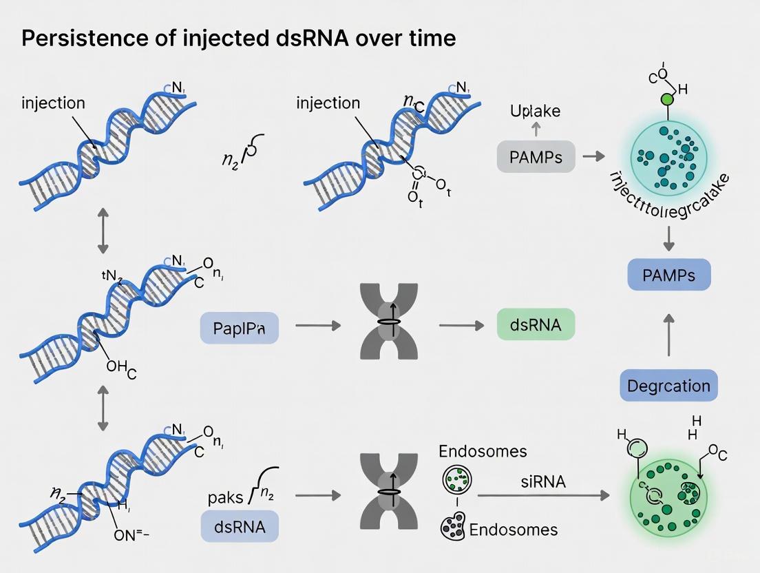
Visualizing the Core RNAi Pathway and Key Failure Points
The diagram below illustrates the journey of exogenous double-stranded RNA (dsRNA) into a cell and the core RNAi pathway, highlighting critical points where the process can fail, leading to reduced persistence and efficacy.
The Scientist's Toolkit: Key Research Reagents and Materials
The table below lists essential reagents and materials used in RNAi persistence research, along with their specific functions in experimental protocols.
| Research Reagent / Material | Function in RNAi Persistence Research |
|---|---|
| Fluorescently Labeled dsRNA (e.g., Fluorescein, CypHer5E) | Visualizing and quantifying dsRNA uptake into cells and tissues, and tracking its intracellular localization over time [1]. |
| Radiolabeled dsRNA (e.g., with α-32P UTP) | Highly sensitive detection of dsRNA stability in biological fluids (e.g., hemolymph, saliva) and degradation kinetics [1]. |
| Nanoparticle Carriers (e.g., Chitosan/SPc Complex - CSC, Carbon Quantum Dots - CQD) | Protecting dsRNA from nuclease degradation in the environment and enhancing cellular uptake efficiency, thereby prolonging the protective window of RNAi [3]. |
| Double-Stranded DNA (dsDNA) | Acting as a competitive decoy for DNA/RNA non-specific nucleases (e.g., in insect saliva), thereby stabilizing co-delivered dsRNA and improving RNAi efficacy [4]. |
| Aminoallyl-UTP | Chemical modification used during in vitro transcription to conjugate dsRNA with pH-sensitive dyes (e.g., CypHer5E) for tracking dsRNA in acidic compartments like endosomes [1]. |
| Micrococcal Nuclease (MNase) | Enzyme used in in vitro assays to test the protective efficacy of various nanoparticle formulations on dsRNA stability [3]. |
| Non-targeting Control dsRNA (e.g., GFP-dsRNA) | A critical negative control to distinguish sequence-specific RNAi effects from non-specific immune responses or toxicity caused by the introduction of exogenous nucleic acid [1]. |
What are the primary pathways for cellular uptake of injected dsRNA?
Following injection, double-stranded RNA (dsRNA) enters cells primarily through two conserved, yet distinct, cellular entry mechanisms. The specific pathway utilized can depend on the insect species and cell type.
Receptor-Mediated Endocytosis: This is a well-documented and widespread pathway for dsRNA internalization. In this process, dsRNA in the hemolymph first binds to carrier proteins, such as apolipoproteins (e.g., ApoLp-III and ApoLp-II/I) [5]. This dsRNA-carrier complex is then recognized by specific receptors on the cell membrane. Studies in Locusta migratoria have identified several candidate receptors, including Scavenger Receptor Class A (SRA), Scavenger Receptor Class C (SRC), and members of the Low-Density Lipoprotein Receptor (LDLR) family [5]. Once bound, the complex is internalized via clathrin-mediated endocytosis or, in some cases, macropinocytosis [5]. The dsRNA is trapped within vesicles called endosomes, from which it must escape into the cytoplasm to activate the RNAi machinery.
Transmembrane Channel Proteins: In some insects, dsRNA can enter cells through transmembrane channels similar to the C. elegans Sid-1 protein [6] [7] [8]. These are often called Sid-1-like genes. However, phylogenetic analyses suggest that these insect Sid-1-like genes may not be direct functional orthologs of the C. elegans sid-1 gene and might have different roles [6] [7]. The number of these genes varies among insects; for example, the red flour beetle (Tribolium castaneum) has three, while many dipterans like Drosophila lack them entirely [6] [8]. This pathway is thought to facilitate the passive movement of dsRNA across the membrane.
The diagram below illustrates how these pathways function in sequence after dsRNA is injected into an insect's hemolymph:
How does dsRNA spread systemically after injection?
Systemic distribution involves both the transport of dsRNA through the body and its cell-to-cell movement, which can occur through different mechanisms than the initial cellular uptake.
Transport via Hemolymph: Upon injection, dsRNA enters the insect's open circulatory system. To remain stable in the hemolymph and avoid degradation by nucleases, dsRNA is often bound by carrier proteins. Apolipoproteins have been identified as key dsRNA carriers in the hemolymph of insects like Locusta migratoria, shielding the dsRNA and facilitating its delivery to various tissues [5].
Cell-to-Cell Movement: The mechanisms for the systemic spread of the RNAi signal between cells in insects are not fully understood and appear to differ from the well-characterized Sid-1-dependent pathway in C. elegans [6]. Research in the model insect Tribolium castaneum, which exhibits robust systemic RNAi, suggests that insects may use an alternative, yet-to-be-discovered mechanism for systemic spreading [6]. This process might involve the repeated cycling of uptake and export or rely on specific intracellular trafficking pathways.
The following diagram summarizes the journey of injected dsRNA from the hemolymph to gene silencing within a cell, including key intracellular trafficking steps:
What key factors determine the efficiency of RNAi following injection?
The effectiveness of gene silencing after dsRNA injection is not guaranteed. It is influenced by a series of factors, from the initial design of the dsRNA to intracellular barriers. The following table summarizes the key factors that impact RNAi efficiency.
| Factor | Description | Impact on RNAi Efficiency |
|---|---|---|
| dsRNA Length | Longer dsRNAs (>60 bp) are generally more effective than shorter ones (<27 bp). | Positively correlated with efficiency; longer dsRNAs produce more siRNAs and are often more readily taken up [9]. |
| Target Gene & Sequence | The biological function of the gene and the specific mRNA region targeted. | Efficiency varies greatly; essential genes with accessible, conserved mRNA regions yield stronger phenotypes [9]. |
| Species-Specific Sensitivity | Innate RNAi efficiency varies by insect order (e.g., Coleoptera are highly sensitive, Lepidoptera are more variable) [10]. | Determines the baseline dose required and the likelihood of achieving systemic silencing [7] [10]. |
| Endosomal Escape | The ability of dsRNA to exit the endosomal compartment into the cytoplasm. | A major rate-limiting step; inefficient escape leads to dsRNA degradation in lysosomes [5]. |
| Intracellular Trafficking | Vesicular transport of dsRNA within the cell, mediated by Rab GTPases (e.g., Rab5, Rab7, Rab11). | Critical for moving internalized dsRNA to the correct cellular compartment for processing and escape [5]. |
How can I experimentally trace the uptake and distribution of dsRNA in my model insect?
To study the journey of dsRNA, you can employ the following experimental protocols, which are adapted from recent research.
Protocol 1: Investigating the dsRNA Uptake Pathway
This protocol is designed to identify which cellular pathway is responsible for dsRNA internalization in your tissue of interest [5].
- dsRNA Preparation: Synthesize and purify dsRNA targeting a reporter gene (e.g., GFP) or a vital gene of interest. Use a fluorescent dye (e.g., Cy3, Cy5) to label the dsRNA for visualization.
- Gene Silencing of Pathway Components: Divide your experimental insects into groups and inject them with dsRNA designed to knock down key genes in the potential uptake pathways:
- Experimental Group 1: dsRNA targeting apolipoproteins (ApoLp-III, ApoLp-II/I).
- Experimental Group 2: dsRNA targeting candidate receptors (SRA, SRC, LRP).
- Experimental Group 3: dsRNA encoding clathrin heavy chain (Chc) to inhibit clathrin-mediated endocytosis.
- Control Group: dsRNA targeting an unrelated gene (e.g., GFP).
- Incubation: Allow 3-5 days for the knockdown of the target genes to take effect.
- Challenger Injection: Inject fluorescently labeled dsRNA into all groups of insects.
- Quantitative Analysis:
- qPCR: After several hours, collect tissues (e.g., fat body, midgut). Extract RNA and measure the amount of internalized labeled dsRNA using specific primers for the dye sequence or via bioanalyzer quantification. Compare the levels between experimental and control groups.
- Immunofluorescence: Fix dissected tissues, stain with antibodies against organelle markers (e.g., Rab5 for early endosomes), and visualize using confocal microscopy to assess co-localization.
Protocol 2: Evaluating Intracellular Trafficking and Endosomal Escape
This protocol focuses on the fate of dsRNA after it has been internalized by the cell [5].
- Knockdown of Trafficking Genes: Similar to Protocol 1, inject insects with dsRNAs that silence crucial intracellular trafficking genes:
- Rab GTPases: Rab5 (early endosomes), Rab7 (late endosomes), Rab11 (recycling endosomes).
- V-ATPase subunits: Genes like V-ATPase C that acidify endosomes and are implicated in dsRNA escape.
- Challenger Injection & Assessment: After a knockdown period, inject dsRNA targeting a vital gene with a clear phenotypic outcome (e.g., a gene that causes lethality or developmental defects when silenced).
- Efficiency Measurement:
- Phenotypic Monitoring: Record the occurrence of the expected phenotypic effect (e.g., mortality, malformation). A reduced phenotype in the experimental groups indicates that the knocked-down gene is important for the RNAi process.
- Molecular Confirmation: Use qPCR to measure the transcript level of the target vital gene in silenced tissues. Higher remaining transcript levels in experimental groups confirm impaired RNAi efficiency.
Troubleshooting Common Experimental Problems
Problem: Weak or no gene silencing observed after dsRNA injection.
- Solution:
- Verify dsRNA integrity using gel electrophoresis before injection.
- Increase the injection dose and consider using a longer dsRNA molecule (>200 bp) [9].
- Check the selection of your target gene and sequence; bioinformatic tools can help predict effective target sites.
- Test for the presence of nucleases in the hemolymph that may be degrading your dsRNA.
- Solution:
Problem: Silencing is only effective locally, not in distant tissues.
- Solution: This suggests a lack of robust systemic spread. Consider using a different insect species or strain known for strong systemic RNAi (e.g., Tribolium castaneum, Locusta migratoria). Alternatively, investigate the use of nanoparticle carriers or complexing agents (e.g., star polycations) that can enhance the stability and mobility of dsRNA [11].
Problem: High mortality following injection, confounding results.
- Solution:
- Ensure the injection technique is clean and minimally injurious.
- Titrate the dsRNA dose to find the minimum effective concentration.
- Verify the specificity of your dsRNA to ensure the mortality is due to on-target effects and not an immune response. In insects, this is less common than in mammals, but controls with irrelevant dsRNA are crucial.
- Solution:
Research Reagent Solutions
The following table lists key reagents and their functions for studying dsRNA uptake and distribution.
| Reagent / Tool | Function in Research | Example Use Case |
|---|---|---|
| Fluorescently Labeled dsRNA (e.g., Cy3-dsRNA) | Visualizing and tracking the location of dsRNA within tissues and cells. | Direct observation of cellular uptake and tissue distribution via fluorescence microscopy [5]. |
| Inhibitors of Endocytosis | Chemically blocking specific uptake pathways. | Using chlorpromazine to inhibit clathrin-mediated endocytosis or EIPA to inhibit macropinocytosis to determine the primary entry route [5]. |
| dsRNA against Pathway Genes | Functional knockdown of genes involved in uptake and trafficking. | Silencing genes like ApoLp-III, Chc, Rab5, or V-ATPase to assess their role in RNAi efficiency (see Protocol 1 & 2) [5]. |
| Nanocarriers (e.g., Star Polycations - SPc) | Formulating dsRNA to enhance stability and cellular uptake. | Protecting dsRNA from degradation, improving entry into cells, and enhancing systemic RNAi efficacy, especially in recalcitrant species [11]. |
| Anti-dsRNA Antibodies | Detecting and quantifying dsRNA impurities or molecules. | Used in techniques like ELISA or BLI to measure dsRNA concentration and stability in samples [12]. |
Core Principles: How dsRNA Characteristics Govern Stability
The longevity and effectiveness of double-stranded RNA (dsRNA) in experimental and therapeutic applications are governed by a set of interdependent physical and molecular characteristics. Understanding these factors is crucial for designing robust and reproducible experiments.
dsRNA Length: The length of the dsRNA molecule is a primary determinant of its stability and silencing efficiency. While the RNAi mechanism utilizes ~21-25 nucleotide siRNAs, longer dsRNA precursors are generally more effective [9]. This is because longer dsRNAs generate a diverse pool of siRNAs, increasing the likelihood of effective mRNA target degradation [9]. They are also often taken up more efficiently by cells; for example, in the beetle Diabrotica virgifera virgifera, dsRNAs shorter than 27 nucleotides showed limited uptake across the midgut epithelium [9]. However, the optimal length is species- and context-dependent, with successful silencing reported using dsRNAs ranging from 141 bp to over 1500 bp in the Colorado potato beetle [9].
Structural Integrity and Sequence: The intrinsic structure of dsRNA makes it a potent trigger for innate immune responses. Cells recognize dsRNA as a pathogen-associated molecular pattern (PAMP) through receptors like RIG-I, MDA5, PKR, and TLRs, leading to inflammatory cytokine production and translation shutdown [13] [14]. This recognition is highly specific; the J2 antibody, for instance, requires a minimum of 14 base pairs for robust binding and is exquisitely selective for dsRNA over dsDNA, ssRNA, or RNA-DNA hybrids [14]. Furthermore, sequence composition (e.g., GC-content) can influence both immunogenicity and how efficiently the dsRNA is processed by the RNAi machinery [9] [14].
Exposure to Degradative Environments: A major hurdle for dsRNA longevity is its susceptibility to degradation by nucleases. In applications like insect pest control, dsRNA instability in the insect's gut fluid or hemolymph is a primary cause of RNAi failure [15] [9] [16]. This degradation is often facilitated by symbiotic microorganisms; for instance, specific Bacillus strains in the cotton bollworm gut secrete extracellular nucleases that rapidly degrade dsRNA, significantly reducing RNAi efficiency [15].
The diagram below illustrates how these core factors converge to impact the final biological outcome of dsRNA application.
Formulation Strategies to Enhance dsRNA Longevity
To overcome the inherent instability of "naked" dsRNA, advanced formulation strategies are essential. These approaches focus on protecting the dsRNA from degradation and enhancing its delivery into target cells.
Nanoparticle-Based Delivery Systems: Nanocarriers such as chitosan nanoparticles, layered double hydroxide (LDH) clays, liposomes, and metal-organic frameworks (MOFs) have proven highly effective [17] [18] [19]. They form complexes with dsRNA, creating a physical barrier that shields it from nuclease activity in harsh biological environments [19]. For example, a cell-penetrating disulfide polymer (CPD) formed stable nanoparticles with dsRNA, preventing degradation and significantly improving gene silencing in the fall armyworm, a lepidopteran pest known for its low RNAi efficiency [19].
Engineered Microbial Systems: Using engineered bacteria or yeast to produce and deliver dsRNA directly within the insect gut is another powerful strategy. Heat-killed or live microbes, such as engineered E. coli or yeast, can act as protective capsules for dsRNA, shielding it from gut nucleases and facilitating uptake [16]. This approach has been shown to be highly potent in overcoming dsRNA instability.
Chemical Modifications and Polymer Complexes: The synthesis of novel cationic polymers offers a cost-effective and scalable delivery solution. A hyperbranched polymer (SPc) was used to form complexes with dsRNA, protecting it and enhancing its uptake in lepidopteran larvae, leading to successful gene silencing [19]. Similarly, liposome-encapsulated dsRNA showed reduced degradation in the midgut and higher mortality rates in the German cockroach [19].
The table below summarizes key formulation strategies and their protective mechanisms.
Table 1: Formulation Strategies for Enhancing dsRNA Stability and Delivery
| Formulation Type | Example Materials | Mechanism of Action | Reported Outcome |
|---|---|---|---|
| Polymer Nanoparticles | Cell-penetrating disulfide polymer (CPD), Hyperbranched polymer (SPc) [19] | Forms stable complexes with dsRNA; enhances cellular uptake via thiol-mediated pathway; degrades intracellularly to release dsRNA [19]. | Protected dsRNA from nucleases; improved RNAi efficiency in lepidopteran pests [19]. |
| Nanoliposomes | Cationic liposomes [19] | Encapsulates dsRNA, shielding it from gut nucleases and facilitating fusion with cell membranes [19]. | Reduced dsRNA degradation in the midgut; increased gene silencing and mortality [19]. |
| Inorganic Nanocarriers | Layered Double Hydroxide (LDH) clays, Metal-Organic Frameworks (ZIF-8) [18] [19] | Adsorbs dsRNA and forms a bio-stable complex, protecting it from environmental degradation and enabling co-delivery with other agents [18] [19]. | Enhanced stability and persistence of dsRNA under field conditions; improved uptake in plants [18]. |
| Engineered Microbes | RNase III-deficient E. coli (e.g., HT115, BL21), Yeast [16] | Produces dsRNA internally; the microbial cell wall acts as a protective barrier during oral ingestion by insects [16]. | Overcame dsRNA instability in the gut; achieved high pest mortality [16]. |
Practical Experimental Protocols
Protocol: Assessing dsRNA Stability in Biological Fluids
This protocol is crucial for diagnosing rapid dsRNA turnover in experiments, especially when working with insect models or serum-containing cell culture.
- Sample Preparation: Collect the biological fluid of interest (e.g., insect hemolymph, gut fluid, or cell culture medium with serum). Centrifuge at low speed (e.g., 4,000 × g for 10 min at 4°C) to remove cells and debris. Aliquot the supernatant [15].
- Incubation Setup: Mix a known quantity of your dsRNA (e.g., 500 ng) with the prepared fluid. Include a control where dsRNA is mixed with a nuclease-free buffer.
- Time-Course Incubation: Incubate the mixtures at the experimental temperature (e.g., 25°C for insects, 37°C for mammalian systems). Remove aliquots at defined time points (e.g., 0, 15, 30, 60, 120 minutes) [15].
- Reaction Termination: Stop the degradation reaction by immediately placing the aliquots on ice and adding a stop solution (e.g., EDTA to chelate divalent cations essential for nuclease activity) or by freezing at -20°C.
- Analysis: Analyze the integrity of the dsRNA from each time point using standard agarose gel electrophoresis. Intact dsRNA will appear as a sharp band, while degraded RNA will show a smeared pattern [15].
Protocol: High-Yield dsRNA Production Using Bacterial Systems
A cost-effective and scalable method for producing dsRNA is essential for large-scale experiments.
- Vector Transformation: Use a plasmid vector (e.g., L4440 or pET28a) containing two opposing T7 promoters flanking the target sequence. Transform this plasmid into an RNase III-deficient E. coli strain like HT115(DE3) or BL21(DE3) RNase III- [19].
- Induction of Transcription: Grow a culture of the transformed bacteria to mid-log phase (OD600 ~0.5-0.6). Induce dsRNA transcription by adding Isopropyl β-d-1-thiogalactopyranoside (IPTG) to a final concentration of 0.4-1.0 mM. Continue incubation for 4-6 hours [19].
- Cell Harvesting and Lysis: Pellet the bacterial cells by centrifugation. Lyse the cells using a lysozyme treatment or a commercial bacterial RNA extraction kit.
- dsRNA Purification: Precipitate the nucleic acids and purify the dsRNA. This can be done using sequential precipitation with lithium chloride (LiCl), which selectively precipitates dsRNA while leaving most ssRNA in solution, or by using commercial purification kits designed for dsRNA [19].
- Quality Control: Verify the concentration using a spectrophotometer and check the integrity and size by gel electrophoresis. The use of the BL21(DE3) RNase III- system has been reported to yield three times more dsRNA than the standard L4440-HT115(DE3) system [19].
Troubleshooting Common dsRNA Longevity Problems
Table 2: Frequently Asked Questions (FAQs) and Troubleshooting Guide
| Problem | Possible Cause | Solution & Recommended Action |
|---|---|---|
| Rapid degradation of injected dsRNA | High nuclease activity in hemolymph/gut fluid or from symbiotic microbiome [15] [9] [16]. | Action: Pre-test dsRNA stability in the target biological fluid. Solution: Switch to a nanoparticle-formulated dsRNA (e.g., chitosan, polymer) to shield it from nucleases [18] [19]. |
| Inefficient RNAi in Lepidopteran insects | Combination of alkaline gut pH, potent nucleases, and inefficient cellular uptake systems [9] [16]. | Action: Use long dsRNA (>200 bp) [9]. Solution: Formulate dsRNA with carrier systems like liposomes, polymer nanoparticles (CPD, SPc), or deliver via engineered microbes [16] [19]. |
| Unexpected immune activation or cytotoxicity in mammalian cells | dsRNA is recognized by cytosolic pattern recognition receptors (PKR, RIG-I, MDA5) [13] [14]. | Action: For saRNA systems, use immune-evasive constructs that co-express inhibitors (e.g., vaccinia E3 protein) via cap-independent translation [13]. Solution: Ensure dsRNA preparations are free of contaminants and consider sequence engineering to reduce immunostimulatory motifs. |
| Variable RNAi efficiency between species | Biological differences in dsRNA uptake mechanisms, systemic spread, and core RNAi machinery efficiency [9] [16]. | Action: Do not assume universal protocols. Solution: Empirically optimize dsRNA length and delivery method for each new species. Refer to successful case studies in related species for initial guidance [9]. |
| Low yield from dsRNA production | Inefficient bacterial expression system or RNA degradation during purification. | Action: Use high-yield expression systems like BL21(DE3) RNase III- [19]. Solution: Optimize induction time and temperature. Use RNase-free techniques and effective purification methods like LiCl precipitation [19]. |
Table 3: Key Research Reagents for dsRNA Longevity Studies
| Reagent / Tool | Function & Application | Key Characteristics |
|---|---|---|
| J2 Anti-dsRNA Antibody | Gold-standard for detecting and mapping dsRNAs in cells and tissues via immunofluorescence, dot blot, or IP [14]. | Exquisitely specific for dsRNA (min. 14 bp); does not bind dsDNA, ssRNA, or RNA-DNA hybrids [14]. |
| RNase III-deficient E. coli | High-efficiency, cost-effective production of dsRNA for large-scale experiments (e.g., bioassays, spraying) [19]. | Lacks RNase III enzyme, preventing dsRNA degradation during production. Strains include HT115(DE3) and the higher-yield BL21(DE3) RNase III- [19]. |
| Cationic Polymer (e.g., CPD, SPc) | Forms stable, protective nanoparticles with dsRNA to enhance nuclease stability and cellular uptake, especially in recalcitrant insects [19]. | Often biodegradable (e.g., CPD has disulfide bonds cleaved by intracellular glutathione), low cytotoxicity, and promotes endosomal escape [19]. |
| Liposomes / Nanoliposomes | A nanocarrier system for encapsulating and delivering dsRNA, improving its stability and transport across insect gut epithelia [19]. | Composed of phospholipids; can fuse with cell membranes to directly deliver payload into the cytoplasm [19]. |
| Chitosan Nanoparticles | A natural, biodegradable nanocarrier used in Spray-Induced Gene Silencing (SIGS) to protect dsRNA from environmental degradation on plants [18]. | Positively charged, adhering to negatively charged plant and insect surfaces, and provides a barrier against water and nucleases [18]. |
Model Organisms and In Vivo Systems for Studying dsRNA Persistence
Comparative Analysis of Model Systems
The table below summarizes key in vivo systems and their characteristics for studying double-stranded RNA (dsRNA) persistence.
| Model Organism / System | Key Findings on dsRNA Persistence | Primary Delivery Method | Persistence Duration | Advantages | Limitations |
|---|---|---|---|---|---|
| Green Ash Seedlings (Fraxinus pennsylvanica) | Successful root uptake and systemic translocation; dsRNA detected in leaf, stem, and root tissues [20]. | Hydroponic root soak [20] | Up to 30 days post-exposure [20] | Intact plant system; non-invasive delivery; models delivery for pest control [20]. | System limited to plant studies; persistence in woody tissues not fully explored [20]. |
| Porcine Model (PRRSV Infection) | Viral dsRNA persisted in lymphoid tissues; shifted localization to germinal centers during persistent infection [21]. | Viral infection (models natural persistent infection) [21] | At least 52 days post-infection [21] | Relevant mammalian model for chronic viral infection and immune evasion [21]. | Complex and costly model; persistence is virus-mediated, not direct exogenous dsRNA [21]. |
| Caenorhabditis elegans | dsRNA-induced heterochromatic marks (H3K9me3) and gene silencing effects persisted transgenerationally [22]. | Injection, feeding, or soaking [23] | Up to 3 generations (decreasing intensity) [22] | Well-established genetics; clear evidence of heritable epigenetic persistence [22] [23]. | Silencing effect wanes without reinforcing signals [22]. |
| Coleopteran Insects (e.g., Leptinotarsa decemlineata) | dsRNA is stable and systematically processed into siRNAs, leading to highly efficient systemic RNAi [1]. | Injection or feeding [1] | Varies (highly stable) [1] | High RNAi efficiency; excellent model for mechanistic studies of successful RNAi [1]. | Findings not directly transferable to low-efficiency systems like Lepidoptera [1]. |
Detailed Experimental Protocols
Protocol 1: Tracking dsRNA Uptake and Persistence in Plants via Root Application
This protocol, adapted from a 2025 study, details how to monitor the systemic movement and stability of dsRNA in ash seedlings [20].
- Key Materials: Target-specific dsRNA (e.g., 302 bp for EAB hsp), Green ash (Fraxinus pennsylvanica) seedlings, hydroponic setup, RNase-free reagents, equipment for RT-PCR and Sanger sequencing [20].
- Procedure:
- dsRNA Treatment: Expose the root system of greenhouse-grown ash seedlings (e.g., ~92 cm tall) to a hydroponic solution containing the target dsRNA [20].
- Tissue Sampling: At predetermined time points (e.g., 3, 7, 14, 21, and 30 days post-exposure), harvest plant tissues. Systematically section into root, woody-stem, soft-stem, and leaf samples [20].
- RNA Extraction: Extract total RNA from all collected tissue samples using a standard method, ensuring no genomic DNA contamination [20].
- Reverse Transcription: Synthesize cDNA from the extracted RNA [20].
- PCR Detection: Perform PCR using two primer sets:
- Analysis: Visualize PCR products via gel electrophoresis. A positive amplicon for the target dsRNA, corresponding to its expected size, indicates successful uptake and persistence. Confirm the identity of the amplicon through Sanger sequencing [20].
Protocol 2: Establishing an In Vitro Model of Viral dsRNA Persistence
This protocol is based on a 2018 study that developed a persistent infection model to investigate viral dsRNA dynamics [21].
- Key Materials: MARC-145 cell line, Porcine Reproductive and Respiratory Syndrome Virus (PRRSV), cell culture equipment, reagents for RNA in situ hybridization or immunofluorescence [21].
- Procedure:
- Infection and Serial Passaging: Infect MARC-145 cells with PRRSV. Instead of harvesting at the peak of acute infection, continuously passage the infected cells repeatedly (e.g., 109 times) in series [21].
- Detection of Persistent dsRNA: Monitor the cultures for the establishment of a persistent infection. Detect associated dsRNA within infected cells using specific detection methods, such as immunohistochemistry or staining with dsRNA-specific antibodies [21].
- In Vivo Correlation (Optional): To correlate in vitro findings with an animal model, experimentally infect target animals (e.g., pigs). Collect lymphoid and other tissues at various time points (e.g., during acute infection and at a later stage such as 52 days post-infection) [21].
- Tissue Analysis: Analyze tissue sections via RNA in situ hybridization to localize viral dsRNA. Note its distribution, which changes from inter-follicular zones during acute infection to the germinal centers during persistent infection [21].
Troubleshooting Common Experimental Issues
FAQ 1: Why is my exogenously applied dsRNA degrading too quickly in the system?
- Potential Cause: Rapid degradation by environmental or extracellular nucleases before uptake can occur. This is a known challenge in systems like lepidopteran insects and on plant surfaces [1] [24].
- Solutions:
- Formulate the dsRNA: Use carrier molecules to protect dsRNA. Interpolyelectrolyte complexes (IPECs) made with biopolymers like chitosan and alginate can encapsulate dsRNA with high efficiency (e.g., 94%), providing outstanding protection against RNase and heat degradation [24].
- Consider Target System Biology: Be aware that some organisms, like lepidopteran insects, have hemolymph and cellular environments that rapidly degrade dsRNA and may trap it in acidic compartments, preventing processing into siRNAs [1].
FAQ 2: I can detect dsRNA in my system, but I do not observe the expected gene silencing effect. Why?
- Potential Cause: The dsRNA is present but is not being processed by the RNAi machinery to produce siRNAs and load the RISC complex. This has been observed in lepidopteran cell lines where dsRNA uptake occurs but no siRNA is detected and no knockdown is achieved [1].
- Solutions:
- Verify siRNA Production: Isolate total RNA from treated samples and analyze it for the presence of siRNAs (e.g., 21-23 nt) specific to your target, for example by using a gel assay or northern blot. The absence of siRNAs indicates a failure in processing [1].
- Check dsRNA Length and Design: Ensure the dsRNA is of sufficient length. Studies in oomycetes showed that short dsRNAs (21-25 bp) had variable and sometimes ineffective results, while longer dsRNAs (≥ 30 bp) completely inhibited spore germination [25].
- Confirm Target Accessibility: Ensure the target gene and mRNA sequence are accessible to the RISC complex.
FAQ 3: How can I visually confirm and track the uptake of dsRNA in my experimental system?
- Solution: Use fluorescently labeled dsRNA.
- Synthesis: Synthesize dsRNA conjugated with a fluorescent dye, such as Cy5 or CypHer5E. This can be done using fluorescent nucleotide mixes during in vitro transcription or via chemical conjugation to amino-allyl labeled nucleotides [25] [1].
- Application and Imaging: Apply the labeled dsRNA to your system (e.g., co-apply with pathogen inoculum to spores or use in a hydroponic solution). Subsequently, use confocal microscopy to visually confirm the internalization of the fluorescent signal into the target cells, tissues, or organisms [25] [1].
The Scientist's Toolkit: Essential Research Reagents
| Reagent / Material | Critical Function | Example Application |
|---|---|---|
| Target-Specific dsRNA | The active silencing molecule; sequence specificity is paramount for targeted gene knockdown [23] [20]. | Can be applied exogenously to plants or insects to silence essential pathogen/pept genes [25] [20]. |
| dsRNA-Specific Antibodies | To detect and localize persistent dsRNA in tissues and cells via immunohistochemistry or ELISA [21]. | Identifying the shift of viral dsRNA to germinal centers in lymphoid tissue during persistent infection [21]. |
| Chitosan & Alginate IPECs | Biopolymer-based formulations that protect dsRNA from nucleases and environmental degradation, enhancing stability and uptake [24]. | Formulating dsRNA for spray applications (SIGS) to protect plants from viruses like Tobacco Mosaic Virus [24]. |
| Fluorescent Dyes (e.g., Cy5) | Label dsRNA to allow for direct visualization and tracking of uptake and translocation using microscopy [25] [1]. | Confirming the uptake of dsRNA by downy mildew spores and germ tubes via confocal microscopy [25]. |
Experimental and Persistence Pathways
dsRNA Persistence Workflow
Transgenerational Persistence in C. elegans
Tracking and Harnessing Long-Term dsRNA Activity
For researchers investigating the persistence of injected double-stranded RNA (dsRNA), selecting the appropriate detection and quantification methodology is critical. This guide details the core techniques, common troubleshooting issues, and essential reagents for tracking dsRNA over time in experimental models.
Frequently Asked Questions (FAQs)
FAQ 1: What is the most sensitive method for detecting low-abundance dsRNA in tissue samples? For detecting low-abundance dsRNA, dsRNA enrichment prior to sequencing is highly recommended. Methods like the novel B2 protein-based extraction can significantly increase the proportion of viral dsRNA reads in a sample, thereby improving detection sensitivity for rare targets [26].
FAQ 2: My dsRNA quantification results are inconsistent between technical replicates. What could be causing this? Inconsistent results, particularly in methods like microfluidic electrophoresis, can stem from variations in the sieving matrix concentration. The migration time and separation efficiency of dsRNA molecules are highly dependent on the polymer concentration of the gel. Ensure the matrix is prepared and loaded with high consistency across all runs [27].
FAQ 3: Can I use standard DNA or ssRNA models to predict the electrophoretic mobility of my dsRNA product? No, dsRNA has unique electrophoretic properties. Its persistence length and radius of gyration differ from both DNA and single-stranded RNA (ssRNA). Relying on models developed for other nucleic acids can lead to inaccurate predictions of migration behavior. Always use dsRNA-specific ladders and, if available, physics-informed neural network models for accurate characterization [27].
FAQ 4: How can I confirm that my detected dsRNA signal is specific to the therapeutic sequence and not endogenous dsRNA? To confirm specificity, your detection method should leverage sequence-specific techniques. Following dsRNA extraction and conversion to cDNA via reverse transcription, using target-specific primers in a qPCR assay is the standard approach. This allows you to quantify the specific dsRNA sequence of interest against a standard curve, distinguishing it from background endogenous dsRNA [28].
Experimental Protocols & Workflows
Below are detailed methodologies for key experiments cited in dsRNA persistence research.
Protocol: dsRNA Extraction Using a Novel B2-Based Method
This protocol describes a cost-effective, high-throughput method for enriching dsRNA from complex samples like plant or animal tissue, ideal for pre-sequencing preparation [26].
- Principle: The Flock House virus B2 protein binds to dsRNA with high affinity and in a sequence-independent manner. The binding is pH-dependent, allowing for efficient capture and release of dsRNA.
- Applications: Virome profiling, viral ecology studies, and preparation of dsRNA for high-throughput sequencing (HTS).
Workflow Overview:
- Step-by-Step Procedure:
- Sample Homogenization: Homogenize tissue samples in a suitable lysis buffer.
- Lysate Preparation: Clarify the lysate by centrifugation to remove cellular debris.
- B2 Binding: Incubate the clarified lysate with the purified B2 protein under binding buffer conditions (acidic pH enhances binding).
- Complex Formation: Allow dsRNA-B2 complexes to form.
- Precipitation: Pellet the dsRNA-B2 complexes using centrifugation.
- Wash: Wash the pellet to remove non-specifically bound contaminants.
- Elution: Elute the purified dsRNA by resuspending the pellet in an elution buffer with a neutral or slightly basic pH, which disrupts the B2-dsRNA interaction.
- Quality Control: Analyze the extracted dsRNA using spectrophotometry and microfluidic electrophoresis (e.g., LabChip GXII) [27].
Protocol: Spiropyran-Based Spectrophotometric Detection of dsRNA
This protocol outlines a quick and convenient method for the relative quantification of total dsRNA levels, useful for diagnostic applications or rapid screening [28].
- Principle: The spiropyran derivative (Am-SP) isomerizes to merocyanine (Am-MC) under UV light. Am-MC binds to dsRNA, causing a measurable change in absorbance at 515 nm.
- Applications: Rapid diagnostic screening for viral infections (e.g., Severe Fever with Thrombocytopenia Syndrome), quick assessment of total dsRNA levels in patient serum.
Workflow Overview:
- Step-by-Step Procedure:
- Probe Preparation: Dissolve amidine-conjugated spiropyran (Am-SP) in triple-distilled water to a concentration of 12 mM.
- Activation: Expose the Am-SP solution to 254 nm ultraviolet light for 1 minute to convert it to the active Am-MC form.
- Reaction: Pipette the RNA sample directly into a cuvette containing the activated Am-MC and incubate in the dark to prevent reverse isomerization.
- Measurement: Within 1 minute, measure the absorbance using a spectrophotometer with a 1 mm path length, scanning from 350 nm to 700 nm.
- Analysis: Calculate the rate of absorbance change at 515 nm, using a water blank as a control. The formula is:
(Abs_sample - Abs_control) / Abs_control × 100 (%)[28].
Data Presentation: Quantitative Comparisons
Table 1: Comparison of dsRNA Extraction Methods for Sequencing
| Method | Principle | Cost per Reaction | Viral Read Proportion | Key Advantage | Key Limitation |
|---|---|---|---|---|---|
| B2-Based Method [26] | Protein binding & pH-dependent elution | $4.47 |
>20% (in most samples) |
High cost-effectiveness, good purity | Sensitivity may vary by virus species |
| DRB4-Based (Commercial Kit) [26] | dsRNA-binding protein (DRB4) | $35.34 |
Not Specified (Highest accuracy) | High detection accuracy | High cost, can be less pure than B2 method |
| Cellulose-Based Method [26] | Binding to cellulose resin | Not Specified | Not Specified | Well-established protocol | Labor-intensive, lower purity |
Table 2: Microfluidic Electrophoresis Conditions for dsRNA Analysis
| Parameter | Condition / Observation | Technical Note |
|---|---|---|
| Platform | LabChip GXII Touch with custom RNA chip [27] | Utilizes SYTO 61 fluorescent stain and PDMA polymer solution. |
| Gel Concentration | Tested range: 1% to 5% PDMA [27] | Higher concentrations (e.g., 4-5%) recommended for resolving longer dsRNA fragments. |
| Key Finding | dsRNA mobility is predictable using Physics-Informed Neural Networks (PINNs) [27] | PINNs can predict migration time and length with an average error of 0.77%. |
| Critical Consideration | Separation time must be increased with higher gel concentration [27] | Ensures all analyte peaks are captured in the electropherogram. |
The Scientist's Toolkit: Key Research Reagent Solutions
| Reagent / Tool | Function in dsRNA Research | Example Use Case |
|---|---|---|
| B2 Protein from Flock House Virus [26] | High-affinity, sequence-independent dsRNA binding for extraction. | Enriching viral dsRNA from plant or animal tissue for HTS. |
| FPLC / HPLC Purification [29] | Removal of immunostimulatory dsRNA contaminants from in vitro transcribed (IVT) RNA. | Purifying synthetic mRNA or saRNA therapeutics to reduce innate immune activation. |
| Spiropyran-Based Probe (Am-MC) [28] | Spectrophotometric detection and relative quantification of total dsRNA. | Rapid diagnostic screening for viral infections from serum samples. |
| Microfluidic Electrophoresis (e.g., LabChip GXII) [27] | High-resolution analysis of dsRNA size, integrity, and concentration. | Quality control of synthesized dsRNA products or extracts prior to experiments. |
| Physics-Informed Neural Networks (PINNs) [27] | Predicting electrophoretic mobility of dsRNA without extensive experimental data. | In silico modeling and optimization of separation assays for novel dsRNA molecules. |
This case study examines a 2025 investigation that demonstrated successful uptake, systemic movement, and long-term persistence of exogenously applied, EAB-specific double-stranded RNA (dsRNA) in green ash (Fraxinus pennsylvanica) seedlings following a hydroponic root soak application. The research provides proof-of-concept for root application as a viable delivery method for protecting trees against the emerald ash borer (EAB) using RNAi technology [20].
The core finding was that dsRNA applied to the roots was detected in ~98% of plant tissue samples (roots, woody stems, soft stems, and leaves) throughout the 30-day experimental period. The study demonstrated consistent distribution and persistence of the dsRNA, with no significant association found between dsRNA recovery and time or tissue type, confirming robust and sustained systemic presence [20].
Table 1: Summary of dsRNA Recovery Rates Across Different Plant Tissues Over 30 Days
| Plant Tissue | Overall dsRNA Recovery Rate | Key Temporal Observations |
|---|---|---|
| Roots | 98.3% (collective all tissues) | Consistent high recovery throughout 30-day period |
| Woody Stem | 95.5% | Slight increase in recovery probability after day 3 |
| Soft Stem | 95.5% | Slight reduction in recovery at day 30 |
| Leaves | 98.3% (collective all tissues) | Successful translocation from root to aerial tissues |
Table 2: Key Experimental Parameters and Persistence Timeline
| Experimental Parameter | Specification |
|---|---|
| Plant Material | Green ash (Fraxinus pennsylvanica) seedlings |
| Average Seedling Height | 92.28 ± 2.14 cm |
| Root Collar Diameter | 0.95 ± 0.02 cm |
| dsRNA Application Method | Hydroponic root soak |
| Sampling Time Points | 3, 7, 14, 21, and 30 days post-exposure |
| Detection Method | RT-PCR with Sanger sequencing confirmation |
| Persistence Duration | Confirmed throughout 30-day study period |
Detailed Experimental Protocol
Plant Material Preparation and dsRNA Application
- Plant Selection: Utilize greenhouse-grown green ash seedlings with an average height of approximately 92 cm and root collar diameter of 0.95 cm [20].
- Hydroponic Exposure: Apply EAB-specific dsRNA (targeting the Hsp gene) as a root soak in a hydroponic system. The dsRNA sequence should be designed for specificity against essential EAB genes [20].
- Control Setup: Include control seedlings exposed only to water without dsRNA to verify the absence of non-specific amplification [20].
Tissue Sampling and RNA Extraction
- Systematic Sampling: At each time point (3, 7, 14, 21, and 30 days post-exposure), collect tissue samples from roots, woody stems, soft stems, and leaves [20].
- RNA Extraction: Extract total RNA from all collected tissues using standard methodologies [20].
- Complementary DNA (cDNA) Synthesis: Synthesize cDNA from the extracted RNA for subsequent PCR analysis [20].
Detection and Analysis
- RT-PCR Amplification: Perform RT-PCR with primers targeting both the EAB-specific Hsp gene (302 bp amplicon) to detect the exogenous dsRNA, and the plant-specific ef1β gene as an endogenous positive control [20].
- Gel Electrophoresis: Visualize PCR products via gel electrophoresis. Successful dsRNA uptake and translocation is confirmed by detection of the 302 bp Hsp amplicon across different tissue types [20].
- Sequence Verification: Validate amplicon identity through Sanger sequencing and pairwise alignment with the annotated EAB Hsp gene sequence (approximately 94% similarity) [20].
The Scientist's Toolkit: Research Reagent Solutions
Table 3: Essential Research Reagents and Materials for dsRNA Persistence Studies
| Reagent/Material | Function/Application | Specifications/Considerations |
|---|---|---|
| EAB-specific dsRNA | RNAi trigger targeting essential EAB genes | Designed for sequence specificity; 302 bp Hsp target used in cited study [20] |
| Hydroponic Growth System | Controlled root application environment | Enables precise root soak delivery of dsRNA [20] |
| RNA Extraction Kit | Isolation of total RNA from plant tissues | Critical for downstream RT-PCR analysis [20] |
| RT-PCR Reagents | Detection of dsRNA in plant tissues | Requires primers for target gene (e.g., EAB Hsp) and endogenous control (e.g., ash ef1β) [20] |
| Agarose Gel Electrophoresis System | Visualization of PCR amplicons | Confirms presence of 302 bp target band [20] |
| Sanger Sequencing Reagents | Verification of amplicon identity | Validates sequence specificity of recovered dsRNA [20] |
Molecular Mechanism of RNAi and dsRNA Persistence
The RNA interference (RNAi) pathway is a conserved sequence-specific gene regulation system. Following root uptake and systemic movement, the cellular RNAi machinery processes the delivered dsRNA [30] [23]:
- Cellular Processing: Upon entering plant cells, dsRNA is cleaved by Dicer-like enzymes into 21-24 nucleotide small interfering RNAs (siRNAs) [31] [23].
- RISC Formation: siRNAs are incorporated into the RNA-induced silencing complex (RISC) containing Argonaute (AGO) proteins [32] [23].
- Target Silencing: The siRNA guide strand directs RISC to complementary mRNA sequences, leading to mRNA cleavage and post-transcriptional gene silencing [32] [23].
- Persistence Mechanism: The month-long persistence observed in this study suggests stability of the applied dsRNA within plant tissues, potentially due to protection from nucleases or continuous systemic distribution [20].
Frequently Asked Questions (FAQs) & Troubleshooting
Experimental Design & Setup
Q: What are the key advantages of root application over other dsRNA delivery methods? A: Root application provides a non-invasive delivery approach that enables systemic distribution throughout the plant. This method is particularly valuable for targeting wood-boring pests like EAB that feed on vascular tissues, and it avoids issues like environmental degradation and variable foliar uptake associated with spray applications [20] [33].
Q: What factors should be considered when selecting target genes for RNAi-based pest control? A: Target essential genes vital for pest survival, development, or reproduction. Ensure sequence specificity to minimize off-target effects on non-target organisms. Bioinformatic analysis of gene conservation across species is crucial for assessing potential ecological impacts [33] [8].
Technical Troubleshooting
Q: We observe weak or inconsistent dsRNA detection in distal tissues. What could be the issue? A: Several factors could contribute: (1) Low-quality or partially degraded dsRNA starting material - verify integrity by gel electrophoresis; (2) Inefficient systemic translocation - ensure plant vascular health and appropriate dsRNA concentration; (3) Suboptimal sampling or RNA extraction methods - include positive controls and validate tissue collection procedures [20] [8].
Q: What is the significance of including both target-specific and endogenous control primers in RT-PCR? A: The endogenous control (e.g., ash ef1β) verifies RNA extraction quality, cDNA synthesis efficiency, and absence of PCR inhibitors. The target-specific primers (e.g., EAB Hsp) specifically detect the exogenous dsRNA. This dual verification is essential for distinguishing true negative results from technical failures [20].
Persistence & Efficacy Optimization
Q: How can we enhance dsRNA stability and persistence in plant tissues? A: Recent advances include: (1) Nanocarrier formulations (clay nanosheets, liposomes) that protect dsRNA from degradation; (2) Chemical modifications to improve nuclease resistance; (3) Optimization of application timing and concentration based on plant physiology [34] [31].
Q: Does the persistence of dsRNA in plants raise any safety concerns? A: RNAi-based approaches are considered environmentally friendly because dsRNA is biodegradable and acts in a sequence-specific manner. However, comprehensive risk assessment should include: evaluation of off-target effects, potential impacts on non-target organisms, and persistence duration in the environment. Regulatory frameworks for dsRNA-based pesticides are currently evolving [33] [35].
This case study demonstrates that a single feeding of double-stranded RNA (dsRNA) is sufficient to induce a persistent and effective gene knockdown in planarians, challenging established protocols that typically utilize multiple feedings.
Table 1: Key Quantitative Findings from Single vs. Triple Feeding RNAi
| Experimental Parameter | Single Feeding Results | Triple Feeding Results | Significance |
|---|---|---|---|
| Phenotype Induction | Similar nociceptive phenotypes induced | Similar nociceptive phenotypes induced | No significant difference between protocols [36] |
| Phenotype Duration | Long-lasting effects observed | Long-lasting effects observed | Effects lasted for 11 weeks in both groups [36] |
| Behavioral Assessment | Effective loss of AITC-induced scrunching | Effective loss of AITC-induced scrunching | Similar behavioral outcomes [36] |
| dsRNA Quantity | ~0.5 μg/μL per feeding [36] | ~0.5 μg/μL per feeding (total ~1.5 μg/μL) [36] | Single feeding used 67% less dsRNA |
Table 2: Experimental Timeline and Key Parameters
| Experimental Phase | Duration | Key Procedures | Assessment Methods |
|---|---|---|---|
| Pre-Treatment | 7 days starvation [36] | Animal preparation | - |
| dsRNA Feeding | Single event or 3x over one week [36] | Feeding with beef liver paste containing dsRNA (0.5 μg/μL) [36] | Visual confirmation of blue food coloring uptake [36] |
| Phenotype Monitoring | 11 weeks total [36] | Behavioral tests every other week [36] | Scrunching response to 50 μM AITC [36] |
| Molecular Validation | At time points throughout experiment | RT-qPCR on whole animals (n=5-8) [36] | Gene expression normalized to GAPDH [36] |
Troubleshooting Guides
Common RNAi Experimental Problems and Solutions
Problem: Animals fail to display any phenotype after dsRNA feeding.
Solutions:
- Confirm effective dsRNA ingestion: Feed planarians with liver paste containing blue food coloring and only use animals that appear completely blue after ingestion [36].
- Verify gene knockdown efficacy: Perform RT-qPCR or quantitative RT-PCR to confirm reduction in target gene expression [37].
- Consider alternative injection method: If feeding is ineffective, try microinjecting 32 nL of dsRNA solution 3-5 times in the prepharyngeal area [37].
Problem: Inconsistent knockdown effects across animals.
Solutions:
- Standardize feeding protocol: Ensure all experimental animals consume the entire dsRNA-containing pellet [36].
- Optimize dsRNA concentration: For some applications, 0.1 μg/μL may be optimal, as demonstrated in related organisms [38].
- Ensure dsRNA purity: Use DNase I treatment followed by phenol/chloroform extraction and ethanol precipitation to remove DNA template contamination [38].
Problem: Unexpected lethal phenotypes or negative feedback loops.
Solutions:
- Avoid targeting RNAi pathway components: Knockdown of genes like Argonaute-2 can disrupt the RNAi process itself and cause mortality [36].
- Test multiple dsRNAs: Generate non-overlapping dsRNAs (~500 bp) targeting different regions of your gene of interest [39].
Frequently Asked Questions (FAQs)
Q: How long does knockdown persist after a single dsRNA feeding? A: The study demonstrated that phenotypic effects can persist for at least 11 weeks after a single dsRNA feeding, with similar duration to triple feeding protocols [36].
Q: What concentration of dsRNA should I use for feeding? A: The cited study used 0.5 μg/μL dsRNA in beef liver paste successfully [36]. However, dose-dependent effects exist, with 0.1 μg/μL showing optimal efficiency in some systems [38].
Q: Can I use this single feeding approach for any planarian gene? A: While effective for TRPA1 knockdown, the authors note that multiple dsRNA treatments may still be needed for large animals and for gene knockdowns with late phenotypic manifestations [36].
Q: What controls are necessary for these experiments? A: Essential controls include: (1) dsRNA targeting a non-planarian gene (e.g., GFP) [36] [39], (2) Untreated animals, and (3) Animals fed with empty vector or scrambled dsRNA [39].
Q: How can I validate that my knockdown is working? A: Use RT-qPCR with appropriate housekeeping genes (GAPDH was most stable in the cited study) [36] and correlate with functional assessments where possible [36].
Detailed Experimental Protocols
Primary RNAi Feeding Protocol
Materials:
- Starved planarians (7 days) [36]
- dsRNA targeting gene of interest (0.5 μg/μL) [36]
- Beef liver paste [36]
- Agarose (0.3%) [36]
- Blue food coloring (3%) [36]
Procedure:
- Prepare feeding pellets by mixing dsRNA with beef liver paste, agarose, and blue food coloring [36].
- Present one pellet to groups of 12 worms [36].
- Keep only animals that appear completely blue after ingestion, indicating sufficient dsRNA consumption [36].
- Return animals to normal maintenance conditions (Volvic mineral water, 21°C, 12h:12h light-dark cycle) [36].
- Conduct phenotypic assessments at appropriate time points post-feeding [36].
Alternative Microinjection Protocol
Materials:
- dsRNA (synthesized in vitro) [37]
- Microinjector (e.g., Nanoject) [37]
- Glass capillaries [37]
- Mineral oil [37]
Procedure:
- Pull microinjection needles and break tips to appropriate diameter [37].
- Fill needle with mineral oil without air bubbles [37].
- Aspirate 1-2 μL of dsRNA solution [37].
- Place worm on cold, wet tissue and introduce needle into prepharyngeal area [37].
- Inject 32 nL per pulse, repeating 3-5 times [37].
- Transfer injected worm to fresh planarian water at room temperature [37].
Signaling Pathway & Experimental Workflow Diagrams
Research Reagent Solutions
Table 3: Essential Research Reagents for Planarian RNAi
| Reagent/Chemical | Function/Purpose | Example Usage/Concentration |
|---|---|---|
| T7 RNA Polymerase | In vitro dsRNA synthesis [40] [37] | 17 units per 20 μL transcription reaction [37] |
| DNase I | DNA template removal after dsRNA synthesis [37] [38] | 1 unit per reaction, 15 min at 37°C [37] |
| Phenol:Chloroform | dsRNA purification [37] [38] | 1:1 ratio for effective purification [38] |
| Beef Liver Paste | dsRNA delivery vehicle for feeding [36] [40] | Mixed with 0.3% agarose, 3% food coloring [36] |
| AITC (Allyl Isothiocyanate) | TRPA1 agonist for behavioral assessment [36] | 50 μM in Volvic water for nociception tests [36] |
| Montjuïc Salts | Planarian maintenance solution [40] | 1.6 mM NaCl, 1 mM CaCl₂, 1 mM MgSO₄, etc. [40] |
| Gentamicin Sulfate | Antimicrobial for culture maintenance [40] [39] | 50 mg/mL stock, used at 100 μg/mL [39] |
| pJC53.2 Vector | Cloning vector with T7 promoters [40] [39] | Flanks insert with T7 polymerase sites for dsRNA synthesis [40] |
Core Concepts and Troubleshooting FAQs
What are the primary causes of dsRNA instability in experimental applications?
Double-stranded RNA (dsRNA) is inherently unstable in most experimental and environmental conditions. Its rapid degradation is a major hurdle for both research and therapeutic applications. The primary causes of instability include:
- Nuclease Degradation: dsRNA is rapidly broken down by nucleases present in biological fluids, soil, and water. In serum, unmodified dsRNA has a half-life of only a few minutes [41]. Insect hemolymph and gut secretions also contain high nuclease activity, which varies significantly between orders; for example, lepidopteran insects exhibit much higher nuclease activity than coleopterans [1] [42].
- Environmental Factors: When applied topically (e.g., in spray-induced gene silencing), dsRNA is degraded by ultraviolet (UV) radiation, rainwater, and microorganisms. In soil and water environments, naked dsRNA can be completely degraded within 48 hours [42].
- pH Sensitivity: The stability of dsRNA is greatly affected by pH. It is generally stable at pH 4.0–5.0 but becomes unstable in alkaline environments, such as the gut of lepidopteran, orthopteran, dipteran, and hymenopteran insects [42].
Troubleshooting Tip: If your dsRNA is degrading too quickly in vitro or in environmental applications, consider using nuclease-resistant chemical modifications or encapsulating the dsRNA within protective nanocarriers.
How can I improve cellular uptake of dsRNA in my experiment?
Poor cellular uptake is a common problem, especially in recalcitrant cell types or organisms. The following solutions can enhance uptake:
- Use of Cationic Nanocarriers: Positively charged nanoparticles, such as chitosan (CS) or polyethyleneimine (PEI), efficiently form complexes with negatively charged dsRNA. These complexes protect dsRNA and enhance its adhesion to and uptake by cells. For instance, a chitosan/SPc complex (CSC) was shown to improve dsRNA uptake by the fungus Rhizoctonia solani [3].
- Viral Vector Transduction: Engineered viral vectors (e.g., lentiviruses, adenoviruses) excel at delivering genetic material into cells. They offer high transduction efficiency and can be engineered for specific cell tropisms [43] [44].
- Chemical Modification of dsRNA: Modifying the RNA backbone, such as replacing the phosphodiester bond with a phosphorothioate (PS) bond, or modifying the ribose 2' hydroxyl group, can reduce immunogenicity and increase stability, indirectly facilitating uptake and persistence [41].
Troubleshooting Tip: If uptake is low in cell culture, try comparing different transfection reagents or nanocarriers. For in vivo work in insects, note that coleopterans generally show better dsRNA uptake and systemic RNAi response than lepidopterans [1].
What delivery system should I choose to maximize the persistence of dsRNA effects?
The choice of delivery system is critical for achieving long-lasting effects. The optimal strategy depends on your target system (e.g., plants, insects, mammalian cells).
- For Long-Term Expression in Animals: Viral Vectors, particularly integrating viruses like lentiviruses, are ideal. They insert the transgene (often as a dsRNA-encoding shRNA) into the host genome, leading to persistent expression. Adeno-Associated Viruses (AAVs) provide long-term episomal expression in non-dividing cells and are a leading platform for in vivo gene therapy [43] [44].
- For Environmental & Agricultural Applications: Nanoparticles significantly prolong the protective window of dsRNA. For example, a chitosan/SPc complex (CSC) protected dsRNA from nuclease degradation and prolonged its activity against rice sheath blight for up to 20 days in a plant model, whereas naked dsRNA was ineffective [3]. Similarly, root application of dsRNA in ash seedlings showed detectable persistence for at least 30 days [20].
- For High Stability and Low Immunogenicity: Chemically Modified dsRNA is the best option. Modifications like 2'-O-methyl, 2'-fluoro, and phosphorothioate linkages dramatically increase nuclease resistance and circulatory half-life, which is why they are used in approved RNAi therapeutics [41].
Troubleshooting Tip: If your dsRNA effect is transient in an animal model, consider switching from synthetic dsRNA to a viral vector that expresses an shRNA for sustained production.
Experimental Protocols for Key Applications
Protocol 1: Evaluating Nanoparticle-dsRNA Complex Stability
This protocol is adapted from methods used to test nanocarriers for plant pathogen control [3].
Objective: To assess the ability of a nanocarrier to protect dsRNA from nuclease degradation.
Materials:
- Fluorescein-labeled YFP-dsRNA (or your target dsRNA)
- Candidate nanoparticles (e.g., Chitosan, PEI, CQD, CSC)
- Micrococcal nuclease (MNase) and corresponding reaction buffer
- Fluorescence plate reader
Method:
- Prepare Complexes: Mix the fluorescein-labeled dsRNA with different nanoparticles at their optimal mass ratios (e.g., CSC:dsRNA at 5:1) to form complexes. Incubate for 30 minutes at room temperature.
- Nuclease Challenge: Add MNase to the complexes and free dsRNA control. Incubate at 37°C for a predetermined time (e.g., 30 minutes).
- Measure Stability: Stop the reaction and measure the fluorescence intensity of each sample. Compare post-MNase fluorescence to pre-MNase levels.
- Analysis: Calculate the percentage reduction in fluorescence. A superior protective nanocarrier will show a significantly smaller reduction compared to naked dsRNA.
Expected Outcome: In the cited study, naked dsRNA lost 80% of its fluorescence after MNase treatment, while the CSC-dsRNA complex showed almost no reduction, demonstrating excellent protection [3].
Protocol 2: Tracking dsRNA Uptake and Systemic Movement in Plants
This protocol is based on research investigating dsRNA delivery for tree protection [20].
Objective: To confirm the uptake, translocation, and persistence of exogenously applied dsRNA in a plant system.
Materials:
- Greenhouse-grown seedlings (e.g., ash, oak)
- Target-specific dsRNA (e.g., targeting an insect pest gene)
- Hydroponic setup or materials for root drench/foliar spray
- Equipment for total RNA extraction, RT-PCR, and Sanger sequencing
Method:
- dsRNA Application: Apply the dsRNA to plants via a root soak or hydroponic solution. For controls, use plants treated with water.
- Tissue Sampling: At multiple time points post-application (e.g., 3, 7, 14, 21, 30 days), sample different tissues: roots, woody stem, soft stem, and leaves.
- RNA Extraction and RT-PCR: Extract total RNA from all samples. Perform RT-PCR using two primer sets:
- One set specific to the applied dsRNA sequence.
- One set specific to an endogenous plant gene (e.g., elongation factor ef1β) as a positive control for RNA quality.
- Confirmation: Visualize PCR products on an agarose gel. Confirm the identity of the amplicon from the applied dsRNA using Sanger sequencing.
Expected Outcome: Successful uptake and translocation are demonstrated by detecting the exogenous dsRNA amplicon in various plant tissues over an extended period. The cited study found dsRNA in ~98% of ash seedling tissues up to 30 days post-root application [20].
The following tables summarize key performance metrics for different delivery systems, as reported in the search results.
Table 1: Protection of dsRNA from Nuclease Degradation by Nanocarriers
| Nanocarrier | Fluorescence Reduction after MNase | Key Finding |
|---|---|---|
| Naked dsRNA | 80% | Baseline - highly susceptible to degradation [3] |
| Chitosan/SPc Complex (CSC) | ~7% | Best protection among tested materials [3] |
| Carbon Quantum Dot (CQD) | 31% | Good protective ability and high loading capacity [3] |
| Polyethyleneimine (PEI) | 43% | Moderate protection [3] |
| Chitosan (CS) | 46% | Moderate protection [3] |
| Polyamidoamine (PAMAM) | 58% | Modest protection [3] |
| Protamine | 60% | Modest protection [3] |
Table 2: Persistence of dsRNA Effects Across Different Delivery Modalities
| Delivery Method / System | Model System | Persistence / Protection Duration |
|---|---|---|
| Root Application (naked dsRNA) | Ash Seedlings | Detected for at least 30 days [20] |
| Nanoparticle (CSC)-dsRNA | Rice - R. solani pathosystem | Protection extended to 20 days [3] |
| Viral Vector (LV/AAV) | Mammalian Systems | Long-term (months to years) from stable transgene expression [43] [44] |
| Chemically Modified siRNA | Mammalian Therapeutics | Increased half-life enabling durable silencing from a single dose [41] |
dsRNA Delivery and Persistence Workflow
The following diagram illustrates the logical workflow for selecting and evaluating a dsRNA delivery system to achieve persistent effects.
Diagram 1: Decision workflow for persistent dsRNA delivery
The Scientist's Toolkit: Key Research Reagents
Table 3: Essential Reagents for dsRNA Persistence Research
| Reagent / Material | Function in Experiment | Example Use-Case |
|---|---|---|
| Cationic Polymers (e.g., Chitosan, PEI) | Form stable complexes with dsRNA via electrostatic interaction, protecting it and enhancing cellular uptake. | Topical application (SIGS) for plant pathogen control [42] [3]. |
| Lipid Nanoparticles (LNPs) | Encapsulate dsRNA/siRNA, protecting it and promoting endosomal escape in mammalian cells. | Delivery of therapeutic siRNA (e.g., Patisiran) [43] [41]. |
| Adeno-Associated Virus (AAV) | A viral vector for efficient in vivo gene delivery resulting in long-term transgene expression. | Preclinical and clinical gene therapy and gene silencing [43] [44]. |
| Lentivirus | An integrating viral vector for stable, long-term expression of shRNA in dividing and non-dividing cells. | Creating stable cell lines for persistent gene knockdown [43] [44]. |
| Chemical Modification Kits | Introduce nuclease-resistant modifications (e.g., 2'-O-Me, 2'-F, PS) into RNA strands. | Producing stabilized siRNA/dsRNA for in vivo applications with extended half-life [41]. |
| Fluorescent RNA Labeling Mix | Tags dsRNA with fluorophores (e.g., Fluorescein, CypHer5E) to track uptake and localization. | Visualizing dsRNA uptake in cell lines or tissues via microscopy [1] [3]. |
Overcoming Hurdles: Maximizing dsRNA Duration and Efficacy
Double-stranded RNA (dsRNA) is a potent activator of the innate immune system, triggering interferon (IFN) and inflammatory responses that can significantly impact the persistence and efficacy of dsRNA in research and therapeutic applications. When introduced into mammalian systems, dsRNA is recognized by various cytosolic pattern recognition receptors, including protein kinase R (PKR), oligoadenylate synthase (OAS), RIG-I, and MDA5 [13] [45]. This recognition initiates signaling cascades that result in the production of type I interferons, pro-inflammatory cytokines, and the activation of cellular pathways that shut down cap-dependent translation and degrade cellular mRNA [13]. Understanding these mechanisms is crucial for researchers aiming to develop dsRNA-based technologies with improved stability and reduced immunogenicity.
Core Mechanisms of Immune Recognition
Key Pathways and Sensors
Cytosolic dsRNA Sensing Mechanisms Mammalian cells have evolved multiple pathways to detect dsRNA as a signature of viral infection:
- PKR Pathway: Protein kinase R (PKR) is activated by binding to dsRNA, leading to phosphorylation of eukaryotic initiation factor 2α (eIF2α), which results in global inhibition of cap-dependent protein synthesis [13].
- OAS/RNase L Pathway: Oligoadenylate synthase (OAS) detects dsRNA and produces 2'-5'-linked oligoadenylates that activate RNase L, leading to non-specific degradation of cellular RNA [13].
- RIG-I/MDA5 Pathway: These RNA helicases detect cytoplasmic dsRNA and initiate signaling cascades that activate IRF3 and NF-κB, resulting in type I interferon production [46] [45].
- ZNFX1 Pathway: ZNFX1 is an understudied SF1 RNA helicase that functions as a cytosolic dsRNA sensor and immunomodulator, with deficiency leading to chronic inflammation and increased susceptibility to pathogens [46].
The following diagram illustrates the major cellular pathways activated by dsRNA:
Quantifying dsRNA Stability Across Environments
The persistence of dsRNA is highly dependent on its formulation and environmental conditions. The following table summarizes half-life data for naked versus encapsulated dsRNA:
Table 1: Environmental Stability of dsRNA Formulations
| Environmental Condition | Naked dsRNA Half-life | Encapsulated dsRNA Half-life | Key Degradation Factors |
|---|---|---|---|
| Aquatic Systems | Varies by water type | >2x increase vs. naked dsRNA | Microbial activity, water hardness (Ca²⁺) |
| Plant Surfaces | Limited | Significant extension | UV exposure, surface nucleases |
| Insect Gut Environment | Rapid degradation | Protected delivery | pH, nucleases, microbial activity [47] |
| Soil | Moderate | Enhanced stability | Microbial communities, fungal activity [48] |
Encapsulation strategies significantly enhance dsRNA stability across all environments, with minicell-encapsulated dsRNA (ME-dsRNA) demonstrating more than twofold increased half-life in most environments compared to naked dsRNA [48]. Fungal communities in aquatic environments appear to be more strongly correlated with dsRNA degradation than bacterial populations [48].
Troubleshooting Guide: FAQs
Q1: Why is my delivered dsRNA failing to produce the expected gene silencing effect?
A: Inefficient RNAi responses can result from multiple factors:
Rapid dsRNA Degradation: dsRNA is extremely unstable in biological fluids. In cotton bollworms, dsRNA is rapidly degraded in midgut fluid and hemolymph even at diluted concentrations [47]. This instability is particularly pronounced in lepidopteran species, where dsRNA cannot be efficiently converted into functional siRNA in the midgut due to low expression levels of Dicer-2 and rapid degradation within the gut environment [49].
Microbial Intervention: Symbiotic bacteria can secrete nucleases that degrade dsRNA. Six Bacillus strains have been identified that exhibit dsRNA-degrading activity, significantly reducing RNAi efficiency by secreting ribonucleases into the insect gut fluid [47]. This degradation directly reduces dsRNA accumulation and blocks RNAi effects.
Immune Activation: dsRNA triggers potent innate immune responses that shut down translation and degrade RNA. These responses include PKR-mediated translation inhibition and OAS/RNase L pathway activation [13].
Solution: Implement encapsulation technologies and consider microbiome manipulation. Minicell-encapsulated dsRNA (ME-dsRNA) formulations enhance stability, increasing half-life by more than twofold in most environments [48].
Q2: How can I reduce interferon responses to dsRNA in mammalian systems?
A: Several strategies can mitigate interferon responses:
Immune-Evasive Engineering: Develop "immune-evasive" self-amplifying RNA (saRNA) that intrinsically suppresses innate immune pathways triggered by its own replication. This approach leverages cap-independent translation to co-express inhibitors targeting key pathways, including PKR, OAS/RNase L, and NF-κB [13].
Pathway-Specific Inhibition: Co-express viral innate immune inhibitor proteins such as vaccinia virus E3 (binds and sequesters dsRNA), Toscana virus NSs (promotes PKR degradation), and Theiler's virus L* (inhibits RNase L) [13].
Encapsulation Strategies: Use minicell encapsulation to protect dsRNA from degradation and reduce immune recognition [48].
Solution: Employ multi-target inhibition strategies. Research shows that simultaneously targeting multiple dsRNA-sensing and inflammatory signaling pathways provides more comprehensive suppression of innate immune responses than single-target approaches [13].
Q3: What factors influence dsRNA stability in different experimental systems?
A: dsRNA stability is affected by numerous environmental and biological factors:
Environmental Conditions: Persistence is shaped by a complex interplay of abiotic factors (water hardness, UV exposure) and biotic factors (microbial activity) [48].
Biological Fluids: Insect midgut fluid and hemolymph exhibit significant enzymatic activity that rapidly degrades dsRNA [47].
Microbial Communities: Fungal communities in water are more strongly correlated with dsRNA degradation than bacterial populations [48].
Table 2: Factors Affecting dsRNA Stability and Persistence
| Factor Category | Specific Elements | Impact on dsRNA Stability |
|---|---|---|
| Abiotic Factors | Water hardness (Ca²⁺), UV exposure, temperature | Direct correlation with degradation rates |
| Biotic Factors | Microbial activity, fungal communities, nucleases | Primary drivers of dsRNA degradation |
| Formulation | Naked vs. encapsulated, chemical modifications | Encapsulation significantly enhances stability |
| Delivery Method | Soaking, injection, feeding | Affects exposure to degradative environments |
| Host Species | Dicer-2 expression levels, gut pH, microbiome composition | Major species-specific differences in RNAi efficacy |
Experimental Protocols
Assessing dsRNA Stability in Biological Environments
Purpose: To evaluate the persistence of dsRNA in different experimental conditions relevant to your research system.
Materials:
- Purified dsRNA (target and control)
- Biological samples (hemolymph, gut fluid, tissue homogenates)
- Encapsulation reagents (minicell formulation kit)
- TRIzol reagent for RNA extraction
- Electrophoresis equipment for integrity assessment
Procedure:
- Prepare dsRNA samples (naked and encapsulated) in triplicate.
- Expose to test biological environments at relevant concentrations.
- Collect samples at timed intervals (0, 15, 30, 60, 120 minutes).
- Extract RNA using TRIzol reagent following manufacturer's instructions.
- Assess dsRNA integrity by gel electrophoresis and quantify degradation rates.
- Calculate half-life (DT₅₀) and time for 90% degradation (DT₉₀) for each condition.
Technical Notes: This protocol adapts methodologies from environmental stability assessments [48] and biological fluid degradation studies [47]. For insect systems, specifically monitor the impact of symbiotic bacteria on dsRNA stability by comparing axenic and conventional specimens.
Engineering Immune-Evasive RNA Constructs
Purpose: To create dsRNA/saRNA constructs with reduced immunogenicity through co-expression of immune inhibitors.
Materials:
- Molecular cloning reagents for plasmid construction
- Viral immune inhibitor sequences (E3, NSs, L*)
- IRES elements for cap-independent translation
- 2A self-cleaving peptides (non-identical to prevent reduced expression)
- In vitro transcription kit
Procedure:
- Clone selected immune inhibitors (E3 for dsRNA sequestration, NSs for PKR degradation, L* for RNase L inhibition) into expression vector.
- Separate genes using non-identical 2A self-cleaving peptides to ensure proper polyprotein cleavage.
- Place immune inhibitors under IRES control to enable cap-independent translation during translation shutdown.
- Transcribe saRNA in vitro and purify.
- Validate inhibitor expression and function in cell culture models before in vivo use.
Technical Notes: This protocol is based on successful engineering of "immune-evasive saRNA" that enables sustained transgene expression without external immunosuppressants [13]. Cap-independent translation ensures immune inhibitor expression continues even during PKR-mediated translation shutdown.
The Scientist's Toolkit: Key Research Reagents
Table 3: Essential Reagents for dsRNA Persistence Research
| Reagent/Category | Specific Examples | Research Application |
|---|---|---|
| Immune Inhibitors | Vaccinia virus E3, Toscana virus NSs, Theiler's virus L* | Co-expression to block specific dsRNA-sensing pathways |
| Encapsulation Systems | Minicell-encapsulated dsRNA (ME-dsRNA) | Enhance environmental stability and reduce degradation |
| Stability Assessment Tools | TRIzol reagent, electrophoresis equipment, spectrophotometry | Quantify dsRNA integrity and degradation rates |
| Delivery Formulations | In vivo ready siRNA duplexes, transfection reagents | Resuspend in UltraPure DNase/RNase-free distilled water for in vivo applications |
| Detection Assays | Luminex assay for IFN response, qRT-PCR for siRNA biodistribution | Monitor immune activation and dsRNA persistence |
Visualization of Strategic Approaches
The following diagram illustrates the key strategies for enhancing dsRNA persistence while avoiding immune recognition:
Successfully navigating immune recognition challenges requires a multi-faceted approach that addresses both the biological barriers to dsRNA persistence and the technical limitations of current delivery systems. The strategies outlined in this technical resource—including encapsulation technologies, immune-evasive engineering, and microbiome management—provide researchers with practical tools to enhance dsRNA stability while minimizing unwanted immune activation. As the field advances, continued refinement of these approaches will be essential for realizing the full potential of dsRNA-based applications in research and therapeutic development.
Optimizing Dosage and Administration Routes for Sustained Silencing
For researchers investigating the persistence of injected dsRNA, achieving sustained gene silencing is a central challenge. The journey from siRNA delivery to durable effect is a complex interplay of dosage, formulation, and administration route. This technical support guide addresses the key experimental hurdles in optimizing these parameters for prolonged silencing efficacy, providing troubleshooting guidance and methodological frameworks to advance your research.
Quantitative Foundations for Dosage Optimization
Dose-Response Dynamics and Key Thresholds
Mathematical modeling reveals that RNA interference systems exhibit complex, non-linear behaviors in response to siRNA dosage. The effect of RNAi enhances as the dosage of siRNA increases, but an exorbitant siRNA dosage will paradoxically inhibit the RNAi effect [50] [51]. This relationship can be described through specific stability thresholds:
- β₀ Threshold: The minimum degradation rate where the system transitions from asymptotic stability to oscillatory behavior, calculated as β₀ = e^(-dₘτ₁ - 1)/τ₁, where dₘ is mRNA degradation rate and τ₁ is the degradation time delay [50] [51]
- β₁ Threshold: The critical point where the system becomes unstable (β₁ = -dₘ/cos(ωτ₁)), triggering Hopf bifurcation and oscillatory dynamics [50] [51]
- Effective Dose Range: Meta-analysis of lipid nanoparticle (LNP)-delivered siRNA studies indicates a mean effective dose of 1.51 ± 0.38 mg/kg achieves effective downregulation across animal models [52]
Efficacy Ranges Across Administration Routes
Table: siRNA Dosing and Efficacy by Administration Route
| Route | Typical Dose Range | Mean Downregulation | Time to Peak Effect | Duration |
|---|---|---|---|---|
| Intravenous | 0.5-5 mg/kg | 76.9% | 24-48 hours | 1-4 weeks |
| Subcutaneous | 1-10 mg/kg | 53.3% | 48-72 hours | 2-6 weeks |
| Intraperitoneal | 1-3 mg/kg | Data limited | ~72 hours | ~2 weeks |
Source: Systematic review and meta-analysis of siRNA-LNP therapies [52]
Administration Route Selection Matrix
Technical Comparison of Delivery Methods
Table: Delivery System Characteristics for Sustained Silencing
| Delivery System | Mechanism | Dosing Frequency | Stability | Key Applications |
|---|---|---|---|---|
| Lipid Nanoparticles (LNPs) | Encapsulation, endosomal escape | Every 2-6 weeks | High | Liver-specific targets [53] [52] |
| GalNAc Conjugates | ASGPR-mediated hepatocyte uptake | Every 3-6 months | Moderate-high | Hepatic targets [54] [52] |
| Polymeric Nanoparticles | Sustained release, biodegradation | Weekly-monthly | Variable | Extrahepatic delivery [53] |
| Chitosan/dsRNA Nanoparticles | Environmental RNAi, mucoadhesion | Multiple applications | Moderate | Agricultural SIGS applications [55] |
Biodistribution Patterns
The effectiveness of siRNA therapies heavily depends on achieving adequate biodistribution to target tissues:
- Liver-Targeted Systems: LNPs and GalNAc-conjugated siRNAs achieve >80% hepatic distribution, making them ideal for liver-specific targets [52]
- Extrahepatic Challenges: Delivery to brain, lung, and muscle tissues remains problematic, with <5% of administered dose typically reaching these sites [54] [53]
- Influence of LNP Composition: Ionizable cationic lipid content (≈50 mol%) significantly enhances liver accumulation and gene silencing efficacy [52]
Experimental Protocols for Dosage Optimization
Phase 1: In Vitro Screening Protocol
Objective: Establish preliminary dose-response relationship and identify optimal siRNA sequences.
Transfection Optimization:
mRNA Knockdown Validation:
Protein-Level Confirmation:
Phase 2: In Vivo Dose-Finding Study
Objective: Determine minimum effective dose and maximum tolerated dose in animal models.
Formulation Preparation:
Dosing Regimen:
- Administer via selected route (IV, SC, IP) to groups of 6-8 animals [52]
- Include negative control (empty LNPs) and benchmark control
Efficacy Assessment:
- Collect tissue samples at 24h, 72h, 1w, 2w, 4w post-administration [52]
- Quantify target mRNA reduction via qRT-PCR
- Measure protein level reduction via ELISA or Western blot
Toxicity Evaluation:
- Monitor body weight, food intake, clinical signs
- Assess liver enzymes (ALT, AST) and inflammatory markers
- Perform histopathology on target tissues
Troubleshooting Common Experimental Issues
FAQ: Addressing Dosage and Persistence Challenges
Q: Despite high initial target mRNA knockdown, silencing effects diminish rapidly. What optimization strategies should I pursue?
A: Implement the following multi-faceted approach:
- Chemical Modifications: Incorporate 2'-O-methyl modifications to enhance siRNA stability and reduce immunogenicity [54]
- Controlled-Release Formulations: Develop biodegradable polymeric nanoparticles for sustained siRNA release [53]
- Dose Fractionation: Administer sequential lower doses (e.g., initial dose followed by booster at 48-72h) to extend duration [52]
- Immune Evasion Strategies: Consider nucleotide modifications (pseudouridine) to reduce pattern recognition receptor activation [13]
Q: How can I differentiate between true biological persistence versus continuous siRNA presence from slow-release formulations?
A: Employ these experimental designs:
- Termination Studies: Use small-molecule antivirals (e.g., ribavirin analogs) to abruptly terminate saRNA replication and distinguish between sustained presence and permanent effect [13]
- Biodistribution Time Course: Track radiolabeled siRNA over time using SPECT/CT imaging [52]
- Metabolite Analysis: Identify siRNA degradation products in plasma and tissues via mass spectrometry [52]
Q: My siRNA formulation shows excellent in vitro activity but poor in vivo efficacy. What delivery issues should I investigate?
A: Focus on these key barriers:
- Serum Stability: Test siRNA integrity in serum; consider chemical modifications if rapid degradation is observed [54]
- Cellular Uptake: Evaluate intracellular delivery efficiency using fluorescently-labeled siRNA and flow cytometry [52]
- Endosomal Escape: Assess colocalization with endosomal markers; optimize LNP composition to enhance escape [52]
- RISC Loading: Verify guide strand incorporation into RISC complex through AGO2 immunoprecipitation [52]
Q: What are the primary causes of variable silencing efficacy between subjects, and how can I improve consistency?
A: Address these potential sources of variability:
- Formulation Heterogeneity: Characterize LNP size distribution (PDI < 0.2 optimal) and encapsulation efficiency (>90% target) [52]
- Administration Technique: Standardize injection protocols, especially for viscous LNP formulations [52]
- Biological Factors: Stratify subjects by age, weight, and metabolic status; consider fasting before hepatic-targeted delivery [52]
- Immunogenicity: Pre-screen for anti-PEG antibodies if using PEGylated formulations [54]
Pathway Visualization and Experimental Workflows
RNAi Mechanism and Dosage Optimization Pathway
Diagram: siRNA dosage optimization workflow showing the iterative process from formulation design to in vivo validation.
Molecular Mechanisms of RNAi and Sustained Silencing
Diagram: Molecular pathway of RNAi-mediated gene silencing showing key steps from cellular uptake to sustained effect.
Research Reagent Solutions for Dosage Optimization
Table: Essential Reagents for siRNA Persistence Studies
| Reagent Category | Specific Examples | Primary Function | Key Considerations |
|---|---|---|---|
| siRNA Controls | Silencer GAPDH siRNA, Negative Control #1 | Transfection optimization, specificity controls | Validated across human, mouse, rat cells [56] |
| Delivery Reagents | siPORT Lipid, siPORT Amine, Electroporation Buffer | Cellular delivery | Electroporation superior for primary/suspension cells [56] |
| Detection Assays | TaqMan Gene Expression Assays, PARIS Kit, mirVana PARIS Kit | mRNA/protein quantification, co-isolation | Enables correlation of mRNA and protein knockdown [56] |
| Nanoparticle Components | DLin-MC3-DMA, DSPC, Cholesterol, PEG-lipids | LNP formulation | Critical for in vivo delivery and pharmacokinetics [52] |
| Stability Enhancers | 2'-O-methyl, pseudouridine modifications | Nuclease resistance, reduced immunogenicity | Extends siRNA half-life in biological fluids [54] |
Advanced Strategies for Enhanced Persistence
Immune Evasion Technologies
Recent advances in immune-evasive RNA designs offer promising approaches to extend silencing duration:
- Cap-Independent Translation: Utilize IRES elements to express innate immune inhibitors even during translation shutdown [13]
- Multi-Pathway Inhibition: Co-express inhibitors targeting PKR, OAS/RNase L, and NF-κB pathways simultaneously [13]
- Self-Amplifying RNA (saRNA): Implement replicon systems for sustained siRNA production at lower doses [13]
- Modified Nucleotides: Incorporate N1-methylpseudouridine to reduce TLR recognition while maintaining functionality [54]
Formulation Innovations
Next-generation delivery systems address key limitations in siRNA persistence:
- Biodegradable LNPs: Develop ionizable lipids with enhanced metabolism profiles for improved safety in repeat dosing [53]
- Targeted Conjugates: Expand beyond GalNAc to include extrahepatic targeting ligands for CNS, muscle, and pulmonary delivery [54]
- Hybrid Systems: Combine viral vector durability with non-viral safety using virus-like particles (VLPs) [57]
By systematically applying these dosage optimization strategies, administration route selection principles, and troubleshooting approaches, researchers can significantly enhance the duration and efficacy of gene silencing in their persistence studies, advancing the development of next-generation RNAi therapeutics.
Frequently Asked Questions (FAQs)
Q1: What are the primary causes of off-target effects in RNAi experiments using dsRNA? Off-target effects occur primarily through two mechanisms. First, the "guide strand" of the siRNA (derived from processed dsRNA) can behave like a microRNA (miRNA) if it has partial complementarity to non-target mRNAs. This leads to miRNA-like translational repression and degradation of unintended transcripts [58]. Second, the "passenger strand" can also be loaded into the RNA-induced silencing complex (RISC) and silence non-target genes with complementary sequences [58] [59]. The risk is heightened with high concentrations of dsRNA/siRNA.
Q2: How does the stability and persistence of dsRNA in the environment influence off-target risks? Unstable, naked dsRNA degrades rapidly in environmental conditions (e.g., on leaves), often within 48 hours, which inherently limits off-target exposure but also reduces efficacy [42] [18]. Conversely, formulating dsRNA with nanocarriers significantly enhances its stability and persistence, which is crucial for potency. However, this prolonged lifespan also extends the window of potential off-target activity, making careful dsRNA design and delivery even more critical [18].
Q3: What strategies can be employed during dsRNA design to minimize off-target effects? Key design strategies include:
- Bioinformatic Screening: Use tools to perform genome-wide searches for sequences with significant complementarity, especially in the "seed region" (nucleotides 2-8 of the guide strand), to other genes [58] [59].
- Chemical Modifications: Incorporating specific chemical modifications into the siRNA backbone can reduce off-target effects by impeding the loading of the passenger strand into RISC and by diminishing miRNA-like off-targeting [58].
- Pooling siRNAs: Using a pool of several siRNAs targeting the same gene, with each at a lower concentration, can achieve effective on-target silencing while diluting the individual off-target signatures of any single siRNA [58].
Q4: Beyond sequence design, how can experimental design help identify and account for off-target effects? It is crucial to include proper control groups. Experiments should use non-targeting dsRNA (e.g., targeting a gene from another species, like GFP) to distinguish sequence-specific effects from non-specific immune responses or other artifacts. For therapeutic development, dedicated in vitro pharmacological profiling is recommended to build a comprehensive safety profile early in the process [59].
Troubleshooting Guides
Problem: Inconsistent Gene Silencing and High Background Toxicity
Potential Cause: Degradation of naked dsRNA before cellular uptake, leading to low on-target potency, coupled with high application concentrations that exacerbate miRNA-like off-target effects.
Solutions:
- Utilize Nanocarriers: Formulate dsRNA with nanoparticles to protect it from degradation. For example, chitosan nanoparticles or star polycations have been proven to protect dsRNA from nucleases and improve uptake, allowing for lower applied doses to achieve the same effect, thereby reducing off-target risks [42].
- Titrate dsRNA Dose: Perform a dose-response curve to find the lowest possible concentration that yields the desired on-target effect. This directly minimizes concentration-dependent off-target effects [58].
- Validate with qPCR: Always measure the mRNA levels of your target gene and a panel of potential off-target genes to quantify both the efficacy and specificity of the silencing.
Problem: Persistent Off-Target Effects Even with Bioinformatically-Optimized dsRNA
Potential Cause: The chemical structure of the siRNA and the RISC loading dynamics are still favoring passenger strand activity or interactions with near-complementary sequences.
Solutions:
- Employ Chemically Modified siRNAs: Introduce chemical modifications like 2'-O-methyl in the passenger strand. This has been shown to reduce off-target effects by preventing its incorporation into RISC [58].
- Shift to shRNA or amiRNA Platforms: For sustained expression in models that allow it, consider using an artificial miRNA (amiRNA) platform. These are modeled on endogenous miRNA precursors and can offer higher specificity [60].
- Use a Pooled siRNA Approach: As mentioned in the FAQs, applying a pool of several siRNAs can dilute individual off-target effects while maintaining on-target potency [58].
Experimental Protocols for Evaluating dsRNA Persistence and Specificity
Protocol 1: Assessing dsRNA Stability Using Nanocarrier Formulations
Objective: To compare the environmental stability of naked dsRNA versus nanocarrier-formulated dsRNA. Materials:
- Purified target dsRNA
- Nanocarrier (e.g., Chitosan, ZIF-8@PDA, Star Polycation)
- Nuclease-rich environment (e.g., insect gut extract, soil slurry, serum)
- Gel electrophoresis equipment
Methodology:
- Formulate Complexes: Complex the dsRNA with the nanocarrier according to established protocols [42] [61].
- Incubation: Incubate both naked and formulated dsRNA in the chosen nuclease-rich environment at room temperature.
- Time-Course Sampling: Take samples at defined time points (e.g., 0, 1, 2, 4, 8, 24, 48 hours).
- Analysis: Run samples on an agarose gel. The intensity of the dsRNA band over time is a direct measure of its stability. Formulated dsRNA should show significantly slower degradation [42] [61] [18].
Protocol 2: A Genome-Wide Approach to Profile Off-Target Effects
Objective: To identify transcripts differentially expressed due to off-target effects of dsRNA treatment.
Materials:
- Cells or model organism treated with:
- Target-specific dsRNA
- Non-targeting control dsRNA (e.g., dsGFP)
- Untreated control
- RNA-seq library prep kit and sequencing platform
Methodology:
- Treatment: Apply the dsRNAs at the determined effective concentration.
- RNA Extraction: Harvest samples 24-48 hours post-treatment and extract total RNA.
- RNA Sequencing: Prepare sequencing libraries and perform high-throughput sequencing.
- Bioinformatic Analysis:
- Map reads to the reference genome.
- Identify differentially expressed genes (DEGs) by comparing the target-specific dsRNA group to both the non-targeting and untreated controls.
- Genes significantly downregulated in the target-specific group, but not in the control groups, are potential off-targets.
- Analyze the sequences of these off-target genes for complementarity, particularly in the seed region, to the applied siRNA [58] [62].
Table 1: Stability of Naked vs. Nano-Formulated dsRNA in Various Conditions
| dsRNA Formulation | Test Environment | Time to 50% Degradation (Approx.) | Key Measurement | Reference |
|---|---|---|---|---|
| Naked dsRNA | Soil/Water | < 48 hours | dsRNA band intensity on gel | [42] |
| Chitosan-dsRNA | Insect Gut Fluid | Significantly prolonged | Gel electrophoresis & bioactivity | [42] |
| ZIF-8@PDA-dsRNA | Insect Hemolymph & Gut Fluid | Fully protected after 1 hour | Fluorescence of labeled dsRNA | [61] |
| Star Polycation-dsRNA | Foliar Surface | Significantly prolonged | RNA extraction & qPCR | [42] |
Table 2: Efficacy and Specificity of Different RNAi Formulations
| Formulation / Strategy | On-Target Efficacy (Gene Knockdown) | Reduction in Off-Target Effects | Key Evidence | Reference |
|---|---|---|---|---|
| Naked dsRNA (High Dose) | High | Low | miRNA-like profile in RNA-seq | [58] |
| Naked dsRNA (Optimal Low Dose) | Moderate | Moderate | Reduced non-specific transcript changes | [58] |
| Cationic Polymer (SPc) dsRNA | Enhanced | Improved (due to lower effective dose) | Effective pest control with specific gene silencing | [42] |
| Chemically Modified siRNA | Maintained | High | Reduced silencing of transcripts with seed-only matches | [58] |
| Pooled siRNAs | High | High | Combined target efficacy with diluted individual off-target effects | [58] |
Research Reagent Solutions
Table 3: Essential Reagents for dsRNA Persistence and Specificity Research
| Reagent | Function/Description | Example Application in Research |
|---|---|---|
| Cationic Nanocarriers (e.g., Chitosan, Star Polycations, ZIF-8) | Bind to negatively charged dsRNA via electrostatic interactions, forming complexes that protect from nucleases and enhance cellular uptake. | Improving dsRNA stability on plant surfaces for SIGS; enhancing oral delivery for insect pest control [42] [61]. |
| Chemical Modification Kits (2'-O-methyl, 2'-Fluoro) | Modifies the sugar-phosphate backbone of siRNA to increase nuclease resistance and alter RISC loading kinetics to favor the guide strand. | Reducing immune activation and miRNA-like off-target effects in therapeutic siRNA development [58]. |
| Non-Targeting Control dsRNA (e.g., dsGFP, dsLacZ) | A dsRNA with no known target in the experimental organism. Critical for distinguishing sequence-specific effects from non-specific responses. | Served as a negative control in experiments to establish the baseline for off-target effects [61]. |
| RNAi Pathway Mutants (e.g., Dicer, AGO mutants) | Cell lines or organisms with mutations in key RNAi machinery. Used to confirm that observed effects are dependent on the canonical RNAi pathway. | Validating the mechanism of action and identifying non-canonical effects of applied dsRNA [58]. |
Signaling Pathways and Experimental Workflows
Technical FAQs and Troubleshooting Guides
FAQ: DsiRNA Design and Application
Q1: What is the primary structural advantage of DsiRNA over conventional siRNA?
DsiRNAs are specifically engineered to be longer than traditional siRNAs. While standard siRNAs are typically 21-23 nucleotide duplexes, DsiRNAs are designed to be 25-30 nucleotides long. This extended structure makes them superior substrates for the Dicer enzyme, enhancing their loading into the RNA-induced silencing complex (RISC) and ultimately improving their gene silencing efficiency and stability [63].
Q2: What DsiRNA concentration should I use to see effective knockdown?
IDT recommends using a dose-response curve of 0.1, 1, and 10 nM to determine the maximum response in your experimental system. The actual level of target gene knockdown is closely related to transfection efficiency, so it is crucial to include a positive control (e.g., HPRT DsiRNA) in each experiment to assess this parameter accurately [64].
Q3: How can I improve the stability and persistence of dsRNA in experimental systems?
Strategies to improve dsRNA persistence include:
- Chemical Modifications: Incorporating 2'-O-methyl, 2'-fluoro, and phosphorothioate (PS) backbone modifications significantly enhances resistance to nucleolytic degradation [63] [65].
- Carrier Systems: Leveraging natural carriers like albumin can prolong circulation time. Dendrimer-conjugated DsiRNA (D-siRNA) that binds albumin has been shown to improve tumor delivery and stability in vivo [66].
- Formulations: Using clay nanosheets or nanovesicles can protect dsRNA from environmental degradation, such as UV exposure, which is particularly relevant for spray-induced gene silencing (SIGS) applications [33].
Q4: What are common causes of low DsiRNA efficiency, and how can I troubleshoot them?
| Potential Cause | Troubleshooting Strategy |
|---|---|
| Poor transfection efficiency | Optimize reagent-to-nucleic acid ratios via titration; use a positive control to assess efficiency [67] [64]. |
| High cytotoxicity | Reduce reagent concentration or exposure time; switch to low-toxicity transfection reagents [67]. |
| Inefficient cellular uptake | Consider alternative delivery methods (e.g., electroporation for hard-to-transfect cells) or use carrier molecules [66] [67]. |
| Rapid degradation of dsRNA | Use chemically modified DsiRNAs; ensure nuclease-free conditions during sample preparation [63] [65]. |
| Inappropriate cell confluency | Transfect cells at an optimal density, typically between 50-80% confluency [67]. |
Experimental Protocols
Protocol 1: Assessing dsRNA Uptake and Persistence Over Time
This protocol is adapted from a study on root uptake of dsRNA in plants, providing a framework for evaluating the persistence of injected or delivered dsRNA [20].
- Treatment: Expose your model system (e.g., cell culture, animal model, plant seedling) to the designed DsiRNA. The method of exposure can vary (e.g., hydroponic soak, injection, transfection).
- Sampling: Collect samples at multiple time points post-exposure (e.g., day 3, 7, 14, 21, and 30) to establish a persistence timeline.
- Tissue Sectioning: If working with a complex organism, section the sample into relevant tissues (e.g., root, stem, leaf; or liver, tumor).
- RNA Extraction: Perform total RNA extraction from the collected samples.
- Analysis:
- Use RT-PCR with primers specific to the exogenous DsiRNA sequence to detect its presence.
- Include an endogenous control (e.g., a housekeeping gene) to confirm RNA quality and protocol success.
- Verify the identity of the amplicon through Sanger sequencing [20].
Protocol 2: DsiRNA-Immunoprecipitation (dsRNA-IP) to Identify Binding Proteins
This protocol helps identify cellular proteins, such as Dicer, that bind to the delivered dsRNA [68].
- Prepare Exogenous dsRNA: Synthesize and purify your DsiRNA.
- Cell Transfection: Introduce the DsiRNA into your target cell line.
- dsRNA-Immunoprecipitation: Perform an immunoprecipitation assay using an antibody specific for dsRNA (e.g., the J2 antibody).
- Analysis: Analyze the immunoprecipitated complexes by mass spectrometry to identify bound proteins, or by small RNA sequencing to analyze the processed siRNA products [68].
Quantitative Data on Stability and Persistence
The following table summarizes key findings from recent studies on dsRNA/DsiRNA persistence and the efficacy of stability-enhancing strategies.
Table 1: Experimental Data on dsRNA Persistence and Enhanced Stability Strategies
| Study Model | Intervention / Strategy | Key Stability/Persistence Finding | Reference |
|---|---|---|---|
| Ash Seedlings | Root application of EAB-specific dsRNA | dsRNA detected via RT-PCR in ~98.3% of plant tissue samples over a 30-day period. | [20] |
| Melanoma Tumor Model (Mouse) | Albumin-binding dendritic DsiRNA (D-siRNA) | Achieved extended plasma circulation and 4.2x higher delivery to tumor parenchyma cells compared to non-albumin-binding controls. | [66] |
| In Vivo Therapeutic | Chemical modifications (2'-OMe, 2'-F, PS) | Unmodified siRNAs degrade rapidly (>50% in one minute), while strategic modifications greatly enhance metabolic stability. | [63] |
| In Planta Application (SIGS) | Nanocarriers (clay nanosheets) | Protected dsRNA from environmental degradation and extended its activity for crop protection. | [33] |
Signaling Pathways and Experimental Workflows
DsiRNA Processing and RNAi Pathway
This diagram illustrates the pathway from an injected or delivered DsiRNA to targeted gene silencing, highlighting its advantage as a Dicer substrate.
Workflow: Evaluating DsiRNA Persistence
This diagram outlines a general experimental workflow for assessing the persistence of injected or delivered DsiRNA over time, as framed by the thesis context.
The Scientist's Toolkit: Research Reagent Solutions
Table 2: Essential Reagents and Kits for DsiRNA Research
| Item | Function in DsiRNA Research | Example Use Case |
|---|---|---|
| Albumin-Binding Conjugates | Enhances in vivo stability and circulation time by binding to serum albumin, acting as a natural carrier. | Improving tumor targeting and extrahepatic delivery of DsiRNAs [66]. |
| Chemically Modified Nucleotides (2'-O-methyl, 2'-fluoro, PS) | Increases resistance to nuclease degradation, reduces immunogenicity, and improves pharmacokinetic properties. | Designing therapeutic DsiRNAs with extended half-life in biological systems [63] [65]. |
| Lipid Nanoparticles (LNPs) | Protects DsiRNA during transport, facilitates cellular uptake, and enhances endolysosomal escape into the cytoplasm. | Systemic delivery of DsiRNAs to tissues beyond the liver [66] [65]. |
| Transfection Reagents for Sensitive Cells | Enables introduction of DsiRNA into hard-to-transfect cells (e.g., primary cells) with low cytotoxicity. | Delivering DsiRNA to primary neurons or immune cells for functional studies [67]. |
| DsiRNA Design Tools (Machine Learning) | Utilizes algorithms to predict high-efficiency siRNA sequences with optimal GC content and minimized off-target effects. | In silico design of potent DsiRNAs against a new target gene prior to synthesis [65]. |
| Anti-dsRNA Antibody (e.g., J2) | Specifically recognizes and binds dsRNA, enabling techniques like immunoprecipitation to study DsiRNA-protein interactions. | Identifying cellular proteins like Dicer that bind to transfected DsiRNA [68]. |
dsRNA in Perspective: Efficacy, Longevity, and Therapeutic Validation
A common point of confusion in RNA interference (RNAi) experiments is selecting the optimal RNA trigger for in vivo applications. Researchers frequently ask: Should I use double-stranded RNA (dsRNA) or small interfering RNA (siRNA) for sustained gene silencing effects? The answer significantly impacts experimental design, dosing regimens, and ultimately, project success.
This guide directly compares the in vivo persistence of these molecules, providing troubleshooting advice and experimental protocols to help you achieve reliable, long-lasting gene silencing.
Direct Comparison: dsRNA vs. siRNA In Vivo Durability
The table below summarizes key durability characteristics of dsRNA versus siRNA based on current research:
| Feature | dsRNA (Long, Dicer-Substrate) | siRNA (Chemically Modified) |
|---|---|---|
| In Vivo Half-Life | Demonstrated persistence for ≥30 days in plant systems via root uptake [20] | Significantly extended half-life with advanced chemical modifications (ESC, DVs); allows less frequent dosing [69] |
| Key Durability Mechanism | Natural stability; processed by Dicer enzyme inside cells [70] | Engineered stability via 2'-OMe and 2'-F modifications to resist nucleases [69] |
| Typical Silencing Duration | Potent and sustained silencing documented over 6 days in cell culture [70] | Long duration enabling weekly or less frequent dosing in preclinical models [69] |
| Primary Challenge | Environmental instability without formulation (e.g., degradation by nucleases) [18] | Requires extensive chemical optimization to balance stability with RNAi machinery activity [69] [71] |
| Solution for Stability | Nanoparticle formulations (e.g., chitosan, clay) dramatically enhance stability and uptake [18] | Advanced Enhanced Stabilization Chemistry (ESC) designs protect against exonuclease attack [69] |
Experimental Protocols: Assessing RNA Durability
Protocol 1: Testing dsRNA Uptake and Persistence via Root Application
This protocol, adapted from successful plant studies, demonstrates the remarkable stability of dsRNA [20].
- Key Application: Evaluating systemic distribution and longevity of dsRNA in a whole organism.
- Materials:
- Target dsRNA (e.g., EAB-specific hsp sequence, 302 bp) [20]
- Model plant seedlings (e.g., Green ash, White oak)
- Hydroponic exposure system
- RT-PCR system and target-specific primers
- Method:
- Application: Apply dsRNA solution (e.g., 50 µl of 2000 ng/µl) to the roots of seedlings via hydroponic soaking [20].
- Sampling: At predetermined intervals (e.g., 3, 7, 14, 21, 30 days post-exposure), harvest different tissue types: root, woody-stem, soft-stem, and leaf [20].
- RNA Extraction & cDNA Synthesis: Extract total RNA from all tissues and synthesize cDNA.
- Detection: Use RT-PCR with primers specific to the applied dsRNA sequence to detect its presence. Include an endogenous control (e.g., plant ef1β gene) to confirm RNA quality [20].
- Validation: Confirm the identity of PCR amplicons via Sanger sequencing [20].
- Troubleshooting:
- No dsRNA detected in distal tissues: Ensure the dsRNA is intact and of high purity before application. Consider using a fluorescently labeled dsRNA to visually track uptake and translocation.
- High background in controls: Run negative controls (water-treated seedlings) side-by-side to rule out non-specific amplification.
Protocol 2: Evaluating siRNA Stability Using an In Vitro Liver Homogenate Assay
This predictive assay helps quantify the metabolic stability of chemically modified siRNAs before costly in vivo studies [72].
- Key Application: High-throughput screening of different siRNA chemical modification patterns for in vivo stability.
- Materials:
- siRNA variants with different chemical modifications
- Liver homogenate (e.g., from mouse, rat, or human)
- Incubation buffer
- Liquid Chromatography-Mass Spectrometry (LC-MS) system
- Method:
- Incubation: Incubate the siRNA (e.g., 1 µM) with liver homogenate (e.g., 2 mg/mL protein concentration) at 37°C [72].
- Time-point Sampling: Remove aliquots at various time points (e.g., 0, 15, 30, 60, 120 minutes).
- Reaction Termination: Stop the degradation reaction (e.g., with organic solvent).
- Analysis: Use LC-MS to identify and quantify intact siRNA and its degradation metabolites [72].
- Data Analysis: Calculate the half-life of each siRNA variant. A longer half-life correlates with improved in vivo pharmacodynamic efficacy [72].
- Troubleshooting:
- Rapid degradation of all tested siRNAs: Confirm the activity of the liver homogenate and optimize protein concentration. Include a positive control with known stability.
- Poor LC-MS signal: Ensure the siRNA is properly desalted and free of contaminants that can ion suppression.
Frequently Asked Questions (FAQs)
Q1: Why is naked dsRNA often unstable in the environment, and how can I overcome this?
Naked dsRNA is rapidly degraded by nucleases present in the environment, on plant surfaces, and in biological fluids [18]. To overcome this, formulate dsRNA with protective nanocarriers. Chitosan nanoparticles, layered double hydroxide (LDH) clays, and bacterial minicells have been proven to shield dsRNA, enhancing its stability, cellular uptake, and bioavailability for effective in vivo use [18].
Q2: What is the most important principle when designing chemically modified siRNAs for longevity?
The key principle is to enhance nuclease resistance without compromising the siRNA's ability to engage the RNAi machinery. Over-modification, especially with bulky groups like 2'-O-Methyl, can prevent loading into RISC. The goal is a balanced design, often using a mix of 2'-F and 2'-OMe modifications at critical positions to maintain intrinsic RNAi activity while achieving maximal stability [69].
Q3: My siRNA shows good in vitro silencing but poor in vivo durability. What should I check first?
First, verify the metabolic stability of your siRNA using an in vitro assay, like the liver homogenate assay described in Protocol 2 [72]. Poor in vivo durability is frequently due to rapid nuclease degradation. If stability is low, redesign the chemical modification pattern, focusing on protecting the termini with phosphorothioate (PS) linkages and increasing 2'-OMe content to defend against endonucleases [69].
Q4: Can long dsRNA be used in mammalian systems without triggering an interferon response?
Yes, under specific conditions. Recent research shows that "pre-soaking" mammalian cells with low concentrations of sequence-specific long dsRNA (below the threshold for interferon induction) can still trigger an effective RNAi-mediated antiviral response. This process is dependent on Dicer, confirming the role of the RNAi pathway [73].
The Scientist's Toolkit: Essential Reagents for Durability Studies
| Reagent / Tool | Function / Description | Key Application |
|---|---|---|
| Dicer-Substrate 27mer dsRNA | A 27bp dsRNA with optimized design (e.g., DNA bases on sense 3') for Dicer processing, leading to more potent and sustained silencing [70]. | Improving potency and longevity of gene silencing in vivo. |
| GalNAc Conjugate | A triantennary N-acetylgalactosamine ligand that targets the asialoglycoprotein receptor (ASGPR) on hepatocytes for highly efficient liver delivery [69]. | Targeted delivery of siRNAs to the liver, reducing required dose and potential off-target effects. |
| siRNAmod Database | A curated database of over 4800 experimentally validated, chemically modified siRNAs. Use it to inform your modification strategies [71]. | Designing stable and efficacious siRNAs by leveraging existing data on modification patterns. |
| Enhanced Stabilization Chemistry (ESC) | An advanced siRNA design incorporating 2'-OMe, 2'-F, and phosphorothioate modifications for superior nuclease resistance [69]. | Achieving robust, long-lasting gene silencing with single-dose efficacy in the 1 mg/kg range or lower [69]. |
Molecular Pathways of RNAi Durability
The following diagram illustrates the key cellular pathways that determine the fate and durability of externally applied dsRNA and siRNA, highlighting where design features like chemical modifications and Dicer-substrate length exert their influence.
Key Takeaways for Experimental Success
- Choose Your Trigger Based on Your System: For non-mammalian systems or low-interferon-risk applications, Dicer-substrate 27mer dsRNA offers a powerful option for potent, sustained silencing [70]. For therapeutic applications in mammals, chemically modified siRNA is the established route.
- Stability is Paramount for In Vivo Longevity: Regardless of the molecule, unprotected RNA will degrade rapidly. Invest in nanocarrier formulations for dsRNA [18] and optimized chemical modifications for siRNA [69] to see a dramatic improvement in persistence and effect.
- Leverage Predictive Assays: Use in vitro stability assays (e.g., liver homogenate) to screen siRNA designs [72] and RT-PCR-based tracking to monitor dsRNA uptake and persistence [20] before committing to lengthy in vivo studies.
FAQs on Gene Silencing Kinetics and Efficacy
What is the typical duration of gene silencing after a single application of siRNA or dsRNA?
The duration of silencing depends on the system and delivery method. In mammalian cell cultures, a single transfection of potent siRNA can achieve near-maximal silencing (>80%) for 5-7 days, with effects diminishing by day 10 [74]. In vivo, the duration is influenced by cell division rates; in rapidly dividing cells, such as in subcutaneous tumors, knockdown may last around 10 days, while in non-dividing cells, like hepatocytes, it can persist for 3-4 weeks [75].
For exogenous dsRNA applications in plants, the effect can be more prolonged. Studies on ash seedlings showed that dsRNA was detectable in plant tissues for up to 30 days after a single root application, demonstrating its environmental persistence and potential for long-term activity [20].
Why does gene silencing diminish over time, and can it be prolonged?
The primary reason for the diminishment of silencing in dividing cells is the dilution of the siRNA due to cell division, rather than the intracellular half-life of the siRNA itself [75]. In non-dividing cells, the effect lasts longer for this reason.
Attempts to prolong silencing by increasing the concentration of transfected siRNA from 5 nM to 50 nM were not successful in mammalian cell culture, suggesting that the RNA-induced silencing complex (RISC) becomes saturated at lower concentrations [74]. In some cases, repeated transfections can extend the duration of silencing, though the restored knockdown levels may not always reach the initial maximum [74].
How can the stability and persistence of dsRNA be improved for agricultural applications?
Naked dsRNA is vulnerable to degradation by environmental factors like nucleases and UV radiation. A key strategy to enhance stability is the use of nanocarrier-based delivery systems [18] [55].
- Chitosan nanoparticles and layered double hydroxide (LDH) clays can encapsulate dsRNA, protecting it and enhancing its uptake by target pathogens [18] [55].
- These formulations significantly improve the efficacy and durability of dsRNA under field conditions, making strategies like Spray-Induced Gene Silencing (SIGS) more practical for crop protection [18].
What are the key factors influencing the efficacy of dsRNA in triggering RNAi?
Several factors are critical for successful gene silencing:
- dsRNA Design: The sequence must be carefully selected to ensure high specificity and efficiency, often using computational algorithms to minimize off-target effects [41].
- Uptake and Systemic Movement: The ability of dsRNA to be taken up and transported is crucial. Studies confirm that exogenously applied dsRNA can be taken up by roots and translocated throughout the plant [20].
- Stability: As mentioned, the inherent stability of the dsRNA molecule, which can be improved through chemical modifications or formulation, is a major determinant of its efficacy [76] [55].
Troubleshooting Guides
Problem: Short Duration of Silencing Effect
| Possible Cause | Solution |
|---|---|
| High cell division rate in the target system [75]. | Plan for repeated applications based on the cell proliferation rate. Use in vivo models with low division rates to model longer durations [75]. |
| Rapid degradation of dsRNA/siRNA by nucleases [76]. | Utilize chemically modified RNAs (e.g., 2'-O-methyl, 2'-F, phosphorothioate) to increase nuclease resistance [76]. For dsRNA, employ nanoparticle formulations (e.g., chitosan, clay) as a delivery vehicle [18] [55]. |
| Inefficient delivery into target cells or tissues. | Optimize delivery methods. In plants, root drench or hydroponic exposure can be an effective delivery route for systemic distribution [20]. |
Problem: Variable or Inefficient Knockdown
| Possible Cause | Solution |
|---|---|
| Poorly designed siRNA/dsRNA sequence with low efficiency or specificity [41]. | Redesign the RNA using validated algorithms that consider factors like thermodynamic stability and the absence of stable secondary structures [41]. |
| Inaccessible target site on the mRNA [41]. | Select a different target region on the mRNA that is more open and accessible to the RISC complex. |
| Insufficient concentration of dsRNA at the target site. | For topical applications in agriculture, ensure even coverage. For research in plants, confirm successful uptake and translocation, |
| e.g., via RT-PCR detection in different tissue types [20]. |
Quantitative Data on Silencing Duration
Table 1. Measured Duration of Gene Silencing in Different Experimental Systems
| System / Molecule | Application Method | Silencing Duration | Key Measurement | Source |
|---|---|---|---|---|
| Mammalian cells (HeLa, BJ) / siRNA | In vitro transfection (5 nM) | 5-7 days | >80% knockdown persisted for 5-7 days, diminishing by day 10. | [74] |
| Mouse hepatocytes / siRNA | High-pressure tail-vein injection | 3-4 weeks | Knockdown lasted 3-4 weeks in non-dividing hepatocytes. | [75] |
| Subcutaneous tumors (Mouse) / siRNA | Systemic delivery with polyplexes | ~10 days | Knockdown duration of ~10 days in rapidly dividing tumor cells. | [75] |
| Ash seedlings / dsRNA | Hydroponic root exposure | At least 30 days | dsRNA detected via RT-PCR in root, stem, and leaf tissues for 30 days. | [20] |
Table 2. Reagent Solutions for Enhancing RNAi Persistence and Efficacy
| Research Reagent / Tool | Function / Application | Key Feature |
|---|---|---|
| Chemically Modified siRNA (e.g., 2'-OMe, 2'-F, PS) | Increases nuclease resistance, reduces immunogenicity, and improves specificity of siRNA for therapeutic or research use [76]. | Modifications on the ribose moiety (2'-OMe, 2'-F) or phosphate backbone (PS) enhance stability and pharmacokinetics. |
| GalNAc-siRNA Conjugate | Enables highly efficient delivery of siRNA to hepatocytes by targeting the asialoglycoprotein receptor [76]. | Allows for subcutaneous administration with extended duration of effect, enabling quarterly or biannual dosing. |
| Chitosan/dsRNA Nanoparticles | A nanocarrier system that protects dsRNA from degradation and enhances its uptake by fungal pathogens or insects in SIGS [18] [55]. | Positively charged chitosan forms polyplexes with negatively charged dsRNA, improving environmental persistence and cellular entry. |
| Layered Double Hydroxide (LDH) Clay | A biocompatible nanocarrier used to deliver dsRNA into plant cells, protecting it from environmental degradation [55]. | Also known as "BioClay," it extends the protective activity of dsRNA on plant surfaces beyond that of naked dsRNA. |
| Engineered Microorganisms (e.g., E. coli HT115/DE3) | A cost-effective system for the in vivo mass production of dsRNA for agricultural or research applications [77]. | RNaseIII-deficient strains allow for high-yield accumulation of dsRNA, which can be fed directly to pests or extracted. |
Experimental Protocols for Assessing dsRNA Persistence
Protocol 1: Evaluating dsRNA Uptake and Persistence in Plants via Hydroponic Exposure
This protocol is adapted from a 2025 study investigating the systemic distribution of EAB-specific dsRNA in ash seedlings [20].
- dsRNA Preparation: Synthesize target-specific dsRNA (e.g., targeting an insect heat shock protein gene) using in vitro transcription or bacterial expression systems (e.g., RNase III-deficient E. coli HT115) [20] [77].
- Plant Treatment:
- Grow uniform ash seedlings (e.g., ~92 cm in height) in a greenhouse.
- Expose the roots of the seedlings to a hydroponic solution containing the formulated dsRNA as a root soak.
- Include control seedlings treated with water only.
- Sampling: At predetermined time points (e.g., 3, 7, 14, 21, and 30 days post-exposure), destructively sample the plants.
- Tissue Sectioning: Section each plant into root, woody-stem, soft-stem, and leaf tissues.
- RNA Extraction and Analysis:
- Extract total RNA from all tissue samples.
- Use Reverse Transcription-Polymerase Chain Reaction (RT-PCR) with primers specific to the applied dsRNA sequence to detect its presence.
- Include primers for a constitutive plant gene (e.g., elongation factor 1β, ef1β) as a positive control for RNA quality and the PCR process.
- Confirmation: Confirm the identity of the RT-PCR amplicon through Sanger sequencing [20].
The following workflow diagram illustrates this experimental process:
Protocol 2: In Vitro Assessment of RNAi Kinetics in a Cell-Free System
This protocol is based on a foundational 1999 study that established a cell-free RNAi system from Drosophila embryos [78].
- Lysate Preparation: Prepare a lysate from syncytial blastoderm Drosophila embryos that retains translational capacity and RNAi machinery.
- Reporter and dsRNA Setup: Use two unrelated reporter mRNAs (e.g., firefly luciferase and Renilla luciferase). Design dsRNA (~500 bp) targeting one of the reporters (e.g., firefly luciferase), while the other serves as an internal control.
- Pre-incubation: Pre-incubate the dsRNA in the embryo lysate for a period (e.g., 10 minutes) to allow for the initiation of the RNAi pathway.
- Translation Reaction: Add both reporter mRNAs to the lysate and incubate to allow for translation.
- Activity Measurement: Measure the enzymatic activity of both luciferase reporters.
- Data Analysis: Calculate the ratio of targeted luciferase activity (firefly) to control luciferase activity (Renilla). Compare this ratio to a control reaction where no dsRNA was added to determine the percentage of specific gene silencing [78].
Key Signaling Pathways and Workflows
The core mechanism of RNA interference involves a defined pathway that processes double-stranded RNA into effectors that mediate gene silencing. The following diagram illustrates the canonical antiviral RNAi pathway in plants, which highlights key steps from trigger to systemic silencing [79].
Troubleshooting Guide: dsRNA Persistence in Experimental Models
Why does the duration of dsRNA-induced gene silencing vary significantly between my experimental models?
The persistence of dsRNA-induced silencing varies due to fundamental differences in RNAi machinery, dsRNA delivery methods, cellular uptake mechanisms, and the stability of the silencing effect across biological systems.
Key Factors Contributing to Variability:
- Inherent Biological Differences: Core RNAi pathway components and their efficiency differ across species [80] [81].
- Delivery and Uptake: The method of dsRNA introduction (injection, feeding, transfection) and the presence of specialized import proteins (e.g., SID channels in C. elegans) greatly affect uptake and subsequent persistence [82].
- Stability of Trigger and Effect: Differences in how long the dsRNA trigger lasts in cells and whether the silencing signal is amplified (e.g., via RNA-dependent RNA polymerases in some invertebrates) determine duration [80] [74].
How long can I expect gene silencing to last in mammalian cell cultures?
In mammalian cells transfected with synthetic siRNAs, silencing is typically transient, lasting from several days to a couple of weeks, depending on the cell type and transfection efficiency.
Table 1: Persistence of siRNA-Induced Silencing in Mammalian Cell Cultures
| Cell Line | siRNA Type | Concentration | Transfection Method | Time to Max Knockdown | Duration of Significant Knockdown (>80%) | Key Findings | Source |
|---|---|---|---|---|---|---|---|
| HeLa | Silencer Select (LNA-modified) | 5 nM | Lipid-based | 2 days | 5-7 days | Knockdown diminished to <80% after day 7. | [74] |
| BJ (human fibroblast) | Silencer Select (LNA-modified) | 5 nM | Lipid-based | 2 days | 5-7 days | Higher siRNA concentration (50 nM) did not prolong effect. | [74] |
Troubleshooting Tip: If silencing duration is insufficient, a repeated transfection can be attempted. However, results are variable. A second transfection at day 4 may improve knockdown from day 6 to 11, but may not restore maximal silencing levels [74].
Can dsRNA-induced effects persist across generations in any model system?
Yes, transgenerational RNAi is a well-documented phenomenon in the invertebrate model C. elegans. dsRNA expressed in somatic tissues like neurons can be processed into mobile RNA signals that are imported into the germline, initiating gene silencing that can persist for over 25 generations in the absence of the original dsRNA trigger [82].
Experimental Workflow: Transgenerational Silencing in C. elegans
Protocol: Investigating Transgenerational RNAi in C. elegans
- Generate Sensor Strain: Create or obtain a worm strain expressing dsRNA against a visible marker (e.g., GFP) under a tissue-specific promoter (e.g., neuronal) in a sensitized genetic background (e.g.,
eri-1(-)) [82]. - Cross and Screen: Cross the dsRNA-expressing strain with a strain carrying the target gene (e.g., GFP) in the germline.
- Observe Silencing: Monitor the F1 and subsequent generations for loss of the target signal (e.g., absence of GFP fluorescence) in the germline and offspring.
- Genetic Analysis: Use genetic mutants (e.g.,
sendmutants) to dissect the mechanisms of mobile RNA biogenesis, export, and import [82].
What strategies can enhance the stability and persistence of dsRNA for agricultural applications?
In Spray-Induced Gene Silencing (SIGS), a major challenge is protecting naked dsRNA from rapid environmental degradation. Nanocarrier formulations are a primary strategy to enhance stability and persistence.
Table 2: Strategies to Improve dsRNA Persistence for SIGS
| Strategy | Mechanism | Example Materials | Target Pathogens | Key Outcome | Source |
|---|---|---|---|---|---|
| Clay Nanocarriers | Adsorbs dsRNA, protects from nuclease and UV degradation. | Layered double hydroxide (LDH) / "BioClay" | Fungal pathogens (e.g., Botrytis cinerea) | Improves stability and efficacy on plant surfaces. | [55] |
| Biopolymer Nanoparticles | Encapsulates dsRNA, enhances cellular uptake. | Chitosan-based nanoparticles | Fungi, insects | Increases environmental stability and promotes endocytic uptake. | [55] |
| Bacterial Minicells | Biological encapsulation for protected delivery. | Engineered bacterial minicells | Various pests | Shields dsRNA, can be designed for specific targeting. | [55] |
Troubleshooting Tip: The effectiveness of nanocarriers depends on the target pathogen and environmental conditions. Always include controls with naked dsRNA to directly quantify the improvement in disease control efficacy offered by the formulation [55].
Frequently Asked Questions (FAQs)
What are the primary mechanisms for dsRNA uptake in different organisms?
dsRNA uptake mechanisms are highly variable and a major determinant of RNAi efficiency and persistence [83] [81] [82].
- C. elegans: Utilizes systemic RNA interference-deficient (SID) channels, which are conserved dsRNA-selective transmembrane transporters that actively import dsRNA into cells [82].
- Insects: Uptake occurs primarily via receptor-mediated endocytosis. The efficiency of this process, especially in the gut, is a key reason for the variable success of RNAi among different insect orders [83] [81].
- Mammalian Cells: Lacking robust active import mechanisms, they typically rely on transfection reagents (e.g., cationic lipids or polymers) that complex with dsRNA/siRNA to facilitate endosomal uptake [80] [84] [74].
Can stable, long-term gene silencing be achieved in mammalian systems?
Yes, but it requires a DNA-based approach rather than delivery of synthetic dsRNA. Stable expression of short hairpin RNAs (shRNAs) from integrated DNA vectors can induce long-term, persistent gene silencing. This method was successfully demonstrated in murine P19 cells, where enforced expression of long hairpin dsRNAs induced stable "knock-down" cell lines [80]. This approach is foundational for creating persistent phenotypes in mammalian cells and for therapeutic applications [84] [85].
How does the physical structure of dsRNA influence its persistence and function?
The mechanical properties of dsRNA, particularly its persistence length, influence how it interacts with proteins and cellular machinery. The persistence length of dsRNA has been measured to be ~63-64 nm, which is significantly larger than that of dsDNA (~50 nm) [86]. This greater stiffness means dsRNA is harder to bend, which could affect its packaging, cellular transport, and how it is processed by enzymes like Dicer, potentially influencing the efficiency and longevity of the RNAi response [86].
The Scientist's Toolkit: Key Research Reagents & Materials
Table 3: Essential Reagents for dsRNA Persistence Research
| Reagent / Material | Function in Research | Application Context |
|---|---|---|
| T7 RiboMAX Kit (or similar) | High-yield in vitro transcription for synthesizing long dsRNA molecules. | Standard for producing dsRNA for injection or feeding in invertebrate studies [86]. |
| Silencer Select siRNAs (LNA-modified) | Chemically modified siRNAs for highly potent and specific silencing with reduced off-target effects. | Gold standard for transient RNAi experiments in mammalian cells [74]. |
| Cationic Liposome Transfection Reagents | Form complexes with nucleic acids to facilitate cellular uptake via endocytosis. | Essential for delivering dsRNA/siRNA into mammalian cells in culture [80] [74]. |
| Chitosan / LDH (BioClay) Nanoparticles | Nanocarriers to encapsulate and protect dsRNA from environmental degradation. | Critical for enhancing the stability and persistence of dsRNA in agricultural SIGS applications [55]. |
| FuGENE 6 / Similar Transfection Agents | Multi-component lipid-based reagents for low-toxicity transfection of mammalian cells. | Used in foundational studies for transfecting dsRNA into murine cell lines [80]. |
| Stable shRNA Expression Vectors | Plasmid or viral vectors for long-term expression of short hairpin RNAs in host cell genome. | Enables creation of stable mammalian cell lines with persistent gene knockdown [80] [84]. |
RNA interference (RNAi) represents a revolutionary class of therapeutic modalities that silence specific genes by degrading target messenger RNA (mRNA). Since the discovery of RNAi in 1998, which earned Andrew Fire and Craig Mello the Nobel Prize in 2006, the technology has evolved from a basic research tool to a validated therapeutic approach with multiple approved drugs [87]. The fundamental RNAi mechanism involves introducing double-stranded RNA (dsRNA) into the cell, where it is processed by the enzyme Dicer into small interfering RNAs (siRNAs) of 21-23 nucleotides in length. These siRNAs are then loaded into the RNA-induced silencing complex (RISC), which uses the antisense strand to identify and cleave complementary mRNA targets, preventing translation into protein [88] [87].
A critical factor determining the success of RNAi therapeutics is the persistence of the RNAi effect, which directly influences dosing frequency and therapeutic practicality. Unmodified dsRNA molecules face significant challenges including rapid degradation by nucleases, poor cellular uptake due to their negative charge, and potential immunogenicity [87]. Research into enhancing dsRNA persistence has driven innovations in chemical modifications and advanced delivery systems, enabling the transition from laboratory discovery to clinical application. This technical support center addresses the key experimental considerations for developing persistent and effective RNAi therapeutics, drawing lessons from approved drugs and current clinical trials.
Approved RNAi Therapeutics: Clinical Data and Key Metrics
The clinical success of RNAi therapeutics is demonstrated by several approved drugs that showcase the technology's potential across diverse disease areas. The table below summarizes key approved RNAi drugs, their targets, and dosing frequencies that reflect the persistence of the RNAi effect in vivo.
Table 1: Approved RNAi Therapeutics and Their Clinical Profile
| Drug Name (Company) | Target Gene | Indication | Dosing Frequency | Delivery System |
|---|---|---|---|---|
| ONPATTRO (patisiran) [89] | TTR | hATTR Amyloidosis with Polyneuropathy | Every 3 weeks [87] | Lipid Nanoparticles (LNPs) [87] |
| AMVUTTRA (vutrisiran) [89] | TTR | ATTR Amyloidosis with Cardiomyopathy & hATTR Amyloidosis with Polyneuropathy | Every 3 months [89] | Enhanced Stabilization Chemistry (ESC)-GalNAc conjugate [87] |
| GIVLAARI (givosiran) [89] | ALAS1 | Acute Hepatic Porphyria (AHP) | Monthly [89] | GalNAc conjugate |
| OXLUMO (lumasiran) [89] | HAO1 | Primary Hyperoxaluria Type 1 (PH1) | Quarterly (after initial doses) [89] | GalNAc conjugate |
| Leqvio (inclisiran) [89] | PCSK9 | Hypercholesterolemia [89] | Twice-yearly [89] | GalNAc conjugate |
| Redemplo [90] | Unknown | Familial Chylomicronemia Syndrome (FCS) | Once every 3 months [90] | Proprietary Targeted RNAi Molecule (TRiM) platform |
The most recent approval, Redemplo (Arrowhead Pharmaceuticals), for familial chylomicronemia syndrome (FCS), marks a significant milestone. This approval demonstrates the transition of a second major RNAi company into the commercial stage and highlights the competitive landscape, setting up a "turf war" with Ionis Pharmaceuticals, whose drug Tryngolza was approved first for FCS [90]. Clinical data suggests Redemplo may offer efficacy and convenience advantages, demonstrating about a 70% reduction in triglycerides compared to placebo after one year [90].
The dosing frequency of these drugs is a direct reflection of advances in achieving therapeutic persistence. For example, the quarterly dosing of AMVUTTRA and the twice-yearly dosing of Leqvio represent a significant improvement over earlier RNAi therapeutics, reducing the treatment burden on patients and underscoring the critical role of chemical stabilization and targeted delivery in prolonging the duration of the RNAi effect.
Troubleshooting Guides and FAQs for RNAi Experiments
Common RNAi Experimental Challenges and Solutions
Table 2: Troubleshooting Common RNAi Experimental Issues
| Problem | Potential Causes | Recommended Solutions |
|---|---|---|
| Poor Knockdown Efficiency [91] | Low transfection efficiency, poor-quality DNA/RNA, suboptimal target sequence, or mutations in the RNAi construct. | - Optimize transfection conditions (reagent, cell confluency) [91].- Use HPLC- or PAGE-purified oligos [91].- Sequence-verify your RNAi construct [91].- Re-design the siRNA/shRNA to a different target region [91] [92]. |
| High Off-Target Effects [88] | siRNA sequence has complementarity to non-targeted mRNAs, triggering unintended silencing. | - Use stringent bioinformatic design tools (e.g., RNAi Designer) to ensure specificity [92].- Utilize Stealth RNAi or other chemically modified siRNAs designed to reduce off-targets [92].- Consider switching to CRISPRi for genetic screens, which has fewer off-target effects [88]. |
| Rapid Loss of Silencing Effect | Instability of dsRNA/siRNA in cellular environment; dilution in dividing cells. | - For transient knockdown (<7 days), use Stealth RNAi with advanced transfection reagents [92].- For long-term knockdown (>10 days), use viral delivery of shRNA/miRNA vectors (e.g., lentiviral, adenoviral) to generate stable cell lines [92]. |
| Toxicity or Immune Response | Transfection reagent cytotoxicity; siRNA sequence triggering innate immune pathways. | - Scale back the amount of transfection reagent used [91].- Use specialized control siRNAs to distinguish sequence-specific effects from non-specific immune activation [93].- Employ chemically modified siRNAs (e.g., 2'-OMe, 2'-F) to reduce immunogenicity [87]. |
| Difficulty with Inducible Systems | Leaky basal expression; poor induction. | - Use cell lines stably expressing the Tet repressor (e.g., using pLenti6/TR) [91] [92].- Ensure culture medium is free of tetracycline, which can be present in FBS [91].- Verify the amount of tetracycline used for induction is sufficient [91]. |
Frequently Asked Questions (FAQs)
Q1: What is the key difference between siRNA and miRNA in RNAi experiments? [93] A: siRNA is typically exogenous and designed to have perfect complementarity to a single specific mRNA target, leading to its cleavage and degradation. miRNA is endogenous, originates from cellular transcripts, and often has imperfect base pairing, leading to translational repression or fine-tuning of multiple mRNA targets.
Q2: How should I normalize my RNAi experiments? [93] A: Always include a suite of controls:
- Positive Control: siRNA targeting a constitutively expressed gene (e.g., GAPDH) to monitor transfection and silencing efficiency.
- Negative Control: A non-targeting siRNA with minimal sequence similarity to any genes in your system. This is preferred over an untreated control as it accounts for effects of the transfection process itself.
- Untreated Control: Cells without any treatment to establish baseline viability and gene expression.
Q3: My target cells are hard to transfect. What are my options for delivering RNAi triggers? A: If lipid-based transfection fails, consider these alternatives:
- Viral Delivery: Use lentiviral or adenoviral vectors encoding shRNA or miRNA. This is highly effective for hard-to-transfect, primary, and non-dividing cells and allows for stable cell line generation [92].
- Chemical Conjugates: For in vivo applications, GalNAc-siRNA conjugates enable highly efficient uptake in hepatocytes [87].
- Nanoparticles: Lipid nanoparticles (LNPs) or polymeric nanoparticles can protect the RNA and enhance delivery to various cell types [18] [87].
Q4: How can I experimentally test and improve the persistence of dsRNA in my system? A: To investigate and enhance dsRNA persistence:
- Chemical Modification: Incorporate modifications like 2'-O-methyl (2'-OMe), 2'-fluoro (2'-F), or phosphorothioate (PS) linkages into the siRNA backbone to increase nuclease resistance and half-life [87].
- Advanced Delivery Systems: Formulate dsRNA with nanocarriers such as chitosan nanoparticles, layered double hydroxide (LDH) clay, or lipid-based systems to protect it from environmental degradation and enhance cellular uptake [18] [94].
- Persistence Assays: Measure the duration of the functional effect (knockdown of target mRNA/protein) over time (e.g., days to weeks) in your cellular or animal model after a single administration of the dsRNA formulation.
Experimental Protocols for Key Applications
Protocol 1: Testing dsRNA Persistence Using a Stable Cell Line
Objective: To establish a system for evaluating the longevity of gene silencing after a single transfection of modified or formulated dsRNA.
Materials:
- Target cells (e.g., HEK293, HeLa, or primary cells)
- Unmodified siRNA, chemically modified siRNA (e.g., Stealth RNAi [92]), or nanoparticle-formulated dsRNA [18]
- Appropriate transfection reagent (e.g., Lipofectamine RNAiMAX [92]) or equipment for viral transduction
- Lentiviral particles for an inducible shRNA system (e.g., BLOCK-iT Inducible H1 system with pLenti6/TR [92])
- Selection antibiotics (e.g., Blasticidin, Zeocin)
- Tetracycline or doxycycline for induction
- Lysis buffer for RNA/protein extraction
- qRT-PCR reagents and/or Western blot supplies
Method:
- Generate a Repressor Cell Line: If using an inducible system, transduce your target cells with pLenti6/TR lentiviral particles and select with Blasticidin to create a stable cell line expressing the Tet repressor. Maintain under Blasticidin selection [91].
- Deliver RNAi Trigger: Transiently transfect the stable repressor cells with your test dsRNA (modified/unmodified) or transduce with inducible shRNA lentiviral particles. For the latter, select transduced cells with the appropriate antibiotic (e.g., Zeocin) [91].
- Induce and Harvest: Induce shRNA expression by adding tetracycline/doxycycline to the medium. For persistence studies using transient transfection, skip this step and proceed directly after transfection.
- Time-Course Sampling: At defined time points post-transfection/induction (e.g., 24h, 48h, 72h, 7 days, 14 days), harvest cells for analysis.
- Analyze Silencing Efficiency: Isolate RNA and protein from each sample. Quantify target gene expression using qRT-PCR (mRNA level) and Western blot (protein level).
- Data Analysis: Plot the percentage of knockdown against time. The decay curve will visually represent the persistence of the RNAi effect. Compare the half-life of silencing between different dsRNA formulations.
Protocol 2: Evaluating Environmental Stability of dsRNA for SIGS Applications
Objective: To assess the stability of naked versus nanocarrier-formulated dsRNA under simulated environmental conditions, relevant for agricultural Spray-Induced Gene Silencing (SIGS) [18] [33].
Materials:
- Purified long dsRNA (200-500 bp) targeting a gene of interest
- Nanocarrier formulation (e.g., Chitosan nanoparticles, LDH clay [18])
- UV light source
- Simulated rainwater or buffered solutions
- Nuclease enzymes
- Gel electrophoresis equipment
Method:
- Formulate dsRNA: Complex your dsRNA with the chosen nanocarrier according to established protocols.
- Apply Stress Conditions:
- UV Exposure: Spot equal amounts of naked and formulated dsRNA onto a surface and expose to UV light for varying durations (0, 15, 30, 60 min).
- Aqueous Stability: Incubate dsRNA formulations in simulated rainwater at room temperature for several days.
- Nuclease Challenge: Treat dsRNA formulations with RNase or serum for a short period.
- Recover and Analyze: After treatment, recover the dsRNA from the nanoparticles (if necessary) and run on an agarose gel.
- Quantify Integrity: Compare the intensity and sharpness of the dsRNA bands to untreated controls. Formulated dsRNA should show significantly less degradation compared to naked dsRNA, indicating enhanced stability [18].
Visualization: RNAi Mechanism and Workflow
Diagram 1: RNAi Mechanism and Gene Silencing Pathway. This diagram illustrates the core pathway of RNA interference, from the introduction of exogenous dsRNA to the final prevention of protein production.
The Scientist's Toolkit: Key Research Reagents
Table 3: Essential Reagents for RNAi Persistence Research
| Reagent / Tool | Function and Utility in Persistence Studies |
|---|---|
| Chemically Modified siRNA (e.g., Stealth RNAi) [92] | Proprietary modifications enhance stability in serum, reduce off-target effects, and lower immunogenicity, making them ideal for testing persistence in vitro and in vivo. |
| Lipid Nanoparticles (LNPs) [87] | A clinically validated delivery system that protects siRNA from degradation, facilitates cellular uptake, and enables systemic administration. Critical for studying extended duration of action. |
| GalNAc-siRNA Conjugates [87] | A targeted delivery approach for hepatocytes. The conjugate enables efficient receptor-mediated uptake, allowing for very low doses and infrequent (quarterly or semi-annual) dosing. |
| Lentiviral shRNA/miRNA Vectors [91] [92] | Allows for stable genomic integration and long-term expression of the RNAi trigger, enabling studies of chronic gene silencing and generation of stable knockdown cell lines. |
| Inducible RNAi Systems (e.g., Tet-On/H1/TO) [91] [92] | Permits precise temporal control over shRNA expression. This is crucial for studying the kinetics of silencing onset, decay, and the duration of the effect after a single induction event. |
| Nanocarriers (Chitosan, LDH Clay) [18] | Used in SIGS and other applications to protect environmentally applied dsRNA from UV degradation and hydrolysis, directly addressing the challenge of environmental persistence. |
Conclusion
The persistence of injected dsRNA is a cornerstone for successful and durable RNAi applications, demonstrated by studies showing detectable levels and functional activity for weeks post-administration. Key takeaways include the critical role of delivery methods—such as root uptake in plants or lipid nanoparticles in mammals—in ensuring systemic distribution and longevity. Furthermore, optimizing dsRNA design to minimize immune stimulation while maximizing stability is paramount. The comparative advantage of long dsRNA or DsiRNA over traditional siRNAs lies in their engagement with the native Dicer machinery, often resulting in more potent and sustained silencing. Future research must focus on refining targeted delivery platforms for extrahepatic tissues and standardizing protocols to translate these promising persistence profiles into safe, effective, and long-lasting RNAi-based human therapies.
References
- 1. Reduced stability and intracellular transport of dsRNA ... [https://pmc.ncbi.nlm.nih.gov/articles/PMC4962799/]
- 2. RNAi Synthetics Support—Troubleshooting [https://www.thermofisher.com/us/en/home/technical-resources/technical-reference-library/rnai-support-center/rnai-synthetics-support/rnai-synthetics-support-troubleshooting.html]
- 3. Nanoparticle carriers enhance RNA stability and uptake ... [https://phytopatholres.biomedcentral.com/articles/10.1186/s42483-023-00157-1]
- 4. Double-Stranded DNA Reduces dsRNA Degradation in the ... [https://pubmed.ncbi.nlm.nih.gov/40820284/]
- 5. Molecular mechanisms of dsRNA uptake and intracellular ... [https://www.sciencedirect.com/science/article/abs/pii/S0965174825001766]
- 6. Exploring systemic RNA interference in insects: a genome ... [https://pmc.ncbi.nlm.nih.gov/articles/PMC2395250/]
- 7. RNAi Efficiency, Systemic Properties, and Novel Delivery ... [https://pmc.ncbi.nlm.nih.gov/articles/PMC5112363/]
- 8. RNAi Efficiency, Systemic Properties, and Novel Delivery ... [https://www.frontiersin.org/journals/physiology/articles/10.3389/fphys.2016.00553/full]
- 9. RNAi in Pest Control: Critical Factors Affecting dsRNA ... [https://pmc.ncbi.nlm.nih.gov/articles/PMC12295715/]
- 10. Double-Stranded RNA Technology to Control Insect Pests [https://www.frontiersin.org/journals/plant-science/articles/10.3389/fpls.2020.00451/full]
- 11. A gene and drug co-delivery application helps to solve the ... [https://www.sciencedirect.com/science/article/abs/pii/S1748013222000792]
- 12. Based Double-Stranded RNA Detection Method with ... [https://www.mdpi.com/1999-4923/16/9/1227]
- 13. Cap-independent co-expression of dsRNA-sensing and NF ... [https://pmc.ncbi.nlm.nih.gov/articles/PMC12396818/]
- 14. Specificity and mechanism of the double-stranded RNA ... [https://pmc.ncbi.nlm.nih.gov/articles/PMC12190177/]
- 15. Bacillus secretes nucleases to degrade dsRNA, thereby ... [https://pmc.ncbi.nlm.nih.gov/articles/PMC12229549/]
- 16. Next Generation dsRNA-Based Insect Control: Success So ... [https://www.frontiersin.org/journals/plant-science/articles/10.3389/fpls.2021.673576/full]
- 17. Nanoparticle-loaded dsRNA delivery system for pesticides [https://www.sciencedirect.com/science/article/pii/S2773237125000590]
- 18. Insights into nanoparticle-formulated dsRNA in RNAi- ... [https://pubmed.ncbi.nlm.nih.gov/40974940/]
- 19. Improvement of RNAi efficiency by delivering of dsRNA ... [https://www.sciencedirect.com/science/article/abs/pii/S0022191025001350]
- 20. Root uptake, translocation and persistence of EAB-specific ... [https://www.nature.com/articles/s41598-025-90266-y]
- 21. Double-stranded viral RNA persists in vitro and in vivo during prolonged infection of porcine reproductive and respiratory syndrome virus - PubMed [https://pubmed.ncbi.nlm.nih.gov/30165309/]
- 22. The persistence of a silent memory - PMC [https://pmc.ncbi.nlm.nih.gov/articles/PMC5556923/]
- 23. RNA Interference: Biology, Mechanism, and Applications - PMC [https://pmc.ncbi.nlm.nih.gov/articles/PMC309050/]
- 24. Interpolyelectrolyte complexes of in vivo produced dsRNA with chitosan and alginate for enhanced plant protection against tobacco mosaic virus - PubMed [https://pubmed.ncbi.nlm.nih.gov/40023414/]
- 25. RNAi Strategies Against Downy Mildews [https://pmc.ncbi.nlm.nih.gov/articles/PMC12358739/]
- 26. An Innovative Binding‐Protein‐Based dsRNA Extraction ... [https://pmc.ncbi.nlm.nih.gov/articles/PMC12415820/]
- 27. Characterizing ssRNA and dsRNA electrophoretic behavior [https://pubs.rsc.org/en/content/articlehtml/2025/an/d5an00381d]
- 28. Double-Stranded RNA-Based Method for Diagnosing ... [https://www.mdpi.com/2077-0383/14/1/105]
- 29. mRNA vaccines — a new era in vaccinology [https://www.nature.com/articles/nrd.2017.243]
- 30. Suppressor of RNA silencing - ViralZone [https://viralzone.expasy.org/891]
- 31. Advances in RNAi-based nanoformulations - RSC Publishing [https://pubs.rsc.org/en/content/articlehtml/2025/na/d5na00044k]
- 32. Maintaining the silence: reflections on long-term RNAi - PMC [https://pmc.ncbi.nlm.nih.gov/articles/PMC7108305/]
- 33. Spray-induced gene silencing for crop protection [https://www.frontiersin.org/journals/plant-science/articles/10.3389/fpls.2025.1527944/full]
- 34. Engineered nanotransporters for efficient RNAi delivery in ... [https://pubmed.ncbi.nlm.nih.gov/40080402/]
- 35. DsRNA-based pesticides: Considerations for efficiency and ... [https://www.sciencedirect.com/science/article/pii/S0045653524004235]
- 36. Planarian RNAi knockdown: feeding once might just be ... [https://pmc.ncbi.nlm.nih.gov/articles/PMC12075233/]
- 37. Gene Knockdown in Planarians Using RNA Interference [https://pmc.ncbi.nlm.nih.gov/articles/PMC10511149/]
- 38. RNAi-directed knockdown in the cnidarian fish blood ... [https://www.nature.com/articles/s41598-024-54171-0]
- 39. Protocol combining RNA interference and regeneration ... [https://pmc.ncbi.nlm.nih.gov/articles/PMC12330580/]
- 40. RNAi Screening to Assess Tissue Regeneration in ... [https://link.springer.com/protocol/10.1007/978-1-0716-2172-1_27]
- 41. RNA Therapeutics: Delivery Problems and Solutions—A Review [https://pmc.ncbi.nlm.nih.gov/articles/PMC12567017/]
- 42. Advances in nanocarriers to improve the stability of dsRNA in ... [https://pmc.ncbi.nlm.nih.gov/articles/PMC9424858/]
- 43. Optimal delivery of RNA interference by viral vectors for ... - PMC [https://pmc.ncbi.nlm.nih.gov/articles/PMC10638062/]
- 44. Viral vector platforms within the gene therapy landscape [https://www.nature.com/articles/s41392-021-00487-6]
- 45. Context-dependent impact of type I interferon signaling in cancer [https://pmc.ncbi.nlm.nih.gov/articles/PMC12574114/]
- 46. ZNFX1: a multifunctional modulator of the innate immune ... [https://www.frontiersin.org/journals/immunology/articles/10.3389/fimmu.2025.1564628/full]
- 47. Bacillus secretes nucleases to degrade dsRNA, thereby ... [https://www.nature.com/articles/s41522-025-00757-z]
- 48. Stability of naked and encapsulated dsRNA in water ... [https://pubmed.ncbi.nlm.nih.gov/41138451/]
- 49. Differential RNAi efficacy of siRNA and dsRNA targeting ... [https://www.frontiersin.org/journals/insect-science/articles/10.3389/finsc.2025.1574585/full]
- 50. Periodicity and dosage optimization of an RNAi model in ... [https://pmc.ncbi.nlm.nih.gov/articles/PMC6580653/]
- 51. Periodicity and dosage optimization of an RNAi model in ... [https://bmcbioinformatics.biomedcentral.com/articles/10.1186/s12859-019-2925-z]
- 52. Exploring dose and downregulation dynamics in lipid ... [https://www.sciencedirect.com/science/article/abs/pii/S0141813024047895]
- 53. RNA Interference Drug Delivery Market Size 2025 to 2034 [https://www.precedenceresearch.com/rna-interference-drug-delivery-market]
- 54. Advances in RNA-based therapeutics: current ... [https://www.frontiersin.org/journals/genetics/articles/10.3389/fgene.2025.1675209/full]
- 55. From stability to persistence: Insights into nanoparticle ... [https://www.sciencedirect.com/science/article/abs/pii/S0048969725021333]
- 56. RNAi How To for New Users [https://www.thermofisher.com/us/en/home/references/ambion-tech-support/rnai-sirna/tech-notes/rnai-how-to--for-new-users.html]
- 57. Pioneering next-generation mRNA therapeutics through ... [https://www.sciencedirect.com/science/article/abs/pii/S1369702125003608]
- 58. siRNA Specificity: RNAi Mechanisms and Strategies to ... [https://pmc.ncbi.nlm.nih.gov/articles/PMC7876455/]
- 59. Editorial focus: understanding off-target effects as the key to ... [https://cmbl.biomedcentral.com/articles/10.1186/s11658-019-0196-3]
- 60. RNA Interference Library Research Progress and Its ... [https://pmc.ncbi.nlm.nih.gov/articles/PMC6000388/]
- 61. 姜晓谦课题组提出MOF基纳米材料介导RNAi抗虫效率增强的 ... [https://ab.sysu.edu.cn/zh-hans/article/1264]
- 62. Cytoplasmic mRNA decay represses RNA polymerase II ... [https://pmc.ncbi.nlm.nih.gov/articles/PMC8192121/]
- 63. Advances in structural-guided modifications of siRNA [https://www.sciencedirect.com/science/article/abs/pii/S0968089624002396]
- 64. What DsiRNA concentration for my experiment to see ... [https://www.idtdna.com/pages/support/faqs/what-dsirna-concentration-do-i-need-to-use-in-my-experiment-to-see-knockdown-]
- 65. Mastering siRNA Design: Steps to Achieve Precision Gene ... [https://www.genscript.com/synthetic-biology-news/mastering-sirna-design-precision-gene-silencing.html]
- 66. Leveraging Nature's Carrier: Albumin-Binding Dendritic ... [https://oligotherapeutics.org/leveraging-natures-carrier-albumin-binding-dendritic-sirna-for-enhanced-tumor-targeting/]
- 67. Troubleshooting Transfection: Common Problems and Solutions [https://www.yeasenbio.com/blogs/cell/troubleshooting-transfection-common-problems-and-solutions]
- 68. Protocol for identifying Dicer as dsRNA binding and ... [https://www.sciencedirect.com/science/article/pii/S2666166724007561]
- 69. Advanced siRNA Designs Further Improve In Vivo ... [https://pmc.ncbi.nlm.nih.gov/articles/PMC5910670/]
- 70. Increased Potency and Longevity of Gene Silencing Using ... [https://pmc.ncbi.nlm.nih.gov/articles/PMC2567136/]
- 71. siRNAmod: A database of experimentally validated ... [https://www.nature.com/articles/srep20031]
- 72. Introducing an In Vitro Liver Stability Assay Capable of ... [https://www.sciencedirect.com/science/article/pii/S2162253120302018]
- 73. Discovery and Use of Long dsRNA Mediated RNA ... [https://pmc.ncbi.nlm.nih.gov/articles/PMC9120774/]
- 74. Duration of siRNA Induced Silencing [https://www.thermofisher.com/us/en/home/references/ambion-tech-support/rnai-sirna/tech-notes/duration-of-sirna-induced-silencing.html]
- 75. Insights into the kinetics of siRNA-mediated gene silencing ... [https://pmc.ncbi.nlm.nih.gov/articles/PMC1331994/]
- 76. Therapeutic siRNA: state of the art [https://www.nature.com/articles/s41392-020-0207-x]
- 77. RNAi-based pest control: Production, application and the ... [https://www.frontiersin.org/journals/bioengineering-and-biotechnology/articles/10.3389/fbioe.2022.1080576/full]
- 78. Targeted mRNA degradation by double-stranded RNA in ... [https://pmc.ncbi.nlm.nih.gov/articles/PMC317199/]
- 79. Antiviral RNA interference in plants: Increasing complexity ... [https://pmc.ncbi.nlm.nih.gov/articles/PMC12546453/]
- 80. Stable suppression of gene expression by RNAi in ... [https://pmc.ncbi.nlm.nih.gov/articles/PMC122210/]
- 81. RNAi turns 25:contributions and challenges in insect science [https://www.frontiersin.org/journals/insect-science/articles/10.3389/finsc.2023.1209478/full]
- 82. Movement of RNA between animal cells and across ... [https://science.umd.edu/cbmg/joselab/older_pages/research_Dec2015.html]
- 83. Mechanisms of Cell Entry by dsRNA Viruses - PubMed Central [https://pmc.ncbi.nlm.nih.gov/articles/PMC8632066/]
- 84. RNAi Therapeutics: Principles, Prospects and Challenges - PMC [https://pmc.ncbi.nlm.nih.gov/articles/PMC1978219/]
- 85. RNAi: The Persistence of a Few Leads to Success [https://oligotherapeutics.org/rnai-the-persistence-of-a-few-leads-to-success/]
- 86. Single-Molecule Measurements of the Persistence Length of ... [https://pmc.ncbi.nlm.nih.gov/articles/PMC1305369/]
- 87. New breakthroughs in siRNA therapeutics expand the drug ... [https://www.cas.org/resources/cas-insights/sirna-therapeutics]
- 88. RNAi vs. CRISPR: Guide to Selecting the Best Gene ... [https://www.synthego.com/blog/rnai-vs-crispr-guide/]
- 89. Clinical Development Pipeline [https://www.alnylam.com/alnylam-rnai-pipeline]
- 90. RNAi biotech Arrowhead wins first FDA approval [https://www.biopharmadive.com/news/arrowhead-fda-approve-redemplo-familial-chylomicronemia-syndrome-ionis/805694/]
- 91. Vector-Based RNAi Support—Troubleshooting [https://www.thermofisher.com/us/en/home/technical-resources/technical-reference-library/rnai-support-center/vector-based-rnai-support/vector-based-rnai-support-troubleshooting.html]
- 92. RNAi FAQs: siRNA Duplex and Oligo Ordering [https://www.thermofisher.com/us/en/home/life-science/rnai/rnai-misc/understand-how-rnai-works/rnai-faqs.html]
- 93. Fundamentals of RNAi Tools and Delivery Technologies Q&A [https://www.thermofisher.com/au/en/home/life-science/cloning/synthetic-biology/synthetic-biology-webinars/fundamentals-rnai-tools-qa.html]
- 94. RNAi in Pest Control: Critical Factors Affecting dsRNA ... [https://www.mdpi.com/2075-4450/16/7/737]