SHMC-Net vs Traditional Analysis: AI-Powered Sperm Morphology Assessment for Enhanced Reproductive Outcomes
This article explores the transformative potential of SHMC-Net, an advanced AI model, against established traditional methods for sperm morphology analysis.
SHMC-Net vs Traditional Analysis: AI-Powered Sperm Morphology Assessment for Enhanced Reproductive Outcomes
Abstract
This article explores the transformative potential of SHMC-Net, an advanced AI model, against established traditional methods for sperm morphology analysis. Targeting researchers and drug development professionals, it examines the foundational principles of sperm morphology assessment, the methodological workflow of deep learning applications, strategies for overcoming technical and standardization challenges, and rigorous validation through performance comparisons. By synthesizing evidence from recent studies and clinical guidelines, this analysis provides a comprehensive framework for integrating AI-driven diagnostics into assisted reproductive technology (ART) pipelines and pharmaceutical development, aiming to improve objectivity, efficiency, and predictive value in male fertility evaluation.
The Foundation of Sperm Morphology: Clinical Relevance and Traditional Assessment Challenges
The Clinical Significance of Sperm Morphology in Male Infertility Diagnostics
Male infertility constitutes a significant global health concern, contributing to approximately 30-50% of all infertility cases among couples worldwide [1] [2]. The morphological assessment of sperm remains one of the cornerstone diagnostic evaluations in male fertility testing, as abnormal sperm head morphology is recognized as a key factor directly impacting fertilization potential [1]. Traditional manual microscopy assessment is labor-intensive, highly subjective, and prone to substantial inter-observer variability, with reported inter-laboratory coefficients of variation ranging from 4.8% to as high as 132% [1]. This diagnostic inconsistency has driven the development of computational approaches, including Computer-Assisted Semen Analysis (CASA) systems and advanced deep learning models such as SHMC-Net, which aim to automate and standardize sperm morphology evaluation while improving accuracy and objectivity [1] [3] [4].
Comparative Analysis of Sperm Morphology Assessment Technologies
Performance Comparison Across Methodologies
The evolution from manual assessment to deep learning-based automated systems has brought substantial improvements in classification accuracy, efficiency, and consistency. The table below summarizes the key performance metrics across different methodological approaches:
Table 1: Performance comparison of sperm morphology assessment technologies
| Methodology | Reported Accuracy | Key Strengths | Principal Limitations |
|---|---|---|---|
| Manual Microscopy | Not quantified | Established clinical standard; Direct visual inspection | High subjectivity (4.8-132% inter-lab variance); Labor-intensive; Time-consuming [1] |
| Traditional CASA | Varies widely | Partial automation; Reduced manual workload | Costly; Inflexible; Limited functionality with noisy samples [3] |
| Conventional Machine Learning | 49-90% [2] | Automated feature extraction; Improved consistency | Relies on handcrafted features; Limited to head morphology only [2] |
| Basic Deep Learning Models (VGG16, InceptionV3) | 87.3-94% [1] | Automatic feature learning; Reduced subjectivity | Sensitive to target position/orientation; Requires manual standardization [1] |
| Advanced Ensemble Models | Up to 99.17% [1] | High accuracy; Robust performance | High computational complexity; Limited clinical feasibility [1] |
| SHMC-Net Framework | State-of-the-art on SCIAN & HuSHeM datasets [4] | Mask-guided feature fusion; Handles small datasets & noisy labels; Clinically relevant architecture | Complex training pipeline; Requires mask generation [4] |
| Two-Stage Divide-and-Ensemble | 68.41-71.34% (18-class) [3] | Hierarchical classification; Reduces misclassification; Handles class imbalance | Moderate accuracy on fine-grained classification [3] |
Specialized Architecture Comparison
Recent research has produced several specialized architectures with distinctive approaches to addressing the challenges of sperm morphology classification:
Table 2: Comparison of specialized deep learning architectures for sperm morphology analysis
| Architecture | Core Innovation | Dataset Application | Key Advantage |
|---|---|---|---|
| SHMC-Net [4] | Mask-guided feature fusion; Soft Mixup regularization | SCIAN; HuSHeM | Utilizes segmentation masks to enhance morphological feature learning |
| Two-Stage Divide-and-Ensemble [3] | Hierarchical classification with structured voting | Hi-LabSpermMorpho (18-class) | Reduces misclassification among visually similar categories |
| EdgeSAM-Based Framework [1] | Integration with Segment Anything Model; Pose correction | HuSHem; Chenwy | Robust to rotational and translational transformations; 97.5% accuracy |
| Hybrid MLFFN–ACO [5] | Ant Colony Optimization with neural networks | UCI Fertility Dataset | 99% classification accuracy with ultra-low computational time (0.00006s) |
Experimental Protocols and Methodologies
SHMC-Net Architecture and Workflow
The SHMC-Net framework introduces a sophisticated approach that leverages segmentation masks to guide morphology classification, addressing key challenges of small datasets and noisy labels [4]. The experimental protocol comprises three core components:
Mask Generation and Refinement: Initial sperm-head-only crops are obtained using anatomical and image priors through the HPM method [4]. A Graph-based Boundary Refinement (GrBR) algorithm then optimizes boundary contours by formulating contour refinement as a shortest-path problem in a directed graph, incorporating smoothness and near-convex constraints specific to sperm head morphology [4].
Fusion Encoder Architecture: SHMC-Net employs parallel image and mask processing streams. The image network processes sperm head crops, while the mask network processes the corresponding refined masks. Intermediate features from both streams are fused at deeper network stages, allowing the model to leverage complementary information from both domains [4].
Soft Mixup Regularization: To address noisy labels and dataset limitations, SHMC-Net implements an intra-class mixup augmentation strategy combined with a specialized loss function. This approach regularizes training and improves generalization on small datasets by handling observer variability [4].
Two-Stage Divide-and-Ensemble Methodology
The two-stage framework represents a hierarchical approach to sperm morphology classification, particularly effective for datasets with numerous fine-grained classes [3]:
First Stage - Routing: A dedicated "splitter" model categorizes sperm images into two principal groups: (1) head and neck region abnormalities, and (2) normal morphology together with tail-related abnormalities [3].
Second Stage - Specialized Ensemble Classification: Each category from the first stage is processed by a customized ensemble model integrating four distinct deep learning architectures, including DeepMind's NFNet-F4 and vision transformer (ViT) variants. Unlike conventional majority voting, this approach employs a structured multi-stage voting strategy that considers both primary and secondary model votes to enhance decision reliability [3].
EdgeSAM with Pose Correction Framework
This methodology addresses deep learning models' sensitivity to rotational and translational variations through an integrated pipeline [1]:
Feature Extraction and Segmentation: EdgeSAM, a parameter-efficient variant of the Segment Anything Model, performs initial feature extraction and segmentation using a single coordinate point as a prompt to indicate rough sperm head location [1].
Pose Correction Network: A dedicated network predicts sperm head position, angle, and orientation, followed by Rotated RoI alignment to standardize presentation, significantly improving classification consistency [1].
Classification with Flip Feature Fusion: The classification network incorporates flip feature fusion and deformable convolutions to capture symmetrical characteristics, enhancing accuracy across morphological variations [1].
Table 3: Key research reagents and computational resources for sperm morphology analysis
| Resource Category | Specific Examples | Function/Application |
|---|---|---|
| Public Datasets | HuSHeM [1] [4]; SCIAN-Morpho [4]; Hi-LabSpermMorpho (18-class) [3]; MHSMA [2] | Benchmarking; Model training & validation; Comparative studies |
| Staining Protocols | Diff-Quick (BesLab, Histoplus, GBL) [3]; WHO-recommended staining [3] | Enhance morphological features for classification; Standardize sample preparation |
| Deep Learning Frameworks | PyTorch [6]; TensorFlow; WSInfer [6] | Model development; Deployment; Inference on whole-slide images |
| Computational Pathology Infrastructure | QuPath [6]; HL7 Standard [6]; Whole Slide Scanners (3DHistech, Leica) [6] | Slide digitization; Integration with LIS; Visualization & analysis |
| Optimization Algorithms | Ant Colony Optimization (ACO) [5]; Soft Mixup [4]; Graph-based Boundary Refinement [4] | Enhance model performance; Regularize training; Handle small datasets |
The evolution from traditional manual assessment to sophisticated deep learning frameworks like SHMC-Net represents a paradigm shift in sperm morphology analysis. While conventional methods remain limited by subjectivity and variability, advanced computational approaches demonstrate remarkable improvements in accuracy, consistency, and efficiency. The integration of mask-guided feature fusion, hierarchical classification strategies, and pose correction mechanisms has addressed fundamental challenges in morphological classification.
Future directions point toward increased clinical adoption through standardized integration frameworks, such as HL7-standard interfaces between anatomical pathology laboratory information systems and AI-based decision support systems [6]. As these technologies mature, they hold significant promise for transforming male infertility diagnostics from a subjective art to a precise, reproducible science, ultimately improving patient care through more accurate diagnosis and personalized treatment planning.
Evolution of WHO Guidelines and Traditional Staining Methods (Papanicolaou, Diff-Quik)
The diagnostic assessment of cellular morphology remains a cornerstone of pathological evaluation across numerous medical fields, from cervical cancer screening to male fertility assessment. For decades, traditional staining methods, primarily Papanicolaou (Pap) and Diff-Quik, have served as the fundamental technical backbone for microscopic analysis, enabling clinicians to visualize cellular structures and identify abnormalities. The World Health Organization (WHO) has progressively refined its guidelines surrounding the use of these techniques, emphasizing standardized protocols to ensure diagnostic accuracy and reproducibility worldwide. In the specific context of male fertility, the evaluation of sperm head morphology is a critical diagnostic parameter. While manual assessment using these stains has been the historical standard, the emergence of advanced computational frameworks like Sperm Head Morphology Classification Network (SHMC-Net) represents a paradigm shift towards automation. This guide objectively compares the performance of traditional staining methods within the evolving landscape of WHO guidelines and assesses their role alongside modern deep learning approaches in sperm morphology analysis, providing researchers and drug development professionals with crucial experimental data and methodological insights.
The efficacy of any morphological analysis, whether manual or automated, is intrinsically linked to the quality of the stained sample. Pap and Diff-Quik stains, though serving similar ultimate purposes, employ distinct chemical principles and procedural workflows that directly influence their diagnostic application and performance.
Papanicolaou (Pap) Stain: This is a polychromatic stain that utilizes multiple dyes to produce a detailed contrast of cellular structures. It involves a multi-step process of fixation, nuclear staining with hematoxylin, and cytoplasmic counterstaining with Orange G and Eosin Azure. The result is a highly detailed visualization of cellular morphology, where nuclei appear blue/black, and cytoplasmic colors vary from pink to green, highlighting keratinization and metabolic activity. Its key advantage lies in its ability to reveal subtle nuclear abnormalities, making it exceptionally valuable for detecting pre-cancerous and cancerous changes. However, the procedure is relatively complex and time-consuming, requiring trained personnel and a dedicated laboratory setup [7] [8].
Diff-Quik Stain: This is a rapid, Romanowsky-type stain primarily used for air-dried cytological specimens. Its protocol is significantly simpler and faster than Pap, involving a sequential immersion in a methanol-based fixative, eosinophilic xanthene dye, and basophilic thiazine dye. The entire process can be completed in as little as 30 seconds. Diff-Quik staining results in larger cell appearances due to the absence of a alcohol-fixation step, which can enhance the visualization of certain cellular structures and cytoplasmic granules. It is known for its speed, cost-effectiveness, and utility in rapid on-site evaluations (ROSE). However, the air-drying process can sometimes introduce cellular distortion, and the stain may offer less nuclear detail compared to Pap [7] [8].
Table 1: Comparative Overview of Papanicolaou and Diff-Quik Staining Methods
| Feature | Papanicolaou (Pap) Stain | Diff-Quik Stain |
|---|---|---|
| Staining Type | Polychromatic | Romanowsky |
| Fixation Requirement | Alcohol-based fixation | Air-dried smears |
| Procedure Complexity | Multi-step, complex | Three-step, simple |
| Typical Staining Time | Several minutes to hours | ~30 seconds |
| Key Advantage | Superior nuclear detail and cell differentiation | Speed and cost-effectiveness; good for cytoplasmic features |
| Primary Disadvantage | Time-consuming; requires more training | Potential cellular distortion; less nuclear detail |
| Common Applications | Cervical cytology; general exfoliative cytology | Rapid on-site evaluation (ROSE); body fluid cytology; semen analysis |
WHO Guidelines and Evolving Diagnostic Standards
The World Health Organization has played a pivotal role in standardizing diagnostic practices globally. Its guidelines for cervical cancer screening have undergone significant evolution, directly impacting the use of traditional staining methods, while its laboratory manuals provide critical standards for semen analysis.
Cervical Cancer Screening Guidelines
WHO's updated recommendations, released in 2021, mark a significant shift in cervical cancer prevention strategy. The guidelines strongly advocate for the use of HPV DNA testing as the primary screening method, moving away from cytology-based tests like the Pap smear or visual inspection with acetic acid (VIA). The rationale behind this shift is evidence-based: HPV DNA testing is an objective diagnostic that demonstrates higher cost-effectiveness and prevents more pre-cancers and cancers than cytology. WHO suggests either a "screen-and-treat" or "screen, triage, and treat" approach using HPV DNA testing for the general population of women, starting at age 30 and repeated every 5 to 10 years. For women living with HIV, who face a six-fold higher risk of cervical cancer, WHO recommends initiating screening earlier, at age 25, with more frequent intervals of every 3 to 5 years [9].
This evolution does not render the Pap stain obsolete but repositions it. In many high-income countries, Pap cytology remains an acceptable alternative, often in co-testing (combined with HPV testing) for women aged 30-65, as reflected in endorsed guidelines from organizations like the American College of Obstetricians and Gynecologists (ACOG) and the U.S. Preventive Services Task Force (USPSTF) [10] [11]. However, the overarching global trend, championed by WHO, is toward primary HPV testing due to its superior performance in saving lives.
Standards for Sperm Morphology Analysis
In the domain of male fertility, the WHO Laboratory Manual is the authoritative international standard for semen analysis. It explicitly recommends the use of stained and fixed smears for the precise assessment of sperm morphology, as staining reveals fine morphological details that are critical for accurate diagnosis. Both Papanicolaou and Diff-Quik are recommended by WHO for this purpose [8] [3]. The manual emphasizes that sperm morphology evaluation should be based on defects in the head, midpiece (neck), and tail, with head defects being the most prevalent abnormality observed in clinical practice [3].
Performance Comparison in Diagnostic Applications
The comparative performance of Pap and Diff-Quik stains has been evaluated in various cytological contexts, providing quantitative data on their diagnostic sensitivity and utility.
Performance in Pleural Effusion Analysis
A 2025 study investigating the diagnosis of malignant pleural effusion (MPE) provided a direct comparison of the two stains. The research found that the combination of Pap smears and cytoblocks had a sensitivity of 54% for detecting malignancy. In comparison, Diff-Quik staining demonstrated a similar sensitivity of 51%. Crucially, the study highlighted that these methods were complementary; each detected cases missed by the other. When Diff-Quik was added to the conventional Pap/cytoblock protocol, the overall diagnostic sensitivity increased by 11%, reaching 65%. The study also noted performance variations by tumour type; for instance, Diff-Quik was significantly more sensitive than Pap for haematologic tumours (49% vs. 26%), whereas Pap/cytoblock was superior for lung adenocarcinoma (81% vs. 65%) [7].
Table 2: Diagnostic Performance in Malignant Pleural Effusion (MPE) Detection
| Method | Sensitivity (First Specimen) | Specificity | Key Diagnostic Utility |
|---|---|---|---|
| Diff-Quik Stain | 50-51% | 99% | Superior for haematologic malignancies |
| Pap Smear/Cytoblock | 49-54% | 100% | Superior for lung adenocarcinoma |
| Diff-Quik + Pap/Cytoblock Combined | 61-65% | N/A | Complementary use increases overall sensitivity |
Performance in Semen Analysis
In the context of male fertility, a 2022 study focused on differentiating round cells in semen—a critical task for distinguishing between immature germ cells (indicating testicular damage) and leukocytes (indicating inflammation). The study found good concordance between Diff-Quik and Pap stains in the detection of both inflammatory cells and immature germ cells, with a statistically significant correlation (P = 0.000). The research concluded that Diff-Quik is a reliable, easy-to-use, and rapid stain that is well-suited for this application. It was particularly noted that morphological interpretation with Diff-Quik could be performed even on non-liquefied semen samples, enhancing its utility in difficult scenarios [8].
The Integration with Modern SHMC-Net Research
The field of sperm morphology analysis is being transformed by deep learning (DL) models like SHMC-Net, which aim to automate classification and overcome the subjectivity of manual assessment. The relationship between traditional staining methods and these advanced models is not one of replacement but of foundational support.
Stained samples are a prerequisite for effective deep learning. The WHO's recommendation to use stained smears for morphology assessment is equally valid for automated systems. Staining, whether by Pap or Diff-Quik, enhances morphological features such as head contour, acrosome presence, and cytoplasmic remnants, which are essential for both human experts and DL algorithms to extract meaningful features [1] [3]. Advanced models like the "Category-Aware Two-Stage Divide-and-Ensemble" framework are explicitly trained and evaluated on datasets of sperm images prepared using Diff-Quik staining protocols, demonstrating the continued relevance of these traditional methods in generating the high-quality input data required for modern research [3].
Furthermore, the principles of segmentation and feature extraction in models like SHMC-Net are directly inspired by the morphological criteria defined by manual analysis using these stains. For instance, SHMC-Net uses a mask-guided feature fusion network, where it first generates and refines segmentation masks of sperm heads before classification. This process effectively automates the identification of morphologically relevant shapes and structures that a cytologist would manually evaluate under a microscope using stained slides [4]. The latest research continues to build upon this foundation, with new frameworks integrating segmentation models like EdgeSAM to precisely isolate sperm heads before classification, thereby reducing interference from irrelevant image features [1].
Diagram 1: Integrated Workflow of Staining and Analysis
The Scientist's Toolkit: Essential Research Reagents and Materials
For researchers embarking on studies in sperm morphology analysis, whether for traditional manual evaluation or for developing/training DL models like SHMC-Net, a core set of reagents and materials is indispensable.
Table 3: Essential Research Reagent Solutions for Sperm Morphology Analysis
| Item | Function/Application | Relevance to SHMC-Net Research |
|---|---|---|
| Diff-Quik Stain Kit | Rapid staining of air-dried semen smears for morphological evaluation. | Creates consistent, high-contrast input images for training and validating deep learning models. [8] [3] |
| Papanicolaou Stain Reagents | Detailed polychromatic staining of alcohol-fixed smears for high-resolution cytology. | Provides an alternative staining standard for generating diverse datasets and benchmarking model performance. [8] |
| Sperm Morphology Kit (with fixes and stains) | Integrated kits often containing fixatives and stains optimized for WHO-compliant semen analysis. | Ensures laboratory procedures adhere to international standards, improving the clinical relevance of research findings. [8] |
| Hi-LabSpermMorpho Dataset (or equivalent) | A large-scale, expert-labeled dataset of sperm images with 18 morphology classes, often using Diff-Quik. | Serves as the essential benchmark dataset for training, testing, and comparing the performance of automated classification models. [3] |
| HuSHeM & SCIAN-Morpho Datasets | Publicly available datasets of sperm head images with categorized morphological abnormalities. | Used as standard benchmarks for validating the accuracy of new algorithms like SHMC-Net against existing literature. [1] [4] |
The evolution of WHO guidelines and the enduring utility of traditional staining methods like Papanicolaou and Diff-Quik illustrate a dynamic interplay between established practice and technological innovation. In cervical cancer screening, WHO's advocacy for primary HPV testing represents a strategic shift towards more objective and effective methods, while in semen analysis, stained morphology assessment remains a gold standard. The experimental data clearly shows that Pap and Diff-Quik stains have complementary strengths, and their combined use can significantly enhance diagnostic sensitivity in various cytological contexts. For researchers in the field of automated sperm analysis, these stains are not relics but are fundamental tools for generating the high-quality, morphologically enhanced data required to develop robust deep learning systems like SHMC-Net. The future of morphological diagnosis lies not in abandoning these traditional methods, but in leveraging their strengths to build more accurate, automated, and accessible diagnostic tools.
Sperm morphology analysis remains a cornerstone of male fertility assessment, providing critical insights into reproductive health and potential. For decades, the field has relied heavily on manual evaluation techniques, which are inherently constrained by significant limitations in subjectivity, inter-operator variability, and limited prognostic value [12] [13]. These constraints have prompted the development of advanced computational approaches, including deep learning models such as the Sperm Head Morphology Classification Network (SHMC-Net), which aim to introduce standardization, improve accuracy, and enhance diagnostic predictability [14] [4]. This comparison guide objectively evaluates the performance of SHMC-Net against traditional manual assessment and other automated systems, providing researchers and clinicians with experimental data and methodological insights to inform technological adoption in both clinical and research settings.
Methodological Comparison: Manual, CASA, and SHMC-Net Protocols
Traditional Manual Assessment Workflow
Traditional manual morphology assessment follows standardized protocols outlined by the World Health Organization (WHO). The process typically involves semen sample collection, smear preparation, staining (commonly with Papanicolaou or Diff-Quick stains), and microscopic examination by experienced technicians [12] [13]. Technicians visually classify spermatozoa based on strict morphological criteria for head, midpiece, and tail defects, typically analyzing 200-400 sperm cells per sample to calculate the percentage of normal forms [13]. This method's effectiveness heavily depends on technician expertise, with studies reporting inter-laboratory coefficients of variation ranging from 4.8% to as high as 132% due to subjective interpretation [1]. The manual method is further complicated by the challenging nature of classifying subtle morphological variations, with inter-expert agreement analyses showing instances of only partial agreement or even complete disagreement among experienced evaluators [12].
Computer-Assisted Semen Analysis (CASA) Systems
CASA systems represent the initial technological evolution beyond purely manual assessment. These systems utilize digital imaging hardware combined with basic image analysis algorithms to quantify sperm parameters. Traditional CASA systems employ handcrafted feature extraction, measuring morphological parameters such as head length, width, area, perimeter, ellipticity, and acrosome area [1] [13]. The SSA-II Plus CASA system, for instance, captures a series of Z-axis images at ≥40 fps, selects the clearest focal plane, and automatically segments sperm for parameter calculation [13]. While CASA systems reduce some subjectivity, they struggle with accurately distinguishing sperm from cellular debris and classifying subtle abnormalities, particularly in midpiece and tail regions [12]. Their performance is also highly dependent on image quality, with poor staining or illumination leading to unsatisfactory results [12].
SHMC-Net Deep Learning Framework
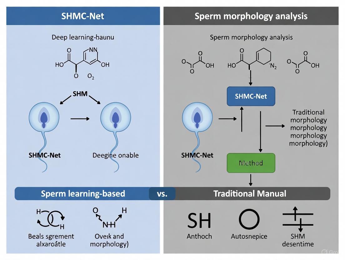
SHMC-Net introduces a sophisticated deep learning architecture specifically designed to overcome the limitations of both manual and conventional CASA approaches. The framework employs a mask-guided feature fusion strategy that integrates information from both raw sperm images and their corresponding segmentation masks [14] [4]. As shown in Figure 1, the network comprises three core components: (1) a mask generation and refinement module that produces accurate sperm head boundaries using a graph-based method; (2) a fusion encoder with parallel image and mask networks that merge features at intermediate stages; and (3) a Soft Mixup regularization component that combines mixup augmentation with a specialized loss function to handle noisy labels and small datasets [4]. This architecture enables the model to focus on morphologically relevant features while minimizing distraction from background artifacts and irregular structures.
Figure 1: SHMC-Net Architecture Overview. The system processes raw sperm images through mask generation and refinement, then utilizes a fusion encoder with parallel networks for images and masks, incorporating multi-stage feature fusion and Soft Mixup regularization before final classification.
Comparative Performance Analysis
Quantitative Accuracy Metrics Across Methods
Table 1: Performance Comparison of Sperm Morphology Classification Methods
| Method | Dataset | Accuracy | Precision | Recall | F1-Score | Key Limitations |
|---|---|---|---|---|---|---|
| Manual Assessment [12] [13] | SMD/MSS | N/A (High variability) | N/A (Subjective) | N/A (Subjective) | N/A | Inter-operator variability; Dependent on technician expertise |
| CASA Systems [15] | Clinical Samples | Variable (Correlation 0.81-0.98 with manual) | Moderate | Moderate | Moderate | Limited accuracy for subtle defects; Struggles with debris distinction |
| SHMC-Net [4] | SCIAN (Partial Agreement) | 92.6% | 92.7% | 92.7% | 92.7% | Requires quality images; Computational complexity |
| Ensemble CNN (VGG/ResNet/DenseNet) [3] | Hi-LabSpermMorpho | 71.3% | N/A | N/A | N/A | High computational demand; Complex implementation |
| Two-Stage Divide-and-Ensemble [3] | Hi-LabSpermMorpho | 69.4-71.3% | N/A | N/A | N/A | Multi-stage processing; Architectural complexity |
Operational and Technical Parameters
Table 2: Technical and Operational Characteristics of Assessment Methods
| Parameter | Manual Assessment | Traditional CASA | SHMC-Net |
|---|---|---|---|
| Analysis Time per Sample | 15-30 minutes [13] | 5-10 minutes [15] | Seconds (after training) [4] |
| Inter-Operator Variability | High (CV: 4.8-132%) [1] | Moderate (CV: 2.3-12.8%) [15] | Minimal (automated) [14] |
| Training Requirements | Extensive technical training [12] | Operator training | Specialized AI expertise |
| Classification Granularity | Based on WHO/David criteria [12] | Limited abnormality classes | Multiple head morphology classes [4] |
| Handling of Noisy/Ambiguous Samples | Subjective interpretation [12] | Often misclassifies | Soft Mixup regularization [4] |
Experimental Protocols and Validation Frameworks
Dataset Composition and Preparation
Robust validation of sperm morphology classification methods requires diverse, well-annotated datasets. The HuSHem dataset contains 216 RGB images across four categories: normal, pyriform, amorphous, and tapered heads, with most images sized 131×131 pixels [1]. The SCIAN dataset provides additional test cases with expert annotations [4]. For comprehensive evaluation, researchers have employed data augmentation techniques including rotation, translation, brightness adjustment, and color jittering to expand training data from 8,450 to 26,280 images, implementing five-fold cross-validation to prevent overfitting [1]. The SMD/MSS dataset includes 1,000 images extended to 6,035 through augmentation, classified according to the modified David classification system encompassing 7 head defects, 2 midpiece defects, and 3 tail defects [12].
SHMC-Net Implementation Protocol
Implementing SHMC-Net involves a structured workflow beginning with mask generation using anatomical and image priors to obtain sperm-head-only crops [4]. The graph-based boundary refinement (GrBR) module then optimizes contour detection by formulating it as a shortest-path problem in a directed graph, incorporating smoothness and near-convex constraints for biologically plausible shapes [4]. The fusion encoder processes both the head crops and refined masks through parallel networks, integrating features at multiple stages. The training protocol employs Soft Mixup, which implements intra-class mixup augmentation combined with a specialized loss function to address dataset limitations and label noise [4]. This comprehensive approach enables the model to achieve state-of-the-art performance while maintaining robustness to real-world variability.
Performance Validation Methodologies
Rigorous validation of sperm morphology classification systems utilizes standardized metrics including accuracy, precision, recall, and F1-score calculated on held-out test sets [4]. For clinical relevance, methods should be evaluated against expert consensus, with some studies employing partial agreement (2/3 experts) and total agreement (3/3 experts) as ground truth [12]. Comparative studies often use statistical tests (e.g., Mann-Whitney) to assess significance of differences between methods [15]. Additionally, reliability metrics such as sensitivity, specificity, and coefficients of variation provide insights into clinical applicability, with automated systems typically demonstrating superior precision (CV <7.5%) compared to manual assessment (CV up to 132%) [1] [15].
Research Reagent Solutions and Essential Materials
Table 3: Essential Research Reagents and Materials for Sperm Morphology Analysis
| Reagent/Material | Function/Application | Protocol Specifications |
|---|---|---|
| Papanicolaou Stain [13] | Sperm cell staining for morphological assessment | Standard WHO protocol with progressive ethanol dehydration |
| RAL Diagnostics Staining Kit [12] | Semen smear staining for bright-field microscopy | Following manufacturer specifications for timing |
| Diff-Quick Stains [3] | Rapid staining for morphological classification | Three staining variants: BesLab, Histoplus, GBL |
| α-Chymotrypsin [15] | Viscosity reduction in semen samples | Enzymatic treatment for improved sperm recovery |
| Sperm Quality Analyzer (SQA) [15] | Laboratory-grade semen analysis | Quality control and method validation |
| Hamilton Thorne CEROS [15] | CASA system for comparative validation | Following manufacturer operational protocols |
The comprehensive comparison presented herein demonstrates that SHMC-Net substantially advances the field of sperm morphology analysis by directly addressing the key limitations of subjectivity, inter-operator variability, and prognostic value that have long plagued traditional methods. While manual assessment remains the statutory standard despite its inherent variability, and conventional CASA systems offer partial automation with persistent limitations, SHMC-Net's mask-guided feature fusion approach achieves unprecedented classification accuracy (92.6% F1-score on SCIAN dataset) while effectively handling dataset constraints through innovative regularization techniques [4]. The model's architecture enables focused learning of morphologically relevant features, minimizing distraction from artifacts and background noise that commonly challenge traditional approaches.
For the research community, these findings highlight the transformative potential of specialized deep learning architectures in overcoming long-standing challenges in biological image analysis. SHMC-Net's performance demonstrates that targeted network designs incorporating domain-specific knowledge can yield significant improvements over both manual methods and generic deep learning models. Future research directions should focus on expanding classification granularity to encompass a broader spectrum of morphological abnormalities, enhancing model interpretability for clinical adoption, and validating prognostic value through longitudinal fertility outcome studies. As the field progresses, the integration of such advanced computational approaches with traditional andrological assessment promises to deliver more standardized, accurate, and clinically meaningful sperm morphology evaluation.
Male infertility, contributing to nearly one-third of global infertility cases, has established sperm morphology assessment as a fundamental diagnostic component in reproductive medicine [4] [5]. Traditional manual microscopy evaluation, while long considered the standard, encounters significant challenges with inter-observer variability and diagnostic discrepancies even among experts, leading to inconsistent results and subjective interpretations [4] [2]. The emergence of computer-assisted semen analysis (CASA) systems aimed to address these limitations by introducing more objective metrics; however, these systems often suffered from limitations related to low-quality sperm images, small datasets, and noisy class labels [4] [3].
In response to these challenges, the field has witnessed a paradigm shift toward two seemingly contradictory directions: simplification of routine clinical assessment alongside technological advancement in detecting specific pathological syndromes. Recent expert guidelines, particularly from the French BLEFCO Group, have recommended significant simplification of routine sperm morphology evaluation while maintaining focused analysis on detecting specific monomorphic abnormalities [16]. Concurrently, advanced deep learning approaches like SHMC-Net have demonstrated remarkable capabilities in automating morphology classification with high precision [4] [14]. This review examines these parallel developments, comparing traditional methodologies with cutting-edge computational approaches to provide researchers and clinicians with a comprehensive understanding of the current landscape in sperm morphology analysis.
Current Expert Guidelines: Streamlining Clinical Practice
The French BLEFCO Group Recommendations
The French BLEFCO Working Group conducted a systematic evaluation of sperm morphology assessment, resulting in several key recommendations that challenge conventional practices. Their 2025 guidelines represent a significant simplification of traditional approaches while maintaining critical diagnostic capabilities for specific syndromes [16].
Core Recommendations:
- R1: Against systematic detailed analysis of abnormalities during routine sperm morphology assessment
- R2: For using qualitative or quantitative methods specifically for detecting monomorphic abnormalities (globozoospermia, macrocephalic spermatozoa syndrome, pinhead spermatozoa syndrome, multiple flagellar abnormalities)
- R3: Against using sperm abnormality indexes (TZI, SDI, MAI) in infertility investigation and before ART
- R4: For using qualified and validated automated systems based on cytological analysis after staining
- R5: Against using the percentage of normal morphology sperm as a prognostic criterion before IUI, IVF, or ICSI
These recommendations reflect a pragmatic approach based on their finding that "the overall level of evidence from studies is low, challenging current practices regarding sperm morphology assessment" [16]. The guidelines specifically emphasize that laboratories should focus their efforts on detecting specific monomorphic abnormalities, which have clearer clinical implications, while de-emphasizing the comprehensive classification of all abnormality types that has characterized traditional morphology assessment.
Rationale for Simplified Assessment
The shift toward simplified assessment protocols stems from accumulating evidence questioning the clinical value of detailed morphological classification. Studies have demonstrated significant variability in performance and interpretation of traditional morphology assessment, reducing its reliability as a standalone prognostic indicator [16]. Furthermore, the clinical relevance of exhaustive abnormality categorization has shown limited impact on treatment decisions or outcomes in many cases. Instead, experts now recommend concentrating resources on detecting specific, clinically significant syndromes that directly influence treatment pathways and genetic counseling [16].
Table 1: Key Expert Recommendations for Sperm Morphology Assessment in 2025
| Recommendation | Direction | Clinical Rationale |
|---|---|---|
| Detailed abnormality analysis | Not recommended | Limited clinical relevance and high variability |
| Monomorphic abnormality detection | Recommended | Clear diagnostic and treatment implications |
| Sperm abnormality indexes (TZI, SDI, MAI) | Not recommended | Insufficient evidence of clinical value |
| Automated systems after staining | Recommended (with validation) | Improved objectivity and standardization |
| Normal morphology percentage for ART selection | Not recommended | Poor predictive value for procedure selection |
Advanced Computational Approaches: The SHMC-Net Framework
Architecture and Methodology
In contrast to simplified clinical guidelines, technological advancements have produced increasingly sophisticated computational approaches. SHMC-Net (Mask-guided Feature Fusion Network for Sperm Head Morphology Classification) represents a state-of-the-art deep learning framework that addresses key limitations in traditional CASA systems [4] [14]. The network employs a novel architecture that integrates information from both raw sperm images and their corresponding segmentation masks to enhance classification accuracy.
The SHMC-Net framework operates through three primary components [4]:
- Mask Generation and Refinement: The system generates initial segmentation masks using anatomical and image priors, then refines object boundaries with a Graph-based Boundary Refinement (GrBR) method. This refinement formulates optimal contour detection as a shortest-path problem in a directed graph with smoothness and shape constraints, requiring less than 7ms per image.
- Fusion Encoder: The core innovation involves parallel processing of sperm head crops and their corresponding refined masks through separate image and mask networks. At intermediate stages, features from both streams are fused through a dedicated feature fusion scheme to better learn morphological characteristics.
- Soft Mixup Regularization: To address noisy class labels and limited dataset sizes, SHMC-Net implements Soft Mixup, which combines mixup augmentation with a specialized loss function. This approach regularizes training and improves generalization on small datasets.
Comparative Performance Analysis
SHMC-Net has demonstrated superior performance on standard datasets compared to both traditional methods and other deep learning approaches. On the SCIAN dataset with Partial Agreement (PA) metrics, SHMC-Net achieved state-of-the-art results, while on the HuSHeM dataset, it attained exceptional accuracy of 98.3% [4] [14]. These results are particularly noteworthy as they outperform methods requiring additional pre-training or costly ensemble techniques.
Table 2: Performance Comparison of Sperm Morphology Classification Methods
| Method | Dataset | Accuracy | Precision | Recall | F1 Score |
|---|---|---|---|---|---|
| SHMC-Net [4] | HuSHeM | 98.3% | - | - | - |
| SHMC-Net [4] | SCIAN (PA) | State-of-the-art | - | - | - |
| Ensemble CNN (VGG16, VGG19, ResNet34, DenseNet161) [3] | Hi-LabSpermMorpho | ~70% | - | - | - |
| VGG16 [1] | HuSHeM | 94.0% | - | - | - |
| InceptionV3 [1] | HuSHeM | 87.3% | - | - | - |
| GAN + CapsNet [1] | HuSHeM | 97.8% | - | - | - |
| ADPL [4] | HuSHeM | 92.6% | 92.7% | 92.7% | 92.7% |
| Hybrid MLFFN-ACO [5] | UCI Fertility | 99.0% | - | 100% | - |
Recent studies have further validated the efficacy of advanced deep learning approaches. A 2024 framework integrating EdgeSAM for segmentation with pose correction and flip feature fusion achieved 97.5% accuracy on the HuSHeM and Chenwy datasets [1]. Another two-stage divide-and-ensemble approach utilizing multiple architectures including NFNet-F4 and vision transformers demonstrated significant improvement over single-model baselines, achieving 69.43-71.34% accuracy across different staining protocols on an 18-class dataset [3]. These results highlight both the capabilities and challenges of automated systems, with performance varying significantly based on dataset complexity and class diversity.
Experimental Protocols and Methodologies
SHMC-Net Experimental Workflow
The experimental implementation of SHMC-Net follows a structured workflow that transforms raw sperm images into precise morphological classifications [4]. The process begins with input raw sperm images undergoing initial mask generation using the HPM method to produce sperm-head-only crops and their corresponding pseudo-masks. These masks then proceed through the Graph-based Boundary Refinement (GrBR) module, which optimizes contour detection through a directed graph framework with smoothness and shape constraints.
The refined masks and original image crops are processed through parallel network pathways. The image network extracts features from the head crops, while the mask network processes the corresponding segmentation masks. At strategic intermediate stages, features from both streams are fused through the mask-guided feature fusion module. The fused features undergo further processing before final classification, with the entire training process regularized by Soft Mixup to handle label noise and dataset limitations.
Traditional Morphology Assessment Protocol
Traditional manual assessment follows a standardized protocol derived from WHO guidelines, though with variations across laboratories [16] [2]. The process typically begins with semen sample collection and preparation, involving smearing, washing, and staining of specimens. Stained samples are then examined under microscopy, where trained clinicians systematically evaluate at least 200 sperm cells for morphological features across head, neck, and tail regions [1] [2].
The assessment categorizes sperm into normal and various abnormal morphological types based on strict criteria. However, this method faces significant challenges with inter-observer variability, with reported inter-laboratory coefficients of variation ranging from 4.8% to as high as 132% [1]. The subjective nature of assessment, combined with the labor-intensive process of evaluating 200+ sperm cells per sample, has driven the search for more automated and standardized approaches.
Advancements in sperm morphology research rely on specialized datasets and computational resources that enable training and validation of sophisticated models. The field has benefited from several publicly available datasets, though limitations in size, quality, and annotation consistency remain challenges [2].
Table 3: Essential Research Resources for Sperm Morphology Analysis
| Resource | Type | Key Features | Applications |
|---|---|---|---|
| HuSHeM Dataset [1] | Image dataset | 216 RGB images, 4 morphology classes | Classification model development |
| SCIAN-Morpho Dataset [4] | Image dataset | Multiple morphology classes | Algorithm validation |
| Hi-LabSpermMorpho Dataset [3] | Image dataset | 18 morphology classes, 3 staining protocols | Multi-class classification |
| SVIA Dataset [2] | Video/image dataset | 125,000 detection instances, 26,000 segmentation masks | Large-scale model training |
| SHMC-Net [4] [14] | Deep learning model | Mask-guided feature fusion | Sperm head morphology classification |
| EdgeSAM [1] | Segmentation model | Efficient feature extraction | Sperm segmentation tasks |
| Mask R-CNN [17] | Segmentation model | Multi-part segmentation | Head, acrosome, nucleus, neck, tail segmentation |
Experimental Considerations and Methodological Challenges
Researchers working in sperm morphology analysis must navigate several methodological challenges. Dataset limitations represent a significant constraint, with issues including limited sample sizes, class imbalance, inconsistent annotations, and variability in staining protocols and image quality [3] [2]. Model selection depends heavily on the specific task, with Mask R-CNN demonstrating advantages for smaller, regular structures like heads and nuclei, while U-Net excels at segmenting morphologically complex components like tails [17].
The handling of stained versus unstained samples presents another consideration, as staining enhances morphological features but introduces artifacts and variability [3]. Computational efficiency also varies significantly between approaches, with simpler models offering faster inference while complex ensembles and multi-stage frameworks provide enhanced accuracy at the cost of increased computational requirements [1] [3].
The current landscape of sperm morphology assessment presents an apparent paradox between simplified clinical guidelines and increasingly sophisticated computational methodologies. However, these developments represent complementary rather than contradictory approaches to addressing the challenges in male fertility assessment.
The expert recommendations from the French BLEFCO Group reflect a pragmatic clinical perspective focused on maximizing diagnostic value while minimizing unnecessary complexity. By streamlining routine assessment and concentrating resources on detecting specific monomorphic syndromes, these guidelines aim to enhance clinical efficiency without compromising patient care [16]. Simultaneously, advanced computational approaches like SHMC-Net demonstrate the potential for automated systems to overcome the limitations of traditional assessment, providing objective, reproducible, and detailed morphological analysis that may eventually support more personalized treatment approaches [4] [14].
For researchers and clinicians, these developments highlight the importance of context-specific application of morphological assessment tools. Simplified protocols remain appropriate for routine clinical evaluation, while sophisticated computational approaches offer valuable research tools and potential future clinical applications, particularly for complex cases and specialized diagnostic challenges. As automated systems continue to evolve and validate their clinical utility, they may eventually bridge the gap between comprehensive assessment and practical implementation, ultimately enhancing both the efficiency and effectiveness of male fertility evaluation.
The diagnostic assessment of male fertility has long relied on the morphological evaluation of sperm cells, where establishing a "normal" reference range is paramount. These reference ranges are predominantly derived from studies of fertile populations, serving as the gold standard against which individual patient samples are compared [3]. Traditional manual microscopy, while foundational, is inherently subjective and labor-intensive, leading to significant inter-observer variability; reported inter-laboratory coefficients of variation can range from 4.8% to as high as 132% [1]. The emergence of Computer-Aided Sperm Analysis (CASA) systems sought to introduce more objectivity but often faced limitations with low-quality images and limited functionality [3]. The recent development of advanced deep learning models, particularly the Sperm Head Morphology Classification Network (SHMC-Net), represents a paradigm shift. This guide provides a comparative analysis of SHMC-Net against traditional and other modern methods for sperm morphology classification, framing the evaluation within the critical context of using data from fertile populations to establish robust, clinically relevant reference standards.
Performance Comparison of Sperm Morphology Analysis Methods
A critical step in male fertility diagnostics is the accurate classification of sperm head morphology, which directly relies on reference ranges established from fertile populations. The performance of different methodologies in this task varies significantly. The following table summarizes the quantitative performance of various methods on public datasets, with SHMC-Net demonstrating state-of-the-art results.
Table 1: Performance Comparison of Sperm Morphology Classification Methods
| Method | Dataset | Reported Accuracy | Key Features / Architectures |
|---|---|---|---|
| SHMC-Net [4] [14] | SCIAN (PA), HuSHeM | State-of-the-art (SOTA) | Mask-guided feature fusion, Soft Mixup, graph-based boundary refinement |
| Proposed Deep Learning Framework [1] | HuSHem, Chenwy | 97.5% | EdgeSAM segmentation, pose correction network, flip feature fusion |
| Two-Stage Ensemble Framework [3] | Hi-LabSpermMorpho (3 stains) | 68.41% - 71.34% | Two-stage classification (head/neck vs. tail/normal), ensemble of NFNet & ViT |
| Hybrid MLFFN–ACO Framework [5] | UCI Fertility Dataset | 99% | Multilayer neural network with Ant Colony Optimization (ACO) |
| VGG16 [1] | HuSHeM | 94% | Standard VGG16 architecture |
| InceptionV3 [1] | HuSHeM | 87.3% | Standard InceptionV3 architecture |
| ADPL [4] | HuSHeM | 92.6% | Traditional method with hand-crafted features |
| Manual Microscopy [1] | N/A | N/A | High inter-observer variability (4.8% - 132% CoV) |
Beyond raw accuracy, the methodological approach of each system dictates its suitability for establishing reliable reference ranges. The following table compares the core methodologies and their direct implications for this specific application.
Table 2: Methodological Comparison for Establishing Reference Ranges
| Method Category | Core Methodology | Impact on Reference Range Establishment |
|---|---|---|
| Manual Microscopy | Visual assessment by trained clinicians of stained samples based on WHO criteria [1] [3]. | Prone to subjectivity and high variability, leading to inconsistent and unreliable reference ranges across laboratories. |
| Traditional CASA | Relies on hand-crafted features (area, length-width ratio, perimeter, symmetry) [1] [4]. | Feature selection bias may overlook subtle but clinically significant morphological details captured from fertile populations. |
| Standard Deep Learning (VGG16, InceptionV3) | Transfer learning with standard CNN architectures for end-to-end classification [1]. | Performs well but can be sensitive to sperm pose and may focus on irrelevant features, limiting generalization [1]. |
| SHMC-Net (Proposed) | Uses segmentation masks to guide classification; fuses features from image and mask; employs Soft Mixup for regularization [4] [14]. | Mask guidance ensures focus on morphologically relevant structures. Improved robustness and accuracy contribute to more precise and reliable reference data. |
| Two-Stage Ensemble [3] | A splitter model first categorizes sperm into major groups (head/neck vs. tail/normal), then category-specific ensembles perform fine-grained classification. | Reduces misclassification between visually dissimilar categories (e.g., head vs. tail defects), refining the specificity of reference ranges for different abnormality types. |
Experimental Protocols and Workflows
Detailed Methodology of SHMC-Net
SHMC-Net introduces a novel architecture that leverages segmentation masks to guide the morphology classification process, which is crucial for generating consistent data from fertile populations. Its experimental protocol can be broken down into three key components [4]:
- Mask Generation and Refinement: Initial sperm-head-only crops are generated using anatomical and image priors via the HPM method [1]. The boundaries of the resulting pseudo-masks are then refined using a sperm head shape-aware Graph-based Boundary Refinement (GrBR) method. GrBR formulates the optimal contour refinement as a shortest-path problem in a directed graph, enforcing smoothness and a near-convex shape constraint to capture the accurate head boundary efficiently (in less than 7 ms per image).
- Fusion Encoder Architecture: The model employs two parallel networks: an image network that processes the head crops and a mask network that processes the corresponding boundary-refined masks. In the intermediate stages of these networks, features from both streams are fused. This fusion scheme allows the model to learn morphological features from both the raw image data and the clean, shape-focused mask data.
- Soft Mixup Regularization: To handle the common challenges of small datasets and noisy class labels (observer variability), SHMC-Net uses Soft Mixup. This technique combines intra-class Mixup augmentation for both images and masks with a corresponding loss function, which regularizes training and improves generalization.
Workflow of a Comparative Deep Learning Framework
Another advanced framework highlights a comprehensive workflow that directly addresses the need for standardized analysis of sperm from fertile populations [1]. The process is designed to mimic and automate the expert's approach to classification.
Figure 1: Workflow for Automated Sperm Morphology Classification
- Sperm Feature Extraction and Segmentation: The process begins with a raw sperm image. The EdgeSAM model, an efficient variant of the Segment Anything Model, is used for initial feature extraction and precise segmentation of the sperm head. A single coordinate point prompt can be used to indicate the rough location of the specific sperm head, enabling accurate segmentation while suppressing irrelevant content like tails or noise [1].
- Sperm Head Pose Correction: The segmented sperm head is then passed to a dedicated Sperm Head Pose Correction Network. This network predicts the head's position, angle, and orientation. Using Rotated RoI alignment, it standardizes the head's pose, correcting for rotational and translational variations. This step is critical for consistent feature extraction and for improving the model's robustness and classification accuracy, directly contributing to the reliability of the derived morphological data [1].
- Morphology Classification: The standardized sperm head is fed into the final classification network. This network employs specialized techniques such as flip feature fusion to leverage the symmetrical properties of certain sperm heads (e.g., pyriform) and deformable convolutions to better capture morphological variations. The output is the final morphology class, such as normal, amorphous, pyriform, or tapered [1].
Two-Stage Ensemble Classification Workflow
For complex datasets with a wide spectrum of abnormalities, a hierarchical two-stage approach has been developed to improve classification reliability, which is essential for dissecting the subtle morphological variations within a fertile population [3].
Figure 2: Two-Stage Ensemble Classification
- First Stage - Splitting: A dedicated "splitter" model acts as a coarse classifier, routing each sperm image to one of two principal categories: Category 1 for head and neck region abnormalities, or Category 2 for normal morphology and tail-related abnormalities [3].
- Second Stage - Fine-Grained Ensemble Classification: Each category is processed by a customized ensemble model dedicated to that specific group. The ensemble typically integrates multiple deep learning architectures, such as DeepMind's NFNet and Vision Transformer (ViT) variants. A structured multi-stage voting mechanism is used to aggregate predictions from the models in the ensemble, enhancing decision reliability beyond simple majority voting [3].
The Scientist's Toolkit: Research Reagent Solutions
The following table details key reagents, datasets, and computational tools essential for conducting research in automated sperm morphology analysis and establishing reference ranges.
Table 3: Essential Research Reagents and Materials
| Item Name | Function / Application | Specific Example / Note |
|---|---|---|
| Diff-Quick Staining Kits | Enhances contrast and visualization of sperm morphological features for microscopy and image analysis [3]. | Different specific kits (BesLab, Histoplus, GBL) can create variations in image appearance, requiring model robustness or normalization [3]. |
| HuSHeM Dataset | A public benchmark dataset for training and evaluating models on sperm head morphology classification [1]. | Contains 216 RGB images of normal, pyriform, amorphous, and tapered sperm heads with expert contour annotations [1]. |
| SCIAN-Morpho Dataset | A public dataset used for comparative performance evaluation of sperm classification algorithms [4]. | Used in conjunction with HuSHeM to demonstrate generalizability of models like SHMC-Net [4]. |
| Hi-LabSpermMorpho Dataset | A large-scale dataset for complex morphology classification with 18 distinct classes based on WHO criteria [3]. | Includes annotations for head, neck, and tail defects, enabling detailed analysis of specific abnormality types [3]. |
| EdgeSAM Model | A parameter-efficient segment anything model used for precise initial segmentation of sperm heads from raw images [1]. | Used in the automated framework to suppress irrelevant features like sperm tails, improving downstream analysis [1]. |
| Soft Mixup | A regularization technique combining mixup augmentation and a loss function to handle noisy labels and small datasets [4]. | Mitigates the effect of inter-observer variability in training data labels, a common issue in medical image analysis [4]. |
| Ant Colony Optimization (ACO) | A nature-inspired optimization algorithm used for tuning parameters in hybrid machine learning frameworks [5]. | Can enhance predictive accuracy and convergence in diagnostic models, as demonstrated in a hybrid MLFFN–ACO framework [5]. |
SHMC-Net in Action: Architectural Principles and Workflow Integration for Drug Discovery
The manual assessment of sperm morphology, a cornerstone of male fertility diagnosis, has long been plagued by subjectivity and significant inter-observer variability, with reported coefficients of variation ranging from 4.8% to as high as 132% between laboratories [1]. This diagnostic inconsistency poses a substantial challenge for clinicians and researchers developing reliable drug therapies and diagnostic tools. The emergence of deep learning offers a path toward standardization. This guide provides an objective comparison of deep learning architectures, with a specific focus on the configuration and application of ResNet50 via transfer learning, within the context of automated sperm morphology analysis. The performance of these traditional convolutional neural networks (CNNs) is contextualized against a novel, purpose-built architecture: the Sperm Head Morphology Classification Network (SHMC-Net) [14]. By comparing experimental data on accuracy, computational demands, and methodological rigor, this guide aims to equip scientists and drug development professionals with the evidence needed to select appropriate models for their research in reproductive medicine.
Technical Foundations of ResNet50 and SHMC-Net
ResNet50: Architecture and Relevance to Medical Imaging
ResNet50 is a 50-layer deep convolutional neural network that introduced a breakthrough architectural innovation: residual connections [18]. These skip connections allow the gradient to backpropagate more effectively through the deep network by bypassing one or more layers, thereby mitigating the vanishing gradient problem that had previously hindered the training of very deep networks [18]. This design enables the model to learn more complex features without degrading performance, a key reason for its widespread adoption.
In the context of medical image analysis, including sperm morphology, ResNet50 is seldom trained from scratch. Instead, transfer learning (TL) is the predominant strategy. This involves taking a ResNet50 model pre-trained on a large-scale natural image dataset like ImageNet and fine-tuning it on a specialized, smaller medical dataset [19]. This approach leverages the generic feature extraction capabilities (e.g., edge and texture detection) learned from millions of images, allowing the model to adapt quickly and effectively to new visual domains with limited data, a common scenario in clinical research [18].
SHMC-Net: A Specialized Architecture for Morphology Classification
In contrast to general-purpose CNNs like ResNet50, SHMC-Net represents a tailored deep-learning solution designed explicitly for the challenges of sperm head morphology classification [14]. Its core innovation is a mask-guided feature fusion mechanism. Instead of relying solely on raw images, SHMC-Net uses a two-stream network architecture: one stream processes the original sperm head crops, while the other processes their corresponding segmentation masks. The features from these two streams are fused in the intermediate stages of the network, forcing the model to focus on precise morphological structures and boundaries, thereby learning a more robust representation of shape and form [14]. Furthermore, to handle the common issues of small datasets and noisy labels, SHMC-Net employs Soft Mixup, a regularization technique that combines mixup augmentation with a tailored loss function to improve generalization [14].
Performance Comparison of Deep Learning Models
The following tables summarize the quantitative performance of various deep learning models, including ResNet50-based approaches and more specialized architectures, across different sperm image analysis tasks and datasets.
Table 1: Performance of ResNet50 and Other CNN Models on Sperm Head Classification
| Model | Dataset | Key Methodology | Reported Accuracy | Reference |
|---|---|---|---|---|
| TL-ResNet50 | NEU-CLS (Steel Defects) | Transfer Learning from ImageNet | 99.4% | [19] |
| VGG16 | HuSHeM | Standard Fine-tuning | 94.0% | [1] |
| InceptionV3 | HuSHeM | Standard Fine-tuning | 87.3% | [1] |
| Ensemble (VGG16, VGG19, ResNet34, DenseNet161) | HuSHeM | Model Ensembling | >94.0% (Outperformed single models) | [1] |
Table 2: Performance of Advanced and Specialized Models on Sperm Morphology Analysis
| Model | Task | Dataset | Reported Performance | Reference |
|---|---|---|---|---|
| Two-Stage Ensemble (NFNet, ViT) | 18-class Morphology Classification | Hi-LabSpermMorpho | 69-71% Accuracy (4.38% improvement over baselines) | [3] |
| Proposed Framework (EdgeSAM + Classification) | Sperm Head Classification | HuSHeM & Chenwy | 97.5% Accuracy | [1] |
| SHMC-Net | Sperm Head Morphology Classification | SCIAN & HuSHeM | State-of-the-Art (Outperformed methods with pre-training/ensembling) | [14] |
| Mask R-CNN | Multi-part Sperm Segmentation | Live, Unstained Human Sperm | Highest IoU for head, nucleus, acrosome | [20] |
| U-Net | Multi-part Sperm Segmentation | Live, Unstained Human Sperm | Highest IoU for the tail | [20] |
Table 3: ResNet50 Benchmarking on Edge Hardware (Wildfire & Martian Terrain Classification)
| Hardware Platform | Task | Baseline Model Inference Time | Quantized Model Size Reduction | Inference Time Reduction |
|---|---|---|---|---|
| Nvidia Jetson Nano | Wildfire Detection | 50 ms | 73-74% | 56-68% |
| Intel NUC | Wildfire Detection | 316 ms | 73-74% | 56-68% |
| Nvidia Jetson Nano | Martian Terrain | 62 ms | 73-74% | 56-68% |
| Intel NUC | Martian Terrain | 580 ms | 73-74% | 56-68% |
Experimental Protocols and Methodologies
Standard Protocol for Transfer Learning with ResNet50
A typical experimental protocol for applying TL-ResNet50 to an image classification task, such as steel defect detection which methodologically parallels biomedical image analysis, involves several key stages [19]:
- Dataset Preparation and Preprocessing: The target dataset (e.g., NEU-CLS with 1800 images of 6 defect classes) is split into training and testing sets, commonly at an 8:2 ratio. To enhance model robustness, data augmentation techniques like contrast adjustment are often applied, especially when the image background is dark and may obscure features [19].
- Model Configuration and Fine-tuning: A ResNet50 model pre-trained on the ImageNet dataset is loaded. The final fully connected (classification) layer is modified to output the number of classes required for the new task (e.g., 6). An optimization strategy combining the Adam optimizer with a learning rate decay is often employed to fine-tune the model, balancing rapid convergence with final performance [19].
- Interpretation and Analysis: To build trust and understanding in the model's predictions, interpretability algorithms like Grad-CAM++ can be applied. This generates heatmaps that visually highlight the image regions most influential in the model's classification decision, providing approximate localization of defects or morphological features [19].
Protocol for Two-Stage Divide-and-Ensemble Framework
A more complex, hierarchical approach has been developed to address the challenge of classifying a wide spectrum of sperm abnormalities with high inter-class similarity. The methodology, as applied to an 18-class dataset, proceeds as follows [3]:
- First Stage - Category Splitting: A dedicated "splitter" model is trained to perform a coarse-level classification, routing each sperm image into one of two principal categories: (1) head and neck region abnormalities, or (2) normal morphology together with tail-related abnormalities. This initial division simplifies the problem for subsequent models.
- Second Stage - Category-Specific Ensemble Classification: For each of the two broad categories, a separate ensemble of deep learning models is employed to perform the fine-grained classification. A typical ensemble might integrate four distinct architectures, such as DeepMind's NFNet-F4 and various Vision Transformer (ViT) variants.
- Structured Multi-Stage Voting: Unlike conventional majority voting, this framework introduces a more sophisticated decision-making mechanism. Each model in the ensemble casts a primary vote and a secondary vote. This strategy mitigates the influence of dominant classes and enhances the reliability of the final prediction, leading to a statistically significant 4.38% improvement in accuracy over prior approaches [3].
Protocol for SHMC-Net
The SHMC-Net framework introduces a segmentation-guided approach for classification, which involves a carefully designed multi-step pipeline [14]:
- Segmentation Mask Generation: The process begins not with classification, but with segmentation. The framework uses image priors to generate reliable initial segmentation masks of sperm heads. These masks are then refined using an efficient graph-based method to precisely delineate object boundaries.
- Dual-Stream Network Training: SHMC-Net consists of two parallel networks: an image network that takes the original sperm head crops as input, and a mask network that takes the corresponding segmentation masks as input.
- Mask-Guided Feature Fusion: In the intermediate layers of the networks, features from the image stream and the mask stream are fused together. This fusion forces the model to align its understanding of the raw image with the precise structural information from the mask, thereby learning more accurate morphological features.
- Regularization with Soft Mixup: To handle the common challenges of small, noisy biomedical datasets, the model is trained using Soft Mixup. This technique combines mixup data augmentation with a loss function that is more robust to label noise, regularizing the training process and improving generalization.
Diagram 1: SHMC-Net's segmentation-guided classification workflow with dual-stream feature fusion.
The Scientist's Toolkit: Essential Research Reagents and Materials
Table 4: Key Reagents and Computational Tools for Sperm Morphology Analysis Research
| Item Name | Function/Application in Research | Example from Literature |
|---|---|---|
| Hi-LabSpermMorpho Dataset | A large-scale, expert-labeled dataset with 18 distinct sperm morphology classes, used for training and evaluating complex classification models. | Used to develop and test the two-stage ensemble framework [3]. |
| HuSHeM Dataset | The Human Sperm Head Morphology dataset; a benchmark containing images of normal and abnormal sperm heads (amorphous, pyriform, tapered). | Used for evaluating models like VGG16, SHMC-Net, and the EdgeSAM-based framework [1] [14]. |
| Diff-Quick Staining Kits | A staining protocol (e.g., BesLab, Histoplus, GBL) used to enhance morphological features in sperm images for improved manual and automated analysis. | Used to prepare images in the Hi-LabSpermMorpho dataset [3]. |
| Pre-trained Model Weights (ImageNet) | Initial parameters for models like ResNet50, enabling effective transfer learning by providing a strong foundation of general feature extraction. | Used as the starting point for TL-ResNet50 models in various studies [19]. |
| EdgeSAM | A lightweight variant of the Segment Anything Model (SAM) used for precise feature extraction and segmentation, reducing computational demands. | Used for initial sperm head segmentation in a pose-correction and classification framework [1]. |
| Grad-CAM++ | An interpretability algorithm that generates visual explanations for model predictions, highlighting regions of the input image most relevant to the classification. | Used to provide explanatory analysis and visualize model focus in TL-ResNet50 studies [19]. |
Diagram 2: End-to-end experimental workflow for deep learning-based sperm morphology analysis.
The empirical data and experimental protocols presented in this guide demonstrate a clear trade-off in the selection of deep learning architectures for sperm morphology analysis. ResNet50, particularly when configured with transfer learning, provides a robust, well-understood, and highly accessible baseline. It can achieve excellent performance (e.g., >99% accuracy in controlled industrial defect detection tasks that share methodological similarities) and is amenable to optimization for deployment, such as through quantization for resource-constrained environments [21] [19]. However, for the specific and nuanced challenge of sperm morphology classification within clinical research, purpose-built architectures like the two-stage ensemble and SHMC-Net have begun to demonstrate superior performance. These models directly address key limitations—such as high inter-class similarity, data scarcity, and the need for precise morphological focus—through innovative hierarchical structures and mask-guided learning. For researchers and drug development professionals, the choice between a transfer-learned ResNet50 and a novel architecture like SHMC-Net will depend on the specific priorities of their project, balancing factors such as required accuracy, computational resources, and the availability of segmented data.
The accurate assessment of sperm morphology represents a critical component in the diagnosis of male infertility, a condition affecting approximately 15% of couples globally with male-related factors contributing to 30-40% of cases [1] [20]. Traditional manual microscopy for sperm evaluation is notoriously labor-intensive and susceptible to significant observer variability, with inter-laboratory coefficients of variation ranging from 4.8% to as high as 132% [1]. To address these challenges, computer-assisted sperm analysis (CASA) systems have emerged as a transformative technology, leveraging advanced imaging and machine learning to standardize and automate the evaluation process [20]. Within this technological evolution, deep learning approaches like the Sperm Head Morphology Classification Network (SHMC-Net) have demonstrated remarkable performance, achieving up to 98.3% accuracy in classification tasks by integrating segmentation masks with image features [14].
The foundation of any robust computational analysis lies in the quality and precision of its input data. For sperm morphology research, this necessitates the careful curation of datasets through high-resolution confocal microscopy and meticulous image annotation protocols. Confocal microscopy provides the essential optical sectioning capabilities required to generate high-resolution images of sperm substructures without the blurring effect of out-of-focus light, while advanced annotation tools enable precise labeling of morphological features at cellular and subcellular levels [22] [23]. This methodological foundation supports not only the training of accurate models like SHMC-Net but also enables meaningful comparisons with traditional analytical approaches, ultimately advancing the field of reproductive medicine through more reliable, automated diagnostic capabilities.
High-Resolution Confocal Microscopy Fundamentals
Core Principles and Technical Specifications
Confocal microscopy operates on the fundamental principle of point illumination and spatial pinholes to eliminate out-of-focus light, thereby significantly enhancing optical resolution and contrast compared to conventional widefield microscopy [22]. This technical advantage is particularly crucial for imaging thick biological specimens like sperm cells, where precise visualization of subcellular structures determines diagnostic accuracy. In a confocal system, illumination and detection optics are focused on the same diffraction-limited spot in the sample, which is scanned across the field of view to construct a complete image point-by-point [22]. This configuration provides exceptional optical sectioning capability, allowing for high-resolution three-dimensional reconstruction of specimens from z-stack image collections.
The resolution capabilities of confocal microscopy are mathematically determined by specific optical parameters. Lateral resolution can be calculated as Rlateral = 0.4λ/NA, while axial resolution follows Raxial = 1.4λη/(NA)², where λ represents the emission light wavelength, η is the refractive index of the mounting medium, and NA is the objective's numerical aperture [22]. In practical application, the best resolution achievable is approximately 0.2 μm laterally and 0.6 μm axially, though these theoretical limits are not always attained in biological imaging scenarios [22]. A critical tradeoff exists between light collection efficiency and resolution, governed by adjustable pinhole settings – opening the pinhole increases signal at the cost of resolution, while closing it enhances resolution but reduces signal-to-noise ratio [22].
Table 1: Comparison of Confocal Microscope Types for Sperm Imaging
| Microscope Type | Scanning Method | Resolution | Imaging Speed | Advantages | Limitations |
|---|---|---|---|---|---|
| Laser Scanning Confocal (LSCM) | Single point scanning | High (~0.2 μm lateral) | Moderate | Excellent optical sectioning, versatile 3D imaging | Slower imaging speed, potential photodamage |
| Spinning Disk Confocal | Multi-point scanning (Nipkow disk) | High (~0.2 μm lateral) | High | Fast imaging, low light dose | Non-adjustable pinholes, potential crosstalk in thick samples |
| Scanned Fibre Endomicroscope | Full point scanning | Submicron lateral | High | Cellular/subcellular resolution, miniaturized probe | Specialized application, limited field of view |
Instrumentation Selection for Sperm Imaging
When configuring confocal microscopy for sperm imaging, several instrumental considerations significantly impact data quality. Laser scanning confocal microscopes (LSCMs) represent the most common commercial implementation, utilizing galvanometer mirrors to sweep a laser beam across the sample in x and y dimensions [22]. These systems offer exceptional versatility for 2D, 3D, and even 4D/5D imaging (incorporating time and wavelength dimensions), making them well-suited for comprehensive sperm morphological analysis [22]. Alternatively, spinning disk confocal microscopes employ rapidly rotating disks containing multiple pinholes to simultaneously image multiple points, offering significantly faster acquisition speeds beneficial for dynamic live sperm imaging [22].
The scanned fibre endomicroscope represents a specialized category of confocal microscopy particularly relevant for high-resolution cellular imaging. These instruments are full point-scanning confocal microscopes with submicron lateral resolution and optical slice thickness sufficient to isolate individual cell layers [24]. Unlike bundle fibre devices limited by fixed z-focus depth and resolution constrained by fibre count, scanned fibre systems enable active positioning of the optical slice in the z-axis and collection of megapixel images with cellular and subcellular resolution [24]. This capability is essential for in vivo identification of morphological features critical for sperm quality assessment.
Image Annotation Protocols and Tools
Annotation Approaches for Morphological Analysis
Image annotation constitutes the foundational process of labeling digital images to provide ground truth data for training and validating computational models like SHMC-Net. In the context of sperm morphology, annotation strategies must capture both global structural characteristics and fine subcellular details to support accurate classification. The three primary annotation types employed in computer vision projects include: (1) whole-image classification for broad categorization; (2) object detection using bounding boxes to localize individual sperm within images; and (3) image segmentation, which assigns each pixel to a specific class, providing the most granular morphological information [25].
For sperm morphology analysis, segmentation-based annotation is particularly valuable as it enables precise delineation of sperm components including head, acrosome, nucleus, neck, and tail [20]. This pixel-level annotation supports the training of sophisticated models like SHMC-Net that rely on mask-guided feature fusion to enhance classification accuracy [14]. Contemporary annotation platforms such as Supervisely and Labelbox offer specialized toolkits for these tasks, including bounding box tools, polygon tools for outlining object boundaries, mask pen tools for segmentation, and brush tools for free-form mask creation [23] [25]. These platforms increasingly incorporate AI-assisted functionality, with model-assisted labeling reducing annotation time and costs by up to 50% by generating pre-labels for human reviewers to refine rather than starting from scratch [25].
Specialized Annotation Tools for Sperm Analysis
Table 2: Image Annotation Tools for Sperm Morphology Analysis
| Tool Category | Specific Tools | Primary Function | Advantages for Sperm Analysis |
|---|---|---|---|
| Manual Annotation Tools | Bounding Box, Polygon Tool, Brush Tool | Precise manual delineation of sperm structures | Flexibility for irregular shapes; accurate boundary definition |
| AI-Assisted Tools | SmartTool (RITM, ClickSeg, Segment Anything) | Automated segmentation with manual refinement | Rapid processing; consistent results across large datasets |
| Specialized Features | Tags and Attributes, Hotkeys, Collaborative Features | Enhanced metadata and workflow efficiency | Standardized classification; team-based quality control |
Advanced annotation platforms provide specialized functionality essential for high-quality sperm morphology datasets. The Supervisely Labeling Toolbox, for instance, offers a comprehensive suite including bounding boxes for object detection, polygon tools for precise boundary outlining, mask pen tools combining polygon and brush functionalities, and specialized brush tools optimized for working with hundreds of masks simultaneously [23]. Particularly valuable for sperm analysis is the SmartTool, which leverages neural network algorithms like RITM, ClickSeg, and Segment Anything models for interactive object segmentation [23]. This tool allows annotators to guide the model by adding positive and negative points to adjust predictions, significantly accelerating the annotation process while maintaining accuracy.
For specialized sperm component analysis, additional features such as tags and attributes enable detailed morphological characterization beyond simple segmentation. These classification systems allow researchers to tag both images and individual objects with attributes such as "pyriform," "amorphous," or "tapered" to describe head morphology, or to flag specific structural abnormalities [23]. The availability of customizable hotkeys further enhances annotation efficiency, allowing experienced annotators to work rapidly without interrupting their workflow to select tools or tags from menus [23]. These specialized capabilities collectively support the creation of comprehensively annotated datasets required for robust model training.
Experimental Protocols for Comparative Analysis
Specimen Preparation and Imaging Parameters
Optimal specimen preparation is essential for high-quality confocal imaging of sperm morphology. While many preparation protocols are derived from conventional microscopy, confocal applications typically require increased staining concentrations or extended staining times due to the optical sectioning that undersamples fluorescence compared to widefield epifluorescence microscopy [26]. For sperm imaging, both stained and unstained approaches are utilized, with unstained live sperm presenting greater challenges due to low signal-to-noise ratios and minimal color differentiation between components, though offering the advantage of avoiding potential morphological alterations from staining procedures [20].
The selection of appropriate fluorescent probes significantly impacts image quality and resolution. Commonly used topical fluorophores for mucosal cellular imaging include fluorescein, acriflavine, and PARPi-FL, with the latter specifically targeting PARP1 (Poly (ADP-ribose) polymerase 1), a nuclear protein overexpressed in many malignancies [24]. For deeper imaging within specimens, longer wavelength excitation dyes such as cyanine 5 are advantageous as they experience less scattering and can penetrate further into samples, though with a slight reduction in maximum resolution compared to shorter wavelength alternatives [26]. Objective lens selection represents another critical parameter, with higher numerical aperture (NA) objectives providing thinner optical sections – for example, a 60x NA 1.4 objective can achieve approximately 0.4 μm section thickness with a 1 mm pinhole, compared to 1.8 μm for a 16x NA 0.5 lens with the same pinhole setting [26].
Workflow for SHMC-Net Evaluation
The evaluation of SHMC-Net against traditional sperm morphology analysis follows a structured experimental workflow to ensure comprehensive and unbiased comparison. The initial stage involves dataset curation using high-resolution confocal microscopy to capture sperm images with appropriate magnification and resolution to visualize critical morphological features. Subsequent annotation employs a combination of manual and AI-assisted tools to generate precise segmentation masks and classification labels that serve as ground truth for model training and validation [23] [14]. The implementation of SHMC-Net then proceeds with its distinctive mask-guided feature fusion architecture, which integrates features from both original sperm head crops and their corresponding segmentation masks to enhance morphological learning [14].
Diagram 1: Experimental workflow for sperm morphology analysis comparison
A critical component of the experimental protocol involves addressing common challenges in sperm image analysis, including class imbalance and rotational variance. SHMC-Net incorporates Soft Mixup augmentation to handle noisy class labels and regularize training on limited datasets, while more recent approaches have introduced sperm head pose correction networks to standardize orientation and position before classification [14] [1]. The comparative evaluation then assesses performance across multiple metrics including segmentation accuracy (IoU, Dice coefficient), classification accuracy, sensitivity, specificity, and computational efficiency, with rigorous statistical validation to ensure findings are robust and clinically relevant [1] [20].
Quantitative Performance Comparison
Segmentation Accuracy Across Models
The accurate segmentation of sperm components represents a fundamental requirement for precise morphological classification. Recent systematic evaluations comparing deep learning models for multi-part sperm segmentation have revealed distinct performance characteristics across different architectural approaches. Mask R-CNN demonstrates particular strength in segmenting smaller and more regular structures such as the head, nucleus, and acrosome, achieving slightly higher IoU for the nucleus compared to YOLOv8 and surpassing YOLO11 for acrosome segmentation [20]. For the morphologically complex tail structure, U-Net achieves the highest IoU, benefiting from its global perception and multi-scale feature extraction capabilities [20].
Table 3: Segmentation Performance Comparison of Deep Learning Models
| Sperm Component | Mask R-CNN | YOLOv8 | YOLO11 | U-Net |
|---|---|---|---|---|
| Head | Highest IoU | High IoU | Moderate IoU | High IoU |
| Acrosome | Superior Performance | Moderate IoU | Lower IoU | High IoU |
| Nucleus | Slightly Higher IoU | High IoU | Moderate IoU | High IoU |
| Neck | High IoU | Comparable/Slightly Better | Moderate IoU | High IoU |
| Tail | Moderate IoU | High IoU | Moderate IoU | Highest IoU |
These comparative findings provide valuable guidance for model selection in sperm segmentation tasks. The performance variations highlight how different architectural strengths align with specific morphological challenges – Mask R-CNN's two-stage detection-segmentation pipeline benefits well-defined structures, while U-Net's encoder-decoder architecture with skip connections effectively captures the elongated, complex morphology of sperm tails [20]. For comprehensive sperm analysis encompassing all components, ensemble approaches or hybrid architectures may offer the most robust solution, though with increased computational complexity that may limit real-time clinical application [1].
Classification Accuracy and Computational Efficiency
The ultimate objective of sperm morphology analysis is accurate classification into clinically relevant categories, with SHMC-Net representing a state-of-the-art approach specifically designed for this purpose. Comparative evaluations demonstrate that SHMC-Net achieves 98.3% accuracy on the SCIAN dataset and outperforms methods requiring additional pre-training or costly ensembling techniques [14]. This performance edge stems from its innovative mask-guided feature fusion, which enables the model to leverage both pixel-level texture information from original images and precise morphological boundaries from segmentation masks [14].
More recent advancements have further pushed classification accuracy while addressing computational efficiency concerns. An automated deep learning framework integrating EdgeSAM for segmentation with a sperm head pose correction network and flip feature fusion has demonstrated 97.5% accuracy on the HuSHem and Chenwy datasets while offering greater robustness to rotational and translational transformations [1]. This approach specifically addresses the symmetry characteristics of pyriform and amorphous sperm heads through deformable convolutions that adapt to irregular shapes, significantly enhancing classification accuracy across morphological variations [1].
Diagram 2: SHMC-Net architecture with mask-guided feature fusion
Beyond raw accuracy, computational efficiency represents a critical consideration for clinical implementation. Traditional manual microscopy requires extensive time from highly trained personnel, while early computer-assisted systems often suffered from slow processing speeds. Contemporary optimized models like SHMC-Net and its variants achieve classification in computationally efficient timeframes, with some hybrid frameworks reporting ultra-low computational times of just 0.00006 seconds per sample while maintaining 99% classification accuracy and 100% sensitivity [5]. This combination of high accuracy and computational efficiency makes these advanced approaches increasingly viable for integration into clinical workflows where both reliability and speed are essential.
Essential Research Reagents and Materials
Research Reagent Solutions for Sperm Imaging
The successful implementation of confocal microscopy and image annotation protocols for sperm morphology analysis requires specific research reagents and materials optimized for imaging quality and procedural efficiency. These reagents span specimen preparation, staining, mounting, and imaging applications, each serving distinct functions in the experimental workflow. The selection of appropriate reagents significantly impacts image quality, annotation accuracy, and ultimately the performance of computational models like SHMC-Net in morphological classification.
Table 4: Essential Research Reagents for Sperm Morphology Analysis
| Reagent Category | Specific Examples | Function in Workflow | Application Notes |
|---|---|---|---|
| Fluorescent Probes | Fluorescein, Acriflavine, PARPi-FL | Contrast enhancement for cellular visualization | PARPi-FL targets PARP1 overexpression; acriflavine binds nuclear material |
| Mounting Media | Antifade reagents, Refractive index matching solutions | Preservation of specimen structure and reduction of photobleaching | Essential for 3D reconstruction; affects effective numerical aperture |
| Staining Kits | DNA-specific stains, Vitality stains | Differentiation of viable sperm and structural components | Impacts signal-to-noise ratio in confocal imaging |
| Annotation Software | Supervisely, Labelbox | Image segmentation and classification labeling | AI-assisted features significantly reduce annotation time |
Fluorescent probes represent particularly critical reagents for confocal imaging of sperm morphology. Acriflavine serves as a common topical antiseptic that binds membrane and nuclear material non-selectively, while fluorescein functions as an exogenous dye routinely used in ophthalmic practice that has been adapted for mucosal imaging [24]. More specialized probes like PARPi-FL (Poly (ADP-ribose) polymerase 1 inhibitor) offer targeted imaging capabilities, as PARP1 is a nuclear protein overexpressed in many malignancies and has demonstrated utility in delineating tumor tissue from normal tissue in clinical trials [24]. The selection of appropriate mounting media and antifade reagents further enhances image quality by preserving specimen integrity and reducing photobleaching during extended imaging sessions, particularly important for comprehensive z-stack collection for 3D reconstruction [26].
The comparative analysis between SHMC-Net and traditional sperm morphology analysis methods reveals a clear trajectory toward increasingly sophisticated computational approaches that offer superior accuracy, efficiency, and consistency. The foundation of these advances rests squarely on meticulous dataset curation through high-resolution confocal microscopy and precise image annotation protocols. Confocal microscopy provides the essential optical sectioning capabilities and resolution necessary to visualize critical sperm substructures, while modern annotation tools enable the creation of comprehensive labeled datasets required for training robust deep learning models.
The quantitative evidence demonstrates that specialized architectures like SHMC-Net, with their mask-guided feature fusion and attention to class imbalance issues, consistently outperform traditional manual assessment and earlier computer-assisted approaches in classification accuracy, achieving up to 98.3-99% accuracy in controlled evaluations [5] [14]. Furthermore, the integration of pose correction networks and flip feature fusion modules addresses historical challenges with rotational variance, enhancing model robustness for clinical implementation [1]. As these computational methodologies continue to evolve alongside improvements in confocal imaging technology and annotation workflows, the field moves closer to realizing fully automated, highly accurate sperm morphology analysis that can expand access to reliable fertility diagnostics and ultimately improve patient outcomes in reproductive medicine.
Male infertility accounts for approximately one-third of the estimated 15% of couples affected by infertility globally [4] [27]. The morphological assessment of sperm, particularly the analysis of head shape, size, and structure, represents one of the most clinically significant yet challenging parameters in semen analysis [12] [27]. Traditional manual classification suffers from substantial observer variability, diagnostic discrepancies among experts, and is both time-consuming and dependent on human expertise [4] [28]. The World Health Organization considers 4% or more of sperm with normal morphology as the reference threshold for fertility, highlighting the critical importance of accurate assessment [29].
Computer-Assisted Semen Analysis (CASA) systems emerged to address these limitations but face challenges including low-quality sperm images, small annotated datasets, noisy class labels, and often require staining procedures that render sperm unusable for subsequent fertility treatments [4] [30]. This review examines the evolution from traditional methods to advanced deep learning frameworks, with a specific focus on the performance of SHMC-Net within the broader context of automated sperm morphology classification systems. We objectively compare experimental data and methodologies across competing approaches to provide researchers and clinicians with a comprehensive analysis of this rapidly advancing field.
Methodological Approaches: From Traditional Image Processing to Deep Learning
Traditional Computer Vision and Machine Learning Techniques
Early automated approaches to sperm morphology classification relied heavily on handcrafted feature extraction followed by conventional machine learning classifiers. These methods typically involved segmenting sperm components using morphological operations and thresholding techniques, then extracting features such as area, eccentricity, major/minor axes, perimeter, and Fourier descriptors [1] [28]. Classifiers including Support Vector Machines (SVM), k-Nearest Neighbors (k-NN), and ensemble methods were then applied to these engineered features.
Notably, Chang et al. developed a two-phase SVM framework (CE-SVM) that first distinguished Amorphous sperm heads from other categories, then classified the remaining non-Amorphous types [27]. Shaker et al. employed adaptive dictionary learning (APDL), extracting square patches from sperm head images to create dictionaries for recognizing different morphological categories [27]. While these methods represented important advances, they often required manual pre-orientation of sperm images, introducing human intervention and reducing objectivity [28]. Performance varied significantly, with accuracy rates ranging from 56% to 92.9% across different datasets and methodologies [28] [27].
Deep Learning Architectures for Sperm Analysis
The limitations of traditional approaches prompted the development of deep learning frameworks that automatically learn relevant features directly from images. Convolutional Neural Networks (CNNs) have demonstrated remarkable success in this domain. Riordon et al. fine-tuned the VGG16 architecture pre-trained on ImageNet, achieving significantly improved performance over traditional methods [27]. More recent approaches have incorporated specialized architectures and preprocessing steps to further enhance performance.
SHMC-Net (Sperm Head Morphology Classification Network) introduces a mask-guided feature fusion approach that leverages segmentation masks to guide the morphology classification [4] [14]. The framework generates reliable segmentation masks using image priors, refines object boundaries with a graph-based method, and trains separate networks on sperm head crops and their corresponding masks. Intermediate features from both networks are fused to better learn morphological characteristics [4]. To address noisy labels and small datasets, SHMC-Net employs "Soft Mixup," which combines mixup augmentation with a specialized loss function [4].
Alternative deep learning approaches include the integration of EdgeSAM for precise segmentation combined with a Sperm Head Pose Correction Network to standardize orientation and position [1]. This method uses flip feature fusion and deformable convolutions to capture symmetrical characteristics, enhancing classification across morphological variations [1]. Another study systematically compared Mask R-CNN, YOLOv8, YOLO11, and U-Net for multi-part sperm segmentation, finding that Mask R-CNN excelled at segmenting smaller regular structures (head, nucleus, acrosome) while U-Net performed best on the morphologically complex tail [20].
For unstained live sperm analysis, which preserves sperm viability for clinical use, ResNet50 transfer learning models have been successfully applied to high-resolution confocal microscopy images, achieving test accuracy of 93% with precision of 95% for abnormal sperm detection [30].
Experimental Protocols and Workflow Comparison
Sample Preparation and Image Acquisition Protocols
The analytical workflow begins with sample preparation, which varies significantly between stained and unstained approaches. For traditional analysis, samples are typically fixed and stained using methods such as Diff-Quik (a Romanowsky stain variant) or RAL Diagnostics staining kits to enhance contrast [12] [30]. Smears are prepared according to WHO guidelines, with careful attention to avoiding high-concentration samples (>200 million/mL) that can cause image overlap [12].
For unstained live sperm analysis, which is crucial for clinical applications where sperm viability must be preserved, samples are dispensed as 6μL droplets onto specialized two-chamber slides with a depth of 20μm [30]. Image acquisition methods differ substantially:
- Stained sperm imaging: Typically uses bright-field microscopy with oil immersion at 100x magnification [12].
- Unstained live sperm imaging: Employs confocal laser scanning microscopy at 40x magnification in Z-stack mode (0.5μm interval, covering 2μm range) to capture high-resolution images without staining [30].
The SMD/MSS dataset creation protocol involved capturing approximately 37±5 images per sample from 37 patients, with each image containing a single spermatozoon [12]. Similarly, the HuSHeM dataset includes 216 sperm head images across four morphological classes [1].
Annotation and Quality Control Procedures
Robust annotation protocols are essential for training reliable models. Typically, multiple experts (usually three) with extensive experience in semen analysis independently classify each sperm image based on established criteria such as the modified David classification or WHO guidelines [12]. The annotation process includes:
- Expert agreement assessment: Categorizing annotations into "No Agreement" (NA), "Partial Agreement" (PA: 2/3 experts agree), and "Total Agreement" (TA: 3/3 experts agree) [12].
- Ground truth compilation: Creating comprehensive files containing image names, expert classifications, and morphological measurements [12].
- Quality metrics: For unstained sperm analysis, reported inter-observer correlation coefficients reached 0.95 for normal morphology and 1.0 for abnormal morphology detection [30].
Table 1: Key Datasets in Sperm Morphology Research
| Dataset | Sample Size | Classes | Annotation Protocol | Key Characteristics |
|---|---|---|---|---|
| SCIAN [27] | 1854 images | 5 (Normal, Tapered, Pyriform, Amorphous, Small) | 3 expert annotators; PA and TA subsets | Includes partial agreement labels; small image size (~35×35 pixels) |
| HuSHeM [1] | 216 images | 4 (Normal, Pyriform, Amorphous, Tapered) | Expert annotations of contours and vertices | Images mostly 131×131 pixels; contour annotations |
| SMD/MSS [12] | 1000→6035 images (after augmentation) | 14 classes (modified David classification) | 3 expert annotators; detailed head/tail dimensions | Comprehensive class coverage; extensive augmentation |
| Confocal Live Sperm [30] | 21,600 images | 2 (Normal, Abnormal) | Based on WHO criteria; 5-frame validation | Unstained, live sperm; high-resolution Z-stack images |
Data Preprocessing and Augmentation Techniques
Consistent preprocessing is critical for model performance. Common approaches include:
- Image normalization: Resizing images using linear interpolation strategies, typically to 80×80×1 grayscale or matching dimensions across datasets (e.g., 201×201 pixels) [12] [1].
- Data augmentation: To address limited dataset sizes, techniques include rotation, translation, brightness adjustment, color jittering, and Generative Adversarial Networks (GANs) to synthesize additional training samples [12] [1]. The SMD/MSS dataset expanded from 1,000 to 6,035 images through augmentation [12].
- Train-test splits: Standard practice involves 80:20 or similar splits, with careful attention to ensuring original and augmented versions of the same image do not appear in both sets [12] [1].
Quantitative Performance Comparison
Table 2: Performance Comparison Across Classification Methods on Major Datasets
| Method | SCIAN-PA Accuracy | HuSHeM Accuracy | Precision | Recall | F1-Score | Key Innovations |
|---|---|---|---|---|---|---|
| CE-SVM [4] [27] | - | 78.7% | 80.6% | 78.6% | 79.6% | Two-phase SVM classification |
| ADPL [4] [27] | - | 92.6% | 92.7% | 92.7% | 92.7% | Adaptive dictionary learning |
| FT-VGG [27] | - | 94.0% | - | - | - | Fine-tuned VGG16 on ImageNet |
| EdgeSAM with Pose Correction [1] | - | 97.5% | - | - | - | Pose correction + flip feature fusion |
| Ensemble Methods [1] | - | 99.17% | - | - | - | Multiple model integration |
| SHMC-Net [4] | State-of-the-art | 98.3% | High | High | High | Mask-guided feature fusion + Soft Mixup |
| ResNet50 (Unstained) [30] | - | 93.0% | 95% (Abnormal) 91% (Normal) | 91% (Abnormal) 95% (Normal) | - | Transfer learning on confocal images |
Segmentation Performance Metrics
Accurate segmentation is a prerequisite for reliable morphology classification. Recent comparative studies have evaluated multiple architectures on unified datasets:
Table 3: Segmentation Performance Across Sperm Components (IoU Scores)
| Model | Head | Acrosome | Nucleus | Neck | Tail |
|---|---|---|---|---|---|
| Mask R-CNN [20] | Highest | Highest | Highest | High | Moderate |
| YOLOv8 [20] | High | High | High | Highest | Moderate |
| YOLO11 [20] | High | Moderate | High | High | Moderate |
| U-Net [20] | High | High | High | High | Highest |
The segmentation performance varies significantly across sperm components, with Mask R-CNN excelling at smaller regular structures while U-Net demonstrates advantages for the morphologically complex tail region [20].
Technical Implementation and Workflow Visualization
The SHMC-Net Architecture Workflow
SHMC-Net Analytical Workflow: The framework processes raw sperm images through mask generation and refinement, then utilizes dual pathways for image and mask analysis with intermediate feature fusion [4].
Comparative Framework Architectures
Comparative Architecture Approaches: Methodologies range from traditional handcrafted features to advanced deep learning with specialized components for sperm morphology analysis [4] [1] [28].
Table 4: Key Research Reagent Solutions for Sperm Morphology Analysis
| Reagent/Resource | Function | Application Context | Performance Considerations |
|---|---|---|---|
| RAL Diagnostics Stain [12] | Enhances contrast for morphological analysis | Stained sperm imaging for traditional and CASA analysis | Alters sperm morphology; renders sperm unusable for treatment |
| Diff-Quik Stain [30] | Romanowsky-type stain for sperm morphology | CASA system analysis of fixed sperm | Standard for stained morphology but affects sperm viability |
| Leja Chamber Slides [30] | Standardized depth (20μm) for sample preparation | Unstained live sperm imaging | Maintains sperm viability for clinical use |
| Confocal Laser Scanning Microscopy [30] | High-resolution Z-stack imaging without staining | Unstained live sperm analysis | Preserves sperm viability; enables subcellular feature detection |
| HuSHeM Dataset [1] | Benchmark dataset with contour annotations | Method development and validation | Limited size (216 images) but detailed annotations |
| SCIAN Dataset [27] | Gold-standard with multi-expert annotations | Algorithm comparison and validation | Includes partial agreement labels; addresses real-world variability |
| SMD/MSS Dataset [12] | Comprehensive modified David classification | Multi-class morphology analysis | Extensive augmentation from 1,000 to 6,035 images |
| EdgeSAM [1] | Efficient segmentation with prompt guidance | Sperm head isolation and feature extraction | 1.5% of trainable parameters compared to original SAM |
The analytical workflow from unstained live sperm to automated morphology classification has evolved significantly from subjective manual assessment to sophisticated deep learning frameworks. SHMC-Net represents a state-of-the-art approach that addresses key challenges including small datasets, noisy labels, and the need for precise morphological feature extraction through its mask-guided fusion architecture [4]. Experimental results demonstrate its competitive performance, achieving 98.3% accuracy on the HuSHeM dataset while eliminating the need for manual pre-orientation required by earlier methods [4] [28].
The comparison reveals several important trends. First, segmentation quality directly impacts classification performance, with specialized approaches like Mask R-CNN and U-Net excelling at different sperm components [20]. Second, pose correction and standardization methods significantly enhance robustness to rotational and translational variations [1]. Most importantly, the emergence of accurate unstained live sperm analysis using confocal microscopy and transfer learning models (93% accuracy) represents a crucial advancement for clinical applications where sperm viability must be preserved [30].
Future research directions should focus on expanding diverse datasets, developing more efficient architectures for clinical deployment, and integrating morphological analysis with motility and DNA fragmentation assessment for comprehensive sperm quality evaluation. As these technologies mature, they promise to transform male infertility diagnosis and treatment selection, ultimately improving outcomes for couples facing fertility challenges.
The diagnostic evaluation of sperm morphology is a cornerstone of male fertility assessment. For decades, this analysis has relied on manual microscopy, a method plagued by subjectivity, low throughput, and significant inter-observer variability [1] [13]. The emergence of Computer-Assisted Semen Analysis (CASA) systems brought initial automation but often remained costly, limited in functionality, and dependent on operator intervention [20] [3]. The integration of advanced deep learning models, particularly the SHMC-Net (A Mask-guided Feature Fusion Network for Sperm Head Morphology Classification), represents a paradigm shift. This guide provides a detailed comparison of this novel AI-driven approach against traditional and contemporary alternatives, focusing on the critical operational metrics of processing speed, compatibility with live sperm, and precision in subcellular feature detection. This analysis is framed within the broader thesis that SHMC-Net and similar architectures are paving the way for a new era of highly automated, objective, and precise male fertility diagnostics.
Performance Comparison: SHMC-Net vs. Alternative Methods
The following tables synthesize quantitative data from recent studies, comparing the performance of SHMC-Net against other deep learning models and traditional methods across key metrics.
Table 1: Comparative Analysis of Sperm Head Morphology Classification Performance
| Model / Method | Dataset | Accuracy (%) | Precision (%) | Recall (%) | F1-Score (%) |
|---|---|---|---|---|---|
| SHMC-Net [4] | SCIAN (PA) | 85.9 | 86.7 | 85.8 | 86.2 |
| SHMC-Net [4] | HuSHeM | 97.8 | 97.8 | 97.8 | 97.8 |
| Automated DL Model [1] | HuSHem/Chenwy | 97.5 | - | - | - |
| Two-Stage Ensemble [3] | Hi-LabSpermMorpho | 69.4 - 71.3 | - | - | - |
| ADPL [4] | HuSHeM | 92.6 | 92.7 | 92.7 | 92.7 |
| MC-HSH [4] | SCIAN (PA) | 63.0 | - | - | - |
| CE-SVM [4] | HuSHeM | 78.7 | 80.6 | 78.6 | 79.6 |
Table 2: Segmentation Performance and Computational Efficiency Across Models
| Model / Method | Task | Key Metric | Performance | Notable Characteristics |
|---|---|---|---|---|
| SHMC-Net [14] [4] | Mask Generation | Boundary Refinement Speed | < 7 ms per image | Enables real-time processing |
| Mask R-CNN [20] | Multi-Part Segmentation | IoU (Head, Nucleus, Acrosome) | Slightly higher than YOLOv8/YOLO11 | Robust for smaller, regular structures |
| U-Net [20] | Multi-Part Segmentation | IoU (Tail) | Highest performance | Superior for complex, elongated structures |
| YOLOv8 [20] | Multi-Part Segmentation | IoU (Neck) | Comparable to Mask R-CNN | Efficient single-stage model |
| Hybrid MLFFN–ACO [5] | Fertility Diagnosis | Computational Time | 0.00006 seconds | Ultra-fast for clinical data classification |
Detailed Experimental Protocols and Workflows
SHMC-Net Workflow and Mask-Guided Feature Fusion
SHMC-Net introduces a sophisticated architecture designed to overcome challenges of low-quality images and small, noisy datasets [14] [4]. Its experimental protocol can be summarized as follows:
- Input Sperm Image: The process begins with a raw sperm image, which may contain background artifacts and indistinct boundaries.
- Mask Generation and Refinement: The image is processed using anatomical and image priors (e.g., the HPM method) to generate an initial sperm-head-only crop and a corresponding pseudo-mask [4]. This mask is then refined by the Graph-based Boundary Refinement (GrBR) module. GrBR formulates boundary correction as a shortest-path problem in a directed graph, applying smoothness and near-convex shape constraints to produce a highly accurate segmentation mask in under 7 milliseconds [4].
- Dual-Branch Feature Extraction: The refined sperm head crop and its corresponding mask are fed into two parallel neural networks: an image network and a mask network.
- Feature Fusion: At intermediate stages of the dual networks, feature maps from the image and mask branches are fused. This fusion allows the model to leverage both the textural information from the original image and the precise morphological shape information from the mask, guiding the network to focus on clinically relevant features [14] [4].
- Classification with Regularization: The final fused features are passed to a linear classifier for morphology prediction. To handle noisy labels and small datasets, SHMC-Net employs "Soft Mixup," an intra-class mixup augmentation technique combined with a tailored loss function to regularize training [4].
Comparative Model Evaluation Protocols
Multi-Part Segmentation Models (Mask R-CNN, YOLOv8, U-Net): A systematic evaluation was conducted on a dataset of live, unstained human sperm [20]. The protocol involved annotating key components (head, acrosome, nucleus, neck, tail). Models were trained and evaluated using metrics like Intersection over Union (IoU), Dice coefficient, Precision, Recall, and F1 Score to quantitatively assess their performance in segmenting each distinct part under challenging, non-stained conditions [20].
Two-Stage Divide-and-Ensemble Framework: This approach addresses the complexity of classifying an extensive set of 18 sperm morphology categories [3]. The protocol is:
- First Stage (Splitting): A dedicated "splitter" model classifies sperm images into two broad categories: (1) head and neck region abnormalities, and (2) normal morphology combined with tail-related abnormalities.
- Second Stage (Ensemble): Images routed to each category are processed by a customized ensemble of four deep learning models (e.g., NFNet, ViTs). A multi-stage voting mechanism, incorporating both primary and secondary votes, is used to determine the final fine-grained classification, thereby improving reliability and reducing misclassification among visually similar categories [3].
The Scientist's Toolkit: Essential Research Reagents and Materials
Table 3: Key Reagents and Materials for Sperm Morphology Analysis
| Item | Function / Application | Specific Examples / Notes |
|---|---|---|
| Staining Solutions | Enhances contrast for visual and computational analysis of morphological details. | Papanicolaou stain (recommended by WHO) [13], Diff-Quick kits (e.g., BesLab, Histoplus, GBL) [3]. |
| Datasets | Provides standardized, annotated images for training and validating deep learning models. | HuSHeM [1] [4], SCIAN [4], Hi-LabSpermMorpho (18-class) [3], Chenwy Sperm-Dataset [1]. |
| Deep Learning Models | Core architectures for segmentation and classification tasks. | SHMC-Net (mask-guided fusion) [14] [4], Mask R-CNN (instance segmentation) [20], U-Net (biomedical segmentation) [20], YOLOv8/YOLO11 (real-time detection) [20], Vision Transformers (ViT) [3]. |
| Imaging Hardware | Captures high-resolution digital images of sperm samples for analysis. | Olympus CX43 microscope with 100x oil immersion objective [13], CMOS microscope camera [13], automated slide scanning platforms (e.g., BM8000) [13]. |
| Computational Resources | Trains and runs complex deep learning models. | NVIDIA GPUs (e.g., 1660) [13], Intel i5+ processors [13]. |
Critical Analysis of Operational Advantages
Processing Speed and Computational Efficiency
A significant operational advantage of modern AI solutions is their processing speed, which directly impacts clinical throughput and potential for real-time application. SHMC-Net demonstrates this through its highly optimized mask refinement module, which processes image boundaries in under 7 milliseconds, enabling rapid analysis pipelines [4]. Another study reported an ultra-low computational time of 0.00006 seconds for a hybrid neural network with Ant Colony Optimization for fertility classification, highlighting the potential for real-time diagnostic decision support [5]. While complex ensemble methods like the two-stage framework may have higher computational demands, they trade this for significantly improved accuracy on complex, multi-class problems, a trade-off that must be managed based on clinical or research needs [3].
Compatibility with Live Sperm Analysis
The move towards analyzing live, unstained sperm is critical for clinical applications like Intracytoplasmic Sperm Injection (ICSI), as staining procedures can be cytotoxic and alter sperm morphology [20]. Traditional CASA and manual analysis often struggle with the low signal-to-noise ratio and indistinct boundaries of unstained samples. Recent research has directly addressed this challenge. One study systematically evaluated models like Mask R-CNN and U-Net specifically on a dataset of "live, unstained human sperm," proving that accurate multi-part segmentation is feasible without staining [20]. Similarly, SHMC-Net's use of refined masks helps isolate the sperm head from a potentially noisy background, improving robustness in low-contrast scenarios [4]. This demonstrates a clear trend and advantage of advanced models in supporting non-invasive, clinically safer sperm selection.
Precision in Subcellular Feature Detection
The ability to precisely detect and segment subcellular components—such as the acrosome, nucleus, and vacuoles—is paramount for a nuanced assessment of sperm health, going far beyond a simple "normal vs. abnormal" classification. The comparative study of segmentation models revealed that Mask R-CNN excels at segmenting smaller, more regular structures like the head, nucleus, and acrosome, achieving a slightly higher IoU for the nucleus than YOLOv8 [20]. Conversely, for the long, thin, and complex tail structure, U-Net achieved the highest IoU, leveraging its multi-scale feature extraction and global perception capabilities [20]. SHMC-Net's innovation lies in its mask-guided feature fusion, which explicitly uses the shape information from segmentation masks to steer the classification network's focus towards morphologically relevant features, thereby improving discrimination between subtly different head abnormalities [14] [4]. The following diagram illustrates this comparative performance.
Integration Pathways with Existing ART and High-Throughput Screening Platforms
The evaluation of sperm morphology is a cornerstone of male fertility assessment, providing critical diagnostic and prognostic information for assisted reproductive technology (ART) workflows. Traditional manual morphology analysis, while established, is plagued by significant subjectivity, inter-laboratory variability, and low throughput, creating bottlenecks in clinical and research settings [1] [31] [2]. The emergence of deep learning-based models, particularly the Sperm Head Morphology Classification Network (SHMC-Net), promises to overcome these limitations. This guide provides a comparative analysis of SHMC-Net against traditional and alternative automated methods, focusing on their integration pathways with existing ART platforms and high-throughput screening (HTS) environments. We objectively evaluate performance metrics, detail experimental protocols, and outline the material requirements for implementation, providing researchers and drug development professionals with a data-driven framework for technology adoption.
Performance Comparison of Sperm Morphology Analysis Platforms
The quantitative performance of various sperm morphology analysis methods varies significantly across accuracy, throughput, and scalability metrics. The following table summarizes key performance indicators from recent studies.
Table 1: Performance Comparison of Sperm Morphology Analysis Platforms
| Analysis Platform | Reported Accuracy (%) | Classification Capability | Throughput & Scalability | Key Advantages | Major Limitations |
|---|---|---|---|---|---|
| Manual Microscopy | High inter-observer variation [32] | 2 to 25 abnormality categories [32] | Low; ~7-10 seconds per sperm classification [32] | Low direct equipment cost; WHO standard [31] | Subjectivity; high variability; labor-intensive [1] [2] |
| Traditional CASA | Varies; often limited in complex morphology [3] | Often binary (normal/abnormal) or limited classes [2] [3] | Moderate | Standardized output; reduces some subjectivity [13] | Limited by handcrafted features; lower robustness [1] [2] |
| SHMC-Net & Variants | 97.5% - 98.3% on benchmark datasets [1] [14] | High (e.g., 4-class head morphology) [1] [14] | High; amenable to full automation | State-of-the-art accuracy; robust feature learning [1] [14] | Requires computational resources and annotated data [14] [2] |
| Two-Stage Ensemble DL (e.g., NFNet, ViT) | 68.4% - 71.3% on 18-class dataset [3] | Very High (18 abnormality categories) [3] | High, but computationally intensive | Excellent for complex, fine-grained classification [3] | High computational cost; complex training pipeline [3] |
| Live Sperm Analysis Framework | 90.82% morphological accuracy [33] | 11 abnormal morphologies according to WHO [33] | High; analyzes motility and morphology simultaneously | Non-invasive; no staining required; real-time tracking [33] | Validation primarily on live sperm; accuracy lower than stained-sample analysis [33] |
Experimental Protocols for Key Studies
SHMC-Net Protocol
Objective: To accurately classify sperm head morphology using a mask-guided feature fusion network.
Dataset: Models are typically trained and validated on public datasets like SCIAN and HuSHeM. The HuSHeM dataset contains 216 RGB images of sperm heads across four categories: normal, pyriform, amorphous, and tapered [1] [14].
Methodology:
- Segmentation Mask Generation: Reliable segmentation masks of sperm heads are generated using image priors. An efficient graph-based method refines the object boundaries [14].
- Dual-Network Training: An image network is trained using sperm head crops, while a parallel mask network is trained using the corresponding segmentation masks [14].
- Feature Fusion: In the intermediate stages of the networks, features from the image and mask streams are fused. This fusion allows the model to better learn morphological features critical for classification [14].
- Regularization: To handle noisy labels and small datasets, Soft Mixup—a combination of mixup augmentation and a tailored loss function—is applied during training [14].
Integration Pathway: The model can be integrated as a software module within existing CASA systems or laboratory information management systems (LIMS) to automate the classification step in the semen analysis workflow, replacing manual assessment or older algorithmic classification.
Automated Deep Learning Framework with Pose Correction
Objective: To automate sperm head classification by integrating precise segmentation, pose correction, and a specialized classification network.
Dataset: Utilizes the HuSHem dataset and the Chenwy Sperm-Dataset (1314 sperm head images) [1].
Methodology:
- Feature Extraction and Segmentation: EdgeSAM, a lightweight segmentation model, is used for initial feature extraction and segmentation. A single coordinate point serves as a prompt to locate the sperm head, suppressing irrelevant features [1].
- Pose Correction Network: A dedicated network predicts the position, angle, and orientation of the sperm head. Rotated Region of Interest (RoI) alignment is then used to standardize the head's position and orientation, significantly improving classification robustness [1].
- Classification with Flip Feature Fusion: The classification network employs flip feature fusion and deformable convolutions to capture symmetrical characteristics and enhance accuracy across morphological variations [1].
Integration Pathway: This end-to-end framework is suitable for high-throughput clinical diagnostics. Its pose correction module makes it particularly robust for analyzing sperm from different imaging conditions, facilitating deployment in multi-center studies or labs with varying microscopy setups.
Two-Stage Divide-and-Ensemble Framework
Objective: To accurately classify a wide spectrum (18 classes) of sperm abnormalities using a hierarchical deep-learning approach.
Dataset: The Hi-LabSpermMorpho dataset, featuring images processed with three different staining techniques (BesLab, Histoplus, GBL) [3].
Methodology:
- First Stage - Splitting: A "splitter" model categorizes sperm images into two broad groups: (1) head and neck region abnormalities, and (2) normal morphology together with tail-related abnormalities [3].
- Second Stage - Ensemble Classification: Each category from the first stage is processed by a customized ensemble model. This ensemble integrates four distinct deep learning architectures (e.g., DeepMind’s NFNet-F4 and Vision Transformer variants) [3].
- Structured Voting: Instead of simple majority voting, a multi-stage voting strategy is employed. Models cast primary and secondary votes to determine the final prediction, mitigating the influence of dominant classes and improving reliability [3].
Integration Pathway: This framework is ideal for complex diagnostic and research applications requiring detailed abnormality profiling. Its hierarchical nature simplifies a complex multi-class problem into more manageable stages, improving accuracy. It can be integrated as a decision-support tool for teratozoospermia characterization.
Multidimensional Morphological Analysis of Live Sperm
Objective: To enable non-invasive, simultaneous analysis of sperm motility and morphology in live sperm without staining.
Dataset: 1272 samples collected from multiple tertiary hospitals [33].
Methodology:
- Multi-Object Tracking: An improved FairMOT tracking algorithm incorporates sperm head movement distance, angle, and Intersection over Union (IOU) in adjacent frames to accurately track individual sperm in motion [33].
- Morphological Segmentation: The BlendMask method segments individual sperm, and SegNet is used to separate the head, midpiece, and principal piece [33].
- Validation: The system's results were compared against manual microscopy by experienced embryologists, confirming a high consistency between the two methods [33].
Integration Pathway: This platform is directly applicable to high-throughput screening for drug discovery, where non-invasiveness is paramount. It can be used to assess the real-time effects of pharmaceutical compounds on sperm motility and morphology. It also integrates seamlessly with intracytoplasmic sperm injection (ICSI) workstations for selecting morphologically normal and motile sperm.
Integration Workflows
The following diagram illustrates the contrasting workflows between traditional manual analysis and an integrated AI-driven platform, highlighting the automation and data-driven decision points.
Diagram: Workflow comparison shows AI-enhanced path automates from imaging to reporting, enabling data-driven ART decisions.
The Scientist's Toolkit: Essential Research Reagents and Materials
Successful implementation and validation of automated sperm morphology platforms require specific laboratory materials and computational resources. The following table details key components and their functions.
Table 2: Essential Research Reagents and Solutions for Automated Sperm Morphology Analysis
| Item Name | Function/Application | Example/Reference |
|---|---|---|
| Papanicolaou Stain | Recommended staining method for detailed morphological assessment of sperm head, acrosome, and midpiece [13]. | WHO manual standard [13]. |
| Diff-Quick Stain | A rapid Romanowsky-type stain used for sperm morphology assessment; variations exist (BesLab, Histoplus, GBL) [3]. | Used in the Hi-LabSpermMorpho dataset [3]. |
| HuSHeM Dataset | Public benchmark dataset with 216 sperm head images across four morphological classes (normal, pyriform, amorphous, tapered) [1]. | Used for training and validating SHMC-Net and other models [1] [14]. |
| Hi-LabSpermMorpho Dataset | A large-scale dataset with 18 distinct sperm morphology classes, using three staining protocols [3]. | Used for complex, fine-grained classification tasks [3]. |
| SSA-II Plus CASA System | An example of a commercial Computer-Assisted Sperm Analysis system capable of automated sperm morphometric measurements [13]. | Used for acquiring reference sperm head parameters (length, width, area, acrosome ratio) [13]. |
| Deep Learning Workstation | High-performance computing system with powerful GPU for model training and inference. | Specifications: Intel i5+ processor, NVIDIA 1660+ graphics card [13]. |
| Automated Microscope Scanner | Motorized stage for high-throughput, automated capture of multiple fields of view from a slide. | BM8000 automated microscope scanning platform [13]. |
Optimizing SHMC-Net: Addressing Technical Hurdles and Standardization for Clinical Deployment
In the specialized field of male fertility diagnostics, the transition from traditional manual sperm morphology assessment to automated deep learning systems represents a significant technological advancement. Traditional manual microscopy is notoriously labor-intensive and susceptible to substantial observer variability, with inter-laboratory coefficients of variation ranging from 4.8% to as high as 132% [1]. Computer-assisted semen analysis (CASA) systems emerged to address these limitations but often rely on hand-crafted features that can lead to cumulative errors and reduced efficiency [1]. The recent development of sophisticated deep learning frameworks like SHMC-Net (A Mask-guided Feature Fusion Network for Sperm Head Morphology Classification) has demonstrated remarkable accuracy, achieving up to 98.3% on standardized datasets [14]. However, these advanced systems remain vulnerable to data bias at multiple stages of development, which can significantly impact their real-world clinical performance and generalizability.
The integration of artificial intelligence in reproductive medicine carries substantial clinical implications, as biased algorithms could lead to misdiagnosis or unequal care quality across different patient populations. Research consistently shows that bias in medical AI often originates from non-representative training data and can perpetuate existing healthcare disparities [34] [35]. For sperm morphology analysis specifically, biases may emerge from imbalanced datasets containing insufficient examples of rare abnormalities, inconsistent staining protocols, or variability in sample preparation techniques [3]. This article examines the strategies for mitigating data bias within the context of SHMC-Net versus traditional sperm morphology analysis, providing researchers and drug development professionals with evidence-based frameworks for developing more robust and equitable diagnostic tools.
Understanding Data Bias in Sperm Morphology Analysis
Bias in AI systems for sperm morphology analysis can originate from multiple sources throughout the development pipeline. Understanding these sources is essential for developing effective mitigation strategies. One primary source is data collection bias, which occurs when training datasets do not adequately represent the real-world population or clinical scenarios. For sperm analysis, this might manifest as overrepresentation of certain morphological types or underrepresentation of rare abnormalities [34]. Another significant source is labeling bias, where human annotators' subjective interpretations introduce inconsistencies, particularly problematic in sperm morphology due to the subtle distinctions between categories like "pyriform" and "tapered" heads [34].
The model training phase introduces additional biases, especially when algorithms are optimized for overall accuracy at the expense of performance on minority classes. This is particularly relevant for sperm morphology datasets where normal sperm typically dominate, and clinically significant abnormalities occur less frequently [3]. Deployment bias emerges when models trained on idealized laboratory conditions encounter real-world clinical data with different staining techniques, image quality, or preparation methods [35]. Several documented bias types significantly impact model performance:
- Selection Bias: Occurs when training data is not representative of the real-world population [34]. In sperm analysis, this could mean datasets lacking representation from various age groups, ethnicities, or infertility etiologies.
- Measurement Bias: Arises when data collection methods systematically differ from true variables of interest [34]. For example, using different microscopy techniques or staining protocols across clinics.
- Confirmation Bias: Happens when AI systems reinforce historical patterns in the data [34]. In sperm analysis, this might mean perpetuating diagnostic criteria that have inherent subjective elements.
Impact of Bias on Diagnostic Accuracy
Biased training data directly impacts the performance and generalizability of sperm morphology analysis systems. Studies have shown that biased data can reduce model accuracy, particularly for underrepresented morphological categories [3]. This reduction in diagnostic accuracy has direct clinical implications, potentially affecting fertility treatment decisions and patient outcomes. Models trained on biased data may also exhibit decreased robustness when applied to data from new clinical sites with different protocols or patient demographics [35].
The hierarchical two-stage classification framework for sperm morphology demonstrates how bias propagates through complex systems, where errors in initial categorization can compound in subsequent specialized classifiers [3]. Furthermore, biased models may achieve high overall accuracy while performing poorly on clinically important rare conditions, creating a false sense of reliability. The consequences extend beyond technical performance to ethical considerations, as biased diagnostic tools could disproportionately affect certain patient populations, potentially exacerbating healthcare disparities in fertility treatment access and outcomes.
Comparative Analysis: SHMC-Net vs. Traditional Approaches
Performance Metrics and Benchmarking
The evolution from traditional manual assessment to advanced deep learning systems like SHMC-Net represents a paradigm shift in sperm morphology analysis. The following table summarizes key performance metrics across different methodological approaches:
Table 1: Performance Comparison of Sperm Morphology Analysis Methods
| Methodology | Reported Accuracy | Key Strengths | Limitations | Bias Vulnerability |
|---|---|---|---|---|
| Manual Microscopy | N/A (High inter-observer variability) | Direct clinical interpretation, no technical barriers | Labor-intensive, subjective (4.8-132% CV) [1] | High - Dependent on technician expertise and experience |
| Traditional CASA | Varies (Feature-dependent) | Automated, standardized measurements | Hand-crafted features, complex hyperparameters [1] | Medium - Depends on feature selection and threshold tuning |
| Basic CNN Models | 85-94% [1] | Automatic feature learning, reduced subjectivity | Standard architectures not optimized for sperm morphology | Medium - Sensitive to training data distribution |
| SHMC-Net | 98.3% [14] | Mask-guided feature fusion, handles class imbalance | Computational complexity, requires segmentation masks [14] | Lower - Explicit mechanisms for feature alignment |
| Two-Stage Ensemble | 69-71% (18-class complex dataset) [3] | Hierarchical classification, reduces misclassification | High computational demand, complex implementation [3] | Lower - Category-specific ensembles address imbalance |
Recent research introduces increasingly sophisticated frameworks that specifically address bias and classification challenges. The automated deep learning model featuring EdgeSAM for segmentation combined with a Sperm Head Pose Correction Network achieves 97.5% accuracy on HuSHem and Chenwy datasets by standardizing orientation and position to reduce rotational and translational biases [1]. Similarly, the category-aware two-stage divide-and-ensemble framework addresses class imbalance and high inter-class similarity through hierarchical classification, demonstrating a statistically significant 4.38% improvement over prior approaches on an 18-class dataset with complex staining variations [3].
Experimental Protocols and Validation Frameworks
Robust experimental design is essential for meaningful comparison between sperm morphology analysis methods. The following protocols represent current best practices:
SHMC-Net Experimental Protocol:
- Data Preparation: Utilize SCIAN and HuSHeM datasets with segmentation masks of sperm heads [14].
- Network Architecture: Implement parallel image and mask processing streams with feature fusion [14].
- Bias Mitigation: Apply Soft Mixup augmentation to handle noisy class labels and regularize training on small datasets [14].
- Validation: Five-fold cross-validation with strict separation between original and augmented images to prevent data leakage [1].
Two-Stage Ensemble Framework Protocol:
- Data Categorization: Split sperm images into two principal groups: (1) head and neck region abnormalities, and (2) normal morphology with tail-related abnormalities [3].
- Ensemble Construction: Integrate four distinct deep learning architectures, including DeepMind's NFNet-F4 and vision transformer (ViT) variants [3].
- Structured Voting: Implement multi-stage voting strategy where models cast primary and secondary votes to enhance decision reliability [3].
- Evaluation: Assess across three different staining protocols (BesLab, Histoplus, and GBL) to measure robustness to technical variations [3].
Rigorous evaluation metrics beyond overall accuracy are critical for assessing bias mitigation:
- Per-class sensitivity and specificity to identify performance disparities across morphological categories
- Cross-dataset generalization testing to evaluate robustness to population and protocol variations
- Confusion matrix analysis to identify specific misclassification patterns between visually similar categories
Comprehensive Strategies for Bias Mitigation
Data Collection and Preprocessing Techniques
Ensuring diversity and representativeness in training data is the foundational step for mitigating bias in sperm morphology analysis systems. The following table outlines proven strategies for bias-resistant data collection and preprocessing:
Table 2: Data-Centric Bias Mitigation Strategies for Sperm Morphology Analysis
| Strategy | Implementation in Sperm Analysis | Effectiveness | Considerations |
|---|---|---|---|
| Comprehensive Data Analysis | Statistical analysis of class distribution and staining technique representation [36] | Identifies representation gaps before model development | Requires demographic and technical metadata |
| Strategic Data Augmentation | Rotation, translation, brightness, and color jittering specific to microscopy variations [1] | Improves model robustness to technical variations | May not capture true biological variability |
| Synthetic Data Generation | GANs and Capsule Networks to synthesize sperm images addressing data imbalance [1] [37] | Effective for rare morphological classes | Must preserve clinically relevant features |
| Fairness-Aware Data Splitting | Ensure proportional representation of classes and staining protocols in all splits [36] | Prevents biased performance estimation | More complex than random splitting |
| Cross-Dataset Validation | Train on multiple datasets (HuSHem, SCIAN, Hi-LabSpermMorpho) with different characteristics [3] | Measures reliance on dataset-specific artifacts | Requires careful dataset selection |
The SHMC-Net approach demonstrates effective data preprocessing through mask-guided feature fusion, which helps the model focus on morphologically relevant regions rather than potentially biased background features [14]. The two-stage framework employs structured multi-stage voting to mitigate the influence of dominant classes and ensure more balanced decision-making across different sperm abnormalities [3]. Both approaches highlight the importance of continuous monitoring and improvement, where AI systems are regularly evaluated and updated based on real-world interactions and new data [34].
Algorithmic Solutions and Model-Level Interventions
Beyond data-centric approaches, algorithmic innovations play a crucial role in bias mitigation for sperm morphology analysis:
Architectural Adaptations:
- Pose Correction Networks: Standardize sperm head orientation and position to minimize rotational and translational biases [1].
- Flip Feature Fusion Modules: Leverage symmetry characteristics of sperm heads by processing flipped feature maps to enhance classification accuracy [1].
- Deformable Convolutions: Capture irregular morphological variations more effectively than standard convolutions [1].
Training Techniques:
- Progressive Intersectional Categorical Sampling: Address negative feedback loops that can cause up to 15% drop in accuracy for minority classes [37].
- Fairness Constraints: Apply constraints during model training to ensure equal false-positive rates across different morphological categories [36].
- Adversarial Debiasing: Use adversarial networks to remove sensitive attributes from feature representations while maintaining diagnostic information.
Ensemble Methods:
- Category-Specific Ensembles: Employ separate expert models for different abnormality categories to prevent majority classes from dominating predictions [3].
- Structured Voting Mechanisms: Allow models to cast primary and secondary votes to enhance decision reliability beyond simple majority voting [3].
Research Tools and Experimental Implementation
Essential Research Reagent Solutions
Implementing effective bias mitigation requires specialized research reagents and computational resources. The following table details key solutions for robust sperm morphology analysis research:
Table 3: Essential Research Reagents and Computational Tools for Bias-Resistant Sperm Analysis
| Resource Category | Specific Tools/Datasets | Application in Bias Mitigation | Implementation Considerations |
|---|---|---|---|
| Reference Datasets | HuSHem, SCIAN, Hi-LabSpermMorpho, Chenwy Sperm-Dataset [1] [14] [3] | Benchmarking across diverse populations and staining protocols | Varied annotation schemes require standardization efforts |
| Segmentation Models | EdgeSAM, Mask R-CNN, U-Net variants [1] | Precise region extraction minimizes irrelevant feature influence | Computational efficiency vs. accuracy trade-offs |
| Bias Detection Frameworks | Fairness metrics, confusion matrix analysis, subgroup performance evaluation [34] [36] | Identify performance disparities across morphological classes | Requires careful definition of sensitive attributes and subgroups |
| Data Augmentation Platforms | GANs, VAEs, conventional augmentation pipelines [1] [37] | Address class imbalance and improve model robustness | Synthetic data must preserve clinically relevant features |
| Model Interpretation Tools | Grad-CAM, saliency mapping, feature visualization [3] | Understand model focus areas and detect spurious correlations | Interpretation methods must be validated for biological relevance |
Workflow Visualization for Bias-Resistant Analysis
The following diagram illustrates a comprehensive workflow for implementing bias mitigation strategies in sperm morphology analysis research:
Bias Mitigation Workflow for Sperm Morphology Analysis
Methodological Framework for Comparative Studies
For researchers conducting comparative studies between SHMC-Net and traditional approaches, the following methodological framework ensures comprehensive bias assessment:
Standardized Evaluation Protocol:
- Dataset Curation: Include multiple datasets with varied staining protocols (Diff-Quick, BesLab, Histoplus, GBL) and demographic representations [3].
- Performance Metrics: Report overall accuracy, per-class sensitivity/specificity, F1 scores, and area under ROC curve for each morphological category.
- Bias Assessment: Conduct subgroup analysis based on staining technique, image quality, and morphological category prevalence.
- Statistical Validation: Employ appropriate statistical tests (e.g., McNemar's test for paired comparisons) to determine significance of performance differences.
Implementation Considerations:
- Computational resources required for ensemble methods versus single-model approaches
- Annotation quality and consistency across different datasets
- Clinical validation requirements for translational applications
- Ethical considerations in data sharing and patient privacy protection
The systematic mitigation of data bias is not merely a technical consideration but an essential prerequisite for clinically viable sperm morphology analysis systems. As demonstrated through the comparison of SHMC-Net and traditional approaches, architectures specifically designed with bias mitigation mechanisms—such as mask-guided feature fusion, pose correction networks, and category-aware ensemble strategies—consistently outperform generic models and traditional methods in both accuracy and robustness. The hierarchical two-stage framework shows particular promise for complex real-world applications, achieving 4.38% improvement over conventional approaches while significantly reducing misclassification among visually similar morphological categories [3].
For researchers and drug development professionals, the implementation of comprehensive bias mitigation strategies requires ongoing commitment throughout the AI development lifecycle. This includes proactive data collection representing diverse populations and technical conditions, algorithmic innovations that explicitly address class imbalance and confounding variables, and continuous monitoring after deployment to identify emerging biases. The integration of synthetic data generation, fairness-aware model architectures, and structured ensemble methods provides a multifaceted approach to developing more equitable and reliable diagnostic tools. As AI systems become increasingly integrated into reproductive medicine, these bias mitigation strategies will be essential for ensuring accurate, generalizable, and clinically actionable results that benefit diverse patient populations across different healthcare settings.
Male infertility is a significant global health concern, with male-related factors contributing to nearly half of all infertility cases [1] [5] [2]. Sperm morphology analysis—the microscopic examination of sperm size, shape, and structure—represents a cornerstone of male fertility assessment, providing crucial diagnostic and prognostic information for clinical decision-making [13] [2]. Traditional manual microscopy assessment is notoriously subjective, labor-intensive, and plagued by significant inter-observer variability, with reported inter-laboratory coefficients of variation ranging from 4.8% to as high as 132% [1]. While Computer-Assisted Semen Analysis (CASA) systems have attempted to automate and standardize this process, they often remain costly, inflexible, and limited in their ability to analyze complex morphological patterns in noisy samples [3].
The emergence of deep learning approaches, particularly sophisticated architectures like SHMC-Net (Sperm Head Morphology Classification Network), has demonstrated remarkable potential to overcome these limitations by achieving expert-level classification accuracy [1] [4]. However, as these models grow increasingly complex, a critical challenge emerges: the "black box" problem. For AI to be truly adopted in clinical practice, researchers and clinicians require not just high accuracy, but also transparent interpretability—the ability to understand why a model makes a particular classification and translate those outputs into clinically actionable insights for diagnosis and treatment planning [2] [3]. This comparison guide examines the interpretability techniques that bridge the gap between algorithmic outputs and clinical utility in sperm morphology analysis, with a specific focus on the SHMC-Net framework versus traditional analytical approaches.
Comparative Analytical Frameworks: From Traditional CASA to Advanced Deep Learning
Traditional CASA and Conventional Machine Learning
Traditional computer-assisted sperm analysis systems and conventional machine learning approaches primarily rely on handcrafted feature extraction based on established morphological parameters. These typically include quantitative measurements such as sperm head length (typically 3.7-4.7μm), width (2.4-3.2μm), area (8.8-11.9μm²), perimeter, ellipticity (length-to-width ratio of 1.5-2.0), and acrosome area [13]. Additional shape descriptors like Fourier descriptors, Zernike moments, and Hu moments have also been employed to capture morphological variations [2].
The primary interpretability strength of these traditional approaches lies in their inherent transparency—each feature represents a clinically understandable parameter that aligns directly with established WHO criteria for sperm morphology assessment [13] [32]. However, these methods face significant limitations in capturing the complex, nuanced morphological patterns associated with various sperm abnormalities, particularly amorphous heads or subtle structural defects that may not be fully represented by basic geometric measurements [1] [2].
SHMC-Net and Advanced Deep Learning Architectures
SHMC-Net represents a paradigm shift in sperm morphology analysis through its mask-guided feature fusion architecture, which integrates information from both raw sperm images and their corresponding segmentation masks [4]. This approach leverages the complementary strengths of two data representations: the textural and contextual information from original images, and the precise morphological shape information from segmentation masks. The network employs a Fusion Encoder that processes both inputs in parallel, with feature fusion occurring at intermediate stages to enhance morphological learning [4].
Unlike traditional methods that require manual feature engineering, SHMC-Net and similar deep learning frameworks automatically learn hierarchical feature representations directly from data [1] [4]. This enables them to capture subtle morphological patterns that may elude human observers or traditional measurement-based approaches. However, this increased complexity creates interpretability challenges, as the learned features often do not correspond directly to clinically established morphological parameters, necessitating specialized techniques to bridge this translational gap [3].
Table 1: Comparison of Analytical Approaches for Sperm Morphology Classification
| Feature | Traditional CASA/ML | Basic Deep Learning | SHMC-Net Advanced DL |
|---|---|---|---|
| Feature Extraction | Handcrafted (area, L/W ratio, perimeter) | Automated but opaque | Automated with mask guidance |
| Interpretability Strength | High transparency | Low transparency | Medium-high with techniques |
| Classification Accuracy | 78.7-92.6% [4] | 87.3-94% [1] | 97.5% [1] |
| Clinical Alignment | Direct parameter mapping | Poor alignment | Requires interpretation techniques |
| Handling Complex Morphology | Limited | Good | Excellent |
| Data Efficiency | High | Low | Medium with Soft Mixup |
Interpretability Techniques: Translating Model Outputs to Clinical Insights
Feature Visualization and Activation Mapping
Feature visualization techniques provide crucial insights into which morphological features drive model classifications by highlighting discriminative image regions. Grad-CAM (Gradient-weighted Class Activation Mapping) has been successfully applied to visualize model attention, revealing that networks like SHMC-Net focus on clinically relevant regions such as head boundaries, acrosomal areas, and neck insertion points rather than artifacts or background noise [3]. This validation is essential for building clinical trust, as it demonstrates that models learn biologically meaningful features rather than exploiting spurious correlations.
In studies comparing model decisions with expert annotations, visualization techniques have confirmed that deep learning models can identify subtle morphological markers—such as minor head shape irregularities or vacuolization patterns—that correlate with established fertility indicators but may be inconsistently recognized by human observers [3] [4]. This capability is particularly valuable for detecting subcellular features in unstained sperm, where traditional assessment is exceptionally challenging [30].
Hierarchical Classification and Structured Decision-Making
Hierarchical classification frameworks introduce clinical reasoning into model architecture by structuring the classification process to mirror expert diagnostic workflows. The two-stage divide-and-ensemble framework first categorizes sperm into major abnormality groups (head/neck defects vs. tail abnormalities/normal sperm), then performs fine-grained classification within each category [3].
This approach significantly enhances interpretability by:
- Providing structured decision pathways that clinicians can follow and validate
- Reducing misclassification between visually similar categories (e.g., pyriform vs. tapered heads)
- Enabling targeted quality control for specific abnormality types
- Enriching diagnostic reports with hierarchical abnormality profiles rather than simple normal/abnormal classifications [3]
Experimental results demonstrate that this structured approach achieves 69-71% accuracy on complex 18-class datasets—a 4.38% improvement over conventional single-model architectures while providing substantially more interpretable decision logic [3].
Confidence Calibration and Uncertainty Quantification
For clinical translation, understanding model confidence levels and uncertainty is as crucial as the predictions themselves. SHMC-Net incorporates Soft Mixup augmentation and loss functions that not only regularize training on small datasets but also produce better calibrated confidence scores [4]. This technique combines mixup augmentation with a specialized loss function to handle noisy class labels and improve generalization.
In clinical practice, properly calibrated confidence scores enable:
- Triage mechanisms where low-confidence predictions are flagged for expert review
- Quality control indicators for staining or image acquisition issues
- Longitudinal tracking precision for monitoring treatment response
- Integration with clinical decision thresholds based on risk assessment [4]
Additionally, ensemble methods that combine predictions from multiple architectures (NFNet, Vision Transformer, etc.) provide inherent uncertainty estimation through vote distribution analysis, further enhancing clinical utility [3].
Table 2: Interpretability Techniques and Their Clinical Applications
| Technique | Methodology | Clinical Translation | Limitations |
|---|---|---|---|
| Feature Visualization (Grad-CAM) | Highlights discriminative image regions | Validates clinically relevant features; identifies new biomarkers | May highlight correlated but non-causal features |
| Hierarchical Classification | Multi-stage decision process mimicking expert reasoning | Structured diagnostic reports; reduced similar-category errors | Requires carefully designed taxonomy |
| Confidence Calibration | Quantifies prediction uncertainty | Triage system for expert review; quality control | Requires large, diverse datasets for optimal calibration |
| Feature Fusion Analysis | Compares image and mask feature contributions | Distinguishes shape vs. texture-based decisions | Increased computational complexity |
| Ensemble Voting Analysis | Analyzes agreement across multiple models | Uncertainty quantification; reliability scoring | Computationally intensive for real-time applications |
Experimental Protocols and Methodological Frameworks
SHMC-Net Architecture and Training Methodology
The SHMC-Net framework employs a sophisticated multi-component architecture designed specifically to enhance both performance and interpretability in sperm morphology classification:
Mask Generation and Refinement: The system first generates precise sperm head segmentation masks using anatomical and image priors through the HPM (Head-Only Pseudo-Mask) method [4]. These masks are subsequently refined using a novel Graph-based Boundary Refinement (GrBR) algorithm that optimizes boundary contours by formulating the refinement as a shortest-path problem in a directed graph with smoothness and shape constraints. This process ensures accurate morphological representation while operating efficiently (<7 ms per image) [4].
Fusion Encoder Architecture: The core innovation of SHMC-Net lies in its dual-pathway Fusion Encoder that processes both the original sperm head crops and their corresponding refined masks in parallel [4]. The image network pathway learns features from the raw pixel data, while the mask network pathway specializes in morphological shape characteristics. Crucially, feature fusion occurs at intermediate stages, allowing the model to integrate both textural and structural information progressively rather than merely at the final classification layer.
Soft Mixup Regularization: To address the challenges of limited dataset size and label noise inherent in sperm morphology datasets (due to inter-expert variability), SHMC-Net implements a specialized Soft Mixup technique [4]. This approach combines intra-class mixup augmentation with a compatible loss function, enabling the model to learn more robust decision boundaries while maintaining interpretable feature representations.
Two-Stage Ensemble Classification Framework
For complex multi-class sperm morphology classification, a two-stage ensemble framework has demonstrated enhanced performance and interpretability:
Stage 1 - Category Splitting: A dedicated "splitter" model first categorizes sperm images into two major groups: (1) head and neck region abnormalities, and (2) normal morphology together with tail-related abnormalities [3]. This initial high-level classification reduces the complexity of the subsequent fine-grained classification task.
Stage 2 - Category-Specific Ensemble Classification: Within each major category, a customized ensemble model—integrating four distinct deep learning architectures including DeepMind's NFNet-F4 and vision transformer (ViT) variants—performs detailed abnormality classification [3]. Unlike conventional majority voting, this framework employs a structured multi-stage voting strategy that considers both primary and secondary model preferences, enhancing decision reliability and providing inherent confidence metrics.
Performance Validation: This hierarchical approach has demonstrated statistically significant 4.38% improvement over prior methods across three different staining protocols (BesLab, Histoplus, and GBL), achieving accuracies of 69.43%, 71.34%, and 68.41% respectively on an 18-class dataset [3].
Visualization of Interpretability Frameworks
SHMC-Net Feature Fusion Workflow
Two-Stage Hierarchical Classification Framework
The Researcher's Toolkit: Essential Materials and Methods
Table 3: Research Reagent Solutions for Interpretable Sperm Morphology Analysis
| Reagent/Resource | Specification | Clinical/Research Application |
|---|---|---|
| Staining Protocols | Diff-Quick, Papanicolaou, Harris's hematoxylin | Enhances morphological features for analysis; critical for traditional CASA |
| Annotation Platforms | LabelImg, Custom web interfaces with expert consensus | Creates ground truth datasets; reduces subjective bias in training data |
| Microscopy Systems | Olympus CX43 with 100× oil immersion, Confocal LSM 800 | High-resolution image acquisition; enables unstained live sperm analysis |
| Public Datasets | HuSHem, SCIAN-MorphoSpermGS, Hi-LabSpermMorpho | Benchmarking model performance; transfer learning |
| Computational Frameworks | PyTorch/TensorFlow with Grad-CAM, Custom fusion layers | Model development and interpretability visualization |
| Evaluation Metrics | Accuracy, Precision, Recall, F1-Score, Calibration curves | Comprehensive performance assessment beyond simple accuracy |
The translation of AI outputs into clinically actionable insights represents the critical final mile in the adoption of advanced sperm morphology analysis systems. While traditional CASA provides inherent interpretability through direct parameter mapping, its diagnostic value is limited by an inability to capture complex morphological patterns. Conversely, advanced deep learning architectures like SHMC-Net demonstrate remarkable classification performance but require sophisticated interpretability techniques to bridge the gap between computational outputs and clinical reasoning.
The most promising path forward lies in integrating multiple interpretability approaches—feature visualization to validate clinically relevant features, hierarchical classification to mirror expert diagnostic workflows, and confidence calibration to enable appropriate clinical utilization. As these technologies continue to evolve, the focus must remain on developing transparent, clinically aligned systems that enhance rather than replace expert diagnostic judgment, ultimately advancing the field toward more precise, personalized male infertility assessment and treatment.
For researchers implementing these systems, priority should be given to dataset quality and appropriate validation frameworks, including multi-expert consensus labeling and clinical correlation studies. Only through rigorous attention to both performance and interpretability can AI-powered sperm morphology analysis fulfill its potential to revolutionize male fertility assessment.
The evaluation of sperm morphology is a cornerstone in the diagnostic assessment of male fertility, providing critical insights into a patient's reproductive health status. [38] [3] For decades, this analysis has relied on manual microscopy, a method plagued by substantial subjectivity, inter-observer variability, and a high degree of technical inconsistency. [1] [3] These limitations have posed significant challenges for establishing reliable analytical performance benchmarks in andrology laboratories.
The emergence of advanced deep learning models, particularly the SHMC-Net (Mask-guided Feature Fusion Network), represents a paradigm shift towards automated, objective, and highly accurate sperm head morphology classification. [4] [14] This guide provides a detailed comparative analysis of this innovative technology against traditional analytical methods, framing the discussion within the critical context of quality control and validation. By examining experimental data and methodologies, we aim to establish clear performance benchmarks that are essential for researchers, scientists, and developers in the field of reproductive medicine and diagnostic technology.
Traditional Sperm Morphology Analysis
Traditional sperm morphology assessment can be broadly categorized into manual evaluation and earlier computer-aided sperm analysis (CASA) systems.
Manual Microscopy: The conventional method requires trained technicians to visually assess stained sperm smears under a microscope according to standardized WHO criteria. [38] [39] The process mandates the examination of at least 200 spermatozoa, with each sperm head classified based on strict morphological criteria, including a smooth, regularly contoured, and generally oval shape. [39] This method is inherently labor-intensive and suffers from significant subjectivity; inter-laboratory coefficients of variation have been reported from as low as 4.8% to as high as 132%. [1]
Computer-Aided Sperm Analysis (CASA): Traditional CASA systems aimed to automate and standardize semen analysis. [3] They typically rely on hand-crafted image features—such as area, length-to-width ratio, perimeter, and Fourier descriptors—for classification. [1] However, these systems often struggle with low-quality images, are sensitive to preprocessing steps, and can be plagued by cumulative errors due to algorithmic complexity. [4] [1] Furthermore, they are frequently limited to analyzing motility and concentration in fresh samples, overlooking subtle morphological details that are more evident in stained, fixed smears as recommended by WHO. [3]
A critical component of traditional analysis is a robust Quality Control (QC) and Quality Assurance (QA) program. This involves internal QC (IQC) to monitor day-to-day reproducibility and external QC (EQC) where an external agency provides samples for inter-laboratory comparison. [38] Key QC steps include instrument calibration, technician proficiency testing, and adherence to standardized operating procedures (SOPs). [38] [40]
The SHMC-Net Deep Learning Framework
SHMC-Net introduces a novel, deep learning-based approach specifically designed to overcome the limitations of traditional methods. Its core innovation lies in using segmentation masks of sperm heads to guide and enhance the morphology classification process. [4] [14]
The model's effectiveness stems from several key technical contributions:
Mask-Guided Feature Fusion: SHMC-Net processes two parallel inputs: the original sperm head crop and its corresponding, boundary-refined segmentation mask. [4] The mask, which clearly delineates the sperm head's shape with minimal background artifacts, provides a strong prior for morphological learning. Features from the image and mask networks are fused at intermediate stages, allowing the model to learn enriched representations that combine textural information from the image with precise shape information from the mask. [4]
Graph-Based Boundary Refinement (GrBR): The network employs an efficient graph-based method to refine the initial sperm head mask. This module formulates boundary refinement as a shortest-path problem in a directed graph, enforcing smoothness and near-convex shape constraints to generate an accurate head contour. The process is computationally efficient, taking less than 7 ms per image. [4]
Soft Mixup Regularization: To handle the common challenges of small datasets and noisy class labels (caused by expert disagreement), SHMC-Net incorporates a Soft Mixup technique. This combines intra-class mixup augmentation with a tailored loss function, which regularizes training and improves generalization. [4]
The following diagram illustrates the integrated workflow of the SHMC-Net framework, from input processing to final classification.
Experimental Performance Comparison
Quantitative Results on Benchmark Datasets
The performance of SHMC-Net has been rigorously evaluated on public datasets and compared against existing state-of-the-art methods. The results demonstrate a significant advancement in classification accuracy.
Table 1: Performance Comparison on the SCIAN and HuSHeM Datasets
| Method | Pre-training | SCIAN (PA) Accuracy (%) | HuSHeM Accuracy (%) |
|---|---|---|---|
| SHMC-Net [4] | ✕ | 86.0 | 98.3 |
| CE-SVM [4] | ✕ | -- | 78.7 |
| ADPL [4] | ✕ | -- | 92.6 |
| MC-HSH [4] | ✕ | 63.0 | -- |
| VGG16 [1] | ✓ | -- | 94.0 |
| InceptionV3 [1] | ✓ | -- | 87.3 |
| GAN + CapsNet [1] | ✓ | -- | 97.8 |
| Ensemble (VGG, ResNet, etc.) [1] | ✓ | -- | 99.17 |
As shown in Table 1, SHMC-Net achieves state-of-the-art results, with an accuracy of 98.3% on the HuSHeM dataset, outperforming other deep learning models like VGG16 (94.0%) and InceptionV3 (87.3%). [4] [1] Notably, it achieves this without relying on additional pre-training, which is a requirement for many other high-performing models. On the SCIAN dataset, SHMC-Net achieves a top accuracy of 86.0% under the "Partial Agreement" (PA) metric, significantly surpassing other methods. [4] Subsequent studies have confirmed the robustness of this approach, with similar mask-guided models reporting accuracies as high as 97.5% on combined datasets. [1]
Key Performance Parameter Benchmarking
Beyond raw accuracy, a comprehensive validation under a quality framework requires assessing multiple analytical performance parameters. The following table benchmarks SHMC-Net against traditional methods across these critical dimensions.
Table 2: Benchmarking of Analytical Performance Parameters
| Performance Parameter | Traditional Manual Analysis | Traditional CASA | SHMC-Net & Advanced DL Models |
|---|---|---|---|
| Analytical Sensitivity | Moderate (observer-dependent) [3] | High for concentration/motility [3] | Very High (e.g., ~100% sensitivity reported) [5] |
| Analytical Specificity | Moderate (observer-dependent) [3] | Prone to artifacts [1] | Very High (mask-guided focus reduces interference) [4] |
| Precision (Reproducibility) | Low (High inter-observer variability) [1] [3] | Moderate (sensitive to settings/sample prep) [3] | Very High (inherently objective and automated) [4] |
| Trueness (Accuracy) | Variable (depends on technician skill) [38] | Good for standard parameters [3] | Very High (SOTA results on benchmarks) [4] [1] |
| Throughput/Speed | Slow (Labor-intensive) [1] | Fast | Very Fast (e.g., GrBR refinement <7 ms/image) [4] |
| Robustness to Image Noise | Good (Human context) | Low (Relies on clean segmentation) [1] | High (Soft Mixup handles label noise) [4] |
Experimental Protocols and Validation
Detailed SHMC-Net Training and Evaluation
For researchers seeking to replicate or build upon this work, the following outlines the core experimental protocol for SHMC-Net:
Dataset Preparation: The model was trained and evaluated on public sperm morphology datasets such as SCIAN and HuSHeM. [4] The HuSHeM dataset, for instance, contains 216 images across four categories: normal, pyriform, amorphous, and tapered sperm heads. [1] Standard practice involves an 80:20 split for training and testing, sometimes employing multi-fold cross-validation to ensure robustness. [1]
Image Preprocessing and Mask Generation: Input images are processed using the HPM method to generate initial sperm-head-only crops and their corresponding pseudo-masks. [4] The Graph-based Boundary Refinement (GrBR) algorithm is then applied. This involves:
- Sampling
npoints on the initial contourC. - Drawing orthogonal line segments at each sample point.
- Sampling
mpoints on each line segment to form a graphGwhere vertex weights are the negatives of their image gradients. - Computing the shortest path in
Gwith dynamic programming, subject to smoothness and shape constraints, to produce the refined boundaryC'. [4]
- Sampling
Model Architecture and Training:
- The Fusion Encoder consists of two parallel networks (e.g., CNN-based) that take the head crop and refined mask as input.
- Features are fused at multiple intermediate stages using a cross-attention or concatenation-based fusion scheme.
- The model is trained with Soft Mixup, which performs mixup augmentation only within the same class to handle label noise, combined with a loss function like categorical cross-entropy. [4]
Performance Metrics: Evaluation is based on standard classification metrics: Accuracy, Precision, Recall, and F1-Score. [4] These are calculated on the held-out test set to ensure an unbiased assessment of generalizability.
Validation Framework for Automated Semen Analysis
Validating an automated system like SHMC-Net against a traditional method requires a structured approach, aligning with broader regulatory and quality principles such as those outlined in the In Vitro Diagnostic Regulation (IVDR). [41]
Establishing a Reference Method: While a perfect "gold standard" is challenging due to expert disagreement, a consensus from multiple expert andrologists can serve as a reference. [38] This is a key step in demonstrating trueness.
Assessing Precision: The validation must include a precision study evaluating both repeatability (same operator, same equipment, short interval) and reproducibility (different operators, different days). [38] [41] SHMC-Net's fully automated pipeline is inherently positioned to show exceptionally high precision compared to manual methods.
Determining Analytical Specificity and Interferents: The model should be challenged with samples containing common interferents like debris, overlapping cells, or staining artifacts to ensure the mask-guided network robustly ignores non-relevant image content. [4] [41]
Defining the Measuring Range and Reportable Range: The model's performance should be consistent across the entire spectrum of morphological classes, ensuring it does not fail on rare or extreme abnormalities. Techniques like Soft Mixup and addressing class imbalance are critical here. [4] [3]
The following diagram maps the logical flow of this validation framework, connecting experimental results to the final performance claims.
The Scientist's Toolkit: Essential Research Reagents and Materials
Implementing and validating a deep learning model for sperm morphology analysis requires both computational resources and traditional laboratory materials. The following table details key components.
Table 3: Essential Research Materials and Reagents for Sperm Morphology Analysis
| Item | Function in Research/Validation | Example Types/Brands |
|---|---|---|
| Stained Sperm Smears | Provides the physical sample for imaging and ground truth annotation. Essential for validating any automated system against a biological baseline. | Papanicolaou, Diff-Quik, Shorr stains [1] [39] |
| Benchmarked Datasets | Serves as a standardized, annotated resource for training AI models and performing fair comparisons between different algorithms. | HuSHeM, SCIAN, SCIAN-Morpho, Hi-LabSpermMorpho [4] [1] [3] |
| Quality Control (QC) Slides | Used to monitor the precision and accuracy of the manual assessment that generates ground truth, and to calibrate imaging systems. | Slides with immobilized sperm or QC beads [38] |
| Phase-Contrast Microscope & Camera | The primary equipment for digitizing sperm samples, generating the image data that fuels deep learning models. | Systems with 200x-400x magnification, attached digital cameras [39] |
| Improved Neubauer Haemocytometer | The standard tool for validating sperm concentration measurements, a key parameter that may be correlated with morphology. [39] | 100-µm deep chamber [39] |
| Deep Learning Framework | The software environment for building, training, and testing models like SHMC-Net. | PyTorch, TensorFlow |
| Computational Hardware | Provides the processing power necessary for training complex neural networks, which is computationally intensive. | GPUs (NVIDIA RTX series, Tesla series) |
The comprehensive comparison presented in this guide underscores a clear trend: deep learning models, particularly innovative architectures like SHMC-Net, are establishing new benchmarks for analytical performance in sperm morphology classification. By directly addressing the critical limitations of traditional methods—specifically subjectivity, low reproducibility, and high operational burden—SHMC-Net demonstrates a path toward more reliable, efficient, and standardized male fertility diagnostics.
The quantitative evidence shows that SHMC-Net not only meets but significantly exceeds the performance of traditional manual analysis and earlier CASA systems, achieving accuracy rates over 98% on benchmark datasets. [4] [1] Its integrated approach, which combines robust mask-guided feature learning with techniques to handle real-world data challenges like noise and limited samples, provides a validated framework for future developments in the field. For researchers and drug development professionals, adopting and further refining such models promises to enhance the quality of diagnostic tools, ultimately contributing to more accurate patient assessments and improved clinical outcomes in reproductive medicine.
Navigating Regulatory and Reimbursement Landscapes for Novel AI Diagnostics
The integration of artificial intelligence (AI) into medical diagnostics represents a paradigm shift in clinical practice, offering unprecedented opportunities for enhancing accuracy, efficiency, and standardization. Within male fertility diagnostics specifically, this transformation is particularly evident in the evolution from traditional sperm morphology analysis to advanced AI-powered systems like SHMC-Net. However, the successful translation of these technological innovations from research laboratories to clinical implementation requires careful navigation of complex regulatory pathways and reimbursement structures. The current regulatory landscape for AI-enabled medical devices has expanded dramatically, with the U.S. Food and Drug Administration (FDA) having cleared approximately 950 AI/ML-enabled devices by mid-2024, a figure that continues to grow steadily [42]. This growth reflects both rapid technological advancement and the development of specialized regulatory frameworks to ensure patient safety while fostering innovation.
For researchers and developers in the field of AI diagnostics, understanding these frameworks is crucial for designing validation studies and positioning products for successful market entry. This guide examines the current regulatory and reimbursement considerations for novel AI diagnostics through a comparative analysis of SHMC-Net—a state-of-the-art deep learning system for sperm head morphology classification—against traditional analysis methods and other AI alternatives. By synthesizing experimental data, regulatory trends, and implementation challenges, this analysis provides a comprehensive resource for navigating the complex pathway from research validation to clinical adoption.
Regulatory Pathways for AI-Enabled Diagnostic Devices
FDA Oversight and Classification
The FDA has established specific pathways for AI-enabled medical devices, maintaining a public list of authorized devices to provide transparency for healthcare providers, patients, and innovators [43]. The agency encourages the development of innovative, safe, and effective medical devices incorporating AI, with a focus on overall safety and effectiveness evaluation within the device's intended use and technological characteristics. For AI/ML-enabled devices, the FDA has begun developing more specialized approaches, including exploring methods to identify and tag medical devices that incorporate foundation models, though large language models (LLMs) have not yet appeared in authorized devices as of late 2024 [44].
Most AI-enabled medical devices, including diagnostic systems like sperm morphology analyzers, typically follow one of three regulatory pathways:
- 510(k) clearance (requiring demonstration of substantial equivalence to a predicate device)
- De Novo classification (for novel devices with no predicate)
- Premarket Approval (PMA) (the most stringent pathway for high-risk devices)
Notably, a significant majority of AI/ML-enabled devices reach the market through the 510(k) pathway, which generally does not require prospective human testing [45]. This has raised concerns about clinical validation gaps, with studies showing that 43% of recalls for AI medical devices occur within one year of FDA authorization [45].
Evolving Regulatory Trends
Comprehensive analysis of 1,016 FDA authorizations of AI/ML-enabled medical devices reveals important trends that inform regulatory strategy development for novel diagnostics like SHMC-Net. The taxonomy developed from these authorizations categorizes devices by clinical function, AI function, and data type, providing a framework for understanding where new devices fit within the regulatory landscape [44].
Table 1: FDA Authorization Trends for AI/ML-Enabled Medical Devices (as of December 2024)
| Category | Subcategory | Number of Devices | Percentage | Notable Trends |
|---|---|---|---|---|
| Data Type | Images | 621 | 84.4% | Proportion peaked in 2021 (94%), declining to 81% in 2024 |
| Signals | 107 | 14.5% | Includes ECG, EEG; cardiovascular most common (64.5%) | |
| 'Omics data | 5 | 0.7% | RNA expression, DNA variants, antibody assays | |
| EHR data | 3 | 0.4% | Tabular data like treatment information, vital measurements | |
| Clinical Function | Assessment | 619 | 84.1% | Diagnosis, monitoring, quantification |
| Intervention | 117 | 15.9% | Surgical planning, radiotherapy, treatment guidance | |
| AI Function | Analysis | 630 | 85.6% | Interpretation of data for clinical tasks |
| Generation | 83 | 11.3% | Image enhancement, acquisition guidance | |
| Both | 23 | 3.1% | Combined analysis and generation capabilities |
For sperm morphology analysis systems like SHMC-Net, the most relevant categorization would be under Images as the data type, Assessment as the clinical function, and Analysis as the primary AI function, with potential subclassification under quantification/feature localization or diagnosis depending on the specific implementation and intended use [44].
Comparative Performance Analysis: SHMC-Net vs. Alternative Methods
Traditional Sperm Morphology Assessment
Traditional sperm morphology analysis relies on manual microscopy examination by trained clinicians. The process involves preparing specimens through smearing, washing, and staining semen samples, followed by microscopic examination of at least 200 sperm heads and tails for morphological features [1]. The proportion of normal sperm is then calculated to determine whether it meets clinical criteria. However, this method faces significant limitations:
- High inter-observer variability: Inter-laboratory coefficients of variation in analysis results range from 4.8% to as high as 132% [1]
- Labor-intensive process: Requires substantial time and expertise from trained personnel
- Subjectivity: Qualitative assessment leads to diagnostic discrepancies among experts
The clinical value of traditional sperm morphology assessment has been questioned in recent guidelines, with the French BLEFCO Group noting that "the overall level of evidence from studies is low, challenging current practices" [16]. These guidelines specifically recommend against using the percentage of spermatozoa with normal morphology as a prognostic criterion before IUI, IVF, or ICSI, or as a tool for selecting the ART procedure [16].
Computer-Assisted Semen Analysis (CASA) Systems
Computer-assisted semen analysis (CASA) systems were developed to automate and standardize sperm morphology evaluation, reducing subjectivity and improving consistency. Traditional CASA systems largely rely on hand-crafted features derived from images, including area, length-to-width ratio, perimeter, Fourier descriptors, image moments, and image gradient [1]. Some systems design specialized features targeting symmetry characteristics of abnormal sperm heads, such as Quadrant Fitness and Bilateral Symmetry for identifying pyriform sperm [1]. However, these traditional algorithms involve multiple complex steps including image preprocessing, feature extraction, sperm head segmentation, and numerical analysis, leading to cumulative errors and reduced efficiency.
Deep Learning Approaches in Sperm Morphology
In recent years, research on sperm morphology analysis has increasingly focused on deep learning models, which automatically learn key features from images without manual feature extraction. Various architectures have been applied to sperm morphology classification:
- VGG16 achieved 94% accuracy for identifying tapered, pyriform, amorphous, and small-headed sperm on the HuSHeM dataset [1]
- InceptionV3 reached 87.3% accuracy on similar classification tasks [1]
- GANs and Capsule Networks have been employed to synthesize sperm images, addressing data imbalance issues and achieving 97.8% accuracy [1]
- Ensemble methods integrating multiple models have reached up to 99.17% accuracy but with considerable computational complexity [1]
SHMC-Net Architecture and Performance
SHMC-Net (Sperm Head Morphology Classification Network) represents a significant advancement in deep learning approaches for sperm morphology classification. The network uses segmentation masks of sperm heads to guide the morphology classification of sperm images, generating reliable segmentation masks using image priors and refining object boundaries with an efficient graph-based method [14]. The architecture trains an image network with sperm head crops and a mask network with corresponding masks, fusing image and mask features in intermediate stages to better learn morphological features. To handle noisy class labels and regularize training on small datasets, SHMC-Net applies Soft Mixup to combine mixup augmentation and a loss function [14].
More recent innovations have built upon this approach, with one study introducing a deep learning framework that integrates EdgeSAM for precise segmentation with a Sperm Head Pose Correction Network to standardize orientation and position [1]. The classification network employs flip feature fusion and deformable convolutions to capture symmetrical characteristics, enhancing classification accuracy across morphological variations. This model achieves a test accuracy of 97.5% on the HuSHem and Chenwy datasets, outperforming existing methods and demonstrating greater robustness to rotational and translational transformations [1].
Table 2: Performance Comparison of Sperm Morphology Analysis Methods
| Method | Accuracy | Advantages | Limitations |
|---|---|---|---|
| Manual Microscopy | Not applicable (qualitative) | Direct visualization, clinical expertise | High variability (4.8-132% CV), labor-intensive, subjective |
| Traditional CASA | Varies by system | Reduced subjectivity vs. manual, standardized metrics | Complex pipelines, cumulative errors, hand-crafted features |
| VGG16 | 94.0% | Automated feature learning, high accuracy | Limited robustness to positional changes |
| InceptionV3 | 87.3% | Advanced architecture, automated features | Lower accuracy compared to alternatives |
| GANs + CapsNets | 97.8% | Addresses data imbalance, high accuracy | Computational complexity, training instability |
| Ensemble Methods | 99.17% | Highest accuracy, robust predictions | High computational cost, complex deployment |
| SHMC-Net | 98.3% [14] | Mask-guided feature fusion, handles noisy labels | Requires segmentation masks |
| EdgeSAM Framework | 97.5% [1] | Pose correction, robustness to transformations | Multi-stage pipeline |
Experimental Protocols and Validation Methodologies
Dataset Composition and Preprocessing
Robust validation of AI diagnostics requires rigorous dataset construction and preprocessing. For sperm morphology classification systems like SHMC-Net, standard protocols involve:
Dataset Sources and Characteristics:
- HuSHem Dataset: Contains 216 RGB images (54 normal, 57 pyriform, 52 amorphous, 53 tapered), most sized 131×131 pixels, with annotations by male fertility specialists for sperm head contours and vertex points [1]
- Chenwy Sperm-Dataset: Comprises 320 RGB images at 1280×1024 resolution, containing 5-6 complete sperm cells per image with annotations for sperm head, midpiece, and tail contours [1]
Data Preprocessing Pipeline:
- Image resizing using reflection padding and upsampling to standard dimensions (201×201 pixels)
- Data augmentation through rotation, translation, brightness, and color jittering
- Dataset splitting with 8:2 training-test ratio and five-fold cross-validation
- Prevention of data leakage by ensuring original and augmented images of the same sperm head do not appear in both training and validation sets
This preprocessing approach expanded training data from 8,450 to 26,280 images, providing sufficient samples for effective deep learning model training while maintaining validation integrity [1].
SHMC-Net Architecture Details
The technical implementation of SHMC-Net involves several sophisticated components:
Segmentation Module:
- Utilizes EdgeSAM for initial feature extraction and segmentation
- Suppresses irrelevant content in feature maps through prompts
- Requires only 1.5% of trainable parameters compared to original SAM, reducing computational demands [1]
Pose Correction Network:
- Predicts position, angle, and orientation of sperm heads
- Employs Rotated RoI alignment to normalize sperm head position and orientation
- Significantly improves classification accuracy and efficiency [1]
Classification Network:
- Incorporates flip feature fusion module to leverage symmetry of pyriform and amorphous sperm heads
- Uses deformable convolutions to align and enhance feature maps
- Implements Soft Mixup augmentation to handle noisy labels and regularize training on small datasets [14]
Performance Metrics and Validation
Comprehensive validation of AI diagnostics requires multiple performance metrics beyond overall accuracy:
- Classification Accuracy: Primary metric for overall system performance (97.5-98.3% for SHMC-Net variants)
- Robustness to Transformations: Evaluation of performance consistency across rotational and translational variations
- Computational Efficiency: Assessment of inference time and resource requirements for clinical deployment
- Generalization Ability: Performance consistency across different datasets and clinical settings
For regulatory submissions, validation should also include:
- Clinical concordance studies comparing AI system outputs with expert clinician assessments
- Failure mode analysis to identify edge cases and limitations
- Multi-site validation to demonstrate generalizability across different clinical environments
Implementation Considerations and Reimbursement Strategies
Clinical Integration Workflows
Successful implementation of AI diagnostics like SHMC-Net requires thoughtful integration into existing clinical workflows. The system can function as:
- Standalone diagnostic aid: Providing secondary assessment to confirm or challenge human evaluation
- Primary screening tool: Automating initial morphology assessment in high-volume settings
- Quality control system: Monitoring consistency across multiple technicians or laboratories
The French BLEFCO Group guidelines offer a positive opinion "on the use of automated systems based on cytological analysis after staining after qualification of the operators, and validation of the analytical performance within their own laboratory" [16]. This emphasizes the importance of local validation even for commercially available systems.
Reimbursement Landscape
The reimbursement pathway for novel AI diagnostics involves multiple considerations:
- FDA clearance status as a prerequisite for insurance coverage
- Demonstration of clinical utility beyond analytical validity
- Cost-effectiveness analyses showing improved outcomes or reduced overall costs
- Current Procedural Terminology (CPT) code assignment, either through existing codes or application for new codes
For sperm morphology analysis specifically, recent guidelines questioning the clinical value of traditional assessment [16] may impact reimbursement unless AI systems can demonstrate superior predictive value for fertility outcomes.
Risk Management and Quality Assurance
Post-market surveillance and quality assurance are critical components of the total product lifecycle for AI diagnostics. Recent studies indicate that recalls of AI-enabled medical devices, while uncommon, tend to occur early after authorization and are predominantly associated with products lacking clinical validation [45]. The most common causes of recalls are diagnostic or measurement errors, followed by functionality delay or loss [45].
Implementing robust quality assurance programs including:
- Regular performance monitoring and drift detection
- Clear protocols for handling discordant results between AI and human reviewers
- Ongoing training data curation to address population shifts
- Transparent documentation of limitations and failure modes
Table 3: Essential Research Reagent Solutions for Sperm Morphology AI Development
| Resource Category | Specific Examples | Function/Purpose | Implementation Notes |
|---|---|---|---|
| Annotation Tools | Contour annotation software, Vertex marking tools | Creating ground truth data for training | Require expert andrologist input for reliability |
| Public Datasets | HuSHem Dataset (216 images), Chenwy Sperm-Dataset (320 images) | Model training and benchmarking | Limited size necessitates data augmentation strategies |
| Data Augmentation | Rotation, translation, brightness/color jittering | Expanding effective training dataset size | Critical for overcoming limited dataset sizes |
| Segmentation Models | EdgeSAM, U-Net architectures | Precise sperm head isolation | EdgeSAM uses only 1.5% of SAM parameters for efficiency |
| Pose Correction | Sperm Head Pose Correction Network, Rotated RoI alignment | Standardizing orientation for classification | Significantly improves model robustness |
| Classification Architectures | CNN backbones (VGG16, InceptionV3), Transformers | Feature learning and morphology classification | Custom architectures (SHMC-Net) outperform generic models |
| Feature Fusion | Flip feature fusion modules, Deformable convolutions | Leveraging symmetrical characteristics | Specifically designed for sperm morphology patterns |
| Regularization Techniques | Soft Mixup, Label smoothing | Handling noisy labels and small datasets | Particularly important for medical imaging with annotation variability |
The regulatory and reimbursement landscape for AI diagnostics continues to evolve rapidly, with distinct considerations for specialized applications like sperm morphology analysis. SHMC-Net and similar advanced deep learning systems demonstrate significant performance advantages over traditional methods and earlier AI approaches, with accuracy exceeding 97% and improved robustness to technical variations. However, technological superiority alone is insufficient for successful clinical translation.
Researchers and developers should prioritize:
- Early regulatory engagement to determine appropriate classification and pathway
- Rigorous clinical validation beyond technical metrics, particularly for devices pursuing 510(k) clearance
- Comprehensive quality systems addressing post-market surveillance and performance monitoring
- Reimbursement strategy development parallel to technical validation rather than as an afterthought
The field of AI-enabled medical devices continues to mature, with regulatory frameworks adapting to address the unique challenges posed by adaptive algorithms and data-dependent performance. By understanding both the current landscape and emerging trends, developers of novel AI diagnostics can strategically position their technologies for successful integration into clinical practice, ultimately advancing patient care through enhanced diagnostic capabilities.
Male infertility is a significant global health concern, contributing to approximately one-third of all infertility cases [4]. The morphological analysis of sperm heads is a cornerstone of male fertility assessment, as abnormal shapes can impair fertilization potential and serve as indicators of genetic or environmental factors influencing treatment decisions [1] [46]. Traditional manual morphology assessment is notoriously subjective, time-consuming, and plagued by significant observer variability and diagnostic discrepancies even among experts [4] [2]. Computer-Assisted Semen Analysis (CASA) systems emerged to address these limitations but have historically suffered from challenges related to low-quality sperm images, small datasets, and noisy class labels [4].
In recent years, deep learning-based approaches have revolutionized this field. This analysis focuses on a specific advanced deep learning model, SHMC-Net (A Mask-guided Feature Fusion Network for Sperm Head Morphology Classification), and conducts a systematic cost-benefit analysis comparing it against traditional manual methods and conventional CASA systems. The framework evaluates implementation costs against tangible gains in diagnostic efficiency, analytical accuracy, and potential improvements in clinical success rates, providing researchers and clinicians with a objective basis for investment decisions.
Methodological Comparison: Manual, Conventional CASA, and SHMC-Net Protocols
Traditional Manual Microscopy Assessment
The conventional manual assessment protocol, as outlined by the World Health Organization (WHO), requires trained laboratory personnel to evaluate stained semen samples under a brightfield microscope [46]. The multi-step workflow is as follows:
- Sample Preparation: Semen samples are washed, smeared, and stained using methods like Papanicolaou, Diff-Quick, or Shorr to enhance visibility of sperm structures [46].
- Microscopic Examination: A clinician examines at least 200 individual sperm cells under a microscope, assessing the head, acrosome, midpiece, and tail for morphological abnormalities [46] [2].
- Classification and Counting: Each sperm is classified as normal or abnormal based on strict Kruger criteria. The proportion of normal forms is calculated, with a reference value of ≥4% considered normal according to the latest WHO manual [46]. This process is labor-intensive, requiring 30-60 minutes per sample, and is highly susceptible to intra- and inter-laboratory variations, with coefficients of variation reported from 4.8% to as high as 132% [1] [46].
Conventional Computer-Assisted Semen Analysis (CASA)
Traditional CASA systems automate the analysis but rely on handcrafted feature extraction and classical machine learning algorithms. The standard protocol includes:
- Image Pre-processing: Sperm images are processed to remove noise and enhance contrast [2].
- Handcrafted Feature Extraction: Algorithms extract predefined morphological features such as area, perimeter, length-to-width ratio, Fourier descriptors, image moments, and gradient information [1] [2]. Some methods design specialized features for symmetry, like Quadrant Fitness and Bilateral Symmetry, to identify pyriform heads [1].
- Classification: Machine learning classifiers, most commonly Support Vector Machines (SVM), are trained on these extracted features to categorize sperm into morphological classes (e.g., normal, tapered, pyriform, amorphous) [2]. These pipelines often involve complex hyperparameter tuning and are prone to cumulative errors from the multiple processing steps [1].
SHMC-Net Deep Learning Framework
SHMC-Net introduces an integrated deep-learning approach that leverages segmentation masks to guide classification [4] [14]. Its experimental protocol involves several sophisticated stages as shown in the workflow below:
Diagram 1: SHMC-Net Experimental Workflow
The key components of its methodology are:
Mask Generation and Refinement: The model first generates initial sperm-head-only crops and pseudo-masks using the HPM method, which relies on anatomical and image priors [4]. Subsequently, a novel Graph-based Boundary Refinement (GrBR) module is applied. GrBR formulates boundary refinement as a shortest-path problem in a directed graph, incorporating smoothness and near-convex shape constraints specific to sperm heads. This refines the contour to accurately capture the head boundary in under 7 milliseconds per image [4].
Fusion Encoder Architecture: The core of SHMC-Net is a dual-stream network. The sperm head crops are fed into an image network, while the corresponding refined masks are processed by a parallel mask network. The masks, which contain well-delineated boundaries and morphologically relevant shape information with fewer distracting artifacts, guide the model to focus on clinically significant features [4]. A key innovation is the feature fusion scheme, where features from the image and mask networks are fused at intermediate stages and again before the final classifier, enabling the model to synergistically learn from both raw pixel data and explicit shape information [4] [14].
Soft Mixup Regularization: To handle the common challenges of small datasets and noisy expert labels, SHMC-Net employs Soft Mixup. This technique combines mixup augmentation (which creates synthetic training examples by linearly combining pairs of images and their labels) with a tailored loss function. This regularizes the network, improves generalization, and enhances robustness to label inconsistencies [4].
Quantitative Performance Comparison
The following tables summarize the experimental performance data of SHMC-Net against other leading methods and traditional approaches on public benchmark datasets.
Table 1: Performance Comparison on SCIAN and HuSHeM Datasets [4]
| Method | Pre-training | SCIAN-PA Accuracy (%) | HuSHeM Accuracy (%) |
|---|---|---|---|
| SHMC-Net | ✕ | 77.0 | 98.3 |
| MC-HSH | ✕ | 63.0 | 94.7 |
| MSCR-Net | ✕ | 66.0 | 96.3 |
| ADPL | ✕ | -- | 92.6 |
| CE-SVM | ✕ | -- | 78.7 |
Table 2: Comparison of Sperm Head Analysis Methodologies
| Feature | Manual Assessment | Conventional CASA | SHMC-Net |
|---|---|---|---|
| Analysis Time per Sample | 30-60 minutes [2] | 5-15 minutes | Seconds (post-training) |
| Accuracy (HuSHeM) | ~90% (Variable) [2] | Up to 94% [1] | 98.3% [4] |
| Inter-Observer Variability | High (CV: 4.8-132%) [1] [46] | Moderate | Minimal |
| Key Innovation | Expert judgment | Automated feature measurement | Mask-guided feature fusion & deep learning |
| Primary Limitation | Subjectivity, labor-intensive | Relies on handcrafted features, lower accuracy | Computational cost of training |
Cost-Benefit Analysis: Weighing the Investment
Implementation Costs
- Computational Infrastructure: The primary cost for implementing SHMC-Net involves access to high-performance computing resources, particularly GPUs, for model training. This requires a significant upfront investment and technical expertise that may not be necessary for manual analysis or simpler CASA systems.
- Data Curation: Deep learning models require large, high-quality annotated datasets. While public datasets exist (e.g., HuSHeM, SCIAN), curating a proprietary dataset for specific clinical needs involves costs related to expert annotation, data storage, and preprocessing [2].
- Expertise and Personnel: Developing and maintaining a system like SHMC-Net requires a team with specialized skills in deep learning, computer vision, and software engineering, representing a higher personnel cost compared to training lab technicians for manual analysis.
Tangible and Intangible Benefits
- Gains in Diagnostic Efficiency: SHMC-Net reduces the analysis time per sample from hours to seconds, enabling high-throughput processing. This allows clinics to analyze more samples with the same personnel, reducing labor costs and decreasing patient wait times [4] [2].
- Improved Diagnostic Accuracy and Consistency: With a state-of-the-art accuracy of 98.3% on the HuSHeM dataset, SHMC-Net significantly outperforms conventional CASA and provides superior consistency compared to variable manual assessments. This reduces diagnostic errors and misclassification, leading to more reliable prognostic information [4] [14].
- Robustness to Real-World Challenges: The architecture is specifically designed to handle key challenges in sperm imaging: its mask-guided approach makes it robust to background artifacts and irregular structures, while Soft Mixup mitigates the impact of small datasets and noisy labels, which are common in clinical practice [4].
- Enhanced Clinical Decision-Making: More accurate and objective morphology classification can better inform treatment selection between IUI, IVF, and ICSI. For instance, the presence of specific monomorphic abnormalities can be a powerful prognostic tool, and accurate identification by systems like SHMC-Net can guide clinicians toward the most effective and cost-efficient treatment path for the patient [46].
The relationship between these costs and benefits is visualized in the following decision framework:
Diagram 2: Cost-Benefit Decision Framework
Table 3: Key Research Reagent Solutions for Sperm Morphology Analysis
| Item | Function | Example/Note |
|---|---|---|
| HuSHeM Dataset | Public benchmark dataset for training and evaluating sperm head classification models. | Contains 216 images of normal, pyriform, amorphous, and tapered sperm heads [1]. |
| SCIAN Dataset | Another public dataset used for comparative performance validation. | Used in SHMC-Net paper to demonstrate generalizability [4]. |
| EdgeSAM | Efficient segmentation model used for precise sperm head segmentation. | Used in related work for feature extraction and segmentation with minimal trainable parameters [1]. |
| Graph-Based Boundary Refinement (GrBR) | Algorithm for instantaneously refining sperm head mask boundaries. | A key component of SHMC-Net; enforces smoothness and shape constraints [4]. |
| Soft Mixup | Regularization technique combining mixup augmentation and a loss function. | Mitigates overfitting on small datasets and handles noisy class labels [4]. |
The cost-benefit analysis clearly demonstrates that while the initial implementation costs for a sophisticated deep learning model like SHMC-Net are non-trivial, the potential gains in diagnostic efficiency, accuracy, and consistency present a compelling value proposition. For research institutions and clinical laboratories aiming to scale their operations, improve diagnostic reproducibility, and enhance the quality of fertility treatments, the long-term benefits of adopting such advanced AI-driven methodologies are likely to outweigh the upfront investments. SHMC-Net represents a significant step toward automated, reliable, and objective sperm morphology analysis, with the potential to set a new standard in male fertility diagnostics.
Performance Validation: Benchmarking SHMC-Net Against CASA and Manual Analysis
The diagnostic evaluation of sperm morphology remains a critical, yet challenging, component of male fertility assessment. Traditional methods, encompassing both conventional manual microscopy and Computer-Assisted Sperm Analysis (CASA) systems, are established practices in clinical andrology laboratories. However, the emergence of advanced deep learning models like SHMC-Net (Sperm Head Morphology Classification Network) presents a paradigm shift in automating and standardizing this process. This guide provides an objective comparative analysis of SHMC-Net against traditional manual semen analysis and commercial CASA systems. Aimed at researchers, scientists, and drug development professionals, it synthesizes current experimental data and methodologies to delineate the performance characteristics, strengths, and limitations of each approach within the broader research context of SHMC-Net versus traditional sperm morphology analysis.
Performance Comparison of Analytical Approaches
The evaluation of sperm morphology can be segmented into three primary methodologies: the manual method, considered the historical gold standard; commercial CASA systems, which provide partial automation; and the novel deep learning model, SHMC-Net. The following sections and tables provide a detailed, data-driven comparison of their performance across key metrics.
Table 1: Overall Performance Characteristics in Sperm Morphology Analysis
| Feature | Conventional Manual Analysis | Commercial CASA Systems | SHMC-Net (Deep Learning) |
|---|---|---|---|
| Basis of Analysis | Visual inspection by trained technologist [47] | Proprietary algorithms for automated measurement [47] [48] | Mask-guided feature fusion network [14] |
| Reported Morphology Accuracy/ICC | Gold Standard (by definition) | Poor to inconsistent (ICC: 0.160 - 0.261) [47] | 98.3% on SCIAN & HuSHeM datasets [14] |
| Key Advantage | Low cost, reliable with experienced personnel [47] | High-throughput, objective motility & concentration analysis [48] | High accuracy, automation, robustness to rotational variance [14] [1] |
| Primary Limitation | Subjective, labor-intensive, high inter-observer variability [47] [1] | Poor consistency in morphology assessment [47] | Computational complexity; performance on rare abnormalities [14] [3] |
| Impact on ICSI/IVF Allocation | Baseline for clinical decision-making | Can skew allocation away from ICSI [47] | Potential for more consistent selection (Research Phase) |
Table 2: Quantitative Performance Metrics Across Key Parameters
| Parameter | Commercial CASA (e.g., CEROS II, LensHooke) | SHMC-Net |
|---|---|---|
| Concentration (ICC vs. Manual) | Moderate to Good (ICC: 0.723 - 0.842) [47] | Not Primary Function |
| Motility (ICC vs. Manual) | Poor to Moderate (ICC: 0.417 - 0.634) [47] | Not Primary Function |
| Morphology Classification Accuracy | Not consistently reported | 97.5% - 98.3% on benchmark datasets [14] [1] |
| Performance in Oligozoospermia (κ) | Substantial (κ: 0.664 - 0.701) [47] | Data not specific to condition |
| Performance in Asthenozoospermia (κ) | Fair to Moderate (κ: 0.249 - 0.405) [47] | Data not specific to condition |
Detailed Experimental Protocols and Methodologies
A clear understanding of the experimental procedures used to generate performance data is essential for critical appraisal.
Validation Protocol for Commercial CASA Systems
The performance data for CASA systems cited in this guide are typically derived from clinical validation studies. The following workflow outlines a standard protocol for evaluating a CASA system against the manual gold standard [47] [48].
Key Steps Explained:
- Sample Collection and Preparation: A sufficient number of semen samples are collected from participants following standard abstinence periods. Each sample is then divided for parallel testing [47].
- Gold Standard Manual Analysis: An experienced andrologist performs the analysis according to the WHO laboratory manual (5th Edition or later). Concentration is calculated using an improved Neubauer chamber, motility is evaluated visually, and morphology is assessed on stained (e.g., Diff-Quik) smears under oil immersion [47]. The laboratory typically participates in external quality assurance schemes (e.g., UK NEQAS) [47].
- CASA System Analysis: The same or split sample is analyzed using the CASA system according to the manufacturer's instructions. This involves loading a precise volume into a specialized chamber (e.g., Leja, GoldCyto) and running the automated software for concentration, motility, and morphology [47] [49].
- Statistical Comparison: Pairwise comparisons between the CASA results and the manual gold standard are conducted using robust statistical measures. The Intraclass Correlation Coefficient (ICC) evaluates reliability for continuous data, Bland-Altman plots assess agreement, and Cohen's Kappa (κ) measures categorical agreement for diagnoses like oligozoospermia [47].
Training and Validation Protocol for SHMC-Net
SHMC-Net is a deep learning model, and its performance is validated through a distinct protocol involving dataset preparation, model training, and evaluation on benchmark data [14].
Table 3: Research Reagent Solutions for Sperm Morphology Analysis
| Item / Solution | Function / Description | Example Use Case |
|---|---|---|
| Diff-Quick Stain | A Romanowsky-type stain used to differentiate sperm head structures (acrosome, nucleus) for manual and automated morphology assessment [47] [3]. | Staining sperm smears for manual microscopy or for creating annotated datasets like Hi-LabSpermMorpho [3]. |
| Leja / GoldCyto Slides | Standardized counting chambers with a defined depth (e.g., 20 µm). Ensure consistent sample depth and reliable concentration/motility analysis in CASA [47] [49]. | Loaded with a precise semen volume (e.g., 5-10 µL) for analysis in CASA systems like SCA or Hamilton-Thorne CEROS II [49]. |
| HuSHeM / SCIAN Datasets | Publicly available benchmark image datasets of sperm heads, annotated with morphology classes (normal, pyriform, amorphous, tapered) by experts [14] [1]. | Used as a standardized benchmark to train and evaluate deep learning models like SHMC-Net for classification accuracy [14]. |
| Annotated Sperm Datasets | Larger datasets (e.g., Hi-LabSpermMorpho, SVIA) with multi-part annotations (head, acrosome, nucleus, tail) for complex model training [20] [3]. | Training and evaluating instance-aware segmentation networks like CP-Net or Mask R-CNN for detailed sperm parsing [20]. |
Key Steps Explained:
- Dataset Curation: The model is trained and tested on publicly available, expert-annotated datasets such as HuSHeM and SCIAN, which contain sperm head images categorized into morphological classes (e.g., normal, amorphous, pyriform) [14] [1].
- Model Architecture: SHMC-Net's core innovation is its mask-guided feature fusion. It first generates a precise segmentation mask of the sperm head. Features from both the original image and this segmentation mask are then fused, forcing the network to focus on morphological structures rather than irrelevant background noise [14].
- Training with Regularization: The model uses "Soft Mixup" augmentation, a technique that creates blended images and labels, to handle noisy class labels and prevent overfitting, which is crucial for small biological datasets [14].
- Performance Evaluation: The trained model is evaluated on a held-out test set from the benchmark datasets. The primary metric reported is classification accuracy, comparing its performance against other models and methods [14].
Critical Analysis and Research Implications
The data indicates a clear performance dichotomy. While commercial CASA systems excel in automating concentration and, to a lesser extent, motility analysis, they demonstrate significant limitations in morphology assessment, a finding consistent across different systems [47] [48]. This deficiency can potentially lead to skewed treatment decisions in assisted reproductive technologies [47]. In contrast, SHMC-Net represents a specialized, research-phase tool that exhibits superior accuracy in the specific task of sperm head morphology classification by leveraging advanced deep-learning architectures to mitigate subjectivity [14].
For researchers and drug developers, the choice of analytical method should align with the study's objective. CASA systems are suitable for high-throughput analysis of basic semen parameters. However, for endpoint analyses where precise morphology classification is critical, deep learning models like SHMC-Net offer a more reliable and automated alternative to subjective manual scoring. Future developments in this field are likely to focus on integrating these specialized deep learning models into broader, multi-parameter CASA systems, creating more robust and comprehensive diagnostic platforms for male fertility [20] [3].
The assessment of sperm morphology is a cornerstone of male fertility diagnosis, providing critical insights into reproductive health and potential. For decades, this analysis has relied on manual microscopic evaluation by trained professionals, a method notoriously plagued by high subjectivity and significant inter-laboratory variability [50]. Studies reveal that manual assessment can yield coefficients of variation as high as 132% between different laboratories, undermining diagnostic consistency and reliability [1]. The challenge is particularly acute with unstained live sperm, which present additional difficulties due to low signal-to-noise ratios and indistinct structural boundaries compared to stained specimens [20].
In response to these challenges, deep learning approaches have emerged as transformative tools for automating sperm analysis. Among these, SHMC-Net (a mask-guided feature fusion network) represents a significant advancement in sperm head morphology classification [1] [51]. This review provides a comprehensive comparison of the accuracy metrics—including precision, recall, and processing speed—between emerging AI-driven models like SHMC-Net and traditional assessment methods, with particular focus on the technically challenging domain of unstained sperm evaluation.
Comparative Performance Metrics: Quantitative Analysis
Automated deep learning models have demonstrated remarkable performance in sperm morphology classification, often surpassing traditional methods in both accuracy and consistency.
Table 1: Classification Accuracy of Sperm Morphology Assessment Methods
| Method Type | Specific Model/Approach | Reported Accuracy | Dataset Used | Key Advantages |
|---|---|---|---|---|
| Deep Learning Framework | EdgeSAM with Pose Correction | 97.5% | HuSHem & Chenwy | Integrated segmentation & pose correction |
| Specialized Network | SHMC-Net | 98.3% | SCIAN & HuSHeM | Mask-guided feature fusion |
| Ensemble Model | Integrated SHMC-Net variations | 99.17% | SCIAN & HuSHeM | Enhanced through model combination |
| Hybrid AI Framework | MLFFN with Ant Colony Optimization | 99% | UCI Fertility Dataset | Combines neural network with bio-inspired optimization |
| Two-Stage Deep Learning | Category-aware ensemble | 69.43%-71.34% | Hi-LabSpermMorpho | Reduces misclassification in complex categories |
| Traditional Manual Assessment | Expert morphologists | 73%-98% (varies by complexity) | Various | Benchmark for human performance |
The performance of automated systems is particularly notable given the high variability in manual assessment. While trained morphologists can achieve 98% accuracy in simple 2-category classification (normal/abnormal), this decreases significantly to approximately 90% for a 5-category system and further to 82.7% for complex 25-category classification, even after extensive training [32].
Segmentation Performance for Unstained Sperm
Accurate segmentation of sperm components is foundational for morphological analysis. For unstained live sperm, this presents unique challenges due to the lack of contrast enhancement provided by staining procedures.
Table 2: Segmentation Performance of Deep Learning Models on Unstained Sperm
| Model | Component | IoU | Dice Score | Precision | Recall | F1 Score |
|---|---|---|---|---|---|---|
| Mask R-CNN | Head | - | - | - | - | 95.70% |
| Nucleus | 89.39% | 93.87% | 95.49% | 92.32% | 93.88% | |
| Acrosome | 78.37% | 86.99% | 90.23% | 84.06% | 87.04% | |
| YOLOv8 | Head | - | - | - | - | 95.30% |
| Nucleus | 89.10% | 93.62% | 95.42% | 91.89% | 93.62% | |
| Acrosome | 76.69% | 85.94% | 88.49% | 83.56% | 85.96% | |
| U-Net | Tail | Highest performance | - | - | - | - |
| Improved U-Net | Head | High accuracy in complex images | - | - | - | - |
The data reveals that Mask R-CNN generally outperforms other models for segmenting smaller, more regular structures like the head, nucleus, and acrosome, while U-Net excels at tail segmentation due to its architecture's strength in capturing morphologically complex structures [20]. This specialized performance highlights the importance of model selection based on the specific sperm component of interest.
Processing Speed and Computational Efficiency
Beyond accuracy, processing speed is a critical metric for clinical applicability, particularly for high-volume laboratory settings.
- A hybrid diagnostic framework combining a multilayer feedforward neural network with ant colony optimization demonstrated an ultra-low computational time of just 0.00006 seconds per sample, highlighting the potential for real-time analysis [5] [52].
- The EdgeSAM model utilized in automated deep learning frameworks requires only 1.5% of the trainable parameters of the original Segment Anything Model (SAM), significantly reducing computational demands for both training and inference [1].
- Traditional manual assessment requires approximately 4.9-7.0 seconds per image for classification, with more complex categorization systems requiring longer processing times [32].
Detailed Methodologies: Experimental Protocols
Deep Learning Framework for Sperm Head Segmentation and Classification
Recent automated approaches employ sophisticated multi-stage pipelines that address key challenges in sperm morphology analysis:
Feature Extraction and Segmentation: EdgeSAM is employed for initial feature extraction and segmentation, using a single coordinate point as a prompt to indicate the rough location of the sperm head. This enables accurate feature extraction and segmentation for specific sperm while suppressing irrelevant content in the feature map [1].
Pose Correction: A dedicated Sperm Head Pose Correction Network standardizes the orientation and position of sperm heads using Rotated RoI alignment. This addresses the sensitivity of deep learning models to changes in target position and orientation, significantly improving classification robustness [1].
Classification Network: The classification component employs flip feature fusion and deformable convolutions to capture symmetrical characteristics of sperm heads. This enhances classification accuracy across morphological variations, particularly for pyriform and amorphous heads that exhibit distinct symmetrical properties [1].
Data Augmentation: To address limited dataset sizes, techniques including rotation, translation, brightness adjustment, and color jittering are applied, expanding training data from 8,450 to 26,280 images in referenced studies [1].
AI Sperm Analysis Workflow: Modern deep learning pipelines for unstained sperm analysis typically involve sequential stages of feature extraction, segmentation, pose correction, and final classification.
Traditional Manual Assessment Protocol
Traditional manual assessment follows standardized protocols despite their limitations:
Sample Preparation: Sperm samples are fixed by immersion in 95% ethanol for at least 15 minutes, followed by Papanicolaou staining using a standardized process including Harris's hematoxylin for nuclear staining and EA-50 for cytoplasmic staining [13].
Microscopy and Evaluation: Slides are examined under bright-field microscopy at 1000× magnification, with technicians evaluating at least 200 sperm cells according to WHO strict criteria across four sperm parts: head, midpiece, tail, and excessive residual cytoplasm [50].
Quality Control: External quality control programs like the Dutch EQC program distribute sperm photos with dichotomous propositions based on 14 criteria, allowing laboratories to compare their assessments with expert consensus [50].
Two-Stage Classification Framework
For complex categorization tasks, a two-stage divide-and-ensemble framework has demonstrated improved performance:
Stage 1 - Splitting: A splitter model routes sperm images to two principal categories: (1) head and neck region abnormalities, and (2) normal morphology together with tail-related abnormalities [3].
Stage 2 - Specialized Classification: Category-specific ensemble models perform fine-grained classification within their assigned categories. These ensembles integrate multiple deep learning architectures including DeepMind's NFNet-F4 and vision transformer (ViT) variants [3].
Structured Voting Mechanism: Unlike conventional majority voting, a multi-stage voting strategy allows models to cast both primary and secondary votes, enhancing decision reliability and mitigating the influence of dominant classes [3].
The Scientist's Toolkit: Essential Research Reagents and Materials
Table 3: Key Research Reagents and Materials for Sperm Morphology Analysis
| Item | Function/Application | Considerations for Unstained Analysis |
|---|---|---|
| Olympus CX43 Upright Microscope | High-magnification imaging of sperm samples | Equipped with 100x oil immersion objective and CMOS camera for detailed unstained imaging [13] |
| Computer-Assisted Sperm Analysis (CASA) | Automated sperm analysis system | SSA-II Plus system can process unstained samples with Z-axis focal plane calculation [13] |
| Papanicolaou Stain | Standard staining for morphological detail | WHO-recommended reference staining; enhances contrast but alters natural sperm state [50] [13] |
| BM8000 Automated Microscope Scanning Platform | High-throughput slide processing | Supports up to eight standard slides with XYZ-axis automatic movement [13] |
| Phase Contrast Optics | Enhanced visualization of unstained samples | Enables assessment of live, unstained sperm without structural alteration [20] |
| Flexacam C1 Camera | High-resolution sperm imaging | Captures images at 1000× magnification for detailed morphological assessment [50] |
The quantitative data and methodological comparisons presented in this review demonstrate a significant paradigm shift in sperm morphology assessment. Automated deep learning models, particularly approaches like SHMC-Net and integrated segmentation-classification pipelines, offer substantial improvements in accuracy, consistency, and efficiency compared to traditional manual methods.
For researchers and clinicians working with unstained sperm samples, these advancements are particularly valuable. The ability to accurately analyze live, unstained specimens without the altering effects of staining procedures provides more physiologically relevant morphological data while maintaining high throughput analysis. Furthermore, the dramatically reduced computational times of modern frameworks—approaching real-time analysis—open new possibilities for clinical applications where rapid assessment is critical.
Future directions in this field will likely focus on integrating multi-dimensional analysis combining morphology with motility assessment, enhancing model interpretability for clinical adoption, and developing standardized benchmarking datasets to facilitate comparative evaluation of emerging methodologies. As these technologies continue to mature, they hold significant promise for transforming male fertility diagnostics and optimizing sperm selection for assisted reproductive technologies.
The evaluation of sperm morphology is a cornerstone of male fertility assessment, providing critical insights into the functional potential of spermatozoa. Traditional manual microscopy, while foundational, is inherently limited by significant subjectivity and inter-laboratory variability, complicating the reliable prediction of clinical outcomes such as fertilization success and resultant embryo quality [3] [1]. Artificial Intelligence (AI), particularly deep learning, is revolutionizing this field by introducing unprecedented levels of automation, accuracy, and objectivity. This guide performs a detailed comparison of emerging AI-based sperm morphology analysis systems, with a specific focus on the mask-guided SHMC-Net framework, against traditional methods. The analysis is centered on a critical metric: the correlation between algorithmic assessments and key clinical endpoints in assisted reproductive technology (ART), including fertilization rates and embryo quality. By synthesizing current experimental data and methodologies, this guide provides researchers and clinicians with a evidence-based framework for evaluating the clinical translatability of these advanced diagnostic tools.
Comparative Performance Analysis of Sperm Morphology Assessment Methods
The transition from traditional morphology assessment to AI-driven analysis represents a paradigm shift in male fertility diagnostics. The table below provides a quantitative comparison of their performance characteristics, highlighting the superior accuracy and efficiency of modern computational approaches.
Table 1: Performance Comparison of Sperm Morphology Analysis Methods
| Method Category | Specific Method/Model | Reported Accuracy | Key Strengths | Clinical Correlation Evidence |
|---|---|---|---|---|
| Traditional Manual Analysis | Visual Microscopy (WHO guidelines) | N/A (Subjective) | Low cost; Well-established | Established but variable |
| Computer-Aided Semen Analysis (CASA) | LensHooke X1 PRO [53] | >90% Sensitivity/Specificity | Standardization; Kinetic parameter analysis | Shows post-surgical improvement correlation |
| Deep Learning (Single Model) | Custom CNN [3] | ~65-70% (Baseline) | Automated feature learning | Reduced misclassification of subtle defects |
| Advanced Deep Learning (Ensemble) | Two-Stage Ensemble (NFNet, ViT) [3] | 68-71% (18-class) | Handles class similarity; Robustness | Statistically significant 4.38% improvement over baselines |
| Specialized Architecture | SHMC-Net (Mask-guided) [14] | 98.3% (HuSHeM Dataset) | High accuracy; Exploits shape features | Strong indicator of head normality and DNA integrity |
| Integrated Pipeline | Pose Correction + Classification [1] | 97.5% (HuSHem/Chenwy) | Robust to rotation/translation; Automated pose normalization | Enhanced feature extraction for reliable classification |
The data reveals a clear evolution from subjective manual analysis to highly accurate, automated AI systems. Specialized architectures like SHMC-Net and integrated pipelines demonstrate a breakthrough in performance, achieving accuracies exceeding 97% on benchmark datasets [14] [1]. This leap in diagnostic precision is crucial for clinical correlation, as it allows for a more reliable and granular association between specific morphological defects and reproductive outcomes. Furthermore, AI-based CASA systems show promise in capturing clinically meaningful changes, as evidenced by their ability to detect significant improvements in sperm parameters following medical interventions like varicocelectomy [53].
Experimental Protocols for Validating Clinical Correlations
Establishing a robust link between AI-derived morphology scores and clinical outcomes requires carefully designed experimental protocols. The following section details the methodologies employed in key studies to validate this critical correlation.
The Two-Stage Divide-and-Ensemble Framework
This protocol was designed to enhance classification accuracy for a wide spectrum of sperm abnormalities, thereby improving the diagnostic clarity needed for outcome prediction [3].
- Dataset: Utilized the Hi-LabSpermMorpho dataset, comprising expert-labeled images across 18 morphological classes, prepared with three different staining protocols (BesLab, Histoplus, GBL) [3].
- Preprocessing: Images were acquired via bright-field microscopy with a mobile phone camera. Staining techniques (Diff-Quick) were used to enhance morphological features for classification [3].
- Core Methodology:
- Stage 1 - Splitting: A dedicated "splitter" model categorized input sperm images into two principal groups: (1) head and neck region abnormalities, and (2) normal morphology together with tail-related abnormalities.
- Stage 2 - Ensemble Classification: Each category from Stage 1 was processed by a customized ensemble model. This ensemble integrated four distinct deep learning architectures, including DeepMind’s NFNet-F4 and vision transformer (ViT) variants.
- Decision Fusion: A structured multi-stage voting strategy was employed, allowing models to cast primary and secondary votes to determine the final prediction, thereby enhancing reliability beyond simple majority voting [3].
- Outcome Correlation: The framework achieved a statistically significant 4.38% improvement in accuracy over prior approaches. By substantially reducing misclassification among visually similar categories, it provides a more reliable foundation for associating specific abnormality types with fertilization failure or poor embryo development [3].
The SHMC-Net Workflow
This protocol focuses on achieving state-of-the-art classification accuracy for sperm heads, a key factor in fertilization success, by leveraging precise segmentation masks [14].
- Dataset: Evaluated on SCIAN and HuSHeM datasets, which include images of normal and abnormal sperm heads (e.g., amorphous, pyriform, tapered) [14].
- Preprocessing: Employs an efficient graph-based method to refine object boundaries and generate reliable segmentation masks of sperm heads using image priors [14].
- Core Methodology:
- Dual-Pathway Training: The model trains two parallel networks simultaneously: an image network using sperm head crops and a mask network using the corresponding generated segmentation masks.
- Feature Fusion: In the intermediate stages of the networks, features from both the image and mask pathways are fused. This fusion guides the model to focus on morphologically relevant features, leading to more robust learning.
- Regularization: To handle noisy labels and small datasets, SHMC-Net applies "Soft Mixup," a technique that combines mixup augmentation with a tailored loss function [14].
- Outcome Correlation: The model's high accuracy (98.3%) in classifying head morphology directly links the integrity of the sperm head's structure to its functional potential. This makes its output a highly reliable biomarker for predicting fertilization success and the genetic quality of the ensuing embryo [14].
The Integrated Pose Correction and Classification Pipeline
This protocol addresses a critical challenge in automated analysis: the variability in sperm orientation and position, which can confound classification and obscure clinical correlations [1].
- Dataset: Primary evaluation on the HuSHem dataset and the Chenwy Sperm-Dataset, with annotations for contour, acrosome position, and morphology categories [1].
- Preprocessing: Data augmentation techniques (rotation, translation, brightness, and color jittering) were applied to expand the training data. Images were resized and normalized for consistency [1].
- Core Methodology:
- Segmentation: Uses EdgeSAM, a efficient segmentation model, for precise feature extraction and sperm head segmentation, using a single coordinate point as a prompt.
- Pose Correction: A dedicated Sperm Head Pose Correction Network predicts the position, angle, and orientation of the sperm head. The Rotated RoI (Region of Interest) alignment is then used to spatially normalize each sperm head to a standard pose.
- Classification: The classification network incorporates a flip feature fusion module and deformable convolutions to capture symmetrical characteristics and enhance accuracy against morphological variations [1].
- Outcome Correlation: By standardizing the input view of sperm heads, this pipeline ensures that morphological assessments are consistent and independent of initial orientation. This reproducibility is fundamental for building trustworthy models that can generalize across different clinics and correlate strongly with clinical outcomes [1].
Clinical Validation via AI-CASA in Surgical Outcomes
This protocol validates the clinical relevance of AI-based semen analysis by measuring its sensitivity to change following a therapeutic intervention [53].
- Study Design: A prospective, single-center study where an AI-enabled CASA system (LensHooke X1 PRO) was operated by urology residents.
- Participants: 42 patients undergoing loupe-assisted varicocelectomy. Semen analysis was performed the day before and 3 months after surgery [53].
- Core Methodology:
- AI-Based Analysis: The CASA system combined AI algorithms with autofocus optical technology to assess conventional and kinematic semen parameters according to WHO 6th-edition guidelines.
- Parameter Tracking: The system tracked parameters including sperm concentration, total and progressive motility, morphology, and detailed kinematic metrics (e.g., curvilinear velocity, straight-line velocity).
- Statistical Analysis: Changes in parameters from baseline to 3-month follow-up were analyzed using paired statistical tests, with significance set at p < 0.05 [53].
- Outcome Correlation: The AI-CASA system detected statistically significant postoperative improvements across multiple sperm parameters. This ability to objectively quantify meaningful physiological improvements in semen quality following treatment directly links the AI's analysis to a positive clinical outcome, establishing its predictive utility [53].
Visualizing Workflows and Logical Relationships
The following diagrams illustrate the core experimental workflows, highlighting the logical flow from sample preparation to clinical correlation that underpins the validation of these AI models.
Two-Stage Ensemble Classification Workflow
SHMC-Net Mask-Guided Feature Fusion
The Scientist's Toolkit: Essential Research Reagents and Materials
Successful experimentation in this field relies on a set of core reagents, datasets, and computational tools. The following table catalogues key resources referenced in the featured studies.
Table 2: Key Research Reagents and Solutions for AI-Based Morphology Analysis
| Item Name | Function/Application | Relevance in Experimental Protocols |
|---|---|---|
| Diff-Quick Staining Kits (BesLab, Histoplus, GBL) | Enhances contrast of morphological features in sperm cells for microscopic imaging. | Used in the Hi-LabSpermMorpho dataset to prepare images for the two-stage ensemble model, critical for highlighting subtle defects [3]. |
| HuSHeM / SCIAN-Morpho Datasets | Publicly available, expert-labeled image datasets for sperm head morphology classification. | Serve as benchmark datasets for training and evaluating models like SHMC-Net and the pose correction pipeline [14] [1]. |
| Hi-LabSpermMorpho Dataset | A large-scale dataset with 18 distinct sperm morphology classes. | Used to train and validate the two-stage ensemble framework, providing a wide spectrum of abnormalities for robust learning [3]. |
| EdgeSAM Model | An efficient variant of the Segment Anything Model (SAM) for image segmentation. | Used for precise sperm head segmentation and initial feature extraction in the integrated pose correction pipeline [1]. |
| Pre-trained Models (NFNet, ViT) | Deep learning architectures pre-trained on large general image datasets. | Used as backbone networks within ensembles for transfer learning, boosting performance on specific sperm classification tasks [3]. |
| AI-CASA System (e.g., LensHooke X1 PRO) | Integrated hardware-software platform for automated semen analysis. | Used in clinical validation studies to objectively track parameter changes post-intervention, linking AI analysis to patient outcomes [53]. |
| Soft Mixup / Data Augmentation | Computational techniques to artificially expand dataset size and variety. | Applied by SHMC-Net and other models to regularize training, prevent overfitting, and improve generalization to new data [14] [1]. |
The integration of AI into sperm morphology analysis marks a decisive move away from subjective assessment towards a quantitative, data-driven discipline with a growing capacity to predict clinical outcomes. As the comparative data and protocols detailed in this guide demonstrate, specialized deep learning models like SHMC-Net and integrated analytical pipelines are not merely achieving superior accuracy in classifying morphological defects—they are establishing a more reliable and mechanistic link between sperm structure and function. The consistent demonstration that these AI systems can detect subtle, clinically meaningful changes underscores their transformative potential. For researchers and clinicians, the priority now lies in the continued validation of these tools through large-scale, multi-center prospective studies that firmly connect algorithmic predictions to live birth outcomes. This will cement the role of AI not just as a diagnostic aid, but as an indispensable component in personalized fertility treatment planning.
The manual assessment of sperm morphology, a cornerstone of male fertility diagnosis, is notoriously subjective and time-consuming, leading to significant observer variability [14] [2]. Traditional Computer-Assisted Semen Analysis (CASA) systems have attempted to automate this process but often struggle with low-quality images, small datasets, and an inability to capture nuanced morphological features [14] [4]. These limitations are particularly pronounced in two challenging scenarios: the identification of monomorphic teratozoospermia, where a specific sperm defect is predominant, and the detection of subtle morphological cues that are clinically significant but visually minor.
In response, advanced deep learning frameworks like SHMC-Net (A Mask-guided Feature Fusion Network) have emerged [14] [4]. This guide provides a objective, data-driven comparison of SHMC-Net against other state-of-the-art methodologies, focusing on their performance in these specific, clinically complex scenarios. We detail experimental protocols and present quantitative evidence to illustrate how mask-guided feature fusion offers a distinct advantage in enhancing classification accuracy and robustness.
Comparative Performance Analysis
To objectively evaluate the advancements brought by SHMC-Net and other contemporary models, we compare their performance on standardized public datasets. The following table summarizes key quantitative results, highlighting accuracy and other relevant metrics across different sperm morphology classes.
Table 1: Performance Comparison of Sperm Morphology Classification Models on Public Datasets
| Model / Method | Dataset(s) | Reported Accuracy (%) | Key Morphological Classes | Key Differentiating Feature |
|---|---|---|---|---|
| SHMC-Net [14] [4] | SCIAN (PA), HuSHeM | State-of-the-art (Specifics: Outperformed methods with additional pre-training/ensembling) | Not explicitly listed, but focuses on head morphology | Mask-guided feature fusion; Soft Mixup for noisy labels |
| Automated DL Model (EdgeSAM-based) [1] | HuSHem, Chenwy | 97.5% | Normal, Pyriform, Amorphous, Tapered | Pose correction network; flip feature fusion |
| Two-Stage Category-Aware Ensemble [3] | Hi-LabSpermMorpho (3 stains) | 69.43%, 71.34%, 68.41% | 18 classes (Head, neck, tail abnormalities) | Two-stage hierarchical classification; structured ensemble voting |
| Custom CNN on SMD/MSS [12] | SMD/MSS (Augmented) | 55% to 92% (range) | 12 classes (Modified David classification) | Data augmentation to address dataset size and class imbalance |
| Conventional ML (SVM, etc.) [2] | Various | 49% - 90% (highly variable) | Often binary (Normal/Abnormal) or limited head classes | Reliance on hand-crafted features (e.g., Hu moments, Fourier descriptors) |
The data reveals a clear trend: models that integrate additional sources of information beyond raw images consistently achieve higher performance. SHMC-Net's use of segmentation masks and the EdgeSAM-based model's use of pose correction are prime examples of this, allowing them to surpass the accuracy of both traditional machine learning models and earlier deep learning approaches that relied solely on image data [14] [1] [2].
Experimental Protocols & Methodologies
Understanding the experimental design behind these models is crucial for interpreting their performance data. Below, we detail the core methodologies for the two most relevant approaches for detecting subtle cues.
SHMC-Net: Mask-Guided Feature Fusion
SHMC-Net's architecture is specifically designed to leverage shape information, which is critical for identifying monomorphic defects and subtle shape anomalies [14] [4]. Its workflow can be summarized as follows:
- Mask Generation and Refinement: Initial sperm-head crops are obtained from raw images using anatomical priors. The boundaries of these initial masks are then refined using a Graph-based Boundary Refinement (GrBR) method. This module formulates the optimal contour as the shortest path in a directed graph, incorporating smoothness and near-convex shape constraints specific to sperm heads, resulting in precise segmentation masks in under 7 ms per image [4].
- Fusion Encoder Training: The model employs a dual-pathway network. The image network processes the sperm head crops, while the mask network processes the corresponding refined masks. A key innovation is the feature fusion scheme implemented at intermediate stages, where features from the mask pathway are integrated into the image pathway. This forces the network to focus on morphologically relevant shape features learned from the clean masks, thereby enhancing its sensitivity to structural details [14] [4].
- Soft Mixup Regularization: To combat noisy labels and limited data—common issues in sperm morphology datasets—SHMC-Net employs Soft Mixup. This technique involves intra-class mixup augmentation combined with a dedicated loss function, which regularizes training and improves generalization on small datasets [4].
EdgeSAM-Based Model with Pose Correction
This framework addresses the challenge of rotational and translational variance in sperm images, which can obscure subtle morphological cues [1].
- Segmentation with EdgeSAM: The model uses EdgeSAM for initial feature extraction and segmentation. A single coordinate point is used as a prompt to guide the model to the rough location of a specific sperm head, enabling accurate segmentation even in cluttered images and suppressing irrelevant background noise [1].
- Sperm Head Pose Correction: A dedicated pose correction network predicts the precise position, angle, and orientation of the sperm head. Using Rotated RoI (Region of Interest) alignment, the sperm head is standardized to a canonical position and orientation. This step is critical for ensuring that subsequent classification is invariant to the sperm's initial pose, allowing the model to concentrate on genuine morphological features [1].
- Flip Feature Fusion for Classification: The classification network incorporates a flip feature fusion module and deformable convolutions. This module processes feature maps along with their flipped versions to explicitly leverage the symmetrical and asymmetrical properties of sperm heads (e.g., the bilateral symmetry of pyriform heads), further enhancing classification accuracy for specific defect types [1].
The following diagram illustrates the core workflow and logical relationship of components in the SHMC-Net model:
The Scientist's Toolkit: Key Research Reagents & Materials
The development and application of these advanced models rely on a foundation of specific datasets and computational tools. The following table outlines the essential "research reagents" for this field.
Table 2: Essential Research Materials for Sperm Morphology AI Research
| Item / Resource | Type | Key Function in Research | Example in Use |
|---|---|---|---|
| HuSHeM Dataset [1] [4] | Image Dataset | A benchmark dataset for evaluating sperm head morphology classification, containing images of normal, pyriform, amorphous, and tapered heads. | Used to train and test both SHMC-Net [4] and the EdgeSAM-based model [1]. |
| SCIAN-Morpho Dataset [4] | Image Dataset | A public dataset used for training and benchmarking sperm morphology classification models, often noted for label variability. | Used for evaluating SHMC-Net's performance, particularly its handling of noisy labels [4]. |
| Hi-LabSpermMorpho Dataset [3] | Image Dataset | A large-scale dataset with 18 expert-labeled classes, enabling research on a wide spectrum of head, neck, and tail abnormalities. | Serves as the basis for developing complex, hierarchical models like the two-stage ensemble [3]. |
| EdgeSAM [1] | Segmentation Model | A computationally efficient variant of the Segment Anything Model (SAM) used for precise segmentation of sperm heads from microscopy images. | Acts as the foundational segmenter in the automated DL model for initial feature extraction [1]. |
| Graph-based Boundary Refinement (GrBR) [4] | Computational Algorithm | An efficient algorithm that refines the initial mask boundaries by imposing sperm head shape constraints, improving segmentation accuracy. | A key component of SHMC-Net for generating high-quality masks from initial coarse segmentations [4]. |
| Soft Mixup [4] | Training Technique | A regularization method combining intra-class mixup augmentation with a specialized loss function to handle noisy labels and small datasets. | Employed by SHMC-Net to improve model robustness and generalization where expert annotations may disagree [4]. |
The comparative analysis clearly demonstrates that for the challenging scenarios of detecting monomorphic defects and subtle morphological cues, advanced deep learning frameworks like SHMC-Net and pose-correction models hold a significant advantage. Their core innovation lies in moving beyond raw pixel data—by integrating mask-guided shape information [14] [4] or standardizing input through pose correction [1]. These approaches provide a more robust and morphologically-aware analysis, which is less susceptible to the image artifacts, pose variations, and labeling inconsistencies that plague traditional methods. As these technologies continue to evolve, they promise to deliver the precision and objectivity required for next-generation clinical diagnostics and male fertility research.
While deep learning models like SHMC-Net represent a significant advancement in automated sperm morphology analysis, achieving state-of-the-art classification accuracy on benchmark datasets [14], traditional manual assessment methods retain critical utility in specific scenarios. This guide provides an objective comparison between SHMC-Net and traditional approaches, identifying limitations of advanced models and contexts where conventional methods offer superior practicality, interpretability, or cost-effectiveness. Evidence from recent studies indicates that well-standardized traditional methods can achieve up to 98% accuracy in basic binary classification tasks when supported by structured training tools [32], rivaling automated approaches for specific clinical applications.
Experimental Protocols & Performance Data
SHMC-Net Methodology and Performance
Experimental Protocol: SHMC-Net employs a mask-guided feature fusion network that integrates image and segmentation mask data [14]. The methodology involves:
- Segmentation Mask Generation: Using image priors to create initial sperm head segmentation masks.
- Boundary Refinement: Applying a graph-based method to refine object boundaries for precise segmentation.
- Multi-Stream Processing: Training separate image and mask networks on sperm head crops and corresponding masks.
- Feature Fusion: Implementing intermediate fusion of image and mask features to enhance morphological learning.
- Regularization: Utilizing Soft Mixup augmentation to address noisy labels and small dataset limitations.
Performance Metrics: SHMC-Net achieves state-of-the-art results on SCIAN and HuSHeM datasets, outperforming methods requiring additional pre-training or costly ensembling techniques [14]. On similar morphology classification tasks, recent deep learning frameworks report test accuracies of 97.5% on HuSHem and Chenwy datasets [1].
Traditional Morphology Assessment Methodology
Experimental Protocol: Standardized manual assessment follows WHO guidelines with quality control measures [32]:
- Sample Preparation: Sperm specimens are prepared using staining techniques (typically Papanicolaou) to enhance morphological features.
- Microscopy Setup: Examination using standardized microscopy (100x oil immersion objective) by trained technicians.
- Classification Systems: Application of structured categorization (2-category, 5-category, 8-category, or 25-category systems) based on defect location and type.
- Quality Assurance: Implementation of training tools with expert consensus "ground truth" labels to standardize assessments.
- Validation: Cross-validation among multiple experienced technicians to minimize individual variability.
Performance Metrics: Recent studies with standardized training show traditional methods can achieve 98% accuracy for 2-category classification (normal/abnormal), 97% for 5-category systems (head, midpiece, tail defects), and 90% for complex 25-category classification [32].
Table 1: Quantitative Performance Comparison of SHMC-Net vs. Traditional Methods
| Metric | SHMC-Net (Deep Learning) | Traditional Methods (Trained) | Traditional Methods (Untrained) |
|---|---|---|---|
| Maximum Classification Accuracy | 97.5% on HuSHem dataset [1] | 98% (2-category) [32] | 81% (2-category) [32] |
| Complex Classification Accuracy | Maintains high accuracy across multiple classes | 90% (25-category) [32] | 53% (25-category) [32] |
| Subjectivity/Variability | Low (Algorithmic consistency) | Low (With standardized training) | High (CV=0.28) [32] |
| Training Requirements | Extensive annotated datasets, computational resources | 4-week training program [32] | Basic technical instruction |
| Infrastructure Needs | High (GPU workstations, software) | Moderate (Microscopy, training tools) | Low (Basic microscopy) |
| Interpretability | Medium (Requires explainable AI techniques) | High (Direct visual assessment) | High (Direct visual assessment) |
Visualization of Methodologies
SHMC-Net Architecture Workflow
Diagram 1: SHMC-Net mask-guided feature fusion workflow for sperm morphology classification
Traditional Assessment Workflow
Diagram 2: Traditional sperm morphology assessment with multi-level classification systems
Research Reagent Solutions for Morphology Analysis
Table 2: Essential Research Reagents and Materials for Sperm Morphology Analysis
| Reagent/Material | Function/Application | Protocol Specifications |
|---|---|---|
| Papanicolaou Stain | Enhances morphological features for visual assessment [13] | Standard WHO protocol: sequential staining with hematoxylin, Orange G, EA-50 [13] |
| Diff-Quick Stains | Rapid staining for morphological classification (BesLab, Histoplus, GBL) [3] | Protocol-specific variations for different staining systems |
| Computer-Assisted Semen Analysis (CASA) | Automated sperm concentration, motility, and morphology analysis [13] | Systems like SSA-II Plus with 100x oil immersion objective [13] |
| Ground-Truth Datasets | Training and validation for both manual and automated systems [32] | Expert-validated image sets (HuSHem, SCIAN, Hi-LabSpermMorpho) [1] [3] |
| Standardized Training Tools | Proficiency development for manual morphologists [32] | Structured training programs with expert consensus labels [32] |
Critical Analysis: Limitations and Utility Scenarios
Scenarios Favoring Traditional Methods
Simple Binary Classification Needs: For basic normal/abnormal sperm assessment, trained traditional methods achieve 98% accuracy [32], comparable to deep learning approaches while offering greater interpretability and lower computational requirements.
Reference Standard Establishment: Traditional methods using WHO-stipulated Papanicolaou staining provide the foundational reference values for sperm morphology (head length: 3.28-4.19μm, width: 2.57-3.29μm) [13] that inform and validate automated systems.
Resource-Limited Settings: In laboratories lacking advanced computational infrastructure, traditional microscopy with standardized training tools offers a cost-effective solution while maintaining accuracy exceeding 90% for core classification tasks [32].
Persistent Limitations of Both Approaches
Traditional Methods:
- Accuracy decreases significantly with classification complexity (from 98% in 2-category to 90% in 25-category systems) [32]
- Require intensive training (4-week programs) to achieve proficiency [32]
- Exhibit high inter-observer variability without standardization (CV=0.28) [32]
SHMC-Net Advanced Models:
- Depend on extensive, accurately annotated datasets [14]
- Lack inherent interpretability without explainable AI techniques [3]
- Require significant computational resources for training and deployment [1]
Traditional sperm morphology assessment methods retain significant utility in scenarios requiring simple classification, reference standard establishment, and resource-constrained environments, particularly when enhanced with standardized training tools that mitigate their historical limitations. SHMC-Net and similar deep learning approaches excel in complex classification tasks and high-throughput environments but face challenges in interpretability and computational demands. The optimal approach integrates both methodologies, using traditional methods for validation and reference standards while leveraging advanced models for complex morphological analysis and large-scale screening applications.
Conclusion
The integration of SHMC-Net represents a paradigm shift in sperm morphology analysis, moving from subjective, stained-sample assessments toward objective, automated, and live-sperm compatible diagnostics. Validation studies demonstrate its superior correlation with established methods and potential for enhanced ART outcomes through improved sperm selection. For biomedical research, this technology opens new avenues for high-throughput screening in drug discovery and the development of personalized infertility treatments. Future directions must focus on large-scale, multi-center clinical trials to solidify evidence, refine model generalizability across diverse populations, and establish standardized protocols for seamless integration into clinical and research workflows, ultimately advancing both reproductive medicine and pharmaceutical development.
References
- 1. Automated Deep Learning Model for Sperm Head ... [https://www.mdpi.com/2076-3417/14/23/11303]
- 2. Deep learning-based approach for sperm morphology analysis [https://bmcurol.biomedcentral.com/articles/10.1186/s12894-025-01946-w]
- 3. Category-Aware Two-Stage Divide-and-Ensemble ... [https://pmc.ncbi.nlm.nih.gov/articles/PMC12428259/]
- 4. A Mask-guided Feature Fusion Network for Sperm Head ... [https://arxiv.org/html/2402.03697v3]
- 5. Enhancing the precision of male fertility diagnostics ... [https://www.nature.com/articles/s41598-025-25052-x]
- 6. Closing the gap in the clinical adoption of computational ... [https://genomemedicine.biomedcentral.com/articles/10.1186/s13073-025-01484-y]
- 7. The combination of cytospin Diff-Quik and conventional ... [https://pmc.ncbi.nlm.nih.gov/articles/PMC12134919/]
- 8. Diff-Quik: A reliable stain for rapid onsite evaluation and ... [https://cytojournal.com/diff-quik-a-reliable-stain-for-rapid-onsite-evaluation-and-differentiation-of-round-cells-in-semen/]
- 9. New recommendations for screening and treatment to ... [https://www.who.int/news/item/06-07-2021-new-recommendations-for-screening-and-treatment-to-prevent-cervical-cancer]
- 10. Cervical Cancer Screening [https://www.acog.org/womens-health/infographics/cervical-cancer-screening]
- 11. Draft Recommendation: Cervical Cancer: Screening [https://www.uspreventiveservicestaskforce.org/uspstf/draft-recommendation/cervical-cancer-screening-adults-adolescents]
- 12. Deep-learning based model for sperm morphology ... [https://pmc.ncbi.nlm.nih.gov/articles/PMC12599357/]
- 13. Morphological Parameters of 29994 sperm in a fertile male ... [https://www.frontiersin.org/journals/endocrinology/articles/10.3389/fendo.2025.1546290/full]
- 14. A Mask-guided Feature Fusion Network for Sperm Head ... [https://arxiv.org/abs/2402.03697]
- 15. Medical Electronic Systems Studies and Publications [https://mes-global.com/support/medical-electronic-systems-studies-and-publications/]
- 16. An Expert Review From the French BLEFCO Group [https://pubmed.ncbi.nlm.nih.gov/41185394/]
- 17. Deep Learning Models for Multi-Part Morphological Segmentation and... [https://pubmed.ncbi.nlm.nih.gov/40431886/]
- 18. ResNet-50 and Its Role In Computer Vision [https://www.ultralytics.com/blog/what-is-resnet-50-and-what-is-its-relevance-in-computer-vision]
- 19. A Transfer Residual Neural Network Based on ResNet-50 for Detection of Steel Surface Defects [https://www.mdpi.com/2076-3417/13/9/5260]
- 20. Deep Learning Models for Multi-Part Morphological ... [https://pmc.ncbi.nlm.nih.gov/articles/PMC12115634/]
- 21. Benchmarking ResNet50 for Image Classification [https://itea.org/journals/volume-45-3/benchmarking-resnet50-for-image-classification/]
- 22. Confocal Microscopy: Principles and Modern Practices - PMC [https://pmc.ncbi.nlm.nih.gov/articles/PMC6961134/]
- 23. The Ultimate Image Annotation Tool in 2024 [https://supervisely.com/blog/releasing-new-image-annotation-tool/]
- 24. Acquisition and annotation in high resolution in vivo digital ... [https://pmc.ncbi.nlm.nih.gov/articles/PMC10352099/]
- 25. Best practices for successful image annotation [https://labelbox.com/guides/image-annotation/]
- 26. Specimen Preparation and Imaging - Confocal [https://www.microscopyu.com/techniques/confocal/specimen-preparation-and-imaging]
- 27. Deep Learning-Based Morphological Classification of ... [https://pmc.ncbi.nlm.nih.gov/articles/PMC7277990/]
- 28. Automated sperm morphology analysis approach using a ... [https://www.sciencedirect.com/science/article/abs/pii/S0010482520302043]
- 29. Understanding Your Semen Analysis Results | Examen [https://examenlab.com/male-fertility-tests/understanding-semen-analysis-results/]
- 30. Artificial intelligence model for the assessment of unstained ... [https://pmc.ncbi.nlm.nih.gov/articles/PMC12060770/]
- 31. Sperm morphology: Evaluating its clinical relevance in ... [https://pmc.ncbi.nlm.nih.gov/articles/PMC11194684/]
- 32. Use of a sperm morphology assessment standardisation ... [https://www.nature.com/articles/s41598-025-07515-3]
- 33. Multidimensional morphological analysis of live sperm ... [https://www.sciencedirect.com/science/article/pii/S2001037024000497]
- 34. Bias in AI [https://www.chapman.edu/ai/bias-in-ai.aspx]
- 35. AI pitfalls and what not to do: mitigating bias in AI - PMC [https://pmc.ncbi.nlm.nih.gov/articles/PMC10546443/]
- 36. Mitigating Data Bias in AI Practices - Trailhead [https://trailhead.salesforce.com/content/learn/modules/data-bias-recognition-and-prevention/mitigate-data-bias]
- 37. Mitigating Bias in Training Data with Synthetic Data [https://keymakr.com/blog/mitigating-bias-in-training-data-with-synthetic-data/]
- 38. Standardized Laboratory Procedures, Quality Control and ... [https://pmc.ncbi.nlm.nih.gov/articles/PMC8761242/]
- 39. Automated Semen Analysis Validation vs Manual ... [https://mes-global.com/blog/service-support/automated-semen-analysis-validation-vs-manual-microscopic-semen-analysis-sqa-sperm-quality-analyzer/]
- 40. Quality assurance applied to semen analysis [https://www.sciencedirect.com/science/article/abs/pii/S0378432024002094]
- 41. Breaking Down Analytical Performance Studies under IVDR [https://www.abingdonhealth.com/breaking-down-analytical-performance-studies-under-ivdr/]
- 42. AI Medical Devices: 2025 Status, Regulation & Challenges [https://intuitionlabs.ai/articles/ai-medical-devices-regulation-2025]
- 43. Artificial Intelligence-Enabled Medical Devices [https://www.fda.gov/medical-devices/software-medical-device-samd/artificial-intelligence-enabled-medical-devices]
- 44. How AI is used in FDA-authorized medical devices [https://www.nature.com/articles/s41746-025-01800-1]
- 45. Keep an Eye on Clinical Validation Gaps in AI-Enabled ... [https://www.aha.org/aha-center-health-innovation-market-scan/2025-09-16-keep-eye-clinical-validation-gaps-ai-enabled-medical-devices]
- 46. Sperm morphology value in assisted reproduction [https://pmc.ncbi.nlm.nih.gov/articles/PMC11624537/]
- 47. ICSI/IVF treatments allocation using CASAs compared to ... [https://pmc.ncbi.nlm.nih.gov/articles/PMC11813069/]
- 48. Performance evaluation of sperm concentration, motility ... [https://pmc.ncbi.nlm.nih.gov/articles/PMC10756940/]
- 49. Evaluation of sperm counting accuracy on computer ... [https://www.frontiersin.org/journals/veterinary-science/articles/10.3389/fvets.2023.1283128/full]
- 50. Evaluation of structural problems in the application of strict ... [https://pmc.ncbi.nlm.nih.gov/articles/PMC11867914/]
- 51. Shmc-Net: A Mask-Guided Feature Fusion Network For ... [https://www.scribd.com/document/883591096/2402-03697]
- 52. Enhancing the precision of male fertility diagnostics ... [https://pmc.ncbi.nlm.nih.gov/articles/PMC12559441/]
- 53. Frontiers | Validation of AI -based semen analysis by urologists in... [https://www.frontiersin.org/journals/urology/articles/10.3389/fruro.2025.1702569/full]