SOX30 Hypermethylation in Non-Obstructive Azoospermia: From Epigenetic Discovery to Therapeutic Validation
Non-obstructive azoospermia (NOA), the most severe form of male infertility, often lacks effective treatments due to unknown etiology.
SOX30 Hypermethylation in Non-Obstructive Azoospermia: From Epigenetic Discovery to Therapeutic Validation
Abstract
Non-obstructive azoospermia (NOA), the most severe form of male infertility, often lacks effective treatments due to unknown etiology. This review synthesizes current evidence validating SOX30 hypermethylation as a key epigenetic driver of NOA. We explore how promoter hypermethylation silences this testis-specific transcription factor, detail methodological approaches for clinical detection, and examine SOX30's essential role in spermatogenesis through knockout mouse models that recapitulate human NOA. Crucially, we highlight groundbreaking research demonstrating that targeted re-expression of SOX30 can reverse testicular pathology and restore fertility, positioning it as a promising therapeutic target. This comprehensive analysis provides researchers and drug development professionals with a foundation for advancing diagnostic and therapeutic strategies for idiopathic male infertility.
The Epigenetic Landscape of NOA: Discovering SOX30 as a Key Regulator
Non-obstructive azoospermia (NOA) represents the most severe form of male infertility, characterized by the absence of sperm in the ejaculate due to impaired spermatogenesis within the testes [1] [2]. This condition affects approximately 1% of the general male population and accounts for 10-15% of infertile men seeking treatment [3] [2] [4]. Among all azoospermia cases, NOA comprises about 60%, making it significantly more common than obstructive azoospermia (OA) [1] [5] [2].
The clinical significance of NOA has increased substantially in recent decades. A recent meta-analysis indicates that the prevalence of male infertility has risen by 76.9% since 1990, highlighting the growing importance of understanding and treating this condition [3]. The diagnosis of NOA is confirmed when no spermatozoa are found in the sediment of a properly centrifuged ejaculate sample, as defined by the World Health Organization [3].
Table 1: Prevalence and Distribution of Azoospermia
| Parameter | Prevalence | Reference Population |
|---|---|---|
| General male population | ~1% | All men [2] |
| Infertile male population | 10-15% | Men presenting with infertility [3] [2] |
| Among azoospermia cases | ~60% | All azoospermic men [1] [5] |
| Temporal trend | Increased 76.9% since 1990 | Global male population [3] |
Clinical Challenges and Current Management
Diagnostic Differentiation
The initial clinical challenge involves differentiating NOA from obstructive azoospermia (OA), as this distinction has profound implications for patient management. OA results from physical blockage in the reproductive tract despite normal spermatogenesis, while NOA involves fundamental spermatogenic failure [3] [5]. This differentiation requires comprehensive evaluation including medical history, physical examination, hormonal profiling, genetic testing, and imaging studies [3] [6].
Key distinguishing features include testicular volume (typically <16 mL in NOA), hormonal profiles (often elevated FSH >15 IU/L in NOA), and genetic abnormalities [3]. Physical examination findings in NOA patients often reveal reduced testicular size and consistency, while OA patients typically maintain normal testicular volume but may show absent or atretic Wolffian duct structures [3].
Therapeutic Challenges
Current NOA management primarily relies on surgical sperm retrieval techniques including conventional testicular sperm extraction (c-TESE) and microdissection testicular sperm extraction (micro-TESE), followed by intracytoplasmic sperm injection (ICSI) [1] [2]. However, these approaches face significant limitations:
- Variable Success Rates: The overall sperm retrieval rate (SRR) stands at approximately 50%, meaning half of all NOA patients undergo invasive procedures without successful sperm recovery [1].
- Limited Predictive Biomarkers: Despite routine clinical, hormonal, and genetic evaluations, predicting sperm retrieval outcomes remains challenging [7].
- Psychological Impact: Failed retrieval procedures can significantly affect couples' psychological well-being, often leading to anxiety and depression [2].
Table 2: Current Sperm Retrieval Techniques and Outcomes
| Technique | Procedure Description | Success Rate | Key Limitations |
|---|---|---|---|
| c-TESE | Conventional testicular biopsy with random sampling | ~50% overall SRR | Blind procedure, potential for greater tissue damage [1] |
| micro-TESE | Microsurgical approach identifying seminiferous tubules | ~50% overall SRR | Requires specialized expertise, still invasive [1] [7] |
| ICSI Outcomes | Fertilization and pregnancy rates post-retrieval | Lower than OA and non-azoospermic patients | 43.7% fertilization, 28.6% clinical pregnancy, 21.4% live birth rates [2] |
SOX30 Hypermethylation in NOA Research
SOX30 as a Key Epigenetic Regulator
Recent research has identified SOX30 as a crucial transcription factor in spermatogenesis, with epigenetic inactivation through promoter hypermethylation representing a significant mechanism in NOA pathogenesis [8]. SOX30 operates as a testis-specific transcription regulator that activates the postmeiotic haploid gene program, playing an essential role in the final stages of sperm development [9].
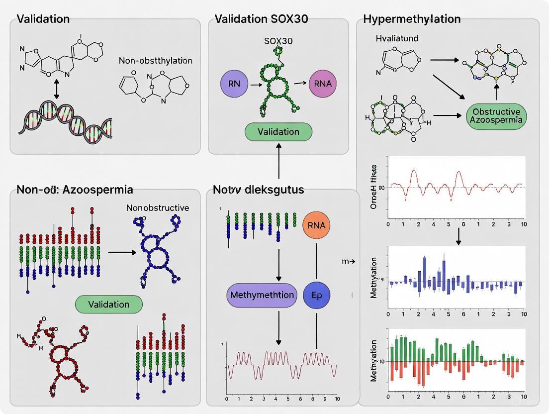
Comparative genome-wide profiling of DNA methylation in testicular tissues revealed SOX30 as the most notably hypermethylated gene at its promoter region in NOA patients compared to OA controls [8]. This hypermethylation directly causes transcriptional silencing of SOX30, with reduced expression levels correlating with NOA severity [8]. The identification of SOX30 mutations in NOA patients further validates its critical role in male fertility, with functional studies showing that these mutations impair protein interactions and DNA-binding capabilities [9].
Experimental Validation of SOX30 Hypermethylation
DNA Methylation and Expression Analysis Workflow
Key experimental approaches for validating SOX30 hypermethylation include:
- DNA Methylation Analysis: Direct bisulfite sequencing of testicular tissues identifies differentially methylated regions, with SOX30 showing significant promoter hypermethylation in NOA patients (p = 3.23E-6) [8].
- Expression Profiling: Quantitative analysis demonstrates silenced SOX30 expression in hypermethylated NOA cases, with expression levels correlating with disease severity [8].
- Functional Studies: Sox30 knockout mouse models uniquely impair testis development and spermatogenesis, completely eliminating spermatozoa production while maintaining normal ovary development and female fertility [8].
- Rescue Experiments: Re-expression of Sox30 in adult null mice reverses testicular pathology and restores spermatogenesis, with resulting spermatozoa capable of initiating pregnancy and producing viable offspring [8].
Table 3: Research Reagent Solutions for SOX30 Methylation Studies
| Research Tool | Application | Experimental Function |
|---|---|---|
| Bisulfite Conversion Kit | DNA methylation analysis | Converts unmethylated cytosine to uracil while preserving methylated cytosines [8] |
| Methylation-Specific PCR | Promoter methylation detection | Amplifies DNA sequences based on methylation status using specific primers [4] |
| SOX30 Antibodies | Immunohistochemistry/IF | Detects SOX30 protein expression and localization in testicular tissues [8] |
| DNMT Inhibitors | Functional studies | Modulates methylation status to investigate causal relationships [4] |
| Sox30 Knockout Mice | In vivo validation | Models human NOA pathology and tests therapeutic interventions [8] |
Comparative Analysis: Current vs. Emerging Approaches
The limitations of current NOA management have stimulated research into novel diagnostic and therapeutic strategies. The validation of SOX30 hypermethylation represents a paradigm shift from purely surgical interventions to molecular-based approaches.
SOX30 Hypermethylation Pathogenesis Pathway
Emerging Diagnostic and Therapeutic Platforms
Future directions in NOA management focus on addressing current limitations through several innovative approaches:
- Gene Editing Technologies: CRISPR/Cas9 and prime editing systems show potential for correcting genetic mutations underlying testicular dysfunction, though challenges remain with off-target effects, ethical concerns, and affordability [1].
- Non-Invasive Biomarkers: Seminal plasma analysis for cell-free nucleic acids, microRNAs, proteins, and metabolites offers promise as liquid biopsy for predicting sperm retrieval success [7].
- Multi-Omics Integration: Combining genomics, transcriptomics, proteomics, and metabolomics provides comprehensive molecular profiling to decode NOA heterogeneity [7].
- Artificial Intelligence: AI applications may combine clinical data with molecular biomarkers to improve sperm retrieval prediction and personalized treatment planning [7].
The recognition of SOX30 hypermethylation as a key mechanism in NOA pathogenesis opens avenues for epigenetic therapies and targeted interventions. As research advances, the future of NOA management will likely involve combination strategies tailored to individual patient profiles, potentially incorporating demethylating agents alongside current surgical techniques to improve outcomes for this challenging patient population [1] [8].
DNA Methylation Mechanisms in Spermatogenesis and Germ Cell Development
Spermatogenesis is a complex, multi-stage differentiation process that relies on precise epigenetic regulation to ensure the production of genetically sound and functionally competent spermatozoa. Among these regulatory mechanisms, DNA methylation serves as a critical epigenetic modifier that dynamically changes throughout germ cell development. This process involves the addition of a methyl group to the 5-carbon position of cytosine residues, primarily within CpG dinucleotides, which plays a pivotal role in controlling gene expression, genomic imprinting, and transposable element silencing [10]. The establishment of correct DNA methylation patterns is essential for male fertility, as dysregulation of this process has been directly linked to spermatogenic failure and non-obstructive azoospermia (NOA), the most severe form of male infertility [11] [12].
The journey of DNA methylation reprogramming in the male germline begins during embryonic development, undergoes extensive remodeling during postnatal spermatogenesis, and culminates in the establishment of a sperm-specific methylome that influences not only spermatogenesis but also early embryonic development [13] [14]. This article comprehensively examines the mechanisms of DNA methylation during spermatogenesis and germ cell development, with particular emphasis on validating SOX30 hypermethylation as a key pathogenic event in NOA, while providing detailed experimental protocols and analytical frameworks for researchers investigating epigenetic causes of male infertility.
DNA Methylation Reprogramming During Spermatogenesis
Stage-Specific Methylation Dynamics
The establishment of the male germ cell methylome represents a continuous process that begins during embryonic development and extends throughout adult spermatogenesis [12]. DNA methylation undergoes waves of reprogramming characterized by global erasure, de novo establishment, and maintenance phases:
- Primordial Germ Cells (PGCs): Following germ cell specification, genome-wide DNA methylation levels are dramatically reduced in proliferating PGCs, reaching minimal levels (∼3-5%) upon their arrival in embryonic gonads [13].
- Fetal Prospermatogonia: DNA methylation is subsequently restored in a sexually dimorphic pattern, with male germ cells carrying high levels (~80%) by birth [13] [15]. This de novo methylation is mediated by DNMT3A, DNMT3B, DNMT3C, and cofactor DNMT3L [13] [14].
- Postnatal Development: Residual DNA methylation acquisition continues after birth, culminating in highly methylated genomes in mature spermatozoa [13]. During adult spermatogenesis, germ cells undergo a global transient reduction of DNA methylation in primary spermatocytes, presumably due to a delay in maintenance methylation during premeiotic DNA replication [12] [14].
Table 1: DNA Methylation Levels During Key Stages of Murine Spermatogenesis
| Developmental Stage | Global CG Methylation Level | Key Regulatory DNMTs | Functional Significance |
|---|---|---|---|
| E16.5 Prospermatogonia | ~30% | DNMT3A, DNMT3L | Establishment of paternal imprints |
| P0.5 Prospermatogonia | ~76% | DNMT3A, DNMT3B, DNMT3L | Completion of neonatal methylation |
| P7.5 Undifferentiated Spermatogonia | ~77% | DNMT1, DNMT3A | Maintenance of stem cell population |
| P7.5 Differentiating Spermatogonia | ~76% | DNMT1, DNMT3B | Initiation of differentiation program |
| Adult Spermatozoa | ~79% | DNMT1, DNMT3B | Final sperm-specific methylome |
Unique Features of Germ Cell Methylation
Male germ cells display several distinctive methylation characteristics that differentiate them from somatic cells:
- Non-CG Methylation: Neonatal prospermatogonia exhibit significant accumulation of non-CG methylation, a pattern typically associated with embryonic stem cells and neuronal progenitors [15].
- Large Partially Methylated Domains (PMDs): All germ cell types contain large genomic domains (up to 12.0 Mb) showing relative hypomethylation, resembling PMDs reported in cancer cells and placenta [15].
- Stage-Specific Differential Methylation: Early postnatal male germ cells show numerous regions with stage-specific differential methylation around genes critical for stem cell function and spermatogenesis, contrasting with the scarcity of differential methylation in adult spermatogonial stem cells [15].
- Transposable Element Regulation: DNA methylation provides crucial suppression of transposable elements (TEs), with different TEs exhibiting distinct methylation patterns and sensitivities during germline development [12].
SOX30 Hypermethylation in Non-Obstructive Azoospermia
SOX30 as a Master Regulator of Spermatogenesis
SOX30 (SRY-box transcription factor 30) belongs to the SOX family of transcription factors and has been identified as a crucial regulator of spermatogenesis. Comparative genome-wide DNA methylation profiling of testicular tissues from NOA patients revealed SOX30 as the most notably hyper-methylated gene at its promoter region [11]. This hypermethylation directly causes transcriptional silencing, with reduced SOX30 expression levels correlating with the severity of NOA. The essential role of SOX30 in male fertility is evidenced by animal models, where Sox30 deletion in mice uniquely impairs testicular development and spermatogenesis, resulting in complete absence of spermatozoa and male infertility, while not affecting female fertility [11].
Validation of SOX30 Hypermethylation as a Diagnostic and Therapeutic Target
The functional significance of SOX30 hypermethylation has been rigorously validated through multiple experimental approaches:
- Methylation-Expression Correlation: A direct inverse correlation exists between SOX30 promoter methylation and its expression levels in human testicular tissues, with the most severe NOA cases showing the highest methylation and lowest expression [11].
- Animal Model Studies: Sox30 null mice recapitulate the human NOA phenotype, exhibiting impaired testis development and complete absence of spermatozoa [11].
- Re-expression Experiments: Restoration of Sox30 expression in adult Sox30 null mice reverses testicular pathological damage and restores spermatogenesis, with resulting spermatozoa capable of initiating pregnancy and producing viable offspring [11].
- Therapeutic Potential: The reversible nature of epigenetic silencing makes SOX30 an attractive therapeutic target for NOA, offering potential for pharmacological interventions aimed at demethylating its promoter and restoring expression [11].
Table 2: Experimental Evidence Validating SOX30 Role in NOA
| Experimental Approach | Key Findings | Implications for NOA |
|---|---|---|
| Comparative methylome analysis | SOX30 most hypermethylated gene in NOA patients | Potential diagnostic biomarker |
| Expression correlation | Inverse relationship between methylation and expression | Confirms functional silencing |
| Mouse knockout | Complete absence of sperm; mimics human NOA | Validates essential spermatogenic function |
| Re-expression in adults | Restored spermatogenesis and fertility | Demonstrates reversible pathology |
| Transgenerational assessment | Offspring viable and fertile | Supports therapeutic safety |
DNA Methylation and Nucleosome Retention in Sperm
Interplay Between DNA Methylation and Chromatin Remodeling
During the final stages of spermatid development, most nucleosomes are replaced by protamines, resulting in extensive nuclear compaction. However, a small fraction of nucleosomes is retained in mature sperm (~2% in mouse, ~15% in human) [13]. Research has revealed a sophisticated relationship between DNA methylation and nucleosome retention:
- Inverse Correlation: An inverse relationship exists between DNA methylation levels and nucleosome retention at CpG-rich sequences, particularly at gene promoters and exons [13].
- Developmental Predetermination: Site-specific DNA demethylation during the mitosis-to-meiosis transition in spermatogenesis predetermines nucleosome retention sites in mature spermatozoa [14].
- Functional Consequences: Reduced DNA methylation in sperm, achieved through conditional deletion of Dnmt3a and Dnmt3b, results in increased nucleosome occupancy preferentially at CpG-rich regions [13].
Intergenerational Implications
The DNA methylation pattern established during spermatogenesis has consequences beyond sperm function, influencing early embryonic development:
- Embryonic Chromatin Programming: Paternally inherited DNA methylation directs chromatin formation in early embryos, with reduced sperm DNA methylation rendering paternal alleles permissive for H3K4me3 establishment independently of possible paternal inheritance of sperm-born H3K4me3 [13].
- Gene Regulatory priming: The sites of unmethylated DNA coupled with nucleosome retention in sperm are associated with embryonic gene expression programs after fertilization, suggesting a role in paternal epigenetic inheritance [14].
Diagram 1: DNA Methylation Dynamics During Spermatogenesis and SOX30 Hypermethylation Pathway in NOA. This diagram illustrates the key stages of DNA methylation reprogramming throughout male germ cell development, highlighting the critical points where epigenetic dysregulation can lead to SOX30 hypermethylation and non-obstructive azoospermia.
Research Methodologies and Experimental Protocols
Genome-Wide Methylation Analysis Techniques
Advanced genomic technologies have enabled comprehensive mapping of DNA methylation patterns during spermatogenesis:
- Whole-Genome Bisulfite Sequencing (WGBS): Considered the gold standard for DNA methylation analysis, providing base-pair resolution mapping of 5-methylcytosine across the genome. Applications include analysis of neonatal prospermatogonia and early postnatal spermatogonia [15].
- Enzymatic Methyl-seq (EM-seq): A bisulfite-free method that detects DNA methylation with reduced DNA damage, suitable for assessing DNAme status of millions of CpGs in sperm samples [13].
- MethylCap-seq: Utilizes the Methyl-CpG-binding domain (MBD) to capture methylated DNA followed by next-generation sequencing, specifically detecting 5mC without cross-reactivity with 5hmC [14].
- Single-Cell Methylation Sequencing: Emerging technologies that enable methylation profiling of individual germ cells, particularly valuable for heterogeneous testicular tissues in NOA patients [16].
Germ Cell Isolation and Purification Protocols
Accurate methylation analysis requires pure populations of germ cells at specific developmental stages:
- Fluorescence-Activated Cell Sorting (FACS): Enables isolation of specific germ cell populations using surface markers such as THY1+ for undifferentiated spermatogonia and KIT+ for differentiating spermatogonia [14] [15].
- Enzymatic Digestion Protocol: Testicular tissues are digested using a two-step enzymatic incubation with collagenase IA followed by trypsin/DNase I treatment, with subsequent removal of erythrocytes by hemolysis buffer incubation [12].
- Immunostaining and Validation: Isolated cells are fixed, permeabilized, and stained with antibodies against germ cell markers (DMRT1, MAGEA4, UTF1) to verify purity before analysis [12].
Functional Validation Experiments
To establish causal relationships between DNA methylation changes and phenotypic outcomes:
- Gene Targeting Approaches: Conditional knockout models using Cre-loxP system with germ cell-specific promoters (e.g., Stra8-iCre) to delete DNA methyltransferases (Dnmt3a/3b) or specific genes like Sox30 [11] [13].
- Re-expression Studies: Restoration of gene expression in knockout models to demonstrate functional rescue of spermatogenic defects [11].
- Nucleosome Occupancy Assays: MNase-seq sequencing to map nucleosome positioning in sperm with altered DNA methylation patterns [13].
The Scientist's Toolkit: Essential Research Reagents
Table 3: Key Research Reagents for DNA Methylation Studies in Spermatogenesis
| Reagent/Category | Specific Examples | Research Application | Function |
|---|---|---|---|
| Cell Surface Markers | THY1 (CD90), KIT (CD117) | Isolation of spermatogonial subpopulations | FACS purification of undifferentiated vs. differentiating spermatogonia |
| DNA Methyltransferases | DNMT3A, DNMT3B, DNMT1 | Functional studies of de novo and maintenance methylation | Conditional knockout models to assess methylation requirements |
| Methylation Detection Kits | Whole-genome bisulfite sequencing kits | Genome-wide methylation profiling | Comprehensive mapping of 5mC at single-base resolution |
| Antibodies for Germ Cell Markers | DMRT1, MAGEA4, UTF1, PLZF | Identification and validation of germ cell types | Immunostaining and flow cytometry for cell purity assessment |
| Methylation Enzymes | MBD2-MBD protein | MethylCap-seq applications | Enrichment of methylated DNA regions for sequencing |
| Germ Cell-Specific Cre Drivers | Stra8-iCre, Vasa-Cre | Conditional gene targeting in germ cells | Tissue-specific deletion of floxed target genes |
| Live/Dead Cell Stains | Near-IR fluorescent reactive dye | Cell viability assessment during isolation | Exclusion of dead cells and debris from analysis |
The comprehensive analysis of DNA methylation mechanisms during spermatogenesis has significantly advanced our understanding of male fertility and its pathologies. The validation of SOX30 hypermethylation as a causative factor in NOA represents a paradigm shift in how we approach male infertility, moving beyond genetic determinants to include epigenetic dysregulation as a fundamental pathogenic mechanism. The reversible nature of epigenetic modifications, combined with the demonstrated feasibility of restoring fertility through SOX30 re-expression in adult animals, offers promising therapeutic avenues for what was previously considered an untreatable condition.
Future research directions should focus on developing targeted epigenetic therapies capable of specifically demethylating the SOX30 promoter in human testes, optimizing delivery mechanisms for clinical application, and identifying additional epigenetic regulators of spermatogenesis that may contribute to the heterogeneity of NOA. Furthermore, the interplay between DNA methylation and other epigenetic modifications, including histone modifications and non-coding RNAs, warrants deeper investigation to fully comprehend the complex regulatory network governing spermatogenesis. As single-cell technologies continue to evolve, they will undoubtedly uncover further layers of complexity in the epigenetic regulation of male germ cell development, ultimately leading to improved diagnostic, prognostic, and therapeutic strategies for male infertility.
Genome-Wide Methylation Profiling Identifies SOX30 as the Most Notably Hypermethylated Gene in NOA
Non-obstructive azoospermia (NOA) represents the most severe form of male infertility, affecting approximately 10-15% of infertile men and characterized by the complete absence of sperm in semen due to impaired spermatogenesis [4]. While genetic abnormalities account for only about 20% of NOA cases, recent research has illuminated the crucial role of epigenetic modifications, particularly DNA methylation, in the pathogenesis of this condition [8] [4]. DNA methylation involves the addition of a methyl group to cytosine nucleotides in CpG islands, primarily resulting in gene silencing when occurring in promoter regions [4]. This comprehensive analysis examines the groundbreaking discovery of SOX30 hypermethylation in NOA, validating its significance through comparative assessment of supporting evidence, experimental methodologies, and functional implications for the field of andrology research.
Comparative Analysis of Key Hypermethylated Genes in NOA
Genome-wide methylation studies have identified numerous genes with aberrant methylation patterns in NOA patients, with SOX30 consistently emerging as the most significantly hypermethylated. The table below provides a comparative overview of key hypermethylated genes identified in NOA research:
Table 1: Key Hypermethylated Genes Identified in NOA Research
| Gene Symbol | Methylation Status | Biological Function | Association with NOA Severity | Validation Methods |
|---|---|---|---|---|
| SOX30 | Most notably hypermethylated | Transcription factor, spermatogenesis regulation | Correlated with severity levels | MSP, BGS, RNA expression, mouse models |
| ZCCHC13 | Significantly hypermethylated | Zinc finger protein, regulates AKT/MAPK/c-MYC pathway | Associated with spermatogenesis failure | Integrated methylation/expression arrays |
| SPATA16 | Hypermethylated | Acrosome formation | Highest in SCOS, then MA, then HS | MSP, correlation with spermatogenic disorder |
| MTHFR | Hypermethylated | Folate metabolism | Controversial, needs validation | Methylation-specific PCR |
| DDR1 | Hypermethylated | Receptor tyrosine kinase, cell proliferation | Found in idiopathic NOA | MSP, expression analysis |
The preeminence of SOX30 in NOA methylation studies is demonstrated by multiple lines of evidence. A comparative genome-wide profiling study identified SOX30 as "the most notably hyper-methylated gene at promoter in testicular tissues from NOA patients" with 25 significant hypermethylated CpG sites at its promoter region [8]. This hypermethylation directly causes silencing of SOX30 expression, and the reduction in expression levels correlates directly with NOA disease severity [8] [11].
SOX30 Hypermethylation: Quantitative Evidence and Functional Correlation
The validation of SOX30 as a critically hypermethylated gene extends beyond mere identification to encompass rigorous quantification and demonstration of functional consequences:
Table 2: Quantitative Evidence for SOX30 Hypermethylation in NOA
| Experimental Evidence | Results | Functional Correlation |
|---|---|---|
| Promoter Methylation Density | 25 significant hypermethylated CpG sites at promoter | Direct silencing of SOX30 expression |
| Expression Correlation | Inverse correlation between methylation and expression | Reduced levels correlate with NOA severity |
| Mouse Model Phenotype | Sox30 null mice show complete absence of spermatozoa | Pathology simulates human NOA condition |
| Re-expression Studies | Sox30 re-expression in adult mice restores spermatogenesis | Proof of potential therapeutic target |
The functional impact of SOX30 hypermethylation has been validated through sophisticated animal model studies. Deletion of Sox30 in mice uniquely impairs testis development and spermatogenesis, leading to complete absence of spermatozoa and male infertility, while not affecting ovarian development or female fertility [8]. Crucially, the pathology and testicular size of Sox30 null mice highly simulate those of human NOA patients, providing a robust model system for mechanistic studies [8]. Most significantly, re-expression of Sox30 in adult null mice reverses testicular pathological damage and restores spermatogenesis, with the resulting spermatozoa demonstrating the ability to initiate pregnancy and produce viable offspring [8] [11].
Experimental Protocols for SOX30 Methylation Analysis
Genome-Wide Methylation Profiling
The identification of SOX30 as the most notably hypermethylated gene in NOA emerged from comprehensive genome-wide methylation analysis. The standard protocol involves:
- Sample Collection: Testicular biopsy specimens from carefully characterized NOA patients and obstructive azoospermia (OA) controls as normozoospermic fertile controls [8]
- DNA Extraction and Bisulfite Conversion: Genomic DNA isolation followed by bisulfite treatment using commercial kits
- Microarray Analysis: Genome-wide methylation profiling using Infinium 450K BeadChip arrays covering >450,000 CpG sites [17] [18]
- Data Analysis: Identification of differentially methylated regions (DMRs) with statistical significance (p < 0.01) and absolute Δβ > 0.20 [18]
- Validation: Confirmatory analysis through bisulfite sequencing PCR (BSP) and methylation-specific PCR (MSP)
SOX30-Specific Methylation Detection
For targeted analysis of SOX30 methylation status, researchers employ:
Methylation-Specific PCR (MSP)
- Primer Design: Specific for methylated vs. unmethylated sequences after bisulfite conversion
- Amplification Conditions: 95°C for 5 min, 40 cycles of 95°C for 10s, 68°C for 1min, 72°C for 1min, 80°C for 30s (fluorescence collection) [19]
- Analysis: Qualitative assessment of methylation status
Bisulfite Genomic Sequencing (BGS)
- Provides single-base resolution of methylation status across CpG sites
- Covers 25+ CpG sites in SOX30 promoter region [8]
- Enables quantitative assessment of methylation density
Real-Time Quantitative MSP (RQ-MSP)
- Quantitative measurement of SOX30 methylation levels
- Uses ALU or other reference genes for normalization [20]
- Calculates relative methylation level by 2−ΔΔCT method
Figure 1: Experimental Workflow for SOX30 Methylation Profiling in NOA Research
SOX30 Molecular Signaling Pathways and Mechanisms in Spermatogenesis
The molecular mechanisms through which SOX30 influences spermatogenesis involve complex regulatory pathways:
Figure 2: SOX30 Molecular Pathways in Spermatogenesis and Potential Therapeutic Intervention
At the molecular level, SOX30 functions as a transcription factor containing a high mobility group (HMG) DNA-binding domain [21]. Research across different biological systems has demonstrated that SOX30 promotes tumor cell apoptosis by transcriptionally activating p53 through direct binding to the CACTTTG motif (+115 to +121) of the p53 promoter region [21]. While the precise mechanisms in spermatogenesis require further elucidation, this p53 activation pathway likely contributes to the proper regulation of germ cell development and differentiation. The epigenetic silencing of SOX30 through promoter hypermethylation disrupts these critical regulatory pathways, leading to impaired spermatogenesis and ultimately manifesting as NOA.
The Scientist's Toolkit: Essential Research Reagents for SOX30 Studies
Table 3: Essential Research Reagents for SOX30 Methylation and Functional Studies
| Reagent/Category | Specific Examples | Research Application | Function in SOX30 Studies |
|---|---|---|---|
| Methylation Analysis Kits | EZ DNA Methylation-Gold Kit, MethylEdge Bisulfite Conversion System | Bisulfite conversion | Convert unmethylated cytosines to uracils for methylation detection |
| Methylation Arrays | Infinium MethylationEPIC BeadChip | Genome-wide methylation screening | Identify differentially methylated regions across genome |
| Methylation-Specific PCR Reagents | MSP primer sets, HotStart Taq PCR mixes | Targeted methylation analysis | Specific amplification of methylated vs. unmethylated SOX30 sequences |
| Demethylating Agents | 5-aza-2'-deoxycytidine (5-Aza) | Functional validation | Reverse methylation-mediated silencing to confirm causal relationship |
| Antibodies | Anti-SOX30, Anti-5-methylcytosine, Anti-5-hydroxymethylcytosine | Protein expression, methylation detection | Confirm SOX30 silencing, validate methylation status |
| Cell Culture Systems | Mouse spermatogonia GC-1 cells, testicular tissue explants | In vitro functional studies | Mechanism investigation without animal models |
The comprehensive analysis of genome-wide methylation profiling solidifies SOX30's position as the most notably hypermethylated gene in NOA pathogenesis. The evidence from multiple independent studies, combined with functional validation in animal models, underscores the critical role of SOX30 epigenetic regulation in male fertility. The correlation between SOX30 hypermethylation and disease severity, coupled with the remarkable reversal of spermatogenic failure upon SOX30 re-expression, positions this gene as both a robust diagnostic biomarker and a promising therapeutic target. For researchers and drug development professionals, these findings open avenues for developing epigenetic-based diagnostics and targeted therapies for NOA, potentially addressing a significant unmet need in male reproductive medicine. Future research should focus on elucidating the upstream regulators of SOX30 methylation and developing targeted demethylation strategies for clinical application.
SOX30 (SRY-box containing gene 30) is a transcription factor belonging to the High Mobility Group (HMG) superfamily that has emerged as a critically important gene regulated by epigenetic mechanisms in multiple disease states. Originally identified for its essential role in spermatogenesis and gonadal development, recent research has revealed that SOX30 promoter hypermethylation serves as a significant biomarker and potential mechanistic driver across a spectrum of human diseases, particularly in male infertility and various cancers. This epigenetic alteration results in transcriptional silencing of SOX30, with consequent impacts on disease pathogenesis, progression, and clinical outcomes. This comprehensive guide examines the correlation between SOX30 promoter hypermethylation and disease severity across histological subtypes, providing comparative experimental data and methodological protocols to facilitate research and drug development in this emerging field.
Molecular Mechanisms and Functional Consequences
SOX30 functions as a transcription factor containing the characteristic HMG DNA-binding domain that enables sequence-specific DNA binding and transcriptional regulation. The gene is located on chromosome 5 in humans and is subject to stringent epigenetic control through DNA methylation of its promoter region.
Promoter Hypermethylation and Transcriptional Silencing: SOX30 promoter hypermethylation occurs primarily at CpG islands within the promoter region, leading to direct silencing of gene expression. This epigenetic modification prevents transcription factor binding and recruits methyl-CpG-binding proteins that promote chromatin condensation into transcriptionally inactive states [4]. In normal tissues where SOX30 is expressed (such as testis and lung), the promoter region exists in a hypomethylated state, allowing active transcription [22]. The critical consequence of promoter hypermethylation is the complete suppression or significant reduction of SOX30 mRNA and protein levels, with subsequent disruption of its normal cellular functions.
Functional Role as Tumor Suppressor: In cancer contexts, SOX30 has been demonstrated to function as a novel tumor suppressor through direct regulation of p53 transcription. The antitumorigenic effect of SOX30 is mediated by its direct binding to the CACTTTG motif (+115 to +121) within the p53 promoter region, thereby activating p53 transcription and triggering apoptosis while inhibiting proliferation [23]. Restoration of SOX30 expression in lung cancer cell lines induces significant cancer cell apoptosis and inhibits proliferation in vitro, while repressing tumor formation in vivo [23]. Conversely, knockdown of SOX30 promotes cellular proliferation and inhibits apoptosis, confirming its tumor-suppressive functions [23].
Role in Spermatogenesis: In male reproduction, SOX30 is indispensable for proper testis development and spermatogenesis. The gene is expressed in both testis germ cells and Sertoli cells, with expression levels increasing progressively during testicular development [22]. SOX30 deletion in mice results in complete absence of spermatozoa in testes, leading to male infertility, while notably not affecting ovary development or female fertility [11]. The pathology and testicular size reduction in Sox30 null mice closely simulate the clinical presentation of human non-obstructive azoospermia [11].
Table 1: Functional Consequences of SOX30 Promoter Hypermethylation Across Tissue Types
| Tissue Type | Methylation Status | SOX30 Expression | Primary Functional Consequences |
|---|---|---|---|
| Normal Testis | Hypomethylated | High | Normal spermatogenesis and testis development |
| NOA Testis | Hypermethylated | Silenced/Low | Impaired spermatogenesis, absent spermatozoa |
| Normal Lung | Hypomethylated | Moderate | Cellular homeostasis, p53 regulation |
| Lung Cancer | Hypermethylated | Silenced/Low | Uncontrolled proliferation, reduced apoptosis |
| Normal Myeloid Cells | Hypomethylated | Moderate | Hematopoietic differentiation |
| AML/MDS | Hypermethylated | Silenced/Low | Disease progression, poor treatment response |
Disease-Specific Correlations and Clinical Implications
Non-Obstructive Azoospermia (NOA)
SOX30 promoter hypermethylation demonstrates a particularly strong association with non-obstructive azoospermia, the most severe form of male infertility characterized by absent spermatogenesis. Comparative genome-wide methylation profiling identified SOX30 as the most notably hypermethylated gene in testicular tissues from NOA patients [11]. This hypermethylation directly causes transcriptional silencing, with the reduction in SOX30 expression levels correlating significantly with NOA disease severity [11]. The critical evidence establishing SOX30's functional role comes from murine models, where Sox30 deletion uniquely impairs testis development and spermatogenesis, resulting in complete absence of spermatozoa [11]. Most remarkably, re-expression of Sox30 in adult null mice reverses testicular pathological damage and restores spermatogenesis, with the resulting spermatozoa demonstrating capacity to initiate pregnancy and produce viable offspring [11] [24]. This reversible phenotype positions SOX30 as a promising therapeutic target for NOA treatment.
Correlation with Histological Subtypes in NOA: The severity of SOX30 hypermethylation correlates with specific histological patterns in testicular biopsies. Higher methylation levels are observed in Sertoli cell-only syndrome (SCOS) followed by maturation arrest (MA) and hypospermatogenesis (HS) [4]. This methylation gradient corresponds with progressive reduction in SOX30 expression and increasingly impaired spermatogenesis.
Acute Myeloid Leukemia (AML) and Myelodysplastic Syndromes (MDS)
In hematological malignancies, SOX30 methylation serves as a significant prognostic biomarker and indicator of disease progression. SOX30 methylation represents a frequent event in AML, with an inverse correlation between methylation levels and SOX30 expression [25] [19]. Survival analysis demonstrates that SOX30 hypermethylation negatively associates with complete remission rates, overall survival, and leukemia-free survival in AML patients [25]. Importantly, SOX30 methylation levels significantly increase during progression from MDS to AML, indicating its involvement in disease evolution [19]. The dynamic nature of SOX30 methylation is further evidenced by its significant decrease in AML patients achieving complete remission compared to diagnosis, and marked increase upon relapse compared to the remission population [25]. This pattern establishes SOX30 methylation as a sensitive biomarker for monitoring treatment response and disease recurrence in myeloid malignancies.
Lung Cancer
In lung adenocarcinoma (LUAD), SOX30 promoter hypermethylation represents a common epigenetic event with significant clinical implications. SOX30 hypermethylation occurs in 70.83% (85/120) of primary lung tumors compared to only 8% (2/25) of peri-tumoral tissues and 0% (0/20) of normal lung tissues [23]. This methylation strongly correlates with transcriptional silencing, as SOX30 is expressed in normal lung tissues where the promoter is unmethylated, but silenced or downregulated in tumor tissues with hypermethylation [23]. The clinical significance is substantial, with SOX30 hypermethylation and consequent low expression associating with unfavorable survival in lung adenocarcinoma patients [26]. Multivariate Cox regression analysis identifies SOX30 expression as an independent prognostic factor for overall survival in NSCLC patients, particularly in the adenocarcinoma subtype [26].
Correlation with Histological Subtypes in Lung Cancer: SOX30 expression and methylation patterns demonstrate histological subtype specificity in lung cancer. The incidence of SOX30 overexpression is significantly higher in adenocarcinoma (31.33%, 47/150) compared to squamous cell carcinoma (14.29%, 10/70) [26]. Furthermore, SOX30 expression represents a favorable prognostic factor specifically in lung adenocarcinoma patients, but not in squamous cell carcinoma [26]. This histological specificity underscores the tissue-context dependent functions of SOX30 and the importance of histological stratification in both research and clinical applications.
Table 2: SOX30 Hypermethylation Patterns Across Diseases and Histological Subtypes
| Disease Category | Methylation Frequency | Expression Correlation | Clinical Associations |
|---|---|---|---|
| Non-Obstructive Azoospermia | Highly prevalent in testicular tissues | Inverse correlation with disease severity | Disease severity, impaired spermatogenesis |
| Acute Myeloid Leukemia | 70.83% in primary tumors | Inverse correlation with expression | Poor OS, LFS, CR; disease progression |
| MDS to AML Progression | Significant increase during progression | Decreasing during progression | Disease evolution marker |
| Lung Adenocarcinoma | 70.83% in primary tumors | Inverse correlation with expression | Unfavorable survival, independent prognostic factor |
| Lung Squamous Cell Carcinoma | Lower frequency than ADC | Weak inverse correlation | Limited prognostic value |
Comparative Experimental Data Analysis
Methylation Frequencies Across Diseases
The prevalence of SOX30 promoter hypermethylation varies substantially across different diseases, reflecting its tissue-specific regulatory roles. In non-obstructive azoospermia, SOX30 is described as the most notably hypermethylated gene based on comparative genome-wide methylation profiling [11]. In lung cancer, SOX30 hypermethylation occurs in 100% of lung cancer cell lines (9/9) and 70.83% of primary lung tumors (85/120), compared to none of normal lung tissues (0/20) and only 8% of peri-tumoral tissues (2/25) [23]. In myeloid malignancies, SOX30 methylation represents a frequent event in AML and shows significant increase during progression from MDS to AML [25]. The methylation frequency in MDS itself is comparatively lower, suggesting accumulation of SOX30 hypermethylation during leukemic transformation [19].
Expression Correlation with Methylation Status
Across all disease states, a consistent inverse correlation exists between SOX30 promoter methylation and gene expression levels. In lung cancer, SOX30 is broadly expressed in normal lung tissues with unmethylated promoters, but silenced or downregulated in cancer cell lines and primary tumors with hypermethylated SOX30 [23]. Treatment with DNA methyltransferase inhibitor 5-aza-2'-deoxycytidine restores SOX30 expression in silenced cell lines, confirming methylation-mediated regulation [23]. Similarly, in NOA, SOX30 hypermethylation directly causes silencing of expression, with reduced expression levels correlating with disease severity [11]. In AML, SOX30 methylation inversely correlates with SOX30 expression, and dynamic methylation changes correspond with expression alterations during treatment response and disease relapse [25].
Functional Restoration Studies
Evidence from functional restoration studies provides compelling evidence for the pathogenic role of SOX30 hypermethylation. In NOA, re-expression of Sox30 in adult null mice reverses testicular pathological damage and restores complete spermatogenesis [11]. The spermatozoa produced after SOX30 re-expression demonstrate functional capacity to initiate pregnancy and produce viable, fertile offspring [11] [24]. In lung cancer, ectopic SOX30 expression induces cancer cell apoptosis, inhibits proliferation in vitro, and represses tumor formation in vivo [23]. These functional restoration experiments validate SOX30 as a genuine therapeutic target rather than merely a bystander epigenetic marker.
Experimental Methodologies and Protocols
DNA Methylation Analysis Techniques
Bisulfite Genomic Sequencing (BGS): This method provides single-base resolution methylation data across the SOX30 promoter region. Genomic DNA is treated with sodium bisulfite, which converts unmethylated cytosines to uracils while leaving methylated cytosines unchanged. The bisulfite-treated DNA is then amplified by PCR using primers specific to the SOX30 promoter region, and the resulting products are cloned and sequenced to determine methylation patterns at individual CpG sites [23]. This approach allows quantitative assessment of methylation density and identification of specific methylated CpG dinucleotides.
Methylation-Specific PCR (MSP): MSP represents a rapid, sensitive method for detecting methylation status of specific CpG islands within the SOX30 promoter. Following bisulfite treatment of DNA, two sets of primers are used: one specific for methylated sequences and another for unmethylated sequences. The presence or absence of PCR products with these primer sets indicates the methylation status of the target region [4]. MSP is particularly useful for clinical sample screening due to its requirement for small DNA quantities and high throughput potential.
Real-Time Quantitative Methylation-Specific PCR (RQ-MSP): This quantitative adaptation of MSP enables precise measurement of SOX30 methylation levels. The method utilizes bisulfite-treated DNA with methylation-specific primers and fluorescent probes in a real-time PCR system. The amplification curves provide quantitative data on methylation levels, normalized to a reference gene such as ALU [25] [19]. RQ-MSP offers superior sensitivity for detecting methylation changes in heterogeneous samples and monitoring dynamic methylation alterations during disease progression or treatment.
Expression Analysis Methods
Reverse Transcription Quantitative PCR (RT-qPCR): This represents the standard method for quantifying SOX30 expression levels. Total RNA is extracted from tissues or cell lines, treated with DNase I to eliminate genomic DNA contamination, and reverse transcribed into cDNA. Real-time PCR is performed using SOX30-specific primers, with expression levels normalized to housekeeping genes such as ABL or β-actin using the 2−ΔΔCT method [25] [22]. This approach provides sensitive, reproducible quantification of SOX30 transcript levels for correlation with methylation status.
Immunohistochemistry (IHC): IHC enables protein-level expression analysis and histological localization of SOX30 in tissue sections. Paraffin-embedded tissue sections are deparaffinized, rehydrated, and subjected to antigen retrieval. After blocking, sections are incubated with SOX30-specific primary antibodies followed by appropriate secondary antibodies and detection systems [26]. IHC allows correlation of SOX30 protein expression with tissue morphology and histological subtypes, providing clinically relevant information for pathological assessment.
Functional Validation Experiments
Demethylation Treatment: Pharmacological reversal of DNA methylation using DNMT inhibitors provides direct evidence for methylation-mediated SOX30 silencing. Cell lines are treated with 5-aza-2'-deoxycytidine (a DNA methyltransferase inhibitor) for 72-96 hours, typically at concentrations of 1-10 μM, with medium and drug replenishment every 24 hours [22] [23]. Subsequent analysis of SOX30 expression by RT-qPCR or Western blot demonstrates reactivation following methylation inhibition.
Ectopic Expression Studies: Gain-of-function experiments validate the tumor-suppressive activities of SOX30. A SOX30-expressing construct is transfected into cancer cell lines using appropriate transfection methods. The effects of SOX30 re-expression on apoptosis (Annexin V staining, sub-G1 population analysis), proliferation (MTS assays, EdU incorporation, colony formation), and in vivo tumor growth (xenograft models) are then assessed [23].
Knockdown Experiments: Loss-of-function studies using RNA interference further confirm SOX30's functional roles. SOX30-specific miRNAs or siRNAs are stably or transiently transfected into cell lines, followed by assessment of proliferation, apoptosis, and colony formation capacity [23]. Consistent effects from both gain-of-function and loss-of-function approaches provide compelling evidence for SOX30's pathogenic significance.
Signaling Pathways and Molecular Interactions
SOX30 participates in distinct signaling pathways across different tissue contexts, with promoter hypermethylation disrupting these regulatory networks in disease-specific manners.
Diagram 1: SOX30 Hypermethylation in Disease-Specific Signaling Pathways. This diagram illustrates the tissue-specific consequences of SOX30 promoter hypermethylation and transcriptional silencing across different pathological contexts, highlighting the distinct downstream pathways affected in each disease state.
The Scientist's Toolkit: Essential Research Reagents
Table 3: Essential Research Reagents for SOX30 Methylation and Expression Studies
| Reagent Category | Specific Products/Assays | Research Applications | Technical Considerations |
|---|---|---|---|
| Methylation Analysis | Bisulfite Conversion Kits (EZ DNA Methylation kits) | Convert unmethylated cytosines to uracils for methylation detection | Complete conversion critical; optimize incubation times |
| MSP Primers (SOX30-specific) | Amplify methylated vs unmethylated sequences | Validate specificity with methylated/unmethylated controls | |
| RQ-MSP Probes/Primers | Quantitative methylation analysis | Normalize to reference genes (ALU); establish standard curves | |
| Expression Analysis | SOX30 Antibodies (IHC/WB validated) | Protein expression detection and localization | Validate specificity with knockout/knockdown controls |
| RT-qPCR Primers (SOX30-specific) | mRNA expression quantification | Use intron-spanning designs; normalize to housekeeping genes | |
| RNA Extraction Kits (Trizol-based) | High-quality RNA isolation from tissues/cells | Prevent RNase contamination; assess RNA integrity | |
| Functional Studies | 5-aza-2'-deoxycytidine | DNMT inhibitor for demethylation studies | Optimize concentration (1-10μM) and treatment duration (72-96h) |
| SOX30 Expression Vectors | Ectopic expression studies | Use tissue-specific promoters for physiological relevance | |
| SOX30 miRNAs/siRNAs | Knockdown experiments | Include multiple constructs to control for off-target effects | |
| Cell Lines/Models | Lung cancer lines (A549, H460) | In vitro functional assays | Verify baseline SOX30 methylation status before experiments |
| GC-2spd, TM3, TM4 cells | Spermatogenesis studies | Maintain appropriate culture conditions for specialized lines | |
| Sox30 knockout mice | In vivo functional validation | Monitor fertility parameters and testicular histology |
SOX30 promoter hypermethylation represents a significant epigenetic mechanism with demonstrated correlations to disease severity and distinct patterns across histological subtypes. In non-obstructive azoospermia, SOX30 hypermethylation associates with impaired spermatogenesis and disease severity, with promising therapeutic implications evidenced by restored spermatogenesis following SOX30 re-expression. In oncology contexts, SOX30 functions as a tissue-specific tumor suppressor, with hypermethylation patterns distinguishing histological subtypes in lung cancer and predicting clinical outcomes in hematological malignancies. The consistent inverse relationship between SOX30 promoter methylation and expression across diseases, coupled with functional restoration evidence, positions SOX30 as both a valuable biomarker and compelling therapeutic target. Future research directions should focus on developing targeted demethylation strategies, validating SOX30 methylation as clinical biomarkers, and exploring combination therapies that leverage SOX30 reactivation across different disease contexts.
The SRY-box transcription factor 30 (SOX30) is a member of the SOX family of transcription factors, which are defined by a conserved high-mobility group (HMG) box domain that mediates DNA binding [27] [28]. As the sole member of the SoxH group, SOX30 exhibits remarkable tissue specificity, with abundant expression predominantly in the testis [27] [29]. Research over the past decade has established SOX30 as a crucial regulator of spermatogenesis, with its dysfunction directly linked to male infertility, particularly non-obstructive azoospermia (NOA) [11]. This review synthesizes current understanding of SOX30's structure, molecular functions, and emerging role as a potential therapeutic target, framing this knowledge within the context of validating SOX30 hypermethylation as a key mechanism in NOA pathogenesis.
Gene and Protein Structure
The human SOX30 gene is located on chromosome 5 (5q33.3), while its mouse ortholog resides on chromosome 11 [30]. The gene encodes a protein characterized by several distinctive structural features that define its function.
Structural Domains and Isoforms
The SOX30 protein contains a conserved DNA-binding HMG domain that enables sequence-specific DNA recognition and binding [27] [28]. Crystal structure analysis has revealed this domain's architecture at 1.40 Å resolution, highlighting the structural basis for its DNA interaction capabilities [28]. The protein also possesses N and C-terminal regions that contribute to overall protein folding, stability, and transcriptional activation potential [28].
Human SOX30 encodes three distinct transcript variants through alternative splicing, expanding its functional repertoire [28]. The C-terminal region appears particularly important for protein-protein interactions, as evidenced by a stop-gain mutation (Arg478*) identified in NOA patients that produces a C-terminal truncated protein with dramatically reduced association with the histone deacetylase HDAC3 [9].
Table 1: Key Structural Features of SOX30 Protein
| Structural Feature | Functional Significance | Experimental Evidence |
|---|---|---|
| HMG DNA-binding domain | Binds specific DNA sequences (ACAT motif) | ChIP-seq, crystal structure (1.40 Å) [27] [28] |
| N-terminal region | Contributes to protein folding and stability | Structural analysis [28] |
| C-terminal region | Mediates protein-protein interactions (e.g., with HDAC3) | Co-immunoprecipitation with truncation mutants [9] |
| Transcript variants (3 in humans) | Potential for functional diversity | RNA sequencing and isoform detection [28] |
Expression Patterns and Regulation
SOX30 exhibits a tightly restricted tissue-specific expression pattern, predominantly in the testis, with minimal expression detected in other tissues [27] [30]. This expression profile emerges during specific developmental stages and is regulated by epigenetic mechanisms.
Developmental and Cellular Expression
During testicular development, SOX30 expression initiates around postnatal day 14 in mice, with substantial increases by postnatal day 21 when round spermatids first appear [27]. At the cellular level, SOX30 is detectable in late pachytene spermatocytes, peaks in round spermatids at steps 7-8, and becomes downregulated in later elongated spermatids [27]. Immunostaining localizes SOX30 primarily to the nuclei of germ cells, consistent with its function as a transcription factor [27].
Beyond mammals, SOX30 shows a male-biased expression pattern in other vertebrates, including the Nile tilapia and Chinese soft-shelled turtle, where it is preferentially expressed in testes and responsive to sex hormone treatments [29] [31].
Epigenetic Regulation
The SOX30 promoter is subject to epigenetic regulation through DNA methylation, with hypermethylation directly causing transcriptional silencing in NOA patients [11]. The degree of SOX30 promoter hypermethylation correlates with disease severity, making it a potential diagnostic biomarker and therapeutic target [11].
Molecular Functions in Spermatogenesis
SOX30 serves as a master transcriptional regulator that coordinates the transition from meiotic to postmeiotic gene expression programs during spermatogenesis [27].
Genomic Binding and Target Genes
Chromatin immunoprecipitation followed by sequencing (ChIP-seq) analyses reveal that SOX30 binds to specific DNA sequences in mouse testes, with genomic occupancy positively correlating with expression of key postmeiotic genes including Tnp1, Hils1, Ccdc54, and Tsks [27]. In Nile tilapia, SOX30 directly binds promoters of ift140 and ptprb, two genes critical for spermiogenesis, and activates their transcription [31].
SOX30 deficiency disrupts the transcriptional program essential for haploid cell differentiation, leading to arrested development at the early round spermatid stage (steps 2-3) in mice [27]. This arrest is characterized by failure of proacrosomic vesicles to form a single acrosomal organelle, despite most spermatocytes progressing through meiosis [27].
Functional Interactions
SOX30 interacts with epigenetic regulators including the histone deacetylase HDAC3, with mutations in the C-terminal domain impairing this association and compromising SOX30 function [9]. This interaction suggests SOX30 recruits chromatin-modifying complexes to fine-tune gene expression during spermatogenesis.
Figure 1: SOX30 Molecular Functions and Consequences of Mutation. SOX30 regulates spermatogenesis through DNA binding via its HMG domain and protein interactions through its C-terminal domain. Mutations in these domains disrupt these functions, leading to male infertility.
Role in Male Infertility and NOA Pathogenesis
SOX30 dysfunction represents a significant etiological factor in male infertility, particularly non-obstructive azoospermia (NOA), the most severe form of male infertility characterized by complete absence of sperm in ejaculate due to impaired spermatogenesis [11].
Genetic and Epigenetic Evidence
Genetic screening of 620 NOA patients identified six heterozygous SOX30 sequence variations, including five missense mutations and one stop-gain mutation (Arg478*) [9]. Functional characterization demonstrated that these mutations impair SOX30 function through distinct mechanisms: the HMG domain mutation reduces DNA-binding capacity, while the C-terminal truncation disrupts interaction with HDAC3 [9].
Beyond genetic mutations, epigenetic inactivation via SOX30 promoter hypermethylation represents a major mechanism disrupting SOX30 expression in NOA patients [11]. Comparative genome-wide methylation profiling identified SOX30 as the most notably hypermethylated gene in testicular tissues from NOA patients, with this hypermethylation directly causing transcriptional silencing [11].
Table 2: SOX30 Alterations in Male Infertility
| Alteration Type | Functional Consequence | Associated Phenotype | Reference |
|---|---|---|---|
| Promoter hypermethylation | Epigenetic silencing; reduced expression | Non-obstructive azoospermia (NOA) | [11] |
| HMG domain missense mutations | Reduced DNA-binding ability | Impaired spermatogenesis; reduced sperm count | [9] |
| C-terminal truncation (Arg478*) | Disrupted HDAC3 interaction | Spermiogenic defects | [9] |
| Complete gene knockout | Loss of postmeiotic gene expression | Spermatogenic arrest at round spermatid stage | [27] |
Animal Model Phenotypes
Sox30-deficient mouse models recapitulate key features of human NOA, exhibiting complete male sterility with spermatogenic arrest at the early round spermatid stage (steps 2-3) [27] [11]. These mutants display failure in acrosome formation and impaired transition from meiotic to postmeiotic stages, despite most spermatocytes progressing through meiosis [27].
Notably, a point mutation in the HMG domain (modeling the human P353S mutation) results in a less severe phenotype than complete knockout, with mutant mice remaining fertile but producing reduced numbers of mature sperm [9]. This suggests that certain SOX30 mutations may cause varying degrees of impaired spermatogenesis rather than complete arrest.
In Nile tilapia, CRISPR/Cas9-mediated sox30 mutation causes abnormal spermiogenesis, reduced sperm motility, and male subfertility, confirming its conserved role in fish spermatogenesis [31].
Experimental Models and Research Tools
Key Experimental Models
Various experimental approaches have been employed to elucidate SOX30 functions:
- CRISPR/Cas9-mediated knockout mice: Complete Sox30 disruption causes spermatogenic arrest at early round spermatid stage [27]
- Point mutation knock-in mice: Modeling human P353S mutation causes reduced sperm production without complete sterility [9]
- Germ cell-specific Sox30 deletion: Demonstrates cell-autonomous function in germ cells [27]
- Tilapia sox30 mutants: Reveal conserved functions in teleost spermatogenesis [31]
Core Research Methodologies
Key experimental techniques used in SOX30 research include:
- Chromatin Immunoprecipitation Sequencing (ChIP-seq): Identifies genome-wide SOX30 binding sites and target genes [27] [31]
- Stage-specific germ cell RNA-seq: Reveals transcriptome changes in Sox30 mutants [27]
- Methylation-specific PCR: Detects SOX30 promoter hypermethylation in clinical samples [11]
- Immunostaining of testis sections: Localizes SOX30 protein expression during spermatogenesis [27]
Figure 2: Experimental Approaches for SOX30 Research. A workflow illustrating common research strategies for investigating SOX30 function, from model selection through genetic manipulation, functional analysis, mechanistic studies, and clinical correlation.
Therapeutic Implications and Future Directions
The demonstration that SOX30 re-expression in adult Sox30 null mice reverses testicular pathology and restores spermatogenesis highlights its potential as a therapeutic target for NOA [11]. Remarkably, spermatozoa produced following SOX30 re-expression demonstrated fertilization capability, and resulting male offspring were able to father children without apparent abnormalities [11].
Diagnostic and Therapeutic Potential
The strong association between SOX30 promoter hypermethylation and NOA suggests potential clinical applications:
- Diagnostic biomarker: SOX30 methylation status could aid NOA diagnosis and classification
- Therapeutic target: Demethylating strategies could potentially restore SOX30 expression in NOA patients
- Prognostic indicator: Correlation between SOX30 expression levels and NOA severity may inform prognosis
Research Reagent Solutions
Table 3: Essential Research Tools for SOX30 Investigation
| Reagent/Resource | Specific Application | Key Function | Example Use |
|---|---|---|---|
| SOX30-specific antibodies | Immunodetection, ChIP | Protein localization and binding studies | Testis immunostaining [27] |
| Sox30 knockout mice | Functional analysis | Model complete gene loss | Phenotypic characterization [27] |
| Sox30 point mutant mice | Structure-function studies | Model specific human mutations | HMG domain functional analysis [9] |
| Methylation-specific PCR primers | Epigenetic analysis | Detect promoter methylation | Clinical sample screening [11] |
| ChIP-seq-grade SOX30 antibody | Genome-wide binding studies | Identify direct transcriptional targets | Target gene identification [27] [31] |
SOX30 represents a master transcriptional regulator of spermatogenesis that coordinates the transition from meiotic to postmeiotic development. Its testis-specific expression, critical role in activating haploid gene programs, and association with NOA through both genetic and epigenetic mechanisms position SOX30 as a key factor in male fertility. The validation of SOX30 hypermethylation as a causative factor in NOA, coupled with dramatic rescue of spermatogenesis upon SOX30 re-expression in animal models, offers promising avenues for future diagnostic and therapeutic development. Further research is needed to fully elucidate SOX30's regulatory networks and translate these findings into clinical applications for male infertility.
Analytical Approaches for SOX30 Hypermethylation Detection and Functional Validation
The epigenetic silencing of the SOX30 (SRY-box 30) gene through promoter hypermethylation has emerged as a critical biomarker and functional mechanism in multiple disease pathologies. Research has validated its significance particularly in non-obstructive azoospermia (NOA), the most severe form of male infertility, where SOX30 represents the most notably hypermethylated gene promoter in testicular tissues of affected patients [8]. Beyond reproductive medicine, SOX30 hypermethylation functions as a tumor suppressor in various cancers, including lung cancer and myeloid malignancies such as acute myeloid leukemia (AML) and myelodysplastic syndromes (MDS) [25] [21] [20]. The accurate detection of this epigenetic alteration is therefore paramount for both diagnostic and therapeutic applications across medical specialties.
Two principal methodologies have become cornerstone techniques for evaluating SOX30 methylation status: bisulfite sequencing and methylation-specific PCR. These approaches rely on the fundamental process of sodium bisulfite conversion, which selectively deaminates unmethylated cytosines to uracils while leaving methylated cytosines unchanged, thereby creating sequence differences that can be detected through subsequent analysis [32] [33]. This guide provides a comprehensive comparative analysis of these methodologies, their technical variations, and their application in SOX30 research, with particular emphasis on validation in NOA studies.
Fundamental Principles of Bisulfite Conversion
The cornerstone of both major SOX30 methylation analysis techniques is the bisulfite conversion process, which provides the chemical foundation for distinguishing methylated from unmethylated cytosines. When genomic DNA is treated with sodium bisulfite, a selective deamination occurs: unmethylated cytosines are converted to uracils, which are then amplified as thymines during PCR, while 5-methylcytosines (5-mC) remain protected from conversion and are amplified as cytosines [32] [33]. This process creates sequence polymorphisms that can be exploited for methylation detection.
Despite its widespread use, this chemical conversion presents several technical challenges. The process requires micrograms of DNA input and involves harsh chemical treatment that can lead to substantial DNA degradation (up to 84-96% loss) [33]. Furthermore, the conversion may be incomplete, leading to false-positive results for methylation, and the method cannot distinguish between 5-mC and 5-hydroxymethylcytosine (5-hmC), potentially confounding results [33]. Recently, enzymatic conversion methods such as NEBNext Enzymatic Methyl-seq (EM-seq) have emerged as alternatives, offering gentler treatment with less DNA damage and more uniform GC coverage, though they similarly cannot differentiate between 5-mC and 5-hmC [33].
The following diagram illustrates the core bisulfite conversion principle and its application in subsequent analysis methods:
Bisulfite Sequencing Methodologies
Bisulfite sequencing represents the gold standard for DNA methylation analysis, providing single-base resolution of methylation patterns across genomic regions of interest. This method involves PCR amplification of bisulfite-converted DNA followed by sequencing to determine the methylation status of individual CpG sites. In SOX30 research, this approach has been instrumental in identifying specific hypermethylated CpG sites in the promoter region [8]. The technique can be implemented through various sequencing platforms, from traditional Sanger sequencing to next-generation sequencing (NGS) platforms for comprehensive methylation mapping.
The typical workflow begins with bisulfite conversion of genomic DNA extracted from samples of interest (e.g., testicular tissues from NOA patients or bone marrow from leukemia patients). Specific primers are then designed to amplify the SOX30 promoter region, taking into account the sequence changes induced by bisulfite conversion. The resulting PCR products are sequenced, and methylation percentages are calculated by comparing the ratio of C (originally methylated) to T (originally unmethylated) residues at each CpG site. Research has identified 25 serious hyper-methylated sites of CpG islands at the promoter of SOX30 in NOA patients using this approach [8].
Key Experimental Protocols
In a seminal study on SOX30 methylation in NOA, researchers performed comparative genome-wide profiling of DNA methylation using direct bisulfite sequencing, identifying 5,832 differentially methylated regions (DMRs) in NOA compared with obstructive azoospermia (OA) controls [8]. The SOX30 gene was found to be one of the most notably hypermethylated genes at its promoter region (p = 3.23E−6) [8]. The experimental protocol typically involves:
- DNA Extraction: Genomic DNA is isolated from testicular tissues using standard purification kits.
- Bisulfite Conversion: DNA treatment with sodium bisulfite for 16 hours [32].
- PCR Amplification: Target-specific amplification of the SOX30 promoter region.
- Sequencing Analysis: Determination of methylation status at individual CpG sites.
For SOX30 analysis in cancer research, bisulfite genomic sequencing (BGS) has been employed to validate results from methylation-specific PCR. In lung cancer studies, BGS analysis of SOX30 isolated from A549, H460, H358 cell lines and primary tissue samples confirmed hypermethylation status observed in MSP analysis [21].
Applications in SOX30 Research
Bisulfite sequencing has been critical in establishing SOX30 as an epigenetically regulated gene across multiple diseases:
- Non-Obstructive Azoospermia: Identified SOX30 as the most notably hypermethylated gene in testicular tissues from NOA patients, with methylation directly causing silencing of expression [8].
- Lung Cancer: Genome-wide methylation screening identified SOX30 as a novel preferentially methylated gene, with bisulfite sequencing validating hypermethylation in cancer cell lines and primary tumors [21].
- Myeloid Malignancies: Bisulfite sequencing PCR (BSP) confirmed increasing SOX30 methylation density during disease progression in chronic myeloid leukemia (CML) patients [20].
Methylation-Specific PCR Methodologies
Methylation-specific PCR (MSP) is a rapid, sensitive method for detecting methylation patterns at specific CpG islands without the need for sequencing. The technique utilizes two primer pairs - one specific for methylated DNA and another for unmethylated DNA - following bisulfite conversion [32]. MSP has been widely adopted in SOX30 research due to its sensitivity, specificity, and ability to work with limited DNA samples, such as clinical biopsies.
The fundamental principle of MSP relies on the sequence differences created by bisulfite treatment. Primer binding sites are designed to encompass CpG dinucleotides, with the methylated-specific primers complementary to sequences where cytosines remain intact (methylated), and unmethylated-specific primers complementary to sequences where cytosines have been converted to uracils (and subsequently amplified as thymines) [32]. The presence or absence of PCR products with each primer set indicates the methylation status of the target region.
Quantitative Approaches (qMSP and RQ-MSP)
Quantitative variations of MSP have enhanced its application in SOX30 research:
- Real-Time Quantitative MSP (RQ-MSP): This method utilizes real-time PCR with fluorescence detection to provide quantitative measurements of methylation levels. In SOX30 studies, RQ-MSP has been applied to examine methylation levels in myeloid malignancies, with relative SOX30 methylation level calculated by 2−∆∆CT methods using the ALU gene for normalization [25] [20].
- Methylation-Specific qPCR (MS-qPCR): A relative method that measures DNA methylation by comparing samples to a reference, providing information on the methylation status of the analyzed region [34].
A critical consideration in quantitative MSP is establishing appropriate cut-off values for defining methylation status. Studies comparing MS-qPCR with bisulfite pyrosequencing found that MS-qPCR tends to underestimate methylation for values between 0-15% while overestimating methylation for values >30% [34]. The estimated cut-off for MS-qPCR data was significantly lower than that derived from pyrosequencing, highlighting the importance of method-specific validation [34].
Applications in SOX30 Research
MSP-based approaches have been extensively used in SOX30 methylation studies:
- Acute Myeloid Leukemia: SOX30 methylation was identified as a frequent event in AML, negatively associated with SOX30 expression and correlated with overall survival and leukemia-free survival [25].
- Chronic Myeloid Leukemia: RQ-MSP identified SOX30 methylation in 11% of CML patients, with methylation frequency increasing during disease progression from chronic phase (5%) to blast crisis (39%) [20].
- Lung Cancer: MSP analysis showed SOX30 hypermethylation in 100% of lung cancer cell lines and 70.83% of primary lung tumors compared with 0% of normal lung tissues [21].
Comparative Analysis of Methodologies
Performance Characteristics and Data Output
The selection between bisulfite sequencing and MSP methodologies depends on research objectives, required resolution, and available resources. The table below summarizes the key characteristics of each method:
Table 1: Comparison of SOX30 Methylation Analysis Methodologies
| Parameter | Bisulfite Sequencing | Methylation-Specific PCR |
|---|---|---|
| Resolution | Single-base resolution [33] | Regional methylation status [32] |
| Quantification | Direct quantitative measurement | Semi-quantitative (MSP) to quantitative (qMSP) [34] |
| Sensitivity | Detects low-level methylation | High sensitivity for detection [32] |
| Throughput | Lower throughput (targeted) to high throughput (NGS) | High throughput for sample screening |
| DNA Input | Moderate to high requirements [33] | Works with limited DNA (e.g., biopsies) |
| Technical Complexity | High | Moderate |
| Data Interpretation | Complex, requires bioinformatics | Straightforward, presence/absence or Ct values |
| Cost | Higher | Lower |
| Applications in SOX30 Research | Genome-wide discovery [8], validation [21] | Clinical screening [25], monitoring [20] |
Correlation Between Methods
Studies have directly compared methylation measurement techniques, revealing important considerations for SOX30 research. When comparing bisulfite pyrosequencing with MS-qPCR for p16/INK4A methylation analysis (a methodology applicable to SOX30 studies), researchers found an acceptable correlation (Pearson's R² = 0.738) between the methods [34]. However, systematic differences were observed: MS-qPCR tended to underestimate methylation for values between 0-15% while overestimating methylation for values >30% compared to pyrosequencing [34].
Furthermore, the cut-off values for defining methylation status differed significantly between methods. The estimated cut-off for MS-qPCR data based on cluster analysis (6.86%) was much lower than that derived from pyrosequencing (12.54%) [34]. These findings emphasize that methylation percentages and clinical cut-offs are method-dependent and should not be used interchangeably between techniques.
SOX30 Methylation Analysis Workflow
The following diagram illustrates the complete workflow for SOX30 methylation analysis, from sample preparation through data interpretation:
The Scientist's Toolkit: Essential Reagents and Materials
Table 2: Essential Research Reagents for SOX30 Methylation Analysis
| Reagent/Material | Function | Application Examples |
|---|---|---|
| Sodium Bisulfite | Chemical conversion of unmethylated cytosines to uracils | Fundamental step in both BS and MSP protocols [32] |
| DNA Purification Kits | Genomic DNA isolation from tissues/cells | Promega DNA Purification Wizard kit [35] |
| Methylation-Specific Primers | Amplification of methylated/unmethylated sequences | SOX30 MSP primers for methylated and unmethylated DNA [21] |
| Hot-Start DNA Polymerase | Specific amplification of bisulfite-converted DNA | Reduces non-specific amplification in MSP [32] |
| 5-aza-2'-deoxycytidine | DNA methyltransferase inhibitor for functional validation | Re-expression studies of silenced SOX30 [35] [21] |
| SYBR Green Master Mix | Real-time detection in qMSP | AceQ qPCR SYBR Green Master Mix [25] [20] |
| Bisulfite Conversion Kits | Commercial kits for standardized conversion | Various commercial available options |
| SOX30 Antibodies | Validation of protein expression changes | Santa Cruz Biotechnology (sc-20104) [35] |
Bisulfite sequencing and methylation-specific PCR represent complementary methodologies for SOX30 methylation analysis, each with distinct advantages and applications in research and clinical contexts. Bisulfite sequencing provides the gold standard for comprehensive, single-base resolution mapping of SOX30 promoter methylation, making it ideal for discovery research and validation studies [8] [21]. In contrast, methylation-specific PCR offers a rapid, sensitive, and cost-effective approach for clinical screening and monitoring of SOX30 methylation status in larger sample sets [25] [20].
In the context of non-obstructive azoospermia research, both techniques have been instrumental in establishing SOX30 hypermethylation as a key molecular event in disease pathogenesis [8]. The consistent observation of SOX30 methylation across multiple pathologies - from male infertility to various cancers - highlights the fundamental role of SOX30 epigenetic regulation in cellular differentiation and disease. As research progresses toward targeted epigenetic therapies, the continued refinement of these analytical methodologies will be essential for patient stratification, treatment monitoring, and ultimately, the clinical translation of SOX30-directed interventions for conditions including NOA.
Non-obstructive azoospermia (NOA) represents the most severe form of male infertility, characterized by the complete absence of sperm in the ejaculate due to impaired spermatogenesis. Unlike obstructive azoospermia (OA) where sperm production is normal but physical blockages prevent sperm delivery, NOA stems from intrinsic testicular failure. Research into the validation of SOX30 hypermethylation in NOA requires meticulous patient cohort selection to ensure biologically and clinically meaningful results. The composition of cell types varies tremendously among NOA patients, necessitating rigorous stratification to identify novel and key related genes in NOA disease that are specifically associated with germ cells or spermatogenesis [8].
Proper cohort differentiation is not merely a methodological formality but fundamentally impacts research outcomes and clinical translatability. Studies have demonstrated that testicular tissues from NOA patients exhibit significant compositional differences compared to control tissues, with enrichment of endothelial, testicular interstitial, and vascular smooth muscle cells, as well as macrophages, in NOA groups versus the high prevalence of spermatogenic cells in controls [36]. This review provides a comprehensive comparison of cohort selection strategies and experimental protocols for differentiating NOA subtypes and control groups in the context of SOX30 hypermethylation research.
NOA Patient Classification Systems
Histopathological Classification Standards
The most established system for classifying NOA patients relies on testicular histopathology obtained through biopsy. This classification is crucial for correlating molecular findings with disease severity.
Table 1: Histopathological Classification of NOA Patients
| Classification | Germ Cell Presence | Spermatogenic Arrest Level | Testicular Phenotype |
|---|---|---|---|
| NOA-I | No spermatozoa | Late spermatid stage | Hypospermatogenesis |
| NOA-II | No spermatids | Spermatocyte stage | Maturation arrest |
| NOA-III | No spermatocytes | Spermatogonia stage | Early maturation arrest |
| Sertoli Cell-Only (SCO) | No spermatogenic cells | Complete absence | Germ cell aplasia |
This stratification system directly informs research design, as studies focused on genes like SOX30 that operate in later spermatogenesis stages (e.g., SOX30's role in meiotic exit and haploid gene activation) would logically prioritize NOA-I and NOA-II cohorts [9] [8]. Importantly, some studies explicitly exclude SCO patients when investigating genes associated with germ cells or spermatogenesis to eliminate confounding factors [8].
Etiological Classification Framework
Beyond histopathology, NOA patients can be classified by underlying etiology, which informs genetic and epigenetic investigations:
- Genetic NOA Subtypes: Includes karyotype abnormalities (Klinefelter syndrome), Y-chromosome microdeletions (AZF regions), and single-gene mutations (e.g., SOX30 variants) [9]
- Idiopathic NOA: No identifiable genetic cause, potentially with stronger epigenetic components
- Acquired NOA: Resulting from environmental exposures, infection, or gonadotoxin exposure
Recent genetic screenings of NOA cohorts have identified specific SOX30 mutations among patients, including six heterozygous sequence variations found in a cohort of 620 NOA patients. These included five missense mutations and one stop-gained mutation (Arg478*), with functional assessments confirming reduced DNA-binding ability or impaired protein interactions [9].
Control Group Selection Strategies
Obstructive Azoospermia Controls
OA patients represent the ideal control group for NOA studies because they exhibit normal spermatogenesis despite the physical blockage preventing sperm appearance in semen. Selection criteria for OA controls typically include:
- Normal spermatogenic histology with abundant sperm in testicular tissue
- Successful testicular sperm extraction for assisted reproduction
- Confirmed patency of excurrent ducts despite obstruction
- Ability to father children through assisted reproductive technologies
Studies have specifically selected OA men who "underwent testicular sperm extraction (TESE) or microsurgical epididymal sperm aspiration (MESA) for assisted reproduction and could father children" as controls [8]. These patients exhibit normal tissue morphology with ample sperm and no significant reduction in spermatogenic cells, providing a robust comparator for molecular analyses.
Fertile Donor Controls
While less common in testicular tissue studies due to ethical constraints, fertile donors can serve as controls for semen-based molecular analyses. These donors typically have:
- Confirmed fertility (proven paternity or confirmed conception)
- Normal semen parameters according to WHO guidelines
- Absence of urogenital pathologies
Cohort Sizing and Statistical Considerations
Appropriate cohort sizing is critical for detecting molecular differences with statistical significance. Published studies provide guidance on workable cohort sizes:
Table 2: Representative Cohort Sizes in NOA Research Studies
| Study Focus | OA Controls | NOA Patients | Subtype Breakdown | Total Cohort |
|---|---|---|---|---|
| SOX30 methylation discovery [8] | 15 | 58 | 31 NOA-I, 22 NOA-II, 5 NOA-III | 73 |
| SOX30 genetic screening [9] | Not specified | 620 | Not specified | 620+ |
| scRNA-seq analysis [36] | 10 | 7 | Not specified | 17 |
| Inflammatory gene expression [37] | Multiple datasets | Multiple datasets | Combined analysis | 4 datasets |
Larger cohorts are typically required for genetic association studies (hundreds of patients) compared to mechanistic molecular studies (tens of patients). The statistical power needed to detect SOX30 hypermethylation differences between groups must guide cohort sizing decisions, with typical methylation studies employing 15-30 patients per group [8].
Experimental Protocols for Cohort Characterization
Histopathological Evaluation Methods
Standardized histopathological assessment forms the foundation of NOA subtyping:
- Testicular biopsy processing: Fixation in Bouin's solution or 4% paraformaldehyde, paraffin embedding, sectioning at 3-5μm thickness [8]
- Staining protocols: Hematoxylin and eosin staining for general morphology, immunohistochemistry for specific cell type identification
- Evaluation criteria: Johnsen score quantification or similar standardized scoring systems
- Cell type identification: Immunostaining for germ cell markers (VASA, DAZL), Sertoli cell markers (GATA4, AMH), and Leydig cell markers (3β-HSD)
Molecular Confirmation of NOA Subtypes
Beyond histology, molecular techniques provide objective stratification:
Bisulfite sequencing for SOX30 methylation analysis:
- DNA extraction: Use commercial kits for genomic DNA isolation from testicular tissues
- Bisulfite conversion: Treat DNA with sodium bisulfite (e.g., using EZ DNA Methylation kits)
- PCR amplification: Target SOX30 promoter regions with bisulfite-specific primers
- Sequencing analysis: Direct Sanger sequencing or next-generation sequencing to quantify methylation at individual CpG sites
- Methylation quantification: Calculate percentage methylation at each CpG site across the SOX30 promoter [8]
Single-cell RNA sequencing for cellular composition:
- Tissue dissociation: Enzymatic digestion of testicular tissue to single-cell suspension
- Cell viability assessment: Trypan blue exclusion or fluorescent viability dyes
- Library preparation: Use 10X Genomics platform or similar droplet-based systems
- Sequencing: Illumina platform with recommended read depth (>50,000 reads/cell)
- Bioinformatic analysis: Cell type identification using known marker genes [36]
Multi-Omics Validation Approaches
Integrated Molecular Profiling
Comprehensive NOA characterization employs multi-omics approaches:
Methylome-epigenome integration: Combine whole-genome bisulfite sequencing with histone modification ChIP-seq to identify coordinated epigenetic dysregulation [36]
Methylome-transcriptome integration: Correlate SOX30 promoter methylation with transcript abundance in matched samples, demonstrating inverse relationships [8]
Genetic-epigenetic integration: Assess whether genetic variants influence methylation patterns, as seen in studies showing specific SOX30 mutations affecting DNA-binding capacity [9]
Functional Validation in Model Systems
Animal model studies provide critical functional validation:
- Sox30 knockout mice: Complete gene deletion models to recapitulate human NOA pathology [8]
- Point mutation models: Precisely model human mutations (e.g., P382S in mice corresponding to human P353S) [9]
- Therapeutic rescue experiments: Demethylation treatments or Sox30 re-expression to assess functional reversibility [8]
Research Reagent Solutions for NOA Studies
Table 3: Essential Research Reagents for NOA Cohort Studies
| Reagent Category | Specific Examples | Research Application | Technical Considerations |
|---|---|---|---|
| DNA Methylation Analysis | EZ DNA Methylation Kit (Zymo Research), Methylation-Specific PCR primers, Bisulfite Conversion Reagents | SOX30 promoter methylation quantification | Optimize bisulfite conversion conditions; design primers targeting CpG islands |
| Histopathology Reagents | Bouin's fixative, H&E staining solutions, IHC antibodies (GATA4, VASA, SOX9) | Testicular tissue morphology and cell type identification | Standardize fixation times; validate antibody specificity |
| Single-Cell RNA-seq | Chromium Next GEM Single Cell 3' Kit (10X Genomics), Enzymatic digestion mix (collagenase/trypsin) | Testicular cell type composition analysis | Optimize tissue dissociation; target 5,000-10,000 cells/sample |
| Immunofluorescence | Primary antibodies: SOX30 (Santa Cruz, sc-20104), HDAC2 (CST 2540S), EZH2 (Proteintech, 21800-1-AP) | Protein localization and expression level assessment | Perform antigen retrieval; include appropriate controls |
| Genetic Analysis | Whole-exome sequencing kits, SOX30-specific PCR primers, Sanger sequencing reagents | Mutation detection in NOA cohorts | Target HMG domain for functional mutations; validate variants |
Robust patient cohort selection is fundamental to advancing our understanding of SOX30's role in NOA pathogenesis. The stratification of NOA patients into histopathologically defined subgroups, coupled with appropriate OA control selection, enables researchers to draw meaningful conclusions about the specific spermatogenic defects associated with SOX30 hypermethylation. The integration of multi-omics approaches and functional validation in model systems provides a comprehensive framework for translating molecular findings into clinical insights. As research progresses, these carefully designed cohort selection strategies will continue to illuminate the epigenetic regulation of spermatogenesis and potentially identify novel therapeutic avenues for this challenging condition.
Non-obstructive azoospermia (NOA) is the most severe form of male infertility, characterized by the complete absence of sperm in the ejaculate due to defective spermatogenesis. The transcription factor SOX30, a member of the SOX (SRY-related HMG box) family, has emerged as a critical regulator of male fertility. Research has demonstrated that hypermethylation of the SOX30 promoter directly causes its transcriptional silencing in human NOA patients [8]. This comparative guide examines how various animal models of SOX30 deficiency recapitulate human NOA pathology, providing researchers with experimental data and methodologies to advance therapeutic development for this condition.
SOX30 Dysregulation in Human NOA Pathology
In human patients, SOX30 deficiency manifests through two primary mechanisms: epigenetic silencing and genetic mutations. Comprehensive genome-wide methylation profiling of testicular tissues from NOA patients revealed SOX30 as the most notably hypermethylated gene at its promoter region, with 25 significantly hypermethylated CpG sites identified [8]. This hypermethylation directly silences SOX30 expression, with expression levels correlating with disease severity across NOA subtypes (NOA-I, NOA-II, and NOA-III) [8].
Genetic screening of 620 NOA patients identified six heterozygous sequence variations in SOX30, including five missense mutations and one stop-gained mutation (Arg478) [9]. Functional analyses demonstrated that the Arg478 mutation produces a C-terminal truncated protein with dramatically reduced association with histone deacetylase HDAC3, while missense mutations in the HMG domain impair DNA-binding capacity [9]. These findings establish SOX30 deficiency as a significant contributor to human NOA pathology.
Comparative Analysis of SOX30-Deficient Animal Models
Mouse Models of SOX30 Deficiency
| Model Type | Genetic Manipulation | Spermatogenesis Defect | Fertility Status | Key Pathological Features | Human NOA Correlation |
|---|---|---|---|---|---|
| Sox30 Null [8] [38] [39] | Complete knockout | Arrest at round spermatid stage; failure of spermiogenesis | Completely sterile | • Significantly smaller testes• Multinucleated germ cells (symplasts)• Absence of elongated spermatids and spermatozoa• Impaired meiotic progression in spermatocytes | Recapitulates severe NOA pathology with complete sperm absence |
| Sox30 P382S Knock-in [9] | Point mutation in HMG domain (equivalent to human P353S) | Late-stage spermatocyte defects; reduced mature sperm | Fertile but with reduced sperm production | • Defects in late spermatocyte development• Reduced sperm count• Partial penetrance phenotype | Models heterozygous human mutations with subtler fertility impacts |
| Sox30 Re-expression [8] | Restoration of Sox30 in knockout background | Restoration of complete spermatogenesis | Fertility restored | • Reversal of testicular pathology• Production of functional spermatozoa• Normal offspring production | Validates SOX30 as therapeutic target for NOA |
Cross-Species Conservation of SOX30 Function
The essential role of SOX30 in male fertility extends beyond murine models. In Nile tilapia (Oreochromis niloticus), CRISPR/Cas9-mediated mutation of sox30 results in abnormal spermiogenesis, reduced sperm motility, and male subfertility [31]. Similarly, in the Chinese soft-shelled turtle (Pelodiscus sinensis), Sox30 demonstrates a male-biased expression pattern, with higher expression in male gonads compared to females [29]. These comparative studies highlight the evolutionarily conserved function of SOX30 in vertebrate spermatogenesis and support the relevance of animal models for understanding human NOA pathology.
Experimental Methodologies for SOX30 Research
Model Generation Protocols
Sox30 Null Mouse Generation [38]: Embryonic stem (ES) cells were electroporated with a targeting vector containing a LoxP-SA-IRES-GFP-NEO-STOP-PPS-LoxP cassette inserted between Exon1 and Exon2 of Sox30 via homologous recombination. Recombinants were selected under G418 and ganciclovir treatment. Targeted ES cells were screened by PCR and Southern blot, with positive clones microinjected to generate chimeras. The chimeras were crossed with C57BL/6 mice to produce heterozygous offspring, which were intercrossed to generate homozygous null mice.
Sox30 P382S Knock-in Generation [9]: The P382S point mutation was introduced into the HMG domain of the mouse Sox30 gene using CRISPR/Cas9-mediated genome editing with homology-directed repair. The mutation corresponds to the human P353S mutation identified in NOA patients.
Phenotypic Assessment Techniques
Histological Analysis [38] [39]: Testes were dissected and fixed in Bouin's fluid, dehydrated, embedded in paraffin, and sectioned at 5μm thickness. Sections were stained with hematoxylin and eosin (H&E) for morphological assessment or with periodic acid-Schiff (PAS) reagent to visualize acrosomal granules.
Sperm Analysis [39]: Epididymal sperm content was assessed by dissecting caput and cauda epididymides, mincing them in physiological media, and examining the released contents under light microscopy. Sperm counts and morphology were evaluated.
Cell Proliferation Assay [38]: Proliferation of testicular cells was assessed using 5-ethynyl-2'-deoxyuridine (EdU). Mice were intraperitoneally injected with 100μg of EdU, and proliferation was analyzed 72 hours post-injection using an EdU detection kit according to manufacturer's instructions.
Single-Cell RNA Sequencing [38]: Testicular cells from Sox30-null and wild-type mice were isolated and processed using 10x Genomics platform. Library preparation and sequencing were performed, followed by bioinformatic analysis using Seurat and Monocle for cell clustering, differential expression, and pseudotime trajectory analysis.
Molecular Analysis Techniques
DNA Methylation Analysis [8]: Global DNA methylation profiling was performed using direct bisulfite sequencing of testicular tissues from NOA and OA (obstructive azoospermia) patients. Differentially methylated regions (DMRs) were identified with p<0.01 threshold, with specific focus on promoter hypermethylation.
Chromatin Immunoprecipitation (ChIP) [31]: Testicular tissues were crosslinked with formaldehyde, chromatin was sheared, and SOX30-bound DNA fragments were immunoprecipitated using anti-SOX30 antibody. Precipitated DNA was sequenced (ChIP-seq) or analyzed by qPCR (ChIP-PCR) to identify direct transcriptional targets.
Functional Rescue Experiments [8]: Sox30 re-expression in knockout mice was achieved using viral vector-mediated gene delivery. Fertility restoration was assessed by mating experiments and analysis of offspring.
Signaling Pathways and Molecular Mechanisms
The molecular mechanisms through which SOX30 regulates spermatogenesis have been partially elucidated through transcriptomic and ChIP-seq analyses. SOX30 functions as a transcription factor that directly regulates genes essential for meiotic progression and spermiogenesis.
SOX30 deficiency disrupts critical transcriptional networks in spermatogenesis through multiple mechanisms. The protein normally binds to histone deacetylase HDAC3, and C-terminal truncation mutations impair this association, disrupting epigenetic regulation [9]. Mutations in the HMG domain directly impair DNA-binding capacity, preventing SOX30 from activating its target genes [9]. Single-cell RNA sequencing of Sox30-null testes revealed dysregulation of transcription factor networks primarily involved in cell proliferation and differentiation, with particular impact on meiotic progression [38]. In Nile tilapia, Sox30 directly regulates the transcription of spermiogenesis-related genes ift140 and ptprb by binding to their promoters [31]. The convergence of these disrupted pathways leads to the characteristic spermatogenic arrest observed in NOA pathology.
Research Reagent Solutions for SOX30 Studies
| Reagent/Category | Specific Examples | Research Application | Function/Purpose |
|---|---|---|---|
| Animal Models | Sox30 null mice [8] [39], Sox30 P382S knock-in mice [9], Sox30-deficient Nile tilapia [31] | Phenotypic characterization, therapeutic testing | Recapitulate human NOA pathology for mechanistic and translational studies |
| Molecular Tools | Anti-SOX30 antibodies [39], SOX30 expression vectors [8], CRISPR/Cas9 systems [9] [31] | Protein localization, gene expression studies, gene editing | Detect SOX30 expression, restore function, create specific mutations |
| Cell Culture Assays | Luciferase reporter constructs [31], Primary testicular cell cultures [38] | Transcriptional regulation studies, cell-specific analyses | Assess SOX30 target gene regulation, cell-autonomous effects |
| Epigenetic Analysis | Bisulfite sequencing primers [8], Methylation-specific PCR assays [8], HDAC3 interaction assays [9] | DNA methylation profiling, protein-protein interactions | Analyze SOX30 promoter methylation, study functional partnerships |
| Histological Reagents | H&E staining kits [38], PAS staining reagents [39], SYCP3 antibodies [39], EdU proliferation kits [38] | Tissue morphology, meiotic progression, cell proliferation | Visualize testicular architecture, track meiosis, quantify cell division |
Animal models of SOX30 deficiency faithfully recapitulate the pathological features of human non-obstructive azoospermia, providing valuable platforms for investigating disease mechanisms and developing targeted therapies. The consistency of findings across multiple species underscores the fundamental role of SOX30 in spermatogenesis and validates these models for preclinical research. Recent advances in LNP-mediated mRNA delivery offer promising therapeutic avenues for genetic forms of NOA [40] [41] [42], highlighting the translational potential of research conducted in these model systems. The comprehensive experimental frameworks and reagent tools outlined in this guide provide researchers with essential resources to advance our understanding and treatment of SOX30-related male infertility.
Non-obstructive azoospermia (NOA), the most severe form of male infertility, is characterized by the complete absence of sperm in semen due to impaired spermatogenesis. While genetic abnormalities explain only approximately 20% of NOA cases, recent research has increasingly focused on epigenetic modifications, particularly DNA methylation, as key factors in its pathogenesis [8]. Among these epigenetic regulators, SOX30 (SRY-box transcription factor 30) has emerged as a critical player. SOX30 is a testis-specific transcription factor and the sole member of the SOXH protein family, containing a conserved high-mobility group (HMG) domain that mediates DNA binding [28] [27]. Comprehensive genome-wide methylation profiling has identified SOX30 as the most notably hypermethylated gene at its promoter region in testicular tissues from NOA patients compared to those with obstructive azoospermia (OA) [8]. This epigenetic silencing directly inactivates SOX30 expression, establishing a fundamental link between aberrant methylation and the disruption of spermatogenic pathways that warrants detailed mechanistic investigation.
Quantitative Evidence: SOX30 Methylation and Expression Patterns
Comparative analyses of SOX30 methylation status and its functional expression provide compelling evidence for its role in NOA pathogenesis. The tables below summarize key quantitative findings from clinical and experimental studies.
Table 1: SOX30 Methylation Patterns in Human Testicular Tissues
| Study Group | Promoter Methylation Status | Methylation Change | SOX30 Expression | Clinical Correlation |
|---|---|---|---|---|
| NOA Patients | Significant hypermethylation | 25 hypermethylated CpG sites; p = 3.23E−6 | Silenced or significantly reduced | Correlated with severity of spermatogenic failure |
| OA Patients (Controls) | Normal methylation | Baseline reference | Normal expression | Normal spermatogenesis |
| NOA Subtypes (I-III) | Hypermethylated | Progressive increase from NOA-I to NOA-III | progressively reduced | Negative correlation with disease severity |
Table 2: Functional Consequences of SOX30 Deficiency in Mouse Models
| Model System | Testicular Phenotype | Spermatogenic Arrest | Fertility Status | Key Molecular Defects |
|---|---|---|---|---|
| Sox30 knockout mice | Reduced testis size and weight | Early round spermatid stage (step 2-3) | Completely sterile | Failed acrosome formation; impaired meiotic exit |
| Sox30 null mice | Impaired testis development | Complete absence of spermatozoa | Completely sterile | Disrupted spermatocyte differentiation |
| Sox30 re-expression | Restored testicular architecture | Restored spermatogenesis | Fertility restored | Normal spermatozoa produced; pregnancy achieved |
| Sox30 P382S point mutant (HMG domain) | Mild defects | Reduced mature sperm count | Subfertile | Reduced DNA-binding capacity |
Experimental Approaches for SOX30 Functional Analysis
DNA Methylation Profiling Methodologies
The identification of SOX30 hypermethylation in NOA patients relied on sophisticated epigenetic mapping techniques. Key methodological approaches include:
Bisulfite Sequencing: Testicular tissues from NOA and OA patients were treated with bisulfite to convert unmethylated cytosines to uracils while leaving methylated cytosines unchanged, followed by sequencing to map methylation patterns at single-base resolution across the SOX30 promoter region [8].
Methylation Microarrays: Genome-wide methylation profiling using array-based platforms enabled the identification of differentially methylated regions (DMRs) across the genome, with SOX30 emerging as a top hypermethylated candidate in independent validation cohorts [8].
Methylation-Specific PCR (MSP): This technique was utilized to validate hypermethylation status of specific CpG islands within the SOX30 promoter region in patient samples, providing a targeted approach for clinical correlation studies [4].
Functional Validation in Animal Models
The causal relationship between SOX30 deficiency and spermatogenic failure has been established through precise genetic manipulation in mouse models:
CRISPR/Cas9-Mediated Knockout: Sox30 null mice were generated using CRISPR/Cas9 technology to target exon 2, which encodes the essential DNA-binding HMG domain. This approach resulted in complete loss of functional SOX30 protein [27].
Conditional Re-expression: To demonstrate reversibility of the phenotype, Sox30 was re-expressed in adult knockout mice using genetic induction systems. This critical experiment established that restoring SOX30 expression could rescue spermatogenesis and fertility, confirming its ongoing requirement in adult spermatogenesis [8].
Point Mutation Models: Knock-in mice with specific point mutations in the HMG domain (e.g., P382S corresponding to human P353S) were generated to model human mutations identified in NOA patients, revealing specific defects in DNA binding and transcriptional activation [9].
Molecular Phenotyping Techniques
Comprehensive molecular analyses have been employed to delineate the mechanistic pathways downstream of SOX30:
Single-Cell RNA Sequencing: Recent single-cell RNA-seq analysis of testicular cells from Sox30-null mice provided unprecedented resolution of the transcriptional alterations across all testicular cell types, revealing spermatocyte arrest at meiosis I and disruption of somatic cell support functions [43].
Chromatin Immunoprecipitation Sequencing (ChIP-seq): Genome-wide mapping of SOX30 binding sites in testicular tissues identified direct transcriptional targets, including genes critical for acrosome formation (IFT140, PTPRB) and meiotic progression (STRA8, REC8, CYP26B1) [27] [31].
Histopathological and Immunofluorescence Analysis: Testicular sections from knockout and wild-type mice were examined using H&E staining and immunofluorescence for germ cell markers (SYCP3 for meiotic cells) to characterize structural defects and validate findings from transcriptomic analyses [43] [27].
Mechanisms of Spermatogenic Disruption by SOX30 Silencing
Transcriptional Regulation of Meiotic and Postmeiotic Genes
SOX30 functions as a master transcriptional regulator that coordinates the transition from meiotic to postmeiotic gene expression programs. ChIP-seq analyses have demonstrated that SOX30 directly binds to promoter regions of key spermatogenic genes, including Tnp1, Hils1, Ccdc54, and Tsks [27]. In Sox30-deficient mice, the expression of these target genes is significantly downregulated, leading to arrest at the early round spermatid stage (step 2-3). Mechanistically, SOX30 interacts with histone deacetylase HDAC3 to modify chromatin accessibility and facilitate stage-specific transcriptional activation [9]. This regulatory function is particularly critical for activating the postmeiotic haploid gene program, which must be precisely timed to enable spermatid elongation and differentiation.
SOX30 Epigenetic Silencing and Disrupted Spermatogenic Pathways: This diagram illustrates how SOX30 promoter hypermethylation in NOA leads to transcriptional silencing, which subsequently disrupts key spermatogenic pathways through dysregulation of critical target genes.
Disruption of Spermatogenic Cell Development and Differentiation
Single-cell RNA sequencing analyses have revealed that SOX30 deficiency impacts all testicular cell types, with the most profound effects on spermatocyte development [43]. In Sox30-null mice, spermatocytes arrest at the early phase of meiosis I, with nearly no normally developing secondary spermatocytes. This arrest is characterized by the emergence of aberrant spermatocyte subclusters and an altered developmental trajectory where Sox30-null and wild-type spermatocytes occupy divergent endpoints. Furthermore, SOX30 loss disrupts the maturation phenotypes of Sertoli and Leydig cells, impairing their supportive capacity for germ cell development. The transcription factor networks governing cell proliferation and differentiation become dysregulated, particularly those involved in retinoic acid (RA) signaling pathway, which is essential for meiotic initiation and progression [43] [27].
Structural and Functional Defects in Spermiogenesis
Beyond transcriptional regulation, SOX30 silencing leads to profound structural abnormalities in developing germ cells. The most notable defect is the failure of acrosome formation, where proacrosomic vesicles fail to coalesce into a single functional acrosomal organelle [27]. This structural deficit prevents spermatids from progressing beyond step 2-3 of spermiogenesis. Additionally, nuclear restructuring and chromatin condensation defects occur due to improper expression of transition proteins and histone variants that are normally regulated by SOX30. These structural deficiencies are complemented by functional impairments in cytoplasmic remodeling and flagellar assembly, ultimately resulting in complete absence of mature spermatozoa in both mouse models and human NOA patients with SOX30 hypermethylation [8] [27].
Spermatogenic Arrest in SOX30 Deficiency: This diagram compares normal spermatogenesis with the disrupted process in SOX30 deficiency, showing the specific stage of meiotic arrest and associated cellular defects.
The Scientist's Toolkit: Essential Research Reagents
Table 3: Key Research Reagents for SOX30 Functional Studies
| Reagent/Cell Model | Specific Application | Experimental Function |
|---|---|---|
| Sox30 knockout mice (CRISPR/Cas9-generated) | In vivo functional validation | Models human NOA pathology with complete spermatogenic arrest |
| Sox30 point mutant mice (P382S HMG domain) | Structure-function studies | Demonstrates DNA-binding domain requirements and partial function |
| Anti-SOX30 antibodies (specific to HMG domain) | Immunohistochemistry/Western blot | Detects SOX30 expression and cellular localization in testis |
| SYCP3 antibodies | Meiotic progression analysis | Marks synaptonemal complexes in meiotic spermatocytes |
| Testis-specific single-cell RNA-seq libraries | Transcriptomic profiling | Identifies cell-type specific expression changes in Sox30 null testes |
| ChIP-seq validated SOX30 antibodies | Genome-wide binding studies | Maps SOX30 target genes and regulatory networks |
| Bisulfite conversion kits | Methylation analysis | Detects CpG methylation status in SOX30 promoter region |
| Spermatogenic cell isolation kits (STA-PUT) | Cell-type specific studies | Purifies stage-specific germ cells for molecular analyses |
The comprehensive characterization of SOX30's role in spermatogenesis provides not only insight into NOA pathogenesis but also promising avenues for therapeutic development. The demonstration that re-expression of SOX30 in adult null mice reverses testicular pathology and restores functional spermatogenesis [8] offers compelling evidence for the potential of targeted epigenetic therapies. Future research should focus on developing specific demethylating agents capable of reversing SOX30 promoter hypermethylation in human testes, while avoiding global epigenetic disruption. Additionally, the identification of SOX30 target genes and pathways may yield biomarkers for diagnostic applications and alternative therapeutic targets. As the only SOXH family member with testis-specific expression, SOX30 represents both a crucial regulator of male fertility and a promising target for innovative treatments for non-obstructive azoospermia.
SOX30 is a testis-specific transcription factor and the sole member of the SOX protein family's H group, characterized by a high mobility group (HMG) box for DNA binding [44] [27] [39]. Research over the past decade has established it as a critical regulator of male fertility, with its dysfunction strongly linked to non-obstructive azoospermia (NOA) in both mouse models and humans [11] [9] [39]. SOX30 exhibits a restricted expression pattern, primarily in meiotic spermatocytes and post-meiotic haploid spermatids, positioning it as a key player in the transition from meiotic to post-meiotic developmental programs [44] [27]. This review synthesizes current understanding of SOX30's downstream targets and regulated gene networks, framing these molecular functions within the clinical context of NOA and the potential of SOX30 hypermethylation as a diagnostic biomarker and therapeutic target.
Table 1: Key Characteristics of SOX30
| Feature | Description |
|---|---|
| Protein Family | SOX (Sry-related HMG box), Group H [39] |
| DNA-Binding Domain | High Mobility Group (HMG) box [44] [27] |
| Expression Pattern | Testis-specific; meiotic spermatocytes and post-meiotic round spermatids [44] [27] |
| Loss-of-Function Phenotype | Male sterility due to spermiogenic arrest at early round spermatid stage [44] [39] |
| Clinical Association | Hypermethylation and mutations linked to human non-obstructive azoospermia (NOA) [11] [9] |
Direct Genomic Targets of SOX30: Evidence from ChIP-seq and Functional Studies
Genome-wide chromatin immunoprecipitation followed by sequencing (ChIP-seq) in mouse testes has been instrumental in mapping SOX30 binding sites and identifying its direct transcriptional targets. These studies reveal that SOX30 binds to specific DNA sequences, and its genomic occupancy positively correlates with the expression of a core set of post-meiotic genes [44] [27].
Key Identified Direct Targets
- Tnp1 and Hils1: SOX30 directly binds and activates genes encoding transition nuclear protein 1 (Tnp1) and histone H1-like protein in spermatids (Hils1), both of which are crucial for chromatin condensation during spermiogenesis [44] [27].
- Ccdc54 and Tsks: These genes, involved in sperm tail formation and structural organization, are also direct SOX30 targets, highlighting its role in cytoplasmic remodeling [44].
- Dnajb8: A member of the DNAJ family of molecular chaperones, Dnajb8 was identified as a direct SOX30 target, though subsequent knockout studies showed it is dispensable for male fertility in mice, suggesting functional redundancy or a non-essential role among SOX30's broad target network [45].
- Ift140 and Ptprb: In Nile tilapia, a non-mammalian model, Sox30 directly activates these genes to regulate spermiogenesis, indicating evolutionary conservation of its transcriptional role [31].
Beyond these specific haploid genes, mechanistic studies show SOX30 also directly regulates critical meiotic and sex determination genes, including Stra8 (meiosis initiation), Rec8 (meiotic cohesion), and Cyp26b1 (a retinoic acid-degrading enzyme), thereby acting as a master regulator of the meiotic program in postnatal testes [46].
Figure 1: SOX30 Transcriptional Regulatory Network. SOX30 directly activates (solid arrows) or represses (dashed arrow) key genes involved in meiosis and post-meiotic development. Its regulation of Cyp26b1, a retinoic acid (RA) degrading enzyme, indirectly modulates RA signaling levels.
SOX30-Dependent Gene Networks and Functional Programs
Transcriptome analyses of stage-specific spermatogenic cells from Sox30-null mice demonstrate that SOX30 controls a core post-meiotic gene expression program that initiates as early as the late meiotic cell stage [44] [47] [27]. Single-cell RNA-seq has further refined our understanding, revealing that Sox30 deficiency disrupts the developmental trajectory of germ cells, causing spermatocyte arrest at meiosis I and preventing the normal formation of spermatid subclusters [47].
Disrupted Biological Processes in Sox30 Knockouts
The absence of SOX30 function leads to the dysregulation of a cohesive network of genes responsible for critical processes in spermiogenesis, as detailed in Table 2.
Table 2: Key Biological Processes and Gene Networks Disrupted in SOX30 Deficiency
| Biological Process | Representative Dysregulated Genes | Functional Consequence of Disruption |
|---|---|---|
| Chromatin Condensation | Tnp1, Hils1, Prm1, Prm2 [44] [27] [39] | Impaired nuclear compaction and spermatid elongation [44] [39] |
| Acrosome Formation | Genes across multiple pathways [47] | Failure of proacrosomic vesicles to form a single acrosome [44] [27] |
| Flagellum & Sperm Tail Assembly | Ccdc54, Ift140 [44] [31] | Defective axoneme and sperm tail development [39] [31] |
| Retinoic Acid Signaling | Stra8, Rec8, Cyp26b1 [46] | Impaired meiotic progression and germ cell differentiation [46] |
| Cytoplasmic Remodeling | Tsks, other structural genes [44] [47] | Defective cytoplasmic ejection and formation of multinucleated symplasts [39] |
Methodological Framework for Studying SOX30 Function
Key Experimental Protocols
A combination of advanced molecular techniques has been essential for defining SOX30's role and targets.
- Chromatin Immunoprecipitation Sequencing (ChIP-seq): This is the primary method for identifying direct genomic targets of SOX30. The protocol involves: cross-linking proteins to DNA in testicular tissue, chromatin shearing, immunoprecipitation using a SOX30-specific antibody, library preparation, and high-throughput sequencing [44] [27] [31]. Bioinformatic analysis of the sequenced DNA fragments identifies SOX30 binding peaks, which are often confirmed by ChIP-PCR [31].
- Stage-Specific Germ Cell Transcriptomics: To define SOX30-dependent gene networks, researchers isolate pure populations of spermatogenic cells (e.g., spermatogonia, spermatocytes, round spermatids) from wild-type and Sox30-null testes using velocity sedimentation (e.g., STA-PUT method) [44] [27]. RNA from these cells is then subjected to RNA-seq, allowing for the comparison of gene expression profiles and identification of pathways disrupted by SOX30 loss [44] [47] [27].
- Single-Cell RNA Sequencing (scRNA-seq): This more recent approach provides a high-resolution functional atlas of SOX30's role. Single-cell suspensions from testicular cells of Sox30-null and wild-type mice are generated and processed for scRNA-seq. This allows for the identification of all testicular cell types, reveals new cell subclusters in the mutant, and enables the construction of developmental trajectories that are disrupted upon Sox30 loss [47].
The Scientist's Toolkit: Research Reagent Solutions
Table 3: Essential Research Reagents for SOX30 Investigation
| Reagent / Tool | Function / Application | Key Examples / Notes |
|---|---|---|
| Sox30-Null Mouse Model | In vivo functional analysis of SOX30 loss. Generated via CRISPR/Cas9 targeting the HMG-box domain in exon 2 [44] [27] [39]. | Phenotype: male sterility, spermatid arrest at step 2-3, absence of sperm [44] [39]. |
| Anti-SOX30 Antibody | Key for protein detection and localization (immunofluorescence, IF) and target identification (ChIP-seq) [44] [27]. | Validation of antibody specificity is critical, often confirmed by loss of signal in knockout testis sections [27]. |
| Germ Cell Isolation Kit | Obtains pure populations of specific spermatogenic stages for transcriptomic and biochemical studies. | STA-PUT velocity sedimentation is a common method for separating cells based on size and density [44] [27]. |
| CRISPR/Cas9 System | Generation of knockout and knock-in mouse models or specific mutations for functional studies. | Used to create point mutations (e.g., P382S in mouse) modeling human variants found in NOA patients [9]. |
Figure 2: Experimental Workflow for SOX30 Target and Network Analysis. A multi-step approach combining in vivo mouse models with high-throughput molecular techniques is essential for comprehensively defining SOX30's downstream functions.
Clinical Implications: SOX30 in Non-Obstructive Azoospermia
The fundamental role of SOX30 in spermatogenesis is directly relevant to human male infertility. Epigenetic studies have identified SOX30 promoter hypermethylation as one of the most notable methylation defects in testicular tissues from men with NOA [11]. This hypermethylation directly causes silencing of SOX30 expression, and the reduction in SOX30 levels correlates with the severity of the disease [11]. Furthermore, genetic screening of NOA patients has identified heterozygous mutations in the SOX30 gene, including missense mutations in the critical HMG DNA-binding domain and a stop-gained mutation predicted to produce a truncated protein [9]. In vitro analyses confirm that these mutations impair SOX30's function by reducing its DNA-binding ability or disrupting its association with co-factors like the histone deacetylase HDAC3 [9].
Most significantly, translational research demonstrates the potential for therapeutic intervention. Re-expression of SOX30 in adult Sox30-null mice successfully reversed testicular pathology, restored spermatogenesis, and resulted in the birth of viable offspring that could themselves reproduce [11] [46]. This proof-of-concept suggests that targeted reactivation of SOX30, for instance by reversing its promoter hypermethylation, could represent a future therapeutic strategy for a subset of NOA cases.
Overcoming Technical Challenges and Interpreting Complex Results in SOX30 Research
Addressing Tissue Heterogeneity in Testicular Biopsy Samples
Non-obstructive azoospermia (NOA), the most severe form of male infertility, affects approximately 1% of all men and 10-15% of infertile males [7] [48]. This condition presents a significant diagnostic challenge due to the profound histopathological heterogeneity observed within testicular tissues, where focal areas of complete spermatogenesis can exist alongside tubules showing Sertoli cell-only syndrome (SCOS) or maturation arrest [48]. This heterogeneity substantially complicates diagnostic accuracy and clinical decision-making for researchers and clinicians.
The broader thesis context of this review focuses on validating SOX30 hypermethylation as a biomarker in NOA research, representing a critical epigenetic mechanism implicated in spermatogenic failure [49] [19]. As the field advances toward personalized medicine approaches for male infertility, addressing tissue sampling variability becomes paramount for reliable biomarker validation and therapeutic development.
Comparative Analysis of Testicular Sampling Methodologies
Diagnostic Performance Across Sampling Techniques
Table 1: Comparison of testicular sampling methodologies for NOA evaluation
| Method | Target Population | SRR (%) | Advantages | Limitations | Tissue Representation |
|---|---|---|---|---|---|
| Multiple Random Biopsies | NOA patients with heterogeneous histopathology | 47 | Improved sampling distribution | Increased tissue damage | Moderate - depends on number of biopsies |
| Micro-TESE with Optical Analysis | NOA patients with heterogeneous tubules | 65 | Real-time identification of healthier tubules | Requires specialized equipment/expertise | High for identified tubules |
| Testicular Pool Analysis | Azoospermic patients undergoing TESE | 93.3 | Analyzes residual tissue after sperm extraction | Not available for diagnostic-only procedures | High - represents multiple samples |
| Single Diagnostic Biopsy | Standard diagnostic approach | 12-29 | Minimal tissue excision | Poor representation in heterogeneous testes | Low - high false-negative risk |
Impact of Histopathological Patterns on Sperm Retrieval
Table 2: Sperm retrieval rates by histopathological classification and heterogeneity
| Histopathological Pattern | Homogeneous SRR (%) | Heterogeneous SRR (%) | Clinical Implications |
|---|---|---|---|
| Sertoli Cell-Only (SCO) | 6 | 29 | Focal spermatogenesis present in heterogeneous cases |
| Maturation Arrest (MA) | 0 | 38 | Arrest may be incomplete in heterogeneous testes |
| Tubular Hyalinization | 20 | 42 | Viable tubules often interspersed in heterogeneous pattern |
| Hypospermatogenesis | 100 | 100 | Generally favorable prognosis regardless of pattern |
| Overall Averages | 12 | 47 | Heterogeneity increases SRR 4-fold |
Molecular Insights into Testicular Heterogeneity
Epigenetic Dysregulation in NOA
The epigenetic landscape of NOA testes reveals critical insights into the molecular basis of heterogeneity. Methylation-associated gene silencing has emerged as a significant mechanism in spermatogenic failure, with MTHFR promoter hypermethylation observed in 53% of NOA testicular biopsies compared to 0% in obstructive azoospermia controls [50]. This epigenetic alteration represents a tissue-specific phenomenon not detected in peripheral blood, highlighting the necessity of testicular tissue for accurate molecular diagnosis [50].
The SOX30 transcription factor has demonstrated methylation-mediated regulation in testicular development, with hypomethylation in adult testes correlating with robust expression, while hypermethylation results in transcriptional silencing in non-expressing tissues [22]. This methylation pattern dynamically changes during testis development, with decreased CpG island methylation observed postnatally coinciding with enhanced SOX30 expression [22]. Treatment with demethylating agent 5-aza-dC restores SOX30 expression in testicular cell lines (GC2, TM3, TM4), confirming epigenetic regulation of this key spermatogenesis gene [22].
Single-Cell Perspectives on Cellular Heterogeneity
Advanced single-cell RNA sequencing technologies have revealed unprecedented resolution of testicular cellular heterogeneity, identifying abnormalities in both somatic and germ cells in NOA testes [51]. These include somatic cell immaturity, aberrant growth factor signaling, increased inflammation, and abnormal extracellular matrix regulation that collectively disrupt the spermatogenic niche [51]. The identification of cell-type-specific mechanisms of dysfunction provides potential targets for future regenerative therapies aimed at overcoming these abnormalities.
Experimental Protocols for Addressing Heterogeneity
Comprehensive Tissue Processing Protocol
The testicular pool methodology represents a significant advancement for addressing sampling heterogeneity [52]. This protocol involves:
Multiple Sample Excisions: Six samples excised from each testicle (upper pole, lower pole, equator) through three separate transverse tunical incisions
Embryological Processing: Tissue placed in sterile Petri dish with medium solution, stretched between sterile glass slides under stereomicroscope
Sperm Extraction: Suspension observed under inverted microscope (×200), transferred to tubes, centrifuged at 1,800 rpm for 8 minutes
Pool Analysis: Residual seminiferous tubules (testicular pool) fixed for histopathological analysis after sperm extraction
This approach demonstrated superior predictive accuracy for sperm retrieval compared to standard single biopsy, with confirmation of embryological findings in 73.3% of cases and identification of discrepancies in 26.6% where single biopsy failed to predict successful sperm retrieval [52].
Microdissection TESE with Tubule Heterogeneity Assessment
The micro-TESE protocol with tubule classification enables real-time assessment of heterogeneity [48]:
Optical Magnification: Examination under operative microscope at 15-18× magnification
Tubule Diameter Measurement: Comparison using 5/0 surgical suture reference (100μm diameter)
Heterogeneity Classification:
- Homogeneous tubules: diameter difference <50μm between most dilated and finest tubules
- Heterogeneous tubules: diameter difference ≥50μm
Targeted Extraction: Preference for larger, more opaque tubules suggesting active spermatogenesis
This method achieved a 65% sperm retrieval rate in patients with heterogeneous tubules compared to 15% in homogeneous cases, demonstrating the clinical value of intraoperative heterogeneity assessment [48].
Research Reagent Solutions for Heterogeneity Studies
Table 3: Essential research reagents for testicular heterogeneity and epigenetic studies
| Reagent/Category | Specific Examples | Research Application | Experimental Function |
|---|---|---|---|
| Epigenetic Modulators | 5-aza-2'-deoxycytidine (5-aza-dC) | SOX30 demethylation studies [22] | DNA methyltransferase inhibitor for restoring gene expression |
| Cell Isolation Enzymes | Trypsin, Collagenase IV | Sertoli and germ cell separation [22] | Tissue dissociation for single-cell studies |
| Molecular Biology Kits | Promega DNA Purification Wizard, PrimeScript RT reagent Kit | Nucleic acid extraction and quantification [22] | High-quality DNA/RNA isolation from limited samples |
| Antibodies for IHC | Sox30 (Santa Cruz Biotechnology, sc-20104) | Cellular localization studies [22] | Protein expression visualization in specific cell types |
| Cell Culture Media | DMEM/F12 with fetal bovine serum | Testicular cell line maintenance [22] | In vitro modeling of testicular microenvironment |
| Methylation Analysis | Bisulfite modification kits, Methylation-Specific PCR | SOX30 and MTHFR promoter methylation [50] [22] | Epigenetic profiling of testicular tissues |
Addressing tissue heterogeneity in testicular biopsy samples remains a fundamental challenge in NOA research, particularly in the context of validating emerging epigenetic biomarkers like SOX30 hypermethylation. The integration of advanced sampling methodologies with molecular profiling techniques provides a robust framework for overcoming sampling bias and generating reproducible results. As the field progresses toward personalized therapeutic interventions for male infertility, acknowledging and accounting for testicular heterogeneity will be essential for developing reliable diagnostic biomarkers and effective regenerative strategies. Future research directions should focus on correlating epigenetic markers with histopathological patterns across heterogeneous samples to establish clinically relevant thresholds for prediction and intervention.
Distinguishing Causative vs. Correlative Epigenetic Changes in NOA Pathogenesis
The following table summarizes key epigenetic alterations in Non-Obstructive Azoospermia (NOA), categorizing them based on the strength of evidence supporting a causative versus purely correlative role in pathogenesis.
Table 1: Classification of Epigenetic Alterations in NOA Pathogenesis
| Epigenetic Alteration | Association with NOA | Evidence Level for Causation | Functional Validation |
|---|---|---|---|
| SOX30 Promoter Hypermethylation | Strong, specific association with spermatogenic failure [8] | Causative | Direct silencing confirmed; rescue via re-expression restores spermatogenesis and fertility in mice [8] |
| MTHFR Promoter Hypermethylation | Reported in testicular biopsies [4] | Correlative | Association observed; conclusive functional validation of causality is lacking [4] |
| Global Hypomethylation | Correlates with severity (HS, MA, SCOS) [4] | Correlative | Associated with disease state but may be a consequence of cellular composition or underlying pathology |
| SPATA16 Promoter Hypermethylation | Higher methylation in SCOS > MA > HS [4] | Likely Correlative | Correlation with disease severity shown; direct causative link not definitively established [4] |
| piRNA Pathway Gene Hypermethylation | Reported in idiopathic NOA [4] | Correlative | Evidence is associative; requires targeted epigenetic editing for causal confirmation |
SOX30 Hypermethylation: A Causative Epigenetic Driver in NOA
Quantitative Evidence for SOX30 Hypermethylation
Genome-wide methylation profiling identified SOX30 as the most significantly hypermethylated gene at its promoter region in the testicular tissues of NOA patients compared to obstructive azoospermia (OA) controls [8].
Table 2: Quantitative Methylation and Functional Data for SOX30 in NOA
| Parameter | OA Patients (Control) | NOA Patients | Statistical Significance | Notes |
|---|---|---|---|---|
| Promoter Methylation Level | 0.075 - 0.427 (variable by site) [8] | 0.299 - 0.935 (variable by site) [8] | p = 3.23E-6 [8] | 25 hyper-methylated CpG sites identified [8] |
| Gene Expression | Normal expression [8] | Silenced or significantly reduced [8] | Directly correlated with disease severity [8] | Methylation directly causes transcriptional silencing [8] |
| In Vivo Model Phenotype | Normal spermatogenesis [8] | Complete absence of spermatozoa; infertility [8] | N/A (experimental model) | Pathology mimics human NOA [8] |
| Fertility Rescue after Re-expression | N/A | Restored spermatogenesis and actual pregnancy achieved [8] | N/A (experimental outcome) | Proof of concept for causal, reversible role [8] |
Experimental Workflow for Establishing Causality
The definitive evidence for SOX30's causative role was established through a multi-step experimental protocol, outlined in the diagram below.
Detailed Experimental Protocols:
Discovery and Validation:
- Genome-wide Methylation Profiling: Researchers performed direct bisulfite sequencing on testicular tissues from NOA and OA patients, identifying 5,832 differentially methylated regions (DMRs). SOX30 was a top hit among 1,391 hypermethylated genes [8].
- Targeted Validation: Significant SOX30 promoter hypermethylation was confirmed in independent patient cohorts using methods like methylation-specific PCR and pyrosequencing [8] [53].
Functional In Vitro Analysis:
- Expression Correlation: RNA sequencing or qPCR on matched tissue samples demonstrated that SOX30 hypermethylation directly correlated with its transcriptional silencing [8].
- Luciferase Assays: The SOX30 promoter was cloned upstream of a luciferase reporter gene. In vitro methylation of this construct prior to transfection into germ cell lines significantly reduced reporter activity, confirming methylation-induced silencing [8].
Functional In Vivo & Causality Testing:
- Animal Models:
- Knockout: Sox30 null mice were generated. These mice displayed azoospermia and testicular pathology highly similar to human NOA, confirming the gene's essential role [8] [9].
- Rescue Experiment: The critical test for causation was the re-expression of Sox30 in the testes of adult Sox30 null mice. This intervention reversed testicular damage, restored complete spermatogenesis, and resulted in the production of sperm capable of initiating pregnancy and yielding viable offspring [8]. This demonstrates the defect is reversible and directly caused by the absence of SOX30, not an irreversible developmental block.
- Animal Models:
Correlative Epigenetic Changes in NOA Pathogenesis
In contrast to SOX30, many other epigenetic associations in NOA remain correlative. The key challenge is that observed epigenetic differences could be a cause, a consequence, or simply an epiphenomenon of the disease [53].
Table 3: Epigenetic Associations with Correlative Evidence in NOA
| Gene/Pathway | Proposed Function | Observed Change in NOA | Limitations in Establishing Causality |
|---|---|---|---|
| MTHFR | Folate metabolism [4] | Promoter hypermethylation in testis [4] | Conflicting reports; lacks direct in vivo functional proof that methylation causes NOA [4] |
| Global DNA Methylation | Genome stability [4] | Hypomethylation, correlating with severity [4] | Change could be due to altered cell population (loss of germ cells) rather than a primary cause [53] |
| piRNA Pathway Genes | Transposon silencing [4] | Hypermethylation of genes like MAEL, PIWIL1 [4] | Strong biological plausibility but lacks targeted evidence showing methylation initiates disease |
The Scientist's Toolkit: Key Reagents and Methods
Table 4: Essential Research Reagents and Solutions for Epigenetic Causality Research
| Reagent / Solution | Critical Function | Application Example |
|---|---|---|
| Bisulfite Conversion Kit | Converts unmethylated cytosines to uracils, while methylated cytosines remain unchanged, allowing for methylation detection at single-base resolution. | Genome-wide sequencing (e.g., WGBS) and targeted validation (e.g., pyrosequencing) of SOX30 promoter [8] [53]. |
| CRISPR-dCas9 Epigenetic Editors | Targeted DNA methylation (fused to DNMT3A) or demethylation (fused to TET1). Enforces or reverses specific epigenetic marks to test their functional impact [53] [54]. | Causality testing by specifically demethylating the SOX30 promoter in cell lines or model systems to assess gene reactivation [53]. |
| DNA Methyltransferases (DNMTs) & TET Enzymes | Catalyze methylation (DNMT1, DNMT3A/B) and active demethylation (TET1/2/3) of DNA. Their expression levels can indicate global epigenetic dysfunction [4]. | Studying expression of DNMTs (e.g., reduced DNMT1 in severe NOA) to understand global methylation changes [4]. |
| Sox30 Knockout & Knock-in Mouse Models | In vivo platforms to study the physiological consequences of gene loss-of-function (knockout) or specific mutations (knock-in) [8] [9]. | Establishing the essential role of Sox30 in spermatogenesis and modeling human mutations found in NOA patients [8] [9]. |
| Fluorescence-Activated Cell Sorting (FACS) | Isolates specific testicular cell populations (e.g., spermatogonia, spermatocytes) to control for cellular heterogeneity, a major confounder in bulk tissue analyses [53]. | Obtaining pure germ cell populations for cell-type-specific methylation analysis, increasing signal-to-noise ratio [53]. |
A Framework for Causality Assessment in Epigenetic Research
To systematically distinguish causative from correlative epigenetic changes, researchers can follow a structured workflow. This is particularly crucial for complex diseases like NOA, where tissue heterogeneity and multifactorial etiology complicate analysis [53].
The following diagram illustrates a proposed workflow for assessing causality, from initial discovery to therapeutic insight.
Workflow Stages:
- Discovery and Validation: Begin with an epigenome-wide association study (EWAS) on well-characterized patient cohorts to identify candidate epigenetic signatures. These hits must be validated in independent samples using targeted methods like pyrosequencing [53].
- Control for Confounders: A critical step is to control for cellular heterogeneity, which can severely bias results. Techniques like Fluorescence-Activated Cell Sorting (FACS) or Laser-Capture Microdissection (LCM) allow for methylation analysis in specific, pure cell populations [53].
- Functional Correlation: Correlate the methylation status of the candidate locus with the expression levels of its associated gene (e.g., via RNA sequencing) to suggest a potential regulatory mechanism [8].
- Causality Testing via Epigenetic Editing: This is the definitive step for establishing causation. Using tools like CRISPR-dCas9 fused to a methyltransferase (DNMT3A) or demethylase (TET1), researchers can directly rewrite the epigenetic mark at the specific locus—without altering the DNA sequence—and observe the outcome on gene expression and cellular function [53] [54].
- Phenotypic Validation: The functional consequences are then assessed in vitro (e.g., in germ cell lines) and in vivo (e.g., in animal models). Successfully recapitulating a disease-related phenotype by installing the mark, or rescuing it by removing the mark, provides powerful evidence for a causative role [8] [53].
Technical Considerations in Quantitative Methylation Analysis and Expression Correlation
In the field of male infertility research, the validation of epigenetic markers such as SOX30 hypermethylation in non-obstructive azoospermia (NOA) requires precise and reliable DNA methylation analysis techniques. SOX30, a testis-specific transcription factor crucial for activating the postmeiotic haploid gene program, has been identified as a key player, with mutations leading to defects in meiotic exit and spermatogenesis arrest [9]. The accurate quantification of DNA methylation patterns and their correlation with gene expression is fundamental to understanding such epigenetic mechanisms in disease pathogenesis. This guide provides an objective comparison of current DNA methylation analysis technologies, supported by experimental data, to inform researchers validating SOX30 hypermethylation and other epigenetic biomarkers.
DNA methylation, the process of adding a methyl group to cytosine bases in CpG dinucleotides, is a key epigenetic mechanism regulating transcriptional programs and showing systematic changes with age and disease states [55] [56]. In contemporary terms, epigenetics refers to modifications of the genome that are heritable during cell division but do not involve a change in the DNA sequence [55]. Alterations in normal DNA methylation profiles are a hallmark of cancer and other human diseases, making accurate methylation analysis crucial for both basic research and clinical applications [55] [57].
Multiple approaches have been developed for studying DNA methylation, each with distinct advantages and limitations. The major methodological categories include: sodium bisulfite conversion-based techniques, enzymatic conversion methods, affinity enrichment-based strategies, restriction enzyme-based approaches, and direct sequencing technologies [58] [59]. The selection of an appropriate method depends on various factors including resolution requirements, DNA input availability, cost considerations, and specific research objectives.
Comparative Analysis of Major Methylation Analysis Platforms
Table 1: Technical Comparison of DNA Methylation Analysis Methods
| Method | Resolution | Genomic Coverage | DNA Input | Advantages | Limitations |
|---|---|---|---|---|---|
| Whole-Genome Bisulfite Sequencing (WGBS) | Single-base | ~80% of CpGs genome-wide | 100-500 ng | Single-base resolution; comprehensive genome coverage | DNA degradation; high cost; complex data analysis [59] |
| EPIC Methylation Array | Single-CpG site | ~850,000-935,000 pre-selected CpG sites | 500 ng | Cost-effective; standardized processing; suitable for large cohorts | Limited to pre-designed sites; cannot detect novel CpGs [59] |
| Enzymatic Methyl-Sequencing (EM-seq) | Single-base | Comparable to WGBS | 10-100 ng | Minimal DNA damage; uniform GC coverage; detects 5mC and 5hmC | Cannot distinguish between 5mC and 5hmC [58] [59] |
| Oxford Nanopore Technologies (ONT) | Single-base | Genome-wide with long reads | ~1 μg (8 kb fragments) | Long-read sequencing; detects methylation natively; accesses challenging regions | Lower agreement with WGBS/EM-seq; requires high DNA input [59] |
| MBD-Chip | Region-based (100-1000 bp) | Dependent on array design | 100-500 ng | Cost-effective for specific regions; compatible with archived samples | Limited by antibody specificity; not single-base resolution [57] |
Table 2: Performance Metrics Across Methylation Detection Methods (Adapted from Recent Comparative Studies)
| Method | Concordance with WGBS | CpG Detection Efficiency | Technical Reproducibility | Best Application Context |
|---|---|---|---|---|
| WGBS | Gold Standard | High (~80% of genomic CpGs) | High (with sufficient coverage) | Genome-wide discovery studies [59] |
| EPIC Array | Moderate-High | Limited to designed content | Very High | Large cohort studies; clinical screening [59] |
| EM-seq | Very High | High with more uniform coverage | High | Studies requiring minimal DNA damage [59] |
| ONT | Moderate | Unique coverage in complex regions | Moderate | Long-range methylation phasing [59] |
Recent comparative evaluations using biological samples from tissue, cell lines, and whole blood have demonstrated that EM-seq shows the highest concordance with WGBS, indicating strong reliability due to their similar sequencing chemistry [59]. ONT sequencing, while showing lower agreement with WGBS and EM-seq, captured certain loci uniquely and enabled methylation detection in challenging genomic regions [59]. Despite substantial overlap in CpG detection among methods, each technique identified unique CpG sites, emphasizing their complementary nature in methylation analysis workflows.
Experimental Protocols for Methylation Analysis
Sodium Bisulfite Conversion-Based Methods
The foundation of many methylation analysis techniques is sodium bisulfite conversion, which efficiently converts cytosine to uracil while 5-methylcytosine remains intact [55]. The protocol involves:
- DNA Denaturation: Conversion of double-stranded DNA to single strands through alkaline treatment.
- Sulfonation: Reaction with sodium bisulfite to convert unmethylated cytosines to uracil sulfonate.
- Desulfonation: Alkaline treatment to remove the sulfonate group, resulting in uracil.
- Purification: Recovery of converted DNA for downstream analysis [55].
Following conversion, DNA is amplified and analyzed, with uracil residues read as thymine during sequencing, while 5-methylcytosine residues are read as cytosine. Comparison of sequence information between the reference genome and bisulfite-treated DNA provides single-nucleotide resolution information about cytosine methylation patterns [58]. Critical considerations include potential DNA degradation (approaching 85-95% after 4 hours at 55°C) and incomplete conversion, which may yield false-positive results [55] [59].
MBD-Chip Methodology for Methylation Analysis
The MBD-chip approach enables enrichment of methylated genomic fragments using the methyl-binding domain of the human MBD2 protein (MBD2-MBD) followed by analysis with high-density tiling microarrays [57]. The protocol includes:
- DNA Fragmentation: Genomic DNA is fragmented using restriction enzymes or sonication.
- MBD Enrichment: Fragmented DNA is incubated with MBD2-MBD bound magnetic beads to specifically capture methylated DNA fragments.
- Fraction Elution: Methylated and unmethylated fractions are separated using salt elutions.
- Amplification and Labeling: Enriched fractions are amplified by random-primed PCR and labeled with fluorescent dyes.
- Microarray Hybridization: Labeled DNA is hybridized to promoter, CpG island, or chromosome-wide tiling microarrays [57].
This method has been successfully applied to characterize DNA methylation patterns across entire chromosomes at high resolution in normal and malignant prostate cells, revealing widespread methylation of both gene promoter and non-promoter regions [57].
Expression Quantitative Trait Methylation (eQTM) Analysis
To functionally correlate DNA methylation changes with gene expression, eQTM analysis integrates methylation data with transcriptomic profiles. The protocol involves:
- Parallel Sample Processing: Collection of DNA and RNA from the same biological samples.
- Methylation Profiling: Genome-wide DNA methylation analysis using arrays (e.g., HumanMethylation450K) or sequencing methods.
- Transcriptome Analysis: RNA sequencing or microarray analysis to quantify gene expression.
- Integrative Statistical Analysis: Identification of associations between specific methylation loci and expression levels of nearby genes [60].
This approach has been applied to identify differentially methylated regions annotated to specific genes (SEC61G, REEP3, ZNF577, HNRNPF, MSC, and SDHAF1) associated with changes in gene expression in fetal alcohol spectrum disorder, providing insights into the molecular footprint of the condition [60].
Methylation and Expression Correlation in Disease Contexts
The relationship between DNA methylation and gene expression varies depending on genomic context. Hypermethylation of gene promoters may block the transcriptional machinery, while hypermethylation of gene bodies or annotated regulatory enhancer sites may result in increased expression of a gene [60]. In cancer research, DNA hypermethylation is associated with silencing of specific genes that control cellular proliferation, including tumor suppressor genes, while DNA hypomethylation is associated with activation and inappropriate expression of proto-oncogenes [55].
In prostate cancer research, chromosome-wide DNA methylation patterns revealed significant methylation of gene-proximal and conserved intergenic sequences, with hypermethylated intragenic regions highly enriched for overlap with intron-exon boundaries, suggesting a possible role in regulation of alternative transcriptional start sites, exon usage, and/or splicing [57]. These findings highlight the complex relationship between methylation location and transcriptional outcomes.
The Scientist's Toolkit: Essential Research Reagents and Materials
Table 3: Key Research Reagent Solutions for Methylation Analysis
| Reagent/Material | Function | Application Examples |
|---|---|---|
| Sodium Bisulfite | Chemical conversion of unmethylated cytosine to uracil | WGBS, EPIC array, targeted bisulfite sequencing [55] [59] |
| MBD2-MBD Magnetic Beads | Affinity enrichment of methylated DNA fragments | MBD-chip, MeDIP-seq, methylated DNA capture [57] |
| TET2 Enzyme & APOBEC | Enzymatic conversion of cytosine for methylation detection | EM-seq, enzymatic conversion methods [58] [59] |
| DNA Methyltransferases (M.SssI) | Positive control for methylation experiments | In vitro methylation of control DNA [57] |
| Methylation-Sensitive Restriction Enzymes | Digestion based on methylation status | MSRE-PCR, HELP assay, restriction-based methods [58] |
| Anti-5-methylcytosine Antibodies | Immunoprecipitation of methylated DNA | MeDIP, mDIP-seq, methylated DNA enrichment [57] |
| HumanMethylationEPIC/450K BeadChip | Genome-wide methylation profiling at pre-designed sites | Large cohort studies, clinical biomarker validation [60] [59] |
| Bisulfite Conversion Kits | Standardized bisulfite treatment of DNA | All bisulfite-based methylation detection methods [59] |
Application to SOX30 Hypermethylation in Non-Obstructive Azoospermia
In the context of NOA research, SOX30 represents a critical epigenetic regulator of spermatogenesis. Mutations in SOX30 have been identified in azoospermic human patients, with functional evaluations showing that specific mutations reduce DNA-binding ability or disrupt protein associations with epigenetic regulators like histone deacetylase HDAC3 [9]. Mouse models with Sox30 point mutations demonstrate defects in late stages of spermatocytes that reduce mature sperm, suggesting a potential correlation between SOX30 mutations and human male infertility [9].
For validating SOX30 hypermethylation in NOA, the selection of appropriate methylation analysis methods should consider:
- Resolution Requirements: Single-base resolution methods (WGBS, EM-seq) are preferred for detailed characterization of specific CpG sites within SOX30 regulatory regions.
- Sample Availability: When working with limited clinical samples, low-input methods like EM-seq (10 ng) may be advantageous over techniques requiring larger DNA inputs.
- Throughput Needs: For large cohort screening, EPIC arrays provide cost-effective profiling of numerous samples with standardized analysis pipelines.
- Functional Correlation: Integration with gene expression data through eQTM analysis can establish mechanistic links between SOX30 methylation changes and transcriptional alterations in infertile patients.
The comprehensive characterization of SOX30 methylation patterns and their functional consequences will require multi-platform approaches that leverage the complementary strengths of different methylation analysis technologies.
The validation of SOX30 hypermethylation in non-obstructive azoospermia research demands careful consideration of technical approaches for quantitative methylation analysis and expression correlation. Current methodologies each offer distinct advantages, with bisulfite-based methods providing established workflows, enzymatic approaches minimizing DNA damage, and emerging technologies like ONT enabling long-range methylation phasing. The integration of methylation data with transcriptomic profiles through eQTM analysis strengthens the biological interpretation of epigenetic findings. As methodological advancements continue to improve resolution, reduce input requirements, and enhance reproducibility, researchers investigating SOX30 and other epigenetic regulators in male infertility will benefit from selecting purpose-driven analytical strategies that align with their specific research objectives and sample characteristics.
In the field of molecular biology and medical genetics, accurately interpreting the functional consequences of genomic alterations is fundamental to understanding disease pathogenesis and developing targeted therapies. Two fundamentally distinct mechanisms—epigenetic silencing and genetic mutation—can both lead to the loss of gene function, yet they operate through different biological processes and require different diagnostic and therapeutic approaches.
Epigenetic silencing refers to the reversible suppression of gene expression mediated primarily through DNA methylation and histone modifications without altering the underlying DNA sequence. In contrast, genetic mutations involve permanent changes to the DNA sequence itself, including point mutations, insertions, deletions, or chromosomal rearrangements that disrupt the genetic code. The growing recognition that these mechanisms frequently interact has complicated variant interpretation, necessitating sophisticated experimental approaches to distinguish their individual contributions to disease phenotypes.
This guide provides a structured framework for differentiating these mechanisms, with a specific focus on validating SOX30 hypermethylation in non-obstructive azoospermia (NOA) research, offering experimental protocols, data interpretation guidelines, and practical resources for researchers and drug development professionals.
Fundamental Mechanisms and Key Distinctions
Comparative Analysis of Core Characteristics
Table 1: Fundamental characteristics of epigenetic silencing versus genetic mutations
| Characteristic | Epigenetic Silencing | Genetic Mutation |
|---|---|---|
| Molecular Basis | Chemical modifications to DNA (methylation) or histones without sequence change | Permanent alteration of the DNA nucleotide sequence |
| Inheritance Pattern | Potentially reversible; may show non-Mendelian inheritance | Stable and permanent; follows Mendelian inheritance |
| Functional Consequence | Typically reduces or silences gene expression | Can create loss-of-function, gain-of-function, or dominant-negative effects |
| Experimental Detection | Bisulfite sequencing, methylation-specific PCR, chromatin immunoprecipitation | DNA sequencing, karyotyping, FISH |
| Therapeutic Implications | Potential for reversal with demethylating agents (e.g., 5-aza-2'-deoxycytidine) | Requires gene correction, replacement, or targeted therapies |
Interplay Between Epigenetic and Genetic Mechanisms
The relationship between epigenetic and genetic mechanisms is not merely dichotomous; rather, these processes engage in complex crosstalk during carcinogenesis and other disease processes. As illustrated in [61], "Genetic alteration of the epigenome therefore contributes to cancer just as epigenetic process can cause point mutations and disable DNA repair functions." This bidirectional relationship creates a challenging landscape for variant interpretation:
- Epigenetic modifications can predispose to genetic mutations: Promoter hypermethylation of DNA repair genes (e.g., MGMT, MLH1) increases the likelihood of subsequent genetic mutations in critical pathways by compromising genomic integrity [61].
- Genetic mutations can disrupt epigenetic regulators: Somatic mutations in genes encoding epigenetic modifiers (e.g., DNMT3A, EZH2) can produce widespread epigenetic alterations, creating secondary effects on gene expression throughout the genome [61].
- Methylated cytosines are mutation hotspots: The spontaneous deamination of 5-methylcytosine to thymine causes C→T transitions at CpG sites, accounting for approximately one-third of disease-causing familial mutations and single nucleotide polymorphisms [61].
SOX30 as a Case Study in Non-Obstructive Azoospermia
SOX30 Hypermethylation in Male Infertility
Non-obstructive azoospermia (NOA) represents the most severe form of male infertility, characterized by complete absence of sperm in the ejaculate due to impaired spermatogenesis. Research has identified SOX30 (SRY-box containing gene 30) as a critical regulator in this process, with epigenetic inactivation through promoter hypermethylation emerging as a primary disease mechanism rather than genetic mutation [11].
Key findings establishing SOX30's role in NOA include:
- Differential Methylation Profiling: Genome-wide methylation analysis identified SOX30 as the most notably hypermethylated gene in testicular tissues from NOA patients compared to controls [11].
- Functional Validation in Models: In vivo studies demonstrated that deletion of Sox30 in mice uniquely impairs testis development and spermatogenesis, resulting in complete absence of spermatozoa and male infertility, while not affecting female fertility [11].
- Reversibility Evidence: Re-expression of Sox30 in adult null mice reversed pathological testicular damage and restored spermatogenesis, with resulting spermatozoa capable of initiating pregnancy and producing viable offspring [11].
- Clinical Correlation: The reduced expression levels of SOX30 directly correlate with the severity of NOA disease, establishing a dose-response relationship between epigenetic silencing and phenotypic severity [11].
Comparative Evidence Across Biological Systems
Table 2: SOX30 alterations across disease contexts
| Disease Context | Epigenetic Alteration | Genetic Alteration | Functional Consequence |
|---|---|---|---|
| Non-obstructive Azoospermia | Promoter hypermethylation (most notably hypermethylated gene in NOA patients) | No significant mutations or deletions reported | Silencing impairs spermatogenesis; reversible upon demethylation |
| Lung Cancer | Hypermethylation detected in 100% of cell lines (9/9) and 70.83% of primary tumors (85/120) | No homozygous deletion or mutation detected in cell lines or primary cancers | Promotes apoptosis and inhibits proliferation via p53 activation |
| Myeloid Malignancies | Hypermethylation frequent in AML; levels increase during MDS to AML progression | Not a primary mechanism in AML/MDS pathogenesis | Associated with poor complete remission, overall survival, and leukemia-free survival |
The consistent pattern of epigenetic inactivation without concomitant genetic alteration across multiple disease contexts reinforces SOX30 as a paradigm of methylation-dependent regulation.
Experimental Approaches for Mechanism Differentiation
Core Methodologies for Epigenetic Analysis
DNA Methylation Detection Techniques
- Bisulfite Genomic Sequencing: Treatment of DNA with sodium bisulfite converts unmethylated cytosines to uracils (detected as thymines after PCR amplification), while methylated cytosines remain unchanged, allowing for single-base resolution methylation mapping [21] [19].
- Methylation-Specific PCR (MSP): Uses primers designed to amplify either methylated or unmethylated DNA following bisulfite treatment, providing a rapid qualitative assessment of methylation status at specific loci [21].
- Array-Based Methylation Profiling: Platforms such as the Infinium 450K Methylation BeadChip enable genome-wide assessment of over 450,000 CpG sites, generating comprehensive methylation profiles for hypothesis-free discovery [17] [18].
- Reduced Representation Bisulfite Sequencing (RRBS): Combine restriction enzyme digestion with bisulfite sequencing to enrich for CpG-dense regions, providing a cost-effective alternative for genome-wide methylation analysis [62].
Gene Expression and Functional Correlation
Establishing a causal relationship between epigenetic marks and gene expression requires integrated methodologies:
- Expression Correlation: Quantitative RT-PCR or RNA-seq should demonstrate inverse correlation between promoter methylation and transcript levels [21] [19].
- Demethylation Experiments: Treatment with DNA methyltransferase inhibitors (e.g., 5-aza-2'-deoxycytidine) should reactivate gene expression if methylation is the primary silencing mechanism [21].
- Functional Rescue: Restoration of gene expression in epigenetic-silenced models should reverse phenotypic abnormalities, as demonstrated by SOX30 re-expression in mouse models [11].
Genetic Analysis to Exclude Mutations
To confirm that epigenetic silencing is the primary mechanism rather than a secondary event:
- DNA Sequencing: Comprehensive sequencing of coding exons, splice sites, and regulatory regions to exclude inactivating mutations [21].
- Copy Number Variation Analysis: Array CGH or digital PCR to detect heterozygous or homozygous deletions that could explain loss of function [21].
- Structural Variant Assessment: Karyotyping or FISH to identify chromosomal rearrangements affecting the locus of interest.
Signaling Pathways and Experimental Workflows
SOX30 Regulatory Network in Spermatogenesis
Figure 1: SOX30 epigenetic silencing pathway in non-obstructive azoospermia. Hypermethylation leads to transcriptional silencing, impaired spermatogenesis, and clinical azoospermia, a process reversible with demethylating agents.
Experimental Workflow for Mechanism Differentiation
Figure 2: Experimental workflow for differentiating epigenetic silencing from genetic mutations. Integrated approaches are necessary to establish causal mechanisms.
The Scientist's Toolkit: Essential Research Reagents
Table 3: Key research reagents for studying epigenetic silencing mechanisms
| Reagent Category | Specific Examples | Research Application | Key Considerations |
|---|---|---|---|
| DNA Methylation Inhibitors | 5-aza-2'-deoxycytidine (Decitabine) | Experimental demethylation to confirm epigenetic regulation | Cytotoxic at high doses; requires optimization of concentration and exposure time |
| Bisulfite Conversion Kits | EZ DNA Methylation kits, MethylCode kits | DNA pretreatment for methylation analysis | Conversion efficiency must be validated; DNA degradation can occur |
| Methylation-Specific PCR Reagents | MSP primers, methylated & unmethylated controls | Targeted methylation assessment | Primer design critical for specificity; requires bisulfite-converted DNA |
| Antibodies for Detection | Anti-5-methylcytosine, SOX30 antibodies | Immunodetection of methylation or gene expression | Specificity validation essential through knockout controls |
| Genome-Wide Arrays | Infinium MethylationEPIC BeadChip | Discovery-phase methylation profiling | Covers >850,000 CpG sites; bioinformatics support required |
| Methylation Sequencing | RRBS, Whole-genome bisulfite sequencing | Comprehensive methylation mapping | Higher cost but base-resolution data; computational resources needed |
Data Interpretation Guidelines
When evaluating evidence for epigenetic silencing versus genetic mutation, consider these critical interpretation guidelines:
- Establish Inverse Correlation: Methylation must demonstrate a statistically significant inverse relationship with gene expression across multiple samples [11] [21].
- Exclude Genetic Causes: Comprehensive genetic screening should confirm the absence of inactivating mutations or deletions in the same gene [21].
- Demonstrate Functional Impact: Experimental modulation of methylation should produce corresponding changes in gene function and phenotype [11].
- Validate Clinical Relevance: Methylation status should correlate with disease severity, progression, or therapeutic response in clinical samples [11] [49].
- Confirm Tissue Specificity: Epigenetic alterations often show tissue-specific patterns, unlike germline mutations [11] [17].
Distinguishing between epigenetic silencing and genetic mutations requires integrated experimental approaches that interrogate both the DNA sequence and its chemical modifications. The validation of SOX30 hypermethylation as a primary mechanism in non-obstructive azoospermia provides a compelling paradigm for epigenetic pathogenesis, demonstrating both the clinical significance of methylation-dependent regulation and its potential therapeutic reversibility. As research progresses, the continued refinement of these discriminatory methodologies will enhance both diagnostic accuracy and targeted therapeutic development across human diseases.
Optimizing Detection Sensitivity for Clinical Application and Biomarker Development
Non-obstructive azoospermia (NOA), the most severe form of male infertility, is characterized by the complete absence of sperm in semen due to abnormal spermatogenesis. A significant proportion of NOA cases have an unknown etiology, creating a pressing need for definitive diagnostic biomarkers and a deeper understanding of its molecular pathogenesis [8]. Emerging research has identified epigenetic dysregulation, particularly DNA methylation, as a key mechanism. Among the most notable findings is the hypermethylation of the SOX30 promoter, which has been robustly identified as the most notably hypermethylated gene in testicular tissues from NOA patients [8] [11]. This hypermethylation directly causes the transcriptional silencing of SOX30, and the reduction in SOX30 expression is correlated with the severity of NOA disease [8]. The critical role of SOX30 is further confirmed by animal models; deletion of Sox30 in mice uniquely impairs testis development and spermatogenesis, leading to a complete absence of spermatozoa and male infertility, while re-expression in adulthood can reverse the pathology and restore fertility [11]. This positions SOX30 hypermethylation not only as a powerful diagnostic biomarker but also as a potential therapeutic target. Consequently, optimizing the sensitivity and reliability of detection methods for SOX30 hypermethylation is a critical step toward its successful translation into clinical application.
Comparative Analysis of Detection Platforms
Selecting the optimal analytical platform is fundamental for the precise detection of SOX30 hypermethylation. The following section objectively compares the performance characteristics of commonly used technologies in this field.
Table 1: Comparison of Methylation Detection Platforms
| Platform | Key Advantage | Key Limitation | Throughput | Sensitivity | Analytical Flexibility | Best Suited For |
|---|---|---|---|---|---|---|
| Bisulfite Sequencing (BSP) [19] [63] | Single-base pair resolution for methylation density. | Labor-intensive; complex data analysis. | Low | High (quantitative) | High | Discovery; validation of specific loci. |
| Real-time Quantitative MSP (RQ-MSP) [19] [63] | High sensitivity and specificity; quantitative; amenable to automation. | Limited to pre-defined CpG sites; requires rigorous optimization. | Moderate to High | High (detects low-level methylation) | Low | High-throughput screening; clinical validation. |
| Methylation-Specific PCR (MSP) [64] | Technically simple; cost-effective; qualitative. | Qualitative or semi-quantitative; prone to false positives if not optimized. | Moderate | Moderate | Low | Rapid, initial screening. |
| Next-Generation Sequencing (NGS) [65] | Genome-wide, unbiased discovery; high multiplexing. | Expensive; complex data analysis; high DNA input. | Very High | High | Very High | Discovery of novel methylated loci. |
The choice of platform involves a strategic balance. While NGS offers unparalleled comprehensiveness for discovery, its cost and complexity often make it less practical for focused clinical assays. For the specific and quantitative detection of SOX30 hypermethylation, RQ-MSP emerges as a superior choice due to its high sensitivity, quantitative nature, and potential for automation, which enhances reproducibility—a critical factor for clinical application [19] [65]. Research demonstrates its effective use in clinical samples, showing an inverse correlation between SOX30 methylation and its transcript level in myeloid malignancies, confirming its biological relevance [19] [63].
Core Experimental Protocols for SOX30 Hypermethylation Analysis
A robust and reproducible workflow is essential for generating reliable data. The following protocol details the key steps for detecting SOX30 hypermethylation using the highly sensitive RQ-MSP method.
Sample Collection and DNA Isolation
- Sample Source: Bone marrow mononuclear cells (BMMNCs) are a common source for hematological malignancy studies, while testicular biopsy specimens are used for NOA research [19] [8]. For minimally invasive diagnostics, plasma-derived cell-free DNA can be analyzed [66].
- DNA Isolation: Genomic DNA is isolated using standardized methods, such as phenol-chloroform extraction or commercial kits (e.g., easyMag platform). Purified DNA is eluted in a low-EDTA TE buffer or the kit's specific elution buffer to prevent inhibition of downstream reactions [19] [66].
Bisulfite Conversion
This critical step converts unmethylated cytosines to uracils, while methylated cytosines remain unchanged.
- Procedure: The isolated DNA is treated with a deamination solution (e.g., sodium bisulfite). A typical reaction involves incubating 30 μl of DNA extract with 60 μl of deamination solution at 90°C for 10 minutes, followed by purification to remove bisulfite salts [66]. This treatment creates sequence differences based on methylation status, which is the basis for specific detection.
Real-time Quantitative Methylation-Specific PCR (RQ-MSP)
This step quantitatively amplifies the methylated SOX30 allele.
- Primers and Probes: Use methylation-specific primers and probes designed for the bisulfite-converted SOX30 promoter sequence. The primer sequences 5'-TGTCACACTTTTCCAGCCCA-3' (forward) and 5'-TGAAATCCTGTTGGCGCTCT-3' (reverse) have been previously employed [19].
- Reaction Setup: The PCR is performed using a master mix like AceQ qPCR SYBR Green Master Mix. A typical 30 μl reaction volume includes the master mix, primers, and the bisulfite-converted DNA template.
- Amplification Conditions:
- Initial Denaturation: 95°C for 5 min.
- 40 Cycles of:
- Denaturation: 95°C for 10 s
- Annealing/Extension: 68°C for 1 min, with fluorescence acquisition at 80°C for 30 s to reduce nonspecific signal [19].
- Quantification: The housekeeping gene ALU is used as a reference to calculate the abundance of methylated SOX30. The relative SOX30 methylation level is calculated using the 2−∆∆CT method [19] [63].
Figure 1: RQ-MSP Workflow for SOX30 Methylation. This diagram outlines the key steps from sample collection to quantitative result.
The Scientist's Toolkit: Essential Research Reagents
Successful experimentation relies on a suite of reliable reagents and tools. The table below catalogs essential solutions for SOX30 hypermethylation studies.
Table 2: Key Research Reagent Solutions
| Reagent / Solution | Function / Application | Example & Notes |
|---|---|---|
| DNA Bisulfite Conversion Kit | Converts unmethylated cytosine to uracil for sequence discrimination. | Critical for assay specificity. Kits from manufacturers like Qiagen ensure high conversion efficiency. |
| Methylation-Specific Primers/Probes | Amplify and detect the bisulfite-converted, methylated SOX30 promoter sequence. | Must be meticulously designed and validated. Previously used sequences are a starting point [19]. |
| Hot-Start Taq Polymerase | Reduces non-specific amplification and primer-dimer formation during PCR setup. | Improves assay sensitivity and specificity. |
| Real-time PCR Master Mix | Provides optimized buffer, nucleotides, and enzyme for efficient qPCR. | SYBR Green or probe-based mixes (e.g., AceQ qPCR SYBR Green Master Mix) are commonly used [19]. |
| DNA Methylation Standards | Serve as positive and negative controls for methylation status. | Commercially available methylated and unmethylated human DNA is essential for assay calibration and quality control. |
SOX30 in the Broader Biological Context
The role of SOX30 extends beyond male infertility, underscoring its biological significance. It has been identified as a novel epigenetic-silenced tumor suppressor in various cancers. In lung cancer, SOX30 hypermethylation directly silences its expression, and ectopic re-expression of SOX30 promotes cancer cell apoptosis and inhibits tumor growth by directly binding to and transcriptionally activating the p53 promoter [64]. Similarly, in myeloid malignancies like AML, MDS, and CML, SOX30 hypermethylation is a frequent event that is inversely correlated with its expression and is associated with poor prognosis, disease progression, and transformation from MDS to AML [19] [63]. The conservation of SOX30 hypermethylation as a silencing mechanism across diverse disease states highlights its fundamental role in controlling cell differentiation and apoptosis.
Figure 2: SOX30 Pathway in NOA. Hypermethylation silences SOX30, leading to loss of function and disease.
Navigating Validation and Regulatory Landscapes
The journey from a discovered biomarker to a clinically approved test is rigorous. The failure rate for biomarker candidates is notoriously high, estimated at 95% between discovery and clinical use [65]. Successful validation requires demonstrating three pillars of validity:
- Analytical Validity: Proof that the assay accurately and reliably measures SOX30 methylation. This includes precision (coefficient of variation <15%), accuracy (recovery rates of 80-120%), and sensitivity [65] [67].
- Clinical Validity: Proof that the SOX30 methylation result predicts the clinical condition (NOA). This requires high sensitivity and specificity (typically ≥80% depending on the indication) in well-defined patient cohorts [65].
- Clinical Utility: Proof that using the test improves patient outcomes, such as enabling accurate diagnosis or guiding successful therapeutic interventions [65].
Engaging with regulatory guidance from bodies like the FDA early in the development process is paramount. The FDA's perspective emphasizes prioritizing precision and accuracy first, which aligns perfectly with the need for robust RQ-MSP assays [67]. For clinical use, assays must meet stringent regulatory standards, often requiring automation to ensure consistency, reproducibility, and traceability [67].
Comparative Analysis and Therapeutic Validation of SOX30 as a NOA Target
Non-obstructive azoospermia (NOA), the most severe form of male infertility, presents a significant challenge in reproductive medicine, with its underlying etiology largely unknown in most cases [8]. Among the potential mechanisms, epigenetic regulation, particularly DNA methylation, has emerged as a critical factor in spermatogenesis failure. This review objectively compares the evidence for SRY-box containing gene 30 (SOX30) against other epigenetic regulators in NOA, evaluating its specificity, prevalence, and validation across multiple experimental models. The hypermethylation-mediated inactivation of SOX30 represents one of the most consistently documented and functionally validated epigenetic alterations in NOA pathogenesis, distinguishing it from other potential regulators through its direct demonstrable role in both causing and potentially reversing the infertile phenotype [8] [11].
Comparative Prevalence of SOX30 Hypermethylation in NOA
Genome-Wide Methylation Profiling Identifies SOX30 as a Top Hypermethylated Gene
Initial genome-wide DNA methylation analysis comparing testicular tissues from NOA patients and obstructive azoospermia (OA) controls revealed SOX30 as exceptionally hypermethylated. Research identified 5,832 differentially methylated regions (DMRs) corresponding to 2,189 genes in NOA patients versus OA controls [8]. Among these, SOX30 stood out as one of the most notably hypermethylated genes at its promoter region (p = 3.23E−6), with 25 specific hypermethylated CpG sites identified within its promoter [8]. This initial discovery positioned SOX30 as a prime candidate warranting further investigation among numerous epigenetically altered loci.
Prevalence of SOX30 Hypermethylation Versus Other Epigenetic Regulators
The specificity of SOX30 hypermethylation to NOA pathology is evidenced by its significant prevalence differentials when compared with control populations. The table below summarizes the key comparative prevalence data for SOX30 and other epigenetic regulators in NOA and related conditions.
Table 1: Prevalence of SOX30 Hypermethylation Across Biological Contexts
| Condition/Tissue | SOX30 Hypermethylation Prevalence | Comparison Group | Prevalence in Controls | Significance |
|---|---|---|---|---|
| Non-obstructive Azoospermia (Testis) | Notably hypermethylated [8] | Obstructive Azoospermia | Significantly lower methylation [8] | p = 3.23E−6 [8] |
| Lung Cancer | 70.83% (85/120 primary tumors) [21] | Normal Lung Tissue | 0% (0/20) [21] | P<0.001 [21] |
| Acute Myeloid Leukemia (AML) | Frequent event [25] | Healthy Donors | Not hypermethylated [25] | Associated with OS and LFS [25] |
| Myelodysplastic Syndromes (MDS) | Not a frequent event [25] | MDS-derived AML | Significantly increased in AML stage [25] | Correlated with disease progression [25] |
While SOX30 hypermethylation appears across cancer types, its role in NOA demonstrates remarkable tissue and pathology specificity. In NOA, SOX30 hypermethylation directly correlates with disease severity, with reduced SOX30 expression levels corresponding to more severe spermatogenic failure phenotypes (NOA-I, II, and III classifications) [8]. This graded relationship strengthens the argument for its specific role in NOA progression rather than being a general epiphenomenon.
Specificity of SOX30 to Male Infertility Pathology
Male-Specific Phenotype in Knockout Models
The functional specificity of SOX30 to male infertility is dramatically demonstrated in knockout mouse models. Sox30-null mice present a striking sexual dimorphism in fertility phenotypes: null females are completely fertile, while null males are sterile despite normal mating behavior [39]. This unique profile distinguishes SOX30 from more general epigenetic regulators that typically affect both sexes or have broader systemic impacts.
The testicular phenotype of Sox30-null mice closely mimics human NOA pathology, featuring significantly smaller testes (approximately 72.4% of wildtype size) and a complete absence of sperm in the epididymides [39]. Histological analysis reveals spermatogenic arrest at the round spermatid stage, with no cells progressing to elongating spermatids [39]. The appearance of multinucleated giant cells (symplasts) and aberrant acrosome and axoneme development further characterizes the spermiogenesis defect [39]. This precise point of arrest highlights SOX30's specific role in the transition from round to elongated spermatids, a critical phase in spermatogenesis.
Tissue-Specific Methylation Regulation
The epigenetic regulation of SOX30 demonstrates remarkable tissue specificity under normal physiological conditions. During normal testicular development, SOX30 is hypomethylated in mouse testes, coinciding with its increased expression from 8 days post-partum through adulthood [22]. Conversely, SOX30 is hypermethylated and silenced in numerous other tissues including ovary, heart, brain, liver, kidney, and spleen [22]. This tissue-specific methylation pattern establishes SOX30 as a testis-specific epigenetic regulator rather than a broadly active factor, explaining its particular relevance to male infertility when dysregulated.
Experimental Validation of SOX30 in NOA Pathogenesis
Key Experimental Models and Methodologies
The association between SOX30 hypermethylation and NOA has been validated through multiple experimental approaches spanning molecular analyses, animal models, and functional rescue experiments. The table below outlines the key methodological frameworks used to establish SOX30's role in NOA.
Table 2: Experimental Models Validating SOX30 Role in NOA
| Experimental Approach | Key Methodology | Primary Findings | Citation |
|---|---|---|---|
| Human Tissue Methylation Profiling | Genome-wide methylation analysis of testicular tissues from NOA vs OA patients | SOX30 identified as most notably hypermethylated gene; 25 hypermethylated CpG sites in promoter | [8] |
| Mouse Knockout Model | Sox30-null mice generation and phenotypic characterization | Male sterility with spermatogenic arrest at round spermatid stage; normal female fertility | [39] |
| Functional Rescue Experiment | Re-expression of Sox30 in adult Sox30 null mice | Reversed testicular pathology and restored spermatogenesis; resulting sperm capable of initiating pregnancy | [8] [11] |
| Mutation Analysis in Human NOA | Genetic screening of 620 NOA patients | Identified six heterozygous sequence variations (5 missense, 1 stop-gained) | [9] |
| Cell Line Demethylation | 5-aza-2'-deoxycytidine treatment of GC2, TM3, TM4 cells | Restored SOX30 expression, confirming methylation-dependent silencing | [22] |
Functional Rescue Evidence
The most compelling evidence for SOX30's specific role in NOA comes from functional rescue experiments. Remarkably, re-expression of SOX30 in adult Sox30 null mice reversed testicular pathology and restored complete spermatogenesis [8] [11]. The sperm produced following SOX30 re-expression demonstrated functional competence, capable of initiating pregnancy and producing viable offspring that could subsequently father their own children [8]. This unique reversal capability distinguishes SOX30 from other epigenetic regulators in NOA and highlights its potential therapeutic relevance.
SOX30-Specific Research Tools and Reagents
The experimental validation of SOX30's role in NOA has been enabled by specialized research reagents and model systems. The following toolkit represents essential resources for investigating SOX30 in reproductive and epigenetic contexts.
Table 3: Essential Research Reagent Solutions for SOX30 Investigation
| Reagent/Model | Specification | Research Application | Citation |
|---|---|---|---|
| Sox30-Null Mouse Model | Global Sox30 knockout; available from KOMP | Phenotypic characterization of spermatogenic arrest; fertility rescue studies | [39] |
| Sox30P382S Knock-in Mouse | Point mutation in HMG domain (models human P353S) | Study of partial function mutants; late-stage spermatocyte defect analysis | [9] |
| GC-2spd Cell Line | Mouse spermatocyte-derived | In vitro study of SOX30 regulation in meiotic cells | [22] |
| TM4 Sertoli Cell Line | Mouse Sertoli cell line | Investigation of Sox30 expression in somatic testicular cells | [22] |
| 5-aza-2'-deoxycytidine | DNA methyltransferase inhibitor | Demethylation studies to confirm epigenetic regulation of SOX30 | [22] |
| Anti-SOX30 Antibody | Commercial (Santa Cruz Biotechnology, sc-20104) | Protein expression detection via Western blot and IHC | [22] |
SOX30 in Context: Comparison with Other Epigenetic Regulators
When evaluating SOX30 against other potential epigenetic regulators in NOA, several distinguishing features emerge. First, the direct demonstrable reversibility of the infertile phenotype through SOX30 re-expression represents a unique characteristic not reported for other epigenetically silenced genes in NOA [8] [11]. Second, the specificity of the spermiogenesis arrest at the round spermatid stage in Sox30-null mice provides a more precise phenotypic signature than the broader spermatogenic defects associated with dysregulation of other epigenetic factors [39].
The recent identification of SOX30 mutations in human NOA patients further strengthens its specific involvement in human infertility [9]. Genetic screening of 620 NOA patients identified six heterozygous sequence variations in SOX30, including missense mutations and a stop-gained mutation (Arg478*) predicted to produce a C-terminal truncated protein [9]. Functional assessment confirmed that these mutations impair SOX30 function through disrupted DNA binding (HMG domain mutations) or compromised protein interactions (C-terminal truncation) [9].
The following diagram illustrates the central role of SOX30 in spermatogenesis and the consequences of its dysregulation through either hypermethylation or genetic mutation:
Based on comprehensive evaluation of specificity and prevalence evidence, SOX30 emerges as a uniquely validated epigenetic regulator in NOA pathogenesis. Its position is distinguished by several compelling factors: the remarkable male-specificity of the infertility phenotype in knockout models; the direct correlation between its promoter hypermethylation and human NOA severity; the functional reversibility of spermatogenic failure through its re-expression; and the recent identification of inactivating mutations in human NOA patients. While numerous epigenetic alterations occur in NOA, SOX30 represents one of the few with comprehensive validation across human tissue analyses, animal models, and functional rescue experiments. This evidence profile suggests SOX30 deserves particular focus in both diagnostic development for NOA etiology determination and potential therapeutic strategies aimed at epigenetic restoration of spermatogenesis.
The molecular pathogenesis of human disease is driven by both genetic and epigenetic alterations, two distinct yet frequently interconnected mechanisms. Genetic mutations involve permanent changes to the DNA sequence itself, including point mutations, insertions, deletions, and rearrangements that can alter protein function or expression. In contrast, epigenetic silencing refers to reversible, regulatory modifications that influence gene expression without changing the underlying DNA sequence, primarily through DNA methylation, histone modifications, and chromatin remodeling. While both mechanisms can disrupt gene function and contribute to disease pathogenesis, they differ fundamentally in their stability, reversibility, and patterns of occurrence across different diseases and patient populations.
This review systematically compares the frequency and functional impact of these two mechanisms across multiple disease contexts, with a particular focus on validating SOX30 hypermethylation in non-obstructive azoospermia (NOA) research. Understanding the relative contributions of genetic and epigenetic mechanisms is crucial for developing targeted diagnostic and therapeutic strategies, as epigenetic alterations offer unique therapeutic opportunities due to their reversible nature.
Frequency Landscape: Mutations versus Epigenetic Alterations
Quantitative Comparison Across Diseases
The relative prevalence of genetic mutations and epigenetic alterations varies significantly across different disease contexts. In cancer, large-scale genomic studies have revealed that epigenetic regulators are frequently mutated, representing a substantial proportion of all driver alterations.
Table 1: Frequency of Genetic Mutations in Various Diseases
| Disease Context | Most Frequently Mutated Genes | Mutation Frequency | Sample Size | Source |
|---|---|---|---|---|
| Melanoma | BRAF | 42.1% of patients | 38 samples | [68] |
| Melanoma | NRAS | 36.8% of patients | 38 samples | [68] |
| Melanoma | TP53 | 31.6% of patients | 38 samples | [68] |
| Oropharyngeal SCC | TP53 | Most frequently mutated | 51 samples | [69] |
| Pan-Cancer (TCGA) | TP53 | 37.80% average frequency | 29,559 subjects | [70] |
| Wilson Disease | ATP7B (H1069Q in Europeans) | 40-72% allele frequency | >1,300 patients | [71] |
Table 2: Frequency of Epigenetic Alterations in Various Diseases
| Disease Context | Epigenetic Alteration Type | Frequency | Sample Size | Source |
|---|---|---|---|---|
| Melanoma | Mutations in epigenetic regulators | 22.3% of all non-silent mutations | 38 samples | [68] |
| Melanoma | ≥1 mutation in epigenetic regulator | 92.1% of patient samples | 38 samples | [68] |
| Chronic Lymphocytic Leukemia | HOXA4 promoter hypermethylation | Associated with poor survival | 163 patients | [72] |
| Non-obstructive Azoospermia | MTHFR promoter hypermethylation | Higher in NOA vs. OA | 35 patients | [4] |
| Non-obstructive Azoospermia | SPATA16 promoter hypermethylation | Highest in SCOS, then MA/HS | Testicular biopsies | [4] |
| Pan-Cancer | Epifactor expression clusters | Predictive of outcome in 10/24 cancers | TCGA datasets | [73] |
Mutation Patterns in Epigenetic Regulators
The high frequency of mutations in genes encoding epigenetic regulators is particularly noteworthy. A targeted next-generation sequencing study of 38 treatment-naïve melanoma samples revealed that 22.3% (165 of 740) of all non-silent mutations affected epigenetic regulators, with 92.1% of patient samples harboring at least one mutation in an epigenetic regulator gene [68]. The most frequently mutated epigenetic regulators included genes encoding histone-modifying enzymes (MECOM, MLL2), chromatin remodeling complexes (ARID1B, ARID2), and DNA methylation/demethylation machinery (TET2, IDH1) [68].
Notably, epigenetic genes harbored a significantly greater number of UVB-signature mutations per gene than non-epigenetic genes (3.7 versus 2.4, respectively; p = 0.01), suggesting potential mechanisms for their increased mutational susceptibility in certain environmental contexts [68].
Functional Consequences and Clinical Impact
Impact on Gene Expression and Protein Function
The functional consequences of genetic mutations and epigenetic silencing differ substantially in their mechanisms but can converge on similar pathogenic pathways:
Genetic mutations can directly alter protein structure and function. In Wilson disease, specific ATP7B mutations (e.g., H1069Q) cause protein misfolding, aberrant phosphorylation, and altered ATP binding, ultimately impairing cellular copper transport [71]. In NOA, SOX30 mutations include a stop-gained mutation (Arg478*) predicted to produce a C-terminal truncated protein with dramatically reduced association with histone deacetylase HDAC3, and missense mutations in the HMG domain that reduce DNA-binding ability [9].
Epigenetic silencing primarily affects gene expression levels. In chronic lymphocytic leukemia (CLL), hypermethylation of the HOXA4 promoter region is associated with reduced gene expression and significantly increased apoptosis resistance to fludarabine, ibrutinib, and idelalisib treatment [72]. In NOA, hypermethylation of the MTHFR promoter is associated with reduced seminal plasma folate levels, while SPATA16 promoter hypermethylation correlates with the degree of spermatogenic disorder [4].
Clinical Correlations and Patient Outcomes
Both genetic and epigenetic alterations show strong associations with clinical outcomes, though their predictive value varies by disease context:
Pan-cancer analyses demonstrate that expression patterns of 720 epigenetic factors (epifactors) can stratify tumors into clinically distinct clusters. For five cancer types (ACC, KIRC, LGG, LIHC, and LUAD), epifactor-based clustering was better than grade or epithelial-to-mesenchymal transition in predicting clinical outcomes [73]. Tumors in the poor-outcome clusters showed higher cancer stage, larger size, and greater likelihood of lymph node spread.
Co-mutation analyses reveal that specific combinations of mutations can have synergistic effects on clinical outcomes. The TP53:KRAS co-mutation in pancreatic adenocarcinoma is significantly associated with disease-specific survival (hazard ratio = 2.87, adjusted p-value = 0.0003), with greater predictive power than either mutation alone [70].
Treatment resistance can emerge through both genetic and epigenetic mechanisms. In CLL, epigenetic silencing of HOXA4 is associated with reduced sensitivity to multiple drugs, and this silencing becomes enriched following therapy [72]. Similarly, in melanoma, mutations in epigenetic regulators may contribute to the development of resistance to targeted therapies [68].
SOX30 in Non-Obstructive Azoospermia: A Case Study in Validation
Genetic Mutations of SOX30
The role of SOX30 in non-obstructive azoospermia provides an instructive case study for comparing genetic and epigenetic mechanisms. Genetic studies have identified several deleterious SOX30 mutations in NOA patients:
A genetic screen of 620 patients with non-obstructive azoospermia identified six heterozygous sequence variations in SOX30, including five missense mutations and one stop-gained mutation (Arg478) [9]. Functional characterization demonstrated that the C-terminal truncated protein resulting from the Arg478 mutation exhibits dramatically reduced association with histone deacetylase HDAC3, while missense mutations in the HMG domain reduce DNA-binding ability [9].
To model the functional impact of these mutations, researchers generated Sox30P382S knock-in mice with a point mutation in the HMG domain (corresponding to the human P353S mutation). The mutant mice were fertile but showed defects in late stages of spermatocytes and reduced mature sperm counts, suggesting that this mutation impairs but does not completely abolish SOX30 function [9].
Epigenetic Silencing of SOX30
While the search results specifically address SOX30 mutations rather than epigenetic silencing, research in NOA has identified extensive epigenetic alterations affecting other genes critical for spermatogenesis. In NOA patients, numerous genes show aberrant promoter hypermethylation, including:
- MTHFR: Involved in folate metabolism, with hypermethylation associated with reduced seminal plasma folate levels [4]
- SPATA16: Essential for acrosome formation, with hypermethylation correlating with the severity of spermatogenic impairment (highest in Sertoli cell-only syndrome) [4]
- DDR1: A receptor tyrosine kinase involved in cell proliferation and differentiation, showing promoter hypermethylation in testicular fibroblasts from idiopathic NOA patients [4]
- Genes in piRNA pathways (MAEL, BOLL, DDX4): Critical for suppressing transposable elements during spermatogenesis, with hypermethylation associated with reduced expression [4]
The relationship between SOX30 mutations and potential epigenetic regulation remains an area of active investigation, particularly given the demonstrated interaction between SOX30 and histone deacetylase HDAC3 [9].
Comparative Functional Impact
The functional consequences of SOX30 mutations versus epigenetic silencing of other genes in NOA demonstrate complementary pathogenic mechanisms:
SOX30 mutations directly impair the transcription factor's ability to bind DNA and recruit epigenetic co-regulators like HDAC3, disrupting its role in activating the postmeiotic haploid gene program [9]
Epigenetic alterations in NOA affect multiple aspects of spermatogenesis, including folate metabolism (MTHFR), acrosome formation (SPATA16), transposon silencing (piRNA pathway genes), and general cellular processes (DDR1) [4]
Global epigenetic changes include decreased DNMT1 expression and global DNA methylation levels that correlate with NOA severity, with the lowest levels in Sertoli cell-only syndrome [4]
Experimental Approaches for Detection and Validation
Methodologies for Genetic Mutation Analysis
The detection and validation of genetic mutations rely on a suite of genomic technologies:
Next-generation sequencing:
- Targeted panels: The Oncopanel (Brigham and Women's Hospital/Dana-Farber Cancer Institute) sequences 275 cancer genes (41 epigenetic regulators) with high depth [68]
- Whole-exome sequencing: Used in large-scale Wilson disease studies to identify ATP7B mutations [71]
- Ion Torrent PGM sequencing: Employed for oropharyngeal squamous cell carcinoma studies using custom panels [69]
Functional validation:
- In vitro characterization: Recombinant protein expression and assessment of DNA-binding, protein-protein interactions, and catalytic activity [9] [71]
- Animal models: Knock-in mice with specific point mutations (e.g., Sox30P382S) to assess in vivo functional impact [9]
- Transcriptional profiling: RNA sequencing to identify dysregulated downstream targets [70]
Methodologies for Epigenetic Analysis
Epigenetic silencing is characterized using distinct experimental approaches:
DNA methylation analysis:
- Epigenome-wide association studies (EWAS): Using Illumina HumanMethylation450 BeadChips to identify differentially methylated regions in CLL [72]
- Methylation-specific PCR (MSP): For targeted analysis of specific gene promoters (e.g., MTHFR in NOA) [4]
- Pyrosequencing: For quantitative, locus-specific methylation analysis of candidate regions like HOXA4 [72]
Functional epigenomics:
- Correlation with gene expression: Integrating DNA methylation microarray data with RNA-seq or expression arrays [72]
- In vitro demethylation: Using DNA methyltransferase inhibitors to assess reversibility of silencing [72]
- Primary cell culture: Testing chemosensitivity in primary CLL cells cultured on CD40L-expressing feeder layers [72]
Integrated Analysis Platforms
Comprehensive datasets and analysis tools enable direct comparison of genetic and epigenetic alterations:
- The Cancer Genome Atlas (TCGA): Provides multi-omics data (genomic, epigenomic, transcriptomic) for pan-cancer analyses [68] [73]
- GDC Mutation Frequency Tool: Visualizes frequently mutated genes and somatic mutations across cancer types [74]
- UK Biobank: Enables analysis of epigenetic gene variant frequencies across populations [75]
Research Reagent Solutions
Table 3: Essential Research Reagents for Genetic and Epigenetic Studies
| Reagent/Category | Specific Examples | Application | Function | Source |
|---|---|---|---|---|
| Sequencing Kits | Ion AmpliSeq Library Kit 2.0 | Library preparation | Target enrichment for NGS | [69] |
| DNA Methylation Arrays | Illumina HumanMethylation450 BeadChip | Epigenome-wide analysis | Genome-wide methylation profiling | [72] |
| Bisulfite Conversion Kits | EZ DNA Methylation-Gold kit | DNA methylation analysis | Converts unmethylated C to U | [72] |
| Cell Culture Systems | CD40L-expressing mouse fibroblast feeder layers | Primary CLL cell culture | Maintains primary cells ex vivo | [72] |
| DNA Extraction Kits | QIAamp DNA FFPE Tissue Kit | Nucleic acid isolation | Extracts DNA from archived samples | [69] |
| Pyrosequencing Platforms | PyroMark Q96 MD system | Locus-specific methylation | Quantitative methylation analysis | [72] |
Signaling Pathways and Experimental Workflows
Genetic and Epigenetic Alterations in Disease Pathogenesis
SOX30 Validation Workflow in NOA Research
The comparative analysis of genetic mutations and epigenetic silencing reveals distinct yet complementary roles in disease pathogenesis. Genetic mutations in epigenetic regulators are remarkably common, occurring in over 90% of melanoma samples and representing nearly a quarter of all non-silent mutations [68]. These mutations directly alter protein function and are frequently associated with specific mutational signatures, such as UVB damage in skin cancers.
Epigenetic silencing, particularly promoter hypermethylation, provides an alternative mechanism for disrupting gene function without altering the DNA sequence. The reversibility of epigenetic alterations presents unique therapeutic opportunities, as demonstrated by the sensitivity of epigenetically silenced genes to demethylating agents. In the context of SOX30 and NOA research, both genetic mutations and epigenetic alterations contribute to the disease phenotype, suggesting that comprehensive diagnostic approaches should assess both mechanisms.
For researchers and drug development professionals, these findings highlight the importance of integrated genomic-epigenomic analyses in patient stratification and therapeutic development. The validation of SOX30 hypermethylation as a potential biomarker in NOA would benefit from the methodological approaches outlined here, including quantitative methylation analysis, functional validation of transcriptional impact, and correlation with clinical severity. As epigenetic therapies continue to advance, understanding the interplay between genetic and epigenetic mechanisms will be essential for developing effective targeted treatments across diverse disease contexts.
Non-obstructive azoospermia (NOA) represents the most severe form of male infertility, affecting approximately 1% of the male population and 10% of infertile men [76]. This condition is characterized by a complete lack of sperm in the ejaculate due to impaired spermatogenesis, and its etiology remains largely idiopathic in many cases [76]. Recent research has identified SOX30 hypermethylation as a significant epigenetic mechanism contributing to defective spermatogenesis in NOA [77]. SOX30, the sole member of the SOX H subfamily of transcription factors, exhibits testis-specific expression and plays a critical role in regulating the postmeiotic haploid gene program during spermatogenesis [9]. This guide comprehensively compares the experimental approaches and outcomes of SOX30 re-expression as a therapeutic strategy for reversing infertility in animal models, providing objective performance data within the context of validating SOX30 hypermethylation in NOA research.
SOX30 in Normal Spermatogenesis and Disease Pathogenesis
Physiological Functions of SOX30
SOX30 is a transcription factor containing a high-mobility group (HMG) box domain that enables DNA binding [78]. During normal spermatogenesis, SOX30 serves as a key regulatory switch that activates the postmeiotic gene expression program essential for spermatid differentiation [9]. Expression analysis in mice demonstrates that SOX30 is predominantly expressed in meiotic spermatocytes and round spermatids, with its transcription sharply increasing around postnatal day 8, coinciding with the appearance of preleptotene spermatocytes, and peaking at approximately day 29 [39]. This expression pattern correlates with critical phases of germ cell development, particularly the transition from meiotic to postmeiotic stages.
Consequences of SOX30 Deficiency
In both murine models and human studies, SOX30 deficiency results in complete spermatogenic arrest at the round spermatid stage [39] [9]. Sox30-null mice are healthy with normal mating behavior, but males are completely sterile due to an inability to produce elongated spermatids or spermatozoa [39]. Histological analysis of Sox30-null testes reveals several pathological features:
- Failed spermiogenesis: Round spermatids cannot progress beyond step 3 of spermiogenesis [39]
- Aberrant organelle development: Defective acrosome and axoneme formation [39]
- Multinucleated germ cells: Formation of abnormal symplasts resulting from collapsed intercellular bridges [39]
- Premature germ cell release: Abnormal release of haploid germ cells into the epididymis [39]
In humans, genetic screening of NOA patients has identified heterozygous sequence variations in SOX30, including missense mutations and a stop-gained mutation (Arg478*) that produces a C-terminal truncated protein with impaired function [9]. These findings establish SOX30 as a critical regulator of male fertility and a plausible therapeutic target for NOA.
Experimental Models of SOX30 Deficiency and Rescue
Animal Model Generation
The proof-of-concept studies utilized genetically engineered mouse models with targeted disruption of the Sox30 gene. The models were generated using homologous recombination in embryonic stem cells, with a LoxP-SA-IRES-GFP-NEO-STOP-PPS-LoxP cassette inserted between Exon1 and Exon2 of Sox30 [38]. The resulting Sox30-null mice displayed the characteristic infertility phenotype with complete arrest of spermatogenesis at the round spermatid stage [39] [38].
Table 1: SOX30 Deficiency Models in Research
| Model Type | Genetic Manipulation | Observed Phenotype | Fertility Status | Reference |
|---|---|---|---|---|
| Sox30-null mice | Complete gene knockout | Spermatogenic arrest at round spermatid stage; no elongated spermatids | Completely sterile | [39] |
| Sox30P382S knock-in | Point mutation in HMG domain | Defects in late spermatocyte stages; reduced mature sperm | Fertile but with reduced sperm production | [9] |
| Human SOX30 variants | Six heterozygous sequence variations identified in NOA patients | Impaired DNA-binding or protein interaction capabilities | Azoospermic | [9] |
SOX30 Re-expression Protocol
The fundamental proof-of-concept experiment involved re-introducing functional SOX30 into the germ cells of Sox30-null mice. The experimental methodology encompassed several critical stages:
Vector Construction: A Sox30 expression vector was constructed containing the complete coding sequence along with native regulatory elements to ensure appropriate temporal and spatial expression [38] [79].
Delivery System: Viral delivery systems, likely lentiviral or adenoviral vectors, were employed to achieve efficient transduction of germ cells, though the specific viral platform was not detailed in the available literature.
Expression Validation: Successful SOX30 re-expression was confirmed through qRT-PCR to measure transcript levels and Western blot analysis to verify protein production in testicular tissues [79].
Functional Assessment: Fertility rescue was evaluated through multiple parameters, including histological examination of testicular tissues, sperm analysis in epididymal contents, and mating trials with wild-type females [38].
Comparative Performance Analysis of SOX30 Re-expression
Quantitative Outcomes of Fertility Rescue
The therapeutic efficacy of SOX30 re-expression was assessed through comprehensive phenotypic and functional analyses. The results demonstrated striking reversal of the infertile phenotype in previously sterile Sox30-null mice.
Table 2: Comparative Performance Metrics of SOX30 Re-expression
| Parameter | Sox30-Null Mice | After SOX30 Re-expression | Wild-Type Controls | Assessment Method |
|---|---|---|---|---|
| Testis size | Significantly reduced (≈72% of wild-type) | Normalized to wild-type levels | Normal | Caliper measurement [39] |
| Spermatid development | Arrest at step 3 round spermatids; no elongated spermatids | Complete spermiogenesis with elongated spermatids | Normal progression | Histological analysis [39] [38] |
| Sperm in epididymis | Absent; only abnormal germ cells present | Normal sperm presence | Normal sperm presence | Light microscopy [39] |
| Multinucleated symplasts | Consistently present | Absent | Absent | PAS staining [39] |
| Fertility in mating trials | Completely sterile (0% pregnancy rate) | Fertile with normal pregnancy rates | Normal fertility | 3-month breeding study [39] [38] |
Molecular Mechanisms of Action
Single-cell RNA sequencing analyses of testicular tissues from Sox30-null mice and SOX30-rescued mice revealed the comprehensive transcriptional network regulated by SOX30 [38]. The molecular mechanisms through which SOX30 re-expression restores fertility include:
- Reactivating the haploid gene program: SOX30 directly binds promoter regions of genes essential for spermatid elongation and differentiation [9]
- Restoring cellular trajectories: Pseudotime analysis shows normalized germ cell development progression after rescue [38]
- Reestablishing testicular cell communication: SOX30-dependent pathways restore Sertoli-germ cell cross-talk essential for spermatogenesis [38]
Figure 1: SOX30 Expression States and Phenotypic Outcomes. SOX30 hypermethylation causes epigenetic silencing leading to deficient spermatogenesis, while therapeutic re-expression rescues normal function.
Research Reagent Solutions for SOX30 Studies
Table 3: Essential Research Reagents for SOX30 Investigations
| Reagent/Category | Specific Examples | Research Application | Key Function | Reference |
|---|---|---|---|---|
| Animal Models | Sox30-null mice; Sox30P382S knock-in mice | In vivo functional studies | Phenotypic characterization of SOX30 deficiency | [39] [9] |
| Expression Vectors | SOX30 overexpression constructs; miRNA knockdown vectors | Mechanistic studies | Gain/loss-of-function experiments | [79] |
| Antibodies | SOX30 rabbit polyclonal antibody (sc-20104) | Protein detection | Immunohistochemistry, Western blot | [79] |
| Molecular Biology Kits | Chromatin Immunoprecipitation assay; RT-PCR kits | Gene regulation studies | DNA-binding analysis; expression profiling | [79] |
| Cell Lines | A549, LTEP-a-2, H520, H226 | In vitro studies | Cellular models for mechanistic investigations | [79] |
SOX30 Regulatory Network and Experimental Workflow
Figure 2: SOX30 Regulatory Network and Rescue Strategy. SOX30 activates desmosomal genes essential for spermatogenesis; its deficiency causes pathological outcomes reversed through targeted re-expression.
The proof-of-concept evidence demonstrating that SOX30 re-expression reverses infertility in animal models represents a significant advancement in NOA research. The data consistently show that restoring SOX30 function in deficient germ cells completely rescues spermatogenesis, enabling the production of functional sperm and restoration of fertility. These findings validate SOX30 hypermethylation as a clinically relevant mechanism in NOA pathogenesis and position SOX30-mediated pathways as promising therapeutic targets. Future research should focus on developing clinically viable delivery systems for SOX30 restoration in human testes and identifying small molecules that can modulate SOX30 expression or function for non-gene therapy approaches.
Comparative Analysis with Other NOA-Associated Genes and Epigenetic Modifiers
Non-obstructive azoospermia (NOA) is the most severe form of male infertility, affecting approximately 10-15% of infertile men and characterized by the complete absence of sperm in semen due to impaired spermatogenesis [8] [4]. While genetic abnormalities account for only 20% of NOA cases, epigenetic mechanisms—particularly DNA methylation—have emerged as crucial factors in its pathogenesis [8]. Among various epigenetically regulated genes, SOX30 has been identified as the most notably hypermethylated gene in testicular tissues from NOA patients, directly causing its transcriptional silencing and impairing spermatogenesis [8] [11]. This comprehensive analysis compares SOX30 with other NOA-associated genes and epigenetic modifiers, providing researchers and drug development professionals with structured experimental data and methodological frameworks for advancing NOA diagnostics and therapeutics.
Comparative Analysis of NOA-Associated Genes
DNA Methylation Patterns in NOA-Associated Genes
Table 1: Comparative DNA Methylation Profiles of Key NOA-Associated Genes
| Gene | Methylation Status in NOA | Expression Change | Biological Function | Association with NOA Severity |
|---|---|---|---|---|
| SOX30 | Promoter hypermethylation (25 CpG sites identified) [8] | Silenced or significantly downregulated [8] [11] | Transcription factor required for spermatid differentiation and meiotic exit [9] [39] | Strong correlation: reduced expression levels correlate with severity [8] |
| MTHFR | Promoter hypermethylation in testicular biopsies [4] | Reduced expression [4] | Folate metabolism; crucial for purine biosynthesis [4] | Conflicting reports; requires further validation [4] |
| SPATA16 | Promoter hypermethylation [4] | Reduced expression [4] | Acrosome formation [4] | Hypermethylation highest in SCOS > MA > HS [4] |
| DDR1 | Single CpG hypermethylation in promoter [4] | Altered expression [4] | Receptor tyrosine kinase; cell proliferation and migration [4] | Identified in idiopathic NOA patients [4] |
| MAEL | Promoter hypermethylation [4] | Decreased mRNA expression [4] | Suppression of transposable elements via piRNA pathways [4] | Found in NOA patients with hypospermatogenesis [4] |
Functional Consequences of Genetic and Epigenetic Alterations
Table 2: Functional Characterization of NOA-Associated Gene Defects
| Gene | Type of Alteration | Spermatogenesis Arrest Stage | Animal Model Phenotype | Fertility Outcome |
|---|---|---|---|---|
| SOX30 | Epigenetic (hypermethylation) and Genetic (mutations) [8] [9] | Early round spermatids; meiotic exit defects [9] [39] | Complete arrest at step 3 of spermiogenesis; multinucleated germ cells; no elongated spermatids [39] | Complete male infertility; reversible upon re-expression [8] |
| DNMT3L | Genetic knockout [4] | Spermatocyte stage [4] | Failure to progress beyond spermatocytes [4] | Complete azoospermia [4] |
| BOLL | Epigenetic (hypermethylation) [4] | Not specified | Not specified | Reduced sperm production [4] |
| DDX4 | Epigenetic (hypermethylation) [4] | Not specified | Not specified | Impaired germ cell development [4] |
Experimental Methodologies for Epigenetic Analysis in NOA
Genome-Wide DNA Methylation Profiling
The identification of SOX30 as a key hypermethylated gene in NOA was achieved through comparative genome-wide methylation analysis of testicular biopsy specimens from NOA patients and obstructive azoospermia (OA) controls [8]. The experimental workflow included:
- Sample Collection: Testicular tissues from 15 well-matched OA patients and 58 NOA patients (31 NOA-I, 22 NOA-II, 5 NOA-III) based on strict inclusion criteria of cell type composition [8].
- DNA Extraction and Bisulfite Treatment: Genomic DNA was extracted using commercial kits and treated with bisulfite to convert unmethylated cytosines to uracils while preserving methylated cytosines [8] [35].
- Methylation Analysis: Direct bisulfite sequencing identified 5,832 differentially methylated regions (DMRs) in NOA compared to OA, with 1,391 hypermethylated genes and 798 hypomethylated genes [8].
- Validation: Methylation-specific PCR (MSP) and bisulfite genomic sequencing (BGS) validated SOX30 hypermethylation, identifying 25 significantly hypermethylated CpG sites at the SOX30 promoter region (p = 3.23E-6) [8] [21].
Functional Validation of Epigenetic Modifications
Table 3: Key Experimental Approaches for Functional Validation
| Method | Application | Key Findings | References |
|---|---|---|---|
| Demethylation treatment (5-aza-2'-deoxycytidine) | Reverse epigenetic silencing | Restored SOX30 expression in GC2, TM3, and TM4 cell lines [35] and lung cancer cells [21] | [35] [21] |
| Sox30 knockout mouse model | In vivo functional analysis | Complete male infertility with spermatogenic arrest at round spermatid stage; females fertile [39] | [8] [39] |
| Sox30 re-expression in null mice | Therapeutic potential | Reversed testicular pathology and restored spermatogenesis; offspring could father children [8] [11] | [8] [11] |
| Point mutation knock-in (Sox30P382S) | Modeling human mutations | Fertile but with reduced sperm production; defects in late spermatocytes [9] | [9] |
| Chromatin Immunoprecipitation | Mechanism of action | SOX30 directly binds CACTTTG motif (+115 to +121) of p53 promoter [21] | [21] |
SOX30 Epigenetic Regulation Pathway
Figure 1: SOX30 Epigenetic Regulation Pathway in Spermatogenesis
The Scientist's Toolkit: Essential Research Reagents
Table 4: Key Research Reagents for SOX30 and Epigenetic Studies
| Reagent/Resource | Application | Function | Example Sources |
|---|---|---|---|
| 5-aza-2'-deoxycytidine | Demethylation studies | DNA methyltransferase inhibitor; reverses epigenetic silencing | Sigma-Aldrich, Thermo Fisher [35] [21] |
| Bisulfite Conversion Kit | DNA methylation analysis | Converts unmethylated cytosine to uracil for methylation detection | Qiagen, Zymo Research [8] [21] |
| SOX30 Antibodies | Protein detection, IHC, WB | Detects SOX30 expression in tissues and cells | Santa Cruz Biotechnology (sc-20104) [35] |
| Sox30 knockout mice | In vivo functional studies | Models human NOA; tests therapeutic interventions | KOMP repository [39] |
| GC-2spd, TM3, TM4 cells | In vitro mechanistic studies | Mouse spermatocyte, Leydig, and Sertoli cell lines | ATCC [35] |
| Methylation-specific PCR primers | Targeted methylation analysis | Amplifies methylated vs. unmethylated SOX30 sequences | Custom design [8] [21] |
This comparative analysis establishes SOX30 as a central epigenetic regulator in NOA pathogenesis, distinguished from other NOA-associated genes by its strong hypermethylation profile, precise functional characterization, and unique potential for therapeutic reversal. The experimental methodologies outlined provide robust frameworks for investigating epigenetic mechanisms in male infertility, while the research toolkit equips scientists with essential resources for advancing this field. The demonstrated reversibility of SOX30-mediated spermatogenic arrest positions it as a promising target for epigenetic-based therapies, offering new avenues for addressing this challenging form of male infertility.
The SRY-related HMG-box (SOX) family of transcription factors plays critical roles in development, cell fate decisions, and tissue regeneration [9] [80]. Among them, SOX30 has emerged as a particularly crucial regulator of spermatogenesis, with recent evidence establishing its direct involvement in the pathogenesis of non-obstructive azoospermia (NOA) through epigenetic mechanisms [8]. NOA represents the most severe form of male infertility, affecting approximately 1% of the male population and 10% of infertile men, characterized by a complete lack of sperm in the ejaculate due to impaired spermatogenesis [76]. While genetic abnormalities explain only about 25% of NOA cases, epigenetic alterations—particularly DNA methylation—have recently been implicated in a substantial proportion of idiopathic cases [76] [81].
This review synthesizes current evidence establishing SOX30 hypermethylation as a promising diagnostic biomarker and therapeutic target in NOA. We provide objective comparisons with alternative biomarkers, present supporting experimental data in structured formats, and detail methodological protocols to facilitate research translation. The compelling data supporting SOX30's role in male infertility, combined with its testis-specific expression pattern, positions this transcription factor as a focal point for developing targeted diagnostic and therapeutic interventions for NOA patients [9] [8].
SOX30 Hypermethylation: Comparative Diagnostic Value in NOA
SOX30 Versus Other Epigenetic Biomarkers in NOA
Extensive research has identified numerous epigenetic alterations associated with NOA, but SOX30 demonstrates distinctive diagnostic characteristics as summarized in Table 1.
Table 1: Comparative Analysis of Epigenetic Biomarkers in Non-Obstructive Azoospermia
| Biomarker | Methylation Status in NOA | Functional Consequence | Diagnostic Specificity | Evidence Level |
|---|---|---|---|---|
| SOX30 | Promoter hypermethylation [8] | Gene silencing; Spermatogenesis arrest [8] | Highly specific to NOA subtypes [8] | Validated in human tissues and mouse models [8] |
| MTHFR | Promoter hypermethylation [4] | Altered folate metabolism; Uncertain spermatogenic impact [4] | Moderate; also varies in other tissues [4] | Conflicting human studies [4] |
| SPATA16 | Promoter hypermethylation [4] | Impaired acrosome formation [4] | Varies with spermatogenic defect severity [4] | Human testicular tissue analysis [4] |
| Imprinted Genes (H19, MEST) | Altered methylation patterns [81] | Genomic imprinting defects; Reduced embryo viability [81] | Low; associated with various infertility phenotypes [81] | Multiple human sperm studies [81] |
| LINE-1 Retrotransposons | Global hypomethylation [81] | Genomic instability; Insertional mutagenesis [81] | Low; general marker of epigenetic instability [81] | Human cohort studies [81] |
SOX30 stands out as a superior diagnostic biomarker due to its exceptional specificity for NOA pathogenesis. A comparative genome-wide methylation profiling study identified SOX30 as "the most notably hyper-methylated gene at promoter in testicular tissues from NOA patients" [8]. This hypermethylation directly causes transcriptional silencing, with methylation levels correlating with disease severity [8]. The functional significance of SOX30 inactivation is further demonstrated by mouse models where Sox30 deletion "uniquely impairs testis development and spermatogenesis with complete absence of spermatozoa in testes leading to male infertility" while not affecting female reproduction [8].
SOX30 Mutations Versus Hypermethylation in NOA Pathogenesis
Both genetic and epigenetic alterations of SOX30 can disrupt spermatogenesis, but with different mechanistic implications as detailed in Table 2.
Table 2: SOX30 Genetic Mutations vs. Epigenetic Alterations in NOA
| Parameter | SOX30 Genetic Mutations | SOX30 Epigenetic Hypermethylation |
|---|---|---|
| Frequency | Relatively rare (6 mutations identified in 620 NOA patients) [9] | Common (identified in multiple NOA cohorts) [8] |
| Molecular Consequence | Amino acid substitutions or premature stop codons [9] | Complete transcriptional silencing [8] |
| Functional Impact | Reduced DNA-binding (HMG domain mutations) or impaired protein interactions (C-terminal truncations) [9] | Complete loss of SOX30 expression [8] |
| Reversibility | Not reversible | Potentially reversible with demethylating agents [8] |
| Therapeutic Approach | Gene replacement therapy | Epigenetic therapy to restore expression [8] |
The functional impact of SOX30 mutations was systematically characterized in a 2025 study that identified "six heterozygous sequence variations in SOX30" from a genetic screen of 620 NOA patients [9]. These included missense mutations and one stop-gained mutation (Arg478*), with in vitro analyses demonstrating that the C-terminal truncated protein exhibited "dramatic loss of its protein association with the histone deacetylase HDAC3," while HMG domain mutations reduced DNA-binding ability [9]. Importantly, modeling a human mutation (P353S/P382S in mice) resulted in fertile but subfertile mice with reduced sperm counts, suggesting that unlike complete epigenetic silencing, certain mutations may permit residual function [9].
Experimental Validation: Methodologies and Workflows
Key Experimental Protocols for SOX30 Analysis
DNA Methylation Analysis Protocol
The validation of SOX30 hypermethylation as a diagnostic biomarker requires rigorous methodological approaches, with direct bisulfite sequencing representing the gold standard technique [8].
Step-by-Step Protocol:
- DNA Extraction: Isolate genomic DNA from testicular biopsy specimens using phenol-chloroform extraction or commercial kits, ensuring DNA integrity (A260/280 ratio 1.8-2.0)
- Bisulfite Conversion: Treat 500ng-1μg DNA with sodium bisulfite using commercial conversion kits (e.g., EZ DNA Methylation Kit) to convert unmethylated cytosines to uracils while preserving methylated cytosines
- PCR Amplification: Design primers specific for bisulfite-converted SOX30 promoter region containing CpG islands. Amplify with bisulfite-specific PCR protocols
- Sequencing Analysis: Purify PCR products and perform Sanger sequencing or next-generation sequencing. Quantify methylation percentage at each CpG site by calculating C/T ratio
- Data Interpretation: Compare methylation patterns between NOA and control (OA) samples. Hypermethylation is defined as significant increase in methylated CpG sites in the SOX30 promoter region [8]
This methodology enabled the identification of "25 serious hyper-methylated sites of CpG island at the promoter of SOX30 in NOA patients compared to OA patients" [8].
Functional Validation Using Mouse Models
The functional significance of SOX30 in spermatogenesis has been rigorously validated through carefully designed mouse models, with two principal approaches employed:
Sox30 Knockout Model:
- Complete deletion of Sox30 gene
- Results: "Complete absence of spermatozoa in testes leading to male infertility" but no effect on female fertility [8]
- Pathology closely mimics human NOA, including testicular size reduction
Sox30 Point Mutation Model (P382S knock-in):
- Models human P353S mutation in HMG domain
- Results: Mutants are fertile but show "defects in late stages of spermatocytes that reduces mature sperm" [9]
- Demonstrates dose-dependent effect of SOX30 dysfunction
The therapeutic potential of SOX30 reactivation was demonstrated through sophisticated rescue experiments where "re-expression of Sox30 in Sox30 null mice at adult age reverses the pathological damage of testis and restores the spermatogenesis" [8]. Remarkably, the restored spermatozoa demonstrated functional competence to "start a pregnancy" and produce viable offspring [8].
SOX30 Regulatory Pathway and Experimental Workflow
The molecular mechanisms through which SOX30 regulates spermatogenesis can be visualized through the following pathway diagram:
Figure 1: SOX30 Epigenetic Regulation Pathway in Spermatogenesis. SOX30 promoter hypermethylation leads to transcriptional silencing, which disrupts its association with HDAC3 and prevents activation of postmeiotic haploid gene programs, ultimately causing spermatogenesis arrest [9] [8].
The experimental workflow for validating SOX30 as a diagnostic biomarker and therapeutic target follows a systematic approach:
Figure 2: SOX30 Validation Workflow. The sequential research process begins with patient selection, progresses through molecular and functional analyses, and culminates in therapeutic testing and clinical application [9] [8].
The Scientist's Toolkit: Essential Research Reagents
Targeted research on SOX30 requires specific reagents and methodological approaches, as detailed in Table 3.
Table 3: Essential Research Reagents for SOX30 Investigation
| Reagent/Category | Specific Examples | Research Application |
|---|---|---|
| DNA Methylation Analysis | Bisulfite conversion kits (e.g., EZ DNA Methylation Kit) [8] | Convert unmethylated cytosines for methylation-specific analysis |
| Methylation Detection | Methylation-specific PCR primers, Bisulfite sequencing primers [8] | Amplify and quantify methylated vs. unmethylated SOX30 promoter regions |
| Antibodies | Anti-SOX30 antibodies, Anti-5-methylcytosine antibodies [8] | Detect SOX30 protein expression and global DNA methylation patterns |
| Animal Models | Sox30 knockout mice, Sox30 point mutation knock-in mice [9] [8] | Study SOX30 function in spermatogenesis and test therapeutic interventions |
| Cell Culture | Primary Sertoli cells, Germ cell lines [9] | In vitro analysis of SOX30 function and DNA-protein interactions |
| Molecular Biology | HDAC3 expression plasmids, SOX30 promoter constructs [9] | Investigate protein interactions and promoter regulation |
Clinical Translation Prospects and Future Directions
The compelling evidence for SOX30's role in NOA pathogenesis positions it as a promising candidate for clinical translation. Several key aspects warrant emphasis:
Diagnostic Applications: SOX30 hypermethylation analysis could significantly improve NOA diagnosis, particularly for idiopathic cases. The correlation between methylation levels and disease severity suggests potential for developing quantitative diagnostic assays to guide clinical management decisions [8] [81].
Therapeutic Potential: The most striking evidence comes from rescue experiments demonstrating that "re-expression of Sox30 in Sox30 null mice at adult age reverses the pathological damage of testis and restores the spermatogenesis" [8]. This remarkable reversibility indicates that SOX30-targeted therapies could potentially restore fertility even after NOA establishment.
Personalized Medicine Approaches: The differential impact of various SOX30 alterations (complete epigenetic silencing vs. specific mutations) suggests that optimal therapeutic strategies may need tailoring to individual patient profiles [9] [8].
While significant progress has been made, further research is needed to fully elucidate SOX30's regulatory mechanisms and develop targeted demethylation strategies for clinical application. The robust experimental evidence summarized in this review provides a solid foundation for these future developments, offering hope for effective SOX30-targeted diagnostics and therapies for NOA patients.
Conclusion
The validation of SOX30 hypermethylation represents a paradigm shift in understanding NOA pathogenesis, moving beyond genetic anomalies to encompass reversible epigenetic regulation. Evidence from human studies and animal models consistently demonstrates that SOX30 inactivation disrupts spermatogenesis, while its targeted re-expression offers remarkable therapeutic potential—effectively reversing testicular pathology and restoring fertility. This positions SOX30 not only as a robust diagnostic biomarker but as a promising therapeutic target for epigenetic-based treatments. Future research should focus on developing specific demethylating agents, optimizing delivery systems to the testicular microenvironment, and validating SOX30 methylation status in larger, diverse patient cohorts. The integration of SOX30 analysis into clinical diagnostics could revolutionize NOA management, offering hope for idiopathic cases currently lacking treatment options and paving the way for personalized epigenetic therapies in male infertility.
References
- 1. Future prospects for the advancement of treatment of men with NOA ... [https://pmc.ncbi.nlm.nih.gov/articles/PMC12112929/]
- 2. Non-Obstructive Azoospermia and Intracytoplasmic Sperm ... [https://www.mdpi.com/2077-0383/13/16/4939]
- 3. Differentiation between nonobstructive azoospermia and ... [https://pmc.ncbi.nlm.nih.gov/articles/PMC12112924/]
- 4. Epigenetics of nonobstructive azoospermia - PMC [https://pmc.ncbi.nlm.nih.gov/articles/PMC12112928/]
- 5. Analysis of genes implicated in non-obstructive azoospermia [https://www.sciencedirect.com/science/article/abs/pii/S0039128X25000248]
- 6. MALE INFERTILITY [https://uroweb.org/guidelines/sexual-and-reproductive-health/chapter/male-infertility]
- 7. Frontiers | Non-invasive biomarkers for sperm retrieval in... [https://www.frontiersin.org/journals/endocrinology/articles/10.3389/fendo.2024.1349000/full]
- 8. Epigenetic Inactivation of SOX30 Is Associated with Male ... [https://pmc.ncbi.nlm.nih.gov/articles/PMC6926170/]
- 9. Mutations of SOX30 are identified in azoospermic human ... [https://pubmed.ncbi.nlm.nih.gov/41269228/]
- 10. DNA methylation in spermatogenesis and male infertility [https://pmc.ncbi.nlm.nih.gov/articles/PMC5038464/]
- 11. Epigenetic Inactivation of SOX Is Associated with Male Infertility and... 30 [https://pubmed.ncbi.nlm.nih.gov/31835093/]
- 12. Genome-wide DNA methylation changes in human ... [https://pmc.ncbi.nlm.nih.gov/articles/PMC11179423/]
- 13. DNA methylation modulates nucleosome retention in ... [https://www.nature.com/articles/s41467-024-55441-1]
- 14. Site-specific DNA demethylation during spermatogenesis ... [https://elifesciences.org/reviewed-preprints/105926]
- 15. DNA methylation and gene expression dynamics during ... [https://bmcgenomics.biomedcentral.com/articles/10.1186/s12864-015-1833-5]
- 16. Frontiers | Single-cell sequencing in non - obstructive ... azoospermia [https://www.frontiersin.org/journals/endocrinology/articles/10.3389/fendo.2025.1539063/full]
- 17. Array-based DNA methylation profiling reveals peripheral ... [https://pubmed.ncbi.nlm.nih.gov/31103287/]
- 18. Novel biomarker ZCCHC13 revealed by integrating DNA ... [https://www.nature.com/articles/s41420-018-0033-x]
- 19. Identification and validation of SRY-box containing gene ... [https://clinicalepigeneticsjournal.biomedcentral.com/articles/10.1186/s13148-018-0523-y]
- 20. SOX30 methylation correlates with disease progression in ... [https://pmc.ncbi.nlm.nih.gov/articles/PMC6592060/]
- 21. SOX30, a novel epigenetic silenced tumor suppressor ... [https://www.nature.com/articles/onc2014370]
- 22. Epigenetic Regulation of Sox30 Is Associated with Testis ... [https://journals.plos.org/plosone/article?id=10.1371/journal.pone.0097203]
- 23. SOX30, a novel epigenetic silenced tumor suppressor ... [https://pmc.ncbi.nlm.nih.gov/articles/PMC4541146/]
- 24. Epigenetic Inactivation of SOX30 Is Associated with Male ... [https://researchprofiles.ku.dk/da/publications/epigenetic-inactivation-of-sox30-is-associated-with-male-infertil]
- 25. Identification and validation of SRY-box containing gene ... [https://pmc.ncbi.nlm.nih.gov/articles/PMC6034269/]
- 26. High expression of SOX30 is associated with favorable ... [https://www.nature.com/articles/srep13630]
- 27. Sox30 initiates transcription of haploid genes during late ... [https://pmc.ncbi.nlm.nih.gov/articles/PMC6053656/]
- 28. Multifaceted roles and functions of SOX30 in human cancer [https://pmc.ncbi.nlm.nih.gov/articles/PMC11212289/]
- 29. Molecular Characterization and Expression Patterns of ... [https://www.mdpi.com/2073-4425/16/11/1249]
- 30. SOX30 [https://en.wikipedia.org/wiki/SOX30]
- 31. The transcription factor Sox30 is involved in Nile tilapia ... [https://www.sciencedirect.com/science/article/pii/S1673852721003453]
- 32. Bisulfite conversion-specific and methylation-specific PCR [https://www.sciencedirect.com/science/article/abs/pii/S0006291X03015894]
- 33. Comparison of Methods for DNA Methylation Analysis [https://www.neb.com/en-us/tools-and-resources/selection-charts/dna-methylation-table?srsltid=AfmBOoo-bCUoITyd4UD2TCZu4WZEzoRaDj21L-TwWX9GbaiXIFBMnCeg]
- 34. Comparison of Bisulfite Pyrosequencing and Methylation- ... [https://pmc.ncbi.nlm.nih.gov/articles/PMC7730915/]
- 35. Epigenetic Regulation of Sox30 Is Associated with Testis ... [https://pmc.ncbi.nlm.nih.gov/articles/PMC4014610/]
- 36. Analysis of histone modifications in key cellular ... [https://pmc.ncbi.nlm.nih.gov/articles/PMC12313672/]
- 37. Analysis of the inflammatory gene expression characteristics ... [https://journals.plos.org/plosone/article?id=10.1371/journal.pone.0324948]
- 38. Single-cell RNA-seq and pathological phenotype reveal the ... [https://pmc.ncbi.nlm.nih.gov/articles/PMC11840104/]
- 39. SOX30 is required for male fertility in mice | Scientific Reports [https://www.nature.com/articles/s41598-017-17854-5]
- 40. Sperm and offspring production in a nonobstructive ... - PubMed [https://pubmed.ncbi.nlm.nih.gov/41082659/]
- 41. Novel approach restores sperm production in mouse ... [https://blogs.bcm.edu/2025/10/28/from-the-labs-novel-approach-restores-sperm-production-in-mouse-model-of-infertility/]
- 42. New mRNA therapy could inform future male infertility ... [https://www.drugtargetreview.com/news/189727/new-mrna-therapy-could-inform-future-male-infertility-treatments/]
- 43. Single-cell RNA-seq and pathological phenotype reveal ... [https://www.nature.com/articles/s41419-025-07442-1]
- 44. Sox30 initiates transcription of haploid genes during late meiosis and spermiogenesis in mouse testes - PubMed [https://pubmed.ncbi.nlm.nih.gov/29866902/]
- 45. Dnajb8, a target of gene , is dispensable for male fertility in mice SOX 30 [https://peerj.com/articles/10582/]
- 46. Identification of SRY‐box 30 as an age‐related essential gatekeeper for... [https://pmc.ncbi.nlm.nih.gov/articles/PMC8135013/]
- 47. Single-cell RNA-seq and pathological phenotype reveal the functional... [https://www.nature.com/articles/s41419-025-07442-1?error=cookies_not_supported&code=f140baf8-5d00-4c27-b7ae-1250ab90a24b]
- 48. Heterogenicity of testicular histopathology and tubules as a ... [https://pmc.ncbi.nlm.nih.gov/articles/PMC6392630/]
- 49. Identification and validation of SRY-box containing gene ... [https://pubmed.ncbi.nlm.nih.gov/30002740/]
- 50. MTHFR promoter hypermethylation in testicular of patients... biopsies [https://pubmed.ncbi.nlm.nih.gov/19477879/]
- 51. Molecular mechanisms of cellular dysfunction... | Nature Reviews Urology [https://www.nature.com/articles/s41585-023-00837-9?error=cookies_not_supported&code=acb674a7-121c-4637-9c0a-a5976c3001ff]
- 52. Novel method of histopathological analysis after testicular ... [https://pmc.ncbi.nlm.nih.gov/articles/PMC6277673/]
- 53. A perspective on causality assessment in epigenetic research ... [https://pmc.ncbi.nlm.nih.gov/articles/PMC9396512/]
- 54. Epigenetic editing: from concept to clinic [https://www.nature.com/articles/s41573-025-01323-0]
- 55. Quantitative DNA Methylation Analysis: The Promise of High ... [https://pmc.ncbi.nlm.nih.gov/articles/PMC1867580/]
- 56. Exploring the correlation between DNA methylation and ... [https://www.nature.com/articles/s41598-024-75586-9]
- 57. Chromosome-wide mapping of DNA methylation patterns in ... [https://pmc.ncbi.nlm.nih.gov/articles/PMC3124442/]
- 58. Comparison of Methods for DNA Methylation Analysis [https://www.neb.com/en-us/tools-and-resources/selection-charts/dna-methylation-table?srsltid=AfmBOordAgNaMzGBpmUkGhpsXUf8_4Rw39LFjcIGlFSzU9AXil3-GHNp]
- 59. Comparison of current methods for genome-wide DNA ... [https://pmc.ncbi.nlm.nih.gov/articles/PMC12376410/]
- 60. Expression Quantitative Trait Methylation Analysis ... [https://www.mdpi.com/1422-0067/24/7/6601]
- 61. Cancer Genetics and Epigenetics: Two Sides of the Same ... [https://pmc.ncbi.nlm.nih.gov/articles/PMC3396881/]
- 62. Whole-Genome DNA Methylation Sequencing Reveals ... [https://www.frontiersin.org/journals/oncology/articles/10.3389/fonc.2022.897898/full]
- 63. methylation correlates with disease progression in patients with SOX 30 [https://www.dovepress.com/sox30-methylation-correlates-with-disease-progression-in-patients-with-peer-reviewed-fulltext-article-OTT]
- 64. , a novel epigenetic silenced tumor suppressor, promotes tumor... SOX 30 [https://www.nature.com/articles/onc2014370?error=cookies_not_supported&code=b52b623d-d845-4463-8619-9f3f1c6cf298]
- 65. Biomarker Validation Guide 2025: Discovery to FDA Approval [https://www.motif.bio/blog/biomarker-discovery-validation-guide-2025]
- 66. Hypermethylated DNA, a circulating biomarker for... | PLOS One [https://journals.plos.org/plosone/article?id=10.1371/journal.pone.0180809]
- 67. Precision-Driven Biomarker Validation: A Biotech ... [https://www.criver.com/eureka/precision-driven-biomarker-validation-biotech-perspective-part-i]
- 68. Targeted next-generation sequencing reveals high frequency of... [https://pmc.ncbi.nlm.nih.gov/articles/PMC4517542/]
- 69. Impact of genetic variants in clinical outcome of a cohort ... [https://www.nature.com/articles/s41598-020-66741-z]
- 70. Comprehensive Analysis of Co-Mutations Identifies ... [https://pmc.ncbi.nlm.nih.gov/articles/PMC8774165/]
- 71. and Genetics factors of Wilson disease - Medici - Annals of... epigenetic [https://atm.amegroups.org/article/view/23992/html]
- 72. Epigenome-wide analysis reveals functional modulators of ... [https://www.nature.com/articles/s41416-020-01117-8]
- 73. Pan-cancer landscape of epigenetic factor expression predicts tumor... [https://www.nature.com/articles/s42003-023-05459-w?error=cookies_not_supported&code=356be9b1-9770-402b-a192-aaee36c38392]
- 74. Mutation Frequency - GDC Docs [https://docs.gdc.cancer.gov/Data_Portal/Users_Guide/mutation_frequency/]
- 75. Mapping Epigenetic Gene Variant Dynamics - PubMed Central [https://pmc.ncbi.nlm.nih.gov/articles/PMC11343269/]
- 76. Genetic Landscape of Nonobstructive Azoospermia and ... [https://www.mdpi.com/2077-0383/9/2/300]
- 77. Sperm epigenetics and male infertility - Human Genomics [https://humgenomics.biomedcentral.com/articles/10.1186/s40246-024-00626-4]
- 78. The role of SOX transcription factors in prostate cancer [https://pmc.ncbi.nlm.nih.gov/articles/PMC12357254/]
- 79. SOX30 is a key regulator of desmosomal gene suppressing ... [https://jeccr.biomedcentral.com/articles/10.1186/s13046-018-0778-3]
- 80. Transcription factor profiling identifies Sox9 as regulator of ... [https://www.nature.com/articles/s41598-018-28596-3]
- 81. Epigenetics of Male Infertility: The Role of DNA Methylation [https://www.frontiersin.org/journals/cell-and-developmental-biology/articles/10.3389/fcell.2021.689624/full]