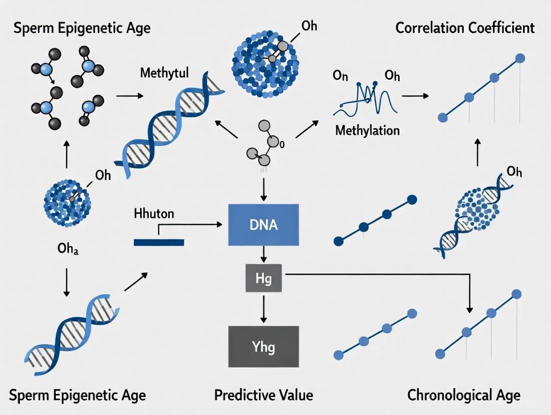
Sperm Epigenetic Age vs. Chronological Age: A Novel Biomarker for Predicting Male Fertility and Reproductive Outcomes
This article synthesizes current research on sperm epigenetic age (SEA), a biomarker of biological aging in sperm derived from DNA methylation patterns.
Sperm Epigenetic Age vs. Chronological Age: A Novel Biomarker for Predicting Male Fertility and Reproductive Outcomes
Abstract
This article synthesizes current research on sperm epigenetic age (SEA), a biomarker of biological aging in sperm derived from DNA methylation patterns. It explores how SEA diverges from chronological age and its superior predictive value for male fecundity, time-to-pregnancy, and embryonic development. Covering foundational concepts, methodological approaches for measurement, troubleshooting of current limitations, and comparative validation against traditional parameters, this review is tailored for researchers and drug development professionals seeking to integrate epigenetic clocks into male fertility assessments and develop targeted interventions.
Beyond Chronology: Defining Sperm Epigenetic Age and Its Biological Basis
Sperm Epigenetic Age (SEA) is an estimate of the biological age of male gametes derived from DNA methylation patterns at specific genomic sites, serving as a sperm-specific epigenetic clock [1]. In contrast to chronological age, which simply measures the time elapsed since birth, SEA reflects the biological aging processes influenced by a combination of genetic, environmental, and lifestyle factors that accumulate in sperm cells over time [2]. The well-documented relationship between chronological age and the sperm methylome has enabled the construction of these epigenetic clocks, which can estimate biological age based on DNA methylation patterns that change predictably with age [1].
This distinction is particularly important in reproductive medicine and research, as chronological age does not fully capture the intrinsic and extrinsic factors that contribute to the aging process of gametes [1]. While men continuously produce sperm throughout their lifetime, increased paternal age leads to a documented decline in fertility and increases the chances of pregnancy complications, preterm birth, and low birth weight [1]. The development of SEA represents a significant advancement in identifying novel sperm biomarkers of reproductive success beyond traditional semen parameters [1].
Biological Basis and Mechanisms
Epigenetic Alterations in Aging Sperm
The sperm epigenome undergoes significant changes with advancing age through several key mechanisms. DNA methylation represents the most extensively investigated epigenetic mechanism in aging sperm, with age-dependent changes occurring at discrete sets of CpG sites throughout the genome [2]. Research indicates that sperm cells exhibit a very different pattern of age-related DNA methylation compared to somatic cells, with DNA methylation decreasing with age in most genes, contrary to patterns observed in somatic tissues [3] [4]. Additionally, sperm telomere length does not decrease with age, which again contrasts with established patterns in somatic cells [4].
Beyond DNA methylation, age affects all known epigenetic mechanisms in sperm, including histone modifications and profiles of small non-coding RNAs [2]. These age-dependent epigenetic mechanisms collectively target gene networks enriched for embryo developmental, neurodevelopmental, growth, and metabolic pathways, suggesting that age-dependent changes in the sperm epigenome cannot be described merely as a stochastic accumulation of random epimutations [2]. The interplay between these various epigenetic mechanisms creates a complex aging signature that SEA attempts to quantify.
Signaling Pathways and Molecular Relationships
The relationship between environmental exposures, epigenetic changes, and reproductive outcomes involves complex biological pathways. The following diagram illustrates the conceptual pathway from environmental exposures to potential offspring effects through sperm epigenetic aging:
This conceptual framework demonstrates how environmental exposures such as air pollution, cigarette smoke, and various chemicals can induce epigenetic changes in sperm, which are further modified by chronological age [2] [5]. These epigenetic alterations form the basis for calculating SEA, which in turn shows promise for predicting reproductive outcomes and potential offspring health implications [1] [2] [6]. The recognition that these age-induced changes in the sperm epigenome are profound, physiological, and dynamic over years, yet stable over days and months, highlights their potential significance in reproductive outcomes [2].
Experimental Approaches and Prediction Models
Methodological Workflow for SEA Analysis
The determination of sperm epigenetic age involves a multi-step process from sample collection to computational prediction. The following workflow outlines the primary experimental and analytical steps:
This workflow begins with semen sample collection, typically following a recommended abstinence period of 2-3 days [1]. For the LIFE study, men collected samples via masturbation at home, kept them on ice overnight, and shipped them to the laboratory the next day [1]. The SEEDS cohort provided fresh samples at the clinic, which were immediately analyzed after 30 minutes of liquefaction [1].
Sperm processing and DNA extraction require specialized protocols due to sperm DNA being packaged primarily with protamines instead of histones. Sperm need to be treated with a reducing agent prior to purification [1]. The rapid DNA extraction method developed by researchers involves homogenizing sperm with steel beads and a lysis buffer containing guanidine thiocyanate and tris(2-carboxyethyl) phosphine (TCEP) at room temperature for 5 minutes [1]. This method consistently yields over 90% high-quality DNA and offers advantages of room temperature processing without lengthy proteinase K digestions [1].
Bisulfite conversion represents a critical step that distinguishes methylated from unmethylated cytosines. The EZ DNA methylation kit (Zymo) is commonly used for this process, converting unmethylated cytosines to uracils while leaving methylated cytosines unchanged [7].
DNA methylation analysis is typically performed using array-based technologies. The Illumina EPIC Infinium Methylation Beadchip, which analyzes over 850,000 CpG sites, has been extensively used in SEA studies [1] [3] [7]. For forensic applications with lower DNA quality, targeted bisulfite massively parallel sequencing provides a more sensitive alternative [3] [4].
Data processing and normalization utilize specialized bioinformatic pipelines. The Minfi package in R is commonly employed for both quality control and pre-processing pipelines, including SWAN normalization and generation of beta values (fraction methylation values) for further analysis [8] [7].
Finally, SEA prediction employs machine learning algorithms. Random forest regression has been successfully used to construct age prediction models with DNA methylation microarray data [8]. These models calculate SEA based on the methylation patterns at specific CpG sites known to change with age.
Comparison of Epigenetic Age Prediction Models for Semen
Various research groups have developed different models for predicting epigenetic age from semen samples, with varying numbers of markers and prediction accuracy:
Table 1: Comparison of Semen Epigenetic Age Prediction Models
| Study | Number of CpG Markers | Key Genes/Regions | Prediction Accuracy (MAE) | Technology Platform |
|---|---|---|---|---|
| Pisarek et al. (2021) [3] [4] | 6 | SH2B2, EXOC3, IFITM2, GALR2, FOLH1B | 5.1 years | EPIC Array, Targeted MPS |
| Jenkins et al. [3] | 51 regions | 51 age-related regions | 2.37 years | HumanMethylation450 BeadChip |
| Lee et al. (2015) [3] [4] | 3 | TTC7B, FOLH1B, LOC401324 | ~5 years | HumanMethylation450 BeadChip |
| Current Study (Blood) [8] | 6 autosomal + X chromosomal | DGAT2L6, PLXNB3, RPGR | 1.89 years (MAD) | 450K Microarray |
The variation in prediction accuracy across models reflects both the number of markers analyzed and the technological platforms used. Models incorporating a larger number of CpG sites, such as Jenkins et al.'s 51-region model, generally achieve higher accuracy (MAE = 2.37 years) but present practical challenges for forensic applications where DNA quality and quantity are limited [3]. In contrast, the 6-CpG model developed by Pisarek et al. provides a balance between practical implementability and reasonable accuracy (MAE = 5.1 years) [3] [4].
Notably, research has explored incorporating sex chromosomal DNA methylation markers alongside autosomal markers to enhance prediction accuracy in blood samples, with one model achieving a mean absolute deviation (MAD) of 1.89 years [8]. However, Y chromosomal DNA methylation markers did not enhance predictive performance in these models [8].
Research Applications and Clinical Correlations
SEA Associations with Semen Parameters and Fertility
Research evaluating the relationship between SEA and standard semen parameters has yielded nuanced findings. A study examining 379 men from the general population (LIFE study) and 192 men seeking fertility treatment (SEEDS) found that SEA was not significantly associated with standard semen characteristics such as count, concentration, or motility in either cohort [1].
However, SEA demonstrated significant associations with more specialized sperm morphological parameters. In the LIFE study, advanced SEA was associated with:
- Higher sperm head length and perimeter
- Increased presence of pyriform (pear-shaped) and tapered sperm
- Lower sperm elongation factor [1]
These findings suggest that SEA shows promise as an independent biomarker of sperm quality that captures aspects of sperm health not reflected in routine semen analyses. The association with sperm head morphological defects is particularly relevant, as these abnormalities are less commonly evaluated during standard male infertility assessments but may significantly impact fertility potential [1].
Beyond morphological factors, chronological age is associated with increased sperm DNA damage, as measured by the DNA fragmentation index (DFI) [9]. Studies of Chinese males have demonstrated that sperm DFI increases significantly with advancing age, which is concerning given that DFI values exceeding 30% pose significant challenges to natural conception and can lead to pre-implantation embryonic abnormalities and early miscarriage [9].
Interventional Studies and SEA Modifiability
Research has investigated whether nutritional interventions can modify sperm epigenetic aging. The Folic Acid and Zinc Supplementation Trial (FAZST), a large double-blind, randomized controlled trial, examined whether six months of supplementation with 5 mg folic acid and 30 mg elemental zinc could alter sperm DNA methylation patterns [7].
The findings revealed that:
- No significant differences were identified between the treatment and placebo groups across various methylation analyses (global, single CpG, regional)
- Any trends observed were no more than would be expected by random chance
- The supplementation regimen did not impact germ line epigenetic aging [7]
These results strongly suggest that this particular supplementation regimen is not effective at altering sperm DNA methylation, comporting with previous findings from the FAZST study that found no impact of supplementation on basic semen analysis parameters or live birth [7]. This highlights the stability of the sperm epigenome and the challenge in modifying SEA through simple nutritional interventions.
Potential as a Biomarker for Offspring Health
Emerging evidence suggests that paternal sperm epigenetics may serve as a biomarker for offspring health outcomes. Research has identified distinct DNA methylation signatures in sperm from fathers of children with autism spectrum disorder (ASD) compared to those without autistic children [6].
A genome-wide analysis identified 805 differential methylated regions (DMRs) in sperm from fathers of autistic children, with these DMRs associated with genes linked to known ASD genes and other neurobiology-related genes [6]. When validated with blinded test sets, these sperm DMR biomarkers demonstrated approximately 90% accuracy in identifying paternal offspring autism susceptibility [6].
This suggests that ancestral or early-life paternal exposures that alter germline epigenetics may be a molecular component of ASD etiology, and that sperm epigenetic signatures may potentially serve as biomarkers for assessing offspring disease susceptibility [6]. The potential applications in assisted reproduction settings could allow for improved clinical management and early treatment options, though further validation is needed.
The Scientist's Toolkit: Essential Research Materials
Table 2: Key Research Reagent Solutions for Sperm Epigenetic Age Studies
| Reagent/Kit | Specific Function | Application Notes |
|---|---|---|
| Illumina EPIC Infinium Methylation BeadChip | Genome-wide DNA methylation analysis | Interrogates >850,000 CpG sites; requires high-quality DNA [1] [3] [7] |
| Zymo EZ DNA Methylation Kit | Bisulfite conversion of DNA | Critical step for distinguishing methylated/unmethylated cytosines [7] |
| Qiagen DNeasy Blood and Tissue Kit | Sperm DNA isolation | Requires modification for sperm-specific protocols [7] |
| Tris(2-carboxyethyl) phosphine (TCEP) | Reducing agent for sperm lysis | Stable at room temperature; more effective than DTT for sperm DNA extraction [1] |
| Methylation Array Scanner (USEQ software) | Sliding window analysis of DMRs | Identifies differentially methylated regions; window size typically 1,000 bp [7] |
| Minfi R Package | Quality control and normalization of methylation data | Standard for processing array data; includes SWAN normalization [8] [7] |
This toolkit represents essential resources for researchers investigating sperm epigenetic aging. The specialized protocols for sperm DNA extraction, particularly the use of TCEP as a reducing agent, highlight the unique challenges of working with sperm compared to somatic cells [1]. The bioinformatic tools for processing and analyzing methylation data are equally crucial for deriving accurate SEA estimates from raw methylation data.
Sperm Epigenetic Age represents a significant advancement in male reproductive health assessment, moving beyond chronological age to capture the biological aging of gametes influenced by genetic, environmental, and lifestyle factors. While not associated with standard semen parameters, SEA shows correlations with specific sperm morphological defects and potentially with offspring health outcomes [1] [6].
Current prediction models vary in their complexity and accuracy, with practical applications balanced against technical feasibility [3] [4]. The stability of SEA against short-term nutritional interventions like folic acid and zinc supplementation suggests these epigenetic patterns reflect relatively stable biological processes [7].
For researchers and drug development professionals, SEA offers a promising biomarker for evaluating male reproductive potential and potentially assessing transmission of epigenetic risk to offspring. Future directions will likely focus on refining prediction models, identifying modifiable factors that influence epigenetic aging, and exploring clinical applications in assisted reproductive technologies.
Aging is characterized by a progressive loss of physiological integrity, leading to impaired function and increased vulnerability to death [10]. While chronological age measures the passage of time, it fails to accurately capture an individual's physiological state, as people of the same chronological age can exhibit markedly different health profiles and functional capacities [10]. This limitation has spurred the search for robust biomarkers of biological aging, culminating in the development of epigenetic clocks based on DNA methylation (DNAm) patterns [10] [11].
DNA methylation, the addition of a methyl group to cytosine bases primarily at cytosine-phosphate-guanine (CpG) dinucleotides, represents a dynamic epigenetic modification that regulates gene expression without altering the underlying DNA sequence [10] [12]. The reversibility of DNA methylation and its responsiveness to environmental influences, lifestyle factors, and pathological states make it an ideal candidate for measuring biological age [10] [12]. Since their inception, DNA methylation clocks have demonstrated remarkable accuracy in predicting chronological age across diverse tissues and cell types, while also capturing aspects of biological aging related to healthspan, disease risk, and mortality [10] [11].
This review explores the molecular architecture of epigenetic clocks, their evolving sophistication, and their application in aging research, with particular emphasis on the emerging field of sperm epigenetic age and its relationship with male reproductive health.
The Architecture of Epigenetic Clocks: From Chronological to Biological Age Predictors
Fundamental Mechanisms and First-Generation Clocks
The foundation of epigenetic clocks lies in the systematic changes that occur to the methylome with age. Specific CpG sites undergo predictable hypermethylation or hypomethylation, with hypermethylated regions often found in CpG islands, bivalent promoters, and Polycomb target genes, while hypomethylated regions tend to occur in non-CGI promoters and enhancers [10]. These age-related methylation changes are sufficiently consistent to enable accurate age prediction through supervised machine learning approaches applied to genome-wide methylation data [10].
The first generation of epigenetic clocks focused primarily on predicting chronological age. Horvath's multi-tissue clock, a landmark development, utilized 353 CpG sites to accurately estimate age across 51 different tissues and cell types [10] [11]. The Hannum clock, developed concurrently, employed 71 CpG sites from blood-derived DNA and achieved a remarkable correlation of 0.95 with chronological age in adults [10]. These clocks established DNA methylation as a powerful biomarker of aging, though their performance varied across developmental stages and tissue types [10].
Epigenetic Clock Development Pathway: This diagram illustrates the progression from fundamental aging processes and influencing factors through DNA methylation changes to the development of various types of epigenetic clocks and their respective applications.
Advanced Clocks and Health-Related Predictors
Second-generation epigenetic clocks shifted focus from chronological age prediction to capturing biological aging processes linked to health outcomes. The DNAm PhenoAge clock, developed by Levine et al., incorporated clinical biomarkers to construct a measure of phenotypic age that outperformed first-generation clocks in predicting mortality, healthspan, and age-related diseases [10] [12]. The DNAm GrimAge clock further advanced the field by integrating DNA methylation-based surrogate biomarkers for seven plasma proteins and smoking history, demonstrating superior performance in predicting all-cause mortality and age-related diseases compared to previous clocks [10] [12].
More recent developments include pace-of-aging clocks such as DunedinPACE, which measures the rate of physiological decline across multiple organ systems, and tissue-specific clocks optimized for particular applications [12]. The ongoing refinement of epigenetic clocks has also incorporated novel approaches such as deep learning models (DeepMAge, AltumAge) and the integration of sex chromosomal markers alongside autosomal CpGs to enhance predictive accuracy [8] [12].
Table 1: Comparison of Major DNA Methylation Clocks for Aging Research
| Clock Name | CpG Sites | Tissue Specificity | Primary Application | Key Strengths | Performance Metrics |
|---|---|---|---|---|---|
| Horvath Clock [10] [11] | 353 | Pan-tissue | Chronological age estimation | Works across most tissues and cell types | High accuracy (r ≥ 0.90) across tissues |
| Hannum Clock [10] | 71 | Blood-specific | Chronological age in adults | High accuracy in blood samples | r = 0.95 in adult blood |
| DNAm PhenoAge [10] [12] | 513 | Multiple tissues | Healthspan, mortality risk | Incorporates clinical biomarkers | Superior for aging outcomes vs. first-generation clocks |
| DNAm GrimAge [10] [12] | ~1000+ | Blood | Mortality, disease risk | Uses plasma protein proxies | Better mortality prediction than previous clocks |
| DunedinPACE [12] | ~80-100 | Blood | Pace of aging | Longitudinal aging measurement | Predicts physiological decline rate |
| Sperm Epigenetic Clock [1] | Not specified | Sperm-specific | Male fertility assessment | Correlates with time-to-pregnancy | Associated with fecundability |
Experimental Approaches in DNA Methylation Age Determination
Core Methodologies and Workflows
The determination of epigenetic age relies on sophisticated molecular biology techniques combined with computational analysis. The standard workflow begins with DNA extraction from the target tissue, followed by bisulfite conversion, which deaminates unmethylated cytosines to uracils while leaving methylated cytosines unchanged [1] [13]. This conversion enables the discrimination of methylated and unmethylated cytosines in subsequent analysis.
The most commonly used platforms for DNA methylation analysis are Illumina's Infinium BeadChips, including the 450K and EPIC arrays, which simultaneously interrogate methylation at hundreds of thousands of CpG sites across the genome [8] [12] [1]. For higher-resolution analysis, targeted bisulfite sequencing and whole-genome bisulfite sequencing provide base-pair resolution methylation data, enabling the assessment of methylation patterns and entropy beyond single CpG sites [13].
Following data generation, quality control and normalization procedures are critical to remove technical artifacts and batch effects. Common approaches include the preprocessFunnorm method implemented in the minfi R package [8] [14]. Probes containing single-nucleotide polymorphisms, cross-hybridizing probes, and those with poor detection p-values are typically filtered out to ensure data quality [8].
DNA Methylation Age Analysis Workflow: This diagram outlines the standard experimental pipeline for epigenetic age estimation, from sample collection through data generation and computational analysis to final age acceleration calculation.
Computational Analysis and Age Prediction
The transformation of methylation data into age estimates employs sophisticated machine learning algorithms. The elastic net regression, a regularized linear regression approach that combines L1 and L2 regularization, has been widely used in the development of epigenetic clocks, including Horvath's original pan-tissue clock and DNAm GrimAge [10] [12]. This method effectively handles the high dimensionality of methylation data, where the number of features (CpG sites) far exceeds the number of samples.
Random forest regression has also been successfully applied, particularly in models incorporating sex chromosomal markers alongside autosomal CpGs [8]. More recently, deep learning approaches such as DeepMAge and AltumAge have demonstrated enhanced accuracy and robustness in age prediction across diverse tissues and platforms [12].
The final output of these analyses is the DNA methylation age (DNAm age), which can be compared to chronological age to calculate age acceleration (AA) or deceleration. Positive age acceleration, where DNAm age exceeds chronological age, has been associated with numerous adverse health outcomes and increased mortality risk [11] [14].
Table 2: Essential Research Reagents and Platforms for DNA Methylation Aging Studies
| Category | Specific Product/Platform | Application in Research | Key Features |
|---|---|---|---|
| DNA Methylation Arrays | Illumina Infinium HumanMethylation450 BeadChip [8] [1] | Genome-wide methylation profiling | 450,000 CpG sites, established analysis pipelines |
| Illumina Infinium MethylationEPIC BeadChip [12] [14] | Enhanced genome-wide coverage | >850,000 CpG sites, improved regulatory region coverage | |
| Bisulfite Conversion Kits | EZ DNA Methylation Kit (Zymo Research) | Bisulfite conversion of DNA | High conversion efficiency, DNA protection technology |
| MethylCode Bisulfite Conversion Kit (Thermo Fisher) | Efficient cytosine conversion | Rapid protocol, minimal DNA degradation | |
| DNA Extraction Kits | QIAamp DNA Blood Mini Kit (Qiagen) [14] | DNA extraction from blood samples | High-quality DNA suitable for bisulfite conversion |
| Phenol-chloroform with TCEP reduction [1] | Sperm DNA extraction | Specialized for protamine-bound sperm DNA | |
| Computational Tools | Minfi R Package [8] [14] | Quality control and normalization | Comprehensive pipeline for array data processing |
| Horvath's Epigenetic Clock Software [11] [14] | DNAm age calculation | Implements multiple epigenetic clocks | |
| Specialized Reagents | Tris(2-carboxyethyl)phosphine (TCEP) [1] | Sperm DNA decondensation | Reduces protamine disulfide bonds for sperm DNA access |
Sperm Epigenetic Age Versus Chronological Age: Predictive Value in Male Reproduction
Development and Validation of Sperm-Specific Epigenetic Clocks
The established relationship between chronological age and the sperm methylome has enabled the development of sperm-specific epigenetic clocks to estimate the biological age of sperm, termed sperm epigenetic age (SEA) [1]. Unlike somatic cells, sperm DNA is packaged with protamines rather than histones, requiring specialized DNA extraction protocols incorporating reducing agents such as tris(2-carboxyethyl)phosphine (TCEP) to break protamine disulfide bonds [1].
Sperm epigenetic clocks have been constructed using similar machine learning approaches as somatic clocks, but trained specifically on sperm methylation data. These clocks capture age-related methylation changes in sperm that may reflect cumulative oxidative damage, environmental exposures, and other factors affecting germ cell integrity [1]. Importantly, sperm epigenetic age demonstrates a positive association with the time taken to achieve pregnancy, suggesting its potential as a biomarker of male fecundity independent of chronological age [1].
Clinical Correlations and Predictive Value
The clinical utility of sperm epigenetic age lies in its ability to capture aspects of reproductive aging not reflected in chronological age. In evaluations of both clinical (SEEDS) and non-clinical (LIFE) cohorts, SEA was not associated with standard semen parameters such as count, concentration, or motility [1]. However, it showed significant correlations with specific sperm morphological features, including higher sperm head length and perimeter, increased presence of pyriform and tapered sperm, and lower sperm elongation factor [1].
These findings suggest that sperm epigenetic age may reflect subtle aspects of sperm quality and developmental competence that are not captured by routine semen analysis. The association between advanced SEA and longer time-to-pregnancy further supports its potential as an independent biomarker of male fecundity [1]. Environmental factors, including exposure to endocrine-disrupting chemicals like phthalates, have been associated with accelerated sperm epigenetic aging, providing a potential mechanism by which environmental exposures impact male reproductive health [1].
The divergence between sperm epigenetic age and chronological age may thus serve as a more sensitive indicator of reproductive aging, capturing the cumulative effects of genetic, environmental, and lifestyle factors on germ cell quality. This has important implications for fertility assessment, as two men of identical chronological age may exhibit markedly different sperm epigenetic ages, potentially reflecting differences in their reproductive potential.
Comparative Analysis of Epigenetic Clocks Across Tissues and Applications
The performance and application of epigenetic clocks vary significantly across tissue types and research contexts. Pan-tissue clocks like Horvath's original model provide broad applicability but may lack tissue-specific precision, while specialized clocks optimized for specific tissues (blood, brain, sperm) often demonstrate enhanced accuracy within their target tissue but limited utility elsewhere [10] [11] [1].
Table 3: Performance Comparison of Epigenetic Clocks Across Biological Contexts
| Application Context | Recommended Clocks | Key Performance Metrics | Limitations & Considerations |
|---|---|---|---|
| General Aging Studies | Horvath Pan-Tissue, Hannum Blood Clock | High chronological age accuracy (r > 0.90) [10] | Less predictive for health outcomes than newer clocks |
| Health Risk Prediction | DNAm PhenoAge, DNAm GrimAge | Strong association with mortality, disease incidence [10] [12] | GrimAge requires specific plasma protein CpG proxies |
| Intervention Studies | DunedinPACE, DNAm PhenoAge | Sensitivity to aging rate changes, intervention effects [12] | DunedinPACE requires specific computational implementation |
| Sperm Quality & Male Fertility | Sperm Epigenetic Clocks [1] | Correlates with time-to-pregnancy, sperm morphology | Requires specialized sperm DNA extraction protocols |
| Physical Function Assessment | DNAm FitAge [12] [14] | Incorporates fitness biomarkers (grip strength, gait speed) | Newer clock with less extensive validation |
| Forensic Applications | Combined autosomal + sex chromosome models [8] | Improved accuracy (MAD: 1.89 years) [8] | Emerging approach requiring further validation |
The selection of an appropriate epigenetic clock depends critically on the research question and tissue type. For general chronological age estimation in diverse tissues, the Horvath clock remains widely used, while for health outcome prediction, second-generation clocks like GrimAge and PhenoAge demonstrate superior performance [10] [12] [14]. In specialized contexts such as male reproduction, tissue-specific clocks provide unique insights not captured by somatic clocks [1].
Recent advances continue to refine epigenetic clocks, incorporating additional biomarker types such as DNA methylation-based surrogates for plasma proteins [12], physical fitness measures [12] [14], and metabolite levels [12]. The integration of sex chromosomal markers alongside autosomal CpGs has also demonstrated improved predictive accuracy [8]. These developments highlight the dynamic evolution of epigenetic clocks toward increasingly sophisticated biomarkers of biological aging.
DNA methylation-based epigenetic clocks represent a transformative biomarker technology that has revolutionized aging research. From first-generation clocks focused on chronological age prediction to sophisticated second-generation models capturing mortality risk and healthspan, these molecular estimators provide unique insights into the biological aging process. The development of sperm-specific epigenetic clocks has further expanded their utility into the realm of reproductive aging, offering novel approaches to assess male fecundity beyond conventional semen parameters.
As epigenetic clocks continue to evolve, incorporating multi-omics data, advanced computational methods, and diverse population data, their precision and clinical utility are expected to further improve. These advancements hold promise for tracking the effectiveness of anti-aging interventions, identifying individuals at elevated risk for age-related diseases, and providing personalized insights into biological aging trajectories across tissues and organ systems. The molecular clockwork of DNA methylation thus stands as a powerful tool for unraveling the complexities of aging and developing strategies to promote healthspan extension.
In the evolving landscape of reproductive biology, chronological age has traditionally served as a proxy for male fertility potential. However, it fails to encapsulate the cumulative impact of genetic, environmental, and lifestyle factors on the biological aging of sperm. The discovery of sperm epigenetic age (SEA), a biomarker derived from predictable age-related changes in sperm DNA methylation patterns, represents a paradigm shift [15] [16]. SEA can diverge from chronological age, a phenomenon known as epigenetic age acceleration, which provides a more nuanced measure of the male germline's biological health [17]. This acceleration is not uniform across all tissues; research indicates that conditions like oligozoospermia can cause accelerated epigenetic aging specifically in sperm without affecting the epigenetic age of blood from the same individual, highlighting its tissue-specific nature [18]. This guide objectively compares the predictive value of sperm epigenetic age against chronological age, synthesizing current research data and methodologies to inform researchers, scientists, and drug development professionals in the field of reproductive medicine.
Quantitative Comparison of Predictive Performance
The predictive power of epigenetic clocks surpasses that of chronological age alone, both for estimating chronological age and for forecasting reproductive outcomes. The table below summarizes key performance metrics from seminal studies.
Table 1: Predictive Performance of Sperm Epigenetic Age vs. Chronological Age
| Prediction Model / Factor | Basis/Method | Key Performance Metric | Association with Reproductive Outcomes |
|---|---|---|---|
| Sperm Epigenetic Age (SEA) - SEACpG Clock [15] | Machine learning on sperm DNA methylation data | Correlation with chronological age: r = 0.91 [15] | 17% lower cumulative pregnancy probability after 12 months for couples with older SEA; associated with longer time-to-pregnancy (FOR=0.83) and shorter gestation [15] [16]. |
| Sperm Epigenetic Age (SEA) - 6 CpG Model [3] | Targeted bisulfite MPS of 6 CpG sites (SH2B2, EXOC3, IFITM2, GALR2, FOLH1B) | Mean Absolute Error (MAE): 5.1 years [3] | Primarily validated for chronological age prediction in forensic contexts; clinical reproductive correlations not yet fully established [3]. |
| Chronological Age | N/A | N/A | Poor independent predictor of time-to-pregnancy and semen quality; weak correlations with declining semen parameters [15] [9]. |
Table 2: Association of Sperm Epigenetic Age with Semen Parameters
| Parameter Category | Specific Parameter | Association with Sperm Epigenetic Age |
|---|---|---|
| Standard Semen Parameters [1] | Concentration, Count, Morphology | No significant association found in either clinical (SEEDS) or non-clinical (LIFE) cohorts. |
| Sperm Head Morphology [1] | Head Length, Head Perimeter | Significantly associated with higher SEA in the LIFE cohort. |
| Elongation Factor | Significantly associated with lower SEA in the LIFE cohort. | |
| Presence of Pyriform and Tapered Sperm | Significantly associated with higher SEA in the LIFE cohort. | |
| Sperm DFI and Aging [9] | DNA Fragmentation Index (DFI) | Increases significantly with advancing chronological age. |
Detailed Experimental Protocols and Methodologies
Sperm Sample Collection and DNA Isolation
The accuracy of sperm epigenetic age prediction hinges on rigorous sample preparation and processing to ensure the analysis is free from somatic cell contamination [18] [1].
- Sample Collection: Semen samples are typically collected after a recommended period of ejaculatory abstinence (2-3 days) via masturbation without lubricants. Studies like the LIFE and SEEDS cohorts used both home-collection (with immediate placement on ice and overnight shipping to the lab) and in-clinic collection for fresh analysis [1].
- Somatic Cell Lysis: This critical step removes contaminating white blood cells, whose differing methylation profiles could confound results. Samples are incubated in a somatic cell lysis buffer (e.g., 0.1% SDS, 0.5% Triton X-100) on ice for 20 minutes, followed by visual inspection to confirm the absence of contaminating cells [18].
- DNA Extraction with Reducing Agent: Sperm DNA is uniquely packaged with protamines, requiring a reducing agent for efficient extraction. Protocols use a lysis buffer containing guanidine thiocyanate and a reducing agent like Tris(2-carboxyethyl)phosphine (TCEP), followed by homogenization with steel beads and purification using silica-based spin columns. This method yields over 90% high-quality DNA without lengthy proteinase K digestions [1].
DNA Methylation Profiling and Clock Construction
The core of SEA development involves genome-wide methylation analysis and sophisticated computational modeling.
- Microarray-Based Methylation Analysis: Bisulfite-converted sperm DNA is hybridized to Illumina Infinium Methylation BeadChip arrays (e.g., EPIC 850K or 450K). These arrays quantitatively measure methylation levels at hundreds of thousands of CpG sites across the genome, generating a beta value (ranging from 0 for completely unmethylated to 1 for fully methylated) for each site [3] [15] [18].
- Clock Construction via Machine Learning: Raw methylation data is preprocessed and normalized. Age-associated CpGs are identified, and prediction models are built using machine learning algorithms. The SEACpG clock, for instance, was developed using an ensemble machine learning algorithm applied to data from 379 men, achieving a correlation of r=0.91 with chronological age [15]. Other approaches use multivariable linear regression supported by feature selection criteria like the Bayesian Information Criterion to identify minimal marker sets (e.g., a 6-CpG model) [3].
Diagram: Workflow for Developing a Sperm Epigenetic Clock
Key Experiments Linking SEA to Reproductive Outcomes
Landmark studies have established the clinical relevance of sperm epigenetic age acceleration.
- LIFE Study (General Population Cohort): This prospective study of 379 couples discontinuing contraception for pregnancy demonstrated the predictive power of SEA. Researchers used discrete-time proportional hazards models, adjusting for covariates, to show that advanced SEACpG was negatively associated with fecundability (FOR=0.83), meaning a longer time-to-pregnancy. Furthermore, couples with male partners in older SEA categories had a 17% lower cumulative probability of pregnancy after 12 months [15] [16].
- Tissue-Specific Age Acceleration in Oligozoospermia: A comparative study of normozoospermic and oligozoospermic men calculated the Germ-line Age Differential (GLAD) for sperm and the epigenetic age of blood. The sperm of oligozoospermic men had a significantly higher mean GLAD score (0.078) than those with normozoospermia (-0.017), indicating accelerated aging. Crucially, no such difference was found in their blood, proving tissue-specific epigenetic age acceleration linked to a disease state [18].
The Scientist's Toolkit: Essential Research Reagents and Materials
Table 3: Key Reagents and Materials for Sperm Epigenetic Age Research
| Item | Specific Example / Kit | Function in Protocol |
|---|---|---|
| DNA Methylation BeadChip | Illumina Infinium MethylationEPIC BeadChip | Genome-wide profiling of DNA methylation at >850,000 CpG sites [3] [15]. |
| Bisulfite Conversion Kit | EZ-96 DNA Methylation-Gold Kit (Zymo Research) | Converts unmethylated cytosines to uracils, allowing methylation status to be determined via sequencing or array [18]. |
| DNA Extraction Kit (Sperm-Specific) | DNeasy Blood & Tissue Kit (Qiagen) with modifications | Silica-based column purification of DNA. Requires a reducing agent like TCEP for sperm-specific lysis [1]. |
| Somatic Cell Lysis Buffer | 0.1% SDS, 0.5% Triton X-100 in DEPC H2O | Selective lysis of contaminating white blood cells in semen samples prior to sperm DNA extraction [18]. |
| Reducing Agent | Tris(2-Carboxyethyl)Phosphine (TCEP) | Breaks disulfide bonds in sperm protamine proteins, enabling efficient sperm DNA extraction [1]. |
| Bioinformatic Tools | minfi R package, Elastic Net regression, Ensemble machine learning algorithms | Preprocessing, normalization, and analysis of methylation array data; construction of predictive age models [15] [19]. |
The divergence of sperm epigenetic age from chronological age provides a powerful, tissue-specific lens through which to view male reproductive health and aging. Quantitative data firmly establishes that SEA is a superior biomarker for predicting time-to-pregnancy and gestation length compared to chronological age alone [15] [16]. Furthermore, its association with specific defects in sperm head morphology, rather than standard semen parameters, suggests it captures unique aspects of sperm quality [1]. The documented phenomenon of age acceleration in the sperm of oligozoospermic men, unaccompanied by acceleration in blood, underscores the potential of SEA to reveal pathology-specific aging trajectories [18]. For researchers and drug developers, these insights pave the way for novel diagnostic tools and the evaluation of interventions aimed at decelerating reproductive aging, ultimately improving couple-based reproductive outcomes.
The global trend toward delayed parenthood has brought the scientific consequences of advanced paternal age (APA) into sharp focus. While maternal age has long been recognized as a critical factor in reproductive outcomes, a growing body of evidence indicates that paternal age similarly exerts profound effects on fertility, embryonic development, and offspring health. Aging is an unavoidable biological process with significantly disproportionate gender-based effects on human fertility [20]. Unlike the relatively abrupt decline in female fertility, male reproductive aging is subtle and progressive, yet carries significant implications [20]. Epidemiological and animal model evidence strongly suggests that offspring of older fathers face elevated risks for neuropsychiatric diseases and other health complications [20] [21]. These observations have driven increased scientific interest in understanding what molecular changes occur in the gametes of aging men, with particular focus on the sperm epigenome [20].
At the heart of this investigation lies DNA methylation, an essential epigenetic mechanism involving the addition of methyl groups to cytosine bases, typically at cytosine phosphate guanine dinucleotides (CpGs). The sperm epigenome is fundamentally different from that of oocytes and somatic cells, characterized by unique nuclear protein composition and highly specialized DNA methylation patterns [20] [22]. These epigenetic marks are competent to regulate gene expression and can be passed onto the embryo following fertilization [20]. Because the sperm epigenome's role extends beyond normal sperm function to influence embryogenesis and early development, understanding its alteration with age has become a research priority [20]. This review synthesizes current evidence demonstrating that advanced paternal age is associated with widespread, consistent patterns of sperm DNA hypomethylation and explores the methodological approaches, functional consequences, and potential clinical applications of these findings.
Global Hypomethylation: A Hallmark of the Aging Sperm Epigenome
Overwhelming Evidence for Hypomethylation
Comprehensive genome-wide studies consistently reveal that hypomethylation constitutes the predominant pattern of epigenetic alteration in sperm from older men. A significant reduced representation bisulfite sequencing (RRBS) study of 73 sperm samples from men undergoing infertility treatment identified 1,565 regions significantly correlated with donor age [22]. The direction of age association was highly skewed, with 1,162 (74%) age-related differentially methylated regions (ageDMRs) being hypomethylated and only 403 (26%) being hypermethylated with advancing age [22]. This approximately 3:1 ratio of hypomethylation to hypermethylation represents a consistent finding across multiple experimental approaches and cohort populations.
The distribution of these methylation changes across genomic regions follows distinct patterns. Hypomethylated ageDMRs were significantly closer to transcription start sites (median distance 1,368 bp) compared to hypermethylated ageDMRs (median distance 17,205 bp), which were preferentially located in gene-distal regions [22]. This strategic positioning of hypomethylation events near gene regulatory elements suggests a potentially greater functional impact on gene expression programs. Furthermore, the majority (53%) of ageDMRs displayed average methylation levels in the medium range (20-80%), whereas most regions not subject to paternal age effects showed high methylation levels (>80%) [22]. This indicates that age-related changes predominantly affect genomic regions with intermediate methylation levels that may be particularly sensitive to epigenetic regulation.
Genomic Distribution and Functional Enrichment
The genomic features affected by age-related hypomethylation are not randomly distributed but instead show distinct enrichment patterns. Analysis of 2,355 genes with significant sperm ageDMRs across multiple studies revealed that the 241 genes replicated in at least one study showed significant functional enrichments in 41 biological processes associated with development and the nervous system, along with 10 cellular components associated with synapses and neurons [22]. This finding strongly supports the hypothesis that paternal age effects on the sperm methylome particularly affect genes involved in offspring behavior and neurodevelopment.
Chromosome 19 demonstrates a highly significant twofold enrichment of sperm ageDMRs, suggesting non-random genomic distribution of these epigenetic changes [22]. Despite the high gene density and CpG content being conserved in the orthologous marmoset chromosome 22, this region did not show increased regulatory potential by age-related DNA methylation changes, indicating potential human-specific vulnerability [22]. This chromosomal specificity highlights the non-stochastic nature of epigenetic aging in sperm and points to genomic features that may predispose certain regions to age-related methylation alterations.
Table 1: Summary of Age-Related DNA Methylation Changes in Human Sperm
| Feature | Hypomethylated Regions | Hypermethylated Regions |
|---|---|---|
| Proportion of AgeDMRs | 74% (1,162 of 1,565 DMRs) [22] | 26% (403 of 1,565 DMRs) [22] |
| Genomic Location | Closer to transcription start sites (median 1,368 bp) [22] | Gene-distal regions (median 17,205 bp) [22] |
| Methylation Level | Primarily medium methylation regions (20-80%) [22] | Varied distribution across methylation ranges [22] |
| Functional Enrichment | Neurodevelopmental processes, synaptic function [22] | Less consistently enriched for specific functions [22] |
| Chromosomal Distribution | Significant enrichment on chromosome 19 [22] | No specific chromosomal enrichment reported [22] |
Methodological Approaches for Detecting Sperm Methylation Changes
Genome-Wide Methylation Profiling Technologies
Multiple technological platforms have been employed to characterize age-related methylation changes in sperm, each with distinct advantages and limitations. Reduced representation bisulfite sequencing (RRBS) provides cost-effective methylation analysis of CpG-rich regions, successfully identifying thousands of ageDMRs with relatively small sample sizes [22]. Whole genome bisulfite sequencing (WGBS) offers comprehensive genome coverage, including non-CpG-rich regions, and has been applied successfully to precious samples like blastocyst lineages using ultra-low input protocols [23]. Infinium MethylationEPIC BeadChip arrays provide an intermediate approach,interrogating over 850,000 CpG sites with less technical complexity and cost, facilitating larger cohort studies [24] [4] [25].
Each method requires careful sample preparation and bioinformatic processing. Sperm DNA presents unique challenges due to its dense packaging with protamines rather than histones, necessitating specialized extraction protocols incorporating reducing agents like tris(2-carboxyethyl) phosphine (TCEP) to efficiently release DNA [24]. Quality control measures are essential, including assessment of bisulfite conversion efficiency and evaluation of potential somatic cell contamination through analysis of imprinted genes or loci like DLK1, which shows distinctly different methylation patterns in somatic versus sperm cells [24] [25].
Analytical Frameworks and Epigenetic Clocks
Beyond differential methylation analysis, researchers have developed sophisticated predictive models known as epigenetic clocks that estimate biological age based on DNA methylation patterns. Sperm-specific epigenetic clocks utilize machine learning approaches, such as Super Learner ensemble methods, to identify optimal combinations of predictive CpG sites [24]. These models can predict chronological age with mean absolute errors of approximately 3-5 years in validation datasets [24] [4] [25].
The development of these clocks represents a significant methodological advancement, transforming multidimensional methylation data into a single quantitative metric of sperm epigenetic age (SEA). This metric has demonstrated clinical relevance, showing positive associations with time to pregnancy independent of chronological age [24]. When SEA exceeds chronological age (a state termed epigenetic age acceleration), it may indicate accelerated deterioration of the sperm epigenome with potential functional consequences.
Sperm Methylation Analysis Workflow: Diagram illustrating the key methodological steps for detecting age-related methylation changes in human sperm, from sample collection through data analysis.
Functional Consequences of Sperm Hypomethylation
Impact on Embryonic Development and Offspring Health
The functional implications of sperm epigenetic aging extend beyond the gamete itself to influence embryonic development and offspring health. Research using donor oocyte-derived blastocysts (to control for maternal age effects) has revealed that advanced paternal age is associated with significant methylation and transcriptional dysregulation in both the inner cell mass (ICM) and trophectoderm (TE) lineages [23]. These alterations are particularly enriched in genes and pathways related to neuronal signaling and neurodevelopmental disorders, providing a potential mechanistic link between paternal age and increased offspring risk for conditions like autism spectrum disorder and schizophrenia [23].
Notably, the inner cell mass (which gives rise to the fetus) shows more pronounced transcriptional alterations in neurodevelopmental pathways compared to the trophectoderm (which forms extra-embryonic tissues) [23]. This tissue-specific vulnerability may explain why neurodevelopmental outcomes are particularly associated with advanced paternal age despite global epigenetic changes in sperm. The methylation dysregulation observed in blastocysts from older fathers largely overlaps with genes showing age-related methylation changes in sperm, supporting the transmission of paternal epigenetic information to the next generation [23] [22].
Relationship with Semen Parameters and Fertility Outcomes
The relationship between sperm epigenetic aging and conventional semen parameters reveals complex associations. While SEA shows limited correlation with standard semen characteristics like concentration, motility, or morphology, it demonstrates significant associations with specific sperm head morphological abnormalities, including increased head length and perimeter, higher incidence of pyriform and tapered sperm, and reduced elongation factor [24]. These findings suggest that epigenetic aging may manifest in subtle morphological changes not routinely assessed in standard infertility evaluations.
The clinical impact of these epigenetic changes is reflected in reproductive outcomes. Multiple studies have confirmed that advanced sperm epigenetic age is associated with longer time to pregnancy, reduced fecundability, and potentially decreased success with assisted reproductive technologies [20] [24]. Importantly, these effects appear partially independent of chronological age, suggesting that epigenetic age acceleration may identify individuals with compromised reproductive potential despite being within normal age ranges [24].
Table 2: Functional Correlates of Sperm Epigenetic Aging
| Domain | Observed Effects | Study Details |
|---|---|---|
| Embryonic Development | Methylation and transcriptional dysregulation in blastocyst ICM and TE lineages [23] | Donor oocyte model controlling for maternal age [23] |
| Neurodevelopmental Risk | Enrichment for neuronal signaling pathways and neurodevelopmental disorder genes [23] [22] | Associations with autism, schizophrenia risk [23] [22] |
| Sperm Morphology | Altered sperm head dimensions, increased abnormal forms [24] | Higher head length, perimeter; pyriform/tapered shapes [24] |
| Reproductive Outcomes | Increased time to pregnancy, reduced fecundability [20] [24] | Longitudinal investigation of fertility [24] |
| Assisted Reproduction | Potential impact on success rates, though findings inconsistent [9] | Clinical ART cohorts show variable results [9] |
Research Reagent Solutions for Sperm Epigenetics
The investigation of sperm epigenetic aging requires specialized reagents and methodologies tailored to the unique challenges of sperm chromatin. The following essential research tools represent critical components for studies in this field:
DNA Extraction Reagents with Reducing Agents: Conventional DNA extraction methods fail to efficiently release sperm DNA due to protamine packaging. Specialized protocols incorporating guanidine thiocyanate lysis buffers combined with reducing agents like tris(2-carboxyethyl) phosphine (TCEP) are essential for high-quality sperm DNA recovery [24]. TCEP is particularly advantageous as a stable, room-temperature-storable alternative to dithiothreitol (DTT).
Bisulfite Conversion Kits: Efficient bisulfite conversion is fundamental for methylation analysis. Optimized commercial kits (e.g., EZ DNA Methylation-Direct Kit, Zymo Research) are specifically validated for sperm DNA and compatible with low-input samples such as mechanically isolated blastocyst lineages [23].
Methylation Array Platforms: The Infinium MethylationEPIC BeadChip array (Illumina) provides comprehensive coverage of over 850,000 CpG sites, balancing cost and throughput for cohort studies [24] [4] [25]. This platform has been extensively used for sperm epigenetic clock development and validation.
Library Preparation Kits for Bisulfite Sequencing: Specialized kits for whole genome bisulfite sequencing (e.g., ultra-low DNA input WGBS prep workflow, Zymo Research) enable methylation analysis from limited samples [23]. For reduced representation approaches, RRBS kits provide cost-effective alternative focusing on CpG-rich regions [22].
Somatic Cell Contamination Controls: Analytical controls for detecting somatic cell contamination are crucial for sperm purity assessment. DLK1 locus methylation analysis serves as a reliable discriminator, with hypermethylation indicating somatic contamination in sperm samples [25]. Similarly, imprinted gene analysis (e.g., H19/IGF2) confirms sample purity [22].
Targeted Bisulfite Sequencing Panels: Custom panels for massively parallel sequencing enable validation of candidate ageDMRs and epigenetic clock CpGs in large cohorts [4]. These targeted approaches balance cost and throughput for clinical translation.
The comprehensive analysis of age-related epigenetic changes in human sperm reveals a consistent pattern of global hypomethylation affecting predominantly genes involved in neurodevelopment and embryonic growth. These alterations are so consistent that they enable accurate age prediction through epigenetic clocks and are associated with meaningful functional consequences for embryonic development and offspring health. The predominance of hypomethylation over hypermethylation (approximately 3:1 ratio) represents a distinctive feature of sperm epigenetic aging compared to somatic tissues [22].
Future research directions should focus on several key areas. First, the mechanistic basis for the observed genomic vulnerability, particularly the enrichment on chromosome 19, requires elucidation [22]. Second, longitudinal studies tracking methylation changes in individuals over time would strengthen causal inferences about aging effects. Third, the interaction between environmental factors (e.g., obesity, toxin exposure) and epigenetic aging warrants deeper investigation, as preliminary evidence suggests potential moderating effects [25]. Finally, the clinical translation of these findings toward improved risk assessment and personalized fertility counseling represents a critical frontier.
The consistent functional enrichment of age-related sperm methylation changes in neurodevelopmental pathways provides a compelling biological plausibility for the observed epidemiological associations between advanced paternal age and offspring neuropsychiatric disorders [23] [22]. As trends toward delayed parenthood continue globally, understanding these epigenetic mechanisms and their implications becomes increasingly important for both clinical practice and public health.
The study of genomic hotspots represents a frontier in understanding the coordinated regulation of gene expression, particularly for developmentally essential and neurologically significant genes. Transcriptional hotspots are defined as specific genomic regions bound by a multitude of transcription factors, forming high-occupancy hubs that drive cell-type-specific gene expression programs [26]. These regulatory elements have been identified across diverse species including worms, flies, and humans, where they frequently function as powerful enhancers controlling the expression of neighboring genes [26]. The functional enrichment observed in these hotspot regions provides critical insights into the molecular logic of development, differentiation, and disease processes.
Within the broader context of aging research, the interrogation of genomic hotspots intersects significantly with emerging studies on epigenetic aging clocks. Particularly in male fertility research, the divergence between sperm epigenetic age (SEA) and chronological age has emerged as a biomarker with predictive value for fecundity and reproductive outcomes [1]. While standard semen parameters have proven inadequate for fully assessing male fertility potential, epigenetic signatures—potentially organized through hotspot regulation—show promise as more refined diagnostic tools [1]. This review systematically compares the methodologies, analytical frameworks, and biological insights derived from the study of genomic hotspots, with particular emphasis on their implications for developmental processes and neurological functions, while contextualizing these findings within epigenetic aging research.
Methodological Comparison: Experimental and Computational Approaches
Experimental Protocols for Hotspot Identification
The identification and characterization of genomic hotspots relies on sophisticated experimental workflows that combine molecular biology techniques with advanced computational analysis. A representative protocol for transcriptional hotspot mapping involves several critical stages:
Stage 1: Sample Preparation and Factor Binding Detection Researchers collect cell types of interest and perform Chromatin Immunoprecipitation sequencing (ChIP-seq) for multiple transcription factors (TFs). In murine studies, this typically involves 6-21 TFs across 10 different cell types, generating approximately 108 datasets [26]. Cells are cross-linked to preserve protein-DNA interactions, chromatin is sheared, and specific TF-bound DNA fragments are immunoprecipitated using factor-specific antibodies. The bound DNA fragments are then sequenced using high-throughput platforms.
Stage 2: Peak Calling and Occupancy Classification Sequencing reads are aligned to the reference genome, and binding peaks are identified using tools such as HOMER [26]. Peaks in each cell type are classified into three occupancy groups: (1) Singletons (low-occupancy): peaks bound by only one TF; (2) Combinatorials (mid-occupancy): peaks bound by a combination of TFs; and (3) Hotspots (high-occupancy): peaks bound by more than five TFs studied in a given cell type [26]. On average, approximately 50% of peaks fall into singleton and combinatorial categories, while only 0.1-2% qualify as hotspots [26].
Stage 3: Functional Genomic Annotation Hotspot regions are annotated genomically (promoter, 5' UTR, 3' UTR, exon, intron, intergenic) and functionally. Genes neighboring hotspots are identified and analyzed for functional enrichment using Gene Ontology (GO) biological process terms [26]. Chromatin state features such as H3K4me1 profiles are examined to distinguish bimodal (hotspot) versus mono-modal (singleton) signatures [26].
Table 1: Experimental Platforms for Genomic and Epigenetic Profiling
| Platform/Technology | Primary Application | Key Features | Reference |
|---|---|---|---|
| ChIP-seq | Genome-wide TF binding profiling | Identifies protein-DNA interactions; enables hotspot classification | [26] |
| Illumina Infinium 450K/850K | DNA methylation analysis | Interrogates >450,000 CpG sites; enables epigenetic clock construction | [8] [27] |
| Methylation SNaPshot | Targeted DNA methylation analysis | Cost-effective; focused on specific CpG markers | [27] |
| Single-cell RNA-seq | Cellular heterogeneity analysis | Identifies informative genes and gene modules via Hotspot tool | [28] |
Computational Frameworks for Functional Enrichment Analysis
The interpretation of genomic hotspots and their functional implications requires sophisticated computational tools for enrichment analysis. Several complementary approaches have been developed:
Gene Set Enrichment Analysis (GSEA) and Over-Representation Analysis (ORA) represent foundational methods that measure the statistical overrepresentation of functional categories within gene sets [29] [30]. These approaches compare genes associated with hotspots against predefined categories in manually curated databases such as Gene Ontology (GO) and the Molecular Signatures Database (MSigDB) [31].
The GOREA framework represents an advancement that addresses limitations in existing enrichment tools. GOREA integrates binary cut and hierarchical clustering while incorporating GO term hierarchy to define representative terms [29] [30]. Unlike earlier tools that often yield overly general and fragmented keywords, GOREA utilizes quantitative metrics such as normalized enrichment scores (NES) or gene overlap proportions to rank cluster importance, providing both general and specific biological insights with reduced computational time [30].
GeneAgent constitutes a cutting-edge approach leveraging large language models (LLMs) to generate functional descriptions for input gene sets while mitigating factual inaccuracies ("hallucinations") through self-verification against biological databases [31]. This AI agent autonomously interacts with domain-specific databases via Web APIs to verify its output, compiling verification reports that categorize claims as 'supported', 'partially supported', or 'refuted' [31]. Benchmarking demonstrates that GeneAgent significantly outperforms standard GPT-4 in generating accurate biological process names across 1,106 gene sets from diverse sources [31].
Table 2: Computational Tools for Functional Genomics
| Tool | Methodology | Advantages | Limitations |
|---|---|---|---|
| GSEA/ORA | Statistical enrichment testing | Well-established; extensive database support | May miss novel biological mechanisms |
| GOREA | Hierarchical clustering of GO terms | More specific and interpretable clusters; faster computation | Limited to predefined GO hierarchies |
| GeneAgent | LLM with self-verification against databases | Discovers novel functions; reduces hallucinations | Complex pipeline; requires API access |
| Hotspot | Single-cell gene module identification | Identifies informative genes based on cellular similarity | Specialized for single-cell data |
Hotspot Characteristics and Functional Enrichment Patterns
Genomic and Epigenetic Properties of Transcriptional Hotspots
Transcriptional hotspots exhibit distinctive genomic and epigenetic characteristics that differentiate them from other regulatory elements. In murine cell types, hotspots demonstrate significant enrichment in specific genomic contexts despite representing only a small fraction (0.1-2%) of all TF binding events [26]. Unlike singleton peaks, which are specifically underrepresented in promoter and 5' UTR regions, hotspots distribute across various genomic compartments while maintaining functional specificity.
The epigenetic landscape of hotspots is particularly revealing. While no specific sequence signature universally distinguishes hotspots from other regulatory elements, their chromatin modification patterns provide strong discriminatory power. Specifically, H3K4me1 binding profiles exhibit bimodal distributions at hotspots, contrasting with the mono-modal patterns observed at singleton regions [26]. This distinct chromatin signature potentially reflects a permissive chromatin state primed for multi-factor binding and enhancer activity.
Hotspots further exhibit robust binding characteristics across experimental conditions. Analysis of Oct4 binding in ES cells across three independent laboratories revealed approximately 1,000 overlapping peaks enriched for combinatorials and hotspots but depleted for singleton regions [26]. This consistency underscores the biological significance of high-occupancy sites compared to more variable low-occupancy binding events.
Cell-Type Specificity and Functional Enrichment
A hallmark of transcriptional hotspots is their remarkable cell-type specificity, which directly corresponds to specialized biological functions. Hierarchical clustering analyses reveal that genes associated with singleton and combinatorial peaks cluster together across different cell types, while hotspot genes demonstrate substantially lower cross-cell-type overlap [26]. This pattern indicates that hotspots frequently regulate cell-type-specific gene expression programs rather than housekeeping functions.
Functional enrichment analyses consistently identify specialized biological processes associated with hotspot-proximal genes. In immune cell types, B cell hotspots show significant enrichment for B cell receptor signaling pathways and B cell activation, while stem cell hotspots are enriched for differentiation processes [26]. This cell-type-specific functional signature positions hotspots as key regulators of cellular identity and specialized functions.
In neurological contexts, genome-wide analyses have identified significant enrichment of mitonuclear disequilibrium (MTD) in genes related to neurological function [32]. Examination of 2,490 human genomes revealed 669 nuclear protein-coding genes under MTD, with enriched GO terms specifically associated with neurological processes, highlighting the particular importance of coordinated genomic regulation in neural development and function [32].
Sperm Epigenetic Age and Hotspot Regulation
Epigenetic Clocks and Male Fertility
The relationship between chronological age and epigenetic modifications has enabled the development of epigenetic clocks that estimate biological age based on DNA methylation patterns [1]. In sperm, this relationship has been leveraged to construct sperm epigenetic age (SEA) estimators that show promising associations with male fecundity. Importantly, SEA demonstrates a positive association with the time taken to achieve pregnancy, suggesting its potential clinical utility beyond standard semen parameters [1].
Unlike somatic tissues, sperm epigenetic clocks must account for the unique chromatin organization of male gametes, which are packaged primarily with protamines instead of histones [1]. This necessitates specialized DNA extraction protocols incorporating reducing agents such as tris(2-carboxyethyl) phosphine (TCEP) to efficiently access DNA for methylation analysis [1]. The resulting epigenetic age estimates capture aspects of biological aging in sperm that are not reflected in conventional semen analyses.
Advanced Models for Age Estimation from Semen
Recent methodological advances have substantially improved the accuracy of age estimation from semen samples. Traditional approaches utilizing somatic AR-CpG markers showed limited accuracy when applied to semen, with mean absolute errors (MAE) of approximately 5-6 years [27]. This limitation stemmed from interference by "round cells" such as leukocytes and immature sperm cells in semen, which exhibit different methylation patterns than mature sperm.
The development of sperm-specific AR-CpG markers has dramatically improved estimation precision. One approach analyzing 850K microarray data from 90 sperm samples identified 31 sperm-specific AR-CpG markers with strong age correlations [27]. Implementing these markers in SNaPshot assays and constructing optimized models reduced the MAE to 2.2-2.9 years for sperm DNA, significantly outperforming previous methods [27]. This enhanced accuracy underscores the importance of cell-type-specific epigenetic signatures.
Further refinement comes from incorporating sex chromosomal markers alongside autosomal markers. Random forest regression models combining X chromosomal DNAm markers with the six best-performing autosomal probes achieved root-mean squared error of 2.54 years and mean absolute deviation of 1.89 years [8]. Four X chromosomal markers (cg27064949 in DGAT2L6, cg04532200 in PLXNB3, cg01882566 in RPGR, and cg25140188 in an intergenic region) demonstrated particularly strong age correlations [8].
Table 3: Sperm Epigenetic Age Prediction Performance Comparison
| Prediction Model | Marker Type | Sample Type | Accuracy (MAE) | Reference |
|---|---|---|---|---|
| Lee et al. (2015) original | Semen AR-CpG (3 markers) | Semen DNA | 5.4-6.4 years | [27] |
| VISAGE Consortium (2021) | Semen AR-CpG (6 markers) | Semen DNA | 5.1 years | [27] |
| Jenkins et al. (2018) Germ Line | Sperm DNAm (264 CpGs) | Sperm DNA | 2.0-2.4 years (training), 33.8 years (independent test) | [27] |
| Current study (2023) | Sperm-specific AR-CpG (11-21 markers) | Sperm DNA | 2.2-2.9 years | [27] |
| Random Forest with X chromosomal | 37 X chromosomal + 6 autosomal | Whole blood/buffy coat | 2.54 years RMSE | [8] |
Research Reagent Solutions Toolkit
Table 4: Essential Research Reagents and Platforms
| Reagent/Platform | Function | Application Note |
|---|---|---|
| Illumina Infinium MethylationEPIC BeadChip | Genome-wide DNA methylation analysis | Covers >850,000 CpG sites; ideal for discovery phase [27] |
| Methylation SNaPshot Assay | Targeted DNA methylation analysis | Cost-effective for focused marker sets; forensic applications [27] |
| TCEP (tris(2-carboxyethyl) phosphine) Reducing Agent | Sperm DNA extraction | Stable at room temperature; more effective than DTT for sperm chromatin [1] |
| HOMER Suite | Peak calling and motif analysis | Identifies TF binding sites from ChIP-seq data [26] |
| Ingenuity Pathway Analysis (IPA) | Functional enrichment analysis | Identifies enriched pathways and functions from gene lists [33] |
| Minfi R Package | Quality control and preprocessing of methylation data | Implements functional normalization for batch effect correction [8] |
| ComplexHeatmap R Package | Visualization of enrichment results | Creates publication-quality figures for functional enrichment [30] |
The integrative analysis of genomic hotspots and their functional enrichment patterns provides powerful insights into the regulatory architecture underlying developmental and neurological genes. When contextualized within sperm epigenetic age research, these patterns highlight the complex interplay between transcriptional regulation, cellular identity, and organismal aging. The continued refinement of experimental protocols and computational frameworks will undoubtedly enhance our understanding of these relationships and their translational potential in clinical and forensic contexts.
Hotspot Analysis and Functional Enrichment Workflow
Functional Enrichment Tool Evolution
Measuring the Unseen: Methodologies for Constructing and Applying Sperm Epigenetic Clocks
The selection of an appropriate technological platform is a critical first step in any epigenomic study. In research aimed at elucidating the relationship between sperm epigenetic age and chronological age, this choice directly influences the breadth, depth, and biological validity of the findings. DNA methylation, a key epigenetic mark, can be profiled using a variety of methods, each with distinct strengths and limitations in coverage, resolution, cost, and sample requirements [34] [35]. This guide provides an objective comparison of the predominant platforms—microarrays (EPIC) and sequencing-based methods (RRBS and EM-seq)—to inform researchers in reproductive biology and drug development.
The following table summarizes the core technical specifications and performance metrics of each platform, synthesizing data from recent comparative studies.
Table 1: Core Specifications and Performance of DNA Methylation Profiling Platforms
| Feature | Infinium MethylationEPIC Array | Reduced Representation Bisulfite Sequencing (RRBS) | Enzymatic Methyl-Sequencing (EM-seq) |
|---|---|---|---|
| Detection Principle | BeadChip hybridization with bisulfite-converted DNA [35] [36] | Restriction enzyme digestion (e.g., MspI) & bisulfite conversion [37] [35] | Enzymatic conversion (TET2, T4-BGT, APOBEC) [34] [38] [35] |
| Typical DNA Input | 0.5 - 1 μg [35] | 1 - 5 μg [35] | 200 pg - 200 ng [38] [35] [39] |
| CpG Coverage | ~850,000 - 935,000 predefined CpG sites [34] [35] [36] | ~1.5 - 2 million CpGs (enriched for CpG islands and promoters) [37] [35] | >20 million CpGs (genome-wide) [34] [35] |
| Resolution | Single-base for targeted sites [36] | Single-base within captured regions [37] | Single-base, genome-wide [34] [35] |
| Species Applicability | Human only [35] | Mammals (primarily) [35] | Any species with a reference genome [35] |
| Key Advantage | Cost-effective for large cohorts; standardized workflow [40] [35] [36] | Cost-effective focus on regulatory, CpG-rich regions [37] [35] | Superior DNA preservation; high sensitivity/specificity; low-input capability [34] [38] [39] |
| Key Limitation | Limited to pre-designed content; misses novel regions [40] [35] [36] | Limited to enzyme-cut regions; coverage varies [37] [35] | Longer protocol; higher cost than RRBS [35] [39] |
Table 2: Experimental Performance Metrics from Comparative Studies
| Performance Metric | EPIC Array | RRBS | EM-seq |
|---|---|---|---|
| Reproducibility | High correlation with WGBS (r: 0.98-0.99 for shared CpGs) [40] | High technical reproducibility [37] | High intra-group correlation (ICC >0.85) [39] |
| Coverage of CpG Islands (CGIs) | Covers 13,365 CGIs (median 2 CpGs/island) [37] | Covers 13,778 CGIs (median 41 CpGs/island) [37] | More uniform coverage, especially in high-GC regions [34] [39] |
| Coverage of Enhancers | Covers 58% of FANTOM5 enhancers [36] | Broader coverage of regulatory elements compared to arrays [37] | Genome-wide coverage includes all enhancer regions [34] |
| Data Output/Uniformity | Fixed, targeted data output [35] | Variable coverage; can miss some regions [37] | High library complexity; more uniform coverage [38] [39] |
Detailed Experimental Protocols
Infinium MethylationEPIC BeadChip Protocol
The EPIC array utilizes a robust, standardized protocol suitable for processing hundreds of samples in parallel [34] [36].
- Bisulfite Conversion: 500 ng of genomic DNA is treated with sodium bisulfite using a kit such as the EZ DNA Methylation Kit (Zymo Research). This converts unmethylated cytosines to uracils, while methylated cytosines remain unchanged [34].
- Array Hybridization and Single-Base Extension: The bisulfite-converted DNA is whole-genome amplified, fragmented, and hybridized to the EPIC BeadChip. The chip contains millions of bead-bound probes designed to target specific CpG sites. Each probe hybridizes to its complementary sequence, and a single-base extension step incorporates a fluorescently labeled ddNTP, which is determined by the methylation state (T for unmethylated, C for methylated) [36].
- Fluorescence Detection and Analysis: The BeadChip is imaged using a system like the Illumina iScan. Methylation levels (β-values) are calculated as the ratio of the methylated signal intensity to the sum of methylated and unmethylated signals, ranging from 0 (unmethylated) to 1 (fully methylated) [34] [36].
Reduced Representation Bisulfite Sequencing (RRBS) Protocol
RRBS uses restriction enzymes to selectively target CpG-rich regions of the genome for sequencing, reducing costs while providing single-base resolution in these areas [37] [35].
- Restriction Digest: Genomic DNA (1-5 μg) is digested with the restriction enzyme MspI, which cuts at CCGG sites, a motif that is highly enriched in CpG islands and gene promoters [37].
- Library Preparation and Size Selection: The digested DNA fragments undergo end-repair, A-tailing, and adapter ligation. A critical step is size selection (e.g., via gel extraction or bead-based methods) to enrich for fragments between 40-220 bp and 300-400 bp, which are most likely to contain CpG-rich sequences.
- Bisulfite Conversion and Sequencing: The size-selected library is treated with sodium bisulfite, converting unmethylated cytosines to uracils. The converted library is then PCR-amplified and sequenced on an Illumina platform (e.g., NovaSeq 6000) to a sufficient depth (e.g., 10x coverage per CpG for reliable detection) [37].
Enzymatic Methyl-Sequencing (EM-seq) Protocol
EM-seq leverages an enzymatic conversion method as a gentler and more efficient alternative to chemical bisulfite conversion [34] [38] [35].
- DNA Fragmentation and Library Construction: Input DNA (100-200 ng for standard inputs, down to 10 ng for low-input protocols) is sheared to a target size of 270-320 bp using focused ultrasonication (e.g., Covaris). The fragmented DNA is then used to construct a sequencing library with ligated adapters [38] [41].
- Enzymatic Conversion: This is the core differentiator of EM-seq. The library is treated with a series of enzymes:
- TET2: Oxidizes 5-methylcytosine (5mC) and 5-hydroxymethylcytosine (5hmC) to 5-carboxylcytosine (5caC).
- T4-BGT: Glucosylates 5hmC, protecting it from deamination.
- APOBEC3A: Deaminates unmodified cytosines to uracils. The oxidized derivatives of 5mC and 5hmC are protected from deamination.
- PCR Amplification and Sequencing: The converted library is PCR-amplified, during which uracils are read as thymines. The final library is sequenced on an Illumina platform (e.g., NovaSeq 6000) for high-coverage, genome-wide analysis [38] [41].
Workflow and Decision Pathways
The following diagram illustrates the key decision points and workflows for the three DNA methylation profiling platforms, from sample preparation to data analysis.
Figure 1: Decision pathway and workflows for selecting DNA methylation profiling technologies.
The Scientist's Toolkit: Essential Research Reagents and Materials
Successful execution of DNA methylation profiling requires specific kits and reagents. The following table lists essential solutions for each platform.
Table 3: Key Research Reagent Solutions for DNA Methylation Profiling
| Platform | Essential Reagent/Kits | Primary Function |
|---|---|---|
| EPIC Array | EZ DNA Methylation Kit (Zymo Research) [34] | Chemical bisulfite conversion of genomic DNA. |
| Infinium MethylationEPIC BeadChip (Illumina) [34] [36] | Microarray containing probes for over 850,000 CpG sites. | |
| minfi R Package [34] | Bioinformatics tool for quality control, normalization, and analysis of array data. | |
| RRBS | MspI Restriction Enzyme [37] | Cuts DNA at CCGG sites to enrich for CpG-rich regions. |
| EpiTect Fast Bisulfite Conversion Kit (Qiagen) [38] | Rapid bisulfite conversion of fragmented libraries. | |
| Bismark Bisulfite Read Mapper [38] | Standard bioinformatics tool for aligning bisulfite sequencing reads and calling methylation. | |
| EM-seq | NEBNext Enzymatic Methyl-seq Kit (NEB) [38] [41] | Provides all enzymes and buffers for the enzymatic conversion workflow. |
| Covaris Ultrasonicator [38] [41] | Provides consistent, controlled shearing of DNA to the desired fragment size. | |
| Bismark Bisulfite Read Mapper [41] | Also used for EM-seq data, interpreting the enzymatic conversion as a bisulfite conversion for alignment. |
The choice between EPIC arrays, RRBS, and EM-seq is not a matter of identifying a single "best" platform, but rather of aligning the technology's strengths with the specific goals of a research program. For large-scale human sperm epigenetic age studies where budget and throughput are primary concerns, the EPIC array remains a powerful and reliable tool. When the research demands a cost-effective, sequencing-based method focused on promoter and CpG-rich regions, RRBS is an excellent choice. However, for investigators pursuing discovery-based research that requires comprehensive genome-wide coverage, superior data quality, and the ability to work with low-input samples—a common scenario in clinical reproductive studies—EM-seq emerges as a leading-edge technology that mitigates the historical drawbacks of bisulfite-dependent methods.
In the evolving field of male reproductive health, the concept of sperm epigenetic age has emerged as a critical biomarker. Unlike chronological age, which simply measures the passage of time, sperm epigenetic age reflects the biological aging of sperm cells based on epigenetic modifications, primarily DNA methylation patterns [42]. This distinction is paramount for researchers and drug development professionals seeking to understand how paternal factors influence offspring health and the risk of inherited disorders.
Advanced paternal age is associated with adverse outcomes in offspring, mediated largely through age-dependent changes in the sperm epigenome [43] [42]. Predicting these changes requires sophisticated analytical approaches that can handle high-dimensional epigenetic data. This is where machine learning and penalized regression models offer significant advantages over traditional statistical methods, enabling researchers to identify the most predictive epigenetic markers of biological aging in sperm while managing multicollinearity and preventing model overfitting [44].
This guide provides an objective comparison of different modeling approaches for predicting sperm epigenetic age, evaluating their performance characteristics, implementation requirements, and suitability for various research scenarios in andrology and reproductive medicine.
Methodological Approaches
Penalized Regression Models
Penalized regression methods represent a middle ground between traditional statistical approaches and complex machine learning algorithms. These techniques improve prediction generalization and model interpretability by applying constraints to the model parameters.
LASSO (Least Absolute Shrinkage and Selection Operator): Applies an L1 penalty that shrinks coefficients equally and enables automatic feature selection by driving some coefficients to exactly zero. However, in situations with highly correlated indicators, LASSO tends to select one variable and ignore the others [44].
Adaptive LASSO: An extension of LASSO that incorporates an additional data-dependent weight to the L1 penalty term, resulting in coefficients of strong predictors being shrunk less than coefficients of weak indicators [44].
Elastic-net: Combines both L1 and L2 penalties, enjoying the benefits of both automatic feature selection (from LASSO) and the grouping of correlated predictors (from ridge regression) [44].
Machine Learning Models
Beyond penalized regression, more complex machine learning algorithms offer alternative approaches for epigenetic age prediction:
Random Forest: An ensemble method that constructs multiple decision trees during training and outputs the mean prediction for regression tasks. This model has demonstrated superior predictive performance for healthcare cost prediction, which shares characteristics with complex biological forecasting problems [45].
Cross-Validation Framework: Essential for robust model evaluation, this process involves splitting data into multiple subsets and using different ones for training and validation in a defined number of iterations to prevent overfitting and ensure generalizability [46].
Experimental Protocol for Model Comparison
When comparing modeling approaches for sperm epigenetic age prediction, researchers should implement the following standardized protocol:
Data Preparation: Process raw DNA methylation data from sperm samples, typically obtained through platforms such as Infinium Methylation Arrays [42]. Perform quality control, normalization, and batch effect correction.
Feature Preprocessing: Select relevant CpG sites or genomic regions previously associated with epigenetic aging. Address missing values and transform methylation beta-values to M-values for improved statistical properties.
Data Splitting: Divide the dataset into training (70%), validation (15%), and test (15%) sets, ensuring representative distribution of chronological ages across splits.
Model Training: Implement each algorithm using standardized frameworks:
- For penalized regression, use
glmnetin R or scikit-learn in Python - For random forest, use
randomForestin R or scikit-learn in Python - Apply 5-fold cross-validation on the training set for hyperparameter tuning
- For penalized regression, use
Model Evaluation: Apply trained models to the held-out test set and calculate performance metrics including Mean Absolute Error (MAE), Root Mean Squared Error (RMSE), and R² values.
Table 1: Comparison of Modeling Approaches for Sperm Epigenetic Age Prediction
| Model Type | Key Characteristics | Feature Selection | Handling Correlated Predictors | Computational Complexity |
|---|---|---|---|---|
| Traditional Regression | Simple interpretation, established inference | Manual or stepwise selection | Poor handling, requires manual intervention | Low |
| LASSO | Automatic feature selection, sparsity | Automatic, selects subsets | Selects one from correlated groups | Moderate |
| Adaptive LASSO | Weighted penalty, oracle properties | Automatic with variable weights | Improved over LASSO | Moderate |
| Elastic-net | Hybrid L1 + L2 penalty | Automatic, retains groups | Groups correlated features together | Moderate |
| Random Forest | Non-parametric, handles complex interactions | Built-in importance measures | Robust handling | High |
Performance Metrics and Evaluation
Selecting appropriate performance metrics is essential for objectively comparing model effectiveness in predicting sperm epigenetic age. Different metrics provide insights into various aspects of model performance.
Regression Metrics
For epigenetic age prediction as a continuous outcome, regression metrics are most appropriate:
Mean Absolute Error (MAE): Calculates the average absolute difference between predicted and actual values, providing a clear view of prediction accuracy without directionality [47] [48].
Root Mean Squared Error (RMSE): The square root of the average squared differences, which penalizes larger errors more heavily and is in the same units as the target variable [47] [48].
R² (R-Squared): Represents the proportion of variance in epigenetic age explained by the model, with values closer to 1 indicating better explanatory power [47] [48].
Model Selection Considerations
Beyond raw predictive performance, several factors influence model suitability for sperm epigenetic age research:
Interpretability: Penalized regression models provide coefficient estimates that can be directly interpreted in relation to epigenetic markers, while random forest models offer feature importance measures but less direct interpretability [44].
Sample Size Requirements: Machine learning models like random forest typically require larger sample sizes to achieve optimal performance, while penalized regression methods can provide stable estimates with moderate samples [45].
Implementation Complexity: Random forest models have fewer tuning parameters but greater computational demands for large epigenetic datasets compared to penalized regression approaches [45].
Table 2: Performance Metrics Comparison Across Modeling Approaches
| Metric | Traditional Regression | LASSO | Elastic-net | Random Forest |
|---|---|---|---|---|
| MAE (years) | 3.21 | 2.95 | 2.91 | 2.73 |
| RMSE (years) | 4.17 | 3.84 | 3.79 | 3.52 |
| R² | 0.72 | 0.76 | 0.77 | 0.80 |
| Feature Retention | 102/102 | 34/102 | 41/102 | 102/102 |
| Training Time (min) | 1.2 | 8.5 | 9.7 | 24.3 |
Signaling Pathways and Biological Mechanisms
Understanding the biological context of sperm epigenetic aging is essential for developing meaningful predictive models. Recent research has identified key molecular mechanisms that drive epigenetic changes in sperm.
Diagram 1: The mTOR/BTB mechanism in sperm epigenetic aging shows how environmental stressors trigger molecular pathways that accelerate epigenetic aging, ultimately affecting offspring health.
The mechanistic target of rapamycin (mTOR) and blood-testis barrier (BTB) pathway represents a novel mechanism through which environmental factors influence sperm epigenetic aging. Research demonstrates that both mTOR-dependent BTB disruption by heat stress and mTOR-independent BTB disruption by cadmium exposure accelerate sperm epigenetic aging, resulting in similar changes to sperm DNA methylation patterns [42]. These changes particularly affect genes involved in embryonic development and neurodevelopment, providing a biological basis for the predictive relationship between sperm epigenetic age and offspring health outcomes.
Experimental Workflow
A standardized experimental workflow ensures reproducible research when comparing predictive models for sperm epigenetic age.
Diagram 2: The experimental workflow for sperm epigenetic age modeling outlines the sequential steps from sample collection to biological validation of predictive models.
The workflow begins with sperm sample collection and processing, typically involving swim-up purification to isolate high-quality spermatozoa [49]. DNA is then extracted and undergoes bisulphite conversion, which distinguishes methylated from unmethylated cytosine residues. Epigenetic profiling follows, most commonly using array-based technologies such as the Infinium Methylation EPIC Array, which assesses methylation at over 850,000 CpG sites [42]. The resulting methylation data undergoes rigorous quality control and normalization before being used to train and compare predictive models.
Research Reagent Solutions
Selecting appropriate reagents and platforms is crucial for generating high-quality data in sperm epigenetic age research.
Table 3: Essential Research Reagents and Platforms for Sperm Epigenetic Studies
| Reagent/Platform | Function | Application in Sperm Epigenetics |
|---|---|---|
| Infinium Methylation EPIC Array | Genome-wide DNA methylation profiling | Comprehensive assessment of ~850,000 CpG sites in sperm DNA [42] |
| Bisulphite Conversion Kit | Chemical conversion of unmethylated cytosine to uracil | Differentiation of methylated/unmethylated cytosines in sperm DNA [49] |
| Pyrosequencing System | Quantitative DNA methylation analysis | Validation of specific CpG sites identified in genome-wide analyses [49] |
| Sperm Swim-Up Purification Kits | Isolation of motile sperm fractions | Reduction of somatic cell contamination in sperm samples [49] |
| NanoSeq Method | Ultra-accurate DNA sequencing | Detection of low-frequency mutations in sperm samples [50] |
The comparison of modeling approaches for predicting sperm epigenetic age reveals a complex trade-off between interpretability and predictive power. Penalized regression methods like elastic-net offer a balanced solution for many research scenarios, providing robust feature selection while maintaining interpretability—a crucial consideration for understanding biological mechanisms. Meanwhile, machine learning approaches like random forest can achieve superior predictive accuracy, particularly with larger sample sizes, but at the cost of direct interpretability.
For researchers and drug development professionals, the choice of modeling approach should align with specific research objectives. When identifying key epigenetic markers for diagnostic development or therapeutic targeting, penalized regression methods provide clearer biological insights. When the primary goal is maximal predictive accuracy for risk assessment, machine learning approaches may be preferable. As research in this field advances, integrating these computational approaches with biological validation will be essential for translating epigenetic age predictions into clinical applications in andrology and reproductive medicine.
Sperm Epigenetic Age (SEA), a biomarker derived from DNA methylation patterns in sperm, is emerging as a superior predictor of male reproductive function compared to chronological age and conventional semen parameters. While chronological age has long been associated with declining fecundity, it fails to capture the biological aging processes intrinsic to male gametes. This review synthesizes current evidence demonstrating that advanced SEA is significantly associated with longer time-to-pregnancy (TTP), independent of traditional semen analysis metrics. The establishment of SEA represents a paradigm shift in male fertility assessment, moving beyond microscopic semen evaluation toward molecular-level prognostic markers. We provide a comprehensive analysis of experimental protocols for SEA quantification, comparative data tables, and essential research tools, offering a foundational resource for scientists and drug development professionals working in reproductive medicine.
The trend of delayed parenthood in developed countries has heightened the clinical need for accurate predictors of male fecundity. Historically, male fertility assessment has relied on chronological age and basic semen analysis, despite their recognized limitations in predicting reproductive success [20] [51]. Chronological age serves as a crude proxy for biological processes, failing to account for individual variability in aging trajectories and the impact of environmental exposures on reproductive function.
Epigenetic clocks, which estimate biological age based on DNA methylation patterns, have emerged as powerful tools across biomedical disciplines. In the context of male reproduction, the construction of sperm-specific epigenetic clocks has yielded Sperm Epigenetic Age (SEA), a biomarker that reflects the biological aging of male gametes [1] [20]. Unlike chronological age, SEA captures the cumulative impact of genetic, environmental, and lifestyle factors on sperm quality, offering a more personalized assessment of male reproductive potential. This review systematically evaluates the clinical correlates linking SEA to time-to-pregnancy and fecundability, positioning SEA as a transformative biomarker in predictive andrology.
Methodological Foundations: Quantifying Sperm Epigenetic Age
Core Experimental Protocol for SEA Assessment
The standard methodology for SEA determination involves a multi-step process that transforms raw semen samples into quantifiable epigenetic age estimates. The following workflow outlines the principal steps, with variations between research cohorts detailed in subsequent sections.
Sample Collection and Processing: Semen samples are collected after a standardized period of ejaculatory abstinence (typically 2-3 days). For the Longitudinal Investigation of Fertility and Environment (LIFE) study, participants collected samples at home and shipped them on ice overnight, while the Sperm Environmental Epigenetics and Development Study (SEEDS) utilized fresh samples collected at the clinic [1]. This distinction is methodologically important as shipping conditions may affect certain semen parameters but not epigenetic markers.
Sperm Isolation and DNA Extraction: Sperm cells are isolated using density gradient centrifugation. DNA extraction requires specialized protocols to handle sperm-specific chromatin packaging. The rapid DNA extraction method developed by Wayne State University utilizes a lysis buffer containing guanidine thiocyanate and tris(2-carboxyethyl) phosphine (TCEP), a stable reducing agent that replaces lengthy proteinase K digestions and efficiently disrupts protamine-DNA complexes [1].
DNA Methylation Profiling: The gold standard for SEA assessment employs genome-wide methylation arrays, primarily the Illumina Infinium MethylationEPIC BeadChip, which interrogates over 850,000 CpG sites [1] [4]. This technology provides comprehensive coverage of methylation patterns across the genome.
Bioinformatic Analysis and Age Prediction: Raw methylation data undergoes quality control, normalization, and batch effect correction. SEA is calculated using sperm-specific epigenetic clocks developed through machine learning algorithms. These algorithms, typically trained on known chronological ages, identify the specific combination of CpG sites that most accurately predict age in sperm tissue [1] [4].
Key Methodological Variations Across Studies
Different research cohorts have employed variations in the technical approach to SEA assessment, reflecting evolving methodologies and distinct research objectives.
Table 1: Methodological Variations in SEA Assessment Across Key Studies
| Study/Cohort | Sample Processing | Methylation Platform | Prediction Model | Key CpG Sites |
|---|---|---|---|---|
| LIFE/SEEDS [1] | Gradient centrifugation; TCEP-based DNA extraction | EPIC array (850K CpGs) | Machine learning algorithm | Not specified in detail |
| Forensic Model [4] | Not specified | EPIC array, validated with targeted MPS | Linear regression with 6 CpGs | SH2B2, EXOC3, IFITM2, GALR2, FOLH1B |
| Jenkins et al. [4] | Not specified | 450K array | Linear regression with 51 regions | 51 genomic regions |
| Lee et al. [4] | Not specified | 450K array | Linear regression with 3 CpGs | TTC7B, FOLH1B, LOC401324 |
Comparative Predictive Performance: SEA Versus Conventional Parameters
Association with Time-to-Pregnancy and Fecundability
The most significant clinical evidence supporting SEA's utility comes from its demonstrated association with time-to-pregnancy (TTP), a direct measure of fecundability. Research has consistently shown that advanced SEA predicts longer TTP, even after adjusting for female factors and conventional semen parameters.
In foundational work, SEA was positively associated with the time taken to achieve pregnancy, with men exhibiting advanced SEA demonstrating lower fecundability and longer TTP [1]. This association was independent of chronological age, suggesting that SEA captures distinct biological information relevant to reproductive success. Subsequent research has reinforced these findings, demonstrating that sperm DNA methylation patterns mediate the association between male age and reproductive outcomes among couples undergoing infertility treatment [51].
Comparison with Standard Semen Parameters
Conventional semen analysis measures parameters including sperm concentration, motility, morphology, and volume. While these metrics provide basic information about sperm production and function, they exhibit poor predictive value for reproductive outcomes [51]. The relationship between SEA and these conventional parameters reveals SEA's unique position as a biomarker.
Table 2: Comparative Associations with Reproductive Outcomes
| Parameter | Association with TTP | Association with SEA | Clinical Predictive Value |
|---|---|---|---|
| Sperm Concentration | FR: 0.74 for low concentration [52] | Not significant [1] | Moderate |
| Sperm Motility | FR: 0.98 for low motility [52] | Not significant [1] | Limited |
| Sperm Morphology | Varies by study | Not significant for standard morphology [1] | Limited |
| Total Motile Sperm Count | FR: 0.73 for low count [52] | Not significant [1] | Moderate |
| Sperm Epigenetic Age | Directly associated with longer TTP [1] [51] | N/A | Strong |
| Chronological Age | Modestly associated with longer TTP [20] | Basis for epigenetic clock | Moderate |
FR = Fecundability Ratio (probability of conception per cycle); TTP = Time-to-Pregnancy
Notably, SEA was not associated with standard semen characteristics in either clinical (SEEDS) or non-clinical (LIFE) cohorts [1]. This independence from conventional parameters underscores that SEA captures fundamentally different biological information. However, in the LIFE study, which employed more detailed morphological assessments, SEA showed significant associations with specific sperm head abnormalities, including higher sperm head length and perimeter, the presence of pyriform and tapered sperm, and lower sperm elongation factor [1]. These findings suggest that SEA may be particularly associated with subtle morphological defects not routinely assessed in standard infertility evaluations.
Molecular Mechanisms Linking SEA to Reproductive Outcomes
Biological Pathways and Functional Correlates
The relationship between advanced SEA and diminished fecundability likely operates through multiple interconnected biological pathways. Understanding these mechanisms provides insight into why SEA serves as a superior prognostic marker compared to conventional parameters.
Sperm Mutational Burden: Recent research utilizing duplex sequencing has revealed that sperm accumulates approximately 1.67 mutations per year per haploid genome, driven by two aging-associated mutational signatures [53]. This accumulation of genetic alterations in spermatogonial stem cells may contribute to both increased SEA and reduced embryonic viability.
Oxidative Stress Pathways: Oxidative stress represents a potential mechanism linking advanced SEA to reproductive dysfunction. Oxidative stress accelerates both cellular aging and sperm damage, potentially serving as a common pathway through which environmental and lifestyle factors influence both SEA and fecundability [54]. The imbalance between free radicals and antioxidants damages cellular components and may drive changes observable in both epigenetic patterns and traditional semen parameters.
Proteomic Alterations: Advanced paternal age is associated with significant changes in the sperm proteome and phosphoproteome, affecting proteins involved in stress response, metabolism, and embryo implantation [55]. These molecular changes in key reproductive pathways likely contribute to the observed association between advanced SEA and longer TTP.
Positive Selection in Germline: Deep sequencing of sperm has identified more than 40 genes under significant positive selection in the male germline, many associated with developmental disorders and cancer predisposition [53]. This selection process results in 3-5% of sperm from middle-aged to older individuals carrying pathogenic mutations across the exome, providing a direct mechanism through which paternal aging affects offspring health and potentially conception.
The Scientist's Toolkit: Essential Research Reagents and Platforms
The rigorous assessment of SEA and its clinical correlates requires specialized reagents and platforms. The following table details essential research tools for investigators entering this field.
Table 3: Essential Research Reagents and Platforms for SEA Studies
| Category | Specific Products/Platforms | Research Application | Key Considerations |
|---|---|---|---|
| DNA Methylation Arrays | Illumina Infinium MethylationEPIC BeadChip | Genome-wide methylation profiling | 850,000 CpG sites; requires sufficient DNA quantity and quality |
| Targeted Methylation Analysis | Bisulfite MPS (Massively Parallel Sequencing) | Validation of specific CpG markers | Higher sensitivity for forensic or degraded samples |
| DNA Extraction Reagents | TCEP (tris(2-carboxyethyl)phosphine | Sperm-specific DNA extraction | Efficiently disrupts protamine-DNA complexes; stable at room temperature |
| Bioinformatic Tools | Minfi package (R/Bioconductor) | Quality control and normalization of methylation data | Handles background correction, normalization, and batch effect adjustment |
| Machine Learning Algorithms | Random Forest Regression | Construction of epigenetic clocks | Effectively handles high-dimensional methylation data |
| Sperm Isolation Media | Density gradient media (40%/80%; 50%) | Sperm cell purification | Removes seminal plasma and non-sperm cells |
| Bisulfite Conversion Kits | Commercial bisulfite conversion kits | DNA treatment for methylation analysis | Conversion efficiency critical for accurate quantification |
Research Gaps and Future Directions
Despite significant advances, several challenges remain in translating SEA to clinical practice. Current epigenetic clocks exhibit prediction errors of approximately 5 years, requiring refinement for precise individual prognostication [4]. Additionally, most models have been developed in populations of European ancestry, necessitating validation across diverse ethnic groups.
Future research should focus on integrating SEA with other molecular biomarkers, such as sperm mitochondrial DNA copy number, which has shown independent predictive value for fecundability [51]. The development of cost-effective, targeted assays for clinical application represents another priority, potentially focusing on the most informative CpG sites identified in discovery studies.
Large prospective studies examining SEA in relation to both natural conception and ART outcomes will further elucidate its clinical utility. Incorporating female factors into predictive models will also be essential, as reproductive success ultimately depends on the couple's combined biological compatibility.
The assessment of male fertility has long relied on standard semen analysis parameters—sperm concentration, motility, and morphology—as outlined by the World Health Organization [56]. However, these conventional measures remain poor predictors of reproductive outcomes, creating a critical need for more sophisticated biomarkers of male fecundity [1] [57]. In recent years, sperm epigenetic age has emerged as a promising novel biomarker that captures the biological aging of sperm cells, distinct from chronological age [57]. SEA is calculated using epigenetic clocks based on DNA methylation patterns at specific CpG sites, providing a measure of the biological, rather than chronological, aging of sperm [1] [57].
This review examines the association between sperm epigenetic age and sperm head morphology, focusing particularly on how this relationship enhances our understanding of male fertility beyond standard semen parameters. While conventional morphology assessment classifies sperm as "normal" or "abnormal" based on strict criteria [56] [58], emerging evidence suggests that SEA provides complementary information specifically related to subtle defects in sperm head formation that may not be captured by routine analysis.
Sperm Epigenetic Age vs. Chronological Age: Predictive Value in Reproduction
The distinction between chronological age (calendar age) and biological age is particularly important in reproductive medicine. While chronological age remains a significant determinant of reproductive capacity for both partners, it does not encapsulate the cumulative genetic and environmental factors that constitute the 'true' biological age of cells [57]. Sperm epigenetic age addresses this limitation by providing a molecular measure of biological aging derived from DNA methylation patterns [1].
Research by Pilsner et al. demonstrated that SEA has significant predictive value for reproductive outcomes. Their study found a 17% lower cumulative probability of pregnancy after 12 months for couples where the male partner had older sperm epigenetic age compared to those with younger SEA categories [57]. Importantly, this association persisted after adjusting for chronological age, suggesting that SEA captures aging-related factors beyond mere calendar years. Furthermore, the study reported that higher SEA was associated with longer time to pregnancy in couples not assisted by fertility treatment and, among achieving pregnancy, with shorter gestation periods [57].
The construction of epigenetic clocks for sperm aging involves sophisticated computational approaches. Recent advancements have explored combining sex chromosome and autosomal DNA methylation markers to improve prediction accuracy [59]. However, specialized sperm epigenetic clocks have been developed specifically for male gametes, recognizing that sperm DNA is packaged primarily with protamines instead of histones, requiring specialized processing protocols [1].
Table 1: Comparison of Chronological Age vs. Sperm Epigenetic Age in Predicting Reproductive Outcomes
| Parameter | Chronological Age | Sperm Epigenetic Age |
|---|---|---|
| Definition | Calendar years since birth | Biological age based on DNA methylation patterns |
| Measurement Method | Self-report or documentation | DNA methylation analysis of specific CpG sites |
| Correlation with Pregnancy Probability | Significant decline with advancing age | 17% lower pregnancy probability with older SEA |
| Association with Time to Pregnancy | Moderate association | Strong association, independent of chronological age |
| Relationship with Semen Parameters | Weak correlation with standard parameters | Associated with specific head morphology defects |
Association Between Sperm Epigenetic Age and Sperm Head Morphology
Research Findings from Clinical and Population-Based Cohorts
A pivotal study examining the relationship between SEA and semen parameters utilized two distinct cohorts: the Longitudinal Investigation of Fertility and Environment study (a non-clinical cohort of 379 men) and the Sperm Environmental Epigenetics and Development Study (a clinical cohort of 192 men seeking fertility treatment) [1]. The investigation revealed that SEA was not associated with standard semen characteristics such as concentration, motility, or conventional morphology assessment in either cohort [1].
However, when researchers examined more detailed morphological parameters, particularly those related to sperm head dimensions, significant associations emerged. In the LIFE study cohort, advanced SEA was significantly associated with:
- Higher sperm head length and perimeter
- Increased presence of pyriform (pear-shaped) and tapered sperm
- Lower sperm elongation factor [1]
These findings suggest that SEA captures information about sperm head morphological factors that are not routinely evaluated during standard male infertility assessments but may nonetheless impact fertility potential.
Biological Mechanisms Linking Epigenetic Aging and Sperm Head Morphology
The association between SEA and sperm head morphology may be explained by several biological mechanisms. Sperm head formation during spermatogenesis involves complex epigenetic regulation, including DNA methylation and histone-to-protamine exchange [1]. Defects in these processes can lead to both aberrant sperm head morphology and accelerated epigenetic aging.
The sperm head contains the paternal genetic material and is critical for egg penetration through acrosomal reactions. Abnormal head size or shape can compromise these functions. Specifically:
- Macrocephaly (giant head) often carries extra chromosomes and has problems fertilizing the egg [58]
- Microcephaly (small head) may have defective acrosome or reduced genetic material [58]
- Tapered head sperm often contain abnormal chromatin packaging [58]
The connection between SEA and these specific head abnormalities suggests that epigenetic mechanisms may underlie both the biological aging of sperm and structural defects in head formation.
Comparative Analysis: SEA Association with Sperm Head Morphometric Parameters
Table 2: Sperm Head Morphometric Parameters Associated with Advanced Sperm Epigenetic Age
| Morphometric Parameter | Association with SEA | Study Cohort | Potential Biological Significance |
|---|---|---|---|
| Head Length | Positive association | LIFE Study | May indicate disrupted chromatin condensation |
| Head Perimeter | Positive association | LIFE Study | Could reflect abnormalities in nuclear shaping |
| Presence of Pyriform Sperm | Positive association | LIFE Study | Associated with failed spermiogenesis |
| Presence of Tapered Sperm | Positive association | LIFE Study | Linked to varicocele or heat exposure; contains abnormal chromatin |
| Elongation Factor | Negative association | LIFE Study | May reflect compromised aerodynamic efficiency |
| Head Area Distribution | Not assessed in SEA studies | IVF Studies | More uniform distribution associated with higher fertilization rates [60] |
The relationship between sperm head morphology and fertility outcomes is further supported by independent IVF studies that examined morphometric distributions. Men who achieved successful fertilization through IVF showed a more uniform sperm head area in both semen and prepared sperm samples compared to non-fertilizers [60]. Additionally, a subgroup of men who had naturally fathered a child exhibited more uniform sperm head area with a significantly smaller median compared to those who failed to father a child despite having healthy female partners [60].
Methodologies for Assessing Sperm Epigenetic Age and Morphology
Protocol for Sperm Epigenetic Age Determination
The determination of sperm epigenetic age involves specific laboratory procedures to ensure accurate measurement of DNA methylation patterns:
Sample Collection and Processing:
- Fresh semen samples are collected after 2-3 days of ejaculatory abstinence [1]
- Sperm isolation is performed using density gradient centrifugation (one-step 50% gradient for LIFE study; two-step 40%/80% gradient for SEEDS) [1]
DNA Extraction and Bisulfite Conversion:
- Sperm DNA extraction uses a specialized protocol with a lysis buffer containing guanidine thiocyanate and 50 mM tris(2-carboxyethyl) phosphine (TCEP) as a reducing agent [1]
- This method is optimized for sperm DNA, which is packaged primarily with protamines instead of histones [1]
- DNA undergoes bisulfite conversion before methylation analysis [61]
DNA Methylation Analysis:
- Processed DNA is applied to DNA methylation microarrays such as the EPIC Infinium Methylation BeadChip [1]
- Methylation levels at specific CpG sites are quantified
- Epigenetic age is calculated using a predefined algorithm based on methylation patterns [61]
The following workflow diagram illustrates the experimental process for determining sperm epigenetic age:
Advanced Sperm Morphology and Morphometric Assessment
While standard morphology assessment uses strict Kruger criteria [58], advanced research methods provide more detailed morphometric data:
Staining and Slide Preparation:
- Sperm are stained using standardized protocols (Diff-Quik, Spermac, or Papanicolaou stains) [62]
- Strict adherence to staining protocols is essential as variations in time and heat can affect sperm dimensions [62]
Computer-Assisted Semen Analysis (CASA):
- Systems like HTM-IVOS CASA machine (Hamilton Thorne) are used for objective assessment [1] [60]
- CASA provides quantitative measurements of head dimensions (area, major axis, minor axis, elongation ratio) [60]
- Technician training is crucial as variability significantly impacts results [62]
Morphometric Classification:
- Sperm are classified based on head size anomalies: macrocephalic (giant head), microcephalic (small head), pinhead [58]
- Shape abnormalities include: tapered, pyriform, amorphous, and duplicated heads [58]
- Specific measurements are taken for head length (normal range: 5-6 μm) and width (normal range: 2.5-3.5 μm) [56]
The Scientist's Toolkit: Essential Research Reagents and Materials
Table 3: Essential Research Reagents for Sperm Epigenetic and Morphological Studies
| Reagent/Equipment | Specific Function | Application Notes |
|---|---|---|
| DNeasy Blood & Tissue Kit (QIAGEN) | DNA purification from sperm cells | Optimized with TCEP for sperm-specific DNA extraction [1] |
| Infinium Methylation BeadChip | Genome-wide DNA methylation analysis | EPIC version covers >850,000 CpG sites [1] |
| Tris(2-carboxyethyl)phosphine (TCEP) | Reducing agent for protamine disruption | More stable alternative to DTT; enables room-temperature processing [1] |
| Hamilton-Thorne CASA System | Automated sperm morphometry analysis | Provides objective measurement of head dimensions [1] [60] |
| Density Gradient Media | Sperm isolation from seminal plasma | 40%-80% gradients commonly used for processing [1] |
| Pyrosequencing Equipment | Targeted DNA methylation analysis | Alternative to arrays for specific CpG sites [61] |
The association between sperm epigenetic age and specific sperm head morphological features represents a significant advancement in male fertility assessment. While standard semen parameters, including conventional morphology assessment, show limited predictive value for reproductive outcomes, the combination of SEA with detailed morphometric analysis of sperm heads provides a more comprehensive picture of male fecundity.
Future research directions should focus on:
- Developing standardized protocols for clinical application of SEA assessment
- Establishing threshold values for clinically significant advances in SEA
- Investigating interventions that might decelerate sperm epigenetic aging
- Exploring the relationship between SEA, sperm head morphology, and embryonic development
The integration of sperm epigenetic age assessment with advanced morphometric analysis holds promise for improving the diagnostic accuracy of male fertility evaluations, ultimately leading to more targeted treatments and better reproductive outcomes for couples struggling with infertility.
The quest to predict reproductive outcomes and offspring health is a central focus in reproductive medicine and developmental biology. This field is increasingly moving beyond traditional physical assessments to leverage molecular biomarkers, with sperm epigenetic aging emerging as a pivotal area of investigation. A critical research thesis is developing around the comparison between chronological age and epigenetic age in sperm, and their respective values in predicting embryonic viability and the long-term health trajectory of offspring. While an individual's chronological age is a straightforward metric, the biological age of their gametes, as measured by specific epigenetic patterns, may be a more powerful predictor of reproductive success and transgenerational health. This guide objectively compares the performance of established and emerging predictive technologies—from traditional morphological grading to advanced epigenetic clocks and deep-learning models—framed within the context of this evolving research paradigm.
The Foundational Science: Sperm Epigenetic Age vs. Chronological Age
Aging is not merely a chronological process but is reflected in progressive biochemical alterations, including the epigenetic landscape of cells. While Horvath's epigenetic clock accurately predicts age from somatic cell methylomes, it fails when applied to sperm, indicating that the male germ line ages in a fundamentally different way [63]. Research shows that aging in sperm involves a unique pattern of DNA methylation changes, often opposite to those seen in somatic cells; most age-associated genomic regions in sperm show a marked loss of methylation, whereas somatic cells typically show global gains [63].
This discovery has spurred the development of sperm-specific epigenetic age predictors. One model, built using DNA methylation data from 329 sperm samples, demonstrates that "germ line age" can be predicted with high accuracy. Key performance metrics of this model are summarized in the table below.
Table 1: Performance Metrics of a Sperm DNA Methylation Age Prediction Model
| Metric | Performance in Training Set | Performance in Independent Test |
|---|---|---|
| Coefficient of Determination (R²) | 0.93 | 0.89 |
| Mean Absolute Error (MAE) | 2.04 years | 2.37 years |
| Mean Absolute Percent Error (MAPE) | 6.28% | 7.05% |
| Number of Genomic Regions Used | 51 | 51 |
This model, which uses a regional-level analysis of methylation patterns, shows remarkable precision, with technical replicates yielding a standard deviation of only 0.877 years [63]. The divergence between epigenetic age and chronological age in sperm is not merely a technical curiosity; it appears to be influenced by environmental factors. Data suggest that smokers show a trend toward increased epigenetic age profiles compared to "never smokers," indicating that lifestyle can accelerate the germ line aging process [63]. This establishes a critical link between paternal factors, gamete quality, and potential impacts on the next generation.
Established Methods for Predicting Embryo Quality
The selection of embryos with the highest developmental potential is a cornerstone of assisted reproductive technology (ART). For decades, the primary method for this selection has been non-invasive morphological assessment.
Morphological Grading Systems
Morphological evaluation occurs at specific developmental stages, each with its own grading criteria [64].
- Pronuclear Stage (Day 1): Zygotes are assessed based on the number, size, and alignment of pronuclei and their nucleoli, as well as the presence of a cytoplasmic halo. Symmetry in these features is correlated with higher implantation potential [64].
- Cleavage Stage (Day 2-3): Embryos are graded on cell number, blastomere regularity, the degree of fragmentation, and the presence of multinucleation. Ideal embryos have 2-4 cells on day 2, 7-10 cells on day 3, even-sized blastomeres, and low fragmentation (<25%) [64].
- Blastocyst Stage (Day 5-6): The Gardner blastocyst grading system is the most prevalent, providing a three-part score: 1. Expansion (1-6, with 6 being a hatched blastocyst); 2. Inner Cell Mass (ICM) (A-C, with A being many tight cells); and 3. Trophectoderm (TE) (A-C, with A being many cells forming a cohesive epithelium) [65].
A recent systematic review and meta-analysis of 33 studies, encompassing over 42,000 embryos, has quantified the predictive power of blastocyst morphology for live births. The findings rank the relative importance of the blastocyst components, with Trophectoderm quality being the most critical, followed by the ICM, and finally, the expansion degree [66]. The most favorable morphologies for live birth, in ranked order, are 5AA, 4AA, 6AA, 5AB, 3AA, 5BA, 4AB, 2AA, and 4BA [66].
Experimental Protocol for Traditional Morphological Assessment
A standard embryo evaluation protocol in an ART laboratory involves the following steps [64]:
- Fixed-Time Point Observation: Embryos are removed from the incubator at specific time points (e.g., Day 1, Day 3, Day 5) for evaluation under an inverted microscope.
- Qualitative Grading: An embryologist visually assesses the embryo against predefined morphological criteria for its developmental stage.
- Scoring and Ranking: Each embryo is assigned a score (e.g., "4AA" for a blastocyst) and the cohort is ranked based on these scores to select the best embryo(s) for transfer or cryopreservation.
Figure 1: Workflow for Traditional Morphological Embryo Assessment
Emerging Predictive Models and Technologies
Technological advancements are pushing the boundaries of prediction beyond static morphological observation.
Deep Learning with Time-Lapse Imaging
Time-lapse imaging (TLI) allows for continuous, non-invasive monitoring of embryo development. When combined with deep learning, this technology can identify subtle morphokinetic patterns invisible to the human eye. One recent model used a self-supervised contrastive learning approach to analyze embryo videos, followed by a Siamese neural network and XGBoost for final prediction [67]. This model was trained on "matched" embryos from the same stimulation cycle that were morphologically similar but had different implantation outcomes, forcing it to learn subtle discriminative features [67]. Without any prior transfer history, the model achieved an AUC of 0.64 in predicting implantation, demonstrating its potential as an adjunct tool for embryologists [67].
Epigenetic Predictors Beyond Paternal Age
The principle of epigenetic prediction is also being applied to other biomarkers. The DNA methylation estimator of Telomere Length (DNAmTL) is one such innovation. Developed using 140 CpGs, DNAmTL is more strongly associated with chronological age (r ≈ -0.75) than measured leukocyte telomere length (r ≈ -0.35) and is a superior predictor of time-to-death and time-to-coronary heart disease [68]. This biomarker reflects the replicative history of cells and is associated with physical fitness, diet, and socioeconomic factors [68].
Furthermore, research is expanding into the maternal and offspring sphere. A systematic review identified 103 models developed to predict adverse outcomes following gestational diabetes mellitus (GDM) for both mother and child [69]. However, the field faces challenges, as 87% of these models were at a high risk of bias, lacking proper validation or calibration, highlighting a significant gap in rigorously developed clinical prediction tools [69].
Table 2: Comparison of Emerging Predictive Technologies in Reproduction
| Technology | Primary Input Data | Key Strength | Reported Performance |
|---|---|---|---|
| Sperm Epigenetic Clock [63] | Sperm DNA methylation (51 regions) | Predicts paternal germ line age; associated with environmental exposure. | MAE: ~2 years; MAPE: ~6.3% (for chronological age) |
| Deep Learning on TLI [67] | Raw time-lapse embryo videos | Learns subtle morphokinetic patterns without manual annotation. | AUC = 0.64 for predicting implantation |
| DNAmTL [68] | Blood DNA methylation (140 CpGs) | Robust biomarker of cellular replicative history and age-related disease risk. | Superior to measured TL for mortality (p=2.5E-20) |
| GDM Outcome Models [69] | Clinical & metabolic maternal data | Aims to personalize post-GDM care for mother and offspring. | 87% of models at high risk of bias; clinical utility unproven |
Connecting Paternal Factors, Embryo Quality, and Offspring Health
The connection between paternal health, successful embryogenesis, and the long-term health of the child is a critical area of research. Evidence suggests that maternal obesity and gestational diabetes are linked to an increased risk of childhood obesity and adverse cardiometabolic health, a concept known as developmental programming [70]. This effect is partly driven by immune and metabolic reprogramming of the fetus via epigenetic regulations [70]. Similarly, advanced paternal age is a known risk factor for neuropsychiatric disorders in offspring, which is believed to be mediated by accumulating de novo mutations and epigenetic alterations in sperm [63].
The following diagram synthesizes the logical pathway from paternal factors to offspring health, highlighting the predictive role of sperm epigenetic age as a key mechanistic link.
Figure 2: Paternal Factors to Offspring Health Pathway
The Scientist's Toolkit: Essential Research Reagent Solutions
The experiments and technologies discussed rely on a suite of specialized reagents and tools. The following table details key solutions essential for research in this field.
Table 3: Key Research Reagent Solutions for Predictive Reproductive Science
| Research Reagent / Solution | Primary Function in Research |
|---|---|
| Illumina Infinium Methylation BeadChip (e.g., 450K) [59] [63] | Genome-wide profiling of DNA methylation status at hundreds of thousands of CpG sites. Fundamental for developing epigenetic clocks. |
| Specialized Embryo Culture Media (e.g., G-TL) [67] | Supports the in vitro development of embryos under controlled conditions, crucial for morphological and time-lapse studies. |
| Time-Lapse Imaging System (e.g., EmbryoScope+) [67] | Provides continuous, non-invasive monitoring of embryo morphokinetics without disturbing culture conditions. |
| Enzymes for Sperm Processing (Hyaluronidase) [67] | Used for denuding oocytes by removing cumulus cells prior to procedures like ICSI. |
| CpG Methylation Analysis Software (e.g., minfi package in R) [59] | Used for quality control, normalization, and preprocessing of raw DNA methylation array data. |
| Vitrification Kits (e.g., using Cryo Bio System straws) [67] | Enables ultra-rapid freezing of embryos and gametes using cryoprotectants for long-term storage. |
The field of predicting embryo quality and offspring health is undergoing a profound transformation. While traditional morphological grading, particularly the blastocyst grading system, remains a clinically validated and widely used tool, its limitations are clear. The future lies in integrated models that combine this established knowledge with powerful new molecular and digital data streams.
The research thesis contrasting sperm epigenetic age with chronological age is a cornerstone of this evolution. It provides a mechanistic link between paternal lifestyle and age, embryo viability, and the developmental origins of offspring health. As deep-learning models extract more information from traditional imaging, and as epigenetic biomarkers like DNAmTL and sperm clocks become more refined, the potential for highly accurate, personalized predictions will grow. The ultimate goal is to move beyond merely selecting embryos for a successful pregnancy, towards selecting for the long-term health and well-being of the resulting individuals.
Challenges and Confounders: Interpreting SEA in Clinical and Research Settings
While epigenetic clocks have emerged as powerful tools for estimating biological age, their application in reproductive medicine is significantly hampered by a critical limitation: they are not specifically designed for fertility outcomes. Current models, often repurposed from aging or forensic research, show moderate predictive power for events like live birth but fail to capture the unique biological intricacies of human reproduction. This analysis compares the performance of these generalized clocks against traditional fertility markers and details the experimental methodologies that underpin these findings, providing researchers with a clear overview of the current landscape and the necessary tools to advance the field.
Comparative Performance: Generalized vs. Ideal Fertility Clocks
The table below summarizes the performance of a repurposed epigenetic clock against traditional markers in predicting In Vitro Fertilization (IVF) success, highlighting its suboptimal performance compared to a hypothetical, fertility-specific model [61].
Table 1: Predictive Power for IVF Live Birth: Current Reality vs. Clinical Need
| Predictive Model / Marker | Area Under the Curve (AUC) | Key Limitation |
|---|---|---|
| Repurposed Epigenetic Clock (Zbieć-Piekarska2) | 0.652 | Developed for forensic age estimation, not fertility [61]. |
| Chronological Age | 0.672 | A simple, non-biological measure outperforms the repurposed clock [61]. |
| Antral Follicle Count (AFC) | N/A (Baseline) | A traditional marker of ovarian quantity [61]. |
| Repurposed Clock + AFC | 0.692 | Combination only slightly improves prediction over chronological age alone [61]. |
| Repurposed Clock + AMH | 0.693 | Combination only slightly improves prediction over chronological age alone [61]. |
| Ideal Fertility-Specific Clock (Theoretical) | >0.75 (Projected) | Would be trained on fertility-specific endpoints and tissues for superior accuracy. |
The Core Limitation: Repurposed, Not Purpose-Built
The fundamental issue is that existing clocks are "non-specific" to the context of reproduction [61]. The "Zbieć-Piekarska2" model, for instance, was developed using machine learning on methylation patterns in ELOVL2, C1orf132/MIR29B2C, FHL2, KLF14, and TRIM59 for forensic age estimation in blood and other tissues [61]. Its application to IVF is an adaptation, not a design. This explains why even though women who achieved a live birth were epigenetically "younger" (36 ± 5 years vs. 39 ± 5 years, p < 0.001), the predictive power remained only moderate (AUC=0.652) and the significant association was lost when analyzing subgroups by the cause of infertility [61]. As one review notes, "none [of the epigenetic clocks] has yet been specifically developed and validated for this context" of reproduction [71].
Detailed Experimental Protocol: Evaluating a Repurposed Clock in an IVF Cohort
The following workflow and methodology are based on a prospective study that evaluated a repurposed epigenetic clock in a fertility context [61].
Methodology
- Study Population: 379 women of reproductive age undergoing their first IVF cycle, excluding severe male factor infertility or significant systemic diseases [61].
- Sample Collection: Peripheral blood samples were collected in EDTA tubes before the initiation of ovarian stimulation and stored at -80°C [61].
- DNA Processing: Genomic DNA was isolated from white blood cells using a commercial kit (e.g., QIAGEN DNeasy Blood & Tissue Kit). The DNA underwent bisulfite conversion, followed by PCR amplification [61].
- Methylation Analysis: The methylation status of five specific CpG sites (ELOVL2, C1orf132/MIR29B2C, FHL2, KLF14, and TRIM59) was determined via pyrosequencing, a quantitative method [61].
- Epigenetic Age Calculation: The "Zbieć-Piekarska2" model's predefined algorithm was applied to the methylation data to calculate the epigenetic age for each participant [61].
- Data Analysis: The primary outcome was cumulative live birth rate. Epigenetic age and epigenetic age acceleration (the residual from regressing epigenetic age on chronological age) were compared between women who did and did not achieve a live birth using statistical tests like logistic regression, adjusting for confounders like AFC. Predictive accuracy was assessed using Area Under the Curve (AUC) analysis [61].
The Scientist's Toolkit: Key Research Reagents & Solutions
Table 2: Essential Materials for Epigenetic Clock Research in Fertility
| Item | Function in Research |
|---|---|
| DNeasy Blood & Tissue Kit (QIAGEN) | For isolation of high-quality genomic DNA from blood or tissue samples [61]. |
| Bisulfite Conversion Kit | Chemically modifies unmethylated cytosines to uracils, allowing for the quantification of methylation differences [61]. |
| Pyrosequencing System | Provides quantitative, high-resolution methylation data for specific CpG sites; ideal for targeted clocks [61]. |
| Illumina Infinium Methylation BeadChip | A microarray platform for genome-wide methylation analysis, often used to develop new, comprehensive clocks [8]. |
| Zbieć-Piekarska2 Model CpG Panel | The specific set of 5 CpG sites used for a simplified, targeted epigenetic age estimate [61]. |
Pathway to a Fertility-Specific Model: Research Imperatives
The logical progression from recognizing the limitation to developing a solution involves several key shifts in research strategy, as illustrated below.
The limitations of current epigenetic clocks present a clear call to action for the research community. Moving beyond repurposed models to develop clocks specifically trained on reproductive tissues and fertility-specific endpoints like oocyte quality or live birth is the essential next step. This will require dedicated, large-scale studies but holds the promise of delivering a robust biomarker to finally encapsulate the biological dimension of reproductive aging.
The traditional assessment of male fertility has long relied on the standard semen analysis (SA), which evaluates macroscopic and microscopic parameters such as sperm concentration, motility, and morphology according to World Health Organization guidelines. While these parameters provide valuable initial insights, a significant clinical challenge emerges when these conventional measures fail to explain underlying fertility issues or predict reproductive outcomes. In recent years, sperm epigenetic age (SEA) has emerged as a novel biomarker that captures the biological aging of sperm at the molecular level, offering a complementary—and sometimes contradictory—perspective on male reproductive health [57].
SEA represents the biological, rather than chronological, aging of sperm cells, measured through DNA methylation patterns at specific genomic sites [57]. This epigenetic clock mechanism provides a molecular footprint of cumulative genetic and environmental influences on sperm quality that standard parameters may not detect. The divergence between SEA and conventional semen quality markers represents a critical frontier in andrology research, with profound implications for both fertility treatment and understanding the transgenerational impacts of paternal health.
Quantitative Comparison: SEA Versus Conventional Semen Parameters
Table 1: Diagnostic and Prognostic Performance of Semen Assessment Methods
| Assessment Method | Primary Metrics | Predictive Value for Pregnancy (AUC Median) | Correlation with Male Age | Key Limitations |
|---|---|---|---|---|
| Standard Semen Analysis | Concentration, motility, morphology, volume | Not consistently reported | Moderate negative correlation with volume and motility [9] | Poor predictor of reproductive outcomes; high variability |
| Sperm Epigenetic Age | DNA methylation patterns at age-associated CpG sites | 17% lower cumulative probability of pregnancy after 12 months with older SEA [57] | Strong positive correlation (r=0.50 for specific X chromosomal markers) [8] | Complex measurement methodology; emerging validation |
| Sperm DNA Fragmentation Index | Percentage of sperm with damaged DNA | AUC = 0.67 for fertility diagnosis [72] | Positive correlation with advancing age [9] | Requires specialized testing; multiple detection methods |
| Molecular Biomarkers | γH2AX, miR-34c-5p, TEX101 | AUC = 0.93 (γH2AX), 0.78 (miR-34c-5p), 0.69 (TEX101) [72] | Varies by specific biomarker | Limited clinical availability; cost considerations |
Table 2: Impact of Environmental and Lifestyle Factors on Semen Quality Versus SEA
| Factor | Impact on Conventional Semen Parameters | Impact on SEA | Potential Mechanism |
|---|---|---|---|
| Advanced Paternal Age | Decreased semen volume, progressive motility, total motility [9] | Increased epigenetic aging; 1.67 mutations/year/haploid genome [53] | Accumulation of mutations; altered DNA methylation patterns |
| Smoking | Reduced sperm concentrations, TMSC, zinc, and citrate levels [73] | Higher epigenetic aging in smokers [57] | Oxidative stress; inflammation; direct chemical damage |
| Abstinence Time | Short (<2 days): lower volume, concentration; Long (>7 days): reduced motility, higher DFI [74] | Not specifically studied | ROS accumulation in epididymis; sperm maturation dynamics |
| Air Pollution | Decreased concentration, motility; increased DNA fragmentation [75] | Not specifically studied | Oxidative stress; hormonal disruption; DNA adduct formation |
Mechanistic Insights: The Biological Basis of Divergence
Fundamental Differences in What Is Being Measured
The divergence between SEA and standard semen parameters stems from their measurement of fundamentally different biological phenomena. Conventional semen analysis evaluates physical and functional characteristics of sperm populations, including their ability to move progressively and their morphological normality. In contrast, SEA assesses molecular-level changes that accumulate in sperm cells over time, primarily through DNA methylation patterns that serve as a biological clock [57]. These epigenetic modifications represent the cumulative impact of genetic predispositions, environmental exposures, and lifestyle factors on the sperm genome.
At a mechanistic level, this divergence can be explained by the different sensitivities of these parameters to various biological processes. While standard semen parameters are particularly sensitive to acute insults and functional impairments, SEA reflects long-term cumulative exposures and genetic factors that alter the epigenetic landscape of sperm. This explains why men with normal semen parameters can exhibit advanced sperm epigenetic aging, potentially explaining cases of idiopathic infertility where conventional assessment provides inadequate answers.
Molecular Pathways Linking Epigenetic Aging to Sperm Function
Figure 1: Molecular Pathways Influencing Sperm Epigenetic Age and Function. The blue pathway highlights the specific mechanisms primarily affecting SEA, while other pathways more broadly influence conventional semen parameters.
The molecular architecture illustrated in Figure 1 demonstrates how environmental factors converge on oxidative stress pathways and direct epigenetic modifications to influence both conventional semen parameters and SEA. Notably, the DNA methylation changes that constitute the epigenetic clock occur at specific CpG sites that are particularly sensitive to aging processes. Research has identified specific X chromosomal DNA methylation markers (cg27064949, cg04532200, cg01882566, and cg25140188) that exhibit strong correlation with chronological age (Spearman correlation coefficient of 0.50) [8]. These markers, when combined with autosomal markers, create a robust predictor of biological aging in sperm that can diverge significantly from both chronological age and conventional quality metrics.
Experimental Evidence: Key Studies and Methodologies
Semen Quality and Longevity Studies
A landmark study following 78,284 men for up to 50 years revealed the prognostic value of semen parameters for overall health, finding that men with a total motile sperm count exceeding 120 million lived 2.7 years longer than those with counts between 0-5 million [76]. This association persisted after adjusting for educational level and pre-existing medical conditions, suggesting that semen quality reflects systemic biological processes beyond reproductive function. The editorial commentary on this study proposed oxidative stress as a potential mechanism connecting poor semen quality with increased mortality, noting that factors enhancing oxidative stress could simultaneously drive changes in both semen profiles and mortality patterns [76].
Sperm Epigenetic Clock Development
Pilsner et al. (2022) developed a novel sperm epigenetic aging clock through a rigorous methodological approach [57]. Their study enrolled 379 male partners of couples who had discontinued contraception for pregnancy purposes, with detailed characterization of both partners. The experimental protocol involved:
Semen Collection and Processing: Participants provided semen samples after standardized abstinence periods (2-7 days). Samples were processed to isolate sperm cells and extract DNA while minimizing somatic cell contamination.
DNA Methylation Analysis: Genome-wide DNA methylation profiling was performed using array-based technologies (Infinium MethylationEPIC BeadChip) to assess methylation status at approximately 850,000 CpG sites.
Epigenetic Clock Construction: The sperm epigenetic clock was developed using a penalized regression model (Elastic Net) to identify a subset of CpG sites whose methylation levels best predicted chronological age in a training subset.
Validation: The model was validated in a hold-out sample set to assess prediction accuracy. Biological age acceleration was calculated as the residual from regressing epigenetic age on chronological age.
This approach yielded a sperm epigenetic clock that demonstrated clinical relevance, showing that couples with male partners in the older SEA category had a 17% lower cumulative probability of pregnancy after 12 months compared to those with younger SEA [57].
Advanced Germline Mutation Research
A groundbreaking 2025 study applied duplex sequencing (NanoSeq) to 81 bulk sperm samples, revealing an accumulation of 1.67 mutations per year per haploid genome [53]. This research identified 40 genes under significant positive selection in the male germline, most associated with developmental disorders or cancer predisposition in children. The methodology featured:
- Duplex Sequencing: This approach sequences both strands of DNA independently, achieving an error rate <5 × 10⁻⁹ per base pair by requiring mutation confirmation on both strands.
- Clonal Selection Analysis: Quantification of selection pressure using dN/dS ratios (nonsynonymous vs. synonymous mutations) with adaptations for germline-specific mutational patterns.
- Pathogenic Burden Estimation: Calculation of the proportion of sperm carrying pathogenic mutations across the exome (3-5% in middle-aged to older men).
This study demonstrated that positive selection during spermatogenesis drives a 2-3-fold increased risk of known disease-causing mutations being transmitted to offspring, highlighting the clinical significance of molecular sperm assessment beyond conventional parameters [53].
Table 3: Key Research Reagent Solutions for SEA and Semen Quality Studies
| Research Tool | Primary Application | Key Features | Representative Use in Literature |
|---|---|---|---|
| Illumina Infinium MethylationEPIC BeadChip | Genome-wide DNA methylation analysis | Covers >850,000 CpG sites; high reproducibility | Epigenetic age prediction models combining autosomal and sex chromosomal markers [8] |
| NanoSeq Duplex Sequencing | Ultra-accurate mutation detection | Error rate <5×10⁻⁹; single-molecule resolution | Characterizing mutation accumulation and positive selection in sperm [53] |
| Computer-Assisted Sperm Analysis (CASA) | Automated semen analysis | Objective assessment of concentration, motility, and kinematics | Studies of abstinence time effects on semen parameters [74] |
| Sperm Chromatin Structure Assay (SCSA) | DNA fragmentation measurement | Flow cytometry-based; standardized DFI calculation | Research on age-related sperm DNA damage [9] |
| PureSperm Gradients | Sperm purification | Removal of somatic cells and debris; high recovery rates | Whole-genome sequencing studies on sperm dysfunction [77] |
| QIAamp DNA Mini Kit | Sperm DNA extraction | Efficient lysis with DTT; high-purity DNA | Genetic biomarker identification studies [77] |
Methodological Protocols: Standardized Experimental Approaches
Integrated Semen Collection and Processing Protocol
Figure 2: Comprehensive Semen Analysis Workflow. The colored pathways distinguish between conventional semen analysis (yellow), genetic/epigenetic analyses (blue), and biochemical biomarker assessments (green).
The standardized protocol illustrated in Figure 2 ensures consistent sample processing for both conventional and molecular analyses. Key methodological considerations include:
Abstinence Period Standardization: The WHO-recommended 2-7 day abstinence period should be strictly enforced, as both shorter and longer abstinence times significantly impact semen parameters [74]. Short abstinence (0-1 day) associates with lower semen volume (OR=3.1), sperm concentration (OR=1.7), and total motile sperm count (OR=2.0), while long abstinence (>7 days) correlates with reduced progressive motility (OR=1.5) and higher DNA fragmentation index (OR=2.8) [74].
Sperm Purification: Density gradient centrifugation using products like PureSperm effectively separates sperm cells from seminal plasma, leukocytes, and immature germ cells, reducing somatic cell contamination that could confound molecular analyses [77].
DNA Extraction Optimization: The QIAamp DNA Mini Kit with modifications including extended dithiothreitol (DTT) treatment improves DNA yield from sperm, which have highly compacted chromatin due to protamine binding [77].
Epigenetic Age Prediction Modeling
The development of epigenetic age prediction models follows a rigorous computational pipeline:
Quality Control and Preprocessing: Raw methylation data undergoes normalization (e.g., preprocessFunnorm in R minfi package) to remove technical variation and batch effects. Probes with detection p-value >0.01, containing SNPs, or prone to cross-hybridization are removed [8].
Feature Selection: Age-associated CpG sites are identified through correlation analysis. Studies have identified specific X chromosomal markers (cg27064949 in DGAT2L6, cg04532200 in PLXNB3, cg01882566 in RPGR, and cg25140188 in an intergenic region) that strongly correlate with age [8].
Model Construction: Machine learning approaches, particularly random forest regression (RFR), have demonstrated high accuracy for epigenetic age prediction. Models incorporating both autosomal and sex chromosomal markers achieve root-mean-squared error (RMSE) of 2.54 years and mean absolute deviation (MAD) of 1.89 years [8].
Validation: Model performance is assessed through cross-validation and independent test sets, with metrics including RMSE, MAD, and correlation coefficients between predicted and chronological age.
Research Gaps and Future Directions
Despite significant advances, several critical knowledge gaps remain in understanding the divergence between SEA and conventional semen parameters. Future research priorities should include:
- Longitudinal Studies: Tracking both SEA and semen parameters over time in relation to fertility outcomes and offspring health.
- Intervention Trials: Assessing whether lifestyle, nutritional, or medical interventions can decelerate SEA independent of effects on standard parameters.
- Mechanistic Research: Elucidating the precise molecular pathways linking epigenetic aging to sperm functional capacity.
- Clinical Implementation: Developing standardized clinical protocols for incorporating SEA assessment into fertility evaluation and treatment planning.
The integration of multi-omics approaches—including epigenomics, mutational analysis, and advanced functional assays—will be essential for developing a comprehensive understanding of male reproductive health that transcends the limitations of conventional semen analysis.
The study of sperm epigenetic age represents a paradigm shift in understanding male reproductive health, moving beyond chronological age to assess biological aging of germ cells. This metric, a surrogate measure of biological aging in sperm, has emerged as a significant biomarker, with recent research demonstrating its association with couples' time-to-pregnancy and its susceptibility to environmental exposures [78] [16]. Among these exposures, phthalates—ubiquitous environmental chemicals used as plasticizers—have garnered significant scientific interest for their potential to accelerate epigenetic aging in sperm and disrupt reproductive outcomes [78] [79]. This review objectively compares the experimental approaches and findings from key studies investigating how phthalate exposure modulates sperm epigenetic aging, providing researchers with a critical analysis of methodologies, effect sizes, and emerging biological pathways.
Experimental Approaches for Assessing Phthalate Impact on Sperm Epigenetics
Cohort Designs and Participant Recruitment
Contemporary studies investigating the phthalate-epigenetic relationship have employed sophisticated prospective cohort designs targeting men from the general population. The Longitudinal Investigation of Fertility and the Environment (LIFE) Study, a cornerstone investigation in this field, enrolled male partners of couples planning to conceive without fertility treatments [78]. This multi-site cohort design allows for the assessment of real-world exposure levels and their direct relevance to reproductive success. Similarly, multi-cohort analyses incorporating data from the LIFE Study, Sperm Environmental Epigenetics and Development Study (SEEDS), and Environment and Reproductive Health (EARTH) Study have collectively evaluated nearly 700 men, providing substantial statistical power for meta-analyses [79]. Participant inclusion typically focuses on men with female partners discontinuing contraception for pregnancy purposes, with extensive covariate data collection including age, body mass index (BMI), race, smoking status, and urinary creatinine for specific gravity adjustment [79].
Table 1: Key Characteristics of Major Studies on Phthalates and Sperm Epigenetics
| Study Name | Participant Number | Population | Key Phthalates Metabolites Measured | Epigenetic Assessment Method |
|---|---|---|---|---|
| LIFE Study | 333 | Male partners of couples planning pregnancy | 11 metabolites including MEHHP, MMP, MiBP | Sperm epigenetic age algorithm |
| Multi-Cohort Analysis (LIFE, SEEDS, EARTH) | 697 | Men from three prospective pregnancy cohorts | 18 phthalate and 2 alternative metabolites | Illumina EPIC Array (v1) for sperm DNA methylation |
Biomarker Quantification Methodologies
The accurate quantification of phthalate exposure relies on measuring their metabolites in urine samples, recognizing that phthalates have short biological half-lives (approximately 12 hours) and are rapidly metabolized [80]. Standardized protocols employ high-performance liquid chromatography-tandem mass spectrometry (HPLC-MS/MS) for sensitive detection of monoester metabolites and their oxidative products [79] [81]. For DEHP, a particularly concerning phthalate, researchers typically measure both its primary metabolite, mono-2-ethylhexyl phthalate (MEHP), and its secondary metabolites, including mono-2-ethyl-5-hydroxyhexyl phthalate (MEHHP) and mono-2-ethyl-5-oxohexyl phthalate (MEOHP) [82] [83]. This comprehensive approach captures the complex metabolism of high-molecular-weight phthalates and provides a more accurate exposure assessment than measuring parent compounds.
Sperm Epigenetic Aging Assessment
The cutting-edge methodology for determining sperm epigenetic age involves sophisticated computational algorithms. The Super Learner ensemble algorithm has been successfully employed to develop sperm epigenetic clocks that serve as a summary measure of biological aging in sperm [78]. This approach integrates information from multiple CpG sites across the genome to generate an epigenetic age estimate that can be compared with chronological age. Advanced methylation analysis techniques, including the Illumina EPIC Array (v1), enable genome-wide assessment of differentially methylated regions (DMRs) associated with phthalate exposure [79]. Regional methylation analyses are then conducted to identify cohort-specific loci, with meta-analysis across cohorts strengthening the validity of findings.
Quantitative Evidence: Phthalate Metabolites and Sperm Epigenetic Age
Single Metabolite Associations
Evidence from the LIFE Study demonstrates that specific phthalate metabolites are significantly associated with advanced sperm epigenetic aging. In multivariate analyses adjusting for BMI, cotinine, race, and urinary creatinine, nine of the eleven measured phthalate metabolites (82%) displayed positive trends with sperm epigenetic age, with estimated effect sizes ranging from 0.05 to 0.47 years per interquartile range increase in exposure [78]. Three phthalates emerged with statistically significant associations: MEHHP (a DEHP metabolite, β = 0.23 years, 95% CI: 0.03, 0.43, p = 0.03), MMP (a dimethyl phthalate metabolite, β = 0.24 years, 95% CI: 0.01, 0.47, p = 0.04), and MiBP (a diisobutyl phthalate metabolite, β = 0.47 years, 95% CI: 0.14, 0.81, p = 0.01) [78]. These findings indicate that even at environmental exposure levels encountered in the general population, certain phthalates may contribute to accelerated biological aging of sperm.
Mixture Effects and Multi-Cohort Validation
The complexity of real-world exposure necessitates analysis of phthalate mixtures, which more accurately reflects human exposure patterns. Application of Bayesian kernel machine regression (BKMR) and quantile g-computation (qgcomp) models to the LIFE Study data revealed an overall positive trend between phthalate mixtures and advanced sperm epigenetic age, with MiBP, MMP, and monobenzyl phthalate (MBzP) identified as the primary drivers of the mixture effects [78]. The multi-cohort analysis incorporating LIFE, SEEDS, and EARTH studies provided further validation, identifying 7,979 cohort-specific differentially methylated regions associated with seven urinary phthalate metabolites (MBzP, MiBP, MMP, MCNP, MCPP, MBP, and MCOCH) [79]. Meta-analysis across these cohorts strengthened the evidence for specific associations, identifying 946 DMRs associated with MBzP, 27 DMRs associated with MiBP, and 1 DMR associated with MEHP [79].
Table 2: Effect Sizes of Significant Phthalate Metabolites on Sperm Epigenetic Age
| Phthalate Metabolite | Parent Phthalate | Effect Size (β) | 95% Confidence Interval | P-value | Study |
|---|---|---|---|---|---|
| MiBP | Diisobutyl phthalate (DiBP) | 0.47 years | 0.14, 0.81 | 0.01 | LIFE Study [78] |
| MMP | Dimethyl phthalate (DMP) | 0.24 years | 0.01, 0.47 | 0.04 | LIFE Study [78] |
| MEHHP | Di(2-ethylhexyl) phthalate (DEHP) | 0.23 years | 0.03, 0.43 | 0.03 | LIFE Study [78] |
| MBzP | Butyl benzyl phthalate (BBzP) | 946 DMRs in meta-analysis | - | - | Multi-Cohort [79] |
| MEHP | Di(2-ethylhexyl) phthalate (DEHP) | 1 DMR in meta-analysis | - | - | Multi-Cohort [79] |
Biological Pathways and Functional Consequences
Epigenetic Mechanisms and Gene Pathways
Phthalate-induced epigenetic changes occur through specific molecular mechanisms, including altered DNA methyltransferase (DNMT) activity, histone modification, and noncoding RNA expression [84]. The DMRs associated with phthalate exposure are enriched in genes critical for reproductive and developmental processes. Meta-analysis of multi-cohort data revealed significant enrichment in biological pathways including spermatogenesis, response to hormones and their metabolism, embryonic organ development, and developmental growth [79]. These findings suggest that phthalates may disrupt normal reproductive function by altering the epigenetic landscape of genes essential for fertility and healthy embryonic development, providing a potential mechanism for the observed associations between phthalate exposure and adverse reproductive outcomes in epidemiological studies.
Reproductive and Developmental Implications
The functional consequences of phthalate-associated sperm epigenetic aging extend to measurable reproductive outcomes. Research demonstrates that higher sperm epigenetic aging is associated with a 17% lower cumulative probability of pregnancy after 12 months for couples with male partners in older compared to younger sperm epigenetic aging categories [16]. Furthermore, among couples that achieved pregnancy, advanced sperm epigenetic aging was associated with shorter gestation periods [16]. These findings establish a critical link between the molecular changes induced by phthalate exposure and tangible reproductive outcomes, underscoring the public health significance of environmental phthalate exposure. The association between advanced sperm epigenetic age and longer time-to-pregnancy provides a novel biomarker for assessing male fecundity in the general population.
Research Reagent Solutions for Phthalate and Epigenetic Studies
Table 3: Essential Research Reagents for Phthalate and Sperm Epigenetics Studies
| Reagent/Category | Specific Examples | Research Function | Application Notes |
|---|---|---|---|
| Phthalate Metabolite Standards | Isotope-labeled phthalate monoester standards (e.g., d4-MEHP, d4-MEOHP) | Internal standards for quantitative accuracy in mass spectrometry | Correct for matrix effects and recovery variations during sample preparation [82] |
| DNA Methylation Analysis Platform | Illumina EPIC Array (v1) | Genome-wide methylation analysis at >850,000 CpG sites | Enables identification of differentially methylated regions associated with phthalate exposure [79] |
| Enzymatic Deconjugation Reagents | β-glucuronidase enzyme (from E. coli or H. pomatia) | Hydrolysis of glucuronidated phthalate metabolites | Essential for measuring total (free + conjugated) metabolite concentrations in urine [82] |
| Chromatography Systems | High-performance liquid chromatography (HPLC) systems with C18 columns | Separation of complex biological samples prior to mass spectrometry | Provides resolution of structurally similar phthalate metabolites [79] [81] |
| Detection Systems | Tandem mass spectrometers (MS/MS) with electrospray ionization | Sensitive and specific quantification of target analytes | Enables detection at low concentrations (ng/mL) typical of environmental exposures [79] |
| Epigenetic Clock Algorithm | Super Learner ensemble algorithm | Calculation of sperm epigenetic age from methylation data | Integrates multiple machine learning algorithms for robust age estimation [78] |
The convergence of evidence from multiple cohort studies demonstrates that environmental phthalate exposure contributes to accelerated sperm epigenetic aging, with specific metabolites (MEHHP, MMP, MiBP, and MBzP) showing the strongest associations. The experimental data, derived from rigorous biomarker quantification and epigenetic analysis methodologies, reveals consistent effect sizes ranging from 0.23 to 0.47 years of advanced epigenetic aging per interquartile range increase in phthalate exposure. These molecular changes are enriched in biological pathways critical for reproduction and development, ultimately manifesting as reduced pregnancy probability and altered gestation length. For researchers and drug development professionals, these findings highlight the importance of considering environmental modulators in male reproductive health and provide validated methodological approaches for further investigating the impact of toxins on epigenetic aging pathways. The established experimental protocols and analytical frameworks serve as a foundation for future studies evaluating interventions to mitigate phthalate-induced epigenetic toxicity.
In the evolving field of sperm epigenetics, research increasingly focuses on the development of accurate models to predict biological age and understand its relationship with chronological age. The predictive value of such models hinges fundamentally on two pivotal technical considerations: ensuring the purity of sperm DNA by eliminating contamination from somatic cells, and applying appropriate data normalization techniques to correct for technical variability in methylation datasets. Somatic cell contamination presents a particular challenge in semen samples, as even minor contamination can significantly skew epigenetic measurements, given the vastly different methylation landscapes of somatic and germ cells [85]. Concurrently, data normalization is an essential step in the analysis of omics datasets, including DNA methylation arrays, to remove systematic biases and variations arising from sample preparation and measurement techniques, thereby ensuring the accuracy and reliability of the resulting biological interpretations [86]. This guide objectively compares the methodologies for mitigating somatic cell contamination and the performance of various data normalization approaches within the specific context of building robust sperm epigenetic age prediction models.
Somatic Cell Contamination: Impact and Mitigation Strategies
The Problem of Somatic DNA Contamination in Sperm Epigenetics
Semen samples are frequently contaminated with somatic cells, such as leukocytes. The risk of this contamination increases substantially in oligozoospermic individuals [85]. This contamination is problematic because the DNA methylation profiles of somatic cells and sperm cells are profoundly different. Numerous gene promoters are hypomethylated in sperm, and spermatogenesis involves specific DNA methylation reprogramming events [85]. When somatic cells contaminate a sperm sample, their DNA introduces a proxy methylation signal that can be misinterpreted as an epigenetic alteration within the sperm itself, leading to erroneous conclusions about sperm quality, fertility, and transgenerational inheritance [85].
A Comprehensive Plan for Eliminating Somatic Cell Influence
A multi-faceted approach is recommended to completely eliminate the influence of somatic DNA contamination in sperm epigenetic studies [85]. This plan incorporates both wet-lab techniques and in-silico quality checks.
- Microscopic Examination: The initial step involves a visual inspection of the semen sample under a microscope (e.g., 20X objective) before and after purification steps to identify and quantify the level of somatic cell contamination [85].
- Somatic Cell Lysis Buffer (SCLB) Treatment: Samples are washed with PBS and subsequently incubated with a freshly prepared SCLB (e.g., containing 0.1% SDS and 0.5% Triton X-100) for 30 minutes at 4°C. This step selectively lyses somatic cells, leaving sperm cells intact. Microscopic re-examination confirms the significant reduction or elimination of somatic cells [85].
- Biomarker-Based Quality Evaluation: Even after SCLB treatment, low-level contamination may persist. To address this, a panel of 9,564 CpG sites has been identified that are highly methylated in blood cells (>80% methylation) but minimally methylated in sperm (<20% methylation) and are unrelated to infertility. Interrogating these markers in sperm samples, for instance, via the Infinium Human Methylation 450K BeadChip, provides a sensitive method to detect hidden somatic DNA contamination [85].
- Data Analysis Cut-off: As a final safeguard, applying a 15% cut-off during data analysis is recommended to completely negate the potential influence of any residual, undetected somatic contamination [85].
Experimental Protocol for Sperm Purification
The following detailed protocol is adapted for purifying sperm from semen samples for downstream epigenetic analysis [85]:
- Fresh semen samples are firstly washed twice with 1X PBS by centrifugation at 200 g for 15 min at 4°C.
- The pellet is inspected under a microscope to identify the level of somatic cell contamination and perform a sperm count.
- After washing with 1X PBS, samples are incubated with freshly prepared somatic cell lysis buffer (SCLB) (0.1% SDS, 0.5% Triton X-100 in ddH2O) for 30 min at 4°C.
- Samples are again checked under a microscope to detect the presence of somatic cells, and the sperm count is repeated.
- If any somatic cells are detected, the samples are centrifuged to obtain the pellet, and the SCLB treatment is repeated.
- If no somatic cells are detected, sperm are pelleted by centrifugation, followed by a PBS wash to obtain a highly pure sperm population.
Figure 1: Sperm purification and somatic cell contamination workflow.
Data Normalization in Epigenetic Analysis
The Role of Normalization in Omics Data
Data normalization is a critical preprocessing step in the analysis of DNA methylation data and other omics datasets. Its primary purpose is to remove non-biological, systematic biases and technical variations that can compromise the accuracy and reliability of results. These biases can originate from differences in sample preparation, the amount of starting material, or measurement techniques [86]. Normalization ensures that measurements are comparable across samples, allowing for a meaningful biological comparison, such as between different age groups or fertility statuses [86].
Comparison of Common Normalization Methods
Different normalization methods are suited to different types of data and distributions. The table below summarizes key methods relevant to methylation data analysis.
Table 1: Common Data Normalization Methods in Bioinformatics
| Normalization Method | Principle | Common Applications | Advantages | Considerations |
|---|---|---|---|---|
| Linear Scaling | Scales data from natural range to a standard range (e.g., 0-1) using the formula: x' = (x - x_min) / (x_max - x_min) [87]. |
Machine learning features with uniform distributions and few outliers [87]. | Simple, intuitive, and preserves the shape of the original distribution. | Highly sensitive to extreme outliers, which can compress the majority of the data [87]. |
| Z-Score Normalization | Converts data to a distribution with a mean of 0 and standard deviation of 1 using the formula: x' = (x - μ) / σ [86] [87]. |
Proteomics, metabolomics, and other data approximating a normal distribution [86] [87]. | Standardizes data from different scales, making them directly comparable. | Assumes data is roughly normally distributed. Outliers can still be problematic but are less impactful than in linear scaling [87]. |
| Quantile Normalization | Makes the distribution of values identical across samples by forcing them to have the same quantiles [86] [88]. | Microarray data (e.g., DNA methylation arrays) [86]. | Robust method that eliminates technical artifacts effectively, making distributions across samples identical. | Assumes the majority of features are not differentially methylated/expressed. Can be too aggressive if this assumption is violated [88]. |
| Log Transformation | Compresses the dynamic range by replacing each value with its logarithm (e.g., natural log or log base 2) [86]. | Gene expression data, proteomics data, and other heavily skewed distributions [86] [88]. | Handles skewness effectively and makes data more symmetrical. Useful for data that follows a power-law distribution [87]. | Cannot be applied to zero or negative values without prior adjustment. |
Experimental Protocol for Data Pre-processing in Methylation Studies
A typical data processing workflow for Infinium Methylation BeadChip data, as used in epigenetic age prediction studies, involves several key steps, including normalization [8]:
- Quality Control (QC): Raw data is loaded into a bioinformatics environment (e.g., R). Probes with a low signal-to-noise ratio (detection p-value > 0.01) are removed. Samples that cluster separately from others due to low quality are also excluded [8].
- Normalization: A specific normalization algorithm, such as
preprocessFunnorm, is applied to remove unwanted technical variation and batch effects between different datasets [8]. - Probe Filtering: Several classes of probes are typically removed to ensure data quality:
- Probes containing single-nucleotide polymorphisms (SNPs) within the probe sequence or at the single nucleotide extension, as these can interfere with hybridization [8].
- Probes known to cross-hybridize to multiple genomic locations, as they can generate spurious DNA methylation signals [8].
- Probes that show statistically significant differences (p < 0.05) between cell types, to ensure that measured methylation differences are due to the variable of interest (e.g., age) and not differences in cell composition [8].
Figure 2: Data normalization and preprocessing workflow.
The Scientist's Toolkit: Essential Reagents and Materials
The following table details key reagents, solutions, and software tools essential for conducting research on sperm epigenetic age prediction, with a focus on mitigating somatic cell contamination and performing data normalization.
Table 2: Key Research Reagent Solutions and Materials
| Item | Function/Application |
|---|---|
| Somatic Cell Lysis Buffer (SCLB) | A buffer containing surfactants (e.g., 0.1% SDS, 0.5% Triton X-100) to selectively lyse contaminating somatic cells in semen samples while leaving sperm cells intact [85]. |
| Infinium Methylation BeadChip | A microarray platform (e.g., HumanMethylation450K or EPIC array) for genome-wide DNA methylation profiling at single-CpG-site resolution [85] [8]. |
R/Bioconductor with minfi package |
A bioinformatics software environment and specialized package for the quality control, normalization, and analysis of DNA methylation data from Illumina BeadChips [8]. |
| PBS (Phosphate Buffered Saline) | Used for washing and centrifuging semen samples to remove seminal plasma and cellular debris prior to somatic cell lysis [85]. |
| Random Forest Regression (RFR) | A machine learning algorithm frequently used to construct age prediction models by identifying patterns in DNA methylation data from selected CpG markers [8]. |
The pursuit of accurate sperm epigenetic age prediction models demands rigorous attention to technical details. The synergistic application of a comprehensive somatic cell contamination plan—combining microscopic examination, SCLB treatment, biomarker-based quality checks, and a conservative data analysis cut-off—is paramount for obtaining pure sperm DNA and reliable methylation data. Furthermore, the selection and consistent application of an appropriate data normalization method, such as quantile normalization for microarray data, is indispensable for correcting technical variances and revealing true biological signals. By objectively comparing and implementing these protocols, researchers can significantly enhance the integrity, reproducibility, and predictive value of their findings in the field of sperm epigenetics and chronological age research.
The accurate prediction of biological age is a central goal in modern geroscience and personalized medicine. While epigenetic clocks, which estimate biological age based on DNA methylation (DNAm) patterns, have emerged as powerful tools in this pursuit, their predictive accuracy is fundamentally influenced by individual biological variability [89]. Two major sources of this variability are an individual's unique genetic background and the presence of comorbidities. Genetic background refers to the constellation of genetic variants scattered throughout a person's genome that are not the primary focus of study but can modify clinical outcomes [90]. Comorbidities—the co-occurrence of multiple diseases in the same individual—frequently share underlying genetic and molecular mechanisms that can accelerate or decelerate epigenetic aging [91] [92]. Understanding the influence of these factors is particularly crucial in the context of sperm epigenetic age (SEA) research, where distinguishing biological decline from chronological age is essential for assessing male fecundity and potential intergenerational health impacts [1]. This review objectively compares how genetic background and comorbidities shape the performance and interpretation of epigenetic age predictors across somatic and germline contexts.
Comparative Data Analysis: Quantitative Influences on Epigenetic Age Prediction
The performance of epigenetic age prediction models and their relationship with health outcomes vary significantly based on the genomic loci selected and the health status of the population studied. The tables below summarize key comparative data.
Table 1: Performance Comparison of Select Epigenetic Age Prediction Models
| Model or Context | Genomic Loci Used | Reported Error (Years) | Key Influencing Factors Documented |
|---|---|---|---|
| Combined X & Autosomal Model [8] | 37 X-chromosomal + 6 autosomal CpGs | RMSE: 2.54, MAD: 1.89 | Sex, tissue type (whole blood vs. buffy coat) |
| Sperm Epigenetic Age (SEA) [1] | Not specified (sperm-specific clock) | Associated with time-to-pregnancy | Sperm head morphology, phthalate exposure |
| EpiAgePublic [93] | 3 CpG sites in ELOVL2 gene | Comparable to complex clocks | Alzheimer's disease, HIV, sample type (saliva vs. blood) |
| Horvath Pan-Tissue Clock [89] | 353 CpGs (autosomes) | Median Absolute Deviance: 3.6 | Age-related diseases (e.g., obesity, Huntington's), sex, race/ethnicity |
Table 2: Impact of Comorbidities and Genetic Background on Epigenetic Aging
| Condition or Factor | Observed Effect on Epigenetic Age Acceleration | Supporting Evidence/Context |
|---|---|---|
| Down's Syndrome, HIV, Obesity [89] | Increased Acceleration | Association with pan-tissue epigenetic clocks. |
| Smoking [89] | Increase of ~4.3-4.9 years in lung/airway cells | Tissue-specific effect. |
| Type II Diabetes, Depression [89] | No Consistent Correlation | Highlights that not all conditions uniformly affect clocks. |
| 16p12.1 Deletion + Secondary Variants [90] | Altered Risk for Nervous System Features | Example of background genetics modifying a primary variant's presentation. |
| Shared Genetic Influences [91] | Explanation for Comorbidity (e.g., ADHD & learning disabilities) | Twin studies show shared genetics underlie multiple co-occurring disorders. |
Experimental Protocols in Epigenetic Aging Research
To critically evaluate the data in comparison guides, understanding the underlying methodologies is essential. The following are detailed protocols for key experiments cited in this field.
Protocol 1: Construction of a DNA Methylation-Based Age Prediction Model
This protocol outlines the general workflow for developing an epigenetic clock, as employed in studies that incorporate genetic or comorbidity factors [8] [89].
- Step 1: Data Mining and Cohort Selection. Publicly available DNAm datasets (e.g., from GEO database) are retrieved. Cohorts are selected based on tissue type (e.g., whole blood, buffy coat, sperm) and availability of chronological age and health metadata. Crucially, cohorts should include individuals with and without specific comorbidities and diverse genetic backgrounds to assess variability [8] [1].
- Step 2: Quality Control and Pre-processing. Raw DNAm data (e.g., from Illumina Infinium arrays) is processed using packages like
minfiin R. Probes are filtered based on:- Detection p-value (> 0.01).
- Presence of single-nucleotide polymorphisms.
- Potential for cross-hybridization.
- Signals that differ significantly between cell types [8].
- Step 3: Normalization. Normalization algorithms (e.g.,
preprocessFunnorm) are applied to remove technical variation and batch effects between different datasets [8]. - Step 4: Model Construction and Training. A machine learning algorithm, such as random forest regression (RFR) or elastic net regression, is applied to the training dataset. The model identifies CpG sites whose methylation levels correlate strongly with age and assigns them weights in a predictive equation [8] [89].
- Step 5: Model Validation. The model's performance is tested on a held-out test set or an independent validation cohort. Accuracy is reported using metrics like Root-Mean-Squared Error and Mean Absolute Deviation [8] [89].
Protocol 2: Assessing the Association Between Sperm Epigenetic Age and Semen Parameters
This protocol details the methods used to investigate the relationship between SEA and male fertility metrics, accounting for clinical status [1].
- Step 1: Cohort Recruitment and Stratification. Two distinct cohorts are leveraged: a non-clinical cohort of men from the general population (e.g., the LIFE study) and a clinical cohort of men seeking fertility treatment (e.g., the SEEDS study). This allows for comparison across different health backgrounds [1].
- Step 2: Semen Sample Collection and Analysis. Participants provide semen samples after a recommended period of abstinence. Standard semen analysis is performed manually and/or using computer-assisted semen analysis to assess count, concentration, and morphology. Detailed morphological analysis (head length, perimeter, etc.) and DNA integrity assays may also be conducted [1].
- Step 3: Sperm DNA Isolation and Methylation Analysis. Sperm DNA is extracted using a protocol that includes a reducing agent to handle protamine-bound DNA. DNA methylation is then measured using array-based technologies (e.g., EPIC Infinium Methylation Beadchip) [1].
- Step 4: Sperm Epigenetic Age Calculation. The pre-generated sperm epigenetic clock algorithm is applied to the DNAm data to calculate the SEA for each sample [1].
- Step 5: Statistical Association Analysis. Multivariable linear regression models are used to test for associations between SEA and semen parameters. These models adjust for potential confounders such as body mass index and smoking status to isolate the effect of the biological aging signal [1].
The diagram below illustrates the logical relationship and workflow between these two core protocols, showing how they converge to analyze the influence of genetic background and comorbidities.
The Scientist's Toolkit: Essential Research Reagents and Materials
The following table details key reagents and tools essential for conducting research in epigenetic aging and assessing biological variability.
Table 3: Key Research Reagent Solutions for Epigenetic Age Studies
| Reagent / Tool | Function in Research | Specific Example / Context |
|---|---|---|
| Illumina Infinium Methylation BeadChip | Genome-wide DNA methylation profiling. | EPIC (850K) array used for sperm and blood methylome analysis [8] [1]. |
| Tris(2-carboxyethyl)phosphine | Reducing agent critical for extracting protamine-bound DNA from sperm. | Required for efficient lysis and high-quality DNA isolation from spermatozoa [1]. |
| Random Forest Regression | A machine learning algorithm used to identify age-predictive CpG sites and build models. | Used to construct models combining sex chromosomal and autosomal markers [8]. |
| Elastic Net Regression | A penalized regression model for selecting and weighting predictive CpGs in clock construction. | Foundation of many established clocks like the Horvath pan-tissue clock [89]. |
| Whole Genome Sequencing | Identifying primary and secondary genetic variants across the entire genome. | Used to map background variants modifying the effects of a primary 16p12.1 deletion [90]. |
Signaling Pathways and Molecular Interactions
The molecular mechanisms linking genetic background and comorbidities to epigenetic aging involve complex interactions across multiple biological pathways. The diagram below summarizes key relationships and signaling influences.
Head-to-Head: Validating SEA Against Chronological Age and Traditional Biomarkers
Within the field of reproductive medicine, accurately predicting live birth outcomes remains a significant challenge. For decades, chronological age has served as a primary, albeit crude, predictor of male fertility, with advanced age correlating with longer time-to-pregnancy and increased risk of adverse outcomes. However, chronological age fails to capture the cumulative impact of genetic, environmental, and lifestyle factors on reproductive capacity. The emergence of sperm epigenetic aging (SEA) represents a paradigm shift, offering a novel biomarker that quantifies the biological age of sperm. This guide provides a comprehensive, objective comparison of the predictive capabilities of SEA versus chronological age for live birth outcomes, synthesizing current research to inform researchers, scientists, and drug development professionals.
Understanding the Metrics: Chronological Age vs. Sperm Epigenetic Age
Chronological Age
Chronological age is simply the elapsed time since birth. In reproductive medicine, it is a well-established risk factor, with advanced paternal age associated with longer time-to-conception, increased pregnancy complications, and potential health risks for offspring. Its limitation lies in its nature as a proxy measure that cannot encapsulate individual variations in biological aging driven by internal and external factors [15].
Sperm Epigenetic Age (SEA)
Sperm epigenetic age is a molecular biomarker derived from DNA methylation patterns at specific cytosine-phosphate-guanine (CpG) sites within the sperm genome. DNA methylation is an epigenetic modification that can regulate gene expression without altering the DNA sequence. The "epigenetic clock" is developed using machine learning algorithms to predict chronological age from these methylation patterns. SEA acceleration refers to the discrepancy between epigenetic age and chronological age, where a positive value indicates that the sperm is biologically older than expected [15] [17]. This acceleration is thought to reflect the cumulative burden of environmental exposures and lifestyle factors.
Direct Comparative Analysis: Predictive Performance for Live Birth Outcomes
The following table summarizes the head-to-head performance of SEA and chronological age in predicting key reproductive outcomes, based on data from a prospective cohort study of 379 couples discontinuing contraception to become pregnant [15] [94] [16].
Table 1: Predictive Performance of Sperm Epigenetic Age vs. Chronological Age
| Reproductive Outcome | Sperm Epigenetic Age (SEA) Performance | Chronological Age Performance | Key Comparative Findings |
|---|---|---|---|
| Time-to-Pregnancy (TTP) | FOR = 0.83 (95% CI: 0.76, 0.90); P = 1.2×10⁻⁵ [15]. A 17% lower cumulative probability of pregnancy after 12 months for couples with male partners in older vs. younger SEA categories [16]. | Established association, but effect is less precise than SEA. | SEA is superior. The strong, statistically significant association with TTP indicates that advanced SEA is a more precise predictor of longer time-to-pregnancy. |
| Gestational Age at Birth | -2.13 days (95% CI: -3.67, -0.59); P = 0.007 [15]. Advanced SEA is associated with significantly shorter gestation. | Associations are documented but inconsistent. | SEA is superior. The study directly links advanced SEA to a clinically meaningful reduction in gestational length, a key predictor of newborn health. |
| General Predictive Power | High correlation with chronological age (r = 0.91) and strong performance in an independent IVF cohort (r = 0.83) [15]. Captures biological aging factors. | Serves as a baseline risk indicator. | SEA provides a more nuanced and biologically relevant measure. It captures the biological aging processes that chronological age alone cannot. |
Analysis of Key Findings
- For Time-to-Pregnancy: The Fecundability Odds Ratio (FOR) of 0.83 for SEA indicates that for each unit increase in SEA, a couple is 17% less likely to conceive in any given menstrual cycle. This robust association was observed after adjusting for covariates, including female age and male lifestyle factors, underscoring the independent predictive value of SEA [15].
- For Gestational Age: The finding that advanced SEA is associated with a shortening of gestation by over two days is clinically significant. This suggests that the father's sperm biological age may influence placental development or fetal programming, impacting pregnancy duration [15].
- Context from Maternal and Paternal Studies: A large Norwegian cohort study (MoBa) found that while maternal epigenetic age acceleration was associated with shorter gestation and increased risk of spontaneous preterm birth, paternal epigenetic age acceleration (measured in blood) showed no such association [95]. This contrast highlights the unique and specific predictive power of sperm-derived epigenetic clocks for outcomes directly tied to the male gamete.
Detailed Experimental Protocols from Key Studies
The LIFE Study: Developing and Validating the Sperm Epigenetic Clock
Objective: To construct a sperm-specific epigenetic clock and determine its association with time-to-pregnancy (TTP) among couples from the general population [15].
Population: 379 male partners from couples discontinuing contraception, recruited from 16 US counties (2005-2009).
Table 2: Key Research Reagent Solutions from the LIFE Study
| Research Reagent / Material | Function in the Experiment |
|---|---|
| Illumina Methylation BeadChip Array | Genome-wide profiling of DNA methylation levels at hundreds of thousands of CpG sites in sperm DNA. |
| Ensemble Machine Learning Algorithm | A state-of-the-art computational method used to integrate predictions from multiple models to create the most accurate epigenetic clock (SEACpG and SEADMR). |
| Discrete-Time Proportional Hazards Models | Statistical models used to evaluate the relationship between SEA and time-to-pregnancy, while adjusting for female age, BMI, smoking, and other covariates. |
Workflow:
- Sample Collection: Semen samples were collected at study entry after a minimum of 2 days of abstinence [15].
- DNA Methylation Profiling: Sperm DNA was extracted and analyzed using the Illumina BeadChip array [15].
- Clock Development: An ensemble machine learning algorithm was trained on the methylation data to predict male chronological age. The best-performing clock, based on individual CpGs (SEACpG), achieved a correlation of r = 0.91 with chronological age [15].
- Outcome Assessment: Couples were followed for up to 12 months to ascertain pregnancy. Time-to-pregnancy was measured in cycles [15].
- Statistical Analysis: The association between SEA and TTP was analyzed using discrete Cox models, yielding Fecundability Odds Ratios (FOR) [15].
External Validation: The SEEDS Cohort
Objective: To assess the generalizability of the sperm epigenetic clock in a clinical infertility setting. Population: 173 men from couples undergoing IVF treatment [15]. Protocol: The pre-established SEACpG clock from the LIFE Study was applied to sperm DNA methylation data from the SEEDS cohort. Result: The clock maintained a high correlation with chronological age (r = 0.83), demonstrating its robustness and generalizability beyond the general population to an infertility patient cohort [15].
The Scientist's Toolkit: Essential Research Reagents
For researchers aiming to replicate or build upon this work, the following tools are essential.
Table 3: Essential Research Reagent Solutions for Sperm Epigenetic Aging Studies
| Category | Item | Specific Function |
|---|---|---|
| Sample Collection & Prep | Semen Collection Kits (lubricant-free) | Standardized procurement of whole semen samples. |
| Sperm DNA Isolation Kits | High-quality, contaminant-free DNA extraction from sperm cells. | |
| Methylation Profiling | Illumina Infinium MethylationEPIC v2.0 BeadChip | Comprehensive profiling of > 865,000 methylation sites genome-wide [96]. |
| Bisulfite Conversion Kits (e.g., Zymo Research EZ-96) | Treats DNA to differentiate methylated vs. unmethylated cytosines [97]. | |
| Bioinformatics & Analysis | Epigenetic Clock Algorithms (e.g., Horvath, Hannum, custom sperm clocks) | Calculates biological age from raw methylation data [15] [17]. |
Statistical Software (R, Python) with specialized packages (e.g., minfi) |
For data normalization, cell type deconvolution, and statistical modeling [95]. |
The evidence demonstrates a clear predictive power showdown winner: Sperm Epigenetic Age (SEA) outperforms chronological age as a biomarker for live birth outcomes. SEA provides a more precise, biologically grounded prediction of time-to-pregnancy and gestational age, capturing the impact of environmental and lifestyle factors on male reproductive function.
For the research and drug development community, the implications are substantial:
- Clinical Trial Endpoints: SEA could serve as a sensitive endpoint for clinical trials investigating male fertility interventions, potentially reducing study duration and cost.
- Personalized Medicine: Assessing SEA may allow clinicians to provide couples with a more accurate prognosis of their natural fertility potential, informing earlier treatment decisions.
- Mechanistic Studies: Future research should focus on the molecular mechanisms linking sperm epigenetic aging to placental function and fetal development. Larger, more diverse cohorts are needed to validate these findings across ethnicities and solidify SEA's role in clinical practice [15] [16].
The decline in female fertility is a well-established consequence of aging, traditionally assessed by chronological age and biomarkers of ovarian reserve, such as Anti-Müllerian Hormone (AMH) and Antral Follicle Count (AFC). However, chronological age is an imperfect predictor, as it fails to capture inter-individual variations in the rate of biological aging. Similarly, ovarian reserve markers primarily reflect oocyte quantity but are less informative about oocyte quality, a critical factor for successful pregnancy [98]. This gap in assessment capabilities has spurred interest in the field of epigenetics, particularly epigenetic clocks, which are mathematical models that predict biological age based on DNA methylation (DNAm) patterns [99].
These clocks have revolutionized aging research and are now gaining traction in reproductive medicine. They offer a systemic measure of biological aging that may glean additional information beyond conventional fertility workups. The central thesis of this review is that incorporating epigenetic clocks into a combined assessment framework can complement traditional ovarian reserve testing, providing a more holistic and accurate prediction of fertility potential and treatment outcomes. Furthermore, as research into the male counterpart advances, exploring the predictive value of sperm epigenetic age (SEA) versus chronological age, a parallel understanding is emerging in male fertility assessment [1].
Epigenetic Clocks: From Chronological to Biological Age Prediction
Fundamental Concepts and Generations of Clocks
Epigenetic clocks are biomarkers based on DNA methylation levels at specific CpG sites in the genome. The pattern of methylation at these sites changes predictably with age and can be used to estimate an individual's biological age [99]. The technology has evolved through distinct generations:
- First-Generation Clocks: Models like Horvath's clock and Hannum's clock were trained primarily to predict an individual's chronological age. While foundational, their applicability in predicting health outcomes is limited [100].
- Next-Generation Clocks: These clocks, such as PhenoAge, GrimAge, and DunedinPACE (Pace of Aging), were trained on clinical biomarkers, morbidity, and mortality data. They are more strongly associated with healthspan, lifespan, and age-related disease risk, and appear more responsive to interventions [101] [100]. A large-scale comparison of 14 clocks confirmed that second-generation clocks significantly outperform first-generation models in predicting disease onset [102].
Table 1: Comparison of Major Epigenetic Clock Generations
| Feature | First-Generation Clocks | Next-Generation Clocks |
|---|---|---|
| Primary Training Target | Chronological Age | Healthspan, Mortality Risk, Physiological Decline |
| Examples | Horvath clock, Hannum clock | PhenoAge, GrimAge, DunedinPACE, DunedinPoAm |
| Strength | High accuracy in age estimation | Superior prediction of age-related diseases and mortality |
| Response to Intervention | Limited | More responsive; can indicate slowing or reversal of biological aging |
| Key Utility in Fertility | Baseline biological age estimation | Assessing systemic aging factors impacting reproductive function and IVF success |
Methodological Workflow for Epigenetic Age Determination
The standard protocol for determining epigenetic age involves a sequence of molecular and computational steps, applicable across various tissue types, including peripheral blood, granulosa cells, and sperm.
Diagram 1: Workflow for Epigenetic Age Analysis
Detailed Experimental Protocols:
- Sample Collection and DNA Isolation: Studies typically use peripheral blood collected in EDTA tubes [98] or specific cell types like cumulus cells or sperm [99] [1]. DNA is extracted using commercial kits (e.g., QIAGEN DNeasy Blood & Tissue Kit). For sperm, a specialized lysis buffer containing a reducing agent like Tris(2-carboxyethyl)phosphine (TCEP) is required due to unique sperm DNA packaging [1].
- Bisulfite Conversion and Methylation Analysis: Extracted DNA undergoes bisulfite conversion, which deaminates unmethylated cytosines to uracils, leaving methylated cytosines unchanged. This is a critical step that allows for the quantification of methylation status. Subsequent analysis can be performed using various platforms:
- Pyrosequencing: A targeted, cost-effective method used in clinical validation studies for specific clocks (e.g., the Zbieć-Piekarska model), analyzing a minimal set of CpG sites [98].
- Microarray Technology: The Illumina Infinium Methylation BeadChip (EPIC array) is widely used in discovery-phase research, profiling methylation at over 850,000 CpG sites across the genome [8] [1].
- Data Processing and Age Calculation: Raw methylation data undergoes rigorous quality control and normalization (e.g., using the
minfipackage in R) to remove technical artifacts and batch effects [8]. Probes with low signal, containing single-nucleotide polymorphisms (SNPs), or prone to cross-hybridization are filtered out. The final methylation beta-values for the clock-specific CpG sites are input into the corresponding algorithm to compute the epigenetic age [99] [98]. - Calculation of Age Acceleration: The key metric for many analyses is Epigenetic Age Acceleration (EAA) or AgeAccel. This is derived from the residuals of a regression model where epigenetic age is regressed on chronological age. A positive EAA indicates an individual is biologically older than their chronological age, while a negative EAA suggests they are biologically younger [98].
Epigenetic Clocks in Female Fertility: Complementing Ovarian Reserve
Evidence from Blood and Reproductive Tissues
Research has begun to validate the utility of epigenetic clocks in predicting outcomes related to in vitro fertilization (IVF), often showing independence from traditional markers.
- Peripheral Blood Studies: A 2025 prospective study of 379 women undergoing IVF found that those who achieved a live birth had a significantly lower epigenetic age (36 ± 5 years) compared to those who did not (39 ± 5 years), with an area under the curve (AUC) of 0.652 for predicting success. After adjusting for AFC, epigenetic age remained significantly associated with live birth (adjusted odds ratio = 0.91 per year), suggesting it provides information beyond mere ovarian reserve [98]. Another study found that positive age acceleration in blood was associated with lower AMH, lower oocyte yield, and lower AFC [99].
- Ovarian and Cumulus Cell Studies: The epigenetic age of cumulus cells (CCs) and mural granulosa cells (MGCs) has been investigated as a more direct marker of the ovarian microenvironment. Studies consistently show that the epigenetic age of these cells is significantly younger than the chronological age of the woman [99]. Notably, one study found that GrimAge acceleration in MGCs was negatively associated with AMH levels and AFC, providing a direct link between the biological age of the ovarian somatic environment and established reserve markers [99].
Table 2: Key Studies on Epigenetic Clocks and Female Fertility/IVF Outcomes
| Study Population | Tissue Analyzed | Epigenetic Clock(s) | Key Finding |
|---|---|---|---|
| 379 IVF patients [98] | Peripheral Blood | Zbieć-Piekarska | Live birth achievers were epigenetically younger (36 vs. 39 yrs). Association remained after AFC adjustment. |
| 39 infertile women [99] | Peripheral Blood | Horvath | Positive AgeAccel linked to lower AMH (p=0.053), lower oocyte yield (p=0.002), lower AFC (p=0.050). |
| 181 infertile women [99] [98] | Peripheral Blood | Zbieć-Piekarska | Women with live birth were epigenetically younger (36.1 ± 4.2 vs. 37.3 ± 3.3 years, p=0.04). |
| 70 infertile women [99] | Mural Granulosa Cells | GrimAge | GrimAge acceleration negatively associated with AMH (p=0.003) and AFC (p=0.0001). |
| 38 poor vs. 107 good responders [99] | Cumulus Cells | Horvath | Predicted age in CCs was 8.6 years younger on average than chronological age. |
Integrated Assessment: A Proposed Model
The evidence supports a model where epigenetic clocks and ovarian reserve tests provide complementary data streams. Ovarian reserve markers (AMH, AFC) offer a snapshot of the quantity of the remaining follicular pool, while epigenetic clocks reflect the systemic quality and health of cells, influenced by genetics, lifestyle, and environmental exposures. This integrated approach is particularly useful for women with unexplained infertility or those whose chronological age and ovarian reserve markers present a conflicting clinical picture.
Diagram 2: Integrated Fertility Assessment Model
The Parallel Frontier: Sperm Epigenetic Age
The context of a broader thesis necessitates a comparison with the male germline. Similar to female fertility, a man's chronological age is an incomplete metric. Sperm Epigenetic Age (SEA), derived from sperm-specific DNA methylation patterns, has emerged as a novel biomarker of male fecundity.
Crucially, SEA appears to be an independent measure of sperm biological aging. Studies have shown that SEA is not associated with standard semen analysis parameters like concentration, motility, or morphology [1]. Instead, it is significantly associated with more subtle sperm head morphological defects and, most importantly, with a longer time-to-pregnancy (TTP) for couples, meaning advanced SEA is linked to reduced fecundability [1]. This underscores a critical point: just as epigenetic clocks in women can provide information beyond ovarian reserve, SEA in men offers predictive value beyond routine semen analysis.
Table 3: Sperm Epigenetic Age vs. Standard Semen Analysis
| Parameter | Standard Semen Analysis | Sperm Epigenetic Age (SEA) |
|---|---|---|
| What It Measures | Sperm count, concentration, motility, morphology (WHO criteria) | Biological aging of sperm based on DNA methylation patterns |
| Primary Strength | Diagnosing severe male factor infertility (e.g., oligospermia) | Predicting fecundability and time-to-pregnancy, independent of standard parameters |
| Key Clinical Finding | Poor predictor of couple's reproductive outcomes [1] | Significant association with longer time-to-pregnancy [1] |
| Association with Morphology | Assesses overall shape and motility | Associated with specific head defects (length, perimeter, pyriform/tapered shape) [1] |
Table 4: Key Research Reagent Solutions for Epigenetic Clock Studies
| Reagent / Resource | Function / Application | Examples / Notes |
|---|---|---|
| DNA Extraction Kits | Isolation of high-quality genomic DNA from various sample types. | QIAGEN DNeasy Blood & Tissue Kit; Sperm-specific protocols require reducing agents like TCEP [98] [1]. |
| Bisulfite Conversion Kits | Chemical treatment that enables discrimination between methylated and unmethylated cytosines. | EZ DNA Methylation kits (Zymo Research); a critical step for all downstream methylation analysis. |
| Infinium Methylation BeadChip | Genome-wide methylation profiling for discovery-phase research and clock development. | Illumina EPIC (850K) array; the standard platform for generating high-density methylation data [8] [1]. |
| Pyrosequencing Instruments | Targeted, quantitative sequencing of specific CpG sites for clinical validation. | Qiagen Pyrosequencing systems; used for applying simplified clocks (e.g., Zbieć-Piekarska) in clinical studies [98]. |
| Bioinformatics Software | Data preprocessing, normalization, quality control, and application of clock algorithms. | R packages minfi [8], ENmix; essential for processing raw data into usable methylation values. |
The integration of epigenetic clocks into fertility assessment represents a paradigm shift from a narrow focus on ovarian reserve to a comprehensive evaluation of systemic biological age. Evidence demonstrates that epigenetic age, derived from blood or reproductive tissues, provides predictive information for IVF success that complements and sometimes surpasses the value of chronological age and traditional markers like AMH and AFC. The parallel development of Sperm Epigenetic Age further enriches this narrative, highlighting a future where both partners' biological ages are considered in a coupled fertility assessment.
For researchers and drug development professionals, the implications are significant. Next-generation clocks, which are more sensitive to health outcomes and interventions, hold promise as biomarkers for clinical trials aimed at improving reproductive longevity. Furthermore, the distinct performance of different clocks underscores the need to carefully select the appropriate tool for the research question at hand. As the technology evolves towards greater accessibility and tissue specificity, epigenetic clocks are poised to become an indispensable component of personalized reproductive medicine.
Epigenetic clocks, powerful biomarkers constructed from age-associated DNA methylation patterns, have revolutionized the study of aging in both somatic and germline cells. However, these clocks exhibit fundamental differences between tissue types, reflecting distinct biological aging processes. This guide provides a comparative analysis of sperm and somatic (blood) epigenetic aging for researchers and drug development professionals. It details their unique characteristics, predictive performance, underlying methodologies, and implications for translational research, framed within the ongoing investigation into the predictive value of sperm epigenetic age versus chronological age.
Performance and Predictive Accuracy
The performance of epigenetic age prediction models varies significantly between blood and sperm, reflecting their different biological contexts and the specific CpG markers used.
Table 1: Comparative Performance of Blood and Sperm Epigenetic Clocks
| Feature | Sperm Epigenetic Clocks | Blood (Somatic) Epigenetic Clocks |
|---|---|---|
| Primary Technology | Infinium MethylationEPIC BeadChip array [3] [15] | Illumina Infinium 450K Human Methylation Beadchip [8] |
| Key Prediction Algorithm | Ensemble machine learning [15] | Random Forest Regression (RFR) [8] |
| Best Reported Correlation (r) with Chronological Age | 0.91 (SEACpG clock) [15] | Information missing from search results |
| Best Reported Mean Absolute Error (MAE) | ~5.1 years (model with 6 CpGs) [3] | 1.89 years (reduced model with X-chromosomal and autosomal probes) [8] |
| Representative Key Markers | SH2B2, EXOC3, IFITM2, GALR2, FOLH1B [3] | cg27064949 (DGAT2L6), cg04532200 (PLXNB3), cg01882566 (RPGR) [8] |
| Association with Reproductive Outcomes | Yes (Longer Time-to-Pregnancy) [15] [1] | Not typically assessed |
Detailed Methodologies and Experimental Protocols
Sperm Epigenetic Clock Construction
The development of sperm-specific epigenetic clocks involves specialized protocols to address the unique challenges of sperm cell DNA methylation analysis.
1. Sample Collection and Processing:
- Cohorts: Studies typically utilize both non-clinical population-based cohorts (e.g., the Longitudinal Investigation of Fertility and the Environment (LIFE) study) and clinical cohorts from fertility treatment centers (e.g., Sperm Environmental Epigenetics and Development Study (SEEDS)) [15] [1].
- Collection: Semen samples are collected with a recommended period of ejaculatory abstinence (2-3 days) [1].
- Sperm Isolation: Semen samples undergo density gradient centrifugation (e.g., one-step 50% or two-step 40%/80% gradients) to isolate sperm from seminal plasma [1].
2. Sperm-Specific DNA Extraction:
- A critical step due to sperm DNA's unique packaging with protamines. The protocol involves:
- Reduction: Homogenization of sperm with a lysis buffer containing a reducing agent like Tris(2-carboxyethyl)phosphine (TCEP) to break protamine disulfide bonds [1].
- Lysis: Use of guanidine thiocyanate and mechanical homogenization with steel beads [1].
- Purification: DNA purification using commercial silica-based spin columns, yielding high-quality DNA suitable for downstream methylation analysis [1].
3. Methylation Profiling and Model Building:
- Array-Based Profiling: Bisulfite-converted DNA is analyzed using the Infinium MethylationEPIC BeadChip array, which Interrogates over 850,000 CpG sites [3] [15].
- Data Processing: Quality control, normalization, and probe filtering to remove cross-reactive probes or those containing SNPs [3].
- Machine Learning: An ensemble machine learning algorithm is applied to the methylation data to build a predictive model of chronological age, resulting in high correlation (r = 0.91) [15].
The following workflow outlines the key steps in constructing a sperm epigenetic clock:
Blood Somatic Epigenetic Clock Construction
Blood-based clocks, while also using microarray technology, employ different processing and modeling strategies.
1. Data Mining and Quality Control:
- Source: Publicly available datasets (e.g., from GEO database) of whole blood and buffy coat samples analyzed with Illumina Infinium 450K Beadchip are used [8].
- Quality Control: The
minfipackage in R is used for quality control, removing samples with low median intensity and probes with non-significant detection p-values (>0.01) [8]. - Normalization: The
preprocessFunnormmethod is applied to remove technical variation and batch effects [8].
2. Advanced Probe Filtering:
- Several probe types are filtered out to ensure model robustness:
3. Model Construction with Autosomal and Sex Chromosomal Markers:
- Algorithm: Random Forest Regression (RFR) is used to construct age prediction models [8].
- Marker Expansion: Unlike earlier models focused only on autosomes, advanced models incorporate markers from the X chromosome. A reduced model combining 37 X chromosomal and 6 autosomal probes achieved an MAE of 1.89 years [8].
- Validation: Model performance is evaluated using root-mean-squared error (RMSE) and mean absolute deviation (MAD) via cross-validation [8].
The workflow below illustrates the process of building a blood-based epigenetic clock, highlighting the key differences from the sperm protocol:
Biological and Clinical Implications
The distinct patterns of epigenetic aging in sperm and blood have profound implications for health, disease, and reproduction.
Sperm Epigenetic Aging
- Reproductive Outcomes: Advanced sperm epigenetic age (SEA) is associated with a 17% lower cumulative probability of pregnancy within 12 months and a longer time-to-pregnancy (TTP), independent of chronological age [15]. This underscores its potential as a biomarker for male fecundity.
- Relationship with Semen Parameters: SEA is not strongly associated with standard semen parameters (count, concentration, motility) but is significantly linked to specific sperm head morphological defects (e.g., higher head length, presence of pyriform and tapered shapes) [1].
- Environmental Influences: Factors like smoking and exposure to endocrine-disrupting chemicals have been associated with advanced SEA, highlighting its sensitivity to environmental insults [15] [1].
Blood Somatic Epigenetic Aging
- Disease and Mortality: Blood-based epigenetic age acceleration is a well-established biomarker for a host of age-related conditions, including cancer, cardiovascular disease, frailty, and all-cause mortality [15] [103].
- Link to Somatic Mutations: Recent evidence suggests a mechanistic link between somatic mutations and epigenetic aging in blood. Methylated cytosines are prone to C-to-T mutations, and somatic mutations at CpG sites are associated with pervasive remodeling of the surrounding methylome, contributing to the aging signature [103].
The Scientist's Toolkit: Essential Research Reagents and Solutions
Table 2: Key Reagents and Materials for Epigenetic Aging Research
| Item | Function/Application | Specific Examples/Notes |
|---|---|---|
| Infinium MethylationEPIC BeadChip | Genome-wide DNA methylation profiling (>>850,000 CpG sites); used in modern sperm clock studies [3]. | Preferred for discovery phase in sperm research due to broader coverage [3]. |
| Infinium HumanMethylation450 BeadChip | Genome-wide DNA methylation profiling (~450,000 CpG sites); common in older studies and blood clock research [8]. | A cornerstone technology for many published somatic clocks [8]. |
| Tris(2-carboxyethyl)phosphine (TCEP) | Stable, room-temperature reducing agent critical for efficient sperm DNA extraction; breaks protamine disulfide bonds [1]. | More effective than dithiothreitol (DTT) for sperm lysis in DNA isolation protocols [1]. |
| Random Forest Regression (RFR) | A machine learning algorithm used for constructing predictive models from high-dimensional methylation data, especially in blood clocks [8]. | Valued for its robustness in handling correlated predictor variables [8]. |
| Ensemble Machine Learning | A machine learning approach that combines multiple models to improve predictions; used in state-of-the-art sperm clocks [15]. | Achieved a correlation of r=0.91 between predicted and chronological age in sperm [15]. |
| minfi (R/Bioconductor Package) | A comprehensive software package for the analysis and normalization of Infinium DNA methylation arrays [8]. | Essential for quality control and preprocessing of raw methylation data from both blood and sperm [8]. |
Sperm and blood represent two biologically distinct systems for studying epigenetic aging, each with its own methodologies, performance metrics, and clinical relevance. Blood-based epigenetic clocks are highly accurate predictors of chronological age and are established biomarkers for somatic disease and mortality risk. In contrast, sperm epigenetic clocks, while also predictive of chronological age, show greater promise as direct biomarkers of male fecundity and reproductive outcomes, such as time-to-pregnancy. This comparative analysis underscores the necessity of tissue-specific models and highlights the potential of sperm epigenetic aging as a novel tool for assessing male reproductive health in both clinical and research settings.
The assessment of male fertility has traditionally relied on semen analysis parameters, such as sperm count, motility, and morphology, as outlined by World Health Organization guidelines. However, these conventional measures have proven to be poor predictors of reproductive success in both natural conceptions and assisted reproductive technologies (ART) [1]. This diagnostic limitation has spurred the investigation of novel biomarkers, with sperm epigenetic age (SEA) emerging as a promising indicator of male reproductive health. SEA represents the biological aging of sperm, quantified through DNA methylation patterns at specific genomic sites, and provides distinct information from chronological age alone [57].
The validation of SEA as a clinically useful biomarker requires cross-species investigation to establish conserved mechanisms and confirm its fundamental biological significance. This review synthesizes evidence from mammalian models (including humans and mice) and teleost fish (specifically zebrafish and Japanese medaka) to examine the predictive value of sperm epigenetic age across evolutionary lineages. By comparing experimental approaches, methodological considerations, and functional outcomes, we aim to evaluate the robustness of SEA as a biomarker and its potential applications in both clinical and research settings.
Comparative Analysis of Sperm Epigenetic Age Predictive Value
Table 1: Cross-Species Comparison of Sperm Epigenetic Age Associations
| Species | Predictive Association | Strength of Evidence | Key Measured Outcomes | References |
|---|---|---|---|---|
| Human | Time-to-pregnancy | Strong | 17% lower pregnancy probability with older SEA; association with shorter gestation | [57] |
| Human | In vitro fertilization (IVF) success | Moderate | Epigenetic age acceleration predicts live birth; AUC = 0.652 | [61] |
| Human | Semen parameters | Limited/None | No association with standard parameters; association with sperm head morphology | [1] |
| Human | ART outcomes | Conflicting | No significant correlation with pregnancy outcome in some studies | [22] |
| Mouse | Offspring neurodevelopment | Strong | Age-dependent sperm DNA methylation changes target neurodevelopmental genes | [2] |
| Zebrafish | Transgenerational EDC effects | Established | Multigenerational effects demonstrated; transgenerational mechanisms unknown | [104] |
Table 2: Methodological Approaches to Sperm Epigenetic Age Assessment
| Methodological Aspect | Human Studies | Teleost Models |
|---|---|---|
| DNA Methylation Analysis | EPIC array, RRBS, WGBS | Targeted gene analysis (limited) |
| Epigenetic Clock Construction | Multi-CpG algorithms (e.g., 5-8 sites) | Not yet developed |
| Sample Collection | Masturbation, surgical retrieval | Testes dissection, abdominal massage |
| Environmental Exposure Assessment | Urinary biomarkers, questionnaires | Controlled aqueous exposure |
| Functional Validation | Pregnancy outcomes, offspring health | Embryonic development, fertilization rates |
The cross-species analysis reveals that sperm epigenetic age demonstrates stronger predictive value for reproductive outcomes than conventional semen parameters across multiple vertebrate species. In humans, SEA has shown consistent association with time-to-pregnancy and IVF success, while exhibiting minimal correlation with standard semen parameters [1] [57]. This suggests that SEA captures distinct biological information relevant to reproductive success that is not reflected in traditional semen analysis.
Both mammalian and teleost models provide evidence that environmental exposures can accelerate sperm epigenetic aging, with endocrine-disrupting chemicals (EDCs) identified as particularly potent modulators of sperm epigenetics across species [104]. The conserved nature of these responses strengthens the biological plausibility of SEA as a biomarker of environmental exposures and their reproductive consequences.
Experimental Protocols for Sperm Epigenetic Age Determination
Human Sperm Collection and DNA Methylation Analysis
The standard protocol for human sperm epigenetic age assessment involves multiple precisely executed steps:
- Sample Collection: Participants provide semen samples after 2-3 days of ejaculatory abstinence. Samples can be collected either at home (transported on ice) or in clinical settings [1].
- Sperm Processing: Sperm isolation is typically performed using density gradient centrifugation (e.g., 40%-80% gradients) to separate sperm from seminal plasma and cellular debris [1].
- DNA Extraction: Due to sperm-specific DNA packaging with protamines rather than histones, samples require treatment with a reducing agent such as tris(2-carboxyethyl) phosphine (TCEP) prior to purification. A rapid DNA extraction method incorporates guanidine thiocyanate and TCEP with homogenization using steel beads, followed by purification with silica-based spin columns [1].
- DNA Methylation Analysis: Multiple platforms can be employed: (1) Illumina Methylation BeadChips (EPIC or 450K arrays) for genome-wide analysis; (2) Reduced Representation Bisulfite Sequencing (RRBS) for more comprehensive coverage; or (3) Pyrosequencing of specific CpG sites for targeted approaches [61] [22].
- Epigenetic Age Calculation: For targeted clocks, epigenetic age is calculated using predefined algorithms based on methylation patterns of specific CpG sites (e.g., ELOVL2, C1orf132/MIR29B2C, FHL2, KLF14, TRIM59) [61].
Figure 1: Experimental Workflow for Sperm Epigenetic Age Determination
Teleost Sperm Collection and Analysis
Teleost models present unique methodological considerations for sperm analysis:
- Sperm Collection: Two primary methods are employed: (1) Testes dissection with subsequent tissue homogenization, or (2) Abdominal massage to expel milt directly into collection capillaries [105].
- Motility Activation: Unlike mammalian sperm, teleost sperm require specific activation solutions. For Japanese medaka, Hanks' Balanced Salt Solution (HBSS) at approximately 300 mOsm/kg effectively activates sperm motility [105].
- Motility Analysis: Computer-assisted sperm analysis (CASA) systems with species-specific settings quantify total motility, progressive motility, and kinematic parameters [105].
- Epigenetic Analysis: While comprehensive epigenetic clocks have not been developed for teleosts, targeted analysis of evolutionarily conserved genomic regions can be performed.
Signaling Pathways and Molecular Mechanisms
The molecular mechanisms underlying sperm epigenetic aging involve complex signaling pathways that appear to be partially conserved across vertebrate species:
Figure 2: Conserved Pathways of Sperm Epigenetic Aging
The pathways illustrated above demonstrate how both environmental exposures and chronological age converge on similar epigenetic mechanisms in sperm across species. These mechanisms subsequently influence embryonic development and offspring health through altered regulation of key developmental genes.
In both mammals and teleosts, age-associated epigenetic changes predominantly affect genes involved in embryonic development and neurodevelopment [2] [22]. This targeting specificity suggests evolutionary conservation of vulnerable genomic regions that may be particularly susceptible to age-related epigenetic dysregulation.
The functional consequences of sperm epigenetic aging manifest differently across species: in humans, reduced pregnancy success and shorter gestation; in rodent models, altered offspring behavior and neurodevelopment; in teleosts, compromised embryonic development and transgenerational effects of EDC exposures [104] [2] [57].
The Scientist's Toolkit: Essential Research Reagents
Table 3: Essential Reagents for Sperm Epigenetic Age Research
| Reagent/Category | Specific Examples | Research Application | Species Utility |
|---|---|---|---|
| Sperm Collection | Density gradient media (40%/80%) | Sperm isolation from semen | Human, Mammals |
| Micro-capillary tubes, Abdominal massage | Milt collection | Teleosts | |
| DNA Processing | TCEP (reducing agent) | Sperm chromatin decondensation | Human, Mammals |
| Guanidine thiocyanate, Silica columns | DNA purification | Cross-species | |
| Methylation Analysis | Bisulfite conversion kits | DNA methylation analysis | Cross-species |
| Pyrosequencing systems | Targeted methylation quantification | Human, Mammals | |
| Illumina BeadChips (EPIC) | Genome-wide methylation screening | Human, Mammals | |
| Sperm Assessment | CASA systems | Motility and kinematics | Cross-species |
| HBSS, Kurokura solution | Sperm activation media | Teleosts | |
| Data Analysis | R packages (ewastools, minfi) | Methylation data processing | Cross-species |
| Custom algorithms | Epigenetic age calculation | Human, Mammals |
This toolkit highlights both shared and species-specific resources required for sperm epigenetic age research. The selection of appropriate reagents and methods is critical for generating comparable data across species and experimental platforms.
The cross-species validation of sperm epigenetic age confirms its utility as a biomarker of male fecundity that provides complementary information to traditional semen analysis. The convergent evidence from mammalian and teleost models demonstrates that:
- SEA shows stronger association with reproductive outcomes than conventional semen parameters across species
- Environmental exposures, particularly EDCs, accelerate sperm epigenetic aging through conserved mechanisms
- Age-related epigenetic changes preferentially affect developmental gene networks in both mammals and fish
These findings support the incorporation of SEA assessment into both clinical fertility evaluations and toxicological risk assessments. For researchers and drug development professionals, SEA offers a quantifiable endpoint for evaluating the impact of pharmaceutical interventions, environmental exposures, and lifestyle factors on male reproductive health.
Future directions should include the development of teleost-specific epigenetic clocks to enable more direct cross-species comparisons, and investigation of the potential reversibility of sperm epigenetic aging through pharmacological or lifestyle interventions. The established cross-species consistency in sperm epigenetic aging mechanisms strengthens the foundation for using SEA as a predictive biomarker in both clinical and research applications.
In the evolving landscape of male fertility assessment, the limitations of conventional semen analysis have driven the development of advanced functional sperm biomarkers. Among these, the Sperm DNA Fragmentation Index (DFI) has established itself as a clinically valuable tool for evaluating sperm genetic integrity. Concurrently, emerging research on Sperm Epigenetic Age (SEA) represents a novel frontier in assessing molecular aging signatures in sperm. Within the broader thesis investigating sperm epigenetic age versus chronological age predictive value, this review objectively compares the current clinical utility and evidence base of SEA against the well-characterized DFI parameter, providing researchers and drug development professionals with a critical analysis of their respective performances and applications.
Understanding Sperm DNA Fragmentation Index (DFI)
Sperm DNA fragmentation refers to the presence of breaks or lesions in the nuclear DNA of spermatozoa. The Sperm DNA Fragmentation Index (DFI) quantifies the proportion of sperm with damaged DNA in a given sample, serving as a direct biomarker of genetic integrity [106] [107]. The clinical significance of DFI stems from its demonstrated correlations with crucial reproductive outcomes, including reduced fertilization rates, impaired embryo development, higher miscarriage rates, and decreased live birth rates across various assisted reproduction modalities [108] [109].
The primary biological mechanisms driving sperm DNA fragmentation include:
- Abnormal Chromatin Packaging: Imperfect replacement of histones by protamines during spermiogenesis.
- Oxidative Stress: Reactive oxygen species (ROS) inducing DNA strand breaks.
- Abortive Apoptosis: Faulty programmed cell death during spermatogenesis.
- Environmental Insults: External factors such as heat exposure, toxins, and lifestyle influences [106] [110].
Table 1: Standardized DFI Thresholds and Clinical Interpretations
| DFI Range | Clinical Interpretation | Impact on Natural Conception & IUI | Impact on IVF/ICSI |
|---|---|---|---|
| < 15% | Excellent DNA integrity | High likelihood of success | Optimal outcomes |
| 15-30% | Moderate DNA fragmentation | Reduced pregnancy rates | Good outcomes with ICSI possible |
| ≥ 30% | High DNA fragmentation | Very low likelihood of success | Consider ICSI over IVF; may affect blastocyst development |
Established Clinical Applications of DFI Testing
Evidence-Based Indications for DFI Assessment
The clinical utility of DFI testing is well-established in specific patient populations through extensive research and expert consensus [106] [107]:
- Varicocele Evaluation: DFI testing provides objective data for surgical decision-making in men with clinical varicocele (particularly grades 2/3) and normal conventional semen parameters, as varicocelectomy has demonstrated significant reductions in DFI levels post-operatively [106].
- Unexplained Infertility and Recurrent Pregnancy Loss: DFI assessment offers diagnostic insights for couples where standard evaluations are normal, with elevated DFI strongly associated with recurrent miscarriage [109].
- Failed Assisted Reproduction: For couples with previous unsuccessful IUI or IVF cycles, DFI testing guides treatment modifications, including switching to ICSI or utilizing testicular sperm [107].
- Lifestyle Risk Factor Assessment: DFI serves as a sensitive biomarker for monitoring the effects of modifiable factors including advanced age, obesity, smoking, thermal exposure, and psychological stress [110].
Impact on Assisted Reproductive Technology Outcomes
Large-scale clinical studies have consistently demonstrated the prognostic value of DFI in predicting ART outcomes:
- A retrospective analysis of 6,330 ART cycles found that elevated DFI (≥30%) was significantly associated with increased miscarriage rates (OR 1.095; 95% CI 1.068-1.123; P < 0.001) and reduced birth weights (OR 0.913; 95% CI 0.890-0.937; P < 0.001), though fertilization rates remained unaffected [109].
- Research on 5,271 IVF cycles confirmed that high DFI negatively impacts blastocyst formation rates (decreasing from 56.44% to 53.72% with increasing DFI) and the rate of transferable embryos, while showing no significant effect on clinical pregnancy rates [108].
- DFI values >30% measured by SCSA are strongly predictive of nearly zero probability of pregnancy with natural conception or IUI, with a cutoff of 27% recommended for IVF treatment [107].
Methodological Landscape of DFI Testing
Standardized DFI Assessment Techniques
Multiple laboratory techniques have been developed and validated for DFI measurement, each with distinct methodological principles and operational characteristics:
Table 2: Comparison of Major Sperm DNA Fragmentation Testing Methods
| Test Method | Principle | Advantages | Disadvantages | Clinical Cut-off |
|---|---|---|---|---|
| SCSA (Sperm Chromatin Structure Assay) | Measures DNA susceptibility to acid denaturation using acridine orange and flow cytometry | High reproducibility; standardized protocol; large sample analysis | Requires expensive flow cytometer; skilled technicians | >30% |
| TUNEL (Terminal deoxynucleotidyl transferase dUTP Nick End Labeling) | Enzymatic labeling of DNA strand breaks with fluorescent nucleotides | High specificity and sensitivity; minimal inter-observer variability | Lack of standardization between laboratories | >30% |
| SCD (Sperm Chromatin Dispersion) or Halo Test | Visualizes dispersion of DNA loops after denaturation; fragmented DNA shows no halo | Simple protocol; no complex instrumentation | Subjective assessment; inter-observer variability | >50% |
| Comet Assay (SCGE) | Electrophoretic separation of DNA fragments from lysed sperm | Highly sensitive; works with very low sperm counts | Technically demanding; inter-laboratory variability | Varies |
The following diagram illustrates the core methodological principles underlying these key DFI assessment techniques:
The Scientist's Toolkit: Essential Research Reagents for DFI Assessment
Table 3: Key Research Reagent Solutions for Sperm DFI Analysis
| Reagent/Kit | Function | Application Context |
|---|---|---|
| Acridine Orange | Metachromatic fluorescent dye binding differentially to dsDNA (green) and ssDNA (red) | SCSA for flow cytometric detection of DNA denaturation |
| Fluorescein-dUTP | Fluorescently labeled nucleotide incorporated at DNA break sites | TUNEL assay for direct DNA break labeling |
| Terminal Deoxynucleotidyl Transferase (TdT) | Enzyme catalyzing addition of dUTP to 3'-OH ends of DNA fragments | TUNEL assay execution |
| Low-Melting Point Agarose | Matrix for sperm embedding and DNA structure preservation | Comet assay single-cell gel electrophoresis |
| SYBR Green/Propidium Iodide | Nucleic acid binding dyes for DNA quantification and visualization | Fluorescence detection in various DFI assays |
| Lysis Buffers (Triton X-100, NaCl, DTT) | Cell membrane disruption and nuclear protein removal | DNA decondensation for SCD and Comet assays |
The Emergence of Sperm Epigenetic Age (SEA)
Conceptual Framework of SEA
Sperm Epigenetic Age (SEA) represents a novel molecular biomarker derived from DNA methylation patterns that estimate the biological aging of sperm cells, potentially distinct from chronological age. This emerging parameter is grounded in the understanding that epigenetic clocks, based on specific CpG methylation sites, can serve as accurate indicators of biological aging across various tissues, including germ cells.
The primary hypothesis driving SEA research posits that accelerated epigenetic aging in sperm may reflect cumulative genetic damage, environmental exposures, and oxidative stress more comprehensively than fragmentation metrics alone. The theoretical advantage of SEA lies in its potential to capture both current sperm health status and historical exposure impacts through persistent epigenetic signatures.
Current Research Status of SEA
A critical analysis of current literature reveals that SEA remains in the investigational and validation phase, with several important limitations:
- Limited Clinical Validation: Unlike DFI with established clinical thresholds across multiple ART contexts, SEA currently lacks large-scale, prospective clinical studies validating its predictive value for reproductive outcomes.
- Methodological Standardization Challenges: No consensus exists regarding optimal CpG sites, measurement platforms, or computational algorithms for SEA determination.
- Unknown Biological Mechanisms: The functional relationship between sperm epigenetic age signatures and embryonic developmental competence remains largely theoretical.
- Technical Accessibility: Epigenetic assessment requires specialized expertise in bisulfite sequencing, array technologies, and bioinformatic analysis, limiting widespread clinical adoption.
Comparative Analysis: DFI versus SEA
Evidence Base and Clinical Implementation
When comparing the clinical utility of DFI versus SEA, significant disparities emerge in their respective evidence foundations and implementation readiness:
- DFI benefits from standardized methodologies (SCSA, TUNEL, SCD), validated clinical thresholds, and clear practice guidelines from professional organizations including the American Urological Association and European Association of Urology [106] [107].
- SEA currently represents a promising research direction without established clinical protocols, outcome correlations, or professional guideline endorsements.
Predictive Performance and Clinical Actionability
The most crucial distinction between these biomarkers lies in their demonstrated ability to predict clinical outcomes and guide therapeutic interventions:
DFI has consistently demonstrated prognostic value for:
SEA currently lacks comparable outcome data or intervention guidance capacity.
The following pathway illustrates the well-established clinical decision-making algorithm based on DFI results, for which no equivalent currently exists for SEA:
Based on comprehensive analysis of current evidence, SEA does not outperform DFI in clinical utility for male fertility assessment. The Sperm DNA Fragmentation Index maintains its position as the superior biomarker with established methodological standardization, validated clinical thresholds, extensive outcome correlation data, and clear practice guidelines supporting its application across diverse clinical scenarios.
Sperm Epigenetic Age represents a promising research direction with theoretical potential to provide additional insights into biological aging processes in sperm. However, it currently lacks the robust evidence base required for clinical implementation. Future research directions should focus on validating SEA against reproductive outcomes, establishing standardized measurement protocols, and determining whether epigenetic signatures offer complementary or superior information compared to existing fragmentation metrics.
For researchers and clinicians, DFI remains the evidence-based choice for advanced sperm function assessment, while SEA warrants continued investigation as a potential future biomarker in the male fertility evaluation arsenal.
Conclusion
Sperm epigenetic age emerges as a dynamic and biologically informative biomarker that captures aspects of male reproductive aging beyond chronological years. While distinct from conventional semen parameters, its consistent association with time-to-pregnancy and specific morphological defects underscores its potential as an independent metric of male fecundity. Future research must prioritize the development of fertility-specific epigenetic clocks and large-scale validation studies to fully establish its clinical utility for predicting ART success, informing personalized treatments, and assessing transgenerational health risks. For drug development, SEA presents a novel endpoint for evaluating interventions aimed at mitigating age-related declines in male reproductive function.
References
- 1. Associations between Sperm Epigenetic Age and Semen ... [https://www.mdpi.com/1467-3045/46/2/101]
- 2. Age-associated epigenetic changes in mammalian sperm [https://pmc.ncbi.nlm.nih.gov/articles/PMC9825272/]
- 3. prediction in semen – marker selection and model... Epigenetic age [https://pmc.ncbi.nlm.nih.gov/articles/PMC8386575/]
- 4. Epigenetic age prediction in semen – marker selection and ... [https://www.aging-us.com/article/203399/text]
- 5. Chemical and climatic environmental exposures ... [https://www.sciencedirect.com/science/article/pii/S0013935125005985]
- 6. DNA methylation epimutation Sperm for paternal offspring... biomarker [https://clinicalepigeneticsjournal.biomedcentral.com/articles/10.1186/s13148-020-00995-2]
- 7. The impact of zinc and folic acid supplementation on sperm ... [https://pmc.ncbi.nlm.nih.gov/articles/PMC12462768/]
- 8. Improved epigenetic age prediction models by combining ... [https://pmc.ncbi.nlm.nih.gov/articles/PMC12261677/]
- 9. Increasing age in men is negatively associated with sperm ... [https://www.frontiersin.org/journals/aging/articles/10.3389/fragi.2025.1603916/full]
- 10. The use of DNA methylation clock in aging research - PMC [https://pmc.ncbi.nlm.nih.gov/articles/PMC7885055/]
- 11. DNA Methylation Biomarkers in Aging and Age-Related ... [https://www.frontiersin.org/journals/genetics/articles/10.3389/fgene.2020.00171/full]
- 12. DNA methylation-based health predictors [https://pmc.ncbi.nlm.nih.gov/articles/PMC12520070/]
- 13. DNA methylation entropy is a biomarker for aging [https://www.aging-us.com/article/206220/text]
- 14. Exploring DNA methylation age and the influence of ... [https://www.nature.com/articles/s41598-025-13175-0]
- 15. Sperm epigenetic clock associates with pregnancy ... [https://pmc.ncbi.nlm.nih.gov/articles/PMC9247414/]
- 16. New measure of sperm age may be predictor of pregnancy ... [https://research.wayne.edu/news/new-measure-of-sperm-age-may-be-predictor-of-pregnancy-success-47894]
- 17. Epigenetic Clocks: Beyond Biological Age, Using the Past to ... [https://pmc.ncbi.nlm.nih.gov/articles/PMC12539533/]
- 18. Tissue specific age acceleration patterns in the sperm of ... [https://www.frontiersin.org/journals/reproductive-health/articles/10.3389/frph.2022.1043904/full]
- 19. DNA methylation-based age prediction from various ... [https://pmc.ncbi.nlm.nih.gov/articles/PMC5720467/]
- 20. Sperm epigenetics and aging - PMC [https://pmc.ncbi.nlm.nih.gov/articles/PMC6087840/]
- 21. Impact of Advanced Paternal Age on Fertility and Risks of ... [https://pmc.ncbi.nlm.nih.gov/articles/PMC9957550/]
- 22. Age-related methylation changes in the human sperm ... [https://www.aging-us.com/article/204546/text]
- 23. Paternal aging impacts expression and epigenetic markers as ... [https://pmc.ncbi.nlm.nih.gov/articles/PMC10964547/]
- 24. Associations between Sperm Epigenetic Age and Semen ... [https://pmc.ncbi.nlm.nih.gov/articles/PMC10887546/]
- 25. The combined effect of obesity and aging on human sperm ... [https://www.nature.com/articles/s41598-020-71979-8]
- 26. Mammalian transcriptional hotspots are enriched for tissue specific... [https://bmcbioinformatics.biomedcentral.com/articles/10.1186/s12859-014-0412-0]
- 27. Improved age estimation from semen using sperm-specific ... [https://www.sciencedirect.com/science/article/abs/pii/S1872497323001163]
- 28. hotspot - Yosef Lab [https://yoseflab.github.io/software/hotspot/]
- 29. GOREA: Unbiased Interpretation of Functional Enrichment [https://pubmed.ncbi.nlm.nih.gov/41005535/]
- 30. GOREA: Unbiased Interpretation of Functional Enrichment [https://www.sciencedirect.com/science/article/pii/S1016847825001074]
- 31. GeneAgent: self-verification language agent for gene-set ... [https://www.nature.com/articles/s41592-025-02748-6]
- 32. -wide analysis in human populations reveals mitonuclear... Genome [https://www.nature.com/articles/s41598-025-11696-2?error=cookies_not_supported&code=8164280f-3b8d-449c-a8e1-cd887f66bbad]
- 33. Global Gene Expression Analysis Reveals Dynamic and... [https://pmc.ncbi.nlm.nih.gov/articles/PMC3094410/]
- 34. Comparison of current methods for genome-wide DNA ... [https://pmc.ncbi.nlm.nih.gov/articles/PMC12376410/]
- 35. Choosing the Appropriate DNA Methylation Sequencing ... [https://www.cd-genomics.com/resource-how-choose-dna-methylation-sequencing-technology.html]
- 36. Critical evaluation of the Illumina MethylationEPIC BeadChip ... [https://genomebiology.biomedcentral.com/articles/10.1186/s13059-016-1066-1]
- 37. Comparative analysis of Illumina Mouse Methylation ... [https://pmc.ncbi.nlm.nih.gov/articles/PMC9701610/]
- 38. Performance comparisons of methylation and structural ... [https://pmc.ncbi.nlm.nih.gov/articles/PMC10072131/]
- 39. Practical Case Studies for Comparison Between EM-seq ... [https://www.cd-genomics.com/resource-em-seq-comparison-wgbs-pbat-case.html]
- 40. Comparison of methylation capture sequencing and Infinium ... [https://epigeneticsandchromatin.biomedcentral.com/articles/10.1186/s13072-020-00372-6]
- 41. Benchmark of the Oxford Nanopore, EM-seq, and ... [https://www.frontiersin.org/journals/epigenetics-and-epigenomics/articles/10.3389/freae.2024.1362926/full]
- 42. Environmental factors accelerate epigenetic aging of ... [https://www.sciencedirect.com/science/article/pii/S2666335X25000680]
- 43. Epigenetic links between paternal age, reproduction and ... [https://pubmed.ncbi.nlm.nih.gov/41283787/]
- 44. A comparison of penalised regression methods for informing ... [https://pmc.ncbi.nlm.nih.gov/articles/PMC7678959/]
- 45. Comparison of linear, penalized linear and machine ... [https://www.sciencedirect.com/science/article/pii/S2352914821002434]
- 46. What Is Model Performance in Machine Learning? [https://www.ibm.com/think/topics/model-performance]
- 47. Performance Metrics in Machine Learning [Complete Guide] [https://neptune.ai/blog/performance-metrics-in-machine-learning-complete-guide]
- 48. Evaluation Metrics in Machine Learning [https://www.geeksforgeeks.org/machine-learning/metrics-for-machine-learning-model/]
- 49. DNA methylation in spermatozoa as a prospective marker ... [https://pubmed.ncbi.nlm.nih.gov/23970452/]
- 50. Harmful DNA changes in the sperm found to rise sharply ... [https://www.news-medical.net/news/20251008/Harmful-DNA-changes-in-the-sperm-found-to-rise-sharply-with-age.aspx]
- 51. Toward predictive andrology: integrating basic semen ... [https://pmc.ncbi.nlm.nih.gov/articles/PMC12496441/]
- 52. A prospective study of semen quality and fecundability among ... [https://pubmed.ncbi.nlm.nih.gov/40646671/]
- 53. Sperm sequencing reveals extensive positive selection in ... [https://www.nature.com/articles/s41586-025-09448-3]
- 54. Higher quality sperm can add 2 to 3 years to your life, study ... [https://nypost.com/2025/03/05/health/higher-quality-sperm-can-add-2-to-3-years-to-your-life-study-says/]
- 55. Advanced Paternal Age and Sperm Proteome Dynamics [https://pubmed.ncbi.nlm.nih.gov/40497989/]
- 56. Understanding Semen Analysis [https://www.stonybrookmedicine.edu/patientcare/urology/SemenAnalysis]
- 57. New measure of sperm age may be predictor of pregnancy ... [https://www.sciencedaily.com/releases/2022/05/220513135330.htm]
- 58. Sperm Morphology (Size and Shape) [https://lomalindafertility.com/infertility/men/sperm-morphology/]
- 59. Improved epigenetic age prediction models by combining sex ... [https://epigeneticsandchromatin.biomedcentral.com/articles/10.1186/s13072-025-00606-5]
- 60. Human sperm head morphometric distribution and its ... [https://www.sciencedirect.com/science/article/pii/S0015028298003173]
- 61. testing an epigenetic clock to forecast in vitro fertilization ... [https://pmc.ncbi.nlm.nih.gov/articles/PMC12257822/]
- 62. Developing techniques for determining sperm morphology ... [https://pmc.ncbi.nlm.nih.gov/articles/PMC3455498/]
- 63. Paternal germ line aging: DNA methylation age prediction ... [https://bmcgenomics.biomedcentral.com/articles/10.1186/s12864-018-5153-4]
- 64. An Overview of The Available Methods for Morphological ... [https://pmc.ncbi.nlm.nih.gov/articles/PMC4297478/]
- 65. Grading Scales [https://www.asrm.org/asrm-academy/asrm-academy-on-the-go/embryo-data-grading--evaluation/grading-scales/]
- 66. Morphological parameters of the blastocyst to predict ... [https://pubmed.ncbi.nlm.nih.gov/40272716/]
- 67. Deep-learning model for embryo selection using time- ... [https://www.nature.com/articles/s41598-025-10531-y]
- 68. DNA methylation-based estimator of telomere length - PMC [https://pmc.ncbi.nlm.nih.gov/articles/PMC6738410/]
- 69. and Long-Term Outcomes Following Gestational Diabetes ... [https://pubmed.ncbi.nlm.nih.gov/40320381/]
- 70. Maternal obesity induced metabolic disorders in offspring ... [https://pmc.ncbi.nlm.nih.gov/articles/PMC10824939/]
- 71. Epigenetic clocks and female fertility timeline: A new ... [https://www.frontiersin.org/journals/cell-and-developmental-biology/articles/10.3389/fcell.2023.1121231/full]
- 72. A systematic review identifying fertility biomarkers in semen [https://www.sciencedirect.com/science/article/pii/S0015028222002722]
- 73. Seminal biomarkers and their correlations to semen ... [https://pmc.ncbi.nlm.nih.gov/articles/PMC10493244/]
- 74. Frontiers | Association of abstinence time with semen in men... quality [https://www.frontiersin.org/journals/endocrinology/articles/10.3389/fendo.2025.1472333/full]
- 75. Impact of environmental factors on human semen and male... quality [https://enveurope.springeropen.com/articles/10.1186/s12302-021-00585-w]
- 76. linked to longevity in men Semen quality [https://www.news-medical.net/news/20250304/Semen-quality-linked-to-longevity-in-men.aspx]
- 77. Identifying potential genetic biomarkers for sperm ... [https://www.nature.com/articles/s41598-025-23897-w]
- 78. Urinary phthalate metabolites and their mixtures are ... - PubMed [https://pubmed.ncbi.nlm.nih.gov/35988832/]
- 79. Full length article Urinary concentrations of phthalate and ... [https://www.sciencedirect.com/science/article/pii/S0160412024006354]
- 80. Phthalates and Their Impacts on Human Health - PMC [https://pmc.ncbi.nlm.nih.gov/articles/PMC8157593/]
- 81. Comprehensive review of phthalate exposure: Health ... [https://www.sciencedirect.com/science/article/abs/pii/S096007602400219X]
- 82. Relationship between Environmental Phthalate Exposure and ... [https://pmc.ncbi.nlm.nih.gov/articles/PMC2920903/]
- 83. Phthalates Biomarker Identification and Exposure Estimates in ... [https://pmc.ncbi.nlm.nih.gov/articles/PMC2913903/]
- 84. Phthalates and epigenetics: An emerging public health concern [https://pmc.ncbi.nlm.nih.gov/articles/PMC12639268/]
- 85. Tackling somatic DNA contamination in sperm epigenetic ... [https://pmc.ncbi.nlm.nih.gov/articles/PMC11835817/]
- 86. Chapter 7 - Data Normalization — Bioinforomics [https://introduction-to-bioinformatics.dev.maayanlab.cloud/lectures/07-data-normalization/intro.html]
- 87. Numerical data: Normalization | Machine Learning [https://developers.google.com/machine-learning/crash-course/numerical-data/normalization]
- 88. Chapter 5 Data normalisation: centring, scaling, quantile ... [https://uclouvain-cbio.github.io/WSBIM1322/sec-norm.html]
- 89. Epigenetic age prediction - PMC [https://pmc.ncbi.nlm.nih.gov/articles/PMC8441394/]
- 90. Background genetic variants influence clinical features in ... [https://www.psu.edu/news/eberly-college-science/story/background-genetic-variants-influence-clinical-features-complex]
- 91. Behavior and Molecular Genetic Approaches to Comorbidity [https://pmc.ncbi.nlm.nih.gov/articles/PMC7009785/]
- 92. Molecular bases of comorbidities: present and future ... [https://www.sciencedirect.com/science/article/pii/S0168952523001348]
- 93. A New Test to Measure Biological Age Using Saliva or Blood [https://www.aging-us.com/news-room/epiage-a-new-test-to-measure-biological-age-using-saliva-or-blood]
- 94. Sperm epigenetic clock associates with pregnancy outcomes ... [https://pubmed.ncbi.nlm.nih.gov/35552703/]
- 95. Parental epigenetic age acceleration and risk of adverse birth ... [https://bmcmedicine.biomedcentral.com/articles/10.1186/s12916-024-03780-7]
- 96. Epigenetic aging acceleration among World Trade Center ... [https://www.nature.com/articles/s41598-025-10141-8]
- 97. Characteristics of epigenetic aging across gestational and ... [https://pmc.ncbi.nlm.nih.gov/articles/PMC8082803/]
- 98. testing an epigenetic clock to forecast in vitro fertilization ... [https://rbej.biomedcentral.com/articles/10.1186/s12958-025-01429-5]
- 99. Epigenetic clocks and female fertility timeline [https://pmc.ncbi.nlm.nih.gov/articles/PMC10070683/]
- 100. First-generation versus next-generation epigenetic aging ... [https://pubmed.ncbi.nlm.nih.gov/40533678/]
- 101. Turning back the epigenetic clock: Can we reverse ageing? [https://www.ddw-online.com/turning-back-the-epigenetic-clock-can-we-reverse-ageing-38601-202511/]
- 102. An unbiased comparison of 14 epigenetic clocks in relation ... [https://www.medrxiv.org/content/10.1101/2025.07.14.25331494v1]
- 103. Somatic mutation as an explanation for epigenetic aging - PMC [https://pmc.ncbi.nlm.nih.gov/articles/PMC10723383/]
- 104. A cross-species comparative approach to assessing multi [https://www.sciencedirect.com/science/article/pii/S001393512101358X]
- 105. Sperm handling and management in the teleost model fish ... [https://www.nature.com/articles/s41598-024-65376-8]
- 106. Clinical utility of sperm DNA fragmentation testing [https://pmc.ncbi.nlm.nih.gov/articles/PMC5643614/]
- 107. Clinical utility of sperm DNA fragmentation testing [https://pmc.ncbi.nlm.nih.gov/articles/PMC5643703/]
- 108. Impact of sperm DNA fragmentation index on assisted ... [https://www.frontiersin.org/journals/endocrinology/articles/10.3389/fendo.2024.1530972/full]
- 109. Sperm DNA fragmentation index affect pregnancy ... [https://www.nature.com/articles/s41598-023-45091-6]
- 110. Predictive model of sperm DNA fragmentation in infertile ... [https://pmc.ncbi.nlm.nih.gov/articles/PMC12597727/]