Sperm Epigenetic Clock: A Novel Biomarker for Male Biological Aging and Fertility
This article synthesizes current research on the sperm epigenetic clock, a rapidly advancing field using sperm-specific DNA methylation patterns to measure biological age.
Sperm Epigenetic Clock: A Novel Biomarker for Male Biological Aging and Fertility
Abstract
This article synthesizes current research on the sperm epigenetic clock, a rapidly advancing field using sperm-specific DNA methylation patterns to measure biological age. It covers the foundational principles distinguishing sperm from somatic aging clocks, details cutting-edge methodologies from microarray to sequencing-based approaches, and addresses key challenges in model optimization. The content critically evaluates the clock's validity against established fertility biomarkers and its predictive power for reproductive outcomes, positioning it as a transformative tool for andrology research, clinical male fertility assessment, and the development of novel therapeutic interventions.
The Basis of Sperm Epigenetic Aging: From Chronological Age to Biological Age
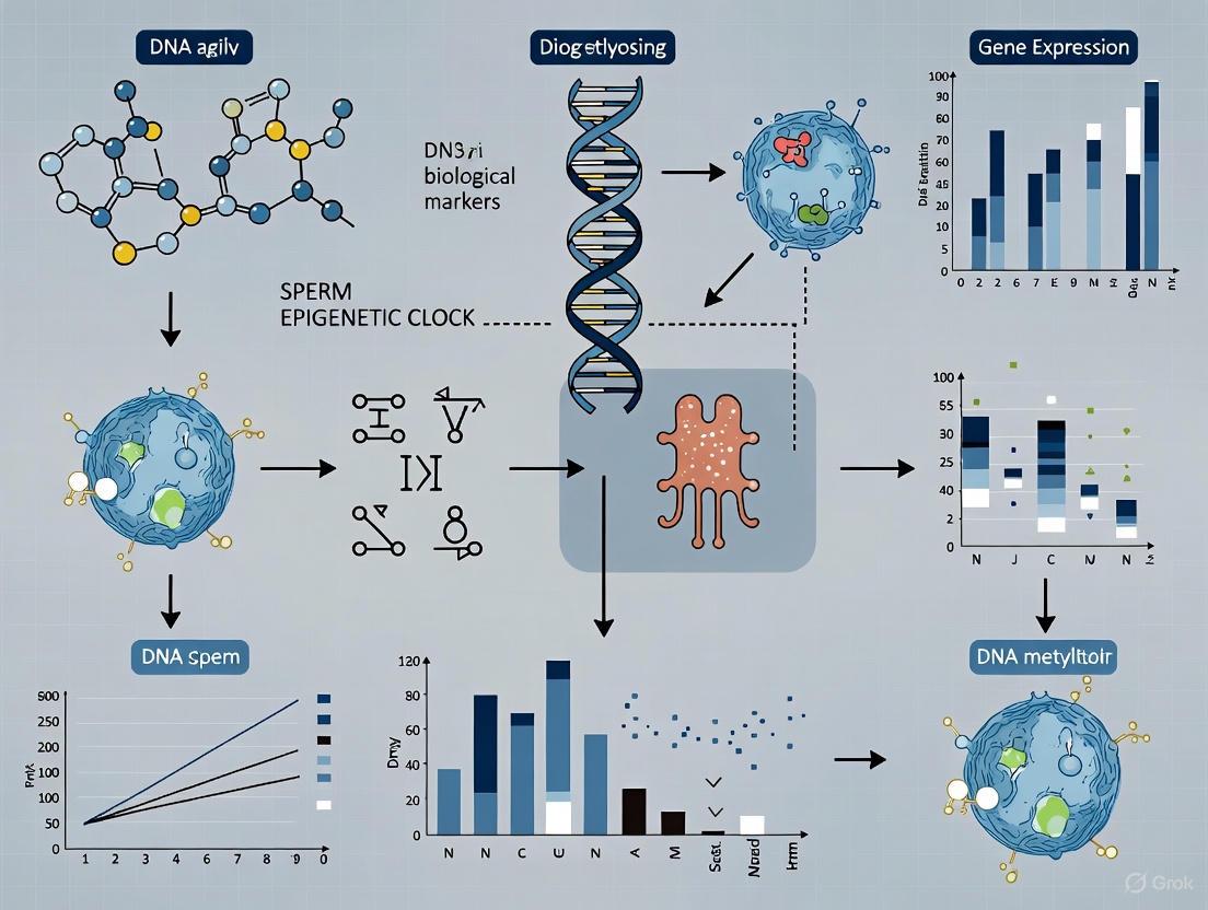
The sperm epigenetic clock is an emerging biomarker that predicts chronological and biological age based on defined patterns of DNA methylation in the male germline. Unlike somatic epigenetic clocks, it captures the unique aging trajectory of sperm, which is characterized by highly proliferative spermatogonial stem cells undergoing hundreds of replication cycles over a man's lifetime. This technical review synthesizes current methodologies for constructing and validating sperm-specific epigenetic clocks, detailing the specific genomic regions and bioinformatic pipelines used. We further explore the functional implications of advanced sperm epigenetic age, linking it to longer time-to-pregnancy, altered offspring neurodevelopment, and transgenerational disease risk. Within the broader context of biological aging research, the sperm epigenetic clock presents a novel tool for investigating paternal age effects and offers potential clinical applications in reproductive medicine and public health.
Aging is a multidimensional process characterized by a progressive decline in physiological function, with epigenetic alterations representing one of its fundamental hallmarks. While epigenetic clocks have been developed for numerous somatic tissues, the male germline presents a distinct and compelling model. The sperm epigenome is the product of extensive reprogramming and is fundamentally different from that of oocytes or somatic cells [1]. During a man's lifetime, spermatogonial stem cells undergo continuous divisions—from approximately 35 times at puberty to over 800 times by age 50 [1]. Each replication cycle introduces opportunities for both genetic and epigenetic replication errors. Critically, the error rate for copying epigenetic marks is at least an order of magnitude higher than for genetic information [1], making the sperm epigenome a particularly sensitive record of age-associated change.
The concept of a sperm epigenetic clock is built upon the measurable, age-associated alterations to the sperm DNA methylome. Early work by Horvath demonstrated that standard epigenetic clocks developed for somatic tissues failed to accurately predict age in testicular tissue or sperm [2], highlighting the need for a germline-specific model. Research has since confirmed that the nature of age-associated alterations in sperm is often opposite to that seen in somatic cells; where somatic tissues often show global hypomethylation with age, sperm exhibit both pronounced regional hypomethylation and more limited hypermethylation at specific loci [2]. This technical guide details the construction, validation, and application of the sperm epigenetic clock, positioning it within the broader framework of aging and reproductive research.
Technical Foundations of the Sperm Epigenetic Clock
Core DNA Methylation Dynamics in Aging Sperm
The sperm epigenetic clock is predicated on the identification of age-related differentially methylated regions (ageDMRs). Genome-wide studies have revealed that these changes are not random but exhibit specific patterns:
- Direction of Change: A predominant skew towards hypomethylation is observed with advancing age. One analysis of 73 sperm samples identified 1,162 (74%) significantly hypomethylated regions and 403 (26%) hypermethylated regions [1] [3].
- Genomic Location: Hypomethylated ageDMRs are preferentially located near transcription start sites (TSS), within exons and introns. In contrast, hypermethylated ageDMRs are typically found in gene-distal intergenic regions [1]. The median distance from hypomethylated ageDMRs to the nearest TSS is 1,368 bp, compared to 17,205 bp for hypermethylated ageDMRs [3].
- Functional Enrichment: AgeDMRs that have been replicated across multiple studies show significant functional enrichment in biological processes associated with embryonic development and the nervous system, including synapses and neurons [1]. This suggests a plausible mechanistic link between paternal age and offspring neurodevelopmental outcomes.
Table 1: Characteristics of Age-Related Differentially Methylated Regions (AgeDMRs) in Human Sperm
| Feature | Hypomethylated AgeDMRs | Hypermethylated AgeDMRs |
|---|---|---|
| Proportion | 74% (1,162 of 1,565 regions) [3] | 26% (403 of 1,565 regions) [3] |
| Genomic Context | Enriched near Transcription Start Sites (TSS), exons, introns [1] | Enriched in intergenic, gene-distal regions [1] |
| Median Distance to TSS | 1,368 bp [3] | 17,205 bp [3] |
| Methylation Level | More often in medium methylation range (20-80%) [3] | More often in high methylation range (>80%) [3] |
Predictive Modeling and Clock Construction
The construction of a sperm epigenetic clock involves applying statistical learning algorithms to DNA methylation data to derive a predictive model for chronological age.
- Feature Selection: Early models focused on 148 genomic regions previously identified as strong candidates due to their association with aging [2]. An optimized model can achieve high accuracy using just 51 robustly selected genomic regions [2].
- Algorithm and Training: Common approaches use linear regression models, such as those implemented with the
glmnetpackage in R [2]. Models can be trained on individual CpG sites or on mean beta-values calculated across predefined genomic regions, with the latter offering improved biological interpretability [2]. - Performance Metrics: A model developed from 329 sperm samples demonstrated a high correlation between predicted and chronological age (R² = 0.89), with a Mean Absolute Error (MAE) of 2.04 years and a Mean Absolute Percent Error (MAPE) of 6.28% [2]. Technical validation in an independent cohort confirmed similar accuracy (MAE = 2.37 years) and high precision between replicates [2].
This workflow outlines the primary steps for developing a sperm epigenetic clock, from sample processing to age prediction.
Methodological Guide: Key Experimental Protocols
Methylomic Profiling Technologies
Accurate construction of a sperm epigenetic clock relies on robust DNA methylation profiling. The following table summarizes essential reagents and solutions for these workflows.
Table 2: Research Reagent Solutions for Sperm Epigenomic Analysis
| Reagent / Material | Function in Protocol | Technical Notes |
|---|---|---|
| Illumina Infinium MethylationEPIC BeadChip | Genome-wide methylation profiling of ~850,000 CpG sites. | Standardized, high-throughput; covers enhancer regions. Ideal for initial clock development [4] [2]. |
| Bisulfite Conversion Kit | Deaminates unmethylated cytosines to uracils, allowing methylation quantification. | Critical step; requires optimized conversion efficiency. |
| Reduced Representation Bisulfite Sequencing (RRBS) | High-resolution methylation analysis of CpG-rich regions. | Cost-effective for targeted, high-depth analysis [1]. |
| Whole Genome Bisulfite Sequencing (WGBS) | Comprehensive, single-base resolution methylome mapping. | Gold standard for discovery but cost-prohibitive for large cohorts [1]. |
| Somatic Cell Lysis Buffer | Purifies sperm cells from seminal fluid and contaminating somatic cells. | Essential for sperm-specific methylation analysis [2]. |
| DNA Methylation Age Prediction Software (e.g., glmnet in R) | Statistical model to predict chronological age from methylation data. | Requires predefined CpG panels and trained models [2]. |
Detailed Protocol for Sperm Epigenetic Age Analysis
Step 1: Sample Preparation and DNA Extraction
- Collect semen samples after a recommended minimum of 2 days of sexual abstinence [5].
- Critical Step: Perform somatic cell lysis to isolate a pure sperm cell population. This is crucial because contamination with somatic cells, which have a different methylome, will confound results [2].
- Extract genomic DNA using standard phenol-chloroform or commercial column-based kits. Assess DNA quality and quantity via spectrophotometry (e.g., Nanodrop) and fluorometry (e.g., Qubit).
Step 2: DNA Methylation Profiling
- Subject 500 ng of high-quality sperm DNA to bisulfite conversion using a commercial kit. Monitor conversion efficiency with control DNA.
- For array-based approaches, hybridize converted DNA to the Illumina MethylationEPIC BeadChip [4] [2]. For sequencing-based approaches, prepare libraries for RRBS or WGBS [1].
Step 3: Bioinformatic Data Processing
- Process raw array data (IDAT files) using R packages like
minfifor background correction, normalization (e.g., functional normalization), and probe filtering (remove cross-reactive and SNP-affected probes). - For sequencing data, align bisulfite-treated reads to a reference genome (e.g., using Bismark) and calculate methylation levels (beta-values) for each CpG site.
Step 4: Age Prediction using the Epigenetic Clock Model
- Input the normalized beta-values from the pre-selected CpG sites or genomic regions into the validated prediction algorithm.
- Apply the model (e.g., the regional-level model based on 51 genomic regions [2]) to calculate the Sperm Epigenetic Age (SEA).
- Calculate "Age Acceleration" (AgeAccel) as the residual from regressing SEA on chronological age. A positive AgeAccel indicates an older biological age relative to chronological age.
Functional and Clinical Correlates of Sperm Epigenetic Aging
The sperm epigenetic clock is not merely a predictor of chronological age; it is functionally linked to key reproductive and offspring health outcomes.
Impact on Reproductive Success
Advanced sperm epigenetic aging is associated with diminished reproductive potential:
- Longer Time-to-Pregnancy (TTP): In a prospective cohort study of couples from the general population, advanced SEA was negatively associated with fecundability. Each unit increase in SEA was linked to a 17% lower cumulative probability of pregnancy within 12 months (Fecundability Odds Ratio = 0.83) [5].
- Assisted Reproductive Technology (ART) Outcomes: Male body mass index (BMI) and diet, which can influence the sperm epigenome, correlate with embryo quality and Intracytoplasmic Sperm Injection (ICSI) outcomes [4]. The sperm epigenetic clock shows promise as a biomarker to improve ART success rates [4] [5].
Implications for Offspring Health
The age-related epigenetic alterations in sperm can be transmitted to the embryo, potentially influencing its developmental trajectory and long-term health.
- Neurodevelopmental Trajectory: Functionally enriched ageDMR genes are significantly associated with biological processes related to the nervous system and synapses [1]. This finding supports the hypothesis that paternal age effects on the sperm methylome contribute to the risk of neurodevelopmental disorders in offspring, such as autism and schizophrenia [1].
- Gestational Age: Advanced SEA in fathers has been associated with a shorter gestational age in resulting pregnancies (-2.13 days), indicating a potential impact on fetal development [5].
This diagram illustrates the functional pathway from paternal factors and age to sperm epigenetic alterations and their potential consequences.
Modifiability and Intervention Strategies
A key advantage of epigenetic biomarkers is their potential reversibility. Research indicates that the sperm epigenetic clock is dynamic and can be influenced by lifestyle and pharmacological interventions.
- Lifestyle Factors: Paternal smoking has been consistently linked to advanced SEA [5] [2]. Obesity and high-fat diets are also associated with altered sperm methylation and sncRNA profiles [4]. Consequently, interventions such as smoking cessation, weight management, and a balanced diet (including adequate folate) are proposed as means to mitigate adverse sperm epigenetic aging [4].
- Pharmacological Interventions: While direct studies on sperm are still emerging, research in somatic tissues shows that certain compounds can modulate epigenetic age. For instance, the drug semaglutide was associated with decreased epigenetic age in multiple organ-system clocks in a clinical trial [6]. The TRIIM trial demonstrated that a regimen involving growth hormone could reduce epigenetic age by approximately 1.5 years, accompanied by thymic regeneration [6].
The sperm epigenetic clock, defined by specific DNA methylation patterns, establishes a direct and measurable link between paternal chronological age, biological aging of the germline, and subsequent health outcomes in the next generation. Its precision, with a mean absolute error of just over two years, makes it a powerful tool for both clinical and research applications.
Future work in this field should focus on:
- Standardization and Validation: Implementing standardized epigenome assays (e.g., MethylationEPIC, small-RNA profiling) in andrology and ART workflows requires large, diverse, longitudinal cohorts to confirm associations and establish causality [4].
- Mechanistic Insight: Further research is needed to elucidate the precise molecular mechanisms by which sperm epigenetic signatures influence embryonic gene regulation and long-term offspring phenotypes.
- Interventional Trials: Clinical trials testing the effects of preconception lifestyle modifications or therapeutic compounds on sperm epigenetic age and subsequent reproductive and child health outcomes are the critical next step [4].
In the broader context of biological aging research, the sperm epigenetic clock offers a unique window into how aging of the germline—a lineage that ensures genetic and epigenetic continuity—is manifested and measured. It underscores the importance of the paternal preconceptual environment and provides a actionable biomarker for improving reproductive success and potentially safeguarding the health of future generations.
Spermatogenesis exhibits unique DNA methylation dynamics that fundamentally differ from patterns observed in somatic aging. While somatic tissues typically display progressive, stochastic epigenetic alterations, the male germline undergoes a precisely orchestrated cascade of methylation reprogramming events designed to preserve transgenerational genomic integrity. This whitepaper synthesizes current research on sperm-specific epigenetic clocks, stage-specific methylation dynamics during gametogenesis, and the implications for paternal age-related disease transmission. We present comprehensive quantitative comparisons, detailed experimental methodologies, and essential research tools that define this emerging field at the intersection of reproductive biology and epigenetic aging research.
The establishment and maintenance of DNA methylation patterns follow fundamentally different rules in the male germline compared to somatic tissues. While somatic epigenetic clocks reflect cumulative environmental exposures and stochastic aging processes, spermatogenesis involves a highly ordered, programmed series of epigenetic events essential for producing functional gametes and ensuring proper embryonic development [7] [8]. This distinction forms the critical foundation for understanding how paternal age impacts offspring health and why sperm-specific epigenetic clocks require specialized development.
The sperm epigenome is uniquely configured, characterized by extensive hypermethylation of intergenic regions coupled with strategic hypomethylation at developmental gene promoters [7] [9]. This configuration differs dramatically from somatic cells, where aging typically manifests as global hypomethylation with localized hypermethylation at specific CpG islands. During spermatogenesis, germ cells undergo two major waves of epigenetic reprogramming: first in primordial germ cells, and later during the mitosis-to-meiosis transition, establishing a unique epigenetic landscape that predetermines nucleosome retention sites in mature sperm [8] [10].
Quantitative Comparison of Methylation Patterns
Table 1: Genome-Wide Methylation Alterations in Sperm vs. Somatic Aging
| Feature | Spermatogenesis | Somatic Aging | Experimental Evidence |
|---|---|---|---|
| Global Trend | Dynamic reprogramming followed by stabilization | Progressive, cumulative drift | MCC-seq in human sperm [9] |
| Hypermethylation | 62% of age-related CpGs; distal to genes | Varies by tissue; often at CpG islands | 150,000 age-related CpGs identified [9] |
| Hypomethylation | 38% of age-related CpGs; near transcription start sites | Global loss in intergenic regions | Sperm analysis in aged men [9] |
| Genomic Distribution | Non-random clusters (e.g., chr4, chr16) | More evenly distributed | Chromosome density analysis [9] |
| Functional Association | Developmental genes, neurodevelopmental pathways | Disease-specific genes, cancer pathways | Gene ontology analysis [9] |
Table 2: DNA Methyltransferase Expression and Function Across Tissues
| Enzyme | Role in Spermatogenesis | Role in Somatic Aging | Knockout Consequences |
|---|---|---|---|
| DNMT1 | Upregulated in spermatocytes; maintenance methylation | General maintenance methylation; decreased activity with age | Spermatogonial apoptosis; lack of genomic imprinting [7] |
| DNMT3A/B | De novo methylation; expression patterns unique to germline | De novo methylation; altered expression with aging | Impaired spermatogenesis [7] |
| DNMT3L | Critical for meiosis; expressed predominantly in germ cells | Limited expression in somatic tissues | Sterility; meiotic arrest [7] |
| TET Family | Active demethylation in PGCs; role in meiosis | Varied roles in somatic maintenance | Defects in epigenetic reprogramming [8] |
Stage-Specific Methylation Dynamics During Spermatogenesis
Developmental Timeline and Key Transitions
The journey from primordial germ cell (PGC) to mature sperm involves precisely timed epigenetic transitions that ensure proper erasure and re-establishment of methylation marks. The most dramatic epigenetic alterations occur during the early developmental stages, particularly in PGCs and spermatogonial stem cells [8]. Research utilizing differential DNA methylation region (DMR) analysis has demonstrated that the number of DMRs is highest in comparisons involving mature PGCs, prospermatogonia, and spermatogonia, indicating intense epigenetic remodeling during these stages [8].
A critical window of epigenetic reprogramming occurs during the mitosis-to-meiosis transition, where site-specific DNA demethylation presets nucleosome retention sites in mature sperm [10]. This preprogrammed demethylation is not observed in somatic aging and represents a unique feature of germline development. The established hypomethylated sites subsequently determine where histones will be retained (rather than replaced by protamines) in mature sperm, creating a blueprint for embryonic gene activation after fertilization [10].
Diagram 1: Spermatogenesis Methylation Dynamics Timeline. The mitosis-to-meiosis transition represents a critical window for site-specific demethylation that predetermines nucleosome retention in mature sperm [10].
Mechanistic Insights: DNMT Dynamics and Enzymatic Control
The unique methylation patterns in spermatogenesis are orchestrated by specialized expression and regulation of DNA methyltransferases (DNMTs). DNMT1, the primary maintenance methyltransferase, shows robust expression in early spermatocytes but is significantly reduced in pachytene stage spermatocytes [7]. This regulated reduction contributes to the transient global demethylation observed during meiosis. Heterozygous DNMT1 mice maintain normal reproductive capacity, suggesting that half-dose expression suffices for maintenance functions [7].
DNMT3A and DNMT3B demonstrate developmentally programmed expression patterns, with DNMT3A upregulated prior to birth and during early postnatal life, while DNMT3B follows an opposite pattern [7]. The catalytically inactive cofactor DNMT3L plays an unexpectedly critical role in spermatogenesis, with knockout models showing smaller testes, negligible sperm production, and sterility due to meiotic arrest [7]. DNMT3L expression peaks at 15.5 days post-fertilization and declines after birth, highlighting its role in establishing methylation patterns rather than maintaining them in mature germ cells [7].
Sperm Epigenetic Clocks: Specialized Tools for Germline Aging
Development and Validation of Sperm-Specific Clocks
Unlike somatic epigenetic clocks that predict chronological age across multiple tissues, sperm-specific clocks require specialized development due to the unique epigenome of male gametes. The Jenkins sperm clock was developed specifically to address the inaccuracy of somatic clocks (e.g., Horvath clock) in predicting germline age [11]. This specialized tool demonstrates how methylation patterns at specific CpG sites can predict chronological age in sperm with comparable accuracy to somatic clocks in blood and other tissues.
The Germline Age Differential (GLAD) metric quantifies epigenetic age acceleration in sperm, calculated as GLAD = (predicted age/actual age) - 1 [12]. Positive GLAD values indicate accelerated epigenetic aging in sperm compared to chronological age. This measure has revealed that oligozoospermic men exhibit significant age acceleration in their sperm (mean GLAD = 0.078) compared to normozoospermic men (mean GLAD = -0.017), while showing no equivalent acceleration in blood [11]. This tissue-specific aging pattern underscores the unique susceptibility of the germline to certain disease states.
Environmental Accelerants of Sperm Epigenetic Aging
Emerging evidence indicates that environmental exposures can selectively accelerate epigenetic aging in sperm without parallel effects in somatic tissues. Recent mouse studies have identified mTOR-dependent disruption of blood-testis barrier integrity as a novel mechanism mediating environmental effects on sperm epigenetic aging [13]. Exposure to heat stress (31.5°C or 34.5°C) or cadmium chloride (2 mg/kg body weight) significantly increased sperm epigenetic age in mouse models, demonstrating how environmental toxicants can specifically target the germline epigenome [13].
Human studies of World Trade Center-exposed individuals demonstrated significant epigenetic aging acceleration in blood using multiple epigenetic clocks (Hannum, Horvath, and PhenoAge) [14], highlighting how environmental exposures can accelerate aging in somatic tissues. However, the tissue-specific nature of these effects is evident in conditions like oligozoospermia, where accelerated epigenetic aging occurs specifically in sperm without parallel acceleration in blood [11], suggesting distinct regulatory mechanisms between germline and somatic aging.
Table 3: Sperm Epigenetic Age Acceleration in Pathological Conditions
| Condition/Exposure | GLAD Value | Blood Epigenetic Age | Biological Significance |
|---|---|---|---|
| Normozoospermia | -0.017 (reference) | No acceleration | Baseline germline health [11] |
| Oligozoospermia | 0.078 (accelerated) | No acceleration | Tissue-specific aging [11] |
| Advanced Paternal Age | ~1.4 years older prediction with high BMI | Not assessed | Subtle acceleration trend [12] |
| Heat Stress Exposure | Significant acceleration (mouse model) | Not assessed | mTOR-mediated mechanism [13] |
| Cadmium Exposure | Significant acceleration (mouse model) | Not assessed | Blood-testis barrier disruption [13] |
Experimental Methodologies for Sperm Methylation Analysis
Critical Protocol: Addressing Somatic Contamination
A paramount concern in sperm methylation studies is the potential contamination by somatic cells, which possess dramatically different methylation patterns that can confound results. Semen samples from oligozoospermic individuals present particular vulnerability to this artifact due to higher relative proportions of somatic cells [15]. A comprehensive approach to eliminate somatic contamination includes:
- Microscopic Examination: Initial visual inspection to identify somatic cells, though this method fails to detect contamination below 5% [15].
- Somatic Cell Lysis Buffer (SCLB) Treatment: Incubation with SCLB (0.1% SDS, 0.5% Triton X-100 in ddH2O) for 30 minutes at 4°C, followed by centrifugation and repeat inspection [15].
- Epigenetic Quality Control: Analysis of established somatic methylation markers such as DLK1, which shows >80% methylation in blood but <20% methylation in pure sperm [15] [12]. Research has identified 9,564 CpG sites with high methylation in blood (>80%) and low methylation in sperm (<20%) that serve as contamination biomarkers [15].
- Analytical Thresholding: Implementation of a 15% methylation cutoff during data analysis to exclude samples with residual contamination [15].
Diagram 2: Sperm Sample Purity Workflow. Comprehensive approach to eliminate somatic cell contamination in sperm epigenetic studies, incorporating physical removal and epigenetic verification [15].
Advanced Methylation Profiling Techniques
The unique architecture of the sperm epigenome necessitates specialized profiling approaches that address its distinct characteristics:
MethylC-capture sequencing (MCC-seq) with sperm-specific panels provides targeted assessment of dynamic regions, offering superior coverage of sperm-specific epigenomic features compared to standard arrays [9]. This approach enables high-resolution identification of age-related epigenetic alterations, having revealed more than 150,000 age-related CpG sites from 2.65 million covered sites in human sperm [9].
MethylCap-seq utilizes methyl-CpG-binding domain (MBD) capture to specifically detect 5mC without confusion with 5hmC, which is particularly valuable during meiotic phases when oxidation markers may be present [10]. This method has been instrumental in identifying site-specific methylation changes during the mitosis-to-meiosis transition that predetermine nucleosome retention sites [10].
Quality Control Protocols must include verification of imprinting control regions. Bisulfite pyrosequencing of paternally methylated loci (H19, DLK1/GTL2-IG DMR) and maternally methylated loci (MEST, KCNQ1OT1) confirms sample purity and proper imprinting status [9].
The Scientist's Toolkit: Essential Research Reagents
Table 4: Essential Research Reagents for Sperm Methylation Studies
| Reagent/Assay | Specific Application | Function and Importance |
|---|---|---|
| Somatic Cell Lysis Buffer (0.1% SDS, 0.5% Triton X-100) | Sperm purification | Selectively lyses somatic contaminants while preserving sperm integrity [15] |
| Infinium MethylationEPIC BeadChip | Genome-wide methylation screening | Interrogates 866,562 CpG sites; effective for initial surveys [14] |
| MethylCap-seq Library Prep | Stage-specific methylation analysis | MBD-based capture of methylated DNA; distinguishes 5mC from 5hmC [10] |
| Custom Sperm Capture Panel (MCC-seq) | Sperm-specific epigenomic profiling | Targets dynamic regions; covers 2.65M CpG sites with 20-30x coverage [9] |
| DLK1 Locus Assay | Somatic contamination detection | 14 CpG sites; highly methylated in somatic cells, unmethylated in sperm [12] |
| Bisulfite Pyrosequencing Reagents | Imprinting validation | Confirms methylation status at H19, MEST, and other imprinted loci [9] |
| UHRF1/DNMT1 Antibodies | Meiotic regulation studies | Identifies maintenance methylation machinery in spermatogenesis [10] |
The distinct methylation dynamics governing spermatogenesis create a unique epigenetic aging paradigm that differs fundamentally from somatic aging patterns. While somatic tissues accumulate stochastic methylation changes over time, the male germline undergoes precisely programmed epigenetic reprogramming with established susceptibility to environmental accelerants. The development of sperm-specific epigenetic clocks and purification protocols enables accurate assessment of germline aging, revealing tissue-specific acceleration in conditions like oligozoospermia and following toxicant exposures.
These advances carry significant implications for both clinical andrology and transgenerational inheritance research. The ability to measure sperm epigenetic age acceleration provides a novel biomarker for male fertility assessment, potentially predicting embryonic developmental competence and offspring health outcomes. Furthermore, understanding the mechanisms behind environmental acceleration of sperm epigenetic aging opens therapeutic avenues for mitigating paternal age-related disease risks. As research progresses, integrating sperm epigenetic clocks into broader biological aging models will be essential for comprehensive understanding of how paternal germline aging impacts subsequent generations.
Aging is accompanied by highly reproducible changes in DNA methylation (DNAm) at specific cytosine-phosphate-guanine (CpG) sites, forming the basis of epigenetic clocks that can predict biological age [14] [16]. In male germ cells, age-associated epigenetic changes are of particular concern given the modern trend of delayed parenthood and the potential implications for offspring health and development [17]. Unlike somatic tissues, sperm exhibits unique methylation dynamics during spermatogenesis, necessitating the identification of sperm-specific age-related CpG (AR-CpG) sites for accurate age estimation and biological aging assessment [18] [19]. This technical guide comprehensively details the key genomic loci, methodologies, and analytical frameworks for studying AR-CpG sites in sperm, positioning this research within the broader context of epigenetic clock development and male reproductive aging.
Genome-Wide Discovery of Sperm AR-CpG Sites
Methodological Approaches for Marker Identification
The discovery of sperm-specific AR-CpG sites has evolved significantly with advancing genomic technologies. Early studies relied on methylation microarrays (Illumina Infinium 450K and 850K BeadChips), which provided limited coverage of potentially relevant genomic regions [20] [19]. More recently, double-enzyme reduced representation bisulfite sequencing (dRRBS) has enabled comprehensive methylome-wide association studies, generating data for over 4 million CpG sites per sample at sufficient depth for robust analysis [18] [20]. This approach revealed that more than 95% of age-informative CpGs in semen were not covered by conventional methylation microarrays, explaining previous limitations in prediction accuracy [18].
The standard workflow involves a two-stage validation process beginning with dRRBS discovery in stratified age groups, followed by targeted validation using bisulfite amplicon sequencing (BSAS) or multiplex PCR-based approaches [18] [20]. This combination allows for both broad discovery and precise quantification of methylation levels at candidate loci.
Key Genomic Loci and Their Characteristics
Research has identified numerous AR-CpG sites with significant correlations to chronological age. The following table summarizes the most robustly validated genomic loci and their characteristics:
Table 1: Key Age-Related CpG Loci in Human Sperm
| Genomic Coordinate | Associated Gene | CpG Identifier | Age Correlation ( | rho | ) | Methylation Trend with Age | Validation Method |
|---|---|---|---|---|---|---|---|
| chr2:129071885 | - | cg19998819 | 0.81 | Not specified | BSAS [18] | ||
| chr3:123069181 | - | cg06979108 | Validated in multiple studies | Not specified | SNaPshot, MPS [19] | ||
| chr14:100253471 | - | cg12837463 | Validated in multiple studies | Not specified | SNaPshot, MPS [19] | ||
| chr1:1899049 | PDE4C | cg17861230 | Significant in blood studies | Gain | EPIC Array [21] | ||
| Multiple sites | ELOVL2, FHL2, KLF14, TRIM59, C1orf132 | Multiple | Used in simplified clocks | Tissue-dependent | Pyrosequencing [22] |
Beyond individual CpGs, genomic regions exhibiting spatial clustering of AR-CpG sites, such as those associated with the ELOVL2, FHL2, and PDE4C genes, represent particularly promising targets [20] [21]. These regions often show coordinated methylation changes and may represent epigenetic hubs with functional significance in the aging process.
Quantitative Age Estimation Models and Performance
Model Architectures and Algorithm Selection
Various computational approaches have been employed to translate sperm DNA methylation patterns into accurate age predictions. Multiple linear regression (MLR) models offer simplicity and interpretability, while more complex machine learning algorithms, particularly random forest (RF) regression, often demonstrate superior accuracy in handling the non-linear relationships present in epigenetic data [18].
The model development process typically employs a repeated nested cross-validation framework (e.g., 10-fold outer CV with 10-fold inner CV, repeated 10 times) to ensure robust performance estimates and avoid overfitting [18]. This rigorous validation approach provides realistic expectations of model performance when applied to new samples.
Comparative Performance of Sperm Age Estimators
The accuracy of sperm epigenetic age estimators has improved significantly with the identification of sperm-specific markers and refinement of modeling techniques. The following table compares the performance of recently published models:
Table 2: Performance Comparison of Sperm DNA Methylation Age Estimation Models
| Model Description | Sample Size | CpG Count | Algorithm | Mean Absolute Error (Years) | Correlation (R²) | Reference |
|---|---|---|---|---|---|---|
| dRRBS-based model | 247 | 9 | Random Forest | 3.30 | 0.76 | [18] |
| Sperm-specific model | 253 | 14 | Multiple | 2.89 (sperm) / 3.58 (semen) | 0.81 (sperm) | [19] |
| Germ Line Age Calculator | 329 | 264 (51 regions) | Generalized Linear Model | 2.04 (training) / 2.37 (test) | 0.89 | [19] |
| Lee et al. model | 31 (training) / 32 (test) | 3 | Multiple Linear Regression | 5.4 (test) | Not specified | [19] |
Notably, models utilizing sperm-specific AR-CpG markers consistently outperform those developed for somatic tissues or those using non-specific semen markers [19]. This highlights the importance of cell-type specific epigenetic signatures in age estimation accuracy.
Experimental Protocols for AR-CpG Analysis
Sample Processing and Bisulfite Conversion
The accurate quantification of DNA methylation patterns requires meticulous sample processing. The foundational step involves bisulfite conversion, where unmethylated cytosines are deaminated to uracils while methylated cytosines remain protected [14] [20]. This conversion allows for the discrimination between methylated and unmethylated alleles in subsequent analyses.
For sperm samples, additional somatic cell removal is critical, as contamination with white blood cells or other somatic cells introduces confounding methylation signals. Efficiency of somatic cell depletion can be verified by analyzing the DLK1 locus, which is highly methylated in somatic cells but essentially unmethylated in sperm cells [12].
Methylation Assessment Techniques
Table 3: Methodologies for DNA Methylation Analysis in Sperm Research
| Method | Throughput | Coverage | Cost | Primary Application | Key Considerations |
|---|---|---|---|---|---|
| dRRBS | High | ~4 million CpGs | Moderate | Genome-wide discovery [18] | Identifies novel sites beyond microarray coverage |
| Bisulfite Amplicon Sequencing (BSAS) | Medium | Targeted | Lower | Validation and quantification [18] | High accuracy for specific loci |
| Illumina MethylationEPIC BeadChip | High | ~860,000 CpGs | Higher | Population studies [14] [19] | Limited to predefined CpG set |
| Pyrosequencing | Low | Targeted | Lower | Clinical validation [22] | Quantitative, cost-effective for few sites |
| SNaPshot | Medium | Targeted | Lower | Forensic applications [19] | Multiplexing capability |
Each method offers distinct advantages depending on the research objectives, with a typical workflow progressing from broad discovery (dRRBS or EPIC array) to targeted validation (BSAS or pyrosequencing) [18] [19].
Research Reagent Solutions and Experimental Tools
Table 4: Essential Research Reagents for Sperm AR-CpG Analysis
| Reagent/Kit | Primary Function | Specific Application | References |
|---|---|---|---|
| DNeasy Blood & Tissue Kit | DNA extraction from white blood cells | Isolating high-quality DNA for methylation analysis | [22] |
| Infinium MethylationEPIC BeadChip v2.0 | Epigenome-wide profiling | Simultaneous analysis of ~860,000 CpG sites | [14] |
| Bisulfite Conversion Kits | Chemical conversion of unmethylated cytosines | Sample preparation for methylation detection | [14] [22] |
| dCas9-DNMT3A/CRISPRoff | Targeted epigenetic editing | Functional validation of AR-CpG sites | [21] |
| Pyrosequencing Systems | Quantitative methylation analysis | Validation of specific CpG sites | [16] [22] |
Visualization of Experimental Workflows
Sperm AR-CpG Discovery and Validation Pipeline
Diagram 1: AR-CpG Discovery Workflow
Epigenetic Editing to Validate AR-CpG Function
Diagram 2: AR-CpG Functional Validation
Implications for Offspring Health and Clinical Applications
Advanced paternal age and associated sperm epigenetic changes have been linked to increased risks for neurodevelopmental disorders in offspring, including autism spectrum disorders, through the transmission of altered epigenetic information [17]. The sperm epigenome appears to be vulnerable to environmental exposures, with studies demonstrating that World Trade Center-exposed individuals showed significant epigenetic aging acceleration in blood samples [14]. While similar studies in sperm are limited, this highlights the potential for environmental factors to accelerate epigenetic aging.
In clinical reproduction, sperm epigenetic aging clocks show promise as novel biomarkers predicting pregnancy success and time-to-pregnancy in couples not seeking fertility treatment [23]. Each year of increased sperm epigenetic age was associated with a 17% lower cumulative probability of pregnancy after 12 months, underscoring the clinical significance of these markers [23].
Future Directions and Research Challenges
Future research must address the tissue specificity of epigenetic aging signals, as current models demonstrate varying accuracy across different cell types [19]. The development of multiplexed epigenetic editing approaches will help establish causal relationships between specific AR-CpG sites and functional aging phenotypes [21]. Additionally, longitudinal studies are needed to track intraindividual changes in sperm epigenetic age and their relationship to environmental exposures, lifestyle factors, and health outcomes.
The integration of sperm epigenetic clocks into clinical practice requires standardization of analytical methods and establishment of reference ranges across diverse populations. As research progresses, these epigenetic biomarkers hold immense potential for both forensic applications and clinical assessment of male reproductive health.
Emerging research establishes the male germline as a novel biomarker for systemic aging, revealing that sperm epigenetic and mutational landscapes provide a sensitive readout of biological age. This whitepaper synthesizes cutting-edge findings demonstrating that sperm epigenetic clocks not only predict reproductive outcomes but also reflect organism-wide aging processes. Advanced molecular techniques including duplex sequencing and epigenetic profiling have identified specific mutational signatures and age-associated methylation changes in sperm that correlate with both declining fertility and broader health indicators. These findings position sperm analysis as a unique portal for investigating aging mechanisms and developing interventions that target reproductive and systemic aging simultaneously, offering drug development professionals new biomarkers and therapeutic targets for age-related conditions.
The strong correlation between chronological age and DNA methylation patterns has enabled the development of epigenetic "clocks" as powerful biomarkers of biological aging across somatic tissues. Recent evidence indicates that male germ cells exhibit their own distinct aging signatures that reflect both reproductive and overall health status. Unlike somatic clocks, sperm-specific epigenetic clocks capture unique aspects of the aging process in the male germline, providing critical insights into how systemic aging manifests in reproductive cells [5].
The transmission of genetic and epigenetic information to subsequent generations positions sperm as a particularly sensitive indicator of aging-related damage accumulation. The continuously dividing nature of spermatogonial stem cells throughout a male's lifespan makes them vulnerable to both replicative and environmental insults, resulting in measurable molecular changes that parallel systemic aging processes. These changes have clinical significance beyond reproduction, as advanced sperm epigenetic aging has been associated with shorter gestational age and other adverse health outcomes in offspring, suggesting connections to broader physiological decline [5].
This whitepaper examines the current state of research linking sperm biological age to overall health, detailing the molecular mechanisms, measurement methodologies, and potential applications for pharmaceutical development and clinical practice.
Quantitative Data on Sperm Aging Parameters
Age-Related Changes in Sperm Quality and DNA Integrity
Table 1: Age-associated decline in conventional sperm parameters
| Parameter | Age Group (Years) | Mean Value | Change vs. Youngest Group | Study Details |
|---|---|---|---|---|
| Semen Volume | 20-24 | 3.45 mL | Reference (n=102) | Analysis of 6,805 samples [24] |
| 35-39 | 2.91 mL | -15.7% | ||
| >40 | 2.82 mL | -18.3% | ||
| Sperm Progressive Motility | 20-24 | 56.93% | Reference (n=102) | Analysis of 6,805 samples [24] |
| 35-39 | 50.61% | -11.1% | ||
| >40 | 48.17% | -15.4% | ||
| Sperm Total Motility | 20-24 | 64.97% | Reference (n=102) | Analysis of 6,805 samples [24] |
| 35-39 | 59.31% | -8.7% | ||
| >40 | 57.03% | -12.2% | ||
| Sperm DNA Fragmentation Index (DFI) | 20-24 | 18.20% | Reference (n=25) | Analysis of 1,253 samples [24] |
| 35-39 | 22.87% | +25.7% | ||
| >40 | 25.36% | +39.3% |
Note: * indicates statistical significance (P < 0.01)*
Mutational Burden and Epigenetic Changes in Aging Sperm
Table 2: Molecular alterations in aging sperm
| Parameter | Accumulation Rate | Age Correlation | Technical Approach | Study |
|---|---|---|---|---|
| Single Nucleotide Variants (SNVs) | 1.67/year/haploid genome (95% CI: 1.41-1.92) | Linear (r = 0.91) | Whole-genome NanoSeq (n=81) | [25] |
| Insertion-Deletion Mutations (Indels) | 0.10/year/haploid genome (95% CI: 0.06-0.15) | Linear | Whole-genome NanoSeq (n=81) | [25] |
| Age-Related Differentially Methylated Regions (ageDMRs) | 1,565 significant regions (74% hypomethylated, 26% hypermethylated) | Strong correlation with age | RRBS (n=73 samples) | [3] |
| Sperm Epigenetic Age Acceleration | FOR=0.83 (95% CI: 0.76, 0.90) for time-to-pregnancy | P = 1.2×10⁻⁵ | EPIC array + machine learning (n=379) | [5] |
Methodologies for Assessing Sperm Biological Age
Sperm Epigenetic Clock Construction
Experimental Protocol: Sperm DNA methylation analysis using ensemble machine learning [5]
- Sample Collection and DNA Extraction: Collect semen samples after minimal 2-day abstinence. Extract DNA from purified sperm cells to minimize somatic contamination. For the LIFE Study, 379 samples were analyzed from male partners of couples discontinuing contraception.
- DNA Methylation Profiling: Assess sperm DNA methylation using Illumina EPIC BeadChip arrays covering >850,000 CpG sites. Process samples in duplicate with appropriate controls.
- Age Prediction Modeling: Employ ensemble machine learning algorithms trained on chronological age using methylation data. Utilize ten-fold cross-validation to assess prediction accuracy.
- Clock Validation: Validate clocks in independent cohorts (e.g., SEEDS IVF cohort, n=173). Assess correlation between predicted and chronological age (r=0.83-0.91 in validation studies).
- Reproductive Outcomes Analysis: Evaluate association between sperm epigenetic age (SEA) and time-to-pregnancy using discrete-time proportional hazards models with adjustment for female age, BMI, and lifestyle factors.
Duplex Sequencing for Germline Mutations
Experimental Protocol: NanoSeq for sperm mutation detection [25]
- Sample Preparation: Collect bulk semen samples (n=81) with sperm counts >1 million/mL to avoid somatic cell contamination. Include matched blood samples (n=119) as controls.
- Library Preparation and Sequencing: Perform whole-genome NanoSeq using duplex sequencing approach with mean duplex coverage of 3.7 dx in sperm. This approach sequences both DNA strands to achieve error rates <5×10⁻⁹ per base pair.
- Variant Calling and Filtering: Identify single nucleotide variants and indels while excluding inherited germline variants using matched blood samples. Implement strict quality controls and remove potential artifacts.
- Mutational Signature Analysis: Deconstruct mutational signatures using non-negative matrix factorization. Compare signatures to known COSMIC reference signatures.
- Selection Analysis: Calculate dN/dS ratios using modified dNdScv algorithm accounting for duplex sequencing coverage, CpG methylation levels, and pentanucleotide context.
Analysis of Age-Related DNA Methylation Changes
Experimental Protocol: Reduced representation bisulfite sequencing (RRBS) for sperm epigenomics [3]
- Cohort Selection: Recruit men from fertility centers (n=73) with age range 25.8-50.4 years. Collect detailed clinical parameters including BMI, semen quality, and pregnancy outcomes.
- Library Preparation: Perform RRBS using MspI digestion for consistent genome coverage. Conduct bisulfite conversion with efficiency controls.
- Sequencing and Alignment: Sequence on Illumina platforms. Align to reference genome using Bismark or similar bisulfite-aware aligners.
- Differential Methylation Analysis: Identify age-related differentially methylated regions (ageDMRs) using statistical models with false discovery rate (FDR) correction. Validate imprinting control regions to exclude somatic contamination.
- Functional Annotation: Annotate ageDMRs to genomic features including proximity to transcription start sites, gene bodies, and regulatory elements. Perform pathway enrichment analysis.
Mechanisms Linking Sperm Aging to Systemic Health
Shared Aging Signatures Between Sperm and Somatic Tissues
Advanced analytical approaches have revealed striking similarities between aging processes in sperm and somatic tissues. Whole-genome NanoSeq of sperm and matched blood samples demonstrates that while sperm accumulate mutations at a significantly slower rate (7.6-fold fewer substitutions per year compared to blood), they share two primary clock-like mutational signatures: SBS1 (spontaneous deamination of 5-methylcytosine) and SBS5 (unknown etiology but correlated with age) [25]. The conservation of these signatures suggests common underlying aging mechanisms across tissues, with SBS1 contributing approximately 16% and SBS5 approximately 84% of sperm mutations.
Positive selection acting on spermatogonial stem cells represents a unique aspect of germline aging not observed in somatic tissues. Deep targeted sequencing has identified 40 genes under significant positive selection in the male germline, most associated with developmental disorders or cancer predisposition. This selection results in a 2-3-fold increased risk of transmitting disease-causing mutations, with approximately 3-5% of sperm from middle-aged to older individuals carrying pathogenic mutations across the exome [25]. These findings illuminate how germline selection dynamics contribute to increased disease risk for offspring of older fathers.
Environmental and Pharmaceutical Accelerators of Sperm Aging
Multiple exogenous factors have been identified that accelerate sperm epigenetic aging, creating discordance between chronological and biological age. Current smoking status significantly advances sperm epigenetic age (P < 0.05), demonstrating how environmental exposures can accelerate reproductive aging [5]. Additionally, various pharmaceutical medications have been associated with impaired male fertility through diverse mechanisms including hormonal disruption, direct toxicity to germ cells, and induction of oxidative damage.
Table 3: Medications and substances impacting sperm quality
| Medication/Substance | Class | Impact on Sperm | Proposed Mechanism | Evidence Level |
|---|---|---|---|---|
| Paroxetine | SSRI antidepressant | ↑ DNA fragmentation (13.8% to 30.3%) | Serotonin pathway disruption, oxidative stress | Clinical trial (n=35) [26] |
| Calcium Channel Blockers | Antihypertensive | ↓ Motility, ↓ acrosome reaction | Calcium signaling disruption, altered membrane composition | In vitro & clinical studies [26] |
| Methamphetamine | Stimulant | ↑ Germ cell apoptosis, ↓ proliferation | Oxidative damage, direct genotoxicity | Animal studies [27] |
| Cocaine | Stimulant | ↓ Concentration, ↑ aberrant morphology | Caspase-mediated apoptosis, mitochondrial dysfunction | Animal & human studies [27] |
| HAART | Antiretroviral | Variable effects on parameters | Mitochondrial toxicity, oxidative stress | Clinical studies [26] |
Psychoactive substances induce testicular toxicity through promotion of ROS-dependent oxidative damage, inflammation, and apoptosis. These drugs suppress the hypothalamic-pituitary-testicular axis, resulting in suppressed circulating androgens, impaired spermatogenesis, and reduced sperm quality [27]. The convergence of multiple pharmacological classes on oxidative stress pathways suggests this as a common mechanism for accelerated sperm aging.
The Scientist's Toolkit: Essential Research Reagents
Table 4: Key research reagents for sperm aging studies
| Reagent/Technology | Application | Key Features | Representative Use |
|---|---|---|---|
| Illumina EPIC BeadChip | DNA methylation profiling | >850,000 CpG sites, coverage of enhancer regions | Sperm epigenetic clock development [5] |
| NanoSeq Duplex Sequencing | Mutation detection in sperm | Error rate <5×10⁻⁹, single-molecule resolution | Identification of positively selected genes [25] |
| Reduced Representation Bisulfite Sequencing (RRBS) | Methylome analysis | Cost-effective, CpG-rich region coverage | AgeDMR discovery in clinical cohorts [3] |
| SCSA (Sperm Chromatin Structure Assay) | DNA fragmentation assessment | Flow cytometry-based, clinical correlation | DFI measurement in aging studies [24] |
| Machine Learning Algorithms | Epigenetic clock construction | Ensemble methods, high predictive accuracy (r=0.91) | Biological age prediction from methylation data [5] |
The establishment of sperm biological age as a biomarker for systemic aging opens transformative possibilities for both reproductive medicine and aging research. Sperm epigenetic clocks and mutational profiles provide a window into organism-wide aging processes, reflecting the cumulative burden of environmental exposures, genetic predispositions, and lifestyle factors. The technical methodologies outlined—from duplex sequencing to epigenetic profiling—provide robust tools for quantifying biological aging in the male germline.
For drug development professionals, these advances offer new approaches for evaluating compound effects on aging processes and identifying interventions that may mitigate both reproductive and systemic aging. The documented impact of various pharmaceutical classes on sperm quality and epigenetic age underscores the importance of considering reproductive aging in drug safety and efficacy assessments. Future research should focus on validating these biomarkers in diverse populations, elucidating the precise mechanisms connecting germline and somatic aging, and developing interventions that can decelerate reproductive aging while improving overall healthspan.
Building and Applying the Clock: From dRRBS to Forensic and Clinical Models
Double-enzyme Reduced Representation Bisulfite Sequencing (dRRBS) represents a significant methodological advancement in epigenomic profiling, offering enhanced coverage and accuracy for genome-wide DNA methylation analysis. This technique is particularly transformative for studying complex tissues like semen, where traditional methods fall short, and is proving indispensable for developing precise sperm epigenetic clocks. This technical guide details the dRRBS methodology, its superiority over existing approaches, and its critical application in aging research, providing researchers and drug development professionals with the protocols and insights needed to implement this powerful technology.
DNA methylation (DNAm), the covalent addition of a methyl group to a cytosine residue in a CpG dinucleotide, has emerged as a pivotal epigenetic marker in forensic science, disease research, and the biology of aging. In particular, DNA methylation-based age estimation has become a robust method for developing epigenetic clocks, which are multivariate models that predict chronological age and biological age with high accuracy from various tissues and body fluids, including blood, saliva, and bone [20].
However, the performance of age-related CpG (AR-CpG) markers is highly inconsistent across different cell types. This is especially true for semen analysis, where unique methylation patterns emerge during spermatogenesis. Unlike somatic cells, sperm cells exhibit a decline in both global and gene-specific DNAm levels with age, creating a distinct challenge for accurate age estimation [20]. This tissue-specific discrepancy underscores an urgent need for semen-specific AR-CpG markers and optimized methods for their discovery.
While traditional RRBS is a cost-effective method that provides single base-pair resolution for high-CG regions like CpG islands (CGIs) and promoters, its coverage is restricted. Importantly, functional regions such as CGI shores, enhancers, and introns are often under-represented, limiting the comprehensiveness of methylation studies [28]. The dRRBS method was developed to overcome these limitations, creating a more representative and accurate genome-wide methylation profile that is crucial for advanced research into the sperm epigenome and its relationship with biological aging.
The dRRBS Methodology: A Technical Deep Dive
Principle and Design
The core principle of dRRBS is to increase the genomic coverage of methylation profiling by using a combination of two restriction enzymes instead of one. The standard single-enzyme RRBS (sRRBS) uses MspI (cuts at C^CGG), which primarily targets high-CG regions. dRRBS supplements MspI with a second enzyme, ApeKI (G^CWGC), which lacks CG in its recognition site. This combination fragments the genome more representatively, allowing for the interrogation of both high-CG and low-CG regions that are often missed by sRRBS [28].
In silico simulation of enzyme digestion on the human and mouse genomes demonstrated that the MspI + ApeKI combination significantly increases the number of interrogated CpG dinucleotides across diverse genomic elements compared to MspI alone. The selection of this specific pair offers an optimal balance between dramatically increased CpG coverage and manageable sequencing costs [28]. Proper size-selection (e.g., 40-220 bp or 40-300 bp) of the digested DNA fragments is then performed to enrich for a representative library.
Comparative Performance: dRRBS vs. sRRBS
The performance gain from using dRRBS is substantial. The table below summarizes the key advantages as demonstrated in application to human cell lines.
Table 1: Key Advantages of dRRBS over sRRBS
| Genomic Feature | Coverage Improvement with dRRBS | Impact on Methylation Analysis |
|---|---|---|
| CpG Islands (CGIs) | Considerably increased | More comprehensive profiling of key regulatory regions. |
| CGI Shores | Considerably increased | Better detection of tissue-specific and cancer-specific DMRs. |
| Promoters | Considerably increased | Interrogation of nearly 65% of all promoters with higher CpG density. |
| Introns & Intergenic Regions | Considerably increased | Insights into methylation involved in alternative splicing and non-coding RNA expression. |
| Overall CpG Coverage | ~2-fold increase | More accurate detection of regional average methylation levels. |
This increased coverage is not merely quantitative; it directly influences the qualitative accuracy of the results. Studies have shown that the average methylation levels in genomic regions can vary as CpG coverage increases, meaning that sRRBS may provide a skewed view of the methylome. dRRBS, by covering more CpGs within a given region, provides a more accurate measurement of its true average methylation level, leading to more reliable identification of differential methylation regions (DMRs) between samples [28].
Application to Sperm Epigenetics and Biological Aging
Overcoming the Challenges of Semen Analysis
The development of accurate epigenetic clocks for semen has been challenging. Early microarray-based studies identified AR-CpG sites in semen, but the highest age correlation (r) achieved was only 0.85, compared to over 0.90 for many sites identified in somatic cells like blood [20]. It remained unclear whether high-performance semen AR-CpG sites were simply undetectable by microarray platforms.
dRRBS directly addresses this limitation. By employing a genome-wide discovery approach not limited by predetermined microarray probes, researchers can identify novel AR-CpG sites with stronger age associations. For instance, one study used dRRBS on 21 semen samples, followed by bisulfite amplicon sequencing (BSAS) validation, leading to the development of a 9-CpG Random Forest model for age estimation with a Mean Absolute Error (MAE) of 3.30 years (R² = 0.76) [20]. This represents a significant improvement in accuracy, directly enabled by the superior discovery power of dRRBS.
Illuminating Aging and Rejuvenation Dynamics
Epigenetic clocks are not only tools for age estimation but also for probing fundamental biological processes, including rejuvenation events. Research applying epigenetic clocks to mouse and human prenatal development has revealed a significant decrease in biological age during early embryogenesis, followed by an increase in later stages [29]. This rejuvenation event resets the biological age of the germ line, which, despite being a metabolically active and potentially "aging" lineage, must be returned to a youthful state in the offspring for new life to begin.
The following diagram illustrates the conceptual relationship between germline aging, rejuvenation, and the application of dRRBS in this research context.
In this context, dRRBS serves as a powerful tool to track these precise epigenetic age dynamics due to its comprehensive coverage and accuracy, enabling researchers to map the resetting of the epigenetic clock during early development with high resolution.
Experimental Protocol and Research Toolkit
A Step-by-Step Workflow
The following diagram and description outline a standard dRRBS experimental workflow.
Key Steps:
- Double Enzyme Digestion: High-quality genomic DNA is digested with both MspI and ApeKI.
- Size Selection: The digested DNA fragments are size-selected (e.g., 40-220 bp) via gel electrophoresis or magnetic beads to enrich for a representationally fragmented library.
- Library Preparation and Bisulfite Conversion: Standard library preparation steps (end-repair, A-tailing, adapter ligation) are performed. This is followed by bisulfite conversion, which deaminates unmethylated cytosines to uracils (read as thymines in sequencing), while methylated cytosines remain unchanged.
- Sequencing and Analysis: Libraries are sequenced using a high-throughput platform (e.g., 90 bp paired-end reads). The resulting sequences are aligned to a reference genome, and methylation levels at each CpG site are calculated by comparing the C/T polymorphism at that position.
Essential Research Reagent Solutions
The following table catalogs the key reagents and materials required for a successful dRRBS experiment.
Table 2: Research Reagent Solutions for dRRBS
| Reagent / Material | Function / Role in dRRBS | Example & Notes |
|---|---|---|
| Restriction Enzymes | Fragments genomic DNA at specific sequences to create a reduced representation. | MspI (C^CGG) and ApeKI (G^CWGC). Must be methylation-insensitive. |
| Bisulfite Conversion Kit | Chemically converts unmethylated cytosine to uracil, allowing for discrimination from methylated cytosine. | Kits from suppliers like Zymo Research or Qiagen ensure high conversion efficiency, critical for data accuracy. |
| High-Fidelity DNA Polymerase | Amplifies the bisulfite-converted library for sequencing. Must be robust for PCR-bias free amplification of converted DNA. | KAPA HiFi HotStart Uracil+ ReadyMix is a common choice. |
| Next-Generation Sequencer | Provides the high-throughput data required for genome-wide methylation profiling. | Illumina platforms (e.g., NovaSeq, HiSeq) are standard. A PE90 strategy is often used. |
| Bioinformatic Tools | Aligns bisulfite-treated sequences to a reference genome and calls methylation status at each CpG. | Software like Bismark, BSMAP, or MethGo. Relies on alignment to bisulfite-converted reference genomes. |
Double-enzyme RRBS has firmly established itself as a superior methodology for genome-wide DNA methylation profiling, striking an optimal balance between comprehensive coverage, single-base resolution, and cost-effectiveness. Its ability to interrogate previously inaccessible genomic regions, particularly low-CG areas like shores and enhancers, makes it indispensable for modern epigenomic research.
In the specific field of sperm epigenetic clock development and biological aging research, dRRBS is a cornerstone technology. It enables the discovery of robust, semen-specific AR-CpG markers by moving beyond the limitations of microarray-based studies, thereby facilitating the creation of highly accurate age estimation models. Furthermore, its application is crucial for unraveling fundamental biological phenomena, such as the rejuvenation event during embryogenesis, where precise tracking of epigenetic age dynamics is required [29].
As research progresses, the integration of dRRBS with other multi-omics technologies and its application to larger, diverse cohorts will further refine our understanding of the epigenetic landscape of aging. The continued development and adoption of this powerful technique will undoubtedly accelerate the discovery of diagnostic biomarkers and therapeutic targets related to age-associated diseases and the very process of aging itself.
The sperm epigenome undergoes predictable changes over time, providing a powerful foundation for developing epigenetic clocks that can estimate both chronological and biological age [1]. Unlike somatic cells, sperm cells exhibit a unique pattern of age-related methylation changes, with a strong tendency toward hypomethylation in gene-proximal regions [1]. Advanced paternal age is associated with increased risks for reproductive difficulties and offspring neurodevelopmental disorders, suggesting that the sperm epigenome serves as a critical interface between paternal aging and intergenerational health outcomes [1] [5].
The development of accurate predictive models for sperm biological age has significant implications for both clinical and forensic applications. In reproductive medicine, these models can help assess male fecundity and predict time-to-pregnancy, while in forensics, they enable age estimation from semen evidence [5] [30] [19]. This technical guide comprehensively examines the application of Multiple Linear Regression (MLR) and Random Forest Regression (RFR) algorithms for constructing sperm epigenetic clocks, providing detailed methodologies, performance comparisons, and practical implementation protocols.
Fundamental Principles of Sperm Epigenetic Aging
Distinctive Features of Sperm Methylation Patterns
Sperm DNA methylation patterns differ fundamentally from those in somatic cells. Large-scale epigenome-wide studies reveal that approximately 74% of age-related differentially methylated regions (ageDMRs) in sperm become hypomethylated with advancing age, while only 26% show hypermethylation [1]. These ageDMRs are not randomly distributed throughout the genome; chromosome 19 demonstrates a twofold enrichment of ageDMRs, suggesting chromosome-specific vulnerability to aging effects [1].
Hypomethylated ageDMRs preferentially locate near transcription start sites (TSS), typically within approximately 1,368 bp, whereas hypermethylated DMRs are predominantly gene-distal, with a median distance of 17,205 bp from TSS [1]. This spatial distribution indicates that age-related epigenetic changes in sperm preferentially affect regulatory regions with potential functional consequences for gene expression.
Biological Significance of Sperm Epigenetic Aging
The functional enrichment analysis of genes associated with replicated sperm ageDMRs reveals significant involvement in 41 biological processes related to development and the nervous system, along with 10 cellular components associated with synapses and neurons [1]. This pattern supports the hypothesis that paternal age effects on the sperm methylome may influence offspring behavior and neurodevelopment, providing a potential mechanism for the observed association between advanced paternal age and increased risk for neurodevelopmental disorders in offspring.
Sperm epigenetic age (SEA) demonstrates clinical relevance as a biomarker, showing significant associations with time-to-pregnancy (fecundability odds ratio = 0.83) and gestational age at birth (-2.13 days) [5]. Interestingly, SEA shows limited correlation with standard semen parameters but associates with specific sperm morphological features, including head dimensions and shape abnormalities [31]. This suggests that sperm epigenetic aging represents a dimension of sperm quality largely independent of conventional semen analysis parameters.
Multiple Linear Regression Approaches
Model Development and Marker Selection
Multiple Linear Regression (MLR) represents a straightforward, interpretable approach for epigenetic age prediction that remains widely used in forensic applications due to its computational efficiency and compatibility with limited marker sets. The fundamental MLR equation for epigenetic age prediction is:
Age = β₀ + β₁M₁ + β₂M₂ + ... + βₙMₙ + ε
Where M₁...Mₙ represent methylation values at individual CpG sites, β₀ is the intercept, β₁...βₙ are regression coefficients, and ε is the error term.
The critical step in MLR model development involves identifying highly informative age-related CpG (AR-CpG) markers through genome-wide screening approaches. For sperm-specific applications, this typically involves analyzing methylation array data (450K or EPIC arrays) from reference sperm samples spanning a broad age range, followed by correlation analysis between methylation levels and donor age [30] [19]. Candidate markers are selected based on the strength of correlation (Pearson's r or R²), statistical significance (p-value with false discovery rate correction), and technical performance in downstream detection platforms.
Table 1: Performance Characteristics of MLR Models for Sperm Age Prediction
| Study | CpG Markers | Sample Size | Mean Absolute Error (MAE) | Technology |
|---|---|---|---|---|
| Lee et al. [19] | 3 (TTC7B, NOX4, LOC401324) | 32 (test set) | 5.4 years | SNaPshot |
| VISAGE Consortium [30] | 6 (including FOLH1B, SH2B2, EXOC3) | 54 (test set) | 5.1 years | MPS |
| Xiao et al. [19] | 8 sperm-specific markers | 76 (test set) | 3.67 years | SNaPshot |
Implementation Protocol for MLR Models
Step 1: DNA Extraction and Bisulfite Conversion
- Extract sperm DNA using protocols incorporating reducing agents (e.g., 50 mM tris(2-carboxyethyl) phosphine) to address protamine-based packaging [31].
- Convert DNA using bisulfite treatment kits with >99% conversion efficiency verified through control reactions.
- Quantify bisulfite-converted DNA using fluorometric methods compatible with single-stranded DNA.
Step 2: Targeted Methylation Analysis
- For SNaPshot applications: Design PCR primers amplifying 100-150 bp regions flanking target CpGs. Perform multiplex PCR with careful optimization to ensure balanced amplification. Conduct single-base extension reactions with fluorescently-labeled ddNTPs [19].
- For Massively Parallel Sequencing: Design amplification primers with Illumina adapters. Use dual-indexing strategies to enable sample multiplexing. Employ limited cycle amplification to maintain quantitative accuracy.
Step 3: Data Processing and Analysis
- Calculate methylation percentages at each CpG from sequencing data or peak heights in electrophoregrams.
- Apply quality control filters: minimum read depth (>100x for MPS), bisulfite conversion efficiency (>99%), and internal controls for quantification.
- Input normalized methylation values into the pre-trained MLR model to calculate predicted age.
Advantages and Limitations of MLR
MLR offers several advantages for sperm epigenetic age prediction: computational simplicity, straightforward interpretation, minimal processing requirements, and compatibility with resource-constrained environments like forensic laboratories. However, the approach assumes linear relationships between methylation and age, potentially missing important non-linear dynamics, particularly at extreme ages [32]. MLR models also demonstrate limited robustness across different technological platforms and may exhibit batch effects when applied to data generated under different experimental conditions.
Random Forest Regression Approaches
Algorithm Fundamentals and Implementation
Random Forest Regression (RFR) represents a powerful machine learning alternative to linear models, capable of capturing complex, non-linear relationships between methylation patterns and age. RFR operates by constructing multiple decision trees during training and outputting the mean prediction of the individual trees, substantially improving predictive accuracy and robustness compared to single decision trees.
For sperm epigenetic clock development, RFR implementations typically utilize methylation beta values from hundreds to thousands of CpG sites as input features. The algorithm performs feature selection during tree construction, assigning variable importance metrics to identify the most predictive CpGs. This embedded feature selection makes RFR particularly suitable for high-dimensional methylation data where the number of features far exceeds the number of samples.
Table 2: Performance of Random Forest Models for Epigenetic Age Prediction
| Study | Tissue | CpG Count | Sample Size | RMSE | MAD |
|---|---|---|---|---|---|
| Forensics Study [33] | Blood | 13-15 markers | 312 total | 3.93 years | 3.16 years |
| Improved Model [34] | Blood | 6 autosomal + X-chromosomal | 481 test | 2.54 years | 1.89 years |
| Sperm Clock [5] | Sperm | 10,000+ CpGs | 379 | R=0.91 | - |
RFR Model Development Protocol
Step 1: Data Preprocessing and Quality Control
- Process raw methylation array data (IDAT files) using minfi or similar packages in R [34].
- Perform functional normalization to remove technical artifacts and batch effects.
- Apply rigorous quality control: exclude probes with detection p-value >0.01, remove cross-reactive probes, and filter SNP-containing probes.
- Calculate methylation beta values (β = intensitymethylated / (intensitymethylated + intensity_unmethylated + 100)).
Step 2: Feature Selection and Model Training
- Pre-select age-associated CpGs through epigenome-wide association studies (EWAS) considering both linear and non-linear associations [32].
- Implement Random Forest Regression using caret or randomForest packages in R with 10-fold cross-validation.
- Optimize hyperparameters: number of trees (ntree), number of features considered per split (mtry), and minimum node size.
- Calculate variable importance metrics (mean decrease in accuracy) to identify the most predictive CpGs.
Step 3: Model Validation and Performance Assessment
- Evaluate model performance using leave-one-out or independent test set validation.
- Calculate performance metrics: mean absolute deviation (MAD), root mean squared error (RMSE), and correlation coefficient (R) between predicted and chronological age.
- Assess generalizability in external cohorts with different demographic characteristics [5].
Advanced Ensemble Approaches
Recent advancements incorporate ensemble methods like Super Learner algorithms that combine multiple machine learning models to optimize predictive performance. One sperm epigenetic clock development study employed this approach, achieving a remarkable correlation of r=0.91 between predicted and chronological age [5]. These sophisticated implementations can integrate both CpG-level methylation values and differentially methylated regions (DMRs), potentially capturing complementary information at different genomic scales.
The inclusion of sex chromosome markers alongside autosomal CpGs represents another innovation, with studies demonstrating improved accuracy when combining X chromosomal markers with the best-performing autosomal CpGs [34]. This approach achieved exceptional performance in blood (RMSE=2.54 years, MAD=1.89 years), though similar applications in sperm remain unexplored.
Comparative Analysis and Technical Considerations
Performance Comparison in Sperm Applications
When selecting between MLR and RFR for sperm epigenetic clock development, researchers must consider multiple factors beyond simple prediction accuracy:
Table 3: Algorithm Comparison for Sperm Epigenetic Clock Development
| Characteristic | Multiple Linear Regression | Random Forest Regression |
|---|---|---|
| Model Complexity | Low (linear combinations) | High (ensemble of trees) |
| Interpretability | High (direct coefficients) | Moderate (feature importance) |
| Handling Non-linearity | Poor | Excellent |
| Feature Selection | Manual pre-selection | Embedded in algorithm |
| Data Requirements | Lower (works with few markers) | Higher (requires many CpGs) |
| Computational Demand | Low | High |
| Forensic Compatibility | High (works with degraded DNA) | Limited (requires many targets) |
| Current Best MAE in Sperm | 3.67 years [19] | ~2.37 years [19] |
Technical Implementation Challenges
Cell Type Specificity: Sperm epigenetic clocks require sperm-specific methylation markers, as somatic-derived epigenetic clocks perform poorly in semen samples [19]. This necessitates pure sperm separation from semen samples before analysis, typically through density gradient centrifugation or somatic cell lysis protocols [31].
Platform Compatibility: Methylation values show systematic differences between measurement technologies (microarrays vs. targeted sequencing), requiring technology-specific model training or cross-platform normalization [30].
Multicollinearity: High correlation between neighboring CpGs presents challenges for MLR, while RFR naturally handles correlated features through its feature selection mechanism.
The Scientist's Toolkit: Essential Research Reagents and Materials
Table 4: Essential Research Reagents for Sperm Epigenetic Clock Development
| Reagent/Category | Specific Examples | Function and Application |
|---|---|---|
| DNA Extraction | Guanidine thiocyanate buffer, TCEP reducing agent, silica-based spin columns [31] | Efficient sperm DNA isolation by addressing protamine packaging |
| Bisulfite Conversion | EZ-96 DNA Methylation kits (Zymo Research), MethylEdge kits (Promega) | Conversion of unmethylated cytosine to uracil while preserving methylated cytosines |
| Methylation Analysis Platforms | Illumina MethylationEPIC BeadChip, SNaPshot single-base extension, MiSeq MPS systems [30] [19] | Quantitative methylation measurement at targeted CpG sites |
| PCR Reagents | Bisulfite-converted DNA optimized polymerases, dNTPs, multiplex PCR kits | Amplification of target regions from bisulfite-converted DNA |
| Bioinformatics Tools | Minfi R package, SeSAMe, WEKA, randomForest R package [34] [32] | Data processing, normalization, and machine learning implementation |
| Quality Controls | Bisulfite conversion controls, methylation standards, internal reference materials | Monitoring technical performance and quantification accuracy |
Future Directions and Research Applications
The integration of sperm epigenetic clocks into broader aging research networks, such as the Precision Aging Network, promises to accelerate discoveries in male reproductive aging and its intergenerational consequences [35]. Future methodological developments will likely focus on multi-tissue models that can simultaneously estimate age from diverse biological samples, enhanced non-linear algorithms that better capture age-related methylation dynamics across the lifespan, and integration with other omics data to provide more comprehensive biological age assessments.
For drug development applications, sperm epigenetic clocks offer potential as biomarkers for evaluating interventions targeting male reproductive aging or mitigating age-related epigenetic changes in the germline. The demonstrated association between sperm epigenetic age and couple reproductive outcomes [5] further suggests clinical utility in fertility assessment and treatment personalization.
As methodological refinements continue, both MLR and RFR approaches will remain fundamental to advancing sperm epigenetic clock research, each offering complementary strengths for different application contexts in the evolving landscape of biological aging research.
In the field of biological aging research, particularly in the development of sperm epigenetic clocks, the assessment of model accuracy is paramount. These clocks use DNA methylation patterns at specific genomic sites to estimate biological age, providing insights into reproductive health, aging trajectories, and disease risk [5] [36]. However, the value of these biomarkers hinges on their demonstrated accuracy and reliability through rigorous statistical validation. Among the various metrics available, Mean Absolute Error (MAE) and the coefficient of determination (R²) have emerged as fundamental tools for evaluating predictive performance. MAE provides an intuitive, scale-based measure of average prediction error, while R² quantifies how much variance in chronological age is explained by the methylation model [37] [38]. For researchers and drug development professionals, understanding these metrics is essential for critically evaluating existing epigenetic clocks, developing improved models, and assessing the potential efficacy of therapeutic interventions targeting aging processes.
Fundamental Concepts of MAE and R²
Mean Absolute Error (MAE)
Mean Absolute Error measures the average magnitude of errors in a set of predictions, without considering their direction. It is calculated as the average of the absolute differences between the actual values ( (yi) ) and the predicted values ( (\hat{y}i) ) across all (n) observations [37] [38]. The formula for MAE is expressed as:
[ MAE = \frac{1}{n} \sum{i=1}^{n} |yi - \hat{y}_i| ]
In the context of sperm epigenetic clocks, if a model predicts biological ages that are off by -3 years for one sample and +5 years for another, the MAE would be 4 years [37]. This straightforward interpretation—the average error in years—makes MAE particularly valuable for communicating model performance in clinically meaningful units. A key characteristic of MAE is its robustness to outliers; because it uses absolute values rather than squaring the errors, it does not excessively penalize large errors, providing a more balanced view of typical model performance [37] [38].
R-Squared (R²)
R-squared, also known as the coefficient of determination, is a standardized metric that measures the proportion of variance in the dependent variable (chronological age) that is predictable from the independent variables (DNA methylation values) [37] [38]. It is calculated as:
[ R^2 = 1 - \frac{SS{res}}{SS{tot}} ]
where (SS{res}) is the sum of squares of residuals and (SS{tot}) is the total sum of squares, proportional to the variance of the dependent variable [38].
R² values range from 0 to 1 (or 0% to 100%), with higher values indicating that a greater proportion of variance is explained by the model [37]. An R² of 0.90, for example, means that 90% of the variance in chronological age can be explained by the DNA methylation patterns in the model, while the remaining 10% is attributable to other factors not captured by the model. It is crucial to recognize that R² is a relative measure, comparing the fit of the model to a simple mean model, and is most effectively used to compare models trained on the same dataset [38].
Comparative Characteristics of MAE and R²
Table 1: Key Characteristics of MAE and R²
| Metric | Interpretation | Strengths | Limitations | Ideal Use Case |
|---|---|---|---|---|
| MAE | Average error magnitude in original units (years) | Robust to outliers; Intuitive interpretation | Does not penalize large errors heavily; Scale-dependent | Communicating clinical relevance; Understanding typical error |
| R² | Proportion of variance explained | Scale-independent; Standardized interpretation | Sensitive to number of parameters; Does not indicate bias | Comparing model fit across studies; Assessing explanatory power |
Application in Sperm Epigenetic Clock Research
Performance Benchmarking in Current Literature
In sperm epigenetic clock research, MAE and R² serve as critical benchmarks for comparing model performance across studies and methodologies. Recent advances demonstrate remarkable progress in predictive accuracy. A 2022 study developed a sperm epigenetic age (SEA) clock using an ensemble machine learning algorithm that achieved an impressive correlation of r=0.91 between chronological and predicted age, indicating a very high R² value [5]. This high correlation underscores the strong relationship between DNA methylation patterns in sperm and the aging process.
Further advancing the field, a 2024 study utilizing double-enzyme reduced representation bisulfite sequencing (dRRBS) for genome-wide discovery of age-related CpG sites developed a 9-CpG random forest model that achieved an MAE of 3.30 years with an R² of 0.76 [20]. This represents a significant improvement over earlier models, such as the pioneering work by Lee et al. (2015), which reported an MAE of 4.2 years in the training set and 5.4 years in the testing set using only three CpG sites [20]. The progression toward lower MAE values and higher R² demonstrates the field's maturation and the positive impact of more comprehensive genomic coverage and sophisticated computational approaches.
Table 2: Performance Metrics of Sperm Epigenetic Clocks in Recent Studies
| Study | Technology | CpG Sites | Model Type | MAE (Years) | R² / Correlation |
|---|---|---|---|---|---|
| 2022 Sperm Epigenetic Aging Study [5] | Beadchip Array | Multiple (Machine Learning) | Ensemble Algorithm | Not Specified | r=0.91 |
| 2024 DNA Methylation Study [20] | dRRBS & Bisulfite Amplicon Sequencing | 9 | Random Forest | 3.30 | 0.76 |
| Lee et al., 2015 [20] | Microarray (450K) | 3 | Methylation SNaPshot | 4.20 (Training) / 5.40 (Testing) | Not Specified |
Clinical and Biological Relevance of Metric Values
Beyond statistical benchmarking, the values of MAE and R² in sperm epigenetic clocks carry significant clinical and biological implications. The 2022 study not only achieved high accuracy but also demonstrated that advanced sperm epigenetic age was associated with a 17% lower cumulative probability of pregnancy at 12 months and a shorter gestational age among couples who achieved pregnancy [5]. This connection between metric performance and reproductive outcomes transforms MAE and R² from mere statistical indicators to validators of clinical relevance, strengthening the utility of sperm epigenetic clocks as biomarkers for male fertility.
Furthermore, these metrics have proven sensitive to environmental exposures. The same study found that current smokers displayed advanced sperm epigenetic age, demonstrating how these clocks can capture the biological impact of lifestyle factors [5]. The validation of the sperm epigenetic clock in an independent IVF cohort (r=0.83) further underscores the robustness of models that demonstrate strong metric performance [5].
Experimental Protocols for Metric Evaluation
Sample Collection and DNA Methylation Profiling
The accurate assessment of MAE and R² begins with rigorous experimental design and sample processing. In seminal research, studies typically recruit healthy male volunteers across a broad age range to capture age-related methylation changes [5] [20]. For example, recent studies have collected semen samples from participants with age distributions spanning young (22-23 years), middle-aged (37-38 years), and older (51-59 years) groups to ensure representative sampling across the adult lifespan [20]. Following collection, semen samples undergo processing to isolate sperm cells, with careful attention to potentially confounding factors like leukocyte contamination [20].
DNA extraction is followed by comprehensive methylation profiling using various technological approaches:
- Beadchip arrays (e.g., Illumina Infinium MethylationEPIC array) provide broad coverage of predefined CpG sites [5]
- Double-enzyme reduced representation bisulfite sequencing (dRRBS) offers cost-effective, genome-wide discovery of novel age-related CpG sites beyond microarray limitations [20]
- Bisulfite amplicon sequencing (BSAS) enables targeted validation of candidate age-related CpG sites with high accuracy [20]
Each technology presents trade-offs between coverage, cost, and throughput that can influence the resulting MAE and R² values of developed models.
Model Development and Metric Calculation Workflow
The process of developing an epigenetic clock and calculating performance metrics follows a systematic workflow that directly impacts the resulting MAE and R² values.
Diagram 1: Metric Evaluation Workflow (63 characters)
As illustrated in Diagram 1, the process begins with raw DNA methylation data, which undergoes quality control and normalization. Age-related CpG sites are selected using statistical methods like correlation analysis or machine learning feature selection [5] [20]. The dataset is typically split, with 80% used for training and 20% held out for validation [39]. During the training phase, various algorithms may be employed:
- Random Forest regression (as used in the 9-CpG model with MAE=3.30 years) [20]
- Elastic Net regression (utilized in Hannum's clock development) [36]
- Ensemble machine learning (achieving r=0.91 correlation in sperm epigenetic age prediction) [5]
Following model training and hyperparameter tuning, predictions are generated for the hold-out validation set. MAE is calculated as the average absolute difference between predicted and chronological ages, while R² is computed as the proportion of variance explained [37] [38]. These metrics are then interpreted in the context of biological and clinical significance, such as association with pregnancy outcomes or sensitivity to environmental exposures like smoking [5].
The Scientist's Toolkit: Essential Research Reagents and Materials
Table 3: Essential Research Reagents for Sperm Epigenetic Clock Development
| Reagent / Material | Function | Application Example |
|---|---|---|
| dRRBS Kit | Genome-wide discovery of novel age-related CpG sites beyond microarray limitations | Identification of previously undetectable age-related CpG sites with strong age correlations [20] |
| Bisulfite Conversion Reagents | Chemical modification of unmethylated cytosines to uracils while leaving methylated cytosines unchanged | Distinguishing methylated from unmethylated CpG sites prior to sequencing [20] |
| Illumina MethylationEPIC BeadChip | Simultaneous profiling of >850,000 CpG sites across the genome | High-throughput methylation screening in cohort studies [5] [36] |
| Bisulfite Amplicon Sequencing Primers | Target-specific amplification of candidate age-related CpG regions | Validation of promising age-related CpG sites identified through discovery approaches [20] |
| Sperm Cell Isolation Kits | Separation of sperm cells from seminal plasma and potential leukocyte contamination | Ensuring methylation profiles reflect sperm-specific patterns rather than contaminating cells [20] |
MAE and R² serve as fundamental pillars for assessing the accuracy of sperm epigenetic clocks, providing complementary insights into model performance. MAE offers an intuitive measure of prediction error in clinically meaningful units (years), while R² quantifies the proportion of age-related variance captured by the methylation model. As the field advances with increasingly sophisticated technologies like dRRBS and machine learning algorithms, these metrics demonstrate steady improvement, with recent models achieving MAE values of approximately 3.30 years and correlations as high as r=0.91. Beyond statistical validation, the strength of these metrics correlates with biological significance, as evidenced by associations with pregnancy outcomes and sensitivity to environmental exposures. For researchers and drug development professionals, rigorous application and reporting of MAE and R² remain essential for advancing our understanding of male reproductive aging and developing effective interventions.
The accurate estimation of chronological age from biological samples represents a crucial capability in both forensic investigations and clinical medicine. While epigenetic clocks—predictive models based on DNA methylation patterns—have been extensively developed for somatic tissues, their application to male germ cells has emerged as a specialized frontier with distinct challenges and opportunities. Sperm epigenetic clocks quantify biological aging in male gametes by measuring age-associated changes in DNA methylation at specific cytosine-phosphate-guanine (CpG) sites. Unlike chronological age, which simply tracks time since birth, these epigenetic markers capture the cumulative biological burden of aging, environmental exposures, and lifestyle factors on reproductive cells [40] [5].
The development of robust age estimation models from semen samples sits at the intersection of reproductive medicine, forensic science, and aging research. From a forensic perspective, such models can help construct biological profiles from evidentiary samples in criminal investigations. Clinically, they provide insights into male reproductive aging and its correlation with fertility outcomes [41]. This technical guide examines the current methodologies, analytical frameworks, and practical applications of sperm epigenetic clocks for age estimation, contextualized within the broader landscape of biological aging research.
Foundation of Sperm Epigenetic Clocks
DNA Methylation Dynamics in Sperm
DNA methylation involves the addition of a methyl group to the fifth carbon of a cytosine residue, primarily within CpG dinucleotides. This epigenetic modification regulates gene expression without altering the underlying DNA sequence. In sperm cells, DNA methylation patterns undergo dynamic changes during spermatogenesis, establishing specialized methylation landscapes crucial for embryonic development [40].
Aging correlates with progressive alterations in sperm DNA methylation patterns through two primary mechanisms:
- Epigenetic drift: The gradual accumulation of stochastic methylation changes over time
- Targeted changes: Specific methylation alterations at genomic loci particularly vulnerable to aging processes
Unlike somatic cells, where established epigenetic clocks like Horvath's pan-tissue clock apply, sperm cells require specialized prediction models due to their unique epigenetic architecture and the absence of many somatic clock CpG sites from sperm methylation arrays [5].
Conceptual Framework of Sperm Epigenetic Age (SEA)
Sperm Epigenetic Age (SEA) represents the biological age of sperm cells calculated from DNA methylation patterns at specific CpG sites. The discrepancy between SEA and chronological age—termed epigenetic age acceleration (positive values indicate older biological age) or deceleration (negative values indicate younger biological age)—provides insights into the physiological aging status of the male reproductive system [40] [5].
The mathematical foundation for SEA calculation typically involves regression models where DNA methylation values at selected CpG sites serve as predictors for chronological age. The resulting biological age estimate reflects the functional status of sperm cells more accurately than chronological age alone, with significant implications for fertility potential and offspring health [40].
Table 1: Comparison of Sperm Epigenetic Clock Types
| Clock Type | Basis | CpG Sites | Prediction Accuracy | Primary Applications |
|---|---|---|---|---|
| SEACpG | Individual CpG sites | Single CpG loci | High (r = 0.91) [5] | Forensic age estimation, reproductive aging studies |
| SEADMR | Differentially Methylated Regions | Multiple CpGs in genomic regions | Comparable to SEACpG with attenuated effect sizes [5] | Developmental epigenetic studies, intergenerational effects |
| Combined Models | Autosomal + sex chromosomal markers | X chromosomal + autosomal CpGs | RMSE: 2.54 years, MAD: 1.89 years [34] | Forensic applications requiring highest precision |
Quantitative Data on Prediction Accuracy
Established Performance Metrics
Current sperm epigenetic clocks demonstrate remarkable accuracy in predicting chronological age. The SEACpG clock developed by Pilsner et al. (2022) shows a correlation of r = 0.91 between predicted epigenetic age and chronological age in validation cohorts [5] [41]. This high correlation coefficient underscores the strong relationship between DNA methylation patterns and aging in sperm cells.
In terms of absolute error measurements, advanced models incorporating sex chromosomal markers alongside autosomal CpGs achieve a root mean square error (RMSE) of 2.54 years and mean absolute deviation (MAD) of 1.89 years in whole blood samples, though similar validation in sperm samples is ongoing [34]. These error margins fall within forensically relevant ranges for investigative leads.
Clinical and Functional Correlations
Beyond chronological age prediction, sperm epigenetic age acceleration demonstrates significant clinical correlations:
- Each one-year increase in SEA corresponds to a 17% reduction in the cumulative probability of pregnancy within 12 months [5] [41]
- Advanced SEA associates with shorter gestational length (-2.13 days per SEA year) in subsequent pregnancies [5]
- Smoking status significantly associates with advanced SEA, indicating environmental influences on sperm epigenetic aging [5] [41]
These functional correlations highlight that sperm epigenetic clocks capture not just chronological time but biologically meaningful aging processes relevant to reproductive outcomes.
Table 2: Factors Influencing Sperm Epigenetic Age Acceleration
| Factor Category | Specific Factors | Direction of Effect | Magnitude |
|---|---|---|---|
| Lifestyle | Current smoking | Increases age acceleration | Significant (p < 0.05) [5] |
| Obesity (BMI >30) | Increases age acceleration | Synergistic with age >45 [42] | |
| Environmental | Phthalate exposure | Alters methylation patterns | Detected in urine metabolites [40] |
| Nutritional | Polyamine intake | May decrease age acceleration | Mouse lifespan studies [43] |
Experimental Protocols for Sperm Epigenetic Age Estimation
Sample Collection and DNA Extraction
Materials Required:
- Semen sample collected after minimum 2-day abstinence
- Liquid nitrogen or -80°C freezer for storage
- QIAamp DNA Mini Kit (Qiagen) or similar
- Spectrophotometer (NanoDrop) for DNA quantification
Protocol:
- Collect semen sample via masturbation without lubricants
- Allow liquefaction at room temperature for 20-30 minutes
- Aliquot samples and freeze at -80°C if not processing immediately
- Extract genomic DNA using standardized phenol-chloroform protocol or commercial kits
- Quantify DNA concentration and purity (A260/280 ratio of 1.8-2.0 acceptable)
- Store extracted DNA at -20°C until methylation analysis [5]
DNA Methylation Profiling
Materials Required:
- Illumina Infinium MethylationEPIC BeadChip or 450K array
- Bisulfite conversion kit (Zymo EZ DNA Methylation Kit)
- Thermocycler
- Hybridization oven
Protocol:
- Treat 500ng genomic DNA with bisulfite using commercial conversion kits
- Process converted DNA through Infinium MethylationEPIC BeadChip according to manufacturer protocol
- Hybridize to array with incubation for 16-24 hours at 48°C
- Wash arrays and scan using iScan or NextSeq scanning systems
- Extract intensity data using GenomeStudio (v2.0) or similar software [5] [34]
Data Processing and Quality Control
Computational Tools:
- R programming environment with minfi package
- SeSAMe package for preprocessing
- Custom scripts for normalization
Protocol:
- Import raw intensity data (IDAT files) into analysis pipeline
- Perform quality control using p-detection values (>0.01 threshold)
- Apply preprocessFunnorm normalization to remove technical variation
- Filter probes with:
- Single nucleotide polymorphisms (SNPs) at CpG site
- Cross-hybridization potential
- Low signal intensity across samples
- Convert methylation values to beta-values (0-1 scale) for analysis [34] [44]
Age Prediction Using Machine Learning
Computational Tools:
- Random Forest Regression (RFR) implementation in R (randomForest package)
- Ensemble machine learning algorithms
Protocol:
- Divide dataset into training (70-80%) and validation (20-30%) subsets
- Implement feature selection to identify age-informative CpG sites
- Train prediction model using random forest regression with 1000 trees
- Validate model performance in test set using correlation coefficients and error measurements
- Calculate SEA as predicted age from the model
- Compute age acceleration as residuals from regression of predicted vs. chronological age [5] [34]
Figure 1: Workflow for sperm epigenetic age estimation, illustrating the sequence from sample collection to result interpretation.
Signaling Pathways and Biological Mechanisms
Molecular Basis of Sperm Epigenetic Aging
The relationship between aging and DNA methylation patterns in sperm involves several interconnected biological pathways:
Polyamine Metabolism Pathway: Polyamines (spermidine and spermine) play crucial roles in maintaining epigenetic patterns. Their synthesis from arginine and ornithine produces decarboxylated S-adenosylmethionine (dcSAM), which serves as an aminopropyl group donor for polyamine synthesis while competing with SAM for methyl group availability. Age-related declines in polyamine levels may indirectly influence DNA methylation patterns by altering the SAM:dcSAM ratio [43].
Extracellular Matrix (ECM) Signaling: Single-cell transcriptomic analyses reveal that testicular somatic cells, particularly peritubular myoid cells (TPC), show the earliest age-related changes around age 30, primarily through ECM signaling pathways. These alterations in the testicular microenvironment potentially affect spermatogonial stem cell niches and overall sperm epigenetic maintenance [42].
Hormonal Synthesis Pathways: Leydig cells, responsible for testosterone production, demonstrate significant age-related transcriptional changes around age 50, affecting steroid hormone synthesis pathways. These hormonal shifts may indirectly influence the epigenetic landscape of developing spermatozoa through altered signaling environments [42].
Figure 2: Polyamine metabolism pathway showing connections to DNA methylation regulation.
The Scientist's Toolkit: Essential Research Reagents
Table 3: Essential Research Reagents for Sperm Epigenetic Age Estimation
| Reagent/Category | Specific Product Examples | Function/Application | Technical Notes |
|---|---|---|---|
| DNA Extraction | QIAamp DNA Mini Kit (Qiagen), Phenol-chloroform protocol | Genomic DNA isolation from semen | Assess DNA integrity post-extraction |
| Bisulfite Conversion | EZ DNA Methylation Kit (Zymo Research), EpiTect Bisulfite Kit (Qiagen) | Convert unmethylated cytosines to uracils | Optimize conversion efficiency (>99%) |
| Methylation Arrays | Infinium MethylationEPIC BeadChip (850K sites), HumanMethylation450 BeadChip (450K sites) | Genome-wide methylation profiling | EPIC array covers more regulatory regions |
| Quality Control | PicoGreen dsDNA assay, Agarose gel electrophoresis | DNA quantification and quality assessment | Verify high molecular weight DNA |
| Computational Tools | Minfi R package, SeSAMe, GenomeStudio | Data preprocessing and normalization | Implement strict QC filters |
| Statistical Software | R with randomForest package, Python scikit-learn | Machine learning model development | Use cross-validation to prevent overfitting |
| Validation Methods | Pyrosequencing, Bisulfite sequencing | Confirmatory analysis of key CpG sites | Targeted validation of clock sites |
Technical Considerations and Limitations
Methodological Challenges
Several technical challenges require consideration when implementing sperm epigenetic age estimation:
Cell Type Specificity: Conventional epigenetic clocks are confounded by changes in cellular composition, as different cell types exhibit distinct methylation patterns. For instance, naive CD8+ T cells show epigenetic ages 15-20 years younger than effector memory CD8+ T cells from the same individual [44]. While sperm samples represent a more homogeneous cell population, potential contamination with somatic cells necessitates purity assessment.
Platform Compatibility: Differences between methylation array platforms (450K vs. EPIC) and processing batches can introduce technical variance. Implementation of normalization procedures like preprocessFunnorm and removal of batch effects through empirical Bayes methods (ComBat) are essential for reproducible results [34] [44].
Population Specificity: Current sperm epigenetic clocks have primarily been validated in Caucasian populations [5] [41]. Transferability across diverse ethnic groups requires further investigation, as population-specific genetic variation can influence methylation patterns at particular CpG sites.
Interpretation Framework
Proper interpretation of sperm epigenetic age estimates requires contextual understanding:
Forensic Applications: In forensic contexts, epigenetic age estimates should be reported with confidence intervals reflecting the prediction error of the model. The current minimum error of approximately 2-3 years means epigenetic age cannot definitively establish chronological age but can provide valuable investigative leads when combined with other evidence [34].
Clinical Applications: In clinical settings, sperm epigenetic age acceleration may serve as a biomarker of reproductive aging beyond chronological age. However, established reference ranges and clinical decision points require further development through large-scale population studies [40] [5].
Future Directions and Research Applications
The evolving landscape of sperm epigenetic clock research points to several promising directions:
Multi-tissue Integration: Development of integrated age estimation models combining sperm epigenetic markers with other biological samples could enhance forensic identification capabilities. The IntrinClock approach, which minimizes cell composition effects, represents a step toward this integration [44].
Lifestyle Intervention Monitoring: As research identifies reversible components of sperm epigenetic aging, these clocks may serve as biomarkers for evaluating the effectiveness of lifestyle interventions (diet, exercise, toxin avoidance) on male reproductive health [40] [43].
Intergenerational Health Assessment: Emerging evidence connects advanced paternal age with offspring health outcomes, potentially mediated by epigenetic mechanisms. Sperm epigenetic clocks may help elucidate these relationships, particularly regarding neurodevelopmental disorders [45].
The continued refinement of sperm epigenetic clocks will likely enhance their utility across both forensic and clinical domains, providing increasingly precise tools for age estimation and biological aging assessment.
Overcoming Technical Hurdles and Enhancing Model Performance
The investigation of sperm epigenetics has emerged as a critical frontier in understanding male fertility, environmental toxicology, and transgenerational inheritance. Unlike somatic cells, sperm undergo extensive epigenetic reprogramming during spermatogenesis, resulting in a highly specialized methylome characterized by widespread hypomethylation at gene promoters and specific hypermethylation at other regulatory regions [46]. This unique epigenetic landscape is not merely a biological curiosity but serves as a significant biomarker for sperm quality analysis in assisted reproduction, with alterations in sperm DNA methylation implicated in infertility, embryonic development abnormalities, and the health of subsequent generations [15] [47].
The fundamental challenge in sperm epigenetic research lies in the inherent tissue specificity of methylation patterns. Sperm and somatic cells exhibit dramatically different methylomes, with the majority of promoters in sperm being hypomethylated compared to their somatic counterparts [47]. These differences are so pronounced that even minimal somatic cell contamination in semen samples can significantly skew methylation data, leading to erroneous conclusions about sperm-specific epigenetic states [15]. This technical challenge becomes particularly acute when studying oligozoospermic individuals, where the chances of somatic cell contamination increase severalfold while the relative abundance of sperm decreases proportionally [47]. Within the broader context of biological aging research, the development of sperm-specific epigenetic clocks has provided powerful tools for investigating the male contribution to reproductive success and offspring health, yet these models remain vulnerable to confounding by somatic contamination [5] [23].
Fundamental Differences Between Sperm and Somatic Cell Methylation
Biological Foundations of the Sperm Methylome
Spermatogenesis involves precisely orchestrated DNA methylation reprogramming events, wherein spermatocytes undergo global demethylation followed by selective re-methylation in spermatids and mature sperm [15] [47]. This process generates an epigenome that is remarkably distinct from somatic patterns while sharing surprising similarities with embryonic stem cells [46]. The sperm methylome is characterized by several unique features that distinguish it from somatic methylation patterns.
Table 1: Key Characteristics of Sperm vs. Somatic Cell Methylation
| Feature | Sperm Cells | Somatic Cells |
|---|---|---|
| Global Promoter Methylation | Predominantly hypomethylated [47] | Variably methylated |
| Developmental Gene Promoters | Highly hypomethylated [46] | Tissue-specific methylation |
| Repetitive Elements | Weaker methylation [46] | Heavy methylation |
| Imprinting Control Regions | Parent-specific methylation established | Somatic maintenance of imprints |
| Response to Environmental Insults | Particularly vulnerable [15] | Tissue-specific responses |
| Epigenetic Clock Formulation | Requires sperm-specific CpG sites [5] | Pan-tissue or tissue-specific clocks |
The enzymatic establishment of the sperm methylome involves specialized regulation of DNA methyltransferases (DNMTs). During germ cell development, DNMT3A and DNMT3L are particularly critical for establishing methylation patterns in spermatogonia, while DNMT1 is essential for maintaining these patterns during mitotic divisions [46]. Mouse models have demonstrated that knockout of DNMT1 results in aberrant genomic imprinting and spermatogonial apoptosis, while DNMT3L deficiency leads to sterility with arrested spermatogenesis at the zygotene stage, underscoring the non-redundant functions of these enzymes in male germ cell development [46].
Functional Consequences of Methylation Differences
The distinct methylation profile of sperm serves critical functions beyond its role as a repressive epigenetic mark. Promoters of developmental genes in sperm are highly hypomethylated, facilitating the binding of self-renewal transcription factors such as OCT4, SOX2, NANOG, KLF4, and FOXD3 in the resulting embryo [46]. This pre-programming is thought to poise the embryonic genome for appropriate activation of developmental pathways following fertilization.
Additionally, genomic imprinting represents a quintessential example of germline-specific methylation patterns. Imprinted genes contain differentially methylated regions (DMRs) where methylation is established during gametogenesis and maintained in the embryo to ensure parent-of-origin-specific gene expression [46]. The proper establishment of these imprints is crucial for normal embryonic development, and disruptions have been linked to various disorders. The process involves extensive demethylation in primordial germ cells (PGCs) around embryonic day 8 in mice, followed by de novo methylation during later stages of germ cell development with distinct patterns in male and female gametes [46].
The Problem of Somatic Contamination in Sperm Epigenetic Studies
Impact on Data Interpretation and Clinical Relevance
Semen samples are frequently contaminated with somatic cells, primarily leukocytes and epithelial cells, which present a formidable challenge for accurate sperm epigenetic analysis. In healthy normozoospermic men, somatic cells may be present at concentrations up to 1×10⁶ cells/ml of semen, but this contamination increases substantially in oligozoospermic individuals [15] [47]. The concern extends beyond mere presence to the profound impact on data interpretation, as somatic cells exhibit dramatically different methylation patterns compared to sperm cells.
The mathematical implications of somatic contamination are particularly troubling for studies investigating hypermethylation in sperm DNA. Since somatic cells typically show higher methylation levels at many genomic loci, even minimal contamination can create the false appearance of hypermethylation in sperm samples [15]. This "proxy methylation" signal becomes increasingly problematic as sperm count decreases, as the relative contribution of contaminating somatic DNA increases proportionally [47]. The resulting data can misleadingly suggest epigenetic alterations associated with infertility, environmental exposures, or transgenerational effects when in fact they reflect nothing more than technical artifacts of sample preparation.
Consequences for Sperm Epigenetic Clock Development
The development of sperm-specific epigenetic clocks introduces additional vulnerability to somatic contamination. These clocks, which estimate biological age based on sperm DNA methylation patterns, have demonstrated clinical relevance through associations with time-to-pregnancy and gestational age at delivery [5] [23]. The accuracy of these models depends entirely on the purity of the sperm methylome data used in their construction and application.
Contamination by somatic cells, which possess their own distinct age-related methylation patterns, would necessarily confound the age predictions generated by sperm epigenetic clocks. This is particularly problematic given that somatic epigenetic clocks are well-established and utilize different CpG sites than those informative in sperm [5]. The integration of even small amounts of somatic methylation signals could therefore distort biological age estimates and undermine the clinical utility of these promising biomarkers.
A Comprehensive Strategy for Mitigating Somatic Contamination
Integrated Experimental Workflow
Addressing the challenge of somatic contamination requires a systematic, multi-layered approach that combines physical removal, biochemical treatment, and computational correction. The following workflow illustrates the comprehensive strategy needed to ensure sperm sample purity for epigenetic analyses:
Somatic Cell Lysis Buffer (SCLB) Treatment
The cornerstone of physical somatic cell removal is treatment with Somatic Cell Lysis Buffer (SCLB), a detergent-based solution specifically formulated to lyse somatic cells while preserving sperm integrity. The standard SCLB protocol consists of:
Reagent Composition:
- 0.1% Sodium Dodecyl Sulfate (SDS)
- 0.5% Triton X-100
- Diluted in double-distilled H₂O
Protocol Details:
- Fresh semen samples are initially washed twice with 1X phosphate-buffered saline (PBS) via centrifugation at 200×g for 15 minutes at 4°C [15] [47].
- The washed pellet is resuspended in freshly prepared SCLB and incubated for 30 minutes at 4°C with periodic gentle mixing [47].
- Post-lysis, samples are centrifuged again to pellet sperm cells, and the supernatant containing lysed somatic debris is discarded.
- Microscopic examination using a standard light microscope with at least 20X objective is performed to assess somatic cell removal [15].
- If somatic cells are still detected, the SCLB treatment is repeated until microscopic examination confirms their absence [47].
This method has demonstrated efficiency in significantly reducing or almost completely eliminating somatic cells, particularly leukocytes, while maintaining sperm integrity for downstream epigenetic analyses [15] [47].
Biomarker-Based Quality Control
Despite physical removal methods, low-level somatic contamination may persist below the detection threshold of microscopic examination (approximately 5% of sperm number) [15]. To address this hidden contamination, researchers have identified specific CpG biomarkers that can detect somatic DNA contamination in sperm samples.
Through comparative analysis of Infinium Human Methylation 450K BeadChip data from sperm and blood samples, 9,564 unique CpG sites have been identified as optimal markers for detecting somatic contamination [15] [47]. These sites were selected based on stringent criteria: high methylation in blood (>80%) simultaneously with low methylation in sperm (<20%), while excluding CpGs differentially methylated in infertility to ensure disease-independent contamination assessment [47].
Table 2: Select CpG Biomarkers for Somatic Contamination Detection
| CpG Identifier | Genomic Location | Blood Methylation % | Sperm Methylation % | Potential Gene Association |
|---|---|---|---|---|
| Example Marker 1 | Chromosome 1: 156,789 | 92% | 15% | Developmental regulator |
| Example Marker 2 | Chromosome 6: 32,154 | 87% | 12% | Immune response gene |
| Example Marker 3 | Chromosome 11: 89,432 | 95% | 8% | Metabolic enzyme |
| Example Marker 4 | Chromosome 16: 45,321 | 84% | 11% | Cell adhesion molecule |
| Example Marker 5 | Chromosome 19: 23,786 | 91% | 9% | Signal transduction |
Note: Specific CpG identifiers and gene associations are representative examples; the complete list of 9,564 markers is available in supplementary materials of the cited research [15] [47].
These biomarkers enable a final quality control checkpoint during data analysis, allowing researchers to identify samples with residual somatic contamination that evaded physical removal methods. When working with whole genome methylation sequencing or microarray data, any of these markers can be used to assess the presence of somatic cell contamination [47].
Computational Correction in Data Analysis
The comprehensive approach to addressing somatic contamination incorporates a final computational safeguard during data analysis. Based on mathematical modeling of how undetectable low-level contamination (≤5%) could influence methylation percentages, researchers recommend applying a 15% methylation difference cutoff when interpreting differential methylation results [15] [47].
This conservative threshold accounts for worst-case scenarios where either case or control samples might contain residual contamination, ensuring that only robust, biologically significant methylation differences are considered. The calculation methodology involves:
- Modeling overall DNA methylation percentages in sperm samples with and without theoretical somatic contamination
- Considering inverse scenarios of DNA methylation level between cases and controls
- Calculating differential methylation under four different conditions:
- Both control and case samples contaminated
- Case contaminated, control contamination-free
- Case contamination-free, control contaminated
- Both case and control contamination-free [15]
This mathematical framework provides a rational basis for establishing the 15% cutoff, which effectively eliminates the influence of residual somatic contamination on final data interpretation [47].
The Scientist's Toolkit: Essential Research Reagents and Protocols
Table 3: Essential Research Reagents for Sperm Epigenetic Studies
| Reagent / Equipment | Specific Function | Technical Specifications | Considerations |
|---|---|---|---|
| Somatic Cell Lysis Buffer (SCLB) | Selective lysis of contaminating somatic cells | 0.1% SDS, 0.5% Triton X-100 in ddH₂O [15] | Must be freshly prepared; cold temperature (4°C) maintains sperm integrity |
| Density Gradient Media | Initial sperm isolation | 40%-80% gradient for clinical samples; 50% for research cohorts [48] | Composition affects sperm yield and purity |
| DNA Extraction Kit with Reducing Agent | Sperm DNA isolation | Must include tris(2-carboxyethyl)phosphine (TCEP) or similar reducing agent [48] | Standard kits fail due to sperm-specific chromatin packaging |
| Methylation Array Platform | Genome-wide methylation analysis | Infinium MethylationEPIC BeadChip or similar [5] [48] | Covers ~450,000-850,000 CpG sites |
| Inverted Microscope | Sample quality assessment | Nikon Eclipse Ti-S or equivalent with 20X objective [15] | Essential for pre- and post-lysis quality control |
| CpG Biomarker Panel | Contamination detection | 9,564 specific CpG sites with blood>80%, sperm<20% methylation [47] | Can be incorporated into custom analysis pipelines |
Implications for Sperm Epigenetic Clocks and Aging Research
Development and Validation of Sperm-Specific Clocks
The accurate assessment of sperm biological age through epigenetic clocks represents a significant advancement in male reproductive health assessment. Unlike somatic tissue clocks that utilize CpG sites with age-predictive value across multiple tissues, sperm-specific clocks require completely different CpG panels that capture the unique aging trajectory of male germ cells [5]. The distinction is so pronounced that somatic epigenetic clocks have shown no predictive value in male germ cells, necessitating the development of specialized sperm epigenetic aging (SEA) metrics [5].
Recent research has demonstrated that sperm epigenetic clocks can achieve remarkable accuracy in predicting chronological age, with correlations between predicted and chronological age as high as r = 0.91 [5]. More importantly, these sperm-specific clocks show clinical relevance, with advanced SEA associated with:
- 17% lower cumulative probability of pregnancy after 12 months [5] [23]
- Longer time-to-pregnancy (fecundability odds ratio = 0.83) [5]
- Shorter gestational age among couples that achieved pregnancy (-2.13 days) [5]
- Association with modifiable factors such as smoking status [5] [23]
The construction of these clocks typically employs machine learning algorithms applied to DNA methylation array data, with some models built from individual CpGs (SEA~CpG~) and others based on differentially methylated regions (SEA~DMR~) [5]. The performance of these models has been validated across clinical and non-clinical cohorts, demonstrating their robustness as biomarkers of male fecundity [48].
Relationship Between Sperm Age and Somatic Aging
While sperm and somatic tissues utilize distinct epigenetic clocks, emerging evidence suggests intriguing connections between germline and somatic aging processes. The development of universal pan-mammalian epigenetic clocks that estimate age across diverse tissues and species has revealed evolutionary conservation in age-related methylation changes [49]. These universal clocks achieve remarkable accuracy (r > 0.96) across 185 mammalian species and 59 tissue types, suggesting deep conservation of aging mechanisms [49].
Notably, specific cytosines with methylation levels that change with age across numerous species are highly enriched in polycomb repressive complex 2-binding locations and are near genes implicated in mammalian development, cancer, obesity, and longevity [49]. This conservation pattern suggests that while sperm-specific methylation patterns are unique, they may participate in broader aging networks that connect germline and somatic tissues.
The relationship between somatic mutations and epigenetic aging further highlights potential mechanistic connections. Recent evidence indicates that CpG mutations coincide not only with local hypomethylation but also with pervasive remodeling of the methylome up to ±10 kilobases from the mutation site [50]. This one-to-many mapping enables mutation-based predictions of age that agree with epigenetic clocks, suggesting coupling between the accumulation of sporadic somatic mutations and the widespread changes in methylation observed over the lifespan [50].
The challenge of tissue specificity in sperm epigenetic research represents both a technical hurdle and a biological opportunity. The comprehensive strategy outlined herein—combining microscopic examination, SCLB treatment, biomarker-based quality control, and computational filtering—provides a robust framework for eliminating the confounding effects of somatic contamination [15] [47]. This methodological rigor is essential for advancing our understanding of sperm epigenetic clocks and their relationship to biological aging.
Future research directions should focus on refining sperm-specific epigenetic clocks through even more stringent contamination controls, expanding their validation in diverse populations, and elucidating the mechanistic connections between germline and somatic aging. As evidence grows regarding the fluidity of biological age and its potential reversibility through interventions [6], the accurate measurement of sperm epigenetic age may provide critical insights into not only male fertility but also broader questions of aging and rejuvenation.
The integration of these precise technical approaches with emerging multi-omics technologies will ultimately enhance our ability to decode the complex information embedded in the sperm epigenome, advancing both reproductive medicine and our fundamental understanding of biological aging.
The development of accurate epigenetic clocks for sperm represents a critical frontier in the broader study of biological aging. Unlike somatic cells, sperm exhibit unique, tissue-specific DNA methylation (DNAm) patterns that change with age, rendering general epigenetic clocks ineffective [51] [52]. This technical guide details the methodology for optimizing the selection of CpG sites for sperm epigenetic age prediction, a process essential for creating precise, forensically and clinically viable models. By strategically integrating novel, genome-wide discoveries with previously reported markers, researchers can build powerful, parsimonious clocks that illuminate the biology of male germline aging.
Foundational Concepts and the Need for Sperm-Specific Markers
Epigenetic clocks are statistical models that predict chronological age or biological age based on DNAm levels at specific CpG sites. Their construction for any tissue requires the identification of age-correlated Differentially Methylated Sites (DMSs). The fundamental challenge in sperm epigenetics lies in the stark contrast between its methylome and that of somatic tissues. Horvath's seminal multi-tissue clock, for instance, demonstrated high accuracy across most tissues but failed to correlate with chronological age in sperm cells [51].
This discrepancy arises from global differences in the sperm methylome, which is characterized by unique features such as:
- Distinct Age-Related Trends: Sperm cells predominantly show age-related demethylation, a pattern opposite to what is observed in many somatic tissues [51].
- High Inter-Individual Variation: A landmark longitudinal WGBS study revealed that methylome variability between donors far exceeds age-associated variation. However, after controlling for donor identity, significant, consistent age-dependent changes are detectable [52].
- Tissue-Specific Regulatory Landscapes: The chromatin in sperm is organized by protamines, leading to a unique distribution of hypomethylated regions (HMRs), particularly at promoters and retrotransposons [52].
Consequently, the first and most critical step in optimization is the focused discovery and validation of sperm-specific AR-CpGs, rather than the application of markers derived from blood or other tissues.
A Framework for Marker Selection and Model Optimization
The process of building an optimized epigenetic clock for sperm involves a multi-stage pipeline designed to maximize accuracy while minimizing the number of required markers for practical application. The following workflow delineates this process from initial discovery to final model deployment.
Diagram 1: Workflow for developing an optimized sperm epigenetic clock, from initial discovery to final model deployment.
Stage 1: Genome-Wide Discovery of Candidate Markers
The discovery phase aims to identify a wide pool of potential AR-CpGs from the sperm methylome.
- Technology: Employ high-density microarray technology (e.g., Illumina's MethylationEPIC 850K BeadChip) or, for the highest resolution, Whole-Genome Bisulfite Sequencing (WGBS) [51] [52]. While WGBS is more expensive, it provides unbiased coverage of the entire genome, including regions outside CpG islands.
- Cohort: Analyze a discovery set of semen-derived DNA from healthy males spanning a wide age range (e.g., 24-58 years) [51]. A sample size of ~40 can yield initial candidates, but larger cohorts (e.g., n=90) improve robustness [19].
- Statistical Analysis: Perform correlation analysis (e.g., Pearson's r) between methylation beta-values and donor age. A stringent significance threshold (e.g., p < 0.00001) and false discovery rate (FDR) correction should be applied to select the most promising candidates [51].
Stage 2: Targeted Validation and Integration with Known Markers
Candidate markers from the discovery phase must be validated using targeted, forensically compatible technologies.
- Technology Shift: Move from microarrays to targeted bisulfite sequencing [51] or methylation SNaPshot assays [19]. These methods are more sensitive and suitable for the low-quantity, compromised DNA typical of forensic samples.
- Marker Panel: Combine the top novel candidate CpGs with previously reported, literature-derived markers. For example, the VISAGE consortium validated their ten novel candidates alongside the three markers (in TTC7B, FOLH1, and LOC401324) reported by Lee et al. [51] [19].
- Expanded Cohort: Validate this combined panel in a larger, independent set of sperm DNA samples (e.g., n=125 to n=253) [51] [19].
Stage 3: Feature Selection and Model Building
This is the core optimization stage, where the most predictive and non-redundant marker set is identified.
- Feature Selection Methods: Instead of relying solely on correlation coefficients, employ advanced machine learning feature selection methods to identify the minimal set of CpGs with maximal predictive power. These methods are crucial for building accurate clocks with a low number of sites [53].
- Recursive Feature Elimination (RFE): An iterative process that builds models and eliminates the weakest features until the optimal number is reached.
- Boruta: A wrapper algorithm that compares the importance of real features with randomized "shadow" features to identify all relevant CpGs.
- Genetic Algorithms: A heuristic search method that mimics natural selection to find a high-performing subset of features.
- Chained Approaches: Combining methods, such as using
SelectKBestto pre-filter features before applying Boruta, can yield superior results [53].
- Model Training: Use the selected features to train a multivariate regression model (e.g., multiple linear regression, elastic net) on the DNAm data from the validation cohort to generate a predictive algorithm for age.
Stage 4: Independent Performance Testing
The final model's performance must be evaluated on a completely separate, unseen test set to obtain an unbiased estimate of its prediction error (e.g., n=54) [51]. The key performance metrics are Mean Absolute Error (MAE), which represents the average absolute difference between predicted and chronological age, and the coefficient of determination (R²).
Comparative Analysis of Sperm Epigenetic Clocks
The following tables summarize the performance and composition of key epigenetic clocks developed for semen and sperm, illustrating the outcomes of the optimization process.
Table 1: Performance comparison of different sperm/semen epigenetic clocks.
| Model Name / Study | Marker Source | Number of CpGs/Regions | Reported Mean Absolute Error (MAE) | Technology Used for Model |
|---|---|---|---|---|
| Lee et al. (2015) [51] [19] | Semen (450K array) | 3 CpGs | ~5.0 - 5.4 years | SNaPshot |
| VISAGE Consortium (2021) [51] [54] | Semen (850K array) | 6 CpGs | 5.1 years | Targeted MPS |
| Jenkins et al. (2018) - "Germ Line Age Calculator" [19] | Sperm (450K array) | 51 genomic regions (264 CpGs) | 2.37 years | Methylation Array |
| Xiao et al. (2023) [19] | Sperm (850K array) | 19 CpGs | 3.79 years | SNaPshot |
Table 2: Key gene loci for sperm age prediction and their characteristics.
| Gene Locus | Status | Association with Age | Model Where Featured |
|---|---|---|---|
| FOLH1B (formerly NOX4) | Literature-Reported | Correlation | Lee et al., VISAGE |
| TTC7B | Literature-Reported | Correlation | Lee et al. |
| SH2B2 | Novel | Correlation | VISAGE |
| EXOC3 | Novel | Correlation | VISAGE |
| IFITM2 | Novel | Correlation | VISAGE |
| GALR2 | Novel | Correlation | VISAGE |
The data shows that models focusing on purified sperm cells (e.g., Jenkins et al., Xiao et al.) generally achieve higher accuracy than those using whole semen, which can contain somatic cell contamination [19]. Furthermore, the model by Jenkins et al., while highly accurate, is less suitable for forensic applications due to its reliance on microarray technology and a high number of CpG regions [19]. The optimization process, therefore, involves balancing accuracy with practical applicability.
The Scientist's Toolkit: Essential Research Reagents and Materials
Success in this field depends on a suite of specialized reagents and technologies. The following table details the essential components of the experimental toolkit.
Table 3: Key research reagent solutions for developing sperm epigenetic clocks.
| Reagent / Solution / Kit | Function in Workflow | Critical Specifications |
|---|---|---|
| Sperm DNA Isolation Kit | Purifies high-quality, somatic-cell-free DNA from semen samples. | Must include protocols for somatic cell lysis differential, to ensure pure sperm DNA extraction [19]. |
| Bisulfite Conversion Kit | Chemically converts unmethylated cytosines to uracils, while leaving methylated cytosines unchanged. | High conversion efficiency (>99%) is critical for accurate methylation quantification [51] [52]. |
| MethylationEPIC BeadChip Array | Genome-wide discovery of methylation levels at ~850,000 CpG sites. | Used for the initial marker discovery phase [51]. |
| Targeted Bisulfite Sequencing Kit | Amplifies and sequences a predefined panel of candidate CpG sites. | Offers high sensitivity for forensic-type samples; requires careful primer design [51]. |
| Methylation SNaPshot Multiplex Kit | A single-base extension (SBE) method for multiplexed CpG methylation analysis. | A cost-effective, capillary electrophoresis-based method for validating up to ~20 CpGs [19]. |
| Whole-Genome Bisulfite Sequencing (WGBS) Library Prep Kit | Prepares sequencing libraries from bisulfite-converted DNA for comprehensive methylome analysis. | Provides the most complete picture of methylation but is costlier and requires more complex data analysis [52]. |
Experimental Protocol: A Representative Workflow
This section provides a detailed methodology for a key experiment in the optimization pipeline: Targeted Validation of a Combined Novel and Literature-Reported CpG Panel using Bisulfite Sequencing.
I. Sample Preparation and Bisulfite Conversion
- Extract genomic DNA from purified sperm cells using a dedicated sperm DNA isolation kit.
- Quantify DNA using a fluorescence-based method (e.g., Qubit). Use 50-100 ng of input DNA.
- Perform bisulfite conversion using a commercial kit. Include unmethylated and methylated control DNA in the experiment to assess conversion efficiency.
- Purify the bisulfite-converted DNA according to the kit's instructions and elute in a low-EDTA TE buffer or nuclease-free water.
II. Targeted Amplification and Library Preparation
- Design multiplex PCR primers for the selected CpG sites. Primers must be bisulfite-specific and devoid of CpG sites in their sequence to avoid bias.
- Perform a multiplex PCR amplification using a hot-start, high-fidelity polymerase suitable for amplifying bisulfite-converted DNA (which is inherently fragmented and denatured).
- Cycling Conditions: Initial denaturation (95°C for 2 min); 35-40 cycles of: denaturation (95°C for 30 s), annealing (60°C for 30 s), extension (72°C for 1 min); final extension (72°C for 5 min).
- Clean the PCR products using magnetic beads to remove primers and enzymes.
- Prepare the sequencing library by ligating unique dual-indexed adapters to the purified amplicons to allow for sample multiplexing.
- Perform a second round of clean-up to remove excess adapters.
III. Sequencing and Data Analysis
- Quantify the final libraries using qPCR and pool them in equimolar amounts.
- Sequence the pooled library on an appropriate MPS platform (e.g., Illumina MiSeq) using a paired-end run.
- Bioinformatic Analysis:
- Demultiplex sequences based on their unique indices.
- Align reads to a bisulfite-converted reference sequence of the target amplicons.
- Calculate the methylation level at each CpG site as the percentage of reads containing a cytosine (C) over the total reads (C + thymine (T)) at that position, resulting in a beta-value between 0 (unmethylated) and 1 (fully methylated).
Optimizing marker selection for sperm epigenetic clocks is a deliberate process that leverages high-resolution discovery, rigorous validation, and sophisticated computational feature selection. The strategic integration of novel, sperm-specific CpGs with established literature markers is paramount. This approach has yielded models with progressively improving accuracy, narrowing the MAE from over five years to under four. These advances not only provide powerful tools for forensic investigations but also open new avenues for researching the impact of paternal age on fertility and offspring health, cementing the role of the sperm epigenome in the broader landscape of biological aging research.
The development of sperm epigenetic clocks represents a transformative advancement in male reproductive health and biological aging research. These clocks, which estimate biological age based on sperm DNA methylation patterns, have demonstrated significant clinical relevance, showing associations with time-to-pregnancy and birth outcomes [5]. However, their transition from research tools to clinically applicable biomarkers faces a substantial challenge: navigating inter-laboratory and technical variability. Unlike somatic cells, sperm cells exhibit distinctly different age-related DNA methylation patterns, necessitating specialized approaches for accurate age prediction [30] [51]. This technical guide addresses the critical standardization and validation frameworks required to ensure reliability, reproducibility, and clinical utility of sperm epigenetic clocks across different laboratory settings and technological platforms.
The inherent complexity of semen samples, combined with variations in laboratory protocols, data processing pipelines, and analytical models, creates multiple potential sources of variability that can compromise result comparability. Furthermore, the field must reconcile differences between research-oriented technologies capable of analyzing hundreds of thousands of CpG sites and forensic applications that require minimal marker sets for compromised DNA samples [30]. This whitepaper synthesizes current methodologies, validation paradigms, and practical protocols to establish robust standardization frameworks that will advance sperm epigenetic clock research toward clinical and forensic applications.
Pre-analytical Variables
Pre-analytical factors introduce significant variability in sperm epigenetic analysis if not properly controlled. Semen sample collection protocols must standardize abstinence duration (typically a minimum of 2 days), collection methods (masturbation without lubricants), and processing timelines [5]. Sample storage conditions including temperature, duration, and preservative media can directly impact DNA methylation integrity. Bisulfite conversion efficiency represents another critical variable, as incomplete conversion can lead to false positive methylation calls [30]. Quality control measures should include bisulfite conversion efficiency testing, with failed samples excluded from analysis as demonstrated in epigenetic clock development studies where samples were removed due to inadequate conversion [30].
Analytical Platform Differences
The choice of analytical platform introduces substantial technical variability in sperm epigenetic clock development and application. The most comprehensive approaches utilize microarray technologies such as the Illumina Infinium MethylationEPIC BeadChip array, which targets over 850,000 CpG sites [30] [51]. However, for forensic applications or clinical settings requiring rapid analysis, targeted approaches examining minimal CpG sets are necessary. These include multiplex methylation SNaPshot assays, single base extension (SBE) protocols, and targeted bisulfite massively parallel sequencing (MPS) [55] [30]. Each platform varies significantly in sensitivity, multiplexing capacity, and required DNA quantity/quality, creating challenges for cross-platform standardization and result comparability.
Table 1: Comparison of Major Technological Platforms for Sperm Epigenetic Age Estimation
| Technology Platform | Targeted CpGs | DNA Quantity | Accuracy (MAE) | Primary Applications |
|---|---|---|---|---|
| Illumina MethylationEPIC BeadChip | 850,000+ | 250-500ng | 2.37 years [51] | Discovery research, clock development |
| Targeted Bisulfite MPS | 10-50 sites | 10-50ng | 5.1 years [30] | Validation studies, clinical applications |
| SNaPshot/Single Base Extension | 3-6 sites | 1-10ng | 4.8-5.1 years [30] | Forensic applications, degraded samples |
| Multiplex Methylation SNaPshot | 5-10 sites | 1-10ng | Not specified | Forensic age prediction [55] |
Bioinformatics and Modeling Variability
Bioinformatic processing and statistical modeling introduce additional layers of variability in sperm epigenetic clock development. Differences in normalization methods, background correction, probe filtering, and batch effect correction can significantly impact final methylation values [5]. The choice of machine learning algorithm represents another source of variability, with ensemble methods demonstrating superior performance for age prediction compared to simple linear models [5]. The selection of CpG sites for minimal clocks varies substantially between studies, with different research groups identifying optimal markers in genes including SH2B2, EXOC3, IFITM2, GALR2, and FOLH1B [30]. This diversity of approaches, while beneficial for methodological innovation, creates challenges for direct comparison across studies and necessitates rigorous cross-validation frameworks.
Validation Frameworks and Performance Metrics
Analytical Validation Standards
Robust validation of sperm epigenetic clocks requires rigorous assessment across multiple dimensions of analytical performance. The correlation between predicted epigenetic age and chronological age serves as a primary metric, with high-performing clocks achieving correlations of r = 0.91 in training cohorts [5]. The mean absolute error (MAE) provides critical information about prediction accuracy, with values ranging from 2.37 years for models based on 51 age-related regions to approximately 5 years for minimal models utilizing only 3-6 CpG sites [30] [51]. Additional validation metrics should include precision (assessed through technical replicates), sensitivity (determining minimum DNA input requirements), and specificity (evaluating performance across different sample types and quality levels).
Cross-platform validation represents an essential component of analytical validation. Studies should demonstrate that epigenetic clocks maintain predictive accuracy when measured using different technological approaches. For example, clocks developed using EPIC array data should be validated using targeted methods such as bisulfite sequencing or SNaPshot assays [30]. This ensures that predictive models rely on biologically meaningful methylation patterns rather than platform-specific artifacts. Furthermore, inter-laboratory studies using standardized protocols and reference materials are indispensable for establishing reproducibility across different settings [55].
Biological and Clinical Validation
Beyond analytical performance, sperm epigenetic clocks require validation against biological and clinical endpoints to establish their physiological relevance. The most compelling evidence comes from prospective cohort studies demonstrating that advanced sperm epigenetic aging predicts longer time-to-pregnancy (fecundability odds ratio = 0.83) and shorter gestational age at birth [5]. Association with known age-accelerating factors provides additional validation, as demonstrated by the link between smoking and advanced sperm epigenetic age [5]. Performance in independent cohorts, including both general population samples and clinical populations such as couples undergoing infertility treatment, further strengthens validation evidence [5].
Population-specific considerations represent an important aspect of biological validation. Most existing sperm epigenetic clocks have been developed primarily in Caucasian populations, necessitating validation in diverse ethnic groups [5]. Similarly, the development of population-specific clocks, as demonstrated by the iCAS-DNAmAge clock for Chinese populations, may improve predictive accuracy for particular demographic groups [56]. Such population-specific validation ensures that clocks capture universal aging processes rather than population-specific genetic or environmental influences.
Table 2: Key Performance Metrics for Sperm Epigenetic Clock Validation
| Validation Dimension | Key Metrics | Benchmark Values | Reference Standards |
|---|---|---|---|
| Analytical Performance | Correlation with chronological age (r) | 0.83-0.91 | [5] |
| Mean Absolute Error (MAE) | 2.37-5.1 years | [30] [51] | |
| Inter-laboratory reproducibility | Coefficient of variation <15% | [55] | |
| Clinical/Biological Validity | Association with time-to-pregnancy | FOR=0.83 per unit increase in SEA | [5] |
| Association with gestational age | -2.13 days per unit SEA | [5] | |
| Association with smoking status | P<0.05 | [5] | |
| Technical Robustness | Minimum DNA input | Varies by platform (1-500ng) | [30] |
| Bisulfite conversion efficiency | >99% | [30] | |
| Performance with degraded DNA | MAE <5 years with forensic samples | [30] |
Standardized Experimental Protocols
Sample Collection and DNA Extraction Protocol
Standardized sample collection represents the foundational step in minimizing pre-analytical variability. The following protocol, adapted from the Longitudinal Investigation of Fertility and the Environment (LIFE) Study, provides a robust framework for semen sample collection and processing [5]:
Participant Preparation: Require a minimum of 2 days of sexual abstinence before sample collection. Exclude participants with recent febrile illness, antibiotic use, or known reproductive pathologies.
Sample Collection: Collect whole semen samples via masturbation without the use of any lubricants. Use standardized collection containers that have been validated for DNA methylation stability.
Initial Processing: Allow samples to liquefy for 20-30 minutes at room temperature. Perform basic semen analysis (volume, concentration, motility) within 1 hour of collection according to WHO guidelines.
Aliquoting and Storage: Aliquot semen samples into cryovials, snap-freeze in liquid nitrogen, and transfer to -80°C for long-term storage. Maintain consistent freezing protocols across all samples.
DNA Extraction: Use validated DNA extraction kits specifically optimized for sperm cells. Include quality control measures assessing DNA concentration, purity (A260/280 ratio), and integrity (gel electrophoresis or genomic quality number).
Bisulfite Conversion and Methylation Analysis
Bisulfite conversion represents the most critical step in DNA methylation analysis, with efficiency directly impacting data quality. The following standardized protocol ensures consistent conversion across batches:
DNA Quantification: Precisely quantify DNA using fluorometric methods to ensure input within the optimal range for the chosen conversion kit (typically 500ng-1μg).
Bisulfite Conversion: Use commercial bisulfite conversion kits with demonstrated high efficiency (>99%). Include control DNA with known methylation patterns in each conversion batch to monitor efficiency.
Purification and Elution: Follow manufacturer protocols for purified bisulfite-converted DNA elution. Elute in low TE buffer or nuclease-free water to maintain DNA stability.
Quality Assessment: Verify conversion efficiency using methods such as methylation-specific PCR of control loci or commercial conversion efficiency assays. Exclude samples with conversion efficiency below 99% [30].
For methylation analysis, platform-specific protocols should be followed rigorously:
EPIC BeadChip Array Analysis:
- Use 250-500ng of bisulfite-converted DNA per array
- Follow Illumina Infinium HD Methylation protocol
- Include internal control probes for monitoring hybridization, extension, and staining
- Process samples in randomized batches to avoid confounding by processing date
Targeted Bisulfite Sequencing:
- Design primers targeting specific CpG sites of interest
- Use bisulfite-specific polymerase with demonstrated low conversion bias
- Include both positive and negative methylation controls in each run
- Sequence with sufficient coverage (>100x) to ensure accurate methylation quantification
Multiplex SNaPshot Assay:
- Design single-base extension primers for targeted CpG sites
- Optimize multiplex PCR conditions to ensure balanced amplification
- Include size standards for accurate fragment analysis
- Validate assay sensitivity with dilution series of control DNA
Data Processing and Statistical Analysis Pipeline
Standardized bioinformatic processing is essential for minimizing computational sources of variability. The following pipeline provides a framework for consistent data processing:
Raw Data Quality Control:
- For array data: Evaluate staining intensity, extension efficiency, and hybridization performance
- For sequencing data: Assess read quality, alignment rates, and bisulfite conversion efficiency
- Exclude samples failing quality thresholds (typically >5% missing probes for arrays, <50x coverage for sequencing)
Preprocessing and Normalization:
- Apply background correction using platform-specific methods (e.g., NOOB for Illumina arrays)
- Perform between-array normalization using robust methods (e.g., Quantile normalization)
- Identify and remove technical batch effects using ComBat or similar algorithms
- Filter probes with detection p-value >0.01 in >5% of samples
Epigenetic Age Calculation:
- Implement clock-specific algorithms for age prediction
- Apply principal component analysis or similar dimension reduction for clocks based on multiple CpGs
- Calculate epigenetic age acceleration as residuals from regression of epigenetic age on chronological age
Statistical Analysis:
- Assess clock performance using correlation coefficients (Pearson's r) and mean absolute error
- Evaluate clinical associations using appropriate models (e.g., discrete-time proportional hazards models for time-to-pregnancy)
- Adjust for potential confounders including chronological age, BMI, and smoking status
Figure 1: Standardized Workflow for Sperm Epigenetic Clock Development and Validation. The process encompasses wet laboratory procedures (yellow), bioinformatic processing (green), and statistical analysis (red).
The Scientist's Toolkit: Essential Research Reagents and Materials
Table 3: Essential Research Reagents for Sperm Epigenetic Clock Development
| Reagent Category | Specific Products/Assays | Function | Technical Considerations |
|---|---|---|---|
| DNA Methylation Arrays | Illumina Infinium MethylationEPIC BeadChip | Genome-wide methylation profiling | Covers 850,000+ CpG sites; requires 250-500ng DNA; optimal for discovery phase [30] |
| Targeted Methylation Analysis | Bisulfite Amplicon Sequencing | Validation of specific CpG sites | Enables high-depth coverage of targeted regions; flexible marker selection [30] |
| Methylation SNaPshot | Multiplex analysis of minimal CpG sets | Ideal for forensic applications; works with degraded DNA [55] [30] | |
| Bisulfite Conversion Kits | EZ DNA Methylation kits (Zymo Research) | Convert unmethylated C to U | Efficiency critical for data quality; must exceed 99% conversion rate [30] |
| DNA Extraction Kits | QIAamp DNA Mini Kit, phenol-chloroform methods | Sperm DNA isolation | Must effectively remove protamines; preserve methylation patterns |
| Bioinformatic Tools | Minfi R package, SeSAMe | Preprocessing methylation array data | Background correction, normalization, quality control [5] |
| Statistical Software | R packages: elasticnet, glmnet | Machine learning for clock development | Ensemble methods show superior performance for age prediction [5] |
| Reference Materials | Commercial methylated/unmethylated DNA controls | Process monitoring | Quality assurance across batches and platforms |
Emerging Standards and Future Directions
The field of sperm epigenetic clock research is rapidly evolving toward more sophisticated standardization approaches. A significant advancement is the development of cell-type-specific epigenetic clocks that distinguish intrinsic aging processes from changes in cellular composition [44] [57]. Recent research indicates that existing epigenetic clocks confound two independent variables: true cell-intrinsic aging and age-related changes in cell-type composition [44]. In blood, approximately 39% of epigenetic clock accuracy is driven by changes in immune cell composition, particularly naïve T-cell proportions [57]. For sperm epigenetic clocks, similar considerations may apply, though sperm represents a more homogeneous cell population.
Future standardization efforts must address several emerging challenges. The development of multi-center consortia employing harmonized protocols will be essential for establishing reference standards and interoperability frameworks [56]. Assay technologies continue to evolve, with targeted approaches becoming increasingly multiplexed while requiring less DNA input – a critical consideration for forensic applications and biobank samples with limited material [30]. Furthermore, the integration of sperm epigenetic clocks with other aging biomarkers, such as transcriptomic, proteomic, and metabolomic profiles, will enable more comprehensive biological age assessment [56].
Regulatory science perspectives must also be incorporated into standardization frameworks as sperm epigenetic clocks move toward clinical application. Analytical validity requirements similar to those established for other laboratory-developed tests may be necessary, including strict protocols for sample acceptance, testing conditions, and result reporting. Proficiency testing programs and external quality assessment schemes will be indispensable for verifying inter-laboratory reproducibility. By addressing these standardization challenges comprehensively, the research community can accelerate the translation of sperm epigenetic clocks from research tools to clinically meaningful biomarkers of male reproductive health and biological aging.
The study of the sperm epigenome represents a critical frontier in understanding male fertility, embryonic development, and transgenerational inheritance. Within this domain, the precise measurement of the sperm epigenetic clock—a biomarker of biological aging in sperm derived from DNA methylation patterns—has emerged as a crucial area of investigation [23]. The choice of technological platform for this research carries significant implications for the accuracy, depth, and future relevance of the findings. For decades, microarray technology served as the cornerstone for high-throughput genomic analysis, including DNA methylation studies. Its established protocols and cost-effectiveness made it accessible for large-scale studies [58]. However, the advent of comprehensive sequencing technologies has fundamentally shifted the paradigm, offering an unbiased, genome-wide perspective. This technical guide examines this methodological transition specifically within the context of sperm epigenetic and aging research, providing researchers with a framework for selecting technologies that maximize analytical robustness and longevity in a rapidly evolving field.
Microarrays vs. Sequencing: A Technical Comparison
Fundamental Principles and Limitations
Microarray technology operates on the principle of hybridization. Pre-designed, immobilized DNA probes complementary to known genomic sequences capture fluorescently-labeled target nucleic acids from a sample. The signal intensity at each probe location indicates the abundance of that specific sequence [59] [58]. In sperm epigenetic clock studies, the Infinium MethylationEPIC BeadChip is commonly used to assess methylation status at over 850,000 CpG sites [14]. A primary limitation of this approach is its dependence on prior knowledge; it can only detect what is already known and included in the probe design, potentially missing novel or rare epigenetic variants [60]. Furthermore, microarrays have a narrower dynamic range and lower sensitivity for detecting low-abundance transcripts or subtle methylation changes compared to sequencing-based methods [58].
In contrast, sequencing technologies, including whole-genome bisulfite sequencing (WGBS) and NanoSeq, provide a "digital" readout by directly determining the nucleotide sequence of DNA fragments. WGBS, often applied in multi-omics studies of sperm, allows for single-base resolution mapping of DNA methylation patterns across the entire genome, including non-coding regulatory regions [61]. NanoSeq, a more recent duplex sequencing method, achieves an exceptionally low error rate (<5 × 10⁻⁹ per base pair), enabling the accurate detection of somatic mutations and age-related mutational signatures in sperm with high precision [25]. This fundamental difference grants sequencing an inherent advantage in discovery-driven research.
Quantitative Performance Comparison
The table below summarizes a direct comparative analysis of microarray and RNA-seq performance from a 2025 study, illustrating the broader implications for data output and analytical depth.
Table 1: Direct Comparison of Microarray and RNA-Seq Performance in a Gene Expression Study [62]
| Performance Metric | Microarray | RNA-Sequencing |
|---|---|---|
| Genes Detected Post-Filtering | 15,828 genes | 22,323 genes |
| Differentially Expressed Genes (DEGs) Identified | 427 DEGs | 2,395 DEGs |
| Overlap of DEGs with Other Platform | 223 DEGs (52.2% of its total) | 223 DEGs (9.3% of its total) |
| Perturbed Pathways Identified | 47 pathways | 205 pathways |
| Shared Pathways with Other Platform | 30 pathways | 30 pathways |
| Key Finding | Both platforms provided highly concordant results for shared genes and pathways when consistent statistical methods were applied. |
This data demonstrates that while there is significant concordance for shared measurements, RNA-seq provides a substantially more comprehensive profile, identifying more genes, differentially expressed genes, and affected biological pathways. This expanded detection capability is critical for building complete models of complex biological processes like epigenetic aging.
Application in Sperm Epigenetics and Aging Research
Measuring the Sperm Epigenetic Clock
Epigenetic clocks are algorithms that predict biological age based on DNA methylation levels at specific CpG sites. Their estimation in sperm is a growing area of interest for assessing male reproductive fitness and potential impacts on offspring [23]. Studies in this field, such as those on World Trade Center-exposed populations, have successfully employed the Infinium MethylationEPIC array to estimate age acceleration using various clock algorithms (Hannum, Horvath, PhenoAge, GrimAge) [14]. This approach is validated and effective for focused hypothesis testing.
However, sequencing is indispensable for the development and refinement of these clocks. WGBS allows researchers to discover novel age-associated CpG sites outside the predefined set on microarrays, leading to more accurate and potentially tissue-specific (sperm-specific) epigenetic clocks. The comprehensive nature of sequencing future-proofs the data, allowing it to be re-analyzed as new epigenetic insights emerge.
Investigating Age-Related Mutational Signatures in Sperm
The application of advanced sequencing is unveiling the complex dynamics of mutagenesis in the male germline. A landmark 2025 study utilized NanoSeq on 81 sperm samples to precisely quantify mutation accumulation [25]. The study revealed that sperm accumulate an average of 1.67 single nucleotide variants (SNVs) per year per haploid genome, driven by two clock-like mutational signatures (SBS1 and SBS5) [25]. Furthermore, deep targeted and exome sequencing identified over 35,000 germline coding mutations and 40 genes under significant positive selection during spermatogenesis [25]. Many of these genes are associated with developmental disorders and cancer predisposition in children. This level of detailed, quantitative analysis of low-frequency mutations is beyond the reach of microarray technology and highlights a critical application for sequencing in understanding the genetic consequences of paternal aging.
Assessing Intergenerational Risks
Sequencing enables a multi-omics approach to fully understand how paternal factors affect offspring. A 2025 study on common carp used WGBS, RNA-seq, and proteomics to show that short-term sperm storage induces epigenetic changes in sperm that are transmitted to offspring, altering gene expression and phenotypes related to nervous system development and cardiac function [61]. This demonstrates the power of sequencing to connect paternal epigenetic states directly to molecular and functional outcomes in the next generation.
Essential Methodologies and Workflows
A Rigorous Protocol for Sperm Epigenetic Analysis
A major challenge in sperm epigenetics is contamination by somatic cells (e.g., leukocytes), which have distinct methylation profiles and can confound results. The following workflow, detailed in a 2025 methodological study, is critical for ensuring data integrity [15]:
Table 2: Key Research Reagent Solutions for Sperm Epigenetic Studies
| Research Reagent / Tool | Function in the Protocol |
|---|---|
| Somatic Cell Lysis Buffer (SCLB) | A solution containing 0.1% SDS and 0.5% Triton X-100 to lyse contaminating somatic cells in a semen sample while leaving sperm intact. |
| Infinium MethylationEPIC BeadChip | A microarray platform for genome-wide DNA methylation profiling at over 850,000 CpG sites. |
| CpG Biomarker Panel | A set of 9,564 identified CpG sites that are highly methylated in blood but not in sperm, used to detect residual somatic contamination. |
Diagram 1: Sperm Purity Control Workflow
Experimental Design for Sequencing-Based Studies
For researchers employing sequencing to study the sperm epigenome, the following workflow, synthesized from recent studies, outlines a comprehensive multi-omics approach.
Diagram 2: Multi-omics Sperm Analysis
The shift from microarrays to comprehensive sequencing represents more than a simple technological upgrade; it is a fundamental change in how biological data is generated and interpreted. For research focused on the sperm epigenetic clock and biological aging, the following strategic recommendations are proposed:
For Targeted, Cost-Effective Validation Studies: Microarrays remain a viable tool for large-scale cohort studies where the objective is to profile methylation at known, predefined loci associated with established epigenetic clocks. Their lower per-sample cost and simpler bioinformatic pipeline are advantageous in this context [14] [58].
For Discovery-Driven and Future-Proof Research: Sequencing technologies (WGBS, NanoSeq) are the unequivocal choice for discovering novel aging biomarkers, characterizing mutational signatures, understanding the full spectrum of selection in the germline, and employing multi-omics integration. The initial higher cost is offset by the richness of the data and its longevity as a resource [25] [61].
For Ensuring Data Integrity: Regardless of the platform chosen, a rigorous protocol for sperm purification and somatic contamination assessment, as outlined in this guide, is non-negotiable for producing reliable and interpretable results in sperm epigenetics [15].
In conclusion, future-proofing research models in sperm epigenetics necessitates a deliberate and strategic move towards comprehensive sequencing. While microarrays retain a role in targeted applications, the depth, breadth, and unbiased nature of sequencing provide the necessary foundation for the next decade of discovery, ultimately leading to a more complete understanding of paternal biological aging and its impact on future generations.
Validating the Clock Against Fertility and Established Aging Biomarkers
The chronological age of parents has long been recognized as a significant determinant of reproductive success. However, chronological age serves merely as a proxy for the "true" biological age of cells and fails to encapsulate cumulative genetic and environmental factors that ultimately determine reproductive capacity [23]. In recent years, epigenetic aging clocks have emerged as transformative tools for quantifying biological aging through DNA methylation patterns, providing a more accurate reflection of an individual's physiological state than chronological age alone [63]. While maternal epigenetic aging has received considerable research attention, the role of paternal epigenetic aging remains comparatively underexplored despite growing evidence of its significance [63].
The development of sperm-specific epigenetic clocks represents a paradigm shift in male fecundity assessment. Traditional semen quality parameters based on World Health Organization guidelines have proven to be poor predictors of actual reproductive outcomes, creating a critical need for novel biomarkers [23]. The biological aging of sperm, as captured through epigenetic markers, provides a groundbreaking platform to better assess the male contribution to reproductive success, offering potential insights for infertile couples and informing clinical decisions regarding fertility treatment pathways [23]. This technical guide examines the correlation between sperm epigenetic age and time-to-pregnancy, contextualized within the broader framework of biological aging research.
Quantitative Evidence: Linking Sperm Epigenetic Age to Clinical Pregnancy Outcomes
A landmark study published in Human Reproduction provides compelling quantitative evidence establishing the relationship between sperm epigenetic aging and reproductive outcomes [23]. The investigation analyzed 379 male partners from couples who had discontinued contraception for the purpose of becoming pregnant, offering crucial insights into natural conception dynamics without fertility treatment interventions.
Table 1: Key Quantitative Findings on Sperm Epigenetic Age and Pregnancy Outcomes
| Metric | Finding | Study Details |
|---|---|---|
| Probability of Pregnancy | 17% lower cumulative probability after 12 months for couples with male partners in older sperm epigenetic aging categories [23] | Prospective cohort study of couples attempting conception |
| Time to Pregnancy | Significantly longer time to become pregnant among couples not assisted by fertility treatment [23] | Association observed in natural conception contexts |
| Gestational Length | Shorter gestation among couples that achieved pregnancy [23] | Based on male partners with higher sperm epigenetic aging |
| Modifiable Risk Factors | Higher epigenetic aging of sperm observed in men who smoked [23] | Highlights potential for intervention |
The findings demonstrate that sperm epigenetic aging clocks act as a novel biomarker predicting couples' time to pregnancy, with significant implications for clinical practice and reproductive counseling [23]. Importantly, these associations were identified in couples not seeking fertility treatment, suggesting the relevance of sperm epigenetic aging across the general population rather than being limited to clinically infertile cohorts.
Comparative Analysis: Maternal vs. Paternal Epigenetic Age Contributions
Understanding the relative contributions of maternal and paternal epigenetic aging to reproductive outcomes provides crucial context for evaluating the significance of sperm epigenetic clocks. Recent research from the Norwegian Mother, Father, and Child Cohort Study (MoBa) offers valuable comparative data, having examined both parental contributions using multiple epigenetic clocks in approximately 2,200 mothers and 2,193 fathers [63].
Table 2: Parental Epigenetic Age Acceleration and Associations with Birth Outcomes
| Parent | Epigenetic Clocks Showing Significant Association | Key Birth Outcome Associations | Effect Size |
|---|---|---|---|
| Maternal | 5 of 6 clocks (Horvath, Levine, etc.) [63] | Decreased gestational length | 0.51 to 0.80-day decrease [63] |
| Maternal | DunedinPACE clock [63] | Increased standardized birthweight | Mean difference 0.08 [63] |
| Maternal | Multiple clocks [63] | Increased risk of spontaneous preterm birth and LGA | Reflected in categorical outcomes [63] |
| Paternal | No significant associations observed [63] | No consistent associations with gestational length, birthweight, preterm birth, SGA, or LGA [63] | Not applicable |
The MoBa study findings reveal a crucial distinction: while maternal epigenetic age acceleration demonstrates significant associations with various birth outcomes, paternal epigenetic age acceleration shows no consistent relationships with these particular endpoints [63]. This contrast underscores the potentially unique role of sperm epigenetic aging in the conception phase rather than in later gestational outcomes, highlighting the temporal specificity of paternal contributions to the reproductive process.
Methodological Framework: Experimental Protocols for Sperm Epigenetic Age Analysis
Study Population and Design
The foundational protocol for investigating sperm epigenetic age and pregnancy outcomes employs a prospective cohort design. The Wayne State University study enrolled 379 male partners of couples who had discontinued contraception for pregnancy purposes [23]. Participants were largely Caucasian, highlighting the need for more diverse cohorts in future research to validate findings across racial and ethnic groups [23]. Exclusion criteria typically involve couples receiving fertility treatments to assess natural conception dynamics accurately. Longitudinal follow-up continues for up to 12 months or until pregnancy confirmation, documenting precise time-to-pregnancy metrics.
Laboratory Processing and DNA Methylation Analysis
The technical workflow for sperm epigenetic aging assessment involves multiple precisely calibrated steps:
Sample Collection and Processing: Sperm samples are collected following standard protocols, with meticulous documentation of chronological age at collection [23]. DNA extraction utilizes specialized kits designed for sperm cells, accounting for their unique protein composition and chromatin structure.
DNA Methylation Profiling: The Illumina Infinium MethylationEPIC Array serves as the primary platform, interrogating over 850,000 CpG sites across the genome [63]. This array provides comprehensive coverage of regulatory regions, including enhancers and promoters relevant to reproductive function.
Quality Control Pipeline: Rigorous quality control follows established protocols, including:
- Probe filtering based on detection p-values (>0.01)
- Removal of cross-reactive and polymorphic probes
- Normalization using established algorithms (e.g., BMIQ, Dasen)
- Batch effect correction using empirical methods [63]
Epigenetic Clock Calculation and Statistical Analysis
Epigenetic Clock Implementation: Multiple established epigenetic clocks are calculated simultaneously to enable comparative analysis:
- Horvath's Pan-Tissue Clock: Developed using 353 CpG sites, providing a multi-tissue age estimator [63] [64]
- Hannum Clock: Utilizing 140 CpG sites, with stronger performance in blood tissue [63] [64]
- Levine PhenoAge: Incorporating clinical chemistry parameters to capture physiological dysregulation [63] [64]
- DunedinPACE: Modeling the pace of aging from longitudinal data [63]
Age Acceleration Metrics: Epigenetic age acceleration is calculated as residuals from linear regression of epigenetic age on chronological age, subsequently standardized to Z-scores for analysis [63]. This approach isolates the component of epigenetic aging not explained by chronological age alone.
Statistical Modeling: Multivariable regression models adjust for key covariates including chronological age, parity, educational level, smoking status, and BMI [63]. For time-to-pregnancy analysis, survival models such as Cox proportional hazards incorporate left-truncation and right-censoring to account for varying entry times and couples who do not achieve pregnancy during the study period.
The Scientist's Toolkit: Essential Research Reagents and Materials
Table 3: Essential Research Reagents for Sperm Epigenetic Age Studies
| Category | Specific Product/Kit | Application Note |
|---|---|---|
| DNA Methylation Profiling | Illumina Infinium MethylationEPIC BeadChip Kit [63] | Preferred over 450K array for expanded coverage; processes 8 samples per chip |
| Bisulfite Conversion | Zymo Research EZ DNA Methylation Kit | Critical for cytosine to uracil conversion while preserving methylated cytosines |
| DNA Extraction | QIAamp DNA Mini Kit (sperm protocol) | Includes optimized lysis conditions for sperm-specific chromatin proteins |
| Quality Control | Agilent 4200 TapeStation | Assess DNA integrity post-extraction (DIN >7 recommended) |
| Epigenetic Clock Algorithms | Horvath, Hannum, Levine, DunedinPACE scripts [63] [64] | Available via GitHub repositories from respective laboratories |
| Statistical Analysis | R packages: minfi, ewastools, survival | Essential for normalization, cell type correction, and time-to-event analysis |
Biological Pathways and Clinical Implications
The mechanistic relationship between sperm epigenetic aging and reproductive outcomes operates through multiple interconnected biological pathways. The diagram below illustrates the conceptual framework linking environmental exposures, sperm epigenetic age, and clinical pregnancy outcomes:
Environmental Modulators: Lifestyle factors, particularly smoking, have been identified as significant modulators of sperm epigenetic aging [23]. This suggests that environmental exposures accelerate biological aging in sperm, potentially through oxidative stress or inflammatory pathways that influence DNA methylation patterns.
Molecular and Developmental Impacts: The precise mechanisms through which sperm epigenetic aging affects pregnancy outcomes may involve several interconnected pathways:
- Genomic Integrity: Accelerated epigenetic aging may correlate with increased DNA fragmentation or structural chromatin abnormalities
- Imprinting Regulation: Disruption of sperm-specific methylation patterns at imprinting control regions
- Embryonic Programming: Altered epigenetic landscape transmitted to the embryo affecting early developmental processes
- Placental Function: Potential impacts on trophoblast development and placental function, contributing to observed associations with shorter gestation [23]
The establishment of sperm epigenetic aging as a biomarker for time-to-pregnancy represents a significant advancement in reproductive medicine, offering a novel tool for assessing male fecundity beyond conventional semen parameters. The 17% reduction in cumulative pregnancy probability associated with older sperm epigenetic age underscores the clinical relevance of this biomarker [23]. Furthermore, the association between modifiable factors like smoking and accelerated sperm epigenetic aging presents promising intervention opportunities [23].
Future research directions should prioritize several key areas:
- Validation in Diverse Populations: Expanding beyond predominantly Caucasian cohorts to ensure generalizability across racial and ethnic groups [23]
- Intervention Studies: Investigating whether lifestyle modifications or pharmacological interventions can decelerate sperm epigenetic aging
- Mechanistic Investigations: Elucidating the precise molecular pathways through which sperm epigenetic aging influences embryonic development and pregnancy establishment
- Integration with Female Factors: Developing comprehensive models that incorporate both male and female epigenetic aging for improved prediction of couple-based reproductive outcomes
The integration of sperm epigenetic clocks into clinical practice holds potential for revolutionizing male fertility assessment, enabling more personalized treatment pathways and informed reproductive decision-making for couples attempting conception.
Within the expanding field of male reproductive biology, the quest to identify robust biomarkers of sperm quality and fetal developmental potential has intensified. While conventional semen analysis provides a foundational assessment, it often correlates poorly with reproductive outcomes. Two advanced classes of biomarkers have emerged at the forefront of research: sperm epigenetic age (SEA) and sperm DNA fragmentation (SDF). SEA represents the biological aging of sperm, quantified through DNA methylation patterns at specific CpG sites, and serves as a summary measure of the cumulative genetic and environmental influences on the male germline [31] [23]. In contrast, SDF is a direct measure of physical breaks in the sperm DNA backbone, often resulting from oxidative stress and defective chromatin remodeling [65] [66]. Framed within the context of broader research on the sperm epigenetic clock and biological aging, this analysis provides a technical comparison of these two distinct biomarkers. It details their methodologies, biological underpinnings, and clinical correlations, serving as a guide for researchers and drug development professionals aiming to leverage these tools for advanced diagnostic and therapeutic applications.
Definitions and Core Concepts
Sperm Epigenetic Age (SEA)
Sperm Epigenetic Age (SEA), also referred to as sperm epigenetic aging, is an estimate of the biological age of sperm derived from DNA methylation patterns at specific cytosine-phosphate-guanine (CpG) sites. Unlike chronological age, SEA captures the cumulative effects of genetic predispositions and environmental exposures, serving as a molecular biomarker of the functional state of the male germline [31] [23]. The core premise is that as men age, their sperm DNA undergoes predictable changes in methylation, which can be modeled to predict chronological age with a high degree of accuracy. However, when an individual's SEA deviates from their chronological age—a state termed epigenetic age acceleration—it indicates accelerated biological aging of sperm. This acceleration has been significantly associated with a longer time-to-pregnancy and shorter gestation periods, independent of the female partner's age [23]. Research also indicates that sperm epigenetic age acceleration can be influenced by environmental factors, such as exposure to phthalates [31].
Sperm DNA Fragmentation (SDF)
Sperm DNA Fragmentation (SDF) refers to the presence of single or double-stranded breaks in the nuclear DNA of spermatozoa. This damage is primarily a consequence of oxidative stress from reactive oxygen species (ROS), which can be generated internally by abnormal spermatozoa with excess residual cytoplasm or externally by environmental toxicants [65] [66]. The integrity of sperm DNA is crucial for the accurate transmission of paternal genetic information. Elevated SDF levels have been robustly linked to impaired fertilization, disrupted preimplantation embryo development, and an increased risk of early pregnancy loss [67] [68] [66]. It is a direct measure of genomic integrity, reflecting immediate cellular stress and pathology within the male reproductive tract.
Table 1: Fundamental Characteristics of SEA and SDF
| Feature | Sperm Epigenetic Age (SEA) | Sperm DNA Fragmentation (SDF) |
|---|---|---|
| Core Definition | Biological age estimate from DNA methylation patterns | Proportion of sperm with physical DNA strand breaks |
| Molecular Basis | Epigenetic modifications (DNA methylation) | Genetic integrity (DNA strand continuity) |
| Primary Driver | Cumulative age-related and environmental influences | Oxidative stress and defective apoptosis |
| Assayed Component | Sperm epigenome | Sperm genome |
| Temporal Nature | Cumulative, chronic measure | Acute, snapshot measure of cellular health |
Measurement and Methodological Approaches
Quantifying Sperm Epigenetic Age
The measurement of SEA relies on genome-wide DNA methylation analysis followed by computational modeling. The process begins with bisulfite conversion of sperm DNA, which deaminates unmethylated cytosines to uracils while leaving methylated cytosines unchanged, allowing for single-base resolution mapping of methylation status [51] [31]. The converted DNA is then analyzed using high-throughput platforms such as the Infinium MethylationEPIC BeadChip, which Interrogates over 850,000 CpG sites [51] [31]. After quality control and normalization, methylation data are fed into a pre-trained sperm-specific epigenetic clock algorithm. This algorithm, often built using machine learning techniques like penalized regression or the Super Learner ensemble method, applies a weighted combination of the methylation levels at several dozen to hundreds of key CpG sites to generate an accurate estimate of biological age [31]. For example, one model utilizing 6 CpGs from genes such as SH2B2, EXOC3, IFITM2, GALR2, and FOLH1B predicted age with a mean absolute error (MAE) of 5.1 years [51].
Assessing Sperm DNA Fragmentation
Multiple assays are available to measure SDF, each with distinct principles and methodologies. The alkaline Comet assay is a sensitive technique that quantifies DNA damage by measuring the migration of fragmented DNA from the nucleus in an electrophoretic field, with results expressed as an Average Comet Score (ACS) [67]. The TUNEL (TdT-mediated dUTP Nick-End Labeling) assay enzymatically labels strand breaks with fluorescent nucleotides, which can be quantified via flow cytometry or fluorescence microscopy [65] [68]. The SCSA (Sperm Chromatin Structure Assay) employs flow cytometry to measure the susceptibility of sperm DNA to acid-induced denaturation, reported as the DNA Fragmentation Index (DFI) [66]. Clinical thresholds for abnormality are assay-specific. For the alkaline Comet, an ACS ≥26% is highly predictive of miscarriage (AUC 0.965) [67], while for TUNEL, a cut-off of >26% has been used to diagnose male infertility [68].
Table 2: Key Methodologies for Sperm Biomarker Assessment
| Methodology | Underlying Principle | Key Output Metrics | Reported Clinical Threshold |
|---|---|---|---|
| MethylationEPIC BeadChip | Array-based quantification of methylation at >850,000 CpG sites [51] [31] | Beta-values for each CpG site | N/A (Used for model input) |
| Sperm Epigenetic Clock | Machine learning model (e.g., Super Learner) applied to methylation data [31] | Predicted Biological Age, Age Acceleration | N/A (Research ongoing) |
| Alkaline Comet Assay | Electrophoretic migration of fragmented DNA [67] | Average Comet Score (ACS), High/Low Comet Score | ACS ≥26% [67] |
| TUNEL Assay | Enzymatic labeling of DNA strand breaks [65] [68] | Percentage of TUNEL-positive sperm | >26% [68] |
| SCSA | Acid-induced DNA denaturation measured by flow cytometry [66] | DNA Fragmentation Index (DFI) | Varies by laboratory |
The Scientist's Toolkit: Essential Research Reagents
Table 3: Key Research Reagent Solutions for Sperm Biomarker Analysis
| Reagent / Kit | Primary Function | Application Context |
|---|---|---|
| Infinium MethylationEPIC BeadChip Kit | Genome-wide DNA methylation profiling [51] [31] | SEA measurement and discovery of novel age-associated CpGs |
| Bisulfite Conversion Kit | Chemical treatment to distinguish methylated/unmethylated cytosines [51] [14] | Essential preprocessing step for all methylation-based analyses |
| In Situ Cell Death Detection Kit | Fluorescent labeling of DNA breaks for TUNEL assay [65] | Quantification of SDF via fluorescence microscopy or flow cytometry |
| Chromomycin A3 (CMA3) | Staining agent to assess chromatin packaging/protamination [65] | Proxy measure of DNA vulnerability to fragmentation |
| MitoTracker Green FM | Fluorescent dye for assessing mitochondrial membrane potential [65] | Evaluation of mitochondrial function, correlated with DNA damage |
| Annexin V Assay Kit | Detection of phosphatidylserine externalization on the sperm membrane [65] | Assessment of early-stage apoptosis |
Biological Mechanisms and Pathways
The development of sperm DNA fragmentation is predominantly driven by oxidative stress [65] [66]. Internally, morphologically abnormal spermatozoa that have undergone incomplete apoptosis or defective chromatin remodeling during spermiogenesis often retain excess residual cytoplasm. This cytoplasm contains enzymes that generate reactive oxygen species (ROS), which then attack the structurally vulnerable DNA, leading to strand breaks [66]. Externally, factors like smoking, environmental toxicants, and lifestyle can further elevate ROS levels. With age, the body's antioxidant defense mechanisms may decline, leading to an accumulation of oxidative damage and a consequent significant increase in SDF [65]. This is often accompanied by mitochondrial damage, as reflected by a decreased mitochondrial membrane potential [65].
In contrast, the aging of the sperm epigenome involves a more complex and progressive reprogramming of DNA methylation patterns. Genome-wide studies using Reduced Representation Bisulfite Sequencing (RRBS) and MethylationEPIC arrays have identified Age-Related Differentially Methylated Regions (ageDMRs) [3]. A key hallmark of paternal epigenetic aging is a skewed pattern, with a majority (~74%) of ageDMRs becoming hypomethylated, while a smaller subset (~26%) becomes hypermethylated [3]. These changes are not random; hypomethylated regions are often located near gene promoters and within genic regions, potentially influencing gene expression. In contrast, hypermethylated regions tend to be located in gene-distal intergenic regions [3]. Functionally, these age-related methylation alterations are significantly enriched in genes involved in embryonic development and nervous system function, providing a potential mechanistic link between advanced paternal age and increased risks for neurodevelopmental disorders in offspring [3].
Diagram 1: Biological pathways of Sperm Epigenetic Age and Sperm DNA Fragmentation. SEA involves chronic, cumulative changes in DNA methylation patterning, while SDF results from acute oxidative damage to the DNA molecule itself.
Clinical and Research Correlations
Correlation with Male Fertility and Pregnancy Outcomes
Both SEA and SDF demonstrate significant, albeit distinct, correlations with clinical reproductive outcomes. Sperm DNA Fragmentation has been firmly established as a diagnostic biomarker for male infertility. Infertile patients consistently exhibit higher SDF levels (32.77 ± 13.61%) compared to fertile donors (22.19 ± 8.37%) [68]. Its predictive power is particularly strong for early pregnancy loss; research shows that male partners of women who miscarried had significantly higher sperm DNA damage (Average Comet Score 33.32%) compared to fertile men (ACS 14.87%) [67]. Furthermore, SDF negatively correlates with standard semen parameters, including sperm count, motility, and morphology, and is associated with lower embryo quality in ART [68].
Sperm Epigenetic Age offers a different perspective on reproductive potential. In a cohort study of couples from the general population, higher SEA was associated with a 17% lower cumulative probability of pregnancy after 12 months of trying, indicating that biological aging of sperm prolongs the time-to-pregnancy [23]. This association was independent of the female partner's chronological age and the male partner's conventional semen parameters, suggesting SEA provides unique prognostic information [31] [23].
Relationship with Paternal Age
The relationship with chronological paternal age is a critical differentiator between these two biomarkers. Sperm DNA Fragmentation exhibits a clear positive correlation with advancing age. A cross-sectional study of 2,178 men found that DNA fragmentation, along with mitochondrial damage, worsened significantly with age, reinforcing the notion that delaying childbearing can jeopardize a couple's reproductive capacity [65].
The relationship for Sperm Epigenetic Age is more intrinsic; chronological age is the very variable that sperm epigenetic clocks are designed to predict. The underlying age-related methylation changes are the foundation upon which SEA is calculated. Therefore, while chronological age is a primary driver of SEA, the clinically relevant metric is the deviation from this expected relationship—i.e., epigenetic age acceleration [31] [23].
Impact of Environmental Exposures
Environmental factors can differentially affect these biomarkers. Evidence indicates that sperm epigenetic aging can be accelerated by exposures such as World Trade Center dust, which was associated with significant aging acceleration across multiple epigenetic clocks (Hannum, Horvath, PhenoAge) in exposed individuals [14]. Furthermore, mixtures of phthalates have also been linked to advanced SEA [31]. For SDF, the primary environmental mediator is oxidative stress. Factors like smoking, illness, and exposure to environmental toxicants can increase ROS production, directly leading to higher levels of DNA fragmentation [66]. Antioxidant therapy has been explored as a potential treatment to reduce SDF, though outcomes can be variable [66].
Applications in Research and Drug Development
Biomarker Discovery and Validation
In preclinical and clinical research, SEA and SDF serve distinct but complementary roles. SEA functions as a novel biomarker of male fecundity that captures the cumulative impact of environmental exposures and genetic factors on the biological age of sperm [23]. Its ability to predict time-to-pregnancy in the general population makes it a valuable endpoint for longitudinal studies investigating the effects of environmental toxins, lifestyle interventions, or pharmaceutical agents on reproductive health. Furthermore, because sperm ageDMRs are enriched in genes related to neurodevelopment, SEA may also be investigated as a potential biomarker for estimating the risk of neurodevelopmental disorders in offspring [3].
SDF is well-established as a diagnostic and prognostic biomarker for male infertility and ART success [68] [66]. It is particularly useful for clinical trials focusing on interventions aimed at reducing oxidative stress in the male reproductive tract. For instance, the efficacy of antioxidant supplements can be directly assessed by monitoring changes in SDF levels before and after treatment.
Therapeutic Development and Monitoring
The field of therapeutic development for male infertility stands to benefit significantly from these biomarkers. Research has demonstrated the potential of sperm DNA methylation signatures to predict responsiveness to therapy. A study on idiopathic infertile men treated with follicle-stimulating hormone (FSH) identified distinct genome-wide differential methylated regions (DMRs) that could distinguish FSH-responsive patients from non-responders [69]. This paves the way for epigenetic companion diagnostics that can stratify patient populations for targeted therapies, thereby improving clinical trial success rates and personalizing treatment.
For SDF, its role is more centered on patient stratification and treatment selection. Men with high SDF levels may be directed towards specific ART techniques, such as Intracytoplasmic Sperm Injection (ICSI) with sperm selection methods like hyaluronic acid binding, which may help to choose sperm with lower DNA damage [66]. Monitoring SDF levels can also serve as a direct pharmacodynamic biomarker for therapies designed to ameliorate oxidative damage.
Sperm Epigenetic Age and Sperm DNA Fragmentation represent two advanced, yet fundamentally distinct, dimensions of sperm quality assessment. SDF is a measure of genomic integrity, providing a snapshot of acute oxidative damage with direct consequences for embryo development and pregnancy loss. In contrast, SEA is a measure of epigenetic vitality, reflecting the cumulative, biological aging of the male germline with profound implications for time-to-pregnancy and potentially the long-term health of the offspring. For the researcher and drug developer, the choice between them is not a matter of superiority but of application. SDF is an established, actionable diagnostic in the clinic and a clear endpoint for antioxidant therapies. SEA, while still primarily a research tool, offers a transformative view of male reproductive health, with immense potential for guiding novel therapeutic discovery, stratifying patient populations, and understanding the transgenerational impact of paternal aging and environmental exposures. Their integrated use will undoubtedly provide a more holistic and powerful framework for advancing the field of male reproductive medicine.
Epigenetic clocks have emerged as powerful computational tools for quantifying biological age, offering a significant advancement over chronological age by capturing the cumulative physiological and environmental influences on an organism. These clocks are primarily based on patterns of DNA methylation (DNAm)—chemical modifications to DNA that regulate gene activity without altering the underlying genetic sequence [70]. The development of these clocks has progressed through distinct generations. First-generation clocks, such as the Horvath and Hannum clocks, were trained to predict chronological age with high accuracy across multiple tissues [70]. Second-generation clocks, including GrimAge and PhenoAge, were refined to predict healthspan, mortality risk, and age-related functional decline, thereby providing a more robust measure of biological aging [71] [70]. Concurrently, research has revealed that the male germline is not immune to the aging process. The sperm epigenome undergoes significant, measurable changes with age, creating a potential link between paternal aging and offspring health [13] [72]. This whitepaper provides a technical benchmark of three prominent epigenetic clocks—GrimAge, PhenoAge, and DunedinPACE—and frames their utility within the emerging field of sperm epigenetic aging research, offering drug development professionals and scientists a guide for experimental design and interpretation.
GrimAge
The GrimAge clock is a second-generation epigenetic clock specifically engineered to predict mortality and healthspan. It was developed using a two-stage approach that first estimates plasma levels of seven age-related proteins (e.g., those involved in inflammation and cardiovascular function) and smoking pack-years based on DNAm, and then combines these DNAm-based surrogate biomarkers into a final age estimate [71] [70]. This design gives GrimAge a strong foundation in clinical pathology. In large-scale validation studies, GrimAge has been shown to outperform other epigenetic clocks, including PhenoAge, in predicting all-cause mortality [71]. Its construction from biomarkers with clear pathophysiological roles makes its acceleration a compelling indicator of disease risk.
PhenoAge
PhenoAge, another second-generation clock, was trained on a composite clinical biomarker called "phenotypic age," which is derived from nine blood-based parameters (including albumin, creatinine, and glucose) and chronological age [70]. The goal was to create a DNAm-based measure that captures physiological dysregulation across multiple organ systems. PhenoAge is associated with a wide range of aging-related conditions, including coronary heart disease, cancer, and Alzheimer's disease [70]. While powerful, its predictions are tied to a clinical phenotype that reflects overall health status, which may differ from the specific processes affecting the germline.
DunedinPACE
DunedinPACE (Pace of Aging Calculated from the Epigenome) represents a different approach. Instead of estimating a static biological age, it is designed to measure the pace of biological deterioration over time [71] [70]. It was developed from longitudinal data on the rate of decline in multiple organ systems in the Dunedin Study cohort. A higher DunedinPACE value indicates a faster rate of aging. This clock has shown particular utility in interventional studies, as it can be more sensitive to changes over shorter timeframes than clocks that estimate a cumulative age [70].
Table 1: Technical Specifications of GrimAge, PhenoAge, and DunedinPACE
| Feature | GrimAge | PhenoAge | DunedinPACE |
|---|---|---|---|
| Clock Generation | Second-generation | Second-generation | Second-generation (Pace of Aging) |
| Primary Training Target | Time-to-death, healthspan | Clinical phenotypic age composite | Longitudinal decline in organ-system integrity |
| Key Inputs / Surrogates | DNAm-based plasma proteins (7) & smoking pack-years | DNAm-based clinical biomarkers (9) | DNAm-based algorithm from longitudinal data |
| Primary Output | Biological Age (years) | Biological Age (years) | Pace of Aging (unitless, higher=faster) |
| Key Strengths | Strongest predictor of mortality [71] | Captures multi-system physiological dysregulation | Measures rate of change; sensitive to short-term interventions |
Comparative Performance in Aging Research
Independent benchmarking studies provide critical insights into the relative performance of these clocks. Researchers from the National Institute on Aging (NIA) conducted a large-scale statistical analysis comparing the ability of several clocks to predict mortality. Their findings, published in Aging Cell, indicated that GrimAge outperformed PhenoAge and other clocks in predicting mortality [71]. Furthermore, the study concluded that all the assessed epigenetic clocks, including GrimAge, PhenoAge, and DunedinPACE, were superior to telomere length—another popular biomarker of aging—in mortality prediction [71].
It is crucial to note that these clocks are not perfectly correlated and may capture different aspects of the aging process. For instance, a study probing the biological underpinnings of epigenetic clocks found that estimates of biological age (DNAmAge) and age acceleration (AgeAccel) are associated with different blood cell composition patterns [73]. This suggests that GrimAge and PhenoAge may reflect distinct biological processes, and their utility may depend on the specific research context.
The Sperm Epigenetic Clock and Paternal Aging
The establishment of a sperm-specific epigenetic clock confirms that the male germline undergoes predictable epigenetic changes with age. This clock, developed by Jenkins et al., uses DNAm patterns in sperm to estimate chronological age with high accuracy (R² = 0.89, MAE = 2.04 years) [70]. Beyond mere chronology, the sperm epigenome is susceptible to environmental stressors. Recent research demonstrates that exposures such as heat stress and cadmium can accelerate the epigenetic age of sperm, and this process appears to be mediated via the mTOR (mechanistic target of rapamycin) signaling pathway and blood-testis barrier (BTB) integrity [13]. The mTOR pathway is a central regulator of cell growth and metabolism, and its dysregulation can disrupt the specialized environment protecting developing sperm, leading to altered DNA methylation patterns.
The implications of paternal epigenetic aging are significant. Advanced paternal age is associated with an increased risk of certain neurodevelopmental disorders and other adverse outcomes in offspring [13] [72]. This risk is driven in part by a rise in de novo mutations in sperm. A landmark study from the Wellcome Sanger Institute used ultra-accurate DNA sequencing (NanoSeq) on sperm from men aged 24-75 and found that the proportion of sperm carrying disease-causing mutations rises from about 2% in a man's early 30s to 3-5% in middle and older age [72]. Intriguingly, this increase is not purely random; a subtle form of natural selection during sperm production can favor mutations in certain genes linked to severe childhood disorders, giving them a competitive edge [72].
Table 2: Key Research Reagents and Resources for Sperm Epigenetic Clock Studies
| Reagent / Resource | Function and Application in Research |
|---|---|
| Illumina Methylation BeadChip (450K/EPIC) | Genome-wide profiling of DNA methylation at CpG sites; standard tool for clock computation. |
| NanoSeq | Ultra-accurate DNA sequencing method for detecting very low-frequency mutations in non-dividing cells like sperm [72]. |
| EpiDISH / IDOL-extended | Computational deconvolution algorithms for estimating cell-type proportions from DNAm data, crucial for accounting for cellular heterogeneity [73]. |
| C57BL/6 Mouse Model | Common in vivo model for studying effects of environmental stressors (e.g., heat, cadmium) on sperm epigenetic aging [13]. |
| mTOR Pathway Modulators | Pharmacological agents (e.g., rapamycin) used to investigate the mechanistic role of mTORC1/mTORC2 in sperm epigenome regulation [13]. |
Experimental Protocols for Key Studies
Protocol: Assessing Environmental Stress on Sperm Epigenetic Age in a Murine Model
This protocol is adapted from Arowolo et al., which investigated how heat stress and cadmium exposure accelerate sperm epigenetic aging [13].
- Animal Grouping and Housing: Acquire C57BL/6 male mice (e.g., from Jackson Laboratories). Acclimate for one week post-arrival. House individually in a controlled environment (temperature: 23 ± 2°C, humidity: 40 ± 10%, 12-hour light/dark cycle) with ad libitum access to food and water.
- Treatment Application: Randomly assign mice to control or experimental groups.
- Heat Stress (HS) Groups: Expose mice to chronic mild (31.5°C) or severe (34.5°C) heat stress in environmental chambers for the designated treatment period (e.g., 100 days).
- Cadmium Group: Administer 2 mg/kg body weight of CdCl₂ via subcutaneous injection or drinking water.
- Control Group: Maintain under standard housing conditions.
- Tissue Collection and Weights: Euthanize mice at the endpoint (e.g., 100 days). Record final body weights and dissect to collect testes and epididymides. Weigh testes immediately to calculate relative testis weight (testis weight/body weight).
- Sperm Collection and DNA Isolation: Mince cauda epididymides in phosphate-buffered saline (PBS) to release sperm. Purify sperm cells using a density gradient centrifugation method to eliminate somatic cell contamination. Extract genomic DNA using a standard phenol-chloroform protocol or commercial kit.
- DNA Methylation Profiling: Quantify DNA concentration and quality. Perform bisulfite conversion on 500 ng of genomic DNA using a commercial conversion kit. Analyze the converted DNA on the Illumina EPIC Methylation BeadChip array according to the manufacturer's instructions.
- Epigenetic Age Calculation: Process raw intensity data (IDAT files) using R packages such as
minfi. Normalize data and extract beta-values for CpG sites. Apply the sperm-specific epigenetic clock algorithm to calculate the epigenetic age of each sample. - Statistical Analysis: Compare epigenetic age acceleration (residuals of epigenetic age regressed on chronological age) between treatment and control groups using analysis of covariance (ANCOVA), adjusting for potential confounders. A positive age acceleration in treated groups indicates accelerated epigenetic aging.
Protocol: Evaluating Sperm Epigenetic Age as a Predictor in Human IVF
This protocol is based on the observational study by researchers testing a simplified epigenetic clock in an IVF context [74].
- Cohort Recruitment and Ethics: In a prospective observational study design, recruit women of reproductive age undergoing IVF treatment. Obtain written informed consent and ethical approval from the relevant institutional review board.
- Blood Sample Collection: On the day of ovarian stimulation or oocyte retrieval, collect peripheral blood samples from participants into EDTA tubes.
- DNA Isolation from Leukocytes: Isolate genomic DNA from white blood cells using a standardized method (e.g., salting-out procedure or commercial DNA extraction kit). Quantify DNA purity and concentration via spectrophotometry.
- Targeted DNA Methylation Analysis: Instead of a genome-wide array, use a targeted approach. Design PCR primers and perform bisulfite conversion on the isolated DNA. Analyze methylation levels at five specific CpG sites included in the "Zbieć-Piekarska2" model via pyrosequencing.
- Epigenetic Age Calculation: Input the methylation percentages from the five CpGs into the published algorithm to compute the epigenetic age for each participant.
- Data Collection and Outcome Measurement: Collect data on chronological age, ovarian reserve markers (Antral Follicle Count-AFC, Anti-Müllerian Hormone-AMH), and IVF outcomes (number of oocytes retrieved, fertilization rate, and primary endpoint: live birth).
- Statistical Analysis: Compare epigenetic age and epigenetic age acceleration between women who achieved a live birth and those who did not, using t-tests or Mann-Whitney U tests. Perform logistic regression to determine if epigenetic age is an independent predictor of live birth after adjusting for chronological age and AFC/AMH. Assess predictive power using Area Under the Curve (AUC) analysis.
Visualization of Core Concepts and Pathways
Sperm Epigenetic Aging Pathway
The following diagram illustrates the mechanistic pathway through which environmental stressors are hypothesized to accelerate sperm epigenetic aging, as identified in recent research [13].
Experimental Workflow for Sperm Clock Studies
This flowchart outlines a generalized experimental workflow for conducting studies on sperm epigenetic aging, integrating protocols from both animal and human research [13] [74].
The benchmarking of GrimAge, PhenoAge, and DunedinPACE reveals a suite of tools with distinct strengths. GrimAge stands out for its robust association with mortality, PhenoAge for capturing multisystem physiological decline, and DunedinPACE for measuring the dynamic rate of aging. The parallel development of a sperm-specific epigenetic clock opens a new frontier in reproductive research, suggesting that paternal biological age, as measured by these systemic clocks, may have profound implications for germline integrity and offspring health.
Future research must focus on cross-tissue validation of these relationships, as clocks trained on blood may not be directly comparable to those applied to sperm or other tissues [75]. Furthermore, the development of next-generation clocks that integrate multiple data types (e.g., clinical, proteomic, and epigenetic) using advanced dimensionality reduction techniques like Principal Component Analysis (PCA) shows promise for creating more holistic and actionable biomarkers of aging [76]. For drug development professionals, these clocks offer potential endpoints for clinical trials, providing a means to assess whether an intervention can slow systemic biological aging and, crucially, whether that benefit translates to the preservation of germline epigenetic health.
Male infertility contributes to approximately half of all infertility cases among couples, yet its diagnostic landscape remains dominated by semen analysis, which provides limited prognostic value for reproductive success [5] [77]. The World Health Organization's standardized semen parameters—assessing sperm concentration, motility, and morphology—have demonstrated poor predictive ability for natural fecundity and medically assisted reproduction outcomes [48]. This diagnostic gap has accelerated research into molecular biomarkers of male fertility, with the sperm epigenome emerging as a particularly promising candidate. Unlike static semen parameters, the sperm epigenome dynamically integrates genetic predispositions, environmental exposures, and lifestyle factors, potentially offering a more holistic assessment of male reproductive health [77]. Among epigenetic mechanisms, DNA methylation-based biomarkers have garnered significant attention due to their stability, measurability, and profound influence on embryonic development.
The concept of sperm epigenetic aging represents a novel dimension in this diagnostic paradigm. Chronological age alone inadequately captures the biological aging processes relevant to reproduction, as evidenced by considerable variability in reproductive outcomes among men of similar age [5]. The strong relationship between chronological age and DNA methylation patterns has enabled the development of epigenetic clocks that estimate biological age from DNA methylation profiles [5]. While epigenetic clocks have proven valuable for predicting all-cause mortality and age-related diseases in somatic tissues, their application in male germ cells required the development of sperm-specific epigenetic clocks [5]. These clocks measure what has been termed sperm epigenetic age (SEA), and critically, the discrepancy between epigenetic age and chronological age (age acceleration) may reflect underlying pathological processes affecting reproductive capacity.
This technical review comprehensively examines the emerging clinical utility of sperm epigenetic biomarkers, with particular emphasis on sperm epigenetic aging as a diagnostic and prognostic tool in male infertility. We synthesize evidence from recent clinical studies, detail methodological approaches for SEA assessment, and explore the potential integration of these novel biomarkers into clinical practice and therapeutic development.
Sperm Epigenetic Aging: Fundamental Concepts and Measurement
Biological Basis of Epigenetic Clocks
Epigenetic clocks are mathematical models that predict biological age based on DNA methylation levels at specific cytosine-phosphate-guanine (CpG) sites across the genome [5]. These clocks leverage the predictable changes in methylation patterns that occur with age, believed to result from the cumulative effects of epigenetic drift—the gradual accumulation of maintenance errors in DNA methylation over the lifespan [5]. In somatic cells, epigenetic age acceleration (the difference between epigenetic age and chronological age) has been associated with a wide range of conditions, including cancer, cardiovascular disease, and all-cause mortality [5].
The development of sperm-specific epigenetic clocks required creating novel algorithms distinct from somatic clocks, as the CpG sites used in somatic tissue clocks showed no predictive value in male germ cells [5]. Sperm epigenetic clocks are constructed by applying machine learning algorithms to sperm DNA methylation data from cohorts of men with known chronological ages. The resulting models identify the specific CpG sites or differentially methylated regions (DMRs) that most accurately predict age in sperm tissue [5].
Methodological Approaches for Sperm Epigenetic Age Assessment
The standard workflow for determining sperm epigenetic age involves multiple precise technical steps, from sample collection to computational analysis:
Table 1: Key Methodological Steps in Sperm Epigenetic Age Assessment
| Step | Description | Technical Considerations |
|---|---|---|
| Sample Collection | Semen collection after 2+ days of ejaculatory abstinence | Can be home-collected (ice, overnight shipping) or clinic-collected [48] |
| Sperm Processing | Density gradient centrifugation to isolate sperm | Reduces somatic cell contamination; protocols may vary (one-step vs. two-step gradients) [48] |
| DNA Extraction | Lysis with reducing agents (e.g., TCEP) | Essential due to sperm-specific protamine packaging; commercial silica-based columns typically used [48] |
| DNA Methylation Profiling | Genome-wide analysis via Infinium Methylation EPIC BeadChip | Covers ~850,000 CpG sites; platform provides standardized, reproducible results [5] [48] |
| Computational Analysis | Application of pre-trained sperm epigenetic clock algorithm | Ensemble machine learning approaches show highest accuracy (r = 0.91 between predicted/chronological age) [5] |
The resulting SEA value represents the estimated biological age of the sperm sample. Sperm epigenetic age acceleration (SEAA) can then be calculated as the residual from regressing SEA on chronological age, with positive values indicating older biological age relative to chronological age [5].
Figure 1: Workflow for Sperm Epigenetic Age Assessment. The process involves wet lab procedures (yellow), data generation (green), computational analysis (blue), and final output (red).
Diagnostic Utility: Associations with Semen Parameters and Morphology
The relationship between SEA and standard semen parameters provides critical insights into its potential clinical utility. Interestingly, research indicates that SEA operates as a largely independent dimension of sperm quality assessment.
Relationship with Standard Semen Parameters
A comprehensive analysis of both clinical (men seeking fertility treatment) and non-clinical (general population) cohorts revealed that SEA was not significantly associated with standard semen characteristics such as concentration, motility, or morphology as defined by WHO criteria [48]. This finding was consistent across both cohorts, suggesting that SEA provides complementary rather than redundant information to conventional semen analysis.
Associations with Sperm Morphological Defects
Despite the lack of association with standard parameters, advanced SEA showed significant correlations with specific sperm morphological abnormalities when detailed morphological assessments were performed [48]. In the Longitudinal Investigation of Fertility and Environment (LIFE) study, which included detailed computer-assisted semen analysis (CASA), SEA was significantly associated with:
- Higher sperm head length and perimeter
- Increased presence of pyriform (pear-shaped) and tapered sperm
- Lower sperm elongation factor [48]
These findings suggest that SEA may be particularly sensitive to defects in spermiogenesis—the final phase of sperm development where major morphological maturation occurs. The independence of SEA from standard parameters underscores its potential as a novel biomarker that captures different aspects of sperm quality than conventional semen analysis.
Table 2: Association Between Sperm Epigenetic Age and Semen Parameters
| Parameter Category | Specific Measures | Association with SEA | Clinical Implications |
|---|---|---|---|
| Standard WHO Parameters | Concentration, Motility, Volume | No significant association | SEA provides non-redundant information beyond standard semen analysis [48] |
| Sperm Head Morphology | Head length, Perimeter, Elongation factor | Significant association (p < 0.05) | Reflects defects in spermiogenesis; not routinely assessed clinically [48] |
| Sperm Shape Abnormalities | Pyriform, Tapered forms | Significant association (p < 0.05) | May indicate compromised sperm function and fertilization capacity [48] |
| DNA Fragmentation | DNA Fragmentation Index (DFI) | Inconsistent associations | SEA and DFI may capture distinct aspects of sperm quality [48] |
Prognostic Value: Predicting Reproductive Outcomes
The most compelling evidence for the clinical utility of SEA comes from its demonstrated associations with meaningful reproductive outcomes, including time-to-pregnancy and birth outcomes.
Time-to-Pregnancy and Fecundability
In a prospective cohort study of couples attempting conception from the general population, advanced SEA was significantly associated with longer time-to-pregnancy [5]. After adjustment for covariates including male age, female age, and body mass index, the fecundability odds ratio (FOR) was 0.83 (95% CI: 0.76, 0.90; P = 1.2×10⁻⁵) for each unit increase in SEA, indicating a 17% reduction in the probability of conception per cycle [5]. Couples with male partners in the older SEA category had a 17% lower cumulative probability of pregnancy at 12 months compared to those with male partners in the younger SEA category [5].
This association between advanced SEA and reduced fecundability highlights the prognostic value of sperm epigenetic aging in predicting natural fertility outcomes. Importantly, these findings were demonstrated in a population-based cohort excluding couples with known infertility, suggesting that SEA may detect subtler impairments in reproductive potential that precede clinical infertility.
Birth Outcomes and Gestational Age
Beyond conception, SEA has also shown associations with birth outcomes. In an analysis of 192 live births from the LIFE study, advanced SEA was significantly associated with shorter gestational age (-2.13 days; 95% CI: -3.67, -0.59; P = 0.007) [5]. This association between paternal sperm epigenetic aging and pregnancy duration underscores the potential influence of paternal factors on gestational development and suggests that the sperm epigenome may have implications extending beyond conception to fetal development and pregnancy maintenance.
Potential in ART Settings
The role of paternal age and sperm quality in Assisted Reproductive Technology (ART) outcomes remains ambiguous, with some studies suggesting that male age and sperm quality do not exhibit a pronounced impact on ART outcomes when controlling for female factors [24]. However, given the established role of sperm epigenetics in embryonic development, SEA may prove valuable in predicting ART success, particularly in cases of unexplained fertilization failure or poor embryonic development. Research indicates that sperm epigenome abnormalities could explain some cases of unexplained male infertility in men with normal sperm parameters and are associated with poor embryo development in IVF cycles [77].
Environmental Influences and Modifiable Risk Factors
Sperm epigenetic aging is not a fixed trait but appears responsive to various environmental exposures and lifestyle factors, opening potential avenues for intervention.
Environmental Exposures
Evidence from both human studies and animal models indicates that environmental stressors can accelerate sperm epigenetic aging:
- Cigarette smoking has been associated with advanced SEA in men, with current smokers displaying significantly older epigenetic age compared to non-smokers [5]
- Phthalate exposure, as measured by urinary metabolites, has been positively associated with advanced SEA in a dose-dependent manner [48]
- Heat stress and cadmium exposure in mouse models have been shown to accelerate sperm epigenetic aging through mechanisms involving mTOR signaling and blood-testis barrier integrity [13]
The association between environmental exposures and accelerated sperm epigenetic aging suggests that SEA may serve as a biomarker of environmental impact on male reproductive health, potentially mediating the known effects of these exposures on fertility.
Biological Mechanisms of Environmental Impact
Research in mouse models has identified the mTOR/blood-testis barrier (BTB) mechanism as a pathway through which environmental stressors may accelerate sperm epigenetic aging [13]. Exposure to heat stress or cadmium appears to disrupt BTB integrity, potentially through mTOR-dependent and independent pathways, allowing increased exposure of developing sperm cells to stressors and accelerating epigenetic aging [13]. This mechanism provides a biological framework for understanding how environmental factors influence the sperm epigenome and suggests potential targets for therapeutic interventions.
Figure 2: Proposed Pathway of Environmental Influence on Sperm Epigenetic Aging and Reproductive Outcomes. Environmental stressors disrupt the blood-testis barrier, leading to accelerated epigenetic aging and functional consequences for reproduction.
Research Reagents and Methodological Toolkit
Implementation of sperm epigenetic aging assessment requires specific research reagents and technical approaches. The following table details essential solutions for researchers establishing this methodology:
Table 3: Essential Research Reagents for Sperm Epigenetic Age Assessment
| Reagent Category | Specific Examples | Function & Application | Technical Notes |
|---|---|---|---|
| Sperm Processing | Density gradient media (e.g., PureSperm, Percoll) | Isolation of sperm from seminal plasma; reduces somatic cell contamination | Critical for methylation analysis as somatic cells have distinct epigenetic profiles [48] |
| DNA Extraction | Reducing agents (TCEP, DTT), Proteinase K, Guanidine thiocyanate, Silica-based columns | Efficient lysis of protamine-packed sperm nuclei; DNA purification | Standard tissue DNA extraction methods inadequate for sperm; reducing agents essential [48] |
| Methylation Array | Infinium MethylationEPIC BeadChip Kit | Genome-wide DNA methylation analysis at ~850,000 CpG sites | Platform provides comprehensive coverage; standardized analysis pipelines available [5] [48] |
| Bioinformatics | R/Bioconductor packages (minfi, ewastools, WaterRmelon) | Raw data processing, normalization, quality control | Specialized packages account for EPIC array technical artifacts; batch effect correction critical [5] |
| Epigenetic Clock | Pre-trained algorithm (CpG weights, software implementation) | Prediction of sperm epigenetic age from methylation data | Ensemble machine learning approaches show superior performance (r = 0.91) [5] |
Future Directions and Clinical Implementation Challenges
Despite promising evidence regarding the diagnostic and prognostic value of sperm epigenetic aging, several challenges must be addressed before widespread clinical implementation.
Standardization and Validation Needs
The field requires standardized protocols and analytical frameworks to enable comparison of results across studies and laboratories. Key considerations include:
- Pre-analytical variables: Effects of abstinence time, sample processing methods, and storage conditions on SEA measurements
- Analytical standardization: Development of consensus guidelines for data processing, normalization, and SEA calculation
- Reference ranges: Establishment of population-based reference ranges for SEA across different age groups and ethnicities
- Clinical thresholds: Determination of clinically meaningful thresholds for accelerated epigenetic aging that predict reproductive impairment
Integration with Other Biomarkers
Sperm epigenetic aging likely provides the greatest clinical value when integrated with other diagnostic modalities, including:
- Sperm DNA fragmentation assessment
- Oxidative stress biomarkers
- Conventional semen parameters
- Hormonal profiles
Multiparameter models incorporating SEA alongside traditional and novel biomarkers may offer superior predictive value for reproductive outcomes compared to any single biomarker alone.
Potential for Intervention and Monitoring
The responsiveness of SEA to environmental exposures suggests potential for intervention studies aimed at reducing sperm epigenetic aging. Nutritional, lifestyle, or pharmacological interventions that decelerate epigenetic aging could represent novel approaches to improving male reproductive health. Furthermore, SEA could serve as a biomarker for monitoring the effectiveness of such interventions.
Sperm epigenetic aging represents a promising biomarker with demonstrated diagnostic and prognostic value in male infertility assessment. The independence of SEA from standard semen parameters, its association with time-to-pregnancy, and its responsiveness to environmental factors position it as a complementary tool that captures distinct aspects of male reproductive health. While further validation is needed before routine clinical implementation, current evidence supports the potential of sperm epigenetic aging to enhance male infertility evaluation, prognostication, and potentially guide targeted interventions to improve reproductive outcomes.
Conclusion
The sperm epigenetic clock represents a paradigm shift in male reproductive health, offering a quantifiable measure of biological aging directly from semen. Research solidifies its foundation, with advanced methodologies achieving clinically relevant accuracy. Validation against reproductive outcomes confirms its prognostic value, distinguishing it from traditional semen analysis. Future work must focus on standardizing assays, expanding diverse cohort validation, and elucidating the mechanistic links between sperm epigenetic aging, offspring health, and systemic age-related decline. For researchers and drug developers, this biomarker opens new avenues for diagnostics, monitoring intervention efficacy, and pioneering novel therapeutics aimed at mitigating male reproductive aging.
References
- 1. Age-related methylation changes in the human sperm ... [https://pmc.ncbi.nlm.nih.gov/articles/PMC10042684/]
- 2. Paternal germ line aging: DNA methylation age prediction ... [https://bmcgenomics.biomedcentral.com/articles/10.1186/s12864-018-5153-4]
- 3. Age-related methylation changes in the human sperm ... [https://www.aging-us.com/article/204546/text]
- 4. Review reveals how paternal lifestyle shapes sperm ... [https://www.news-medical.net/news/20251017/Review-reveals-how-paternal-lifestyle-shapes-sperm-epigenetics-and-offspring-health.aspx]
- 5. Sperm epigenetic clock associates with pregnancy ... [https://pmc.ncbi.nlm.nih.gov/articles/PMC9247414/]
- 6. Turning back the epigenetic clock: Can we reverse ageing? [https://www.ddw-online.com/turning-back-the-epigenetic-clock-can-we-reverse-ageing-38601-202511/]
- 7. Experimental and Therapeutic Medicine [https://www.spandidos-publications.com/10.3892/etm.2016.3569]
- 8. Developmental alterations in DNA methylation during ... [https://pmc.ncbi.nlm.nih.gov/articles/PMC8819394/]
- 9. High-resolution analyses of human sperm dynamic methylome reveal... [https://clinicalepigeneticsjournal.biomedcentral.com/articles/10.1186/s13148-020-00988-1]
- 10. Site-specific DNA demethylation during spermatogenesis presets the... [https://elifesciences.org/reviewed-preprints/105926v1]
- 11. Tissue specific age acceleration patterns in the sperm of ... [https://www.frontiersin.org/journals/reproductive-health/articles/10.3389/frph.2022.1043904/full]
- 12. The combined effect of obesity and aging on human sperm ... [https://www.nature.com/articles/s41598-020-71979-8]
- 13. Environmental factors accelerate epigenetic aging of ... [https://www.sciencedirect.com/science/article/pii/S2666335X25000680]
- 14. Epigenetic aging acceleration among World Trade Center ... [https://www.nature.com/articles/s41598-025-10141-8]
- 15. Frontiers | Tackling somatic contamination in sperm epigenetic... DNA [https://www.frontiersin.org/journals/reproductive-health/articles/10.3389/frph.2025.1506117/full]
- 16. DNA methylation levels at individual age-associated CpG ... [https://pmc.ncbi.nlm.nih.gov/articles/PMC4789590/]
- 17. Age-associated epigenetic changes in mammalian sperm [https://pubmed.ncbi.nlm.nih.gov/36066418/]
- 18. DNA methylation-based age estimation from semen [https://pubmed.ncbi.nlm.nih.gov/39752798/]
- 19. Improved age estimation from semen using sperm-specific ... [https://www.sciencedirect.com/science/article/abs/pii/S1872497323001163]
- 20. DNA methylation-based age estimation from semen [https://www.sciencedirect.com/science/article/abs/pii/S1872497324002114]
- 21. Epigenetic editing at individual age-associated CpGs ... [https://www.nature.com/articles/s43587-025-00841-1]
- 22. testing an epigenetic clock to forecast in vitro fertilization ... [https://pmc.ncbi.nlm.nih.gov/articles/PMC12257822/]
- 23. New measure of sperm age may be predictor of pregnancy ... [https://research.wayne.edu/news/new-measure-of-sperm-age-may-be-predictor-of-pregnancy-success-47894]
- 24. Increasing age in men is negatively associated with sperm ... [https://www.frontiersin.org/journals/aging/articles/10.3389/fragi.2025.1603916/full]
- 25. Sperm sequencing reveals extensive positive selection in ... [https://www.nature.com/articles/s41586-025-09448-3]
- 26. Effects of Pharmaceutical Medications on Male Fertility - PMC [https://pmc.ncbi.nlm.nih.gov/articles/PMC3719368/]
- 27. Psychoactive drugs and male fertility: impacts and mechanisms [https://rbej.biomedcentral.com/articles/10.1186/s12958-023-01098-2]
- 28. Double restriction-enzyme digestion improves the coverage ... [https://bmcgenomics.biomedcentral.com/articles/10.1186/1471-2164-14-11]
- 29. Epigenetic clocks reveal a rejuvenation event during ... - PMC [https://pmc.ncbi.nlm.nih.gov/articles/PMC8232908/]
- 30. Epigenetic age prediction in semen – marker selection and ... [https://www.aging-us.com/article/203399/text]
- 31. Associations between Sperm Epigenetic Age and Semen ... [https://pmc.ncbi.nlm.nih.gov/articles/PMC10887546/]
- 32. Refining epigenetic prediction of chronological and biological ... [https://genomemedicine.biomedcentral.com/articles/10.1186/s13073-023-01161-y]
- 33. Chronological age prediction based on DNA methylation [https://www.sciencedirect.com/science/article/pii/S1872497317301643]
- 34. Improved epigenetic age prediction models by combining ... [https://pmc.ncbi.nlm.nih.gov/articles/PMC12261677/]
- 35. Precision Aging Network data release will open new ... [https://news.arizona.edu/news/precision-aging-network-data-release-will-open-new-pathways-healthy-aging-research]
- 36. Epigenetic Clocks: Beyond Biological Age, Using the Past to ... [https://pmc.ncbi.nlm.nih.gov/articles/PMC12539533/]
- 37. MSE, RMSE, MAE, R², and Adjusted R² | by FARSHAD K [https://farshadabdulazeez.medium.com/essential-regression-evaluation-metrics-mse-rmse-mae-r%C2%B2-and-adjusted-r%C2%B2-0600daa1c03a]
- 38. A Comprehensive Overview of Regression Evaluation ... [https://developer.nvidia.com/blog/a-comprehensive-overview-of-regression-evaluation-metrics/]
- 39. Prediction of chronological and biological age from ... [https://www.aging-us.com/article/102900/text]
- 40. 强悍的生育能力,靠你的精子『生理学』年龄而非『身份证』年龄 [https://news.bioon.com/article/d8a7e3014221.html]
- 41. 精子表观遗传时钟与普通人群的妊娠结局相关。 [https://suppr.wilddata.cn/browses/detail/pubmed/35552703]
- 42. 中科院动物所最新Nature Aging:解析人类睾丸衰老的分子机制 [https://news.qq.com/rain/a/20250306A025SA00]
- 43. Overview of Polyamines as Nutrients for Human Healthy ... [https://pmc.ncbi.nlm.nih.gov/articles/PMC8750749/]
- 44. Development of an epigenetic clock resistant to changes in ... [https://www.nature.com/articles/s42003-024-06609-4]
- 45. 高龄父亲的影响会波及精子和胎盘DNA甲基组中的常见基因 ... [https://www.ebiotrade.com/newsf/2025-10/20251030002553815.htm]
- 46. DNA methylation in spermatogenesis and male infertility [https://pmc.ncbi.nlm.nih.gov/articles/PMC5038464/]
- 47. Tackling somatic DNA contamination in sperm epigenetic ... [https://pmc.ncbi.nlm.nih.gov/articles/PMC11835817/]
- 48. Associations between Sperm Epigenetic Age and Semen ... [https://www.mdpi.com/1467-3045/46/2/101]
- 49. Universal DNA methylation age across mammalian tissues [https://www.nature.com/articles/s43587-023-00462-6]
- 50. Somatic mutation as an explanation for epigenetic aging - PMC [https://pmc.ncbi.nlm.nih.gov/articles/PMC10723383/]
- 51. Epigenetic age prediction in semen – marker selection and ... [https://pmc.ncbi.nlm.nih.gov/articles/PMC8386575/]
- 52. Global effects of identity and aging on the human sperm ... [https://pmc.ncbi.nlm.nih.gov/articles/PMC10408082/]
- 53. Novel feature selection methods for construction of accurate ... [https://journals.plos.org/ploscompbiol/article?id=10.1371/journal.pcbi.1009938]
- 54. Epigenetic age prediction in semen – marker selection and ... [https://www.aging-us.com/article/203399]
- 55. Reproducibility and validation of a targeted and flexible ... [https://pubmed.ncbi.nlm.nih.gov/39983295/]
- 56. 张维绮课题组等利用表观遗传特征构建中国人群衰老评估指数 [https://literature.chinacdc.cn/xuekefuwu/gonggongweishengxueshuredianzhuiz/202404/t20240418_276570.html]
- 57. 营养与健康所Andrew E. Teschendorff研究组利用表观遗传 ... [http://www.sinh.cas.cn/kxyj/kyjz/202501/t20250106_7514304.html]
- 58. Microarray vs RNA Sequencing: Which Gene Expression ... [https://www.labmanager.com/microarray-vs-rna-sequencing-which-gene-expression-analysis-technique-is-more-effective-33683]
- 59. Navigating the microarray landscape: a comprehensive ... [https://www.frontiersin.org/journals/big-data/articles/10.3389/fdata.2025.1624507/full]
- 60. Microarray vs Whole-Exome Sequencing (WES) [https://3billion.io/blog/microarray-vs-wes]
- 61. Comprehensive multi-omics analysis uncovers potential risks ... [https://pmc.ncbi.nlm.nih.gov/articles/PMC12374335/]
- 62. The Role of Microarray in Modern Sequencing [https://pmc.ncbi.nlm.nih.gov/articles/PMC12285979/]
- 63. Parental epigenetic age acceleration and risk of adverse ... [https://pmc.ncbi.nlm.nih.gov/articles/PMC11590542/]
- 64. Epigenetic Aging Markers Predict Colorectal Cancer Risk ... [https://www.aging-us.com/news-room/epigenetic-aging-markers-predict-colorectal-cancer-risk-in-postmenopausal-women]
- 65. The effects of male age on sperm DNA damage [https://pmc.ncbi.nlm.nih.gov/articles/PMC6210622/]
- 66. The impact of sperm DNA damage in assisted conception ... [https://www.sciencedirect.com/science/article/abs/pii/S1472648313003635]
- 67. Sperm DNA fragmentation is a novel biomarker for early ... [https://pubmed.ncbi.nlm.nih.gov/33082108/]
- 68. The Utility of Sperm DNA Fragmentation as a Diagnostic ... [https://www.mdpi.com/1422-0067/26/13/6314]
- 69. Sperm DNA Methylation Epimutation Biomarkers for Male ... [https://www.nature.com/articles/s41598-019-52903-1]
- 70. Critical review of aging clocks and factors that may ... [https://www.frontiersin.org/journals/aging/articles/10.3389/fragi.2024.1487260/full]
- 71. Epigenetic Aging Clocks Compared: Which One Predicts ... [https://www.nmn.com/news/epigenetic-aging-clocks-compared-which-one-predicts-mortality-best]
- 72. Harmful DNA changes in the sperm found to rise sharply ... [https://www.news-medical.net/news/20251008/Harmful-DNA-changes-in-the-sperm-found-to-rise-sharply-with-age.aspx]
- 73. Probing epigenetic clocks as a rational markers of biological ... [https://pmc.ncbi.nlm.nih.gov/articles/PMC12132128/]
- 74. testing an epigenetic clock to forecast in vitro fertilization ... [https://pubmed.ncbi.nlm.nih.gov/40660261/]
- 75. Cross-tissue comparison of epigenetic aging clocks in humans [https://pubmed.ncbi.nlm.nih.gov/39780748/]
- 76. Principal component-based clinical aging clocks identify ... [https://www.nature.com/articles/s43587-024-00646-8]
- 77. Could the sperm epigenome become a diagnostic tool for evaluation... [https://pubmed.ncbi.nlm.nih.gov/38148019/]