Sperm-Mediated Gene Transfer: Techniques, Optimizations, and Applications in Biomedical Research
This article provides a comprehensive overview of sperm-mediated gene transfer (SMGT), a technique that leverages the innate ability of spermatozoa to bind, internalize, and deliver exogenous DNA into oocytes during...
Sperm-Mediated Gene Transfer: Techniques, Optimizations, and Applications in Biomedical Research
Abstract
This article provides a comprehensive overview of sperm-mediated gene transfer (SMGT), a technique that leverages the innate ability of spermatozoa to bind, internalize, and deliver exogenous DNA into oocytes during fertilization. Tailored for researchers, scientists, and drug development professionals, we explore the foundational mechanisms of sperm-DNA interaction, detail established and cutting-edge methodological protocols, and present strategies for troubleshooting and optimizing transfer efficiency. The content further validates the technology through comparative analysis with other transgenic methods and discusses its significant implications for creating large animal models, advancing xenotransplantation research, and exploring future gene therapy avenues.
The Science Behind SMGT: Unraveling the Mechanisms of Sperm-DNA Interaction
Sperm-mediated gene transfer (SMGT) is a transgenic technique that leverages the innate ability of sperm cells to bind, internalize, and transport exogenous DNA into an oocyte during fertilization, leading to the production of genetically modified animals [1]. This methodology serves as a potent biotechnological tool for generating animals valuable for basic research as well as biomedical, veterinary, and agricultural applications [2]. The core principle of SMGT utilizes the sperm cell as a natural vector for genetic material, providing an alternative to more technically demanding and expensive methods like pronuclear microinjection [1] [3]. Since its initial report in 1989, SMGT has been applied across a variety of animal species, including mammals, birds, and fish, indicating its broad applicability within the Metazoan kingdom [1]. Despite its potential, the technique is accompanied by controversy and variable success rates, largely attributable to natural biological barriers that have evolved to prevent the inadvertent uptake of foreign DNA [1].
Core Mechanisms of DNA Uptake and Transport
The process by which sperm cells acquire and deliver exogenous DNA is not a random event but a regulated sequence involving specific molecular interactions.
DNA Binding and Internalization
The journey of exogenous DNA begins with its binding to the cell membrane of the sperm head. This interaction is specifically mediated by DNA-binding proteins (DBPs) present on the sperm surface [1]. The presence of these proteins suggests a selective mechanism for DNA uptake. However, in mammals, this process is naturally inhibited by a factor found in seminal plasma. This inhibitory factor causes the DBPs to lose their ability to bind exogenous DNA, thus acting as a primary barrier against foreign genetic material [1]. Consequently, for SMGT to be successful, the seminal fluid must be removed from sperm samples through extensive washing immediately after ejaculation [1]. Once the inhibitory factor is eliminated and DNA binding occurs, the exogenous genetic material is translocated into the sperm cell interior.
Endogenous Machinery and Retrotransposon Activity
Recent insights into the mechanism challenge the traditional view of sperm as metabolically inert cells. Studies indicate that the binding of exogenous DNA triggers otherwise repressed enzymatic functions within the sperm [4]. Among these, a significant discovery is the activity of an endogenous retrotransposon-encoded reverse transcriptase. This enzyme can reverse transcribe exogenous RNA molecules into cDNA copies, which are then delivered to the embryo during fertilization [4]. The resulting reverse-transcribed molecules are characterized as low-copy, extrachromosomal structures that are mosaic distributed among tissues, transcriptionally competent, and capable of inducing phenotypic variations. This has led to the proposal that SMGT can be viewed as a retrotransposon-mediated phenomenon, positioning the sperm's endogenous retrotransposon machinery as a novel source of genetic variability [4].
Natural Barriers and Controversy
The inherent ability of sperm to take up foreign DNA is a subject of scientific skepticism, primarily because evolutionary chaos could ensue if sperm cells readily acted as vectors for any exogenous DNA they encountered [1]. Nature has, therefore, established robust barriers to minimize such unintentional genetic interactions. The two identified primary protections are:
- The inhibitory factor in seminal fluid that prevents DNA binding [1].
- An endogenous sperm nuclease activity that is activated upon interaction with foreign DNA molecules [1]. These barriers ensure that not every fertilization event is potentially mutagenic. The inconsistency in SMGT experimental outcomes across different laboratories is often attributed to the variable efficacy of these natural protections, with successful SMGT potentially representing instances where these barriers were overcome [1].
Quantitative Data on SMGT Efficiency
The efficiency of SMGT varies significantly across species and experimental protocols. The table below summarizes key performance metrics from published studies.
Table 1: Efficiency Metrics of Sperm-Mediated Gene Transfer in Different Species
| Species | Method | Transgene Transmission Rate (F0) | Germline Transmission (F1) | Key Findings | Source |
|---|---|---|---|---|---|
| Pig | Linker-Based SMGT (LB-SMGT) | 37.5% of offspring | Demonstrated | High-efficiency generation of transgenic pigs. | [5] |
| Mouse | Linker-Based SMGT (LB-SMGT) | 33% of offspring | Demonstrated | High-efficiency generation of transgenic mice. | [5] |
| Pig | Standard SMGT | Up to 80% phenotype modification in some experiments | ~25% of studies showed F1 transmission | High variability in success rates. | [1] |
| Goat | Electroporation-aided TMGT | Production of one transgenic kid from 9 matings | Not assessed | First successful report in goats; no detrimental effects on sperm quality. | [6] |
| Mouse | MBCD-SMGT with CRISPR/Cas9 | Successful generation of targeted mutant blastocysts and mice | Not assessed | Validated targeted indels in embryos and offspring. | [7] |
Detailed Experimental Protocols
This section provides a detailed methodology for two primary approaches to SMGT: the basic protocol utilizing sperm incubated in vitro and a more advanced method involving testis-mediated gene transfer.
Protocol 1: Standard Sperm-Mediated Gene Transfer
Principle: This method relies on the spontaneous uptake of exogenous DNA by washed spermatozoa, which are then used for in vitro fertilization (IVF) to produce genetically modified embryos [1] [2].
Procedure:
- Sperm Collection and Washing: Collect sperm from the epididymis or ejaculate. It is critical to remove the seminal plasma thoroughly through extensive washing in a suitable buffer (e.g., PBS or HTF medium) to eliminate the inhibitory factor that blocks exogenous DNA binding [1].
- Sperm Incubation with Exogenous DNA: Incubate the washed, motile sperm cells with the prepared exogenous DNA construct. A common approach is to resuspend approximately 1-5 x 10^7 sperm cells in a medium containing 20-100 ng/µL of the linearized plasmid DNA for 30-60 minutes at room temperature or 37°C [7] [2].
- In Vitro Fertilization (IVF): Use the DNA-loaded sperm cells to fertilize mature oocytes in vitro under standard conditions for the species of interest [2].
- Embryo Culture and Transfer: Culture the resulting zygotes to the desired embryonic stage (e.g., blastocyst) in vitro. Subsequently, transfer the embryos into synchronized surrogate females to come to term [6].
- Genotyping and Analysis: Screen the resulting offspring (F0 generation) for the presence and integration of the transgene using PCR, Southern blot, and expression analyses. Assess germline transmission by breeding F0 animals and screening the F1 progeny [5].
Protocol 2: Electroporation-Aided Testis-Mediated Gene Transfer (TMGT)
Principle: This in vivo technique involves the direct injection of a transgenic construct into the testicular interstitium, followed by electroporation to facilitate gene transfer into spermatogenic cells, including spermatogonial stem cells [6].
Procedure:
- Optimization of Injection Parameters:
- Volume: Determine the maximum injectable volume for the testis. For pre-pubertal goats, this is 1.0 mL, and for adult goats, 1.5 mL [6].
- DNA Concentration: Standardize the DNA concentration. A concentration of 1 µg/µL of linearized plasmid is optimal for efficient transfection of testicular cells [6].
- In Vivo Gene Transfer:
- Anesthetize the male animal and surgically expose the testes.
- Using a sterile syringe, inject the optimized volume of DNA solution (e.g., pIRES2-EGFP plasmid at 1 µg/µL) directly into the testicular interstitium [6].
- Immediately after injection, apply electroporation to the testis using optimized conditions (e.g., voltage, pulse duration, and intervals) to enhance DNA uptake by the germ cells [6].
- Recovery and Sperm Analysis:
- Allow the animal to recover. The transgene expression in spermatogenic cells can be assessed as early as 21 days post-electroporation via immunohistochemistry, qPCR, and Western blot [6].
- Collect semen samples periodically (e.g., day 60 and 120 post-treatment) to analyze sperm for the presence and integration of the transgene using qPCR and fluorescence microscopy. This protocol has been shown to not alter seminal parameters like motility, viability, or fertilization capacity [6].
- Production of Transgenic Offspring:
- Use the sperm from the transfected male (pre-founder) for natural mating or IVF. In the cited goat study, natural mating of a pre-founder buck resulted in the birth of a transgenic kid, confirming the successful integration of the transgene into the germline [6].
Figure 1: A generalized workflow for Sperm-Mediated Gene Transfer (SMGT), outlining both the standard in vitro protocol and the Testis-Mediated Gene Transfer (TMGT) in vivo approach. Key optimization steps are indicated with dashed lines.
Advanced Applications: SMGT in the CRISPR/Cas9 Era
The advent of CRISPR/Cas9 genome editing has opened new avenues for SMGT, leading to the development of techniques like sperm-mediated gene editing (SMGE). A prominent advancement is the MBCD-SMGE technique, which optimizes the uptake of the CRISPR/Cas9 system into sperm cells [7].
Principle: Treatment of sperm with Methyl β-Cyclodextrin (MBCD), a cyclic glucose heptamer, removes cholesterol from the sperm membrane. This process increases membrane fluidity, induces a premature acrosomal reaction, and elevates extracellular reactive oxygen species (ROS), collectively enhancing the internalization of exogenous DNA, including CRISPR/Cas9 constructs [7].
Procedure:
- Sperm Treatment: Incubate washed sperm in a protein-free medium (e.g., c-TYH) supplemented with MBCD (e.g., 0.75-2 mM) in the presence of the CRISPR/Cas9 plasmid (e.g., 20 ng/µL) for 30 minutes [7].
- Assessment: Evaluate functional sperm parameters (motility, viability), extracellular ROS, and the copy number of internalized plasmids per sperm cell.
- IVF and Analysis: Perform IVF using the transfected sperm. The technique has been validated to produce a higher rate of GFP-positive blastocysts and, ultimately, targeted mutant mice with specific indels confirmed [7].
Figure 2: Mechanism of the MBCD-SMGE technique. MBCD treatment modifies the sperm membrane biophysics and biochemistry, leading to enhanced CRISPR/Cas9 system uptake and the generation of targeted mutant animals.
Essential Research Reagent Solutions
The following table catalogues key reagents and their critical functions in SMGT experiments, as derived from the cited protocols.
Table 2: Key Research Reagents for Sperm-Mediated Gene Transfer
| Reagent / Material | Function in SMGT Protocol | Example Usage |
|---|---|---|
| Methyl β-Cyclodextrin (MBCD) | Cholesterol removal from sperm membrane; enhances exogenous DNA/CRISPR complex uptake by increasing membrane fluidity and inducing acrosome reaction. | Used at 0.75-2 mM in c-TYH medium for MBCD-SMGE [7]. |
| Linker Protein (e.g., mAb C) | Monoclonal antibody that binds to sperm surface antigen and, via ionic interaction, acts as a cross-linker to specifically tether exogenous DNA to sperm cells. | Used in Linker-Based SMGT (LB-SMGT) to significantly increase DNA binding to sperm in multiple species [5]. |
| Electroporation Apparatus | Application of electrical pulses to create transient pores in cell membranes, facilitating DNA entry into sperm (in vitro) or testicular cells (in vivo). | Used for in vivo gene transfer after intratesticular injection in TMGT [6]. |
| Sperm Washing Media (e.g., PBS, HTF) | Removal of inhibitory seminal plasma components that prevent exogenous DNA binding to sperm surface DNA-Binding Proteins (DBPs). | Essential first step in all standard SMGT protocols [1] [7]. |
| Transgenic Constructs (Plasmids) | Vector carrying the gene of interest (e.g., reporter genes like EGFP, therapeutic genes like hDAF) or the CRISPR/Cas9 system for gene editing. | pIRES2-EGFP for tracking transfection [6]; pCAG-eCas9-GFP for gene editing [7]. |
| In Vitro Fertilization (IVF) Media | Supports the process of fertilizing oocytes with DNA-loaded sperm and subsequent culture of the resulting embryos. | Media like HTF for fertilization and mKSOM for embryo culture [6] [7]. |
Sperm-mediated gene transfer (SMGT) is a transgenic technique that utilizes the innate ability of sperm cells to bind, internalize, and transport exogenous DNA into an oocyte during fertilization to produce genetically modified animals [1]. The core of this process hinges upon specific molecular interactions, primarily between exogenous DNA and DNA-binding proteins (DBPs) present on the sperm cell membrane [1] [4]. These interactions are not random events but are instead highly regulated processes that determine the efficiency of DNA uptake and integration [4]. Understanding these mechanisms is paramount for optimizing SMGT protocols, enhancing its reliability for generating transgenic animal models for biomedical, agricultural, and veterinary research, and exploring its potential applications in gene therapy [1] [8]. This application note details the key molecular mechanisms, provides quantitative insights, and outlines standardized experimental protocols for investigating DNA-binding protein interactions in SMGT.
Key Molecular Mechanisms of DNA-Sperm Interaction
The journey of exogenous DNA from the extracellular environment into the sperm nucleus involves a coordinated series of events mediated by specific proteins. The traditional view of metabolically inert spermatozoa has been challenged by findings showing that the binding and internalization of exogenous DNA are active, regulated processes [4].
The Central Role of DNA-Binding Proteins (DBPs)
The initial attachment of exogenous DNA to the sperm cell membrane is facilitated by DBPs located on the surface of the sperm head [1]. Recent research has identified specific proteins that are critical for this function.
Table 1: Key Proteins in Sperm-Mediated DNA Binding and Uptake
| Protein Name | Location | Function in SMGT | Experimental Evidence |
|---|---|---|---|
| IAM38 (Inner Acrosomal Membrane protein 38) | Sperm plasma membrane | Primary receptor for binding exogenous DNA; forms a complex with CD4 for DNA transport [9]. | Blocking IAM38 with antibodies significantly impairs DNA-binding capacity and reduces EGFP-positive embryos [9]. |
| CD4 | Sperm plasma membrane | Cooperates with IAM38; facilitates the uptake and transportation of bound DNA into the sperm nucleus [9]. | Blocking CD4 decreases DNA uptake without affecting initial binding, and reduces EGFP-positive blastocysts [9]. |
| Major Histocompatibility Complex Class II | Sperm plasma membrane | Implicated in the interaction between sperm cells and exogenous DNA [9]. | Early studies suggested a role in the DNA interaction mechanism [9]. |
| Endogenous Reverse Transcriptase | Intracellular | Reverse transcribes exogenous RNA into cDNA, which can be delivered to embryos [4]. | The generated cDNA is propagated as extrachromosomal structures and can induce phenotypic variations [4]. |
The identification of IAM38 and CD4 provides a more detailed molecular understanding of the SMGT mechanism. The process begins with foreign DNA binding directly to the transmembrane protein IAM38. This DNA/IAM38 complex then interacts with CD4, forming a larger complex that completes the transportation of the exogenous DNA into the nucleus of the sperm [9].
Natural Barriers and Regulatory Controls
Nature has evolved protective barriers to prevent the unintentional uptake of foreign DNA, which contributes to the variable efficiency of SMGT. Two primary barriers have been identified [1]:
- Seminal Fluid Inhibitory Factor: A factor present in mammalian seminal fluid blocks the binding capacity of DBPs. For successful SMGT, this necessitates the extensive washing of sperm samples immediately after ejaculation to remove the seminal plasma [1] [9].
- Sperm Endogenous Nuclease Activity: An endogenous nuclease activity is triggered when sperm cells interact with foreign DNA molecules, likely as a defense mechanism to degrade the external genetic material [1].
A Retrotransposon-Mediated Model
Emerging evidence suggests that SMGT is not a simple mechanical transfer but a retrotransposon-mediated phenomenon. The binding of exogenous DNA or RNA sequences can trigger enzymatic functions, including an endogenous retrotransposon-encoded reverse transcriptase activity [4]. This enzyme can reverse transcribe exogenous RNA molecules into cDNA copies. These cDNA molecules are then delivered to the embryo upon fertilization, where they can be propagated as low-copy, extrachromosomal structures that are mosaic distributed, transcriptionally competent, and can even be sexually transferred to subsequent generations in a non-Mendelian fashion [4]. This reveals that the sperm endogenous retrotransposon machinery can be a novel source of genetic information.
The following diagram illustrates the sequential signaling pathway of exogenous DNA interaction with sperm membrane proteins, culminating in nuclear entry and potential retrotranscription.
Quantitative Data on SMGT Efficiency
The efficiency of SMGT is variable across species and experimental conditions. The tables below summarize key quantitative findings from the literature, highlighting both the potential and the challenges of the technique.
Table 2: SMGT Efficiency in Mouse Models
| Parameter | Value | Context |
|---|---|---|
| Overall Transgenic Success Rate | 7.4% of total fetuses (13 out of 75 experiments) | Large, collaborative study on mouse eggs [10]. |
| High-Efficiency Clustering | >85% of offspring in 5 experiments | Success was clustered in a small number of runs, indicating influential variables [10]. |
| Number of Non-Productive Experiments | 62 experiments produced no transgenic offspring | Highlights the inconsistency of early SMGT protocols [10]. |
Table 3: Impact of Protein Blocking on DNA Transfer Efficiency
| Experimental Condition | Effect on DNA Binding | Effect on DNA Uptake | Effect on Embryo Development | Model System |
|---|---|---|---|---|
| IAM38 Blocking | Significantly impaired | Not explicitly measured | Decreased EGFP-positive embryos and blastocysts | Rabbit sperm [9] |
| CD4 Blocking | No significant influence | Decreased | Decreased EGFP-positive embryos and blastocysts | Rabbit sperm [9] |
Experimental Protocols
Protocol: Identifying DNA-Binding Proteins (DBPs) in Sperm
This protocol is adapted from methods used to identify IAM38 and CD4 in rabbit sperm [9].
Objective: To isolate and identify sperm membrane proteins responsible for binding exogenous DNA.
The Scientist's Toolkit: Research Reagent Solutions
| Item | Function/Description |
|---|---|
| Native Polyacrylamide Gel Electrophoresis (PAGE) | Separates native proteins and protein-DNA complexes based on charge and size without denaturing them. |
| Coomassie Blue Staining | A standard dye-based method for visualizing protein bands in a gel. |
| Mass Spectrometry Analysis | Identifies proteins by determining the mass-to-charge ratio of peptide fragments, allowing for precise protein identification from excised gel bands. |
| Western Blotting | Uses specific antibodies to detect and confirm the presence of a target protein (e.g., IAM38, CD4) in sperm cell lysates. |
| Specific Blocking Antibodies | Antibodies against candidate proteins (e.g., anti-IAM38, anti-CD4) used to inhibit protein function and assess its role in DNA binding/uptake. |
| DNA Fluorescence Labeling | Tagging exogenous DNA with a fluorescent marker (e.g., for EGFP) to track its uptake and expression in embryos. |
Methodology:
- Sperm Preparation:
DNA-Protein Binding Assay:
- Incubate the washed sperm with a defined fragment of exogenous DNA during the capacitation period.
- Lyse the sperm cells to release intracellular contents, including any DNA-protein complexes.
Separation and Identification:
- Load the lysate onto a native polyacrylamide gel for electrophoresis. This preserves the interaction between proteins and DNA.
- After electrophoresis, stain the gel with Coomassie Blue. A distinct band that appears in the DNA-treated sample but not in the control may represent a DNA-protein complex.
- Excise the band of interest and subject it to mass spectrometry analysis for protein identification.
Confirmation and Functional Analysis:
- Confirm the presence of identified proteins (e.g., IAM38, CD4) in sperm, but not seminal plasma, using Western blotting [9].
- For functional validation, pre-incubate sperm samples with specific blocking antibodies against the target protein (e.g., anti-IAM38). Then, perform the DNA-binding and uptake assay to quantify the reduction in efficiency.
The following workflow diagram outlines the key steps for identifying and validating DNA-binding proteins.
Protocol: Standard Sperm-Mediated Gene Transfer
This protocol describes the general workflow for generating transgenic animals via SMGT, as applied in species like mice and pigs [1] [3] [10].
Objective: To produce transgenic offspring by fertilizing oocytes with sperm that have incorporated exogenous DNA.
Methodology:
- Sperm Preparation and DNA Incubation:
- Collect and wash sperm extensively to remove seminal plasma.
- Incubate the capacitated sperm cells with the plasmid DNA containing the transgene of interest for a defined period (e.g., 30-60 minutes).
In Vitro Fertilization (IVF):
- Add the DNA-treated sperm suspension to freshly ovulated oocytes in culture.
- Allow fertilization to proceed under standard IVF conditions.
Embryo Transfer and Analysis:
- Culture the fertilized oocytes until they reach the desired cleavage stage.
- Transfer the cleaved embryos into the oviducts of synchronized pseudopregnant recipient females [10].
- Bring the pregnancies to term and analyze the resulting offspring for the presence and expression of the transgene using PCR, Southern blotting, and fluorescence observation (if a reporter gene like EGFP is used).
The molecular mechanism of SMGT is a sophisticated process mediated by specific DNA-binding proteins, such as IAM38 and CD4, which facilitate the binding, internalization, and nuclear transport of exogenous DNA. While natural barriers like seminal plasma factors and endogenous nucleases pose challenges, standardized protocols that include extensive sperm washing and optimized fertilization conditions can significantly improve efficiency. The emerging understanding of SMGT as a retrotransposon-mediated process opens new avenues for research. Future work should focus on further elucidating the structure and function of DBPs, standardizing protocols to achieve consistent high efficiency across species, and carefully exploring the potential of this technology for gene therapy and the generation of advanced biomedical models.
Sperm-mediated gene transfer (SMGT) represents a promising transgenic technique that utilizes the innate ability of sperm cells to bind, internalize, and transport exogenous DNA into an oocyte during fertilization to produce genetically modified animals [1]. Despite its theoretical simplicity and potential applications in biomedical, agricultural, and veterinary research, SMGT has faced significant challenges in achieving consistent experimental outcomes and establishing itself as a reliable genetic manipulation method [1] [11]. The primary reason for this inconsistency lies in the sophisticated natural barriers that protect sperm cells from unintended genetic alterations, maintaining genomic integrity across generations [1]. These protective mechanisms include seminal plasma inhibitory factors and endogenous nuclease activities that collectively minimize the risk of foreign DNA integration during fertilization [1]. Understanding and overcoming these barriers is fundamental to advancing SMGT from an experimental curiosity to a robust biotechnology platform.
This application note provides a comprehensive technical resource for researchers aiming to optimize SMGT protocols by addressing these natural barriers. We present detailed experimental data on nuclease activities across species, systematic analyses of inhibitory mechanisms, and validated methodologies for bypassing these protections. Additionally, we provide visual workflow summaries and essential research reagent solutions to facilitate implementation of effective SMGT strategies in diverse experimental models.
Mechanisms of Natural Barriers
Seminal Plasma Inhibitory Factors
Seminal plasma contains specific factors that actively prevent the interaction between spermatozoa and exogenous DNA molecules. The primary mechanism involves DNA-binding proteins (DBPs) present on the surface of sperm cells that lose their DNA-binding capability when exposed to seminal plasma components [1]. This inhibitory effect is rapidly established post-ejaculation, as demonstrated by studies showing increased sperm nuclear resistance to chromatin decondensation within minutes after ejaculation, a stabilization process significantly reduced by saline dilution of semen [12]. The inhibitory factor effectively creates a molecular shield that blocks the binding of foreign genetic material to sperm cells, thereby preventing potential mutagenic events during fertilization [1].
Endogenous Nuclease Activities
Multiple nuclease activities have been identified in seminal plasma across species, representing a second major barrier to SMGT. These nucleases rapidly degrade exogenous DNA before it can interact with sperm cells [13] [11]. Research has confirmed the presence of robust nuclease cocktails in rooster seminal plasma that efficiently degrade plasmid DNA within hours [11]. Similarly, human seminal plasma demonstrates significant DNase I and II activities that induce single-stranded DNA breaks in sperm cells and somatic cells [13]. These nucleases function as an enzymatic defense system, destroying foreign genetic material that might otherwise be incorporated during fertilization.
Table 1: Seminal Plasma Nuclease Activities Across Species
| Species | Nuclease Types Identified | Degradation Efficiency | Key Characteristics |
|---|---|---|---|
| Human | DNase I, DNase II | High (induces single-strand breaks) | Active in seminal plasma; inhibited by HFF chelators [13] |
| Rooster | Robust nuclease cocktail | Degrades 2μg plasmid in 1h | Bypassed by lipoplex structures [11] |
| Mammals | Multiple nucleases | Variable between species | Includes endogenous sperm nuclease activity [1] |
Quantitative Analysis of Barrier Systems
Experimental Measurements of Nuclease Activity
Systematic evaluation of nuclease activity in reproductive fluids provides critical data for developing effective SMGT strategies. Quantitative assessments demonstrate that incubation of genomic DNA with human seminal plasma induces significant degradation, while human follicular fluid (HFF) shows no detectable nuclease activity under identical conditions [13]. The degradation kinetics follow a time-dependent pattern, with substantial DNA fragmentation occurring within 30-60 minutes of exposure to seminal plasma. Importantly, HFF contains chelating agents that inhibit both its own latent nuclease activity and seminal plasma nucleases, potentially through metal ion chelation essential for nuclease function [13]. This protective effect of HFF is concentration-dependent, with higher HFF concentrations providing more complete protection against DNA degradation.
Table 2: Quantitative Analysis of Nuclease Activity and Inhibition
| Parameter | Seminal Plasma | Human Follicular Fluid (HFF) | HFF + Seminal Plasma |
|---|---|---|---|
| Nuclease Activity | High (degrades genomic DNA) | None detected | Significant inhibition |
| Key Components | DNase I, DNase II | Latent nuclease + chelators | Chelators dominate |
| Inhibition Mechanism | N/A | Metal ion chelation | Metal ion chelation |
| Effect on DNA Integrity | Fragmentation | Protection | Protected with sufficient HFF |
| Functional Consequence | Barrier to SMGT | Protective for sperm DNA | Synergistic protection |
Species-Specific Variations
Comparative analyses reveal significant interspecies variation in seminal plasma nuclease activity and sensitivity to inhibition. Rooster seminal plasma demonstrates particularly robust nuclease activity that requires specific lipoplex formulations for effective protection [11]. In contrast, bovine and equine seminal plasma show intermediate nuclease levels that can be modulated through extenders and processing techniques [13]. These variations necessitate species-specific optimization of SMGT protocols, particularly in the choice of protective agents and incubation conditions.
Protocols for Overcoming Natural Barriers
Seminal Plasma Removal and Sperm Washing
Principle: Physical separation of spermatozoa from seminal plasma eliminates both the inhibitory factors that block DNA-binding protein function and the nucleases that degrade exogenous DNA [1].
Protocol:
- Collect fresh semen samples in sterile containers
- Dilute samples 1:3 with pre-warmed (37°C) PBS or physiological saline
- Centrifuge at 300 × g for 10 minutes at room temperature
- Carefully aspirate supernatant without disturbing sperm pellet
- Resuspend pellet in equal volume of washing medium
- Repeat centrifugation and washing steps twice
- Confirm sperm viability and motility post-washing
- Proceed immediately to DNA incubation steps
Technical Notes: Excessive washing may reduce sperm viability; optimize cycle number for each species. Maintain samples at physiological temperature throughout process. Include viability controls with each experiment [1] [11].
DNA Protection via Lipoplex Formation
Principle: Complexing DNA with lipofection reagents creates protected nanoparticles resistant to nuclease degradation while facilitating cellular uptake [11].
Protocol:
- Dilute purified plasmid DNA (≥1μg/μL) in nuclease-free water or Tris-EDTA buffer
- Prepare TransIT or alternative lipofection reagent at room temperature
- Combine DNA and transfection reagent at optimal ratios (1:1 to 1:3 μg:μL)
- Mix gently by pipetting, do not vortex
- Incubate mixture at 25°C for 60 minutes to form stable lipoplexes
- Confirm complex formation by gel retardation assay
- Use lipoplexes immediately for sperm incubation
Technical Notes: Optimal DNA:reagent ratios vary by transfection reagent and species; require empirical determination. Lipoplex stability varies with temperature and buffer conditions; avoid freeze-thaw cycles [11].
Inhibition of Nuclease Activity
Principle: Chelating agents in human follicular fluid and chemical inhibitors block metal-dependent nuclease activity, preserving DNA integrity [13].
Protocol:
- Prepare seminal plasma by centrifugation at 19,000 × g for 10 minutes
- Collect supernatant and filter-sterilize (0.22μm)
- Add HFF (50-80% v/v) or EDTA (1-5mM) to seminal plasma
- Pre-incubate inhibitor-seminal plasma mixture for 15 minutes at 37°C
- Add DNA or DNA lipoplexes to inhibited seminal plasma
- Incubate for desired duration at physiological temperature
- Assess DNA integrity by gel electrophoresis or quantitative assays
Technical Notes: HFF sourcing requires ethical approval and appropriate consent. EDTA concentration must be optimized to balance nuclease inhibition with sperm viability [13].
Experimental Workflow for SMGT Optimization
Research Reagent Solutions
Table 3: Essential Research Reagents for SMGT Applications
| Reagent/Category | Specific Examples | Function & Application | Technical Considerations |
|---|---|---|---|
| Sperm Washing Media | PBS, Physiological Saline, Specialized extenders | Removes seminal plasma and inhibitory factors | Must maintain sperm viability; species-specific formulations may be required [1] |
| Transfection Reagents | TransIT (Mirus), Lipofectamine, other lipoplex formers | Protects DNA from nucleases and enhances cellular uptake | Optimal DNA:reagent ratio must be determined empirically for each system [11] |
| Nuclease Inhibitors | Human Follicular Fluid (HFF), EDTA, EGTA | Chelates metal ions required for nuclease activity | HFF requires ethical sourcing; chemical inhibitors may affect sperm function [13] |
| Proteomic Analysis Tools | TMT labeling, LC-MS/MS, Label-free quantification | Identifies protein changes in seminal plasma post-intervention | Requires specialized equipment and expertise; provides comprehensive protein profiling [14] [15] |
| DNA Integrity Assays | Gel electrophoresis, PCR, Fluorometric assays | Quantifies DNA degradation and protection efficiency | Should be performed at multiple timepoints to assess degradation kinetics [11] |
Applications and Future Perspectives
The methodologies described herein for overcoming natural barriers in SMGT have far-reaching implications across multiple research domains. In animal transgenesis, optimized SMGT protocols enable production of genetically modified models for biomedical research with potential efficiencies as high as 80% in some experimental systems [1]. For agricultural biotechnology, SMGT offers a simplified approach for generating transgenic livestock with improved production traits or disease resistance. The most significant future application may emerge in gene therapy, where the inverse correlation between patient age and treatment effectiveness makes embryonic intervention particularly attractive [1]. The discovery that sperm cells possess endogenous retrotransposon-encoded reverse transcriptase activity that can reverse transcribe exogenous RNA into cDNA copies further expands SMGT's potential [4]. This mechanism suggests SMGT represents a retrotransposon-mediated phenomenon that could be harnessed for more sophisticated genetic engineering strategies.
Current research continues to refine our understanding of the molecular mechanisms underlying sperm-DNA interaction and the protective barriers that regulate this process. Future directions include identifying specific inhibitory factors in seminal plasma, characterizing nuclease isoforms across species, and developing novel nanoparticle formulations that provide enhanced DNA protection while maintaining sperm functionality. Integration of SMGT with emerging gene editing technologies like CRISPR-Cas9 systems represents particularly promising avenue for future innovation. As these methodologies mature, SMGT is positioned to transition from specialized laboratory technique to established biotechnology platform with broad applications across research and therapeutic domains.
Nuclear Internalization and Fate of Foreign DNA within the Sperm Cell
Sperm-mediated gene transfer (SMGT) presents a paradigm-shifting approach in transgenic technology, leveraging the innate capacity of spermatozoa to function as natural vectors for exogenous genetic material [3] [1]. This biological phenomenon, once considered controversial, provides a simplified and cost-effective alternative to conventional transgenesis methods such as pronuclear microinjection, requiring neither complex embryo manipulation nor specialized equipment [3]. The process is fundamentally governed by the sperm cell's spontaneous ability to bind, internalize, and transport foreign DNA into the oocyte during fertilization, ultimately leading to the generation of genetically modified offspring [16] [1].
The core premise of SMGT rests upon a series of highly regulated molecular interactions between mature sperm cells and exogenous DNA sequences. Contrary to initial assumptions of a passive or random association, research confirms that this interaction constitutes a specific, regulated process mediated by distinct sperm surface factors and intracellular mechanisms [16] [4]. This application note delineates the precise mechanisms governing nuclear internalization and the subsequent fate of foreign DNA within sperm cells, providing researchers with detailed protocols and analytical frameworks to advance applications in animal transgenesis, gene therapy, and basic reproductive science.
Mechanisms of Sperm-DNA Interaction and Internalization
The journey of foreign DNA from the extracellular environment into the sperm nucleus involves a coordinated sequence of binding, internalization, and potential integration events, each mediated by specific molecular players.
Initial Binding and Surface Interactions
The initial contact between sperm cells and foreign DNA is not a stochastic event but is facilitated by specific DNA-binding proteins (DBPs) present on the sperm head's plasma membrane [16] [1]. These DBPs possess a high affinity for exogenous DNA molecules, enabling stable complex formation. A critical regulatory aspect of this stage is the presence of an inhibitory factor in the seminal fluid that naturally antagonizes this binding interaction [16] [1]. Consequently, successful experimental protocols for SMGT necessitate the thorough removal of seminal plasma through extensive washing procedures post-ejaculation to liberate DBPs from inhibition and permit DNA binding [1].
Table 1: Key Molecular Mediators of Sperm-DNA Interaction
| Molecule/Factor | Localization | Function in SMGT | Effect of Inhibition/Block |
|---|---|---|---|
| DNA-Binding Proteins (DBPs) | Sperm cell surface (head) | Mediate specific binding of exogenous DNA molecules | Prevents initial association of DNA with sperm cells [16] |
| Seminal Fluid Inhibitory Factor | Seminal plasma | Antagonizes DBP activity, blocks foreign DNA binding | Removal is essential for successful SMGT [1] |
| CD4 Molecule | Sperm cell surface | Mediates the internalization of bound DNA into the sperm nucleus [16] | Significant reduction in DNA internalization; disrupts transport pathway [16] [17] |
| Major Histocompatibility Complex Class II (MHCII) | Sperm cell surface | Participates in the initial DNA binding step [17] | Disconnection from CD4 more impactful than block alone [17] |
Nuclear Internalization and Triggered Metabolic Functions
Following surface binding, a portion of the foreign DNA is internalized into the sperm nucleus. This critical step is mediated by CD4 molecules, the same surface receptors involved in immune cell signaling [16]. The internalization of foreign DNA is not a metabolically inert process; instead, it actively triggers otherwise repressed enzymatic functions within the mature spermatozoon [16] [4]. Key among these is the activation of endogenous nuclease activity that cleaves both the exogenous and the sperm's own genomic DNA, initiating a cell death process resembling apoptosis [16] [17]. This nuclease activity likely functions as a natural barrier against the intrusion of foreign genetic material [1]. Furthermore, spermatozoa possess an endogenous retrotransposon-encoded reverse transcriptase activity, capable of reverse-transcribing exogenous RNA molecules into cDNA copies that can subsequently be delivered to the embryo during fertilization [4].
Figure 1: Signaling Pathway of Foreign DNA Internalization and Fate in Sperm Cells. The diagram illustrates the sequential steps from initial DNA binding, internalization mediated by surface receptors, to the triggered intracellular responses and potential outcomes leading to oocyte fertilization.
Quantitative Dynamics of Sperm-DNA Interaction
The efficiency of foreign DNA internalization is governed by a dynamic equilibrium between competing biochemical reactions. Computational modeling using ordinary differential equations (ODE) has delineated the kinetic parameters of this system, simulating the flow of DNA between different states—from free molecules to internalized cargo [17].
The model accounts for key variables, including free foreign DNA, complexes with MHCII and CD4 proteins, compartmentalization within the sperm cell, and degradation by DNases both outside and inside the spermatozoon [17]. Simulations reveal that the process involves distinct phases: a delay phase, a period of active DNA internalization, a plateau, and finally a decrease in internal DNA content due to the action of endogenous nucleases [17]. Importantly, modeling indicates that DNases in seminal fluid do not completely prevent foreign DNA penetration, and the MHCII-CD4 interaction is a critical node in the internalization pathway [17]. Artificial disconnection of MHCII and CD4 proteins has a more detrimental effect on DNA uptake than simply blocking MHCII alone [17].
Table 2: Key Parameters from ODE Modeling of Sperm-DNA Interaction [17]
| Parameter/Variable | Description | Simulated Impact / Value |
|---|---|---|
| S1 (Free DNA) | Concentration of exogenous DNA available for binding | Initial condition set to 1 (input) |
| S2 (MHCII-bound DNA) | DNA complexed with Major Histocompatibility Complex Class II | Rapid binding phase (k1 = 1, strong chemical affinity) |
| S3 (CD4-bound DNA) | DNA complexed with CD4 receptor | Intermediate state before internalization |
| S4 (Internalized DNA) | Foreign DNA successfully internalized into sperm cell | Shows delay, internalization, plateau, and decrease phases |
| S5, S6 (Cleaved DNA) | DNA degraded by DNases outside (S5) and inside (S6) sperm | k5 = {0, 1}, k6 = {0, 0.1, 1} |
| k1 (MHCII binding rate) | Rate constant for DNA binding to MHCII | High value (1) simulates fast, strong binding |
| k2, k12, k3 (Complex transport) | Rate constants for interactions between MHCII, CD4, and internalization | Varied in 'genetic' experiments (0, 0.1) |
| Key Finding: MHCII-CD4 Link | Disconnection of MHCII and CD4 (k2=0) | More significant reduction in DNA internalization than MHCII block alone |
Sperm Chromatin Architecture and the Fate of Internalized DNA
The ultimate fate of internalized foreign DNA is profoundly influenced by the unique, highly condensed architecture of sperm chromatin. Mammalian sperm chromatin is organized into three primary structural domains, each with distinct functional implications for the processing and integration of exogenous genetic material [18].
Protamine-bound Chromatin (~85-98%): The vast majority of sperm DNA is packaged into toroidal structures by protamines, which neutralize the DNA backbone and form intermolecular disulfide cross-links for stability [18]. This configuration renders the DNA highly resistant to mechanical disruption and nuclease digestion, primarily serving a protective function during fertilization rather than a regulatory one in the embryo, as protamines are replaced by histones in the oocyte shortly after fertilization [18].
Histone-bound Chromatin (2-15%): A small but critical fraction of sperm DNA remains bound to histones. This association is non-random and is preferentially located at gene promoters and genomic regions crucial for early embryonic development, such as developmental transcription factors and signaling genes [18]. This histone-bound chromatin is thought to carry epigenetic information that is transferred to the paternal pronucleus post-fertilization.
Nuclear Matrix Attachment Regions (MARs): The sperm DNA is anchored to the nuclear matrix at approximately 50 kb intervals. These MARs are also transferred to the paternal pronucleus and are essential for the initiation of DNA replication in the developing embryo [18].
Internalized foreign DNA that escapes degradation reaches the nuclear matrix and can undergo recombination with chromosomal DNA [16]. The specific integration site may be influenced by this chromatin landscape, potentially favoring more accessible histone-bound regions or matrix attachment sites.
Experimental Protocol for Sperm-Mediated Gene Transfer
The following protocol provides a detailed methodology for conducting SMGT, optimized for generating transgenic models, and can be adapted for various species.
Reagent Preparation
- Sperm Washing Buffer: A calcium-free medium, such as a modified Tyrode's or Hepes-buffered saline, supplemented with bovine serum albumin (BSA, 0.4-1.0%) to prevent premature capacitation and maintain sperm viability.
- DNA Vector Solution: Purified plasmid DNA containing the transgene of interest, dissolved in TE buffer or nuclease-free water at a concentration of 1-10 µg/µL. The DNA should be of high purity (e.g., endotoxin-free, column-purified).
- Capacitation Medium: A defined medium containing bicarbonate, BSA, and sometimes calcium, specific to the species under investigation, to support the physiological process of sperm capacitation.
Step-by-Step Procedure
Sperm Collection and Washing:
- Collect semen via ejaculation or from the cauda epididymis.
- Critically, dilute the semen sample 1:3 to 1:5 in pre-warmed Sperm Washing Buffer.
- Centrifuge at 500 x g for 10 minutes to pellet sperm cells.
- Carefully aspirate and discard the supernatant, which contains the inhibitory factors present in the seminal fluid [1].
- Repeat the washing step a total of two to three times to ensure complete removal of seminal plasma.
Sperm Capacitation and DNA Incubation:
- Resuspend the final sperm pellet in Capacitation Medium.
- Adjust the sperm concentration to 1-5 x 10^6 sperm/mL.
- Add the DNA Vector Solution directly to the sperm suspension to a final concentration of 10-100 ng/µL.
- Incubate the sperm-DNA mixture for 30-120 minutes at the appropriate temperature and CO₂ conditions for the species (e.g., 37°C, 5% CO₂ for mice) to allow for simultaneous capacitation and DNA binding/internalization.
In Vitro Fertilization (IVF) and Embryo Transfer:
- Use the transfected sperm suspension to fertilize freshly ovulated, metaphase II oocytes using standard IVF techniques for the species.
- The following day, assess the oocytes for successful fertilization (presence of two pronuclei).
- Transfer the resulting cleaved embryos (at the 2-cell stage for mice) into the oviducts of pseudopregnant recipient females for gestation [10].
Figure 2: SMGT Experimental Workflow. The diagram outlines the key steps from sperm preparation and DNA incubation to fertilization and analysis of resulting offspring.
The Scientist's Toolkit: Essential Reagents for SMGT
Table 3: Key Research Reagent Solutions for SMGT Experiments
| Reagent / Material | Function / Application | Key Considerations |
|---|---|---|
| Purified Plasmid DNA | Source of the transgene for uptake and integration. | High purity (endotoxin-free) is critical. Size and topology can influence efficiency [1]. |
| Sperm Washing Buffer (Ca²⁺-free) | Removal of seminal plasma inhibitory factors. | Must be isotonic and contain energy substrates (e.g., pyruvate, lactate) and BSA to maintain viability [1]. |
| Capacitation Medium | Supports physiological changes enabling sperm to fertilize and interact with DNA. | Species-specific; typically requires bicarbonate, BSA, and calcium to induce capacitation. |
| DNase I (Experimental Control) | To confirm internalization is specific and not due to surface adherence. | Treatment of DNA-incubated sperm with DNase I degrades non-internalized DNA [17]. |
| IF-1 (Inhibitory Factor-1) | Experimental tool to study binding blockade. | Mimics the action of the seminal fluid inhibitor. Modeling shows it significantly reduces internalization, especially with active DNase [17]. |
| Antibodies vs CD4/MHCII | Molecular tools to block specific interaction steps. | Used to validate the roles of these receptors in the internalization pathway [16] [17]. |
Critical Considerations and Troubleshooting
- Variable Efficiency: The success rate of SMGT can be highly variable, with some experiments yielding over 85% transgenic offspring while others fail entirely [10]. This underscores the influence of subtle, often undefined factors in the protocol.
- Barrier Activation: Researchers must be mindful that the internalization of foreign DNA can trigger apoptotic-like pathways, including endogenous nucleases, which may reduce the population of viable, transfected spermatozoa [16]. Optimizing DNA concentration and exposure time is essential to balance uptake with cell survival.
- Validation of Integration: The integration mechanism of foreign DNA post-fertilization remains an active area of research. Proposed mechanisms include integration during oocyte activation, paternal pronucleus decondensation, or pronuclei formation [1]. Analysis of founder animals (F0) and subsequent Mendelian inheritance in the F1 generation is necessary to confirm stable genomic integration.
The Role of Endogenous Retrotransposons in Reverse Transcription and Gene Propagation
Endogenous retrotransposons are mobile genetic elements that constitute a substantial portion of eukaryotic genomes, utilizing reverse transcription to amplify their sequences and propagate within host DNA. These elements are broadly categorized into two main classes: Long Terminal Repeat (LTR) retrotransposons, which include endogenous retroviruses, and non-LTR retrotransposons, primarily consisting of Long Interspersed Nuclear Elements (LINEs) and Short Interspersed Nuclear Elements (SINEs) [19]. Through their replicative activities, retrotransposons have become major drivers of genome evolution and diversity, accounting for approximately 45% of the human genome [20] and similarly significant proportions in other species.
The connection between retrotransposition and germline transmission is particularly relevant in the context of sperm-mediated gene transfer (SMGT), a technique that utilizes sperm cells as natural vectors for introducing foreign genetic material into oocytes during fertilization [21] [22]. Understanding how endogenous retroelements utilize reverse transcription and integration mechanisms provides valuable insights for optimizing SMGT protocols and applications in transgenic animal production. This application note explores the mechanisms of retrotransposon replication and their practical implications for biotechnology.
Molecular Mechanisms of Retrotransposition
Structural Features of Retroelements
Autonomous retrotransposons encode the necessary proteins for their own mobilization. The human LINE-1 (L1) element, the most abundant autonomous non-LTR retrotransposon, exemplifies this structure with two open reading frames: ORF1 encodes an RNA-binding protein with nucleic acid chaperone activity, while ORF2 encodes a protein with both reverse transcriptase (RT) and endonuclease (EN) activities [20]. Full-length L1 elements are approximately 6 kb long and are flanked by target site duplications, with a characteristic polyadenylate tail at their 3' end [19].
LTR retrotransposons and endogenous retroviruses exhibit a different organization, containing gag, pro, pol, and sometimes env genes flanked by long terminal repeats that regulate their expression [23] [19]. The pol gene encodes reverse transcriptase and integrase enzymes essential for their replication cycle. Human endogenous retroviruses (HERVs) account for approximately 8% of the human genome and, while most are inactive, some retain protein-coding capacity and regulatory functions [23].
Replication Cycle of Non-LTR Retrotransposons
The replication mechanism of non-LTR retrotransposons, particularly LINE elements, occurs through a process called target-primed reverse transcription (TPRT) [20]. The following steps outline this complex process:
Transcription and Translation: Retrotransposon DNA is transcribed in the nucleus by RNA polymerase II, producing an mRNA that is exported to the cytoplasm. This mRNA is translated to produce ORF1 and ORF2 proteins, which associate with their own encoding mRNA to form a ribonucleoprotein (RNP) particle [20].
Nuclear Import and Target Site Selection: The RNP complex is imported back into the nucleus. The ORF2 endonuclease component then cleaves one strand of genomic DNA at the consensus sequence 5'-TTTT/A-3' or variants thereof [24] [20].
Reverse Transcription and Integration: The newly generated 3' hydroxyl group on the genomic DNA serves as a primer for ORF2 reverse transcriptase to copy the retrotransposon RNA into DNA. Second-strand cleavage and synthesis complete the integration process, creating a new retrotransposon copy flanked by short target site duplications [20].
The following diagram illustrates the key stages of the non-LTR retrotransposition mechanism:
Regulatory Control of Retrotransposition
Retrotransposition is tightly regulated in most somatic cells through epigenetic mechanisms, primarily DNA methylation of CpG islands in the L1 5' UTR [25] [24]. The L1 promoter contains a CpG island that is typically methylated in differentiated cells, effectively silencing L1 transcription. Additional repression is mediated by proteins such as Sox2 and MeCP2, which associate with the L1 5' UTR and contribute to transcriptional control [24].
This repression is selectively lifted in certain biological contexts. During early embryonic development, global demethylation permits temporary retrotransposon activation [23]. Notably, L1 retrotransposition occurs in neural progenitor cells, contributing to somatic mosaicism in the brain [24]. Similarly, retrotransposon reactivation is frequently observed in various cancers, including hepatocellular carcinoma, lung cancer, and colorectal cancer, where epigenetic dysregulation leads to increased retrotransposition activity [25] [26].
Quantitative Analysis of Retrotransposition Activity
Endogenous Retrotransposon Activity in Human Tissues
Retrotransposition activity varies significantly across tissue types and developmental stages. The following table summarizes key quantitative findings on endogenous retrotransposon activity in human tissues:
Table 1: Quantified Retrotransposition Activity in Human Tissues and Cancers
| Tissue/Condition | Retrotransposon Type | Measurement | Reference |
|---|---|---|---|
| Average human genome | LINE-1 (L1) | 500,000 copies; 80-100 active | [19] [20] |
| Neural progenitor cells | Engineered L1 | 8-12 events/100,000 cells | [24] |
| Hepatocellular carcinoma | Endogenous L1 | 21.1% with germline insertions in MCC | [25] |
| Human hippocampus | Endogenous L1 | Increased copy number vs. heart/liver | [24] |
| Colorectal epithelial cells | Endogenous L1 | 1,708 somatic events | [19] |
| Human fetal brain | L1 5'UTR | Significantly less methylated vs. skin | [24] |
Cancer-Associated HERV Expression Patterns
Human endogenous retroviruses exhibit distinct expression profiles across cancer types, suggesting potential roles as biomarkers or therapeutic targets:
Table 2: HERV Family Expression in Selected Cancers
| HERV Family | Cancer Types with Documented Expression |
|---|---|
| HERV-E | Clear cell kidney cancer, breast cancer |
| HERV-K | Breast cancer, melanoma, germ cell tumors, leukemia, colon cancer |
| HERV-H | Gastrointestinal/pancreatic neuroendocrine tumors, colon cancer, lung cancer |
| HERV-W | Testicular cancer, germ cell tumors, non-small cell lung cancer, endometrial carcinoma |
| HERV-FRD | Seminomas, glioblastoma |
| HEMO | Breast cancer, ovarian cancer, endometrial cancer |
Data compiled from [23]
Experimental Protocols for Retrotransposition Analysis
Engineered L1 Retrotransposition Assay
This protocol enables quantitative analysis of L1 retrotransposition efficiency in cultured cells, including neural progenitor cells and other permissive cell types [24]:
Principle: A retrotransposition-competent L1 (RC-L1) vector contains an antisense reporter gene (EGFP) interrupted by an intron in the same transcriptional orientation as the L1. Successful retrotransposition results in EGFP expression.
Reagents and Equipment:
- RC-L1 plasmid (e.g., L1RP)
- Control plasmid with mutated ORF1 (JM111/L1RP)
- Appropriate cell culture system (e.g., hCNS-SCns, hESC-derived NPCs)
- Transfection reagents
- Flow cytometer or fluorescence microscope
- RNA extraction kit
- RT-PCR reagents
Procedure:
- Cell Preparation: Culture target cells (e.g., neural progenitor cells) under appropriate conditions.
- Transfection: Introduce 500-5,000 copies of RC-L1 plasmid per cell using preferred transfection method.
- Incubation: Maintain transfected cells for sufficient time to allow retrotransposition events (typically 2-4 weeks).
- Analysis:
- Quantify EGFP-positive cells by flow cytometry or fluorescence microscopy.
- Confirm precise splicing of intron from retrotransposed EGFP gene by RT-PCR.
- Verify integration sites by sequencing.
- Controls: Include parallel transfections with ORF1-mutant L1 (JM111) to confirm dependence on L1-encoded proteins.
Expected Results: Typical retrotransposition efficiency in human fetal NPCs averages 8-12 events per 100,000 transfected cells [24]. hESC-derived NPCs generally show higher retrotransposition efficiency.
Sperm-Mediated Gene Transfer Protocol
This protocol adapts natural sperm DNA uptake for transgenesis, leveraging principles similar to retrotransposon propagation [21] [22] [2]:
Principle: Sperm cells spontaneously bind and internalize exogenous DNA, which can be transferred to oocytes during fertilization to generate transgenic offspring.
Reagents and Equipment:
- Fresh spermatozoa (species-specific)
- Foreign DNA of interest (100-200 ng/µL)
- DNase inhibitors (optional)
- Lipofection reagents (e.g., Lipofectin)
- In vitro fertilization system
- Embryo culture media
Procedure:
- Sperm Preparation: Collect fresh sperm and remove seminal plasma by centrifugation.
- DNA Incubation: Incubate sperm cells with foreign DNA (100-200 ng/µL) for 30-60 minutes at appropriate temperature.
- DNase Treatment (Optional): Treat DNA-bound sperm with DNase to remove uninternalized DNA.
- Fertilization: Use DNA-loaded sperm for in vitro fertilization or artificial insemination.
- Embryo Transfer: Implant fertilized oocytes into synchronized foster females.
- Transgenic Screening: Analyze offspring for transgene integration via PCR, Southern blot, or reporter expression.
Expected Results: Efficiency varies by species but typically ranges from 1-10% transgenic offspring relative to total embryos transferred [22]. The technique has successfully generated transgenic mice, rabbits, pigs, sheep, cattle, chickens, and fish [22].
The following workflow diagram outlines the key steps in the sperm-mediated gene transfer protocol:
The Scientist's Toolkit: Essential Research Reagents
Table 3: Key Research Reagents for Retrotransposition and SMGT Studies
| Reagent/Category | Function/Application | Examples/Specifications |
|---|---|---|
| RC-L1 Reporter Plasmids | Quantitative retrotransposition assays | EGFP, neomycin, or blasticidin reporter cassettes in antisense orientation with intron [24] |
| Retroviral Vectors | Gene delivery and integration | Capacity: 7-8 kb foreign DNA; suitable for germline integration [22] |
| Adeno-Associated Virus (AAV) | High-efficiency gene delivery | Capacity: ~10 kb foreign DNA; minimal pathogenicity [22] |
| Sperm Binding Reagents | Enhance DNA uptake in SMGT | Lipofectin, dimethylsulfoxide (DMSO), N,N-dimethylacetamide [21] |
| Methylation Analysis Tools | Epigenetic regulation studies | Bisulfite conversion kits; L1 5'UTR specific primers [24] |
| Integration Site Mapping | Genomic localization | Retrotransposon Capture Sequencing (RC-seq); enhanced protocols with multiplex liquid-phase capture [25] |
Applications in Biotechnology and Medicine
The mechanistic understanding of endogenous retrotransposition has enabled several biotechnology applications:
Transgenic Animal Production
Sperm-mediated gene transfer leverages the natural ability of sperm to bind and internalize exogenous DNA, providing a efficient method for transgenic animal production [21] [22]. Compared to pronuclear microinjection, SMGT offers advantages of technical simplicity, lower equipment requirements, and potential for mass transgenesis. Success rates vary by species, with reported efficiencies of 1-10% transgenic offspring relative to total embryos transferred [22].
The exploration of endogenous retrotransposon mechanisms has revealed that sperm cells may possess inherent reverse transcriptase activity derived from endogenous retroelements, which could facilitate foreign DNA integration during SMGT procedures [21]. This connection underscores the relevance of retrotransposon biology to germline genetic modification technologies.
Gene Therapy Vectors
Retroviral vectors derived from retrotransposons and retroviruses enable stable genomic integration of therapeutic genes [22]. The reverse transcription and integration machinery of these elements has been harnessed for durable gene correction in target cells. Current engineering efforts focus on improving vector safety, including self-inactivating designs and tissue-specific targeting systems.
Cancer Biomarker Development
The reactivation of specific HERV families in cancer tissues provides potential diagnostic and prognostic biomarkers [23] [25]. For example, HERV-K expression is elevated in melanoma, breast cancer, and germ cell tumors, while HERV-E expression characterizes clear cell kidney cancer. Detection of tumor-specific retrotransposition events in circulating DNA could enable non-invasive cancer monitoring.
Endogenous retrotransposons represent powerful natural genetic engineering systems that have profoundly shaped genome evolution. Their sophisticated reverse transcription and integration mechanisms provide valuable tools for biotechnology applications, particularly in the realm of germline genetic modification through techniques like sperm-mediated gene transfer.
Future research directions include refining RC-seq methodologies for comprehensive retrotransposon insertion profiling, developing more specific retrotransposition assays for different tissue contexts, and optimizing SMGT protocols through better understanding of sperm-DNA interaction mechanisms. The continued exploration of retrotransposon biology promises to yield new insights into genome dynamics and enhanced capabilities for genetic manipulation across diverse applications from basic research to therapeutic development.
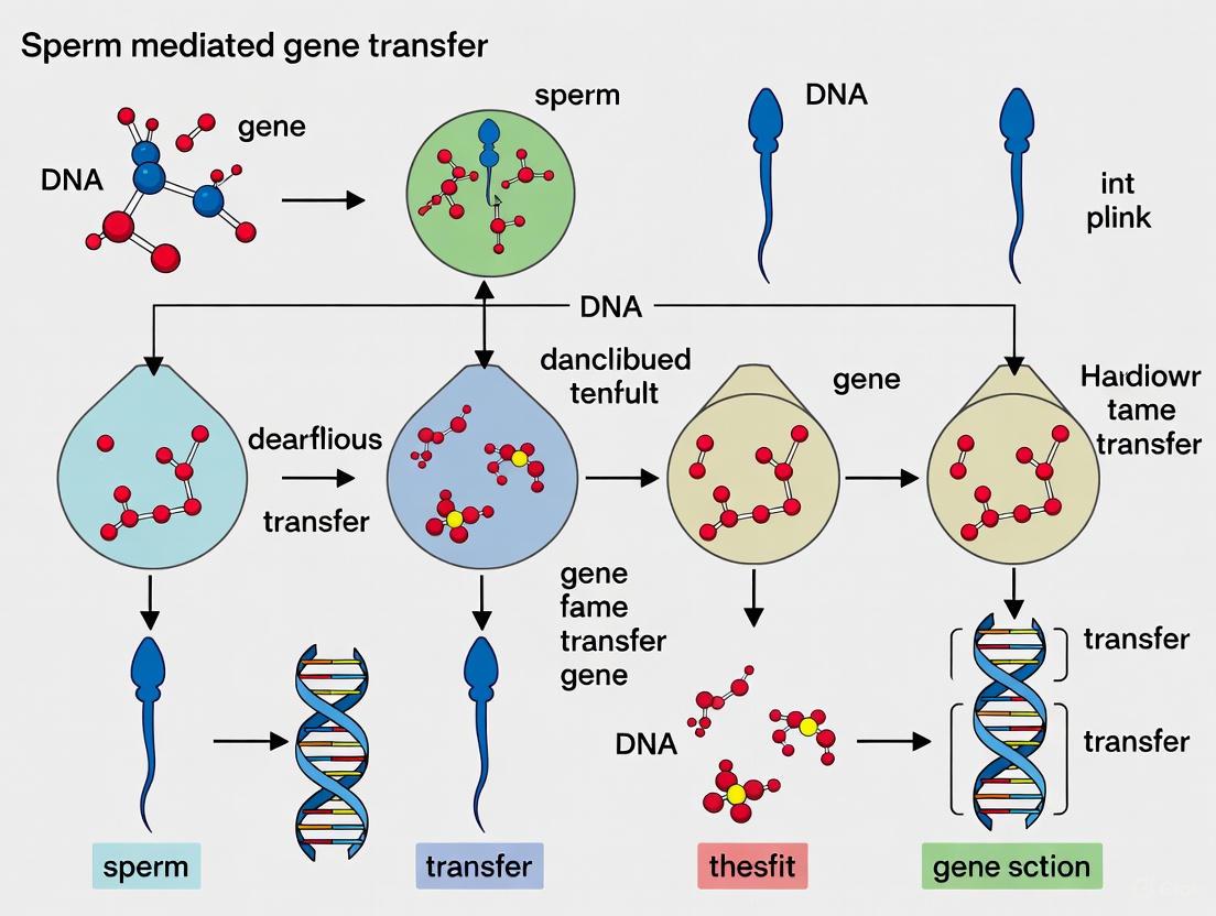
SMGT in Practice: Protocols and Transformative Biomedical Applications
Sperm-Mediated Gene Transfer (SMGT) is a biotechnological method that utilizes sperm cells as natural vectors to deliver foreign genetic material into an oocyte during fertilization [21]. This technique facilitates the introduction of new genes into an organism's genome, serving as a powerful tool in genetic engineering and reproductive biology to enhance genetic diversity and modify traits in various species [21]. For researchers and drug development professionals, SMGT offers a compelling alternative to traditional pronuclear injection due to its high efficiency, relatively low cost, and ease of use, as it does not require direct embryo handling or expensive micromanipulation equipment [27] [28]. A significant implication of SMGT research is the understanding that the process is not casual but a regulated mechanism potentially mediated by endogenous retrotransposons, challenging the traditional view of sperm as metabolically inert cells [4].
Materials and Reagents
Research Reagent Solutions
The following table details the essential materials and reagents required for the successful execution of the SMGT protocol.
| Reagent/Material | Function/Application in SMGT Protocol |
|---|---|
| Semen Extender (e.g., Swine Fertilisation Medium - SFM) | Preserves sperm quality and viability during storage and processing post-collection [28]. |
| Exogenous DNA Construct (e.g., pEGFP-N1) | The foreign genetic material to be transferred into the oocyte; typically linearized [21]. |
| Lipofection Reagent | A cationic liposome-based transfection agent used to enhance the uptake of exogenous DNA by spermatozoa [21]. |
| Sperm Washing Medium | A buffered solution, often containing antibiotics, used to separate motile sperm from seminal plasma and debris [28]. |
| In Vitro Fertilization (IVF) Medium | A defined culture medium that supports the capacitation of sperm and the fertilization of oocytes [28]. |
Experimental Protocol
Sperm Washing and Preparation
- Semen Collection and Initial Evaluation: Collect semen from a donor boar (or other species) and evaluate immediately for standard quality parameters (color, volume, concentration, and motility) [21].
- Washing and Selection of Motile Sperm: Dilute the semen in a pre-warmed sperm washing medium. Centrifuge the mixture to pellet the sperm cells. Carefully remove the supernatant containing seminal plasma. Repeat this washing step if necessary [28]. This process is critical for removing seminal plasma components that may inhibit DNA uptake [28].
- Resuspension: After the final wash, resuspend the sperm pellet in a suitable semen extender or a specific medium like Swine Fertilisation Medium (SFM) to the desired concentration for the DNA uptake step [28].
Sperm Treatment and DNA Uptake
- DNA Preparation: Prepare a solution containing the exogenous DNA construct (e.g., a linearized plasmid such as pVIVO2-GFP/LacZ) at a defined concentration. The standard concentration used is 5 µg/mL, though studies have tested much higher amounts (100 µg/mL) without significantly compromising sperm quality [27] [28].
- Co-incubation with DNA: Incubate the washed, motile spermatozoa with the prepared DNA solution. To enhance transfection efficiency, a lipofection reagent can be added to the mixture [21]. The incubation should be carried out for a defined period, typically at 37°C.
- Post-Incubation Assessment: Following co-incubation, assess the sperm cells again for quality parameters, including motility, membrane integrity, and mitochondrial activity, to ensure the treatment has not adversely affected their fertilizing capability [27] [28].
In Vitro Fertilization (IVF) with Transfected Sperm
- Oocyte Collection and Maturation: Collect oocytes from superovulated prepubertal gilts or other donors. Allow the oocytes to mature in vitro to the Metaphase II (MII) stage [28].
- Fertilization: Co-incubate the matured oocytes with the spermatozoa that have been treated with the exogenous DNA. This is typically performed in a specialized IVF medium that supports capacitation and fertilization [28].
- Assessment of Fertilization Success: Approximately 24 hours post-insemination, evaluate the oocytes for successful fertilization by checking for the formation of two pronuclei (zygotes) and subsequent cleavage rates [27] [28].
Figure 1: A flowchart illustrating the step-by-step workflow of the standard SMGT protocol.
Key Data and Results
The success of the SMGT protocol is evaluated through key performance metrics, including sperm quality post-treatment and subsequent embryo development rates. The following tables summarize typical quantitative outcomes from an optimized SMGT experiment in swine.
Table 1: Sperm Quality Parameters Post-DNA Uptake
| Treatment Condition | Motility (%) | Membrane Integrity (%) | Developmental Rate to Blastocyst (%) |
|---|---|---|---|
| Control Sperm | Data not in search results | Data not in search results | 48 |
| SMGT-Treated Sperm (5 µg/mL DNA) | Data not in search results | Data not in search results | 41 |
| SMGT-Treated Sperm (100 µg/mL DNA) | Not significantly affected [27] | Not significantly affected [27] | Data not in search results |
Table 2: In Vitro Fertilization Outcomes with SMGT-Treated Sperm
| Parameter | Control Group | SMGT-Treated Group |
|---|---|---|
| Cleavage Rate (%) | 58 | 60 |
| Transformation Efficiency (%) | — | 62 |
Mechanism Underlying SMGT
The mechanism of SMGT extends beyond the passive binding of DNA to the sperm cell surface. Research indicates it is an active process mediated by specific factors within the sperm [4]. The binding of exogenous DNA triggers enzymatic functions that are otherwise repressed, including an endogenous retrotransposon-encoded reverse transcriptase activity [4]. This activity can reverse transcribe exogenous RNA molecules into cDNA copies. These reverse-transcribed molecules can be propagated in tissues as low-copy, extrachromosomal structures that are mosaic distributed but transcriptionally competent [4]. This suggests that the sperm's endogenous retrotransposon machinery can be a novel source of genetic information and is central to the SMGT phenomenon.
Figure 2: A diagram of the proposed molecular mechanism of SMGT involving reverse transcription.
The field of genetic engineering and gene therapy is critically dependent on advanced delivery vehicles to transport genetic material into target cells. These vectors overcome significant biological barriers that otherwise prevent exogenous nucleic acids from reaching their intracellular targets. Non-viral vectors, particularly lipid nanoparticles (LNPs), have emerged as a safer and more versatile alternative to viral carriers, though they have historically faced challenges with transfection efficacy compared to viral systems [29]. LNPs demonstrate remarkable capability to condense and deliver various nucleic acid molecules ranging from small RNA sequences to large DNA constructs [29]. Simultaneously, sperm-mediated gene transfer (SMGT) represents a unique biological approach that leverages the natural ability of sperm cells to bind, internalize, and transport exogenous DNA during fertilization [3] [1]. This application note examines these advanced delivery systems within the context of sperm-mediated gene transfer research, providing technical protocols and comparative analyses for researchers and drug development professionals.
The fundamental challenge in gene delivery involves navigating both extracellular and intracellular barriers. From the moment of injection, genetic material encounters enzymatic degradation by nucleases, clearance by the reticuloendothelial system (RES), and physical barriers to cellular uptake due to the anionic nature of both nucleic acids and cell membranes [29]. Success in gene therapy depends largely on developing vehicles that can efficiently bypass these barriers to deliver genetic material to target cells with sufficient expression levels and minimal toxicity [29]. This document outlines the key methodologies, applications, and technical considerations for implementing these delivery systems in research settings focused on sperm-mediated gene transfer techniques.
Principles of SMGT
Sperm-mediated gene transfer (SMGT) is a transgenic technique based on the intrinsic ability of sperm cells to spontaneously bind to and internalize exogenous DNA and transport it into an oocyte during fertilization [1]. First described in 1989, SMGT provides a method for producing genetically modified animals with several distinct advantages [2]. The technique benefits from high efficiency, low cost, and ease of use compared to other transgenic methods, and does not require specialized equipment or extensive embryo manipulation [3]. This makes it particularly valuable for generating large animal models for medical research, agricultural applications, and xenotransplantation studies [3].
The mechanism of SMGT is not a random event but rather a regulated process mediated by specific factors. Exogenous DNA molecules interact with DNA-binding proteins (DBPs) present on the surface of sperm cells, facilitating binding and internalization [1]. However, natural barriers exist against this process, including an inhibitory factor present in mammalian seminal fluid that blocks the binding of sperm cells to exogenous DNA [1]. For successful SMGT, this seminal fluid must be removed through extensive washing immediately after ejaculation to enable DBPs to interact with DNA molecules [1]. Following internalization, the exogenous DNA must integrate into the genome, potentially occurring during oocyte activation, nucleus decondensation, or pronuclei formation [1].
Key Enhancements and Technical Variations
Recent research has focused on improving the efficiency of SMGT through various technical enhancements. A 2024 systematic review compared sperm-mediated and testis-mediated gene transfer (TMGT) methods, highlighting optimal approaches for different species [30]. For the SMGT approach in mice, nanoparticles, streptolysin-O, and virus packaging were identified as the most effective gene transfer methods [30]. The efficiency of producing transgenic animals varies significantly depending on species, gene carrier, and transfer method, necessitating careful experimental design.
Table 1: Comparison of Sperm-Mediated and Testis-Mediated Gene Transfer Methods
| Aspect | Sperm-Mediated Gene Transfer (SMGT) | Testis-Mediated Gene Transfer (TMGT) |
|---|---|---|
| Most Common Species | Mice and rats | Mice and rats |
| Optimal Gene Transfer Methods | Nanoparticles, streptolysin-O, virus packaging | Virus packaging, DMSO, electroporation, liposome |
| Number of Studies (2010-2022) | 47 | 25 |
| Key Advantages | Does not require embryo handling; relatively simple procedure | Direct targeting of testicular tissue |
| Notable Applications | Production of multigene transgenic pigs for xenotransplantation | Transfection of spermatogonial stem cells |
Beyond standard SMGT protocols, several variations have been developed to enhance efficiency. Intracytoplasmic sperm injection (ICSI)-mediated gene transfer involves injecting sperm that has been incubated with DNA directly into oocytes, showing improved transgenesis rates in some species [2]. Additional methods include electroporation of sperm in the presence of exogenous DNA and treatment with dimethyl sulfoxide (DMSO) or N,N-dimethylacetamide to facilitate DNA uptake, particularly in avian species [2]. These technical refinements have progressively addressed the primary limitation of SMGT – the relatively low and variable efficiency of transgene transmission – making it an increasingly reliable method for transgenic animal production.
Lipid-Based Delivery Systems
Composition and Mechanism of Action
Lipid nanoparticles represent one of the most advanced non-viral vector platforms for gene delivery. These systems typically consist of cationic lipids that electrostatically bind to nucleic acids, helper lipids that enhance stability and fusogenicity, and PEGylated lipids that provide a steric barrier to prevent aggregation and reduce immune recognition [29]. The fundamental mechanism involves condensing genetic material into nanoparticles that protect it from degradation and facilitate cellular uptake through endocytosis. Following internalization, LNPs enable endosomal escape through phase transitions that disrupt endosomal membranes, releasing genetic material into the cytoplasm [29].
The rational design of LNPs has focused particularly on overcoming intracellular barriers. Initially, researchers proposed that anionic phospholipids could displace cationic lipids from plasmids, assisting release following cellular uptake [29]. Alternative hypotheses suggest that cationic lipids form ion pairs with anionic lipids within the endosome membrane, leading to disruption and cytoplasmic release of genetic material [29]. Cullis and colleagues further proposed that mixtures of cationic lipids and anionic phospholipids preferentially adopt the inverted hexagonal (HII) phase, which facilitates escape from the endosome into the cytoplasm [29]. These sophisticated design principles have progressively enhanced the efficiency of lipid-based gene delivery systems.
Advanced Formulation Strategies
Advanced LNP formulations incorporate specific components to address particular biological challenges. PEGylation – the incorporation of polyethylene glycol (PEG) lipids – creates a hydrated layer and steric barrier on the nanoparticle surface that reduces recognition by the immune system and uptake by the reticuloendothelial system [29]. The degree of PEGylation significantly impacts circulation half-life, with higher PEG content generally increasing persistence in the bloodstream [29]. However, this approach must be carefully balanced against the accelerated blood clearance (ABC) phenomenon that can occur with repeated injections of PEGylated formulations due to anti-PEG IgM production [29].
Recent innovations include the development of fusigenic viral liposomes that combine advantages of viral and non-viral systems. These hybrid vectors incorporate fusion proteins from the Hemagglutinating Virus of Japan (HVJ; Sendai virus) into liposomal membranes [31]. The resulting constructs can fuse directly with plasma membranes at neutral pH, introducing genetic material directly into the cytoplasm and avoiding lysosomal degradation [31]. This system has demonstrated efficient transfection of oligonucleotides, plasmid DNA up to 100 kbp, and even proteins co-introduced to enhance transgene expression [31]. The incorporation of DNA-binding nuclear proteins like high mobility group-1 further enhances transgene expression in target tissues [31].
Table 2: Lipid Nanoparticle Components and Their Functions
| Component | Function | Examples |
|---|---|---|
| Cationic Lipids | Electrostatic binding to nucleic acids; endosomal disruption | DOTAP, DC-Chol |
| Helper Lipids | Enhance membrane stability and fusogenicity | DOPE, cholesterol |
| PEGylated Lipids | Steric stabilization; reduce immune recognition | PEG-DSPE |
| Fusigenic Proteins | Enable direct membrane fusion | HVJ fusion proteins |
| Nuclear Proteins | Enhance transgene expression | High mobility group-1 |
Experimental Protocols
Standardized SMGT Protocol
The following protocol outlines the essential steps for sperm-mediated gene transfer, adapted from established methodologies [2] [32]:
Reagents and Equipment:
- Sperm preparation medium (appropriate for species)
- DNA solution (purified, linearized fragment or circular plasmid)
- Fertilization medium
- Incubator at appropriate temperature and CO₂ conditions
- Sterile centrifuge tubes
- Microcentrifuge
Procedure:
- Sperm Collection and Washing: Collect semen and immediately wash extensively to remove seminal plasma, which contains inhibitory factors that block exogenous DNA binding. Centrifuge at 800×g for 10 minutes and resuspend in appropriate medium.
- DNA Incubation: Incubate washed sperm (approximately 1-5×10⁶ cells/mL) with exogenous DNA (1-100 ng/μL) for time periods ranging from 30 minutes to 2 hours at appropriate temperature conditions (typically 32-37°C for mammalian species).
- Removal of Unbound DNA: Wash sperm cells to remove unbound DNA by centrifugation at 500×g for 5 minutes. Resuspend in fertilization medium.
- In Vitro Fertilization: Use DNA-loaded sperm for standard in vitro fertilization procedures with mature oocytes.
- Embryo Transfer: Culture resulting embryos to appropriate stages and transfer to synchronized foster mothers for transgenic offspring production.
Critical Considerations:
- DNA integrity and conformation significantly impact efficiency; linearized fragments often show better integration than supercoiled plasmids.
- Sperm viability and fertilizing capacity must be preserved throughout the procedure.
- Optimal DNA concentration varies by species and must be determined empirically to balance transfection efficiency with maintained sperm function.
LNP Formulation Protocol for Nucleic Acid Delivery
This protocol describes the preparation of lipid nanoparticles for nucleic acid delivery, adaptable for co-incubation with sperm cells [29] [33]:
Reagents and Equipment:
- Cationic lipid (e.g., DOTAP, DC-Chol)
- Helper lipid (e.g., DOPE, cholesterol)
- PEG-lipid (e.g., PEG-DSPE)
- Nucleic acid (DNA, siRNA, mRNA) in purified form
- Ethanol and aqueous buffers
- Microfluidic device or rapid mixing apparatus
- Dialysis membranes or tangential flow filtration system
Procedure:
- Lipid Solution Preparation: Dissolve lipid components in ethanol at specific molar ratios (typically 50-70% cationic lipid, 25-45% helper lipid, 0.5-5% PEG-lipid).
- Aqueous Phase Preparation: Dilute nucleic acid in appropriate aqueous buffer (e.g., citrate buffer, pH 4).
- Nanoparticle Formation: Rapidly mix lipid and aqueous solutions using microfluidic device or rapid pipetting. Standard flow rate ratios typically range from 1:1 to 3:1 (aqueous:ethanol).
- Buffer Exchange: Dialyze or use tangential flow filtration to remove ethanol and exchange buffer to physiological conditions (e.g., PBS, pH 7.4).
- Characterization: Determine particle size (target 80-200 nm) by dynamic light scattering, measure zeta potential, and quantify nucleic acid encapsulation efficiency.
Critical Considerations:
- Maintain strict control over mixing parameters (flow rates, total flow rate) to ensure reproducible particle size.
- N/P ratio (nitrogen groups of cationic lipid to phosphate groups of nucleic acid) typically ranges from 3:1 to 6:1 for optimal encapsulation and function.
- Sterile filtration (0.22 μm) may be applied after formulation for sterility while avoiding significant particle loss.
Advanced Hybrid Systems
Fusigenic Viral Liposome System
The fusigenic viral liposome represents a sophisticated hybrid vector that combines the efficiency of viral fusion mechanisms with the safety and cargo capacity of liposomes [31]. The construction involves several key steps:
- HVJ Preparation: Grow HVJ (Sendai virus) in chick embryonated eggs and purify by sucrose density gradient centrifugation. Inactivate with UV irradiation (110 erg/mm²/sec for 3 minutes) to eliminate viral replication while preserving fusion activity.
- Liposome Preparation: Formulate liposomes containing phosphatidylcholine, cholesterol, and negatively charged lipids (e.g., phosphatidylserine) using reverse-phase evaporation or vortexing methods. Encapsulate DNA during this process, achieving trapping efficiencies of 10-30%.
- Fusion Reaction: Mix UV-inactivated HVJ with DNA-containing liposomes at a ratio of 10,000-20,000 hemagglutinating units (HAU) per μmol phospholipid. Incubate at 37°C for 1 hour with occasional shaking.
- Purification: Remove free HVJ and unfused liposomes by sucrose density gradient centrifugation.
This system enables direct introduction of molecules into the cytoplasm through fusion with the plasma membrane, bypassing endosomal degradation pathways [31]. The approach has demonstrated efficient gene transfer in various tissues including liver, kidney, heart, and skeletal muscle, with transgene expression persisting for several weeks [31].
Applications in Cardiovascular Gene Therapy
The fusigenic viral liposome system has shown particular promise in cardiovascular gene therapy applications [31]. In models of vascular proliferative disease, researchers have successfully implemented cytostatic gene therapy using:
- Antisense oligodeoxynucleotides against cell cycle genes such as proliferating cell nuclear antigen (PCNA) and cell division cycle 2 (cdc2)
- Transcription factor decoys containing E2F binding sites to competitively inhibit cell cycle progression
- Nitric oxide synthase gene delivery to restore protective endothelial function
Similar strategies have proven effective for genetic engineering of vein grafts and treatment of immune-mediated glomerular disease [31]. The system's efficiency stems from its ability to rapidly transfer genes into target tissues – exposure of rat carotid artery to HVJ-liposomes for just 10 minutes results in uptake by 30-50% of cells within the vessel wall [31].
Visualization and Workflow Diagrams
SMGT Experimental Workflow
Diagram 1: SMGT Experimental Workflow illustrating key procedural steps from sperm collection to transgenic offspring production.
LNP Formulation and Mechanism
Diagram 2: LNP Formulation and Mechanism illustrating component assembly and intracellular trafficking pathway.
Research Reagent Solutions
Table 3: Essential Research Reagents for Advanced Gene Delivery Systems
| Reagent/Category | Specific Examples | Function/Application | Technical Notes |
|---|---|---|---|
| Cationic Lipids | DOTAP, DC-Chol, DOTMA | Nucleic acid condensation; endosomal disruption | Vary in head group structure and chain saturation affecting transfection efficiency and toxicity |
| Helper Lipids | DOPE, cholesterol | Enhance membrane fusion and stability | DOPE promotes hexagonal phase formation crucial for endosomal escape |
| PEG-Lipids | PEG-DSPE, PEG-Ceramides | Steric stabilization; reduce opsonization | PEG length (MW 750-5000) and density (0.5-5 mol%) critically impact circulation time |
| Fusigenic Components | HVJ envelope proteins | Enable direct membrane fusion | UV-inactivated virus maintains fusion activity without replication capability |
| DNA-Binding Proteins | High mobility group-1 | Enhance transgene expression | Non-histone chromosomal protein facilitates nuclear import and transcription |
| Permeabilization Agents | Streptolysin-O, DMSO | Enhance membrane permeability for DNA uptake | Streptolysin-O creates transient pores without complete membrane disruption |
| Viral Packaging Systems | Lentivirus, retrovirus | High-efficiency gene transfer and integration | Essential for TMGT; select based on target cell division status |
Advanced delivery vehicles including liposomes, viral vectors, and nanoparticles represent critical enabling technologies for modern genetic research and therapeutic development. Within the context of sperm-mediated gene transfer, these systems offer complementary approaches for enhancing the efficiency and reliability of transgenic animal production. Lipid-based nanoparticles provide a safe, versatile platform for nucleic acid delivery that can be optimized through rational design of their biochemical and biophysical properties. The integration of viral fusion mechanisms into hybrid systems such as fusigenic viral liposomes further enhances delivery efficiency while maintaining favorable safety profiles.
Future developments in this field will likely focus on increasing cell-type specificity through targeted ligands, enhancing endosomal escape capabilities, and developing stimulus-responsive systems that activate only in target tissues or cellular compartments. For SMGT applications specifically, research will continue to optimize the balance between transfection efficiency and preservation of sperm function, potentially through transient membrane permeabilization strategies or receptor-mediated uptake mechanisms. As these advanced delivery systems mature, they will increasingly enable researchers to address complex biological questions and develop innovative genetic therapies for both agricultural and biomedical applications.
Sperm-mediated gene transfer (SMGT) represents a foundational technique in reproductive biotechnology wherein spermatozoa are utilized as natural vectors to deliver exogenous genetic material into oocytes during fertilization [34]. The integration of SMGT with the precision of CRISPR/Cas9 genome editing systems has given rise to powerful methodologies for generating targeted mutant animals, a technique increasingly referred to as sperm-mediated gene editing (SMGE) [7]. This approach leverages the inherent ability of sperm cells to bind and internalize foreign DNA, coupling it with the targeted genomic cleavage capabilities of the CRISPR/Cas9 system to create specific, heritable genetic modifications. This Application Note details the principles, optimized protocols, and key reagents for effectively coupling SMGT with CRISPR/Cas9, providing a robust framework for researchers in transgenesis and drug development.
The MBCD-SMGE technique, which uses methyl β-cyclodextrin (MBCD) to enhance the uptake of CRISPR/Cas9 components, demonstrates the significant potential of this coupled approach [7]. This method has been successfully validated through the production of targeted indel mutations in the Gdf8 gene in both blastocysts and live mice, showcasing its applicability for modeling human diseases and improving animal traits [7]. The subsequent sections provide a detailed experimental workflow, quantitative data on optimization, and a comprehensive toolkit for implementing this technology.
Principles of the Coupled SMGT-CRISPR/Cas9 System
The SMGE technique capitalizes on the synergistic effect of two core biological processes. First, the sperm cell's capacity to spontaneously bind and internalize exogenous DNA is enhanced through chemical treatment. Second, the CRISPR/Cas9 system, delivered via this route, facilitates precise double-strand breaks at predefined genomic loci.
- Sperm as a Delivery Vector: Mature spermatozoa possess a natural, though limited, ability to bind and internalize exogenous DNA molecules [34]. The efficiency of this process is critically dependent on the integrity and composition of the sperm membrane. The removal of cholesterol from the sperm membrane using MBCD induces a premature acrosomal reaction and increases membrane permeability, thereby significantly enhancing the uptake of plasmid DNA containing CRISPR/Cas9 components [7].
- CRISPR/Cas9 Mechanism: The CRISPR/Cas9 system consists of a Cas9 endonuclease and a single-guide RNA (sgRNA). The sgRNA directs the Cas9 protein to a specific DNA sequence adjacent to a protospacer adjacent motif (PAM—e.g., 5'-NGG-3' for Streptococcus pyogenes Cas9), where the nuclease creates a double-strand break (DSB) [35]. This DSB is subsequently repaired by the cell's endogenous DNA repair machinery, predominantly through the error-prone non-homologous end joining (NHEJ) pathway, leading to insertion or deletion mutations (indels) that can disrupt the target gene [7].
The following diagram illustrates the logical and procedural workflow of the MBCD-SMGE technique, from sperm preparation to the generation of mutant offspring:
Key Research Reagent Solutions
The successful implementation of SMGE relies on a specific set of reagents and vectors. The table below catalogues the essential materials and their functions based on established protocols.
Table 1: Essential Research Reagents for SMGE
| Reagent/Vector | Function and Description | Source/Example |
|---|---|---|
| Sperm Cells | Delivery vector for CRISPR/Cas9 system during fertilization. | B6D2F1 mouse strain [7]. |
| CRISPR Plasmid Vector | Expresses Cas9 protein and sgRNA; contains a fluorescent marker (e.g., GFP) for tracking transfection. | pCAG-eCas9-GFP-U6-gRNA (Addgene #79145) [7]. |
| Methyl β-Cyclodextrin (MBCD) | Cyclic oligosaccharide that removes cholesterol from the sperm membrane, enhancing permeability and plasmid uptake. | Sigma-Aldrich [7]. |
| c-TYH Medium | Protein-free medium used for sperm incubation with MBCD and CRISPR plasmids. | Choi-Toyoda Yokoyama Hosi medium [7]. |
| Control Plasmid | Used to calibrate and monitor transfection efficiency independent of CRISPR editing. | plenti-CAG-gate-FLAG-IRES-GFP (Addgene #107398) [7]. |
| Alt-R CRISPR-Cas9 System | Commercial system offering synthesized guide RNAs and recombinant Cas9 proteins; can be used as RNP complexes. | Integrated DNA Technologies [36]. |
Quantitative Data and Optimization
Optimizing the concentration of chemical enhancers like MBCD is critical for maximizing plasmid uptake while maintaining sperm viability. The following table summarizes key quantitative findings from a recent study that titrated MBCD concentrations.
Table 2: Effect of MBCD Concentration on SMGE Efficiency
| MBCD Concentration (mM) | Plasmid Copy Number per Sperm Cell | Effect on Sperm Membrane & Viability | Production Rate of GFP-positive Blastocysts |
|---|---|---|---|
| 0 (Control) | Low (Baseline) | Standard membrane integrity | Low [7] |
| 0.75 | Increased | Induces premature acrosomal reaction; optimizes fertility [7] | Significantly Higher [7] |
| 1.0 | Further Increase | Effective cholesterol removal | High (Optimal) [7] |
| 2.0 | High, but potential toxicity | Dose-dependent effect; may impair viability | Reduced due to potential toxicity [7] |
The data indicates that 1.0 mM MBCD often represents an optimal balance, resulting in a high copy number of internalized plasmid and a superior production rate of transfected blastocysts without excessive cytotoxicity [7]. The study also monitored extracellular reactive oxygen species (ROS), which increased with MBCD treatment, suggesting that ROS may play a role in the DNA uptake mechanism [7].
Detailed Experimental Protocol: MBCD-SMGE
This section provides a step-by-step methodology for generating targeted mutant mice using the MBCD-SMGE technique, as validated in recent research [7].
Protocol Workflow
The end-to-end experimental procedure, from sperm preparation to genotyping, is visualized below.
Step-by-Step Methodology
Step 1: sgRNA Design and Vector Preparation
- Design: Select a target site within the first exons of the gene of interest (e.g., Gdf8). Use an online gRNA design tool and verify target uniqueness within the genome.
- Cloning: Synthesize sense and antisense oligodeoxynucleotides for the sgRNA with BbsI-specific sticky ends. Anneal and ligate them into a BbsI-linearized pCAG-eCas9-GFP-U6-gRNA vector [7].
- Propagation and Purification: Transform the ligated product into a stable bacterial strain (e.g., Stbl4). Culture and purify the plasmid using a commercial plasmid extraction kit. Resuspend the final plasmid in nuclease-free water or TE buffer at a working concentration of 20 ng/µL.
Step 2: Sperm Collection and Incubation
- Collection: Collect sperm from the cauda epididymides of sexually mature B6D2F1 male mice.
- Incubation: Place the sperm in c-TYH medium supplemented with 1 mM MBCD and 20 ng/µL of the purified pgRNA-Cas9 plasmid.
- Duration: Incubate the mixture for 30 minutes at 37°C under 5% CO₂ [7].
Step 3: Sperm Quality Assessment
- Functional Parameters: Evaluate sperm motility and concentration using a Makler chamber.
- Viability: Use an MTT assay to assess cell viability post-incubation [37].
- ROS Measurement: Quantify extracellular reactive oxygen species (ROS) levels, as this is a relevant parameter influenced by MBCD treatment [7].
Step 4: In Vitro Fertilization (IVF) and Embryo Culture
- Oocyte Collection: Retrieve oocytes from superovulated CD1 or B6D2F1 female mice.
- Fertilization: Co-incubate the transfected sperm with the collected oocytes in HTF medium.
- Culture: Wash the resulting zygotes and culture them in microdrops of modified KSOM (mKSOM) medium at 37°C under 5% CO₂ for 3.5-4 days. Monitor embryonic development to the blastocyst stage [7].
Step 5: Analysis and Genotyping
- Blastocyst Screening: Examine blastocysts for GFP fluorescence to identify successfully transfected embryos.
- Genotype Analysis: Extract genomic DNA from blastocysts or pup tail biopsies. Perform PCR amplification of the target region and confirm the presence of indel mutations using Sanger sequencing followed by alignment software (e.g., TIDE or ICE analysis) [37] [7].
The coupling of sperm-mediated gene transfer with CRISPR/Cas9 technology provides a streamlined and efficient platform for generating targeted mutant animals. The MBCD-SMGE protocol outlined herein offers a reproducible method that enhances the delivery of CRISPR components via sperm cells. By optimizing key parameters such as MBCD concentration and sperm incubation conditions, researchers can achieve high rates of mutagenesis. This Application Note serves as a comprehensive guide for scientists seeking to apply this integrated technology in biomedical research, drug development, and the creation of advanced animal models of human disease.
Generation of Transgenic Livestock for Xenotransplantation and Disease Modeling
The generation of transgenic livestock represents a cornerstone of biomedical and agricultural research, enabling critical advancements in xenotransplantation and human disease modeling [38]. The severe shortage of human organs for transplantation has created sustained demand for alternative solutions, with porcine xenotransplantation emerging as one of the most promising approaches [39]. Simultaneously, genetically engineered large animals have become indispensable for modeling human diseases with greater physiological relevance than rodent models [38].
This application note details methodologies centered on sperm-mediated gene transfer (SMGT) alongside other established techniques for generating transgenic livestock. SMGT leverages the natural ability of spermatozoa to bind, internalize, and transport exogenous DNA into oocytes during fertilization [1]. While this technique offers simplicity compared to more complex procedures, its application requires careful consideration of efficiency and consistency [1] [34]. We provide structured protocols and analytical frameworks to facilitate the implementation of these technologies in research settings focused on addressing the organ shortage crisis and complex human disease pathogenesis.
Key Applications of Transgenic Livestock
Xenotransplantation
Genetically engineered pigs are considered the most viable source for organ xenotransplantation due to physiological similarities to humans, compatible organ size, and ease of breeding [39]. The primary immunological barrier is hyperacute rejection, mediated by pre-formed human antibodies against porcine carbohydrate antigens, primarily galactose-α1,3-galactose (α-Gal) [39]. Multiplex genetic engineering addresses this by eliminating xenoantigens and expressing human protective proteins [39] [40].
Table 1: Key Genetic Modifications for Xenotransplantation
| Target Type | Gene/Protein | Modification | Primary Purpose |
|---|---|---|---|
| Xenoantigen Knockout | GGTA1 | Knockout | Eliminate α-Gal epitope [39] |
| CMAH | Knockout | Eliminate Neu5Gc antigen [39] | |
| B4GalNT2 | Knockout | Reduce antibody-mediated rejection [39] | |
| Human Transgene Insertion | hCD46, hCD55, hCD59 | Knock-in | Express human complement regulatory proteins [39] [41] |
| hTBM, hEPCR | Knock-in | Regulate coagulation pathways [39] | |
| HLA-E, CD47 | Knock-in | Inhibit human NK and T-cell responses [39] |
Recent clinical milestones underscore the field's progress. The first investigational new drug application for a pig-to-human kidney xenotransplantation clinical trial received FDA clearance in early 2025 [42]. Furthermore, compassionate use cases have demonstrated life-supporting function of 69-gene edited porcine kidneys for nearly two months and 10-gene edited pig hearts for over one month in human recipients [41].
Disease Modeling
Transgenic livestock, particularly pigs, provide highly relevant models for human diseases due to their physiological and anatomical similarities to humans [38]. CRISPR-Cas9 has accelerated the creation of large animal models for conditions including cardiovascular diseases, metabolic disorders, and various cancers [38]. These models enable more accurate study of disease mechanisms and preclinical therapeutic testing than is possible with rodent models alone.
Quantitative Data and Market Analysis
The global transgenic livestock market reflects the growing importance of these technologies. Market research indicates robust expansion, with the market size projected to grow from USD 3.1 billion in 2024 to USD 6.6 billion by 2033, at a compound annual growth rate of 8.7% [43].
Table 2: Transgenic Livestock Market Analysis (2024-2033)
| Segment | 2024 Market Value | Projected 2033 Value | Key Growth Drivers |
|---|---|---|---|
| Overall Market | USD 3.1 Billion | USD 6.6 Billion (CAGR 8.7%) | Gene-editing adoption, demand for animal protein, biomedical applications [43] |
| By Animal Type (Dominant Segments) | |||
| Cattle | Largest share | Agricultural traits, biopharmaceutical production [43] | |
| Pigs | Substantial share | Xenotransplantation, disease modeling [43] | |
| By Technique | |||
| CRISPR | Rapid adoption | Precision, cost-effectiveness, multiplex editing capability [43] [38] | |
| Somatic Cell Nuclear Transfer | Established use | Clonal propagation of validated genotypes [39] [43] |
Experimental Protocols
Sperm-Mediated Gene Transfer (SMGT) Protocol
Principle: Sperm cells spontaneously bind and internalize exogenous DNA, transporting it into the oocyte during fertilization to produce genetically modified animals [1] [34].
Reagents and Materials:
- Fresh porcine semen samples
- DNA-binding proteins (DBPs)
- Hepes-buffered medium
- Seminal plasma removal reagents (extensive washing buffers)
- Exogenous DNA construct (linearized, purified)
- Dimethylsulfoxide (DMSO) or N,N-dimethylacetamide [1]
Procedure:
- Sperm Collection and Preparation: Collect fresh semen and immediately remove seminal plasma through extensive washing. Seminal plasma contains an inhibitory factor that blocks the binding of exogenous DNA to DBPs on sperm cells [1] [34].
- Sperm Transfection: Incubate washed sperm cells (5 × 10^6 cells/mL) with the exogenous DNA construct (1-5 µg/mL) in Hepes-buffered medium for 20-40 minutes at 16-18°C. The presence of DBPs on the sperm head facilitates DNA binding and internalization [1].
- In Vitro Fertilization (IVF): Use transfected sperm cells for standard IVF procedures with matured oocytes.
- Embryo Transfer: Culture resulting embryos to the desired stage and transfer them into synchronized recipient females.
- Genotyping: Analyze offspring for the presence and integration of the transgene using PCR, Southern blotting, and functional assays.
Technical Notes: SMGT efficiency remains variable. Pre-treating exogenous DNA with DMSO or N,N-dimethylacetamide may improve uptake. A significant challenge is ensuring that transfected spermatozoa maintain functionality for fertilization while carrying exogenous DNA [1].
CRISPR-Cas9 Mediated Somatic Cell Nuclear Transfer (SCNT) Protocol
Principle: Combines the precision of CRISPR-Cas9 genome editing with the cloning capability of SCNT to generate genetically uniform large animals. This is currently the most reliable method for introducing multiplexed mutations in pigs [39] [38].
Reagents and Materials:
- CRISPR-Cas9 components (sgRNA, Cas9 protein/mRNA)
- Donor somatic cells (porcine fetal fibroblasts)
- Electroporation system
- Enucleated oocytes
- Fusion and activation equipment
- Embryo culture media
Procedure:
- Cell Culture and Transfection: Culture porcine fetal fibroblasts and transfect with CRISPR-Cas9 components via electroporation to introduce desired genetic modifications [39] [38].
- Cell Clone Selection: Select and expand cell clones with verified precise mutations through PCR, sequencing, and functional validation. This ex vivo selection is a key advantage, ensuring only correctly edited nuclei proceed to cloning [39].
- Oocyte Enucleation: Collect in vivo-matured oocytes and remove the metaphase II spindle.
- Nuclear Transfer: Micromanipulate a single validated donor somatic cell under the zona pellucida of each enucleated oocyte.
- Fusion and Activation: Fuse the cell-cytoplast complexes via electrical pulses and chemically activate the reconstructed embryos to initiate development.
- Embryo Transfer: Culture the embryos briefly and transfer them into synchronized recipient sows for gestation.
Technical Notes: While direct microinjection of CRISPR-Cas9 into zygotes is possible, it is less efficient for multiplex editing and carries a higher risk of mosaicism [39]. SCNT from validated cell clones ensures all animals carry the exact mutation, despite its overall lower efficiency in terms of live births [39].
The Scientist's Toolkit: Research Reagent Solutions
Table 3: Essential Research Reagents for Transgenic Livestock Generation
| Reagent / Solution | Function | Application Context |
|---|---|---|
| CRISPR-Cas9 System | RNA-guided nuclease for precise DNA cleavage | Gene knockout (GGTA1, CMAH) and knock-in (hCD46, hCD55) in somatic cells or zygotes [39] [38] |
| Somatic Cell Nuclear Transfer (SCNT) Reagents | Enables cloning from genetically validated somatic cell lines | Propagation of pigs with complex, multiplexed genetic modifications [39] [38] |
| Single-Guide RNA (sgRNA) | Targets Cas9 nuclease to specific genomic loci | Design requires accurate sequencing of the target pig line to ensure efficiency and minimize off-target effects [39] [38] |
| Cytosine Base Editors (CBE) | Converts C to T without double-strand breaks | Safer alternative for gene silencing by introducing nonsense mutations; reduces off-target risks [39] |
| Inducible Promoter Systems | Enables temporal control of transgene expression | Allows potentially lethal transgenes to be activated post-transplantation using doxycycline [39] |
Workflow and Pathway Visualizations
SMGT and SCNT Workflow Comparison
Immunological Barrier Modification Pathways
The convergence of Sperm-Mediated Gene Transfer (SMGT) and advanced sperm sorting technologies represents a transformative approach in transgenic animal production and reproductive science. This synergy enables the simultaneous introduction of desired genetic traits and the pre-determination of offspring sex, offering unprecedented control for biomedical research, livestock breeding, and therapeutic development. This application note details standardized protocols for coupling these technologies, providing researchers with robust methodologies to enhance efficiency in generating genetically modified embryos of predetermined sex. We present comprehensive experimental workflows, quantitative efficacy data, and essential reagent solutions to facilitate implementation across diverse research and development contexts.
Sperm-Mediated Gene Transfer (SMGT) leverages the innate capacity of spermatozoa to bind, internalize, and deliver exogenous DNA to oocytes during fertilization, serving as a versatile and efficient method for producing transgenic animals [4] [2]. Concurrently, sperm sorting technologies have evolved to enable reliable sex pre-determination by distinguishing X- and Y-chromosome-bearing spermatozoa based on DNA content or surface markers [44]. The integration of these methodologies—termed Sorted-SMGT—creates a powerful platform to address complex challenges in reproductive biotechnology, significantly reducing the time and resources required to establish homozygous transgenic lines by ensuring that modified alleles are introduced into embryos of the desired sex from the outset [45].
This document provides a detailed framework for implementing this combined approach, focusing on procedural optimization, critical quality control checkpoints, and tailored application notes for research professionals.
Technical Background and Principles
Sperm-Mediated Gene Transfer (SMGT) Fundamentals
SMGT is a technique founded on the spontaneous ability of sperm cells to act as vectors for exogenous genetic material. The process involves several key stages:
- DNA Binding: Exogenous DNA molecules bind to specific surface receptors on the sperm head.
- Internalization: The bound DNA is internalized into the sperm nucleus through active, regulated processes, challenging the traditional view of sperm as metabolically inert cells [4].
- Fertilization and Delivery: During fertilization, the sperm delivers both its own genome and the internalized exogenous DNA into the oocyte, leading to the generation of transgenic embryos.
Notably, research indicates that the binding of exogenous DNA can trigger enzymatic functions within the sperm, including an endogenous retrotransposon-encoded reverse transcriptase activity, which may play a role in the mechanism of transgene integration and propagation [4].
Sperm Sorting for Sex Pre-determination
The most established method for sperm sex-sorting is flow cytometric sorting, which exploits the small but consistent difference in DNA content between X- and Y-chromosome-bearing sperm (approximately 3.6% in boars, for example) [45] [44]. This high-precision technique allows for the production of semen samples highly enriched for the desired sex chromosome. Emerging immunological sorting methods offer a promising, cost-effective alternative. These techniques aim to identify sex-specific surface proteins or markers on X and Y sperm, enabling separation through methods like immunoadsorption, agglutination, or complement-mediated cytotoxicity [44]. While not yet as commercially advanced as flow cytometry, immunological sorting holds significant potential for broader application, particularly in settings where flow cytometry infrastructure is unavailable.
Integrated Workflow: Sorted-SMGT
The successful combination of SMGT and sperm sorting requires a carefully orchestrated sequence of steps, as outlined below.
Key Experimental Data and Efficacy
The following tables summarize critical quantitative data and performance metrics for the Sorted-SMGT protocol, derived from foundational and recent studies.
Table 1: Optimization of DNA Uptake in Sorted Sperm (Based on Swine Model)
| Sorting Parameter Adjusted | Objective | Outcome Metric | Reported Finding | Citation |
|---|---|---|---|---|
| Sorting Protocol Modification | Maximize exogenous DNA uptake by sex-sorted spermatozoa | DNA binding/internalization efficiency | Significant increase in sperm capable of DNA uptake post-sorting | [45] |
| Post-Sorting SMGT | Assess fertility of processed sperm | In vitro fertilization (IVF) rate | Sorted-SMGT sperm retained fertility, produced viable embryos | [45] |
| Blastocyst Formation | Evaluate embryo development | Blastocyst rate | Successful production of transformed blastocysts of predetermined male sex | [45] |
Table 2: Impact of Oxidative Conditions on SMGT Efficiency (Mouse Model)
| Treatment Condition | Concentration | Effect on Sperm Motility | Effect on DNA Uptake | GFP-Positive Blastocysts | Citation |
|---|---|---|---|---|---|
| Control (No treatment) | N/A | Normal | Baseline | Baseline | [46] |
| Oxidant (H₂O₂) | 100 µM | Significantly Reduced | Increased | Significantly Higher | [46] |
| Antioxidant (Melatonin) | 10⁻⁹ M | Improved vs. Oxidant | Protected sperm DNA integrity | Reduced apoptosis in embryos | [46] |
Detailed Experimental Protocols
Protocol A: Coupling Flow Cytometric Sorting with SMGT
This protocol is adapted from successful applications in swine transgenesis [45].
I. Sperm Preparation and Sex-Sorting
- Sperm Collection: Collect fresh semen samples into pre-warmed, non-capacitating media.
- Initial Processing: Dilute semen with an appropriate extender and assess initial motility and concentration.
- Staining: Incubate sperm with a fluorescent DNA-binding dye (e.g., Hoechst 33342) at a predetermined optimal concentration and temperature (32-36°C) for 45-60 minutes.
- Flow Cytometric Sorting: Use a high-speed cell sorter to separate X- and Y-chromosome bearing sperm based on DNA fluorescence intensity. Collect sorted populations into tubes containing recovery media supplemented with energy substrates and antioxidants.
II. Sperm-Mediated Gene Transfer on Sorted Sperm
- DNA Preparation: Prepare a pure, linearized foreign DNA construct (e.g., for a reporter gene like GFP) at an optimal concentration (typically 1-10 µg/10⁶ sperm) in a low-calcium medium.
- Co-incubation: Incubate the sex-sorted sperm population with the prepared DNA construct for 30-45 minutes at room temperature or 37°C, with gentle agitation.
- Washing: Pellet the sperm by gentle centrifugation and resuspend in fresh medium to remove unbound DNA.
III. Fertilization and Embryo Assessment
- In Vitro Fertilization (IVF): Use the Sorted-SMGT sperm in IVF with in vitro-matured oocytes.
- Embryo Culture: Culture resulting embryos in sequential media for 5-7 days to the blastocyst stage.
- Confirmation Analysis:
Protocol B: Immunological Sorting Combined with SMGT
This protocol outlines a promising alternative method [44].
I. Immunological Isolation of X or Y Sperm
- Antibody Preparation: Obtain antibodies against a verified Y-chromosome-specific surface protein (e.g., SRY) or generate polyclonal/monoclonal antibodies by immunizing animals with total sperm proteins and screening for sex-specific reactivity.
- Sperm Incubation with Antibody: Incubate the prepared sperm sample with the primary antibody for 30-60 minutes.
- Separation:
- Magnetic-Activated Cell Sorting (MACS): If using magnetic bead-conjugated secondary antibodies, pass the sample through a magnetic column. Y-sperm (if the target is Y-specific) will be retained, while X-sperm will flow through.
- Other Methods: Alternatively, employ immunoadsorption on a column or complement-mediated cytotoxicity to selectively immobilize or disable one population.
- Elution and Collection: Elute the bound sperm fraction from the column (for MACS) after removing the magnetic field.
II. SMGT on Immunologically-Sorted Sperm
- Follow the SMGT steps outlined in Protocol A (Section II) using the immunologically sorted sperm population.
The Scientist's Toolkit: Essential Research Reagent Solutions
Table 3: Key Reagents for Sorted-SMGT Experiments
| Reagent / Material | Function / Purpose | Example & Notes |
|---|---|---|
| Fluorescent DNA Stain | Labels sperm DNA for flow cytometric sorting based on content. | Hoechst 33342: Permeant dye used for live sorting. Critical for X/Y resolution. |
| Sperm Recovery Media | Supports sperm viability and function post-sorting. | Modified Tyrode's or SOF media, often with added BSA, energy substrates (e.g., pyruvate), and antioxidants. |
| Exogenous DNA Construct | The genetic material to be transferred via SMGT. | Linearized plasmid, purified and dissolved in low-Ca²⁺ buffer (e.g., TE). GFP is a common reporter. |
| Sex-Specific Antibodies | For immunological sorting of X or Y sperm. | Anti-SRY antibodies target Y-sperm. Polyclonal antibodies generated against total sperm proteins require rigorous validation for sex-specificity [44]. |
| Immunomodulators | To selectively alter motility of one sperm sex population. | R848 (Resiquimod): TLR7/8 agonist that can reduce X-sperm motility, enabling separation via swim-up [44]. |
| Antioxidants | To mitigate oxidative stress during sorting/SMGT, protecting sperm DNA integrity. | Melatonin: Potent antioxidant shown to improve sperm parameters and reduce embryonic apoptosis in SMGT contexts [46]. |
| PCR Reagents for Sexing | To confirm the sex of resulting embryos. | Primers for X- and Y-specific genes (e.g., AMELX/AMELY or ZFX/ZFY) [48]. |
Application Notes and Troubleshooting
- Optimization is Paramount: The success of Sorted-SMGT hinges on optimizing the sorting protocol specifically for subsequent DNA uptake. Standard sorting conditions may need adjustment to maintain sperm competency for SMGT [45].
- Manage Oxidative Stress: The physical and chemical stresses of sorting and incubation can increase reactive oxygen species (ROS). Incorporating antioxidants like melatonin into media can preserve sperm quality and DNA integrity, improving overall outcomes [46].
- Validate Immunological Tools: The efficacy of immunological sorting is entirely dependent on the specificity of the antibodies used. Always validate new antibody batches for their ability to selectively bind the target sperm population with minimal cross-reactivity [44].
- Control for Mosaicism: In SMGT-derived embryos, transgene integration can be mosaic. Analyze multiple cells from a blastocyst or multiple tissues from offspring for a complete assessment of transgenesis efficiency.
Enhancing SMGT Efficiency: Troubleshooting and Cutting-Edge Optimization Strategies
Sperm-mediated gene transfer (SMGT) represents a powerful biotechnology for generating genetically modified animals, offering significant potential for biomedical, agricultural, and pharmaceutical applications. This application note examines two primary bottlenecks that critically limit SMGT efficiency: the low uptake of exogenous DNA by spermatozoa and the subsequent mosaic distribution of transgenes in founder animals. We detail specific experimental protocols to quantify these limitations and present a reagent toolkit to aid researchers in developing optimized SMGT strategies. Framed within broader thesis research on refining SMGT techniques, this analysis provides a structured approach to identifying and overcoming fundamental barriers in this transgenic technology.
Sperm-mediated gene transfer utilizes the innate ability of sperm cells to bind, internalize, and transport exogenous DNA into oocytes during fertilization [3] [1]. Since its initial demonstration, SMGT has been regarded as a simpler and less expensive alternative to pronuclear microinjection for producing transgenic animals, particularly valuable for large species where conventional methods show limited success [3] [5]. The technique boasts applications ranging from creating large animal models for human diseases to improving livestock traits for agricultural purposes [1].
Despite its theoretical advantages, SMGT has not yet established itself as a reliably efficient transgenesis method [1]. The core challenge lies in inconsistent experimental outcomes, primarily driven by two interconnected technical bottlenecks: firstly, the inherently low efficiency of exogenous DNA uptake by spermatozoa, and secondly, the mosaic distribution of integrated transgenes in resulting offspring, which complicates germline transmission and phenotypic stability [1] [4]. This application note systematically breaks down these bottlenecks, providing researchers with clear diagnostic protocols and a resource toolkit to advance their SMGT research.
Experimental Protocols for Identifying Bottlenecks
Protocol 1: Quantifying Exogenous DNA Uptake Efficiency
This protocol assesses the success of initial DNA-sperm interaction, a critical first step in SMGT. The method is adapted from established procedures in bovine and ovine studies [49] [50] [51].
Materials:
- Fresh or cryopreserved spermatozoa
- Exogenous DNA plasmid (e.g., fluorescent protein reporter vector)
- Rhodamine-labeling kit for DNA
- Dimethyl sulfoxide (DMSO)
- Lipofectamine 2000
- Tris-based semen extender
- Fluorescence microscope with camera and counting chamber
Procedure:
- Sperm Preparation: Collect semen and wash extensively with a Tris-based extender to remove seminal plasma, which contains inhibitory factors that block DNA binding [1]. Adjust concentration to 250 × 10⁶ sperm/mL.
- DNA Labeling: Label the exogenous DNA plasmid (e.g., pmKate2, pEGFP) using a rhodamine-tagging kit according to the manufacturer's instructions.
- Experimental Groups: Divide the prepared sperm into several treatment groups:
- Group 1 (Negative Control): Sperm incubated with buffer only.
- Group 2 (Incubation Control): Sperm incubated with 300-600 ng of rhodamine-labeled DNA.
- Group 3 (DMSO Treatment): Sperm pre-treated with 0.1% DMSO for 10 minutes, then incubated with labeled DNA [50].
- Group 4 (Lipofection): Sperm incubated with a complex of labeled DNA and Lipofectamine 2000 [49] [50].
- Incubation: Incubate all groups for 20-40 minutes at room temperature.
- Washing and Smearing: Wash sperm samples to remove unbound DNA. Prepare smears on microscope slides.
- Visualization and Quantification: Examine at least 200 sperm cells per sample under a fluorescence microscope. A spermatozoon is considered positive if fluorescence is clearly associated with the head. Calculate the uptake efficiency as: (Number of fluorescent sperm / Total sperm counted) × 100.
Note: Simultaneously assess sperm motility for each group, as some augmentation treatments may impair fertility [50].
Protocol 2: Assessing Mosaic Transgene Distribution in Embryos/Offspring
This protocol evaluates the integration pattern of the transgene in embryos or resulting offspring, a key indicator of SMGT success.
Materials:
- Embryos (blastocyst stage) or tissue samples (e.g., tail biopsies) from F0 generation animals
- DNA extraction kit
- PCR reagents specific for the transgene
- FISH probes for the transgene (optional, for confirmation)
- Southern blotting materials
Procedure:
- Sample Collection:
- For in vitro studies: Collect blastocysts on day 9-12 of culture [49].
- For live offspring: Collect tissue samples (e.g., ear or tail clip) from F0 generation animals.
- DNA Extraction: Isolate genomic DNA from pools of embryos or individual tissue samples.
- Initial PCR Screening: Perform PCR analysis for the transgene on all samples. Record the percentage of positive embryos or animals. This initial screen identifies successful transgene transfer [49].
- Analysis of Mosaicism:
- For PCR-positive animals, collect multiple, distinct tissue samples (e.g., liver, skin, muscle, gonads).
- Extract DNA from each tissue and re-analyze by PCR and/or Southern blotting.
- Mosaicism is indicated if the transgene is detected in some tissues but not others, or if Southern blot analysis shows bands of varying intensity, suggesting different copy numbers across cell populations [4] [5].
- Germline Transmission Test: Cross F0 generation positive animals with wild-type partners. Screen F1 offspring via PCR for the transgene. The failure to transmit the transgene to F1 offspring confirms germline mosaicism [1].
The following tables consolidate key quantitative findings from published SMGT studies, highlighting the efficiencies and challenges associated with different approaches.
Table 1: Efficiency of Different DNA Uptake Augmentation Methods
| Method | Species | Reported Uptake Efficiency | Effect on Sperm Motility | Source |
|---|---|---|---|---|
| Simple Incubation | Ovine | 57.8% | No significant reduction | [50] |
| DMSO Treatment (0.1%) | Ovine | 69.4% (Significant increase) | No significant difference from control | [50] |
| Lipofection | Ovine, Bovine | Not significantly different from incubation control | Could not support motility in transfected sperm | [49] [50] |
| Freeze-Thaw (no cryoprotectant) | Ovine | ~100% (Almost all sperm) | All sperm immotile | [50] |
| Triton X-100 Treatment | Ovine | ~100% (Almost all sperm) | All sperm immotile | [50] |
| Linker-Based (mAb C) | Porcine, Murine | 25-56% increase in DNA binding vs. control | Did not interfere with fertilization | [5] |
Table 2: Outcomes Related to Mosaic Distribution and Overall Transgenesis
| Parameter | Species | Finding | Implication | Source |
|---|---|---|---|---|
| Transgene Positive Blastocysts (PCR) | Bovine | Low number, no difference among uptake methods | Mosaicism may already be present at early embryonic stages | [49] |
| F0 Generation Transgenesis Rate | Porcine | 37.5% (LB-SMGT); 61% expression in F0 | High rate of F0 generation production | [5] |
| Transgene Transmission Beyond F0 | Multiple | ~25% of successful claims (1989-2004) | Mosaicism in germline prevents stable inheritance | [1] |
| Nature of Transferred DNA | General | Reverse-transcribed cDNA as low copy, extrachromosomal structures | Explains mosaic, non-mendelian distribution | [4] |
The Scientist's Toolkit: Key Research Reagent Solutions
Table 3: Essential Reagents for SMGT Bottleneck Analysis
| Reagent / Material | Function in SMGT Protocol | Key Consideration |
|---|---|---|
| Tris-Based Extender | To wash spermatozoa free of inhibitory seminal plasma, enabling DNA binding. | Essential initial step to remove natural barriers to DNA uptake [1] [51]. |
| DMSO (Dimethyl Sulfoxide) | Membrane permeabilizer to augment exogenous DNA uptake into sperm head. | Low concentrations (e.g., 0.1%) can enhance uptake without critically impairing motility [50]. |
| Cationic Liposome Reagents (e.g., Lipofectamine 2000) | To form complexes with DNA, facilitating fusion with sperm membrane. | May not consistently improve transfection rates in sperm and can negatively impact motility [49] [50]. |
| Fluorescent Reporter Plasmids (e.g., pEGFP, pmKate2) | To visually track DNA uptake and initial transgene expression. | Allows direct quantification of uptake efficiency via fluorescence microscopy [50] [51]. |
| Monoclonal Antibody (mAb C) | Linker protein that binds sperm surface and DNA, enhancing specific association. | A biological cross-linker that significantly increases DNA binding without harming fertility [5]. |
| Reverse Transcriptase Inhibitors | To probe the mechanism of cDNA formation and integration. | Useful for investigating the role of endogenous retrotransposon activity in mosaicism [4]. |
Workflow and Mechanism Diagrams
The following diagram illustrates the core SMGT process and the points where key bottlenecks occur.
Diagram 1: SMGT Workflow with Key Bottlenecks. The process highlights two critical failure points: initial DNA uptake and subsequent transgene integration leading to mosaicism.
The molecular mechanism of DNA uptake and the origin of mosaicism are complex. The following diagram details the current understanding of these processes.
Diagram 2: Molecular Mechanisms. The pathway shows DNA binding via specific DBPs, internalization, and a proposed retrotransposon-mediated mechanism leading to delayed integration and mosaicism [1] [4].
The quantitative data and protocols presented herein confirm that the efficiency of SMGT is constrained by two major, sequential bottlenecks. The first bottleneck, low DNA uptake, can be partially mitigated using specific reagents like DMSO or linker proteins, which enhance permeability and specific binding without completely sacrificing sperm functionality [50] [5]. However, highly effective physical methods like membrane disruption often render sperm immotile, limiting their application to ICSI-based SMGT [50].
The second, more persistent bottleneck is mosaic transgene distribution. Evidence suggests this is not a random failure but a consequence of the underlying mechanism. The process appears to be a retrotransposon-mediated phenomenon [4]. The exogenous DNA can be reverse-transcribed into cDNA copies by endogenous sperm enzymes, which are then delivered to the oocyte. These molecules integrate late during embryo development, leading to their mosaic, extrachromosomal distribution in founder animals and non-Mendelian inheritance patterns [4]. This explains why a high percentage of F0 animals may be positive, yet only about 25% of studies successfully demonstrate transmission beyond the F0 generation [1].
In conclusion, overcoming the bottlenecks in SMGT requires a dual-focused strategy: first, optimizing DNA delivery to sperm using non-detrimental augmentation methods, and second, developing novel approaches to ensure early and uniform integration of the transgene into the embryonic genome. Future research should target the molecular machinery responsible for the delayed integration to reduce mosaicism and improve the reliability of SMGT as a robust transgenesis platform.
Sperm-mediated gene transfer (SMGT) represents a innovative technique in transgenic animal production, utilizing the innate ability of sperm cells to bind, internalize, and deliver exogenous DNA to oocytes during fertilization [1] [3]. Despite its theoretical simplicity and cost-effectiveness compared to pronuclear microinjection or somatic cell nuclear transfer, the practical application of SMGT has been hampered by biological barriers within sperm cells that limit foreign DNA uptake [52]. Mature spermatozoa possess protective mechanisms, including membrane charge incompatibilities with negatively-charged DNA molecules and endogenous nuclease activities that fragment foreign genetic material upon detection [52].
Chemical modulation of sperm membranes has emerged as a crucial strategy to overcome these natural barriers and enhance SMGT efficiency. Two chemical enhancers—Dimethyl Sulfoxide (DMSO) and Methyl-β-Cyclodextrin (MBCD)—have demonstrated significant potential for facilitating exogenous DNA uptake through distinct yet complementary mechanisms. DMSO, a membrane-permeable cryoprotectant, enhances membrane fluidity and DNA penetration [52], while MBCD, a cyclic oligosaccharide, primarily extracts cholesterol from sperm membranes, inducing structural changes that promote DNA internalization [7] [53]. When strategically employed, these chemicals can substantially improve transfection rates while maintaining sperm viability, making them valuable tools for advancing SMGT protocols across multiple species.
Methyl-β-Cyclodextrin (MBCD): Mechanisms and Applications
Biochemical Properties and Mechanism of Action
Methyl-β-cyclodextrin (MBCD) belongs to the family of β-cyclodextrins, which are cyclic oligosaccharides composed of seven glucose units arranged in a toroidal structure [7]. This unique configuration provides MBCD with an external hydrophilic surface and an internal lipophilic cavity, enabling it to encapsulate and solubilize hydrophobic molecules [7]. The primary mechanism through which MBCD modulates sperm membranes is cholesterol complexation and extraction [7] [53] [54]. Cholesterol constitutes a critical component of sperm membrane lipid rafts, maintaining structural integrity and regulating membrane fluidity. By selectively removing cholesterol, MBCD induces membrane destabilization that facilitates exogenous DNA internalization while simultaneously promoting capacitation-like changes in spermatozoa [7] [53].
The cholesterol extraction efficiency of MBCD exhibits concentration-dependent effects on sperm physiology. At optimal concentrations, MBCD enhances sperm capacitation and DNA uptake without compromising cellular viability [7] [53]. However, excessive concentrations can lead to complete membrane disruption, acrosome damage, and loss of motility—essentially spermicidal effects [54]. The specificity of MBCD for cholesterol-rich microdomains makes it particularly effective for modifying membrane regions involved in DNA binding and internalization processes [7].
MBCD Application Protocols
Standard MBCD-SMGT Protocol for Mice
The following protocol, adapted from studies generating targeted mutant mice, details the optimal use of MBCD for sperm-mediated gene transfer in murine models [7]:
- Sperm Preparation: Collect sperm from B6D2F1 mouse cauda epididymides and incubate in c-TYH medium supplemented with Polyvinyl Alcohol (PVA) at 37°C [7].
- MBCD Treatment: Prepare c-TYH medium with 0.75-1.0 mM MBCD concentration. Add 20 ng/µl of plasmid DNA (e.g., pCAG-eCas9-GFP-U6-gRNA for CRISPR/Cas9 applications) to the medium [7].
- Incubation Parameters: Incubate sperm in the MBCD-DNA solution for 30-60 minutes at 37°C [7].
- Sperm Washing: Centrifuge sperm at 600 × g for 15 minutes and resuspend in fresh medium to remove excess MBCD and non-internalized DNA [7].
- In Vitro Fertilization: Collect oocytes from superovulated females and perform IVF using transfected sperm at a concentration of 1-5 × 10⁵ cells/mL [7].
- Embryo Culture and Transfer: Culture fertilized oocytes in mKSOM medium and transfer resulting blastocysts to synchronized surrogate females [7].
MBCD Optimization for Avian Species
For avian SMGT applications, particularly in chickens, the following protocol modifications have demonstrated efficacy [53]:
- Semen Processing: Collect and dilute rooster semen in pre-warmed Lake Buffer (1:7 ratio). Centrifuge at 600 × g for 15 minutes at 37°C to remove seminal plasma, which contains inhibitory factors [53].
- MBCD-DNA Complexation: Incubate approximately 10⁹ sperm in 500 μL Lake Buffer containing 1 mM MBCD and 10 μg of linearized plasmid DNA (e.g., pcDNA3.1+/hG-CSF) for 30 minutes at 37°C [53].
- DNase Treatment: After transfection, treat sperm with 0.1 mg DNase I for 30 minutes to digest non-internalized DNA, ensuring only properly incorporated DNA is retained [53].
- Artificial Insemination: Perform artificial insemination with transfected sperm to generate transgenic offspring [53].
Table 1: Optimal MBCD Concentrations Across Species
| Species | Optimal MBCD Concentration | Incubation Time | Key Outcomes | Primary References |
|---|---|---|---|---|
| Mouse | 0.75-1.0 mM | 30-60 minutes | Increased plasmid internalization; Higher production of GFP-positive blastocysts | [7] |
| Chicken | 1.0 mM | 30 minutes | Improved motility (98.9%) and membrane integrity (76.2%) post-transfection | [53] |
| Pig | <1.0 mM (lower concentrations recommended) | Not specified | Higher concentrations cause membrane disintegration and acrosome disruption | [54] |
Experimental Outcomes and Efficacy Data
MBCD treatment significantly enhances SMGT efficiency across multiple parameters. Studies in murine models demonstrate that 0.75-1.0 mM MBCD increases both the copy numbers of internalized plasmids per sperm cell and the production rate of GFP-positive blastocysts [7]. Quantitative analysis reveals that MBCD creates a larger population of transfected motile sperm, directly contributing to higher transgenic embryo yields [7].
In avian species, 1 mM MBCD optimizes post-transfection sperm quality, with motility rates of 98.9±0.81%, membrane functionality of 64±1.64%, and membrane integrity of 76.2±1.65% [53]. These parameters significantly exceed those of control groups, highlighting MBCD's dual functionality in enhancing both DNA uptake and preserving sperm viability [53].
Table 2: MBCD Effects on Sperm Parameters and Transfection Efficiency
| Parameter | Effect of MBCD Treatment | Significance | References |
|---|---|---|---|
| Cholesterol Removal | Dose-dependent extraction from sperm membrane | Induces capacitation-like membrane changes | [7] [54] |
| Plasmid Internalization | Increased copy numbers per sperm cell | Enhances DNA delivery to oocytes | [7] |
| Sperm Motility | Optimized at 1-2 mM; impaired at >2 mM | Critical for maintaining fertilizing capacity | [53] [54] |
| Membrane Integrity | Preserved at lower concentrations; compromised at higher concentrations | Balance required between transfection and viability | [53] [54] |
| Reactive Oxygen Species (ROS) | Increases extracellular ROS levels | May contribute to DNA uptake mechanisms | [7] |
| Acrosomal Reaction | Induces premature acrosomal reaction | Facilitates sperm-egg interaction but requires timing control | [7] [54] |
Dimethyl Sulfoxide (DMSO): Mechanisms and Applications
Biochemical Properties and Mechanism of Action
Dimethyl Sulfoxide (DMSO) is a highly polar, organosulfur compound characterized by its exceptional solvent properties and membrane permeability [55]. As a chemical enhancer for SMGT, DMSO functions through multiple mechanisms to facilitate foreign DNA uptake by spermatozoa. The primary action involves membrane fluidization, where DMSO intercalates into the phospholipid bilayer, reducing membrane viscosity and increasing permeability to macromolecules like DNA [52]. This property is particularly valuable for overcoming the natural charge repulsion between negatively-charged sperm membranes and DNA molecules [52].
Additionally, DMSO serves as a differentiation inducer in cellular systems, promoting stem cell differentiation into various lineages—a property potentially relevant to its effects on sperm cells [55]. DMSO also exhibits cytoprotective properties against various stressors, including cold shock and oxidative damage, which may help maintain sperm viability during transfection procedures [55]. Unlike MBCD, which primarily targets membrane cholesterol, DMSO exerts more generalized effects on membrane structure and cellular function, making it a versatile tool for SMGT enhancement.
DMSO Application Protocols
Standard DMSO-SMGT Protocol for Bovine Species
The following protocol outlines the application of DMSO for bovine sperm transfection, adapted from studies using the X-tremeGENE HP transfection system [52]:
- Sperm Preparation: Thaw frozen bovine sperm straws in a 37°C water bath for 30 seconds. Wash sperm twice with Brackett and Oliphant (BO) medium containing 10 µg/mL heparin, 137 µg/mL sodium pyruvate, and 1.942 mg/mL caffeine sodium benzoate [52].
- DMSO Treatment: Resuspend sperm pellet in BO medium containing 1.0-1.75% DMSO (v/v) and exogenous DNA (1-2 µg EGFP plasmid). Incubate for 30-60 minutes at 37°C [52].
- Transfection Enhancement: For combined approaches, add X-tremeGENE HP transfection reagent (1 µL per 1 µg DNA) to the DMSO-containing medium [52].
- Sperm Washing: Centrifuge at 600 × g for 15 minutes to remove DMSO and non-internalized DNA [52].
- In Vitro Fertilization: Use transfected sperm for IVF at a concentration of 1×10⁶ cells/mL in BO medium containing fatty-acid-free bovine serum albumin [52].
DMSO Protocol for Caprine Species
For goat sperm cryopreservation and transfection, the following DMSO-specific parameters have been established [56]:
- Extender Preparation: Prepare Tris-citric acid-fructose extender containing 1.0-1.75% DMSO (v/v) and 2.5% egg yolk [56].
- Semen Processing: Dilute fresh Markhoz goat semen (1:4) with extender at 37°C. Package in 0.25 mL French straws [56].
- Cooling and Freezing: Maintain straws at 5°C for 3 hours before freezing in liquid nitrogen vapor for 10 minutes, then store in liquid nitrogen [56].
- Thawing and Assessment: Thaw straws in a 37°C water bath for 2 minutes. Assess post-thaw motility, viability, and acrosome integrity [56].
Table 3: Optimal DMSO Concentrations Across Species
| Species | Optimal DMSO Concentration | Temperature Conditions | Key Outcomes | Primary References |
|---|---|---|---|---|
| Cattle | 1.0-1.75% | 37°C incubation | Enhanced DNA uptake; Maintained fertilizing capacity | [52] |
| Goat | 1.75% | Added at 37°C | Improved post-thaw motility and viability | [56] |
| Mouse | Used in combination with other transfection reagents | Not specified | Applied with X-tremeGENE HP for transfection | [52] |
Experimental Outcomes and Efficacy Data
DMSO's effectiveness as a SMGT enhancer varies significantly by species and protocol specifics. In bovine studies, DMSO treatment facilitated sperm transfection when combined with the X-tremeGENE HP reagent, though resulting embryos showed no EGFP expression, suggesting potential limitations in DNA integration or expression despite successful uptake [52]. This indicates that while DMSO enhances membrane permeability to DNA, additional barriers may exist downstream in the transgenic embryo production pipeline.
In caprine species, 1.75% DMSO demonstrated superior performance for preserving post-thaw motility and viability compared to lower concentrations when added at 37°C [56]. However, the same study noted that DMSO was significantly less effective than glycerol for sperm cryopreservation, highlighting the importance of context-specific application [56]. Notably, DMSO exhibited a concentration-dependent effect on acrosome integrity, with higher concentrations causing increased damage—a critical consideration for protocol optimization [56].
Comparative Analysis and Integration Strategies
Mechanism-Based Selection Guidelines
The choice between DMSO and MBCD for specific SMGT applications should be guided by their distinct mechanisms of action and desired experimental outcomes:
MBCD Preference: Recommended for applications requiring membrane cholesterol modification to induce capacitation-like changes while enhancing DNA uptake [7] [53]. Particularly effective for:
DMSO Application: Better suited for protocols requiring generalized membrane fluidization and permeability enhancement [52]. Particularly valuable for:
Species-Specific Considerations
The efficacy and safety profiles of both chemical enhancers vary significantly across species:
Murine Models: MBCD demonstrates excellent results with 0.75-1.0 mM concentrations effectively balancing transfection efficiency with sperm viability [7]. The MBCD-SMGE (sperm-mediated gene editing) technique has successfully generated targeted mutant blastocysts and live mice [7].
Avian Species: MBCD at 1 mM concentration significantly improves sperm quality parameters post-transfection while facilitating DNA uptake in chicken sperm [53].
Porcine Models: MBCD requires careful concentration optimization, as porcine sperm show heightened sensitivity to cholesterol depletion, with >1 mM concentrations causing membrane disintegration and spermicidal effects [54].
Ruminant Species: Both enhancers show variable efficacy, with DMSO demonstrating better compatibility with established cryopreservation protocols in goats [56], while MBCD requires further optimization for consistent results in bovine SMGT [52].
Integrated Workflow for Enhanced SMGT
The following diagram illustrates a decision framework for incorporating chemical enhancers into SMGT protocols:
The Scientist's Toolkit: Essential Research Reagents
Table 4: Essential Reagents for Membrane Modulation in SMGT
| Reagent/Chemical | Function in SMGT | Application Notes | References |
|---|---|---|---|
| Methyl-β-Cyclodextrin (MBCD) | Cholesterol depletion from sperm membrane; enhances DNA uptake | Optimal concentration species-specific (0.75-2 mM); induces capacitation-like changes | [7] [53] |
| Dimethyl Sulfoxide (DMSO) | Membrane fluidization; increases DNA permeability | Typically used at 1-1.75%; can be combined with transfection reagents | [52] [56] |
| Polyvinyl Alcohol (PVA) | Protein-free medium supplement for defined conditions | Used in c-TYH medium with MBCD at 1 mg/mL | [7] |
| Reduced Glutathione (GSH) | Modifies zona pellucida structure for easier sperm penetration | Oocyte pre-incubation in 1.0 mM GSH for 30 minutes before IVF | [57] |
| Monothioglycerol (MTG) | Antioxidant protection during cryopreservation | Added to R18S3 cryoprotectant at 477 μM; improves post-thaw motility | [57] |
| X-tremeGENE HP Reagent | High-performance polymer for enhanced DNA delivery | Used at 1 μL per 1 μg DNA; can be combined with DMSO treatment | [52] |
| DNase I | Digests non-internalized DNA post-transfection | Validates genuine DNA uptake; 0.1 mg for 30 minutes incubation | [53] |
| Lake Buffer | Avian sperm washing and transfection medium | Specific ion composition optimized for rooster sperm | [53] |
| c-TYH Medium | Protein-free defined medium for mouse sperm | Used with MBCD and PVA for controlled capacitation conditions | [7] |
Troubleshooting and Technical Considerations
Optimization Strategies for Membrane Modulators
Successful implementation of chemical enhancers in SMGT requires careful attention to several technical aspects:
Concentration Titration: Both MBCD and DMSO exhibit narrow optimal concentration windows that must be empirically determined for each species and even specific genetic backgrounds [7] [53] [54]. Initial range-finding experiments should test multiple concentrations while monitoring sperm viability and motility.
Temporal Considerations: Incubation time with chemical enhancers significantly impacts outcomes. Shorter exposures (30-60 minutes) typically balance efficacy with toxicity, while prolonged exposure increases damage risk [7] [53].
Sequential Application: For challenging applications, consider sequential rather than simultaneous application of multiple enhancers to reduce cumulative toxicity while maintaining synergistic benefits.
Quality Assessment Metrics
Rigorous assessment of sperm quality post-treatment is essential for interpreting SMGT outcomes:
Motility Analysis: Use computer-assisted sperm analysis (CASA) for objective quantification of total and progressive motility [57] [53]. High linear correlation exists between progressive motility and fertilization rate (R² = 0.9993 for post-thaw sperm) [57].
Membrane Integrity: Employ eosin-nigrosin staining or hypoosmotic swelling tests to evaluate membrane integrity post-transfection [53] [56].
DNA Uptake Validation: Implement DNase I treatment followed by PCR amplification of internalized transgenes to distinguish true uptake from surface adherence [53].
Functional Competence: Assess acrosome integrity and capacitation status using specific staining methods to ensure maintained fertilizing capacity [54] [56].
The following workflow summarizes the key steps in optimizing and assessing chemical enhancers for SMGT:
Chemical enhancement of sperm membranes through DMSO and MBCD represents a powerful strategy for overcoming the natural biological barriers that limit SMGT efficiency. While MBCD excels in applications requiring specific cholesterol depletion and capacitation induction, DMSO offers broader membrane fluidization benefits, particularly in combination approaches. The optimal selection and implementation of these chemical enhancers requires careful consideration of species-specific responses, concentration optimization, and rigorous quality assessment.
Future directions in this field will likely focus on developing novel combination approaches that maximize DNA uptake while minimizing sperm damage, potentially through sequential application or lower concentrations of multiple enhancers. Additionally, continued investigation into the molecular mechanisms underlying DNA internalization and fate in sperm cells will inform more targeted modulation strategies. As CRISPR/Cas9 and other precise genome editing technologies continue to advance, the integration of chemical enhancers like MBCD and DMSO into SMGT protocols will play an increasingly important role in efficient transgenic animal production for biomedical research, agricultural applications, and therapeutic development.
Within the broader scope of researching sperm-mediated gene transfer (SMGT) techniques, the physical method of electroporation stands out for its direct efficiency in introducing exogenous DNA into cells. SMGT itself leverages the natural ability of sperm cells to bind, internalize, and deliver foreign DNA into an oocyte during fertilization [34] [1]. While SMGT is noted for being a simple and cost-effective transgenic technique, its application is often challenged by variable and low efficiency, partly due to the poor uptake of exogenous DNA by spermatozoa [3] [1].
Electroporation, which uses electrical pulses to create transient pores in cell membranes, represents a highly controllable and optimized alternative or complementary physical method for gene delivery [58] [59]. Optimizing its parameters is a critical prerequisite for enhancing gene transfer efficiency, particularly in difficult-to-transfect primary cells, and provides valuable insights for improving other physical delivery methods, including those used in SMGT [60]. This application note details the systematic optimization of square-wave electroporation parameters to achieve high-efficiency DNA delivery.
Critical Electroporation Parameters and Optimization Data
Electroporation efficiency and cell viability are determined by a complex interplay of several physical and solution-based parameters. Systematic optimization of these factors is essential to balance high transfection rates with acceptable cell survival [60] [59]. The following data, summarized from optimization studies, provides a guideline for establishing robust electroporation protocols.
Table 1: Optimized Electroporation Parameters for Different Cell Types
| Parameter | Bovine Fetal Fibroblasts (BFFs) [60] | K562 Cell Line [61] | General Mammalian Cells [59] |
|---|---|---|---|
| Waveform | Square-wave | Square-wave | Square-wave |
| Pulse Voltage | 400 V (single pulse) | 875 V/cm | Cell type-dependent; requires optimization |
| Pulse Duration | 10 ms | Information Missing | Microseconds to milliseconds |
| Pulse Number | 1 | 1 | 1 or more; requires optimization |
| Electroporation Buffer | Opti-MEM | RPMI 1640 | Low ionic strength, hypo-osmolar solutions recommended |
| DNA Amount | 10 µg | 30 µg | Variable by cell number and type |
| Temperature | Room Temperature | Room Temperature | Room Temperature |
| Cuvette Gap | 4 mm | 0.4 cm (4 mm) | 2 mm or 4 mm |
Table 2: Impact of Specific Parameter Variations on Bovine Fetal Fibroblasts [60]
| Parameter Tested | Tested Range | Observed Effect on Viability | Observed Effect on Transfection | Optimal Value |
|---|---|---|---|---|
| Pulse Voltage | 200 - 500 V | Inversely proportional to voltage | Directly proportional to voltage | 400 V |
| Pulse Duration | 1 - 30 ms | Decreased with longer durations | Highest with 10 ms pulse | 10 ms |
| DNA Amount | 5 - 30 µg | Decreased with higher DNA | 10 µg offered best balance | 10 µg |
| Electroporation Buffer | Various commercial and lab-made | Opti-MEM provided highest viability | Opti-MEM provided high transfection rate | Opti-MEM |
| Temperature | 4°C vs. Room Temp | Precooling reduced viability | Precooling drastically reduced efficiency | Room Temperature |
Key Insights from Parameter Optimization
- Buffer Composition: The electroporation buffer's ionic strength and composition are critical. Opti-MEM was identified as a superior buffer for bovine fetal fibroblasts, yielding around 40% viability, with nearly half of the viable cells expressing the transgene [60]. Low ionic strength buffers are generally recommended to reduce arcing and minimize heat generation [60].
- Pulse Conditions: A single square-wave pulse is often sufficient. Increasing pulse number does not necessarily improve transfection but can lower viability [60]. Voltage and duration must be balanced; higher values increase DNA uptake but also increase cell death [60] [59].
- Cell-Specific Optimization: The optimal electric field strength (V/cm) varies significantly by cell type. K562 cells required a much higher field strength (875 V/cm) [61] compared to bovine fibroblasts (400 V in a 4mm gap, equating to ~1000 V/cm) [60], underscoring the need for empirical optimization.
- Handling Conditions: Contrary to some historical practices, electroporation performed at room temperature yielded significantly better results than with pre-cooled cuvettes for BFFs [60]. The use of cuvettes with a larger electrode gap (4 mm) also improved transfection efficiency in this cell type [60].
Detailed Experimental Protocol for Square-Wave Electroporation
The following protocol is adapted from optimized conditions for bovine primary fibroblasts and can be used as a starting point for optimizing the transfection of other mammalian cell types [60].
Pre-Electroporation Preparation
- Cells: Harvest and wash bovine fetal fibroblasts (or your target cell line) using a phosphate-buffered saline (PBS). Centrifuge to obtain a cell pellet.
- Cell Suspension: Resuspend the cells in electroporation buffer (e.g., Opti-MEM) at a high density of (0.3 \times 10^6) to (1 \times 10^7) cells per 100 µL of buffer [60] [61].
- DNA: Prepare high-quality, endotoxin-free supercoiled plasmid DNA. For a 100 µL reaction, 10 µg of DNA is an effective starting point [60].
- Mixture: Combine the cell suspension and plasmid DNA in a sterile electroporation cuvette with a 4 mm electrode gap. Mix gently and incubate at room temperature for about 15 minutes.
Electroporation Execution
- Place the cuvette into the chamber of a square-wave electroporator.
- Apply a single square-wave pulse at 400 V for a duration of 10 milliseconds [60].
- Immediately after the pulse, follow a protocol to enhance DNA uptake: centrifuge the cuvette at a high speed (e.g., 13,000 x g) for 30 seconds and leave the cell pellet undisturbed for 20 minutes [61].
Post-Electroporation Recovery
- Carefully remove the electroporation buffer.
- Gently resuspend the cell pellet in pre-warmed complete culture medium (e.g., RPMI 1640 with 10% Fetal Calf Serum) [61].
- Transfer the cells to a culture plate and incubate at 37°C in a 5% CO₂ atmosphere.
- Assess cell viability 24 hours post-electroporation using a method like Trypan Blue exclusion.
- Analyze transfection efficiency (e.g., via fluorescence microscopy or flow cytometry for a reporter gene like GFP) 24-48 hours after electroporation [60] [61].
Diagram 1: Electroporation experimental workflow.
The Scientist's Toolkit: Essential Research Reagents and Materials
Table 3: Key Reagents and Materials for Electroporation Experiments
| Item | Function/Description | Example Products/Citations |
|---|---|---|
| Square-Wave Electroporator | Generates controlled electrical pulses for membrane permeabilization. | Bio-Rad Gene Pulser [60], Lonza Nucleofector [62], Thermo Fisher Neon NxT [59] |
| Electroporation Cuvettes | Holds cell/DNA mixture during pulse; electrode gap is critical. | 2 mm or 4 mm gap cuvettes [60] |
| Electroporation Buffer | Low-ionic-strength solution to maintain cell health and facilitate current flow. | Opti-MEM, Gene Pulser Electroporation Buffer, RPMI 1640, or in-house "Chicabuffers" [60] [62] |
| Supercoiled Plasmid DNA | Vector carrying the gene of interest; quality and quantity affect efficiency. | High-quality, endotoxin-free prep, e.g., pEGFP-N1 for reporter assays [60] [61] |
| Cell Culture Reagents | For cell maintenance and post-electroporation recovery. | RPMI 1640 medium, Fetal Calf Serum (FCS), antibiotics [61] |
Connecting Electroporation Optimization to Sperm-Mediated Gene Transfer
The principles of carefully controlling physical parameters to maximize DNA delivery, as demonstrated in electroporation optimization, can inform SMGT research. SMGT efficiency is limited by natural barriers, including inhibitory factors in seminal plasma and endogenous nuclease activity within sperm cells that degrade foreign DNA [34] [1].
A key insight from electroporation studies that could be analogously applied in SMGT is the use of nuclease inhibitors. The addition of Zn²⁺ as a DNAse inhibitor immediately after electroporation was shown to significantly improve transfection efficiency in K562 cells [61]. Similarly, manipulating the conditions of sperm-DNA interaction—such as extensive washing to remove inhibitory seminal plasma and the potential use of nuclease inhibitors—could protect exogenous DNA and enhance its uptake and stability in SMGT protocols [34] [1].
Diagram 2: Conceptual link between electroporation and SMGT research.
Furthermore, the electroporation of primary cells like bovine fibroblasts, which are then used in somatic cell nuclear transfer (SCNT) to generate transgenic animals [60], shares a common translational goal with SMGT: the efficient production of genetically modified animals for biomedical and agricultural research [3] [1]. Thus, advancements in the efficiency and reliability of physical gene delivery methods like electroporation contribute valuable knowledge and techniques to the broader field of transgenesis, of which SMGT is a part.
Sperm-mediated gene transfer (SMGT) represents a promising transgenic technique that utilizes the innate ability of sperm cells to bind, internalize, and transport exogenous DNA into oocytes during fertilization [3] [1]. While this method offers advantages of simplicity and cost-effectiveness compared to pronuclear microinjection, its widespread application has been limited by inconsistent efficiency, largely due to poor DNA uptake by sperm cells [63] [1]. Recent advancements in nanotechnology have introduced Zeolitic Imidazolate Framework-8 (ZIF-8), a metal-organic framework (MOF), as a novel vector that significantly enhances gene delivery efficiency in reproductive biology applications [63].
ZIF-8 possesses unique properties including a porous structure, high loading capacity, tunable pore size, and excellent biocompatibility, making it an ideal candidate for protecting and delivering genetic material [63] [64]. The framework consists of zinc ions coordinated with 2-methylimidazole ligands, forming a structure that is stable under physiological conditions but degrades in acidic environments, facilitating controlled release of genetic cargo [64]. In the context of SMGT, ZIF-8 nanoparticles efficiently load and deliver plasmid DNA into sperm cells, overcoming the natural barriers that typically limit exogenous DNA uptake and leading to increased transgenesis rates [63].
This technology platform holds significant implications for developing genetically modified animal models for biomedical research, agricultural applications, and potentially for gene therapy approaches in reproductive medicine [3] [8]. The following sections provide detailed experimental data, standardized protocols, and analytical frameworks for implementing ZIF-8 mediated gene transfer in research settings.
The experimental efficacy of ZIF-8 as a gene delivery vector is demonstrated through the following quantitative measurements from recent investigations:
Table 1: Characterization of Synthesized ZIF-8 Nanoparticles
| Parameter | Measurement | Analytical Method | Experimental Conditions |
|---|---|---|---|
| Size Distribution | 100-150 nm | Dynamic Light Scattering (DLS) | In deionized water after sonication [63] |
| Specific Surface Area | High surface area (>500 m²/g) | Porosimetry analyzer | Autosorb iQ analyzer [65] |
| DNA Encapsulation Efficiency | 7-8% for genomic DNA; Higher for plasmid DNA | Spectrophotometry & PCR | Physiological conditions (37°C) [65] [63] |
| Crystal Structure | Zeolitic imidazolate framework confirmed | X-ray Powder Diffraction | Phaser D2 XRPD device [65] |
Table 2: Functional Efficacy in Sperm-Mediated Gene Transfer
| Performance Metric | Control (Standard SMGT) | ZIF-8 Mediated SMGT | Assessment Method |
|---|---|---|---|
| Gene Uptake Efficiency | Low, spontaneous uptake [1] | Significantly increased | qPCR analysis [63] |
| Sperm Viability | Varies with preparation | Maintained post-treatment | Staining assays [63] |
| Transgene Expression | Inconsistent between studies | Elevated GFP expression | Fluorescence microscopy [63] |
| Embryo Development | Not impeded by technique | Comparable development rates | In vitro fertilization assessment [63] |
Experimental Protocols
ZIF-8 Synthesis and Characterization
Objective: To synthesize and characterize ZIF-8 nanoparticles for gene delivery applications.
Materials:
- Zinc nitrate hexahydrate (Zn(NO₃)₂·6H₂O)
- 2-methylimidazole
- Deionized water
- Centrifuge tubes and equipment
- Sonicator bath
- 0.22-µm syringe filter
- Oven or incubator (65°C)
Procedure:
- Preparation of Solutions: Dissolve 585 mg of Zn(NO₃)₂·6H₂O in 4 ml of deionized water. In a separate container, dissolve 3.511 g of 2-methylimidazole in 40 ml of deionized water [63].
- Mixing and Reaction: Combine the zinc nitrate solution with the 2-methylimidazole solution under gentle stirring at room temperature. The solution will turn milky immediately upon mixing [63].
- Completion of Reaction: Continue stirring the mixture for 24 hours at room temperature to ensure complete formation of ZIF-8 crystals [63].
- Purification: Centrifuge the resulting suspension at 4000 rpm for 15 minutes to collect the ZIF-8 nanoparticles. Discard the supernatant and wash the pellet with deionized water to remove unreacted precursors [63].
- Drying: Transfer the purified ZIF-8 nanoparticles to an oven and dry at 65°C for 24 hours to obtain a stable powder [63].
- Storage Preparation: Prepare a working solution by dissolving 1 mg of the synthesized ZIF-8 powder in 1 mL of cell culture water. Sonicate for 30 minutes to obtain a homogeneous suspension, then filter through a 0.22-µm filter before use [63].
Characterization:
- SEM Analysis: Examine morphology using scanning electron microscopy after gold coating [63].
- FT-IR Spectroscopy: Analyze functional groups in the range of 400-4000 cm⁻¹ [63].
- XRD Analysis: Confirm crystal structure using X-ray diffraction [63].
- DLS Measurement: Determine hydrodynamic size distribution [63].
DNA Loading into ZIF-8 Nanoparticles
Objective: To efficiently encapsulate plasmid DNA within ZIF-8 nanoparticles.
Materials:
- Synthesized ZIF-8 nanoparticles
- Plasmid DNA (e.g., GFP expression vector)
- Citrate buffer (for dissolution)
- Standard molecular biology reagents
Procedure:
- Biomineralization: Incubate ZIF-8 nanoparticles with plasmid DNA in physiological conditions (37°C) at varying molar ratios of zinc ions to 2-methylimidazole [65].
- Encapsulation Monitoring: Allow the biomineralization process to proceed for specified duration to form DNA@ZIF-8 composites.
- Quantification: Dissolve the resulting DNA@ZIF-8 composite in citrate buffer to release encapsulated DNA [65].
- Integrity Assessment: Evaluate DNA integrity using real-time allele-specific PCR targeting specific genomic loci or plasmid regions [65].
Sperm Transfection and Assessment
Objective: To transfert sperm cells with DNA-loaded ZIF-8 nanoparticles and assess transfection efficiency.
Materials:
- Freshly collected spermatozoa from animal model (e.g., mouse)
- DNA@ZIF-8 composites
- Density gradient centrifugation materials
- Culture media
- qPCR equipment and reagents
- Fluorescence microscopy setup
Procedure:
- Sperm Preparation: Collect spermatozoa from healthy male mice using standard methods. Isolate sperm cells from seminal plasma and other cell types using density gradient centrifugation [63].
- Experimental Groups: Divide sperm samples into three groups: (1) Control group (no treatment), (2) Standard SMGT group (incubated with naked DNA), and (3) ZIF-8 mediated group (incubated with DNA@ZIF-8 composites) [63].
- Incubation: Incubate sperm cells with or without ZIF-8 nanoparticles under optimal conditions for fertilization competence.
- Viability and Motility Assessment: Evaluate sperm viability, motility, and acrosomal reaction using appropriate staining methods [63].
- Gene Uptake Quantification: Analyze DNA uptake efficiency using qPCR or other appropriate molecular techniques [63].
- Expression Analysis: Assess transgene expression (e.g., GFP fluorescence) using fluorescence microscopy or flow cytometry [63].
- Functional Assessment: Evaluate the effect of ZIF-8 treatment on embryo development through in vitro fertilization experiments [63].
Workflow and Conceptual Diagrams
Research Reagent Solutions
Table 3: Essential Research Reagents for ZIF-8 Mediated Gene Transfer
| Reagent/Material | Function/Application | Specifications/Alternatives |
|---|---|---|
| Zinc Nitrate Hexahydrate | Metal ion source for ZIF-8 synthesis | High purity (>99%), store in dry conditions [63] |
| 2-Methylimidazole | Organic linker for ZIF-8 framework | Molecular biology grade, hygroscopic [63] |
| Plasmid DNA Vectors | Genetic cargo for delivery | GFP reporters, antibiotic resistance markers [63] |
| Citrate Buffer | Dissolution of ZIF-8 for DNA release | pH 4-5 for efficient decomposition [65] |
| Density Gradient Media | Sperm isolation from seminal plasma | Remove inhibitory factors [63] [1] |
| PCR Reagents | Assessment of DNA integrity and uptake | Real-time allele-specific PCR kits [65] |
| Cell Culture Media | Maintenance of sperm viability | Optimized for specific species requirements [63] |
| Staining Assays | Viability and acrosome integrity | Fluorescent probes for functional assessment [63] |
Sperm-Mediated Gene Transfer (SMGT) presents a transformative opportunity for large-scale transgenesis, using spermatozoa as natural vectors for exogenous DNA. The core challenge of this technique lies in the inherent conflict between achieving high transfection efficiency and preserving sperm's fundamental biological functions essential for fertilization. This protocol details a refined methodology that balances the capacity of sperm to bind and carry exogenous DNA with the imperative to maintain viability, motility, and DNA integrity. The following sections provide a detailed, application-oriented guide for researchers and drug development professionals aiming to implement SMGT within a robust experimental framework, ensuring that transfected sperm remain functionally competent.
Key Quantitative Data on SMGT Effects on Sperm Function
The following table summarizes the primary quantitative findings on the effects of exogenous DNA incubation on bull spermatozoa, a key model system for SMGT development [66].
Table 1: Summary of Quantitative Effects of Exogenous DNA Incubation on Bull Spermatozoa
| Parameter Assessed | Experimental Finding | Implication for SMGT |
|---|---|---|
| DNA Binding Kinetics | Binding is rapid, reaching a maximum after approximately 30 minutes of incubation. | The DNA uptake process is efficient and does not require prolonged incubation, potentially reducing procedural stress. |
| Viability of DNA-Bound Sperm | ~50% of spermatozoa with bound exogenous DNA remained viable. | Highlights a significant selection pressure; only a subset of the sperm population is both transfected and viable. |
| Overall Viability & Motility | Incubation with DNA induced a decrease in both sperm viability and motility. | Confirms a tangible cytotoxic effect that must be mitigated to preserve fertilization potential. |
| Apoptotic Activity | An increase in the proportion of apoptotic cells was observed post-incubation. | Suggests that the cellular stress from DNA uptake can trigger programmed cell death pathways. |
| In Vitro Fertilization (IVF) Outcome | The cleavage rate in IVF assays was not affected. | Crucially, despite negative impacts on some parameters, the fundamental fertilizing capacity was not drastically compromised. |
| Effect of Heparin | Increased the number of sperm with bound DNA (both viable and dead). | Heparin, a known capacitating agent, can enhance DNA uptake but does not exclusively benefit viable cells. |
Experimental Protocol: Sperm Mediated Gene Transfer (SMGT)
This protocol is adapted from the foundational work on bovine sperm functionality and is designed to optimize DNA uptake while monitoring and preserving sperm health [66].
Reagents and Equipment
- Sperm Preparation Medium: Modified Tyrode's medium or other defined capacitation-supporting medium.
- Exogenous DNA: Purified plasmid DNA, suspended in TE buffer or nuclease-free water.
- Heparin: Stock solution for induction of capacitation.
- Flow Cytometer: For assessing viability (e.g., using propidium iodide/Yo-Pro-1), apoptosis (Annexin V), and capacitation status (merocyanine-540).
- Computer-Assisted Semen Analysis (CASA) System: For objective, quantitative assessment of sperm motility parameters.
- In Vitro Fertilization (IVF) Setup: Including oocytes, culture media, and incubators.
Step-by-Step Procedure
Sperm Sample Preparation:
- Collect semen samples using standard procedures, maintaining sterile conditions.
- Allow semen to liquefy (if applicable) at 37°C for 30 minutes.
- Isolate motile spermatozoa using a validated method such as a "swim-up" technique or density gradient centrifugation. Using a motile sperm population is critical for success.
Capacitation and DNA Incubation:
- Suspend the prepared, motile sperm pellet in capacitation medium at a concentration of 5-10 x 10^6 sperm/mL.
- Experimental Groups: Divide the sperm suspension into:
- Control Group: Incubated without exogenous DNA.
- SMGT Group: Incubated with exogenous DNA (e.g., 2-5 µg DNA/mL of sperm suspension).
- Heparin Group (Optional): Incubated with both exogenous DNA and heparin (e.g., 10 µg/mL) to study the enhancement of DNA uptake.
- Incubate the mixtures at 37°C, 5% CO2 for 30 minutes to allow for maximal DNA binding [66].
Post-Incubation Analysis:
- DNA Binding Confirmation: Use specific techniques (e.g., fluorescence-labeled DNA and flow cytometry or PCR) to confirm and quantify the uptake of exogenous DNA by spermatozoa.
- Functional Assessment:
- Viability and Apoptosis: Analyze using flow cytometry with dual staining (e.g., propidium iodide for necrosis and Annexin V for apoptosis).
- Motility Parameters: Assess using CASA to measure total motility, progressive motility, and kinematic parameters.
- Capacitation Status: Evaluate using chlortetracycline (CTC) assay or merocyanine-540 staining analyzed by flow cytometry.
- Fertilization Competence:
- Perform standard IVF procedures using oocytes and sperm from each experimental group.
- Assess the primary outcome of cleavage rate at 24-48 hours post-insemination to determine the retention of fertilizing ability.
Critical Steps and Troubleshooting
- DNA Quality and Purity: Ensure the exogenous DNA is endotoxin-free and highly purified to avoid additional toxicity.
- Incubation Time: Adhere to the 30-minute incubation; longer times may increase adverse effects without improving binding.
- Heparin Optimization: The concentration of heparin may require titration for different species or sperm preparations to maximize DNA uptake in viable cells.
The Scientist's Toolkit: Research Reagent Solutions
Table 2: Essential Reagents and Materials for SMGT Experiments
| Item | Function/Application in SMGT | Example/Notes |
|---|---|---|
| Cationic Polymers | Facilitate DNA compaction and protect it from nuclease degradation; enhance endosomal escape via the "proton sponge" effect [67]. | Poly-L-lysine, polyethylenimine (PEI). |
| Capacitation Inducers | Induce physiological changes in sperm, which can increase membrane permeability and DNA uptake efficiency [66]. | Heparin, Bicarbonate. |
| Flow Cytometry Assays | Multiparametric analysis of sperm viability, apoptosis, capacitation status, and DNA binding. | Propidium Iodide (viability), Annexin V (apoptosis), Merocyanine-540 (membrane lipid disorder). |
| Computer-Assisted Semen Analysis (CASA) | Provides objective, high-throughput quantification of sperm concentration, motility, and movement kinematics [66]. | Essential for detecting subtle changes in motility post-transfection. |
| Cryopreservation Media | Allows long-term storage of sperm samples for SMGT; typically contains permeating and non-permeating cryoprotectants. | Media containing glycerol + egg yolk or glycerol + sucrose [68]. |
| Antioxidants | Mitigate oxidative stress induced by procedural steps, potentially improving post-transfection viability. | Reduced Glutathione (GSH) [69]. |
Visualizing the SMGT Workflow and Sperm Stress Pathways
The following diagrams illustrate the core experimental workflow and the internal balancing act a sperm cell undergoes during SMGT.
SMGT Experimental Workflow
Diagram 1: SMGT Experimental Workflow. This chart outlines the key stages in a SMGT experiment, from sperm preparation to the final assessment of fertility and transgene transmission, highlighting the critical evaluation of functional parameters.
The Sperm Transfection Balancing Act
Diagram 2: The SMGT Balancing Act. This diagram depicts the central conflict in SMGT: the procedure enables DNA binding and transgenesis but simultaneously imposes multiple stresses on sperm cells that compromise their function. Successful protocol implementation depends on applying mitigation strategies to tip the balance toward a positive outcome.
Validating and Benchmarking SMGT: Analysis, Comparison, and Future Outlook
In the field of sperm-mediated gene transfer (SMGT) research, robust validation of successful gene transfer and expression is paramount. SMGT is a transgenic technique that utilizes the innate ability of sperm cells to bind, internalize, and transport exogenous DNA into an oocyte during fertilization to produce genetically modified animals [1]. The process involves several critical steps: the binding of exogenous DNA to the sperm head via DNA-binding proteins (DBPs), internalization of the DNA, and its subsequent integration into the genome post-fertilization [1]. However, the technique faces challenges such as low efficiency due to poor uptake of exogenous DNA by spermatozoa and natural protective barriers in seminal fluid that inhibit foreign DNA binding [1]. Therefore, a multi-faceted approach using molecular and biochemical techniques is essential to confirm the presence, integration, and functional expression of the transgene.
This application note provides detailed protocols and data presentation guidelines for key validation methodologies—PCR, Southern Blot, Fluorescence In Situ Hybridization (FISH), and functional protein expression assays—within the context of SMGT research. The integration of these methods provides a comprehensive framework for researchers to validate each step of the SMGT process, from initial gene uptake to functional protein expression in resultant embryos or animals.
Comparative Analysis of Validation Techniques
The following table summarizes the key applications, advantages, and limitations of each primary validation technique within the SMGT research pipeline.
Table 1: Comparison of Key Validation Techniques in SMGT Research
| Technique | Primary Application in SMGT | Key Advantages | Key Limitations / Considerations |
|---|---|---|---|
| PCR | Rapid screening for the presence of the transgene in sperm cells, embryos, or offspring [70]. | High sensitivity; requires small amounts of DNA; capable of high-throughput analysis [71]. | Does not confirm integration into the genome; prone to variance in reproducibility [71]. |
| Southern Blot | confirming the genomic integration of the transgene and estimating copy number [72]. | Considered a gold standard for integration; provides information on copy number and integration pattern [70] [72]. | Time-consuming; requires substantial DNA; incorporates subtelomeric DNA in measurement, overestimating length [71]. |
| FISH | Visualizing the physical location and organization of the transgene on chromosomes [73]. | Allows demonstration of amplification heterogeneity and the nature of amplification units; high specificity [73]. | Technically demanding, expensive, requires expertise and fluorescence microscopy; subjective evaluation [73]. |
| Functional Protein Assays (e.g., IHC, Western Blot) | Assessing the ultimate functional outcome: expression of the transgenic protein [73]. | Confirms functional expression and can be performed on tissue sections (IHC); convenient and cost-effective (IHC) [73]. | Protein expression may not always correlate directly with gene amplification; depends on antibody quality and specificity [73]. |
Detailed Experimental Protocols
Southern Blotting for Transgene Integration Analysis
Southern blotting remains a foundational method for confirming the integration of exogenous DNA transferred via sperm cells [72].
Workflow Overview:
Detailed Methodology:
- DNA Extraction and Digestion: Extract high-quality genomic DNA from target tissues (e.g., tail clips of SMGT-derived offspring or embryos). Digest 10-20 µg of DNA to completion with an appropriate restriction enzyme that flanks the transgene insert. This step cuts the DNA into predictable fragments [72].
- Gel Electrophoresis: Separate the digested DNA fragments by size using agarose gel electrophoresis (0.8-1.0% agarose). Include a DNA molecular weight marker (ladder) for size reference. Run the gel at a low voltage (1-2 V/cm) for optimal resolution [72].
- DNA Denaturation and Transfer:
- Depurination (Optional): Soak the gel in a weak HCl solution to fragment large DNA pieces for more efficient transfer.
- Denaturation: Soak the gel in an alkaline denaturation solution (e.g., containing NaOH) to convert DNA into single strands.
- Neutralization: Immerse the gel in a neutralizing buffer.
- Blotting: Set up a capillary transfer system to move DNA from the gel onto a solid membrane (nylon or nitrocellulose) overnight. Alternatively, use a vacuum or electroblotter for faster transfer [72].
- Hybridization:
- Pre-hybridization: Incubate the membrane in a pre-hybridization buffer containing Denhardt's solution and sheared, denatured salmon sperm DNA (or a commercial blocking agent) to block non-specific DNA binding sites on the membrane [72].
- Probe Preparation: Label a transgene-specific DNA probe with a detectable tag (e.g., radioisotope ³²P, digoxigenin, or fluorescein).
- Hybridization: Replace the pre-hybridization buffer with a fresh hybridization buffer containing the labeled probe. Incubate at a carefully optimized temperature (based on probe sequence) overnight to allow the probe to bind to complementary transgene sequences [72].
- Washing and Detection:
- Washing: Perform a series of stringency washes with buffers containing SDS and SSC to remove any probe that is not specifically bound.
- Detection: Expose the membrane to X-ray film for autoradiography (for radioactive probes) or use a chemiluminescence/fluorescence detection system according to the manufacturer's protocols. The resulting bands confirm transgene integration and can indicate copy number based on band intensity and size [72].
Fluorescence In Situ Hybridization (FISH) for Cytogenetic Validation
FISH is used in SMGT research to visually confirm the chromosomal integration site and structure of the transgene, such as distinguishing between double-minute chromosomes and homogeneously staining regions [73].
Workflow Overview:
Detailed Methodology:
- Sample Preparation: Prepare metaphase chromosome spreads from dividing cells (e.g., lymphocytes or cultured embryonic cells) or use interphase nuclei from tissue sections or sperm cells. Treat slides with RNAase to reduce background [73].
- Denaturation: Denature the chromosomal DNA on the slide by immersing in a formamide solution at a high temperature (e.g., 70-80°C) to separate the DNA strands.
- Probe Hybridization:
- Prepare a fluorescently labeled probe specific to the transgene or a control locus.
- Denature the probe mixture and apply it to the denatured slide.
- Seal the slide and incubate in a humidified chamber at 37-45°C for 4-16 hours to allow for hybridization [73].
- Post-Hybridization Washing: Perform stringent washes, typically with formamide/SSC solutions, to remove any excess or mismatched probe. This step is critical for specificity.
- Counterstaining and Visualization: Counterstain the DNA with DAPI (4',6-diamidino-2-phenylindole) to visualize all chromosomes. Analyze the slides using a fluorescence microscope equipped with appropriate filter sets. The presence of distinct fluorescent signals at specific chromosomal locations confirms the integration site of the transgene [73].
Functional Protein Expression Analysis by Immunohistochemistry (IHC)
Confirming that the transferred gene leads to the production of a functional protein is a critical endpoint. IHC allows for the localization of the protein within specific tissues and cell types of the resulting animal [73].
Workflow Overview:
Detailed Methodology:
- Tissue Sectioning and Preparation: Cut 4-5 µm sections from formalin-fixed, paraffin-embedded (FFPE) tissue blocks. Mount on slides, dry, and then deparaffinize in xylene and rehydrate through a graded ethanol series to water [73].
- Antigen Retrieval: Perform heat-induced epitope retrieval (HIER) by heating slides in a citrate- or EDTA-based buffer (pH 6.0-9.0) in a pressure cooker or microwave. This step unmasks antigenic sites cross-linked by formalin.
- Blocking and Antibody Incubation:
- Block endogenous peroxidase activity by incubating with 3% H₂O₂.
- Block non-specific protein binding with a normal serum or a commercial protein block.
- Incubate sections with a primary antibody specific to the transgenic protein (e.g., anti-N-myc for MYCN studies) at an optimized dilution. Incubate overnight at 4°C or for 1-2 hours at room temperature [73].
- Detection and Visualization:
- Apply a labeled secondary antibody (e.g., horseradish peroxidase-conjugated) directed against the host species of the primary antibody.
- Visualize the antibody complex using a chromogen substrate such as 3,3'-Diaminobenzidine (DAB), which produces a brown precipitate.
- Counterstain with hematoxylin to visualize cell nuclei.
- Dehydrate, clear, and mount the slides with a coverslip [73].
- Scoring and Analysis: Evaluate staining under a light microscope. Score the intensity (e.g., 0 = negative, 1 = weak, 2 = moderate, 3 = strong) and distribution of specific immunoreactivity. Pathologists should be blinded to the genotypic status (e.g., FISH or PCR results) of the samples during scoring [73].
The Scientist's Toolkit: Essential Research Reagents
Successful validation in SMGT research relies on a suite of high-quality reagents. The following table details essential materials and their functions.
Table 2: Key Research Reagent Solutions for SMGT Validation
| Reagent / Material | Function / Application |
|---|---|
| Restriction Endonucleases | Enzymes that cut DNA at specific sequences; used in Southern blotting to generate defined fragments for analysis [72]. |
| Agarose | A polysaccharide used to create gels for the electrophoretic separation of DNA fragments by size [72]. |
| Nylon/Nitrocellulose Membrane | A solid support used in blotting techniques to immobilize separated DNA, RNA, or proteins for hybridization [72]. |
| Labeled Nucleic Acid Probes | DNA or RNA fragments complementary to the transgene, labeled with radioisotopes, fluorophores, or haptens (e.g., digoxigenin) for detection in hybridization assays [72]. |
| Peptide Nucleic Acid (PNA) Probes | Synthetic DNA analogs with a peptide backbone used in FISH; offer higher binding affinity and specificity to target DNA sequences [71]. |
| SYBR Green / Ethidium Bromide | Fluorescent dyes that intercalate with DNA, allowing visualization of DNA fragments in gels. SYBR Green is a safer alternative to the mutagenic ethidium bromide [72]. |
| Primary Antibodies | Immunoglobulins that bind specifically to the transgenic protein of interest (e.g., anti-N-myc antibody) for functional detection by IHC or Western blot [73]. |
| DAB (3,3'-Diaminobenzidine) Chromogen | A substrate for horseradish peroxidase (HRP) enzyme that produces an insoluble brown precipitate upon reaction, used for visualization in IHC [73]. |
Data Presentation and Analysis
Effective presentation of quantitative data is crucial for communicating experimental results. Data should be presented using clearly structured tables and appropriate graphs that reflect the nature of the data (continuous vs. categorical) [74] [75] [76].
Table 3: Example Data Table from a Comparative Study of Flow-FISH and qPCR for Telomere Length Measurement
| Sample Group | Method | Correlation with TRF (R²) | Intra-Assay CV (%) | Inter-Assay CV (%) | Sensitivity to Detect Short Telomeres | Specificity to Detect Short Telomeres |
|---|---|---|---|---|---|---|
| Healthy Individuals (n=70) | Flow-FISH | 0.60 [71] | 10.8 ± 7.1 [71] | 9.6 ± 7.6 [71] | 100% [71] | 93% [71] |
| qPCR | 0.35 [71] | 9.5 ± 7.4 [71] | 16.0 ± 19.5 [71] | 100% [71] | 89% [71] | |
| Patients (n=45) | Flow-FISH | 0.51 [71] | (Data from combined cohort) | (Data from combined cohort) | 80% [71] | 85% [71] |
| qPCR | 0.20 [71] | (Data from combined cohort) | (Data from combined cohort) | 40% [71] | 63% [71] |
Abbreviations: TRF (Terminal Restriction Fragment, the gold standard), CV (Coefficient of Variation), R² (Coefficient of Determination). This table format allows for direct comparison of method performance across multiple metrics and sample types [71].
For graphical representation of continuous data (e.g., telomere length measurements, expression levels), scatter plots are ideal for showing the relationship between two variables and can be accompanied by correlation coefficients [74] [75]. Bar graphs should generally be avoided for continuous data as they obscure the underlying data distribution; instead, box plots or dot plots are recommended as they show the central tendency, spread, and outliers [75]. All figures must have clear, descriptive captions and axis labels with units to ensure they can be understood independently of the main text [74] [77].
Transgenesis, the introduction of exogenous genetic material into an organism's germline, is a cornerstone of modern biological research with applications spanning from basic gene function studies to the production of therapeutic proteins [78]. The efficiency of gene transfer varies significantly depending on the species and methodology employed. This application note documents quantitative efficiency benchmarks for transgenesis across multiple model organisms, with particular emphasis on sperm-mediated gene transfer (SMGT) techniques. We provide structured comparative data, detailed experimental protocols for SMGT, and essential resource guides to facilitate cross-species transgenic research in biomedical and pharmaceutical development contexts.
Quantitative Transgenesis Efficiency Benchmarks
The efficiency of generating transgenic organisms is highly dependent on the technique and species used. The table below summarizes documented efficiency rates across multiple gene transfer methods.
Table 1: Documented Transgenesis Efficiency Rates Across Species and Methods
| Species | Method | Efficiency Rate | Key Findings and Notes |
|---|---|---|---|
| Mouse | Sperm-Mediated Gene Transfer (SMGT) | "High efficiency" [2] | Efficiency optimized through donor boar selection and DNA uptake optimization; used to produce multi-transgenic pigs expressing three fluorescent proteins [2]. |
| Pig | Sperm-Mediated Gene Transfer (SMGT) | "High efficiency" [2] | Noted as an efficient method for producing genetically modified animals; protocol described for mice [2]. |
| Zebrafish | Microinjection (Standard Transgenesis) | ~10% (estimated from C. elegans) [78] | Microinjection into gonad is a common method; stable transformation requires genomic integration. |
| C. elegans | Microparticle Bombardment | Up to 35% transformation efficiency [78] | Creates low-copy number transgenes; leads to spontaneous genomic integration in ~1% of transformants. |
| C. elegans | Mos1 Transposon System | ~5% stable genomic insertion [78] | Results in single-copy transgene expression at endogenous levels. |
| Drosophila | PhiC31 Integrase System | >50% of adults produced transgenic offspring [78] | Represents a >10-fold increase over standard P-element transgenesis. |
| Soybean | Agrobacterium-mediated Transformation | Rare structural variation [79] | On average, orders of magnitude fewer genes affected by structural variations compared to mutagenized plants and cultivars. |
Detailed Protocol: Sperm-Mediated Gene Transfer (SMGT)
Principle
SMGT exploits the innate ability of spermatozoa to bind, internalize exogenous DNA, and spontaneously deliver it to the oocyte during fertilization, resulting in genetically modified offspring [4] [2]. This process is not merely passive but is a regulated mechanism involving specific factors, and can trigger endogenous enzymatic functions such as reverse transcriptase activity [4].
Materials and Reagents
- Fresh or cryopreserved spermatozoa
- Exogenous DNA vector (e.g., for a fluorescent protein like GFP [79])
- DNA uptake enhancers (e.g., Dimethylsulfoxide (DMSO) or N,N-Dimethylacetamide [2])
- Appropriate media for sperm washing and incubation
- Instruments for artificial insemination or in vitro fertilization
Experimental Workflow
The following diagram illustrates the key stages of the SMGT protocol.
Stepwise Procedure
- Sperm Preparation: Collect and wash spermatozoa thoroughly to remove seminal plasma, which can inhibit DNA binding. For some species like fish or chickens, treatment with DNA uptake enhancers like DMSO may be applied after plasma removal [2].
- Incubation with DNA: Incubate prepared sperm cells with the exogenous DNA construct (typically linearized or plasmid DNA) for a defined period (e.g., 15-40 minutes) at a specific temperature optimized for the species [2].
- DNA Uptake and Internalization: The sperm cells bind and internalize the exogenous DNA. Studies suggest this is an active process mediated by specific factors, and may involve the sperm's endogenous retrotransposon machinery [4].
- Fertilization: Use the DNA-loaded spermatozoa for in vitro fertilization or artificial insemination. Intracytoplasmic sperm injection (ICSI) can also be employed for maximum efficiency, directly injecting a sperm cell carrying the exogenous DNA into an oocyte [2].
- Embryo Transfer and Gestation: Transfer the resulting embryos into synchronized foster mothers for gestation.
- Genotyping and Phenotype Screening: Screen the resulting offspring (F0 generation) for the integration and expression of the transgene using PCR, Southern blot, and fluorescence imaging (for reporter genes like GFP) [2] [79].
The Scientist's Toolkit: Essential Research Reagents
Table 2: Key Research Reagent Solutions for Transgenesis
| Reagent / Resource | Function in Transgenesis | Specific Examples & Notes |
|---|---|---|
| Transgenesis Vectors | Carries the genetic cargo for integration. | pTransgenesis system [80]; P-element vectors (Drosophila) [78]; P[acman]BAC libraries (Drosophila) [78]. |
| Reporter Genes | Visualizes and confirms transgene expression. | Green Fluorescent Protein (GFP) [79]; β-Galactosidase [78]; various spectral variants for multi-labeling (e.g., Brainbow mice) [78]. |
| Site-Specific Recombinases | Enables precise genetic manipulations like gene knockout, knock-in, or conditional expression. | Cre-loxP, FLP-FRT systems [78]. |
| Engineered Nucleases | Facilitates targeted gene editing (knock-out, knock-in) by creating specific DNA double-strand breaks. | Zinc-Finger Nucleases (ZFNs) [78] [79]; Transcription Activator-Like Effector Nucleases (TALENs) [79]. |
| Selection Markers | Allows for the enrichment of successfully transformed cells or organisms. | Antibiotic resistance genes; fluorescent markers for FACS sorting. |
| DNA Uptake Enhancers | Increases the efficiency of DNA association with sperm in SMGT protocols. | Dimethylsulfoxide (DMSO), N,N-Dimethylacetamide [2]. |
Molecular Mechanism of Sperm-Mediated Gene Transfer
The following diagram illustrates the current understanding of the molecular mechanism by which sperm cells internalize and process exogenous DNA, a key factor in SMGT efficiency.
This mechanism highlights that SMGT is a regulated process. The interaction is not random; the binding of exogenous nucleic acids triggers otherwise repressed enzymatic activities within the sperm [4]. Notably, the identification of an endogenous retrotransposon-encoded reverse transcriptase can process bound RNA molecules into cDNA copies, which are then delivered to the embryo [4]. These reverse-transcribed molecules can be propagated as low-copy, extrachromosomal structures that are mosaic distributed, transcriptionally active, and can even be sexually transferred to subsequent generations in a non-Mendelian fashion [4].
Within the field of transgenic animal model generation, two distinct methodologies—sperm-mediated gene transfer (SMGT) and pronuclear microinjection (PNM)—present researchers with different strategic paths. This application note provides a structured comparison of these techniques, framing them within a broader thesis on SMGT research. We dissect the core protocols, present quantitative data on efficiency and cost, and contextualize their applications for modern research and drug development. The analysis confirms PNM as a robust, high-efficiency method for standard CRISPR/Cas9 applications, particularly in mouse models, while SMGT emerges as a compelling, low-cost alternative for specific large-animal applications where ultimate efficiency is less critical.
The generation of genetically modified animals is a cornerstone of biomedical, veterinary, and agricultural research [32]. The choice of technique for introducing foreign DNA is pivotal, impacting everything from project timeline and cost to the very feasibility of a study. This document performs a comparative analysis of two fundamental techniques: Pronuclear Microinjection (PNM), a long-established and physically direct method, and Sperm-Mediated Gene Transfer (SMGT), a alternative strategy that utilizes the sperm cell's natural function as a genetic vector [1]. For researchers and drug development professionals, understanding the nuanced trade-offs in efficiency, cost, technical demand, and applicability of each method is essential for strategic experimental planning.
Quantitative Efficiency Comparison
The following table summarizes key performance metrics for SMGT and PNM, compiled from the analysis of search results.
Table 1: Comparative Efficiency of SMGT and Pronuclear Microinjection
| Metric | Sperm-Mediated Gene Transfer (SMGT) | Pronuclear Microinjection (PNM) |
|---|---|---|
| Overall Transgenesis Rate | Low to moderate; highly variable. Transmission beyond F0 generation is ~25% of claimed successes [1]. | Consistently high. |
| Knock-in Efficiency | Demonstrated but inefficient [81]. | A statistically significant 87% improvement in knock-in efficiency over microinjection was reported for electroporation-based RNP delivery [82]. |
| Embryo Development/Survival | The electroporation process itself did not affect development in bovine studies, but DNA-treated sperm led to significantly lower development rates (27%) [81]. | Electroporation delivery, a variant of PNM, resulted in a higher rate of embryo survival and development compared to conventional microinjection [82]. |
| Key Advantage | Simplicity and low cost; no need for specialized embryo manipulation equipment [3]. | High efficiency and reliability for standard models like mice; well-optimized for CRISPR/Cas9 [82]. |
Detailed Experimental Protocols
Protocol for Sperm-Mediated Gene Transfer (SMGT)
SMGT leverages the innate ability of spermatozoa to bind and internalize exogenous DNA and deliver it during fertilization [1] [32]. The following protocol is adapted for use in mammalian models such as mice or pigs.
I. Sperm Preparation and DNA Uptake
- Seminal Plasma Removal: Immediately after ejaculation, wash sperm samples extensively to remove seminal plasma. This is critical, as seminal plasma contains an inhibitory factor that blocks the binding of exogenous DNA to sperm-surface DNA-binding proteins (DBPs) [1].
- Sperm Selection: Perform a swim-up selection to isolate motile spermatozoa [81].
- Incubation with Exogenous DNA: Incubate approximately 6 x 10^6 spermatozoa with 100-600 ng of the exogenous DNA construct for a defined period (e.g., 30-60 minutes) [81].
- Electroporation (Optional but Recommended): To significantly increase DNA uptake, electroporate the sperm-DNA mixture. One established condition is 300 V and 25 μF [81]. Without electroporation, DNA uptake is poor, leading to low transgenesis efficiency [1].
- DNase Treatment (Optional): Treat sperm with DNase I after incubation to remove any non-internalized DNA that is merely adherent to the sperm's exterior, ensuring only internalized DNA is delivered to the oocyte [81].
II. In Vitro Fertilization (IVF) and Embryo Transfer
- Fertilization: Use the transfected spermatozoa in a standard in vitro fertilization (IVF) procedure with mature oocytes.
- Embryo Culture: Culture the resulting embryos in vitro. Note that development rates may be lower compared to controls when using DNA-treated sperm [81].
- Embryo Transfer: Transfer embryos that develop to a suitable stage (e.g., two-cell or blastocyst) into the oviducts of a pseudopregnant recipient female.
- Genotyping: Genotype the resulting offspring (F0) for the presence of the transgene. Transmission to subsequent generations (F1) must be confirmed to claim successful, stable transgenesis [1].
Protocol for Pronuclear Microinjection with CRISPR/Cas9 RNP
This protocol details the delivery of CRISPR/Cas9 as a ribonucleoprotein (RNP) complex via electroporation, a modern and highly efficient variant of PNM [82].
I. Zygote Collection and Preparation
- Source: Collect fertilized zygotes from superovulated C57BL/6J females. Alternatively, for higher efficiency, use zygotes derived from donor females that ubiquitously express Cas9, eliminating the need to deliver the Cas9 protein [82].
- Preparation: Remove cumulus cells enzymatically (e.g., with hyaluronidase) and wash zygotes in a clean culture medium.
II. RNP Complex Formation and Electroporation
- RNP Complex Preparation: Complex the single-guide RNA (sgRNA) at a final concentration of 130 ng/μL with recombinant NLS-Cas9 protein at a final concentration of 650 ng/μL. For knock-in experiments, include a single-stranded oligodeoxynucleotide (ssODN) repair template at 430 ng/μL [82].
- Electroporation Setup: Place the zygotes in an electroporation cuvette with the RNP mix.
- Electroporation Parameters: Electroporate using optimized conditions, such as those reported by [82] (specific voltage/capacity settings were not detailed in the provided excerpt, but standard conditions for mouse zygotes apply).
- Post-Electroporation Handling: After pulsing, incubate the zygotes at room temperature for 10 minutes before transferring them to fresh culture medium.
III. Embryo Culture and Transfer
- Culture: Culture the electroporated zygotes overnight to the two-cell stage and subsequently to the blastocyst stage for in vitro efficiency analysis.
- Embryo Transfer: Transfer viable two-cell embryos into the oviducts of pseudopregnant recipient females [82].
- Genotyping: Genotype live offspring via PCR of tail-tip DNA. For indels, use heteroduplex analysis (e.g., PAGE); for knock-ins, use restriction enzyme digestion followed by Sanger sequencing to confirm precise integration [82].
Workflow and Mechanism Visualization
The diagram below illustrates the core mechanistic differences and experimental workflows between SMGT and Pronuclear Microinjection.
Diagram 1: Comparative Workflows of SMGT and PNM. SMGT (top, orange) utilizes sperm as a natural vector for DNA, while PNM/Electroporation (bottom, green) involves the direct delivery of genetic material into the zygote.
The Scientist's Toolkit: Essential Research Reagents
Successful execution of either SMGT or PNM requires high-quality, specific reagents. The following table lists key solutions and their critical functions in the protocols.
Table 2: Essential Research Reagent Solutions for Gene Transfer
| Research Reagent | Function in Protocol |
|---|---|
| Recombinant NLS-Cas9 Protein | The core nuclease enzyme of the CRISPR/Cas9 system. The nuclear localization signal (NLS) ensures efficient entry into the nucleus. Delivered as part of the RNP complex in PNM [82]. |
| Single-Guide RNA (sgRNA) | A synthetic RNA that complexes with Cas9 protein to form the RNP. It guides the Cas9 nuclease to a specific genomic target sequence [82]. |
| Single-Stranded Oligodeoxynucleotide (ssODN) | A short, single-stranded DNA molecule that serves as a repair template for introducing precise point mutations or small inserts via homology-directed repair (HDR) during knock-in experiments [82]. |
| Pst1 Beta-Actin GFP DNA Construct | An example of a larger, more complex DNA vector used in transgenesis. In the cited SMGT study, it contained a highly repetitive sequence that favored integration via homologous recombination [81]. |
| DNase I | An enzyme used in SMGT protocols to digest non-internalized DNA that is merely bound to the sperm's exterior membrane. This step helps ensure that detected transgenes in embryos are the result of genuine internalization [81]. |
| Nucleofector Solution & Device | A proprietary system (Amaxa Biosystems) that combines specialized buffers with electroporation technology to transfer nucleic acids directly into the nucleus of hard-to-transfect cells, including stem cells [83] [84]. |
The comparative analysis reveals a clear dichotomy between SMGT and PNM. Pronuclear Microinjection, especially in its modern electroporation-based form for CRISPR/Cas9 RNP delivery, stands out for its high efficiency and reliability in generating both knock-out and, importantly, knock-in alleles in mouse models [82]. The method's primary drawbacks are its technical demand and associated equipment costs.
Conversely, Sperm-Mediated Gene Transfer offers a paradigm of simplicity and low cost, requiring no specialized equipment for embryo manipulation [3]. Its most significant limitation is its low and variable efficiency, largely attributed to natural biological barriers that prevent the unintended uptake of foreign DNA [1]. While successes have been reported in a wide range of species, the failure rate and inconsistent results have prevented SMGT from becoming a mainstream, reliable technique for routine transgenesis.
In conclusion, for research programs focused on murine models where high efficiency and project predictability are paramount, PNM (and its electroporation evolution) is the unequivocal recommended technique. However, within the context of a broader thesis on SMGT, this technique retains its relevance for specific applications, particularly in large animal transgenesis where its cost-effectiveness and technical simplicity can be leveraged, and where lower efficiency rates may be an acceptable trade-off. Future research aimed at overcoming the natural barriers to DNA uptake in spermatozoa could potentially elevate SMGT to a more competitive standing.
Within the field of gene therapy, the selection of a gene delivery vector is a critical determinant of therapeutic success, influencing efficacy, safety, and manufacturability. This Application Note provides a systematic comparison between two distinct methodologies: Sperm-Mediated Gene Transfer (SMGT) and Viral Delivery systems. Framed within broader thesis research on SMGT techniques, this document assesses these platforms against key criteria of safety, immunogenicity, and transgene capacity for researchers, scientists, and drug development professionals. The analysis synthesizes current data to inform vector selection for therapeutic development and agricultural biotechnology.
Comparative Vector Analysis: SMGT vs. Viral Platforms
The following tables provide a quantitative and qualitative comparison of the core characteristics of SMGT and common viral vector platforms, summarizing data critical for selection.
Table 1: Quantitative Comparison of Key Technical Parameters
| Parameter | Sperm-Mediated Gene Transfer (SMGT) | Lentiviral (LV) Vector | Adenoviral (AV) Vector | Adeno-Associated Viral (AAV) Vector |
|---|---|---|---|---|
| Transgene Capacity | Large DNA fragments (theoretically high, size not well-defined in results) | ~8 kbp [85] | ~8 kb [85] | ~4.7 kb [85] |
| Genomic Integration | Can result in extrachromosomal structures or integration [4] | Stable, integrating [85] | Non-integrating, transient [85] | Non-integrating, predominantly episomal [85] |
| Typical Transduction Efficiency | Highly efficient in generating transgenic animals [3] [32] | High in immune cells (e.g., 30–70% in CAR-T) [85] | High across immune cell types [85] | Varies by cell type; suitable for delicate cells [85] |
| Vector Copy Number (VCN) | Low copy extrachromosomal structures reported [4] | Clinically maintained below 5 copies/cell [85] | N/A (transient) | N/A (transient) |
Table 2: Qualitative Comparison of Safety and Applicability
| Parameter | Sperm-Mediated Gene Transfer (SMGT) | Lentiviral (LV) Vector | Adenoviral (AV) Vector | Adeno-Associated Viral (AAV) Vector |
|---|---|---|---|---|
| Innate Immunogenicity | Low (uses natural fertilization process) | Moderate, acts as a "self-adjuvant" [86] | High, pronounced immunogenicity [85] | Low immunogenicity [85] |
| Primary Safety Concerns | Ethical challenges of germline alteration [8] | Insertional mutagenesis (mitigated by SIN designs) [85] | Severe inflammatory responses, TMA [87] | TMA, complement activation, hepatic toxicity [87] |
| Key Advantages | High efficiency, low cost, no embryo handling [3] | Broad tropism, transduces non-dividing cells [85] | High titer production, rapid onset [86] | Favorable safety profile, repeated administration possible [85] |
| Ideal Applications | Transgenic animal generation, infertility research [8] | CAR-T/TCR therapies, long-term expression needed [85] | Vaccines, transient immune modulation [86] [85] | In vivo gene therapy for non-dividing cells [85] [87] |
Experimental Protocols
Protocol A: Sperm-Mediated Gene Transfer (SMGT) for Transgenic Animal Generation
This protocol describes a method for generating transgenic animals using the intrinsic ability of spermatozoa to bind and internalize exogenous DNA [3] [32].
Research Reagent Solutions
- Sperm Preparation Medium: A calcium-free buffer to maintain sperm viability and facilitate DNA uptake.
- Exogenous DNA Solution: High-quality, endotoxin-free plasmid DNA containing the transgene of interest.
- In Vitro Fertilization (IVF) Media: Standard media to support fertilization and early embryo development.
- Surrogate Females: For embryo transfer and gestation.
Methodology
- Sperm Incubation with DNA: Isolate mature spermatozoa and incubate them with the prepared exogenous DNA solution for a defined period (typically 20-40 minutes) to allow for binding and internalization [3] [32].
- Unbound DNA Removal: Wash the sperm cells to remove any excess, unbound DNA molecules.
- In Vitro Fertilization: Use the DNA-loaded spermatozoa to fertilize oocytes via standard IVF procedures.
- Embryo Transfer: Implant the resulting fertilized embryos into pseudopregnant surrogate females.
- Genotyping and Analysis: Screen the resulting offspring for the presence and expression of the transgene. Note that the transferred genetic material can manifest as low-copy, extrachromosomal structures that are mosaic distributed and transcriptionally competent [4].
Protocol B: Viral Transduction of Human T-Cells for CAR-T Therapy
This protocol outlines the critical steps for genetically modifying human T-cells using lentiviral vectors to produce Chimeric Antigen Receptor (CAR) T-cells, a key process in immune cell therapy manufacturing [85].
Research Reagent Solutions
- Activation Media: Contains anti-CD3/CD28 antibodies and cytokines (e.g., IL-2) to activate T-cells and upregulate viral receptors [85].
- Lentiviral Vector Stock: High-titer, VSV-G pseudotyped, self-inactivating (SIN) lentiviral vector encoding the CAR transgene.
- Transduction Enhancers: Agents such as polycations (e.g., protamine sulfate) to improve cell-vector contact.
- Expansion Media: supplemented with IL-2, IL-7, or IL-15 to support T-cell survival and proliferation post-transduction [85].
Methodology
- T-Cell Isolation and Activation: Isolate T-cells from donor blood and activate them using CD3/CD28 antibodies in Activation Media for 24-48 hours.
- Transduction Setup: Seed activated T-cells and mix with the lentiviral vector at a predetermined Multiplicity of Infection (MOI). Add transduction enhancers. Consider using spinoculation (centrifugation of vector onto cells) to enhance efficiency [85].
- Incubation: Incubate cells for 8-24 hours to allow for viral entry and integration.
- Vector Removal and Expansion: Replace the transduction medium with fresh Expansion Media and culture the cells for several days to allow for transgene expression and cell proliferation.
- Quality Control: Assess Critical Quality Attributes (CQAs) post-transduction:
- Transduction Efficiency: Measure via flow cytometry for surface CAR expression [85].
- VCN: Quantify using droplet digital PCR (ddPCR) to ensure values are within safe limits (typically <5 copies/cell) [85].
- Cell Viability and Function: Assess using viability dyes (e.g., trypan blue, Annexin V/7-AAD) and functional assays like IFN-γ ELISpot or cytotoxicity assays [85].
Visualized Workflows and Signaling Pathways
SMGT Workflow for Transgenic Animal Generation
The following diagram illustrates the key experimental steps in generating transgenic animals via Sperm-Mediated Gene Transfer.
Viral Transduction and Immune Signaling in Gene Therapy
This diagram outlines the key steps in viral vector transduction of T-cells and highlights the associated immune signaling pathways that contribute to immunogenicity and safety concerns.
The Scientist's Toolkit: Key Research Reagents
Table 3: Essential Reagents for SMGT and Viral Transduction Research
| Reagent | Function and Application |
|---|---|
| Lentiviral Vectors (VSV-G pseudotyped) | Enables stable integration and long-term transgene expression in both dividing and non-dividing cells, such as T-cells for CAR-therapy [85]. |
| Anti-CD3/CD28 Antibodies & IL-2 | Critical for T-cell activation prior to viral transduction, enhancing cell proliferation and vector receptor expression to improve efficiency [85]. |
| Transduction Enhancers (e.g., Polycations) | Compounds that improve viral vector contact with the target cell membrane, thereby increasing transduction efficiency [85]. |
| Droplet Digital PCR (ddPCR) | Gold-standard method for precise quantification of Vector Copy Number (VCN), a critical safety attribute for integrating vectors [85]. |
| Flow Cytometry Assays | Essential for measuring transduction efficiency (e.g., CAR expression), assessing cell viability, and characterizing immune cell phenotypes [85]. |
| Exogenous DNA Plasmid | The vector carrying the transgene of interest for SMGT protocols; requires high purity and quality for effective sperm binding [3] [32]. |
| In Vitro Fertilization (IVF) Media | Supports the process of fertilization when using DNA-loaded spermatozoa and the subsequent development of early-stage embryos [3]. |
The choice between SMGT and viral delivery is application-dependent. SMGT presents a highly efficient, low-cost, and low-immunogenicity platform ideal for generating transgenic animals in research and agriculture. Its potential for germline alteration, however, poses significant ethical and safety hurdles for human therapeutics [8]. Viral vectors offer powerful, targeted delivery for human gene therapy. Lentiviral vectors are preferred for long-term expression in cell therapies like CAR-Ts, while AAVs are suited for in vivo gene replacement. Adenoviral vectors provide high transgene expression but are limited by immunogenicity and transient expression [86] [85]. A critical limitation of all viral platforms is immunogenicity, which can lead to severe adverse events like thrombotic microangiopathy, necessitating sophisticated immunomodulation regimens [87]. Researchers must weigh the trade-offs between transduction efficiency, persistence of expression, payload size, and safety profile when selecting a gene delivery system.
Sperm-mediated gene transfer (SMGT) represents a conceptually simple transgenic technique that leverages the innate ability of sperm cells to bind, internalize, and transport exogenous DNA into an oocyte during fertilization [1]. First reported in 1989, this method promised a revolutionary alternative to technically demanding and expensive techniques like pronuclear microinjection, particularly for generating large transgenic animals [3] [5]. However, despite decades of research, SMGT remains shrouded in controversy due to significant challenges in achieving consistent and reproducible results across different laboratories and species [1] [88]. The core of this controversy stems from the assumption that evolutionary chaos would ensue if sperm cells readily acted as vectors for exogenous DNA, suggesting that nature has established robust barriers against such events [1]. This application note analyzes the sources of irreproducibility in SMGT and presents emerging, optimized protocols that offer a path toward standardization for researchers and drug development professionals working in the field of transgenesis.
The fundamental reproducibility issues in SMGT are not merely technical artifacts but are rooted in biological defense mechanisms. Mature spermatozoa are naturally protected against the intrusion of exogenous DNA by an inhibitory factor present in seminal plasma and endogenous nuclease activity [1]. The seminal plasma factor blocks the ability of DNA-binding proteins (DBPs) on the sperm cell surface to interact with foreign DNA, while the nucleases are triggered to degrade foreign DNA upon interaction [1]. These protections ensure that spontaneous mutagenic events during fertilization are kept to a minimum, but they also represent the primary hurdles that SMGT protocols must overcome. Consequently, the reported successes in SMGT may represent unusual cases where these natural barriers were effectively circumvented, explaining the inconsistent outcomes observed in the literature [1].
Analysis of Reproducibility Challenges
Historical Context and Success Rates
The historical trajectory of SMGT reveals a pattern of promising results followed by failed replication attempts. Between 1989 and 2004, over 30 claims were made for producing viable transgenic animals using SMGT, yet only approximately 25% of these studies demonstrated transmission of transgenes beyond the F0 generation, a prerequisite for claiming usable animal transgenesis [1]. This low rate of germline transmission highlights the fundamental reproducibility problem that has plagued the field. A significant contributing factor is the variable and often poor uptake of exogenous DNA by sperm cells across preparations, which reduces the probability of oocytes being fertilized by transfected spermatozoa [1]. Despite these challenges, the technique has shown remarkably high efficiency in some specific experiments, with phenotype modification rates reaching up to 80% in certain studies, indicating its potential when optimal conditions are met [1].
The reproducibility challenges in SMGT stem from multiple interconnected biological and technical factors that researchers must address systematically:
Seminal Plasma Variability: The inhibitory factor in seminal plasma represents a primary barrier. While extensive washing can remove this factor, the efficiency of removal varies significantly based on washing protocols, centrifugation forces, and buffer compositions [1]. The concentration of this inhibitory factor may also vary between species and individual animals, adding another layer of complexity.
Sperm Heterogeneity: Sperm populations are not uniform, with different subpopulations exhibiting varying capacities to bind and internalize exogenous DNA [5]. Flow-cytometric analyses have confirmed that mAb C, a linker protein used in enhanced SMGT, binds to varying degrees to different sperm subpopulations in species like goats and sheep, with some sperm populations showing no interaction at all [5]. This inherent heterogeneity directly impacts transfection efficiency.
DNA Integration Mechanisms: The mechanism by which exogenous DNA integrates into the host genome remains incompletely understood, with proposed mechanisms including integration at oocyte activation, nucleus decondensation, or pronuclei formation [1]. This unresolved integration mechanism contributes to variable outcomes and mosaic distribution of transgenes in founder animals.
Species-Specific Differences: SMGT efficiency varies dramatically between species, with successful protocols in mice often failing to translate directly to livestock or other species [6]. These differences necessitate species-specific protocol optimization, particularly regarding sperm handling conditions, DNA concentrations, and incubation times.
Table 1: Key Reproducibility Challenges in Conventional SMGT and Their Implications
| Challenge | Root Cause | Impact on Reproducibility |
|---|---|---|
| Seminal Plasma Inhibition | DNA-binding proteins (DBPs) blocked by inhibitory factor | Prevents consistent DNA binding across preparations |
| Endogenous Nuclease Activity | Sperm nucleases activated by foreign DNA | Leads to degradation of transgene before fertilization |
| Sperm Population Heterogeneity | Variable antigenic surface protein expression | Results in inconsistent DNA uptake between samples |
| DNA Integration Mechanism | Poorly understood post-fertilization integration | Causes mosaic patterns and variable germline transmission |
| Species-Specific Variability | Differences in sperm physiology and membrane composition | Prevents universal protocol application |
Toward Standardization: Emerging Optimized Protocols
Linker-Based Sperm-Mediated Gene Transfer (LB-SMGT)
The linker-based SMGT approach represents a significant advancement in addressing the reproducibility challenges of conventional SMGT. This method utilizes a monoclonal antibody (mAb C) that binds specifically to a surface antigen present on sperm across multiple species, including pigs, mice, cows, goats, sheep, chickens, and humans [5]. As a basic protein, mAb C interacts ionically with the negatively charged DNA backbone, serving as a molecular linker that specifically attaches exogenous DNA to sperm cells. This strategy significantly enhances DNA binding efficiency, with studies reporting 25-56% more DNA bound to sperm compared to controls without the linker antibody [5].
The LB-SMGT protocol has demonstrated remarkable success in generating transgenic pigs and mice. In one comprehensive study, this approach achieved a 37.5% transgenic efficiency in pigs and 33% in mice, with germline transmission confirmed through F1 and F2 generations [5]. Furthermore, 61% (35/57) of the transgenic pigs in the F0 generation showed expression of the transgene, as detected by SEAP (secreted embryonic alkaline phosphatase) activity in serum [5]. The standardized LB-SMGT protocol involves several critical steps that contribute to its enhanced reproducibility:
- Sperm Preparation: Semen is collected and extensively washed to remove seminal plasma, which contains the inhibitory factor that blocks DNA binding.
- DNA-Antibody Complex Formation: The transgene plasmid (e.g., pSEAP-2 control DNA) is incubated with purified mAb C to allow ionic complex formation.
- Sperm Transfection: Washed sperm are incubated with the DNA-mAb C complex to allow specific binding via the surface antigen.
- Fertilization: In pigs, transfection is typically performed via surgical oviductal insemination; in mice, standard fertilization techniques can be used.
This method's reproducibility stems from its ability to bypass the natural barriers against foreign DNA uptake by utilizing a specific receptor-mediated pathway, rather than relying on the highly variable spontaneous DNA binding observed in conventional SMGT.
Methyl β-Cyclodextrin-Sperm-Mediated Gene Editing (MBCD-SMGE)
A particularly advanced adaptation of SMGT combines cholesterol depletion with CRISPR/Cas9 gene editing, creating a highly efficient platform for producing targeted mutant animals. The MBCD-SMGE technique utilizes methyl β-cyclodextrin (MBCD) to remove cholesterol from sperm membranes, which induces a premature acrosomal reaction and increases the capacity for exogenous DNA uptake [89]. This approach has been optimized for use with the CRISPR/Cas9 system, enabling precise gene editing rather than random transgene integration.
In the MBCD-SMGE protocol, mouse sperm are incubated in c-TYH medium containing different concentrations of MBCD (0.75-2 mM) along with the CRISPR/Cas9 plasmid (typically 20 ng/μl) [89]. The cholesterol removal leads to a dose-dependent increase in plasmid internalization, with 1 mM MBCD identified as optimal in many cases. This method significantly enhances the production of transfected motile sperm and increases the yield of GFP-positive blastocysts, demonstrating its efficacy for precise genome editing [89]. The integration of CRISPR technology with enhanced SMGT addresses another historical limitation of conventional SMGT—the random integration pattern of transgenes—by enabling targeted genetic modifications.
Electroporation-Augmented Testis-Mediated Gene Transfer (TMGT)
For applications where direct sperm transfection proves challenging, testis-mediated gene transfer (TMGT) offers an alternative approach with potentially higher reproducibility. TMGT involves the in vivo introduction of foreign DNA directly into testicular germ cells, enabling the production of transgenic sperm within the native testicular environment [6]. A particularly effective protocol combines intratesticular injection with electroporation to enhance gene transfer efficiency in spermatogenic cells.
A landmark study in goats established a standardized protocol for electroporation-aided TMGT [6]. The optimized procedure involves:
- Injection Standardization: Determining the maximum injectable volume (1.0 ml for pre-pubertal and 1.5 ml for adult buck testis) and optimal DNA concentration (1 μg/μl of linearized plasmid).
- Electroporation Conditions: Applying optimized electrical parameters to enhance DNA uptake following intratesticular injection.
- Validation Methods: Confirming transgene transfer through immunohistochemistry, qPCR, and Western blotting analyses.
This approach successfully produced a transgenic kid through natural mating, with the protocol demonstrating no detrimental effects on semen parameters (progressive motility, viability, membrane integrity, acrosome integrity) or fertilization capacity [6]. The TMGT method offers the advantage of generating multiple transgenic offspring through natural mating once a pre-founder male is established, avoiding the need for repeated in vitro manipulations.
Table 2: Comparison of Optimized SMGT Protocols and Their Efficiencies
| Protocol | Key Innovation | Reported Efficiency | Advantages | Limitations |
|---|---|---|---|---|
| LB-SMGT [5] | Monoclonal antibody linker (mAb C) for specific DNA binding | 37.5% transgenic pigs, 33% transgenic mice | High efficiency across multiple species; Germline transmission confirmed | Requires antibody production and characterization |
| MBCD-SMGE [89] | Cholesterol removal from sperm membrane to enhance DNA uptake | Significant increase in GFP-positive blastocysts | Compatible with CRISPR/Cas9 for targeted editing; Reduced mosaicism | Requires optimization of MBCD concentration for each species |
| Electroporation-TMGT [6] | In vivo gene transfer to testicular germ cells followed by electroporation | Successful production of transgenic kid | Generates sustainable source of transgenic sperm; No effect on sperm quality | Surgical procedure required; Species-specific optimization needed |
Implementing reproducible SMGT protocols requires specific reagents and resources that address the technical challenges discussed. The following table outlines key solutions and their functions in standardized SMGT workflows:
Table 3: Essential Research Reagent Solutions for SMGT Protocols
| Reagent/Resource | Function in SMGT | Application Notes |
|---|---|---|
| mAb C (Monoclonal Antibody) [5] | Linker protein that binds sperm surface antigen and DNA via ionic interaction | Enables receptor-mediated DNA binding; Cross-reactive with multiple species |
| Methyl β-Cyclodextrin (MBCD) [89] | Cholesterol-chelating agent that modifies sperm membrane fluidity | Enhances DNA uptake by inducing premature acrosome reaction; Optimal concentration varies by species |
| c-TYH Medium [89] | Protein-free chemically defined medium for sperm incubation | Used with MBCD for cholesterol removal; Supports sperm viability during transfection |
| Linearized Plasmid DNA [6] | Vector for transgene or CRISPR/Cas9 components | 1 μg/μl concentration optimal in TMGT; Higher concentrations may not improve efficiency |
| Electroporation System [6] | Physical method to enhance DNA uptake into testicular cells | Applied after intratesticular injection; Species-specific parameters required |
Experimental Workflow and Visualization
The progression from conventional SMGT to modern enhanced protocols represents a logical evolution in addressing specific technical barriers. The following diagram illustrates this developmental pathway and the key innovations at each stage:
The implementation of standardized SMGT protocols follows a systematic workflow that incorporates critical control points to ensure reproducibility. The following diagram outlines a generalized workflow applicable to most enhanced SMGT methods:
The journey toward reproducible SMGT has evolved from a simple concept of using sperm as natural DNA vectors to sophisticated protocols that actively overcome biological barriers. The controversies surrounding SMGT reproducibility, while legitimate in the context of early experiments, are being systematically addressed through molecular interventions that enhance DNA uptake and integration. The development of linker-based systems, membrane modification techniques, and testis-targeted approaches represents a maturation of the field toward reliable protocols suitable for both basic research and applied biotechnology.
For researchers implementing SMGT, success now depends on selecting the appropriate enhanced protocol for their specific model species and application. LB-SMGT offers broad species compatibility and high efficiency; MBCD-SMGE provides precision through CRISPR integration; while electroporation-aided TMGT enables sustainable production of transgenic sperm. As these protocols continue to be refined and adopted, SMGT is poised to fulfill its initial promise as a simple, efficient, and cost-effective method for generating genetically modified animals, with significant implications for biomedical research, disease modeling, and agricultural biotechnology.
Conclusion
Sperm-mediated gene transfer has evolved from a contested concept into a powerful and versatile tool for transgenesis, demonstrating remarkable efficiency in large animal models like swine, with integration rates as high as 80% reported. The elucidation of its underlying mechanisms, coupled with significant optimization through chemical treatments and novel nano-delivery systems like ZIF-8, has substantially improved its reliability. When integrated with modern CRISPR/Cas9 gene editing, SMGT offers a streamlined path for generating precise genetic models. Its low cost and simplicity compared to microinjection position it as a highly accessible technology. Future directions should focus on standardizing protocols to enhance reproducibility, exploring its potential in human gene therapy, and fully leveraging its capacity for introducing multiple transgenes to address complex diseases. For biomedical research, particularly in xenotransplantation and the development of large animal models for human pathologies, SMGT holds immense and growing promise.
References
- 1. Sperm-mediated gene transfer - Wikipedia [https://en.wikipedia.org/wiki/Sperm-mediated_gene_transfer]
- 2. Methods for Sperm - Mediated | SpringerLink Gene Transfer [https://link.springer.com/protocol/10.1007/978-1-62703-038-0_44]
- 3. Sperm-mediated gene transfer - PubMed [https://pubmed.ncbi.nlm.nih.gov/16478599/]
- 4. - Sperm : mechanisms and implications mediated gene transfer [https://pubmed.ncbi.nlm.nih.gov/17644984/]
- 5. Effective generation of transgenic pigs and mice by linker based sperm-mediated gene transfer. | BMC Biotechnology | Full Text [https://bmcbiotechnol.biomedcentral.com/articles/10.1186/1472-6750-2-5]
- 6. Intratesticular injection followed by electroporation allows gene ... [https://www.nature.com/articles/s41598-018-21558-9?error=cookies_not_supported&code=8df87874-9457-4d19-b911-990d86095551]
- 7. Methyl β-Cyclodextrin- sperm - mediated editing (MBCD-SMGE)... gene [https://pmc.ncbi.nlm.nih.gov/articles/PMC10811837/]
- 8. Sperm and testis mediated DNA transfer as a means of gene therapy - PubMed [https://pubmed.ncbi.nlm.nih.gov/21222517/]
- 9. The effects of IAM38 blocking or CD4 blocking on the binding of... [https://link.springer.com/article/10.1007/s11033-018-4466-3]
- 10. Sperm-mediated gene transfer in mice - PubMed [https://pubmed.ncbi.nlm.nih.gov/9669524/]
- 11. Is passive transmission of non-viral vectors through artificial... [https://pmc.ncbi.nlm.nih.gov/articles/PMC4919290/]
- 12. Rapid post-ejaculatory inhibitory effect of seminal plasma on sperm nuclear chromatin decondensation ability in man - PubMed [https://pubmed.ncbi.nlm.nih.gov/7446164/]
- 13. DNase activity in human seminal plasma and follicular fluid and its inhibition by follicular fluid chelating agents - ScienceDirect [https://www.sciencedirect.com/science/article/abs/pii/S1472648321004740]
- 14. Quantitative Proteomic Analysis of Human Seminal Plasma from Normozoospermic and Asthenozoospermic Individuals - PMC [https://www.ncbi.nlm.nih.gov/pmc/articles/PMC6431472/]
- 15. Unbiased label-free quantitative proteomic profiling and enriched proteomic pathways in seminal plasma of adult men before and after varicocelectomy - PubMed [https://pubmed.ncbi.nlm.nih.gov/23042794/]
- 16. and Sperm : a controversial relation cells foreign DNA [https://pubmed.ncbi.nlm.nih.gov/9872062/]
- 17. interaction with DNA : ODE model | BMC Systems Biology sperm cells [https://bmcsystbiol.biomedcentral.com/articles/10.1186/1752-0509-1-S1-P42]
- 18. Function of sperm chromatin structural elements in fertilization and development - PMC [https://pmc.ncbi.nlm.nih.gov/articles/PMC2790366/]
- 19. - Wikipedia Retrotransposon [https://en.wikipedia.org/wiki/Retrotransposon]
- 20. Non-long terminal repeat (non-LTR) retrotransposons : mechanisms... [https://mobilednajournal.biomedcentral.com/articles/10.1186/1759-8753-1-15]
- 21. Papers - Academia.edu Sperm Mediated Gene Transfer Research [https://www.academia.edu/Documents/in/Sperm_Mediated_Gene_Transfer]
- 22. A review of transgenic animal techniques and their applications - PMC [https://pmc.ncbi.nlm.nih.gov/articles/PMC10169938/]
- 23. and Transcriptional regulation of host reverse ... transcriptional genes [https://pmc.ncbi.nlm.nih.gov/articles/PMC9343867/]
- 24. L1 Retrotransposition in Human Neural Progenitor Cells - PMC [https://www.ncbi.nlm.nih.gov/pmc/articles/PMC2909034/]
- 25. Endogenous Retrotransposition Activates Oncogenic Pathways in Hepatocellular Carcinoma - PMC [https://pmc.ncbi.nlm.nih.gov/articles/PMC3898742/]
- 26. Natural Mutagenesis of Human Genomes by Endogenous Retrotransposons - ScienceDirect [https://www.sciencedirect.com/science/article/pii/S0092867410005568]
- 27. -mediated gene transfer-treated spermatozoa maintain good... Sperm [https://pubmed.ncbi.nlm.nih.gov/19767086/]
- 28. (PDF) Sperm -mediated gene transfer–treated spermatozoa maintain... [https://www.academia.edu/17876150/Sperm_mediated_gene_transfer_treated_spermatozoa_maintain_good_quality_parameters_and_in_vitro_fertilization_ability_in_swine]
- 29. Lipid Nanoparticles for Gene Delivery - PMC [https://pmc.ncbi.nlm.nih.gov/articles/PMC5006671/]
- 30. Comparison of two methods of sperm - and testis- mediated ... gene [https://pubmed.ncbi.nlm.nih.gov/38361185/]
- 31. Fusigenic viral for liposome therapy in cardiovascular diseases gene [https://nap.nationalacademies.org/read/5708/chapter/25]
- 32. Methods for sperm-mediated gene transfer - PubMed [https://pubmed.ncbi.nlm.nih.gov/22992941/]
- 33. Liposomal Gene Delivery System - an overview | ScienceDirect Topics [https://www.sciencedirect.com/topics/biochemistry-genetics-and-molecular-biology/liposomal-gene-delivery-system]
- 34. - Sperm : applications and implications mediated gene transfer [https://pubmed.ncbi.nlm.nih.gov/15832378/]
- 35. CRISPR/Cas9 therapeutics: progress and prospects | Signal Transduction and Targeted Therapy [https://www.nature.com/articles/s41392-023-01309-7]
- 36. Genome Editing CRISPR | IDT Protocols [https://www.idtdna.com/page/support-and-education/decoded-plus/protocols-for-crispr-genome-editing-in-your-model-system/]
- 37. Frontiers | Genome editing in mouse spermatogonial stem cell lines... [https://www.frontiersin.org/journals/veterinary-science/articles/10.3389/fvets.2025.1599598/full]
- 38. CRISPR-Cas9 in the Tailoring of Genetically Engineered Animals - PMC [https://pmc.ncbi.nlm.nih.gov/articles/PMC12109904/]
- 39. Frontiers Publishing Partnerships | Current Techniques of Gene Editing... [https://www.frontierspartnerships.org/journals/transplant-international/articles/10.3389/ti.2025.13807/full]
- 40. : status of cardiac 2025 including preclinical... xenotransplantation [https://pmc.ncbi.nlm.nih.gov/articles/PMC12037586/]
- 41. Xenotransplantation Literature Update: January–June 2025 - PMC [https://pmc.ncbi.nlm.nih.gov/articles/PMC12391572/]
- 42. United Therapeutics Corporation Announces FDA Clearance of its Investigational New Drug Application for the UKidney Xenotransplantation Clinical Trial – United Therapeutics Investor Relations [https://ir.unither.com/press-releases/2025/02-03-2025-120011819]
- 43. Transgenic Livestock Market Research Report 2033 [https://dataintelo.com/report/transgenic-livestock-market]
- 44. Frontiers | Advances in immunological sorting of X and... [https://www.frontiersin.org/journals/veterinary-science/articles/10.3389/fvets.2025.1523491/full]
- 45. Coupling sperm mediated gene transfer and sperm ... sorting [https://pubmed.ncbi.nlm.nih.gov/20537690/]
- 46. Simulating the effects of H2O2 and melatonin... | Research Square [https://www.researchsquare.com/article/rs-3673341/v1]
- 47. Preimplantation genetic diagnosis - Wikipedia [https://en.wikipedia.org/wiki/Preimplantation_genetic_diagnosis]
- 48. Molecular Diagnostics in Preimplantation Genetic Diagnosis - PMC [https://pmc.ncbi.nlm.nih.gov/articles/PMC1906968/]
- 49. Comparison of different methods for exogenous DNA by bovine... uptake [https://revistas.usp.br/bjvras/article/view/82459]
- 50. Techniques for augmentation of exogenous DNA by ovine... uptake [https://pubmed.ncbi.nlm.nih.gov/27656225/]
- 51. of exogenous bovine GH–pmKate2–N expression vector by... Uptake [https://bnrc.springeropen.com/articles/10.1186/s42269-019-0136-4]
- 52. AAB - Evaluating bovine sperm transfection using a high-performance... [https://aab.copernicus.org/articles/61/351/2018/]
- 53. The effect of methyl-beta-cyclodextrin on DNA absorption and quality of posttransfected sperm - PMC [https://www.ncbi.nlm.nih.gov/pmc/articles/PMC8010517/]
- 54. Effects of methyl-beta-cyclodextrin-mediated cholesterol depletion in porcine sperm compared to somatic cells - PubMed [https://pubmed.ncbi.nlm.nih.gov/16044473/]
- 55. The evidence behind DMSO 's utility for a myriad of challenging diseases [https://www.midwesterndoctor.com/p/how-dmso-protects-and-heals-the-internal]
- 56. The Effect of Different Concentrations of Glycerol and DMSO on... [https://scialert.net/fulltext/?doi=pjbs.2009.239.245]
- 57. Cryorecovery of Mouse Sperm by Different IVF Methods Using MBCD and GSH - PMC [https://pmc.ncbi.nlm.nih.gov/articles/PMC4940049/]
- 58. Exploring Delivery for Methods Therapy Manufacturing Gene [https://www.thermofisher.com/blog/behindthebench/unlocking-the-future-exploring-viral-and-non-viral-delivery-methods-for-gene-therapy-manufacturing/]
- 59. | Thermo Fisher Scientific - US Electroporation [https://www.thermofisher.com/us/en/home/references/gibco-cell-culture-basics/transfection-basics/methods/electroporation.html]
- 60. Systematic optimization of square-wave electroporation conditions for bovine primary fibroblasts | BMC Molecular and Cell Biology | Full Text [https://bmcmolcellbiol.biomedcentral.com/articles/10.1186/s12860-020-00254-5]
- 61. Optimization of an electroporation protocol using the K562 cell line as a model: role of cell cycle phase and cytoplasmic DNAses - PMC [https://pmc.ncbi.nlm.nih.gov/articles/PMC3449805/]
- 62. Frontiers | An Efficient Electroporation for the Genetic... Protocol [https://www.frontiersin.org/journals/bioengineering-and-biotechnology/articles/10.3389/fbioe.2016.00099/full]
- 63. - ZIF Nanoparticle: A Valuable Tool for Improving 8 in... Gene Delivery [https://pmc.ncbi.nlm.nih.gov/articles/PMC10811821/]
- 64. Metal-Organic Frameworks as Advanced Gene Vectors... Delivery [https://www.dovepress.com/metal-organic-frameworks-as-advanced-gene-delivery-vectors-mechanisms--peer-reviewed-fulltext-article-IJN]
- 65. Журнал СТМ - Article View - Modern Technologies in Medicine [https://stm-journal.ru/en/numbers/2024/1/1859]
- 66. Effect of exogenous DNA on bovine sperm functionality using the... [https://pubmed.ncbi.nlm.nih.gov/20568297/]
- 67. Frontiers | An Overview of Methods and Tools for Transfection of Eukaryotic Cells in vitro [https://www.frontiersin.org/journals/bioengineering-and-biotechnology/articles/10.3389/fbioe.2021.701031/full]
- 68. Impact of cryopreservation agents on sperm quality, DNA fragmentation, and apoptotic markers in fertile and infertile males - PMC [https://pmc.ncbi.nlm.nih.gov/articles/PMC12358582/]
- 69. Sperm Cryopreservation Today: Approaches, Efficiency, and Pitfalls [https://www.mdpi.com/1467-3045/45/6/300]
- 70. of hybridization Validation : correlation of filter in situ, dot assays ... blot [https://pubmed.ncbi.nlm.nih.gov/1330908/]
- 71. Direct Comparison of Flow-FISH and qPCR as Diagnostic Tests for Telomere Length Measurement in Humans - PMC [https://pmc.ncbi.nlm.nih.gov/articles/PMC4237503/]
- 72. | MyBioSource Learning Center Southern Blotting [https://www.mybiosource.com/learn/southern-blotting/]
- 73. Droplet digital PCR as an alternative to FISH for MYCN amplification... [https://bmccancer.biomedcentral.com/articles/10.1186/s12885-019-5306-0]
- 74. Presenting Data – Graphs and Tables – Principles of Biology [https://openoregon.pressbooks.pub/mhccmajorsbio/chapter/presenting-data/]
- 75. Scientific Data Presentation: a Picture Is Worth a Thousand Words | AJE [https://www.aje.com/arc/scientific-data-presentation-picture-worth-thousand-words]
- 76. Presenting Quantitative Data Graphically | Mathematics for the Liberal Arts [https://courses.lumenlearning.com/waymakermath4libarts/chapter/presenting-quantitative-data-graphically/]
- 77. Presenting Research Data Effectively Through Tables and Figures | Paperpal [https://paperpal.com/blog/researcher/presenting-research-data-effectively-through-tables-and-figures]
- 78. sciencedirect.com/topics/agricultural-and-biological-sciences... [https://www.sciencedirect.com/topics/agricultural-and-biological-sciences/transgenesis]
- 79. Genomic variation and DNA repair associated with soybean... [https://bmcbiotechnol.biomedcentral.com/articles/10.1186/s12896-016-0271-z]
- 80. pTransgenesis: a cross- species , modular transgenesis resource [https://pubmed.ncbi.nlm.nih.gov/22110059/]
- 81. Electroporation of bovine spermatozoa to carry DNA containing highly... [https://pubmed.ncbi.nlm.nih.gov/11066062/]
- 82. Electroporation and genetic supply of Cas9 increase the generation... [https://www.nature.com/articles/s41598-020-74960-7?error=cookies_not_supported&code=2e471518-89c4-46a3-9846-93348c0d6638]
- 83. Comparison of Gene-Transfer Efficiency in Human Embryonic Stem Cells - PMC [https://pmc.ncbi.nlm.nih.gov/articles/PMC2803751/]
- 84. Comparison of gene-transfer efficiency in human embryonic stem cells - PubMed [https://pubmed.ncbi.nlm.nih.gov/19551446/]
- 85. Optimizing viral transduction in immune cell therapy manufacturing... [https://pmc.ncbi.nlm.nih.gov/articles/PMC12046913/]
- 86. Frontiers | Factors Which Contribute to the Immunogenicity of... [https://www.frontiersin.org/journals/immunology/articles/10.3389/fimmu.2020.00909/full]
- 87. JCI - Improving the safety of systemic viral gene therapy [https://www.jci.org/articles/view/177078]
- 88. Transgenesis Via Sperm | BioOne Complete [https://bioone.org/journals/journal-of-mammalian-ova-research/volume-22/issue-3/jmor.22.92/Transgenesis-Via-Sperm/10.1274/jmor.22.92.short]
- 89. Methyl β-Cyclodextrin-sperm-mediated gene editing (MBCD-SMGE): a simple and efficient method for targeted mutant mouse production | Biological Procedures Online | Full Text [https://biologicalproceduresonline.biomedcentral.com/articles/10.1186/s12575-024-00230-9]