Standardizing Endometrial Sampling for Transcriptomics: From Foundational Principles to Clinical Translation
Accurate and reproducible endometrial sampling is the critical first step for reliable transcriptomic analysis, directly impacting research validity and clinical diagnostics in reproductive medicine and oncology.
Standardizing Endometrial Sampling for Transcriptomics: From Foundational Principles to Clinical Translation
Abstract
Accurate and reproducible endometrial sampling is the critical first step for reliable transcriptomic analysis, directly impacting research validity and clinical diagnostics in reproductive medicine and oncology. This article provides a comprehensive framework for standardizing endometrial sampling methodologies, addressing the needs of researchers and drug development professionals. We explore the foundational biology of the endometrium and the impact of sampling on data quality, compare traditional and emerging non-invasive techniques like uterine fluid extracellular vesicle analysis, and detail optimal protocols for tissue processing and storage. The content further tackles common troubleshooting scenarios, validates sampling methods through comparative accuracy studies, and discusses the integration of advanced multi-omics and spatial transcriptomics. By synthesizing current evidence and best practices, this guide aims to enhance reproducibility, fuel biomarker discovery, and accelerate the development of personalized diagnostic and therapeutic strategies.
The Endometrial Landscape: Biological Principles and Sampling Implications for Transcriptomic Integrity
Frequently Asked Questions (FAQs)
1. What is endometrial receptivity and why is it critical for research on recurrent implantation failure (RIF)?
Endometrial receptivity describes the period of endometrial maturation during which the trophectoderm of the blastocyst can attach to the endometrial epithelial cells and subsequently invade the endometrial stroma and vasculature [1]. This receptive state, often called the window of implantation (WOI), is a limited period, generally detected between days 20 and 24 of a normal 28-day menstrual cycle [1]. It is critical because a deficiency or absence of receptivity leads to early pregnancy loss and infertility [1]. For RIF research, abnormalities in the endometrium often play a crucial role, and understanding the molecular dynamics of receptivity is key to improving IVF outcomes [2].
2. What is the Endometrial Receptivity Analysis (ERA) and how does it inform personalized embryo transfer (pET)?
The Endometrial Receptivity Analysis (ERA) is a molecular diagnostic method that uses a gene chip containing hundreds of genes expressed at different stages of the endometrial cycle to predict endometrial receptivity status [3]. By analyzing the transcriptomic signature of an endometrial biopsy, it can determine if the endometrium is receptive or if the WOI is displaced (pre-receptive or post-receptive) [3]. This result guides personalized embryo transfer (pET), where the embryo transfer timing is adjusted based on the individual's displaced WOI, thereby correcting embryo-endometrium asynchrony [3].
3. What are the primary clinical outcomes of using ERA to guide embryo transfer?
A large 2025 retrospective study of patients with previous failed embryo transfer cycles demonstrated that pET guided by ERA significantly improved pregnancy outcomes, especially for patients with RIF [3]. The key results are summarized in the table below.
Table 1: Clinical Outcomes after Personalized Embryo Transfer (pET) guided by ERA [3]
| Patient Group | Intervention | Clinical Pregnancy Rate | Live Birth Rate | Early Abortion Rate |
|---|---|---|---|---|
| Non-RIF | pET with ERA | 64.5% | 57.1% | 8.2% |
| Non-RIF | non-pET (npET) | 58.3% | 48.3% | 13.0% |
| RIF (after PSM) | pET with ERA | 62.7% | 52.5% | Not Specified |
| RIF (after PSM) | npET | 49.3% | 40.4% | Not Specified |
4. What factors are correlated with a displaced window of implantation?
Research has identified several clinical factors positively correlated with an increased rate of displaced WOI [3]:
- Age: The average age of patients with a displaced WOI was significantly higher (33.53 years) than those with a normal WOI (32.26 years) [3].
- Number of Previous Failed ET Cycles: The number of previous failed cycles was higher in the displaced WOI group (2.04) compared to the normal WOI group (1.68) [3].
- Serum E2/P Ratio: Patients with a mid-range E2/P ratio (4.46 - 10.39 pg/ng) had a significantly lower rate of displaced WOI (40.6%) compared to those with lower or higher ratios (54.8% and 58.5%, respectively) [3].
5. How can spatial transcriptomics advance our understanding of RIF?
Spatial transcriptomics (ST) is a cutting-edge technology that captures gene expression data while preserving the spatial location of cells within a tissue section [2]. This allows researchers to identify distinct cellular niches and understand cell-cell communication within the endometrium. Applying ST to RIF research can provide a deeper understanding of the tissue context and spatial organization underlying RIF, moving beyond the limitations of bulk RNA sequencing and helping to identify dysregulated molecular pathways and potential therapeutic targets [2] [4].
Troubleshooting Guides
Issue 1: Inconsistent Endometrial Receptivity Signatures Between Samples
Problem: Gene expression profiles from endometrial biopsies taken from the same patient in different cycles show high variability, making a consistent WOI diagnosis difficult.
Solution:
- Standardize Sampling Protocol: Ensure the biopsy is consistently taken from the fundal and upper part of the uterus using a Pipelle catheter [2].
- Control for Timing: In a natural cycle, use urinary LH dipstick testing to detect the LH surge (LH+0) and perform the biopsy at a standardized time (e.g., LH+7) [2]. In a hormone replacement therapy (HRT) cycle, perform the biopsy after a fixed duration of progesterone exposure (e.g., P+5d) [3].
- Verify Endometrial Thickness: Confirm via ultrasound that the endometrial thickness is greater than six millimeters before proceeding with the biopsy [3].
- Rapid Sample Processing: After collection, immediately freeze the tissue in isopentane pre-chilled with liquid nitrogen and store at -80°C to prevent RNA degradation [2].
Issue 2: Low RNA Quality or Yield from Biopsy Samples
Problem: The extracted RNA is degraded or of insufficient quantity for downstream transcriptomic analysis like ERA or RNA sequencing.
Solution:
- Minimize Ischemia Time: Quickly process the biopsy sample after collection.
- Assess RNA Integrity: Use an Agilent Bioanalyzer or similar system to ensure the RNA Integrity Number (RIN) is greater than 7 before proceeding with library preparation [2].
- Optimize Tissue Permeabilization: For spatial transcriptomics, perform tissue optimization to determine the ideal permeabilization time for your specific sample type and protocol, ensuring sufficient mRNA is released for capture [2].
Issue 3: Interpreting a "Displaced WOI" Result and Determining the Correct pET Timing
Problem: The ERA report returns a "Displaced WOI" result (pre-receptive or post-receptive), and the researcher or clinician needs to determine the new progesterone exposure duration for pET.
Solution:
- Follow the Computational Prediction: The ERA test provides a recommended adjustment. For example, if the result is "pre-receptive," the recommendation is typically to increase the duration of progesterone exposure before transfer (e.g., from P+5 to P+6 or P+7) [3].
- Validate with a Mock Cycle: The pET should be performed in a subsequent HRT cycle that mimics the diagnostic cycle, applying the recommended progesterone exposure shift [3].
- Correlate with Clinical Factors: Consider the patient's age and number of previous failures, as these factors are positively correlated with a higher likelihood of a displaced WOI [3].
Experimental Protocols for Key Methodologies
Protocol 1: Standardized Endometrial Biopsy for Transcriptomic Analysis
Objective: To collect a consistent and high-quality endometrial tissue sample for RNA sequencing or ERA.
Materials: Pipelle endometrial catheter, liquid nitrogen, isopentane, RNA-later solution, -80°C freezer.
Procedure:
- Patient Preparation: For a natural cycle, monitor for the LH surge (LH+0). For an HRT cycle, prepare the endometrium with exogenous estrogen for ~16 days, then administer intramuscular progesterone (P+0) [3] [2].
- Biopsy Timing: Perform the biopsy on LH+7 in a natural cycle or P+5 in an HRT cycle [3] [2].
- Sample Collection: Using a Pipelle catheter, obtain the tissue sample from the fundal and upper part of the uterine wall [2].
- Sample Processing:
- For immediate RNA extraction, place the tissue in RNA-later and store at -80°C.
- For spatial transcriptomics, immediately embed the tissue in Optimal Cutting Temperature (OCT) compound, freeze it in isopentane chilled by liquid nitrogen, and store at -80°C [2].
- Quality Control: Assess RNA quality and quantity. Proceed only if RIN > 7 [2].
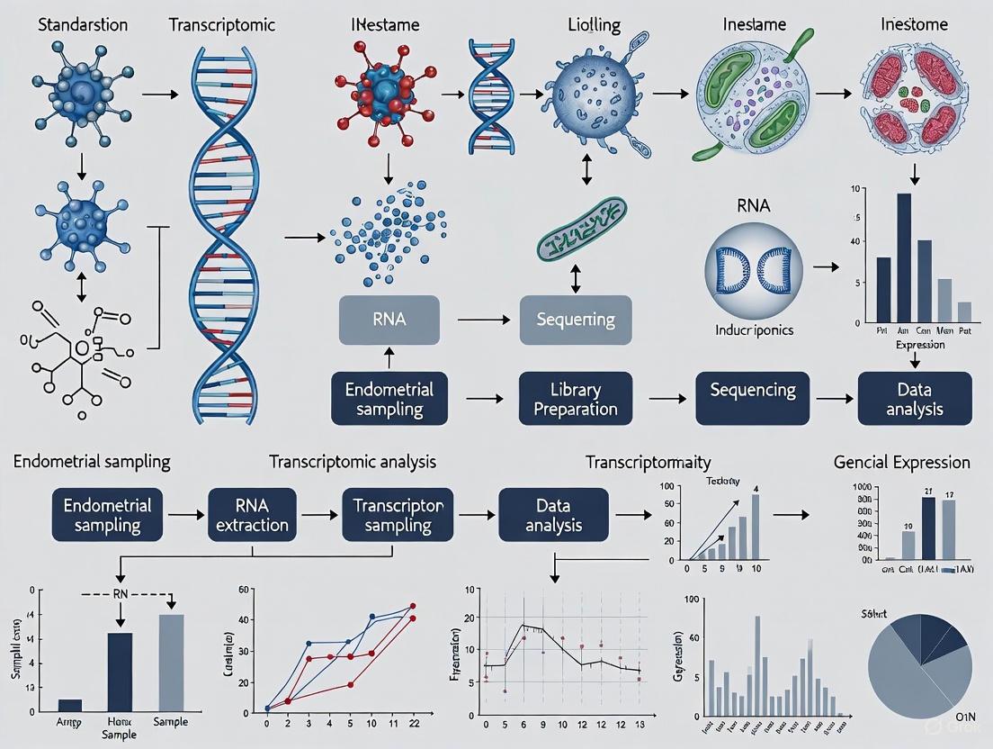
Endometrial Biopsy and Processing Workflow
Protocol 2: Spatial Transcriptomics (10x Visium) of Endometrial Tissue
Objective: To generate a spatial gene expression atlas of the human endometrium during the window of implantation.
Materials: Fresh frozen endometrial tissue blocks, 10x Visium Spatial Tissue Optimization and Gene Expression slides, cryostat, standard reagents for H&E staining, library construction, and sequencing.
Procedure:
- Cryosectioning: Section the fresh frozen tissue into slices of appropriate thickness (e.g., 10 µm) using a cryostat and mount them onto 10x Visium slides [2].
- H&E Staining & Imaging: Stain the tissue sections with Hematoxylin and Eosin (H&E) and image them using a brightfield microscope [2].
- Tissue Permeabilization: Permeabilize the tissue to release mRNA molecules, which are then captured by the spatially barcoded spots on the Visium slide [2].
- Library Preparation: Perform reverse transcription to generate cDNA, followed by library construction according to the standard 10x Visium protocol [2].
- Sequencing: Sequence the libraries on an Illumina NovaSeq 6000 platform using a PE150 model [2].
- Data Processing: Use the Space Ranger count pipeline to align the spatial transcriptome data to the human reference genome (GRCh38), detect tissue sections, and generate feature-spot matrices [2]. Subsequent analysis (normalization, clustering, differential expression) can be performed using tools like Seurat.
Spatial Transcriptomics Experimental Pipeline
The Scientist's Toolkit: Essential Research Reagents & Materials
Table 2: Key Research Reagent Solutions for Endometrial Receptivity Studies
| Item | Function / Application |
|---|---|
| Pipelle Endometrial Catheter | A minimally invasive device for obtaining endometrial tissue biopsies for histology or RNA analysis [2]. |
| RNA-later Solution | A stabilizing solution that rapidly penetrates tissues to preserve RNA integrity by inhibiting RNases immediately after biopsy [2]. |
| OCT Compound | An embedding medium for frozen tissue specimens, used to support the tissue during cryosectioning for spatial transcriptomics [2]. |
| 10x Visium Spatial Slide | A glass slide containing ~5,000 barcoded spots for capturing mRNA from a tissue section, enabling spatial transcriptomics [2]. |
| ERA Test Kit | A commercial kit comprising a customized gene array (e.g., 238 genes) and software to analyze the transcriptomic signature and diagnose the WOI status [3]. |
| Leukemia Inhibitory Factor (LIF) | A pleiotropic cytokine critical for implantation; promotes decidualization, pinopod expression, and trophoblast invasiveness. An important analyte in functional studies [1]. |
| Beta-3 Integrin Antibody | A key adhesion molecule upregulated during the WOI; used as an immunohistochemical marker to assess endometrial receptivity in research settings [1]. |
Frequently Asked Questions
Q1: Why is precise timing of endometrial sampling critical for transcriptomic studies? The human endometrium is receptive to embryo implantation only during a brief period known as the window of implantation (WOI), which is tightly regulated by hormonal cues. Transcriptomic studies have shown that gene expression profiles vary significantly across different phases of the menstrual cycle. Sampling outside this window can capture a non-receptive endometrial state, leading to data that misrepresents the molecular landscape of receptivity and confounds research findings [5] [6].
Q2: How is the LH surge used to determine the window of implantation? The luteinizing hormone (LH) surge is a pivotal physiological marker for ovulation. In a natural cycle, the WOI is generally considered to commence on day LH+7 (the 7th day after the LH surge) [6]. Precise dating via daily serum LH measurements is recommended to align sampling with this critical window, as gene expression dynamics are highly time-sensitive during the LH+3 to LH+11 period [6].
Q3: What is WOI displacement, and how does it affect research on RIF? Window of implantation displacement occurs when the receptive phase is advanced or delayed compared to the expected timeline. Transcriptomic profiling has identified that a significant proportion of patients with Recurrent Implantation Failure (RIF) exhibit a displaced WOI. One study found that 67.5% (27/40) of RIF patients were non-receptive on the conventional sampling day (P+5 in HRT cycles), underscoring the necessity for personalized timing in research cohorts to avoid sampling bias [5].
Q4: What are the consequences of sampling at the wrong time in a hormone replacement therapy (HRT) cycle? In a hormone replacement therapy (HRT) cycle, progesterone administration initiates endometrial transformation. The standard sampling day is P+5 (the 5th day after starting progesterone). If sampling occurs on P+5 for a patient with a delayed WOI, the transcriptome profile will reflect a pre-receptive state. Research shows that adjusting the transfer timing based on an individual's receptivity status can significantly improve pregnancy outcomes, highlighting the functional impact of timing on the molecular data obtained [5].
Troubleshooting Common Experimental Issues
Problem: High inter-individual variability in transcriptomic data. Solution: A major source of variability is the inherent difference in cellular composition between endometrial samples. To troubleshoot:
- Employ Single-Cell RNA Sequencing: If bulk RNA-seq data shows high variability, consider using scRNA-seq. This technology can resolve cellular heterogeneity by profiling individual cells, allowing researchers to identify specific dysregulated subpopulations in RIF patients that bulk methods might average out [6].
- Incorporate Cellular Deconvolution: For existing bulk RNA-seq data, use computational deconvolution methods to estimate the proportions of major cell types (e.g., stromal, epithelial, immune cells) in each sample. This can help determine if observed gene expression differences are driven by shifts in cellular composition rather than true molecular dysregulation [6].
Problem: Inconsistent cycle phase classification across study participants. Solution: Standardize the method for cycle dating.
- Mandate Serum LH Tracking: For natural cycles, insist on daily serum LH measurement to pinpoint the LH surge with high accuracy. This is superior to relying on patient-reported cycle days alone [6].
- Utilize a Transcriptomic Classifier: For both natural and HRT cycles, use a validated molecular tool, such as an Endometrial Receptivity Diagnostic (ERD) model, to objectively classify samples as pre-receptive, receptive, or post-receptive. This can confirm that all samples in the "receptive" group are indeed molecularly aligned [5].
Problem: Inadequate or non-representative endometrial tissue sample. Solution: The sampling method can impact the quality and representativeness of the transcriptomic data.
- Choose an Appropriate Sampling Device: Studies comparing sampling methods have found that hysteroscopically directed biopsy provides the highest diagnostic accuracy and sample adequacy [7]. The Pipelle device is also widely used and is considered effective for sampling in an outpatient setting [8].
- Ensure Sample Adequacy: A minimum amount of tissue is required for RNA sequencing. Verify that the sample obtained is sufficient for subsequent RNA extraction and library preparation.
Experimental Protocols for Standardization
Protocol 1: Standardized Endometrial Biopsy for Transcriptomics in a Natural Cycle This protocol is designed for precise sampling during a natural menstrual cycle.
- Participant Recruitment: Recruit participants with confirmed regular menstrual cycles (e.g., 25-35 days). Exclude individuals with confounding gynecological pathologies (e.g., endometriosis, adenomyosis, endometrial polyps) [5].
- LH Surge Monitoring: Beginning around cycle day 10, participants undergo daily phlebotomy for serum LH measurement. The day of the initial LH surge is designated as LH+0.
- Biopsy Timing: Schedule the endometrial biopsy for the target time point within the WOI (e.g., LH+7). Have a contingency plan for participants who do not exhibit a clear LH surge.
- Sample Collection: Perform an endometrial biopsy using a device such as Pipelle or under hysteroscopic guidance. Gently aspirate tissue from the uterine fundus.
- Sample Processing:
- Immediately place the tissue sample in a sterile cryovial.
- For bulk RNA-seq, flash-freeze the entire sample in liquid nitrogen.
- For scRNA-seq, immediately place the tissue in a chilled preservation medium (e.g., DMEM/F-12 with 10% FBS) and process for single-cell dissociation within one hour of collection [6].
- RNA Extraction & Quality Control: Extract total RNA using a commercial kit. Assess RNA integrity (RIN) using an instrument like Bioanalyzer; only proceed with samples having a RIN > 7.0.
Protocol 2: Standardized Sampling in a Hormone Replacement Therapy (HRT) Cycle This protocol controls for hormonal variability using an artificial cycle.
- Cycle Regulation: Down-regulate the participant's ovarian function with a GnRH agonist if necessary. Initiate estrogen supplementation (e.g., estradiol valerate 4-8 mg daily) on cycle day 2-3 [5].
- Endometrial Monitoring: Monitor endometrial thickness via transvaginal ultrasound. Once the endometrium reaches ≥7 mm, begin progesterone administration (e.g., micronized vaginal progesterone). This day is designated as P+0 [5].
- Biopsy Timing: Perform the endometrial biopsy on the predetermined day, most commonly P+5 for a conventional WOI, or as adjusted by a molecular diagnostic test [5].
- Sample Collection & Processing: Follow steps 4-6 from Protocol 1.
Quantitative Data on Sampling Timing
Table 1: Impact of WOI Displacement and Personalized Timing in RIF Patients
| Parameter | Study Finding | Quantitative Value | Implication for Sampling |
|---|---|---|---|
| Prevalence of Displaced WOI | Proportion of RIF patients non-receptive on conventional day P+5 [5] | 67.5% (27/40 patients) | Highlights high risk of sampling error in RIF population without prior timing assessment. |
| WOI Status in RIF | Distribution of advanced, normal, and delayed WOI in pregnant RIF patients after pET [5] | Advanced: 23% (6/26)Normal: 38.5% (10/26)Delayed: 38.5% (10/26) | WOI displacement is common in RIF, with delays being as frequent as a normal WOI. |
| Clinical Benefit of pET | Clinical pregnancy rate in RIF patients after personalized embryo transfer guided by ERD [5] | 65% (26/40 patients) | Validates that correcting for individual WOI via transcriptomic assessment leads to successful outcomes. |
Table 2: Key Differentially Expressed Genes (DEGs) Associated with WOI Status
| Gene Functional Category | Potential Role in Endometrial Receptivity | Association with WOI Displacement |
|---|---|---|
| Immunomodulation | Regulating the local immune environment to facilitate embryo acceptance [5] | Identified as a key function of DEGs that distinguish advanced, normal, and delayed WOI [5]. |
| Transmembrane Transport | Facilitating nutrient and signaling molecule exchange at the maternal-fetal interface [5] | Identified as a key function of DEGs that distinguish advanced, normal, and delayed WOI [5]. |
| Tissue Regeneration | Involved in endometrial remodeling and decidualization [5] | Identified as a key function of DEGs that distinguish advanced, normal, and delayed WOI [5]. |
The Scientist's Toolkit: Research Reagent Solutions
Table 3: Essential Materials for Endometrial Transcriptomics Research
| Item | Function/Brief Explanation |
|---|---|
| Pipelle Endometrial Suction Curette | A flexible plastic cannula for minimally invasive endometrial biopsy in an outpatient setting. Provides adequate tissue for transcriptomic analysis [8]. |
| Hysteroscope | An endoscopic system for direct visualization of the uterine cavity. Allows for targeted biopsy and can improve diagnostic accuracy for focal lesions [7]. |
| 10X Chromium System | A widely used platform for generating single-cell RNA sequencing libraries. Essential for creating high-resolution cellular atlases of the endometrium [6]. |
| Estradiol Valerate | A form of estrogen used in HRT cycles to promote endometrial proliferation and achieve a standardized thickness prior to progesterone exposure [5]. |
| Micronized Vaginal Progesterone | The progestin used in HRT cycles to transform the estrogen-primed endometrium into a receptive state, mimicking the natural secretory phase [5]. |
| DMEM/F-12 Medium | A complex cell culture medium used for temporary storage and transport of fresh endometrial tissue prior to single-cell dissociation [6]. |
| Collagenase | An enzyme used to digest the extracellular matrix of endometrial tissue, dissociating it into a single-cell suspension suitable for scRNA-seq [6]. |
Experimental Workflow and Molecular Pathways
Diagram 1: Standardized Workflow for Endometrial Transcriptomics
Diagram 2: Key Cellular Dynamics During the Window of Implantation
Troubleshooting Guide: Common Spatial Transcriptomics Challenges in Endometrial Research
FAQ: Sample Quality and Preparation
Q: Our endometrial samples show degraded RNA or low RNA Integrity Numbers (RIN). What are the critical steps we might be missing during collection?
A: Proper sample handling is crucial for preserving RNA quality. Based on validated protocols, you must ensure rapid tissue stabilization immediately after biopsy [2]:
- Immediate freezing: Flash-freeze fresh endometrial tissues in isopentane pre-chilled with liquid nitrogen
- Storage temperature: Store samples at -80°C until sectioning
- RNA quality control: Ensure a minimum RIN larger than 7 before proceeding with spatial transcriptomics
- Sectioning optimization: Determine optimal tissue permeabilization time based on fluorescence imaging strength
Q: How can we confirm our endometrial sampling targets the functionally relevant uterine regions?
A: Standardized anatomical sampling is essential for reproducible results. The documented protocol specifies [2] [9]:
- Sampling location: Collect endometrial samples from the fundal and upper part of the uterus using Pipelle endometrial biopsy
- Cycle timing: Time collection precisely at LH + 7 (7 days after the Luteinizing Hormone surge) during the mid-luteal phase
- Patient stratification: Clearly define control vs. RIF patient groups with consistent demographic matching (age ≤35 years, BMI <28 kg/m²)
FAQ: Data Quality and Technical Validation
Q: Our spatial transcriptomics data shows low gene detection per spot. What quality metrics should we check?
A: Reference quality metrics from published endometrial spatial transcriptomics data provide benchmarks for troubleshooting [2]:
- Minimum spot quality: Filter out spots with gene counts below 500 or mitochondrial gene percentage exceeding 20%
- Expected metrics: Target a median detected gene number of approximately 3,156 per spot
- Sequencing saturation: Aim for over 90% sequencing saturation with Q30 values for barcode, UMI, and RNA read all exceeding 90%
Q: What computational approaches help address spatial heterogeneity when integrating with single-cell data?
A: Successful deconvolution of endometrial spatial data requires specific computational strategies [2]:
- Integration method: Use CARD (conditional autoregressive-based deconvolution) or similar tools to estimate cell type proportions for each spot
- Reference data: Integrate with quality-controlled public single-cell datasets (e.g., GSE183837) after rigorous preprocessing
- Cellular mapping: Focus on dominant epithelial cell populations while accounting for niche-specific variations
Experimental Protocols and Methodologies
Sample Processing and Library Preparation
The standardized workflow for endometrial spatial transcriptomics involves these critical steps [2]:
Tissue Preparation
- Embed fresh frozen tissues in OCT compound
- Section into slices of appropriate thickness (typically 10μm)
- Assess RNA quality to ensure RIN >7
Visium Library Construction
- Utilize 10x Visium Spatial Tissue Optimization Slides
- Perform standard methanol fixation
- Conduct H&E staining for histological context
- Optimize tissue permeabilization to release mRNA
- Capture mRNA on barcode-coated spots
- Perform reverse transcription to generate cDNA
- Construct libraries according to standard protocol
Sequencing
- Use Illumina NovaSeq 6000 platform
- Employ PE150 sequencing model
- Target approximately 3×10⁸ read-pairs per sample
Data Processing and Analysis Pipeline
The computational workflow for analyzing endometrial spatial transcriptomics data includes [2]:
Alignment and Preprocessing
- Use Space Ranger count pipeline (version 2.0.0)
- Align to human reference genome (GRCh38-2020-A)
- Detect tissue sections and align fiducials across slices
Quality Control and Normalization
- Filter spots with gene count <500 or mitochondrial percentage >20%
- Normalize spot expression data using SCTransform function
- Merge all slices using merge function in Seurat
Spatial Analysis
- Perform principal component analysis using top 30 PCs
- Conduct dimension reduction with resolution of 0.6
- Identify spatial niches using unsupervised clustering
- Perform differential gene expression analysis among spatial clusters
Table 1: Sample Demographics and Sequencing Metrics from Endometrial Spatial Transcriptomics
| Parameter | Control Group (n=4) | RIF Group (n=4) | Technical Benchmark |
|---|---|---|---|
| Age Range (years) | 24-32 | 25-33 | ≤35 |
| BMI (kg/m²) | 18.14-24.37 | 20.35-24.47 | <28 |
| Previous Embryo Transfers | 0 | 3-5 | ≥3 for RIF definition |
| Spots Under Tissue | 751-2018 per sample | 751-2018 per sample | Variable by tissue size |
| Median Genes per Spot | >2000 | >2000 | 3156 (average) |
| Median UMI Counts | >4000 | >4000 | 6860 (average) |
| Sequencing Saturation | >90% | >90% | >90% target |
| Mitochondrial Gene % | ~5.5% | ~5.5% | <20% threshold |
Data compiled from GSE287278 dataset [2] [9]
Table 2: Cellular Composition Revealed by Spatial-Single Cell Integration
| Cell Type | Spatial Distribution | Functional Significance in Endometrium | RIF-Associated Alterations |
|---|---|---|---|
| Unciliated Epithelia | Dominant component across niches | Endometrial receptivity, implantation signaling | Potential compositional shifts |
| Ciliated Epithelia | Limited spatial domains | Mucosal clearance, fluid movement | Under investigation |
| Stromal Fibroblasts | Niche-specific distribution | Tissue support, decidualization | Spatial organization changes |
| Immune Cells | Varied spatial localization | Immune tolerance, inflammation regulation | Altered spatial patterns in RIF |
| Endothelial Cells | Vascular niche areas | Angiogenesis, nutrient delivery | Potential vascular changes |
Based on CARD deconvolution analysis integrating ST with scRNA-seq data [2]
The Scientist's Toolkit: Essential Research Reagents and Materials
Table 3: Key Research Reagent Solutions for Endometrial Spatial Transcriptomics
| Reagent/Platform | Specific Function | Application Notes |
|---|---|---|
| 10x Visium Spatial Slide | Spatial barcoding of mRNA transcripts | Each capture area: 6.5×6.5mm with ~5000 spots |
| Space Ranger (v2.0.0) | Alignment and spatial data processing | Requires GRCh38-2020-A reference genome |
| Seurat (v4.3.0) | Spatial data analysis and integration | Use Load10X_Spatial() for data import |
| CARD Package (v1.1) | Spatial deconvolution with scRNA integration | Estimates cell type proportions per spot |
| Harmony (v1.0) | Batch effect correction | Essential for multi-sample integration |
| DoubletFinder (v2.0.3) | Doublet detection in scRNA data | Preprocessing for quality reference data |
| Methanol Fixation Solution | Tissue preservation | Standard protocol for Visium platform |
| H&E Staining Kit | Histological context | Correlates spatial gene expression with tissue morphology |
Compiled from methodology sections of cited spatial transcriptomics studies [2]
Advanced Methodological Considerations
Addressing Spatial Heterogeneity in Experimental Design
The identification of seven distinct cellular niches in endometrial tissues underscores the profound spatial heterogeneity that researchers must account for in experimental design [2]. This heterogeneity presents both challenges and opportunities:
- Niche-specific signatures: Each spatial niche demonstrates unique gene expression profiles requiring specialized analytical approaches
- Sampling considerations: Limited biopsies may miss critical niche-specific biological processes
- Integration strategies: Combining spatial data with single-cell references enables resolution of cellular composition within each niche
Standardization Framework for Endometrial Sampling
Based on the accumulated methodological evidence, a robust standardization framework should incorporate:
Temporal standardization: Precise timing to LH+7 during the mid-luteal phase to capture the window of implantation [2] [10]
Spatial standardization: Consistent sampling from fundal/upper uterine regions to reduce anatomical variability [2] [9]
Technical standardization: Implementation of uniform RNA quality thresholds (RIN>7) and sequencing depth targets (>90% saturation) [2]
Analytical standardization: Application of consistent clustering parameters (resolution=0.6) and integration methods for cross-study comparisons [2]
This technical support resource provides a foundation for addressing common challenges in endometrial spatial transcriptomics research, with standardized protocols and troubleshooting guidance specifically tailored to overcome spatial heterogeneity challenges in this critical reproductive tissue.
Frequently Asked Questions (FAQs)
1. Why is the timing of an endometrial biopsy so critical for transcriptomics studies of implantation? The endometrium is receptive to embryo implantation only during a specific, narrow period known as the window of implantation (WOI). Transcriptomic studies show that gene expression profiles differ significantly between pre-receptive, receptive, and post-receptive phases [5]. Sampling outside of this personalized WOI can lead to a non-receptive gene expression signature, which is often associated with Recurrent Implantation Failure (RIF). Adjusting embryo transfer timing based on transcriptomic dating has been shown to improve clinical pregnancy rates [5].
2. What is the difference between a blind Pipelle biopsy and a hysteroscopy-guided biopsy? A blind biopsy, typically performed with a suction catheter (Pipelle), involves sampling the endometrium without direct visualization. In contrast, a hysteroscopy-guided biopsy allows the clinician to visually inspect the uterine cavity and take targeted samples from specific areas, such as suspected lesions [11]. While blind biopsies are common and cost-effective, hysteroscopy is the gold standard for diagnosing focal intrauterine pathologies like polyps or submucosal fibroids, as blind sampling can miss these lesions [11].
3. How can sample storage affect the integrity of RNA for transcriptomic analysis? Long-term storage of biospecimens can significantly alter the molecular composition of the sample. Studies have shown that biomarker levels, including RNA and proteins, can increase or decrease over time, depending on storage conditions and duration [12]. For instance, one experiment found that levels of certain serum markers increased by approximately 15% over ten years of storage [12]. Such pre-analytical variations can introduce bias, leading to underestimated or overestimated associations in biomarker discovery studies.
4. What are the key quality control metrics for spatial transcriptomics data from endometrial samples? For spatial transcriptomics using platforms like 10x Visium, key quality metrics include [2]:
- Sequencing Saturation: Should be over 90%, indicating sufficient sequencing depth.
- Q30 Score: Should exceed 90% for barcode, UMI, and RNA reads, reflecting high sequencing accuracy.
- Spot Quality: Spots should have a minimum number of detected genes (e.g., >500) and a low percentage of mitochondrial genes (e.g., <20%).
- Median Genes/UMI per Spot: High-quality datasets typically show a median of over 2000 genes and 4000 UMI counts per spot [2].
5. My RT-qPCR results after endometrial sampling show low amplification. What could be the cause? Low amplification can stem from several issues related to RNA quality and the reverse transcription process [13]:
- Poor RNA Integrity: RNA may have degraded during sample collection, storage, or extraction.
- Low RNA Purity: Contaminants from the extraction process (e.g., salts, inhibitors) can co-purify with the RNA.
- Insufficient RNA Quantity: The starting amount of RNA may be below the optimal range for the protocol.
- Genomic DNA Contamination: Can lead to nonspecific amplification. Treating samples with DNase is recommended [13].
Troubleshooting Guides
Common Experimental Issues and Solutions
| Problem | Possible Cause | Recommended Solution |
|---|---|---|
| High Background Noise in Spatial Transcriptomics | Low sequencing saturation or high mitochondrial read percentage [2] | Filter spots with high mitochondrial gene percentage (>20%) and ensure sequencing saturation is >90% [2]. |
| Insufficient Endometrial Tissue from Biopsy | Incorrect biopsy technique or atrophic endometrium [14] | Ensure the pipelle is moved in and out with a twisting motion to sample all quadrants. A second pass can be made if needed [15]. |
| Inconsistent Biomarker Profiles in RIF Patients | Incorrect WOI timing or patient-to-patient variability [5] | Use a transcriptomic-based model (e.g., ERD) to determine the personalized WOI for each patient before sampling [5]. |
| Degraded RNA from Endometrial Samples | Improper handling post-biopsy; multiple freeze-thaw cycles; RNase contamination [13] | Snap-freeze tissue immediately in liquid nitrogen. Use RNase-free reagents and equipment. Limit freeze-thaw cycles. Assess RNA Integrity Number (RIN) prior to use [13]. |
| Bias in Biomarker Association Estimates | Long-term storage of samples altering molecular concentrations [12] | Document storage time and conditions meticulously. If possible, use samples with comparable storage histories for case-control studies to minimize bias [12]. |
Standardized Protocol for Endometrial Sampling for Transcriptomics
This protocol is adapted from research on Recurrent Implantation Failure (RIF) and spatial transcriptomics studies [5] [2].
1. Patient Selection and Preparation
- Indications: Include patients with RIF or those undergoing fertility treatment. Exclude patients with uterine pathologies (e.g., endometriosis, fibroids, adenomyosis), active pelvic infection, or pregnancy [5] [2].
- Cycle Timing: For natural cycles, schedule the biopsy at the mid-luteal phase (e.g., LH+7). For hormone replacement therapy (HRT) cycles, schedule it on the 5th day of progesterone administration (P+5) [5].
- Patient Consent: Obtain written informed consent approved by an institutional ethics committee [2].
2. Biopsy Procedure
- Technique: Perform an endometrial biopsy using a Pipelle catheter or similar device.
- Steps:
- The patient is placed in the lithotomy position. A speculum is inserted to visualize the cervix [15].
- The cervix may be cleansed with an antiseptic solution. Topical lidocaine can be applied to reduce discomfort [14].
- A tenaculum may be applied to the cervix to stabilize it, though this can increase pain [14].
- Gently insert the Pipelle through the cervical canal into the uterine fundus.
- Withdraw the internal piston fully to create suction. While maintaining suction, rotate the catheter 360 degrees and move it in and out of the uterine cavity 3-4 times to sample from different areas [14] [15].
- Withdraw the catheter and expel the tissue into a preservation medium.
3. Sample Processing and Storage
- Immediate Handling: Immediately after collection, the tissue should be rinsed if necessary and divided for intended analyses.
- For RNA Sequencing: Place the tissue fragment directly into a cryovial and snap-freeze in liquid nitrogen. Store at -80°C until RNA extraction [5].
- For Spatial Transcriptomics: Embed the fresh tissue in Optimal Cutting Temperature (OCT) compound, snap-freeze in isopentane pre-chilled with liquid nitrogen, and store at -80°C. Section tissues and ensure RNA Integrity Number (RIN) is >7 before proceeding [2].
4. RNA Extraction and Quality Control
- Use commercial kits designed for RNA extraction from tissues.
- Assess RNA concentration and purity using spectrophotometry (e.g., Nanodrop).
- Evaluate RNA integrity using a Bioanalyzer or similar system. A RIN >7 is generally recommended for transcriptomic studies [2].
Reverse Transcription (RT) and cDNA Synthesis Troubleshooting
This guide addresses key steps critical for downstream transcriptomic analyses like RT-qPCR and RNA-Seq [13].
| Problem | Possible Cause | Solution |
|---|---|---|
| Low or No Amplification in RT-qPCR | Poor RNA integrity, low RNA purity, or low RNA quantity [13] | Assess RNA integrity by gel electrophoresis. Repurify RNA to remove inhibitors. Use a high-performance reverse transcriptase. Confirm RNA quantity accurately [13]. |
| Nonspecific Amplification | Contamination with genomic DNA (gDNA) [13] | Treat RNA samples with DNase before reverse transcription. Include a "no-RT" control in qPCR experiments [13]. |
| Truncated cDNA Fragments | High GC content or secondary structures in RNA; poor RNA integrity [13] | Denature RNA at 65°C for 5 min before RT. Use a thermostable reverse transcriptase and perform the reaction at a higher temperature (e.g., 50°C) [13]. |
| Poor Representation of Transcripts | Suboptimal priming strategy [13] | For potentially degraded RNA, use random hexamers instead of oligo(dT) primers to ensure proper coverage of transcripts that may lack poly-A tails [13]. |
The Scientist's Toolkit: Essential Reagents & Materials
| Item | Function in Experiment |
|---|---|
| Pipelle Endometrial Suction Catheter | A flexible tube used to perform minimally invasive endometrial biopsies by suction to obtain tissue samples [14] [15]. |
| RNase Inhibitors | Enzymes added to reactions to protect RNA from degradation by ubiquitous RNases during sample processing and storage [13]. |
| Formalin Solution (10% Neutral Buffered) | A fixative used to preserve tissue architecture for histological examination. Note: Not suitable for RNA extraction [14]. |
| TRIzol Reagent | A monophasic solution of phenol and guanidinium isothiocyanate used for the simultaneous isolation of RNA, DNA, and proteins from tissue samples. |
| High-Performance Reverse Transcriptase | An enzyme with high thermal stability and processivity used to synthesize complementary DNA (cDNA) from RNA templates, even from degraded or inhibitor-containing samples [13]. |
| DNase I, RNase-free | An enzyme that degrades double- and single-stranded DNA to remove genomic DNA contamination from RNA preparations prior to reverse transcription [13]. |
| Visium Spatial Gene Expression Slide | A glass slide from 10x Genomics containing ~5,000 barcoded spots for capturing mRNA from tissue sections for spatial transcriptomics analysis [2]. |
Experimental Workflows and Data Analysis
Endometrial Transcriptomics Workflow for Biomarker Discovery
The diagram below outlines the key steps from patient selection to data analysis in a transcriptomic study of endometrial receptivity.
Impact of Sample Storage on Biomarker Association
Long-term storage of biospecimens can significantly bias the estimates of association between biomarker levels and clinical outcomes. The table below summarizes findings from a simulation study based on real data [12].
| Change in Marker Level Over 10 Years | Direction of Bias in Odds Ratio (OR) | Relative Bias |
|---|---|---|
| 15% Increase (e.g., CA 15-3) [12] | Underestimation of true OR | -10% |
| 15% Decrease | Overestimation of true OR | +20% |
This demonstrates that an observed 15% increase in marker levels over a decade can lead to a significant 10% underestimation of the true association, potentially leading to false negative conclusions in biomarker discovery studies [12].
Sampling in Practice: A Comparative Guide to Techniques and Protocol Standardization
Troubleshooting Guide & FAQs
Tissue Quality & RNA Integrity
Q1: Our RNA Integrity Number (RIN) from Pipelle samples is consistently below 7.0, which is suboptimal for transcriptomics. What are the primary factors affecting RNA quality and how can we mitigate them?
A: Low RIN values are frequently caused by pre-analytical variables. Key factors and solutions include:
- Ischemic Time: Minimize the time from tissue devascularization (sampling) to preservation. Aim for under 10 minutes.
- Preservation Method: Immediately submerge tissue in at least 10 volumes of RNAlater. Do not freeze directly without a cryoprotectant.
- Sample Handling: Avoid excessive manipulation or squeezing of the tissue with forceps.
- Protocol: Adopt the following standardized protocol:
- Pre-chill: Pre-cool a 15mL conical tube containing 5-10 mL of RNAlater on wet ice.
- Immediate Transfer: Eject the tissue core from the Pipelle directly into the chilled RNAlater.
- Dissection: Within 30 minutes, under a sterile laminar flow hood, use RNase-free instruments to dissect away any gross blood clot or necrotic material.
- Incubation: Incubate the tube at 4°C overnight for complete penetration.
- Storage: Transfer the sample to -80°C for long-term storage.
Q2: We observe significant inter-sample variability in transcriptomic profiles from D&C samples. Could this be due to tissue heterogeneity, and how can we control for it?
A: Yes, the endometrium is highly dynamic and heterogeneous. D&C, while providing a large tissue volume, is a "blind" procedure that samples a mixture of functionalis and basalis layers non-specifically.
- Solution: Implement a rigorous histological confirmation and macro-dissection step.
- After preservation, a small portion of the sample can be flash-frozen for cryosectioning and H&E staining.
- A pathologist or trained researcher should confirm the tissue type and proportion of endometrial epithelium vs. stroma.
- For laser capture microdissection (LCM), standardize the collection of specific glandular regions. Alternatively, for bulk RNA-seq, only use samples with a high and consistent epithelial/stromal ratio (e.g., >70% epithelium) as confirmed by histology.
Sampling Procedure & Yield
Q3: Our hysteroscopically directed biopsies often yield insufficient tissue for downstream RNA extraction and library preparation. What can we do to improve yield?
A: Insufficient yield from directed biopsies is often a technique or equipment issue.
- Biopsy Forceps: Ensure you are using large-capacity (e.g., 5.0Fr or larger) biopsy forceps. Avoid small or alligator-style forceps that crush the tissue.
- Biopsy Site: Target areas that appear representative of the pathology or cycle phase. Avoid necrotic or heavily hemorrhagic regions.
- Multiple Passes: Standardize your protocol to include 3-5 directed biopsies from the same region of interest, pooling them into a single preservation tube. This increases biomass while maintaining biological specificity.
- Validation: Weigh the tissue sample after preservation and dissection. A minimum of 50 mg is recommended for robust RNA extraction and potential QC replicates.
Q4: How does the choice of sampling device (Pipelle vs. Hysteroscopic forceps vs. D&C curette) impact the cellular composition of the sample?
A: The sampling method directly influences the cellular composition, which is a critical confounder in transcriptomic studies.
| Sampling Method | Typical Cellular Composition (Qualitative) | Key Considerations for Transcriptomics |
|---|---|---|
| Pipelle | Mixed functionalis layer; variable epithelial/stromal ratio; can include underlying basalis or myometrial cells if inserted too deeply. | High inter-operator variability. Requires mandatory post-hoc histological confirmation of composition. |
| Hysteroscopic Biopsy | Targeted region (e.g., polyp, lesion); primarily epithelium and adjacent stroma from the specific site. | Excellent for lesion-specific analysis but may not represent "global" endometrial transcriptome. Low cellular heterogeneity if targeted correctly. |
| Dilation & Curettage | Large volume of mixed tissue from entire uterine cavity; includes functionalis and basalis layers, blood clots, and debris. | High yield but highest cellular heterogeneity. Requires extensive macro-dissection to obtain a representative and consistent sample. |
Experimental Protocols
Protocol 1: Standardized Endometrial Tissue Processing for RNA-Seq
Objective: To preserve high-quality RNA from endometrial biopsies for downstream transcriptomic analysis. Materials: See "Research Reagent Solutions" table. Steps:
- Preparation: Pre-label and pre-chill 15mL conical tubes with 5mL of RNAlater on wet ice.
- Sampling: Perform the clinical sampling procedure (Pipelle, Hysteroscopic Biopsy, or D&C).
- Immediate Preservation: Transfer tissue immediately from the device into the chilled RNAlater. Gently swirl the tube to ensure the tissue is fully submerged.
- Dissection (within 30 mins): In a RNase-free environment, pour the contents into a sterile Petri dish. Using fine forceps and a scalpel, remove any visible blood clot, mucus, or non-endometrial tissue.
- Incubation: Return the cleaned tissue to the RNAlater and incubate at 4°C for 16-24 hours.
- Aliquoting & Storage: Remove the tissue, briefly blot on a clean wipe, and snap-freeze in liquid nitrogen. Store at -80°C. A small aliquot (e.g., 10-20 mg) can be saved for histology in OCT compound.
Protocol 2: Histological Validation of Endometrial Biopsies
Objective: To quantify the epithelial-to-stromal ratio and confirm tissue type. Materials: Cryostat, OCT compound, Microtome, H&E staining solutions. Steps:
- Embedding: Embed the OCT-embedded tissue aliquot and section at 5-7 µm thickness.
- Staining: Perform standard Hematoxylin and Eosin (H&E) staining.
- Imaging: Digitally scan the slide at 20x magnification.
- Quantification: Use image analysis software (e.g., QuPath, ImageJ) to annotate and calculate the area percentage of endometrial epithelium versus stroma.
- Inclusion/Exclusion: Based on pre-defined criteria (e.g., >60% epithelium), include or exclude samples from the transcriptomics pipeline.
Table 1: Comparison of Sampling Method Attributes
| Attribute | Pipelle Suction Curettage | Hysteroscopically Directed Biopsy | Dilation & Curettage (D&C) |
|---|---|---|---|
| Average Tissue Yield (mg) | 15 - 45 mg | 5 - 25 mg (per bite) | 200 - 1000 mg |
| Typical RNA Yield (µg) | 2 - 10 µg | 1 - 5 µg (per bite) | 30 - 150 µg |
| Median RIN Value (Range) | 7.5 (5.5 - 9.5) | 8.2 (7.0 - 9.8) | 6.8 (4.0 - 9.0) |
| Procedure Cost (Relative) | $ | $$ | $$$ |
| Operator Skill Level | Low | High | High (Requires anesthesia) |
| Visual Guidance | No (Blind) | Yes (Direct visualization) | No (Blind) |
Table 2: Research Reagent Solutions
| Item | Function | Example Product/Catalog # |
|---|---|---|
| RNAlater Stabilization Solution | Stabilizes and protects RNA integrity in fresh tissue samples immediately after collection. | Thermo Fisher Scientific, AM7020 |
| RNase-Free Water | Used to prepare solutions and reconstitute RNA to prevent degradation by RNases. | Thermo Fisher Scientific, AM9937 |
| RNeasy Mini Kit | Spin-column based total RNA purification from small tissue samples. | QIAGEN, 74104 |
| Agilent RNA 6000 Nano Kit | Analysis of RNA integrity and quantification using the Bioanalyzer system. | Agilent Technologies, 5067-1511 |
| OCT Compound | Optimal Cutting Temperature medium for embedding tissue for cryosectioning. | Sakura Finetek, 4583 |
| Laser Capture Microdissection (LCM) Slides | Special membrane slides for precise capture of specific cell populations. | Thermo Fisher Scientific, LCM0522 |
Visualizations
Title: Endometrial Sampling Transcriptomics Workflow
Title: Sampling Method Attribute Comparison
Troubleshooting Guides
UF-EV Isolation and Quality Control
Problem: Low RNA yield or purity from isolated UF-EVs.
- Potential Cause 1: Inefficient vesicle lysis or RNA extraction protocol.
- Solution: Optimize lysis buffer composition and incubation time. Include a spike-in control (e.g., synthetic RNA sequences not found in humans) to monitor extraction efficiency [16].
- Potential Cause 2: Co-isolation of contaminants like proteins or lipoproteins.
- Solution: Combine isolation methods. Following ultracentrifugation with a density gradient centrifugation step can significantly improve EV purity [17].
Problem: Inconsistent results between experimental replicates.
- Potential Cause 1: Variation in uterine fluid collection volume or handling.
- Solution: Standardize the sample collection protocol. Use the same type of catheter and flush with a consistent, predefined volume of saline. Process all samples with the same centrifugation steps to remove cells and debris immediately after collection [18].
- Potential Cause 2: Incomplete characterization of isolated EVs.
- Solution: Implement rigorous quality control using multiple, complementary techniques. The table below outlines essential characterization methods [17] [19].
Table: Essential Characterization Methods for Isolated UF-EVs
| Method | Function | Key Metrics |
|---|---|---|
| Nanoparticle Tracking Analysis (NTA) | Measures particle size distribution and concentration. | Peak particle size (~100-200 nm for exosomes), mode diameter. |
| Transmission Electron Microscopy (TEM) | Visualizes EV morphology and membrane integrity. | Confirmation of cup-shaped, bilayer-bound vesicles. |
| Western Blotting | Detects presence of protein markers. | Positive for CD63, CD9, CD81; negative for Calnexin. |
Transcriptomic Data Generation and Analysis
Problem: High background noise in transcriptomic data.
- Potential Cause: The RNA extracted from UF-EVs is of low abundance and potentially degraded.
- Solution: Use a targeted RNA sequencing approach rather than whole transcriptome sequencing. Assays like the FoundationOneRNA are designed to work with low input (as low as 1.5ng RNA) and can achieve high sensitivity and reproducibility even with challenging samples [20] [21].
Problem: Poor reproducibility of Differentially Expressed Genes (DEGs).
- Potential Cause 1: Failure to account for major sources of biological variation, such as the menstrual cycle phase.
- Solution: Accurately date the endometrial cycle phase for every sample and include it as a key covariate in the statistical model. Molecular dating methods are more precise than histological dating alone [22].
- Potential Cause 2: Small sample sizes and over-reliance on single studies.
- Solution: Employ meta-analysis approaches that combine data from multiple datasets. Methods like "SumRank" prioritize genes that show consistent differential expression across studies, greatly improving the reliability of findings [23].
Frequently Asked Questions (FAQs)
Q1: What are the key advantages of using UF-EVs over traditional endometrial biopsies for transcriptomic studies? UF-EVs offer a completely non-invasive method for sampling the endometrial environment. They can be collected without the discomfort and potential complications of a biopsy, allowing for repeated sampling within the same menstrual cycle or across multiple cycles. This is invaluable for monitoring dynamic changes, such as the window of implantation. Furthermore, their molecular cargo (RNA, proteins) actively reflects the state of the endometrium and the communication with the embryo [16] [18].
Q2: My UF-EV RNA sequencing data shows thousands of differentially expressed genes. How can I prioritize genes for functional validation? Instead of relying solely on p-values, use a systems biology approach to identify functionally relevant gene groups.
- Network Analysis: Use Weighted Gene Co-expression Network Analysis (WGCNA) to cluster genes into modules based on their expression patterns. This identifies groups of genes that are biologically coordinated [16].
- Pathway Enrichment: Analyze these modules for enrichment in key biological processes related to endometrial receptivity and embryo implantation (e.g., cell adhesion, inflammatory response, immune modulation) [16] [18].
- Integrated Models: Build predictive models, such as Bayesian logistic regression, that integrate gene module expression with critical clinical variables (e.g., maternal age, history of miscarriage). This identifies the most powerful combination of molecular and clinical factors for outcome prediction [16].
Q3: How does the presence of an embryo influence the transcriptomic profile of UF-EVs? The presence of a blastocyst actively modifies the protein and, by extension, the likely RNA cargo of UF-EVs. In vivo studies comparing pregnant and cyclic heifers show that UF-EVs from pregnant individuals carry a distinct molecular profile. These changes are associated with biological processes crucial for pregnancy, including modulation of inflammatory and immune responses, enhancement of endometrial receptivity, and promotion of processes that support early embryonic development like cell adhesion and stem cell differentiation [18]. This indicates a active dialogue between the embryo and mother via EVs.
Research Reagent Solutions
Table: Key Reagents and Kits for UF-EV Transcriptomic Profiling
| Item | Function | Example/Note |
|---|---|---|
| EV Isolation Kit | Isolates EVs from uterine fluid samples. | Kits based on precipitation (e.g., PEG-based) or size-exclusion chromatography are commonly used. |
| RNA Extraction Kit | Purifies high-quality total RNA from small volumes. | Select kits optimized for low-abundance RNA and compatible with small RNA species. |
| Targeted RNA-Seq Panel | For transcriptome library prep from low-input/ degraded RNA. | FoundationOneRNA; designed for fusion detection and gene expression from 1.5ng input [20] [21]. |
| Spike-in RNA Controls | To monitor technical variation in RNA extraction and sequencing. | Add known quantities of exogenous synthetic RNA to the sample during lysis. |
| Antibodies for EV Characterization | Confirm EV identity and purity via Western Blot. | Antibodies against tetraspanins (CD63, CD81, CD9) and negative marker Calnexin [19]. |
Experimental Workflow and Signaling Pathways
The following diagram illustrates the complete workflow for transcriptomic profiling of UF-EVs, from sample collection to data interpretation.
The diagram below summarizes the key biological processes and signaling pathways influenced by UF-EVs during embryo implantation, as revealed by transcriptomic studies.
Troubleshooting Guides and FAQs
Frequently Asked Questions
Q1: What are the consequences of vague SOPs in a laboratory or clinical setting? Vague or ambiguous SOPs lead to inconsistent practices, increased errors, and wasted time as staff seek clarification [24]. In severe cases, lack of clarity in safety procedures can result in serious injuries or fatalities [25].
Q2: How often should SOPs be reviewed and updated? SOPs should not be static documents. Best practice is to schedule routine reviews—at least annually—or whenever processes, equipment, or regulations change [24] [26]. A designated owner should be responsible for maintaining SOP accuracy [24].
Q3: What is the best way to ensure SOPs are always accessible to staff? Store SOPs in a centralized, digital platform that can be accessed via both computers and mobile devices from the work situation [25] [24]. This ensures everyone can find the latest version instantly, eliminating confusion from multiple outdated copies [27].
Q4: Why is employee training and feedback critical for SOP effectiveness? Even a perfect SOP is useless if employees don't understand it or follow it correctly [24]. Involving frontline staff in SOP development and providing hands-on training ensures the procedures are practical and that staff are competent and engaged in following them [24] [27].
Q5: Which endometrial sampling method is most accurate for research? Hysteroscopically directed biopsy demonstrates superior diagnostic accuracy for detecting endometrial hyperplasia and carcinoma compared to Pipelle suction curettage and Dilatation & Curettage (D&C) [28]. Blind techniques are not reliable for diagnosing focal pathologies like polyps [29] [11].
Troubleshooting Common Issues
Problem: Low or Poor Quality Nucleic Acid Yield from Endometrial Tissue
- Potential Causes:
- Degraded nucleic acid due to prolonged time between tissue acquisition and fixation [30].
- Sample contaminants (e.g., residual phenol, salts, EDTA) inhibiting downstream enzymatic reactions [30].
- Inaccurate quantification of starting material, leading to suboptimal reaction conditions [30].
- Inadequate tissue sampling from the procedure itself [28] [29].
- Corrective Actions:
- Minimize the ischemia time; place tissue in fixative or stabilization solution immediately after collection.
- Re-purify the input sample using clean columns or beads to remove inhibitors. Ensure wash buffers are fresh [30].
- Use fluorometric methods (e.g., Qubit) rather than UV absorbance for template quantification, as it is more accurate for usable material [30].
- Ensure the sampling method is appropriate. Hysteroscopic biopsy provides a targeted sample and is less likely to yield insufficient material compared to blind techniques [28] [11].
Problem: Inconsistent Tissue Fixation Affecting Transcriptomics Data
- Potential Causes:
- Variable fixation times across different samples.
- Incorrect fixative volume, leading to incomplete penetration.
- Fixative not freshly prepared or degraded.
- Large tissue fragments that the fixative cannot penetrate rapidly.
- Corrective Actions:
- Standardize and document fixation time for every sample (e.g., 24-48 hours for 10% Neutral Buffered Formalin).
- Use a fixative volume at least 10 times the tissue volume to ensure complete immersion and penetration.
- Follow manufacturer guidelines for fixative preparation and shelf life.
- Dissect large tissue samples to a uniform thickness (e.g., 5 mm) before fixation to ensure uniform preservation.
Problem: Incorrect Patient Identification or Sample Labeling
- Potential Causes:
- Failure to confirm patient identity at the time of sample collection.
- Handwriting illegibility on sample containers.
- Labeling performed before patient identification is verified.
- Corrective Actions:
- Implement a "Two-Patient Identifier" rule (e.g., full name and date of birth) confirmed by the patient themselves before the procedure.
- Label specimen containers in the presence of the patient after the sample is obtained, not before.
- Use pre-printed labels or electronic label printing systems to eliminate handwriting errors.
Endometrial Sampling Methodologies and Data
Diagnostic Accuracy of Endometrial Sampling Techniques
The table below summarizes the diagnostic performance of different sampling methods for detecting endometrial hyperplasia or carcinoma in premenopausal women, based on a retrospective cohort analysis of 2054 patients [28].
| Sampling Method | Area Under Curve (AUC) | Sensitivity | Specificity |
|---|---|---|---|
| Hysteroscopically Directed Biopsy | 0.957 | 91.3% | Excellent (p<0.001) |
| Dilatation and Curettage (D&C) | 0.909 | 82.0% | Excellent (p<0.001) |
| Pipelle Suction Curettage | 0.858 | 71.7% | Excellent (p<0.001) |
Patient Risk Factors for Endometrial Hyperplasia or Carcinoma
A multivariate analysis identified key risk factors in premenopausal women. The following odds ratios (OR) indicate the change in risk associated with each factor [28].
| Risk Factor | Odds Ratio (OR) | p-value |
|---|---|---|
| Body Mass Index (BMI) (per unit increase) | 1.054 | 0.005 |
| Hypertension | 1.99 | 0.009 |
| Multiparity (per additional delivery) | 0.877 | 0.029 |
Experimental Protocols and Workflows
Detailed Protocol: Hysteroscopically Directed Biopsy
- Patient Preparation & Consent:
- Obtain and document informed consent after explaining the procedure, risks, and benefits.
- Confirm patient identity using two independent identifiers.
- Position the patient in the dorsal lithotomy position.
- Equipment and Setup:
- Ensure a rigid or flexible hysteroscope, light source, and distension medium (saline) are ready.
- Prepare biopsy forceps, specimen containers with fixative, and personal protective equipment.
- Procedure:
- Perform a bimanual examination to determine uterine position and size.
- Introduce the hysteroscope under direct visualization through the cervical canal into the uterine cavity.
- Systematically inspect the entire endometrial surface (anterior, posterior, lateral walls, and fundus).
- Identify any abnormal areas (e.g., focal lesions, irregular thickening).
- Using the biopsy forceps, take targeted samples from the most suspicious areas. For transcriptomics, also sample a standardized control site (e.g., anterior fundal wall).
- If no focal abnormality is seen, take a random biopsy from the uterine fundus.
- Tissue Handling:
- Immediately retrieve the tissue from the forceps using a sterile needle.
- Gently place the tissue into a pre-labeled container filled with an adequate volume of RNA stabilization reagent (e.g., RNAlater) or 10% Neutral Buffered Formalin for histology.
- Ensure the container is tightly sealed and the specimen is fully submerged.
Detailed Protocol: Pipelle Suction Curettage
- Patient Preparation & Consent: As described for the hysteroscopic biopsy.
- Equipment and Setup:
- Prepare a Pipelle endometrial sampler, speculum, tenaculum, and specimen container.
- Procedure:
- Visualize the cervix using a speculum.
- Gently introduce the Pipelle sampler into the uterine cavity until the fundus is reached.
- Withdraw the sampler's inner piston to its full length to create negative pressure.
- While maintaining suction, move the sampler back and forth 3-4 times in a rotating motion to sample different areas of the cavity.
- Release the suction and carefully withdraw the sampler from the uterus.
- Tissue Handling:
- Expel the tissue core directly into a container with fixative or RNA stabilizer by vigorously pushing the piston through the sampler.
- If the sample is scant, the sampler can be rinsed with a saline solution or buffer to collect all material.
Standardized Workflow Diagrams
Standardized Endometrial Tissue Workflow for Transcriptomics
Troubleshooting Low RNA Yield from Endometrial Tissue
Research Reagent Solutions
Essential materials and reagents for endometrial sampling and tissue processing for transcriptomics research.
| Item | Function/Benefit |
|---|---|
| RNAlater Stabilization Solution | Preserves RNA integrity immediately after tissue collection by inactivating RNases, crucial for accurate transcriptomic data [30]. |
| 10% Neutral Buffered Formalin | Standard fixative for histopathological diagnosis. Ensures tissue morphology is preserved for subsequent H&E staining and diagnostic confirmation [28]. |
| Hysteroscope & Biopsy Forceps | Enables direct visualization of the endometrial cavity and allows for targeted biopsy of specific lesions, improving diagnostic accuracy and sample relevance [28] [11]. |
| Pipelle Endometrial Sampler | A thin, flexible catheter for blind endometrial sampling. Less invasive but has lower sensitivity for focal pathology compared to hysteroscopy [28] [29]. |
| RNA Extraction Kit (e.g., Spin-Column) | For isolating high-quality total RNA from tissue samples. The choice of kit should be optimized for formalin-fixed paraffin-embedded (FFPE) or fresh-frozen tissue [30]. |
| Nuclease-Free Water and Tubes | Essential for all molecular biology steps to prevent degradation of RNA by environmental RNases, ensuring sample integrity [30]. |
FAQs: Core Principles and Sample Quality
What is the most critical factor for a successful RNA-seq experiment? The quality of the initial total RNA is the single most important factor. Successful experiments require pure, high-integrity RNA, as degradation or contamination can skew transcript representation and be mistaken for biological variation. You must provide sufficient quantity (typically >500 ng) and quality (RIN >7) of RNA for reliable library preparation [31].
How does sample preparation for spatial transcriptomics differ from standard RNA-seq? Spatial transcriptomics integrates high-throughput transcriptomics with high-resolution tissue imaging to map gene expression patterns at the tissue section level while preserving spatial context. This requires specialized platforms and overcoming unique challenges, especially in plant research, where rigid cell walls, expansive vacuoles, and abundant polyphenols can impede clean cryosectioning and inhibit enzymatic reactions [32].
What are the key decisions in RNA-seq experimental design? Plowing ahead without a strategy is the number one mistake. You must make several key decisions before starting, including:
- Platform & Replicates: Choosing your sequencing technology and determining the number of biological replicates for statistical power.
- RNA Handling: Defining RNA isolation, quality control (QC), and storage methods to minimize variation.
- Library Construction: Selecting cDNA synthesis primers, library type (stranded vs. unstranded), and methods for ribosomal RNA depletion.
- Sequencing & Analysis: Deciding on read length, sequencing depth, paired-end vs. single-end reads, and the bioinformatics pipeline for alignment and differential expression testing [33].
Why is my endometrial biopsy sample yielding low-quality RNA for transcriptomics? The diagnostic adequacy of endometrial samples can be affected by the sampling method and patient factors. Hysteroscopically directed biopsy has been shown to provide superior diagnostic accuracy and sensitivity compared to Pipelle suction curettage or dilation and curettage (D&C) [28]. Furthermore, the presence of a copper intrauterine device (Cu-IUD) can induce inflammatory or structural changes, leading to a significantly higher proportion of samples that are unclassifiable or of inadequate diagnostic quality [34]. These factors can directly impact the quantity and quality of RNA extracted for downstream transcriptomic analysis.
Troubleshooting Guides
Common RNA-seq Preparation Problems and Solutions
Table: Troubleshooting Common RNA-seq Sample Preparation Issues
| Problem Category | Typical Failure Signals | Common Root Causes | Corrective Actions |
|---|---|---|---|
| Sample Input / Quality | Low library yield; smear in electropherogram; low complexity [30]. | Degraded DNA/RNA; sample contaminants (phenol, salts); inaccurate quantification [30]. | Re-purify input; use fluorometric quantification (Qubit); ensure purity ratios (260/280 ~1.8, 260/230 >1.8) [30] [31]. |
| Fragmentation & Ligation | Unexpected fragment size; inefficient ligation; sharp ~70-90 bp adapter-dimer peaks [30]. | Over-/under-shearing; improper adapter-to-insert molar ratio; poor ligase performance [30]. | Optimize fragmentation parameters; titrate adapter ratios; ensure fresh ligase and correct reaction conditions [30]. |
| Amplification & PCR | Overamplification artifacts; high duplicate rate; sequence bias [30]. | Too many PCR cycles; carryover enzyme inhibitors; primer exhaustion [30]. | Reduce PCR cycles; re-purify ligation product; use efficient polymerase; avoid overcycling weak products [30]. |
| Purification & Cleanup | Incomplete removal of adapter dimers; high sample loss; carryover of salts [30]. | Wrong bead-to-sample ratio; over-dried beads; inadequate washing; pipetting error [30]. | Precisely follow cleanup protocols; avoid bead over-drying; use master mixes to reduce pipetting errors [30]. |
Diagnostic Flow for Sequencing Preparation Failures
Follow this logical workflow to diagnose the root cause of library preparation failures [30]:
Experimental Protocols for Standardization
Standardized Protocol: Total RNA Isolation for Endometrial Biopsies
Principle: To obtain high-quality, intact total RNA from endometrial biopsy samples for downstream RNA-seq analysis, minimizing introduced variation and preserving the true transcriptomic profile.
Reagents and Materials:
- RNase-free environment: RNaseZap or RNase Away decontamination spray [33].
- Stabilization Reagent: RNALater or equivalent for tissue stabilization if immediate processing is not possible [31].
- Lysis Buffer: From a commercial RNA isolation kit.
- DNase I: For on-column digestion of genomic DNA contamination.
- Purification Kit: Column-based purification system (e.g., RNeasy, Qiagen). A combined Trizol/RNeasy protocol is recommended for superior yield and purity [31].
- Elution Buffer: Nuclease-free water.
- Equipment: NanoDrop or equivalent spectrophotometer, Agilent TapeStation or Bioanalyzer.
Procedure:
- Sample Collection: Perform endometrial sampling using a validated method (e.g., hysteroscopically directed biopsy for optimal yield [28]). Immediately post-biopsy, place the tissue specimen in a pre-labeled cryovial containing a sufficient volume of RNALater. Invert to mix and store at 4°C overnight, then transfer to -80°C for long-term storage.
- Homogenization: Thaw the sample in RNALater on ice. Transfer tissue to a gentleMACS C Tube containing appropriate lysis buffer. Homogenize using a gentleMACS Dissociator or similar mechanical homogenizer per the manufacturer's instructions.
- RNA Purification: Follow the protocol for your selected column-based purification kit. This typically involves:
- Binding of lysate to the silica membrane.
- Multiple wash steps to remove impurities.
- On-column DNase I treatment to remove genomic DNA.
- Elution in a small volume (e.g., 30-50 µL) of nuclease-free water.
- Quality Control:
- Purity & Concentration: Measure RNA concentration and purity using a NanoDrop. Acceptable samples have 260/280 and 260/230 ratios >1.8 [31].
- Integrity: Assess RNA integrity using an Agilent TapeStation to obtain an RNA Integrity Number (RIN). For RNA-seq, samples should have a RIN of 7-10, and the range of RIN values within an experiment should be narrow (1-1.5) [31].
Protocol: Decontamination of Sequencing Data
Principle: To remove unwanted sequences (e.g., host DNA, ribosomal RNA, platform-specific spike-ins like PhiX) from raw sequencing reads to prevent analytical artifacts and ensure data protection [35].
Reagents and Materials:
- Software: CLEAN pipeline (https://github.com/rki-mf1/clean) [35].
- Computing Environment: Nextflow, with Docker/Singularity or Conda.
- Input: FASTQ or FASTA files from your sequencing run.
- Contamination Reference: Custom FASTA file with contaminants (optional; CLEAN provides common resources).
Procedure:
- Installation: Install CLEAN via the provided GitHub repository, ensuring Nextflow and a compatible container/package manager (Docker, Singularity, or Conda) are installed on your system [35].
- Input Preparation: Organize your single-end or paired-end FASTQ files. Decide on the decontamination strategy (e.g., remove human host, rRNA, or spike-ins).
- Execution: Run the CLEAN pipeline with a basic command, specifying the input file and any custom references. For example, to remove human host DNA and Illumina's PhiX spike-in in one step:
- Output Analysis: The pipeline produces:
clean.fastq: Purified sequences for downstream analysis.contaminated.fastq: Identified contaminant sequences.- A comprehensive MultiQC report summarizing the decontamination statistics and quality metrics [35].
The Scientist's Toolkit
Table: Essential Research Reagent Solutions for Transcriptomic Sample Prep
| Item | Function/Benefit | Application Context |
|---|---|---|
| RNALater | Stabilizes and protects RNA in fresh tissues immediately after collection, preventing degradation [31]. | Tissue stabilization for biobanking; standardizing sample collection across multiple sites or time points. |
| RNeasy Kit (Qiagen) | Column-based purification producing very pure RNA preparations; recommended over Trizol alone [31]. | Standardized total RNA isolation from various sample types, including endometrial biopsies. |
| Qubit Fluorometer | Provides highly accurate quantification of nucleic acid concentration using fluorescent dyes, unlike UV absorbance which is sensitive to contaminants [30] [31]. | Critical for accurate input quantification before library prep, avoiding over/under-loading. |
| Agilent TapeStation | Provides an objective measure of RNA quality and integrity (RIN score), essential for RNA-seq QC [31]. | Assessing sample quality pre-library prep; ensuring all samples in a batch have comparable integrity. |
| CLEAN Pipeline | An all-in-one tool for reproducible removal of contaminants (host DNA, rRNA, spike-ins) from sequencing data [35]. | Preprocessing of raw sequencing data to improve downstream analysis accuracy and for data protection. |
| iSCALE | A computational framework that predicts large-scale, cellular-level gene expression maps from H&E images and a few spatial transcriptomic training captures [36]. | Overcoming the high cost and small capture area limitations of commercial spatial transcriptomics platforms for large tissues. |
Advanced Applications and Workflow Integration
Overcoming Spatial Transcriptomics Limitations with Computational Prediction
Spatial transcriptomics faces major barriers for widespread adoption, including high costs, low resolution, and small tissue capture areas (e.g., Visium standard area is 6.5 mm × 6.5 mm) [36]. The iSCALE framework addresses this by leveraging machine learning to predict gene expression across large-sized tissues.
Workflow: The iSCALE framework predicts large-scale gene expression from histology and small ST captures [36].
Quantitative Comparison of Endometrial Sampling Methods
The choice of sampling technique directly impacts the adequacy of the tissue specimen, which is the foundation for any subsequent transcriptomic analysis.
Table: Diagnostic Accuracy of Endometrial Sampling Methods in Premenopausal Women [28]
| Sampling Method | Area Under Curve (AUC) | Sensitivity | Specificity | Key Takeaway |
|---|---|---|---|---|
| Hysteroscopically Directed Biopsy | 0.957 | 91.3% | Excellent | Superior diagnostic accuracy for detecting hyperplasia and carcinoma. |
| Dilatation and Curettage (D&C) | 0.909 | 82.0% | Excellent | Moderate accuracy, lower than hysteroscopic biopsy. |
| Pipelle Suction Curettage | 0.858 | 71.7% | Excellent | Lower sensitivity; may miss a significant number of pathologies. |
Navigating Technical Pitfalls: Strategies for Enhanced Sample Quality and Data Reproducibility
Troubleshooting Guide: Frequent Issues in Endometrial Transcriptomics
FAQ 1: How does patient BMI affect my endometrial gene expression results?
Issue: Significant transcriptomic alterations are observed in the endometria of overweight and obese individuals, which can confound research results.
Solution: Stratify study participants by BMI during recruitment and account for it as a covariate in your statistical models.
Table 1: Key Transcriptomic Changes in Endometrium Associated with Elevated BMI
| BMI Category | Key Dysregulated Genes/Functions | Associated Biological Processes | Experimental Model |
|---|---|---|---|
| Obese Infertile Patients | ↓ COL16A1, COTL1, HMHA1, KLRC1, XCL1, XCL2; ↑ KRT7, MFAP5, S100A1 [37] | Chemokine activity, cytokine signaling, immune system activity, extracellular matrix structure [37] | Human endometrial biopsies [37] |
| Overweight/Obese (vs. Normoweight) | Altered pathways: Immune response, ROS production, inflammation (SDF1/CXCL12, CXCR4); Oxidative stress response (NRF2); Immune regulation (NFAT) [38] | Compromised embryo implantation, hostile endometrial environment [38] | Human endometrial samples during implantation window [38] |
| Obese PCOS Patients | Dysregulated pathways: Fatty acid metabolism, endometrial decidualization, immune response [39] | Impaired endometrial receptivity and metabolic function [39] | Human endometrial samples, RNA sequencing [39] |
Experimental Protocol for Investigation: To systematically investigate BMI-related effects, follow this transcriptomic profiling protocol used in foundational studies:
- Patient Stratification: Recruit patients and stratify them into BMI categories based on WHO guidelines (e.g., normal weight: BMI < 25 kg/m²; overweight/obese: BMI ≥ 25 kg/m²) [39].
- Sample Collection: Obtain endometrial biopsies during a precisely defined physiological window. For receptivity studies, this is typically the mid-luteal phase (e.g., 7 days after the LH surge) or after 5-6 days of progesterone administration in a hormone replacement cycle [37] [39].
- RNA Extraction & Analysis: Isolate total RNA and perform transcriptome-wide analysis using microarray [37] or high-throughput RNA sequencing [39].
- Bioinformatics Analysis: Conduct differential gene expression analysis, followed by gene ontology (GO) enrichment and pathway analysis (e.g., KEGG) to identify dysregulated biological processes [37] [39].
FAQ 2: How do comorbidities like endometriosis interact with genetic risk in my study population?
Issue: Comorbid conditions can interact with genetic risk factors in complex ways, complicating the interpretation of endometrial transcriptomic data.
Solution: Document comorbidity burden and consider calculating polygenic risk scores (PRS) for participants to account for these interactions.
Key Findings from Biobank Studies:
- The comorbidity burden is significantly higher in individuals with endometriosis [40].
- A negative correlation exists between endometriosis polygenic risk score (PRS) and comorbidity burden in affected women, suggesting that those with a lower genetic predisposition may require a higher "load" of other risk factors to develop the disease [40].
- Significant interactions exist between an endometriosis PRS and specific comorbidities. For instance, the absolute increase in endometriosis prevalence upon diagnosis of uterine fibroids, heavy menstrual bleeding, or dysmenorrhea is greater in individuals with a high PRS [40].
Experimental Protocol for Polygenic Risk Score Analysis:
- Genotyping: Obtain genetic data from study participants (e.g., genome-wide SNP arrays).
- PRS Calculation:
- Use summary statistics from large-scale endometriosis genome-wide association studies (GWAS) [40].
- Adjust summary statistics using a method like SBayesR to improve prediction accuracy.
- Calculate individual PRS using software such as PLINK, weighting each participant's risk alleles by their effect sizes from the GWAS [40].
- Statistical Interaction Analysis: Model the interaction between PRS and comorbidity status on endometriosis risk or endometrial gene expression outcomes, adjusting for relevant covariates like age and genetic ancestry.
FAQ 3: What is the impact of common medications on endometrial histology and gene expression?
Issue: Medications, particularly hormone therapies, can induce significant morphological and molecular changes in the endometrium, acting as a major confounding variable.
Solution: Meticulously document all patient medications and consider washout periods or stratified analysis where feasible.
Table 2: Documented Endometrial Effects of Common Medications
| Medication Class | Documented Endometrial Effects | Impact on Transcriptomics Research |
|---|---|---|
| Tamoxifen (Anti-estrogen) | Associated with pathological changes: polyps, metaplasias, hyperplasia, and endometrial carcinoma [41]. Dose-response relationship for carcinogenic potential [41]. | Introduces profound non-physiological gene expression patterns. Samples from users may not be suitable for studies of normal endometrial biology. |
| Hormone Replacement Therapy (HRT) | Induces specific morphological changes depending on regimen (sequential vs. continuous combined) [41]. Aims to provide endometrial protection from hyperplasia [41]. | Alters the natural hormonal transcriptomic signature. Critical to note the specific type and timing of HRT relative to biopsy. |
| Pain Management (for biopsy) | Lidocaine spray and NSAIDs (e.g., Naproxen) are effective for pain relief during outpatient biopsy [42]. | Systemic NSAIDs could theoretically have minor, transient effects on inflammatory pathways. Local anesthetics like lidocaine spray are generally considered to have minimal impact on transcriptomics. |
FAQ 4: How do sampling timing and method influence pre-analytical variability?
Issue: The timing of biopsy relative to the menstrual cycle or surgical procedure, as well as the biopsy technique itself, can drastically alter gene expression profiles.
Solution: Standardize the timing and method of biopsy collection across all study participants.
Key Evidence on Pre-Analytical Variables:
- Disease Status: Distinct differences exist in protein levels (e.g., HIF1α, CA9) and gene expression between endometrial cancer and benign endometrium [43].
- Biopsy Timing (Pre- vs. Post-Hysterectomy): Significant increases in markers like VEGFA (both protein and mRNA) are observed in post-hysterectomy biopsies compared to pre-hysterectomy samples from the same patient, likely due to surgically induced ischemia [43].
- Biopsy Type: Full-thickness wedge biopsies show different mRNA expression profiles (e.g., higher VEGFA and PR) compared to pipelle samples taken at the same time [43].
Standardized Sampling Protocol:
- Cycle Timing: For cyclic endometrium, time the biopsy precisely based on the LH surge (LH+7 to LH+9) or administer progesterone for a set number of days (e.g., 5-8 days) in a modeled cycle [2] [39].
- Biopsy Method: Use a consistent, minimally traumatic method like a pipelle sampler for all study participants where possible [43] [2].
- Tissue Processing: Immediately after collection, snap-freeze the tissue in liquid nitrogen or a suitable preservative like RNAlater. For spatial transcriptomics, optimal cutting temperature (OCT) compound-embedded fresh frozen tissues are used [2].
- Quality Control: Assess RNA quality using an RNA Integrity Number (RIN); a RIN >7 is often required for high-quality sequencing [2].
The Scientist's Toolkit: Key Research Reagent Solutions
Table 3: Essential Materials for Standardized Endometrial Transcriptomics Research
| Item | Specific Example | Function in Research |
|---|---|---|
| Endometrial Biopsy Instrument | Pipelle Endometrial Sampler [43] [44] | Minimally invasive device for obtaining endometrial tissue samples in an outpatient setting. |
| Spatial Transcriptomics Platform | 10x Genomics Visium Slide [2] | Allows for transcriptome-wide RNA sequencing while retaining the spatial context of the tissue architecture. |
| Tissue Preservation Medium | RNAlater / Optimal Cutting Temperature (OCT) Compound [2] | Stabilizes RNA and preserves tissue morphology immediately post-biopsy to prevent degradation. |
| RNA Quality Assessment | Bioanalyzer (for RNA Integrity Number - RIN) [2] | Provides quantitative and qualitative assessment of RNA quality to ensure only high-quality samples are sequenced. |
| Single-Cell RNA-seq Platform | 10x Genomics Chromium [2] | Enables profiling of gene expression at the level of individual cells to resolve cellular heterogeneity. |
| Bioinformatics Software for Deconvolution | CARD (Conditional Autoregressive-based Deconvolution) [2] | Computational tool to estimate cell type proportions within spatial transcriptomics spots using single-cell data as a reference. |
| Pain Management (Outpatient Biopsy) | Lidocaine Spray [42] | Topical anesthetic identified as effective for pain relief during the biopsy procedure with minimal risk of confounding molecular analysis. |
Key Takeaways for Standardization
To ensure the generation of accurate, reproducible, and clinically transferrable data from endometrial transcriptomics studies, a rigorous and standardized approach is paramount. This involves:
- Proactive Design: Stratifying study populations based on BMI, meticulously documenting comorbidities and medications, and using these factors as covariates in analysis.
- Protocol Rigor: Implementing a strict, uniform protocol for the timing, method, and processing of endometrial biopsies across all study participants.
- Transparent Reporting: Clearly reporting all pre-analytical variables (BMI, comorbidities, medications, sampling details) in publications to enable proper interpretation and replication of findings.
In transcriptomics research, particularly in the study of endometrial receptivity, the RNA Integrity Number (RIN) is a critical determinant of data reliability. Achieving high RIN values (typically ≥ 7 or 8) is essential for robust RNA sequencing outcomes, especially when working with precious clinical endometrial biopsies [45] [2]. This technical support center provides standardized protocols and troubleshooting guides to help researchers preserve RNA integrity from sample collection to sequencing library preparation.
Key Factors Influencing RNA Integrity
Several pre-analytical variables significantly impact the RNA quality obtained from tissue samples. The following table summarizes the quantitative effects of different handling conditions on RIN, based on experimental data.
Table 1: Impact of Pre-Analytical Variables on RNA Integrity Number (RIN)
| Variable | Condition | RIN Value | Experimental Context |
|---|---|---|---|
| Processing Delay | 120 minutes | 9.38 ± 0.10 | Rabbit kidney tissue, RNALater, 10-30 mg aliquots [45] |
| 7 days | 8.45 ± 0.44 | ||
| Thawing Temperature (for ≤100 mg aliquots) | On ice | RIN ≥ 7 | Rabbit kidney tissue, RNALater [45] |
| Thawing Temperature (for 250-300 mg aliquots) | On ice | 5.25 ± 0.24 | Rabbit kidney tissue, RNALater [45] |
| At -20°C | 7.13 ± 0.69 | ||
| Tissue Aliquot Size | ≤ 30 mg | RIN ≥ 8 | Rabbit kidney tissue, 120-min delay [45] |
| Homogenization Method (GentleMACS Dissociator) | All tissues (except muscle) | Highest RIN values | Human metabolic tissues (COMET Biobank) [46] |
Experimental Protocols for Optimal RNA Preservation
Protocol 1: Handling Cryopreserved Tensions Without Preservatives This protocol is validated for cryopreserved rabbit, human, and murine kidney tissues [45].
- Thawing: For small tissue aliquots (≤100 mg), thaw on ice for 15 minutes. For larger samples (250-300 mg), thaw at -20°C overnight.
- Preservative Application: Add RNALater stabilization solution during the thawing process.
- Processing Delay: Minimize the time between thawing and complete disruption. A 120-minute delay can maintain a RIN of ~9.38, but a 7-day delay reduces RIN to ~8.45.
- Freeze-Thaw Cycles: Minimize repeated freeze-thaw cycles, as they cause significant RIN variability, especially in larger tissues.
Protocol 2: Laser Capture Microdissection (LCM) of Specific Cells This optimized protocol minimizes RNA degradation during staining and dissection for transcriptome profiling [47].
- Fixation: Use chilled 70% ethanol.
- Staining: Incorporate RNase inhibitors in the staining solution.
- Dehydration: Use absolute ethanol followed by xylene clearing.
- LCM Duration: Complete microdissection in less than 15 minutes (aim for 13.6 ± 0.52 minutes) to prevent degradation.
Protocol 3: Mechanical Homogenization for Metabolic Tissues This protocol compares disruption techniques for human metabolic tissues [46].
- Homogenization: Use the GentleMACS Dissociator combined with QIAzol reagent. A single cycle of a predefined program is sufficient for complete dissociation.
- Alternative Method: The syringe/needle method can yield higher RNA concentrations but may not be effective for fibrous tissues like skeletal muscle.
- Purity Check: Expect A260/280 ratios ≥ 1.8. Lower A260/230 ratios (e.g., ~0.71 with GentleMACS) may indicate buffer residues.
Optimal RNA Preservation Workflow
Troubleshooting Guide: FAQs for RNA Quality Issues
Problem: Consistently Low RNA Yield
- Cause: Incomplete tissue disruption or homogenization [48].
- Solution:
- Increase sample digestion or homogenization time.
- Centrifuge the sample after Proteinase K digestion to pellet debris, using only the supernatant for subsequent steps.
- For adipose or liver tissues, use a larger volume of QIAzol reagent [46].
- Ensure the starting material does not exceed the kit's specifications to avoid column overloading [48].
Problem: RNA Degradation (Low RIN)
- Cause: Improper handling/storage of starting material or RNase contamination [48].
- Solution:
- Snap-freeze samples in liquid nitrogen immediately after collection and store at -80°C [48] [49].
- For archival frozen tissues stored without preservatives, add RNALater during thawing and thaw on ice (for small aliquots) or at -20°C (for larger aliquots) [45].
- Use RNase-free reagents and equipment. Decontaminate work areas and use dedicated equipment [49].
- For LCM procedures, add RNase inhibitors to staining solutions and complete dissection within 15 minutes [47].
Problem: DNA Contamination in RNA Samples
- Cause: Genomic DNA not removed by column [48].
- Solution:
- Perform an on-column DNase I treatment during extraction to remove unwanted gDNA [48].
- Alternatively, perform an in-tube/off-column DNase I treatment after extraction.
Problem: Clogged Column During Purification
- Cause: Insufficient sample disruption or too much starting material [48].
- Solution:
- Increase homogenization time or intensity.
- Centrifuge the homogenate to pellet debris before loading the column.
- Reduce the amount of starting tissue to match the kit's specifications [48].
Problem: Unusual Spectrophotometric Readings (A260/230 & A260/280 Ratios)
- Cause: Residual salts, phenol, or other contaminants [46].
- Solution:
The Scientist's Toolkit: Essential Reagents & Materials
Table 2: Key Research Reagent Solutions for RNA Preservation and Extraction
| Reagent/Material | Function | Application Notes |
|---|---|---|
| RNALater Stabilization Solution | Stabilizes and protects RNA in fresh and frozen tissues by inactivating RNases [45]. | Most effective for maintaining high-quality RNA (RIN ≥ 8) when added during thawing of frozen tissues [45]. |
| TRIzol / QIAzol Reagent | Monophasic lysis reagent containing phenol and guanidine thiocyanate for effective cell lysis and RNase inhibition. Ideal for organic RNA extraction [46]. | Recommended for fatty tissues (e.g., adipose) and liver. For fibrous tissues, combine with effective mechanical homogenization [46]. |
| RLT Lysis Buffer | Aqueous buffer containing guanidine isothiocyanate for cell lysis and RNA binding to silica membranes. Often includes β-mercaptoethanol [46]. | Suitable for many tissues; may require Proteinase K digestion for fibrous tissues like muscle [46]. |
| DNase I (Amplification Grade) | Enzyme that degrades residual genomic DNA to prevent false positives in downstream applications like RT-PCR [48] [49]. | Essential for samples with high DNA content or for sensitive downstream applications like RNA-seq. |
| RNA Purification Columns | Silica-based membranes that selectively bind RNA, allowing for contaminants to be washed away [48]. | Avoid overloading to prevent clogging. Ensure wash buffers are completely removed before elution [48]. |
Low RIN Troubleshooting Path
Standardizing endometrial sampling for transcriptomics requires meticulous attention to every step, from biopsy collection in the clinic to RNA extraction in the lab. By adhering to the protocols and troubleshooting guides outlined above, researchers can significantly improve RNA integrity, yielding RIN values robust for sequencing. This ensures the reliability of data in studying complex processes like endometrial receptivity, ultimately advancing reproductive medicine and drug development.
Frequently Asked Questions
How does the sampling method influence the choice of normalization technique in RNA-seq analysis? The sampling method directly impacts the RNA composition and library size of your samples, which are critical factors for normalization. For bulk RNA-seq data from heterogeneous tissues like the endometrium, methods like TMM (Trimmed Mean of M-values) are often preferred because they correct for differences in library size and RNA composition under the assumption that most genes are not differentially expressed [50]. In contrast, single-cell or spatial transcriptomics data may require specialized normalization approaches to account for zero-inflation and unique technical artifacts.
My DEG list seems inconsistent with prior literature. What could be the cause? Inconsistencies in Differentially Expressed Gene (DEG) lists are a common challenge and can often be traced back to the sampling method. Variations in the specific cell type proportions between your samples and those in other studies can dramatically alter the transcriptomic profile [51]. For instance, an endometrial sample with a different proportion of epithelial to stromal cells will yield a different list of DEGs compared to a sample with another cellular makeup, even if the underlying biological condition is similar. Other factors include differences in sequencing depth, platform, and data analysis pipelines [52].
What are the primary sources of batch effects in transcriptomic studies, and how can I mitigate them? Batch effects are technical variations that can be introduced at multiple stages of a transcriptomic study, often related to sampling. Key sources and mitigation strategies include [53]:
- Experiment Phase: Differences between personnel, time of day of sample collection, or animal housing conditions.
- Mitigation: Standardize protocols, use littermate controls, and process experimental and control samples simultaneously.
- RNA Isolation & Library Preparation: Multiple users or performing RNA isolation on different days.
- Mitigation: Minimize the number of users and process all samples in a single batch on the same day.
- Sequencing Run: Running samples in different sequencing lanes or on different days.
- Mitigation: Sequence samples from all experimental conditions on the same run.
Why is cellular deconvolution important for endometrial studies, and when should I use it? The endometrium is a highly dynamic tissue composed of multiple cell types (e.g., epithelial, stromal, immune) whose proportions change throughout the menstrual cycle [51]. Bulk RNA-seq from a tissue homogenate produces an average expression profile, which can mask critical cell-type-specific changes. Cellular deconvolution is a bioinformatics technique used to estimate the proportion of different cell types within your bulk sample. This is crucial for:
- Correctly interpreting your DEG list.
- Determining if an observed differential expression signal is due to a change in gene expression within a cell type or a change in the abundance of that cell type [2]. This is particularly important when integrating your data with public single-cell RNA-seq (scRNA-seq) datasets to gain higher-resolution insights [2].
What is the minimum sample size required for a robust endometrial transcriptomic study? While there is no universal minimum, the consistency of results improves with larger sample sizes. This is especially true for genomics studies like GWAS, where early studies with small sample sizes produced inconsistent results, while later, larger studies (e.g., >4,600 cases and >9,300 controls for endometriosis) were able to identify and confirm robust genetic risk loci [51]. For RNA-seq, a sufficiently large sample size (typically a minimum of 4-6 per group, but preferably more) is necessary to achieve the statistical power required to detect true biological differences over background noise and biological variation [53].
Sampling Methods and Their Impact on Data Analysis
The method used to collect and process endometrial samples fundamentally shapes the resulting data and the necessary analytical approach. The table below summarizes key considerations for different sample types.
| Sampling Method | Description | Impact on DEG Identification & Analysis Considerations |
|---|---|---|
| Bulk Tissue Homogenate | Tissue is lysed and processed as a whole, capturing an average gene expression signal from all constituent cells. [51] | - DEGs may reflect changes in cell type proportions rather than transcriptional regulation.- Requires careful normalization (e.g., TMM, DESeq2). [50]- Cellular deconvolution with a reference scRNA-seq atlas is recommended for interpretation. [2] |
| Single-Cell RNA-seq (scRNA-seq) | Individual cells are isolated and sequenced separately, allowing for the identification of distinct cell populations and their specific transcriptomes. [2] | - Identifies cell-type-specific DEGs that are often masked in bulk data. [2]- Requires specialized normalization and clustering algorithms.- Enables the construction of detailed cell atlases of the endometrium. [2] |
| Spatial Transcriptomics (ST) | Retains the spatial location of gene expression within a tissue section, bridging bulk and single-cell resolution. [2] | - Identifies spatially restricted DEGs and distinct cellular niches. [2]- Analysis involves integration with scRNA-seq data (deconvolution) to assign cell types to spatial spots. [2]- Reveals cell-cell communication patterns in situ. |
Experimental Protocols for Standardized Analysis
Protocol 1: Standardized Bioinformatics Pipeline for Multi-Sample Transcriptomic Analysis
This protocol, adapted from a multi-sample COVID-19 study, outlines a robust and reproducible workflow for analyzing transcriptomic data, which can be directly applied to endometrial studies [52].
Protocol 2: Integration of Spatial Transcriptomics with Single-Cell Data
This protocol details the steps for deconvoluting spatial transcriptomics data using a single-cell reference, a key method for understanding cellular localization in the endometrium [2].
The Scientist's Toolkit: Research Reagent Solutions
| Item | Function in Experiment |
|---|---|
| Pipelle Endometrial Biopsy | Standard tool for obtaining bulk endometrial tissue samples during the window of implantation. [2] |
| CD45 Microbeads | Magnetic beads used for the positive selection of immune cells (e.g., from digested lung or endometrial tissue) prior to sorting or sequencing. [53] |
| PicoPure RNA Isolation Kit | Designed for the extraction of high-quality RNA from small cell populations or low-input samples, such as sorted alveolar macrophages or endometrial cell subsets. [53] |
| NEBNext Poly(A) mRNA Magnetic Isolation Kit | Enriches for messenger RNA (mRNA) from total RNA by capturing the poly-A tail, reducing ribosomal RNA contamination in RNA-seq libraries. [53] |
| NEBNext Ultra DNA Library Prep Kit | A widely used kit for preparing sequencing-ready cDNA libraries from RNA for Illumina platforms. [53] |
| 10x Visium Spatial Gene Expression Slide | A specialized slide with capture areas containing barcoded spots to bind mRNA from tissue sections for spatial transcriptomics. [2] |
Normalization Techniques for Different Data Types
Choosing the correct normalization method is paramount for accurate DEG identification. The method should be selected based on the data type and its underlying assumptions.
| Normalization Method | Underlying Principle | Best Suited For |
|---|---|---|
| TMM (Trimmed Mean of M-values) | Assumes most genes are not DEGs. Trims extreme log-fold-changes (M-values) and calculates a weighted mean to scale libraries. [50] | Bulk RNA-seq data, especially when sample RNA compositions are different. [50] |
| DESeq2's Median of Ratios | Estimates size factors for each sample by comparing the ratio of each gene's count to its geometric mean across samples, then uses the median of these ratios. [50] [52] | Bulk RNA-seq data with a high number of replicates; robust to many lowly expressed genes. |
| SCTransform (v2) | A regularized negative binomial regression method that models technical noise while preserving biological heterogeneity. Used in Seurat for scRNA-seq and spatial data. [2] | Single-cell RNA-seq (scRNA-seq) and Spatial Transcriptomics data. |
Recurrent Implantation Failure (RIF) presents a significant challenge in reproductive medicine, affecting approximately 10% of couples undergoing in vitro fertilization and embryo transfer (IVF-ET) [54]. Despite increasing literature on RIF, the absence of universally accepted diagnostic criteria and standard protocols continues to hamper research and clinical progress [54]. This case study examines the critical need for standardization in endometrial sampling for transcriptomics research, which is essential for advancing our understanding of RIF's complex etiology and developing effective, personalized treatment strategies.
Technical Support Center: FAQs & Troubleshooting Guides
Frequently Asked Questions
FAQ 1: What is the most widely accepted definition of RIF for research purposes? While no universal definition exists, one widely cited research definition by Coughlan et al. describes RIF as "the failure to achieve a clinical pregnancy after transfer of at least four good-quality embryos in a minimum of three fresh or frozen cycles in a woman under the age of 40 years" [54] [55] [56]. The European Society of Human Reproduction and Embryology (ESHRE) defines it as failure after transfer of "more than three good-quality embryo transfers or ten embryos in multiple transfer cycles" [54]. Recent definitions also consider embryo developmental potential, not just morphological quality [57].
FAQ 2: What are the primary endometrial factors contributing to RIF? Endometrial factors in RIF include:
- Window of Implantation (WOI) Displacement: The receptive period is displaced (advanced or delayed) in approximately 25-30% of RIF patients [10] [58] [5].
- Immunological Dysregulation: Abnormal uterine natural killer (uNK) cell populations and function, and altered cytokine profiles shifting toward pro-inflammatory Th1/Th17 responses [54] [55].
- Transcriptomic Signature Abnormalities: Dysregulated expression of genes critical for receptivity, including those involved in immunomodulation, transmembrane transport, and tissue regeneration [5] [59].
- Chronic Endometritis (CE): Persistent endometrial inflammation, often subclinical, found in 14-30% of RIF cases [55].
- Anatomical Abnormalities: Uterine fibroids, adhesions, polyps, or hydrosalpinges that disrupt the endometrial environment [55] [57].
FAQ 3: What standardization challenges exist in endometrial sampling for transcriptomics? Key challenges include:
- Cycle Timing Variability: Inconsistent correlation between chronological timing (e.g., LH+7) and actual molecular receptivity status across individuals [10] [5].
- Sample Processing Differences: Lack of standardized protocols for tissue collection, stabilization, and RNA extraction affects data comparability [2] [5].
- Patient Population Heterogeneity: Variations in RIF definitions, inclusion/exclusion criteria, and demographic factors across studies [54] [59].
- Technical Platform Diversity: Use of different microarray and RNA-Seq platforms with limited cross-platform normalization methods [58] [59].
FAQ 4: How can researchers control for menstrual cycle phase in endometrial studies? The most reliable method combines multiple approaches:
- Hormonal Monitoring: Use LH surge detection (urinary or serum) in natural cycles or progesterone administration timing in hormone replacement therapy (HRT) cycles [2] [5].
- Molecular Dating: Implement transcriptomic classifiers like the Endometrial Receptivity Array (ERA) or RNA-Seq-based predictors to objectively determine receptivity status beyond histological dating [10] [60] [58].
- Multiple Sampling: In longitudinal studies, collect samples across multiple phases (pre-receptive, receptive, post-receptive) from the same individual to minimize inter-patient variability [5].
Troubleshooting Common Experimental Issues
Problem: Inconsistent transcriptomic profiles between replicate samples.
- Potential Causes: Patient heterogeneity; slight variations in sampling timing; differences in sample processing.
- Solutions:
- Implement strict standardized operating procedures (SOPs) for tissue collection, including immediate stabilization in RNAlater or flash-freezing in liquid nitrogen [2].
- Use molecular dating (e.g., ERA or similar classifiers) to verify receptivity status rather than relying solely on cycle day [58] [5].
- Increase sample size and include paired samples (multiple time points from same patient) where possible [5].
Problem: Poor RNA quality or yield from endometrial biopsies.
- Potential Causes: Insufficient tissue collection; delay in processing; excessive blood contamination.
- Solutions:
- Use specialized endometrial biopsy catheters (e.g., Pipelle) to ensure adequate tissue acquisition [2] [5].
- Process samples immediately (within 30 minutes) or use appropriate preservation solutions.
- Assess RNA quality using RNA Integrity Number (RIN) with minimum threshold of RIN>7 for sequencing [2].
- Include visual inspection of tissue sections and quality control metrics (e.g., mitochondrial gene percentage <20%) [2].
Problem: Inability to replicate published transcriptomic signatures.
- Potential Causes: Differences in RIF patient selection; platform-specific biases; bioinformatic processing variations.
- Solutions:
Quantitative Data Synthesis
Table 1: Comparison of Transcriptomic-Based Endometrial Receptivity Diagnostic Tools
| Diagnostic Tool | Technology Platform | Number of Feature Genes | Reported Accuracy | Key Advantages |
|---|---|---|---|---|
| Endometrial Receptivity Array (ERA) [10] | Microarray | 238 genes | High reproducibility (29-40 months) [10] | Commercial availability; extensive validation |
| RNA-Seq-based ERT (rsERT) [58] | RNA-Sequencing | 175 genes | 98.4% (cross-validation) [58] | Ultra-high sensitivity; whole-transcriptome analysis |
| Endometrial Receptivity Diagnostic (ERD) [5] | RNA-Sequencing | 166 genes | 100% (training set) [5] | Specifically developed for Chinese population |
| Transcriptomic Profiling [60] | RNA-Sequencing | Not specified | 85.19% (validation set) [60] | Customized for Chinese population; high accuracy in validation |
Table 2: Molecular Subtypes of RIF Identified Through Transcriptomic Profiling
| Subtype | Prevalence | Key Molecular Features | Potential Therapeutic Approaches |
|---|---|---|---|
| Immune-Driven (RIF-I) [59] | Identified in meta-analysis | Enriched IL-17 and TNF signaling pathways; increased immune cell infiltration; higher T-bet/GATA3 ratio [59] | Sirolimus (rapamycin); immunomodulatory therapies [59] |
| Metabolic-Driven (RIF-M) [59] | Identified in meta-analysis | Dysregulated oxidative phosphorylation; altered fatty acid metabolism; disrupted steroid hormone biosynthesis; circadian clock gene PER1 alterations [59] | Prostaglandins; metabolic pathway modulation [59] |
Experimental Protocols for Standardized Endometrial Research
Standardized Endometrial Biopsy Protocol for Transcriptomic Studies
Materials Needed:
- Pipelle endometrial biopsy catheter or equivalent
- RNAlater stabilization solution or liquid nitrogen for flash-freezing
- Standard operating procedure documentation
- Informed consent forms approved by ethics committee
Procedure:
- Patient Selection & Preparation:
- Apply consistent inclusion criteria: age (typically <40 years), BMI (e.g., 18-25 kg/m²), regular menstrual cycles (25-35 days) [58] [5].
- Exclude patients with endometrial pathologies (polyps, fibroids, adhesions), hydrosalpinx, endometriosis, endocrine disorders, or infectious diseases [5] [59].
- Obtain written informed consent following institutional ethical guidelines [2] [5].
Cycle Monitoring & Timing:
- For natural cycles: monitor LH surge using urinary LH kits or serum measurements; designate day of LH surge as LH+0 [2] [5].
- For HRT cycles: begin progesterone administration after adequate endometrial thickness (>7mm) is achieved; designate day of progesterone initiation as P+0 [5].
- Perform biopsy on specified day according to study protocol (typically LH+7 or P+5 for receptive phase) [5].
Tissue Collection & Processing:
- Perform endometrial biopsy using sterile technique with Pipelle catheter targeting fundal/upper uterine wall [2].
- Immediately transfer tissue to RNAlater (for RNA stabilization) or flash-freeze in liquid nitrogen [2].
- Document tissue appearance and quantity; ideally obtain >50mg tissue for multiple analyses.
- Store at -80°C until RNA extraction.
Quality Control:
- Assess RNA quality using Bioanalyzer or similar system; require RIN >7 for sequencing [2].
- Record precise cycle day, patient age, BMI, and relevant clinical metadata.
RNA Sequencing and Bioinformatics Analysis Workflow
Diagram 1: Transcriptomic Analysis Workflow
Signaling Pathways and Molecular Mechanisms
Molecular Networks in RIF Pathogenesis
Diagram 2: Molecular Pathways in RIF Pathogenesis
The Scientist's Toolkit: Research Reagent Solutions
Table 3: Essential Research Reagents for Endometrial Transcriptomics
| Reagent/Category | Specific Examples | Function/Application | Technical Notes |
|---|---|---|---|
| RNA Stabilization | RNAlater, RNAprotect Tissue Reagent | Preserves RNA integrity immediately after biopsy | Critical for maintaining high RIN; flash-freezing in liquid nitrogen is alternative |
| RNA Extraction Kits | Qiagen RNeasy Mini Kit, TRIzol | High-quality total RNA isolation | Ensure removal of genomic DNA contamination; assess purity with 260/280 ratio |
| Library Preparation | Illumina Stranded mRNA Prep, NEBNext Ultra II | cDNA library construction for sequencing | Poly-A selection for mRNA enrichment; optimize for low-input samples |
| Quality Control Tools | Agilent Bioanalyzer, Qubit fluorometer | Assess RNA integrity and quantity | Minimum RIN of 7 required; document DV200 values for FFPE samples |
| Spatial Transcriptomics | 10x Visium Spatial Gene Expression | Maintains spatial context in gene expression | Requires fresh frozen tissues; specialized sectioning and fixation protocols [2] |
| Single-Cell RNA Sequencing | 10x Chromium Single Cell Gene Expression | Resolves cellular heterogeneity in endometrium | Requires fresh tissue digestion; cell viability >80% recommended [2] |
| Bioinformatic Tools | Seurat, Space Ranger, CARD | Data processing, normalization, and deconvolution | Integrate with public databases (GEO) for meta-analyses [2] [59] |
Standardization of endometrial sampling and analysis represents a critical pathway toward resolving the heterogeneity in RIF research. The emergence of transcriptomic-based classifiers and molecular subtyping approaches offers promising avenues for developing personalized treatment strategies. Future directions should include multi-center collaborative studies with standardized protocols, validation of molecular subtypes across diverse populations, and development of clinically accessible diagnostic tools that can guide targeted interventions for this challenging condition.
Benchmarking Success: Diagnostic Accuracy, Analytical Validation, and Cross-Platform Integration
The standardization of endometrial sampling is a critical foundation for reliable transcriptomics research. For scientists and drug development professionals, the choice of sampling technique directly impacts the quality, integrity, and biological relevance of RNA extracted from endometrial tissues. Variations in sampling method performance can introduce significant pre-analytical variability, potentially compromising gene expression data and the validity of downstream molecular analyses. This guide provides a technical resource for troubleshooting common sampling issues and understanding the diagnostic metrics that underpin the selection of appropriate methodologies for robust transcriptomic studies.
Performance Metrics of Common Endometrial Sampling Methods
The diagnostic accuracy of an endometrial sampling method is primarily described by its sensitivity—the ability to correctly identify positive cases (e.g., endometrial cancer)—and specificity—the ability to correctly identify negative cases. These metrics are crucial for researchers to understand the likelihood that a sampling procedure will successfully capture pathological tissue relevant to their investigative focus.
Table 1: Diagnostic Accuracy of Endometrial Sampling Methods in Premenopausal Women
| Sampling Method | Sensitivity for Hyperplasia/Carcinoma | Specificity for Hyperplasia/Carcinoma | Area Under the Curve (AUC) |
|---|---|---|---|
| Hysteroscopically Directed Biopsy | 91.3% | ~99.4% (calculated) [28] | 0.957 [28] |
| Dilatation and Curettage (D&C) | 82.0% | ~99.2% (calculated) [28] | 0.909 [28] |
| Pipelle Suction Curettage | 71.7% | ~99.0% (calculated) [28] | 0.858 [28] |
Note: The data in this table is derived from a large retrospective cohort study comparing biopsy methods against definitive surgical pathology in premenopausal women [28]. Specificity values were calculated based on data provided in the study.
Experimental Protocols for Sampling and Validation
Protocol: Pipelle Endometrial Biopsy for Transcriptomics Research
The Pipelle biopsy is a common method for obtaining endometrial tissue in both clinical and research settings due to its minimally invasive nature.
- Key Equipment: Pipelle endometrial suction catheter, vaginal speculum, cervical tenaculum (if needed), uterine sound, sterile cleansing swabs, formalin container (for histology) or RNA-later/cryovials (for transcriptomics) [14].
- Procedure Overview:
- The patient is placed in the lithotomy position. A bimanual examination is performed to determine uterine size and position [14].
- A speculum is inserted to visualize the cervix, which is then cleansed with an antiseptic solution [14].
- A uterine sound may be used to assess uterine depth and direction. A tenaculum may be applied to the cervix for stabilization if necessary, though this increases patient discomfort [14].
- The Pipelle catheter is inserted through the cervical canal and advanced to the uterine fundus [14].
- The internal piston is fully withdrawn to create suction. The catheter is then rotated 360 degrees while moving it in and out of the uterine cavity to sample tissue [14].
- The catheter is withdrawn, and the tissue sample is expelled into the appropriate preservation medium. For spatial transcriptomics, fresh frozen tissue is often required [2].
- Considerations for Research: For RNA sequencing, a portion of the sample should be immediately preserved (e.g., flash-frozen in liquid nitrogen or stored in RNAlater) to prevent degradation. A parallel sample in formalin is recommended for histopathological correlation [2].
Protocol: Tissue Processing for Spatial Transcriptomics
Spatial transcriptomics requires specialized tissue handling to preserve RNA integrity and spatial organization.
- Key Equipment: Cryostat, 10x Visium Spatial Gene Expression Slide & Kit, isopentane, liquid nitrogen [2].
- Procedure Overview [2]:
- Fresh Tissue Freezing: Immediately after biopsy, the tissue is embedded in Optimal Cutting Temperature (O.C.T.) compound and rapidly frozen in isopentane pre-chilled with liquid nitrogen. It is then stored at -80°C.
- Cryosectioning: The frozen tissue is sectioned into thin slices (typically 10-20 µm) using a cryostat and mounted onto 10x Visium slides.
- Staining and Imaging: Sections are stained with Hematoxylin and Eosin (H&E) and imaged to capture histology and tissue morphology.
- Permeabilization: Tissue is permeabilized to release mRNA, which binds to barcoded oligonucleotides on the slide's surface.
- cDNA Synthesis & Library Construction: Reverse transcription creates cDNA, which is used to construct sequencing libraries.
- Sequencing: Libraries are sequenced on a platform such as the Illumina NovaSeq 6000.
- Quality Control: RNA Integrity Number (RIN) should be assessed, with a minimum RIN of 7.0 recommended to minimize the impact of RNA degradation [2].
Frequently Asked Questions (FAQs)
Q1: What constitutes an "inadequate" or "unassessable" sample in endometrial pathology, and how does this impact transcriptomics?
An "inadequate" sample typically contains no endometrial tissue. A sample with minimal tissue is often termed "unassessable," meaning that while no hyperplasia or malignancy is seen, the scant material cannot be fully evaluated for a specific diagnosis [61]. For transcriptomics, a scant sample may yield insufficient RNA for sequencing or fail to represent the biological heterogeneity of the endometrium, leading to biased or non-reproducible gene expression data.
Q2: Our research involves patients with Recurrent Implantation Failure (RIF). Are there specific considerations for sampling in such cohorts?
Yes. Research involving RIF patients often requires precise timing of sampling during the mid-luteal phase (e.g., LH surge +7 days) to assess endometrial receptivity [2]. Furthermore, spatial transcriptomics studies have identified distinct cellular niches and gene expression profiles in RIF patients compared to normal controls, underscoring the need for precise and well-timed sampling to capture these subtle molecular differences [2].
Q3: What are the major risk factors for endometrial pathology that should be considered when recruiting a patient cohort for a study?
Key risk factors for endometrial hyperplasia and carcinoma include obesity, hypertension, diabetes mellitus, nulliparity, polycystic ovary syndrome, and exposure to unopposed estrogen therapy or tamoxifen [14] [28]. Multiparity, conversely, appears to have a protective effect [28]. Documenting these factors is essential for accurately characterizing your research cohort.
The Scientist's Toolkit: Essential Research Reagents & Materials
Table 2: Key Research Reagent Solutions for Endometrial Transcriptomics
| Item | Function in Research | Example Use Case |
|---|---|---|
| Pipelle Biopsy Catheter | Minimally invasive device for obtaining endometrial tissue samples. | Standardized tissue collection for bulk RNA-seq or single-cell preparations [14] [2]. |
| 10x Visium Spatial Gene Expression Slide | Glass slide with barcoded spots for capturing location-specific mRNA sequences. | Enables spatial transcriptomics, mapping gene expression data directly onto tissue architecture [2]. |
| RNA-later Stabilization Solution | Chemical stabilizer that permeates tissues to protect RNA integrity. | Preserves RNA in samples destined for bulk or single-cell RNA sequencing, preventing degradation during storage [2]. |
| O.C.T. Compound | Water-soluble embedding medium for frozen tissue specimens. | Used for freezing and cryosectioning tissues prior to spatial transcriptomics or immunohistochemistry [2]. |
| Single-Cell RNA Sequencing Kits | Reagent kits for generating barcoded single-cell libraries. | Profiling cellular heterogeneity and identifying rare cell populations within the endometrium [62]. |
Troubleshooting Common Sampling Challenges
Challenge: Scant Tissue Yield.
- Potential Cause: Atrophic endometrium, cervical stenosis, or technical difficulty in passing the catheter [14] [61].
- Solution: Ensure the catheter has passed the internal cervical os. Consider using a tenaculum for stabilization or a cervical dilator if stenosis is suspected. For research, if a focal lesion is suspected (e.g., via ultrasound), a hysteroscopically directed biopsy may be a more reliable alternative [14] [63].
Challenge: Poor RNA Quality or Yield.
- Potential Cause: Delay in tissue preservation or improper freezing techniques.
- Solution: Minimize the time from biopsy to preservation. For freezing, use isopentane chilled by liquid nitrogen for rapid freezing to avoid ice crystal formation that can degrade RNA [2]. Always check RNA quality with a Bioanalyzer or similar instrument.
Challenge: Sampling "Blind Spots" and Focal Lesions.
- Potential Cause: The Pipelle sampler is a "blind" procedure and may miss focal pathologies like polyps or small tumors [14] [63].
- Solution: For studies targeting focal lesions, integrate imaging modalities such as saline infusion sonography (SIS) or hysteroscopy to guide the biopsy. Hysteroscopically directed biopsy has a higher sensitivity for detecting focal lesions [28] [63].
Workflow Visualization
The following diagram illustrates the integrated workflow for endometrial tissue acquisition and analysis in a transcriptomics research context, highlighting the parallel paths for clinical validation and molecular profiling.
Endometrial Analysis Workflow
Frequently Asked Questions (FAQs)
Q1: What are the main causes of failed or non-diagnostic endometrial sampling for transcriptomic analysis? Failed sampling can result from patient-related and technique-related factors. Key factors associated with failure include nulliparity and advanced age, which increase the risk of technical failure. An endometrial thickness of ≤12 mm also increases the chance of an insufficient sample for diagnosis [64]. Operator experience is crucial, as proper technique is needed to navigate the cervix and obtain adequate tissue [14].
Q2: My RNA-seq data from endometrial samples has low library yield. What should I check? Low library yield often stems from issues with sample input or quality. Systematically check the following [30]:
- Input Quality: Verify that input RNA is not degraded and is free of contaminants (e.g., phenol, salts). Check 260/230 and 260/280 ratios.
- Quantification: Use fluorometric methods (e.g., Qubit) instead of UV absorbance for accurate template quantification.
- Fragmentation: Optimize fragmentation parameters to avoid overly short or long fragments.
- Adapter Ligation: Titrate adapter-to-insert molar ratios to prevent adapter dimer formation and ensure ligase activity.
Q3: What are the best practices for designing a study to correlate transcriptomic data with pregnancy outcomes? A robust experimental design is critical for valid conclusions [65]:
- Define Variables: Clearly specify your independent variable (e.g., transcriptomic profile from a specific sampling method) and dependent variable (e.g., clinical pregnancy confirmed by ultrasound).
- Control Confounders: Account for variables like maternal age, BMI, embryo quality (e.g., using only euploid blastocysts), and previous reproductive history.
- Random Assignment: Where possible, use random assignment to groups. For observational studies, ensure groups are well-matched based on known confounding factors.
- Adequate Sample Size: Ensure the study is powered to detect a significant effect, and clearly report inclusion/exclusion criteria.
Q4: I keep getting command errors when running my differential expression analysis in R. What are the first steps I should take? Most command errors are due to simple syntax or data formatting issues [66] [67]. Follow these steps:
- Spell Check: Carefully check for typos, extra spaces, or incorrect punctuation in your commands and file paths.
- Verify Input Files: Ensure your input files (e.g., count matrix) exist, are correctly named, and are properly formatted. Check that gene identifiers are consistent.
- Seek Help: Use online forums or AI tools to interpret error messages. Ask a colleague to review your code—a fresh pair of eyes can quickly spot mistakes.
Troubleshooting Guides
Guide 1: Troubleshooting Non-Invasive Sampling and Transcriptomic Workflow
This guide addresses challenges in moving from invasive endometrial biopsies to less invasive uterine fluid sampling for transcriptomic analysis.
Problem: Inconsistent transcriptomic profiles from uterine fluid extracellular vesicles (UF-EVs).
- Potential Cause: Contamination from cervical mucus or blood during the sampling procedure.
- Solution: Standardize the sampling protocol. Use an embryo transfer catheter, ensure the tip is placed in the uterine fundus, and avoid the cervical canal during aspiration [68]. Visually inspect the sample and discard if contaminated.
Problem: Low RNA yield from UF-EVs.
- Potential Cause: The volume of uterine fluid collected is too small (typically 5-10 μL) or the EV concentration is low.
- Solution: Optimize RNA extraction protocols for low-input samples. Use carrier molecules or volume amplification techniques during library preparation. Prioritize quality over quantity by using high-sensitivity RNA assays [69].
Problem: High technical variation in gene expression data.
- Potential Cause: Inefficient or biased library preparation during RNA-seq.
- Solution: Implement rigorous quality control after each preparation step. Use unique molecular identifiers (UMIs) to correct for PCR duplicates and amplification bias. Ensure consistent bioinformatic preprocessing, including adapter trimming and quality filtering [30].
Guide 2: Troubleshooting Predictive Model Construction for Pregnancy Outcomes
This guide helps resolve issues when building a statistical model to predict pregnancy success from transcriptomic data.
Problem: The model has high accuracy on training data but performs poorly on new data (overfitting).
- Potential Cause: The number of genes (features) is too large relative to the number of patient samples (observations).
- Solution: Use feature selection methods (e.g., from a differential expression analysis) to reduce the number of input genes. Employ regularized regression or machine learning algorithms like Random Forest that are more robust to overfitting. Always validate the model using a hold-out test set or cross-validation [68].
Problem: The model fails to identify biologically relevant gene pathways.
- Potential Cause: The analysis is limited to individual genes without considering their co-expression and functional relationships.
- Solution: Perform a weighted gene co-expression network analysis (WGCNA). WGCNA clusters genes into modules based on their expression patterns, which are often more robustly associated with clinical traits like pregnancy. These modules can then be used for enrichment analysis and as input for models [69].
Problem: Clinical variables are confounding the transcriptomic signal.
- Potential Cause: Factors like maternal age, BMI, or history of miscarriage are correlated with both gene expression and pregnancy outcome.
- Solution: Integrate clinical variables directly into the model. For example, a Bayesian logistic regression model can incorporate both gene expression modules and key clinical variables (e.g., vesicle size, history of miscarriage) to improve predictive accuracy and account for confounding effects [69].
Data Presentation
Table 1: Key Clinical Variables in Transcriptomic-Pregnancy Correlation Studies
This table summarizes critical patient and experimental factors that must be controlled for in study design and analysis.
| Variable Category | Specific Variable | Impact on Analysis | Recommendation |
|---|---|---|---|
| Patient Demographics | Maternal Age | Confounding factor; affects endometrial function [6]. | Match groups or include as a covariate in statistical models [69]. |
| Body Mass Index (BMI) | Confounding factor; can alter endometrial gene expression. | Apply strict inclusion criteria (e.g., BMI 18-25 kg/m²) [68] or statistically control for it. | |
| Reproductive History | Number of Previous Miscarriages | Strongly associated with future pregnancy outcome [69]. | Document and include as a key predictor in models. |
| Nulliparity | Increases risk of technical sampling failure [64]. | Document and consider during patient recruitment. | |
| Experimental Design | Embryo Quality | Primary driver of pregnancy success; a major confounder. | Transfer only single, euploid blastocysts to isolate endometrial effect [69] [6]. |
| Endometrial Thickness | Affects sampling success and receptivity [64]. | Set a minimum threshold (e.g., ≥8 mm) for transfer cycles [68]. | |
| Sampling Procedure | Sample Type (Tissue vs. UF-EVs) | Impacts transcriptome depth and profile [69]. | Choose based on research question; UF-EVs for non-invasive profiling. |
| Timing (LH+ days) | Critical for capturing the window of implantation [6]. | Time sampling precisely using LH surge or progesterone administration. |
Table 2: Performance of Transcriptome-Based Predictive Models for Pregnancy Outcome
This table compares different computational approaches for predicting pregnancy success from transcriptomic data.
| Model Type | Key Features / Genes | Performance Metrics | Reference |
|---|---|---|---|
| Bayesian Logistic Regression | Integrated WGCNA modules + clinical variables (EV size, miscarriage history) | Accuracy: 0.83; F1-Score: 0.80 [69] | [69] |
| Random Forest (nirsERT) | 87-gene signature from uterine fluid transcriptome | Cross-validation Accuracy: 93.0% [68] | [68] |
| Differential Expression | 4 significant genes (e.g., RPL10P9, LINC00621) after multiple-testing correction | Adjusted p-value (padj) < 0.05; Log2FC > 1 [69] | [69] |
| Gene Set Enrichment (GSEA) | Enriched Biological Processes (e.g., adaptive immune response, ion homeostasis) | FDR < 0.05; NES = 1.71 (adaptive immune) [69] | [69] |
Experimental Protocols
Protocol 1: Standardized Uterine Fluid Aspiration for Transcriptomics
Objective: To consistently collect uterine fluid samples for transcriptomic analysis of extracellular vesicles (UF-EVs) or free RNA, minimizing contamination [68].
Materials:
- Embryo transfer catheter (e.g., Cook Medical)
- 2.5 mL syringe
- Saline solution
- Speculum
- Sterile gloves
- RNA stabilization buffer
Method:
- Patient Timing: Schedule the procedure for the target period (e.g., LH+7 for the window of implantation) in a natural or hormone replacement cycle.
- Preparation: Cleanse the cervix with saline. Insert the outer catheter of the embryo transfer catheter through the cervix to a depth of approximately 4 cm from the external os.
- Aspiration: Introduce the inner catheter into the uterine cavity, advancing to 1-2 cm from the uterine fundus. Attach the syringe and apply gentle suction to aspirate 5-10 μL of uterine fluid.
- Recovery: Withdraw the inner catheter first, then the outer catheter. Immediately expel the fluid into an RNA stabilization buffer and freeze at -80°C.
Protocol 2: Bulk RNA-Seq Data Analysis for Differential Expression
Objective: To identify genes differentially expressed between patient groups (e.g., pregnant vs. non-pregnant) from RNA-seq data [69].
Materials:
- High-performance computing cluster
- Bioinformatic software (e.g., R/Bioconductor, FastQC, STAR, DESeq2)
Method:
- Quality Control: Assess raw sequencing reads using FastQC. Trim adapters and low-quality bases with Trimmomatic or Cutadapt.
- Alignment: Map cleaned reads to a reference genome (e.g., GRCh38) using a splice-aware aligner like STAR.
- Quantification: Generate a count matrix by assigning reads to genomic features (genes) using featureCounts.
- Differential Expression: Import the count matrix into R and use the DESeq2 package to test for statistical differences between groups. Apply multiple testing correction (e.g., Benjamini-Hochberg) to control the False Discovery Rate (FDR). Genes with an adjusted p-value (padj) < 0.05 and |log2FoldChange| > 1 are typically considered significant.
Experimental Workflow and Signaling Visualization
Validation Framework for Pregnancy Outcomes
Key Molecular Features in Endometrial Receptivity
The Scientist's Toolkit: Research Reagent Solutions
Table 3: Essential Materials for Endometrial Transcriptomics Research
| Item | Function / Application | Example / Specification |
|---|---|---|
| Endometrial Biopsy Catheter (Pipelle) | For minimally invasive tissue sampling. | Disposable suction catheter (e.g., Pipelle de Cornier) [14]. |
| Embryo Transfer Catheter | For non-invasive uterine fluid aspiration. | Cook Medical embryo transfer catheter [68]. |
| RNA Stabilization Buffer | To immediately preserve RNA integrity in samples. | RNAlater or similar commercial products. |
| Extracellular Vesicle (EV) Isolation Kit | To isolate EVs from uterine fluid or other biofluids for transcriptomics. | Commercial kits based on precipitation or size-exclusion chromatography [69]. |
| Single-Cell RNA-Seq Kit | For profiling transcriptomes of individual endometrial cells. | 10X Genomics Chromium Single Cell 3' Solution [6]. |
| Bulk RNA-Seq Library Prep Kit | For constructing sequencing libraries from total or EV-derived RNA. | Kits compatible with low-input RNA (e.g., SMARTer technology). |
| Topical Lidocaine | To reduce procedure-associated pain during cervical manipulation. | 2% lidocaine gel or 10% lidocaine spray [14]. |
This technical support guide addresses the impact of sampling choices on differential gene expression (DGE) analysis, with a specific focus on standardizing endometrial sampling for transcriptomics research. Proper experimental design and sampling are critical for generating reliable, reproducible data, as variations in sampling methodology can significantly alter transcriptional profiles and lead to conflicting results.
FAQs: Sampling Methodologies and Their Impact
What are the main sampling considerations for endometrial transcriptomics studies?
The choice between traditional tissue biopsies and newer, less invasive methods is a primary consideration. Each approach has distinct implications for data interpretation.
- Tissue Biopsies: Traditional endometrial biopsies provide full-thickness tissue samples but are invasive, can only be performed in a non-transfer cycle, and may contain multiple cell types, creating a averaged transcriptomic signal. [2] [69]
- Laser-Capture Microdissection: This technique allows for the isolation of specific endometrial cell populations from a tissue section, reducing cellular heterogeneity. However, it is technically demanding and can potentially compromise RNA quality. [70]
- Uterine Fluid Extracellular Vesicles (UF-EVs): A promising non-invasive alternative. UF-EVs contain RNA transcripts that reflect the molecular profile of the endometrium and can be collected without a biopsy, even in the same cycle as embryo transfer. [69]
How does cellular heterogeneity in a sample affect DGE results?
Samples with high cellular heterogeneity are a major source of variability. When a tissue sample contains a mixture of different cell types (e.g., epithelial, stromal, immune cells), observed differences in gene expression between conditions could be due to either:
- A true change in gene expression within a specific cell type.
- A difference in the relative proportions of the cell types between samples. [2] [70]
Troubleshooting Tip: If your DGE analysis reveals unexpected immune or inflammatory pathways, consider whether your sampling method may have inadvertently captured varying levels of immune cell infiltration. Using spatial transcriptomics to map cell niches or deconvoluting your bulk data with single-cell RNA sequencing (scRNA-seq) reference datasets can help clarify the cellular source of signals. [2]
What are the best practices for sample size and replication?
- Avoid Sample Pooling: While pooling samples (e.g., combining tissue from multiple subjects) was once thought to average out noise, it actually creates an artificial "average" transcriptome and eliminates the biological variation needed for proper statistical testing. Always opt for profiling individual samples. [71]
- Prioritize Biological Replicates: Technical variation (e.g., from library prep) is typically much smaller than biological variation (e.g., differences between patients). Invest in biological replicates over technical ones. [71] [72]
- Follow the 3/4 Rule: For RNA-seq experiments, include an absolute minimum of 3 biological replicates per condition, with 4 being the optimal minimum. Very small sample sizes are a common problem and results from such studies should be interpreted with extreme caution. [73] [72]
How can sampling affect RNA quality and introduce technical artifacts?
RNA quality is directly influenced by sampling method and handling, which in turn profoundly impacts DGE results.
- Sampling Method: The method of tissue preservation is critical. Frozen tissue preserves RNA quality better. Formalin-fixed, paraffin-embedded (FFPE) tissues, while common in clinics, can lead to RNA degradation and introduce artifacts, though they can still be used for targeted assays. [70]
- Handling and Processing: All samples should be processed (e.g., RNA extracted) simultaneously whenever possible. Processing in different batches introduces "batch effects" that can confound true biological signals. If batches are unavoidable, ensure all experimental conditions are represented in each batch. [72]
- Confounding Factors: Factors like the time of day (due to circadian rhythms) can alter the transcriptome. Failure to standardize the timing of sample collection can introduce significant, unaccounted-for variation. [71]
The diagram below summarizes how key sampling decisions flow through an experiment and ultimately impact the final DGE results.
Key Reagent and Resource Solutions for Endometrial Sampling
The table below lists essential materials and their functions for different endometrial sampling approaches.
| Reagent/Resource | Function in Experiment | Sampling Context |
|---|---|---|
| Visium Spatial Gene Expression Slide | Enables transcriptome-wide RNA sequencing from intact tissue sections, mapping gene expression to specific histological locations. [2] | Spatial Transcriptomics |
| Pipelle Endometrial Biopsy Catheter | A standard medical device for obtaining endometrial tissue biopsies for histology or RNA extraction. [2] | Tissue Biopsy |
| AIC18 Insemination Catheter | A sterile catheter used for instilling and retrieving saline during uterine fluid lavage for UF-EV collection. [69] | Uterine Fluid Sampling |
| CTAB Lysis Buffer | A reagent used for the effective extraction of genomic DNA (or RNA) from complex samples, useful for microbiome analysis. [74] | Microbiome & UF-EV Analysis |
| Single-Cell RNA-Seq Reference Data | A pre-existing public dataset (e.g., from GEO: GSE183837) used to deconvolute bulk RNA-seq data and estimate cell type proportions. [2] | Computational Deconvolution |
Experimental Workflow for Integrated Sampling Analysis
The following diagram outlines a comprehensive workflow that integrates multiple sampling modalities to achieve a more robust and cell-type-specific understanding of the endometrial transcriptome.
The table below provides a comparative overview of different endometrial sampling methods based on key performance metrics.
| Sampling Method | Invasiveness | Cellular Resolution | Key Technical Challenges | Compatibility with Same-Cycle Transfer |
|---|---|---|---|---|
| Endometrial Tissue Biopsy | High | Bulk (Mixed Cell Types) | Cellular heterogeneity, requires specialist, RNA degradation risk | No |
| Laser-Capture Microdissection | High (post-biopsy) | Single-Cell Type | Low RNA yield, high technical skill required, time-consuming | No |
| Spatial Transcriptomics | High | Sub-tissue 'Niche' (Multi-cell) | High cost, complex bioinformatics, specialized equipment | No |
| Uterine Fluid (UF-EV) Sampling | Low | Surrogate (Averaged Signal) | Indirect measurement, potential for contamination, normalization | Yes |
Troubleshooting Guides
Common Data Integration Errors and Solutions
Problem: Low Cell Mapping Accuracy in Spatial Integration
- Symptoms: A high percentage of cells are incorrectly matched to their types after running spatial integration tools. Validation metrics like Root Mean Square Error (RMSE) and Jensen-Shannon Distance (JSD) show poor performance.
- Possible Causes:
- High noise levels in the spatial transcriptomics (ST) data.
- Incorrect parameter tuning for the integration algorithm.
- Overly complex spatial distributions of cell types (e.g., a high percentage of spots containing multiple cell types).
- Solutions:
- Introduce pseudocounts and resample readings to assess and improve the robustness of your tool against noise [75].
- Systematically optimize key parameters. For instance, with the SIMO tool, setting parameter α to 0.1 has been shown to provide greater stability and high accuracy, even under significant noise, outperforming scenarios that rely solely on gene expression (α=0) or graphical data (α=1) [75].
- Benchmark performance on simulated datasets with known spatial complexity before applying to biological data. Be aware that accuracy naturally decreases as spatial complexity increases (e.g., from 91% accuracy in simple patterns to ~56% in highly complex patterns) [75].
Problem: Inconsistent Cell Type Annotation
- Symptoms: The same cell cluster is assigned different cell types by different annotation tools or marker databases. Results are not reproducible.
- Possible Causes:
- Solutions:
- Use platforms that weight marker genes by an "evidence consistency score" (ECs), which measures the agreement of different annotation sources. This improves the robustness of identification [76].
- Employ tools that map cell type nomenclature to standardized ontologies, such as the Cell Ontology, to ensure consistent naming and classification [76].
- Leverage automated annotation tools like the Cell Marker Accordion, which has been validated to show improved assignment accuracy and lower running time compared to other tools like ScType or SCINA [76].
Problem: Challenges Integrating Non-Transcriptomic Modalities
- Symptoms: Failure to effectively integrate single-cell epigenetic data (e.g., scATAC-seq) with spatial transcriptomics data.
- Possible Causes:
- Lack of a shared modality between the epigenetic data and the spatial data, leading to a "modal gap" [75].
- Incorrect linkage between RNA and ATAC modalities.
- Solutions:
- Use a sequential mapping process. First, integrate ST with scRNA-seq data to establish a spatial transcriptomic foundation. Next, use this mapped transcriptomic data as a bridge to integrate scATAC-seq data [75].
- Use gene activity scores (a gene-level matrix calculated from chromatin accessibility) as a key linkage point to bridge RNA and ATAC modalities [75].
- Employ specialized algorithms like Unbalanced Optimal Transport (UOT) for label transfer between modalities and Gromov-Wasserstein (GW) transport to calculate alignment probabilities between cells across different modal datasets [75].
Handling Technical Noise and Batch Effects
Problem: "Batch Effect" Propagation in Transfer Learning
- Symptoms: Technical variations from different protocols, instruments, or sequencing centers create artificial patterns that are not of biological interest, compromising the integration of datasets from different sources [77].
- Solutions:
- Implement robust pre-processing and data harmonization techniques. Methods like "batch-aware" models (e.g., sysVI) are designed to preserve biological signals while removing technical noise [77].
- Utilize foundation models that have been pre-trained on extremely large and diverse datasets (e.g., millions of cells), as they can demonstrate better generalization and robustness to batch effects [77].
Frequently Asked Questions (FAQs)
FAQ 1: What are the primary computational strategies for integrating vertical (heterogeneous) multi-omics data?
There are several strategies, each with advantages and challenges. The choice depends on your specific analytical goals and data structure [78] [79].
Table: Strategies for Vertical Multi-Omics Data Integration
| Strategy | Description | Best Use Cases | Key Challenges |
|---|---|---|---|
| Early Integration | All omics datasets are concatenated into a single large matrix before analysis [78] [79]. | Capturing all possible cross-omics interactions; preserving raw information [79]. | High dimensionality and noise; complex matrix that discounts dataset size differences [78] [79]. |
| Intermediate Integration | Datasets are transformed into new representations and then combined. Can output both common and omics-specific representations [78] [79]. | Reducing complexity and noise; incorporating biological context through networks [79]. | Requires robust pre-processing; may need domain knowledge to guide transformation [78]. |
| Late Integration | Each omics dataset is analyzed separately, and the final predictions are combined [78] [79]. | Handling missing data well; computationally efficient and robust [79]. | May miss subtle cross-omics interactions not captured by any single model [79]. |
| Hierarchical Integration | Includes prior knowledge of regulatory relationships between different omics layers [78]. | Truly embodying the intent of trans-omics analysis; revealing interactions across layers [78]. | A nascent field; many methods are specific to certain omics types, limiting generalizability [78]. |
FAQ 2: How can I validate the accuracy of my multi-omics integration, particularly for spatial mapping?
Validation should involve a combination of simulated and biological datasets, assessed with multiple quantitative metrics [75].
- Use Simulated Data: Construct simulated spatial datasets with known, varying degrees of spatial complexity. This provides a ground truth for evaluating performance [75].
- Key Metrics:
FAQ 3: My research focuses on endometrial receptivity. How can I access relevant spatial transcriptomics data for validation?
A foundational spatial transcriptomics dataset is available for the human endometrium. You can access it via the Gene Expression Omnibus (GEO) database under the accession number GSE287278 [2].
- Description: This dataset profiles 8 endometrial tissues (4 normal controls and 4 patients with Repeated Implantation Failure (RIF)) during the mid-luteal phase using the 10x Visium platform [2].
- Content: It contains 10,131 high-quality spots, identifies 7 distinct cellular niches, and includes deconvolution results integrated with a public scRNA-seq dataset, highlighting unciliated epithelial cells as the dominant component [2].
FAQ 4: What are "foundation models" in single-cell omics, and how can they help my research?
Single-cell foundation models (scFMs) are large neural networks pre-trained on massive, diverse single-cell datasets (often millions of cells) [77]. They represent a paradigm shift from traditional single-task models.
- Capabilities: They enable zero-shot cell type annotation (annotating cells without prior training on that specific type), in silico perturbation modeling, and enhanced gene regulatory network inference [77].
- Examples: Models like scGPT (pre-trained on over 33 million cells) and Nicheformer (trained on spatially resolved cells) demonstrate exceptional generalization across tasks and biological contexts [77].
Experimental Protocols
Detailed Methodology: Spatial Integration of scRNA-seq and scATAC-seq with SIMO
This protocol outlines the steps for using the SIMO computational tool to spatially map single-cell RNA-seq and ATAC-seq data [75].
1. Preprocessing and Transcriptomics Mapping
- Input: Spatial Transcriptomics (ST) data and single-cell RNA-seq (scRNA-seq) data.
- Procedure:
- Construct a spatial graph based on spatial coordinates and a modality graph based on low-dimensional embeddings of the sequencing data using the k-nearest neighbor (k-NN) algorithm [75].
- Use the fused Gromov-Wasserstein optimal transport algorithm to calculate the mapping relationship between cells and spots. The key hyperparameter α balances the significance of transcriptomic differences and graph distances [75].
- Fine-tune the final cell coordinates based on the transcriptome similarity between the mapped cells and their surrounding spots [75].
2. Integration of scATAC-seq Data via Sequential Mapping
- Input: Preprocessed and mapped scRNA-seq data and scATAC-seq data.
- Procedure:
- Preprocessing: Perform unsupervised clustering on both the mapped scRNA-seq and the scATAC-seq data to obtain initial cell clusters [75].
- Bridging Modalities: Calculate gene activity scores from the scATAC-seq data to create a gene-level matrix that serves as a linkage point [75].
- Label Transfer: Calculate the average Pearson Correlation Coefficients (PCCs) of gene activity scores between cell groups. Use the Unbalanced Optimal Transport (UOT) algorithm to transfer labels from the scRNA-seq to the scATAC-seq clusters [75].
- Cell-to-Cell Alignment: For cell groups with identical labels, construct modality-specific k-NN graphs. Use Gromov-Wasserstein (GW) transport to calculate the alignment probabilities between individual cells across the RNA and ATAC datasets [75].
- Spatial Allocation and Refinement: Based on the cell matching relationship, allocate scATAC-seq data to specific spatial locations (spots). Further adjust cell coordinates based on the modality similarity between the mapped cells and their neighboring spots [75].
Workflow: Multi-Omic Analysis of Gene Regulation
The following diagram illustrates a comprehensive workflow for dissecting gene regulatory networks using single-cell and spatial multi-omics, as applied in a study of mouse liver zonation [80].
Protocol: Deconvolution of Spatial Transcriptomics Spots using CARD
This protocol is used to infer the cellular composition within each spot of a spatial transcriptomics dataset, as demonstrated in endometrial research [2].
- Input: A Spatial Transcriptomics (ST) feature-spot expression matrix and a pre-processed single-cell RNA-seq (scRNA-seq) dataset with annotated cell types [2].
- Software: The CARD package (version 1.1 or higher) in R [2].
- Procedure:
- Data Preparation: Load the ST data and the scRNA-seq annotated reference into the R environment. Ensure the scRNA-seq data has undergone quality control and cell type annotation [2].
- Run CARD: Employ CARD's non-negative matrix factorization model to estimate the cell type proportions for each spot in the ST data. The model uses the single-cell data information as a reference to guide the deconvolution [2].
- Output Analysis: The output is a matrix of estimated cell type proportions per spot. These proportions can be averaged across all spots for each sample to compare overall cellular components between groups (e.g., Control vs. RIF) [2].
Visualization of Signaling Pathways and Workflows
Diagram: SIMO Sequential Multi-Omics Integration
This diagram details the sequential data flow of the SIMO tool for integrating spatial transcriptomics with single-cell RNA-seq and ATAC-seq data [75].
The Scientist's Toolkit: Research Reagent Solutions
Table: Essential Computational Tools for Multi-Omics Integration
| Tool / Resource | Function | Application Context |
|---|---|---|
| SIMO | A computational method for the Spatial Integration of Multi-Omics datasets through probabilistic alignment. It enables integration of ST with scRNA-seq and extends to non-transcriptomic data like scATAC-seq [75]. | Mapping single-cell multi-omics data (RNA, ATAC) into a spatial context to uncover topological patterns and multimodal heterogeneity [75]. |
| Cell Marker Accordion | A platform for automatic cell type annotation in single-cell and spatial data. It uses an integrated database of marker genes weighted by evidence consistency and specificity scores [76]. | Standardizing and improving the accuracy of cell type identification in healthy and diseased tissues, addressing inconsistencies from different marker databases [76]. |
| CARD | A conditional autoregressive-based deconvolution method. It uses a non-negative matrix factorization model to estimate cell type proportions for each spot in spatial transcriptomics data [2]. | Inferring the cellular composition of spots in a spatial transcriptomics dataset by leveraging a reference single-cell RNA-seq dataset [2]. |
| scGPT | A foundation model pre-trained on over 33 million cells. It supports tasks like zero-shot cell type annotation, multi-omic integration, and in silico perturbation prediction [77]. | Generalizable analysis of single-cell data across diverse tasks and biological contexts without the need for task-specific training [77]. |
| ScoMAP | An R package for mapping single-cell omics data into spatial axes using pseudotemporal ordering. It creates a spatial template for visualizing data [80]. | Projecting single-cell omics data (gene expression, chromatin accessibility) into a spatially meaningful context, such as a liver lobule zonation pattern [80]. |
Conclusion
The standardization of endometrial sampling is not merely a technical prerequisite but a foundational pillar for advancing translational research in reproductive health and endometrial pathology. This synthesis underscores that the choice of sampling method—from hysteroscopically directed biopsies with superior diagnostic accuracy to the promising non-invasive profiling of uterine fluid EVs—profoundly influences transcriptomic data quality, biomarker discovery, and clinical predictive models. Future directions must focus on establishing universally accepted SOPs, validating non-invasive biomarkers in large, diverse cohorts, and leveraging integrated multi-omics and AI-driven models to account for patient-specific variables. By prioritizing rigorous standardization, the field can bridge the gap between robust research findings and impactful clinical applications, ultimately enabling personalized diagnostics and therapeutics for infertility and endometrial cancer.
References
- 1. Endometrial Receptivity - StatPearls - NCBI Bookshelf - NIH [https://www.ncbi.nlm.nih.gov/books/NBK587449/]
- 2. A spatial transcriptomics dataset of the endometrium from ... [https://www.nature.com/articles/s41597-025-05995-6]
- 3. The study on the clinical efficacy of endometrial receptivity ... [https://www.nature.com/articles/s41598-025-91745-y]
- 4. Single-cell technologies and spatial transcriptomics [https://pmc.ncbi.nlm.nih.gov/articles/PMC12263668/]
- 5. Endometrial transcriptome profiling of patients with recurrent ... [https://pmc.ncbi.nlm.nih.gov/articles/PMC10863671/]
- 6. Time-series single-cell transcriptomic profiling of luteal ... [https://www.nature.com/articles/s41467-024-55419-z]
- 7. Diagnostic Efficiency of Endometrial Sampling Methods ... [https://www.mdpi.com/2077-0383/14/11/3658]
- 8. Endometrial sampling in low-risk patients with abnormal ... [https://bmcprimcare.biomedcentral.com/articles/10.1186/s12875-018-0817-3]
- 9. Series GSE287278 - GEO Accession viewer - NIH [https://www.ncbi.nlm.nih.gov/geo/query/acc.cgi?acc=GSE287278]
- 10. Human Endometrial Transcriptomics: Implications for ... [https://pmc.ncbi.nlm.nih.gov/articles/PMC4484960/]
- 11. Review Endometrial biopsy: Indications, techniques and ... [https://www.sciencedirect.com/science/article/pii/S2468784723000557]
- 12. The impact of sample storage time on estimates of association ... [https://pmc.ncbi.nlm.nih.gov/articles/PMC3131186/]
- 13. Reverse transcription troubleshooting guide [https://www.thermofisher.com/us/en/home/life-science/cloning/cloning-learning-center/invitrogen-school-of-molecular-biology/rt-education/reverse-transcription-troubleshooting.html]
- 14. Endometrial Biopsy: Tips and Pitfalls [https://www.aafp.org/pubs/afp/issues/2020/0501/p551.html]
- 15. Endometrial Biopsy - StatPearls - NCBI Bookshelf - NIH [https://www.ncbi.nlm.nih.gov/books/NBK541135/]
- 16. Endometrial receptivity profiled through transcriptomic analysis of... [https://pubmed.ncbi.nlm.nih.gov/41083775/]
- 17. Efficient methods of isolation and purification of extracellular ... [https://pmc.ncbi.nlm.nih.gov/articles/PMC12463815/]
- 18. Deciphering the dialogue between the bovine blastocyst and the uterus ... [https://jasbsci.biomedcentral.com/articles/10.1186/s40104-025-01270-1]
- 19. Protocol for isolating and characterizing extracellular ... [https://www.sciencedirect.com/science/article/pii/S2666166725005040]
- 20. Analytical validation (accuracy, reproducibility, limit of ... [https://pubmed.ncbi.nlm.nih.gov/40938919/]
- 21. Analytical validation (accuracy, reproducibility, limit of ... [https://journals.plos.org/plosone/article?id=10.1371/journal.pone.0329697]
- 22. Improving Replication in Endometrial Omics [https://www.mdpi.com/1422-0067/26/2/857]
- 23. Improving reproducibility of differentially expressed genes ... [https://www.nature.com/articles/s41467-025-62579-z]
- 24. Common SOP Mistakes: How to Create Clear and Effective ... [https://www.rostoneopex.com/resources/common-mistakes-in-sops]
- 25. The 3 SOP Examples That Resulted in Serious Errors - Gluu [https://gluu.biz/blog/sop-work-instructions/3-sop-examples-that-resulted-in-serious-errors/]
- 26. Standard Operating Procedures in 2025: A Closer Look [https://www.visualsp.com/blog/standard-operating-procedures/]
- 27. The Top 5 SOP Mistakes You're Making And How To Fix ... [https://blog.bit.ai/sop-mistakes/]
- 28. Diagnostic Efficiency of Endometrial Sampling Methods ... [https://pmc.ncbi.nlm.nih.gov/articles/PMC12156339/]
- 29. Review The accuracy of endometrial sampling in women ... [https://www.sciencedirect.com/science/article/abs/pii/S0301211515004522]
- 30. How to Troubleshoot Sequencing Preparation Errors (NGS ... [https://www.cd-genomics.com/resource-sequencing-preparation-troubleshooting.html]
- 31. RNA-seq Sample Guidelines | Genomics Core Facility [https://www.huck.psu.edu/core-facilities/genomics-core-facility/sample-recommendations/rna-seq-sample-guidelines]
- 32. Opportunities and challenges in the application of ... [https://pmc.ncbi.nlm.nih.gov/articles/PMC12568615/]
- 33. RNA-seq: The Challenges to Diving Right In [https://bitesizebio.com/28906/rna-seq-challenges-diving-right/]
- 34. The Effect of Intrauterine Device Use on the Quality ... [https://www.mdpi.com/2075-4418/15/13/1725]
- 35. Targeted decontamination of sequencing data with CLEAN [https://pmc.ncbi.nlm.nih.gov/articles/PMC12288876/]
- 36. Scaling up spatial transcriptomics for large-sized tissues [https://www.nature.com/articles/s41592-025-02770-8]
- 37. Does an increased body mass index affect endometrial ... [https://pubmed.ncbi.nlm.nih.gov/27919438/]
- 38. The Molecular Link between Obesity and the Endometrial ... [https://pmc.ncbi.nlm.nih.gov/articles/PMC11241599/]
- 39. Transcriptomic Analysis Reveals Endometrial Dynamics in ... [https://www.frontiersin.org/journals/genetics/articles/10.3389/fgene.2022.874487/full]
- 40. Interaction between genetic risk and comorbid conditions ... [https://pmc.ncbi.nlm.nih.gov/articles/PMC12159439/]
- 41. Effects of drugs on the endometrium: recent advances [https://www.sciencedirect.com/science/article/pii/S0968605305800018]
- 42. Medications for pain relief in outpatient endometrial ... [https://pubmed.ncbi.nlm.nih.gov/31056314/]
- 43. The effect of pre-analytical variables on downstream ... [https://pubmed.ncbi.nlm.nih.gov/35775066/]
- 44. Endometrial Biopsy: Pain, Procedure, Results & Recovery [https://my.clevelandclinic.org/health/diagnostics/15676-endometrial-biopsy]
- 45. Optimizing RNA quality in cryopreserved tissues without ... [https://pmc.ncbi.nlm.nih.gov/articles/PMC12584263/]
- 46. Optimization of RNA extraction methods from human ... [https://www.nature.com/articles/s41598-021-00355-x]
- 47. Optimized protocol to preserve RNA integrity for laser ... [https://www.sciencedirect.com/science/article/pii/S2666910225000493]
- 48. Troubleshooting Guide for Total RNA Extraction & Purification [https://www.neb.com/en-us/tools-and-resources/troubleshooting-guides/troubleshooting-guide-for-total-rna-extraction-and-purification?srsltid=AfmBOopXxcNIH7ROta24l3pZ5I4cDw5VPYu41D9VU5I8mHjOA8U6-Zw3]
- 49. RNA Sample Collection, Protection, and Isolation Support ... [https://www.thermofisher.com/us/en/home/technical-resources/technical-reference-library/nucleic-acid-purification-analysis-support-center/rna-sample-collection-protection-isolation-support/rna-sample-collection-protection-isolation-support-troubleshooting.html]
- 50. Differential gene expression analysis pipelines and ... [https://pmc.ncbi.nlm.nih.gov/articles/PMC10951429/]
- 51. Guidelines for the design, analysis and interpretation of 'omics ... [https://pmc.ncbi.nlm.nih.gov/articles/PMC3845681/]
- 52. Integrated multi-sample transcriptomic analysis of COVID- ... [https://www.nature.com/articles/s41598-025-03640-1]
- 53. A Beginner's Guide to Analysis of RNA Sequencing Data [https://pmc.ncbi.nlm.nih.gov/articles/PMC6096346/]
- 54. Recurrent implantation failure: A comprehensive summary ... [https://pmc.ncbi.nlm.nih.gov/articles/PMC9849692/]
- 55. Approaches to Improve Endometrial Receptivity in Case of ... [https://www.frontiersin.org/journals/cell-and-developmental-biology/articles/10.3389/fcell.2021.613277/full]
- 56. Systematic and standardized hysteroscopic endometrial ... [https://www.sciencedirect.com/science/article/abs/pii/S1472648319303785]
- 57. Narrative Review of Multifaceted Approaches to Managing ... [https://www.imrpress.com/journal/CEOG/51/4/10.31083/j.ceog5104087/htm]
- 58. The role of transcriptomic biomarkers of endometrial ... [https://translational-medicine.biomedcentral.com/articles/10.1186/s12967-021-02837-y]
- 59. Molecular subtype of recurrent implantation failure reveals ... [https://translational-medicine.biomedcentral.com/articles/10.1186/s12967-025-06771-1]
- 60. Transcriptomic analysis of endometrial receptivity for a ... [https://www.sciencedirect.com/science/article/pii/S0015028220326789]
- 61. My approach to the interpretation of endometrial biopsies and ... [https://pmc.ncbi.nlm.nih.gov/articles/PMC1860448/]
- 62. Single-cell technologies and spatial transcriptomics ... [https://www.frontiersin.org/journals/immunology/articles/10.3389/fimmu.2025.1636483/full]
- 63. Diagnostic workup of patients with benign or inconclusive ... [https://www.sciencedirect.com/science/article/pii/S030121152500260X]
- 64. Factors attributing to the failure of endometrial sampling in ... [https://pubmed.ncbi.nlm.nih.gov/23808392/]
- 65. Guide to Experimental Design | Overview, Steps, & Examples [https://www.scribbr.com/methodology/experimental-design/]
- 66. Transcriptomics troubleshooting [https://help.galaxyproject.org/t/transcriptomics-troubleshooting/10395]
- 67. a beginner's guide to troubleshooting command error messages [https://wildlife-genomics.sydney.edu.au/the-error-in-your-way-a-beginners-guide-to-troubleshooting-command-error-messages/]
- 68. Can biomarkers identified from the uterine fluid transcriptome ... [https://rbej.biomedcentral.com/articles/10.1186/s12958-023-01070-0]
- 69. Endometrial receptivity profiled through transcriptomic ... [https://pmc.ncbi.nlm.nih.gov/articles/PMC12518540/]
- 70. EXPECTATIONS, VALIDITY, AND REALITY IN GENE ... [https://pmc.ncbi.nlm.nih.gov/articles/PMC2910173/]
- 71. Valuable lessons-learned in transcriptomics experimentation [https://pmc.ncbi.nlm.nih.gov/articles/PMC4581358/]
- 72. Experimental Design: Best Practices - Bioinformatics [https://bioinformatics.ccr.cancer.gov/ccbr/project-support/experimental-design-best-practices/]
- 73. A comparison of methods for differential expression analysis ... [https://pmc.ncbi.nlm.nih.gov/articles/PMC3608160/]
- 74. Analysis of the characteristics of endometrial fluid samples ... [https://www.frontiersin.org/journals/cellular-and-infection-microbiology/articles/10.3389/fcimb.2025.1646125/full]
- 75. Spatial integration of multi-omics single-cell data with SIMO [https://www.nature.com/articles/s41467-025-56523-4]
- 76. Cell Marker Accordion: interpretable single-cell and spatial ... [https://www.nature.com/articles/s41467-025-60900-4]
- 77. Transformative advances in single-cell omics - PubMed Central [https://pmc.ncbi.nlm.nih.gov/articles/PMC12560279/]
- 78. Challenges in multi-omics data integration [https://www.mindwalkai.com/blog/challenges-in-multi-omics-data-integration]
- 79. Integrating Multi-Omics for Precision Medicine: 2025 Guide [https://lifebit.ai/blog/integrating-multi-modal-genomic-and-multi-omics-data-for-precision-medicine/]
- 80. Single-cell spatial multi-omics and deep learning dissect ... [https://pmc.ncbi.nlm.nih.gov/articles/PMC10791584/]