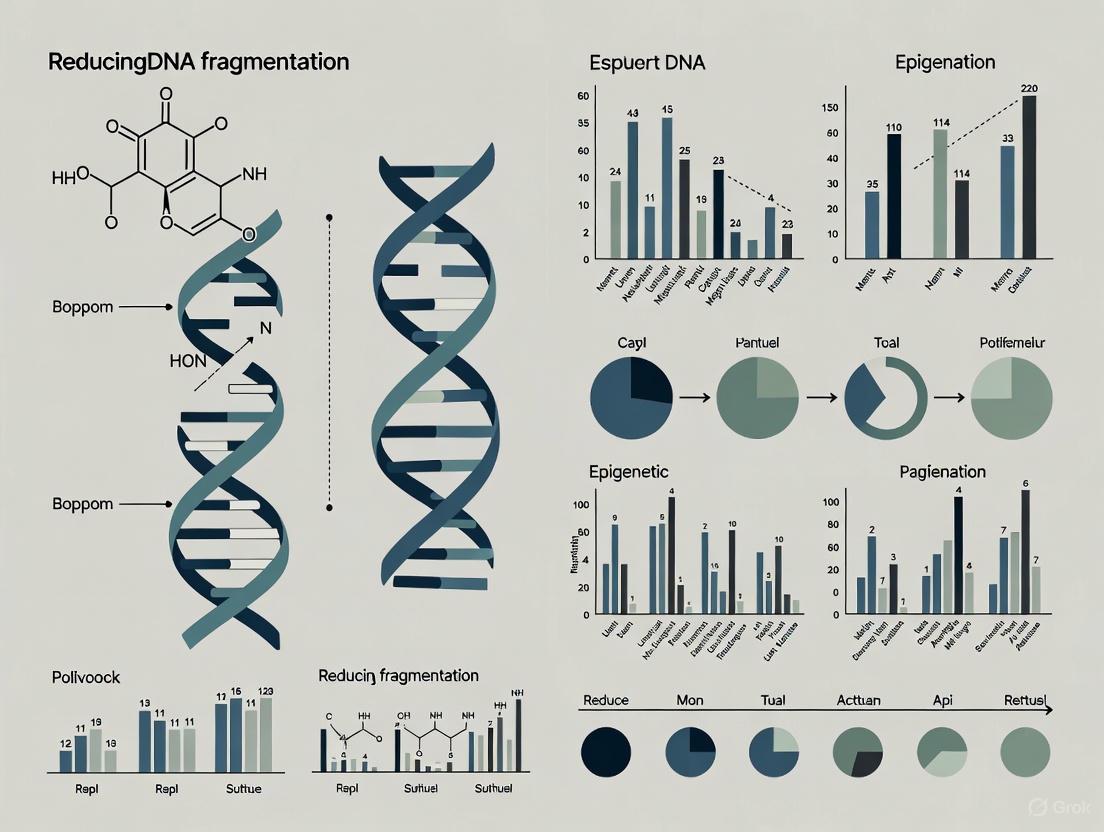
Strategies for Reducing Sperm DNA Fragmentation to Ensure Integrity in Epigenetic Analysis
This article provides a comprehensive resource for researchers and scientists on the critical relationship between sperm DNA fragmentation (SDF) and epigenetic analysis.
Strategies for Reducing Sperm DNA Fragmentation to Ensure Integrity in Epigenetic Analysis
Abstract
This article provides a comprehensive resource for researchers and scientists on the critical relationship between sperm DNA fragmentation (SDF) and epigenetic analysis. It covers the foundational mechanisms by which SDF originates and confounds epigenetic data, explores validated methodologies for SDF assessment and reduction, offers troubleshooting for pre-analytical variables, and outlines rigorous validation frameworks. By synthesizing current evidence, this review aims to equip professionals in drug development and biomedical research with practical strategies to minimize SDF, thereby enhancing the reliability of epigenetic studies in male fertility and transgenerational inheritance.
Understanding Sperm DNA Fragmentation: Mechanisms and Epigenetic Consequences
FAQs: Core Concepts and Troubleshooting
Q1: What is the fundamental difference between single-strand (SSB) and double-strand DNA breaks (DSB) in sperm?
A1: The key difference lies in the structural damage to the DNA helix and their subsequent impact on genetic integrity.
- Single-Strand Breaks (SSBs): Only one of the two strands of the DNA helix is broken. The intact strand can potentially serve as a template for repair. They are extensively present as multiple breakpoints across the genome and are primarily linked to oxidative stress [1] [2] [3].
- Double-Strand Breaks (DSBs): Both strands of the DNA helix are broken in close proximity, severing the DNA molecule. This is considered more severe as it can lead to genetic rearrangements. DSBs are often localized, attached to the sperm nuclear matrix, and are possibly related to abortive apoptosis or a failure in the repair of meiotic breaks [1] [2].
Q2: Which type of sperm DNA break has a greater negative impact on clinical pregnancy outcomes?
A2: Evidence suggests that double-strand breaks (DSBs) have a stronger negative association with key reproductive outcomes [1] [2]. While high levels of SSBs are associated with difficulty achieving pregnancy, high levels of DSBs are more specifically linked to implantation failure, poorer embryo quality, and a significantly increased risk of miscarriage, particularly in ICSI cycles [1] [4].
Q3: A common issue in our lab is the variability of SDF results. Which assay should I use to specifically detect double-strand breaks?
A3: The choice of assay is critical. Most common tests (TUNEL, SCSA, SCD) detect both SSBs and DSBs without distinction. To specifically assess DSBs, you should consider:
- Neutral Comet Assay: This is the most established method for specifically detecting DSBs [1] [2].
- γH2AX Immunodetection: This newer test uses antibodies to detect the phosphorylated form of the H2AX histone (γH2AX), which is a specific molecular biomarker formed at the sites of DSBs [2].
Q4: We are preparing sperm samples for epigenetic research. What selection technique best preserves DNA integrity and reduces SDF?
A4: Conventional techniques like density gradient centrifugation and swim-up involve centrifugation steps that can generate harmful reactive oxygen species (ROS) [5] [6]. For superior DNA integrity, consider:
- Microfluidic Sperm Selection: This technology separates sperm based on motility through microchannels without centrifugation. Meta-analysis shows it yields sperm with significantly lower DNA fragmentation compared to conventional methods [5]. It mimics the natural selection processes of the female reproductive tract, minimizing oxidative damage [6].
- Testicular Sperm Extraction: In cases of persistently high SDF in ejaculated sperm, testicular sperm has been shown to have lower DNA fragmentation, as it is retrieved before exposure to the post-testicular oxidative environment [7].
Experimental Protocols for SDF Assessment
Protocol 1: Neutral Comet Assay for Specific Detection of Double-Strand Breaks (DSBs)
Principle: At neutral pH, the protocol primarily detects DSBs. Sperm with DSBs release DNA fragments that migrate out of the nucleus during electrophoresis, forming a "comet tail," while intact DNA remains in the "head" [1] [2].
Methodology:
- Sample Preparation: Embed a small aliquot of sperm sample in low-melting-point agarose on a specially coated microscope slide.
- Lysis: Immerse slides in a neutral lysis buffer (e.g., containing 2.5 M NaCl, 100 mM EDTA, 10 mM Tris, 1% Triton X-100, pH 8.0) for at least 1 hour to remove membranes and nuclear proteins.
- Electrophoresis: Place slides in an electrophoresis tank filled with neutral buffer (e.g., TBE). Electrophorese at a low voltage (e.g., 1 V/cm) for a specified time (e.g., 20 minutes).
- Staining and Analysis: Stain DNA with a fluorescent dye like Sybr Green/Gold and visualize under a fluorescence microscope. Analyze ~100 cells per sample using specialized software to determine the percentage of DNA in the tail (Tail DNA %) or other comet parameters.
Protocol 2: Sperm Processing Using Microfluidic Devices
Principle: Microfluidic chips (e.g., ZyMot, Fertile Plus) use laminar flow and microchannels or membranes to select sperm based on motility and morphology, avoiding damaging centrifugation steps [5].
Methodology (Generic Workflow):
- Device Priming: Load the collection medium into the device's outlet reservoir to create a fluidic connection.
- Sample Loading: Carefully load the raw, liquefied semen sample into the designated input chamber.
- Incubation: Incubate the device for 30-60 minutes at 37°C. During this time, highly motile and morphologically normal sperm actively swim through the microchannels or membrane into the collection chamber.
- Sperm Recovery: Retrieve the selected sperm population from the output chamber using a pipette. The sample is now ready for use in ART or downstream analysis [5].
Data Presentation: SDF Assays and Clinical Impact
Table 1: Comparison of Primary Sperm DNA Fragmentation Assays
| Assay | Basic Principle | Type of DNA Damage Detected | Key Advantage | Key Disadvantage |
|---|---|---|---|---|
| TUNEL | Labels 3'-OH free ends of DNA breaks with fluorescent nucleotides [1] [2] | SSBs and DSBs | Direct labeling of breaks; highly standardized [1] | Cannot differentiate between SSBs and DSBs [1] |
| SCSA | Measures DNA denaturation susceptibility using Acridine Orange fluorescence [1] [2] | SSBs and DSBs | High standardization; differentiates immature sperm (HDS%) [1] | Requires flow cytometer; cannot differentiate break types [1] |
| SCD (HaloTest) | Visualizes dispersion halo after denaturation; damaged DNA has small/no halo [1] [2] | SSBs and DSBs | Simple, no need for complex equipment [1] | Subjective analysis; cannot differentiate break types [1] |
| Alkaline Comet | Electrophoresis at alkaline (high pH) conditions [1] [2] | Primarily SSBs (and some DSBs) | Can be tuned to quantify total DNA damage [1] | Technique not fully standardized between labs [1] |
| Neutral Comet | Electrophoresis at neutral pH [1] [2] | Specifically DSBs | The best available method for specific DSB detection [1] [2] | Technique not fully standardized between labs [1] |
| γH2AX | Immunodetection of phosphorylated H2AX histone [2] | Specifically DSBs | Direct molecular biomarker for DSBs [2] | Less established protocol; requires antibody-based detection [2] |
Table 2: Distinct Clinical Effects of Single-Strand vs. Double-Strand Breaks
| Reproductive Outcome | Impact of Single-Strand Breaks (SSBs) | Impact of Double-Strand Breaks (DSBs) |
|---|---|---|
| Natural Conception / IUI | Associated with longer time to conception and lack of pregnancy [1] [7] | Stronger negative association with pregnancy loss [4] |
| Fertilization Rate (IVF) | Negatively impacts fertilization in IVF cycles [2] | Less pronounced effect; ICSI can partially overcome this [2] |
| Implantation & Pregnancy | Contributes to implantation failure [1] | Significantly associated with implantation failure and higher miscarriage rates in ICSI [1] [4] |
| Embryo Quality | General negative correlation with embryo quality | Stronger association with poor embryo quality and delayed development [1] [8] |
| Primary Etiology | Linked predominantly to oxidative stress [1] [3] | Linked to abortive apoptosis and errors in meiotic repair [1] [2] |
Signaling Pathways and Workflows
The Scientist's Toolkit: Research Reagent Solutions
Table 3: Essential Materials for Sperm DNA Integrity Research
| Item | Function/Basis of Selection | Key Consideration for Epigenetic Research |
|---|---|---|
| ZyMot or similar Microfluidic Chip | For centrifugation-free sperm selection based on motility, yielding populations with lower SDF [5]. | Minimizes oxidative stress during processing, which helps preserve not just DNA integrity but also native epigenetic marks. |
| Neutral Comet Assay Kit | Provides optimized buffers and reagents for the specific detection of double-strand breaks (DSBs) [1] [2]. | Essential for correlating specific DNA damage types with epigenetic anomalies, rather than relying on total SDF. |
| Anti-γH2AX Antibody | Enables immunodetection of DSBs as an alternative to the Neutral Comet assay [2]. | Can be combined with other immunofluorescence stains for co-localization studies of DNA damage and epigenetic marks. |
| Reactive Oxygen Species (ROS) Detection Kits (e.g., based on DCFDA) | To quantify oxidative stress levels in semen samples, the primary cause of SSBs [3] [8]. | Critical for mechanistic studies linking oxidative stress to both DNA fragmentation and oxidative modification of epigenetic marks. |
| Protamine Stain (e.g., Chromomycin A3) | Assesses chromatin maturity, as defective protamination is a key etiology of SDF [1] [3]. | An immature chromatin structure (abnormal P1/P2 ratio) can make DNA more accessible to damage and may correlate with epigenetic instability. |
| Antioxidant Supplements (e.g., Vitamin C, E, CoQ10) | Used in clinical trials to investigate the reduction of SDF through mitigation of oxidative stress [7]. | When studying interventions, these can be tools to understand the dynamic relationship between oxidative stress, DNA integrity, and the sperm epigenome. |
Core Mechanisms of Sperm DNA Fragmentation
Sperm DNA fragmentation (SDF) refers to single or double-stranded breaks in the sperm genome and is a major cause of male infertility and adverse reproductive outcomes [7]. The three primary mechanisms underlying SDF are oxidative stress, abortive apoptosis, and defective sperm maturation [7] [3]. The following diagram illustrates how these core mechanisms lead to sperm DNA damage.
Table 1: Characteristics of Primary SDF Mechanisms
| Mechanism | Key Features | Primary Location | Resulting DNA Damage |
|---|---|---|---|
| Oxidative Stress | Reactive oxygen species (ROS) overwhelm antioxidant defenses; caused by lifestyle factors, inflammation, leukocytospermia [9] | Post-testicular (throughout male reproductive tract) [3] | Base modifications, single & double-strand breaks, DNA adducts [3] |
| Abortive Apoptosis | Failed apoptosis of defective germ cells; Fas/FasL system activation [3] | Testicular (during spermatogenesis) [7] | Spermatozoa with apoptotic markers in ejaculate [3] |
| Defective Maturation | Impaired chromatin compaction during spermiogenesis; faulty protamine replacement [3] | Testicular (spermiogenesis) [7] | Unrepaired DNA nicks, increased chromatin susceptibility [3] |
Troubleshooting Common SDF Analysis Issues
Problem: High Background in SDF Assays
Q: My SDF assays consistently show high background noise, making results difficult to interpret. What could be causing this?
A: High background noise in SDF assays can result from several factors:
- Sample contamination with apoptotic bodies or debris: These elements can be mistakenly counted as spermatozoa with DNA damage, particularly in flow cytometry-based methods like TUNEL [10]. Always include proper nuclear staining to accurately identify the sperm population and exclude contaminants [10].
- Inadequate sample preparation: For the SCD test, ensure proper denaturation and lysing steps. Incomplete removal of nucleoproteins can lead to inconsistent halo formation [10].
- Over-decondensation of chromatin: In the COMET assay, excessive decondensation can create artificial DNA migration. Optimize lysis and unwinding times for your specific sample type [10].
Problem: Inconsistent Results Between SDF Testing Methods
Q: Why do I get significantly different SDF values when using different testing methods on the same sample?
A: Variability between methods occurs because each test detects different types of DNA damage through distinct mechanisms:
Table 2: Comparison of SDF Testing Methodologies
| Method | Principle | DNA Damage Detected | Output | Key Limitations |
|---|---|---|---|---|
| SCSA | Acridine orange staining after acid denaturation [10] | Chromatin susceptibility to denaturation [10] | %DFI (DNA Fragmentation Index) [10] | Measures susceptibility rather than direct breaks [10] |
| TUNEL | TdT enzyme labels 3'-OH ends of DNA breaks [7] [10] | Direct detection of single & double-strand breaks [10] | % labeled spermatozoa [10] | Access to chromatin may be limited in non-viable sperm [10] |
| SCD Test | Halo pattern formation after denaturation and protein removal [10] | DNA fragmentation based on dispersion patterns [10] | % sperm without halo [10] | Subjective interpretation without specialized software [10] |
| COMET Assay | Electrophoretic DNA migration under alkaline conditions [10] | Single & double-strand breaks with high sensitivity [10] | % tail DNA or % comets [10] | Alkaline conditions may create additional damage at labile sites [10] |
Protocol Recommendation: For consistent results, establish a standardized protocol for your lab and perform parallel testing with a control sample when implementing a new method. When using TUNEL with flow cytometry, couple DNA break labeling with nuclear staining to precisely identify sperm populations and exclude apoptotic bodies [10].
Problem: Low Oxidative Stress Assay Efficiency
Q: My oxidative stress measurements in sperm samples show low efficiency and high variability. How can I improve reliability?
A: Low efficiency in oxidative stress assays often relates to sample handling and reagent issues:
- EDTA contamination: When performing oxidation-based assays, ensure DNA is eluted in nuclease-free water or appropriate elution buffer after ligation, as EDTA can chelate metals necessary for oxidation reactions [11].
- Improper reagent handling: DTT concentration is critical - use fresh aliquots and ensure correct concentration. Avoid reusing old DTT tubes [11].
- Iron solution issues: For assays requiring Fe(II) solution, prepare fresh dilutions and use within 15 minutes. Pipette accurately using calibrated equipment and ensure proper mixing after addition [11].
- Temperature control: Keep reagents on ice and set up reactions on a chilled block to maintain stability [11].
SDF Mechanism-Specific Mitigation Strategies
Targeting Oxidative Stress
Q: What specific interventions can reduce oxidative stress-mediated SDF?
A: Oxidative stress management requires a multi-faceted approach:
- Antioxidant supplementation: Clinical trials demonstrate that antioxidants can improve SDF levels, though optimal formulations and dosing require further standardization [7]. Consider combinations addressing different oxidative pathways.
- Lifestyle modifications: Address key ROS sources including smoking cessation, reduced alcohol consumption, and management of obesity [9] [12]. These factors directly increase seminal ROS production.
- Treatment of underlying conditions: Manage medical conditions associated with inflammation and oxidative stress, particularly varicocele and genital tract infections [7] [9]. Antibiotic treatment for infections can significantly reduce SDF levels [7].
Addressing Defective Maturation and Abortive Apoptosis
Q: Can we influence the testicular mechanisms of SDF, such as defective maturation and abortive apoptosis?
A: While these testicular mechanisms are more challenging to target directly, several approaches show promise:
- Varicocele repair: Men with varicocele and high SDF should consider varicocelectomy, which has been shown to reduce DNA fragmentation index by more than 5% [7].
- Shortened ejaculatory abstinence: Evidence indicates that shorter abstinence periods (1-2 days) can reduce SDF in subsequent ejaculates, potentially by reducing epididymal storage time and associated oxidative damage [7].
- Sperm selection techniques: For ART procedures, testicular sperm extraction may be considered as testicular sperm often demonstrates lower SDF compared to ejaculated sperm, as it avoids post-testicular oxidative damage [7].
The following diagram illustrates the complete pathway from SDF mechanisms to potential interventions and clinical outcomes.
The Scientist's Toolkit: Essential Research Reagents
Table 3: Key Research Reagents for SDF Analysis and Intervention Studies
| Reagent/Category | Specific Examples | Research Application | Technical Notes |
|---|---|---|---|
| SDF Detection Kits | TUNEL assay kits, SCSA reagents, SCD test kits | Quantifying DNA fragmentation levels | For TUNEL: Include nuclear staining to exclude apoptotic bodies [10] |
| Oxidative Stress Probes | DCFH-DA, MitoSOX, C11-BODIPY⁵⁸¹/⁵⁹¹ | Measuring intracellular and mitochondrial ROS | Validate with positive controls; account for sperm autofluorescence |
| Antioxidants | N-acetylcysteine, Vitamin C, Vitamin E, CoQ10 | Intervention studies for oxidative stress | Use physiological concentrations; consider combination approaches |
| Chromatin Stains | Acridine orange, Methyl green, Propidium iodide | Assessing chromatin integrity and maturation | Standardize staining protocols across experiments |
| Sperm Preparation Media | Gradient solutions, Sperm washing buffers | Sample processing for analysis and ART | Avoid prolonged centrifugation; minimize processing time |
| DNA Repair Enzymes | OGG1, APE1 (for BER studies) | Investigating repair mechanisms in sperm | Note: Mature sperm have limited repair capacity [13] |
FAQs on SDF Mechanisms and Analysis
Q: What are the clinical indications for SDF testing in male fertility assessment?
A: Current guidelines recommend SDF testing for men with unexplained infertility, recurrent pregnancy loss, before or after failure of IUI/IVF treatments, and for those with modifiable lifestyle risk factors or clinical varicocele [7].
Q: Can the oocyte repair sperm DNA damage after fertilization?
A: Yes, the oocyte possesses some capacity to repair sperm DNA damage after fertilization using maternal repair factors and mRNAs [13]. However, this repair capacity is limited and depends on both the extent of damage and oocyte quality. High levels of SDF may overwhelm the oocyte's repair mechanisms, leading to failed fertilization, impaired embryo development, or early pregnancy loss [13].
Q: How does advanced paternal age contribute to SDF?
A: Advanced paternal age is associated with increased SDF through multiple mechanisms, including higher exposure to oxidative stress over time, defective sperm chromatin packaging, and disordered apoptosis [3]. Studies indicate that SDF increases with age, starting in reproductive years and potentially doubling between ages 20 and 60 [3].
Q: What is the recommended ejaculatory abstinence period for SDF testing?
A: Shorter ejaculatory abstinence periods (1-2 days) have been associated with lower SDF levels compared to longer abstinence periods [7]. For SDF testing, follow consistent abstinence protocols to enable comparable results across samples.
Core Concepts: Sperm DNA Fragmentation and the Epigenome
What is the fundamental relationship between sperm DNA fragmentation (SDF) and the sperm epigenome? Sperm DNA fragmentation refers to the presence of single or double-strand breaks in the sperm's genetic material. The epigenome consists of molecular modifications, such as DNA methylation and histone packaging, that regulate gene expression without altering the DNA sequence. These two elements are intrinsically linked. High levels of SDF are frequently associated with aberrant epigenetic patterns, including disrupted DNA methylation profiles and impaired chromatin compaction. This combination can compromise paternal genomic integrity and hinder proper gene expression in the developing embryo [14] [15].
How does oxidative stress serve as a common upstream cause for both SDF and epigenetic alterations? Oxidative stress, resulting from an imbalance between reactive oxygen species (ROS) and antioxidant defenses, is a primary driver of both damage types.
- Lipid Peroxidation: ROS attacks the polyunsaturated fatty acids in the sperm plasma membrane, generating toxic byproducts like malondialdehyde (MDA). This not only damages the membrane but can also lead to further oxidative stress and DNA damage [16].
- Direct DNA Damage: ROS directly causes single and double-strand breaks in sperm DNA, leading to fragmentation. It can also modify DNA bases, creating lesions such as 8-hydroxy-2'-deoxyguanosine (8-OHdG) [17] [16].
- Epigenetic Disruption: The processes of oxidative damage can interfere with the enzymes responsible for establishing and maintaining DNA methylation patterns, such as DNA methyltransferases (DNMTs). This can lead to global or gene-specific methylation errors [18] [19].
The diagram below illustrates this destructive cascade originating from oxidative stress.
Troubleshooting Common Scenarios
We are observing high SDF in our patient cohort, but standard semen parameters are normal. What could be the underlying causes and how can we investigate further? This is a common scenario highlighting the limitation of routine analysis. Potential causes and investigative steps are outlined below.
| Potential Cause | Investigation Method | Rationale & Interpretation |
|---|---|---|
| Oxidative Stress | Measure ROS levels in seminal plasma (e.g., chemiluminescence). Test total antioxidant capacity (TAC). | Confirms an imbalance between oxidants and antioxidants, even if sperm count and motility appear normal [17]. |
| Varicocele | Conduct a clinical scrotal examination and Doppler ultrasound. | Varicoceles cause testicular heat stress and ROS production, strongly linked to high SDF despite normal counts [17]. |
| Lifestyle Factors | Use detailed patient questionnaires covering smoking, alcohol, diet, and heat exposure. | Smoking and obesity are significant contributors to oxidative stress and can selectively elevate SDF [17] [16]. |
| Epigenetic Aberrations | Perform genome-wide DNA methylation analysis (e.g., WGBS or EPIC array) on sperm samples. | High SDF often co-occurs with altered methylation at imprinted genes and regulatory regions, providing a more comprehensive diagnostic picture [18] [20]. |
Our lab is consistently getting low fertilization rates and poor blastocyst development in ICSI cycles, despite normal fertilization checks. Could SDF be a factor, and what is the evidence? Yes, SDF is a significant factor. Recent large-scale studies demonstrate a direct, dose-dependent impact on early embryological outcomes.
| Embryological Outcome | Quantitative Impact of SDF | Statistical Significance |
|---|---|---|
| Fertilization Rate | Each 1% increase in SDF reduces odds of fertilization rate >80% by 1.6% (OR=0.984) [14]. | p = 0.015 |
| Top-Quality Blastocyst (Day 5) | Each 1% increase in SDF decreases the chance by 2.5% (OR=0.975) [14]. | p = 0.004 |
| Top-Quality Embryo (Day 3) | A trend toward impaired quality was observed (OR=0.983) [14]. | p = 0.068 (borderline) |
What is the role of the oocyte in mitigating sperm DNA damage, and when does this repair capacity become overwhelmed? The oocyte possesses robust mechanisms to repair sperm DNA damage post-fertilization. However, this capacity is finite and influenced by several factors.
- Repair Mechanisms: The oocyte utilizes multiple pathways, including Base Excision Repair (BER) for oxidized bases and nucleotide lesions, and Non-Homologous End Joining (NHEJ) for double-strand breaks, prior to embryonic genome activation [15].
- Capacity Limits: It is postulated that the oocyte can effectively repair SDF only when it does not exceed approximately 8%. Beyond this threshold, damage persists, potentially leading to embryonic arrest, mutations, or implantation failure [15].
- Critical Factor - Maternal Age: The oocyte's repair capacity is highly dependent on maternal age. Advanced maternal age is associated with reduced expression of key DNA repair genes (e.g., LIG3, APEX, XRCC1), diminishing the ability to correct sperm DNA damage [15].
The following diagram summarizes the critical window and factors affecting oocyte-mediated repair.
Experimental Protocols & Workflows
Protocol: Evaluating Sperm DNA Fragmentation and Concurrent DNA Methylation
This protocol is designed for researchers needing a comprehensive assessment of both DNA integrity and epigenetics from the same sperm sample.
Step 1: Sperm Sample Collection and Processing
- Collect semen sample after 2-7 days of abstinence.
- Allow for liquefaction (30 min at 37°C).
- Perform a density gradient centrifugation (e.g., 80%/40% gradients) to isolate motile sperm with intact membranes, which also enriches for cells with lower DNA damage.
- Wash the sperm pellet with PBS and divide the sample for parallel SDF and methylation assays.
Step 2a: Sperm DNA Fragmentation Testing (Sperm Chromatin Dispersion - SCD)
- Reagent: Use a commercial SCD kit (e.g., Halosperm).
- Method: Embed a sperm aliquot in agarose on a slide. Subject it to an acidic denaturation and lysing solution to remove nuclear proteins. This step is critical as it reveals DNA loops.
- Staining and Analysis: Stain with a fluorescent DNA dye (e.g., DAPI, Propidium Iodide). Sperm with non-fragmented DNA will display large, characteristic halos of dispersed DNA loops, while sperm with fragmented DNA will show small or absent halos. Score a minimum of 500 sperm under a fluorescence microscope. A threshold of >20-30% SDF is often considered clinically significant [14] [21].
Step 2b: Sperm DNA Methylation Analysis (Whole-Genome Bisulfite Sequencing - WGBS)
- DNA Extraction: Extract genomic DNA from the remaining sperm sample using a dedicated kit, ensuring minimal DNA shearing.
- Bisulfite Conversion: Treat 100-500 ng of DNA with sodium bisulfite using a commercial kit. This reaction converts unmethylated cytosines to uracils (which read as thymines in sequencing), while methylated cytosines remain unchanged.
- Library Prep and Sequencing: Prepare a sequencing library from the converted DNA and perform high-coverage whole-genome sequencing on an Illumina platform.
- Bioinformatic Analysis: Map sequenced reads to a bisulfite-converted reference genome. Calculate methylation levels at individual CpG sites. Focus on regions of interest like imprinting control regions (ICRs), gene promoters, and repetitive elements. Compare methylation patterns between high-SDF and low-SDF sample groups [20] [19].
The Scientist's Toolkit: Research Reagent Solutions
Essential materials and reagents for investigating the sperm epigenome and DNA fragmentation.
| Research Need | Essential Reagents & Kits | Primary Function |
|---|---|---|
| SDF Measurement | SCD Kit (Halosperm), TUNEL Assay Kit, SCSA Reagents | To quantify the percentage of sperm with DNA strand breaks using different biochemical principles [14] [21]. |
| DNA Methylation Analysis | Bisulfite Conversion Kit, DNA Methylation ELISA, WGBS or EPIC Array Kits | To convert DNA for methylation detection and perform genome-wide or targeted profiling of methylated cytosines [18] [20]. |
| Oxidative Stress Assessment | Chemiluminescence Probes (e.g., Luminol), Malondialdehyde (MDA) ELISA, Total Antioxidant Capacity (TAC) Assay | To directly measure ROS levels and lipid peroxidation byproducts in seminal plasma [17] [16]. |
| Sperm Selection for ART | PICSI Sperm Slides, MACS Columns (Annexin V), Microfluidic Sperm Sorters | To selectively isolate sperm with lower DNA fragmentation and better chromatin integrity for use in assisted reproduction [14] [15]. |
| Chromatin Analysis | Chromatin Immunoprecipitation (ChIP) Kits, Protamine Staining Dyes (e.g., Chromomycin A3) | To assess histone modifications, nucleosome positioning, and the protamine-to-histone ratio in sperm chromatin [19]. |
FAQs on Clinical Relevance and Intervention
Is testing for SDF recommended in all infertility cases? What do the latest guidelines say? Recent clinical guidelines from the Global Andrology Forum (2025) provide a nuanced view. While there is growing evidence for the clinical benefit of SDF testing, significant gaps in the literature limit its recommendation for routine use in all cases. The guidelines strongly recommend (87.5% consensus) SDF testing in specific clinical scenarios, including idiopathic infertility, recurrent pregnancy loss, and prior failed ART cycles. They emphasize a tailored approach based on individual patient history rather than universal screening [21].
Does antioxidant supplementation effectively reduce SDF and improve epigenetic marks? The evidence is promising but mixed, and supplementation should be approached cautiously.
- Efficacy: Some studies suggest that supplementation with specific antioxidants (e.g., Vitamin C, Vitamin E, Coenzyme Q10, Carnitines) can help reduce oxidative stress and may subsequently lower SDF and support healthier epigenetic patterns [17].
- Limitations: Results vary significantly based on dosage, duration, and the individual's baseline antioxidant status. Current guidelines from major urological associations note that while antioxidants may improve sperm parameters and are not harmful, conclusive data demonstrating improved live birth rates is still lacking [17] [21].
- Best Practice: The emerging consensus is to carefully select patients who are deficient in antioxidants or would most benefit from their reductive stress potential, rather than administering them universally [17].
How do advanced paternal age and environmental factors specifically impact the sperm epigenome? Both factors induce changes that compound the risks associated with SDF.
- Paternal Age: Advanced paternal age is associated with higher baseline SDF and an increased rate of de novo mutations. It is also linked to epigenetic shifts, particularly at imprinting control regions, which can affect offspring health [14] [15].
- Environmental Pollutants: Exposure to heavy metals, air pollutants, and endocrine disruptors can alter protamine packaging and directly increase oxidative stress. This leads to both DNA fragmentation and aberrant DNA methylation, as these toxins can interfere with the enzymes that regulate the epigenome [17] [16].
Technical FAQs on SDF and Epigenetic Analysis
FAQ 1: What is sperm DNA fragmentation (SDF) and why is it a critical biomarker for male infertility? Sperm DNA fragmentation (SDF) refers to the presence of single- or double-stranded breaks in the DNA of sperm chromatin [14]. It is increasingly regarded as a crucial biomarker because, unlike standard semen parameters (concentration, motility, morphology), it provides a more direct indicator of the sperm's genomic health and functional ability [14]. Elevated SDF has been significantly associated with reduced fertilization rates, impaired embryo development, and poorer outcomes in assisted reproductive technologies (ART) [14]. It is a strong predictor of male reproductive potential [12].
FAQ 2: How can paternal factors involving SDF lead to intergenerational or offspring risks? Paternal factors can alter the sperm epigenome, which is then transmitted to the embryo. Sperm carries not only genetic but also epigenetic information, including histone modifications, DNA methylation patterns, and non-coding RNAs [22]. Environmental stressors, advanced paternal age, and oxidative stress can induce high SDF and alter this epigenetic information [22] [12]. Upon fertilization, these altered epigenetic marks can affect gene expression profiles in the embryo, potentially leading to an increased susceptibility in offspring to conditions such as anxiety, depression, cognitive deficits, and other neurological disorders [22] [12].
FAQ 3: What are the primary mechanisms that cause Sperm DNA Fragmentation? The primary mechanism driving SDF is oxidative stress [12]. Spermatozoa are particularly vulnerable to reactive oxygen species (ROS) due to their limited cytoplasmic antioxidant defenses [12]. Oxidative damage can induce single- and double-strand DNA breaks, mutagenic adduct formation, and DNA hypomethylation [12]. Other contributing factors include:
- Abortive apoptosis: Incomplete programmed cell death during spermatogenesis [14] [12].
- Defective chromatin packaging: Abnormalities during the histone-to-protamine transition, which is essential for DNA compaction in sperm [12].
- Environmental insults: Exposure to factors like phthalates, heavy metals, and heat stress [22] [12].
- Advanced paternal age: Associated with higher baseline SDF and mitochondrial dysfunction [14] [12].
FAQ 4: Our lab's SDF measurements are inconsistent between replicates. What are the key troubleshooting steps? Inconsistency in SDF measurements can arise from several sources. Key troubleshooting steps include:
- Standardize sample preparation: Ensure strict adherence to a uniform abstinence period (3-5 days) before sample collection [23]. Inconsistent abstinence periods can introduce variability.
- Verify assay protocols: Confirm that all steps (e.g., dye concentration, incubation times, denaturation conditions) are followed precisely according to the kit manufacturer's instructions. Even minor deviations can affect results [23].
- Calibrate instrumentation: Regularly calibrate flow cytometers or microscopes used for analysis. For SCSA, ensure that the instrument is properly aligned and that a consistent number of cells (e.g., at least 5000) are recorded for analysis [23].
- Control for oxidative stress: Minimize the time between sample collection and processing, as prolonged storage can exacerbate oxidative DNA damage [12].
- Implement internal controls: Use control samples with known high and low SDF levels in each run to monitor assay performance and inter-assay variability.
FAQ 5: What advanced therapeutic strategies can reduce SDF for epigenetic research? Beyond common antioxidant supplements, advanced strategies include:
- Yoga-Based Lifestyle Interventions (YBLI): Integrates physical postures, breath regulation, and meditation. YBLI has been shown to reduce oxidative stress, lower inflammation, and improve stress-induced hormonal dysregulation, thereby reducing SDF [12].
- Sperm Selection Techniques: Using microfluidic devices or physiological methods during swim-up can select sperm with lower DNA damage and better motility/morphology [14].
- Varicocele Repair: Surgical correction of clinical varicocele is a proven method to reduce SDF and improve semen parameters [12] [23].
- Testicular Sperm Retrieval: In some cases, testicular sperm has been shown to exhibit lower SDF than ejaculated sperm, potentially bypassing post-testicular DNA damage [12].
Quantitative Data on SDF and Reproductive Outcomes
The following table summarizes key quantitative findings from a large-scale retrospective cohort study investigating the impact of SDF on ICSI outcomes [14].
Table 1: Impact of SDF on Key Embryological and Clinical ICSI Outcomes (n=870 cycles)
| Outcome Measure | Key Finding | Statistical Significance (p-value) | Odds Ratio (OR) per 1% SDF increase |
|---|---|---|---|
| Fertilization Rate | Significantly reduced in high SDF group (SDF > 20%) | p = 0.009 | OR = 0.984 (95% CI: 0.971–0.997) |
| Top-Quality Blastocyst (Day 5) | Significantly decreased chance of obtaining top-quality blastocysts | p = 0.004 | OR = 0.975 (95% CI: 0.958–0.992) |
| Top-Quality Embryo (Day 3) | Trend toward impaired quality | p = 0.068 (not significant) | OR = 0.983 |
| Clinical Pregnancy | No significant association found | p = 0.155 | OR = 0.989 |
| Miscarriage | Borderline relationship observed | p = 0.053 | OR = 0.961 |
Experimental Protocols for Key Assays
Protocol 1: Sperm Chromatin Structure Assay (SCSA) for DNA Fragmentation Index (DFI) The SCSA is a flow cytometry-based method that indirectly detects sperm DNA fragmentation by measuring the susceptibility of DNA to acid-induced denaturation [23].
- Sample Preparation: Dilute liquefied semen to a concentration of 1–2 × 10^6 sperm/mL using a provided buffer solution (Solution A) [23].
- Acid Denaturation: Add 100 µL of an acidic solution (Solution B) to the sample and incubate for 30 seconds. This denatures DNA in sperm with existing fragmentation [23].
- Staining: Add 1200 µL of acridine orange staining solution (Solution C). Acridine orange emits green fluorescence when bound to double-stranded DNA and red fluorescence when bound to single-stranded DNA [23].
- Flow Cytometry Analysis: Analyze the stained sample using a flow cytometer. Record data from at least 5000 cells per sample [23].
- Calculation: The DNA Fragmentation Index (DFI) is calculated as the ratio of sperm with red fluorescence (fragmented DNA) to the total sperm count, expressed as a percentage [23].
Protocol 2: TUNEL Assay for Direct DNA Break Detection The TUNEL (Terminal deoxynucleotidyl transferase dUTP Nick End Labeling) assay is a direct method for detecting DNA strand breaks by enzymatically labeling the 3'-OH ends of broken DNA [23].
- Sample Preparation: Prepare sperm cells on a glass slide or in a suspension. Permeabilize the cells to allow enzyme entry [23].
- Enzymatic Labeling: Incubate samples with a reaction mixture containing Terminal deoxynucleotidyl transferase (TdT) enzyme and fluorescently labeled dUTP (e.g., FITC-dUTP). The TdT enzyme adds the labeled nucleotides to the 3'-ends of DNA breaks [23].
- Washing and Counterstaining: Wash the samples to remove unincorporated nucleotides. A counterstain like DAPI may be used to visualize all nuclei [23].
- Detection and Analysis: Analyze samples under a fluorescence microscope or flow cytometer. Sperm with DNA fragmentation will display green fluorescence. The percentage of TUNEL-positive sperm is calculated [23].
Protocol 3: TdT/SD Biosensor for Mean DNA Breaks (MDB) and Free DNA (fDFA) This novel technique allows for direct quantification of DNA breakpoints and free DNA fragments in seminal plasma [23].
- DNA Extraction: Extract DNA from sperm cells or free DNA from seminal plasma using a commercial genome extraction kit [23].
- TdT Reaction: Adjust DNA concentration to 5 ng/µL. Prepare a reaction mix with 10x TdT buffer, dATP, DNA solution, and sterile water. Add TdT enzyme and incubate at 37°C for 60 minutes, followed by 75°C for 20 minutes to inactivate the enzyme. This step adds a poly-A tail to DNA breakpoints [23].
- Signal Amplification & Detection: Add Strand Displacement (SD) probes adjusted to 400 nM to the reaction mix. Perform detection in a qPCR instrument at 37°C for 10 cycles (30 seconds/cycle). The fluorescence signal is converted into the Mean number of DNA Break points (MDB) or the amount of free DNA fragments (fDFA) [23].
Signaling Pathways and Experimental Workflows
Diagram 1: Pathway from Paternal Stress to Offspring Risk
Diagram 2: SDF Testing Experimental Workflow
The Scientist's Toolkit: Research Reagent Solutions
Table 2: Essential Reagents and Kits for SDF and Epigenetic Research
| Item Name | Function / Application | Key Notes |
|---|---|---|
| SCSA Kit (e.g., BKR802) | Determines DNA Fragmentation Index (DFI) via flow cytometry. | Utilizes acridine orange staining; requires flow cytometer. Measures acid-induced DNA denaturation [23]. |
| TUNEL Assay Kit | Directly labels DNA strand breaks for microscopy/flow cytometry. | Uses TdT enzyme and fluorescent-dUTP. Considered a direct and accurate method for detecting DNA damage [23]. |
| Acridine Orange | Fluorescent dye for SCSA; distinguishes dsDNA (green) from ssDNA (red). | The core dye for the SCSA methodology [23]. |
| Terminal Deoxynucleotidyl Transferase (TdT) | Key enzyme for TUNEL assay and TdT/SD biosensor. | Catalyzes the addition of labeled nucleotides to 3'-OH ends of DNA breaks [23]. |
| DCFH-DA Probe (e.g., BKR 803) | Measures intracellular Reactive Oxygen Species (ROS) levels via flow cytometry. | Cell-permeable dye that emits fluorescence upon oxidation; confirms oxidative stress involvement [23]. |
| Automated Nucleic Acid Extractor (e.g., GenePure Pro 32E) | Isolates high-purity DNA from sperm/seminal plasma for advanced assays. | Essential for protocols like the TdT/SD biosensor to ensure consistent DNA input [23]. |
| Diff-Quik Staining Kit | Rapid staining for assessment of sperm morphology. | Allows for evaluation of sperm morphology according to WHO guidelines [23]. |
FAQs: Understanding Sperm DNA Fragmentation
What is sperm DNA fragmentation (SDF) and why is it critical for epigenetic research? Sperm DNA fragmentation (SDF) refers to breaks or damage in the genetic material carried by sperm. The sperm DNA fragmentation index (DFI) denotes the percentage of sperm with damaged DNA [24]. This integrity is crucial not only for successful fertilization and embryo development but also for the accurate transmission of epigenetic information [25] [26]. SDF can lead to genomic instability and is associated with adverse offspring outcomes, including congenital anomalies and cognitive deficits, making its management essential for epigenetic studies [12].
Which risk factors cause the most significant increases in SDF? A 2023 systematic review and meta-analysis quantified the impact of various risk factors on SDF. The table below summarizes the factors associated with the most substantial increases [24].
| Risk Factor | Mean Increase in SDF (%) | 95% Confidence Interval |
|---|---|---|
| Impaired Glucose Tolerance | 13.75% | 6.99 to 20.51 |
| Varicocele | 13.62% | 9.39 to 17.84 |
| Advanced Paternal Age (≥50) | 12.58% | 7.31 to 17.86 |
| Testicular Tumors | 11.30% | 7.84 to 14.76 |
| Environmental Pollution | 9.68% | 6.85 to 12.52 |
| Smoking | 9.19% | 4.33 to 14.06 |
How do lifestyle factors influence SDF risk? Beyond the high-impact factors listed above, other modifiable lifestyle factors contribute to SDF. A 2025 predictive model study identified six independent predictors for abnormal DFI, which include age, body mass index (BMI), smoking, hot spring bathing, stress, and daily exercise duration [27]. This indicates that daily habits and exposures are closely linked to sperm DNA integrity.
What is the primary biological mechanism behind SDF? Oxidative stress is the principal driver of sperm DNA fragmentation [25] [12]. Spermatozoa are particularly vulnerable to reactive oxygen species (ROS) due to their limited cytoplasmic antioxidant defenses. Elevated ROS levels lead to lipid peroxidation, DNA strand breaks, and mutagenic adduct formation, ultimately compromising the sperm's genomic and epigenetic integrity [26] [12].
Oxidative Stress Pathway in SDF
The Scientist's Toolkit: SDF Assessment & Analysis
What are the key methods for assessing Sperm DNA Fragmentation? Several assays are available to measure SDF, each with different methodologies and principles. The table below outlines the common tests used in clinical and research settings [24] [25].
| Assay Name | Full Name | Key Principle | Brief Explanation |
|---|---|---|---|
| SCSA | Sperm Chromatin Structure Assay | Acid-Induced Denaturation | Measures the susceptibility of sperm DNA to denaturation under acidic conditions, using flow cytometry. |
| TUNEL | Terminal deoxynucleotidyl transferase dUTP Nick End Labeling | Direct Labeling of DNA Breaks | Enzyme-based labeling of single- and double-strand DNA breaks, detectable by fluorescence microscopy or flow cytometry. |
| SCD | Sperm Chromatin Dispersion Test | Halomax Assay | Sperm with fragmented DNA fail to produce the characteristic halo of dispersed chromatin loops when incubated in an acidic solution. |
| Comet Assay | Single Cell Gel Electrophoresis Assay | Electrophoretic Migration | Subjects single sperm cells to electrophoresis; fragmented DNA migrates away from the nucleus, forming a "comet tail." |
What essential reagents and tools are used in epigenetic analysis of sperm? For researchers investigating the epigenetic correlates of SDF, the following tools are essential for analyzing chromatin structure and DNA methylation [28].
| Research Tool | Primary Function in Epigenetics |
|---|---|
| Whole-Genome Bisulfite Sequencing (WGBS) | Provides a base-resolution map of DNA methylation (5-methylcytosine) across the entire genome. |
| ATAC-Seq | Identifies regions of open, accessible chromatin, which are typically transcriptionally active. |
| ChIP-Seq | Maps specific histone modifications or DNA-binding protein locations across the genome. |
| Infinium MethylationEPIC Array | A microarray-based method that profiles the methylation status of ~930,000 CpG sites. |
| Small RNA Sequencing | Quantifies and identifies small non-coding RNAs (e.g., miRNAs, piRNAs) involved in post-transcriptional gene regulation. |
Troubleshooting Guides: Mitigating SDF in Research Samples
How can we reduce SDF through lifestyle interventions in study cohorts? Problem: High SDF in participant cohorts confounds research outcomes. Solution: Implement structured lifestyle modification programs. A key protocol involves [25]:
- Comprehensive Assessment: Conduct semen analysis, DNA fragmentation testing (e.g., SCD test), and administer a detailed lifestyle questionnaire at baseline.
- Structured Counseling: Provide clear, direct advice to participants on:
- Cessation of smoking and alcohol consumption.
- Encouragement of regular physical fitness, guided by a professional.
- Emphasis on adequate hydration.
- Follow-up Evaluation: Reassess semen parameters and SDF levels after a minimum of 3-6 months of adherence to the modifications. Expected Outcome: A significant reduction in the DNA Fragmentation Index (DFI), leading to improved sample quality for downstream epigenetic analysis [25].
SDF Intervention Workflow
How can stress management be integrated into a study protocol to lower SDF? Problem: Psychological stress is an independent risk factor that elevates SDF by increasing cortisol and oxidative stress [12]. Solution: Incorporate stress-reduction techniques like Yoga-Based Lifestyle Interventions (YBLI). These interventions uniquely integrate physical postures, breath regulation, and meditation, which collectively reduce oxidative stress and inflammation [12]. Protocol:
- Duration: Studies suggest programs lasting 3-6 months.
- Components: Sessions should include physical postures (asanas), controlled breathing (pranayama), and mindfulness/meditation.
- Measurement: Use standardized scales like the Chinese version of the Perceived Stress Scale (CPSS) to quantitatively track stress levels alongside SDF measurements [27].
Our research aims to model SDF risk for cohort stratification. What tools are available? Problem: Direct SDF testing can be costly and technically demanding, limiting routine use [27]. Solution: Utilize a validated predictive nomogram based on easily obtainable lifestyle factors. Model Features: A 2025 study developed a model with six predictors: age, BMI, smoking, hot spring bathing, stress, and daily exercise duration [27]. Application: This nomogram provides a clinically practical tool for early screening and identifying individuals at high risk for abnormal DFI, allowing for targeted intervention and more efficient resource allocation in research cohorts. The model showed excellent discrimination with an area under the curve (AUC) of 0.819 [27].
Assessing and Mitigating SDF: From Laboratory Tests to Clinical Interventions
Sperm DNA fragmentation (SDF) refers to the presence of breaks or damage in the genetic material carried by sperm. While a man may have normal sperm count, motility, and morphology, high levels of fragmented DNA can lead to reduced fertility potential, poor embryo quality, miscarriages, and failed IVF cycles [29]. SDF has become a crucial biomarker in male infertility assessment, as spermatozoa with poor-quality or fragmented genetic material may hinder embryonic growth and development [30].
The four primary methodologies for SDF detection include:
- TUNEL Assay (Terminal deoxynucleotidyl transferase dUTP nick end labeling)
- SCSA (Sperm Chromatin Structure Assay)
- SCD Test (Sperm Chromatin Dispersion)
- Comet Assay (Single-cell gel electrophoresis) [31] [32] [33]
These tests are fundamentally different in their principles, applications, and the specific aspects of DNA damage they detect. Understanding their comparative strengths, limitations, and technical requirements is essential for researchers investigating male infertility and its impact on epigenetic inheritance and embryonic development.
Comparative Analysis of SDF Assays
Technical Specifications and Performance Metrics
Table 1: Comprehensive comparison of sperm DNA fragmentation assays
| Feature | TUNEL Assay | SCSA | SCD Test | Comet Assay |
|---|---|---|---|---|
| Principle | Labels 3'-OH ends of DNA breaks with fluorescent dUTP via TdT enzyme [34] | Flow cytometry with acridine orange after acid denaturation [29] | Measures halo dispersion after acid denaturation and protein removal [33] | Electrophoretic migration of fragmented DNA from single cells [35] |
| Damage Type Detected | Single- and double-strand breaks [29] | Chromatin susceptibility to denaturation [30] | DNA fragmentation based on dispersion capacity [33] | Alkaline: SSB & DSBNeutral: DSB only [35] |
| Detection Method | Fluorescence microscopy or flow cytometry [29] | Flow cytometry [29] | Bright-field or fluorescence microscopy [33] | Fluorescence microscopy [35] |
| Sample Requirements | Fresh or frozen semen [29] | Fresh or frozen semen [29] | Fresh or frozen semen [33] | Fresh semen [30] |
| Cells Required | Individual cells [33] | Several thousand [33] | Individual cells [33] | <60,000 cells per slide [35] |
| Output Parameter | DNA Fragmentation Index (DFI) [36] | DFI and High DNA Stainability (HDS) [29] | Percentage of sperm without halo [33] | % Tail DNA, Tail Moment [35] |
| Infertility Cut-off Values | 20.05%–22.08% [32] [30] | 18.90%–19.90% [32] [30] | 22.75%–24.74% [32] [30] | Alkaline: 45.37%–48.47% [32] [30] |
| Predictive Power for Infertility | High [30] | Moderate [30] | Moderate [30] | Alkaline: Very High [32] [30]Neutral: Poor [32] [30] |
| Correlation with Epigenetic Disruption | Limited association with DNA methylation [36] | Information not available | Information not available | Strong association (3,387 DMRs) [36] |
Clinical and Research Utility
Table 2: Diagnostic performance and practical considerations
| Aspect | TUNEL Assay | SCSA | SCD Test | Comet Assay |
|---|---|---|---|---|
| Sensitivity | High [29] | Very High [29] | Moderate [29] | Very High (Alkaline) [35] |
| Reproducibility | Variable [29] | Excellent [29] | Low [29] | High inter-laboratory variation [35] |
| Quantitative Output | Yes [29] | Yes [29] | Semi-quantitative [29] | Yes [35] |
| Cost Factor | Higher [29] | Moderate [29] | Low [29] | Low (∼1 USD/sample) [35] |
| Turnaround Time | Medium [29] | Fast [29] | Fast [29] | Slow (laborious) [37] |
| Clinical Availability | Moderate [29] | Limited to specialized labs [29] | Widely available [29] | Primarily research [35] |
| Distinguishing Power (Fertile vs. Infertile) | Effective [32] [30] | Effective [32] [30] | Effective [32] [30] | Alkaline: Most effective [32] [30] |
| Best Use Case | Maximum sensitivity; research on DNA break types [29] | Standardized clinical screening [29] | Cost-effective initial screening [29] | Comprehensive research; DSB/SSB differentiation [37] |
Figure 1: Decision workflow for selecting appropriate SDF assays based on research objectives and practical constraints
Troubleshooting Guides
TUNEL Assay Troubleshooting
Issue 1: Non-specific staining or widespread fluorescence
- Problem: Widespread fluorescence with no discernible differences between samples [34].
- Solutions:
- Check TdT enzyme concentration and reduce if too high [34].
- Shorten reaction time if excessively long [34].
- Ensure samples are fixed immediately after sampling using 4% paraformaldehyde in PBS (pH 7.4) [34].
- Use a solution containing dUTP and dAPT to block highly active endogenous nucleases [34].
- Verify that the sample is completely covered during reaction [34].
Issue 2: Low labeling efficiency
- Problem: Inadequate labeling despite sufficient starting material [34].
- Solutions:
- Check if TdT enzyme has been inactivated [34].
- Verify fixation solution suitability and avoid excessive fixation time [34].
- Increase permeabilization agent incubation time or temperature (37°C) [34].
- Extend dewaxing time or use fresh dewaxing solution for embedded samples [34].
- Ensure adequate light protection to prevent fluorescence quenching [34].
Issue 3: High fluorescent background
- Problem: Bright spots throughout sample that persist after washing [34].
- Solutions:
Issue 4: Sample detachment from slides
- Problem: Tissue sections detaching during processing [34].
- Solutions:
Comet Assay Troubleshooting
Issue 1: Incomplete chromatin decondensation
- Problem: Underestimation of DNA damage due to insufficient decondensation [35].
- Solutions:
Issue 2: Overlapping comet tails
- Problem: Reduced accuracy due to overlapping comets [35].
- Solutions:
Issue 3: High background intensity
- Problem: Overestimation of DNA damage due to residual RNA [35].
- Solutions:
Issue 4: Inter-laboratory variability
- Problem: Lack of standardized protocols across facilities [35].
- Solutions:
General SDF Assay Troubleshooting
Issue: Discrepancies between different SDF assays
- Problem: Poor concordance when comparing results from different SDF tests [31].
- Explanation: Different assays detect distinct types of DNA damage and may vary in their response to induced fragmentation [31].
- Solutions:
- Understand each assay's specificity: TUNEL detects both SSB and DSB; neutral Comet detects primarily DSB; alkaline Comet detects both SSB and DSB [37] [35].
- Consider using multiple complementary assays for comprehensive damage assessment [31].
- Account for method-specific biases when interpreting results [31].
Detailed Experimental Protocols
TUNEL Assay Protocol
Principle: The TUNEL assay labels the 3'-OH termini of DNA strand breaks with fluorescent dUTP using terminal deoxynucleotidyl transferase (TdT) enzyme [34].
Reagents Required:
- In situ cell death detection kit
- 3.9% paraformaldehyde in PBS (pH 7.4)
- Phosphate-buffered saline (PBS)
- 2% Triton X-100
- Diamidino-2-phenylindole (DAPI) stain (8 mg/mL)
Procedure:
- Prepare air-dried sperm smears on glass slides [30].
- Fix samples in 3.9% paraformaldehyde at 28°C for 30 seconds [30].
- Wash slides with PBS (pH 7.4) [30].
- Permeabilize with 2% Triton X-100 solution [30].
- Layer TdT enzyme and nucleotide mixture onto slides [30].
- Incubate in humidified chamber at 37°C for 58 minutes in the dark [30].
- Wash slides three times with PBS [30].
- Counterstain with DAPI (8 mg/mL) [30].
- Analyze 300 sperm per sample using fluorescence microscopy [30].
- Calculate DNA Fragmentation Index as percentage of TUNEL-positive cells [30].
Quality Control:
- Include negative controls without TdT enzyme for each sample [30].
- Run duplicates for each sample to ensure reproducibility [30].
Alkaline Comet Assay Protocol
Principle: The alkaline Comet assay detects single and double DNA strand breaks by measuring electrophoretic migration of fragmented DNA from individual cells under alkaline conditions (pH >13) [35].
Reagents Required:
- Normal melting point (NMP) agarose (0.5% in PBS)
- Low melting point (LMP) agarose (0.25% in PBS)
- Lysis buffer (2.5 M NaCl, 100 mM Na₂EDTA, 10 mM Tris-HCl, pH 10)
- Triton X-100 (1% in lysis buffer)
- Dithiothreitol (DTT)
- Lithium diiodosalicyclate (LIS)
- Alkaline electrophoresis buffer (300 mM NaOH, 1 mM EDTA, pH 13.0)
- Neutralization buffer (0.4 M Tris, pH 7.5)
- Ethidium bromide solution (20 μg/mL)
Procedure:
- Prepare base layer by adding 200 μL of 0.5% NMP agarose to fully frosted slides and cover with coverslip. Allow to solidify for 15 minutes [35].
- Adjust sperm concentration to 6 × 10⁶/mL in PBS [35].
- Mix 10 μL sperm sample with 75 μL of 0.25% LMP agarose [35].
- Add mixture dropwise onto base layer, cover with coverslip, and solidify for 15 minutes [35].
- Incubate slides in lysis buffer with 1% Triton X-100 for 1 hour at 4°C [35].
- Add DTT to final concentration of 0.5 mM/mL and incubate for 30 minutes at 4°C [35].
- Add LIS to final concentration of 0.2 mM/mL and incubate for 90 minutes [35].
- Place slides in horizontal electrophoresis tank filled with alkaline electrophoresis buffer [35].
- Incubate for 20 minutes to allow DNA unwinding [35].
- Perform electrophoresis at 25V (0.714 V/cm) for 10 minutes [35].
- Neutralize slides with three changes of neutralization buffer (5 minutes each) [35].
- Stain with 50 μL ethidium bromide (20 μg/mL) and cover with coverslip [35].
- Analyze 50-100 comets per slide using fluorescence microscopy and comet analysis software [35].
Analysis Parameters:
- Percent tail DNA (primary parameter)
- Tail length
- Olive tail moment
- Tail extent moment [35]
Research Reagent Solutions
Table 3: Essential reagents for SDF analysis
| Reagent | Function | Application | Technical Notes |
|---|---|---|---|
| Terminal deoxynucleotidyl transferase (TdT) | Catalyzes addition of fluorescent dUTP to 3'-OH ends of DNA breaks [34] | TUNEL Assay | Concentration critical; too high causes background, too low reduces sensitivity [34] |
| Acridine orange | Metachromatic dye that fluoresces green with intact DNA and red with denatured DNA [29] | SCSA | Requires flow cytometry; measures DNA denaturation after acid treatment [29] |
| Agarose (LMP & NMP) | Forms porous gel matrix for electrophoretic separation of DNA fragments [35] | Comet Assay | LMP agarose holds cells during electrophoresis; NMP provides stable base layer [35] |
| Dithiothreitol (DTT) | Reducing agent that breaks disulfide bonds in protamines for chromatin decondensation [35] | Comet Assay | Critical for sperm-specific chromatin unpacking; use at 0.5 mM/mL [35] |
| Lithium diiodosalicyclate (LIS) | Ionic detergent that removes nuclear proteins and facilitates DNA relaxation [35] | Comet Assay | Works synergistically with DTT for complete decondensation; use at 0.2 mM/mL [35] |
| Acid denaturation solution | Induces partial DNA denaturation in sperm with fragmented DNA [29] | SCSA, SCD | Concentration and exposure time critical for assay specificity [29] |
| Paraformaldehyde (4%) | Cross-linking fixative that preserves cellular structure while maintaining antigen accessibility [34] | TUNEL Assay | Must be prepared in PBS at pH 7.4 for optimal results [34] |
Frequently Asked Questions (FAQs)
Q1: Which SDF assay shows the strongest predictive power for male infertility?
The alkaline Comet assay demonstrates the highest predictive power for distinguishing between fertile and infertile men, followed by TUNEL, SCD, and SCSA [32] [30]. The neutral Comet assay shows poor predictive power [32]. The alkaline Comet assay was identified as the best predictor of male infertility in comprehensive comparative studies [30].
Q2: How well do the different SDF assays correlate with each other?
Significant correlations exist between most SDF assays, but they are not perfect. Studies show:
- SCD test correlates with SCSA (r=0.70) and TUNEL (r=0.68) [30]
- SCSA correlates with TUNEL assay (r=0.77) [30]
- Alkaline Comet test correlates with SCD (r=0.59), SCSA (r=0.57), and TUNEL (r=0.72) [30]
- However, pairwise comparisons of fold increases in induced sDF show poor Lin's concordance correlation coefficients (below 0.5) between most assays [31]
Q3: Which assay is most suitable for samples with very low sperm counts?
TUNEL, Comet, and SCD tests are performed on individual cells, making these tests applicable to patients with cryptozoospermia and surgically retrieved specimens [33]. SCSA requires several thousand sperm for assessment, limiting its use in severe male factor infertility [33].
Q4: How does sperm DNA fragmentation relate to epigenetic abnormalities?
Recent evidence suggests that DNA damage measured by the Comet assay shows a significantly higher association with DNA methylation disruption compared to TUNEL [36]. One study identified 3,387 differentially methylated regions associated with Comet results compared to only 23 with TUNEL [36]. This suggests the Comet assay may be a better indicator of sperm epigenetic health.
Q5: What are the clinical cutoff values for DNA fragmentation index in different assays?
Established thresholds for infertility prediction include:
- TUNEL assay: 20.05%–22.08% [32] [30]
- SCSA: 18.90%–19.90% [32] [30]
- SCD test: 22.75%–24.74% [32] [30]
- Alkaline Comet assay: 45.37%–48.47% [32] [30]
Q6: Can SDF assays differentiate between single-strand and double-strand breaks?
The Comet assay can differentiate between single-strand breaks (SSB) and double-strand breaks (DSB) when performed under alkaline versus neutral conditions [35]. The alkaline Comet assay detects both SSB and DSB, while the neutral Comet assay primarily detects DSB [35]. The TUNEL assay detects both types of breaks without differentiation [29].
Figure 2: Capabilities of different SDF assays in detecting various types of DNA damage
The comparative analysis of SDF assays reveals that each methodology offers distinct advantages and limitations for both clinical and research applications. The alkaline Comet assay demonstrates superior predictive power for male infertility and shows the strongest association with epigenetic abnormalities, particularly in DNA methylation patterns [32] [36] [30]. The TUNEL assay provides high sensitivity for detecting DNA strand breaks and is applicable to low sperm count scenarios [33] [29]. SCSA offers excellent standardization and reproducibility for clinical settings, while the SCD test serves as a cost-effective initial screening tool [29].
For epigenetic analysis research, selection of the appropriate SDF assay should consider not only the technical capabilities but also the specific research questions regarding the relationship between DNA integrity and epigenetic regulation. The emerging evidence suggesting differential associations between various types of DNA damage and epigenetic disruptions highlights the importance of assay selection in research design [36].
Troubleshooting Guides
Abstinence Period Management
Problem: Inconsistent sperm DNA fragmentation (SDF) results despite standardized laboratory protocols. Potential Cause: Variation in abstinence periods among participants. Both excessively short and prolonged abstinence can affect SDF levels. Solution: Implement and verify a strict 2-7 day abstinence period before sample collection. [38] [39] Record the exact date and time of the last ejaculation on the specimen submission form. For research focusing on epigenetic analysis, standardizing towards the shorter end (e.g., 2-3 days) may be preferable, as prolonged abstinence can increase DNA damage. [39]
Problem: Low sperm motility and concentration in samples. Potential Cause: Abstinence period too short (less than 2 days). [39] Solution: Re-collect the sample after confirming the patient has adhered to a minimum of 48 hours of sexual abstinence.
Problem: Increased seminal debris and higher levels of immobile or degraded sperm. Potential Cause: Abstinence period too long (exceeding 7 days). [39] Solution: Advise participants to maintain the abstinence window within the 2-7 day range and avoid exceeding this limit.
Sample Collection and Handling
Problem: Sample contamination leading to unreliable SDF and oxidative stress measurements. Potential Cause: Use of non-sterile containers, lubricants, or improper hygiene during collection. Solution:
- Provide participants with pre-sterilized collection containers. [38] [39]
- Instruct participants to wash hands and genitalia with mild soap and water before collection and to avoid touching the inside of the sterile cup. [39]
- Prohibit the use of lubricants, oils, or regular condoms. If collection during intercourse is necessary, provide special sperm-safe condoms from the clinic. [39]
Problem: Reduced sperm motility and viability upon arrival at the lab. Potential Cause: Improper sample transport temperature or delays. Solution:
- Maintain the sample at body or room temperature (20°C to 37°C) during transit. [39]
- Transport the sample to the laboratory immediately, ideally within 30 to 60 minutes of collection. [38] [39]
- Use an insulated transport bag if provided, but avoid direct contact with ice packs or heat sources. [39]
Problem: Incomplete or split ejaculate collection. Potential Cause: Missing the initial portion of the ejaculate, which often contains the highest sperm concentration. [39] Solution: Collect the entire ejaculate directly into the sterile container. Emphasize to participants the importance of capturing the entire sample for accurate analysis. [38] [39]
Pre-Analytical Patient Factors
Problem: Elevated SDF levels unrelated to the primary research variable. Potential Cause: Uncontrolled lifestyle and environmental factors in participant cohort. Solution: Screen and counsel participants to minimize confounding factors in the days leading up to sample collection:
- Heat Exposure: Avoid hot tubs, saunas, tight underwear, and prolonged laptop use on the lap for 48-72 hours prior. [39]
- Lifestyle Factors: Limit or avoid alcohol, tobacco, and recreational drugs. [39]
- Illness: Postpone collection if the participant has a fever, recent cold, or active infection. [39]
- Diet and Medication: Note any use of antacids, bismuth, anti-diarrheal medication, or oily laxatives prior to collection, as these can interfere with sample quality. [38]
Frequently Asked Questions (FAQs)
Q1: Why is the pre-analytical phase so critical in SDF and epigenetic research? The integrity of sperm DNA is highly sensitive to external factors. Variations in abstinence time, collection methods, and handling can induce DNA damage (fragmentation) and alter epigenetic marks (e.g., DNA methylation), leading to inconsistent results and compromising the validity of your research data. Standardization is key to reliable outcomes. [3] [40]
Q2: What is the exact abstinence period I should require for my study participants? The recommended abstinence period is 2 to 7 days, with a typical optimum of 2-5 days. [39] Abstinence shorter than 2 days can lower sperm count, while periods longer than 7 days can increase DNA fragmentation and the number of immobile sperm. For consistency, choose a specific window within this range (e.g., 48-72 hours) and apply it to all participants. [38] [39]
Q3: A participant used a personal lubricant during collection. What should I do? The sample should be discarded and a new one collected. Most personal lubricants and even saliva can be spermatotoxic and induce DNA damage. Only special "sperm-safe" condoms or lubricants provided explicitly by the research clinic should be used. [39]
Q4: The sample will take 90 minutes to arrive at the lab. Is it still usable? Samples should ideally be delivered within 30 to 60 minutes of collection. [38] [39] While a 90-minute delay may not render the sample useless, it must be documented. Prolonged transport times can negatively impact sperm motility and vitality, potentially affecting SDF assays. Contact your lab for specific protocols if delays are unavoidable.
Q5: How should we handle a situation where a participant cannot produce a sample at the clinic? At-home collection is acceptable if strict protocols are followed. Provide a pre-approved collection kit and clear instructions on hygiene, the use of the sterile container, and the critical importance of timely transport while maintaining the sample at room temperature. [39]
Q6: What are the key patient history factors to document for SDF studies? Crucial factors include:
- Confirmed abstinence period. [38] [39]
- Recent fever or illness (within the last 2-3 months). [39]
- Lifestyle exposures (smoking, alcohol, drug use). [3] [39]
- Medication and supplement use.
- Occupational or environmental exposures to heat, chemicals, or radiation. [3]
Table 1: Standardized Pre-Analytical Conditions for Sperm DNA Integrity Research
| Parameter | Optimal Condition | Justification & Impact on SDF |
|---|---|---|
| Abstinence Period | 2 to 7 days (Optimum: 2-5 days) [38] [39] | Pre-analytical standardization is critical. <2 days can lower count; >7 days can increase DNA fragmentation and immobile sperm. [39] |
| Transport Time | ≤ 60 minutes [38] [39] | Prolonged time ex vivo can compromise sperm motility and vitality, potentially increasing SDF. |
| Transport Temperature | Room/Body Temperature (20°C - 37°C) [39] | Refrigeration or freezing without cryoprotectants is damaging. Extreme heat accelerates metabolic activity and ROS production. |
| Container | Sterile, Wide-Mouthed, Non-Toxic Plastic Cup | Ensures aseptic collection and prevents chemical contamination that could induce DNA damage. |
| Lubricants | Prohibited (Except provided "sperm-safe" types) [39] | Most lubricants are spermatotoxic and can directly damage the sperm membrane and DNA. |
| Patient Preparation | Avoidance of heat exposure (hot tubs, saunas), alcohol, tobacco, and illness for 48-72 hours prior. [39] | These factors are known to increase oxidative stress, a primary driver of sperm DNA fragmentation. [3] |
Experimental Protocol for a Standardized Semen Collection and Initial Processing
Objective: To obtain a semen sample for SDF and epigenetic analysis with minimal iatrogenic damage.
Materials:
- Pre-sterilized specimen container (non-toxic, graduated)
- Specimen label and pen
- Requisition form with fields for abstinence period, last ejaculation date, and relevant health history
- Insulated transport pouch (if required)
- Timer
Methodology:
- Participant Preparation: Provide the participant with written instructions and confirm understanding. Verify a 2-7 day abstinence period. Counsel them to avoid heat exposure, alcohol, tobacco, and recreational drugs for 48-72 hours prior.
- Collection: The sample is produced by masturbation directly into the sterile container. Instruct the participant to collect the entire ejaculate, ensuring no portion is lost, especially the first fraction. [39]
- Immediate Handling: Securely close the container lid immediately after collection to prevent contamination and pH changes. Gently mix the sample by slow inversion if needed, but avoid vigorous shaking to prevent mechanical damage. [41]
- Labeling: In the presence of the participant, label the container with at least two identifiers (e.g., full name, date of birth), plus the date and precise time of collection. [41]
- Transport: Keep the sample at room temperature (20°C-25°C) and transport it to the laboratory within 60 minutes of collection. Do not expose the sample to direct sunlight or extreme temperatures. [38] [39]
- Documentation: Upon receipt, the lab should document the exact time of receipt and any deviations from the protocol (e.g., delayed transport, incomplete sample, visible contamination).
Workflow Diagram
Diagram 1: Standardized Sample Collection Workflow
The Scientist's Toolkit: Research Reagent Solutions
Table 2: Essential Materials for Sperm DNA and Epigenetic Analysis
| Item / Reagent | Primary Function | Application Notes |
|---|---|---|
| Sterile Specimen Containers | Aseptic collection of semen sample. | Must be non-cytotoxic and non-spermatotoxic. Pre-bagged, sterile containers prevent contamination. |
| Sperm Chromatin Structure Assay (SCSA) Reagents | Quantifying sperm DNA fragmentation index (DFI). | A flow cytometry-based method that measures the susceptibility of sperm DNA to acid denaturation. [42] |
| TUNEL Assay Kit | Detecting DNA strand breaks via enzymatic labeling. | Allows for direct in-situ visualization of fragmented DNA. Considered one of the direct SDF tests. |
| SCD (Sperm Chromatin Dispersion) Test Reagents | Assessing sperm DNA fragmentation based on halo patterns. | A halo of dispersed DNA loops is observed in sperm with non-fragmented DNA, while fragmented DNA shows a small or absent halo. |
| Bisulfite Conversion Kit | For DNA methylation analysis (e.g., WGBS, RRBS). | Chemically converts unmethylated cytosines to uracils, allowing for the mapping of methylated cytosines via sequencing. [40] [28] |
| Antioxidant Assays | Measuring oxidative stress levels (e.g., ROS, MDA). | Quantifies reactive oxygen species and by-products of lipid peroxidation like malondialdehyde (MDA), key drivers of SDF. [3] |
| Protamine Staining Kits | Evaluating chromatin integrity and maturity. | Assesses the efficiency of histone-to-protamine exchange, a process critical for proper DNA compaction and protection. |
Frequently Asked Questions: Sperm DNA Fragmentation Reduction
FAQ 1: Which sperm selection technique most effectively reduces DNA fragmentation? The effectiveness of a sperm selection technique depends on the initial level of DNA damage in the raw semen sample. For samples with high initial SDF, the microfluidic (MF) chamber and swim-up (SU) methods have been shown to be the most effective [43]. One study found that in samples with high initial DNA fragmentation, SU reduced the DNA fragmentation index (DFI) from 20.44% to 2.97%, and MF reduced it to 1.95% [43]. In contrast, density gradient centrifugation (DGC) did not show a significant reduction in this context [43]. For samples with low initial SDF, no method provides a significant improvement [43].
FAQ 2: Does using testicular sperm offer an advantage over ejaculated sperm for ICSI in cases of high SDF? Yes, several studies indicate that testicular sperm has a significantly lower level of DNA fragmentation compared to ejaculated sperm in men with high SDF [44]. The relative reduction in SDF can range from 66% to 80% [44]. A retrospective analysis found higher live birth rates with testicular sperm for ICSI (Testi-ICSI) compared to using ejaculated sperm with advanced selection techniques like IMSI and PICSI [44].
FAQ 3: What is the clinical impact of elevated SDF on ICSI outcomes? Elevated SDF is significantly associated with impaired early embryological outcomes in ICSI cycles [14]. A 2025 study of 870 ICSI cycles found that each 1% increase in SDF reduced the odds of achieving a fertilization rate >80% by 1.6% and decreased the chance of obtaining top-quality blastocysts on day 5 by 2.5% [14]. While a trend toward impaired embryo quality was observed, the same study found no significant association with clinical pregnancy rates [14].
FAQ 4: Is supplementing culture media with antioxidants beneficial for ART outcomes? The evidence is mixed and may depend on the specific context. A 2025 randomized controlled trial found that adding a combination of antioxidants (acetyl-L-carnitine, α-lipoic acid, and N-acetyl-L-cysteine) to IVF/ICSI culture media did not increase clinical pregnancy or live birth rates from fresh embryo transfers [45]. However, it did significantly increase the fertilization rate, particularly in ICSI cycles, and reduced the rate of complete fertilization failure [45]. Other studies suggest that in the specific context of cryopreservation, antioxidants like α-tocopherol in post-thaw culture media can improve cell recovery by reducing ROS and apoptosis [46].
Troubleshooting Common Experimental Challenges
Challenge 1: Inconsistent Results with Sperm Selection Techniques
- Problem: The efficacy of a selected method (e.g., PICSI, MACS) in reducing SDF is not reproducible across experiments.
- Solution:
- Verify Initial SDF Levels: Confirm the baseline SDF of the raw semen sample. Techniques like SU and MF are most effective for high-SDF samples, and their benefit may be diluted in unselected populations [44] [43].
- Control Exogenous ROS: Minimize exposure of samples to factors that can artificially increase ROS, such as visible light, atmospheric oxygen concentrations, and temperature fluctuations during handling [47]. Consider using low-oxygen (5%) culture environments [47].
- Standardize Protocols: Ensure strict adherence to standardized laboratory procedures, including incubation times and temperatures, as prolonged incubation at 37°C can itself increase SDF [44].
Challenge 2: Low Fertilization Rate Despite Normal Sperm Parameters
- Problem: ICSI cycles result in unexpectedly low fertilization, even with normal concentration, motility, and morphology.
- Solution:
- Investigate SDF: Incorporate SDF testing using assays like SCD or TUNEL as a routine part of the diagnostic workup, as high SDF is a known cause of reduced fertilization [14].
- Consider Antioxidant Media: A recent RCT showed that antioxidant supplementation in culture media can improve fertilization rates in ICSI cycles, potentially mitigating oxidative damage during the procedure [45].
- Evaluate Sperm Selection: Implement an advanced sperm selection technique like MF or SU for samples identified with high SDF to isolate sperm with better DNA integrity [48] [43].
Challenge 3: Poor Post-Thaw Recovery of Sperm or Stem Cells
- Problem: Cryopreservation leads to a significant loss of cell viability and function due to ROS and apoptosis.
- Solution:
- Supplement Post-Thaw Media: For spermatogonial stem cells (SSCs), research indicates that supplementing the post-thaw culture media with antioxidants like α-tocopherol (400 μM) or the apoptosis inhibitor Z-DEVD-FMK (200 μM) is more effective than adding them only to the freezing media. This approach significantly improves proliferation rates and reduces ROS and apoptosis [46].
- Use Metal Chelators: Add metal chelators like transferrin to the culture media to reduce ROS generation catalyzed by iron and copper ions [47].
Comparative Effectiveness of SDF Reduction Strategies
Table 1: Relative Reduction in Sperm DNA Fragmentation (SDF) by Different Laboratory Methods [44]
| Method | SDF Relative Reduction | SDF Assay Used | Key Findings |
|---|---|---|---|
| Short Abstinence | 22% - 25% | SCD, TUNEL | Simple clinical intervention [44]. |
| Density Gradient Centrifugation (DGC) | 22% - 44%* | SCD | Effective for initial processing; higher recovery rate [44] [48]. |
| Swim-Up (SU) | ~35% | SCD | Selects sperm with high motility and reduced vacuoles [44] [48]. |
| MACS | 0% - 26.7% | TUNEL | Conflicting results; not consistently beneficial [44]. |
| PICSI | 0% - 67.9% | SCD, SCSA | Conflicting results; efficacy debated [44]. |
| IMSI | 0% - 78.1% | TUNEL, SCD | Conflicting results; requires expensive equipment [44]. |
| Testicular Sperm | 66.5% - 79.7% | SCD, TUNEL | Markedly greater reduction; invasive but highly effective for high SDF [44]. |
| Microfluidic (MF) Chamber | Significant reduction (see FAQ1) | SCSA | Highly effective for high-SDF samples; mimics in vivo selection [43]. |
*Combined with frequent ejaculation and short ejaculatory abstinence.
Table 2: Antioxidants in ART: Applications and Efficacy [47] [46] [45]
| Antioxidant / Inhibitor | Application Context | Reported Outcome | Evidence Level |
|---|---|---|---|
| Acetyl-L-carnitine, α-lipoic acid, N-acetyl-L-cysteine | IVF/ICSI culture media | Increased fertilization rate, no improvement in pregnancy/live birth rates [45]. | RCT (Human) |
| α-Tocopherol (Vitamin E) | Post-thaw culture media for SSCs | Improved proliferation, reduced ROS and apoptosis [46]. | Pre-clinical (Mouse) |
| Z-DEVD-FMK (Caspase-3 Inhibitor) | Post-thaw culture media for SSCs | Reduced early apoptosis and Bax/Bcl-xL ratio [46]. | Pre-clinical (Mouse) |
| Hypotaurine | Post-thaw culture media for SSCs | Improved proliferation rate [46]. | Pre-clinical (Mouse) |
| Transferrin (Iron Chelator) | Embryo culture media | Decreased lipid peroxidation and ROS formation [47]. | Experimental |
Detailed Experimental Protocols
Protocol 1: Sperm Selection Using a Microfluidic Chamber for High-SDF Samples
This protocol is recommended for processing raw semen samples with a known high DNA fragmentation index (DFI) to isolate a sperm population with low DFI [43].
Materials:
- Microfluidic sperm selection device (e.g., FERTILE, ZyMōt)
- Sterile culture medium (e.g., HTF)
- Centrifuge tubes
- Incubator (set at 37°C, 5% CO2)
Method:
- Sample Preparation: Allow the liquefied semen sample to reach room temperature. Mix the sample gently to ensure homogeneity.
- Device Loading: Following the manufacturer's instructions, load the raw semen sample into the designated input reservoir of the microfluidic chip.
- Media Loading: Add the sterile culture medium to the media reservoir. The device operates via passive flow, relying on capillary action and the sperm's own motility.
- Incubation: Place the entire loaded device in the incubator for 15-30 minutes. During this time, motile and morphologically normal sperm with good DNA integrity will swim through the microchannels against a gentle fluid flow into the collection chamber.
- Sperm Collection: After incubation, carefully retrieve the selected sperm population from the output collection chamber using a pipette.
- Assessment: Analyze the collected sperm for concentration, motility, and, if required, reassess SDF levels to confirm reduction.
Protocol 2: Post-Thaw Culture of Spermatogonial Stem Cells (SSCs) with Antioxidant Supplementation
This protocol is designed to improve the recovery and viability of cryopreserved SSCs after thawing by mitigating oxidative stress and apoptosis [46].
Materials:
- Cryopreserved SSCs
- Defined mouse serum-free medium (mSFM)
- Antioxidants: α-Tocopherol (α-TCP), Hypotaurine (HTU)
- Apoptosis Inhibitor: Z-DEVD-FMK (ZDF)
- Mitotically inactivated STO feeder cells
- Culture plates
Method:
- Reagent Preparation:
- Prepare stock solutions: HTU (400 mM in water), α-TCP (400 mM in ethanol), ZDF (200 mM in DMSO).
- Dilute stocks 1:1000 in mSFM to achieve working concentrations: HTU 400 μM, α-TCP 400 μM, ZDF 200 μM.
- Thawing: Rapidly thaw the frozen SSC vial in a 37°C water bath.
- Washing: Transfer the cell suspension to a centrifuge tube containing pre-warmed mSFM. Centrifuge at 600 x g for 7 minutes to remove the cryoprotectant. Discard the supernatant.
- Plating: Resuspend the cell pellet in the supplemented mSFM (containing HTU, α-TCP, or ZDF). Plate the cells onto a layer of prepared STO feeder cells.
- Culture: Culture the cells in a standard incubator (37°C, 5% CO2). Refresh the antioxidant/inhibitor-supplemented medium every 48-72 hours.
- Assessment:
- Proliferation Rate: Count cells over several days to calculate the relative proliferation rate compared to a control (DMSO).
- ROS Generation: Use a fluorescent ROS detection assay (e.g., H2DCFDA).
- Apoptosis: Perform an Annexin V assay to detect early apoptosis.
The Scientist's Toolkit: Research Reagent Solutions
Table 3: Essential Reagents for SDF Reduction Research
| Item | Function/Application | Example Usage in Protocols |
|---|---|---|
| Hyaluronic Acid (HA) | Binds to sperm with mature, intact membranes for physiological selection (PICSI) [44] [48]. | Coating Petri dishes or droplets for sperm selection prior to ICSI. |
| Annexin V Microbeads (MACS) | Binds to phosphatidylserine externalized on apoptotic sperm, allowing for their magnetic depletion [44] [48]. | Negative selection of apoptotic sperm from the sample. |
| Colloidal Siladin Gradients | Separates sperm based on density and morphology via discontinuous density gradient centrifugation (DGC) [48]. | Initial processing of semen samples to isolate motile sperm fractions. |
| α-Tocopherol (Vitamin E) | Lipid-soluble antioxidant that protects cell membranes from lipid peroxidation [47] [46]. | Supplementation of post-thaw culture media for SSCs or sperm at 400 μM. |
| Acetyl-L-carnitine | Water-soluble antioxidant that improves mitochondrial function and energy metabolism [45]. | Component of antioxidant cocktail (A3) in IVF/ICSI culture media. |
| Z-DEVD-FMK | Irreversible caspase-3 inhibitor that attenuates the apoptosis cascade [46]. | Supplementation of post-thaw culture media for SSCs at 200 μM. |
| Transferrin | Iron-chelating protein that reduces catalytic generation of hydroxyl radicals via the Fenton reaction [47]. | Addition to embryo or gamete culture media to reduce ROS. |
Experimental Workflow and Decision Pathways
Troubleshooting Guides & FAQs
Frequently Asked Questions
Q1: What is the strongest clinical intervention for reducing sperm DNA fragmentation (SDF) in infertile men with varicocele?
- A: Varicocele repair (VR) is a well-established and highly effective intervention. A 2024 meta-analysis of 29 studies demonstrated that VR leads to a significant reduction in SDF (Standardized Mean Difference [SMD]: -1.125) and a marked decrease in the oxidative stress marker malondialdehyde (MDA; SMD: -2.45) [49]. Current evidence confirms the effectiveness of varicocelectomy as a means of reducing oxidatively induced sperm DNA damage and potentially improving fertility [50].
Q2: Does the technique used for varicocele repair influence the reduction in SDF?
- A: Yes, the surgical technique can influence outcomes. The same 2024 meta-analysis found that both microsurgical and non-microsurgical inguinal approaches significantly reduce SDF, with SMDs of -1.014 and -1.495, respectively [49]. The microsurgical technique is often preferred due to its ability to better preserve lymphatic and arterial structures.
Q3: Can antioxidant therapy effectively reduce SDF, and what are its limitations?
- A: Antioxidant supplementation shows promise in mitigating oxidative stress and improving sperm DNA integrity [17] [51]. However, results are highly variable due to differences in dosage, duration, and individual patient health conditions [17]. A critical limitation is that current major urological guidelines note a lack of conclusive data demonstrating that antioxidants directly improve live birth rates [17]. Furthermore, a 2024 study on mice cautioned that antioxidant supplementation may unexpectedly alter sperm epigenetic marks, such as DNA hydroxymethylation, suggesting it should not be proposed without proper clinical evaluation [52].
Q4: What is the proposed mechanism by which yoga reduces sperm DNA damage?
- A: Yoga is thought to reduce SDF primarily by lowering systemic and seminal oxidative stress. A 2025 study on infertile men showed that a 12-week yoga intervention significantly reduced seminal reactive oxygen species (ROS) levels and the oxidative DNA adduct 8-OHdG, which was associated with improved sperm count and motility [53]. The mechanism integrates physical postures, breath regulation, and meditation to simultaneously reduce oxidative stress, inflammation, and stress-induced hormonal dysregulation [12].
Q5: How do these interventions impact the sperm epigenome?
- A: Oxidative stress is a known modulator of the sperm epigenome. Varicocele has been associated with global sperm DNA hypomethylation and 1,695 differentially methylated regions compared to fertile controls [54]. While reducing oxidative stress via interventions like VR or antioxidants aims to create a healthier epigenetic profile, the effect is an active area of research. Early evidence suggests that interventions can shape the sperm epigenome, but the outcomes, particularly with antioxidant use, require further investigation to ensure beneficial and safe epigenetic transmission [51] [52].
Troubleshooting Common Experimental Challenges
Challenge: Inconsistent results in SDF measurements after antioxidant administration.
- Solution: Ensure proper patient stratification. Antioxidant response is inconsistent across populations. Pre-screen patients for deficiencies in antioxidant levels or confirmed elevated oxidative stress markers to identify those most likely to respond [17]. Standardize the timing of semen analysis relative to the start of supplementation.
Challenge: Deciding when to recommend varicocele repair versus other interventions.
- Solution: Refer to evidence-based algorithms. VR should be offered as a primary treatment option for male partners of infertile couples presenting with palpable varicoceles and elevated SDF [50] [49]. The decision can be guided by the Society for Translational Medicine recommendations on SDF testing in men with varicocele.
Challenge: Controlling for lifestyle confounders when assessing intervention efficacy.
- Solution: Implement strict inclusion/exclusion criteria in study designs. As demonstrated in the yoga study, participants should be free from recent infections, surgeries, and should not have made recent lifestyle or dietary changes for a defined period (e.g., 3 months) prior to and during the intervention [53].
Table 1: Summary of Quantitative Outcomes for SDF Reduction Interventions
| Intervention | Study Design | Key Outcome Measure | Result (Pre- vs. Post-Intervention) | Statistical Significance |
|---|---|---|---|---|
| Varicocele Repair | Meta-analysis (29 studies, n=1,491) [49] | Sperm DNA Fragmentation (SDF) | SMD: -1.125 (95% CI: -1.410, -0.840) | p < 0.0001 |
| Seminal Malondialdehyde (MDA) | SMD: -2.450 (95% CI: -3.903, -0.997) | p = 0.001 | ||
| Yoga-Based Intervention | Clinical Trial (n=42) [53] | Seminal ROS | 45.3 to 16.5 RLU/s/million sperm | p < 0.001 |
| 8-OHdG (Oxidative DNA Adduct) | 86.47 to 48.2 ng/ml | p < 0.001 | ||
| Sperm Count | 34 to 129 million/ejaculate | Reported as significant | ||
| Progressive Motility | 15% to 35% | Reported as significant |
Table 2: Effect of Varicocele Repair Technique on SDF
| Surgical Technique | Number of Studies | Reduction in SDF (SMD with 95% CI) | Heterogeneity (I²) |
|---|---|---|---|
| Microsurgical | 15 | -1.014 (-1.263, -0.765) | 87.4% |
| Non-Microsurgical Inguinal | 10 | -1.495 (-2.116, -0.873) | 92.9% |
Data derived from 2024 meta-analysis [49]. SMD: Standardized Mean Difference; CI: Confidence Interval.
Experimental Protocols
Protocol 1: Assessing the Impact of Varicocele Repair on SDF and Oxidative Stress
This protocol is based on the methodology of the 2024 systematic review and meta-analysis [49].
- Population: Infertile men with clinically palpable varicocele. Exclude adolescents, men with subclinical varicocele, or those with other known causes of infertility (e.g., genetic abnormalities, obstructions).
- Intervention: Varicocele repair via microsurgical varicocelectomy, non-microsurgical inguinal approach, or percutaneous embolization.
- Pre- and Post-Op Assessment:
- Timing: Collect semen samples preoperatively and postoperatively at a standardized time point (e.g., 3-6 months).
- Primary Outcome: Sperm DNA Fragmentation (SDF). Measure using a consistent assay: Sperm Chromatin Structure Assay (SCSA), Terminal deoxynucleotidyl transferase dUTP nick end labeling (TUNEL), or Sperm Chromatin Dispersion (SCD) [49] [55].
- Secondary Outcome: Seminal Oxidative Stress. Quantify using:
- Data Analysis: Use paired t-tests or Wilcoxon signed-rank test to compare pre- and post-operative values. A meta-analysis of multiple studies can be performed using the Standardized Mean Difference (SMD) as the effect size.
Protocol 2: Implementing and Evaluating a Yoga Intervention
This protocol is adapted from a 2025 clinical trial investigating the effects of yoga on male infertility [53].
- Study Population: Men aged 25-40 with a diagnosis of primary infertility. Exclude those on medications affecting reproductive health, with active infections, or whose female partners have known fertility issues.
- Yoga Intervention:
- Duration: 12 weeks.
- Frequency: 60-minute sessions, 5 days per week.
- Structure: A structured module including:
- Preparation (5 min): Prayer and instruction.
- Sukshma Vyayama (5 min): Subtle exercises for loosening joints.
- Yogasanas (40 min): A set of physical postures (e.g., Trikonasana, Bhujangasana, Paschimottanasana).
- Relaxation & Pranayama (10 min): Breathing techniques (e.g., Kapalbhati, Bhramari) and meditation (Dhyana).
- Outcome Measures (Assessed at Baseline and 12 weeks):
- Semen Analysis: Perform according to WHO guidelines (count, motility, morphology).
- Oxidative Stress: Measure seminal ROS via chemiluminescence with luminol [53].
- DNA Damage:
Signaling Pathways & Workflows
Oxidative Stress Pathway in Sperm DNA Damage
Intervention Workflow for SDF Reduction Research
The Scientist's Toolkit: Research Reagent Solutions
Table 3: Essential Reagents and Kits for SDF and Oxidative Stress Analysis
| Item Name | Application/Function | Key Details & Considerations |
|---|---|---|
| Luminol (5mM in DMSO) | Measurement of seminal Reactive Oxygen Species (ROS) levels. | Used in chemiluminescence assays. Results are quantified as Relative Light Units per second per million sperm (RLU/s/10^6 sperm) [53]. |
| Sperm Chromatin Structure Assay (SCSA) | Assessment of Sperm DNA Fragmentation Index (DFI). | Flow cytometry-based method. Provides a robust and standardized DFI value. One of the most common SDF assays used in clinical studies [49] [55]. |
| TUNEL Assay Kit | Detection of sperm DNA strand breaks. | Fluorescence microscopy or flow cytometry-based. Identifies single and double-strand DNA breaks. Considered a direct measure of DNA fragmentation [49] [55]. |
| Sperm Chromatin Dispersion (SCD) Test | Analysis of sperm DNA fragmentation. | Microscope-based technique. Sperm with non-fragmented DNA produce characteristic halos when denatured and stained [49]. |
| Malondialdehyde (MDA) ELISA Kit | Quantification of lipid peroxidation. | MDA is a direct and stable biomarker of oxidative stress resulting from ROS-induced damage to sperm membrane lipids [49]. |
| 8-OHdG ELISA Kit | Detection of oxidative DNA damage. | Measures 8-hydroxy-2'-deoxyguanosine, a specific DNA adduct formed by ROS attack. A direct marker of oxidative damage to sperm DNA [53]. |
| Infinium MethylationEPIC BeadChip | Genome-wide analysis of sperm DNA methylation. | Microarray platform for assessing epigenetic changes. Used to identify global hypomethylation or differentially methylated regions associated with conditions like varicocele [54]. |
The integrity of sperm DNA is a cornerstone of successful assisted reproduction and a critical factor in epigenetic analysis research. Sperm DNA Fragmentation (SDF) refers to breaks or damage within the sperm's genetic material, which can compromise embryonic development and the accurate transmission of epigenetic information. For researchers investigating intergenerational epigenetic inheritance, minimizing SDF is paramount to ensuring that observed patterns truly reflect programmed epigenetic states rather than artifacts of DNA damage. Testicular sperm, retrieved directly from the testes, often demonstrate lower levels of DNA fragmentation compared to ejaculated sperm, making them a valuable resource for high-fidelity research. This technical support center provides targeted guidance for scientists navigating the practical challenges of working with testicular sperm and assessing SDF susceptibility in experimental models.
Technical FAQs & Troubleshooting Guides
Q1: What are the primary surgical techniques for retrieving testicular sperm, and how do I select the most appropriate one for my research model?
A: The choice of sperm retrieval technique depends primarily on whether the subject has obstructive (OA) or non-obstructive azoospermia (NOA), which is a key consideration in creating research models [56] [57].
- For Obstructive Azoospermia (OA) Models: Simple aspiration techniques are often sufficient.
- PESA (Percutaneous Epididymal Sperm Aspiration): A needle is used to aspirate sperm from the epididymis. It is a simple, cost-effective procedure performed under local anesthesia [56].
- TESA (Testicular Sperm Aspiration): A needle is inserted into the testicle to aspirate tissue and sperm. It is coordinated with the egg retrieval timeline in a clinical setting, but for research, timing can be aligned with experimental endpoints [56].
- For Non-Obstructive Azoospermia (NOA) Models: More extensive surgical retrieval is necessary due to drastically reduced sperm production.
- TESE (Testicular Sperm Extraction): An open biopsy involves removing a small piece of testicular tissue and examining the seminiferous tubules for sperm [56] [57].
- microTESE (Microdissection Testicular Sperm Extraction): The gold standard for NOA. This procedure uses an operating microscope to identify and extract the specific, slightly larger and more opaque seminiferous tubules that are more likely to contain sperm. This minimizes tissue removal and increases the chances of successful retrieval [56].
Troubleshooting Guide: Sperm Not Found in Retrieval Attempt
- Problem: No sperm found during TESE or microTESE.
- Solution: The extracted seminiferous tubules should be placed in a culture medium overnight. Re-examine the tissues the next morning, as this can sometimes yield sperm [58].
Q2: How should testicular sperm be processed and cryopreserved to minimize DNA fragmentation for future epigenetic analysis?
A: Proper cryopreservation is essential to prevent the need for repeated surgical procedures and to preserve sperm integrity [59].
- Protocol: Sperm Cryopreservation for Research Samples
- Preparation: Thoroughly mix the testicular sperm suspension with a commercial sperm freezing solution (e.g., from vendors like Vitrolife) at a 1:1 ratio.
- Equilibration: Allow the mixture to stand at room temperature for 10 minutes.
- Freezing: Use a controlled-rate freezer or place the sample on a polystyrene foam board for floatation, then submerge it directly into a liquid nitrogen bath for rapid cooling. Leave it for 30 minutes.
- Storage: Transfer the frozen sample to long-term storage in a liquid nitrogen tank [58].
- Protocol: Thawing and Processing Cryopreserved Sperm
- Thawing: Remove the vial from liquid nitrogen and place it in a 37°C incubator for 15 minutes.
- Washing: Wash the thawed sample with culture medium and centrifuge at 300 × g for 10 minutes. Repeat this wash step twice to remove cryoprotectants and cellular debris [58].
Q3: What is the clinical impact of sperm DNA fragmentation on reproductive outcomes?
A: High sperm DNA Fragmentation Index (DFI) is significantly correlated with adverse clinical outcomes, independent of the fertilization method used (IVF or ICSI) [60].
Table 1: Impact of Sperm DNA Fragmentation Index on Clinical Outcomes
| DFI Threshold | Impact on Miscarriage Rate | Impact on Offspring Birth Weight | Fertilization & Pregnancy Rates |
|---|---|---|---|
| DFI ≥ 30% | Significantly higher than in DFI < 15% groups [60] | Statistically significant decrease compared to DFI < 15% groups [60] | No significant effect [60] |
| DFI 15-30% | Significantly higher than in DFI < 15% groups [60] | Statistically significant decrease compared to DFI < 15% groups [60] | No significant effect [60] |
| DFI < 15% | Baseline (Normal) [60] | Baseline (Normal) [60] | No significant effect [60] |
A smooth fitting curve analysis of clinical data has demonstrated a positive correlation between miscarriage rates and DFI (OR 1.095; 95% CI 1.068–1.123; P < 0.001) and a negative correlation between birth weight and DFI (OR 0.913; 95% CI 0.890–0.937; P < 0.001) [60].
Q4: Does using fresh or frozen testicular sperm affect the success of intracytoplasmic sperm injection (ICSI) in research models like Klinefelter syndrome?
A: In studies on Klinefelter syndrome (47,XXY) models, no significant differences were found in clinical pregnancy rates, live birth rates, or miscarriage rates between cycles using fresh versus cryopreserved testicular sperm [58]. While the use of fresh sperm was associated with higher numbers of retrieved oocytes, MII oocytes, and gestational sacs in these studies, these indicators showed limited ability to predict the ultimate pregnancy outcome [58].
Core Experimental Protocols & Workflows
Protocol: Microdissection Testicular Sperm Extraction (microTESE)
Application: Retrieval of sperm from models of non-obstructive azoospermia (NOA), such as Klinefelter syndrome, for downstream genetic and epigenetic analysis [56] [58].
Methodology:
- Anesthesia & Exposure: The procedure is performed under general anesthesia. A midline scrotal incision is made to expose the testis.
- Microdissection: Under a surgical microscope (e.g., Carl Zeiss S88) at 10–25x magnification, an equatorial incision is made in the tunica albuginea.
- Tubule Selection: The seminiferous tubules are examined. Those that are full, thickened, and more opaque are selectively extracted, as these are more likely to contain active spermatogenesis.
- Sperm Identification: The extracted tubules are transferred to the lab and examined under an inverted microscope (400x magnification) for the presence of sperm.
- Tissue Preservation: If sperm are found, the procedure is concluded. If not, further dissection into deeper testicular parenchyma layers is performed. Unused tissue can be preserved for histology or molecular analysis [58].
Advanced Genomic Analysis: 3D Genome Structure in Sperm
Application: Investigating the fundamental architecture of sperm chromatin and its potential link to fertility and epigenetic regulation. Recent single-cell Hi-C studies reveal that mammalian sperm genomes lack topologically associating domains (TADs) and chromatin loops found in somatic cells, a crucial consideration for epigenetic analysis [61].
Methodology (Optimized Single-Cell Hi-C for Sperm):
- Sperm Collection & Decondensation: Standard single-cell Hi-C protocols yield low contacts from sperm due to highly condensed chromatin. A critical optimization is treatment with a decondensation cocktail (DTT, urea, and heparin), which increases DNA contacts by more than 50-fold [61].
- Chromatin Conformation Capture: Perform standard Hi-C steps: formaldehyde fixation, chromatin digestion, biotin incorporation, ligation, and reverse cross-linking.
- Sequencing & Data Analysis: Sequence the libraries and use computational tools to generate DNA contact maps.
- 3D Genome Reconstruction: Reconstruct the 3D whole-genome structures from the contact data. This has been shown to accurately delineate species-specific nuclear morphologies (e.g., oval human head vs. falciform mouse head) [61].
Table 2: Key Differences in Sperm vs. Somatic Cell 3D Genome Organization
| Genomic Feature | Sperm Cells | Somatic Cells (e.g., mESCs) |
|---|---|---|
| Chromosomal Territories | Present [61] | Present [61] |
| A/B Compartments | Present, but larger and fewer in number [61] | Present [61] |
| Topologically Associating Domains (TADs) | Absent [61] | Present [61] |
| Chromatin Loops | Absent [61] | Present [61] |
| Sex Chromosomes | Located in the nuclear center (Post-Meiotic Sex Chromatin) [61] | Typically located at the nuclear periphery [61] |
The Scientist's Toolkit: Essential Research Reagents & Materials
Table 3: Key Reagents and Tools for Sperm Retrieval and Genetic Analysis
| Item / Reagent | Primary Function / Application | Specific Examples / Notes |
|---|---|---|
| Surgical Microscope | Enables microdissection TESE by visualizing seminiferous tubules for selective extraction [58] | Carl Zeiss S88; used at 10-25x magnification [58] |
| Sperm Freezing Medium | Cryopreserves retrieved testicular sperm for long-term storage and future use [58] | Commercial freezing solution from vendors like Vitrolife, used at a 1:1 ratio with sperm suspension [58] |
| Inverted Microscope | Identifies and confirms the presence of sperm in extracted testicular tissue [58] | Typically used at 400x magnification for examination [58] |
| Chromatin Decondensation Cocktail | Unlocks highly condensed sperm chromatin for Hi-C and other genomic assays [61] | A combination of Dithiothreitol (DTT), Urea, and Heparin [61] |
| Single-Cell Hi-C Kit | Analyzes the 3D architecture of the genome in individual sperm cells [61] | Includes enzymes for chromatin digestion, ligation, and library preparation buffers [61] |
| SDFA Toolkit | Efficiently analyzes structural variants (SVs) in population genomic studies [62] | A standardized decomposition format and toolkit for parsing, storing, and annotating SV data [62] |
The strategic use of testicular sperm, combined with advanced genetic screening techniques, provides a powerful approach for reducing confounding SDF in epigenetic research. By implementing the precise surgical protocols, optimized cryopreservation methods, and cutting-edge genomic analyses detailed in this guide, researchers can significantly enhance the quality and reliability of their data. Understanding the unique 3D architecture of the sperm genome and the quantitative impact of DFI on developmental outcomes allows for the design of more robust experiments. This technical foundation is essential for driving forward discoveries in the field of intergenerational epigenetics.
Optimizing Protocols and Troubleshooting Common Pitfalls in SDF Management
Sperm DNA fragmentation (SDF) testing has emerged as a valuable tool in male fertility evaluation, providing crucial information beyond conventional semen analysis. The integrity of sperm DNA is essential for successful fertilization, embryo development, and pregnancy outcomes. However, the clinical utility of SDF testing has been hampered by significant technical variability across laboratories. Multiple detection methods exist, each measuring different aspects of DNA damage, leading to challenges in result interpretation and comparison. Recent external quality assessment studies have revealed alarmingly high coefficients of variation (CVs) between laboratories, reaching up to 46.6% for some samples, highlighting the pressing need for standardized protocols and rigorous quality control measures. This technical support center provides comprehensive troubleshooting guides and FAQs to help researchers address specific issues encountered during SDF experiments, with particular emphasis on reducing technical variability for reliable epigenetic analysis research.
Several methodologies have been developed to assess sperm DNA fragmentation, each with distinct principles and technical requirements. Understanding these fundamental differences is crucial for selecting appropriate tests and interpreting results accurately within your research context.
Table 1: Key Methodologies for Sperm DNA Fragmentation Testing
| Method | Principle | Measured Parameter | Technical Complexity |
|---|---|---|---|
| Sperm Chromatin Structure Assay (SCSA) | Flow cytometry-based detection of acridine orange staining; measures metachromatic shift from green (double-stranded DNA) to red (single-stranded DNA) fluorescence [63] [64] | DNA Fragmentation Index (DFI) and High DNA Stainability (HDS) [65] | High (requires flow cytometer and trained technician) |
| Sperm Chromatin Dispersion (SCD) | Microscopic evaluation of halo patterns after acid denaturation and nuclear protein removal; sperm with fragmented DNA do not produce characteristic halo of dispersed DNA loops [63] [64] | Percentage of sperm without halos [14] | Medium |
| Terminal deoxynucleotidyl transferase dUTP Nick End Labeling (TUNEL) | Enzymatic labeling of DNA strand breaks ("nicks") with fluorescent nucleotides using flow cytometry or fluorescence microscopy [63] [64] | Percentage of sperm with DNA breaks [63] | Medium to High |
| Comet Assay | Electrophoretic detection of DNA fragments; damaged DNA migrates from nucleus forming "comet tail" [4] | Tail length and intensity [4] | High |
Critical Factors Contributing to Technical Variability
Pre-Analytical Variables
Pre-analytical factors represent a significant source of variability in SDF testing results. Strict control of these factors is essential for obtaining reliable and reproducible data.
Ejaculatory Abstinence Period: Research indicates that DFI increases with longer abstinence periods. A fixed ejaculatory abstinence of 2-3 days before sample collection is recommended to reduce intra- and inter-individual variation [63] [64]. Some studies propose shorter abstinence (1 day) as a potential method to lower SDF in patients with high baseline values [63].
Sample Processing: The use of neat (unprocessed) semen samples is strongly recommended for SDF testing rather than processed samples [63] [64]. Density gradient centrifugation (DGC), commonly used for sperm preparation in ART, has been shown to increase SDF in approximately 50% of patients [63] [64]. When SDF is measured after processing, it may not accurately predict ART outcomes [63].
Cryopreservation Effects: The impact of cryopreservation on SDF remains controversial, with studies reporting conflicting results. Some evidence suggests freeze-thaw cycles may exert deleterious effects on SDF, while other studies fail to demonstrate such a relationship [64].
Analytical Variables
Analytical variations arise from differences in testing methodologies, equipment, and technical expertise.
Inter-Methodological Differences: Various SDF tests measure different types of DNA damage and have correlation coefficients ranging from 0.3 to 0.7, indicating only moderate correlation between methods [63] [64]. This fundamentally limits direct comparability of results obtained through different assays.
Inter-Laboratory Variability: Recent external quality assessment (EQA) studies revealed concerning variability, with CVs as high as 46.6% for samples with low SDF values and 30.1% for high SDF values [66]. This highlights the critical need for standardized protocols and participation in EQA programs.
Somatic Cell Contamination: Semen samples, particularly from oligozoospermic individuals, often contain somatic cells that can significantly bias epigenetic analyses [67]. Without proper handling, this contamination leads to erroneous interpretation of sperm-specific DNA methylation patterns.
Troubleshooting Guides & FAQs
Pre-Analytical Phase
Q1: What is the optimal ejaculatory abstinence period for SDF testing? A: A fixed abstinence period of 2-3 days is recommended to minimize variability [63] [64]. For patients with known high SDF, some evidence suggests shorter abstinence (1 day) may yield lower SDF values [63].
Q2: Should I use neat or processed semen for SDF testing? A: Neat semen is strongly recommended for SDF testing [63] [64]. Processed samples (after density gradient centrifugation or swim-up) may not accurately predict ART outcomes, as processing can alter SDF values [63].
Q3: How does cryopreservation affect SDF results? A: The evidence is conflicting. Some studies report increased SDF after freeze-thaw cycles, while others show no significant effect [64]. If using cryopreserved samples, maintain consistency in freezing/thawing protocols across experiments and document this methodology carefully.
Analytical Phase
Q4: How do I choose the most appropriate SDF testing method for my research? A: Method selection depends on your research question, available equipment, and expertise. SCSA provides high-throughput analysis but requires specialized flow cytometry equipment. SCD is more accessible but involves subjective assessment. Consider what type of DNA damage is most relevant to your study and ensure consistency across all samples [63] [64].
Q5: What quality control measures should I implement for SDF testing? A: Implement both internal quality control (IQC) and participate in external quality assessment (EQA) programs [66]. Establish standardized operating procedures, train all technicians, validate equipment regularly, and use control samples to monitor assay performance over time.
Q6: How can I address high inter-laboratory variability in SDF testing? A: Participation in EQA programs has been shown to improve laboratory performance significantly. One study demonstrated that CVs decreased from 46.6% to 32.5% for low SDF samples and from 30.1% to 22.7% for high SDF samples after laboratories participated in EQA and received feedback [66].
Post-Analytical Phase
Q7: What are the recommended thresholds for defining high SDF? A: There is no universal consensus on SDF thresholds, with proposed values ranging from 15% to 30% depending on the assay and study population [14]. Establish laboratory-specific reference ranges based on your methodology and population.
Q8: How should I interpret conflicting results between different SDF tests? A: Different SDF tests measure various aspects of DNA damage and are not directly comparable [63] [66]. Always interpret results in the context of the specific methodology used, and avoid comparing absolute values across different testing platforms.
Quality Control Protocols for Reliable SDF Assessment
Internal Quality Control Procedures
Implementing robust internal quality control measures is fundamental for generating reliable SDF data.
Standardization of Protocols: Develop and validate detailed standard operating procedures (SOPs) for all aspects of SDF testing, from sample collection to analysis. Ensure all personnel are thoroughly trained and competent in following these protocols.
Technical Validation: Establish intra- and inter-observer variability metrics for your laboratory. For subjective methods like SCD, aim for intra-observer variation ≤1% and inter-observer variation ≤0.21% [68].
Equipment Maintenance: Regularly calibrate and maintain all equipment according to manufacturer specifications. For flow cytometry-based methods, perform daily calibration using reference standards.
External Quality Assessment
Participation in external quality assessment programs is critical for identifying systematic errors and ensuring result comparability across laboratories.
Table 2: Inter-Laboratory Variability in SDF Testing Based on Method
| Method | Intra-Laboratory Correlation (r) | Intra-Laboratory CV | Inter-Laboratory Correlation (r) | Inter-Laboratory CV |
|---|---|---|---|---|
| TUNEL | 0.75-0.956 [68] | 0.1-5.7% [68] | 0.83-0.937 [68] | 0.2-5.2% [68] |
| SCD | 0.91 [68] | Not reported | Not applicable | 6-12% [68] |
| SCSA | Not reported | 1.0-9.1% [68] | 0.90 [68] | ≤1% [68] |
Recent EQA data demonstrates that ongoing participation in quality assessment programs significantly improves laboratory performance. For laboratories that participated in EQA over consecutive years, CVs decreased substantially, highlighting the value of continuous quality improvement [66].
Diagram 1: Comprehensive Workflow for Standardized SDF Analysis. This diagram outlines the key decision points and standardized procedures for reliable SDF assessment, highlighting the three critical phases of testing.
Special Considerations for Epigenetic Analysis Research
Addressing Somatic Cell Contamination in Sperm Epigenetic Studies
Somatic cell contamination poses a significant threat to the validity of sperm epigenetic studies, as somatic cells have dramatically different DNA methylation patterns compared to germ cells.
Comprehensive Plan for Eliminating Somatic Cell Influence:
Microscopic Examination: Initially examine semen samples under a microscope (20X objective) to identify somatic cell contamination [67].
Somatic Cell Lysis Buffer (SCLB) Treatment: Incubate samples with freshly prepared SCLB (0.1% SDS, 0.5% Triton X-100 in ddH2O) for 30 minutes at 4°C [67]. Repeat treatment if contamination persists.
Biomarker Verification: Utilize DNA methylation biomarkers to detect residual contamination. Research has identified 9,564 CpG sites with high methylation in blood (>80%) and low methylation in sperm (<20%) that can serve as contamination markers [67].
Data Analysis Cut-off: Apply a 15% methylation cut-off during data analysis to eliminate the influence of potential undetected somatic cell contamination [67].
Diagram 2: Somatic Cell Contamination Elimination Workflow. This diagram illustrates the comprehensive strategy for detecting and eliminating somatic cell contamination in sperm epigenetic studies, incorporating both laboratory and bioinformatic approaches.
The Scientist's Toolkit: Essential Research Reagent Solutions
Table 3: Key Research Reagents for SDF and Epigenetic Analysis
| Reagent/Kit | Function | Application Notes |
|---|---|---|
| Somatic Cell Lysis Buffer (SCLB) | Selective lysis of somatic cells in semen samples | Composition: 0.1% SDS, 0.5% Triton X-100 in ddH2O; incubate 30 min at 4°C [67] |
| Acridine Orange | Metachromatic fluorescent dye for SCSA | Binds double-stranded DNA (green) and single-stranded DNA (red); enables DFI and HDS calculation [65] |
| Infinium Human Methylation BeadChip | Genome-wide DNA methylation analysis | Interrogate 482,421 CpG sites; useful for identifying somatic contamination biomarkers [67] |
| Sperm Chromatin Dispersion Kit | SCD test for DNA fragmentation | Detects halo patterns; less complex than SCSA but involves subjective assessment [14] |
| Density Gradient Media | Sperm preparation and processing | May increase SDF; avoid for SDF testing samples if possible [63] [64] |
Addressing technical variability in SDF testing requires a multifaceted approach encompassing standardized pre-analytical procedures, rigorous quality control measures, and appropriate methodological selection. The implementation of external quality assessment programs has demonstrated significant improvements in inter-laboratory consistency, highlighting the value of ongoing proficiency testing. For epigenetic analysis research, special attention must be paid to eliminating somatic cell contamination through a comprehensive strategy incorporating both laboratory techniques and bioinformatic filters. By adhering to these standardized protocols and troubleshooting guidelines, researchers can generate more reliable, reproducible SDF data that will enhance our understanding of male fertility and its epigenetic implications.
FAQs: Sperm DNA Fragmentation in a Research Context
What is the fundamental biological link between high SDF and impaired early embryogenesis?
High Sperm DNA Fragmentation (SDF) introduces DNA breaks—including single- and double-strand breaks—into the paternal genome. During normal fertilization, the sperm's tightly packaged DNA must be properly unpacked and replicated following fertilization. Elevated SDF disrupts this process, as the damaged paternal DNA is inadequately repaired by the oocyte. This leads to:
- Impaired Embryo Development: The embryo's cellular machinery is overwhelmed by the extent of DNA damage, leading to arrested development at key stages like cleavage or blastulation [14] [69].
- Activation of Apoptotic Pathways: Extensive, irreparable DNA damage can trigger programmed cell death in the developing embryo [69].
- Epigenetic Dysregulation: Beyond physical DNA breaks, the oxidative stress that causes SDF is also linked to aberrant DNA methylation and other epigenetic marks in sperm. These altered epigenetic patterns can be transmitted to the embryo, potentially affecting gene expression crucial for embryonic development and long-term offspring health [70] [40].
Why might antioxidant supplementation fail to reduce SDF in severe cases, and what are the emerging alternatives?
Standard antioxidant protocols often fail in severe cases due to several factors:
- Inefficient Targeting: Orally supplemented antioxidants may not effectively reach the reproductive tract in sufficient, localized concentrations to mitigate site-specific oxidative stress [12] [71].
- Reductive Stress: The non-targeted, high-dose application of strong antioxidants can potentially cause a harmful state of "reductive stress," leading to DNA decondensation, which is as detrimental to genomic integrity as DNA fragmentation [71].
- Multifactorial Etiology: In severe male factor infertility, SDF is often not solely caused by oxidative stress. Other significant contributors include abortive apoptosis and defective chromatin remodeling during spermatogenesis, which are not addressed by antioxidants alone [12] [69].
Emerging alternative strategies focus on bypassing or correcting these fundamental errors:
- Metabolic Activation: Using substances like Impryl, which contains key micronutrients (L-Carnitine, L-Arginine, Zinc, and Selenium), aims to activate the body's own endogenous antioxidant defense systems, theoretically providing a more balanced and targeted response [71].
- Advanced Sperm Selection Techniques: Methods like Physiological ICSI (PICSI) or Microfluidic Sperm Selection (MFSS) physically isolate sperm with lower DNA damage for use in Assisted Reproductive Technology (ART). MFSS, in particular, has been shown to select sperm with better motility, morphology, and lower SDF [14] [69].
- Surgical Sperm Retrieval: In cases of high SDF in the ejaculate, testicular sperm (retrieved via TESE or micro-TESE) often demonstrates significantly lower DNA fragmentation, as it bypasses the potential oxidative damage that can occur during epididymal transit and emission [12] [69].
How does short-term sperm storage affect DNA integrity and subsequent epigenetic analysis in research models?
Recent multi-omics research on common carp models demonstrates that even short-term in vitro sperm storage can significantly impact DNA integrity and epigenetics, raising critical considerations for experimental design [40].
Key Findings from Model Organism Studies:
| Observed Effect | Consequence for Research |
|---|---|
| Increased DNA Fragmentation | Direct compromise of genomic integrity in the starting material for analysis [40]. |
| Altered DNA Methylation Patterns | Introduction of storage-induced epimutations (hypermethylation and hypomethylation) that confound experimental results [40]. |
| Heritable Molecular Changes | Altered gene expression and proteomic profiles related to nervous system development and immune function in offspring [40]. |
| Reduced Sperm Motility & Membrane Integrity | Indicator of overall sperm health decline, potentially correlated with molecular degradation [40]. |
Best Practices for Research:
- Standardize and Minimize Storage Time: The duration of sperm storage should be a tightly controlled variable in any experimental protocol.
- Optimize Storage Medium: The composition of the artificial seminal plasma is critical to minimize oxidative and other stresses during storage.
- Correlate Functional and Molecular Data: Always link epigenetic findings (e.g., methylation arrays) with functional assessments of sperm quality (motility, viability, DNA fragmentation) at the time of use [40].
Quantitative Data on SDF Impact
Table 1: Impact of Elevated SDF on Key ICSI Outcomes
Data from a retrospective cohort study of 870 ICSI cycles (2025) [14]
| Outcome Parameter | Low SDF (≤20%) | High SDF (>20%) | Statistical Impact (per 1% SDF increase) |
|---|---|---|---|
| Fertilization Rate >80% | Higher likelihood | Reduced likelihood | Odds Reduced by 1.6% (OR = 0.984) |
| Top-Quality Blastocyst (Day 5) | Higher proportion | Lower proportion | Odds Reduced by 2.5% (OR = 0.975) |
| Top-Quality Embryo (Day 3) | Trend towards higher quality | Trend towards lower quality | Not statistically significant (p=0.068) |
| Clinical Pregnancy | No significant association found in this study | No significant association found in this study | Not significant (p=0.155) |
| Miscarriage | Trend towards lower rate | Trend towards higher rate | Borderline significance (p=0.053) |
Table 2: Comparison of Primary SDF Testing Methodologies
Compiled from reviews of clinical and research applications [12] [69]
| Assay Name | Principle | What it Detects | Key Considerations for Research |
|---|---|---|---|
| Sperm Chromatin Structure Assay (SCSA) | Flow cytometry; acid-induced DNA denaturation. | Susceptibility of DNA to denaturation, indirect measure of damage. | Considered a gold standard; high reproducibility; provides DNA Fragmentation Index (DFI). |
| Sperm Chromatin Dispersion (SCD) Test / Halosperm | Acid denaturation and removal of damaged DNA; visualization of halo patterns. | Presence of DNA breaks based on dispersion halo size. | Cost-effective; suitable for smaller labs. Visual assessment may introduce bias. |
| TUNEL Assay | Enzymatic labeling of free 3'-OH ends of DNA breaks. | Direct detection of single and double-strand DNA breaks. | Direct and quantitative measure. Requires flow cytometry or fluorescence microscopy for accurate quantification. |
| Comet Assay | Electrophoresis of single cells; migration of damaged DNA. | Direct visualization of DNA breaks in individual sperm. | Highly sensitive; can differentiate between single and double-strand breaks. Technically demanding and low-throughput. |
Experimental Protocols for SDF Research
Protocol 1: Assessing SDF via Sperm Chromatin Dispersion (SCD) Test
Adapted from established clinical and research methods [14] [69]
Objective: To quantify the percentage of sperm with fragmented DNA in a semen sample by visualizing dispersed DNA halos.
Materials:
- Agarose: High-resolution agarose for embedding sperm.
- Lysis Solution: Contains high salt and detergents to remove nuclear proteins and generate nucleoid structures.
- Staining Solutions: DNA-binding dyes such as DAPI, Propidium Iodide, or Acridine Orange.
- Microscopy: Fluorescence or bright-field microscope with appropriate filters.
Procedure:
- Sample Preparation: Mix liquefied semen sample with 1% low-melting-point agarose (in PBS) at 37°C to create a cell suspension.
- Slide Preparation: Pipette the agarose-sperm mixture onto a pre-coated glass slide. Immediately cover with a coverslip and place the slide on a cold surface (4°C) for 5 minutes to solidify the agarose.
- Denaturation and Lysis: Gently remove the coverslip. Immerse the slide in a prepared acid denaturation solution (e.g., 0.08N HCl) for 7 minutes at room temperature to denature DNA breaks. Then, transfer the slide to the lysis solution for 25 minutes at room temperature to remove nuclear proteins.
- Washing and Dehydration: Wash the slide thoroughly in distilled water. Dehydrate the sperm nucleoids through a series of ethanol baths (70%, 90%, 100%) for 2 minutes each and air dry.
- Staining and Visualization: Apply an appropriate DNA stain (e.g., DAPI) and mount with a coverslip. Visualize under a microscope.
- Scoring and Analysis: Score a minimum of 500 spermatozoa per sample. Sperm with small or absent halos are classified as having fragmented DNA, while those with large, distinct halos have intact DNA. Calculate the DNA Fragmentation Index (DFI) as the percentage of sperm without halos.
Protocol 2: Implementing a Microfluidic Sperm Selection Device for SDF Reduction
Based on advanced sperm selection techniques [14]
Objective: To isolate a population of motile sperm with lower DNA fragmentation using a laminar flow and migration-based platform.
Materials:
- Microfluidic Sperm Sorter: A commercially available or lab-on-a-chip device (e.g., FERTILE, ZyMōt).
- Culture Media: Pre-warmed HEPES-buffered or other appropriate sperm washing medium.
- Collection Tubes: Sterile conical tubes for collecting the selected sperm fraction.
Procedure:
- Device Priming: According to the manufacturer's instructions, prime the microfluidic channels with pre-warmed culture medium to remove air bubbles and create a fluidic path.
- Sample Loading: Carefully load the prepared, liquefied semen sample into the designated "sample" inlet chamber or channel.
- Migration and Selection: Allow the device to incubate at 37°C for 15-30 minutes. During this time, the most motile and morphologically normal sperm will swim across the microfluidic channels against a gentle flow of medium into the "collection" channel. Sperm with poor motility and higher DNA damage are left behind.
- Collection of Selected Sperm: Retrieve the medium from the "collection" outlet, which now contains the selected population of sperm with lower SDF.
- Concentration and Analysis: Centrifuge the collected fraction to concentrate the sperm. Perform a post-selection analysis, including motility assessment and SDF testing (e.g., via a rapid SCD test), to confirm the reduction in DNA fragmentation.
Signaling Pathways and Experimental Workflows
Diagram: Oxidative Stress Pathway and SDF Intervention Points
Diagram Title: Oxidative Stress in Sperm DNA Damage and Intervention
The Scientist's Toolkit: Key Research Reagents & Materials
Table 3: Essential Reagents for Sperm DNA Integrity and Epigenetic Research
Compiled from methodologies cited in the literature [12] [70] [14]
| Reagent / Material | Primary Function in Research | Example Application |
|---|---|---|
| Reactive Oxygen Species (ROS) Detection Kits | Quantifies intracellular levels of oxidative stress in sperm populations. | Correlating specific ROS levels with the degree of SDF and specific epigenetic marks. |
| DNA Methylation Inhibitors (e.g., 5-aza-2'-deoxycytidine) | Induces global DNA hypomethylation to study the functional role of methylation in spermatogenesis. | Experimental models for understanding how methylation loss contributes to DNA instability [70]. |
| Protamine Staining Dyes (e.g., Chromomycin A3) | Assesses the completeness of histone-to-protamine exchange during spermiogenesis. | Investigating the link between aberrant chromatin packaging and susceptibility to DNA fragmentation. |
| Whole-Genome Bisulfite Sequencing (WGBS) Kit | Provides a base-resolution map of DNA methylation across the entire genome. | Profiling methylation patterns in sperm with high vs. low SDF; studying heritable epigenetic changes in offspring [40]. |
| Microfluidic Sperm Sorting Devices | Physically selects a subpopulation of sperm based on motility and morphology, which correlates with lower SDF. | Preparing high-integrity sperm samples for use in ART procedures or for downstream molecular analysis (e.g., epigenomics) [14]. |
| Antibodies for 5-Methylcytosine (5-mC) | Immunodetection of methylated cytosines in sperm DNA. | Used in techniques like immunostaining or MeFISH to correlate DNA methylation status with DNA fragmentation in individual sperm [70]. |
Frequently Asked Questions (FAQs)
FAQ 1: What are the key lifestyle factors that most strongly predict abnormal sperm DNA fragmentation (SDF)?
Research has identified six independent, modifiable lifestyle factors that are significant predictors of abnormal SDF. A predictive model incorporating these factors demonstrated excellent performance in identifying at-risk individuals [72] [27]. The table below summarizes these key predictors:
Table 1: Key Lifestyle Predictors of Abnormal Sperm DNA Fragmentation
| Predictor Factor | Association with SDF Risk | Clinical Assessment Method |
|---|---|---|
| Age | Positive correlation with increased risk [72] [27] | Patient demographic questionnaire |
| Body Mass Index (BMI) | Positive correlation with increased risk [72] [27] | Height and weight measurement |
| Smoking Status | Positive correlation with increased risk [72] [27] | Structured lifestyle questionnaire |
| Hot Spring Bathing | Positive correlation with increased risk [72] [27] | Structured lifestyle questionnaire |
| Perceived Stress Levels | Positive correlation with increased risk [72] [27] | Chinese Perceived Stress Scale (CPSS) |
| Daily Exercise Duration | Negative correlation (protective effect) [72] [27] | Structured lifestyle questionnaire |
FAQ 2: My predictive model has high accuracy but poor clinical utility. How can I improve it for real-world SDF risk stratification?
Ensure your model outputs are linked to a clinically actionable intervention. A model's value is not just in prediction, but in triggering a management pathway. For SDF risk, the primary intervention is lifestyle modification. The performance of a well-constructed lifestyle factor model is shown below [72]:
Table 2: Performance Metrics of a Lifestyle-Based SDF Predictive Model
| Cohort | Sample Size | Area Under the Curve (AUC) | Calibration (Hosmer-Lemeshow P-value) |
|---|---|---|---|
| Training Cohort | 746 men | 0.819 (95% CI: 0.771-0.867) | 0.798 |
| Validation Cohort | 308 men | 0.764 (95% CI: 0.707-0.821) | 0.817 |
FAQ 3: What is the clinical threshold for a pathological Sperm DNA Fragmentation Index (DFI)?
A DFI value greater than 30% is commonly used as a critical threshold in clinical studies, as it may exceed the combined DNA repair capacity of both sperm and oocytes, potentially impacting embryo development and pregnancy outcomes [72] [27].
FAQ 4: Can I use AI to assess SDF without destructive chemical assays?
Yes. Deep learning models can predict SDF from sperm images, preserving sperm for IVF. Binary classification (halo vs. no halo) often performs better than multiclass [73]. One model achieved 80.15% accuracy for binary classification versus 75.25% for multiclass, providing a non-destructive alternative to tests like SCD or TUNEL [73] [74].
Troubleshooting Guides
Problem: Inconsistent SDF measurement results between different technicians.
- Potential Cause: High inter-observer variability in manually interpreting assays like the Sperm Chromatin Dispersion (SCD) test [73].
- Solution: Implement an automated, AI-based image analysis system.
- Protocol: Develop a model using cropped sperm images annotated by multiple experienced embryologists. Use a connected-component analysis for segmentation and augment data with rotation, saturation, and Gaussian blur. Train a classifier (e.g., using Azure Custom Vision or similar platforms) with a final probability threshold of 50% [73].
- Expected Outcome: Standardized results with reduced operator subjectivity. A robust model can achieve an F1 score of 0.81 for binary classification of fragmented vs. non-fragmented sperm [73].
Problem: My SDF predictive model does not generalize well to new patient populations.
- Potential Cause: The model is overfitted to the specific demographics or characteristics of the original training cohort.
- Solution: Employ robust statistical techniques and perform external validation.
- Protocol:
- Variable Selection: Use Least Absolute Shrinkage and Selection Operator (LASSO) regression to identify the most potent predictors from a broad set of candidate variables [72] [27].
- Model Building: Perform multivariable logistic regression with the factors selected by LASSO to determine final independent predictors [72] [27].
- Validation: Validate the model both internally and in an external cohort from a different clinical center to ensure generalizability [72] [27].
- Expected Outcome: A model with high discriminative ability maintained across validation cohorts (e.g., AUC ~0.76), confirming its reliability for broader use [72].
- Protocol:
Problem: A patient has a normal semen analysis but suffers from recurrent miscarriage. What hidden SDF factor should I investigate?
- Potential Cause: High levels of sperm DNA double-strand breaks (DSBs), which are not detected by standard SDF tests that measure overall fragmentation [75].
- Solution: Incorporate specific testing for sperm DNA double-strand breaks.
- Protocol: In couples with a poor prognosis (e.g., as defined by POSEIDON criteria), analyze DSB levels in sperm samples. A specific cutoff of 19% for DSBs has been identified as a predictor of miscarriage with 81% accuracy [75].
- Expected Outcome: Identification of a paternal factor for miscarriage risk that is independent of conventional semen parameters. This allows for more precise counseling and consideration of techniques to select sperm with lower DSB levels for ICSI [75].
The Scientist's Toolkit: Research Reagent Solutions
Table 3: Essential Reagents and Kits for Sperm DNA Fragmentation Research
| Reagent / Kit | Primary Function | Key Application in SDF Research |
|---|---|---|
| Sperm Chromatin Dispersion (SCD) Kit [73] [25] | To differentiate sperm with fragmented DNA (no halo) from those with non-fragmented DNA (with halo) after acid denaturation and protein removal. | The foundational manual method for determining the DNA Fragmentation Index (DFI). Components typically include pre-coated slides, agarose, and denaturing/lysis solutions [73]. |
| Terminal deoxynucleotidyl transferase dUTP nick end labelling (TUNEL) Assay Kit [74] | To enzymatically label DNA strand breaks (nicks and double-strand breaks) with fluorescent nucleotides. | A direct method for detecting sperm DNA fragmentation. Requires a flow cytometer or fluorescence microscope for analysis [74]. |
| Aniline Blue (AB) Stain [74] | To stain lysine-rich histones, identifying sperm with immature chromatin or abnormal nuclear protein composition. | Used as an indicator of poor chromatin condensation, which correlates with DNA vulnerability to fragmentation [74]. |
| Acridine Orange (AO) Stain [74] | To metachromatically stain double-stranded (green) vs. single-stranded (red) DNA. | Allows for the calculation of the ratio of double to single-stranded DNA in a sperm sample, a measure of DNA integrity [74]. |
| Chromomycin A3 (CMA3) Stain [74] | To compete with protamines for binding to GC-rich regions of DNA, indicating protamine deficiency. | An indirect assay for assessing the protamination status of sperm chromatin, which is crucial for DNA protection [74]. |
Experimental Workflow & Data Integration
The following diagram illustrates the integrated workflow for developing and applying a predictive model for SDF risk, from patient assessment to clinical intervention.
Frequently Asked Questions
FAQ 1: What is the fundamental controversy surrounding SDF and ART outcomes? The core controversy lies in the inconsistent findings from large-scale studies regarding the predictive value of Sperm DNA Fragmentation (SDF) on clinical pregnancy and live birth rates. While the detrimental effect of high SDF on early embryological development is more consistently reported, its ultimate impact on the chance of achieving a pregnancy and a live birth remains debated [76] [77] [60].
FAQ 2: Does the type of procedure (IVF vs. ICSI) change the impact of SDF? Evidence suggests it might. Some meta-analyses indicate that high SDF, as measured by the TUNEL assay, can significantly reduce clinical pregnancy rates in IVF cycles but not in ICSI cycles [78]. The rationale is that in ICSI, the embryologist actively selects a single sperm for injection, potentially bypassing sperm with visible damage, whereas in conventional IVF, sperm undergo a natural selection process to fertilize the oocyte.
FAQ 3: What are the clinical indications for SDF testing? SDF testing is particularly valuable in specific clinical scenarios, including:
- Unexplained infertility
- Recurrent implantation failure
- Recurrent pregnancy loss
- Poor embryo quality
- Varicocele
- Advanced paternal age
- Patients with poor semen quality [3].
FAQ 4: What are the primary origins of Sperm DNA Fragmentation? SDF primarily originates from two mechanisms:
- Testicular Factors: Defective maturation during spermatogenesis and abortive apoptosis.
- Post-testicular Factors: Oxidative stress as sperm transit through the male reproductive tract, which can cause single or double-strand DNA breaks [3].
FAQ 5: What advanced sperm selection techniques can mitigate high SDF? Non-invasive and advanced sperm selection techniques are being developed to isolate sperm with better DNA integrity.
- Microfluidic Sperm Sorting: This technology uses microchannels to select motile, morphologically normal sperm with higher DNA integrity, significantly reducing DNA fragmentation compared to traditional methods like swim-up by avoiding centrifugation and reducing exposure to reactive oxygen species (ROS) [6].
- Magnetic-Activated Cell Sorting (MACS): This method separates apoptotic (and thus, more likely DNA-fragmented) sperm from non-apoptotic sperm, enriching the sperm population used for ART [6].
Troubleshooting Guides
Issue: Inconsistent correlation between SDF levels and pregnancy outcomes in my dataset. Diagnosis: This is a common challenge in the field, reflected in the literature. The relationship between SDF and clinical endpoints like pregnancy is complex and can be confounded by multiple factors.
Solution:
- Stratify your analysis by the ART procedure (IVF vs. ICSI). As the FAQ indicates, the impact of SDF may be more pronounced in IVF cycles [78].
- Focus on earlier, more sensitive endpoints. SDF may have a stronger and more consistent negative association with early embryological outcomes than with clinical pregnancy. Analyze your data for correlations with:
- Fertilization Rate
- Day 3 Embryo Quality
- Blastocyst Formation Rate
- Quality of Blastocysts [77].
- Account for female factors. The negative impact of high SDF may be masked or compensated by excellent oocyte quality, which possesses robust DNA repair capabilities [3].
Issue: How to design an experiment to investigate the impact of SDF on embryo development. Diagnosis: A well-structured experimental protocol is key to generating reliable data.
Solution: Protocol: Correlating SDF with Embryological Development in ICSI Cycles
| Step | Parameter | Measurement/Method | Key Rationale |
|---|---|---|---|
| 1. Patient Cohort | Selection Criteria | Include only fresh, single-blastocyst transfer ICSI cycles. | Reduces confounding from endometrial, cryopreservation, or multiple embryo factors [77]. |
| 2. SDF Assessment | Methodology | Sperm Chromatin Dispersion (SCD) test or TUNEL assay. | Standardized, quantitative measure of DNA damage. Group as Low (≤20%) and High (>20%) SDF [77]. |
| 3. Outcome Measures | Primary | Fertilization rate, Blastocyst formation rate, Proportion of top-quality blastocysts (TQD5). | Direct measures of early embryonic development and genomic health [77]. |
| Secondary | Clinical pregnancy rate, Miscarriage rate. | Assesses ultimate clinical success [77] [60]. | |
| 4. Statistical Analysis | Multivariable Regression | Adjust for male age, female age, and semen parameters (concentration, motility). | Isolates the independent effect of SDF on outcomes [77]. |
Expected Data Trends: Based on recent literature, you should anticipate that higher SDF will be significantly associated with a lower chance of obtaining top-quality blastocysts. For instance, one study found that each 1% increase in SDF decreased the odds of a top-quality blastocyst on day 5 by 2.5% (OR = 0.975) [77]. The association with clinical pregnancy may be less clear or non-significant.
The table below consolidates findings from large-scale studies to provide a clear overview of the controversial evidence.
Table 1: Conflicting Evidence on SDF's Impact from Large Cohort Studies
| Study Reference | Sample Size (Cycles) | Key Finding on Pregnancy/Live Birth | Key Finding on Embryo Development/Miscarriage |
|---|---|---|---|
| Zhu et al. (2025) [76] | 5,784 (IVF/ICSI) | No significant differences in implantation, clinical pregnancy, or live birth rates between DFI ≤30% and >30% groups. | No significant differences in 2PN fertilization or blastocyst formation rates. |
| Olaganathan et al. (2024) [60] | 6,330 (Patients) | No significant impact on fertilization or clinical pregnancy rates. | Significantly higher miscarriage rates with increasing DFI. Negative correlation between infant birth weight and DFI. |
| Bocian Clinic Study (2025) [77] | 870 (ICSI) | No significant association found with clinical pregnancy rate. | Significant negative impact on fertilization rates and blastocyst quality. A trend toward impaired day-3 embryo quality. |
The Scientist's Toolkit: Key Research Reagents & Materials
Table 2: Essential Reagents for SDF and Epigenetic Analysis Research
| Item | Function/Application |
|---|---|
| Sperm Chromatin Dispersion (SCD) Test Kit | To assess sperm DNA fragmentation. A common and standardized method for determining SDF levels [77]. |
| TUNEL Assay Kit | To directly detect DNA strand breaks by labeling 3'-OH ends. Considered a direct measure of "real" DNA damage [78] [3]. |
| Reagents for SCSA | For the Sperm Chromatin Structure Assay, which indirectly assesses DNA integrity via flow cytometry after acid denaturation [78] [3]. |
| Reactive Oxygen Species (ROS) Detection Probes | To measure oxidative stress levels in semen samples, a primary cause of post-testicular DNA damage [3]. |
| Protamine & Histone-Specific Antibodies | For immunocytochemistry to evaluate sperm chromatin maturity and epigenetic status, crucial for epigenetic analysis [6] [3]. |
| Microfluidic Sperm Sorting Devices | For non-invasive selection of sperm with higher DNA integrity, minimizing iatrogenic damage [6]. |
| MACS Column and Annexin V MicroBeads | For magnetic-activated cell sorting to remove apoptotic spermatozoa from the sample [6]. |
Experimental Workflow and SDF Origins
The following diagrams outline the core concepts and experimental pathways for SDF research.
Technical Troubleshooting Guides
Troubleshooting Guide: High DNA Fragmentation After Density Gradient Centrifugation (DGC)
| Problem | Potential Cause | Solution | Preventive Measures |
|---|---|---|---|
| High DNA Fragmentation Index (DFI) post-DGC | Centrifugation-induced oxidative stress from repeated spins [79] | Implement microfluidic sperm sorting, which reduces mitochondrial O2⁻ levels and yields lower DFI (8.2%) compared to DGC (25.6%) [79]. | Strictly control centrifugation speed and time; consider single-step gradient. |
| Selection of sperm with intrinsic DNA damage | Assess pre-processing DFI; samples with high baseline damage may show less post-thaw increase [80]. | Use DFI testing to triage samples; reserve DGC for samples with low baseline fragmentation. | |
| Low recovery of motile sperm | Overly stringent density gradient | Adjust gradient density (e.g., 40% and 80%) to optimize yield while maintaining selection for normal morphology [79]. | Validate gradient protocols for specific patient populations (e.g., severe oligozoospermia). |
Troubleshooting Guide: Sperm DNA Damage During Cryopreservation
| Problem | Potential Cause | Solution | Preventive Measures |
|---|---|---|---|
| Increased DFI after freeze-thaw | Intracellular ice crystal formation [81] | Optimize cooling rate to balance dehydration and ice crystal damage; for slow freezing, typical rates are 0.5°C to 0°C/min [81]. | Use cryoprotectants (CPAs) effectively. |
| Oxidative stress during freezing/thawing [81] | Consider adding antioxidants to freezing media. | Use sperm preparation methods (e.g., microfluidic sorting) that yield lower baseline DFI before freezing [79]. | |
| Poor post-thaw motility & viability | Osmotic stress from CPA addition/removal [81] | Ensure controlled, step-wise addition and removal of CPAs. | Test different permeable (e.g., glycerol) and non-permeable (e.g., sucrose) CPAs for your sample type [81]. |
| Variable cryosurvival between samples | Underlying sperm quality is the primary factor [80] | No single CPA is universally best; sample-specific optimization is key [80]. | Perform pre-freeze DFI analysis; samples with high pre-freeze DFI may show less increase or even a decrease post-thaw [80]. |
Frequently Asked Questions (FAQs)
Q1: What is the most effective sperm preparation technique for minimizing sperm DNA fragmentation?
Advanced sperm selection techniques like microfluidic sorting show significant promise. A 2025 study directly compared methods and found microfluidic sorting resulted in a significantly lower DNA Fragmentation Index (DFI of 8.2%) compared to density-gradient centrifugation (DFI of 25.6%) and swim-up (DFI of 15.4%) [79]. This method selects sperm based on motility and morphology without centrifugation, reducing mechanical stress and generation of harmful reactive oxygen species (ROS), particularly mitochondrial superoxide (O2⁻) [79].
Q2: How does cryopreservation specifically damage sperm DNA?
Cryodamage to DNA is primarily driven by two interrelated factors:
- Oxidative Stress: The freeze-thaw process can generate excessive reactive oxygen species (ROS), which directly cause breaks in the DNA strands [81].
- Ice Crystals & Osmotic Stress: While the plasma membrane is a primary site of injury, the physical and osmotic stresses of freezing can lead to indirect DNA damage [81]. The type of cryoprotectant used does not appear to be the major factor influencing DNA integrity post-thaw [80].
Q3: My research involves epigenetic analysis of sperm. Why should I be concerned about DNA fragmentation?
Sperm DNA integrity is intrinsically linked to the epigenome. Damaged DNA is associated with altered DNA methylation patterns in sperm [40]. These storage-induced epigenetic changes can be inherited by the resulting embryo, potentially altering the transcriptome and proteome of the offspring and affecting development and long-term health [40]. Using sperm with high DNA integrity is therefore crucial for accurate epigenetic analysis.
Q4: When should I use Density Gradient Centrifugation over Swim-Up?
The choice depends on your primary goal. Density Gradient Centrifugation (DGC) typically provides a higher recovery rate of total motile sperm, making it suitable for cases of moderate male factor infertility or when a larger number of sperm are needed [48]. Swim-Up often yields a sperm population with higher progressive motility and fewer head vacuoles, which may be preferable for ICSI [48]. However, for the specific goal of minimizing DNA fragmentation, the evidence now strongly supports newer methods like microfluidic sorting [79].
Q5: Are there any sperm preparation methods that are particularly well-suited for cryopreservation?
Yes. Research indicates that processing sperm with certain methods before freezing can yield a better post-thaw product. Sperm prepared using microfluidic sorting and swim-up before cryopreservation maintained significantly lower DFI levels post-thaw (10.5% and 14.8%, respectively) compared to those prepared with density-gradient centrifugation (28.3%) [79]. This suggests that starting with a population of sperm selected for low DFI improves resilience to cryopreservation stress.
| Parameter | Raw Semen | Swim-Up | Density-Gradient Centrifugation | Microfluidic Sorting |
|---|---|---|---|---|
| Total Motility (%) | Not Specified | Not Specified | 70.1 ± 3.5 | 85.3 ± 3.2 |
| Progressive Motility (%) | Not Specified | Not Specified | 58.4 ± 3.1 | 72.5 ± 2.8 |
| DNA Fragmentation Index (DFI%) | Not Specified | 15.4 ± 1.8 | 25.6 ± 2.3 | 8.2 ± 1.5 |
| Mitochondrial O2⁻ Levels (%) | 20.5 ± 1.8 | Not Specified | Not Specified | 12.3 ± 1.2 |
| Post-Thaw DFI (%) | Not Applicable | 14.8 ± 1.9 | 28.3 ± 2.5 | 10.5 ± 1.6 |
| Parameter | Correlation with High DFI | Key Findings from Literature |
|---|---|---|
| Sperm Concentration | Negative | Six studies showed a significant correlation between higher DFI and lower sperm concentration and volume [82]. |
| Sperm Motility | Negative | Six studies demonstrated a statistically significant association of higher DFI values with lower motility [82]. |
| Sperm Morphology | Negative | Six studies showed a significant association between DNA fragmentation and abnormal sperm morphology (teratozoospermia) [82]. |
| Pregnancy Rates | Negative | Seven studies showed significantly lower pregnancy rates and live birth rates with higher DFI values [82]. |
Experimental Protocols
Principle: Separates sperm based on density and size, isolating motile sperm with normal morphology away from immotile sperm, debris, and seminal plasma.
Reagents:
- ISolate Sperm Preparation Medium (or equivalent colloidal silica solution)
- Sperm wash medium (e.g., Quinn's Advantage Medium with HEPES)
Procedure:
- In a 15 mL conical tube, carefully layer 1 mL of an 80% density gradient solution.
- Gently underlay or overlay with 1 mL of a 40% density gradient solution to create a discontinuous gradient.
- Carefully layer 1 mL of liquefied raw semen on top of the gradient.
- Centrifuge at 300 ×g for 15 minutes at room temperature.
- Carefully aspirate and discard the supernatant without disturbing the sperm pellet at the bottom.
- Resuspend the pellet in 3-5 mL of sperm wash medium.
- Centrifuge at 200 ×g for 10 minutes.
- Aspirate the supernatant and resuspend the final pellet in a suitable culture medium for analysis or cryopreservation.
Principle: The Terminal deoxynucleotidyl transferase-mediated dUTP Nick End Labeling (TUNEL) assay directly detects single and double-strand DNA breaks by enzymatically labeling the 3'-OH ends with a fluorescent tag.
Reagents:
- TUNEL assay kit (e.g., from Roche or Millipore)
- Phosphate Buffered Saline (PBS)
- Paraformaldehyde (4% in PBS)
- Permeabilization solution (e.g., 0.1% Triton X-100 in 0.1% sodium citrate)
- Flow cytometry buffer (PBS with 1% BSA)
Procedure:
- Wash the prepared sperm sample with PBS and adjust concentration to ~1-2 x 10^6 sperm/mL.
- Fix sperm with 4% paraformaldehyde for 1 hour at room temperature.
- Wash twice with PBS.
- Permeabilize cells with 0.1% Triton X-100 for 2 minutes on ice.
- Wash twice with PBS.
- Incubate 50 µL of cell suspension with the TUNEL reaction mixture (enzyme and labeled nucleotide) according to the manufacturer's instructions for 1 hour at 37°C in the dark.
- Wash cells with PBS and resuspend in flow cytometry buffer.
- Analyze by flow cytometry. A minimum of 10,000 events should be acquired. Sperm with DNA fragmentation will display higher fluorescence.
Signaling Pathways and Workflows
Diagram: Impact of Processing on Sperm DNA Integrity
The Scientist's Toolkit: Research Reagent Solutions
Table 3: Essential Reagents for Sperm DNA Integrity Research
| Reagent / Kit | Function | Application Note |
|---|---|---|
| ISolate Density Gradient Medium | Discontinuous density gradient for selecting motile, morphologically normal sperm [79]. | Use 40%/80% layers; centrifugation at 300 ×g for 15 min. May increase ROS vs. other methods [79]. |
| TUNEL Assay Kit | Fluorescently labels DNA strand breaks for quantification of DNA fragmentation [83] [84]. | Consider flow cytometry for high-throughput, objective analysis. Correlates with fertility outcomes [82]. |
| Sperm Cryopreservation Medium | Contains permeable (e.g., Glycerol) and non-permeable (e.g., Sucrose) cryoprotectants to minimize ice crystal damage [81]. | No single commercial CPA is best for DNA integrity; test multiple. Underlying sperm quality is a greater variable [80]. |
| MitoSOX Red | Cell-permeable fluorogenic dye for selective detection of mitochondrial superoxide (O2⁻) [79]. | Key for investigating oxidative stress mechanisms. Microfluidic sorting shows reduced MitoSOX signal [79]. |
| Artificial Seminal Plasma | Extender medium for short-term sperm storage in research [40]. | Used to study "sperm aging"; storage beyond 6-14 days increases DNA fragmentation and alters epigenetics [40]. |
Validating SDF Reduction and Its Impact on Epigenetic and Clinical Outcomes
Correlating SDF Levels with Semen Parameters and Multi-Omics Profiles
Table 1: Correlation Between Sperm DNA Fragmentation (SDF) and Conventional Semen Parameters
| Semen Parameter | Correlation with SDF (DFI) | Statistical Significance (p-value) | Sample Size (N) | Key Findings |
|---|---|---|---|---|
| Sperm Viability | Negative (r = -0.514) [85] | p < 0.05 [85] | 11,339 [85] | Strong negative correlation; lower viability associated with higher DFI. |
| Progressive Motility | Negative (r = -0.512) [85] | p < 0.05 [85] | 11,339 [85] | Strong negative correlation; poor motility predicts higher DNA damage [82]. |
| Sperm Concentration | Negative Correlation [82] | p < 0.05 [82] | 2,760 [82] | Significantly lower concentration in patients with DFI >20% and >30% [82]. |
| Normal Sperm Morphology | Negative Correlation [82] | p < 0.001 [82] | 523 [82] | Teratozoospermia (abnormal morphology) is strongly associated with high SDF levels [82]. |
| Seminal Volume | Negative Correlation [82] | p < 0.001 [82] | 1,010 [82] | Lower semen volume is correlated with increased DFI [82]. |
Table 2: Impact of Elevated SDF on Assisted Reproductive Technology (ART) Outcomes
| SDF Threshold | Fertilization Rate | Blastocyst Development | Miscarriage Rate | Clinical Pregnancy Rate | Offspring Birth Weight |
|---|---|---|---|---|---|
| DFI ≤ 15% (Normal) | Reference [60] | Reference [77] | Reference [60] | Reference [60] | Reference [60] |
| DFI 15–30% (Average) | Not Significant [60] | Slightly Reduced [77] | Significantly Higher [60] | Not Significant [60] | Statistically Significant Decrease [60] |
| DFI > 30% (High) | Significantly Reduced (OR=0.984 per 1% SDF increase) [77] | Significantly Reduced (OR=0.975 per 1% SDF increase) [77] | Significantly Higher [77] [60] | Not Significant [77] [60] | Statistically Significant Decrease [60] |
Troubleshooting Guides and FAQs
Frequently Asked Questions
Q1: My patient has normal semen analysis parameters but unexplained infertility. Should I test for Sperm DNA Fragmentation (SDF)?
A: Yes. Standard semen analysis measures concentration, motility, and morphology, but does not assess the integrity of the sperm's genetic material [82]. A significant proportion of samples with normal conventional parameters can still have high levels of DNA damage. One large-scale study found that 9.7% of samples with all normal parameters had abnormally high SDF (DFI >30%), making SDF testing a valuable diagnostic tool in cases of unexplained infertility [85].
Q2: I am planning a multi-omics study on sperm. How does sperm storage affect the epigenome and subsequent analyses?
A: Short-term sperm storage can induce significant epigenetic changes that are transmitted to offspring, posing a potential risk for multi-omics research [40]. Studies on common carp show that storage for 14 days significantly increases global DNA methylation (5mdC levels) and creates numerous differentially methylated regions (DMRs) in sperm [40]. These storage-induced epimutations are heritable and can alter the transcriptomic and proteomic profiles of the resulting embryos, affecting pathways related to nervous system development and immune function [40]. For consistent epigenetic analysis, it is crucial to minimize storage time and standardize storage protocols.
Q3: During SDF analysis, I encountered high variability in results. What are the primary factors that can affect SDF levels?
A: SDF is influenced by a combination of intrinsic and extrinsic factors. Key contributors include:
- Oxidative Stress: A primary cause of DNA strand breaks in sperm [82] [77].
- Paternal Age: Advanced age is associated with higher baseline SDF [77].
- Lifestyle Factors: Smoking and alcohol consumption have been correlated with increased DFI [82].
- Defective Spermatogenesis: Errors during sperm cell development can lead to inherent DNA damage [77].
- Environmental Factors: Exposure to endocrine-disrupting compounds and elevated scrotal temperature can increase SDF [82]. Controlling for these variables is essential for reproducible SDF measurement.
Q4: My data shows a high SDF level. What are the most direct implications for embryonic development?
A: High SDF is significantly associated with impaired early embryonic development. Research indicates that each 1% increase in SDF reduces the odds of obtaining top-quality blastocysts on day 5 by 2.5% [77]. While the oocyte has some capacity to repair sperm DNA damage, high levels of fragmentation can overwhelm these mechanisms, leading to reduced fertilization rates, poorer embryo quality, and a higher incidence of miscarriage, even if clinical pregnancy rates are sometimes unaffected [77] [60].
Experimental Protocols for SDF Assessment and Multi-Omics Integration
Protocol 1: Sperm Chromatin Dispersion (SCD) Test for DNA Fragmentation
Principle: Sperm with fragmented DNA fail to produce the characteristic halo of dispersed chromatin when denatured and stained, allowing for differential visualization.
Procedure:
- Sample Preparation: Dilute a raw semen sample in phosphate-buffered saline (PBS) to approximately 10-20 million sperm/mL.
- Agarose Embedding: Mix the diluted sample with 1% low-melting-point agarose (maintained at 37°C) in a 1:1 ratio. Pipette the mixture onto a pre-coated glass slide and immediately cover with a coverslip. Place the slide on a cold surface (4°C) for 5 minutes to solidify the agarose.
- Denaturation and Lysis: Remove the coverslip and immerse the slide in a freshly prepared acid denaturation solution (0.08N HCl) for 7 minutes at room temperature. Then, transfer the slide to a lysis solution (0.4M Tris, 0.8M DTT, 1% SDS, 0.05M EDTA, pH 7.5) for 25 minutes at room temperature. This step denatures and removes nuclear proteins.
- Washing and Dehydration: Wash the slide thoroughly in distilled water for 5 minutes. Dehydrate the sperm DNA by sequentially immersing the slide in 70%, 90%, and 100% ethanol for 2 minutes each. Air dry the slide at room temperature.
- Staining and Visualization: Stain the slide with a DNA-specific fluorescent stain. Sperm with non-fragmented DNA will display large halos of dispersed chromatin, while sperm with fragmented DNA will show small or absent halos.
- Calculation of DFI: Score a minimum of 500 spermatozoa under a microscope. The DNA Fragmentation Index (DFI) is calculated as the percentage of spermatozoa without a halo.
Protocol 2: Integrated Multi-Omics Workflow for Sperm Analysis
Objective: To correlate SDF levels with comprehensive molecular profiles from the same sperm sample.
Procedure:
- Sample Splitting and Preservation: Split a single, well-homogenized semen sample into three aliquots:
- Aliquot 1 (for SDF): Process immediately for SDF analysis using the SCD test or similar.
- Aliquot 2 (for DNA methylome): Isolate sperm genomic DNA using a commercial kit. Treat DNA with sodium bisulfite for Whole-Genome Bisulfite Sequencing (WGBS) to map DNA methylation at single-base resolution [40].
- Aliquot 3 (for transcriptomics/proteomics): Isolve total RNA (including small non-coding RNAs) for RNA-Seq analysis. For proteomics, lyse sperm cells and extract proteins for mass spectrometry-based profiling [40].
- Data Generation and Analysis:
- WGBS Data: Align sequences to a reference genome and calculate methylation levels for all CpG sites. Identify Differentially Methylated Regions (DMRs) between high-SDF and low-SDF sample groups [40].
- RNA-Seq Data: Perform differential gene expression analysis to identify transcripts that are upregulated or downregulated in high-SDF sperm.
- Proteomics Data: Identify and quantify proteins. Conduct pathway enrichment analysis to determine biological processes affected in high-SDF samples [40].
- Data Integration: Use bioinformatics tools (e.g., Multiple Factor Analysis) to integrate the datasets (SDF, methylome, transcriptome, proteome). Identify overlapping pathways and biomarkers that are consistently associated with high SDF levels [86] [40].
Visualizations
Diagram 1: SDF Correlation with Semen Parameters
Diagram 2: Multi-Omics Analysis Workflow
The Scientist's Toolkit: Research Reagent Solutions
Table 3: Essential Materials for SDF and Multi-Omics Research
| Item Name | Function/Application | Brief Explanation |
|---|---|---|
| Low-Melting-Point Agarose | Sperm embedding for SCD test. | Forms a gel matrix that immobilizes sperm cells while allowing for controlled denaturation and lysis, essential for visualizing DNA halos. |
| DNA-Specific Fluorescent Stain | Nucleic acid staining in SCD test. | Binds specifically to DNA, allowing for clear visualization and scoring of halos (non-fragmented DNA) versus no halos (fragmented DNA) under a microscope. |
| Sodium Bisulfite | DNA methylation analysis (WGBS). | Chemically converts unmethylated cytosine to uracil while leaving methylated cytosine unchanged, enabling the mapping of methylation sites via sequencing. |
| Tri-Reagent or Qiazol | Simultaneous RNA/Protein extraction. | A monophasic solution of phenol and guanidine thiocyanate that facilitates the sequential isolation of RNA, DNA, and proteins from a single biological sample. |
| DNase I (RNase-free) | RNA purification for transcriptomics. | Digests contaminating genomic DNA during RNA extraction, ensuring that RNA-seq data is not confounded by genomic DNA signals. |
| Mass Spectrometry Grade Trypsin | Proteomic sample preparation. | Proteolytic enzyme that digests proteins into peptides, which are then separated and analyzed by mass spectrometry for protein identification and quantification. |
| Antibodies for 5-Methylcytosine (5-mC) | Immunological detection of DNA methylation. | Used in techniques like ELISA or immunoprecipitation to detect and quantify global levels of DNA methylation in sperm samples. |
Frequently Asked Questions
What does a reduction in sperm DNA Fragmentation Index (DFI) actually indicate? A lower DFI signifies less damage to the genetic material carried by sperm. This is crucial because high DNA fragmentation is linked to poorer fertilization rates, increased miscarriage risk, and negative impacts on Assisted Reproductive Technology (ART) outcomes. A therapeutic reduction in DFI suggests improved sperm chromatin integrity [42].
Why is it important to analyze embryonic methylation patterns in this context? Sperm carry not only DNA but also epigenetic information, including DNA methylation patterns, which can be inherited by the embryo. Research shows that sperm exposed to environmental stressors can transmit altered DNA methylation to offspring, affecting embryonic development and health. Analyzing embryonic methylation is key to validating that a treatment has normalized these critical epigenetic marks [40].
What are common sources of sperm DNA fragmentation in a research setting? Exposure to environmental pollutants like fine particulate matter (PM2.5) and polybrominated diphenyl ethers (e.g., BDE-47) is strongly associated with increased sperm DNA fragmentation [87] [42]. Furthermore, technical procedures like prolonged in vitro sperm storage can also increase DNA fragmentation and alter methylation patterns in sperm and the resulting offspring [40].
My experimental treatment improved DFI but not embryo development. What could be wrong? DFI is one important metric, but other sperm factors are critical. It is essential to also assess standard semen parameters (count, motility, morphology) and mitochondrial function. A comprehensive evaluation should include measures of reactive oxygen species (ROS) production and mitochondrial membrane potential (MMP), as these can impact embryonic development independently of DFI [87].
Troubleshooting Guide
| Problem | Potential Cause | Solution / Investigation |
|---|---|---|
| High DFI in control and treatment groups. | Systemic experimental factor (e.g., sample collection method, oxidative stress during processing). | Review and optimize all handling procedures. Ensure use of antioxidant media during sperm processing and storage. Implement positive and negative controls to validate the DFI assay itself [40]. |
| Therapy reduces DFI but embryonic methylation is abnormal. | The treatment may not fully address the initial epigenetic alterations in sperm, or the treatment itself introduces new disruptions. | Perform Whole-Genome Bisulfite Sequencing (WGBS) on both the treated sperm and the resulting embryos to identify specific Differentially Methylated Regions (DMRs). This helps determine if abnormal patterns are inherited or newly introduced [40]. |
| Inconsistent DFI results after therapy. | High variability between biological replicates or inaccurate DFI measurement. | Standardize the sperm storage conditions (duration, temperature, medium) across all samples. Ensure a consistent and optimal sperm-to-egg ratio during fertilization assays. Use a large enough sample size and confirm DFI measurements with a validated, consistent technique like the Sperm Chromatin Structure Assay (SCSA) [42] [40]. |
| Unable to detect significant DMRs in offspring. | Insufficient sequencing depth or high biological variability masking true effects. | Increase the number of biological replicates for WGBS. Use supervised machine learning approaches on the methylation data to identify robust biomarker panels associated with the treatment response, which can be more sensitive than analyzing individual DMRs [88]. |
Key Quantitative Data from Foundational Studies
The following tables summarize core findings from recent studies that inform the validation of therapeutic efficacy.
Table 1: Impact of Environmental Exposures on Sperm DFI
| Exposure Type | Study Model | Key Effect on DFI | Associated Findings |
|---|---|---|---|
| Fine Particulate Matter (PM2.5) [42] | Humans (n=21,851) | Estimate = 0.45; P = 0.0025 | Effect stronger in lower SES areas. Non-linear dose-response peaking at ~11 µg/m³. |
| PBDE (BDE-47) [87] | Rat (Prenatal exposure) | Increased mean DFI & %DFI in F1 and F3 generations. | Altered sperm motility, morphology, MMP, & ROS. Transgenerational epigenetic effects observed. |
Table 2: Consequences of Sperm Storage on Sperm and Offspring
| Storage Condition | Model System | Effect on Sperm | Effect on Offspring |
|---|---|---|---|
| 14-day in vitro storage [40] | Common Carp | • Motility & velocity ↓• Membrane integrity ↓• DNA fragmentation ↑• Global 5mdC level ↑ | • Altered body length• Reduced cardiac performance• 26,109 DMRs in embryos |
Detailed Experimental Protocols
Protocol 1: Assessing Sperm DNA Fragmentation Index (DFI) via SCSA
- Sample Collection: Collect semen samples and perform initial analysis per WHO guidelines [42].
- Sample Staining: Dilute a small aliquot of semen to 1-2 million sperm/mL. Acid-treat the sample to denature DNA at sites of fragmentation, then stain with Acridine Orange.
- Flow Cytometry: Analyze the stained samples using a flow cytometer. Acridine Orange will emit green fluorescence (515-530 nm) when bound to double-stranded DNA and red fluorescence (>630 nm) when bound to single-stranded DNA.
- DFI Calculation: The DFI is calculated as the ratio of red fluorescence to total (red + green) fluorescence, expressed as a percentage. A higher percentage indicates greater DNA fragmentation [42].
Protocol 2: Whole-Genome Bisulfite Sequencing (WGBS) for DNA Methylation Analysis
- DNA Extraction & Quality Control: Extract high-quality genomic DNA from sperm or embryonic tissue. Assess DNA integrity.
- Bisulfite Conversion: Treat the DNA with sodium bisulfite, which converts unmethylated cytosines to uracils, while methylated cytosines remain unchanged.
- Library Preparation & Sequencing: Prepare sequencing libraries from the converted DNA and perform high-throughput sequencing on an appropriate platform.
- Bioinformatic Analysis:
- Alignment: Map the sequenced reads to a bisulfite-converted reference genome.
- Methylation Calling: Calculate the methylation level for each cytosine in the genome as the percentage of reads showing a cytosine (vs. thymine) at that position.
- DMR Identification: Use statistical packages (e.g.,
DSS,methylKit) to identify genomic regions with significantly different methylation levels between experimental groups. A common threshold is a difference of >= 10% and a statistical cutoff of FDR < 0.05 [40].
Protocol 3: Validating Response in a Crohn's Disease Model
- Cohort Definition: Recruit adult participants with active symptomatic and endoscopic Crohn's disease scheduled to start biological therapy (e.g., vedolizumab, ustekinumab). Collect peripheral blood leukocyte samples before treatment begins [88].
- Response Assessment: After a course of treatment (e.g., median 28 weeks), define response using a composite endpoint: a ≥50% reduction in the Simple Endoscopic Score for Crohn's Disease, plus either a corticosteroid-free clinical response or a biochemical response (CRP reduction ≥50% or ≤5 mg/L) [88].
- Data Analysis: Perform epigenome-wide DNA methylation analysis. Use supervised machine learning (e.g., stability selected gradient boosting) on the pre-treatment methylation data to define a panel of biomarkers predictive of treatment response. Validate the model in an independent cohort [88].
The Scientist's Toolkit: Essential Research Reagents & Materials
| Item | Function / Application |
|---|---|
| Artificial Seminal Plasma | A controlled medium for the in vitro storage of sperm, allowing study of storage effects on function and epigenetics [40]. |
| Whole-Genome Bisulfite Sequencing (WGBS) Kit | Provides reagents for the bisulfite conversion of DNA and subsequent library preparation, enabling genome-wide analysis of DNA methylation at single-base resolution [40]. |
| Acridine Orange | A metachromatic dye used in the Sperm Chromatin Structure Assay (SCSA) to differentially stain double-stranded (green) vs. single-stranded (red) DNA, allowing calculation of DFI [42]. |
| Reactive Oxygen Species (ROS) Detection Assay | A kit to measure levels of oxidative stress in sperm samples, a key contributor to DNA fragmentation and epigenetic alterations [87]. |
| Stability Selected Gradient Boosting | A machine learning method used to identify a robust panel of DNA methylation biomarkers from high-dimensional epigenome-wide data that can predict therapeutic response [88]. |
Experimental Workflow & Signaling Pathways
Therapeutic Efficacy Validation Workflow
Stress-Induced Sperm Alterations Pathway
Sperm DNA fragmentation (SDF) represents a critical biomarker of sperm genomic integrity, with significant implications for assisted reproductive technology (ART) outcomes. While conventional semen analysis assesses parameters like concentration, motility, and morphology, these measures provide limited insight into the functional competence of sperm. SDF testing offers a more direct assessment of sperm genetic quality, detecting single- and double-stranded DNA breaks that may compromise embryonic development. This technical guide examines the differential impact of SDF on IVF versus ICSI outcomes, providing evidence-based troubleshooting protocols for optimizing clinical and laboratory approaches in cases of elevated SDF.
Key Concepts and Terminology
What is the clinical relevance of Sperm DNA Fragmentation (SDF) in ART? Sperm DNA fragmentation refers to breaks or damage in the DNA strands within sperm chromatin. Elevated SDF levels are associated with impaired embryonic development, reduced implantation potential, and increased pregnancy loss. While the exact mechanisms remain under investigation, SDF is believed to adversely affect ART outcomes by compromising paternal genetic contribution to the embryo. The clinical relevance stems from its potential as a diagnostic and prognostic tool, particularly in cases of unexplained infertility or recurrent ART failure [89] [12].
How does SDF testing differ from conventional semen analysis? Traditional semen analysis evaluates macroscopic and microscopic parameters including semen volume, sperm concentration, motility, and morphology. In contrast, SDF testing assesses the integrity of sperm DNA at a molecular level using specialized assays such as Sperm Chromatin Dispersion (SCD), Sperm Chromatin Structure Assay (SCSA), Terminal deoxynucleotidyl transferase dUTP Nick End Labeling (TUNEL), and Comet assays. These tests provide complementary information to standard semen parameters and may detect abnormalities in men with otherwise normal routine semen analyses [89] [12].
Comparative Outcome Analysis: Structured Data Tables
Table 1: Impact of SDF on Fertilization and Early Embryonic Development
| Outcome Measure | IVF Impact | ICSI Impact | Key Evidence |
|---|---|---|---|
| Fertilization Rate | No significant association with SDF observed in large studies [76] | Significant negative correlation; each 1% SDF increase reduces odds of >80% fertilization by 1.6% (OR=0.984) [14] | Discrepancy suggests different fertilization mechanisms may respond differently to sperm DNA damage |
| Blastocyst Formation | Limited data available | Significant decrease in top-quality blastocysts; each 1% SDF increase reduces odds by 2.5% (OR=0.975) [14] | DNA damage may manifest during embryonic genome activation |
| Embryo Quality | Not significantly affected [76] | Trend toward impaired Day 3 embryo quality (OR=0.983, p=0.068) [14] | Paternal genetic contributions become increasingly important as development progresses |
Table 2: Impact of SDF on Clinical Pregnancy Outcomes
| Outcome Measure | IVF Impact | ICSI Impact | Key Evidence |
|---|---|---|---|
| Clinical Pregnancy Rate | No significant association [76] [60] | Conflicting evidence: Some studies show no effect [76] [14], while others show significant decrease with high SDF [90] | Inconsistencies may relate to patient selection, female factors, or SDF threshold variations |
| Miscarriage Rate | Significantly increased with DFI >30% and DFI 15-30% vs. <15% [60] | Stronger negative impact; significantly higher in DFI >30% vs. lower groups [60] | Positive correlation between miscarriage rates and SDF levels (OR 1.095 per 1% DFI increase) [60] |
| Live Birth Rate | No significant association in large cohort studies [76] | Significantly decreased cumulative live birth rates with increasing SDF, particularly with female age >35 [90] | Stronger negative impact on cumulative outcomes over multiple cycles |
Table 3: SDF Thresholds and Their Clinical Implications
| SDF Level | Classification | IVF Considerations | ICSI Considerations |
|---|---|---|---|
| <15% | Normal | Standard protocols appropriate | Standard protocols appropriate |
| 15-30% | Moderate | Consider antioxidant pretreatment; monitor miscarriage risk [60] | Assess embryo quality closely; consider advanced sperm selection techniques |
| >30% | High | Elevated miscarriage risk; significant impact on offspring birth weight [60] | Significantly reduced cumulative live birth rates; consider testicular sperm extraction [90] [91] |
Experimental Protocols and Methodologies
SDF Assessment Techniques
Sperm Chromatin Dispersion (SCD) Test Protocol
- Principle: Sperm with fragmented DNA produce minimal or no characteristic halo of dispersed DNA loops after acid denaturation and nuclear protein removal.
- Procedure: (1) Prepare semen sample with agarose; (2) Spread on pre-coated slides; (3) Treat with acid solution for DNA denaturation; (4) Apply lysing solution to remove nuclear proteins; (5) Stain with appropriate DNA dye (e.g., acridine orange, DAPI); (6) Analyze under fluorescence microscope; (7) Calculate DFI as percentage of sperm without halos [14] [89].
- Troubleshooting: Inconsistent halo formation may result from improper denaturation time or temperature. Validate with control samples of known DFI.
Flow Cytometry-Based SCSA Protocol
- Principle: Acridine orange fluorescence metachromatic shift distinguishes intact (green) from fragmented (red) DNA following acid denaturation.
- Procedure: (1) Dilute semen sample to 1-2×10^6 sperm/mL; (2) Treat with acid detergent solution (pH 1.2); (3) Stain with acridine orange; (4) Analyze by flow cytometry within 3-5 minutes; (5) Calculate DFI from red vs. green fluorescence ratio [92].
- Troubleshooting: Ensure immediate analysis after staining to prevent fluorescence decay. Use standardized instrument settings across all samples.
SDF Reduction Strategies
Laboratory-Based Sperm Selection Techniques
- Physiological ICSI (PICSI): Utilizes hyaluronic acid binding to select mature sperm with lower DNA fragmentation.
- Magnetic-Activated Cell Sorting (MACS): Separates apoptotic (high SDF) from non-apoptotic sperm using annexin V microbeads.
- Microfluidic Sperm Sorting: Leverages sperm motility and morphology to isolate sperm with lower DNA damage through lab-on-a-chip technology [91] [12].
Testicular Sperm Extraction for ICSI
- Indications: High SDF in ejaculated sperm despite interventions; previous ICSI failures with ejaculated sperm [91] [93].
- Rationale: Testicular sperm demonstrates significantly lower SDF (mean difference -25.42%) compared to ejaculated counterparts, bypassing post-testicular DNA damage [91] [93].
- Outcomes: Significantly higher clinical pregnancy rates (OR=2.13), lower miscarriage rates (OR=0.31), and increased live birth rates (OR=2.40) compared to ejaculated sperm in high-SDF patients [91] [93].
Visual Experimental Workflows
SDF Impact on Embryonic Development Pathway
SDF Testing and Clinical Decision Workflow
The Scientist's Toolkit: Research Reagent Solutions
Table 4: Essential Reagents and Kits for SDF Research
| Product Category | Specific Examples | Research Application | Technical Notes |
|---|---|---|---|
| SDF Detection Kits | SCD-Halomax kits, SCSA reagents, TUNEL assay kits | Quantifying DNA fragmentation index | Each assay detects different types of DNA damage; validate method consistency |
| Sperm Processing Media | Silane-coated colloid solutions, density gradient media | Sperm preparation for ART | Select media with antioxidant supplements to minimize iatrogenic DNA damage |
| Oxidative Stress Assays | ROS detection kits, TBARS assays, antioxidant capacity kits | Measuring oxidative stress correlates | Essential for mechanistic studies linking OS to SDF |
| Sperm Selection Devices | Microfluidic chips, PICSI plates, MACS columns | Isolating sperm with lower DNA damage | Validate efficiency for specific patient populations |
| Cryopreservation Media | Sperm freezing media with antioxidants | Sperm biobanking for research | Test post-thaw DNA integrity for preservation protocol optimization |
Frequently Asked Technical Questions
What is the most reliable SDF testing method for clinical research? While multiple validated assays exist (SCD, SCSA, TUNEL, Comet), the Sperm Chromatin Structure Assay (SCSA) is considered the gold standard for clinical research due to its standardized flow cytometry platform, high reproducibility, and extensive published reference values. However, the SCD test offers practical advantages for routine clinical use with strong correlation to SCSA results. The Comet assay provides unique information by distinguishing single versus double-strand DNA breaks but requires more specialized expertise [89] [12] [4].
How should we manage patients with elevated SDF in research protocols? A stepped approach is recommended: (1) Identify and address modifiable risk factors (oxidative stress, lifestyle, varicocele); (2) Implement antioxidant supplementation (3-6 months); (3) Utilize laboratory sperm selection techniques (PICSI, MACS, microfluidics); (4) For persistent high SDF with previous ART failures, consider testicular sperm extraction for ICSI cycles [91] [12] [93].
Why do studies show conflicting results regarding SDF impact on ART outcomes? Methodological heterogeneity represents the primary source of conflicting evidence, including: varying SDF assessment methods and thresholds, different patient selection criteria, inconsistent control for female factors, and variations in laboratory protocols. Large-scale studies with standardized methodologies are needed to resolve these discrepancies [76] [14] [89].
Does SDF affect IVF and ICSI outcomes differently? Emerging evidence suggests a more pronounced negative impact of SDF on ICSI outcomes compared to IVF, particularly for cumulative live birth rates. This may reflect the bypassing of natural selection barriers in ICSI, allowing sperm with DNA damage to fertilize oocytes that would be excluded in conventional IVF. However, both techniques show increased miscarriage rates with elevated SDF [90] [60].
Frequently Asked Questions (FAQs) on SDF and Epigenetics
Q1: What is the clinical evidence linking Sperm DNA Fragmentation (SDF) to early embryo development?
A1: Recent large-scale studies provide strong evidence that elevated SDF is significantly associated with impaired early embryological outcomes in Intracytoplasmic Sperm Injection (ICSI) cycles. A 2025 retrospective cohort study of 870 ICSI cycles found that higher SDF levels were significantly correlated with reduced fertilization rates and poorer blastocyst development. Specifically, each 1% increase in SDF reduced the odds of achieving a fertilization rate greater than 80% by 1.6% and decreased the chance of obtaining top-quality blastocysts on day 5 by 2.5% [14]. These findings firmly establish SDF as a robust prognostic tool for early embryonic development.
Q2: How does SDF relate to epigenetic alterations in sperm, and why is this important for offspring health?
A2: SDF is closely linked to epigenetic abnormalities in sperm, which can be transmitted to the embryo and affect offspring health. A 2021 study analyzing 166 semen samples found that sperm with impaired DNA integrity (DFI >15%) showed significant differential methylation in 43 CpG sites across key genes, including the imprinted genes H19 and SNRPN, which are crucial for normal embryonic growth and development [94]. Unlike genetic mutations, epigenetic states are potentially reversible, but aberrant DNA methylation patterns in sperm have been associated with unexplained infertility, idiopathic recurrent pregnancy loss, and an increased risk of congenital imprinting disorders in children conceived via ART [94].
Q3: What is the recommended SDF threshold for predicting top-quality embryo formation?
A3: Research has identified specific SDF thresholds that predict the likelihood of forming high-quality embryos. A study focusing on normal responder women found that to achieve a top-quality (Grade A) embryo formation rate of >70%, the SDF cut-off value was <30.7%. The median top-quality embryo formation rate was significantly higher in individuals with SDF below this threshold (38.1%) compared to those with SDF ≥30.7% (20.0%) [95]. This threshold provides a valuable benchmark for clinical decision-making.
Q4: What are the key methodological considerations for validating SDF as a clinical biomarker?
A4: Effective biomarker validation requires a structured approach. Key considerations include defining a precise intended use statement, ensuring data quality and standardization, and selecting an analytical platform suitable for the intended use [96]. The validation process should progress through several stages: analytical method development, retrospective clinical validation, validation for investigational use in clinical trials, and finally, validation for marketing approval, the rigor of which depends on the device's safety classification and regulatory jurisdiction [97].
Key Quantitative Data on SDF and Clinical Outcomes
The table below synthesizes key quantitative findings from recent studies on the impact of SDF on ART outcomes.
Table 1: Impact of Sperm DNA Fragmentation on Assisted Reproductive Technology Outcomes
| Outcome Measure | Study Findings | Population | Citation |
|---|---|---|---|
| Fertilization Rate | Each 1% increase in SDF reduced odds of FR>80% by 1.6% (OR=0.984). Mean SDF was 17.04% in FR<80% group vs. 15.47% in FR≥80% group (p=0.009). | 870 ICSI cycles [14] | [14] |
| Blastocyst Quality | Each 1% increase in SDF decreased odds of top-quality blastocyst on day 5 by 2.5% (OR=0.975). | 870 ICSI cycles [14] | [14] |
| Top-Quality Embryo (Day 3) | SDF <30.7% correlated with significantly higher top-quality embryo rate (38.1% vs. 20.0%, p=0.038). | 53 IVF/ICSI cycles (normal responders) [95] | [95] |
| Sperm Motility | SDF showed a significant negative linear correlation with sperm motility (r=-0.491, p<0.0001). | 53 IVF/ICSI cycles [95] | [95] |
Experimental Protocols for SDF and Sperm Epigenetic Analysis
Protocol: Sperm Chromatin Dispersion (SCD) Test for SDF
The SCD test (e.g., Halosperm assay) is a widely used method for assessing SDF due to its reproducibility and cost-effectiveness [95].
Detailed Methodology:
- Sample Preparation: After collection, perform one wash of the semen sample. Dilute an aliquot with a suitable medium like Ham's F-10 to a concentration of 5-10 x 10^6 sperm/mL [95].
- Embedding in Agarose: Mix 25 µL of the diluted semen with agarose gel that has been melted and then equilibrated to 37°C. Pipette 15 µL of this mixture onto a pre-coated slide and cover with a coverslip. Place the slide at 4°C for 5 minutes to allow a microgel to solidify [95].
- Denaturation and Lysis: Gently remove the coverslip and immerse the slide in an acid denaturation solution for 7 minutes. Subsequently, transfer the slide to a lysis solution and incubate for 25 minutes [95].
- Washing and Staining: Wash the slide thoroughly with distilled water, then dehydrate by sequential immersion in 70%, 90%, and 100% ethanol baths (2 minutes each). Air-dry the slide and stain with Diff-Quik solution I and II for 7 minutes each, followed by a final rinse and air-drying [95].
- Microscopy and Scoring: Examine the slide under a light microscope at 100x magnification. Score a minimum of 300 spermatozoa. Sperm are classified based on the presence and size of a halo of dispersed DNA loops around the core. Sperm with small halos, no halos, or degraded cores are considered to have fragmented DNA. The SDF level is calculated as (number of fragmented sperm / total sperm counted) x 100 [95].
Protocol: Targeted DNA Methylation Analysis via Bisulfite Sequencing
This protocol assesses the methylation status of specific gene promoters in sperm DNA, relevant to studies linking SDF and epigenetics [94].
Detailed Methodology:
- Sperm DNA Isolation and Bisulfite Conversion: Isolate genomic DNA from purified sperm samples using a commercial kit, ensuring removal of somatic cell contaminants. Treat 500 ng of the extracted DNA with sodium bisulfite using a dedicated kit (e.g., EZ DNA Methylation-Gold Kit). This process converts unmethylated cytosine residues to uracil, while methylated cytosines remain unchanged [94].
- Multiplex PCR and Library Preparation: Design primers targeting the CpG islands in the promoter regions of your candidate genes (e.g., from 2 kb upstream of the transcriptional start site to 1 kb downstream of the first exon). Perform a multiplex PCR using the bisulfite-converted DNA as a template. During this PCR, introduce primers containing index sequences and specific tags compatible with your chosen sequencing platform (e.g., Illumina) [94].
- High-Throughput Sequencing: Pool the PCR amplification products from all samples in equal amounts and create a sequencing library. Sequence the library on a high-throughput platform, such as the Illumina MiSeq, according to the manufacturer's protocols [94].
- Data Analysis: Process the raw sequencing data using specialized software (e.g., BiQ Analyser HT). Align the sequences to the reference and calculate the methylation percentage for each CpG site by determining the ratio of reads reporting a cytosine (originally methylated) versus a thymine (originally unmethylated) at each position [94].
Visualizing Experimental Workflows
Sperm DNA Fragmentation Testing Workflow
Sperm Epigenetics & DNA Integrity Analysis
The Scientist's Toolkit: Research Reagent Solutions
Table 2: Essential Materials and Kits for SDF and Sperm Epigenetic Research
| Item / Kit Name | Function / Application | Key Features / Targets |
|---|---|---|
| Halosperm Kit | Diagnostic kit for Sperm DNA Fragmentation (SDF) using the SCD test. | Simple, cost-effective, and highly reproducible method for SDF quantification [95]. |
| EZ DNA Methylation-Gold Kit | Complete bisulfite conversion of genomic DNA. | Efficiently converts unmethylated cytosines to uracils for downstream methylation analysis [94]. |
| MethylTarget | Targeted, NGS-based DNA methylation analysis. | Analyzes methylation levels of specific CpG sites in selected gene panels (e.g., H19, SNRPN) [94]. |
| Sperm Chromatin Structure Assay (SCSA) | Flow cytometry-based assay for sperm DNA integrity. | Provides DNA Fragmentation Index (DFI); uses acridine orange staining [94]. |
| Illumina MiSeq System | Benchtop next-generation sequencing. | Ideal for targeted bisulfite sequencing applications in epigenetic studies [94]. |
| MassARRAY Epityper | Quantitative, high-throughput DNA methylation analysis. | Uses mass spectrometry to analyze bisulfite-converted DNA without the need for sequencing [98]. |
Sperm DNA Fragmentation (sDF) refers to the presence of both single- and double-stranded DNA breaks in spermatozoa [10]. In the context of epigenetic analysis research, elevated sperm DNA fragmentation index (DFI) presents a significant challenge, as DNA damage can directly interfere with the accurate assessment of epigenetic marks, including DNA methylation patterns and histone modifications [3] [18]. Establishing precise, actionable DFI cut-offs is therefore fundamental for ensuring the validity and reproducibility of epigenetic studies in male fertility research and drug development. This technical support guide provides researchers with evidence-based thresholds, standardized protocols, and troubleshooting resources to effectively manage DFI in experimental designs.
Foundational Concepts: DFI Thresholds and Clinical Implications
What is the clinically established threshold for abnormal DFI in infertility studies?
Based on current research, a DFI value greater than 30% is widely used as the clinical threshold to indicate abnormal sperm DNA fragmentation that may exceed the combined DNA repair capacity of sperm and oocytes [99]. This threshold is particularly relevant for studies investigating the impact on assisted reproductive technology outcomes.
How do DFI values correlate with standard semen parameters?
DFI demonstrates significant negative correlations with conventional semen quality metrics [100]. The table below summarizes these relationships based on current research findings:
Table 1: Correlation Between Sperm DNA Fragmentation and Conventional Semen Parameters
| Semen Parameter | Correlation Coefficient (r) | Statistical Significance (p-value) |
|---|---|---|
| Sperm Motility | -0.6377 | < 0.0001 |
| Sperm Count | -0.4036 | 0.0014 |
| Sperm Morphology | -0.2783 | 0.0378 |
Methodological Guide: DFI Testing Techniques
Several standardized assays are available for DFI assessment in clinical research settings, each with distinct methodologies and technical considerations [10].
Table 2: Comparative Analysis of Primary Sperm DNA Fragmentation Assays
| Assay Name | Primary Detection Principle | Key Advantages | Key Limitations |
|---|---|---|---|
| SCSA | Flow cytometry measuring acridine orange fluorescence shift (red/green) after acid-induced DNA denaturation. | High-throughput; objective; measures chromatin maturity (HDS). | Does not directly detect DNA breaks; requires expensive equipment. |
| TUNEL | Enzyme (TdT) labels 3'-OH ends of DNA breaks with fluorescent nucleotides. | Directly detects single and double-strand breaks; can use microscopy or flow cytometry. | Requires precise controls; signal access may be limited in dense chromatin. |
| Alkaline Comet | Electrophoresis causes DNA fragments to migrate from nucleus, forming a "comet tail." | Sensitive; visual representation of damage per cell. | Low-throughput; subjective analysis; time-consuming. |
| SCD Test | Acid treatment and lysis cause dispersed halos in sperm with non-fragmented DNA. | Simple; cost-effective; no specialized equipment needed. | Subjective interpretation; difficult to standardize. |
Troubleshooting FAQs: Addressing Common Experimental Challenges
Our research team is obtaining inconsistent DFI values between samples. What are the key pre-analytical factors we should control?
Pre-analytical variables significantly impact DFI measurement reliability. Key factors to standardize include:
- Sample Collection and Handling: Standardize abstinence period (recommended 2-5 days), use validated collection containers, and process samples within one hour of ejaculation [10].
- Temperature Control: Maintain consistent processing temperatures as even subtle increases (2°C–3°C) can induce additional DNA fragmentation [3].
- Sample Processing: Implement standardized protocols for sperm selection techniques (density gradient centrifugation, swim-up) as processing methods can selectively enrich or deplete sperm populations with varying DFI levels [10].
Which DFI testing method provides the most reliable results for epigenetic studies?
The optimal method depends on your specific research objectives and technical capabilities:
- For high-throughput drug screening studies: SCSA or flow cytometry TUNEL are preferred for their objectivity and ability to process large sample numbers [10].
- For mechanistic epigenetic studies: TUNEL assay provides direct detection of DNA breaks and is compatible with subsequent epigenetic analysis [10].
- For studies with limited resources: SCD test (Halosperm) offers a cost-effective alternative, though with higher inter-laboratory variability [10].
Regardless of method, establish laboratory-specific reference ranges using internal validation studies and include appropriate controls in each experiment [100].
How do modifiable lifestyle factors influence DFI in study populations, and how should we account for them?
Recent evidence identifies six significant modifiable factors that independently predict abnormal DFI (>30%) [99]. The diagram below illustrates the predictive relationship of these factors:
When designing studies, systematically document these confounding variables through structured questionnaires and consider stratification or statistical adjustment to control for their effects [99].
What emerging technologies show promise for more precise DFI assessment in epigenetic research?
Novel approaches are addressing limitations of current DFI assessment methods:
- TdT/Strand Displacement Probe Technology: This innovative method enables simultaneous quantification of both DNA breakpoints (MDB) and abasic (AP) sites, providing a more comprehensive assessment of DNA damage at the molecular level [101].
- Integrated Epigenetic-DFI Platforms: Emerging methodologies combine DFI assessment with analysis of DNA methylation patterns, histone modifications, and non-coding RNA profiles for a holistic view of sperm genetic and epigenetic integrity [18].
These advanced approaches are particularly valuable for drug development studies aiming to evaluate compound effects on both genetic and epigenetic parameters.
The Scientist's Toolkit: Essential Research Reagents and Materials
Table 3: Core Reagents and Materials for Sperm DNA Fragmentation Analysis
| Reagent/Material | Specific Application | Critical Function | Technical Notes |
|---|---|---|---|
| Acridine Orange | SCSA | Metachromatic dye distinguishes double-stranded (green) vs. single-stranded (red) DNA. | Light-sensitive; requires fresh preparation. |
| Terminal Deoxynucleotidyl Transferase (TdT) | TUNEL Assay | Enzyme catalyzes addition of fluorescent-dUTP to 3'-OH ends of DNA breaks. | Key determinant of assay efficiency; requires optimized buffer conditions. |
| Fluorochrome-labeled dUTP | TUNEL Assay | Directly labels DNA break sites for detection. | Common labels: FITC, BrdU; protect from light. |
| Endonuclease IV | Advanced DNA Damage Assays | Converts abasic (AP) sites to single-strand breaks with 3'-OH ends for detection. | Enables comprehensive DNA damage profiling beyond standard DFI [101]. |
| Low-Melting Point Agarose | Alkaline Comet Assay | Matrix for embedding individual sperm cells for electrophoresis. | Critical for maintaining cell integrity during lysis and electrophoresis. |
| Strand Displacement Probes | Novel TdT/SD Assays | Signal amplification system for sensitive detection of DNA breakpoints. | Typically composed of L-strand (quencher) and S-strand (fluorophore) [101]. |
Establishing and utilizing validated DFI cut-offs is essential for ensuring experimental rigor in epigenetic studies of male fertility. By implementing standardized protocols, controlling for confounding variables, and selecting appropriate assessment methodologies, researchers can significantly enhance the reliability and translational impact of their findings. The continued refinement of DFI thresholds through methodologically sound research will further advance both basic science understanding and therapeutic development in male reproductive health.
Conclusion
Minimizing sperm DNA fragmentation is paramount for ensuring the accuracy of epigenetic analysis and mitigating risks of aberrant inheritance. This synthesis demonstrates that a multi-faceted approach—combining a deep understanding of SDF etiology, rigorous methodological standardization, proactive troubleshooting, and robust validation—is essential. Future research must prioritize establishing standardized, reproducible protocols for SDF assessment, validating the causal links between specific interventions and normalized epigenetic marks in embryos, and exploring novel therapeutic targets. For biomedical and clinical research, integrating SDF management into standard practice is a critical step toward improving assisted reproductive outcomes and safeguarding the health of future generations.
References
- 1. Single and Double Strand Sperm DNA Damage: Different Reproductive Effects on Male Fertility - PMC [https://pmc.ncbi.nlm.nih.gov/articles/PMC6410262/]
- 2. The Impact of Single- and Double-Strand DNA Breaks in Human Spermatozoa on Assisted Reproduction - PMC [https://pmc.ncbi.nlm.nih.gov/articles/PMC7312948/]
- 3. Sperm DNA Fragmentation: A New Guideline for Clinicians [https://pmc.ncbi.nlm.nih.gov/articles/PMC7502318/]
- 4. Double-stranded sperm DNA fragmentation assessed ... [https://www.sciencedirect.com/science/article/pii/S1472648325003918]
- 5. Sperm Selection Using Microfluidic Techniques ... [https://pmc.ncbi.nlm.nih.gov/articles/PMC12292514/]
- 6. Non-Invasive Approaches to Epigenetic-Based Sperm ... [https://pmc.ncbi.nlm.nih.gov/articles/PMC5633068/]
- 7. Sperm DNA Fragmentation: A Critical Assessment of Clinical ... [https://pmc.ncbi.nlm.nih.gov/articles/PMC8761233/]
- 8. The Mechanism and Clinical Significance of Sperm DNA ... [https://www.imrpress.com/journal/FBL/29/12/10.31083/j.fbl2912416/htm]
- 9. Oxidative stress and male infertility: current knowledge of ... [https://pmc.ncbi.nlm.nih.gov/articles/PMC11105059/]
- 10. Sperm DNA Fragmentation in Male Infertility [https://www.mdpi.com/2077-0383/13/17/5309]
- 11. EM-seq Troubleshooting Guide [https://www.neb.com/en-us/tools-and-resources/troubleshooting-guides/em-seq-troubleshooting-guide?srsltid=AfmBOoqyJEBjATPi6e8q0bebj_1TjtX9C_rnXN6KAn7cFaZsghyKt3Qv]
- 12. Sperm DNA Fragmentation and Yoga: A Narrative Review ... [https://www.sciencedirect.com/science/article/abs/pii/S0344033825004625]
- 13. Perspective in the Mechanisms for Repairing Sperm DNA ... [https://link.springer.com/article/10.1007/s43032-024-01714-5]
- 14. Sperm DNA Fragmentation Impairs Early Embryo ... [https://pmc.ncbi.nlm.nih.gov/articles/PMC12386859/]
- 15. Oocyte-mediated repair of sperm DNA fragmentation [https://www.sciencedirect.com/science/article/abs/pii/S1472648325003724]
- 16. Mechanisms of oxidative stress-induced sperm dysfunction [https://www.frontiersin.org/journals/endocrinology/articles/10.3389/fendo.2025.1520835/full]
- 17. The Role of Antioxidants in Male Fertility - PubMed Central - NIH [https://pmc.ncbi.nlm.nih.gov/articles/PMC12383105/]
- 18. New Generation of Clinical Epigenetics Analysis and ... [https://pmc.ncbi.nlm.nih.gov/articles/PMC12192280/]
- 19. Impact of DNA methylation on 3D genome structure - PMC [https://pmc.ncbi.nlm.nih.gov/articles/PMC8163762/]
- 20. DNA methylation and gene expression as determinants of ... [https://www.nature.com/articles/s41467-024-50850-8]
- 21. Global Andrology Forum Clinical Guidelines on the ... [https://pubmed.ncbi.nlm.nih.gov/40263962/]
- 22. Epigenetic Mechanisms of Paternal Stress in Offspring ... [https://pmc.ncbi.nlm.nih.gov/articles/PMC7837765/]
- 23. DNA breakpoints and free DNA fragments as potential ... [https://www.nature.com/articles/s41598-024-81252-x]
- 24. Lifestyle-, environmental-, and additional health factors ... [https://rbej.biomedcentral.com/articles/10.1186/s12958-023-01054-0]
- 25. Study Protocol: Effect of Lifestyle Modification on Sperm ... [https://pmc.ncbi.nlm.nih.gov/articles/PMC12156561/]
- 26. Environmental-stress-induced Chromatin Regulation and ... [https://pmc.ncbi.nlm.nih.gov/articles/PMC4101908/]
- 27. Predictive model of sperm DNA fragmentation in infertile ... [https://www.frontiersin.org/journals/endocrinology/articles/10.3389/fendo.2025.1675168/full]
- 28. Essential Tools for Epigenetic Research [https://www.psomagen.com/blog/essential-tools-for-epigenetic-research]
- 29. SCSA vs TUNEL vs SCD: Which Sperm DNA Test Is Best? [https://www.andrologycenter.in/blog/scsa-vs-tunel-vs-scd-which-sperm-dna-test-is-most-accurate/]
- 30. a comparison of their predictive power for male infertility [https://ecerm.org/journal/view.php?doi=10.5653/cerm.2019.46.1.14]
- 31. Comparison of TUNEL, SCSA, SCD Test and COMET Assay [https://pubmed.ncbi.nlm.nih.gov/41009547/]
- 32. TUNEL assay, SCSA, SCD test and alkaline and neutral ... [https://pubmed.ncbi.nlm.nih.gov/23843251/]
- 33. Will DNA Fragmentation replace the semen Analysis? A ... [https://www.malefertilitydoc.com/blog/will-dna-fragmentation-replace-the-semen-analysis-a-look-at-the-scsacomet-tunel-and-scd-tests]
- 34. TUNEL Experiment Issues? Here's How to Solve Them ... [https://www.linkedin.com/pulse/tunel-experiment-issues-heres-how-solve-them-one-elabscience]
- 35. Chapter 21 – DNA Damage: COMET Assay [https://basicmedicalkey.com/chapter-21-dna-damage-comet-assay/]
- 36. The correlation between sperm DNA methylation and ... [https://www.frontiersin.org/journals/reproductive-health/articles/10.3389/frph.2025.1523386/full]
- 37. Innovative technology for evaluation of sperm DNA double- ... [https://www.nature.com/articles/s41598-023-46049-4]
- 38. Sample Collection [https://www.kvhealthcare.org/hospital-services/laboratory-services/sample-collection/]
- 39. At-Home Semen Collection Instructions - Houston [https://www.inovifertility.com/blog/at-home-semen-collection-instructions-houston/]
- 40. Comprehensive multi-omics analysis uncovers potential risks ... [https://bmcbiol.biomedcentral.com/articles/10.1186/s12915-025-02379-5]
- 41. Introduction to Specimen Collection [https://www.labcorp.com/test-menu/resources/introduction-to-specimen-collection]
- 42. Fine particulate matter exposure and sperm DNA ... [https://pmc.ncbi.nlm.nih.gov/articles/PMC12491671/]
- 43. Sperm processing methods effectively reduce the DNA ... [https://www.sciencedirect.com/science/article/pii/S0737080623004124]
- 44. Comparison of strategies to reduce sperm DNA ... [https://pmc.ncbi.nlm.nih.gov/articles/PMC5643695/]
- 45. A prospective randomized controlled trial of antioxidants in ... [https://pubmed.ncbi.nlm.nih.gov/40973967/]
- 46. Antioxidant or Apoptosis Inhibitor Supplementation in ... [https://pmc.ncbi.nlm.nih.gov/articles/PMC8151184/]
- 47. Role of Antioxidants in Assisted Reproductive Techniques [https://pmc.ncbi.nlm.nih.gov/articles/PMC5583374/]
- 48. Advanced Sperm Selection Techniques for Assisted ... [https://pmc.ncbi.nlm.nih.gov/articles/PMC11278480/]
- 49. Effects of Varicocele Repair on Sperm DNA Fragmentation ... [https://pmc.ncbi.nlm.nih.gov/articles/PMC10949029/]
- 50. Effect of varicocele repair on sperm DNA fragmentation: a review - PubMed [https://pubmed.ncbi.nlm.nih.gov/29542060/]
- 51. Antioxidants modulation of sperm genome and epigenome ... [https://www.sciencedirect.com/science/article/pii/S1110569017302789]
- 52. Exploring the Epigenetic Landscape of Spermatozoa [https://www.mdpi.com/2076-3921/13/12/1520]
- 53. Impact of a 12-week Yoga Intervention on Seminal ... [https://pmc.ncbi.nlm.nih.gov/articles/PMC12510418/]
- 54. Differential DNA methylation pattern and sperm quality in ... [https://www.sciencedirect.com/science/article/pii/S0015028220304039]
- 55. The effect of varicocele repair for sperm DNA fragmentation: A protocol for systematic review and meta-analysis [https://www.ncbi.nlm.nih.gov/pmc/articles/PMC7505379/]
- 56. Sperm Retrieval Procedures [https://www.hopkinsmedicine.org/health/treatment-tests-and-therapies/sperm-retrieval-procedures]
- 57. Testicular Sperm Extraction (TSE) | Male Infertility ... [https://www.ivfminnesota.com/treatment-options/testicular-sperm-extraction/]
- 58. Microdissection testicular sperm extraction ... [https://www.frontiersin.org/journals/endocrinology/articles/10.3389/fendo.2025.1585818/full]
- 59. Sperm cryopreservation protocol for micro-TESE-retrieved ... [https://pubmed.ncbi.nlm.nih.gov/39254400/]
- 60. Sperm DNA fragmentation index affect pregnancy ... [https://www.nature.com/articles/s41598-023-45091-6]
- 61. Three-dimensional genome structures of single ... [https://www.nature.com/articles/s41467-025-59055-z]
- 62. SDFA: a standardized decomposition format and toolkit for ... [https://genomebiology.biomedcentral.com/articles/10.1186/s13059-025-03730-3]
- 63. clinical practice guidelines for sperm DNA fragmentation ... [https://pmc.ncbi.nlm.nih.gov/articles/PMC5643607/]
- 64. clinical practice guidelines for sperm DNA fragmentation ... [https://tau.amegroups.org/article/view/16120/html]
- 65. High sperm DNA stainability might not be an accurate ... [https://www.frontiersin.org/journals/endocrinology/articles/10.3389/fendo.2025.1510114/full]
- 66. External quality assessment scheme for sperm DNA ... [https://bacandrology.biomedcentral.com/articles/10.1186/s12610-023-00211-0]
- 67. Tackling somatic DNA contamination in sperm epigenetic ... [https://www.frontiersin.org/journals/reproductive-health/articles/10.3389/frph.2025.1506117/full]
- 68. The importance of quality control and ... [https://pmc.ncbi.nlm.nih.gov/articles/PMC5643611/]
- 69. Sperm DNA Fragmentation: causes, evaluation and ... [https://pmc.ncbi.nlm.nih.gov/articles/PMC11152411/]
- 70. Epigenetics and its role in male infertility - PMC [https://pmc.ncbi.nlm.nih.gov/articles/PMC3288140/]
- 71. Sperm DNA fragmentation: Causes & Treatment [https://www.fertilityfamily.co.uk/blog/sperm-dna-fragmentation-causes-treatment/]
- 72. of Predictive in infertile men based... model sperm DNA fragmentation [https://pubmed.ncbi.nlm.nih.gov/41220579/]
- 73. Deep Learning-Based Robust Automated System for ... - PMC [https://pmc.ncbi.nlm.nih.gov/articles/PMC10256941/]
- 74. Predicting DNA fragmentation: A non-destructive analogue ... [https://arxiv.org/html/2409.13306v1]
- 75. Personalized prediction for miscarriage: in-depth model ... sperm DNA [https://link.springer.com/article/10.1007/s10815-025-03445-1]
- 76. a cohort retrospective study of 5784 IVF/ICSI cycles [https://pubmed.ncbi.nlm.nih.gov/40715666/]
- 77. Sperm DNA Fragmentation Impairs Early Embryo ... [https://www.mdpi.com/1422-0067/26/16/7923]
- 78. Correlation of sperm DNA damage with IVF and ICSI ... [https://pmc.ncbi.nlm.nih.gov/articles/PMC3455102/]
- 79. Comparative Efficacy of Swim-Up, Density-Gradient ... [https://pmc.ncbi.nlm.nih.gov/articles/PMC12049673/]
- 80. The DNA integrity of cryopreserved spermatozoa ... [https://www.sciencedirect.com/science/article/pii/S0015028208032731]
- 81. Sperm Cryopreservation Today: Approaches, Efficiency, ... [https://pmc.ncbi.nlm.nih.gov/articles/PMC10296824/]
- 82. Sperm DNA Fragmentation: Unraveling Its Imperative ... [https://pmc.ncbi.nlm.nih.gov/articles/PMC11432134/]
- 83. Sperm DNA integrity assays: diagnostic and prognostic ... [https://pmc.ncbi.nlm.nih.gov/articles/PMC3224170/]
- 84. Sperm DNA integrity testing: what is it? patient education ... [https://www.reproductivefacts.org/news-and-publications/fact-sheets-and-infographics/sperm-dna-integrity-testing-what-is-it/]
- 85. Correlation between standard sperm parameters and ... [https://pubmed.ncbi.nlm.nih.gov/38630599/]
- 86. Multi-omics data integration for the identification of ... [https://pubmed.ncbi.nlm.nih.gov/38394258/]
- 87. Epigenetic transgenerational effects of prenatal exposure ... [https://www.sciencedirect.com/science/article/pii/S0278691525004843]
- 88. Development and validation of peripheral blood DNA ... [https://pubmed.ncbi.nlm.nih.gov/40614748/]
- 89. Impact of sperm DNA fragmentation on the clinical outcome of ... [https://pmc.ncbi.nlm.nih.gov/articles/PMC10279435/]
- 90. Effect of sperm DNA fragmentation on the cumulative live ... [https://pubmed.ncbi.nlm.nih.gov/39234976/]
- 91. Superior Live Birth Rates, Reducing Sperm DNA ... [https://www.mdpi.com/2079-7737/14/2/130]
- 92. Association of sperm DNA fragmentation with higher ... [https://tau.amegroups.org/article/view/138724/html]
- 93. Superior Live Birth Rates, Reducing Sperm DNA ... [https://www.ncbi.nlm.nih.gov/sites/entrez/40001898]
- 94. Sperm DNA integrity status is associated with ... [https://pmc.ncbi.nlm.nih.gov/articles/PMC8417181/]
- 95. Effect of Sperm DNA Fragmentation on Embryo Quality in ... [https://pmc.ncbi.nlm.nih.gov/articles/PMC6479125/]
- 96. Ten quick tips for biomarker discovery and validation ... [https://pmc.ncbi.nlm.nih.gov/articles/PMC9371329/]
- 97. A Guide to Biomarker Validation [https://sonraianalytics.com/a-guide-to-biomarker-validation/]
- 98. Epigenetics Facility - The Advanced Science Research Center [https://asrc.gc.cuny.edu/facilities/epigenetics/]
- 99. Predictive model of sperm DNA fragmentation in infertile ... [https://pmc.ncbi.nlm.nih.gov/articles/PMC12597727/]
- 100. The Utility of Sperm DNA Fragmentation as a Diagnostic ... [https://www.mdpi.com/1422-0067/26/13/6314]
- 101. Precise detection of sperm DNA breaks and AP sites based ... [https://www.sciencedirect.com/science/article/abs/pii/S000326702500902X]