Strategies to Optimize Diagnostic Yield in Premature Ovarian Insufficiency Genetic Testing
Premature Ovarian Insufficiency (POI), affecting approximately 3.5% of women under 40, presents significant diagnostic challenges with up to 70% of cases historically classified as idiopathic.
Strategies to Optimize Diagnostic Yield in Premature Ovarian Insufficiency Genetic Testing
Abstract
Premature Ovarian Insufficiency (POI), affecting approximately 3.5% of women under 40, presents significant diagnostic challenges with up to 70% of cases historically classified as idiopathic. This article synthesizes the latest evidence and technological advancements to provide a comprehensive framework for optimizing genetic diagnostic yield in POI. We explore the evolving etiological landscape, including the substantial rise in iatrogenic and autoimmune causes. The review critically evaluates next-generation sequencing (NGS) methodologies, from targeted panels to emerging long-read sequencing, and presents systematic approaches for implementing precision medicine programs. By addressing troubleshooting strategies and comparative validation of testing approaches, this resource equips researchers and drug development professionals with the knowledge to enhance POI diagnosis, facilitate early intervention, and accelerate therapeutic development.
Understanding POI Heterogeneity and the Shifting Etiological Landscape
Current POI Prevalence and Diagnostic Criteria Updates
Premature Ovarian Insufficiency (POI) represents a significant clinical and research challenge characterized by the loss of ovarian function before age 40. Recent evidence has substantially updated our understanding of its prevalence and refined diagnostic approaches. These developments carry crucial implications for optimizing diagnostic yield in genetic testing research. This technical support guide provides researchers and drug development professionals with current protocols, troubleshooting methodologies, and analytical frameworks essential for advancing POI investigation. The updated epidemiological data and streamlined diagnostic criteria outlined below reflect major shifts from historical understanding, enabling more targeted and effective research strategies.
Current Prevalence and Etiological Distribution
Updated Prevalence Estimates
Table 1: Global Prevalence of POI Based on Recent Meta-Analyses
| Source/Study | Reported Prevalence | Population Characteristics | Temporal Notes |
|---|---|---|---|
| 2024 Evidence-Based Guideline [1] [2] | 3.5% | Women under 40 years | Reflects analysis of recent meta-analyses |
| Recent Large-Scale Meta-Analysis [3] | 3.7% | Worldwide female population | Confirms higher prevalence than historically reported |
| Historical Estimates (Reference) | ~1% | Women under 40 years | Provided for comparative context [4] |
The documented prevalence of POI has markedly increased based on recent large-scale analyses, now affecting approximately 1 in 30 women under 40, compared to historical estimates of 1% [4]. This heightened prevalence underscores POI as a more common clinical and research entity than previously recognized, necessitating updated screening protocols and larger cohort studies for genetic investigations.
Shifting Etiological Spectrum
Table 2: Changing Distribution of POI Etiologies Over Time [5]
| Etiology | Historical Cohort (1978-2003) Prevalence (n=172) | Contemporary Cohort (2017-2024) Prevalence (n=111) | Statistical Significance |
|---|---|---|---|
| Idiopathic | 72.1% | 36.9% | p < 0.05 (Significant decrease) |
| Iatrogenic | 7.6% | 34.2% | p < 0.05 (Significant increase) |
| Autoimmune | 8.7% | 18.9% | p < 0.05 (Significant increase) |
| Genetic | 11.6% | 9.9% | Not Significant (Stable prevalence) |
The etiological landscape of POI has undergone substantial redistribution over the past four decades. Research data reveals a dramatic halving of idiopathic cases and a more than fourfold increase in identifiable iatrogenic causes [5]. This shift reflects improved diagnostic capabilities, increased survival following oncological treatments, and greater recognition of autoimmune associations. For genetic researchers, this underscores the critical importance of rigorous patient stratification in study design, as cohorts with defined non-genetic etologies can confound genetic association analyses.
Updated Diagnostic Criteria and Clinical Assessment
Revised Diagnostic Thresholds
The 2024 international guideline collaboration established streamlined diagnostic criteria to facilitate earlier identification [1] [6] [2]:
- Essential Criterion: ≥4 months of oligomenorrhea/amenorrhea in women <40 years
- Biochemical Confirmation: Single elevated FSH level >25 IU/L (replacing previous requirement for two separate measurements)
- Key Change Rationale: The previous requirement for two elevated FSH measurements at least 4 weeks apart contributed to diagnostic delays. Current evidence supports reliable diagnosis with a single value, enabling prompt intervention and research enrollment [1] [6].
Anti-Müllerian Hormone (AMH) testing is not recommended as a primary diagnostic tool but may be utilized in cases of diagnostic uncertainty, where repeat FSH measurement or AMH testing can provide clarification [1] [6].
Diagnostic Workflow and Etiological Evaluation
Diagram Title: POI Diagnostic Clinical Workflow
Following diagnosis, a comprehensive etiological assessment is mandatory for effective research stratification. The evaluation framework encompasses three primary domains [6]:
- Iatrogenic Causes Assessment: Document history of chemotherapy (especially alkylating agents), pelvic radiation, or ovarian surgery
- Genetic Causes Evaluation: Initiate with karyotype analysis and FMR1 premutation testing
- Autoimmune Causes Screening: Test for associated conditions (thyroiditis, Addison's disease, etc.)
This structured diagnostic approach ensures consistent patient characterization across research studies, facilitating more meaningful genetic correlations and therapeutic development.
Research Reagent Solutions for POI Investigation
Table 3: Essential Research Materials for POI Genetic Studies
| Reagent/Category | Specific Examples/Assays | Primary Research Application | Technical Notes |
|---|---|---|---|
| FSH Measurement | Immunoassays (ECLIA, ELISA) | Diagnostic confirmation; cohort stratification | Critical threshold: >25 IU/L for diagnosis [1] |
| AMH Detection | ELISA-based platforms | Ovarian reserve assessment; not primary diagnosis | Research use in prognostic stratification [1] |
| Cytogenetic Analysis | Karyotyping (G-banding) | Detection of X-chromosome abnormalities | Higher yield in primary amenorrhea (21.4%) [5] |
| Molecular Genetic Tools | FMR1 CGG repeat analysis; gene panels | Identification of premutation carriers; candidate gene screening | 55-200 CGG repeats defines premutation [5] |
| Autoantibody Detection | 21-hydroxylase Ab, TPO Ab, Tg Ab | Autoimmune etiology investigation | Steroidogenic cell antibodies suggest autoimmune oophoritis [5] |
FAQs: Troubleshooting Genetic Research Challenges
Q1: What is the optimal patient stratification strategy to maximize genetic testing yield in POI research?
A1: Prioritize recruitment of participants with:
- Strong family history of POI/early menopause (18-fold increased risk in first-degree relatives) [3]
- Primary amenorrhea presentation (higher rate of chromosomal abnormalities: 21.4% vs 10.6% in secondary amenorrhea) [5]
- Negative comprehensive evaluation for iatrogenic and autoimmune causes (true idiopathic presentation)
- Syndromic features suggestive of genetic conditions (e.g., Turner, Fragile X, or other POI-associated syndromes)
Q2: How have updated diagnostic criteria impacted genetic research enrollment and phenotyping?
A2: The simplified single FSH >25 IU/L criterion enables:
- Earlier participant identification and recruitment, reducing loss to follow-up
- Standardized phenotyping across research centers, improving data comparability
- Reduced diagnostic delay, facilitating prospective study designs and intervention trials Researchers should document exact diagnostic criteria met for each participant, as historical cohorts may have used different standards, affecting cross-study comparisons [1].
Q3: What are the current limitations in genetic testing for POI, and how can researchers address them?
A3: Current challenges include:
- High idiopathic rate: Despite advances, 36.9% of cases remain idiopathic [5]
- Genetic heterogeneity: >75 candidate genes implicated with no single high-frequency mutation [5] [3]
- Technical limitations: Many research panels lack comprehensive coverage of non-coding regions Mitigation strategies:
- Implement trio-based whole-exome or genome sequencing to identify de novo and inherited variants
- Pursue multi-omics integration (transcriptomics, epigenetics) to elucidate functional mechanisms
- Develop international collaborative consortia to achieve sufficient sample sizes for robust association studies
Q4: What key methodological considerations are essential for experimental protocols in POI genetic research?
A4: Essential protocol elements include:
- Standardized biochemical confirmation using certified assays with established reference ranges
- Comprehensive clinical metadata collection including age at diagnosis, symptom profile, and associated conditions
- Systematic etiological classification using consistent definitions across the research cohort
- Appropriate control selection matched for age, ethnicity, and menopausal status
- Ethical framework for incidental findings and genetic counseling protocols, particularly for FMR1 premutation carriers and their relatives [7]
The updated prevalence data and refined diagnostic criteria for POI represent significant advancements with direct implications for research optimization. The documented increase in prevalence to 3.5% enlarges the potential participant pool for genetic studies, while the reduced idiopathic fraction (36.9% in contemporary cohorts) enables more precise etiological stratification. The streamlined single FSH >25 IU/L diagnostic criterion facilitates earlier and more consistent participant identification across research sites. For drug development professionals, these updates underscore the growing market for POI therapeutics and the critical importance of well-characterized patient cohorts for clinical trial enrollment. Implementation of the standardized protocols and reagent solutions outlined in this guide will enhance methodological rigor, improve cross-study comparability, and accelerate the discovery of novel genetic mechanisms underlying this complex condition.
Premature Ovarian Insufficiency (POI) is a clinically heterogeneous condition characterized by the loss of ovarian function before age 40, affecting approximately 1-3.7% of women [8] [1]. The diagnostic criteria include menstrual disturbances (oligo/amenorrhea for at least 4 months) and elevated follicle-stimulating hormone (FSH) levels (>25 IU/L on two occasions >4 weeks apart) [8]. For decades, the majority of POI cases were classified as idiopathic due to diagnostic limitations, with up to 70-90% of cases lacking an identifiable cause as recently as 2015 [9] [3]. However, recent advances in genetic technologies and increased recognition of iatrogenic factors have substantially transformed this etiological landscape.
Contemporary research demonstrates a dramatic shift in the distribution of POI causes. A 2025 comparative analysis of historical (1978-2003) and contemporary (2017-2024) cohorts revealed that the idiopathic fraction has decreased from 72.1% to 36.9%, while identifiable causes have correspondingly increased [5]. This transformation is primarily driven by a more than fourfold rise in iatrogenic cases (from 7.6% to 34.2%) and a doubling of autoimmune cases (from 8.7% to 18.9%) [5]. Concurrently, genetic diagnostic yields have improved significantly with next-generation sequencing approaches, enabling precise molecular diagnoses in approximately 23.5-29.3% of cases [10] [11]. This article examines this ongoing paradigm shift and provides technical guidance for optimizing diagnostic approaches in POI research.
Current Etiological Spectrum of POI
Quantitative Analysis of POI Etiologies
Table 1: Changing Prevalence of POI Etiologies Over Time
| Etiological Category | Historical Cohort (1978-2003) | Contemporary Cohort (2017-2024) | Change | P-value |
|---|---|---|---|---|
| Idiopathic | 72.1% | 36.9% | -35.2% | <0.05 |
| Iatrogenic | 7.6% | 34.2% | +26.6% | <0.05 |
| Autoimmune | 8.7% | 18.9% | +10.2% | <0.05 |
| Genetic | 11.6% | 9.9% | -1.7% | NS |
| Total Identifiable Causes | 27.9% | 63.1% | +35.2% | <0.05 |
Data adapted from a comparative cohort analysis (2025) [5]
The substantial reduction in idiopathic cases represents a major achievement in POI research, though the genetic fraction appears stable. This stability is misleading, however, as modern genetic studies identify pathogenic variants in many cases previously classified as idiopathic [11]. The dramatic increase in iatrogenic POI reflects medical advances, particularly improved survival after childhood cancers and increased utilization of gonadotoxic treatments [5].
Table 2: Genetic Diagnostic Yield with Advanced Sequencing Approaches
| Testing Methodology | Diagnostic Yield | Key Findings | Study |
|---|---|---|---|
| Standard testing (karyotype + FMR1) | 11% | Chromosomal aberrations (8%), FMR1 premutations (3%) | [9] |
| Extended WES + POI gene panel + autoantibodies | 41% | Single-gene variants (16%), VUS (11%), autoimmune (3%) | [9] |
| Large-scale WES (1,030 patients) | 23.5% | Pathogenic/likely pathogenic variants in 79 genes | [11] |
| Targeted genetic analysis | 29.3% | 37.4% associated with tumor/cancer susceptibility | [10] |
Etiological Classification Framework
Diagram Title: Comprehensive POI Etiological Classification Framework
The Scientist's Toolkit: Research Reagent Solutions
Table 3: Essential Research Reagents for POI Investigation
| Research Tool Category | Specific Examples | Research Application | Technical Notes |
|---|---|---|---|
| Genetic Analysis Tools | Whole exome sequencing kits | Comprehensive variant detection | Use same capture kit for cases/controls [11] |
| POI-specific gene panels (95-103 genes) | Targeted mutation screening | Include both established and candidate genes [9] [11] | |
| CytoscanHD array | Copy number variation detection | Identifies submicroscopic deletions/duplications [9] | |
| AmplideX FMR1 PCR kit | CGG repeat quantification | Essential for fragile X premutation detection [9] | |
| Autoimmune Assays | Steroidogenic cell autoantibodies | Autoimmune POI detection | Target 21OH, SCC, 17OH, NALP5 [9] |
| Thyroid autoantibodies (TPOAb, TgAb) | Associated autoimmune screening | 89% higher POI risk with Hashimoto's [5] | |
| Hormonal Assays | LC-MS/MS for steroids | Precise hormonal quantification | Gold standard for estradiol, testosterone [9] |
| Electro-chemiluminescent immune assays | FSH, LH, AMH, prolactin | Automated platform for reproductive hormones [9] | |
| Functional Validation | T-clone/10x Genomics approaches | Phasing of compound heterozygous variants | Confirms biallelic pathogenicity [11] |
| CADD scores | Variant pathogenicity prediction | >20 suggests deleteriousness [11] |
Troubleshooting Guide: Optimizing Diagnostic Yield
Low Diagnostic Yield in Genetic Studies
Problem: Despite sequencing efforts, variant identification rates remain low.
- Solution 1: Implement comprehensive variant classification following ACMG guidelines with functional validation [11]. In one study, 75 VUS were functionally tested, with 55 (73.3%) confirmed deleterious and 38 upgraded to likely pathogenic [11].
- Solution 2: Apply case-control association analyses against adequate controls (≥5,000 individuals) to identify novel gene associations [11].
- Solution 3: Consider multi-het and oligogenic inheritance models, as 7.3% of cases harbor multiple pathogenic variants in different genes [11].
Problem: Inconsistent phenotypic correlations with genotypic findings.
- Solution: Stratify analysis by amenorrhea type (primary vs. secondary). Primary amenorrhea cases show higher genetic diagnostic yield (25.8% vs. 17.8%) and more biallelic/multi-het variants [11].
Autoimmune POI Detection Challenges
Problem: Underdetection of autoimmune etiology due to limited antibody testing.
- Solution: Expand beyond standard thyroid antibodies to include steroidogenic cell antibodies (21-hydroxylase, side chain cleavage enzyme) [9]. This approach increased autoimmune diagnosis from conventional rates (<5%) to 18.9% in contemporary cohorts [5].
Idiopathic Case Management
Problem: High proportion of cases remain idiopathic despite standard workup.
- Solution: Implement tiered diagnostic protocol:
Frequently Asked Questions: Technical Research Considerations
Q: What is the optimal sample size for gene discovery studies in POI? A: Recent landmark studies have successfully identified novel associations with cohorts of 1,000+ cases and 5,000+ controls [11]. For rare variant detection, collaborative consortia are essential to achieve sufficient statistical power.
Q: How should we prioritize genes for functional validation? A: Prioritize based on: (1) Statistical evidence from case-control burden tests; (2) Biological plausibility (meiosis, DNA repair, folliculogenesis pathways); (3) Recurrence in multiple cases; (4) In silico prediction scores (CADD >20) [11].
Q: What environmental exposures should be quantified in POI research? A: Focus on endocrine-disrupting chemicals with established ovarian toxicity: phthalates (DEHP, DBP), bisphenols (BPA, BPS, BPF), pesticides, and tobacco [12] [13]. These compounds induce oxidative stress, apoptotic signaling, and epigenetic modifications in ovarian cells [12].
Q: Are there specific considerations for analyzing iatrogenic POI? A: Yes, iatrogenic cases require detailed documentation of: (1) Specific chemotherapeutic agents (alkylators highest risk); (2) Radiation fields and doses; (3) Surgical procedures and ovarian tissue removed; (4) Pre-treatment ovarian reserve markers [5].
Advanced Experimental Protocols
Comprehensive Genetic Workflow for POI
Diagram Title: Comprehensive Genetic Diagnostic Workflow for POI
Functional Validation Protocol for VUS
Objective: Determine pathogenicity of variants of uncertain significance (VUS) in POI-associated genes.
Materials:
- Expression vectors for wild-type and variant alleles
- Mammalian cell lines (HEK293T, KGN)
- Meiosis and DNA repair functional assays
- Antibodies for protein expression analysis
Methodology:
- Site-Directed Mutagenesis: Introduce candidate variants into wild-type expression constructs
- Protein Expression Analysis: Transfert constructs and assess expression levels via Western blot
- Subcellular Localization: Confirm proper cellular trafficking via immunofluorescence
- Functional Complementation: Test rescue of phenotype in gene-specific knockout models
- Protein Interaction Studies: Evaluate impact on protein-protein interactions (co-IP)
- Meiotic Function: Assess double-strand break repair efficiency (γH2AX foci assay)
Validation Criteria: Classify as deleterious if showing: >50% reduced protein expression, mislocalization, or <30% functional activity versus wild-type [11].
Future Directions and Research Opportunities
The shifting etiological spectrum of POI presents both challenges and opportunities. While diagnostic capabilities have improved dramatically, reproductive outcomes remain largely unchanged and suboptimal [5]. Future research should focus on:
- Functional Annotation: Systematic characterization of novel genes in relevant ovarian cell models
- Oligogenic Inheritance: Investigation of multi-gene contributions to POI pathogenesis
- Gene-Environment Interactions: Elucidation of how environmental exposures modify genetic risk
- Therapeutic Translation: Development of targeted interventions based on specific molecular etiologies
The continued reduction of idiopathic POI through advanced diagnostic approaches promises more personalized management strategies and improved outcomes for affected women.
FMR1 Premutation & POI: Core Concepts for Researchers
What is the fundamental genetic mechanism linking FMR1 premutations to Premature Ovarian Insufficiency (POI)?
The link is a "premutation" in the FMR1 gene, defined as a CGG trinucleotide repeat expansion in the 5' untranslated region (UTR) ranging from approximately 55 to 200 repeats [14] [15]. This is distinct from a "full mutation" (>200 repeats), which causes Fragile X Syndrome, and the "intermediate" or "gray zone" (45-54 repeats), which is not associated with clinical symptoms but may be unstable during transmission [14] [16]. Unlike the full mutation, the premutation does not typically silence the gene but is thought to cause toxicity through a gain-of-function mechanism at the RNA level, which can disrupt normal cellular processes in the ovary [15].
What is the specific penetrance and risk profile of Fragile X-Associated Primary Ovarian Insufficiency (FXPOI)?
Approximately 20% of female FMR1 premutation carriers will develop FXPOI, which is a form of hypergonadotropic hypogonadism diagnosed before age 40 [14] [15]. This represents a significant increase over the ~1-3.5% prevalence of POI in the general population [14] [1]. The risk is not uniform across premutation sizes; the highest risk for ovarian dysfunction is observed in women carrying alleles in the 80–100 CGG repeat range [14].
Table 1: FMR1 CGG Repeat Sizes and Associated Phenotypes
| Allele Category | CGG Repeat Range | Associated Clinical Phenotypes |
|---|---|---|
| Normal | ~5 - 44 | No Fragile X-associated disorders [15]. |
| Intermediate (Gray Zone) | ~45 - 54 | Not associated with FXPOI or FXS; may be unstable and expand to a premutation in future generations [14] [16]. |
| Premutation | ~55 - 200 | FXPOI (in ~20% of females), FXTAS (neurodegenerative disorder), and FXAND (neuropsychiatric disorders) [14] [15]. |
| Full Mutation | >200 | Fragile X Syndrome (FXS), the most common monogenic cause of intellectual disability and autism [17] [15]. |
Essential Experimental Protocols & Methodologies
Standard Diagnostic Testing forFMR1Premutations
Accurate sizing of the CGG repeat is critical for both clinical diagnosis and research genotyping. The American College of Medical Genetics and Genomics (ACMG) provides technical standards for this testing [18].
Detailed Methodology: Combined PCR and Southern Blot Analysis
- Principle: No single method is optimal for all allele sizes. A combination of polymerase chain reaction (PCR) and Southern blot analysis is often used for comprehensive assessment.
- Workflow:
- Initial PCR Screening: Use triplet repeat–primed PCR (TP-PCR) assays. This method is effective for detecting and sizing alleles in the normal, intermediate, and premutation ranges. It allows for approximate sizing and can detect the presence of expanded alleles.
- Southern Blot Confirmation: For samples where PCR suggests a large expansion (>200 CGG repeats) or fails to amplify, Southern blot analysis is necessary. This method confirms the full mutation, assesses methylation status (critical for FXS diagnosis), and detects mosaicism.
- AGG Interruption Analysis (Research/Prognostic): Specialized PCR assays can determine the number and pattern of AGG triplets interspersed within the CGG repeat tract. Fewer AGG interruptions are associated with greater meiotic instability and a higher risk of expansion from a premutation to a full mutation when transmitted from a mother to her child [16]. This is a key consideration for genetic counseling in families.
Troubleshooting Guide:
- Challenge: Inconsistent sizing or amplification failure of large premutation alleles with standard PCR.
- Solution: Always confirm premutation sizes, especially those above 100 repeats, with Southern blot. TP-PCR is more reliable than standard PCR for expanded alleles but may not precisely size very large premutations or full mutations [18].
- Challenge: Discrepancy between reported repeat size and observed clinical instability in a family.
- Solution: Perform AGG interruption analysis. A premutation allele with fewer AGG interruptions has a higher risk of significant expansion upon maternal transmission [16].
Diagram 1: FMR1 Testing Workflow.
Computational Screening in Large-Scale Genomic Studies
Detailed Methodology: Using ExpansionHunter on Whole Genome Sequencing (WGS) Data
Large-scale research studies are exploring the use of computational tools like ExpansionHunter to screen for FMR1 expansions in existing WGS datasets [19].
- Workflow:
- Input: Process WGS alignment files (BAM/CRAM format).
- Analysis: Run ExpansionHunter, configured to target the FMR1 CGG repeat locus (e.g.,
chrX:146,993,469-146,993,531in GRCh38). - Output: The tool estimates the number of CGG repeats for each allele.
- Critical Validation & Limitation:
- Overestimation Risk: A 2023 study on over 22,000 subjects found that computational analysis with ExpansionHunter can overestimate the frequency of FMR1 premutation alleles [19].
- Mandatory PCR Validation: The protocol must include a confirmation step using traditional molecular methods (PCR) on a subset of samples, especially those flagged as premutations, to validate the in silico findings [19]. This step is non-negotiable for ensuring data integrity.
Troubleshooting Guide:
- Challenge: High rate of putative premutation calls in WGS data that are not validated by PCR.
- Solution: Optimize the parameters and reference data used by ExpansionHunter. Ensure the tool's internal variant catalog is updated for the FMR1 locus. Always budget for and perform orthogonal molecular validation [19].
The Scientist's Toolkit: Research Reagent Solutions
Table 2: Essential Materials and Reagents for FMR1 and POI Research
| Item / Reagent | Function / Application in Research |
|---|---|
| Triplet Repeat-Primed PCR (TP-PCR) Kits | Targeted amplification and detection of CGG-repeat expansions in the FMR1 gene. Essential for initial screening and AGG interruption analysis [18]. |
| Southern Blot Reagents | Confirmatory testing for large expansions (>200 repeats) and methylation status analysis. Critical for distinguishing full mutations from large premutations [18]. |
| ExpansionHunter Software | Open-source computational tool for identifying repeat expansions from aligned WGS data (BAM/CRAM files). Enables large-scale, retrospective cohort studies [19]. |
| Validated WGS Control Cohorts | Reference datasets (e.g., Medical Reference Genome Bank) with WGS data from healthy subjects. Vital for establishing baseline population frequencies of premutations in study designs [19]. |
| Mesenchymal Stem Cells (MSCs) | Investigational therapeutic agents in POI research. Studies suggest MSCs can promote follicle development and improve the ovarian microenvironment via paracrine mechanisms [20]. |
In-Depth FAQ: Addressing Complex Research Scenarios
FAQ 1: An initial screen of our research cohort with WGS and ExpansionHunter suggests a premutation prevalence of ~1.5% in females, which is higher than established literature. What is the most likely explanation?
This is a classic sign of computational overestimation. A large-scale 2023 study directly addressed this, finding that "PCR validation... suggests an overestimation of the frequency of FMR1 premutation range alleles through computational analysis of WGS data" [19]. The established population frequency is approximately 1 in 151 females (~0.66%) for the premutation [14].
- Next Steps: Your protocol should immediately incorporate orthogonal validation using PCR-based methods on all samples computationally flagged as premutations. Do not report prevalence data based solely on the computational output.
FAQ 2: Beyond FXPOI, what other clinical phenotypes should we consider when correlating FMR1 premutations in our POI research cohort?
The FMR1 premutation is pleiotropic. Your research assessments should be designed to capture data on associated conditions:
- Fragile X-Associated Tremor/Ataxia Syndrome (FXTAS): A neurodegenerative disorder occurring in ~40% of older male and ~16-20% of older female premutation carriers. Characterized by intention tremor, cerebellar ataxia, and cognitive decline [15] [21]. Quantitative digital biomarkers of gait and balance are being explored for early detection [21].
- Fragile X-Associated Neuropsychiatric Disorders (FXAND): Includes an increased risk for anxiety, depression, ADHD, and social anxiety in premutation carriers, which could be confounding factors in quality-of-life studies [14] [15].
FAQ 3: How should we handle the discovery of an "intermediate" or "gray zone" result (45-54 CGG repeats) in a POI research participant?
Current evidence indicates that intermediate alleles are not considered a direct genetic cause of POI [16]. The finding in your participant is likely incidental. Key research considerations:
- Stability: Most intermediate alleles are stable, but about 14% can expand to a premutation when transmitted from a mother to her child [15] [16].
- Reporting: In your research findings, clearly distinguish between premutation carriers (at risk for FXPOI) and individuals with intermediate alleles (not at increased risk for FXPOI). This distinction is critical for accurate genotype-phenotype correlation.
FAQ 4: What are the key emerging therapeutic strategies for POI that impact clinical trial design?
While hormone replacement therapy (HRT) remains the standard of care to alleviate hypoestrogenic symptoms [1], novel therapeutic strategies under investigation include:
- Gene Therapy for FXS: Focused on delivering a functional FMR1 gene or its protein product (FMRP) to the brain. Challenges include efficient blood-brain barrier crossing and regulating protein expression levels. This is not directly applicable to FXPOI but informs the overall field [17].
- Mesenchymal Stem Cell (MSC) Therapy: Shown in pre-clinical models to promote follicle development and improve the ovarian microenvironment via paracrine factors. Key research challenges involve optimizing MSC source, dosage, and transplantation route [20].
Autoimmune Mechanisms and Iatrogenic Factors in Contemporary POI
Premature Ovarian Insufficiency (POI) is a significant clinical disorder characterized by the loss of ovarian function before the age of 40, presenting with menstrual disturbances, elevated gonadotropins, and estrogen deficiency [22]. With a recently updated prevalence of approximately 3.5% [1] [23], POI represents a substantial challenge in female reproductive health. The condition demonstrates remarkable heterogeneity in its etiology, with autoimmune mechanisms and iatrogenic factors constituting major causative pathways that researchers must navigate in both clinical and laboratory settings. Understanding these pathways is paramount for optimizing diagnostic strategies, particularly in genetic testing research where distinguishing true causative variants from secondary phenomena remains challenging.
The contemporary research landscape requires sophisticated approaches to dissect the complex interplay between genetic predisposition, autoimmune dysregulation, and external insults in POI pathogenesis. This technical guide addresses the critical need for standardized methodologies and troubleshooting approaches specifically tailored to researchers investigating autoimmune and iatrogenic aspects of POI. By providing clear experimental frameworks and problem-solving resources, we aim to enhance the reliability and reproducibility of findings in this rapidly evolving field, ultimately contributing to improved diagnostic yields in genetic studies and more targeted therapeutic interventions.
Technical FAQs: Navigating Experimental Challenges in POI Research
Autoimmune Mechanisms Investigation
Q: What constitutes reliable evidence for autoimmune etiology in POI models, and how can we distinguish true autoimmune pathogenesis from secondary inflammatory responses?
A: Establishing autoimmune etiology requires multiple convergent lines of evidence. First, demonstrate specific autoantibodies against ovarian targets—particularly steroid cell antibodies (SCA) which show 87-100% prevalence in POI patients with concurrent Addison's disease [24]. Second, document lymphocytic oophoritis with T-cell infiltration specifically in the theca layer of growing follicles [23]. Third, utilize the 21-hydroxylase autoantibody as your primary screening tool, as it is the only serological marker currently recommended by international guidelines for suspected autoimmune POI [23]. Crucially, distinguish primary autoimmune pathogenesis from secondary inflammation by establishing temporality (immune activation preceding follicular depletion) and specificity (direct antibody-mediated or T-cell-mediated cytotoxicity against ovarian antigens).
Q: Which immune cell populations show the most significant alterations in autoimmune POI, and what are the optimal methods for their quantification?
A: Your flow cytometry panels should prioritize these populations:
- CD4+ T cells: Significantly increased in peripheral blood of POI patients [24]
- Effector Treg cells: Decreased numbers observed in autoimmune POI [24]
- CD4+/CD8+ ratio: Typically elevated due to disproportionate CD4+ expansion [24]
- CD8+/CD57+ T cells (cytotoxic T lymphocytes): Demonstrate reduced levels [24]
- Natural Killer (NK) cells: Show decreased number and activity [24]
For reproducible quantification, use fresh PBMCs within 2 hours of collection, include viability dyes to exclude apoptotic cells, and implement standardized counting beads for absolute quantification. Always compare with age-matched controls due to normal age-related immune variation.
Iatrogenic Injury Modeling
Q: What are the critical parameters for modeling chemotherapy-induced POI in experimental systems, and how do we ensure clinical relevance?
A: When modeling chemotherapy-induced POI, three parameters dictate clinical translatability:
- Agent-specific toxicity profiles: Alkylating agents (cyclophosphamide) demonstrate highest gonadotoxicity [23]
- Cumulative dosing: Calculate cyclophosphamide-equivalent dose (CED), with <7,500 mg/m² representing lower risk threshold [23]
- Age considerations: Prepubertal models show different susceptibility than postpubertal; always document this variable
Your in vitro models should expose ovarian cells to plasma Cmax concentrations documented in human pharmacokinetic studies, while in vivo models should incorporate recovery periods to distinguish transient amenorrhea from permanent ovarian failure.
Q: How do we effectively model radiation-induced ovarian damage while controlling for confounding variables?
A: Radiation modeling requires meticulous dosimetry. Note that <2 Gy destroys 50% of primordial follicles [23]. Implement these controls:
- Calibrate radiation sources monthly
- Shield non-ovarian tissue with custom lead shields
- Account for age-dependent sensitivity (younger animals require lower doses for equivalent effect)
- Include follow-up periods of at least 2 months post-irradiation to distinguish temporary suppression from permanent depletion
Genetic-Immune Interface
Q: What strategies effectively dissect genetic contributions to autoimmune POI when immune dysregulation may be secondary to genetic defects?
A: Employ a phased approach:
- First-tier sequencing: Whole exome sequencing identifies variants in known POI genes with 18.7% diagnostic yield [11]
- Focus on immune-relevant genes: Prioritize genes like AIRE (central tolerance) and those involving DNA repair/meiosis which impact ovarian autoantigen presentation [11]
- Functional validation: For VUS in immune pathways, implement T-cell activation assays or thymic stromal cell differentiations for AIRE variants
- Epigenetic profiling: Assess DNA methylation patterns in ovarian granulosa cells, which show distinct patterns in POI [22]
Recent evidence indicates polygenic origins are common, with CNV analyses revealing 2.5-fold enrichment for rare CNVs comprising ovary-expressed genes and genes implicated in autoimmune response [25].
Research Reagent Solutions: Essential Tools for POI Investigation
Table 1: Core Reagents for Autoimmune POI Investigations
| Reagent Category | Specific Examples | Research Application | Technical Considerations |
|---|---|---|---|
| Autoantibody Detection | 21-hydroxylase Ab, steroid cell Ab (SCA), anti-ovarian Ab (AOA) | Serum screening for autoimmune etiology | 21-hydroxylase Ab has highest specificity; avoid TPO Ab due to high population background |
| Immune Cell Markers | CD4, CD8, CD25, FOXP3 (Treg), CD56 (NK), CD69 (activation) | Flow cytometry of patient PBMCs or ovarian infiltrates | Use frozen PBMC controls from healthy donors; intracellular FOXP3 requires optimal fixation |
| Cytokine Profiling | IL-1β, IL-6, TNF-α, IFN-γ, IL-10, TGF-β | Multiplex assays of serum/follicular fluid | Match sampling timing to menstrual cycle phase; avoid peri-ovulatory inflammatory peaks |
| Ovarian Antigens | 3β-HSD, zona pellucida proteins, FSH receptor | T-cell stimulation assays | Source human recombinant proteins; validate biological activity before functional assays |
| Genetic Screening Tools | WES panels, FMR1 CGG repeat analysis, chromosomal microarray | Identification of predisposing variants | FMR1 premutation found in 2-5% of POI cases [23]; include methylation analysis |
Table 2: Reagents for Iatrogenic Injury Models
| Reagent Category | Specific Examples | Research Application | Technical Considerations |
|---|---|---|---|
| Chemotherapy Agents | Cyclophosphamide (active metabolite), cisplatin, doxorubicin | Modeling treatment-induced follicle depletion | Use clinically relevant concentrations; monitor animal welfare closely with analgesia |
| DNA Damage Markers | γH2AX, 53BP1, RAD51 foci, cleaved caspase-3 | Assessing oocyte damage and apoptosis | Optimize fixation for ovarian tissue; count foci in primordial follicle oocytes specifically |
| Oxidative Stress Detection | DHE, MitoSOX, 8-OHdG, nitrotyrosine | Measuring ROS-induced damage | Fresh tissue required for optimal probe penetration; include antioxidant controls |
| Follicle Health Assessments | AMH, Ki-67, TUNEL, activated caspase-3 | Evaluating follicle staging and atresia | Standardize ovarian sectioning; blinded follicle counting essential for objectivity |
| Senescence Markers | p16, p21, SA-β-gal, γH2AX | Detecting therapy-induced senescence | SA-β-gal requires pH optimization in ovarian tissue; validate with multiple markers |
Experimental Protocols: Standardized Methods for POI Research
Protocol: Autoantibody Screening in POI Sera
Principle: Detect circulating IgG antibodies against ovarian steroidogenic cells using combined immunofluorescence and validated ELISA systems.
Materials:
- Patient serum (fasting, aliquot and store at -80°C)
- Control sera (healthy age-matched, autoimmune disease controls)
- Commercial 21-hydroxylase Ab ELISA kit (recommended)
- primate ovarian tissue frozen sections (5μm) or steroid-producing cell lines
- Fluorescent-conjugated anti-human IgG
- Blocking solution (5% BSA in PBS)
Procedure:
- For tissue-based immunofluorescence:
- Fix frozen ovarian sections in 4% PFA for 10 minutes at 4°C
- Block with 5% BSA for 1 hour at room temperature
- Incubate with patient serum (1:50 dilution) for 2 hours
- Wash 3× with PBS + 0.1% Tween-20
- Incubate with fluorescent anti-human IgG (1:200) for 1 hour
- Counterstain with DAPI and mount
- For 21-hydroxylase Ab ELISA:
- Follow manufacturer's instructions precisely
- Run samples in duplicate with standard curve
- Include known positive and negative controls on each plate
Troubleshooting:
- High background: Increase blocking time or try different blocking agent
- Weak signal: Optimize serum dilution (test 1:20 to 1:100)
- Inconsistent results: Check serum integrity (avoid repeated freeze-thaw)
Interpretation: Positive staining of theca interna, corpus luteum, or adrenal cortex suggests steroid-cell antibodies. Positive 21-hydroxylase Ab requires confirmation with clinical correlation.
Protocol: T-cell Infiltration Assessment in Ovarian Tissue
Principle: Quantify and characterize T-cell populations in ovarian sections to document oophoritis.
Materials:
- Formalin-fixed paraffin-embedded ovarian tissue sections
- Antigen retrieval solution (citrate buffer, pH 6.0)
- Primary antibodies: CD3 (pan-T-cell), CD4 (helper), CD8 (cytotoxic), FOXP3 (Treg)
- HRP or fluorescent detection system
- Hematoxylin counterstain
Procedure:
- Cut 4μm sections and bake at 60°C for 30 minutes
- Deparaffinize and rehydrate through xylene and graded alcohols
- Perform heat-induced epitope retrieval in citrate buffer (20 minutes at 95°C)
- Block endogenous peroxidase with 3% H₂O₂ (for HRP detection)
- Block with protein block for 10 minutes
- Incubate with primary antibody (optimized dilution) for 1 hour at room temperature
- Detect with appropriate HRP or fluorescent system
- Counterstain, dehydrate, and mount
Quantification:
- Count positive cells in 10 high-power fields (400×)
- Focus on peri-follicular regions, particularly around growing follicles
- Report cells/mm² and distribution pattern
- Compare with control ovarian tissue (age-matched)
Troubleshooting:
- Weak staining: Optimize antigen retrieval time/temperature
- High background: Titrate primary antibody concentration
- Non-specific staining: Include isotype controls and secondary-only controls
Data Analysis and Interpretation Framework
Diagnostic Criteria and Classification Standards
Table 3: Current Diagnostic Criteria for POI Based on International Guidelines
| Parameter | Diagnostic Threshold | Special Considerations | Evidence Grade |
|---|---|---|---|
| Age | <40 years | Earlier onset suggests stronger genetic component [11] | Strong recommendation |
| Menstrual pattern | ≥4 months amenorrhea/irregular cycles | Document cycle length variability | Strong recommendation |
| FSH level | >25 IU/L on one measurement [1] | Previously required two measurements >4 weeks apart | Strong recommendation |
| AMH | Not recommended for primary diagnosis [1] | Useful for assessing residual follicle pool | Conditional recommendation |
| Genetic findings | Pathogenic variants in known POI genes | Explain 23.5% of cases in large cohort [11] | Supplemental evidence |
Table 4: Autoimmune Disease Associations with POI
| Autoimmune Condition | Reported Association with POI | Suggested Screening | Strength of Evidence |
|---|---|---|---|
| Addison's disease | Strong association; 87-100% have SCA [24] | 21-hydroxylase Ab, adrenal antibodies | Strong |
| Thyroid autoimmunity | Common but less specific | TSH, TPO Ab (though not specifically recommended) [24] | Moderate |
| Systemic Lupus Erythematosus | Significant association [26] [27] | Clinical assessment, ANA, anti-dsDNA | Moderate |
| Rheumatoid Arthritis | Increased prevalence [26] | Rheumatoid factor, anti-CCP | Moderate |
| Celiac disease | Causal relationship suggested [27] | Tissue transglutaminase Ab | Emerging evidence |
Genetic Data Interpretation in Context of Autoimmunity
Recent Mendelian randomization studies have provided evidence for causal relationships between specific autoimmune diseases and POI, with systemic lupus erythematosus (OR=1.122), celiac disease (OR=1.124), and vitiligo (OR=1.092) showing significant effects [27]. When interpreting genetic data:
- Prioritize genes with roles in immune tolerance (AIRE) and DNA repair/meiosis (which may generate ovarian autoantigens)
- Consider polygenic risk rather than single-gene determinants
- Account for genetic heterogeneity—primary amenorrhea cases show higher genetic contribution (25.8%) than secondary amenorrhea (17.8%) [11]
- Note that FMR1 premutation remains the most common single genetic cause, occurring in 2-5% of POI cases [23]
Visualizing Experimental Workflows and Pathogenic Mechanisms
Diagnostic Algorithm for POI Etiology Investigation
Diagram Title: POI Diagnostic Algorithm
Autoimmune Pathogenesis in POI
Diagram Title: Autoimmune POI Pathogenesis
The investigation of autoimmune and iatrogenic factors in POI requires methodical approaches that acknowledge the complex interplay between genetic predisposition, environmental triggers, and immune dysregulation. By implementing standardized protocols, appropriate controls, and systematic interpretation frameworks, researchers can significantly enhance the diagnostic yield in POI genetic studies. The troubleshooting guidance provided here addresses common experimental challenges while maintaining scientific rigor.
Future directions should focus on developing integrated models that simultaneously consider genetic vulnerability, autoimmune mechanisms, and environmental exposures. Such multidimensional approaches will ultimately unravel the heterogeneity of POI and pave the way for personalized management strategies that address not only the reproductive but also the long-term health consequences of this condition.
The Persistent Challenge of Idiopathic POI and Unexplained Cases
Premature Ovarian Insufficiency (POI) is a significant clinical disorder characterized by the loss of ovarian function before the age of 40, presenting with menstrual disturbances, elevated gonadotropins, and estrogen deficiency [28]. Despite advances in understanding its etiology, a substantial proportion of cases—estimated between 39% and 70%—remain unexplained and are classified as idiopathic [28] [3]. This persistent diagnostic gap represents a critical challenge for researchers and clinicians, limiting the development of targeted therapies and effective patient management strategies. The optimization of genetic testing yields is therefore paramount, as identifying a molecular cause enables improved genetic counseling, familial screening, and personalized management of associated health risks [28]. This technical support center provides troubleshooting guides and experimental protocols designed to enhance the diagnostic pipeline for researchers and drug development professionals working to unravel the complexity of idiopathic POI.
FAQs: Addressing Key Challenges in POI Genetic Research
FAQ 1: What is the current estimated diagnostic yield from combined genetic analyses for idiopathic POI, and what factors influence this yield?
A 2025 study employing a dual-method genetic approach on 28 idiopathic POI patients reported an overall genetic anomaly detection rate of 57.1% [28]. The yield varies significantly based on patient subgroups and methodology. The following table breaks down the diagnostic yield from this study:
Table 1: Genetic Diagnostic Yield in a POI Cohort (n=28) [28]
| Analysis Type | Variant Type Identified | Number of Patients | Percentage of Cohort |
|---|---|---|---|
| Array-CGH | Causal Copy Number Variation (CNV) | 1 | 3.6% |
| Array-CGH | Variants of Uncertain Significance (VUS) | 3 | 10.7% |
| Next-Generation Sequencing (NGS) | Causal SNV/Indel | 8 | 28.6% |
| Next-Generation Sequencing (NGS) | VUS | 5 | 17.9% |
| Combined Approach | Total Genetic Anomalies | 16 | 57.1% |
Key factors influencing diagnostic yield include:
- Patient Phenotype: The yield was notably higher in patients with primary amenorrhea (75%) compared to those with secondary amenorrhea [28].
- Family History: A positive family history of POI is a strong indicator of an underlying genetic cause, though the yield in such cases was 45% in the aforementioned study [28].
- Gene Panel Comprehensiveness: The number and biological relevance of genes included in the NGS capture design directly impact success. Studies now target hundreds of genes involved in ovarian function [28].
FAQ 2: Which genetic pathways and biological processes should a comprehensive research panel for POI encompass?
POI pathogenesis involves disruptions in several critical biological processes. A robust research panel should include genes from all the following pathways [13] [3]:
Table 2: Key Biological Processes and Associated Genes in POI Pathogenesis
| Biological Process | Description of Role in Ovarian Function | Examples of Associated Genes |
|---|---|---|
| Meiosis & DNA Repair | Ensures accurate homologous recombination and repair of DNA double-strand breaks during meiotic prophase I. | MCM8, MCM9, MSH4, MSH5, DMC1, HFM1, ERCC6, FANCA, NBN [13] [29] |
| Folliculogenesis | Regulates the formation, activation, and development of primordial follicles into mature oocytes. | NOBOX, FIGLA, BMP15, GDF9, FOXL2 [13] [3] |
| Hormone Signaling & Metabolism | Involved in follicle-stimulating hormone (FSH) response, steroidogenesis, and other endocrine pathways. | FSHR, AMH, AMHR2, ESR1, CYP19A1 [13] |
| Oogenesis & Early Development | Critical for the formation and maturation of primordial germ cells and oogonia. | LHX8, BNC1, TWNK, POLG [13] [29] |
FAQ 3: How should variants of uncertain significance (VUS) be handled in a research setting to maximize diagnostic outcomes?
The high rate of VUS findings (17.9% in the cited study) is a major challenge [28]. A rigorous multi-step validation protocol is recommended:
- Bioinformatic Re-analysis: Utilize updated population frequency databases (e.g., gnomAD), in silico prediction tools, and variation databases (e.g., ClinVar, DECIPHER) to re-classify variants [28].
- Segregation Analysis: Perform genetic testing on family members to determine if the VUS co-segregates with the POI phenotype.
- Functional Studies: Implement in vitro or in vivo models to assess the functional impact of the variant on protein function, gene expression, or cellular pathways [3].
Optimized Experimental Protocols for Enhanced Genetic Diagnosis
Protocol 1: Integrated Array-CGH and NGS Workflow
This protocol outlines a combined approach to maximize diagnostic yield, as validated by recent research [28].
1. Patient Selection & Phenotypic Data Collection
- Inclusion Criteria: Idiopathic POI (primary or secondary amenorrhea for >4 months before age 40 with FSH >25 IU/L) [28]. Exclude known karyotype abnormalities, FMR1 premutation, and autoimmune/iatrogenic causes.
- Data to Record: Type of amenorrhea, age at diagnosis, family history, FSH/LH/Estradiol/AMH levels, and antral follicle count via ultrasound [28].
2. DNA Extraction
- Extract high-molecular-weight DNA from peripheral blood samples using standardized kits (e.g., QIAsymphony DNA midi kits on a QIAsymphony system) [28].
3. Array-CGH for CNV Detection
- Platform: Use oligonucleotide array-CGH (e.g., Agilent SurePrint G3 Human CGH Microarray 4x180K).
- Bioinformatics Analysis: Use dedicated software (e.g., Agilent CytoGenomics) with settings to detect CNVs ≥60 kb.
- CNV Interpretation: Analyze identified CNVs using laboratory information management systems (e.g., Cartagenia Bench Lab CNV) and public databases to determine pathogenicity [28].
4. Next-Generation Sequencing (NGS)
- Library Preparation: Use target enrichment systems (e.g., Agilent SureSelect XT-HS) with a custom-designed gene panel. The panel should encompass a comprehensive list of genes involved in meiosis, folliculogenesis, DNA repair, and steroidogenesis [28] [13].
- Sequencing: Perform on a high-throughput platform (e.g., Illumina NextSeq 550).
- Bioinformatics Pipeline: Utilize software for alignment, variant calling (e.g., Alissa Align&Call), and annotation (e.g., Alissa Interpret) [28].
5. Variant Classification & Validation
- Classify all variants according to ACMG guidelines (Pathogenic, Likely Pathogenic, VUS, Likely Benign, Benign) [28].
- Validate potentially pathogenic SNVs and indels identified by NGS using an independent method such as Sanger sequencing.
Integrated Genetic Diagnostic Workflow for POI
Protocol 2: A Tiered Bioinformatic Analysis Pipeline for NGS Data
1. Primary Filtering
- Filter against population frequency databases (e.g., gnomAD) to remove common polymorphisms (minor allele frequency >0.1%).
- Retain variants with a predicted functional impact (e.g., missense, nonsense, splice-site, indels).
2. Annotation and Prioritization
- Annotate variants using databases like ClinVar, HGMD, and OMIM.
- Prioritize variants in genes with strong evidence of association with POI or ovarian biology [13] [3].
3. In Silico Pathogenicity Prediction
- Utilize multiple computational prediction tools (e.g., SIFT, PolyPhen-2, CADD) to assess the potential deleteriousness of missense variants.
4. CNV Analysis from NGS Data
- Supplement array-CGH data by analyzing NGS data with specialized algorithms to detect exonic-level deletions or duplications.
The Scientist's Toolkit: Essential Research Reagents & Platforms
Table 3: Key Research Reagent Solutions for POI Genetic Studies
| Reagent / Platform | Specific Example | Function in POI Research |
|---|---|---|
| DNA Extraction Kit | QIAsymphony DNA Midi Kits (Qiagen) | Automated, high-yield extraction of genomic DNA from patient blood samples [28]. |
| Array-CGH Platform | Agilent SurePrint G3 CGH Microarray 4x180K | Genome-wide detection of copy number variations (CNVs) with high resolution [28]. |
| NGS Target Enrichment | Agilent SureSelect XT-HS Custom Capture | Designable probe sets for capturing and sequencing a custom panel of 163+ POI-associated genes [28]. |
| NGS Sequencer | Illumina NextSeq 550 | High-throughput sequencing of enriched genomic libraries [28]. |
| Variant Analysis Software | Alissa Align&Call / Alissa Interpret (Agilent) | Integrated bioinformatics suite for alignment, variant calling, annotation, and clinical interpretation [28]. |
| CNV Analysis Software | Cartagenia Bench Lab CNV (Agilent) | Specialized platform for the classification and reporting of copy number variants [28]. |
The challenge of idiopathic POI is steadily being met with sophisticated genetic tools and integrated analytical approaches. The consistent finding that over 50% of idiopathic cases may harbor an identifiable genetic anomaly underscores the critical importance of comprehensive genetic testing in the research pipeline [28]. Future directions must focus on the functional validation of VUS, exploration of oligogenic inheritance models, and the integration of multi-omics data to fully decipher the complex pathophysiology of POI. By adhering to optimized experimental protocols and leveraging the essential research tools outlined herein, the scientific community can continue to elevate the diagnostic yield, thereby transforming the landscape of care for women affected by this condition.
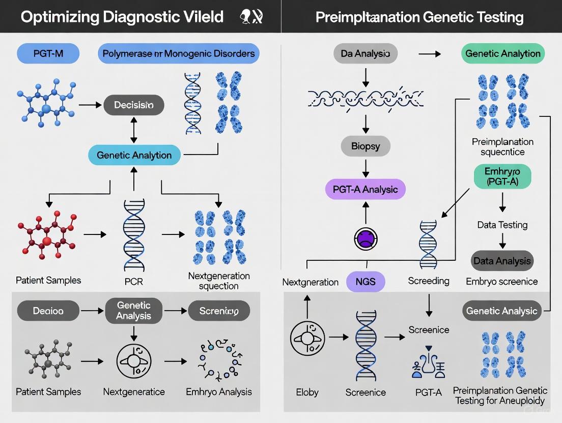
Advanced Genetic Technologies and Testing Strategies for POI Investigation
Next-Generation Sequencing (NGS) has revolutionized genomics research by enabling the parallel sequencing of millions to billions of DNA fragments, providing comprehensive insights into genome structure, genetic variations, and gene expression profiles [30] [31]. This transformative technology has shifted the paradigm from single-gene analysis to massive, high-throughput genomic investigations, making large-scale whole-genome sequencing accessible and practical for researchers at a fraction of the time and cost of traditional methods [30].
The selection of an appropriate NGS platform is a critical strategic decision that directly influences the feasibility and success of a research or clinical project. Modern NGS platforms are categorized into benchtop sequencers for small-scale studies and targeted panels, production-scale sequencers for large genome projects and population studies, and specialized platforms designed for specific applications like long-read sequencing [30]. Each platform excels in different areas, with variations in throughput, read length, error profiles, and analytical scope [32].
Key NGS Platform Specifications
| Platform Type | Typical Throughput | Read Length | Key Applications | Key Strengths |
|---|---|---|---|---|
| Short-Read Platforms (e.g., Illumina) | 1 GB - 6 TB per run [30] | 50-300 bp [30] [31] | Whole Genome Sequencing, Targeted Sequencing, RNA-Seq [30] | High accuracy (error rates: 0.1-0.6%), low cost per base [32] |
| Long-Read Platforms (e.g., PacBio SMRT) | Varies | Average 10,000-25,000 bp [31] | De novo genome assembly, complex structural variant detection [30] | Resolves repetitive regions, haplotype phasing |
| Nanopore Sequencing (e.g., Oxford Nanopore) | Varies | Average 10,000-30,000 bp [31] | Real-time analysis, metagenomics [31] | Ultra-long reads, direct RNA sequencing capability |
For diagnostic yield optimization in genetic testing research, the choice between targeted panels, whole-exome sequencing, and whole-genome sequencing depends on the specific research goals, with targeted approaches offering cost-effective, deep coverage of specific gene sets, while whole-genome methods provide a comprehensive view of the entire genome [33]. Targeted NGS (tNGS) offers advantages of high sensitivity, high efficiency, and relatively low cost, making it particularly valuable for detecting multiple pathogens in mixed infections and drug-resistance genes [34].
Troubleshooting Common NGS Experimental Issues
Library Preparation Problems
Library preparation is a crucial stage where many NGS failures originate. Common issues include:
Problem: Low Library Yield
- Causes: Poor input quality/contaminants, inaccurate quantification, fragmentation inefficiency, suboptimal adapter ligation, or overly aggressive purification [35].
- Solutions: Re-purify input sample to remove inhibitors; use fluorometric quantification methods (e.g., Qubit) instead of UV absorbance alone; optimize fragmentation parameters; titrate adapter:insert molar ratios; and adjust purification protocols [35].
Problem: Adapter Dimer Contamination
- Causes: Excess adapters promote adapter dimer formation, visible as a sharp ~70-90 bp peak in electropherograms [35].
- Solutions: Ensure proper adapter-to-insert molar ratio; optimize ligation conditions; include purification steps with adjusted bead ratios to remove small fragments [35].
Problem: PCR Amplification Bias
- Causes: Too many PCR cycles, inefficient polymerase, or primer exhaustion can introduce duplicates and artifacts [35].
- Solutions: Minimize PCR cycles; use high-fidelity polymerases; optimize primer design and annealing conditions [35] [36].
Problem: Sample Cross-Contamination
- Causes: Inadequate sterilization practices or handling multiple samples simultaneously [36].
- Solutions: Thoroughly sterilize workstations and tools; handle one sample at a time; include DNA-free negative controls in the workflow [36].
Sequencing and Data Quality Issues
Problem: Low-Quality Reads
- Causes: Degraded sequencing reagents, cluster overloading, or phasing issues during the run [37].
- Solutions: Check reagent quality and expiration dates; optimize sample loading concentrations; use appropriate quality control metrics throughout the process [37].
Problem: Insufficient Coverage or Uneven Coverage
- Causes: Biases in primer binding ("mispriming"), inadequate read depth, or GC-content bias [36].
- Solutions: Carefully design specific primers; ensure adequate sequencing depth for the application; use library preparation methods that mitigate GC bias [36].
Problem: Index Misassignment (in Multiplexed Runs)
- Causes: Errors in index allocation during sample multiplexing can lead to sample mix-ups [36].
- Solutions: Use unique dual indexing strategies; employ library prep kits with built-in normalization to minimize index hopping [36].
Essential Research Reagents and Materials
A successful NGS experiment relies on high-quality reagents and materials throughout the workflow. The table below details key components and their functions.
Research Reagent Solutions
| Reagent/Material | Function | Critical Considerations |
|---|---|---|
| High-Quality Input DNA/RNA | Template for library preparation | Minimum 200-500 ng total DNA recommended; assess integrity (RIN/DIN) and purity (260/280 ~1.8, 260/230 >1.8) [35] [36] |
| Fragmentation Reagents | Shear nucleic acids to desired size | Optimize enzymatic, sonication, or nebulization parameters to avoid over/under-shearing [30] [35] |
| Library Preparation Kit | Fragment end-repair, A-tailing, adapter ligation | Select kit compatible with application (e.g., PCR-free for WGS to reduce bias); ensure fresh enzymes and buffers [35] |
| Indexed Adapters | Unique sample identification for multiplexing | Use unique dual indexes to minimize misassignment; titrate adapter:insert ratio to prevent dimer formation [30] [35] |
| High-Fidelity PCR Master Mix | Amplify library fragments | Minimize amplification cycles to preserve complexity; use master mixes to reduce pipetting error [35] [36] |
| Size Selection Beads | Purify and select target fragment size | Precisely control bead:sample ratio; avoid over-drying beads to prevent sample loss [35] |
| Quality Control Instruments (e.g., BioAnalyzer, Qubit) | Quantify and qualify libraries pre-sequencing | Cross-validate with fluorometric and qPCR methods for accurate amplifiable concentration [35] |
NGS Workflow Visualization
The following diagram illustrates the core NGS workflow, from sample preparation to data analysis, highlighting key stages where errors commonly occur and quality control is essential.
Frequently Asked Questions (FAQs)
Q1: What are the most critical steps to prevent NGS library preparation failures? A1: The most critical steps include: (1) Using high-quality, accurately quantified input DNA; (2) Optimizing fragmentation to achieve the desired insert size; (3) Using the correct adapter-to-insert ratio to minimize adapter dimers; and (4) Minimizing PCR amplification cycles to reduce duplicates and bias. Implementing rigorous quality control after each major step is essential for early problem detection [35] [36].
Q2: How can I improve the coverage uniformity in my targeted NGS panels? A2: To improve coverage uniformity: (1) Carefully design primers to avoid mispriming and ensure specific binding; (2) Optimize PCR conditions to minimize amplification bias; (3) Use automated liquid handlers or master mixes to reduce pipetting errors that cause batch effects; and (4) Consider library prep solutions that offer built-in normalization for more consistent read depths across samples [36].
Q3: What are the common sources of false-positive variant calls in NGS data? A3: Common sources include: (1) Sequencing errors, particularly in platforms with higher intrinsic error rates; (2) Inadequate quality control of raw reads, leading to misinterpretation of low-quality bases; (3) Cross-contamination between samples; (4) Misalignment of reads, especially in complex genomic regions; and (5) PCR artifacts introduced during amplification. Robust bioinformatics pipelines and careful troubleshooting are required to minimize these false positives [37] [38] [33].
Q4: How does the choice between short-read and long-read sequencing impact diagnostic yield in genetic testing? A4: Short-read sequencing (e.g., Illumina) offers high accuracy and is excellent for detecting single nucleotide variants and small indels. However, it may miss large structural variants, repeats, and variations in complex genomic regions. Long-read technologies (e.g., PacBio, Oxford Nanopore) can span these challenging regions, potentially increasing diagnostic yield for disorders where these larger alterations are causative. A combined approach or selecting the technology based on the suspected variant type is often optimal for maximizing diagnostic yield [30] [31] [33].
Q5: What computational resources are typically required for NGS data analysis? A5: NGS data analysis demands significant computational resources. Whole-genome sequencing datasets can require powerful servers with substantial memory (RAM), high-performance processors (CPUs), and extensive storage space, often in the terabyte range. The computational load can slow analyses or cause failures without proper resources. Utilizing standardized pipelines and cloud-based solutions can help manage these demands [30] [37].
Integrating CNV Analysis with SNV Detection for Comprehensive Genetic Assessment
Frequently Asked Questions (FAQs)
FAQ 1: Why is integrating CNV and SNV analysis crucial in genetic testing? CNVs contribute significantly to the genomic burden of many monogenic diseases. Relying on SNV analysis alone can miss a substantial number of diagnoses. Integrating both analyses from a single dataset, such as exome or genome sequencing, increases the overall diagnostic yield, making the testing process more efficient and cost-effective, especially in resource-limited settings [39] [40].
FAQ 2: What is the typical diagnostic yield added by CNV analysis? The contribution of CNV analysis varies by disease category but is significant. One large study on kidney disease found that CNVs accounted for 2.4% of the total diagnostic yield, representing 10.5% of all positive genetic tests. The highest impact was observed in congenital anomalies of the kidney and urinary tract (CAKUT) and chronic kidney disease at a young age [39]. For rare monogenic disorders, integrating CNV detection can increase the diagnostic yield by up to 18% beyond what is achieved by SNV analysis alone [40].
FAQ 3: What are the main technological approaches for combined CNV and SNV detection?
- Next-Generation Sequencing (NGS): Methods like whole-exome sequencing (WES) and whole-genome sequencing (WGS) can detect both SNVs and CNVs from the same dataset. WGS is particularly powerful as it provides uniform coverage and can identify variants in both coding and non-coding regions [41] [42].
- Single-Cell Multi-Omics: Emerging platforms enable co-detection of SNVs and CNVs from the same single cell, providing unprecedented insight into cellular heterogeneity, which is particularly valuable in cancer research [43].
- Exome-Based CNV Calling: Computational tools applied to exome sequencing data can call CNVs without requiring a separate test, improving efficiency [39].
FAQ 4: My exome-based CNV analysis has a high false-positive rate. How can I improve specificity? High false-positive rates in exome sequencing often stem from uneven coverage. To mitigate this:
- Utilize Read-Depth Methods: Employ robust read-depth-based algorithms (e.g., ExomeDepth, XHMM) that account for technical variability and coverage biases between samples [41] [40].
- Manual Curation: Implement a step of manual review by an experienced analyst to distinguish true positives from artifacts, acknowledging the assay's limitations in reporting [41].
- Apply Quality Control: Use pipelines with integrated quality control metrics for CNV data to ensure the reliability of calls before further analysis [39].
FAQ 5: How should I interpret a novel CNV of uncertain significance? Interpretation should follow evidence-based professional standards, such as those from the American College of Medical Genetics and Genomics (ACMG) and ClinGen.
- Use a Scoring Framework: Employ a quantitative, points-based system that evaluates the CNV's genomic content (e.g., overlap with haploinsufficient genes), data from population and disease databases, and inheritance patterns [44].
- Uncouple Classification from Implications: Separate the evidence-based classification of the variant (e.g., "Variant of Uncertain Significance") from its potential implications for a specific individual [44].
Troubleshooting Common Experimental Issues
Problem: Low Sensitivity for Single-Exon CNVs in Exome Sequencing Data
- Potential Cause: Capture-based exome sequencing methods often have inconsistent coverage and inherent biases, making it difficult to detect small copy number changes affecting only one or two exons [41].
- Solution:
- Switch to Genome Sequencing: If possible, use WGS data, which provides more uniform coverage and higher resolution for detecting smaller CNVs [41].
- Leverage Specialized Tools: Utilize CNV-calling tools specifically designed for high sensitivity on small regions, such as
ExomeDepthorHMZDelFinder[40]. - Orthogonal Validation: Confirm findings using an independent method like Multiplex Ligation-dependent Probe Amplification (MLPA) for targeted genes [42].
Problem: Inconsistent CNV Calls Between Different Bioinformatics Tools
- Potential Cause: Different algorithms (read-depth, split-read, assembly-based) have unique strengths, weaknesses, and sensitivities to data quality and coverage [41] [40].
- Solution:
- Adopt an Ensemble Approach: Use multiple complementary tools (e.g., combining a read-depth method with a split-read method) to achieve a more holistic and accurate call set [41].
- Standardize Data Quality: Ensure high and uniform sequencing coverage (depth) across the genome or exome, as low-quality data exacerbates discrepancies [41].
- Use Validated Pipelines: Implement well-documented and clinically validated pipelines where possible, and always report the specific tools and parameters used [44] [39].
Problem: Challenges in Detecting Complex Structural Variants or Repeat Expansions
- Potential Cause: Standard short-read NGS methods (both WES and WGS) are often unable to resolve complex rearrangements, balanced inversions, or large repeat expansions [45] [42].
- Solution:
- Employ Specialized Assays: Use methods like chromosomal microarrays (CMA) for large CNVs, or Southern blot/Repeat-Primed PCR for repeat expansions [42].
- Implement Long-Read Sequencing: Utilize third-generation sequencing technologies (e.g., Oxford Nanopore, PacBio) to sequence long, single DNA molecules, which is highly effective for resolving complex regions [42].
Diagnostic Yield of Integrated Analysis
The table below summarizes the demonstrated impact of combining CNV and SNV analysis across different studies and conditions.
| Disease or Context | Base SNV Yield | Additional Yield from CNV Analysis | Overall Contribution of CNV to Positive Tests | Source |
|---|---|---|---|---|
| Monogenic Kidney Disease (n=2,432 probands) | ~20.6% | 2.4% | 10.5% | [39] |
| Rare Monogenic Disorders | Varies | Up to 18% (yield increase) | Up to 15% of cases attributed to CNVs | [40] |
| Prenatal Isolated Clubfoot (n=61 fetuses) | 6.6% (SNVs/Indels) | 3.3% (CNVs) | 33% of pathogenic findings were CNVs | [46] |
Experimental Protocols for Integrated Analysis
Protocol 1: Exome-Based CNV and SNV Co-Detection
This protocol leverages exome sequencing data for simultaneous variant detection, optimizing resource use [39] [40].
Library Preparation & Sequencing:
- Perform exome capture using a clinical-grade kit (e.g., IDT xGen, Agilent SureSelect).
- Sequence on an NGS platform (Illumina) to a minimum mean coverage of 100x.
Bioinformatic Processing:
- Alignment: Map sequencing reads to a reference genome (e.g., GRCh38) using a aligner like BWA-MEM.
- SNV/Indel Calling: Process aligned BAM files through a standard GATK best practices pipeline for SNV and small indel calling.
- CNV Calling: Run the aligned BAM files through a read-depth-based CNV caller.
ExomeDepthis a widely used tool that controls for technical variability between samples and is effective for detecting small, heterozygous deletions [39] [40].
Variant Annotation and Filtration:
- Anonymize all variants using population frequency databases (gnomAD), prediction algorithms, and disease databases (ClinVar, HGMD).
- Filter SNVs/indels based on quality metrics, population frequency, and predicted pathogenicity.
- Filter CNVs based on quality scores, overlap with known pathogenic regions, and dosage-sensitive genes.
Interpretation and Validation:
- Interpret filtered variants according to ACMG/ClinGen guidelines [44].
- Segregation analysis in family trios (where available) is highly recommended for both SNVs and CNVs.
- Validate clinically significant CNVs by an orthogonal method such as MLPA or CMA.
Protocol 2: A Single-Cell Multi-Omics Workflow for SNV and CNV
This protocol is for resolving clonal heterogeneity, as used in cancer research [43].
Sample Preparation:
- Create a single-cell suspension from fresh or frozen tissue or cultured cells.
Single-Cell Sequencing on the Tapestri Platform:
- Load cells and a custom DNA panel onto the Tapestri Instrument.
- The microfluidic system performs targeted amplification of genomic loci for SNVs and genome-wide coverage for CNV analysis from the same single cells.
Data Analysis:
- Use the integrated Tapestri Pipeline and Tapestri Insights software to generate single-cell SNV and CNV data.
- Co-detection allows for the phasing of SNVs and CNVs to reconstruct clonal architecture and evolutionary history.
The Scientist's Toolkit: Essential Research Reagents and Solutions
| Item | Function/Benefit | Example Tools/Assays |
|---|---|---|
| Exome Capture Kits | Enriches coding regions of the genome for efficient sequencing. | IDT xGen Exome Research Panel, Agilent SureSelect |
| CNV Calling Software | Identifies copy number gains/losses from NGS data based on read depth. | ExomeDepth, XHMM, CLAMMS, GATK-gCNV [40] |
| Single-Cell DNA Kits | Enables co-detection of SNVs and CNVs from individual cells. | Mission Bio Tapestri DNA Panel [43] |
| Orthogonal Validation Kits | Independent confirmation of CNVs detected by NGS. | MPLA kits (e.g., for DMD, SMN1), CMA microarrays |
| Variant Interpretation Databases | Provides evidence for variant classification (pathogenic/benign). | ClinGen, ClinVar, DECIPHER, Database of Genomic Variants [45] [44] |
Integrated SNV and CNV Analysis Workflow
The diagram below illustrates a streamlined bioinformatics pipeline for the simultaneous detection of SNVs and CNVs from next-generation sequencing data.
CNV Interpretation and Troubleshooting Logic
This flowchart outlines a standardized, evidence-based process for interpreting and troubleshooting copy number variants, particularly those with uncertain significance.
Customized Gene Panels and Virtual Panel Configurations for POI
Frequently Asked Questions (FAQs)
Q1: What is a virtual gene panel and how does it improve POI genetic analysis? A virtual gene panel is a user-defined, version-controlled set of genes or genomic regions used to focus genetic analysis on specific targets of interest [47]. For Premature Ovarian Insufficiency (POI) research, applying a virtual panel allows you to filter analysis results to variants within the panel's genes and customize the annotation process, for example, by selecting which transcript should be used when annotating variants in a particular gene [47]. This streamlines the case analysis process, ensures consistency, and enhances the reproducibility of your research.
Q2: What is the typical diagnostic yield for a targeted POI gene panel? Recent genetic studies on idiopathic POI report a substantial diagnostic yield. The table below summarizes key performance metrics from a 2025 study that combined array-CGH and NGS of a 163-gene panel [28].
Table 1: Diagnostic Yield of a Combined Genetic Approach in Idiopathic POI
| Genetic Analysis Method | Number of Patients with Anomalies | Percentage of Cohort | Types of Anomalies Identified |
|---|---|---|---|
| Overall Diagnostic Yield | 16 of 28 | 57.1% | Causal CNVs, SNVs, Indels, and VUS |
| Array-CGH (CNV detection) | 1 of 28 | 3.6% | Causal CNV (15q25.2 deletion) |
| NGS (SNV/Indel detection) | 8 of 28 | 28.6% | Causal single nucleotide/indel variations |
| Variants of Uncertain Significance (VUS) | 7 of 28 | 25.0% | Likely benign or VUS |
Q3: Which genes and technologies are critical for a comprehensive POI panel? An effective POI panel should include genes involved in key ovarian functions and leverage multiple genomic technologies to maximize diagnostic yield [28].
Table 2: Essential Research Toolkit for POI Genetic Investigation
| Research Reagent / Technology | Function / Application in POI Research |
|---|---|
| Custom NGS Gene Panel (e.g., 163 genes) | Targeted sequencing of genes known or suspected in oogenesis, folliculogenesis, meiosis, and DNA repair [28]. |
| Array-CGH (Oligonucleotide, 180K) | Genome-wide detection of copy number variations (CNVs) and chromosomal rearrangements contributing to POI [28]. |
| FIGLA, BMP15, GDF5 Genes | Key genes involved in ovarian development and function; inclusion is essential for a comprehensive panel [28]. |
| Virtual Panel Platform (e.g., Franklin) | Software to create and manage version-controlled gene panels, apply curated transcript settings, and filter variants [47]. |
Q4: How should I handle a Variant of Uncertain Significance (VUS) found in my POI panel analysis? When a VUS is identified, a thorough multi-step validation and interpretation process is recommended:
- Database Interrogation: Systematically check population frequency databases (like gnomAD), variation databases (such as ClinVar and DECIPHER), and the scientific literature [28].
- ACMG Guidelines: Classify the variant according to the standards and guidelines from the American College of Medical Genetics (pathogenic, likely pathogenic, VUS, likely benign, benign) [28].
- Phenotype Correlation: Compare the patient's clinical presentation with known disease associations for the gene in OMIM and published case reports [28].
- Familial Segregation Testing: If possible, test biological parents or other affected family members to see if the variant co-segregates with the POI phenotype [28].
Q5: My panel analysis shows adequate sequencing depth, but I'm not getting a clear diagnosis. What are the potential reasons? Several factors can contribute to this, even with good technical quality:
- Incomplete Panel Gene Content: Your panel may be missing novel or very recently discovered POI-associated genes. Regularly review and update your panel gene list [28].
- Non-Coding or Complex Variants: Standard panels focus on exonic regions and may miss deep intronic variants affecting splicing, or complex structural variants. Consider whole-genome sequencing for unresolved cases [28].
- Technical Limitations: Some genomic regions are difficult to sequence or map accurately with short-read NGS. Verify coverage in low-performing regions and consider orthogonal methods like Sanger sequencing for important, poorly covered exons [48].
- Oligogenic or Epigenetic Causes: POI may sometimes result from variations in multiple genes or epigenetic modifications not detected by standard genetic tests [28].
Troubleshooting Guides
Issue 1: Low Diagnostic Yield in a Custom POI Panel
Potential Causes and Solutions:
- Cause: Inadequate Gene Coverage.
- Solution: Regularly curate your gene list against recent literature. The list should be comprehensive, covering genes involved in ovarian development, meiosis, DNA repair, and immune function. A 2025 study used a panel of 163 genes for their investigation [28].
- Cause: Missing Copy Number Variations (CNVs).
- Solution: Relying solely on NGS for SNVs will miss larger structural variants. Integrate array-CGH analysis into your workflow. One study found a pathogenic 15q25.2 deletion via array-CGH that was crucial for a diagnosis [28].
- Cause: Over-reliance on a Single Technology.
- Solution: Adopt a multi-modal genetic testing strategy. The highest diagnostic yield (57.1%) was achieved by combining NGS for small variants with array-CGH for large CNVs in the same patient cohort [28].
The following workflow diagram illustrates a recommended genetic analysis pipeline for POI to maximize diagnostic yield.
Optimized POI Genetic Analysis Workflow
Issue 2: Inconsistent Annotation of Variants Across Research Samples
Potential Causes and Solutions:
- Cause: Lack of a Standardized Transcript for a Critical Gene.
- Solution: Use the "hard panel" function in your virtual panel platform. This allows you to enforce the use of a specific, manually curated preferred transcript for each gene during the annotation phase, guaranteeing consistent results across all samples [47].
- Cause: Using Different Panel Versions for Different Batches.
- Solution: Leverage the version-control features of your virtual panel software. Always document the panel name and version used in each analysis to ensure full reproducibility [47].
- Cause: Uncontrolled Panel Modifications.
- Solution: Establish a formal curation and review process within your organization before any new genes are added to the "Organizational Knowledge Base" [47].
Issue 3: Managing and Interpreting the High Number of Variants from an NGS Panel
Potential Causes and Solutions:
- Cause: Lack of Effective Filtering.
- Solution: Apply your virtual panel as a "soft filter" to immediately restrict the visible results in your analysis platform to only those variants within your POI gene panel, drastically reducing the data burden [47].
- Cause: Unclear Variant Pathogenicity.
- Solution: Implement a strict variant prioritization pipeline as outlined in FAQ A4, adhering to ACMG guidelines. Focus on protein-truncating variants, de novo occurrences in sporadic cases, and variants in genes with a strong known phenotype match [28].
The diagram below outlines the key steps for analyzing and prioritizing variants after sequencing.
NGS Variant Analysis and Prioritization
Bioinformatics Pipelines for Variant Prioritization and Interpretation
This technical support center provides troubleshooting and guidance for bioinformatics pipelines used in the genetic analysis of Premature Ovarian Insufficiency (POI). POI, affecting 1-3.5% of women, is characterized by a loss of ovarian function before age 40, and genetic causes are identified in approximately 20-25% of cases [28] [1] [11]. Optimizing your variant prioritization and interpretation pipeline is crucial for improving diagnostic yield in POI research. The following guides and FAQs address common challenges.
Frequently Asked Questions (FAQs)
FAQ 1: What is a typical diagnostic yield for POI using genetic analysis, and how can we improve it?
Diagnostic yield varies depending on the patient cohort and methods used. The table below summarizes findings from recent studies [28] [11].
Table: Diagnostic Yield in POI Genetic Studies
| Study Details | Cohort Size | Overall Diagnostic Yield | Yield in Primary Amenorrhea (PA) | Yield in Secondary Amenorrhea (SA) | Key Genes Identified |
|---|---|---|---|---|---|
| Whole-exome sequencing (WES) cohort [11] | 1,030 patients | 23.5% (242/1030) | 25.8% (31/120) | 17.8% (162/910) | NR5A1, MCM9, novel meiosis genes |
| Combined array-CGH & targeted NGS panel [28] | 28 patients | 57.1% (16/28) | Information not specified | Information not specified | FIGLA, causal CNVs and SNVs |
To improve yield:
- Consider Amenorrhea Type: Patients with Primary Amenorrhea often have a higher genetic burden, including more biallelic variants [11].
- Utilize Multi-Method Approaches: Combining CNV analysis (e.g., array-CGH) with NGS for single-nucleotide variants (SNVs) can increase yield, as some cases are solved only by one method [28].
- Expand Gene Lists: Incorporate novel gene associations from recent large-scale studies into your analysis panels [11].
FAQ 2: Our pipeline is producing too many Variants of Uncertain Significance (VUS). How can we reduce this?
A high VUS rate is a common challenge. The following experimental protocol can help reclassify VUS through functional validation.
Table: Protocol for Functional Validation of VUS in POI-Associated Genes
| Step | Objective | Detailed Methodology | Expected Outcome & Interpretation |
|---|---|---|---|
| 1. Select Candidates | Prioritize VUS for functional study | Focus on VUS in known POI genes (e.g., BLM, HFM1, MCM8, MCM9, MSH4, RECQL4, NR5A1) from patients with strong phenotype [11]. | A shortlist of high-priority VUS for experimental testing. |
| 2. Functional Assay | Determine the biochemical impact of the variant | For genes involved in homologous recombination (HR) repair, perform cell-based HR assays. Introduce the variant into an appropriate cell line and measure HR efficiency using reporter systems (e.g., DR-GFP assay) [11]. | A significant reduction in HR efficiency compared to wild-type provides PS3 evidence for pathogenicity per ACMG guidelines. |
| 3. Phase Determination | Confirm biallelic inheritance for recessive disorders | If two heterozygous P/LP variants are found in the same gene, confirm they are on different alleles (in trans) using T-clone sequencing or 10x Genomics linked-read approaches [11]. | Confirmation of in trans phase provides PM3 evidence for pathogenicity. |
| 4. Reclassification | Update variant classification | Compile functional evidence (PS3) and phasing data (PM3) and re-evaluate the variant according to ACMG/AMP guidelines [49]. | VUS is reclassified as Likely Pathogenic (LP) or Pathogenic (P). |
FAQ 3: Our data has quality issues leading to unreliable variant calls. What are key QC checkpoints?
The "Garbage In, Garbage Out" principle is critical in bioinformatics [50]. Implement QC at every stage.
Table: Essential Quality Control Checkpoints in a POI NGS Pipeline
| Pipeline Stage | Key QC Metrics & Tools | Common Pitfalls & Solutions |
|---|---|---|
| Raw Sequence Data | Phred Quality Scores (Q30+), GC content, sequence duplication levels via FastQC [50]. | Low scores indicate poor sequencing run. Solution: Re-sequence if necessary. Adapter contamination. Solution: Use tools like Trimmomatic [50]. |
| Alignment | Alignment rate (should be high, e.g., >95%), mean coverage, and uniformity of coverage via SAMtools/Qualimap [50]. | Low alignment rate can indicate contamination or poor reference genome choice. Check sample identity. |
| Variant Calling | Variant Quality Score Recalibration (VQSR) or hard-filtering using tools like GATK. Assess transition/transversion (Ti/Tv) ratio and number of variants [50]. | Too many/too few variants can indicate batch effects or sample contamination. Use cross-validation with an alternative method (e.g., PCR) for key findings [50]. |
| Sample & Data Management | Sample tracking, metadata recording using LIMS; workflow version control with Git/Snakemake/Nextflow [50]. | Sample mislabeling is a pervasive error. Solution: Implement barcode labeling and genetic identity verification [50]. |
FAQ 4: What are the latest software and scoring systems for variant classification?
Stay updated with evolving standards. The QCI Interpret 2025 release includes new features for both hereditary and somatic workflows [51]:
- New Predictors: Integration of REVEL (missense pathogenicity) and SpliceAI (splicing impact) scores.
- Updated Guidelines: Draft support for the points-based ACMG v4 (hereditary) and VICC (somatic) scoring guidance to prepare for upcoming classification changes [51].
- Enhanced Filtering: New Mode of Inheritance (MOI) filters and dynamic gene list creation to streamline prioritization for POI, which involves both dominant and recessive genes [51].
The Scientist's Toolkit
Table: Key Research Reagent Solutions for POI Genetic Testing
| Item | Function in POI Research |
|---|---|
| Custom Targeted NGS Panel [28] | A panel of ~160+ genes known or suspected in ovarian function allows focused, cost-effective sequencing for POI. |
| Array-CGH Kit (e.g., Agilent 4x180K) [28] | Identies copy number variations (CNVs), a known genetic cause of POI that SNV-focused NGS can miss. |
| Functional Assay Kits (e.g., HR Repair Assay) [11] | Provides experimental evidence to reclassify VUS in POI genes involved in DNA repair and meiosis. |
| Variant Interpretation Software (e.g., QCI Interpret, Alissa Interpret) [28] [51] | Clinical decision support software that aggregates curated knowledge and automates ACMG classification. |
| Pathway Analysis Databases (e.g., WikiPathways, Reactome) [52] | Allows visualization of candidate genes in biological context (e.g., meiotic pathways) to assess biological plausibility. |
Visualizing the POI Genetic Analysis Workflow
The following diagram outlines the core bioinformatics pipeline for POI genetic testing, from sample to report, incorporating key troubleshooting checkpoints.
Bioinformatics Pipeline for POI Genetic Testing
Biological Pathways in POI
Understanding the biological pathways of candidate genes is essential for assessing their plausibility in POI pathogenesis. The diagram below maps key genes onto their functional pathways.
Key Biological Pathways and Genes in POI
Overcoming Diagnostic Challenges and Implementing Precision Medicine Programs
Multidisciplinary Team Structures for Effective POI Program Implementation
The integration of multidisciplinary teams (MDTs) has become fundamental to unlocking higher diagnostic yields in complex genetic testing environments, particularly for conditions requiring sophisticated diagnostic pathways like Point of Care Testing (POCT) implementation. As genomic medicine advances, the interpretation of genomic data demands close collaboration between clinical, laboratory, and research expertise [53]. The MDT model, widely regarded as the gold standard in cancer care, is now being successfully adapted to genomic medicine, facilitating higher diagnostic rates and improved patient management [54] [53]. This approach is especially critical for POI programs where optimizing diagnostic yield relies on seamlessly integrating diverse specialized knowledge to address technical, clinical, and analytical challenges.
Core Structure of an Effective Genomic MDT
Essential Team Members and Their Roles
A genomic MDT for an effective POI program requires integration of professionals who contribute distinct but complementary expertise. The team composition should include:
- Clinical Geneticists and Genetic Counselors: Provide expertise in clinical phenotyping, variant interpretation in the context of clinical presentation, and patient communication.
- Molecular Geneticists and Laboratory Scientists: Offer deep knowledge of genomic technologies, variant calling, bioinformatics pipelines, and assay validation.
- Bioinformaticians and Data Scientists: Develop and maintain computational pipelines for variant annotation, filtration, and interpretation; implement machine learning algorithms for data analysis.
- Research Scientists: Bridge cutting-edge research discoveries with clinical applications, providing insights into novel gene-disease associations and functional validation approaches.
- Specialist Clinicians (e.g., oncologists, neurologists, cardiologists based on testing focus): Contribute organ-specific or disease-specific knowledge for clinical correlation.
- POCT Technology Specialists: Provide expertise in decentralized testing platforms, assay optimization, and implementation logistics.
- Ethics and Policy Specialists: Navigate ethical considerations, informed consent processes, and regulatory requirements for genetic testing [53] [55] [56].
Characteristic Features of High-Functioning MDTs
Research on effective cancer MDTs has identified several characteristics of highly functioning teams, which are transferable to genomic MDTs:
- Diverse Expertise: Combination of complementary skills from multiple fields providing holistic perspectives [55]
- Shared Goals: While members have different roles, the team works toward common objectives for patient diagnostics and research outcomes [55]
- Collaborative Approach: Open communication, regular meetings, and consensus building facilitate joint decision-making [55]
- Coordinated Processes: Integrated services and continuity across testing and interpretation phases to avoid duplication and gaps [55]
- Effective Leadership: Designated leadership to direct specialized professionals and maintain meeting focus [54] [55]
Table 1: Quantitative Impact of MDT Approach on Diagnostic Yields in Genomic Medicine
| Condition Category | Base Diagnostic Yield | Yield with MDT Approach | Absolute Improvement | Study Context |
|---|---|---|---|---|
| Rare Diseases/Cancer Genetic Predisposition | Not specified | 30.6% overall diagnostic yield | 6-25% attributed to MDT | French Genomic Medicine Initiative [56] |
| Various Genomic Conditions | 10-78% (depending on context) | Increased by 6-25% with MDT | 6-25% absolute increase | Systematic Review of Genomic MDTs [53] |
MDT Workflow for POI Program Optimization
The following diagram illustrates the coordinated workflow of a multidisciplinary team within a genomic diagnostic program, highlighting the integration of clinical, laboratory, and analytical functions:
Figure 1: Multidisciplinary Team Workflow in Genomic Diagnostics. This workflow demonstrates how cases progress through clinical input, laboratory processing, data analysis, multidisciplinary team discussion, and finally to clinical reporting with continuous feedback to research databases.
Troubleshooting Guides and FAQs for POI Program Implementation
Frequently Encountered Technical Challenges
Q1: Our POCT platform shows inconsistent results between trained operators and novice users. How can we improve reliability?
A: Implement machine learning algorithms for result interpretation to minimize human error. CNNs (Convolutional Neural Networks) have been successfully applied to imaging-based POCT platforms to recognize patterns and extract task-specific features from image datasets, providing automated analysis without compromising sensitivity [57]. Supervised learning approaches using pre-labeled datasets can classify results with high accuracy, reducing false positives and negatives when used by individuals with less training [57].
Q2: How can we enhance the sensitivity of our POCT platform to detect low-abundance biomarkers?
A: Integrate ML-driven signal processing and computational optimization of sensor designs. Deep learning can enhance multiplexing capabilities through parallel analysis of multiple sensing channels [57]. Neural network-based analyte concentration inference significantly improves quantification accuracy and repeatability compared to standard multi-variable regression methods [57]. For lateral flow assays and vertical flow assays, ML algorithms can process complex datasets to identify subtle changes in biomarker profiles despite biological sample noise [57].
Q3: Our multidisciplinary team struggles with coordination across different specialties, leading to delays in diagnostic reporting. What structural improvements would you recommend?
A: Implement standardized meeting protocols with clear leadership and predefined workflows. Research on cancer MDTs recommends [54]:
- Establish a designated MDT lead or chair to maintain focus and efficiency
- Develop structured meeting agendas with time allocation per case
- Ensure availability of complete patient information before meetings
- Utilize assessment tools like the MDT-OARS (Observational Assessment Rating Scale) or MDT-MODe (Metric of Decision-Making) to evaluate and improve team functioning
- Implement a "quality improvement bundle" including checklist application, team skills brief training, and guidance implementation [54]
Q4: How can we efficiently handle variants of uncertain significance (VUS) within our MDT framework?
A: Establish a systematic approach for VUS interpretation leveraging cross-specialty collaboration. The genomic MDT approach has demonstrated high efficiency in interpreting VUS by combining clinical, laboratory, and functional expertise [53]. Develop a standardized protocol for:
- Clinical correlation with patient phenotype
- In silico prediction tools utilization
- Literature review for recent evidence
- Determination of appropriate functional validation studies
- Family studies when appropriate Documenting VUS resolution pathways creates institutional knowledge that improves future efficiency.
Q5: What strategies can improve the adoption and consistent use of our POI program across different clinical specialties?
A: Address key implementation barriers through a multifaceted approach [58] [53]:
- Develop specialized roles like "genomic pathway managers" to assist and train prescribers (successfully implemented in the French Genomic Medicine Initiative) [56]
- Create user-friendly electronic prescription tools with clinical decision support
- Establish regular educational sessions and case discussions
- Demonstrate clinical utility through rapid turnaround times and impactful results
- Secure reimbursement pathways that support the MDT model
Implementation and Process Challenges
Table 2: Performance Metrics from National Genomic Medicine Implementation
| Metric Category | Performance Data | Context and Implications |
|---|---|---|
| Diagnostic Yield | 30.6% for RD/CGP | French Genomic Medicine Initiative (12,737 results returned) [56] |
| Turnaround Time | 202 days (median for RD/CGP), 45 days (median for cancers) | Highlights area for process optimization in MDT workflows [56] |
| Prescriber Engagement | 63.7% of registered clinicians made ≥1 prescription; 6.5% responsible for 69.4% of prescriptions | indicates need for broader adoption across clinical specialties [56] |
Experimental Protocols for MDT Performance Optimization
Protocol: MDT Meeting Efficiency Assessment
Objective: To quantitatively evaluate and improve the effectiveness of multidisciplinary team meetings in genomic diagnostic programs.
Materials:
- MDT Observational Assessment Rating Scale (MDT-OARS) tool [54]
- MDT Metric of Decision-Making (MDT-MODe) instrument [54]
- Audio/video recording equipment (with appropriate consent)
- Structured data collection forms
Methodology:
- Pre-meeting Preparation Assessment:
- Document completeness of patient clinical information
- Verify availability of relevant test results and prior genetic studies
- Confirm appropriate case selection for discussion
Meeting Observation:
- Record attendance by specialty using the MDT-MODe instrument
- Measure time spent per case and contribution distribution across specialties
- Document quality of presented information using standardized metrics
- Assess team ability to reach definitive decisions
Post-meeting Analysis:
- Calculate decision implementation rates
- Track turnaround times from discussion to report finalization
- Correlate meeting process metrics with diagnostic outcomes
Quality Improvement Implementation:
- Provide structured feedback to the MDT using assessment results
- Implement targeted interventions (e.g., checklist application, brief training)
- Reassess at 3-month intervals to measure improvement [54]
Expected Outcomes: This protocol typically identifies specific bottlenecks in MDT functioning and enables targeted improvements, potentially increasing diagnostic yield by 6-25% through optimized team processes [53].
Protocol: Machine Learning Integration for POCT Interpretation
Objective: To enhance accuracy and reliability of point-of-care test interpretation through supervised machine learning approaches.
Materials:
- Labeled dataset of POCT results (minimum 500-1000 samples)
- Computational resources for model training (Python/R environment)
- Standardized imaging setup for result capture (if using visual tests)
- Validation set of prospectively collected samples
Methodology:
- Data Preprocessing:
- Apply image denoising, background subtraction, and normalization for visual tests
- Implement data augmentation techniques to expand training dataset
- Split data into training (60%), validation (20%), and blind testing (20%) sets
Model Selection and Training:
- Evaluate multiple algorithm types: CNN for image-based tests, SVM or random forest for numerical data
- Optimize model hyperparameters using validation set performance
- Employ k-fold cross-validation to minimize overfitting
Performance Validation:
- Test final model on blind dataset never seen during training
- Compare ML interpretation accuracy against expert human readers
- Assess impact on false positive/negative rates in novice users
Implementation:
- Deploy validated model as mobile application or integrated with POCT reader
- Establish ongoing performance monitoring and model refinement process [57]
Expected Outcomes: ML integration can significantly reduce interpretation errors, particularly for non-expert users, and improve detection of faint positive lines or complex patterns in multiplex assays [57].
The Scientist's Toolkit: Essential Research Reagent Solutions
Table 3: Key Research Reagents and Platforms for POI Program Implementation
| Reagent/Platform Category | Specific Examples | Function in POI Program |
|---|---|---|
| Sequencing Technologies | Short-read genome sequencing, Exome sequencing, RNAseq | Comprehensive genomic characterization for rare diseases, cancer predisposition, and cancers [56] |
| Point-of-Care Testing Platforms | Lateral Flow Assays (LFAs), Vertical Flow Assays (VFAs), Nucleic Acid Amplification Tests (NAATs) | Decentralized, rapid testing; enhanced by ML for improved accuracy [57] |
| Computational Tools | Convolutional Neural Networks (CNNs), Support Vector Machines (SVMs), Random Forest classifiers | ML algorithms for test interpretation, signal processing, and quantitative analysis [57] |
| Data Integration Systems | National secure data storage facilities (e.g., CAD in France), Bioinformatics pipelines | Secure data management, intensive calculation capabilities, clinical interpretation support [56] |
| Variant Interpretation Resources | ACMG guidelines, Population databases, Functional prediction tools | Standardized variant classification, pathogenicity assessment, clinical correlation [53] |
The implementation of effectively structured multidisciplinary teams represents a critical success factor for POI programs aiming to optimize diagnostic yield in genomic medicine. Evidence from established genomic and cancer MDTs demonstrates that coordinated multidisciplinary approaches can increase diagnostic yields by 6-25% compared to siloed efforts [53]. The integration of machine learning technologies for POCT interpretation, combined with systematic MDT workflows and continuous performance assessment, creates a robust framework for advancing genetic testing research. As genomic medicine continues to evolve, the MDT model provides the necessary collaborative structure to translate complex genomic data into clinically actionable insights, ultimately enhancing patient care and research outcomes. Future efforts should focus on standardizing MDT processes, improving interoperability between different expert domains, and developing more sophisticated computational tools to support collaborative decision-making in genomic medicine.
In the field of premature ovarian insufficiency (POI) research, optimizing genetic testing is paramount for advancing diagnostic capabilities. Diagnostic yield—the proportion of patients in whom a test successfully identifies the true disease condition—is a critical endpoint positioned between diagnostic accuracy and patient outcomes in research studies [59]. Technical limitations related to specimen quality and analytical sensitivity directly impact this yield, influencing both research validity and clinical translation. This technical support center provides targeted troubleshooting guidance to address these challenges, specifically framed within the context of POI genetic testing where early diagnosis is crucial yet hampered by limited non-invasive warning markers [60].
Troubleshooting Guide: Specimen Quality Issues
The quality of biological specimens directly impacts the success of downstream genetic analyses. The table below outlines common issues, their causes, and evidence-based solutions.
Table 1: Troubleshooting Specimen Quality in DNA Extraction
| Problem | Root Cause | Recommended Solution |
|---|---|---|
| Low DNA Yield | - Frozen cell pellet thawed too abruptly [61]- Membrane clogged by tissue fibers [61]- Column overloaded with DNA (in DNA-rich tissues) [61] | - Thaw cell pellets slowly on ice [61]- Centrifuge lysate to remove fibers; reduce input material for fibrous tissues [61]- Reduce the amount of input material for DNA-rich tissues [61] |
| DNA Degradation | - Tissue pieces too large, allowing nucleases to degrade DNA before lysis [61]- Sample stored improperly or for too long at -20°C [61]- High nuclease content in soft organ tissue [61] | - Cut tissue into smallest possible pieces or grind with liquid nitrogen [61]- Flash-freeze with liquid nitrogen and store at -80°C; use stabilizing reagents [61]- Keep samples frozen and on ice during preparation [61] |
| Protein Contamination | - Incomplete tissue digestion [61]- Membrane clogged with indigestible tissue fibers [61] | - Extend lysis time by 30 minutes to 3 hours after tissue dissolves [61]- Centrifuge lysate at max speed for 3 minutes to remove fibers [61] |
| RNA Contamination | - Too much input material, inhibiting RNase A activity [61]- Insufficient lysis time [61] | - Do not exceed recommended input amounts [61]- Extend lysis time by 30 minutes to 3 hours [61] |
FAQs on Analytical Sensitivity
1. How can our lab prevent contamination in reagents and consumables? Running quality control checks on reagents prior to use in casework is essential. Negative controls and reagent blanks provide a means to detect contamination from reagents. For consumables that cannot be pretreated (e.g., centrifugal filter units), establishing a procedure to evaluate a percentage from each lot number prior to use can provide valuable data for future contamination investigations [62].
2. What quality control parameters are critical for reagents used in nucleic acid analysis? Key QC tests for reagents include DNase/RNase testing to prevent nucleic acid degradation, bioburden testing for microbial enumeration, endotoxin testing to avoid inflammatory reactions in experiments, and absorbance testing to confirm reagent purity and correct components [63].
3. Our genetic testing for POI has a low diagnostic yield. What are the potential reasons? Low diagnostic yield can stem from multiple factors. In POI research, the genetic background remains unexplained in most cases, with over 50 genes implicated but each accounting for only a small portion of patients [64]. Technical factors include suboptimal specimen quality (see Table 1), the use of outdated gene panels that don't cover newly discovered associations, and a lack of systematic reanalysis of genomic data, which has been shown to improve diagnostic yield in rare neurologic diseases and could be applied to POI [65] [64].
Experimental Protocols for Quality Assurance
Protocol 1: Quality Control Testing of Reagents for DNA Analysis
Purpose: To ensure reagents are free of DNase contamination and other impurities that could compromise analytical sensitivity.
Materials:
- Reagents to be tested
- Intact, high-quality genomic DNA (control)
- Electrophoresis equipment
- QC-validated reagents (for comparison)
Methodology:
- Incubation: Mix a sample of the reagent with the control gDNA.
- Incubation Conditions: Incubate the mixture at 37°C for a period of 30-60 minutes.
- Analysis: Run the incubated sample alongside an untreated control sample on an agarose gel.
- Interpretation: The presence of smearing or a lower molecular weight band in the test sample compared to the intact control indicates DNase contamination. The reagent should not be used [63].
Protocol 2: Validating Analytical Sensitivity for POI Genetic Testing
Purpose: To establish the minimum variant allele frequency (VAF) detectable by your sequencing platform for genes associated with POI.
Materials:
- DNA from well-characterized cell lines with known POI-relevant mutations (e.g., in FOXL2, NR5A1, FIGLA) [64]
- Wild-type DNA
- Your standard sequencing and bioinformatics pipeline
Methodology:
- Create Dilution Series: Mix DNA from mutant and wild-type cell lines to create a series of samples with known VAFs (e.g., 1%, 2%, 5%, 10%).
- Processing: Process all samples through your standard DNA extraction, library preparation, and sequencing workflow.
- Data Analysis: Use your standard bioinformatics pipeline to call variants in the target genes.
- Sensitivity Calculation: Determine the lowest VAF at which the known mutation is consistently and accurately detected across replicates. This establishes your assay's limit of detection (LOD) [64].
This protocol's workflow for establishing a Limit of Detection (LOD) is summarized in the following diagram:
The Researcher's Toolkit: Essential Reagents & Materials
Table 2: Key Research Reagents and Materials for POI Genetic Studies
| Item | Function/Application |
|---|---|
| Proteinase K | Digests tissue and inactivates nucleases during genomic DNA extraction, preventing degradation [61]. |
| RNase A | Degrades RNA during DNA extraction to prevent RNA contamination, which can affect yield and purity measurements [61]. |
| Silica Spin Columns | Selective binding and purification of DNA from complex lysates, a key step in many commercial extraction kits [61]. |
| Cell Lysis Buffer | Typically contains guanidine thiocyanate (GTC), which lyses cells, inactivates nucleases, and allows DNA to bind to silica [61]. |
| RNA Quality Control Material | Synthetic or cell-line derived controls used to estimate test precision and detect analytical deviations in genetic testing [66]. |
| ELN (Electronic Lab Notebook) | Standardizes data entry, enables real-time validation, and maintains audit trails to reduce experimental errors [67]. |
Optimizing Workflows to Maximize Diagnostic Yield
The relationship between specimen quality, analytical sensitivity, and the final diagnostic yield is direct and critical. A high-quality specimen and a highly sensitive analytical method are fundamental prerequisites for a high diagnostic yield. This integrated workflow can be visualized as a multi-stage process where the output of each phase feeds into the next, with system-level quality controls supporting the entire operation.
For POI research, integrating multi-omics data (proteomics, metabolomics, transcriptomics) via methods like Mendelian randomization is an emerging strategy to identify new biomarkers and improve the diagnostic yield beyond traditional genetic sequencing alone [60]. Furthermore, implementing systematic collaborative reanalysis of genomic data, a practice proven to improve diagnostic yield in other rare disease fields, should be adopted for POI cohorts [65].
Strategies for Variant of Uncertain Significance (VUS) Resolution and Reclassification
FAQs: Core Concepts and Initial Steps
What is a Variant of Uncertain Significance (VUS), and why is it a major challenge in POI genetic testing?
A Variant of Uncertain Significance (VUS) is a genetic change for which the association with a disease is not yet clear. It does not meet the criteria to be classified as either pathogenic or benign. In the context of Premature Ovarian Insufficiency (POI), this is a significant challenge because the genetic basis is highly diverse, with mutations in more than 75 genes linked to the condition [5] [22]. A VUS result creates uncertainty, which can impede a definitive diagnosis, complicate risk assessment for family members, and hinder the development of personalized treatment strategies. In genetic databases, the majority of missense variants in disease-associated genes are classified as VUS or have conflicting interpretations, underscoring the scale of this problem [68].
Why is the reclassification of a VUS important for our POI research and for patients?
VUS reclassification is a critical process that can directly impact diagnostic yield and clinical management. When a VUS is reclassified to Pathogenic or Likely Pathogenic, it can provide a definitive molecular diagnosis for a patient's POI, ending a long diagnostic odyssey [69]. This information can then be used for informed family planning, assessing risks for associated health conditions (like osteoporosis and cardiovascular disease [22]), and guiding reproductive decisions. For the research community, each reclassification adds a crucial data point that helps interpret the same variant in other patients, progressively clarifying the genetic architecture of POI [70].
We have identified a VUS in a POI patient. What is the first step we should take?
The first and most powerful step is data sharing. Submit the variant to public archives like ClinVar [69]. Before submission, perform a thorough review of the existing evidence using population frequency databases (like gnomAD), in silico prediction tools (such as SIFT, PolyPhen, and CADD), and the scientific literature [69] [70]. Sharing the variant, along with the patient's clinical phenotype (symptoms), allows the global scientific community to see the evidence you have found. This facilitates matching across laboratories and is often the catalyst for reclassification when another lab observes the same variant in a patient with a similar phenotype [71].
What are VUS subclasses, and how can they help prioritize our research efforts?
Some clinical laboratories internally further classify VUS into subcategories based on the weight of available evidence. While not yet standard on all clinical reports, understanding these concepts can help prioritize variants for investigation [72]:
- VUS-high: Evidence suggests the variant could be pathogenic, but it is insufficient for a Likely Pathogenic classification.
- VUS-mid: The evidence is conflicting or entirely absent.
- VUS-low: Evidence suggests the variant may be benign, but it is insufficient for a Likely Benign classification.
Focusing your reclassification efforts on VUS-high variants is the most efficient strategy, as they have the highest probability of being upgraded to (Likely) Pathogenic. One multi-laboratory study showed that variants in higher-level VUS subclasses were significantly more likely to be reclassified towards pathogenic [72].
Troubleshooting Guides: Advanced Resolution Strategies
Problem: A VUS remains unresolved after database searches and in silico analysis.
Solution: Proceed to functional validation using advanced assays.
- Step 1: Employ Multiplexed Assays of Variant Effect (MAVEs). MAVEs are a powerful high-throughput functional genomics approach that can simultaneously assess the impact of thousands of variants in a gene of interest [68]. The workflow is as follows [68]:
- Saturation Mutagenesis: Create a library that contains nearly all possible single nucleotide variants in your target gene (e.g., a POI-associated gene like BMP15 or FSHR).
- Library Delivery: Introduce this variant library into an appropriate cell model (e.g., HEK293 cells, yeast, or even iPSC-derived granulosa cells if possible) using methods like lentiviral transduction or integration into a "landing pad" locus.
- Functional Selection: Subject the pool of cells to a selection pressure that depends on the protein's normal function (e.g., cell survival, surface expression of a receptor, or a fluorescent reporter of a signaling pathway).
- Sequencing and Analysis: Use next-generation sequencing to count the abundance of each variant before and after selection. Variants that are depleted after selection are likely to be disruptive (potentially pathogenic), while those that remain are likely benign.
The following diagram illustrates the core MAVE workflow:
- Step 2: Utilize validated disease-specific models. For POI research, developing assays that reflect ovarian function is ideal. While challenging, emerging models include using induced pluripotent stem cell (iPSC)-derived ovarian cells to test the effects of variants on folliculogenesis or hormone production [68] [22].
Problem: Our VUS is in a non-European patient, and population frequency data is lacking.
Solution: Actively address the ancestry-based data gap.
- Step 1: Acknowledge the disparity. Underrepresented populations, such as Middle Eastern, Asian, and African, receive a higher burden of VUS due to their underrepresentation in major population databases like gnomAD [70]. This limits the ability to use frequency as evidence for benignity.
- Step 2: Generate internal population-specific data. If possible, sequence a control cohort from the same ethnic background to determine if the VUS is a common polymorphism in that population. Collaborating with research institutions in the patient's region of origin can be highly effective [70] [71].
- Step 3: Leverage specialized prediction tools. Use gene-specific or disease-aware prediction algorithms that may be more robust to population biases. Tools like Gene-Aware Variant Interpretation (GAVIN) integrate gene-specific data with in silico predictions to improve accuracy [69].
Problem: We have multiple VUS candidates and need to decide which one to investigate first.
Solution: Implement a systematic prioritization pipeline.
- Phenotype Match: Prioritize variants in genes with a strong known association to the patient's specific POI phenotype (e.g., primary vs. secondary amenorrhea) [5] [22].
- VUS Subclass: If available, prioritize VUS-high variants [72].
- Variant Type: Prioritize protein-truncating variants (nonsense, frameshift, splice-site) in haploinsufficient genes over missense variants, as their impact is often more predictable.
- Functional Evidence: Check if any high-throughput functional data (e.g., from a MAVE study) already exists for the gene or specific variant [68].
- Segregation Analysis: If family members are available, test for co-segregation of the variant with the POI phenotype. This can provide powerful evidence for pathogenicity.
The table below summarizes key quantitative data on VUS reclassification to guide resource allocation:
Table 1: VUS Reclassification Rates from Recent Studies
| Study Context | Reclassification Rate | Key Findings | Source |
|---|---|---|---|
| Hereditary Breast & Ovarian Cancer (Levantine Cohort) | 32.5% of VUS were reclassified | 2.5% of all VUS were upgraded to (Likely) Pathogenic | [70] |
| Multi-Laboratory Data (Various Mendelian Diseases) | Distinct reclassification rates for VUS subclasses | VUS-high variants had the highest odds of being reclassified as (Likely) Pathogenic | [72] |
| Large-Scale Diagnostic Testing (Invitae) | ~80% of reclassified VUS were downgraded to benign | Highlights the importance of reclassification to avoid false positives | [71] |
The Scientist's Toolkit: Research Reagent Solutions
Table 2: Essential Tools for VUS Resolution
| Research Reagent / Tool | Function in VUS Resolution | Application in POI Research |
|---|---|---|
| Saturation Mutagenesis Library | Generates a comprehensive set of all possible variants in a target gene for functional screening. | Systematically test all missense variants in genes like FOXL2 or NOBOX to create an "atlas" of variant effects [68]. |
| Landing Pad Cell Line | Allows for the controlled, single-copy integration of a variant library into a specific genomic site, ensuring consistent expression. | Essential for MAVE experiments in human cell lines (e.g., HEK293) to study ion channels or transcription factors implicated in POI [68]. |
| Induced Pluripotent Stem Cells (iPSCs) | Provides a patient-specific or engineered cell source that can be differentiated into relevant cell types. | Differentiate into ovarian granulosa-like cells to study the functional impact of VUS in a disease-relevant context [68] [22]. |
| In silico Predictors (SIFT, CADD, PolyPhen) | Computational tools that predict the functional consequence of a genetic variant based on evolutionary conservation and sequence context. | Initial triage and evidence gathering for VUS interpretation following ACMG/AMP guidelines [69] [70]. |
| Population Databases (gnomAD) | Catalogues genetic variation from large populations to assess the frequency of a variant. | Provides critical evidence to rule out pathogenicity if a VUS is common in healthy populations [69] [70]. |
| Clinical Archives (ClinVar) | A public repository of reports of the relationships between variants and phenotypes. | Central for data sharing and identifying if other labs have classified the same VUS, potentially with more evidence [69]. |
Experimental Protocol: A Framework for MAVE in a POI Gene
Objective: To determine the functional consequences of all possible missense variants in a POI-associated gene (e.g., BMP15) using a MAVE.
Materials:
- Gene of interest (e.g., BMP15 cDNA)
- Oligonucleotide library for saturation mutagenesis
- Landing pad HEK293T cell line
- Lentiviral packaging plasmids (psPAX2, pMD2.G)
- Selection antibiotics (e.g., Puromycin)
- Cell culture reagents
- Next-generation sequencing platform
Methodology:
- Library Construction: Use an inverse PCR or commercial synthesis-based method to generate a plasmid library encompassing all single-nucleotide substitutions in the BMP15 coding sequence [68].
- Virus Production and Cell Infection: Produce lentivirus containing the BMP15 variant library. Infect the landing pad HEK293T cells at a low Multiplicity of Infection (MOI) to ensure most cells receive only one variant. Select successfully integrated cells with puromycin [68].
- Functional Selection: (Assay must be tailored to the gene's function)
- For BMP15, a key oocyte-derived growth factor, you could use a cell survival or proliferation assay on a reporter granulosa cell line that responds to BMP15 signaling.
- Alternatively, use a surface expression assay (e.g., by FACS) if the variant's impact on protein trafficking is of interest.
- Sequencing: Extract genomic DNA from the cell pool before selection (initial library) and after selection. Amplify the integrated BMP15 region and subject it to deep sequencing [68].
- Data Analysis: Calculate an "enrichment score" or "functional score" for each variant by comparing its frequency post-selection to its frequency in the initial library. Variants with low scores are functionally impaired.
The strategic relationship between VUS resolution and the overall goal of optimizing diagnostic yield in POI research is summarized below:
Optimizing Workflow Efficiency and Integrating Genomic Data with Electronic Health Records
Technical Support Center: Troubleshooting Guides & FAQs
Frequently Asked Questions (FAQs)
Q1: What are the primary technical challenges when integrating genomic data with EHRs?
A: The main challenges include interoperability and data compatibility issues between systems that use different data formats and structures [73] [74]. Semantic misalignment across standards like HL7 FHIR and SNOMED CT can disrupt data interpretation [73]. Furthermore, concerns regarding the security, governance, and clinical utility of sensitive genomic information present significant implementation barriers [73].
Q2: Which integration standards are most critical for connecting lab systems like LIMS to EHRs?
A: The most critical standards are:
- HL7 (v2/v3): Widely used for exchanging structured data like lab orders (ORM messages) and results (ORU messages) [74] [75].
- FHIR (Fast Healthcare Interoperability Resources): A modern, web-based API standard increasingly used for real-time data access [74] [75].
- DICOM (Digital Imaging and Communications in Medicine): The standard for storing, retrieving, and transmitting medical images [75].
- IHE (Integrating the Healthcare Enterprise): A framework that uses HL7 and DICOM to standardize communications across different vendor systems [75].
Q3: How can we improve the diagnostic yield of genetic testing in our research?
A: Evidence suggests several strategies:
- Utilize Gene Panel Sequencing: For genetically heterogeneous diseases (e.g., hereditary ophthalmic diseases), targeted gene panels can offer a higher diagnostic yield than exome sequencing (ES) in some contexts, due to superior read depth and optimization [76].
- Consider Genome Sequencing (GS) as a First-Line Test: A 2024 meta-analysis found that the pooled diagnostic yield for GS was 30.6% compared to 23.2% for ES, with GS having 1.7-times the odds of providing a diagnosis [77].
- Implement Additional Gene Panel Sequencing: If an initial gene panel returns negative, subsequent or complementary panels can enhance the overall diagnostic yield by capturing variants missed in the first test [76].
Q4: What solutions can mitigate data security risks in an integrated system?
A: To protect sensitive patient and genomic data, implement:
- Strong Encryption: For data both in transit and at rest [74].
- Strict Access Controls & Multi-Factor Authentication (MFA): To limit system access to authorized personnel only [74].
- Regular Security Audits: To proactively identify and remediate vulnerabilities [74].
- Blockchain-Enabled Governance: Emerging strategies like blockchain are being explored for secure and ethical genomic data governance [73].
Troubleshooting Common Integration Issues
Problem: Lab results are not populating the correct fields in the EHR.
- Cause: Incorrect data mapping between the LIMS and EHR, often due to the use of different names or codes for the same test [74].
- Solution: Standardize terminology using common vocabularies like LOINC for lab tests and SNOMED CT for clinical terms [75]. Revisit and validate the data mapping specifications.
Problem: High latency (delays) in receiving lab results in the EHR.
- Cause: Inadequate network and server infrastructure struggling with real-time data synchronization, or bottlenecks in the message processing engine [75].
- Solution: Perform a network and infrastructure performance review. Consider using an integration engine (e.g., Mirth Connect) to efficiently manage data flow and message transformations [75].
Problem: Genomic variant data is stored in the EHR but is not clinically actionable.
- Cause: Lack of standardized approaches and clinical decision support (CDS) tools to interpret the genomic data within clinical workflows [73].
- Solution: Develop and implement AI-supported frameworks and CDS tools that can analyze the genomic data in the context of the patient's clinical phenotype to provide personalized treatment recommendations [73].
Quantitative Data on Diagnostic Yield
The following table summarizes key quantitative findings from recent research on the diagnostic yield of different genetic testing approaches, which is central to optimizing research workflows.
Table 1: Diagnostic Yield of Genome-Wide Sequencing (GWS) in Pediatric Rare Diseases [77]
| Sequencing Method | Pooled Diagnostic Yield | Odds Ratio vs. Non-GWS | Comparative Basis |
|---|---|---|---|
| GWS (GS & ES) | 34.2% (95% CI: 27.6-41.5) | 2.4 (95% CI: 1.40-4.04; P < .05) | Within-cohort studies (N=13) |
| Non-GWS | 18.1% (95% CI: 13.1-24.6) | (Reference) | Within-cohort studies (N=13) |
| Genome Sequencing (GS) | 30.6% (95% CI: 18.6-45.9) | 1.7 (95% CI: 0.94-2.92; P = .13) | Within-cohort studies (N=3) |
| Exome Sequencing (ES) | 23.2% (95% CI: 18.5-28.7) | (Reference) | Within-cohort studies (N=3) |
Table 2: Clinical Utility of a Positive Genetic Diagnosis [77]
| Sequencing Method | Pooled Clinical Utility |
|---|---|
| Genome Sequencing (GS) | 58.7% (95% CI: 47.3-69.2) |
| Exome Sequencing (ES) | 54.5% (95% CI: 40.7-67.6) |
Experimental Protocol: Enhancing Diagnostic Yield via Additional Gene Panel Sequencing
This protocol is based on a study investigating hereditary ophthalmic diseases [76].
Objective: To improve the genetic diagnostic yield for a heterogeneous disease group through sequential gene panel testing.
Methodology:
Patient Cohort & DNA Extraction:
- Recruit unrelated patients with a clinical diagnosis of the hereditary disease (e.g., retinopathy).
- Obtain informed consent.
- Extract genomic DNA from peripheral blood samples using a commercial kit (e.g., QIAamp DNA mini kit from Qiagen).
Primary Gene Panel Sequencing:
- Prescribe a targeted, customized gene panel (e.g., a hereditary retinopathy panel) covering known disease-associated genes.
- Refer the sample to a CAP-accredited clinical laboratory for sequencing.
- Perform target enrichment using custom-designed probes and a sequencing kit (e.g., from Celemics).
- Sequence the prepared libraries on a platform like Illumina MiSeqDX with 150 bp paired-end reads.
Bioinformatic Analysis (Primary):
- Align sequences to a reference genome (e.g., hg19) using BWA-aln.
- Identify single nucleotide variants and small insertions/deletions using tools like GATK Haplotypecaller and VarScan.
- Annotate variants and filter against population databases (e.g., gnomAD).
- Classify variants according to ACMG/AMP guidelines (Pathogenic, Likely Pathogenic, Variant of Uncertain Significance, etc.).
Additional Gene Panel Sequencing:
- If no significant disease-associated mutations are found in the primary panel, initiate an additional gene panel sequencing for research purposes using the remaining DNA.
- This secondary panel may cover a broader or complementary set of genes.
Bioinformatic & Clinical Correlation Analysis (Additional):
- Repeat the bioinformatic pipeline for the new data.
- Use in silico prediction algorithms (SIFT, PolyPhen-2, MutationTaster) to assess the impact of missense variants.
- Correlate the identified genotypes with the patient's clinical phenotype to assess pathogenicity.
- Validate all candidate variants (LPV, PV, VUS) using Sanger sequencing.
Workflow Diagram: The following flowchart illustrates the key decision points in this sequential testing protocol.
System Integration Workflow for Genomic and Diagnostic Data
The integration of laboratory and genomic data into the clinical EHR workflow is a multi-step process involving several systems and standards, as shown in the diagram below.
The Scientist's Toolkit: Research Reagent Solutions
Table 3: Essential Materials for Genomic Testing and Data Integration Workflows
| Item / Solution | Function / Application | Example / Specification |
|---|---|---|
| DNA Extraction Kit | Isolation of high-quality genomic DNA from patient blood or tissue samples for downstream sequencing. | QIAamp DNA Blood Mini Kit (Qiagen) [76]. |
| Targeted Enrichment Probes | Custom-designed oligonucleotide probes to capture and enrich specific genomic regions of interest for sequencing. | Custom RNA-based probes (e.g., from Celemics) [76]. |
| Massively Parallel Sequencer | Platform for high-throughput, parallel DNA sequencing of prepared genomic libraries. | Illumina MiSeqDX sequencer [76]. |
| Bioinformatic Pipelines | Software suites for aligning sequences, calling variants, and annotating results. | Genome Analysis Tool Kit (GATK) Best Practices pipeline, BWA for alignment [76]. |
| Variant Annotation Databases | Population and clinical databases used to filter and interpret the pathogenicity of genetic variants. | Genome Aggregation Database (gnomAD) [76]. |
| In Silico Prediction Tools | Algorithms to predict the functional impact of genetic variants, aiding in classification. | SIFT, PolyPhen-2, MutationTaster [76]. |
| Integration Engine | Middleware to manage data flow, transform messages between standards (HL7, FHIR), and route information between systems. | Mirth Connect [75]. |
| Standardized Terminologies | Controlled vocabularies to ensure consistent data meaning and semantic interoperability between systems. | LOINC (for lab tests), SNOMED CT (for clinical terms), RADLEX (for radiology) [75]. |
Economic Considerations and Reimbursement Strategies for Comprehensive Genetic Testing
Technical Support & Troubleshooting
Frequently Asked Questions (FAQs)
Q1: Our research indicates Whole Genome Sequencing (WGS) provides superior diagnostic yield. How do we justify its cost and secure reimbursement for its use in clinical research?
A: Economic justification for WGS hinges on its higher diagnostic yield and long-term cost-effectiveness. Key strategies include:
- Demonstrate Superior Yield: Present data showing WGS can achieve a diagnostic yield of 41%, significantly higher than the 24% from conventional targeted testing [78]. Emphasize that WGS captures structural and non-coding variants, which can contribute to over 40% of solved cases [79].
- Highlight Comprehensive Analysis: WGS serves as a single, comprehensive test, potentially replacing a costly and time-consuming stepwise approach involving chromosomal microarrays and multiple gene panels [78].
- Showcase Clinical Impact: Document how WGS findings directly change clinical management. In one study, life-saving treatment adjustments were made for five patients based on WGS results [79].
Q2: Our claims for pharmacogenomic (PGx) testing panels are frequently denied. What are the proven strategies to improve reimbursement rates?
A: Reimbursement success for PGx testing requires a strategic approach to coding and documentation.
- Prefer Panel Codes: Data indicates PGx panels have a significantly higher reimbursement rate (74%) compared to single-gene tests (43%) [80]. Use panel-specific CPT or PLA (Proprietary Laboratory Analyses) codes when medically justified.
- Maximize Medical Necessity: Claims submitted with multiple, relevant ICD-10-CM diagnosis codes have higher success rates. Ensure clinical documentation clearly links the test to specific patient symptoms, family history, and treatment decisions [80].
- Leverage Guidelines: Reference established guidelines from the Clinical Pharmacogenetics Implementation Consortium (CPIC) and FDA recommendations in prior authorization requests, as these are key sources payers rely on [80].
Q3: What are the critical steps for establishing medical necessity for a comprehensive genetic panel?
A: Medical necessity is the cornerstone of reimbursement. Documentation must include [81]:
- Detailed Phenotype: A thorough clinical description of the patient's condition using standardized ontologies (e.g., Human Phenotype Ontology).
- Comprehensive Family History: A multi-generation family history indicating an inherited pattern.
- Test Selection Rationale: A clear explanation for why a comprehensive panel was chosen over a targeted test, often due to clinical heterogeneity or a complex family history.
- Impact on Management: A statement on how the results will directly influence patient care, such as guiding treatment choices, surgical decisions, or surveillance protocols.
Q4: How do we navigate the use of unlisted CPT codes for novel genomic assays?
A: Use unlisted code 81479 judiciously and with robust support [81].
- Exhaust Alternatives First: Only use 81479 for truly novel methodologies that cannot be reported with existing, more specific codes.
- Provide Detailed Documentation: Submit a cover letter and supporting materials that thoroughly describe the test's methodology, analytes, and clinical validity. Cross-reference to similar, established tests can be helpful.
- Set Financial Expectations: Be aware that reimbursement for unlisted codes is unpredictable and often requires extensive direct communication with payers.
Troubleshooting Guide: Managing Common Economic and Reimbursement Hurdles
| Problem | Possible Cause | Solution |
|---|---|---|
| Low reimbursement rate for comprehensive panels. | Payer policy prefers targeted testing or single-gene approaches. | Perform a reimbursement analysis comparing panel vs. component coding; appeal with data showing panel efficiency and clinical utility [81]. |
| Denials based on "investigational" or "not medically necessary." | Insufficient documentation of medical necessity and clinical utility. | Implement a pre-test checklist to ensure all required elements (phenotype, family history, management impact) are documented [81]. |
| Inconsistent reimbursement across different payers. | Variable coverage policies and interpretation of evidence. | Create a payer-specific knowledge base of coverage criteria for common tests; tailor submissions to each payer's policy [80]. |
| High rate of claim denials for technical reasons. | Incorrect use of CPT codes, modifiers, or Z-codes. | Develop an internal coding guide with a modifier decision tree; audit claims pre-submission [81]. |
Experimental Protocols & Workflows
Protocol: Cost-Benefit Analysis of Whole Genome Sequencing vs. Standard Testing
Objective: To quantitatively compare the diagnostic yield and cost-efficiency of WGS against a conventional genetic testing pathway.
Materials:
- Cohort of patients with undiagnosed, suspected genetic disorders.
- WGS sequencing platform (e.g., Illumina).
- Bioinformatic pipeline for variant calling (SNVs, indels, CNVs, non-coding).
- Clinical interpretation team (clinical molecular geneticists, bioinformaticians).
Methodology:
- Cohort Recruitment: Recruit patients who have undergone or are eligible for standard-of-care genetic testing (e.g., chromosomal microarray + targeted gene panels) [78].
- Parallel Testing: Perform WGS on all participants.
- Variant Analysis: Analyze WGS data using a comprehensive pipeline. Also, analyze the data restricted to genes covered by the conventional panels used in the study for a direct comparison.
- Yield Calculation:
- Cost Analysis:
- Document all costs associated with the conventional diagnostic odyssey (multiple tests, clinician visits).
- Compare with the single cost of WGS.
- Model long-term cost savings from ending the "diagnostic odyssey."
Protocol: Optimizing Reimbursement Strategy for a Novel Genetic Test
Objective: To establish a reimbursement strategy that maximizes financial sustainability for a new genetic test.
Methodology:
- Code Mapping: Identify all potential CPT codes (e.g., 81479 for unlisted molecular pathology) and corresponding Z-codes (if applicable) for the test [81].
- Payer Policy Research: Analyze coverage policies from top payers (commercial, Medicare) for similar tests. Identify specific requirements for coverage.
- Evidence Dossier Development: Compile a dossier including:
- Analytical and clinical validity data.
- Evidence of clinical utility from literature or internal studies.
- Clinical practice guidelines (e.g., CPIC) supporting the test's use.
- Pilot Billing & Appeals:
- Submit a small number of test claims to key payers.
- Meticulously track denials and their reasons.
- Implement a structured appeals process using the evidence dossier [81].
- Financial Modeling: Use pilot results to project overall reimbursement rates and adjust the business model accordingly.
Data Presentation
Diagnostic Yield and Economic Impact of Advanced Sequencing
Table 1: Comparative Diagnostic Yield of Whole Genome Sequencing vs. Conventional Testing
| Testing Method | Reported Diagnostic Yield | Key Advantages | Contributing Variant Types |
|---|---|---|---|
| Whole Genome Sequencing (WGS) | 41% [78] to 35% (up to 39% with novel candidates) [79] | Single, comprehensive test; detects all variant types | Coding, structural, deep intronic, and splice site variants (43% of solved cases) [79] |
| Conventional Targeted Testing | 24% [78] | Lower upfront cost | Limited to SNVs/indels in pre-specified genes |
| Whole Exome Sequencing (WES) | Information missing from sources | Broader than panels but misses non-coding variants | Primarily coding exonic variants |
Table 2: Pharmacogenomic (PGx) Testing Reimbursement Landscape
| Parameter | Single-Gene PGx Test | Multi-Gene PGx Panel |
|---|---|---|
| Typical Reimbursement Rate | 43% [80] | 74% [80] |
| Example CPT Codes | 81225 (CYP2C19), 81226 (CYP2D6) [80] | Various; often panel-specific PLA codes [80] |
| Reimbursement by Payer | Commercial: ~48%, Medicare: ~48%, Medicaid: ~42% [80] | Commercial: ~48%, Medicare: ~48%, Medicaid: ~42% [80] |
Visualization: Workflows and Strategies
Reimbursement Optimization Workflow
Medical Necessity Documentation Pathway
The Scientist's Toolkit: Research Reagent Solutions
Table 3: Essential Components for Comprehensive Genetic Testing & Analysis
| Research Reagent / Tool | Function | Application in Protocol |
|---|---|---|
| Phenotypic Data Capture Tool (e.g., PhenoTips) | Collects and standardizes patient phenotype data using the Human Phenotype Ontology (HPO) [78]. | Critical for establishing a structured phenotype-genotype correlation, supporting medical necessity. |
| Next-Generation Sequencing (NGS) Platform | Provides the technology for high-throughput sequencing of entire genomes or exomes. | Core platform for generating WGS or WES data. |
| Bioinformatic Pipeline (e.g., SVRare, ALTSPLICE) | Specialized algorithms for detecting and annotating structural variants (SVs) and splice-altering variants [79]. | Essential for maximizing diagnostic yield by identifying non-coding and structural variants missed by standard pipelines. |
| Variant Annotation & Prioritization Pipeline (e.g., ANNOVAR-based) | Annotates variants with population frequency, pathogenicity predictions, and clinical databases [78]. | Filters millions of variants to a shortlist of potentially causative ones for clinical review. |
| CLIA/CAP Certified Laboratory | A clinically certified laboratory environment. | Required for confirmation of diagnostic variants before results can be returned to patients and included in clinical reports [78]. |
Assessing Testing Performance and Clinical Utility of POI Diagnostic Approaches
For researchers and drug development professionals, selecting the appropriate genetic testing methodology is a critical strategic decision that directly impacts diagnostic yield, operational efficiency, and research validity. The choice between targeted gene panels and comprehensive genomic sequencing (including whole exome and whole genome sequencing) involves balancing depth against breadth, cost against comprehensiveness, and analytical simplicity against diagnostic discovery potential. Next-generation sequencing (NGS) technologies have revolutionized genomic research by enabling high-throughput, parallel sequencing of DNA fragments, providing unprecedented insights into genetic variations associated with disease [31]. Understanding the precise performance characteristics, limitations, and optimal applications of each approach is fundamental to optimizing genetic testing research and accelerating therapeutic development.
Technical Performance & Diagnostic Yield Comparison
Quantitative Diagnostic Yield Analysis
Extensive comparative studies provide robust quantitative data on the diagnostic performance of targeted versus comprehensive sequencing approaches. The following table synthesizes key findings from recent clinical and research studies:
Table 1: Comparative Diagnostic Yields of Genomic Testing Approaches
| Study Focus/Population | Targeted Panel Yield | Exome Sequencing (ES) Yield | Genome Sequencing (GS) Yield | Key Findings |
|---|---|---|---|---|
| Diverse Pediatric Cohort (n=645) [82] | 8.1% (52/642) | - | 16.5% (106/642) | GS yielded twice as many diagnoses as targeted panels (P < .001). GS detected most copy number variants (17/19) and mosaic variants (6/8). |
| Brazilian Cohort (Mixed Indications) [83] | - | 32.7% (Overall) | - | ES had the highest detection rate but also the highest inconclusive rate. Skeletal (55%) and hearing (50%) disorders showed highest yields. |
| Pediatric Musculoskeletal Disorders (n=36) [84] | - | - | 61.1% (22/36) | WGS identified 38 pathogenic/likely pathogenic variants; 12 (31.6%) were missed by WES. WGS showed particular advantage in detecting CNVs. |
| French National Program (RD/CGP) [56] | - | - | 30.6% (Overall) | Large-scale implementation of GS demonstrated feasibility for rare diseases and cancer genetic predisposition. |
Analysis of Disparities and Limitations
The data reveals important limitations and disparities. While comprehensive sequencing generally provides higher diagnostic yields, this advantage is not uniform across all population groups. One large pediatric study found that the superior yield of genome sequencing was significant for Hispanic/Latino(a) and White/European American participants but not statistically significant for the Black/African American cohort [82]. This highlights the critical impact of population-specific variant databases and the need for more diverse genomic references. Furthermore, exome sequencing carries a higher rate of inconclusive results due to variants of uncertain significance (VUS), presenting interpretive challenges for researchers and clinicians [83].
Experimental Protocols for Method Comparison
To ensure valid, reproducible comparisons between sequencing methods, researchers should implement standardized experimental protocols. The following workflow outlines a rigorous paired study design, adapted from published validation studies [85] [82].
Diagram 1: Experimental workflow for paired method comparison.
Detailed Methodological Components
Sample Preparation and Quality Control
- DNA Extraction: Use standardized kits (e.g., Chemagic DNA Saliva kits, ReliaPrep gDNA Isolation kits) to obtain high-molecular-weight DNA [84] [86].
- Quality Control: Quantify DNA using fluorometric-based methods (Qubit, Quant-iT) and assess purity via spectrophotometry. Input requirement for targeted panels is typically ≥50 ng DNA [85] [84].
Library Preparation and Sequencing
- Targeted Panels: Utilize hybridization-capture or amplicon-PCR based approaches. The TTSH-oncopanel (61 genes) employs hybridization-capture with custom biotinylated oligonucleotides, compatible with automated library preparation systems (e.g., MGI SP-100RS) to reduce human error and contamination risk [85].
- Comprehensive Sequencing:
- Whole Exome Sequencing (WES): Use kit-based exome capture (e.g., Nextera DNA Flex) followed by sequencing on platforms such as Illumina NextSeq 500DX (aiming for 100x coverage) [84].
- Whole Genome Sequencing (WGS): Employ PCR-free library preparation (e.g., Illumina DNA PCR-Free Prep, Tagmentation kit) followed by sequencing on platforms like Illumina NovaSeq 6000 (aiming for 30x coverage) [84].
- Long-Read Sequencing (LR-GS): For ultrarapid applications, use Oxford Nanopore Technology (ONT) ligation sequencing kits (e.g., SQK-LSK114) on PromethION flow cells, targeting 30-40x coverage with read N50 ~15 kb [86].
Bioinformatic Analysis and Variant Interpretation
- Alignment and Variant Calling: Process data using platforms like Illumina DRAGEN or Sophia DDM software. For long-read data, use Clair3 (SNVs/indels), Sniffles2 (SVs), and modkit (methylation) [84] [86].
- Variant Prioritization: Utilize HPO-based prioritization with tools like Exomiser, incorporating in silico prediction scores (REVEL, CADD, AlphaMissense, SpliceAI) [86].
- Variant Classification: Adhere to ACMG guidelines for classifying variants as pathogenic, likely pathogenic, variant of uncertain significance (VUS), likely benign, or benign [83].
Research Reagent Solutions and Essential Materials
Table 2: Key Research Reagents and Platforms for Genomic Studies
| Reagent/Platform Category | Specific Examples | Primary Function in Research |
|---|---|---|
| DNA Extraction Kits | Chemagic DNA Saliva 600 Kit [84], ReliaPrep Large Volume HT gDNA Isolation Kit [86] | Obtain high-quality, high-molecular-weight DNA suitable for various sequencing platforms. |
| Targeted Panel Kits | TTSH-oncopanel (61 genes) [85], Custom hybridization-capture biotinylated oligonucleotides | Selective enrichment of disease-specific gene sets for focused analysis. |
| Library Prep Kits (WES) | Nextera DNA Flex Pre-Enrichment Library Prep [84] | Preparation of sequencing libraries from genomic DNA for exome sequencing. |
| Library Prep Kits (WGS) | Illumina DNA PCR-Free Prep, Tagmentation Kit [84] | PCR-free library preparation minimizing amplification bias for whole genome sequencing. |
| Library Prep Kits (Long-Read) | Oxford Nanopore Ligation Sequencing Kit SQK-LSK114 [86] | Preparation of libraries for long-read sequencing enabling detection of SVs, methylation, and phasing. |
| Sequencing Platforms | Illumina NovaSeq 6000 (WGS/WES) [84], Oxford Nanopore PromethION (Long-Read) [86], MGI DNBSEQ-G50RS (Targeted) [85] | High-throughput instruments for generating sequencing data with different read lengths and applications. |
| Bioinformatics Tools | Sophia DDM [85], Emedgene [84], Exomiser [86] | Automated variant calling, annotation, and phenotype-driven prioritization for efficient data analysis. |
Frequently Asked Questions (FAQs) & Troubleshooting
Q1: When should a targeted gene panel be chosen over comprehensive sequencing? A: Targeted panels are ideal for: (1) Well-defined genetic conditions with known associated genes (e.g., hereditary breast cancer syndromes) [87]; (2) Situations requiring high coverage depth for detecting low-level mosaicism or when analyzing FFPE samples with degraded DNA [85]; (3) Research focused on specific therapeutic areas with established genetic markers; (4) Budget-constrained projects where cost-effectiveness is paramount [87].
Q2: What are the key advantages of comprehensive sequencing (WES/WGS) for diagnostic yield? A: Comprehensive sequencing offers: (1) Higher diagnostic yields (often 1.5-2× higher than panels) by capturing variants in novel or unexpected genes [82]; (2) Detection of diverse variant types including CNVs, mosaic variants, and repeat expansions that panels may miss [84] [82]; (3) Elimination of sequential testing by providing a single comprehensive dataset that can be reanalyzed as new genes are discovered [87]; (4) Potential for novel gene discovery in research settings [87].
Q3: How does the cost-benefit analysis balance between these approaches? A: While per-test costs are higher for comprehensive sequencing, its higher diagnostic yield may make it more cost-effective in the long run by ending diagnostic odysseys. Targeted panels have lower upfront costs but may lead to higher cumulative expenses if multiple sequential tests are required [87]. The French national program (PFMG2025) demonstrates that large-scale implementation of genome sequencing is economically sustainable at a national level [56].
Q4: What strategies can mitigate the challenge of variants of uncertain significance (VUS)? A: (1) Family segregation studies to determine if VUS co-segregates with disease in affected relatives; (2) Implementing robust bioinformatic pipelines that incorporate multiple in silico prediction tools (REVEL, CADD) [86]; (3) Regular reanalysis of genomic data as knowledge evolves; (4) Functional studies to assess variant impact in model systems; (5) Consortium data sharing to identify VUS in multiple unrelated individuals with similar phenotypes.
Q5: How does long-read sequencing complement traditional short-read approaches? A: Long-read sequencing (e.g., Oxford Nanopore, PacBio) provides: (1) Enhanced detection of structural variants and repeat expansions; (2) Direct phasing of variants without family studies [86]; (3) Epigenetic profiling including methylation status from the same data [86]; (4) Improved assembly in complex genomic regions; (5) Ultrarapid turnaround times (2-10 days) for critical care research [86]. The following diagram illustrates the technical advantages of long-read sequencing:
Diagram 2: Technical advantages of long-read sequencing technologies.
Q6: What are the key considerations for implementing a genomic testing strategy in a research program? A: Successful implementation requires: (1) Clear phenotypic characterization using standardized ontologies (HPO terms) [84]; (2) Adequate bioinformatics infrastructure and expertise for data analysis and storage [56]; (3) Ethical frameworks for handling incidental findings and secondary variants [56]; (4) Validation of wet-lab and computational pipelines to ensure analytical performance [85]; (5) Plan for ongoing data reanalysis as knowledge evolves; (6) Consideration of equity in genomic representation across diverse ancestral backgrounds [82].
Validation Frameworks for Novel Genetic Variants and Emerging Technologies
FAQs & Troubleshooting Guides
Category 1: Variant Interpretation & Classification
FAQ 1.1: What is the standard framework for interpreting novel genetic variants in POI research?
The American College of Medical Genetics and Genomics and the Association for Molecular Pathology (ACMG/AMP) guidelines provide the standard framework for variant interpretation. These guidelines classify variants into five categories: Pathogenic, Likely Pathogenic, Variant of Uncertain Significance (VUS), Likely Benign, and Benign. This classification is based on evidence from population data, computational and predictive data, functional data, segregation data, and de novo observation. For diagnostic testing, such as in POI, it is recommended that only pathogenic and likely pathogenic variants be reported in most clinical contexts, though VUS may be reported in certain situations [88].
FAQ 1.2: A novel variant I discovered is predicted to be damaging by in-silico tools but has a population frequency above 1%. How should I proceed?
A population frequency above 1% is generally considered strong evidence for classifying a variant as benign, as it far exceeds the expected frequency for penetrant alleles causing rare disorders like POI. According to ACMG/AMP guidelines, this would constitute evidence against pathogenicity (BA1 criterion). You should:
- Verify the frequency in large, population-specific genomic databases (e.g., gnomAD).
- Correlate with phenotype: If the variant were causative for POI (prevalence ~3.5%), a high population frequency would be inconsistent with the disease prevalence [1] [23].
- Investigate other evidence: Weigh all other available evidence, but a high frequency is typically a decisive factor for benign classification [88].
FAQ 1.3: How should I handle a Variant of Uncertain Significance (VUS) in my POI research?
Handling a VUS requires a multi-faceted approach to gather additional evidence:
- Segregation Analysis: Test parents and other affected or unaffected family members to see if the variant co-segregates with the POI phenotype.
- Functional Studies: Design in vitro or in vivo experiments to assess the variant's impact on protein function, gene expression, or splicing.
- Data Sharing: Submit the variant to public databases like ClinVar. This allows data to be aggregated with findings from other researchers and clinical laboratories worldwide, which can help reclassify the variant over time [89].
- Consortium Collaboration: Work with research consortia (e.g., CRE-WHiRL) focused on POI to pool data and statistical power [1].
Category 2: Technology-Specific Challenges
FAQ 2.1: My NGS data for the FMR1 gene is inconsistent. What could be the cause?
The FMR1 gene, a common cause of POI, contains a CGG trinucleotide repeat. Standard NGS technologies have limited ability to accurately sequence and size long repetitive elements. Your inconsistency is likely due to:
- Technical Limitation: NGS struggles with GC-rich regions and repeat expansions, leading to poor mapping and coverage [88].
- Recommended Solution: Use a complementary method specifically validated for repeat expansions, such as:
- Southern Blot: The traditional gold standard.
- Triple-Primed PCR or RP-PCR: More modern molecular techniques designed for accurate repeat sizing.
- Long-Read Sequencing (PacBio/Oxford Nanopore): Emerging technologies that can span repetitive regions more effectively [31].
FAQ 2.2: My CNV analysis pipeline is detecting numerous false positives. What quality control (QC) metrics should I check?
False positives in Copy Number Variant (CNV) calling are common. Key QC metrics to troubleshoot include [90]:
- Sample Contamination: Check for cross-sample contamination in the wet lab and bioinformatic steps.
- Batch Effects: Ensure there are no systematic technical variations between different sequencing runs.
- Read Depth and Uniformity: Examine the depth of coverage and its evenness across the target regions. Sudden drops in coverage can be misinterpreted as deletions.
- GC Bias: Assess whether the data has significant GC-content bias, which can skew read counts and CNV calls.
- Control Samples: Validate your pipeline using a standard set of control samples with known CNVs to benchmark performance [90].
FAQ 2.3: What are the key differences between using genome sequencing versus exome sequencing for POI gene discovery?
The choice between whole-genome sequencing (WGS) and whole-exome sequencing (WES) involves trade-offs, as summarized in the table below.
Table 1: Comparison of Exome and Genome Sequencing for POI Research
| Feature | Exome Sequencing (WES) | Genome Sequencing (WGS) |
|---|---|---|
| Target Region | Protein-coding exons (~1-2% of genome) [91] | Entire genome, coding and non-coding |
| Variant Detection | Excellent for single nucleotide variants (SNVs) and small indels in exons | Comprehensive for exonic and intronic SNVs/indels; superior for structural variants |
| Coverage Uniformity | Can be uneven due to capture probe hybridization | More uniform coverage |
| Ability to Detect Non-Coding Variants | Limited | Yes, though clinical interpretation is challenging [91] |
| Cost & Data Storage | Lower cost and data volume | Higher cost and data storage requirements |
For POI, where a significant proportion of causes are unknown, WGS offers the advantage of detecting structural variants and non-coding changes, but requires greater bioinformatic resources and poses interpretation challenges [88].
Category 3: Data Management & Sharing
FAQ 3.1: How can I responsibly share my novel genetic variant and associated phenotype data?
Responsible data sharing is critical for advancing the field. Recommended resources include:
- ClinVar: A public, freely accessible database of genetic variants and their relationships to human health. Researchers and clinical laboratories can directly submit variants and interpretations. Submissions can be linked to a publication [89] [90].
- GenomeConnect: A patient-facing registry that allows individuals to contribute their clinical genetic test reports and health information. De-identified data is shared with ClinVar and other approved databases, enabling matching with other participants [89].
- Gene-Specific Databases (LSDBs): Curated databases focused on specific genes.
FAQ 3.2: My analysis pipeline uses the GRCh37 (hg19) reference genome. Should I upgrade to GRCh38?
Yes, upgrading is strongly recommended. The GRCh38 reference genome:
- Contains fewer gaps and errors than GRCh37.
- Provides better representation of complex genomic regions, improving mapping accuracy and variant calling, particularly for structural variants and paralogous genes.
- Is the current standard for most large-scale genomic projects and databases. Using it ensures better compatibility with modern resources [90].
Experimental Protocols for Validation
Protocol 1: In Silico Validation of a Novel Missense Variant
Objective: To bioinformatically assess the potential functional impact of a novel missense variant.
Methodology:
- Frequency Filtering: Check the variant's allele frequency in population databases (e.g., gnomAD, 1000 Genomes). A high frequency (>1%) suggests it is unlikely to be pathogenic for a rare condition like POI.
- Conservation Analysis: Use tools like PhyloP and GERP++ to assess evolutionary conservation of the amino acid position.
- Pathogenicity Prediction: Run the variant through multiple in-silico prediction tools:
- SIFT: Predicts whether an amino acid substitution affects protein function.
- PolyPhen-2: Predicts the possible impact of an amino acid substitution on the structure and function of a human protein.
- CADD: Integrates multiple annotations into a single score (C-score).
- Structural Modeling: If a protein structure is available (e.g., from PDB), model the variant to visualize its potential impact on protein folding, stability, or interaction domains.
Protocol 2: Orthogonal Validation of a Putative Pathogenic CNV
Objective: To confirm a CNV called from NGS data using an independent molecular technique.
Methodology:
- Primer Design: Design PCR primers that flank the predicted breakpoints of the CNV. If breakpoints are not precisely known, design primers within the putative deleted/duplicated region and in a control, unaffected region.
- qPCR (Quantitative PCR):
- Perform qPCR on the patient's DNA and control DNA samples.
- Use a TaqMan assay or SYBR Green chemistry.
- Normalize the data using a reference gene assumed to be in two copies.
- Interpretation: A ~50% reduction in signal suggests a heterozygous deletion; a ~50% increase suggests a heterozygous duplication.
- MLPA (Multiplex Ligation-dependent Probe Amplification):
- Use a commercially available or custom-designed MLPA kit for the gene/region of interest.
- This method uses probe hybridization and PCR amplification to quantify copy number across multiple exons simultaneously.
- Data Analysis: Compare the patient's peak patterns to those of control samples to confirm the copy number change.
Research Reagent Solutions
Table 2: Essential Materials for Genetic Variant Validation in POI Research
| Reagent / Material | Function / Application | Example / Note |
|---|---|---|
| NGS Platforms (Illumina) | High-throughput short-read sequencing for SNV/indel discovery and CNV via low-pass WGS [88]. | NovaSeq X series [92]. |
| Long-Read Sequencers | Resolving complex regions, repeat expansions, and phasing haplotypes [31]. | PacBio SMRT, Oxford Nanopore [92] [31]. |
| ACMG/AMP Guidelines | Standardized framework for classifying variant pathogenicity [88]. | Essential for clinical reporting and rigorous research. |
| Population Databases | Filtering out common polymorphisms and assessing variant frequency. | gnomAD, 1000 Genomes. |
| ClinVar Database | Public archive for submitting and accessing interpretations of variants [89]. | Critical for data sharing and matching. |
| Cell-Free DNA Screening | Non-invasive prenatal screening (NIPS); research application for pregnancy outcomes in POI [88]. | Used to assess fetal aneuploidy from maternal blood. |
| CRISPR-Cas9 Systems | Functional genomics to validate gene function through knockout or knock-in experiments [92]. | Used in high-throughput screens to identify critical genes. |
Workflow Visualization
Variant Validation Workflow
Multi-Omics Integration
Premature Ovarian Insufficiency (POI) is a clinically heterogeneous condition characterized by the loss of ovarian function before age 40, affecting approximately 3.5% of the female population [1] [23]. It presents with significant diagnostic challenges, as the etiology remains unknown in a substantial proportion of cases. Clinical utility in genetic testing refers to the ability of test results to meaningfully influence patient management decisions, provide prognostic information, and inform therapeutic development. In POI research, establishing robust clinical utility metrics is paramount for optimizing diagnostic yield and translating genetic findings into actionable clinical applications.
The diagnostic criteria for POI have recently evolved, with current international guidelines requiring only one elevated FSH level (>25 IU/L) in the context of disrupted menstrual cycles, moving away from the previous requirement for repeated measurements [1]. This refinement highlights the ongoing optimization of diagnostic parameters to facilitate earlier and more accurate identification of affected individuals. Genetic research in POI aims to elucidate the numerous etiological factors—including iatrogenic causes such as chemotherapy and pelvic surgery, as well as non-iatrogenic causes like genetic disorders, chromosomal abnormalities, and autoimmune conditions—that contribute to this complex condition [23].
Table: Key Diagnostic Criteria for Premature Ovarian Insufficiency
| Parameter | Traditional Criteria | Updated 2024 Guidelines |
|---|---|---|
| Age of Onset | <40 years | <40 years |
| Menstrual Status | Amenorrhea/irregular cycles | Amenorrhea/irregular cycles for ≥4 months |
| FSH Level | >40 IU/L on two occasions >1 month apart | >25 IU/L on a single measurement |
| AMH Testing | Not specified | Not recommended as primary diagnostic test |
Essential Research Reagent Solutions for POI Genetic Studies
Core Experimental Materials and Platforms
Successful investigation of POI genetics requires a carefully selected toolkit of research reagents and platforms. The following essential materials represent the foundation for rigorous experimental design in this field:
Table: Research Reagent Solutions for POI Genetic Studies
| Reagent/Platform | Primary Function | Application Context |
|---|---|---|
| Exomiser/Genomiser Software | Prioritizes coding and noncoding variants based on phenotype-genotype integration | Diagnostic variant ranking in ES/GS data; open-source tool for candidate gene identification |
| Human Phenotype Ontology (HPO) Terms | Standardizes clinical feature descriptions using controlled vocabulary | Phenotypic characterization for gene-phenotype association analyses |
| Whole Exome/Genome Sequencing Platforms | Identifies variants across coding regions or entire genome | Comprehensive mutation screening in probands and family members |
| Anti-Müllerian Hormone (AMH) Assays | Quantifies serum AMH levels as an indirect ovarian reserve marker | Ovarian function assessment (note: not recommended for primary POI diagnosis) |
| Cyclophosphamide-Equivalent Dose (CED) Calculations | Standardizes gonadotoxicity risk assessment from chemotherapeutic agents | Iatrogenic POI risk stratification in oncology patients |
Data-Driven Frameworks for Enhancing Diagnostic Yield
Optimized Variant Prioritization Workflow
Implementing a systematic approach to variant prioritization is critical for maximizing diagnostic yield in POI genetic research. Recent evidence-based frameworks demonstrate that parameter optimization in tools like Exomiser can significantly improve ranking of diagnostic variants—from 67.3% to 88.2% for exome sequencing (ES) data, and from 49.7% to 85.5% for genome sequencing (GS) data within the top 10 candidates [93].
The following workflow visualization outlines a standardized process for optimizing variant prioritization in POI genetic studies:
Performance Metrics for Variant Prioritization Tools
The effectiveness of variant prioritization strategies can be quantified through specific performance metrics that reflect their clinical utility:
Table: Performance Metrics for Variant Prioritization in POI Genetic Testing
| Tool/Method | Default Top 10 Ranking | Optimized Top 10 Ranking | Improvement | Primary Application |
|---|---|---|---|---|
| Exomiser (ES) | 67.3% | 88.2% | +20.9% | Coding variant prioritization |
| Exomiser (GS) | 49.7% | 85.5% | +35.8% | Coding variant prioritization |
| Genomiser | 15.0% | 40.0% | +25.0% | Noncoding regulatory variants |
| AI-MARRVEL | Not quantified | Not quantified | - | Multi-tool integration |
Technical Support Center: Troubleshooting Guides and FAQs
Frequently Asked Questions on POI Genetic Research
Q1: Our diagnostic yield for POI remains below 30% despite comprehensive genetic testing. What strategies can improve our variant detection rate?
A: Implement a multi-faceted approach: First, optimize your Exomiser parameters by adjusting gene-phenotype association data sources, variant pathogenicity predictors, and frequency filters—this can improve top-10 diagnostic variant ranking from 49.7% to 85.5% for GS data [93]. Second, ensure comprehensive HPO term curation with at least 10-15 well-selected phenotypic descriptors per case. Third, employ complementary tools like Genomiser for noncoding variants, particularly for cases where one diagnostic variant may be regulatory. Finally, consider periodic reanalysis using updated annotation databases and algorithms, as diagnostic yield improves approximately 5-10% with each reanalysis cycle.
Q2: How should we handle variants of uncertain significance (VUS) in POI-associated genes, particularly when clinical correlation is challenging?
A: Develop a systematic VUS assessment protocol: (1) Determine segregation within the family when possible; (2) Evaluate the gene's association strength with POI through existing literature and databases; (3) Assess variant location within critical protein domains; (4) Utilize functional prediction algorithms with established performance metrics; (5) Consider functional studies for recurrent VUS findings. Document all evidence using standardized classification frameworks (e.g., ACMG-AMP guidelines) and implement tracking systems for periodic reassessment as new evidence emerges.
Q3: What are the key considerations for incorporating patient-centered outcomes into POI genetic research metrics?
A: Patient-centered clinical decision support (PC CDS) frameworks emphasize six key domains: safe, timely, effective, efficient, equitable, and patient-centered care [94]. For POI research, this translates to: (1) Measuring impact on patient quality of life and mental health; (2) Assessing timeliness of diagnosis from symptom onset; (3) Evaluating effect on clinical management decisions; (4) Determining testing efficiency and accessibility; (5) Ensuring equitable application across diverse populations; and (6) Incorporating patient preferences and values into testing protocols and result communication.
Q4: Our research aims to establish clinical utility for a novel POI gene. What endpoints are most meaningful for demonstrating impact on patient management?
A: Focus on endpoints across these categories: Diagnostic impact (resolution of diagnostic odyssey, change in diagnosis), Management impact (initiation of specific monitoring, referral to specialists, change in medication or therapy), Reproductive impact (fertility preservation decisions, family planning alterations), and Psychosocial impact (reduction in anxiety, improved coping, clarity about prognosis). Additionally, track "therapeutic change precision"—how genetic findings enable more targeted interventions rather than generalized approaches.
Troubleshooting Common Experimental Challenges
Challenge: Inconsistent phenotypic data collection across research cohorts compromising gene-phenotype correlations.
Solution: Implement standardized HPO term curation protocols with dual-reviewer verification. Utilize structured phenotyping forms specifically designed for POI that capture: menstrual history, hormone profiles, associated autoimmune conditions, family history, and prior treatments. Establish a minimum set of core HPO terms while allowing for comprehensive additional term collection. Computational tools like PhenoTips can facilitate this standardization [93].
Challenge: Low recruitment numbers for rare POI subtypes limiting statistical power.
Solution: Develop collaborative networks for participant enrollment across multiple institutions. Utilize matchmaking services such as GeneMatcher to connect researchers investigating similar genes or phenotypes. Implement flexible recruitment strategies including remote consent and sample collection where appropriate. Consider leveraging international consortia like the Undiagnosed Diseases Network (UDN) model, which has established protocols for complex rare disease cases [93].
Challenge: Integrating multiple data types (genomic, clinical, lifestyle) for comprehensive analysis.
Solution: Adopt a structured data integration framework that incorporates: (1) EHR data extraction for clinical features and comorbidities; (2) Genomic variant data from sequencing platforms; (3) Patient-reported outcomes and family history; (4) Social determinants of health where relevant. Utilize platforms capable of handling heterogeneous data types while maintaining appropriate privacy protections. Implement data-driven decision making (DDDM) principles that leverage advanced analytics while recognizing limitations related to data quality and interpretability [95].
Advanced Methodologies for POI Genetic Research
Optimized Experimental Protocol for Variant Prioritization
Based on analyses of diagnosed Undiagnosed Diseases Network (UDN) probands, the following step-by-step protocol maximizes diagnostic yield for POI genetic studies:
Step 1: Pre-Analysis Quality Control
- Sequence to minimum 30x coverage for exome, 40x for genome
- Verify sample concordance and contamination checks
- Confirm pedigree relationships match genetic data
Step 2: Comprehensive Phenotype Encoding
- Collect minimum of 10-15 HPO terms per proband
- Include both positive and negative phenotypic findings
- Prioritize specific terms over general manifestations
- Utilize dual-reviewer system for term assignment
Step 3: Optimized Exomiser Parameter Configuration
- Set frequency filter to <0.1% in gnomAD population databases
- Enable multiple inheritance models (autosomal dominant, autosomal recessive, X-linked)
- Configure pathogenicity predictors to include REVEL, CADD, and MVP scores
- Apply known disease gene weight boost for established POI genes
Step 4: Sequential Analysis Approach
- First pass: Exomiser analysis of coding/splice variants
- Second pass: Genomiser analysis for noncoding regulatory variants
- Third pass: Structural variant assessment from GS data
- Fourth pass: Candidate gene analysis for novel associations
Step 5: Validation and Reporting
- Orthogonal validation of putative diagnostic variants
- Segregation analysis in available family members
- Clinical correlation with patient phenotype
- Reporting in HGVS-standardized nomenclature
Clinical Utility Assessment Framework
The following workflow illustrates the comprehensive assessment of clinical utility metrics throughout the genetic testing process:
Optimizing diagnostic yield in POI genetic research requires systematic implementation of evidence-based methodologies across the entire testing pipeline. From careful phenotype characterization using standardized HPO terms to optimized variant prioritization parameters, each step contributes significantly to the ultimate goal of identifying molecular diagnoses for affected individuals. The clinical utility framework extends beyond mere variant discovery to encompass meaningful impacts on patient management, reproductive decision-making, and long-term health outcomes.
As POI genetic research advances, the integration of patient-centered outcomes and data-driven decision-making principles will further refine our approach to this complex condition. Emerging technologies including multi-omics integration, advanced functional validation techniques, and collaborative data sharing platforms promise to continue improving diagnostic yields. Ultimately, the systematic application of these optimized protocols and utility metrics will accelerate both therapeutic development and personalized management approaches for individuals with Premature Ovarian Insufficiency.
Premature ovarian insufficiency (POI) is a clinically heterogeneous disorder, affecting approximately 1% of women under 40, characterized by the cessation of ovarian function leading to infertility and hormonal deficiency [9]. A significant majority of cases have historically been classified as idiopathic, obscuring pathways for personalized prognosis and management. This technical support center is framed within the broader thesis that optimizing diagnostic yield in POI genetic testing is paramount. It provides researchers and clinicians with targeted troubleshooting guides and FAQs to directly address the experimental and analytical challenges encountered in correlating genetic findings with long-term clinical phenotypes.
The Genetic Landscape of POI: Key Data for Researchers
Understanding the genetic architecture of POI is the first step in optimizing diagnostic workflows. The tables below summarize recent, large-scale study findings on pathogenic variants and their correlation with clinical presentation.
Table 1: Genetic Contribution to POI Based on Amenorrhea Type (Cohort: n=1,030 patients)
| Amenorrhea Type | Cohort Prevalence | Cases with P/LP Variants | Monoallelic Variants | Biallelic Variants | Multi-het Variants |
|---|---|---|---|---|---|
| Primary Amenorrhea (PA) | 120/1030 (11.7%) | 31/120 (25.8%) | 21/120 (17.5%) | 7/120 (5.8%) | 3/120 (2.5%) |
| Secondary Amenorrhea (SA) | 910/1030 (88.3%) | 162/910 (17.8%) | 134/910 (14.7%) | 17/910 (1.9%) | 11/910 (1.2%) |
Source: Adapted from [11]
Table 2: Comprehensive Diagnostic Yield from Multi-Modal Screening (Cohort: n=100 patients)
| Diagnostic Investigation Method | Additional Contribution | Cumulative Diagnostic Yield |
|---|---|---|
| Standard Karyotyping & FMR1 testing | 11% | 11% |
| + POI-associated Gene Panel (103 genes) & WES | + 16% | 27% |
| + Autoantibody Assays (e.g., 21OH, SCC) | + 3% | 30% |
| + Variants of Unknown Significance (VUS) | + 11% | 41% |
Source: Adapted from [9]
Troubleshooting Guides and FAQs
FAQ: Low Diagnostic Yield After Initial Genetic Testing
Q: Our clinical team has followed standard guidelines (karyotyping and FMR1 testing) but still has a low diagnostic yield. What are the evidence-based next steps?
A: Standard investigations identify a cause in only ~11% of cases [9]. To significantly improve yield, integrate these steps:
- Implement Next-Generation Sequencing (NGS): Expanding to whole exome sequencing (WES) and targeted POI gene panels can increase the genetic diagnostic yield to 18.7%-23.5% [11] [9]. One optimized study increased the overall etiological diagnosis from 11% to 41% by adding NGS and autoimmune assays [9].
- Incorporate Autoimmune Screening: Assay for specific autoantibodies against steroidogenic enzymes (e.g., 21-hydroxylase, side chain cleavage enzyme) which can identify an autoimmune etiology in ~3% of cases [9].
- Re-evaluate Variants of Unknown Significance (VUS): Carefully curate VUS, as functional studies can reclassify a substantial number into likely pathogenic (LP) categories. In one study, 55 of 75 VUS were experimentally validated as deleterious [11].
FAQ: Prioritizing Candidate Variants from NGS Data
Q: When dealing with hundreds of variants from WES or genome sequencing (GS), how can we effectively prioritize for manual review without missing diagnostic variants?
A: This is a common bottleneck. An evidence-based framework using the open-source Exomiser/Genomiser suite can dramatically improve efficiency.
- Parameter Optimization is Critical: Default parameters are suboptimal. Systematic optimization, including tuning gene-phenotype association data and variant pathogenicity predictors, can increase the percentage of coding diagnostic variants ranked in the top 10 from 49.7% to 85.5% for GS data, and from 67.3% to 88.2% for ES data [93].
- Leverage Phenotypic Depth: The quality and quantity of Human Phenotype Ontology (HPO) terms provided to the tool are crucial. Use comprehensive, expert-curated HPO term lists rather than minimal inputs [93].
- Use Complementary Tools: For non-coding regulatory variants, use Genomiser alongside Exomiser. While improving top-10 ranking for noncoding variants from 15% to 40%, it should be used complementarily due to the higher noise in non-coding regions [93].
FAQ: Interpreting Genotype-Phenotype Correlations
Q: We have identified a pathogenic variant, but how can we correlate this with the patient's long-term clinical phenotype and prognosis?
A: Correlating genotype to phenotype requires understanding broader patterns from cohort studies.
- Differentiate by Amenorrhea Type: The genetic contribution is higher in Primary Amenorrhea (PA) (25.8%) than in Secondary Amenorrhea (SA) (17.8%). Biallelic and multi-het variants are also more frequent in PA, suggesting cumulative genetic effects influence severity [11].
- Understand Gene-Specific Associations: Specific genes are linked to distinct clinical presentations. For example, pathogenic variants in FSHR are prominently involved in PA, while variants in AIRE, BLM, and SPIDR may present with SA [11].
- Look Beyond the Ovaries: POI can be part of a syndromic condition. Pathogenic variants in genes involved in mitochondrial function, metabolism, and autoimmunity can cause isolated POI, highlighting the need for multi-systemic monitoring in adults with genetic syndromes [96] [11].
Experimental Protocols & Workflows
Detailed Protocol: Optimized Variant Prioritization with Exomiser/Genomiser
This protocol is based on the optimized parameters from an analysis of 386 diagnosed probands from the Undiagnosed Diseases Network (UDN) [93].
1. Input Preparation:
- Sequencing Data: Provide a multi-sample VCF file from ES or GS, aligned to GRCh38.
- Phenotype Data: Compile a comprehensive list of the proband's clinical features encoded as HPO terms (e.g., "Primary amenorrhea (HP:0000786)", "Elevated circulating follicle-stimulating hormone level (HP:0008232)"). Avoid using randomly sampled or minimal HPO terms.
- Pedigree Data: Include a PED-formatted file detailing family relationships and affected status.
2. Tool Execution with Recommended Parameters:
- Execute Exomiser for primary analysis of coding variants. The key to success is using the data-driven, optimized parameters for gene-phenotype similarity algorithms and variant pathogenicity scores, which differ from the software defaults.
- For cases where Exomiser does not yield a candidate, or where a regulatory cause is suspected, run Genomiser using the same inputs to prioritize non-coding variants.
3. Output Refinement:
- Apply a p-value threshold to the ranked candidate list to further reduce the number of variants for manual review.
- Flag and deprioritize genes that are frequently ranked in the top 30 across many analyses but are rarely associated with actual diagnoses (a common source of "noise").
Diagnostic Workflow Visualization
The following diagram illustrates the optimized, multi-stage workflow for achieving a high diagnostic yield in POI research, integrating genetic and autoimmune investigations.
The Scientist's Toolkit: Essential Research Reagents & Materials
Table 3: Key Research Reagent Solutions for POI Genetic Studies
| Item / Reagent | Function / Application in POI Research |
|---|---|
| Whole Exome Sequencing (WES) Kit | Captures and sequences the protein-coding regions of the genome, the standard first-line NGS approach for identifying pathogenic variants [9] [77]. |
| POI-Specific Gene Panel (e.g., 100+ genes) | Targeted sequencing of known POI-associated genes (e.g., NR5A1, MCM9, HFM1) for cost-effective, deep analysis [11] [9]. |
| Human Phenotype Ontology (HPO) Terms | Standardized vocabulary for patient clinical features; essential input for phenotype-driven variant prioritization tools like Exomiser [93]. |
| Exomiser/Genomiser Software | Open-source tool that integrates genomic and phenotypic data to prioritize candidate variants; requires parameter optimization for maximum efficacy [93]. |
| Autoantibody Assay Kits (21OH, SCC) | Detect autoimmune antibodies against steroid-cell antigens to identify autoimmune POI, a non-genetic etiology [9]. |
| Chromosomal Microarray (CMA) | Detects submicroscopic copy number variations (CNVs) and long continuous stretches of homozygosity (LCSH) that may be missed by karyotyping [9]. |
Cost-effectiveness Analysis of Different Genetic Testing Strategies in POI
Premature ovarian insufficiency (POI) is a clinically heterogeneous disorder characterized by the loss of ovarian function before age 40, affecting approximately 3.5-3.7% of women globally [1] [3]. This condition presents a significant diagnostic challenge due to its multifactorial etiology, encompassing genetic, autoimmune, iatrogenic, and idiopathic causes. The genetic basis of POI is particularly complex, with over 90 genes currently implicated in its pathogenesis [11] [10]. Recent large-scale genomic studies have demonstrated that identifiable pathogenic or likely pathogenic genetic variants contribute to 18.7-29.3% of POI cases [11] [10], with one study even reporting a diagnostic yield of 57.1% when combining multiple genetic testing modalities [28]. This substantial genetic contribution underscores the critical importance of implementing cost-effective genetic testing strategies in both research and clinical settings.
The evolving understanding of POI genetics reveals distinct patterns between clinical presentations. Patients with primary amenorrhea show a higher genetic contribution (25.8%) compared to those with secondary amenorrhea (17.8%) [11]. This stratification has profound implications for developing tiered testing approaches that maximize diagnostic yield while optimizing resource utilization. Furthermore, the genetic architecture of POI encompasses diverse biological pathways including meiosis and DNA repair (accounting for 48.7% of genetically explained cases), mitochondrial function, metabolic regulation, and autoimmune mechanisms [11]. This pathway diversity necessitates comprehensive testing strategies that can detect variants across multiple functional domains.
Current Genetic Testing Modalities and Methodologies
Established and Emerging Testing Approaches
Several genetic testing methodologies have been employed in POI research and clinical diagnostics, each with distinct strengths, limitations, and cost profiles. The table below summarizes the key characteristics of major genetic testing approaches used in POI investigation:
Table 1: Genetic Testing Modalities for POI Diagnosis
| Testing Method | Variant Types Detected | Approximate Diagnostic Yield | Key Advantages | Main Limitations |
|---|---|---|---|---|
| Karyotyping | Chromosomal numerical and structural abnormalities | 10-13% [97] | Low cost, detects large structural variants | Limited resolution (>5-10 Mb) |
| FMR1 CGG Repeat Analysis | FMR1 premutation (55-200 CGG repeats) | 2-5% [98] | Targeted analysis, clinically actionable | Limited to single gene |
| Array-CGH | Copy number variants (CNVs) | Contributes to 57.1% combined yield [28] | Genome-wide CNV detection, higher resolution than karyotyping | Cannot detect balanced rearrangements or point mutations |
| Gene Panel NGS | Single nucleotide variants (SNVs), small indels | 20-25% [28] | Targeted approach, high coverage of relevant genes | Limited to pre-defined gene set |
| Whole Exome Sequencing | Coding variants (SNVs, indels) | 18.7-23.5% [11] | Unbiased approach, novel gene discovery | Higher cost, complex interpretation |
| Whole Genome Sequencing | Coding and non-coding variants, structural variants | Emerging evidence | Most comprehensive, detects all variant types | Highest cost, storage challenges |
Detailed Experimental Protocols
Next-Generation Sequencing Panel Analysis
For targeted gene panel sequencing, the following protocol has been successfully employed in recent studies [28] [97]:
DNA Extraction and Quality Control:
- Extract genomic DNA from peripheral blood using validated kits (e.g., QIAsymphony DNA Investigator Kit, Qiagen)
- Quantify DNA using fluorometric methods (e.g., Qubit dsDNA HS Assay)
- Ensure DNA integrity via agarose gel electrophoresis or genomic DNA integrity analysis
Library Preparation and Target Enrichment:
- Fragment DNA to 150-200 bp using acoustic shearing
- Perform end-repair, A-tailing, and adapter ligation using commercial library preparation kits
- Enrich target regions using customized capture probes (e.g., SureSelect XT-HS, Agilent Technologies) covering known POI-associated genes (e.g., 26-163 gene panels based on research focus)
- Amplify captured libraries with limited-cycle PCR
Sequencing and Data Analysis:
- Sequence on appropriate NGS platforms (e.g., Illumina MiSeq, NextSeq 550) with minimum 100x mean coverage
- Align sequences to reference genome (GRCh37/hg19 or GRCh38/hg38) using optimized aligners (e.g., BWA-MEM)
- Perform variant calling using GATK Best Practices workflow
- Annotate variants using population databases (gnomAD), prediction tools (SIFT, PolyPhen-2), and clinical databases (ClinVar, HGMD)
Variant Interpretation and Validation:
- Filter variants based on population frequency (MAF <0.01), predicted pathogenicity, and mode of inheritance
- Classify variants according to ACMG/AMP guidelines
- Confirm potentially pathogenic variants by Sanger sequencing
- Perform segregation analysis in available family members
Array Comparative Genomic Hybridization Protocol
For CNV detection using array-CGH, the following methodology has been implemented [28]:
Sample Processing and Hybridization:
- Digest 500 ng of patient and reference DNA with appropriate restriction enzymes
- Label patient and reference DNA with different fluorescent dyes (e.g., Cy3 and Cy5)
- Hybridize to oligonucleotide arrays (e.g., SurePrint G3 Human CGH Microarray 4×180K, Agilent Technologies) for 24-40 hours
- Wash arrays to remove non-specific binding
Data Acquisition and Analysis:
- Scan arrays using high-resolution microarray scanners
- Extract feature data using manufacturer's software (e.g., Feature Extraction, Agilent Technologies)
- Analyze CNVs using dedicated bioinformatics tools (e.g., CytoGenomics, Cartagenia Bench Lab CNV)
- Interpret CNVs using database annotation (DECIPHER, ClinGen, DGV) and gene content analysis
Quantitative Analysis of Testing Strategies
Diagnostic Yield Comparison Across Methodologies
The cumulative diagnostic yield of different testing strategies provides critical insights for cost-effectiveness analysis. Recent studies have demonstrated that a sequential or combined testing approach substantially increases diagnostic sensitivity compared to single-modality testing.
Table 2: Diagnostic Yield of Genetic Testing Strategies in POI
| Testing Strategy | Study Cohort Size | Overall Diagnostic Yield | Primary Amenorrhea Yield | Secondary Amenorrhea Yield | Key Genes Identified |
|---|---|---|---|---|---|
| WES alone [11] | 1,030 | 193/1030 (18.7%) | 31/120 (25.8%) | 162/910 (17.8%) | NR5A1, MCM9, HFM1, SPIDR |
| Combined array-CGH + NGS panel [28] | 28 | 16/28 (57.1%) | Not stratified | Not stratified | FIGLA, PMM2, TWNK, DMC1 |
| Comprehensive genomic analysis [10] | Large cohort | 29.3% | Not reported | Not reported | BRCA2, FANCM, BNC1, ERCC6 |
| Targeted NGS panel (26 genes) [97] | 68 | 4/68 (5.9%) | Not stratified | Not stratified | NOBOX, GDF9, STAG3 |
Cost-Effectiveness Analysis Framework
The economic evaluation of POI genetic testing strategies requires consideration of both direct costs and downstream clinical implications. The following analytical framework facilitates comparison between testing approaches:
Table 3: Cost-Effectiveness Analysis of POI Genetic Testing Strategies
| Testing Strategy | Estimated Relative Cost | Diagnostic Yield | Clinical Actionability | Turnaround Time | Best Application Context |
|---|---|---|---|---|---|
| Karyotype + FMR1 | $ | 12-18% | Medium | 2-3 weeks | First-line clinical testing |
| Sequential testing (Karyotype/FMR1 → Panel) | $$ | 20-25% | High | 3-5 weeks | Standard clinical evaluation |
| NGS panel first-line | $$ | 20-30% | High | 3-4 weeks | Efficient clinical diagnosis |
| WES first-line | $$$ | 18-23% | Medium-High | 6-8 weeks | Research settings, complex cases |
| Comprehensive (array-CGH + NGS) | $$$$ | Up to 57.1% | High | 4-6 weeks | Idiopathic cases, research protocols |
Genetic Testing Workflow and Pathway Analysis
Optimized Testing Algorithm
Based on current evidence, the following workflow represents a cost-effective approach for genetic testing in POI:
Figure 1: Cost-Effective Genetic Testing Algorithm for POI
Biological Pathways in POI Pathogenesis
The genetic architecture of POI involves multiple biological pathways, which informs the selection of genes for targeted testing panels:
Figure 2: Biological Pathways and Representative Genes in POI
Essential Research Reagents and Materials
Table 4: Essential Research Reagents for POI Genetic Studies
| Reagent/Material | Specific Example | Application in POI Research | Key Considerations |
|---|---|---|---|
| DNA Extraction Kits | QIAsymphony DNA Investigator Kit (Qiagen) | High-quality DNA from blood samples | Yield and purity critical for NGS |
| NGS Library Prep Kits | SureSelect XT-HS (Agilent) | Target enrichment for gene panels | Compatibility with sequencing platform |
| Custom Capture Panels | POI-focused gene panels (26-163 genes) | Targeted sequencing | Coverage of known and candidate genes |
| Array Platforms | SurePrint G3 CGH Microarray 4×180K (Agilent) | CNV detection | Resolution and probe density |
| Sequencing Platforms | Illumina MiSeq/NextSeq 550 | NGS data generation | Read length and output requirements |
| Variant Annotation Tools | ANNOVAR, SnpEff | Functional prediction of variants | Integration with population databases |
| CNV Analysis Software | CytoGenomics, Cartagenia Bench Lab | Interpretation of array-CGH data | Database integration for pathogenicity assessment |
| Sanger Sequencing Reagents | BigDye Terminator v3.1 | Variant validation | Optimization for GC-rich regions |
Troubleshooting Guides and FAQs
Common Experimental Challenges and Solutions
Q1: We are observing low diagnostic yield in our POI cohort despite using an NGS panel. What strategies can improve detection rates?
A: Several approaches can enhance diagnostic yield:
- Expand testing methodology: Combine NGS with array-CGH, as this integrated approach has demonstrated yields up to 57.1% compared to 18.7-29.3% with single modalities [28] [11] [10].
- Reanalyze sequencing data: Periodic reanalysis with updated databases and gene discoveries can identify previously missed variants, as new POI genes continue to be identified [11] [10].
- Consider oligogenic inheritance: Implement analysis for potential digenic or oligogenic contributions, as POI may involve multiple genetic hits [3].
- Evaluate non-coding regions: If using WES, consider expanding to WGS to capture non-coding and structural variants not detected by exome sequencing.
Q2: How should we prioritize genes for inclusion in a targeted POI sequencing panel?
A: Gene prioritization should consider:
- Evidence strength: Include genes with established pathogenicity in POI (e.g., NOBOX, FIGLA, BMP15) and those with emerging evidence [97] [11].
- Biological pathways: Ensure coverage across key pathways including meiosis genes (STAG3, MSH4), DNA repair genes (MCM8, MCM9), folliculogenesis genes (GDF9, FSHR), and mitochondrial genes [3] [11].
- Clinical actionability: Include genes with implications for management (e.g., cancer predisposition genes like BRCA2) when identified in POI contexts [10].
- Recent discoveries: Incorporate novel genes identified through large-scale studies (e.g., LGR4, CEBP1, ALOX12 from recent association analyses) [11].
Q3: What is the most cost-effective testing strategy for a clinical research study on POI?
A: Based on current evidence:
- First tier: Implement karyotyping and FMR1 testing for all participants, as these detect 12-18% of cases at relatively low cost [97] [98].
- Second tier: For negative cases, proceed with targeted NGS panel covering 80-160 genes, which balances cost with comprehensive coverage of known POI genes [28] [97].
- Third tier: Reserve array-CGH and WES for idiopathic cases in research settings, as these offer additional yield but at higher cost [28] [11].
- Consider clinical presentation: Prioritize comprehensive testing for patients with primary amenorrhea or family history, where genetic contribution is highest [11].
Q4: How should we handle variants of uncertain significance (VUS) in POI genetic studies?
A: VUS management requires a systematic approach:
- Functional validation: Implement experimental assays (e.g., in vitro functional studies) to assess pathogenicity, as done for 75 VUS in a recent study which reclassified 55 variants as deleterious [11].
- Segregation analysis: Perform family studies when possible to assess co-segregation with phenotype.
- Data sharing: Report VUS to curated databases (ClinVar) to facilitate collective interpretation.
- Periodic reclassification: Establish protocols for regular re-evaluation of VUS as knowledge evolves.
Q5: What quality control metrics are essential for POI genetic testing?
A: Key QC parameters include:
- Sequencing: Minimum 100x mean coverage with >95% of target bases ≥20x for NGS panels [28] [97].
- Variant calling: Implement duplicate variant removal and cross-validation with different callers.
- CNV detection: For array-CGH, establish clear thresholds for copy number changes (typically >60 kb for oligonucleotide arrays) [28].
- Validation: Confirm pathogenic variants by orthogonal methods (Sanger sequencing for SNVs, MLPA for CNVs).
The optimization of genetic testing strategies for POI requires a nuanced approach that balances diagnostic yield, cost-effectiveness, and clinical actionability. Current evidence supports a tiered testing algorithm beginning with karyotype and FMR1 analysis, followed by targeted NGS panels, with advanced genomic technologies reserved for idiopathic cases. The integration of multiple testing modalities significantly enhances diagnostic sensitivity, with combined approaches achieving yields exceeding 50% in research settings [28] [10].
Future directions in POI genetic testing will likely include the incorporation of whole genome sequencing as costs decrease, enhanced functional annotation of non-coding variants, and the development of integrated multi-omics approaches that combine genomic data with transcriptomic, epigenetic, and proteomic profiling. Furthermore, the translation of genetic findings into personalized management strategies—including fertility preservation approaches, comorbidity monitoring, and potential targeted therapies—represents the ultimate application of these diagnostic advances in both clinical care and therapeutic development.
Conclusion
The optimization of genetic diagnostic yield in POI represents a critical frontier in reproductive medicine, with recent advancements significantly transforming our approach to this complex condition. The integration of comprehensive genetic testing strategies, including combined CNV and SNV analysis through NGS, has demonstrated potential to reduce idiopathic cases from historical rates of 70% to under 40%. Emerging technologies, particularly long-read sequencing, promise to further enhance diagnostic capabilities by resolving structurally complex genomic regions and providing epigenetic insights. Successful implementation requires multidisciplinary precision medicine programs that address technical, interpretive, and economic challenges. For researchers and drug development professionals, these diagnostic advancements create new opportunities for understanding POI pathogenesis, identifying novel therapeutic targets, and developing stratified treatment approaches. Future directions should focus on expanding multi-omics integration, developing functional validation frameworks for VUS resolution, and establishing large-scale collaborative databases to power discovery in this genetically heterogeneous disorder.
References
- 1. Evidence-based guideline: Premature Ovarian Insufficiency [https://www.asrm.org/practice-guidance/practice-committee-documents/evidence-based-guideline-premature-ovarian-insufficiency--2024/]
- 2. Evidence-based guideline: premature ovarian insufficiency†‡ [https://pubmed.ncbi.nlm.nih.gov/39647506/]
- 3. Primary ovarian insufficiency: update on clinical and ... [https://www.frontiersin.org/journals/endocrinology/articles/10.3389/fendo.2024.1464803/full]
- 4. The overlooked and undertreated perils of premature ... [https://www.ccjm.org/content/92/8/475]
- 5. Changing Etiological Spectrum of Premature Ovarian ... [https://pmc.ncbi.nlm.nih.gov/articles/PMC12248835/]
- 6. Premature ovarian insufficiency: recommendations from the ... [https://endocrinology.medicinetoday.com.au/et/2025/may/feature-article/premature-ovarian-insufficiency-recommendations-new-guidelines]
- 7. Genetic Testing for POI: Understanding Your Options [https://www.myreproductivehealth.org/poi/diagnose/do-i-need-genetic-testing]
- 8. Current Understanding of the Etiology, Symptomatology ... [https://pmc.ncbi.nlm.nih.gov/articles/PMC7949002/]
- 9. Improving diagnostic precision in primary ovarian ... [https://pmc.ncbi.nlm.nih.gov/articles/PMC10767963/]
- 10. Genetic landscape of a large cohort of Primary Ovarian ... [https://www.sciencedirect.com/science/article/pii/S2352396422004285]
- 11. Landscape of pathogenic mutations in premature ovarian ... [https://www.nature.com/articles/s41591-022-02194-3]
- 12. The influence of environmental factors on premature ... [https://www.sciencedirect.com/science/article/abs/pii/S0378512223004772]
- 13. The risk factors, pathogenesis and treatment of premature ... [https://pmc.ncbi.nlm.nih.gov/articles/PMC12175422/]
- 14. The Fragile X Premutation | NFXF [https://fragilex.org/premutation/about/]
- 15. FMR1 Disorders - GeneReviews® - NCBI Bookshelf - NIH [https://www.ncbi.nlm.nih.gov/books/NBK1384/]
- 16. Understanding a Fragile X Intermediate Result [https://fragilex.org/fx/testing-diagnosis/intermediate-result/]
- 17. Gene Therapy for Fragile X Syndrome, Challenges, and ... [https://pubmed.ncbi.nlm.nih.gov/41207793/?utm_source=FeedFetcher&utm_medium=rss&utm_campaign=None&utm_content=1VIh-1Z3_W7ZXA22ES39NWvkrJLZawDipupslsRB1V_G38zsQB&fc=None&ff=20251116133714&v=2.18.0.post22+67771e2]
- 18. Laboratory testing for fragile X, 2021 revision: a technical ... [https://www.sciencedirect.com/science/article/pii/S1098360021014532]
- 19. Large-Scale Whole Genome Sequence Analysis of >22,000 Subjects... [https://pmc.ncbi.nlm.nih.gov/articles/PMC10454383/]
- 20. Precision medicine in premature ovarian insufficiency [https://pmc.ncbi.nlm.nih.gov/articles/PMC12269181/]
- 21. Frontiers | Biomarkers of Balance and Gait Deficits in... [https://www.frontiersin.org/journals/aging-neuroscience/articles/10.3389/fnagi.2025.1637819/abstract]
- 22. The risk factors, pathogenesis and treatment of premature ... [https://ovarianresearch.biomedcentral.com/articles/10.1186/s13048-025-01714-2]
- 23. Premature Ovarian Insufficiency and Diminished Ovarian ... [https://pmc.ncbi.nlm.nih.gov/articles/PMC12609317/]
- 24. Premature ovarian insufficiency (POI) and autoimmunity-an ... [https://pmc.ncbi.nlm.nih.gov/articles/PMC6885484/]
- 25. Premature ovarian insufficiency: clinical orientations for ... [https://pmc.ncbi.nlm.nih.gov/articles/PMC7722400/]
- 26. Linking Premature Ovarian Insufficiency to Autoimmune ... [https://www.autoimmuneinstitute.org/research_updates/linking-premature-ovarian-insufficiency-to-autoimmune-diseases/]
- 27. Primary ovarian insufficiency consequence of autoimmune ... [https://www.frontiersin.org/journals/endocrinology/articles/10.3389/fendo.2024.1417896/full]
- 28. Genetics Investigation of Idiopathic Premature Ovarian ... [https://pmc.ncbi.nlm.nih.gov/articles/PMC11942377/]
- 29. Contribution of Array-CGH and Next-Generation Sequencing [https://www.mdpi.com/2073-4425/16/3/251]
- 30. Next Generation Sequencing Platform: Ultimate Guide 2025 [https://lifebit.ai/blog/next-generation-sequencing-platform/]
- 31. Next-Generation Sequencing Technology: Current Trends ... [https://pmc.ncbi.nlm.nih.gov/articles/PMC10376292/]
- 32. Next-Generation Sequencing: A Review of Its ... [https://www.mdpi.com/2075-4418/15/19/2425]
- 33. Next Generation Sequencing and the Future of Genetic ... [https://www.sciencedirect.com/science/article/pii/S1878747923016161]
- 34. Review Targeted next-generation sequencing (tNGS): an ... [https://www.sciencedirect.com/science/article/pii/S1876034125002850]
- 35. How to Troubleshoot Sequencing Preparation Errors (NGS ... [https://www.cd-genomics.com/resource-sequencing-preparation-troubleshooting.html]
- 36. Avoid Costly Errors in NGS Sample Handling and Library ... [https://seqwell.com/avoid-costly-errors-in-ngs-sample-handling-and-library-prep/]
- 37. NGS Data Analysis Bottlenecks: Common Pitfalls & Proven ... [https://www.genomebeans.com/blog/ngs-data-analysis-bottlenecks-common-pitfalls-and-how-to-overcome-them]
- 38. Preventing Common Next-Generation Sequencing (NGS) ... [https://www.cap.org/calendar/webinars/preventing-common-next-generation-sequencing-ngs-testing-errors]
- 39. The Importance of Copy Number Variant Analysis in ... [https://pmc.ncbi.nlm.nih.gov/articles/PMC11403095/]
- 40. Incorporating CNV analysis improves the yield of exome ... [https://pmc.ncbi.nlm.nih.gov/articles/PMC10753787/]
- 41. Copy Number Variation (CNV) Analysis: A Guide for ... [https://pages.bionano.com/copy-number-variant-cnv-analysis]
- 42. Decoding the Alphabet Soup: A Practical Guide to Genetic ... [https://tremorjournal.org/articles/10.5334/tohm.971]
- 43. Untangle Single-cell Insights using SNV and CNV Analysis [https://missionbio.com/capabilities/snv-cnv/]
- 44. Technical standards for the interpretation and reporting of ... [https://pmc.ncbi.nlm.nih.gov/articles/PMC7313390/]
- 45. Challenges and standards in integrating surveys of ... [https://pmc.ncbi.nlm.nih.gov/articles/PMC2698291/]
- 46. Genetic analysis in fetuses with isolated clubfoot [https://www.nature.com/articles/s41431-025-01982-y]
- 47. Virtual Panels | Franklin Help Center - Genoox [https://help.genoox.com/en/articles/4350427-virtual-panels]
- 48. Top 13 Medical Laboratory Errors And How To Fix Them [https://genemod.net/blog/top-13-medical-laboratory-errors-and-how-to-fix-them]
- 49. Standards and Guidelines for Validating Next-Generation ... [https://www.sciencedirect.com/science/article/pii/S1525157817303732]
- 50. Garbage In, Garbage Out: Dealing with Data Errors ... [https://blog.omni-inc.com/blog/garbage-in-garbage-out-dealing-with-data-errors-bioinformatics]
- 51. New QCI Interpret 2025 Release enhances variant filtering, ... [https://digitalinsights.qiagen.com/news/blog/clinical/qci-interpret-2025-release/]
- 52. Ten simple rules for creating reusable pathway models for ... [https://journals.plos.org/ploscompbiol/article?id=10.1371/journal.pcbi.1009226]
- 53. What is the power of a genomic multidisciplinary team ... [https://pmc.ncbi.nlm.nih.gov/articles/PMC10999446/]
- 54. Successful strategies in implementing a multidisciplinary team ... [https://pmc.ncbi.nlm.nih.gov/articles/PMC5783021/]
- 55. What is a Multidisciplinary Team? Exploring Diverse ... [https://meridianuniversity.edu/content/what-is-a-multidisciplinary-team-exploring-diverse-teamwork]
- 56. PFMG2025–integrating genomic medicine into the national ... [https://pmc.ncbi.nlm.nih.gov/articles/PMC11910791/]
- 57. Machine learning in point-of-care testing: innovations, ... [https://www.nature.com/articles/s41467-025-58527-6]
- 58. Challenges and strategies for adopting point-of-care ... [https://myadlm.org/cln/cln-daily/2025/challenges-and-strategies-for-adopting-point-of-care-testing-for-infectious-diseases]
- 59. Use of “Diagnostic Yield” in Imaging Research Reports [https://pmc.ncbi.nlm.nih.gov/articles/PMC9747267/]
- 60. Noninvasive markers for warning premature ovarian ... [https://ovarianresearch.biomedcentral.com/articles/10.1186/s13048-025-01696-1]
- 61. Troubleshooting Guide for Genomic DNA Extraction & ... [https://www.neb.com/en-us/tools-and-resources/troubleshooting-guides/troubleshooting-guide-for-t3010?srsltid=AfmBOoqG2_UIdXmMzfOKxpjAnYBHm3UacBb_6X9nzOOoTwqyZjXnx9iM]
- 62. Quality Control Testing of Reagents and Consumables [https://nij.ojp.gov/nij-hosted-online-training-courses/dna-amplification/contamination/prevention/quality-control-testing-reagents-and-consumables]
- 63. Reagent Quality Control Testing [https://www.bostonbioproducts.com/reagent-quality-control-testing/]
- 64. Current Understanding of the Etiology, Symptomatology, ... [https://www.frontiersin.org/journals/endocrinology/articles/10.3389/fendo.2021.626924/full]
- 65. Systematic Collaborative Reanalysis of Genomic Data ... [https://www.sciencedirect.com/science/article/pii/S152515782200068X]
- 66. quality control material, genetics, rna - Product Classification [https://www.accessdata.fda.gov/scripts/cdrh/cfdocs/cfpcd/classification.cfm?id=QIG]
- 67. Essential Tips for Reducing Errors in Experiments [https://www.labguru.com/blog/how-to-avoid-errors-in-experiments]
- 68. Creating an atlas of variant effects to resolve variants of ... [https://pmc.ncbi.nlm.nih.gov/articles/PMC12434709/]
- 69. Variants of uncertain significance: At the crux of diagnostic ... [https://www.sciencedirect.com/science/article/abs/pii/S0378111925003762]
- 70. The Frequency and Reclassification of Variants Uncertain ... [https://pmc.ncbi.nlm.nih.gov/articles/PMC12577467/]
- 71. Horizon's Quest on Resolving Variant of Uncertain ... [https://genomearc.com/blog-show/MjA6OlRhbnZpclNS]
- 72. Distinct rates of VUS reclassification are observed when ... [https://pmc.ncbi.nlm.nih.gov/articles/PMC11601763/]
- 73. Challenges and strategies in building a foundational digital ... [https://www.frontiersin.org/journals/health-services/articles/10.3389/frhs.2025.1600689/full]
- 74. Integrating LIMS with EHR and Other Healthcare Systems [https://www.vozohealth.com/blog/integrating-lims-with-ehr-and-other-healthcare-systems-challenges-and-solutions]
- 75. Integrating LIMS, RIS, and PACS With EHR Systems [https://www.capminds.com/blog/integrating-lims-ris-and-pacs-with-ehr-systems-technical-workflow-explained/]
- 76. Improving the Yield of Genetic Diagnosis through ... [https://pmc.ncbi.nlm.nih.gov/articles/PMC11119902/]
- 77. A meta-analysis of diagnostic yield and clinical utility ... [https://www.sciencedirect.com/science/article/pii/S1098360025000450]
- 78. Improved diagnostic compared with targeted yield sequencing... gene [https://www.nature.com/articles/gim2017119?error=cookies_not_supported&code=99c6a4b2-3911-4077-963b-3f1b29b207f0]
- 79. Structural and non-coding variants increase the diagnostic of... yield [https://pubmed.ncbi.nlm.nih.gov/37946251/]
- 80. Navigating Reimbursement for Pharmacogenomic Testing [https://americanpharmacogenomicsassociation.com/insurance-issues/navigating-reimbursement-for-pharmacogenomic-testing/]
- 81. Maximize Reimbursement: 2025 Genetic Testing CPT Codes [https://medwave.io/2025/04/maximize-reimbursement-2025-genetic-testing-cpt-codes/]
- 82. Molecular diagnostic yield of genome sequencing versus ... [https://www.sciencedirect.com/science/article/pii/S1098360023008936]
- 83. Assessing the impact of exome sequencing on diagnostic ... [https://pmc.ncbi.nlm.nih.gov/articles/PMC12356641/]
- 84. Improved genomic characterization of a clinically ... [https://www.nature.com/articles/s41598-025-21421-8]
- 85. A targeted next-generation sequencing panel for ... [https://www.nature.com/articles/s41598-025-08039-6]
- 86. Nanopore long-read sequencing for the critically ill ... [https://www.nature.com/articles/s41431-025-01959-x]
- 87. Gene Panel vs Clinical Whole Exome Sequencing [https://3billion.io/blog/gene-panel-clinical-whole-exome-sequencing]
- 88. Advancements in Prenatal Genetic Screening and Testing [https://pmc.ncbi.nlm.nih.gov/articles/PMC12564191/]
- 89. All FAQ - ClinGen | Clinical Genome Resource [https://clinicalgenome.org/genomeconnect/for-patients-genomeconnect/faq/all-faqs/]
- 90. FAQ & Troubleshooting [https://docs.varsome.com/en/faq-troubleshooting]
- 91. What clinical genetic testing can and cannot tell us [https://www.unmc.edu/newsroom/2025/07/07/what-clinical-genetic-testing-can-and-cannot-tell-us/]
- 92. 2025 and Beyond: The Future of Genomic Data Analysis ... [https://blog.crownbio.com/2025-and-beyond-the-future-of-genomic-data-analysis-and-innovations-in-genomics-services]
- 93. An optimized variant prioritization process for rare disease ... [https://pmc.ncbi.nlm.nih.gov/articles/PMC12539062/]
- 94. New Performance Measurement Framework for Realizing ... [https://www.jmir.org/2025/1/e68674]
- 95. Data-driven decision making in patient management [https://pmc.ncbi.nlm.nih.gov/articles/PMC12219683/]
- 96. Adult phenotypes of genetic developmental and epileptic ... [https://pmc.ncbi.nlm.nih.gov/articles/PMC11775618/]
- 97. Screening of Premature Ovarian Insufficiency-Associated ... [https://namikkemalmedj.com/articles/screening-of-premature-ovarian-insufficiency-associated-genes-in-turkish-patients/nkmj.galenos.2025.83435]
- 98. Premature Ovarian Insufficiency and Diminished ... [https://www.mdpi.com/2077-0383/14/21/7473]