The Estrobolome and Estrogen Metabolism: Mechanistic Insights and Therapeutic Targeting in Reproductive Disorders
This article synthesizes current research on the estrobolome—the collection of gut microbiota and their genes involved in estrogen metabolism—and its pivotal role in the pathogenesis of reproductive disorders.
The Estrobolome and Estrogen Metabolism: Mechanistic Insights and Therapeutic Targeting in Reproductive Disorders
Abstract
This article synthesizes current research on the estrobolome—the collection of gut microbiota and their genes involved in estrogen metabolism—and its pivotal role in the pathogenesis of reproductive disorders. Targeting a research and drug development audience, we explore foundational mechanisms, including bacterial β-glucuronidase activity in enterohepatic estrogen recycling, and its dysregulation in conditions like endometriosis, PCOS, and hormone-receptor positive breast cancer. The scope extends to advanced methodological approaches (metagenomics, metabolomics), challenges in troubleshooting estrobolome dysbiosis, and the validation of microbial signatures for diagnostic and therapeutic applications. We critically assess the translational potential of microbiome-targeted interventions, including prebiotics, probiotics, and fecal microbiota transplantation, for restoring hormonal equilibrium.
Defining the Estrobolome: Biochemical Mechanisms and Pathophysiological Links to Reproductive Disease
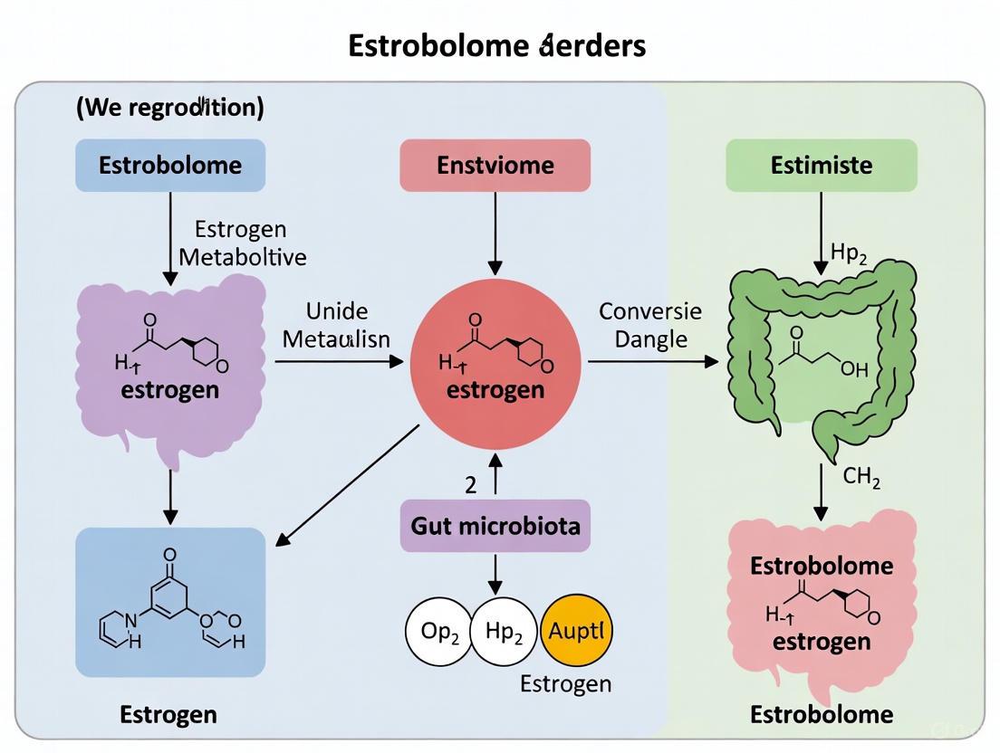
The estrobolome is conceptualized as the aggregate of enteric bacteria and their genes capable of metabolizing estrogen, functioning as a critical microbial endocrine organ within the human host [1] [2] [3]. First defined in 2011, this collection of bacterial genes produces enzymes that metabolize and modulate the body's circulating estrogen, primarily through the deconjugation of estrogen metabolites [2] [4]. The estrobolome represents a pivotal interface between the gut microbiome and the host endocrine system, enabling a bidirectional relationship where gut microbiota influence estrogen levels, and estrogen in turn shapes microbial composition and diversity [5] [6].
Within the context of reproductive disorders research, understanding the estrobolome provides a crucial framework for investigating the pathogenesis of estrogen-driven conditions. The functional significance of the estrobolome extends beyond mere estrogen recycling to encompass systemic endocrine regulation with profound implications for breast cancer, endometriosis, polycystic ovary syndrome (PCOS), and other hormone-mediated diseases [1] [7] [3]. This whitepaper examines the biochemical foundations, mechanistic pathways, and experimental approaches for investigating this emerging frontier in endocrine microbiology.
Biochemical Foundations and Metabolic Pathways
Core Enzymatic Machinery
The estrobolome's influence on endocrine regulation is mediated through specific enzymatic activities, primarily β-glucuronidase, with additional contributions from β-glucosidase and sulfatase enzymes [3] [4]. These microbial enzymes catalyze the deconjugation of estrogen metabolites that have been previously inactivated through hepatic phase II metabolism [1] [3].
The principal metabolic pathway involves the enterohepatic circulation of estrogens: circulating estrogens are conjugated in the liver (primarily glucuronidation), excreted into bile, and delivered to the intestinal tract [1] [3]. Rather than being excreted in feces, β-glucuronidases produced by estrobolome bacteria deconjugate these estrogen metabolites, reversing their inactivation and enabling reabsorption into circulation [1] [3] [4]. This process effectively increases the bioavailability of active estrogens capable of binding to estrogen receptors (ERα and ERβ) in target tissues throughout the body [3].
Table 1: Key Enzymes in Estrobolome Function
| Enzyme | Function in Estrogen Metabolism | Primary Bacterial Producers |
|---|---|---|
| β-glucuronidase | Deconjugates estrogen glucuronides, enabling estrogen reabsorption | Bacteroides, Bifidobacterium, Escherichia coli, Lactobacillus, Clostridium [3] [6] |
| β-glucosidase | Hydrolyzes glucosidic bonds in estrogen metabolites | Multiple gut microbiota species [8] |
| Sulfatase | Hydrolyzes sulfate esters from estrogen metabolites | Various gut commensals [3] |
Estrogen Metabolism Pathway Visualization
The following diagram illustrates the core metabolic pathway of estrogen regulation by the estrobolome:
Pathway of Estrogen Regulation by the Estrobolome: This diagram illustrates the enterohepatic circulation of estrogens and the critical role of microbial β-glucuronidase in estrogen deconjugation and reabsorption.
Key Microbial Taxa
The estrobolome encompasses bacteria from multiple genera that encode estrogen-metabolizing capabilities. Research has identified several key bacterial families and genera with estrobolome functionality:
Table 2: Estrobolome-Associated Microbial Taxa
| Taxonomic Level | Associated Taxa | Functional Significance |
|---|---|---|
| Phylum | Bacteroidetes, Firmicutes, Actinobacteria | Major bacterial divisions containing estrogen-metabolizing species [3] [5] |
| Family | Clostridiaceae, Ruminococcaceae, Lactobacillaceae, Bifidobacteriaceae | Rich in β-glucuronidase (β-GUS) encoding genes; associated with urinary estrogen levels [3] |
| Genus | Bacteroides, Bifidobacterium, Escherichia, Lactobacillus, Clostridium, Roseburia | Direct producers of β-glucuronidase and other estrogen-metabolizing enzymes [1] [3] [6] |
| Species | Escherichia coli, Roseburia inulinivorans, Bifidobacterium longum | Differentially abundant in breast cancer cases and controls; functionally relevant to estrogen metabolism [1] |
Functional Significance in Reproductive Health and Disease
Estrobolome Dysregulation in Disease Pathogenesis
Dysbiosis of the estrobolome—characterized by altered microbial diversity, composition, or functional capacity—can disrupt estrogen homeostasis and contribute to disease pathogenesis through multiple mechanisms:
Elevated β-glucuronidase activity: Increased bacterial production of β-glucuronidase enhances estrogen deconjugation and reabsorption, leading to systemic estrogen dominance [2] [4] [6]. This state has been implicated in endometriosis progression, breast cancer proliferation, and PCOS symptomatology [7] [3] [6].
Reduced microbial diversity: Diminished estrobolome diversity correlates with decreased estrogen-metabolizing capacity, potentially altering the balance between active and inactive estrogen forms [1] [3]. Postmenopausal women demonstrate lower gut microbiota diversity compared to premenopausal women, coinciding with declining estrogen levels [5].
Inflammatory mediation: Dysbiosis can compromise intestinal barrier function, promoting systemic inflammation that further disrupts endocrine signaling and estrogen receptor sensitivity [3] [6].
Disease-Specific Associations
Table 3: Estrobolome Alterations in Reproductive Disorders
| Disease Context | Observed Estrobolome Alterations | Clinical Consequences |
|---|---|---|
| Breast Cancer (HR+ subtype) | Reduced microbial diversity; Higher abundance of facultative aerobes; Differential abundance of Escherichia coli and Roseburia inulinivorans in cases vs controls [1] [3] | Increased estrogen bioavailability promotes tumor proliferation via estrogen receptor activation; Potential modulation of endocrine therapy efficacy [1] [9] |
| Endometriosis | Enrichment of Erysipelotrichia class; Higher folds of estrogen metabolites in fecal samples; Increased β-glucuronidase-producing bacteria (e.g., Escherichia coli) [7] [8] [6] | Elevated estrogen reabsorption fuels ectopic endometrial tissue growth; Enhanced inflammatory response [7] [8] |
| PCOS | Significantly lower gut microbiome diversity; Altered ratio of Firmicutes/Bacteroidetes; Potential existence of a "testrobolome" influencing androgen levels [6] [10] | Hormonal imbalance exacerbating hyperandrogenism and metabolic dysfunction; Systemic inflammation [6] [10] |
| Menopausal Transition | Depletion of beneficial bacteria (Lactobacillus, Bifidobacteria); Increase in harmful bacteria (Enterobacter); Reduced microbial diversity [5] | Contributory factor in metabolic disorders, cognitive decline, and osteoporosis associated with estrogen deficiency [5] |
Experimental Methodologies for Estrobolome Research
Core Analytical Approaches
Investigating the estrobolome requires integrated methodologies that characterize both microbial composition and functional activity:
Microbial Community Profiling
16S rRNA Gene Sequencing: Amplification and sequencing of hypervariable regions to determine taxonomic composition and relative abundance of estrobolome-associated bacteria [8]. Protocol: DNA extraction from stool samples → PCR amplification of V3-V4 regions → library preparation → high-throughput sequencing → bioinformatic analysis (QIIME2, MOTHUR) for taxonomic assignment and diversity metrics [8].
Shotgun Metagenomics: Whole-genome sequencing of microbial DNA to characterize the functional potential of the estrobolome, including identification of β-glucuronidase and other estrogen-metabolizing genes [1]. Protocol: Stool sample collection → DNA extraction → library preparation → shotgun sequencing → functional annotation (KEGG, MetaCyc) with tools like HUMAnN2 or MG-RAST [1].
Functional Activity Assessment
Enzymatic Activity Assays: Quantitative measurement of β-glucuronidase and β-glucosidase activities in fecal samples [8]. Protocol: Fresh or preserved stool samples homogenized in appropriate buffer → incubation with specific fluorogenic or chromogenic substrates (e.g., 4-nitrophenyl β-D-glucuronide) → spectrophotometric measurement of product formation → calculation of enzyme activity (U/L or U/g stool) [8].
Metabolomic Profiling: Quantification of estrogen metabolites in urine, serum, or fecal samples using LC-MS/MS [1] [8]. Protocol: Sample collection → solid-phase extraction → liquid chromatography separation → tandem mass spectrometry detection → quantification of individual estrogen metabolites (estrone, estradiol, estriol, catechol estrogens) [1].
Integrated Experimental Workflow
The following diagram outlines a comprehensive experimental workflow for estrobolome research:
Estrobolome Research Workflow: This diagram outlines the integrated multi-omics approach for investigating the estrobolome, incorporating genomic, functional, and metabolomic analyses.
Research Reagent Solutions
Table 4: Essential Research Tools for Estrobolome Investigation
| Reagent/Category | Specific Examples | Research Application |
|---|---|---|
| DNA Extraction Kits | QIAamp PowerFecal Pro DNA Kit, DNeasy PowerLyzer PowerSoil Kit | Efficient microbial lysis and DNA purification from complex stool samples [8] |
| 16S rRNA Primers | 341F/805R (V3-V4 region), 515F/806R (V4 region) | Amplification of target regions for bacterial taxonomic identification [8] |
| Sequencing Platforms | Illumina MiSeq/NovaSeq, PacBio Sequel, Oxford Nanopore | High-throughput sequencing for microbiome characterization [1] [8] |
| Enzyme Substrates | 4-Nitrophenyl β-D-glucuronide, 4-Nitrophenyl β-D-glucopyranoside | Fluorogenic/chromogenic substrates for β-glucuronidase/β-glucosidase activity quantification [8] |
| LC-MS/MS Standards | Deuterated estrogen metabolites (estrone-d4, estradiol-d3, estriol-d3) | Internal standards for precise quantification of estrogen metabolites [1] [8] |
| Bioinformatics Tools | QIIME2, MOTHUR, HUMAnN2, PICRUSt2, LEfSe | Processing sequencing data, functional inference, and differential abundance analysis [1] [8] [9] |
| Cell Culture Models | Caco-2 intestinal epithelial cells, HT-29-MTX-E12 cells | In vitro systems for studying host-microbe interactions in estrogen metabolism [1] |
Future Directions and Therapeutic Implications
The estrobolome represents a promising frontier for developing novel diagnostic and therapeutic strategies for endocrine-related disorders. Future research priorities include:
Longitudinal human studies: Tracking estrobolome dynamics across the lifespan, particularly during hormonal transitions (menarche, pregnancy, menopause) [2] [5].
Mechanistic investigations: Elucidating precise molecular pathways linking specific bacterial taxa and enzymes to estrogen receptor activation and downstream signaling [1] [3].
Therapeutic modulation: Exploring targeted interventions including precision probiotics, prebiotics, dietary modifications, and fecal microbiota transplantation for estrobolome manipulation [2] [5] [6].
Microbiome-informed pharmacotherapy: Understanding how estrobolome composition influences efficacy and metabolism of endocrine therapies such as tamoxifen and aromatase inhibitors [9].
Advancing our understanding of the estrobolome will require integrated multi-omics approaches, standardized methodological protocols, and interdisciplinary collaboration between microbiologists, endocrinologists, and clinical researchers. The conceptual framework of the estrobolome fundamentally expands our understanding of endocrine regulation and offers transformative potential for managing reproductive disorders through microbial modulation.
The enterohepatic circulation creates a critical pathway for the recycling of estrogens, a process fundamentally regulated by gut microbial enzymes. This whitepaper delineates the core mechanism by which bacterial β-glucuronidase and sulfatase enzymes reactivate estrogen conjugates, thereby modulating host estrogen levels. Within the framework of the estrobolome—the collection of gut microbial genes capable of metabolizing estrogens—this review synthesizes structural, functional, and quantitative data on these key enzymes. We present detailed experimental protocols for characterizing enzyme activity, alongside visualized pathways and essential research tools. This mechanistic insight is foundational for understanding how dysbiosis of the estrobolome may contribute to the pathogenesis of estrogen-driven reproductive disorders and for informing targeted therapeutic strategies.
The concept of the estrobolome defines the aggregate of enteric bacteria encoding enzymes that directly metabolize estrogens, serving as a key regulator of systemic estrogen homeostasis [1] [11]. In premenopausal women, estrogens are primarily synthesized in the ovaries, whereas in postmenopausal women, production occurs in peripheral tissues such as the adrenal glands and adipose tissue [11]. The liver plays a central role in estrogen inactivation, where Phase I (hydroxylation) and Phase II (conjugation) metabolism convert estrogens into more water-soluble forms for excretion. The primary Phase II reactions are glucuronidation and sulfation, which tag estrogens for elimination via the bile or urine [11].
Estrogen conjugates excreted into the bile are released into the intestinal lumen. Rather than being simply eliminated, these conjugates can be hydrolyzed by bacterial enzymes in the gut, specifically β-glucuronidases and sulfatases, which deconjugate them back into their active, absorbable forms [1] [11]. These reactivated estrogens are then reabsorbed into the portal circulation and returned to the liver, completing the enterohepatic circulation [12] [11]. This recycling pathway can significantly influence the body's overall estrogenic tone. Dysbiosis of the estrobolome, characterized by an imbalance in microbial communities and their enzymatic activities, can lead to either excessive reactivation or inadequate clearance of estrogens. This perturbation is hypothesized to be a contributing factor in various hormone-driven reproductive disorders, including endometriosis, fibroids, and breast cancer [1] [11].
Structural and Functional Diversity of Estrogen-Metabolizing Bacterial Enzymes
Bacterial β-Glucuronidase (GUS) Enzymes
Bacterial β-glucuronidases (GUS) are glycoside hydrolases that catalyze the cleavage of glucuronic acid moieties from a wide range of substrates, including estrogen glucuronides [13] [14]. In the human gut, GUS enzymes are produced by a variety of commensals, with the major sources belonging to the phyla Bacteroidetes and Firmicutes [11]. The human gut microbiome encodes a vast diversity of GUS enzymes; one analysis of the Human Microbiome Project identified 279 unique GUS enzymes [15] [11] [14].
Structural studies have revealed that these enzymes can be classified into distinct categories based on their active site loop architectures: Loop 1 (L1), Mini Loop 1 (mL1), Loop 2 (L2), Mini Loop 2 (mL2), and No Loop (NL) [15] [16]. This structural diversity is not merely topological; it has direct functional consequences for substrate specificity and catalytic efficiency. For instance, research has demonstrated that enzymes possessing a Loop 1 (L1) motif are particularly efficient at processing small molecule glucuronides, including drug metabolites and estrogen conjugates like estrone-3-glucuronide (E1-3-G) and estradiol-17-glucuronide (E2-17-G) [16] [14]. The catalytic mechanism is proposed to be an SN2-type reaction involving two key glutamic acid residues, with a transition state that exhibits oxocarbenium ion character [13].
Table 1: Catalytic Efficiency of Select Gut Microbial GUS Enzymes with Estrogen Glucuronide Substrates
| GUS Enzyme | Source Organism | Loop Type | Substrate (Estrogen Glucuronide) | Reported Activity |
|---|---|---|---|---|
| EcGUS | Escherichia coli | Loop 1 (L1) | Estrone-3-Glucuronide | High reactivation [14] |
| FpGUS | Faecalibacterium prausnitzii | Loop 1 (L1) | Estrone-3-Glucuronide | High reactivation [14] |
| RgGUS | Ruminococcus gnavus | Loop 1 (L1) | Estradiol-17-Glucuronide | Moderate reactivation [14] |
| BfGUS | Bacteroides fragilis | Mini Loop 1 (mL1) | Estrone-3-Glucuronide | Low/No reactivation [14] |
| BuGUS-2 | Bacteroides uniformis | Loop 2 (L2) | Not Tested (Active on 4-MUG) [15] | N/A |
| BuGUS-3 | Bacteroides uniformis | Mini Loop 2 (mL2) | Not Tested (Inactive on 4-MUG) [15] | N/A |
Bacterial Sulfatase Enzymes
While less characterized in the context of estrogen metabolism, bacterial sulfatases are another key enzymatic component of the estrobolome. These enzymes hydrolyze the sulfate ester bond in sulfated estrogen conjugates (e.g., estrone sulfate), thereby reactivating them for reabsorption [11]. Sulfatases are a heterogeneous group of enzymes generally categorized into three classes: aryl sulfatases, alkyl sulfatases, and Fe²⁺-dependent sulfatases [17].
The best-studied class relevant to estrogen metabolism is the aryl sulfatases. A defining feature of this class is a highly conserved consensus motif (C/S-X-P-X-A-X₄-T-G), where the lead cysteine or serine residue is post-translationally modified to a catalytically active formylglycine [17]. This unique modification is essential for the enzyme's activity. The stereochemical outcome of sulfate ester hydrolysis can proceed with either inversion or retention of configuration at the chiral carbon, depending on the specific enzyme and its mechanism [17]. Although the presence and activity of sulfatases are noted in estrobolome reviews, detailed functional studies on their specificity for estrogen-sulfate conjugates, comparable to those done for GUS enzymes, are an area for further research.
Experimental Characterization of Enzyme Activity
Protocol for Assessing β-Glucuronidase Activity with Estrogen Substrates
Objective: To determine the kinetic parameters (Km, Vmax, kcat) of a purified bacterial β-glucuronidase enzyme using the estrogen conjugate estrone-3-glucuronide (E1-3-G) as a substrate.
Principle: The enzyme catalyzes the hydrolysis of E1-3-G, releasing free estrone (E1) and glucuronic acid. The formation of the product, estrone, is quantified using a high-performance liquid chromatography (HPLC) system coupled with ultraviolet (UV) or fluorescence detection.
Materials:
- Purified bacterial GUS enzyme (e.g., EcGUS, FpGUS)
- Substrate: Estrone-3-glucuronide (E1-3-G)
- Reaction Buffer: 50 mM potassium phosphate buffer, pH 7.4
- Stop Solution: Acetonitrile or methanol
- HPLC system with a C18 reverse-phase column
- UV/Vis or fluorescence detector
Method:
- Reaction Setup: Prepare a series of reactions containing a fixed amount of purified GUS enzyme (e.g., 10-100 nM) in reaction buffer and varying concentrations of E1-3-G substrate (e.g., 0-500 μM).
- Incubation: Allow the reactions to proceed at 37°C for a predetermined time (e.g., 10-30 minutes) within the linear range of product formation.
- Reaction Termination: Stop each reaction by adding an equal volume of ice-cold acetonitrile to precipitate proteins.
- Sample Analysis: Centrifuge the terminated reactions to remove precipitated protein. Inject the supernatant onto the HPLC system for analysis.
- HPLC Conditions: Isocratic or gradient elution using a water/acetonitrile mobile phase. Detect estrone by UV absorption at ~280 nm or via its native fluorescence (excitation ~280 nm, emission ~310 nm).
- Quantification: Generate a standard curve of peak area versus known concentrations of authentic estrone. Use this curve to calculate the amount of estrone produced in each reaction.
- Data Analysis: Plot the initial velocity (v) against the substrate concentration [S]. Fit the data to the Michaelis-Menten equation (v = (Vmax * [S]) / (Km + [S])) using non-linear regression software (e.g., GraphPad Prism) to determine the Km and Vmax values. The kcat is calculated as Vmax / [Enzyme].
Protocol for Structural Determination via X-ray Crystallography
Objective: To solve the three-dimensional crystal structure of a bacterial GUS enzyme, either in its apo form or in complex with an estrogen-glucuronide substrate analog.
Principle: X-ray crystallography involves growing a high-quality crystal of the protein, collecting X-ray diffraction data, and solving the phase problem to generate an electron density map into which an atomic model is built.
Materials:
- Purified, concentrated GUS protein (>10 mg/mL in low-salt buffer)
- Crystallization screening kits (e.g., from Hampton Research)
- X-ray source (laboratory or synchrotron)
- Cryo-protectant solution (e.g., glycerol, ethylene glycol)
Method:
- Protein Crystallization: Set up crystallization trials using vapor diffusion methods (e.g., sitting or hanging drop). Mix equal volumes of protein solution and crystallization screen reservoir solution. Monitor for crystal growth over days to weeks.
- Cryo-protection and Data Collection: Once suitable crystals are obtained, transfer them to a cryo-protectant solution before flash-freezing in liquid nitrogen. Collect X-ray diffraction data at a synchrotron source at cryogenic temperatures (100 K).
- Data Processing and Structure Solution: Index, integrate, and scale the diffraction data using software like XDS or HKL-2000. If a homologous structure is available, solve the phase problem by molecular replacement using programs like Phaser.
- Model Building and Refinement: Build the atomic model into the electron density map using Coot. Iteratively refine the model against the diffraction data using refinement software such as Phenix or Refmac, adjusting atomic coordinates and temperature factors to improve the fit.
Visualization of Pathways and Workflows
The Enterohepatic Circulation of Estrogens
The following diagram illustrates the complete pathway of estrogen metabolism and recycling, highlighting the critical role of bacterial enzymes.
Diagram Title: Estrogen Enterohepatic Circulation Pathway
Workflow for Assessing Bacterial Enzyme Activity
This diagram outlines the key experimental steps for characterizing the function of bacterial enzymes in estrogen reactivation.
Diagram Title: Enzyme Characterization Workflow
The Scientist's Toolkit: Key Research Reagents
The following table compiles essential materials and reagents for conducting research on bacterial enzymes in estrogen metabolism.
Table 2: Essential Research Reagents for Estrobolome Enzyme Studies
| Reagent / Material | Function / Application | Specific Examples / Notes |
|---|---|---|
| Recombinant GUS/Sulfatase Enzymes | In vitro characterization of substrate specificity and kinetics. | Purified enzymes from gut commensals (e.g., E. coli GUS, B. uniformis GUS) [15] [14]. |
| Estrogen Glucuronide Conjugates | Enzyme substrates for activity and inhibition assays. | Estrone-3-glucuronide (E1-3-G), Estradiol-17-glucuronide (E2-17-G) [14]. |
| Chromogenic / Fluorogenic Substrates | High-throughput screening of enzyme activity and inhibition. | p-Nitrophenyl-β-D-glucuronide (pNPG), 4-Methylumbelliferyl-β-D-glucuronide (4-MUG) [15] [16]. |
| Selective GUS Inhibitors | Tool compounds for validating enzyme function in vitro and in vivo. | Inhibitor 1, UNC10201652 (specific for Loop 1 GUS enzymes) [16]. |
| Fecal Sample Collection Kits | Source of complex microbial communities and native enzymes for ex vivo studies. | Commercially available kits that stabilize microbial DNA and metabolites. |
| HPLC-MS/MS Systems | Sensitive and specific quantification of estrogen species and metabolites. | Used to detect and quantify deconjugated estrogens (e.g., E1, E2) from assay mixtures [14]. |
| Crystallization Screening Kits | Initial condition screening for protein crystal growth for structural studies. | Sparse matrix screens from commercial suppliers (e.g., Hampton Research) [15]. |
Discussion and Research Implications
The quantitative and structural data presented herein underscore the sophistication of the estrobolome's role in endocrinology. The differential activity of various GUS loop types with estrogen substrates, as shown in Table 1, provides a molecular rationale for how shifts in gut microbial population structure—such as an increased Firmicutes-to-Bacteroidetes ratio—could elevate systemic estrogen levels via enhanced reactivation [11]. This enzymatic reactivation mechanism, integrated within the enterohepatic circulation pathway, represents a tangible interface between the gut microbiome and host physiology.
Targeting these bacterial enzymes offers a promising, specific therapeutic avenue. The development of potent, selective inhibitors against bacterial GUS enzymes, which do not inhibit the human homolog, has already shown efficacy in mitigating drug-induced gastrointestinal toxicity in preclinical models [16]. This proof-of-concept strongly supports the feasibility of applying similar strategies to modulate estrogen levels for conditions like breast cancer or endometriosis. However, as research by Ervin et al. suggests, while GUS inhibition is effective in vitro and ex vivo, its impact on complex disease outcomes like breast cancer tumorigenesis in vivo may be multifaceted, potentially requiring a broader targeting strategy [14].
Future research must focus on expanding the functional characterization of estrobolome enzymes, particularly sulfatases, using physiologically relevant estrogen conjugates. Furthermore, correlating the abundance and activity of these specific enzymes in human cohorts with clinical measures of estrogen exposure and reproductive disorder status will be crucial for establishing causal links and validating these enzymes as bona fide therapeutic targets.
The estrobolome, defined as the collection of gut microbiota genes capable of metabolizing estrogen, has emerged as a critical regulator of systemic endocrine homeostasis. Dysbiosis of the estrobolome disrupts estrogen recycling, leading to altered estrogen levels that contribute to the pathogenesis of multiple reproductive disorders. This whitepaper synthesizes current evidence elucidating the mechanistic links between estrobolome dysbiosis and three major estrogen-driven conditions: endometriosis, polycystic ovary syndrome (PCOS), and breast cancer. We detail the specific microbial signatures observed in each disorder, explore the underlying molecular pathways involving immune activation, inflammatory signaling, and hormonal dysregulation, and provide standardized experimental methodologies for investigating estrobolome function. The findings position the estrobolome as a promising therapeutic target and biomarker source for precision medicine approaches in reproductive health.
The estrobolome constitutes a specialized functional component of the gut microbiome that regulates estrogen metabolism and circulating levels through enzymatic activities [18]. Its primary mechanism involves the enterohepatic circulation of estrogens, where estrogens conjugated in the liver for biliary excretion are deconjugated in the gut by microbial enzymes, particularly β-glucuronidase (GUS), allowing reactivated estrogens to re-enter systemic circulation [19] [3].
Core estrobolome functions include:
- Enzymatic deconjugation: Bacterial β-glucuronidase, β-glucosidase, and sulfatase reactivate estrogen metabolites [3].
- Estrogen homeostasis maintenance: A healthy, diverse estrobolome maintains balanced estrogen levels appropriate for physiological needs [18].
- Dysbiosis-induced dysregulation: Reduced microbial diversity or pathogenic overgrowth alters enzyme activity, leading to either estrogen excess or deficiency [20] [3].
The following diagram illustrates the core mechanism of the estrobolome in maintaining estrogen homeostasis:
Estrobolome Dysbiosis in Specific Reproductive Disorders
Endometriosis
Endometriosis, characterized by ectopic endometrial tissue growth, demonstrates strong estrogen dependence and inflammatory components that are modulated by estrobolome activity [20] [21]. Research indicates that gut dysbiosis precedes and promotes endometriosis development through multiple interconnected pathways.
Key Microbial Alterations in Endometriosis:
- Increased ratio of Firmicutes to Bacteroidetes [21]
- Elevated abundance of Bacteroides, Parabacteroides, Oscillospira, and Coprococcus [21]
- Reduced abundance of Paraprevotella, Lachnospira, and Turicibacter [21]
- Decreased α- and β-diversity of gut microbiota [21]
Mechanistic Pathways:
- Estrogen reactivation: Microbial β-glucuronidase increases bioactive estrogen, stimulating ectopic endometrial tissue growth [20].
- Inflammatory activation: Dysbiosis increases intestinal permeability, facilitating LPS translocation and TLR4-mediated NF-κB activation, driving pro-inflammatory cytokine production (TNF-α, IL-6) [21].
- Immune dysregulation: Altered microbial communities impair immune homeostasis, creating a permissive environment for endometriotic lesion establishment [20] [21].
Recent investigations have identified Fusobacterium nucleatum infiltration in the uterus of 64% of women with endometriosis, promoting macrophage infiltration, TGF-β production, and transgelin upregulation, which collectively drive endometriotic lesion development in experimental models [22].
Polycystic Ovary Syndrome (PCOS)
PCOS involves neuroendocrine dysfunction, hyperandrogenism (HA), and insulin resistance (IR), with gut microbiota dysbiosis significantly contributing to its pathogenesis [23]. The estrobolome influences PCOS through both direct hormonal modulation and indirect metabolic pathways.
Key Microbial Alterations in PCOS:
- Diminished α-diversity negatively correlating with serum testosterone levels [23]
- Elevated Bacteroidetaceae, Raoultella, and Prevotella abundance, correlating positively with androgen levels [23]
- Increased Candida levels associated with circulating androstenedione [23]
- Reduced beneficial Lactobacilli, which normally ameliorate PCOS phenotype and reduce androgen biosynthesis [23]
Mechanistic Pathways:
- Androgen metabolism: Specific gut microbiota produce enzymes that metabolize and convert androgens, influencing hyperandrogenemia [23].
- Insulin resistance: Dysbiosis-induced endotoxemia (LPS translocation) activates TLR4 signaling, triggering JNK and IKK pathways that promote IR through inflammatory cytokine production [23].
- Gut-brain axis disruption: Altered gut microbiota affect the release of gut-brain peptides, contributing to neuroendocrine dysfunction in PCOS [23].
Table 1: Microbial Signatures in Reproductive Disorders
| Disorder | Increased Taxa | Decreased Taxa | Key Functional Changes |
|---|---|---|---|
| Endometriosis | Bacteroides, Parabacteroides, Oscillospira, Coprococcus, Firmicutes/Bacteroidetes ratio | Paraprevotella, Lachnospira, Turicibacter | Increased β-glucuronidase activity, LPS translocation, elevated pro-inflammatory cytokines |
| PCOS | Bacteroidetaceae, Raoultella, Prevotella, Candida | Lactobacilli, overall α-diversity | Altered androgen metabolism, increased intestinal permeability, endotoxemia |
| Breast Cancer | Clostridium, Bacteroides, Escherichia, β-glucuronidase-producing bacteria | Microbial diversity, protective SCFA producers | Elevated β-glucuronidase activity, increased estrogen recirculation, reduced anti-inflammatory metabolites |
Breast Cancer
Breast cancer, particularly estrogen receptor-positive (ER+) subtypes, demonstrates strong connections to estrobolome function, with microbial dysbiosis influencing both carcinogenesis and progression through hormonal and inflammatory pathways [19] [24] [18].
Key Microbial Alterations in Breast Cancer:
- Enrichment of β-glucuronidase-producing bacteria (Clostridium, Bacteroides, Escherichia) [3]
- Reduced microbial diversity compared to healthy controls [3]
- Distinct tumor-associated microbial signatures, including bacteria capable of producing mycothiol [3]
- Increased abundance of Firmicutes and Bacteroidetes phyla classified as gut microbial β-glucuronidase (gmGUS) producers [19]
Mechanistic Pathways:
- Estrogen recirculation: Enhanced microbial β-glucuronidase activity increases deconjugated estrogens that bind ERα and ERβ, activating proliferation genes (MYC, CCND1, BCL-2) [3].
- Immune modulation: Dysbiosis reduces protective short-chain fatty acids (SCFAs), allowing pro-inflammatory cytokines (IL-6, TNF-α) to dominate the tumor microenvironment [18] [3].
- Metabolite signaling: Microbial metabolites influence local metabolic conditions and immune responses within tumor tissue [3].
The following diagram illustrates the multifaceted pathways linking estrobolome dysbiosis to reproductive disorders:
Experimental Methodologies for Estrobolome Research
Microbiome Profiling and Metagenomic Analysis
Comprehensive characterization of estrobolome composition and function requires integrated multi-omics approaches:
Sample Collection and Preservation:
- Fecal samples: Collect pre-treatment and store at -80°C with stabilizers for microbial DNA/RNA preservation [19].
- Tissue biopsies: Collect sterile tumor/endometrial tissues for microbial DNA extraction and spatial analysis [3].
- Blood samples: Collect for metabolomic profiling and measurement of circulating estrogen levels [18].
DNA Sequencing and Bioinformatics:
- 16S rRNA sequencing: For taxonomic profiling using primers targeting V3-V4 hypervariable regions [21].
- Shotgun metagenomics: For functional gene analysis, including identification of β-glucuronidase genes [19] [24].
- Bioinformatic processing: Use QIIME2, MOTHUR, or HUMAnN2 pipelines for taxonomic assignment and functional annotation [19].
Functional Assays for Estrobolome Activity
Direct measurement of estrobolome functional output provides critical mechanistic insights:
β-Glucuronidase Activity Assay:
- Principle: Measure enzymatic conversion of p-nitrophenyl-β-D-glucuronide to p-nitrophenol [19].
- Protocol: Incubate fecal supernatants with substrate in acetate buffer (pH 4.5) at 37°C, measure absorbance at 405nm [19].
- Normalization: Express activity per mg protein or per gram fecal material [19].
Estrogen Metabolite Profiling:
- Liquid chromatography-mass spectrometry (LC-MS): Quantify conjugated vs. unconjugated estrogen metabolites in serum, urine, and fecal samples [24].
- Protocol: Use stable isotope-labeled internal standards, reversed-phase C18 columns, and multiple reaction monitoring for sensitive detection [24].
Intestinal Permeability Assessment:
- LPS measurement: Use Limulus Amebocyte Lysate (LAL) assay to quantify systemic endotoxin levels [23].
- Mucosal integrity assays: Measure transepithelial electrical resistance (TEER) in Caco-2 cell models with microbial metabolites [18].
Table 2: Key Experimental Protocols in Estrobolome Research
| Methodology | Key Applications | Critical Parameters | Technical Considerations |
|---|---|---|---|
| 16S rRNA Sequencing | Taxonomic profiling, α/β-diversity analysis | Primer selection (V3-V4), sequencing depth (>10,000 reads/sample) | Cannot detect functional genes; requires complementary methods |
| Shotgun Metagenomics | Functional gene analysis, pathway reconstruction | Sequencing depth (>5 million reads/sample), quality filtering | Computational intensive; requires robust reference databases |
| β-Glucuronidase Assay | Direct estrobolome functional measurement | pH optimization (4.5-5.0), substrate concentration, incubation time | Affected by sample collection methods; requires fresh/frozen samples |
| LC-MS Metabolomics | Estrogen metabolite quantification | Chromatographic separation, ionization efficiency, internal standards | Matrix effects; requires sophisticated normalization |
| Gnotobiotic Models | Causal mechanism validation | Germ-free conditions, controlled microbial colonization | High cost; specialized facility requirements |
Animal Models and Intervention Studies
Preclinical models enable causal inference and therapeutic testing:
Gnotobiotic Mouse Models:
- Protocol: Use germ-free mice colonized with defined microbial communities from patients or specific pathogen-free controls [23] [21].
- Endometriosis model: Intraperitoneal injection of allogeneic mouse endometrium with monitoring of lesion development [21].
- PCOS model: Dehydroepiandrosterone (DHEA) induction with assessment of metabolic and endocrine phenotypes [23].
Microbiota Transplantation Studies:
- Fecal microbiota transplantation (FMT): Transfer donor microbiota to germ-free or antibiotic-treated recipients to assess disease transmissibility [23] [21].
- Intervention protocols: Test probiotics (Lactobacillus, Bifidobacterium), prebiotics (inulin, FOS/GOS), or dietary interventions [18] [22].
The following diagram outlines a comprehensive experimental workflow for estrobolome research:
The Scientist's Toolkit: Research Reagent Solutions
Table 3: Essential Research Reagents for Estrobolome Investigations
| Reagent/Category | Specific Examples | Research Applications | Key Functions |
|---|---|---|---|
| DNA Extraction Kits | QIAamp PowerFecal Pro Kit, DNeasy PowerSoil Kit | Microbial DNA isolation from feces, tissues | Inhibitor removal, high-yield DNA recovery for sequencing |
| Sequencing Reagents | Illumina NovaSeq 6000, MiSeq Reagent Kits | 16S rRNA and shotgun metagenomic sequencing | High-throughput sequencing with low error rates |
| β-Glucuronidase Assay Kits | p-Nitrophenyl-β-D-glucuronide, GUS Reporter Assay | Quantification of estrobolome functional activity | Fluorometric/colorimetric detection of enzyme activity |
| Cell Culture Models | Caco-2 cells, HT-29-MTX-E12 | Intestinal barrier function assessment | Epithelial permeability, host-microbe interaction studies |
| Animal Models | Germ-free C57BL/6 mice, SCID mice | Causal mechanism validation | Controlled microbial colonization, disease phenotype monitoring |
| LC-MS Standards | Deuterated estrogen metabolites, Stable isotope standards | Estrogen metabolite quantification | Internal standards for precise metabolomic quantification |
| Probiotic Strains | Lactobacillus spp., Bifidobacterium longum APC1472 | Therapeutic intervention studies | Microbial restoration, anti-inflammatory effects |
| Prebiotic Compounds | Inulin-type fructans, FOS/GOS mixtures, Psyllium | Dietary intervention studies | Selective stimulation of beneficial bacteria, SCFA production |
The estrobolome represents a pivotal interface between the gut microbiome and endocrine health, with dysbiosis contributing significantly to the pathogenesis of endometriosis, PCOS, and breast cancer through shared mechanisms involving altered estrogen metabolism, immune activation, and inflammatory signaling. While distinct microbial signatures emerge for each disorder, common themes include reduced microbial diversity, enrichment of specific β-glucuronidase-producing taxa, and disruption of gut barrier integrity.
Future research priorities should include:
- Longitudinal studies tracking estrobolome dynamics throughout disease progression
- Advanced multi-omics integration combining metagenomics, metabolomics, and host transcriptomics
- Mechanistic validation using gnotobiotic models with defined microbial communities
- Interventional trials testing targeted probiotics, prebiotics, and dietary strategies
Standardized methodologies and shared reagent resources will accelerate the translation of estrobolome research into clinical applications, potentially enabling microbiome-based diagnostics and therapeutics for hormone-driven reproductive disorders. The estrobolome thus represents both a biomarker for disease risk stratification and a promising therapeutic target for precision medicine approaches in reproductive health.
The gut microbiota, comprising trillions of microorganisms, has emerged as a pivotal endocrine organ that extends its influence far beyond the gastrointestinal tract [25]. This "virtual endocrine organ" produces and regulates a vast array of hormonally active compounds, including neurotransmitters, short-chain fatty acids (SCFAs), and enzymes that metabolize steroid hormones, thereby systemically impacting host physiology [26] [25]. The collective genetic repertoire of gut microbes capable of metabolizing estrogens constitutes the "estrobolome," a key conceptual framework for understanding microbiome-mediated endocrine regulation [1] [3]. Unlike traditional endocrine glands with defined anatomy, the gut microbiome functions as a diffuse biochemical factory whose metabolic output influences distant organs, including the ovaries, through complex signaling pathways [25]. This review examines the mechanistic basis of the gut-ovary axis, exploring how gut microbial communities and their metabolites contribute to reproductive disorders through endocrine modulation, immune regulation, and metabolic pathway disruption.
The Gut Microbiome as a Systemic Endocrine Regulator
Endocrine Capabilities of the Gut Microbiota
The gut microbiota exhibits remarkable endocrine functionality through multiple mechanisms. It produces neurotransmitters including γ-aminobutyric acid (GABA), serotonin, dopamine, and norepinephrine, which can influence both local enteric nervous system function and central processes via the gut-brain axis [25]. Additionally, microbial fermentation of dietary fibers generates SCFAs such as butyrate, propionate, and acetate, which function as signaling molecules that regulate host metabolism and immunity [27] [25]. These SCFAs activate G-protein-coupled receptors (GPCRs) on enteroendocrine cells, stimulating the release of peptide hormones like glucagon-like peptide-1 (GLP-1) and peptide YY (PYY) that regulate appetite and glucose homeostasis [25]. The gut microbiota also plays a crucial role in metabolizing bile acids, converting primary bile acids into secondary forms that act as signaling molecules through receptors such as FXR and TGR5, further influencing metabolic pathways [27].
The estrobolome represents a particularly significant endocrine component of the gut microbiome. Microbes possessing β-glucuronidase enzymes can deconjugate estrogen metabolites that were previously inactivated by hepatic glucuronidation, allowing their reabsorption into circulation [1] [3]. This process regulates the enterohepatic circulation of estrogens, critically determining systemic estrogen bioavailability and consequently influencing estrogen receptor activation throughout the body [26] [3]. The estrobolome thus serves as a master regulator of estrogenic activity, with profound implications for estrogen-dependent physiological processes and pathologies.
Quantitative Assessment of Microbial Endocrine Capacity
Table 1: Key Endocrine-Active Metabolites Produced by Gut Microbiota
| Metabolite Category | Specific Molecules | Producing Microbes | Physiological Effects |
|---|---|---|---|
| Short-chain fatty acids (SCFAs) | Butyrate, Propionate, Acetate | Bacteroides, Firmicutes, Bifidobacterium | GPCR activation; hormone secretion (GLP-1, PYY); anti-inflammatory effects; insulin sensitivity |
| Neurotransmitters | GABA, Serotonin, Dopamine | Lactobacillus, Bifidobacterium | Gut-brain axis signaling; behavior modulation; gut motility |
| Enzymes for hormone metabolism | β-glucuronidase, β-glucosidase, Sulfatase | Clostridium, Bacteroides, Escherichia | Estrogen deconjugation; regulation of bioactive hormone levels |
| Secondary bile acids | Lithocholic acid, Deoxycholic acid | Bacteroides, Clostridium | FXR, TGR5 receptor activation; glucose and lipid metabolism |
Table 2: Gut Microbiome Composition and Diversity in Endocrine Pathology
| Condition | Microbial Diversity | Key Taxonomic Changes | Functional Consequences |
|---|---|---|---|
| Polycystic Ovary Syndrome (PCOS) | ↓ Decreased | ↑ Bacteroides, Escherichia-Shigella; ↓ Prevotella, Bifidobacterium, Akkermansia [27] | Increased gut permeability; endotoxemia; hormonal imbalance |
| Estrogen-driven cancers | ↓ Decreased | Altered β-glucuronidase-producing bacteria; reduced microbial richness [3] | Dysregulated estrogen metabolism; increased bioactive estrogen levels |
| Endometriosis | Variable | Gut and reproductive tract dysbiosis; altered estrobolome [7] | Inflammation; estrogen dominance; pain perception |
| Healthy reproductive state | ↑ High / Balanced | Lactobacillus dominance in reproductive tract; diverse gut microbiota [28] | Balanced hormone metabolism; reduced inflammation |
The Gut-Ovary Axis in Reproductive Health and Disease
Mechanistic Basis of the Gut-Ovary Axis
The gut-ovary axis represents a bidirectional communication network between gastrointestinal microbial communities and ovarian function, mediated through integrated neuroendocrine, metabolic, and immune pathways [27]. This axis does not involve direct contact between gut microbes and ovarian tissue but rather functions through sophisticated intermediary signaling mechanisms. The gut-brain-ovary pathway involves microbial influence on hypothalamic-pituitary-ovarian (HPO) axis regulation, primarily through modulation of gonadotropin-releasing hormone (GnRH) secretion [29] [30]. Additionally, gut microbiota directly impact steroid hormone metabolism, particularly through the estrobolome's regulation of estrogen bioavailability [26] [1]. Immune mediation occurs through microbial influence on systemic inflammation and immune cell function, while metabolic regulation involves microbiota-derived metabolites such as SCFAs and bile acids that influence insulin sensitivity and energy metabolism [27] [30].
The gut-ovary axis significantly influences female reproductive physiology across the lifespan. During reproductive years, gut microbiota contribute to cyclical hormonal fluctuations and ovulatory function [26]. In pregnancy, maternal gut microbiota support gestational metabolic adaptations and influence fetal development [31]. The transition through menopause involves shifting interactions between declining estrogen levels and gut microbial communities, with potential impacts on menopausal symptoms and long-term health [26]. Throughout these life stages, the gut-ovary axis maintains a delicate balance that when disrupted, may contribute to various reproductive pathologies.
Gut-Ovary Axis Dysregulation in Polycystic Ovary Syndrome
Polycystic ovary syndrome (PCOS) represents a quintessential disorder of gut-ovary axis disruption. Characteristic gut microbial signatures in PCOS include reduced overall diversity, decreased abundances of beneficial bacteria such as Prevotella, Bifidobacterium, and Akkermansia, and increased abundance of potentially inflammatory taxa like Bacteroides and Escherichia-Shigella [27]. These compositional changes are associated with functional alterations including reduced SCFA production, particularly butyrate, and increased gut permeability leading to metabolic endotoxemia [27] [30]. The subsequent activation of pro-inflammatory pathways contributes to systemic low-grade inflammation, a hallmark of PCOS that exacerbates both metabolic and reproductive manifestations of the syndrome.
The pathophysiological sequence in PCOS involves gut dysbiosis leading to impaired intestinal barrier function ("leaky gut"), allowing translocation of bacterial components such as lipopolysaccharides (LPS) into circulation [30]. This triggers immune activation and chronic inflammation, which promotes ovarian androgen production and insulin resistance [27] [30]. The resulting hyperandrogenism further exacerbates gut dysbiosis, creating a vicious cycle that perpetuates PCOS pathology. Additionally, gut microbiota influence neuroendocrine function in PCOS through modulation of GnRH secretion, contributing to the characteristic luteinizing hormone (LH) hypersecretion relative to follicle-stimulating hormone (FSH) that drives ovarian androgen production [30].
Estrobolome Dysregulation in Reproductive Disorders
Estrobolome Function in Estrogen Homeostasis
The estrobolome represents the collective genetic capacity of gut microbiota to metabolize estrogens, primarily through enzymes such as β-glucuronidase that catalyze the deconjugation of estrogen metabolites [1] [3]. In normal physiology, estrogens are conjugated in the liver to facilitate biliary excretion, but gut microbial β-glucuronidase reactivates these estrogens by removing glucuronide groups, allowing their reabsorption into the portal circulation and contributing to systemic estrogen levels [1]. This process creates a delicate balance where estrobolome composition directly influences circulating estrogen concentrations. A healthy, diverse estrobolome maintains appropriate estrogen levels for physiological function, while estrobolome dysregulation can lead to either estrogen deficiency or excess, contributing to various pathologies [26] [1].
The estrobolome's impact extends beyond endogenous estrogens to include phytoestrogens and xenoestrogens. Gut microbes metabolize dietary phytoestrogens such as soy isoflavones into more biologically active forms, with specific bacteria like Bifidobacterium enhancing this conversion [26]. Additionally, the estrobolome interacts with endocrine-disrupting chemicals, potentially modifying their estrogenic activity and contributing to their health impacts [30]. These interactions highlight the estrobolome's broader role as a mediator between environmental factors and endocrine function, extending its significance beyond endogenous hormone metabolism alone.
Estrobolome-Mediated Pathologies Beyond the Ovary
Estrobolome dysfunction contributes to various gynecological disorders beyond PCOS. In endometriosis, estrobolome alterations promote estrogen dominance through enhanced deconjugation and reduced diversity of estrogen-metabolizing bacteria [7]. This creates a pro-estrogenic environment that supports the growth and inflammation of ectopic endometrial lesions [7]. Similarly, in estrogen receptor-positive (ER+) breast cancer, estrobolome dysregulation influences cancer risk and progression through modulation of estrogen bioavailability [1] [3]. Postmenopausal women with breast cancer demonstrate reduced gut microbial diversity and altered estrobolome composition, associated with shifts in estrogen metabolite ratios that may influence carcinogenesis [26] [1].
Table 3: Estrobolome Alterations in Reproductive Disorders
| Disorder | Estrobolome Composition | Functional Changes | Hormonal Consequences |
|---|---|---|---|
| Endometriosis | Altered diversity of estrogen-metabolizing bacteria; specific taxa changes in gut and reproductive tract [7] | Increased β-glucuronidase activity; inflammatory microbiome | Estrogen dominance; enhanced local estrogen in lesions |
| ER+ Breast Cancer | Reduced microbial diversity; altered abundance of β-glucuronidase producers [1] [3] | Decreased estrogen deconjugation capacity; shifted estrogen metabolites | Dysregulated estrogen receptor activation; proliferation |
| Postmenopausal States | Diversity positively correlates with estrogen metabolites; response to phytoestrogens [26] | Variable β-glucuronidase based on microbiome; modified phytoestrogen metabolism | Altered estrogenic activity from precursors |
| PCOS | Part of broader dysbiosis; altered bile acid metabolism [27] | Indirect effects on estrogen balance through inflammation | Contribution to hormonal imbalance |
The therapeutic implications of estrobolome research are substantial. Measuring estrobolome composition and function may provide biomarkers for disease risk assessment and prognostication [1] [3]. Additionally, targeted interventions to modulate the estrobolome, including probiotics, prebiotics, and dietary modifications, represent promising approaches for managing estrogen-related disorders [26] [7]. The potential for fecal microbiota transplantation to restore healthy estrobolome function is also under investigation, though this approach requires further validation [31].
Experimental Models and Methodologies
Approaches for Gut-Ovary Axis Research
Investigation of the gut-ovary axis employs diverse experimental models, each with distinct advantages and limitations. In vitro systems include cell culture models of intestinal epithelium (Caco-2 cells), ovarian cells, and immune cells to study specific molecular interactions [28]. More advanced organ-on-a-chip platforms model the gut-ovary interface, allowing study of microbial metabolites' transport and effects on ovarian tissue [28]. These systems enable controlled manipulation of specific variables but lack the systemic complexity of whole organisms.
Animal models provide essential platforms for studying gut-ovary axis physiology and pathology. Germ-free (GF) mice allow investigation of microbial contributions by comparison with conventionally colonized animals [25]. PCOS models include prenatal androgen (PNA) exposure, dihydrotestosterone (DHT) treatment, and letrozole administration, all of which demonstrate gut microbiota alterations [26] [30]. These models recapitulate various PCOS features including hyperandrogenism, oligo-ovulation, and polycystic ovarian morphology, while permitting experimental manipulation of gut microbiota through antibiotics, probiotics, or fecal microbiota transplantation [30].
Human studies primarily employ correlational designs comparing gut microbiota composition between affected individuals and healthy controls through cross-sectional or case-control approaches [27] [1]. Longitudinal studies track microbiota changes in relation to disease progression or treatment response [29]. Intervention trials investigate effects of probiotics, prebiotics, or dietary modifications on reproductive parameters [27] [29]. Each model system contributes unique insights, with the most compelling evidence emerging from concordant findings across multiple approaches.
Analytical Techniques and Protocols
Comprehensive characterization of gut microbiota in reproductive research typically follows a multi-omics approach. 16S rRNA gene sequencing provides cost-effective taxonomic profiling using primers targeting hypervariable regions (e.g., V3-V4), with analysis pipelines including QIIME 2 or mothur for processing, and databases such as SILVA or Greengenes for taxonomic assignment [1]. Shotgun metagenomics enables strain-level resolution and functional gene analysis through platforms like HUMAnN2 for pathway reconstruction and MetaPhlAn for taxonomic profiling [1]. Metabolomic analyses employ LC-MS/MS to quantify microbiota-derived metabolites including SCFAs, bile acids, and estrogen metabolites, while transcriptomic approaches (RNA-Seq) assess host tissue gene expression responses to microbial signals [1] [30].
For estrobolome-specific investigation, functional assays measure β-glucuronidase activity using fluorescent or colorimetric substrates (e.g., p-nitrophenyl-β-D-glucuronide) in fecal samples or bacterial cultures [1]. Quantitative PCR targets specific bacterial taxa with estrogen-metabolizing capabilities, while more specialized approaches include fluorescently labeled estrogens to track microbial metabolism and gnotobiotic models colonized with defined estrobolome communities [1]. Integration of these diverse datasets requires sophisticated bioinformatic approaches including multivariate statistics, machine learning, and network analysis to identify robust associations between microbial features and clinical phenotypes.
Table 4: Research Reagent Solutions for Gut-Ovary Axis Investigation
| Research Tool Category | Specific Reagents/Assays | Experimental Application | Key Functions |
|---|---|---|---|
| Microbiome Profiling | 16S rRNA sequencing kits (Illumina); Shotgun metagenomics; QIIME2 analysis platform | Taxonomic characterization of gut microbiota; functional potential assessment | Identification of dysbiosis patterns; tracking intervention effects |
| Metabolite Measurement | LC-MS/MS systems; SCFA analysis kits; ELISA for hormones and cytokines | Quantification of microbial metabolites; hormone level assessment | Linking microbial changes to physiological outcomes |
| Barrier Function Assessment | FITC-dextran permeability assay; TEER measurement; Zonulin/occludin antibodies | Intestinal barrier integrity measurement; tight junction protein expression | Evaluation of "leaky gut" in pathophysiology |
| Cell Culture Models | Caco-2 intestinal cells; ovarian granulosa cell lines; transwell co-culture systems | Mechanistic studies of host-microbe interactions; metabolite transport | Isulating specific pathways without whole-organism complexity |
| Animal Models | Germ-free mice; prenatal androgenized rodents; antibiotic depletion protocols | Establishing causality; studying systemic effects | Controlled manipulation of microbiome-host interactions |
| Functional Assays | β-glucuronidase activity kits; G-protein coupled receptor assays; immune cell activation tests | Specific mechanism investigation; enzyme activity measurement | Linking microbial functions to host responses |
Therapeutic Implications and Future Directions
Microbiome-Targeted Interventions
Therapeutic modulation of the gut-ovary axis represents a promising approach for managing reproductive disorders. Probiotic interventions utilizing specific bacterial strains such as Lactobacillus and Bifidobacterium demonstrate potential for restoring microbial balance and improving metabolic parameters in PCOS [27] [31]. Prebiotic supplements including inulin, fructooligosaccharides (FOS), and galactooligosaccharides (GOS) provide selective substrates for beneficial gut bacteria, enhancing SCFA production and gut barrier function [27]. Dietary modifications substantially influence gut microbiota composition and function, with Mediterranean-style diets (high fiber, polyphenols) promoting beneficial microbial patterns, while Western diets (high fat, low fiber) exacerbate dysbiosis [29]. Fecal microbiota transplantation (FMT) represents a more intensive approach to rapidly shift microbial community structure, with emerging evidence supporting its potential in metabolic disorders, though applications in reproductive conditions require further investigation [31].
Beyond these broader interventions, more targeted approaches are emerging. Live biotherapeutic products (LBPs) such as LACTIN-V (Lactobacillus crispatus) for bacterial vaginosis and VMSC-04 for recurrent UTIs represent regulated therapeutic applications of specific microbial strains [31]. Vaginal microbiome transplantation explores transfer of beneficial microbial communities from healthy donors to individuals with dysbiosis, while synbiotic combinations of specific probiotics with their preferred prebiotics offer enhanced efficacy [31] [28]. These approaches highlight the translational potential of microbiome science in reproductive medicine, though considerable research remains to optimize strain selection, delivery methods, and patient stratification.
Diagnostic Applications and Personalized Medicine
Microbiome-based diagnostics are emerging as potential tools for risk assessment, diagnosis, and prognostication in reproductive disorders. Distinct microbial signatures in PCOS, endometriosis, and other conditions may provide biomarkers for early detection or stratification of disease subtypes [27] [7]. Estrobolome profiling, including measurement of β-glucuronidase-producing bacteria and related functional capacities, offers potential for assessing estrogen-related disease risk [1] [3]. Integration of microbiome data with clinical parameters could enable more personalized therapeutic approaches, matching specific interventions to individual microbial and metabolic profiles.
Future research directions should address critical knowledge gaps including causal mechanisms in gut-ovary axis communication, longitudinal dynamics of microbiome-reproductive interactions across the lifespan, and influence of ethnic, geographic, and individual factors on these relationships [27] [1]. Large-scale randomized controlled trials of microbiome-targeted interventions are needed to establish efficacy and safety, while advanced multi-omics integration will provide more comprehensive understanding of mechanistic pathways [27] [29]. Additionally, development of more sophisticated models including humanized animals and advanced in vitro systems will facilitate deeper investigation of specific molecular interactions within the gut-ovary axis [28].
The recognition of the gut microbiome as a systemic endocrine organ and the elucidation of the gut-ovary axis represent paradigm shifts in reproductive biology and medicine. The gut microbiota influences ovarian function and reproductive health through multiple integrated mechanisms including hormone metabolism, immune regulation, and metabolic signaling. Dysregulation of these pathways contributes to the pathogenesis of conditions including PCOS, endometriosis, and estrogen-sensitive cancers. The estrobolome concept provides a framework for understanding how microbial communities directly modulate estrogen bioavailability, with far-reaching implications for estrogen-dependent physiology and pathology. While substantial progress has been made in characterizing these relationships, important questions remain regarding causal mechanisms, temporal dynamics, and individual variability. Future research integrating multi-omics approaches, sophisticated experimental models, and targeted interventions will advance our understanding of this complex system and unlock its potential for novel diagnostic and therapeutic strategies in reproductive medicine.
The gut microbiota, through the estrobolome, plays a critical role in modulating systemic estrogen levels via microbial-derived enzymes such as β-glucuronidase. This in-depth technical guide delineates the functional roles of four key bacterial genera—Clostridium, Bacteroides, Escherichia, and Lactobacillus—in estrogen metabolism and their implications in reproductive disorders. We synthesize current mechanistic insights, present structured quantitative data from clinical and preclinical studies, and provide detailed experimental methodologies for investigating estrobolome function. The content is framed for researchers and drug development professionals, offering a resource to advance therapeutic strategies targeting the gut-microbiome-estrogen axis.
The estrobolome is defined as the collective gene repertoire of enteric bacteria capable of metabolizing estrogens [11]. It functions as a virtual endocrine organ, critically regulating the enterohepatic circulation of estrogens and thereby influencing systemic estrogen levels [32] [33]. The physiological process of estrogen elimination involves hepatic conjugation (glucuronidation and sulfation) followed by biliary excretion into the gastrointestinal tract [1] [11]. The pivotal function of the estrobolome is the microbial deconjugation of these estrogen metabolites, primarily via the enzyme β-glucuronidase, which reactivates estrogens and permits their reabsorption into the bloodstream [32] [1] [11]. Alterations in the composition and function of the estrobolome—a state known as gut dysbiosis—can disrupt this delicate equilibrium, leading to either excessive estrogen recycling or inadequate reactivation. This dysregulation has been implicated in the pathogenesis of a spectrum of estrogen-related reproductive disorders, including endometriosis, breast cancer, polycystic ovary syndrome (PCOS), and recurrent implantation failure [32] [7] [9]. The following sections provide a detailed examination of the specific microbial taxa that execute these functions and the experimental frameworks used to study them.
Functional Analysis of Key Microbial Taxa
The estrobolome's function is driven by specific bacterial taxa that encode and express enzymes for estrogen metabolism. The functional roles of the key genera are visualized in the diagram below, which outlines the core pathway of enterohepatic estrogen circulation and microbial intervention.
1Clostridiumspp.
Clostridium species are recognized as significant producers of β-glucuronidase enzymes, directly contributing to the deconjugation of estrogen glucuronides in the gut [32]. This activity facilitates the reactivation and subsequent reabsorption of estrogens, thereby elevating systemic estrogenic activity. In the context of hormone receptor-positive (HR+) breast cancer, the enrichment of Ruminiclostridium (a genus within the Clostridiales order) has been observed in patients, suggesting a potential link between clostridial abundance and an estrogen-driven tumor microenvironment [9]. Furthermore, in endometriosis, an estrogen-dependent inflammatory condition, gut dysbiosis often involves alterations in Clostridia populations, which may contribute to the disease's pathogenesis by promoting systemic estrogen dominance [7].
2Bacteroidesspp.
Bacteroides are a major constituent of the human gut microbiota and a primary source of bacterial β-glucuronidases [11]. Species such as Bacteroides vulgatus have been functionally linked to estrogen metabolism. In polycystic ovary syndrome (PCOS), the abundance of B. vulgatus is significantly increased, and this species is implicated in the deconjugation of conjugated bile acids—a process that can intersect with and influence metabolic and endocrine pathways central to PCOS pathology [33]. Conversely, in breast cancer, case-control studies have reported differential abundance of Bacteroides species, including Bacteroides ovatus, in hormone receptor-negative patients, indicating a potential, though complex, role for this genus in cancer etiology that may extend beyond estrogen metabolism to broader ecological shifts in the gut microbiome [9].
3Escherichiaspp.
Escherichia coli is a well-characterized β-glucuronidase-producing bacterium and has been specifically identified as a differentially abundant and functionally relevant taxon in breast cancer case-control studies [1]. The β-glucuronidase enzyme produced by E. coli efficiently deconjugates estrone-3-glucuronide and estradiol-17-glucuronide back into their bioactive forms, estrone and estradiol, making it a key player in estrogen recycling [11]. An increased ratio of Escherichia/Shigella has also been noted in the gut microbiota of women with PCOS, further underscoring the association of this taxon with endocrine disorders [33]. Its enzymatic potency makes it a critical organism of interest in understanding estrobolome-driven pathophysiology.
4Lactobacillusspp.
The role of Lactobacillus is multifaceted and appears to be protective. While some species possess β-glucuronidase activity [32], the overall influence of Lactobacillus-dominant microbiota seems to favor estrogen excretion. A higher diversity of gut microbes, often associated with a healthy balance including lactobacilli, is correlated with decreased production of β-glucuronidases, leading to greater excretion of conjugated estrogens [11]. Furthermore, in the vaginal microbiome, Lactobacillus species (e.g., L. crispatus, L. gasseri, L. jensenii) are crucial for maintaining a low pH, which supports urogenital health and prevents infections that can complicate reproductive outcomes [7] [34]. Reductions in beneficial Lactobacillus and Bifidobacterium have been noted in postmenopausal gut dysbiosis, which is linked to a decline in estrogen and its protective effects [35].
Table 1: Functional Roles of Key Microbial Taxa in Estrogen Metabolism
| Microbial Taxon | Key Enzymatic Function | Association with Reproductive Disorders | Reported Abundance Changes |
|---|---|---|---|
| Clostridium spp. | β-glucuronidase production [32] | Enriched in HR+ breast cancer; implicated in endometriosis pathogenesis [7] [9] | Ruminiclostridium enrichment in HR+ breast cancer patients [9] |
| Bacteroides spp. | Major source of β-glucuronidase; bile acid deconjugation [33] [11] | Increased in PCOS; differentially abundant in breast cancer subtypes [9] [33] | ↑ B. vulgatus in PCOS; ↑ B. ovatus in HR- breast cancer [9] [33] |
| Escherichia spp. | Potent β-glucuronidase production [1] [11] | Associated with breast cancer and PCOS [1] [33] | Differentially abundant in breast cancer cases; ↑ Escherichia/Shigella ratio in PCOS [1] [33] |
| Lactobacillus spp. | Modulates microbial diversity & β-glucuronidase output; lactic acid production [32] [7] [11] | Dominance associated with vaginal & gut health; depletion in menopause & some PCOS [35] [7] [33] | ↓ in postmenopausal gut dysbiosis; ↓ in some PCOS gut microbiomes [35] [33] |
Table 2: Quantitative Data on Taxa Abundance from Clinical Studies
| Study Cohort | Finding Related to Key Taxa | Statistical Measure | Citation |
|---|---|---|---|
| HR+ vs. HR- Breast Cancer (n=90) | Ruminiclostridium enriched in HR+ patients | raw p = 0.043, FDR p = 0.129, Effect Size (Cohen’s d) = -0.38 [9] | |
| HR+ vs. HR- Breast Cancer (n=90) | Bacteroides ovatus enriched in HR- patients | raw p = 0.033, FDR p = 0.131, Effect Size (Cohen’s d) = 0.35 [9] | |
| PCOS vs. Healthy Controls | Bacteroides vulgatus significantly higher in PCOS | FDR-corrected p < 0.05 [33] | |
| PCOS vs. Healthy Controls | Increased ratio of Escherichia/Shigella in PCOS | Reported as significant (specific p-value not provided) [33] |
Experimental Protocols for Estrobolome Research
Investigating the functional dynamics of the estrobolome requires an integrated multi-omics approach. The standard workflow, from sample collection to functional analysis, is depicted in the following diagram.
Metagenomic Sequencing and Bioinformatic Analysis
Objective: To characterize the taxonomic composition and genetic functional potential of the estrobolome in stool samples.
- Sample Collection and DNA Extraction: Collect fresh fecal samples from case and control cohorts (e.g., endometriosis patients vs. healthy individuals) using standardized kits. Preserve samples immediately at -80°C. Extract genomic DNA using a robust kit designed for complex microbial communities [1] [9].
- Sequencing and Quality Control: Perform shotgun metagenomic sequencing on an Illumina platform to achieve sufficient depth (e.g., 10-20 million reads per sample). Process raw reads with tools like Trimmomatic or FastQC to remove adapter sequences and low-quality bases [1].
- Taxonomic and Functional Profiling: Align quality-filtered reads to curated microbial genome databases (e.g., NCBI, MetaPhlAn) for taxonomic assignment. For functional analysis, align reads to databases like KEGG (Kyoto Encyclopedia of Genes and Genomes) and MetaCyc to identify and quantify genes of interest, specifically focusing on Enzyme Commission (EC) numbers for β-glucuronidase (EC 3.2.1.31) and other relevant enzymes [1]. This allows for the direct estimation of the estrobolome's genetic capacity.
Metabolomic Profiling of Estrogens
Objective: To quantitatively measure systemic and fecal estrogen levels, correlating them with microbial features.
- Sample Preparation: Collect paired plasma and fecal samples. For fecal samples, perform metabolite extraction using a methanol:water solvent system. For plasma, use protein precipitation followed by solid-phase extraction to isolate steroid hormones [1] [9].
- Liquid Chromatography-Tandem Mass Spectrometry (LC-MS/MS): Analyze the extracted samples using a high-sensitivity LC-MS/MS system. Quantify a panel of conjugated (e.g., estrone sulfate, estradiol glucuronide) and deconjugated (e.g., estrone, estradiol) estrogens using stable isotope-labeled internal standards for precise quantification [1].
- Data Integration: Correlate the abundance of specific microbial taxa (from 3.1) with the concentrations of estrogen metabolites using multivariate statistical models (e.g., Spearman correlation, linear regression adjusted for confounding factors like BMI and age) [9].
In Vitro Functional Validation of β-Glucuronidase Activity
Objective: To directly confirm the estrogen-deconjugating function of specific bacterial isolates.
- Bacterial Culture and Supernatant Preparation: Isolate pure cultures of Clostridium, Bacteroides, Escherichia, and Lactobacillus from human stool under anaerobic conditions. Culture the isolates in appropriate broth media. Harvest the cell-free culture supernatant via centrifugation and sterile filtration [1] [11].
- Enzymatic Assay: Incubate the bacterial supernatant with a known concentration of a conjugated estrogen substrate (e.g., estrone-3-glucuronide) in a buffer at physiological pH. Use a commercial β-glucuronidase assay kit, which often relies on the cleavage of a synthetic substrate like p-nitrophenyl-β-D-glucuronide, leading to a colorimetric or fluorometric readout. Include controls (e.g., supernatant from a non-enzyme-producing strain and a no-enzyme buffer blank) [11].
- LC-MS/MS Verification: Confirm the functional outcome by analyzing the incubation mixture using LC-MS/MS to detect the production of deconjugated estrogens (e.g., estrone) from their conjugated precursors [1]. This provides direct evidence of the enzyme's activity on its physiological substrate.
The Scientist's Toolkit: Research Reagent Solutions
Table 3: Essential Reagents and Kits for Estrobolome Research
| Item | Function/Application | Example Use Case |
|---|---|---|
| Stool DNA Extraction Kit (e.g., QIAamp PowerFecal Pro) | Isolation of high-quality microbial genomic DNA from complex fecal samples. | Standardized preparation of DNA for metagenomic sequencing in cohort studies [9]. |
| Shotgun Metagenomic Sequencing Service (Illumina NovaSeq) | Comprehensive profiling of all genetic material in a sample, allowing for simultaneous taxonomic and functional analysis. | Identifying the abundance of β-glucuronidase (EC 3.2.1.31) genes in the gut microbiome of cases vs. controls [1]. |
| β-Glucuronidase Activity Assay Kit (colorimetric/fluorometric) | Quantitative measurement of β-glucuronidase enzyme activity in bacterial culture supernatants or fecal extracts. | Validating the functional capacity of isolated bacterial strains to deconjugate estrogen [11]. |
| LC-MS/MS System with UPLC | High-sensitivity identification and quantification of estrogen metabolites (both conjugated and free) in biological fluids and culture media. | Profiling estrogen levels in plasma and correlating them with microbial β-glucuronidase activity [1] [9]. |
| Anaerobic Chamber & Growth Media | For the cultivation and maintenance of obligate anaerobic gut bacteria like Clostridium and Bacteroides. | Isolating and expanding specific estrobolome taxa for in vitro functional studies [1]. |
The intricate relationship between the gut microbiota, specifically the genera Clostridium, Bacteroides, Escherichia, and Lactobacillus, and host estrogen metabolism represents a frontier in understanding and treating reproductive disorders. The experimental frameworks outlined herein provide a roadmap for validating mechanistic links and identifying novel therapeutic targets. Future research must focus on longitudinal studies to establish causality, the development of targeted probiotics or small molecule inhibitors to modulate estrobolome function, and the integration of estrobolome profiling into personalized medicine approaches for endocrine-related cancers and gynecological health. A deep understanding of these key microbial players will be instrumental in the next generation of drug development and clinical management strategies.
Advanced Analytical Techniques: Profiling the Estrobolome and Estrogen Metabolites in Research and Diagnostics
LC-MS/MS Methodologies for Sensitive Profiling of Estrogens and Metabolites in Stool and Plasma
Estrogens, particularly estrone (E1), estradiol (E2), and estriol (E3), play a fundamental role in female reproductive development and health, with broader physiological effects in both sexes influencing mood, metabolism, bone density, and cardiovascular and cognitive functions [36]. The concept of the estrobolome—the collection of gut microbial genes capable of metabolizing estrogens—has emerged as a critical factor in regulating systemic estrogen levels [1] [3]. In the context of reproductive disorders such as endometriosis and hormone-receptor positive (HR+) breast cancer, understanding the precise balance of estrogen metabolites is paramount [1] [7]. Endometriosis, a long-term inflammatory disease affecting an estimated 5-10% of reproductive-aged women, is characterized by estrogen-dependent growth of ectopic tissue, and its associated pain is often amplified by peaks of estrogen release during the menstrual cycle [7].
Liquid chromatography-tandem mass spectrometry (LC-MS/MS) has become the gold standard for quantifying endogenous estrogens and their bioactive metabolites in complex biological matrices like plasma and stool [36] [37]. This technical guide details the development and application of highly sensitive LC-MS/MS methodologies to profile primary estrogens and estrogen metabolites, providing a framework for investigating the estrobolome's role in reproductive disorders.
Technical Development of the LC-MS/MS Method
Core Analytical Challenges and Solutions
Profiling estrogens in biological samples presents two primary technical challenges: extreme low concentration of analytes, especially in postmenopausal women and men, and high structural similarity between many estrogen metabolites [36] [37]. To overcome these, the developed method incorporates several key features.
- High Sensitivity Requirements: Reference ranges for E2 are 30–400 pg/mL in premenopausal women but plummet to 0–30 pg/mL in postmenopausal women and 10–50 pg/mL in men [36]. The method must therefore achieve limits of quantitation (LOQs) in the sub-pg/mL to low pg/mL range [37].
- Specificity for Isobaric Metabolites: The method must resolve structurally similar metabolites like 2-OHE1 and 4-OHE1, which have identical molecular weights but different biological activities, with 4-hydroxyestrogens being more genotoxic [36] [38].
- Matrix Effects: Stool and plasma contain numerous interfering compounds. Using analyte-specific internal standards (e.g., stable isotope-labeled estrogens) is critical to correct for recovery losses and compensate for ion suppression or enhancement during MS analysis [36] [39].
- Derivatization for Enhanced Detection: Because native estrogens ionize poorly, chemical derivatization with agents like dansyl chloride (DSC) or pyridine-3-sulfonyl chloride is employed to improve ionization efficiency, lowering the limits of detection to approximately 1 pg/mL [36] [37].
Sample Preparation and Extraction Protocols
Robust sample preparation is essential for clean and reproducible results. The following protocols are adapted for simultaneous processing of plasma and stool samples [36].
Table 1: Key Research Reagent Solutions for LC-MS/MS Estrogen Profiling
| Reagent/Material | Function | Application Notes |
|---|---|---|
| Analyte-Specific Internal Standards (e.g., ¹³C or ²H-labeled E1, E2, E3) | Corrects for analyte loss during preparation & matrix effects during MS analysis | Added to sample prior to extraction; crucial for accurate quantification [36] [37]. |
| Solid Phase Extraction (SPE) Cartridges (e.g., C-18 silica-based) | Pre-concentrates estrogens & removes interfering matrix components | Preferred over liquid-liquid extraction (LLE) for better reproducibility and cleaner extracts [40] [37]. |
| Derivatization Reagent (e.g., Dansyl Chloride) | Enhances ionization efficiency & improves sensitivity | Chemically modifies estrogens to allow detection at low pg/mL levels [36] [37]. |
| High Purity Solvents & Water | Used for mobile phase & sample reconstitution | Minimizes background chemical noise, improving signal-to-noise ratio [40]. |
| Stool/Plasma Sample | Biological matrix for analysis | Stool requires homogenization; plasma is typically collected with anticoagulants [36]. |
Detailed Experimental Protocol for Stool and Plasma Estrogen Extraction:
Sample Collection and Homogenization:
Spiking and Hydrolysis:
- Spike a known amount of internal standard solution into each plasma or stool aliquot.
- To measure total (free + conjugated) estrogens, subject the sample to enzymatic hydrolysis using a mixture of β-glucuronidase and sulfatase to deconjugate estrogen glucuronides and sulfates [36].
Solid Phase Extraction (SPE):
Derivatization:
- Evaporate the eluent to complete dryness under a gentle stream of nitrogen.
- Reconstitute the residue in a solution of dansyl chloride in acetone or another suitable solvent.
- Heat the mixture to facilitate the derivatization reaction.
- After the reaction is complete, evaporate the derivatizing reagent and reconstitute the sample in the initial LC mobile phase (e.g., 50:50 methanol:water) for injection [36] [37].
Liquid Chromatography and Mass Spectrometric Analysis
The separation and detection steps are optimized for maximum resolution and sensitivity.
- Liquid Chromatography: Separation is performed using ultra-high performance liquid chromatography (UHPLC) with a core-shell or fully porous silica column, which provides high efficiency and resolution for analytes that differ by only a few daltons. A fine-particle stationary phase and a gradient mobile phase (e.g., water and methanol or acetonitrile) are used to resolve the isobaric estrogen metabolites [36] [40].
- Mass Spectrometry: A triple quadrupole (TQ) mass spectrometer operating in multiple reaction monitoring (MRM) mode is used. After LC separation, analytes are ionized via an electrospray ionization (ESI) source. In the first quadrupole (Q1), a specific precursor ion (e.g., the derivatized molecular ion) is selected. This ion is fragmented in the second quadrupole (Q2 or collision cell) using an inert gas like argon. The third quadrupole (Q3) then selects a specific, unique product ion fragment. Monitoring the transition from precursor to product ion for each analyte provides high specificity [36] [39].
The following workflow diagram illustrates the complete analytical process from sample to result.
Key Experimental Findings and Biological Validation
Applying this validated LC-MS/MS method to human studies has yielded critical insights into estrogen biology and the estrobolome.
Quantitative Profiling in Plasma and Stool
The method successfully quantified a comprehensive panel of estrogens, including E1, E2, E3, and their hydroxy and methoxy metabolites, across different patient cohorts.
Table 2: Representative Estrogen Levels in Different Biological Matrices and Populations
| Analyte / Metric | Premenopausal Women (Plasma) | Postmenopausal Women (Plasma) | Men (Plasma) | Stool (General Findings) |
|---|---|---|---|---|
| Estradiol (E2) | 15 - 350 pg/mL [37] | < 10 pg/mL [37] | 10 - 40 pg/mL [37] | Higher in premenopausal women; increases across menstrual cycle [36]. |
| Estrone (E1) | 17 - 200 pg/mL [37] | 7 - 40 pg/mL [37] | 10 - 60 pg/mL [37] | Present in all study groups [36]. |
| Hydroxy- & Methoxyestrogen Metabolites | Levels vary by specific metabolite | Levels vary by specific metabolite | Levels vary by specific metabolite | Present in all groups; levels correlated with plasma metabolite levels [36]. |
Elucidating the Gut Microbiome's Role
By combining LC-MS/MS with shotgun metagenomic sequencing of stool microbiomes, researchers have directly linked microbial functional genes to estrogen levels.
- Enzyme Activities: The gene copy numbers for microbial β-glucuronidase and arylsulfatase—enzymes that deconjugate estrogen glucuronides and sulfates—were higher in premenopausal women. Crucially, the combined gene copy number of β-glucuronidase + arylsulfatase (but not β-glucuronidase alone) correlated with levels of deconjugated estrogens in stool across all study groups [36] [41].
- Systemic Impact: These microbial enzyme gene copy numbers also correlated with combined plasma estrogen levels in men and with individual plasma estrogen metabolites in men and premenopausal women. This provides direct evidence that gut microbial enzymes control the deconjugation of gut estrogens and modulate systemic levels via enterohepatic recirculation [36] [41].
The following diagram synthesizes the core relationship between the gut microbiome, estrogen metabolism, and systemic health, as revealed by these LC-MS/MS studies.
Application in Reproductive Disorders Research
The precise profiling enabled by this LC-MS/MS methodology is critical for investigating the role of the estrobolome in diseases like endometriosis and breast cancer.
- Endometriosis Pathogenesis: Endometriosis is an estrogen-dependent disease. The gut and reproductive tract microbiomes can influence local and systemic estrogen levels. Dysbiosis in these communities may lead to altered estrogen metabolism, promoting the inflammatory environment and tissue proliferation characteristic of endometriosis [7]. LC-MS/MS allows for the direct measurement of these shifts in estrogen metabolism within specific biological compartments.
- Breast Cancer Risk: Hormone-receptor positive (HR+) breast cancer accounts for roughly 70% of cases, and elevated estrogen levels are a key risk factor, particularly in postmenopausal women [1] [3]. The gut microbial balance can influence cancer risk through the estrobolome; a dysbiotic state may alter the ratio of estrogen metabolites, shifting it toward more genotoxic profiles (e.g., higher 4-hydroxyestrogens and 16α-OHE1) associated with higher DNA damage and cancer risk [1] [38]. LC-MS/MS is essential for quantifying these metabolite ratios (e.g., 2-OHE1/16α-OHE1) as potential biomarkers for risk assessment [36] [38].
The development of highly sensitive and specific LC-MS/MS methodologies for the profiling of estrogens and their metabolites in stool and plasma represents a significant technical advancement. By overcoming the challenges of low concentration and structural similarity, this approach provides an unparalleled tool for quantifying the complex dynamics of estrogen metabolism. When integrated with metagenomic data, it offers direct, mechanistic evidence of the estrobolome's role in regulating systemic estrogen levels via enterohepatic recirculation. This technical capability is fundamental to advancing our understanding of the pathophysiology of estrogen-related reproductive disorders, paving the way for novel diagnostic strategies and microbiome-targeted therapeutic interventions.
The estrobolome is defined as the collection of gut microbiota equipped with genes encoding enzymes capable of metabolizing estrogens. These microorganisms regulate estrogen circulation via enterohepatic circulation—a process where estrogens conjugated in the liver are excreted into the bile, then deconjugated in the gut by microbial enzymes such as β-glucuronidases, allowing reactivated estrogens to re-enter the bloodstream [1]. In reproductive disorders, dysregulation of this pathway can significantly impact systemic estrogen levels, potentially influencing conditions such as endometriosis, polycystic ovary syndrome (PCOS), and hormone-receptor positive cancers. Consequently, accurate identification and functional profiling of the estrobolome has emerged as a critical research focus. The choice of sequencing methodology—16S rRNA amplicon sequencing versus shotgun metagenomic sequencing—fundamentally shapes the depth and quality of data that can be acquired for investigating these microbial communities [1] [42].
Technical Foundations of Sequencing Methodologies
16S rRNA Gene Sequencing
16S rRNA gene sequencing is an amplicon-based approach that leverages the polymerase chain reaction (PCR) to target and amplify specific hypervariable regions (V1-V9) of the bacterial 16S ribosomal RNA gene, a genetic marker universally present in bacteria and archaea [43] [44] [45]. The experimental workflow begins with DNA extraction from samples such as stool, followed by a PCR amplification step using primers designed for conserved regions flanking one or more variable regions (e.g., V3-V4) [43]. The resulting amplicons are then barcoded, pooled, and sequenced on platforms such as the Illumina MiSeq [43] [44]. Subsequent bioinformatic processing involves quality filtering, error correction, and clustering of sequences into Operational Taxonomic Units (OTUs) or denoising into Amplicon Sequence Variants (ASVs) before taxonomic assignment against reference databases like the Ribosomal Database Project (RDP) [43] [46].
Shotgun Metagenomic Sequencing
In contrast, shotgun metagenomic sequencing is an untargeted approach that involves randomly fragmenting all genomic DNA within a sample into small pieces, followed by high-throughput sequencing without prior amplification [43] [45]. The library preparation typically involves a tagmentation step, which cleaves DNA and adds adapter sequences, followed by PCR amplification and size selection [44]. The resulting sequences, often called "reads," are then subjected to a more complex bioinformatic analysis. This can involve quality control, assembly into longer contigs, and taxonomic profiling through alignment to comprehensive genomic databases (e.g., using Kraken2 or MetaPhlAn) or assembly-based methods to reconstruct metagenome-assembled genomes (MAGs) [43] [45]. Crucially, this method also enables functional profiling by identifying microbial genes present in the metagenome, including those directly involved in estrogen metabolism [43] [42].
Comparative Analysis: 16S rRNA vs. Shotgun Metagenomics for Estrobolome Research
Technical Capabilities and Limitations
Table 1: Technical Comparison of 16S rRNA and Shotgun Metagenomic Sequencing
| Factor | 16S rRNA Sequencing | Shotgun Metagenomic Sequencing |
|---|---|---|
| Cost per Sample | ~$50 - $80 USD [44] [45] | Starting at ~$150-$200 USD (deep sequencing) [44] [45] |
| Taxonomic Resolution | Genus-level (sometimes species) [44] [42] | Species-level and sometimes strain-level [44] [42] |
| Taxonomic Coverage | Bacteria and Archaea only [43] [47] | All domains: Bacteria, Archaea, Fungi, Viruses [43] [47] |
| Functional Profiling | No direct functional data; requires prediction (e.g., PICRUSt) [44] [47] | Yes; direct identification of microbial genes and pathways [44] [42] |
| Sensitivity to Host DNA | Low (due to targeted amplification) [44] [45] | High; can be a major confounder [44] [45] |
| Bioinformatics Complexity | Beginner to Intermediate [44] | Intermediate to Advanced [44] |
| Database Dependency | Well-curated 16S databases [45] | Less complete whole-genome databases [45] |
| False Positive Risk | Lower with DADA2 error-correction [45] | Higher due to database gaps and gene sharing [45] |
Table 2: Suitability for Estrobolome and Reproductive Disorder Research
| Research Requirement | 16S rRNA Sequencing | Shotgun Metagenomic Sequencing |
|---|---|---|
| Identify β-glucuronidase-producing taxa | Indirect inference via taxonomy [1] | Direct detection of bgl genes [1] |
| Profile complete estrobolome community | Limited to bacterial component [1] | Cross-domain community profiling [43] |
| Link microbes to estrogen levels | Weak correlation possible | Strong functional association [1] |
| Strain-level resolution | Generally not available [42] | Possible with deep sequencing [45] [42] |
| Novel enzyme discovery | Not possible | Possible via assembly [42] |
Quantitative Data and Empirical Evidence
A comparative study performing deep sequencing of a human fecal sample demonstrated that whole genome shotgun sequencing enhanced detection of bacterial species and increased prediction of genes compared to 16S amplicon sequencing [42] [48]. This is critical for estrobolome research, where comprehensive gene catalogs are essential. Another study on postpartum dairy cows with endometritis utilized shotgun metagenomic sequencing to not only identify pathogenic bacteria like Fusobacterium and Trueperella but also to detect functional differences, such as a lower abundance of the Wnt/catenin pathway in cows with clinical endometritis [49]. This demonstrates shotgun sequencing's power to correlate microbial composition with functional pathways relevant to reproductive health.
However, 16S sequencing can still provide valuable ecological insights. A large-scale study comparing microbiome analysis methods found that 16S sequencing identified up to 140 unique bacterial species per sample, far exceeding the maximum of 8 species typically identified by traditional culturing methods [46]. For broad taxonomic surveys where budget is a constraint, 16S sequencing remains a viable option, though it cannot directly illuminate the functional landscape of the estrobolome.
Experimental Design and Workflows
Recommended Protocol for Estrobolome-Focused Shotgun Metagenomics
For a comprehensive analysis of the estrobolome, the following detailed protocol is recommended, drawing from established methodologies [49] [42]:
Sample Collection and DNA Extraction:
- Collection: Collect fecal or other relevant biological samples (e.g., uterine cytobrushes for direct reproductive tract analysis [49]) using standardized kits that preserve DNA (e.g., OMR-200 tubes). Immediately freeze samples at -80°C.
- DNA Extraction: Extract high-quality, high-molecular-weight metagenomic DNA using dedicated kits (e.g., PowerSoil DNA Isolation Kit). Assess DNA quality and quantity via spectrophotometry (e.g., NanoPhotometer), fluorometry (e.g., Qubit), and agarose gel electrophoresis. The integrity of the DNA is crucial for successful library preparation [42] [48].
Library Preparation and Sequencing:
- Fragmentation: Mechanically shear approximately 1-5 µg of metagenomic DNA to fragments of 300–600 bp using a focused-ultrasonication system (e.g., Covaris S220) [48].
- Library Construction: Use a library preparation kit (e.g., NEBNext Ultra DNA Library Prep Kit for Illumina) to perform end-repair, adenylation, and ligation of Illumina adapter sequences. Include a PCR enrichment step to add unique dual-index barcodes for sample multiplexing.
- Sequencing: Pool barcoded libraries in equimolar ratios and sequence on an Illumina platform (HiSeq or NovaSeq for greater depth, MiSeq for smaller studies) to generate a minimum of 10-20 million paired-end reads (e.g., 2x150 bp) per sample to ensure adequate coverage for functional analysis [47] [42].
Bioinformatic Analysis for Estrobolome Characterization:
- Quality Control and Preprocessing: Use tools like Trimmomatic or FastQC to remove adapter sequences and low-quality reads.
- Host DNA Depletion: If working with samples high in host DNA (e.g., reproductive tract samples), align reads to the host genome (e.g., human, bovine) and remove matching sequences [49] [45].
- Taxonomic Profiling: Assign taxonomy to reads using marker-based tools like MetaPhlAn or k-mer-based tools like Kraken2 against curated databases.
- Functional Profiling: This is the core of estrobolome analysis. Assemble quality-filtered reads into contigs using metaSPAdes or MEGAHIT. Predict genes on contigs and/or unassembled reads using Prodigal. Annotate predicted genes against functional databases (e.g., KEGG, eggNOG, MetaCyc) to identify specific enzyme classes, with a focus on β-glucuronidase (EC 3.2.1.31), β-glucosidase, and hydroxysteroid dehydrogenases (HSDs) [1]. Custom databases of known estrogen-metabolizing genes can also be used for alignment.
- Pathway Reconstruction: Utilize tools like HUMAnN3 to reconstruct and quantify the abundance of complete metabolic pathways, including those involved in estrogen metabolism [44].
The Scientist's Toolkit: Essential Research Reagents and Materials
Table 3: Key Reagent Solutions for Estrobolome Sequencing Studies
| Item | Function/Application | Example Products/Catalog Numbers |
|---|---|---|
| Stool Collection Kit | Standardized sample collection and stabilization for microbiome DNA. | OMNIgene GUT (OMR-200) [47] |
| Metagenomic DNA Extraction Kit | Lysis and purification of microbial DNA from complex samples. | PowerSoil DNA Isolation Kit [42] [48] |
| DNA Shearing Instrument | Fragmentation of DNA to optimal size for library preparation. | Covaris S220 [48] |
| Library Prep Kit | Preparation of sequencing-ready libraries with barcodes. | NEBNext Ultra DNA Library Prep Kit for Illumina [48] |
| Host DNA Depletion Kit | Selective removal of host genetic material to increase microbial sequencing depth. | HostZERO Microbial DNA Kit [45] |
| Mock Microbial Community | Quality control and validation of the entire workflow, from extraction to bioinformatics. | ZymoBIOMICS Microbial Community Standard [45] |
The investigation of the estrobolome's role in reproductive disorders demands a sequencing strategy that can accurately resolve microbial taxa and directly interrogate their functional genetic capacity. While 16S rRNA sequencing is a cost-effective tool for initial, broad taxonomic surveys, the evidence strongly supports the use of shotgun metagenomic sequencing for rigorous estrobolome research [42] [48]. Its unparalleled ability to directly identify and quantify genes encoding estrogen-metabolizing enzymes (e.g., β-glucuronidases) and to reconstruct relevant metabolic pathways provides a definitive functional readout that 16S-based inference cannot match [1].
For researchers designing studies, a hybrid approach can be considered: using 16S sequencing to screen a large number of samples, followed by shotgun metagenomics on a strategically selected subset for deep functional analysis. As sequencing costs continue to decrease and reference databases expand, shotgun metagenomics is poised to become the gold standard for elucidating the complex interactions between the gut microbiome, estrogen metabolism, and reproductive health, ultimately informing novel diagnostic and therapeutic strategies.
Correlating Microbial Beta-Glucuronidase Gene Abundance with Circulating Hormone Levels
The gut microbiome exerts a significant influence on host physiology, particularly through the regulation of steroid hormone homeostasis. The estrobolome, a collection of gut microorganisms capable of metabolizing estrogens, has emerged as a critical regulator of systemic estrogen levels through the activity of microbial enzymes, most notably β-glucuronidase. This enzyme deconjugates estrogens within the gastrointestinal tract, facilitating their reabsorption into the circulation and thereby modulating the bioavailability of active hormones. This technical review synthesizes current evidence and methodologies for quantifying microbial β-glucuronidase gene abundance and correlating it with circulating hormone levels. We provide a detailed examination of experimental protocols, analytical techniques, and key reagents, framing the discussion within the context of estrogen metabolism in reproductive disorders. The objective is to furnish researchers and drug development professionals with a comprehensive framework for investigating this pivotal axis in women's health and disease.
The concept of the estrobolome describes the aggregate of enteric bacterial genes whose products are functionally involved in the metabolism of estrogen [50] [1]. In a state of eubiosis, the estrobolome maintains estrogen homeostasis. However, dysbiosis, characterized by an imbalance in microbial communities, can alter the functional capacity of the estrobolome, leading to pathological deviations in circulating estrogen levels [51] [3]. This dysregulation is implicated in the pathogenesis of a spectrum of estrogen-related conditions, including endometriosis, breast cancer, polycystic ovary syndrome (PCOS), and other reproductive disorders [51] [7] [52].
The principal mechanistic link between the gut microbiota and systemic estrogen levels is the enterohepatic circulation of estrogens. Estrogens are conjugated in the liver (via glucuronidation and sulfation) to form water-soluble compounds that are excreted into the bile [1] [36]. Upon reaching the intestine, a critical reaction occurs: bacterial β-glucuronidase enzymes catalyze the deconjugation of these estrogen metabolites, regenerating their active forms [3] [1]. These active estrogens are then capable of being reabsorbed into the portal circulation, effectively increasing their systemic bioavailability and potential to engage estrogen receptors (ERα and ERβ) in target tissues throughout the body [3] [1]. The abundance and activity of these microbial enzymes are therefore a key determinant of circulating bioactive estrogen levels.
Quantitative Data: Microbial Taxa and Hormonal Correlations
Empirical studies have begun to delineate the specific microbial taxa associated with β-glucuronidase activity and to correlate this functional potential with measured hormone levels.
Table 1: Key Bacterial Taxa Associated with β-Glucuronidase and Estrogen Metabolism
| Bacterial Taxon | Association with β-Glucuronidase/Estrogen | Relevant Health Context | Citation |
|---|---|---|---|
| Clostridium spp. | Enriched in estrobolome; key producer of β-glucuronidase. | Breast cancer, general estrogen metabolism. | [3] [1] |
| Ruminococcaceae | Strongly associated with urinary estrogen levels; β-glucuronidase producer. | General estrogen homeostasis. | [3] |
| Escherichia coli | Differentially abundant in breast cancer cases; known β-glucuronidase producer. | Hormone receptor-positive breast cancer. | [1] |
| Bacteroides spp. | Possess β-glucuronidase genes; linked to estrogen reactivation. | Sex-hormone driven cancers. | [51] [3] |
| Roseburia inulinivorans | Differentially abundant and functionally relevant in case-control studies. | Breast cancer. | [1] |
| Lactobacillus spp. | Abundant in pre- and post-menopausal women; associated with vaginal estrogen effect. | General reproductive health. | [34] [52] |
Table 2: Correlative Findings from Human Studies
| Study Population | Microbial/Functional Finding | Hormonal Correlation / Outcome | Citation |
|---|---|---|---|
| Postmenopausal Women with Breast Cancer | Enrichment of some β-glucuronidase-positive bacteria. | Higher probability of elevated average β-glucuronidase levels; altered progesterone. | [50] |
| Premenopausal vs. Postmenopausal Women | Higher β-glucuronidase & arylsulfatase gene copy numbers in premenopausal women. | Increased deconjugated stool estrogens; levels rise across menstrual cycle. | [36] |
| General Population | β-glucuronidase + arylsulfatase gene copy numbers correlate with deconjugated stool estrogens. | Correlated with combined plasma estrogens in men and estrogen metabolites in premenopausal women. | [36] |
| Postmenopausal Women | Decreased microbial β-glucuronidase abundance compared to premenopausal. | Associated with lower circulating estrogen levels. | [5] [53] |
Experimental Protocols for Correlation Analysis
Establishing a causal link between microbial gene abundance and host hormone levels requires a multi-faceted methodological approach. The following protocols detail the key steps for a comprehensive analysis.
Sample Collection and Preparation
A. Biospecimen Collection:
- Stool Samples: Collect fresh fecal samples from study participants using standardized home collection kits. Immediately freeze samples at -80°C, preferably with a stabilizing solution like RNAlater for microbial genomic analysis and PBS for metabolomic studies [50].
- Blood Samples: Collect blood via venipuncture into appropriate vacutainer tubes (e.g., heparin for plasma). Separate plasma via centrifugation and store aliquots at -80°C to prevent hormone degradation [50] [36].
- Urine Samples: Collect spot urine samples without preservative. Centrifuge to remove sediments and store supernatants at -80°C [50].
B. Metadata Collection:
- Gather comprehensive participant data via questionnaires, including age, menopausal status, BMI, medical history, medication use (especially antibiotics and hormones), diet, and lifestyle factors, as these are critical confounders in analysis [50] [1].
Profiling the Gut Microbiome and β-Glucuronidase Gene Abundance
A. DNA Extraction and Sequencing:
- Extract microbial genomic DNA from stool samples using a commercially available kit designed for complex matrices, with bead-beating to ensure lysis of hardy Gram-positive bacteria.
- Perform 16S rRNA gene amplicon sequencing (e.g., targeting the V4 region) for a cost-effective analysis of microbial community composition. Alternatively, conduct shotgun metagenomic sequencing to achieve strain-level resolution and direct access to the full complement of microbial genes, including those encoding for β-glucuronidase [50] [36].
B. Bioinformatic Analysis:
- Process raw sequencing data through established pipelines (e.g., QIIME2 for 16S data) for quality filtering, denoising, and chimera removal [50].
- Assign taxonomy using reference databases (e.g., GreenGenes, SILVA).
- For metagenomic data, align non-human reads to functional databases (e.g., KEGG, MetaCyc) to identify and quantify the abundance of specific genes, notably those encoding β-glucuronidase (EC 3.2.1.31) and arylsulfatase (EC 3.1.6.1). The resulting gene copy numbers serve as a proxy for the community's estrogen-deconjugation potential [36].
Quantifying Circulating and Fecal Hormones
The gold standard for measuring the complex profile of estrogens and their metabolites is Liquid Chromatography with Tandem Mass Spectrometry (LC-MS/MS).
A. Sample Preparation and Derivatization:
- Subject plasma and stool extracts to liquid-liquid or solid-phase extraction to isolate the steroid fraction.
- Because estrogens ionize poorly, a derivatization step is critical for enhancing sensitivity. Use derivatizing agents such as dansyl chloride or pyridine-3-sulfonyl chloride to significantly improve the limit of detection (to ~1 pg/mL) [36].
B. LC-MS/MS Analysis:
- Separate derivatized extracts using reverse-phase liquid chromatography with a C18 column and a water/acetonitrile or water/methanol gradient.
- Analyze the eluent using a triple-quadrupole mass spectrometer operating in multiple reaction monitoring (MRM) mode. This allows for the highly specific quantification of not only primary estrogens (E1, E2, E3) but also their hydroxylated and methoxylated metabolites, which can have distinct biological activities [36].
Statistical Integration and Correlation
- Normalize and transform sequencing and hormone data appropriately.
- Use multivariate statistical models (e.g., linear regression, general linear models) to test for associations between the abundance of specific microbial taxa or β-glucuronidase gene counts and concentrations of circulating hormones. These models must adjust for key covariates such as age, BMI, and menopausal status [50] [36].
- Employ correlation analyses (e.g., Spearman's rank) to explore relationships between continuous variables, such as the total β-glucuronidase gene count in a sample and the concentration of deconjugated estrogens in stool or plasma [36].
Pathway and Workflow Visualization
Estrogen Metabolism and Enterohepatic Circulation
Diagram Title: Estrogen Metabolism and Enterohepatic Circulation
Experimental Workflow for Correlation Analysis
Diagram Title: Experimental Correlation Workflow
The Scientist's Toolkit: Essential Research Reagents
Table 3: Key Reagent Solutions for Estrobolome Research
| Reagent / Material | Function / Application | Technical Notes |
|---|---|---|
| Stool DNA Extraction Kit | Isolation of high-quality microbial genomic DNA from complex fecal material. | Must include a mechanical lysis step (e.g., bead beating) for robust lysis of all bacterial cell types. |
| Shotgun Metagenomic Sequencing Library Prep Kit | Preparation of sequencing libraries from complex microbial DNA. | Enables assessment of the entire genetic potential, including β-glucuronidase genes. |
| LC-MS/MS System | High-sensitivity quantification of estrogens and their metabolites in plasma and stool. | Essential for profiling the low hormone levels in postmenopausal individuals and men. |
| Derivatization Reagent (e.g., Dansyl Chloride) | Chemical modification of estrogens to enhance MS ionization efficiency and sensitivity. | Critical for achieving low pg/mL limits of detection for estradiol and metabolites. |
| Stable Isotope-Labeled Estrogen Internal Standards | Normalization for recovery and matrix effects during LC-MS/MS sample preparation. | Improves analytical accuracy and precision; required for validated assays. |
| Bioinformatic Databases (KEGG, MetaCyc) | Functional annotation of metagenomic sequences. | Used to map sequenced reads to β-glucuronidase (EC 3.2.1.31) and other relevant enzyme classes. |
| β-Glucuronidase Activity Assay Kit | Functional validation of enzyme activity in stool samples or bacterial cultures. | Provides a direct measure of catalytic function to complement gene abundance data. |
The integration of metagenomics, metabolomics, and transcriptomics represents a transformative approach for investigating complex biological systems, particularly in the realm of estrogen metabolism and reproductive disorders. This multi-omics integration enables researchers to move beyond correlative observations toward mechanistic understandings of how microbial communities (metagenomics) influence host gene expression (transcriptomics) and metabolic outputs (metabolomics) in a coordinated manner. Within the context of estrobolome research—the collection of gut microbiota capable of metabolizing estrogens—this integrated approach is especially valuable for elucidating how microbial enzymatic activities impact systemic estrogen levels and contribute to conditions such as endometriosis, breast cancer, and other estrogen-linked disorders [1] [7]. The estrobolome functions as a critical regulatory interface between host physiology and microbial metabolism, with specific bacterial enzymes like β-glucuronidase playing established roles in deconjugating estrogen metabolites and increasing their bioavailability for systemic reabsorption [1] [8]. By employing multi-omics integration, researchers can now identify not just which microbial taxa are present, but which estrogen-metabolizing genes they express, how these microbial activities influence host transcriptional programs in estrogen-responsive tissues, and what metabolic consequences emerge throughout the system.
Core Methodologies in Multi-Omics Integration
Experimental Design and Sample Collection
Robust multi-omics studies require careful experimental design to ensure that data from different molecular layers can be effectively integrated. For estrobolome research, this typically involves collecting matched samples from multiple compartments: fecal samples for metagenomic analysis of gut microbiota, blood or tissue samples for transcriptomic profiling of host gene expression, and urine or serum for metabolomic analysis of estrogen metabolites and related compounds [8]. The Populus trichocarpa integrated omics study provides an exemplary model for systematic sample collection, having gathered soil, rhizosphere, root endosphere, and leaf samples from multiple genetically distinct specimens across different environments, leading to an integrated dataset of 318 metagenomes, 98 plant transcriptomes, and 314 metabolomic profiles [54]. This comprehensive approach ensures that molecular signatures can be tracked across complementary biological compartments. For human estrobolome studies, similar principles apply, with researchers collecting matched fecal, blood, and tissue samples while carefully controlling for factors known to influence estrogen metabolism, including menstrual cycle phase, age, body mass index, and medication use [8]. Proper sample preservation is crucial—samples for metagenomic analysis are typically frozen at -80°C, while metabolites may require stabilization with specific inhibitors to preserve the metabolic profile at the time of collection.
Metagenomic Processing and Analysis
Metagenomic sequencing provides a comprehensive view of the genetic potential of microbial communities, including genes involved in estrogen metabolism. The experimental workflow begins with DNA extraction from fecal samples using standardized kits such as the Qiagen DNeasy Powersoil Kit, which is optimized for microbial community analysis [54]. For challenging samples like root endospheres where host DNA contamination is a concern, specialized centrifugation-based protocols can be employed to enrich for microbial biomass before extraction [54]. Sequencing is typically performed using Illumina platforms (e.g., True-Seq for high-biomass samples, Nextera XT Low-Input for limited samples), generating short-read data that can be processed through bioinformatic pipelines for taxonomic profiling and functional annotation [54].
Key analytical approaches include:
- Taxonomic profiling using tools like MetaPhlAn to identify bacterial taxa present in the community
- Functional annotation through databases such as KEGG and MetaCyc to identify estrogen-relevant enzymes including β-glucuronidase, β-glucosidase, and hydroxysteroid dehydrogenases [1]
- Gene abundance quantification for estrogen-metabolizing genes to create a functional profile of the estrobolome
For estrobolome-specific analyses, researchers should target bacterial taxa with known estrogen-metabolizing capabilities, including Escherichia coli, Bacteroides species, Clostridium species, and Lactobacillus species [1] [7]. The functional annotation should specifically highlight enzymes involved in estrogen metabolism, with particular attention to β-glucuronidase (EC 3.2.1.31), which catalyzes the deconjugation of estrogen glucuronides and increases biologically active estrogen levels [1] [8].
Transcriptomic Processing and Analysis
Transcriptomic analysis reveals how host gene expression responds to microbial estrogen metabolism and how this relates to reproductive disorder pathophysiology. RNA extraction from relevant tissues (e.g., endometrial tissue for endometriosis studies) followed by RNA sequencing provides genome-wide expression data. Quality control steps include assessing RNA integrity numbers (RIN > 7) and verifying library preparation quality before sequencing on platforms such as Illumina HiSeq or NovaSeq [54].
Data analysis typically involves:
- Differential expression analysis using tools like DESeq2 or edgeR to identify genes with altered expression between case and control groups
- Pathway enrichment analysis to place differentially expressed genes into biological context
- Co-expression network analysis to identify modules of coordinately expressed genes
In estrobolome research, special attention should be paid to expression patterns in estrogen response genes, including nuclear receptors (ESR1, ESR2), estrogen-metabolizing enzymes (SULT1E1, COMT, CYP1B1), and genes involved in inflammation, proliferation, and tissue remodeling [7]. Integration with metagenomic data allows researchers to correlate microbial community features with host transcriptional responses, potentially revealing how specific bacterial taxa or functions influence host physiology in reproductive disorders.
Metabolomic Processing and Analysis
Metabolomic profiling provides direct measurement of estrogen metabolites and related compounds, offering a functional readout of both microbial and host metabolic activities. For estrobolome research, liquid chromatography-mass spectrometry (LC-MS) is the preferred platform due to its sensitivity and specificity for measuring steroid hormones and their metabolites [8]. Sample preparation typically involves liquid-liquid extraction or solid-phase extraction to isolate metabolites from urine or serum, followed by analysis using ultra-performance liquid chromatography coupled to tandem mass spectrometry (UPLC-MS/MS).
Key analytical strategies include:
- Targeted analysis of specific estrogen metabolites (estrone, estradiol, estriol, and their hydroxylated, methylated, and glucuronidated forms)
- Untargeted analysis to capture global metabolic changes associated with estrobolome alterations
- Stable isotope tracing to track metabolic fluxes through estrogen metabolism pathways
For estrobolome studies, particular emphasis should be placed on measuring the ratio of conjugated to deconjugated estrogens, as this provides a direct indicator of microbial β-glucuronidase activity in vivo [8]. Additionally, measuring estrogen metabolites with different biological activities (e.g., 2-hydroxyestrone vs. 16α-hydroxyestrone) can provide insight into shifts in estrogen signaling potential that may influence reproductive disorder progression.
Computational Integration Strategies
Multi-Omics Data Integration Frameworks
The true power of multi-omics approaches emerges through computational integration of datasets to identify patterns that are not apparent when analyzing each data type in isolation. Several sophisticated computational frameworks have been developed specifically for this purpose:
MOGONET (Multi-Omics Graph cOnvolutional NETwork) utilizes graph convolutional networks (GCNs) to explore omics-specific learning and cross-omics correlations for effective classification tasks [55]. The framework constructs weighted sample similarity networks for each omics data type and then employs GCNs to generate initial predictions, which are further integrated through a View Correlation Discovery Network (VCDN) that explores latent correlations across different omics data types in the label space [55]. This approach has demonstrated superior performance in classifying disease states based on multi-omics data, achieving high accuracy (AUC = 0.981) in classifying microsatellite instability status from gene expression and methylation data [56].
Flexynesis provides a deep learning toolkit that streamlines data processing, feature selection, hyperparameter tuning, and marker discovery for bulk multi-omics data integration [56]. This flexible framework supports both single-task and multi-task modeling for regression, classification, and survival analysis, allowing researchers to build models that simultaneously predict multiple clinically relevant variables [56]. The tool is particularly valuable for precision oncology applications but can be adapted for estrobolome research questions.
MCGCN (Multi-view multi-level contrastive graph convolutional network) employs a fusion-free approach that learns both high-level and low-level features from each omics data type [57]. This method uses GCNs to capture intrinsic information in each omics dataset through reconstruction of node attributes and graph structures, while contrastive learning in the high-level feature space achieves integration across omics layers [57]. This approach has shown state-of-the-art performance in cancer subtyping applications across 34 multi-omics datasets.
Knowledge-Based Integration Approaches
Beyond purely data-driven integration methods, knowledge-based approaches incorporate existing biological pathway information to guide the integration process. The IntLIM (Integration through Linear Models) framework specifically addresses the integration of metabolomic and transcriptomic data to identify phenotype-specific gene-metabolite relationships [58]. This R-based package uses linear models with interaction terms to identify relationships between gene expression and metabolite levels that differ between case and control groups, followed by pathway enrichment analysis using tools like RaMP (Relational Database of Metabolomic Pathways) to place these relationships in biological context [58].
For estrobolome research, this approach can identify how microbial metabolic activities (reflected in metabolite levels) interact with host gene expression patterns in estrogen-related conditions. For example, IntLIM could reveal how the abundance of specific deconjugated estrogen metabolites correlates with expression of estrogen-responsive genes in endometrial tissue from women with and without endometriosis, potentially identifying key regulatory nodes in the disease process.
Table 1: Computational Tools for Multi-Omics Integration
| Tool | Primary Approach | Key Features | Applications in Estrobolome Research |
|---|---|---|---|
| MOGONET [55] | Graph convolutional networks | Explores cross-omics correlations in label space, high classification accuracy | Integration of metagenomic, transcriptomic, and metabolomic data for patient stratification |
| Flexynesis [56] | Deep learning with modular architecture | Supports multi-task learning, automated hyperparameter tuning | Simultaneous prediction of multiple clinical endpoints from multi-omics data |
| MCGCN [57] | Multi-level contrastive learning | Fusion-free approach, preserves omics-specific information | Identifying subtle patterns in estrobolome-host interactions across omics layers |
| IntLIM [58] | Linear modeling with interaction terms | Identifies phenotype-specific gene-metabolite relationships | Linking microbial metabolite levels with host gene expression in estrogen-related conditions |
Applications in Estrobolome and Reproductive Disorder Research
Insights into Endometriosis Pathogenesis
Multi-omics approaches have yielded significant insights into the role of the estrobolome in endometriosis pathogenesis. A 2023 case-control study integrating gut metagenomics and urinary metabolomics revealed that while overall microbial diversity and β-glucuronidase activity did not differ significantly between endometriosis patients and controls, specific changes in bacterial taxa and estrogen metabolite patterns were detectable [8]. Specifically, fecal samples from endometriosis patients showed enrichment in the Erysipelotrichia class and contained higher levels of four specific estrogen metabolites, suggesting altered estrogen metabolism despite similar overall enzyme activity [8]. This demonstrates how multi-omics approaches can detect subtle but biologically important changes that might be missed by single-omics analyses.
The integration of vaginal microbiome data with systemic metabolomic profiles has further revealed how microbiota across different body sites may influence endometriosis progression. The vaginal microbiota in healthy individuals is typically dominated by Lactobacillus species (L. crispatus, L. gasseri, L. iners, and L. jensenii), which maintain a protective acidic environment through lactic acid production [7]. In contrast, community state type IV, characterized by reduced Lactobacillus abundance and increased anaerobic bacteria (Gardnerella, Prevotella, Atopobium), has been associated with vaginal dysbiosis [7]. Multi-omics studies can explore how these vaginal microbial patterns correlate with systemic estrogen metabolite profiles and inflammatory markers in endometriosis patients, potentially revealing how different microbial niches collectively influence disease processes.
Connections to Breast Cancer and Other Estrogen-Related Conditions
The estrobolome has been increasingly implicated in breast cancer pathogenesis, particularly in hormone receptor-positive (HR+) subtypes where estrogen signaling drives tumor progression [1]. Multi-omics studies have begun to unravel how microbial β-glucuronidase activity influences estrogen bioavailability and breast cancer risk. Although findings have been heterogeneous across studies, some consistent patterns have emerged, including differential abundance of Escherichia coli and Roseburia inulinivorans between breast cancer cases and controls [1]. These taxa possess estrogen-metabolizing capabilities and may influence systemic estrogen levels through their enzymatic activities.
Multi-omics integration provides a powerful approach to move beyond taxonomic associations toward functional mechanisms by simultaneously measuring microbial genes, their metabolic activities, and host tissue responses. For example, integrating gut metagenomics (to identify estrogen-metabolizing genes), serum metabolomics (to quantify estrogen metabolites), and breast tissue transcriptomics (to assess estrogen-responsive gene expression) could establish a direct functional chain from microbial genetic capacity to host tissue response. This approach could identify which specific bacterial enzymes and host pathways interact to influence breast cancer risk and progression, potentially revealing novel therapeutic targets for prevention and treatment.
Table 2: Key Estrogen-Metabolizing Bacterial Enzymes and Their Implications
| Enzyme | EC Number | Function in Estrogen Metabolism | Bacterial Taxa | Associated Reproductive Disorders |
|---|---|---|---|---|
| β-glucuronidase [1] [8] | EC 3.2.1.31 | Deconjugates estrogen glucuronides to active forms | Escherichia coli, Bacteroides species, Clostridium species | Endometriosis, breast cancer |
| β-glucosidase [8] | EC 3.2.1.21 | Hydrolyzes glucosides, may influence phytoestrogen metabolism | Multiple gut microbiota | Potential role in modifying plant-derived estrogen analogs |
| Hydroxysteroid dehydrogenases [1] | EC 1.1.1.x | Interconverts different estrogen hydroxylated forms | Clostridium species, Eubacterium species | Possible role in regulating active estrogen ratios |
Experimental Protocols for Estrobolome-Focused Multi-Omics
Integrated Sample Collection and Processing Protocol
Materials Required:
- Fecal collection tubes with DNA/RNA stabilizer
- Blood collection tubes (PAXgene for RNA, EDTA plasma for metabolomics)
- Urine collection containers
- Tissue preservation solutions (RNAlater for transcriptomics, specific fixatives for histology)
- DNA/RNA extraction kits (Qiagen DNeasy Powersoil, RNeasy)
- LC-MS equipment for metabolomic profiling
- Sequencing platform (Illumina for metagenomics/transcriptomics)
Step-by-Step Procedure:
Participant Preparation and Sample Collection
- Instruct participants to avoid antibiotics for at least 4 weeks prior to sample collection
- Collect first-morning void urine samples for metabolomic analysis
- Collect fecal samples in DNA/RNA stabilizer tubes, homogenize, and aliquot for metagenomic analysis
- Draw blood for plasma isolation (metabolomics) and PBMC collection (transcriptomics)
- For surgical patients, collect tissue samples divided for transcriptomics (flash-frozen or RNAlater) and histology
Sample Processing and Storage
- Process all samples within 2 hours of collection
- Centrifuge blood samples to isolate plasma and PBMCs
- Aliquot all samples to avoid freeze-thaw cycles
- Store at -80°C until analysis
DNA Extraction for Metagenomics
- Use mechanical lysis with bead beating to ensure complete bacterial cell disruption
- Employ commercial kits (e.g., Qiagen DNeasy Powersoil) with modifications for difficult samples
- Include extraction controls to monitor for contamination
- Assess DNA quality and quantity using fluorometric methods
RNA Extraction for Transcriptomics
- Use TRIzol or column-based methods with DNase treatment
- Assess RNA integrity (RIN > 7 required for sequencing)
- Perform ribosomal RNA depletion for bacterial transcriptomics
Metabolite Extraction
- Use methanol:water or methanol:acetonitrile extraction for comprehensive metabolomic coverage
- Include internal standards for quantification
- Concentrate samples under nitrogen if necessary
Data Generation and Quality Control Protocol
Metagenomic Sequencing:
- Library preparation using Illumina-compatible kits
- Sequence to minimum depth of 10 million reads per sample
- Include positive controls (mock communities) and negative controls (extraction blanks)
Transcriptomic Sequencing:
- Library preparation with poly-A selection (host transcriptomics) or rRNA depletion (bacterial transcriptomics)
- Sequence to minimum depth of 20 million reads per sample
- Assess sequencing quality using FastQC
Metabolomic Profiling:
- Analyze using UPLC-MS/MS in both positive and negative ionization modes
- Include quality control pools from pooled samples
- Use standard reference materials for instrument calibration
Visualization of Multi-Omics Integration in Estrobolome Research
The following diagram illustrates the conceptual framework and workflow for integrating multi-omics data in estrobolome research:
Multi-Omics Integration Workflow for Estrobolome Research
The Scientist's Toolkit: Essential Research Reagents and Platforms
Table 3: Essential Research Reagents and Platforms for Multi-Omics Estrobolome Research
| Category | Specific Product/Platform | Function in Research | Key Features |
|---|---|---|---|
| Sample Collection & Preservation | DNA/RNA Shield (Zymo Research) | Stabilizes nucleic acids in fecal samples | Preserves microbial community structure, prevents degradation |
| PAXgene Blood RNA Tubes | Stabilizes blood transcriptome | Maintains RNA integrity for host transcriptomic analysis | |
| RNAlater Solution | Preserves tissue RNA | Stabilizes RNA for transcriptomics from surgical specimens | |
| Nucleic Acid Extraction | DNeasy Powersoil Pro Kit (Qiagen) | DNA extraction for metagenomics | Optimized for difficult samples, inhibitor removal |
| RNeasy Mini Kit (Qiagen) | RNA extraction for transcriptomics | High-quality RNA with DNase treatment | |
| AllPrep DNA/RNA Kit (Qiagen) | Simultaneous DNA/RNA extraction | Allows multi-omics from single sample | |
| Sequencing & Analysis | Illumina NovaSeq Series | High-throughput sequencing | Enables metagenomic and transcriptomic sequencing |
| MOGONET Framework | Multi-omics integration | Graph convolutional networks for classification [55] | |
| Flexynesis Toolkit | Deep learning integration | Modular architecture for multiple prediction tasks [56] | |
| Metabolomic Analysis | UPLC-MS/MS Systems | Metabolite separation and detection | High sensitivity for estrogen metabolite quantification |
| Human Estrogen Metabolite Kits | Targeted metabolomic analysis | Simultaneous measurement of multiple estrogen forms | |
| Specialized Reagents | Recombinant β-glucuronidase | Enzyme activity standards | Quantification of microbial enzymatic potential |
| Stable Isotope-Labeled Estrogens | Metabolic tracing | Tracking estrogen metabolism pathways |
The integration of metagenomics, metabolomics, and transcriptomics provides an unprecedented opportunity to understand the functional role of the estrobolome in reproductive disorders. By simultaneously capturing information about microbial community composition, their metabolic activities, and host tissue responses, researchers can move beyond correlative associations toward mechanistic understandings of how gut microbiota influence estrogen-mediated physiological and pathological processes. The computational frameworks and experimental protocols outlined in this review provide a roadmap for implementing this integrated approach in estrobolome research.
Future advances in this field will likely come from several directions: improved single-cell multi-omics technologies that can resolve microbial and host activities at higher resolution; longitudinal study designs that capture dynamic changes in the estrobolome-host axis over time and in response to interventions; and enhanced computational methods that can model the complex, non-linear relationships across omics layers. As these approaches mature, they promise to reveal novel therapeutic targets for modulating estrobolome function in estrogen-related disorders, potentially leading to microbiome-based interventions for conditions ranging from endometriosis to hormone-responsive cancers.
The human body harbors complex communities of microorganisms, collectively known as the microbiome, which play crucial roles in maintaining physiological homeostasis and influencing disease pathogenesis. Microbial signatures refer to characteristic patterns in the composition, diversity, and functional capacity of these microbial communities that are associated with specific health states or disease conditions. The integration of microbial signatures into clinical practice represents a paradigm shift in disease detection and risk stratification, offering unprecedented opportunities for non-invasive diagnostics and personalized medicine. Within this landscape, the estrobolome—a collection of gut microorganisms capable of metabolizing estrogens—has emerged as a particularly promising target for understanding and diagnosing reproductive disorders [3] [1].
The estrobolome functions as a critical endocrine regulator by influencing the enterohepatic circulation of estrogens. Through enzymatic activities including β-glucuronidation and sulfation, estrobolome constituents modulate the balance between conjugated (inactive) and unconjugated (active) estrogen forms, thereby regulating systemic estrogen levels [3]. Disruption of this delicate balance, known as dysbiosis, has been implicated in various estrogen-related conditions, including hormone receptor-positive breast cancer, endometriosis, polycystic ovarian syndrome, and premature ovarian failure [3] [59] [60]. This whitepaper provides a comprehensive technical guide to current methodologies for identifying microbial signatures, with particular emphasis on their application within estrobolome and estrogen metabolism research relevant to reproductive disorders.
The Estrobolome: A Gateway to Understanding Reproductive Health
Biochemical Foundations of Estrogen Metabolism
The estrobolome comprises gut bacteria encoding enzymes that metabolize estrogen compounds, primarily β-glucuronidases, β-glucosidases, and sulfatases. These enzymes deconjugate estrogen metabolites that have been inactivated by liver glucuronidation, allowing their reabsorption into systemic circulation [3]. The reactivated estrogens can then bind to estrogen receptors (ERα and ERβ) in target tissues, activating genes involved in cell proliferation, survival, and growth signaling, including MYC, CCND1, BCL-2, and pS2/TFF1 [3]. This gene activation increases DNA synthesis and suppresses apoptosis, creating a potential pathway for promoting hormone-driven oncogenesis when dysregulated.
Key microbial taxa implicated in estrobolome function include members of the Clostridium, Bacteroides, Eubacterium, Lactobacillus, and Ruminococcus genera, many of which harbor genes encoding estrogen-metabolizing enzymes [3]. Specific bacterial families such as Clostridiaceae and Ruminococcaceae, rich in β-glucuronidase (β-GUS) encoding genes, have been strongly associated with urinary estrogen levels and overall microbiome richness [3]. These bacteria contribute to estrogen deconjugation within the gut, influencing how much active hormone is reabsorbed into circulation.
Table 1: Key Enzymes in Estrobolome Function and Their Microbial Sources
| Enzyme | Function in Estrogen Metabolism | Representative Microbial Taxa |
|---|---|---|
| β-glucuronidase | Deconjugates estrogen glucuronides | Clostridium, Bacteroides, Escherichia |
| β-glucosidase | Hydrolyzes estrogen glucosides | Ruminococcaceae, Clostridiaceae |
| Sulfatase | Hydrolyzes estrogen sulfates | Bacteroides, Eubacterium |
| Hydroxysteroid dehydrogenase (HSD) | Interconverts estrogen forms | Clostridium, Escherichia |
Estrobolome Dysbiosis and Reproductive Pathology
Dysbiosis of the estrobolome, characterized by reduced microbial diversity and altered composition, disrupts estrogen homeostasis and contributes to the pathogenesis of various reproductive disorders. Research indicates that postmenopausal women with breast cancer exhibit gut microbiota with reduced diversity and altered composition compared to healthy controls [3]. This decrease in diversity reflects a loss of estrobolome capacity, meaning fewer microbial genes are available to process and reactivate estrogens. When microbial diversity declines, β-glucuronidase activity drops, lowering the proportion of active estrogen available to bind estrogen receptors [3].
The relationship between gut microbial signatures and reproductive health extends beyond breast cancer. Women with reproductive disorders including endometriosis, polycystic ovarian syndrome (PCOS), primary ovarian insufficiency (POI), and recurrent pregnancy loss harbor distinct microbial signatures [59]. Animal studies provide key mechanistic insights, showing that disruption of microbiota accelerates ovarian aging, while colonization of germ-free mice with specific bacterial communities or treatment with microbial-derived metabolites can rescue premature ovarian aging phenotypes [59]. These findings highlight the potential of microbial signatures as biomarkers for various reproductive disorders and suggest promising avenues for therapeutic intervention.
Methodological Approaches for Microbial Signature Identification
Sample Collection and Metagenomic Sequencing
Robust identification of microbial signatures requires standardized protocols for sample collection, processing, and analysis. The following experimental workflow outlines key steps in microbial signature identification:
Diagram 1: Microbial Signature Identification Workflow
For estrobolome research, fecal samples represent the primary biospecimen, as they provide direct access to gut microbial communities. Proper collection protocols are essential to preserve microbial composition and function. Samples should be collected in sterile containers with appropriate preservatives (e.g., 2% glycerol) and stored at -80°C within one hour of collection to maintain integrity [61]. Rigorous subject phenotyping is equally critical, including documentation of age, menopausal status, reproductive history, hormone levels, medication use (especially antibiotics and hormones), dietary patterns, and body mass index, as these factors significantly influence both the microbiome and reproductive outcomes [1] [59].
Shotgun metagenomic sequencing has emerged as the gold standard for comprehensive microbial signature identification, as it enables simultaneous taxonomic profiling and functional characterization of microbial communities. This approach involves fragmenting extracted DNA to approximately 400bp, preparing libraries using kits such as the NEXTFLEX Rapid DNA-Seq kit, and sequencing on platforms like the Illumina NovaSeq X Plus in paired-end mode [61]. Unlike 16S rRNA sequencing, which targets only specific variable regions, shotgun metagenomics provides unrestricted access to the entire genetic content of microbial communities, allowing identification of microbial taxa at higher resolution and detection of genes encoding estrogen-metabolizing enzymes.
Bioinformatics and Machine Learning Analysis
Bioinformatic processing of metagenomic data involves multiple quality control and normalization steps. Raw sequencing reads must be trimmed of adapters, and low-quality reads (length < 50 bp or with quality value < 20 or having N bases) should be removed using tools like fastp [61]. Human DNA contamination is removed by aligning reads to the human genome using BWA, after which high-quality microbial reads are assembled with MEGAHIT and analyzed through open reading frame prediction using Prodigal [61]. A non-redundant gene catalog is constructed with CD-HIT (90% identity, 90% coverage), and gene abundance is estimated using SOAPaligner [61]. Taxonomic annotation is performed using DIAMOND against the NCBI NR database (e-value < 1 × 10⁻⁵) [61].
Machine learning algorithms are increasingly employed to identify diagnostic microbial signatures and develop predictive models. The process typically involves partitioning datasets into training and validation sets at a ratio of 6:4, stratified by relevant clinical variables such as age, sex, and disease stage [61]. RPKM values of differentially abundant microbial taxa are log₁₀-transformed to mitigate right-skewness and standardized using z-score transformation. Least absolute shrinkage and selection operator (LASSO) regression is applied for variable selection to identify the most relevant microbial features, followed by random forest analysis to evaluate feature importance [61]. Microbial features with non-zero regression coefficients from LASSO and importance values exceeding 0.2 in random forest analysis are typically selected as the final set of predictors for model construction [61].
Table 2: Performance Metrics of Microbial Signature-Based Diagnostic Models
| Disease Application | Microbial Features | Model Type | AUC | Sensitivity | Specificity |
|---|---|---|---|---|---|
| Pancreatic Ductal Adenocarcinoma [61] | Species and genus-level signatures combined with CA19-9 | Random Forest | 0.825 | N/A | N/A |
| Colorectal Cancer [62] | 7-protein panel (LRG1, C9, IGFBP2, etc.) | Machine Learning | 0.905-0.959 | 81-90% | 82-98% |
| Breast Cancer [3] | Estrobolome diversity & β-glucuronidase activity | Statistical Model | N/A | N/A | N/A |
| Premature Ovarian Failure [60] | Gut microbiota diversity & specific taxa | Statistical Model | N/A | N/A | N/A |
Research Reagent Solutions for Estrobolome Studies
Table 3: Essential Research Reagents for Estrobolome and Microbial Signature Studies
| Reagent Category | Specific Product/Platform | Application in Estrobolome Research |
|---|---|---|
| DNA/RNA Extraction Kits | QIAamp PowerFecal Pro DNA Kit | High-quality microbial DNA extraction from stool samples |
| Library Preparation Kits | NEXTFLEX Rapid DNA-Seq Kit | Metagenomic library preparation for shotgun sequencing |
| Sequencing Platforms | Illumina NovaSeq X Plus | High-throughput metagenomic sequencing |
| Bioinformatics Tools | AGAMEMNON, DIAMOND, MEGAHIT | Microbial quantification, taxonomic annotation, and assembly |
| Machine Learning Packages | R glmnet, caret | LASSO regression and random forest modeling |
| Reference Databases | NCBI NR, MetaCyc, KEGG | Functional annotation of estrogen-metabolizing pathways |
| Cell Culture Media | Anaerobic growth media | In vitro cultivation of estrobolome-relevant bacteria |
Signaling Pathways Linking Microbial Signatures to Reproductive Outcomes
The mechanistic relationship between microbial signatures and reproductive health outcomes involves multiple interconnected pathways, with estrogen metabolism serving as a central hub. The following diagram illustrates key signaling pathways through which the estrobolome influences reproductive tissue physiology and disease pathogenesis:
Diagram 2: Estrobolome Signaling in Reproductive Pathology
As illustrated, estrobolome dysfunction impacts reproductive health through multiple parallel mechanisms. Reduced production of short-chain fatty acids (SCFAs) by beneficial bacteria diminishes their anti-inflammatory effects, allowing pro-inflammatory cytokines to dominate and establish a microenvironment that favors pathological processes [3]. Microbial metabolites and cell wall components, such as lipopolysaccharides (LPS) and peptidoglycans, can enter systemic circulation and engage Toll-like receptors (TLRs) on immune and epithelial cells, triggering downstream cytokine production including IL-6, TNF-α, and IL-1β that modulate inflammation and cell proliferation in reproductive tissues [3]. Additionally, gut-immune-reproductive crosstalk involves microbial shaping of immune cell behavior, with specific gut microbial biosynthesis pathways linked to reproductive cancer risk through changes in immune cell traits, particularly subsets of CD4+CD8+ T cells involved in inflammatory regulation [3].
Validation and Clinical Translation
Analytical Validation of Microbial Signatures
Robust validation of microbial signatures requires demonstration of analytical precision, accuracy, sensitivity, and specificity across multiple independent cohorts. Stratified random sampling should be employed to ensure balanced representation across relevant clinical subgroups, partitioned into training and validation sets at appropriate ratios [61]. The discrimination performance of microbial classifiers is typically evaluated using receiver operating characteristic (ROC) curve analysis, with the area under the curve (AUC) serving as a key metric for assessing predictive accuracy [61]. Calibration curves should be generated to evaluate the model's calibration by comparing predicted probabilities with observed outcomes, ensuring the reliability of the model's probability estimates [61]. Clinical utility is further examined using decision curve analysis (DCA) to quantify the net benefit of the model across different threshold probabilities [61].
For estrobolome-specific signatures, validation should include correlation with hormonal measures, including serum estrogen levels, urinary estrogen metabolites, and clinical indicators of estrogen activity. Additionally, functional validation through in vitro assays measuring β-glucuronidase and other estrogen-metabolizing enzyme activities strengthens the biological plausibility of identified signatures [1]. Longitudinal studies are particularly valuable for establishing whether microbial changes precede disease onset or simply accompany established pathology, helping to address fundamental questions of causality in microbiome-disease relationships [59].
Clinical Applications and Therapeutic Implications
Validated microbial signatures hold promise for multiple clinical applications in reproductive medicine, including non-invasive diagnostic tests, disease risk stratification, and monitoring of therapeutic responses. The combination of gut microbiome profiling with established biomarkers such as CA19-9 has been shown to improve diagnostic accuracy compared to single-marker approaches in oncology [61], suggesting similar potential for reproductive disorders when microbial signatures are combined with hormonal assays. Microbial signatures may also inform personalized therapeutic approaches, including precision probiotic interventions, prebiotic strategies to modulate estrobolome function, and dietary recommendations tailored to individual microbial and metabolic profiles [59].
Emerging evidence suggests that microbiome-targeted strategies, from diet and probiotics to prebiotics and even fecal microbiota transplants, may restore microbial balance, reduce inflammation, and stabilize hormone metabolism [3]. While still in early stages, these approaches could eventually complement conventional therapies for reproductive disorders by enhancing treatment efficacy, improving immune recovery, and minimizing side effects [3]. Artificial intelligence and machine learning algorithms applied to multi-omics data are crucial for identifying novel therapeutic targets, diagnosing and predicting prognosis, and enabling personalized medicine using microbiota-modulating therapies [63].
Microbial signatures represent a promising frontier in non-invasive disease detection and risk stratification, with particular relevance to estrogen-mediated reproductive disorders. The estrobolome serves as a critical interface between environmental factors, microbial ecology, and endocrine function, offering novel insights into the pathogenesis of conditions ranging from hormone receptor-positive breast cancer to premature ovarian insufficiency. Advanced metagenomic sequencing coupled with sophisticated bioinformatic and machine learning approaches enables robust identification of disease-associated microbial patterns, while ongoing research continues to elucidate the complex signaling pathways connecting gut microbial communities to reproductive tissue physiology.
Despite significant progress, challenges remain in standardizing methodologies, establishing causality, and translating microbial signatures into clinically actionable tools. Future research directions should prioritize multi-omics integration, longitudinal study designs, and interventional trials to establish causal relationships and therapeutic potential. As our understanding of host-microbe interactions in reproductive health continues to evolve, microbial signatures are poised to transform diagnostic paradigms and therapeutic approaches in reproductive medicine, ultimately advancing toward more personalized, predictive, and preventive healthcare strategies.
Addressing Research and Translational Hurdles in Estrobolome Modulation and Therapeutic Development
The estrobolome, defined as the collection of gut microbiota capable of metabolizing estrogens, represents a critical interface between microbiome science and endocrine physiology [1] [3]. Research in this emerging field has established compelling correlations between estrobolome composition and various reproductive disorders, including endometriosis, breast cancer, and endometrial cancer [1] [7] [64]. However, moving beyond correlation to establish definitive causality remains a fundamental challenge that limits translational applications. The complexity of host-microbiome interactions, individual variation in microbial communities, and the multifactorial nature of hormone-regulated pathways create significant methodological hurdles [1] [3]. This technical guide examines current experimental approaches and validation strategies for establishing causal mechanisms linking estrobolome function to estrogen-mediated reproductive pathologies, providing researchers with a framework for mechanistic validation.
Current Correlative Evidence and Identified Gaps
Observed Associations in Human Studies
Human case-control studies have revealed specific microbial alterations in reproductive disorders, though findings remain heterogeneous across populations. In breast cancer research, molecular epidemiological studies have identified only a few consistently differential taxa between cases and controls, with Escherichia coli and Roseburia inulinivorans emerging as functionally relevant examples [1]. Similarly, studies of endometriosis have demonstrated bidirectional relationships between gut microbiota and disease development, with specific microbial signatures observed in patients versus controls [7]. For endometrial cancer, research has focused on hormonal imbalances characterized by elevated estradiol and estrone levels in patients, though the role of microbial metabolism in this context remains underexplored [64].
Table 1: Key Microbial Taxa Associated with Estrogen-Related Reproductive Disorders
| Reproductive Disorder | Associated Microbial Taxa | Functional Implications |
|---|---|---|
| Breast Cancer | Increased: Escherichia coliDecreased: Roseburia inulinivorans [1] | Altered β-glucuronidase activity affecting estrogen deconjugation and recirculation |
| Endometriosis | Reduced microbial diversityAltered Firmicutes/Bacteroidetes ratio [7] | Potential impact on systemic inflammation and estrogen metabolism |
| Endometrial Cancer | Limited direct evidenceIndirect links via estrogen metabolism pathways [64] | Possible influence on estrogen metabolites with genotoxic potential |
Limitations of Current Evidence
The current correlative human evidence presents several significant limitations that impede causal understanding. The observational nature of these studies prevents determination of whether microbial alterations drive disease pathogenesis or merely reflect disease-associated physiological changes [1] [3]. There is considerable heterogeneity in findings across studies, with only a limited number of consistently identified microbial taxa, suggesting that broader ecological shifts rather than specific pathogens may be influential [1]. Current research demonstrates a narrow focus on β-glucuronidases, neglecting other estrogen-related enzymes and pathways that may contribute equally to estrogen homeostasis [1]. Most studies prioritize taxonomic composition over functional capacity, despite the potential for different microbial communities to perform similar metabolic functions [1] [3]. Finally, the influence of confounding clinical and behavioral factors - including diet, antibiotic use, and environmental exposures - is rarely adequately controlled in human studies [1] [3].
Mechanistic Validation Strategies
In Vitro Models for Estrobolome Function
Table 2: In Vitro Systems for Evaluating Estrobolome Mechanisms
| Model System | Application in Estrobolome Research | Key Readouts |
|---|---|---|
| MCF-7 Breast Cancer Cell Line [65] | Assessment of estrogenic activity via pS2 and Mucin1 expression | Gene expression changes in response to microbial metabolites |
| Reporter Gene Assays [66] | Detection of cumulative estrogenic activity in biological samples | Activation of estrogen-responsive elements linked to reporter genes |
| 3D Testicular Co-culture [67] | Screening for reproductive toxicity of estrogen-disrupting compounds | Testosterone production, steroidogenic enzyme expression, cell-specific markers |
| Vaginal Microbiota Models [7] | Study of estrogen-glycogen-microbiome interactions in reproductive tract | Lactobacillus dominance, pH maintenance, pathogen exclusion |
In vitro systems provide controlled environments for isolating specific microbial functions and their effects on host physiology. The MCF-7 breast cancer cell line has been extensively utilized to study estrogenic activity through measurement of estrogen-responsive genes such as pS2 and Mucin1 [65]. These cells express estrogen receptors and respond to both endogenous estrogens and xenoestrogens, making them valuable for screening microbial metabolites with estrogenic potential [65]. Reporter gene assays using estrogen-responsive elements linked to measurable reporters (e.g., luciferase) enable quantification of cumulative estrogenic activity in complex biological samples, providing a functional readout that complements chemical analysis [66]. For investigating broader reproductive toxicity, 3D testicular co-culture models incorporating multiple cell types (Leydig, Sertoli, and germ cells) offer a more physiologically relevant platform for assessing how estrobolome alterations might influence testosterone production and reproductive function [67].
Animal Models for Causal Validation
Animal models, particularly gnotobiotic mice (germ-free animals colonized with specific microbial communities), provide powerful platforms for establishing causal relationships between estrobolome composition and reproductive outcomes. These models allow researchers to control microbial exposure while monitoring physiological responses in a whole-organism context. The experimental workflow typically begins with antibiotic depletion of endogenous microbiota followed by defined microbial colonization with specific taxa of interest. Animals are then assessed for estrogen metabolism profiles (measuring conjugated vs. unconjugated estrogen ratios), reproductive tissue changes (evaluating proliferation, apoptosis, and morphology), and tumor development in cancer models [1] [3].
Key considerations for animal model selection include the choice of hormone receptor-positive tumor models for breast cancer studies, use of xenograft systems with human tumor tissue, and implementation of endometriosis models where uterine tissue is implanted in ectopic locations [1] [7]. Monitoring should include not only tumor incidence and growth but also precise measurements of estrogen receptor activation, inflammatory markers, and cellular proliferation in target tissues. The inclusion of fecal microbiota transplantation from human patients to animal recipients provides a particularly compelling approach for validating human associations under controlled conditions [3].
Advanced Molecular Profiling Approaches
Overcoming causality challenges requires moving beyond taxonomic census to functional assessment through multi-omics approaches. Metagenomic sequencing enables identification of microbial genes present in a community, including those encoding estrogen-metabolizing enzymes, while metatranscriptomics reveals which genes are actively expressed under different physiological conditions [1]. Metabolomic profiling of estrogen metabolites (e.g., 2-OH-E1, 4-OH-E1, 16α-OH-E1) and microbial-derived compounds (e.g., short-chain fatty acids, equol) provides functional readouts of estrobolome activity [1] [64]. Additionally, metaproteomics can quantify the actual enzyme production, offering direct evidence of microbial metabolic activity [1].
Table 3: Multi-Omics Approaches for Estrobolome Characterization
| Methodology | Application | Technical Considerations |
|---|---|---|
| Shotgun Metagenomics | Comprehensive profiling of microbial genes, including those encoding β-glucuronidase, β-glucosidase, and sulfatase [1] | Requires deep sequencing for low-abundance taxa; functional annotation challenges |
| Metatranscriptomics | Identification of actively expressed estrogen-metabolizing genes under different conditions [1] | RNA stabilization critical; host RNA depletion may be necessary |
- LC-MS/MS Metabolomics
- Quantification of estrogen metabolites (catechol estrogens, methoxyestrogens) and microbial co-metabolites [64]
- Reference standards required for absolute quantification; complex sample preparation
- Metaproteomics
- Direct measurement of microbial enzyme production and activity [1]
- Technical challenges in protein extraction and database searching
Integrated Experimental Workflows
The integrated workflow begins with identification of microbial associations in human cohorts through well-designed case-control studies, prioritizing taxa that consistently differ between cases and controls across multiple studies [1]. Subsequent functional characterization of these candidate microbes through in vitro systems determines their specific metabolic capabilities regarding estrogen modification, including β-glucuronidase activity, equol production, and other relevant transformations [1] [3]. The most promising candidates then advance to causal validation in gnotobiotic animal models, where controlled colonization with specific microbial communities enables assessment of their impact on estrogen levels, reproductive tissue biology, and disease phenotypes [1] [3]. Finally, mechanistic dissection using multi-omics approaches identifies the specific molecular pathways through which microbial activities influence host physiology, completing the translation from correlation to causation [1].
The Scientist's Toolkit: Research Reagent Solutions
Table 4: Essential Research Reagents for Estrobolome Mechanistic Studies
| Reagent Category | Specific Examples | Research Applications |
|---|---|---|
| Reference Estrogens & Metabolites | 17β-estradiol, Estrone, Estriol, 2-OH-E2, 4-OH-E2, 16α-OH-E2 [64] | Analytical standards for mass spectrometry; treatment compounds for experimental models |
| Enzyme Activity Assays | β-glucuronidase activity kits, β-glucosidase substrates, Sulfatase inhibitors [1] [3] | Functional assessment of microbial estrogen-metabolizing capacity |
- Cell Line Models
- MCF-7 (ER+ breast cancer), Ishikawa (endometrial), GH3 (pituitary) [65] [66]
- In vitro screening of estrogenic activity; mechanistic pathway studies
- Antibodies for Tissue Staining
- ERα, ERβ, PR, Ki67, CYP11A1, SOX9, DAZL [65] [67]
- Immunohistochemical validation of tissue responses in experimental models
- Bacterial Culture Collection
- ATCC strains: Escherichia coli, Clostridium spp., Bacteroides spp., Lactobacillus spp. [1] [7]
- Controlled colonization studies; mono-association experiments in gnotobiotic models
- Sequencing Reagents
- 16S rRNA primers, Metagenomic library prep kits, RNA sequencing reagents [1]
- Taxonomic and functional profiling of microbial communities
Establishing causal relationships between estrobolome composition and reproductive health outcomes requires methodical progression through a validation pipeline that integrates human observational studies with increasingly reductionist experimental models. The field must move beyond simply cataloging microbial associations to functionally validating specific mechanisms through which gut microbiota influence estrogen homeostasis. By implementing the integrated workflows and methodological approaches outlined in this technical guide, researchers can overcome current causality challenges and accelerate the translation of estrobolome research into clinical applications for prevention, diagnosis, and treatment of estrogen-related reproductive disorders. Future advances will depend on continued refinement of multi-omics integration, development of more sophisticated humanized animal models, and implementation of standardized protocols for functional assessment of estrobolome activity across research laboratories.
The estrobolome, defined as the collection of gut microbiota and their genes capable of metabolizing estrogens, represents a crucial interface between microbial ecology and endocrine signaling. This technical review synthesizes current evidence for defining a healthy estrobolome within the context of reproductive disorders research. We examine the core taxonomic and functional constituents, quantitative assessment methodologies, and the substantial inter-individual variations that complicate standardization. Emerging evidence links estrobolome dysregulation to endometriosis, estrogen-responsive cancers, and infertility through mechanisms involving β-glucuronidase-mediated estrogen deconjugation and receptor signaling pathways. This comprehensive analysis provides researchers and drug development professionals with standardized frameworks for estrobolome characterization, experimental protocols for functional assessment, and critical considerations for accounting of demographic and clinical variables in study design.
The concept of the estrobolome as "the aggregate of enteric bacterial genes whose products are capable of metabolizing estrogens" [68] has emerged as a fundamental component in understanding the microbiome-endocrine axis. In reproductive health, estrobolome function critically regulates systemic estrogen levels through enzymatic deconjugation of hepatically conjugated estrogens, facilitating their enterohepatic recirculation [1] [68]. The primary mechanism involves bacterial β-glucuronidase enzymes that hydrolyze estrogen-glucuronide conjugates, converting them back to their biologically active forms available for intestinal reabsorption [68] [69].
Dysregulation of estrobolome function has been implicated in various estrogen-related pathological states. In endometriosis, estrobolome alterations contribute to disease pathogenesis through creation of a pro-inflammatory microenvironment and modulation of estrogen receptor expression [20]. Similarly, in hormone receptor-positive breast cancer, estrobolome dysbiosis may promote carcinogenesis by increasing systemic estrogen exposure [1] [70]. Recent investigations have also revealed endometrial dysbiosis associated with altered β-glucuronidase activity and estrogen receptor β expression in women with infertility and repeated implantation failure, suggesting a local estrobolome effect within the reproductive tract [69].
Understanding how to define a "healthy" estrobolome amidst substantial inter-individual variation represents a critical challenge for advancing therapeutic interventions for reproductive disorders. This review addresses this challenge by synthesizing current evidence on estrobolome composition, function, and variability to establish standardized assessment frameworks for research and drug development.
Defining the Healthy Estrobolome: Composition and Function
A healthy estrobolome maintains estrogen homeostasis through balanced composition and functional activity. Current evidence suggests that taxonomic diversity, specific bacterial abundances, and regulated β-glucuronidase activity characterize optimal estrobolome function.
Core Taxonomic Composition
While no single microbial profile definitively constitutes a healthy estrobolome, several bacterial taxa consistently emerge as functionally significant. Table 1 summarizes key bacterial genera involved in estrogen metabolism and their functional roles.
Table 1: Key Bacterial Genera in Estrobolome Function and Their Metabolic Roles
| Bacterial Genus | Phylogenetic Classification | Estrogen-Metabolizing Enzymes | Functional Role in Estrogen Metabolism |
|---|---|---|---|
| Escherichia | Proteobacteria | β-glucuronidase [68] | Deconjugation of estrogen glucuronides [1] |
| Collinsella | Actinobacteria | β-glucuronidase [68] | Deconjugation of estrogen glucuronides [1] |
| Lactobacillus | Firmicutes | β-glucuronidase [68] [71] | Deconjugation; associated with favorable estrogen ratios [20] |
| Bifidobacterium | Actinobacteria | β-glucuronidase [68] [71] | Deconjugation; associated with favorable estrogen ratios [5] |
| Roseburia | Firmicutes | β-glucuronidase [68] | Differential abundance in breast cancer cases [1] |
| Bacteroides | Bacteroidetes | β-glucuronidase [68] | Deconjugation of estrogen glucuronides [68] |
| Faecalibacterium | Firmicutes | β-glucuronidase [68] | Deconjugation of estrogen glucuronides [68] |
Microbial diversity represents another key characteristic of estrobolome health. Reduced microbial diversity has been associated with postmenopausal states [5] [72] and breast cancer [1], suggesting diversity may buffer against estrogen-related pathologies. A diverse microbiome appears associated with a higher urinary ratio of hydroxylated estrogen metabolites to parent estrogens, potentially indicating healthier estrogen metabolism [71].
Functional Assessment
Beyond taxonomic composition, functional capacity represents a more direct measure of estrobolome health. The β-glucuronidase (GUS) enzyme serves as the primary effector of estrobolome activity, with more than 60 genera of intestinal microbes capable of producing this enzyme [71]. In the human gastrointestinal tract, the GUS genes encoding β-glucuronidase are primarily represented in four bacterial phyla: Bacteroidetes, Firmicutes, Verrucomicrobia, and Proteobacteria [71].
A balanced estrobolome maintains appropriate β-glucuronidase activity—sufficient to support baseline estrogen recirculation without creating systemic estrogen excess. Clinically appropriate levels of β-glucuronidase activity have not been definitively established [71], creating a significant research gap. In endometrial tissue, elevated β-glucuronidase activity has been associated with dysbiosis and increased ERβ expression in infertile women [69], suggesting tissue-specific functional assessment may be necessary in reproductive disorders.
Table 2: Quantitative Markers of Estrobolome Function in Health and Disease
| Functional Marker | Assessment Method | Association with Health/Disease |
|---|---|---|
| β-glucuronidase activity | Fluorometric assay of stool or tissue [69] | Elevated in endometrial dysbiosis associated with infertility [69] |
| Urinary estrogen metabolite ratios | LC-MS/MS quantification | Higher ratio of hydroxylated metabolites to parent estrogens associated with greater microbial diversity [71] |
| Fecal β-glucuronidase activity | Culture-based or molecular assessment | Modulated by diet; increased with high fat/protein, decreased with high fiber [68] |
| Urinary indican levels | Organic acid testing | Elevated levels suggest intestinal dysbiosis and correlate with higher estradiol in postmenopausal women [71] |
Methodologies for Estrobolome Characterization
Comprehensive estrobolome assessment requires integrated multi-omics approaches that capture both taxonomic composition and functional capacity. The following experimental protocols provide standardized methodologies for estrobolome characterization in research settings.
Sample Collection and Processing
Endometrial Tissue Biopsy Protocol (Adapted from [69])
- Collection: Perform endometrial biopsy after endometrial preparation during natural cycles (days 18-22) or artificial hormone cycles (progesterone +5 days)
- Equipment: Use MedGyn IV pipette or equivalent to prevent contamination and genetic material degradation
- Storage: Immediately freeze samples at -80°C until processing for DNA extraction and protein analyses
- DNA Extraction: Utilize QIAamp DNA Microbiome Kit or equivalent following manufacturer's protocol
- Quantification: Assess DNA concentration and quality using NanoDrop or equivalent spectrophotometer
Stool Sample Collection Protocol (Adapted from [1] [68])
- Collection: Collect fresh stool samples in DNA/RNA-free containers
- Preservation: Immediately freeze at -80°C or use stabilization buffers if prolonged storage anticipated
- Homogenization: Mechanically homogenize samples under sterile conditions
- Aliquoting: Create multiple aliquots to avoid repeated freeze-thaw cycles
Molecular Characterization Techniques
16S rRNA Sequencing for Taxonomic Profiling
- Amplification: Target V3-V4 hypervariable regions using primers 341F (5'-CCTACGGGNGGCWGCAG-3') and 805R (5'-GACTACHVGGGTATCTAATCC-3')
- Sequencing Platform: Illumina MiSeq or NovaSeq with 2×250 bp paired-end reads
- Bioinformatic Processing: Use QIIME2 or Mothur for quality filtering, OTU clustering, and taxonomic assignment against SILVA or Greengenes databases
- Analysis: Calculate alpha diversity (Shannon, Chao1) and beta diversity (Bray-Curtis, UniFrac) metrics
Shotgun Metagenomics for Functional Potential
- Library Preparation: Fragment DNA to 300-800 bp, followed by adapter ligation and PCR amplification
- Sequencing: Illumina HiSeq or NovaSeq with minimum 10 million 2×150 bp reads per sample
- Functional Annotation: Map reads to KEGG, MetaCyc, or CAZy databases to identify estrogen-metabolizing genes
- Gene Quantification: Normalize gene counts as reads per kilobase million (RPKM) for cross-sample comparison
β-Glucuronidase Activity Assay (Adapted from [69])
- Reagents: Fluorometric β-Glucuronidase Assay Kit (e.g., ab234625, Abcam)
- Tissue Homogenization: Mechanically homogenize samples (sonification, 35A) in 100 μL β-Glucuronidase Assay Buffer per 10 mg tissue
- Centrifugation: Centrifuge at 10,000× g for 5 min at 4°C, collect supernatant
- Reaction Setup: Combine 10 μL sample with 90 μL Assay Buffer in black 96-well plate, add 10 μL substrate
- Measurement: Record fluorescence intensity (Ex/Em = 330/450 nm) immediately for 0-60 min (kinetic mode) at 37°C
- Quantification: Calculate activity using 4-Methylumbelliferone (4-MU) standard curve
The following diagram illustrates the integrated experimental workflow for comprehensive estrobolome characterization:
Experimental Workflow for Estrobolome Characterization
Accounting for Inter-Individual Variation in Estrobolome Research
Substantial inter-individual variation in estrobolome composition and function presents significant challenges for establishing universal standards. Research designs must account for multiple modifying factors that contribute to this diversity.
Demographic and Clinical Modifiers
Menopausal Status: The most significant modifier of estrobolome composition and estrogen metabolism. Postmenopausal women demonstrate lower gut microbiome diversity compared to premenopausal women [5] [72], and the microbiome of postmenopausal women appears more similar to men than premenopausal women [71]. These changes have been associated with adverse cardiometabolic risk [71].
Ethnicity and Geography: Microbial composition shows variations across ethnic groups. Women of Caucasian and Asian descent generally display higher Lactobacillus abundance compared to women of African or Hispanic ancestry [20], though the functional implications for estrogen metabolism require further investigation.
Body Mass Index and Metabolic Health: Obesity and type 2 diabetes represent risk factors for estrobolome dysbiosis [71]. In postmenopausal women, estrogen deficiency contributes to metabolic disorders including lipid metabolism abnormalities and altered fat distribution [5].
Lifestyle and Dietary Factors
Dietary patterns significantly influence estrobolome function through multiple mechanisms. Table 3 summarizes key dietary modulators and their evidence-based effects on estrobolome function.
Table 3: Dietary and Lifestyle Modulators of Estrobolome Function
| Modulator | Mechanism of Action | Research Evidence |
|---|---|---|
| Dietary fiber | Increases fecal bulk and transit time; reduces β-glucuronidase activity [68] | High-fiber diet (≥30g/day) associated with improved estrogen excretion [72] |
| Fermented foods | Provides live microbes and bioactive compounds | Randomized trial: 6 servings/day increased microbiome diversity, reduced inflammation [72] |
| Soy isoflavones | Phytoestrogens with selective estrogen receptor modulation | Clinical study: ½ cup soybeans/day reduced menopausal symptoms up to 88% in 12 weeks [72] |
| High-fat diet | Modulates bile acid production and enzyme activity | Observational: Increased fecal β-glucuronidase activity with high fat intake [68] |
| Antibiotic exposure | Reduces microbial diversity and function | History of antibiotic therapy associated with estrobolome dysbiosis [71] |
Methodological Considerations for Research Design
To account for inter-individual variation in estrobolome research, studies should incorporate the following design elements:
- Stratified Sampling: Recruit participants based on menopausal status, age, and BMI with sufficient sample size for subgroup analyses
- Longitudinal Assessment: Collect repeated measures to account for temporal variations in microbial composition and estrogen levels
- Standardized Metadata Collection: Document dietary patterns, medication use (especially antibiotics and hormone therapies), and menstrual cycle phase at sample collection
- Multi-omics Integration: Combine taxonomic data with functional metagenomics, metatranscriptomics, and metabolomics to distinguish compositional from functional variation
The following diagram illustrates the key factors contributing to inter-individual variation in estrobolome composition and function:
Factors Influencing Estrobolome Variation
The Scientist's Toolkit: Essential Research Reagents and Methodologies
This section provides a comprehensive reference table of essential research tools and methodologies for estrobolome investigation, particularly focused on applications in reproductive disorders research.
Table 4: Essential Research Reagents and Methodologies for Estrobolome Investigation
| Category | Specific Reagent/Kit | Manufacturer/Provider | Application in Estrobolome Research |
|---|---|---|---|
| DNA Extraction | QIAamp DNA Microbiome Kit | QIAGEN | Optimal recovery of bacterial DNA from endometrial and stool samples [69] |
| 16S rRNA Sequencing | MiSeq Reagent Kit v3 | Illumina | 600-cycle kit for 2×300 bp paired-end sequencing of V3-V4 regions |
| Shotgun Metagenomics | NovaSeq 6000 S4 Reagent Kit | Illumina | High-output sequencing for comprehensive functional gene analysis |
| β-Glucuronidase Assay | β-Glucuronidase Activity Assay Kit | Abcam (ab234625) | Fluorometric measurement of enzyme activity in tissue homogenates [69] |
| Hormone Quantification | LC-MS/MS platforms | Various | Precise measurement of estrogen metabolites and parent compounds |
| Cell Culture Models | Caco-2 cells | ATCC | Human epithelial cell line for intestinal barrier function studies |
| Gnotobiotic Models | Germ-free mice | Various providers | Investigation of specific bacterial taxa in estrogen metabolism in vivo |
| Bioinformatic Tools | QIIME2, HUMAnN2, METAGENassist | Open source | Taxonomic profiling, functional prediction, and metabolic pathway analysis |
Defining a "healthy" estrobolome requires a multidimensional approach that integrates taxonomic composition, functional capacity, and individual context. Rather than a fixed microbial profile, estrobolome health appears to represent a dynamic equilibrium capable of maintaining estrogen homeostasis amidst various physiological challenges. The substantial inter-individual variation observed in estrobolome composition necessitates careful consideration of demographic, clinical, and lifestyle factors in research design and interpretation.
Future research directions should prioritize establishing quantitative thresholds for β-glucuronidase activity in relation to health outcomes, developing standardized protocols for multi-omics integration, and validating tissue-specific estrobolome assessments in reproductive tissues. Additionally, prospective longitudinal studies examining estrobolome dynamics across key reproductive transitions (menarche, pregnancy, menopause) would provide critical insights into temporal stability and plasticity.
For drug development professionals, the estrobolome represents a promising therapeutic target for modulating estrogen exposure in reproductive disorders. Targeted interventions including specific probiotics, prebiotics, and β-glucuronidase inhibitors offer potential pathways for restoring estrobolome equilibrium without systemic hormonal manipulation. As research advances, estrobolome modulation may emerge as a component of precision medicine approaches for endometriosis, estrogen-responsive cancers, and other reproductive disorders characterized by estrogen dysregulation.
The human gut microbiome, now recognized as a virtual endocrine organ, plays a pivotal role in regulating systemic physiological processes, including reproductive hormone metabolism. Within this complex ecosystem, the estrobolome—a collection of bacteria capable of metabolizing estrogen—has emerged as a critical regulator of estrogen circulation throughout the body [1]. Disruption of the estrobolome and broader gut microbial communities has been implicated in various reproductive disorders, including polycystic ovary syndrome (PCOS), endometriosis, infertility, and recurrent implantation failure (RIF) [73] [74] [10]. This whitepaper provides an in-depth technical analysis of microbiome-targeted interventions—specifically probiotics, prebiotics, and dietary fiber—evaluating their efficacy, limitations, and potential application in managing estrogen-related reproductive disorders for researchers and drug development professionals.
The therapeutic potential of these interventions lies in their ability to modulate the gut-reproductive axis, a bidirectional communication network between the gastrointestinal tract and reproductive system mediated by immunological, metabolic, and neuroendocrine pathways [74]. By restoring microbial homeostasis, these interventions aim to reestablish hormonal balance, reduce inflammation, and improve reproductive outcomes. This review synthesizes current evidence from mechanistic, animal, and human studies to provide a comprehensive assessment of these strategies within the context of estrobolome function and estrogen metabolism.
Estrobolome Function and Pathophysiology in Reproductive Disorders
Biochemical Mechanisms of the Estrobolome
The estrobolome regulates systemic estrogen levels primarily through the activity of microbial β-glucuronidase enzymes [1] [4]. This process involves a precise biochemical pathway:
- Hepatic Conjugation: Estrogens are conjugated with glucuronic acid in the liver, reducing their reactivity and facilitating biliary excretion [1]
- Intestinal Deconjugation: Gut bacteria expressing β-glucuronidase deconjugate estrogens, restoring their biological activity [1] [4]
- Enterohepatic Recirculation: Deconjugated estrogens are reabsorbed through the intestinal epithelium and re-enter systemic circulation [1]
- Tissue Interaction: Reactivated estrogens bind to estrogen receptors α and β in target tissues, including reproductive organs [1]
Dysbiosis of the estrobolome can lead to either excessive deconjugation (causing hyperestrogenism) or insufficient deconjugation (resulting in estrogen deficiency) [74] [4]. Both extremes have significant clinical implications: elevated estrogen levels are associated with endometriosis, uterine fibroids, and hormone-sensitive cancers, while reduced estrogen availability can impair endometrial receptivity and contribute to infertility [74] [4].
Estrobolome Dysregulation in Specific Reproductive Disorders
Table 1: Estrobolome Dysregulation in Reproductive Disorders
| Disorder | Microbial Alterations | Estrogen Metabolism Impact | Clinical Consequences |
|---|---|---|---|
| PCOS | ↓ Microbial diversity, ↑ Firmicutes/Bacteroidetes ratio, ↓ Lactobacillus, ↑ Bacteroides [74] [75] | Altered androgen-estrogen balance, increased bioactive estrogens [74] | Hyperandrogenism, oligo-anovulation, insulin resistance [75] |
| Endometriosis | Distinct dysbiosis patterns, reduced SCFA producers [10] | Increased estrogen-driven proliferation and inflammation [74] [10] | Pelvic pain, infertility, disease progression [10] |
| Unexplained Infertility/RIF | Vaginal/endometrial dysbiosis, ↓ Lactobacillus dominance [73] [74] | Altered endometrial receptivity, implantation failure [73] | Failed embryo implantation, recurrent pregnancy loss [73] |
| Estrogen-Dependent Cancers | Altered β-glucuronidase activity, specific taxon abundance changes [1] | Prolonged estrogen exposure, genotoxic metabolite formation [1] | Increased breast, endometrial, and ovarian cancer risk [1] |
Intervention Strategies: Mechanisms and Efficacy
Probiotics: Strain-Specific Effects and Applications
Probiotics, defined as "live microorganisms that, when administered in adequate amounts, confer a health benefit on the host," represent a promising approach for modulating the estrobolome [76]. Their mechanisms of action in reproductive health include:
- Direct Estrobolome Modulation: Specific probiotic strains can optimize β-glucuronidase activity, promoting balanced estrogen recirculation [76] [77]
- Barrier Enhancement: Probiotics strengthen intestinal integrity, reducing microbial translocation and systemic inflammation [74]
- Immunomodulation: Probiotic strains regulate inflammatory cytokine production (e.g., TNF-α, IL-6), improving endometrial receptivity and ovarian function [73] [74]
- Pathogen Exclusion: Competitive inhibition of pathobionts through resource competition and production of antimicrobial compounds [77]
Table 2: Efficacy of Probiotic Interventions in Reproductive Disorders
| Disorder | Effective Strains | Intervention Duration | Key Outcomes | Study Details |
|---|---|---|---|---|
| PCOS | Lactobacillus acidophilus, L. casei, L. rhamnosus, Bifidobacterium lactis [75] | 8-12 weeks [75] | ↓ HOMA-IR, ↓ testosterone, ↑ SHBG, improved lipid profiles [75] | RCTs in Iranian population (n=11 studies), overweight/obese women [75] |
| Recurrent Implantation Failure | Lactobacillus-based probiotics [73] | Pre-conception through embryo transfer [73] | Enhanced implantation rates, reduced miscarriage risk [73] | Expert consensus (14 specialists), recommended pre-embryo transfer [73] |
| Bacterial Vaginosis | Lactobacillus species [77] [34] | 4-12 weeks | Restoration of vaginal Lactobacillus dominance, reduced recurrence [77] | Multiple RCTs, various formulations [77] |
| General Reproductive Health | Lactobacillus strains [76] [77] | 8+ weeks | Improved hormonal balance, reduced inflammation [76] | Mechanistic and limited human studies [76] |
Prebiotics and Dietary Fiber: Foundations of Microbial Health
Prebiotics (non-digestible carbohydrates that selectively stimulate beneficial microorganisms) and dietary fiber work synergistically with probiotics to support estrobolome function through several mechanisms:
- SCFA Production: Fermentation of prebiotic fibers produces short-chain fatty acids (acetate, propionate, butyrate) that exert anti-inflammatory effects and influence gonadotropin-releasing hormone (GnRH) secretion, thereby regulating the hypothalamic-pituitary-gonadal (HPG) axis [74]
- Microbial Selection: Specific fibers selectively promote the growth of beneficial taxa, including Lactobacillus and Bifidobacterium species [76] [78]
- Barrier Protection: SCFAs, particularly butyrate, enhance intestinal barrier function, reducing metabolic endotoxemia and systemic inflammation [74]
- Estrogen Excretion: Fiber-rich diets increase fecal bulk and transit time, promoting estrogen excretion and reducing reabsorption [4]
Clinical evidence demonstrates that women with PCOS often have deficient dietary fiber intake, which correlates with metabolic abnormalities and gut microbial ecosystem alterations [78]. Intervention studies show that increasing dietary fiber and specifically using prebiotics like fructooligosaccharides (FOS) and inulin can improve reproductive outcomes, though most studies have investigated these approaches in combination with probiotics (as synbiotics) rather than in isolation [76] [75].
Experimental Models and Methodologies
Standardized Protocols for Probiotic Intervention Studies
For researchers designing intervention studies, the following methodological framework has demonstrated reliability in assessing efficacy:
Population Selection Criteria:
- Women aged 15-45 years with confirmed diagnosis using standardized criteria (e.g., Rotterdam criteria for PCOS) [75]
- Exclusion of postmenopausal women, individuals with confounding gastrointestinal disorders, and recent antibiotic/probiotic use [75]
Intervention Parameters:
- Minimum 8-week duration to observe meaningful changes in metabolic and hormonal parameters [75]
- Daily doses typically ranging from 10⁹ to 10¹¹ CFU for probiotic formulations [75]
- Strain-specific formulations with documented viability throughout the study period [76] [75]
Outcome Assessment:
- Primary endpoints: HOMA-IR, fasting insulin/glucose, testosterone, SHBG [75]
- Secondary endpoints: Lipid profiles, inflammatory markers (hs-CRP), clinical symptoms [75]
- Microbiome analysis: 16S rRNA sequencing of stool samples collected prior to intervention and at study conclusion [75]
Methodological Considerations:
- Placebo-controlled designs with matched formulation appearance and administration
- Stratification by key covariates (BMI, phenotype, baseline hormonal status)
- Assessment of compliance through returned product counts and dietary diaries
Laboratory Models for Mechanistic Studies
Table 3: Research Reagent Solutions for Estrobolome Research
| Reagent/Cell Line | Application | Key Features | Research Utility |
|---|---|---|---|
| Caco-2 cells | Intestinal barrier function assessment | Human colorectal adenocarcinoma epithelial cells | Measure transepithelial electrical resistance (TEER), paracellular permeability [74] |
| Ishikawa cells | Endometrial receptivity studies | Differentiated human endometrial adenocarcinoma cells | Evaluate endometrial response to estrogenic compounds [73] |
| HT-29 cells | Mucosal interaction studies | Human colorectal adenocarcinoma cells with goblet cell phenotype | Study mucus production and bacterial adhesion [74] |
| 16S rRNA sequencing | Microbial community profiling | V1-V3, V3-V4, or full-length 16S rRNA gene regions | Taxonomic classification, α/β-diversity analysis [1] [75] |
| Mass spectrometry-based metabolomics | Estrogen metabolite quantification | LC-MS/MS with stable isotope dilution | Precise quantification of estrone, estradiol, catechol estrogens [1] |
| G protein-coupled receptor assays | SCFA mechanism studies | FFAR2 (GPR43) and FFAR3 (GPR41) transfected cells | Investigate SCFA signaling pathways [74] |
| Shotgun metagenomics | Functional potential assessment | Whole-genome sequencing of microbial communities | Identify β-glucuronidase and other estrogen-related genes [1] |
Visualizing Mechanistic Pathways
Gut-Reproductive Axis Signaling Pathways
Experimental Workflow for Intervention Studies
Limitations and Research Gaps
Despite promising results, significant limitations and research gaps remain in the application of probiotics, prebiotics, and dietary fiber for managing reproductive disorders:
Methodological Limitations:
- Most human studies have relatively small sample sizes and homogeneous populations (primarily Iranian in PCOS research) [75]
- Intervention durations typically limited to 8-12 weeks, insufficient for assessing long-term efficacy and safety [75]
- Lack of standardized protocols for strain selection, dosage, and administration formulations [76] [75]
Mechanistic Knowledge Gaps:
- Incomplete understanding of strain-specific mechanisms of action on estrobolome function [76] [1]
- Limited data on the optimal combinations of probiotics and prebiotics for specific reproductive disorders [76] [75]
- Unknown persistence of microbial changes following intervention cessation [76]
Clinical Translation Challenges:
- Variability in individual responses based on baseline microbiome composition [74] [10]
- Potential bidirectional effects where the same intervention may have different impacts depending on host physiology [74]
- Limited understanding of how host genetics influence response to microbiome-targeted interventions [34]
The strategic modulation of the gut microbiome through probiotics, prebiotics, and dietary fiber represents a promising frontier in the management of estrogen-related reproductive disorders. Evidence supports that these interventions can positively influence the estrobolome, reduce inflammation, improve insulin sensitivity, and restore hormonal balance. However, several critical steps are needed to advance this field:
Research Priorities:
- Larger, longer-duration, multicenter trials with diverse populations [75]
- Standardization of intervention protocols and outcome measures [76] [75]
- Integration of multi-omics approaches (metagenomics, metabolomics, transcriptomics) to elucidate mechanisms [1]
- Investigation of personalized approaches based on baseline microbiome and host genetics [10] [34]
Clinical Application Considerations:
- Development of condition-specific and phenotype-targeted formulations [75]
- Exploration of synergistic effects with conventional treatments [73] [75]
- Consideration of timing and duration for optimal intervention in reproductive lifespan [73]
As research progresses, microbiome-targeted therapies offer the potential for more physiological, non-invasive approaches to complement existing treatments for reproductive disorders. By continuing to unravel the complex interactions between the gut microbiome, estrogen metabolism, and reproductive function, researchers can develop more effective, personalized strategies for managing these conditions and improving patient outcomes.
In the evolving field of reproductive endocrinology, the estrobolome—the collection of gut microbial genes capable of metabolizing estrogens—has emerged as a critical regulator of systemic estrogen homeostasis. Research into its role in reproductive disorders such as endometriosis, breast cancer, and endometrial cancer is accelerating [51] [3] [7]. However, the translational potential of this research is constrained by significant technical challenges in standardizing the measurement of estrogen metabolites and the analysis of complex microbial communities across different biological matrices. The intricate bidirectional relationship between host hormones and microbiota creates a complex system where methodological inconsistencies can dramatically alter findings and interpretations [51] [7]. This technical guide details these pitfalls and provides standardized protocols to enhance reproducibility, data comparability, and scientific rigor in estrobolome research.
Technical Pitfalls in Estrogen Metabolite Measurement
Matrix Complexity and Metabolite Diversity
Estrogen metabolites present distinct analytical challenges due to their structural similarity, wide concentration range, and existence in multiple conjugated forms across different biological samples.
- Plasma/Serum vs. Stool Matrices: Estrogen analysis in plasma and serum is well-established, but recent investigations into stool matrices reveal a complex landscape. Li et al. (2025) demonstrated that while most estrogens are present in both plasma and stool of all demographic groups, the correlation between plasma and stool levels varies significantly among different metabolite classes; hydroxyestrogen and methoxyestrogen metabolites showed correlation, whereas estrone levels did not [41]. This suggests distinct compartmentalization and regulation of different estrogen pools.
- Conjugation Status: A critical challenge lies in distinguishing between free (active) and conjugated (inactive) estrogen forms. The enzymatic activity of gut microbial β-glucuronidase and arylsulfatase deconjugates estrogens, allowing their reabsorption via enterohepatic circulation and significantly impacting bioavailable hormone levels [41] [3]. Analytical methods must therefore be capable of separately quantifying these forms to accurately assess physiological activity.
Methodological Limitations and Standardization Gaps
Current methodologies exhibit substantial variability, complicating cross-study comparisons.
- Immunoassays vs. LC-MS/MS: Traditional immunoassays often lack the specificity to distinguish between individual metabolites and are susceptible to cross-reactivity. Liquid chromatography-tandem mass spectrometry (LC-MS/MS) has become the gold standard due to its superior sensitivity, specificity, and ability to profile multiple metabolites simultaneously [41]. However, the lack of standardized LC-MS/MS protocols across laboratories leads to significant inter-laboratory variance.
- Extraction Efficiency and Ion Suppression: In complex matrices like stool, co-extracted compounds can cause ion suppression or enhancement during MS analysis, leading to quantitative inaccuracies. Efficient and reproducible sample clean-up protocols are essential but often under-optimized.
The table below summarizes the key estrogen metabolites, their forms, and associated analytical challenges.
Table 1: Primary Estrogens and Metabolites: Analytical Considerations in Complex Matrices
| Estrogen Form | Primary Metabolites | Biological Matrix | Key Analytical Challenge |
|---|---|---|---|
| Primary Estrogens | Estrone (E1), Estradiol (E2), Estriol (E3) | Plasma, Serum, Stool | Wide dynamic range; differential levels between matrices [41] |
| Phase I Metabolites | 2-/4-/16α-Hydroxyestrogens, Catechol Estrogens | Plasma, Serum, Stool | Structural similarity requires high-resolution separation (e.g., UPLC) |
| Phase II Conjugates | Glucuronides, Sulfates | Stool, Bile, Urine | Requires enzymatic or chemical hydrolysis for total estrogen assessment |
| Methoxyestrogens | 2-/4-Methoxyestrone, 2-Methoxyestradiol | Plasma, Stool | Low abundance demands high MS sensitivity [41] |
Technical Pitfalls in Microbiome Analysis
From DNA Extraction to Functional Inference
Characterizing the estrobolome extends beyond taxonomic census to functional profiling, introducing multiple layers of technical variability.
- Sample Collection and DNA Extraction: The first major source of bias is the efficiency of cell lysis and DNA extraction from diverse bacterial species. Gram-positive bacteria, such as many Firmicutes and Actinobacteria, have robust cell walls that are more difficult to lyse than Gram-negative bacteria, potentially skewing the perceived microbial community structure [3].
- Gene-Centric vs. Taxonomy-Centric Analysis: A fundamental pitfall is equating microbial abundance with functional capacity. The estrobolome is defined by the presence of genes encoding estrogen-metabolizing enzymes like β-glucuronidase (GUS) and arylsulfatase [41] [3]. Therefore, shotgun metagenomic sequencing, which sequences all DNA content, is more informative for estrobolome research than 16S rRNA gene sequencing, which only provides taxonomic information. Li et al. effectively used shotgun metagenomics to quantify β-glucuronidase and arylsulfatase gene copy numbers, finding that their combined copy number, but not β-glucuronidase alone, correlated with deconjugated stool estrogens [41].
Data Normalization and Confounding Factors
The analysis and interpretation of microbiome data are fraught with normalization challenges and confounding variables.
- Normalization to Biomass: Unlike plasma hormone levels, microbial gene counts are relative. Data must be normalized appropriately (e.g., to total read count or by using a robust internal standard) to allow for meaningful comparisons between samples.
- Host and Environmental Confounders: Diet, antibiotic use, host genetics, and age profoundly influence the gut microbiome [3] [7]. Studies that fail to account for these confounders may incorrectly attribute changes in the estrobolome to the pathology of interest.
Table 2: Common Methodologies in Microbiome Analysis for Estrobolome Research
| Methodological Step | Common Approaches | Technical Pitfalls & Best Practices |
|---|---|---|
| Sample Collection | Stool aliquoting, Stabilization buffers | Delayed processing or inconsistent stabilization affects microbial viability and DNA integrity. Use of consistent stabilization methods is critical. |
| DNA Sequencing | 16S rRNA Amplicon Sequencing, Shotgun Metagenomics | 16S provides taxonomy but not direct functional gene data. Shotgun metagenomics is preferred for estrobolome studies to directly quantify enzyme genes [41]. |
| Bioinformatic Analysis | Metagenomic assembly, Gene calling (e.g., with HUMAnN3), Pathway mapping | Reliance on incomplete reference databases can miss novel genes. Use of curated custom databases for β-glucuronidase and sulfatase genes is recommended. |
| Functional Validation | Correlation with enzyme activity assays (e.g., fecal β-glucuronidase activity) | Gene presence does not always equal enzyme activity. Where possible, correlate genetic findings with functional enzymatic assays [41]. |
Integrated Experimental Protocols
To overcome the pitfalls outlined above, researchers require robust, integrated protocols. The following workflows provide a template for standardized analysis.
Protocol 1: Comprehensive Estrogen Metabolite Profiling from Stool and Plasma
Objective: To simultaneously quantify free and conjugated forms of primary estrogens and key metabolites in matched human stool and plasma samples using LC-MS/MS.
Materials:
- Internal Standards: Stable isotope-labeled estrogens (e.g., ¹³C-Estrone, ¹³C-Estradiol)
- Solid-Phase Extraction (SPE): C18 or mixed-mode SPE cartridges
- Enzymes: β-Glucuronidase from E. coli, Arylsulfatase from Helix pomatia
- LC-MS/MS System: UPLC coupled to a triple quadrupole mass spectrometer
Method Details:
- Sample Homogenization: Weigh 100 mg of stool and homogenize in 1 mL phosphate buffer. Centrifuge plasma at 4°C.
- Enzymatic Deconjugation (for total estrogens): Split homogenate/supernatant. Incubate one aliquot with β-glucuronidase/arylsulfatase cocktail (pH 6.8, 37°C, 2h). Keep the other aliquot on ice (for free fraction).
- Solid-Phase Extraction: Add internal standards to all samples. Load onto pre-conditioned SPE cartridges. Wash and elute with methanol.
- LC-MS/MS Analysis:
- Chromatography: Use a C18 UPLC column (2.1 x 100 mm, 1.7 µm). Mobile phase: Water (A) and Methanol (B), both with 0.1% formic acid. Employ a gradient elution.
- Mass Spectrometry: Operate in negative electrospray ionization (ESI-) mode. Use Multiple Reaction Monitoring (MRM) for specific quantitation of each metabolite and its internal standard.
Protocol 2: Estrobolome Functional Profiling via Shotgun Metagenomics
Objective: To characterize the taxonomic and functional capacity of the gut microbiome, with a focus on genes involved in estrogen metabolism.
Materials:
- DNA Extraction Kit: Mechanically-intensive kit (e.g., with bead-beating) for robust lysis of Gram-positive bacteria
- Library Prep Kit: Illumina-compatible library preparation kit
- Sequencing Platform: Illumina NovaSeq or equivalent for high-depth sequencing (>10 million reads/sample)
Method Details:
- DNA Extraction: Extract total genomic DNA from 200 mg of stool using a bead-beating protocol. Quantify DNA using a fluorescence-based assay (e.g., Qubit).
- Shotgun Metagenomic Library Preparation: Fragment DNA, repair ends, and ligate with sequencing adaptors. Amplify the final library via PCR.
- Sequencing: Sequence libraries to a target depth of 40-50 million 150bp paired-end reads per sample.
- Bioinformatic Analysis:
- Quality Control: Use FastQC and Trimmomatic to remove adapter sequences and low-quality reads.
- Taxonomic Profiling: Use MetaPhlAn for species-level taxonomic assignment.
- Functional Profiling: Align reads to a integrated gene catalog (e.g., from the Human Microbiome Project) using HUMAnN3. Specifically, extract and quantify reads mapping to known β-glucuronidase (gus) and arylsulfatase genes.
Visualizing the Workflow and Biology
The following diagrams, created using the specified color palette, illustrate the core experimental workflow and the underlying biological system under investigation.
The Scientist's Toolkit: Essential Research Reagents
A successful estrobolome study requires carefully selected reagents and tools. The following table details key materials and their functions.
Table 3: Essential Research Reagents for Estrobolome and Estrogen Metabolite Studies
| Research Reagent / Tool | Function / Application | Key Considerations |
|---|---|---|
| Stable Isotope-Labeled Estrogen Standards (e.g., ¹³C-E2) | Internal standards for LC-MS/MS to correct for extraction efficiency and matrix effects. | Essential for achieving accurate quantification, especially in complex stool matrices. |
| Recombinant β-Glucuronidase & Arylsulfatase | Enzymatic hydrolysis of conjugated estrogens to measure "total" vs. "free" hormone levels. | Allows for functional validation of genetic findings from metagenomics [41]. |
| Mechanically-Intensive DNA Extraction Kits (with bead-beating) | Lysis of diverse bacterial cell walls (Gram-positive and Gram-negative) for representative metagenomic DNA. | Critical to avoid bias against tough-to-lyse bacterial groups that may possess estrogen-metabolizing genes. |
| Curated Functional Databases (e.g., custom β-glucuronidase database) | Bioinformatics reference for identifying and quantifying estrobolome-specific genes in metagenomic data. | Public databases may be incomplete; custom curation improves accuracy of gene abundance estimates [41]. |
| Standardized Reference Materials (e.g., pooled plasma/stool sample) | Quality control across batches and laboratories to monitor analytical performance and ensure inter-study comparability. | Lacks commercial availability; individual labs should create and characterize their own pools. |
The human microbiome, particularly the gut microbiota, has emerged as a pivotal regulator of human health and disease. Within the context of reproductive disorders, the estrobolome—a collection of gut microbial genes capable of metabolizing estrogens—serves as a critical interface between host physiology, hormone dynamics, and disease pathogenesis. The estrobolome regulates enterohepatic circulation of estrogens through bacterial enzymes such as β-glucuronidase, which deconjugates estrogen metabolites, allowing their reabsorption into circulation and influencing systemic estrogen levels [3]. In conditions such as endometriosis and hormone-receptor positive (HR+) breast cancer, this microbial function has profound implications for disease initiation and progression [1] [7].
Despite compelling molecular evidence linking estrobolome dysfunction to reproductive pathology, translating these findings into effective microbiome-targeting therapies faces substantial obstacles. The development of drugs targeting the microbiome, particularly those aimed at modulating estrogen metabolism for reproductive disorders, encounters unique challenges across the preclinical and clinical development pipeline. These barriers span from inadequate animal models to regulatory uncertainties, creating significant bottlenecks in therapeutic innovation [79] [80] [81]. This technical guide examines these barriers in detail and provides structured frameworks for navigating the complex landscape of microbiome-based drug development.
Current Landscape of Microbiome-Targeted Therapies
The pipeline for microbiome-targeted therapies has expanded significantly, with over 180 drugs in development across more than 140 companies as of 2025 [82]. These investigational therapies employ diverse strategies including single- and multi-strain probiotics, synbiotics, fecal microbiota transplantation (FMT), synthetic bacterial communities, and phage-based approaches [79] [83]. Several candidates have reached advanced development stages, including MaaT-013 (Phase III), SER-155 (Phase II), and BMC128 (Phase II) [82].
Table 1: Selected Microbiome-Targeting Therapeutics in Development
| Drug Candidate | Company/Sponsor | Therapeutic Approach | Development Stage | Target Indication |
|---|---|---|---|---|
| MaaT-013 | MaaT Pharma | Pooled-allogenic FMT | Phase III | Gastrointestinal acute GvHD |
| SER-155 | Seres Therapeutics | Consortia of purified bacterial spores | Phase II | ICU-related infections |
| BMC128 | Biomica Ltd. | Defined bacterial consortium | Phase II | Immuno-oncology |
| VE303 | Vedanta Biosciences | Defined bacterial consortium | Phase II | Clostridioides difficile infection |
| MaaT03X | MaaT Pharma | Synthetic bacterial community | Early-stage | Undisclosed |
Within reproductive disorders, therapeutic development specifically targeting the estrobolome remains in its infancy. Most approaches focus on broader microbiome modulation rather than precise targeting of estrogen-metabolizing functions. Current strategies include probiotic formulations containing Lactobacillus and Bifidobacterium strains, though evidence for their efficacy in directly modulating estrogen metabolism is limited [3] [7]. The field lacks therapeutics specifically designed to manipulate bacterial β-glucuronidase, β-glucosidase, and sulfatase activities—the key enzymatic determinants of estrobolome function [1] [3].
Barrier 1: Inadequate Preclinical Models
Limitations of Conventional Animal Models
A fundamental challenge in microbiome drug development is the inadequacy of conventional preclinical models for accurately recapitulating human microbiome-host interactions [81]. The microenvironment of standard animal models differs significantly from humans in aspects critical to microbiome function, including bile acid composition, digestive transit times, mucosal architecture, and immune system development [83] [80]. These differences limit the translational predictive value of efficacy and toxicity data generated in these models.
For estrobolome-focused research, conventional models present particular challenges. Mouse models, the most widely used in preclinical research, exhibit substantial differences in estrogen metabolism pathways, enterohepatic circulation dynamics, and gut microbiota composition compared to humans [1] [3]. The β-glucuronidase enzymes produced by murine gut bacteria may have different substrate specificities and kinetic properties than their human counterparts, potentially leading to inaccurate predictions of drug effects on estrogen metabolism [1].
Advanced Preclinical Model Systems
To address these limitations, researchers are developing more sophisticated model systems that better approximate human physiology:
Gnotobiotic mice humanized with patient-derived microbiota: These models involve transplanting human gut microbiota, including estrobolome communities, into germ-free mice, creating a more relevant system for studying microbiome-host interactions [83] [80].
Ex vivo intestinal culture systems: Using intestinal organoids or gut-on-a-chip technologies allows for direct investigation of human microbial communities and their interactions with human intestinal epithelium and immune cells [80].
Synthetic bacterial communities in gnotobiotic systems: Defined consortia of human-derived bacterial strains representing key functional groups within the estrobolome can be introduced into germ-free animals to study specific microbial functions [79].
Table 2: Comparison of Preclinical Models for Microbiome-Estrobolome Research
| Model System | Key Advantages | Major Limitations | Suitability for Estrobolome Research |
|---|---|---|---|
| Conventional mice | Low cost, well-established protocols, available immunodeficient variants | Significant physiological differences from humans, distinct native microbiota | Low - Major differences in estrogen metabolism |
| Humanized microbiota mice | Human-relevant microbial communities, customizable donor selection | Murine host physiology still differs, high cost, specialized facilities required | Medium-High - Can incorporate human estrobolome communities |
| Organoid/enteroid cultures | Fully human system, enables mechanistic studies, high-throughput potential | Lack full physiological context (immune system, neuroendocrine connections) | Medium - Suitable for epithelial-microbe interaction studies |
| Gut-on-a-chip microfluidics | Dynamic flow, human cell types, mechanical forces | Simplified system, not yet standardized, limited longevity | Medium - Can model enterolepatic circulation |
Figure 1: Key Limitations of Current Preclinical Models for Microbiome-Estrobolome Research
Experimental Protocols for Enhanced Preclinical Evaluation
Protocol 1: Humanized Microbiota Mouse Model for Estrobolome Function
- Donor selection: Recruit premenopausal women with confirmed endocrine status and collect stool samples under anaerobic conditions [80].
- Microbiota transplantation: Process fresh stool within 30 minutes of collection in anaerobic chamber; resuspend in reduced PBS with 20% glycerol; administer via oral gavage to germ-free C57BL/6 mice (8-10 weeks old) [83].
- Verification of engraftment: Collect fecal samples at days 7, 14, and 21 post-transplantation; perform 16S rRNA sequencing and quantify bacterial β-glucuronidase activity using p-nitrophenyl-β-D-glucuronide as substrate [1].
- Therapeutic intervention: Administer candidate microbiome-targeting therapeutic for 4-8 weeks; monitor estrogen metabolites in serum and feces using LC-MS/MS [1].
- Endpoint analysis: Assess ectopic lesion development (endometriosis models) or tumor progression (breast cancer models); analyze inflammatory markers in target tissues; characterize microbial community structure and function [7].
Protocol 2: Ex Vivo Estrobolome Activity Assessment
- Sample collection: Obtain gut biopsies or fecal samples from human participants under controlled conditions [80].
- Microbial culture: Inoculate anaerobic growth media with sample material; culture for 24-48 hours under anaerobic conditions [1].
- Enzyme activity measurement: Prepare cell-free extracts; incubate with estrogen glucuronide conjugates; quantify deconjugated estrogens using HPLC or ELISA [1] [3].
- Metabolite profiling: Analyze media for short-chain fatty acids, secondary bile acids, and other microbial metabolites using GC-MS [80].
- Multi-omics integration: Perform metagenomic sequencing of cultured communities; correlate gene abundance with metabolic outputs [1] [80].
Barrier 2: Technical and Methodological Challenges
Microbial Community Complexity and Interpersonal Variation
The human gut microbiome exhibits tremendous interpersonal variation, with only up to 30% conservation of strains shared among unrelated individuals [83]. This diversity presents significant challenges for developing universally effective microbiome therapeutics. In the context of estrobolome-targeted therapies, this variation is particularly problematic as individuals differ substantially in their complement of estrogen-metabolizing bacteria and their enzymatic activities [1] [3].
Additional technical challenges include:
Functional redundancy: Different bacterial taxa can perform similar estrogen-metabolizing functions, making it difficult to predict the functional impact of therapeutic interventions based solely on taxonomic composition [1].
Ecological resilience: The gut microbiome exhibits longitudinal stability, resisting permanent changes from therapeutic interventions, particularly in adults with established microbial communities [83].
Barrier function considerations: The efficacy of microbiome therapeutics depends on their ability to interact with the host, which requires surviving digestive processes and potentially translocating across the mucosal barrier [83].
Analytical and Measurement Challenges
Accurately assessing the estrobolome and its functional activities requires sophisticated methodological approaches that present their own challenges:
Enzyme activity quantification: Measuring bacterial β-glucuronidase, β-glucosidase, and sulfatase activities in complex microbial communities requires specialized substrates and controls to distinguish human from microbial enzyme activities [1].
Estrogen metabolite detection: Quantifying the diverse array of estrogen metabolites and conjugates in blood, urine, and feces demands advanced analytical techniques like LC-MS/MS with high sensitivity and specificity [1].
Multi-omics integration: Combining metagenomic, metatranscriptomic, metaproteomic, and metabolomic data to obtain a comprehensive picture of estrobolome function requires sophisticated bioinformatic tools and substantial computational resources [80].
Barrier 3: Clinical Trial Design and Regulatory Hurdles
Patient Stratification and Biomarker Development
A critical barrier in microbiome drug development is the lack of validated biomarkers for patient stratification and treatment response monitoring. For estrobolome-targeted therapies, potential stratification approaches include:
Microbial community profiling: Identifying patients with specific estrobolome configurations, such as low versus high β-glucuronidase activity, that might predict treatment response [1] [3].
Metabolomic signatures: Developing panels of estrogen metabolites and related compounds that reflect estrobolome function and can serve as pharmacodynamic biomarkers [1].
Host genetic factors: Incorporating polymorphisms in host genes involved in estrogen metabolism and immune function that might influence treatment outcomes [7].
The heterogeneous nature of both microbiome composition and reproductive disorders necessitates careful patient selection criteria in clinical trials. For endometriosis trials, this might include documenting specific microbial community features or functional capacities of the estrobolome alongside conventional diagnostic criteria [7].
Endpoint Selection and Clinical Trial Design
Microbiome-targeted therapies present unique challenges in clinical trial design, particularly for estrobolome modulation in reproductive disorders:
Endpoint selection: Determining appropriate endpoints that capture both microbial changes (e.g., estrobolome function) and clinical outcomes (e.g., pain reduction in endometriosis, tumor response in breast cancer) [79] [7].
Dosing considerations: Establishing dosing regimens that account for the replicative capacity of live biotherapeutic products and their interactions with the resident microbiota [79].
Control group design: Selecting appropriate control interventions, particularly for live biotherapeutic products where blinding can be challenging [79].
Recent clinical trials have demonstrated the potential of microbiome-based interventions but also highlighted these methodological challenges. For example, trials of probiotics in preterm infants have shown reductions in necrotizing enterocolitis but also revealed concerns about heterogeneity and risk of bias across studies [79].
Table 3: Key Considerations for Clinical Trials of Microbiome-Targeted Therapies
| Trial Element | Conventional Therapies | Microbiome-Targeted Therapies | Special Considerations for Estrobolome-Targeted Therapies |
|---|---|---|---|
| Primary Endpoints | Clinical disease activity, survival, radiographic progression | Composite endpoints including microbial engraftment, metabolic changes | Should include estrogen metabolite ratios, hormonal response markers |
| Patient Stratification | Demographic, clinical, molecular tumor markers | Microbial community features, functional metagenomic capacity | Estrobolome gene clusters, β-glucuronidase activity, enterophenotypes |
| Dosing Strategy | Based on pharmacokinetic/pharmacodynamic modeling | Based on ecological principles, colonization dynamics | Consideration of menstrual cycle phase, hormonal status |
| Control Arms | Placebo, standard of care | Placebo, standard of care, fecal microbiota transplant | May require dietary control due to phytoestrogen content |
| Trial Duration | Weeks to months | May require longer duration to assess ecological stability | Multiple menstrual cycles to account for hormonal fluctuations |
Regulatory and Safety Considerations
The regulatory landscape for microbiome-targeted therapies is still evolving, with ongoing uncertainties regarding approval pathways [79]. Safety concerns are particularly relevant for vulnerable populations, including immunocompromised patients and pregnant women—populations of particular relevance for reproductive disorders. Specific safety considerations include:
Risk of bacterial translocation: Live microbes may translocate across the gut barrier, potentially causing sepsis in susceptible individuals [79].
Off-target ecological effects: Modifying the estrobolome might inadvertently affect other microbial functions with unintended consequences for host health [1].
Long-term stability of interventions: The durability of microbiome modifications and their long-term effects on host physiology remain poorly understood [83].
Recent guidelines from regulatory agencies reflect growing recognition of the unique aspects of microbiome-based therapies, including requirements for careful characterization of microbial composition, investigation of colonization dynamics, and assessment of ecological impact [79].
Integrated Experimental Strategies and Future Directions
Iterative Research Approaches
Overcoming the barriers in microbiome drug development requires iterative approaches that combine large-scale multi-omics data generation with focused mechanistic studies [80]. An effective strategy integrates:
Hypothesis generation through large-scale molecular epidemiology studies comparing estrobolome features in health and disease [1] [80].
Mechanistic validation using reduced model systems to establish causality and elucidate molecular mechanisms [80].
Therapeutic optimization in advanced preclinical models that better approximate human physiology [80] [81].
Clinical validation in stratified patient populations with comprehensive biomarker monitoring [80].
Figure 2: Iterative Research Approach for Microbiome-Targeted Therapy Development
The Scientist's Toolkit: Essential Research Reagents and Platforms
Table 4: Key Research Reagent Solutions for Estrobolome and Microbiome Therapeutic Development
| Reagent/Platform | Function/Application | Key Specifications | Representative Examples |
|---|---|---|---|
| Gnotobiotic mouse facilities | Host germ-free animals for human microbiota transplantation | Full barrier facility, germ-free monitoring, specialized husbandry | Gnotobiotic Animal Core Facilities, Axenic rodent breeding systems |
| Anaerobic culture systems | Maintain and manipulate oxygen-sensitive gut bacteria | Anaerobic chambers, specialized growth media, redox potential monitoring | Coy Anaerobic Chambers, Whitley A95 Workstations |
| Multi-omics platforms | Comprehensive characterization of microbiome composition and function | Integrated DNA/RNA/protein extraction, high-throughput sequencing, mass spectrometry | Illumina sequencing platforms, Thermo Fisher Orbitrap mass spectrometers |
| Estrogen metabolite standards | Quantify estrogen metabolites in biological samples | Isotope-labeled internal standards, chemical purity >95%, stability in solution | Cerilliant certified reference materials, Steraloids natural product isolates |
| Bacterial genome databases | Reference databases for metagenomic analysis | Curated gene annotations, phylogenetic classification, functional annotations | NIH Human Microbiome Project, MGnify, Integrated Microbial Genomes |
| Enzyme activity assays | Measure bacterial β-glucuronidase, β-glucosidase, sulfatase activities | Fluorogenic or chromogenic substrates, specific inhibitors, kinetic measurements | Sigma-Aldirth enzyme substrates, GUS reporter assays |
| Live biotherapeutic formulation systems | Stabilize and deliver microbial consortia | Cryoprotectants, encapsulation technologies, gastric protection | Lyophilization excipients, alginate microencapsulation, enteric coatings |
Emerging Technologies and Future Perspectives
Several emerging technologies show promise for addressing current barriers in microbiome drug development:
Synthetic biology approaches: Engineering bacterial chassis with precisely controlled functions, including tuned β-glucuronidase activities for targeted estrogen metabolism modulation [83].
Phage-based precision editing: Using bacteriophages to selectively remove specific bacterial taxa without disrupting the broader microbial community [79].
Synbiotic formulations: Combining specific probiotic strains with prebiotics selectively utilized by estrogen-metabolizing bacteria to promote their engraftment and function [79].
Microbiome-humanized organoids: Developing more complex in vitro systems that incorporate human intestinal epithelium, immune cells, and microbial communities to better model host-microbe interactions [80].
For estrobolome-targeted therapies specifically, future directions include developing small molecule inhibitors of bacterial β-glucuronidase to reduce estrogen reactivation, engineering bacterial strains with optimized estrogen-metabolizing capabilities, and creating personalized probiotic formulations based on individual estrobolome profiles [1] [3].
The development of microbiome-targeted therapies, particularly those focused on the estrobolome for reproductive disorders, faces substantial barriers from preclinical models to clinical trials. These challenges stem from fundamental differences between human and animal microbiomes, the complexity of host-microbe interactions, methodological limitations in assessing microbial function, and regulatory uncertainties. Overcoming these barriers requires iterative approaches that combine large-scale multi-omics data generation with focused mechanistic studies, advanced model systems that better approximate human physiology, and innovative clinical trial designs that incorporate biomarker-driven patient stratification. As technologies advance and our understanding of host-microbe interactions deepens, the field is poised to develop increasingly sophisticated approaches for modulating the microbiome to treat reproductive disorders and other conditions linked to estrobolome dysfunction.
Evaluating Evidence and Comparative Analysis of Estrobolome Alterations Across Reproductive Pathologies
The estrobolome is defined as the aggregate of enteric bacterial genes capable of metabolizing estrogens [68]. As an emerging field of research, it represents a crucial interface between the gut microbiome and endocrine signaling, with particular relevance for hormone-driven malignancies. In postmenopausal women, where ovarian estrogen production has ceased, the gut microbiome becomes a potentially significant regulator of systemic estrogen levels [68] [50]. The central hypothesis driving case-control investigations posits that an altered estrobolome composition in breast cancer patients leads to increased bacterial deconjugation of estrogen glucuronides, elevated systemic estrogen bioavailability, and subsequent stimulation of estrogen receptor-positive (ER+) breast tumor growth [3] [50]. This appraisal synthesizes evidence from human case-control studies to evaluate the current state of evidence, methodological approaches, and translational implications.
Estrogen Metabolism and the Enterohepatic Circulation Pathway
The enterohepatic circulation of estrogens represents the fundamental physiological process underlying estrobolome function. Estrogens undergo hepatic conjugation via glucuronidation and sulfation to form water-soluble compounds that are excreted into the bile [68]. Upon reaching the intestinal lumen, bacterial β-glucuronidase and β-glucosidase enzymes can deconjugate these estrogen metabolites, regenerating active estrogens that are reabsorbed into circulation [68] [3]. This recycling pathway can contribute up to 65% of circulating estrogens for estradiol and 48% for estrone [68]. The following diagram illustrates this continuous cycle and the pivotal role played by gut microbial enzymes:
Critical Analysis of Human Case-Control Evidence
Current evidence from human case-control studies reveals inconsistent but promising associations between the estrobolome and breast cancer risk. The heterogeneous nature of findings reflects methodological variations and the complexity of microbiome-host interactions.
Table 1: Key Findings from Human Case-Control Studies on Estrobolome and Breast Cancer
| Study Population | Microbial Diversity | Specific Taxa Alterations | Enzyme Activity | Hormonal Correlations |
|---|---|---|---|---|
| Postmenopausal women with HR+ breast cancer (n=46) vs healthy controls (n=22) [50] | Not significantly different between groups | Enrichment of β-glucuronidase-positive bacteria in cases; Reduction of β-glucuronidase-negative bacteria | Higher probability of elevated β-glucuronidase levels in breast cancer subjects | Significant differences in progesterone levels; No significant estrogen level differences reported |
| Breast cancer cases vs controls (multiple studies) [24] | Lower microbial diversity observed in cases in some studies | Only Escherichia coli and Roseburia inulinivorans consistently identified as differentially abundant | Limited direct measurement of enzymatic activities in human studies | Heterogeneous findings across studies |
| Reproductive-age women with endometriosis [84] | No significant differences in α- or β-diversity | Higher abundance of Erysipelotrichia class in endometriosis group | No significant difference in β-glucuronidase activity | Higher levels of specific estrogen metabolites in cases |
Methodological Assessment of Current Studies
The evaluation of existing case-control studies reveals several methodological challenges and consistent limitations:
Sample Size Limitations: Most studies are underpowered, with sample sizes ranging from approximately 50-70 total participants [84] [50], limiting statistical robustness for detecting modest effect sizes.
Technical Heterogeneity: Substantial variation exists in DNA extraction methods, 16S rRNA sequencing regions, bioinformatic pipelines, and statistical approaches, complicating cross-study comparisons [24] [50].
Confounding Considerations: Incomplete adjustment for known breast cancer risk factors (BMI, dietary patterns, antibiotic exposure) and microbial covariates (age, geography, medications) potentially obscures true associations [24] [50].
Functional Assessment Gaps: Most studies rely on taxonomic profiling rather than direct measurement of enzymatic activities or functional gene quantification, creating presumption rather than demonstration of estrobolome activity [24] [84].
Experimental Workflows and Methodological Protocols
Comprehensive Estrobolome Characterization Pipeline
Rigorous investigation of the estrobolome in case-control studies requires integrated multi-omics approaches that bridge taxonomic identification with functional assessment.
Detailed Methodological Protocols
Sample Collection and Processing
Prospective collection of fecal, plasma, and urine specimens follows standardized protocols essential for reproducible microbiome research [50]. Fecal samples are immediately stabilized in RNAlater or PBS buffer and frozen at -80°C to preserve microbial composition and enzymatic integrity. Plasma and urine samples require collection without preservatives and storage at -80°C until hormone analysis. Critical exclusion criteria typically include antibiotic or probiotic use within six months, hormone replacement therapy, and history of gastrointestinal surgery to minimize confounding [50].
Microbiome Sequencing and Analysis
16S rRNA gene sequencing targets hypervariable regions (V4 commonly used) followed by processing through QIIME2 pipelines with DADA2 for error correction and amplicon sequence variant (ASV) determination [50]. Taxonomic assignment employs reference databases (GreenGenes, SILVA) with rarefaction to even sequencing depth (e.g., 20,000 reads/sample) to normalize for differential sequencing effort. α-diversity (within-sample diversity) is calculated using Shannon-Wiener and Simpson indices, while β-diversity (between-sample dissimilarity) employs weighted/unweighted UniFrac distances with PERMANOVA for group comparisons [84] [50].
Functional Enzyme Activity Assessment
β-glucuronidase activity measurement utilizes fluorescent or colorimetric substrates (e.g., p-nitrophenyl-β-D-glucuronide) in fecal homogenates, with quantification of product formation per unit time [84]. Reference values in control populations typically range approximately 1500-1800 U/L, with comparisons between cases and controls using appropriate statistical tests (t-tests, Wilcoxon rank-sum) [84]. β-glucosidase activity is often simultaneously measured as a control enzyme to assess specificity of findings [84].
Hormone Quantification Methodology
Liquid chromatography tandem mass spectrometry (LC-MS/MS) represents the gold standard for simultaneous quantification of multiple estrogens and metabolites [50]. This methodology typically measures the 11 most predominant steroidal estrogens in women, including estrone, estradiol, and catechol estrogen metabolites, with high sensitivity and specificity compared to immunoassays [50].
Research Reagent Solutions and Technical Tools
Table 2: Essential Research Reagents and Platforms for Estrobolome Studies
| Reagent/Platform | Specific Application | Function and Technical Considerations |
|---|---|---|
| RNAlater Stabilization Solution | Fecal sample preservation | Maintains nucleic acid integrity and microbial composition during storage and transport |
| QIIME2 Pipeline | Microbiome bioinformatics | Integrated toolkit for sequence processing, diversity analysis, and taxonomic assignment |
| GreenGenes Database (v13_8) | 16S rRNA gene taxonomy | Reference database for taxonomic classification of bacterial sequences |
| p-Nitrophenyl-β-D-glucuronide | β-glucuronidase activity assay | Colorimetric substrate for enzymatic activity measurement in fecal samples |
| High-Performance Liquid Chromatography Tandem Mass Spectrometry (LC-MS/MS) | Sex hormone quantification | Simultaneous measurement of multiple estrogen metabolites with high sensitivity and specificity |
| Linear Discriminant Analysis Effect Size (LEfSe) | Differential abundance analysis | Identifies statistically different taxonomic features between case and control groups |
Signaling Pathways and Molecular Mechanisms
The estrobolome influences breast cancer pathogenesis through multiple interconnected biological pathways that extend beyond estrogen recycling to include immune modulation and inflammatory signaling.
Research Gaps and Future Directions
Current evidence demonstrates significant limitations that constrain definitive conclusions about causal relationships between the estrobolome and breast cancer risk. The field requires advancement in several critical areas:
Standardization of Methodologies: Development of consensus protocols for sample processing, sequencing approaches, and functional assays to enable meaningful cross-study comparisons and meta-analyses [24].
Longitudinal Study Designs: Transition from case-control to prospective cohort studies to establish temporal relationships between estrobolome alterations and breast cancer development [24] [50].
Multi-omics Integration: Combined analysis of metagenomics, metatranscriptomics, metabolomics, and host genomics to elucidate functional mechanisms rather than taxonomic associations alone [24] [3].
Intervention Studies: Investigation of targeted modulations through probiotics, prebiotics, or dietary interventions to establish causal relationships and potential therapeutic avenues [85] [3].
Human case-control studies provide preliminary but inconclusive evidence supporting the involvement of the estrobolome in breast cancer etiology. While mechanistic plausibility is strong and some consistent taxonomic signals emerge, significant methodological limitations and heterogeneous findings preclude definitive conclusions. Future research employing standardized methodologies, longitudinal designs, and multi-omics approaches will be essential to elucidate whether estrobolome modulation represents a viable target for breast cancer risk reduction and precision prevention strategies. The integration of estrobolome assessment into broader hormonal metabolic research presents a promising frontier for understanding the complex interplay between environmental factors, microbial ecology, and endocrine signaling in cancer pathogenesis.
The estrobolome, defined as the collection of gut microbiota capable of metabolizing estrogens, serves as a critical interface between host hormonal balance and pathophysiology. Emerging evidence implicates estrobolome dysregulation in the pathogenesis of multiple estrogen-related reproductive disorders. This whitepaper provides a systematic comparison of distinct and shared estrobolome profiles across three complex conditions: endometriosis, premature ovarian insufficiency (POI), and polycystic ovary syndrome (PCOS). We synthesize current molecular evidence of microbial dysbiosis, elucidate underlying mechanisms involving β-glucuronidase-mediated estrogen deconjugation, and detail advanced methodological approaches for estrobolome characterization. Our analysis reveals disorder-specific microbial signatures while identifying common pathways of estrogen-metabolite imbalance and inflammatory signaling. The findings highlight potential diagnostic biomarkers and therapeutic targets, positioning the estrobolome as a promising frontier for innovative interventions in reproductive medicine.
The human gut microbiota functions as a virtual endocrine organ, with the estrobolome representing a specialized functional subset dedicated to estrogen metabolism [1] [3]. Composed primarily of bacteria encoding enzymes such as β-glucuronidase (β-GUS), the estrobolome regulates the enterohepatic circulation of estrogens by deconjugating estrogen metabolites that have been inactivated by liver glucuronidation [1]. This process determines the systemic bioavailability of active estrogens that can bind to estrogen receptors throughout the body, including reproductive tissues [3].
Within the context of a broader thesis on estrogen metabolism in reproductive disorders, this whitepaper examines how specific dysbiotic patterns in the estrobolome contribute to the pathogenesis of endometriosis, POI, and PCOS. While these disorders manifest distinct clinical presentations—from estrogen dominance in endometriosis to estrogen deficiency in POI and hyperandrogenism in PCOS—each demonstrates characteristic alterations in gut microbial communities that influence hormonal signaling, inflammatory pathways, and disease progression [86] [87] [20]. Understanding these shared and distinct estrobolome profiles provides critical insights for developing novel diagnostic and therapeutic strategies targeting the microbiome-estrogen axis.
Comparative Estrobolome Profiles Across Reproductive Disorders
Endometriosis: Estrobolome-Mediated Inflammation and Hyperestrogenism
Endometriosis, characterized by ectopic endometrial tissue growth, both promotes and responds to estrogen imbalance, creating a feed-forward cycle of inflammation and lesion proliferation [87] [20]. Research demonstrates that endometriosis significantly alters gut microbiota composition and associated immune metabolism, with mouse models revealing specific dysbiotic signatures.
Table 1: Microbial Taxa Alterations in Endometriosis
| Taxonomic Level | Change in Endometriosis | Representative Taxa | Functional Implications |
|---|---|---|---|
| Phylum | Increased | Tenericutes | Reduced SCFA production |
| Class | Increased | Mollicutes | Metabolic pathway alterations |
| Order | Increased | Aneroplasmatales | Inflammatory potential |
| Order | Decreased | Clostridiales | Reduced beneficial metabolites |
| Genus | Increased | Aneroplasma | Immune dysregulation |
| Genus | Decreased | Dehalobacterium | Butyrate reduction |
Metabolomic analyses complement these taxonomic findings, revealing increased tricarboxylic acid (TCA) cycle metabolites accompanied by reduced short-chain fatty acids (SCFAs) such as butyric acid in endometriosis models [87]. This metabolic profile indicates a shift toward inflammatory energy pathways and away from anti-inflammatory microbial metabolites. The resulting pro-inflammatory environment is further exacerbated by increased mitochondrial activity and ATP production in immune cells, creating a permissive milieu for endometriotic lesion establishment and growth [87].
The estrobolome connection is particularly relevant given that β-glucuronidase-producing bacteria, including Ruminococcus gnavus, Staphylococcus aureus, and Clostridium species, regulate the deconjugation and recirculation of estrogens [87]. In endometriosis, this estrobolome activity contributes to the local hyperestrogenism that drives disease progression, establishing a bidirectional relationship where estrogen promotes lesion growth while lesions themselves influence estrogen metabolism through inflammatory signaling.
Premature Ovarian Insufficiency: Estrobolome Dysregulation and Estrogen Deficiency
Premature ovarian insufficiency (POI) represents a contrasting hormonal landscape characterized by estrogen deficiency before age 40, with clinical manifestations including oligomenorrhea, amenorrhea, elevated follicle-stimulating hormone (FSH), and infertility [86]. The gut microbiota participates in POI pathogenesis through multiple interconnected mechanisms: direct or indirect sex hormone regulation, inflammatory cytokine production, immune function modulation, metabolic homeostasis, and neurotransmitter synthesis [86].
While specific estrobolome profiles in POI are less characterized than in endometriosis, the fundamental role of the gut microbiota in female reproductive endocrine disorders is well-established [86]. The dominant bacterial phyla in a healthy gut microbiome—Firmicutes, Bacteroidetes, Proteobacteria, and Actinobacteria—include numerous estrogen-metabolizing species, with Firmicutes and Bacteroidetes particularly implicated in estrogen production and metabolism [86]. Dysbiosis in these core phyla potentially disrupts estrogen homeostasis, contributing to the hormonal imbalances central to POI pathology.
The vaginal microbiome also demonstrates relevance to POI, with specific community state types (CSTs) associated with varying estrogen levels [88]. For instance, CST IV, characterized by reduced Lactobacillus abundance and increased anaerobic bacteria, correlates with lower estrogen levels and elevated vaginal pH [88]. This extra-intestinal microbial influence further underscores the systemic nature of microbiome-reproductive interactions and suggests potential diagnostic utility in monitoring reproductive tract microbiota alongside gut estrobolome assessment.
Polycystic Ovary Syndrome: The Estrobolome-Androgen Connection
Although the search results provide limited specific data on PCOS estrobolome profiles, clinical observations indicate that patients with polycystic ovary syndrome frequently exhibit gut microbiota alterations [86]. The composition of the gut microbiota is disrupted in PCOS patients, with diversity and abundance improving significantly after pharmacological treatment [86]. This suggests a dynamic relationship between PCOS pathophysiology and microbial communities, potentially involving estrobolome-mediated estrogen metabolism that influences the condition's characteristic hyperandrogenism.
Table 2: Comparative Dysbiosis Patterns Across Reproductive Disorders
| Disorder | Hormonal Context | Key Microbial Shifts | Metabolomic Profile |
|---|---|---|---|
| Endometriosis | Estrogen dominance | ↑ Tenericutes, Mollicutes, Aneroplasma ↓ Clostridiales, Dehalobacterium | ↑ TCA cycle metabolites ↓ SCFAs (butyrate) |
| POI | Estrogen deficiency | Imbalance in core phyla (Firmicutes, Bacteroidetes) ↓ Lactobacillus dominance (vaginal) | Not fully characterized |
| PCOS | Hyperandrogenism | Overall dysbiosis (specific taxa not detailed) Improvement after treatment | Not fully characterized |
Methodological Approaches for Estrobolome Characterization
Experimental Models and Induction Protocols
Endometriosis Mouse Model: The ovariectomized (OVX) mouse model with endometrial tissue transplantation effectively recapitulates the estrogen-dependent nature of endometriosis [87]. The protocol involves:
- Ovariectomy: Donor and recipient BALB/c mice (6-8 weeks old) undergo bilateral ovariectomy under inhaled isoflurane anesthesia [87].
- Hormonal Priming: Seven days post-ovariectomy, mice receive subcutaneous diethylstilbestrol (DES) at 100μg/kg to stimulate endometrial growth [87].
- Endometrial Transplantation: Uterine horns from donor mice are longitudinally opened, and the endometrial layer is separated and divided into 2mm pieces using a biopsy punch. These fragments are transplanted into the peritoneal cavity of recipient mice via an 18G syringe needle [87].
- Control Groups: Essential control groups include naïve mice (no intervention), naïve mice receiving endometrial transplants (Naive+END), and ovariectomized mice receiving vehicle only (OVX+VEH) [87].
This model effectively demonstrates the gut-immune-reproductive axis, with OVX+END mice showing significant increases in peritoneal fluid immune cells (T cells, NK cells, NKT cells) alongside the dysbiotic microbial profiles detailed in Table 1 [87].
Microbial Community and Metabolomic Analysis
Comprehensive estrobolome assessment requires integrated multi-omics approaches:
- 16S rRNA Pyrosequencing: Utilized for phylogenetic taxonomy analysis of colonic content in experimental models. This technique identifies microbial community shifts at various taxonomic levels but has limited functional resolution [87].
- Metabolomic Profiling: Mass spectrometry-based analysis of peritoneal fluid and colonic content reveals crucial metabolic alterations, including TCA cycle intermediates and SCFA concentrations [87].
- Flow Cytometry: Enables immunophenotyping of peritoneal fluid cells to characterize inflammatory responses associated with estrobolome dysregulation [87].
- Advanced Sequencing Techniques: Future research should employ whole metagenome sequencing, metabolomics, and transcriptomics to move beyond taxonomic composition to functional gene expression, including direct measurement of β-glucuronidase (gus) gene expression and activity [1].
The Scientist's Toolkit: Essential Research Reagents
Table 3: Key Research Reagent Solutions for Estrobolome Studies
| Reagent / Material | Function / Application | Specification / Example |
|---|---|---|
| DES (Diethylstilbestrol) | Synthetic estrogen for hormonal priming in animal models | 100μg/kg in mineral oil, subcutaneous [87] |
| DMEM/F12 Medium | Preparation and maintenance of endometrial transplant tissue | Sterile culture medium [87] |
| 16S rRNA Primers | Amplification of bacterial genomic regions for community analysis | Broad-range primers for pyrosequencing [87] |
| SCFA Standards | Quantification of microbial metabolites via mass spectrometry | Butyric acid, propionic acid, acetic acid [87] |
| Cell Staining Panels | Immunophenotyping by flow cytometry | Antibodies for T cells, NK cells, NKT cells [87] |
| β-GUS Assay Kits | Direct measurement of estrobolome functional activity | Fluorometric or colorimetric β-glucuronidase assays |
Integrated Pathophysiological Mechanisms
The following diagram illustrates the core mechanistic pathway through which estrobolome dysregulation contributes to reproductive disorders, integrating elements from endometriosis, POI, and PCOS:
Key Mechanistic Insights
β-Glucuronidase Activity as Central Regulator: Bacterial β-glucuronidase enzymes encoded by the gus gene serve as the primary mechanistic link between gut microbiota and systemic estrogen levels [87]. These enzymes deconjugate estrogen metabolites in the gut lumen, enabling reabsorption and creating a positive feedback loop that elevates bioactive estrogen concentrations.
SCFA Depletion and Inflammation: The reduction in short-chain fatty acids, particularly butyrate, represents a common pathway across estrobolome-related disorders [87]. Butyrate possesses anti-inflammatory properties and supports gut barrier integrity; its depletion permits systemic translocation of microbial components and amplifies inflammatory signaling.
Immune-Metabolic Crosstalk: Dysbiotic estrobolomes alter immune cell metabolism, particularly increasing mitochondrial activity and ATP production in activated immune cells [87]. This metabolic reprogramming supports sustained inflammatory responses that characterize conditions like endometriosis.
Discussion and Future Directions
The comparative analysis presented herein reveals both disorder-specific estrobolome signatures and shared pathways of microbial dysregulation. Endometriosis demonstrates the most clearly characterized profile with specific taxonomic shifts and metabolomic alterations, while POI and PCOS exhibit more generalized dysbiosis patterns that nonetheless impact hormonal signaling. Across all three conditions, the interplay between gut microbiota, estrogen metabolism, and immune activation emerges as a fundamental pathogenic mechanism.
Future research should prioritize several key areas:
- Functional Metagenomics: Move beyond taxonomic characterization to directly assess estrobolome functional capacity through metatranscriptomics and metabolomics [1].
- Longitudinal Human Studies: Establish causality and temporal relationships through prospective cohort studies with repeated microbiome sampling [1] [3].
- Microbiome-Targeted Interventions: Explore targeted probiotics, prebiotics, and fecal microbiota transplantation as potential therapeutic modalities for restoring estrobolome equilibrium [86] [88].
- Multi-Site Microbiome Integration: Investigate interactions between gut, reproductive tract, and potentially tumor microbiota to develop a holistic understanding of the microbiome-reproductive axis [20] [88].
The following experimental workflow provides a framework for comprehensive estrobolome analysis in reproductive disorders research:
In conclusion, the estrobolome represents a promising target for novel diagnostic and therapeutic approaches in reproductive medicine. By elucidating the distinct and shared dysbiosis profiles across endometriosis, POI, and PCOS, this whitepaper provides a foundation for future research and clinical translation in this emerging field.
The estrobolome is a collection of genes within the gut microbiome that is responsible for metabolizing estrogens [51]. It functions as a critical endocrine regulator by modulating the enterohepatic circulation of estrogen. Bacteria within the estrobolome secrete the enzyme β-glucuronidase, which deconjugates metabolized estrogens from their inactive, water-soluble forms back into their active, unbound forms [51] [89]. These active estrogens are then reabsorbed into the bloodstream, where they can bind to Estrogen Receptors (ERα and ERβ) in various tissues and exert their physiological effects [51] [89]. The integrity of this process is essential for hormonal balance. Dysbiosis, characterized by a decrease in microbial diversity and altered bacterial composition, disrupts estrobolome function [51]. This disruption can lead to either an excess or a deficiency of circulating active estrogen, which is implicated in the pathogenesis of a range of reproductive disorders, including endometriosis, polycystic ovary syndrome (PCOS), breast cancer, and endometrial cancer [51] [89]. Consequently, therapeutic strategies aimed at modulating the gut microbiome present a novel approach for managing conditions linked to estrogen imbalance.
Probiotic Interventions
Mechanisms of Action
Probiotics, primarily strains of Lactobacillus and Bifidobacterium, influence estrogen metabolism through several key mechanisms. Their primary mode of action is the direct modulation of the estrobolome. By introducing bacteria that contribute to a healthy microbial balance, probiotics can help regulate systemic β-glucuronidase activity, thereby normalizing the deconjugation and reabsorption of estrogens [51] [90]. Furthermore, certain probiotic strains can alter the composition of the gut microbiota to favor a state of eubiosis, which is associated with improved gut barrier function and reduced inflammation [91]. Probiotics also interact with the host immune system, promoting the regulation of immune responses and reducing chronic inflammation, which is a known factor in reproductive pathologies like endometriosis and PCOS [91] [90].
Experimental Protocols and Key Studies
Administration routes for probiotics are primarily oral or vaginal, with protocols varying in strain composition, dosage, and duration.
Vaginal Administration for Bacterial Vaginosis (BV) and Fertility: Clinical studies often employ vaginal capsules containing Lactobacillus strains (e.g., L. crispatus, L. gasseri, L. iners, L. jensenii). Typical doses range from ≥10^7 CFU/day to 2.5 × 10^10 CFU/day, administered over several weeks [90]. The primary outcome is often the restoration of a Lactobacillus-dominant microbiota, measured by a normalization of the Nugent score (0-3) [90]. In the context of In Vitro Fertilization (IVF), research explores the impact of endometrial microbiota on implantation success. Studies assess the correlation between a Lactobacillus-dominant endometrium and positive pregnancy outcomes, suggesting that vaginal probiotic intervention prior to embryo transfer could be beneficial [90].
Oral Administration for Systemic Effects: Oral probiotic supplements are widely used to influence gut microbiota composition. The transfer of probiotics from the gut to reproductive sites is believed to occur via ascension or other indirect routes [91] [90]. Studies on PCOS have shown that oral probiotics can improve metabolic parameters and hormonal profiles [91]. The table below summarizes selected clinical studies on probiotic interventions.
Table 1: Summary of Probiotic Intervention Studies in Reproductive Health
| Condition | Study Design | Probiotic Strains & Dose | Administration Route | Key Findings | Reference |
|---|---|---|---|---|---|
| Bacterial Vaginosis (BV) | Clinical Trial | Lactobacillus spp.; ≥10^7 to 2.5x10^10 CFU/day | Vaginal | Increased Lactobacillus abundance, reduced Nugent score. | [90] |
| In Vitro Fertilization (IVF) | Observational Cohort | N/A (Analysis of native microbiota) | N/A | Lactobacillus-dominant endometrium associated with higher implantation and pregnancy rates. | [90] |
| Polycystic Ovary Syndrome (PCOS) | Clinical Trial | Various Lactobacillus and Bifidobacterium | Oral | Improvement in metabolic parameters (insulin resistance) and hormonal profiles. | [91] |
Research Reagent Solutions
- Lactobacillus Strains (e.g., L. crispatus, L. gasseri): Used as live biotherapeutic products to restore vaginal and gut eubiosis [90].
- Vaginal Capsules/Suppositories: Delivery vehicle for direct administration to the vaginal tract [90].
- Nugent Score Kit: A standardized Gram-stain scoring system for diagnosing bacterial vaginosis, used as a key endpoint in BV trials [90].
- 16S rRNA Sequencing Reagents: Used to analyze and characterize changes in the microbiota composition in stool, vaginal, or endometrial samples pre- and post-intervention [90].
Fecal Microbiota Transplantation (FMT)
Mechanisms of Action
FMT is a procedure that involves transferring fecal material from a healthy, pre-screened donor into the gastrointestinal tract of a recipient to restore a healthy microbial community [89] [92] [93]. Its mechanism of action in rectifying estrogen-driven dysbiosis is multi-faceted. Firstly, FMT directly restores microbial diversity, reintroducing a wide array of commensal bacteria that compete with and displace pathobionts [93]. This includes re-establishing a balanced estrobolome with appropriate β-glucuronidase activity [89]. Secondly, the newly transplanted microbiota modulates host immune function, promoting anti-inflammatory responses and reducing systemic inflammation that can exacerbate reproductive disorders [92] [93]. Thirdly, the restored microbiome produces beneficial metabolites, such as Short-Chain Fatty Acids (SCFAs) like butyrate, propionate, and acetate, which are crucial for maintaining gut barrier integrity and have systemic anti-inflammatory effects [89].
Experimental and Clinical Protocols
The efficacy and safety of FMT depend on a rigorous and standardized protocol.
- Donor Screening: Potential donors undergo extensive screening via questionnaires and medical tests. Exclusion criteria include recent antibiotic use, infectious diseases (HIV, HBV, HCV), gastrointestinal disorders, metabolic syndrome, and other chronic conditions that could be linked to dysbiosis [92] [93]. Stool and blood are tested for a comprehensive panel of pathogens.
- Fecal Material Preparation: FMT can be prepared from fresh or frozen stool. For fresh preparation, 30-50g of donor stool is processed within 6 hours of defecation, diluted in saline, and filtered. For frozen preparation, the fecal suspension is mixed with a cryoprotectant like glycerol and stored at -80°C for later use [92].
- Administration Routes: Common routes include colonoscopy, nasogastric/nasoduodenal tube, or enema. The choice depends on the condition being treated and clinical practicality [92] [93].
- Recipient Preparation: Patients may undergo bowel preparation (e.g., with polyethylene glycol) or a course of antibiotics prior to FMT to enhance engraftment of the donor microbiota [93].
Table 2: FMT Protocol Overview for Gynecological Disorders
| Protocol Step | Key Considerations | References |
|---|---|---|
| Donor Screening | Comprehensive health questionnaire; serological and stool pathogen testing; exclude donors with dysbiosis-linked conditions. | [92] [93] |
| Material Processing | Fresh: Process within 6h in saline. Frozen: Suspend in 10-15% glycerol and store at -80°C. | [92] |
| Administration | Colonoscopy (most common), nasoenteric tube, or enema. | [92] [93] |
| Dosage | Typically a single infusion, though multiple infusions may be required for chronic conditions. | [93] |
The following diagram illustrates the FMT workflow and its proposed mechanisms of action on the estrobolome and reproductive health.
Dietary Interventions
Mechanisms of Action
Diet is a fundamental modulator of gut microbiota composition and function, thereby directly influencing the estrobolome. Different dietary components can either promote a healthy, diverse microbiome or contribute to dysbiosis. Diets high in dietary fiber and fermentable substrates promote the growth of beneficial bacteria that produce Short-Chain Fatty Acids (SCFAs) [89]. SCFAs, such as butyrate, have anti-inflammatory properties, enhance gut barrier integrity, and may indirectly influence estrogen levels by affecting the microbial environment [89]. Conversely, high-fat diets, particularly those rich in saturated fats, can support the expansion of pathobionts and promote inflammation, which is linked to estrogen-related pathologies [51] [94]. Furthermore, specific phytoestrogens found in soy and tree nuts can interact with estrogen receptors, potentially offering a protective effect by modulating estrogenic activity [95]. Alcohol consumption has been consistently associated with elevated parent estrogen levels (estrone and estradiol), increasing breast cancer risk [94].
Key Clinical Evidence and Data Synthesis
Clinical studies, particularly in postmenopausal women, have provided valuable insights into the effects of diet on estrogen metabolism.
- Weight Loss and Combined Interventions: Studies show that weight loss achieved through combined diet and exercise interventions is particularly effective at reducing levels of parent estrogens (estradiol and estrone) that are associated with increased breast cancer risk [94].
- Mediterranean Diet: A dietary pattern rich in fruits, vegetables, whole grains, and healthy fats has been associated with favorable shifts in estrogen metabolism, including a higher ratio of protective 2-hydroxylated estrogens to carcinogenic 16α-hydroxylated estrogens [94].
- Specific Nutrients and Foods: Research indicates that consumption of soy products, tree nuts, and coffee can reduce estrogen levels in some contexts, while higher macronutrient and red meat intake may increase them [94] [95].
Table 3: Impact of Dietary Components on Estrogen Levels and Metabolism
| Dietary Component | Impact on Estrogens & Metabolism | Clinical Evidence Summary | Reference |
|---|---|---|---|
| Alcohol | ↑ Estrone (E1), ↑ Estradiol (E2), ↓ SHBG | Positive association with parent estrogen levels; consumers of >25g/day had ~20% higher E1. | [94] |
| Weight Loss (Diet + Exercise) | ↓ Parent Estrogens (E1, E2) | Most effective strategy for reducing levels of detrimental estrogens in postmenopausal women. | [94] |
| Mediterranean Diet | Favors 2-hydroxylation pathway | Associated with a higher 2/16α-hydroxyestrone ratio, which is considered protective. | [94] |
| Soy & Tree Nuts | ↓ Estrogen levels | Intervention studies demonstrate reduced estrogen levels in males and potentially females. | [95] |
| High Red Meat & Dairy | ↑ Estradiol (E2), ↓ SHBG | Inversely related to SHBG; higher dairy consumption associated with increased free and total E2. | [94] |
Experimental Diets in Research
To study these effects, researchers often employ controlled dietary interventions.
- Protocol for a Low-Fat Diet Intervention: Participants are provided with a diet where fat constitutes ≤15-20% of total daily calories, rich in fruits, vegetables, and fiber. The control group typically follows a standard diet (e.g., ≥30% calories from fat). The study duration is usually 6-12 months. Estrogen levels (E1, E2) are measured in serum using techniques like Liquid Chromatography-Tandem Mass Spectrometry (LC-MS/MS) at baseline and post-intervention. Urinary estrogen metabolites are also analyzed to assess shifts in metabolic pathways (2- vs 16α-hydroxylation) [94].
Comparative Analysis and Future Directions
The three therapeutic modalities—probiotics, FMT, and dietary interventions—offer distinct yet complementary approaches to modulating the estrobolome.
- Precision and Specificity: Probiotics allow for targeted introduction of specific bacterial strains. FMT is a broader, ecosystem-level intervention that aims to restore overall microbial diversity. Diet serves as a foundational, continuous modulator of the microbial environment.
- Invasiveness and Practicality: Dietary modification is the least invasive and can be widely implemented. Probiotic supplementation is also non-invasive. FMT is the most invasive, requiring clinical procedures and rigorous safety protocols.
- Clinical Evidence Base: Dietary interventions have the strongest evidence for systemic hormonal modulation, especially in cancer prevention [94]. Probiotics have robust evidence for localized conditions like BV [90]. FMT for gynecological disorders is still in early research stages, supported primarily by preclinical and mechanistic studies [89] [92].
Future research should focus on personalized approaches, identifying which patients are most likely to benefit from a specific modality based on their baseline microbiome profile. Further exploration of vaginal microbiota transplantation (VMT) as a more targeted therapy for reproductive tract dysbiosis is also warranted [89]. Large-scale, randomized controlled trials that integrate multi-omics technologies (metagenomics, metabolomics) are essential to fully elucidate the causal pathways and optimize therapeutic outcomes.
The Scientist's Toolkit: Essential Research Reagents and Materials
Table 4: Key Research Reagents and Materials for Estrobolome Studies
| Item | Function/Application | Example Usage |
|---|---|---|
| β-Glucuronidase Assay Kit | Quantifies enzyme activity critical for estrogen deconjugation in gut samples. | Measuring estrobolome functional output in stool samples from intervention groups. |
| 16S rRNA Sequencing Reagents | For profiling and classifying bacterial communities in gut/vaginal/endometrial samples. | Analyzing microbiota composition changes pre- and post-probiotic or FMT intervention. |
| Liquid Chromatography-Tandem Mass Spectrometry (LC-MS/MS) | Gold-standard for precise quantification of parent estrogens and their metabolites in serum/urine. | Assessing intervention efficacy on hormonal outcomes in clinical trials [94]. |
| Short-Chain Fatty Acid (SCFA) Analysis Kit | Measures levels of butyrate, propionate, acetate in fecal or serum samples. | Evaluating functional metabolic output of the microbiome following dietary interventions [89]. |
| Gnotobiotic Mouse Models | Germ-free or humanized-mouse models for conducting causal mechanistic studies. | Investigating if microbiota from patients with endometriosis can transfer disease phenotype [10]. |
| Cryopreservation Media (e.g., Glycerol) | For long-term storage of fecal samples or bacterial isolates for FMT and probiotic studies. | Creating standardized FMT preparations in a stool bank [92]. |
Short-chain fatty acids (SCFAs), including acetate, propionate, and butyrate, are principal metabolites produced by gut microbiota through the anaerobic fermentation of dietary fibers. This whitepaper delineates the mechanistic role of SCFAs as crucial mediators of host-microbiome crosstalk, with a specific focus on their immunomodulatory functions and relevance to estrogen metabolism within the context of reproductive disorders. SCFAs orchestrate immune responses via receptor-dependent (GPCRs) and receptor-independent (HDAC inhibition) pathways, modulating innate and adaptive immunity. Emerging evidence links SCFA production to the estrobolome—the collective microbial genes involved in estrogen metabolism—suggesting an integrated axis influencing inflammatory tone and hormonal balance. This document provides a technical guide summarizing quantitative data, experimental protocols for validating SCFA functions, and key reagent solutions for researchers and drug development professionals.
The gut microbiome functions as a virtual endocrine organ, generating bioactive metabolites that systemically influence host physiology. Among these, short-chain fatty acids (SCFAs)—primarily acetate (C2), propionate (C3), and butyrate (C4)—are produced at a typical ratio of approximately 3:1:1 in the human colon through microbial fermentation of indigestible dietary fibers [96] [97]. Simultaneously, the estrobolome is defined as the collection of gut microbial genes, primarily from taxa such as Clostridium, Bacteroides, and Eubacterium, that encode enzymes like β-glucuronidase which regulate the deconjugation and enterohepatic recirculation of estrogens [1] [3]. The resulting systemic estrogen bioavailability is a known factor in the pathogenesis of hormone-driven reproductive disorders, including endometriosis and hormone receptor-positive (HR+) breast cancer [7] [3].
SCFAs and estrobolome activities are interconnected functional modules of the gut ecosystem. SCFAs help maintain gut barrier integrity and regulate local and systemic inflammation, thereby creating a microenvironment that influences estrobolome composition and function [7]. Disruptions in microbial diversity (dysbiosis) can lead to reduced SCFA production and altered estrogen metabolism, potentially contributing to a pro-inflammatory state that exacerbates reproductive disease pathologies [3]. This establishes a critical microbiome-immune-hormone axis where SCFAs serve as key immunomodulatory signals.
Biosynthesis, Distribution, and Key Receptors of SCFAs
Biosynthesis and Physiological Concentration
SCFA synthesis is influenced by dietary composition, microbial community structure, and intestinal pH. Obligate anaerobes, including species of Bacteroides, Clostridium, Ruminococcus, and Faecalibacterium, are primary SCFA producers [96] [97]. The concentration of SCFAs exhibits a gradient along the gastrointestinal tract, being highest in the proximal colon (70-140 mM) and lower in the distal colon (20-70 mM) [96]. A significant portion is consumed by colonocytes as an energy source, with the remainder entering systemic circulation via the portal vein. In peripheral blood, concentrations are substantially lower (e.g., acetate ~173 μM, propionate ~3.6 μM, butyrate ~7.5 μM), yet still physiologically active [96].
Table 1: Primary SCFA Producers and Biosynthetic Pathways
| SCFA | Primary Producers | Main Biosynthetic Pathways | Preferred Receptors |
|---|---|---|---|
| Acetate (C2) | Akkermansia muciniphila, Bacteroides spp., Bifidobacterium spp. [96] | Wood-Ljungdahl pathway, via acetyl-CoA [96] | GPR43 (FFAR2) [96] |
| Propionate (C3) | Bacteroides spp., Phascolarctobacterium succinatutens, Dialister spp. [96] | Succinate, Acrylate, and Propanediol pathways [96] | GPR41 (FFAR3), GPR43 [96] |
| Butyrate (C4) | Faecalibacterium prausnitzii, Roseburia spp., Coprococcus comes [96] | Butyryl-CoA:acetate CoA-transferase route [96] | GPR41, GPR109A (HCAR2) [96] |
Key Receptors and Signaling Mechanisms
SCFAs mediate their effects by engaging specific G-protein-coupled receptors (GPCRs) and through receptor-independent intracellular mechanisms.
- GPR43 (FFAR2): Preferentially binds acetate and propionate. Highly expressed on immune cells (neutrophils, eosinophils) and intestinal epithelial cells. Activation inhibits intracellular cAMP accumulation, reduces NF-κB signaling, and promotes NLRP3 inflammasome assembly [96] [97].
- GPR41 (FFAR3): Preferentially binds propionate and butyrate. Expressed in immune cells, adipose tissue, and the peripheral nervous system. Its activation also reduces cAMP levels [96].
- GPR109A (HCAR2): A high-affinity receptor for butyrate. Expressed on colonic epithelial cells and immune cells like macrophages and dendritic cells. Activation induces anti-inflammatory signaling, promoting the differentiation of Tregs and IL-10 producing cells [96] [97].
- Histone Deacetylase (HDAC) Inhibition: Butyrate, and to a lesser extent propionate, are potent inhibitors of class I HDACs (e.g., HDAC1, 3). This inhibition leads to histone hyperacetylation, altering gene expression patterns in immune and epithelial cells, which promotes an anti-inflammatory and tolerogenic state [96].
Table 2: SCFA Immunomodulatory Mechanisms by Cell Type
| Immune Cell | Mechanism of SCFA Action | Functional Outcome |
|---|---|---|
| Macrophages/ Dendritic Cells | HDAC inhibition; GPR109A/GPR43 activation [96] [97] | Reduced pro-inflammatory cytokine production (IL-6, IL-12); Increased anti-inflammatory IL-10; Tolerogenic phenotype [96] |
| Neutrophils | GPR43 activation; HDAC inhibition [96] | Enhanced chemotaxis and phagocytosis; Regulation of apoptosis [96] |
| Regulatory T Cells (Tregs) | HDAC inhibition (enhanced Foxp3 expression); GPR43 signaling [96] [98] | Promotion of differentiation and expansion; Enhanced suppressive function [96] [98] |
| B Cells | GPR43 on gut B cells; HDAC inhibition [98] | Increased IgA class switching and production [98] |
| Th17 Cells | HDAC inhibition [96] | Suppression of pro-inflammatory IL-17 production [96] |
Experimental Protocols for Validating SCFA Functions
This section outlines key methodologies for investigating the role of SCFAs in immune and hormonal modulation.
Protocol 1: Assessing SCFA-Induced Immune Modulation In Vitro
Objective: To evaluate the effect of SCFAs on macrophage polarization and T cell differentiation.
Materials:
- Isolation: CD14+ human monocytes from PBMCs (using magnetic beads). Naïve CD4+ T cells from human PBMCs or mouse spleen.
- Culture: RPMI-1640 medium supplemented with 10% FBS, 1% penicillin-streptomycin.
- Differentiation: Macrophages: 100 ng/mL GM-CSF (M1) or M-CSF (M2) for 6 days. T cells: Anti-CD3/CD28 beads for T cell activation.
- SCFA Treatment: Sodium acetate (0.1-1 mM), sodium butyrate (0.1-0.5 mM), sodium propionate (0.1-0.5 mM). Butyrate concentrations should be carefully titrated due to potential cytotoxicity at high doses (>1mM).
- Inhibitors/Agonists: GPR43 antagonist (GLPG0974), GPR109A agonist (MK-1903), HDAC inhibitor (Trichostatin A as a positive control).
Procedure:
- Differentiate Macrophages: Culture CD14+ monocytes with appropriate cytokines for 6 days.
- Polarize and Treat: Polarize macrophages with LPS (100 ng/mL) + IFN-γ (20 ng/mL) for M1, or IL-4 (20 ng/mL) for M2. Co-treat with SCFAs for 24 hours.
- Differentiate T Cells: Activate naïve CD4+ T cells under Treg-polarizing conditions (TGF-β, IL-2) or Th17-polarizing conditions (TGF-β, IL-6, IL-1β, IL-23) with/without SCFAs for 3-5 days.
- Analysis:
- Flow Cytometry: Analyze surface markers (CD80, CD86, CD206) and intracellular cytokines (IFN-γ, IL-17A) or transcription factors (Foxp3, RORγt).
- ELISA/MSD: Quantify cytokines (TNF-α, IL-6, IL-10, IL-12, IL-17, TGF-β) in culture supernatants.
- qPCR: Measure expression of genes of interest (e.g., FOXP3, RORC, IL10, TNF).
Protocol 2: Evaluating the SCFA-Estrobolome Link In Vivo
Objective: To determine how SCFA supplementation impacts systemic estrogen levels and inflammation in a murine model of endometriosis.
Materials:
- Animals: Ovariectomized female C57BL/6 mice (8-10 weeks old) implanted with estradiol pellets to maintain stable hormone levels.
- Endometriosis Model: Surgical implantation of uterine tissue fragments from donor mice into the peritoneal cavity.
- SCFA Intervention: SCFA-biotherapy (e.g., HAMSAB, a modified polysaccharide delivering acetate and butyrate [98]) administered via drinking water (150 mM SCFA mixture) for 6-8 weeks post-surgery.
- Control Groups: Vehicle control (water), and a group receiving a broad-spectrum antibiotic cocktail to induce dysbiosis.
Procedure:
- Induce Endometriosis: Perform surgery to implant donor uterine tissue.
- Administer Treatment: Commence SCFA-biotherapy in drinking water immediately post-surgery.
- Monitor and Sacrifice: Monitor lesion development. Euthanize mice at endpoint.
- Sample Collection:
- Blood: For plasma SCFA (LC-MS/MS) and estrogen (ELISA) quantification.
- Lesions and Tissues: Collect endometriotic lesions, colon, and spleen for analysis.
- Fluid: Peritoneal fluid for cytokine analysis.
- Feces: For microbiome (16S rRNA sequencing) and SCFA measurement.
- Analysis:
- Hormonal: Measure conjugated and deconjugated estrogen levels in serum and bile.
- Immunological: Flow cytometry of peritoneal exudate cells and splenocytes for Treg (CD4+CD25+Foxp3+), Th17, and macrophage populations.
- Molecular: RNA-seq of lesions to assess inflammatory and hormonal pathway signatures.
- Microbiome: 16S rRNA sequencing of fecal DNA to assess shifts in SCFA producers and estrobolome taxa (e.g., Clostridium, Ruminococcaceae).
The Scientist's Toolkit: Key Research Reagent Solutions
Table 3: Essential Reagents for Investigating SCFA-Mediated Effects
| Reagent / Material | Function / Application | Example Product / Assay |
|---|---|---|
| Sodium Butyrate, Acetate, Propionate | Cell culture treatment; HDAC inhibition; GPCR agonism [96] | Sigma-Aldrich (Sodium butyrate, #303410) |
| GPCR Modulators | Mechanistic studies to validate receptor dependency [96] [97] | GLPG0974 (GPR43 antagonist), MK-1903 (GPR109A agonist) |
| HDAC Inhibitors | Positive control for HDAC inhibition experiments [96] | Trichostatin A (TSA) |
| ELISA / Multiplex Assay Kits | Quantification of cytokines (IL-10, IL-6, TNF-α, IL-17) and hormones (Estradiol) [98] | R&D Systems DuoSet ELISA; Meso Scale Discovery (MSD) U-PLEX |
| Flow Cytometry Antibodies | Immune cell phenotyping (Tregs, Macrophages, Neutrophils) | Anti-mouse/human Foxp3, CD206, CD16/32, CX3CR1 |
| SCFA Measurement Kits | Quantification of SCFA levels in feces, plasma, and cell supernatants [99] | GC-MS or LC-MS/MS metabolomics services |
| 16S rRNA & Metagenomic Sequencing Kits | Microbiome profiling to assess SCFA producers and estrobolome taxa [98] [3] | Illumina 16S Metagenomic Sequencing Kit |
| SCFA-biotherapy (HAMSAB) | In vivo delivery of sustained-release SCFAs [98] | Acetylated and butyrylated high-amylose maize starch |
Signaling Pathway Visualizations
SCFA Immunomodulation Pathways
The following diagram summarizes the key mechanisms by which SCFAs modulate immune cell function.
Estrobolome and SCFA Interrelationship
This diagram illustrates the proposed interconnected network between SCFA production, inflammation, and estrobolome function.
SCFAs are established as critical mediators of microbial-host immune dialogue, with mechanistic roles spanning GPCR signaling, epigenetic regulation, and metabolic reprogramming. The integration of SCFA biology with the estrobolome concept provides a novel framework for understanding the interplay between hormonal balance and immune function in reproductive health and disease. Future research should leverage multi-omics approaches—integrating metagenomics, metabolomics, and immunophenotyping—to define specific SCFA-producing consortia and their functional outputs within the estrobolome landscape. Therapeutic strategies, including targeted prebiotics, SCFA-biotherapies like HAMSAB [98], and engineered probiotics, hold significant promise for modulating this axis. For drug development professionals, targeting SCFA receptors or mimicking their epigenetic actions offers a compelling avenue for developing new treatments for inflammatory and hormone-sensitive reproductive disorders.
The estrobolome is defined as the collection of gut microbiota and their genes capable of metabolizing estrogens, functioning as a critical endocrine regulator by influencing systemic estrogen levels [1] [3]. Its primary mechanism involves the secretion of bacterial enzymes, such as β-glucuronidase, which deconjugate estrogen metabolites in the gut, allowing them to be reabsorbed into circulation via the enterohepatic pathway [1] [3] [5]. In the context of reproductive disorders, dysregulation of this system is increasingly implicated in the pathogenesis of estrogen-driven conditions. For instance, in endometriosis, estrobolome dysbiosis may promote a pro-inflammatory state and enhance estrogenic signaling that supports the growth of ectopic endometrial lesions [7]. Similarly, in endometrial cancer, altered microbial composition can shift estrogen metabolism toward genotoxic catechol metabolites, contributing to oxidative DNA damage and carcinogenesis [64]. The current evidence, however, remains largely correlative, derived from case-control and cross-sectional studies that cannot establish causality or delineate temporal sequences [1] [7]. This whitepaper outlines a strategic research framework to transition from observational associations to causal validation, prioritizing longitudinal study designs and targeted clinical trials essential for translating estrobolome research into clinical applications.
Current Evidence and the Imperative for Causal Inference
Limitations of Existing Observational Studies
Present understanding of the estrobolome's role in reproductive health is built predominantly on case-control and cross-sectional studies. These designs have successfully identified microbial signatures associated with disease states. For example, studies have observed reduced microbial diversity and differential abundance of specific taxa, such as Escherichia coli and Roseburia inulinivorans, in breast cancer cases compared to controls [1]. In endometriosis research, shifts in vaginal and gut microbiota communities have been documented, including variations in Lactobacillus dominance [7]. However, these observational snapshots are inherently limited. They cannot determine whether microbial dysbiosis is a cause or a consequence of the disease state, nor can they fully account for confounding variables like diet, antibiotic use, and host genetics [1] [100]. This reliance on associative data represents a significant translational gap in the field.
The Unique Value of Longitudinal Designs
Longitudinal studies, which follow cohorts of participants over time, provide a powerful methodology to address these limitations [100]. By collecting repeated measurements of the microbiome, estrogen levels, and clinical outcomes, researchers can establish the correct temporal sequence necessary for causal inference. These studies are invaluable for:
- Characterizing Natural History: Documenting how the estrobolome evolves during key life stages (e.g., menarche, pregnancy, menopause) and in relation to disease onset and progression [100] [5].
- Identifying True Risk Factors: Prospectively determining which microbial features or functional activities predict future disease development, rather than simply being associated with an existing condition [100].
- Understanding Mechanisms: Elucidating the mediators and pathways through which the estrobolome influences host physiology and pathology [100].
Table 1: Key Advantages of Longitudinal Studies over Cross-Sectional Designs in Estrobolome Research
| Aspect | Longitudinal Study | Cross-Sectional Study |
|---|---|---|
| Temporal Sequence | Can establish that dysbiosis precedes disease | Cannot determine if dysbiosis is cause or effect |
| Measurement of Change | Tracks intra-individual microbial flux over time | Provides single time-point snapshot |
| Confounding Control | Allows for better adjustment for time-invariant confounders | Limited ability to control for all confounders |
| Outcome | Ideal for studying disease progression and dynamics | Suitable only for disease prevalence and association |
A Framework for Longitudinal Studies in Estrobolome Research
Core Methodologies and Protocols
Implementing robust longitudinal studies requires the integration of advanced multi-omics techniques with rigorous clinical phenotyping.
- Metagenomic Sequencing: This is the cornerstone for functional potential assessment. Protocol: Conduct whole-metagenome shotgun sequencing on serial stool samples (e.g., collected quarterly). DNA is extracted using kits optimized for Gram-positive and Gram-negative bacteria, followed by library preparation and sequencing on platforms like Illumina NovaSeq. Bioinformatic analysis involves quality control (KneadData), taxonomic profiling (MetaPhlAn), and functional annotation (HUMAnN) against databases like KEGG and MetaCyc to quantify gene abundance, particularly those for β-glucuronidase (EC 3.2.1.31), β-glucosidase, and sulfatase [1].
- Metabolomic Profiling: This directly measures the functional output of microbial activity. Protocol: Apply untargeted liquid chromatography-mass spectrometry (LC-MS) to plasma and urine samples. Estrogen metabolites, their conjugates, and microbial co-metabolites (e.g., short-chain fatty acids) are extracted and separated on a C18 column. Mass spectrometry detects and quantifies compounds, with a focus on the ratio of parent estrogens (estradiol, estrone) to their metabolites (e.g., 2-hydroxyestrone, 4-hydroxyestrone, 16α-hydroxyestrone) [101] [64] [66].
- Host Response Profiling: This captures the systemic physiological impact. Protocol: Utilize multiplex immunoassays (Luminex) to quantify a panel of inflammatory cytokines (e.g., IL-6, TNF-α, IL-1β) in serum. Additionally, measure estrogenic activity using cell-based bioassays (e.g., ER-CALUX), which report the integrated biological effect of all estrogenic compounds in a sample, providing a functional readout that complements chemical quantification [3] [66].
Target Populations and Phenotyping
Priority cohorts for longitudinal estrobolome research should include:
- Women at High Risk for Endometrial Cancer: Including those with obesity, polycystic ovary syndrome (PCOS), or Lynch syndrome, with regular tracking of endometrial thickness and histology [64].
- Adolescents and Young Adults: To capture the establishment of the estrobolome and its relationship with the onset of disorders like endometriosis [7].
- Women Transitioning through Menopause: A key period of hormonal flux where the gut microbiota's role in regulating declining estrogen levels becomes critically important [5].
Table 2: Proposed Core Data Collection Schedule for a Longitudinal Estrobolome Cohort
| Time Point | Clinical & Lifestyle Data | Biospecimen Collection | Primary Microbiome & Assay |
|---|---|---|---|
| Baseline | Detailed medical history, diet (FFQ), medication use | Stool, serum, plasma, urine | Metagenomics, Metabolomics, Cytokine Panel |
| Quarterly (± 2 weeks) | Incident symptoms, medication changes, 24-hr diet recall | Stool, urine | Metagenomics, Metabolomics |
| Annually | Full clinical exam, imaging (e.g., transvaginal ultrasound), updated medical history | Stool, serum, plasma, urine | Metagenomics, Metabolomics, Cytokine Panel, Estrogenic Bioassay |
| At Disease Event | Surgical/histological confirmation, treatment plan | Stool, serum, plasma, urine (if applicable) | Full multi-omics panel |
From Observation to Intervention: Designing Targeted Clinical Trials
Longitudinal studies generate hypotheses that must be tested in randomized controlled trials (RCTs), which provide the highest level of evidence for causal validation and therapeutic efficacy.
Trial Design Considerations
- Intervention Types: Trials can evaluate a range of microbiome-targeting strategies.
- Probiotics: Specific strains (e.g., Lactobacillus spp.) selected for their β-glucuronidase activity or ability to restore epithelial integrity [7] [5].
- Prebiotics: Dietary fibers (e.g., inulin, GOS) that selectively nourish beneficial taxa within the estrobolome [5].
- Dietary Modifications: Controlled diets (e.g., high-fiber, Mediterranean) aimed at inducing a sustained shift in microbial community structure and function [3].
- Fecal Microbiota Transplantation (FMT): Investigating the effects of transferring a whole microbial community from a healthy donor [3].
- Endpoint Selection: Trials must move beyond solely microbial outcomes to include clinically meaningful endpoints.
- Primary Endpoints: Clinical outcomes such as reduction in endometriosis-associated pain scores, regression of endometrial hyperplasia, or changes in validated quality-of-life metrics.
- Secondary/Mechanistic Endpoints: Changes in the ratio of urinary estrogen metabolites (e.g., 2-OHE1/16α-OHE1), levels of systemic inflammatory markers, and abundance of estrobolome functional genes [101] [64].
Framework for a "Large Simple Trial" (LST)
For population-level interventions like dietary recommendations, a Large Simple Trial design is ideal. LSTs combine the randomization of a clinical trial with the generalizability of an observational study by using broad eligibility criteria and simplified data collection [102]. This design is efficient for testing the real-world effectiveness of a microbiome-targeted public health strategy.
Diagram 1: LST design for estrobolome intervention.
The Scientist's Toolkit: Essential Reagents and Methodologies
Table 3: Research Reagent Solutions for Estrobolome and Estrogen Metabolism Analysis
| Reagent / Material | Function / Application | Key Details / Examples |
|---|---|---|
| DNA Extraction Kits | Isolation of high-quality microbial DNA from stool for sequencing. | Kits optimized for both Gram-positive and Gram-negative bacteria (e.g., QIAamp PowerFecal Pro DNA Kit). Critical for accurate representation of community structure. |
| Enzyme Activity Assays | Quantifying functional output of estrobolome enzymes. | Fluorometric or colorimetric kits to measure β-glucuronidase activity directly in stool supernatants. Provides a direct functional readout. |
| Stable Isotope-Labeled Estrogens | Tracing estrogen metabolism pathways in vitro or in vivo. | e.g., ¹³C-estradiol. Used in cell cultures or animal models to track conversion to metabolites like 2-OHE1, 4-OHE1, and 16α-OHE1 via LC-MS. |
| Recombinant Enzymes | Positive controls for enzyme activity and inhibition studies. | e.g., purified bacterial β-glucuronidase from E. coli. Essential for validating assays and studying enzyme kinetics. |
| Cell-Based Bioassays | Measuring integrated estrogenic activity in biological samples. | e.g., ER-CALUX (ER-mediated Chemical Activated LUciferase gene eXpression). Reports total biological effect of all estrogenic compounds, including xenoestrogens [66]. |
| Reference Strains | Controls for microbial culture and genomic studies. | Strains with known estrogen-metabolizing capabilities from culture collections (e.g., ATCC, DSM). e.g., Clostridium scindens for bile acid and estrogen metabolism studies. |
Analytical Approaches for Causal Inference in Longitudinal Data
Advanced statistical methods are required to derive causal estimates from longitudinal observational data and trials with non-adherence.
- Intention-to-Treat (ITT) Analysis: In RCTs, the ITT principle compares outcomes between all subjects originally assigned to each treatment arm, regardless of adherence. It preserves the benefits of randomization but may underestimate the true effect of the treatment if non-adherence is high [102].
- G-Methods for Time-Varying Exposures: In longitudinal studies where exposures (e.g., diet, medication) and confounders change over time, G-methods are essential.
- G-Computation: A parametric modeling approach that simulates potential outcomes under different exposure histories.
- Inverse Probability Weighting (IPW): Creates a pseudo-population where the time-varying exposure is independent of the confounders, allowing for an unbiased estimate of the exposure effect [102] [103].
- G-Estimation: Directly estimates the effect of a time-varying exposure on the outcome, even in the presence of time-dependent confounding [102].
Diagram 2: Time-varying confounding in longitudinal studies.
Establishing causal links between the estrobolome and reproductive disorders demands a concerted shift in research strategy. The path forward requires a dedicated commitment to large-scale, deeply phenotyped longitudinal cohorts that employ multi-omics technologies to capture the dynamic interplay between host, microbiome, and environment. The hypotheses generated from these studies must then be rigorously tested in targeted clinical trials, ranging from mechanistic probiotic studies to large simple trials of dietary interventions. Overcoming the analytical challenges of longitudinal data through advanced causal inference methods is paramount. By prioritizing this integrated framework, the scientific community can move beyond correlation and definitively validate the estrobolome as a modifiable target for the prevention and treatment of debilitating reproductive disorders.
Conclusion
The estrobolome represents a fundamental, though complex, interface between the gut microbiome and host endocrine function, with profound implications for understanding and treating reproductive disorders. Research consistently links estrobolome dysbiosis, characterized by altered microbial diversity and β-glucuronidase activity, to the pathogenesis of endometriosis, PCOS, and hormone-driven cancers. While advanced 'omics' technologies are illuminating specific microbial taxa and functional pathways, significant challenges remain in establishing causality and translating these findings into targeted therapies. Future biomedical research must prioritize large-scale longitudinal human studies, the development of standardized analytical frameworks, and innovative clinical trials exploring microbiome-based interventions. Success in this endeavor will pave the way for a new class of diagnostics and therapeutics that leverage the gut-estrogen axis, moving beyond symptomatic management to address the root causes of hormone-mediated reproductive diseases.
References
- 1. The estrobolome: Estrogen‐metabolizing pathways of ... [https://pmc.ncbi.nlm.nih.gov/articles/PMC12178105/]
- 2. Gut microbiome's hidden role in women's hormonal health [https://www.nutraingredients.com/Article/2025/10/15/gut-microbiomes-hidden-role-in-womens-hormonal-health/]
- 3. How the Gut Microbiome Influences Breast Cancer [https://asm.org/articles/2025/october/how-estrobolome-infuences-breast-cancer]
- 4. Hormones & Gut Health: The Estrobolome & ... [https://www.mariongluckclinic.com/blog/hormones-and-gut-health-the-estrobolome-and-hormone-balance.html]
- 5. Gut microbiota has the potential to improve health of ... [https://www.frontiersin.org/journals/endocrinology/articles/10.3389/fendo.2025.1562332/full]
- 6. The Estrobolome: The Gut Microbiome-Estrogen Connection [https://healthpath.com/gut-health/the-estrobolome-the-gut-microbiome-estrogen-connection/]
- 7. Unraveling the Contribution of Estrobolome Alterations to ... [https://www.mdpi.com/1467-3045/47/7/502]
- 8. Gut Microbiome–Estrobolome Profile in Reproductive-Age ... [https://pmc.ncbi.nlm.nih.gov/articles/PMC10671785/]
- 9. Longitudinal changes in gut microbiota composition during ... [https://www.nature.com/articles/s41523-025-00810-2]
- 10. The Gut Microbiome's Role in Women's Reproductive Health [https://www.rheumatologyadvisor.com/features/gut-microbiota-and-fertility/]
- 11. The Estrobolome: The Bidirectional Relationship Between ... [https://www.zrtlab.com/blog/archive/the-estrobolome-the-bidirectional-relationship-between-gut-microbes-and-hormones/]
- 12. The Mechanism of Enterohepatic Circulation in the Formation ... [https://pmc.ncbi.nlm.nih.gov/articles/PMC4207937/]
- 13. β-Glucuronidase [https://en.wikipedia.org/wiki/%CE%92-Glucuronidase]
- 14. Gut microbial β-glucuronidases reactivate estrogens as ... [https://www.sciencedirect.com/science/article/pii/S0021925820304130]
- 15. Three structurally and functionally distinct β- ... [https://pmc.ncbi.nlm.nih.gov/articles/PMC6290157/]
- 16. Structure, function, and inhibition of drug reactivating ... [https://www.nature.com/articles/s41598-018-36069-w]
- 17. Microbial alkyl- and aryl-sulfatases: mechanism, ... [https://pmc.ncbi.nlm.nih.gov/articles/PMC3920027/]
- 18. From Dysbiosis to Diagnosis: The Role of Gut Microbiota in ... [https://www.mdpi.com/2673-8007/5/4/134]
- 19. The Relationship Between Gut Microbiome Estrobolome and ... [https://pmc.ncbi.nlm.nih.gov/articles/PMC10924874/]
- 20. Unraveling the Contribution of Estrobolome Alterations to ... [https://pmc.ncbi.nlm.nih.gov/articles/PMC12293932/]
- 21. Role of the gut microbiota in the pathogenesis ... [https://www.frontiersin.org/journals/microbiology/articles/10.3389/fmicb.2024.1363455/full]
- 22. Nutrition & microbiome research highlights NeuroGASTRO ... [https://www.gutmicrobiotaforhealth.com/nutrition-and-microbiome-research-highlights-from-neurogastro-2025/]
- 23. Exploration of the pathogenesis of polycystic ovary syndrome ... [https://pmc.ncbi.nlm.nih.gov/articles/PMC10727628/]
- 24. Estrogen-metabolizing pathways of the gut microbiome ... [https://pubmed.ncbi.nlm.nih.gov/40177842/]
- 25. Gut Microbiota as an Endocrine Organ: Unveiling Its Role ... [https://www.mdpi.com/2076-3417/14/20/9383]
- 26. The impact of the gut microbiota on the reproductive and ... [https://pmc.ncbi.nlm.nih.gov/articles/PMC7971312/]
- 27. Gut-ovary axis in polycystic ovary syndrome - PubMed Central [https://pmc.ncbi.nlm.nih.gov/articles/PMC12626779/]
- 28. Vaginal microbiome as a target for innovative drug delivery ... [https://www.sciencedirect.com/science/article/abs/pii/S0939641125002085]
- 29. A disturbed communication between hypothalamic-pituitary ... [https://translational-medicine.biomedcentral.com/articles/10.1186/s12967-025-06117-x]
- 30. Linking altered gut microbiome to polycystic ovarian ... [https://www.sciencedirect.com/science/article/abs/pii/S0165037825002268]
- 31. Microscopic innovations in women's health [https://www.ddw-online.com/microscopic-innovations-in-womens-health-26092-202309/]
- 32. The Role of the Gut Microbiota in Female Reproductive and ... [https://pmc.ncbi.nlm.nih.gov/articles/PMC12113314/]
- 33. The Gut Microbiome and Female Health - PMC [https://pmc.ncbi.nlm.nih.gov/articles/PMC9687867/]
- 34. Microbial regulators of physiological and reproductive ... [https://www.nature.com/articles/s41522-025-00839-y]
- 35. Aging in Women – the Microbiome Perspective [https://www.sciencedirect.com/science/article/pii/S156816372500296X]
- 36. Mass spectrometric profiling of primary estrogens and ... [https://www.sciencedirect.com/science/article/abs/pii/S0303720725000851]
- 37. Current strategies for quantification of estrogens in clinical ... [https://pmc.ncbi.nlm.nih.gov/articles/PMC6726893/]
- 38. Estrogen Metabolites Profile; urine [https://www.doctorsdata.com/estrogen-metabolites-profile-urine]
- 39. LC-MS-MS | Liquid Chromatography [https://www.eag.com/app-note/liquid-chromatography-tandem-mass-spectrometry-lc-ms-ms/]
- 40. LC-MS Sample Preparation [https://www.thermofisher.com/us/en/home/industrial/mass-spectrometry/mass-spectrometry-learning-center/liquid-chromatography-mass-spectrometry-lc-ms-information/lc-ms-sample-preparation.html]
- 41. Mass spectrometric profiling of primary estrogens and ... [https://pubmed.ncbi.nlm.nih.gov/40180172/]
- 42. Advantages of whole genome shotgun versus 16S ... [https://www.sciencedirect.com/science/article/abs/pii/S0006291X15310883]
- 43. 16S rRNA Sequencing VS Shotgun Sequencing for Microbial Research - CD Genomics [https://www.cd-genomics.com/microbioseq/resource-16s-rrna-sequencing-vs-shotgun-sequencing-for-microbial-research]
- 44. 16S rRNA Gene Sequencing vs. Shotgun Metagenomic Sequencing [https://blog.microbiomeinsights.com/16s-rrna-sequencing-vs-shotgun-metagenomic-sequencing]
- 45. 16S Sequencing vs Shotgun Metagenomic Sequencing | ZYMO RESEARCH [https://www.zymoresearch.com/blogs/blog/16s-sequencing-vs-shotgun-metagenomic-sequencing]
- 46. Amplicon sequencing provides more accurate microbiome ... [https://www.nature.com/articles/s42003-019-0540-1]
- 47. Comparative Analysis of 16S rRNA Gene and ... [https://pmc.ncbi.nlm.nih.gov/articles/PMC8320171/]
- 48. Analysis of the microbiome: Advantages of whole genome ... [https://pmc.ncbi.nlm.nih.gov/articles/PMC4830092/]
- 49. Shotgun metagenomic composition, microbial interactions ... [https://www.nature.com/articles/s41598-025-03265-4]
- 50. Evaluation of the gut microbiome and sex hormones in ... [https://link.springer.com/article/10.1007/s00432-025-06338-z]
- 51. Unraveling the links between estrogen and gut microbiota ... [https://pmc.ncbi.nlm.nih.gov/articles/PMC12476589/]
- 52. Gut microbiota-estrogen axis: Its influence on female health ... [https://mattioli1885journals.com/index.php/actabiomedica/onlinefirst/view/15980]
- 53. The Hidden Link Between Gut Health and Hormones [https://healthnatura.com/blog/the-hidden-link-between-gut-health-and-hormones-what-science-reveals-in-2025/]
- 54. An integrated , metagenomic and metabolomic ... transcriptomic [https://www.nature.com/articles/s41597-024-03069-7?error=cookies_not_supported&code=23a94fea-eafa-4dea-aff0-a91eb85c9588]
- 55. MOGONET integrates multi-omics data using graph ... [https://www.nature.com/articles/s41467-021-23774-w]
- 56. Flexynesis: A deep learning toolkit for bulk multi-omics data ... [https://www.nature.com/articles/s41467-025-63688-5]
- 57. Multi-view multi-level contrastive graph convolutional ... [https://pmc.ncbi.nlm.nih.gov/articles/PMC11789786/]
- 58. of Integration and Metabolomics to Identify... Transcriptomics [https://pubmed.ncbi.nlm.nih.gov/30725469/]
- 59. From gut to gamete: how the microbiome influences fertility ... [https://microbiomejournal.biomedcentral.com/articles/10.1186/s40168-025-02230-7]
- 60. Gut microbiota: emerging biomarkers and potential ... [https://www.frontiersin.org/journals/microbiology/articles/10.3389/fmicb.2025.1606001/full]
- 61. Metagenomic Microbial Signatures for Noninvasive ... [https://pmc.ncbi.nlm.nih.gov/articles/PMC12025148/]
- 62. Emerging Non-Invasive Biomarkers for Early Detection of ... [https://pmc.ncbi.nlm.nih.gov/articles/PMC12520648/]
- 63. Microbial signatures and therapeutic strategies in ... [https://www.sciencedirect.com/science/article/pii/S075333222500099X]
- 64. Endogenous estrogen metabolites as oxidative stress ... [https://biosignaling.biomedcentral.com/articles/10.1186/s12964-024-01583-0]
- 65. Biomarker Genes for Detecting Estrogenic Activity of ... [https://pmc.ncbi.nlm.nih.gov/articles/PMC3367271/]
- 66. Estrogenic activity of biological samples as a biomarker [https://www.sciencedirect.com/science/article/abs/pii/S0048969720335701]
- 67. Mechanistic screening of reproductive toxicity in a novel 3D ... [https://pmc.ncbi.nlm.nih.gov/articles/PMC11272729/]
- 68. The Intestinal Microbiome and Estrogen Receptor–Positive ... [https://pmc.ncbi.nlm.nih.gov/articles/PMC5017946/]
- 69. A Possible Association with Estrobolome Alteration [https://www.mdpi.com/2218-273X/14/10/1325]
- 70. The gut microbiota and breast cancer: A comprehensive ... [https://www.sciencedirect.com/science/article/pii/S2590097825000369]
- 71. Estrogen + Microbiome = Estrobolome [http://shop.dutchtest.com/blog/estrogen-microbiome-estrobolome/]
- 72. Menopause & microbes: can your gut help ease ... [https://theguthealthdoctor.com/menopause-and-microbes]
- 73. Expert Opinion: Place in Therapy of Probiotics in Infertility ... [https://pmc.ncbi.nlm.nih.gov/articles/PMC12016387/]
- 74. Gut Microbiome Dysbiosis and Its Impact on Reproductive ... [https://pmc.ncbi.nlm.nih.gov/articles/PMC12195147/]
- 75. Effectiveness of Probiotics, Prebiotics, and Synbiotics in ... [https://pmc.ncbi.nlm.nih.gov/articles/PMC11597640/]
- 76. Probiotic-prebiotic therapeutic potential: A new horizon of ... [https://www.sciencedirect.com/science/article/abs/pii/S2213434423000142]
- 77. The role of probiotics in women's health [https://www.sciencedirect.com/science/article/pii/S1028455923003017]
- 78. Impact of Nutrition on Female Reproductive - PMC Disorders [https://pmc.ncbi.nlm.nih.gov/articles/PMC10650105/]
- 79. Present and future of microbiome-targeting therapeutics [https://pmc.ncbi.nlm.nih.gov/articles/PMC12126222/]
- 80. From big data and experimental models to clinical trials [https://www.sciencedirect.com/science/article/pii/S0092867425001072]
- 81. Microbiome Therapeutics and Preclinical Models Infographic [https://beacon-intelligence.com/infographic/microbiome-therapeutics-preclinical-models/]
- 82. Microbiome Disease Pipeline Insight 2025: Over 180 ... [https://www.globenewswire.com/news-release/2025/03/21/3047220/28124/en/Microbiome-Disease-Pipeline-Insight-2025-Over-180-Drugs-in-Development-Across-140-Companies.html]
- 83. Microbiome Therapeutics – Advances and Challenges - PMC [https://pmc.ncbi.nlm.nih.gov/articles/PMC5093770/]
- 84. Gut Microbiome–Estrobolome Profile in Reproductive-Age ... [https://www.mdpi.com/1422-0067/24/22/16301]
- 85. Estrobolome Disparities May Lead To Developing ... [https://pmc.ncbi.nlm.nih.gov/articles/PMC5017933/]
- 86. Gut microbiota: emerging biomarkers and potential ... [https://pmc.ncbi.nlm.nih.gov/articles/PMC12303985/]
- 87. Estrobolome dysregulation is associated with altered ... [https://pmc.ncbi.nlm.nih.gov/articles/PMC10748389/]
- 88. Linking reproductive tract microbiota to premature ovarian ... [https://www.sciencedirect.com/science/article/pii/S0165037824001347]
- 89. Microbiota Transplant and Gynecological Disorders [https://pmc.ncbi.nlm.nih.gov/articles/PMC10609601/]
- 90. Vaginal Probiotics for Reproductive Health and Related ... [https://pmc.ncbi.nlm.nih.gov/articles/PMC8037567/]
- 91. Microorganisms in the reproductive system and probiotic's ... [https://www.sciencedirect.com/science/article/pii/S2001037022000927]
- 92. Fecal Microbiota Transplantation: A Potential Tool for ... [https://pmc.ncbi.nlm.nih.gov/articles/PMC6890827/]
- 93. Safety and efficacy of fecal microbiota transplantation (FMT ... [https://www.frontiersin.org/journals/immunology/articles/10.3389/fimmu.2024.1439176/full]
- 94. The Effects of Diet and Exercise on Endogenous Estrogens ... [https://pmc.ncbi.nlm.nih.gov/articles/PMC8489575/]
- 95. Influence of Dietary Interventions and Nutrients on ... [https://www.sciencedirect.com/science/article/pii/S2666149725000581]
- 96. Regulation of short-chain fatty acids in the immune system [https://pmc.ncbi.nlm.nih.gov/articles/PMC10196242/]
- 97. Immunomodulatory role of gut microbial metabolites [https://www.frontiersin.org/journals/microbiology/articles/10.3389/fmicb.2025.1675065/full]
- 98. SCFA biotherapy delays diabetes in humanized ... [https://pmc.ncbi.nlm.nih.gov/articles/PMC11937418/]
- 99. Effects of Gut Microbiome and Short-Chain Fatty Acids ... [https://www.frontiersin.org/journals/microbiology/articles/10.3389/fmicb.2020.01835/full]
- 100. Translating Research to Promote Healthy Aging [https://pmc.ncbi.nlm.nih.gov/articles/PMC3057767/]
- 101. Estradiol metabolism: an endocrine biomarker for ... [https://pmc.ncbi.nlm.nih.gov/articles/PMC1469897/]
- 102. Causal Inference from Longitudinal Studies with Baseline ... [https://pmc.ncbi.nlm.nih.gov/articles/PMC2835458/]
- 103. Causal inference for observational longitudinal studies ... [https://www.sciencedirect.com/science/article/pii/S1532046422001356]