The Reproductive Tract Microbiome: Defining Composition, Function, and Clinical Translation for Researchers
This article provides a comprehensive analysis of the reproductive tract microbiome (RTM) for researchers and drug development professionals.
The Reproductive Tract Microbiome: Defining Composition, Function, and Clinical Translation for Researchers
Abstract
This article provides a comprehensive analysis of the reproductive tract microbiome (RTM) for researchers and drug development professionals. It synthesizes foundational knowledge of microbial composition and spatial distribution across the female reproductive tract, explores advanced methodologies for microbiome characterization, and examines the role of dysbiosis in gynecological diseases and infertility. The review further analyzes emerging microbiome-based therapeutic strategies, including live biotherapeutic products and innovative drug delivery systems, and validates these approaches through comparative analysis of clinical pipelines and market trends. By integrating current research from 2025, this resource aims to bridge basic science with clinical application for advancing women's health.
Defining the Ecosystem: Composition and Spatial Dynamics of the Reproductive Tract Microbiome
In the rapidly evolving field of reproductive biology, precise terminology is paramount for advancing research, developing targeted therapies, and facilitating clear scientific communication. The terms microbiota, microbiome, and dysbiosis represent foundational concepts that, while interconnected, describe distinct biological entities and states. Within the context of female reproductive health, these concepts have emerged as critical determinants of physiological function and disease pathogenesis. The female reproductive tract hosts complex microbial communities that interact intimately with host anatomy, histology, and immunity, forming a sophisticated microecosystem essential for maintaining reproductive homeostasis [1]. This technical guide provides a comprehensive framework for distinguishing these core concepts, with specific emphasis on their application in reproductive tract research, enabling researchers, scientists, and drug development professionals to navigate this field with terminological precision.
Defining the Core Concepts
Microbiota
Microbiota refers to the assemblage of living microorganisms—including bacteria, archaea, fungi, viruses, and other microbes—found in a defined environment [2] [3]. In the context of reproductive health, this term specifically denotes the communities of microorganisms inhabiting various niches of the reproductive tract, such as the vagina, cervix, and endometrium. The vaginal microbiota of reproductive-age women, for instance, is predominantly composed of members of the genus Lactobacillus, which can constitute over 89% of the microbial community in healthy states [1]. Other documented genera include Prevotella, Sneathia, Staphylococcus, Veillonella, and Streptococcus, though their roles and abundance remain active areas of investigation [1]. The key distinction is that microbiota encompasses the living organisms themselves, taxonomically classified and identified through methods such as 16S ribosomal RNA sequencing.
Microbiome
The microbiome is a broader, more encompassing term that extends beyond the microorganisms themselves to include their structural elements, genetic material (genomes), metabolic products, and the surrounding environmental conditions [2]. As one review articulates, the microbiome represents "the collection of genomes from all the microorganisms in the environment" and includes "not only the community of the microorganisms, but also the microbial structural elements, metabolites, and the environmental conditions" [2]. The human gut microbiome, for perspective, contains an estimated 3.3 million genes, vastly outnumbering the human genome [3]. In reproductive sciences, studying the endometrial microbiome, therefore, involves not only cataloging the resident bacteria but also analyzing their gene expression, metabolic outputs (e.g., lactic acid production), and interactions with the host's immune system and hormonal milieu [1] [4].
Dysbiosis
Dysbiosis describes a state of imbalance within a microbial community, characterized by a loss of beneficial organisms, an overgrowth of potentially harmful organisms, or a reduction in the overall diversity of the microbiota [5] [3]. It is a condition where the microbial community no longer functions optimally with the host, potentially working against it. In the female reproductive tract, the most characterized form of dysbiosis is bacterial vaginosis (BV), where the typical Lactobacillus-dominated community is replaced by a polymicrobial consortium of facultative and obligate anaerobes such as Gardnerella vaginalis, Prevotella, Atopobium, Peptostreptococcus, and Mobiluncus [4]. This imbalance depletes lactic acid, elevates vaginal pH above 4.5, and leads to the production of biogenic amines, which further exacerbates the condition and can compromise reproductive outcomes [1] [4].
Table 1: Core Definitions at a Glance
| Term | Definition | Scope | Key Examples in Reproductive Health |
|---|---|---|---|
| Microbiota | The community of living microorganisms themselves in a defined environment. | The organisms (e.g., bacteria, fungi, viruses). | Vaginal Lactobacillus spp., cervical Prevotella. |
| Microbiome | The entire ecological niche, including microorganisms, their genomes, metabolites, and environmental conditions. | The habitat, the organisms, and their functional potential. | The genetic capacity for lactic acid production in the vaginal tract. |
| Dysbiosis | An imbalance in the microbial community, disrupting the symbiotic relationship with the host. | The functional state of the community. | Bacterial vaginosis (BV); CST-IV community state. |
Composition and Spatial Distribution of the Reproductive Tract Microbiome
The female reproductive tract comprises distinct anatomical regions, each harboring unique microbial communities. Research has confirmed that microbial colonization exists throughout the tract, which is no longer considered sterile [1]. A discernible gradient exists from the lower to the upper reproductive tract; the relative abundance of Lactobacillus and total bacterial biomass gradually decrease from the vagina to the uterus, while microbial diversity generally increases [1].
Lower Reproductive Tract Microbiome
The lower reproductive tract, comprising the vagina and cervix, hosts the highest bacterial biomass in the reproductive system.
Vaginal Microbiome: The healthy vaginal microbiota is characterized by low diversity and a high abundance of Lactobacillus species, which ferment glycogen to produce lactic acid, maintaining a protective acidic environment (pH ~3.5-4.5) [4]. Through community state type (CST) analysis, the vaginal microbiome is typically categorized into five main groups: CST-I (L. crispatus-dominated), CST-II (L. gasseri-dominated), CST-III (L. iners-dominated), CST-IV (highly diverse, Lactobacillus-depleted), and CST-V (L. jensenii-dominated) [1]. Notably, not all lactobacilli are equally beneficial. L. iners (CST-III) has a reduced genome and lacks the ability to produce D-lactic acid and hydrogen peroxide, making it a less stable colonizer and often associated with transitions to dysbiotic states [4].
Cervical Microbiome: While historically considered a continuation of the vaginal microbiota, recent evidence confirms a distinct cervical microbiome. Firmicutes, primarily Lactobacillus, remain the most abundant phylum (up to 80.2%), followed by Bacteroidetes (e.g., Prevotella), Actinobacteria (e.g., Gardnerella), and Fusobacteria (e.g., Sneathia) [1]. Specific taxa have clinical relevance; for instance, L. crispatus in the cervix is associated with a reduced risk of human papillomavirus (HPV) infection, while higher abundances of Gardnerella and Sneathia are linked to high-risk HPV infections [1].
Upper Reproductive Tract Microbiome
The upper reproductive tract, including the uterus and fallopian tubes, hosts a more diverse and lower-biomass microbiome compared to the lower tract. The endometrial microbiome, for example, includes residents like Lactobacillus and Bacteroides, which are thought to compete with pathogens for ecological niches and may play a role in regulating maternal-fetal immune tolerance, thereby supporting embryo implantation [1]. Dysbiosis in these regions is increasingly linked to adverse reproductive outcomes, including implantation failure and preterm birth [1] [4].
Table 2: Key Microbial Communities in the Female Reproductive Tract
| Anatomic Site | Dominant Phyla/Genera | Biomass & Diversity | Physiological Function |
|---|---|---|---|
| Vagina | Firmicutes (Genus: Lactobacillus - L. crispatus, L. iners, L. gasseri, L. jensenii) [1] [4]. | High biomass, low diversity in health [1]. | Glycogen fermentation, lactic acid production, maintenance of low pH, pathogen exclusion [4]. |
| Cervix | Firmicutes (Lactobacillus), Bacteroidetes (Prevotella), Actinobacteria (Gardnerella), Fusobacteria (Sneathia) [1]. | High biomass, low-moderate diversity. | Mucus production, barrier function, immunoregulation. |
| Uterus (Endometrium) | Lactobacillus, Bacteroides [1]. | Lower biomass, higher diversity than vagina [1]. | Pathogen exclusion, immunomodulation, potential support of embryo implantation [1]. |
Dysbiosis in Reproductive Health and Disease
Dysbiosis occurs when the delicate balance of the reproductive tract microbiota is disrupted. A primary driver is the shift from a Lactobacillus-dominant state (CSTs I, II, III, V) to a diverse, anaerobic community categorized as CST-IV [4]. This state is a hallmark of bacterial vaginosis (BV).
Functional Consequences of Dysbiosis
The pathological impact of dysbiosis stems from fundamental changes in the functional capacity of the microbial community:
- Metabolic Shift: Dysbiotic communities deplete lactic acid and produce various biogenic amines (e.g., putrescine, cadaverine), leading to an elevated vaginal pH (>4.5) [4]. These amines also negatively impact the growth and lactic acid production of remaining Lactobacillus species, creating a cycle that sustains dysbiosis [4].
- Barrier Disruption: Bacteria associated with CST-IV secrete hydrolytic enzymes like sialidases, which degrade protective mucins on the cervicovaginal surface, compromising mucosal barrier integrity [4].
- Immune Activation: The breakdown of the mucosal barrier allows microbial pathogen-associated molecular patterns (PAMPs), such as LPS from anaerobic bacteria, to be recognized by host Toll-like receptors (TLRs). This triggers pro-inflammatory signaling cascades (e.g., via NF-κB), leading to the production of cytokines and chemokines that recruit immune cells and exacerbate local inflammation [4]. This inflammatory milieu can disrupt maternal-fetal immune tolerance and is implicated in adverse outcomes like premature cervical remodeling and preterm birth [1].
The Gut-Reproductive Axis
Dysbiosis is not confined to the reproductive tract. An imbalance in the gut microbiome, influenced by factors like a Western diet high in fat and processed foods, can reduce the production of beneficial short-chain fatty acids (SCFAs), increase intestinal permeability, and trigger systemic low-grade inflammation [6]. These systemic effects can indirectly influence reproductive health. Distinct gut microbial signatures have been identified in women with reproductive disorders such as polycystic ovary syndrome (PCOS), endometriosis, and primary ovarian insufficiency [6].
Diagram 1: Dysbiosis consequences and inflammatory pathway.
Essential Methodologies for Microbiome Research
Advancements in microbiome science are intrinsically linked to the development of sophisticated molecular and computational techniques.
Sequencing and Bioinformatics
The application of Next-Generation Sequencing (NGS) technologies, particularly 16S ribosomal RNA (rRNA) gene sequencing, has been fundamental in moving beyond culture-dependent methods to characterize complex microbial communities [1] [3]. This process involves:
- DNA Extraction: Microbial DNA is isolated from reproductive tract samples (e.g., vaginal swabs, endometrial fluid).
- 16S rRNA Gene Amplification: The hypervariable regions of the bacterial 16S rRNA gene are amplified using polymerase chain reaction (PCR) with universal primers.
- Sequencing: The amplified products are sequenced on an NGS platform.
- Bioinformatic Analysis:
- Quality Filtering & Clustering: Raw sequences are processed to remove errors and clustered into Operational Taxonomic Units (OTUs) or Amplicon Sequence Variants (ASVs) based on sequence similarity.
- Taxonomic Assignment: OTUs/ASVs are classified against reference databases (e.g., SILVA, Greengenes) to identify the microbial taxa present.
- Functional Prediction: Tools like PICRUSt can infer the functional potential of the community based on the identified taxa.
- Diversity Analysis: Metrics like alpha-diversity (within-sample diversity) and beta-diversity (between-sample diversity) are calculated to describe the microbial community structure.
For a more comprehensive functional analysis, whole-genome shotgun metagenomics is employed, which sequences all the genetic material in a sample, allowing for strain-level identification and a direct assessment of the functional gene content [2].
Experimental Models
- Germ-Free (GF) Mouse Models: These animals, raised in sterile isolators with no resident microbiota, are pivotal for establishing causality. Studies in GF mice have demonstrated that the absence of gut microbiota leads to accelerated ovarian aging and reduced primordial follicle reserves, a phenotype that can be rescued by colonization with specific bacteria or administration of their metabolites, such as SCFAs [6].
- Human Cohort Studies: Well-designed longitudinal studies that collect samples from women with and without specific reproductive conditions (e.g., infertility, preterm birth) are essential for identifying microbial signatures associated with health and disease. These studies must carefully control for confounding variables like age, ethnicity, and lifestyle factors [1] [4].
Diagram 2: Microbiome analysis workflow.
Table 3: Research Reagent Solutions and Essential Materials
| Reagent / Material | Function in Research | Application Example |
|---|---|---|
| 16S rRNA Universal Primers (e.g., 27F/338R) | Amplify the 16S rRNA gene from complex microbial DNA for sequencing. | Initial taxonomic profiling of vaginal or endometrial swabs to determine CST [1]. |
| DNA Extraction Kits (e.g., Mo Bio PowerSoil) | Isolate high-quality, inhibitor-free microbial DNA from clinical samples. | Preparing sequencing libraries from low-biomass endometrial samples. |
| Cell Culture Media | Support the growth of specific bacterial strains in vitro. | Culturing L. crispatus to study its antimicrobial compound production [1]. |
| Short-Chain Fatty Acids (SCFAs: Butyrate, Propionate, Acetate) | Microbial metabolites used in mechanistic studies. | Rescuing ovarian aging phenotypes in germ-free mouse models [6]. |
| Toll-like Receptor (TLR) Agonists/Antagonists | Modulate specific innate immune signaling pathways. | Investigating NF-κB activation by bacterial LPS in cervical epithelial cells [4]. |
The precise distinction between microbiota (the organisms), microbiome (their functional habitat), and dysbiosis (their pathological state) is fundamental to deconstructing the complex role of microbial communities in reproductive health. The female reproductive tract is a dynamic microecosystem where a Lactobacillus-dominated microbiota is synonymous with homeostasis, while a shift to diversity (dysbiosis) triggers metabolic, barrier, and immune dysregulation with significant clinical consequences. Cutting-edge research, powered by NGS and sophisticated animal models, continues to unravel the mechanisms of the gut-reproductive axis and the local interplay between microbes and host immunity. As this field progresses, a steadfast commitment to terminological accuracy and rigorous methodology will be the bedrock upon which novel microbiome-based diagnostics and therapeutics for reproductive disorders are built.
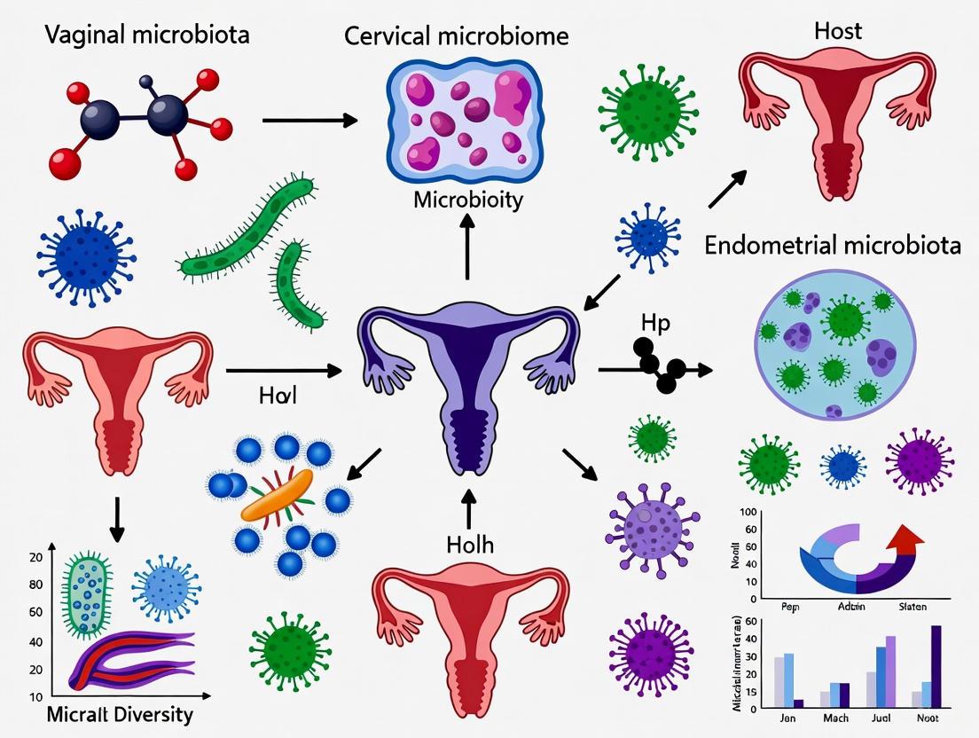
The female reproductive tract (FRT) hosts a dynamic microbial ecosystem, spatially structured from the vagina to the endometrium. Once considered sterile, the upper reproductive tract is now recognized to possess its own endogenous microbiome, distinct from the vaginal microbiota [7] [8]. Understanding the spatial architecture of these microbial communities is crucial for researchers and drug development professionals investigating reproductive health, disease pathogenesis, and treatment outcomes. This technical guide synthesizes current evidence on the composition and community state types (CSTs) across the FRT, providing a foundational framework for ongoing research into the reproductive tract microbiome.
Spatial Distribution of Microbial Communities in the Female Reproductive Tract
The FRT exhibits a anatomical continuum with distinct microbial gradients from the lower to upper tracts.
Lower Reproductive Tract Microbiome
The vaginal and cervical microbiota typically demonstrate low diversity and are predominantly composed of the genus Lactobacillus in healthy women of reproductive age [9]. Vaginal microbiota classification has been standardized into five Community State Types (CSTs) based on the dominant Lactobacillus species and bacterial composition [9] [10]:
- CST I: Dominated by L. crispatus
- CST II: Dominated by L. gasseri
- CST III: Dominated by L. iners
- CST V: Dominated by L. jensenii
- CST IV: Characterized by a diverse mixture of facultative and obligate anaerobes with reduced Lactobacillus abundance
Lactobacillus species maintain vaginal health through lactic acid production, which acidifies the environment (pH 3.5-4.5), and through the production of antimicrobial compounds including hydrogen peroxide and bacteriocins [9]. Notably, not all Lactobacillus species offer equal protection; L. iners has a reduced genome size (~1.3 Mb) and lacks the ability to produce D-lactic acid and hydrogen peroxide, making it a less stable component of the microbiota [9].
CST IV, widely recognized as a hallmark of vaginal dysbiosis, is further categorized into three subtypes: IV-A (dominated by Candidatus Lachnocurva vaginae and G. vaginalis), IV-B (enriched in Atopobium vaginae and G. vaginalis), and IV-C (characterized by low abundances of Lactobacillus spp. and a predominance of diverse facultative and obligate anaerobes) [9].
Upper Reproductive Tract Microbiome
The endometrium harbors greater bacterial diversity and richness compared to the vagina [8] [10]. While the upper reproductive tract was traditionally considered sterile, advancements in genomic technologies have revealed distinct microbial communities in the endometrium and fallopian tubes [7] [8].
The endometrial microbiome is mainly composed of bacteria belonging to the phyla Firmicutes, Bacteroidetes, and Proteobacteria [8]. A study analyzing matched vaginal and endometrial samples found that endometrial microbiomes were more diverse than vaginal microbiomes (average Shannon entropy = 1.89 versus 0.75, p = 10⁻⁵) and enriched in bacterial species such as Corynebacterium sp., Staphylococcus sp., Prevotella sp., and Propionibacterium sp. [10].
Fallopian tube samples show a microbial profile distinct from yet sharing similarities with the endometrium, with 69% of detected taxa common to both sites [8]. Seventeen bacterial taxa were found exclusively in fallopian tube samples, including the genera Enhydrobacter, Granulicatella, Haemophilus, Rhizobium, Alistipes, and Paracoccus [8].
Table 1: Comparative Analysis of Vaginal and Endometrial Microbiome Characteristics
| Parameter | Vaginal Microbiome | Endometrial Microbiome |
|---|---|---|
| Typical Diversity | Low diversity (Shannon entropy ~0.75) [10] | Higher diversity (Shannon entropy ~1.89) [10] |
| Dominant Taxa in Health | Lactobacillus spp. (often >50%) [9] | Lactobacillus spp. (often >90% in LD state) [11] [10] |
| Common Non-Lactobacillus Taxa | Gardnerella, Prevotella, Atopobium (in CST IV) [9] | Corynebacterium, Staphylococcus, Prevotella, Propionibacterium [10] |
| Classification System | Community State Types (CSTs I-V) [9] [10] | Lactobacillus-dominant (LD) vs. Non-Lactobacillus-dominant (NLD) [11] [10] |
| Definition of "Dominant" | ≥50% Lactobacillus abundance [10] | ≥90% Lactobacillus abundance for LD classification [10] |
| Biomass | Higher biomass [10] | Lower biomass [10] |
Methodological Approaches for Reproductive Tract Microbiome Analysis
Sample Collection Protocols
Vaginal Sampling: Vaginal fluid is collected from the posterior fornix using a dry swab under direct visualization with a speculum, without lubricants to prevent contamination [12]. Multiple sampling time points across the menstrual cycle provide more comprehensive characterization, as the vaginal microbiome demonstrates dynamic fluctuations [13].
Endometrial Sampling: Transcervical collection requires meticulous technique to minimize contamination during catheter passage through the cervicovaginal canal. The Tao Brush IUMC Endometrial Sampler or similar devices with protective sheaths are recommended [12]. Samples obtained via hysterectomy avoid vaginal contamination but are not feasible for most clinical studies [8].
DNA Extraction and Sequencing
DNA Extraction: The PureLink Microbiome DNA Purification Kit effectively extracts microbial DNA from low-biomass endometrial samples [12]. DNA quantification using fluorometric methods (e.g., Qubit Fluorometer) is essential [12].
16S rRNA Gene Amplification and Sequencing:
- Primer Selection: Targeting hypervariable regions V1-V2 or V3-V4 of the 16S rRNA gene enables differentiation of common vaginal lactobacilli [10] [12].
- PCR Amplification: Using Taq DNA polymerase (e.g., KAPA HiFi HotStart) with primers 357F and 806R at 1μM concentration, with approximately 100ng DNA template in 25μL reaction volume [12].
- Library Preparation and Sequencing: The Nextera XT library preparation kit followed by sequencing on Illumina MiSeq platforms with v3 reagents provides sufficient depth for community analysis [12].
Bioinformatic Analysis:
- Processing raw sequences through quality control, chimera filtering, and OTU clustering at 97% similarity threshold
- Taxonomic assignment using reference databases
- Diversity analysis (alpha and beta diversity metrics)
- Differential abundance testing (LEfSe analysis) [11]
Table 2: Essential Research Reagents and Tools for Reproductive Microbiome Studies
| Reagent/Tool | Specific Example | Function/Application |
|---|---|---|
| DNA Extraction Kit | PureLink Microbiome DNA Purification Kit [12] | DNA extraction from low-biomass samples |
| Polymerase | KAPA HiFi HotStart ReadyMix [12] | High-fidelity amplification of 16S rRNA gene |
| Sequencing Platform | Illumina MiSeq [12] | 16S rRNA gene amplicon sequencing |
| Sequencing Kit | MiSeq Reagent Kit v3 (600-cycle) [12] | Provides appropriate read length for V3-V4 region |
| Library Prep Kit | Nextera XT DNA Library Preparation Kit [12] | Indexing and library preparation for multiplexing |
| Endometrial Sampler | Tao Brush IUMC Endometrial Sampler [12] | Transcervical sampling with reduced contamination |
| Primer Set | 357F (5'-CCTACGGGNGGCWGCAG-3') and 806R (5'-GGACTACHVGGGTWTCTAAT-3') [12] | Amplification of 16S rRNA V3-V4 region |
Methodological Considerations for Low-Biomass Samples
Endometrial samples present particular challenges due to low microbial biomass. Essential controls include:
- Negative extraction controls to detect reagent contamination
- Blank PCR controls
- Processing in clean laboratory environments with sterile, DNA-free reagents and consumables [11]
- Reporting of all control results to validate findings
Functional Dynamics and Clinical Implications
Microbial Community Dynamics
The vaginal microbiome exhibits dynamic temporal patterns classified as Vaginal Community Dynamics (VCDs):
- Constant eubiotic: Stable Lactobacillus-dominant state
- Constant dysbiotic: Stable non-Lactobacillus-dominant state
- Menses-related: Cyclic dysbiosis associated with menstruation
- Unstable dysbiotic: Frequent fluctuations between community states [13]
These dynamics are influenced by host factors (menstruation, sexual activity, contraceptive use) and microbiome-intrinsic factors (bacteriophage activity, bacterial gene content) [13].
Impact on Reproductive Outcomes
The composition of the reproductive tract microbiota significantly influences assisted reproductive technology (ART) outcomes. In frozen embryo transfer (FET) cycles, a Lactobacillus-dominant (LD) uterine microbiota is associated with significantly higher clinical pregnancy rates (75.00% vs. 45.16%) and live birth rates (65.00% vs. 29.03%) compared to non-Lactobacillus-dominant (NLD) microbiota [11]. Similarly, vaginal Lactobacillus dominance is associated with improved reproductive outcomes [14] [12].
The mechanisms underlying these associations involve immunological pathways. Lactobacillus dominance promotes an anti-inflammatory environment conducive to embryo implantation, while dysbiosis triggers pro-inflammatory responses through Toll-like receptor (TLR) activation, particularly TLR4 recognition of lipopolysaccharide (LPS) from Gram-negative bacteria, leading to NF-κB signaling and cytokine production [14] [9].
Figure 1: Microbial Modulation of Endometrial Receptivity and Inflammation. Lactobacillus species (green) protect against inflammation through multiple mechanisms, while dysbiotic bacteria (yellow/red) trigger pro-inflammatory pathways that can impair implantation.
Associations with Gynecological Diseases
Alterations in reproductive tract microbiota are associated with various gynecological conditions:
- Endometrial cancer: Characterized by depletion of protective Lactobacillus species and enrichment of anaerobic, pro-inflammatory bacteria like Prevotella, Atopobium, and Porphyromonas [15]
- Endometriosis: Associated with increased abundance of Fusobacterium and other pathogenic bacteria [7]
- Chronic endometritis: Linked to disturbance in Lactobacillus dominance and higher abundance of Gardnerella, Streptococcus, and Enterobacteriaceae [10]
- Uterine fibroids: Show altered cervical and vaginal microbiota with reduced Lactobacillus and enrichment of specific anaerobic taxa [7]
The spatial architecture of the female reproductive tract microbiome demonstrates a complex ecosystem with distinct yet interconnected communities from the vagina to the endometrium. The lower tract is characterized by Lactobacillus-dominant CSTs, while the upper tract exhibits greater diversity with enrichment of non-Lactobacillus taxa even in healthy states. Methodological rigor in sampling, DNA extraction, and sequencing is particularly critical for accurate characterization of the low-biomass endometrial microbiome.
Future research directions should focus on standardized protocols for cross-study comparisons, functional analyses of microbial metabolites and host interactions, and developing targeted interventions to modulate dysbiotic communities. Understanding this spatial architecture provides a critical foundation for developing novel diagnostics and therapeutics for reproductive disorders and optimizing outcomes in assisted reproduction.
The vaginal microbiome is a critical component of female reproductive health, with Lactobacillus species serving as foundational commensals that maintain homeostasis and a protective acidic pH. Through mechanisms including lactic acid production, bacteriocin secretion, and competitive exclusion, these microbes create an environment that inhibits pathogen colonization and modulates local immune responses. This whitepaper synthesizes current research on the functional roles of vaginal lactobacilli, detailing the molecular basis for pH maintenance and ecosystem stability. Framed within the broader context of reproductive tract microbiome research, we present quantitative data on species-specific functions, experimental protocols for investigating microbial dynamics, and essential research tools for drug development targeting vaginal health.
The human vaginal microbiome is a dynamic ecosystem whose composition profoundly influences gynecological, obstetric, and reproductive health [16]. A healthy vaginal environment in most reproductive-age women is dominated by Lactobacillus species, which maintain an acidic pH through lactic acid production and provide colonization resistance against pathogens [4] [17]. Contemporary research characterizes the vaginal microbiota into five primary Community State Types (CSTs): CST-I (L. crispatus-dominant), CST-II (L. gasseri-dominant), CST-III (L. iners-dominant), CST-V (L. jensenii-dominant), and CST-IV (diverse anaerobic flora with low Lactobacillus abundance) [18] [17]. This taxonomic framework provides a critical foundation for understanding how specific Lactobacillus species contribute to maintaining homeostasis and how deviations from these healthy states correlate with disease risk, including bacterial vaginosis (BV), preterm birth, and increased susceptibility to sexually transmitted infections [4] [19].
The functional dominance of lactobacilli represents an evolutionary adaptation supported by host physiology. Estrogen stimulates vaginal epithelial proliferation and glycogen deposition, which serves as the primary carbon source for lactobacilli [20] [4]. The metabolic products of this symbiosis, particularly lactic acid, create an environment (pH 3.5-4.5) that is inhibitory to many pathogenic organisms [4] [17]. Beyond acidification, lactobacilli employ multiple mechanisms including hydrogen peroxide production, bacteriocin secretion, and competitive adhesion to maintain vaginal homeostasis [17] [21]. Understanding these mechanisms within the broader composition of the reproductive tract microbiome provides crucial insights for developing targeted interventions for vaginal dysbiosis.
Core Mechanisms of Homeostasis and pH Regulation
Lactic Acid Production and Acidic Environment Maintenance
The primary mechanism by which Lactobacillus species maintain vaginal health is through lactic acid production, which acidifies the vaginal environment to pH 3.5-4.5 [4] [17]. This acidic pH is detrimental to many pathogenic bacteria and provides a competitive advantage to lactobacilli. The process begins with glycogen accumulation in vaginal epithelial cells under estrogen stimulation [20] [4]. Both host-derived and microbially-derived enzymes, particularly α-amylases, break down glycogen into simpler sugars such as maltose and glucose [20]. Lactobacilli ferment these sugars to produce both D- and L-isomers of lactic acid, with the D-form exhibiting particularly potent antimicrobial properties [4]. The resulting acidic environment selectively inhibits the growth of many opportunistic pathogens while promoting the stability of Lactobacillus-dominant communities.
The following diagram illustrates this glycogen metabolism pathway:
Multi-Functional Protective Mechanisms
Beyond lactic acid production, lactobacilli employ several complementary mechanisms to maintain vaginal homeostasis and prevent pathogen colonization, as summarized in the table below.
Table 1: Multi-Functional Protective Mechanisms of Vaginal Lactobacillus Species
| Mechanism | Functional Description | Key Species/Examples | Protective Outcome |
|---|---|---|---|
| Hydrogen Peroxide (H₂O₂) Production | Produces antimicrobial oxidizing agent toxic to catalase-negative pathogens | L. jensenii (94%), L. crispatus (95%), L. fermentum, L. acidophilus [17] | Creates hostile environment for anaerobes; promotes immune tolerance |
| Bacteriocin Secretion | Releases antimicrobial peptides that inhibit cell wall synthesis and spore formation in pathogens | L. salivarius CRL 1328 inhibits N. gonorrhoeae; L. fermentum 123 against Gram-positive/negative bacteria [17] | Direct pathogen growth inhibition without affecting lactobacilli |
| Competitive Adhesion & Co-aggregation | Binds to epithelial cell receptors, blocking pathogen attachment sites | Co-aggregation with G. vaginalis, C. albicans, and E. coli [17] | Physical exclusion of pathogens; nutrient deprivation |
| Mucosal Barrier Enhancement | Strengthens epithelial integrity through microbial interactions | L. acidophilus demonstrates strong barrier enhancement properties [22] | Reduced pathogen translocation and inflammation |
| Immunomodulation | Regulates pro-inflammatory cytokine induction and promotes homeostasis | L. crispatus associated with reduced inflammation [21] | Balanced immune response without excessive inflammation |
These mechanisms function synergistically to create a comprehensive defense system. The combination of acidification, direct antimicrobial activity, physical exclusion, and immune regulation provides robust protection against vaginal dysbiosis while maintaining a stable microenvironment conducive to Lactobacillus dominance [4] [17] [21].
Experimental Models and Methodological Approaches
Community State Type Analysis and Microbial Dynamics
Investigating vaginal microbiome composition and dynamics relies heavily on molecular techniques, particularly next-generation sequencing (NGS). The standard methodological workflow begins with sample collection using sterile Dacron polyester swabs from the posterior vaginal fornix, followed by placement in phosphate-buffered saline or preservation media like eNAT [18] [23]. DNA extraction typically employs commercial kits such as the QIAamp DNA Mini Kit (QIAGEN), with subsequent PCR amplification of the bacterial 16S rRNA gene V4/V5 regions using primers F519/R926 [18]. Sequencing is performed on platforms such as Illumina MiSeq or NovaSeq, followed by bioinformatic processing using tools like QIIME2 for quality filtering, chimera removal, and taxonomic classification against reference databases (e.g., SILVA) [18] [22]. Community State Type (CST) classification is achieved through hierarchical clustering of taxonomic profiles, differentiating Lactobacillus-dominant states (CST-I, II, III, V) from the diverse anaerobic state (CST-IV) [18] [17].
The experimental workflow for vaginal microbiome analysis can be visualized as follows:
Functional Assessment of Lactobacillus Strains
Beyond compositional analysis, functional characterization of lactobacilli requires different methodological approaches. For acid production capacity, researchers typically conduct in vitro fermentation assays using MRS medium with pH monitoring over time [21]. Quantification of lactic acid isomers can be performed using high-performance liquid chromatography (HPLC). Antimicrobial compound production is assessed through agar well diffusion assays against target pathogens like Gardnerella vaginalis and Escherichia coli [17] [21]. Adhesion capabilities are evaluated using epithelial cell line models (e.g., VK2/E6E7) with enumeration of adherent bacteria microscopically or through plating [17]. For probiotic efficacy testing, randomized controlled trials employ various delivery methods including vaginal suppositories and oral capsules, with outcomes measured through Nugent scoring, vaginal pH assessment, and symptom questionnaires [23] [21].
Table 2: Quantitative Assessment of Lactobacillus Species Functional Characteristics
| Lactobacillus Species | Vaginal pH Range | Lactic Acid Production Capacity | H₂O₂ Production Prevalence | Protective Association with Conditions |
|---|---|---|---|---|
| L. crispatus | 3.5-4.0 [4] | High (D- and L-isomers) [4] | 95% of strains [17] | Strong inverse association with BV, PTB, HPV persistence [22] [19] |
| L. jensenii | 3.5-4.0 [4] | Moderate to High [4] | 94% of strains [17] | Protection against BV and STIs [17] |
| L. gasseri | 3.5-4.5 [18] | Moderate [18] | Variable [18] | Increased in non-specific CVD [18] |
| L. iners | 4.0-4.5 [4] | Low (L-isomer only) [4] | Limited or absent [4] | Ambiguous role; associated with transition to dysbiosis [4] |
| L. acidophilus | 3.5-4.5 [21] | High [21] | Present in some strains [17] | Epithelial barrier enhancement [22] |
The Scientist's Toolkit: Essential Research Reagents and Methodologies
Table 3: Essential Research Reagents and Methodologies for Vaginal Microbiome Research
| Reagent/Methodology | Specific Examples | Research Application | Technical Considerations |
|---|---|---|---|
| DNA Extraction Kits | QIAamp DNA Mini Kit (QIAGEN) [18] [22] | Microbial DNA isolation from vaginal swabs | Includes enzymatic and mechanical lysis; critical for NGS success |
| Sequencing Platforms | Illumina MiSeq/NovaSeq [22] [23] | 16S rRNA gene amplicon sequencing | V3-V4 or V4-V5 regions; minimum 10M reads/sample for metagenomics [22] |
| Bioinformatics Tools | QIIME2, SILVA database, MetaPhlAn4 [22] [23] | Taxonomic classification, CST assignment | Hierarchical clustering for CST determination; decontam for contaminant removal [23] |
| Culture Media | MRS medium, blood agar, chocolate agar [18] [22] | Lactobacillus cultivation and isolation | Anaerobic conditions at 37°C for 48-72 hours [22] |
| Probiotic Formulations | Multi-strain supplements (e.g., VagiBIOM) [21] | Intervention studies for dysbiosis | Typically contain 2×10⁹ to 1×10¹⁰ CFU per strain [23] [21] |
| pH Measurement | pH strips (Merck) [18] | Assessment of vaginal acidity | Immediate measurement prior to sampling for accuracy |
| Cell Lines | VK2/E6E7 [17] | Adhesion and host-pathogen interaction studies | Human vaginal epithelial cell line for in vitro modeling |
Discussion: Research Implications and Future Directions
The functional roles of Lactobacillus species in maintaining vaginal homeostasis and pH extend beyond simple acidification to encompass a sophisticated multi-mechanism defense system. Current research reveals significant species-specific functional differences that explain their varying protective associations. For instance, L. crispatus demonstrates superior protection through high lactic acid production (both D- and L-isomers), consistent H₂O₂ production, and stable colonization, making it a keystone species in vaginal health [4] [22]. In contrast, L. iners exhibits a more ambiguous role due to its reduced genome size, limited metabolic capacity, inability to produce D-lactic acid, and production of inerolysin—a pore-forming toxin that may compromise epithelial integrity [4]. These functional differences highlight the importance of moving beyond genus-level characterization to species- and strain-level analyses for understanding vaginal ecosystem dynamics.
Future research directions should focus on elucidating the complex interactions between specific Lactobacillus strains, host immunity, and the broader microbial community. The emerging concept of vaginal community dynamics (VCDs)—categorizing women into "constant eubiosis," "constant dysbiosis," "unstable," or "menses-related dysbiosis" patterns—provides a more nuanced framework for understanding temporal fluctuations [20]. Additionally, the gut-vaginal axis warrants deeper investigation, particularly how systemic factors and gut microbiota influence vaginal health [4]. From a therapeutic perspective, well-designed randomized controlled trials using defined bacterial consortia rather than single strains may better replicate the natural protective functions of diverse Lactobacillus communities. Advanced delivery systems, including optimized suppositories and prebiotic combinations, represent promising approaches for maintaining vaginal homeostasis and preventing dysbiosis-related complications throughout a woman's lifespan [23] [21].
Lactobacillus species play indispensable functional roles in maintaining vaginal homeostasis and pH through multiple complementary mechanisms. The core functions of lactic acid production, bacteriocin secretion, competitive exclusion, and immunomodulation work synergistically to create an environment that inhibits pathogens while supporting a beneficial microbial community. Understanding the species-specific characteristics and community dynamics of these commensals provides crucial insights for developing targeted interventions for vaginal dysbiosis. As research in this field advances, leveraging sophisticated methodological approaches and reagent systems will enable more precise manipulation of the vaginal ecosystem, ultimately leading to improved strategies for maintaining reproductive health and preventing gynecological diseases across diverse patient populations.
The definition of a healthy female reproductive tract (FRT) microbiome is undergoing a profound paradigm shift. While the dominance of Lactobacillus species, particularly L. crispatus, has long been synonymous with vaginal health, emerging research reveals a more complex ecological narrative [24] [4]. The FRT microbiome encompasses a continuum of communities from the vagina to the upper reproductive tract (uterus, fallopian tubes, and ovaries), each with distinct compositional and functional characteristics [25] [26]. The persistent focus on Lactobacillus obscures the critical roles of other microbial communities, both as vital components of a healthy ecosystem and as mediators of pathology. This whitepaper explores the nuanced roles of non-classical players, specifically the enigmatic species L. iners and diverse non-Lactobacillus-dominant communities (Community State Type IV, CST-IV), framing them within the broader context of FRT microbiome research. Understanding these nuances is essential for developing targeted therapeutic interventions and diagnostic tools that move beyond a simplistic dichotomy of "healthy" versus "dysbiotic" states.
2Lactobacillus iners: A Double-Edged Sword in Microbial Homeostasis
Lactobacillus iners is one of the most prevalent and yet paradoxical bacteria in the FRT. Despite being a Lactobacillus species, its ecological role starkly contrasts with the protective functions of its congeners, marking it as a transitional species with a potentially detrimental impact on reproductive health.
Genomic and Metabolic Distinctiveness
The unique behavior of L. iners is rooted in its genomic architecture. Comparative genomics reveals that L. iners possesses an unusually small genome (~1.3 Mb), comparable in size to human symbionts and parasites, which is significantly smaller than the 1.5-2.0 Mb genomes of other dominant vaginal Lactobacillus species like L. crispatus [4] [27]. This genome reduction signifies a decreased metabolic capacity. Crucially, L. iners lacks the ability to produce D-lactic acid and hydrogen peroxide (H₂O₂), key antimicrobial compounds synthesized by other Lactobacillus species that contribute to pathogen inhibition and environmental stability [4]. Instead of maintaining a rigid homeostasis, L. iners exhibits high ecological niche specificity and relies on metabolic adaptation to thrive in fluctuating host microenvironments [4].
Association with Dysbiosis and Disease
The metabolic flexibility of L. iners allows it to survive in both Lactobacillus-dominant and dysbiotic environments, but it often acts as a harbinger of instability. A pivotal study on infertile couples undergoing Assisted Reproductive Technology (ART) found L. iners (CST III) to be the most abundant species in vaginal lavages and linked it to a decreased fertility rate [28]. Furthermore, the genome of L. iners contains genes encoding potential virulence factors, most notably inerolysin, a pore-forming toxin functionally homologous to vaginolysin produced by Gardnerella vaginalis [4]. This cytolysin may compromise the integrity of the vaginal mucus layer, weakening host defenses and facilitating the overgrowth of anaerobic bacteria associated with bacterial vaginosis (BV) [4]. Consequently, a vaginal microbiome dominated by L. iners (CST III) is considered less robust and more prone to transitioning to the dysbiotic CST IV state compared to one dominated by L. crispatus (CST I) [4] [26].
Table 1: Comparative Analysis of Key Vaginal Lactobacillus Species
| Species | Community State Type (CST) | Genome Size (approx.) | Key Metabolites | Association with Health/Disease |
|---|---|---|---|---|
| L. crispatus | CST I | 1.5-2.0 Mb | L-lactic acid, D-lactic acid, H₂O₂ | Strongly associated with health and stability [4] [26] |
| L. gasseri | CST II | 1.5-2.0 Mb | L-lactic acid, Bacteriocins | Associated with health [24] |
| L. iners | CST III | ~1.3 Mb | L-lactic acid, Inerolysin | "Transitional" state; associated with instability and decreased fertility [28] [4] |
| L. jensenii | CST V | 1.5-2.0 Mb | L-lactic acid, H₂O₂ | Associated with health [24] |
Non-Lactobacillus-Dominant Communities (CST-IV): A Spectrum of Diversity and Risk
CST-IV represents a polymicrobial consortium not dominated by any single Lactobacillus species. It is a heterogeneous state characterized by high microbial diversity and is a hallmark of bacterial vaginosis, though it can also represent a stable, asymptomatic state for some women, particularly those of African, Hispanic, and certain Asian ancestries [29] [4].
Composition and Functional Impact
CST-IV is typically dominated by a diverse array of facultative and obligate anaerobes. Key genera include Gardnerella, Prevotella, Atopobium, Sneathia, Megasphaera, and Mobiluncus [24] [4]. This shift in community structure leads to profound functional changes in the FRT environment:
- Metabolite Shift: CST-IV communities deplete lactic acid and produce various biogenic amines (e.g., putrescine, cadaverine), which elevate vaginal pH above 4.5 and are responsible for the characteristic malodor of BV [4]. These amines can also negatively impact the growth and lactic acid production of beneficial Lactobacillus, delaying the re-establishment of a Lactobacillus-dominant community [4].
- Barrier Disruption: Bacteria in CST-IV secrete hydrolytic enzymes like sialidases that degrade protective mucins on the cervicovaginal surface, compromising the epithelial barrier and increasing the risk of ascending infections and inflammation [4].
Clinical Consequences of CST-IV
The altered metabolic and immunological landscape of CST-IV is linked to numerous adverse reproductive health outcomes. In the context of infertility, L. gasseri and Prevotella have been identified in seminal fluids, follicular fluids, and embryo culture media, with L. gasseri associated with oocyte DNA fragmentation and decreased sperm mobility, and Prevotella linked to reduced sperm motility [28]. Furthermore, non-Lactobacillus dominant endometrial microbiota have been associated with negative reproductive outcomes in patients undergoing in vitro fertilization (IVF) [25]. Beyond infertility, CST-IV is associated with an increased risk of acquiring sexually transmitted infections (including HPV and HIV), preterm birth, and gynecologic cancers [25] [24].
Table 2: Characteristics of Non-Lactobacillus-Dominant (CST-IV) Community Subtypes
| Subtype | Dominant Taxa | Vaginal pH | Nugent Score | Clinical Associations |
|---|---|---|---|---|
| CST IV-A | Candidatus Lachnocurva vaginae, Gardnerella vaginalis | Elevated | High | Common in African and Hispanic women; associated with BV [4] |
| CST IV-B | Atopobium vaginae, Gardnerella vaginalis | Elevated | High | Common in African and Hispanic women; associated with BV [4] |
| CST IV-C | Diverse facultative and obligate anaerobes; low Lactobacillus, G. vaginalis, A. vaginae | Variable | Lower than IV-A/B | Less prevalent; may represent a more stable, non-BV state [4] |
Methodological Approaches for Advanced Microbiome Research
Accurate characterization of the FRT microbiome requires sophisticated molecular techniques that move beyond traditional culture. Next-Generation Sequencing (NGS) of the bacterial 16S ribosomal RNA (16S rRNA) gene is the cornerstone of modern microbiome research.
Sample Collection and Nucleic Acid Extraction
Sterile sampling is critical, especially for the low-biomass upper reproductive tract. Methods include:
- Vaginal/Cervical Swabs: Standard for lower FRT sampling [25].
- Transcervical Embryo Transfer Catheter Tips: Used to sample the endometrial microbiome during IVF procedures [25].
- Endometrial Fluid Aspiration or Biopsy: Collected with careful cervicovaginal preparation to minimize contamination [25].
- Surgical Collection: Sterile collection of uterine swabs or tissue during hysterectomy, avoiding passage through the cervix [25].
Nucleic acid extraction is performed using automated systems like the NucliSENS easyMAG [28]. Seminal fluid often requires pre-treatment with dithiothreitol (DTT) to liquefy the sample, while other fluids are centrifuged to pellet microbial biomass [28].
16S rRNA Gene Amplification and Sequencing
A common approach involves a nested PCR strategy to enrich for the target region:
- Primary PCR: Amplification of the V1-V3 hypervariable regions (~500 bp) of the 16S rRNA gene using degenerate primers (e.g., 27FYM and U534R) [28].
- Nested PCR: A second amplification targeting the V3 region (~200 bp) with barcoded primers (e.g., B338FP1-adaptor and U534RA_barcode) to allow for multiplexing [28].
- Library Preparation and Sequencing: Amplicons are pooled in equimolar amounts, and template preparation is performed using systems like the Ion OneTouch 2, followed by sequencing on an Ion PGM System [28].
Bioinformatic and Statistical Analysis
Sequencing data is processed through pipelines such as QIIME 2 [28]. Key steps include:
- Quality Filtering: Retaining reads with Q ≥ 20 and removing ambiguous bases/homopolymers.
- Denoising and Clustering: Using algorithms like DADA2 to infer exact amplicon sequence variants (ASVs).
- Taxonomic Assignment: Classifying sequences against curated reference databases (e.g., VIRGO for vaginal microbiota) using BLAST+ [28] [24].
- Downstream Analysis: Analyses include alpha-diversity (within-sample diversity), beta-diversity (between-sample diversity), and differential abundance testing to identify taxa associated with clinical conditions.
Diagram 1: 16S rRNA Sequencing Workflow. This diagram outlines the key steps in a standard 16S rRNA gene sequencing protocol for reproductive microbiome characterization, from sample collection to data analysis.
The Inflammatory Nexus: Microbial Dysbiosis and Host Immunity
The balance between the FRT microbiota and the host immune system is critical for maintaining homeostasis. Dysbiosis, characterized by a loss of Lactobacillus dominance and an increase in microbial diversity, disrupts this balance and triggers a pro-inflammatory response [29].
The innate immune system in the FRT recognizes microbial components via Pattern Recognition Receptors (PRRs), such as Toll-like Receptors (TLRs). In a dysbiotic state (CST-IV), bacteria like Prevotella and Gardnerella produce pathogen-associated molecular patterns (PAMPs), including lipopolysaccharide (LPS) [4]. TLR4 on epithelial and immune cells recognizes LPS via the CD14-MD-2 complex, initiating a signaling cascade that activates the MyD88-dependent pathway. This leads to the activation of NF-κB, a master transcription factor that translocates to the nucleus and promotes the expression of pro-inflammatory cytokines and chemokines (e.g., IL-6, IL-8, TNF-α) [4]. This inflammatory milieu recruits immune cells, exacerbating local inflammation and contributing to tissue damage, impaired sperm function, and adverse reproductive outcomes such as implantation failure and preterm birth [28] [29] [4].
Diagram 2: Inflammatory Pathway in Dysbiosis. This diagram illustrates the proposed mechanism through which non-Lactobacillus-dominant microbiota trigger a pro-inflammatory response via TLR4/MyD88/NF-κB signaling, leading to adverse reproductive outcomes.
Table 3: Research Reagent Solutions for Reproductive Microbiome Studies
| Reagent / Resource | Function / Application | Example from Literature |
|---|---|---|
| NucliSENS easyMAG | Automated nucleic acid extraction from diverse sample types (lavages, fluids, tissues) [28]. | Used for DNA extraction from vaginal lavages, follicular fluids, and seminal fluids [28]. |
| Kapa 2G HiFi Hotstart ReadyMix | Robust, high-fidelity PCR enzyme mix for efficient amplification of 16S rRNA gene targets, crucial for NGS library construction [28]. | Used in primary and nested PCR amplification of the 16S V1-V3 and V3 regions [28]. |
| Ion PGM Hi-Q View Sequencing Kit | Sequencing chemistry for the Ion Torrent PGM platform, used for generating 16S rRNA amplicon sequencing data [28]. | Used for final sequencing of barcoded 16S rRNA libraries [28]. |
| Ion Xpress Barcode Adapters | Unique molecular barcodes attached to PCR primers, enabling multiplexing of multiple samples in a single sequencing run [28]. | Attached to the reverse primer during nested PCR for sample identification post-sequencing [28]. |
| VIRGO Database | A curated, non-redundant gene catalog and database for vaginal microbiota, enabling high-resolution taxonomic and functional profiling from metagenomic data [24]. | Allows for consistent classification of vaginal bacteria at the species and subspecies level [24]. |
| QIIME 2 (v2020.2) | A powerful, extensible, and decentralized microbiome analysis platform for processing raw sequencing data into biological insights [28]. | Used for sequence quality control, DADA2 denoising, and taxonomic analysis [28]. |
| Dithiothreitol (DTT) | A reducing agent used to pretreat and liquefy viscous seminal fluid samples prior to DNA extraction, improving microbial recovery [28]. | Pretreatment of seminal fluids to break down disulfide bonds in sperm nuclei [28]. |
The landscape of reproductive tract microbiome research is evolving from a Lactobacillus-centric view to a more nuanced understanding of microbial ecology. L. iners and non-Lactobacillus-dominant communities (CST-IV) are not mere aberrations but are key players with complex roles in health and disease. Future research must focus on elucidating the mechanistic pathways linking these microbial communities to host physiology, moving beyond associative studies to causal relationships. This will require integrated multi-omics approaches (metagenomics, metatranscriptomics, metabolomics) and advanced in vitro and in vivo models. Furthermore, the development of therapeutics—such as targeted probiotics beyond traditional Lactobacillus species, phage therapy, and microbiome transplantation—holds promise for restoring a healthy ecosystem. For researchers and drug development professionals, embracing this complexity is essential for pioneering the next generation of diagnostics and interventions in reproductive medicine.
The human body exists as a complex superorganism, intimately integrated with trillions of microorganisms that constitute the microbiome. Recent scientific advances have fundamentally reshaped our understanding of human physiology by revealing that these microbial communities engage in extensive cross-talk with host organ systems, creating intricate axes that maintain homeostasis and influence disease pathogenesis. Within this framework, two particularly sophisticated systems have emerged: the gut-reproductive axis and the estrobolome. The gut-reproductive axis represents the bidirectional communication network between gastrointestinal microbiota and reproductive tract physiology, while the estrobolome comprises the specific ensemble of gut bacteria capable of metabolizing and modulating systemic estrogen levels [30] [31]. Together, these systems form a critical regulatory circuit that integrates metabolic, immune, and endocrine signaling to influence reproductive health, fetal development, and a spectrum of gynecological pathologies.
This conceptual framework aligns with the evolving "meta-host" model in microbiome science, which expands the traditional definition of a host to include its symbiotic microbial communities as an integrated functional unit [32]. Understanding the precise mechanisms governing these interactions provides unprecedented opportunities for novel diagnostic, therapeutic, and preventive strategies in reproductive medicine and drug development.
The Gut-Reproductive Axis: Bridging Microbial Balance to Reproductive Health
Conceptual Framework and Physiological Significance
The gut-reproductive axis functions as a multifaceted bridge connecting the digestive tract's microbial ecosystem with the female reproductive system. This axis facilitates continuous dialogue between these seemingly disparate systems, maintaining maternal reproductive homeostasis and influencing offspring development [30]. The gastrointestinal tract hosts an extraordinarily complex microbial community, with the gut microbiome encoding approximately two million microbial genes—vastly outnumbering the 20,000 genes in the human genome [31] [32]. This genetic repertoire enables the microbiome to function as a virtual endocrine organ, capable of synthesizing, activating, and metabolizing compounds that systemically influence host physiology.
The anatomical distribution of human microbiomes reveals that the urogenital tract contains approximately 9% of the body's bacterial populations, while the gastrointestinal tract hosts 29%, creating substantial potential for cross-system interaction [32]. Although these organs are physically separate, they share common embryonic origins from the embryonic mesoderm and endoderm, potentially explaining their continued physiological connectivity through shared neural, endocrine, and immune pathways in postnatal life.
Mechanisms of Interaction
The gut-reproductive axis operates through several interconnected mechanistic pathways:
Immunological Modulation: Gut microbiota metabolites, particularly short-chain fatty acids (SCFAs) like butyrate, propionate, and acetate, play pivotal roles in regulating systemic inflammation and immune responses. Butyrate induces differentiation of T-regulatory (Treg) cells through inhibition of histone deacetylases (HDACs) and promotes anti-inflammatory forkhead box protein P3 (Foxp3) expression [33]. By binding to GPR109a on dendritic cells and macrophages, butyrate increases IL-10 production while decreasing pro-inflammatory IL-6, resulting in enhanced Treg development and suppressed Th17 cell expansion [33]. This immunomodulatory activity directly impacts reproductive tissue environments.
Hormonal Regulation: Beyond the estrogen-specific mechanisms of the estrobolome (detailed in Section 3), gut microbiota influence broader neuroendocrine pathways, including the hypothalamic-pituitary-gonadal (HPG) axis, through microbial metabolites that can function as signaling molecules to distant organs.
Barrier Function Integrity: Gut dysbiosis can compromise intestinal epithelial barrier function, increasing intestinal permeability and facilitating translocation of bacterial components such as lipopolysaccharides (LPS) into systemic circulation [34]. This microbial translocation triggers low-grade chronic inflammation that can disrupt reproductive tissue homeostasis.
The following diagram illustrates the core mechanisms through which the gut-reproductive axis functions:
Clinical Implications in Reproductive Pathology
Dysbiosis within the gut-reproductive axis has been mechanistically linked to several gynecological and obstetric conditions through clinical and translational research:
Endometriosis: Molecular studies have identified Fusobacterium nucleatum infiltration in the uterine tissue of approximately 64% of women with endometriosis [35]. Experimental models demonstrate that Fusobacterium infection promotes macrophage infiltration, transforming growth factor-β (TGF-β) production, and transgelin upregulation in endometrial tissue, ultimately driving endometriotic lesion development [35]. This suggests specific pathogenic bacteria may directly contribute to disease pathogenesis rather than merely correlating with disease state.
Polycystic Ovary Syndrome (PCOS): Gut dysbiosis in PCOS is characterized by altered ratios of Firmicutes to Bacteroidetes and reduced microbial diversity [30]. This dysbiosis contributes to systemic inflammation, insulin resistance, and hormonal imbalances that exacerbate hallmark PCOS features through multiple pathways, including increased intestinal permeability and LPS translocation.
Preeclampsia and Gestational Diabetes: Dysregulated maternal gut microbiota have been associated with improper placental development and function, contributing to the inflammatory milieu observed in preeclampsia [30]. Similarly, gestational diabetes has been linked to specific gut microbial signatures that influence glucose metabolism and insulin sensitivity during pregnancy.
Reproductive Cancers: Endometrial and breast cancers demonstrate associations with gut and reproductive tract dysbiosis. In endometrial cancer, specific microbiota profiles within the reproductive tract itself may influence local estrogen levels and chronic inflammation, creating a procarcinogenic microenvironment [16].
Table 1: Gut-Reproductive Axis Associations in Gynecological Pathologies
| Condition | Microbial Alterations | Proposed Mechanisms | Research Evidence |
|---|---|---|---|
| Endometriosis | ↑ Fusobacterium in uterine tissue | Macrophage infiltration, TGF-β production, transgelin upregulation | Human cohort studies & animal models [35] |
| PCOS | ↑ Firmicutes:Bacteroidetes ratio, ↓ diversity | Increased intestinal permeability, LPS translocation, inflammation | Human case-control studies [30] |
| Preeclampsia | Maternal gut dysbiosis | Systemic inflammation, impaired placental development | Cohort studies [30] |
| Endometrial Cancer | Reproductive tract dysbiosis | Local estrogen modulation, chronic inflammation | Cross-sectional studies [16] |
The Estrobolome: Regulation of Systemic Estrogen Homeostasis
Biochemical Foundations and Metabolic Pathways
The estrobolome constitutes a specialized functional component of the gut microbiome comprised of bacteria encoding enzymes capable of metabolizing estrogen. This microbial consortium functions as a critical regulator of systemic estrogen homeostasis through enterohepatic circulation [36] [34]. The biochemical pathway governing this process follows a well-defined sequence:
Hepatic Conjugation: Estrogens (primarily estradiol, estrone, and estriol) undergo phase II metabolism in the liver, where they are conjugated with glucuronic acid via uridine 5'-diphospho-glucuronosyltransferases (UGT enzymes) to form estrogen-glucuronides [34].
Biliary Excretion: These conjugated, water-soluble estrogen metabolites are excreted from the liver into the bile and subsequently released into the intestinal lumen.
Microbial Deconjugation: Within the intestinal tract, bacterial β-glucuronidase enzymes produced by estrobolome bacteria hydrolyze the glucuronic acid moiety, regenerating active, unconjugated estrogens.
Systemic Reabsorption: These deconjugated estrogens are reabsorbed across the colonic mucosa into the portal circulation, effectively completing the enterohepatic cycle and increasing systemic estrogen bioavailability.
The Human Microbiome Project has identified 279 distinct β-glucuronidase enzymes across various gut microbial species, each with varying enzymatic activities and substrate specificities [34]. Additionally, bacterial sulfatase enzymes contribute to estrogen metabolism by processing sulfated forms of estrogens and dehydroepiandrosterone (DHEA), though these pathways are less comprehensively characterized [34].
The following diagram illustrates the complete enterohepatic circulation of estrogens mediated by the estrobolome:
Composition and Taxonomic Distribution
The estrobolome is taxonomically diverse, with β-glucuronidase production distributed across multiple bacterial phyla. Firmicutes and Bacteroidetes—the dominant phyla in the human gastrointestinal tract—represent the primary sources of bacterial β-glucuronidases [34]. Importantly, β-glucuronidases derived from Firmicutes demonstrate significantly higher estrogen reactivation capacity compared to those from Bacteroidetes, suggesting taxonomic composition has functional implications for estrogen metabolism [34].
Research has identified specific bacterial taxa associated with estrogen metabolism, including differentially abundant Escherichia coli and Roseburia inulinivorans in breast cancer cases versus controls [36]. However, current evidence remains heterogeneous, with studies often revealing broad ecological shifts in microbiome composition rather than specific, consistent taxonomic signatures across populations.
Dysregulation and Pathological Consequences
Estrobolome dysfunction, characterized by impaired composition or metabolic activity, can disrupt systemic estrogen homeostasis with significant clinical consequences:
Estrogen Dominance: Excessive β-glucuronidase activity leads to increased estrogen deconjugation and reabsorption, creating a state of relative estrogen excess termed estrogen dominance [37] [34]. This hormonal imbalance associates with clinical conditions including fibrocystic breasts, uterine fibroids, premenstrual syndrome, estrogen-related cancers, and endometriosis [37].
Microbial Dysbiosis: Gut dysbiosis characterized by decreased microbial diversity and increased Firmicutes-to-Bacteroidetes ratio promotes inflammation and compromises gut barrier integrity [34]. This environment favors the proliferation of β-glucuronidase-producing bacteria, further exacerbating estrogen dysregulation.
Dietary Influences: High-fat diets, particularly those rich in saturated fats from animal sources, significantly increase β-glucuronidase activity and promote Firmicutes dominance, creating a pro-estrogenic gut environment [34]. Conversely, diverse, plant-rich diets support microbial diversity and balanced estrogen metabolism.
Table 2: Estrobolome Dysregulation in Hormone-Mediated Conditions
| Condition | Estrobolome Features | Systemic Hormonal Impact | Supporting Evidence |
|---|---|---|---|
| Breast Cancer | Altered microbial abundance (E. coli, R. inulinivorans); Reduced diversity | Elevated systemic estrogens; Increased estrogen receptor activation | Case-control studies; Mechanistic models [36] |
| Endometriosis | Elevated β-glucuronidase activity; Dysbiosis | Increased bioavailable estrogens; Enhanced local estrogen response | Clinical cohort studies [30] [37] |
| PCOS | Increased Firmicutes:Bacteroidetes ratio; Dysbiosis | Altered estrogen metabolism; Androgen-estrogen imbalance | Observational studies [30] |
| Uterine Fibroids | Gut and reproductive tract dysbiosis; ↑ β-glucuronidase potential | Local estrogen hyper-responsiveness; Tissue proliferation | Animal and in vitro models [16] |
Methodologies for Investigating the Gut-Reproductive Axis and Estrobolome
Analytical Frameworks and Multi-Omics Approaches
Comprehensive investigation of the gut-reproductive axis and estrobolome requires integrated multi-omics approaches that capture microbial composition, functional capacity, and metabolic activity:
Metagenomic Sequencing: Both 16S rRNA gene sequencing and whole-genome shotgun metagenomics provide taxonomic profiles of microbial communities across different body sites [38] [32]. 16S sequencing offers cost-effective community profiling, while shotgun metagenomics enables strain-level identification and functional gene annotation, including β-glucuronidase and sulfatase genes.
Metabolomic Profiling: Mass spectrometry-based analysis of microbial metabolites (e.g., SCFAs, estrogen metabolites) in stool, serum, and reproductive tissues provides functional readouts of microbial metabolic activity and host-microbe co-metabolism [36].
Metatranscriptomics: RNA sequencing of microbial communities reveals actively expressed genes and pathways, distinguishing metabolic potential from actual activity in specific microenvironmental conditions [36].
Quantitative Microbiome Profiling: Moving beyond relative abundance measurements, quantitative approaches incorporating flow cytometry or internal standards enable absolute quantification of bacterial loads, providing enhanced resolution of host-microbe interactions [38]. Recent research demonstrates that total vaginal bacterial load serves as a stronger predictor of genital immune milieu than Nugent scoring for bacterial vaginosis [38].
Experimental Models and Functional Validation
In vitro and in vivo model systems remain essential for mechanistic validation of observational findings:
Gnotobiotic Mouse Models: Germ-free mice colonized with defined human microbial communities enable controlled investigation of specific bacterial taxa or consortia on estrogen metabolism and reproductive endpoints [32].
In Vitro Culture Systems: Anaerobic batch cultures and continuous-culture bioreactors simulating gastrointestinal or reproductive tract environments allow precise manipulation of microbial communities and measurement of estrogen metabolism kinetics [36].
Organoid Models: Reproductive tract organoids derived from endometrial or cervical tissues provide physiologically relevant human model systems for investigating host-microbe interactions at mucosal interfaces.
Table 3: Essential Research Reagents and Methodologies
| Category | Specific Reagents/Assays | Application | Technical Considerations |
|---|---|---|---|
| Sequencing | 16S rRNA primers (515F/806R); Shotgun metagenomic libraries; MetaCyc database | Microbial community profiling; Functional potential assessment | Contamination controls; Extraction efficiency; Bioinformatics pipelines [36] [38] |
| Molecular Assays | Multiplex immunoassays (IL-1α, IL-1β, IL-6, IL-8, TNF-α); HDAC activity assays; β-glucuronidase activity assays | Host immune response; Epigenetic regulation; Microbial enzyme activity | Sample collection medium; Detection limits; Normalization methods [33] [38] |
| Culture Systems | Anaerobic culture media; SCFA standards; Gnotobiotic isolators | Functional validation; Microbial isolation | Oxygen sensitivity; Nutrient composition; Community stability [36] [33] |
| Analytical Standards | Deuterated estrogen metabolites; SCFA calibration curves; Quantitative PCR standards | Metabolite quantification; Absolute abundance | Isotope effects; Extraction efficiency; Standard curve range [36] [38] |
The gut-reproductive axis and estrobolome represent paradigm-shifting concepts in reproductive biology, establishing that microbial communities actively participate in regulating reproductive physiology and pathophysiology. The mechanistic insights gleaned from these systems have profound implications for drug development, particularly in targeting hormone-responsive conditions beyond traditional endocrine approaches.
Future research priorities include developing standardized protocols for absolute microbial quantification, establishing reference databases for estrobolome composition across diverse populations, and advancing targeted therapeutic interventions such as next-generation probiotics, prebiotics, and dietary strategies specifically designed to modulate these systems. The continued integration of multi-omics datasets with sophisticated computational models will further elucidate the complex networks connecting microbial ecology to reproductive health, ultimately enabling precision medicine approaches that account for individual variation in both human and microbial components of the superorganism.
As this field advances, the concepts of the gut-reproductive axis and estrobolome will undoubtedly expand to include analogous systems for other steroid hormones ("androbolome," "progestobolome"), potentially revolutionizing our understanding of endocrine physiology and opening new frontiers in therapeutic development for reproductive disorders across the lifespan.
From Sequencing to Solutions: Methodologies for Profiling and Targeting the Reproductive Microbiome
The human reproductive tract comprises a complex ecosystem of microorganisms, now recognized as a critical determinant of health and disease. Initial characterizations of this microbiome, largely reliant on culture-based methods, were limited to a narrow spectrum of culturable organisms. The advent of next-generation sequencing (NGS) has revolutionized this field, enabling culture-free, comprehensive profiling of microbial communities. While early NGS studies provided invaluable insights into species-level composition, a new frontier has emerged: strain-level resolution. Moving beyond species identification to distinguish between bacterial strains is paramount, as strains within a single species can exhibit profound differences in virulence, antimicrobial resistance (AMR), and metabolic function [39]. For instance, specific strains of Escherichia coli and Acinetobacter baumannii demonstrate markedly heightened pathogenicity and drug resistance compared to their commensal counterparts [39]. In the context of reproductive health, where microbial dysbiosis has been linked to conditions ranging from endometriosis and adenomyosis to infertility and poor assisted reproductive technology (ART) outcomes, understanding the microbiome at this granular level is no longer a luxury but a necessity for advancing diagnostic precision and developing targeted therapeutics [7] [40]. This technical guide delineates the core genomic tools and methodologies empowering this transition to high-resolution microbiome analysis within reproductive tract research.
Fundamental NGS Methodologies for Microbiome Profiling
The choice of NGS methodology is a fundamental decision that dictates the depth and scope of microbial characterization. The two primary approaches, 16S ribosomal RNA (rRNA) gene sequencing and shotgun metagenomic sequencing, offer complementary strengths and limitations for reproductive tract microbiome studies [41] [42].
16S rRNA gene sequencing employs PCR to amplify specific hypervariable regions (V1-V9) of the bacterial 16S rRNA gene, a phylogenetic marker that is ubiquitous and contains both conserved and variable regions. Following amplification, these regions are sequenced, and the resulting reads are clustered into Operational Taxonomic Units (OTUs) or Amplicon Sequence Variants (ASVs) for taxonomic classification against reference databases like SILVA or Greengenes [41] [42]. Its key advantage is cost-effectiveness, allowing for high-throughput screening of many samples, which is ideal for large cohort studies exploring compositional differences between, for example, patients with endometrial polyps and healthy controls [7]. However, its resolution is typically limited to the genus or species level, and it provides no direct information on the functional potential of the microbial community [41].
In contrast, shotgun metagenomic sequencing involves fragmenting and sequencing all the DNA in a sample without prior amplification. This approach allows for taxonomic identification across all domains of life (bacteria, archaea, viruses, fungi) and, crucially, enables the reconstruction of metabolic pathways and the identification of specific genes, such as those conferring antimicrobial resistance [41] [42]. Most importantly for this discussion, with sufficient sequencing depth, it can achieve strain-level resolution, discriminating between genetically distinct lineages within a species [39]. The principal drawbacks are higher cost and greater computational demands for data analysis [41] [42].
Table 1: Comparison of Primary NGS Methodologies for Microbiome Analysis
| Feature | 16S rRNA Gene Sequencing | Shotgun Metagenomic Sequencing |
|---|---|---|
| Target | Specific hypervariable regions of the 16S rRNA gene | All genomic DNA in the sample |
| Taxonomic Resolution | Genus to species level | Species to strain level |
| Functional Insight | Indirect (inferred from taxonomy) | Direct (gene and pathway identification) |
| Organism Coverage | Primarily bacteria and archaea | All domains of life (bacteria, viruses, fungi, etc.) |
| Cost | Lower | Higher |
| Computational Demand | Moderate | High |
| Ideal Use Case | Large-scale compositional surveys, initial dysbiosis screening | In-depth functional analysis, strain-level tracking, AMR profiling |
Advanced Metagenomic Frameworks for Strain-Level Characterization
Achieving strain-level resolution from metagenomic data requires sophisticated bioinformatic tools capable of discerning subtle genetic variations. Methods for this metagenotyping leverage single nucleotide polymorphisms (SNPs), k-mer frequencies, or the phylogeny of metagenome-assembled genomes (MAGs) [39]. The required sequencing depth is a critical consideration; while tools like StrainGE and metaMLST require >5x coverage for SNP calling, emerging algorithms are pushing the boundaries of sensitivity.
A prominent example is the Metagenomic Intra-Species Typing (MIST) software, which simultaneously exploits strain-specific SNPs and gene content information. This integration allows MIST to resolve co-occurring strains at an average nucleotide identity (ANI) resolution of 99.9% with a coverage as low as 0.001x per strain, making it particularly suitable for clinical specimens where pathogen DNA abundance is often minimal [39]. In reproductive medicine, this sensitivity is vital for analyzing samples like endometrial fluid or lavage, where microbial biomass is typically low [7] [43].
The power of strain-level analysis is exemplified in a study of pneumonia pathogens, where mNGS revealed that 5.40% of A. baumannii-positive and 19.55% of K. pneumoniae-positive samples involved co-infections by different strains (clonal complexes). These distinct strains exhibited different antimicrobial resistance profiles, a finding with immediate therapeutic implications [39]. An analogous scenario is highly plausible in the reproductive tract, where co-colonization by strains with varying virulence could significantly influence the progression of conditions like chronic endometritis or bacterial vaginosis.
Workflow Visualization: From Sample to Strain-Level Data
The following diagram illustrates the integrated experimental and computational workflow for achieving strain-level resolution of the reproductive tract microbiome using metagenomic sequencing.
Experimental Protocols for Reproductive Tract Microbiome Analysis
Implementing robust and reproducible protocols is essential for generating high-quality, comparable data in reproductive microbiome studies. The following section outlines detailed methodologies for key experimental steps, from sample collection to bioinformatic analysis, with a focus on strain-level applications.
Multi-Site Sample Collection and DNA Extraction
Proper sample collection is the first critical step. Studies investigating the female reproductive tract microbiome should consider multi-site sampling to capture microbial gradients and niche-specific communities [40].
Sample Collection: Using sterile techniques, collect samples from multiple anatomical sites. Standardized protocols include:
- Posterior Fornix: Use a sterile swab to collect vaginal secretions.
- Endocervical Canal: Insert a speculum and use a separate sterile swab to collect endocervical secretions.
- Endometrium: An endometrial biopsy catheter or cytology brush can be used to obtain endometrial tissue or fluid transcervically, ensuring minimal contamination from the lower tract [40].
- Peritoneal Fluid: During surgery, pelvic peritoneal fluid can be collected via sterile saline lavage [40].
- Immediate freezing of samples at -80°C is essential to preserve microbial DNA and RNA integrity.
DNA Extraction and Library Preparation:
- Extraction: Use commercial kits designed for microbial DNA extraction from complex biological samples. Protocols often include mechanical lysis (bead-beating) to ensure efficient disruption of tough bacterial cell walls.
- Host Depletion: For low-microbial-biomass samples like endometrial tissue or fluid, a host DNA depletion step (e.g., using saponin-based differential lysis or enzymatic digestion) is highly recommended to increase the proportion of microbial reads and improve sequencing depth for metagenomic strain typing [39].
- Library Prep and Sequencing: For shotgun metagenomics, fragment the purified DNA, ligate sequencing adapters (including sample barcodes for multiplexing), and perform deep sequencing on platforms like Illumina NextSeq or NovaSeq. A high sequencing depth (e.g., 20-50 million reads per sample) is often necessary for confident strain-level analysis [39].
Bioinformatic Analysis for Strain-Level Resolution
The computational workflow transforms raw sequencing data into biologically interpretable strain-level information.
Pre-processing and Quality Control: Use tools like Trimmomatic or Cutadapt to remove low-quality bases and adapter sequences. Subsequently, align reads to the human reference genome (e.g., GRCh38) using Bowtie2 to identify and remove host-derived sequences [39] [40].
Species-Level Profiling and Metagenomic Typing:
- Taxonomic Assignment: Align non-host reads to a comprehensive microbial database (e.g., NCBI RefSeq) using specialized aligners to determine species-level composition and relative abundance.
- Strain-Level Resolution: Apply strain-typing tools like MIST to the sequencing data. The process involves:
- Reference Database Curation: Download all complete genomes for the species of interest (e.g., Lactobacillus crispatus, Gardnerella vaginalis) from NCBI GenBank.
- Clustering: Group reference genomes into clonal complexes (CCs) or strain groups at a defined ANI threshold (e.g., 99.5%) using the MIST 'Cluster' module.
- Strain Composition Inference: The MIST 'Strain' module processes the mNGS reads to infer the strain composition within the sample, estimating the relative abundance of each CC.
- Statistical Validation: Perform bootstrapping replicates (e.g., 200 iterations) to calculate confidence intervals for CC abundances. A CC is considered reliably present if the lower bound of its 95% confidence interval exceeds 1% abundance [39].
Table 2: Essential Research Reagents and Tools for Strain-Level Metagenomics
| Category | Item | Specific Example / Vendor | Function in Protocol |
|---|---|---|---|
| Sample Collection | Sterile Swabs / Biopsy Catheters | FLOQSwabs (Copan) / Pipelle (CooperSurgical) | Aseptic collection of vaginal, cervical, and endometrial samples. |
| DNA Extraction | Microbial DNA Extraction Kit | DNeasy PowerSoil Pro Kit (QIAGEN) | Efficient lysis and purification of microbial DNA from complex samples. |
| Host Depletion | Host DNA Depletion Kit | GensKey Host DNA Depletion kit | Selectively reduces human DNA, enriching for microbial sequences. |
| Sequencing Control | Mock Microbial Community | ZymoBIOMICS Microbial Community Standard | Validates entire workflow accuracy from extraction to bioinformatics. |
| Library Prep | Shotgun Library Prep Kit | Illumina DNA Prep | Prepares sequencing libraries from fragmented genomic DNA. |
| Bioinformatics | Strain-Typing Software | MIST (Metagenomic Intra-Species Typing) | Resolves strain-level composition from metagenomic sequencing data. |
| Reference Database | Curated Genomic Database | NCBI RefSeq / SILVA | Provides reference genomes for taxonomic classification and typing. |
Applications in Reproductive Tract Microbiome Research
The application of strain-level metagenomics is transforming our understanding of reproductive health and disease by moving beyond "who is there" to "what specific lineages are doing."
Elucidating Pathogenesis of Gynecological Diseases: Endometriosis and adenomyosis are characterized by significant microbial dysbiosis. While species-level analyses show a decrease in Lactobacillus and an increase in opportunistic pathogens, strain-level analysis can identify specific, highly virulent strains driving disease pathology. For example, all Gardnerella vaginalis are not equal; certain strains may possess unique virulence factors (like sialidases or vaginolysin) that more effectively degrade the mucosal barrier and incite inflammation, promoting the establishment and growth of endometriotic lesions [4] [40]. Functional validation in vitro, where human endometrial stromal cells are co-cultured with specific bacterial strains isolated from patients, can confirm the distinct transcriptomic and pro-inflammatory responses elicited by different strains [40].
Informing Fertility and Assisted Reproductive Technologies (ART): The composition of the reproductive tract microbiome is a significant predictor of ART success. A meta-analysis confirmed that women with a "favorable" vaginal microbiome (dominated by Lactobacillus crispatus) had significantly higher pregnancy and live birth rates compared to those with an "unfavorable" microbiome [44]. Strain-level profiling could refine this further by distinguishing between beneficial L. crispatus strains and other, less protective strains of the same species. This high-resolution insight could improve pre-implantation screening and guide personalized probiotic interventions to optimize the endometrial environment for embryo transfer.
Tracking Transmission and Persistence: Strain-level resolution allows researchers to track specific bacterial strains over time within a single host or between partners. This is crucial for understanding the recurrence of conditions like bacterial vaginosis, which may be due to the persistence of a resilient biofilm-forming strain of G. vaginalis that survives antibiotic treatment. Furthermore, it can clarify the role of sexual transmission in shaping the reproductive microbiome of both partners [45].
Visualization: From Strain Data to Clinical Insight
The process of translating raw strain-level data into actionable biological or clinical insights involves integrating multiple layers of information, as shown in the following pathway.
The integration of advanced genomic tools, particularly shotgun metagenomic sequencing coupled with sophisticated bioinformatic algorithms for strain-level resolution, is poised to redefine reproductive tract microbiome research. This technical capacity to discriminate between bacterial strains—each with unique pathogenic, functional, and resistance profiles—provides an unprecedented lens through which to view reproductive health and disease. The path forward involves standardizing these protocols across research centers, expanding curated databases of reproductive tract-associated bacterial genomes, and conducting large-scale longitudinal studies that correlate strain-level dynamics with precise clinical outcomes. As these tools become more accessible and refined, they will undoubtedly accelerate the transition from descriptive microbial ecology to functional, mechanistic insights and the development of precise, microbiome-based diagnostics and therapeutics for reproductive disorders.
The initial application of next-generation sequencing to the female reproductive tract (FRT) microbiome provided a revolutionary census of microbial residents, moving beyond the long-held belief that the upper reproductive tract was sterile [7] [46]. It established that a healthy lower reproductive tract is often dominated by Lactobacillus species, which maintain a protective acidic environment, while dysbiosis, characterized by increased microbial diversity and a decline in lactobacilli, is linked to gynecological diseases ranging from bacterial vaginosis to endometriosis and infertility [29] [4] [46]. However, this catalog of "who is there" falls short of explaining the functional mechanisms underpinning health and disease. To bridge this gap, the field must now embrace an integrated approach centered on culturomics—the high-throughput cultivation of microbes [47] [48]—and sophisticated functional assays. This paradigm shift is essential to move from correlation to causation, elucidating the metabolic pathways and host-microbe interactions that define FRT homeostasis and pathogenesis. This technical guide outlines the core methodologies and integrated frameworks required to achieve this goal, with a specific focus on their application within FRT microbiome research.
Culturomics: Unveiling the Uncultured Microbiota
Culturomics addresses a fundamental limitation of sequencing: the inability to study the physiology and metabolism of microbes that are detected genetically but have never been cultured. By employing extensive and targeted culture conditions, it transforms genetic signals into living biological reagents for functional experimentation.
Core Principles and Workflow
The foundational principle of culturomics is the strategic creation of cultivation conditions that mimic a microbe's native habitat. A typical workflow begins with a meta-genomic analysis of a sample (e.g., endometrial fluid or vaginal swab) to identify the total microbial diversity, including uncultured taxa. This genomic data then guides the design of culture media and conditions. Following high-throughput inoculation, the developed microbial communities are analyzed again using metagenomics to determine which taxa were successfully enriched and cultivated [47]. This creates a feedback loop where sequencing informs cultivation, and cultivation validates and expands upon sequencing data.
Strategic Media Formulation for FRT Microbes
The success of culturomics hinges on media composition. While the gut microbiome field has pioneered many approaches, the unique ecology of the FRT demands specific adaptations. The table below summarizes key media modifications used in cutting-edge culturomics, which can be tailored for FRT isolates.
Table 1: Strategic Media Modifications for Targeted Culturomics
| Modification Category | Specific Examples | Rationale and Mechanism | Target Microbes/Outcome |
|---|---|---|---|
| Antibiotics | Vancomycin, Clindamycin, Ciprofloxacin [47] | Inhibits fast-growing, common bacteria; selects for resistant or rare taxa. | Enrichment of slow-growing, low-abundance species. |
| Compounds of Interest | Caffeine, Ethanol, Sodium Chloride [47] | Mimics compounds encountered in host diet or physiology; exerts selective pressure. | In one study, caffeine enhanced taxa like Lachnospiraceae associated with healthier subjects [47]. |
| Complex Carbohydrates | Mucin, Pectin, Inulin [47] | Replicates complex glycans found in host mucosal layers; only specialized microbes can metabolize them. | Favors growth of mucin-degrading specialists (e.g., Akkermansia muciniphila) [47] [49]. |
| Short-Chain Fatty Acids (SCFAs) | Acetate, Butyrate, Propionate [47] | Metabolic products of gut/FRT microbes; can act as inhibitors or carbon sources. | Alters community composition and pH; can select for SCFA-utilizing species. |
| Bile Acids | Cholic Acid (CA), Taurocholic Acid (TCA) [47] | Host-derived metabolites that modulate microbial communities; TCA can enhance culturability of spore-formers. | Up to 70,000-fold increase in culture yield of some spore-forming bacteria [47]. |
| Physicochemical Conditions | pH 4-5, 10X Dilution, Anaerobic Chamber [47] [48] | Mimics the acidic FRT environment, reduces nutrient concentration, and provides strict anaerobiosis. | Essential for cultivating oxygen-sensitive, fastidious FRT anaerobes like Fusobacterium and Prevotella [7] [48]. |
Experimental Protocol: Metagenome-Guided Culturomics
This protocol, adapted from a recent Nature Communications study, provides a scalable framework for targeted enrichment [47].
- Sample Preparation: Collect FRT samples (vaginal, cervical, or endometrial) under anaerobic conditions and homogenize in a pre-reduced buffer.
- Base Medium Preparation: Use a commercial anaerobic medium (e.g., Gifu Anaerobic Medium (GAM)) as a base. Modify it by adding key supplements for fastidious anaerobes: hemin (1-5 µg/mL), vitamin K1 (0.1-1 µg/mL), and antioxidants (e.g., L-cysteine, 0.05-0.1%).
- Media Modification: Aliquot the base medium and introduce the selected modifications from Table 1. This includes adding antibiotics at clinically relevant concentrations, complex carbohydrates (e.g., 0.5% mucin), or adjusting the pH to 4.5-5.0 with lactic acid or HCl to mimic the FRT environment.
- Inoculation and Incubation: Inoculate the modified media in Petri dishes or anaerobic broth with the FRT sample. Incubate under strict anaerobic conditions (e.g., in an anaerobic chamber with 85% N₂, 10% H₂, 5% CO₂) at 37°C for up to 14 days, monitoring colony formation daily.
- Community Analysis: Harvest colonies from the entire plate by scraping. Extract genomic DNA and perform shotgun metagenomic sequencing to identify the cultivated community structure (mOTUs) and compare it to the original sample's metagenome.
- Strain Isolation and Biobanking: Use the knowledge of which modifications enrich for target taxa to sub-culture and isolate pure strains for downstream functional assays and biobanking.
Functional Assays: From Isolation to Mechanism
Once microbial isolates are obtained, functional assays are required to define their metabolic capabilities and interactions with the host.
Metabolic Pathway Mapping with Multi-Omics
Integrating metagenomics, metatranscriptomics, and metabolomics allows researchers to move from genetic potential to actual biochemical activity.
- Metagenomics identifies which metabolic genes and pathways are present in the community or isolate [49].
- Metatranscriptomics reveals which of these genes are actively being transcribed under specific conditions (e.g., in the presence of host hormones or at different menstrual cycle stages) [49].
- Metabolomics (e.g., LC-MS/MS) identifies and quantifies the small-molecule metabolites produced by the microbes, providing a direct readout of biochemical activity [49].
For example, a multi-omics study on diet transition revealed that a shift to a high-fiber diet promoted microbial pathways for tryptophan, galactose, and fructose metabolism, which were linked to specific metabolites and microbes like Faecalibacterium rodentium and Akkermansia muciniphila [49]. In the FRT context, this approach could be used to profile the production of lactic acid, bacteriocins, biogenic amines (e.g., putrescine, cadaverine), and immunomodulatory compounds by different microbial consortia.
Assaying Host-Microbe Interactions
A key function of the FRT microbiota is its interaction with the host immune system. Functional assays are critical to quantify these effects.
- Inflammation and Epithelial Barrier Function:
- Cell Culture Model: Grow human endometrial or vaginal epithelial cell lines (e.g., HEC-1A, VK2/E6E7) in transwell inserts to form polarized, confluent monolayers.
- Challenge: Apically introduce live bacteria, bacterial conditioned media, or purified metabolites (e.g., lactic acid vs. biogenic amines).
- Measurement:
- Transepithelial Electrical Resistance (TEER): Measure regularly to assess barrier integrity.
- Cytokine Profiling: Use ELISA or multiplex immunoassays to quantify pro-inflammatory cytokines (e.g., IL-6, IL-8, TNF-α) in the basolateral medium.
- Immunofluorescence: Stain for tight junction proteins (e.g., ZO-1, occludin) to visualize barrier disruption.
Research shows that a dysbiotic CST-IV microbiota, rich in Gardnerella, Prevotella, and Atopobium, secretes sialidases and biogenic amines that compromise the mucosal barrier and trigger pro-inflammatory responses via TLR4/MyD88/NF-κB signaling [4]. This pathway can be experimentally validated using the above assays.
Diagram 1: Host inflammatory signaling pathway activated by dysbiotic microbiota.
An Integrated Framework: Metabolic Modeling of Host-Microbe Interactions
To synthesize data from culturomics and functional assays, Genome-Scale Metabolic Models (GEMs) provide a powerful computational framework. GEMs are mathematical representations of the metabolic network of an organism, based on its genome annotation [50] [51].
- Model Reconstruction: A GEM for a host endometrial cell can be integrated with GEMs for key FRT bacteria (e.g., L. crispatus, G. vaginalis) obtained through culturomics. Resources like the AGORA database provide pre-curated models for many human microbes [50].
- Simulating Interaction: Using a technique called Flux Balance Analysis (FBA), researchers can simulate the flow of metabolites through the integrated host-microbe network. This can predict outcomes such as:
- How the host's glycogen supports Lactobacillus growth and lactic acid production.
- How cross-feeding between microbial species influences community stability.
- How a pathogenic bacterium might outcompete commensals by altering nutrient availability.
- Hypothesis Generation: These in silico simulations generate testable hypotheses. For instance, a model might predict that supplementation with a specific amino acid can restore a healthy community, a prediction that can then be validated using in vitro culturomics co-culture experiments [50].
Diagram 2: Integrated workflow combining culturomics and metabolic modeling.
The Scientist's Toolkit: Essential Research Reagents
Success in this integrated field relies on a suite of specialized reagents and tools.
Table 2: Key Research Reagent Solutions for FRT Culturomics and Functional Assays
| Reagent / Material | Function and Application | Examples and Notes |
|---|---|---|
| Gifu Anaerobic Medium (GAM) | A commercially available, complex base medium for cultivating anaerobic bacteria. | Often modified with hemin, vitamin K1, and reducing agents to enhance recovery of fastidious FRT microbes [47] [48]. |
| Mucin (Porcine/Gastric) | A complex glycoprotein used in media to simulate the host mucosal environment. | Selects for microbes specialized in mucin degradation, such as Akkermansia muciniphila [47] [49]. |
| Anaerobic Chamber | Creates an oxygen-free atmosphere (e.g., with N₂, H₂, CO₂ mix) for processing and cultivating strict anaerobes. | Essential for maintaining the viability of oxygen-sensitive FRT microbiota during all procedures [48]. |
| Transwell Inserts | Permeable supports for growing polarized epithelial cell monolayers. | Used to assess microbial impact on epithelial barrier function (TEER) and polarized cytokine secretion [4]. |
| Cytokine ELISA Kits | Quantify specific immune markers in cell culture supernatants or patient samples. | Critical for functional assays measuring host inflammatory response (e.g., IL-8, IL-6) to microbes or their metabolites [29] [4]. |
| AGORA Model Database | A curated resource of genome-scale metabolic models for human gut microbes. | Provides a starting point for building metabolic models for FRT microbes; models may require further curation [50]. |
The future of reproductive tract microbiome research lies in moving from descriptive catalogs to a mechanistic, functional understanding. The integrated application of culturomics, multi-omics functional assays, and computational modeling provides a powerful pipeline to achieve this. By isolating the key players, characterizing their metabolic functions and interactions with the host, and synthesizing this knowledge into predictive models, researchers can identify novel therapeutic targets. This approach holds the promise of developing more effective interventions, such as targeted probiotics or metabolic inhibitors, to restore a healthy microbiome and improve gynecological and reproductive outcomes.
The study of the human reproductive tract microbiome has evolved from simple taxonomic catalogs to complex, functional ecosystems analysis. Integrative multi-omics represents a paradigm shift in microbiome research, enabling scientists to move beyond "who is there" to understand "what they are doing" and "how they interact" with the host. This approach is particularly crucial for the reproductive tract, where low microbial biomass and delicate host-microbe interactions have historically challenged conventional single-omics investigations. By simultaneously analyzing metagenomic (microbial genetic material), metabolomic (small molecules), and proteomic (proteins) data, researchers can now construct comprehensive functional maps of these complex biological systems [52] [53].
The reproductive tract microbiome exists as a continuum from lower to upper regions, with decreasing biomass and increasing diversity from vagina to uterus [53]. In the vaginal niche, a Lactobacillus-dominant microbiome is generally considered favorable due to protective characteristics including lactic acid production that lowers pH and creates an antimicrobial environment [53]. However, molecular profiling has revealed significant diversity, classified into community-state types defined by predominant taxa [53]. Understanding the functional implications of these compositional differences requires moving beyond census-taking to mechanistic studies of microbial activities—a challenge perfectly suited for integrative multi-omics approaches.
Core Multi-Omic Technologies and Their Applications
Multi-omics integration in reproductive tract studies employs complementary technologies that each capture distinct layers of biological information:
Metagenomics involves sequencing all microbial DNA in a sample, providing information about potential functional capabilities and taxonomic composition. Both 16S rRNA amplicon sequencing and whole-genome shotgun sequencing are employed, with the latter offering superior functional annotation through full genetic content analysis [54] [52]. For low-biomass reproductive tract samples, shotgun metagenomics requires careful contamination controls and specialized protocols to avoid reagent and environmental DNA contamination [52].
Metabolomics identifies and quantifies small molecules (<1,500 Da) using primarily mass spectrometry techniques. This approach provides a direct readout of biochemical activities occurring in the reproductive tract environment, including microbial metabolites such as short-chain fatty acids, bile acids, and neurotransmitters that can influence host physiology [54] [55]. In vaginal health, lactic acid is a particularly crucial metabolite that can be precisely quantified through metabolomic approaches [53].
Proteomics characterizes the full complement of proteins using mass spectrometry, offering insights into functional expression and post-translational modifications. Meta-proteomics specifically targets proteins of microbial origin, revealing which genetic potentials are actually being expressed and in what quantities [54]. This is particularly valuable for understanding active microbial processes in the endometrial environment that may influence reproductive outcomes [52].
Table 1: Core Multi-Omic Technologies in Reproductive Tract Microbiome Research
| Technology | Analytical Target | Primary Platform | Key Information Gained | Reproductive Tract Considerations |
|---|---|---|---|---|
| Metagenomics | Microbial DNA | Next-generation sequencing | Taxonomic composition; functional potential | Low biomass requires enhanced sensitivity; contamination control critical |
| Metabolomics | Small molecules | Mass spectrometry | Biochemical activities; host-microbe co-metabolites | Spatial variation important (mucus vs. tissue); dynamic fluctuations |
| Proteomics | Proteins & peptides | Mass spectrometry | Functional expression; post-translational modifications | Distinguishing host vs. microbial proteins challenging in low-biomass samples |
| Metatranscriptomics | RNA transcripts | RNA sequencing | Gene expression patterns; regulatory mechanisms | Rapid RNA degradation requires optimized stabilization methods |
Integrated Workflows for Data Acquisition
Successful multi-omics integration begins with thoughtful experimental design that preserves molecular relationships across omics layers. For reproductive tract sampling, this typically involves collecting matched specimens from the same individuals across multiple time points when possible, using specialized collection devices that maintain molecular integrity [52]. The general workflow proceeds through coordinated sample processing, data generation, and integrated analysis:
Figure 1: Integrated Multi-Omics Workflow for Reproductive Tract Samples
Analytical Frameworks and Computational Integration Strategies
Statistical Integration Methods
The complexity of multi-omics data demands sophisticated statistical approaches that can handle high dimensionality, compositionality, and technical noise. A recent systematic benchmark evaluated nineteen different integrative methods for microbiome-metabolome data, categorizing them by research goal and data structure [56]. These methods address distinct but complementary biological questions:
Global association methods including Procrustes analysis, Mantel test, and MMiRKAT determine the presence of an overall association between two omic datasets, providing an initial assessment of whether the data layers contain related biological signals [56].
Data summarization methods such as Canonical Correlation Analysis (CCA), Partial Least Squares (PLS), and Multi-Omics Factor Analysis (MOFA) reduce dimensionality while preserving covariance structure, facilitating visualization and identification of major patterns across omics layers [56] [54].
Individual association methods identify specific microbe-metabolite or microbe-protein relationships through pairwise correlation or regression analyses, though these require careful multiple testing correction [56].
Feature selection methods including sparse CCA (sCCA) and LASSO identify the most relevant associated features across datasets while addressing multicollinearity, helping to distill complex datasets into biologically meaningful subsets [56].
Table 2: Performance Characteristics of Select Multi-Omic Integration Methods
| Method Category | Specific Methods | Primary Use Case | Strengths | Limitations |
|---|---|---|---|---|
| Global Association | Procrustes, Mantel, MMiRKAT | Initial screening for dataset relationships | Computationally efficient; intuitive interpretation | Does not identify specific feature relationships |
| Data Summarization | CCA, PLS, MOFA | Exploratory data analysis; pattern identification | Captures shared variance; handles high dimensionality | Limited resolution for specific mechanistic insights |
| Feature Selection | sCCA, LASSO, Sparse PLS | Identification of key driving features | Reduces feature space; identifies robust signatures | May eliminate subtle but biologically important signals |
| Network Analysis | SPIEC-EASI, WGCNA | Construction of interaction networks | Models complex biological relationships; hypothesis generation | Computationally intensive; requires large sample sizes |
Addressing Analytical Challenges in Reproductive Tract Studies
Reproductive tract microbiome data presents unique analytical challenges that require specialized approaches:
Compositionality arises because microbiome data represents relative rather than absolute abundances, where an increase in one taxon necessarily causes an apparent decrease in others. Appropriate transformations like centered log-ratio (CLR) or isometric log-ratio (ILR) are essential to avoid spurious correlations [56] [54].
Low biomass in upper reproductive tract samples (endometrium, fallopian tubes) creates sensitivity to contamination and technical noise. Statistical methods must incorporate careful normalization and contamination filtering steps [52] [53].
Dynamic fluctuations related to hormonal cycles require longitudinal sampling designs and analytical methods that can account for time-dependent variation [53].
Multi-kingdom interactions between bacteria, viruses, fungi, and archaea necessitate analytical frameworks that can integrate across microbial domains, though reference databases for non-bacterial communities remain limited [54].
Experimental Design and Protocol Details
Sample Collection and Processing for Reproductive Tract Multi-Omics
Robust multi-omics studies require standardized protocols specifically adapted for reproductive tract specimens:
Sample Collection Protocol:
- Participant preparation: Avoid sexual intercourse, douching, and topical treatments for 48 hours prior to sampling
- Specimen collection: Utilize specialized devices (e.g., Endorette or similar sterile sampling devices) for sequential collection of matched samples from different reproductive tract niches
- Multiple aliquoting: Immediately divide samples into portions dedicated to each omics technology
- Stabilization: Apply appropriate stabilizers (RNA later for transcriptomics, protease inhibitors for proteomics, immediate freezing at -80°C for metabolomics)
- Documentation: Record menstrual cycle phase, hormonal contraceptive use, and relevant clinical metadata [52] [53]
Nucleic Acid Extraction for Metagenomics:
- Dual extraction: Perform simultaneous DNA and RNA extraction using commercial kits with modified protocols for low biomass samples
- DNase treatment: For metatranscriptomics, include rigorous DNase treatment to remove genomic DNA contamination
- Whole genome amplification: For ultra-low biomass samples, consider limited cycle whole genome amplification with appropriate controls for amplification bias
- Quality control: Verify DNA/RNA integrity using fragment analyzers and quantify using fluorometric methods specific for low concentrations [52] [55]
Metabolite Extraction for Metabolomics:
- Dual extraction: Implement both aqueous and organic phase extraction to capture metabolites with diverse chemical properties
- Internal standards: Add labeled internal standards before extraction to correct for technical variation
- Derivatization: For GC-MS analysis, include appropriate chemical derivatization for volatile compounds
- Quality control: Include pooled quality control samples and solvent blanks in each analytical batch [55]
Instrumental Analysis Parameters
Metagenomic Sequencing:
- Platform: Illumina NovaSeq or similar high-output sequencer
- Read configuration: 2×150 bp paired-end reads
- Sequencing depth: Minimum 20 million reads per sample for adequate species-level resolution
- Library preparation: Use PCR-free protocols when possible to minimize amplification bias [52] [55]
LC-MS Metabolomics:
- Platform: High-resolution mass spectrometer (Q-TOF or Orbitrap)
- Chromatography: Reverse-phase C18 column for non-polar metabolites; HILIC column for polar metabolites
- Ionization: Positive and negative electrospray ionization modes in separate runs
- Mass range: m/z 50-1500 with resolution >30,000
- Quality control: Inject pooled QC samples every 6-10 experimental samples [55]
LC-MS Proteomics:
- Platform: High-resolution tandem mass spectrometer (Orbitrap series)
- Chromatography: Nano-flow C18 column with extended gradients (2-4 hours)
- Fragmentation: Data-dependent acquisition with dynamic exclusion
- Quantification: Label-free quantification or isobaric tagging (TMT, iTRAQ) [55]
The Scientist's Toolkit: Essential Research Reagents and Materials
Table 3: Key Research Reagent Solutions for Reproductive Tract Multi-Omics
| Reagent/Material | Specific Product Examples | Function | Application Notes |
|---|---|---|---|
| DNA/RNA Shield | Zymo Research DNA/RNA Shield, RNAlater | Stabilizes nucleic acids during storage and transport | Critical for preserving RNA integrity in metatranscriptomic studies |
| Membrane Filters | Sterivex-GP 0.22μm filters | Concentrates low-biomass samples | Essential for endometrial lavage or fluid samples |
| Bead Beating Matrix | Garnet or silica beads (0.1mm) | Mechanical cell lysis for tough bacterial cells | Optimized bead composition improves DNA yield from Gram-positive bacteria |
| PCR Inhibitor Removal | Zymo OneStep PCR Inhibitor Removal | Removes humic acids and other inhibitors | Particularly important for cervical mucus samples |
| Internal Standards | Cambridge Isotope labeled compounds | Quantification normalization in metabolomics | 13C-labeled amino acids, 15N-labeled nucleotides for microbial processes |
| Protein Lysis Buffer | 8M urea, 2M thiourea in ammonium bicarbonate | Efficient protein extraction and denaturation | Must be prepared fresh with protease and phosphatase inhibitors |
| Digestion Enzymes | Sequencing-grade trypsin/Lys-C mix | Protein digestion for mass spectrometry | Combination enzymes improve digestion efficiency |
| Derivatization Reagents | MSTFA for GC-MS, dansyl chloride for amine detection | Chemical modification for metabolite detection | Enables detection of otherwise poorly ionizing compounds |
Data Integration and Visualization Framework
Effective multi-omics integration requires both computational frameworks and intuitive visualization strategies. The relationship between different data layers and analytical approaches can be conceptualized as follows:
Figure 2: Multi-Omic Data Integration and Analysis Framework
Pathway-Centric Integration
A powerful approach to multi-omics integration involves mapping diverse data types onto unified biochemical pathways. For example, in studying the vaginal microbiome, researchers can integrate:
- Genetic potential from metagenomics (presence of lactate dehydrogenase genes)
- Gene expression from metatranscriptomics (expression levels of lactate pathway genes)
- Metabolite levels from metabolomics (lactate concentrations)
- Protein abundance from proteomics (lactate dehydrogenase enzyme levels)
This pathway-centric approach revealed in a Parkinson's disease study that despite stable genetic potential (metagenomics), key metabolic pathways related to glutamate metabolism, bile acid biosynthesis, and flagellar assembly showed significant transcriptional alterations (metatranscriptomics) [55]. Similar approaches could be applied to reproductive tract microbiomes to understand functional changes associated with conditions like bacterial vaginosis or endometrial disorders.
Applications in Reproductive Health and Disease
Integrative multi-omics has begun to yield novel insights into reproductive health and disease mechanisms:
In bacterial vaginosis, multi-omics approaches have moved beyond simple taxonomic shifts to reveal functional alterations including:
- Changes in amino acid metabolism and polyamine production
- Alterations in mucosal immune modulator expression
- Increased abundance of sialidase enzymes that degrade protective mucins
- Distinct metabolic profiles characterized by reduced lactic acid and elevated succinate and short-chain fatty acids [53]
In endometrial cancer, preliminary multi-omics investigations suggest:
- Associations between specific microbial communities and molecular subtypes of endometrial cancer
- Potential modulation of tumor immune microenvironment by microbial metabolites
- Possible microbial influences on immunotherapy response [53]
In infertility and reproductive failure, integrated approaches are revealing:
- Functional differences in endometrial microbiomes between receptive and non-receptive states
- Microbial metabolic influences on embryo implantation
- Potential biomarkers for personalized fertility treatments [52]
These applications demonstrate how multi-omics integration provides not just correlative associations but testable mechanistic hypotheses about how reproductive tract microbiomes influence health and disease.
The field of integrative multi-omics in reproductive tract research is rapidly evolving, with several promising directions emerging:
Single-cell multi-omics technologies will enable researchers to study host-microbe interactions at unprecedented resolution, potentially revealing how individual epithelial or immune cells respond to specific microbial signals in the reproductive tract microenvironment.
Spatial multi-omics approaches will map microbial colonization and biochemical activity to specific anatomical locations within the reproductive tract, moving beyond homogenized samples to understand micro-niche specialization.
Longitudinal multi-omics sampling across menstrual cycles, pregnancies, and disease progression will capture the dynamic nature of host-microbe interactions, distinguishing stable relationships from transient fluctuations.
Machine learning integration will become increasingly sophisticated, with algorithms capable of predicting clinical outcomes from complex multi-omics signatures and identifying key biomarkers for diagnostic applications [57] [58].
As these technical advances mature, integrative multi-omics will progressively transform reproductive medicine from descriptive ecology to mechanistic science, enabling development of novel therapeutics, diagnostics, and personalized interventions based on comprehensive understanding of the functional reproductive tract microbiome.
Research into the human reproductive microbiome has fundamentally shifted our understanding of female reproductive health, moving from a historical focus on pathogens to recognizing the crucial physiological roles of complex microbial communities. This paradigm shift introduces distinct technical and ethical challenges in sample collection, processing, and biobanking that differentiate this field from other microbiome research areas. The low-biomass nature of the endometrial environment, the dynamic fluctuations of the reproductive tract microbiota across menstrual cycles and life stages, and the sensitive nature of the tissues and data involved create a unique set of considerations for researchers and biobank operators [59] [4].
The establishment of specialized biobank networks, such as the Female Breast and Genital Disease with Microbiome Biobank Network (FDMNet) in South Korea, highlights the growing recognition of the need for dedicated resources in this field. These networks aim to distribute high-quality bioresources, including traditional biospecimens, microbiome-related samples, and associated imaging data using standardized protocols and ethical governance structures [60]. However, recent surveys of researchers utilizing such resources reveal significant challenges, including complex access procedures (31.0%), lack of process knowledge (23.9%), and concerns about Institutional Review Board approval (11.6%), indicating substantial barriers to effective utilization of these crucial research infrastructures [60].
Technical Sampling Considerations Across Reproductive Niches
Site-Specific Sampling Methodologies
The female reproductive tract comprises distinct microbiological niches, each requiring specialized sampling approaches to accurately capture microbial community structure while minimizing contamination. The table below summarizes the primary sampling methods and considerations for different reproductive tract sites.
Table 1: Sampling Methodologies for Different Reproductive Tract Sites
| Site | Sampling Methods | Key Considerations | Common Contaminants |
|---|---|---|---|
| Vagina | Swabs (polyester, nylon, or rayon), cytobrush | Standardized sampling location (usually lateral or posterior vaginal fornix); avoid lubricants | Cervical microbiota, skin flora |
| Cervix | Cytobrush, swabs | Pass through vaginal canal without contact; timing in menstrual cycle | Vaginal microbiota |
| Endometrium | Transcervical aspiration, biopsy, swabs | Low-biomass challenge; high contamination risk from lower tract | Cervical/vaginal microbiota during transcervical passage [59] |
| Menstrual Blood | Collection cups, specialized tampons | Non-invasive endometrial proxy; timing standardization | Vaginal, skin, environmental contaminants |
The transition from the lower to upper reproductive tract represents a critical methodological challenge. While the vaginal microbiome is relatively high-biomass and dominated by Lactobacillus species in healthy states, the endometrial microbiome exists as a low-biomass environment with bacterial presence estimated to be 100-10,000 times less abundant than in the vagina [59]. This fundamental difference necessitates distinct approaches for sampling, processing, and analysis to avoid contamination and generate meaningful data.
Methodological Workflow for Endometrial Sampling
The following diagram illustrates the standardized protocol for endometrial microbiome sampling, highlighting critical control points to ensure sample integrity:
This workflow emphasizes several critical technical considerations. First, contamination controls must be implemented at the sampling stage, including collection kit controls and extraction blanks, to account for potential contaminants introduced during the sampling process [59]. Second, immediate preservation is essential to maintain microbial community integrity, with cryopreservation at -80°C or placement in DNA/RNA stabilization buffers being standard practice. Third, DNA extraction must be optimized for low-biomass samples, often requiring specialized kits with enhanced lysis efficiency and reduced contamination.
Molecular Analysis Considerations
The selection of analytical approaches significantly impacts research outcomes in reproductive microbiome studies:
Table 2: Molecular Analysis Methods for Reproductive Microbiome Research
| Method | Resolution | Best Applications | Limitations |
|---|---|---|---|
| 16S rRNA Sequencing | Genus level (sometimes species) | Community profiling, diversity studies | Variable region selection affects results; limited functional data [59] |
| Shotgun Metagenomics | Species/strain level | Functional potential analysis, strain tracking | Higher cost; computational intensity; host DNA contamination [59] |
| qPCR/PCR Arrays | Targeted taxa quantification | Validation of specific signatures | Limited discovery capability; primer bias |
| Culturomics | Viable isolates | Functional studies, biotherapeutics development | Culture bias; labor intensive [59] |
The lack of methodological standardization across studies presents a significant challenge for comparative analysis. Different researchers use different regions of the 16S rRNA gene (V1–V2, V3–V4, V4–V5), leading to variations in taxonomic resolution and making cross-study comparisons difficult [59]. This highlights the need for field-wide standardization of protocols for sampling, DNA extraction, and bioinformatic analysis.
Biobanking Infrastructure and Utilization Challenges
Specialized Biobanking Models
Biobanking for reproductive microbiome research requires specialized infrastructure that accounts for the unique properties of these samples. The FDMNet model in South Korea demonstrates a coordinated approach across five university hospitals, collecting traditional biospecimens alongside microbiome-specific samples and associated clinical and imaging data [60]. Such networks face distinctive challenges in maintaining sample integrity while ensuring ethical governance.
Recent survey data from biobank users reveals both engagement patterns and concerning trends. While 76% of researchers reported active engagement with biobank resources (including planning future usage, previously conducted research, or currently conducting research), participation rates declined significantly from 68 participants in 2022 to 27 in 2024, coinciding with nationwide healthcare disruptions [60]. This highlights the vulnerability of these specialized resources to external systemic pressures.
Barriers to Biobank Utilization
User-reported challenges in biobank utilization provide critical insights for infrastructure improvement:
Table 3: Barriers to Biobank Utilization Based on Researcher Surveys
| Barrier Category | Specific Challenges | Reported Frequency |
|---|---|---|
| Procedural Barriers | Complex access procedures, lack of process knowledge | 31.0% complex procedures, 23.9% lack knowledge [60] |
| Technical Challenges | Sample acquisition, collection of health/medical data | 23.0% sample acquisition, 21.8% data collection [60] |
| Ethical/Regulatory | IRB approval concerns, consent management | 11.6% IRB concerns [60] |
| Financial Barriers | Cost of access, storage fees | 14.9% financial issues [60] |
Qualitative analysis of researcher feedback identifies three thematic clusters of inconvenience: (1) sample collection and consent-related issues; (2) data management and anonymization challenges; and (3) administrative and procedural barriers [60]. These findings underscore the need for streamlined access procedures, enhanced researcher education, and improved integration of clinical data to support effective biobank utilization.
Ethical Framework and Social Implications
Privacy and Identifiability Concerns
Biobanking for human microbiome research introduces unique ethical considerations that extend beyond those associated with human genomic research. Microbiome samples contain not only human DNA but also microbial genetic material that can serve as an identifying fingerprint. For research ethics oversight, human microbiome research samples should be treated with the same privacy considerations as human tissues samples [61]. This classification acknowledges the identifiability potential of microbiome samples and necessitates robust privacy protection frameworks.
The distinction between human microbiome research samples (which can be linked back to donors) and bacterial cultures (consisting of bacterial strains isolated from these samples, grown in the laboratory, and unable to be linked back to donors) provides an important ethical framework for different levels of regulatory oversight and consent requirements [61]. This differentiation enables some research to proceed with less stringent privacy concerns while maintaining appropriate protections for donor-linked samples.
Return of Results and Clinical Actionability
The dynamic nature of the human microbiome presents both opportunities and challenges for the return of individual research findings and incidental findings. Unlike static genetic information, the microbiome can be modified through interventions, suggesting that returning individual microbiome-related findings could provide a powerful clinical tool for care management [61]. However, the clinical validity and utility of many microbiome findings remain uncertain, creating ethical challenges in determining when and what results should be returned to participants.
The context of disease-specific biobanks, such as those focused on cystic fibrosis, illustrates the potential clinical relevance of microbiome findings. In such cases, microbial dynamics in the lungs are directly linked to disease progression and treatment response, potentially making microbiome data more immediately actionable for clinical management [61]. This highlights the need for context-specific frameworks for return of results that account for the clinical relevance of microbiome findings in different disease states.
Community and Societal Implications
Microbiome research extends ethical considerations beyond the individual to broader community and societal implications. As noted in biobanking ethics literature, "modifying the human microbiome for health-related reasons may have social and ethical implications for the individuals, their family members and even broader communities" [61]. These broader implications include:
- Commercialization concerns: The mobilization of microbiome research to promote probiotics and dietary supplements despite limited regulation and evidence of safety and effectiveness [61].
- Social justice considerations: The initial focus of research on specific demographic groups (e.g., Euro-Americans of middle to upper socioeconomic status) raises concerns about equitable benefit distribution and exploitation of vulnerable populations [61].
- Cultural acceptability: Interventions such as fecal microbiota transplantation face cultural barriers related to perceptions of disgust, impacting social acceptability and implementation [61].
Essential Research Reagents and Materials
The following table compiles key reagents and materials essential for conducting rigorous reproductive microbiome research, based on methodologies cited in current literature.
Table 4: Essential Research Reagents for Reproductive Microbiome Studies
| Reagent/Material | Function | Application Notes |
|---|---|---|
| DNA Stabilization Buffers | Preserve nucleic acid integrity during storage/transport | Critical for low-biomass endometrial samples; prevents degradation [59] |
| Low-Biomass DNA Extraction Kits | Microbial DNA isolation with minimal host DNA bias | Enhanced lysis efficiency; crucial for endometrial samples [59] |
| 16S rRNA PCR Primers | Amplify variable regions for sequencing | V3-V4 or V4-V5 regions most common; selection affects results [59] |
| Shotgun Metagenomics Kits | Library preparation for whole-genome sequencing | Provides strain-level resolution and functional data [59] |
| Contamination Control Reagents | Identify and filter background contamination | Includes extraction blanks, kit controls, and negative PCR controls [59] |
| Microbial Culture Media | Enrichment and isolation of viable bacteria | Supports culturomics approaches for biotherapeutic development [59] |
| Pro-inflammatory Cytokine Assays | Quantify host immune response to microbiota | Links microbial composition to host inflammation (e.g., TLR4/NF-κB) [4] [7] |
| β-glucuronidase Activity Assays | Measure estrogen metabolism capability | Evaluates estrobolome function in gut-reproductive axis [62] |
The field of human reproductive microbiome research faces distinct technical and ethical challenges that require specialized approaches to sampling and biobanking. The low-biomass nature of endometrial samples, contamination risks during transcervical sampling, and dynamic fluctuations of microbial communities across physiological states necessitate rigorous methodological standardization and validation. Simultaneously, the identifiability of microbiome data and potential for clinical actionability create complex ethical considerations that must be addressed through thoughtful governance frameworks.
Future progress in this field will depend on addressing key challenges, including: (1) developing standardized protocols for sample collection, processing, and analysis; (2) implementing comprehensive contamination control measures; (3) creating specialized biobanking infrastructures with streamlined access procedures; and (4) establishing ethical guidelines for privacy protection and return of results. As these foundations strengthen, reproductive microbiome research holds significant promise for advancing our understanding of female reproductive health and developing novel microbiome-based diagnostics and therapeutics.
The integration of multi-omics approaches, coupled with advances in culturomics and functional validation, will be essential for moving from correlative observations to mechanistic understanding of host-microbiome interactions in the reproductive tract. This progression will ultimately enable the translation of reproductive microbiome research into clinical applications that improve patient care and reproductive outcomes.
The intricate symbiotic relationships between hosts and their microbial communities are fundamental to health and disease. Within the context of the reproductive tract, understanding these interactions is particularly crucial, as they influence fertility, pregnancy outcomes, and susceptibility to disease [1]. Preclinical models, ranging from whole-animal systems to innovative engineered in vitro platforms, provide the experimental control necessary to move beyond correlation and establish causation in host-microbe research [63] [64]. These models have been instrumental in revealing that the female reproductive tract harbors distinct microbial communities beyond the vagina, forming a microbiota continuum from the lower to the upper reproductive tract [65]. This guide details the core animal and in vitro models used to decipher these complex interactions, providing technical methodologies and frameworks tailored for research on the reproductive tract microbiome.
Animal Model Systems in Host-Microbe Research
Animal models offer a complex in vivo environment where systemic host responses and multi-kingdom interactions can be studied in their full physiological context.
Key Animal Models and Their Applications
The choice of an animal model is dictated by the research question, with each system providing unique advantages. The following table summarizes the primary models and their applications in microbiomer research, including those with direct relevance to reproductive tract studies.
Table 1: Key Animal Models for Host-Microbe Interactions
| Model Organism | Microbial Complexity | Key Experimental Advantages | Representative Insights |
|---|---|---|---|
| Hawaiian Bobtail Squid (Euprymna scolopes) | Single symbiont (Vibrio fischeri) | Naturally occurring one-on-one relationship; partners can be grown independently; precise microbial genetics [63]. | Molecular dialog in symbiosis initiation; impact of microbial luminescence and quorum-sensing on host development [63]. |
| Fruit Fly (Drosophila melanogaster) | Low-complexity communities (e.g., Acetobacteraceae, Lactobacillales) [63]. | Powerful host genetics; wealth of genetic tools; simplified microbiome [63]. | Innate immune sensing; role of diet in shaping gut microbial communities [63]. |
| Mouse (Mus musculus) | High-complexity communities | Genetic tractability; availability of germ-free isolates; well-defined immune tools [66]. | Microbiota-dependent disease phenotypes (e.g., colitis, metabolic syndrome); mechanisms of immune cell induction by specific bacteria like SFB [66]. |
| Standardized Murine Models | Defined, simplified communities | High reproducibility across institutions; suitable for systematic experimentation [66]. | Unraveling specific host-microbial metabolic handshakes and immune interactions under controlled conditions [66]. |
Large Animal and Human Cohort Studies
While not the focus of this guide, it is important to note that human tissue studies and large animal models provide essential validation. For instance, studies of the female reproductive tract in human cohorts have directly confirmed the existence of a microbiota continuum and identified potential microbial markers for conditions like adenomyosis and endometriosis [65].
AdvancedIn Vitroand Engineering Models
To overcome the limitations of animal models, such as cost, complexity, and ethical constraints, researchers are developing sophisticated in vitro systems that offer greater control and human biological relevance [64].
Design Challenges and Engineering Solutions
Modeling host-microbiome interfaces in vitro requires recreating critical aspects of the native tissue microenvironment. The table below outlines major challenges and the innovative engineering strategies employed to address them.
Table 2: Key Engineering Challenges and Solutions for In Vitro Models
| Design Challenge | Impact on Host-Microbe Biology | Innovative Engineering Solutions |
|---|---|---|
| Specialized Tissue Structures | Villi, crypts, and gingival pockets influence microbial colonization, nutrient transport, and host cell maturation [64]. | 3D bioprinting; microfabrication; organoid cultures to create biomimetic tissue architectures [64]. |
| Steep Oxygen Gradients | Differential requirements of aerobic host cells vs. anaerobic microbes shape microbial composition and host responses [64]. | Microfluidic chambers; membrane-based systems; engineered anaerobic chambers to establish and maintain oxygen gradients [64]. |
| Diverse Host Cell Types | Multiple host cell types (epithelial, immune, stromal) detect and respond to microbial signals [64]. | Co-culture systems; incorporation of primary or tissue-specific cells; inclusion of immune components [64]. |
Experimental Readouts forIn VitroSystems
Selecting appropriate biological and functional outputs is critical for evaluating host-microbiome interactions in microphysiological systems. Key readouts include [64]:
- Epithelial Barrier Integrity: Transepithelial electrical resistance (TEER), permeability assays.
- Host Immune Responses: Cytokine/chemokine secretion, immune cell recruitment and activation.
- Microbial Community Analysis: 16S rRNA sequencing, metatranscriptomics, viability assays.
- Host and Microbial Metabolism: Short-chain fatty acid production, nutrient consumption, host gene expression.
The Scientist's Toolkit: Essential Research Reagents and Materials
The following table details key reagents and materials essential for conducting experiments in host-microbe interactions, with a focus on applications relevant to reproductive tract microbiome research.
Table 3: Key Research Reagent Solutions for Host-Microbe Studies
| Reagent/Material | Function/Application | Example Use Case |
|---|---|---|
| Germ-Free Animals | Provides a microbiota-null host to establish causality with defined microbial consortia [66]. | Investigating the role of specific Lactobacillus species in protecting the upper reproductive tract from pathogenic bacteria [1]. |
| 16S rRNA Gene Sequencing | Culture-independent identification and relative quantification of microbial community composition [65]. | Profiling the microbiota continuum from the vagina to the peritoneal fluid and identifying dysbiosis in endometriosis [65]. |
| Organoids | 3D structures derived from adult stem cells that recapitulate key aspects of native tissue architecture and function [64]. | Modeling the endometrial epithelium to study interactions with reproductive tract microbes in a physiologically relevant context. |
| Transwell Systems | Permeable supports that enable the co-culture of different cell types and the establishment of polarized epithelial layers and chemical gradients [64]. | Studying microbial translocation across the cervical epithelial barrier and its impact on immune responses in the lower reproductive tract. |
| Gnotobiotic Animals | Animals colonized with a known, defined set of microorganisms [66]. | Establishing standardized, reproducible models to test the individual and synergistic effects of key reproductive tract bacteria on host immunity. |
| Microfluidic Chambers | Devices that allow precise control over fluid flow, shear stress, and the spatial organization of cells and microbes [64]. | Recreating the dynamic fluid flow and mechanical forces present in the fallopian tubes to study their effect on microbial survival and host signaling. |
Visualizing Host-Microbe Interaction Pathways and Workflows
Signaling Pathways in Host-Microbe Symbiosis
The following diagram illustrates a generalized signaling pathway for the initiation and maintenance of a host-microbe symbiosis, integrating principles from models like the Hawaiian bobtail squid and murine systems.
Diagram 1: Host-microbe symbiosis signaling pathway.
Workflow for Establishing a Defined Microbiota Model
This diagram outlines a standardized experimental workflow for generating and utilizing a defined microbiota model in gnotobiotic animals, a key approach for ensuring reproducible results.
Diagram 2: Workflow for a defined microbiota model.
Dysbiosis and Disease: Mechanistic Insights and Microbiome-Targeted Therapeutic Interventions
The human microbiome, comprising trillions of symbiotic microorganisms, is now recognized as a fundamental regulator of physiological homeostasis. Within the context of female reproductive health, the microbiome extends beyond the long-studied vaginal ecosystem to encompass distinct microbial communities throughout the reproductive tract, from the cervix to the endometrium and fallopian tubes, as well as distal sites like the gut that communicate via intricate axes [16] [4]. A balanced microbial state is crucial for maintaining immune tolerance, hormonal equilibrium, and metabolic function. Conversely, dysbiosis—an imbalance in these microbial communities—has been increasingly implicated in the pathogenesis of several complex gynecological conditions [67] [62]. This technical review synthesizes current evidence on the microbial signatures associated with endometriosis, polycystic ovary syndrome (PCOS), leiomyoma (uterine fibroids), and infertility. Framed within broader thesis research on the definition and composition of the reproductive tract microbiome, this analysis aims to provide researchers and drug development professionals with a structured overview of quantitative microbial alterations, underlying mechanistic pathways, and standardized experimental methodologies for ongoing investigation.
Microbial Signatures Across Gynecological Pathologies
The following section details the specific microbial alterations observed in endometriosis, PCOS, leiomyoma, and infertility. The table below summarizes the key dysbiotic features across these conditions.
Table 1: Microbial Dysbiosis Signatures in Gynecological Pathologies
| Pathology | Key Microbial Alterations (Gut) | Key Microbial Alterations (Reproductive Tract) | Reported Changes in Diversity |
|---|---|---|---|
| Endometriosis | ↓ Lachnospira sp. [68]; ↑ Escherichia/Shigella [69]; Inconsistent α-diversity findings across studies [68] [69] | ↑ Streptococcus in cervical fluid; ↑ Pseudomonas in peritoneal fluid [68]; ↑ Bacterial load in menstrual blood/eutopic endometrium [16] | Significant heterogeneity; some studies report increased α-diversity (Shannon Index) [69] |
| PCOS | ↓ α-diversity; ↑ Firmicutes/Bacteroidetes ratio; ↑ Bacteroides, Escherichia, Shigella; ↓ Lactobacillus, Bifidobacterium [70] [71] | Emerging evidence of lower genital tract dysbiosis, often characterized by reduced Lactobacillus dominance [70] | Inconsistent patterns, with reports of increased, decreased, or unchanged α- and β-diversity; overall evidence quality is low (GRADE) [70] |
| Leiomyoma (Uterine Fibroids) | Limited data available | ↓ Lactobacillus sp. in vagina/cervix; ↑ L. iners in cervix; ↓ microbial network connectivity/complexity [16] | Decreased α-diversity correlated with increased fibroid number [16] |
| Infertility/Endometrial Receptivity | Dysbiosis associated with Recurrent Implantation Failure (RIF) and unexplained infertility [62] [72] | Endometrial microbiota composition low in Lactobacillus associated with poor reproductive outcomes [62] [4] | Altered diversity linked to adverse outcomes [72] |
Mechanisms Linking Dysbiosis to Disease Pathogenesis
Dysbiosis in the gut and reproductive tract contributes to pathology through several interconnected mechanistic pathways. The following diagram illustrates the core gut-reproductive axis and the key mechanisms involved.
Figure 1: The Gut-Reproductive Axis: Core Pathogenic Mechanisms. This diagram outlines the primary pathways through which gut dysbiosis contributes to gynecological pathology.
Key Pathogenic Pathways
Immunological Dysregulation and Systemic Inflammation: Dysbiosis can compromise intestinal barrier integrity, leading to increased translocation of microbial products like lipopolysaccharide (LPS) into circulation. This metabolic endotoxemia triggers a chronic low-grade inflammatory state via activation of Toll-like receptor 4 (TLR4) signaling and NF-κB pathways, promoting the release of pro-inflammatory cytokines (e.g., TNF-α, IL-6) [67] [71]. This systemic inflammation can disrupt ovarian function, impair endometrial receptivity, and fuel the inflammatory microenvironment of endometriotic lesions and uterine fibroids [16] [67].
Hormonal Dysregulation via the Estrobolome: The estrobolome is a collection of gut bacterial genes capable of modulating estrogen metabolism. Bacteria producing the enzyme β-glucuronidase (e.g., Clostridium, Escherichia, Bacteroides) deconjugate estrogen in the gut, allowing it to be reabsorbed into the bloodstream [67] [62]. Dysbiosis can alter this process, leading to either estrogen deficiency or hyperestrogenism, which is a known driver of estrogen-dependent conditions such as endometriosis, uterine fibroids, and endometrial cancer [67] [62].
Microbial Metabolite Signaling: Gut microbes produce bioactive metabolites that systemically influence host physiology. Short-chain fatty acids (SCFAs), such as butyrate and acetate, exhibit anti-inflammatory effects and can influence the hypothalamic-pituitary-gonadal (HPG) axis by modulating gonadotropin-releasing hormone (GnRH) release [67] [72]. Conversely, reduced SCFA production is linked to inflammation and insulin resistance in PCOS. Other metabolites, like tryptophan catabolites, also play critical roles in shaping immune tolerance at the uterine interface [62].
Experimental Workflows for Microbiome Analysis
Standardized methodologies are critical for robust and reproducible microbiome research. The following diagram and table outline a typical workflow from sample collection to data analysis.
Figure 2: Standard Microbiome Study Workflow. The process from sample collection to data interpretation, highlighting the two primary sequencing approaches.
Table 2: Key Experimental Protocols in Microbiome Research
| Protocol Step | Description | Technical Considerations |
|---|---|---|
| Sample Collection & Storage | Collection of samples (e.g., stool, vaginal swabs, endometrial fluid, peritoneal fluid) using standardized kits. | Immediate freezing at -80°C is critical to preserve microbial integrity [68] [69]. Consistent sampling methods and anatomical locations are vital. |
| DNA Extraction | Isolation of total genomic DNA using commercial kits. | Must be optimized for sample type to ensure lysis of tough gram-positive bacteria and minimize inhibitor co-extraction [69]. |
| Sequencing | 16S requires careful primer selection. Shotgun provides more comprehensive data but is more expensive and computationally intensive. | |
| Bioinformatic Analysis | Processing raw sequences through pipelines (e.g., QIIME 2, mothur) for quality filtering, denoising, chimera removal, and amplicon sequence variant (ASV) calling. | Strict quality control is essential. Taxonomic assignment is performed against reference databases (e.g., SILVA, Greengenes) [69]. |
| Statistical & Ecological Analysis | Analysis of α-diversity (within-sample richness/evenness) and β-diversity (between-sample dissimilarity) using metrics like Shannon/Simpson and Bray-Curtis/UniFrac. Differential abundance testing (e.g., DESeq2, LEfSe) [68] [69]. | Multivariate statistics (PERMANOVA) must control for confounders (e.g., BMI, diet, menstrual cycle phase) [68]. |
The Scientist's Toolkit: Research Reagent Solutions
This section details essential reagents, kits, and tools utilized in microbiome research for gynecological pathologies.
Table 3: Essential Research Reagents and Kits for Microbiome Studies
| Item | Function/Application | Examples & Notes |
|---|---|---|
| DNA Extraction Kits | Isolation of high-quality, inhibitor-free microbial genomic DNA from diverse sample types. | MoBio PowerSoil Pro Kit, DNeasy PowerLyzer PowerSoil Kit. Critical for standardizing yields from low-biomass samples (e.g., endometrial fluid) [69]. |
| 16S rRNA PCR Primers | Amplification of hypervariable regions for taxonomic profiling via sequencing. | Primers targeting V4 region (515F/806R) are widely used. Choice of region influences taxonomic resolution [69]. |
| Shotgun Metagenomic Library Prep Kits | Preparation of sequencing libraries from fragmented genomic DNA for whole-genome shotgun analysis. | Illumina Nextera XT DNA Library Prep Kit. Enables functional profiling and analysis of the estrobolome [68] [62]. |
| Pro-inflammatory Cytokine ELISA Kits | Quantification of systemic or local inflammatory mediators in serum, plasma, or tissue culture supernatant. | Kits for TNF-α, IL-6, IL-1β. Used to correlate microbial dysbiosis with host inflammatory status [67] [71]. |
| Lipopolysaccharide (LPS) Detection Assays | Measurement of LPS concentration in serum as a marker of endotoxemia and gut barrier permeability. | Limulus Amebocyte Lysate (LAL) chromogenic assays. A key metric for linking gut dysbiosis to systemic inflammation [67] [71]. |
| Probiotic Strains | Used in interventional studies in vitro or in vivo to test causal roles of specific bacteria. | Strains of Lactobacillus and Bifidobacterium are commonly investigated for their potential to restore homeostasis [16] [71]. |
The evidence linking microbial dysbiosis to the pathogenesis of endometriosis, PCOS, leiomyoma, and infertility is substantial and growing. The communication along the gut-reproductive axis via immune, hormonal, and metabolic pathways provides a mechanistic framework for understanding this relationship [67] [62] [4]. However, the field must move beyond correlation to establish causation. Current limitations include pronounced heterogeneity in taxonomic profiles across studies, typically low sample sizes, and frequent failure to control for key confounders such as diet, menstrual cycle phase, and medication use [68] [70].
Future research should prioritize large-scale, longitudinal studies that employ standardized protocols from sample collection to bioinformatic analysis. Integrating multi-omics approaches—including metagenomics, metabolomics, and host transcriptomics—will be essential to elucidate the functional mechanisms by which specific microbes and their metabolites influence reproductive pathophysiology [72]. Furthermore, the development of targeted therapeutic strategies, such as specific probiotic formulations, prebiotics, and potentially fecal microbiota transplantation (FMT), represents a promising frontier for clinical translation [67] [71]. For drug development professionals, the microbiome offers a novel landscape for identifying biomarkers for early diagnosis and developing innovative therapies aimed at restoring microbial equilibrium to improve women's health outcomes.
The human microbiome, particularly within the reproductive tract, is a critical regulator of physiological and pathological states. This whitepaper delineates the molecular mechanisms through which microbial metabolites and Toll-like Receptor (TLR) signaling orchestrate immune and inflammatory responses, driving the pathogenesis of gynecological diseases. We synthesize current findings on how dysbiosis disrupts metabolic output and aberrantly activates TLR2 and TLR4 pathways, leading to a cascade of pro-inflammatory signaling. This document provides a structured analysis of quantitative data, detailed experimental methodologies, and key signaling pathways, serving as a technical guide for researchers and drug development professionals focused on host-microbe interactions in reproductive health.
The female reproductive tract (FRT) hosts a dynamic microbiome, whose composition is a key determinant of health. In a state of eubiosis, the lower FRT (vagina and cervix) is predominantly colonized by Lactobacillus species, which maintain a protective acidic environment through lactic acid production [9] [73]. A healthy upper reproductive tract (endometrium) also shows a prevalence of lactobacilli, though with greater microbial diversity than the lower tract [73]. The core function of this symbiotic microbiome is to provide a barrier against pathogens, modulate local immune responses, and support reproductive functions such as embryo implantation [9].
Dysbiosis, a disruption of this microbial homeostasis, is characterized by a decline in Lactobacillus dominance and an increase in microbial diversity, often involving facultative and obligate anaerobes such as Gardnerella, Prevotella, Atopobium, and Sneathia [9] [7]. This shift is clinically recognized as bacterial vaginosis (BV) and is strongly associated with a spectrum of gynecological diseases, including endometriosis, uterine fibroids, endometrial polyps, and infertility [7] [73]. The transition from a symbiotic to a dysbiotic microbiome alters the metabolic landscape and provokes pathological inflammation, primarily through the activation of pattern recognition receptors like TLRs, establishing a foundational link between microbial ecology and host disease pathogenesis.
Molecular Mechanisms of Pathogenesis
Microbial Metabolites in Disease Signaling
The metabolic output of the FRT microbiome directly influences the local epithelial and immune cell functions. Dysbiosis leads to a shift from beneficial to pathogenic metabolites, which actively drive disease processes.
- Short-Chain Fatty Acids (SCFAs): In a healthy state, lactobacilli ferment glycogen to produce lactic acid, maintaining a vaginal pH of 3.5–4.5, which inhibits pathogens [9]. Lactic acid also exhibits anti-inflammatory properties and supports epithelial barrier integrity. In contrast, gut-microbiome-derived SCFAs like butyrate, propionate, and acetate, produced from dietary fiber fermentation, can enter systemic circulation and influence distal sites, including the reproductive system. These SCFAs act as signaling molecules, engaging with G-protein-coupled receptors (GPCRs) such as GPR41 and GPR43 on enteroendocrine and immune cells, modulating hormone secretion and inflammatory pathways [74] [75].
- Pathogenic Metabolites in Dysbiosis: Dysbiotic communities deplete lactic acid and produce various harmful metabolites.
- Biogenic Amines: Bacteria like Dialister, Megasphaera, and Prevotella produce putrescine and cadaverine, which elevate vaginal pH and create a favorable environment for anaerobes [9]. These amines negatively impact the growth and lactic acid production of lactobacilli, perpetuating dysbiosis [9].
- Enzymes: CST IV-associated bacteria secrete hydrolytic enzymes like sialidases, which degrade protective mucins on the cervicovaginal mucosal surface, compromising barrier integrity and facilitating microbial translocation and ascending infections [9].
The following table summarizes the key metabolites and their roles in health and disease.
Table 1: Key Microbial Metabolites and Their Pathogenic Roles
| Metabolite Class | Source in Dysbiosis | Biological Effect | Association with Disease |
|---|---|---|---|
| L-Lactic Acid | Lactobacillus spp. (Health) | Acidifies environment, anti-inflammatory, maintains barrier | Protective; hallmark of health [9] |
| Biogenic Amines (e.g., Putrescine, Cadaverine) | Dialister, Megasphaera, Prevotella | Elevates pH, disrupts Lactobacillus growth | Bacterial Vaginosis (BV), delayed restoration of eubiosis [9] |
| Sialidases | Gardnerella, other BV-associated bacteria | Degrades mucins, compromises mucosal barrier | Ascending infection, inflammation [9] |
| Short-Chain Fatty Acids (SCFAs) | Gut microbiota (e.g., Firmicutes) | Binds GPCRs (GPR41/43), modulates immunity and hormones | Systemic immune and metabolic regulation [74] [75] |
TLR Signaling as a Nexus of Inflammation
Toll-like receptors (TLRs), particularly TLR2 and TLR4, are transmembrane pattern recognition receptors that act as central sentinels of the innate immune system in the FRT. They bridge microbial recognition to inflammatory responses by detecting both pathogen-associated molecular patterns (PAMPs) and damage-associated molecular patterns (DAMPs) [76].
TLR2 and TLR4 Activation and Signaling: TLR2 and TLR4 recognize a broad range of microbial components. TLR2 typically forms heterodimers with TLR1 or TLR6 to recognize triacylated or diacylated lipopeptides from bacteria, respectively [76]. TLR4, in complex with its co-receptor MD-2, recognizes lipopolysaccharide (LPS) from Gram-negative bacteria [76]. Ligand binding induces dimerization and initiates downstream signaling primarily through two adaptor proteins: MyD88 and TRIF.
- The MyD88-Dependent Pathway: This is the primary pathway for both TLR2 and TLR4. Recruitment of MyD88 activates a kinase cascade leading to the phosphorylation and degradation of IκB, which allows the transcription factor NF-κB to translocate to the nucleus. NF-κB drives the expression of pro-inflammatory cytokines such as TNF-α, IL-6, and IL-1β [76] [7].
- The TRIF-Dependent Pathway: This pathway is primarily associated with TLR4. It leads to the activation of interferon regulatory factors (IRFs) and the late-phase activation of NF-κB, resulting in the production of type I interferons and other inflammatory mediators [76].
From Physiological to Pathological Inflammation: Physiological, transient inflammation mediated by TLRs is essential for reproductive processes like uterine clearance post-coitus and embryo implantation [76]. However, in dysbiosis, chronic activation of TLRs by persistent PAMPs and DAMPs leads to a state of pathological inflammation. For instance, LPS from dysbiotic bacteria activates the TLR4/MyD88/NF-κB signaling pathway in endometrial and fibroblast cells, promoting cell proliferation and survival, a mechanism implicated in the pathogenesis of endometriosis and uterine fibroids [7]. This sustained inflammatory response results in tissue damage, impaired function, and disease progression.
Table 2: TLR2 and TLR4 Profiles in Reproductive Health and Disease
| Feature | TLR2 | TLR4 |
|---|---|---|
| Common Ligands (PAMPs) | Di/Tri-acylated lipopeptides (Gram+/– bacteria) | Lipopolysaccharide (LPS, Gram– bacteria) |
| Dimerization Pattern | Heterodimer with TLR1 or TLR6 | Homodimer with TLR4 |
| Core Signaling Adaptor | MyD88/MAL | MyD88/MAL & TRIF/TRAM |
| Key Downstream Effectors | NF-κB | NF-κB, IRF3 |
| Primary Cytokine Output | Pro-inflammatory (IL-12, TNF-α); can be anti-inflammatory (IL-10) with TLR2/6 [76] | Predominantly pro-inflammatory (TNF-α, IL-6, IL-1β) |
| Role in Disease | Can facilitate uterine clearance (physiological) [76] | Drives pathological inflammation in endometriosis, fibroids [76] [7] |
Experimental Protocols for Mechanistic Investigation
To elucidate the described mechanisms, robust in vitro and in vivo models are required. Below is a detailed protocol for a key experiment investigating TLR4 activation in endometrial cells.
Protocol: Investigating TLR4/NF-κB Pathway Activation in Human Endometrial Stromal Cells
Objective: To determine the effect of dysbiosis-associated bacterial LPS on the activation of the TLR4/MyD88/NF-κB signaling pathway and subsequent pro-inflammatory cytokine production in primary human endometrial stromal cells (HESCs).
Materials and Reagents:
- Cell Line: Primary Human Endometrial Stromal Cells (HESCs) (e.g., from ATCC or ScienCell).
- Stimulant: Ultrapure LPS from E. coli or BV-associated bacteria (e.g., Gardnerella vaginalis). Reconstitute in sterile PBS to a 1 mg/mL stock.
- Inhibitors: TAK-242 (a specific TLR4 signaling inhibitor) or a MyD88 inhibitor peptide.
- Antibodies: For Western Blot: anti-phospho-IκBα, anti-total-IκBα, anti-NF-κB p65; for Immunofluorescence: anti-NF-κB p65.
- Cell Culture: DMEM/F-12 culture medium, Fetal Bovine Serum (FBS), penicillin/streptomycin.
- Assay Kits: ELISA kits for human IL-6 and TNF-α.
Methodology:
- Cell Culture and Pre-treatment: Culture HESCs in DMEM/F-12 supplemented with 10% FBS and 1% penicillin/streptomycin at 37°C with 5% CO₂. Upon reaching 70-80% confluence, serum-starve cells for 24 hours to synchronize them. For inhibition studies, pre-treat cells with TAK-242 (1 μM) or vehicle control for 1 hour before LPS stimulation.
- Stimulation: Stimulate the HESCs with LPS at various concentrations (e.g., 10 ng/mL, 100 ng/mL, 1 μg/mL) for different time points (e.g., 30 min, 1h, 2h for signaling; 6h, 12h, 24h for cytokine measurement). Include a vehicle control group.
- Protein Extraction and Western Blotting:
- Lyse cells in RIPA buffer containing protease and phosphatase inhibitors.
- Separate proteins (20-30 μg per lane) by SDS-PAGE and transfer to a PVDF membrane.
- Block the membrane and incubate with primary antibodies (e.g., anti-phospho-IκBα, 1:1000) overnight at 4°C.
- After washing, incubate with HRP-conjugated secondary antibodies and develop using chemiluminescence. Quantify band density to assess IκBα phosphorylation and degradation.
- NF-κB Nuclear Translocation (Immunofluorescence):
- Plate HESCs on glass coverslips. After stimulation, fix cells with 4% PFA, permeabilize with 0.1% Triton X-100, and block with BSA.
- Incubate with anti-NF-κB p65 antibody overnight, followed by a fluorescently-labeled secondary antibody. Stain nuclei with DAPI.
- Visualize using a fluorescence microscope. Nuclear localization of p65 indicates NF-κB pathway activation.
- Cytokine Quantification (ELISA):
- Collect cell culture supernatants after LPS stimulation.
- Perform ELISA for IL-6 and TNF-α according to the manufacturer's instructions. Measure absorbance and calculate cytokine concentrations against a standard curve.
Expected Outcomes: LPS stimulation is expected to induce IκBα phosphorylation/degradation, promote NF-κB p65 nuclear translocation, and significantly increase the secretion of IL-6 and TNF-α. These effects should be attenuated in cells pre-treated with TAK-242, confirming TLR4-specific signaling.
Visualization of Signaling Pathways
The following diagram, generated using Graphviz DOT language, illustrates the core TLR2/TLR4 signaling pathways and their link to disease pathogenesis, as detailed in this document.
Diagram 1: TLR2/TLR4 Signaling in Disease Pathogenesis. This diagram illustrates how microbial PAMPs from a dysbiotic microbiome activate TLR2/TLR4 signaling, leading to NF-κB-mediated pro-inflammatory cytokine production and subsequent disease pathogenesis.
The Scientist's Toolkit: Research Reagent Solutions
The following table catalogues essential reagents and tools for investigating the molecular mechanisms described in this whitepaper.
Table 3: Essential Research Reagents for Investigating Microbiome-Mediated Pathogenesis
| Reagent / Tool | Function / Specificity | Example Application |
|---|---|---|
| Ultrapure LPS | TLR4-specific agonist; activates MyD88/TRIF pathways. | Stimulating pro-inflammatory signaling in endometrial or immune cells [76] [7]. |
| Pam3CSK4 | Synthetic triacylated lipopeptide; TLR2/TLR1 agonist. | Selective activation of the TLR2 pathway to study its distinct role in inflammation [76]. |
| TAK-242 (Resatorvid) | Small molecule inhibitor that binds TLR4 intracellularly. | Confirming TLR4-specific effects in mechanistic studies [76]. |
| MyD88 Inhibitor Peptide | Cell-permeable peptide that blocks MyD88 homodimerization. | Differentiating between MyD88-dependent and independent signaling pathways [76]. |
| Anti-phospho-IκBα Antibody | Detects phosphorylated (activated) IκBα via Western Blot. | Key readout for NF-κB pathway activation upstream of nuclear translocation. |
| Cytokine ELISA Kits | Quantifies secreted proteins (e.g., IL-6, TNF-α, IL-1β). | Measuring the functional inflammatory output of TLR pathway activation [7]. |
| 16S rRNA Sequencing | Profiling bacterial community composition without culture. | Characterizing dysbiotic states in clinical samples (e.g., CST IV vs. LD/NLD) [9] [73]. |
| Metabolomics Platforms | Global profiling of metabolites (e.g., SCFAs, biogenic amines). | Linking microbial dysbiosis to functional metabolic shifts in the host environment [9] [75]. |
| Primary Human Endometrial Stromal Cells (HESCs) | In vitro model of the uterine lining. | Studying cell-type-specific responses to microbial stimuli in a relevant human context [7]. |
The human microbiome, particularly within the reproductive tract, has emerged as a critical regulator of physiological and reproductive health. Over the past decade, research on the female reproductive tract microbiota has expanded significantly, revealing intricate connections between microbial communities and host health outcomes [77] [4]. The use of probiotics, prebiotics, and synbiotics has gained growing interest as a promising approach for restoring microbial balance in various physiological contexts, including the reproductive system [77] [78]. These interventions represent a valuable alternative or complementary approach to conventional antimicrobial therapies, offering potential for long-term benefits through ecological restoration rather than mere pathogen eradication [77].
Within the context of reproductive health, the vaginal and endometrial microbiomes play crucial roles in maintaining homeostasis and preventing disease. A healthy female reproductive tract is predominantly colonized by Lactobacillus species, which maintain an acidic environment through lactic acid production, inhibit pathogen growth, and modulate local immune responses [4] [7] [73]. Disruptions to this delicate ecosystem, termed dysbiosis, have been associated with various gynecological conditions including bacterial vaginosis, endometriosis, infertility, and increased risk of sexually transmitted infections [4] [7]. This whitepaper provides a comprehensive technical evaluation of the efficacy of probiotics, prebiotics, and synbiotics in restoring microbial balance, with particular emphasis on the reproductive tract microenvironment and its implications for women's health.
Composition and Dynamics of the Reproductive Tract Microbiome
Spatial Distribution and Community Structure
The female reproductive tract hosts a diverse yet structured microbial ecosystem that varies along its anatomical regions. The lower genital tract (LGT), comprising the cervix and vagina, harbors a microbiota characterized by low diversity and predominance of Lactobacillus species in healthy states [4]. Specifically, the vaginal microbiota of reproductive-age women is commonly categorized into five community state types (CSTs): CSTs I, II, III, and V are each dominated by a single Lactobacillus species (L. crispatus, L. gasseri, L. iners, and L. jensenii, respectively), whereas CST IV is characterized by a diverse mixture of facultative and obligate anaerobes [4].
Contrary to historical belief that the upper reproductive tract was sterile, advanced sequencing technologies have revealed that the uterine cavity maintains its own microbial community, though with different composition from the lower tract [7] [73]. The endometrial microbiome typically demonstrates a sparser but more diverse bacterial population compared to the vagina, with a progressive decrease in Lactobacillus dominance from the lower to upper genital tract [73]. Research indicates two primary compositional states at the endometrial level: Lactobacillus-dominant (LD), where lactobacilli constitute at least 90% of the microbiome, or non-Lactobacillus-dominant (NLD), where lactobacilli represent less than 90% of the flora [73].
Functional Roles of Core Microbiota
Lactobacillus species maintain vaginal health through multiple mechanisms including lactic acid production (establishing acidic pH ~3.5-4.5), competitive exclusion of pathogens, production of antimicrobial compounds like hydrogen peroxide and bacteriocins, and immunomodulation [4] [79] [7]. The acidic environment created by lactobacilli inhibits the growth of pathogenic microorganisms and helps preserve microbial homeostasis [4]. Beyond colonization resistance, these bacteria also strengthen epithelial barrier function and modulate host immune responses to maintain tissue homeostasis [79] [7].
It is important to note that not all Lactobacillus species confer equal protection. L. iners presents a notable exception, functioning as a transitional species with potential negative impacts on vaginal health due to its reduced genome size, limited metabolic capacity, and production of the pore-forming toxin inerolysin [4]. This contrasts with the more robustly protective species such as L. crispatus [4] [79].
Quantitative Efficacy Assessment: Clinical and Experimental Evidence
Reproductive Health Applications
Table 1: Efficacy of Microbiome-Targeted Interventions in Reproductive Health
| Intervention Type | Specific Formulation | Study Design | Key Efficacy Outcomes | Mechanistic Insights |
|---|---|---|---|---|
| Probiotic (Vaginal) | Lactobacillus crispatus Chen-01 | Prospective controlled pilot study (n=100 HR-HPV+ women) | Significantly reduced HPV viral load; Improved clearance rate; Reconstituted vaginal microbiota [79] | Restoration of Lactobacillus-dominated microbiota; Improved vaginal inflammatory state [79] |
| Probiotic (Oral) | Lacticaseibacillus rhamnosus TOM 22.8 | Strain-specific qPCR detection in vaginal swabs | Detection in vaginal swabs at 5.24-6.65 log CFU/mL after oral administration [77] | Demonstrated survival through GI transit and translocation to vaginal ecosystem [77] |
| Vaginal Microbiome | L. crispatus dominance | Observational studies | Associated with reduced prevalence of BV, lower inflammation, fewer infections [4] | Lactic acid production; H₂O₂ synthesis; Competitive exclusion; Barrier enhancement [4] |
| Dysbiotic State | CST IV (anaerobe-dominated) | Population studies | Elevated pH >4.5; Increased biogenic amines; Association with BV pathogenesis [4] | Sialidase production; Mucin degradation; Elevated pro-inflammatory cytokines [4] |
Systemic Health and Metabolic Applications
Table 2: Efficacy of Prebiotics, Probiotics, and Synbiotics in Systemic Conditions
| Intervention Type | Population | Study Design | Key Efficacy Outcomes | Reference |
|---|---|---|---|---|
| Synbiotic Yogurt | Adults with Metabolic Syndrome (n=44) | Randomized controlled trial (12 weeks) | Significant reductions in FBG, fasting insulin, HOMA-IR, waist-to-hip ratio, systolic BP [80] | [80] |
| Probiotics | Older Adults (Meta-analysis, 29 RCTs) | Systematic Review & Meta-analysis | Increased Bifidobacterium abundance (SMD=0.40); Enhanced microbial diversity (Shannon index SMD=0.76) [78] | [78] |
| Prebiotics (Inulin) | Children with Obesity (n=143) | Randomized controlled trial (6 months) | Significant increase in alpha-diversity; Enrichment of Bifidobacterium, Blautia, Megasphaera, and butyrate-producing bacteria [80] | [80] |
| Synbiotics | Type 2 Diabetes Mellitus (n=120) | Randomized, double-blind, placebo-controlled trial | Significant decreases in HbA1C, serum insulin, HOMA-IR; Superior to probiotic alone [80] | [80] |
Experimental Models and Methodological Approaches
Strain-Specific Tracking and Validation
Advanced molecular techniques have enabled precise tracking of specific probiotic strains in complex microbial communities. A notable experimental approach utilized in silico analyses to design a specific primer pair for detecting the orally administered Lacticaseibacillus rhamnosus TOM 22.8 probiotic strain in vaginal swabs [77]. The methodology included:
Genome Sequencing and Analysis: The complete genome of the L. rhamnosus TOM 22.8 strain was sequenced via Illumina MiSeq System, with quality assessment using FastQC (0.11.9) [77]. De novo assembly was performed with the Shovill (1.1.0) pipeline implementing SPAdes (v.3.15.4) genome assembler, with annotation via Prokka (v.1.14.6) [77].
Primer Design and Validation: Based on identified unique genomic regions, the strain-specific primer pair TOMF (AATGTCTGCGAGTTCTGCCTTT) and TOMR (ACTGCTGTGCGTCGTA) was designed [77]. Specificity was validated against 30 L. rhamnosus, 5 L. casei, and 5 L. paracasei strains using both endpoint PCR and qPCR [77].
qPCR Conditions: PCR amplification was performed in 25 µL reactions containing 12.5 µL of 2X YourTaq PCR Master Mix, 10 µM of each primer, 1 µL DNA template, and 9.5 µL DNase/RNase-free water [77]. The thermal profile consisted of: 95°C for 2 min; 30 cycles of 95°C for 30 s, 60°C for 20 s, 72°C for 30 s; final extension at 72°C for 5 min [77].
This methodology confirmed the translocation of the orally administered probiotic from the gastrointestinal tract to the vaginal ecosystem, demonstrating the existence of the gut-vagina axis [77].
Clinical Trial Designs for Efficacy Assessment
Rigorous clinical trial designs have been employed to evaluate the efficacy of microbiome-targeted interventions:
HR-HPV Clearance Study: A prospective controlled pilot study enrolled 100 women with high-risk HPV infection, randomly allocated to receive either vaginal transplantation of L. crispatus Chen-01 or placebo [79]. Cervical exfoliated cells were collected at 6 months for HPV DNA load quantification, typing, and cytological analysis, with simultaneous 16S rRNA sequencing to assess microbial composition changes [79].
Synbiotic Formulation Testing: A randomized, double-blind, placebo-controlled clinical trial evaluated synbiotic efficacy in type 2 diabetes patients over 12 weeks [80]. The study employed three arms: probiotic alone (Bifidobacterium animalis subsp. lactis MN-Gup), synbiotic (MN-Gup with galactooligosaccharide), and placebo. Multiple parameters were assessed including fasting blood glucose, HbA1C, inflammatory markers, oxidative stress indicators, gut microbiota composition, and bile acid profiles [80].
Mechanistic Insights: Pathways and Microbial Interactions
Immunomodulatory Pathways
The efficacy of probiotics, prebiotics, and synbiotics in restoring balance is mediated through multiple immunomodulatory pathways. Butyrate and other short-chain fatty acids (SCFAs) produced by microbial fermentation of prebiotic fibers act as histone deacetylase inhibitors, modulating gene expression in host epithelial and immune cells [62]. These SCFAs also activate G-protein-coupled receptors (GPCRs) such as GPR41, GPR43, and GPR109a, leading to anti-inflammatory effects through regulation of cytokine production and immune cell function [62].
The gut-vagina axis represents another crucial pathway, wherein gut microbiota modulation influences distal reproductive sites. Orally administered probiotics can translocate from the gastrointestinal tract to the vaginal ecosystem, potentially through immune-mediated transport or direct migration [77]. This axis enables systemic effects of locally administered (oral) interventions, expanding their potential therapeutic reach.
Figure 1: Mechanistic Pathways of Probiotic Action in Reproductive Health. This diagram illustrates the multi-step mechanisms through which probiotics and prebiotics modulate both local and systemic environments to ultimately improve reproductive health outcomes, highlighting key processes including SCFA signaling, estrobolome activation, and immune priming.
Metabolic and Endocrine Regulation
The gut microbiota functions as a virtual endocrine organ through its influence on estrogen metabolism via the estrobolome - a collection of microbial genes capable of metabolizing estrogen [62]. The estrobolome regulates estrogen homeostasis through a three-phase process: hepatic conjugation, microbial deconjugation via β-glucuronidase production, and enterolepatic recirculation [62]. Specific gut bacteria including Clostridium, Escherichia, Bacteroides, and Lactobacillus produce β-glucuronidase that deconjugates estrogen metabolites, increasing circulating estrogen levels that subsequently influence endometrial receptivity and reproductive function [62].
Dysbiosis-induced disruption of this equilibrium can contribute to estrogen-dependent conditions such as endometriosis, polycystic ovary syndrome (PCOS), and endometrial cancer [62]. Restoration of balanced microbiota through targeted interventions can therefore reestablish hormonal equilibrium, demonstrating the profound interconnectedness of microbial, metabolic, and reproductive health.
Research Reagent Solutions and Technical Tools
Table 3: Essential Research Reagents and Technical Tools for Microbiome Studies
| Reagent/Tool | Specific Example | Application/Function | Technical Considerations |
|---|---|---|---|
| Strain-Specific Primers | TOMF/TOMR for L. rhamnosus TOM 22.8 | Specific detection and quantification of probiotic strains in complex samples [77] | Requires comprehensive genomic analysis for unique target identification; Validation against related strains essential |
| Sequencing Platforms | Illumina MiSeq System | Whole genome sequencing of probiotic strains; Metagenomic analysis of microbial communities [77] | Enables de novo assembly; Quality assessment with FastQC; Assembly with SPAdes via Shovill pipeline |
| Bioinformatics Tools | Shovill (1.1.0) with SPAdes (v.3.15.4) | De novo genome assembly from raw sequencing reads [77] | Pre- and post-assembly optimization steps; Quality assessment with QUAST |
| Annotation Pipelines | Prokka (v.1.14.6) | Rapid prokaryotic genome annotation [77] | Customized genus-specific databases improve annotation accuracy |
| Phylogenetic Analysis | UBCG pipeline; GET_HOMOLOGUES | Phylogenomic relationships; Core genome multi-locus sequence analysis [77] | 92 core genes for UBCG; 2132 loci for cgMLSA in GET_HOMOLOGUES |
| qPCR Master Mixes | YourTaq PCR Master Mix | Quantitative PCR for strain quantification and validation [77] | Requires optimization of primer concentrations and thermal cycling conditions |
| Microbial Culture Media | Selective media for Lactobacillus | Isolation and expansion of specific probiotic strains [79] | Enables viability assessment and biomass production for transplantation studies |
The therapeutic application of probiotics, prebiotics, and synbiotics represents a promising frontier in restoring microbial balance, particularly within the context of reproductive health. Evidence from mechanistic studies and clinical trials demonstrates that these interventions can effectively modulate microbial communities, restore homeostasis, and improve health outcomes through multiple pathways including immunomodulation, endocrine regulation, and competitive exclusion of pathogens. The development of strain-specific tracking methodologies and rigorous clinical trial designs has significantly advanced our understanding of how these interventions function in complex biological systems.
Future research directions should focus on optimizing strain selection for specific clinical indications, elucidating the precise mechanisms underlying the gut-reproductive axis, and developing personalized approaches based on individual microbiome profiles. As our understanding of host-microbe interactions deepens, targeted microbial interventions offer significant potential for addressing various conditions associated with reproductive tract dysbiosis, ultimately advancing both preventive and therapeutic strategies in women's health.
The human reproductive tract microbiome is a critical ecosystem for maintaining gynecological and reproductive health. In healthy women of reproductive age, the lower genital tract (vagina and cervix) is predominantly colonized by species of Lactobacillus, which account for up to 99% of the vaginal microbiota and 97% of the cervical microbiota [4]. These bacteria produce lactic acid, creating an acidic environment (pH 3.5-4.5) that inhibits pathogens and maintains microbial homeostasis [4]. The concept of Community State Types (CSTs) classifies the vaginal microbiome into five categories: CSTs I, II, III, and V are each dominated by a single Lactobacillus species (L. crispatus, L. gasseri, L. iners, and L. jensenii, respectively), while CST IV is characterized by a diverse mixture of facultative and obligate anaerobes and is associated with dysbiosis [4].
Dysbiosis, particularly bacterial vaginosis (BV), represents a shift from Lactobacillus-dominance to a polymicrobial consortium dominated by anaerobic bacteria including Gardnerella vaginalis, Prevotella, Atopobium, Peptostreptococcus, and Mobiluncus [4]. This imbalance depletes lactic acid, elevates vaginal pH above 4.5, and leads to the production of biogenic amines responsible for BV's characteristic malodor [4]. More critically, dysbiosis is strongly associated with adverse gynecological and reproductive outcomes, including increased risk of infertility, preterm delivery, and acquisition of sexually transmitted infections [4] [73].
The growing understanding of microbiome dynamics has catalyzed the development of targeted therapeutic interventions aimed at restoring microbial homeostasis. This technical guide explores two pioneering approaches: Vaginal Microbiome Transplantation (VMT) and Defined Synthetic Consortia, framing them within the broader context of reproductive tract microbiome research and its translational applications.
Therapeutic Approach 1: Vaginal Microbiome Transplantation (VMT)
Vaginal Microbiome Transplantation (VMT) is a therapeutic approach that involves transferring vaginal fluid from a healthy pre-screened donor to a recipient with a dysbiotic condition, with the goal of restoring a healthy microbial ecosystem [81]. The conceptual foundation for VMT dates back to 1955 when Dr. Herman L. Gardner induced BV in healthy women by inoculating them with G. vaginalis-positive samples from other women [81]. Modern VMT protocols have evolved significantly from this initial experiment.
Key Experimental Evidence and Protocols
Human Clinical Trial for Recurrent BV: A landmark study investigated VMT for patients with intractable, recurrent BV [81]. The experimental protocol involved:
- Antibiotic Pre-treatment: Five patients with refractory BV received antibiotics to suppress their existing dysbiotic vaginal microbiota.
- Donor Material Preparation: After one week, vaginal secretions were collected from three healthy female donors with no history of BV in the previous five years.
- Transplantation Procedure: Each recipient received a cervical injection of donor fluid.
- Follow-up and Re-treatment: Patients were monitored for symptoms and microbial composition. BV recurrence triggered up to three repeated treatments, with one patient requiring a donor change.
Results: Four out of five patients (80%) achieved long-term remission with no relapse during 21 months of follow-up after their final transplant. One patient showed incomplete remission. No adverse effects were reported, demonstrating the potential safety and efficacy of VMT for recurrent BV [81].
Murine Model for Vaginal Atrophy: A 2025 study demonstrated VMT's efficacy beyond BV, using an ovariectomized (OVX) mouse model to simulate postmenopausal vaginal atrophy [82].
Table 1: Key Outcomes from VMT Study in Ovariectomized Mice [82]
| Parameter Measured | OVX Group (Control) | OVX + VMT Group | Significance |
|---|---|---|---|
| Vaginal Weight | 0.020 ± 0.001 g | 0.040 ± 0.009 g | Significantly increased (P<0.0001) |
| Vaginal Epithelial Thickness | 20 ± 2.0 μm | Significantly increased | Improved vs. OVX |
| Vaginal Epithelial Cell Layers | 2.8 ± 0.26 | Significantly increased | Improved vs. OVX |
| PCNA Expression (Proliferation) | Low | 0.21 ± 0.013 (H-score) | Significantly upregulated |
| ESR1 Expression (Estrogen Receptor) | Low | 0.17 ± 0.01 (H-score) | Significantly upregulated |
| IL-1β in Lavage Fluid | 62.3 ± 2.5 ng/L | 53.7 ± 4.1 ng/L | Reduced |
| TNF-α in Lavage Fluid | 412.5 ± 43.7 ng/L | 327.0 ± 18.1 ng/L | Reduced |
The study protocol involved:
- Model Induction: 8-week-old C57 mice underwent bilateral ovariectomy.
- Treatment: Four weeks post-surgery, the OVX+VMT group received vaginal microbiota from ovary-intact healthy mice.
- Analysis: After four weeks of treatment, vaginal tissue, lavage fluid, and microbiota were analyzed.
Mechanistic Insight: VMT alleviated vaginal atrophy not by increasing systemic estrogen levels, but by upregulating the estrogen receptor alpha gene (ESR1) in vaginal epithelial cells, thereby promoting cell proliferation and suppressing local inflammation [82]. This highlights a potential microbiome-mediated mechanism for managing conditions like genitourinary syndrome of menopause (GSM).
VMT Workflow and Donor Screening
The general workflow for VMT, from donor screening to post-treatment monitoring, can be visualized as follows:
Diagram 1: VMT Clinical Workflow. This diagram outlines the key stages of a VMT procedure, highlighting the critical importance of donor screening and the potential for retreatment in cases of recurrence.
Therapeutic Approach 2: Defined Synthetic Consortia
Defined Synthetic Consortia represent an alternative, more controlled strategy. Instead of transferring a complex, undefined donor sample, this approach uses a laboratory-constructed mixture of specific bacterial strains selected for their beneficial properties [83]. This strategy aims to overcome key limitations of VMT, such as the potential transmission of undetected pathogens or undesirable genetic elements and the variability of donor material [83] [84].
Design Strategies for Synthetic Communities
Synthetic microbial communities (SynComs) are designed to mimic the functions of a healthy microbiome. The design strategies can be categorized into four primary approaches [83]:
- Fecal Derivation: Isolating a defined set of strains from a complex, healthy donor sample (e.g., a fecal or vaginal sample).
- Feature-Guided: Selecting strains based on specific, known functional traits or genes (e.g., lactic acid production, hydrogen peroxide synthesis).
- Model-Based: Using computational models of microbial ecology to predict a stable, synergistic combination of strains.
- Experimentally-Guided: Iteratively testing combinations of strains in vitro or in vivo to identify a consortium that produces a desired phenotypic outcome.
Key Experimental Evidence and Protocol: SBCT for BV
A 2023 study directly compared the efficacy of Synthetic Bacterial Consortia Transplantation (SBCT) against VMT for treating G. vaginalis-induced BV in a murine model [84].
Experimental Protocol:
- BV Model Induction: Female mice were inoculated with 3 × 10⁹ CFU mL⁻¹ of G. vaginalis for 8 days, leading to increased vaginal secretions, epithelial thickening, and inflammatory cell infiltration.
- Treatment Groups: BV mice were divided into three groups treated for two weeks with: a) SBCT, b) VMT (from healthy mice), or c) Saline control.
- Analysis: Vaginal tissue damage, inflammatory cytokines, gene expression, and microbiota composition were assessed.
Results and Comparative Efficacy: Both SBCT and VMT were effective in restoring vaginal health, but with notable differences [84].
Table 2: Comparison of SBCT and VMT Efficacy in a Murine BV Model [84]
| Parameter | G. vaginalis Group | SBCT Group | VMT Group | Comparison |
|---|---|---|---|---|
| Vaginal Tissue Damage | Severe (thickening, infiltration) | Significant improvement | Near-normal, best improvement | VMT > SBCT |
| IL-1β & IL-8 (Serum) | Significantly increased | Inhibited | Inhibited | Both effective |
| IL-10 (Serum) | Decreased | Increased | Increased | Both effective |
| NF-κB Pathway (TNF-α, iNOS, COX-2) | Significantly activated | Down-regulated | Down-regulated | Both effective |
| Microbiota Diversity (Chao1/Shannon) | Lowered | Restored | Restored | Both effective |
| Community Structure (PCOA) | Displaced | Partially clustered with control | Overlapped with control | VMT most effective |
The study concluded that while VMT was more effective than the specific SBCT formulation used, SBCT still showed significant therapeutic potential, offering a safer and more reproducible profile [84].
Mechanisms of Action: How Microbiome Therapies Exert Their Effects
The therapeutic benefits of VMT and Synthetic Consortia are mediated through multifaceted mechanisms that restore physiological balance to the reproductive tract.
Diagram 2: Core Mechanisms of Action. This diagram summarizes the primary pathways through which microbiome-directed therapies restore health, encompassing direct microbial competition, immunomodulation, and enhancement of host tissue function.
The mechanism of Immunomodulation is particularly critical. Dysbiosis triggers pro-inflammatory responses via recognition of microbial pathogen-associated molecular patterns (PAMPs) by Toll-like receptors (TLRs) on vaginal epithelial cells and immune cells [4]. TLR4 activation by LPS from BV-associated bacteria triggers a MyD88-dependent NF-κB signaling cascade, promoting the production of IL-1β, IL-8, and other inflammatory mediators [4] [84]. Successful therapy suppresses this NF-κB pathway and promotes a regulatory immune environment, characterized by increased IL-10 and Foxp3 expression in T-regulatory cells [84].
The Scientist's Toolkit: Essential Reagents and Models
Research and development in this field rely on a specific set of reagents, models, and analytical tools.
Table 3: Key Research Reagent Solutions for Microbiome Therapy Development
| Reagent / Model / Tool | Function & Application | Example Use Case |
|---|---|---|
| Ovariectomized (OVX) Mouse Model | Models postmenopausal vaginal atrophy and Genitourinary Syndrome of Menopause (GSM) for testing therapeutic efficacy. | Used to demonstrate VMT's ability to alleviate vaginal atrophy by upregulating ESR1 [82]. |
| G. vaginalis-Induced BV Mouse Model | Provides a standardized in vivo system for studying BV pathogenesis and screening treatments. | Used for direct comparative testing of SBCT vs. VMT efficacy [84]. |
| CRISPR-Cas Systems | Enables precise genetic engineering of bacterial chassis for Defined Synthetic Consortia. | Potential tool to engineer strains for enhanced metabolite production or safety features in SynComs [85]. |
| Next-Generation Sequencing (NGS) | Profiling microbial community composition (16S rRNA gene sequencing) and functional potential (metagenomics). | Essential for diagnosing dysbiosis (e.g., CST typing) and monitoring engraftment after therapy [4] [81]. |
| Enzyme-Linked Immunosorbent Assay (ELISA) | Quantifying protein biomarkers, particularly inflammatory cytokines (e.g., IL-1β, IL-8, TNF-α, IL-10) in vaginal lavage fluid or serum. | Used to measure the reduction of pro-inflammatory cytokines post-treatment [82] [84]. |
| qPCR / RT-PCR | Quantifying gene expression of host inflammatory markers (e.g., TNF-α, iNOS, COX-2) or bacterial load. | Used to validate the downregulation of NF-κB pathway genes after SBCT/VMT treatment [84]. |
Vaginal Microbiome Transplantation and Defined Synthetic Consortia represent two parallel and complementary frontiers in the quest to resolve gynecological dysbiosis. VMT has demonstrated remarkable clinical success in early studies, leveraging the full complexity of a native healthy microbiome to achieve long-term remission in challenging cases of recurrent BV [81]. Defined Synthetic Consortia, while potentially less efficacious in direct comparison, offer a superior safety profile, reproducibility, and a clear path to regulatory approval and commercial development as Live Biotherapeutic Products (LBPs) [83] [84].
The future of this field lies in the continued refinement of both approaches. For VMT, this involves standardizing rigorous donor screening and preparation protocols. For Synthetic Consortia, the challenge is to identify the optimal combination of strains that can reliably engraft and recapitulate the functional robustness of a native healthy microbiome. The integration of synthetic biology tools, such as CRISPR, may further enable the design of next-generation consortia with enhanced therapeutic functions [85]. As our understanding of the mechanistic pathways linking the microbiome to host reproductive health deepens, these next-generation therapies are poised to fundamentally reshape the clinical management of a wide spectrum of female reproductive conditions.
The female reproductive tract microbiome plays a fundamental role in maintaining physiological and reproductive health. A healthy vaginal ecosystem is predominantly colonized by Lactobacillus species, which create a protective acidic environment (pH 3.5-4.5) through lactic acid production, inhibiting pathogen growth and maintaining microbial homeostasis [4]. This environment is crucial for preventing gynecological disorders and supporting positive reproductive outcomes. However, microbial dysbiosis, characterized by a shift from Lactobacillus dominance to polymicrobial anaerobic communities, is strongly associated with various conditions including bacterial vaginosis (BV), infertility, endometriosis, and increased risk of preterm birth [46] [4] [7].
Innovative drug delivery systems are emerging to target this delicate microenvironment precisely. This technical guide explores the engineering principles behind three advanced platforms—nanoparticles, electrospun fibers, and mucoadhesive formulations—focusing on their design, functionality, and application for restoring and maintaining vaginal health. These systems offer significant advantages over conventional therapies, including sustained drug release, enhanced targeting, and improved stability of therapeutic agents [86] [87].
The Vaginal Microbiome: Composition and Dysbiosis
Community State Types and Clinical Significance
The vaginal microbiota of reproductive-age women is commonly categorized into five Community State Types (CSTs) [4]:
- CSTs I, II, III, and V: Each dominated by a single Lactobacillus species (L. crispatus, L. gasseri, L. iners, and L. jensenii, respectively).
- CST IV: Characterized by a diverse mixture of facultative and obligate anaerobes (e.g., Gardnerella, Prevotella, Atopobium, Sneathia) and widely recognized as a hallmark of vaginal dysbiosis [4].
Table 1: Vaginal Microbiome Community State Types (CSTs) and Clinical Associations
| Community State Type (CST) | Dominant Microbiota | Vaginal pH | Clinical Association |
|---|---|---|---|
| CST I | Lactobacillus crispatus | 3.5-4.5 | Healthy state |
| CST II | Lactobacillus gasseri | 3.5-4.5 | Healthy state |
| CST III | Lactobacillus iners | 3.5-4.5 | Transitional state, higher risk of dysbiosis |
| CST IV | Polymicrobial anaerobic community (Gardnerella, Prevotella) | >4.5 | Bacterial Vaginosis (BV), adverse reproductive outcomes |
| CST V | Lactobacillus jensenii | 3.5-4.5 | Healthy state |
Not all lactobacilli confer equal protection. L. iners (CST III) acts as a "traitor" within the vaginal microbiota due to its reduced genome size and limited metabolic capacity. Unlike other lactobacilli, it cannot produce D-lactic acid or hydrogen peroxide (H₂O₂) and possesses genes encoding virulence factors like inerolysin, which compromises the vaginal mucus layer [4].
Microbial Dysbiosis and Gynecological Health
Dysbiotic communities deplete lactic acid and produce biogenic amines (e.g., putrescine, cadaverine), elevating vaginal pH and exacerbating BV severity [4]. These bacteria also secrete hydrolytic enzymes like sialidases that degrade mucins, compromising the cervicovaginal mucosal barrier and increasing the risk of ascending infections and local inflammation [4]. This dysbiosis has been linked to a range of gynecological diseases, including endometrial polyps, leiomyoma (uterine fibroids), and endometriosis, highlighting the therapeutic potential of restoring a healthy microbiome [7].
Advanced Drug Delivery Platforms
Electrospun Fibers for Multipurpose Prevention
Electrospun fibers represent a highly versatile platform for vaginal drug delivery, offering a high surface-to-volume ratio, substantial drug loading capacity, and precise control over release profiles [88] [87]. As a solid dosage form, they address challenges associated with gels, such as messiness and leakage, and have been preferred by users in exploratory studies [88].
Key Engineering and Material Considerations
Fiber formation is influenced by electrospinning parameters (voltage, flow rate, needle-to-mandrel distance) and formulation properties (polymer selection, viscosity, surface tension, conductivity) [87]. Material selection is critical for achieving desired mucoadhesion, drug release kinetics, and biodegradability.
Table 2: Key Polymers and Excipients in Electrospun Fiber Formulations for Vaginal Delivery
| Polymer/Excipient | Function in Formulation | Key Characteristics |
|---|---|---|
| Cellulose Acetate Phthalate (CAP) | pH-responsive polymer for top layer | Dissolves at vaginal pH (~4.5), enabling on-demand drug release [88] |
| Polycaprolactone (PCL) | Backing layer polymer for sustained release | Biocompatible, biodegradable, provides structural support for sustained release [88] |
| Polyethylene Glycol (PEG) | Plasticizer and additive | Improves fiber flexibility, modulates drug release profile [88] |
| Tenofovir (TFV) | Active Pharmaceutical Ingredient (API) | Antiretroviral drug for HIV pre-exposure prophylaxis [88] |
| Nifedipine (NFP) | Active Pharmaceutical Ingredient (API) | Non-specific calcium channel blocker that inhibits sperm motility [88] |
Experimental Protocol: Multilayered CAP/PCL Electrospun Fibers
A representative study developed a multilayer electrospun fiber system for the pH-responsive release of tenofovir (TFV) and sustained release of nifedipine (NFP) [88].
Methods:
- Polymer Solution Preparation:
- CAP Layer (for TFV): A 17% w/v CAP solution was prepared in an acetone:ethanol (1:1) mixture and stirred for 3 hours. 10% w/w PEG was added as a plasticizer and stirred for 2 hours. TFV was dissolved in this mixture at a theoretical loading of 1% w/v [88].
- PCL Layer (for NFP): A 15% w/v PCL solution was prepared in a chloroform:ethanol (1:1 v/v) mixture and stirred overnight. NFP was added at a theoretical loading of 0.05% w/v [88].
- Electrospinning Process: A multineedle electrospinning setup (NLI machine) was used.
- Backing Layer (PCL/NFP): The PCL solution was spun first using an 18G needle, with a voltage of 23 kV, flow rate of 1 mL/h, and a needle-to-collector distance of 15 cm. The drum collector rotated at 500-520 rpm [88].
- Top Layer (CAP/TFV): The CAP solution was spun onto the PCL layer using a 21G needle, with a voltage of 15 kV, flow rate of 1 mL/h, and the same collector distance. The drum rotation was reduced to 30 rpm to promote fiber alignment [88].
- Post-Processing: The collected fiber mat was air-dried at 25°C ± 2°C for 24 hours to ensure complete solvent evaporation [88].
Results and Performance: The multilayered fiber system (DB6 in the source study) achieved an encapsulation efficiency of 52.13% for TFV (drug loading: 7.00%) and 63.86% for NFP (drug loading: 0.56%). The CAP top layer exhibited a pH-responsive release profile, while the PCL backing layer showed a release profile closer to zero-order kinetics, demonstrating its potential for sustained contraception [88].
Diagram 1: Electrospinning Workflow for CAP/PCL Fibers
Mucoadhesive Formulations for Probiotic Delivery
Mucoadhesive systems are designed to prolong the residence time of therapeutics at the vaginal mucosa, enhancing efficacy and patient compliance. A key application is the delivery of live biotherapeutic products, such as Lactobacillus spp., to directly restore a healthy microbiome [89] [86].
Formulation Considerations:
- Polymer Selection: Polymers like chitosan, carbopol, and alginate are often used for their mucoadhesive properties. They facilitate adhesion to the vaginal epithelium, creating a protective biofilm and enabling sustained release of probiotics [86].
- Probiotic Viability: The formulation process must preserve the viability and metabolic activity of live bacteria through production, storage, and after administration. This includes using lyoprotectants during freeze-drying and ensuring the final dosage form (e.g., capsules, tablets) maintains an optimal water activity level [89].
These formulations represent a direct microbiome-based intervention strategy, moving beyond small-molecule drugs to deliver live microbes that can recolonize and stabilize the vaginal environment.
Nanoparticulate Systems for Targeted Therapy
Nanoparticles (NPs) offer unique advantages for vaginal drug delivery, including enhanced cellular uptake, protection of encapsulated drugs, and the potential for active targeting of specific tissues or pathogens [86] [90].
Applications and Targeting Strategies:
- Antimicrobial Delivery: Nanocarriers can be engineered to encapsulate antibiotics or antimicrobial peptides, providing localized, sustained release and reducing the frequency of dosing compared to conventional creams or gels [91].
- Cancer Therapy: For cervical and ovarian cancers, nanoparticles can be functionalized with targeting ligands (e.g., peptides, antibodies) to improve drug accumulation in tumor cells while minimizing off-target toxicity. Examples include LHRH peptide-conjugated polycaprolactone (PCL) NPs and RGD (arginine-glycine-aspartic acid) peptide-targeted systems [90].
- Endometriosis Treatment: Active targeting approaches using peptide-coated nanoparticles (e.g., silver NPs, iron oxide NPs) that target receptors overexpressed in endometriotic lesions are under investigation. These systems can be used for imaging and localized therapies like photothermal treatment [90].
The Scientist's Toolkit: Essential Research Reagents
Table 3: Key Research Reagents for Developing Vaginal Drug Delivery Systems
| Reagent / Material | Function / Application | Example Use Case |
|---|---|---|
| Cellulose Acetate Phthalate (CAP) | pH-responsive polymer for on-demand drug release | Top layer in electrospun fibers for HIV prophylaxis [88] |
| Polycaprolactone (PCL) | Biocompatible polymer for sustained release | Backing layer in electrospun fibers for contraception [88] |
| Porcine Stomach Mucin (Type II) | In vitro model for studying mucoadhesion | Used in mucoadhesion testing experiments [88] |
| Simulated Vaginal Fluid (SVF) | Biorelevant medium for drug release testing | Used to characterize pH-responsive drug release profiles [88] |
| Lactobacillus spp. (e.g., L. crispatus) | Live biotherapeutic product (LBP) | Encapsulated in mucoadhesive capsules to restore healthy microbiome [46] [89] |
| Tenofovir (TFV) | Antiretroviral API | Model drug for HIV prevention in multipurpose prevention technologies [88] |
| Polyethylene Glycol (PEG) | Plasticizer, pore-former, additive | Modifies fiber morphology and drug release kinetics in electrospun systems [88] |
Discussion: Safety, Efficacy, and Future Directions
While innovative drug delivery platforms hold immense promise, their translation to clinical practice requires careful consideration of safety and efficacy. Toxicological assessments are paramount, as studies suggest some nanoparticles can cross physiological barriers and potentially cause oxidative stress or DNA damage in ovarian cells [90]. Furthermore, irritation or inflammation caused by biomaterials can increase vulnerability to infections, underscoring the need for comprehensive biocompatibility testing [90].
Future directions in the field point toward:
- Personalized Medicine: Leveraging the understanding of individual microbiome variations (CSTs) to tailor formulations and treatments [86].
- Multipurpose Prevention Technologies (MPTs): Combining protection against multiple threats (e.g., HIV, other STIs, unintended pregnancy) in a single platform, as exemplified by the multilayered electrospun fiber system [88].
- Integration of Advanced Technologies: The use of machine learning and AI in formulation development, 3D bioprinted scaffolds, and organ-on-a-chip models for preclinical testing will accelerate innovation and improve predictive accuracy [86] [87].
Diagram 2: Therapeutic Strategy for Microbiome Dysbiosis
The intricate relationship between the reproductive tract microbiome and women's health necessitates sophisticated drug delivery strategies. Electrospun fibers, mucoadhesive formulations, and nanoparticulate systems represent a new generation of platforms that can provide controlled, targeted, and sustained therapy. By framing the development of these systems within the context of microbiome research, scientists and drug development professionals can create more effective and personalized interventions to treat dysbiosis and its associated gynecological conditions, ultimately advancing the field of women's health.
Translational Validation: Clinical Pipelines, Market Analysis, and Cross-Species Comparative Biology
The human reproductive tract harbors a complex and dynamic ecosystem of microorganisms, now recognized as a critical regulator of physiological and reproductive health. A healthy female vaginal microbiome is typically dominated by Lactobacillus species, which maintain a low pH, inhibit pathogens, and support immune homeostasis [9] [73]. Conversely, dysbiosis—a disruption of this microbial community—is characterized by increased diversity and a depletion of lactobacilli. This state is strongly associated with a range of gynecological conditions, including bacterial vaginosis, endometriosis, uterine fibroids, and infertility, creating a compelling rationale for therapeutic intervention [7] [9].
Live Biotherapeutic Products (LBPs) and Fecal Microbiota Transplantation (FMT) represent emerging therapeutic classes designed to correct such microbial imbalances. Regulated as biological products, LBPs contain live organisms, such as bacteria, and are developed for the prevention or treatment of specific diseases [92]. While the first FDA-approved LBPs (REBYOTA and Vowst) target recurrent Clostridioides difficile infection (rCDI), their success has validated the broader concept of microbiota-based therapeutics [93]. This review synthesizes the current status of LBP and FMT clinical development for reproductive indications, detailing the underlying science, ongoing research, and essential methodological tools driving this nascent field forward.
Defining the Reproductive Tract Microbiome in Health and Disease
Composition and Spatial Distribution
The microbiome of the female reproductive tract is not uniform; its composition varies significantly from the lower to the upper tract. In the lower genital tract (vagina and cervix) of healthy women, the microbiota exhibits low diversity and is predominantly composed of the genus Lactobacillus [9]. Community state types (CSTs) are used to categorize the vaginal microbiota, with CSTs I, II, III, and V each dominated by a single Lactobacillus species (L. crispatus, L. gasseri, L. iners, and L. jensenii, respectively). CST IV, in contrast, is characterized by a diverse mixture of facultative and obligate anaerobes and is a hallmark of dysbiosis [9].
Historically, the upper reproductive tract (uterus and fallopian tubes) was considered sterile. However, advances in genomic sequencing have revealed a distinct, albeit less abundant, microbial community in the uterine cavity [7] [73]. The endometrial microbiota is typically described as either Lactobacillus-dominant (LD) or non-Lactobacillus-dominant (NLD), with the NLD state associated with adverse reproductive outcomes [73]. The microbiota is thought to colonize the uterus via ascension from the vagina, as well as through hematogenous or lymphatic spread from distant sites like the gut and oral cavity [73].
Dysbiosis and Pathogenic Mechanisms in Gynecological Diseases
Reproductive tract dysbiosis is implicated in the pathogenesis of numerous gynecological diseases through mechanisms involving immune dysregulation, altered metabolite production, and chronic inflammation.
- Endometriosis: Studies have found a higher abundance of bacterial colonization in the menstrual blood and endometrial tissue of patients with endometriosis compared to healthy controls [7]. Specific bacteria, such as Fusobacterium, have been identified as potential contributors to disease progression by exacerbating inflammation [7].
- Leiomyoma (Uterine Fibroids): Research suggests that bacteria may induce inflammation that contributes to pathogenesis. Activation of the TLR4/MyD88/NF-κB signaling pathway in primary cultured human fibroblasts from leiomyomas by E. coli LPS treatment promotes cell proliferation [7]. Microbial co-occurrence networks in patients with leiomyoma show lower connectivity and complexity, suggesting decreased stability of the microbiota [7].
- Endometrial Polyps and Cancer: Dysbiosis linked to chronic endometritis, potentially driven by microbes like Ureaplasma urealyticum, is associated with endometrial polyp formation [7]. Furthermore, a shift in the reproductive tract microbiota, with specific bacteria producing metabolites that can damage DNA or promote proliferation, may contribute to the development of endometrial hyperplasia and carcinoma [7].
The diagram below illustrates a key inflammatory pathway activated by dysbiosis, connecting microbial components to disease progression.
The Clinical Pipeline: LBPs and FMT for Reproductive Health
Analysis of Current Development Status
A systematic review of the clinical pipeline for microbiome-based therapies reveals a critical gap: as of late 2025, there are no LBPs or FMT products in late-stage clinical development specifically for a reproductive indication [93]. The landscape is predominantly focused on gastrointestinal disorders, with over 240 candidates in development for conditions like rCDI, inflammatory bowel disease (IBD), and metabolic disorders [93].
The table below summarizes the status of approved and late-stage LBPs, none of which are for reproductive indications, highlighting the unmet opportunity in this field.
Table 1: Approved and Select Late-Stage Live Biotherapeutic Products (Non-Reproductive Indications)
| Company / Product | Indication(s) | Modality & Mechanism | Development Stage |
|---|---|---|---|
| Ferring Pharma/Rebiotix – Rebyota [94] [95] [93] | Recurrent C. diff Infection (rCDI) | Rectally administered fecal microbiota transplant (FMT); donor stool suspension restoring broad microbial diversity. | Approved (FDA) |
| Seres Therapeutics – Vowst [93] | rCDI; exploring ulcerative colitis | Oral live biotherapeutic; purified Firmicutes spores that recolonize the gut. | Approved (FDA) |
| Vedanta Biosciences – VE303 [93] | rCDI | Defined eight-strain bacterial consortium; promotes colonization resistance. | Phase III |
| MaaT Pharma – MaaT013 [93] | Graft-versus-host disease | Pooled FMT product; restores immune homeostasis after stem-cell transplant. | Phase III |
| Mikrobiomik – MBK-01 [93] | rCDI | Oral FMT capsules with standardized donor microbiota. | Phase III |
| Synlogic – SYNB1934 [93] | Phenylketonuria (PKU) | Engineered E. coli Nissle expressing phenylalanine ammonia lyase. | Phase II |
Foundational Research and Emerging Opportunities for Reproductive Applications
Despite the lack of late-stage clinical programs, a strong foundation of preclinical and early clinical research is building the case for LBPs and microbiota modulation in reproductive health.
- Targeting Bacterial Vaginosis (BV) and Dysbiosis: Several research groups are exploring the use of rationally designed bacterial consortia or refined probiotics to restore a Lactobacillus-dominant state. For instance, L. crispatus is a prime candidate due to its strong association with vaginal health and its production of D-lactic acid and hydrogen peroxide, which inhibit pathogens [9]. A significant challenge is the persistence of biogenic amines (e.g., putrescine, cadaverine) produced by BV-associated bacteria like Prevotella and Mobiluncus, which can inhibit the growth of beneficial lactobacilli [9].
- Restoring Endometrial Receptivity: In the context of assisted reproductive technology (ART), the composition of the endometrial microbiota appears to influence implantation success. Studies have defined a "dysbiotic" endometrial environment as non-Lactobacillus-dominant (NLD), which is linked to higher rates of implantation failure and pregnancy loss [73]. Interventions aimed at converting the endometrium to a Lactobacillus-dominant state prior to embryo transfer are under investigation, though these largely involve antibiotics and probiotics rather than defined LBPs at this stage [73].
- The Gut-Reproductive Axis: The gut microbiome exerts distal effects on reproductive health through metabolic, endocrine, and immune pathways [9]. This suggests that oral LBPs, initially developed for GI indications, could have secondary benefits for reproductive function, opening a potential avenue for repurposing.
Experimental Protocols and Methodologies
The development of LBPs for reproductive health relies on a suite of sophisticated experimental protocols for both basic research and clinical application.
Key Experimental Workflow for LBP Development
The journey from concept to clinic for a reproductive LBP involves a multi-stage process, from initial discovery and in vitro testing to clinical validation. The workflow below outlines the key stages.
Detailed Methodological Breakdown
1. Sample Collection and Metagenomic Sequencing
- Methodology: Microbiological samples are prospectively collected from specific niches of the reproductive tract (vagina, cervix, endometrium) using specialized swabs or brushes. Endometrial fluid is collected via a transcervical catheter. Samples are immediately frozen at -80°C until processing [7] [73].
- DNA Extraction and Sequencing: Total genomic DNA is extracted using kits designed for microbial DNA. Shotgun metagenomic sequencing is performed on next-generation sequencing platforms (e.g., Illumina). This provides strain-level resolution and functional data about the microbial community, unlike 16S rRNA sequencing which is only taxonomic [96] [93].
- Bioinformatic Analysis: Sequencing reads are processed through pipelines that involve quality filtering, removal of human reads, and assembly into metagenome-assembled genomes (MAGs). Tools like MAGEnTa can use donor and pre-treatment metagenomic data directly, without relying on an external database, to efficiently track donor microbiota engraftment in intervention studies [96].
2. In Vitro and Ex Vivo Validation
- Cell Culture Models: Primary human vaginal or endometrial epithelial cells are cultured to model the host environment. These cells are exposed to candidate LBP strains or conditioned media to assess their impact on host pathways.
- Mechanistic Assays:
- Barrier Function: Transepithelial electrical resistance (TEER) is measured to assess how LBP strains strengthen epithelial integrity against pathogens [9].
- Immune Response: ELISA or multiplex immunoassays are used to quantify the production of pro-inflammatory (e.g., IL-6, IL-8, TNF-α) and anti-inflammatory (e.g., IL-10) cytokines in response to microbial challenges with and without LBP co-culture [7] [9].
- Metabolite Production: Mass spectrometry (e.g., LC-MS) is used to identify and quantify key microbial metabolites, such as lactic acid and short-chain fatty acids, which are crucial for maintaining a healthy microenvironment [9].
3. In Vivo Animal Models
- Model Selection: Rodent models, particularly mice, are commonly used. These can be germ-free, antibiotic-treated, or humanized mice colonized with human microbiota.
- Intervention and Analysis: Animals are treated with LBP candidates via intravaginal administration. Outcomes assessed include:
- Colonization Efficiency: Quantification of LBP strains in vaginal lavage or tissue samples over time using qPCR or sequencing.
- Disease Phenotype: Evaluation in models of induced endometriosis or vaginal infection, measuring lesion size or pathogen load.
- Host Response: Histological analysis of reproductive tract tissues and profiling of local and systemic immune markers.
The Scientist's Toolkit: Essential Research Reagents and Solutions
The following table details key reagents, technologies, and their functions that are essential for conducting research in the reproductive microbiome and LBP development.
Table 2: Key Research Reagent Solutions for Reproductive Microbiome and LBP Research
| Item / Technology | Function & Application | Specific Examples / Notes |
|---|---|---|
| Metagenomic Sequencing Kits | Provides comprehensive profiling of the microbiome at strain level, including functional potential. | Illumina NovaSeq; Shotgun metagenomics is preferred over 16S rRNA for LBP development due to superior resolution [93]. |
| Anaerobic Culturing Systems | Enables the growth and isolation of oxygen-sensitive commensal and pathogenic bacteria from reproductive tract samples. | Anaerobic chambers or jars; essential for expanding biobanks of reproductive isolates for defined consortia [9]. |
| Cryopreservation Media | Preserves the viability and function of LBP strains during storage and transport. | Glycerol or lyoprotectant-based media; critical for maintaining potency of live biotherapeutic products [92]. |
| Cultured Epithelial Cells | Provides an in vitro model of the human vaginal/endometrial epithelium for host-microbe interaction studies. | Primary human vaginal epithelial cells (HVECs) or cell lines like VK2/E6E7; used for barrier function and immune response assays [9]. |
| Cytokine Detection Assays | Quantifies host inflammatory and immune responses to microbial communities or LBP candidates. | Multiplex bead-based immunoassays (Luminex) or ELISA; measures IL-6, IL-8, TNF-α, IL-10, etc. [7] [9]. |
| Gnotobiotic Animal Models | Allows the study of host-microbe interactions in a controlled, defined microbiota environment. | Germ-free or humanized mice; fundamental for establishing causal relationships and testing LBP efficacy in vivo [9]. |
| Bioinformatic Pipelines | Analyzes complex metagenomic sequencing data to track microbial engraftment, function, and dynamics. | Tools like MAGEnTa, MetaPhlAn; used for tracking donor strain engraftment post-FMT/LBP administration [96]. |
The field of live biotherapeutic products for reproductive health stands at a pivotal juncture. A robust and growing body of evidence firmly establishes the critical role of the reproductive tract microbiome in gynecological and obstetric health, providing a strong scientific rationale for LBP and FMT interventions. However, the clinical pipeline for such therapies remains in its infancy, with no products yet in late-stage trials for reproductive indications. This gap represents a significant untapped opportunity for research and development.
The path forward will require a concerted effort to bridge foundational science and clinical translation. Key challenges include the standardization of manufacturing, the development of robust biomarkers for patient stratification, and the design of rigorous clinical trials that can clearly demonstrate efficacy. As sequencing technologies, bioinformatic tools, and our understanding of host-microbe interactions continue to advance, the potential for LBPs to revolutionize the treatment of conditions like endometriosis, infertility, and recurrent bacterial vaginosis is immense. The success of LBPs in gastroenterology has blazed a regulatory and commercial trail; the task now is to apply these lessons to the unique complexities and opportunities of the reproductive microbiome.
The human microbiome market is experiencing unprecedented growth, fueled by increasing scientific validation of the microbiome's crucial role in human health and disease. The global human microbiome market was valued at approximately $0.91 billion in 2024 and is projected to reach $7.09 billion by 2031, expanding at a remarkable compound annual growth rate (CAGR) of 31.0% during the forecast period [97]. This explosive growth trajectory underscores the transformative potential of microbiome-based interventions across therapeutic areas, with particular relevance to reproductive tract microbiome research.
Microbiome therapeutics represent a novel class of treatments designed to prevent, manage, or cure diseases by modulating the composition and function of the human microbiome—the vast community of microorganisms living in and on the human body [98]. These therapies work by introducing beneficial microbes, selectively eliminating harmful ones, or using microbial metabolites to restore a healthy microbial balance. The market encompasses various product types, including drugs, probiotics, prebiotics, synbiotics, and diagnostics, with applications spanning infectious diseases, gastrointestinal disorders, metabolic conditions, and notably, reproductive health [97].
The broader thesis context of reproductive tract microbiome research provides a critical framework for understanding market dynamics, as dysbiosis in reproductive tract microbiota has been implicated in numerous gynecological conditions, including endometriosis, polycystic ovary syndrome (PCOS), recurrent implantation failure, and preterm birth [62]. The commercial landscape is thus evolving at the intersection of scientific advancement, clinical need, and investment opportunity, with reproductive medicine representing a significant growth frontier.
Market Size and Growth Projections
The microbiome therapeutics market exhibits varying growth estimates across different segments, but all projections indicate substantial expansion over the coming decade. The global microbiome therapeutics market specifically was valued at $158 million in 2024 and is projected to grow from $168 million in 2025 to $1.68 billion by 2032, exhibiting an even higher CAGR of 41.3% during this period [98]. Another analysis estimates the market will increase from $164.8 million in 2022 to reach $1.5 billion by 2027, at a CAGR of 54.8% from 2022 through 2027 [99].
Table 1: Microbiome Market Size and Growth Projections
| Market Segment | Base Year Value | Projection Year | Projected Value | CAGR | Source |
|---|---|---|---|---|---|
| Human Microbiome Market | $0.91B (2024) | 2031 | $7.09B | 31.0% | [97] |
| Microbiome Therapeutics | $158M (2024) | 2032 | $1.68B | 41.3% | [98] |
| Microbiome Therapeutics | $164.8M (2022) | 2027 | $1.5B | 54.8% | [99] |
| Human Microbiome Research | $0.62B (2024) | 2030 | $1.52B | 16.28% | [100] |
These divergent growth rates reflect different market definitions and segmentation approaches but consistently signal a market transitioning from research to commercialization. The higher growth rates in the therapeutics segment specifically indicate increasing maturation of product pipelines and regulatory milestones.
Geographically, the Asia Pacific region is expected to record the fastest growth in the human microbiome market, driven by increasing awareness of microbiome health, emerging healthcare and biotechnology industries, government support, and funding potential for personalized medicine [97]. This regional expansion creates new opportunities for global market penetration and diverse population-specific product development, particularly relevant for reproductive health applications that may exhibit population-specific microbial signatures.
Segmentation Analysis of the Microbiome Market
By Product Type
The human microbiome market is segmented by product into drugs, probiotics, prebiotics, synbiotics, and diagnostics. The drugs segment is the fastest-growing segment in the global human microbiome market, driven by factors such as a rising number of microbiome-based drugs in the pipeline and increasing funding for drug development [97]. Supplements currently dominate the market due to their wide availability, while drugs and diagnostics are gaining momentum with ongoing clinical trials and personalized medicine approaches.
Table 2: Microbiome Market Segmentation by Product and Application
| Segmentation Category | Key Segments | Market Characteristics | Growth Drivers |
|---|---|---|---|
| By Product | Drugs | Fastest-growing segment | Pipeline expansion, increased funding |
| Probiotics/Supplements | Current market dominance | Wide availability, consumer familiarity | |
| Diagnostics | Emerging segment | Personalized medicine approaches | |
| By Application | Infectious Diseases | 30% of research programs | FMT success in C. difficile infection |
| Gastrointestinal Diseases | Largest market share | Role of gut microbiota in digestive health | |
| Metabolic Disorders | ~10% of research programs | Microbiome influence on host metabolism | |
| Oncology | >100 ongoing projects | Microbiome-immune checkpoint inhibitor interactions | |
| Women's Health | Emerging application | Gut-reproductive axis research advances |
By Therapeutic Application
The application landscape for microbiome therapeutics has expanded significantly beyond initial gastrointestinal indications. While infectious diseases (particularly C. difficile infection) still account for approximately 30% of research programs, the field has diversified substantially [101]. Oncology represents the second main application of microbiome drugs, with over 100 ongoing projects investigating how gut microbiota composition influences cancer progression and response to therapy [101].
The gut-brain axis is capturing significant research attention, with over 40 programs focused on developing strategies to treat autism, Parkinson's disease, Alzheimer's disease, and depression [101]. Within the context of reproductive tract microbiome research, the gut-endometrial axis has emerged as a critical area of investigation, with the gut microbiota acting as a systemic regulator of reproductive health through molecular signaling pathways [62].
Key Market Dynamics and Growth Drivers
Market Drivers
Several powerful forces are propelling the microbiome therapeutics market forward. Collaborative initiatives between organizations and academia in the microbiome industry are accelerating research and development, while an increasing number of start-ups and SMEs are exploring niche microbiome applications [97]. Advancements in microbiome sequencing technologies, such as 16S rRNA, nanopore, and whole-genome sequencing, are providing precise microbial profiling and enabling the development of targeted therapies [97].
The increasing demand for personalized medicine is creating significant opportunities in the human microbiome market, enabling treatments tailored to individual microbiome profiles for enhanced efficacy and reduced adverse effects [97]. This trend is particularly relevant for reproductive health applications, where individual microbial variations may significantly impact treatment outcomes.
The increased application in cancer treatment represents another major driver, as research demonstrates the microbiome's crucial role in modulating immune responses that directly impact the efficacy of immunotherapies and chemotherapy [100]. Certain gut microbiota strains have been found to enhance the effectiveness of immune checkpoint inhibitors, leading to improved patient outcomes.
Market Restraints and Challenges
Despite promising growth projections, the microbiome therapeutics market faces significant headwinds. The high investments required for commercializing microbiome drugs present substantial barriers, with complex GMP requirements and specialized handling of live organisms increasing financial risks [97]. The scientific complexity and limited understanding of microbiome functions also impedes market growth, as scientists still face difficulties differentiating between beneficial, neutral, and harmful microbes and establishing causal relationships between microbiome imbalances and specific diseases [100].
Slow patient adoption of microbiome-based therapies remains a challenge, limited by low awareness among patients and healthcare providers, inconsistent clinical evidence, and variability in product quality [97]. Additionally, regulatory complexities surrounding microbiome-based products create uncertainty, with varying regulatory classifications across different jurisdictions [101].
For reproductive tract microbiome applications specifically, the limited understanding of cross-body-site microbial regulation presents unique challenges, as the effectiveness of distal microbial influences in maintaining and regulating the female microbiota at different life stages is still in the stage of ongoing research and validation [4].
The Reproductive Tract Microbiome: Scientific Foundation for Commercial Applications
Composition and Dynamics of Reproductive Tract Microbiomes
The female reproductive tract hosts distinct microbial communities that vary by anatomical site. The lower genital tract (LGT), comprising the cervix and vagina, harbors a microbiota characterized by low diversity and predominance of the genus Lactobacillus in healthy women, accounting for approximately 99% and 97% of the vaginal and cervical microbiota, respectively [4]. This predominance is closely related to the accumulation of intracellular glycogen in the vaginal epithelium under estrogen stimulation [4].
The vaginal microbiota of reproductive-age women is commonly categorized into five community state types (CSTs) [4]. CSTs I, II, III, and V are each dominated by a single Lactobacillus species (L. crispatus, L. gasseri, L. iners, and L. jensenii, respectively), whereas CST IV is characterized by a diverse mixture of facultative and obligate anaerobes [4]. This classification system provides a framework for understanding vaginal health and disease, with CST IV widely recognized as a hallmark of vaginal dysbiosis.
Contrary to historical belief, the upper reproductive tract is not sterile but hosts its own microbiome. Although the composition of a healthy endometrial microbiota is still debated, mounting research suggests that Lactobacillus is the most prevalent genus in a relatively healthy uterus [7]. This discovery has profound implications for understanding reproductive health and disease, particularly in conditions such as endometriosis, uterine fibroids, and endometrial cancer.
Molecular Mechanisms Linking Microbiome to Reproductive Health
The gut microbiota influences reproductive health through several molecular mechanisms, primarily via the gut-endometrial axis [62]. The estrobolome—a collection of gut microbial genes capable of metabolizing estrogen—plays a crucial role in systemic estrogen homeostasis [62]. Specific gut bacteria, including Clostridium, Escherichia, Bacteroides, and Lactobacillus, produce β-glucuronidase, an enzyme that deconjugates estrogen metabolites, allowing their reabsorption and influencing endometrial function [62].
Microbial-derived metabolites, including short-chain fatty acids (SCFAs), bile acids, and tryptophan catabolites, shape immune tolerance, epithelial integrity, and inflammatory tone within the endometrium [62]. These mediators interact with host receptors and signaling networks to regulate T cell differentiation, cytokine expression, and endometrial remodeling, ultimately impacting reproductive outcomes.
The following diagram illustrates the key molecular mechanisms of the gut-endometrial axis:
Figure 1: Molecular Mechanisms of the Gut-Endometrial Axis. The diagram illustrates how gut microbiota influence endometrial function through estrobolome-mediated estrogen recycling and microbial metabolite immune regulation.
Microbiome Dysbiosis in Gynecological Diseases
Reproductive tract microbiome dysbiosis has been associated with numerous gynecological conditions. In endometriosis, studies have demonstrated that the abundance of bacterial colonization in menstrual blood and endometrial tissue of patients is higher than that of healthy women [7]. Specific bacteria, such as Fusobacterium, may exacerbate endometriosis progression [7].
Research on uterine leiomyoma (fibroids) has revealed microbial dysbiosis in patients compared to healthy individuals. One study found that Lactobacillus sp. was less abundant in the vaginal and cervical samples from leiomyoma patients, while L. iners was more abundant in the cervix [7]. The microbial co-occurrence networks in leiomyoma patients exhibited lower connectivity and complexity, suggesting decreased interactions and stability of the microbiota [7].
For endometrial polyps, studies have shown that patients generally have a more diverse intrauterine microbiome than healthy controls, with significantly higher proportions of Firmicutes and lower proportions of Proteobacteria [7]. These findings indicate characteristic microbial signatures associated with common gynecological conditions, presenting opportunities for diagnostic and therapeutic development.
Technological Approaches in Microbiome Therapeutics
Therapeutic Modalities
Microbiome drug developers are exploring multiple technological approaches, which can be clustered into several strategic categories:
Fecal Microbiota Transplantation (FMT): This approach involves transferring more or less processed fecal microbiome material from a healthy donor to a diseased individual. While historically significant, regulatory, IP, and safety considerations have made it minoritarian in the industry, with approximately 6% of companies using this approach [101].
Defined Consortia: This strategy involves treating patients with a consortium of several microbial species (usually between two and a dozen). Only about 7% of biotechs are following this strategy due to the technical complexity of rational design based on ecological properties or metabolic capacities [101].
Single Species (Strain): This popular approach, followed in approximately 20% of programs, involves administering a single type of microorganism to elicit beneficial effects [101]. Most programs in this cluster exploit targeted cross-talk between specific microbial strains and host pathways.
Molecules from the Microbiome: This approach focuses on using microbial metabolites, proteins, or other molecules as therapeutic agents, bypassing the need for live microorganisms.
Genetically Modified Organisms (GMOs): This emerging approach involves engineering microbial strains to enhance their therapeutic properties or enable novel functions.
The following diagram illustrates the experimental workflow for developing microbiome-based therapeutics:
Figure 2: Microbiome Therapeutic Development Workflow. The diagram outlines the key stages in developing microbiome-based therapeutics from sample collection through clinical validation.
Research Methodologies and Reagent Solutions
The following table details essential research reagents and methodologies used in reproductive tract microbiome research:
Table 3: Research Reagent Solutions for Reproductive Tract Microbiome Studies
| Research Tool Category | Specific Examples | Research Applications | Technical Considerations |
|---|---|---|---|
| Sampling Methodologies | Vaginal swabs, Endometrial biopsies, Menstrual blood collection | Site-specific microbiome characterization | Standardization critical for comparability |
| Sequencing Technologies | 16S rRNA sequencing, Whole-genome sequencing, Metatranscriptomics | Microbial community profiling, functional analysis | Different resolutions and information depths |
| Culture Systems | In vitro culture of reproductive tract microbes, Co-culture systems | Isolation and characterization of individual strains | Most microbes unculturable with current methods |
| Animal Models | Germ-free mice, Humanized microbiome models | Mechanistic studies, therapeutic testing | Limited translational validity for human reproductive biology |
| Analytical Tools | Metabolomic profiling, Immunoassays, Histopathological analysis | Functional assessment of host-microbe interactions | Multi-omics integration challenges |
Commercial Landscape and Key Players
The microbiome therapeutics landscape features a diverse array of companies, from established pharmaceutical firms to specialized biotechnology startups. Key players in the market include Seres Therapeutics, Rebiotix (a Ferring Company), Vedanta Biosciences, 4D Pharma, and Evelo Biosciences [100]. These companies are leading the development of novel therapies across various stages of clinical trials and represent the vanguard of microbiome-based drug development.
The industry has seen significant investment activity, with microbiome therapeutics and diagnostics companies collectively attracting approximately €4 billion in investments to date [101]. Most of these operations have taken place in the 2014-2019 period, with over 80% of the 233 financial rounds recorded happening in these years [101]. This substantial financial commitment underscores investor confidence in the therapeutic potential of microbiome modulation.
Strategic partnerships between biotechnology startups and large pharmaceutical companies are becoming increasingly common, as established pharma firms seek to access innovative microbiome platforms and therapies. These collaborations provide resources for clinical development while leveraging the specialized expertise of microbiome-focused startups. For instance, several companies have established partnerships with leading pharmaceutical firms to develop microbiome-based approaches for enhancing cancer immunotherapy [101].
Regulatory Considerations and Future Outlook
The regulatory landscape for microbiome therapeutics continues to evolve as these novel products advance through development. Regulatory classification varies depending on multiple factors, including chemical or biological structure, composition, origin, manufacturing process, and mode of action [101]. The lack of specific regulatory frameworks and harmonization across jurisdictions means that precise regulatory allocation must be studied on a case-by-case basis and in a geography-specific manner.
Recent regulatory milestones, such as the FDA approval of Vowst (SER-109) in 2023 as the first orally administered microbiome therapeutic for preventing recurrent C. difficile infection, signal growing regulatory acceptance of microbiome-based therapies [98]. Such approvals create important precedents that may facilitate the development pathway for other microbiome therapeutics, including those targeting reproductive health indications.
Looking forward, the field is moving toward personalized microbiome-based therapies, with a shift from generalized probiotic or therapeutic approaches toward interventions tailored to individual microbiome compositions [100]. This trend aligns with the broader healthcare movement toward precision medicine and offers the potential for more effective, safer, and targeted interventions for reproductive health conditions.
The integration of artificial intelligence and machine learning in microbiome analysis is helping identify biomarkers that predict treatment responses and disease risk, further enabling personalized approaches [100]. As genomic sequencing and microbiome mapping technologies become more accessible and affordable, personalized microbiome-based therapies are poised to redefine patient care across therapeutic areas, including reproductive medicine.
The commercial landscape for microbiome-based therapeutics is characterized by rapid growth, technological innovation, and expanding therapeutic applications. With the global market projected to expand at CAGRs ranging from 31.0% to 54.8% over the coming years, significant value creation and clinical advancement are anticipated. The intersection of market forces and scientific progress positions microbiome therapeutics as a transformative approach to human health, with particular relevance for reproductive medicine.
Within the context of reproductive tract microbiome research, understanding the gut-endometrial axis, local reproductive tract microbiota dynamics, and their roles in gynecological health and disease provides a scientific foundation for commercial development. As research methodologies advance and regulatory pathways become more defined, microbiome-based approaches offer promising avenues for addressing significant unmet needs in women's health, from endometriosis and uterine fibroids to infertility and pregnancy-related complications.
The continued translation of basic research findings into clinical applications, supported by strategic investments and partnerships, will determine the pace at which the promising landscape of microbiome-based therapeutics realizes its potential to revolutionize aspects of reproductive medicine and broader healthcare.
Abstract The human microbiome, particularly within the reproductive tract, is emerging as a rich source of biomarkers for patient stratification and companion diagnostics. This whitepaper provides a technical guide for researchers and drug development professionals, detailing the composition of the reproductive tract microbiome, standardized protocols for its analysis, and a framework for translating microbial profiles into clinically actionable biomarkers. Emphasis is placed on rigorous methodological standards to ensure reproducibility and clinical validity within this promising field.
The female reproductive tract (FRT) is a complex microecological site where microbial communities interact with host anatomy, histology, and immunity to maintain health or contribute to disease [1] [4]. The FRT is anatomically divided into the lower reproductive tract (vagina and cervix) and the upper reproductive tract (uterus, fallopian tubes, and ovaries), each harboring distinct microbial communities [1]. Unlike the gut, a healthy lower FRT is often characterized by low diversity and a predominance of Lactobacillus species, which help maintain a protective acidic environment [4]. Dysbiosis, or an imbalance in this microbial community, has been strongly associated with a range of adverse gynecological and reproductive outcomes, including bacterial vaginosis (BV), infertility, preterm birth, and increased susceptibility to viral infections like HPV [1] [4]. This tight linkage between microbial state and host pathophysiology makes the FRT microbiome a compelling target for biomarker discovery.
The core premise of utilizing microbiome profiles for patient stratification is that specific microbial signatures, or "community state types" (CSTs), can predict disease risk, therapeutic response, or clinical outcomes. For instance, a vaginal microbiome dominated by L. crispatus (CST I) is associated with health, whereas a community with high diversity and reduced Lactobacillus (CST IV) is linked to dysbiosis and BV [4]. By moving beyond single-pathogen models to a holistic, community-level profile, developers can create sophisticated diagnostic tests to stratify patients for targeted therapies, monitor treatment efficacy, and ultimately improve personalized medicine in women's health.
Defining and Composing the Female Reproductive Tract Microbiome
A precise understanding of the baseline composition and inherent variability of the FRT microbiome is the foundational step in biomarker development.
2.1 Spatial Distribution and Community State Types (CSTs) The biomass and diversity of microbial communities increase from the lower to the upper reproductive tract [1]. The vaginal microbiome, which has been the most extensively studied, is commonly categorized into five CSTs, as detailed in Table 1 [4].
Table 1: Vaginal Community State Types (CSTs) and Their Clinical Associations
| Community State Type (CST) | Dominant Microbiota | Associated pH | Clinical Associations |
|---|---|---|---|
| CST I | Lactobacillus crispatus | Low (3.5-4.5) | Considered optimal for vaginal health; protective |
| CST II | Lactobacillus gasseri | Low | Associated with health, but less common |
| CST III | Lactobacillus iners | Low | Transitional state; more prone to dysbiosis |
| CST IV | Polymicrobial; diverse facultative and obligate anaerobes (e.g., Gardnerella, Prevotella) | High (>4.5) | Hallmark of dysbiosis; associated with BV |
| CST V | Lactobacillus jensenii | Low | Associated with health, but rare |
It is critical to note that L. iners (CST III) is considered a "traitor" among lactobacilli. Its unusually small genome and lack of genes for producing D-lactic acid and hydrogen peroxide make it less robust in maintaining homeostasis, often serving as a transitional state to the dysbiotic CST IV [4].
2.2 Host and Environmental Factors Influencing Composition The composition of the FRT microbiome is not static and is influenced by a multitude of factors that must be recorded as metadata in any biomarker study. These include [1] [4]:
- Host Factors: Age, hormonal status (menstrual cycle, estrogen levels), ethnicity, and host genetics (e.g., polymorphisms in HLA and TLR genes).
- Behavioral and Environmental Factors: Sexual activity, smoking, diet, hygiene practices, and geographic location.
- Medical Interventions: Recent antibiotic use, immunosuppressive therapies, and other medications.
Failure to account for these variables can introduce significant confounding bias, undermining the validity of a putative biomarker.
Standardized Experimental Protocols for Reproducible Research
The reproducibility of microbiome research has been a significant challenge, with studies showing that relatively minor alterations in protocol can drastically distort the observed microbial profile [102]. Adherence to standardized methodologies is therefore non-negotiable for robust biomarker development.
3.1 Sample Collection and Preservation Immediate and uniform preservation of samples is critical to maintain a static microbial profile from the moment of collection. Inadequate preservation can lead to bacterial "blooms" that compromise data quality [102].
- Protocol Detail: Collect samples using validated kits that include a DNA/RNA shield stabilizer. Specify the exact anatomical site (e.g., posterior fornix of vagina, endocervix). Flash-freeze in liquid nitrogen or place in specialized preservative buffer immediately upon collection and store at -80°C.
3.2 DNA Extraction and Library Preparation The DNA extraction method is the most significant wet-lab variable in metagenomic measurements, with some protocols recovering 100-fold more DNA than others due to differential lysis efficiency for Gram-positive bacteria, Gram-negative bacteria, and eukaryotes like yeast [102].
- Protocol Detail: Use a mechanical lysis-based DNA extraction kit validated for microbiome studies. Incorporate a Mock Microbial Community as a process control. This is a synthetic mixture of known microbes that benchmarks the accuracy and bias of the entire wet-lab workflow, from lysis to sequencing [102].
- PCR Amplification (for 16S rRNA sequencing): Use primer sets that capture the full spectrum of prokaryotic diversity, including often-missed archaea. Specify the hypervariable region sequenced (e.g., V4) and the polymerase used to minimize GC-bias [102].
3.3 Bioinformatics and Data Analysis A wide variety of bioinformatics tools exist, and a recent comparison found that different tools can identify organism numbers differing by up to three orders of magnitude [102].
- Protocol Detail: For shotgun metagenomics, pair bioinformatic tools with different classification principles to improve accuracy. Use standardized pipelines (e.g., QIIME 2, mothur) and reference databases. Report all parameters and software versions used.
3.4 Reporting Guidelines To facilitate peer review, reproducibility, and meta-analyses, follow the STORMS (Strengthening The Organization and Reporting of Microbiome Studies) checklist [103]. This 17-item checklist guides the concise and complete reporting of key study elements across the abstract, introduction, methods (participants, laboratory, bioinformatics, statistics), and results sections.
Data Analysis, Visualization, and Biomarker Identification
The high-dimensional, sparse, and compositional nature of microbiome data requires specialized analytical and visualization approaches.
4.1 Core Analytical Concepts
- Alpha Diversity: Measures the taxonomic diversity within a single sample (e.g., richness, evenness). A common finding in FRT health is low alpha diversity.
- Beta Diversity: Measures the differences in taxonomic diversity between samples. Used to test if microbial communities from different patient groups (e.g., healthy vs. disease) are significantly distinct.
- Differential Abundance Analysis: Identifies specific taxonomic groups or functional genes that are significantly enriched or depleted between predefined sample groups.
4.2 Visualizing Microbiome Data for Biomarker Discovery Choosing the correct visualization is key to interpretation and communication. Table 2 summarizes appropriate plot types for different analytical goals [104].
Table 2: A Guide to Visualizing Microbiome Data for Analysis and Publication
| Analysis Goal | Best Plot Types | Key Considerations |
|---|---|---|
| Alpha Diversity | Box plots (for group comparisons), Scatter plots (for all samples) | Add jitters to box plots to show individual data points [104]. |
| Beta Diversity | PCoA (Principal Coordinates Analysis) plots (for groups), Dendrograms/Heatmaps (for samples) | Color-code groups in PCoA; use clustering with heatmaps to show sample relationships and abundance [104]. |
| Relative Abundance | Stacked bar charts (for groups), Heatmaps (for samples) | Aggregate rare taxa in bar charts to avoid overcrowding [104]. |
| Core Microbiome / Shared Taxa | UpSet plots (for >3 groups), Venn diagrams (for 2-3 groups) | UpSet plots are superior to Venn diagrams for complex comparisons [104]. |
| Microbial Interactions | Network plots, Correlograms | Reveals co-occurrence or co-exclusion patterns between microbes. |
4.3 The Biomarker Development Pipeline The following diagram outlines the logical workflow from sample to validated biomarker, incorporating the protocols and analyses described above.
From Biomarker to Companion Diagnostic: Clinical Translation and Considerations
Translating a microbial signature into a clinically validated companion diagnostic requires rigorous validation and consideration of regulatory pathways.
5.1 Analytical and Clinical Validation
- Analytical Validation: Demonstrates that the test itself is robust, reproducible, accurate, and precise. This includes defining the limit of detection, cross-reactivity, and lot-to-lot variability.
- Clinical Validation: Establishes the clinical utility of the biomarker by showing that it reliably predicts the specific outcome (e.g., response to a drug, risk of disease progression) in a well-defined, independent patient population.
5.2 Integration with Broader Research Consortia Engagement with public-private partnerships, such as the Biomarkers Consortium managed by the Foundation for the National Institutes of Health (FNIH), can accelerate the discovery, development, and regulatory acceptance of biomarkers [105]. Similarly, resources from networks like the Early Detection Research Network (EDRN) provide critical infrastructure for biomarker validation [106].
Essential Research Reagent Solutions
The following table details key materials and tools essential for conducting reproducible FRT microbiome research.
Table 3: Research Reagent Solutions for Microbiome Biomarker Studies
| Reagent / Tool | Function | Key Considerations |
|---|---|---|
| Standardized Swab & Preservative Kit | For consistent sample collection and stabilization of microbial DNA/RNA at the point of care. | Prevents shifts in microbial abundance due to temperature fluctuations during transport [102]. |
| Mechanical Lysis DNA Extraction Kit | For efficient and unbiased extraction of genomic DNA from all microbial taxa. | Critical for breaking tough cell walls of Gram-positive bacteria and fungi; superior to enzymatic-only methods [102]. |
| Mock Microbial Community | A defined mix of microbial cells or DNA used as a process control. | Benchmarks accuracy and identifies technical bias in DNA extraction, PCR, and sequencing [102]. |
| 16S rRNA Gene Primers | For PCR amplification of taxonomic marker genes prior to sequencing. | Select primers that cover a broad range of prokaryotes, including archaea [102]. |
| Bioinformatic Pipelines (e.g., QIIME 2) | Integrated suites for processing raw sequence data into analyzed results. | Ensures standardized data processing from quality filtering to taxonomic assignment and diversity analysis [103]. |
The utilization of reproductive tract microbiome profiles for patient stratification represents a paradigm shift in diagnostic and therapeutic development. By leveraging a deep understanding of FRT microbial ecology, adhering to stringent experimental and reporting standards like STORMS, and employing robust data analysis and visualization techniques, researchers can overcome historical reproducibility challenges. The path forward requires a disciplined, collaborative effort to translate these complex microbial communities into validated, clinically actionable biomarkers that can power the next generation of companion diagnostics in women's health.
The reproductive tract microbiome constitutes a dynamic community of microorganisms, including bacteria, viruses, fungi, and archaea, residing within the reproductive organs of both males and females. Unlike the more extensively studied gut microbiome, the reproductive microbiome represents a specialized ecosystem with distinct compositional and functional characteristics critical for host fertility and health. The term "microbiome" encompasses not only the microbial community (microbiota) but also the entirety of their genomic elements (metagenome) and ecological interactions within a defined habitat with distinct physicochemical properties [107]. In veterinary medicine, understanding these complex microbial communities provides crucial insights into reproductive health, disease states, and conservation success across diverse species.
Research in this field has revealed that reproductive microbiomes are not passive inhabitants but active participants in regulating physiological functions essential for reproduction. These microbial communities contribute to maintaining homeostasis, protecting against pathogens, influencing hormone regulation, and supporting gamete viability [108] [109]. The composition and dynamics of these microbiomes vary significantly across animal species, influenced by factors including anatomy, physiology, diet, environment, and evolutionary history. This comparative perspective between domestic and wild species offers unique opportunities to understand fundamental microbial functions and develop innovative applications in conservation, assisted reproduction, and veterinary medicine.
Composition and Diversity of Reproductive Microbiomes Across Species
The taxonomic composition of reproductive microbiomes demonstrates remarkable interspecies variation, reflecting distinct evolutionary pathways and ecological adaptations. While certain microbial patterns appear conserved across taxa, significant differences exist between domestic animals, wildlife, and humans, with important implications for fertility and health outcomes.
Female Reproductive Microbiomes
In females, the vaginal microbiome represents a mucosal community subject to environmental disturbances and physiological fluctuations inherent to reproductive cycles [107]. The composition of these microbiomes varies substantially across species:
- Humans: Characterized by strong dominance of Lactobacillus species, which create an acidic environment inhibiting pathogen growth [108] [110].
- Domestic Animals: Dairy cattle vaginal microbiomes are predominantly composed of phyla including Firmicutes, Proteobacteria, Bacteroidetes, and Actinobacteria [111]. Specific commensals include Streptococcus sp., Staphylococcus sp., Enterococci, and members of Enterobacteriaceae [111].
- Non-Human Primates: Feature more diverse vaginal communities with rare Lactobacillus but often contain abundant lactic acid-producing bacteria from the Lactobacillales order that may provide functionally similar benefits [110].
- Wild Carnivores: Endangered black-footed ferrets exhibit vaginal microbiomes with lower inter-individual variation compared to males, with distinct compositional patterns associated with reproductive outcomes [110].
Male Reproductive Microbiomes
Male reproductive microbiomes have been less studied but show equally important patterns:
- Seminal Microbiomes: In humans, increased abundance of Lactobacillus correlates with improved sperm quality, while Prevotella enrichment associates with negative sperm markers [110]. Dogs show species-specific variations, with Lactobacillus notably absent in canine seminal microbiomes despite its importance in humans [107].
- Preputial Microbiomes: Studies in collared peccaries reveal abundant Corynebacterium and Staphylococcus, with increased Corynebacterium correlating with decreased sperm membrane integrity [110]. Free-ranging rhesus macaques exhibit prepuce microbiomes with high inter-individual variation that changes across age groups [110].
Table 1: Comparative Analysis of Female Reproductive Microbiome Composition Across Species
| Species | Dominant Taxa/Characteristics | Association with Reproductive Success |
|---|---|---|
| Human | Dominated by Lactobacillus spp. (e.g., L. crispatus) | Lactobacillus dominance associated with optimal vaginal health and positive pregnancy outcomes [110]. |
| Dairy Cattle | Firmicutes, Proteobacteria, Bacteroidetes, Actinobacteria [111]. | Dysbiosis linked to purulent vaginal discharge and metritis; specific pathogens include F. necrophorum and T. pyogenes [111]. |
| Black-footed Ferret | Lower inter-individual variation; composition differs between wild and ex-situ populations [110]. | Females producing non-viable litters had greater phylogenetic diversity and distinct composition [110]. |
| Non-Human Primates | Diverse communities; rare Lactobacillus but abundant Lactobacillales [110]. | Structure shaped by reproductive state, varying significantly between ovarian cycle phases [110]. |
Table 2: Male Reproductive Microbiome Associations with Semen Quality
| Species | Sampling Site | Microbial Associations | Impact on Semen Parameters |
|---|---|---|---|
| Human | Semen | Increased abundance of Lactobacillus [110]. | Correlated with improved sperm quality and motility [110]. |
| Human | Semen | Enrichment for Prevotella [110]. | Associated with negative sperm markers [110]. |
| Dog | Semen | Distinct from humans; Lactobacillus not detected [107]. | Specific microbial species linked to semen quality (e.g., Pseudomonas and Escherichia-Shigella) [107]. |
| Collared Peccary | Prepuce | Increased Corynebacterium [110]. | Correlated with decreased sperm membrane activity [110]. |
| Black-footed Ferret | Prepuce | Varying abundances of bacterial taxa including Lactobacillus [110]. | Correlation with sperm concentration mirrors findings in humans [110]. |
Methodological Approaches in Reproductive Microbiome Research
Advanced molecular techniques have revolutionized the study of reproductive microbiomes, enabling comprehensive characterization of microbial communities that were previously poorly understood due to limitations of culture-based methods.
Sample Collection and Processing
Sample collection from reproductive tracts requires careful consideration to avoid contamination and preserve microbial integrity:
- Female Sampling: Vaginal swabs are commonly collected, with care taken to avoid the cloaca in avian and reptile species [108]. Uterine samples may be obtained via transcervical techniques or post-mortem.
- Male Sampling: Semen is the ideal sample but can be invasively obtained; preputial swabs offer a less invasive alternative [110].
- Preservation: Immediate freezing at -80°C or placement in specialized preservation buffers (e.g., DNA/RNA Shield) is critical to maintain nucleic acid integrity [107].
DNA Extraction and Sequencing
The low biomass nature of reproductive tract samples presents particular challenges requiring optimized protocols:
- DNA Extraction: Methods typically involve mechanical disruption (bead beating) combined with chemical lysis using kits specifically designed for microbiome studies (e.g., Qiagen DNeasy PowerSoil Kit) [112] [110].
- 16S rRNA Gene Sequencing: The most widely used approach, amplifying hypervariable regions (e.g., V4 with 515F/806R primers) followed by Illumina MiSeq sequencing [112] [110]. Bioinformatic processing using QIIME2 and DADA2 for amplicon sequence variant (ASV) analysis provides high taxonomic resolution [112].
- Controls: Inclusion of extraction controls, negative controls, and positive controls is essential to account for contamination and reagent microbiome signatures [108].
Complementary Methodologies
- Metagenomic Sequencing: Shotgun sequencing provides functional insights by sequencing all genomic material, enabling analysis of metabolic pathways [107].
- Culturomics: High-throughput culturing approaches help isolate viable microorganisms for functional studies and probiotic development [113].
- Metabolomics: Analysis of microbial metabolites (e.g., short-chain fatty acids, neurotransmitters) provides functional readouts of microbial activities [113].
Experimental Workflow for Reproductive Microbiome Studies
The Scientist's Toolkit: Essential Research Reagents and Materials
Table 3: Essential Research Reagents and Kits for Reproductive Microbiome Studies
| Category/Item | Specific Examples | Function/Application |
|---|---|---|
| Sample Collection & Preservation | Sterile swabs (rayon, flocked), DNA/RNA Shield, RNAlater | Non-invasive sample collection; stabilizes nucleic acids for transport and storage [107]. |
| DNA Extraction Kits | Qiagen DNeasy PowerSoil Kit, MagMAX Microbiome Ultra Kit | Efficient lysis and purification of microbial DNA from low-biomass samples; removes PCR inhibitors [112]. |
| 16S rRNA PCR Primers | 515F (GTGYCAGCMGCCGCGGTAA), 806R (GGACTACNVGGGTWTCTAAT) | Amplification of the V4 hypervariable region for bacterial community profiling [112]. |
| Library Prep Kits | Illumina Nextera XT Index Kit | Preparation of barcoded sequencing libraries for multiplexed high-throughput sequencing [112]. |
| Sequencing Platforms | Illumina MiSeq, NovaSeq | High-resolution sequencing of amplicon libraries; MiSeq ideal for 16S rRNA studies [112]. |
| Bioinformatics Tools | QIIME2, DADA2, Greengenes/SILVA databases | Processing raw sequences, denoising, taxonomic assignment, and diversity analysis [112]. |
| Culture Media | De Man, Rogosa and Sharpe (MRS) for Lactobacilli, Brain Heart Infusion (BHI) | Isolation and cultivation of specific bacterial taxa for functional validation and probiotic development [113]. |
Signaling Pathways and Host-Microbiome Interactions in Reproduction
Reproductive microbiomes influence host physiology through multiple mechanistic pathways, creating a complex network of interactions that span different bodily systems. The gut-reproductive axis represents a particularly important interface, where gut microbes influence reproductive function through endocrine, immune, and metabolic pathways.
Host-Microbiome Interactions in Reproduction
Endocrine-Mediated Interactions
The host endocrine system and microbiomes engage in bidirectional communication that significantly influences reproductive fitness:
- Sex Hormones: Estradiol and progesterone fluctuations during reproductive cycles shape the structure of vaginal microbiomes in primates and giant pandas [108]. In female Phayre's leaf monkeys, reproductive hormones contribute to gut microbiome shifts during pregnancy and lactation [108].
- Glucocorticoids: Stress-related glucocorticoids (cortisol, corticosterone) correlate with changes in gut microbiome structure and inflammatory responses that affect pregnancy outcomes [108]. In Eastern black rhinos, glucocorticoid and progestagen concentrations, along with certain gut microbes, differ between breeding and non-breeding females [108].
- Microbial Hormone Metabolism: Bacteria can metabolize steroid hormones such as glucocorticoids and convert them into androgens [108]. In captive southern white rhinoceros, gut microbial metabolism of dietary phytoestrogens (which mimic endogenous estrogen) is associated with fertility problems [113].
Immunological Pathways
Reproductive microbiomes interact extensively with host immune systems:
- Mucosal Immunity: Commensal bacteria in the vaginal tract maintain epithelial integrity and produce antimicrobial compounds (e.g., bacteriocins, lactic acid) that competitively exclude pathogens [110] [111].
- Inflammatory Regulation: Balanced microbial communities help regulate local inflammatory responses, while dysbiosis can trigger excessive inflammation linked to conditions like metritis in cattle and preterm birth across species [109] [111].
- Neonatal Immune Programming: Maternal reproductive microbiomes contribute to infant microbiome colonization during birth, playing a crucial role in immune system development [114] [109].
Conservation Applications and Intervention Strategies
Understanding reproductive microbiomes has profound implications for wildlife conservation, particularly for endangered species maintained in ex-situ breeding programs where reproductive success is critical for species survival.
Microbiome-Informed Conservation Husbandry
- Biomarkers of Reproductive Success: In black-footed ferrets, vaginal microbiomes of females that produced non-viable litters showed greater phylogenetic diversity and distinct composition compared to successful breeders [110]. Similarly, prepuce microbiomes in males correlated with sperm concentration [110]. These microbial signatures can serve as diagnostic tools for identifying individuals with reproductive challenges.
- Environmental Management: Wild black-footed ferrets harbor potential soil bacteria in their reproductive tracts, reflecting their natural fossorial behavior and exposure to native soil microbiomes [110]. This suggests that replicating natural environmental exposures in captivity may support healthier reproductive microbiomes.
- Dietary Interventions: In captive southern white rhinoceros, switching to low-phytoestrogen diets improved fertility in some individuals, with gut microbial metabolism of these compounds implicated in the underlying mechanism [113].
Microbial Therapies
- Probiotics: Identification of beneficial microbes (e.g., lactic acid bacteria in vaginal tracts) enables development of species-specific probiotics to enhance reproductive health [113]. In amphibians, skin probiotics containing Janthinobacterium lividum and Pseudomonas fluroscens protect against fatal fungal infections [113].
- Microbial Transfaunation: Transfer of microbial communities from healthy, reproductively successful individuals to those with reproductive challenges represents an emerging intervention strategy [110].
- Microbiome-Compatible Assisted Reproduction: Integrating microbiome assessments into assisted reproductive technologies (artificial insemination, in vitro fertilization) may improve success rates by ensuring optimal microbial conditions during critical reproductive procedures [108].
Future Directions and Concluding Perspectives
The study of reproductive microbiomes in veterinary species represents a rapidly evolving frontier with significant potential to enhance animal health, reproduction, and conservation success. Several key directions will shape future advancements in this field:
- Moving Beyond Correlation: Future research must advance from correlative studies to mechanistic understanding of host-microbiome interactions through gnotobiotic models, microbial culturomics, and multi-omics integration [109] [113].
- Standardization and Reproducibility: Developing standardized protocols for sample collection, processing, and analysis is crucial for comparability across studies and species [109].
- Longitudinal Studies: Tracking microbiome dynamics across reproductive cycles, pregnancies, and generations will reveal temporal patterns and causal relationships [109].
- Cross-Species Comparative Analyses: Expanded comparative studies across diverse taxonomic groups will identify conserved principles versus species-specific adaptations in reproductive microbiome function [108] [109].
- Intervention Development: Translating basic research into practical interventions (probiotics, prebiotics, dietary management) requires targeted clinical trials in both domestic and wild species [109] [113].
The integration of reproductive microbiome science into veterinary medicine and conservation biology represents a paradigm shift in our understanding of animal reproduction. By appreciating the profound influence of these microbial communities on fertility, pregnancy outcomes, and offspring health, researchers and clinicians can develop more effective strategies for managing reproductive health across the spectrum of domestic and endangered species. The comparative approach provides powerful insights that benefit both conservation efforts for wildlife and reproductive management of domestic animals, creating a synergistic relationship that advances fundamental knowledge while addressing pressing practical challenges in animal reproduction and conservation.
The human microbiome has evolved from a scientific curiosity into a legitimate therapeutic frontier, with Live Biotherapeutic Products (LBPs) and other microbiome-based therapies advancing through late-stage clinical trials for a range of indications [115]. Unlike conventional drugs, microbiome-based therapies comprise living microorganisms designed to confer clinical benefit, creating novel regulatory challenges for global agencies [115]. Within this landscape, the reproductive tract microbiome represents a particularly emerging area of investigation, with research revealing that microbial communities in the female reproductive tract interact with anatomy, histology, and immunity, playing crucial roles in maintaining health and influencing diseases including endometrial polyps, leiomyoma, endometriosis, and gynecological cancers [7] [1]. The regulatory pathways for these innovative products are still evolving, requiring developers to navigate complex frameworks that were not originally designed for living therapeutic entities.
Regulatory Frameworks and Classification
Current Regulatory Paradigms
Microbiome-based products span a spectrum of regulatory categories depending on their intended use, composition, and mechanism of action. The "intended use" of a finished product is a key determinant of its regulatory status, with products intended for disease prevention or treatment classified as medicinal products [116].
Table 1: Regulatory Classification of Microbiome-Based Products
| Product Category | Regulatory Status | Key Characteristics | Examples |
|---|---|---|---|
| Microbiota Transplantation (MT) | Regulated as biologic/drug; enforcement discretion for some FMT | Minimally manipulated community from human donor | Conventional FMT for rCDI |
| Donor-Derived Microbiome-Based Medicinal Products | Approved biologics | Standardized, manufactured from human microbiome samples | REBYOTA, VOWST |
| Live Biotherapeutic Products (LBPs) | Investigational/approved biologics | Defined strains produced from clonal cell banks | VE303, SER-109 |
| Rationally Designed Ecosystem-Based Medicinal Products | Investigational biologics | Controlled ecosystem of multiple strains via co-fermentation | Products in development |
The European Medicines Agency (EMA) and U.S. Food and Drug Administration (FDA) have established complementary but differing approaches to regulating these therapies. The EMA has categorized LBPs under frameworks similar to Advanced Therapy Medicinal Products (ATMPs), while the FDA regulates them as biologics [115]. Both require Good Manufacturing Practice (GMP) compliance, but approaches remain fragmented, creating challenges for global development [115].
Evolving Regulatory Science
Traditional regulatory frameworks are not fully adapted to assess the unique challenges of microbiome-based therapies, catalyzing the emergence of regulatory science – defined by the EMA as "the range of scientific disciplines that are applied to the quality, safety and efficacy assessment of medicinal products" [116] [117]. This field develops new tools, standards, and methodologies for evaluating innovative products where established pathways may not exist [116]. For microbiome-based products, this science must address challenges in characterization, potency testing, and quality control of living organisms [117].
Specific Hurdles in Microbiome Product Development
Characterization and Quality Control
The living nature of microbiome therapeutics creates unique hurdles in standardization and manufacturing:
- Identity and Consistency: Maintaining efficacy with dynamic microbial communities presents significant challenges [115]. Unlike traditional drugs with defined chemical structures, LBPs contain living organisms that may evolve.
- Potency Metrics: Developing meaningful potency assays that correlate with biological activity is complex for products with multiple potential mechanisms of action [115].
- Manufacturing Control: Ensuring batch-to-batch consistency is particularly challenging for complex consortia, especially those produced via co-fermentation processes [116].
Safety Considerations
Safety assessment must address unique concerns including:
- Absence of pathogens and antibiotic resistance genes [115]
- Potential for unintended metabolic consequences [115]
- Long-term effects of engraftment [118]
- Transmission of infectious agents, particularly for donor-derived products [116]
The FDA has documented reports of serious infections—including at least one death—associated with unvetted fecal microbiota material, highlighting the very real safety concerns [119].
Clinical Development Challenges
Clinical trials for microbiome-based products require adaptations to traditional drug development approaches:
- Engraftment Monitoring: Unlike conventional drugs, microbiome therapies may replicate or replace existing microbial populations, making engraftment a critical endpoint that must be monitored [118].
- Unique Endpoints: Efficacy endpoints must align with the product's intended function and site of action, which may include symptom improvement, reduction in disease-specific markers, or production of therapeutic metabolites [118].
- Placebo Considerations: Placebo controls are essential for robust efficacy assessments but may be challenging to design for some administration routes [118].
Reproductive Tract Microbiome: Specific Considerations
Composition and Function of Reproductive Tract Microbiomes
The female reproductive tract hosts distinct microbial communities that vary by anatomical location. Unlike the gut microbiome, a healthy reproductive tract microbiome is typically characterized by lower diversity and Lactobacillus dominance [1]. From the lower to upper reproductive tract, the relative abundance of Lactobacillus gradually decreases while microbial diversity increases [1].
Table 2: Reproductive Tract Microbiome Composition in Health and Disease
| Anatomical Site | Healthy Microbiome | Dysbiotic States | Associated Gynecological Conditions |
|---|---|---|---|
| Vagina | Lactobacillus dominance (CST I, III, V) | Decreased Lactobacillus, increased diversity | Bacterial vaginosis, HPV persistence |
| Cervix | Firmicutes-dominated (mainly Lactobacillus) | Increased Gardnerella, Sneathia | Cervical dysplasia, HR-HPV infection |
| Endometrium | Lactobacillus and Bacteroides | Increased microbial diversity, specific pathogens | Endometritis, endometrial cancer, infertility |
| General Reproductive Tract | Lactobacillus dominance | Presence of specific pathogens like Fusobacterium | Endometriosis, adenomyosis, leiomyoma |
Research has demonstrated correlations between reproductive tract microbiota dysbiosis and various gynecological conditions. For instance, patients with endometrial polyps show significantly different intrauterine microbiome profiles compared to healthy women, with increased Firmicutes and decreased Proteobacteria [7]. In endometriosis, studies suggest increased bacterial colonization in menstrual blood and endometrial tissue [7]. Specific pathogens like Fusobacterium may exacerbate disease progression, while Lactobacillus strains appear protective [7].
Signaling Pathways in Reproductive Tract Microbiome-Disease Interactions
The following diagram illustrates key mechanistic pathways through which the reproductive tract microbiome influences gynecological health and disease, summarizing current understanding from research findings:
The diagram above illustrates how dysbiosis of the reproductive tract microbiome can trigger inflammatory pathways and tissue changes that contribute to various gynecological conditions. These mechanistic insights are crucial for developing targeted microbiome-based therapies.
Regulatory Pathways: Step-by-Step Navigation
Development Workflow for Microbiome-Based Products
The journey from concept to approved microbiome-based therapy involves multiple stages with specific regulatory requirements. The following diagram outlines the key stages in this process:
Agency-Specific Requirements and Perspectives
While both the FDA and EMA require rigorous demonstration of safety, quality, and efficacy, their priorities and processes differ:
- FDA Focus: Emphasis on preclinical characterization, controlled manufacturing processes, and rigorous clinical trial design [115]. The FDA has established specific guidance for Live Biotherapeutic Products requiring detailed CMC information [115].
- EMA Focus: Prioritizes donor screening (for donor-derived products), genetic stability, validated GMP protocols, and post-market surveillance [115]. The EMA categorizes LBPs under Advanced Therapy Medicinal Product frameworks [115].
These divergent priorities introduce complexity for global developers seeking approval in multiple regions [115]. Recent industry efforts have focused on harmonizing these frameworks through collaborations like that between the Microbiome Therapeutics Innovation Group (MTIG) and European Microbiome Innovation for Health (EMIH) [119].
Essential Methodologies and Research Tools
Experimental Protocols for Reproductive Tract Microbiome Research
Research on reproductive tract microbiomes requires specialized methodologies for sample collection, processing, and analysis:
Sample Collection Protocol:
- Lower Reproductive Tract: Vaginal and cervical samples collected using sterile swabs, avoiding lubricants that may inhibit molecular analyses [7] [1].
- Endometrial Sampling: Minimally invasive techniques such as endometrial brushing or lavage, performed with careful attention to avoiding contamination from the lower tract [7].
- Controls: Inclusion of extraction controls, no-template PCR controls, and positive controls to identify potential contamination [7].
DNA Extraction and Sequencing:
- Utilize specialized kits designed for low microbial biomass samples to maximize yield and minimize host DNA contamination [7].
- Amplify 16S rRNA gene regions (V3-V4) using primers 341F (5'-CCTACGGGNGGCWGCAG-3') and 805R (5'-GACTACHVGGGTATCTAATCC-3') [1].
- Perform sequencing on Illumina MiSeq or similar platforms with minimum 10,000 reads per sample [1].
- Include negative controls throughout the process to detect and account for environmental contamination [7].
Data Analysis Pipeline:
- Process raw sequences through QIIME2 or Mothur pipelines [1].
- Cluster sequences into operational taxonomic units (OTUs) at 97% similarity or use amplicon sequence variant (ASV) methods [1].
- Analyze alpha diversity (Chao1, Shannon index) and beta diversity (Bray-Curtis, UniFrac distances) [7] [1].
- Perform statistical analyses including PERMANOVA for group differences and LEfSe for biomarker identification [7].
The Scientist's Toolkit: Research Reagent Solutions
Table 3: Essential Research Reagents for Reproductive Tract Microbiome Studies
| Reagent/Category | Specific Examples | Function/Application |
|---|---|---|
| Sample Collection | Copan FLOQSwabs, endometrial brushes, sterile saline | Standardized collection of reproductive tract specimens while preserving microbial integrity |
| DNA Extraction Kits | Qiagen PowerSoil Pro, MoBio UltraClean Microbial DNA Isolation Kit | Efficient DNA extraction from low-biomass samples with inhibition removal |
| 16S rRNA Primers | 341F/805R, 27F/534R | Amplification of variable regions for bacterial community profiling |
| Sequencing Reagents | Illumina MiSeq Reagent Kit v3, Ion Torrent Ion 16S Metagenomics Kit | High-throughput sequencing of amplified microbial regions |
| Culture Media | De Man, Rogosa and Sharpe (MRS) medium, Columbia blood agar | Selective cultivation of Lactobacillus and other fastidious reproductive tract microbes |
| qPCR Reagents | SYBR Green master mix, TaqMan assays with specific probes | Quantitative analysis of target bacterial species (e.g., Lactobacillus spp.) |
| Immunoassay Kits | ELISA for cytokines (IL-1β, IL-6, IL-8), mucins, antimicrobial peptides | Assessment of host immune and barrier responses to microbial changes |
The field of microbiome-based therapeutics stands at an inflection point, with scientific validation increasing but scalable and compliant pathways still under development [115]. For reproductive tract microbiome therapies specifically, several key developments will shape future progress:
First, regulatory harmonization between agencies is crucial. Initiatives such as pre-competitive consortia, academic-industry working groups, and early dialogue with regulators will be key to shaping coherent frameworks [115]. The collaboration between MTIG and EMIH represents a positive step in this direction [119].
Second, standardization of methodologies for product characterization and quality control must advance. Defining meaningful metrics for microbial identity, potency, and purity specific to reproductive tract applications will enable more efficient development [115]. Integration of metagenomic and metabolomic analytics into GMP workflows represents a promising approach [115].
Third, clinical trial designs must continue to adapt to the unique properties of microbiome therapies. This includes appropriate endpoint selection, engraftment monitoring, and safety surveillance specific to reproductive health applications [118].
The reproductive tract microbiome represents a promising frontier for therapeutic intervention, with potential applications spanning from infectious diseases to gynecologic malignancies and infertility. As research continues to elucidate the complex interactions between reproductive tract microbiota and host physiology, regulatory pathways must evolve in parallel to ensure these innovative therapies can reach patients without compromising safety or efficacy. Through collaborative efforts between researchers, developers, and regulators, microbiome-based products for reproductive health may soon join their gastrointestinal counterparts as approved therapeutic options.
Conclusion
The reproductive tract microbiome is unequivocally established as a critical determinant of female physiological and reproductive health, functioning through intricate metabolic, immune, and endocrine crosstalk. Research has evolved from foundational cataloging to a deep, mechanistic understanding of how specific microbial communities and their metabolites influence conditions from infertility to gynecological cancers. The translation of this knowledge into clinical practice is now underway, evidenced by a robust pipeline of live biotherapeutic products and sophisticated, microbiome-targeted drug delivery systems. Future research must focus on elucidating causal mechanisms through advanced models, standardizing therapeutic interventions, and validating biomarkers in diverse populations. For researchers and drug developers, the RTM represents a frontier for pioneering a new class of targeted, effective, and personalized therapies that address the root causes of reproductive disorders, ultimately shifting the paradigm from symptomatic treatment to microbial homeostasis and prevention.
References
- 1. Deciphering the role of female reproductive tract microbiome ... [https://pmc.ncbi.nlm.nih.gov/articles/PMC10982509/]
- 2. Microbiota in health and diseases [https://www.nature.com/articles/s41392-022-00974-4]
- 3. The Role of the Gut Microbiome and Microbial Dysbiosis in ... [https://pmc.ncbi.nlm.nih.gov/articles/PMC10889245/]
- 4. Microbial regulators of physiological and reproductive ... [https://www.nature.com/articles/s41522-025-00839-y]
- 5. Dysbiosis: What It Is, Symptoms, Causes, Treatment & Diet [https://my.clevelandclinic.org/health/diseases/dysbiosis]
- 6. From gut to gamete: how the microbiome influences fertility ... [https://pmc.ncbi.nlm.nih.gov/articles/PMC12465865/]
- 7. Reproductive tract microbiome dysbiosis associated with ... [https://www.frontiersin.org/journals/cellular-and-infection-microbiology/articles/10.3389/fcimb.2025.1519690/full]
- 8. The female upper reproductive tract harbors endogenous ... [https://www.frontiersin.org/journals/endocrinology/articles/10.3389/fendo.2023.1096050/full]
- 9. Microbial regulators of physiological and reproductive health ... [https://pmc.ncbi.nlm.nih.gov/articles/PMC12624143/]
- 10. Differential characteristics of vaginal versus endometrial ... [https://www.nature.com/articles/s41598-024-82466-9]
- 11. The relationship between vaginal and endometrial microbiota ... [https://bmcmicrobiol.biomedcentral.com/articles/10.1186/s12866-025-04167-1]
- 12. Impact of the Vaginal and Endometrial Microbiome Pattern ... [https://www.mdpi.com/2077-0383/10/18/4063]
- 13. Defining Vaginal Community Dynamics: daily microbiome ... [https://microbiomejournal.biomedcentral.com/articles/10.1186/s40168-024-01870-5]
- 14. Clinical Relevance of Vaginal and Endometrial Microbiome ... [https://pmc.ncbi.nlm.nih.gov/articles/PMC10779912/]
- 15. The Female Reproductive Tract Microbiota and ... [https://www.mdpi.com/1422-0067/26/18/8877]
- 16. Reproductive tract microbiome dysbiosis associated with ... [https://pmc.ncbi.nlm.nih.gov/articles/PMC11876126/]
- 17. Translating the vaginal microbial landscape: a connecting ... [https://www.explorationpub.com/Journals/ei/Article/1003223]
- 18. Vaginal microbiota: different roles of lactobacilli and ... [https://pmc.ncbi.nlm.nih.gov/articles/PMC12399642/]
- 19. Vaginal microbiome and preterm birth [https://www.spandidos-publications.com/10.3892/ijmm.2025.5644?text=fulltext]
- 20. Exploring the vaginal ecosystem: insights into host-microbe ... [https://pmc.ncbi.nlm.nih.gov/articles/PMC12418763/]
- 21. VagiBIOM Lactobacillus suppository improves vaginal ... [https://www.nature.com/articles/s41598-024-53770-1]
- 22. Predictive value of vaginal lactic acid bacteria changes on ... [https://bmccancer.biomedcentral.com/articles/10.1186/s12885-025-14604-z]
- 23. Role of Vaginal Microbiota and Oral Lactobacillus ... [https://www.mdpi.com/2306-5354/12/11/1134]
- 24. Roles of the Microbiota of the Female Reproductive Tract in ... [https://pmc.ncbi.nlm.nih.gov/articles/PMC9769908/]
- 25. The Female Reproductive Tract Microbiome—Implications ... [https://pmc.ncbi.nlm.nih.gov/articles/PMC8231212/]
- 26. Deciphering the role of female reproductive tract ... [https://www.frontiersin.org/journals/cellular-and-infection-microbiology/articles/10.3389/fcimb.2024.1351540/full]
- 27. - Wikipedia Lactobacillus iners [https://en.wikipedia.org/wiki/Lactobacillus_iners]
- 28. and gasseri, Prevotella bivia and HPV Belong to... Lactobacillus iners [https://pmc.ncbi.nlm.nih.gov/articles/PMC7824525/]
- 29. The female reproductive tract microbiotas, inflammation, and ... [https://pmc.ncbi.nlm.nih.gov/articles/PMC9580710/]
- 30. The gut-reproductive axis: Bridging microbiota balances to ... [https://pubmed.ncbi.nlm.nih.gov/39579544/]
- 31. Estrogen Action and Gut Microbiome Metabolism in Dermal ... [https://pmc.ncbi.nlm.nih.gov/articles/PMC9276867/]
- 32. A systematic framework for understanding the microbiome ... [https://www.nature.com/articles/s41392-024-01946-6]
- 33. Gut Microbiota and Immune System Interactions - PMC [https://pmc.ncbi.nlm.nih.gov/articles/PMC7602490/]
- 34. The Bidirectional Relationship Between Gut Microbes and ... [https://www.zrtlab.com/blog/archive/the-estrobolome-the-bidirectional-relationship-between-gut-microbes-and-hormones/]
- 35. Nutrition & microbiome research highlights NeuroGASTRO ... [https://www.gutmicrobiotaforhealth.com/nutrition-and-microbiome-research-highlights-from-neurogastro-2025/]
- 36. The estrobolome: Estrogen‐metabolizing pathways of ... [https://pmc.ncbi.nlm.nih.gov/articles/PMC12178105/]
- 37. Hormones & Gut Health: The Estrobolome & ... [https://www.mariongluckclinic.com/blog/hormones-and-gut-health-the-estrobolome-and-hormone-balance.html]
- 38. Quantitative profiling of the vaginal microbiota improves ... [https://microbiomejournal.biomedcentral.com/articles/10.1186/s40168-025-02039-4]
- 39. Strain-level characterization of bacterial pathogens using ... [https://translational-medicine.biomedcentral.com/articles/10.1186/s12967-025-07134-6]
- 40. Reproductive tract microbiota dysbiosis in ovarian ... [https://bmcmicrobiol.biomedcentral.com/articles/10.1186/s12866-025-04412-7]
- 41. Next-generation sequencing: insights to advance clinical ... [https://pmc.ncbi.nlm.nih.gov/articles/PMC8970668/]
- 42. Microbiome Analysis using NGS [https://www.thermofisher.com/us/en/home/life-science/sequencing/sequencing-learning-center/next-generation-sequencing-information/microbiome-research/microbiome-analysis.html]
- 43. Characterization of the reproductive tract microbiome in ... [https://www.sciencedirect.com/science/article/pii/S0737080625001285]
- 44. Vaginal Microbiome and Its Relationship with Assisted ... [https://pmc.ncbi.nlm.nih.gov/articles/PMC12471688/]
- 45. perspectives from the reproductive microbiomes of animals [https://www.sciencedirect.com/science/article/pii/S2666334125000133]
- 46. The Role of the Genital Tract Microbiome in Human Fertility [https://pmc.ncbi.nlm.nih.gov/articles/PMC12072807/]
- 47. Metagenome-guided culturomics for the targeted ... [https://www.nature.com/articles/s41467-024-55668-y]
- 48. Advances in Culturomics Research on the Human Gut ... [https://pmc.ncbi.nlm.nih.gov/articles/PMC11091661/]
- 49. Comprehensive multi omics explore the microbial function ... [https://www.nature.com/articles/s41538-025-00575-9]
- 50. Metabolic modeling of host-microbe interactions - PMC [https://pmc.ncbi.nlm.nih.gov/articles/PMC12538087/]
- 51. Metabolic modeling of host-microbe interactions [https://www.sciencedirect.com/science/article/pii/S2001037025004076]
- 52. Exploring the Microbiome in Human Reproductive Tract [https://www.thieme-connect.com/products/ejournals/abstract/10.1055/s-0044-1779025]
- 53. The Female Reproductive Tract Microbiome—Implications ... [https://www.mdpi.com/2075-4426/11/6/546]
- 54. Multi-omic approaches for host-microbiome data integration [https://pmc.ncbi.nlm.nih.gov/articles/PMC10766395/]
- 55. Integrated multi-omics highlights alterations of gut microbiome ... [https://microbiomejournal.biomedcentral.com/articles/10.1186/s40168-025-02227-2]
- 56. A systematic benchmark of integrative strategies for ... [https://www.nature.com/articles/s42003-025-08515-9]
- 57. Microbiome Based Precision Medicine through Integrated ... [https://www.sciencedirect.com/science/article/abs/pii/S094450132500343X]
- 58. Integrative analysis of multi-omics data and gut microbiota ... [https://www.nature.com/articles/s41598-025-08915-1]
- 59. Endometrial Microbiome and Reproductive Receptivity [https://www.mdpi.com/1422-0067/26/21/10796]
- 60. Barriers and opportunities in biobank utilization [https://pmc.ncbi.nlm.nih.gov/articles/PMC12628755/]
- 61. Navigating social and ethical challenges of biobanking for ... [https://bmcmedethics.biomedcentral.com/articles/10.1186/s12910-016-0160-y]
- 62. The Role of the Gut Microbiota in Female Reproductive and ... [https://pmc.ncbi.nlm.nih.gov/articles/PMC12113314/]
- 63. Exploring host–microbiota interactions in animal models and ... [https://pmc.ncbi.nlm.nih.gov/articles/PMC3639412/]
- 64. Innovative engineering approaches to model host ... [https://www.sciencedirect.com/science/article/abs/pii/S0169409X25001620]
- 65. The microbiota continuum along the female reproductive ... [https://www.nature.com/articles/s41467-017-00901-0]
- 66. Standardised animal models of host microbial mutualism [https://www.nature.com/articles/mi2014113]
- 67. Gut Microbiome Dysbiosis and Its Impact on Reproductive ... [https://pmc.ncbi.nlm.nih.gov/articles/PMC12195147/]
- 68. Microbiome dysbiosis and endometriosis: a systematic ... [https://pubmed.ncbi.nlm.nih.gov/41216332/?utm_source=FeedFetcher&utm_medium=rss&utm_campaign=None&utm_content=1JGmIQAFk1rxWC3KXIbgg6SYKM5vdgNLGiT-QU5JPXSd2sSA-L&fc=None&ff=20251111134359&v=2.18.0.post22+67771e2]
- 69. Association between endometriosis and gut microbiota [https://www.frontiersin.org/journals/microbiology/articles/10.3389/fmicb.2025.1552134/full]
- 70. Dysbiosis in PCOS: A Systematic Review of Microbiome ... [https://pubmed.ncbi.nlm.nih.gov/41032469/]
- 71. Gut microbiota: a hidden player in polycystic ovary syndrome [https://translational-medicine.biomedcentral.com/articles/10.1186/s12967-025-06315-7]
- 72. From gut to gamete: how the microbiome influences fertility ... [https://microbiomejournal.biomedcentral.com/articles/10.1186/s40168-025-02230-7]
- 73. The Role of the Genital Tract Microbiome in Human Fertility [https://www.mdpi.com/2077-0383/14/9/2923]
- 74. Modulating Metabolism and Reproductive Health through ... [https://www.sciencedirect.com/science/article/abs/pii/S0882401025008381]
- 75. Mechanisms and implications of the gut microbial ... [https://www.nature.com/articles/s44324-025-00066-1]
- 76. TLR2 and TLR4 bridge physiological and pathological ... [https://pmc.ncbi.nlm.nih.gov/articles/PMC12228841/]
- 77. Strain-specific qPCR assay to track the vaginal ... [https://pmc.ncbi.nlm.nih.gov/articles/PMC12625420/]
- 78. Effects of probiotics, prebiotics, and synbiotics on gut ... [https://pmc.ncbi.nlm.nih.gov/articles/PMC12482033/]
- 79. Effectiveness of vaginal probiotics Lactobacillus crispatus ... [https://www.aging-us.com/article/206032/text]
- 80. What's the Latest in Prebiotic Research? – January 2025 ... [https://prebioticassociation.org/whats-the-latest-in-prebiotic-research-january-2025-edition/]
- 81. Vaginal microbiota transplantation is a truly opulent and ... [https://pmc.ncbi.nlm.nih.gov/articles/PMC10995359/]
- 82. Vaginal microbiota transplantation alleviates ... [https://www.nature.com/articles/s41598-025-92881-1]
- 83. Design and application of synthetic human gut microbial ... [https://pmc.ncbi.nlm.nih.gov/articles/PMC12622336/]
- 84. Synthetic bacterial consortia transplantation for the treatment ... [https://microbiomejournal.biomedcentral.com/articles/10.1186/s40168-023-01497-y]
- 85. Genetic improvement of a synthetic microbiota [https://www.frontiersin.org/journals/microbiology/articles/10.3389/fmicb.2025.1619874/full]
- 86. Vaginal microbiome as a target for innovative drug delivery ... [https://pubmed.ncbi.nlm.nih.gov/40754205/]
- 87. Electrospun nanofibers: Focus on local therapeutic delivery ... [https://www.sciencedirect.com/science/article/abs/pii/S1773224724011894]
- 88. Innovative Multilayered Electrospun Fiber Systems for Dual ... [https://pmc.ncbi.nlm.nih.gov/articles/PMC12229729/]
- 89. DESIGN AND EVALUATION OF MUCOADHESIVE ... [https://farmaciajournal.com/issue-articles/design-and-evaluation-of-mucoadhesive-capsule-formulations-of-live-biotherapeutic-product-for-vaginal-delivery-of-lactobacillus-spp/]
- 90. Nanodrug Impact on Female Reproductive Health and ... [https://www.sciencedirect.com/science/article/abs/pii/S0890623825002746]
- 91. Exploring novel approaches for vaginal delivery [https://www.sciencedirect.com/science/article/pii/S2949829525001482]
- 92. Live Biotherapeutic Products: Where Are We? [https://www.frontiersin.org/research-topics/58633/live-biotherapeutic-products-where-are-we/magazine]
- 93. Human Microbiome Market & Therapeutic Pipeline 2025 [https://www.strategicmarketresearch.com/blogs/human-microbiome-market-clinical-trials-pipeline]
- 94. Ferring Presents New Data Analyses for REBYOTA® (fecal ... [https://ferringusa.com/?press=ferring-presents-new-data-analyses-for-rebyota-fecal-microbiota-live-jslm-at-acg-2025]
- 95. Ferring Presents New Real-World REBYOTA® (fecal ... [https://ferringusa.com/?press=ferring-presents-new-real-world-rebyota-fecal-microbiota-live-jslm-data-underscoring-effectiveness-and-significant-improvements-in-patient-quality-of-life]
- 96. The challenges and opportunities in the expanding horizons ... [https://pmc.ncbi.nlm.nih.gov/articles/PMC12584655/]
- 97. Human Microbiome Market Size & Growth Forecast to 2031 [https://www.marketsandmarkets.com/Market-Reports/human-microbiome-market-37621904.html]
- 98. Microbiome Therapeutics Market Outlook 2025-2032 [https://www.intelmarketresearch.com/microbiome-therapeutics-market-14923]
- 99. Global Microbiome Therapeutics Market and Industry Analysis [https://www.bccresearch.com/market-research/biotechnology/microbiome-therapeutics-market.html]
- 100. Human Microbiome Research Report 2025: Market to ... [https://finance.yahoo.com/news/human-microbiome-research-report-2025-093500315.html]
- 101. The Microbiome Biotech Landscape: An Analysis of ... [https://www.microbiometimes.com/the-microbiome-biotech-landscape-an-analysis-of-the-pharmaceutical-pipeline/]
- 102. Setting standards for reproducibility in gut microbiome ... [https://www.nature.com/articles/d42473-018-00136-7]
- 103. Reporting guidelines for Human Microbiome Research [https://pmc.ncbi.nlm.nih.gov/articles/PMC9105086/]
- 104. Choosing the Perfect Way to Visualize Your Microbiome Data [https://bitesizebio.com/78637/visualize-microbiome-data/]
- 105. Biomarkers Consortium [https://fnih.org/our-programs/biomarkers-consortium/]
- 106. Early Detection Research Network (EDRN) [https://prevention.cancer.gov/research-areas/networks-consortia-programs/edrn]
- 107. How deciphering the reproductive microbiomes can ... [https://www.sciencedirect.com/science/article/abs/pii/S0378432025002209]
- 108. Interactions between reproductive biology and microbiomes in ... [https://animalmicrobiome.biomedcentral.com/articles/10.1186/s42523-021-00156-7]
- 109. Special series on the role of the microbiome ... [https://pmc.ncbi.nlm.nih.gov/articles/PMC10762581/]
- 110. Markers of fertility in reproductive microbiomes of male and ... [https://www.nature.com/articles/s42003-024-05908-0]
- 111. Microbial Gatekeepers of Fertility in the Female ... [https://www.mdpi.com/1422-0067/25/20/10923]
- 112. Comparative analysis of reproductive tract microbiomes in ... [https://www.frontiersin.org/journals/veterinary-science/articles/10.3389/fvets.2024.1386410/full]
- 113. Into the Wild: Animal Microbiomes in Conservation [https://asm.org/articles/2022/august/into-the-wild-animal-microbiomes-in-conservation]
- 114. Updates and Current Challenges in Reproductive Microbiome [https://pmc.ncbi.nlm.nih.gov/articles/PMC11240322/]
- 115. Standardizing microbiome therapeutics: challenges and ... [https://www.lucid-analytics.ai/2025/lucid-life/standardizing-microbiome-therapeutics-challenges-and-innovations-in-gmp-manufacturing-and-regulatory-approval/]
- 116. The regulatory framework for microbiome-based therapies [https://www.nature.com/articles/s41522-025-00683-0]
- 117. Regulatory Science & Tools [https://pharmabiotic.org/regulatory-science/]
- 118. Designing Effective Clinical Trials for Microbiome-Based ... [https://www.sgs.com/en/news/2025/01/designing-effective-clinical-trials-for-microbiome-based-products]
- 119. MTIG Statements [https://microbiometig.org/statements/]