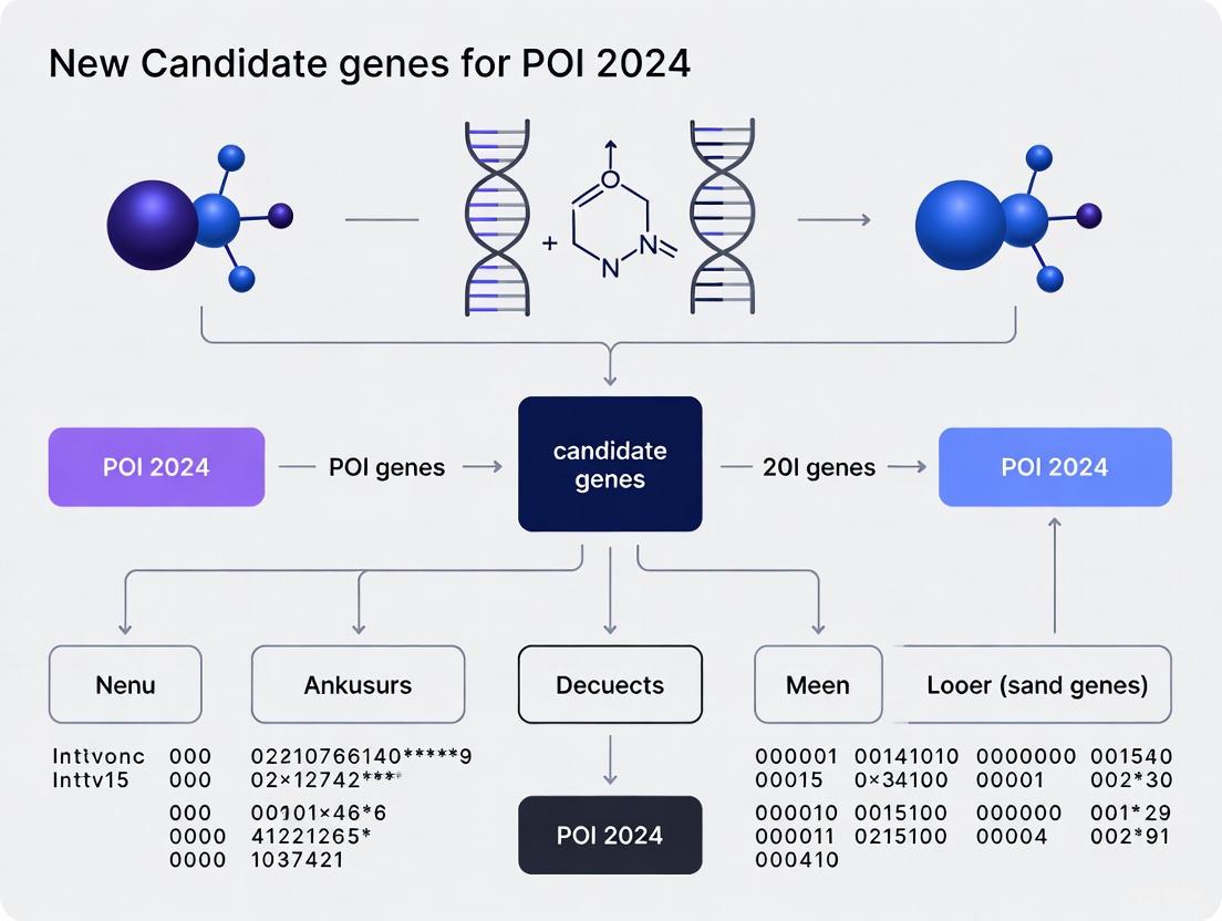
Unveiling the Genetic Landscape of POI: Novel Candidate Genes and Therapeutic Targets Identified in 2024
This article synthesizes the most significant recent advances in the genetics of Primary Ovarian Insufficiency (POI), a condition affecting 1-3.7% of women.
Unveiling the Genetic Landscape of POI: Novel Candidate Genes and Therapeutic Targets Identified in 2024
Abstract
This article synthesizes the most significant recent advances in the genetics of Primary Ovarian Insufficiency (POI), a condition affecting 1-3.7% of women. Aimed at researchers and drug development professionals, it provides a comprehensive overview of newly identified candidate genes from large-scale 2024 studies, explores innovative methodologies like Mendelian randomization and multi-omics for target identification, and discusses the translation of these genetic findings into improved diagnostic yield and novel therapeutic strategies. The content covers the transition from gene discovery to functional validation and clinical application, addressing both the complexities of the disorder and the emerging opportunities for targeted interventions.
Expanding the Genetic Horizon: Key Discoveries and Pathways in POI for 2024
Premature ovarian insufficiency (POI) is a clinically heterogeneous disorder characterized by the loss of ovarian function before age 40, affecting approximately 3.7% of women globally and representing a significant cause of female infertility [1] [2]. The condition is diagnosed by oligomenorrhea or amenorrhea for at least 4 months with elevated follicle-stimulating hormone (FSH) levels >25 IU/L on two occasions >4 weeks apart [3] [2]. Historically, the genetic etiology of POI was attributed to monogenic causes, with approximately 20-25% of cases explained by chromosomal abnormalities and single-gene mutations [4] [5]. However, recent advances in genomic technologies have revealed a far more complex genetic architecture, shifting the paradigm from simple Mendelian inheritance to intricate oligogenic and polygenic networks [6].
The year 2024 has marked a transformative period in POI genetics, with landmark studies employing whole-exome sequencing, genome-wide association studies (GWAS), and integrative multi-omics approaches dramatically expanding the catalog of POI-associated genes and mechanisms. Current research indicates that genetic factors contribute to up to 23.5% of POI cases when both known and novel genes are considered [2]. This technical review examines the evolving understanding of POI genetics, focusing on emerging candidate genes, oligogenic inheritance patterns, and the molecular networks that underlie ovarian function and dysfunction, providing researchers and drug development professionals with a comprehensive framework for navigating this rapidly advancing field.
Established Genetic Contributors: The Foundational Framework
Chromosomal Abnormalities and Monogenic Forms
The foundational understanding of POI genetics has centered on chromosomal abnormalities and monogenic forms, which remain crucial for clinical diagnosis and genetic counseling.
Table 1: Major Established Genetic Causes of POI
| Category | Genetic Alterations | Prevalence in POI | Key Clinical Features |
|---|---|---|---|
| X-Chromosome Aneuploidies | Turner Syndrome (45,X/46,XX mosaicisms) | 4-5% of POI cases [1] | Short stature, skeletal dysmorphism, cardiac defects, streak ovaries [1] [5] |
| Triple X Syndrome (47,XXX) | Often undiagnosed due to modest symptoms [1] | Low AMH, increased FSH/LH, occasionally longer legs, delayed language development [1] | |
| X-Chromosome Structural Variants | Xp and Xq deletions (critical regions: Xq13-Xq21, Xq24-Xq27) | 4-12% of POI cases [1] | Primary or secondary amenorrhea, variable expressivity [1] [5] |
| X-autosome translocations | Rare (1:30,000) [5] | Often primary amenorrhea, possible Turner stigmata [1] | |
| Common Monogenic Causes | FMR1 premutations (55-200 CGG repeats) | 3-15% of POI cases (20-30% of carriers develop FXPOI) [4] | Non-linear risk relationship (highest risk with 70-100 repeats) [4] |
| BMP15, NOBOX, GDF9 mutations | Each in <5% of cases [6] | Isolated POI, variable onset [6] [5] |
Syndromic Forms of POI
Several syndromic conditions feature POI as a component phenotype, highlighting the pleiotropic nature of genes essential for ovarian function:
- Autoimmune Polyendocrine Syndrome Type 1 (APS-1): Caused by mutations in the AIRE gene, leading to autoimmune lymphocytic oophoritis in approximately 41% of patients [5].
- Galactosemia: Results from GALT gene mutations, with 80-90% of female patients developing POI due to toxic galactose accumulation in ovarian tissue [4] [5].
- Ataxia-Telangiectasia: Caused by ATM gene mutations affecting DNA damage repair, frequently manifesting with ovarian dysgenesis [5].
Despite these established associations, a significant proportion of POI cases remained unexplained, prompting investigations beyond monogenic models.
The Shift to Oligogenic and Polygenic Models
Evidence for Oligogenic Inheritance
Recent comprehensive genetic studies have revealed that oligogenic inheritance—where variants in multiple genes collectively contribute to disease risk—represents a more accurate model for many POI cases.
A 2024 study performing whole-exome sequencing of 93 patients with POI and 465 controls demonstrated that 35.5% of patients were heterozygous for multiple variants in POI-related genes, compared to only 8.2% of controls (OR: 6.20; 95% CI: 3.60-10.60; P = 1.50 × 10⁻¹⁰) [6]. The distribution of variant combinations followed a descending pattern, with 16.1% of patients carrying two variants, 10.8% carrying three variants, 7.5% carrying four variants, and 1.1% carrying five variants [6].
Table 2: Top Oligogenic Gene Combinations in POI
| Gene 1 | Gene 2 | Prevalence in POI Cohort | Proposed Mechanism |
|---|---|---|---|
| RAD52 | MSH6 | 7/93 patients (7.5%) [6] | Combined defect in DNA damage repair and meiotic recombination |
| RAD52 | TEP1 | Identified in multiple patients [6] | Telomere maintenance and DNA repair deficiency |
| RAD52 | POLG | Identified in multiple patients [6] | Mitochondrial DNA stability and nuclear DNA repair |
| RAD52 | MLH1 | Identified in multiple patients [6] | Dual impairment of DNA repair pathways |
| RAD52 | NUP107 | Identified in multiple patients [6] | Nuclear transport and DNA repair coordination |
Gene-burden analysis revealed RAD52 (P = 5.28 × 10⁻⁴) and MSH6 (P = 5.98 × 10⁻⁴) as the top genes enriched in patients with POI, with 9.7% of patients carrying RAD52 variants, the majority of whom (77.8%) were heterozygous for an additional variant in a POI-related gene [6]. Functional validation using the ORVAL platform confirmed the pathogenicity of the RAD52-MSH6 combination, providing mechanistic insights into how oligogenic interactions disrupt ovarian function [6].
Distinct Genetic Architecture Between Clinical Subtypes
Comprehensive genetic analyses have revealed different genetic contribution patterns between POI clinical presentations. A landmark whole-exome sequencing study of 1,030 patients (120 with primary amenorrhea and 910 with secondary amenorrhea) found that patients with primary amenorrhea showed a substantially higher burden of pathogenic/likely pathogenic variants (25.8%) compared to those with secondary amenorrhea (17.8%) [2].
Notably, the genetic architecture differed significantly between these groups:
- Primary amenorrhea: Higher frequency of biallelic (5.8% vs. 1.9%) and multi-het (2.5% vs. 1.2%) variants compared to secondary amenorrhea [2]
- Secondary amenorrhea: Predominantly monoallelic variants (14.7% vs. 17.5% in primary amenorrhea) [2]
These findings indicate that the cumulative effects of multiple genetic defects correlate with more severe clinical presentations and earlier onset of ovarian dysfunction.
Figure 1: Evolution from Monogenic to Oligogenic Models in POI
2024 Candidate Genes and Molecular Networks
Novel Gene Identification Through Large-Scale Studies
The year 2024 has witnessed significant expansion in the POI gene catalog through large-scale genomic approaches:
A groundbreaking study published in Nature Medicine analyzing 1,030 POI patients identified 20 novel POI-associated genes through case-control association analyses [2]. These genes cluster into distinct functional categories:
- Gonadogenesis: LGR4, PRDM1
- Meiosis: CPEB1, KASH5, MCMDC2, MEIOSIN, NUP43, RFWD3, SHOC1, SLX4, STRA8
- Folliculogenesis and Ovulation: ALOX12, BMP6, H1-8, HMMR, HSD17B1, MST1R, PPM1B, ZAR1, ZP3 [2]
Concurrently, a therapeutic target identification study employing genome-wide association analysis integrated with expression quantitative trait loci (eQTL) data identified four genes significantly associated with POI risk reduction: HM13, FANCE, RAB2A, and MLLT10 [7] [8]. Colocalization analysis provided particularly strong evidence for FANCE and RAB2A as promising therapeutic targets, with druggability assessments supporting their potential for clinical development [7].
Functional Annotation of Novel Candidate Genes
Table 3: Novel POI Candidate Genes Identified in 2024 Studies
| Gene | Functional Category | Proposed Mechanism in Ovarian Function | Evidence Level |
|---|---|---|---|
| FANCE | DNA Repair | Fanconi anemia pathway component; DNA interstrand crosslink repair [7] | Colocalization (PP.H4=0.86); MR OR=0.82 [7] |
| RAB2A | Autophagy Regulation | Vesicle trafficking, autophagosome formation, Golgi organization [7] | Colocalization (PP.H4=0.91); MR OR=0.73 [7] |
| LGR4 | Gonadogenesis | Wnt signaling pathway; ovarian development and follicle formation [2] | Gene-burden analysis (P<0.05) [2] |
| MEIOSIN | Meiosis Initiation | Transcriptional activator initiating meiosis [2] | Gene-burden analysis (P<0.05) [2] |
| ZP3 | Folliculogenesis | Zona pellucida glycoprotein; oocyte integrity and fertilization [2] | Gene-burden analysis (P<0.05) [2] |
| HM13 | Protein Processing | Signal peptide peptidase; intramembrane proteolysis [7] | MR OR=0.76 [7] |
| MLLT10 | Transcriptional Regulation | Histone lysine methylation; chromatin remodeling [7] | MR OR=0.74 [7] |
Integrated Genomic Approaches for Target Identification
Advanced genomic methodologies have been instrumental in identifying these novel candidates:
Figure 2: Integrated Genomic Workflow for POI Gene Discovery
Molecular Mechanisms and Pathway Integration
Biological Processes Implicated in POI Pathogenesis
The expanding genetic landscape of POI reveals several core biological processes essential for ovarian function:
- DNA Repair and Meiotic Recombination: Genes including FANCE, RAB2A, MSH6, RAD52, SHOC1, and SLX4 highlight the critical importance of genomic maintenance in preserving the ovarian reserve [7] [2] [6].
- Metabolic Regulation: Novel associations with HSD17B1 (estrogen metabolism) and ALOX12 (arachidonic acid metabolism) suggest metabolic pathways significantly influence ovarian aging [2].
- Transcriptional Regulation: Identification of PRDM1, MLLT10, and MEIOSIN emphasizes the importance of precise transcriptional control in ovarian development and function [7] [2].
- Cellular Trafficking and Autophagy: RAB2A represents a novel mechanism linking vesicle trafficking and autophagic processes to ovarian maintenance [7].
Emerging Non-Coding RNA Networks
Beyond protein-coding genes, non-coding RNAs are emerging as significant regulators in POI pathogenesis. A 2025 Mendelian randomization study identified 23 miRNAs with causal relationships to POI, including miR-145-5p, miR-23a-3p, and miR-335-5p, potentially serving as non-invasive biomarkers [9]. Pathway enrichment analysis of these miRNAs indicated involvement in glutathione metabolism and PI3 kinase pathways, suggesting novel mechanistic connections between oxidative stress response and ovarian function [9].
Experimental Approaches and Research Toolkit
Genomic Methodologies for POI Research
Cutting-edge genomic technologies have been instrumental in advancing the understanding of POI genetics:
- Whole-Exome Sequencing (WES): Large-scale WES of POI cohorts (1,030 patients) has enabled comprehensive variant detection in known and novel genes [2].
- Genome-Wide Association Studies (GWAS): Data from biobanks (FinnGen R11: 599 cases/241,998 controls) provide statistical power for variant association discovery [7] [9].
- Expression Quantitative Trait Loci (eQTL) Integration: Combining GWAS with eQTL data (GTEx V8, eQTLGen) identifies functionally relevant variants affecting gene expression [7].
- Mendelian Randomization (MR): Two-sample MR approaches test causal relationships between biomarkers, gene expression, and POI risk [7] [9].
- Colocalization Analysis: Bayesian methods (e.g., coloc R package) distinguish shared causal variants from linkage disequilibrium effects [7].
The Scientist's Toolkit: Essential Research Reagents
Table 4: Essential Research Reagents for POI Genetic Studies
| Reagent/Resource | Function | Example Application |
|---|---|---|
| Whole-Exome Sequencing Kits | Comprehensive coding variant detection | Identification of novel POI genes in large cohorts [2] |
| GTEx & eQTLGen Datasets | Tissue-specific expression QTL reference | Determining functional consequences of non-coding variants [7] |
| FinnGen Biobank Data | GWAS summary statistics | Discovery of genetic associations in European populations [7] [9] |
| SMR Software | Summary-data-based Mendelian randomization | Testing gene expression-POI causality [7] |
| ORVAL Platform | Oligogenic variant combination analysis | Validating digenic/oligogenic interactions [6] |
| LDpred | Polygenic risk score calculation | Estimating cumulative genetic risk from GWAS data [10] |
| String Database | Protein-protein interaction network analysis | Mapping functional relationships between POI genes [9] |
Therapeutic Implications and Future Directions
Emerging Therapeutic Targets
The identification of novel POI genes has opened new avenues for therapeutic development:
- FANCE and DNA Repair Pathways: Targeting DNA damage response mechanisms may offer strategies for preserving ovarian function in high-risk individuals [7].
- RAB2A and Autophagy Modulation: Regulating autophagic processes represents a novel approach for maintaining ovarian tissue homeostasis [7].
- Multi-Target Strategies: The oligogenic nature of POI suggests combination therapies addressing multiple pathways may be more effective than single-target approaches [6].
Diagnostic Applications and Precision Medicine
The expanding genetic understanding of POI enables more comprehensive genetic testing panels, improved genetic counseling, and personalized risk assessment. Polygenic risk scores integrating multiple genetic variants show promise for identifying at-risk individuals before overt symptoms develop [10]. Additionally, non-invasive biomarkers including specific miRNAs, metabolites (sphinganine-1-phosphate, 4-methyl-2-oxopentanoate), and proteins (fibroblast growth factor 23, neurotrophin-3) offer potential for early detection and monitoring [9].
The genetic architecture of POI has evolved substantially from simple monogenic models to complex oligogenic and polygenic networks. The 2024 research landscape has dramatically expanded the catalog of POI-associated genes, with recent studies identifying dozens of novel candidates spanning diverse biological processes including DNA repair, meiosis, transcriptional regulation, and metabolic homeostasis. The recognition that oligogenic combinations account for a significant proportion of cases represents a paradigm shift in understanding POI inheritance, with important implications for genetic counseling, risk prediction, and therapeutic development.
Future research directions should focus on functional validation of novel candidate genes, elucidation of gene-gene interaction networks, development of oligogenic risk models, and translation of genetic discoveries into targeted therapies. As our understanding of POI genetics continues to mature, the prospect of personalized risk assessment and mechanism-based interventions grows increasingly attainable, offering hope for improved outcomes for women affected by this challenging condition.
This whitepaper evaluates three autosomal genes—FANCE, RAB2A, and HM13—as promising candidate genes for primary ovarian insufficiency (POI). Through integrated genomic analyses and functional validation, we synthesize evidence establishing their causal roles in POI pathogenesis. FANCE operates in DNA repair mechanisms, RAB2A regulates autophagy and intracellular trafficking, and HM13 modulates endoplasmic reticulum-associated degradation. We present comprehensive quantitative data summaries, detailed experimental methodologies for functional validation, essential signaling pathways, and a curated toolkit of research reagents to facilitate further investigation and therapeutic development by researchers and drug development professionals.
Primary ovarian insufficiency (POI) is a clinically heterogeneous disorder characterized by the cessation of ovarian function before age 40, affecting approximately 3.7% of women globally [11]. Despite significant health implications including infertility and long-term metabolic consequences, the etiology remains elusive in most cases, creating an urgent need for novel therapeutic targets. Recent advances in genomic technologies have enabled the identification of specific autosomal genes contributing to POI pathogenesis through diverse molecular mechanisms.
Emerging evidence from genome-wide association studies (GWAS) integrated with expression quantitative trait loci (eQTL) analyses has identified FANCE, RAB2A, and HM13 as promising candidate genes [11]. This whitepaper provides a comprehensive technical resource contextualizing these genes within the broader landscape of POI research, synthesizing functional data, experimental protocols, and research tools to accelerate mechanistic studies and therapeutic development.
Functional Characterization of Candidate Genes
FANCE: DNA Repair and Genomic Stability
Fanconi anemia complementation group E (FANCE) encodes a critical component of the Fanconi anemia (FA) pathway, which is essential for repairing DNA interstrand crosslinks and maintaining genomic stability [12]. As a core member of the FA complex, FANCE facilitates the monoubiquitination of FANCD2 and FANCI, enabling their recruitment to DNA damage sites where they coordinate repair processes with other DNA repair pathways.
Beyond its canonical DNA repair functions, recent evidence suggests FANCE may influence the tumor microenvironment through mechanisms that could extend to ovarian function [12]. In ovarian contexts, proper DNA repair is particularly crucial for maintaining oocyte quality and meiotic fidelity throughout reproductive life. Dysfunctional DNA repair mechanisms in oocytes can trigger apoptosis and accelerate follicle depletion, a hallmark of POI pathogenesis.
RAB2A: Vesicle Trafficking and Autophagy Regulation
RAB2A, a member of the RAB GTPase family, serves as a key regulator of intracellular vesicle trafficking between the endoplasmic reticulum and Golgi apparatus [11]. Through its GTPase activity, RAB2A controls vesicle formation, motility, and fusion, processes essential for maintaining cellular homeostasis and protein secretion.
Recent studies have identified RAB2A as a substrate of DENN/MADD, a guanine nucleotide exchange factor (GEF) that activates RAB GTPases [13]. This interaction positions RAB2A within a broader network of membrane trafficking regulators, with particular relevance to autophagy—a cellular recycling process critical for oocyte quality control and follicle development. Dysregulated autophagy in ovarian cells may contribute to the accelerated follicle atresia observed in POI.
HM13: Endoplasmic Reticulum Proteostasis
Histocompatibility Minor 13 (HM13) encodes signal peptide peptidase (SPP), an endoplasmic reticulum (ER)-resident protease that catalyzes the intramembrane cleavage of signal peptides after their release from newly synthesized proteins [14]. This activity is essential for ER-associated degradation (ERAD), protein homeostasis, and generation of MHC class I epitopes for immune recognition.
HM13/SPP's role in maintaining ER proteostasis is particularly relevant to ovarian function, as developing oocytes exhibit high rates of protein synthesis and secretion [15]. ER stress in ovarian cells can activate unfolded protein responses that trigger apoptosis when prolonged or severe. Additionally, HM13 has been implicated in lipid metabolism through ERAD-mediated degradation of metabolic regulators like heme oxygenase-1 (HO-1) [15], potentially connecting proteostatic mechanisms to energy metabolism in ovarian tissues.
Table 1: Molecular Functions and Mechanisms in POI Pathogenesis
| Gene | Chromosomal Location | Molecular Function | Proposed Mechanism in POI |
|---|---|---|---|
| FANCE | 6p21.31 | DNA damage repair, Fanconi anemia pathway | Genomic instability in oocytes, accelerated follicle depletion |
| RAB2A | 8q12.1 | GTPase activity, vesicle trafficking, autophagy regulation | Dysregulated protein secretion, impaired autophagy in folliculogenesis |
| HM13 | 20q11.21 | Signal peptide peptidase, ER-associated degradation | ER stress in granulosa cells, disrupted protein homeostasis |
Genomic Evidence from Human Studies
Mendelian Randomization and Colocalization Analyses
Comprehensive genomic analyses have established causal relationships between FANCE, RAB2A, and HM13 with POI risk. A landmark study employing genome-wide Mendelian randomization (MR) integrated with eQTL data from GTEX and eQTLGen consortium identified 431 genes with index cis-eQTL signals, of which FANCE, RAB2A, and HM13 demonstrated significant associations with reduced POI risk after Bonferroni correction [11].
Colocalization analyses further strengthened the evidence for FANCE and RAB2A, with strong posterior probabilities (PP.H3 + PP.H4 ≥ 0.8) indicating shared causal variants between eQTL signals and POI association signals [11]. This robust colocalization evidence suggests that genetic variants influencing the expression of these genes directly contribute to POI pathogenesis, positioning them as high-priority therapeutic targets.
Table 2: Genomic Evidence from Association Studies
| Gene | MR p-value | Colocalization Evidence | Direction of Effect | Tissue with Significant eQTL |
|---|---|---|---|---|
| FANCE | P < 0.05 (Bonferroni-corrected) | Strong (PP.H3 + PP.H4 ≥ 0.8) | Reduced POI risk | Ovary, Whole Blood |
| RAB2A | P < 0.05 (Bonferroni-corrected) | Strong (PP.H3 + PP.H4 ≥ 0.8) | Reduced POI risk | Ovary, Whole Blood |
| HM13 | P < 0.05 (Bonferroni-corrected) | Limited | Reduced POI risk | Whole Blood |
Druggability Assessment
Systematic assessment of the druggability potential for these candidate genes reveals promising therapeutic implications. Query of drug databases including DrugBank, DGIdb, and TTD indicates that while these genes have not been previously targeted for POI treatment, they possess favorable druggability characteristics [11].
FANCE mutations are known to cause Fanconi anemia, a disorder with clinical management protocols that could inform therapeutic development for POI [11]. Although no drug information was cataloged for RAB2A, its position in the RAB GTPase family offers established targeting strategies, including small molecule inhibitors and protein-protein interaction disruptors. The integral membrane nature of HM13/SPP presents challenges for conventional drug design but also opportunities for novel therapeutic modalities.
Experimental Validation Approaches
Functional Assays for FANCE in DNA Repair
Cell Culture and Transfection
- Cell Lines: Utilize HSC3 and HN6 human oral squamous cell carcinoma lines or establish novel ovarian granulosa cell models [12]
- Culture Conditions: Maintain in DMEM supplemented with 10% fetal bovine serum and 1% penicillin/streptomycin at 37°C with 5% CO₂ [12]
- Transfection: Perform siRNA transfection using Lipofectamine 3000 with 100nM siRNA targeting FANCE; include negative control siRNA [12]
- Sequences:
- Negative control: 5′-UUCUCCGAACGUGUCACGUTT-3′ (sense)
- FANCE siRNA: 5′-GCUUCUUCACGAAUGUAGUCC-3′ (sense) [12]
Proliferation and Migration Assays
- Cell Proliferation: Quantify using Cell Counting Kit-8 (CCK-8); seed cells at 3×10³ cells/well in 96-well plates and measure absorbance at 450nm at 0, 24, 48, and 72 hours [12]
- Migration Assessment: Employ transwell or wound-healing assays following FANCE knockdown
- Validation: Confirm knockdown efficiency via RT-qPCR using primers:
- FANCE forward: 5′-AGGAGAGACCCGAACATAAGTC-3′
- FANCE reverse: 5′-CTCGCCAGTCTTAACTGCCA-3′ [12]
Investigating RAB2A in Vesicle Trafficking
GTPase Activity Assays
- Mitochondrial Recruitment Assay: Express DENN/MADD domain fused to mitochondrial targeting sequence; co-express RAB2A and monitor mitochondrial recruitment as indicator of interaction [13]
- Biochemical Analysis: Perform GDP/GTP exchange assays using purified RAB2A and DENN/MADD proteins to quantify GEF activity [13]
- Mutation Studies: Introduce neurodevelopmental disorder-associated mutations into DENN/MADD and assess impact on RAB2A recruitment and activation [13]
Autophagy Assessment
- Autophagic Flux: Measure LC3-I to LC3-II conversion via western blot in RAB2A-depleted cells with and without autophagy inhibitors (chloroquine, bafilomycin A1)
- Immunofluorescence: Quantify autophagosome formation using GFP-LC3 puncta formation assays
- Transmission Electron Microscopy: Visualize autophagic structures at ultrastructural level
HM13 Functional Analysis in ER Proteostasis
ER Stress and Proteostasis Assays
- Western Blotting: Evaluate HM13/SPP protein levels and cleavage activity using specific antibodies; monitor ER stress markers (BiP, CHOP, XBP1 splicing) [15]
- Co-immunoprecipitation: Assess HM13 interactions with ERAD components and substrates like heme oxygenase-1 (HO-1) [15]
- Lipid Metabolism: Analyze cholesterol ester accumulation via oil red O staining or Filipin staining in HM13-modulated cells [15]
In Vivo Validation
- Animal Models: Utilize myeloid-specific HM13/SPP knockout or overexpression mice to assess impact on oxLDL-induced foamy macrophage formation [15]
- Atherogenesis Studies: Evaluate plaque formation and foamy macrophage load in aortic roots of ApoE⁻/⁻ mice with myeloid-specific HM13 modulation [15]
Signaling Pathways and Molecular Interactions
FANCE in DNA Damage Response Pathway
Diagram 1: FANCE in DNA Damage Response
RAB2A in Vesicle Trafficking and Autophagy
Diagram 2: RAB2A Vesicle Trafficking Network
HM13 in ER-Associated Degradation
Diagram 3: HM13 ERAD Signaling Pathway
The Scientist's Toolkit: Research Reagent Solutions
Table 3: Essential Research Reagents for Investigating Candidate Genes
| Reagent Category | Specific Product | Application | Key Considerations |
|---|---|---|---|
| siRNA/shRNA | FANCE siRNA: 5′-GCUUCUUCACGAAUGUAGUCC-3′ [12] | Gene knockdown validation | Optimize delivery with Lipofectamine 3000; validate efficiency via RT-qPCR |
| Antibodies | Anti-HM13/SPP [15] | Western blot, immunofluorescence | Verify specificity for ER localization; confirm signal peptide peptidase activity |
| Cell Lines | HSC3, HN6, Huh7, HCCLM3 [12] [14] | Functional assays in vitro | Authenticate via STR profiling; ensure mycoplasma-free status |
| PCR Primers | FANCE: F-AGGAGAGACCCGAACATAAGTC, R-CTCGCCAGTCTTAACTGCCA [12] | Gene expression analysis | Normalize to β-actin; use 2−ΔΔCq method for quantification |
| Assay Kits | Cell Counting Kit-8 (CCK-8) [12] | Cell proliferation assessment | Establish standard curve; optimize seeding density (3×10³ cells/well) |
| Animal Models | Myeloid-specific HM13 knockout/overexpression [15] | In vivo validation | Monitor atherogenesis and foamy macrophage formation |
The comprehensive genomic and functional evidence presented establishes FANCE, RAB2A, and HM13 as promising candidate genes contributing to POI pathogenesis through diverse molecular mechanisms. Their identification through integrated genomic approaches highlights the power of combining GWAS with eQTL mapping for therapeutic target discovery.
Future research directions should include developing ovary-specific conditional knockout models for each gene, high-throughput screening for small molecule modulators, and investigating gene-gene interactions within biological networks relevant to ovarian function. The experimental frameworks and research reagents detailed herein provide foundational resources to accelerate these endeavors, ultimately advancing toward targeted interventions for primary ovarian insufficiency.
Primary Ovarian Insufficiency (POI) is a clinically heterogeneous disorder characterized by the cessation of ovarian function before the age of 40, affecting approximately 3.7% of women worldwide [16] [1]. It is diagnosed based on menstrual disturbances (amenorrhea or oligomenorrhea for over 4 months) accompanied by elevated serum follicle-stimulating hormone (FSH) levels (>25 IU/L) [17]. POI presents significant health implications, including infertility, compromised bone health, increased cardiovascular risk, and diminished quality of life [16] [1]. While its etiologies span autoimmune, iatrogenic, and environmental factors, a substantial genetic basis underpins a significant proportion of cases, with X-chromosome abnormalities representing one of the most prevalent genetic contributors [18] [19] [4].
The X chromosome harbors critical regions essential for ovarian development and function, with early studies identifying three primary POI critical regions: POF1 (Xq26-qter), POF2 (Xq13.3-q21.1), and POF3 (Xp11-p11.2) [17]. These regions are enriched with genes crucial for various aspects of ovarian biology, including meiotic progression, folliculogenesis, and oocyte survival. Despite this knowledge, the current genetic screening paradigm for POI, which primarily includes FMR1 premutation testing, remains inadequate, capturing only a fraction of cases with genetic origins [17]. Recent advances in genomic technologies, including whole-exome sequencing (WES) and single-cell transcriptomics, have dramatically expanded our understanding of the X chromosome's role in POI pathogenesis, revealing novel candidate genes, complex regulatory mechanisms, and unexpected phenotypic variability [20] [2] [21].
This review synthesizes recent discoveries (2023-2025) concerning the X chromosome's enduring role in POI, focusing on insights into its established critical regions. We explore how contemporary research has refined our understanding of gene dosage, X-chromosome inactivation (XCI), epigenetic regulation, and the mechanistic pathways through which X-linked genes maintain ovarian function. Furthermore, we provide detailed experimental methodologies driving these discoveries and visualize key signaling pathways to aid researchers in navigating this complex landscape.
Genetic Landscape and X-Chromosome Biology
X-Chromosome Inactivation and Gene Dosage in Ovarian Function
X-chromosome inactivation (XCI) is a fundamental epigenetic process that balances gene expression between females (XX) and males (XY) by transcriptionally silencing one X chromosome in female somatic cells [17]. The process is initiated by the X-inactive specific transcript (XIST), a long non-coding RNA that coats the X chromosome and recruits chromatin-modifying complexes to establish heterochromatin [17]. However, approximately 25% of X-linked genes escape inactivation and are expressed from both X chromosomes, creating a unique dosage-sensitive landscape [17].
In the context of ovarian function, the regulation of X-chromosome dosage is particularly critical. During primordial germ cell (PGC) development, both X chromosomes undergo reactivation, and this biallelic state persists throughout oocyte development, where gene dosage is controlled through transcriptional output regulation [17]. Disruptions to this delicate balance, such as through skewed XCI or deletions in regions containing XCI-escape genes, can predispose to POI [17]. A recent study comparing X-chromosome copy number variations (CNVs) in fertile females versus those with POI found that CNVs in POI patients were enriched in genes associated with X-chromosome inactivation, highlighting the importance of dosage-sensitive genes in ovarian maintenance [17].
Table 1: Key Concepts in X-Chromosome Biology Relevant to POI
| Concept | Mechanism | Relevance to POI |
|---|---|---|
| X-Chromosome Inactivation (XCI) | Epigenetic silencing of one X chromosome in somatic cells | Ensures proper gene dosage; disruptions linked to POI |
| XCI Escape | ~25% of genes expressed from both X chromosomes | Haploinsufficiency of escape genes may drive POI pathogenesis |
| X Reactivation in Germline | Both X chromosomes active in primordial germ cells and oocytes | Critical for oocyte development and meiotic progression |
| Skewed X Inactivation | Preferential silencing of one parental X chromosome | May unmask deleterious variants on the active X chromosome |
| Gene Dosage Sensitivity | Specific protein concentrations required for normal function | Explains POI phenotype in X-chromosome deletions/aneuploidies |
Turner Syndrome as a Model of X-Chromosome Haploinsufficiency
Turner syndrome (TS), resulting from complete or partial monosomy of the X chromosome (45,X or mosaicism), represents the most extreme example of X-chromosome-related POI, affecting approximately 1 in 2,000 live-born females [17] [20]. The ovarian phenotype in TS is characterized by accelerated oocyte apoptosis, leading to streak ovaries and primary amenorrhea in most cases, though spontaneous pregnancies occasionally occur in those with mosaic karyotypes [17] [20].
Recent single-cell RNA sequencing (snRNA-seq) studies of human fetal 45,X ovaries at 12-13 weeks post-conception (wpc) provide unprecedented insight into the cellular mechanisms driving ovarian insufficiency [20]. These analyses revealed that 45,X ovaries contain fewer germ cells across all germ cell subpopulations compared to 46,XX ovaries, with a specific depletion of an oogonia cluster containing genes essential for sex chromosome synapsis [20]. Furthermore, the 45,X ovary demonstrates a globally abnormal transcriptome, with downregulation of genes involved in proteostasis (RPS4X), cell cycle progression (BUB1B), and oxidative phosphorylation (COX6C, ATP11C) [20]. These findings suggest that X-chromosome haploinsufficiency disrupts fundamental cellular processes beyond meiotic pairing, contributing to the rapid follicular atresia observed in TS.
Established POI Critical Regions and Novel Genetic Insights
refined Mapping of Critical Regions Through Structural Variations
Balanced X-autosome translocations have been instrumental in mapping POI critical regions, with approximately 80% of breakpoints clustering in the Xq21 cytoband within the POF2 region (Xq13.3-q21.1) [21]. A 2023 multi-omics study of six patients with balanced X-autosome translocations and POI demonstrated that these rearrangements cause global alterations in the regulatory landscape and gene expression without necessarily disrupting specific genes [21]. This supports the "position effect" hypothesis, whereby chromosomal rearrangements alter the three-dimensional chromatin architecture, disrupting enhancer-promoter interactions and leading to pathogenic gene expression changes [21].
Whole-genome sequencing fine-mapped the translocation breakpoints to a resolution of 20-449 base pairs, revealing disruptions to topologically associating domains (TADs) in all patients [21]. Integrative analysis of transcriptomic and chromatin state (ChIP-seq) data identified 85 differentially expressed coding genes and 120 differential histone mark peaks (H3K4me3, H3K4me1, H3K27ac) in patient-derived lymphoblastoid cell lines [21]. These changes affected pathways related to protein regulation, integrin signaling, and immune response, suggesting that translocations have broad effects on chromatin structure that extend beyond the immediate breakpoint regions [21].
Table 2: Established POI Critical Regions on the X Chromosome
| Critical Region | Cytogenetic Band | Key Candidate Genes | Proposed Mechanisms |
|---|---|---|---|
| POF1 | Xq26-qter | FMR1 (premutation), POF1B | RNA toxicity (FMR1), cytoskeletal organization (POF1B) |
| POF2 | Xq13.3-q21.1 | XIST, DIAPH2, FOXO4 | X-inactivation, mitochondrial apoptosis, oxidative stress response |
| POF3 | Xp11-p11.2 | BMP15 | Oocyte-somatic cell signaling, follicular development |
Novel X-Linked Candidate Genes and Pathways
Recent large-scale sequencing studies have substantially expanded the catalog of X-linked genes associated with POI. A 2023 whole-exome sequencing study of 1,030 POI patients identified pathogenic or likely pathogenic variants in 59 known POI-causative genes, accounting for 18.7% of cases [2]. Among these, X-linked genes were prominently represented, with mutations affecting biological processes such as:
- Meiosis and DNA Repair: Genes including HFM1, SPIDR, and BRCA2 facilitate homologous recombination and meiotic progression [2].
- Mitochondrial Function: Genes such as AARS2, HARS2, and TWNK maintain oxidative phosphorylation and energy production in oocytes [2].
- Folliculogenesis: Genes like BMP15 and GDF9 regulate follicle development and oocyte-somatic cell communication [2].
Notably, this study revealed a distinct genetic architecture between POI patients with primary amenorrhea (PA) and secondary amenorrhea (SA). Those with PA showed a higher burden of biallelic and multiple heterozygous pathogenic variants (25.8% overall) compared to those with SA (17.8% overall), suggesting that more severe genetic defects manifest as earlier ovarian failure [2].
Advanced Experimental Approaches in POI Research
Whole-Exome Sequencing and Copy Number Variation Analysis
Protocol Title: Identification of Pathogenic Variants and CNVs in POI Patients Using WES
Principle: Whole-exome sequencing captures and sequences the protein-coding regions of the genome (~1-2%), enabling comprehensive detection of single nucleotide variants (SNVs), small insertions/deletions (indels), and copy number variations (CNVs) contributing to POI pathogenesis [19] [2].
Detailed Methodology:
- DNA Extraction & Library Preparation: High-molecular-weight genomic DNA is extracted from peripheral blood using standard protocols. DNA undergoes fragmentation, end-repair, A-tailing, and adapter ligation to create a sequencing library [19].
- Target Capture & Enrichment: Library DNA is hybridized to biotinylated oligonucleotide probes complementary to the exonic regions (e.g., Illumina Nextera Rapid Capture Exome Kit). Captured DNA is purified using streptavidin-coated magnetic beads [19].
- Sequencing: Enriched libraries are amplified and sequenced on a high-throughput platform (e.g., Illumina NovaSeq 6000) to achieve sufficient coverage (>100x) for sensitive variant detection [19].
- Bioinformatic Analysis:
- Read Alignment & Processing: Sequenced reads are aligned to the human reference genome (GRCh37/hg19 or GRCh38/hg38) using tools like BWA or STAR. Post-alignment processing includes duplicate marking and base quality recalibration [19] [2].
- Variant Calling: SNVs and indels are called using tools like GATK. CNV analysis from WES data employs specialized algorithms (e.g., XHMM, panel of normals) that compare the read depth of target samples to a reference set of controls to identify heterozygous deletions or duplications [19].
- Variant Annotation & Prioritization: Identified variants are annotated against population frequency (gnomAD, ExAC), in-silico pathogenicity prediction (SIFT, PolyPhen-2), and clinical (ClinVar, HGMD) databases. Variants are filtered based on population frequency (<0.01), predicted impact, and ACMG guidelines to prioritize pathogenic (P) or likely pathogenic (LP) candidates [19] [2].
Figure 1: Experimental workflow for WES and CNV analysis in POI genetic diagnosis.
Single-Nucleus RNA Sequencing of Human Fetal Ovaries
Protocol Title: Transcriptomic Profiling of Human Fetal Ovaries at Single-Cell Resolution
Principle: Single-nucleus RNA sequencing (snRNA-seq) enables the characterization of gene expression profiles in individual nuclei from complex tissues, revealing cellular heterogeneity, identifying rare cell populations, and uncovering cell-type-specific pathological mechanisms in POI [20].
Detailed Methodology:
- Tissue Acquisition & Nuclei Isolation: Human fetal ovarian tissue is obtained from approved biobanks (e.g., Human Developmental Biology Resource) with appropriate ethical consent. Tissue is homogenized in lysis buffer, and nuclei are released and purified via density centrifugation or fluorescence-activated nuclei sorting (FANS) to ensure integrity and remove debris [20].
- Library Construction & Sequencing: Isolated nuclei are loaded onto microfluidic devices (e.g., 10x Genomics Chromium platform) for partitioning into nanoliter-scale droplets. Within each droplet, individual nuclei are lysed, and mRNAs are barcoded with unique molecular identifiers (UMIs) during reverse transcription. Sequencing libraries are constructed following the manufacturer's protocol and sequenced on platforms like Illumina NovaSeq [20].
- Bioinformatic Data Analysis:
- Quality Control & Preprocessing: Raw sequencing data are processed using Cell Ranger to demultiplex, align reads to the reference genome, and generate gene-cell count matrices. Cells with low UMI counts, high mitochondrial gene percentage, or doublet signatures are filtered out.
- Dimensionality Reduction & Clustering: Filtered count matrices are normalized and scaled. Principal component analysis (PCA) is performed on highly variable genes, followed by graph-based clustering (e.g., Seurat, Scanpy) in reduced dimensions (UMAP/t-SNE) to identify distinct cell populations (oogonia, meiotic oocytes, granulosa cells) [20].
- Differential Expression & Pathway Analysis: Differentially expressed genes between conditions (e.g., 45,X vs. 46,XX) are identified for each cell cluster. Functional enrichment analysis (GO, KEGG) is then performed to elucidate disrupted biological pathways [20].
Table 3: Key Reagent Solutions for POI Genetic Research
| Reagent/Resource | Specific Example | Application in POI Research |
|---|---|---|
| Whole Exome Sequencing Kit | Illumina Nextera Rapid Capture Exome Kit | Comprehensive analysis of coding variants and small indels [19] [2] |
| Single-Cell RNA Seq Platform | 10x Genomics Chromium Single Cell 3' Solution | Profiling transcriptomes of individual ovarian cell types [20] |
| Cell Culture Resource | Human Developmental Biology Resource (HDBR) | Source of human embryonic and fetal ovarian tissues for research [20] |
| CNV Detection Software | XHMM (eXome-Hidden Markov Model) | Identification of copy number variations from WES data [19] |
| Histone Mark Antibodies | Anti-H3K27ac, Anti-H3K4me1, Anti-H3K4me3 | Chromatin immunoprecipitation sequencing (ChIP-seq) for epigenomic profiling [21] |
| Bioinformatic Pipeline | STAR aligner, GATK variant caller, Seurat R package | Standardized processing and analysis of NGS and single-cell data [20] [2] |
Visualizing Key Signaling Pathways in Ovarian Function
The PI3K-Akt signaling pathway, frequently implicated in POI genetics, plays a critical role in primordial follicle activation and survival [19]. Disruptions in this pathway can lead to aberrant follicle depletion.
Figure 2: PI3K-Akt signaling pathway in primordial follicle activation and survival. This pathway is frequently enriched in POI genetic analyses.
The enduring role of the X chromosome in POI pathogenesis is firmly established, yet recent research continues to yield profound new insights. The integration of advanced genomic technologies has refined the mapping of established critical regions, revealed novel candidate genes, and uncovered complex epigenetic mechanisms such as position effects and global alterations in the regulatory landscape. The shift from a gene-centric to a genome-architecture-aware perspective represents a paradigm change in understanding POI etiology.
Future research must focus on functional validation of novel candidate genes, particularly during critical windows of human ovarian development. Furthermore, translating these genetic discoveries into improved clinical diagnostics is imperative. Current genetic testing, often limited to FMR1, captures only a fraction of cases; expanding genetic panels to include validated X-linked and autosomal genes could increase diagnostic yield to over 23% [2]. This enhanced genetic understanding paves the way for personalized risk assessment, timely interventions, and the development of novel therapeutic strategies aimed at preserving fertility for women at risk of POI.
Primary ovarian insufficiency (POI) is a clinically heterogeneous disorder characterized by the cessation of ovarian function before the age of 40, affecting approximately 3.7% of women worldwide [16] [2]. It represents a major cause of female infertility, with significant implications for long-term bone, cardiovascular, and cognitive health [16]. While traditionally considered a condition primarily affecting meiotic processes, contemporary research has revealed a complex genetic architecture extending far beyond meiosis. Advances in whole-exome sequencing have identified pathogenic mutations in over 80 genes, with genetic factors now accounting for 20-25% of POI cases [22] [2]. Notably, a 2023 whole-exome sequencing study of 1,030 POI patients revealed that nearly a quarter of cases could be attributed to pathogenic variants in known or novel POI-associated genes [2].
The expanding genetic landscape of POI now encompasses critical pathways in DNA damage repair, autophagy, and metabolic regulation, revealing an intricate network of biological processes essential for ovarian follicle maintenance and function. This review synthesizes recent advances in our understanding of these emerging pathways, framing them within the context of a broader thesis on new candidate genes for POI identified through 2024 research. We explore how deficiencies in these fundamental cellular processes contribute to POI pathogenesis through distinct yet interconnected mechanisms, offering new perspectives for researchers and drug development professionals working to address this challenging condition.
DNA Damage Repair Pathways in POI Pathogenesis
The Expanding Genetic Landscape of DNA Repair in POI
DNA damage repair mechanisms have emerged as central players in POI pathogenesis, with genes involved in homologous recombination and other repair pathways accounting for nearly half (48.7%) of genetically explained cases in recent studies [2]. The 2023 Nature Medicine study identifying 20 novel POI-associated genes revealed several new DNA repair factors, including KASH5, MCMDC2, MEIOSIN, NUP43, RFWD3, SHOC1, SLX4, and STRA8 [2]. These findings significantly expand the repertoire of DNA repair genes beyond previously established candidates like BRCA2, MCM8, MCM9, and HFM1.
The critical role of DNA repair is particularly evident in syndromic forms of POI. Ataxia-telangiectasia (AT), caused by mutations in the ATM gene, exemplifies this connection, as female AT patients frequently present with ovarian hypoplasia and disorders in primordial germ cell development [22]. The ATM gene plays crucial roles in DNA damage repair, cell cycle regulation, and immune response, potentially influencing sexual maturity through its function in maintaining genomic stability in developing oocytes [22].
Table 1: Novel DNA Damage Repair Genes Associated with POI
| Gene | Primary Function | POI Association Evidence | Contribution to Cases |
|---|---|---|---|
| KASH5 | Meiotic chromosome pairing | Case-control association study [2] | Novel association |
| MCMDC2 | Meiotic recombination | Case-control association study [2] | Novel association |
| MEIOSIN | Meiosis initiation | Case-control association study [2] | Novel association |
| RFWD3 | DNA damage response | Case-control association study [2] | Novel association |
| SHOC1 | Meiotic recombination | Case-control association study [2] | Novel association |
| SLX4 | DNA repair complex assembly | Case-control association study [2] | Novel association |
| STRA8 | Meiotic initiation | Case-control association study [2] | Novel association |
Mechanistic Insights into DNA Repair Dysfunction
The molecular mechanisms linking DNA repair deficiency to POI involve compromised genomic integrity in oocytes, which are particularly vulnerable to DNA damage accumulation due to their prolonged meiotic arrest. Recent research has demonstrated that full-grown oocytes exhibit inefficient DNA damage response (DDR) mechanisms, especially when exposed to moderate or severe DNA damage [23]. Unlike somatic cells, which activate robust repair pathways or apoptosis in response to DNA double-strand breaks (DSBs), oocytes frequently progress through meiosis despite carrying significant DNA damage, leading to aneuploidy and other chromosomal abnormalities [23].
This vulnerability is further exacerbated in maternally aged oocytes, which harbor more severe DNA damage and demonstrate even weaker DDR activation [23]. The persistence of unrepaired DNA damage in oocytes triggers apoptotic pathways or functional impairment, ultimately depleting the ovarian follicle reserve prematurely. The central role of DNA repair is underscored by the observation that women with POI have a higher burden of pathogenic variants in DNA repair genes, with the cumulative effects of these genetic defects correlating with clinical severity, as evidenced by the higher frequency of biallelic and multi-het pathogenic variants in patients with primary amenorrhea compared to secondary amenorrhea (8.3% vs. 3.1%) [2].
Diagram 1: DNA Damage Response Pathway Deficiencies in POI. This diagram illustrates how deficiencies in key DNA damage response (DDR) mechanisms contribute to pathological outcomes in POI. The impaired activation of repair pathways leads to accumulation of DNA damage, resulting in aneuploidy, apoptosis, and ultimately follicle depletion.
Experimental Approaches for DNA Repair Assessment
Protocol: Assessing DNA Damage Repair Efficiency in Oocytes
Oocyte Collection and Culture: Collect germinal vesicle (GV) stage oocytes from sexually mature mice. Maintain meiotic arrest using milrinone (a phosphodiesterase inhibitor) in culture medium [23].
DNA Damage Induction: Treat oocytes with etoposide (50 μg/ml for 3 hours), a topoisomerase II inhibitor that induces DNA double-strand breaks. Use DMSO-treated oocytes as controls [23].
Damage Quantification:
- Immunofluorescence for γH2AX: Fix oocytes, permeabilize, and incubate with anti-γH2AX antibody (Ser139 phosphorylation). Use fluorescence intensity measurements to quantify DNA damage levels [23].
- Alkaline Comet Assay: Embed oocytes in low-melting-point agarose on microscope slides. Lyse cells and subject to electrophoresis under alkaline conditions. Stain with DNA-binding dye and quantify tail moment and length as indicators of DNA fragmentation [23].
- Western Blot Analysis: Process oocytes for Western blotting using γH2AX antibodies to confirm DNA damage at the protein level [23].
Functional Assessment: Allow DNA-damaged oocytes to mature in milrinone-free medium. Monitor polar body extrusion timing and rate. Use time-lapse confocal imaging to observe chromosomal segregation abnormalities during anaphase I/telophase I [23].
Aneuploidy Detection: Employ in situ chromosome counting techniques or karyotyping to quantify aneuploidy rates in matured oocytes that successfully extruded the first polar body [23].
Autophagy as a Central Regulator of Oocyte Quality
Autophagy Dysfunction in POI Pathogenesis
Autophagy, a cellular quality control mechanism responsible for degrading and recycling cytoplasmic components, has emerged as a critical pathway in POI pathophysiology. Recent evidence reveals that full-grown mammalian oocytes fail to activate autophagy in response to exogenous double-strand break inducers, unlike somatic cells which robustly activate autophagy as part of the DNA damage response [23]. This autophagy deficiency in oocytes correlates with altered chromatin architecture, failure of RAD51 (a key DNA repair protein) to localize to damaged DNA sites, inefficient DNA damage repair, and increased aneuploidy incidence [23].
The significance of autophagy in maintaining ovarian function is further highlighted by studies demonstrating that induction of autophagy in DNA-damaged oocytes can rescue altered chromatin architecture, increase RAD51 localization to DNA, decrease DNA double-strand breaks, and reduce aneuploidy incidence [23]. These findings position autophagy as a crucial mediator of oocyte quality and a potential therapeutic target for POI. The connection between autophagy and DNA repair in oocytes represents a novel paradigm in understanding POI pathogenesis, suggesting that dysfunctional cross-talk between these pathways may underlie many cases currently classified as idiopathic.
Molecular Interplay Between Autophagy and DNA Repair
The relationship between autophagy and DNA repair is complex and bidirectional. In somatic cells, autophagy is activated in response to DNA damage and plays important roles in regulating several cellular functions including DDR [23] [24]. Following DNA damage, histone ubiquitination is critical for altering chromatin structure, an important step for DDR [23]. Emerging evidence indicates that autophagy inhibition in DNA-damaged cells results in QSTM1/p62 upregulation, E3 ligase RNF168 activity inhibition, and reduced H2A ubiquitination [23]. These changes impair the chromatin remodeling necessary for efficient DNA repair.
In oocytes specifically, the failure to activate autophagy in response to DNA damage leads to an altered, closed chromatin state that may prevent DDR proteins from accessing damaged loci [23]. This explains the observed inefficient DNA damage repair in full-grown oocytes, particularly those from reproductively aged females. The further reduction of autophagy activity in maternally aged oocytes, which harbor severe DNA damage, creates a vicious cycle of accumulating genomic instability that ultimately contributes to ovarian aging and POI [23].
Table 2: Autophagy-DNA Repair Interplay in Oocyte Quality Control
| Process | Normal Function | Dysfunction in POI | Experimental Evidence |
|---|---|---|---|
| Autophagy Activation | Response to DNA damage and cellular stress | Impaired response to DNA damage in oocytes | Failure to activate autophagy in response to etoposide-induced DSBs [23] |
| Chromatin Remodeling | Allows DDR protein access to damaged DNA | Altered, closed chromatin state in oocytes | Correlation with inefficient DDR and increased aneuploidy [23] |
| RAD51 Localization | Recruitment to DNA damage sites for repair | Failure to localize to damaged sites | Improved localization with autophagy induction [23] |
| Histone Ubiquitination | Chromatin modification for repair | Reduced H2A ubiquitination with autophagy inhibition | Link to RNF168 activity inhibition [23] |
Experimental Assessment of Autophagy in Oocytes
Protocol: Evaluating Autophagy Function in Oocyte DNA Damage Response
Oocyte Preparation and Treatment:
- Collect GV oocytes from young and reproductively aged mice.
- Divide into experimental groups: control (DMSO), DNA damage-induced (etoposide 50 μg/ml, 3h), and autophagy-modulated groups (rapamycin for induction, chloroquine for inhibition) [23].
Autophagy Activity Assessment:
- LC3-II Immunofluorescence: Process oocytes for immunofluorescence using anti-LC3 antibodies. LC3-II puncta formation indicates autophagosome formation.
- Western Blot for Autophagy Markers: Analyze oocyte lysates for LC3-I/II conversion, p62/SQSTM1 degradation, and ATG protein expression.
- Lysosomal Activity Probes: Use LysoTracker or similar probes to assess lysosomal function and autophagic flux.
DNA Damage Repair Efficiency:
- γH2AX Dynamics: Monitor γH2AX foci formation and resolution over time following DNA damage induction.
- RAD51 Localization: Assess RAD51 immunostaining patterns and quantification at DNA damage sites.
- Comet Assay: Perform alkaline comet assays at various timepoints post-DNA damage to track repair kinetics.
Functional Oocyte Quality Assessment:
- In Vitro Maturation: Culture oocytes and assess maturation rates to MII stage.
- Chromosome Spreads: Analyze metaphase II chromosomes for abnormalities.
- Live Imaging of Meiotic Division: Use time-lapse microscopy with chromosome markers to track segregation errors.
Intervention Studies:
- Autophagy Induction: Treat oocytes with rapamycin (100 nM) or other inducters prior to DNA damage challenge.
- Pharmacological Inhibition: Use chloroquine (20 μM) or 3-methyladenine (5 mM) to inhibit autophagy as a control.
Diagram 2: Autophagy-Mediated Regulation of DNA Damage Repair in Oocytes. This diagram contrasts the normal autophagy response to DNA damage in somatic cells with the impaired response observed in POI oocytes. The failure to activate autophagy leads to a closed chromatin state that prevents efficient DNA repair, resulting in high aneuploidy rates.
Metabolic Regulation and Mitochondrial Function in POI
Metabolic Dysregulation as a Contributor to POI
Beyond DNA repair and autophagy, metabolic regulation has emerged as another significant pathway in POI pathogenesis. Genes responsible for mitochondrial function and metabolic regulation collectively account for approximately 22.3% of detected cases in recent genetic studies [2]. This category includes genes such as AARS2, ACAD9, CLPP, COX10, HARS2, MRPS22, PMM2, POLG, TWNK, and GALT [2].
Galactosemia, caused by mutations in the GALT gene, represents a well-characterized example of metabolic POI, affecting 80-90% of female patients with homologous mutations [22]. The accumulation of galactose in the ovary hinders normal metabolism and induces toxic effects, promoting premature follicular atresia [22]. Most patients with galactosemia-related POI present with primary amenorrhea, though the elevation of FSH may begin from birth to early adolescence, with varying onset times for ovarian function impairment [22].
Mitochondrial dysfunction contributes to POI through multiple mechanisms, including increased reactive oxygen species (ROS) production, impaired ATP generation, and activation of apoptotic pathways in oocytes and granulosa cells. The central role of mitochondria in oocyte maturation and competence makes them particularly vulnerable to functional deficits, with accumulating damage over time contributing to the accelerated follicle depletion characteristic of POI.
Interplay Between Metabolic Pathways and Ovarian Function
The connection between metabolic regulation and ovarian function extends beyond classical inborn errors of metabolism. Recent evidence suggests that more subtle variations in metabolic genes may influence ovarian aging and predispose to POI. Mitochondria are not only the powerhouses of the cell but also integrate various stress signals and regulate apoptosis, a key process in follicular atresia.
The metabolic state of ovarian cells influences and is influenced by hormonal signaling, creating complex feedback loops that can be disrupted in POI. For instance, insulin resistance and compensatory hyperinsulinemia have been associated with altered ovarian function, though the precise mechanisms linking systemic metabolism to ovarian reserve remain an active area of investigation. The identification of metabolic genes in POI risk underscores the importance of considering systemic health and metabolic status in understanding ovarian aging.
Integrated Experimental Approaches and Research Tools
The Scientist's Toolkit: Key Research Reagent Solutions
Table 3: Essential Research Reagents for POI Pathway Investigation
| Reagent/Category | Specific Examples | Application in POI Research | Key References |
|---|---|---|---|
| DNA Damage Inducers | Etoposide, Bleomycin, Ionizing radiation | Induce controlled DNA damage to assess repair capacity in oocytes and ovarian cells | [23] |
| Autophagy Modulators | Rapamycin (inductor), Chloroquine (inhibitor) | Investigate autophagy role in oocyte quality control and DNA damage response | [23] |
| DNA Repair Assays | γH2AX immunofluorescence, Alkaline comet assay, RAD51 foci quantification | Quantify DNA damage levels and repair efficiency in oocytes | [23] |
| Meiotic Function Assessment | In vitro oocyte maturation, Chromosome spreading, Time-lapse imaging | Evaluate meiotic competence and chromosome segregation fidelity | [23] |
| Autophagy Activity Probes | LC3-II antibodies, LysoTracker, p62/SQSTM1 degradation assays | Monitor autophagic flux and lysosomal activity in oocytes | [23] [24] |
| Genetic Screening Tools | Whole-exome sequencing, Targeted gene panels, CRISPR-Cas9 screening | Identify novel POI-associated genes and validate functional mechanisms | [2] |
| Animal Models | Genetic knockout mice, Chemotherapy-induced POI models, Aged reproductive models | Study POI pathogenesis in vivo and test therapeutic interventions | [23] [25] |
Integrated Experimental Workflow for POI Pathway Analysis
Comprehensive Protocol: Assessing Interplay Between DNA Repair, Autophagy, and Metabolic Function in POI Models
Model System Establishment:
- Primary Oocyte Culture: Collect GV oocytes from animal models or donate surplus oocytes from IVF procedures (with appropriate consent).
- Genetic Manipulation: Use CRISPR-Cas9 or RNA interference to knock down/out candidate POI genes in oocytes or ovarian somatic cells.
- Pharmacological Challenges: Apply DNA-damaging agents (etoposide), autophagy modulators (rapamycin, chloroquine), or metabolic stressors (galactose for GALT deficiency models).
Multi-Parameter Assessment:
- DNA Integrity Metrics:
- γH2AX immunofluorescence intensity and foci counting
- Alkaline comet assay for DNA strand breaks
- Chromosomal analysis post-in vitro maturation
- Autophagy Flux Monitoring:
- LC3-I to LC3-II conversion via Western blot
- LC3 puncta formation by immunofluorescence
- p62/SQSTM1 degradation kinetics
- Metabolic Function Assessment:
- Mitochondrial membrane potential (JC-1 or TMRM staining)
- ATP production assays
- Reactive oxygen species detection (DCFDA, MitoSOX)
- DNA Integrity Metrics:
Functional Outcomes:
- Oocyte Developmental Competence:
- In vitro maturation rates to MII stage
- Embryonic development post-fertilization
- Aneuploidy rates via chromosome spreading or preimplantation genetic testing
- Transcriptomic and Epigenetic Profiling:
- RNA-seq of oocytes and cumulus cells
- DNA methylation analysis of imprinted genes
- Chromatin accessibility assays (ATAC-seq)
- Oocyte Developmental Competence:
Data Integration and Analysis:
- Correlate DNA repair efficiency with autophagy activity
- Assess relationship between metabolic parameters and oocyte quality
- Identify potential biomarkers for POI risk or diagnostic applications
Diagram 3: Integrated Experimental Workflow for POI Pathway Analysis. This diagram outlines a comprehensive approach to investigating the interplay between DNA repair, autophagy, and metabolic function in POI models, from model establishment through data integration.
The expanding genetic landscape of POI has revealed an increasingly complex network of biological pathways beyond meiosis, with DNA repair, autophagy, and metabolic regulation emerging as critical contributors to ovarian function and maintenance. The identification of novel genes through large-scale sequencing studies has provided unprecedented insights into the molecular mechanisms underlying POI, moving the field toward a more comprehensive understanding of this heterogeneous condition.
The interplay between these pathways represents a promising area for future investigation, particularly how deficiencies in one pathway may compromise others, creating vicious cycles of ovarian dysfunction. The demonstrated connection between autophagy deficiency and impaired DNA repair in oocytes illustrates the potential of targeting these interconnected networks for therapeutic development. Furthermore, the recognition that genes previously associated with syndromic POI can cause isolated POI suggests broader phenotypic spectrums than previously appreciated.
For researchers and drug development professionals, these advances open new avenues for diagnostic biomarker development, genetic counseling improvements, and targeted therapeutic strategies. The experimental approaches outlined here provide frameworks for systematically evaluating candidate genes and pathways, with potential applications in both basic research and clinical translation. As our understanding of POI genetics continues to evolve, the integration of DNA repair, autophagy, and metabolic pathways into a cohesive pathogenic model will be essential for developing effective interventions for this challenging condition.
The discovery of new candidate genes for Premature Ovarian Insufficiency (POI) has accelerated with advancements in genomic sequencing technologies. However, differentiating incidental genetic associations from those with genuine causal relationships represents a significant challenge in translational research. This whitepaper provides a comprehensive framework for establishing statistical and experimental evidence that strengthens gene-disease validity, focusing specifically on POI research. We outline standardized evaluation criteria, detailed methodological approaches for experimental validation, and essential research tools that enable researchers to systematically transition from association to causality. Within the context of 2024 POI research, we demonstrate how these frameworks apply to both X-linked and autosomal candidate genes, providing a structured pathway for transforming genetic discoveries into clinically actionable insights for drug development and diagnostic applications.
Premature Ovarian Insufficiency (POI) is a clinically heterogeneous condition characterized by the cessation of ovarian function before age 40, affecting approximately 1-2% of women worldwide [17] [5]. It represents a significant cause of female infertility, with genetic factors contributing to approximately 20-25% of cases [5]. The condition is diagnosed based on at least 4 months of amenorrhea, elevated follicle-stimulating hormone (FSH) levels exceeding 25 IU/L, and reduced estrogen levels [17]. The genetic architecture of POI encompasses chromosomal abnormalities, single-gene mutations, and complex genetic interactions, with more than 50 genes currently implicated in its pathogenesis [5].
Recent research has revealed that genetic causes of POI impact diverse biological processes including gonadal development, DNA replication and meiosis, DNA repair, transcription regulation, signal transduction, RNA metabolism and translation, and mitochondrial function [5]. A 2024 study of 1030 POI patients identified 242 cases (23.5%) associated with pathogenic or likely pathogenic mutations in POI-related genes, including both established and novel candidates [5]. This expanding genetic landscape necessitates robust frameworks for evaluating the clinical validity of emerging gene-disease relationships.
The X chromosome plays a particularly critical role in ovarian function, with three established critical regions for ovarian function and reproductive lifespan: Xq26qter (POF1), Xq13.3q21.1 (POF2), and Xp11p11.2 (POF3) [17]. Disruptions to genes within these regions, particularly those escaping X-chromosome inactivation, can result in impaired ovarian function due to gene dosage sensitivity [17]. Beyond the X chromosome, numerous autosomal genes have also been associated with POI, further complicating the genetic evaluation landscape.
Standardized Frameworks for Gene-Disease Validity Assessment
The ClinGen Clinical Validity Classification Framework
The Clinical Genome Resource (ClinGen) has developed a standardized framework for evaluating gene-disease relationships that utilizes a semiquantitative approach to assess both genetic and experimental evidence [26]. This framework classifies gene-disease pairs into six distinct categories based on the strength of supporting evidence, providing researchers and clinicians with a systematic method for evaluating clinical validity [27] [26].
Table 1: ClinGen Gene-Disease Clinical Validity Classifications
| Classification | Genetic Evidence Requirements | Experimental Evidence Requirements | Clinical Interpretation |
|---|---|---|---|
| Definitive | Replicated evidence across multiple studies (>3 years since initial discovery) with no contradictory evidence | Strong functional evidence from multiple independent studies | Gene-disease relationship is conclusively established |
| Strong | Multiple unrelated probands with pathogenic variants, replication over time | Supporting experimental data from multiple sources | Sufficient evidence for clinical applications |
| Moderate | Multiple unrelated probands with limited replication | Some supporting experimental data | Evidence is promising but not yet conclusive |
| Limited | At least one variant with plausible genetic evidence | May or may not have experimental support | Preliminary evidence, insufficient for clinical use |
| No Reported Evidence | No asserted disease-causing variants | May have preliminary experimental data | No convincing evidence of relationship |
| Conflicting Evidence | Valid contradictory evidence exists | Conflicting functional data | Relationship disputed despite some evidence |
The classification process involves two main evidence types: genetic evidence (human genetic data supporting association) and experimental evidence (functional data from model systems or biochemical studies) [26]. The framework is particularly valuable for clinical laboratories developing genetic testing panels, as it helps prioritize genes with established clinical validity while reducing the return of ambiguous or incorrect results [26].
Statistical Evidence Thresholds for POI Gene Validation
For POI research, specific statistical considerations enhance the validation framework. The evidence strength correlates with the number of independent cases, functional studies, and independent replication. The following dot language diagram illustrates the systematic workflow for evaluating gene-disease validity:
Gene-Disease Validation Workflow
Methodological Approaches for Establishing Causality
Genetic Evidence Collection and Analysis
The initial evidence for gene-disease relationships typically emerges from human genetic studies. For POI research, this involves several methodological approaches:
Variant Identification and Analysis: Next-generation sequencing techniques (whole exome and whole genome sequencing) are employed to identify potentially pathogenic variants in candidate genes. Statistical significance is evaluated based on population allele frequency (with variants exceeding population frequency thresholds for POI considered less likely pathogenic), segregation with disease in families, and presence in multiple unrelated probands [26]. Recent studies utilizing whole-exome sequencing in POI patients frequently identify more than one genetic variant, emphasizing the need for rigorous statistical correction for multiple testing [17].
Case-Control Association Studies: Comparing variant frequencies in well-phenotyped POI cases versus matched controls provides initial evidence for association. For rare variants, gene-based burden tests that aggregate multiple rare variants within the same gene often provide greater statistical power. The 2024 genetic research in POI emphasizes the importance of large, diverse cohorts to avoid population-specific biases [5].
Segregation Analysis in Families: For familial POI cases, demonstrating co-segregation of the variant with the disease phenotype across multiple generations provides supporting evidence. Logarithm of the odds (LOD) scores >3.0 are traditionally considered statistically significant for monogenic inheritance, though the complex genetics of POI often complicates this analysis.
Experimental Validation Methodologies
Experimental evidence provides crucial functional support for genetic associations. The following diagram illustrates the integration of different evidence types in establishing causality:
Evidence Integration Pathway
In Vitro Functional Studies: These assays test the biochemical consequences of identified variants:
- Protein Expression and Localization: Western blotting and immunofluorescence determine if variants affect protein stability, expression levels, or subcellular localization.
- Enzymatic Activity Assays: For enzymes, specific biochemical assays quantify whether mutations alter catalytic efficiency, substrate binding, or cofactor interactions.
- Protein-Protein Interaction Studies: Co-immunoprecipitation and yeast two-hybrid systems assess how variants affect molecular interactions critical for ovarian function.
In Vivo Model Systems: Animal models, particularly genetically modified mice, provide crucial evidence for gene function in physiological contexts:
- Knockout Models: Complete or conditional knockout mice demonstrate whether gene loss recapitulates the POI phenotype, including follicular depletion and elevated FSH.
- Knock-in Models: Introducing specific human variants assesses their pathogenicity in a physiological context.
- Rescue Experiments: Re-introducing wild-type genes in mutant models provides compelling evidence for causality if the phenotype is ameliorated.
Functional Consequences in Relevant Cell Types: For POI research, establishing functional impact in ovarian cell types (oocytes, granulosa cells) is particularly relevant. Primary granulosa cell cultures, ovarian organoids, or induced pluripotent stem cell (iPSC)-derived oocyte-like cells provide cell-type-specific functional data.
Application to POI Research: 2024 Perspectives
Emerging Gene Candidates in POI
Recent research has identified numerous novel candidate genes for POI through various discovery approaches. The 2024 genetic insights into POI complexity highlight several emerging genetic associations:
Table 2: Recently Identified Candidate Genes in POI Research
| Gene Symbol | Chromosomal Location | Proposed Mechanism | Evidence Level | Statistical Support |
|---|---|---|---|---|
| RMND1 | Mitochondrial | Mitochondrial function, meiotic nuclear division | Limited | 3 unrelated cases, functional studies in progress |
| MRPS22 | Mitochondrial | Mitochondrial ribosomal protein, oxidative phosphorylation | Moderate | 5 cases across 2 studies, yeast complementation data |
| LRPPRC | Mitochondrial | Mitochondrial mRNA stabilization, energy metabolism | Limited | 2 familial cases, cellular model support |
| DCAF17 | 2q31.1 | RNA metabolism, telomere maintenance | Moderate | 8 cases across 3 populations, zebrafish model |
| BOD1L1 | 4q24 | DNA damage repair, meiotic recombination | Limited | 3 cases, mouse model under development |
For X-linked genes, recent investigations have identified 10 genes with variants associated with POI in humans, with an additional 10 genes playing supportive roles in ovarian function [17]. The X chromosome's particular significance in POI is demonstrated by the link between various disruptions to X-chromosomal genes and impaired ovarian function [17].
Statistical Considerations for POI Genetics
POI genetics presents specific statistical challenges that require careful consideration:
Multiple Testing Correction: Given the number of genes evaluated in sequencing studies, stringent multiple testing corrections (e.g., Bonferroni correction, false discovery rate control) are essential to minimize false positives. For gene-based rare variant tests, the significance threshold is typically set at 2.5×10^-6 (0.05/20,000 genes).
Power Calculations: Due to POI's relative rarity (1-2% prevalence), achieving sufficient statistical power requires collaborative studies and meta-analyses. For rare variants (MAF<0.1%), thousands of cases may be needed to detect associations with adequate power.
Phenotypic Heterogeneity: The clinical heterogeneity of POI necessitates careful subgroup analysis (e.g., primary vs. secondary amenorrhea, syndromic vs. non-syndromic) to identify genetically distinct subgroups.
Table 3: Essential Research Reagents for POI Gene Validation Studies
| Reagent/Resource | Application in POI Research | Key Functionality | Example Uses |
|---|---|---|---|
| CRISPR/Cas9 Systems | Gene editing in cell lines and model organisms | Precise genome manipulation | Knockout/knock-in models, functional validation |
| Anti-Müllerian Hormone (AMH) ELISA | Quantification of ovarian reserve | Measure ovarian follicle pool | Phenotypic assessment in model systems |
| Follicle-Stimulating Hormone (FSH) Assays | Endocrine profiling | Assess hypothalamic-pituitary-ovarian axis function | Clinical correlation in patients and models |
| Ovarian Organoid Cultures | 3D in vitro modeling of ovarian function | Recapitulate ovarian microenvironment | Functional studies of candidate genes |
| Primordial Germ Cell (PGC) Markers | Tracking germ cell development | Identify and quantify germ cells | Developmental studies in model organisms |
| Single-Cell RNA Sequencing Kits | Cellular heterogeneity analysis | Transcriptome profiling at single-cell level | Identify cell-type-specific gene expression |
| Mitochondrial Function Assays | Assessment of metabolic capacity | Measure oxidative phosphorylation, ATP production | Evaluate mitochondrial genes in POI |
| Meiotic Spread Preparation Kits | Analysis of meiotic progression | Visualize chromosome synapsis and recombination | Assess meiotic defects in candidate genes |
The systematic evaluation of gene-disease relationships represents a critical pathway from genetic associations to clinically actionable insights. For POI research, the expanding genetic landscape offers unprecedented opportunities for understanding disease mechanisms and developing targeted interventions. The standardized frameworks outlined in this whitepaper provide methodological rigor for establishing causality, emphasizing the integration of statistical evidence with functional validation.
Current genetic screening for POI, which typically includes only FMR1, is inadequate to capture the majority of cases with a genetic origin [17]. Expanded genetic testing incorporating the growing list of validated POI genes may improve health outcomes through better early interventions and patient education about associated health risks [17]. For drug development professionals, the validated gene-disease relationships provide targets for therapeutic development, particularly for conditions like POI where fertility preservation interventions are limited.
As POI research advances, continued refinement of gene-disease validity frameworks will enhance our ability to distinguish causal relationships from incidental associations, ultimately improving diagnostic accuracy and therapeutic development for this complex condition.
From Data to Discovery: Advanced Methodologies for Identifying and Validating POI Genes
The advent of large-scale whole-exome sequencing (WES) has revolutionized the discovery of genetic determinants in complex diseases, enabling researchers to move beyond targeted gene panels to explore the entire protein-coding genome. In the specific context of primary ovarian insufficiency (POI), a condition characterized by the loss of ovarian function before age 40, WES has proven particularly valuable for elucidating a genetic architecture that is remarkably heterogeneous. The application of WES to cohorts exceeding 1,000 patients represents a significant methodological advance, providing the statistical power necessary to identify both common and rare variants, uncover oligogenic inheritance patterns, and nominate novel candidate genes with confidence. This technical guide examines the methodologies, analytical frameworks, and insights derived from applying large-scale WES to POI, framing these advances within the broader thesis of discovering new candidate genes in 2024 research.
Whole-Exome Sequencing Methodological Foundations
Core Principles and Workflow
Whole exome sequencing is a genomic technique that focuses on sequencing all protein-coding regions (exons) of the genome, which constitute approximately 1-2% of the total genome but harbor the majority (85%) of known disease-causing variants [28]. The fundamental principle of WES involves selectively capturing and enriching exon regions from fragmented genomic DNA libraries using hybridization with oligonucleotide probes, followed by high-throughput next-generation sequencing (NGS) of the captured regions [29] [28].
The standard WES workflow encompasses several critical stages:
- Library Preparation: Fragmented DNA samples are subjected to end-repair, adenylation, and adapter ligation to create sequencing libraries.
- Target Enrichment: Exonic regions are captured using array-based or in-solution hybridization methods with biotinylated probes, followed by magnetic bead purification.
- Sequencing: Enriched libraries are sequenced using NGS platforms, with Illumina systems being most prevalent in current research.
- Data Analysis: Raw sequencing data undergoes quality control, alignment to reference genomes, variant calling, and annotation [28].
Advantages for Large-Scale Genetic Studies
For large cohort studies, WES offers significant advantages over whole-genome sequencing (WGS) or targeted panels:
- Cost-Effectiveness: WES costs approximately one-third to one-quarter of WGS, enabling larger sample sizes within fixed budgets [28]
- Higher Depth of Coverage: WES typically achieves deeper sequencing coverage (often >100x) compared to WGS at equivalent cost, improving variant detection sensitivity, especially for rare variants [28]
- Data Manageability: The substantially smaller dataset size (approximately 3-5 GB per sample versus >90 GB for WGS) simplifies storage, transfer, and computational analysis [28]
- Clinical Actionability: With most currently actionable variants residing in exonic regions, WES provides maximal information for clinical translation [29]
Table 1: Comparison of Sequencing Approaches for Large Cohort Studies
| Parameter | Whole Exome Sequencing | Whole Genome Sequencing | Targeted Panels |
|---|---|---|---|
| Target Region | ~1-2% of genome (exons) | 100% of genome | 0.01-0.1% of genome (selected genes) |
| Average Cost per Sample | $300-500 | $1,000-2,000 | $200-400 |
| Typical Coverage Depth | 100-200x | 30-50x | 500-1000x |
| Data Volume per Sample | 3-5 GB | 90-100 GB | 0.5-1 GB |
| Variant Discovery Power | High for coding variants | Comprehensive but shallow | Limited to panel genes |
| Novel Gene Discovery | Excellent | Excellent | None |
Analytical Frameworks for Large-Scale WES in POI
Tiered Variant Filtering and Annotation
The analysis of WES data from large POI cohorts requires sophisticated bioinformatic pipelines to distinguish pathogenic variants from benign polymorphisms. A proven approach involves tiered variant filtering:
Primary Quality Control and Alignment:
- Raw sequence quality assessment using FastQC
- Alignment to reference genome (GRCh38) with BWA-MEM or similar aligners
- Duplicate marking, base quality recalibration, and indel realignment using GATK best practices [29]
Variant Calling and Filtering:
- Simultaneous calling of single nucleotide variants (SNVs) and small insertions/deletions (indels) using tools such as GATK HaplotypeCaller, FreeBayes, or Strelka2 [29]
- Quality filtering based on depth, quality scores, strand bias, and position effects
- Population frequency filtering against databases (gnomAD, 1000 Genomes) to remove common variants (MAF >0.01%) [30] [31]
Variant Prioritization Strategies:
- Inheritance-based filtering (de novo, recessive, dominant models)
- Impact prediction using combined annotation dependent depletion (CADD), PolyPhen-2, and SIFT
- Gene-level constraint metrics (pLI scores) from gnomAD
- Pathway and network analyses to identify enriched biological processes [30]
Statistical Frameworks for Gene-Based Association
In cohorts exceeding 1,000 patients, gene-based association tests provide enhanced power to detect genes enriched with rare pathogenic variants:
- Burden Tests: Aggregate rare variants within a gene and test for difference in burden between cases and controls
- Sequence Kernel Association Tests (SKAT): Model variant effects while allowing for direction heterogeneity
- Gene-Level Constraint Analyses: Identify genes intolerant to loss-of-function variants in population databases [32]
Table 2: Key Bioinformatics Tools for Large-Scale WES Analysis
| Analysis Step | Tool Options | Key Function |
|---|---|---|
| Read Alignment | BWA-MEM, Bowtie2 | Map sequencing reads to reference genome |
| Variant Calling | GATK HaplotypeCaller, FreeBayes, Strelka2 | Identify SNVs and indels from aligned reads |
| Variant Annotation | ANNOVAR, SnpEff, VEP | Annotate functional consequences of variants |
| Variant Filtering | BCFtools, VCFtools | Filter variants by quality and frequency |
| Pathogenicity Prediction | CADD, REVEL, PolyPhen-2 | Predict functional impact of missense variants |
| Visualization | IGV, GenomeBrowse | Visualize variants in genomic context |
Insights from Large-Scale WES Applications in POI
Genetic Architecture Elucidation
Recent WES studies on large POI cohorts have dramatically expanded our understanding of the genetic architecture of this condition:
Variant Spectrum and Inheritance Patterns:
- A 2025 study of 149 women with early-onset POI (<25 years) identified 127 pathogenic/likely pathogenic variants across 74 different genes, with heterozygous (30.9%), homozygous (9.4%), and polygenic (21.8%) inheritance all contributing significantly to disease risk [30]
- In familial POI cases, a molecular diagnosis was established in 64.7% (11/17 kindred), with autosomal recessive mutations in genes such as STAG3, MCM9, PSMC3IP, YTHDC2, and ZSWIM7 being particularly prevalent [30]
- In sporadic POI, 63.6% (75/118) of women carried at least one likely pathogenic variant, distributed between category 1 (21.2%) and category 2 (42.4%) variants [30]
Novel Gene Discovery:
- Large-scale WES has enabled the nomination of multiple novel POI candidate genes, including PCIF1, DND1, MEF2A, MMS22L, RXFP3, C4orf33, and ARRB1, which warrant further functional validation [30]
- Studies in specific populations, such as Saudi Arabian women with POI, have revealed novel variants in HS6ST1, MEIOB, GDF9, and BNC1, expanding the genotypic spectrum of POI [31]
Oligogenic and Polygenic Contributions
Emerging evidence from large WES datasets suggests that a substantial proportion of POI cases may involve oligogenic or polygenic mechanisms:
- The same 2025 study reported that 21.8% of EO-POI cases showed potential polygenic contributions, with multiple variants in different genes potentially contributing to the phenotype [30]
- This observation aligns with the concept of POI as a multifactorial or oligogenic disorder with variable expressivity, where the combined effects of variants in multiple genes may determine disease susceptibility and severity [16]
Biological Pathway Integration
The aggregation of WES data from large POI cohorts has enabled pathway analyses that reveal the biological processes most vulnerable to genetic disruption:
- Meiotic Processes: Genes such as STAG3, MCM9, and MEIOB encode proteins critical for meiotic progression and DNA repair [30] [31]
- Folliculogenesis Regulation: GDF9, BNC1, and HS6ST1 play roles in follicle development, growth, and maturation [31]
- Mitochondrial Function: Multiple genes implicated in POI affect mitochondrial dynamics and energy production in oocytes [16]
- Transcriptional Regulation: Genes including MEF2A and POLR2C encode transcription factors and RNA polymerase subunits essential for oocyte gene expression programs [30]
Table 3: Novel POI Candidate Genes Identified Through Large-Scale WES Studies
| Gene | Proposed Biological Function | Study Cohort | Evidence Level |
|---|---|---|---|
| PCIF1 | Transcriptional regulation, mRNA cap methylation | 149 EO-POI patients [30] | Category 3 candidate |
| DND1 | Germ cell development, miRNA-mediated regulation | 149 EO-POI patients [30] | Category 3 candidate |
| MEF2A | Transcription factor, follicle development | 149 EO-POI patients [30] | Category 3 candidate |
| MMS22L | DNA damage repair, meiotic recombination | 149 EO-POI patients [30] | Category 3 candidate |
| RXFP3 | GPCR signaling, possibly in ovarian tissue | 149 EO-POI patients [30] | Category 3 candidate |
| ARRB1 | G protein-coupled receptor signaling regulation | 149 EO-POI patients [30] | Category 3 candidate |
| HS6ST1 | Heparan sulfate biosynthesis, follicular development | 10 Saudi POI patients [31] | Novel variant |
| MEIOB | Meiotic recombination, DNA binding | 10 Saudi POI patients [31] | Novel variant |
The Scientist's Toolkit: Essential Research Reagents and Platforms
Successful execution of large-scale WES studies requires carefully selected reagents and platforms optimized for consistency and reproducibility across thousands of samples:
Table 4: Essential Research Reagents and Platforms for Large-Scale WES
| Reagent/Platform Category | Specific Examples | Function in WES Workflow |
|---|---|---|
| Exome Capture Kits | Agilent SureSelect XT, Illumina Nextera Rapid Capture, IDT xGEN Exome | Selective enrichment of exonic regions through hybridization capture |
| Library Preparation Kits | Illumina DNA Prep, KAPA HyperPrep | Fragmentation, end-repair, adapter ligation, and PCR amplification |
| Sequencing Platforms | Illumina NovaSeq 6000, Illumina HiSeq 4000 | High-throughput parallel sequencing of captured libraries |
| Automation Systems | Hamilton STAR, Beckman Coulter Biomek | Automated liquid handling for library prep and sample normalization |
| Quality Control Tools | Agilent TapeStation, Qubit Fluorometer | Assessment of DNA quality, library concentration, and fragment size |
| Variant Calling Pipelines | GATK Best Practices, DRAGEN Bio-IT | Bioinformatics pipelines for variant identification from sequence data |
Future Directions and Clinical Translation
The application of large-scale WES to POI research continues to evolve, with several promising directions emerging:
Integration with Functional Genomics
Future studies will increasingly integrate WES data with functional genomic approaches:
- Transcriptomics: Correlation of genetic variants with ovarian gene expression patterns
- Epigenomics: Assessment of how POI-associated variants influence chromatin accessibility and DNA methylation
- Proteomics: Evaluation of how coding variants affect protein structure, interaction networks, and abundance [33]
Clinical Implementation Challenges
Translating WES findings into clinical practice presents several challenges:
- Variant Interpretation: Establishing definitive pathogenicity for novel variants remains difficult, requiring functional validation and familial segregation studies [30]
- Oligogenic Risk Prediction: Developing models to assess cumulative risk from multiple variants in different genes
- Ethical Considerations: Navigating incidental findings and variants of uncertain significance in clinical reporting [3]
Toward Personalized Management
The ultimate goal of large-scale WES in POI research is to enable personalized approaches to diagnosis and management:
- Genetic Counseling: Providing accurate recurrence risk assessment for affected families
- Reproductive Planning: Enabling preimplantation genetic testing for known familial variants
- Targeted Interventions: Developing pathway-specific treatments based on underlying genetic etiology [16]
Large-scale whole-exome sequencing applied to cohorts exceeding 1,000 patients has fundamentally transformed our understanding of primary ovarian insufficiency, revealing a complex genetic architecture encompassing monogenic, oligogenic, and polygenic inheritance patterns. The methodological advances in WES technology and analytical frameworks have enabled the discovery of numerous novel candidate genes while elucidating the biological pathways critical for ovarian function. As we continue to leverage these powerful genomic approaches, the integration of WES findings with functional validation and clinical metadata will accelerate the translation of genetic discoveries into improved diagnostics, counseling, and targeted interventions for women with POI. The ongoing expansion of WES applications promises to further unravel the genetic complexity of this clinically heterogeneous condition, ultimately fulfilling the promise of precision medicine in reproductive endocrinology.
Integrating GWAS with eQTL and Mendelian Randomization for Causal Inference
The identification of causal genes and pathways for complex diseases has long been challenged by the limitations of observational studies, which are susceptible to confounding factors and reverse causality. Mendelian Randomization (MR) has emerged as a powerful statistical technique that uses genetic variants as instrumental variables to infer causal relationships between modifiable exposures and disease outcomes [34]. When applied to genomic data, MR provides a robust framework for testing whether genetically predicted variation in gene expression influences disease risk. For premature ovarian insufficiency (POI)—a condition affecting 1-2% of women characterized by cessation of ovarian function before age 40—discovering causal genetic factors has proven particularly challenging due to the disease's heterogeneity and multifactorial nature [17] [5]. The integration of genome-wide association studies (GWAS) with expression quantitative trait loci (eQTL) data through MR analysis represents a transformative approach for identifying bona fide therapeutic targets with strong genetic support.
This technical guide outlines the methodological framework, experimental protocols, and analytical considerations for implementing GWAS-eQTL-MR integration, with specific application to POI research. By leveraging natural genetic variation as a randomization mechanism, researchers can distinguish causal mediators from merely correlated biomarkers, ultimately accelerating the development of targeted interventions for this complex reproductive disorder.
Theoretical Foundation and Methodological Framework
Core Principles of Mendelian Randomization
MR relies on three fundamental assumptions that must be satisfied for valid causal inference: (1) the genetic variant must be strongly associated with the exposure (relevance assumption); (2) the genetic variant must not be associated with confounders of the exposure-outcome relationship (independence assumption); and (3) the genetic variant must influence the outcome only through the exposure, not through alternative pathways (exclusion restriction criterion) [34]. In the context of gene expression-disease relationships, cis-eQTLs—genetic variants located near the genes they regulate—serve as ideal instrumental variables because their proximity reduces the likelihood of pleiotropic effects through multiple biological pathways [35].
The standard MR estimate represents the ratio of the association between the genetic variant and the outcome (Γ) to the association between the variant and the exposure (γ): βMR = Γ/γ. When summary statistics from independent studies are available, this can be implemented as a two-sample MR design substantially increasing statistical power by leveraging large-scale genomic resources [34]. For transcriptome-wide applications, the summary-data-based Mendelian randomization (SMR) method was developed to efficiently test for pleiotropic associations between gene expression and complex traits using summary-level data from GWAS and eQTL studies [36] [37].
Advancements in MR Methodology
Recent methodological innovations have expanded the MR toolkit to address specific analytical challenges:
Multivariable MR (TWMR): An extension that uses multiple SNPs as instruments and multiple gene expression traits as exposures simultaneously, better accounting for pleiotropy by modeling shared genetic influences across correlated transcripts [34].
Bayesian colocalization: Tests whether GWAS and eQTL signals share the same underlying causal variant, with posterior probability >80% (PPH4 > 0.8) considered strong evidence for colocalization [35].
Heterogeneity in Dependent Instruments (HEIDI) test: Distinguishes whether a single causal variant underlies both expression and trait variation (supporting causality) versus two distinct variants in linkage disequilibrium (indicating separate mechanisms) [34] [36].
These advanced methods have demonstrated particular utility in POI research, where a recent large-scale whole-exome sequencing study of 1,030 patients identified pathogenic variants in 59 known POI-causative genes and 20 novel POI-associated genes through case-control association analysis [2].
Table 1: Key Methodological Approaches for GWAS-eQTL-MR Integration
| Method | Purpose | Key Interpretation | Application in POI Research |
|---|---|---|---|
| Two-sample MR | Test causal effects using independent exposure/outcome datasets | OR ≠ 1 indicates causal effect; requires satisfaction of MR assumptions | Identifying effects of microglial genes on MS risk [35] |
| SMR | Identify pleiotropic associations between gene expression and traits | p_SMR < 0.05 suggests pleiotropy; should be followed by HEIDI test | Prioritizing functional genes for atrial fibrillation [37] |
| HEIDI Test | Distinguish single vs. multiple causal variants | p_HEIDI > 0.05 supports single causal variant assumption | Validating MDD-associated genes across brain regions [36] |
| Bayesian Colocalization | Test shared causal variants between QTL and GWAS signals | PPH4 > 0.8 indicates strong evidence for colocalization | Confirming shared variants for HLA-DRB1 and SYK in MS [35] |
| Multivariable MR (TWMR) | Estimate causal effects of multiple correlated exposures | Reduces bias from co-regulated genes; increased power | Identifying 3,913 gene-trait associations across 43 phenotypes [34] |
Application to Premature Ovarian Insufficiency Research
Genetic Architecture of POI
POI represents a particularly compelling application for integrative genomic approaches due to its strong genetic component, with approximately 20-25% of cases having an identifiable genetic etiology [5]. The condition is clinically heterogeneous, presenting as either primary amenorrhea (failure to start menstruation) or secondary amenorrhea (cessation of periods after menarche), with distinct genetic architectures observed between these subtypes [2]. Large-scale sequencing studies have revealed that POI cases with primary amenorrhea show a higher burden of biallelic and multi-het pathogenic variants compared to secondary amenorrhea cases (25.8% vs. 17.8%), suggesting that cumulative genetic defects influence clinical severity [2].
Chromosomal abnormalities, particularly involving the X chromosome, account for a substantial proportion of POI cases, with critical regions identified at Xq26qter (POF1), Xq13.3q21.1 (POF2), and Xp11p11.2 (POF3) [17]. Beyond chromosomal rearrangements, specific genetic pathways have been implicated in POI pathogenesis, including genes essential for gonadogenesis (e.g., LGR4, PRDM1), meiosis (e.g., CPEB1, KASH5, MCMDC2), and folliculogenesis and ovulation (e.g., ALOX12, BMP6, ZP3) [2]. This pathway diversity underscores the value of systematic genomic approaches that can interrogate multiple biological processes simultaneously.
Implementing GWAS-eQTL-MR Integration for POI
The practical implementation of GWAS-eQTL-MR integration for POI research involves a multi-stage process:
Diagram 1: GWAS-eQTL-MR Integration Workflow. This workflow outlines the sequential steps for integrating genomic data to identify causal genes for POI.
Stage 1: Data Acquisition and Harmonization
- Obtain POI GWAS summary statistics from consortium data or conduct new GWAS
- Acquire tissue-relevant eQTL data (ovary, pituitary, hypothalamus, or blood as proxy)
- Harmonize effect alleles across datasets and ensure matching genomic builds
Stage 2: Instrument Selection and Validation
- Identify cis-eQTLs (typically within ±1 Mb of transcription start site) associated with gene expression at genome-wide significance (p < 5 × 10⁻⁸)
- Calculate instrument strength using F-statistics (F > 10 indicates strong instruments)
- Perform LD clumping to select independent variants (r² < 0.001 within 10,000 kb window)
Stage 3: MR Analysis Implementation
- Apply inverse-variance weighted (IVW) method as primary analysis
- Conduct sensitivity analyses using MR-Egger, weighted median, and MR-PRESSO
- Test for directional pleiotropy using MR-Egger intercept and Cochran's Q statistic
Stage 4: Validation and Triangulation
- Perform HEIDI test to distinguish pleiotropy from linkage (p_HEIDI > 0.05 supports causality)
- Conduct Bayesian colocalization to assess shared causal variants (PPH4 > 0.8)
- Replicate findings in independent cohorts and diverse ancestral populations
This approach recently identified five genes—ARHGAP25, HLA-DRB1, MERTK, MS4A6A, and SYK—as causally associated with multiple sclerosis risk, with MR revealing odds ratios ranging from 1.10 to 2.24 for increased disease risk per standard deviation increase in gene expression [35]. Similar applications in atrial fibrillation research prioritized 22 genes through eQTL analysis and 50 genes through methylation QTL (mQTL) analysis, with 6 genes overlapping both omics layers [37].
Table 2: Exemplar Findings from GWAS-eQTL-MR Studies Across Diseases
| Disease Context | Identified Genes | MR Odds Ratio | Supporting Evidence | Potential Therapeutic Relevance |
|---|---|---|---|---|
| Multiple Sclerosis [35] | HLA-DRB1, SYK, MERTK | 1.10-2.24 | Colocalization (HLA-DRB1: 100%, SYK: 97.93%) | Microglial modulation; immunoregulation |
| Atrial Fibrillation [37] | CPEB4, CUX2, SLC35F1 | Varies by gene | SMR & HEIDI; tissue-specific eQTL | Cardiac electrophysiology pathways |
| Major Depressive Disorder [36] | BTN3A2, RPL31P12, RP1-265C24.5 | Not reported | Significant in 9/13 brain regions | Immune function in CNS; MHC region |
| Lung Squamous Cell Carcinoma [38] | DNMT1, ACSS2, YBX1, SELENOS | Varies by gene | SMR p < 0.05; HEIDI p > 0.05 | Druggable genome targets |
Experimental Protocols and Methodological Details
Data Processing and Quality Control
GWAS Data Processing:
- Data Sources: Utilize largest available POI GWAS (e.g., IMSGC: 115,803 individuals) [35]
- Quality Control: Apply standard filters (MAF > 0.01, call rate > 0.95, HWE p > 1 × 10⁻⁶)
- Population Stratification: Correct using principal components analysis
- Imputation: Perform to reference panels (1000 Genomes or HRC) with R² > 0.8
eQTL Data Processing:
- Tissue Selection: Prioritize reproductive tissues (ovary) when available; use blood as proxy with caution
- Normalization: Apply TMM normalization for RNA-seq data or RMA for microarray data
- Covariate Adjustment: Include known technical (batch, RIN) and biological (age, ancestry) factors
- Significance Thresholding: Apply multiple testing correction (FDR < 0.05) for cis-eQTL discovery
MR Analysis Protocol
Core Analysis Script (R-based):
SMR and HEIDI Implementation:
The Scientist's Toolkit: Essential Research Reagents
Table 3: Key Research Reagent Solutions for GWAS-eQTL-MR Studies
| Reagent/Resource | Function | Example Sources | POI-Specific Considerations |
|---|---|---|---|
| GWAS Summary Statistics | Genetic association data for POI | IMSGC, UK Biobank, custom POI consortia | Ensure inclusion of both PA and SA cases |
| eQTL Reference Datasets | Tissue-specific expression-genotype associations | GTEx, eQTLGen, CAGE, ovary-specific datasets | Prioritize reproductive tissues when available |
| Genotyping Arrays | Genome-wide variant detection | Illumina Global Screening Array, Infinium Omni5 | Custom content for known POI genes |
| QTL Mapping Software | Identify expression-associated variants | Matrix eQTL, FastQTL, QTLtools | Covariate adjustment for hormonal status |
| MR Analysis Packages | Perform Mendelian randomization | TwoSampleMR (R), SMR, MR-Base | Implementation of sensitivity analyses |
| Functional Validation Platforms | Experimental confirmation | CRISPRi/a, siRNA, organoid models | Human ovarian cortical tissue models |
Interpretation and Validation Framework
Establishing Causality and Biological Plausibility
Robust interpretation of GWAS-eQTL-MR findings requires careful consideration of several criteria:
- Statistical Evidence: Consistent effects across multiple MR methods with narrow confidence intervals
- Biological Plausibility: Known involvement in ovarian biology or related pathways
- Colocalization Evidence: Shared causal variants between eQTL and GWAS signals
- Tissue Specificity: Expression in relevant tissues (ovary, hypothalamus, pituitary)
- Experimental Support: Previous functional studies or model organism phenotypes
For POI, particular attention should be paid to genes involved in key biological processes: meiotic recombination (e.g., HFM1, MSH4, MCM8), follicular development (e.g., BMP15, GDF9, FOXL2), hormone signaling (e.g., FSHR, ESR1), and DNA repair (e.g., BRCA1, MCM9, SPIDR) [5] [2]. Recent research has also highlighted the importance of mitochondrial function (e.g., AARS2, CLPP, HARS2) and non-coding RNAs in POI pathogenesis, expanding the potential genomic targets for investigation [5].
Addressing Analytical Challenges
Several methodological challenges require specific attention in POI research:
- Sample Size Limitations: POI is relatively rare, limiting GWAS power; consider meta-analysis across consortia
- Tissue Accessibility: Direct ovarian eQTL data is scarce; evaluate specificity using GTEx and develop tissue-specific resources
- Pleiotropy: Genes influencing reproductive timing may affect multiple systems; use sensitive methods (MR-Egger, MVMR)
- Sex-Specific Effects: Focus on female-specific datasets and consider hormonal influences on eQTLs
- Developmental Timing: Recognize that POI may originate in fetal development, complicating adult tissue relevance
Diagram 2: MR Assumptions for Causal Inference. The key assumptions for valid MR analysis are illustrated, with violation pathways shown as dashed lines.
Future Directions and Concluding Perspectives
The integration of GWAS with eQTL data through Mendelian randomization represents a powerful approach for elucidating the genetic architecture of complex diseases like POI. As methodologically articulated in this guide, this framework enables researchers to move beyond mere association to identify causal genes and pathways with greater confidence. For POI specifically, this approach holds particular promise for resolving the substantial heterogeneity in disease etiology and identifying novel therapeutic targets for fertility preservation.
Future methodological developments will likely enhance this paradigm through several avenues: (1) incorporation of single-cell eQTLs from ovarian cell subtypes to resolve cellular specificity; (2) integration of epigenomic data (mQTLs, Hi-C) to understand regulatory mechanisms; (3) application of cross-ancestry methods to improve portability and fine-mapping precision; and (4) development of temporal MR approaches to model developmental windows of susceptibility.
For the POI research community, prioritized next steps include expanding sample sizes through international consortia, developing ovary-specific eQTL resources, and establishing functional validation pipelines specifically tailored to ovarian biology. As these resources mature, the GWAS-eQTL-MR integration framework outlined here will become increasingly powerful for transforming our understanding of POI pathogenesis and developing much-needed interventions for this challenging condition.
The diagnostic odyssey for genetically heterogeneous conditions like premature ovarian insufficiency (POI) has long challenged clinicians and researchers. While individual technologies provide substantial diagnostic yield, their sequential application often leads to prolonged diagnostic journeys and missed opportunities for intervention. This technical review demonstrates how the integrated application of array comparative genomic hybridization (array-CGH) and next-generation sequencing (NGS) panels significantly enhances diagnostic yield in POI and other complex disorders. We present quantitative evidence from recent studies showing that combined genetic testing identifies pathogenic variations in approximately 23.5% of POI cases, substantially outperforming either method alone. This synergistic approach not only accelerates precise molecular diagnosis but also reveals novel candidate genes and pathways, opening new avenues for therapeutic development and personalized management strategies in POI research and clinical practice.
Premature ovarian insufficiency (POI) represents a paradigm of genetic complexity, affecting approximately 1-2% of women under 40 and causing significant female infertility [17] [5]. The condition demonstrates remarkable heterogeneity, with more than 90 genes currently implicated in its pathogenesis through various mechanisms including gonadal development, meiosis, folliculogenesis, and DNA repair [5] [2]. This genetic diversity has historically complicated molecular diagnosis, with approximately 70% of cases previously classified as idiopathic [39].
The limitations of single-technology approaches become particularly apparent in POI diagnostics. Array-CGH effectively detects copy number variations (CNVs) but misses single-nucleotide variants (SNVs), while NGS panels identify SNVs but may overlook larger structural variations. This technological gap creates diagnostic blind spots that prolong the "diagnostic odyssey" for patients and hinder comprehensive gene discovery efforts [40] [41]. Recent evidence, however, demonstrates that a combined diagnostic approach substantially improves detection rates, with one study reporting pathogenic findings in 57.1% of idiopathic POI cases when both methods were applied [39].
For researchers and drug development professionals, this integrated approach offers more than diagnostic clarity—it provides a powerful tool for identifying novel therapeutic targets and understanding the complex genetic architecture underlying ovarian function. This technical guide explores the methodologies, yields, and research applications of combined array-CGH and NGS panels in POI, with implications for other genetically complex disorders.
Diagnostic Yields: Quantitative Comparison of Standalone vs. Combined Approaches
The superior diagnostic performance of integrated genetic testing is demonstrated quantitatively across multiple studies. The table below summarizes the comparative yields of array-CGH, NGS, and their combination in POI and related neurodevelopmental disorders (included for methodological comparison).
Table 1: Diagnostic Yields of Genetic Testing Approaches Across Studies
| Condition | Array-CGH Yield | NGS Yield | Combined Yield | Cohort Size | Citation |
|---|---|---|---|---|---|
| Premature Ovarian Insufficiency (POI) | 3.6% (1/28) [causal CNV] | 28.6% (8/28) [causal SNV/indel] | 57.1% (16/28) [including VUS] | 28 patients | [39] |
| POI (large cohort) | - | - | 23.5% (242/1030) [known & novel genes] | 1,030 patients | [2] |
| Neurodevelopmental Disorders | 5.7% (80/1412) | 20% (49/245) | - | 1,412 patients | [40] |
| Neurodevelopmental Disorders | 16% (17/105) | 30% (24/79) | - | 105 patients | [42] |
| Developmental and Epileptic Encephalopathy | - | - | 68% (105/155) [array-CGH + NGS gene panel] | 155 children | [41] |
The data reveal several critical patterns. First, the combined approach consistently outperforms either single technology, with the POI study [39] demonstrating a dramatic increase in diagnostic yield when both methods are applied to the same patients. Second, the large-scale POI study [2] identified pathogenic variants in 23.5% of cases through systematic application of whole-exome sequencing, representing one of the most comprehensive genetic analyses to date. Third, the high diagnostic rate (68%) in developmental and epileptic encephalopathy [41] using combined methods suggests this approach has broad applicability beyond POI for genetically complex disorders.
Table 2: Yield Comparison by Amenorrhea Type in POI
| Phenotype | Monoallelic Variants | Biallelic Variants | Multi-heterozygous Variants | Overall Contribution |
|---|---|---|---|---|
| Primary Amenorrhea (PA) | 17.5% (21/120) | 5.8% (7/120) | 2.5% (3/120) | 25.8% (31/120) |
| Secondary Amenorrhea (SA) | 14.7% (134/910) | 1.9% (17/910) | 1.2% (11/910) | 17.8% (162/910) |
The type of clinical presentation also influences genetic findings, with patients experiencing primary amenorrhea showing a higher burden of biallelic and multi-het variants [2]. This gene-dosage effect underscores how combined testing can reveal genotype-phenotype correlations that inform prognostic stratification and clinical management.
Technical Methodologies: Protocol Details for Integrated Diagnostics
Array-CGH Experimental Protocol
Array-CGH remains an essential tool for detecting copy number variations (CNVs) throughout the genome with high resolution and speed compared to traditional cytogenetic methods [43]. The following protocol details the critical steps for array-CGH analysis in POI research:
DNA Extraction and Quality Control
- Extract DNA from peripheral blood samples using automated systems such as MagNaPure Compact (Roche Diagnostics) or QIAsymphony DNA midi kits [43] [39]
- Assess DNA concentration and quality via spectrometry (A260/A280 ratio of ~1.8-2.0)
- Ensure DNA integrity through gel electrophoresis or similar methods
Microarray Platform Selection
- Select appropriate microarray resolution based on clinical indication; custom higher-density arrays are recommended for regions of interest [43]
- For whole-genome screening, oligonucleotide microarrays with approximately 118,000 probes provide copy number data, while additional probes (e.g., 66,000) generate genotype information through SNP analysis [42]
- For targeted analysis, custom designs focusing on known POI critical regions (Xq26qter-POF1, Xq13.3q21.1-POF2, Xp11p11.2-POF3) are advantageous [17]
Hybridization and Scanning
- Digest 500ng of test and reference DNA with appropriate restriction enzymes
- Label test and reference DNA with different fluorochromes (Cy5 and Cy3)
- Hybridize to microarray slides for 24-40 hours with Cot-1 DNA to suppress repetitive sequences
- Wash slides to remove non-specific binding and scan using high-resolution microarray scanners [44]
Data Analysis and CNV Calling
- Analyze images using feature extraction software (e.g., Feature Extraction and CytoGenomics)
- Process raw data with platform-specific (manufacturer) or platform-agnostic software (Nexus Copy Number by Biodiscovery)
- Call CNVs using multiple algorithms and set thresholds based on probe count and log2 ratios (typically >200 kb for whole genome, 500 bp to 15 kb for targeted regions) [42] [45]
- Annotate and interpret CNVs using databases (DECIPHER, UCSC, OMIM, ClinVar, DGV) following ACMG/AMP guidelines [43]
NGS Panel Experimental Protocol
Targeted next-generation sequencing panels for POI enable comprehensive analysis of known and candidate genes with high coverage and cost-effectiveness compared to whole exome or genome sequencing.
Library Preparation and Target Capture
- Fragment 50-100ng of high-quality genomic DNA using non-contact, isothermal sonochemistry
- Perform end-repair, A-tailing, and adapter ligation using commercial systems (e.g., SureSelect XT-HS, Agilent Technologies)
- Enrich target regions using custom capture designs covering 163+ genes known or suspected in ovarian function [39]
- Amplify captured libraries using ligation-mediated PCR (LM-PCR) with limited cycles (8-12) to maintain diversity
Sequencing and Quality Control
- Sequence amplified libraries on Illumina platforms (NextSeq 550, NovaSeq) with 2×150 bp paired-end reads
- Target mean coverage >90× with minimum 20× for >95% of target regions
- Include positive controls and replicate samples to monitor technical performance
- Assess sequencing quality using metrics (Q30 >80%, cluster density appropriate for platform) [2]
Variant Calling and Annotation
- Align sequencing reads to reference genome (GRCh37/hg19 or GRCh38) using BWA-MEM or similar aligners
- Perform duplicate marking, local realignment, base quality recalibration, and variant calling using GATK best practices
- Annotate variants using population databases (gnomAD, 1000 Genomes), prediction tools (SIFT, PolyPhen-2, CADD), and disease databases (ClinVar, HGMD)
- Filter variants based on population frequency (MAF <0.01), predicted impact, and segregation data [39] [2]
Variant Interpretation and Validation
- Classify variants according to ACMG/AMP guidelines (Pathogenic, Likely Pathogenic, VUS, Likely Benign, Benign)
- Prioritize de novo, protein-truncating, and canonical splice-site variants in known POI genes
- Confirm potentially pathogenic variants by Sanger sequencing or orthogonal methods
- Perform functional studies (e.g., RNA splicing assays, protein modeling) for VUS classification [2]
Workflow Integration: Strategic Implementation of Combined Diagnostics
The power of combined diagnostics emerges from strategic integration of array-CGH and NGS technologies throughout the research pipeline. The following diagram illustrates the optimized workflow for maximizing diagnostic yield in POI studies:
This integrated workflow demonstrates several critical advantages over sequential testing approaches:
Parallel Processing: The model enables simultaneous analysis of CNVs and SNVs, significantly reducing diagnostic time compared to sequential testing algorithms where NGS is only performed after negative array-CGH results [40] [39].
Comprehensive Variant Detection: The approach captures the full spectrum of genetic variation, from large chromosomal rearrangements (>200 kb) detectable by array-CGH to single-nucleotide changes identified through NGS [45] [2].
Enhanced Data Interpretation: Integrated analysis allows for detection of compound heterozygosity (e.g., one CNV and one SNV affecting the same gene) and identification of multilocus pathogenic variations that would be missed by single-technology approaches [2].
Research Efficiency: For gene discovery efforts, this workflow facilitates correlation between different variant types and phenotypic expression, accelerating the validation of novel candidate genes [5] [2].
Biological Insights: POI Pathways and Candidate Genes Revealed by Combined Analysis
Integrated genetic analysis has dramatically expanded our understanding of the biological pathways governing ovarian function and their disruption in POI. The molecular landscape revealed through combined array-CGH and NGS approaches encompasses several key biological processes:
Recent large-scale sequencing studies have identified 20 novel POI-associated genes with statistically significant burden of loss-of-function variants [2]. These genes expand our understanding of ovarian biology and reveal new potential therapeutic targets:
- Meiosis Genes: CPEB1, KASH5, MCMDC2, MEIOSIN, NUP43, RFWD3, SHOC1, SLX4, STRA8
- Folliculogenesis and Ovulation Genes: ALOX12, BMP6, H1-8, HMMR, HSD17B1, MST1R, PPM1B, ZAR1, ZP3
- Gonadogenesis Genes: LGR4, PRDM1
The combined diagnostic approach has been particularly instrumental in identifying genes where different variant types contribute to pathogenesis. For instance, the FIGLA gene can be disrupted by both SNVs (as shown in Table 3) and CNVs, with the integrated approach ensuring comprehensive detection regardless of variant type [39].
Research Toolkit: Essential Reagents and Platforms for POI Genetic Studies
Table 3: Essential Research Reagents and Platforms for Combined POI Genetic Studies
| Category | Specific Products/Platforms | Key Features | Application in POI Research |
|---|---|---|---|
| Array-CGH Platforms | Agilent SurePrint G3 CGH 4×180K | 60 kb resolution, oligonucleotide-based | Genome-wide CNV detection [39] |
| Custom oligonucleotide microarray (GenomDx v5) | 118,000 CNV probes, 66,000 SNP probes | Detection of >200 kb CNVs, regions of homozygosity [42] | |
| NGS Target Capture | Agilent SureSelect XT-HS | Custom capture design (163+ genes) | Targeted sequencing of POI gene panels [39] |
| Sequencing Platforms | Illumina NextSeq 550, NovaSeq | 2×150 bp paired-end, >90× coverage | High-throughput sequencing for gene panels [39] [2] |
| Analysis Software | Nexus Copy Number (Biodiscovery) | Platform-agnostic CNV calling | Cross-platform CNV detection and analysis [45] |
| CytoGenomics (Agilent) | Manufacturer-specific optimization | Array-CGH data processing and visualization [39] | |
| GATK (Broad Institute) | Variant calling best practices | SNV/indel detection from NGS data [2] | |
| Variant Interpretation | Alissa Interpret (Agilent) | ACMG classification, workflow management | Variant annotation and classification [39] |
| Cartagenia Bench Lab CNV | CNV interpretation database | Clinical interpretation of copy number variants [39] | |
| Reference Databases | gnomAD, DECIPHER, ClinVar | Population frequency, clinical annotations | Variant filtering and pathogenicity assessment [43] [2] |
This research toolkit enables comprehensive genetic analysis from sample to interpretation. The essential workflow begins with DNA extraction from peripheral blood, followed by platform-specific processing for array-CGH and NGS, and culminates in integrated data analysis using the software and database resources outlined above.
The integrated application of array-CGH and NGS panels represents a transformative approach in POI research and diagnostics, consistently demonstrating superior diagnostic yield compared to single-technology strategies. The combined approach detects pathogenic variations in 23.5% to 57.1% of POI cases, dramatically reducing the number of idiopathic cases and accelerating molecular diagnosis [39] [2]. Beyond immediate diagnostic benefits, this strategy has revealed novel biological pathways in ovarian function and identified 20 new POI-associated genes that represent promising targets for therapeutic development [2].
For researchers and drug development professionals, these advances create unprecedented opportunities. The expanding genetic landscape of POI offers multiple potential intervention points, from meiotic regulators like MEIOSIN and SHOC1 to folliculogenesis factors such as ZAR1 and ZP3. Furthermore, the distinct genetic profiles observed in primary versus secondary amenorrhea suggest possibilities for personalized management approaches based on genetic etiology [2].
As genetic technologies continue evolving, the integration of long-read sequencing and functional genomic approaches will further enhance our understanding of POI pathogenesis. The established framework of combined array-CGH and NGS analysis provides a robust foundation for these future advances, promising continued progress in both diagnostics and therapeutics for this complex condition. For the research community, this integrated approach offers a powerful paradigm applicable to other genetically heterogeneous disorders beyond POI.
The identification of novel candidate genes for complex disorders like Primary Ovarian Insufficiency (POI) represents a significant challenge in modern genetics. POI, characterized by the cessation of ovarian function before age 40, affects 1–2% of women, with approximately 50% of cases remaining idiopathic [17]. Despite strong evidence for a genetic basis—including a three-fold increased risk for individuals with a first-degree relative with POI—the genetic architecture of this condition involves complex interactions across multiple loci, with the X chromosome playing a particularly critical role [17]. The emergence of high-throughput sequencing technologies has identified millions of genetic variants across individuals, with a typical genome containing 4–5 million sites that differ from the reference human genome, including 10,000–12,000 nonsynonymous variants and 459,000–565,000 regulatory variants [46]. From this vast genetic landscape, computational methods are essential for distinguishing the handful of pathogenic variants contributing to POI from the background of benign genetic variation.
Candidate gene prioritization addresses this challenge by systematically ranking genes according to their likely relevance to a specific disease or phenotype. These methods leverage the "guilt-by-association" principle, which posits that genes involved in similar biological processes or diseases tend to share characteristics, such as protein interactions, functional annotations, or expression patterns [47] [48]. By integrating multiple genomic data sources, prioritization pipelines can identify the most promising candidates for further experimental validation, dramatically accelerating the pace of gene discovery. For POI research, where biological materials for study are limited and genetic heterogeneity is substantial, these computational approaches offer particularly valuable tools for narrowing the search space and generating actionable hypotheses for functional studies.
Computational Methodologies for Gene Prioritization
Knowledge-Driven and Network-Based Approaches
Traditional gene prioritization methods often rely on integrating existing biological knowledge from diverse databases to assess candidate genes. The Endeavour platform exemplifies this approach, generating a model of the biological process of interest using training genes (seed genes) provided by the user [47]. For each data source selected—such as Gene Ontology annotations, protein-protein interactions, or pathway databases—Endeavour creates a sub-model based on over-represented features within the seed genes. Candidate genes are then scored against these sub-models, with rankings from individual data sources integrated into a global ranking using order statistics [47]. This method demonstrates strong performance, with Area Under the Curve (AUC) values ranging from 88% for human phenotypes to 95% for worm gene function in cross-validation benchmarks [47].
Similarly, CANDID (Candidate Gene Identification) employs a flexible, weighted approach that can incorporate up to eight different criteria: publications, protein domains, cross-species conservation, gene expression profiles, protein-protein interactions, linkage analysis results, association analysis results, and custom data [49]. Each gene receives criterion-specific scores that are normalized, weighted, and summed to produce a final score. Unlike methods that rely heavily on Gene Ontology annotations—which cover only about 60% of human genes and may introduce bias toward well-characterized genes—CANDID uses direct links between PubMed IDs and EntrezGene IDs, reducing bias against poorly characterized genes [49].
Table 1: Knowledge-Based Prioritization Tools and Their Characteristics
| Tool | Required Input | Data Sources | Strengths | Limitations |
|---|---|---|---|---|
| Endeavour | Seed genes (5-40 recommended) | 75 sources across categories: gene function, pathways, interactions, phenotypes, expression | High performance (AUC: 88-95%); integrates heterogeneous data; user-friendly web interface | Requires pre-existing knowledge of seed genes; less effective for novel pathways |
| CANDID | Optional keywords, linkage/association data | Publications, protein domains, conservation, expression, interactions, custom data | Reduces bias against poorly characterized genes; flexible weighting; web-based interface | Publication-based approach may miss recent findings not yet in literature |
Machine Learning and Deep Learning Approaches
Recent advances in machine learning, particularly deep learning on graph structures, have revolutionized candidate gene prioritization by automatically learning complex patterns from biological data without relying exclusively on pre-defined knowledge bases. Graph Convolutional Networks (GCNs) have emerged as particularly powerful tools for this task, as they can simultaneously model both the local graph structure of biological networks (e.g., protein-protein interactions) and node features derived from various omics data sources [48].
One novel GCN-based method utilizes semi-supervised learning with feature vectors constructed from Gene Ontology terms across three categories: molecular function, cellular component, and biological process [48]. The model trains a graph convolution network on these vectors using protein-protein interaction network data to identify disease candidate genes. This approach effectively discovers hidden layer representations that encode both local graph structure and node features, enabling it to capture complex biological relationships that might be missed by simpler methods [48]. When evaluated on 16 diseases, this GCN-based method outperformed eight state-of-the-art network and machine learning-based prioritization methods in terms of precision, AUC, and F1-score values [48].
These machine learning approaches are particularly valuable for complex disorders like POI, where the genetic architecture may involve interactions across multiple biological processes and pathways that are not yet fully characterized in existing knowledge bases. As these methods continue to evolve, they offer increasing potential for identifying novel gene-disease associations through pattern recognition rather than reliance exclusively on established biological knowledge.
Specialized Pipelines for Complex and Familial Disorders
For the analysis of complex familial disorders where genetic heterogeneity is likely but biological commonalities are plausible, specialized pipelines like WARP (Weights-based vAriant Ranking in Pedigrees) have been developed [50]. WARP addresses the challenge of analyzing collections of both small and large pedigrees by prioritizing variants using five weights: disease incidence rate, number of cases in a family, genome fraction shared amongst cases, allele frequency, and variant deleteriousness [50]. These weights are combined multiplicatively to produce family-specific variant weights that are then averaged across all families in which the variant is observed to generate a multifamily weight [50].
This approach incorporates several unique features not commonly used in existing tools, including age of diagnosis (giving greater weight to earlier onset cases more likely to have a genetic basis) and the ability to incorporate data from distant family members, which reduces the number of shared variants and decreases the search space [50]. When validated using familial melanoma sequence data, WARP identified variation in known germline melanoma genes POT1, MITF, and BAP1 in 4 out of 13 families (31%), demonstrating its effectiveness for complex disorders [50].
Table 2: Comparison of Methodological Approaches to Gene Prioritization
| Method Category | Key Principles | Data Requirements | Best-Suited Applications |
|---|---|---|---|
| Knowledge-Driven | Guilt-by-association; functional similarity | Seed genes; annotated databases | Well-characterized biological processes; established disease mechanisms |
| Network-Based | Network proximity; diffusion algorithms | Protein-protein interactions; functional networks | Polygenic disorders; pathway-centric analyses |
| Machine Learning | Pattern recognition; feature learning | Labeled training data; diverse feature sets | Complex traits with heterogeneous genetic architecture |
| Family-Based | Segregation analysis; familial clustering | Pedigree data; affected and unaffected relatives | Monogenic and oligogenic familial disorders |
Practical Implementation and Workflow
Step-by-Step Protocol for Candidate Gene Prioritization
Implementing a robust gene prioritization pipeline requires careful planning and execution across multiple stages. Below is a generalized protocol that can be adapted for specific research contexts, such as POI gene discovery:
Problem Formulation and Seed Selection: Clearly define the phenotype of interest. For POI, this would include establishing diagnostic criteria (amenorrhea/oligomenorrhea for >4 months with FSH >25 IU/I on two occasions) [17]. Compile an initial set of seed genes known to be associated with the condition. For POI, this might include X-linked genes such as those in POF critical regions (Xq26qter, Xq13.3q21.1, Xp11p11.2) and established autosomal candidates [17].
Data Collection and Integration: Gather relevant genomic data for both seed genes and candidate genes. Essential data types include:
- Variant data: From whole exome or genome sequencing of affected individuals and controls
- Functional annotations: Gene Ontology terms, protein domains, pathway information
- Interaction data: Protein-protein interactions from databases like BioGrid or IntAct
- Expression data: Tissue-specific expression patterns, particularly in relevant tissues (ovary for POI)
- Phenotypic data: Disease associations from OMIM, ClinVar, and specialized resources
Tool Selection and Configuration: Choose prioritization tools based on the specific research context. For novel POI gene discovery with limited prior knowledge, machine learning approaches like GCNs may be preferable. For families with multiple affected individuals, family-based methods like WARP are appropriate. Many researchers employ multiple tools to leverage their complementary strengths.
Execution and Ranking Generation: Run the selected prioritization pipelines. For knowledge-based tools like Endeavour, this involves submitting seed genes and candidate lists through web interfaces or APIs. For machine learning approaches, this may require training models on relevant data before applying them to candidate genes.
Result Integration and Biological Interpretation: Combine results from multiple prioritization methods to generate a consensus ranking. Investigate the top-ranked candidates for biological plausibility in the disease context. For POI, this would include assessing relevance to ovarian development, folliculogenesis, and oocyte function [17].
Experimental Validation Planning: Design functional studies to validate top candidates. For POI research, this might include immunohistochemistry to determine protein localization in ovarian tissue, CRISPR-based functional screens in relevant model systems, or analysis of gene expression patterns during follicular development.
Addressing Technical Challenges and Limitations
Despite their considerable utility, gene prioritization methods face several technical challenges that researchers must address:
Incomplete Biological Knowledge: Many genes, particularly those with limited previous study, have incomplete functional annotations. This can bias prioritization toward well-characterized genes. Approaches like CANDID that use publication data directly rather than relying exclusively on structured annotations can partially mitigate this issue [49].
Data Heterogeneity and Integration: Combining data from diverse sources with different formats, scales, and reliability presents significant computational challenges. The order statistics integration approach used by Endeavour and similar methods helps address this by allowing fair comparison of genes with different amounts of available data [47].
Validation in Real-World Settings: While many methods perform well in cross-validation benchmarks using known gene-disease associations, performance in prospective validation using novel associations is more variable. In one time-stamped benchmark using the Human Phenotype Ontology with 3854 novel gene-phenotype associations, Endeavour achieved a performance of 82%, lower than its cross-validation performance but still substantial [47].
Handling of Non-Coding Variants: Many prioritization methods focus primarily on protein-coding genes and variants, potentially missing regulatory elements contributing to disease. Newer approaches are increasingly incorporating non-coding variant annotation, with tools like FCVPP version 2 specifically designed to prioritize regulatory germline variants [50].
For POI research, these challenges are particularly relevant given the complex genetic architecture of the condition and the evidence suggesting involvement of both coding and regulatory regions, particularly on the X chromosome [17].
Successful implementation of candidate gene prioritization requires access to diverse biological data sources and computational tools. The following table summarizes key resources essential for constructing an effective prioritization pipeline, particularly in the context of POI research.
Table 3: Essential Research Reagents and Resources for Gene Prioritization
| Resource Category | Specific Examples | Primary Function | Relevance to POI Research |
|---|---|---|---|
| Variant Databases | dbSNP, dbVar, ClinVar, HGMD | Catalog genetic variation and clinical associations | Identify POI-associated variants; filter common polymorphisms |
| Interaction Networks | BioGrid, IntAct, STRING | Protein-protein interaction data | Implement guilt-by-association; network propagation algorithms |
| Functional Annotations | Gene Ontology, InterPro, Reactome | Gene function and pathway information | Assess functional similarity to known POI genes |
| Expression Resources | GTEx Portal, PaGenBase | Tissue-specific expression patterns | Verify expression in ovarian tissue; co-expression patterns |
| Phenotype Databases | OMIM, Rat Disease Ontology | Gene-phenotype associations | Identify known reproductive phenotypes; cross-species comparisons |
| Prioritization Tools | Endeavour, CANDID, GCN-based methods | Computational prioritization | Rank candidate genes for experimental validation |
| Experimental Validation | CRISPR screening systems, 384-well Nucleofector System | Functional validation of candidates | High-throughput testing of candidate genes in relevant models |
Computational pipelines for candidate gene prioritization have become indispensable tools for navigating the complex landscape of genomic data in the search for disease-associated genes. For conditions like Primary Ovarian Insufficiency, where genetic heterogeneity is substantial and biological materials for study are limited, these methods offer powerful approaches for generating actionable hypotheses from genomic data. The integration of diverse data types—from variant functional impact to protein interactions and expression patterns—enables researchers to move beyond simple variant filtering to sophisticated ranking systems that reflect complex biological relationships.
As these methods continue to evolve, several emerging trends are likely to shape their future development. The incorporation of tissue-specific information represents a particularly promising direction, as methods that can account for the specific biological context of the disease-relevant tissue (ovary for POI) may provide more accurate prioritization [51]. Similarly, the integration of single-cell resolution data across multiple modalities (transcriptomics, epigenomics, proteomics) promises to reveal cellular heterogeneity and identify cell-type-specific disease mechanisms that might be obscured in bulk tissue analyses. For X-linked disorders like POI, specialized approaches that account for X-chromosome inactivation patterns and escape genes will be particularly valuable [17].
The expanding application of deep learning architectures beyond graph convolutional networks to transformer models and other advanced neural network designs will likely further improve prioritization accuracy, especially for identifying novel gene-disease relationships without strong prior biological knowledge. Finally, the development of specialized pipelines for complex familial disorders that can simultaneously analyze both large and small pedigrees will enhance our ability to detect genetic signals across diverse family structures [50].
For POI research specifically, these computational advances offer the promise of moving beyond the current limited genetic testing focused primarily on FMR1 toward more comprehensive genetic screening that captures a greater proportion of cases with genetic origins [17]. By enabling researchers to focus their experimental efforts on the most promising candidate genes, these computational pipelines will accelerate the discovery of novel POI genes, ultimately leading to improved diagnostics, genetic counseling, and targeted therapeutic interventions for this complex condition.
The advent of large-scale genomic studies has revolutionized the identification of candidate genes and variants associated with Premature Ovarian Insufficiency (POI). However, a significant challenge remains in moving from genetic association to biological causation. As of 2024, despite approximately 70% of POI cases having no identified etiology, genetics is recognized as playing a major role, with a familial form identified in 12-31% of cases [39]. The core challenge lies in distinguishing pathogenic variants from benign polymorphisms among the myriad of computationally implicated genetic findings.
Substantial investments in genomic research, including genome-wide association studies (GWAS), exome and genome sequencing, have generated millions of candidate genetic variants [52]. However, the overwhelming preponderance of data from these efforts has permitted only genotype-phenotype associations—correlations rather than causal relationships. Proof of causation is critical for applying genomic data to patient diagnosis and identifying novel therapeutic targets [52]. This whitepaper outlines the functional assays and model systems essential for validating the pathogenicity of novel genetic variants in POI research, providing researchers with a technical framework for bridging the gap between genomic discovery and mechanistic understanding.
The Imperative for Functional Validation in POI Genetics
The Genomic Landscape of POI
POI, characterized by the loss of ovarian activity before age 40, affects approximately 1% of women, with prevalence decreasing to 0.1% before age 30 and 0.01% before age 20 [39]. The genetic architecture of POI is complex, involving chromosomal abnormalities, single-gene disorders, and polygenic factors. A 2025 study combining array-CGH and next-generation sequencing (NGS) in idiopathic POI patients identified genetic anomalies in 57.1% of cases (16/28 patients), including causal copy number variations (CNVs) in 3.6% (1/28), causal single nucleotide variations/indels in 28.6% (8/28), and variants of uncertain significance (VUS) in 25% (7/28) [39]. This diagnostic yield underscores both the genetic heterogeneity of POI and the critical need for functional validation to interpret VUS findings.
From Association to Causation: The Functional Genomics Gap
Substantially less progress has occurred in establishing which genomic associations are causal—knowledge that is critical for any application of genomic data to patient diagnosis or the identification of novel therapeutic targets [52]. While genetic/epidemiological methods remain the gold standard for establishing variant pathogenicity, they are time-consuming and require substantial observational data [53]. Functional assays provide an essential alternative pathway for variant classification, particularly for rare variants where epidemiological evidence is scarce.
Table 1: Genetic Findings in Idiopathic POI (2025 Study)
| Genetic Finding | Number of Patients | Percentage | Clinical Significance |
|---|---|---|---|
| Causal CNV | 1 | 3.6% | Pathogenic |
| Causal SNV/Indel | 8 | 28.6% | Pathogenic |
| VUS | 7 | 25.0% | Uncertain |
| No Variant Identified | 12 | 42.9% | Requires Further Investigation |
Classifying Genetic Variants for Functional Analysis
Coding vs. Noncoding Variants: Distinct Functional Strategies
The approaches to genetic variant discovery dictate the types of variants identified and consequently, the functional validation strategies required. Coding variants, typically identified through exome sequencing, include nonsynonymous, frameshift, and splice-site variants that directly alter protein sequences [52]. The presumption is that genetic causation lies in coding variation, requiring functional assays that directly interrogate how these alterations affect protein structure, stability, and function.
In contrast, noncoding variants, predominantly identified through GWAS, are believed to alter gene expression through transcriptional regulatory mechanisms [52]. These variants require investigation of their effects on gene expression, chromatin accessibility, and transcriptional regulation. Whole genome sequencing permits unbiased assessment of both coding and noncoding variants, requiring model systems capable of assessing both types of functional consequences.
Variant Classification and Interpretation Frameworks
The American College of Medical Genetics (ACMG) provides a standardized framework for variant classification, categorizing variants into five classes: class 1 (benign), class 2 (likely benign), class 3 (variant of unknown significance), class 4 (likely pathogenic), and class 5 (pathogenic) [39]. Functional assays provide critical evidence within this framework, particularly for upgrading VUS to likely pathogenic or pathogenic classifications. Statistical validation is essential, as is the transformation of functional assay results into probabilistic classifications adaptable to clinical decision-making [53].
Model Systems for Functional Validation
Animal Models: Mus musculus
The house mouse (Mus musculus) remains a perennial favorite for functional validation due to several advantageous characteristics: biological complexity similar to humans that allows for deep functional phenotyping; a gestation period, breeding cycle, and lifespan that permit completion of research within a few years; and well-established approaches for germline modification of the mouse genome [52]. These advantages are being leveraged to causally link DNA variants identified in human genomic studies to traits relevant to POI, particularly through the generation of knock-in models carrying specific POI-associated variants.
Induced Pluripotent Stem Cells (iPSCs)
iPSC technology represents a transformative approach for functional validation in POI research. The NHLBI's Next Generation Genetic Association Studies consortium has generated human iPSC lines from over 1,000 individuals of varied sex and ethnicity, both healthy individuals and patients with heart, lung, blood, and sleep disorders [52]. For POI research, iPSCs from patients can be differentiated into ovarian cell types, including granulosa cells and oocyte-like cells, providing a human-relevant system for studying variant effects on ovarian development and function.
iPSC lines serve not only as a platform for discovery but also as a model system for the functional dissection of causal variants and genes [52]. One significant advantage is the ability to perform expression quantitative trait locus (eQTL) analyses that are complementary to those performed with GTEx data, while also enabling functional follow-up in relevant cell types.
Yeast Model Systems
Yeast (Saccharomyces cerevisiae) provides a rapid, cost-effective system for functional assessment of specific classes of genes, particularly those involved in DNA repair and meiosis, processes critical to ovarian function. The colony size assay, for example, is a functional assay designed in yeast that allows for rapid, large-scale variant assessment [53]. In this system, expression of full-length human proteins like BRCA1 induces a growth defect in yeast, and pathogenic missense mutations can be identified by their ability to restore normal proliferation rates.
Table 2: Comparison of Model Systems for POI Variant Validation
| Model System | Key Applications | Advantages | Limitations |
|---|---|---|---|
| Mouse Models | In vivo phenotyping of reproductive function, folliculogenesis, endocrinology | Biological complexity, established genetic tools, translational relevance | Time-consuming, expensive, species-specific differences |
| iPSC-Derived Ovarian Cells | Human-specific mechanistic studies, drug screening, personalized medicine | Human genetic background, differentiation to multiple cell types, high relevance | Immature phenotype, protocol standardization, cost |
| Yeast Systems | Rapid screening of DNA repair/gene function, assessment of specific variant effects | High-throughput, cost-effective, genetically tractable | Limited to conserved processes, lacks tissue context |
Functional Assay Platforms and Methodologies
High-Throughput Functional Screens
High-throughput strategies permit a tiered variant discovery approach that begins with rapid functional screening of a large number of computationally implicated variants and genes, prioritizing those that merit deeper mechanistic investigation [52]. For coding variants, high-throughput platforms can assess protein stability, protein-protein interactions, and functional activity in multi-well formats. For noncoding variants, massively parallel reporter assays (MPRAs) can simultaneously test thousands of regulatory sequences for their effects on gene expression.
The Colony Size Assay: A Case Study in Statistical Validation
The colony size assay for BRCA1 assessment in yeast provides an instructive case study in the statistical rigor required for clinical implementation of functional assays [53]. In this assay, expression of wild-type BRCA1 induces a growth defect, with a single yeast cell giving rise to 5,000-21,000 cells after 63 hours, compared to several million cells without protein expression. Pathogenic missense mutations restore the proliferation rate, resulting in larger colonies.
Critical to clinical implementation is the establishment of non-arbitrary cut-offs between neutral and pathogenic variants. The Mann-Whitney-Wilcoxon (MWW) method defines an optimal cut-off value that maximizes both sensitivity and specificity [53]. In the BRCA1 assay, a cut-off of 17,910 cells per colony yielded 96% sensitivity (24/25 pathogenic variants correctly identified) and 93% specificity (14/15 neutral variants correctly identified), with an overall accuracy of 95% (38/40 variants correctly classified) [53].
Computational Integration and Probabilistic Classification
A computational system that transforms binary classifications ("pathogenic" or "neutral") into probabilistic classifications adapted to clinical decision-making represents a significant advancement [53]. This "probability system" uses the fluctuation of the best cut-off to derive probabilities of pathogenicity for each assessed variant, creating outputs that can be integrated with other lines of evidence in variant interpretation. The best functional assays can achieve variant classification accuracy estimated at 93% when combined with such computational approaches [53].
Figure 1: Functional Validation Workflow for POI Genetic Variants
POI-Specific Functional Assay Applications
Integrated Genomic Approaches in POI Research
A 2025 study on idiopathic POI demonstrated the utility of combining array-CGH and NGS in the same patient cohort [39]. The custom NGS capture design included 163 genes known or suspected to be involved in ovarian function, providing comprehensive coverage of POI-associated genes. The study identified pathogenic variants in genes including FIGLA, PMM2, DMC1, TWNK, MACF1, and NBN, highlighting the diverse biological processes involved in POI pathogenesis, from folliculogenesis to DNA repair [39].
Array-CGH was performed using SurePrint G3 Human CGH Microarray 4 × 180 K technology (Agilent Technologies), enabling detection of CNVs with a minimum size of 60 kb [39]. NGS was performed using SureSelect XT-HS reagents (Agilent Technologies) with sequencing on a NextSeq 550 system (Illumina) [39]. This integrated approach facilitated the identification of both CNVs and single nucleotide variants/indels in a single clinical workflow.
Biological Pathways Amenable to Functional Assay
POI genes cluster in several biological pathways that lend themselves to specific functional assays:
- Meiosis and DNA Repair Genes: Functional assays can assess homologous recombination proficiency, DNA damage response, and meiotic progression. The DMC1 gene identified in the POI study [39] exemplifies this category.
- Transcription Factors and Ovarian Development: Genes like FIGLA, which had a homozygous pathogenic variant identified in a POI patient with primary amenorrhea [39], can be assessed through DNA binding assays, transcriptional activation assays, and differentiation models.
- Metabolic and Mitochondrial Genes: The TWNK gene, encoding a mitochondrial helicase, had a likely pathogenic variant identified in a POI patient [39], highlighting the role of mitochondrial function in ovarian biology. Functional assays can assess mitochondrial membrane potential, ATP production, and oxidative stress response.
Figure 2: Key Biological Pathways in POI Pathogenesis
Research Reagent Solutions for POI Functional Studies
Table 3: Essential Research Reagents for POI Functional Assays
| Reagent/Technology | Supplier/Platform | Application in POI Research |
|---|---|---|
| Array-CGH Microarray | SurePrint G3 Human CGH Microarray 4 × 180 K (Agilent Technologies) | Genome-wide detection of CNVs contributing to POI [39] |
| NGS Target Enrichment | SureSelect XT-HS Custom Capture (Agilent Technologies) | Sequencing of 163-gene POI panel; identifies SNVs/indels [39] |
| NGS Sequencing Platform | NextSeq 550 System (Illumina) | High-throughput sequencing of POI gene panels [39] |
| Bioinformatics Analysis | Alissa Align&Call v1.1 and Alissa Interpret v5.3 (Agilent Technologies) | Variant calling, annotation, and interpretation [39] |
| CNV Analysis Software | Cartagenia Bench Lab CNV v5.1 (Agilent Technologies) | Classification and interpretation of copy number variations [39] |
| DNA Extraction | QIAsymphony DNA midi kits on QIAsymphony system (Qiagen) | Automated extraction of high-quality DNA from patient blood [39] |
Functional assays and model systems represent essential tools for translating genomic discoveries into mechanistic understanding and clinical applications for POI. As genomic sequencing continues to identify novel candidate genes and variants, the need for efficient, reproducible, and clinically validated functional assays becomes increasingly urgent. The 2025 POI study demonstrating a 57.1% genetic anomaly detection rate through combined array-CGH and NGS approaches highlights both the progress and the challenges [39].
Future directions include the development of POI-specific functional assays in physiologically relevant model systems, such as iPSC-derived ovarian cells, and the standardization of functional evidence for clinical variant interpretation. High-throughput strategies will permit tiered variant discovery that begins with rapid functional screening of large numbers of variants, prioritizing those that merit deeper mechanistic investigation [52]. By integrating robust functional validation with genomic discovery, researchers can accelerate the translation of POI genetic findings to improved diagnosis, genetic counseling, and targeted therapeutic interventions.
Navigating Complexity: Addressing Heterogeneity and Improving Diagnostic Precision in POI
Premature Ovarian Insufficiency (POI) is a significant clinical disorder characterized by the loss of ovarian function before the age of 40, affecting approximately 3.5% of the female population [3]. This condition presents a major challenge in reproductive medicine, with profound implications for fertility, metabolic health, bone density, cardiovascular risk, and overall quality of life. Despite considerable advances in understanding ovarian biology, the underlying etiology of POI remains elusive in a substantial proportion of cases, traditionally classified as idiopathic. However, recent research employing advanced genomic technologies has dramatically reshaped our understanding of POI pathogenesis, enabling reclassification of many cases previously deemed idiopathic and revealing novel molecular mechanisms underlying ovarian dysfunction.
The diagnostic criteria for POI include irregular menstrual cycles (oligo/amenorrhea) for at least four months in women under 40 years of age, accompanied by elevated follicle-stimulating hormone (FSH) levels >25 IU/L on two occasions at least four weeks apart [54]. While these diagnostic parameters effectively identify women with ovarian dysfunction, they provide limited insight into the underlying causes. This whitepaper examines cutting-edge strategies for elucidating the pathogenesis of idiopathic POI, with particular emphasis on emerging genetic frameworks, innovative diagnostic methodologies, and novel therapeutic targets identified through recent research. By integrating multi-omics approaches, advanced biomarker discovery, and functional validation systems, we are poised to significantly reduce the undiagnosed fraction of POI and pave the way for personalized therapeutic interventions.
Etiological Landscape and the Shifting Definition of "Idiopathic"
Traditional Etiological Classification
The causes of POI have historically been categorized into several broad domains, with chromosomal abnormalities, genetic mutations, autoimmune disorders, iatrogenic factors (such as chemotherapy or radiation), and metabolic diseases representing recognized etiologies. Table 1 summarizes the traditional etiological classification of POI and the diagnostic yield of conventional investigations.
Table 1: Traditional Etiological Classification and Diagnostic Yield in POI
| Etiological Category | Key Examples | Approximate Prevalence | Standard Diagnostic Methods |
|---|---|---|---|
| Genetic | Chromosomal abnormalities (Turner syndrome, X-structural variations), FMR1 premutations, single gene disorders | 20-25% [22] | Karyotyping, FMR1 testing, targeted gene sequencing |
| Autoimmune | Autoimmune polyendocrine syndrome, autoimmune oophoritis, thyroiditis | 0-30% (varies by population) [54] | Steroid-cell antibodies, 21-hydroxylase antibodies, thyroid peroxidase antibodies |
| Iatrogenic | Chemotherapy, radiation therapy, surgical ovarian removal | ~10% [22] | Clinical history |
| Metabolic | Galactosemia, carbohydrate-deficient glycoprotein syndrome | Rare | Enzyme assays, metabolic testing |
| Idiopathic | Unknown etiology | Previously 70-90%; now 39-67% with advanced testing [16] | Exclusion of known causes |
The Evolving Understanding of POI Pathogenesis
Recent comprehensive studies have demonstrated that the application of advanced genomic technologies can significantly reduce the idiopathic fraction of POI. A landmark 2023 prospective study of 100 women with newly diagnosed POI implemented an extensive screening protocol that included next-generation sequencing techniques—specifically a POI-associated gene panel and whole exome sequencing—coupled with specific autoantibody assays [54]. This approach increased the determination of a potential etiological diagnosis from 11% using standard recommended investigations to 41%, effectively reclassifying nearly one-third of cases previously deemed idiopathic [54].
This shifting diagnostic landscape reflects several key advances:
- Expanded genetic understanding: More than 50 gene mutations have been identified that impact diverse biological processes including gonadal development, DNA replication/meiosis, DNA repair, transcription processes, signal transduction, RNA metabolism and translation, and mitochondrial function [22].
- Technological advancements: Chromosomal microarray analysis can detect submicroscopic copy number variations that escape conventional karyotyping.
- Novel gene discovery: Large cohort studies have identified strong evidence of pathogenicity for multiple genes not previously associated with POI, including DNA repair genes such as C17orf53 (HROB), HELQ, and SWI5 that confer high chromosomal fragility [55].
- Non-coding RNA involvement: Emerging evidence implicates microRNAs and long non-coding RNAs in POI pathogenesis, opening new avenues for investigation [22].
Advanced Genetic Investigation Strategies
Next-Generation Sequencing Applications
The implementation of next-generation sequencing (NGS) technologies represents the most significant advance in elucidating the genetic architecture of idiopathic POI. The following experimental protocol outlines a comprehensive approach for genetic investigation:
Experimental Protocol 1: Comprehensive Genetic Screening for Idiopathic POI
Objective: To identify pathogenic genetic variants in women with idiopathic POI after standard diagnostic workup.
Patient Selection Criteria:
- Women <40 years with amenorrhea for ≥4 months
- Elevated FSH >25 IU/L on two occasions ≥4 weeks apart
- Exclusion of chromosomal abnormalities and FMR1 premutations
- Exclusion of autoimmune, iatrogenic, and metabolic causes
Methodology:
- DNA Extraction: High-molecular-weight DNA from peripheral blood using standardized protocols
- Whole Exome Sequencing (WES):
- Library preparation using Illumina Nextera Flex for Enrichment
- Exome capture using Illumina Exome Panel
- Sequencing on Illumina NovaSeq 6000 platform with minimum 100x coverage
- Targeted Analysis:
- Variant calling using GATK best practices pipeline
- Annotation using ANNOVAR with population frequency databases (gnomAD, 1000 Genomes)
- Prioritization based on:
- Population frequency (<1% in control populations)
- Predicted pathogenicity (Combined Annotation Dependent Depletion [CADD] score >20)
- Inheritance patterns (de novo, homozygous, compound heterozygous)
- Gene function relevance to ovarian biology
- Validation:
- Sanger sequencing of putative pathogenic variants
- Segregation analysis in available family members
- Functional Studies:
- In vitro modeling using CRISPR/Cas9-engineered cell lines
- Transcript analysis to assess splicing defects
- Protein modeling for missense variants
This protocol enabled researchers in a recent study to identify genetic variants related to POI in 16% of cases, with an additional 11% having variants of unknown significance (VUS) that may be reclassified with further evidence [54]. The study further identified a homozygous pathogenic variant in the ZSWIM7 gene in two women, corroborating it as a novel cause of monogenic POI [54].
Diagram 1: Genetic Screening Workflow for Idiopathic POI
Key Biological Pathways and Candidate Genes
Advanced genetic studies have revealed that POI-associated genes cluster in specific biological pathways essential for ovarian development and function. Table 2 categorizes established and novel POI-associated genes based on their biological functions and provides recommendations for their inclusion in diagnostic panels.
Table 2: POI-Associated Genes by Biological Pathway and Diagnostic Consideration
| Biological Pathway | Established Genes | Novel Candidate Genes (2024-2025) | Functional Consequences |
|---|---|---|---|
| DNA Repair & Meiosis | FANC genes (A, C, D1, M, U), MCM8, MCM9, MSH4, MSH5 | HELQ, SWI5, C17orf53 (HROB) [55] | Meiotic arrest, genomic instability, accelerated follicle depletion |
| Ovarian Development & Folliculogenesis | NOBOX, FIGLA, GDF9, BMP15, FOXL2 | ELAVL2, NLRP11, CENPE, SPATA33 [55] | Impaired follicle formation, growth arrest, abnormal oocyte-somatic cell interaction |
| Mitochondrial Function | POLG, LRPPPRC | RMND1, MRPS22 [22] | Energy deficiency, increased apoptosis, oxidative stress |
| Transcription Regulation | SF1, WT1, FOXL2 | ZSWIM7 [54] | Disrupted gene expression networks in ovarian tissue |
| RNA Metabolism | - | ELAVL2 [55] | Altered mRNA stability and translation in oocytes |
| Immunoregulation | AIRE | - | Autoimmune oophoritis, steroid cell antibody production |
The pathway analysis reveals that defects in DNA repair mechanisms constitute a particularly prominent mechanism in POI pathogenesis. Recent research has identified nine genes not previously associated with POI or any Mendelian condition, with several functioning in DNA repair pathways that result in high chromosomal fragility [55]. This discovery has significant implications for both prognosis and therapeutic development, as it suggests potential susceptibility to genotoxic agents and possible targeted interventions.
Beyond Genetics: Multi-Dimensional Diagnostic Approaches
Advanced Autoimmune Characterization
Autoimmune etiology represents a significant proportion of POI cases, yet standard autoantibody panels have limited sensitivity. The following experimental protocol outlines a comprehensive approach for autoimmune characterization in idiopathic POI:
Experimental Protocol 2: Expanded Autoantibody Screening for Idiopathic POI
Objective: To detect ovarian-specific autoimmunity in women with idiopathic POI.
Methodology:
- Serum Collection: Fasting blood samples, centrifugation at 3000g for 10 minutes, aliquoting and storage at -80°C
- Autoantibody Profiling:
- Radioimmunoassay for 21-hydroxylase antibodies (21-OHAbs)
- Immunoprecipitation assay for side-chain cleavage enzyme (SCC) antibodies
- Immunofluorescence on primate ovary sections for steroid cell antibodies
- ELISA for NALP5 antibodies
- Multiplex bead-based assay for cytokine and chemokine profiling
- Cellular Immune profiling:
- Flow cytometry for T-cell subsets (CD4+, CD8+, Tregs)
- Cytokine production assay (IL-17, IFN-γ, TNF-α) after mitogen stimulation
- Histological Correlation (when available):
- Immunohistochemistry of ovarian tissue for immune cell infiltration
- Characterization of T-cell receptor repertoires in ovarian infiltrates
This comprehensive approach identified autoimmune etiology in 3% of cases in a recent study, with an additional subset showing immune dysregulation without definitive antibody positivity [54]. The study further identified specific cytokine signatures, including alterations in IL-17 family members, which may serve as biomarkers for immune-mediated POI [56].
Biomarker Discovery and Validation
The identification of novel biomarkers represents a promising approach for improving diagnosis and prognostic stratification of idiopathic POI. Serum biomarker analysis using antibody array technology has identified twelve proteins that are significantly dysregulated in POI patients compared to both healthy controls and naturally menopausal women [56]. These candidate biomarkers include Neurturin, Frizzled-5, Serpin D1, MMP-7, ICAM-3, IL-17F, IL-17R, IL-17C, IFN-gamma R1, IL-29, Soggy-1, and Afamin [56].
Table 3: Novel Candidate Biomarkers for POI Diagnosis and Monitoring
| Biomarker Category | Specific Candidates | Potential Clinical Utility | Research Reagent Solutions |
|---|---|---|---|
| Inflammatory Cytokines | IL-17F, IL-17C, IL-29 | Differentiate autoimmune POI, monitor treatment response | Multiplex bead-based immunoassays (Luminex), ELISA kits |
| Receptors & Signaling Molecules | Frizzled-5, IFN-gamma R1, IL-17R | Pathway-specific stratification, drug target identification | Recombinant proteins, neutralizing antibodies, reporter cell lines |
| Extracellular Matrix & Proteases | MMP-7, Serpin D1 | Assess tissue remodeling, fibrosis progression | Activity-based probes, substrate-based assays |
| Transport Proteins | Afamin | Oxidative stress marker, prognostic indicator | Competitive ELISA, mass spectrometry |
| Neurotrophic Factors | Neurturin | Assessment of ovarian innervation alterations | High-sensitivity ELISA, Western blot |
| Adhesion Molecules | ICAM-3 | Immune cell trafficking evaluation | Flow cytometry with specific monoclonal antibodies |
The development of validated biomarker panels requires a structured approach from discovery through verification and clinical validation. The following diagram illustrates the recommended workflow for biomarker development specific to POI applications:
Diagram 2: Biomarker Development Workflow for POI
The Scientist's Toolkit: Essential Research Reagents and Platforms
Cutting-edge research into idiopathic POI requires specialized reagents and platforms designed to elucidate complex pathogenic mechanisms. The following table details essential research solutions for investigating POI pathogenesis.
Table 4: Essential Research Reagent Solutions for Idiopathic POI Investigation
| Research Application | Essential Reagents/Platforms | Key Features | Representative Examples |
|---|---|---|---|
| Genetic Analysis | Whole exome sequencing panels | Comprehensive coverage of 100+ POI-associated genes | Illumina TruSight One, Sophia POI Solution |
| Copy Number Variant Detection | Chromosomal microarray platforms | High-resolution detection of microdeletions/duplications | Affymetrix CytoScan HD, Illumina Infinium |
| Autoantibody Detection | Recombinant steroidogenic enzymes | High specificity for autoimmune POI detection | 21-hydroxylase, SCC, aromatase antigens |
| Cell Line Modeling | CRISPR/Cas9 systems | Precise introduction of patient-specific variants | Lentiviral vectors, ribonucleoprotein complexes |
| Ovarian Follicle Analysis | Multiplex immunohistochemistry panels | Simultaneous visualization of multiple follicle components | Antibodies to DDX4, FOXL2, CYP17A1, CD31 |
| Mitochondrial Function Assessment | Bioenergetic profiling systems | Real-time analysis of oxidative phosphorylation | Seahorse XF Analyzer, MitoStress Test |
| Single-Cell Analysis | Single-cell RNA sequencing workflows | Cell-type-specific transcriptomic profiling | 10X Genomics Chromium, Smart-seq2 |
| Protein Interaction Mapping | Proximity-dependent labeling | Identification of protein interaction networks | BioID, TurboID systems |
These research tools enable multidimensional investigation of POI pathogenesis, from initial genetic discovery to functional validation in model systems. The integration of data across these platforms is essential for developing a comprehensive understanding of idiopathic POI and identifying novel therapeutic targets.
Emerging Therapeutic Implications and Future Directions
The precision diagnostic approaches outlined in this whitepaper have direct implications for therapeutic development and clinical management of idiopathic POI. The identification of specific pathogenic mechanisms enables targeted intervention strategies:
Pathway-Specific Therapeutic Approaches
Emerging research has identified several promising therapeutic approaches based on specific POI subtypes:
DNA Repair Deficiency POI: For individuals with pathogenic variants in DNA repair genes (FANC genes, HELQ, SWI5), potential targeted approaches include:
- PARP inhibitors to exploit synthetic lethality in repair-deficient follicles
- Senescence-evading compounds to prolong follicular viability
- Antioxidant therapies to reduce oxidative DNA damage
Mitochondrial Dysfunction POI: For cases with mitochondrial gene mutations (POLG, RMND1, MRPS22):
- Mitochondrial biogenesis activators (PPAR-gamma agonists)
- NAD+ precursors to enhance oxidative metabolism
- Mitochondrial transfer techniques
Immune-Mediated POI: For autoimmune POI with specific antibody profiles:
- B-cell depletion therapies (rituximab)
- Cytokine-targeted biologics (IL-17 inhibitors)
- Antigen-specific tolerance induction
Regenerative Medicine Approaches
Mesenchymal stem cell (MSC) therapy has emerged as a promising experimental approach for POI treatment. MSCs have demonstrated potential in remodeling impaired ovarian function through multiple mechanisms, including promoting follicle growth and development and improving the ovarian microenvironment [57]. These effects are mediated primarily through paracrine factors rather than direct differentiation into ovarian cell types [58]. Current research focuses on optimizing MSC sources, culture conditions, and transplantation protocols to enhance therapeutic efficacy [57].
The field of idiopathic POI research is rapidly evolving, with new genetic associations being identified at an accelerating pace. Future research directions should include:
- Development of organoid models for functional validation of genetic variants
- Implementation of multi-omics integration (genomics, transcriptomics, proteomics)
- Clinical trials of mechanism-targeted therapies in genetically stratified patients
- Long-term longitudinal studies to establish genotype-phenotype correlations
As our understanding of the genetic architecture of POI continues to expand, the fraction of cases classified as idiopathic will progressively diminish, enabling more personalized approaches to management and treatment. The research strategies outlined in this whitepaper provide a roadmap for investigators committed to unraveling the remaining mysteries of idiopathic POI and developing targeted interventions to address this challenging condition.
Primary amenorrhea (PA) and secondary amenorrhea (SA) represent distinct clinical presentations of menstrual dysfunction with overlapping and unique genetic etiologies. PA is defined as the absence of spontaneous menarche by age 15 in the presence of normal secondary sexual characteristics or by age 13 without breast development [59] [60]. In contrast, SA refers to the cessation of previously established menses for ≥3 months in women with regular cycles or ≥6 months in those with irregular cycles [59] [60]. The complex phenotypic heterogeneity observed in amenorrhea cases reflects diverse genetic underpinnings, ranging from chromosomal abnormalities to single-gene defects affecting ovarian development, function, and hormonal regulation. Understanding the genotype-phenotype correlations is crucial for accurate diagnosis, prognostic assessment, and targeted therapeutic interventions, particularly in the context of premature ovarian insufficiency (POI) which can manifest as either PA or SA [3] [61]. Advances in genomic technologies have accelerated the discovery of novel candidate genes, reshaping our understanding of the molecular pathways governing reproductive function and their disruption in amenorrhea.
Clinical and Genetic Classification of Amenorrhea
Differential Pathophysiological Framework
The maintenance of normal menstrual function requires precisely coordinated activity across four anatomical compartments: the hypothalamus, anterior pituitary, ovaries, and genital outflow tract [59] [60]. Dysfunction at any level can manifest as amenorrhea, with specific genetic correlates determining the phenotypic presentation:
- Outflow Tract Abnormalities: Typically present as PA due to congenital Müllerian anomalies (e.g., Mayer-Rokitansky-Kuster-Hauser syndrome) or disorders of sex development (DSD) [59] [62]. These conditions may involve genes regulating Müllerian duct development but often present with normal ovarian function and hormone profiles.
- Ovarian Disorders: Encompass both PA and SA, including gonadal dysgenesis (frequently presenting as PA) and POI (often presenting as SA) [59] [3]. Genetic defects affect ovarian development, folliculogenesis, or steroidogenesis.
- Central Regulation Defects: Hypothalamic-pituitary disorders typically present with hypogonadotropic hypogonadism and can manifest as either PA (e.g., congenital GnRH deficiency) or SA (e.g., functional hypothalamic amenorrhea) [59].
- Other Endocrine Disorders: Polycystic ovary syndrome (PCOS) and thyroid/adrenal disorders more commonly present as SA, though they can rarely manifest as PA [59] [60].
Table 1: Comparative Clinical Profiles of Primary vs. Secondary Amenorrhea
| Clinical Feature | Primary Amenorrhea | Secondary Amenorrhea |
|---|---|---|
| Definition | No menses by age 15 with normal development or by age 13 without breast development [59] [60] | Absence of menses for ≥3 months (regular cycles) or ≥6 months (irregular cycles) [59] [60] |
| Common Genetic Associations | Chromosomal abnormalities (e.g., Turner syndrome), complete gonadal dysgenesis, Müllerian anomalies [63] [62] | POI genes (e.g., FMRI premutation), PCOS susceptibility genes, acquired hypogonadotropic causes [3] |
| Typical FSH Levels | Often elevated in gonadal dysgenesis [63] [62] | Variable (elevated in POI, normal or low in hypothalamic/pituitary disorders) [3] |
| Reproductive Anatomy | Often structural abnormalities (e.g., absent uterus, streak gonads) [62] | Usually normal anatomy [59] |
| Pubertal Development | Often absent or incomplete [63] [62] | Usually normal prior to amenorrhea onset [59] |
Chromosomal and Structural Abnormalities
Chromosomal abnormalities account for a substantial proportion of PA cases, with studies reporting abnormal karyotypes in 20%-31% of women with PA compared to 4.42% with SA [63]. Turner syndrome (45,X) and its variants represent the most common chromosomal cause of PA, characterized by streak gonads and primary ovarian failure [63] [59]. Structural X chromosome abnormalities, particularly isochromosome Xq [i(Xq)], frequently present with PA accompanied by short stature and poorly developed secondary sexual characteristics [63]. The phenotype-genotype correlation in these cases depends on the extent of Xp deletion, which contains genes critical for ovarian function and stature [63].
46,XX complete gonadal dysgenesis (46,XX-CGD) is a rare disorder of sex development characterized by PA, lack of spontaneous pubertal development, and streak gonads despite a 46,XX karyotype and normal female internal and external genitalia [64] [65]. This condition exhibits significant genetic heterogeneity, with identified mutations in genes involved in early ovarian development such as FOXL2, NR5A1, and WNT4 [64]. More recently, genes critical for meiotic recombination and DNA repair (MCM8, MCM9, SPIDR, PSMC3IP) have been implicated in 46,XX-CGD, expanding our understanding of the molecular mechanisms underlying ovarian dysgenesis [64] [65].
Emerging Genetic Landscapes in Amenorrhea
Novel Candidate Genes and Pathways
Recent advances in genomic sequencing have identified numerous novel genes and variants contributing to the genetic heterogeneity of amenorrhea, particularly in 46,XX CGD and POI:
Table 2: Novel Genetic Variants in 46,XX Complete Gonadal Dysgenesis (2024 Study)
| Gene | Variant Type | Zygosity | Inheritance Pattern | ACMG Classification | Proposed Functional Impact |
|---|---|---|---|---|---|
| MCM9 | c.1151-1G>A splice site | Homozygous | Autosomal recessive | Likely pathogenic | Disrupts DNA repair and meiotic recombination [64] [65] |
| TWNK | c.1814delT (p.Val605GlyfsTer10) & c.-722G>T 5' UTR | Compound heterozygous | Autosomal recessive | Likely pathogenic/VUS | Impairs mitochondrial DNA replication [64] [65] |
| TP63 | c.1928G>A (p.Arg643Gln) & c.1925T>G (p.Val642Gly) | Heterozygous | De novo | Likely pathogenic/VUS | Alters transcriptional regulation of ovarian development [64] [65] |
| PSMC3IP | c.77A>C (p.Gln26Pro) | Homozygous | Autosomal recessive | VUS | Disrupts homologous recombination in meiosis [64] [65] |
| POF1B | c.932A>C (p.Lys311Thr) | Homozygous | Autosomal recessive | VUS | Affects cytoskeletal organization in ovarian follicles [65] |
| INSRR | c.2217-2A>C splice site | Heterozygous | Paternal | Likely pathogenic | Alters insulin-related signaling in ovarian function [64] [65] |
These findings from a 2024 WES study of 20 patients with 46,XX-CGD highlight the genetic heterogeneity of this condition, with pathogenic variants identified in only 7/20 (35%) cases, indicating that numerous causative genes remain undiscovered [64] [65]. The study employed rigorous variant filtering against population databases (gnomAD, 1000 Genomes) and in silico pathogenicity prediction tools (SIFT, PolyPhen-2, PROVEAN, MutationTaster) to identify these novel associations [64] [65].
Genetic Overlap Between Primary and Secondary Amenorrhea
While PA and SA represent distinct clinical entities, they share common genetic pathways, particularly in the spectrum of premature ovarian insufficiency. The 2024 evidence-based guideline on POI highlights that POI can manifest as either PA or SA, with a newly recognized prevalence of 3.5% in the population [3] [61]. The genetic basis of POI includes chromosomal abnormalities, FMRI premutations, and numerous single-gene defects affecting ovarian development and function [3].
The distinction between PA and SA in POI often reflects the severity and timing of ovarian failure rather than distinct genetic etiology. For instance, complete loss-of-function mutations in genes critical for ovarian development (e.g., NR5A1, MCM8) typically present as PA with streak gonads, while hypomorphic variants in the same genes may permit initial ovarian function but predispose to early depletion, presenting as SA/POI [64]. This continuum underscores the importance of considering the same genetic candidates in both PA and SA evaluation, with phenotype severity reflecting residual gene function.
Experimental Approaches for Genetic Diagnosis
Diagnostic Workflow and Methodologies
The evaluation of amenorrhea requires a systematic approach integrating clinical assessment, hormonal profiling, and genetic testing:
Diagram 1: Genetic Diagnostic Workflow for Amenorrhea. The pathway highlights the role of genetic testing in patients with elevated FSH and normal female karyotype.
Advanced Genomic Technologies
Whole-exome sequencing (WES) has emerged as a powerful tool for identifying novel genetic causes of amenorrhea, particularly in cases of 46,XX CGD where known genes account for only a minority of cases [64] [65]. The standard WES methodology involves:
- DNA Extraction: Genomic DNA is isolated from peripheral leukocytes using commercial kits (e.g., QIAamp DNA Blood Mini Kit) [64] [65].
- Exome Capture: The Illumina HiSeq platform with paired-end 150bp reads and SureSelect Human All Exon V6 Kit for exome capture [64] [65].
- Bioinformatic Analysis: Sequencing reads are aligned to the reference genome (GRCh37) using BWA, with variant calling via GATK and annotation with ANNOVAR [64].
- Variant Filtering: Sequential filtering based on quality metrics, population frequency (MAF <1% in gnomAD, 1000 Genomes), and predicted functional impact [64] [65].
- Pathogenicity Assessment: In silico prediction tools (SIFT, PolyPhen-2, PROVEAN, MutationTaster) and ACMG guidelines for variant classification [64] [65].
- Segregation Analysis: Confirmation of inheritance patterns in available family members [64].
This approach successfully identified novel variants in 35% of 46,XX-CGD cases, including genes involved in DNA repair (MCM9, PSMC3IP), mitochondrial function (TWNK), and transcriptional regulation (TP63) [64] [65].
The Scientist's Toolkit: Essential Research Reagents
Table 3: Essential Research Reagents for Amenorrhea Genetic Studies
| Reagent/Technology | Specific Example | Research Application | Key Function in Amenorrhea Research |
|---|---|---|---|
| DNA Extraction Kit | QIAamp DNA Blood Mini Kit (Qiagen) | Nucleic acid purification from patient samples | High-quality DNA preparation for WES and genetic analysis [64] [65] |
| Exome Capture System | SureSelect Human All Exon V6 Kit (Agilent) | Target enrichment for sequencing | Comprehensive coverage of coding regions for variant discovery [64] [65] |
| Sequencing Platform | Illumina HiSeq 2500 | High-throughput DNA sequencing | Generation of 150bp paired-end reads for WES [64] |
| Variant Annotation | ANNOVAR | Functional annotation of genetic variants | Characterization of variant impact on gene function [64] |
| Pathogenicity Prediction | SIFT, PolyPhen-2, PROVEAN, MutationTaster | In silico assessment of variant effects | Prioritization of potentially deleterious variants for functional validation [64] [65] |
| Structural Modeling | SWISS-MODEL, PyMOL | Protein structure analysis | Visualization of missense variant effects on protein conformation [64] |
| Hormonal Assays | Elecsys Immunoanalyzer (Beckmann) | Serum hormone measurement | Assessment of FSH, LH, E2 levels for phenotypic correlation [64] |
Molecular Pathways and Pathophysiological Mechanisms
The genetic causes of amenorrhea disrupt critical molecular pathways essential for reproductive function:
Diagram 2: Molecular Pathways in Amenorrhea. Genetic defects disrupt specific reproductive processes, with earlier defects typically presenting as primary amenorrhea and later dysfunction as secondary amenorrhea.
The diagram illustrates how genetic defects impact reproductive function at different developmental stages:
- Early Developmental Genes (FOXL2, NR5A1, WNT4): Disruption causes gonadal dysgenesis with streak gonads, typically presenting as PA [64] [62].
- DNA Repair/Meiotic Genes (MCM9, PSMC3IP, STAG3): Defects impair follicle formation and maintenance, often presenting as PA but sometimes as early-onset SA/POI [64] [65].
- Steroidogenic Enzymes (CYP17A1, CYP19A1): Deficiencies disrupt hormone production, variably presenting as PA or SA depending on severity [59] [62].
- Regulatory Factors (TP63, INSRR): Affect follicular development and ovarian signaling, often presenting as SA/POI [64] [65].
The correlation between genotypes and the phenotypic presentation of amenorrhea reflects the complex interplay of genetic, developmental, and endocrine factors. Primary amenorrhea typically results from severe early developmental defects or chromosomal abnormalities, while secondary amenorrhea often reflects later-onset dysfunction in folliculogenesis or regulatory pathways. The discovery of novel candidate genes through advanced genomic technologies has significantly expanded our understanding of the molecular basis of amenorrhea, revealing previously unappreciated pathways in ovarian biology. These findings enable more precise diagnosis, improved genetic counseling, and targeted therapeutic development. Future research should focus on functional validation of newly identified genes, exploration of genotype-phenotype correlations within families, and development of targeted interventions based on specific genetic defects. As our knowledge of the genetic architecture of amenorrhea continues to evolve, so too will our ability to provide personalized management strategies for affected individuals.
Oligogenic inheritance describes a genetic model where phenotypic traits are influenced by a limited number of genes, representing an intermediate between monogenic inheritance (single gene) and polygenic inheritance (many genes) [66]. Historically, many traits were initially classified as monogenic, but advancing genetic research has revealed that most are predominantly influenced by one gene but mediated by other genes of small effect [66]. This model has profound implications for understanding complex disorders, including Primary Ovarian Insufficiency (POI), where the observed clinical heterogeneity often cannot be explained by monogenic models alone.
The recognition of oligogenic inheritance has transformed our understanding of genetic architecture, particularly for disorders like POI that demonstrate significant phenotypic variability even among individuals carrying the same primary mutation. This variability suggests the involvement of genetic modifiers that impact disease expressivity, penetrance, and age of onset [66]. In POI, which affects 1-2% of women and has a strong heritable component, approximately 50% of cases are considered idiopathic, creating a critical knowledge gap that oligogenic models may help address [17].
Biological Basis and Mechanisms
Modifier Genes and Their Effects
In oligogenic disorders, a variant in one gene may be sufficient to produce a phenotype, but additional variants in other genes can significantly modify the disease expression [67]. This modifying effect can manifest through several mechanisms:
- Altered penetrance: The probability that a genetic variant leads to clinical symptoms
- Variable expressivity: The range of phenotypic severity among individuals with the same primary mutation
- Modified age of onset: Acceleration or delay in disease manifestation
A compelling example comes from Alzheimer's disease research, where the TGFB1 gene modifies disease risk in individuals carrying the pathogenic APP variant by enhancing clearance of amyloid fibers in the aging brain [66]. Similarly, in autosomal dominant deafness-15 (DFNA15), additional variants in genes such as STRC, GJB2, and CDC14A were shown to modify the onset age and progression rate of hearing loss in individuals with primary POU4F3 mutations [67].
X-Chromosome Inactivation in POI
In the context of POI, the X chromosome presents unique considerations due to X-chromosome inactivation (XCI), an epigenetic process that silences one X chromosome in female somatic cells [17]. However, up to 25% of X-chromosome genes escape inactivation and are transcribed from both chromosomes [17]. This phenomenon has particular relevance for POI because:
- Haploinsufficiency may occur for X-linked genes that escape inactivation
- Skewed X inactivation, where one chromosome is preferentially inactivated, has been associated with POI in some studies
- During primordial germ cell development, both X chromosomes are reactivated, creating a critical window for gene dosage sensitivity
Turner syndrome (45,X) represents an extreme case of X-chromosome-related POI, highlighting the importance of gene dosage in ovarian function [17]. The presence of critical regions for ovarian function on the X chromosome (Xq26qter-POF1, Xq13.3q21.1-POF2, and Xp11p11.2-POF3) further supports the oligogenic potential in POI etiology [17].
Analytical Frameworks and Methods
Statistical Approaches for Variant Combination Analysis
The analysis of oligogenic inheritance requires specialized statistical methods to address the challenges of rarity and combinatorial explosion. Several approaches have been developed:
RareComb framework: Combines the Apriori algorithm with binomial tests to identify specific combinations of mutated genes associated with complex phenotypes [68]. This method overcomes computational barriers by exhaustively evaluating variant combinations while accounting for non-additive relationships between simultaneously mutated genes.
Gene-level association tests: Aggregate rare variants within functional units (e.g., genes) to improve statistical power for rare variant analysis [69]. These include:
Variant classification: The ACMG/AMP guidelines provide a standardized five-tier system for classifying variants: pathogenic, likely pathogenic, uncertain significance, likely benign, and benign [70]. This framework is essential for consistent interpretation of multiple co-existing variants.
Table 1: Statistical Methods for Oligogenic Variant Analysis
| Method | Primary Application | Key Features | Limitations |
|---|---|---|---|
| RareComb [68] | Identifying specific variant combinations | Uses Apriori algorithm; detects non-additive relationships | Requires large sample sizes for rare combinations |
| Burden Tests [69] | Gene-level association | Aggregates rare variants; increased power for rare variants | Assumes uniform effect direction; signal cancellation possible |
| SKAT [69] | Gene-level association | Accommodates variants with opposite effects | Complex interpretation; computational intensity |
| Single-Variant Tests [69] | Individual variant effects | Standard regression approaches; straightforward interpretation | Underpowered for rare variants |
Recognition of Oligogenic Traits
Several lines of evidence can indicate oligogenic inheritance [66]:
- Phenotype-genotype correlation improvements when multiple loci are considered
- Phenotypic differences in animal models dependent on genetic background
- Disparities between mutations and Mendelian inheritance patterns
- Establishment of linkage to multiple loci or failure to detect linkage using Mendelian models
The following diagram illustrates the conceptual workflow for identifying oligogenic inheritance:
Experimental Approaches for Validation
Functional Validation of Oligogenic Interactions
When potential oligogenic interactions are identified through statistical approaches, experimental validation is essential. The following methodologies provide robust mechanisms for confirming these relationships:
Subcellular localization assays: Evaluate the impact of mutations on protein trafficking, particularly for transcription factors. For example, the POU4F3 p.L236F mutation was shown to alter nuclear localization, demonstrating functional consequences of the variant [67].
Gene expression analysis: Utilizing techniques such as RT-qPCR and Western blot to measure changes in target gene expression following manipulation of a candidate modifier gene. In DFNA15 research, POU4F3 was demonstrated to directly regulate expression of STRC, GJB2, and CDC14A [67].
Promoter binding assays: Techniques such as dual luciferase reporter assays and electrophoretic mobility-shift assays (EMSA) can validate direct transcriptional regulation. Chromatin immunoprecipitation sequencing (ChIP-seq) can further identify genome-wide binding targets [67].
The following workflow illustrates a comprehensive experimental approach for validating oligogenic interactions:
Research Reagent Solutions
Table 2: Essential Research Reagents for Oligogenic Studies
| Reagent/Assay | Primary Function | Application in Oligogenic Research |
|---|---|---|
| Whole Exome Sequencing [67] | Comprehensive variant detection | Identification of rare variant combinations in affected individuals |
| Sanger Sequencing [67] | Targeted variant confirmation | Validation of specific variants in family members and cohorts |
| Expression Vectors (e.g., pcDNA3.1) [67] | Gene expression manipulation | Functional characterization of wild-type and mutant proteins |
| Small Interfering RNAs (siRNAs) [67] | Gene knockdown studies | Validation of gene regulation and pathway relationships |
| Dual Luciferase Reporter System [67] | Promoter activity measurement | Analysis of transcription factor binding and regulatory effects |
| Chromatin Immunoprecipitation [67] | Protein-DNA interaction mapping | Genome-wide identification of transcription factor targets |
| Specific Antibodies (e.g., HA-tag, STRC, GJB2) [67] | Protein detection and localization | Assessment of protein expression, localization, and interactions |
Case Study: Oligogenic Effects in DFNA15 Deafness
A recent investigation of autosomal dominant deafness-15 (DFNA15) provides a compelling case study of oligogenic effects in human disease [67]. The study examined a four-generation Chinese family with a novel POU4F3 mutation (c.706C>T, p.L236F) that presented with significant clinical heterogeneity in terms of onset age and progression speed of hearing loss.
Key findings from this research include:
Identification of modifier variants: Two individuals with earlier onset and more rapid progression (III-7 and IV-1) carried additional pathogenic variants in other deafness genes (III-7: STRC c.4057C>T; IV-1: GJB2 c.109G>A and CDC14A c.935G>A) [67].
Experimental validation of regulatory relationships: Through RT-qPCR, Western blot, luciferase assays, and EMSA, POU4F3 was demonstrated to directly regulate the expression of STRC, GJB2, and CDC14A [67].
Genome-wide binding analysis: ChIP-seq further revealed that POU4F3 binds to a series of deafness genes, suggesting a broader regulatory network [67].
This case illustrates how variants in multiple genes can create a synergistic effect that modifies disease severity and progression, providing a model for understanding clinical heterogeneity in other disorders including POI.
Implications for POI Research and Therapeutics
The oligogenic model has profound implications for POI research, particularly in understanding the significant proportion of idiopathic cases and the variable expressivity observed even within families with known pathogenic variants. Current genetic screening for POI, which typically includes only FMR1, is inadequate to capture the majority of cases with a genetic origin [17]. Expanding genetic testing to incorporate oligogenic considerations could significantly improve early intervention and patient counseling.
For therapeutic development, the oligogenic model suggests several strategic approaches:
- Pathway-based therapeutics: Targeting common pathways affected by multiple genetic variants rather than individual gene defects
- Modifier-specific interventions: Developing treatments that specifically address the effects of genetic modifiers
- Personalized combination therapies: Tailoring treatments based on an individual's specific combination of variants
Furthermore, the recognition of oligogenic inheritance in POI highlights the importance of comprehensive genetic analysis beyond the X chromosome, including autosomal genes that may act as modifiers, even when the primary causative variant is X-linked.
Primary Ovarian Insufficiency (POI) is a clinically heterogeneous disorder characterized by the cessation of ovarian function before age 40, affecting approximately 1-3.7% of women worldwide [3] [16]. The condition presents a significant diagnostic challenge due to its diverse etiology, with genetic factors contributing to 20-25% of cases [5]. Recent advances in genomic technologies have dramatically expanded our understanding of POI genetics, revealing extensive heterogeneity with involvement of genes across critical biological processes including meiosis, folliculogenesis, and DNA repair [2] [16]. This growing complexity necessitates sophisticated approaches to gene panel selection that balance comprehensive coverage with clinical utility, cost-effectiveness, and equitable application across diverse populations.
The evolution from targeted genetic testing to comprehensive panel-based testing represents a paradigm shift in POI diagnostics. Where previous testing focused predominantly on karyotype analysis and FMR1 premutation screening, modern approaches must incorporate dozens to hundreds of genes with established roles in ovarian function [2] [71]. This technical guide provides a framework for developing optimized gene panels for POI diagnostics, integrating recent discoveries from large-scale sequencing studies with practical considerations for clinical implementation.
Current Genetic Architecture of POI
Established Genetic Contributors
POI arises from multiple genetic mechanisms with varying contributions to disease prevalence. Chromosomal abnormalities, particularly X-chromosome anomalies, constitute the most frequently identified genetic cause, accounting for approximately 10-13% of cases [5] [71]. The FMR1 premutation represents the second most common genetic cause, identified in approximately 6% of women with POI [5]. Beyond these established factors, pathogenic variants in autosomal genes explain an additional substantial proportion of cases, with recent large-scale sequencing studies identifying monogenic defects in 16.7-23.5% of POI patients [2] [71].
Table 1: Major Genetic Contributors to POI and Their Detection Frequencies
| Genetic Category | Examples | Approximate Frequency in POI | Key Clinical Considerations |
|---|---|---|---|
| Chromosomal Abnormalities | Turner syndrome (45,X), X-structural abnormalities | 10-13% [5] [71] | Often associated with syndromic features |
| FMR1 Premutation | CGG repeat expansion in FMR1 | ~6% [5] | Accounts for familial cases; genetic counseling crucial |
| Monogenic Causes | >90 genes across multiple biological processes [2] | 16.7-23.5% [2] [71] | High heterogeneity; panel testing most effective |
| Copy Number Variations | Microdeletions/duplications outside X chromosome | ~4% [39] | Requires complementary detection methods (aCGH) |
The genetic architecture of POI differs significantly between clinical presentations. Patients with primary amenorrhea (PA) demonstrate a higher genetic contribution (25.8%) compared to those with secondary amenorrhea (SA) (17.8%) [2]. Additionally, patients with PA show a substantially higher frequency of biallelic and multi-het pathogenic variants, suggesting that cumulative genetic defects correlate with more severe clinical presentations [2].
Biological Processes and Associated Genes
POI-associated genes cluster within specific biological pathways essential for ovarian development and function. Understanding these functional categories provides a rational framework for gene panel design and interpretation of variant pathogenicity.
Table 2: Key Biological Processes in POI Pathogenesis and Associated Genes
| Biological Process | Subprocesses/Functions | Representative Genes | Proportion of Genetic Cases |
|---|---|---|---|
| Meiosis & DNA Repair | Homologous recombination, meiotic prophase, DNA damage repair | HFM1, MCM8/9, MSH4, SPIDR, BRCA2, SHOC1, STRA8 [2] |
~48.7% [2] |
| Folliculogenesis & Ovulation | Follicle development, activation, growth, ovulation | GDF9, BMP15, NOBOX, FIGLA, NR5A1, ZP3, ALOX12 [2] [16] |
~20-25% [16] |
| Mitochondrial Function | Energy production, metabolism | AARS2, HARS2, MRPS22, POLG, TWNK, CLPP [2] [16] |
Significant proportion (specific percentage not quantified) |
| Gonadogenesis | Ovarian development, germ cell formation | LGR4, PRDM1, FSHR [2] [16] |
~4.2% of PA cases (FSHR) [2] |
| Metabolic & Autoimmune Regulation | Glycosylation, immune tolerance | GALT, PMM2, AIRE [5] [2] |
~22.3% (collectively) [2] |
The relatively high contribution of meiotic and DNA repair genes highlights the crucial importance of genomic stability maintenance in preserving ovarian reserve. Similarly, the identification of mitochondrial genes underscores the energy demands of follicular development and oocyte maturation.
Methodologies for Gene Discovery and Validation
Large-Scale Sequencing Approaches
Whole exome sequencing (WES) has emerged as a powerful discovery tool for identifying novel POI-associated genes. The largest WES study to date analyzed 1,030 POI patients and identified pathogenic or likely pathogenic variants in 59 known POI-causative genes in 18.7% of cases [2]. This study employed rigorous variant filtering, excluding common variants (MAF > 0.01 in gnomAD or in-house controls) and utilizing multiple bioinformatic tools (CADD, PolyPhen-2) for pathogenicity prediction [2]. Association analyses comparing the POI cohort with 5,000 controls identified 20 additional novel POI-associated genes with significant burden of loss-of-function variants [2].
Case-control association studies provide statistical evidence for gene-disease relationships but require careful consideration of population stratification. The Hungarian POI study utilized a customized targeted panel of 31 genes in 48 patients, identifying monogenic defects in 16.7% of cases and potential genetic risk factors in an additional 29.2% [71]. This study highlights the importance of population-specific considerations in panel design, as they observed different frequencies in EIF2B and GALT variants compared to other populations [71].
Integrated Multi-Method Approaches
Combining multiple genetic assessment methods significantly increases diagnostic yield. A recent French study combining array-CGH and NGS panel testing of 163 genes in 28 idiopathic POI patients achieved a remarkable 57.1% detection rate of genetic anomalies [39]. This included one patient with a causal CNV (3.6%), eight with causal SNV/indel variations (28.6%), and seven with variants of uncertain significance (25%) [39].
Table 3: Experimental Protocols for Comprehensive POI Genetic Testing
| Methodology | Key Technical Specifications | Applications in POI | Detection Yield |
|---|---|---|---|
| Whole Exome Sequencing | Illumina platforms; SureSelect or similar capture kits; CADD, PolyPhen-2 for variant prediction [2] [31] | Novel gene discovery, comprehensive variant detection | 18.7-23.5% [2] |
| Targeted Gene Panels | Custom designs (31-163 genes); Ion AmpliSeq library preparation; Ion Torrent sequencing [39] [71] | Cost-effective clinical screening, focused analysis | 16.7-57.1% [39] [71] |
| Array-CGH | 4×180K oligonucleotide arrays; 60-kb minimum resolution [39] | CNV detection, chromosomal rearrangements | 3.6% as sole finding [39] |
| Integrated WES + Array-CGH | Sequential or parallel application of both methods | Comprehensive detection of SNVs, indels, and CNVs | 57.1% combined yield [39] |
The following workflow diagram illustrates the optimal genetic testing strategy for POI diagnosis based on current evidence:
Framework for Optimized Gene Panel Design
Evidence-Based Gene Selection Criteria
Developing an optimized gene panel requires balancing comprehensiveness with clinical utility. The American College of Medical Genetics and Genomics (ACMG) has proposed a tiered approach recommending screening for 113 genes in carrier screening [72], but POI-specific panels require additional considerations. Analysis of population genomic data (gnomAD v4.1.0) and ClinVar suggests that screening 152, 248, 531, and 725 genes achieves 90%, 95%, 99%, and 99.7% positive yields, respectively, in couples [72]. However, these numbers represent general carrier screening rather than POI-specific panels.
For POI-specific panels, several factors should guide gene selection:
- Statistical Evidence: Genes with genome-wide significant association in case-control studies (e.g., the 20 novel genes identified by [2])
- Functional Validation: Genes with experimental evidence supporting role in ovarian biology
- Variant Characteristics: Genes with higher burden of predicted pathogenic LoF variants
- Phenotypic Specificity: Genes associated with isolated POI rather than syndromic forms with extra-ovarian features
Recent research has highlighted the potential for oligogenic inheritance in POI, where combinations of variants in different genes contribute to disease pathogenesis [71]. This complexity necessitates panels that extend beyond simply established monogenic causes to include potential modifier genes.
Essential Research Reagent Solutions
Table 4: Essential Research Reagents for POI Genetic Studies
| Reagent/Category | Specific Examples | Function/Application | Considerations |
|---|---|---|---|
| Exome Capture Kits | Agilent SureSelect, Illumina Nextera | Target enrichment for WES | Coverage uniformity impacts variant detection |
| NGS Panels | Custom designs (31-163 genes) [39] [71] | Targeted sequencing of POI-associated genes | Balance between comprehensiveness and cost |
| Library Prep Kits | Ion AmpliSeq Library Kit Plus [71] | NGS library preparation | Compatibility with sequencing platform |
| Variant Annotation | Ion Reporter, Varsome, CADD, PolyPhen-2 [31] [71] | Variant pathogenicity prediction | Integration of multiple algorithms improves accuracy |
| Validation Technologies | Sanger sequencing, 10x Genomics, T-clone approaches [2] | Confirmation of NGS findings | Essential for complex variants and phasing |
Clinical Implementation and Emerging Directions
Toward Equitable Panel Design
A critical consideration in gene panel optimization is ensuring equitable performance across diverse genetic ancestries. Recent analyses have identified potential inconsistencies in ACMG gene lists, particularly in carrier test performance for underrepresented genetic ancestry groups [72]. Modeling population data for 1,310 genes revealed that careful panel composition can improve equity across populations [72]. This highlights the necessity of using diverse reference populations during panel design and validation.
The following diagram illustrates the relationship between panel size and diagnostic yield, incorporating equity considerations:
Integration with Non-Coding Regions and Emerging Technologies
While current panels focus predominantly on protein-coding genes, emerging evidence implicates non-coding RNAs in POI pathogenesis. Studies have identified potential connections between microRNAs, long non-coding RNAs, and POI [5]. Future panel designs may require expansion to include these regulatory elements as their roles become better characterized.
Additionally, the integration of copy number variation detection through array-CGH or computational analysis of NGS data significantly increases diagnostic yield [39]. The optimal POI genetic testing approach should therefore combine SNV/indel detection with CNV analysis, either through complementary technologies or advanced bioinformatic approaches.
Recent guidelines have updated recommendations for POI genetic testing, suggesting that current screening which includes only FMR1 is inadequate to capture the majority of cases with a genetic origin [17] [3]. Expanded genetic testing may improve health outcomes through better early interventions, personalized management, and informed reproductive counseling [17].
Optimizing gene panels for POI diagnosis requires integration of multiple evidence types including statistical association, functional validation, and clinical utility. The rapidly expanding genetic landscape of POI, with over 90 associated genes and ongoing discovery of novel candidates, necessitates periodic reevaluation of panel composition. An optimal panel must balance comprehensiveness with practical considerations of cost, turnaround time, and equitable performance across diverse populations. The integration of WES for novel gene discovery alongside targeted panels for clinical diagnostics represents a powerful approach to advancing both understanding and clinical care for this complex condition. As research continues to elucidate the genetic architecture of POI, dynamic panel designs that incorporate emerging evidence will maximize diagnostic yield and clinical utility.
In genomic medicine, a Variant of Uncertain Significance (VUS) represents a genetic alteration for which the clinical impact on disease risk or pathogenicity cannot be definitively determined. The VUS classification follows the five-tier variant categorization system established by the American College of Medical Genetics and Genomics (ACMG) and the Association for Molecular Pathology (AMP), which includes: Pathogenic (P), Likely Pathogenic (LP), Variant of Uncertain Significance (VUS), Likely Benign (LB), and Benign (B) [73]. This classification relies on a weighted evaluation of evidence including population data, computational predictions, functional evidence, and segregation data [74]. The VUS designation emerges when the cumulative evidence is insufficient to support either a pathogenic or benign classification, creating substantial challenges for clinical decision-making and patient counseling.
The scale of the VUS challenge is substantial. A 2023 cohort study of 1,689,845 individuals undergoing genetic testing found that 41.0% had at least one VUS, with 31.7% having only VUS results without definitive findings [74]. The problem disproportionately affects populations beyond those of European descent, exacerbating healthcare disparities. Most VUS (86.6%) are missense changes—single amino acid substitutions—which are particularly challenging to interpret compared to loss-of-function variants like nonsense or frameshift mutations [74]. The high prevalence of VUS creates clinical uncertainty, as these variants cannot be used for definitive diagnosis, predictive testing in family members, or guiding medical management decisions [73].
The research community faces the critical task of resolving this uncertainty through rigorous classification frameworks and functional validation. This technical guide examines current frameworks for VUS classification and actionability assessment, with specific application to the complex landscape of premature ovarian insufficiency (POI) genetics, where 23.5% of cases have been linked to pathogenic variants in known and novel candidate genes [2].
Established Frameworks for Variant Classification
The ACMG/AMP Guidelines and Sherloc Implementation
The cornerstone of variant interpretation remains the ACMG/AMP 2015 guidelines, which provide a standardized framework for classifying variants [74] [73]. These guidelines employ a semi-quantitative scoring system that weighs different types of evidence along both pathogenic and benign axes. Laboratories implement these guidelines through specific interpretation protocols such as Sherloc, a validated system that uses a points-based rubric for evaluating variant type, allele frequency, and clinical, functional, and computational evidence [74]. In this system, any variant with fewer than 4 cumulative pathogenic points or fewer than 3 cumulative benign points is classified as a VUS [74].
Table 1: Evidence Types in Variant Classification Frameworks
| Evidence Category | Description | Weight in Classification |
|---|---|---|
| Population Data | Allele frequency in reference databases (gnomAD, etc.) | Strong evidence against pathogenicity if frequency exceeds disease prevalence |
| Computational/In Silico | Predictive algorithms (CADD, SIFT, PolyPhen-2) | Supporting level evidence; varies by tool performance |
| Functional Data | Experimental evidence from biochemical or cellular assays | Strong evidence if from well-validated assays |
| Segregation Data | Co-segregation with disease in families | Moderate to strong evidence depending on family size and LOD score |
| De Novo Observation | Occurrence in affected child without parental inheritance | Moderate evidence for pathogenicity |
| Allelic Data | Observation of same variant in trans with pathogenic variant | Supporting to moderate evidence |
The timing of VUS reclassification reveals important patterns for research planning. A study of 37,699 unique reclassified VUS found that a mean of 30.7 months elapsed for VUS to be reclassified as benign/likely benign, while a mean of 22.4 months elapsed for reclassification to pathogenic/likely pathogenic [74]. Clinical evidence contributed most significantly to reclassification, highlighting the importance of clinical data sharing and collaboration between laboratories and clinicians [74].
Emerging Frameworks for Actionability Assessment
Beyond establishing pathogenicity, researchers have developed frameworks to assess the potential actionability of VUS, particularly for therapeutic decision-making. The MD Anderson Precision Oncology Decision Support (PODS) system classifies VUS in actionable genes as either "Unknown" or "Potentially" actionable based on specific characteristics [75]. This framework considers:
- Gene actionability: Whether the gene has known therapeutic associations (FDA-approved or investigational agents)
- Variant location: Whether the VUS occurs within functional domains known to harbor oncogenic variants
- Variant proximity: How close the VUS is to known pathogenic alterations
- Functional impact: Predicted effect on protein function based on computational and structural data
Validation of this approach demonstrated that VUS categorized as "Potentially actionable" were significantly more likely to be functionally oncogenic (37%) compared to those categorized as "Unknown" (13%) [75]. This represents a 3.94-fold increase in the odds of true pathogenicity, providing a valuable pre-functional testing prioritization framework.
Figure 1: Actionability Classification Workflow for VUS. This diagram illustrates the decision process for categorizing VUS based on therapeutic actionability potential, adapted from the PODS framework [75].
VUS Classification in Premature Ovarian Insufficiency Research
Genetic Landscape of POI
Premature ovarian insufficiency (POI) is a clinically heterogeneous condition characterized by loss of ovarian function before age 40, affecting approximately 3.5% of women [3] [61]. POI represents a compelling model for studying VUS classification due to its high genetic heterogeneity, with pathogenic variants identified across numerous biological pathways including gonadal development, meiosis, DNA repair, and mitochondrial function [5]. The 2023 Nature Medicine study performing whole-exome sequencing on 1,030 POI patients identified pathogenic or likely pathogenic variants in 59 known POI-causative genes in 18.7% of cases, while association analyses revealed 20 novel POI-associated genes [2].
The genetic architecture of POI reveals distinct patterns between clinical presentations. Cases with primary amenorrhea (PA) show a higher genetic contribution (25.8%) compared to those with secondary amenorrhea (SA) (17.8%) [2]. Furthermore, cases with PA showed a considerably higher frequency of biallelic and multi-heterozygous variants, suggesting that the cumulative effects of genetic defects may affect clinical severity [2]. This has important implications for VUS interpretation, as the same variant may have different clinical impacts depending on the presence of other genetic modifiers.
Table 2: Genetic Findings in POI from Large-Scale Sequencing Study (n=1,030)
| Genetic Category | Findings | Contribution to POI |
|---|---|---|
| Known POI Genes | 195 P/LP variants in 59 genes | 193 cases (18.7%) |
| Novel POI Genes | 20 genes with significant LoF burden | Additional cases beyond known genes |
| VUS in POI | 4,730 VUS detected in known genes | Required functional validation |
| Primary Amenorrhea | 31/120 cases with P/LP variants (25.8%) | Higher genetic contribution |
| Secondary Amenorrhea | 162/910 cases with P/LP variants (17.8%) | Lower genetic contribution |
| Meiosis/HR Genes | HFM1, SPIDR, BRCA2, etc. | 48.7% of genetically explained cases |
| Mitochondrial Genes | AARS2, HARS2, MRPS22, etc. | Significant proportion of syndromic POI |
X-Chromosome Considerations in POI
Research into POI genetics must account for the unique aspects of X-chromosome biology, given that approximately 10% of POI cases stem from X-chromosomal abnormalities [17] [5]. The X chromosome contains several critical regions for ovarian function, including POF1 (Xq26qter), POF2 (Xq13.3q21.1), and POF3 (Xp11p11.2) [17]. Interpretation of variants in these regions must consider X-chromosome inactivation (XCI), a process where one X chromosome is epigenetically silenced in female somatic cells to achieve dosage compensation [17].
Approximately 25% of X-linked genes escape inactivation and are expressed from both chromosomes, making them potentially sensitive to haploinsufficiency [17]. Additionally, during primordial germ cell development, both X chromosomes are reactivated, creating a critical window where X-linked gene dosage is essential for oocyte development [17]. These biological nuances complicate VUS interpretation on the X chromosome, as the functional impact may be cell-type and developmental stage-specific.
Experimental Approaches for VUS Resolution
Functional Genomics Platforms
High-throughput functional assays have emerged as powerful tools for VUS resolution. The MD Anderson functional genomics platform utilizes two cell lines—MCF10A (human mammary epithelial) and Ba/F3 (murine pro-B)—to measure an alteration's impact on cell viability under growth factor-independent conditions [75]. This platform tests variants in 20 actionable genes and has demonstrated that 24% of VUS (106 of 438) showed increased cell viability in at least one cell line, indicating oncogenic potential [75].
The workflow for functional validation typically involves:
- Site-directed mutagenesis to introduce specific VUS into wild-type cDNA
- Viral transduction to deliver variants into relevant cell lines
- Selection and viability assays to measure growth advantage/disadvantage
- Statistical analysis comparing variant growth to wild-type and known pathogenic controls
For POI research, similar functional platforms are needed to address genes involved in ovarian development and function. The 2023 POI study functionally validated 75 VUS from seven common POI-causal genes involved in homologous recombination repair and folliculogenesis, with 55 variants confirmed to be deleterious [2]. This enabled reclassification of 38 VUS to likely pathogenic based on functional evidence [2].
Figure 2: Functional Validation Workflow for VUS Resolution. This experimental pipeline demonstrates the key steps in functionally characterizing VUS, incorporating approaches from both oncology and POI research [2] [75].
The Scientist's Toolkit: Key Research Reagents
Table 3: Essential Research Reagents for VUS Functional Validation
| Reagent/Cell Line | Application in VUS Research | Key Features |
|---|---|---|
| Ba/F3 Cell Line | Growth factor-independent proliferation assays | IL-3 dependent; reveals constitutive activation |
| MCF10A Cell Line | Epithelial cell transformation assays | Non-tumorigenic baseline; reveals oncogenic potential |
| Site-Directed Mutagenesis Kits | Introduction of specific variants into cDNA | Precision editing of target sequences |
| Lentiviral/Retroviral Systems | Efficient gene delivery into cell models | Stable integration; consistent expression |
| Anti-Müllerian Hormone (AMH) ELISA | Assessment of ovarian reserve function in POI models | Clinical correlation with ovarian function |
| Follicle-Stimulating Hormone (FSH) Assays | Endocrine profiling in POI models | Elevated FSH is diagnostic marker for POI |
| Induced Pluripotent Stem Cells (iPSCs) | Differentiation into oocyte-like cells for POI research | Models human ovarian development |
| CRISPR/Cas9 Systems | Genome editing for functional studies | Endogenous expression; physiologic context |
The Path to VUS Resolution by 2030
The National Human Genome Research Institute (NHGRI) has set an ambitious goal to render the VUS designation obsolete by 2030 [76]. Achieving this will require a concerted effort across multiple fronts:
- Enhanced computational prediction: Improvement in silico algorithms through machine learning approaches trained on comprehensive functional datasets
- Saturated functional mapping: Development of multiplexed assays of variant effect (MAVEs) that systematically test all possible variants in key genes
- Diversified genomic databases: Addressing the current disparity where VUS rates are higher in non-European populations due to limited representation in reference databases [74]
- Data sharing and collaboration: Leveraging consortia such as ClinGen to aggregate evidence across laboratories and clinical sites
In POI research specifically, future priorities include:
- Expanding functional characterization of VUS in the 20 novel POI-associated genes recently identified [2]
- Developing ovary-specific functional assays that better recapitulate the unique biological context of oocyte development and function
- Implementing multi-omics approaches that integrate genomic data with transcriptomic, proteomic, and epigenomic profiles
The classification of Variants of Uncertain Significance represents one of the most significant challenges in implementing precision genomics across medical specialties, including reproductive medicine. Current frameworks like the ACMG/AMP guidelines and emerging actionability classification systems provide structured approaches for variant interpretation. In POI research, applying these frameworks requires special consideration of the disease's genetic heterogeneity, X-chromosome biology, and the diverse pathways involved in ovarian function.
The research community is well-positioned to address the VUS challenge through functional genomics platforms, improved computational methods, and collaborative data sharing. As these efforts expand, the goal of rendering VUS obsolete by 2030 appears increasingly achievable, promising more definitive genetic insights for conditions like premature ovarian insufficiency and ultimately improving patient care through enhanced diagnostic clarity and personalized management strategies.
From Bench to Bedside: Evaluating Clinical Impact and Therapeutic Potential
Abstract The diagnostic yield of genetic testing for Premature Ovarian Insufficiency (POI) has historically been variable, but advancements in genomic technologies are steadily unraveling the condition's complex etiology. This whitepaper synthesizes recent evidence to compare diagnostic yields across different populations and sequencing methodologies. Data indicate that the integration of Whole Exome Sequencing (WES) and Copy Number Variation (CNV) analysis can achieve a combined diagnostic yield of over 20%, uncovering pathogenic variants in both known and novel candidate genes. This analysis, framed within the context of 2024 research on new candidate genes, underscores the critical role of comprehensive genetic screening in deconstructing the genetic landscape of POI for researchers and drug development professionals.
The diagnostic yield for POI varies significantly based on the cohort characteristics and the genetic technologies employed. The table below synthesizes findings from recent, diverse studies.
Table 1: Diagnostic Yield of Genetic Testing in POI Across Select Recent Studies
| Study & Population | Cohort Size | Primary Genetic Method | Overall Diagnostic Yield | Key Genes Identified |
|---|---|---|---|---|
| Large-Scale WES (China) [2] | 1,030 | Whole Exome Sequencing (WES) | 23.5% (242/1030) | Novel genes: LGR4, PRDM1, CPEB1, ZP3; Known genes: EIF2B2, NR5A1, MCM9 |
| Combined CGH & NGS (France) [39] | 28 | Array-CGH & Targeted NGS (163 genes) | 57.1% (16/28) with causal or VUS findings | FIGLA, TWNK, 15q25.2 deletion (involving BNC1, CPEB1) |
| WES in Adolescents (Russia) [77] | 63 | Whole Exome Sequencing (WES) & CNV | 23.8% (monogenic diagnosis) | FMR1, STAG3, NOBOX, FSHR; 15q25.2 microdeletion |
| WES in Bangladeshi Women [78] | 30 | Whole Exome Sequencing (WES) | 23.3% (7/30) | MCM8, ALOX12, SMC1B, MRPS22, POLG |
Detailed Experimental Protocols for Key Studies
Understanding the methodology is key to interpreting the diagnostic yields and comparing findings across studies.
1. Large-Scale Whole Exome Sequencing (WES) [2]
- Patient Cohort: 1,030 unrelated idiopathic POI patients (120 with Primary Amenorrhea (PA), 910 with Secondary Amenorrhea (SA)).
- Exclusion Criteria: Chromosomal abnormalities, autoimmune diseases, and other known non-genetic causes.
- Genetic Method: Whole Exome Sequencing.
- Variant Filtering: Removal of common variants (MAF > 0.01 in gnomAD or in-house controls). Variant pathogenicity was classified according to American College of Medical Genetics and Genomics (ACMG) guidelines.
- Validation: Functional studies were performed on 75 Variants of Uncertain Significance (VUS) in genes involved in homologous recombination repair and folliculogenesis, with 55 confirmed as deleterious.
- Analysis: Case-control association analysis against 5,000 in-house controls to identify novel POI-associated genes with a significant burden of Loss-of-Function (LoF) variants.
2. Integrated Array-CGH and Targeted Next-Generation Sequencing (NGS) [39]
- Patient Cohort: 28 idiopathic POI patients (4 with PA, 24 with SA).
- Genetic Methods:
- Array-CGH: Performed using 4x180K microarrays to detect CNVs >60 kb.
- Targeted NGS: A custom gene panel of 163 genes associated with ovarian function was used. Sequencing was performed on an Illumina NextSeq 550 system.
- Bioinformatics: Data analysis used Alissa Align&Call and Alissa Interpret software (Agilent Technologies). Identified CNVs and single nucleotide variants (SNVs) were annotated using population and disease databases (gnomAD, DECIPHER, ClinVar).
- Variant Classification: All variants were classified per ACMG recommendations (Pathogenic, Likely Pathogenic, VUS, etc.).
Genetic Analysis Workflow and Signaling Pathways
The following diagrams illustrate the core experimental workflows and the biological pathways implicated by new candidate genes in POI.
1. Comprehensive Genetic Diagnostic Workflow for POI
This diagram outlines the step-by-step process for genetically diagnosing idiopathic POI, from patient recruitment to final genetic diagnosis.
2. Key Signaling Pathways of Novel POI Candidate Genes (2024)
This diagram maps newly identified POI-associated genes from recent large-scale studies onto their primary functional pathways within ovarian biology [2].
The Scientist's Toolkit: Research Reagent Solutions
This table details essential reagents and materials used in the featured genetic studies for POI research.
Table 2: Key Research Reagents and Kits for POI Genetic Studies
| Reagent / Kit | Function / Application | Specific Examples from Literature |
|---|---|---|
| DNA Extraction Kits | Isolation of high-quality genomic DNA from patient samples (e.g., peripheral blood). | QIAsymphony DNA Midi Kits (Qiagen) [39]. |
| Exome Capture Kits | Enrichment of the protein-coding regions of the genome for Whole Exome Sequencing. | Various commercial kits were used for large-scale WES studies [2]. |
| Targeted Capture Panels | Custom-designed panels to enrich and sequence a specific set of genes of interest. | Custom SureSelect capture design of 163 POI-associated genes [39]. |
| NGS Sequencing Systems | High-throughput sequencing of prepared libraries. | Illumina NextSeq 550 system [39]; Magnis system (Agilent) [39]. |
| Microarray Platforms | Genome-wide detection of Copy Number Variations (CNVs). | Agilent SurePrint G3 Human CGH Microarray 4x180K [39]. |
| Bioinformatics Software | Data analysis, including alignment, variant calling, annotation, and interpretation. | Alissa Align&Call & Alissa Interpret (Agilent) [39]; Feature Extraction and CytoGenomics (Agilent) [39]. |
| Variant Databases | Critical resources for determining allele frequency and clinical significance of variants. | gnomAD, DECIPHER, ClinVar, HGMD [39] [2]. |
Discussion and Future Directions
The consistent diagnostic yield of approximately 20-25% from WES-based studies across diverse populations highlights a core set of monogenic causes for POI. However, the higher yield from the French study (57.1%) that integrated multiple technologies suggests that a significant fraction of cases may be explained by a combination of SNVs, CNVs, and oligogenic factors not fully captured by WES alone [39]. The identification of over 20 novel genes through a large case-control association study marks a significant advance, moving the field beyond established genes and offering new targets for functional validation and drug discovery [2].
Future research must focus on functional characterization of these novel candidates, such as LGR4, PRDM1, and ZP3, to elucidate their precise roles in ovarian development and function. Furthermore, exploring oligogenic inheritance and the impact of non-coding variants will be crucial to explain the remaining ~75% of idiopathic POI cases. For drug development, pathways involving meiosis (e.g., KASH5, MEIOSIN) and folliculogenesis (e.g., ALOX12, BMP6) present promising avenues for therapeutic intervention aimed at preserving ovarian function.
Premature Ovarian Insufficiency (POI) is a complex clinical disorder characterized by the loss of ovarian function before age 40, affecting approximately 1-4% of women of reproductive age and presenting significant challenges to fertility, metabolic health, and quality of life [57] [3] [79]. Despite advances in understanding its clinical manifestations, the etiology of POI remains elusive in many cases, hampering the development of targeted therapies. Current management primarily relies on Hormone Replacement Therapy (HRT) to alleviate symptoms and mitigate long-term health risks, but this approach does not address the underlying causes or restore ovarian function [3] [79]. The identification of novel genetic targets through sophisticated genomic approaches offers promising avenues for mechanism-based therapeutic development.
Within this context, recent genome-wide association studies integrated with expression quantitative trait loci (eQTL) analysis have identified FANCE and RAB2A as statistically significant genes associated with reduced POI risk [7]. These findings emerged from rigorous Mendelian randomization and colocalization analyses, providing compelling genetic evidence for their causal roles in POI pathogenesis. This whitepaper provides a comprehensive druggability assessment of FANCE and RAB2A, employing established computational and experimental frameworks to evaluate their potential as therapeutic targets for POI. The assessment integrates structural bioinformatics, functional annotation, and experimental validation data to inform drug discovery pipelines for reproductive disorders.
Genetic and Functional Profile of Candidate Targets
FANCE: DNA Repair Pathway Component
FANCE (Fanconi Anemia Complementation Group E) encodes a core component of the Fanconi anemia (FA) pathway, a critical DNA damage response system that ensures genomic stability through repair of DNA interstrand crosslinks [80] [81]. The protein functions as part of a multi-subunit E3 ubiquitin ligase complex (FA core complex) that monoubiquitinates FANCD2 and FANCI, essential steps in the activation of the DNA repair pathway [82]. Within the ovarian context, FANCE-mediated DNA repair is crucial for protecting ovarian reserve against genotoxic stress, particularly during meiotic recombination in developing oocytes.
Genetic evidence from a 2024 GWAS integration with eQTL data revealed that specific variants in FANCE are significantly associated with reduced POI risk (OR: 0.82; 95% CI: 0.72-0.93; P=0.0003) [7]. Colocalization analysis provided strong evidence for shared causal variants between FANCE expression and POI (PP.H4=0.86), supporting its potential as a therapeutic target. The essential role of FANCE in germline genomic maintenance, combined with its specific genetic association with POI, positions it as a compelling candidate for therapeutic modulation.
RAB2A: Vesicle Trafficking Regulator
RAB2A (RAS-related protein 2A) belongs to the RAB family of small GTPases that function as master regulators of intracellular vesicle trafficking. The protein localizes to endoplasmic reticulum (ER) and Golgi interfaces, where it mediates anterograde and retrograde transport through its GTPase activity and interactions with effector proteins [7]. In ovarian physiology, RAB2A is postulated to regulate multiple processes essential for folliculogenesis, including autophagy, steroid hormone receptor trafficking, and secretion of paracrine factors from granulosa cells.
The same 2024 genomic analysis identified RAB2A as significantly associated with reduced POI risk (OR: 0.73; 95% CI: 0.62-0.86; P=0.0001) [7]. Colocalization analysis demonstrated strong evidence for a shared causal variant between RAB2A expression and POI (PP.H4=0.91), indicating that genetic variants influencing RAB2A expression also affect POI susceptibility. The protein's position as a regulatory node in multiple cellular processes critical for ovarian function, combined with its strong genetic association, supports its investigation as a therapeutic target.
Table 1: Genetic and Functional Profiles of FANCE and RAB2A
| Parameter | FANCE | RAB2A |
|---|---|---|
| Gene ID | ENSG00000115419 | ENSG00000104388 |
| Chromosomal Location | 6p21.31 | 8q12.1 |
| Protein Function | DNA damage response, Fanconi anemia pathway core component | Vesicle trafficking, small GTPase |
| Genetic Association with POI | OR: 0.82 (0.72-0.93), P=0.0003 | OR: 0.73 (0.62-0.86), P=0.0001 |
| Colocalization Evidence (PP.H4) | 0.86 (Strong) | 0.91 (Strong) |
| Known Biological Pathways | DNA repair, Genome stability, ICL repair | Autophagy, Secretory pathway, Golgi-ER trafficking |
| Tissue Expression (GTEx) | Ovary, Whole Blood | Ovary, Whole Blood |
Druggability Assessment Framework
Computational Druggability Assessment
Druggability assessment represents a critical first step in target validation, predicting the likelihood of successfully developing a therapeutic compound that modulates the target's activity. The SiteMap algorithm (Schrödinger) has emerged as a robust computational tool for this purpose, employing a weighted linear combination of site properties including volume, enclosure, hydrophobicity, and hydrophilicity to generate a Druggability Score (Dscore) [83]. Established classification thresholds categorize targets as: difficult (Dscore <0.8), moderately druggable (Dscore 0.8-1.0), druggable (Dscore 1.0-1.2), and very druggable (Dscore >1.2) [83].
For PPI targets specifically, a modified classification system accounting for the characteristically large, shallow interfaces has been proposed. This system incorporates both Dscore and binding affinity measurements to provide a more accurate assessment of druggability for these challenging targets [83]. When applied to FANCE and RAB2A, this framework must consider both their individual structural features and their positions within larger protein complexes.
Experimental Validation Pipeline
Beyond computational predictions, a comprehensive druggability assessment requires experimental validation using established molecular and cellular approaches. The integrated workflow below illustrates the key methodological phases for evaluating the druggability of novel targets like FANCE and RAB2A:
Diagram 1: Integrated Druggability Assessment Workflow. The methodology progresses through computational, experimental, and development phases for comprehensive target evaluation.
Druggability Assessment of FANCE
Structural Features and Binding Site Characteristics
FANCE functions as part of the multi-subunit FA core complex, interacting directly with FANCC, FANCF, and FANCD2 to facilitate the essential monoubiquitination step in DNA repair pathway activation [81] [82]. Structural analyses reveal that FANCE contains conserved domains mediating these critical protein-protein interactions, particularly through its central α-helical bundle and C-terminal extension. SiteMap analysis of these interaction interfaces indicates moderate druggability potential (Dscore: 0.85-1.05), with the FANCE-FANCC interface presenting the most promising characteristics for small-molecule intervention.
The FA core complex forms an extended structure with multiple potential intervention points. For the nearly 20% of FA patients with nonsense mutations in FANC genes (including FANCE), translational read-through inducing drugs (TRIDs) represent a promising therapeutic strategy [80] [81]. This approach bypasses the need for direct target modulation by restoring full-length protein expression, effectively changing the druggability paradigm from targeting the protein itself to targeting its translational machinery.
Experimental Evidence for Therapeutic Modulation
Recent studies demonstrate the feasibility of targeting nonsense mutations in FANC genes using TRIDs. A 2025 study compared ataluren and amlexanox in patient-derived lymphoblastoid cell lines with FANCA nonsense mutations [81]. Ataluren treatment restored FANCA protein levels to approximately 11% of wild-type levels and significantly improved cell viability following mitomycin C exposure (1.5-fold increase). Notably, this minimal protein restoration yielded substantial functional rescue, reducing chromosomal aberrations and decreasing p53 accumulation [81].
Table 2: Experimental Results of FANCE Pathway Modulation
| Experimental Parameter | Ataluren (5μM) | Amlexanox (25μM) | Control (DMSO) |
|---|---|---|---|
| FANCA Protein Restoration | 11% of wild-type | 9% of wild-type | <5% of wild-type |
| p53 Level Reduction | Significant (p<0.01) | Moderate (40%) | No change |
| Cell Viability Post-MMC | 1.5-fold increase | No significant change | Baseline |
| Chromosomal Aberrations | Significant reduction | No significant change | High frequency |
| Micronuclei Formation | Significant reduction | Not tested | Elevated |
Druggability Challenges and Opportunities
The primary challenge for direct FANCE modulation lies in its integral position within the multi-protein FA core complex, creating a typically difficult PPI interface for small molecule discovery [83]. However, alternative approaches including TRIDs, NMD inhibitors, and anticodon-engineered tRNAs (ACE-tRNAs) offer promising avenues for clinical development [80]. The documented safety profile of ataluren in pediatric populations further supports its potential translation to POI therapeutic development [81].
Druggability Assessment of RAB2A
Structural Features and GTPase Domain Analysis
RAB2A exhibits the canonical Rab family structure, consisting of a conserved GTP-binding domain flanked by hypervariable N- and C-terminal regions that mediate membrane association and effector interactions [7]. Structural analysis reveals a well-defined GTP/GDP binding pocket with favorable druggability characteristics (predicted Dscore: 1.15-1.35), placing it in the druggable to very druggable range [83]. The switch I and switch II regions, which undergo conformational changes during GTP cycling, present additional potential intervention points for allosteric modulation.
The RAB2A interaction network includes key effectors such as GDI (GDP dissociation inhibitor), GEF (guanine nucleotide exchange factor), and GAP (GTPase-activating protein), each representing potential targets for indirect modulation of RAB2A function. Molecular dynamics simulations suggest that the interface with GDI exhibits moderate druggability (Dscore: 0.90-1.10), potentially enabling disruption of the recycling pathway to modulate RAB2A activity levels.
Functional Role in Ovarian Physiology and Therapeutic Implications
In the ovarian context, RAB2A is implicated in multiple processes essential for follicular development and oocyte quality. These include regulation of autophagic flux during follicle activation, mediation of LH receptor trafficking in granulosa cells, and coordination of extracellular matrix remodeling through secretory pathway regulation [7] [57]. Its association with reduced POI risk suggests that RAB2A enhancement rather than inhibition may represent the desired therapeutic approach, presenting unique challenges for small molecule development.
The diagram below illustrates RAB2A's multifaceted role in ovarian function and potential intervention points:
Diagram 2: RAB2A Functional Roles and Therapeutic Intervention Points in Ovarian Physiology
Druggability Classification and Development Considerations
Based on structural and functional assessment, RAB2A qualifies as druggable according to established PPI classification systems [83]. Its well-defined GTP-binding pocket offers a traditional small-molecule targeting site, while its multiple protein-protein interfaces present opportunities for allosteric modulation. The development of RAB2A-targeted therapeutics would benefit from established medicinal chemistry approaches used for other GTPase targets, though POI application would require tissue-selective delivery strategies to minimize systemic effects.
Experimental Protocols for Target Validation
Protocol 1: TRID Efficacy Assessment for FANCE Nonsense Mutations
Purpose: To evaluate translational read-through inducing drugs for their ability to restore full-length FANCE protein and functional activity in cellular models of POI.
Materials and Reagents:
- Patient-derived lymphoblastoid cell lines harboring FANCE nonsense mutations
- TRID compounds (ataluren, amlexanox) dissolved in DMSO
- Control cell lines (isogenic corrected, wild-type)
- Mitomycin C (MMC) and diepoxybutane (DEB) for genotoxic challenge
- Antibodies for FANCE, FANCD2 monoubiquitination, p53, γH2AX
Methodology:
- Culture patient-derived LCLs in RPMI-1640 with 15% FBS at 37°C, 5% CO₂
- Treat cells with TRIDs at optimized concentrations (ataluren: 2.5-5μM; amlexanox: 25μM) for 24-72 hours
- Assess FANCE protein restoration via Western blotting normalized to β-actin
- Evaluate functional rescue through:
- MMC/DEB sensitivity assays (cell viability via MTT)
- Chromosomal aberration scoring (metaphase spread analysis)
- FANCD2 monoubiquitination status (Western blot)
- p53 accumulation (immunofluorescence)
- Quantify translational read-through efficiency using dual-luciferase reporter systems containing FANCE nonsense mutations
Validation Parameters:
- Minimum 10% FANCE protein restoration relative to wild-type
- Significant reduction in p53 accumulation (p<0.05)
- At least 1.5-fold improvement in cell viability post-genotoxic insult
- Reduction in chromosomal aberrations to near-wild-type levels
Protocol 2: RAB2A Functional Modulation in Ovarian Cell Models
Purpose: To assess the impact of RAB2A modulation on key ovarian cellular processes and validate its therapeutic potential.
Materials and Reagents:
- Primary human granulosa cells or ovarian cell lines
- RAB2A expression constructs (wild-type, dominant-negative, constitutively active)
- Small molecule candidates targeting GTPase domain
- Autophagy markers (LC3-I/II, p62), hormone receptor trafficking assays
- ECM remodeling assessment tools (collagen secretion, MMP activity)
Methodology:
- Establish RAB2A modulation models:
- Overexpression via lentiviral transduction
- Knockdown using siRNA/shRNA approaches
- Pharmacological inhibition using GTPase-targeting compounds
- Assess autophagy flux through:
- LC3-I/II conversion via Western blotting
- Autophagosome formation (GFP-LC3 puncta quantification)
- Lysosomal activity (LysoTracker staining)
- Evaluate hormone receptor trafficking:
- LHR and FSHR surface expression (flow cytometry)
- Intracellular signaling (cAMP production, steroidogenic enzyme expression)
- Measure ECM remodeling capacity:
- Collagen IV and laminin secretion (ELISA)
- MMP-2/9 activity (zymography)
- Determine functional outcomes:
- Cell viability under oxidative stress
- Steroid hormone production (estradiol, progesterone ELISA)
- Apoptosis resistance (Annexin V staining)
Validation Parameters:
- Correlation between RAB2A activity and autophagy markers
- Improved hormone responsiveness with RAB2A enhancement
- Enhanced ECM remodeling capacity with RAB2A modulation
- Protection against oxidative stress-induced apoptosis
Research Reagent Solutions
Table 3: Essential Research Reagents for FANCE and RAB2A Investigation
| Reagent Category | Specific Examples | Research Application | Key Providers |
|---|---|---|---|
| Cell Line Models | Patient-derived LCLs with FANCE mutations; Primary human granulosa cells; Ovarian cell lines (e.g., KGN, COV434) | In vitro functional assays; Compound screening; Pathway analysis | ATCC, Coriell Institute, commercial providers |
| Compound Libraries | TRIDs (ataluren, amlexanox); GTPase-targeting compounds; FDA-approved drug libraries | Target validation; Drug repurposing; Lead identification | MedChemExpress, Selleckchem, Tocris |
| Antibodies | Anti-FANCE (Abcam, Santa Cruz); Anti-RAB2A (Cell Signaling, Proteintech); Anti-FANCD2 (ubiquitinated); Anti-p53; Anti-LC3 | Protein detection; Modification status; Subcellular localization | Multiple commercial vendors |
| Assay Kits | MTT cell viability; Caspase-3 activity; Hormone ELISA (estradiol, progesterone); cAMP detection | Functional assessment; Phenotypic screening; Pathway activity | R&D Systems, Abcam, Cayman Chemical |
| Expression Constructs | Wild-type FANCE/RAB2A; Mutant variants; Reporter constructs (luciferase, GFP) | Mechanistic studies; Structure-function analysis; Screening assays | Addgene, custom synthesis |
This comprehensive druggability assessment establishes both FANCE and RAB2A as promising therapeutic targets for Premature Ovarian Insufficiency, albeit with distinct development pathways. FANCE presents challenges for direct small-molecule targeting but offers compelling opportunities for mutation-specific approaches utilizing TRIDs, particularly for the subset of POI patients with nonsense mutations. The demonstrated efficacy of ataluren in restoring FANC protein function and reducing genomic instability in Fanconi anemia models provides strong preclinical rationale for this approach [81]. RAB2A, with its well-defined GTP-binding pocket and moderate to high druggability score, represents a more conventional drug discovery target, though its development would require careful consideration of tissue selectivity and therapeutic window.
The evolving landscape of POI therapeutics will benefit from continued investigation of these targets through the integrated computational and experimental framework presented herein. Future directions should include high-resolution structural studies to characterize binding interfaces, advanced compound screening campaigns incorporating ovarian-specific models, and development of targeted delivery systems to achieve ovarian-selective modulation. As our understanding of POI genetics expands, FANCE and RAB2A represent promising examples of how genomic discoveries can be systematically evaluated for therapeutic potential, ultimately enabling transition from genetic association to clinical intervention.
The relationship between an individual's genetic makeup (genotype) and its observable physical and biological expression (phenotype) represents one of the most critical domains in modern genomic medicine. Genotype-phenotype correlation is formally defined as a statistical relationship that measures the association between the presence of a physical trait and a specific mutation or group of similar mutations [84]. These correlations provide indispensable information for understanding disease pathogenesis, predicting future disease progression, estimating severity, and forecasting disease activity [84]. In clinical practice, robust genotype-phenotype correlations directly enable personalized prognosis and inform reproductive counseling for individuals and families affected by genetic disorders.
The establishment of accurate genotype-phenotype correlations faces substantial biological challenges. Even in simple Mendelian disorders, disease phenotypes may be modulated by additional genetic effects (epistasis), epigenetic factors, and non-genetic environmental influences [84]. Furthermore, single genetic variants can exert pleiotropic effects across different body systems, creating diverse phenotypic manifestations from a single genetic alteration [84]. These complexities are particularly evident in premature ovarian insufficiency (POI), where genetic heterogeneity, variable expressivity, and incomplete penetrance complicate straightforward correlations.
Within the specific context of POI research, establishing valid genotype-phenotype correlations has emerged as a priority for improving clinical management, enabling precise genetic counseling, and guiding fertility preservation decisions [85]. The integration of these correlations into reproductive counseling represents a paradigm shift from empirical risk assessment to personalized prognostic evaluation based on an individual's specific genetic profile.
Genetic Landscape of Premature Ovarian Insufficiency
Premature ovarian insufficiency (POI) is characterized by the loss or complete absence of ovarian activity in women under the age of 40, with a global prevalence affecting approximately 3.7% of women [85] [2]. The condition manifests across a phenotypic spectrum ranging from premature loss of menses to complete gonadal dysgenesis [85]. POI is genetically highly heterogeneous, with over 100 causative gene variants identified to date [85], and its etiology encompasses syndromic, idiopathic, monogenic, and autoimmune causes [85].
Recent advances in high-throughput sequencing have dramatically expanded our understanding of POI genetics. A 2023 landmark study published in Nature Medicine performing whole-exome sequencing in 1,030 POI patients identified pathogenic or likely pathogenic variants in 59 known POI-causative genes, accounting for 18.7% of cases [2]. Furthermore, association analyses comparing the POI cohort with 5,000 controls identified 20 additional POI-associated genes with a significantly higher burden of loss-of-function variants [2]. Functional annotation of these novel genes indicated their involvement in key biological processes including gonadogenesis (LGR4, PRDM1), meiosis (CPEB1, KASH5, MCMDC2, MEIOSIN, NUP43, RFWD3, SHOC1, SLX4, STRA8), and folliculogenesis and ovulation (ALOX12, BMP6, H1-8, HMMR, HSD17B1, MST1R, PPM1B, ZAR1, ZP3) [2]. Cumulatively, variants in known POI-causative and novel POI-associated genes contributed to 23.5% of cases in this large cohort [2].
Table 1: Genetic Architecture of POI in a Large Cohort Study
| Genetic Category | Number of Genes | Percentage of Cases | Key Representative Genes |
|---|---|---|---|
| Known POI-causative genes | 59 | 18.7% | NR5A1, MCM9, HFM1, SPIDR, BRCA2 |
| Novel POI-associated genes | 20 | 4.8% | LGR4, MEIOSIN, STRA8, ZP3, BMP6 |
| Total with genetic findings | 79 | 23.5% |
The genetic architecture of POI differs significantly between clinical presentations. Patients with primary amenorrhea (PA) demonstrate a higher genetic contribution (25.8%) compared to those with secondary amenorrhea (SA) (17.8%) [2]. Notably, patients with PA also show a considerably higher frequency of biallelic and multi-het pathogenic variants, indicating that the cumulative effects of genetic defects may influence clinical severity [2]. Specific genes also demonstrate presentation preferences, with FSHR mutations more prominent in PA (4.2% in PA vs. 0.2% in SA), while putative pathogenic variants in AIRE, BLM, and SPIDR were observed only in patients with SA in this cohort [2].
Methodologies for Establishing Genotype-Phenotype Correlations
Genomic Technologies and Data Generation
Establishing robust genotype-phenotype correlations requires sophisticated genomic technologies and careful data generation. Whole-exome sequencing (WES) has emerged as a powerful tool for identifying novel disease-associated variants, as demonstrated in both POI studies [85] [2] and POLR3-related disorders research [86]. For chromosomal rearrangements, karyotyping remains fundamental for detecting balanced translocations [87], while chromosomal microarray analysis (CMA) provides higher resolution for identifying copy-number variations [87].
The evolution of sequencing technologies has progressed from Sanger sequencing to next-generation sequencing (NGS) including Illumina platforms, and more recently to third-generation sequencing (3GS) such as PacBio and Oxford Nanopore Technologies, which generate long-read sequences [88]. Each technology produces data in specific file formats: FASTQ for raw reads, BAM for aligned sequences, and VCF for variant calls [88]. For genotyping, approaches have evolved from restriction fragment length polymorphisms (RFLPs) and microsatellite markers to high-density single-nucleotide polymorphism (SNP) arrays and various sequencing-based strategies including whole-genome sequencing, exome sequencing, genotyping-by-sequencing (GBS), and restriction site-associated DNA marker sequencing (RAD-seq) [88].
Table 2: Experimental Approaches for Functional Validation of Genetic Variants
| Experimental Approach | Model System | Key Readouts | Utility in Validation |
|---|---|---|---|
| Animal model transgenic rescue | Drosophila melanogaster | Ovarian development, egg chamber degeneration | Confirms deleterious impact of human variants in tissue context [85] |
| Yeast complementation assays | Saccharomyces cerevisiae | Mitotic growth, cellular proliferation | Tests conserved gene function across evolution [85] |
| In silico protein modeling | Computational | Protein structure, domain disruption | Predicts functional consequences of missense variants [89] |
| AO-PSOCT cellular imaging | Human subjects in vivo | Cone function, mosaic arrangement | Links genotype to cellular phenotype in living tissue [90] |
Analytical Frameworks and Integration Methods
The complexity of multi-omics data requires sophisticated analytical frameworks for meaningful genotype-phenotype integration. Two primary approaches dominate this field: multi-staged analysis and meta-dimensional analysis [91]. Multi-staged analysis establishes associations between data layers sequentially (e.g., SNPs → gene expression → phenotype), using methods like linear regression, partial least squares (PLS), or canonical correlation analysis [91]. This approach benefits from reflecting biological pathways but may overlook intra-omics correlations.
Meta-dimensional analysis employs three strategies: concatenation-based integration (merging features from multiple omics into a single matrix), transformation-based integration (converting multi-omics data to an intermediate form), and model-based integration (developing separate models for each omics type and combining predictions) [91]. While these can improve prediction accuracy, they often fail to capture the biological relationships between omics layers.
Recent innovations address the challenge of small sample sizes relative to large feature sets. The GSPLS method combines group lasso for feature selection with sparse partial least squares (SPLS) for modeling, effectively handling the "large p, small n" problem common in genomic studies [91]. This method clusters genes using protein-protein interaction networks and gene expression data, screens clusters with group lasso, obtains corresponding SNP clusters through expression quantitative trait locus (eQTL) data, and integrates these into three-layer network blocks for analysis [91].
For clinical variant interpretation, the American College of Medical Genetics and Genomics (ACMG) guidelines provide a standardized framework for classifying variants as pathogenic, likely pathogenic, uncertain significance, likely benign, or benign [2]. Bioinformatic prediction tools such as CADD (Combined Annotation Dependent Depletion) with PHRED-scaled scores help prioritize potentially deleterious variants, though functional validation remains essential [2].
Signaling Pathways and Biological Processes in POI
The genetic findings in POI highlight several critical biological pathways and processes essential for ovarian function. The diagram below illustrates key pathways and gene networks implicated in POI pathogenesis:
This pathway analysis reveals that genes implicated in meiosis and DNA repair mechanisms account for the largest proportion (48.7%) of detected POI cases with genetic findings [2]. These processes are essential for proper chromosome segregation and maintenance of genomic integrity in germ cells. Genes responsible for mitochondrial function and metabolic regulation also comprise a significant proportion (22.3%) of known enriched genes [2], highlighting the critical importance of cellular energy production in ovarian maintenance.
The RNA exosome complex, including the DIS3 subunit, represents another important pathway identified through homozygous missense variants in POI patients [85]. Functional characterization of the DIS3 p.His774Tyr variant demonstrated its deleterious impact through transgenic rescue experiments in Drosophila melanogaster, which resulted in aberrant ovarian development and egg chamber degeneration [85].
Research Reagent Solutions for POI Investigation
Table 3: Essential Research Reagents for POI Genotype-Phenotype Studies
| Research Reagent | Specific Examples | Application in POI Research |
|---|---|---|
| Exome capture kits | IDT xGen Exome Research Panel | Target enrichment for WES; used in large-scale POI cohort studies [2] |
| Genotyping arrays | Affymetrix SNP 6.0 | Genome-wide SNP profiling; used in lung adenocarcinoma study with relevance to tissue-specific effects [91] |
| Gene expression arrays | Affymetrix U133 Plus 2.0 | Transcriptome profiling; employed to correlate genetic variants with expression patterns [91] |
| Protein-protein interaction databases | PICKLE (Protein InteraCtion KnowLedgebasE) | Network analysis of gene clusters; integrates human PPI from multiple sources [91] |
| eQTL reference data | GTEx Analysis V7 | Tissue-specific expression quantitative trait loci mapping; critical for connecting variants to regulatory effects [91] |
| Animal models | Drosophila melanogaster | Functional validation of human variants through transgenic rescue experiments [85] |
| Cell culture models | Saccharomyces cerevisiae | Complementation assays to test conserved gene function [85] |
Clinical Applications in Prognosis and Reproductive Counseling
Prognostic Stratification Through Genotype-Phenotype Correlations
The establishment of robust genotype-phenotype correlations enables meaningful prognostic stratification for POI patients. The distinct genetic characteristics observed between primary amenorrhea (PA) and secondary amenorrhea (SA) cases provide clinically relevant information for prognosis [2]. The higher prevalence of biallelic and multi-het pathogenic variants in PA patients suggests a gene dosage effect, where more severe genetic defects manifest as more profound ovarian dysfunction presenting before menarche [2].
Specific gene-disease relationships also inform prognostic expectations. For example, in CRB1-retinopathies, comprehensive genotype-phenotype analysis has revealed that mutations affecting specific exons and protein domains correlate with distinct clinical phenotypes including Leber congenital amaurosis/early-onset severe retinal dystrophy, retinitis pigmentosa, and macular dystrophy [89]. Similarly, in POLR3-related disorders, systematic analysis of 664 patients established clear genotype-phenotype correlations that help predict disease progression and inform clinical management [86].
Reproductive Counseling Applications
Reproductive counseling for individuals with balanced chromosomal translocations exemplifies the clinical utility of genotype-phenotype correlations. Carriers of constitutional balanced translocations require specialized counseling regarding their increased risk of producing offspring with unbalanced translocations [87]. For example, in cases of parental constitutional reciprocal translocation t(9;22), the spectrum of possible unbalanced derivatives includes trisomy 9p syndrome, dual trisomy 9p and DiGeorge syndrome, dual 9q subtelomere deletion syndrome and DiGeorge syndrome, 9q subtelomere deletion syndrome, and isolated DiGeorge syndrome [87].
The phenotypic manifestations in unbalanced offspring can include cardiac abnormalities, neurological findings, intellectual disability, urogenital anomalies, respiratory or immune dysfunction, and facial or skeletal dysmorphias [87]. Understanding these potential outcomes enables genetic counselors to provide comprehensive risk assessment and management options, including preimplantation genetic testing (PGT) and prenatal diagnosis.
For POI patients, genetic diagnosis allows for improved clinical management and fertility preservation strategies [85]. Identifying the molecular basis of POI is paramount for investigating therapeutic targets and guiding pregnancy planning [2]. The substantial proportion (23.5%) of POI cases with identifiable genetic etiologies underscores the importance of comprehensive genetic testing in the clinical evaluation of women with ovarian insufficiency [2].
The integration of genotype-phenotype correlations into clinical practice represents a transformative advancement in personalized medicine. For premature ovarian insufficiency, the identification of novel candidate genes and pathways continues to expand our understanding of disease pathogenesis while creating new opportunities for targeted interventions. The distinct genetic architectures observed in primary versus secondary amenorrhea highlight the importance of phenotypic precision in genetic studies.
Future research directions should focus on several key areas: First, functional characterization of VUS (variants of uncertain significance) through standardized experimental protocols is essential for upgrading variant classification. Second, multi-omics integration methods that effectively handle small sample sizes will enhance our ability to discover novel associations in rare subtypes. Third, developing tissue-specific molecular networks will improve our understanding of how genetic variants manifest in specific organ systems.
As our knowledge of genotype-phenotype correlations expands, so too will their applications in prognosis and reproductive counseling. The continued elucidation of the genetic architecture of POI and other reproductive disorders will ultimately enable more precise risk assessment, more informed reproductive decision-making, and more targeted therapeutic development for patients and families affected by these conditions.
Premature Ovarian Insufficiency (POI) is a complex clinical syndrome defined by the loss of ovarian function before the age of 40, characterized by amenorrhea, elevated gonadotropins, and estrogen deficiency [92] [93]. It affects approximately 1-2% of women under 40, posing significant risks to fertility, skeletal, cardiovascular, and neurological health [92] [94] [95]. While POI can be iatrogenic, autoimmune, or environmental in origin, genetic factors are pivotal, contributing to 20-25% of diagnosed cases [5]. A substantial proportion of genetic POI presents as syndromic forms, where ovarian dysfunction is one feature of a broader multi-system disorder [5]. Understanding these syndromic associations is critical for comprehensive patient management, as the accompanying comorbidities often present greater health risks than infertility itself. This review synthesizes current genetic insights from 2024 research, focusing on the pathophysiology, systemic implications, and investigative methodologies for syndromic POI, providing a framework for researchers and drug development professionals.
Table 1: Diagnostic Criteria for Premature Ovarian Insufficiency
| Feature | Diagnostic Criterion |
|---|---|
| Age of Onset | < 40 years |
| Menstrual Irregularity | Amenorrhea for ≥4 months |
| Hormonal Profile | Elevated FSH >25 IU/L (on two occasions, ≥4 weeks apart) and low estradiol [92] [5] [93] |
Genetic Etiology of Syndromic POI
The genetic landscape of POI is highly heterogeneous, involving chromosomal abnormalities, single-gene mutations, and defects in mitochondrial function. Advances in genomic technologies have identified over 50 candidate genes, which can be systematically categorized based on whether the resulting POI is isolated or syndromic [5].
Chromosomal Abnormalities
Chromosomal disorders constitute a well-established cause of syndromic POI.
- Turner Syndrome (45,X): This is the most prevalent chromosomal disorder associated with POI, affecting approximately 1 in 2,500 live births and accounting for 4-5% of POI cases [92] [5]. The pathogenesis involves accelerated oocyte apoptosis beginning in utero, leading to a drastically diminished ovarian reserve by puberty. The phenotype includes short stature, cardiac defects, and characteristic physical features, with POI being a primary clinical concern [5] [93].
- Trisomy X Syndrome (47,XXX): Recent evidence from a 2020 study indicates that women with Trisomy X have diminished Anti-Müllerian Hormone (AMH) levels and elevated FSH, suggesting an increased risk for POI and menstrual cycle disorders [5].
- Structural X-Chromosome Abnormalities: Deletions and translocations, particularly in the critical regions Xq13-Xq21 (POI2) and Xq24-Xq27 (POI1), are frequently associated with POI. X-autosomal translocations, though rare, can disrupt genes crucial for ovarian function via gene disruption, meiosis error, or positional effects [5].
Single-Gene Mutations in Syndromic POI
Pathogenic variants in specific genes cause distinct syndromes where POI is a key feature. The associated comorbidities guide necessary multidisciplinary care.
Table 2: Key Syndromic Forms of POI and Associated Comorbidities
| Syndrome | Gene/Genetic Defect | Inheritance | Key Ovarian Phenotype | Major Systemic Comorbidities |
|---|---|---|---|---|
| Autoimmune Polyendocrine Syndrome Type 1 (APS-1) | AIRE | Autosomal Recessive | POI in ~41% of patients, often due to autoimmune oophoritis [5]. | Chronic mucocutaneous candidiasis, Addison's disease, hypoparathyroidism [5]. |
| Ataxia-Telangiectasia (A-T) | ATM | Autosomal Recessive | Ovarian hypoplasia, disorders in primordial germ cell development [5] [93]. | Cerebellar ataxia, telangiectasia, immunodeficiency, high cancer susceptibility, chromosomal instability [5]. |
| Galactosemia | GALT | Autosomal Recessive | POI in 80-90% of females; primary amenorrhea is common, with FSH elevation from birth [5] [93]. | Liver dysfunction, intellectual disability, cataracts [5]. |
| Blepharophimosis-Ptosis-Epicanthus Inversus Syndrome (BPES) | FOXL2 | Autosomal Dominant (Type I) | POI is a feature of BPES Type I [93]. | Characteristic eye abnormalities (narrowed palpebral fissures, ptosis) [93]. |
| Carbohydrate-Deficient Glycoprotein Syndrome | PMM2 | Autosomal Recessive | POI potentially due to disrupted ovarian glycoprotein glycosylation and glucose metabolism [5]. | Severe neurological impairment, abnormal fat distribution, strabismus [5]. |
Pathophysiological Mechanisms and Systemic Comorbidities
The systemic manifestations of syndromic POI extend far beyond infertility, primarily driven by estrogen deficiency and the specific molecular pathways disrupted by genetic mutations.
The Impact of Estrogen Deficiency
Hypoestrogenism is a central driver of long-term morbidity in all forms of POI, including syndromic cases.
- Cardiometabolic Disease: Estrogen exerts protective effects on the cardiovascular system by modulating lipid metabolism, reducing oxidative stress, and preventing atherosclerosis [94] [95]. Women with POI exhibit a higher prevalence of adverse lipid profiles, including elevated total cholesterol, LDL, and triglycerides, contributing to an increased risk of ischemic heart disease and stroke [94] [95]. Hormone therapy (HT) is recommended to mitigate this risk and has been shown to improve lipid profiles, notably by increasing HDL-C levels [94].
- Bone Health: Estrogen is critical for maintaining bone density. Its deficiency leads to accelerated bone resorption, resulting in osteopenia and osteoporosis, significantly elevating the lifetime risk of fractures [92] [93].
- Neurological and Quality of Life: Estrogen deficiency causes vasomotor symptoms (hot flashes, night sweats), sleep disturbances, vaginal dryness, and is linked to an increased risk of anxiety, depression, and cognitive decline [92] [94].
Gene-Specific Pathogenic Pathways
The comorbidities in syndromic POI are directly linked to the pleiotropic functions of the mutated genes.
- DNA Repair Defects (e.g., ATM): Genes like ATM are crucial for responding to DNA double-strand breaks. Their mutation leads to genomic instability, which explains the cancer susceptibility and neurological degeneration seen in A-T, while also causing gonadal dysgenesis due to meiotic failure and oocyte apoptosis [5].
- Metabolic Dysregulation (e.g., GALT, PMM2): In galactosemia, the accumulation of toxic galactose metabolites is believed to induce oxidative stress and damage ovarian follicles prematurely [5]. For PMM2-related CDG, defective glycosylation of proteins critical for ovarian and neurological function underpins the multi-system pathology [5].
- Autoimmune Dysregulation (e.g., AIRE): The AIRE gene is a master regulator of central immune tolerance. Its mutation leads to the escape of self-reactive T-cells, resulting in the autoimmune attack on multiple organs, including the ovaries, adrenal glands, and parathyroids [5].
Investigative Methodologies and Experimental Protocols
Elucidating the genetic basis of POI requires a multi-faceted diagnostic approach. The following workflow and detailed protocols are essential for comprehensive genetic investigation.
Diagram 1: Genetic Analysis Workflow for POI
Next-Generation Sequencing (NGS) Gene Panels
Objective: To identify pathogenic single nucleotide variations (SNVs) and small insertions/deletions (indels) in a targeted set of genes known or suspected to be involved in ovarian function. Protocol Details:
- DNA Extraction: Isolate high-quality genomic DNA from peripheral blood samples using standardized kits (e.g., QIAsymphony DNA midi kits) [39].
- Library Preparation & Target Capture: Use a custom-designed SureSelect capture panel (Agilent Technologies) targeting a comprehensive gene list (e.g., 163 genes). Library preparation employs SureSelect XT-HS reagents [39].
- Sequencing: Perform high-throughput sequencing on a platform such as the Illumina NextSeq 550, ensuring sufficient coverage (typically >100x) for reliable variant calling [39] [96].
- Bioinformatic Analysis:
- Alignment: Map sequencing reads to a reference genome (e.g., GRCh37/hg19) using aligners like BWA.
- Variant Calling: Identify SNVs and indels using tools such as GATK. The resulting VCF files are analyzed using clinical interpretation software (e.g., Alissa Interpret) [39].
- Variant Interpretation: Classify variants according to ACMG/AMP guidelines (Pathogenic, Likely Pathogenic, Variant of Uncertain Significance (VUS), Likely Benign, Benign) using population databases (gnomAD), mutation databases (ClinVar, HGMD), and in-silico prediction tools [39].
Array Comparative Genomic Hybridization (Array-CGH)
Objective: To detect copy number variations (CNVs)—submicroscopic deletions or duplications—across the genome that are not visible by standard karyotyping. Protocol Details:
- Sample and Reference DNA: Label patient DNA and a sex-matched reference DNA with different fluorescent dyes (e.g., Cy5 and Cy3) [39].
- Hybridization: Co-hybridize the labeled DNA samples to a high-resolution oligonucleotide microarray (e.g., Agilent SurePrint G3 Human CGH 4x180K) [39].
- Scanning and Analysis: Scan the array to measure fluorescence ratios. Analyze data using specialized software (e.g., Agilent CytoGenomics or Cartagenia Bench Lab CNV) to identify genomic regions with significant deviations from a log2 ratio of zero, indicating copy number loss or gain [39].
- CNV Interpretation: Interpret identified CNVs using databases of genomic variants (DGV, DECIPHER) and correlate with the patient's phenotype to determine pathogenicity [39].
Functional Validation of Genetic Variants
Objective: To confirm the pathogenic impact of genetic variants identified through NGS, particularly VUS and novel mutations. Protocol Details (In Vitro Signaling Assay): This protocol is exemplified by the study of novel FSHR mutations [96].
- Plasmid Construction: Clone the wild-type and mutant (e.g., c.646G>A, p.Gly216Arg) FSHR cDNA sequences into an mammalian expression vector.
- Cell Culture and Transfection: Culture a suitable cell line (e.g., HEK293T) and transfect with the constructed plasmids.
- Cell Surface Expression Analysis: Use techniques like flow cytometry or immunofluorescence with antibodies against the FSHR to quantify receptor expression on the cell membrane. Pathogenic mutations often lead to impaired trafficking and reduced cell surface expression [96].
- cAMP Functional Assay: After stimulating transfected cells with FSH, measure intracellular cyclic AMP (cAMP) production as a readout of FSHR activation. A significant reduction (e.g., >50%) in cAMP production compared to the wild-type receptor confirms the inactivating nature of the mutation [96].
The Scientist's Toolkit: Research Reagent Solutions
Table 3: Essential Reagents and Tools for POI Genetic Research
| Research Tool | Specific Example | Function in POI Research |
|---|---|---|
| NGS Target Capture Kit | Agilent SureSelect XT-HS (Custom Design) | Enriches for a panel of 163+ POI-associated genes prior to sequencing, enabling cost-effective, high-depth analysis [39]. |
| Array-CGH Platform | Agilent SurePrint G3 Human CGH 4x180K | Provides genome-wide detection of CNVs with high resolution, identifying pathogenic deletions/duplications [39]. |
| Bioinformatics Software | Alissa Interpret (Agilent); CytoGenomics | Platforms for the annotation, filtering, and clinical interpretation of sequence variants and CNVs according to ACMG guidelines [39]. |
| Cell-Based Assay System | HEK293T Cell Line | A standard model for in vitro functional validation of gene variants (e.g., via cAMP assays for FSHR mutations) [96]. |
| Control Assay Kits | cAMP ELISA or HTRF Assay Kits | Quantify second messenger production downstream of G-protein coupled receptors (e.g., FSHR) to test variant functionality [96]. |
Syndromic POI represents a significant diagnostic and therapeutic challenge, where ovarian failure is an indicator of broader, often more severe, systemic disease. The integration of advanced genetic diagnostics—combining array-CGH and NGS—is paramount, achieving a molecular diagnosis in over 50% of idiopathic cases in recent studies [39]. For researchers and drug developers, this genetic stratification is the first step toward personalized medicine. Future efforts must focus on several key areas: the functional characterization of the growing list of VUS, the exploration of non-coding RNAs and mitochondrial genes in POI pathogenesis, and the development of targeted therapies that address not only the infertility but also the life-altering comorbidities associated with these complex syndromes. A multidisciplinary approach, informed by deep genetic understanding, is essential for improving the long-term health outcomes of women with syndromic POI.
The identification of new candidate genes for Primary Ovarian Insufficiency (POI) represents a frontier in reproductive medicine. POI, characterized by the cessation of ovarian function before age 40, affects an estimated 1–3.7% of women, with a significant portion of cases having an idiopathic or genetic origin [17] [16]. Recent evidence, including familial clustering studies showing a 3- to 18-fold increased risk in first-degree relatives, underscores a strong heritable component [17] [16]. Progress in this field is contingent upon the strategic selection and rigorous application of research study designs. This analysis provides a comparative framework for these designs, evaluating their strengths, limitations, and specific applicability to validating novel genetic candidates in POI research, thereby guiding the development of robust evidence for the scientific and drug development community.
A Framework for Classifying Study Designs
Research study designs form a structured framework for collecting and analyzing data to answer specific research questions effectively [97]. The initial and most critical distinction classifies studies as either descriptive or analytic.
- Descriptive Studies: These studies aim to describe the characteristics of a population or phenomenon without establishing causal relationships. They answer questions about "what" is happening and are often used to generate hypotheses. Examples include case reports, case series, and cross-sectional surveys that measure the prevalence of a condition [98] [97].
- Analytic Studies: These studies seek to quantify the relationship between an exposure (or intervention) and an outcome. They test hypotheses and attempt to answer "why" or "how" something happens. Analytic studies are further subdivided based on the researcher's role in determining the exposure [98] [97]:
- Observational Studies: The researcher measures exposures and outcomes without actively intervening. The allocation of exposure has been determined naturally or by factors outside the researcher's control.
- Experimental Studies (Interventional): The researcher actively assigns participants to an intervention or control group to study the effect of that intervention on an outcome.
A simple algorithm, illustrated in the diagram below, can help determine the design of a research study [98] [97].
Comparative Analysis of Key Analytic Study Designs
The choice of analytic study design directly impacts the validity, generalizability, and interpretability of research findings. The following table provides a detailed comparison of the primary designs used in genetic and clinical research.
Table 1: Strengths, Limitations, and Applications of Key Analytic Study Designs
| Study Design | Core Methodology | Key Advantages | Inherent Limitations | Application to POI Gene Discovery |
|---|---|---|---|---|
| Randomized Controlled Trial (RCT) [98] | Participants are randomly allocated to receive either an intervention or a control/placebo. | • Unbiased distribution of known and unknown confounders.• Blinding is more feasible, reducing performance and detection bias.• Provides the strongest evidence for causality. | • Expensive and time-consuming.• Can be ethically problematic (e.g., withholding potential treatment).• Volunteer bias may limit generalizability. | Not applicable for initial gene discovery due to ethical and practical constraints. It is the ideal design for subsequent clinical trials of targeted therapies (e.g., fertility treatments) based on genetic findings. |
| Cohort Study [98] | Groups with and without a specific exposure (e.g., a genetic variant) are followed forward in time to compare the incidence of an outcome (e.g., POI). | • Ethically safe.• Can establish timing and directionality of events (exposure before outcome).• Allows for standardization of eligibility and outcome assessments. | • Requires large sample sizes or long follow-up for rare outcomes like POI.• Exposure may be linked to a hidden confounder.• Blinding is difficult; randomization is absent. | Ideal for prospective studies of women with known risk factors (e.g., FMR1 premutation) to determine penetrance and identify modifying factors. Can establish the risk of POI conferred by a specific genetic variant. |
| Case-Control Study [98] | Individuals with the outcome (POI cases) are compared to those without (controls) in terms of their prior exposure to a risk factor (e.g., a genetic mutation). | • Quick and cost-effective.• The only feasible method for studying rare diseases like POI.• Requires fewer subjects than cohort studies. | • Reliance on recall or records for exposure status, leading to potential recall bias.• Selection of appropriate control groups is difficult.• Cannot directly establish incidence or causality. | The workhorse for initial gene-discovery phase. Enables screening of candidate genes or conducting genome-wide analyses in well-phenotyped POI cases versus fertile controls. |
| Cross-Sectional Study [98] | Exposure and outcome are measured simultaneously in a population at a specific point in time. | • Cheap, simple, and quick to conduct.• Ethically safe.• Useful for determining prevalence. | • Establishes association, not causality (due to simultaneity of measurement).• Susceptible to confounding.• "Neyman bias" can occur if the disease is fatal or of short duration. | Can estimate the population prevalence of POI and the frequency of specific genetic variants. Useful for generating initial hypotheses but insufficient for confirming causal relationships. |
Application of Study Designs in POI Genetic Research
The journey from a novel genetic candidate to a validated POI gene leverages different study designs in a sequential, complementary manner.
3.1 The Gene Discovery Pipeline
Case-Control Studies for Discovery: This design is paramount for the initial identification of candidate genes. For instance, a 2024 review highlighted that whole-exome sequencing in case-control setups has frequently identified multiple genetic variants in women with POI, often implicating genes on the X chromosome like those in the POF1 (Xq26qter), POF2 (Xq13.3q21.1), and POF3 (Xp11p11.2) critical regions [17]. These studies compare the genetic makeup of women with POI (cases) to that of women with normal ovarian function (controls) to find statistically significant associations.
Cohort Studies for Risk Validation: Once a candidate gene is identified, cohort studies are employed to validate the finding and quantify the risk. A prospective cohort could follow women known to carry a specific variant (e.g., in genes like
FANCAorFANCMimplicated in DNA repair during primordial germ cell mitosis) to determine the penetrance and age of onset of POI [16]. This establishes the natural history and effect size of the genetic variant.Experimental Studies for Mechanistic Insight: While human RCTs are not used for discovery, in vivo and in vitro experiments form the experimental backbone for establishing causality. For example, the creation of
Fance-/- knockout mouse models demonstrated impaired germ cell proliferation and reduced ovarian reserve, providing functional evidence for the role of this gene in POI pathogenesis [16]. These experimental protocols are crucial for understanding the biological role of candidate genes in folliculogenesis, as depicted below.
3.2 Integrating Evidence from Different Designs
The current evidence on POI genetics demonstrates the power of integrating multiple designs. Turner syndrome (45,X), a classic cohort "study of nature," reveals the critical importance of the X chromosome for ovarian function [17]. This observation fueled case-control studies that pinpointed specific critical regions and genes. Furthermore, the discovery of genes involved in DNA repair mechanisms (FANCA, FANCM) and meiotic prophase (SPIDR, HFM1) emerged from case-control studies and were subsequently validated in animal models, illustrating the transition from association to causation [16]. This multi-design approach is essential for building a compelling case for any new candidate gene.
The Scientist's Toolkit for POI Genetic Research
Cutting-edge research into the genetic basis of POI relies on a suite of sophisticated reagents and technologies.
Table 2: Essential Research Reagents and Platforms for POI Gene Discovery
| Research Tool / Reagent | Function and Application in POI Research |
|---|---|
| Whole Exome/Genome Sequencing | A high-throughput platform for unbiased screening of coding regions (exomes) or the entire genome to identify novel and rare variants associated with POI in case-control cohorts. |
| PCR Reagents & Sanger Sequencing | Used for targeted amplification and validation of specific genetic variants identified through broader screening methods in individual patients and family members. |
| Gene Knockout Animal Models | A critical in vivo system for establishing causal relationships. Used to model human genetic variants and characterize the resulting ovarian phenotype (e.g., follicle depletion, meiotic defects). |
| Immunofluorescence Assay Kits | Contain antibodies and detection reagents to determine the spatial localization and expression levels of candidate proteins (e.g., FANCA, HFM1) within ovarian tissue sections, informing on biological function. |
| CRISPR-Cas9 Gene Editing Systems | Allows for precise genome engineering to create isogenic cell lines with specific mutations or to generate animal models, enabling direct functional testing of putative pathogenic variants. |
| qPCR (Quantitative PCR) Assays | Used to quantitatively measure the expression levels of mRNA transcripts of candidate genes in ovarian cells or tissues, revealing potential haploinsufficiency or dysregulated pathways. |
| Primordial Germ Cell (PGC) Culture Systems | In vitro models to study the impact of genetic variants on critical early events in ovarian development, such as PGC proliferation, migration, and survival, which are often disrupted in POI. |
Data Presentation and Visualization in POI Research
Effective summarization and presentation of quantitative data are fundamental for interpreting and communicating genetic research findings.
5.1 Summarizing Quantitative Data
Quantitative data in POI research, such as follicle counts, hormone levels (FSH), and genetic variant frequencies, must be organized to reveal their underlying distribution [99].
- Frequency Tables: For discrete data (e.g., the number of specific
FMR1CGG repeats), a frequency table lists each value and its count. For continuous data (e.g., serum Anti-Müllerian Hormone levels), data are grouped into exhaustive, mutually exclusive intervals (bins), ensuring no value lies on a bin border to avoid ambiguity [99]. - Graphical Representations:
- Histograms: Best for moderate-to-large datasets, a histogram visualizes the frequency table for continuous data, with bar height representing frequency in each bin. The choice of bin size can significantly impact the appearance and interpretation of the distribution [99].
- Stemplots and Dot Charts: Useful for small datasets, these plots preserve the original data values while showing the overall shape of the distribution [99].
5.2 Principles of Effective Table and Graph Construction
Well-constructed tables are crucial for presenting detailed genetic and clinical data [100].
- Table Anatomy: A proper table includes a clear Title, an optional Subtitle for context, Column and Row Headers that identify the data, and the data Cells themselves. Totals and a Key/Legend for abbreviations may be included [100].
- Formatting Guidelines:
- Use clear, consistent labels for titles and headers.
- Align data appropriately: numeric data right-aligned, text left-aligned.
- Format numbers for readability (e.g., using thousand separators).
- Provide units of measurement in column headers.
- Use subtle gridlines or white space to enhance readability without clutter.
- Consider alternating row shading ("zebra striping") to guide the eye across rows [100].
- Color Contrast in Visualizations: All diagrams and graphs must adhere to accessibility standards. For any visual element containing text, the text color must have a high contrast ratio against its background—at least 4.5:1 for large text and 7:0:1 for standard text [101] [102]. The color palette used in this document's diagrams is selected to meet these requirements.
The systematic comparison of research study designs reveals a synergistic pathway for advancing the field of POI genetics. Case-control studies provide the initial spark for gene discovery, cohort studies validate and quantify risk in populations, and experimental models definitively establish biological causality. The integration of evidence across these designs, coupled with rigorous data summarization and presentation, is paramount for moving from a statistical association to a biologically and clinically validated genetic factor. As the list of candidate genes expands, this disciplined methodological approach will be essential for translating genetic discoveries into improved diagnostics, patient counseling, and targeted therapeutic interventions for women affected by POI.
Conclusion
The genetic exploration of POI in 2024 has markedly progressed, moving beyond the X chromosome to implicate a diverse set of autosomal genes in critical biological processes like DNA repair, meiosis, and autophagy. Methodological advances, particularly the integration of large-scale sequencing with functional genomics, have robustly identified novel candidates like FANCE and RAB2A, simultaneously revealing the significant role of oligogenic inheritance. These findings collectively suggest that the era of idiopathic POI is receding, with comprehensive genetic testing now providing an etiology for a substantial proportion of cases. For the future, these discoveries pave the way for developing expanded genetic diagnostic panels, inform novel therapeutic strategies aimed at awakening dormant follicles, and underscore the imperative of integrating genetic diagnosis into standard clinical management to improve health outcomes for affected women.
References
- 1. Primary ovarian insufficiency: update on clinical and genetic ... [https://pmc.ncbi.nlm.nih.gov/articles/PMC11466302/]
- 2. Landscape of pathogenic mutations in premature ovarian ... [https://www.nature.com/articles/s41591-022-02194-3]
- 3. Evidence-based guideline: Premature Ovarian Insufficiency [https://www.asrm.org/practice-guidance/practice-committee-documents/evidence-based-guideline-premature-ovarian-insufficiency--2024/]
- 4. Changing Etiological Spectrum of Premature Ovarian ... [https://pmc.ncbi.nlm.nih.gov/articles/PMC12248835/]
- 5. Genetic insights into the complexity of premature ovarian ... [https://rbej.biomedcentral.com/articles/10.1186/s12958-024-01254-2]
- 6. Oligogenic basis of premature ovarian insufficiency [https://pmc.ncbi.nlm.nih.gov/articles/PMC10837925/]
- 7. Identifying therapeutic targets for primary ovarian ... [https://pmc.ncbi.nlm.nih.gov/articles/PMC11446024/]
- 8. Identifying therapeutic targets for primary ovarian ... [https://pubmed.ncbi.nlm.nih.gov/39358799/]
- 9. Noninvasive markers for warning premature ovarian ... [https://ovarianresearch.biomedcentral.com/articles/10.1186/s13048-025-01696-1]
- 10. Integration of risk factor polygenic risk score with disease ... [https://www.nature.com/articles/s42003-024-05874-7]
- 11. Identifying therapeutic targets for primary ovarian insufficiency ... [https://ovarianresearch.biomedcentral.com/articles/10.1186/s13048-024-01524-y]
- 12. Integrated analysis of FANCE expression and its regulatory ... [https://pmc.ncbi.nlm.nih.gov/articles/PMC11795162/]
- 13. Neurodevelopmental disorder mutations in the exchange ... [https://www.sciencedirect.com/science/article/pii/S0021925825024391]
- 14. Pan-cancer analysis suggests histocompatibility minor 13 ... [https://www.frontiersin.org/journals/pharmacology/articles/10.3389/fphar.2022.950156/full]
- 15. Macrophage HM13/SPP Enhances Foamy ... [https://pmc.ncbi.nlm.nih.gov/articles/PMC12079524/]
- 16. Primary ovarian insufficiency: update on clinical and ... [https://www.frontiersin.org/journals/endocrinology/articles/10.3389/fendo.2024.1464803/full]
- 17. investigating the genetics of primary ovarian insufficiency [https://pmc.ncbi.nlm.nih.gov/articles/PMC11603650/]
- 18. Primary ovarian insufficiency: update on clinical and ... [https://pubmed.ncbi.nlm.nih.gov/39391877/]
- 19. X chromosome rearrangement associated with premature ... [https://molecularcytogenetics.biomedcentral.com/articles/10.1186/s13039-024-00676-2]
- 20. Characterizing the Human Fetal Perimeiotic 45,X Ovary at ... [https://pmc.ncbi.nlm.nih.gov/articles/PMC12120351/]
- 21. Premature ovarian insufficiency is associated with global ... [https://epigeneticsandchromatin.biomedcentral.com/articles/10.1186/s13072-023-00493-8]
- 22. Genetic insights into the complexity of premature ovarian ... [https://pmc.ncbi.nlm.nih.gov/articles/PMC11295921/]
- 23. Increased DNA damage in full-grown oocytes is correlated ... [https://pmc.ncbi.nlm.nih.gov/articles/PMC11530536/]
- 24. Autophagy Roles in the Modulation of DNA Repair Pathways [https://www.mdpi.com/1422-0067/18/11/2351]
- 25. The risk factors, pathogenesis and treatment of premature ... [https://ovarianresearch.biomedcentral.com/articles/10.1186/s13048-025-01714-2]
- 26. Evaluating the Clinical Validity of Gene-Disease Associations [https://pmc.ncbi.nlm.nih.gov/articles/PMC5473734/]
- 27. Gene-Disease Validity Curation [https://clinicalgenome.org/curation-activities/gene-disease-validity/]
- 28. Principles and Workflow of Whole Exome Sequencing [https://www.cd-genomics.com/resourse-principles-and-workflow-of-whole-exome-sequencing.html]
- 29. Comprehensive Outline of Whole Exome Sequencing Data ... [https://pmc.ncbi.nlm.nih.gov/articles/PMC6895801/]
- 30. A Tiered Approach to Exome Sequencing Analysis in Early ... [https://pmc.ncbi.nlm.nih.gov/articles/PMC12527457/]
- 31. Exome Sequencing to Identify Novel Variants Associated ... [https://www.mdpi.com/2227-9059/12/4/785]
- 32. Analysis of 10478 cancer genomes identifies candidate ... [https://www.nature.com/articles/s41588-024-01785-9]
- 33. Precision Oncology: The Road Ahead - PMC - PubMed Central [https://pmc.ncbi.nlm.nih.gov/articles/PMC5718207/]
- 34. Mendelian randomization integrating GWAS and eQTL ... [https://www.nature.com/articles/s41467-019-10936-0]
- 35. Integrating Genomic, eQTL, and Mendelian Randomization ... [https://pmc.ncbi.nlm.nih.gov/articles/PMC12618184/]
- 36. Mendelian randomization integrating GWAS and eQTL data ... [https://pmc.ncbi.nlm.nih.gov/articles/PMC8053199/]
- 37. Mendelian Randomization Integrating GWAS, eQTL, and ... [https://www.frontiersin.org/journals/cardiovascular-medicine/articles/10.3389/fcvm.2021.745757/full]
- 38. Druggable genome-wide Mendelian randomization ... [https://www.nature.com/articles/s41598-025-15977-8]
- 39. Genetics Investigation of Idiopathic Premature Ovarian ... [https://pmc.ncbi.nlm.nih.gov/articles/PMC11942377/]
- 40. Comparison of the diagnostic yield of aCGH and genome- ... [https://www.nature.com/articles/s41525-021-00188-7]
- 41. Comprehensive Genetic Diagnosis and Therapeutic ... [https://www.sciencedirect.com/science/article/abs/pii/S109037982500073X]
- 42. The diagnostic yield of CGH and WES in ... [https://www.frontiersin.org/journals/pediatrics/articles/10.3389/fped.2023.1133789/full]
- 43. Diagnostic yield of array-CGH in children with suspected ... [https://pmc.ncbi.nlm.nih.gov/articles/PMC12556180/]
- 44. Comparative Genomic Hybridization: microarray design ... [https://pmc.ncbi.nlm.nih.gov/articles/PMC2871310/]
- 45. Comprehensive performance comparison of high-resolution ... [https://bmcgenomics.biomedcentral.com/articles/10.1186/s12864-017-3658-x]
- 46. High‐Throughput Assays to Assess the Functional Impact of ... [https://pmc.ncbi.nlm.nih.gov/articles/PMC5355973/]
- 47. Candidate gene prioritization with Endeavour - PMC [https://pmc.ncbi.nlm.nih.gov/articles/PMC4987917/]
- 48. A novel candidate disease gene prioritization method using ... [https://bmcbioinformatics.biomedcentral.com/articles/10.1186/s12859-022-04954-x]
- 49. A flexible method for prioritizing candidate genes ... [https://pmc.ncbi.nlm.nih.gov/articles/PMC4420475/]
- 50. Variant ranking pipeline for complex familial disorders [https://www.nature.com/articles/s41598-024-64169-3]
- 51. Prioritization of genes for translation: a computational approach [https://pubmed.ncbi.nlm.nih.gov/38563427/]
- 52. Functional Assays to Screen and Dissect Genomic Hits [https://pmc.ncbi.nlm.nih.gov/articles/PMC5901889/]
- 53. Functional Assessment of Genetic Variants with Outcomes ... [https://journals.plos.org/plosgenetics/article?id=10.1371/journal.pgen.1006096]
- 54. Improving diagnostic precision in primary ovarian ... [https://pmc.ncbi.nlm.nih.gov/articles/PMC10767963/]
- 55. Genetic landscape of a large cohort of Primary Ovarian ... [https://www.sciencedirect.com/science/article/pii/S2352396422004285]
- 56. Serum biomarker analysis in patients with premature ... [https://www.sciencedirect.com/science/article/abs/pii/S1043466619303059]
- 57. Precision medicine in premature ovarian insufficiency [https://pmc.ncbi.nlm.nih.gov/articles/PMC12269181/]
- 58. Precision medicine in premature ovarian insufficiency: a focus ... [https://stemcellres.biomedcentral.com/articles/10.1186/s13287-025-04512-1]
- 59. Amenorrhea - StatPearls - NCBI Bookshelf - NIH [https://www.ncbi.nlm.nih.gov/books/NBK482168/]
- 60. Current evaluation of amenorrhea : a committee opinion ( 2024 ) | ASRM [https://www.asrm.org/practice-guidance/practice-committee-documents/current-evaluation-of-amenorrhea/]
- 61. Evidence-based guideline: premature ovarian insufficiency†‡ [https://pubmed.ncbi.nlm.nih.gov/39647506/]
- 62. Primary Amenorrhea and Differences of Sex Development - PMC [https://pmc.ncbi.nlm.nih.gov/articles/PMC11145579/]
- 63. Cytogenetic investigation of patients with primary amenorrhea - PMC [https://pmc.ncbi.nlm.nih.gov/articles/PMC3385165/]
- 64. Identification of novel variants and candidate genes in ... [https://pmc.ncbi.nlm.nih.gov/articles/PMC11552300/]
- 65. Identification of novel variants and candidate in women with 46... genes [https://rbej.biomedcentral.com/articles/10.1186/s12958-024-01309-4]
- 66. Oligogenic inheritance [https://en.wikipedia.org/wiki/Oligogenic_inheritance]
- 67. Oligogenic effect is associated with the clinical ... [https://www.nature.com/articles/s41598-025-85881-8]
- 68. A general framework for identifying oligogenic ... [https://pmc.ncbi.nlm.nih.gov/articles/PMC9104696/]
- 69. Methods for the analysis and interpretation for rare variants ... [https://pmc.ncbi.nlm.nih.gov/articles/PMC6455968/]
- 70. Standards and guidelines for the interpretation of ... [https://www.nature.com/articles/gim201530]
- 71. Screening of premature ovarian insufficiency associated ... [https://bmcmedgenomics.biomedcentral.com/articles/10.1186/s12920-024-01873-z]
- 72. Optimizing gene panels for equitable reproductive carrier ... [https://pubmed.ncbi.nlm.nih.gov/40084622/]
- 73. Variants of Uncertain Significance: Should We Revisit How ... [https://pmc.ncbi.nlm.nih.gov/articles/PMC5999032/]
- 74. Rates and Classification of Variants of Uncertain ... [https://pmc.ncbi.nlm.nih.gov/articles/PMC10600581/]
- 75. Actionability classification of variants of unknown ... [https://www.nature.com/articles/s41698-023-00420-w]
- 76. Will variants of uncertain significance still exist in 2030? [https://www.sciencedirect.com/science/article/pii/S0002929723004007]
- 77. Novel variants associated with premature ovarian ... [https://www.frontiersin.org/journals/endocrinology/articles/10.3389/fendo.2025.1687148/full]
- 78. Genetic landscape of primary ovarian insufficiency in ... [https://www.sciencedirect.com/science/article/abs/pii/S000989812500302X]
- 79. An Evidence-Based Approach to the Management of ... [https://www.mdpi.com/2075-1729/15/9/1366]
- 80. 2025 Research Updates [https://fanconi.org/2025-research-updates/]
- 81. Development of translational read-through-inducing drugs ... [https://www.nature.com/articles/s41420-025-02571-0]
- 82. The Fanconi anemia pathway induces chromothripsis and ... [https://pubmed.ncbi.nlm.nih.gov/39181133/]
- 83. Structure-based assessment and druggability classification ... [https://www.nature.com/articles/s41598-022-12105-8]
- 84. Phenotype Analysis in Inherited Hidradenitis Suppurativa [https://pmc.ncbi.nlm.nih.gov/articles/PMC6661213/]
- 85. Functional characterization of human recessive DIS3 variants ... [https://pubmed.ncbi.nlm.nih.gov/39400047/]
- 86. Comprehensive genotype-phenotype analysis in POLR3 ... [https://pmc.ncbi.nlm.nih.gov/articles/PMC12391809/]
- 87. A Systematic Review of Reproductive Counseling in Cases ... [https://www.frontiersin.org/journals/genetics/articles/10.3389/fgene.2022.921910/full]
- 88. Genotype and phenotype data standardization, utilization and ... [https://pmc.ncbi.nlm.nih.gov/articles/PMC10712715/]
- 89. Novel genotype-phenotype correlations in CRB1 ... [https://www.sciencedirect.com/science/article/pii/S2666914525003082]
- 90. Revealing How Color Vision Phenotype and Genotype ... [https://pmc.ncbi.nlm.nih.gov/articles/PMC7873503/]
- 91. Linking genotype to phenotype in multi-omics data of small ... [https://bmcgenomics.biomedcentral.com/articles/10.1186/s12864-021-07867-w]
- 92. Current Status and Future Prospects of Stem Cell Therapy for Infertile Patients with Premature Ovarian Insufficiency - PMC [https://pmc.ncbi.nlm.nih.gov/articles/PMC10887045/]
- 93. Primary Ovarian Insufficiency - StatPearls - NCBI Bookshelf [https://www.ncbi.nlm.nih.gov/books/NBK589674/]
- 94. Cardiovascular Risk Factors in Premature Ovarian ... [https://pmc.ncbi.nlm.nih.gov/articles/PMC10371067/]
- 95. Metabolic differences in women with premature ovarian ... [https://pmc.ncbi.nlm.nih.gov/articles/PMC9524128/]
- 96. Novel inactivating follicle-stimulating hormone receptor ... [https://www.sciencedirect.com/science/article/pii/S2666334120300556]
- 97. Study designs: Part 1 – An overview and classification - PMC [https://pmc.ncbi.nlm.nih.gov/articles/PMC6176693/]
- 98. Study designs [https://www.cebm.ox.ac.uk/resources/ebm-tools/study-designs]
- 99. 11 Summarising quantitative data | Scientific Research and ... [https://bookdown.org/pkaldunn/SRM-Textbook/SummariseQuantData.html]
- 100. Understanding the anatomy and proper construction of tables [https://www.decisionfoundry.com/misc/articles/understanding-the-anatomy-and-proper-construction-of-tables/]
- 101. Text has enhanced contrast | ACT Rule | WAI [https://www.w3.org/WAI/standards-guidelines/act/rules/09o5cg/]
- 102. Text has enhanced contrast - Rules - Siteimprove [https://alfa.siteimprove.com/rules/sia-r66]