Vitellogenin: From Egg Yolk Precursor to Pleiotropic Biomarker in Biomedical Research
This article provides a comprehensive overview of vitellogenin (Vg), a conserved glycolipophosphoprotein and yolk precursor critical for reproduction in oviparous species.
Vitellogenin: From Egg Yolk Precursor to Pleiotropic Biomarker in Biomedical Research
Abstract
This article provides a comprehensive overview of vitellogenin (Vg), a conserved glycolipophosphoprotein and yolk precursor critical for reproduction in oviparous species. We explore Vg's evolution from a nutritional reservoir to a protein with diverse roles in longevity, immunity, behavior, and social regulation, as highlighted in recent structural and functional studies. The content details established and emerging methodologies for Vg analysis, addresses common experimental challenges in its study across model organisms, and evaluates its validation as a sensitive biomarker for endocrine disruption in ecotoxicology. Aimed at researchers and drug development professionals, this review synthesizes foundational knowledge with cutting-edge applications, positioning Vg as a significant model for understanding gene pleiotropy, protein multifunctionality, and environmental health assessment.
The Multifunctional Nature of Vitellogenin: Structure, Evolution, and Biological Roles
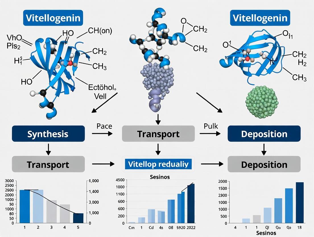
Vitellogenin (Vg) is a glycolipophosphoprotein that serves as the primary precursor to the major egg yolk proteins in nearly all oviparous species [1] [2]. This macromolecule, possessing properties of sugar, fat, and phosphate groups, belongs to the large lipid transfer protein (LLTP) superfamily, which also includes microsomal triglyceride transfer protein (MTP) and apolipoprotein B (apoB-100) [1] [2] [3]. Vg is historically recognized as a female-specific protein synthesized in extra-ovarian tissues—primarily the liver in vertebrates, the fat body in insects, and the hepatopancreas in crustaceans [1] [4]. Following synthesis and secretion into the circulatory system (blood or hemolymph), Vg is transported to the ovaries, where it is taken up by growing oocytes via receptor-mediated endocytosis. Within the oocyte, it is proteolytically processed into its derived forms, such as vitellin (Vn), which provides essential nutrients—including amino acids, lipids, phosphorous, carbohydrates, and metals (Mg, Ca, Zn)—for embryonic development [2] [3] [5].
The gene encoding vitellogenin is ancient, with its emergence linked to the increased need for lipid transport associated with multicellularity and dating back at least 700 million years [2] [3]. Vertebrates initially possessed a single vitellogenin gene, with subsequent gene duplications occurring in the bird-mammalian and amphibian lineages [1]. With the exception of monotremes, most mammals have seen their vitellogenin genes become pseudogenes [1]. Vitellogenin's role has traditionally been viewed through the lens of reproduction. However, contemporary research, particularly in model organisms like the honey bee (Apis mellifera), has revealed a remarkable pleiotropy in its functions, encompassing immunity, antioxidant protection, social behavior regulation, and longevity [6] [2]. This whitepaper provides a comprehensive technical overview of vitellogenin, detailing its structure, diverse functions, and the experimental methodologies central to its study, framed within the context of its conserved role across oviparous species.
Structural Characteristics and Domain Architecture
Vitellogenin is a large, complex macromolecule with a molecular weight that typically ranges from 240 to 650 kDa, depending on the species [2] [5]. Its function as a multifunctional transport protein is intrinsically linked to its sophisticated biochemical structure and conserved domain architecture.
Biochemical Composition and Proteolytic Processing
As a glyco-lipo-phospho-protein, vitellogenin undergoes significant post-translational modification. The apo-protein is synthesized and subsequently modified with sugar (glycosylation), lipid (lipidation), and phosphate (phosphorylation, particularly on serine residues in the phosvitin domain) moieties in its tissue of origin [1] [2]. The primary structure of Vg is proteolytically cleaved at conserved sites in different species to generate distinct yolk proteins [1]. In vertebrates, a complete Vg is cleaved into lipovitellin (a lipoglycoprotein), phosvitin (a highly phosphorylated domain that binds metals like calcium and iron), and a von Willebrand factor type D domain (vWD) [1] [6]. The extent of this cleavage varies; in holometabolous insects, the Vg precursor is typically cleaved into two polypeptides, whereas in hemimetabolous insects, it is cleaved into several polypeptides [7]. Notably, in higher Hymenoptera (like honey bees and fire ants), the Vg gene product may remain uncleaved [7].
Conserved Structural Domains
Recent structural biology breakthroughs, including a 3.2 Å resolution cryo-electron microscopy (cryo-EM) structure of native honey bee vitellogenin (AmVg), have provided unprecedented insight into its domain architecture [6]. The core of Vg is characterized by an LLTP lipid binding module, which is shared across the superfamily. This module consists of several key subdomains:
- N-sheet (N-terminal β-barrel): An antiparallel β-sheet wrapped around a central α-helix. This domain is responsible for receptor binding, facilitating the uptake of Vg into oocytes [6] [2].
- Lipid Binding Cavity: Formed by the A-sheets and C-sheets, this cavity is central to Vg's role in lipid transport [1] [6].
- α-Helical Domain: A domain that wraps around the A and C-sheets and contains a lipophilic cavity implicated in binding various ligands. This domain is also believed to facilitate vitellogenin's anti-inflammatory functions [2].
- Von Willebrand Factor type D (vWD) Domain: Previously uncharacterized in LLTPs, this domain was clearly resolved in the AmVg cryo-EM structure, though its precise function remains under investigation [6].
- C-terminal Cystine Knot (CTCK) Domain: The recent AmVg structure identified a domain of unknown function as a CTCK domain based on structural homology, which may contain a putative dimerization site [6].
Table 1: Key Structural Domains of Vitellogenin
| Domain | Structural Features | Postulated Functions |
|---|---|---|
| N-sheet (N-terminal β-barrel) | Antiparallel β-sheet, central α-helix | Receptor binding, oocyte uptake [6] [2] |
| Lipid Binding Cavity | Formed by A-sheets and C-sheets | Binding and transport of lipids, triglycerides [1] [6] |
| α-Helical Domain | Lipophilic cavity | Ligand binding, anti-inflammatory activity [2] |
| Phosvitin Domain | Serine-rich, highly phosphorylated | Metal ion binding (Ca, Mg, Zn) [1] |
| vWD Domain | Conserved protein interaction module | Unknown function in Vg, potentially involved in oligomerization or binding [6] |
| CTCK Domain | Cystine knot structure | Putative dimerization site [6] |
The following diagram illustrates the logical relationship between vitellogenin's primary structure, its domains, and its ultimate functions:
Functional Pleiotropy: From Nutrition to Immunity and Beyond
The paradigm of vitellogenin research has expanded significantly from its canonical role in reproduction. It is now recognized as a quintessential example of pleiotropy, where a single gene product influences multiple, seemingly unrelated, phenotypic traits [6].
Primary Reproductive and Nutritional Roles
The primary and evolutionarily conserved function of Vg is to supply the developing embryo with essential nutrients. Vg serves as a source of amino acids, peptides, lipids, carbohydrates, and fat-soluble vitamins and hormones [6] [2]. This nutritional cargo is bound and transported by Vg from the site of synthesis to the oocyte, where it is deposited and stored as vitellin (Vn) within yolk granules [2] [3]. During embryogenesis, these reserves are mobilized to fuel growth and development until the offspring can feed exogenously.
Immune and Antioxidant Functions
Accumulating evidence from diverse taxa—including corals, mollusks, arthropods, and fishes—has established Vg as a potent immune molecule [6] [7]. Vitellogenin can function as a pattern recognition receptor (PRR), capable of binding to a range of pathogen-associated molecular patterns (PAMPs) such as lipopolysaccharide (LPS), lipoteichoic acid, and peptidoglycan [6] [7]. It also exhibits direct bactericidal activity, damaging bacterial cell walls, and can act as an opsonin, marking pathogens for phagocytosis by immune cells [6]. Furthermore, Vg has demonstrated antiviral activity, neutralizing viruses like the Infectious Pancreatic Necrosis Virus (IPNV) in fish [5]. Alongside its immune roles, Vg protects organisms from oxidative stress through strong free radical scavenging activity, thereby contributing to cellular homeostasis and longevity [6] [2].
Specialized Roles in Social Insects
In eusocial insects like the honey bee, vitellogenin has acquired highly specialized functions that regulate social organization and lifespan [6] [2]. In the honey bee worker caste, which is sterile, Vg titers influence behavioral maturation and the division of labor. Nurse bees with high Vg titers care for the brood inside the nest, while foragers, which have low Vg titers, undertake the risky task of collecting resources outside the hive [1] [2]. Vg also participates in a regulatory feedback loop with juvenile hormone (JH), which is instrumental in controlling honey bee development and behavior [1]. High Vg levels suppress JH, promoting a longer lifespan and the nurse bee phenotype, whereas low Vg levels allow JH to rise, triggering the transition to foraging and a associated with a shorter lifespan [1] [2]. This interplay is also implicated in complex behaviors like swarming [1].
Table 2: Pleiotropic Functions of Vitellogenin Across Species
| Function Category | Specific Activity | Example Organisms |
|---|---|---|
| Reproductive & Nutritional | Yolk precursor, nutrient source (proteins, lipids, metals) | All oviparous vertebrates and invertebrates [1] [2] |
| Immune Defense | Pattern recognition, opsonization, bactericidal, antiviral | Fishes, crustaceans, insects, mollusks [6] [5] [7] |
| Antioxidant | Free radical scavenging | Honey bee, nematode (C. elegans) [6] [5] |
| Hormonal & Behavioral | Regulation of juvenile hormone, division of labor, lifespan | Honey bee (Apis mellifera), fire ant (Solenopsis invicta) [1] [2] [8] |
| Neuroprotective | Modulation of neuron survival, improved cognitive function | Chicken (Yolkin peptides) [5] |
Key Experimental Methodologies and Workflows
The study of vitellogenin employs a suite of sophisticated molecular, biochemical, and structural techniques. Below are detailed protocols for two pivotal experimental approaches cited in recent research: transcriptomic analysis for identifying and characterizing Vg genes, and RNA interference (RNAi) for functional validation.
Transcriptomic Identification and Expression Profiling of Vg Genes
Purpose: To identify vitellogenin gene sequences, quantify their expression levels across different tissues or caste types, and discover differentially expressed genes (DEGs) associated with reproduction. Principle: High-throughput mRNA sequencing (RNA-seq) provides a comprehensive profile of all transcribed genes in a sample, allowing for the discovery and quantification of Vg transcripts. Protocol (as applied in Solenopsis invicta [8]):
- Sample Collection: Collect target tissues (e.g., fat body, ovary, hepatopancreas) or whole organisms of different reproductive caste types (e.g., queen, winged female, male). Use a minimum of three biological replicates per group.
- RNA Extraction: Homogenize tissues in TRIzol reagent or using a commercial kit (e.g., RNeasy Mini Kit, Qiagen). Assess RNA integrity and purity using agarose gel electrophoresis and spectrophotometry (A260/A280 ratio >1.9).
- Library Preparation and Sequencing: Isolate mRNA from total RNA using oligo(dT) magnetic beads. Fragment the mRNA and synthesize cDNA. Ligate adapters to the cDNA fragments and amplify via PCR to create the sequencing library. Sequence the library on a platform such as Illumina HiSeq, aiming for a minimum of 6.08 Giga bases (Gb) of clean reads per sample.
- Bioinformatic Analysis:
- Data Preprocessing: Remove low-quality reads and adapter sequences using tools like Trimmomatic or Fastp.
- Read Mapping and Quantification: Map the clean reads to a reference genome using HISAT2 or STAR. Assemble transcripts and estimate their abundance (e.g., as FPKM or TPM) using StringTie or featureCounts.
- Differential Expression Analysis: Identify DEGs between sample groups using software such as DESeq2 or edgeR, with a standard significance threshold of adjusted p-value (FDR) < 0.05 and |log2(FoldChange)| > 1.
- Functional Enrichment: Perform Gene Ontology (GO) and Kyoto Encyclopedia of Genes and Genomes (KEGG) enrichment analysis on DEGs to identify involved biological pathways.
Functional Validation via RNA Interference (RNAi)
Purpose: To investigate the biological function of a specific Vg gene by knocking down its expression and observing the resulting phenotypic consequences. Principle: Introduction of double-stranded RNA (dsRNA) complementary to the target Vg gene sequence triggers the cellular RNAi machinery, leading to sequence-specific degradation of the corresponding mRNA and a reduction in protein levels. Protocol (as applied in Solenopsis invicta [8]):
- dsRNA Synthesis:
- Template Generation: Design PCR primers with T7 promoter sequences appended to the 5' ends. Amplify a 300-500 bp fragment of the target Vg gene (e.g., SiVg2 or SiVg3) from cDNA.
- In Vitro Transcription: Purify the PCR product and use it as a template for in vitro transcription with a T7 RNA polymerase kit (e.g., MEGAscript T7 Kit, Thermo Fisher Scientific). Include a control dsRNA targeting a non-endogenous gene (e.g., GFP).
- dsRNA Purification: Treat the reaction with DNase I to remove the DNA template. Purify the synthesized dsRNA using phenol-chloroform extraction or a commercial purification kit. Confirm integrity via agarose gel electrophoresis and quantify using a spectrophotometer.
- dsRNA Delivery:
- Microinjection: Anesthetize experimental animals (e.g., fire ant queens) on ice. Using a microinjector (e.g., Nanoject III, Drummond Scientific), inject a calibrated volume (e.g., 100-200 nL) of dsRNA solution (e.g., 2-4 µg/µL) directly into the hemocoel. Seal the injection wound with wax to prevent leakage and infection.
- Efficacy and Phenotype Assessment:
- Knockdown Validation: After 3-7 days, sacrifice a subset of injected animals. Extract total RNA and synthesize cDNA. Assess the knockdown efficiency of the target Vg gene using quantitative real-time PCR (qRT-PCR) with gene-specific primers and a reference gene (e.g., Actin or GAPDH).
- Phenotypic Analysis: In the remaining animals, observe and quantify phenotypic outcomes. Key metrics for reproductive studies include:
- Ovarian Morphology: Dissect ovaries and measure their size/weight. Calculate the gonadosomatic index (GSI) = (Ovary Weight / Body Weight) × 100.
- Oogenesis and Fecundity: Process ovarian tissue for histology (fixation, paraffin embedding, sectioning, H&E staining). Count the number of developing oocytes and measure their size. Record daily egg production.
- Molecular Confirmation: Analyze the expression of downstream genes or pathways potentially affected by Vg knockdown.
The following workflow diagram summarizes the key steps in the RNAi-based functional validation protocol:
The Scientist's Toolkit: Essential Research Reagents and Materials
Research into vitellogenin requires a range of specialized reagents and tools. The following table details key solutions and their applications, derived from the experimental protocols cited in this whitepaper.
Table 3: Essential Research Reagents for Vitellogenin Studies
| Research Reagent / Kit | Specific Example(s) | Primary Function in Vitellogenin Research |
|---|---|---|
| RNA Extraction Kit | RNeasy Mini Kit (Qiagen), TRIzol Reagent | Isolation of high-quality total RNA from tissues (fat body, ovary, hepatopancreas) for transcriptomic analysis and qRT-PCR [8]. |
| cDNA Synthesis Kit | High-Capacity cDNA Reverse Transcription Kit (Thermo Fisher) | Generation of stable cDNA templates from isolated RNA for subsequent PCR, qRT-PCR, and dsRNA synthesis [8]. |
| RNA-seq Library Prep Kit | Illumina TruSeq Stranded mRNA Kit | Preparation of sequencing-ready libraries from purified mRNA for transcriptome profiling and differential expression analysis [8]. |
| In Vitro Transcription Kit | MEGAscript T7 Kit (Thermo Fisher Scientific) | Synthesis of high-yield, pure double-stranded RNA (dsRNA) for RNAi-mediated gene knockdown experiments [8]. |
| Microinjection System | Nanoject III (Drummond Scientific) | Precise, nanoliter-scale delivery of dsRNA or other reagents into the hemocoel of insects or small crustaceans for functional studies [8]. |
| qRT-PCR Master Mix | SYBR Green or TaqMan Master Mix | Quantitative measurement of vitellogenin gene expression levels and knockdown validation using gene-specific primers and probes [8]. |
| Cryo-EM Equipment | Titan Krios Cryo-Electron Microscope | High-resolution structural determination of native vitellogenin proteins and their complexes, as demonstrated for honey bee Vg [6]. |
| Antibodies (Anti-Vg) | Species-specific polyclonal/monoclonal antibodies | Detection, quantification (via ELISA), and cellular localization (via immunohistochemistry) of the vitellogenin protein [4]. |
Evolutionary and Comparative Perspectives
Vitellogenin genes have undergone significant evolutionary diversification. The ancestral single-copy gene has been duplicated in many lineages, leading to subfunctionalization and neofunctionalization [1] [7]. For instance, the red imported fire ant (Solenopsis invicta) possesses multiple vitellogenin genes (e.g., Vg1, Vg2, Vg3) that show caste-specific expression patterns, with Vg2 and Vg3 being critical for queen fecundity [8]. In contrast, the honey bee has a single Vg gene but also expresses several more ancient vitellogenin homologs (vg-like-A, vg-like-B, vg-like-C) that may share and specialize in certain tasks, such as the immune response, thereby mitigating evolutionary constraints on the pleiotropic main Vg gene [7].
The mode of vitellogenesis—the process of yolk formation—also varies. In exogenous vitellogenesis, Vg is synthesized in extra-ovarian tissues (e.g., liver, fat body, hepatopancreas) and transported to the ovary, as seen in most vertebrates, insects, and shrimp like Litopenaeus vannamei [1] [4]. In endogenous vitellogenesis, Vg is synthesized directly by the oocytes themselves, as observed in some bivalves [4]. Some crustaceans, like Scylla paramamosain, exhibit a mixed mode, with Vg production occurring in both the hepatopancreas and the ovary [4].
Vitellogenin stands as a remarkable and conserved glycolipophosphoprotein fundamental to reproduction in oviparous species. However, as detailed in this technical overview, its functional repertoire extends far beyond nutrition to encompass critical roles in immunity, antioxidant defense, and the regulation of social behavior and lifespan. The elucidation of its high-resolution structure and the application of advanced molecular techniques like RNAi and transcriptomics continue to unravel the molecular mechanisms underlying this pleiotropy. Understanding the diverse functions and evolutionary trajectories of vitellogenin not only provides deep insights into fundamental biological processes but also opens avenues for applied research, such as the development of novel strategies for pest control by disrupting reproduction or for promoting health in aquaculture.
Vitellogenin (Vg) is the foundational yolk precursor protein, a glycolipophosphoprotein that serves as the primary source of nutrients for embryonic development in nearly all oviparous species, including fish, amphibians, insects, and birds [1] [2]. This protein belongs to the large lipid transfer protein (LLTP) superfamily, which includes mammalian counterparts such as apolipoprotein B (apoB) and microsomal triglyceride transfer protein (MTP) [6] [2]. The journey of vitellogenin from its synthesis in somatic tissues to its ultimate deposition in the oocyte represents a critical biological pathway essential for reproductive success. This process ensures the developing embryo is supplied with essential amino acids, lipids, carbohydrates, phosphorous, and metal ions such as magnesium, calcium, and zinc [2] [9]. Understanding the molecular mechanisms governing vitellogenin synthesis, transport, and uptake is therefore paramount in fields ranging from reproductive biology to aquaculture management and environmental toxicology.
Biosynthesis of Vitellogenin
Synthesis Sites and Protein Structure
Vitellogenin is synthesized primarily in two somatic tissues: the liver (or hepatopancreas) of vertebrates and the fat body of insects [1] [2]. The fat body is an organ analogous to the vertebrate liver and adipose tissue, with functions in homeostasis, immunity, and nutrient storage [2]. In some insect species, additional sites of Vg synthesis have been reported, including follicle cells, nurse cells, and hemocytes [10].
The protein structure of Vg is evolutionarily conserved and characterized by several key domains that facilitate its function as a transport protein [2]. As a member of the LLTP superfamily, Vg contains a characteristic lipid-binding module [6]. The canonical structure typically includes:
- A vitellogenin N-terminal domain (Vitellogenin_N), which is a lipid transport domain also found in MTP and apoB [1].
- A domain of unknown function (DUF1943) [1].
- A von Willebrand factor type D domain (vWFD) located in the C-terminus [6] [10].
Vg is initially synthesized as a large precursor protein. In insects, this precursor typically has a molecular mass of ∼200 kDa and is derived from ∼7 kb mRNAs [11]. This primary precursor undergoes proteolytic cleavage in the fat body to form large (140–190 kDa) and small (40–60 kDa) subunits, which then assemble and are secreted into the hemolymph as large oligomeric proteins ranging from 400–600 kDa [11]. These molecules also undergo significant post-translational modifications, including phosphorylation, glycosylation, and sulfation [11].
Table 1: Key Domains of the Vitellogenin Protein
| Domain Name | Location | Putative Function |
|---|---|---|
| VitellogeninN (LPDN) | N-terminus | Lipid binding and transport [1]. |
| DUF1943 | Central Region | Unknown function [10]. |
| von Willebrand factor type D (vWFD) | C-terminus | Previously uncharacterized; structural role [6]. |
| Polyserine Tract | Within N-sheet | Site of phosphorylation; characteristic of insect Vgs [6] [10]. |
Regulatory Mechanisms of Synthesis
The synthesis of vitellogenin is under complex hormonal control, which varies across taxonomic groups.
- In Fish and Vertebrates: The expression of the Vg gene is estrogen-dependent and is normally limited to females. The presence of Vg in the blood of male fish is a widely used biomarker for exposure to environmental estrogens or endocrine-disrupting chemicals (EDCs) [1] [2] [12].
- In Insects: Vitellogenesis is governed by two critical hormones: the sesquiterpenoid juvenile hormone (JH) and the ecdysteroid 20-hydroxyecdysone (20E) [10]. JH acts as the principal gonadotropic hormone that stimulates Vg synthesis in most holometabolous insects and basal hemimetabolous insects. The molecular action of JH relies on its intracellular receptor, Methoprene-tolerant (Met), which forms a complex with Taiman (Tai) to activate JH-responsive genes [10]. In some insects, such as certain lepidopterans and dipterans, 20E plays a primary role [10].
Furthermore, Vg synthesis is tightly coupled to the nutritional status of the organism. Nutritional sensors, including the amino acid/Target of Rapamycin (AA/TOR) and insulin-like peptide (ILP) pathways, interact with JH and 20E signaling cascades to regulate vitellogenesis [10] [2]. Recent studies have also revealed an essential role for microRNAs (miRNAs) in the fine-tuning of this process [10].
Transport and Intracellular Trafficking
Following its synthesis and secretion, vitellogenin embarks on a journey to the oocyte, navigating both extracellular and intracellular compartments.
Systemic Transport
Once processed and secreted from the somatic tissue:
- In vertebrates, Vg is released into the bloodstream [9].
- In insects and nematodes, it is released into the hemolymph or pseudocelom, respectively [13] [11].
This systemic transport allows the glycolipoprotein to circulate throughout the body and reach the developing oocytes in the ovary.
Intracellular Trafficking and ER Export
A critical step in Vg transport is its export from the endoplasmic reticulum (ER) in the synthesizing cell. Recent research in Caenorhabditis elegans has identified a key protein, TNGL-1, which mediates the export of Vg from the ER in intestinal cells [13]. TNGL-1 is a distant member of the TANGO1 family of proteins, which are known in mammals to generate membrane carriers for the ER export of bulky cargo, such as ApoB-containing lipoprotein particles [13].
The mechanism involves:
- TNGL-1 localizes to ER exit sites [13].
- It uses an N-terminal globular domain to bind Vg directly [13].
- Its C-terminal unstructured domain is required for proper targeting to ER exit sites [13].
- Depletion of TNGL-1 causes the retention of Vg in the ER lumen, preventing its continued secretion and transport to the oocyte [13].
This discovery points to a universal requirement of TANGO1-based mechanisms for the secretion of specific metazoan proteins, functionally conserving the pathway from nematodes to mammals, despite sequence divergence [13].
Diagram 1: Intracellular trafficking of Vg for secretion. TNGL-1/TANGO1 at ER exit sites is crucial for Vg export.
Oocyte Uptake and Yolk Formation
The final stage of Vg's journey is its targeted uptake by the developing oocyte and processing into mature yolk.
Receptor-Mediated Endocytosis
The uptake of circulating Vg into growing oocytes occurs primarily through receptor-mediated endocytosis [14] [2] [9]. The receptors responsible for this process belong to the low-density lipoprotein receptor (LDLR) family [14]. In teleost fish like the flathead mullet (Mugil cephalus), two specific subtypes have been identified as putative vitellogenin receptors (VgRs): the Lr8/VLDLR and Lrp13/LRX+1 subfamilies [14]. These receptors exhibit conserved domain architectures and show ovary-specific expression profiles, consistent with their role in mediating Vg uptake during oocyte development [14].
The process of uptake follows these general steps:
- Circulating Vg diffuses through the basement membrane and between follicle cells to reach the oocyte surface [9].
- Binding to VgR: Vg binds specifically to its receptor on the oolemma (oocyte membrane) [11].
- Internalization: The Vg-VgR complex is internalized via clathrin-coated pits [14] [11].
- Endocytic Trafficking: The complex follows a canonical endocytic route, forming early and late endosomes. The VgR is typically recycled back to the oocyte membrane, while Vg is trafficked to yolk granules [11].
- Yolk Formation: Within the yolk granules, Vg is proteolytically cleaved and processed into its storage form, vitellin (Vt), which serves as the final nutrient reserve for the embryo [11].
Diversity in Uptake Mechanisms
Morphological studies in birds like the quail (Coturnix japonica) have revealed that the mechanism of uptake can vary during different stages of oogenesis [9]:
- In small oocytes (white follicles), Vg is taken up through fluid-phase endocytosis, assisted by specialized structures called follicular lining bodies [9].
- In large oocytes (yellow follicles), both Vg and very-low-density lipoproteins (VLDL) are taken up via classic receptor-mediated endocytosis through coated vesicles [9].
Once inside the oocyte, enzymes like cathepsin D are responsible for the proteolytic processing of Vg into yolk proteins within the yolk spheres [9].
Diagram 2: Vg uptake via receptor-mediated endocytosis in oocytes.
Experimental Protocols and Research Toolkit
The study of vitellogenin synthesis and transport relies on a suite of molecular, biochemical, and morphological techniques. Below is a summary of key methodological approaches and reagents derived from the cited research.
Key Experimental Workflow
A comprehensive in silico identification and characterization of Vg receptors in the flathead mullet exemplifies a modern integrative approach [14]. The workflow can be summarized as follows:
- Genomic and Proteomic Data Mining: Retrieve proteomes from databases like Ensembl. Use orthology inference tools (e.g., KofamScan) against databases like KEGG Orthology to identify LDLR family members [14].
- Homology Searches and Sequence Analysis: Perform in-depth mining of the proteome using BLASTp against custom non-redundant protein sequence databases of known VgRs to identify putative receptors [14].
- Domain Architecture Analysis: Use tools like SMART to characterize the domain structure of identified proteins [14].
- Structural Prediction and Modeling: Predict 3D protein structures with atomic accuracy using methods like AlphaFold2 [14]. Model protein-ligand binding sites with algorithms like COACH-D [14].
- Synteny Evaluation: Analyze the conserved gene arrangements (synteny) of the identified receptor genes by comparing them with orthologs in other related species [14].
- Expression Validation: Validate tissue-specific expression, particularly ovary-specific expression, using RNA-seq data from multiple tissues [14].
- Phylogenetic Analysis: Reconstruct evolutionary relationships through phylogenetic analyses to confirm evolutionary conservation [14].
Diagram 3: Workflow for identification of vitellogenin receptors.
The Scientist's Toolkit: Essential Research Reagents
Table 2: Key Reagents and Resources for Vitellogenin Research
| Reagent / Resource | Function / Application | Example from Literature |
|---|---|---|
| KofamScan / KEGG Orthology | Tool for orthology inference and gene function annotation in genomic data. | Used to assign K numbers and identify LDLR family members in Mugil cephalus proteome [14]. |
| AlphaFold2 | Protein structure prediction with atomic accuracy. | Used to predict 3D structure of putative VgRs and honey bee vitellogenin [14] [6]. |
| RNA Interference (RNAi) | Functional validation through gene knockdown. | Used in C. elegans and Rhodnius prolixus to knock down TNGL-1 and Vg genes, respectively, to study loss-of-function phenotypes [13] [11]. |
| Immunoelectron Microscopy | Ultra-structural localization of proteins within cells. | Used to detect endogenous VIT-1/VIT-2 in the ER lumen of C. elegans intestinal cells after TNGL-1 depletion [13]. |
| SEC-16A.1::GFP Marker | Fluorescent marker for identifying ER exit sites in live cells. | Used to demonstrate co-localization of TNGL-1 with ER exit sites in C. elegans intestine [13]. |
| DEAE52 Column Chromatography | Purification of proteins, such as vitellogenin, from plasma or tissue extracts. | Used to purify quail VTG from estrogen-primed male plasma [9]. |
| Pepstatin-Sepharose Affinity Chromatography | Specific purification of aspartic proteases like cathepsin D. | Used in the purification of cathepsin D from yolk or liver homogenates [9]. |
The synthesis and transport of vitellogenin from somatic tissues to oocyte uptake is a beautifully complex and highly regulated process, fundamental to reproduction in oviparous species. From its hormone-regulated biosynthesis in the liver or fat body, its TANGO1-mediated export from the ER, its journey through the circulatory system, to its receptor-specific uptake by the oocyte, each step is crucial for successful yolk formation and embryonic development. Ongoing research continues to unveil novel players in this pathway, such as TNGL-1 in nematodes, and expands our understanding of the conserved LDLR family receptors in fish and other vertebrates. This detailed molecular knowledge is not only critical for basic reproductive biology but also provides practical avenues for improving aquaculture, controlling disease vectors, and assessing the impact of environmental pollutants on animal fertility.
This technical guide delves into the pivotal role of single-particle cryogenic electron microscopy (cryo-EM) in elucidating the complex domain architecture of large proteins, with a specific focus on the yolk protein precursor vitellogenin (Vg). For researchers and drug development professionals, understanding the structure-function relationship of such pleiotropic molecules is fundamental. The recent cryo-EM structure of native honey bee vitellogenin (Apis mellifera Vg, or AmVg) at 3.2 Å resolution serves as a paradigm, revealing previously uncharacterized domains and molecular mechanisms underlying its diverse functions, from longevity and social behavior in honey bees to immunity and antioxidant protection [6]. This whitepaper provides an in-depth analysis of the core findings, summarizes quantitative data in structured tables, and outlines the detailed experimental methodologies that enabled this structural breakthrough.
Vitellogenin is a large lipid transfer protein (LLTP) that serves as the main yolk precursor in nearly all egg-laying animals [6]. Beyond its canonical role in reproduction, Vg has evolved a remarkable range of secondary functions. In the honey bee, Vg is a model for studying pleiotropy, where a single gene influences multiple phenotypic traits, including immunity, antioxidant protection, the regulation of social behavior, and longevity [6]. Despite two decades of functional studies, the molecular mechanisms enabling Vg to perform these varied roles remained poorly understood, primarily due to the lack of a full-length structural model. The cryo-EM structure of native AmVg directly addresses this gap, providing a high-resolution framework to interpret its diverse functionalities in structural terms [6].
Core Structural Breakthrough: The Honey Bee Vg Cryo-EM Structure
The determination of the AmVg structure from native hemolymph represents a significant leap forward. The key achievements of this study are quantitatively summarized in the table below.
Table 1: Key Experimental Parameters and Resolutions of the AmVg Cryo-EM Structure
| Parameter | Full-Length AmVg | AmVg Cleavage Product |
|---|---|---|
| Source | Honey Bee (Apis mellifera) Hemolymph | Honey Bee (Apis mellifera) Hemolymph |
| Purification | One-step purification directly from hemolymph [6] | Co-purified with full-length protein [6] |
| Resolution | 3.2 Å [6] | 3.0 Å [6] |
| Oligomeric State | Monomer [6] | Monomer [6] |
| Key Domains Resolved | Lipid binding module (N, A, C sheets, α-helical domain), vWD, CTCK [6] | Lipid binding module (N, A, C sheets, α-helical domain), vWD, CTCK [6] |
The structure provided nearly full-length coverage of the protein, enabling the first comprehensive view of its domain architecture. A significant finding was the identification of a domain of unknown function as a C-terminal cystine knot (CTCK) domain based on structural homology, which suggests a potential role in dimerization [6]. Furthermore, the structure offered insights into post-translational modifications, metal and lipid binding, and the presence of a major ~150 kDa cleavage product, all of which contribute to the functional understanding of Vg [6].
Detailed Domain Architecture and Functional Implications
The AmVg structure reveals a multi-domain protein that incorporates both a conserved core and unique functional elements.
The Conserved Lipid Binding Module
As a member of the LLTP superfamily, AmVg contains the characteristic lipid binding module. This module is composed of several subdomains [6]:
- The N-sheet: An antiparallel β-sheet wrapped around a central α-helix, located at the N-terminus. It is responsible for receptor binding and forms a β-sandwich with the A-sheet.
- The Lipid Binding Cavity: Formed by the A-sheet and C-sheet, this is the central pocket for lipid transport.
- The α-helical subdomain: A bundle of α-helices that wraps around the A and C sheets, stabilizing the entire module.
Previously Uncharacterized Domains
The structure provided the first view of two critical domains not previously resolved in other Vg structures:
- Von Willebrand factor type D (vWD) domain: This domain, common in proteins involved in clotting and cell adhesion, was uncharacterized in any LLTP superfamily member prior to this study. Its presence in Vg may be linked to its immune-related functions, such as pathogen recognition [6].
- C-terminal Cystine Knot (CTCK) domain: A domain of unknown function was definitively identified as a CTCK domain based on structural homology. This domain type is often involved in protein-protein interactions and dimerization, suggesting a potential mechanism for Vg oligomerization or interaction with other immune molecules [6].
Flexible and Modified Regions
The structure also highlighted dynamic regions critical for function:
- Polyserine (polyS) region: A characteristic, disordered region in insect vitellogenins (residues 340-384 in AmVg) that was not resolved in the cryo-EM density due to its flexibility [6]. This region is known to be phosphorylated, which protects it from proteolysis and is likely crucial for its function [6].
- Disulfide bridges: A conserved disulfide bridge (C178–C222) was observed, stabilizing a short β-strand that integrates with the A-sheet. The absence of density for a flexible loop (residues 232–245) suggests conformational dynamics that may be regulated by zinc binding [6].
Experimental Protocols and Workflows
The successful determination of the AmVg structure relied on a rigorous methodological pipeline, from native protein purification to high-resolution single-particle analysis.
Sample Preparation and Purification
A key to the success of this study was the use of natively sourced protein. AmVg was one-step purified directly from the hemolymph of honey bees, ensuring the protein retained its native post-translational modifications, bound ligands, and physiological oligomeric state [6]. This approach avoids potential artifacts that can arise from recombinant overexpression in heterologous systems.
Cryo-EM Data Collection and Image Processing
The process of generating a 3D reconstruction from 2D micrographs relies on principles of image formation and computational refinement.
- Image Formation and Contrast: In cryo-EM, contrast for biological samples arises primarily from phase contrast, not amplitude contrast [15]. Proteins are weak phase objects; they delay the phase of the electron wave rather than absorbing it. To make these phase shifts detectable, data is intentionally collected out of focus, which introduces a phase shift that converts the invisible phase information into measurable amplitude contrast in the final image [15].
- Contrast Transfer Function (CTF) Correction: The imperfections of the microscope lens and the deliberate defocus impart an oscillating signal dampening and inversion known as the CTF. Accurate estimation and correction of the CTF is essential for high-resolution reconstruction [16]. Tools like CTFFIND are used to fit the CTF parameters from the power spectra of micrographs, and newer versions like CTFFIND5 can account for complex sample geometries like tilt and thickness, improving CTF estimation for challenging samples [16].
- Heterogeneous Refinement: The hemolymph-sourced sample was heterogeneous, containing both full-length AmVg and a prominent ~150 kDa cleavage product. Advanced computational sorting techniques in software packages like cryoSPARC or RELION were employed to separate these two populations from a single dataset, yielding two high-resolution reconstructions (3.2 Å and 3.0 Å) [6].
Table 2: Key Research Reagent Solutions for Cryo-EM Structural Analysis
| Reagent / Resource | Function / Application | Example / Note |
|---|---|---|
| Native Protein Source | Provides natively modified, functional protein for structural studies. | Honey bee hemolymph [6]. |
| Cryo-EM Grids | Support for vitrified sample; enables imaging in the electron microscope. | Quantifoil or C-flat grids with ultra-thin carbon. |
| cryo-EM Software Suites | Processing, 2D classification, 3D reconstruction, refinement, and validation of cryo-EM data. | cryoSPARC, RELION, cisTEM [16]. |
| CTF Estimation Software | Critical estimation and correction of the microscope's Contrast Transfer Function. | CTFFIND4/5 [16]. |
| Model Building & Visualization | Atomic model building into cryo-EM maps, validation, and visualization. | Coot, Phenix, PyMOL, Nanome Cryo-EM plugin [17]. |
| Structural Databases | Repository for depositing and accessing experimental maps and atomic models. | EMDB (maps), PDB (models) [18]. |
The cryo-EM structure of native honey bee vitellogenin has provided an unprecedented molecular blueprint for understanding the pleiotropic functions of this critical protein. By revealing the full domain architecture, including the vWD and CTCK domains, and offering insights into lipid and metal binding sites, the structure serves as a foundation for formulating and testing specific mechanistic hypotheses. The experimental protocols outlined herein—emphasizing native purification, advanced image processing, and rigorous quality control—provide a template for the structural investigation of other challenging, multifunctional proteins. This structural knowledge paves the way for future research into vitellogenin's role in development, evolution, and social behavior, with potential applications in areas like insect physiology, immunology, and even the development of strategies to support pollinator health.
Vitellogenin (Vg), an ancient and highly conserved glycolipoprotein, is recognized as the primary egg yolk precursor in oviparous species. However, emerging research has illuminated its critical functions beyond reproduction, particularly in eusocial insects like the honey bee (Apis mellifera). This whitepaper synthesizes current evidence establishing Vg as a pleiotropic protein integral to longevity regulation, immune defense, and oxidative stress resistance. We detail the molecular mechanisms underpinning these functions, present quantitative findings from key studies, and provide methodologies for investigating Vg's non-reproductive roles. The insights herein are relevant for researchers exploring the evolutionary rewiring of nutrient signaling pathways and their implications for lifespan and healthspan in broader taxa.
Vitellogenin is a large lipid transfer protein (LLTP) traditionally studied for its role in oocyte development and embryonic nutrition [2]. In a remarkable example of evolutionary co-option, Vg has acquired a diverse repertoire of functions in the honey bee, a model organism for social insect physiology [2]. In honey bee workers—largely sterile females—Vg does not primarily serve reproduction but has been repurposed to regulate social organization, behavioral maturation, and caste-specific longevity [19] [2].
The protein's structure, recently resolved via cryo-electron microscopy to a resolution of 3.2 Å, provides a molecular basis for its multifunctionality [6]. Key domains include an N-terminal β-barrel implicated in receptor binding, a central lipid-binding cavity, and a C-terminal cystine knot (CTCK) domain with putative dimerization capabilities [6]. This structural complexity allows Vg to interact with diverse ligands, including lipids, pathogens, and, as recently discovered, DNA [20].
Vitellogenin in Longevity and Oxidative Stress
Mechanistic Insights and Key Evidence
The link between Vg and extended lifespan is well-established in honey bees. Long-lived queen bees and "diutinus" winter workers exhibit consistently high Vg titers, while short-lived foragers show markedly low levels [19] [21] [2]. The protein exerts its anti-aging effects primarily through its role as a potent antioxidant.
- Oxidative Shielding: Vg functions as a sacrificial antioxidant [19]. During oxidative challenge, Vg is preferentially carbonylated, thereby protecting other critical cellular proteins from irreversible oxidative damage. In experiments, workers with higher hemolymph Vg titers demonstrated significantly greater survival after paraquat injection (an agent that induces reactive oxygen species) compared to those with lower titers [19].
- Zion Binding and Immune Maintenance: Vg is the primary zinc carrier in honey bee hemolymph [21]. Zinc ions are crucial for immune function, and the decline of Vg in foragers leads to a corresponding drop in hemolymph zinc levels. This zinc deficiency induces apoptosis in immunocytes, effectively suppressing the innate immune system and contributing to the accelerated senescence of foragers [21].
- Regulatory Feedback with Juvenile Hormone: Vg and juvenile hormone (JH) participate in a mutually repressive feedback loop [1] [21]. High Vg titers suppress JH, delaying the transition from in-hive nursing to risky foraging and thus prolonging life. Knockdown of Vg gene expression leads to precocious foraging and significantly reduced lifespan [21].
Table 1: Key Experimental Evidence Linking Vitellogenin to Longevity and Oxidative Stress
| Experimental Approach | Key Finding | Biological Implication | Citation |
|---|---|---|---|
| Paraquat challenge after Vg RNAi | Vg knockdown bees showed significantly higher mortality. | Vg is crucial for resistance to oxidative stress. | [19] |
| Carbonylation analysis | Vg was more oxidized than other hemolymph proteins after paraquat exposure. | Vg acts as a sacrificial antioxidant. | [19] |
| Vg RNAi in free-flying bees | Induced early foraging and decreased lifespan. | Vg paces behavioral maturation to influence longevity. | [21] |
| Genotype-specific Vg RNAi | Lifespan effects were strain-dependent. | Genetic background influences Vg's role in aging. | [21] |
Experimental Protocol: Assessing the Role of Vg in Oxidative Stress Resistance
The following methodology, adapted from Seehuus et al. (2006), is used to quantify Vg's antioxidant function [19].
- Experimental Groups: Establish three groups of age-matched nurse bees (e.g., 150 bees per group).
- Vg Knockdown Group: Inject with Vg-dsRNA.
- Control Group 1: Inject with non-specific dsRNA (e.g., GFP-dsRNA).
- Control Group 2: Inject with vehicle only (e.g., PBS).
- Knockdown Verification: After 3-5 days, sacrifice a subset of bees from each group. Quantify Vg knockdown using:
- qPCR: Extract RNA from fat body tissue, synthesize cDNA, and perform qPCR with Vg-specific primers. Calculate relative expression using the ΔΔCt method with reference genes (e.g., β-actin and NDUFA8) [22].
- Western Blot: Analyze hemolymph samples via SDS-PAGE and immunoblotting with an anti-Vg antibody to confirm reduced protein titer.
- Oxidative Challenge: Inject the remaining bees in each group with a controlled dose of paraquat (e.g., 5mM in PBS) to induce systemic oxidative stress.
- Survival Assay: Monitor bees daily, recording mortality. Maintain bees in cages under standard laboratory conditions (e.g., 33°C, 50% relative humidity) with ad libitum sugar syrup and pollen paste.
- Data Analysis: Compare survival curves between groups using Kaplan-Meier analysis and a log-rank test. A significantly steeper survival curve in the Vg knockdown group versus both controls demonstrates Vg's role in oxidative stress resistance.
Immunological Functions of Vitellogenin
Vg has emerged as a critical immune effector molecule across diverse taxa, including fish, crustaceans, and insects [23] [6]. Its immune functions can be categorized as follows:
- Pathogen Recognition and Opsonization: Vg binds to pathogen-associated molecular patterns (PAMPs) on the surfaces of bacteria, fungi, and nematodes [23] [6]. In crustaceans and fish, Vg opsonizes pathogens for phagocytosis by immune cells [6]. For example, in the honey bee, Vg is present in the venom, potentially enhancing its efficacy in the target organism [23].
- Antibacterial and Antiviral Activity: Direct microbicidal activity has been observed. Vg can bind to and agglutinate both Gram-positive and Gram-negative bacteria, and in some species, it exhibits direct antiviral properties [6].
- Trans-generational Immune Priming: In a fascinating link between reproduction and immunity, Vg can transport immune signals or pathogen fragments from mother to offspring, priming the immune system of the next generation before hatching [6].
Table 2: Documented Immune Functions of Vitellogenin Across Species
| Immune Function | Mechanism | Example Organism | Citation |
|---|---|---|---|
| Anti-nematodal | Vg transcript and protein levels increase in male fat body and hemolymph upon infection. | Pyrrhocoris apterus (Firebug) | [23] |
| Antibacterial | Binds to bacterial cell wall components (e.g., LPS, PGN), leading to agglutination. | Coral; Fish; Honey Bee | [6] |
| Opsonization | Coats pathogens, facilitating recognition and phagocytosis by hemocytes. | Fish; Crustaceans | [6] |
| Venom Component | Vg is present in honey bee venom, potentially increasing venom efficacy. | Apis mellifera (Honey Bee) | [23] |
Novel Regulatory Roles and Signaling Pathways
Recent groundbreaking research has revealed that Vg can influence gene expression, adding a new layer to its functional repertoire.
- Nuclear Translocation and DNA Binding: A conserved subunit of Vg, the β-barrel domain, can be cleaved and translocate into the nucleus of fat body cells in honey bees [20]. Chromatin immunoprecipitation sequencing (ChIP-seq) has shown that this subunit binds to DNA at hundreds of loci.
- Gene Regulation: This DNA-binding activity is associated with changes in the expression of dozens of genes involved in energy metabolism, behavior, and cell signaling [20]. This suggests Vg may act as a transcription factor or co-regulator, providing a direct mechanistic link between an individual's nutritional status (reflected in Vg titer) and large-scale genomic reprogramming.
- Connection to Broader Signaling Networks: Vg's functions are integrated with conserved endocrine pathways. Its mutually repressive relationship with JH is a classic example [21]. Furthermore, Vg interacts with the Insulin/Insulin-like signaling (IIS) pathway, a central regulator of longevity and metabolism [21]. The expression of insulin-like peptides (Ilps) is correlated with Vg titer, and the IIS pathway is a key differentiator in the lifespan response to Vg manipulation across honey bee genotypes [21].
The Scientist's Toolkit: Essential Research Reagents and Methods
Table 3: Key Reagents and Methodologies for Vitellogenin Research
| Reagent / Method | Function/Description | Application Example | Citation |
|---|---|---|---|
| Vg-specific dsRNA | Double-stranded RNA for RNA-mediated gene knockdown. | Functional studies of Vg loss-of-function (e.g., lifespan, behavior). | [19] [21] |
| Anti-Vg Antibodies | Polyclonal or monoclonal antibodies for protein detection. | Quantifying Vg titer via ELISA; detecting protein localization via immunohistochemistry. | [4] |
| Vg Gene Expression Primers | Sequence-specific primers for qPCR. | Quantifying relative Vg mRNA levels using the ΔΔCt method. | [22] |
| Paraquat (Methyl Viologen) | Compound that generates intracellular superoxide anions. | Inducing systemic oxidative stress to test Vg's antioxidant role. | [19] |
| ChIP-seq (Chromatin Immunoprecipitation) | Identifies genome-wide binding sites of DNA-associated proteins. | Mapping Vg binding sites on DNA to investigate its role as a transcriptional regulator. | [20] |
| Cryo-Electron Microscopy | High-resolution structural determination of proteins in near-native state. | Solving the 3D structure of full-length Vg and its complexes. | [6] |
The paradigm of vitellogenin has been fundamentally shifted from a reproductive protein to a central, pleiotropic regulator of life history. In honey bees, Vg integrates nutritional status, social cues, and oxidative stress to modulate aging, behavior, and immunity. The recent discovery of its DNA-binding capacity unveils a direct mechanism for gene regulatory control, positioning Vg at the interface of metabolic and genomic signaling networks.
Future research should focus on several fronts:
- Elucidating the precise mechanisms of Vg fragment translocation to the nucleus and its target gene specificity.
- Exploring the structural determinants of Vg's diverse ligand-binding capabilities, including its interactions with pathogens and DNA.
- Investigating the conservation of Vg's non-reproductive functions in other taxa, including vertebrates, where its descendant proteins (e.g., Apolipoprotein B) are critical for human cardiovascular health.
Understanding the molecular basis of Vg's pleiotropy not only deepens our knowledge of social insect biology but also offers a unique window into the evolution of nutrient signaling pathways and their profound impact on longevity and health.
The intricate interplay between juvenile hormone (JH) and 20-hydroxyecdysone (20E) constitutes a fundamental regulatory axis governing development, reproduction, and metamorphosis in insects. This whitepaper delineates the molecular mechanisms of this hormonal crosstalk, framing it within the context of vitellogenin (Vg) regulation. Vg, the primary yolk protein precursor, serves as a critical functional readout of JH-20E signaling integration. We provide a comprehensive analysis of receptor complexes, gene regulatory networks, and experimental methodologies, supported by recent structural and functional data on Vg. This guide is intended to equip researchers and drug development professionals with the advanced toolkit necessary to navigate and manipulate this complex endocrine nexus.
In insects, the sequential and synergistic actions of JH and 20E coordinate nearly all aspects of post-embryonic development, including molting, metamorphosis, and reproduction [24]. Juvenile Hormone (JH) predominantly maintains larval characteristics during immature stages and promotes vitellogenesis in adult females. In contrast, 20-Hydroxyecdysone (20E), the active form of the molting hormone ecdysone, primarily drives molting and metamorphic transitions [25]. The opposing yet complementary functions of these two hormones ensure the precise timing of developmental events.
Central to reproductive processes is the synthesis of vitellogenin (Vg), a glycolipophosphoprotein and the main yolk precursor. Vg is synthesized in extra-ovarian tissues—such as the fat body in insects and the liver in vertebrates—and is transported via the hemolymph or blood to the oocytes, where it is internalized to form the yolk [1] [26]. While traditionally viewed as a nutrient source, Vg has pleiotropic roles in immunity, antioxidant defense, and, in social insects, the regulation of social behavior and longevity [6]. Its expression is a key endpoint regulated by the JH-20E nexus, making it an excellent model for studying the integrated output of these hormonal pathways.
Molecular Mechanisms of JH and 20E Signaling
The Ecdysone Receptor Complex and Its Ligands
The functional ecdysone receptor is a heterodimer composed of the Ecdysone Receptor (EcR) and its partner Ultraspiracle (Usp), an orthologue of the vertebrate Retinoid X Receptor (RXR) [27] [24]. This complex acts as a ligand-activated transcription factor.
- Ligand Binding and Receptor Activation: Ecdysteroids, such as 20E, bind to the ligand-binding domain of EcR. This binding induces a conformational change, particularly in Helix 12, stabilizing a salt bridge between helix 4 and helix 12. This "agonistic" position enables the recruitment of coactivators and the initiation of transcription of target genes [27].
- EcR and Usp Specificity: Both EcR and Usp can exist in multiple isoforms, contributing to tissue-specific and stage-specific hormonal responses [27]. While the EcR/Usp heterodimer is the canonical functional unit, there is evidence that EcR can exhibit some ligand-induced activity even in the absence of Usp, though with lower affinity [27].
Juvenile Hormone Receptors and Signaling Pathways
The receptor for JH has been more elusive, but research points to multiple potential mediators.
- Methoprene-tolerant (Met) as an Intracellular JH Receptor: The Methoprene-tolerant (Met) protein, a bHLH-PAS family transcription factor, is a primary intracellular receptor for JH. Upon JH binding, Met translocates to the nucleus and associates with its partner Taiman (Tai) and other factors like Usp to form a transcriptional complex that binds to JH response elements, activating target genes such as Kr-h1 [24].
- Ultraspiracle (Usp) as a Potential JH Receptor: Usp has also been implicated in mediating JH effects. It can bind to JH and its analogs, potentially influencing transcription directly or by modifying the activity of the EcR/Usp complex [24] [25].
- The Kruppel homolog 1 (Kr-h1): This zinc finger transcription factor is a key downstream effector of JH signaling, acting to inhibit premature metamorphosis and promote adult morphogenesis [24].
Integration and Crosstalk at the Molecular Level
The crosstalk between JH and 20E is not merely sequential but involves direct potentiation and synergistic interactions at the transcriptional level.
- Transcriptional Potentiation: In mammalian cell culture systems reconstituted with insect EcR and RXR, JHIII potentiates the transcriptional activity induced by ecdysteroids. This potentiating effect is dependent on the presence of a ligand-bound EcR, indicating a cooperative interaction at the level of the receptor complex [25].
- Calponin-like Protein (Chd64) as an Integrator: The protein Chd64 acts as a molecular switch. When phosphorylated by Protein Kinase C (an event triggered by 20E), it translocates to the nucleus and participates in the 20E signaling pathway. In the absence of phosphorylation (a state induced by JH), Chd64 binds to Usp in the nucleus, integrating into the JH transcription factor complex and mediating JH signal transduction [24].
Table 1: Core Components of the JH and 20E Signaling Pathways
| Component | Type | Primary Function in Hormonal Signaling |
|---|---|---|
| EcR | Nuclear Receptor | Ligand-binding subunit of the functional ecdysone receptor; heterodimerizes with Usp [27]. |
| Usp | Nuclear Receptor (RXR orthologue) | Heterodimerization partner for EcR; also implicated in JH reception and signaling [27] [24]. |
| Met | bHLH-PAS Transcription Factor | Intracellular receptor for JH; forms a complex with Tai to regulate JH-responsive genes [24]. |
| Tai | bHLH-PAS Transcription Factor | Coactivator and dimerization partner for Met in the JH receptor complex [24]. |
| Kr-h1 | Zinc Finger Transcription Factor | Key downstream effector of JH signaling; inhibits metamorphosis [24]. |
| Chd64 | Calponin-like Protein | Molecular integrator; its phosphorylation state determines its role in 20E or JH pathways [24]. |
| ftz-f1 | Nuclear Receptor | Competence factor; essential for molting and involved in JH biosynthesis [24]. |
The following diagram illustrates the core signaling pathways and their integration:
The Vitellogenin Nexus: A Functional Output of Hormonal Crosstalk
Vitellogenin serves as a critical functional interface where JH and 20E signals converge to regulate reproduction.
Vg is a large lipid transfer protein (LLTP) and is ancestrally related to human apolipoprotein B [6] [26]. A recent cryo-EM structure of native honey bee Vg (AmVg) has provided unprecedented insights into its architecture.
- Domain Architecture: The AmVg structure reveals a lipid-binding module common to the LLTP superfamily, composed of an N-sheet, A-sheet, and C-sheet, along with an α-helical domain. Notably, the structure also characterized a von Willebrand factor type D (vWD) domain and identified a C-terminal cystine knot (CTCK) domain, which may be involved in dimerization [6].
- Pleiotropic Functions: Beyond its role as a yolk nutrient, Vg in honey bees has acquired functions in immunity, antioxidant protection, social behavior, and longevity. Its circulation in high concentrations in body fluids makes it ideally suited for such scavenging and protective roles [6].
Hormonal Regulation of Vitellogenin Expression
The regulation of Vg is a prime example of hormonal crosstalk, with JH and 20E often acting in a stage- and species-specific manner.
- Juvenile Hormone as a Primary Regulator: In many insects, JH is the primary gonadotropic hormone that stimulates the transcription of Vg genes in the fat body. This has been documented in species such as Locusta migratoria and the honey bee [1] [24].
- Regulatory Feedback Loop with JH: In honey bees, Vg itself is part of a regulatory feedback loop where it and JH mutually suppress each other. High Vg titers suppress JH, and vice versa. This balance is crucial for regulating honey bee development, behavior, and even swarming [1].
- Interplay with 20E: While JH is often the dominant regulator, 20E also plays a role, particularly during specific developmental windows. The precise outcome of JH-20E crosstalk on Vg expression depends on the developmental context and hormonal titers [24].
Experimental Analysis of Hormonal Interactions
A recent study on honey bee (Apis mellifera) pupal development provides a detailed experimental model for investigating JH-20E interactions [24].
Experimental Protocol: Hormonal Treatments and Expression Analysis
Objective: To assess the impact of JH and 20E treatments on the expression profiles of key developmental genes and microRNAs during pupal development.
Methodology:
- Age Control: A queen bee was caged for a 6-hour oviposition period to obtain age-synchronized worker bees.
- Staging: Pupae were meticulously staged according to established morphological criteria (e.g., white-eyed pupae "Pw", brown-eyed pupae "Pb").
- Hormonal Treatments:
- JH Treatment: White-eyed pupae (Pw) received a topical application of 1 µL of JH III (3 µg/µL) dissolved in acetone. Control groups received acetone only.
- 20E Treatment: Brown-eyed pupae (Pb) were injected with 1 µL of 20E (3 µg/µL) in saline. Control groups received saline only.
- Sampling: Treated pupae were incubated at 34°C and 70% humidity. Samples were collected at multiple time points post-treatment (1 h, 1.5 h, and 24 h).
- Gene Expression Analysis: Total RNA was extracted from decapitated bodies. After DNase treatment, cDNA was synthesized and used for quantitative PCR (qPCR) with gene-specific primers for Usp, EcR, Met, Kr-h1, Chd64, InR-2, ftz-f1, and Tai.
- microRNA Analysis: Expression of miR-34 and miR-281 was assessed, and their binding to the 3' UTR of target genes was validated computationally and experimentally.
The workflow for this experimental protocol is summarized below:
Key Quantitative Findings
The experimental results demonstrate the distinct and interactive effects of JH and 20E.
Table 2: Gene Expression Responses to JH and 20E Treatments in Honey Bee Pupae [24]
| Gene / miRNA | Response to JH Treatment | Response to 20E Treatment | Principal Function |
|---|---|---|---|
| Met | Upregulated | Variable (correlates with 20E titer) | Intracellular JH receptor [24]. |
| Usp | Upregulated | Downregulated | Heterodimerization partner for EcR; JH receptor component [24]. |
| EcR | Not specified | Downregulated | Ecdysone receptor subunit [24]. |
| Kr-h1 | Upregulated | Downregulated | JH signaling effector; inhibits metamorphosis [24]. |
| Chd64 | Upregulated | Downregulated | Molecular integrator of JH and 20E pathways [24]. |
| ftz-f1 | Upregulated | Stable | Competence factor for molting and metamorphosis [24]. |
| Tai | Upregulated | Stable | Coactivator in the JH receptor complex [24]. |
| miR-34 | Upregulated | Downregulated | Post-transcriptional regulator of InR-2, Chd64, Kr-h1, ftz-f1 [24]. |
| miR-281 | Upregulated | Downregulated | Post-transcriptional regulator of EcR and ftz-f1 [24]. |
The Scientist's Toolkit: Essential Research Reagents and Models
This section details critical reagents, model systems, and techniques for investigating the JH-20E-Vg regulatory axis.
Table 3: Key Research Reagent Solutions for JH-20E-Vg Studies
| Reagent / Model | Specification / Function | Research Application |
|---|---|---|
| Hormonal Agonists/Antagonists | JH III (Sigma-Aldrich); 20-Hydroxyecdysone (Sigma-Aldrich); Methoprene (JH analog) | Used for topical application or injection to manipulate hormonal pathways in vivo [24] [25]. |
| Mammalian Cell Culture System | CHO-K1 cells (lack endogenous EcR/FXR) | Reconstitution system for transfection with EcR/USP to study receptor-specific transcriptional activity without endogenous insect signaling interference [25]. |
| Reporter Plasmids | (EcRE)₅-ΔMTV-CAT (tandem ecdysone response elements) | Reporter construct to measure ecdysone receptor-dependent transcriptional activity in cell culture assays [25]. |
| Gene Expression Analysis | qPCR with SYBR Green; primers for Usp, EcR, Met, Kr-h1, etc. | Quantification of transcriptional responses of key pathway genes to hormonal treatments [24]. |
| Structural Biology | Cryo-Electron Microscopy (Cryo-EM) | Determination of high-resolution structures of large complexes like native vitellogenin, revealing lipid-binding cavities and domain architecture [6]. |
| Vitellogenin Purification | Hydroxylapatite and Gel Filtration Chromatography | Single-step purification of native Vg directly from hemolymph or egg extracts for biochemical and structural studies [6] [28]. |
The nexus of Juvenile Hormone and ecdysteroid signaling represents a master regulatory system in insect biology, with vitellogenin serving as a critical functional and evolutionary output. The molecular dialogue between these hormones, mediated through integrated receptor complexes, transcriptional effectors, and non-coding RNAs, ensures precise control over development and reproduction. Recent advances, such as the cryo-EM structure of honey bee Vg and the detailed profiling of gene-miRNA-hormone interactions, provide a deeper, more mechanistic understanding of this network.
For researchers and drug development professionals, this complex nexus presents both a challenge and an opportunity. The specificity of insect hormone receptors, such as EcR's adaptable ligand-binding pocket [27], is already exploited in the design of safe, insect-specific insecticides [27]. Furthermore, the pleiotropic functions of Vg, particularly its role in immunity and longevity, open new avenues for exploring immune priming and healthspan in other organisms. Future research should leverage the emerging structural data to design specific inhibitors or stabilizers of the JH-20E receptor complexes and continue to elucidate the complex regulatory networks, including miRNA regulation, to identify novel targets for insect control and fundamental biological insight.
Vitellogenin (Vg) is an ancient and highly conserved glycolipophosphoprotein that serves as the primary precursor to egg yolk proteins in nearly all oviparous species, including fish, amphibians, reptiles, birds, most invertebrates, and monotremes [1] [7]. This macromolecule, with a molecular mass typically ranging from 210 to 650 kDa, functions as a critical nutrient reserve for developing embryos [23] [1]. Vg belongs to the large lipid transfer protein (LLTP) superfamily, which also includes microsomal triglyceride transfer protein (MTP) and apolipoprotein B (apoB) [6] [7]. These proteins share a common structural domain responsible for lipid binding and transport [6].
The synthesis of vitellogenin occurs primarily in somatic tissues—the liver of vertebrates, the fat body of insects, and the hepatopancreas of crustaceans—before being secreted into the circulatory system and transported to developing oocytes [1] [7]. Once in the oocytes, Vg is taken up via receptor-mediated endocytosis and proteolytically cleaved into smaller derivative proteins, including lipovitellin and phosvitin, which are stored in yolk granules to nourish the developing embryo [7].
Initially regarded as a female-specific protein dedicated solely to reproduction, recent research has revealed that vitellogenin is a remarkably pleiotropic molecule with diverse functions across different taxa [20] [6]. These functions include immune defense, antioxidant activity, hormone regulation, behavior modulation, and lifespan determination [23] [20] [6]. This whitepaper examines the evolutionary journey of vitellogenin from a universal egg-yolk precursor to a protein with specialized, species-specific functions, providing researchers and drug development professionals with a comprehensive technical overview of this multifunctional protein family.
Evolutionary History and Gene Family Expansion
The vitellogenin gene family has undergone significant expansion throughout vertebrate evolution, driven by multiple duplication events. Comparative genomic analyses reveal that the vitellogenin gene family expanded from two ancestral genes present at the beginning of vertebrate radiation through multiple independent duplication events in diverse lineages [29].
Table 1: Vitellogenin Gene Family Expansion in Vertebrates
| Lineage | Number of Vg Genes | Gene Types | Genomic Events |
|---|---|---|---|
| Jawless Fish (Lamprey) | 1 | Single Vg | Minimal expansion |
| Catshark | 1 | Single Vg | Minimal expansion |
| Non-teleost Fish (Spotted Gar, Bichir) | 3 | Multiple Vg | Independent duplications |
| Teleost Fish | 3-8+ | VtgAa, VtgAb, VtgC | Teleost-specific WGD (Ts3R) |
| Salmonids | 16+ | Multiple paralogs | Salmonid-specific WGD (Ss4R) |
| Tetrapods | 2-3 | VtgI, VtgII, VtgIII | Independent duplications |
| Placental Mammals | 0 (Pseudogenes) | Non-functional | Gene loss |
The evolutionary history of vitellogenin has been shaped by whole genome duplication (WGD) events [29]. Vertebrates experienced four significant WGD events: the 1R and 2R events at the stem of vertebrates, the teleost-specific WGD (Ts3R) at the base of teleosts, and the salmonid-specific WGD (Ss4R) in the common ancestor of salmonids [29]. Following these duplication events, many vertebrate lineages experienced differential gene loss, resulting in the varied numbers of vitellogenin genes observed in modern species [29].
In 2008, Babin proposed the existence of an ancestral gene cluster composed of three vitellogenin genes that originated before the separation of teleosts and tetrapods [29]. This hypothesis was supported by the chromosomal localization of three vitellogenin sequences in Gallus gallus (vtgI, vtgII, vtgIII) and the identification of three syntenic vtg loci in teleost genomes [29]. Brawand and colleagues further suggested that before the reptile/amphibian split, only two genes existed—vitI (vtgI) and vitanc (vtg ancestral)—with the latter duplicating to give rise to vtgII and vtgIII in various taxonomic groups [29].
The evolution of vitellogenin in mammals presents a particularly fascinating case of adaptive gene loss. With the exception of monotremes (egg-laying mammals), placental mammals have lost functional vitellogenin genes, with all copies becoming pseudogenes [1]. This loss coincided with the evolution of placental reproduction and lactogenesis, which eliminated the need for yolk-dependent nourishment of embryos [1].
Structural Conservation and Functional Diversification
Despite its diverse functions across species, vitellogenin maintains a conserved core structure that reveals its evolutionary history and adaptive capabilities. Recent structural biology advances, particularly the cryo-EM structure of native honey bee vitellogenin (AmVg) resolved at 3.2 Å resolution, have provided unprecedented insights into the molecular architecture of this multifunctional protein [6].
Table 2: Conserved Structural Domains in Vitellogenin
| Domain | Location | Conservation | Primary Function |
|---|---|---|---|
| Lipoprotein N-terminal (LPD_N) | N-terminus | High across taxa | Lipid binding and transport |
| Domain of Unknown Function (DUF1943) | Central region | High across taxa | Pathogen recognition; reclassified as C-terminal cystine knot (CTCK) |
| von Willebrand Factor Type D (vWD) | C-terminus | High across taxa | Cell adhesion, immune response |
| N-sheet (β-barrel) | N-terminal region | High across taxa | Receptor binding |
| α-helical domain | Central region | Moderate to high | Lipophilic cavity for ligand binding |
| Polyserine region (polyS) | Variable | Insect-specific | Phosphorylation site, protease protection |
The lipid binding module is a characteristic feature of the LLTP superfamily and is particularly well-conserved across vitellogenins from diverse species [6]. This module consists of several subdomains: the N-sheet responsible for receptor binding, the lipid binding cavity formed by A and C-sheets, and an α-helical subdomain that wraps around the A and C-sheets [6]. The N-sheet forms an antiparallel β-sheet wrapped around a central α-helix, creating a structure one strand short of a complete barrel with strands of varying lengths [6].
The recently characterized von Willebrand factor type D (vWD) domain represents a structural breakthrough, as this domain had not been previously reported in any LLTP superfamily member [6]. This domain is believed to play roles in cell adhesion and immune functions, consistent with vitellogenin's documented involvement in immune responses across multiple taxa [6].
Another significant finding is the reclassification of the previously designated "domain of unknown function" (DUF1943) as a C-terminal cystine knot (CTCK) domain based on structural homology [6]. This domain may facilitate dimerization and contribute to vitellogenin's stability and functional versatility [6].
Insect vitellogenins often contain a polyserine region (residues 340-384 in AmVg) that is highly disordered and phosphorylated, providing protection against protease cleavage [6]. This region exemplifies how taxon-specific structural elements can expand protein functionality while maintaining conserved core domains.
Methodological Approaches in Vitellogenin Research
Gene Expression Quantification
The quantification of vitellogenin gene expression employs sophisticated molecular techniques that enable precise measurement of transcript levels. A standard protocol involves:
RNA Extraction: RNA is extracted from target tissues (fat body, hepatopancreas, or ovary) using commercial kits such as the Maxwell RSC 48 SimplyRNA Tissue Kit (Promega) [22]. Tissues are homogenized in SimplyRNA homogenization solution, with debris removed via centrifugation.
cDNA Synthesis: Extracted RNA is reverse transcribed into complementary DNA (cDNA) following established protocols [22].
Quantitative Real-Time PCR (qPCR): qPCR reactions are performed using systems such as the Bio-Rad CFX Connect Real-Time System with SYBR/FAM dye [22]. Each sample is typically run in triplicate to ensure technical reproducibility. The reaction conditions for honey bee vitellogenin are: 95°C for 3 minutes, followed by 40 cycles of 95°C for 5 seconds, 57.5°C for 10 seconds, and 72°C for 10 seconds [22].
Data Analysis: Relative gene expression is calculated using the ΔΔCt method with reference genes (β-actin and NDUFA8 in honey bees) for standardization [22]. Gene expression data are often log2-transformed to approximate normality before statistical analysis using linear mixed-effects models (LMM) to account for variability among biological replicates [22].
Functional Characterization through RNA Interference
RNA interference (RNAi) has emerged as a powerful tool for elucidating vitellogenin function, particularly in crustaceans and insects:
dsRNA Synthesis: Double-stranded RNA (dsRNA) targeting the vitellogenin gene of interest is synthesized using in vitro transcription kits [30].
Delivery Methods: dsRNA can be administered through injection into the hemolymph [30], in vitro culture systems [30], or feeding [30].
Efficacy Assessment: Knockdown efficiency is evaluated through qPCR measurement of transcript levels and Western blot analysis of protein expression [30].
Phenotypic Screening: Functional consequences are assessed through observation of ovarian development, embryonic maturation, immune response, or behavioral changes [30].
Structural Analysis via Cryo-Electron Microscopy
The recent determination of native honey bee vitellogenin structure employed cryo-EM methodology:
Protein Purification: Vitellogenin is purified directly from hemolymph using single-step purification protocols to maintain native conformation [6].
Grid Preparation: Purified protein is applied to cryo-EM grids, vitrified in liquid ethane, and maintained at cryogenic temperatures [6].
Data Collection: Micrographs are collected using modern cryo-EM instruments with automated data acquisition software [6].
Image Processing: Particles are picked, classified, and refined using software packages such as RELION or cryoSPARC to generate high-resolution 3D reconstructions [6].
Model Building and Validation: Atomic models are built into cryo-EM density maps using Coot and refined with phenix.realspacerefine, with validation performed against geometric constraints [6].
Experimental Visualization and Workflows
Diagram 1: Vitellogenin Regulatory Pathways in Insects. This workflow illustrates the two primary hormonal control mechanisms for vitellogenin synthesis in insects, highlighting the JH-dominated pathway in most species and the ecdysteroid-dominated pathway in Diptera.
Species-Specific Functional Adaptations
Honey Bees: Social Behavior and Longevity
In honey bees (Apis mellifera), vitellogenin has evolved remarkable functions extending far beyond its reproductive role. This protein influences social organization, division of labor, and longevity in worker bees [20] [2]. The honey bee vitellogenin gene is part of a regulatory feedback loop with juvenile hormone (JH) that controls behavioral maturation [1]. High vitellogenin levels in young nurse bees are associated with brood care activities, while declining vitellogenin and rising JH levels trigger the transition to foraging behavior [20] [2].
Vitellogenin also functions as an antioxidant that protects against oxidative stress, thereby extending the lifespan of both queen bees and foragers [1] [2]. Experimental reduction of vitellogenin through RNA interference (RNAi) significantly decreases honey bee longevity, confirming its vital role in lifespan determination [2]. Recent research has revealed that a vitellogenin subunit can be cleaved and translocated to the nucleus, where it potentially binds DNA and regulates gene expression, suggesting vitellogenin may function as a transcription factor or co-regulator [20].
A 2025 study demonstrated that vitellogenin levels are significantly elevated in 10- and 14-day-old bees from pre-swarming colonies three days prior to and within 24 hours of swarm issuance [22]. This finding indicates that vitellogenin plays a previously unrecognized role in regulating honey bee swarming behavior, connecting individual bee physiology to colony-level reproductive events [22].
Crustaceans: Multiple Vitellogenin Specialization
Crustaceans exhibit sophisticated specialization of multiple vitellogenin genes with distinct functions. In the ridgetail white shrimp (Exopalaemon carinicauda), genomic analyses have identified ten Vg genes (EcVtg1-8) that are unevenly distributed across chromosomes and show distinct expression patterns [31]. Phylogenetic analyses classify crustacean Vg genes into four groups: Astacidea, Brachyra, Penaeidae, and Palaemonidae [31].
The mud crab (Scylla paramamosain) possesses at least three vitellogenin subtypes with specialized functions [30]. Vtg1 is highly expressed in the hepatopancreas and ovaries of mature female crabs and plays the classical role in ovarian development [30]. Surprisingly, Vtg2 shows specific expression in the testes of male crabs and appears to be involved in immune function [30]. Vtg3, identified from embryonic transcriptome data, shows dramatically increased expression during the "five pairs of appendages" stage of embryonic development, suggesting a novel role in yolk utilization rather than ovarian maturation [30].
RNA interference experiments targeting Spvtg3 in mud crabs resulted in significant embryonic malformation and mortality rates exceeding 50%, confirming the essential role of this vitellogenin subtype in embryonic development [30]. Transcriptome and metabolome analyses following Spvtg3 knockdown revealed disruptions in glycerophospholipid metabolism and arachidonic acid metabolism, providing mechanistic insights into its function [30].
Fish: Environmental Biomarker and Yolk Specialization
In fish, vitellogenin has become a valuable biomarker for environmental estrogenic compounds [1] [2]. The detection of vitellogenin in male fish indicates exposure to endocrine-disrupting chemicals (EDCs) that mimic estrogen and artificially induce vitellogenin synthesis [1] [2]. Studies with tris (1,3-dichloro-2-propyl) phosphate (TDCPP) demonstrated that exposure significantly increased hepatic vitellogenin in both female and male zebrafish, confirming the compound's estrogenic activity and detrimental effects on reproduction [2].
Fish typically possess a tripartite vitellogenin system (VtgAa, VtgAb, and VtgC) where each form contributes differently to yolk composition [31]. VtgAa is largely degraded into free amino acids during ovarian maturation, while yolk derivatives from VtgAb and VtgC remain largely intact and serve as nutrients for later-stage embryos and larvae [31]. The differential processing and utilization of these vitellogenin types represent a sophisticated adaptation to meet changing nutritional demands throughout development.
Defense Functions Across Taxa
Vitellogenin has been co-opted for immune defense across diverse species, from corals to insects and fish [23] [6]. In the firebug (Pyrrhocoris apterus), infection by the entomopathogenic nematode Steinernema carpocapsae triggers a sexually dimorphic vitellogenin response [23]. Infected males show a significant increase in both vitellogenin transcripts and proteins, while infected females display a substantial decrease in both parameters [23]. This suggests vitellogenin may play a more prominent immune role in males, who lack the reproductive functions that dominate vitellogenin utilization in females [23].
Vitellogenin's immune functions include pathogen recognition, bactericidal activity, and opsonization for phagocytosis [6]. The protein acts as a pattern recognition receptor capable of binding to lipopolysaccharide, lipoteichoic acid, peptidoglycan, glucan, and virions [7]. It also directly damages bacterial cell walls and functions as an acute-phase protein that inhibits bacterial activity [7]. The structural basis for these functions lies in vitellogenin's conserved domains, particularly the von Willebrand factor type D (vWD) domain and the reclassified C-terminal cystine knot (CTCK) domain [6].
The Scientist's Toolkit: Essential Research Reagents
Table 3: Essential Research Reagents for Vitellogenin Studies
| Reagent/Kit | Application | Specific Function | Example Use |
|---|---|---|---|
| Maxwell RSC 48 SimplyRNA Tissue Kit | RNA extraction | Automated RNA purification with DNase treatment | RNA extraction from honey bee abdomens for Vg expression analysis [22] |
| SYBR/FAM dye | qPCR | Fluorescent detection of DNA amplification | Quantification of Vg transcript levels in honey bees [22] |
| Bio-Rad CFX Connect Real-Time System | qPCR | Thermal cycling with fluorescence detection | Vg gene expression analysis with reference genes β-actin and NDUFA8 [22] |
| 5′-RACE System for Rapid Amplification of cDNA Ends | cDNA cloning | Amplification of unknown 5′ ends of transcripts | Completing vitellogenin transcripts in lungfish studies [29] |
| pGEM-T Easy Vector | Molecular cloning | TA cloning of PCR products | Cloning RACE products for sequencing [29] |
| Testor's enamel paint | Behavioral studies | Marking bees for age-based tracking | Age-marking newly emerged honey bee workers [22] |
| dsRNA synthesis kits | Functional genetics | Production of double-stranded RNA for RNAi | Knockdown of Spvtg3 in mud crab embryos [30] |
The evolutionary trajectory of vitellogenin reveals a remarkable journey from a universal egg-yolk precursor to a protein with diverse, species-specific functions. The conserved core structure of vitellogenin, particularly its lipid binding module and domain architecture, has been maintained across hundreds of millions of years of evolution [6]. Meanwhile, taxon-specific adaptations have enabled this protein family to expand into novel functions including social behavior regulation, immune defense, longevity assurance, and environmental sensing [23] [20] [2].
The gene family expansion through whole genome duplications and subsequent selective gene loss has created a rich landscape for functional diversification [29]. In some lineages, such as crustaceans, multiple vitellogenin genes have subfunctionalized to serve distinct roles in reproduction, immunity, and embryonic development [31] [30]. In other cases, such as the honey bee, a single vitellogenin gene has evolved pleiotropic functions through structural flexibility and interactions with other regulatory networks [20] [6].
Future research directions should focus on several key areas. First, the structural basis for vitellogenin's diverse functions requires further elucidation, particularly how conserved domains interact with different molecular partners across taxa [6]. Second, the regulatory networks controlling vitellogenin expression and processing in non-model organisms remain largely unexplored. Third, the potential applications of vitellogenin's unique properties in biotechnology and medicine warrant investigation, particularly its immune functions and lipid-binding capabilities [7].
The continued development of sophisticated research tools, including cryo-EM, genome editing, and multi-omics approaches, will enable deeper understanding of this multifunctional protein family [6]. As vitellogenin research progresses, it will undoubtedly continue to provide fascinating insights into evolutionary innovation and functional adaptation at the molecular level.
Analytical Approaches and Practical Applications of Vitellogenin Research
Vitellogenin (Vg), a phospholipoglycoprotein, serves as a critical yolk protein precursor in egg-laying animals, providing essential nutrients for embryonic development [22]. Beyond its reproductive role, Vg has evolved pleiotropic functions in honey bees and other species, including behavioral regulation, immunological support, and nutrient storage [32]. Accurate quantification of Vg is therefore fundamental across diverse research contexts, from ecological studies to aquaculture and conservation biology.
This technical guide provides researchers with current methodologies for Vg quantification, focusing on three core techniques: qPCR for gene expression analysis, ELISA for protein detection and quantification, and Western blotting for protein characterization. The protocols and data presented herein are synthesized from recent research (2024-2025) to ensure relevance and technical precision for scientists and drug development professionals.
Technical Approaches: Principles and Applications
The selection of an appropriate quantification method depends on the research question, available equipment, and required sensitivity. The table below compares the three primary techniques for Vg analysis.
Table 1: Comparison of Vg Quantification Techniques
| Method | Target | Principle | Sensitivity | Throughput | Key Applications |
|---|---|---|---|---|---|
| qPCR | Vg mRNA | Reverse transcription followed by fluorescent probe-based amplification | High (can detect single copies) | Medium to High | Gene expression analysis, transcriptional regulation [22] |
| ELISA | Vg protein | Antibody-based antigen capture and detection | High (e.g., 3.4 ng/mL for DAS-ELISA) | High | Protein quantification, clinical serology, infection monitoring [33] |
| Western Blot | Vg protein | SDS-PAGE separation, antibody-based detection | Moderate | Low | Protein size confirmation, isoform detection, post-translational modification analysis [33] |
Detailed Methodologies
Quantitative PCR (qPCR) for Vg mRNA Quantification
qPCR remains the gold standard for precise quantification of gene expression levels, including Vg mRNA.
Protocol
- RNA Extraction: Homogenize tissue samples (e.g., honey bee abdomen or shrimp hepatopancreas) in RNA homogenization solution. Process using automated systems (e.g., Maxwell RSC 48 SimplyRNA Tissue Kit) with DNase treatment to remove genomic DNA contamination. Complete extraction within 52 minutes to maintain RNA integrity [22].
- cDNA Synthesis: Perform reverse transcription using standard protocols with oligo(dT) and/or random hexamer primers to convert purified RNA to cDNA [22].
- qPCR Reaction Setup:
- Thermal Cycling:
- Initial Denaturation: 95°C for 3 minutes.
- Amplification (40 cycles): Denature at 95°C for 5 seconds, anneal at primer-specific temperature (57.5°C for honey bee Vg) for 10 seconds, and extend at 72°C for 10 seconds [22].
- Data Analysis: Calculate relative gene expression using the ΔΔCt method, comparing CT values of target genes to reference genes [22].
Technical Considerations
Experimental design should account for biological variability. Studies on honey bee Vg have successfully employed linear mixed-effects models (LMM) to evaluate fixed effects (e.g., colony type, bee age) while accounting for random effects like colony-to-colony variability [22]. Clearly document biological replicates (independent samples) versus technical replicates (multiple measurements from the same sample) to ensure statistical robustness [34].
Enzyme-Linked Immunosorbent Assay (ELISA) for Vg Protein
ELISA provides a sensitive method for quantifying Vg protein in tissue homogenates or hemolymph/serum.
Double Antibody Sandwich (DAS-ELISA) Protocol
Recent research demonstrates the development of a highly specific DAS-ELISA for pathogen detection, which can be adapted for Vg quantification [33].
Antibody Production:
- Immunogen Preparation: Express and purify recombinant Vg protein (e.g., using a prokaryotic pET vector system in E. coli BL21(DE3)). For complex proteins like Vg, consider using tags (e.g., His-tag) for purification [33].
- Animal Immunization: Immunize mice or rabbits with purified Vg protein emulsified in Freund's adjuvant via subcutaneous multi-point injection. Follow with booster immunizations at 2-week intervals using incomplete adjuvant [33].
- Monoclonal Antibody Production: Generate hybridomas from immunized mouse splenocytes fused with SP2/0 myeloma cells using polyethylene glycol. Screen supernatants by indirect ELISA and subclone positive cells through limiting dilution to obtain stable monoclonal antibody (mAb)-secreting clones [33].
DAS-ELISA Procedure:
- Coating: Use a rabbit polyclonal anti-Vg antibody (pAb) as the capture antibody to coat ELISA plate wells.
- Blocking: Incubate with blocking buffer (e.g., 1% BSA in PBS) to prevent non-specific binding.
- Sample Incubation: Add test samples or Vg standards and incubate to allow antigen-antibody complex formation.
- Detection: Add monoclonal anti-Vg antibody (mAb) as the detection antibody, followed by enzyme-conjugated secondary antibody (e.g., HRP-anti-mouse IgG).
- Substrate Reaction: Develop with enzyme substrate (e.g., TMB) and measure absorbance.
- Calculation: Determine Vg concentration from the standard curve [33].
Performance Characteristics
A well-optimized DAS-ELISA can achieve a detection limit of 3.4 ng/mL for the target protein with a linear detection range. Intra- and inter-assay coefficients of variation should be below 10% for reliable results. Specificity testing should show no cross-reactivity with related proteins or common pathogens [33].
Western Blotting for Vg Characterization
Western blotting allows for the determination of Vg protein size, presence of isoforms, and relative abundance.
Protocol
- Protein Extraction and Quantification: Lyse tissues in RIPA buffer with protease inhibitors. Quantify total protein concentration using a standard assay (e.g., BCA).
- Gel Electrophoresis: Separate 20-40 µg of total protein by SDS-PAGE (e.g., 12% polyacrylamide gel) alongside pre-stained molecular weight markers.
- Protein Transfer: Transfer proteins from gel to nitrocellulose or PVDF membrane using wet or semi-dry transfer systems.
- Blocking and Antibody Incubation:
- Blocking: Incubate membrane in 5% non-fat milk or BSA in TBST for 1 hour.
- Primary Antibody: Incubate with anti-Vg primary antibody (e.g., mouse mAb or rabbit pAb) at optimal dilution overnight at 4°C.
- Secondary Antibody: Incubate with HRP-conjugated anti-species IgG for 1 hour at room temperature.
- Detection: Develop blots using enhanced chemiluminescence (ECL) substrate and image with a digital imaging system [33].
Technical Considerations
Western blotting for Vg confirmation was used in a recent study on PCV3 Cap protein, where anti-His tag monoclonal antibody verified the recombinant protein. Include appropriate controls: positive control (purified Vg), negative control (sample known to lack Vg), and loading control (e.g., actin or GAPDH) for normalization [33].
Research Reagent Solutions
Successful Vg quantification relies on high-quality, specific reagents. The table below details essential materials and their functions.
Table 2: Essential Reagents for Vg Research
| Reagent Category | Specific Examples | Function in Vg Research |
|---|---|---|
| Cloning & Expression Vectors | pET-30a(+), pET-28a-SUMO, pCAGGS-HA [33] | Prokaryotic and eukaryotic expression of recombinant Vg for antibody production and assay standards. |
| Cell Culture Systems | HEK293T cells, SP2/0 myeloma cells [33] | Heterologous protein expression (HEK293T) and monoclonal antibody production (SP2/0). |
| Antibodies | Mouse monoclonal anti-Vg, Rabbit polyclonal anti-Vg, Anti-His tag antibody [33] | Core detection reagents for immunoassays (ELISA, Western blot). |
| RNA/DNA Kits | Maxwell RSC 48 SimplyRNA Tissue Kit [22] | High-quality RNA extraction for accurate qPCR analysis. |
| qPCR Reagents | SYBR Green/FAM dyes, specific Vg primers, reference gene primers (β-actin, NDUFA8) [22] | Sensitive and specific detection of Vg mRNA. |
| Immunoassay Reagents | Freund's complete/incomplete adjuvant, Ni-NTA affinity chromatography resin, ELISA coating buffers, enzyme substrates (TMB) [33] | Antibody production, protein purification, and assay development. |
Workflow Visualization
The following diagram illustrates the decision-making process and parallel workflows for the three core Vg quantification techniques.
Data Analysis and Interpretation
Effective data exploration is crucial in quantitative biology. Embrace practices that enhance reproducibility and reliability [34]:
- Utilize SuperPlots: For discrete data, SuperPlots effectively combine individual data points with overall trends, displaying variability across biological repeats to help assess reproducibility [34].
- Maintain Tidy Data Format: Organize data where each variable forms a column, each observation forms a row, and each cell contains a single value. This structure streamlines analysis in both R and Python [34].
- Leverage Programming Tools: Learning R or Python transforms data exploration by automating repetitive tasks, creating plots, and building reproducible analysis pipelines. These open-source ecosystems offer specialized packages for biological data analysis [34].
- Document Metadata: Consistently track experimental metadata (biological conditions, instrument settings, etc.) throughout data analysis to understand variability and ensure reproducibility [34].
The precise quantification of vitellogenin through qPCR, ELISA, and Western blotting provides powerful insights into reproductive biology, ecotoxicology, and comparative physiology. The protocols and considerations outlined in this guide, drawn from current methodologies, provide researchers with a robust framework for Vg analysis. By selecting the appropriate technique, implementing rigorous protocols, and applying thorough data analysis practices, scientists can generate reliable, reproducible data that advances our understanding of this multifunctional protein across species and biological contexts.
In the study of reproductive biology and ovarian development in oviparous species, the processes of vitellogenesis—the synthesis and accumulation of yolk proteins—are of fundamental importance. The yolk precursor protein vitellogenin (Vg) and its receptor (VgR) play central roles in these processes, facilitating the transport and deposition of essential nutrients into developing oocytes [4]. Understanding the precise sites and timing of Vg synthesis and uptake requires sophisticated spatial localization techniques that can visualize target messenger RNA (mRNA) transcripts and their protein products within the biological context of tissues and cells.
This technical guide focuses on two powerful complementary methods: mRNA in situ hybridization (ISH), which enables the visualization of gene expression patterns within tissue specimens, and immunofluorescence (IF), which allows for the detection and localization of specific proteins. When applied to the study of vitellogenin, these techniques provide crucial insights into the molecular mechanisms governing oocyte development across diverse species, from crustaceans and ticks to fish and nematodes [4] [35] [36]. The integration of these methods offers researchers a comprehensive toolset for deciphering the complex regulatory networks that control reproductive physiology.
Technical Foundations of Localization Techniques
mRNA In Situ Hybridization (ISH)
mRNA in situ hybridization is a technique that allows for the precise localization of specific RNA sequences within intact cells, tissue sections, or whole-mount preparations, preserving valuable spatial information that is lost in bulk extraction methods [37]. The fundamental principle of ISH relies on the thermodynamic propensity of complementary nucleic acid strands to anneal and form stable hybrids—DNA:RNA or RNA:RNA—under controlled conditions [37].
The evolution of ISH from its initial development using radioactive probes to modern fluorescence-based methods (FISH) has significantly enhanced its safety, resolution, and accessibility [37]. A major advancement came with the introduction of single-molecule FISH (smFISH), which utilizes multiple short, singly-labeled oligonucleotide probes that collectively span the target mRNA transcript. This approach allows for the visualization and quantification of individual mRNA molecules with high precision, as each transcript is tagged with a predictable number of fluorophores [37].
Immunofluorescence (IF)
Immunofluorescence is an immunological technique used for detecting and localizing specific proteins within cells and tissues. It employs antibodies conjugated to fluorescent dyes that bind directly or indirectly to the target protein. When combined with fluorescence microscopy, IF provides high-resolution data on protein abundance, subcellular distribution, and post-translational modifications.
The two primary immunofluorescence approaches are:
- Direct IF: Uses a primary antibody directly conjugated to a fluorophore.
- Indirect IF: Uses an unlabeled primary antibody followed by a fluorophore-conjugated secondary antibody that recognizes the primary, resulting in signal amplification.
Integrated Experimental Workflows
Workflow for Combined mRNA-Protein Localization
The simultaneous detection of mRNA and protein within the same biological sample provides a comprehensive view of gene expression regulation. The following diagram illustrates a sophisticated protocol that combines padlock probes for mRNA detection and ssDNA-labeled antibodies for protein detection, enabling multiplexed spatial profiling at single-cell resolution [38].
Complementary Localization Workflow for Vitellogenin Research
The following workflow illustrates how ISH and IF can be applied sequentially to investigate vitellogenin synthesis and uptake in ovarian tissues, providing complementary data on both gene expression and protein localization [4] [35] [36].
Applications in Vitellogenin Research
Key Localization Findings Across Species
Research utilizing ISH and IF techniques has revealed significant diversity in vitellogenin synthesis sites and uptake mechanisms across species, as summarized in the table below.
Table 1: Vitellogenin and VgR Localization Patterns Across Species
| Species | Vg Synthesis Sites | VgR Expression Sites | Localization Techniques | Key Findings | Citation |
|---|---|---|---|---|---|
| Litopenaeus vannamei (Pacific white shrimp) | Hepatopancreas, ovarian follicular cells | Oocyte membrane | ISH, IHC, qPCR | Exogenous vitellogenesis mode; VgR mediates Vg uptake | [4] |
| Penaeus monodon (Giant tiger shrimp) | Hepatopancreas, ovarian follicular cells | Developing oocytes | ISH, IF, Western blot | PmVtg1 mRNA in follicular cells; protein in oocytes | [36] |
| Haemaphysalis longicornis (Tick) | Fat body | Stage I-V oocytes | ISH, IF, RNAi | VgR shifts from cytoplasm to cell periphery during maturation | [35] |
| Pampus argenteus (Silver pomfret) | Liver, ovary | Oocyte membrane | qPCR, ELISA, IHC | Vg synthesis in liver; VgR-mediated endocytosis in ovaries | [39] |
| Armadillidium vulgare (Terrestrial isopod) | Fat body, ovary | Not studied | Tissue culture, SDS-PAGE | Multiple synthesis sites; glycolipoprotein nature of Vg | [40] |
| Caenorhabditis elegans (Nematode) | Intestinal cells | Not studied | Transgenic strains, ISH | Tissue-specific Vg expression in adult hermaphrodite intestine | [41] |
Quantitative Analysis of Expression Patterns
Advanced image analysis frameworks now enable precise quantification of RNA-ISH signals, moving beyond qualitative assessment to robust quantitative analysis. The QuantISH pipeline, for example, can classify cell types based on nuclear morphology and quantify expression signals at the single-cell level in chromogenic RNA-ISH images [42].
Table 2: Expression Profiles of Vitellogenin System Components During Ovarian Development
| Species | Target | Tissue | Developmental Expression Pattern | Quantification Method | Citation |
|---|---|---|---|---|---|
| Penaeus monodon | Vg mRNA | Hepatopancreas | 25-40× higher than in ovaries in stages I-III | qPCR | [36] |
| Pampus argenteus | Vg protein | Liver, serum, ovary | Peak: Liver (Stage IV), Serum/Ovary (Stage V) | ELISA | [39] |
| Pampus argenteus | VgR | Ovary | Protein peaks in Stage V, unlike mRNA | ELISA, qPCR | [39] |
| Haemaphysalis longicornis | VgR mRNA | Ovary | Upregulated during blood-feeding, highest at engorgement | Real-time PCR | [35] |
Essential Research Reagents and Solutions
Successful implementation of localization studies requires carefully selected reagents and appropriate controls. The following table outlines key components used in vitellogenin localization research.
Table 3: Essential Research Reagent Solutions for Vitellogenin Localization Studies
| Reagent Category | Specific Examples | Function/Purpose | Technical Notes |
|---|---|---|---|
| Fixation Agents | 4% paraformaldehyde | Preserves tissue architecture and macromolecules | Optimize concentration and duration to balance preservation and antigen accessibility |
| Permeabilization Agents | Proteinase K, Triton X-100 | Enables probe/antibody access to intracellular targets | Titrate carefully to avoid over-digestion and tissue damage |
| Nucleic Acid Probes | Antisense riboprobes, oligonucleotide pads | Hybridize to target mRNA sequences | Design probes against conserved Vg/VgR regions; verify specificity |
| Primary Antibodies | Polyclonal anti-Vg, anti-VgR | Recognize specific vitellogenin system epitopes | Validate cross-reactivity for each species; include pre-immune controls |
| Detection Systems | Fluorophore-conjugated secondary antibodies, hapten-based detection | Amplify and visualize target signals | Choose fluorophores with minimal overlap for multiplexing |
| Mounting Media | Antifade media with DAPI | Preserve samples and provide nuclear counterstain | Select media compatible with intended fluorophores |
Detailed Methodologies
mRNA In Situ Hybridization Protocol for Vitellogenin
Based on established methods for crustacean tissues [4] [36], the following protocol provides detailed steps for localizing Vg mRNA:
Tissue Preparation: Dissect ovarian and hepatopancreas tissues and fix immediately in 4% paraformaldehyde in appropriate buffer (e.g., PBS or SSPE) for 10-24 hours at 4°C. For hard tissues, decalcification may be necessary.
Processing and Sectioning: Dehydrate through graded ethanol series, clear with xylene substitute, and embed in paraffin. Section at 4-7μm thickness using a microtome and mount on positively charged slides.
Pre-hybridization Processing: Deparaffinize in xylene, rehydrate through graded ethanols, and perform proteinase K digestion (1-10μg/mL for 15-30 minutes at 37°C) to increase probe accessibility.
Hybridization: Apply hybridization mixture containing labeled Vg probes (50-100ng/μL) in optimized buffer (70% formamide, 5X Denhardt's, 2X SSPE, 10mM DTT, 1μg/mL rRNA). Incubate overnight at 37-42°C in a humidified chamber [35] [41].
Post-hybridization Washes: Perform stringency washes beginning with 70% formamide in 1X SSPE at 32°C, progressing to lower salt concentrations (0.1X SSPE) at higher temperatures (53°C) to remove nonspecifically bound probes [41].
Signal Detection: For chromogenic detection, use enzyme-conjugated antibodies (alkaline phosphatase or peroxidase) with appropriate substrates. For fluorescence detection, use fluorophore-conjugated reagents and counterstain with DAPI.
Immunofluorescence Localization of Vitellogenin and VgR
The following protocol adapts methods from tick and fish studies for localizing Vg and VgR proteins [35] [39]:
Tissue Preparation and Sectioning: Follow similar fixation and processing as for ISH, though fixation time may be reduced to better preserve antigenicity.
Antigen Retrieval: Deparaffinize and rehydrate sections, then perform heat-induced epitope retrieval using citrate buffer (pH 6.0) or EDTA buffer (pH 8.0) in a water bath or pressure cooker.
Blocking: Incubate sections with blocking solution (5-10% normal serum from secondary antibody host species, 1-5% BSA in PBS) for 1 hour at room temperature to reduce nonspecific binding.
Primary Antibody Incubation: Apply optimized dilution of anti-Vg or anti-VgR primary antibody in blocking buffer overnight at 4°C in a humidified chamber.
Secondary Antibody Incubation: After thorough washing, apply species-specific secondary antibody conjugated to appropriate fluorophore (e.g., Alexa Fluor 488, Cy3) for 1-2 hours at room temperature protected from light.
Counterstaining and Mounting: Apply nuclear counterstain (DAPI at 1μg/mL for 5 minutes), wash, and mount with antifade mounting medium. Seal coverslips with clear nail polish.
Imaging and Analysis: Image using epifluorescence or confocal microscopy with appropriate filter sets. Capture control and experimental images with identical settings for valid comparisons.
Molecular Visualization of Vitellogenin Pathways
The molecular interplay between vitellogenin and its receptor during oocyte development involves complex trafficking and regulatory mechanisms that can be visualized as follows:
The integration of mRNA in situ hybridization and immunofluorescence provides a powerful methodological framework for investigating the complex spatial and temporal dynamics of vitellogenin synthesis and uptake during ovarian development. As demonstrated across diverse species, these techniques have revealed both conserved and species-specific features of vitellogenesis, significantly advancing our understanding of reproductive biology.
Future developments in multiplexing capabilities, signal amplification methods, and computational analysis pipelines will further enhance the resolution and quantitative power of these localization techniques. When applied to vitellogenin research, these advancements promise to unravel the intricate regulatory networks that control this fundamental biological process, with potential applications in aquaculture, pest control, and comparative evolutionary studies.
Vitellogenin (VTG) is a phospholipoglycoprotein that serves as the primary egg-yolk precursor protein in all oviparous animals, including fish and aquatic invertebrates [3]. Its primary function is to transport maternal lipids, carbohydrates, metals, and phosphorous to the oocyte, providing essential resources for embryogenesis [3]. VTG belongs to an ancient superfamily of genes known as large lipid transport proteins (LLTPs), which also includes key mammalian transport proteins [6] [3].
In ecotoxicology, VTG has gained prominence as a sensitive biomarker for detecting exposure to estrogenic endocrine-disrupting chemicals (EDCs) in aquatic environments [43] [44]. Under normal physiological conditions, VTG is synthesized predominantly in the liver of mature females under the control of endogenous estrogens [43]. Its expression in males and juvenile fish is typically negligible; however, exposure to environmental estrogens can strongly induce VTG synthesis in these individuals, making it a clear indicator of estrogenic disruption [43] [45]. The biomarker's utility extends beyond fish to various aquatic invertebrates, including mollusks and crustaceans, although responses can be more variable [43] [44].
The central role of VTG in aquatic toxicology is further solidified by its integration into Adverse Outcome Pathway (AOP) frameworks, where it serves as a key event linking molecular initiating events (e.g., estrogen receptor activation) to adverse outcomes at the individual and population levels (e.g., reproductive impairment) [43]. This review provides a comprehensive technical examination of VTG as a biomarker, covering its molecular basis, standardized detection methodologies, quantitative findings from recent studies, and advanced integrative approaches.
Molecular Basis of VTG as a Biomarker
Protein Structure and Functional Domains
Vitellogenin is a large lipoprotein characterized by several conserved structural domains that underpin its biological functions. Recent structural biology advances, particularly cryo-electron microscopy (cryo-EM) of native honey bee VTG, have elucidated the architecture of the LLTP lipid binding module, which includes an N-sheet domain, a central lipid-binding cavity formed by A and C-sheets, and an α-helical domain [6]. The N-sheet domain contains the receptor binding site crucial for VTG uptake into oocytes [3]. A von Willebrand factor type D (vWD) domain, previously uncharacterized in LLTPs, has been identified, along with a C-terminal cystine knot (CTCK) domain that may facilitate dimerization [6]. These structural insights provide a molecular basis for understanding VTG's pleiotropic functions, including its roles in lipid transport, immunity, and antioxidant protection [6] [3].
Endocrine Disruption Mechanisms
Endocrine-disrupting chemicals interfere with the hormonal signaling pathways that regulate VTG expression. The canonical mechanism involves xenoestrogens binding to and activating estrogen receptors (ERs), which subsequently triggers VTG gene transcription [43] [45]. Nuclear receptor isoforms, including estrogen receptor alpha (ERα), estrogen receptor beta (ERβ), and androgen receptor (AR) variants, exhibit sex-dependent expression and sensitivity to EDCs [43]. Beyond receptor activation, EDCs can disrupt steroidogenic enzymes such as aromatase (CYP19A1B), which converts androgens to estrogens and plays a crucial role in sexual differentiation [43] [45]. Epigenetic mechanisms, including DNA methylation and microRNA expression, further modulate sex-dependent sensitivity to endocrine disruption [43].
Experimental Protocols and Detection Methodologies
Quantitative Enzyme-Linked Immunosorbent Assay (ELISA)
The ELISA technique represents the gold standard for VTG quantification due to its high sensitivity, specificity, and suitability for high-throughput screening [46]. The protocol utilizes a sandwich format with a combination of monoclonal and polyclonal fish VTG antibodies.
- Sample Preparation: Collect plasma or hemolymph from test organisms. Homogenize whole-body samples for small fish species (e.g., zebrafish, medaka) or invertebrates. Centrifuge at high speed (e.g., 10,000 × g for 10 minutes) to remove particulate matter. Aliquot and store supernatants at -80°C until analysis [46].
- Assay Procedure:
- Coat microplate wells with capture antibody (monoclonal anti-VTG) diluted in carbonate-bicarbonate buffer. Incubate overnight at 4°C.
- Block non-specific binding sites with protein-based buffer (e.g., 1% BSA in PBS) for 1-2 hours at room temperature.
- Add diluted samples and VTG standards of known concentration to designated wells. Incubate for 2 hours.
- Add detection antibody (polyclonal anti-VTG) conjugated to horseradish peroxidase. Incubate for 1-2 hours.
- Add enzyme substrate (e.g., TMB) and incubate for 15-30 minutes until color develops.
- Stop the reaction with stop solution and measure absorbance at appropriate wavelength.
- Quantification: Generate a standard curve from known VTG concentrations and interpolate sample values. Assay performance characteristics for key species are detailed in Table 1 [46].
Transcript-Level Analysis
Gene expression analysis provides a sensitive complementary approach to protein detection, capable of identifying early responses to EDC exposure.
- RNA Extraction: Homogenize liver tissue in TRIzol reagent or using silica-membrane columns. Treat with DNase to remove genomic DNA contamination. Assess RNA quality and concentration using spectrophotometry or microfluidics [43].
- cDNA Synthesis: Reverse transcribe 1 μg of total RNA using reverse transcriptase and oligo(dT) or random hexamer primers [45].
- Quantitative PCR (qPCR): Amplify VTG cDNA using gene-specific primers and fluorescent dyes (e.g., SYBR Green) or probes. Normalize VTG expression to stable reference genes (e.g., β-actin, GAPDH). Calculate fold-change using the 2^(-ΔΔCt) method [43] [45].
- Advanced Transcriptomics: For discovery-based approaches, RNA-Seq provides an unbiased assessment of global transcriptional changes, including VTG and related pathways, in response to EDC exposure [43].
The Scientist's Toolkit: Research Reagent Solutions
Table 1: Essential Research Reagents for VTG Analysis
| Reagent/Category | Function and Application | Examples/Specifications |
|---|---|---|
| VTG Antibodies | Core detection reagents for immunoassays and immunoblotting; determine assay specificity [46]. | Monoclonal and polyclonal antibodies specific to VTG of target species (e.g., carp, zebrafish, medaka). |
| VTG Standards | Essential for creating calibration curves and quantifying VTG in samples accurately [46]. | Purified, stabilized VTG from the test species (e.g., common carp, fathead minnow, zebrafish). |
| ELISA Kits | Integrated solutions for quantitative VTG measurement, providing antibodies, standards, and buffers [46]. | Species-specific sandwich ELISA kits with pre-coated plates. |
| RNA Isolation Kits | High-quality RNA extraction is critical for reliable gene expression analysis of VTG transcripts [43] [45]. | TRIzol-based or column-based kits with DNase treatment. |
| qPCR Reagents | Enable sensitive and specific quantification of VTG mRNA levels [43] [45]. | SYBR Green or TaqMan master mixes, reverse transcriptase, VTG-specific primer/probe sets. |
| Reference Genes | Essential internal controls for normalizing VTG gene expression data in qPCR assays [45]. | Primers for β-actin, GAPDH, 18S rRNA; require validation for stability under experimental conditions. |
Quantitative Data and Response Profiles
The application of VTG as a biomarker has generated substantial quantitative data on the estrogenic potency of various EDCs across aquatic species. The following tables summarize key findings from experimental studies.
Table 2: VTG Induction in Fish Following Exposure to Selected EDCs
| Chemical | Species | Concentration | Exposure Duration | Key Findings (VTG Response) | Source |
|---|---|---|---|---|---|
| Methylparaben (MTP) | Clarias gariepinus | 0.006 mg/L | 28 days | Significant upregulation of VTG gene expression | [45] |
| Methylparaben (MTP) | Clarias gariepinus | 0.06 mg/L | 28 days | Significant downregulation of VTG gene expression | [45] |
| Benzalkonium Chloride (BZC) | Clarias gariepinus | 0.15 mg/L | 28 days | Significant upregulation of VTG gene expression | [45] |
| Benzalkonium Chloride (BZC) | Oryzias latipes | 0.1 - 0.2 mg/L | Not specified | Upregulation of VTG1 and VTG2 genes | [45] |
| 17α-ethinylestradiol (EE2) | Various freshwater fish | ng/L levels | Chronic | Population collapse via reproductive failure; strong VTG induction in males | [43] |
Table 3: Performance Characteristics of Quantitative VTG-ELISAs for Fish Model Species
| Species | Working Range | Minimal Detection Limit | Practical Detection Limit (Whole-Body Homogenate) | Inter-Assay Variation | |
|---|---|---|---|---|---|
| Common Carp | 1 - 63 ng/mL | 0.6 ng/mL | Not applicable (plasma assay) | < 20% | [46] |
| Fathead Minnow | 1 - 63 ng/mL | 0.6 ng/mL | 400 ng/mL | < 20% | [46] |
| Zebrafish | 0.5 - 63 ng/mL | 0.4 ng/mL | 200 ng/mL | < 20% | [46] |
| Japanese Medaka | 0.25 - 16 ng/mL | 0.1 ng/mL | 125 ng/mL | < 20% | [46] |
Advanced Applications and Future Directions
Integrative Approaches and AOP Frameworks
Contemporary ecotoxicology emphasizes the integration of VTG data within Adverse Outcome Pathway (AOP) frameworks to establish mechanistic links between molecular events and ecological risks [43]. Within an AOP for estrogenic disruption, VTG induction serves as a key event, connecting molecular initiating events (e.g., ER activation) to histological changes (e.g., intersex condition), individual reproductive impairment, and potential population-level consequences [43]. This integration enhances the predictive power and regulatory utility of VTG data.
Multi-omics approaches—combining transcriptomics, epigenomics, and proteomics—reveal that VTG induction occurs within broader regulatory networks involving genes such as cyp19a1 (aromatase), cyp1a (cytochrome P4501A), and various stress-responsive genes [43]. For instance, multi-omics analyses in Atlantic cod (Gadus morhua) demonstrated that EE2 exposure caused hepatic reprogramming of lipid metabolism and immunity alongside VTG induction [43].
In Silico Modeling and Machine Learning
The combination of in vivo VTG data with in silico modeling represents a cutting-edge frontier. Molecular docking and MM-GBSA calculations can predict the binding affinities and modes of interaction between EDCs and estrogen receptors, helping to explain observed VTG induction patterns [45]. For example, integrated in vivo and in silico assessment of Clarias gariepinus exposed to methylparaben, chloroxylenol, and benzalkonium chloride confirmed that the observed endocrine effects were mediated by interactions with estrogen receptors [45]. Future developments are focusing on integrating omics-based VTG data with machine learning algorithms to improve predictive ecotoxicology and risk assessment [43].
Cross-Species and Ecological Applications
While VTG is most established as a biomarker in fish, its application extends to aquatic invertebrates, though with notable differences. In mollusks like Mytilus spp., exposure to EDCs can alter vitellogenin-like proteins and disrupt gametogenesis, though the induction response is less consistent than in fish [43] [44]. In crustaceans, EDCs can disrupt the ecdysteroid–methyl farnesoate axis and alter vitellogenesis, impacting growth and reproduction [43].
Future directions focus on developing sex-specific biomarker panels that combine VTG with other endocrine-related endpoints (e.g., aromatase activity, steroid hormones, histopathology) to provide a more comprehensive assessment of endocrine disruption [43]. Embedding these molecular responses into ecological and regulatory contexts will help bridge mechanistic insights with environmental relevance, ultimately supporting sustainability goals such as SDG 14 (Life Below Water) [43].
Functional genomics aims to understand the relationship between genotype and phenotype by systematically analyzing gene functions. A cornerstone of this field is gene knockdown, a technique to reduce the expression of a target gene to study its functional impact. Two primary methods, RNA interference (RNAi) and CRISPR-based knockout, have become essential tools for researchers, including those studying fundamental biological processes such as the synthesis and regulation of the yolk protein precursor vitellogenin (Vg). Vg, a highly conserved lipoprotein, is critical for egg-yolk formation in oviparous species and has acquired pleiotropic functions in immunity, antioxidant protection, and social behavior in organisms like the honey bee [6]. Investigating Vg's diverse roles relies heavily on the ability to precisely manipulate its expression and the expression of its regulatory factors in various model organisms.
Core Gene Knockdown Technologies
RNA Interference (RNAi)
RNAi is an evolutionary conserved biological mechanism that silences gene expression at the translational level by degrading complementary messenger RNA (mRNA) molecules [47].
- Molecular Mechanism: The process begins when double-stranded RNA (dsRNA) is introduced into the cell. The enzyme Dicer cleaves this dsRNA into small fragments of approximately 21 nucleotides, forming small interfering RNAs (siRNAs) or microRNAs (miRNAs). These small RNAs are then loaded into the RNA-induced silencing complex (RISC). The complex uses the antisense strand of the siRNA as a guide to find complementary mRNA sequences. Once bound, the Argonaute protein within RISC cleaves the target mRNA, preventing its translation into protein [47].
- Experimental Workflow: A standard RNAi experiment involves (1) designing highly specific siRNA or dsRNA sequences that target the gene of interest; (2) delivering these RNA molecules into cells or organisms via methods such as transfection of synthetic RNAs, feeding with dsRNA-producing bacteria, or microinjection; and (3) validating the knockdown efficiency by measuring reductions in target mRNA levels (e.g., via qRT-PCR) or protein levels (e.g., via immunoblotting) [47].
CRISPR-Cas9 Gene Knockout
Unlike RNAi, CRISPR-Cas9 technology creates permanent knockouts by introducing mutations directly into the DNA sequence [47].
- Molecular Mechanism: The CRISPR-Cas9 system comprises two core components: a guide RNA (gRNA) and a Cas9 nuclease. The gRNA, through its 20-nucleotide guide sequence, directs the Cas9 nuclease to a specific genomic locus. Upon binding, Cas9 creates a double-strand break (DSB) in the DNA. The cell's primary repair mechanism, non-homologous end joining (NHEJ), is error-prone and often results in small insertions or deletions (indels) at the break site. When these indels occur within a protein-coding exon, they can disrupt the reading frame, leading to a premature stop codon and a non-functional, knocked-out gene [48].
- Experimental Workflow: A typical CRISPR knockout experiment involves (1) designing and synthesizing gRNAs with high on-target efficiency and minimal off-target effects; (2) delivering the CRISPR components (Cas9 and gRNA) into cells as plasmids, in vitro transcribed RNAs, or, most effectively, as a pre-complexed ribonucleoprotein (RNP); and (3) analyzing editing efficiency using methods like the Inference of CRISPR Edits (ICE) assay or by sequencing the target locus [47] [48].
Technology Comparison
The choice between RNAi and CRISPR depends on the experimental goals, as summarized in the table below.
Table 1: Comparison of RNAi and CRISPR-Cas9 for Gene Silencing
| Feature | RNAi (Knockdown) | CRISPR-Cas9 (Knockout) |
|---|---|---|
| Mechanism of Action | Degrades mRNA or blocks translation (post-transcriptional). | Creates mutations in the DNA sequence (genetic). |
| Permanence | Transient, reversible knockdown. | Permanent, heritable knockout. |
| Efficiency | Can be incomplete; residual protein may remain. | Can achieve complete protein ablation. |
| Off-Target Effects | Higher risk due to sequence-independent interferon responses and partial complementarity to non-target mRNAs [47]. | Generally lower; can be minimized with optimized gRNA design and RNP delivery [47]. |
| Key Applications | Study of essential genes (lethal if fully knocked out), transient functional assays, and therapeutic target validation [47]. | Complete loss-of-function studies, high-throughput genetic screens, and generating stable mutant lines [48]. |
| Typical Workflow Timeline | Relatively fast, from experiment to analysis (days to weeks). | Can be longer, especially when establishing stable cell lines or organisms. |
Experimental Protocols for Model Organisms
Gene knockdown protocols must be optimized for specific model organisms. The following are established methodologies for RNAi and CRISPR.
RNAi via Feeding in Planarians
Planarians are a popular model for regeneration research. Their RNAi protocol is notably straightforward.
Table 2: Key Reagents for Planarian RNAi via Feeding
| Reagent | Function |
|---|---|
| dsRNA Template | PCR product or cloned template containing the target gene sequence. |
| In Vitro Transcription Kit | Generates large quantities of dsRNA from the template (e.g., using T7 polymerase). |
| Beef Liver Paste / Agarose | Forms a base for creating food pellets that planarians will consume. |
| Blue Food Coloring | Allows visual confirmation that the planarian has ingested the dsRNA-laden pellet. |
Detailed Methodology [49]:
- dsRNA Production: A DNA template for the gene of interest (e.g., TRPA1) is amplified by PCR with T7 promoter sequences attached to both primers. Double-stranded RNA is then synthesized from this template using an in vitro transcription kit.
- Food Preparation: The synthesized dsRNA is mixed with beef liver paste, agarose (to solidify the mixture), and blue food coloring.
- Feeding: Starved planarians are offered this mixture in the form of small pellets. Animals that consume the pellet and turn blue are selected for the experiment, ensuring dsRNA uptake.
- Phenotypic Assessment: The knockdown effect is assessed through behavioral tests, molecular analyses (e.g., RT-qPCR to measure mRNA reduction), or morphological observation over time. Recent evidence suggests that a single feeding can be as effective as multiple feedings for inducing long-lasting knockdowns (up to 11 weeks) for certain genes [49].
Diagram 1: Planarian RNAi workflow.
RNAi in Insects (e.g.,Chilo suppressalis)
RNAi is widely used in entomological research, including studies on vitellogenin regulation.
Detailed Methodology [50]:
- dsRNA Synthesis: dsRNA targeting the gene of interest (e.g., the nuclear receptor HR3) is produced via in vitro transcription.
- Delivery: The dsRNA is typically delivered into the insect hemocoel through microinjection.
- Functional Validation: Knockdown efficiency and phenotypic consequences are evaluated. For example, RNAi-mediated silencing of CsHR3 in the rice striped stem borer led to significantly downregulated Vg expression, delayed oocyte maturation, and reduced fecundity, demonstrating HR3's critical role in reproductive regulation [50].
CRISPR-Cas9 in Zebrafish
Zebrafish is a premier vertebrate model for functional genomics and disease modeling.
Detailed Methodology [48]:
- gRNA Design and Synthesis: Target-specific gRNAs are designed using specialized software and synthesized in vitro.
- Microinjection: Cas9 mRNA (or protein) and the synthesized gRNA are co-injected into the yolk of one-cell stage zebrafish embryos.
- Screening and Validation: Injected embryos (F0 generation) are screened for induced mutations. These F0 fish are mosaic, but a proportion of their gametes will carry the mutation. Raising these fish and outcrossing them allows for the identification of F1 offspring with germline-transmitted mutations, establishing a stable mutant line.
Diagram 2: Zebrafish CRISPR workflow.
The Scientist's Toolkit: Research Reagent Solutions
Table 3: Essential Reagents for Gene Knockdown Experiments
| Reagent / Solution | Function | Application Examples |
|---|---|---|
| Double-Stranded RNA (dsRNA) | The effector molecule that triggers the RNAi pathway; sequence-specific to the target gene. | Functional studies of vitellogenin regulators in insects [50]; knockdown of TRPA1 in planarians [49]. |
| In Vitro Transcription Kits | Enzymatically produces large amounts of dsRNA or sgRNA from a DNA template. | Standardized production of dsRNA for feeding or injection; synthesis of guide RNAs for CRISPR. |
| Ribonucleoprotein (RNP) Complex | A pre-complex of Cas9 protein and guide RNA; offers high editing efficiency and reduced off-target effects. | Preferred method for CRISPR knockout in zebrafish and cell culture [47] [48]. |
| CRISPR-Cas9 Plasmids | Vectors encoding Cas9 nuclease and the guide RNA; used for stable expression in cells. | Creating stable knockout cell lines. |
| Microinjection Apparatus | Precisely delivers nucleic acids (dsRNA, CRISPR components) into embryos or small organisms. | Injecting dsRNA into insects [50]; injecting CRISPR components into zebrafish embryos [48]. |
Functional Genomics in Vitellogenin Research: Case Studies
Gene knockdown technologies have been instrumental in elucidating the complex functions and regulation of vitellogenin.
- Unraveling Transcriptional Regulation: In the rice striped stem borer (Chilo suppressalis), RNAi knockdown of the nuclear receptor HR3 demonstrated its essential role in vitellogenesis. Knockdown resulted in dramatically reduced Vg transcription, impaired yolk deposition, and decreased fecundity, placing HR3 as a critical upstream regulator of reproductive genes [50].
- Elucidating Receptor Function: A recent study on mud crabs used a comparative genomics approach on normal crabs and rare "abnormal" crabs with ovarian failure. It was discovered that an enhancer element is required for the upregulation of the vitellogenin receptor (VtgR) under heat stress. Crabs lacking this enhancer had low VgR expression and failed to absorb Vg into their oocytes at high temperatures, revealing a mechanism for heat adaptability in reproduction [51]. While not a direct knockdown, this natural "knockout" model underscores the importance of VgR function, which can be further investigated using CRISPR.
- Discovering Non-Nutritional Functions: Research in honey bees has revealed that Vg is not merely a yolk protein but also influences immunity, oxidative stress resistance, and behavior. A cleavage product of Vg can translocate to the nucleus, and structural analysis has identified conserved amino acids that suggest a capacity for DNA binding [20]. This proposes a novel mechanism for Vg in gene regulation, a hypothesis that can be directly tested using CRISPR to mutate the identified DNA-binding domain and observe the functional consequences.
The synergy between RNAi and CRISPR-Cas9 technologies provides functional genomicists with a powerful and versatile arsenal. RNAi offers a swift and reversible means to assess gene function, while CRISPR enables the creation of definitive, stable knockout models. The application of these tools in diverse model organisms—from planarians and insects to zebrafish—has been fundamental to advancing our understanding of complex biological processes. Their continued use and refinement, particularly in the study of multifaceted proteins like vitellogenin, will undoubtedly unlock deeper insights into gene regulation, reproductive biology, and the mechanisms of life.
The integration of cryogenic electron microscopy (cryo-EM) and artificial intelligence-based protein structure prediction, exemplified by AlphaFold, is revolutionizing structural biology. These technologies have dramatically accelerated the process of moving from gene sequence to determined structure, enabling researchers to visualize biological macromolecules at near-atomic resolution and predict structures with unprecedented accuracy. This technical guide examines how these complementary approaches are providing profound functional insights, with a specific focus on research of the yolk protein precursor vitellogenin (Vg). Vitellogenin represents an ideal case study, as it is a large, multifunctional lipoprotein involved in reproduction, immunity, antioxidant protection, social behavior, and longevity in various species, particularly honey bees [6] [23]. The complex pleiotropy of Vg has long been poorly understood at the molecular level, creating an outstanding need for high-resolution structural information to elucidate the mechanisms underlying its diverse functionalities [6].
Technical Foundations: Cryo-EM and AlphaFold
Cryo-Electron Microscopy (Cryo-EM)
Cryo-EM has emerged as a powerful technique for determining high-resolution structures of biologically relevant molecules and complexes. The method involves rapidly freezing biological samples in vitreous ice to preserve native structure, followed by imaging using an electron microscope. Advanced computational processing of thousands of particle images enables the reconstruction of three-dimensional density maps at atomic or near-atomic resolution.
Key Technical Specifications for High-Resolution Cryo-EM:
- Resolution Target: <4 Å for de novo model building
- Sample Requirements: High purity (≥95% homogeneity), appropriate particle concentration
- Grid Preparation: Vitrification using ethane-propane mixture or dedicated vitrification devices
- Data Collection: Automated acquisition of thousands of micrographs
- Processing: Single-particle analysis using packages like cryoSPARC, RELION, or EMAN2
Recent advances in direct electron detectors, phase plate technology, and processing algorithms have enabled routine determination of structures at 2-3 Å resolution, making cryo-EM particularly valuable for studying large complexes that are difficult to crystallize, such as vitellogenin [6].
AlphaFold and Deep Learning Structure Prediction
AlphaFold represents a breakthrough in protein structure prediction using deep neural networks trained on the Protein Data Bank and sequence information. The system leverages attention mechanisms and novel architectural components to predict the 3D coordinates of protein structures from their amino acid sequences with remarkable accuracy.
AlphaFold Technical Pipeline:
- Input Processing: Amino acid sequence and multiple sequence alignment (MSA)
- Structure Module: Geometric transformations and iterative refinement
- Confidence Metrics: Per-residue and paired confidence scores (pLDDT)
- Output Generation: Atomic coordinates in PDB format
For vitellogenin research, AlphaFold has enabled full-length protein modeling that complements experimental approaches, particularly for domains that are flexible or poorly resolved in cryo-EM maps [52]. Subsequent versions like AlphaFold3 and its open-weights analogs (e.g., Chai-1) have expanded capabilities to predict protein-ligand complexes, offering insights into molecular interactions [53].
Integrated Methodologies for Structural Analysis
Cryo-EM Workflow for Vitellogenin Structure Determination
The recent determination of the honey bee vitellogenin structure exemplifies a modern cryo-EM pipeline [6] [54]:
Sample Preparation and Validation:
- Source: Hemolymph from Apis cerana (Asian honey bee)
- Purification: One-step purification directly from native source
- Validation: Blue native PAGE and size exclusion chromatography to confirm monodispersity
- Grid Preparation: Application of 3.5μL sample to UltrAuFoil 1.2/1.3 300 mesh grids, blotted for 3.0-4.0 seconds at 100% humidity, 4°C, then plunge-frozen in liquid ethane
Data Collection Parameters:
- Microscope: Titan Krios G3i equipped with Gatan K3 direct electron detector
- Voltage: 300 keV
- Magnification: 105,000x (0.826 Å/pixel)
- Dose: 50 e-/Ų fractionated over 40 frames
- Exposures: 5,642 micrographs collected with aberration-free image shift
Image Processing Pipeline:
- Motion Correction: Patch motion correction using cryoSPARC v4.2
- CTF Estimation: Patch CTF estimation
- Particle Picking: Blob picker followed by 2D classification to generate templates for template picker
- Extraction: 2,387,807 particles extracted with 400px box size, binned to 128px
- 2D Classification: Multiple rounds to remove false positives and contaminants
- Ab Initio Reconstruction: Generation of initial models
- Heterogeneous Refinement: Separation of full-length (1,033,396 particles) and cleavage product (297,826 particles)
- Homogeneous Refinement: Non-uniform refinement with per-particle CTF refinement
- Local Resolution Estimation: Using blocres and local resolution algorithms
Model Building and Refinement:
- Initial Model: Generated using PHENIX maptomodel
- Iterative Refinement: Coot and PHENIX real-space refinement
- Validation: MolProbity and EMRinger scores
- Deposition: PDB ID 9ENR, EMD-19842 [54]
Table 1: Cryo-EM Data Collection and Refinement Statistics for Honey Bee Vitellogenin
| Parameter | Full-length Vg | Cleavage Product |
|---|---|---|
| Resolution (Å) | 3.2 | 3.0 |
| Particles | 1,033,396 | 297,826 |
| Map Sharpening (Ų) | -97.8 | -64.3 |
| Model Composition | ||
| - Protein residues | 1,730 | 1,396 |
| - Ligands | 1 phospholipid | 1 phospholipid |
| - Carbohydrates | 1 N-linked glycan | 1 N-linked glycan |
| Refinement | ||
| - B factors (min/max, Ų) | 22.6/398.1 | 26.5/200.0 |
| - Rwork/Rfree | 0.209/0.243 | 0.195/0.229 |
| Validation | ||
| - MolProbity score | 1.52 | 1.44 |
| - Ramachandran favored (%) | 94.8 | 95.9 |
| - Poor rotamers (%) | 0.2 | 0.1 |
Integrated AI and Experimental Structure Determination
For complexes where cryo-EM density for certain regions remains poor, an integrated approach combining AlphaFold predictions with experimental data has proven valuable:
AI-Guided Model Building Workflow:
- Input Preparation: Protein amino acid sequence and ligand specification (SMILES)
- Complex Prediction: Generation of five models using Chai-1 (AlphaFold3-equivalent)
- Rigid-Body Alignment: Fit to experimental cryo-EM density using ChimeraX
- Flexible Fitting: Density-guided molecular dynamics simulations in GROMACS
- Validation: Cross-correlation coefficient calculation relative to experimental map [53]
This approach is particularly valuable for resolving ligand-binding sites and flexible regions that may be poorly resolved in cryo-EM maps. For vitellogenin, this methodology helped identify lipid-binding cavities and previously uncharacterized domains [6] [52].
Structural Insights into Vitellogenin Function
Domain Architecture and Novel Structural Features
The cryo-EM structure of native honey bee vitellogenin revealed several previously uncharacterized structural elements that provide mechanistic insights into its multiple functions:
LLTP Lipid Binding Module:
- N-sheet Domain: Antiparallel β-sheet wrapped around a central α-helix, responsible for receptor binding
- Lipid Binding Cavity: Formed by A and C-sheets, with capacity for lipid transport
- α-Helical Subdomain: Wraps around A and C-sheets to complete the lipid-binding module
- Disulfide Bridge: C178-C222 stabilizes a short β-strand that integrates with the A-sheet [6]
Previously Uncharacterized Domains:
- von Willebrand Factor Type D (vWD) Domain: First structural characterization in any LLTP superfamily member, with a highly conserved Ca²⁺-ion-binding site potentially central to Vg function [6] [52]
- C-terminal Cystine Knot (CTCK) Domain: Identified based on structural homology, replacing previous "domain of unknown function" classification [6]
- Polyserine Region (Residues 340-384): Highly disordered region with phosphorylation sites that prevent proteolytic cleavage [6]
Functional Implications from Structural Features
The vitellogenin structure provides molecular explanations for its diverse biological roles:
Immune Function Mechanisms:
- Pathogen Recognition: Multiple binding sites specific for lipopolysaccharide (LPS), lipoteichoic acid (LTA), and peptidoglycan (PGN) enable direct interaction with bacterial cell walls [55]
- Antibacterial Activity: Lethal to Gram-negative (E. coli) and Gram-positive (S. aureus) whole cells through interactions with LPS and LTA, but not bacterial protoplasts [55]
- Lectin-like Activity: Carbohydrate-binding capability suppressible by D-mannose, N-acetyl-D-glucosamine, and D-fucose [55]
Social Insect Specialization:
- Longevity and Antioxidant Protection: Structural features support Vg's role in lifespan regulation in honey bees through social organization and caste differentiation [23] [1]
- Hormonal Regulation: Structural basis for the JH-Vg feedback loop that influences behavioral maturation and division of labor [1] [56]
Table 2: Structural Domains and Their Functional Implications in Honey Bee Vitellogenin
| Domain/Region | Structural Features | Functional Implications |
|---|---|---|
| N-sheet | Antiparallel β-sheet, central α-helix | Receptor binding, forms β-sandwich with A-sheet |
| A and C-sheets | Forms lipid-binding cavity | Lipid transport and storage |
| Polyserine region | Disordered, multiple phosphorylation sites | Protease resistance, post-translational modification site |
| vWF type D domain | Conserved Ca²⁺-binding site | Potential role in oligomerization or structural stability |
| CTCK domain | Cystine knot structure | Putative dimerization site, structural integrity |
The Scientist's Toolkit: Essential Research Reagents and Materials
Table 3: Essential Research Reagents and Materials for Vitellogenin Structural Studies
| Reagent/Material | Specification/Example | Function/Application |
|---|---|---|
| Biological Source | Apis cerana hemolymph | Native Vg source for structural studies |
| Purification Media | Size exclusion chromatography resins | One-step purification from native source |
| Cryo-EM Grids | UltrAuFoil 1.2/1.3 300 mesh | Sample support for high-resolution data collection |
| Cryogen | Liquid ethane | Vitrification for sample preservation |
| Detection System | Gatan K3 direct electron detector | High-resolution electron detection |
| Processing Software | cryoSPARC v4.2 | Single-particle analysis and reconstruction |
| Modeling Software | Coot, PHENIX | Model building and refinement |
| Prediction Tools | AlphaFold/Chai-1 | AI-based structure prediction |
| Validation Tools | MolProbity, EMRinger | Structure quality assessment |
Integrated Workflow and Signaling Pathway Visualization
The coordination between experimental and computational approaches can be visualized through the following integrated workflow:
Diagram 1: Integrated Experimental-Computational Workflow for Vitellogenin Structure Determination
The structural insights from vitellogenin research further illuminate its role in key biological signaling pathways:
Diagram 2: Vitellogenin in Biological Context: Structure-Function Relationships
The integration of cryo-EM and AlphaFold represents a paradigm shift in structural biology, enabling researchers to address increasingly complex biological questions with higher efficiency and accuracy. The structural elucidation of honey bee vitellogenin exemplifies the power of this integrated approach, revealing novel domains, metal and lipid binding sites, and post-translational modifications that underlie its diverse functions in reproduction, immunity, and social behavior [6]. As these technologies continue to evolve, we anticipate further advancements in modeling protein dynamics, protein-ligand interactions, and transient complexes that will deepen our understanding of biological mechanisms at the molecular level. For vitellogenin research, future directions include structural characterization of Vg-pathogen complexes, Vg-receptor interactions, and the molecular basis of its role in transgenerational immune priming, all facilitated by the continuing synergy between experimental and computational structural biology approaches.
Vitellogenin (Vg), an evolutionarily conserved yolk protein precursor, plays multifaceted roles beyond reproduction, influencing aging, behavior, and social organization. The complex regulatory networks controlling Vg expression and function necessitate integrated multi-omics approaches for comprehensive understanding. This technical guide explores current methodologies for placing Vg within broader transcriptomic and epigenetic contexts, providing researchers with frameworks to investigate Vg-associated networks across species. We detail experimental protocols, computational integration strategies, and visualization techniques essential for unraveling Vg's pleiotropic functions in development, reproduction, and environmental adaptation, with direct applications in developmental biology, toxicology, and conservation science.
Vitellogenin represents a critical node in physiological networks across oviparous species. Originally identified as a precursor to yolk proteins vitellin and lipovitellin, Vg provides essential nutrients for embryonic development [4]. Beyond its nutritive function, Vg has evolved diverse roles in immune response, oxidative stress resistance, and behavioral regulation [22] [57]. In honeybees (Apis mellifera), Vg influences caste differentiation, lifespan, and social behavior [58], while in fish, it serves as a biomarker for endocrine disruption [59]. The molecular versatility of Vg stems from its complex regulatory architecture, which integrates nutritional, hormonal, and environmental signals through transcriptional and epigenetic mechanisms.
The structural properties of Vg facilitate its multiple functions. As a lipoglycoprotein, Vg contains domains for lipid binding, carbohydrate modification, and receptor recognition [4]. Its receptor, VgR (a member of the low-density lipoprotein receptor family), mediates endocytic uptake into oocytes during vitellogenesis [51]. Recent evidence suggests that Vg expression is regulated by intricate networks involving estrogen-responsive elements, nutritional sensors, and epigenetic modifiers that vary across tissues and species [59] [51]. Understanding these regulatory networks requires moving beyond single-omics approaches toward integrated analyses that capture the complex interplay between different regulatory layers.
Vg Regulation and Function: A Multi-Omics Perspective
Transcriptomic Regulation of Vg
Transcriptomic studies have revealed the complex expression dynamics of Vg across species, tissues, and physiological states. RNA sequencing (RNA-Seq) provides a comprehensive tool for profiling Vg expression and identifying co-regulated genes within biological networks.
In honeybees, Vg gene expression levels show age-dependent dynamics that correlate with behavioral transitions. Nurse bees (typically 3-14 days old) exhibit high Vg levels, which decline during the transition to foraging behavior [22]. A 2025 study demonstrated that Vg levels were significantly elevated in 10- and 14-day-old bees from pre-swarming colonies just before swarm issuance, suggesting its role in regulating social reproduction [22]. This study employed real-time quantitative PCR to measure Vg expression, using β-actin and NDUFA8 as reference genes for normalization [22]. The findings connected individual bee physiology to colony-level reproductive events through transcriptomic profiling.
In aquatic species, transcriptomic approaches have identified Vg as a biomarker for endocrine disruption. Exposure of male brown trout (Salmo trutta) to 17β-estradiol (E2) at 34.38 ng/L for 4 days induced 2,113 differentially regulated transcripts in liver tissue, with Vg showing dramatic upregulation [59]. This study utilized Illumina HiSeq 2500 sequencing with ERCC spike-in controls for normalization, establishing a robust protocol for quantifying estrogenic effects through transcriptomic profiling [59]. The research demonstrated how RNA-Seq can identify conserved estrogen-responsive pathways, even in non-model species with limited genomic resources.
Table 1: Key Transcriptomic Findings Related to Vitellogenin Regulation
| Species | Experimental Condition | Vg Expression Change | Key Co-regulated Pathways | Citation |
|---|---|---|---|---|
| Honeybee (Apis mellifera) | Pre-swarming colony | Significantly increased in 10-14 day old bees | Delayed behavioral maturation | [22] |
| Brown trout (Salmo trutta) | 34.38 ng/L E2, 4 days | Dramatically upregulated | Lipid metabolism, ribosome biogenesis | [59] |
| Mud crab (Scylla paramamosain) | High temperature (>30°C) | VgR expression critical for heat adaptation | Vitellogenic oocyte formation | [51] |
Epigenetic Regulation of Vg
Epigenetic mechanisms, particularly DNA methylation, play crucial roles in modulating Vg expression in response to environmental cues. Advances in epigenomic technologies now enable genome-wide profiling of these regulatory elements.
Research in honeybees has revealed connections between epigenetic regulation, Vg levels, and aging. The conditional sterility of worker bees and their plastic aging patterns correlate with epigenetic modifications that influence Vg expression [57]. These epigenetic mechanisms help explain how environmental factors can modulate Vg-related phenotypes without altering underlying DNA sequences, representing a critical interface between environment and gene expression.
Methodologies for epigenomic profiling have advanced significantly, with Reduced-Representation Bisulfite Sequencing (RRBS) emerging as a cost-effective approach for DNA methylation analysis [60]. This technique provides quantitative methylation data for CpG-rich regions while requiring less sequencing than whole-genome bisulfite sequencing. The integration of these DNA methylation profiles with transcriptomic data enables researchers to identify epigenetically-regulated genes, including those in Vg networks.
Table 2: Epigenetic Regulatory Mechanisms Influencing Vg Networks
| Epigenetic Mark | Analytical Method | Biological Significance | Research Application | |
|---|---|---|---|---|
| DNA Methylation | Reduced-Representation Bisulfite Sequencing (RRBS) | Regulates gene expression in response to environmental cues | Identifying methylation patterns in Vg promoters | [60] |
| Histone Modification | Chromatin Immunoprecipitation Sequencing (ChIP-Seq) | Modifies chromatin accessibility | Mapping histone marks at Vg regulatory elements | [61] |
| Non-coding RNA | Small RNA Sequencing | Post-transcriptional regulation | Identifying miRNAs targeting Vg transcripts | [60] |
Multi-Omics Integration Methodologies
Computational Frameworks for Multi-Omics Integration
Integrating transcriptomic and epigenomic data requires sophisticated computational approaches that can handle high-dimensional, heterogeneous datasets. Current methods range from classical statistical models to advanced machine learning techniques.
Classical statistical approaches include principal component regression, which has been successfully applied to identify associations between DNA methylation patterns and gene expression changes in major depressive disorder [62]. This method revealed how epigenetic modifications contribute to gray matter volume abnormalities through effects on gene expression, providing a model for similar analyses in Vg research.
Deep generative models, particularly variational autoencoders (VAEs), have emerged as powerful tools for multi-omics integration [61] [63]. These models can learn joint representations of different data types while handling missing values and correcting for batch effects. Regularization techniques such as adversarial training, disentanglement, and contrastive learning further enhance their performance by creating more biologically meaningful embeddings [61].
Cloud computing platforms like Google Cloud Platform offer scalable infrastructure for multi-omics analyses, providing specialized tools for collaborative research and data visualization [60]. The use of Jupyter notebooks with R kernels enables reproducible analysis workflows that can be shared across research teams [60].
Experimental Design for Multi-Omics Vg Studies
Robust multi-omics studies require careful experimental design to ensure biological relevance and technical feasibility. Key considerations include:
- Sample Collection and Storage: Snap-freezing tissues in liquid nitrogen and storage at -80°C preserves RNA and protein integrity for subsequent analyses [22] [59].
- Quality Control: Assessment of RNA quality using metrics like RNA Integrity Number (RIN >8) ensures reliable transcriptomic data [59].
- Replication: Biological replicates (e.g., 3-5 individuals per treatment group) account for natural variation and provide statistical power.
- Spike-in Controls: External RNA Controls Consortium (ERCC) spike-ins enable technical normalization across samples and batches [59].
Experimental Protocols for Vg-Focused Multi-Omics Research
Integrated Transcriptomic-Epigenetic Workflow for Vg Analysis
(Multi-omics workflow for Vg analysis)
Detailed Methodological Protocols
RNA Extraction and Transcriptomic Profiling
Protocol: RNA Extraction for Vg Expression Analysis (adapted from [22] [59])
Tissue Homogenization:
- Place 20-30 mg of liver, fat body, or ovarian tissue in 200 μL of homogenization solution.
- Homogenize using a mechanical disruptor for 30-60 seconds.
- Centrifuge at 12,000 × g for 5 minutes to remove debris.
RNA Extraction:
- Transfer supernatant to Maxwell RSC cartridges for automated extraction.
- Include DNase treatment to remove genomic DNA contamination.
- Elute RNA in 30-50 μL nuclease-free water.
Quality Control:
- Assess RNA concentration using NanoDrop spectrophotometer.
- Verify RNA integrity using Agilent Bioanalyzer (RIN >8 required).
- Include ERCC spike-in controls for normalization.
Library Preparation and Sequencing:
- Use Illumina TruSeq Stranded mRNA kit for library preparation.
- Sequence on Illumina HiSeq 2500 or NovaSeq platforms (30-50 million reads/sample).
- Include positive controls for Vg quantification.
DNA Methylation Analysis
Protocol: Reduced-Representation Bisulfite Sequencing (RRBS) (adapted from [60])
DNA Digestion:
- Digest 100-500 ng genomic DNA with MspI restriction enzyme (recruits CCGG sites).
- Clean up digestion products using magnetic beads.
Library Preparation:
- Perform end-repair and A-tailing of digested fragments.
- Ligate methylated adapters to fragment ends.
- Size-select fragments (150-400 bp) using gel extraction or beads.
Bisulfite Conversion:
- Treat size-selected DNA with sodium bisulfite using EZ DNA Methylation Kit.
- Desalt and purify converted DNA.
Amplification and Sequencing:
- PCR-amplify libraries with 8-12 cycles.
- Validate library quality using Bioanalyzer.
- Sequence on Illumina platform (10-20 million reads/sample).
Data Integration and Bioinformatics Analysis
Protocol: Integrated Analysis of Transcriptomic and Epigenomic Data (adapted from [60] [62])
Differential Expression Analysis:
- Align RNA-Seq reads to reference genome using STAR aligner.
- Quantify gene-level counts using featureCounts.
- Identify differentially expressed genes using DESeq2 or edgeR (FDR < 0.05).
Differential Methylation Analysis:
- Align RRBS reads using Bismark or BS-Seeker2.
- Extract methylation calls with ≥10x coverage per CpG.
- Identify differentially methylated positions (DMPs) using methylKit or DSS.
Multi-Omics Integration:
- Map DMPs to gene promoters (TSS ± 1500 bp).
- Correlate promoter methylation with gene expression.
- Perform pathway enrichment analysis on coordinated genes.
- Construct regulatory networks using Cytoscape or R/Bioconductor.
Table 3: Essential Research Reagents for Vg-Focused Multi-Omics Studies
| Reagent/Resource | Function | Example Product | Application Notes |
|---|---|---|---|
| Maxwell RSC SimplyRNA Tissue Kit | Automated RNA extraction | Promega AS1340 | High-quality RNA for transcriptomics [22] |
| ERCC Spike-in Controls | Technical normalization | Thermo Fisher 4456740 | Account for technical variation in RNA-Seq [59] |
| Illumina TruSeq Stranded mRNA Kit | RNA-Seq library prep | Illumina 20020594 | Maintains strand information for accurate transcript quantification |
| MspI Restriction Enzyme | RRBS library preparation | NEB R0106L | Cuts CCGG sites for reduced representation approach [60] |
| EZ DNA Methylation Kit | Bisulfite conversion | Zymo Research D5001 | Efficient conversion while minimizing DNA degradation |
| SYBR Green qPCR Master Mix | Vg expression validation | Thermo Fisher A25742 | Cost-effective for targeted expression analysis [22] |
| Vitellogenin Antibodies | Protein-level validation | Various commercial sources | Species-specific validation required |
Case Studies: Integrated Analysis of Vg Networks
Vg in Social Insect Caste Differentiation
Single-cell transcriptomic analysis of honeybee brains has identified Vg as a key factor in caste differentiation [58]. This study employed 10x Genomics Chromium platform for single-cell RNA sequencing, followed by t-distributed stochastic neighbor embedding (t-SNE) for cell clustering. Integration with bulk tissue transcriptomics revealed that Vg expression patterns distinguish different behavioral castes, providing insights into how this conserved protein has been co-opted for social functions [58].
VgR-Mediated Heat Adaptation in Crustaceans
A 2025 study on mud crabs (Scylla paramamosain) demonstrated how VgR expression is regulated by an enhancer element that responds to high temperatures [51]. Researchers constructed a chromosome-level genome assembly using Nanopore, 10X Genomics, and Hi-C technologies, enabling precise mapping of regulatory elements. Integrated analysis revealed that abnormal crabs lacking this enhancer showed impaired Vg uptake and oocyte development failure under heat stress [51]. This study provides a model for investigating gene-environment interactions through multi-omics approaches.
(VgR-mediated environmental response pathway)
Future Directions and Concluding Remarks
The integration of multi-omics data provides unprecedented opportunities to understand Vg within broader biological contexts. Emerging technologies such as single-cell multi-omics enable simultaneous profiling of transcriptomic and epigenomic features within individual cells, revealing cellular heterogeneity in Vg-expressing tissues. Spatial transcriptomics adds anatomical context to gene expression patterns, while long-read sequencing improves characterization of Vg gene structures and regulatory elements.
Advancements in deep generative models and foundation models for biology promise to enhance multi-omics integration, enabling prediction of Vg expression from epigenetic marks and identification of novel regulatory mechanisms [61] [63]. These computational approaches, combined with experimental validation, will accelerate discovery of Vg's diverse functions across species.
For researchers investigating Vg networks, we recommend:
- Prioritizing sample quality and appropriate controls from experimental design stage
- Implementing cloud-based computational infrastructure for scalable data analysis
- Applying multiple integration methods to validate findings
- Publicly sharing data and analytical pipelines to facilitate collaboration
Multi-omics integration represents a paradigm shift in molecular biology, moving beyond single-gene perspectives to network-level understanding. For Vg research, this approach illuminates how this evolutionarily ancient protein has been adapted for diverse physiological functions, with implications for developmental biology, environmental science, and biomedicine.
Navigating Experimental Complexities in Vitellogenin Studies
Challenges in Determining Precise Synthesis Sites in Ovarian Tissues
Within the broader context of yolk protein precursor research, pinpointing the exact synthesis sites of vitellogenin (Vg) in ovarian tissues represents a significant and persistent technical challenge. Vitellogenin, the primary precursor of yolk proteins, is critical for oocyte development and embryonic growth in oviparous species. The fundamental question of whether Vg is synthesized within the ovary itself (endogenous synthesis) or produced externally and transported to the ovary (exogenous synthesis) remains difficult to resolve across many species. This challenge is compounded by the ovarian tissue's complex cellular architecture, comprising both germ cells (oocytes) and somatic cells (such as follicular cells), which creates ambiguity in localizing synthesis sites precisely. The precise determination of Vg synthesis origins is not merely academic; it has profound implications for understanding reproductive biology, developing aquaculture technologies, and addressing ovarian dysfunction in clinical contexts.
Core Technical Challenges and Research Landscape
Cellular Complexity of the Ovarian Niche
The ovary is a highly organized structure where follicles, the functional units, consist of oocytes surrounded by somatic cells including granulosa cells and theca cells, all embedded within a stromal matrix [64] [65]. This intricate cellular environment creates substantial challenges for attributing biomarker detection to specific cell types. During vitellogenesis, the rapid deposition of yolk proteins necessitates precise coordination between different cellular compartments, yet distinguishing whether Vg originates from oocytes, follicular cells, or both remains technically demanding. The problem is further exacerbated by the dynamic nature of ovarian development, where synthesis patterns may shift across different maturation stages [4].
Limitations of Conventional Detection Methodologies
Traditional approaches for identifying Vg synthesis sites have inherent limitations that compromise result specificity. Immunohistochemical and immunofluorescence methods, while valuable for protein localization, cannot distinguish between locally synthesized Vg and protein internalized from circulation, potentially leading to misinterpretation of synthesis sites [4]. Even molecular techniques like real-time qPCR, when applied to bulk ovarian tissue, lack the spatial resolution to attribute Vg mRNA expression to specific ovarian cell types due to the heterogeneous cellular composition of ovarian samples.
Table: Comparison of Methods for Determining Vitellogenin Synthesis Sites
| Method | Principle | Key Limitations | Spatial Resolution |
|---|---|---|---|
| Immunohistochemistry (IHC) | Antibody-based protein detection | Cannot distinguish synthesized vs. internalized protein | Cellular level |
| Real-time qPCR | mRNA quantification from tissue homogenate | Cannot attribute signal to specific cell types in mixed populations | Tissue level |
| In situ hybridization (ISH) | mRNA detection in tissue sections | Preserves spatial context; technically challenging | Cellular level |
| Single-cell RNA sequencing | Transcriptome profiling of individual cells | Requires cell dissociation; may lose spatial information | Single-cell level |
Species-Specific and Stage-Dependent Variability
Compounding methodological challenges is the remarkable diversity of Vg synthesis patterns across taxonomic groups and developmental stages. Research in crustaceans reveals particularly complex scenarios. In the Pacific white shrimp (Litopenaeus vannamei), comprehensive mRNA in situ hybridization studies identified Vg expression primarily in hepatopancreas and ovarian follicular cells, supporting an exogenous synthesis model, with no detection in oocytes themselves [4]. Conversely, species like Exopalaemon carinicauda and Scylla paramamosain exhibit mixed synthesis patterns, with Vg mRNA detected in both hepatopancreas and oocytes [4]. This interspecies variability underscores the danger of extrapolating findings without direct experimental validation. Furthermore, synthesis modes may vary throughout ovarian development stages, adding temporal complexity to spatial localization challenges [4].
Advanced Experimental Approaches and Protocols
mRNA In Situ Hybridization Protocol for Cellular Localization
Among the most reliable techniques for precise Vg synthesis site determination is mRNA in situ hybridization (ISH), which preserves spatial context while detecting gene expression.
Sample Preparation:
- Dissect ovarian tissues and fix immediately in 4% paraformaldehyde in DEPC-treated PBS for 12-24 hours at 4°C.
- Dehydrate through graded ethanol series, clear with xylene, and embed in paraffin.
- Section tissues at 5-7μm thickness using RNase-free techniques and mount on positively charged slides.
Probe Synthesis and Hybridization:
- Clone a species-specific Vg gene fragment (300-500 bp) into an appropriate vector.
- Generate digoxigenin (DIG)-labeled RNA probes using in vitro transcription with T7/SP6 RNA polymerases.
- Deparaffinize sections, rehydrate, and perform proteinase K digestion (1-10 μg/mL for 15-30 minutes at 37°C).
- Prehybridize with hybridization buffer for 1-2 hours at 55-65°C, then hybridize with DIG-labeled probe (50-100 ng/μL) overnight at 55-65°C.
Detection and Analysis:
- Wash stringently with SSC buffers of decreasing concentration.
- Incubate with anti-DIG alkaline phosphatase-conjugated antibody (1:500-1:2000 dilution) for 2 hours at room temperature.
- Develop color reaction with NBT/BCIP substrate for 2-16 hours in darkness.
- Counterstain with nuclear fast red or methyl green, dehydrate, and mount.
- Analyze staining patterns microscopically, noting specific cell types with positive signals.
Integrated Multi-Method Workflow
For conclusive determination of Vg synthesis sites, a combinatorial approach is recommended, incorporating both protein and mRNA detection methods with appropriate controls.
Experimental Workflow for Vg Synthesis Site Determination
Research Reagent Solutions for Vg Localization Studies
Table: Essential Reagents for Vitellogenin Synthesis Localization Experiments
| Reagent/Category | Specific Examples | Function/Application |
|---|---|---|
| Fixation Solutions | 4% Paraformaldehyde, RNA-later | Tissue preservation while maintaining RNA integrity |
| Nucleic Acid Probes | DIG-labeled RNA probes, Fluorescently-labeled DNA probes | Target mRNA detection in situ hybridization |
| Detection Systems | Anti-DIG-AP conjugate, Tyramide signal amplification | Signal amplification and visualization |
| Antibodies | Species-specific Vg antibodies, Secondary antibody conjugates | Protein localization via immunohistochemistry |
| Molecular Biology Kits | In vitro transcription kits, RNA extraction kits | Probe synthesis and tissue processing |
Emerging Technologies and Future Directions
Single-Cell Transcriptomics and Spatial Mapping
Advanced genomic technologies offer promising avenues for overcoming current limitations in Vg synthesis site determination. Single-cell RNA sequencing (scRNA-seq) enables comprehensive transcriptome profiling of individual ovarian cells, potentially revealing Vg expression patterns in specific cell populations without the ambiguity of bulk tissue analysis. When combined with emerging spatial transcriptomics methods, which preserve topological information while capturing gene expression data, these approaches could definitively map Vg synthesis to particular ovarian niches and cell types. Implementation requires dissociating ovarian tissues into single-cell suspensions while maintaining viability, followed by library preparation using platforms like 10X Genomics, and subsequent computational analysis to identify Vg-expressing cell clusters.
High-Resolution Imaging and Computational Integration
Correlative light and electron microscopy (CLEM) and multiplexed error-robust fluorescence in situ hybridization (MERFISH) provide unprecedented resolution for subcellular localization of Vg mRNA and protein. These techniques enable precise visualization of synthesis and trafficking events within the complex ovarian architecture. Computational approaches for integrating multi-omics datasets can further elucidate the regulatory networks controlling Vg expression across different ovarian cell types and developmental stages, moving beyond simple localization to understanding functional coordination in vitellogenesis.
Advanced Synthesis Site Mapping Technologies
The determination of precise vitellogenin synthesis sites in ovarian tissues remains challenging due to the organ's cellular complexity, limitations of conventional detection methods, and species-specific variability in synthesis patterns. While mRNA in situ hybridization currently represents the most reliable approach for cellular localization, emerging technologies like single-cell transcriptomics and spatial mapping offer promising avenues for resolving these long-standing questions. A comprehensive understanding of Vg synthesis origins requires integrated methodological approaches, careful validation, and consideration of developmental context. Overcoming these challenges will not only advance fundamental reproductive biology but also inform applications in aquaculture, conservation, and reproductive medicine.
Managing Species-Specific Vg Gene Multiplicity and Protein Variants
Vitellogenin (Vg), the primary yolk precursor protein, exhibits remarkable species-specific variation in gene copy number and protein variants across oviparous animals. This multiplicity presents both challenges and opportunities for researchers studying reproductive biology, evolutionary adaptation, and endocrine disruption. The number of Vg genes ranges from a single copy in some insects to six in the nematode Caenorhabditis elegans, with certain ant species possessing up to five Vg genes [26] [56]. This technical guide provides a comprehensive framework for managing this complexity, offering standardized approaches for the classification, experimental analysis, and functional characterization of Vg genes and their protein products across diverse species.
Table 1: Vitellogenin Gene Multiplicity Across Species
| Species | Number of Vg Genes | Notable Features of Vg Genes/Proteins | Citation |
|---|---|---|---|
| Honey Bee (Apis mellifera) | 1 | Single Vg with pleiotropic functions in immunity, antioxidant protection, and social behavior | [56] [6] |
| Nematode (C. elegans) | 6 | vit-2, vit-3, vit-4, vit-5 encode YP170; vit-6 is divergent (50% identity) and cleaved into YP115/YP88 | [26] |
| Ant (Linepithema humile) | 5 | Example of high gene copy number in social insects | [56] |
| Mosquito (Aedes aegypti) | 5 | Model for hormonal regulation studies | [56] |
| Fire Ant (Solenopsis invicta) | 4 | vitellogenin-1, -2, -3, -4 show caste-specific expression | [7] |
| Fruit Fly (D. melanogaster) | Yolk Proteins (YPs) | YPs form a different protein family; Vg gene product remains uncleaved in higher Hymenoptera | [7] [56] |
Molecular Characteristics and Protein Variants
Vitellogenins are large glycolipophosphoproteins that often form oligomeric complexes. The molecular characteristics of Vg proteins and their complexes are summarized below.
Table 2: Vg Protein Variants and Complexes in C. elegans
| Protein Complex/Polypeptide | Molecular Weight | Composition | Encoding Genes |
|---|---|---|---|
| A Complex | ~439,000 kDa (oligomer) | YP170A, YP115, YP88 (linked via disulfide bonds) | vit-3, vit-4, vit-5 (YP170A); vit-6 (YP115 & YP88) |
| B Complex | ~437,000 kDa (dimer) | YP170B (simple dimer) | vit-2 (YP170B); vit-1 (likely) |
| YP170A & YP170B | ~170 kDa each | Large polypeptides | vit-1, vit-2, vit-3, vit-4, vit-5 |
| YP115 | ~115 kDa | C-terminal portion of cleaved VIT-6 | vit-6 |
| YP88 | ~88 kDa | N-terminal portion of cleaved VIT-6 | vit-6 |
The lipid content of yolk complexes in C. elegans is approximately 15% by weight, comprising phospholipids (e.g., phosphatidylcholine, phosphoethanolamine), diacylglycerides, free fatty acids, triacylglycerides, and cholesterol [26]. Vg protein sequences are evolutionarily conserved across insect orders (except for Diptera YPs) and are generally composed of a lipoprotein N-terminal domain (LPD_N) for lipid binding, a domain of unknown function (DUF1943), and a von Willebrand factor type D domain (vWFD) in the C-terminus [56]. A recent cryo-EM structure of native honey bee Vg provided structural insights into the lipid binding cavity and the previously uncharacterized vWD domain, identifying a C-terminal cystine knot domain based on structural homology [6].
Experimental Protocols for Vg Analysis
Protocol: Gene Expression Analysis via RNA Interference
Purpose: To determine the functional role of specific Vg genes in a given species. Applications: Functional validation of Vg gene multiplicity, tracing the contribution of specific genes to yolk formation, and understanding caste-specific expression in social insects [56].
- dsRNA Preparation: Design and synthesize double-stranded RNA (dsRNA) targeting a unique sequence region of the specific Vg gene of interest. For controls, prepare dsRNA targeting an irrelevant gene (e.g., GFP).
- Administration: Inject dsRNA into the hemocoel of adult female insects or nematodes. For C. elegans, microinjection into the gonad or feeding with bacteria expressing the dsRNA are effective methods.
- Incubation: Maintain injected specimens under standard laboratory conditions for a sufficient period to allow for transcript turnover and phenotypic manifestation (typically 2-5 days, species-dependent).
- Validation and Phenotyping:
- Molecular Validation: Quantify target Vg mRNA levels using qRT-PCR to confirm gene knockdown. Assess the impact on protein levels via Western blotting using Vg-specific antibodies.
- Phenotypic Assessment: Monitor for defects in oocyte development, egg production, and embryonic viability. A successful knockdown of a critical Vg gene should result in reduced fecundity or non-viable eggs.
Protocol: Vitellogenin Purification and Characterization
Purpose: To isolate native Vg complexes for biochemical, structural, or functional studies. Applications: Analysis of post-translational modifications, lipid composition, structural studies (e.g., cryo-EM), and in vitro functional assays [6].
- Sample Collection: Collect hemolymph (for insects) or pseudocoelomic fluid (for nematodes) from reproductively active adult females. Centrifuge to remove hemocytes or other cells.
- Precipitation: Use gentle precipitation methods, such as ammonium sulfate fractionation or ultracentrifugation, to concentrate Vg from the supernatant.
- Chromatography: Purify Vg using size-exclusion chromatography (e.g., Sephacryl S-400 HR) to separate Vg complexes based on size. This can be followed by ion-exchange chromatography for further purification if necessary.
- Characterization:
- Purity and Integrity: Analyze purified fractions by SDS-PAGE and Western blotting.
- Structural Analysis: For high-resolution structural work, use single-particle cryo-EM, as demonstrated for honey bee Vg [6].
- Lipid Analysis: Extract lipids from purified Vg complexes and analyze via thin-layer chromatography or mass spectrometry.
Protocol: Assessing Endocrine Disruption via Vg Modulation
Purpose: To evaluate the impact of environmental contaminants on Vg synthesis as an indicator of endocrine disruption. Applications: Environmental monitoring, toxicological studies, and understanding cross-talk between signaling pathways (e.g., AhR and ER) [66].
- Animal Exposure: Expose male or juvenile fish (e.g., white sturgeon) to the contaminant of interest (e.g., an estrogenic compound like 17β-estradiol) via intraperitoneal injection or waterborne exposure.
- Co-Exposure for Antagonism Studies: To test for anti-estrogenic effects, co-expose animals to both an estrogen (E2) and a suspected antagonist (e.g., the AhR agonist β-naphthoflavone, BNF).
- Sample Collection: After a defined exposure period (e.g., 7 days), collect blood plasma and liver tissue.
- VTG Quantification: Measure plasma Vg levels using an ELISA specific to the species' Vg.
- Mechanistic Insight: Correlate Vg levels with enzymatic activity in the liver (e.g., ethoxyresorufin-O-deethylase (EROD) activity for AhR activation) to investigate the mechanism of inhibition.
The Scientist's Toolkit: Research Reagent Solutions
Table 3: Essential Reagents for Vg Research
| Reagent / Material | Function / Application | Example from Literature |
|---|---|---|
| Vg-specific Antibodies | Detection and quantification of Vg protein in tissues and plasma via Western blot, ELISA, and immunohistochemistry. | Polyclonal antibody against white sturgeon Vg used in ELISA [66]. |
| dsRNA for Target Vg Genes | Functional genetic analysis via RNAi to knock down specific Vg gene expression and observe phenotypic consequences. | dsRNA targeting Met (JH receptor) to indirectly study Vg regulation [56]. |
| Hormones: JH III, 20-Hydroxyecdysone | Investigate hormonal regulation of Vg gene expression in insect systems. | JH and 20E used to stimulate Vg synthesis in fat body cultures [56]. |
| Endocrine Disruptors: 17β-Estradiol, β-Naphthoflavone (BNF) | Modulate Vg synthesis in vertebrate and invertebrate models to study endocrine disruption. | BNF co-injection with E2 to study AhR-mediated anti-estrogenicity in sturgeon [66]. |
| Serine Integrase System (e.g., PhiC31) | Targeted integration of reporter constructs into specific genomic loci (e.g., heterochromatin) to study barrier activity of DNA elements. | Used in the SHIELD platform to screen for barrier elements that could protect transgenes from silencing [67]. |
| UAS-Gal4 System (Drosophila) | Targeted gene expression for functional studies, including ectopic expression of Vg or its mutants. | Used to express wild-type Vg and Vg missing the Sd-binding domain to demonstrate the necessity of this interaction for wing development [68]. |
Visualization of Regulatory Pathways and Experimental Workflows
Vg Synthesis Regulatory Network
The following diagram illustrates the key regulatory pathways controlling vitellogenin synthesis in the insect fat body, integrating hormonal, nutritional, and miRNA-mediated signals.
SHIELD Experimental Workflow
The SHIELD (Site-specific Heterochromatin Insertion of Elements at Lamina-associated Domains) platform provides a high-throughput method for screening DNA elements, such as potential Vg regulatory sequences, for barrier activity that protects against epigenetic silencing.
Accounting for Sex, Age, and Nutritional Status in Vg Titers
Vitellogenin (Vg), a critical yolk precursor protein in oviparous animals, serves as a well-established biomarker in ecological, developmental, and endocrine disruption research. The accurate interpretation of Vg titer data is notoriously complex, as measurements are profoundly influenced by intrinsic biological variables—namely sex, age, and nutritional status. Failure to account for these factors can lead to significant misinterpretation of experimental results, whether in environmental risk assessments, reproductive physiology studies, or endocrine disruption screening. This guide provides a structured framework for researchers to navigate these confounding variables. It synthesizes current methodological approaches and quantitative data to standardize Vg assessment, ensuring that resulting data are biologically relevant, reproducible, and fit for purpose in regulatory and scientific contexts.
Biological Variables Affecting Vg Titers
Sex as a Primary Determinant
The influence of sex on Vg levels is the most pronounced biological variable. In mature female oviparous vertebrates, Vg is synthesized in the liver under the control of estrogen, circulated in the blood, and sequestered by developing oocytes. In males, juveniles, and immature females, Vg genes are typically silenced, making its induction a sensitive biomarker of exposure to estrogenic compounds [43].
Table 1: Sex-Specific Vg Response to Estrogenic Exposure in Fish
| Species | Sex | Exposure | Vg Measurement Method | Key Finding | Source |
|---|---|---|---|---|---|
| Zebrafish (Danio rerio) | Male | 17α-ethynylestradiol (EE2) | Mucus VTG1 protein | Higher sensitivity in males; mucus VTG1 responded earlier than liver/blood VTG. | [69] |
| Zebrafish (Danio rerio) | Male & Female | Multiple EEs (E2, DES, BPA, etc.) | Mucus VTG protein | Male mucus VTG showed high sensitivity to a broad range of estrogens; female response less pronounced. | [69] |
| Wild Roach (Rutilus rutilus) | Male | Wastewater Effluents | VTG protein | Induction of VTG and intersex conditions in wild populations downstream of wastewater treatment plants. | [43] |
| Mud Crab (Scylla paramamosain) | Female | High Temperature | VgR Expression | VgR upregulation is crucial for protecting oocyte development against heat stress. | [51] |
The table illustrates that in standard toxicological models like zebrafish, males are the preferred sex for detecting environmental estrogens due to their low baseline Vg and high inducibility. Field studies on wild fish populations confirm the translational relevance of this finding, linking anthropogenic pollution to aberrant Vg induction in males [43]. Furthermore, in female crustaceans, the receptor for Vg (VgR) is critical for normal yolk deposition, and its expression can be disrupted by environmental stressors such as heat, leading to failed vitellogenesis [51].
Age-Dependent Dynamics of Vg Expression
Vg titers are not static and fluctuate significantly with age, tracking closely with reproductive maturation and senescence. In social insects like the honey bee (Apis mellifera), Vg has evolved pleiotropic functions intimately linked with the age-based division of labor.
Table 2: Age-Dependent Vg Dynamics in the Honey Bee
| Age of Worker Bee | Behavioral Stage | Typical Vg Level | Function/Note | Source |
|---|---|---|---|---|
| ~3-12 days | Nurse Bee | High | Vg is converted to brood food (royal jelly) for larval nutrition. | [22] |
| ~12-14 days | Transition | Declining | Bees move to tasks peripheral to the brood nest; Vg decreases as juvenile hormone rises. | [22] |
| >14 days | Forager | Low | Concomitant with high juvenile hormone; Vg levels are lowest in this behavioral state. | [22] |
| Winter (Diutinus bee) | Winter Bee | High | Accumulates lipid and Vg stores for colony overwintering, supporting longevity. | [22] |
Research on honey bee swarming has revealed that this age-related pattern can be altered at the colony level. In colonies preparing to swarm, 10- and 14-day-old worker bees maintain significantly higher Vg levels compared to same-aged bees in non-swarming colonies, representing a delayed behavioral maturation that facilitates social reproduction [22]. This underscores the necessity of precisely tracking age in studies involving social insects.
The Critical Role of Nutrition
Vitellogenesis is an energetically expensive process, and as such, Vg synthesis is tightly coupled to the nutritional status of the organism. Nutrient sensing pathways, including the Target of Rapamycin (TOR) and insulin-like signaling pathways, interact with hormonal regulators to control Vg production [56].
A clear example comes from the cotton bollworm, Helicoverpa armigera. When adult female moths were provided with a 10% honey solution versus only water, the honey-fed individuals exhibited [70]:
- Shortened preoviposition period and extended oviposition duration.
- Improved overall fecundity.
- Significant upregulation of Vg transcription in the fat body on days 3 and 4 of vitellogenesis.
- Attenuated juvenile hormone (JH) biosynthesis in water-fed females. The honey-fed females showed reinforced expression of JH pathway genes, Met and Kr-h1, in the fat body [70].
This demonstrates that nutrient shortage impairs female vitellogenesis, at least in part, through the attenuation of JH signaling, a key gonadotropic hormone in many insects [70] [56].
Experimental Protocols for Vg Assessment
Non-Invasive Vg Monitoring in Fish
The measurement of Vg in fish skin mucus has emerged as a powerful, welfare-oriented methodology that aligns with the 3Rs (Replacement, Reduction, and Refinement) principle.
Protocol: Mucus Vg Collection and Analysis in Zebrafish
- Application: Acute or chronic exposure studies for detecting environmental estrogens, particularly in male fish [69].
- Animals: Adult male zebrafish (Danio rerio), 5 months old.
- Acclimation: House in a semi-static system with charcoal-filtered, aerated tap water (28 ± 0.5°C; 14:10 light:dark photoperiod) for at least 2 weeks. House males and females separately to prevent breeding and minimize baseline physiological variation [69].
- Exposure: Expose fish to the test estrogen (e.g., EE2 at environmentally relevant concentrations of 1-100 ng/L) or vehicle control (DMSO <0.001% v/v) for a defined period (e.g., 1-7 days). Use n=3 replicate tanks per group with 30 fish per tank [69].
- Mucus Collection:
- Anesthetize fish on ice.
- Gently scrape the skin surface with a cell scraper while rinsing with 400 µL of phosphate-buffered saline (PBS) containing a protease inhibitor cocktail.
- Collect the PBS wash and centrifuge at 10,000 × g at 4°C for 15 minutes.
- Collect the supernatant (mucus supernatant) and immediately freeze in liquid nitrogen. Store at -80°C until analysis [69].
- Vg Quantification: Vg can be quantified using techniques such as ELISA or, for specific isoforms, proteomic analysis. In zebrafish, the VTG1 isoform in male mucus has been identified as a particularly suitable biomarker [69].
Assessing Nutritional Effects on Insect Vitellogenesis
The following protocol is adapted from studies on Helicoverpa armigera to systematically evaluate the impact of adult nutrition on Vg synthesis and related hormonal pathways.
Protocol: Nutrient Manipulation in Lepidopteran Adults
- Application: To study the crosstalk between nutrition, JH signaling, and vitellogenesis in insects where JH is the principal gonadotropic hormone [70].
- Insects: Newly emerged adult female moths (e.g., H. armigera).
- Experimental Groups:
- Control: Continuously fed a 10% honey solution.
- Treatment: Fed exclusively with distilled water for the first 1-3 days post-eclosion, then recovered on a 10% honey solution.
- Data Collection:
- Life History Traits: Record preoviposition period, oviposition duration, and total fecundity.
- Ovary Dissection: Dissect ovaries daily to observe development and record follicle stages (previtellogenic, vitellogenic, mature).
- Biochemical Assays: Measure triglyceride, protein, and glycogen content in whole-body or fat body homogenates.
- Molecular Analysis:
- Vg Transcription: Extract total RNA from the fat body. Perform cDNA synthesis and quantify Vg mRNA levels using real-time quantitative PCR (qPCR) with appropriate reference genes (e.g., β-actin, NDUFA8) [70] [22].
- JH Pathway Genes: Quantify the expression of key JH signaling genes, such as the receptor Met and the early-response gene Krüppel homolog 1 (Kr-h1), in the fat body via qPCR [70] [56].
- JH Biosynthesis: Measure JH biosynthesis rates in the corpor allata ex vivo or quantify intrinsic JH titers in the hemolymph using techniques like GC-MS [70].
Visualization of Regulatory Pathways
Nutrient-JH-Vg Signaling Axis in Insects
The following diagram summarizes the molecular and hormonal interplay through which adult nutrient intake regulates vitellogenin synthesis, as demonstrated in Helicoverpa armigera [70] [56].
Experimental Workflow for Mucus Vg Analysis
This workflow outlines the key steps for utilizing fish skin mucus as a non-invasive biomarker for environmental estrogen monitoring, as detailed in the provided protocol [69].
The Scientist's Toolkit: Research Reagent Solutions
Table 3: Essential Reagents and Materials for Vg Research
| Item | Function/Application | Example from Search Results |
|---|---|---|
| 17α-ethynylestradiol (EE2) | A potent synthetic estrogen used as a positive control in exposure studies to validate Vg induction, particularly in males. | Used in zebrafish to induce mucus Vg [69] and in whole-lake experiments causing population collapse [43]. |
| Protease Inhibitor Cocktail | Protects Vg protein from degradation during sample collection and processing, especially in non-invasive methods like mucus collection. | Used in PBS during mucus collection from zebrafish skin [69]. |
| Maxwell RSC SimplyRNA Tissue Kit | Automated, high-throughput RNA extraction for gene expression studies; essential for quantifying Vg and JH pathway gene mRNA. | Used for RNA extraction from honey bee abdomens for Vg qPCR [22]. |
| SYBR Green qPCR Master Mix | For real-time quantitative PCR (qPCR) analysis of Vg, JH signaling genes (Met, Kr-h1), and reference genes. | Used with Bio-Rad CFX Connect system for Vg gene expression analysis in honey bees [22]. |
| Honey/Sucrose Solution | Nutritional supplement in insect studies to investigate the link between adult nutrient intake, hormone titers, and vitellogenesis. | 10% honey solution used in H. armigera studies to support normal JH and Vg production [70]. |
| Specific Antibodies for Vg/VTG1 | Essential for immunoassays (ELISA, Western Blot) and immunohistochemistry to detect and quantify Vg protein. | Implied in proteomic analysis to identify VTG1 as the key isoform in zebrafish mucus [69]. |
| Primers for Vg and Reference Genes | Gene-specific primers are required for qPCR. Critical reference genes include β-actin and NDUFA8. | Primers for Vg, β-actin, and NDUFA8 were used for normalization in honey bee studies [22]. |
Overcoming Limitations in Antibody Specificity and Immunodetection
Antibodies are indispensable tools in biomedical research and drug development, enabling the identification and quantification of proteins to understand disease mechanisms and validate therapeutic targets. However, a significant challenge persists: many antibodies used in research lack sufficient specificity, leading to compromised data, wasted resources, and irreproducible results [71]. The problem of antibody validation has grown in importance by 2025, with strong characterization processes now recognized as crucial for ensuring specificity and reliability across diverse applications, from basic research to the development of complex biopharmaceuticals [71]. This technical guide examines the core limitations in antibody specificity and immunodetection, presenting advanced characterization methodologies, experimental protocols, and strategic frameworks to overcome these challenges, with particular emphasis on research involving the yolk protein precursor vitellogenin.
Fundamental Challenges in Antibody Specificity
The primary challenges in antibody specificity stem from both inherent biological complexities and technical limitations in validation processes. Non-specific binding, cross-reactivity with unrelated epitopes, and lot-to-lot variability continue to plague research outcomes, particularly when studying multifunctional proteins like vitellogenin.
Vitellogenin, an ancient and highly conserved lipoglycoprotein, exemplifies these challenges due to its multiple functions across species. In honey bees (Apis mellifera), vitellogenin demonstrates roles in egg-yolk formation, behavioral transition from nurse bees to foragers, stress resistance, longevity, immune functions, and even potential gene regulation through DNA-binding activity [20] [26]. This functional diversity necessitates exceptionally specific antibodies to distinguish between these roles and avoid misinterpretation of experimental data.
For therapeutic antibodies, the specificity challenges extend to safety concerns. As evidenced by physiologically based pharmacokinetic (PBPK) modeling, even well-characterized monoclonal antibodies face distribution limitations, with only 0.1–1% of the dosed antibody typically reaching the intended solid tumor target, while the remainder accumulates in healthy tissues due to target-mediated internalization and nonspecific uptake [72]. This underscores the critical importance of specificity in minimizing on-target, off-tissue toxicities.
Advanced Characterization Techniques
Next-Generation Analytical Platforms
Contemporary antibody characterization has been transformed by technological advances that provide unprecedented resolution and sensitivity. High-resolution mass spectrometry (HRMS) now offers unparalleled precision in identifying post-translational modifications and determining molecular weights, ensuring batch-to-batch consistency for therapeutic antibodies [71]. The integration of hydrogen-deuterium exchange mass spectrometry (HDX-MS) provides critical insights into antibody conformational dynamics and stability, while cryo-electron microscopy (cryo-EM) enables high-resolution structural imaging of antibody-antigen interactions at near-atomic resolution [71].
For complex antibody formats like bispecific antibodies (bsmAbs) and antibody-drug conjugates (ADCs), characterization challenges multiply due to their intricate structures. Bispecific antibodies, designed to bind two different antigens simultaneously, face issues with chain mispairing during production and require sophisticated analytical strategies to confirm proper assembly and dual binding specificity [71]. Hydrophobic interaction chromatography (HIC) coupled with liquid chromatography-mass spectrometry (LC-MS) has emerged as a systematic approach for analyzing mispairing in asymmetric bispecific antibodies [71].
Vitellogenin Antibody Characterization Case Study
A specific example from vitellogenin research illustrates the rigorous characterization required for reliable immunodetection. A 2019 study developed a rabbit polyclonal vitellogenin antibody for honey bee research and comprehensively characterized its specificity [73]. The antibody recognized full-length 180-kDa vitellogenin and a 150-kDa fragment in fat body, hemolymph, and brain tissues, with an additional 75-kDa band detected in hemolymph. Critically, the researchers employed liquid chromatography-mass spectrometry to confirm the identity of the 180-kDa and 150-kDa bands as genuine vitellogenin, validating the antibody's specificity through orthogonal methods [73].
This characterized antibody subsequently revealed biologically significant findings: when honey bees were exposed to the neonicotinoid clothianidin at 3 ng/bee for 24 hours, full-length vitellogenin was upregulated 3-fold in the fat body, while the 150-kDa fragment increased in the brain, with no alterations observed in hemolymph [73]. These tissue-specific responses would have been undetectable without a properly validated antibody, demonstrating how rigorous characterization enables discovery of nuanced biological phenomena.
Table 1: Key Characterization Techniques for Antibody Validation
| Technique | Application | Key Parameters Measured |
|---|---|---|
| High-Resolution Mass Spectrometry (HRMS) | Structural characterization | Molecular weight, post-translational modifications, batch consistency |
| Hydrogen-Deuterium Exchange MS (HDX-MS) | Conformational analysis | Protein dynamics, epitope mapping, stability |
| Cryo-Electron Microscopy (Cryo-EM) | Structural biology | Antibody-antigen interactions at high resolution |
| Surface Plasmon Resonance (SPR) | Binding kinetics | Affinity (KD), association/dissociation rates |
| Chromatin Immunoprecipitation (ChIP-seq) | Functional validation | DNA-binding proteins, genomic targets |
Experimental Protocols for Validation
Vitellogenin Quantification Using qPCR
Research on honey bee vitellogenin provides a robust methodological framework for gene expression quantification. A 2025 study established this comprehensive protocol for analyzing vitellogenin (Vg) gene expression levels related to swarming behavior [22]:
Sample Preparation:
- Collect age-marked worker bees (e.g., 7, 10, and 14 days old) from experimental colonies
- Immediately flash-freeze samples in liquid nitrogen and store at -80°C until processing
- For tissue-specific analysis, dissect abdomen, fat body, hemolymph, or brain as required
RNA Extraction:
- Homogenize individual bee abdomens in 200 µL of SimplyRNA homogenization solution
- Remove debris via centrifugation
- Perform automated RNA extraction using Maxwell RSC 48 cartridges with DNase treatment
- Follow the "Simply RNA Tissue Protocol" (52-minute completion time)
cDNA Synthesis:
- Use reverse transcriptase with oligo(dT) and random hexamer primers
- Include controls without reverse transcriptase to detect genomic DNA contamination
Quantitative Real-Time PCR:
- Utilize the Bio-Rad CFX Connect Real-Time System or equivalent
- Target the vitellogenin (Vg) gene alongside two reference genes (β-actin and NDUFA8)
- Perform each sample in triplicate with SYBR/FAM dye
- Apply the following thermal cycling conditions:
- For Vg: 95°C for 3 min, followed by 40 cycles of 95°C for 5 s, 57.5°C for 10 s, and 72°C for 10 s
- For β-actin: 95°C for 3 min, followed by 40 cycles of 95°C for 5 s, 52.5°C for 10 s, and 72°C for 10 s
- For NDUFA8: 95°C for 3 min, followed by 40 cycles of 95°C for 5 s, 52.5°C for 10 s, and 72°C for 10 s
- Calculate relative gene expression using the ΔΔCt method [22]
Chromatin Immunoprecipitation for DNA-Binding Proteins
A 2025 study on vitellogenin's DNA-binding capability in honey bees provides this ChIP-seq protocol [20]:
Crosslinking and Cell Lysis:
- Crosslink cells with 1% formaldehyde for 10 minutes at room temperature
- Quench crosslinking with 125 mM glycine
- Lyse cells and isolate nuclei
Chromatin Preparation:
- Sonicate chromatin to fragment DNA to 200-500 bp fragments
- Verify fragmentation size by agarose gel electrophoresis
Immunoprecipitation:
- Pre-clear chromatin with protein A/G beads
- Incubate with validated vitellogenin antibody overnight at 4°C
- Include control samples with non-specific IgG
- Capture antibody-chromatin complexes with protein A/G beads
DNA Recovery and Analysis:
- Reverse crosslinks by heating at 65°C overnight
- Treat with RNase A and proteinase K
- Purify DNA using column-based purification
- Prepare sequencing libraries for high-throughput sequencing
- Analyze sequences to identify vitellogenin-binding genomic regions
ChIP-seq Workflow: This diagram outlines the chromatin immunoprecipitation followed by sequencing protocol for identifying protein-DNA interactions.
Antibody Specificity Validation for Vitellogenin
The honey bee vitellogenin antibody study established this comprehensive validation protocol [73]:
Specificity Confirmation:
- Perform Western blotting on fat body, hemolymph, and brain tissues
- Identify bands at expected molecular weights (180-kDa full-length Vg, 150-kDa fragment)
- For hemolymph, additional 75-kDa band may be detected
Mass Spectrometry Verification:
- Excise protein bands of interest from gels
- Perform in-gel tryptic digestion
- Analyze peptides using liquid chromatography-mass spectrometry (LC-MS)
- Match peptide sequences to vitellogenin database entries
Functional Application:
- Expose honey bees to target stressors (e.g., neonicotinoid insecticides)
- Extract proteins from tissues of interest
- Detect vitellogenin expression changes via Western blot
- Quantify band intensities using densitometry software
- Perform statistical analyses on expression changes
The Scientist's Toolkit: Research Reagent Solutions
Table 2: Essential Research Reagents for Vitellogenin and Antibody Research
| Reagent / Material | Function / Application | Specifications / Notes |
|---|---|---|
| Rabbit Polyclonal Vitellogenin Antibody | Detection of vitellogenin in various tissues | Recognizes 180-kDa full-length and 150-kDa fragment; validated in honey bees [73] |
| Maxwell RSC 48 SimplyRNA Tissue Kit | RNA extraction from tissues | Automated extraction with DNase treatment; used for bee tissues [22] |
| SYBR/FAM dye | qPCR detection | Compatible with Bio-Rad CFX Connect Real-Time System [22] |
| Protein A/G beads | Immunoprecipitation | For antibody-chromatin complex pulldown in ChIP-seq [20] |
| Hydrogen-Deuterium Exchange Reagents | Conformational dynamics analysis | For HDX-MS studies of antibody-antigen interactions [71] |
| CRISPR-Cas9 gene editing system | Therapeutic antibody validation | Confirms antibody-antigen targets by creating double-stranded breaks [71] |
AI and Automation in Characterization
The integration of artificial intelligence and automation has revolutionized antibody characterization processes, addressing reproducibility challenges while enhancing predictive accuracy. Machine learning (ML) and deep learning (DL) algorithms can now predict chemical properties, binding affinities, and toxicity profiles from vast datasets, fundamentally transforming characterization workflows [71].
In immuno-oncology, AI-driven biomarkers are outperforming traditional markers like PD-L1 in predicting treatment response. Emerging models trained on routine laboratory tests, imaging data, and spatial "omics" can now be embedded directly into hospital electronic medical records, creating powerful predictive tools for clinical decision-making [74]. These platforms improve analysis sensitivity while guiding the development of safer, more effective antibody therapeutics by enabling researchers to study antibody mechanisms of action with unprecedented resolution [71].
Automation has simultaneously addressed critical reproducibility challenges in characterization. Automated liquid handling systems precisely prepare samples for mass spectrometry-based proteomic investigations, enhancing both the speed and reproducibility of protein characterization [71]. Robotic platforms and high-throughput screening systems execute sample preparation, assay execution, and data collection with minimal human intervention, significantly reducing variability while increasing throughput.
Strategic Framework for Enhanced Specificity
Multi-Layered Validation Approach
Ensuring antibody specificity requires a strategic, multi-layered approach:
Target Identification and Epitope Mapping:
- Employ CRISPR-Cas9 gene editing to confirm antibody-antigen targets by creating double-stranded breaks in immunoglobulin loci [71]
- Utilize high-resolution techniques like cryo-EM for precise epitope mapping
- Generate multiple antibodies targeting distinct epitopes on the same antigen to improve specificity and reduce cross-reactivity [71]
Orthogonal Method Validation:
- Confirm findings through multiple independent detection methods (e.g., Western blot combined with mass spectrometry)
- Implement both positive and negative controls across biologically relevant samples
- For vitellogenin studies, verify detection across multiple tissues with expected molecular weight patterns [73]
Developability Assessment:
- Integrate biophysical assessments and machine learning tools early in discovery to flag aggregation, solubility, or manufacturability issues [75]
- Employ interpretable machine learning models that predict developability before scale-up
- Utilize Fc engineering and glycoengineering to improve stability, pharmacokinetics, and effector function [75]
Experimental Design Considerations
Specificity Validation Pathway: This workflow outlines the key stages in ensuring antibody specificity, from target identification to final validation.
For vitellogenin research specifically, consider these specialized design elements:
Tissue-Specific Expression Patterns:
- Account for vitellogenin's diverse expression across tissues (fat body, hemolymph, brain) [73]
- Recognize potential proteolytic processing that creates fragments with different molecular weights
- Consider species-specific variations in vitellogenin structure and function
Multifunctional Protein Considerations:
- Design experiments that distinguish between vitellogenin's roles in nutrient transport, immunity, antioxidant activity, and gene regulation [20] [26]
- Employ techniques like ChIP-seq to investigate DNA-binding capabilities in relevant species [20]
- Consider temporal expression patterns, such as age-dependent changes in honey bee workers [22]
Regulatory and Commercial Implications
The regulatory landscape for antibody characterization has evolved significantly, with stringent requirements now mandated for therapeutic development. Regulatory organizations require comprehensive characterization demonstrating structural integrity, biological activity, and purity/impurity profiles [71]. The World Health Organization (WHO) provides specific guidelines for monoclonal antibody production and quality control that necessitate thorough structural characterization and evaluation of biological activity [71].
For complex antibody formats including bispecific antibodies and antibody-drug conjugates (ADCs), regulatory expectations are particularly rigorous. These innovative structures demand highly sophisticated analytical approaches to analyze stability, functioning, and potential immunogenicity throughout their shelf life [71]. Comprehensive characterization data must demonstrate that these complex therapeutics maintain their intended efficacy and safety profiles in clinical settings.
From a commercial perspective, the antibody engineering market in 2025 faces pressing challenges around developability, intellectual property strategy, and market navigation. Promising antibodies frequently fail in late development due to aggregation, solubility, or manufacturability issues that could be identified earlier through robust characterization [75]. Effective portfolio management requires integrating scientific innovation with business strategy, including intellectual property protection, partnership development, and regulatory navigation [75].
Overcoming limitations in antibody specificity and immunodetection requires a multidisciplinary approach integrating advanced analytical technologies, rigorous validation methodologies, and strategic experimental design. The field has progressed from relying on basic validation to implementing sophisticated, multi-parameter characterization workflows that ensure reliability across research and therapeutic applications. As antibody-based modalities continue to evolve in complexity—from bispecific formats to antibody-drug conjugates—characterization strategies must similarly advance, leveraging AI-driven prediction tools, high-resolution structural analysis, and automated platforms to address emerging challenges. For researchers studying multifunctional proteins like vitellogenin, these enhanced specificity approaches enable more accurate detection and interpretation of complex biological phenomena, ultimately driving scientific discovery and therapeutic innovation forward.
Optimizing Conditions for Studying Vg in Non-Traditional Model Organisms
Vitellogenin (Vg) is traditionally known as an egg-yolk precursor lipoprotein, essential for reproduction in nearly all egg-laying animals. However, contemporary research has revealed that Vg exhibits remarkable pleiotropy, functioning beyond its nutritional role to include immunological functions, antioxidant protection, and the regulation of social behavior and longevity in species like the honey bee [6]. This functional diversity, combined with the protein's complex molecular architecture, makes Vg a protein of profound interest in evolutionary, ecological, and physiological research.
The study of non-traditional model organisms is crucial for unraveling the full spectrum of Vg's functionalities [76]. These organisms provide novel perspectives on biological processes that traditional models cannot offer. However, working with non-traditional models presents unique challenges, including the absence of standardized reagents and species-specific physiological knowledge. This technical guide synthesizes recent methodological advances to provide a framework for optimizing the study of Vg in these diverse species, supporting a broader thesis on yolk protein precursor research.
Methodological Considerations for Non-Traditional Models
When embarking on Vg research in non-traditional organisms, several foundational aspects must be considered to ensure rigorous and reproducible results. The table below outlines key biological and technical factors.
Table 1: Key Considerations for Vg Research in Non-Traditional Model Organisms
| Consideration | Impact on Experimental Design | Recommended Approaches |
|---|---|---|
| Synthesis Mode (Exogenous vs. Endogenous) | Determines target tissues for mRNA and protein localization [4]. | Conduct comprehensive in situ hybridization across candidate tissues (e.g., hepatopancreas, ovary, fat body) to identify synthesis sites. |
| Genetic Diversity | Presence of multiple Vg genes or alleles with potential functional specialization [32] [4]. | Employ genomic and transcriptomic sequencing to identify all Vg gene copies and allele-specific expression patterns. |
| Protein Processing | Existence of multiple cleavage products and post-translational modifications [6] [5]. | Use western blotting and mass spectrometry to characterize full-length Vg and its derived peptides (e.g., Yolkin). |
| Life History Stage | Vg expression and function can vary with developmental stage, caste, or reproductive status [4]. | Correlate Vg measurements with precise morphological and histological staging of the ovary or other relevant organs. |
Technical Approaches and Workflows
Structural and Computational Analysis
Advances in artificial intelligence (AI) have revolutionized the structural analysis of complex proteins like Vg. For non-traditional models, where experimental structure determination may be infeasible, AlphaFold2 (AF2) provides high-quality predicted protein structures [32]. The workflow below integrates computational and experimental methods for a comprehensive structural-functional analysis.
This integrated approach has been successfully applied. For example, a study on a honey bee subspecies leveraged a dataset of 1,086 Vg alleles to identify a population-specific 9-nucleotide deletion. The structural impact of this deletion was assessed using molecular dynamics simulations and IndeLLM, a transformer-based indel predictor, which together demonstrated that the deletion did not disrupt Vg's structure or stability [32]. Furthermore, the recent native cryo-EM structure of honey bee Vg has provided an experimental benchmark, confirming the impressive accuracy of AF2 predictions (RMSD: 2.35 Å) and revealing non-protein molecules, cleavage products, and metal-binding sites not captured by computational models alone [32] [6].
Functional Characterization through Gene Editing
The CRISPR-Cas9 system is a powerful tool for testing the functional roles of candidate genes in both host and parasite [77]. In the context of Vg research, it can be used to knockout Vg genes, validate their roles in immunity or reproduction, or even to study host-parasite interactions where Vg may be involved.
Table 2: Research Reagent Solutions for Vitellogenin Studies
| Reagent / Tool | Function in Vg Research | Example Application |
|---|---|---|
| CRISPR-Cas9 System | Targeted gene knockout or mutation to establish Vg function [77]. | Functional validation of Vg in immunity and reproduction in non-traditional insects. |
| IndeLLM | AI-based predictor for assessing the structural and functional impact of insertions and deletions (indels) [32]. | Evaluation of the pathogenicity of natural Vg sequence variations discovered in population studies. |
| Polyclonal Antibodies | Detection and quantification of Vg protein across different tissues and developmental stages. | Used in shrimp to localize Vg protein in hepatopancreas and ovarian follicles via immunohistochemistry [4]. |
| RNA Probes for in situ Hybridization | Precise cellular localization of Vg mRNA transcripts to determine synthesis sites [4]. | Determining exogenous vs. endogenous vitellogenesis in shrimp by distinguishing oocyte vs. follicular cell expression. |
The following workflow outlines a generalized strategy for employing gene editing in a non-traditional model organism to investigate Vg function.
Detailed Experimental Protocols
Determining the Vitellogenin Synthesis Mode
Objective: To definitively establish whether Vg synthesis in a novel crustacean or insect species is exogenous (synthesized in extra-ovarian tissues like the hepatopancreas or fat body), endogenous (synthesized in the oocytes themselves), or a combination of both [4].
Materials:
- Trizol reagent for RNA isolation.
- DNase I, RNase-free.
- Reverse transcription kit.
- Specific primers for the target Vg gene.
- DIG RNA Labeling Kit (SP6/T7).
- Formamide, blocking reagent.
- Anti-digoxigenin-AP, Fab fragments.
- NBT/BCIP stock solution.
Method Details:
- Tissue Collection and Staging: Dissect hepatopancreas and ovary tissues from individuals at different, well-defined developmental stages (e.g., based on ovary histology as in Litopenaeus vannamei [4]). Immediately preserve tissues for RNA extraction (flash-freeze in liquid nitrogen) and for in situ hybridization (fix in 4% paraformaldehyde).
- RNA Isolation and qRT-PCR: Isolve total RNA using Trizol. Treat with DNase I to remove genomic DNA contamination. Synthesize cDNA and perform quantitative RT-PCR (qRT-PCR) with Vg-specific primers. Normalize Vg expression levels to a stable reference gene (e.g., β-actin). This provides a quantitative profile of Vg mRNA abundance across tissues and stages.
- Probe Synthesis: Clone a fragment of the target Vg gene into an appropriate vector. Generate digoxigenin (DIG)-labeled RNA sense (control) and antisense probes by in vitro transcription.
- Tissue Preparation and Hybridization: Embed fixed tissues in paraffin and section at 5-8 μm thickness. Deparaffinize, rehydrate, and permeabilize the sections with proteinase K. Pre-hybridize sections, then hybridize with the DIG-labeled antisense or sense probe overnight at an optimized temperature.
- Washing and Detection: Wash stringently to remove non-specific binding. Incubate sections with an anti-DIG antibody conjugated to alkaline phosphatase. Finally, incubate with the colorimetric substrate NBT/BCIP, which produces a purple precipitate where the probe is bound. Counterstain (e.g., with eosin), dehydrate, and mount.
- Analysis: Observe sections under a microscope. Specific purple staining in the cytoplasm of follicular cells and/or oocytes indicates the precise sites of Vg mRNA synthesis and storage [4].
Assessing the Impact of Sequence Variation on Vg Structure
Objective: To evaluate whether a naturally occurring deletion or single nucleotide polymorphism (SNP) in the Vg gene affects the protein's predicted structure and stability.
Materials:
- Wild-type and variant Vg protein sequences.
- Access to AlphaFold2 or the AlphaFold Protein Structure Database (AFDB).
- Molecular dynamics simulation software (e.g., GROMACS, NAMD).
- High-performance computing (HPC) resources.
Method Details:
- Sequence Retrieval and Alignment: Obtain the full-length wild-type Vg amino acid sequence. Generate the variant sequence by introducing the specific deletion (e.g., p.N153_V155del) or substitution.
- Structure Prediction: Submit both wild-type and variant sequences to AlphaFold2 to generate 3D structural models. Analyze the predicted aligned error (PAE) and per-residue confidence score (pLDDT) to assess model quality.
- Structural Comparison: Superimpose the wild-type and variant predicted structures using root-mean-square deviation (RMSD) calculations to identify global conformational changes. Manually inspect the local region where the mutation occurs for structural distortions.
- Molecular Dynamics (MD) Simulations: Solvate both protein models in a simulation box with explicit water molecules and ions. Energy-minimize and equilibrate the system. Run production MD simulations for at least 100-200 ns, replicating the simulations to ensure robustness. This assesses the stability and flexibility of the protein structure over time.
- Stability Analysis: Analyze the MD trajectories by calculating metrics such as RMSD, root-mean-square fluctuation (RMSF), radius of gyration (Rg), and the number of hydrogen bonds. Compare these metrics between the wild-type and variant Vg to determine if the mutation compromises structural integrity [32].
- Pathogenicity Prediction (Optional): Use specialized computational tools like IndeLLM, an indel pathogenicity predictor, to provide an additional, independent assessment of the variant's potential functional impact in [32].
Guidelines for Optimizing Experimental Conditions
- Addressing Variability in Quantification: Vg data, particularly in fish ecotoxicology, can be highly variable. To improve robustness, adhere to emerging best practices, such as those being developed into an OECD Guidance Document. This includes standardizing laboratory practices, data handling, and statistical interpretation [78] [79].
- Leveraging Non-Traditional Model Systems: Host-parasite systems, where Vg may be involved in immune priming or manipulation, offer a unique frontier for behavioral and immunological research. These systems are ideal for applying multidisciplinary approaches that integrate genomics, gene editing, and behavioral assays [77].
- Exploring Non-Nutritional Functions: When studying Vg, design experiments to probe its pleiotropic roles. Assay for antimicrobial activity against gram-negative and gram-positive bacteria, test for effects on neuron survival and cognitive function in models, and investigate its role as a carrier protein for lipids, metals, and other nutrients [6] [5].
Distinguishing Endogenous from Exogenous Vitellogenesis Pathways
Vitellogenesis, the process of yolk formation in developing oocytes, is a cornerstone of reproductive biology in oviparous species. This biological pathway is characterized by the massive accumulation of yolk proteins, lipids, and other nutrients essential for embryonic development. The central player in this process is vitellogenin (Vtg), a conserved lipoprotein that serves as the precursor to the primary yolk protein vitellin. Vtg synthesis and deposition follow two principal pathways: endogenous and exogenous vitellogenesis. Accurate distinction between these pathways is critical for understanding reproductive physiology, evolutionary adaptations, and environmental impacts on reproduction. This technical guide provides researchers with a comprehensive framework for distinguishing these pathways through molecular, cellular, and functional approaches.
Core Concepts and Definitions
Vitellogenin Fundamentals
Vitellogenin is a large glycolipophosphoprotein that serves as the precursor to vitellin (Vn), the main component of yolk protein in oocytes. Vtg provides essential nutrients including proteins, carbohydrates, phospholipids, and carotenoids that support early embryonic and larval development [4]. In vertebrates, Vtg is primarily synthesized in the liver under estrogenic regulation, while in invertebrates, production occurs in various tissues including the hepatopancreas and fat body [4] [80].
The Vtg gene family exhibits significant diversity across species. Many species possess multiple Vtg subtypes with distinct functional specializations. In teleost fish, the system typically consists of three forms: two complete Vtgs (VtgAa and VtgAb) and an incomplete form (VtgC), which contribute differently to yolk formation and embryonic nutrition [81]. Similarly, crustaceans like Exopalaemon carinicauda possess multiple Vtg genes (e.g., EcVtg1a, EcVtg2, EcVtg3) with specialized roles in vitellogenesis [82].
Pathway Definitions
Endogenous Vitellogenesis: Vtg is synthesized directly within the oocyte itself or by ovarian somatic cells (follicular cells) immediately adjacent to developing oocytes. This pathway represents an autonomous ovarian mechanism for yolk production [4]. Examples include scallops, crassostrea, mussels, and some nereis [4].
Exogenous Vitellogenesis: Vtg is synthesized in extra-ovarian tissues—typically the liver in vertebrates, hepatopancreas in crustaceans, or intestinal tract in nematodes—then transported via circulation (blood or hemolymph) to the ovaries, where it is incorporated into developing oocytes [4] [83]. This represents a systemic approach to yolk production.
Many species employ a mixed model incorporating both pathways, with the relative contribution varying by species, reproductive stage, and environmental conditions [4] [82].
Molecular Mechanisms and Regulatory Components
Vitellogenin Receptors and Uptake Mechanisms
The exogenous vitellogenesis pathway depends critically on receptor-mediated endocytosis for Vtg uptake into oocytes. The primary receptor responsible for this process is the vitellogenin receptor (VtgR), a member of the low-density lipoprotein receptor (LDLR) superfamily [84] [51].
In oviparous animals, the VLDL receptor (VLDLR) functions as VtgR, facilitating Vtg uptake into oocytes [84]. These receptors are type I transmembrane proteins characterized by extracellular ligand-binding repeats that interact with Vtg, a transmembrane domain, and an intracellular domain containing NPxY motifs for adapter protein docking [84].
Recent research has revealed that multiple LDLR family members may participate in Vtg uptake. In zebrafish, Lrp13 (LDL receptor-related protein 13) serves as a VtgR-like protein essential for vitellogenin absorption [51]. The binding site for VtgR is located in the N-terminal lipovitellin I domain of Vtg, with specific residues (e.g., Lys185) identified as critical for electrostatic interaction [84].
Table 1: Key Receptor Proteins in Vitellogenesis
| Receptor | Structural Features | Species Distribution | Primary Function |
|---|---|---|---|
| VtgR/VLDLR | 8 LDLR class A domains, O-glycosylated region, NPxY motif | Vertebrates, Crustaceans | Primary Vtg receptor mediating endocytosis |
| LRP8 | 7 LDLR class A domains, NPxY motif | Vertebrates | Putative Vtg receptor, especially in fish |
| LRP13 | Variable LDLR class A domains | Fish (zebrafish) | VtgR-like function, heat stress protection |
| RME-2 | LDLR family domains | Nematodes (C. elegans) | Vtg endocytosis in oocytes |
Regulatory Mechanisms
Vitellogenesis is regulated through complex endocrine signaling primarily governed by the hypothalamic-pituitary-gonadal (HPG) axis. In females, this axis stimulates estrogen release (particularly 17β-estradiol), which activates estrogen receptors in hepatocytes to initiate Vtg synthesis [80].
In crustaceans, the X-organ/sinus gland complex in the eyestalk plays a crucial regulatory role, producing hormones that inhibit vitellogenesis. Ablation of this complex significantly upregulates Vtg mRNA expression in the hepatopancreas, demonstrating its inhibitory function [82].
Environmental factors, particularly temperature, significantly impact vitellogenesis. Recent research has identified a VtgR-mediated protection mechanism that safeguards vitellogenic oocyte formation against heat stress in both mud crabs and zebrafish [51]. In mud crabs, an enhancer element stimulates VtgR expression under high temperatures, ensuring continued Vtg uptake during thermal stress [51].
Experimental Approaches for Pathway Distinction
Molecular Localization Techniques
mRNA In Situ Hybridization
Principle: This method enables precise cellular localization of Vtg mRNA transcripts, providing the most definitive evidence for synthesis sites [4].
Protocol:
- Tissue Preparation: Collect ovarian and extra-ovarian tissues (hepatopancreas, liver) and fix in 4% paraformaldehyde for 24 hours at 4°C.
- Sectioning: Dehydrate through ethanol series, embed in paraffin, and section at 5-7μm thickness.
- Probe Design: Generate DIG-labeled RNA probes targeting specific Vtg gene sequences.
- Hybridization: Deparaffinize sections, perform proteinase K digestion (10μg/mL, 20min), and hybridize with probes at 55-65°C overnight.
- Detection: Incubate with anti-DIG alkaline phosphatase antibody and develop with NBT/BCIP substrate for 2-8 hours.
- Counterstaining: Use Nuclear Fast Red or eosin, then mount for microscopy.
Interpretation: Specific staining in oocyte cytoplasm indicates endogenous synthesis, while staining in follicular cells or extra-ovarian tissues indicates exogenous contributions [4].
Immunohistochemistry and Immunofluorescence
Principle: These techniques localize Vtg protein rather than mRNA, requiring careful interpretation as protein may be present at sites of storage rather than synthesis.
Protocol:
- Tissue Preparation: Fix tissues as above and section.
- Antigen Retrieval: Use citrate buffer (pH 6.0) or Tris-EDTA (pH 9.0) with heating.
- Blocking: Incubate with 5-10% normal serum from secondary antibody host for 1 hour.
- Primary Antibody: Apply specific anti-Vtg antibody (e.g., rabbit polyclonal) overnight at 4°C.
- Detection: For IHC, use biotinylated secondary antibody with ABC kit and DAB development. For IF, use fluorophore-conjugated secondary antibodies.
- Counterstaining: Use hematoxylin (IHC) or DAPI (IF).
Limitations: Cannot distinguish synthesized versus internalized Vtg; best combined with mRNA detection methods [4].
Gene Expression Analysis
Quantitative Real-Time PCR (qRT-PCR)
Principle: Measures Vtg mRNA abundance across different tissues to identify primary synthesis sites.
Protocol:
- RNA Extraction: Homogenize tissues in TRIzol reagent, isolate RNA, and treat with DNase I.
- cDNA Synthesis: Use 1μg total RNA with reverse transcriptase and oligo-dT primers.
- Primer Design: Design gene-specific primers for target Vtg genes and reference genes (β-actin, GAPDH, rRNA).
- Amplification: Perform qPCR with SYBR Green or TaqMan chemistry using 40 cycles of 95°C (15s) and 60°C (1min).
- Analysis: Calculate relative expression using 2^(-ΔΔCt) method with reference gene normalization.
Interpretation: Higher Vtg mRNA levels in ovary suggest endogenous synthesis, while predominance in hepatopancreas/liver indicates exogenous synthesis [85] [82].
RNA Interference and Mutational Analysis
Principle: Gene knockdown or knockout approaches determine functional contributions of specific Vtg genes.
Protocol (CRISPR/Cas9):
- gRNA Design: Design guide RNAs targeting critical exons of Vtg or VtgR genes.
- Microinjection: Inject Cas9 mRNA and gRNAs into 1-cell stage embryos.
- Screening: Identify mutants by PCR genotyping and sequencing.
- Phenotyping: Assess ovarian development, oocyte maturation, and embryonic viability.
Application: In zebrafish, Vtg2 mutation causes vitelline membrane deficiencies and significant embryonic mortality, revealing its essential role [86].
Genomic and Bioinformatic Approaches
Principle: Genome-wide identification and phylogenetic analysis of Vtg gene families provides evolutionary context for vitellogenesis pathways.
Protocol:
- Sequence Identification: Use BLASTp and hidden Markov models (HMM) with conserved Vtg domains (LPD_N, DUF1943, vWD) to identify family members.
- Phylogenetic Analysis: Perform multiple sequence alignment and construct phylogenetic trees using maximum likelihood or Bayesian methods.
- Synteny Analysis: Examine genomic context and conservation of Vtg gene loci across related species.
- Expression Profiling: Analyze RNA-seq data across tissues and developmental stages.
Application: In Exopalaemon carinicauda, 10 Vtg genes were identified with distinct expression patterns, revealing specialized functions in endogenous and exogenous vitellogenesis [82].
Quantitative Comparison of Pathways
Table 2: Comparative Analysis of Endogenous vs. Exogenous Vitellogenesis
| Characteristic | Endogenous Vitellogenesis | Exogenous Vitellogenesis |
|---|---|---|
| Synthesis Sites | Oocytes, ovarian follicular cells | Hepatopancreas, liver, extra-ovarian tissues |
| Transport Mechanism | Intracellular processing | Circulatory system (hemolymph/blood) |
| Receptor Requirement | Not applicable | VtgR-mediated endocytosis essential |
| Regulatory Control | Local ovarian factors | Systemic hormones (estrogen, eyestalk factors) |
| Temporal Pattern | Often stage-specific | May be continuous or cyclic |
| Species Examples | Scallops, mussels, some polychaetes | Shrimp, crabs, fish, nematodes |
| Contribution to Total Yolk | Variable (0-100% depending on species) | Variable (0-100% depending on species) |
| Environmental Sensitivity | Less sensitive to nutritional status | Highly sensitive to nutrition, environmental stressors |
Table 3: Tissue-Specific Vtg Expression Across Species
| Species | Hepatopancreas/Liver Expression | Ovarian Expression | Primary Pathway | Experimental Evidence |
|---|---|---|---|---|
| Litopenaeus vannamei (Pacific white shrimp) | Detected (exogenous source) | Detected in follicular cells (exogenous) | Predominantly exogenous | mRNA in situ hybridization [4] |
| Callinectes arcuatus (Blue crab) | 13 orders magnitude higher than ovary | Low levels | Predominantly exogenous | qRT-PCR, transcriptomics [85] |
| Exopalaemon carinicauda (Ridgetail white shrimp) | EcVtg1a, EcVtg2, EcVtg3 highly expressed | EcVtg3 expressed | Mixed (both pathways) | Genome-wide analysis, qRT-PCR [82] |
| Scylla paramamosain (Mud crab) | Hepatopancreas synthesis | Follicular cell and oocyte synthesis | Mixed (both pathways) | mRNA localization [4] |
| Cynops orientalis (Chinese fire-bellied newt) | Liver synthesis | Oocyte uptake via VTGR | Exogenous | Phylogenetic analysis, expression profiling [84] |
| Danio rerio (Zebrafish) | Liver synthesis | Oocyte uptake via Lrp13/VtgR | Exogenous | Mutational analysis [51] |
Research Toolkit: Essential Reagents and Methodologies
Table 4: Essential Research Reagents for Vitellogenesis Studies
| Reagent/Category | Specific Examples | Application/Function |
|---|---|---|
| Molecular Probes | DIG-labeled RNA probes for Vtg mRNA, Vtg-specific antibodies | Localization of Vtg mRNA and protein |
| Antibodies | Polyclonal anti-Vtg, anti-VtgR, fluorescent secondary antibodies | Protein detection and visualization |
| Gene Editing Tools | CRISPR/Cas9 systems, specific gRNAs for Vtg/VtgR genes | Functional analysis through targeted mutagenesis |
| Expression Analysis Kits | qRT-PCR kits, RNA extraction kits, cDNA synthesis kits | Quantitative gene expression measurement |
| Histology Reagents | Paraformaldehyde, paraffin, NBT/BCIP substrate, hematoxylin/eosin | Tissue preservation and staining |
| Cell Culture Systems | Hepatocyte primary cultures, ovarian explants | In vitro studies of Vtg synthesis and regulation |
| Animal Models | Zebrafish (Danio rerio), Mud crab (S. paramamosain), Shrimp (L. vannamei) | In vivo studies of vitellogenesis pathways |
Signaling Pathways and Experimental Workflows
Distinguishing between endogenous and exogenous vitellogenesis pathways requires a multidisciplinary approach combining molecular localization, gene expression analysis, and functional validation. The key discriminator is the cellular site of Vtg mRNA synthesis, with endogenous pathways showing transcription within ovarian components and exogenous pathways demonstrating extra-ovarian synthesis followed by receptor-mediated uptake.
Current research reveals that most species employ mixed strategies with varying contributions from both pathways, influenced by phylogenetic position, reproductive strategy, and environmental conditions. The emerging understanding of Vtg gene families and their specialized functions, receptor diversity, and regulatory mechanisms provides researchers with sophisticated tools for pathway discrimination.
Future research directions should focus on understanding how environmental stressors—including temperature fluctuations and endocrine-disrupting chemicals—differentially impact these pathways, potentially informing conservation efforts for vulnerable species and developing reproductive management strategies for aquaculture species.
Validation, Cross-Species Comparison, and Biomarker Efficacy
Vitellogenin as a Validated Biomarker for Estrogenic Endocrine-Disrupting Chemicals
Vitellogenin (Vg) is a lipoglycophosphoprotein that serves as the precursor to the major yolk protein, vitellin, in all oviparous species, including fish, amphibians, and invertebrates [87]. Its synthesis is primarily regulated by estrogen signaling through the hypothalamus-pituitary-gonad axis, making it exquisitely sensitive to circulating estrogen levels [80]. In mature females, Vg is normally produced in the liver (or equivalent tissue such as the hepatopancreas in crustaceans or fat body in insects) in response to endogenous estrogen, then transported via the bloodstream to developing oocytes where it provides essential nutrients for embryogenesis [4] [88].
The biomarker application of Vg stems from its inducible expression in male and immature female fish following exposure to estrogenic substances. Under normal physiological conditions, male fish possess the Vg gene but produce negligible amounts of the protein due to insufficient circulating estrogen. When exposed to environmental estrogens or estrogen-mimicking compounds, the Vg gene becomes activated, leading to measurable Vg production in these non-traditional specimens [89] [80]. This dose-dependent response provides a sensitive indicator of exposure to estrogenic endocrine-disrupting chemicals (EEDCs) in aquatic environments, with detection possible at concentrations as low as 1-5 ng/L for potent synthetic estrogens like 17α-ethynylestradiol (EE2) [90].
Vg as a Biomarker: Validation and Mechanisms
Molecular Basis of Vg Induction
The transcriptional activation of the Vg gene occurs primarily through the classical genomic estrogen pathway. Estrogenic compounds, whether natural or synthetic, bind to intracellular estrogen receptors (ERs), forming ligand-receptor complexes that dimerize and interact with estrogen response elements (EREs) in the promoter region of the Vg gene [87] [80]. This binding initiates chromatin remodeling and recruitment of transcriptional coactivators, leading to enhanced Vg transcription and subsequent protein synthesis.
While this pathway is well-established in vertebrate models, research indicates that estrogen signaling in invertebrates may involve additional non-genomic pathways and receptor systems that remain partially characterized [87]. Despite these mechanistic differences, the conserved inducibility of Vg across species makes it a valuable cross-taxa biomarker for estrogenic activity.
Specificity and Sensitivity
The sensitivity of Vg as a biomarker varies by species, chemical, exposure duration, and analytical method. Different Vg subtypes (e.g., VtgAa, VtgAb, VtgC in fish) demonstrate varying responsiveness to estrogenic compounds, necessitating subtype-specific characterization for optimal biomarker application [80]. Research on Scatophagus argus revealed that the vtgAb subtype showed the highest responsiveness to EE2 exposure, with a 3-fold increase in vivo at 10.0 μg/g EE2 and a remarkable 30-fold increase in vitro at 10⁻⁷ mol/L EE2 [80].
Table 1: Vitellogenin Subtype Responsiveness to EE2 in Scatophagus argus
| Vg Subtype | In Vivo Response (10.0 μg/g EE2) | In Vitro Response (10⁻⁷ mol/L EE2) | Peak Expression Time |
|---|---|---|---|
| vtgAa | Moderate increase | Significant increase | 72 hours post-exposure |
| vtgAb | 3-fold increase | 30-fold increase | 72 hours post-exposure |
| vtgC | Moderate increase | Significant increase | 72 hours post-exposure |
The temporal dynamics of Vg induction follow a predictable pattern, with peak expression typically observed approximately 72 hours post-exposure, though this varies by species and exposure concentration [80]. This time-dependent response provides valuable information for designing monitoring studies and interpreting environmental exposure scenarios.
Current Methodological Approaches
Detection and Quantification Techniques
Various methodologies have been developed for Vg detection and quantification, each with distinct advantages, limitations, and sensitivity ranges. The selection of an appropriate method depends on research objectives, available equipment, required throughput, and desired sensitivity.
Table 2: Vitellogenin Detection and Quantification Methods
| Method | Principle | Sensitivity | Applications |
|---|---|---|---|
| Enzyme-Linked Immunosorbent Assay (ELISA) | Antibody-antigen binding with enzymatic signal amplification | High (protein detection) | High-throughput screening; regulatory compliance monitoring [90] [89] |
| Quantitative PCR (qPCR) | Fluorescence-based quantification of target gene transcripts | Very high (mRNA detection) | Mechanistic studies; sensitive detection of early exposure [90] [80] |
| Western Blot | Protein separation and antibody-based detection | Moderate | Confirmatory analysis; protein size verification [90] |
| Immunohistochemistry | Antibody-based localization in tissue sections | Moderate | Spatial distribution analysis; tissue-specific effects [4] |
Standardization and Quality Assurance
Significant interlaboratory variability in Vg measurement has been documented, primarily stemming from differences in equipment, reagents, protocols, and data analysis software [90] [91]. A cross-laboratory study demonstrated that this variability could be substantially reduced through standardized protocols and the use of freely available data analysis tools like LinRegPCR, which calculates cycle thresholds and PCR efficiencies independently of proprietary software [90].
In response to these challenges, international efforts are underway to develop harmonized testing guidelines. A recent workshop (May 2024) scoped the development needs for an OECD Guidance Document on fish Vg assessment, aiming to establish standardized recommendations, quality assurance criteria, and minimum reporting standards for regulatory studies [78]. This initiative addresses key areas including technical factors in Vg quantification, data handling procedures, and biological relevance interpretation of Vg responses.
Experimental Protocols
In Vivo Vg Induction Assay in Fish
The following protocol outlines a standardized approach for detecting estrogenic activity through Vg induction in fish, suitable for both laboratory testing and environmental monitoring applications.
Animal Acquisition and Acclimation
- Obtain healthy, sexually immature fish of known age and origin. For screening studies, male fathead minnows (Pimephales promelas) are commonly used due to their well-characterized response to estrogens [90] [89].
- Acclimate fish for a minimum of 14 days in appropriate holding facilities with continuous aeration and controlled water parameters (temperature, pH, hardness, photoperiod).
- Randomly assign fish to exposure groups (typically n=5-8 per treatment) to ensure statistical power.
Exposure Regimen
- Prepare exposure solutions using certified reference standards of test compounds. For waterborne exposures, use a static renewal system with 80-100% solution replacement every 24 hours to maintain compound stability [90] [80].
- Include appropriate controls: negative controls (clean dilution water), solvent controls (if carriers like ethanol are used), and positive controls (e.g., 5-10 ng/L EE2).
- Exposure duration typically ranges from 48 hours to 14 days, depending on study objectives [90].
Tissue Collection and Sample Preparation
- Humanely euthanize fish using an approved anesthetic (e.g., MS-222 at 100 mg/L) [80].
- For mRNA analysis, collect liver tissues (primary site of Vg synthesis) and preserve immediately in RNAlater at 4°C overnight, then store at -80°C until RNA extraction [80].
- For protein analysis, collect blood via caudal puncture or collect liver tissue. Separate plasma by centrifugation and store at -80°C.
RNA Extraction and Complementary DNA (cDNA) Synthesis
- Homogenize liver tissue using TRI Reagent or similar RNA-stabilizing reagents [90].
- Extract total RNA following manufacturer's protocol, including DNase I treatment to remove genomic DNA contamination.
- Assess RNA quality and integrity using spectrophotometry (A260/A280 ratio >1.8) and/or microfluidic analysis (RNA Integrity Number ≥8) [90].
- Synthesize cDNA using reverse transcriptase with oligo(dT) and/or random primers.
Quantitative PCR Analysis
- Design primers specific to Vg subtypes and reference genes (e.g., 18S rRNA, β-actin, EF1α). Reference genes should be validated for stable expression under experimental conditions [90] [80].
- Perform qPCR reactions in triplicate using validated primer concentrations and standardized thermal cycling conditions.
- Analyze raw fluorescence data using non-proprietary software (e.g., LinRegPCR) to determine PCR efficiencies and calculate normalized relative expression levels [90].
In Vitro Hepatocyte Assay
For higher throughput screening, an in vitro approach using isolated hepatocytes provides a complementary method:
- Isolate hepatocytes from fish liver using collagenase perfusion [80].
- Plate cells in appropriate culture media and allow to adhere.
- Expose cells to test compounds for 24-72 hours.
- Quantify Vg mRNA expression using qPCR as described above.
This method eliminates whole-animal variability and allows direct assessment of compound-hepatocyte interactions, often showing significantly higher fold-induction compared to in vivo exposures [80].
The Scientist's Toolkit: Research Reagent Solutions
Table 3: Essential Research Reagents for Vitellogenin Studies
| Reagent/Category | Specific Examples | Function/Application |
|---|---|---|
| Reference Compounds | 17α-ethynylestradiol (EE2), 17β-estradiol (E2), bisphenol A | Positive controls for assay validation; establishing dose-response relationships [90] [80] |
| RNA Stabilization Reagents | TRI Reagent, RNAlater | Preservation of RNA integrity during tissue collection and storage [90] [80] |
| Reverse Transcription Kits | PrimeScript RT reagent Kit, GoScript Reverse Transcriptase | High-efficiency cDNA synthesis from RNA templates [80] [88] |
| qPCR Master Mixes | SYBR Green, TaqMan probes | Fluorescence-based detection and quantification of specific Vg transcripts [90] [80] |
| Vg-Specific Primers | Species-specific Vg subtype primers | Targeted amplification of Vg transcripts; requires validation for each species [90] [80] |
| Reference Gene Assays | 18S rRNA, β-actin, elongation factor-1α (EF1α) | Normalization of gene expression data; must be validated for stability under experimental conditions [90] |
| Data Analysis Tools | LinRegPCR, REST2009 | Standardized calculation of PCR efficiency and gene expression levels; reduces interlaboratory variability [90] |
Emerging Research and Future Directions
Vg Pleiotropy and Structural Insights
Recent structural biology advances have revealed unexpected complexity in Vg functionality. The 2025 cryo-EM structure of native honey bee Vg provided the first high-resolution view of a nearly full-length invertebrate Vg, identifying previously uncharacterized domains including a von Willebrand factor type D domain and a C-terminal cystine knot domain [6]. These structural insights help explain Vg's pleiotropic functions beyond reproduction, including roles in immunity, antioxidant protection, social behavior, and longevity [6].
The structural data confirm that Vg belongs to the large lipid transfer protein superfamily, characterized by a conserved lipid-binding module [6]. Understanding these structural elements provides a foundation for elucidating the molecular mechanisms underlying Vg's multiple functionalities and its interactions with environmental contaminants.
Subtype-Specific Responses
Growing evidence indicates that different Vg subtypes show distinct responsiveness to EEDCs, with potential implications for biomarker sensitivity and specificity. In Scatophagus argus, the vtgAb subtype demonstrated dramatically higher inducibility compared to vtgAa and vtgC following EE2 exposure [80]. This subtype-specific responsiveness varies across species, necessitating comprehensive characterization before selecting optimal biomarkers for environmental monitoring programs.
Alternative Models and Applications
While fish remain the primary vertebrate models for Vg-based endocrine disruption assessment, research has expanded to include invertebrate models such as mollusks and insects [87] [88]. These models offer ethical and practical advantages, though the estrogen responsiveness in some invertebrate species operates through different signaling pathways that require further characterization [87].
In agricultural pest management, Vg and its receptor (VgR) have emerged as potential targets for biorational control strategies. RNA interference-mediated suppression of Vg genes has successfully impaired ovarian development and reduced fecundity in multiple insect species, including Zeugodacus cucurbitae and Conopomorpha sinensis [88] [92]. Additionally, sublethal insecticide exposures have been shown to disrupt Vg/VgR expression, contributing to reduced population growth in target pests [92].
Vitellogenin remains a well-validated, sensitive biomarker for detecting estrogenic endocrine-disrupting chemicals in aquatic environments. Its induction in male and immature fish provides a definitive indicator of exposure to substances with estrogenic activity, with applications in environmental monitoring, chemical safety assessment, and regulatory decision-making. Ongoing efforts to standardize methodologies and develop international guidance documents will enhance data reliability and comparability across studies and laboratories [78] [91].
Future research directions include elucidating the structure-function relationships of different Vg subtypes [6] [80], expanding applications to invertebrate models [87] [88], and developing innovative approaches that leverage Vg and VgR as targets for pest management [88] [92]. As our understanding of Vg pleiotropy and species-specific responses advances, so too will the sophistication and application of this important biomarker in environmental toxicology and chemical risk assessment.
Vitellogenin (Vg), the principal yolk precursor protein, is a cornerstone of reproductive biology in oviparous animals. While its core function in vitellogenesis is conserved, the regulatory mechanisms governing its synthesis, transport, and uptake have diversified remarkably across taxa. This review provides a comparative analysis of Vg regulation in insects, fish, and crustaceans, synthesizing recent structural, genomic, and functional data. We examine the distinct endocrine controls, the complex sites of Vg synthesis, and the critical role of the vitellogenin receptor (VgR). Furthermore, we detail standard experimental methodologies, visualize key signaling pathways, and catalog essential research reagents. Understanding these taxon-specific regulatory networks is crucial for advancing fundamental reproductive science and for developing applications in aquaculture, pest management, and environmental toxicology.
Vitellogenin is a large glycolipophosphoprotein that serves as the primary precursor of yolk nutrients, including proteins, lipids, carbohydrates, and phosphorous, essential for embryonic development [4] [56]. It is a member of the large lipid transfer protein (LLTP) superfamily, characterized by a conserved lipid-binding module [6] [93]. The Vg protein typically contains three major domains: a lipoprotein N-terminal domain (LPD_N) for lipid binding, a domain of unknown function (DUF1943), and a von Willebrand factor type D domain (vWFD) [31] [56]. A recent cryo-EM structure of honey bee Vg has further refined our understanding of this architecture, identifying the vWD domain and a C-terminal cystine knot domain, while also providing insights into post-translational modifications and lipid binding [6] [93].
Beyond its nutritional role, Vg has undergone significant functional pleiotropy. In many species, it has acquired roles in immunity, acting as a pattern recognition molecule with antibacterial and antiviral activities, and in antioxidant protection [6] [43]. In social insects like honey bees, Vg has co-opted functions in social organization, caste determination, and longevity regulation [6] [93] [56]. The synthesis of Vg is under tight hormonal control, but the specific endocrine pathways and primary sites of Vg production vary significantly between insects, fish, and crustaceans, forming the basis for this comparative analysis.
Hormonal Regulation of Vitellogenin: A Cross-Taxa Perspective
The endocrine pathways that stimulate Vg synthesis represent a primary point of divergence among the three groups. The following diagram and table summarize the key regulatory hormones and their interactions.
Diagram Title: Key Hormonal Pathways Regulating Vitellogenin Synthesis
Table 1: Comparative Overview of Primary Vg Regulatory Hormones
| Taxon | Primary Hormone(s) | Receptor Complex | Major Site of Vg Synthesis | Key Modulators |
|---|---|---|---|---|
| Insects | Juvenile Hormone (JH) and/or 20-Hydroxyecdysone (20E) [56] | Methoprene-tolerant/Taiman (JH) [56]; EcR/USP (20E) [56] | Fat Body (primary) [56] | Nutritional sensors (TOR, ILP) [56]; MicroRNAs [56] |
| Fish | Estrogens (e.g., 17β-estradiol) [43] | Estrogen Receptors (ERα, ERβ) [43] | Liver [43] | Aromatase; Androgens (inhibitory) [43] |
| Crustaceans | Unclear; Ecdysteroids & Methyl Farnesoate implicated; inhibited by Eyestalk hormones [31] [94] | Not fully characterized | Hepatopancreas & Ovary (species-specific) [4] [31] [94] | Eyestalk Ablation (removes inhibition) [31] |
Insects: The JH/20E Dichotomy
In insects, vitellogenesis is governed by a dual hormonal system. Juvenile Hormone (JH) is the principal gonadotropic hormone in most hemimetabolous and many holometabolous insects (e.g., cockroaches, locusts, beetles). JH binds to its intracellular receptor complex formed by Methoprene-tolerant (Met) and Taiman (Tai), which directly activates the transcription of Vg and other target genes in the fat body [56]. In contrast, in some insects like mosquitoes and fruit flies, the steroid hormone 20-hydroxyecdysone (20E) plays a more dominant role. The 20E receptor complex, comprising Ecdysone receptor (EcR) and Ultraspiracle (USP), directly activates Vg expression after a blood meal in mosquitoes [56]. These pathways are intricately modulated by nutritional status via the amino acid/Target of Rapamycin (AA/TOR) and insulin-like peptide (ILP) pathways, as well as by microRNAs that fine-tune the hormonal signals [56].
Fish: Estrogen-Dependent Synthesis
In fish, Vg synthesis is predominantly under estrogenic control. Estrogens, particularly 17β-estradiol (E2), are the primary inducers of Vg production [43]. They act by binding to estrogen receptors (ERα and ERβ) in the liver, triggering the transcription of Vg genes. This estrogen-sensitive nature makes Vg a supremely sensitive biomarker for exposure to estrogenic endocrine-disrupting chemicals (EDCs) in the aquatic environment. The induction of Vg in male or juvenile fish is a definitive indicator of estrogenic exposure, a principle embedded in OECD test guidelines for EDC screening [43] [78].
Crustaceans: A Complex and Less-Defined System
The endocrine regulation of vitellogenesis in crustaceans is more complex and less understood. Unlike insects and fish, there is no single identified master regulator. While ecdysteroids and methyl farnesoate are thought to be involved, a defining feature is the strong inhibitory control exerted by neurohormones secreted from the X-organ-sinus gland complex in the eyestalk [31]. A common practice in aquaculture to induce ovarian maturation is bilateral eyestalk ablation, which removes this inhibitory source and leads to a significant upregulation of Vg mRNA in the hepatopancreas, as demonstrated in Exopalaemon carinicauda [31].
Synthesis Sites and Vg Uptake: Cellular and Molecular Mechanisms
The sites of Vg production and the receptor-mediated mechanism for its incorporation into oocytes are critical components of the vitellogenic process.
Sites of Vitellogenin Synthesis
- Insects: The fat body is the major and often sole site of Vg synthesis [56]. In some species, ancillary sites like ovarian follicle cells, nurse cells, or hemocytes may contribute [56].
- Fish: The liver is the well-established, extra-ovarian site of Vg production. The synthesized Vg is then secreted into the bloodstream and transported to the ovary [43].
- Crustaceans: The situation is more contentious and species-specific. A dual synthesis model is prevalent in many species, involving both the hepatopancreas (an exogenous source) and the ovary itself (an endogenous source) [4] [94]. Precise localization studies in Litopenaeus vannamei have shown that within the ovary, Vg mRNA is expressed in the follicular cells surrounding the oocytes, not in the oocytes themselves, confirming an exogenous mode of vitellogenesis for ovarian Vg [4]. Genomic studies in Exopalaemon carinicauda have identified a family of 10 Vg genes, with certain members (e.g., EcVtg1a, EcVtg2, EcVtg3) being highly expressed in the hepatopancreas, underscoring its role as a primary synthesis site [31].
Vitellogenin Receptor (VgR) and Oocyte Maturation
Regardless of its synthetic origin, the uptake of Vg into growing oocytes is a conserved process mediated by a specific vitellogenin receptor (VgR). VgR belongs to the low-density lipoprotein receptor (LDLR) family and is localized on the oocyte membrane [94]. It mediates the endocytosis of Vg from the hemolymph or circulation. The critical role of VgR has been demonstrated through RNA interference (RNAi) experiments across species. For instance:
- In the Pacific white shrimp (Litopenaeus vannamei), silencing of VgR expression led to stunted ovarian development [94].
- In the citrus red mite (Panonychus citri), dsRNA-mediated knockdown of VgR resulted in a significant reduction (∼41%) in egg laying, and a synergistic effect with Vg dsRNA led to over 60% infertility [95].
Experimental Protocols for Key Assays
This section outlines standard methodologies used to investigate Vg and its functions.
Vg and VgR Gene Expression Analysis via qRT-PCR
Purpose: To quantify the transcriptional levels of Vg and VgR genes in different tissues, during various developmental stages, or in response to experimental treatments (e.g., hormone exposure, RNAi) [31] [95]. Protocol:
- Total RNA Extraction: Homogenize target tissues (e.g., hepatopancreas, ovary, fat body, liver) in TRIzol reagent. Isolate total RNA following the manufacturer's protocol.
- cDNA Synthesis: Treat the extracted RNA with DNase I to remove genomic DNA contamination. Use a reverse transcription kit with oligo(dT) or random hexamers to synthesize first-strand cDNA.
- Quantitative PCR (qPCR): Prepare reaction mixtures containing cDNA template, gene-specific forward and reverse primers (see Table 3 for examples), and SYBR Green PCR master mix.
- Data Analysis: Run the reactions in a real-time PCR cycler. Calculate relative gene expression levels using the comparative 2^–ΔΔCt method, normalizing to the expression of stable reference genes (e.g., β-actin, GAPDH, ribosomal proteins).
RNA Interference (RNAi) for Functional Gene Analysis
Purpose: To knock down the expression of a target gene (e.g., Vg or VgR) in vivo to investigate its functional role in reproduction [94] [95]. Protocol:
- dsRNA Synthesis: Design primers with T7 promoter sequences flanking a 500-700 bp fragment of the target gene's ORF. Amplify the fragment by PCR. Use the purified PCR product as a template for in vitro transcription with T7 RNA polymerase to generate dsRNA. Purify the dsRNA product.
- Delivery of dsRNA:
- Injection: For larger crustaceans and insects, anaesthetize the animal and inject a defined amount of dsRNA (e.g., 1-5 μg) into the hemolymph [94].
- Oral Feeding / Leaf Dip: For smaller arthropods like mites, immerse host leaves in a dsRNA solution (e.g., 1000 ng/μL) and allow the organisms to feed on them [95].
- Efficacy Assessment: After a set period (e.g., 3-7 days), collect tissues and assess knockdown efficacy using the qRT-PCR protocol described above. Monitor phenotypic effects such as egg laying, ovarian development, and egg hatching rate.
Vg Protein Detection and Quantification (ELISA)
Purpose: To detect and quantify Vg protein levels in hemolymph, plasma, or tissue homogenates, widely used in environmental endocrine disruption testing [43] [78]. Protocol:
- Sample Collection: Collect hemolymph or blood, centrifuge to obtain cell-free supernatant, and store at -80°C.
- Assay Procedure: Coat a 96-well plate with a capture antibody specific to the target species' Vg. Block non-specific sites. Add samples and standards of known Vg concentration to the wells. Incubate, then wash. Add a detection antibody conjugated to an enzyme (e.g., horseradish peroxidase). Wash again.
- Detection and Quantification: Add a enzyme substrate that produces a colorimetric, chemiluminescent, or fluorescent signal. Measure the signal intensity. Generate a standard curve from the known standards and interpolate the Vg concentration in the unknown samples.
The Scientist's Toolkit: Key Research Reagents and Materials
Table 2: Essential Reagents for Vitellogenin Research
| Reagent / Material | Function / Application | Example in Context |
|---|---|---|
| dsRNA / siRNA | For RNAi-mediated gene knockdown to study gene function in vivo. | Functional validation of VgR in L. vannamei [94] and P. citri [95]. |
| Specific Antibodies | Detection and quantification of Vg/VgR protein (Western Blot, ELISA, IF/IHC). | Vg measurement in fish for ecotoxicology (ELISA) [43] [78]. |
| Hormones (E2, JH, 20E) | To experimentally induce vitellogenesis or study specific signaling pathways. | E2 exposure in male fish to validate biomarker response [43]. |
| qPCR Primers & Kits | Quantifying mRNA expression levels of Vg, VgR, and related genes. | Expression profiling of 10 EcVtg genes in E. carinicauda [31]. |
| Cryo-EM | Determining high-resolution 3D structures of native proteins and complexes. | Solving the structure of honey bee Vg from hemolymph [6] [93]. |
This comparative analysis underscores that while the fundamental role of Vg in reproduction is universal, its regulatory mechanisms are a testament to evolutionary adaptation. Insects rely primarily on JH and/or 20E signaling in the fat body, fish on an estrogen-driven system in the liver, and crustaceans on a decentralized, multi-tissue system under inhibitory control from the eyestalk.
Future research directions should focus on elucidating the precise hormonal identities in crustaceans, exploiting high-resolution structural data from cryo-EM to understand Vg's pleiotropic functions, and further developing RNAi-based technologies for species-specific pest control and aquaculture enhancement. The integration of multi-omics data (genomics, transcriptomics, proteomics) will be key to building comprehensive models of Vg regulation. Furthermore, the continued use of Vg as a sensitive biomarker in regulatory toxicology, guided by standardized OECD protocols, remains vital for monitoring the health of aquatic ecosystems [78]. As a nexus between reproduction, immunity, and metabolism, vitellogenin will continue to be a protein of central importance across diverse biological fields.
Assessing the Impact of Genetic Variants on Vg Structure and Function
Vitellogenin (Vg) is a large glycolipophosphoprotein that serves as the essential precursor to yolk proteins in nearly all egg-laying species [2] [1]. As a member of the large lipid transfer protein (LLTP) superfamily, Vg's primary reproductive function is to transport lipids, carbohydrates, metals, and phosphorous to developing oocytes, providing the nutritional foundation for embryogenesis [6] [2]. However, research over the past two decades has revealed that Vg exhibits remarkable functional pleiotropy, serving roles in immunity, antioxidant protection, behavior modulation, and longevity, particularly in social insects like honeybees [32] [6] [2]. The protein's complex multidomain structure includes an N-terminal β-barrel domain (or N-sheet) responsible for receptor binding, a central lipid-binding cavity, a von Willebrand factor type D (vWD) domain, and a C-terminal cystine knot domain [6] [2].
The assessment of genetic variants in Vg represents a critical research domain with implications for understanding evolutionary biology, species conservation, and ecotoxicology. Natural sequence variation occurs across populations, and with increasing environmental pressures from contaminants and habitat changes, evaluating how genetic changes affect Vg structure and function becomes essential. This technical guide provides a comprehensive framework for assessing Vg variants, leveraging recent advances in structural biology, bioinformatics, and computational modeling to bridge the gap between genetic sequence changes and their functional consequences.
Documented Genetic Variants in Vitellogenin
Naturally Occurring Variants in Honeybee Populations
Recent research on Apis mellifera vitellogenin has identified specific structural variants through the sequencing of 1,086 full-length Vg alleles [32]. This extensive dataset revealed several deletions in the coding region, with one population-specific variant being particularly noteworthy for conservation biology.
Table 1: Documented Deletions in Honeybee Vitellogenin
| Variant Notation | Location | Domain | Population Frequency | Conservation Status |
|---|---|---|---|---|
| p.N153_V155del | Exon 2 | β-barrel (N-sheet) | 105 haplotypes total, 91 from A. m. mellifera conservatories | Locally endangered subspecies |
| p.S844_V845del | Exon 4 | Outside β-barrel | 3 haplotype sequences (single US apiary) | Limited distribution |
| p.R1669del | Exon 7 | Outside β-barrel | 1 haplotype sequence (single US apiary) | Rare variant |
The p.N153_V155del variant is of particular interest as it affects the N-terminal β-barrel domain, a region typically characterized by high conservation across the protein family due to its multiple functional roles [32]. This domain contains a receptor recognition site, proteolytic cleavage sites, zinc-binding sites, and capacity for DNA binding [32]. The deletion was found predominantly in the European Dark Bee subspecies (A. m. mellifera), which has experienced near-extinction events and human-assisted conservation efforts, raising questions about potential genetic consequences of population bottlenecks and intensive management [32].
Variation Patterns Across Vg Domains
Analysis of variant distribution across Vg domains reveals a non-uniform pattern. The lipid-binding cavity demonstrates high variability with enriched nonsynonymous single nucleotide polymorphisms (nsSNPs), while the N-terminal β-barrel is typically highly conserved [32]. This pattern reflects differential functional constraints across the protein structure, with the lipid-binding cavity potentially accommodating more variation while maintaining function, whereas the β-barrel's multiple functional roles impose stronger evolutionary constraints.
Methodological Framework for Variant Assessment
Computational Prediction and Structural Analysis
The assessment of Vg variants employs an integrated computational approach that leverages recent advances in artificial intelligence-based protein structure prediction and molecular modeling.
Table 2: Computational Methods for Vg Variant Assessment
| Method Category | Specific Tools/Approaches | Key Applications | Technical Considerations |
|---|---|---|---|
| Structure Prediction | AlphaFold 2 (AF), AF Database (AFDB) | High-quality protein structure predictions, comparative modeling of variants | Confidence scores (pLDDT >80 indicate reliable predictions) [32] |
| Molecular Dynamics | GROMACS, AMBER, NAMD | Simulate protein flexibility, deletion impacts on structure and stability | Simulation duration, solvation models, force field selection [32] |
| Indel Pathogenicity Prediction | IndeLLM (transformer-based) | Predict deletion impact on protein function | Training data specificity, confidence thresholds [32] |
| Bioinformatic Analysis | Multiple Sequence Alignment (MSA) | Identify conserved regions, assess variation impact | Taxonomic representation, alignment algorithms [6] |
Experimental Validation Approaches
While computational methods provide valuable predictions, experimental validation remains essential for confirming the structural and functional impacts of Vg variants.
- Cryo-Electron Microscopy (Cryo-EM): Recent advances have enabled high-resolution (3.2 Å) structure determination of native honeybee Vg directly from hemolymph [6]. This approach captures post-translational modifications, cleavage products, and bound ligands that computational methods may miss [6].
- Biochemical Assays: Assessment of metal binding (particularly zinc), lipid binding capacity, and receptor interactions through spectrophotometric methods, isothermal titration calorimetry, and surface plasmon resonance.
- Functional Characterization: Evaluation of Vg uptake in oocytes, receptor binding affinity, and immunological functions through in vitro and in vivo models.
Case Study: Assessment of the A. m. mellifera β-barrel Deletion
Experimental Workflow
The comprehensive assessment of the p.N153_V155del variant in the honeybee Vg β-barrel domain followed a multi-step workflow that integrated computational and experimental approaches.
Diagram 1: Variant assessment workflow for the A. m. mellifera β-barrel deletion (13 words)
Key Findings and Structural Implications
Application of the assessment framework to the p.N153_V155del variant yielded several critical insights:
- Structural Integrity Preservation: Molecular dynamics simulations demonstrated that the 9-nucleotide (3-amino acid) deletion does not disrupt the overall β-barrel domain structure or protein stability [32]. The deletion occurs in a solvent-exposed region (residues 147-160) that can accommodate length variations without compromising structural integrity [32] [96].
- Functional Conservation: Despite the location in a conserved domain, the IndeLLM predictor classified the deletion as non-pathogenic, indicating maintained protein function [32]. This suggests structural flexibility in this region despite sequence conservation.
- Population Genetics Context: The variant's high frequency in conservatory populations (16 homozygous individuals across 8 conservatories) indicates it does not confer severe fitness consequences, supporting the computational predictions of neutral impact [32].
Vitellogenin Structure-Function Relationships
Understanding how genetic variants affect Vg requires knowledge of its complex structure-function relationships. Vg functions through multiple specialized domains that interact with various ligands and receptors.
Diagram 2: Vitellogenin structure-function relationships (9 words)
The structural analysis reveals that Vg is a multidomain protein where different regions specialize in distinct biological functions. The N-terminal β-barrel domain is particularly crucial as it mediates multiple functions through various binding sites, explaining its generally high conservation across species [32] [2]. This domain organization creates a framework where variants can have domain-specific effects, enabling researchers to generate hypotheses about variant impacts based on their location within the protein structure.
Research Toolkit for Vitellogenin Variant Studies
Table 3: Research Reagent Solutions for Vg Variant Studies
| Reagent/Resource | Specifications | Research Application | Example Sources |
|---|---|---|---|
| Vg Allele Sequences | 1,086 full-length alleles from diverse populations [32] | Natural variation mapping, population genetics | Public sequencing databases |
| AlphaFold2 Models | >80 pLDDT confidence score for honeybee Vg [32] | Structural template for variant modeling | AlphaFold Protein Structure Database |
| Cryo-EM Structure | 3.2 Å resolution native honeybee Vg [6] | Experimental structure reference, validation | Protein Data Bank (PDB) |
| Molecular Dynamics Software | GROMACS, AMBER, NAMD | Simulating structural impacts of variants | Open source/academic licensing |
| Indel Prediction Tools | IndeLLM (transformer-based) [32] | Pathogenicity assessment of insertions/deletions | Custom development |
| Vg-Specific Antibodies | Polyclonal/monoclonal for different species | Protein detection, localization, quantification | Commercial suppliers/custom generation |
Experimental Model Systems
Different model systems offer specific advantages for Vg variant studies:
- Honeybee (Apis mellifera): Advanced structural resources, known natural variants, well-characterized pleiotropic functions [32] [2]
- Pacific white shrimp (Litopenaeus vannamei): Exogenous vitellogenesis model, aquaculture importance [97] [4]
- Zebrafish (Danio rerio): Established ecotoxicology model, endocrine disruption studies [78] [2]
- Daphnia magna: Ecotoxicology standard, environmental monitoring [12]
Regulatory and Conservation Implications
The assessment of Vg variants has significant implications beyond basic research, particularly in conservation biology and environmental regulation.
In conservation contexts, understanding the functional impacts of Vg variants in endangered populations, such as the European Dark Bee, informs management strategies about whether detected variants represent concerns requiring intervention or represent neutral population-specific characteristics [32]. The case study of the p.N153_V155del variant demonstrates how structural bioinformatics can alleviate conservation concerns by showing that a frequent deletion does not compromise protein structure or stability [32] [96].
In regulatory toxicology, Vg measurement serves as a sensitive biomarker for estrogenic endocrine disruption in vertebrate species [78] [2] [12]. The Organization for Economic Cooperation and Development (OECD) is currently developing guidance documents to standardize Vg assessment in fish, acknowledging the challenges in interpreting variable Vg data across studies [78]. Understanding natural genetic variation in Vg becomes crucial for distinguishing contaminant-induced responses from natural population variation in regulatory contexts.
The integration of computational predictions with experimental validation provides a powerful framework for assessing the impact of genetic variants on Vg structure and function. The case study of the honeybee Vg β-barrel deletion demonstrates that even variants in conserved domains may have neutral structural consequences, highlighting the importance of empirical assessment rather than assumptions based solely on sequence conservation or population frequency.
Future research directions should focus on:
- Expanding structural characterization of Vg variants across diverse taxa
- Developing standardized pathogenicity prediction pipelines specific to vitellogenin family proteins
- Integrating high-throughput functional assays to validate computational predictions
- Establishing genotype-phenotype databases linking Vg variants to fitness consequences in natural populations
As structural biology and bioinformatics continue to advance, the capacity to predict and validate the functional consequences of Vg variants will increasingly inform conservation decisions, toxicological assessments, and fundamental understanding of this evolutionarily conserved but functionally diverse protein family.
Validation within Adverse Outcome Pathway (AOP) Frameworks
The Adverse Outcome Pathway (AOP) framework is a structured toxicological concept that describes a sequential chain of causally linked events at different levels of biological organization, leading from a Molecular Initiating Event (MIE) to an Adverse Outcome (AO) relevant to risk assessment [98] [99]. Validation within this context is the rigorous process of establishing the scientific confidence in the postulated relationships and the practical utility of the AOP for specific applications, such as chemical risk assessment or drug development [100] [101]. As regulatory toxicology shifts towards integrating non-animal testing methods like New Approach Methodologies (NAMs), the role of validated AOPs becomes critical for translating mechanistic data into predictions of adverse effects in vivo [98] [99].
Framing this within vitellogenin research provides a powerful illustration. Vitellogenin (Vg), the main yolk precursor protein in egg-laying animals, exhibits remarkable pleiotropy, functioning in immunity, antioxidant protection, social behavior, and longevity in species like the honey bee [93] [6]. AOPs linking chemical stressors to the disruption of Vg's multifaceted roles can help predict adverse outcomes such as reproductive impairment and developmental neurotoxicity, demonstrating the framework's application to complex, multi-functional biomolecules [98].
Foundational Concepts of AOP Validation
An AOP is composed of several key components, and understanding these is a prerequisite for their validation [98]:
- Molecular Initiating Event (MIE): The initial interaction of a stressor with a biomolecule (e.g., a chemical binding to and inhibiting Vg).
- Key Events (KEs): Measurable, essential steps in the pathway towards the adverse outcome.
- Key Event Relationships (KERs): Descriptions of the causal linkages between two KEs.
- Adverse Outcome (AO): An effect of regulatory relevance at the organism or population level.
Validation of an AOP is not a binary determination but an exercise in building weight-of-evidence confidence. The OECD's Guidance Document on Developing and Assessing Adverse Outcome Pathways provides an international standard for this process [101]. Confidence is built by assessing the biological plausibility, empirical evidence, and essentiality of the proposed KEs and KERs, often through systematic WoE assessments [99].
Table 1: Key Components for AOP Validation and Their Descriptions
| Component | Description | Role in Validation |
|---|---|---|
| Molecular Initiating Event (MIE) | Initial chemical-biological interaction | Confirm direct, mechanistic link between stressor and biological target |
| Key Events (KEs) | Measurable intermediate steps | Establish essentiality via loss/gain-of-function experiments |
| Key Event Relationships (KERs) | Causal linkages between KEs | Evaluate biological plausibility, empirical support, and consistency |
| Adverse Outcome (AO) | Regulatory-relevant in vivo effect | Anchor pathway to a meaningful endpoint for decision-making |
Quantitative AOPs and Model Development
While qualitative AOPs organize knowledge, Quantitative AOPs (qAOPs) are mathematical constructs that model the dose-response or response-response relationships within the pathway, enabling prediction of the probability or magnitude of an AO [100]. The development of a qAOP is a critical step in validation, as it tests the quantitative accuracy of the proposed relationships.
The modeling continuum for qAOPs includes [100]:
- Quantitative KER: A model for a single dose/response-response relationship.
- Partial qAOP: A model encompassing more than one KER.
- Full qAOP Model: A mathematical construct that models all KERs described in an AOP.
These models can range from statistical regressions and Bayesian networks to more complex ordinary differential equations, chosen based on the available data and the assessment question [100]. A crucial aspect of qAOP development is the incorporation of Toxicokinetic (TK) models, which describe the absorption, distribution, metabolism, and excretion of a chemical. TK models are essential for linking external exposure concentrations to internal doses at the site of the MIE and for enabling extrapolation from in vitro test systems to in vivo outcomes [100].
Table 2: Types of Quantitative AOP Models and Their Applications
| Model Type | Key Characteristics | Typical Application |
|---|---|---|
| Statistical Model (e.g., Regression) | Describes correlation between KE measures; relatively simple | Screening-level hazard identification; initial qAOP development |
| Bayesian Network | Represents probabilistic relationships between KEs; handles uncertainty | Integrating diverse data types; probabilistic risk assessment |
| Ordinary Differential Equation (ODE) Model | Mechanistically describes system dynamics over time | Detailed prediction of temporal progression of KEs |
| Toxicokinetic-Toxicodynamic (TK-TD) Model | Links external dose to internal target site concentration and biological effects | In vitro to in vivo extrapolation (IVIVE); species-to-species extrapolation |
Experimental Validation Methodologies
Experimental validation provides the empirical foundation for AOP confidence. The following protocols detail key methodologies for generating evidence for KEs and KERs, with examples relevant to vitellogenin disruption.
Protocol 1: In Vitro Receptor Binding and Gene Expression Assay
This protocol tests an MIE involving chemical binding to a hormone receptor and a subsequent KE of altered gene expression.
- Objective: To quantify chemical interaction with the estrogen receptor (ER) and measure downstream changes in vitellogenin gene expression.
- Materials:
- Human ERα Competitor Assay Kit (Invitrogen, #PV4892): Provides purified human ERα ligand binding domain, fluorescent tracer ligand, and buffer for a homogenous competitive binding assay.
- Cell Line: MCF-7 human breast adenocarcinoma cells (ATCC HTB-22), which express endogenous ER.
- qRT-PCR Reagents: TaqMan Fast Advanced Master Mix (Applied Biosystems, #4444557) and primers/probes for Vg (e.g., Apl_100176690) and housekeeping gene (e.g., GAPDH).
- Procedure:
- Receptor Binding:
- Prepare test chemical in a concentration series (e.g., 1 nM - 100 µM).
- Follow the competitor assay kit instructions: mix ER, tracer ligand, and test chemical.
- Incubate for 2 hours at room temperature, protected from light.
- Measure fluorescence polarization. A decrease indicates displacement of the tracer, confirming binding.
- Calculate IC₅₀ values.
- Gene Expression Analysis:
- Culture MCF-7 cells in phenol-red free DMEM with 10% charcoal-stripped FBS for 5 days to eliminate estrogenic compounds.
- Treat cells with test chemical (e.g., at IC₅₀ concentration) for 24 hours. Include a positive control (e.g., 10 nM 17β-estradiol) and vehicle control.
- Extract total RNA using a kit (e.g., RNeasy Mini Kit, Qiagen #74104).
- Synthesize cDNA from 1 µg RNA.
- Perform qPCR using TaqMan assays. Use the 2^(-ΔΔCq) method to calculate fold-change in Vg mRNA expression relative to the vehicle control.
- Receptor Binding:
Protocol 2: Computational Analysis of AOP Network Confidence
This bioinformatics protocol uses public databases to perform a weight-of-evidence analysis for an AOP network.
- Objective: To systematically evaluate and quantify the confidence in an AOP network linking nicotinic acetylcholine receptor activation to honey bee colony death.
- Materials:
- AOP-Wiki (aopwiki.org): The primary crowd-sourced knowledge base for AOPs.
- AOP-DB (U.S. EPA Adverse Outcome Pathway Database): A database integrating AOP information with genes, chemicals, and diseases [102].
- R Statistical Software with packages
rjson,dplyr, andigraph.
- Procedure:
- Data Extraction:
- From the AOP-Wiki, use its REST API or export function to download structured data for the AOP network of interest (e.g., AOPs linked to "colony death").
- From the AOP-DB, query associated genes and stressors for the AOPs in the network using the web interface or SPARQL endpoint [102].
- Weight-of-Evidence Scoring:
- For each KER in the network, extract the WoE assessments from the AOP-Wiki, which typically score Biological Plausibility, Empirical Support, and Essentiality on a scale (e.g., Low, Moderate, High).
- Convert these qualitative scores to numerical values (e.g., Low=1, Moderate=2, High=3).
- Network Confidence Metric:
- Calculate an overall confidence score for the network. A simple method is to average the numerical scores for all KERs within the network.
- Identify weak links (KERs with "Low" scores) that require further empirical investigation.
- Data Extraction:
The Scientist's Toolkit: Research Reagent Solutions
Successfully validating an AOP requires a suite of specialized reagents and tools. The following table details essential materials for key experiments in this field.
Table 3: Essential Research Reagents for AOP Validation Studies
| Reagent/Tool | Function in AOP Validation | Example Product/Source |
|---|---|---|
| Recombinant Protein Targets | Provides the biological macromolecule for in vitro MIE binding studies (e.g., receptor, enzyme). | Purified Honey Bee Vitellogenin (e.g., isolated from hemolymph [93]); Human Estrogen Receptor alpha ligand-binding domain (Invitrogen). |
| Cryo-Electron Microscopy | Elucidates high-resolution 3D protein structure to understand molecular interactions and potential MIEs. | Used to solve the native structure of honey bee Vg, revealing lipid-binding cavities and domains [93] [6]. |
| ToxCast In Vitro Assays | High-throughput screening to identify chemicals that perturb specific MIEs or KEs. | U.S. EPA's ToxCast database; used to map stressors from the AOP-Wiki to assay data [102]. |
| AOP-Wiki | The central crowd-sourced platform for AOP development, sharing, and collaborative WoE assessment. | OECD-hosted wiki; used to structure qualitative AOP knowledge per international guidance [98] [101]. |
| AOP-Database (AOP-DB) | Integrates AOP information with external data on genes, chemicals, and diseases to enable computational analysis. | U.S. EPA database; links AOPs to Entrez genes, DisGeNET diseases, and chemical stressors [103] [102]. |
| SeqAPASS Tool | Computationally predicts cross-species susceptibility to chemical exposure based on protein sequence similarity. | U.S. EPA tool; supports KE conservation analysis across species for ecological AOPs [103]. |
Visualization of AOP Validation Workflows and Structures
Visual diagrams are indispensable for understanding the logical flow of AOP development and validation. The following graphs, generated with Graphviz DOT language, illustrate core workflows and structural relationships.
AOP Validation Workflow
Vitellogenin-Disruption AOP Example
Quantitative AOP Modeling Structure
Correlating Molecular Vg Induction with Population-Level Reproductive Effects
Vitellogenin (Vg), an evolutionarily conserved yolk protein precursor, serves as a critical link between individual physiological processes and population-level reproductive outcomes across oviparous species. This review synthesizes current research on the molecular mechanisms of Vg induction and its profound effects on fitness, social organization, and population dynamics. We examine Vg's roles beyond reproduction, including its function in immunity, antioxidant defense, and behavior, and explore how these pleiotropic effects are integrated to influence collective reproductive success. By correlating quantitative data on Vg gene expression, protein dynamics, and hormonal regulation with observed population effects, this analysis provides a comprehensive framework for understanding how molecular-scale Vg induction propagates through biological systems to shape reproductive outcomes at population levels.
Vitellogenin represents a fundamental reproductive protein in oviparous animals, serving as the primary precursor to egg yolk proteins that nourish developing embryos [104]. While traditionally studied for its role in oocyte development, recent research has revealed Vg as a multifunctional protein with significant influence on population-level reproductive phenomena, including swarming behavior in social insects, reproductive cycling in aquatic species, and overall colony fitness [22] [6].
The molecular induction of Vg—triggered by hormonal signals, environmental cues, and nutritional status—initiates a cascade of physiological events that ultimately manifest as observable reproductive effects at population scales. In honey bees, for instance, Vg dynamics regulate the complex transition from hive maintenance to reproductive swarming, effectively determining the reproductive capacity of the entire colony [22] [105]. Similarly, in crustaceans and fish, the timing and magnitude of Vg induction directly influence reproductive cycling and offspring viability [106] [4].
This technical review examines the mechanistic pathways linking molecular Vg induction to population-level reproductive outcomes, synthesizing findings from recent molecular studies, ecological models, and comparative biology. We provide detailed experimental protocols for quantifying Vg induction, analyze the signaling pathways governing Vg expression, and present structured data correlations between molecular measurements and reproductive effects.
Molecular Mechanisms of Vitellogenin Induction
Structural Foundations and Functional Domains
Vitellogenin belongs to the large lipid transfer protein (LLTP) superfamily and functions as a lipoglycophosphoprotein with multiple structurally conserved domains [6]. Recent cryo-EM analysis of native honey bee Vg has revealed its complete domain architecture, including the lipid-binding cavity formed by the N-sheet, A-sheet, and C-sheet regions, and the previously uncharacterized von Willebrand factor type D (vWFD) domain at the C-terminus [6]. The structure also identified a C-terminal cystine knot (CTCK) domain that may facilitate protein-protein interactions.
The primary Vg sequence contains several conserved motifs critical to its function:
- LPD_N domain: Facilitates lipid binding
- DUF1943: Domain of unknown function
- Polyserine tract: Characteristic of insect Vgs, heavily phosphorylated
- vWFD domain: Implicated in protein polymerization
- Cleavage motifs (R/KXXR/K): Proteolytic processing sites [56] [104]
These structural elements enable Vg's diverse functionalities, including nutrient transport, immune recognition, and antioxidant activity, which collectively contribute to its reproductive effects [6].
Hormonal Regulation Pathways
Vg synthesis is primarily regulated through endocrine pathways, with significant variation across taxonomic groups. The principal hormonal regulators include:
Table: Hormonal Regulators of Vitellogenin Across Species
| Taxonomic Group | Primary Regulator | Secondary Regulator | Cellular Mechanism |
|---|---|---|---|
| Basal hemimetabolous insects | Juvenile Hormone (JH) | Ecdysteroids | JH-Met/Tai complex binding to JH response elements [56] |
| Holometabolous insects (most) | Juvenile Hormone (JH) | 20-hydroxyecdysone (20E) | JH receptor complex formation [56] |
| Hymenopterans, Lepidopterans, Dipterans | 20-hydroxyecdysone (20E) | Juvenile Hormone (JH) | EcR/USP complex binding to ecdysone response elements [56] |
| Fish (Rainbow trout) | Estradiol-17β (E2) | Follicle-stimulating hormone | E2 binding to hepatic estrogen receptors [106] |
| Mollusks (Oysters) | Estradiol-17β (E2) | Unknown | E2 binding to estrogen receptor, ERE promoter activation [107] |
Figure 1: Integrated Vitellogenin Induction Pathway. This schematic illustrates the complete signaling cascade from environmental inputs through endocrine signaling, molecular induction, and ultimate population effects, incorporating regulatory feedback mechanisms.
In honey bees, Vg has acquired novel regulatory functions beyond reproduction. The protein can be cleaved to release a β-barrel domain that translocates to the nucleus and binds DNA, potentially regulating gene expression [20]. This mechanism represents an evolutionary co-option of Vg for transcriptional regulation in social contexts.
Nutritional and Environmental Modulators
Nutritional status serves as a critical modulator of Vg induction, interacting with hormonal pathways to regulate reproductive timing. The Target of Rapamycin (TOR) and insulin signaling pathways integrate nutrient availability with Vg synthesis, creating a nutritional checkpoint for reproduction [56].
In honey bees, pollen availability (the primary protein source) directly influences Vg titers, with well-fed workers showing significantly higher Vg levels [105]. This nutritional regulation connects resource availability to reproductive capacity at colony levels, as colonies with abundant pollen stores can support higher Vg levels and delayed behavioral maturation.
Environmental factors including photoperiod, temperature, and endocrine disruptors also modulate Vg induction. In rainbow trout, changing day length triggers endocrine changes that initiate Vg synthesis [106], while in aquatic organisms, environmental estrogens can induce aberrant Vg expression with population-level consequences.
Experimental Methodologies for Vg Analysis
Gene Expression Quantification
Accurate measurement of Vg gene expression provides critical insights into induction dynamics. The following protocol details the qRT-PCR methodology used in recent honey bee studies [22]:
RNA Extraction Protocol:
- Homogenize individual bee abdomens in 200 μL SimplyRNA homogenization solution
- Remove debris via centrifugation
- Process extracts using Maxwell RSC 48 SimplyRNA Tissue Kit (Promega) with DNase treatment
- Complete automated extraction using Maxwell RSC 48 cartridges (52-minute protocol)
cDNA Synthesis and qPCR:
- Synthesize cDNA following established protocols [22]
- Perform quantitative PCR using Bio-Rad CFX Connect Real-Time System
- Use triplicate technical replicates for each sample
- Apply the following cycling conditions:
- For Vg: 95°C for 3 min, 40 cycles of 95°C for 5s, 57.5°C for 10s, 72°C for 10s
- For β-actin: 95°C for 3 min, 40 cycles of 95°C for 5s, 52.5°C for 10s, 72°C for 10s
- For NDUFA8: 95°C for 3 min, 40 cycles of 95°C for 5s, 52.5°C for 10s, 72°C for 10s
Data Analysis:
- Calculate relative gene expression using the ΔΔCt method [22]
- Standardize using the mean CT values of two reference genes (β-actin and NDUFA8)
- Log2-transform expression data to approximate normality
- Apply linear mixed-effects models with colony as a random effect
Protein Detection and Quantification
Vg Protein Detection Methods:
Table: Vitellogenin Protein Analysis Techniques
| Method | Application | Sensitivity | Key Reagents | References |
|---|---|---|---|---|
| Western Blot | Vg protein detection and relative quantification | Moderate | Polyclonal anti-Vg antibodies | [6] |
| ELISA | Absolute Vg quantification in hemolymph | High | Vg-specific antibodies, standardized kits | [4] |
| Immunohistochemistry | Tissue localization of Vg | Moderate | Anti-Vg antibodies, fluorescence conjugates | [4] |
| Cryo-EM | Native Vg structure determination | High-resolution | Grids, vitrification system | [6] |
| Chromatin Immunoprecipitation (ChIP) | Vg-DNA binding identification | High | Anti-Vg antibodies, protein A/G beads | [20] |
ChIP-seq Protocol for Vg-DNA Binding [20]:
- Cross-link proteins to DNA in fat body tissue using formaldehyde
- Sonicate chromatin to 200-500bp fragments
- Immunoprecipitate using Vg-specific antibodies
- Reverse cross-links and purify DNA
- Construct sequencing libraries for high-throughput sequencing
- Map reads to reference genome, call peaks, and associate with gene promoters
Functional Analysis Through Gene Knockdown
RNA interference provides a powerful approach for establishing causal relationships between Vg induction and reproductive effects:
Whitefly VgR Knockdown Protocol [108]:
- Design siRNA targeting VgR transcripts
- Deliver siRNA through feeding or injection
- Confirm knockdown efficiency via qRT-PCR
- Monitor phenotypic effects: mortality, fecundity, Vg accumulation in oocytes
- Statistical analysis of reproductive outcomes
In whiteflies, VgR silencing demonstrated the receptor's essential role in Vg transport to oocytes, establishing a direct molecular-population link through significantly reduced fecundity [108].
Correlation of Molecular Induction with Population Effects
Social Insect Colony Reproduction
In honey bees, Vg induction correlates strongly with swarming behavior, the colony's primary reproductive mechanism. Quantitative studies show significantly elevated Vg levels in 10- and 14-day-old bees from pre-swarming colonies compared to non-swarming colonies [22]. This Vg elevation occurs 3 days prior to and within 24 hours of swarm issuance, indicating its role as both precursor and effector of reproductive behavior.
Table: Vg Gene Expression in Pre-Swarming vs. Non-Swarming Honey Bee Colonies
| Bee Age (days) | Time Relative to Swarming | Vg Expression (Pre-swarming) | Vg Expression (Non-swarming) | Statistical Significance |
|---|---|---|---|---|
| 7 | 3 days prior | Moderate | Moderate | Not significant |
| 10 | 3 days prior | High | Moderate | p < 0.05 |
| 14 | 3 days prior | High | Moderate | p < 0.05 |
| 10 | Within 24 hours | High | Moderate | p < 0.05 |
| 14 | Within 24 hours | High | Moderate | p < 0.05 |
The maintenance of high Vg levels in nurse-age bees delays their transition to foraging, facilitating the retention of a young bee population that accompanies the queen during swarming [22] [105]. This mechanism directly links molecular Vg dynamics to colony-level reproductive success.
Individual Fecundity and Embryonic Development
In non-social insects and aquatic species, Vg induction directly correlates with individual fecundity measures:
Whitefly (Bemisia tabaci) [108]:
- VgR knockdown reduces Vg accumulation in oocytes by >60%
- Fecundity decreases by 45-60% in silenced insects
- No significant effect on Vg transcript levels, confirming receptor-specific mechanism
Pacific White Shrimp (Litopenaeus vannamei) [4]:
- Vg expression in hepatopancreas and follicular cells increases with ovarian development
- Gonadosomatic index correlates with Vg mRNA levels (r² = 0.89)
- Vg and VgR co-localization demonstrates exogenous vitellogenesis mode
Population Dynamics Modeling
Mathematical modeling incorporating Vg dynamics accurately predicts honey bee population trajectories [105]. Key relationships include:
- Vg titers directly influence division of labor through behavioral maturation rates
- Protein intake (pollen) regulates Vg levels, creating nutritional feedback
- Colony population structure emerges from Vg-regulated transition rates between behavioral states
These models demonstrate how molecular-scale Vg induction propagates through individual physiology to shape collective outcomes including reproductive timing, resource allocation, and overall colony fitness.
The Scientist's Toolkit: Research Reagent Solutions
Table: Essential Research Reagents for Vitellogenin Studies
| Reagent/Category | Specific Examples | Application & Function | Key References |
|---|---|---|---|
| Antibodies | Polyclonal anti-Vg antibodies | Vg detection in Western blot, IHC, ELISA | [4] |
| qPCR Reagents | SimplyRNA Homogenization Solution, Maxwell RSC 48 cartridges | RNA extraction and purification for gene expression | [22] |
| Sequencing Kits | SMARTer RACE cDNA Amplification Kit | Full-length Vg and VgR gene cloning | [108] |
| RNAi Reagents | VgR-specific siRNA sequences | Functional gene knockdown studies | [108] |
| Structural Biology | Cryo-EM grids, vitrification system | Native Vg structure determination | [6] |
| Hormonal Assays | JH and 20E ELISA kits | Endocrine correlative studies | [56] |
The correlation between molecular Vg induction and population-level reproductive effects demonstrates how conserved molecular pathways interface with ecological dynamics across taxonomic groups. In social insects, Vg has been co-opted to regulate colony reproduction through complex behavioral and physiological mechanisms [22] [105], while in solitary species, it maintains its fundamental role in individual fecundity [4] [108].
The experimental methodologies detailed herein—from gene expression analysis to functional genomics approaches—provide robust tools for quantifying these relationships across biological scales. Future research integrating structural biology [6], transcriptional regulation [20], and ecological modeling [105] will further elucidate the precise mechanisms linking Vg induction to reproductive outcomes, with significant implications for conservation biology, toxicology assessment, and understanding social evolution.
Vitellogenin (Vg) is a glyco-lipo-phosphoprotein precursor to egg yolk proteins, normally synthesized by female oviparous vertebrates in the liver under estrogenic control and transported to developing oocytes to provide nutrients for embryonic development [1]. In male fish, whose genomes contain the Vg gene, Vg production is typically negligible; however, exposure to estrogenic endocrine-disrupting chemicals (EDCs) can trigger significant Vg synthesis in male hepatocytes [1] [109]. This induced vitellogenin production in male zebrafish (Danio rerio) provides a highly sensitive biomarker for detecting estrogenic activity in environmental samples and chemical screening assays [110] [109] [111].
The exceptional sensitivity of the Vg induction response in male zebrafish, coupled with the species' advantages as a model organism, has established it as a cornerstone in endocrine disruption research and toxicological risk assessment. This case study examines the experimental evidence for this sensitivity, details the methodological protocols for its measurement, and contextualizes its vital role within a broader research framework focused on yolk protein precursors.
The Vitellogenin Biomarker System
Biological Function and Normative Expression
In mature female zebrafish, Vg is a critical component of the reproductive process. It serves as the dominant source of nutrients for the developing embryo, containing lipids, phospholipids, and phosphoproteins that are cleaved into specific yolk components such as lipovitellin and phosvitin within the oocyte [1] [112]. This expression is tightly regulated by endogenous estrogen (e.g., 17β-estradiol), which binds to estrogen receptors in the female liver, initiating Vg gene transcription and protein synthesis [109].
Vg Induction as a Biomarker of Endocrine Disruption
The utility of Vg stems from its sex-specific expression. Males possess the Vg gene but produce minimal levels of the protein under normal physiological conditions. When exposed to environmental estrogens or EDCs, the estrogen receptor pathway in male liver cells is activated, leading to dose-dependent Vg production [1]. Because baseline Vg levels in males are virtually undetectable, the assay possesses a wide dynamic range and high signal-to-noise ratio, making it an exceptionally sensitive and specific indicator of exposure to estrogenic substances [109]. The induction response can be measured at multiple levels, including Vg mRNA transcription and Vg protein accumulation in blood, liver, or whole-body homogenates.
Experimental Evidence of High Sensitivity
A seminal study investigating Vg induction in male zebrafish demonstrated its significant sensitivity to natural and synthetic estrogens [110]. Adult male zebrafish were exposed to 17β-estradiol (E2) or 17α-ethinylestradiol (EE2) in a flow-through system for 8 days, and Vg protein was quantified in whole-body homogenate using a species-specific ELISA.
The quantitative results from this study are summarized in the table below:
Table 1: Sensitivity of Vitellogenin Induction in Male Zebrafish to Estrogenic Compounds [110]
| Parameter | 17β-estradiol (E2) | 17α-ethinylestradiol (EE2) |
|---|---|---|
| LOEC (Lowest Observed Effect Concentration) | 21 ng/L | 3.0 ng/L |
| EC₁₀ (Effective Concentration for 10% Response) | 15.4 ng/L | 0.92 ng/L |
| EC₅₀ (Effective Concentration for 50% Response) | 41.2 ng/L | 2.51 ng/L |
| EC₉₀ (Effective Concentration for 90% Response) | 67.1 ng/L | 4.09 ng/L |
This data reveals that the Vg induction system is highly sensitive, responding to EE2 concentrations in the sub-nanogram to low nanogram per liter range. Furthermore, EE2 was found to be approximately 16 times more potent than E2 in inducing Vg based on a comparison of their respective EC₅₀ values [110]. The study concluded that zebrafish are a sensitive test species, with response thresholds comparable to or lower than those observed in other established model fish like rainbow trout, fathead minnow, and Japanese medaka [110].
Detailed Experimental Protocol
The following methodology outlines a standardized protocol for assessing Vg induction in male zebrafish, based on the cited research and common practices in the field.
Test Organism and Acclimation
- Species: Male zebrafish (Danio rerio).
- Life Stage: Adult.
- Housing: Maintain in flow-through aquaria with conditioned, carbon-filtered water under a controlled photoperiod (e.g., 16h light:8h dark) and temperature (e.g., 26 ± 1°C).
- Acclimation: Acclimate fish for at least two weeks prior to exposure. Feed a standard diet once or twice daily.
Chemical Exposure
- Test Compounds: Prepare stock solutions of the test estrogen (e.g., E2, EE2) in a suitable solvent like dimethyl sulfoxide (DMSO).
- Exposure System: Utilize a flow-through system to maintain stable, continuous concentrations of the test chemical. This is superior to static renewal for stable concentration maintenance [110].
- Concentration Series: Expose groups of male zebrafish to a range of test chemical concentrations, including a solvent control (e.g., 0.01% DMSO).
- Exposure Duration: A typical exposure period is 8-14 days [110] [111].
Water Chemistry Analysis
- Verification: Periodically collect water samples from the exposure chambers.
- Analysis: Quantify actual water concentrations of the test compound using a highly sensitive method like liquid chromatography-mass spectrometry (LC-MS) to confirm nominal concentrations [110].
Sample Collection and Vg Quantification
- Sacrifice and Homogenization: At test termination, euthanize fish humanely. Homogenize entire individual fish or dissect and pool specific tissues (e.g., liver).
- Protein Extraction: Centrifuge homogenates to collect supernatant for Vg analysis.
- Vg Measurement: Quantify Vg levels using a species-specific Enzyme-Linked Immunosorbent Assay (ELISA). This method provides high specificity and sensitivity for detecting the Vg protein [110]. Alternatively, for earlier or more sensitive detection, measure Vg mRNA expression via quantitative PCR (qPCR) [111].
Data Analysis
- Dose-Response: Plot Vg levels (protein or mRNA) against the measured water concentrations of the test chemical.
- Statistical Analysis: Determine LOEC values using analysis of variance (ANOVA) followed by post-hoc tests. Calculate EC values (EC10, EC50, EC90) and their confidence intervals by fitting the data to a logistic regression model [110].
The following diagram illustrates the core workflow of this experimental protocol:
Mechanism of Vg Induction Signaling Pathway
The induction of Vg in male zebrafish is a direct consequence of the activation of the estrogen receptor signaling pathway. The following diagram details the molecular mechanism, from initial exposure to protein synthesis:
The Scientist's Toolkit: Essential Research Reagents and Materials
Successful execution of a Vg induction study requires specific biological materials, analytical tools, and molecular reagents. The following table lists key components of the research toolkit.
Table 2: Essential Research Reagents and Materials for Zebrafish Vg Studies
| Tool/Reagent | Function & Application | Specific Examples / Notes |
|---|---|---|
| Male Zebrafish | Model organism for the bioassay. | Use healthy, sexually mature adults. |
| Reference Estrogens | Positive control substances to validate the test system's sensitivity. | 17β-estradiol (E2), 17α-ethinylestradiol (EE2) [110]. |
| Species-Specific Vg ELISA Kit | Highly specific and sensitive quantification of Vg protein in homogenates. | Critical for accurate measurement; cross-reactivity with other proteins can cause inaccuracies [110]. |
| qPCR Assay for Vg mRNA | Molecular-level detection of Vg induction; can be more sensitive than protein assay. | Requires sequence-specific primers and probes for zebrafish Vg genes [111]. |
| LC-MS/MS System | Analytical chemistry method to verify actual exposure concentrations in water. | Confirms dose-response relationship with high accuracy [110]. |
| Transgenic Zebrafish Lines | Reporters of estrogenic activity via easily measurable signals (e.g., luciferase). | e.g., ERA-LUC zebrafish; allows rapid screening and links Vg induction to receptor activation [111]. |
The case for the high sensitivity of Vg induction in male zebrafish is robust, grounded in empirical data showing response thresholds to potent estrogens like EE2 at concentrations below 1 nanogram per liter. The well-characterized mechanism of action, coupled with standardized and highly specific protocols for protein and mRNA quantification, makes this biomarker system an indispensable tool. Its reliability and sensitivity ensure its continued prominence in environmental monitoring, toxicological profiling of new chemicals, and the broader scientific investigation into the impacts of endocrine-disrupting compounds on vertebrate reproductive physiology.
Conclusion
Vitellogenin exemplifies profound gene pleiotropy, evolving from a fundamental reproductive protein into a key regulator of immunity, longevity, and social behavior. The recent elucidation of its high-resolution structure provides an unprecedented molecular framework to decipher the mechanisms behind its diverse functions. As a supremely sensitive biomarker, Vg is indispensable for monitoring environmental estrogenic pollution and assessing reproductive health risks. Future research should focus on exploiting structural insights for targeted interventions, integrating Vg data into computational models for predictive toxicology, and exploring its potential in understanding lipid transport and storage diseases. For biomedical and clinical research, Vg stands as a powerful model for understanding how protein multifunctionality evolves and how it can be harnessed for diagnostic and therapeutic development.
References
- 1. Vitellogenin [https://en.wikipedia.org/wiki/Vitellogenin]
- 2. Vitellogenin - an overview [https://www.sciencedirect.com/topics/agricultural-and-biological-sciences/vitellogenin]
- 3. Vitellogenin - an overview [https://www.sciencedirect.com/topics/medicine-and-dentistry/vitellogenin]
- 4. Vitellogenin and its receptor characterize exogenous ... [https://www.sciencedirect.com/science/article/abs/pii/S1095643325001461]
- 5. yolkin and yolk glycopeptide 40, present in the plasma ... [https://pmc.ncbi.nlm.nih.gov/articles/PMC10238746/]
- 6. Cryo-EM structure of native honey bee vitellogenin [https://www.nature.com/articles/s41467-025-58575-y]
- 7. Vitellogenin - an overview [https://www.sciencedirect.com/topics/biochemistry-genetics-and-molecular-biology/vitellogenin]
- 8. Comparative Analyses of Reproductive Caste Types ... [https://www.mdpi.com/1422-0067/24/24/17130]
- 9. Vitellogenin Transport and Yolk Formation in the Quail Ovary [https://bioone.org/journals/zoological-science/volume-20/issue-6/zsj.20.717/Vitellogenin-Transport-and-Yolk-Formation-in-the-Quail-Ovary/10.2108/zsj.20.717.full]
- 10. Regulatory Mechanisms of Vitellogenesis in Insects - PMC [https://pmc.ncbi.nlm.nih.gov/articles/PMC7901893/]
- 11. Functional characterization of vitellogenin unveils novel ... [https://www.sciencedirect.com/science/article/abs/pii/S0965174825000451]
- 12. Vitellogenin – Knowledge and References [https://taylorandfrancis.com/knowledge/Medicine_and_healthcare/Physiology/Vitellogenin/]
- 13. A distant TANGO1 family member promotes vitellogenin ... [https://pmc.ncbi.nlm.nih.gov/articles/PMC11841073/]
- 14. Characterization of the low-density lipoprotein receptors ... [https://www.nature.com/articles/s41598-025-14884-2]
- 15. Contrast in Cryo-EM [https://guide.cryosparc.com/cryo-em-foundations/image-formation/contrast-in-cryo-em]
- 16. CTFFIND5 provides improved insight into quality, tilt and ... [https://elifesciences.org/reviewed-preprints/97227]
- 17. Cryo-EM | Docs - Nanome AI [https://docs.nanome.ai/nanome1x/plugins/cryoem.html]
- 18. Ten rules for a structural bioinformatic analysis - PMC [https://pmc.ncbi.nlm.nih.gov/articles/PMC12578330/]
- 19. Reproductive protein protects functionally sterile honey ... [https://pmc.ncbi.nlm.nih.gov/articles/PMC1347965/]
- 20. Evidence for vitellogenin DNA‐binding in honey bees - PMC [https://pmc.ncbi.nlm.nih.gov/articles/PMC12432429/]
- 21. Genotype effect on lifespan following vitellogenin knockdown [https://www.sciencedirect.com/science/article/abs/pii/S0531556514003544]
- 22. Vitellogenin plays a role in regulating honey bee swarming [https://www.nature.com/articles/s41598-025-20547-z]
- 23. Unusual Functions of Insect Vitellogenins: Minireview - PMC [https://pmc.ncbi.nlm.nih.gov/articles/PMC10861248/]
- 24. Interactions of juvenile hormone, 20-hydroxyecdysone ... [https://www.nature.com/articles/s41598-025-93580-7]
- 25. Juvenile hormone potentiates ecdysone receptor- ... [https://www.sciencedirect.com/science/article/abs/pii/S0965174803001383]
- 26. Vitellogenins - Yolk Gene Function and Regulation in ... [https://pmc.ncbi.nlm.nih.gov/articles/PMC6736625/]
- 27. Ecdysteroid hormone action - PMC - PubMed Central - NIH [https://pmc.ncbi.nlm.nih.gov/articles/PMC11115491/]
- 28. Characterization and purification of lipovitellin-like egg yolk ... [https://pubmed.ncbi.nlm.nih.gov/40889593/]
- 29. New Perspectives on the Evolutionary History of ... [https://pmc.ncbi.nlm.nih.gov/articles/PMC6185446/]
- 30. Molecular and structural analysis of a novel vitellogenin ... [https://www.sciencedirect.com/science/article/abs/pii/S0141813025059495]
- 31. Genome-Wide Identification of Vitellogenin Gene Family and ... [https://pmc.ncbi.nlm.nih.gov/articles/PMC10815947/]
- 32. Assessing structure-function impacts on Vitellogenin by ... [https://pmc.ncbi.nlm.nih.gov/articles/PMC12496180/]
- 33. Development of a Highly Specific Monoclonal Antibody-Based ... [https://pmc.ncbi.nlm.nih.gov/articles/PMC12568205/]
- 34. Practical considerations for data exploration in quantitative ... [https://pmc.ncbi.nlm.nih.gov/articles/PMC12045597/]
- 35. Intracellular localization of vitellogenin receptor mRNA and ... [https://pmc.ncbi.nlm.nih.gov/articles/PMC6501394/]
- 36. Expression profiles and localization of vitellogenin mRNA and ... [https://hero.epa.gov/hero/index.cfm/reference/details/reference_id/2650474]
- 37. A technical review and guide to RNA fluorescence in situ ... [https://pmc.ncbi.nlm.nih.gov/articles/PMC7085896/]
- 38. Protocol for simultaneous detection of mRNAs and ... [https://www.sciencedirect.com/science/article/pii/S266616672400501X]
- 39. Expression and localization of vitellogenin genes (VTG) ... [https://link.springer.com/article/10.1007/s00343-022-2016-2]
- 40. Partial characterization of vitellin and localization ... [https://www.sciencedirect.com/science/article/abs/pii/S0305049100002145]
- 41. In situ analysis of C. elegans vitellogenin fusion gene ... [https://pmc.ncbi.nlm.nih.gov/articles/PMC6081627/]
- 42. QuantISH: RNA in situ hybridization image analysis ... [https://www.nature.com/articles/s41374-022-00743-5]
- 43. Sex-Specific Molecular and Genomic Responses to ... [https://www.mdpi.com/2073-4425/16/11/1317]
- 44. Vitellogenin as a biomarker of exposure to estrogenic ... [https://www.sciencedirect.com/science/article/abs/pii/S0160412007001882]
- 45. IN VIVO AND IN SILICO ASSESSMENT OF ENDOCRINE ... [https://www.sciencedirect.com/science/article/abs/pii/S0166445X25003716]
- 46. Development of quantitative vitellogenin-ELISAs for fish ... [https://pubmed.ncbi.nlm.nih.gov/14551668/]
- 47. RNAi vs. CRISPR: Guide to Selecting the Best Gene ... [https://www.synthego.com/blog/rnai-vs-crispr-guide/]
- 48. CRISPR-based functional genomics tools in vertebrate ... [https://www.nature.com/articles/s12276-025-01514-0]
- 49. Planarian RNAi knockdown: feeding once might just be ... [https://www.frontiersin.org/journals/neuroscience/articles/10.3389/fnins.2025.1546196/full]
- 50. RNA interference knockdown of the nuclear receptor HR3 ... [https://pubmed.ncbi.nlm.nih.gov/40397197/]
- 51. Vitellogenin receptor mediates heat adaptability of oocyte ... [https://www.nature.com/articles/s41467-025-59035-3]
- 52. Structure prediction of honey bee vitellogenin: a multi‐domain ... [https://pmc.ncbi.nlm.nih.gov/articles/PMC8727950/]
- 53. Cryo-EM ligand building using AlphaFold3-like model and ... [https://journals.plos.org/ploscompbiol/article?id=10.1371/journal.pcbi.1013367]
- 54. 9ENR: Vitellogenin from the honey bee hemolymph [https://www.rcsb.org/structure/9enr]
- 55. Vitellogenin is a cidal factor capable of killing bacteria via ... [https://pubmed.ncbi.nlm.nih.gov/19729202/]
- 56. Regulatory Mechanisms of Vitellogenesis in Insects [https://www.frontiersin.org/journals/cell-and-developmental-biology/articles/10.3389/fcell.2020.593613/full]
- 57. Epigenetic regulation of aging in honeybee workers [https://pubmed.ncbi.nlm.nih.gov/15229347/]
- 58. Single-cell transcriptomic analysis of honeybee brains ... [https://www.sciencedirect.com/science/article/pii/S2589004222009154]
- 59. Identification of conserved hepatic transcriptomic responses to ... [https://pmc.ncbi.nlm.nih.gov/articles/PMC4556936/]
- 60. Transcriptomics and epigenetic data integration learning ... [https://pmc.ncbi.nlm.nih.gov/articles/PMC11299028/]
- 61. A technical review of multi-omics data integration methods [https://pubmed.ncbi.nlm.nih.gov/40748323/]
- 62. Integrative omics analysis reveals epigenomic and ... [https://www.nature.com/articles/s41398-023-02724-8]
- 63. A technical review of multi-omics data integration methods [https://arxiv.org/abs/2501.17729]
- 64. Engineering the ovarian niche: Environmental control of ... [https://www.sciencedirect.com/science/article/abs/pii/S1084952125000497]
- 65. Precision medicine in premature ovarian insufficiency [https://pmc.ncbi.nlm.nih.gov/articles/PMC12269181/]
- 66. REDUCTION OF VITELLOGENIN SYNTHESIS BY AN ARYL ... [https://pmc.ncbi.nlm.nih.gov/articles/PMC2858920/]
- 67. SHIELD: a platform for high-throughput screening of barrier ... [https://www.nature.com/articles/s41467-023-41468-3]
- 68. Molecular interactions between Vestigial and Scalloped ... [https://pmc.ncbi.nlm.nih.gov/articles/PMC317270/]
- 69. Fish Skin Mucus Vitellogenin as a Noninvasive, Sensitive ... [https://pmc.ncbi.nlm.nih.gov/articles/PMC12012661/]
- 70. Adult nutrient shortage impairs female reproduction via the ... [https://pmc.ncbi.nlm.nih.gov/articles/PMC12571479/]
- 71. 5 Critical Insights You Need to Know About Antibody ... [https://precisionantibody.com/insights-about-antibody-characterization/]
- 72. The physiological limits of bispecific monoclonal antibody ... [https://pmc.ncbi.nlm.nih.gov/articles/PMC12005452/]
- 73. A Vitellogenin Antibody in Honey Bees (Apis mellifera) [https://pubmed.ncbi.nlm.nih.gov/30714192/]
- 74. Immuno-Oncology 2025: Expert Panel Weighs in on AI, ... [https://www.cancerresearch.org/blog/immuno-oncology-2025-expert-panel]
- 75. How Your 2025 Agenda Addresses the Big Industry ... [https://informaconnect.com/antibody-engineering-therapeutics/how-your-2025-agenda-addresses-the-big-industry-challenges/]
- 76. Call for papers - Non-traditional model organisms [https://www.biomedcentral.com/collections/COL-3296]
- 77. Host Manipulation, Gene Editing, and Non-Traditional ... [https://www.frontiersin.org/journals/insect-science/articles/10.3389/finsc.2022.938644/full]
- 78. Scoping for the Development of a Proposal for an OECD ... [https://pubmed.ncbi.nlm.nih.gov/40971660/]
- 79. Identifying and addressing challenges in fish vitellogenin ... [https://nc3rs.org.uk/our-portfolio/identifying-and-addressing-challenges-fish-vitellogenin-assessment]
- 80. Decoding Vitellogenin Subtype Responses: A Molecular ... [https://www.mdpi.com/2410-3888/10/1/15]
- 81. The role of multiple vitellogenins in early development ... [https://www.sciencedirect.com/science/article/pii/S001664802400039X]
- 82. Genome-Wide Identification of Vitellogenin Gene Family ... [https://www.mdpi.com/1422-0067/25/2/1089]
- 83. RME-1-associated recycling endosomes participate in ... [https://pmc.ncbi.nlm.nih.gov/articles/PMC12507027/]
- 84. A bioinformatic approach to characterize the vitellogenin ... [https://www.nature.com/articles/s41598-025-88011-6]
- 85. Characterization of Maturation-Associated Genes in Ovary ... [https://pmc.ncbi.nlm.nih.gov/articles/PMC12524293/]
- 86. Genomic disturbance of vitellogenin 2 (vtg2) leads to ... [https://www.nature.com/articles/s41598-023-46148-2]
- 87. The Utility of Vitellogenin as a Biomarker of Estrogenic ... [https://pubmed.ncbi.nlm.nih.gov/31091639/]
- 88. Expression and Role of Vitellogenin Genes in Ovarian ... [https://www.mdpi.com/2075-4450/13/5/452]
- 89. Are changes in vitellogenin concentrations in fish reliable ... [https://www.sciencedirect.com/science/article/pii/S0147651323010679]
- 90. TOOLS TO MINIMIZE INTERLABORATORY VARIABILITY ... [https://pmc.ncbi.nlm.nih.gov/articles/PMC5894818/]
- 91. An international cross-laboratory survey on fish vitellogenin ... [https://www.sciencedirect.com/science/article/pii/S0273230023001691]
- 92. Characterization of Vitellogenin and ... [https://pmc.ncbi.nlm.nih.gov/articles/PMC6154279/]
- 93. Cryo-EM structure of native honey bee vitellogenin - PMC [https://pmc.ncbi.nlm.nih.gov/articles/PMC12214520/]
- 94. Vitellogenin Receptor (VgR) Mediates Oocyte Maturation ... [https://www.frontiersin.org/journals/physiology/articles/10.3389/fphys.2020.00485/full]
- 95. Biofunctional analysis of Vitellogenin and ... [https://www.nature.com/articles/s41598-017-16331-3]
- 96. Article Assessing structure-function impacts on Vitellogenin ... [https://www.sciencedirect.com/science/article/pii/S2589004225015020]
- 97. Vitellogenin and its receptor characterize exogenous ... [https://pubmed.ncbi.nlm.nih.gov/41197792/]
- 98. Adverse Outcome Pathways | US EPA [https://www.epa.gov/chemical-research/adverse-outcome-pathways]
- 99. The Adverse Outcome Pathway: A Multifaceted Framework ... [https://pmc.ncbi.nlm.nih.gov/articles/PMC5906804/]
- 100. Building and Applying Quantitative Adverse Outcome ... [https://pmc.ncbi.nlm.nih.gov/articles/PMC6771761/]
- 101. Adverse Outcome Pathways [https://www.oecd.org/en/topics/sub-issues/testing-of-chemicals/adverse-outcome-pathways.html]
- 102. Adverse Outcome Pathway Database (AOP-DB) [https://www.epa.gov/healthresearch/adverse-outcome-pathway-database-aop-db]
- 103. adverse-outcome-pathway - Dataset - Catalog [https://catalog.data.gov/dataset/?tags=adverse-outcome-pathway]
- 104. Review Molecular characteristics of insect vitellogenins [https://www.sciencedirect.com/science/article/abs/pii/S0022191008001698]
- 105. Effects of vitellogenin in age polyethism and population ... [https://www.sciencedirect.com/science/article/abs/pii/S0304380018303077]
- 106. MODELING THE ENDOCRINE CONTROL OF VITELLOGENIN ... [https://pmc.ncbi.nlm.nih.gov/articles/PMC4674810/]
- 107. Mechanistic insights into induction of vitellogenin gene ... [https://pubmed.ncbi.nlm.nih.gov/26963518/]
- 108. Molecular Characterization of Vitellogenin and ... [https://pmc.ncbi.nlm.nih.gov/articles/PMC4861306/]
- 109. Vitellogenin - an overview | ScienceDirect Topics [https://www.sciencedirect.com/topics/pharmacology-toxicology-and-pharmaceutical-science/vitellogenin]
- 110. Vitellogenin induction by 17beta-estradiol and 17alpha- ... [https://pubmed.ncbi.nlm.nih.gov/11976068/]
- 111. Estrogenic endpoints in fish early life‐stage tests: Luciferase ... [https://academic.oup.com/etc/article/25/1/241/7770696]
- 112. -product relationship between Precursor and the vitellogenin ... yolk [https://pubmed.ncbi.nlm.nih.gov/3601655/]