Vitellogenin Gene Structure and Functional Domains: From Evolutionary Architecture to Clinical Applications
This comprehensive review examines the structural architecture, functional domains, and evolutionary relationships of vitellogenin genes and proteins across diverse taxa.
Vitellogenin Gene Structure and Functional Domains: From Evolutionary Architecture to Clinical Applications
Abstract
This comprehensive review examines the structural architecture, functional domains, and evolutionary relationships of vitellogenin genes and proteins across diverse taxa. Drawing on recent structural biology breakthroughs including cryo-EM analyses, we detail the conserved domain organization—LPD_N, DUF1943, and vWD domains—that enables vitellogenin's pleiotropic functions in reproduction, immunity, antioxidant protection, and social behavior. We explore methodological approaches for characterizing vitellogenin gene families, address challenges in functional annotation of unknown domains, and compare structural variations across species. The findings highlight vitellogenin's potential as a target for biomedical research, particularly in understanding lipid transport disorders and developing novel therapeutic strategies.
Evolutionary Origins and Core Structural Architecture of Vitellogenin Genes
The evolution of gene families from single ancestral genes represents a fundamental process in evolutionary genomics, driving functional innovation and biological complexity. This process involves the duplication of genetic material and the subsequent divergence of copies, which can acquire new functions (neofunctionalization), partition ancestral functions (subfunctionalization), or degenerate into pseudogenes [1]. The vitellogenin (Vg) gene family, a central component of the large lipid transfer protein (LLTP) superfamily, serves as a powerful model for investigating these macroevolutionary patterns [2] [3]. Vitellogenins, the main yolk precursor proteins in egg-laying species, have undergone extensive lineage-specific expansions, resulting in paralogous gene sets that vary considerably across vertebrate and invertebrate taxa [4] [5]. Understanding the phylogenetic history of such families is critical for deciphering the relationship between gene duplication and the emergence of novel phenotypic traits. This guide synthesizes current research on gene family evolution, with a specific focus on vitellogenin, to provide researchers with methodological frameworks and analytical tools for probing deep evolutionary histories.
The Vitellogenin Gene Family: An Evolutionary Case Study
Gene Structure and Functional Domains
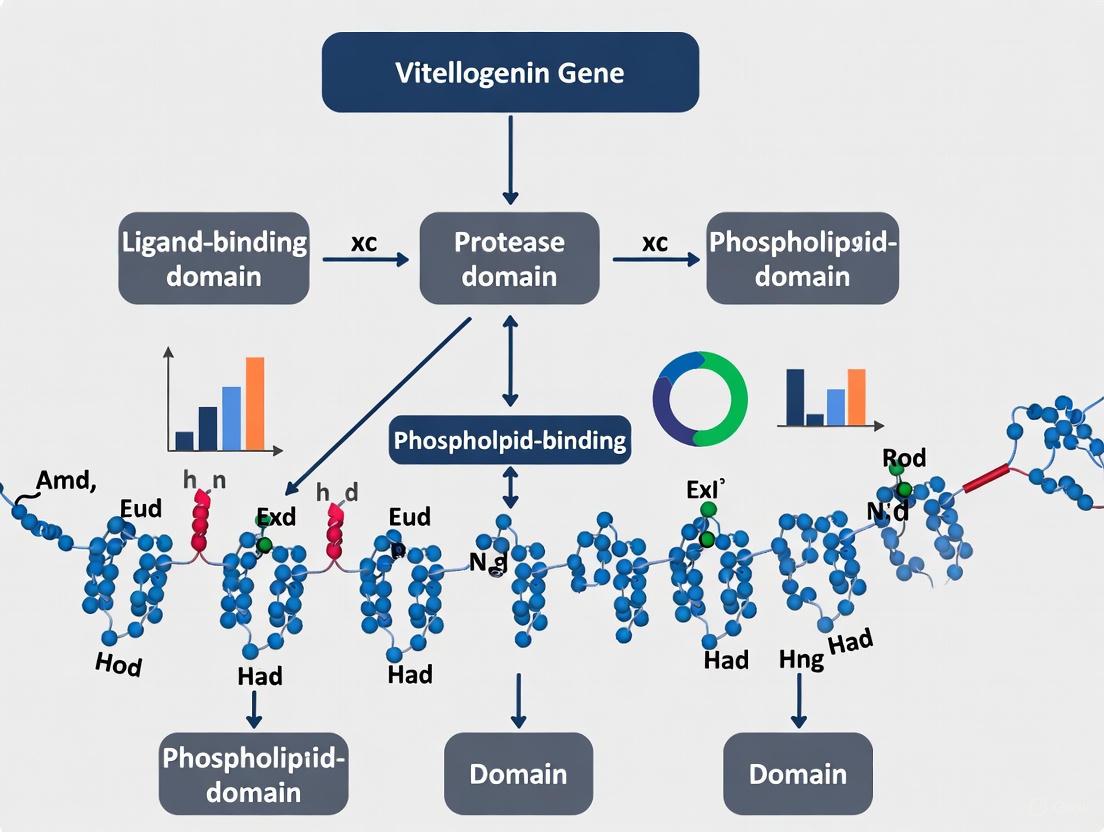
Vitellogenin proteins are characterized by a conserved multidomain architecture that underpins their diverse functions. The core structural domains include:
- Lipoprotein N-terminal domain (LPD_N): Also known as the vitellogenin N-terminal domain, it is critical for receptor recognition and uptake of Vg into oocytes [5].
- Domain of Unknown Function 1943 (DUF1943): A conserved region often associated with pathogen recognition, suggesting immune-related functions [5].
- von Willebrand factor type D domain (vWD): Implicated in protein-protein interactions and immune response [2] [5].
- Large Lipid Transfer Protein (LLTP) module: Comprising N-sheet, A-sheet, C-sheet, and α-helical subdomains that form a hydrophobic lipid-binding cavity [2].
Recent cryo-EM structure analysis of native honey bee vitellogenin has further refined our understanding of this architecture, identifying a previously uncharacterized C-terminal cystine knot (CTCK) domain based on structural homology [2]. In many crustaceans, including Exopalaemon carinicauda, these domains are conserved across multiple paralogous Vg genes, which have expanded significantly in their genomes [5].
Evolutionary History and Expansion Mechanisms
The vitellogenin gene family demonstrates a complex evolutionary history marked by multiple duplication events. Comparative genomic analyses support the hypothesis that the family expanded from two ancestral genes present at the beginning of vertebrate radiation, with subsequent independent duplications occurring across diverse lineages [4]. Whole-genome duplication (WGD) events have been particularly influential in this expansion [4] [3].
Table 1: Vitellogenin Gene Copy Number Variation Across Vertebrate Lineages
| Lineage/Group | Representative Species | Number of Vg Paralogs | Types Identified |
|---|---|---|---|
| Jawless Fishes | Silver Lamprey (Ichthyomyzon unicuspis) | 1 | Single Vg |
| Cartilaginous Fishes | Catshark (Scyliorhinus torazame) | 1 | Single Vg |
| Non-Teleost Bony Fishes | Spotted Gar (Lepisosteus oculatus), Bichir (Acipenser schrenckii) | 3 | - |
| Teleost Fishes | Salmonids, Cyprinids | 3-8+ | VtgAa, VtgAb, VtgC (Acanthomorpha) [4] |
| Sarcopterygians | Coelacanth (Latimeria spp.) | 3 | VtgI, VtgII, VtgIII [4] |
| Crustaceans | Exopalaemon carinicauda | 10 | EcVtg1-8 [5] |
The evolutionary trajectory of gene families like vitellogenin often follows a predictable pattern across eukaryotic lineages. Recent research on macroevolutionary dynamics has revealed that gene family content typically peaks at major evolutionary transitions, then gradually decreases toward extant organisms through a process of simplification and specialization [6]. This pattern reflects intense ecological specialization and "functional outsourcing," where organisms relinquish certain genomic functions to symbiotic partners or their environment [6].
Methodological Framework for Phylogenetic Analysis
Sequence Identification and Curation
Profile Hidden Markov Model (HMM) Searches
- Procedure: Build HMM profiles from seed domain amino acid sequences obtained from databases like Pfam. Use these profiles to scan protein databases with an E-value cutoff (e.g., 1.0) [7].
- Verification: Manually inspect resulting sequences through domain detection to remove artifacts. Use obtained sequences as queries for BLASTP searches (E-value < 0.01) followed by domain verification to identify additional family members [7].
- Application: For vitellogenin studies, key domains include LPD_N (Pfam ID PF01347), DUF1943, and vWD [5].
Clustering Approaches for Homologous Groups
- Algorithm Selection: Utilize fast, sensitive clustering tools like MMseqs2 for large protein datasets [6].
- Parameter Optimization: Adjust the 'c' parameter (minimal fraction of alignment overlap) from 0 (permits short homologous regions) to 0.8 (requires 80% sequence length overlap) to balance cluster inclusivity and stringency [6].
- Ortholog Extraction: For phylogenomic analyses, identify putative orthologs through graph-based clustering approaches followed by processing through tree-based decomposition methods like LOFT, Agalma, or DISCO to extract orthologous genes from families containing paralogs [8].
Phylogenetic Reconstruction and Evolutionary Inference
Multiple Sequence Alignment and Tree Building
- Alignment Tools: Use ClustalX 2.0 or similar software for aligning domain amino acid sequences [7].
- Manual Curation: Refine alignments using tools like Jalview to ensure accuracy [7].
- Phylogenetic Methods: Employ maximum likelihood and Bayesian inference methods to reconstruct gene trees, incorporating both single-copy orthologs and paralogs where appropriate [8].
Microsyntenic Analysis
- Procedure: Examine gene order and content around vitellogenin loci across multiple species to identify conserved genomic regions [4].
- Application: In avian species, the chromosomal localization of three available Gallus gallus vitellogenin sequences (vtgI, vtgII, vtgIII) revealed homologous syntenic blocks in teleost genomes, supporting the existence of an ancestral gene cluster [4].
Table 2: Comparison of Data Selection Strategies for Phylogenomic Inference
| Data Subset | Method of Construction | Advantages | Limitations |
|---|---|---|---|
| Single-Copy Families (SCCs) | Retain clusters with exactly one gene per species [8] | High confidence in orthology; minimal downstream processing [8] | Severely limits data as more species are added [8] |
| Tree-Based Decomposition | Extract orthologs from larger families using phylogenetic approaches [8] | Vastly increases gene number; more accurate orthology prediction [8] | Computationally intensive; requires gene tree estimation [8] |
| All Families (Orthologs + Paralogs) | Use all clustering output without filtering for orthology [8] | Maximizes data utilization; suitable with robust species tree methods [8] | Requires methods robust to paralogy (e.g., ASTRAL) [8] |
Species Tree Inference Methods
- Quartet-based Approaches: Methods like ASTRAL are robust to the inclusion of paralogs because the most common quartet is still expected to match the species tree even with gene duplication and loss [8].
- Concatenation Approaches: Combine aligned sequences from multiple genes into a supermatrix for phylogenetic analysis, which may be misled by incomplete lineage sorting or paralogy if not properly accounted for [8].
Experimental Visualization and Workflow
Phylogenetic Analysis Workflow
The following diagram illustrates the comprehensive workflow for phylogenetic reconstruction of gene families, incorporating both sequence-based and synteny-based approaches:
Vitellogenin Domain Architecture Evolution
This diagram depicts the evolutionary relationships between major LLTP superfamily members, emphasizing the domain architecture of vitellogenin and its paralogs:
Table 3: Key Experimental Reagents and Resources for Gene Family Analysis
| Reagent/Resource | Function/Application | Example Sources/Tools |
|---|---|---|
| Genome Databases | Source of gene and protein sequences for identification and comparison | NCBI, ENSEMBL, Phytozome, UCSC Genome Browser [7] [4] |
| Domain Databases | Identification of conserved protein domains and functional regions | Pfam, SMART, NCBI-CDD [7] [5] |
| HMMER Suite | Building hidden Markov models for sensitive sequence detection | HMMER software [7] |
| Clustering Algorithms | Grouping homologous sequences into gene families | MMseqs2, MCL algorithm [6] |
| Multiple Alignment Tools | Creating alignments for phylogenetic analysis | ClustalX, MAFFT, MUSCLE [7] |
| Phylogenetic Software | Constructing evolutionary trees from sequence data | RAxML, MrBayes, ASTRAL [8] |
| Synteny Browsers | Visualizing and comparing genomic context across species | GENEVESTIGATOR, UCSC Genome Browser [4] |
| RACE Kits | Obtaining full-length cDNA sequences | 5′-RACE System for Rapid Amplification of cDNA Ends [4] |
The phylogenetic history of gene families, exemplified by vitellogenin, reveals complex patterns of expansion and contraction driven by whole-genome duplications, segmental duplications, and lineage-specific adaptations. The vitellogenin family has evolved from ancestral genes in the LLTP superfamily through a series of duplication events beginning before the divergence of teleosts and tetrapods, with additional independent expansions in various lineages [4] [3]. Modern phylogenomic approaches that leverage complete genome sequences and sensitive computational tools have revolutionized our ability to reconstruct these deep evolutionary histories. By integrating sequence-based phylogenetics with microsyntenic analysis and structural insights from techniques like cryo-EM, researchers can now trace the intricate pathways through which single ancestral genes expand into diverse gene families that enable biological innovation across the Tree of Life.
Vitellogenin (Vg), a member of the large lipid transfer protein (LLTP) superfamily, serves as the primary egg-yolk precursor protein in nearly all oviparous species, providing essential nutrients for embryonic development [9] [10]. However, research over the past two decades has revealed that Vg's functions extend far beyond nutrition, encompassing immune defense, antioxidant activity, behavioral regulation, and lifespan determination in various species [9] [10] [2]. These pleiotropic functions are intrinsically linked to Vg's multi-domain architecture, which has been conserved throughout evolution. This whitepaper examines the structure-function relationships of three conserved Vg domains—LPD_N, DUF1943, and von Willebrand factor type D domain (vWD)—synthesizing recent structural biology breakthroughs and functional studies to provide a comprehensive resource for researchers investigating this multifunctional protein.
Vitellogenin is a large, complex glycolipophosphoprotein that typically circulates as a homodimer in the blood or hemolymph [9]. The recent revolution in structural biology techniques, particularly cryo-electron microscopy (cryo-EM) and artificial intelligence (AI)-based prediction algorithms like AlphaFold 2, has dramatically advanced our understanding of Vg's architecture [11] [2]. The 3.2 Å resolution cryo-EM structure of full-length honey bee Vg (AmVg) purified from hemolymph represents a landmark achievement, providing nearly complete coverage of the protein sequence and revealing previously uncharacterized regions [2].
Table 1: Core Domains of Vitellogenin
| Domain | Location | Structural Features | Primary Known Functions |
|---|---|---|---|
| LPDN (VitellogeninN) | N-terminus | Antiparallel β-sheet wrapped around central α-helix; part of LLTP lipid binding module [2] | Receptor binding [10] [2]; nutrient transport [12] |
| DUF1943 | Central region | Classified as a C-terminal cystine knot (CTCK) domain based on structural homology [2] | Bacterial binding [9]; phagocytosis enhancement [9]; pIgR interaction [9] |
| vWD (von Willebrand factor type D) | C-terminus | Structural domain distributed across wide range of proteins [9] | Bacterial binding [9]; direct bacterial growth inhibition [9] |
| Lipid Binding Cavity | Formed by A and C-sheets | Hydrophobic cavity within LLTP module [2] | Lipid transport [2] |
The overall Vg structure comprises a lipid binding module common to the LLTP superfamily, characterized by several subdomains: the N-sheet (LPD_N domain) responsible for receptor binding, the lipid binding cavity itself formed by the A and C-sheets, and an α-helical subdomain that wraps around the A and C-sheets [2]. The recently resolved structures have identified a putative dimerization site in the C-terminal domain and provided new insights into Vg's post-translational modifications, metal binding sites, and cleavage products [2].
Detailed Domain Analysis and Functional Characterization
LPDN (VitellogeninN) Domain
The LPDN domain, also known as the LLT domain or VitellogeninN, is located at the N-terminus of Vg and represents a conserved region found in several lipid transport proteins [9] [13]. Structurally, this domain forms an antiparallel β-sheet wrapped around a central α-helix, creating a structure one strand short of forming a complete barrel with strands of varying lengths [2]. This configuration allows for an overlap between the N-sheet and the A-sheet from the lipid binding cavity, forming a β-sandwich as observed in the silver lamprey lipovitellin structure [2].
Functionally, the LPDN domain has been identified as the primary phosphorylation site and protein modification region, contributing to Vg cleavage, Vg-Vitellogenin receptor (VgR) recognition, and nutrient transport [12]. In the honey bee Vg structure, the region between the N-sheet and the α-helical domain (residues 340-384) corresponds to a polyserine region (polyS) characteristic of insect vitellogenins, which has been shown to be highly disordered with multiple phosphorylated serine residues that prevent its cleavage [2]. Unlike the other two conserved domains, the LPDN domain demonstrates no direct bacterial binding ability [9], suggesting its functional specialization is dedicated to nutritional and developmental roles rather than immune defense.
DUF1943 Domain
The DUF1943 domain, previously designated as a "Domain of Unknown Function," has been structurally classified as a C-terminal cystine knot (CTCK) domain based on structural homology revealed in the recent honey bee cryo-EM study [2]. This breakthrough represents a significant advancement in our understanding of this previously enigmatic domain.
Functionally, DUF1943 exhibits strong bacterial binding capability for both Gram-positive and Gram-negative bacteria [9]. This binding occurs through interactions with signature components on microbial surfaces, specifically lipoteichoic acid (LTA) in Gram-positive bacteria [9]. While DUF1943 binds bacteria effectively, it does not directly inhibit bacterial proliferation [9].
The most precisely defined immune function of DUF1943 is its role in regulating hemocyte phagocytosis. Coimmunoprecipitation assays demonstrate that the DUF1943 domain specifically interacts with the polymeric immunoglobulin receptor (pIgR) [9]. Subsequent functional experiments confirmed that EsVg regulates hemocyte phagocytosis by binding with EspIgR through the DUF1943 domain, thereby promoting bacterial clearance and protecting the host from bacterial infection [9]. This represents the first reported evidence that pIgR acts as a phagocytic receptor for Vg in invertebrates.
vWD (von Willebrand Factor Type D) Domain
The vWD domain is located at the C-terminus of Vg and is distributed across a wide range of proteins [9]. In the recently solved honey bee Vg structure, this previously uncharacterized domain has now been structurally resolved, providing insights into its molecular architecture [2].
Functionally, the vWD domain demonstrates definitive bacterial binding activity through interaction with signature components on microbial surfaces, specifically lipopolysaccharide (LPS) in Gram-negative bacteria [9]. Although its binding affinity is comparatively weaker than that of the DUF1943 domain [9], the vWD domain uniquely possesses direct antibacterial activity.
Antibacterial assays indicate that only the vWD domain inhibits bacterial proliferation in a dose-dependent manner, unlike LPD_N and DUF1943 [9]. This antibacterial function appears to be conserved between different species due to conserved amino acid residues. Mutation studies have identified that T20/F21 conserved amino acid residues are critical for the VWD domain's ability to inhibit bacterial growth, while V35/L36 residues do not affect this function [9].
Table 2: Comparative Immune Functions of Vg Domains
| Immune Function | LPD_N | DUF1943 | vWD |
|---|---|---|---|
| Bacterial Binding | No activity [9] | Strong affinity for both Gram-positive and Gram-negative bacteria [9] | Weaker affinity for both Gram-positive and Gram-negative bacteria [9] |
| Binding Mechanism | N/A | Interaction with LTA in Gram-positive bacteria [9] | Interaction with LPS in Gram-negative bacteria [9] |
| Direct Antibacterial Activity | No inhibition [9] | No inhibition [9] | Significant inhibition in dose-dependent manner [9] |
| Phagocytosis Enhancement | Not reported | Strong enhancement (~100%) via pIgR interaction [9] | Moderate enhancement (~70%) [9] |
| Conserved Critical Residues | Not applicable | Not fully characterized | T20/F21 essential for antibacterial function [9] |
Experimental Approaches and Methodologies
Domain-Specific Functional Assays
To characterize the immune functions of individual Vg domains, researchers have employed a reductionist approach involving the recombinant expression of specific domains followed by functional assays:
Recombinant Protein Expression: Individual Vg domains (LPD_N, DUF1943, and vWD) are cloned and expressed as recombinant proteins in Escherichia coli [9]. These tagged proteins are then purified using affinity chromatography for downstream applications.
Bacteria-Binding Assays: The recombinant domain proteins are incubated with various Gram-positive and Gram-negative bacteria. After incubation and washing, bound proteins are detected through Western blotting or ELISA to quantify binding affinity [9].
Bacterial Growth Inhibition Assays: Recombinant domain proteins are added to bacterial cultures at different concentrations. Bacterial growth is monitored through optical density measurements to determine inhibitory effects [9].
Site-Directed Mutagenesis: Conserved residues within functional domains (e.g., T20/F21 and V35/L36 in vWD) are mutated through PCR-based techniques. The mutant proteins are then tested in functional assays to identify critical residues [9].
Phagocytosis Assays: FITC-labeled bacteria are pre-coated with recombinant domain proteins and incubated with hemocytes. Phagocytosis rates are quantified through flow cytometry or fluorescence microscopy [9].
Diagram 1: Experimental workflow for characterizing Vg domain architecture and function
Structural Analysis Techniques
Recent advances in structural biology have provided unprecedented insights into Vg domain architecture:
Cryo-Electron Microscopy: The native honey bee Vg structure was resolved to 3.2 Å using cryo-EM with direct purification from hemolymph, enabling visualization of post-translational modifications, cleavage products, and metal-binding sites [2].
AlphaFold2 Prediction: AI-based structure prediction has generated high-quality models (pLDDT >80) for Vg proteins across diverse species, complementing experimental methods and providing insights into flexible regions [11] [2].
Molecular Dynamics Simulations: These computational approaches assess the structural impacts of natural variations and deletions on domain stability and function, particularly useful for evaluating population-specific variants [11].
X-ray Crystallography: Earlier structural work on silver lamprey lipovitellin provided initial insights into the LLTP lipid binding module, though with limited coverage (approximately 75% of the sequence) [2].
The Scientist's Toolkit: Research Reagent Solutions
Table 3: Essential Research Reagents for Vg Domain Studies
| Reagent / Method | Specific Application | Function in Research |
|---|---|---|
| Recombinant Domain Proteins | Functional assays for binding, antibacterial activity, and phagocytosis [9] | Enables domain-specific functional characterization independent of full-length Vg |
| Polyclonal/Monoclonal Antibodies | Domain-specific antibody production for Western blot, ELISA, and immunoprecipitation [9] | Detects and quantifies specific domains; used in functional blocking studies |
| HEK293T Cell Line | Heterologous protein expression for coimmunoprecipitation assays [9] | Validates protein-protein interactions (e.g., DUF1943-pIgR interaction) |
| RNAi/dsRNA Tools | Gene silencing in model organisms (e.g., insects, crustaceans) [14] [15] | Determines domain-specific functional loss in vivo |
| LTA/LPS Components | Binding specificity assays [9] | Identifies specific microbial surface components interacting with Vg domains |
| IndeLLM/Pathogenicity Predictors | Computational assessment of indel impacts on domain structure [11] | Predicts structural consequences of natural variations and mutations |
Evolutionary and Comparative Perspectives
The Vg gene family exhibits complex evolutionary patterns across taxa. Vertebrates began with a single Vg copy, with bird-mammalian and amphibian lineages experiencing independent duplications [4]. Most mammals have pseudogenized their Vg genes, with the exception of monotremes which retained one functional gene [13] [4]. In contrast, many invertebrate species maintain multiple Vg subtypes with potentially specialized functions [14].
The conserved domain architecture across diverse taxa suggests strong evolutionary pressure to maintain these structural units. The emergence of unique functions for different domains, particularly the immune specialization of DUF1943 and vWD, represents an fascinating example of functional co-option where domains within a primarily reproductive protein have been adapted for immune defense across evolutionary lineages [9] [12] [2].
Recent research on the mud crab Scylla paramamosain has identified a novel Vg gene (SpVTG3) with the characteristic LPD_N, DUF1943, and VWD domains that shows distinct expression patterns during embryonic development, suggesting Vg domains may play previously uncharacterized roles in embryogenesis beyond nutrient provision [14]. RNA interference studies demonstrated that Spvtg3 knockdown significantly impaired embryonic development, indicating its essential role in this process [14].
The conserved LPDN, DUF1943, and vWD domains represent the architectural foundation of vitellogenin's pleiotropic functions. While the LPDN domain specializes in receptor recognition and nutrient transport, the DUF1943 and vWD domains have evolved specialized immune capabilities including pathogen recognition, direct antibacterial activity, and phagocytosis enhancement. The recent structural biology revolution, particularly through cryo-EM and AI-based prediction, has dramatically enhanced our understanding of these domains at the molecular level. Future research should focus on elucidating the precise molecular mechanisms by which these domains achieve their diverse functions and how natural variations in these domains impact organismal fitness and disease resistance. This knowledge may provide novel insights for therapeutic development and conservation strategies across species.
Large Lipid Transfer Protein Superfamily Relationships
The Large Lipid Transfer Protein (LLTP) superfamily represents a class of essential proteins facilitating lipid transport, metabolism, and signaling across animal taxa. This comprehensive review delineates the evolutionary relationships, structural characteristics, and functional diversification within this superfamily, with particular emphasis on the central role of vitellogenin (Vtg) as the ancestral foundation. We examine the molecular architecture of LLTP domains, quantitative comparative analyses across family members, experimental methodologies for structural and functional characterization, and emerging research tools. Framed within vitellogenin gene structure and domain research, this analysis provides a technical foundation for researchers and drug development professionals investigating lipid transport mechanisms, reproductive biology, and metabolic regulation.
The Large Lipid Transfer Protein (LLTP) superfamily comprises essential molecules responsible for the circulatory transport of lipids in animals, with their emergence linked to the increased need for lipid transport associated with multicellularity [2]. These proteins share a common evolutionary origin and structural features that enable their lipid-binding capabilities. From a phylogenetic perspective, vitellogenin represents the most ancestral and oldest member of this superfamily, believed to date back at least 700 million years [10]. The expansion and diversification of LLTPs in invertebrates appear to be mediated via retrotransposon-mediated duplications, followed by either subfunctionalization or neofunctionalization in different lineages [16].
The LLTP superfamily includes several key protein families that have evolved specialized functions. Apolipoprotein B (apoB) functions primarily in cholesterol transport and lipoprotein assembly in mammals [13] [10]. Microsomal triglyceride transfer protein (MTP) plays a critical role in the biosynthesis and lipid loading of apolipoprotein B [13]. Vitellogenin (Vtg) serves as the main yolk precursor lipoprotein in almost all egg-laying animals [2]. Insect apolipophorins II/I (apoLp-II/I) represent the insect homologs of apolipoprotein B [2]. According to current understanding, all LLTPs originate from a series of duplications of a primitive yolk protein gene similar to Vtg, with two early consecutive duplications resulting in the formation of MTP and the APO gene ancestor [16].
Table 1: Evolutionary Relationships within the LLTP Superfamily
| Protein Family | Primary Function | Structural Features | Evolutionary Origin |
|---|---|---|---|
| Vitellogenin (Vtg) | Yolk precursor nutrient storage | LPD_N, DUF1943, vWD domains | Ancestral LLTP, ~700 million years old |
| Apolipoprotein B (apoB) | Cholesterol transport, lipoprotein assembly | LLTP lipid binding module | Early duplication from Vtg ancestor |
| MTP | Lipid loading protein for other LLTPs | Truncated LLTP module | Early duplication from Vtg ancestor |
| Apolipophorins II/I | Insect lipid transport | Homolog of apoB | Lineage-specific evolution in insects |
| Bridge-like LTPs (BLTPs) | Bulk lipid transfer at membrane contact sites | Repeating β-groove (RBG) domains | Distinct structural class |
Structural Domains and Molecular Architecture
Conserved Domain Architecture of Vitellogenin and Related LLTPs
Vitellogenin and other LLTPs share a characteristic lipid binding module that defines the superfamily [2]. This module consists of several structurally conserved subdomains: the N-sheet (responsible for receptor binding), the lipid binding cavity formed by the A and C-sheets, and the α-helical subdomain that wraps around the A and C-sheets [2] [10]. The N-sheet is found at the N-terminus and formed by an antiparallel β-sheet wrapped around a central α-helix, creating a structure that is one strand short of forming a barrel with strands of very different lengths [2].
The domain architecture of vitellogenin includes three conserved functional domains that have been characterized across multiple species. The lipoprotein N-terminal domain (LPD_N), also known as the vitellogenin N-terminal domain, represents a conserved region found in several lipid transport proteins [17] [13]. This domain is required for interaction with the Vtg receptor and plays a conserved role in receptor recognition in both vertebrates and invertebrates [17]. The domain of unknown function (DUF1943) and von Willebrand factor type D domain (vWD) have been implicated in pathogen recognition, suggesting immune-related functions for Vtg beyond its nutritional role [17]. Recent structural studies of honey bee Vtg have identified the previously uncharacterized vWD domain and classified a domain of unknown function as a C-terminal cystine knot (CTCK) domain based on structural homology [2].
Structural Classification of Lipid Transfer Proteins
Beyond the classical LLTP superfamily, a novel superfamily of bridge-like lipid transfer proteins (BLTPs) has recently been identified [18] [19]. These proteins are characterized by a unique structural feature termed the repeating β-groove (RBG) domain [18]. This modular unit contains five antiparallel β-strands followed by a disordered loop usually starting with a short helix that curves back across the β-sheet [18]. The β-strands form a U-shape with hydrophobic residues populating the inner face and hydrophilic residues on the exterior face [18]. Multimerization of these repeating units creates an unbroken chain of structurally identical repeats that together build a hydrophobic groove functioning as a "lipid superhighway" connecting organelle membranes at membrane contact sites [18].
Table 2: Structural Classification of Lipid Transfer Proteins
| Structural Class | Representative Proteins | Structural Features | Lipid Transfer Mechanism |
|---|---|---|---|
| Classical LLTPs | Vtg, apoB, MTP | LLTP lipid binding module with N-sheet, A/C-sheets, α-helical domain | Hydrophobic cavity for individual lipid molecules |
| Box-like LTPs | OSBP, Sec14 family | Box-like shape with hydrophobic pocket | Shuttling mechanism for single lipid molecules |
| Bridge-like LTPs (RBG proteins) | VPS13, ATG2, BLTP1-3 | Repeating β-groove (RBG) domains forming long rods | Continuous hydrophobic groove for bulk lipid transport |
Functional Diversification and Pleiotropy
The LLTP superfamily exhibits remarkable functional diversification across its member proteins, with vitellogenin representing a prime example of profound pleiotropy [2]. While traditionally studied as a female-specific protein in the context of vitellogenesis, Vg has developed a range of new functions in different taxa [2]. In the honey bee, Vg has acquired functions related to immunity, antioxidant protection, social behavior, and longevity [2] [10].
The molecular basis for Vg pleiotropy appears to stem from its structural characteristics and evolutionary history. Vg's function as a transport protein derives from its ability to bind to lipids and numerous ligands through its biochemical structure [10]. The N-terminus β-barrel (N-sheet) harbors the receptor binding area of the protein, while the α-helical domain contains a lipophilic cavity implicated in binding to various ligands [10]. This α-helical domain is also believed to facilitate vitellogenin's anti-inflammatory functions [10]. In recent years, data about the immune functions of Vg have emerged in taxa as different as corals, mollusks, arthropods, and fishes [2]. Vg has been found to have antibacterial and antiviral activities, achieved through recognizing pathogen-associated molecular patterns (PAMPs), directly causing pathogen death, or opsonizing for phagocytosis by immune cells [2].
Beyond immunity, Vg plays critical roles in reproductive regulation. In insects, vitellogenin synthesis is typically coordinated with titers of ecdysteroids and juvenile hormones [10]. Nutritional status is sensed, at least in part, by signaling through insulin/insulin-like pathways and by JHs and target of rapamycin (TOR)-dependent mechanisms [10]. The Vg expression is part of a regulatory feedback loop that enables vitellogenin and juvenile hormone to mutually suppress each other [13]. Vitellogenin and juvenile hormone likely work antagonistically in the honey bee to regulate development and behavior, with suppression of one leading to high titers of the other [13].
Experimental Approaches and Methodologies
Structural Characterization Techniques
Advanced structural biology techniques have been instrumental in elucidating the architecture of LLTP superfamily members. Cryogenic electron microscopy (cryo-EM) has emerged as a powerful method for determining the structure of large, flexible proteins like vitellogenin. The recent cryo-EM structure of native honey bee vitellogenin was determined at 3.2 Å resolution, providing nearly full-length coverage including previously uncharacterized domains [2]. The experimental workflow involved several key steps that can be adapted for structural studies of other LLTPs.
For the honey bee Vg structure, hemolymph was collected as the native source, followed by one-step purification directly from hemolymph [2]. The sample was heterogeneous and contained full-length protein along with cleavage products. Particles of both full-length Vg and cleavage products were processed separately, with the cleavage product yielding higher resolution maps of 3.0 Å [2]. The structural data was complemented with multiple sequence alignment (MSA) of homologous sequences, providing information about residue conservation and confidence in structural elements when cryo-EM density was not conclusive [2].
Functional Assays for Lipid Transfer Activity
Several experimental approaches have been developed to assess the lipid transfer capabilities of LLTP superfamily members. For bridge-like LTPs, in vitro lipid transfer assays have been crucial for demonstrating function. These assays typically involve purified protein incubated with donor and acceptor liposomes, followed by measurement of lipid movement between membranes [18] [19]. For VPS13A and ATG2A, purified proteins have been shown to be capable of transporting lipids between liposomes in vitro [19].
Cellular localization studies provide complementary functional information. Proteins in the BLTP family, including VPS13A and ATG2A, localize to membrane contact sites in cultured cells, where they form bridges that connect two organelle membranes and allow non-vesicular lipid transfer [19]. This subcellular localization is consistent with their proposed function as bridge-like lipid transporters.
For vitellogenin, functional assays often focus on its role in reproduction and immunity. Gene expression analyses during ovarian development can identify Vtg genes with major roles in exogenous and endogenous vitellogenesis [17]. Eyestalk ablation experiments in crustaceans have demonstrated regulatory control over Vtg synthesis, with bilateral ablation of the eyestalk significantly upregulating EcVtg mRNA expression in the female hepatopancreas [17]. Antibacterial assays can assess Vg's immune functions by testing its activity against various pathogens [2].
Table 3: Essential Research Reagents and Experimental Tools
| Research Reagent/Tool | Application | Function in LLTP Research |
|---|---|---|
| Cryo-EM with single particle analysis | Structural biology | High-resolution structure determination of native LLTPs |
| Liposome-based transfer assays | Functional biochemistry | In vitro measurement of lipid transfer activity |
| AlphaFold2 prediction | Computational structural biology | Protein structure prediction for poorly characterized LLTPs |
| Multiple sequence alignment | Bioinformatics | Identification of conserved domains and residues |
| Gene expression profiling (qPCR/RNA-seq) | Molecular biology | Expression analysis during development or in different tissues |
| RNA interference (RNAi) | Functional genetics | Gene knockdown to assess functional consequences |
| Hepatopancreas/fat body extracts | Tissue biochemistry | Source of native LLTPs from synthesizing tissues |
| Lipid-binding probes | Biochemical assays | Detection and quantification of lipid-protein interactions |
Genomic Organization and Expression Regulation
Vitellogenin Gene Family Diversity
Genome-wide analyses have revealed significant diversity in vitellogenin gene family organization across species. In the ridgetail white shrimp (Exopalaemon carinicauda), 10 Vtg genes have been identified and characterized, unevenly distributed across chromosomes [17]. Phylogenetic analyses show that Vtg genes in crustaceans can be classified into four groups: Astacidea, Brachyra, Penaeidae, and Palaemonidae [17]. Molecular evolutionary analysis indicates that EcVtg genes are primarily constrained by purifying selection during evolution, suggesting conservation of essential functions [17].
In vertebrates, the vitellogenin gene family has undergone lineage-specific expansions. Vertebrates started with a single copy of the vitellogenin gene, with bird-mammalian and amphibian lineages each experiencing duplications that gave rise to modern genes [13]. With the exception of monotremes, mammals have turned all their vitellogenin genes into pseudogenes, although the region syntenic to bird VIT1-VIT2-VIT3 can still be found and aligned [13]. This pattern reflects the evolutionary trajectory of LLTP superfamily members, with duplication events followed by functional diversification or gene loss in different lineages.
Regulation of Expression and Synthesis
Vitellogenin gene expression is tightly regulated in accordance with reproductive cycles and environmental conditions. In crustaceans, Vtg genes exhibit higher expression in the female hepatopancreas than in other tissues, and expression patterns during ovarian development suggest the hepatopancreas as the main synthesis site [17]. Different Vtg paralogs play distinct roles in vitellogenesis, with some having major roles in exogenous vitellogenesis while others function in endogenous vitellogenesis [17].
In insects, vitellogenin synthesis is regulated by a complex interplay of hormonal signals and nutritional status. The fat body serves as the primary site of Vg synthesis, with production rates typically coordinated with titers of ecdysteroids and juvenile hormones [10]. Nutritional status is sensed through insulin/insulin-like pathways and TOR-dependent mechanisms [10]. The regulatory feedback loop between vitellogenin and juvenile hormone enables mutual suppression, creating a dynamic system that regulates honey bee development and behavior [13].
The Large Lipid Transfer Protein superfamily represents a remarkable example of evolutionary innovation through gene duplication and functional diversification. Vitellogenin stands as the ancestral foundation of this superfamily, with its pleiotropic functions illustrating how a primary reproductive protein can acquire diverse physiological roles. The structural characterization of LLTP members, particularly through cryo-EM and predictive modeling with AlphaFold, has revolutionized our understanding of their lipid transfer mechanisms.
Future research directions should focus on several key areas. First, the molecular basis for Vg's immune functions requires further elucidation, particularly how its pathogen recognition capabilities intersect with its lipid transport functions. Second, the regulatory networks controlling LLTP expression and activity in different physiological contexts remain incompletely understood. Third, the potential applications of LLTP research in drug development, particularly for lipid metabolism disorders and reproductive health, warrant expanded investigation. Finally, comparative studies across diverse taxa will continue to reveal the evolutionary principles guiding LLTP superfamily diversification and specialization.
As structural biology techniques advance and genomic data accumulate, our understanding of the LLTP superfamily will continue to deepen, providing new insights into fundamental biological processes and potential therapeutic applications.
Gene Duplication Events and Lineage-Specific Expansions
Gene duplication is a fundamental evolutionary process that provides the raw genetic material for functional innovation and adaptation. Within the context of vitellogenin (Vg) gene research, duplication events and subsequent lineage-specific expansions have played a critical role in shaping the structural and functional diversity of this essential gene family. Vitellogenin, a large lipid transfer protein primarily known as the main yolk precursor in egg-laying animals, exhibits remarkable pleiotropy across taxa, with functions extending to immunity, antioxidant protection, social behavior, and longevity regulation [2]. The complex domain architecture and multifaceted functionality of Vg make it an ideal model system for studying the evolutionary consequences of gene duplication. This technical guide examines the mechanisms, patterns, and experimental approaches for investigating gene duplication events and lineage-specific expansions, with particular emphasis on vitellogenin gene family evolution and its implications for biomedical and pharmacological research.
Mechanisms of Gene Duplication
Gene duplication occurs through several distinct mechanistic pathways, each with different implications for gene structure, regulation, and evolutionary potential. Understanding these mechanisms is crucial for interpreting patterns observed in the vitellogenin gene family and other expanded gene families.
Table 1: Mechanisms of Gene Duplication and Their Characteristics
| Mechanism | Scale | Key Features | Evolutionary Implications |
|---|---|---|---|
| Whole Genome Duplication (WGD) | Genome-wide | Duplication of all genomic content; also called polyploidization | Preserves stoichiometric balances in molecular networks; common in plants and some vertebrate lineages [20] |
| Segmental Duplication | Intermediate (1kb-1Mb) | Unequal crossing-over during meiosis | Can duplicate gene clusters or regulatory regions; frequent in eukaryotes [20] |
| Tandem Duplication | Local (adjacent genes) | Unequal crossing-over between closely related sequences | Creates gene arrays; often associated with stress response and environmental adaptation genes [21] |
| Retrotransposition | Single gene | Reverse transcription of mRNA and integration | Produces intron-less copies (retrogenes); often tissue-specific expression patterns [20] |
Whole genome duplication events have been particularly significant in vertebrate evolution, including the two rounds of WGD at the base of vertebrates (1R and 2R), the teleost-specific WGD (Ts3R), and the salmonid-specific WGD (Ss4R) [4]. These events initially provide duplicated copies of all genes, including vitellogenin genes, which subsequently undergo differential loss and functional divergence in various lineages.
Segmental and tandem duplications represent ongoing processes in genomes and contribute to lineage-specific expansions. In plants, tandem duplicates are significantly enriched in genes involved in environmental stress responses, while nontandem duplicates more often have intracellular regulatory roles [21]. This pattern suggests that duplication mechanisms are non-random with respect to gene function, influencing the functional spectrum of expanded gene families.
Evolutionary Fate of Duplicated Genes
Following duplication, genes may undergo several evolutionary trajectories that determine their long-term retention and functional characteristics. The vitellogenin gene family exemplifies these diverse fates across different taxonomic groups.
Fixation and Maintenance Pathways
A critical distinction exists between the initial fixation of a duplicate in a population and its long-term maintenance. Fixation probability depends on population genetics parameters and immediate selective advantages, while maintenance depends on continued functional utility [20]. Duplicates can be classified into four theoretical categories based on these properties: spreading difficult/maintenance difficult; spreading difficult/maintenance easy; spreading easy/maintenance difficult; and spreading easy/maintenance easy.
For vitellogenin genes, dosage effects often provide immediate selective advantages that promote fixation. Increased gene copy number generally leads to increased product dosage [22], which can be advantageous for nutrient transport and storage functions. In yeast, for example, duplication of hexose transporter genes (HXT6 and HXT7) provides a selective advantage under low-glucose conditions through increased glucose transport capacity [22].
Functional Divergence Mechanisms
Table 2: Evolutionary Fates of Duplicated Genes
| Fate | Molecular Mechanism | Examples in Vitellogenin Genes |
|---|---|---|
| Nonfunctionalization | Accumulation of deleterious mutations in one copy | Gene loss following WGD events in various vertebrate lineages [4] |
| Neofunctionalization | One copy acquires novel function | Immune functions of Vg in corals, mollusks, arthropods, and fishes [2] |
| Subfunctionalization | Partitioning of ancestral functions between copies | Caste- and task-specific expression of Vg duplicates in social insects [23] |
| Dosage Conservation | Maintained selection for increased gene dosage | Nutrient transport adaptation in various taxa [22] |
In social Hymenoptera, vitellogenin gene duplicates have undergone both subfunctionalization and neofunctionalization. In the ant Formica fusca, conventional Vg shows queen- and nurse-biased expression, while Vg-like-C displays forager-biased expression [23]. This expression partitioning represents subfunctionalization of regulatory elements. Meanwhile, the acquisition of immune functions in Vg duplicates across diverse taxa represents neofunctionalization events [2].
Lineage-Specific Expansions in Vitellogenin Gene Family
The vitellogenin gene family exhibits remarkable lineage-specific expansion patterns that reflect diverse life history strategies and ecological adaptations.
Vertebrate Vitellogenin Gene Evolution
Comparative genomic analyses reveal complex evolutionary history of vitellogenin genes in vertebrates. Early hypotheses suggested that multiple Vg copies originated through whole genome duplication events, with expectations of four Vg genes in early-branching fish and tetrapods, eight in teleosts, and up to sixteen in salmonids [4]. However, empirical data show extensive gene loss following duplication events, resulting in lineage-specific repertoires.
Microsyntenic and phylogenetic analyses support the hypothesis that the vitellogenin gene family expanded from two genes present at the beginning of vertebrate radiation through multiple independent duplication events in different lineages [4] [24]. Jawless vertebrates like lamprey typically possess a single Vg gene, while non-teleost fish such as spotted gar have three Vg sequences. Among teleosts, salmonids have three paralog genes (vtgAsa1, vtgAsb, and vtgC), while cyprinids and anguillids have several homologous genes [4].
Invertebrate Vitellogenin Gene Expansions
In social insects, vitellogenin gene family expansions show distinct patterns correlated with social complexity. The honey bee (Apis mellifera) possesses a single conventional Vg gene [2], while ant species exhibit substantial variation in Vg copy number. Formica exsecta and Camponotus floridanus have one conventional Vg copy, Pogonomyrmex barbatus and Atta cephalotes have two copies, Solenopsis invicta has four copies, and Linepithema humile has five copies [23].
In Formica ants, in addition to conventional Vg, three Vg-like genes (Vg-like-A, -B, and -C) have been identified, with Vg-like-C found exclusively in Hymenoptera [23]. These homologs differ in conserved protein domains and have undergone rapid evolution after duplication, suggesting functional diversification related to social organization.
Experimental Approaches for Studying Gene Duplications
Computational and Phylogenetic Methods
Phylogenetic analysis combined with microsyntenic investigations provides powerful tools for reconstructing the evolutionary history of gene families. For vitellogenin genes, this approach has revealed lineage-specific duplication events and differential gene loss [4] [24]. Sequence alignment of homologous regions across multiple species allows identification of conserved domains and lineage-specific innovations.
Population genetics approaches can detect selection on recent gene duplications through analyses of variability around emerging gene copies (hitchhiking effects) [22]. However, these methods face technical challenges in assembling recent gene duplications from whole genome sequencing data.
Structural Biology Techniques
Recent advances in cryo-electron microscopy (cryo-EM) have enabled high-resolution structural analysis of complex proteins like vitellogenin. The 3.2 Å resolution cryo-EM structure of native honey bee Vg revealed previously uncharacterized domains, including the von Willebrand factor type D domain (vWD) and a C-terminal cystine knot domain (CTCK) [2]. Such structural data provide mechanistic insights into how duplication and divergence affect protein function.
Artificial intelligence-based structure prediction tools, particularly AlphaFold 2, have complemented experimental approaches by generating high-quality predicted protein structures for thousands of Vg variants across diverse species [11]. These computational models facilitate analysis of structural impacts of natural variation, including deletions and substitutions.
Gene Expression Analyses
Quantitative RT-PCR enables precise measurement of gene expression patterns across castes, tasks, and social contexts in social insects. In Formica fusca, expression analysis of conventional Vg and Vg-like genes revealed caste-specific (queen vs. worker) and task-specific (nurse vs. forager) expression patterns [23]. Such expression partitioning provides evidence for subfunctionalization after gene duplication.
Experimental manipulation of social context (e.g., queenless vs. queenright colonies) can reveal plastic responses in gene expression and provide insights into regulatory evolution following duplication events [23].
Visualization of Gene Duplication Concepts and Workflows
Research Reagent Solutions for Gene Duplication Studies
Table 3: Essential Research Reagents for Vitellogenin and Gene Duplication Studies
| Reagent/Category | Specific Examples | Research Application | Key Functions |
|---|---|---|---|
| Structural Biology Tools | Cryo-EM single particle analysis | Native protein structure determination | Resolves domain architecture, lipid binding cavities, post-translational modifications [2] |
| AI Prediction Resources | AlphaFold 2 database | Structural prediction for diverse Vg variants | Models protein structures across species; assesses impact of sequence variation [11] |
| Gene Expression Analysis | Quantitative RT-PCR with specific primers | Caste- and task-specific expression profiling | Measures expression patterns of conventional Vg and Vg-like genes [23] |
| Sequence Analysis Tools | Multiple sequence alignment algorithms | Phylogenetic reconstruction and domain identification | Identifies conserved regions and lineage-specific innovations [2] |
| Population Genomics | Whole genome sequencing with assembly | Identification of copy number variation | Detects segregating duplications and fixed copy number differences [22] |
| Molecular Dynamics | Simulation software (e.g., GROMACS) | Assess structural impacts of natural variation | Evaluates protein stability and dynamics following indels or substitutions [11] |
Gene duplication events and lineage-specific expansions represent fundamental evolutionary mechanisms that have shaped the diversity and functional complexity of the vitellogenin gene family across taxa. The interplay between whole genome duplication, segmental duplication, and tandem duplication has generated complex gene families with members that undergo various evolutionary fates, including nonfunctionalization, neofunctionalization, subfunctionalization, and dosage conservation. The vitellogenin system provides a compelling model for understanding these processes, with clear examples of how gene duplication enables functional innovation in reproduction, immunity, social behavior, and longevity regulation. Experimental approaches combining phylogenetic analysis, structural biology, gene expression profiling, and population genomics continue to reveal the intricate relationships between duplication mechanisms, structural constraints, and functional outcomes. This knowledge provides a foundation for understanding evolutionary innovations not only in vitellogenin genes but across expanded gene families with relevance to human health and disease.
Structural Evolution of Vitellogenin-Like Proteins and Their Divergent Functions
Vitellogenin (Vg) is an ancient and conserved glycolipophosphoprotein that serves as the primary precursor of yolk proteins in nearly all oviparous animals, providing essential nutrients for embryonic development [25] [4]. This protein belongs to the large lipid transfer protein (LLTP) superfamily, which also includes microsomal triglyceride transfer protein (MTP) and apolipoprotein B (apoB) [2] [25]. Along its evolutionary history, Vg has acquired a remarkable array of novel functions in various taxa, extending far beyond its ancestral role in reproduction. In social insects specifically, Vg has developed functions related to immunity, antioxidant protection, social behavior, caste differentiation, and longevity [26] [2]. The structural evolution of Vg and its homologs—the vitellogenin-like proteins (Vg-likes)—represents a fascinating case study in how gene duplication and structural diversification enable functional innovation.
This whitepaper examines the molecular evolution of the vitellogenin gene family, focusing on how structural changes in Vg and Vg-like proteins have facilitated their functional divergence. The content is framed within broader research on vitellogenin gene structure and domains, providing technical insights relevant for researchers investigating evolutionary biology, protein structure-function relationships, and molecular genetics.
Structural Domains and Phylogenetic Relationships
Conserved Protein Architecture
Vitellogenin proteins are characterized by a conserved multi-domain architecture that underlies their diverse functionalities. The canonical domains include:
- Lipoprotein N-terminal domain (LPDN or VitellogeninN): Also known as the N-sheet or β-barrel domain, this region is responsible for receptor binding and contains structural features that may facilitate DNA binding [25] [11] [27].
- Domain of Unknown Function 1943 (DUF1943): A centrally located domain with potential roles in pathogen recognition and immune function [25] [5].
- von Willebrand factor type D domain (vWD): A C-terminal domain involved in multimerization and potentially immune recognition [2] [25] [5].
Table 1: Conserved Structural Domains in Vitellogenin and Vg-Like Proteins
| Domain | Structural Features | Known or Proposed Functions |
|---|---|---|
| LPDN (VitellogeninN) | N-terminal β-barrel composed of 12 β-strands | Receptor recognition, lipid binding, DNA binding [2] [11] [27] |
| DUF1943 | Central domain of unknown structure | Pathogen recognition, immune response [25] [5] |
| vWD | C-terminal domain with conserved cysteine residues | Multimerization, immune function [2] [25] |
| Polyserine region | Disordered region with multiple serine residues | Phosphorylation sites, protease resistance [2] |
| Lipid binding cavity | Formed by A and C-sheets, wrapped by α-helical domain | Lipid transport and storage [2] |
Phylogenetic History and Gene Duplication Events
The vitellogenin gene family has expanded through several evolutionary mechanisms. Gene duplication events have played a crucial role in creating functional diversity within this protein family [26] [25] [4]. In vertebrates, the Vg gene family expanded from two ancestral genes present at the beginning of vertebrate radiation through multiple independent duplication events in different lineages [4]. In insects, particularly Hymenoptera, an ancient gene duplication event gave rise to the conventional Vg and three Vg-like genes (Vg-like-A, Vg-like-B, and Vg-like-C) [26] [23].
Table 2: Vitellogenin Gene Family Members Across Taxa
| Gene/Protein | Distribution | Domain Composition | Evolutionary Pattern |
|---|---|---|---|
| Conventional Vg | All oviparous species | LPD_N, DUF1943, vWD, polyserine region, lipid binding cavity | Strong positive selection in social insects [26] |
| Vg-like-A | All insects | Similar to Vg but with domain variations | Relaxed purifying selection [26] |
| Vg-like-B | All insects | Lost several Vg structural elements | Relaxed purifying selection [26] |
| Vg-like-C | Hymenoptera only | Primarily contains N-sheet domain | Rapid evolution after duplication [26] [23] |
| VtgAa/VtgAb/VtgC | Teleost fishes | VtgC lacks Pv domain | Lineage-specific duplication and subfunctionalization [4] |
The phylogenetic analysis of Vg genes reveals a complex evolutionary history with multiple instances of lineage-specific expansions. In crustaceans such as Exopalaemon carinicauda, genome-wide analyses have identified up to 10 Vg genes, indicating additional duplication events in this lineage [5]. Similarly, in insects, species such as the mosquito Aedes aegypti and the ant Linepithema humile possess up to five Vg copies, while others like the honeybee (Apis mellifera) have only a single conventional Vg gene [25].
Molecular Evolution and Selection Pressures
Differential Selection Across Gene Family Members
Molecular evolutionary analyses reveal distinct selection pressures acting on different members of the Vg gene family. In bumble bees (Bombus), the conventional Vg has experienced strong positive selection (dN/dS = 1.311), while the Vg-like genes show an overall relaxation of purifying selection [26]. This pattern contrasts with that observed in honey bees and stingless bees, where all four Vg genes remain under purifying selection [26].
The strength of selection varies considerably across taxonomic groups and ecological contexts. In bumble bees, positive selection on conventional Vg occurs across most subgenera, with the notable exception of the obligate parasitic subgenus Psithyrus (dN/dS = 0.713), which has lost caste differentiation [26]. This suggests that the social functions of Vg, particularly those related to caste differentiation and division of labor, may drive positive selection in social insects.
Structural Variation and Functional Innovation
Following gene duplication, Vg-like genes have undergone structural diversification that has enabled functional innovation. Vg-like-B has lost several structural elements present in conventional Vg, potentially limiting its ability to perform the full range of ancestral Vg functions [26]. Vg-like-C retains primarily the N-sheet domain, suggesting potential functional specialization [26]. This structural simplification in Vg-like genes may facilitate the evolution of novel functions unconstrained by the requirements of vitellogenesis.
Recent research has identified a population-specific 9-nucleotide deletion in the Vg β-barrel domain of the locally endangered European Dark Bee subspecies (A. m. mellifera) [11]. Structural bioinformatics and molecular dynamics simulations demonstrate that this deletion does not disrupt Vg's structure or stability, revealing the structural plasticity of this conserved domain [11].
Divergent Functional Roles Across Taxa
Conventional Vg: A Pleiotropic Protein in Social Insects
The conventional Vg protein maintains its ancestral role in vitellogenesis and oocyte development across insect taxa [26] [25]. RNA interference studies in the brown planthopper (Nilaparvata lugens) demonstrate that Vg is essential for both oocyte development and nymph development [25]. However, in social insects, Vg has acquired additional social pleiotropic functions:
- Caste differentiation: Vg is upregulated in queens compared to workers and in nurses compared to foragers [26] [23].
- Longevity regulation: Vg contributes to queen longevity and the extended lifespan of winter bees [26] [2].
- Behavioral regulation: Vg influences the transition from nursing to foraging behaviors in honey bees [26] [27].
- Antioxidant protection: Vg enhances stress resistance through its antioxidant properties [2] [25].
- Immune priming: Vg is involved in transgenerational immune priming [2].
Specialized Functions of Vg-Like Proteins
The Vg-like proteins have evolved specialized functions distinct from conventional Vg:
- Vg-like-A: Shows the closest structural and functional similarity to conventional Vg. It responds strongly to inflammatory and oxidative conditions, is associated with aging processes, and is linked to nursing behaviors in ants [26] [23]. In honey bees, Vg-like-A shows strong temporal expression variation and may contribute to wintering worker longevity [26].
- Vg-like-B: Has lost several Vg structural elements and may perform only a subset of Vg's original functions, such as coping with oxidative stress [26].
- Vg-like-C: Contains primarily the N-sheet domain and may have neurobiological functions, though its precise role remains unclear [26] [23]. In the ant Formica fusca, Vg-like-C displays consistent forager-biased expression patterns [23].
Table 3: Functional Roles of Vg Gene Family Members in Social Insects
| Gene | Reproductive Function | Behavioral Function | Immunological Function | Stress Response |
|---|---|---|---|---|
| Conventional Vg | Egg yolk precursor, oocyte development | Caste differentiation, nursing behavior | Immune priming, antibacterial activity | Antioxidant protection, oxidative stress resilience |
| Vg-like-A | Limited role | Regulation of nursing behaviors | Strong response to inflammatory conditions | Oxidative stress response, aging processes |
| Vg-like-B | Minimal role | Unknown | Moderate immune response | Oxidative stress coping mechanism |
| Vg-like-C | Unknown | Forager-biased expression | Unknown | Unknown |
Non-Nutritional Functions: DNA Binding and Gene Regulation
Recent research has revealed a novel function for Vg in gene regulation. Evidence from honey bees indicates that a Vg subunit can translocate to the nucleus and interact with DNA [27]. Structural analyses have identified conserved DNA-binding amino acids in the β-barrel domain of Vg, with structural regions similar to established DNA-binding proteins [27]. This Vg-DNA binding is associated with expression changes in dozens of genes involved in energy metabolism, behavior, and signaling [27].
The DNA-binding capability of Vg appears to involve:
- Outward-facing β-strands in the β-barrel domain that can interact with DNA
- A central α-helix and putative zinc-binding sites that stabilize the structure
- Glycosylation sites on the β-barrel domain that may support DNA binding
This gene regulatory function represents a significant expansion of Vg's pleiotropic roles and may be conserved across taxa, including human descendant proteins like Apolipoprotein B100 [27].
Experimental Approaches and Methodologies
Genomic Identification and Molecular Evolutionary Analyses
Experimental Protocol 1: Genome-Wide Identification and Phylogenetic Analysis of Vg Gene Family
Sequence Identification:
- Perform BLASTp and Hidden Markov Model (HMM) searches against target genomes using known Vg domain sequences [5]
- Confirm identified sequences using NCBI-CDD and SMART tools to verify domain architecture [5]
- Isolate uncharacterized proteins from proteome datasets using InterPro for structural annotation [28]
Phylogenetic Analysis:
Molecular Evolutionary Analyses:
Expression Pattern Analyses
Experimental Protocol 2: Spatio-Temporal Expression Analysis Using qRT-PCR
Sample Collection:
RNA Extraction and cDNA Synthesis:
- Extract total RNA using appropriate isolation methods
- Synthesize cDNA using reverse transcriptase with oligo(dT) or random primers
Quantitative Real-Time PCR:
- Design gene-specific primers for conventional Vg and Vg-like genes
- Perform qRT-PCR with appropriate reference genes for normalization
- Analyze expression patterns using comparative Ct method (2^(-ΔΔCt)) [23]
Statistical Analysis:
- Perform ANOVA or linear mixed models to test for significant expression differences
- Account for factors such as caste, task, social context, and sampling time [23]
Functional Characterization Through RNA Interference
Experimental Protocol 3: Functional Analysis Using RNA Interference
dsRNA Preparation:
- Design double-stranded RNA (dsRNA) targeting specific Vg or Vg-like genes [25]
- Synthesize dsRNA using in vitro transcription methods
Experimental Treatment:
- Inject dsRNA into experimental animals (e.g., female adults, nymphs)
- Include control groups injected with non-targeting dsRNA or buffer alone [25]
Phenotypic Assessment:
- Monitor oocyte development, embryogenesis, and nymph development [25]
- Assess behavioral changes in social insects (e.g., nursing vs. foraging)
- Quantify gene expression changes in target and related genes
Molecular Analysis:
- Validate gene knockdown using qRT-PCR
- Analyze downstream effects on putative target pathways [27]
Research Reagent Solutions Toolkit
Table 4: Essential Research Reagents and Resources for Vitellogenin Studies
| Reagent/Resource | Specifications | Application Examples | Technical Considerations |
|---|---|---|---|
| Genome Databases | NCBI, ENSEMBL, UniProt (Proteome ID: UP000084051) [28] [4] | Sequence retrieval, proteome analysis | Use species-specific databases when available (e.g., XENBASE for Xenopus) [4] |
| Structural Annotation Tools | InterPro, NCBI-CDD, SMART, Pfam [5] [28] | Domain architecture analysis, functional annotation | InterPro integrates multiple databases including PROSITE, Pfam, SMART [28] |
| Physicochemical Analysis | ExPASy-ProtParam, EMBOSS-PEPSTATS [5] [28] | Molecular weight, pI, instability index, hydropathicity | Grand-average hydropathicity values indicate hydrophobic nature of Vg proteins [5] |
| Structural Modeling | AlphaFold 2, Molecular dynamics simulations [11] | Protein structure prediction, deletion impact assessment | AF2 models show high accuracy compared to experimental structures (RMSD: 2.35 Å) [11] |
| qRT-PCR Reagents | Gene-specific primers, reverse transcriptase, SYBR Green [23] | Expression pattern analysis across castes, tissues, development stages | Normalize using appropriate reference genes; use whole-body or tissue-specific RNA [23] |
| RNAi Reagents | dsRNA targeting Vg genes, microinjection equipment [25] | Functional characterization through gene knockdown | Target different Vg genes specifically to assess functional divergence [25] |
Visualization of Experimental Workflows and Relationships
Evolutionary Workflow of Vitellogenin Gene Family
Evolutionary Workflow of Vitellogenin Gene Family: This diagram illustrates the evolutionary pathway from ancestral Vg gene to functionally specialized Vg proteins through gene duplication events and differential selection pressures.
Structural and Functional Relationships
Structural and Functional Relationships: This diagram maps the relationship between Vg protein domains and their molecular functions, highlighting how structural variations in Vg-like proteins influence their functional capabilities.
The structural evolution of vitellogenin-like proteins exemplifies how gene duplication and structural diversification enable functional innovation. The Vg gene family has evolved through an intricate pattern of gene duplication events, differential selection pressures, and structural modifications, resulting in proteins with diverse roles extending far beyond ancestral vitellogenesis. The conventional Vg in social insects represents a remarkable case of pleiotropic protein evolution, maintaining its reproductive function while acquiring novel roles in behavior, immunity, and longevity. The Vg-like proteins, resulting from ancient duplications, have undergone functional specialization through structural simplification and neofunctionalization.
Future research should focus on elucidating the precise molecular mechanisms through which Vg and Vg-like proteins achieve their diverse functions, particularly the newly discovered DNA-binding capability and its role in gene regulation. The structural basis for pathogen recognition and immune function across different Vg family members also warrants further investigation. From a practical perspective, understanding Vg gene family evolution and function has implications for developing insect pest management strategies [25], conservation of endangered species [11], and potentially informing human health research through studies of Vg's descendant proteins in the LLTP superfamily [2] [27].
Advanced Techniques for Vitellogenin Characterization and Functional Analysis
Cryo-EM and X-ray Crystallography in Vitellogenin Structure Resolution
Vitellogenin (Vg), the main yolk precursor protein found in nearly all egg-laying species, exhibits remarkable functional pleiotropy, serving roles in immunity, antioxidant protection, social behavior, and longevity, particularly in insects like the honey bee [2]. Understanding the molecular mechanisms behind these diverse functions requires high-resolution structural data. The field of structural biology has been transformed by two powerful techniques: X-ray crystallography, the long-established gold standard, and cryogenic electron microscopy (cryo-EM), which has undergone a "resolution revolution" [29] [30]. This technical guide explores the application of these methods in elucidating the structure of vitellogenin, framing the discussion within broader research on Vg gene structure and domains.
Core Structural Techniques: A Comparative Analysis
Fundamental Principles and Workflows
X-ray crystallography determines structure by analyzing the diffraction pattern produced when an X-ray beam passes through a crystallized protein. The resulting pattern is used to calculate an electron density map, into which an atomic model is built [31] [32]. The critical and often challenging first step is obtaining high-quality crystals, which can require extensive screening and optimization of conditions [31] [33].
Cryo-EM bypasses the crystallization step altogether. Proteins are flash-frozen in a thin layer of vitreous ice, preserving their native state. A beam of electrons is passed through the sample, and 2D projection images are collected. Computational processing then reconstructs a 3D density map from these thousands of individual particle images [29] [30].
The following workflow diagrams illustrate the key steps for each technique.
Figure 1: Cryo-EM single-particle analysis workflow for vitellogenin structure determination.
Figure 2: X-ray crystallography workflow. The 'phase problem' is a central challenge where phase information must be determined experimentally or computationally [31] [29].
Quantitative Technique Comparison
The choice between cryo-EM and X-ray crystallography depends on the protein's characteristics and research goals. The table below summarizes their key differences.
Table 1: Comparative analysis of cryo-EM and X-ray crystallography
| Parameter | Cryo-Electron Microscopy (Cryo-EM) | X-ray Crystallography (MX) |
|---|---|---|
| Sample State | Solution state, vitreous ice [30] [34] | Solid crystal lattice [31] |
| Sample Preparation | Vitrification; no crystallization needed [30] | Requires high-quality crystals; can be a major bottleneck [31] [33] |
| Typical Resolution | Near-atomic to atomic (e.g., 3.0-3.2 Å for AmVg) [2] | Atomic (often < 2.0 Å) [31] |
| Ideal Sample Size | Large complexes (> 100 kDa); smaller targets becoming feasible [30] [34] | No strict upper or lower size limit [31] |
| Key Advantage | Studies dynamic complexes & membrane proteins in near-native state [29] [34] | High-throughput; atomic resolution for well-diffracting crystals [31] [33] |
| Main Limitation | Specialized equipment & expertise; computationally intensive [34] | Difficulty crystallizing flexible or membrane proteins [30] |
| Temperature | Cryogenic (∼-180°C) | Typically cryogenic (∼100 K), room-temperature possible [33] |
| PDB Deposition Share | ~31.7% of new structures (2023) [32] | ~66% of new structures (2023) [32] |
Structural Insights into Vitellogenin Domains
Domain Architecture Revealed by Cryo-EM
The recent cryo-EM structure of full-length honey bee vitellogenin (AmVg) at 3.2 Å resolution marked a significant leap forward [2]. Unlike the earlier lamprey lipovitellin structure solved by X-ray crystallography, which covered only ~75% of the sequence, the AmVg structure provided nearly full-length coverage [2]. This allowed for the first-time structural characterization of key domains:
- Lipid Binding Module: Confirmed the presence of the large lipid transfer protein (LLTP) module, comprising N-sheet, A-sheet, C-sheet, and α-helical subdomains, which form the central lipid-binding cavity [2].
- von Willebrand Factor Type D (vWD) Domain: This domain, present in some LLTPs but previously uncharacterized in any LLTP family member, was clearly resolved [2].
- C-Terminal Cystine Knot (CTCK) Domain: A domain of unknown function was identified as a CTCK domain based on structural homology, suggesting a potential role in dimerization [2].
- Polyserine Region: The structure confirmed the highly disordered nature of a characteristic polyserine tract in insect Vgs, a region for which cryo-EM density was absent [2].
Figure 3: Vitellogenin domain architecture revealed by integrated structural techniques. Cryo-EM provided the first full-length view, revealing previously uncharacterized domains like vWD and CTCK [2], while X-ray crystallography offered initial insights into the core lipid-binding module [2]. Computational models now help predict interaction interfaces [35].
Complementary Information from X-ray Crystallography
The foundational structural work on vitellogenins came from X-ray crystallography of lipovitellin (the processed form of Vg) from silver lamprey eggs [2]. This structure provided the first atomic-level view of the LLTP lipid-binding module, revealing the architecture of the lipid-binding cavity that is central to Vg's evolutionary conserved role in nutrient transport [2]. However, this structure lacked entire domains, including the vWD domain and several flexible loops, providing an incomplete picture of the full-length protein [2]. Room-temperature serial crystallography techniques are now advancing to capture more physiologically relevant conformations and ligand-binding interactions, reducing cryogenic artifacts [33].
Integrated Experimental Protocols
Protocol for Native Vitellogenin Structure Determination by Cryo-EM
This protocol is adapted from the study on native honey bee Vg [2].
- Protein Purification: Isolate vitellogenin directly from honey bee hemolymph. Use size-exclusion chromatography as a key purification step to separate full-length Vg from its cleavage products.
- Grid Preparation: Apply the purified protein sample to a cryo-EM grid. Blot to remove excess liquid and achieve a thin film.
- Vitrification: Rapidly plunge-freeze the grid into a cryogen (typically liquid ethane) cooled by liquid nitrogen. This preserves the protein in a thin layer of vitreous, non-crystalline ice.
- Data Collection: Load the grid into a high-end cryo-electron microscope equipped with a direct electron detector. Collect thousands of micrograph movies at a defined defocus range under low-electron-dose conditions to minimize radiation damage.
- Image Processing:
- Patch Motion Correction & CTF Estimation: Correct for beam-induced motion and estimate the contrast transfer function for each micrograph.
- Particle Picking: Automatically select particle images from the micrographs.
- 2D Classification: Generate class averages to remove non-particle images and junk particles.
- Ab Initio Reconstruction & 3D Classification: Rebuild the initial 3D model without a reference. Use 3D classification to separate heterogeneous populations (e.g., full-length Vg vs. cleavage products).
- Non-uniform Refinement & Local Resolution Estimation: Perform high-resolution refinement of homogeneous particle sets and calculate local resolution maps.
- Atomic Model Building:
- Use the refined cryo-EM density map to build an atomic model de novo or by homology modeling.
- Iteratively refine the model against the map using tools like Phenix or Refmac, validating geometry and fit-to-density throughout the process.
Protocol for Fragment Screening via X-ray Crystallography
This protocol is based on recent room-temperature serial crystallography approaches for drug discovery [33].
- Crystal Growth: Obtain crystals of the target protein using standard vapor-diffusion or batch crystallization methods. For membrane proteins like some Vg receptors, lipidic cubic phase (LCP) crystallization may be employed.
- Fragment Soaking: Incubate the crystals with individual compounds from a fragment library. This can be done in sitting-drop plates or using fixed-target chips designed for serial crystallography.
- Data Collection:
- Cryogenic (Traditional): Harvest a single crystal, cryo-cool it in liquid nitrogen, and collect a complete diffraction dataset by rotating the crystal in the X-ray beam at a synchrotron source.
- Room-Temperature (Serial): For fixed-target serial crystallography, load a chip containing thousands of microcrystals. Collect diffraction stills from each crystal in random orientation using a synchrotron X-ray beam [33].
- Data Processing:
- Traditional: Index, integrate, and scale the diffraction data from a single crystal.
- Serial: Index and integrate each still. Merge the partial reflections from thousands of crystals into a complete, high-resolution dataset [33].
- Ligand Identification: Solve the structure by molecular replacement. Calculate a difference electron density map (e.g., Fo - Fc or 2Fo - Fc) to identify positive density peaks indicating bound fragments. Model and refine the bound ligand.
Essential Research Reagent Solutions
Successful structure determination relies on high-quality reagents and materials. The following table details key solutions used in vitellogenin structural studies.
Table 2: Essential research reagents and materials for vitellogenin structural studies
| Reagent/Material | Function/Application | Technical Notes |
|---|---|---|
| Hemolymph (Apis mellifera) | Native source of honey bee vitellogenin (AmVg) for cryo-EM [2] | Provides post-translationally modified, functional protein; requires careful collection and protease inhibition. |
| Size Exclusion Chromatography (SEC) Columns | Purification and separation of full-length Vg from cleavage products [2] | Critical for isolating homogeneous samples for single-particle cryo-EM analysis. |
| Cryo-EM Grids (e.g., Quantifoil) | Physical support for vitrified protein samples [29] | Grids have a perforated carbon film to hold the thin layer of vitreous ice. |
| Direct Electron Detectors | Recording of cryo-EM images [29] | Essential for high-resolution data; provide high signal-to-noise and enable motion correction. |
| Fragment Libraries (e.g., F2X Entry) | Collections of small molecules for screening against Vg or VgR [33] | Used in X-ray crystallography to identify potential ligands or inhibitors. |
| Microporous Fixed-Target Chips | High-throughput room-temperature serial crystallography [33] | Allow on-chip crystallization and ligand soaking for screening campaigns. |
| Synchrotron Beamtime | High-brilliance X-ray source for data collection [31] [33] | Essential for both conventional MX and serial crystallography experiments. |
| AlphaFold2/ColabFold | AI-based prediction of Vg and VgR structures [35] | Generates models for molecular replacement; predicts interaction interfaces. |
Bioinformatics Approaches for Genome-Wide Vitellogenin Gene Identification
Vitellogenin (Vtg) is a conserved glycolipoprotein that serves as the primary precursor of yolk proteins in nearly all oviparous species, providing essential nutrients for embryonic development [5] [2]. Beyond its fundamental role in reproduction, Vtg has acquired diverse functions across taxa, including immune responses, antioxidant activity, and social behavior regulation in insects [2] [36]. The Vtg gene family exhibits species-specific differences in structure and member quantity, with most fish possessing a tripartite system (VtgAa, VtgAb, and VtgC) that contributes differentially to yolk formation [5].
Genome-wide identification of Vtg gene families provides crucial insights into evolutionary relationships, structural variations, and functional diversification. Recent advances in sequencing technologies and bioinformatics tools have enabled comprehensive characterization of these genes across multiple species [5]. This technical guide outlines standardized bioinformatics approaches for identifying and characterizing vitellogenin gene families at genome-wide scale, providing researchers with robust methodologies for structural and functional annotation within the broader context of Vtg gene structure and domain research.
Core Bioinformatics Workflow for Vtg Identification
Sequence Identification and Retrieval
The initial step in genome-wide Vtg identification involves comprehensive sequence retrieval from genomic and transcriptomic resources. Public databases such as NCBI, UniProt, and ensemble resources provide essential starting material. For example, in the study of Exopalaemon carinicauda, researchers downloaded the reference proteome from UniProt (UP000084051) to identify uncharacterized proteins [5] [28].
Key Tools and Databases:
- NCBI BLAST: Identify homologous sequences using known Vtg queries
- UniProtKB: Access curated protein sequences and functional information
- Pfam: Profile Hidden Markov Models (HMMs) for domain identification
- InterPro: Integrated resource for protein classification and domain prediction
The retrieval process typically begins with known Vtg sequences as queries for BLAST searches (BLASTp, tBLASTn) against target organism genomes. For instance, in Cynops orientalis, researchers screened transcriptomic data to identify low-density lipoprotein receptor superfamily members including VTGR [37]. Complementary HMMER searches using Pfam Vtg-specific HMM profiles (e.g., Vitellogenin_N, DUF1943, VWD) enhance identification sensitivity for divergent family members [28].
Structural Domain Identification and Characterization
Vitellogenins share conserved structural domains across taxa, though with significant sequence variation. The core Vtg architecture typically includes three principal domains:
Conserved Vtg Domains:
- Lipoprotein N-terminal domain (LPDN/VitellogeninN): Required for receptor interaction and lipid binding [5]
- Domain of unknown function (DUF1943): Implicated in pathogen recognition [5] [2]
- von Willebrand factor type D domain (vWD): Associated with immune functions and structural integrity [5] [36]
In honey bee Vtg, recent cryo-EM structural analysis revealed additional features including a C-terminal cystine knot (CTCK) domain not previously characterized [2] [38]. The polyserine region in insect Vtgs represents another taxa-specific feature, showing high phosphorylation and disorder [2].
Table 1: Conserved Vitellogenin Protein Domains and Their Characteristics
| Domain | Structural Features | Putative Functions | Conservation |
|---|---|---|---|
| LPDN (VitellogeninN) | β-barrel and α-helical subdomains | Lipid binding, receptor recognition | High across taxa |
| DUF1943 | Unknown structure, potentially globular | Pathogen recognition, immune function | Moderate |
| vWF type D | β-sheet rich, disulfide bonds | Structural stability, pathogen binding | High |
| Polyserine region (insects) | Disordered, phosphorylated | Protease resistance, metal binding | Insect-specific |
Multiple computational tools facilitate domain identification. The SMART server and NCBI's Conserved Domain Database (CDD) provide domain boundary predictions, while InterPro integrates multiple databases for comprehensive domain architecture analysis [5] [28]. For example, in N. tabacum, InterProScan was used to confirm VLP (vitellogenin-like protein) domain architecture through cross-referencing with HMMER results [28].
Phylogenetic and Evolutionary Analysis
Phylogenetic reconstruction elucidates evolutionary relationships among Vtg family members and identifies potential subfunctionalization or neofunctionalization events. In E. carinicauda, phylogenetic analysis revealed that crustacean Vtg genes cluster into four distinct groups: Astacidea, Brachyura, Penaeidae, and Palaemonidae [5].
Methodological Pipeline:
- Multiple Sequence Alignment: Use ClustalX, MAFFT, or MUSCLE
- Model Selection: Determine best-fit substitution model (e.g., using ProtTest)
- Tree Construction: Apply maximum likelihood, neighbor-joining, or Bayesian methods
- Support Assessment: Bootstrap analysis (≥1000 replicates) or posterior probabilities
Molecular evolutionary analyses can further reveal selection pressures acting on Vtg genes. In E. carinicauda, purifying selection was identified as the primary constraint on EcVtg genes, though positive selection has been documented in specific domains (e.g., lipid-binding regions) of honey bee Vg, potentially driven by local pathogen pressures [5] [36].
Experimental Validation and Functional Characterization
Expression Profiling
Transcriptomic analyses across tissues, developmental stages, and experimental conditions validate putative Vtg identifications and provide functional insights. In Solenopsis invicta, RNA-seq of different caste types revealed caste-specific Vtg expression patterns, with SiVg2 specifically expressed in winged females and queens, while SiVg3 was queen-specific [39].
Quantitative PCR Validation: Standardized qPCR protocols minimize interlaboratory variability in Vtg expression analysis [40]. Key considerations include:
- RNA Quality: RNA Integrity Number (RIN) ≥8 for reliable results
- Reference Genes: Use stable reference genes (e.g., 18S rRNA, actin)
- Data Analysis: Utilize standardized analysis software (e.g., LinRegPCR)
Table 2: Expression Patterns of Vitellogenin Genes Across Species
| Species | Tissue/Stage | Expression Pattern | Functional Implications |
|---|---|---|---|
| Exopalaemon carinicauda | Female hepatopancreas | Higher expression than other tissues | Main synthesis site for Vtg [5] |
| Solenopsis invicta | Queen vs. other castes | SiVg3 queen-specific | Role in caste differentiation [39] |
| Cynops orientalis | Ovarian tissue | VTGR expression | Receptor-mediated Vtg uptake [37] |
| Agasicles hygrophila | Ovarian development | Stage-specific expression | Regulation of oogenesis [41] |
Functional Validation Through Genetic Manipulation
Loss-of-function approaches, particularly RNA interference (RNAi), establish causal relationships between Vtg genes and reproductive phenotypes. In A. hygrophila, RNAi-mediated knockdown of AhVgR inhibited yolk deposition, shortened ovarioles, and drastically reduced egg production [41]. Similarly, in S. invicta, silencing SiVg2 and SiVg3 resulted in smaller ovaries, reduced oogenesis, and decreased egg production [39].
RNAi Experimental Protocol:
- dsRNA Design: Target specific regions (e.g., AhVgR used two fragments: dsVgR-A and dsVgR-B)
- dsRNA Synthesis: Use in vitro transcription kits (e.g., HiScribe T7 Quick High Yield)
- Delivery: Microinjection into target tissues (e.g., conjunctivum of newly-emerged adults)
- Phenotypic Assessment: Evaluate ovarian development, vitellogenesis, and fecundity
Structural Bioinformatics and Modeling
Computational Structure Prediction
Homology modeling and deep learning approaches generate 3D structural models when experimental structures are unavailable. For honey bee Vg, researchers combined homology modeling with AlphaFold predictions, validated against negative-stain electron microscopy maps [36].
Modeling Workflow:
- Template Identification: HHpred search against PDB
- Model Building: MODELLER, Swiss-Model, or AlphaFold
- Model Validation: QMEAN, MolProbity, Ramachandran plots
- Experimental Restraint Integration: Cryo-EM maps, cross-linking data
Recent cryo-EM structures of native honey bee Vg (3.2 Å resolution) revealed novel structural features, including a conserved Ca²⁺-ion-binding site in the vWF domain that may be central to Vg function [2] [38]. These experimental structures provide valuable templates for modeling Vtgs in non-model organisms.
Molecular Docking and Interaction Studies
Molecular docking simulations predict interactions between Vtg and ligands, receptors, or pathogens. In N. tabacum, docking studies revealed that VLP had stronger affinity for peptidoglycan (-10.16 kcal/mol) than β-glucan (-7.19 kcal/mol), suggesting its role in pathogen recognition [28].
Docking Protocol:
- Receptor Preparation: Vtg structure optimization and binding site definition
- Ligand Preparation: Small molecule or pathogen-associated molecular pattern optimization
- Docking Simulation: AutoDock Vina, HADDOCK, or similar tools
- Complex Analysis: Binding affinity, interaction patterns, and interface residues
- Validation: Molecular dynamics simulations (100+ ns) and binding free energy calculations
Essential Research Tools and Reagents
Table 3: Research Reagent Solutions for Vitellogenin Studies
| Reagent/Resource | Specifications | Application | Example Use |
|---|---|---|---|
| Sequence Databases | UniProt, NCBI RefSeq | Sequence retrieval and homology searches | UP000084051 (N. tabacum proteome) [28] |
| Domain Databases | Pfam, SMART, CDD | Domain architecture analysis | DUF1943, VWD domain identification [5] |
| Structural Templates | PDB, AlphaFold DB | Template-based modeling | 1LSH (lamprey Vtg), 9ENR (honey bee Vg) [36] [38] |
| HMM Profiles | Pfam clan, custom HMMs | Sensitive sequence identification | Vitellogenin_N (PF01347) profile searches [5] |
| qPCR Reagents | SYBR Green, TaqMan | Expression validation | Vtg primer validation in fathead minnow [40] |
| RNAi Tools | dsRNA synthesis kits | Functional validation | AhVgR knockdown in A. hygrophila [41] |
Workflow Visualization
Vtg Identification Workflow: This diagram outlines the comprehensive bioinformatics pipeline for genome-wide vitellogenin gene identification and characterization, integrating computational and experimental approaches.
Bioinformatics approaches for genome-wide vitellogenin identification have revolutionized our understanding of this multifunctional gene family. Integrated methodologies combining sequence analysis, structural modeling, phylogenetic reconstruction, and experimental validation provide powerful insights into Vtg evolution, structure, and function. Standardized protocols—particularly for expression analysis and functional validation—enhance reproducibility across laboratories. As structural information expands through cryo-EM and predictive algorithms improve, our ability to correlate Vtg sequence features with diverse biological functions will continue to advance, supporting research in reproductive biology, immunology, and evolutionary development.
Molecular Dynamics Simulations of Domain Interactions and Ligand Binding
Molecular dynamics (MD) simulations have emerged as a pivotal tool for studying protein dynamics, domain interactions, and ligand-binding mechanisms. In the context of vitellogenin (Vg)—a multifunctional lipoprotein essential for reproduction, immunity, and antioxidant protection in egg-laying animals—MD simulations provide atomic-level insights into structural stability, conformational changes, and functional pleiotropy. Recent cryo-EM structures of honey bee Vg (Apis mellifera) reveal conserved domains, including the lipid-binding module, von Willebrand factor type D (vWD) domain, and a C-terminal cystine knot (CTCK), which govern ligand recognition and allosteric regulation [2] [11]. This guide integrates experimental and computational protocols to explore Vg dynamics, emphasizing its relevance to drug development and ecological conservation.
Structural and Functional Insights into Vitellogenin
Vitellogenin’s domain architecture facilitates its diverse roles:
- Lipid-binding cavity: Composed of N-sheet, A-sheet, and C-sheet subdomains, which transport lipids, ions, and hormones [2].
- vWD domain: Implicated in protein-protein interactions and immune response [2] [14].
- CTCK domain: Mediates dimerization and structural stability [2].
- Disordered regions: e.g., polyserine tracts, which undergo phosphorylation and regulate proteolytic processing [2].
Natural variants, such as the 9-nucleotide deletion in A. m. mellifera Vg, demonstrate how MD simulations assess structural impacts without disrupting function [11]. Similarly, studies on mud crab (Scylla paramamosain) Vg subtypes (e.g., SpVTG3) highlight domain-specific roles in embryonic development [14].
MD Simulation Workflows for Domain-Ligand Analysis
System Preparation
- Initial coordinates: Obtain structures from cryo-EM (e.g., PDB: 7VGI for honey bee Vg) or AlphaFold2 predictions [2] [11].
- Solvation and ionization: Embed proteins in explicit solvent (e.g., TIP3P water) and add ions (e.g., 150 mM NaCl) to mimic physiological conditions [42] [43].
- Force fields: Use CHARMM36 or AMBER-ff19SB for proteins and lipids [44] [43].
Enhanced Sampling Techniques
- 4D-MD simulations: Introduce a fourth spatial dimension to overcome energy barriers and identify cryptic binding sites [44].
- Metadynamics: Apply bias potentials to sample rare events (e.g., ligand unbinding) [43].
- Replica exchange MD (REMD): Enhance conformational sampling across temperatures [11].
Trajectory Analysis
- Root-mean-square deviation (RMSD): Quantify structural stability relative to a reference frame [42].
- Hydrogen bonding: Calculate donor-acceptor distances (<3 Å) and angles (≥120°) using geometric criteria [42].
- Principal component analysis (PCA): Identify collective motions governing domain interactions [11] [42].
The workflow below summarizes the iterative MD process:
Figure 1: MD Simulation Workflow for Vitellogenin Studies
Quantitative Metrics for Validation
Table 1: Key Parameters for MD Convergence and Validation
| Parameter | Target Value | Application in Vg Studies |
|---|---|---|
| Simulation replicates | ≥3 independent runs | Ensures statistical robustness [43] |
| RMSD of backbone | <2 Å | Measures stability of Vg domains [11] [42] |
| Hydrogen bond occupancy | >70% | Assesses ligand-binding affinity [42] |
| Total simulation time | ≥100 ns per replicate | Samples slow conformational transitions [43] |
Table 2: Experimentally Resolved Vg Structures for MD Validation
| Species | Domain Resolved | Ligand/Metal Bound | Resolution | Source |
|---|---|---|---|---|
| Honey bee (A. mellifera) | Lipid-binding cavity, vWD | Zn²⁺, phospholipids | 3.2 Å (cryo-EM) | [2] |
| Mud crab (S. paramamosain) | DUF1943, vWD | Unknown | Predicted (AF2) | [14] |
The Scientist’s Toolkit
Table 3: Essential Reagents and Software for Vg Simulations
| Tool/Reagent | Function | Example Use in Vg Research |
|---|---|---|
| GROMACS/AMBER | MD simulation engines | Simulating Vg-lipid interactions [42] [43] |
| MDAnalysis | Trajectory analysis | Calculating RMSD and H-bond occupancy [42] |
| CHARMM36 force field | Describes atomic interactions | Modeling Vg glycosylation sites [44] [43] |
| CPPTRAJ | Processing MD trajectories | Aligning Vg domains for comparative analysis [42] |
| Cryo-EM density maps | Experimental validation of MD models | Refining Vg domain loops [2] |
Experimental Protocol: Ligand-Binding Site Identification
Objective: Map lipid-binding cavities in Vg using 4D-MD [44].
- System setup:
- Extract Vg coordinates (e.g., residues 1–1770 of honey bee Vg).
- Place ligands at non-native sites (e.g., protein centroid).
- 4D embedding:
- Add a fourth spatial coordinate (ω) to ligands with force constant K4D = 50 kcal/mol·Å².
- Run 30–100 ps simulations at 300 K using LeapFrogVerlet4D [44].
- Back-projection:
- Gradually increase K4D to 150 to reduce dimensionality.
- Cool the system to 0 K to stabilize the 3D pose.
- Validation:
- Compare predicted binding sites to cryo-EM densities [2].
- Calculate RMSD of ligand poses against known complexes.
The analysis pipeline for MD trajectories is illustrated below:
Figure 2: MD Trajectory Analysis Pipeline
Integration with Broader Vitellogenin Research
- Conservation biology: MD simulations validate Vg variants in endangered subspecies (e.g., A. m. mellifera) without functional loss [11].
- Drug development: Vg’s role in oxidative stress and immunity informs antioxidant-therapy design [2] [14].
- Transcriptome-metabolome integration: Combine MD with RNAi (e.g., SpVTG3 knockdown) to link dynamics to phenotypic outcomes [14].
MD simulations bridge vitellogenin’s structural features to its pleiotropic functions, offering a roadmap for probing domain-ligand interactions across species. By adhering to rigorous validation standards and leveraging emerging tools, researchers can unlock Vg’s potential in ecology, agriculture, and medicine.
Gene Expression Profiling Across Tissues and Developmental Stages
Gene expression profiling provides a powerful lens through which researchers can observe the dynamic functional state of cells and tissues. By quantifying the transcriptome, scientists can unravel complex biological processes, from embryonic development to disease pathogenesis. This technical guide outlines the core methodologies, experimental protocols, and analytical frameworks for conducting robust gene expression studies, with a specific focus on applications in vitellogenin research. Vitellogenin (Vg), a conserved lipoprotein essential for reproduction in egg-laying animals, serves as an exemplary model for demonstrating these techniques due to its complex expression patterns across tissues and developmental stages, as well as its multifaceted roles in immunity, antioxidant protection, and social behavior in species like the honey bee [2].
The following sections provide a comprehensive resource for researchers and drug development professionals, detailing the selection of appropriate profiling technologies, experimental design considerations, and data analysis workflows. Special emphasis is placed on the practical application of these methods to investigate the structure-function relationships of complex genes like vitellogenin and their roles in organismal biology.
Technology Selection for Transcriptomic Profiling
Choosing the appropriate technology is a critical first step in any gene expression study. The selection should be guided by the specific research questions, required throughput, and available resources. The table below compares the major profiling platforms used in contemporary research.
Table 1: Comparison of Gene Expression Profiling Technologies
| Technology | Throughput | Key Advantages | Key Limitations | Ideal Use Cases |
|---|---|---|---|---|
| Microarray [45] | High | Low cost per sample; well-established analysis pipelines; smaller data size. | Limited dynamic range; high background noise; predefined probes only. | Large-scale, targeted studies; dose-response modeling where cost is prohibitive for RNA-seq. |
| Bulk RNA-seq [45] | High | Unbiased transcriptome detection; wide dynamic range; can identify novel transcripts, splice variants, and non-coding RNAs. | Higher per-sample cost than microarray; larger, more complex datasets. | Discovery-oriented studies; detecting novel transcripts; alternative splicing analysis. |
| Single-Cell RNA-seq (Whole Transcriptome) [46] | Medium | Unbiased cell atlas creation; de novo cell type identification; reveals cellular heterogeneity. | High cost per cell; "gene dropout" effect (false negatives, especially for low-abundance genes); complex computational analysis. | Exploring unknown cellular heterogeneity; identifying novel cell types or states. |
| Single-Cell RNA-seq (Targeted) [46] | High (for cell number) | Superior sensitivity for pre-defined genes; minimizes dropouts; cost-effective for large sample numbers; streamlined bioinformatics. | Blind to genes outside the panel; requires prior knowledge for panel design. | Validating discoveries across large cohorts; high-throughput drug screens; clinical biomarker assays. |
| Long-Read RNA-seq (Nanopore, PacBio) [47] | Medium/High | Full-length transcript sequencing; accurate isoform identification and quantification; detects fusion transcripts and RNA modifications. | Higher error rates than short-read seq (historically); complex data analysis; specific protocols may bias against short transcripts. | Resolving complex transcript isoforms; detecting gene fusions; studying RNA modifications. |
For studies focused on a specific gene family like vitellogenin, targeted approaches can be particularly powerful. For instance, a genome-wide identification of the Vg gene family in the ridgetail white shrimp (Exopalaemon carinicauda) revealed 10 distinct Vg genes with different expression patterns during ovarian development [5]. Such multi-gene family studies benefit from the sensitivity of targeted RNA-seq when validating and quantifying expression across many samples.
Experimental Design and Workflow
A rigorous experimental design is paramount for generating reliable and interpretable gene expression data. The workflow below outlines the key stages of a standard transcriptomics study.
Detailed Methodologies for Key Steps
Tissue Collection and Preservation: The integrity of RNA is critical. Tissues should be dissected rapidly, snap-frozen in liquid nitrogen, and stored at -80°C. For spatial transcriptomics or highly precise anatomical studies, optimal cutting temperature (OCT) compound embedding and cryosectioning are recommended. Laser-capture microdissection can be used to isolate specific cell populations from heterogeneous tissues [48].
RNA Extraction and Quality Control: Use standardized kits (e.g., Qiagen RNeasy) with DNase treatment to eliminate genomic DNA contamination. RNA quality must be assessed using an Agilent Bioanalyzer or similar system, with an RNA Integrity Number (RIN) > 8.0 generally considered suitable for most sequencing applications [45]. UV spectrophotometry (NanoDrop) confirms purity (260/280 ratio ~2.0).
Library Preparation and Sequencing: The protocol depends on the chosen technology.
- For short-read RNA-seq, the Illumina Stranded mRNA Prep kit is widely used. It involves mRNA capture via poly-A selection, cDNA synthesis, adapter ligation, and PCR amplification [45].
- For long-read sequencing (e.g., Oxford Nanopore), both cDNA-based (PCR-cDNA, direct cDNA) and native RNA (direct RNA) protocols are available. The direct RNA protocol is unique as it sequences RNA directly, preserving modification information [47].
Data Analysis Pipeline: A standardized pipeline ensures reproducibility.
- Raw Data Processing: Tools like FastQC for quality control and Cutadapt for adapter trimming.
- Alignment: Spliced alignment to a reference genome using STAR (for short reads) or minimap2 (for long reads) [47].
- Quantification: Generating counts per gene or transcript using featureCounts (gene-level) or Salmon/StringTie (transcript-level).
- Differential Expression: Statistical testing with packages like DESeq2 or edgeR.
- Functional Enrichment: Using tools like GSEA or clusterProfiler to identify overrepresented Gene Ontology terms or KEGG pathways.
Community-curated pipelines like nf-core/nanoseq [47] provide containerized, standardized workflows for processing long-read RNA-seq data, encompassing quality control, alignment, quantification, and differential expression analysis.
Application in Vitellogenin Research: A Case Study
Gene expression profiling is indispensable for elucidating the complex roles of pleiotropic genes like vitellogenin. The following diagram and case study illustrate a typical experimental approach to investigate Vg expression and function.
Experimental Protocol: Spatial and Temporal Expression of Vg
Objective: To determine the sites of Vg synthesis and its expression dynamics during ovarian development in the Pacific white shrimp (Litopenaeus vannamei) [48].
Tissue Collection: Dissect key tissues (e.g., hepatopancreas, ovary at different developmental stages, hemolymph) from multiple individuals. Immediately preserve tissues for RNA extraction (in RNAlater) and for histology (in fixative).
mRNA In Situ Hybridization: This is the gold standard for precisely localizing RNA synthesis sites [48].
- Probe Design: Generate digoxigenin (DIG)-labeled RNA probes complementary to the target Vg mRNA sequence.
- Tissue Preparation: Fix tissues in 4% paraformaldehyde, embed in paraffin, and section (5-7 µm thickness).
- Hybridization: Deparaffinize sections, rehydrate, and treat with proteinase K. Apply the DIG-labeled probe and incubate overnight under appropriate conditions.
- Detection: Incubate with an anti-DIG antibody conjugated to alkaline phosphatase. Add a colorimetric substrate (e.g., NBT/BCIP) which produces a precipitate upon enzyme reaction.
- Analysis: Observe under a microscope. Cells actively transcribing the Vg gene will show a distinct stain, allowing researchers to distinguish between synthesis in oocytes (endogenous) and follicular cells/hepatopancreas (exogenous) [48].
Quantitative Expression Analysis:
- RNA Extraction & cDNA Synthesis: Extract total RNA from each tissue and developmental stage. Verify RNA quality. Convert equal amounts of high-quality RNA into cDNA using a reverse transcriptase kit.
- Quantitative PCR (qPCR):
- Primer Design: Design gene-specific primers for the target Vg gene and for reference housekeeping genes (e.g., β-actin, GAPDH).
- Reaction Setup: Perform qPCR reactions in triplicate for each sample using a SYBR Green master mix.
- Data Analysis: Calculate relative expression levels using the comparative Ct (2^(-ΔΔCt)) method. Normalize Vg expression levels in each sample to the reference genes.
Expected Outcomes: This protocol can establish whether vitellogenesis is primarily endogenous (oocyte-synthesized) or exogenous (synthesized in the hepatopancreas and transported to the oocyte), and how Vg expression correlates with specific stages of ovarian maturation [48] [5].
The Scientist's Toolkit: Research Reagent Solutions
Table 2: Essential Reagents and Kits for Gene Expression Profiling
| Item | Function/Description | Example Use Case |
|---|---|---|
| TRIzol/RNAlater [45] | Stabilizes and protects RNA in tissues and cells during sample collection and storage. | Preserving RNA integrity in hepatopancreas and ovary samples during dissection of shrimp for Vg studies [48]. |
| Poly(A) Selection Beads | Isolates messenger RNA (mRNA) from total RNA by binding to the poly-A tail. | A key step in library prep for Illumina RNA-seq to enrich for coding transcripts [45]. |
| DIG-Labeled RNA Probe | A tagged nucleic acid probe for detecting specific RNA sequences within tissue sections via in situ hybridization. | Precisely localizing Vg mRNA synthesis to follicular cells vs. oocytes in shrimp ovary [48]. |
| Stranded mRNA Prep Kit | A commercial kit for preparing sequencing libraries from RNA, preserving strand orientation information. | Preparing Illumina RNA-seq libraries to accurately quantify gene expression and identify antisense transcription [45]. |
| Spike-in RNA Controls [47] | Synthetic RNA molecules added to samples in known quantities to normalize data and assess technical variability. | ERCC or Sequins spike-ins added to RNA-seq reactions to improve accuracy of cross-sample transcript quantification [47]. |
| nf-core/nanoseq Pipeline [47] | A community-curated, containerized bioinformatics pipeline for processing long-read RNA-seq data. | Standardized analysis of Nanopore direct RNA-seq data from honey bee hemolymph for Vg isoform discovery [47]. |
Gene expression profiling across tissues and developmental stages provides an unparalleled view into the molecular mechanisms of life. The technologies and protocols detailed in this guide, from targeted qPCR to comprehensive long-read sequencing, empower researchers to dissect these complex processes with ever-increasing precision. The application of these methods to the study of vitellogenin has been particularly illuminating, revealing its multi-faceted roles beyond reproduction and the complex regulation of its gene family. As these methodologies continue to evolve, they will undoubtedly deepen our understanding of gene structure, domain function, and the intricate regulatory networks that underpin development, health, and disease, thereby informing the next generation of therapeutic interventions.
Challenges in Domain Annotation and Functional Prediction
Interpreting Post-translational Modifications and Their Functional Impacts
Post-translational modifications (PTMs) represent crucial covalent processing events that dynamically alter protein properties after their biosynthesis, serving as fundamental molecular regulatory mechanisms that govern diverse cellular processes [49]. These modifications constitute a sophisticated biochemical interface that enables cells to respond to genetic programming and environmental stressors by rapidly altering proteome structure and function [50]. The expanding landscape of PTM research has revealed over 400 distinct modification types that collectively enhance the functional diversity of the human proteome, which contains over one million distinct proteins [49] [51]. PTMs function as essential regulatory switches that control protein activity, stability, localization, interactions, and turnover, thereby influencing all aspects of cellular physiology and pathology [50] [51].
The strategic importance of PTMs extends across the entire spectrum of biological organization, from individual molecular interactions to organism-level physiology. These modifications dynamically build the buffering and adapting interface between oncogenic mutations and environmental stressors on one hand, and cancer cell structure, functioning, and behavior on the other [50]. In pathological contexts, aberrant PTMs can be considered enabling characteristics of cancer as they orchestrate all malignant modifications and variability in the proteome of cancer cells, cancer-associated cells, and the tumor microenvironment [50]. Approximately 10% of cellular proteins undergo phosphorylation, making it one of the most prevalent and extensively studied PTMs [49]. The regulatory potential of PTMs is further amplified through combinatorial complexity, where multiple modifications on a single protein can create sophisticated signaling networks with emergent properties that cannot be achieved through single modifications alone [51].
Major Classes and Functional Consequences of PTMs
Systematic Classification of PTM Types
Post-translational modifications can be systematically categorized based on their chemical nature and the structural changes they impart to target proteins. This classification framework provides researchers with a logical structure for understanding the diverse landscape of protein modifications and their respective functional consequences. The most biologically significant PTMs fall into several broad categories, including additions of chemical groups, attachments of complex molecules, polypeptide additions, proteolytic cleavage events, and chemical conversions of amino acid side chains [49] [52] [53].
Table 1: Major Categories of Post-Translational Modifications
| Modification Category | Specific Examples | Target Amino Acids | Functional Consequences |
|---|---|---|---|
| Addition of Chemical Groups | Phosphorylation, Acetylation, Methylation, Hydroxylation | Ser, Thr, Tyr, Lys, Arg, Pro | Alters charge, creates docking sites, regulates activity |
| Addition of Complex Groups | Glycosylation, Lipidation (prenylation, myristoylation, palmitoylation) | Asn, Ser, Thr, Cys, Gly | Modifies localization, stability, protein-protein interactions |
| Addition of Polypeptides | Ubiquitination, SUMOylation | Lys | Controls degradation, subcellular trafficking, complex assembly |
| Protein Cleavage | Proteolytic processing | Various | Activates zymogens, releases functional domains, generates signals |
| Amino Acid Modification | Deamidation, Citrullination | Asn, Gln, Arg | Alters charge, affects protein stability and interactions |
Functional Impacts on Protein Properties and Cellular Processes
PTMs exert their biological effects through multiple mechanistic pathways that fundamentally alter protein physicochemical properties and functional capabilities. Phosphorylation, one of the most extensively studied PTMs, can convert a previously uncharged protein pocket into a negatively charged and hydrophilic environment, thereby inducing conformational changes that regulate protein activity [52]. This modification serves as a molecular switch for controlling enzyme activity and signaling pathways, with particular importance in cell cycle regulation, growth, apoptosis, and signal transduction pathways [52]. The pivotal role of phosphorylation is exemplified in the activation mechanism of p53, a critical tumor suppressor protein, which undergoes N-terminal phosphorylation by several kinases to become functionally active for cancer suppression [52].
Acetylation represents another functionally diverse PTM with far-reaching biological implications. This modification involves the addition of acetyl groups to lysine residues, primarily through the action of lysine acetyltransferases (KATs), with the reverse reaction catalyzed by deacetylases (HDACs) [49]. In histone proteins, acetylation reduces the positive charge on lysine side chains, consequently weakening histone-DNA interactions and rendering chromatin more accessible for gene transcription [52]. Beyond chromatin remodeling, acetylation regulates diverse processes including protein stability, subcellular localization, synthesis, and apoptosis [52]. The functional importance of acetylation is illustrated by its role in regulating p53, where acetylation is crucial for the tumor suppressor's growth-inhibiting properties [52].
Ubiquitination stands as a particularly versatile PTM that can target proteins for proteasomal degradation or regulate non-proteolytic functions such as intracellular trafficking, endocytosis, and signal transduction [49] [52]. This modification involves a sophisticated enzymatic cascade comprising ubiquitin-activating (E1), ubiquitin-conjugating (E2), and ubiquitin ligase (E3) enzymes that coordinate to attach ubiquitin molecules to target proteins [49]. Polyubiquitinated proteins are typically recognized by the 26S proteasome and subsequently degraded, while monoubiquitination more commonly influences cell tracking and endocytosis [52]. The biological significance of ubiquitination extends to critical cellular processes including stem cell preservation, differentiation, proliferation, transcription regulation, DNA repair, replication, intracellular trafficking, innate immune signaling, autophagy, and apoptosis [49].
Table 2: Frequency and Functional Roles of Major PTMs
| PTM Type | Relative Frequency | Key Biological Functions | Associated Diseases |
|---|---|---|---|
| Phosphorylation | Most common (~75% of proteins) [51] | Enzyme regulation, signal transduction, cell cycle control | Cancer, Alzheimer's, Parkinson's, heart disease [49] |
| Acetylation | High (3 main forms) [49] | Chromatin remodeling, metabolic regulation, protein stability | Cancer, aging, immune disorders, neurological diseases [49] |
| Ubiquitination | High (all 20 amino acids) [49] | Protein degradation, DNA repair, immune signaling | Cancer, neurodegenerative disorders [49] |
| Glycosylation | Moderate (N-linked & O-linked) [52] | Protein sorting, immune recognition, receptor binding | Immune deficiencies, cancer metastasis [52] |
| Methylation | Moderate (Lys, Arg residues) [52] | Gene expression regulation, histone code | Developmental disorders, cancer [52] |
Experimental Methods for PTM Analysis
Core Methodological Approaches
The experimental characterization of PTMs requires sophisticated methodologies capable of detecting often subtle chemical alterations against a complex background of unmodified proteins. Mass spectrometry (MS) has emerged as a cornerstone technology in PTM research, particularly when coupled with separation techniques such as liquid chromatography (LC-MS/MS) [54]. This powerful combination enables researchers to identify modification sites, quantify modification levels, and assess occupancy rates across complex protein populations. The open search (OS) approach in mass spectrometry allows for comprehensive detection of both expected and unexpected modifications, providing an unbiased view of the PTM landscape [54]. Modern proteomic workflows routinely employ isobaric tags for relative and absolute quantitation (iTRAQ) to enable multiplexed comparison of PTM states across different biological conditions [54].
Immunoaffinity-based methods represent another essential toolkit for PTM investigation. Techniques such as immunoprecipitation (IP) utilize modification-specific antibodies to enrich targeted PTM-bearing proteins or peptides from complex biological mixtures, significantly enhancing detection sensitivity [49]. When combined with mass spectrometric analysis, IP strategies form a highly effective methodology for large-scale PTM discovery [49]. Proximity ligation assay (PLA) constitutes a more recent innovation in immunoassay technology that can be effectively deployed to study PTMs in their native cellular contexts [49]. This method offers enhanced specificity and signal-to-noise ratios compared to traditional immunohistochemical approaches, making it particularly valuable for validating PTM identifications obtained through discovery proteomics.
Specialized Techniques for Functional Characterization
Beyond identification and quantification, understanding the functional consequences of PTMs requires specialized experimental approaches. Eastern and Western blotting techniques provide semi-quantitative information about PTM states while offering the advantage of molecular weight validation [53]. These methods remain workhorses in PTM validation despite their relatively low throughput compared to mass spectrometry-based approaches. For enzymatic PTMs such as phosphorylation, researchers often employ targeted manipulation of the corresponding modifying enzymes—kinases and phosphatases—through chemical inhibitors, activators, or genetic approaches to establish causal relationships between specific PTMs and functional outcomes [52] [51].
The functional characterization of novel PTMs frequently requires integrative approaches that combine multiple methodological strengths. Structural biology techniques such as X-ray crystallography and cryo-electron microscopy can reveal how specific modifications alter protein conformation at atomic resolution. Complementary biochemical assays then test hypotheses generated from structural observations to establish mechanistic links between PTM-induced structural changes and functional consequences. For PTMs that regulate protein-protein interactions, techniques such as surface plasmon resonance, isothermal titration calorimetry, and fluorescence-based binding assays provide quantitative information about affinity changes resulting from specific modifications.
Diagram 1: PTM analysis workflow. This flowchart illustrates the integrated experimental pipeline for identifying and validating post-translational modifications, combining discovery proteomics with functional assessment.
The Scientist's Toolkit: Essential Research Reagents
Successful PTM research requires carefully selected reagents and methodologies designed to address the unique challenges of studying transient, often low-abundance protein modifications. The following toolkit encompasses essential resources that enable comprehensive characterization of PTM landscapes, from initial detection to functional validation.
Table 3: Essential Research Reagents for PTM Investigation
| Reagent Category | Specific Examples | Primary Applications | Technical Considerations |
|---|---|---|---|
| Modification-Specific Antibodies | Anti-phospho-Ser/Thr/Tyr, Anti-acetyl-Lys, Anti-ubiquitin | Western blotting, Immunoprecipitation, Immunofluorescence | Specificity validation crucial; lot-to-lot variability |
| Enzyme Modulators | Kinase inhibitors/activators, Phosphatase inhibitors, HDAC inhibitors | Functional studies, Pathway manipulation | Off-target effects common; use multiple complementary approaches |
| Protein Ladders | Pre-stained markers, Molecular weight standards | Gel electrophoresis, Western blotting | Phosphoprotein markers available for PTM studies |
| Protease Inhibitors | PMSF, Protease inhibitor cocktails | Sample preparation | Essential for preserving PTM states during processing |
| Enrichment Reagents | immobilized metal affinity chromatography (IMAC) beads, antibody-conjugated beads | Phosphopeptide enrichment, Ubiquitin pull-down | Optimization required for different sample types |
| Mass Spec Standards | Stable isotope-labeled peptides, iTRAQ/TMT reagents | Quantitation, Instrument calibration | Enable precise relative and absolute quantitation |
PTM Regulation of Vitellogenin Structure and Function
Vitellogenin Domain Architecture and Conservation
Vitellogenin (Vtg) represents a fascinating model system for investigating how PTMs regulate complex multidomain proteins with diverse biological functions. This glycolipoprotein serves as the major precursor of yolk proteins in nearly all oviparous species and belongs to the lipid transporter superfamily, sharing conserved structural domains with other lipid transport proteins such as microsomal triglyceride transfer protein (MTTP) and apolipoprotein B [13] [55]. The canonical vitellogenin protein exhibits a conserved tripartite domain structure consisting of a vitellogenin N-terminal domain (VitellogeninN or LPDN), a domain of unknown function 1943 (DUF1943), and a von Willebrand factor type D domain (vWD) located at the C-terminus [17] [55]. These domains collectively enable Vtg's dual functionality in both nutritional provisioning and immune defense.
The Vitellogenin_N domain represents a conserved region found in several lipid transport proteins that facilitates receptor recognition and lipid binding [13] [17]. This domain is essential for the interaction between vitellogenin and its receptor in various species, promoting the transport of Vtg to oocytes [17]. The DUF1943 and vWD domains have been increasingly recognized for their roles in pathogen recognition and immune function [17] [55]. Research across diverse taxa from corals to fish has demonstrated that these domains can interact with both Gram-positive and Gram-negative bacteria as well as their signature molecular patterns including lipopolysaccharide (LPS) and lipoteichoic acid (LTA) [55]. The DUF1943 domain additionally functions as an opsonin that promotes phagocytosis of bacteria by macrophages, highlighting the crucial immunological functions embedded within vitellogenin's structure [55].
PTM-Mediated Functional Diversification of Vitellogenin
Vitellogenin undergoes extensive post-translational processing to become a functional glyco-lipo-phospho-protein, with additions of sugar, fat, and phosphate groups to the apo-protein in its tissue of origin [13]. These modifications profoundly influence Vtg's stability, solubility, and functional capabilities. In honey bees (Apis mellifera), vitellogenin demonstrates particularly sophisticated regulation by PTMs and hormonal interactions, serving not only as a nutritional reservoir but also as a hormone that affects foraging behavior and social organization [13] [56]. The protein deposits in fat bodies in the abdomen and heads of honey bees, where it acts as an antioxidant to prolong queen bee and forager lifespan while simultaneously regulating the age-based division of labor through a feedback loop with juvenile hormone [13].
Recent research has revealed that vitellogenin plays a significant role in regulating honey bee swarming behavior, a crucial aspect of colony-level reproduction [56]. Vitellogenin levels are significantly elevated in 10- and 14-day-old bees from pre-swarming colonies three days prior to and within 24 hours of swarm issuance [56]. This temporal correlation suggests that Vg levels in individual bees influence the colony-level regulatory processes that lead to swarming, representing a fascinating example of how PTM-regulated proteins can scale their effects from molecular interactions to complex social behaviors [56]. The mutual suppression between vitellogenin and juvenile hormone creates a regulatory feedback loop that fine-tunes honey bee development and behavior, with the balance between these signaling molecules likely involved in swarming decisions [13].
Diagram 2: Vitellogenin functional domains. This diagram illustrates the conserved domain architecture of vitellogenin and the functional consequences of its post-translational modifications, highlighting the protein's dual roles in nutrition and immunity.
PTM Dysregulation in Disease and Therapeutic Implications
Pathological Consequences of PTM Imbalance
The disruption of normal PTM patterns represents a fundamental mechanism underlying numerous human diseases, particularly cancer and neurodegenerative disorders. Aberrant PTMs can be considered enabling characteristics of cancer as they orchestrate all malignant modifications and variability in the proteome of cancer cells and their microenvironment [50]. In cancer biology, PTMs dynamically interface between oncogenic mutations and environmental stressors to drive tumorigenesis, genetic instability, epigenetic reprogramming, metastatic cascade events, cytoskeleton and extracellular matrix remodeling, angiogenesis, immune evasion, and metabolic rewiring [50]. The strategic importance of PTMs extends across all recognized hallmarks of cancer, making them attractive targets for therapeutic intervention.
Phosphorylation dysregulation features prominently in multiple pathological states. Disruption in phosphorylation pathways can lead to various diseases including cancer, Alzheimer's disease, Parkinson's disease, and heart disease [49]. Similarly, acetylation dysregulation manifests in serious conditions including cancer, aging, immune disorders, neurological diseases (Huntington's disease and Parkinson's disease), and cardiovascular diseases [49]. Ubiquitination pathway dysfunction contributes to diverse diseases through misregulation of protein degradation, DNA repair mechanisms, and signal transduction pathways [49]. The interconnected nature of PTM networks means that disturbances in one modification type often create cascading effects across multiple regulatory pathways, amplifying the pathological consequences.
PTM-Targeted Therapeutic Strategies
The growing understanding of PTM dysregulation in disease has catalyzed the development of targeted therapeutic strategies designed to restore normal modification patterns or exploit pathological PTM states for treatment benefit. Kinase inhibitors represent one of the most successful classes of PTM-targeted therapeutics, with numerous FDA-approved drugs now routinely used in cancer treatment [49] [51]. These compounds specifically target aberrant phosphorylation events that drive oncogenic signaling pathways, demonstrating the clinical viability of PTM modulation. Similarly, histone deacetylase (HDAC) inhibitors have emerged as valuable therapeutics for specific cancer types, leveraging the importance of acetylation in regulating gene expression patterns [49] [52].
The expanding toolkit of PTM-modulating agents continues to grow as research reveals new pathological mechanisms. Proteasome inhibitors that disrupt ubiquitin-mediated protein degradation have shown significant efficacy in hematological malignancies, particularly multiple myeloma [49]. More recently, strategies targeting the SUMOylation pathway have entered clinical development, offering new avenues for therapeutic intervention [51]. The remarkable progress in PTM research has additionally facilitated the discovery of novel biomarkers for cancer progression and prognosis, enabling improved personalization of oncotherapies and identification of new targets for drug development [50]. These advances highlight the translational potential of fundamental research into PTM mechanisms and their functional impacts on cellular and organismal physiology.
Future Perspectives in PTM Research
The future landscape of PTM research promises continued expansion in both methodological capabilities and conceptual understanding. Technological advances in mass spectrometry, particularly in sensitivity, throughput, and data analysis algorithms, will enable increasingly comprehensive characterization of PTM landscapes across diverse biological contexts [54] [51]. The integration of artificial intelligence and machine learning approaches will facilitate prediction of modification sites, functional consequences, and network-level interactions, accelerating hypothesis generation and experimental design. Additionally, the development of novel chemical biology tools for site-specific incorporation of modified amino acids or controlled manipulation of PTM states will provide unprecedented precision in establishing causal relationships between specific modifications and functional outcomes.
The emerging recognition of PTM crosstalk represents a particularly promising frontier for future investigation. Rather than functioning in isolation, multiple PTMs often act in concert through additive, synergistic, or antagonistic interactions to regulate protein behavior [51]. Understanding this combinatorial complexity will require development of new experimental and computational approaches capable of capturing the dynamics of multiple coexisting modifications. Similarly, the exploration of rare or previously uncharacterized PTMs continues to yield surprising insights into novel regulatory mechanisms. As research progresses, the systematic deciphering of post-translational modification landscapes will undoubtedly reveal new biological principles and therapeutic opportunities across the spectrum of human health and disease.
Addressing Gene Family Complexity in Multi-Gene Organisms
Gene families are sets of related genes that originate from the duplication of a single ancestral gene and generally share similar biochemical functions [57]. In multi-gene organisms, these families represent a fundamental level of genome organization and a primary source of evolutionary complexity. The expansion and contraction of gene families along specific lineages occur through chance or natural selection, creating dynamic genetic architectures that enable specialized physiological functions and adaptive innovations [58] [57]. The functional diversity within gene families is further enhanced through mechanisms such as alternative splicing and proteolytic cleavage of duplicated gene segments, creating an extensive repertoire of molecular functions from a finite set of genetic templates [58].
The vitellogenin (Vg) gene family exemplifies this complexity, demonstrating how a conserved protein architecture can evolve diverse physiological roles across species. Originally functioning as the main yolk precursor lipoprotein in nearly all egg-laying animals, vitellogenin has acquired taxon-specific functions in immunity, antioxidant protection, social behavior, and longevity regulation [2] [11]. In honey bees (Apis mellifera), Vg exemplifies extreme pleiotropy, governing caste determination, lifespan, and immunocompetence while maintaining its fundamental role in reproduction [2]. This functional diversification occurs within a conserved structural framework, highlighting how gene family complexity emerges from molecular innovation within stable architectural constraints.
Structural and Functional Classification of Gene Families
Organizational Hierarchy
Gene families exist within a hierarchical classification system based on their size, sequence diversity, and genomic arrangement:
Multigene Families: Typically consist of members with similar sequences and functions, though significant divergence at sequence or functional levels doesn't necessarily remove a gene from the family [57]. Individual genes may be arranged in clusters on the same chromosome or dispersed throughout the genome [57]. These families often share regulatory control elements and may contain members with nearly identical sequences for massive product expression when needed [57].
Superfamilies: Represent larger assemblages containing up to hundreds of genes, including multiple multigene families alongside individual gene members [57]. These exhibit wide genomic dispersion with diverse sequences and functions, displaying various expression levels and separate regulatory controls [57]. The large lipid transfer protein (LLTP) superfamily, which includes vitellogenin, exemplifies this category with members responsible for circulatory lipid transport in animals [2].
Pseudogenes: Many gene families contain these non-functional DNA sequences that closely resemble functional genes [57]. They may arise through mutation accumulation (non-processed pseudogenes) or retrotransposition events (processed pseudogenes), with isolated pseudogenes referred to as "orphans" [57].
The Gene Ontology Framework
The Gene Ontology (GO) resource provides a standardized, species-agnostic framework for classifying gene product attributes across three domains [59] [60]:
Table: Gene Ontology Classification Framework
| Aspect | Scope | Examples |
|---|---|---|
| Molecular Function | Molecular-level activities performed by gene products | Catalytic activity, transporter activity, transcription regulator activity |
| Cellular Component | Cellular locations where molecular functions occur | Plasma membrane, mitochondrion, protein-containing complexes |
| Biological Process | Larger programs accomplished by multiple molecular activities | DNA repair, signal transduction, metabolic processes |
This structured representation enables consistent gene annotation, functional comparison across organisms, and integration of knowledge across biological databases [59]. The GO framework is particularly valuable for classifying members of large gene families with diverse functions, such as vitellogenin and its related proteins in the LLTP superfamily.
Evolutionary Mechanisms Driving Gene Family Complexity
Molecular Processes of Gene Family Expansion
Gene families arise through multiple duplication mechanisms followed by mutation and divergence [57]. Four hierarchical levels of duplication exist:
- Exon duplication and shuffling - Generates variation and new genes through recombination of functional domains
- Entire gene duplication - Creates complete genetic units for functional diversification
- Multigene family duplication - Copies entire functional clusters
- Whole genome duplication - Doubles all genetic material through autopolyploidization or alloploidization [57]
Duplication occurs primarily through uneven crossing over during meiosis, where misaligned chromosomes exchange genetic material unequally, producing one chromosome with expanded gene copy number and another with contracted numbers [57]. This process of expansion and contraction creates the dynamic size variation observed in gene families across lineages.
Post-Duplication Diversification Mechanisms
Following duplication, several mechanisms drive functional diversification:
Relocation: Gene family members disperse throughout the genome via transposable elements or reverse transcription [57]. Composite transposons can transport intervening genes to new genomic locations, while reverse transcriptase enzymes can create DNA copies from mRNA transcripts that integrate randomly [57].
Divergence: Non-synonymous mutations accumulate in redundant gene copies, allowing acquisition of new or modified functions without detrimental effects to the organism [57]. This neofunctionalization enables proteins to evolve novel biochemical activities or expression patterns.
Concerted Evolution: Some multigene families maintain high sequence homogeneity through repeated cycles of unequal crossing over and gene conversion [57]. These mechanisms create an optimal size range through natural selection, with contraction deleting divergent copies and expansion replacing lost genes.
Evolutionary Dynamics in Nervous System Gene Families
The nervous system exemplifies how gene family expansion drives functional specialization in multi-gene organisms:
Table: Expanded Gene Families in Nervous System Function
| Gene Family | Representative Members | Functional Roles |
|---|---|---|
| Neurotransmitter Receptors | GluR1-7, NR1-3, GABAA subunits | Form ionotropic and metabotropic receptor complexes with specific pharmacological properties |
| Voltage-Gated Ion Channels | SCN1A-9A (sodium), KCNA-KCND (potassium), CACNA1A-S (calcium) | Generate and shape action potentials, regulate neuronal excitability |
| Neurotrophic Factors | NGF, BDNF, NT-3, NT-4/5 | Mediate neuronal survival, differentiation, and synaptic plasticity |
| Synaptic Proteins | Neurexins (NRXN1-3), Neuroligins (NLGN1-4), SHANK1-3 | Facilitate synapse formation, adhesion, and scaffolding |
These families illustrate how duplication and divergence create specialized molecular systems for complex neural functions. The odorant receptor gene family in mice represents an extreme example, with approximately 1,400 genes clustered at about 50 chromosomal loci, enabling sophisticated chemosensation [61].
Experimental Approaches for Gene Family Analysis
Structural Characterization Techniques
Advanced structural biology methods provide critical insights into gene family complexity:
Cryo-electron microscopy (cryo-EM) has revolutionized analysis of large, complex proteins like vitellogenin. The recent 3.2Å resolution structure of native honey bee Vg purified from hemolymph revealed previously uncharacterized domains, including the von Willebrand factor type D domain and a C-terminal cystine knot domain based on structural homology [2]. This approach captures native post-translational modifications, cleavage products, and ligand binding that computational methods may miss.
AI-based structure prediction complements experimental methods, with AlphaFold2 providing high-quality models (pLDDT >80) for thousands of Vg structures across diverse species [11]. These computational approaches enable rapid assessment of natural variation, as demonstrated in studies of 1,086 fully sequenced Vg alleles that identified population-specific deletions in endangered honey bee subspecies [11].
Molecular Dynamics and Pathogenicity Prediction
For assessing the functional impacts of sequence variation:
- Molecular dynamics simulations probe structural flexibility and stability, revealing how deletions or substitutions affect protein dynamics without disrupting overall fold [11]
- Indel pathogenicity predictors (e.g., IndeLLM) evaluate whether insertions or deletions likely impair protein function [11]
- Natural variant analysis leverages existing diversity as a guide to tolerated versus detrimental mutations [11]
Table: Research Reagent Solutions for Gene Family Analysis
| Reagent/Resource | Application | Function |
|---|---|---|
| Cryo-EM Infrastructure | High-resolution structure determination | Visualize native protein structures with post-translational modifications |
| AlphaFold2 Database | Computational structure prediction | Access predicted models for thousands of gene family members across species |
| Molecular Dynamics Software | Simulation of protein dynamics | Assess structural impacts of natural variation and mutations |
| Gene Ontology Resources | Functional annotation | Standardized classification of molecular functions, processes, and components |
| Species-Specific Biobanks | Natural variation studies | Access to genetically diverse samples for population-level analyses |
Case Study: Vitellogenin Gene Family Complexity
Structural and Functional Diversity in Vitellogenins
The vitellogenin gene family exemplifies how gene duplication and diversification create pleiotropic functions within a conserved structural framework:
Vitellogenin's LLTP lipid binding module contains several structurally conserved subdomains: the N-sheet for receptor binding, the lipid binding cavity formed by A and C-sheets, and an α-helical subdomain that wraps around the A and C-sheets [2]. Despite this conserved architecture, taxon-specific loops and domain additions create substantial functional variation [2]. In honey bees, Vg has acquired roles in immunity, antioxidant protection, social behavior, and longevity regulation while maintaining its fundamental reproductive function [2].
Natural Variation and Structural Impacts
Analysis of 1,086 fully sequenced Vg alleles revealed non-uniform distribution of non-synonymous polymorphisms across protein domains [11]. The lipid-binding cavity shows high mutation enrichment, while the N-terminal β-barrel remains highly conserved due to its multiple functional roles including receptor recognition, proteolytic cleavage sites, zinc binding, DNA interaction, and post-translational modification sites [11]. Population-specific deletions, such as the 9-nucleotide deletion in endangered European Dark Bee subspecies, demonstrate how natural selection maintains functional integrity despite sequence variation [11].
Gene family complexity in multi-gene organisms represents a fundamental evolutionary strategy for generating biological innovation. Through duplication, diversification, and functional specialization, gene families create the molecular infrastructure for complex physiological systems. The vitellogenin gene family exemplifies these principles, demonstrating how a conserved architectural framework can evolve diverse biological roles through structural variation and domain specialization.
Future research will increasingly integrate multi-omics approaches—genomics, transcriptomics, proteomics, and structural biology—with advanced computational methods to unravel the dynamic evolution, regulation, and function of gene families [61]. These integrated approaches will deepen our understanding of how gene family complexity contributes to normal physiological functions and disease processes, potentially guiding novel therapeutic strategies for neurological disorders, metabolic diseases, and conservation efforts for endangered species [11] [61]. As structural prediction methods advance and natural variation datasets expand, researchers will gain unprecedented insights into the structure-function relationships that underlie biological complexity in multi-gene organisms.
Overcoming Technical Limitations in Full-Length Protein Purification
The purification of full-length, functional proteins is a cornerstone of biochemical research, enabling structural and functional studies. However, proteins with large molecular weights, complex domain architectures, and post-translational modifications present significant technical hurdles. This is particularly true for vitellogenin (Vg), a large, multifunctional lipoprotein essential to reproduction, immunity, and longevity in egg-laying animals. This whitepaper details the strategic approaches and advanced methodologies that can overcome these limitations. We provide a comprehensive technical guide, including optimized protocols and key reagent solutions, framed within the context of vitellogenin research to illustrate their application for challenging protein targets relevant to drug development and basic science.
Proteins such as vitellogenin (Vg) exemplify the challenges in full-length protein purification. Honey bee Vg (AmVg) is a large, multi-domain, lipo-glyco-metallo-phosphoprotein with a range of pleiotropic functions, from lipid transport to antioxidant protection and social behavior regulation [2]. Understanding its molecular mechanisms requires high-quality, full-length protein for techniques like cryo-electron microscopy (cryo-EM) and X-ray crystallography.
The intrinsic properties of Vg and similar complex proteins create major purification hurdles:
- Large Size and Multiple Domains: Vg consists of a large lipid-binding module, a von Willebrand factor type D (vWD) domain, and a C-terminal cystine knot (CTCK) domain, which complicate expression and stability [2] [11].
- Hydrophobic Surfaces and Flexibility: The lipid-binding cavity presents hydrophobic surfaces prone to aggregation. Furthermore, Vg contains flexible, disordered regions, such as a polyserine tract, which can be susceptible to proteolysis and hinder structural resolution [2] [62].
- Post-translational Modifications (PTMs): Vg undergoes phosphorylation, glycosylation, and metal binding, which are essential for its function but can be heterogeneous, leading to sample complexity [2] [11].
- Genetic Diversity: Naturally occurring sequence variations, such as deletions identified in honey bee subspecies, can impact protein stability and function, requiring purification and assessment of multiple variants [11].
Overcoming these limitations demands a tailored, multi-faceted approach to purification, from the initial strategic choices to the final quality control assessments.
Strategic Purification Framework
A successful purification strategy must be designed to preserve the native state, stability, and function of the target protein. The following framework outlines the critical decision points.
Source Selection: Recombinant vs. Native Expression
The choice between recombinant expression and purification from a native source is fundamental and depends on the research goals.
- Recombinant Expression: This is a versatile approach using systems like E. coli, insect cells, or mammalian cells. It allows for high-yield production and the facile incorporation of affinity tags (e.g., His-tags) [63]. However, it may not replicate the correct PTMs found in the native protein. For a complex protein like Vg, which requires specific lipidation and glycosylation, eukaryotic expression systems are often necessary.
- Native Source Purification: Isolating the protein directly from its biological source, such as purifying AmVg from honey bee hemolymph, guarantees authentic PTMs, proper folding, and the presence of native ligands [2]. The main challenges include low abundance, the presence of proteases, and contaminating proteins.
A key example from the literature: The recent cryo-EM structure of honey bee Vg was determined from protein one-step purified directly from hemolymph, which was crucial for capturing its native structure, including bound lipids and metals [2].
Purification Philosophy: One-Step vs. Multi-Step Purification
- One-Step Purification: Affinity chromatography (e.g., immobilized metal affinity chromatography, IMAC) can achieve high purity in a single step, minimizing handling time and potential degradation. This method is ideal for proteins with a strong, specific affinity tag or ligand.
- Multi-Step Purification: Complex proteins often require a combination of techniques. A common workflow involves an initial affinity capture followed by polishing steps using ion exchange or size exclusion chromatography (SEC). SEC is particularly valuable as a final step because it separates monomers from aggregates and places the protein into a consistent, biocompatible buffer [2] [62].
High-Throughput and Automated Methods
The need to purify numerous protein variants or conditions, as in the case of studying natural Vg alleles, necessitates high-throughput methods [11].
Table 1: Comparison of High-Throughput Purification Methods
| Method | Throughput (Proteins/Day) | Key Equipment | Purification Types Available | Best For |
|---|---|---|---|---|
| Spin Columns | ~96 | Centrifuge | Affinity, IEX, HIC, SEC | Labs with standard equipment; low to medium throughput [63] |
| Magnetic Beads | ~100 | Magnetic rack | Affinity, IEX, HIC | Low-cost automation; gentle handling [63] |
| Tip-Based Formats | ~9,200 | Liquid handler | Affinity, IEX | Very high throughput; minimal hands-on time [63] |
| Plate-Based Formats | ~9,200+ | Liquid handler, centrifuge or vacuum | Affinity, IEX | High-throughput screening; integration with automation [63] |
Automation through liquid handlers is transformative, enabling the purification of thousands of proteins by streamlining the binding, washing, and elution steps. This is critical for the "make" phase of the design-make-test-analyze cycle in biomedical research [63].
Detailed Experimental Protocol: Purification of a Challenging Protein from a Native Source
The following protocol is adapted from methods used for the successful purification of native honey bee vitellogenin [2] and principles for handling aggregation-prone proteins [62].
Basic Protocol: Purification from Hemolymph
Objective: To isolate full-length, native Vg from honey bee hemolymph for structural studies.
Pre-processing: Sample Collection and Clarification
- Collection: Collect hemolymph from adult honey bees via controlled bleeding. Immediately collect into a chilled, protease-inhibitor cocktail (e.g., containing PMSF, EDTA, and commercial protease inhibitors).
- Clarification: Centrifuge the collected hemolymph at 10,000 × g for 30 minutes at 4°C to remove hemocytes and other insoluble debris. Carefully collect the supernatant.
Chromatography Steps
- Affinity Chromatography:
- Column: Prepare an affinity resin specific to Vg. Given the lack of a tag, this could involve an immunoaffinity column with immobilized anti-Vg antibodies.
- Equilibration: Equilibrate the column with at least 5 column volumes (CV) of cold binding buffer (e.g., 50 mM Tris-HCl, 150 mM NaCl, pH 7.4).
- Loading: Load the clarified hemolymph supernatant onto the column at a slow flow rate (e.g., 0.5-1 mL/min) to maximize binding.
- Washing: Wash the column with 10-15 CV of binding buffer until the absorbance at 280 nm returns to baseline. Perform a secondary wash with binding buffer containing 0.5 M NaCl to remove weakly bound, non-specific proteins.
- Elution: Elute the bound Vg protein using a low-pH elution buffer (e.g., 0.1 M glycine-HCl, pH 2.5-3.0) or a competitive ligand. Immediately neutralize the elution fractions by collecting them into tubes containing 1/10 volume of 1 M Tris-HCl, pH 8.5.
- Size Exclusion Chromatography (SEC) - Polishing:
- Buffer Exchange: Concentrate the pooled affinity elution fractions using a centrifugal concentrator (e.g., 100 kDa molecular weight cut-off) and exchange into the SEC buffer (e.g., 20 mM HEPES, 150 mM NaCl, pH 7.0).
- Separation: Load the concentrated sample onto a pre-equilibrated SEC column (e.g., Superose 6 Increase). The key is to separate the full-length Vg monomer from any cleavage products, aggregates, or residual contaminants [2].
- Analysis: Monitor the absorbance at 280 nm. Collect the peak corresponding to the full-length Vg monomer as determined by its elution volume relative to standards. Analyze the fractions by SDS-PAGE.
Post-purification: Concentration and Quality Control
- Concentration: Concentrate the purified Vg to the desired concentration (e.g., 2-5 mg/mL for cryo-EM) using a centrifugal concentrator.
- Quality Control:
- SDS-PAGE: Confirm purity and check for the presence of common cleavage products.
- Mass Spectrometry: Verify protein identity and analyze PTMs.
- Negative Stain Electron Microscopy: Quickly assess sample homogeneity and monodispersity before proceeding to cryo-EM grid preparation.
Alternate Protocol: Handling Aggregation-Prone and Disordered Proteins
For proteins with low-complexity domains prone to aggregation (e.g., FET family proteins), the following modifications are critical [62]:
- Lysis and Binding: Include mild detergents (e.g., 0.1-0.5% Triton X-100) in all lysis and binding buffers to prevent hydrophobic interactions and aggregation.
- Chaotropic Agents: For particularly stubborn proteins, include non-denaturing chaotropic agents like 0.5-1 M Urea in the buffers to improve solubility.
- Protease Inhibition: Use a broad-spectrum protease inhibitor cocktail, as disordered domains are often protease targets.
The Scientist's Toolkit: Essential Research Reagents
Table 2: Key Reagent Solutions for Protein Purification
| Reagent / Material | Function | Technical Notes |
|---|---|---|
| Protease Inhibitor Cocktails | Prevents proteolytic degradation during purification, crucial for fragile proteins like Vg. | Use broad-spectrum, EDTA-free cocktails if the protein requires divalent cations. |
| Affinity Resins | Enables single-step purification. | Includes Ni-NTA for His-tagged proteins, or antibody-coupled resins for native proteins [2] [63]. |
| Size Exclusion Chromatography (SEC) Columns | Polishing step to remove aggregates and exchange buffer. | Separates monomers from oligomers and places protein in a defined, compatible buffer [2]. |
| Magnetic Agarose Beads | High-throughput affinity purification with gentle separation. | Beads are functionalized with affinity ligands; separation is achieved with a magnet, minimizing mechanical stress [63]. |
| Chromatography Systems (FPLC/AKTA) | Provides precise control over purification parameters. | Essential for reproducible ion-exchange and SEC chromatography. |
| Liquid Handling Robots | Automates pipetting in tip- or plate-based purification formats. | Dramatically increases throughput and reproducibility while reducing human error [63]. |
Visualization of Workflows
The following diagrams illustrate the core purification strategies discussed in this guide.
Diagram 1: Native Source Purification Workflow
Diagram 2: High-Throughput Screening Workflow
The purification of full-length, functional proteins is no longer an insurmountable challenge for complex targets like vitellogenin. By leveraging a strategic combination of source selection, multi-step chromatography, and the growing power of automation and high-throughput methodologies, researchers can consistently obtain high-quality protein. The integration of artificial intelligence (AI) and machine learning is poised to further revolutionize this field by predicting optimal purification conditions, identifying stable protein variants, and automating the entire design-make-test-analyze cycle [63] [11] [64]. As these technologies mature, they will dramatically accelerate the pace of structural and functional discovery, providing deeper insights into the molecular mechanisms of pleiotropic proteins like vitellogenin and advancing the development of novel biopharmaceuticals.
Standardizing Nomenclature Across Diverse Taxa
Vitellogenin (Vtg) is a glycolipophosphoprotein that serves as the primary precursor of yolk proteins in nearly all oviparous species, providing essential nutrients for embryonic development [4] [13]. Research across vertebrate and invertebrate taxa has revealed that Vtg is encoded by a family of paralog genes whose number varies substantially across different evolutionary lineages [4]. This diversity, compounded by independent gene duplication events and lineage-specific nomenclature, has created a challenging taxonomic landscape for comparative research. The absence of a standardized naming convention hinders effective communication among researchers, impedes meta-analyses, and complicates the transfer of knowledge from model organisms to economically or ecologically important species. This whitepaper establishes a comprehensive framework for standardizing Vtg nomenclature across diverse taxa, providing methodological guidance and structural criteria to unify this critical field of research within the broader context of vitellogenin gene structure and domain evolution.
Current State of Vtg Diversity and Classification
Evolutionary Origins and Gene Expansion
The vitellogenin gene family expanded from two ancestral genes present at the beginning of vertebrate radiation through multiple independent duplication events in diverse lineages [4]. Molecular phylogenetic and microsyntenic analyses support that the vertebrate Vtg gene cluster originated prior to the separation of Sarcopterygii (tetrapod branch) from Actinopterygii (fish branch) over 450 million years ago, a period associated with the second round of whole genome duplication (WGD) [65]. Additional duplication events include the teleost-specific WGD (Ts3R) at the base of teleosts and salmonid-specific WGD (Ss4R) in the common ancestor of salmonids [4].
In vertebrates, the genome duplications initially resulted in multiple Vtg genes, but subsequent gene losses and specific polyploid phenomena in certain taxa created the diverse patterns observed today [4]. Early-branching fish and tetrapods would have theoretically possessed four Vtg genes following the 1R and 2R WGD events, while teleosts were expected to have eight, and salmonids sixteen, though losses following WGDs created different outcomes than expected [4].
Table 1: Vitellogenin Gene Distribution Across Major Taxa
| Taxonomic Group | Representative Species | Vtg Gene Count | Gene Designations | Key References |
|---|---|---|---|---|
| Jawless Vertebrates | Silver lamprey (Ichthyomyzon unicuspis) | 1 | Single gene | [4] |
| Cartilaginous Fishes | Catshark (Scyliorhinus torazame) | 1 | Single gene | [4] |
| Non-teleost Bony Fishes | Spotted gar (Lepisosteus oculatus) | 3 | Unspecified | [4] |
| Salmonid Teleosts | Atlantic salmon (Salmo salar) | 3 | VtgAsa1, VtgAsb, VtgC | [4] |
| Cyprinid Teleosts | Zebrafish (Danio rerio) | 8 | Multiple forms | [65] |
| Acanthomorph Teleosts | Medaka (Oryzias latipes) | 4 | VtgAa1, VtgAa2, VtgAb, VtgC | [65] |
| Birds | Chicken (Gallus gallus) | 3 | VtgI, VtgII, VtgIII | [4] [65] |
| Nematodes | Caenorhabditis elegans | 3+ | Vit-2, Vit-6, etc. | [66] |
| Insects | Honeybee (Apis mellifera) | 1 | Vg (multiple alleles) | [11] [13] |
| Crustaceans | Mud crab (Scylla paramamosain) | 3 | Vtg1, Vtg2, Vtg3 (ApoCr1, ApoCr2) | [14] |
Structural Domains and Functional Implications
Vitellogenin proteins share conserved structural domains that determine their functional properties, though these domains exhibit lineage-specific variations:
- LPD_N Domain: An N-terminal lipid-binding domain found in several lipid transport proteins [13] [67]
- Phosvitin (Pv) Domain: A highly phosphorylated region rich in serine residues, though absent in teleost VtgC and some invertebrate Vtgs [4]
- von Willebrand Factor Type D (vWD) Domain: Involved in protein complex formation [68] [67]
- β-Component (β') and C-Terminal (CT) Regions: Variable across taxa [4]
In teleosts, the complete VtgAa and VtgAb forms contain all domains (LvH-Pv-LvL-β'-CT), while VtgC lacks the Pv domain and has a truncated C-terminal end, existing only as an LvH-LvL complex [4] [65]. These structural differences have functional implications; for example, the neofunctionalization of VtgAa in acanthomorph teleosts makes the heavy chain domain (LvH-Aa) sensitive to catheptic proteolysis, generating free amino acids that facilitate oocyte hydration and determine egg buoyancy [65].
A Unified Framework for Vtg Nomenclature
Core Principles for Gene Naming
Based on comprehensive analysis of Vtg literature, we propose these core principles for standardizing nomenclature:
- Phylogenetic Orthology Priority: Names should reflect evolutionary relationships rather than order of discovery
- Structural Domain-Based Classification: Protein domain architecture should inform primary categorization
- Taxon-Specific Qualification: Names should include taxonomic identifiers for cross-species comparisons
- Functional Descriptor Inclusion: Where known, functional attributes should be incorporated
Classification Criteria and Naming Conventions
Table 2: Vtg Nomenclature Classification System Based on Structural and Functional Properties
| Classification Criteria | Vertebrate Nomenclature | Invertebrate Nomenclature | Key Distinguishing Features |
|---|---|---|---|
| Complete Vtg (Pentapartite) | VtgA-type (Aa, Ab) | Vg1-type | Contains all domains: LvH-Pv-LvL-β'-CT; complete nutrient transport capability |
| Phosvitin-Less Vtg | VtgC | Vg2-type | Lacks phosvitin domain (LvH-LvL only); truncated C-terminal |
| Male-Expressed/Sperm-Specific | Not established | Vtg2 (Crustaceans) | Testis-specific expression; potential immune functions [14] |
| Embryo-Specific | Not established | Vtg3 (Crustaceans) | Highly expressed during embryonic development; distinct from ovarian Vtgs [14] |
| Tissue-Specific Isoforms | Hepatic-type, Ovarian-type | Hepatopancreas-type, Ovary-type | Based on synthesis site (heterosynthesis vs. autosynthesis) [69] |
Implementation Protocol for New Species Characterization
When characterizing Vtg genes in a previously unstudied species, researchers should follow this standardized workflow:
Experimental Methodologies for Vtg Characterization
Molecular Identification and Structural Analysis
Protocol 1: Comprehensive Vtg Gene Identification
- Template Preparation: Isolate high-quality RNA from vitellogenic tissues (liver/fat body/hepatopancreas and ovaries) using TRIzol reagent [67]. For crustaceans, include both hepatopancreas and ovarian tissues to identify tissue-specific isoforms [69].
- cDNA Synthesis: Treat with DNase I to remove genomic DNA contamination. Synthesize first-strand cDNA using reverse transcriptase with oligo(dT) and random primers [67].
- Gene Amplification: Use degenerate primers targeting conserved regions (LPD_N, vWD domains) for initial amplification. Subsequently, employ Rapid Amplification of cDNA Ends (RACE) to obtain full-length sequences [4] [69].
- Sequence Analysis: Identify protein domains using Pfam database searches for LPD_N (PF01347), DUF1943 (PF09172), and vWD domains. Determine polyserine tracts and potential cleavage sites (RXXR) [68] [67].
Protocol 2: Phylogenetic and Syntenic Analysis for Orthology Assignment
- Sequence Dataset Curation: Compile Vtg sequences from major taxonomic groups, including outgroup sequences from the large lipid transfer protein (LLTP) superfamily [4].
- Multiple Sequence Alignment: Use iterative alignment methods with domain-aware parameters to account for length variation, particularly in phosvitin domains [65].
- Phylogenetic Reconstruction: Implement multiple methods (Bayesian inference, maximum likelihood, neighbor-joining) with appropriate model selection. Use microsyntenic analyses of genomic loci to validate orthology assignments [4] [65].
- Orthology Confirmation: Identify conserved gene neighborhoods and syntenic blocks surrounding Vtg loci across species [65].
Expression and Functional Characterization
Protocol 3: Spatial and Temporal Expression Profiling
- Tissue-Specific Expression: Isolve RNA from multiple tissues (fat body, hepatopancreas, ovary, testis, midgut, etc.) during active vitellogenesis [68] [67].
- Developmental Time Course: Collect samples across developmental stages, with particular attention to reproductive maturation and embryonic development [14].
- Quantitative Analysis: Perform quantitative real-time PCR (qRT-PCR) with validated reference genes. Use gene-specific primers that distinguish between paralogs [67].
- In Situ Hybridization: Localize transcript distribution in tissues using DIG-labeled riboprobes to identify specific cell types responsible for Vtg synthesis [69].
Protocol 4: Functional Validation Through RNA Interference
- dsRNA Preparation: Design gene-specific double-stranded RNAs targeting unique regions of each Vtg paralog. Use in vitro transcription systems with T7 or T3 RNA polymerases [14] [67].
- Delivery Methods: For large organisms (e.g., crabs), use intramuscular injection of dsRNA (dose: 1-2 μg/g body weight) [14]. For small insects, use microinjection techniques [67].
- Phenotypic Assessment: Monitor ovarian development, egg production, egg quality, embryonic development, and gene expression changes in knockdown individuals [14] [67].
- Transcriptomic Analysis: Perform RNA-seq on knockdown and control tissues to identify differentially expressed genes and pathways [14].
The Scientist's Toolkit: Essential Research Reagents
Table 3: Essential Research Reagents for Vtg Nomenclature Standardization
| Reagent/Category | Specific Examples | Application in Vtg Research | Protocol Reference |
|---|---|---|---|
| Degenerate Primers | LPD_N-forward: 5'-GGN-GAR-ATH-GAR-AAY-MG-3'vWD-reverse: 5'-SWRTA-NSW-RCA-NAC-YTG-3' | Initial amplification of Vtg fragments from novel species | [67] |
| RACE Systems | 5'/3'-RACE Kit (Invitrogen)SMARTer RACE | Full-length cDNA amplificationTranscript end verification | [4] |
| qRT-PCR Assays | SYBR Green master mixTaqMan gene expression assays | Paralog-specific expression profilingAbsolute quantification of transcript copies | [67] |
| RNAi Reagents | T7 RiboMAX Express RNAi SystemGene-specific dsRNAs | Functional validation of Vtg paralogsTissue-specific knockdown | [14] [67] |
| Antibodies | Custom anti-peptide antibodiesDomain-specific antibodies | Protein localization and quantificationWestern blot analysis of processing | [68] |
| Hormonal Regulators | 20-hydroxyecdysone (20E)Juvenile Hormone (JH) analogs | Regulation studies of Vtg expressionEndocrine disruption assays | [67] |
The standardization of vitellogenin nomenclature across diverse taxa represents a critical step forward for the field of reproductive biology and comparative genomics. By adopting the comprehensive framework outlined in this technical guide, researchers can systematically classify Vtg genes based on evolutionary relationships, structural domains, and functional characteristics rather than historical discovery order or taxon-specific conventions. The integrated experimental methodologies provide a roadmap for thorough characterization of Vtg genes in any species, enabling meaningful cross-taxonomic comparisons. As research continues to reveal the diverse functions of vitellogenins beyond their traditional role as yolk precursors—including immune defense, antioxidant activity, behavior modulation, and potentially even gene regulation [11] [27]—a standardized nomenclature becomes increasingly essential. Implementation of this framework will facilitate collaboration, enhance database interoperability, and accelerate our understanding of vitellogenin evolution and function across the animal kingdom.
Comparative Structural Analysis and Functional Validation Across Species
Vitellogenin (Vg) is a large lipoprotein traditionally studied for its role as the primary yolk precursor in nearly all egg-laying animals. However, emerging research has revealed that this protein exhibits remarkable functional pleiotropy, influencing diverse physiological processes including immunity, antioxidant defense, behavior, and lifespan regulation. These non-traditional functions are particularly pronounced in social insects like the honey bee (Apis mellifera), where Vg has evolved specialized roles that underpin social organization [2] [70]. Understanding the molecular mechanisms governing these diverse functionalities requires an integrated approach combining structural biology, genetic manipulation, and molecular biology techniques. This technical guide provides a comprehensive framework for investigating Vg's non-traditional functions, with particular emphasis on the relationship between its multi-domain architecture and its diverse physiological roles, providing methodologies relevant for researchers exploring pleiotropic proteins in therapeutic development.
Structural Foundations of Vitellogenin Function
The diverse functionalities of Vg are encoded within its conserved multi-domain architecture. Recent structural determinations have been pivotal in elucidating the structure-function relationships that enable Vg's pleiotropic capacities.
Core Domain Organization
Vitellogenin proteins share a conserved core structure comprising several key domains:
- VitellogeninN (LPDN) domain: An N-terminal β-barrel domain involved in receptor recognition and binding [36] [5].
- Domain of Unknown Function 1943 (DUF1943): A central domain forming part of the lipid-binding cavity [2] [36].
- von Willebrand factor type D domain (vWD): A C-terminal domain with potential roles in oligomerization and pathogen recognition [2] [36].
- α-helical domain: Forms part of the lipid-binding cavity along with DUF1943 [2].
- Polyserine region: A disordered, flexible region unique to insect Vgs that serves as a proteolytic cleavage site and phosphorylation domain [2] [36].
The recent cryo-EM structure of native honey bee Vg solved at 3.2 Å resolution provides unprecedented insight into the spatial arrangement of these domains and their functional interfaces [2]. This structure reveals a large hydrophobic lipid-binding cavity formed by the A and C-sheets of the DUF1943 and α-helical domains, explaining Vg's capacity for lipid transport. Additionally, the structure identified a previously uncharacterized C-terminal cystine knot (CTCK) domain that may facilitate dimerization [2].
Structural Determinants of Non-Traditional Functions
Specific structural features enable Vg's non-traditional functions:
- Pathogen recognition: The vWD and β-barrel domains contain binding sites for pathogen-associated molecular patterns (PAMPs) and damage-associated molecular patterns (DAMPs) [36]. The positively charged surfaces of these domains facilitate interactions with negatively charged microbial membranes.
- Antioxidant activity: The lipid-binding cavity and specific metal-binding sites enable Vg to sequester free radicals and reactive oxygen species [70] [71].
- Social behavior regulation: Polymorphic regions within the lipid-binding cavity and β-barrel domain correlate with behavioral specializations in social insects [11].
Table 1: Key Functional Domains of Vitellogenin and Their Established Roles
| Domain | Structural Features | Traditional Function | Non-Traditional Functions |
|---|---|---|---|
| Vitellogenin_N (β-barrel) | Antiparallel β-sheet wrapped around central α-helix | Receptor recognition | Pathogen recognition, DNA binding, zinc binding |
| α-helical domain | Helical bundle structure | Lipid binding | Antioxidant protection, behavioral regulation |
| DUF1943 | Forms part of lipid-binding cavity | Lipid transport | Pathogen recognition, immune priming |
| vWD domain | β-sheet sandwich structure | Unknown (possibly oligomerization) | Pathogen recognition, structural stability |
| Polyserine region | Disordered, phosphorylation sites | Proteolytic processing | Signaling, regulatory functions |
Experimental Methodologies for Functional Validation
Genetic Manipulation Approaches
RNA Interference (RNAi) Knockdown
RNAi-mediated knockdown provides a powerful approach for investigating Vg function in vivo. The following protocol has been successfully applied to honey bees [70]:
Reagents and Equipment:
- dsRNA targeting Vg coding sequence (typically 300-500 bp)
- Green fluorescent protein (GFP) dsRNA for control injections
- Microinjection system (e.g., Nanoject II)
- Custom-designed primers fused with T7 promoter sequences
Procedure:
- Design and synthesize dsRNA targeting a conserved region of the Vg transcript. The non-injected and GFP dsRNA-injected groups serve as critical controls for handling stress and non-specific immune activation.
- For honey bee studies, collect newly emerged worker bees (<24 hours old) and maintain in incubators at 34°C and 50-60% relative humidity.
- Anesthetize bees on ice and inject 1-2 μL of Vg dsRNA (concentration 3-5 μg/μL) into the abdominal cavity using a glass capillary needle.
- Return injected bees to their source colony or maintain in laboratory cages with appropriate feeding.
- Validate knockdown efficiency 5-20 days post-injection using qPCR (fat tissue) and Western blotting (hemolymph). Effective knockdown typically reduces Vg protein levels by 50-70% compared to controls [70].
Functional Assessments:
- Onset of foraging: Record age at first foraging flight in individually marked bees. Vg knockdown typically reduces foraging age by 3-7 days [70].
- Foraging specialization: Analyze load sizes and types (pollen vs. nectar) collected by foragers using standardized weighing and visual identification.
- Lifespan monitoring: Track longevity of marked individuals from emergence to death. Vg knockdown typically reduces lifespan by 10-30% in wild-type bees [70] [71].
CRISPR-Cas9-Mediated Gene Editing
While RNAi produces transient knockdown, CRISPR-Cas9 enables permanent gene disruption. The following protocol has been successfully applied to the diamondback moth (Plutella xylostella) Vg receptor (VgR) [72]:
Reagents and Equipment:
- Cas9 protein (commercial source)
- Single-guide RNA (sgRNA) targeting Vg or VgR
- Microinjection system
- Embryo collection and handling tools
Procedure:
- Identify target sequences in Vg exons using computational tools to minimize off-target effects.
- Synthesize sgRNA in vitro using T7 RNA polymerase.
- Prepare injection mixture: 200 ng/μL Cas9 protein, 100-200 ng/μL sgRNA in nuclease-free injection buffer.
- Collect freshly laid embryos (<2 hours old) and align on microscope slides.
- Microinject embryos using fine glass needles, targeting the posterior pole.
- Raise injected embryos to adulthood and outcross to establish stable mutant lines.
- Validate mutations by sequencing target regions and assessing Vg/VgR protein expression via Western blot.
Functional Readouts for VgR Knockout:
- Ovarian development: Measure ovariole length and ovary weight in newly eclosed females.
- Egg production and quality: Quantify daily egg production, egg size, coloration, and hatching rate.
- Vg transport efficiency: Compare hemolymph and ovarian Vg titers using ELISA.
Assessing Immune Functions
Vg demonstrates broad immune functionalities including pathogen recognition, opsonization, and antibacterial activity. The following assays quantitatively evaluate these properties:
Pathogen Binding Assay
Reagents:
- Purified native Vg (from hemolymph or recombinant)
- Pathogen strains (gram-positive and gram-negative bacteria, fungi)
- Fluorescent labeling reagents (e.g., FITC)
- Microplate reader or flow cytometer
Procedure:
- Label pathogens with FITC according to standard protocols.
- Incubate increasing concentrations of Vg (0-500 μg/mL) with fixed numbers of labeled pathogens (10⁶ CFU/mL) in binding buffer for 1-2 hours at 25°C.
- Separate bound and unbound Vg by centrifugation.
- Measure fluorescence in supernatant and pellet fractions to calculate binding percentage.
- Include BSA as negative control and known pathogen-binding proteins as positive controls.
Data Interpretation: Vg from honey bees and zebrafish typically binds a broad spectrum of pathogens, with binding affinities (Kd) in the micromolar range [36]. Competition assays with specific PAMPs (e.g., LPS, peptidoglycan) can identify specific recognition mechanisms.
Antibacterial Activity Assay
Reagents:
- Bacterial cultures in logarithmic growth phase
- Culture media appropriate for test strains
- Purified Vg and proteolytic fragments
- Spectrophotometer for monitoring bacterial growth
Procedure:
- Inoculate culture media with test bacteria at ~10⁴ CFU/mL.
- Add Vg at various concentrations (0-200 μg/mL).
- Incubate with shaking at optimal growth temperature.
- Monitor optical density (600 nm) at regular intervals for 12-24 hours.
- Plate cultures at endpoint for viable counts to confirm bactericidal vs. bacteriostatic effects.
Data Interpretation: Vg from certain species (particularly fishes and crustaceans) demonstrates direct antibacterial activity, typically with minimum inhibitory concentrations (MICs) ranging from 50-200 μg/mL [73] [5]. The antibacterial activity of specific Vg domains can be tested using recombinant fragments.
Table 2: Quantitative Assessments of Vitellogenin Immune Functions Across Species
| Species | Assay Type | Pathogen/Stressor | Key Findings | Magnitude of Effect |
|---|---|---|---|---|
| Honey bee | Pathogen binding | Bacteria (E. coli, M. luteus) | Vg recognizes PAMPs and DAMPs | 60-80% binding efficiency [36] |
| Zebrafish | Antibacterial activity | A. hydrophila | Vg reduces bacterial survival | ~50% reduction at 100 μg/mL [73] |
| Coral | Opsonization | Multiple pathogens | Enhances phagocytosis | 3-5 fold increase [36] |
| Honey bee | Transgenerational immunity | Bacterial cell walls | Vg transports immune elicitors to eggs | Protected offspring survival increased by 30% [36] |
Evaluating Longevity and Antioxidant Functions
Vg influences lifespan through multiple mechanisms including antioxidant activity, modulation of behavioral maturation, and regulation of stress resistance pathways.
Oxidative Stress Resistance Assay
Reagents:
- Oxidizing agents (paraquat, hydrogen peroxide)
- Purified Vg protein
- Cell viability assay kits
- Antioxidant enzyme activity assay kits
Procedure:
- In vitro assay: Incubate linoleic acid or LDL with oxidizing agent (e.g., AAPH) in presence of Vg (0-100 μg/mL). Measure formation of conjugated dienes at 234 nm or thiobarbituric acid-reactive substances (TBARS).
- Cell-based assay: Treat cell lines (e.g., insect hemocytes or mammalian endothelial cells) with oxidative stressor with and without Vg pretreatment. Assess viability using MTT or similar assays.
- In vivo assay: Inject bees or other model organisms with paraquat (0.5-1 mM) following Vg knockdown or supplementation. Monitor survival every 6-12 hours.
Data Interpretation: Vg typically reduces lipid oxidation by 40-60% in vitro at physiological concentrations [71]. In honey bees, Vg knockdown increases mortality after oxidative challenge by 2-3 fold compared to controls [71].
Lifespan and Behavioral Tracking
Equipment and Reagents:
- Marking materials (paint tags or numbered tags)
- Observation colonies or cages
- Data recording system
Procedure:
- Create cohorts of age-synchronized bees (or other model organisms) with different Vg levels (via RNAi, overexpression, or selection of genetic strains).
- Individually mark 100-200 individuals per experimental group.
- In social insect studies, introduce marked individuals into observation colonies with standardized population demographics.
- Record behavioral transitions (nest tasks to foraging) and lifespan daily.
- For foraging specialization, install pollen traps or use video monitoring to quantify load types.
Data Interpretation: In honey bees, Vg knockdown typically:
- Reduces median lifespan by 20-30% (e.g., from 30 to 22 days) [70]
- Accelerates foraging onset by 3-7 days [70] [71]
- Increases propensity for nectar foraging by 40-60% [70]
Structural Analysis of Functional Determinants
Understanding how Vg's structure enables its diverse functions requires integrated computational and experimental approaches.
Comparative Structural Bioinformatics
Tools and Resources:
- AlphaFold2 or similar structure prediction servers
- Molecular dynamics simulation software (GROMACS, NAMD)
- Multiple sequence alignment tools (Clustal Omega, MUSCLE)
- Structural visualization software (PyMOL, ChimeraX)
Procedure:
- Obtain Vg sequences from databases (UniProt, NCBI) representing diverse taxa.
- Generate structural models using AlphaFold2 or perform homology modeling using known Vg structures as templates.
- Perform molecular dynamics simulations (50-100 ns) to assess structural stability and conformational dynamics.
- Identify conserved and variable regions through multiple sequence alignment mapped onto structural models.
- Analyze surface properties (electrostatic potential, hydrophobicity) to predict functional interfaces.
Application: This approach identified a highly conserved Ca²⁺-binding site in the vWD domain that may be central to Vg function [36]. It also revealed that naturally occurring deletions in the β-barrel domain (e.g., p.N153_V155del in A. m. mellifera) do not disrupt overall structure or stability [11].
Cryo-EM Structure Determination
The recent determination of honey bee Vg structure at 3.2 Å resolution provides a template for structural analyses [2]:
Key Steps:
- Purify native Vg directly from hemolymph using size-exclusion chromatography.
- Prepare cryo-EM grids, optimize vitrification conditions.
- Collect datasets using modern cryo-EM equipped with direct electron detector.
- Process images through standard workflow: motion correction, CTF estimation, particle picking, 2D and 3D classification, refinement.
- Build atomic model using iterative refinement validated against experimental density.
Structural Insights:
- Revealed full domain architecture including previously uncharacterized vWD domain
- Identified lipid-binding cavity dimensions and composition
- Discovered post-translational modifications and metal-binding sites
- Observed cleavage products and their structural relationships [2]
The Vg-Regulated Signaling Network
Vitellogenin functions within a complex regulatory network that integrates nutritional, hormonal, and immune signals. In honey bees, the core regulatory circuit involves mutual repression between Vg and juvenile hormone (JH) [70] [71]. This relationship is unusual in insects, where JH typically stimulates Vg production, suggesting evolutionary co-option of this regulatory module in social insects.
This regulatory network explains key phenotypic observations:
- Behavioral maturation: High Vg represses JH, delaying foraging onset; low Vg releases JH inhibition, promoting earlier foraging [70] [71].
- Lifespan regulation: Vg enhances lifespan through antioxidant effects and by delaying risky foraging behavior [71].
- Genotype-specific effects: The relationship between Vg knockdown and lifespan depends on genetic background, with some strains showing compensatory regulation through insulin signaling pathways [71].
The Scientist's Toolkit: Essential Research Reagents
Table 3: Key Reagents for Vitellogenin Functional Research
| Reagent/Category | Specific Examples | Function/Application | Considerations |
|---|---|---|---|
| Genetic Manipulation | Vg-specific dsRNA for RNAi | Gene knockdown | Design targeting conserved regions; verify specificity |
| CRISPR-Cas9 with sgRNA | Gene knockout | Optimize delivery to embryos; screen for mutants | |
| Protein Analysis | Native Vg purification | Structural/functional studies | Isolate from hemolymph; maintain native conformation |
| Recombinant Vg domains | Domain-specific functions | Express in eukaryotic systems for proper folding | |
| Antibodies | Anti-Vg polyclonal | Detection, quantification | Validate species cross-reactivity |
| Domain-specific antibodies | Localization studies | Confirm epitope specificity | |
| Assay Systems | Pathogen binding assay | Immune function assessment | Include multiple pathogen types |
| Oxidative stress challenge | Antioxidant capacity | Use paraquat or H₂O₂ as stressors | |
| Model Organisms | Honey bee (Apis mellifera) | Social behavior, longevity | Utilize wild-type and selected strains |
| Zebrafish (Danio rerio) | Immune function, development | Leverage transgenic lines | |
| Diamondback moth (Plutella xylostella) | Reproductive function | Use CRISPR-established mutant lines |
Validating the non-traditional functions of vitellogenin requires a multidisciplinary approach that connects molecular structure to organismal physiology. The methodologies outlined here provide a comprehensive toolkit for investigating this multifunctional protein, with particular relevance for researchers exploring protein pleiotropy in therapeutic contexts. The structural insights provided by recent cryo-EM studies [2] create new opportunities for rational design of experiments targeting specific functional domains. Similarly, the documentation of natural genetic variation [11] enables powerful comparative approaches that leverage evolutionary insights. As research increasingly reveals the complex roles of vitellogenin and similar pleiotropic proteins, the integrated application of these methodologies will be essential for deciphering molecular mechanisms and their physiological consequences across diverse biological contexts.
Structural Basis for Lipid Binding and Transport Mechanisms
Vitellogenin (Vg) is a large, multifunctional lipoprotein that serves as the main yolk precursor in nearly all egg-laying animals [74]. As a member of the large lipid transfer protein (LLTP) superfamily, Vg is responsible for the circulatory transport of lipids, a function that emerged with the increased need for lipid transport associated with multicellularity [74]. While traditionally studied for its role in reproduction, Vg has developed a remarkable range of additional functions across different taxa, including immunity, antioxidant protection, social behavior, and longevity regulation [74] [2].
This technical review examines the structural basis for lipid binding and transport mechanisms in Vg, with particular emphasis on recent cryo-EM structures and computational advances that have revolutionized our understanding of this complex protein. The honey bee (Apis mellifera) Vg serves as an exemplary model system due to its well-characterized pleiotropic functions and recent structural elucidation [74] [2]. By integrating findings from structural biology, molecular dynamics simulations, and bioinformatics, we can now delineate the precise molecular mechanisms that enable Vg to bind, shield, and transport lipid cargo while performing diverse biological roles.
Structural Architecture of Vitellogenin
Domain Organization and Topology
The full-length architecture of honey bee Vg has been resolved through cryo-EM at 3.2 Å resolution, revealing a complex multi-domain organization [74] [2]. The structure encompasses several distinct domains that collectively facilitate its lipid binding and transport capabilities:
- N-terminal β-sheet (N-sheet): An antiparallel β-sheet wrapped around a central α-helix, responsible for receptor binding [74] [2]
- Lipid binding cavity: Formed by the A and C-sheets, creating a hydrophobic core for lipid accommodation [74]
- α-helical subdomain: Wraps around the A and C-sheets, providing structural scaffolding [74]
- von Willebrand factor type D (vWD) domain: Previously uncharacterized in LLTPs, positioned near the lipid binding site [74] [2]
- C-terminal cystine knot (CTCK) domain: Recently identified based on structural homology, containing a putative dimerization site [74] [2]
The overall structure adopts a monomeric state in solution, with no evidence of dimerization observed in cryo-EM studies [74] [2]. This domain architecture provides the structural foundation for Vg's capacity to bind diverse lipid molecules while maintaining solubility in aqueous environments.
Structural Conservation Across the LLTP Superfamily
Vg belongs to the LLTP superfamily, which includes mammalian apolipoprotein B (apoB), microsomal triglyceride transfer protein (MTP), and insect apolipophorins II/I (apoLp-II/I) [74]. These proteins share a conserved lipid binding module characterized by:
- Amphipathic α-helical repeats surrounding multiple amphipathic β-sheets [75]
- Hydrophobic lining of the interior cavity provided by β-sheets [75]
- Connective loops and flexible elements that confer elasticity for cavity expansion/compression during lipid uptake and delivery [75]
Despite these common features, Vg exhibits significant structural variation through taxa-specific loops and domain additions, as well as alternative proteolytic processing events that generate distinct protein chains [74] [2].
Table 1: Key Domains in Honey Bee Vitellogenin and Their Functional Roles
| Domain | Structural Features | Proposed Functions |
|---|---|---|
| N-sheet | Antiparallel β-sheet wrapped around central α-helix | Receptor binding [74] |
| Lipid binding cavity | Formed by A and C-sheets; hydrophobic interior | Lipid binding and transport [74] [75] |
| vWD domain | Structural domain packed near lipid binding site | Unknown, but may participate in lipid shielding [74] [2] |
| CTCK domain | Four short β-strands, α-helix, two longer β-strands; three disulfide bridges | Putative dimerization site; potential redox sensing [74] [75] |
| Polyserine region | Disordered region (residues 340-384) | Phosphorylation sites; protease resistance [74] |
Lipid Binding Cavity Architecture
Molecular Composition and Structural Features
The lipid binding cavity of Vg represents the structural core responsible for its fundamental role in lipid transport. Recent cryo-EM structures reveal a large hydrophobic cavity capable of accommodating numerous lipid molecules [74] [75]. The cavity is constructed from multiple structural elements:
- DUF1943 domain: Comprises two β-sheets (β1 and β2, also referred to as C- and A-sheets) that form the central cavity [75]
- Additional β-sheet (β3): Works in concert with the vWF domain to complete the funnel-like shape of the lipid cavity [75]
- α-helical subdomain: Covers and scaffolds the DUF1943 structural elements, reducing the lipid cavity's exposure to solvent [75]
The N-terminal domain folds around the lipid binding cavity, with the longer β2 sheet extending toward the N-terminal domain to create a continuous structural framework [75]. This arrangement creates a substantial hydrophobic surface area that must be effectively shielded from the aqueous environment to maintain protein solubility and stability.
C-Terminal Shielding Mechanism
A remarkable feature of Vg's lipid binding architecture is the dynamic role of its C-terminal region in shielding the hydrophobic cavity [75]. Structural evidence suggests that the C-terminal domain can adopt multiple conformations:
- "Open" state: The C-terminal region resides at the flank of the main Vg structure, exposing the lipid binding cavity [75]
- "Closed" state: The C-terminal region repositioned above the lipid binding site, shielding the hydrophobic interior from solvent [75]
This shielding mechanism is facilitated by several structural attributes:
- A flexible linker connecting the C-terminal region to the vWF domain, allowing substantial domain repositioning [75]
- Complementary electrostatic surfaces between the C-terminal region (positively charged) and the lipid binding cavity (negatively charged center) [75]
- A potential zinc-coordination site between adjacent disulfide bridges in the C-terminal region that may function as a redox switch, triggering conformational changes under oxidative conditions [75]
The capacity to toggle between open and closed states enables Vg to effectively manage lipid loading and unloading while maintaining solubility during transport through aqueous environments.
Figure 1: C-terminal shielding mechanism of vitellogenin. The C-terminal domain toggles between open and closed states to facilitate lipid loading, protect hydrophobic surfaces during aqueous transport, and enable lipid unloading at destinations.
Methodological Approaches for Structural Analysis
Cryo-Electron Microscopy Protocol
The recent determination of native honey bee Vg structure employed cryo-EM methodology with the following experimental workflow [74] [2]:
Protein Purification:
- Source: Hemolymph collected from honey bees (Apis mellifera)
- Purification method: One-step purification directly from native source
- Sample characteristics: Heterogeneous containing full-length protein and ~150 kDa cleavage product in similar abundance
Grid Preparation and Imaging:
- Vitrification: Rapid freezing in liquid ethane
- Data collection: Cryo-EM imaging of vitrified samples
- Particle analysis: Multiple particle classes identified including full-length Vg and cleavage product
Image Processing and Reconstruction:
- Resolution: 3.2 Å for full-length Vg; 3.0 Å for cleavage product
- Software: Not explicitly specified in search results
- Map quality: Sufficient to identify domains, lipid interactions, and post-translational modifications
This protocol successfully yielded the first nearly full-length Vg structure from a non-vertebrate species, providing unprecedented insights into domain architecture and lipid binding mechanisms [74] [2].
Molecular Dynamics Simulations
Molecular dynamics (MD) simulations have been employed to assess the structural impacts of natural variation in Vg, particularly for evaluating the stability of variant forms [11]. The standard protocol includes:
System Preparation:
- Initial structure: Atomic coordinates from cryo-EM or AlphaFold prediction
- Solvation: Embedding in explicit solvent model
- Ion concentration: Addition of ions to physiological concentration
Simulation Parameters:
- Force field: Selection of appropriate empirical force field
- Temperature and pressure: Maintenance at physiological conditions (e.g., 310 K, 1 atm)
- Time scale: Typically hundreds of nanoseconds to microseconds
Analysis Methods:
- Root mean square deviation (RMSD): Measuring structural stability over time
- Root mean square fluctuation (RMSF): Assessing regional flexibility
- Interaction analysis: Evaluating hydrogen bonding, salt bridges, and hydrophobic contacts
This approach has demonstrated that naturally occurring deletions in the β-barrel domain (e.g., p.N153_V155del in European Dark Bee subspecies) do not disrupt overall protein structure or stability [11].
Figure 2: Integrated workflow for vitellogenin structural analysis. The approach combines experimental cryo-EM determination with computational predictions and molecular dynamics simulations to elucidate structure-function relationships.
Natural Variation and Structural Plasticity
Analysis of naturally occurring Vg variants provides valuable insights into the structural plasticity and functional constraints of the protein. Recent studies have identified 1,086 full-length allelic sequences of honey bee Vg, revealing patterns of genetic variation [11]. Key findings include:
- Variant distribution: Non-synonymous single nucleotide polymorphisms (nsSNPs) are distributed non-uniformly across Vg domains [11]
- Hypervariable region: The lipid-binding cavity is highly enriched in mutations, suggesting functional flexibility in lipid binding specificity [11]
- Conserved regions: The N-terminal β-barrel is highly conserved, reflecting its multiple functional roles including receptor recognition, proteolytic cleavage sites, zinc binding, and DNA interaction [11]
Notably, a population-specific 9-nucleotide deletion (p.N153_V155del) was identified in the European Dark Bee subspecies (A. m. mellifera), located in the central Vg domain [11]. Structural bioinformatics and molecular dynamics simulations demonstrated that this deletion does not disrupt Vg's structure or stability, illustrating the structural plasticity of certain regions [11].
Table 2: Experimentally Characterized Vitellogenin Domains and Their Functions
| Domain | Experimental Method | Key Structural Findings | Functional Implications |
|---|---|---|---|
| Lipid binding module | Cryo-EM (3.2 Å) [74] [2] | Large hydrophobic cavity; structural plasticity | Binds and transports diverse lipid species |
| vWD domain | Cryo-EM, bacterial binding assays [74] [76] | Definitive bacterial binding activity | Antimicrobial function; possible role in immune recognition |
| DUF1943 domain | Cryo-EM, co-immunoprecipitation [75] [76] | Interacts with polymeric immunoglobulin receptor (pIgR) | Regulates hemocyte phagocytosis; promotes bacterial clearance |
| C-terminal region | AlphaFold prediction, EM fitting [75] | Flexible linker; redox-sensitive zinc site | Shielding of lipid cavity; conformational switching |
| N-terminal β-barrel | Molecular dynamics simulations [11] | High conservation despite natural variation | Multiple functional roles: receptor binding, zinc coordination, DNA interaction |
Research Reagent Solutions
The following table outlines essential research reagents and methodologies for investigating vitellogenin structure and function:
Table 3: Key Research Reagents and Methodologies for Vitellogenin Studies
| Reagent/Method | Specifications | Research Application |
|---|---|---|
| Cryo-EM | 3.2 Å resolution; native purification from hemolymph [74] [2] | High-resolution structure determination of full-length Vg |
| AlphaFold 2 | pLDDT >80; RMSD 2.35 Å compared to experimental structure [11] | Computational prediction of Vg structure; model building |
| Molecular Dynamics | GROMACS; all-atom simulations; μs timescales [11] [77] | Assessing structural impacts of mutations; evaluating stability |
| BioDolphin Database | >127,000 lipid-protein interactions; annotated binding data [78] | Systematic analysis of lipid-protein assemblies; interaction mapping |
| Co-immunoprecipitation | HEK293T cell expression system; domain-specific antibodies [76] | Protein-protein interaction studies; immune function analysis |
| Bacterial Binding Assays | Recombinant domain proteins; microbial surface components [76] | Antimicrobial activity assessment; pathogen recognition studies |
The structural basis for lipid binding and transport mechanisms in vitellogenin represents a sophisticated integration of conserved architectural elements and dynamic structural rearrangements. Recent advances in cryo-EM, computational prediction, and molecular dynamics simulations have illuminated the complex relationship between Vg's structure and its multiple biological functions. The delineation of the lipid binding cavity, the identification of the C-terminal shielding mechanism, and the characterization of domain-specific functions provide a comprehensive framework for understanding how this pleiotropic protein operates at the molecular level.
These structural insights not only advance our fundamental knowledge of lipid transport mechanisms but also offer potential pathways for therapeutic intervention. As structural methodologies continue to evolve, particularly through integrated experimental and computational approaches, our understanding of Vg's structure-function relationships will undoubtedly deepen, revealing new opportunities for manipulating lipid transport and immune function in both invertebrate and vertebrate systems.
Emerging Evidence of Vitellogenin DNA-Binding Capabilities and Nuclear Localization
Vitellogenin (Vg), traditionally classified as a glycolipophosphoprotein and a precursor to egg yolk, is now recognized as a multifunctional protein with roles that extend beyond nutrient transport. Recent, groundbreaking research has established that Vg possesses DNA-binding capabilities and can localize to the nucleus, suggesting a previously uncharacterized function in gene regulation. This paradigm shift, moving from viewing Vg solely as a nutritional reservoir to understanding it as a potential transcriptional regulator, has profound implications for developmental biology, immunology, and evolutionary studies. This whitepaper synthesizes the emerging evidence for Vg's nuclear functions, detailing the structural basis for DNA binding, the experimental methodologies for its investigation, and the potential downstream regulatory consequences across diverse animal taxa.
The Structural Domains of Vitellogenin and a New Functional Paradigm
The vitellogenin gene structure is highly conserved across oviparous species, encoding a protein that typically contains three major domains: the vitellogenin N-terminal domain (LPDN), a domain of unknown function (DUF1943), and the von Willebrand factor type D domain (vWD) [13] [17] [55]. The LPDN domain is primarily involved in lipid transport and receptor recognition, while the vWD and DUF1943 domains have been strongly implicated in immune responses, including pathogen recognition and opsonization [17] [55].
The novel DNA-binding function is primarily associated with a specific structural subunit of Vg known as the β-barrel domain [27]. This domain is characterized by 12 β-strands that fold into a nearly complete barrel structure, a central α-helix, and the presence of putative zinc-binding sites. Intriguingly, this architecture shares significant similarities with established DNA-binding proteins from the WRKY, THAP zinc finger, and GCM transcription factor families, which utilize outward-facing β-strands for DNA interaction [27]. The conservation of these structural features in Vg, along with the presence of stabilizing elements like the α-helix and potential zinc-binding sites, provides a plausible structural explanation for its newly discovered DNA-binding potential.
Table 1: Conserved Functional Domains of Vitellogenin
| Domain Name | Primary Location | Traditional Function | Emerging / Related Functions |
|---|---|---|---|
| VitellogeninN (LPDN) | N-terminus | Lipid transport, receptor binding [17] | Immune response; interaction with bacteria and LPS [55] |
| Domain of Unknown Function 1943 (DUF1943) | Middle region | Unknown (Structural) | Immune response; binds bacteria, LPS, LTA; acts as an opsonin [17] [55] |
| von Willebrand factor type D (vWD) | C-terminus | Multimerization, coagulation [17] | Immune response; pathogen recognition [17] [55] |
| β-Barrel Domain | Within Vg structure | Structural stability, zinc binding [27] | DNA binding, nuclear localization, gene regulation [27] |
Experimental Evidence for Nuclear Localization and DNA Binding
Key Experimental Workflows
The investigation of Vg's non-traditional roles relies on sophisticated molecular and cellular biology techniques. The following workflows outline the primary methodologies used to gather evidence for its nuclear localization and DNA-binding activity.
Detailed Experimental Protocols
1. Chromatin Immunoprecipitation Sequencing (ChIP-seq) for Vg-DNA Binding
- Objective: To identify the specific genomic loci where Vg binds.
- Methodology:
- Crosslinking: Treat honey bee fat body tissue or cultured cells with formaldehyde to covalently crosslink Vg to its bound DNA.
- Cell Lysis and Chromatin Shearing: Lyse cells and fragment the chromatin into small pieces (200–500 bp) using sonication.
- Immunoprecipitation: Incubate the sheared chromatin with a specific antibody against Vg. A control (e.g., non-specific IgG) is used in parallel. Capture the antibody-protein-DNA complexes using protein A/G beads.
- Washing and Reverse Crosslinking: Wash the beads stringently to remove non-specifically bound chromatin. Reverse the crosslinks by heating to free the DNA from Vg.
- DNA Purification and Sequencing: Purify the DNA and prepare a next-generation sequencing library. Sequence the DNA (ChIP-seq) and map the reads to the reference genome to identify Vg-binding peaks [27].
2. Reporter Gene Assay for Nuclear Import and Transcriptional Activity
- Objective: To confirm the functionality of a putative Nuclear Localization Signal (NLS) or to test if Vg can activate transcription.
- Methodology:
- Construct Assembly: Clone a candidate Vg NLS sequence or the entire β-barrel domain in-frame with a reporter gene, such as Green Fluorescent Protein (GFP), to create a fusion protein. For transcriptional assays, clone the Vg gene upstream of a reporter gene (e.g., Luciferase) under a minimal promoter.
- Cell Transfection: Introduce the constructed plasmid into a suitable cell line (e.g., insect Sf9 or mammalian HEK293T cells).
- Visualization and Quantification: For NLS testing, use fluorescence microscopy to confirm the subcellular localization of the GFP signal. For transcriptional activity, measure reporter gene (e.g., Luciferase) activity after a set period using a luminometer and an appropriate substrate [79].
3. RNA Sequencing (RNA-seq) for Downstream Gene Expression Analysis
- Objective: To determine the global changes in gene expression resulting from Vg-DNA binding.
- Methodology:
- Sample Collection: Isolate RNA from tissues or cells with high and low Vg titers (e.g., nurse vs. forager honey bee fat body).
- Library Preparation and Sequencing: Convert the purified RNA into a cDNA library and perform high-throughput sequencing.
- Bioinformatic Analysis: Map the sequenced reads to the reference genome, quantify gene expression levels, and identify differentially expressed genes. Perform Gene Ontology (GO) term enrichment analysis to identify biological processes significantly affected by Vg levels [27].
The Nuclear Translocation Pathway of Vitellogenin
The journey of Vg from the cytoplasm to the nucleus involves specific recognition by the nuclear import machinery. The current model, based on findings in honey bees, suggests a cleavage event releases the β-barrel domain, which then enters the nucleus via its NLS.
The Role of Nuclear Localization Signals (NLS)
For a protein to be actively imported into the nucleus, it must contain a nuclear localization signal (NLS). These are short peptide sequences recognized by specific import receptors called karyopherins (e.g., Kapβ2, also known as Transportin) [80] [81]. While the exact NLS sequence in honey bee Vg is still being characterized, research into known NLS types provides a framework for its identification:
- Classical NLS (cNLS): Can be monopartite (a single cluster of basic amino acids, e.g.,
PKKKRKV) or bipartite (two clusters separated by a linker, e.g.,KR...X...KKK) [81]. - Non-Classical PY-NLS (ncNLS): Characterized by a disordered sequence of 20-30 amino acids containing an N-terminal hydrophobic or basic motif and a C-terminal
R/H/K-X2-5-PYmotif (where X is any amino acid) [80] [81]. Kapβ2 is a primary receptor for PY-NLS.
The discovery that Vg's β-barrel domain translocates to the nucleus [27] strongly implies the presence of a functional NLS within its sequence, which likely fits one of these established patterns.
Functional Consequences and Research Toolkit
Biological Implications of Vg-DNA Binding
In honey bees, Vg-DNA binding is associated with expression changes in dozens of genes. Gene Ontology analysis suggests that these genes are involved in critical biological processes, indicating that Vg acts as a master regulator influencing [27]:
- Energy Metabolism
- Behavioral maturation
- Cell signaling pathways
This regulatory function is integrated into the social physiology of the honey bee. A feedback loop between Vg and juvenile hormone (JH) governs the behavioral transition of workers from in-hive nurses (high Vg, low JH) to foragers (low Vg, high JH) [27] [13]. The DNA-binding capability of Vg provides a direct molecular mechanism for how its titer can orchestrate this complex behavioral switch.
Table 2: The Scientist's Toolkit: Key Reagents and Methods for Vg Research
| Reagent / Method | Function / Purpose | Specific Application in Vg Research |
|---|---|---|
| Anti-Vg Antibody | Specific immunodetection and purification | Immunoprecipitation of Vg in ChIP assays; Western blotting to quantify Vg levels and cleavage [27] |
| ChIP-seq Kit | Genome-wide mapping of protein-DNA interactions | Identification of Vg-binding sites and target genes in the fat body or other tissues [27] |
| RNA-seq | Profiling of global gene expression | Comparing transcriptomes from animals or cells with different Vg status to find downstream targets [27] |
| Reporter Gene Constructs (GFP, Luciferase) | Visualizing localization and measuring transcriptional activity | Testing functionality of putative Vg NLS sequences or Vg's ability to activate/repress transcription [79] |
| Kapβ2 (Importin) | Key nuclear import receptor | In vitro binding assays to confirm direct interaction with Vg's NLS [80] |
| Mass Spectrometry | Identifying protein interaction partners | Co-immunoprecipitation followed by mass spectrometry to find other nuclear proteins in the Vg-DNA complex [27] |
The emerging evidence unequivocally demonstrates that vitellogenin is a multifunctional protein with the capacity to enter the nucleus and bind DNA, thereby influencing gene expression profiles. This DNA-binding function, structurally rooted in its conserved β-barrel domain, represents a significant expansion of Vg's functional repertoire beyond its classical role in yolk formation. These findings have profound implications for understanding the integration of metabolic, immune, and reproductive signaling.
Future research should focus on elucidating the precise NLS responsible for Vg's nuclear import, the exact consensus sequence of its DNA-binding sites, and the structural details of the Vg-DNA complex. Furthermore, given the high conservation of Vg and its descendant proteins in the large lipid transfer protein superfamily—such as apolipoprotein B in humans—these findings in honey bees and other model organisms suggest that DNA-binding and gene regulatory functions may be phylogenetically widespread [27]. Investigating this potential in vertebrate systems could open new avenues for understanding the interplay between lipid metabolism, immunity, and transcriptional regulation in human health and disease.
Correlating Domain Variations with Functional Specialization
Vitellogenin (Vg) is a large, multifunctional lipoprotein essential in most egg-laying animals. It serves as a precursor for yolk proteins and is central to oogenesis, providing lipids, amino acids, and other nutrients to developing oocytes [27] [25]. The Vg gene belongs to the large lipid transfer protein (LLTP) superfamily, which also includes apolipoprotein B (apoB) and microsomal triglyceride transfer protein (MTP) [36] [25]. While its role in reproduction is well-established, Vg has also been implicated in a diverse array of other biological processes, including immunity, antioxidant activity, behavior regulation, and longevity [27] [2]. This functional pleiotropy is intrinsically linked to the protein's multi-domain architecture. This technical guide explores how variations within these structural domains correlate with and enable functional specialization of Vg, with a specific focus on insights gained from the honey bee (Apis mellifera), a key model organism in this field.
Vitellogenin Domain Architecture and Structural Foundations
The functional versatility of Vg is rooted in its conserved yet adaptable domain structure. A comprehensive understanding of this architecture is a prerequisite for correlating specific variations with distinct biological roles.
Core Domain Organization
The canonical Vg protein is composed of several conserved domains, each contributing to its overall functionality [36] [25]:
- N-terminal Domain (ND) / Vitellogenin_N: This domain is further subdivided into a β-barrel subdomain and an α-helical subdomain, linked by a disordered polyserine region in insects [27] [2].
- Domain of Unknown Function 1943 (DUF1943): A central domain whose molecular function is still being elucidated.
- von Willebrand Factor Type D Domain (vWF): A large C-terminal domain involved in protein complex formation.
- C-terminal Cystine Knot Domain (CTCK): A recently identified domain based on the native honey bee Vg structure, suggesting a role in dimerization [2].
This arrangement forms the lipid binding module common to the LLTP superfamily, characterized by a large hydrophobic cavity responsible for lipid transport [2].
Advances in Structural Elucidation
For years, structural knowledge was limited to a partial crystal structure of lamprey lipovitellin. Recent technological advances have revolutionized our understanding:
- Cryo-Electron Microscopy: The 2025 cryo-EM structure of native honey bee Vg (AmVg) at 3.2 Å resolution provided the first near-full-length view of an invertebrate Vg. This revealed the spatial organization of domains, the lipid-binding cavity, and the structural identity of the vWF and CTCK domains [2].
- Computational Predictions: AlphaFold 2 (AF2) predictions have proven highly accurate for Vg, achieving a root-mean-square deviation (RMSD) of 2.35 Å when compared to the experimental cryo-EM structure. This allows for high-confidence modeling of genetic variants [11].
- Integrated Modeling: Combining homology modeling, AF2, and negative-stain electron microscopy has enabled the construction of validated full-length models, offering insights into post-translational modifications and metal-binding sites [36].
The following diagram illustrates the core domain architecture of honey bee vitellogenin and the key functions associated with its major regions.
Domain-Specific Variations and Functional Correlations
Sequence variations within Vg domains are not uniformly distributed. This non-random distribution is a key indicator of domains undergoing different evolutionary pressures and specializing in distinct functions. The table below summarizes the core domains, their conserved functions, and the impact of documented variations.
Table 1: Correlation of Vitellogenin Domain Variations with Functional Specialization
| Protein Domain | Primary Conserved Function(s) | Documented Variation / Specialization | Functional Impact of Variation |
|---|---|---|---|
| N-terminal β-barrel | DNA binding [27], Receptor recognition [11], Zinc binding [27] | High sequence conservation; 3-amino acid deletion (p.N153_V155del) in A. m. mellifera [11] | Deletion is structurally neutral, maintains DNA-binding potential; extreme conservation suggests purifying selection for multifunctionality [11]. |
| α-helical domain | Pathogen recognition [36], Lipid binding cavity formation [2] | Enriched in non-synonymous SNPs; positive selection linked to local pathogen pressure [36] | Specialization in immune recognition; variations likely alter binding specificity to diverse pathogens (PAMPs/DAMPs) [36]. |
| DUF1943 | Unknown, potential role in lipid transport [36] | — | — |
| von Willebrand Factor D (vWD) | Structural integrity, potential role in protein interactions [2] | — | — |
| C-terminal Cystine Knot (CTCK) | Putative dimerization site [2] | — | — |
The β-Barrel Domain: A Hub of Conservation and Novel Function
The N-terminal β-barrel is a paradigm of functional specialization without structural compromise. This domain is the most conserved region of the honey bee Vg gene [11], yet it has acquired a novel, non-canonical role: gene regulation.
- Mechanism of DNA Binding: Structural bioinformatics analyses reveal that the β-barrel domain contains conserved DNA-binding amino acids arranged in outward-facing β-strands. Its structure includes a central α-helix and putative zinc-binding sites, features shared with established DNA-binding proteins like the WRKY transcription factor family [27].
- Nuclear Translocation: A cleaved subunit of Vg containing the β-barrel can translocate into the nucleus of fat body cells in honey bees, where it binds to DNA at numerous loci, functioning as a transcription factor or co-regulator [27].
- Impact of Variation: A specific 3-amino acid deletion (p.N153_V155del) identified in the endangered European Dark Bee (A. m. mellifera) population provided a natural case study. Molecular dynamics simulations and an indel pathogenicity predictor (IndeLLM) demonstrated that this deletion does not disrupt the domain's overall structure or stability, explaining its prevalence and suggesting Vg can tolerate small variations in this region without loss of core function [11].
The α-Helical Domain and Lipid-Binding Cavity: Loci for Adaptive Evolution
In stark contrast to the β-barrel, the central region of Vg encompassing the α-helical domain and the DUF1943 domain exhibits high levels of non-synonymous polymorphisms [11] [36]. This pattern is characteristic of positive selection, where genetic diversity is driven by adaptive pressures.
- Immune Recognition: This region is a primary site for pathogen recognition, binding to pathogen-associated molecular patterns (PAMPs) and damage-associated molecular patterns (DAMPs) [36] [2].
- Local Adaptation: The observed genetic diversity is correlated with local pathogen pressures. This suggests that variations in this domain allow Vg to specialize in recognizing geographically distinct microbial communities, thereby tailoring the immune response of a population to its specific environment [36].
Experimental Methodologies for Structure-Function Analysis
Correlating domain variations with functional outputs requires a multidisciplinary approach. The following section details key experimental and computational protocols used in contemporary Vg research.
Computational Analysis of Variant Impact
- Objective: To predict the structural and functional consequences of naturally occurring sequence variations (e.g., SNPs, indels) in Vg.
- Protocol:
- Variant Identification: Compile a dataset of full-length Vg allelic sequences from target populations using long-read sequencing technologies to ensure accurate haplotype resolution [11].
- Structural Modeling: Generate high-confidence protein structures for each variant using AlphaFold 2, leveraging the experimentally validated AF2 model of the honey bee Vg reference sequence (UniProt ID: Q868N5) as a benchmark [11] [2].
- Molecular Dynamics (MD) Simulations:
- System Setup: Solvate the predicted protein structure in an explicit solvent box (e.g., TIP3P water). Add ions to neutralize the system.
- Energy Minimization and Equilibration: Use software like GROMACS or AMBER to minimize energy and equilibrate the system under NVT (constant Number of particles, Volume, and Temperature) and NPT (constant Number of particles, Pressure, and Temperature) ensembles.
- Production Run: Perform a multi-nanosecond MD simulation at physiological temperature (310 K) and pressure (1 bar). Analyze root-mean-square deviation (RMSD), root-mean-square fluctuation (RMSF), and radius of gyration (Rg) to assess structural stability and flexibility changes induced by the variant [11].
- Pathogenicity Prediction: Utilize specialized tools like IndeLLM, a transformer-based indel predictor, to independently assess the potential deleteriousness of the variation [11].
The workflow for this integrated computational pipeline is visualized below.
Functional Validation of DNA Binding
- Objective: To experimentally confirm the DNA-binding potential of Vg and identify its genomic targets.
- Protocol:
- Chromatin Immunoprecipitation followed by Sequencing (ChIP-seq):
- Cross-linking: Treat fat body tissue from honey bees (e.g., high-Vg nurses vs. low-Vg foragers) with formaldehyde to cross-link DNA-bound proteins.
- Cell Lysis and Chromatin Shearing: Lyse cells and fragment chromatin via sonication to ~200-500 bp fragments.
- Immunoprecipitation: Incubate sheared chromatin with a specific antibody against the Vg β-barrel subunit. Use a non-specific IgG as a control.
- DNA Recovery: Reverse cross-links, purify the co-precipitated DNA, and prepare sequencing libraries.
- Bioinformatic Analysis: Map sequencing reads to the reference genome, call peaks to identify Vg-binding sites, and analyze proximity to gene promoter regions [27].
- RNA Sequencing (RNA-seq):
- Extract total RNA from the same fat body tissues used for ChIP-seq.
- Prepare and sequence cDNA libraries.
- Perform differential gene expression analysis to correlate Vg-binding sites with changes in transcript abundance [27].
- Gene Ontology (GO) Term Analysis: Input lists of genes bound by Vg and/or differentially expressed into GO enrichment tools to identify overrepresented biological processes (e.g., energy metabolism, signaling) [27].
- Chromatin Immunoprecipitation followed by Sequencing (ChIP-seq):
Structural Biology Techniques
- Cryo-Electron Microscopy (Cryo-EM) for Native Structure Determination:
- Protein Purification: Purify native Vg directly from hemolymph via size-exclusion chromatography to maintain post-translational modifications [2].
- Grid Preparation: Apply the purified sample to cryo-EM grids, blot away excess liquid, and vitrify by plunging into liquid ethane.
- Data Collection: Acquire micrographs using a high-end cryo-electron microscope.
- Image Processing: Perform particle picking, 2D classification, 3D reconstruction, and refinement to generate a high-resolution density map [2].
- Model Building and Validation: Build an atomic model into the density map using the AF2 prediction as a guide. Refine the model and validate its geometry [2].
Table 2: Key Research Reagent Solutions for Vitellogenin Studies
| Reagent / Resource | Function / Application | Example Use Case |
|---|---|---|
| AlphaFold 2 (AF2) Model | Provides a high-confidence predicted protein structure for hypothesis generation and experimental design. | Used as a starting model for cryo-EM refinement and for in silico analysis of variant effects [11] [2]. |
| Anti-Vg β-barrel Antibody | Specific immunoprecipitation and cellular localization of the DNA-binding subunit of Vg. | Critical for ChIP-seq experiments to pull down DNA fragments bound by Vg [27]. |
| Cryo-EM Structure of Native Vg (PDB) | Serves as a ground-truth structural reference for understanding domain architecture and ligand binding. | Revealed the CTCK domain, lipid-binding cavity, and metal-binding sites in honey bee Vg [2]. |
| Dataset of Vg Allelic Variants | Provides a snapshot of natural genetic variation for population-level and evolutionary genetics studies. | Enabled the identification and analysis of the A. m. mellifera-specific β-barrel deletion [11]. |
| Molecular Dynamics Software (e.g., GROMACS) | Simulates the physical movements of atoms and molecules over time to assess protein dynamics and stability. | Used to demonstrate that the p.N153_V155del deletion does not compromise β-barrel stability [11]. |
The functional specialization of vitellogenin is directly encoded in the variation within its structural domains. The highly conserved β-barrel domain supports essential, non-redundant functions like DNA binding and receptor recognition, tolerating only minor, structurally neutral variations. Conversely, the α-helical domain and lipid-binding cavity are hotspots for positive selection, driven by pressures such as co-evolution with pathogens, leading to population-specific immune specialization. The continued integration of cutting-edge structural techniques like cryo-EM with computational modeling and population genetics is illuminating the precise molecular mechanisms behind Vg's remarkable pleiotropy. This knowledge is not only fundamental to insect physiology but also provides insights into the function of homologous LLTPs, such as human apolipoprotein B, with implications for cardiovascular health and drug development.
Conclusion
The structural architecture of vitellogenin genes reveals an elegant integration of conserved domains that have evolved to support diverse biological functions beyond their traditional role in reproduction. Recent structural biology advances, particularly cryo-EM analyses, have resolved long-standing questions about domain organization while revealing new functional capabilities, including DNA-binding potential. The pleiotropic nature of vitellogenin—encompassing lipid transport, immune defense, antioxidant protection, and gene regulation—positions this protein family as a valuable model for understanding how structural domains evolve new functionalities. Future research should focus on leveraging this structural knowledge for biomedical applications, particularly in lipid metabolism disorders, autoimmune diseases, and regenerative medicine. The conservation of vitellogenin domains across species and their relationship to human lipid transport proteins suggest broad translational potential for therapeutic development.
References
- 1. Placing human gene families into their evolutionary context [https://humgenomics.biomedcentral.com/articles/10.1186/s40246-022-00429-5]
- 2. Cryo-EM structure of native honey bee vitellogenin [https://www.nature.com/articles/s41467-025-58575-y]
- 3. Comparative genomic and phylogenetic analysis of ... [https://www.sciencedirect.com/science/article/pii/S0014579310001602]
- 4. New Perspectives on the Evolutionary History of ... [https://pmc.ncbi.nlm.nih.gov/articles/PMC6185446/]
- 5. Genome-Wide Identification of Vitellogenin Gene Family ... [https://www.mdpi.com/1422-0067/25/2/1089]
- 6. Macroevolutionary dynamics of gene family gain and loss ... [https://www.nature.com/articles/s41467-024-47017-w]
- 7. Expansion Mechanisms and Evolutionary History on Genes ... [https://pmc.ncbi.nlm.nih.gov/articles/PMC4860697/]
- 8. Using all Gene Families Vastly Expands Data Available for ... [https://pmc.ncbi.nlm.nih.gov/articles/PMC9178227/]
- 9. Distinct vitellogenin domains differentially regulate ... [https://pmc.ncbi.nlm.nih.gov/articles/PMC7949091/]
- 10. Vitellogenin - an overview [https://www.sciencedirect.com/topics/agricultural-and-biological-sciences/vitellogenin]
- 11. Assessing structure-function impacts on Vitellogenin by ... [https://pmc.ncbi.nlm.nih.gov/articles/PMC12496180/]
- 12. The DUF1943 and VWD domains endow Vitellogenin from ... [https://www.sciencedirect.com/science/article/abs/pii/S0145305X23000496]
- 13. Vitellogenin [https://en.wikipedia.org/wiki/Vitellogenin]
- 14. Molecular and structural analysis of a novel vitellogenin ... [https://www.sciencedirect.com/science/article/abs/pii/S0141813025059495]
- 15. Molecular Characterization of Vitellogenin and Its Receptor ... [https://www.frontiersin.org/journals/physiology/articles/10.3389/fphys.2019.01532/full]
- 16. Origin and evolution of yolk proteins: expansion and ... [https://pubmed.ncbi.nlm.nih.gov/23426435/]
- 17. Genome-Wide Identification of Vitellogenin Gene Family and ... [https://pmc.ncbi.nlm.nih.gov/articles/PMC10815947/]
- 18. A novel superfamily of bridge-like lipid transfer proteins - PMC [https://pmc.ncbi.nlm.nih.gov/articles/PMC9588498/]
- 19. The bridge-like lipid transfer protein (BLTP) gene group [https://humgenomics.biomedcentral.com/articles/10.1186/s40246-022-00439-3]
- 20. The role of duplications in the evolution of genomes highlights ... [https://biologydirect.biomedcentral.com/articles/10.1186/1745-6150-6-11]
- 21. Importance of Lineage-Specific Expansion of Plant ... [https://pmc.ncbi.nlm.nih.gov/articles/PMC2556807/]
- 22. Gene duplication as a mechanism of genomic adaptation ... [https://pmc.ncbi.nlm.nih.gov/articles/PMC3497230/]
- 23. Vitellogenin and vitellogenin-like gene expression patterns ... [https://link.springer.com/article/10.1007/s00040-019-00725-9]
- 24. [PDF] New Perspectives on the Evolutionary History of ... [https://www.semanticscholar.org/paper/817f80c72f7c73d2060ad10bfd8c588d1ef36e57]
- 25. Vitellogenin and Vitellogenin-Like Genes in the Brown ... [https://www.frontiersin.org/journals/physiology/articles/10.3389/fphys.2019.01181/full]
- 26. Molecular evolution of bumble bee vitellogenin and ... [https://pmc.ncbi.nlm.nih.gov/articles/PMC8258195/]
- 27. Evidence for vitellogenin DNA‐binding in honey bees - PMC [https://pmc.ncbi.nlm.nih.gov/articles/PMC12432429/]
- 28. Bioinformatics-Based Management of Vitellogenin-like ... [https://www.mdpi.com/2076-3417/15/8/4463]
- 29. High-resolution protein modeling through Cryo-EM and AI ... [https://www.frontiersin.org/journals/molecular-biosciences/articles/10.3389/fmolb.2025.1688455/full]
- 30. JEOL USA blog | Cryo-EM vs. X-ray Crystallography [https://www.jeolusa.com/NEWS-EVENTS/Blog/cryo-em-vs-x-ray-crystallography]
- 31. Comparison Of Structural Techniques: X-ray, NMR, Cryo-EM [https://peakproteins.com/a-comparison-of-the-structural-techniques-used-at-sygnature-discovery-x-ray-crystallography-nmr-and-cryo-em/]
- 32. Comparison of X-ray Crystallography, NMR and EM [https://www.creative-biostructure.com/comparison-of-crystallography-nmr-and-em_6.htm?srsltid=AfmBOoollGIK4hyJxQBYf3Yz-u0R8SVxVaXeV-UZt5fAstdk8JLWNQ8L]
- 33. Room-temperature X-ray fragment screening with serial ... [https://www.nature.com/articles/s41467-025-64918-6]
- 34. Comparing Analytical Techniques for Structural Biology [https://www.nanoimagingservices.com/about/blog/unraveling-the-mysteries-of-the-molecular-world-structural-biology-techniques]
- 35. In silico prediction of the binding structure between ... [https://pmc.ncbi.nlm.nih.gov/articles/PMC12604960/]
- 36. Structure prediction of honey bee vitellogenin: a multi‐domain ... [https://pmc.ncbi.nlm.nih.gov/articles/PMC8727950/]
- 37. A bioinformatic approach to characterize the vitellogenin ... [https://www.nature.com/articles/s41598-025-88011-6]
- 38. 9ENR: Vitellogenin from the honey bee hemolymph [https://www.rcsb.org/structure/9enr]
- 39. Comparative Analyses of Reproductive Caste Types ... [https://www.mdpi.com/1422-0067/24/24/17130]
- 40. TOOLS TO MINIMIZE INTERLABORATORY VARIABILITY ... [https://pmc.ncbi.nlm.nih.gov/articles/PMC5894818/]
- 41. Identification and Characterization of the Vitellogenin ... [https://www.frontiersin.org/journals/physiology/articles/10.3389/fphys.2019.00969/full]
- 42. T020 · Analyzing molecular dynamics simulations [https://projects.volkamerlab.org/teachopencadd/talktorials/T020_md_analysis.html]
- 43. Reliability and reproducibility checklist for molecular ... [https://www.nature.com/articles/s42003-023-04653-0]
- 44. Ligand Binding Site Identification by Higher Dimension ... [https://pmc.ncbi.nlm.nih.gov/articles/PMC3706581/]
- 45. An updated comparison of microarray and RNA-seq for ... [https://pmc.ncbi.nlm.nih.gov/articles/PMC12016467/]
- 46. Whole Transcriptome vs. Targeted Gene Expression Profiling [https://missionbio.com/company/blog/comparison-whole-transcriptome-vs-targeted-gene-expression-profiling/]
- 47. A systematic benchmark of Nanopore long-read RNA ... [https://www.nature.com/articles/s41592-025-02623-4]
- 48. Vitellogenin and its receptor characterize exogenous ... [https://www.sciencedirect.com/science/article/abs/pii/S1095643325001461]
- 49. - Post in proteins: resources, tools and... translational modifications [https://pmc.ncbi.nlm.nih.gov/articles/PMC8040245/]
- 50. Post-Translational Modifications of Proteins Orchestrate All ... [https://pubmed.ncbi.nlm.nih.gov/39860065/]
- 51. Protein post-translational modifications in serine synthetic ... [https://biosignaling.biomedcentral.com/articles/10.1186/s12964-025-02327-4]
- 52. of Protein Types - Post Translational Modification [https://www.news-medical.net/life-sciences/Types-of-Protein-Post-Translational-Modification.aspx]
- 53. - Post - Wikipedia translational modification [https://en.wikipedia.org/wiki/Post-translational_modification]
- 54. Deciphering the landscape of post-translational ... [https://www.sciencedirect.com/science/article/pii/S0171933525000469]
- 55. Functional characterization of Vitellogenin_N domain, ... [https://www.sciencedirect.com/science/article/abs/pii/S0145305X16303482]
- 56. Vitellogenin plays a role in regulating honey bee swarming [https://pubmed.ncbi.nlm.nih.gov/41120404/]
- 57. Gene family [https://en.wikipedia.org/wiki/Gene_family]
- 58. Multigene families and the evolution of complexity [https://pubmed.ncbi.nlm.nih.gov/1909373/]
- 59. Gene Ontology overview [http://geneontology.org/docs/ontology-documentation/]
- 60. Gene Ontology Resource [http://geneontology.org/]
- 61. Multigene Family - an overview [https://www.sciencedirect.com/topics/neuroscience/multigene-family]
- 62. Purification of Low‐Complexity Domain Proteins FUS, ... [https://pmc.ncbi.nlm.nih.gov/articles/PMC12032855/]
- 63. High-Throughput Methods for Protein Purification [https://www.goldbio.com/blogs/articles/high-throughput-methods-for-protein-purification?srsltid=AfmBOopSjO_gqkSHKlyYmg3ShCpLR8jQj32auyoY0y3htKxjI_D1_DJq]
- 64. Protein Purification Market Optimization 2025 [https://www.linkedin.com/pulse/protein-purification-market-optimization-2025-niybf/]
- 65. Evolution and differential expression of a vertebrate ... [https://bmcecolevol.biomedcentral.com/articles/10.1186/1471-2148-9-2]
- 66. Comparison of a vitellogenin gene between two distantly ... [https://pubmed.ncbi.nlm.nih.gov/8676742/]
- 67. Expression and Role of Vitellogenin Genes in Ovarian ... [https://pmc.ncbi.nlm.nih.gov/articles/PMC9143374/]
- 68. Journal of Insect Physiology [https://www.sciencedirect.com/science/article/abs/pii/S0022191010003392]
- 69. Organization of the Shrimp Vitellogenin Gene [https://pubmed.ncbi.nlm.nih.gov/12559571/]
- 70. The Gene vitellogenin Has Multiple Coordinating Effects on ... [https://pmc.ncbi.nlm.nih.gov/articles/PMC1808115/]
- 71. Genotype effect on lifespan following vitellogenin knockdown [https://www.sciencedirect.com/science/article/abs/pii/S0531556514003544]
- 72. CRISPR/Cas9-Mediated Vitellogenin Receptor Knockout ... [https://www.frontiersin.org/journals/physiology/articles/10.3389/fphys.2019.01585/full]
- 73. Fish Skin Mucus Vitellogenin as a Noninvasive, Sensitive ... [https://pmc.ncbi.nlm.nih.gov/articles/PMC12012661/]
- 74. Cryo-EM structure of native honey bee vitellogenin - PMC [https://pmc.ncbi.nlm.nih.gov/articles/PMC12214520/]
- 75. How Honey Bee Vitellogenin Holds Lipid Cargo [https://pmc.ncbi.nlm.nih.gov/articles/PMC9219001/]
- 76. Distinct vitellogenin domains differentially regulate ... [https://www.sciencedirect.com/science/article/pii/S0021925820000460]
- 77. Correlating membrane-protein dynamics with function - PubMed [https://pubmed.ncbi.nlm.nih.gov/41131924/]
- 78. New Database Revolutionizes Protein-Lipid Research [https://research.gatech.edu/new-database-revolutionizes-protein-lipid-research]
- 79. Reporter gene assays for screening and identification of ... [https://pmc.ncbi.nlm.nih.gov/articles/PMC7953021/]
- 80. Rules for Nuclear Localization Sequence Recognition by ... [https://pmc.ncbi.nlm.nih.gov/articles/PMC3442361/]
- 81. Types of nuclear localization signals and mechanisms of ... [https://biosignaling.biomedcentral.com/articles/10.1186/s12964-021-00741-y]